Integrin Regulation of the IGF-I Receptor in Bone, and the Response to Load
12
ORIGINAL PAPER Integrin Regulation of the IGF-I Receptor in Bone, and the Response to Load Roger K. Long Bernard P. Halloran Daniel D. Bikle Published online: 16 May 2008 Ó Humana Press Inc. 2008 Abstract Bone loss during skeletal unloading, whether due to neurotrauma resulting in paralysis or prolonged immobilization due to a variety of medical illnesses, accelerates bone loss. In this review the evidence that skeletal unloading leads to bone loss at least in part due to disrupted IGF signaling, resulting in reduced osteoblast proliferation and differentiation, will be examined. The mechanism underlying this disruption in IGF signaling appears to involve integrins, the expression of which is reduced during skeletal unloading. Integrins play an important, albeit not well defined, role in facilitating sig- naling not only by IGF but also by other growth factors. However, the interaction between selected integrins such as aVb3 and b1 and the IGF receptor are of especial impor- tance with respect to the ability of bone to respond to mechanical load. Disruption of this interaction blocks IGF signaling and results in bone loss. Keywords Bone IGF Integrin Mechanical load Osteoblast Osteoclast Introduction Bone is a dynamic tissue that is exquisitely sensitive and responsive to changes in mechanical loading. This adaptive response leads to the loss of bone mass and structural integrity in conditions of skeletal unloading like prolonged bedrest or immobilization. Under these conditions the loss is most pronounced in those bones that experience the greatest decrease in mechanical load. In the extreme cases, this bone loss is associated with fractures and the accompanying morbidity and mortality. On the other hand, increased mechanical load, as seen in elite athletes in high impact sports like gymnastics, stimulates bone accrual during training. The phenomenon of adaptive bone loss or gain in response to changes in mechanical load is well established, however, the mechanisms by which bone senses mechanical load and responds are gradually being clarified. It is known that various cell types and the signaling cascades of multiple autocrine, paracrine, and endocrine factors are involved in this complex system. For this review, we will focus on the role of the interaction between integrin and insulin-like growth factor type I (IGF-I) signaling in osteoblasts in the adaptive response of bone to changes in mechanical load. Impact of Mechanical Load on Bone Mechanical loading has a profound influence on bone modeling and remodeling in those regions undergoing increased stress [1]. For example, when the ulna undergoes cyclic compression along its axis, because of its natural curvature, the medial surface is put under the greatest stress and thus, is the site of greatest bone formation following this procedure [2]. Both frequency and strain rate of the loading stimulus are important [3, 4]. The osteocyte is generally thought to be the major cell type in bone responding to mechanical load. These cells form a syncy- tium in bone, with cellular processes connecting each other and to the lining cells and osteoblasts on the surface of bone via gap junctions. The osteocyte lacunae serve as The skeletal response to mechanical load is critical for maintenance of skeletal integrity. This review will assess the interacting roles that IGF-I signaling and selected integrins play in this response. R. K. Long B. P. Halloran D. D. Bikle (&) Veterans Affairs Medical Center, University of California, San Francisco, 4150 Clement Street (111N), San Francisco, CA 94121, USA e-mail: [email protected] Clinic Rev Bone Miner Metab (2007) 5:222–233 DOI 10.1007/s12018-008-9009-3
-
Upload
independent -
Category
Documents
-
view
3 -
download
0
Transcript of Integrin Regulation of the IGF-I Receptor in Bone, and the Response to Load

ORIGINAL PAPER
Integrin Regulation of the IGF-I Receptor in Bone,and the Response to Load
Roger K. Long Æ Bernard P. Halloran ÆDaniel D. Bikle
Published online: 16 May 2008
� Humana Press Inc. 2008
Abstract Bone loss during skeletal unloading, whether
due to neurotrauma resulting in paralysis or prolonged
immobilization due to a variety of medical illnesses,
accelerates bone loss. In this review the evidence that
skeletal unloading leads to bone loss at least in part due to
disrupted IGF signaling, resulting in reduced osteoblast
proliferation and differentiation, will be examined. The
mechanism underlying this disruption in IGF signaling
appears to involve integrins, the expression of which is
reduced during skeletal unloading. Integrins play an
important, albeit not well defined, role in facilitating sig-
naling not only by IGF but also by other growth factors.
However, the interaction between selected integrins such as
aVb3 and b1 and the IGF receptor are of especial impor-
tance with respect to the ability of bone to respond to
mechanical load. Disruption of this interaction blocks IGF
signaling and results in bone loss.
Keywords Bone � IGF � Integrin � Mechanical load �Osteoblast � Osteoclast
Introduction
Bone is a dynamic tissue that is exquisitely sensitive and
responsive to changes in mechanical loading. This adaptive
response leads to the loss of bone mass and structural
integrity in conditions of skeletal unloading like prolonged
bedrest or immobilization. Under these conditions the loss is
most pronounced in those bones that experience the greatest
decrease in mechanical load. In the extreme cases, this bone
loss is associated with fractures and the accompanying
morbidity and mortality. On the other hand, increased
mechanical load, as seen in elite athletes in high impact
sports like gymnastics, stimulates bone accrual during
training. The phenomenon of adaptive bone loss or gain in
response to changes in mechanical load is well established,
however, the mechanisms by which bone senses mechanical
load and responds are gradually being clarified. It is known
that various cell types and the signaling cascades of multiple
autocrine, paracrine, and endocrine factors are involved in
this complex system. For this review, we will focus on the
role of the interaction between integrin and insulin-like
growth factor type I (IGF-I) signaling in osteoblasts in the
adaptive response of bone to changes in mechanical load.
Impact of Mechanical Load on Bone
Mechanical loading has a profound influence on bone
modeling and remodeling in those regions undergoing
increased stress [1]. For example, when the ulna undergoes
cyclic compression along its axis, because of its natural
curvature, the medial surface is put under the greatest stress
and thus, is the site of greatest bone formation following
this procedure [2]. Both frequency and strain rate of the
loading stimulus are important [3, 4]. The osteocyte is
generally thought to be the major cell type in bone
responding to mechanical load. These cells form a syncy-
tium in bone, with cellular processes connecting each other
and to the lining cells and osteoblasts on the surface of
bone via gap junctions. The osteocyte lacunae serve as
The skeletal response to mechanical load is critical for maintenance
of skeletal integrity. This review will assess the interacting roles that
IGF-I signaling and selected integrins play in this response.
R. K. Long � B. P. Halloran � D. D. Bikle (&)
Veterans Affairs Medical Center, University of California,
San Francisco, 4150 Clement Street (111N), San Francisco,
CA 94121, USA
e-mail: [email protected]
Clinic Rev Bone Miner Metab (2007) 5:222–233
DOI 10.1007/s12018-008-9009-3

stress risers to amplify an applied bone strain up to fifteen
times in the vicinity of the osteocyte, enhancing the fluid
shear stress thought to be key for mechanotransduction
[5, 6]. This initial signal involves calcium influx through
stretch-activated and L-type channels [7, 8], ATP release
which acts through its purinergic receptors to further
increase intracellular calcium concentrations, PGE2 pro-
duction [9, 10], and nitric oxide release [11]. These cellular
responses are found in vitro in studies of both osteocytes
and osteoblasts, so osteoblasts as well as osteocytes may
respond directly to mechanical load in vivo. Of particular
relevance for this review is that IGF-I expression is also
increased by mechanical load in osteocytes and osteoblasts
in vivo and in vitro [12, 13].
Skeletal unloading, on the other hand, as occurs during
prolonged bed rest and immobilization resulting from spinal
injuries, amputations, fractures, and arthritic conditions
leads to bone loss. The most extreme example is the micro-
gravity environment of space flight, which results in the near
cessation of bone formation, as evidenced by the appearance
of an extensive arrest line in the periosteum of cortical bone
in both the tibia and humerus [14, 15]. Skeletal unloading
leads to a decrease in osteoblast number and activity [16–23],
likely due to a decrease in proliferation of osteoprogenitor
cells [24]. The hindlimb unloading by tail suspension model
was developed to simulate skeletal unloading without the
trauma associated with other manipulations such as nerve
resection. This procedure is well tolerated by the animals
with minimal evidence of stress as indicated by continued
weight gain [25] and normal levels and circadian rhythms of
corticosterone [26]. Skeletal unloading by hindlimb eleva-
tion results in decreased bone formation, mineralization, and
maturation [25, 27–31], decreased osteoblast numbers [32],
reduced serum and skeletal osteocalcin levels [33], lowered
ash content of bone [25, 27, 28], and decreased bone strength
[28, 34]. When bone marrow stromal cells (BMSC) from the
bones of the unloaded limbs are cultured in vitro, there are
fewer adherent osteoprogenitors, and they proliferate more
slowly [35], indicating that skeletal unloading causes a
persistent change in cell function that can be assessed
in vitro. In contrast to the unloaded bones of the hindlimbs,
no significant change in bone mass or bone formation is
observed in the humeri, mandible, and cervical vertebrae
during hindlimb elevation [25]. The lack of effect of hind-
limb elevation on these normally loaded bones indicates that
local factors rather than systemic factors dominate the
response of bone to skeletal unloading. To seek a local factor
that could explain these changes we first examined the role of
IGF-I.
IGF Signaling Pathways
The IGF-I receptor (IGF-IR) is a single gene product that is
cleaved to form a heterotetrameric protein composed of
two extra-cellular a-subunits and two membrane-spanning
b-subunits linked by disulfide bridges (Fig. 1) (review in
[36]). IGF-I binding to the receptor results in activation of its
intrinsic tyrosine kinase. The kinase domain resides within
amino acids 956–1256; activation entails the sequential
tyrosine phosphorylation of residues Y1135, Y1131, and
Y1136 which alters the structure of the b-subunit enabling
its kinase activity to be expressed [37]. Mutation of these
tyrosines to phenylalanine impairs the ability of IGF-IR to
complex with other signaling molecules, including specific
Fig. 1 IGF signaling pathway.
IGF-I binds to the IGF-I
receptor, a heterotetramer
comprising two alpha and two
beta subunits. Upon binding of
IGF, the receptor undergoes
autophosphorylation of critical
tyrosine residues, and two major
pathways are activated. The first
pathway leads to activation of
Ras and the MAPK pathway
eventuating in the activation of
ERK1/2, which can enter the
nucleus to induce genes
important for proliferation. The
second pathway leads to the
activation of PI3K resulting in
activation of Akt, which exerts
anti-apoptotic actions by
phosphorylating and so
inactivating Bad, a proapoptotic
regulator of Bcl-2
Clinic Rev Bone Miner Metab (2007) 5:222–233 223

integrins [38]. These and subsequent phosphorylations
create multiple docking sites for a variety of endogenous
substrates including members of the insulin receptor sub-
strate (IRS) family which associate with IGF-IR via PTB and
SH2 domains, growth receptor binding protein-2 (Grb2),
which binds to specific motifs in the IGF-I receptor as well as
in IRS, and the p85 subunit of phosphatidyl inositol 3 kinase
(PI3K), which binds to other specific motifs within IRS. Shc,
when tyrosine phosphorylated in response to IGF-I, binds to
the SH2 domain of Grb2, which in turn forms a complex with
SOS, a guanine nucleotide exchange factor that mediates
GDP/GTP exchange in ras and thus activates it. Ras then
activates Raf (MAPKKK), which phosphorylates and acti-
vates MEK (MAPKK), which in turn phosphorylates and
activates ERK1/2 (MAPK). These are serine/threonine
phosphorylations. Activated ERK enters the nucleus to
phosphorylate and so activate transcription factors (e.g. elk-1
and c-jun) leading to increased cyclin D1 and reduced p21cip
and p27kip expression. The increased levels of cyclin D1 and
reduced levels of the cell cycle inhibitors p21cip and p27kip
stimulate cell cycle progression from G1 to S, thus com-
pleting the pathway by which IGF-I and other growth factors
promote proliferation. Activation of PI3K sets up a dif-
ferent pathway. PI3K phosphorylates PIP2 to PIP3 in the
membrane, recruiting Akt to the membrane where it is
phosphorylated and activated by PDK1/2. The activated Akt
then phosphorylates and inactivates Bad, a proapoptotic
member of the bcl-2 family. This pathway blocks apoptosis.
However, PI3K and Akt can enter the nucleus and by phos-
phorylating critical transcription factors also lead to
increased cyclin D1 levels.
IGF-I Signaling in Bone
We have focused on IGF-I because it is abundantly pro-
duced by murine bone [39], is a well-studied regulator of
osteoblast proliferation and differentiation [40], and when
deleted results in animals with retarded bone growth [41,
42] and bone formation [43]. These abnormalities are
readily reversed with exogenous IGF-I [43]. Overexpres-
sion of IGF-I in bone under the osteocalcin [44] or type 1
collagen [45] promoter increases bone formation. Deletion
of the IGF-IR from mature osteoblasts results in mice with
poorly mineralized bone [46]. Although the number of
colony forming units in BMSC cultures from such mice is
normal, the colonies fail to mineralize [47] indicating an
important role for IGF-I signaling in osteoblast differenti-
ation as well as proliferation. Therefore, an impairment of
IGF-I signaling in bone would explain much of the phe-
notype caused by hindlimb unloading via tail suspension.
We have shown that skeletal unloading results in
resistance to IGF-I with respect to its anabolic actions
[39, 48–51]. When IGF-I is infused into unloaded growing
rats, their unloaded bones (tibiae) do not increase in size
unlike the bones of normally loaded animals [39]. Fur-
thermore, IGF-I fails to stimulate bone formation (BFR) in
the unloaded bones (tibiae), although stimulation of BFR in
the humerus is equivalent to that seen in the normally
loaded rats [50, 51]. Proliferation of osteoblasts in vivo is
depressed, whereas apoptosis is increased in the unloaded
bones, and neither respond to IGF-I infusion, unlike the
situation in normally loaded bones [51]. The resistance to
IGF-I caused by skeletal unloading persists when the
BMSC are studied in vitro. BMSC from the tibiae of
hindlimb unloaded rats form fewer colony forming units
in vitro and fail to respond to the proliferative effects of
IGF-I administration in vitro [51, 52]. BMSC from
unloaded bones have normal levels of the IGF-IR and
normal binding of IGF-I to this receptor, but fail to respond
to IGF-I with receptor activation as assessed by phos-
phorylation [51, 52]. Pre-treatment of BMSC from
unloaded bones with the phosphatase inhibitor orthovana-
date prior to IGF-I exposure did not rescue the impaired
IGF-IR phosphorylation [51]. These results indicate that
the resistance to IGF-I in unloaded bone is primarily due to
a failure of IGF-I to activate its own receptor. The down-
stream pathways are likewise impacted in that ras is not
activated, and ERK1/2 are not phosphorylated in response
to IGF-I in BMSC from unloaded bones, in contrast to that
from normally loaded animals, and IGF-I-stimulated
phosphorylation of Akt is reduced [51]. When animals are
reloaded after a period of unloading, they show an accen-
tuated response to IGF-I resulting in bone formation rates
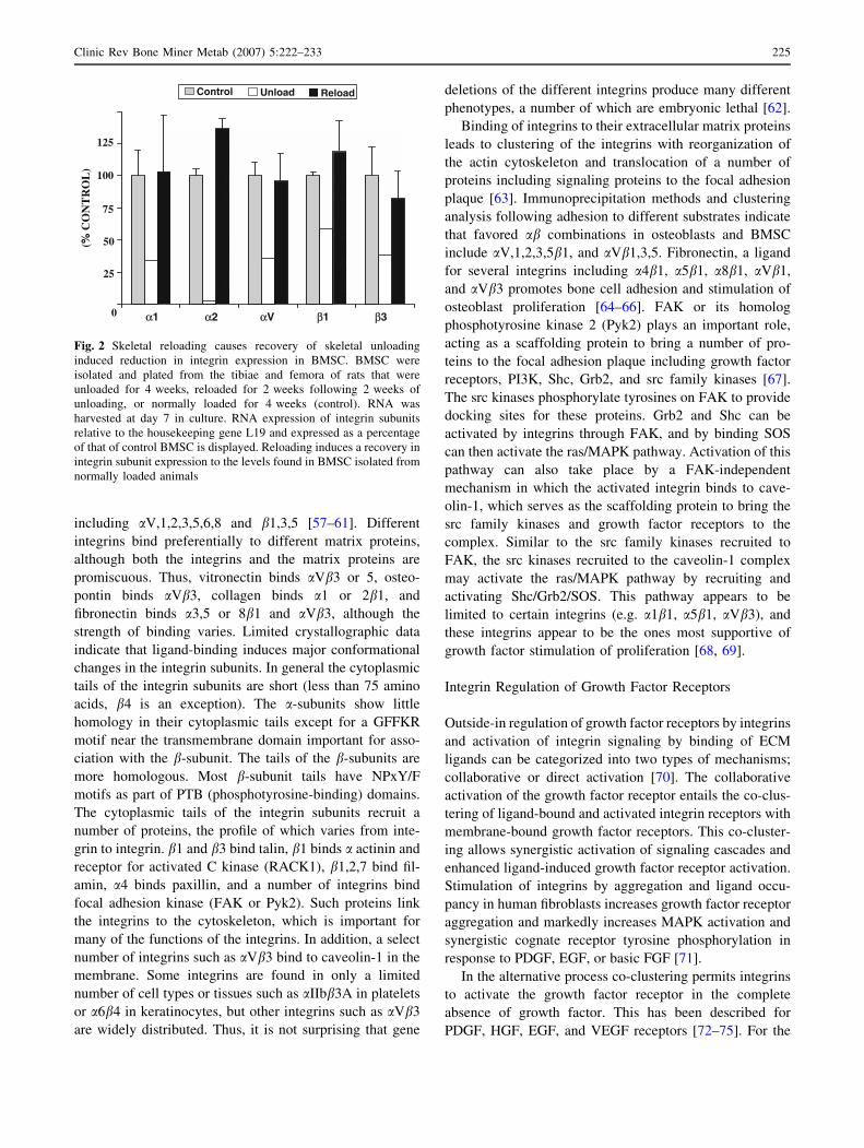
above the normally loaded controls [48]. Coinciding with
the loss IGF-I responsiveness we found that BMSC from
unloaded bones have profoundly diminished expression of
multiple integrin subunits [53], and like IGF-I respon-
siveness, these levels return to normal upon reloading
(Fig. 2). Thus, mechanical loading (or unloading) of bone
profoundly alters IGF signaling. Based on our own work
and that of others, we hypothesize that one of the keys to
understanding the impact of mechanical load on the skel-
etal response to IGF-I lies in integrin activation.
Integrin Signaling
Integrins are membrane-bound heterodimeric receptors
comprising one alpha and one beta subunit [54–56]. There
are at least 18a and 8b genes so far identified in the human
genome, several of which produce multiple transcripts by
alternative splicing. These combine to form 24 distinct
functional receptors, each of which binds to its own limited
set of extra-cellular matrix (ECM) and/or cell surface
proteins. The RGD-binding or ligand-binding site is loca-
ted at the interface between the a-subunit and b-subunit.
Osteoblasts and BMSC express a number of integrins
224 Clinic Rev Bone Miner Metab (2007) 5:222–233
including aV,1,2,3,5,6,8 and b1,3,5 [57–61]. Different
integrins bind preferentially to different matrix proteins,
although both the integrins and the matrix proteins are
promiscuous. Thus, vitronectin binds aVb3 or 5, osteo-
pontin binds aVb3, collagen binds a1 or 2b1, and
fibronectin binds a3,5 or 8b1 and aVb3, although the
strength of binding varies. Limited crystallographic data
indicate that ligand-binding induces major conformational
changes in the integrin subunits. In general the cytoplasmic
tails of the integrin subunits are short (less than 75 amino
acids, b4 is an exception). The a-subunits show little
homology in their cytoplasmic tails except for a GFFKR
motif near the transmembrane domain important for asso-
ciation with the b-subunit. The tails of the b-subunits are
more homologous. Most b-subunit tails have NPxY/F
motifs as part of PTB (phosphotyrosine-binding) domains.
The cytoplasmic tails of the integrin subunits recruit a
number of proteins, the profile of which varies from inte-
grin to integrin. b1 and b3 bind talin, b1 binds a actinin and
receptor for activated C kinase (RACK1), b1,2,7 bind fil-
amin, a4 binds paxillin, and a number of integrins bind
focal adhesion kinase (FAK or Pyk2). Such proteins link
the integrins to the cytoskeleton, which is important for
many of the functions of the integrins. In addition, a select
number of integrins such as aVb3 bind to caveolin-1 in the
membrane. Some integrins are found in only a limited
number of cell types or tissues such as aIIbb3A in platelets
or a6b4 in keratinocytes, but other integrins such as aVb3
are widely distributed. Thus, it is not surprising that gene
deletions of the different integrins produce many different
phenotypes, a number of which are embryonic lethal [62].
Binding of integrins to their extracellular matrix proteins
leads to clustering of the integrins with reorganization of
the actin cytoskeleton and translocation of a number of
proteins including signaling proteins to the focal adhesion
plaque [63]. Immunoprecipitation methods and clustering
analysis following adhesion to different substrates indicate
that favored ab combinations in osteoblasts and BMSC
include aV,1,2,3,5b1, and aVb1,3,5. Fibronectin, a ligand
for several integrins including a4b1, a5b1, a8b1, aVb1,
and aVb3 promotes bone cell adhesion and stimulation of
osteoblast proliferation [64–66]. FAK or its homolog
phosphotyrosine kinase 2 (Pyk2) plays an important role,
acting as a scaffolding protein to bring a number of pro-
teins to the focal adhesion plaque including growth factor
receptors, PI3K, Shc, Grb2, and src family kinases [67].
The src kinases phosphorylate tyrosines on FAK to provide
docking sites for these proteins. Grb2 and Shc can be
activated by integrins through FAK, and by binding SOS
can then activate the ras/MAPK pathway. Activation of this
pathway can also take place by a FAK-independent
mechanism in which the activated integrin binds to cave-
olin-1, which serves as the scaffolding protein to bring the
src family kinases and growth factor receptors to the
complex. Similar to the src family kinases recruited to
FAK, the src kinases recruited to the caveolin-1 complex
may activate the ras/MAPK pathway by recruiting and
activating Shc/Grb2/SOS. This pathway appears to be
limited to certain integrins (e.g. a1b1, a5b1, aVb3), and
these integrins appear to be the ones most supportive of
growth factor stimulation of proliferation [68, 69].
Integrin Regulation of Growth Factor Receptors
Outside-in regulation of growth factor receptors by integrins
and activation of integrin signaling by binding of ECM
ligands can be categorized into two types of mechanisms;
collaborative or direct activation [70]. The collaborative
activation of the growth factor receptor entails the co-clus-
tering of ligand-bound and activated integrin receptors with
membrane-bound growth factor receptors. This co-cluster-
ing allows synergistic activation of signaling cascades and
enhanced ligand-induced growth factor receptor activation.
Stimulation of integrins by aggregation and ligand occu-
pancy in human fibroblasts increases growth factor receptor
aggregation and markedly increases MAPK activation and
synergistic cognate receptor tyrosine phosphorylation in
response to PDGF, EGF, or basic FGF [71].
In the alternative process co-clustering permits integrins
to activate the growth factor receptor in the complete
absence of growth factor. This has been described for
PDGF, HGF, EGF, and VEGF receptors [72–75]. For the
Control Unload Reload(%
CO
NT
RO
L)
75
125
100
50
25
0 α1 α2 αV β1 β3
Fig. 2 Skeletal reloading causes recovery of skeletal unloading
induced reduction in integrin expression in BMSC. BMSC were
isolated and plated from the tibiae and femora of rats that were
unloaded for 4 weeks, reloaded for 2 weeks following 2 weeks of
unloading, or normally loaded for 4 weeks (control). RNA was
harvested at day 7 in culture. RNA expression of integrin subunits
relative to the housekeeping gene L19 and expressed as a percentage
of that of control BMSC is displayed. Reloading induces a recovery in
integrin subunit expression to the levels found in BMSC isolated from
normally loaded animals
Clinic Rev Bone Miner Metab (2007) 5:222–233 225
EGF receptor (EGFR), integrin-mediated adhesion induces
formation of a macromolecular complex including the
EGFR plus aVb3 and b1 integrins that enable ligand-
independent activation of the EGFR. This activation is
dependent upon c-src kinase, integrin b1, adaptor protein
p130Cas, and the EGFR kinase activity. Remarkably,
integrin-mediated EGFR activation induces a pattern of
tyrosine phosphorylation unique from that seen with EGFR
autophosphorylation [74]. The functional significance of
these two manners of integrin regulated growth factor
receptor activation is unknown, but they are likely com-
plementary and will differentially manifest depending on
the cell type and local microenvironment including the
availability of autocrine, paracrine, and endocrine factors
and ECM proteins.
Integrin Regulation of IGF-I Signaling
The role of integrins in regulating IGF-I responsiveness has
been demonstrated in a number of tissues. Perhaps the most
extensive examination emanates from the Clemmons lab-
oratory who have studied this phenomenon in porcine
vascular smooth muscle cells [75–80]. They found that
treatment with echistatin (a RGD containing disintegrin) or
blocking antibodies to the integrin aVb3 blocked IGF-I-
stimulated proliferation, IGF-IR autophosphorylation,
IRS-1 phosphorylation, and binding of the p85 subunit of
PI3K to IRS-1, whereas soluble ECM proteins such as
vitronectin enhanced IGF-I signaling. Their proposed
mechanism is that integrin b3 phosphorylation creates a
binding site for DOK1 (downstream of kinase 1) that in turn
recruits the tyrosine phosphatase SHP-2 to the cell mem-
brane. IGF-I via the IGF-IR phosphorylates and so activates
the transmembrane protein SHPS-1, which recruits SHP-2
to SHPS-1 and subsequently recruits and phosphorylates
Shc. This SHPS-1–SHP-2–Shc complex enables sustained
Shc phosphorylation, MAPK activation, and enhanced IGF-
I signaling. But, when aVb3 activation is blocked, the SHP-
2 complex is not formed and IGF-I signaling is terminated.
The human and porcine amino acid sequence 177–184
(177CYDMKTTC184) of the extracellular domain of b3
integrin provides ligand specificity, and ligand-binding to
this region represents the most proximal step in integrin
regulation of the IGF-IR as sequence-specific blocking
antibodies and targeted mutations in this region disrupt Shc
phosphorylation and downstream IGF-I signaling [79]. The
murine b3 integrin amino acid sequence is closely homol-
ogous (94%) to the human sequence, but there are three
amino acid substitutions within this crucial region that
disrupt ligand-binding. As expected, murine vascular
smooth muscle cells demonstrate impaired IGF-I signaling
that does not augment in response to vitronectin, however,
IGF-I signaling and the response to vitronectin is rescued
when human b3 integrin is transfected into the murine
cells [81].
The b1 integrin subunit has also been shown to form
multiprotein complexes inclusive of the IGF-IR in various
tissues that modulate IGF-I signaling in response to inte-
grin stimulation [82–84]. In response to IGF-I or phorbol
12-myristate 13-acetate (PMA), the IGF-IR and b1 integrin
localize into lipid rafts [82] and specifically, form a com-
plex with the scaffolding protein receptor for activated
C kinase (RACK1) [85]. RACK1 binding to the IGF-IR is
cell adhesion dependent and requires specific IGF-IR
tyrosine residues (Y1250/1251) as binding sites [38]. In
response to IGF-I binding, SHP-2, Shc, src kinase, and
IRS-1 are recruited to RACK1 [38], and the serine/threo-
nine protein phosphatase 2A (PPA2) dissociates from
RACK1 [86]. PPA2 dissociation enhances Shc phosphor-
ylation and promotes IGF-I-mediated cell migration.
The downstream effects of IGF-IR activation can be further
modulated by expression of b1 integrin subunits with dis-
tinctly different cytoplasmic variants [84]. The cytoplasmic
domain of the b1 integrin has been demonstrated to be the key
regulator of b1 integrin modulation of IGF-IR function and
activation. b1A integrin (the canonical variant) binds directly
with the IGF-IR in focal contacts [87] and permits IGF-I-
induced IRS-1 association with the IGF-IR/integrin complex
and cell proliferation [84]. Introduction of the b1C variant, an
alternative cytoplasmic splice variant, does not allow IGF-IR
to form focal contacts [87] and instead recruits and activates
Grb2-associated binder1 and SHP-2, thus preventing IGF-IR
phosphorylation and IGF-I-induced cell proliferation [84].
This multiprotein complex composed of integrins, IGF-
IR, and their respective signaling cascade components that
allows integrin-mediated regulation of IGF-I signaling also
permits reciprocal IGF-I regulation of integrin signaling
and function. IGF-I treatment has been shown to increase
b3 integrin subunit phosphorylation and ligand-binding to
the aVb3 integrin [88, 89]. Downstream MAP kinase
activation through IGF-IR requires integrity of the integrin
cascade. In particular, integrin-associated focal adhesion
signaling components FAK and Pyk2 are activated in
response to IGF-I and required for MAPK activation [88].
Specifically, FAK has been shown to be a substrate for the
insulin and IGF-I receptors [90]. These results suggest that
inside–out activation of integrin signaling by IGF-IR is
required for intact cellular IGF-I signaling just as outside–
in signaling is involved with activation of IGF-IR by
integrins stimulated by their ECM ligands.
Role of Integrins in Regulating IGF-I Responsiveness
in Bone
Integrins form an important link between the extra-cellular
matrix and the cytoskeleton, and thus are in a position to
226 Clinic Rev Bone Miner Metab (2007) 5:222–233

transduce mechanical signals imposed on bone to responses
by the bone cells. Fluid shear stress or mechanical defor-
mation of bone cells increases and/or activates selected
integrins [91, 92], whereas culturing mesenchymal stem
cells (presumed osteoblast precursors) in a rotary cell
culture system decreases downstream integrin signaling
(decreased FAK and Pyk2 phosphorylation and RhoA
activation) [93, 94]. As mentioned previously, osteocytes
are thought to be central to the ability of bone to sense
mechanical load. These cells show attachments between
their canalicular processes and the lining of the canalicular
wall, attachments which contain aVb3 [95]. Mechanical
stress on bone increases the expression of osteopontin, the
matrix ligand for aVb3, and other integrin ligands in
osteoblasts [92, 96, 97]. Activation of integrins by their
extracellular ligands leads to clustering into focal adhesion
plaques, which likely include growth factor receptors
including IGF-IR [98]. This co-localization of the IGF-IR
and integrins and the modulation of IGF-I signaling by
integrins enable IGF-I signaling to have a role in the
skeletal response to mechanical load.
Our results in bone cells from tail suspended hindlimb
unloaded animals indicate there is a role for integrins in
IGF-I signaling in bone. During skeletal unloading by
hindlimb elevation we found resistance to IGF-I in vivo
and in vitro is associated with reduction in integrin
expression (Figs. 2, 3) [51, 53]. This reduction in integrin
expression secondary to unloading has recently been con-
firmed by others [99]. The resistance to IGF-I appears to be
specific in that PDGF is fully capable of stimulating cog-
nate receptor phosphorylation and cell proliferation in
BMSC from unloaded bones [100]. Reloading these ani-
mals restores IGF-I response in vivo [48] and is associated
with recovery of integrin expression (Fig. 2). BMSC iso-
lated from unloaded bones grown in culture over a 21-day
period lose their unloaded phenotype in that they respond
normally to IGF-I in vitro, as demonstrated by intact IGF-
IR phosphorylation, and there is an associated integrin
0
25
50
75
100
125
150
Alpha1 Alpha2 AlphaV Beta1 Beta3
Integrin Subunits
Day 7
Day 21
* * **+
B)
A)
Fig. 3 Recovery of skeletal unloading induced changes in BMSC
over time in culture. BMSC from loaded and unloaded bones were
grown in culture over 21 days. At days 7 and 21, total RNA was
harvested or cultures were serum deprived for 24 h and then treated
with 10 ng/ml IGF-I for 10 min. Total cell lysates were collected for
analysis. (a) Immunoprecipitation of the IGF-IR and subsequent
immunoblotting for phospho-tyrosine and IGF-IR illustrate that the
skeletal unloading induced impairment of IGF-IR phosphorylation at
day 7 in culture recovered by day 21. The studies were performed in
triplicate and representative immunoblots displayed. (b) Relative
mRNA expression of the integrin subunits of the unloaded BMSC as a
percentage of that of loaded BMSC at day 7 and 21 is displayed.
Integrin expression in unloaded BMSC was significantly less than that
in loaded BMSC after 7 days but not 21 days in culture indicating
recovery in vitro. Data are means ± SEM, of thirteen independent
BMSC pools at day 7 and four at day 21, *p \ 0.01, + p \ 0.05
Clinic Rev Bone Miner Metab (2007) 5:222–233 227

subunit expression recovery (Fig. 3). The mechanism of
the integrin regulation of the IGF-IR in osteoblasts appears
to differ from that proposed by Clemmons et al. [76–80,
101]. Although echistatin blocks IGF-I activation of the
IGF-IR, recapitulating the phenotype of unloaded BMSC,
neither skeletal unloading nor echistatin alters the recruit-
ment of SHP-2 to the IGF-IR nor the timing of IGF-IR
phosphorylation and dephosphorylation [51]. Failure to
rescue the IGF-IR activation by treatment with a phos-
phatase inhibitor confirms the lack of increased
phosphatase activity, which is similar to the mechanism of
integrin regulation of EGF receptor [74]. Instead, the IGF-
IR is just not phosphorylated in response to IGF-I in BMSC
from unloaded bone or normal cells treated with echistatin.
IGF-I responsiveness in bone cells may require direct
binding of aVb3 to the IGF-IR [102]. Indeed, IGF-I
stimulates the phosphorylation of the b3 integrin subunit,
and so activates it [102].
The importance of IGF-IR/integrin interactions with
respect to IGF signaling is supported by observations that
the matrix on which bone cells are plated makes a differ-
ence with respect to the strength of IGF-I signaling. An
osteoblastic cell line grown on surfaces coated with dif-
ferent ECM proteins to promote integrin activation
demonstrates that vitronectin and fibronectin increase the
binding of the b3 integrin subunit to IGF-IR and enhance
the ability of IGF-I to activate its receptor (Fig. 4a).
Mutation of the tyrosines involved (aa 773, 785) in the b3
integrin subunit function blocks IGF-I signaling [78].
Expression of integrin subunits was specifically disrupted
using siRNA oligonucleotides targeted at the b1, b3, or
both the b1 and b3 integrin subunits. Loss of expression of
each integrin subunit significantly impaired ligand-induced
IGF-I signaling, and the combined knockdown is especially
inhibitory (Fig. 4b). These results establish the role for
both b1 and b3 integrin subunits in the ligand-induced
activation of the IGF-IR in osteoblasts.
We have found that both b1 and b3 integrin subunits
have a role in modulating the activation of osteoblast
IGF-IR in response to mechanical loading. A mechanical
load stimulus is generated by subjecting adherent cells to
pIGF IR
IGF IR
siCont siβ3 siβ1/siβ3
IGF-I:
A)
B)siβ1
– – – –+ + + +
Fig. 4 Integrin modulation of IGF-I-induced IGF-IR activation. (a)
Human osteosarcoma (HOS) cells were grown to confluence on
culture plates coated with extra-cellular matrices: fibronectin, vitro-
nectin, or vehicle (PBS). The cells were stimulated with IGF-I (10 ng/
ml) for 10 min following 24-h incubation in serum-free media. Total
cell lysates were collected. Immunoprecipitation of the IGF-IR and
subsequent immunoblotting for integrin b3 subunit, phospho-tyrosine,
and IGF-IR revealed matrix coating augmented IGF-IR activation.
The greatest augmentation is associated with integrin b3 subunit
recruitment to the IGF-IR. The studies were performed in quadrupli-
cate and representative immunoblots displayed. (b) HOS cells were
grown to confluence following treatment with siRNA non-targeting
control oligonucleotides or oligonucleotides directed at integrin b1,
b3, or both b1 and b3 subunits. Cells were stimulated with IGF-I (as
described above). Western blot analysis reveals that knockdown of
either integrin b1 or b3 subunit decreased the ligand activation of the
IGF-IR. Knockdown of both integrin subunits further impaired the
ligand activation of the IGF-IR
pIGF- IR
IGF- IR
Col - 1 Col-1/Fibro Col-1/Vitro
PFF(dynes/cm2) 5
A)
B)
15 15155 5
pIGF- IR
IGF- IR
Fig. 5 Integrin modulation of mechanical loading induced IGF-IR
activation. (a) HOS cells were grown to confluence on glass flow
plates coated with extra-cellular matrices: collagen-1, collagen-1 with
fibronectin, and collagen-1 with vitronectin. The cells were subjected
to 15 min of pulsatile fluid flow (PFF) with serum-free media
inducing either 5 or 15 dynes/cm2 shear stress at 2 Hz. Western blot
analysis reveals a PFF-induced ligand-independent phosphorylation
of the IGF-IR that is potentiated in cells plated on fibronectin or
vitronectin; there is an increased intensity of IGF-IR activation and a
lower threshold for activation. (b) HOS cells were grown on non-
coated glass flow plates to confluence following treatment with
siRNA non-targeting control oligonucleotides or oligonucleotides
directed at integrin b1, b3, or both b1 and b3 subunits. The cells were
subjected to 15 min of PFF with serum-free media inducing 15 dynes/
cm2 shear stress at 2 Hz. Western blot analysis reveals a PFF-induced,
ligand-independent phosphorylation of the IGF-IR that is blunted by
knockdown of integrin b-subunits
228 Clinic Rev Bone Miner Metab (2007) 5:222–233

pulsatile laminar fluid flow in vitro, a well-established
and widely used loading modality [103–105]. Stimulation
with fluid media without growth factors allows assess-
ment of ligand-independent mechanical loading-induced
receptor activation, presumably mediated through inte-
grins. Osteoblastic cells grown on vitronectin- or
fibronectin-coated surfaces have enhanced IGF-I signal-
ing in response to loading. These cells have a greater
magnitude of IGF-IR activation and a lower loading
stimulus threshold for activation compared to cells plated
on collagen alone (Fig. 5a). Non-specific integrin antag-
onism with echistatin pretreatment blocks ligand-
independent IGF-IR phosphorylation induced by pulsatile
fluid flow [103]. Targeted siRNA knockdown of the b1
or b3 integrin subunits prior to mechanical load in vitro
reveals that both integrin subunits participate in the
activation of the IGF-IR, and these integrin subunits
modulate IGF-I signaling in osteoblasts in response to
skeletal loading (Fig. 5b). The synergistic effects of IGF-
I and pulsatile fluid flow on IGF-IR activation further
reinforce this vital mechanism in the adaptive response
of bone to load [103].
We hypothesize that mechanical loading of bone stimu-
lates formation of an IGF-IR/integrin complex in osteoblasts
that modulates skeletal IGF-I responsiveness (Fig. 6).
Complex formation is maximized by activating the integrin
with its ECM substrate (e.g. osteopontin) and the IGF-IR
with IGF-I. We further hypothesize that the IGF-IR/integrin
complex recruits non-receptor tyrosine kinases to the com-
plex which are required for IGF-IR phosphorylation/
activation in response to IGF-I. Skeletal unloading disrupts
this process by reducing expression of integrin subunits.
Reloading restores integrin expression and enhances the
formation of the integrin/IGF-IR complex accentuating
IGF-I responsiveness. Recent results support this hypothesis
by demonstrating that fluid shear stress on bone cells stim-
ulates IGF-IR phosphorylation independent of IGF-I, that
this response is potentiated by fibronectin and vitronectin,
and that the response is blocked by echistatin and knockdown
of b1 and b3 integrin subunits. This model is shown in Fig. 6.
RACK1
SHPS-1
β3αv
ECM
IRS-1
p85
p110
ECM
BAD
AKT
Anti-Apoptosis
GDP GTPRas
Raf
MEK
ERK 1/2
EIKCreb
Jun/Fos
Cyclin D1
P21cip, p27kip
Proliferation
P
P
PP
P
Pyk2/FAK
srcPP P
shc
Grb2
SOS
P
IGF- I
IAP
PBAD
BCI2
p110
p85
P shc
Grb2
SOS
P
shc
Grb2
SOS
P
SHP-2
shc
β1
srcSHP-2
Fig. 6 Model for IGF/integrin signaling interactions in regulating the
skeletal response to load. The IGF-IR forms complexes with aVb3
and b1 integrins that are required for IGF-I activation of the IGF-IR.
This complex may be formed via the scaffolding function of FAK/
Pyk2 or RACK1. SHPS-1 and RACK1 may play a role by regulating
access of the phosphatase SHP-2 to this complex. Mechanical load
increases whereas unloading decreases formation of this complex and
so regulates IGF-I responsiveness. Formation of the integrin/IGF-IR
complex brings to the IGF-IR non-receptor kinases such as FAK and
src family kinases which may activate the IGF-IR independently and/
or synergistically with IGF-I. The source of IGF-I may originate from
osteocytes and osteoblasts. Production of IGF-I as well as expression
and activation of integrins in these cells are stimulated by mechanical
load
Clinic Rev Bone Miner Metab (2007) 5:222–233 229
Clinical Implications
Disuse osteoporosis is a major health problem contributing
to substantial morbidity through increased risk of fractures
especially in the elderly. The bone lost during extended bed
rest or immobilization is generally not regained in the
elderly. The mechanisms underlying the imbalance
between bone formation and resorption during skeletal
unloading are not well understood. We propose that the
skeletal response to mechanical load requires complex
formation between selected integrins and IGF-IR. Disrup-
tion of this complex during skeletal unloading results in a
relative reduction in bone formation leading to osteoporo-
sis, but this can be restored by reloading. Based on our
findings, we anticipate that drugs that selectively regulate
the integrin signaling pathway will prove efficacious in
modulating the skeletal response to IGF-I and in so doing
prevent or reverse the loss of bone during disuse.
Acknowledgments The authors have drawn from the work of cur-
rent and former postdoctoral fellows Drs. Paul Kostenuik, Takashi
Sakata, Shigeki Nishida, and Yongmei Wang. The work has been and
is supported by a Veterans Affairs Research Enhancement Award
Program, a grant from the National Aeronautics and Space Admin-
istration (NNA04CC67G), a grant from the National Institutes of
Health (RO1 DK54793) all to Dr. Bikle, and a fellowship training
grant from the National Institutes of Health (T32 DK07161-33 and
DK07418-27) and a National Space Biomedical Research Institute
Postdoctoral Fellowship both to Dr. Long.
References
1. Robling AG, Castillo AB, Turner CH. Biomechanical and
molecular regulation of bone remodeling. Annu Rev Biomed
Eng. 2006;8:455–98.
2. Robling AG, Hinant FM, Burr DB, Turner CH. Improved bone
structure and strength after long-term mechanical loading is
greatest if loading is separated into short bouts. J Bone Miner
Res. 2002;17:1545–54.
3. Turner CH, Forwood MR, Otter MW. Mechanotransduction in
bone: do bone cells act as sensors of fluid flow? Faseb J.
1994;8:875–8.
4. Turner CH, Owan I, Takano Y. Mechanotransduction in bone:
role of strain rate. Am J Physiol. 1995;269:E438–42.
5. Weinbaum S, Cowin SC, Zeng Y. A model for the excitation of
osteocytes by mechanical loading-induced bone fluid shear
stresses. J Biomech. 1994;27:339–60.
6. Nicolella DP, Bonewald LF, Moravits DE, Lankford J.
Measurement of microstructural strain in cortical bone. Eur
J Morphol. 2005;42(1–2):23–9.
7. Li J, Duncan RL, Burr DB, Gattone VH, Turner CH. Parathyroid
hormone enhances mechanically induced bone formation, pos-
sibly involving L-type voltage-sensitive calcium channels.
Endocrinology. 2003;144:1226–33.
8. Rawlinson SC, Pitsillides AA, Lanyon LE. Involvement of
different ion channels in osteoblasts’ and osteocytes’ early
responses to mechanical strain. Bone 1996;19:609–14.
9. Genetos DC, Geist DJ, Liu D, Donahue HJ, Duncan RL. Fluid
shear-induced ATP secretion mediates prostaglandin release in
MC3T3-E1 osteoblasts. J Bone Miner Res. 2005;20:41–9.
10. Jorgensen NR, Geist ST, Civitelli R, Steinberg TH. ATP- and
gap junction-dependent intercellular calcium signaling in
osteoblastic cells. J Cell Biol. 1997;139:497–506.
11. Bakker AD, Soejima K, Klein-Nulend J, Burger EH. The pro-
duction of nitric oxide and prostaglandin E(2) by primary bone
cells is shear stress dependent. J Biomech. 2001;34:671–7.
12. Lean JM, Jagger CJ, Chambers TJ, Chow JW. Increased insulin-
like growth factor I mRNA expression in rat osteocytes in
response to mechanical stimulation. Am J Physiol. 1995;268:
E318–27.
13. Reijnders CM, Bravenboer N, Tromp AM, Blankenstein MA,
Lips P. Effect of mechanical loading on insulin-like growth
factor-I gene expression in rat tibia. J Endocrinol. 2007;192:
131–40.
14. Morey ER, Baylink DJ. Inhibition of bone formation during
space flight. Science. 1978;201:1138–41.
15. Wronski TJ, Morey ER. Effect of spaceflight on periosteal bone
formation in rats. Am J Physiol. 1983;244:R305–09.
16. Vico L, Novikov VE, Very JM, Alexandre C. Bone histomor-
phometric comparison of rat tibial metaphysis after 7-day tail
suspension vs. 7-day spaceflight. Aviat Space Environ Med.
1991;62:26–31.
17. Oganov VS, Rakhmanov AS, Novikov VE, Zatsepin ST, Ro-
dionova SS, Cann C. The state of human bone tissue during
space flight. Acta Astronaut. 1991;23:129–33.
18. Vico L, Alexandre C. Microgravity and bone adaptation at the
tissue level. J Bone Miner Res. 1992;7(Suppl 2):S445–7.
19. Zerath E, Holy X, Malouvier A, Caissard JC, Nogues C. Rat and
monkey bone study in the Biocosmos 2044 space experiment.
Physiologist. 1991;34:S194–5.
20. Jee WS, Wronski TJ, Morey ER, Kimmel DB. Effects of
spaceflight on trabecular bone in rats. Am J Physiol. 1983;244:
R310–4.
21. Yagodovsky VS, Triftanidi LA, Gorokhova GP. Space flight
effects on skeletal bones of rats (light and electron microscopic
examination). Aviat Space Environ Med. 1976;47:734–8.
22. Vico L, Chappard D, Palle S, Bakulin AV, Novikov VE, Al-
exandre C. Trabecular bone remodeling after seven days of
weightlessness exposure (BIOCOSMOS 1667). Am J Physiol.
1988;255:R243–7.
23. Turner RT, Evans GL, Wakley GK. Spaceflight results in
depressed cancellous bone formation in rat humeri. Aviat Space
Environ Med. 1995;66:770–4.
24. Garetto LP, Gonsalves MR, Morey ER, Durnova G, Roberts
WE. Preosteoblast production 55 hours after a 12.5-day space-
flight on Cosmos 1887. Faseb J. 1990;4:24–8.
25. Globus RK, Bikle DD, Morey-Holton E. Effects of simulated
weightlessness on bone mineral metabolism. Endocrinology.
1984;114:2264–70.
26. Halloran BP, Bikle DD, Cone CM, Morey-Holton E. Gluco-
corticoids and inhibition of bone formation induced by skeletal
unloading. Am J Physiol. 1988;255:E875–9.
27. Globus RK, Bikle DD, Morey-Holton E. The temporal response
of bone to unloading. Endocrinology. 1986;118:733–42.
28. Abram AC, Keller TS, Spengler DM. The effects of simulated
weightlessness on bone biomechanical and biochemical prop-
erties in the maturing rat. J Biomech. 1988;21:755–67.
29. Bikle DD, Halloran BP, Cone CM, Globus RK, Morey-Holton
E. The effects of simulated weightlessness on bone maturation.
Endocrinology. 1987;120:678–84.
30. Kidder LS, Klein GL, Stuart CA, Lee TC, Gundberg CM,
Alcock N, Cooper CW, Simmons DJ. Skeletal effects of sodium
fluoride during hypokinesia. Bone Miner. 1990;11:305–18.
31. LeBlanc A, Marsh C, Evans H, Johnson P, Schneider V, Jhin-
gran S. Bone and muscle atrophy with suspension of the rat.
J Appl Physiol. 1985;58:1669–75.
230 Clinic Rev Bone Miner Metab (2007) 5:222–233

32. Halloran BP, Bikle DD, Wronski TJ, Globus RK, Levens MJ,
Morey-Holton E. The role of 1,25-dihydroxyvitamin D in the
inhibition of bone formation induced by skeletal unloading.
Endocrinology. 1986;118:948–54.
33. Patterson-Buckendahl P, Globus RK, Bikle DD, Cann CE, Morey-
Holton E. Effects of simulated weightlessness on rat osteocalcin
and bone calcium. Am J Physiol. 1989;257:R1103–9.
34. Shaw SR, Zernicke RF, Vailas AC, DeLuna D, Thomason DB,
Baldwin KM. Mechanical, morphological and biochemical
adaptations of bone and muscle to hindlimb suspension and
exercise. J Biomech. 1987;20:225–34.
35. Machwate M, Zerath E, Holy X, Hott M, Modrowski D, Ma-
louvier A, Marie PJ. Skeletal unloading in rat decreases
proliferation of rat bone and marrow-derived osteoblastic cells.
Am J Physiol. 1993;264:E790–9.
36. Le Roith D, Bondy C, Yakar S, Liu JL, Butler A. The
somatomedin hypothesis: 2001. Endocr Rev. 2001;22:53–74.
37. Favelyukis S, Till JH, Hubbard SR, Miller WT. Structure and
autoregulation of the insulin-like growth factor 1 receptor
kinase. Nat Struct Biol. 2001;8:1058–63.
38. Kiely PA, Leahy M, O’Gorman D, O’Connor R. RACK1-
mediated integration of adhesion and insulin-like growth factor I
(IGF-I) signaling and cell migration are defective in cells
expressing an IGF-I receptor mutated at tyrosines 1250 and
1251. J Biol Chem. 2005;280:7624–33.
39. Bikle DD, Harris J, Halloran BP, Morey-Holton E. Altered
skeletal pattern of gene expression in response to spaceflight and
hindlimb elevation. Am J Physiol. 1994;267:E822–7.
40. Canalis E. Effect of growth factors on bone cell replication and
differentiation. Clin Orthop. 1985;246–63.
41. Baker J, Liu JP, Robertson EJ, Efstratiadis A. Role of insulin-
like growth factors in embryonic and postnatal growth. Cell.
1993;75:73–82.
42. Powell-Braxton L, Hollingshead P, Warburton C, Dowd M,
Pitts-Meek S, Dalton D, Gillett N, Stewart TA. IGF-I is required
for normal embryonic growth in mice. Genes Dev. 1993;7:
2609–17.
43. Bikle D, Majumdar S, Laib A, Powell-Braxton L, Rosen C,
Beamer W, Nauman E, Leary C, Halloran B. The skeletal
structure of insulin-like growth factor I-deficient mice. J Bone
Miner Res. 2001;16:2320–9.
44. Zhao G, Monier-Faugere MC, Langub MC, Geng Z, Nakayama
T, Pike JW, Chernausek SD, Rosen CJ, Donahue LR, Malluche
HH, Fagin JA, Clemens TL. Targeted overexpression of insulin-
like growth factor I to osteoblasts of transgenic mice: increased
trabecular bone volume without increased osteoblast prolifera-
tion. Endocrinology. 2000;141:2674–82.
45. Jiang J, Lichtler AC, Gronowicz GA, Adams DJ, Clark SH,
Rosen CJ, Kream BE. Transgenic mice with osteoblast-targeted
insulin-like growth factor-I show increased bone remodeling.
Bone. 2006;39:494–504.
46. Zhang M, Xuan S, Bouxsein ML, von Stechow D, Akeno N,
Faugere MC, Malluche H, Zhao G, Rosen CJ, Efstratiadis A,
Clemens TL. Osteoblast-specific knockout of the insulin-like
growth factor (IGF) receptor gene reveals an essential role of
IGF signaling in bone matrix mineralization. J Biol Chem. 2002;
277:44005–12.
47. Wang Y, Nishida S, Boudignon BM, Burghardt A, Elalieh HZ,
Hamilton MM, Majumdar S, Halloran BP, Clemens TL, Bikle
DD. IGF-I receptor is required for the anabolic actions of
parathyroid hormone on bone. J Bone Miner Res. 2007;22:
1329–37.
48. Boudignon BM, Bikle DD, Kurimoto P, Elalieh H, Nishida S,
Wang Y, Burghardt A, Majumdar S, Orwoll BE, Rosen C,
Halloran BP. Insulin-like growth factor-I stimulates recovery of
bone lost after a period of skeletal unloading. J Appl Physiol.
2007;103(1):125–31.
49. Kostenuik PJ, Halloran BP, Morey-Holton ER, Bikle DD.
Skeletal unloading inhibits the in vitro proliferation and differ-
entiation of rat osteoprogenitor cells. Am J Physiol. 1997;273:
E1133–9.
50. Sakata T, Halloran BP, Elalieh HZ, Munson SJ, Rudner L,
Venton L, Ginzinger D, Rosen CJ, Bikle DD. Skeletal unloading
induces resistance to insulin-like growth factor I on bone for-
mation. Bone. 2003;32:669–80.
51. Sakata T, Wang Y, Halloran BP, Elalieh HZ, Cao J, Bikle DD.
Skeletal unloading induces resistance to insulin-like growth
factor-I (IGF-I) by inhibiting activation of the IGF-I signaling
pathways. J Bone Miner Res. 2004;19:436–46.
52. Kostenuik PJ, Harris J, Halloran BP, Turner RT, Morey-Holton
ER, Bikle DD. Skeletal unloading causes resistance of osteo-
progenitor cells to parathyroid hormone and to insulin-like
growth factor-I. J Bone Miner Res. 1999;14:21–31.
53. Bikle DD, Sakata T, Halloran BP. The impact of skeletal
unloading on bone formation. Grav Space Biol Bull. 2003;16(2):
45–54.
54. Giancotti FG, Ruoslahti E. Integrin signaling. Science. 1999;
285:1028–32.
55. Schwartz MA. Integrin signaling revisited. Trends Cell Biol.
2001;11:466–70.
56. Takada Y, Ye X, Simon S. The integrins. Genome Biol. 2007;
8:215.
57. Kaiser E, Sato M, Onyia JE, Chandrasekhar S. Parathyroid
hormone (1–34) regulates integrin expression in vivo in rat
osteoblasts. J Cell Biochem. 2001;83:617–30.
58. Lai CF, Chaudhary L, Fausto A, Halstead LR, Ory DS, Avioli
LV, Cheng SL. Erk is essential for growth, differentiation,
integrin expression, and cell function in human osteoblastic
cells. J Biol Chem. 2001;276:14443–50.
59. Gronthos S, Simmons PJ, Graves SE, Robey PG. Integrin-medi-
ated interactions between human bone marrow stromal precursor
cells and the extracellular matrix. Bone. 2001;28:174–81.
60. Moursi AM, Globus RK, Damsky CH. Interactions between
integrin receptors and fibronectin are required for calvarial
osteoblast differentiation in vitro. J Cell Sci. 1997;110:2187–96.
61. Pistone M, Sanguineti C, Federici A, Sanguineti F, Defilippi P,
Santolini F, Querze G, Marchisio PC, Manduca P. Integrin
synthesis and utilization in cultured human osteoblasts. Cell Biol
Int. 1996;20:471–9.
62. Sheppard D. In vivo functions of integrins: lessons from null
mutations in mice. Matrix Biol. 2000;19:203–9.
63. Asthagiri AR, Nelson CM, Horwitz AF, Lauffenburger DA.
Quantitative relationship among integrin-ligand binding, adhe-
sion, and signaling via focal adhesion kinase and extracellular
signal-regulated kinase 2. J Biol Chem. 1999;274:27119–27.
64. Cowles EA, Brailey LL, Gronowicz GA. Integrin-mediated
signaling regulates AP-1 transcription factors and proliferation
in osteoblasts. J Biomed Mater Res. 2000;52:725–37.
65. Gronthos S, Stewart K, Graves SE, Hay S, Simmons PJ. Integrin
expression and function on human osteoblast-like cells. J Bone
Miner Res. 1997;12:1189–97.
66. Globus RK, Doty SB, Lull JC, Holmuhamedov E, Humphries
MJ, Damsky CH. Fibronectin is a survival factor for differen-
tiated osteoblasts. J Cell Sci. 1998;111:1385–93.
67. Horne WC, Sanjay A, Bruzzaniti A, Baron R. The role(s) of Src
kinase and Cbl proteins in the regulation of osteoclast differ-
entiation and function. Immunol Rev. 2005;208:106–25.
68. Wary KK, Mainiero F, Isakoff SJ, Marcantonio EE, Giancotti
FG. The adaptor protein Shc couples a class of integrins to the
control of cell cycle progression. Cell 1996;87:733–43.
Clinic Rev Bone Miner Metab (2007) 5:222–233 231

69. Wary KK, Mariotti A, Zurzolo C, Giancotti FG. A requirement
for caveolin-1 and associated kinase Fyn in integrin signaling
and anchorage-dependent cell growth. Cell 998;94:625–34.
70. Yamada KM, Even-Ram S. Integrin regulation of growth factor
receptors. Nat Cell Biol. 2002;4:E75–6.
71. Miyamoto S, Teramoto H, Gutkind JS, Yamada KM. Integrins
can collaborate with growth factors for phosphorylation of
receptor tyrosine kinases and MAP kinase activation: roles of
integrin aggregation and occupancy of receptors. J Cell Biol.
1996;135(6 pt 1):1633–42.
72. Sundberg C, Rubin K. Stimulation of beta1 integrins on fibro-
blasts induces PDGF independent tyrosine phosphorylation of
PDGF-beta receptors. J Cell Biol. 1996;132(4):741–52.
73. Wang R, Kobayashi R, Bishop JM. Cellular adherence elicits
ligand-independent activation of the Met cell-surface receptor.
PNAS. 1996;93:8425–30.
74. Moro L, Dolce L, Cabodi S, Bergatto E, Boeri Erba E, Smeriglio
M, Turco E, Retta SF, Giuffrida MG, Venturino M, Godovac-
Zimmermann J, Conti A, Schaefer E, Beguinot L, Tacchetti C,
Gaggini P, Silengo L, Tarone G, Defilippi P. Integrin-induced
epidermal growth factor receptor activation requires c-Src and
p130Cas and leads to phosphorylation of specific EGF receptor
tyrosines. J Biol Chem. 2002;277(11):9405–14.
75. Wang JF, Zhang XF, Groopman JE. Stimulation of beta1 inte-
grin induces tyrosine phosphorylation of vascular endothelial
growth factor receptor-3 and modulates cell migration. J Biol
Chem. 2001;276(45):41950–7.
76. Maile LA, Badley-Clarke J, Clemmons DR. Structural analysis
of the role of the beta 3 subunit of the alpha V beta 3 integrin in
IGF-I signaling. J Cell Sci. 2001;114:1417–25.
77. Zheng B, Clemmons DR. Blocking ligand occupancy of the
alphaVbeta3 integrin inhibits insulin-like growth factor I sig-
naling in vascular smooth muscle cells. Proc Natl Acad Sci U S
A. 1998;95:11217–22.
78. Clemmons DR, Maile LA. Interaction between insulin-like
growth factor-I receptor and alphaVbeta3 integrin linked sig-
naling pathways: cellular responses to changes in multiple
signaling inputs. Mol Endocrinol. 2005;19:1–11.
79. Maile LA, Busby WH, Sitko K, Capps BE, Sergent T, Badley-
Clarke J, Clemmons DR. Insulin-like growth factor-I signaling
in smooth muscle cells is regulated by ligand binding to the177CYDMKTTC184 sequence of the b3-subunit of aVb3. Mol
Endocrinol. 2006;20:405–13.
80. Ling Y, Maile LA, Badley-Clarke J, Clemmons DR. DOK1
mediates SHP-2 binding to the aVb3 integrin and thereby reg-
ulates insulin-like growth factor I signaling in cultured vascular
smooth muscle cells. J Biol Chem. 2005;280(5):3151–8.
81. Xi G, Maile LA, Yoo S, Clemmons DR. Expression of the
human b3 integrin subunit in mouse smooth muscle cells
enhances IGF-I-stimulated signaling and proliferation. J Cell
Physiol. 2008;214:306–15.
82. Tai YT, Podar K, Catley L, Tseng YH, Akiyama M, Shringar-
pure R, Burger R, Hideshima T, Chauhan D, Mitsiades N,
Richardson P, Munshi NC, Kahn CR, Mitsiades C, Anderson
KC. Insulin-like growth factor-1 induces adhesion and migration
in human multiple myeloma cells via activation of b1-integrin
and phosphatidylinositol 30-kinase/AKT signaling. Cancer Res.
2003;63:5850–8.
83. Shakibaei M, John T, deSouza P, Rahmanzaden R, Merker HJ.
Signal transduction by b1 integrin receptors in human chon-
drocytes in vitro: collaboration with insulin-like growth factor-I
receptor. Biochem J. 1999;342:615–23.
84. Goel HL, Fornaro M, Moro L, Teider N, Rhim JS, King M,
Languino LR. Selective modulation of type 1 insulin-like
growth factor receptor signaling and functions by b1 integrins.
J Cell Biol. 2004;166(3):407–18.
85. Hermanto U, Zong CS, Li W, Wang LH. RACK1, an insulin-
like growth factor I receptor-interacting protein, modulates IGF-
I dependent integrin signaling and promotes cell spreading and
contact with extracellular matrix. Mol Cell Biol. 2002;22(7):
2345–65.
86. Kiely PA, O’Gorman D, Luong K, Ron D, O’Conner R. Insulin-
like growth factor I controls a mutually exclusive association of
RACK1 with protein phosphatase 2A and b1 integrin to promote
cell migration. Mol Cell Biol. 2006;26(11):4041–51.
87. Goel HL, Breen M, Zhang J, Das I, Aznavoorian-Cheshire S,
Greenberg NM, Elgavish A, Languino LR. b1A integrin
expression is required for type 1 insulin-like growth factor
receptor mitogenic and transforming activities and localization
to focal contacts. Cancer Res. 2005;65(15):6692–700.
88. Sekimoto H, Eipper-Mains J, Pond-Tor S, Boney C. aVb3
integrins and Pyk2 mediate insulin-like growth factor I activa-
tion of src and MAP kinase in 3T3-L1 cells. Mol Endo. 2005;
19(7):1859–67.
89. Maile LA, Imai Y, Clark J, Clemmons DR. Insulin-like growth
factor I increase aVb3 affinity by increasing the amount of
integrin associated protein that is associated with non-raft
domains of the cellular membrane. J Biol Chem. 2002;277(3):
1800–5.
90. Baron V, Calleja V, Ferrari P, Alengrin F, VanObberghen E.
p125FAK focal adhesion kinase is a substrate for the insulin and
insulin-like growth factor I tyrosine kinase receptors. J Biol
Chem. 1998;273(12):7162–8.
91. Liedert A, Kaspar D, Blakytny R, Claes L, Ignatius A. Signal
transduction pathways involved in mechanotransduction in bone
cells. Biochem Biophys Res Commun. 2006;349:1–5.
92. Pavalko FM, Norvell SM, Burr DB, Turner CH, Duncan RL,
Bidwell JP. A model for mechanotransduction in bone cells: the
load-bearing mechanosomes. J Cell Biochem. 2003;88:104–112.
93. Meyers VE, Zayzafoon M, Douglas JT, McDonald JM. RhoA
and cytoskeletal disruption mediate reduced osteoblastogenesis
and enhanced adipogenesis of human mesenchymal stem cells in
modeled microgravity. J Bone Miner Res. 2005;20:1858–66.
94. Meyers VE, Zayzafoon M, Gonda SR, Gathings WE, McDonald
JM. Modeled microgravity disrupts collagen I/integrin signaling
during osteoblastic differentiation of human mesenchymal stem
cells. J Cell Biochem. 2004;93:697–707.
95. Wang Y, McNamara LM, Schaffler MB, Weinbaum S. A model
for the role of integrins in flow induced mechanotransduction in
osteocytes. Proc Natl Acad Sci U S A. 2007;104:15941–6.
96. Carvalho RS, Bumann A, Schaffer JL, Gerstenfeld LC. Pre-
dominant integrin ligands expressed by osteoblasts show
preferential regulation in response to both cell adhesion and
mechanical perturbation. J Cell Biochem. 2002;84:497–508.
97. Morinobu M, Ishijima M, Rittling SR, Tsuji K, Yamamoto H,
Nifuji A, Denhardt DT, Noda M. Osteopontin expression in
osteoblasts and osteocytes during bone formation under
mechanical stress in the calvarial suture in vivo. J Bone Miner
Res. 2003;18:1706–15.
98. Borges E, Jan Y, Ruoslahti E. Platelet-derived growth factor
receptor beta and vascular endothelial growth factor receptor 2
bind to the beta 3 integrin through its extracellular domain.
J Biol Chem. 2000;275:39867–73.
99. Dufour C, Holy X, Marie PJ. Skeletal unloading induces
osteoblast apoptosis and targets alpha5beta1-PI3K-Bcl-2 sig-
naling in rat bone. Exp Cell Res. 2007;313:394–403.
100. Nishida S, Wang Y, Elalieh HZ, Halloran BP, Bikle DD. The
mechanism for IGF-I resistance induced by skeletal unloading is
specific for IGF-I and mediated by down regulation of the
integrin signaling pathway. J Bone Min Res. 2004;19:S72.
101. Ling Y, Maile LA, Lieskovska J, Badley-Clarke J, Clemmons
DR. Role of SHPS-1 in the regulation of insulin-like growth
232 Clinic Rev Bone Miner Metab (2007) 5:222–233

factor I stimulated Shc and MAP kinase activation in vascular
smooth muscle cells. Mol Biol Cell. 2005;16:3353–64.
102. Long RK, Nishida S, Bikle DD. IGF-1 stimulated b3 integrin
recruitment is associated with potentiation of the IGF-1 receptor.
J Bone Min Res. 2005;20:S250.
103. Kapur S, Mohan S, Baylink DJ, Lau KH. Fluid shear stress syn-
ergizes with insulin-like growth factor-I (IGF-I) on osteoblast
proliferation through integrin-dependent activation of IGF-I
mitogenic signaling pathway. J Biol Chem. 2005;280:20163–70.
104. Pavalko FM, Chen NX, Turner CH, Burr DB, Atkinson S, Hsieh
YF, Qiu J, Duncan RL. Fluid shear-induced mechanical sig-
naling in MC3T3-E1 osteoblasts requires cytoskeleton-integrin
interactions. Am J Phys. 1998;275:C1591–601.
105. Reich KM, Gay CV, Frangos JA. Fluid shear stress as a medi-
ator of osteoblast cyclic adenosine monophosphate production.
J Cell Physiol. 1990;143(1):100–4.
Clinic Rev Bone Miner Metab (2007) 5:222–233 233