HSP90 Protein Stabilizes Unloaded Argonaute Complexes and Microscopic P-bodies in Human Cells

TH
EJ
OU
RN
AL
OF
CE
LL
BIO
LO
GY
JCB: ARTICLE
© The Rockefeller University Press $30.00The Journal of Cell Biology, Vol. 180, No. 5, March 10, 2008 1037–1049http://www.jcb.org/cgi/doi/
JCB 103710.1083/jcb.200707175
Correspondence to Reinhard F ä ssler: [email protected]
Abbreviations used in this paper: BM, basement membrane; ddH 2 O, double-distilled H 2 O; DGC, dystrophin – glycoprotein complex; DM, differentiation medium; E, embryonic day; EGF, epithelial growth factor; GC, gastrocnemius; GM, growth medium; HSA, human skeletal � -actin; IGF, insulin-like growth factor; IGF-1R, IGF receptor 1; ILK, integrin-linked kinase; MTJ, myotendinous junction.
The online version of this paper contains supplemental material.
Introduction ECM of skeletal muscle consists of a basement membrane
(BM) surrounding each myofiber and interstitial connective
tissue (endomysium) between the myofi bers. The attachment of
myofi bers to the BM is mainly mediated by integrins and the
dystrophin – glycoprotein complex (DGC; Mayer, 2003 ; Michele
and Campbell, 2003 ). Integrins are expressed throughout the
sarcolemma of myofi bers but are highly enriched at two force-
transducing and force-regulating structures, the myotendinous
junctions (MTJs), which connect myofi bers to tendons, and the
costameres, which are focal adhesion – like structures that con-
nect the sarcomeric z bands with the sarcolemma.
Integrins are a large family of � / � heterodimeric adhesion
receptors ( Bouvard et al., 2001 ; Hynes, 2002 ). Several � 1 inte-
grins were shown to play essential roles during myogenesis and
muscle homeostasis ( Mayer, 2003 ). Antibody perturbation studies
and � 1 integrin gene ablations in fl ies and mice demonstrated
that � 1 integrins regulate proliferation and fusion of myoblasts
and the assembly and maintenance of sarcomeres ( Menko and
Boettiger, 1987 ; Volk et al., 1990 ; Sastry et al., 1996 ; Hirsch et al.,
1998 ; Schwander et al., 2003 ). The � 7 � 1 and, until the fi rst
postnatal days, the � 5 � 1 integrins are expressed at the MTJs,
where they implement and maintain the linkage of the myofi ber
to the tendon matrix. � 5 Integrin – defi cient chimeric mice develop
a muscle dystrophy associated with reduced adhesion and
proliferation of myoblasts ( Taverna et al., 1998 ). � 7 Integrin –
defi cient mice suffer from a progressive muscular dystrophy
with disrupted MTJs ( Mayer et al., 1997 ; Miosge et al., 1999 ).
Integrins transduce important signals. They control actin
dynamic and link the actin cytoskeleton with the ECM, and they
transduce biochemical signals in cooperation with growth factor
receptors, including receptors for insulin-like growth factor (IGF;
Goel et al., 2004 ), PDGF ( Schneller et al., 1997 ; Baron et al.,
2002 ), VEGF ( Soldi et al., 1999 ), and epithelial growth factor
(EGF; Moro et al., 1998 ; Moro et al., 2002 ). An important and still
largely unanswered question is how integrins execute their
Skeletal muscle expresses high levels of integrin-
linked kinase (ILK), predominantly at myotendinous
junctions (MTJs) and costameres. ILK binds the
cytoplasmic domain of � 1 integrin and mediates phos-
phorylation of protein kinase B (PKB)/Akt, which in turn
plays a central role during skeletal muscle regeneration.
We show that mice with a skeletal muscle – restricted dele-
tion of ILK develop a mild progressive muscular dystrophy
mainly restricted to the MTJs with detachment of base-
ment membranes and accumulation of extracellular matrix.
Endurance exercise training enhances the defects at
MTJs, leads to disturbed subsarcolemmal myofi ber archi-
tecture, and abrogates phosphorylation of Ser473 as
well as phosphorylation of Thr308 of PKB/Akt. The re-
duction in PKB/Akt activation is accompanied by an
impaired insulin-like growth factor 1 receptor (IGF-1R)
activation. Coimmunoprecipitation experiments reveal that
the � 1 integrin subunit is associated with the IGF-1R in
muscle cells. Our data identify the � 1 integrin – ILK complex
as an important component of IGF-1R/insulin receptor
substrate signaling to PKB/Akt during mechanical stress
in skeletal muscle.
Integrin-linked kinase stabilizes myotendinous junctions and protects muscle from stress-induced damage
Hao-Ven Wang , 1 Ling-Wei Chang , 1,2 Klara Brixius , 3 Sara A. Wickstr ö m , 1 Eloi Montanez , 1 Ingo Thievessen , 1
Martin Schwander , 4 Ulrich M ü ller , 4 Wilhelm Bloch , 3 Ulrike Mayer , 5 and Reinhard F ä ssler 1
1 Department of Molecular Medicine, Max Planck Institute of Biochemistry, 82152 Martinsried, Germany 2 Department of Obstetrics and Gynecology, National Cheng Kung University Medical College and Hospital, 70428 Tainan, Taiwan 3 Department of Molecular and Cellular Sport Medicine, 50933 Cologne, Germany 4 Department of Cell Biology, The Scripps Research Institute, La Jolla, CA 92037 5 Biomedical Research Centre, School of Biological Sciences, University of East Anglia, Norwich NR4 7TJ, England, UK

JCB • VOLUME 180 • NUMBER 5 • 2008 1038
To test ILK functions in the skeletal muscle of mice with-
out affecting cardiac function, we conditionally ablated the ILK
gene using human skeletal � -actin (HSA) promoter – driven Cre
expression. We found that loss of ILK triggered a mild, progressive
muscular dystrophy, mainly restricted to MTJ areas, which was
dramatically aggravated after exercise and accompanied by
an impaired phosphorylation of IGF – 1 receptor (IGF-1R) and
PKB/Akt at the Thr308 and Ser473 residues, respectively.
Results Skeletal muscle-specifi c deletion of the ILK gene Because ILK-null ( ILK lacZ/lacZ ) mice die shortly after implanta-
tion ( Sakai et al., 2003 ), we used the Cre/loxP system to disrupt
the ILK gene specifi cally in skeletal muscle. To obtain mice
with the genotype HSAcre + /ILK flox/flox (called HSACre-ILK),
ILK fl ox/fl ox mice ( Grashoff et al., 2003 ) were intercrossed with a
transgenic mouse strain expressing the Cre recombinase under
the control of the HSA promoter ( Schwander et al., 2003 ).
The effi ciency of the Cre-mediated deletion of the ILK
gene in vivo was tested by Southern blotting using genomic
DNA and Western blotting using protein extracts from gastroc-
nemius (GC) muscle of 3-mo-old control and HSACre-ILK
mice. The Southern blots revealed a recombination effi ciency of
� 80% ( Fig. 1 A ) and the Western blots an � 70% reduction of
ILK protein level ( Fig. 1 B ). Similar experiments with muscle
tissue from 4-wk-, 3-mo-, and 1-yr-old HSACre-ILK mice
revealed that the ILK gene deletion and ILK protein reduction
remained stable ( Fig.1 C and not depicted).
It has been reported that the HSA-Cre transgene induces
DNA recombination as early as embryonic day (E) 9.5 ( Schwander
et al., 2003 ). Despite this early Cre activity, we observed robust
ILK immunostaining in all hindlimb muscle of E14.5 and 16.5
HSACre-ILK embryos ( Fig.1 D and not depicted). The intensity
and the distribution of ILK immunostaining were comparable to
muscle tissue from control mice with a strong signal at the MTJs
and a weaker sarcolemmal staining ( Fig. 1 D ). Peri- and post-
natally, ILK was not detected in HSACre-ILK muscle by immuno-
staining ( Fig. 1 D ). Quantifi cation of Western blots from muscle
tissue lysates showed a 60 – 70% reduction in ILK protein levels
at this stage ( Fig.1 C ). These results show that the ILK gene is
effi ciently deleted by the HSA-Cre transgene and that the ILK
mRNA and/or protein have a long half-life in skeletal muscle
cell precursors and muscle fi bers.
HSACre-ILK mice develop a muscular dystrophy Intercrosses of HSAcre + /ILK +/ � males with ILK fl/fl females
revealed a normal Mendelian distribution of the four possible
genotypes among 231 offspring tested: HSAcre + /ILK fl / � ; HSAcre + /ILK fl /+ ; HSAcre � /ILK fl / � ; and HSAcre � /ILK fl /+ = 24.4; 23.7;
25.2; and 26.8%. HSACre-ILK mice did not show an overt pheno-
type at birth. They were viable, fertile, and showed normal
growth rates with normal weight and body length. At 3 wk of
age, when control mice began to securely walk, HSACre-ILK
mice still shambled and showed an abnormal walking pattern.
functions in myoblasts and adult skeletal muscle. Integrin cyto-
plasmic domains lack actin binding sites and enzymatic activ-
ities. Therefore, integrin signals are transduced through accessory
molecules such as talin, � -actinin, and integrin-linked kinase
(ILK; Brakebusch and F ä ssler, 2003 ).
ILK is composed of ankyrin repeats at the N terminus, a
pleckstrin homology – like domain, and a putative kinase domain
at the C terminus, which binds the cytoplasmic tail of � 1 and 3
integrins ( Grashoff et al., 2004 , Legate et al., 2006 ). A major
function of ILK is to organize the actin cytoskeleton by recruit-
ing actin binding and actin-regulatory proteins, such as PINCH,
parvin, paxillin, and kindlin ( Legate et al., 2006 ), and to phos-
phorylate several proteins, including GSK-3 � and PKB/Akt
( Delcommenne et al., 1998 ; Novak et al., 1998 ; Persad et al.,
2000 ), both of which are important for homeostasis and regen-
eration of muscle ( Glass, 2003 ; Hoffman and Nader, 2004 ).
ILK is ubiquitously expressed and essential for the devel-
opment of vertebrates and invertebrates. Mice lacking ILK die
during the periimplantation stage because of abnormal F-actin
reorganization and polarity of the epiblast ( Sakai et al., 2003 ).
In Drosophila melanogaster and Caenorhabditis elegans , the
deletion of ILK leads to muscle detachment resembling the
� integrin loss-of-function phenotype ( Zervas et al., 2001 ;
Mackinnon et al., 2002 ). Interestingly, the severe phenotypes
both in fl ies and nematodes can be fully rescued with kinase-
dead versions of ILK, suggesting that in invertebrates the kinase
activity is dispensable for development and physiology ( Zervas
et al., 2001 ; Mackinnon et al., 2002 ).
Similarly, as in fl ies and nematodes, mammalian myoblasts
and myofi bers express high levels of ILK. In myofi bers ILK is
found at MTJs and costameres. The costameric location makes
ILK perfectly suited to transduce contractile forces from the sarco-
meres across the sarcolemma to the ECM. Consistent with such
a function, mice and zebrafi sh that lack ILK function in cardio-
myocytes exhibit severe defects in mechanotransduction resulting
in lethal heart dilation, fi brosis, and disaggregation of cardio-
myocytes ( Bendig et al., 2006 ; White et al., 2006 ). The defects in
mouse cardiomyocytes are associated with reduced Ser473
phosphorylation of PKB/Akt. Because PKB/Akt activity is cru-
cial for cardiomyocyte growth and contractility ( Condorelli et al.,
2002 ; DeBosch et al., 2006 ) and ILK phosphorylates Ser473 of
PKB/Akt ( Delcommenne et al., 1998 ; Persad et al., 2000 ), it was
concluded that mechanical stress – mediated activation of ILK
supports cardiomyocyte homeostasis via PKB/Akt activation.
The role of ILK functions in skeletal muscle is obscure.
Overexpression of ILK in C2C12 myoblasts was shown to
inhibit myoblast fusion by sustained phosphorylated Erk1/2
activation, thus preventing cell cycle exit and myogenic determi-
nation ( Huang et al., 2000 ). However, ILK overexpression in L6
myoblasts was shown to promote fusion and myogenin expression
( Miller et al., 2003a ). Finally, genetic studies in mice showed
that ILK is dispensable for the development and homeostasis of
skeletal muscle ( White et al., 2006 ). The latter fi nding was un-
expected and could potentially be because of the severe heart
abnormalities and the early death of the mice, or it could alter-
natively result from incomplete Cre-mediated ILK gene deletion
in skeletal muscle.

1039INTEGRIN-LINKED KINASE FUNCTION IN MUSCLE • WANG ET AL.
cle groups derived from control mice exhibited myofi bers with
regular diameter and peripherally located nuclei. In contrast, all
three muscle types analyzed from HSACre-ILK mice contained
myofi bers with variable fi ber size and centralized nuclei ( Fig. 3,
A and B ; Fig. S1 A; and Fig. S2, A and B, available at http://
www.jcb.org/cgi/content/full/jcb.200707175/DC1). Staining of
tissue sections for ATPase and NADH activity revealed that
both fast and slow fi ber types showed centralized nuclei ( Fig. 3 B ).
The irregular fi ber size with centralized nuclei could be ob-
served as early as 10 d after birth and were aggravated with age
( Fig. 3 A and Fig. S1 B). The number of myofi bers with central
nuclei increased from 11.6 ± 2.9% in 3-mo-old mice to 22.2 ±
5.1% in 12-mo-old mice, whereas the number in control mice
was � 2% at all ages analyzed. Furthermore, we frequently
To visualize the defect, we painted the front pad of control and
HSACre-ILK mice with red ink and the hind pad with blue ink
and let them walk on blotting paper. HSACre-ILK mice had
an abnormal footprint pattern ( Fig. 2 A ) with a signifi cantly
shorter stride length, and this abnormality was maintained with
age ( Fig. 2 B ).
Adult mammalian skeletal muscle is differentiated into
distinct fi ber types, which are characterized by a unique combi-
nation of functional, biochemical, and metabolic properties.
To exclude the possibility that loss of ILK affected only specifi c
muscle fi ber types, we analyzed the histology of muscles with
predominantly fast fi bers (tibialis anterior muscle), slow fi bers
(soleus muscle), and a mixture of slow and fast fi bers (GC muscle)
from control and HSACre-ILK mice. Samples of all three mus-
Figure 1. ILK expression in HSACre-ILK mice. (A) South-ern blot analysis of ILK in control (HSAcre � /ILK fl /+ ) and HSACre-ILK (HSAcre + /ILK fl / � ) GC muscles from two 4-wk-old mice. fl , fl ox; con, control; rec., recombined allele. (B) West-ern blot analysis of ILK expression in muscle used for Southern blot assay. GAPDH was used as a loading control. (C) Quantifi cation of the ILK protein content of control and HSACre-ILK muscle by densitometric measurement of the Western blot signals. Data are expressed as the mean ± SD. (D) Immunofl uorescence of ILK (red) in control and HSACre-ILK in E16.5 forelimbs, postnatal day 1 (P1) fore-limbs, and 3-mo-old GC muscles. ILK is highly expressed at the MTJ (arrowheads). Lower levels of ILK are detected at the sarcolemma. No ILK signal is detected at the MTJ of HSACre-ILK mice. Nuclei are stained with DAPI (blue). Bars: (E16.5) 4 μ m; (P1 and 3-mo) 50 μ m.

JCB • VOLUME 180 • NUMBER 5 • 2008 1040
trusions ( Fig. 5 D , arrowheads). Interestingly, sarcomeres of
HSACre-ILK myofi brils that contained central nuclei and, hence,
had regenerated, appeared similar to control mice ( Fig. 5, E and F ),
indicating that regeneration occurs normally in the absence of
ILK expression.
observed loosened intercellular space fi lled with fi brotic mate-
rial and mononuclear cell infi ltrates, which were particularly
prominent at MTJs and in regions near tendons ( Fig. 3 A ).
The extent of the fi brosis was assessed in more detail by
analyzing collagen deposition using trichrome staining. In the
GC of 4-wk-old mice, no obvious difference between control
and HSACre-ILK was observed. However, at the age of 5 mo,
fi brotic regions were observed in the endomysial space around
myofi bers and at the MTJs of HSACre-ILK mice. The fi brosis
became more pronounced in 12-mo-old muscle ( Fig. 4 ).
Because ILK-defi cient MTJs displayed abnormalities, we
further analyzed them at the ultrastructural level. The MTJs
from control mice were extensively folded, forming digit-like
protuberances of regular size and width that were covered with
a well-structured BM and extended from the muscle cells into
the collagen-rich tendon matrix ( Fig. 5, A and C ). The MTJs of
5-mo-old HSACre-ILK mice had folds with irregular size and
width ( Fig. 5, B and D ). We frequently observed that the BM was
detached from the sarcolemma at the base of the digit-like pro-
Figure 2. Footprint analysis. Footprints of 4-wk-, 3-mo-, and 12-mo-old control and HSACre-ILK mice. (A) Front and back pads were inked in red and blue colors, respectively. (B) The distances between each two footprints were measured by pixel, and to diminish the infl uence of body length, the distances were divided by body length. The stride length of front and back feet of 4-wk-, 3-mo-, and 12-mo-old mutant mice are signifi cantly shorter in HSACre-ILK mice. Data are expressed as mean ± SD ( n = 3; ***, P < 0.001).
Figure 3. HSACre-ILK muscle displays signs of a mild dystrophy. (A) Hema-toxylin/eosin-stained paraffi n sections of the GC muscle of 10-d-, 5-mo-, and 12-mo-old control and HSACre-ILK mice. Note that the myofi bers of HSACre-ILK mice show irregular diameter, centrally located nuclei (arrowheads), mono-nuclear cell infi ltrates (asterisk), and fi brosis in 12-mo-old mutant muscle. Bars: (10-d) 40 μ m; (5-mo) 50 μ m; (12-mo) 60 μ m. (B) Myosin ATPase, pH 4.6, and NADH-stained cryosections of the GC muscle of 3-mo-old HSACre-ILK muscles. Myofi ber with asterisks indicate type II fi bers with centralized nuclei. Arrowheads indicate type I fi bers with centralized nuclei. Bar, 80 μ m.

1041INTEGRIN-LINKED KINASE FUNCTION IN MUSCLE • WANG ET AL.
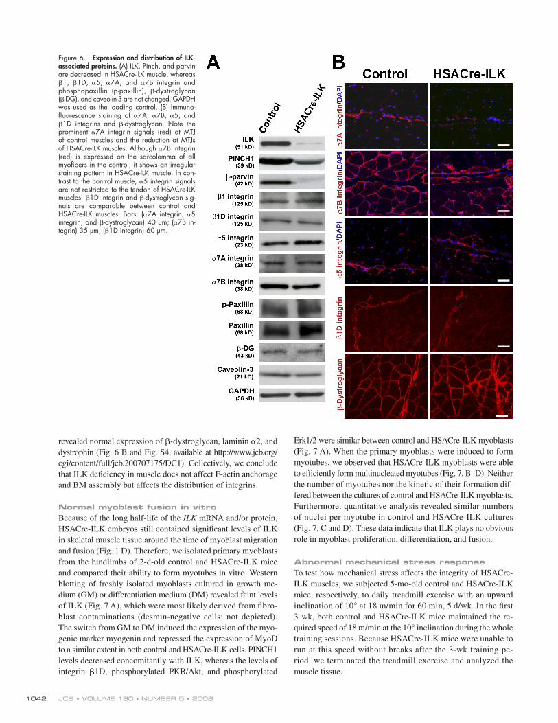
variant of the � 1 integrin subunit called � 1D integrin, which is
found at the MTJs and costameres ( Belkin et al., 1996 ; van der
Flier et al., 1997 ). Western blotting and immunostaining revealed
strong � 1D integrin expression at MTJs and lower levels at the
sarcolemma both in control and HSACre-ILK muscle ( Fig. 6, A
and B ). Although similar amounts were detected by immuno-
blotting, immunostaining for � 7A � 1 and � 7B � 1 integrins re-
vealed irregular staining patterns with reduced signals in some
areas of the muscle tissue ( Fig. 6 B ). Furthermore, unlike in
control muscle, the � 5 staining was not restricted to the tendon
but partly extended into the muscle fi bers ( Fig. 6 B ).
The BM around myofi bers is assembled by integrins and
the DGC ( Mayer et al., 1997 ; Miosge et al., 1999 ; Nawrotzki et al.,
2003 ; Guo et al., 2006 ; Rooney et al., 2006 ). Immunostaining
Collectively, these results suggest that the deletion of the
ILK gene from skeletal muscles leads to a mild muscular
dystrophy characterized by abnormalities at MTJs, variation of
myofi ber size, and increased fi brosis.
Loss of ILK affects integrin localization at MTJs ILK forms a ternary complex with PINCH and parvin that is
important for the stability of the individual components and the
recruitment of the complex into focal adhesions. Similarly, as
reported for other cell types and tissues, HSACre-mediated loss
of ILK was associated with reduced PINCH1 and � -parvin levels,
which are both highly expressed in skeletal muscle ( Fig. 6 A ).
The ILK – PINCH – parvin complex is thought to regulate inte-
grin function and actin reorganization. Interestingly, however,
ultrastructural analysis revealed no signs of F-actin detachment
from the sarcolemma both at the MTJs and in central areas of
the muscle tissue (Fig. S3, available at http://www.jcb.org/cgi/
content/full/jcb.200707175/DC1). This indicates that, in contrast
with fl ies and nematodes, ILK is not essential for anchoring ac-
tin fi laments to the muscle cell membrane.
The predominant integrin of skeletal muscle is the � 7 � 1D
integrin. The � 7 integrin gene is alternatively spliced, producing
the � 7B splice variant ( � 7B � 1), which is the predominant form
in skeletal muscle found at the sarcolemma and the MTJ, and
the � 7A splice variant ( � 7A � 1), which is expressed at MTJs
( Nawrotzki et al., 2003 ). Both � 7 subunits associate with a splice
Figure 4. Altered collagen deposition in HSACre-ILK muscles. Trichrome staining of paraffi n sections of GC muscle of 4-wk-, 5-mo-, and 12-mo-old control and HSACre-ILK mice. Collagen-containing fi brotic regions display blue color signals (arrowheads). Bar, 50 μ m.
Figure 5. Abnormalities at the MTJs of HSACre-ILK mice. Alteration of MTJs in HSACre-ILK mice. Electron micrographs of the MTJ of 5-mo-old control (A and C) and HSACre-ILK (B and D) mice. (A and B) MTJs with multiple fi nger-like interdigitations of regular organization and length are observed in control (A) and HSACre-ILK (B) mice. (C) The fi nger-like folds are covered by a well-organized and closely attached BM interdigitating with collagen fi brils in control mice. (D) HSACre-ILK mice showed folds of slightly different length and width with a partial detachment of the BM (arrowheads). (E and F) The sarcomeric structure was normal in fi bers of HSACre-ILK containing centralized nuclei (asterisk). The open white rectangle in E indicates the region shown in F. Bars: (A and B) 2.2 μ m; (C and D) 500 nm; (E) 20 μ m; (F) 800 nm.
JCB • VOLUME 180 • NUMBER 5 • 2008 1042
Erk1/2 were similar between control and HSACre-ILK myoblasts
( Fig. 7 A ). When the primary myoblasts were induced to form
myotubes, we observed that HSACre-ILK myoblasts were able
to effi ciently form multinucleated myotubes ( Fig. 7, B – D ). Neither
the number of myotubes nor the kinetic of their formation dif-
fered between the cultures of control and HSACre-ILK myoblasts.
Furthermore, quantitative analysis revealed similar numbers
of nuclei per myotube in control and HSACre-ILK cultures
( Fig. 7, C and D ). These data indicate that ILK plays no obvious
role in myoblast proliferation, differentiation, and fusion.
Abnormal mechanical stress response To test how mechanical stress affects the integrity of HSACre-
ILK muscles, we subjected 5-mo-old control and HSACre-ILK
mice, respectively, to daily treadmill exercise with an upward
inclination of 10 ° at 18 m/min for 60 min, 5 d/wk. In the fi rst
3 wk, both control and HSACre-ILK mice maintained the re-
quired speed of 18 m/min at the 10 ° inclination during the whole
training sessions. Because HSACre-ILK mice were unable to
run at this speed without breaks after the 3-wk training pe-
riod, we terminated the treadmill exercise and analyzed the
muscle tissue.
revealed normal expression of � -dystroglycan, laminin � 2, and
dystrophin ( Fig. 6 B and Fig. S4, available at http://www.jcb.org/
cgi/content/full/jcb.200707175/DC1). Collectively, we conclude
that ILK defi ciency in muscle does not affect F-actin anchorage
and BM assembly but affects the distribution of integrins.
Normal myoblast fusion in vitro Because of the long half-life of the ILK mRNA and/or protein,
HSACre-ILK embryos still contained signifi cant levels of ILK
in skeletal muscle tissue around the time of myoblast migration
and fusion ( Fig. 1 D ). Therefore, we isolated primary myoblasts
from the hindlimbs of 2-d-old control and HSACre-ILK mice
and compared their ability to form myotubes in vitro. Western
blotting of freshly isolated myoblasts cultured in growth me-
dium (GM) or differentiation medium (DM) revealed faint levels
of ILK ( Fig. 7 A ), which were most likely derived from fi bro-
blast contaminations (desmin-negative cells; not depicted).
The switch from GM to DM induced the expression of the myo-
genic marker myogenin and repressed the expression of MyoD
to a similar extent in both control and HSACre-ILK cells. PINCH1
levels decreased concomitantly with ILK, whereas the levels of
integrin � 1D, phosphorylated PKB/Akt, and phosphorylated
Figure 6. Expression and distribution of ILK-associated proteins. (A) ILK, Pinch, and parvin are decreased in HSACre-ILK muscle, whereas � 1, � 1D, � 5, � 7A, and � 7B integrin and phosphopaxillin (p-paxillin), � -dystroglycan ( � -DG), and caveolin-3 are not changed. GAPDH was used as the loading control. (B) Immuno-fl uorescence staining of � 7A, � 7B, � 5, and � 1D integrins and � -dystroglycan. Note the prominent � 7A integrin signals (red) at MTJ of control muscles and the reduction at MTJs of HSACre-ILK muscles. Although � 7B integrin (red) is expressed on the sarcolemma of all myofi bers in the control, it shows an irregular staining pattern in HSACre-ILK muscle. In con-trast to the control muscle, � 5 integrin signals are not restricted to the tendon of HSACre-ILK muscles. � 1D Integrin and � -dystroglycan sig-nals are comparable between control and HSACre-ILK muscles. Bars: ( � 7A integrin, � 5 integrin, and � -dystroglycan) 40 μ m; ( � 7B in-tegrin) 35 μ m; ( � 1D integrin) 60 μ m.

1043INTEGRIN-LINKED KINASE FUNCTION IN MUSCLE • WANG ET AL.
Furthermore, we observed profound abnormalities in MTJs
of HSACre-ILK muscles at the ultrastructural level. Although
control mice had normal MTJs after the exercise program (Fig. S5,
C and D), the MTJs from HSACre-ILK mice almost completely
lost their digit-like interdigitations and instead formed irregular
membrane protrusions and invaginations ( Fig. S5, E and F ).
Concomitantly with these defects, the BM detachment from
the sarcolemma was further aggravated (Fig. S5, F and G)
when compared with untrained muscle ( Fig. 5 F ). In addition,
in some areas the BM was replaced by an electron-dense mate-
rial (Fig. S5 F).
Training experiments with 9-mo-old HSACre-ILK mice
showed that they were unable to exercise at a speed of 18 m/min.
This age-dependent decline in running capacity made it impossible
to perform a training intervention comparable to the 5-mo-old mice.
Hematoxylin/eosin and trichrome staining of trained
HSACre-ILK muscles revealed an increase in fi brosis which
was not detected in control muscles ( Fig. 8 A ). The myofi bers of
untrained HSACre-ILK mice showed mild dystrophic changes
and were positive for methylene blue staining ( Fig. 8 B ). In con-
trast, fi bers of trained HSACre-ILK muscle were frequently
negative for methylene blue, indicating muscle damage ( Fig. 8 B ).
To evaluate the mechanical stress – induced damage more quanti-
tatively, we investigated the levels of stretch/injury-responsive
muscle ankyrin-repeat proteins Ankrd2 and CARP ( Miller et al.,
2003b ; Hentzen et al., 2006 ). Both Ankrd2 and CARP mRNA
were found to be signifi cantly up-regulated in trained HSACre-
ILK muscles, further confi rming the exercise-induced muscle
damage in HSACre-ILK mice (Fig. S5, A and B, available at
http://www.jcb.org/cgi/content/full/jcb.200707175/DC1).
Figure 7. Normal fusion of HSACre-ILK myo-blasts. (A) Protein levels of ILK and PINCH1 are dramatically decreased in primary HSACre-ILK myoblasts both in GM and DM. Signals of desmin and myogenin increase and MyoD de-creases in response to differentiation in both cell types. The levels of � 1D integrin, pSer473-Akt, pThr308-Akt, Akt, pErk1/2, and Erk1/2 are comparable. GAPDH was used as the load-ing control. (B) Myoblast cells were isolated from postnatal day 2 control and HSACre-ILK hindlimbs, incubated in DM, and evaluated microscopically. Differentiated myoblasts and myo tubes were stained with sarcomeric � -actinin antibody (red) and DAPI (blue). (C) Quantifi cation of myotube numbers from control and HSACre-ILK myoblasts plated at different cell densities. Data are expressed as mean ± SD ( n = 3; 200 and 400 cells/nm 2 ). (D) Determination of the num-ber of nuclei per fused myotube. No difference between control and HSACre-ILK mice was observed (P = 0.253). Bars: (phase) 50 μ m; (sarcomeric � -actinin) 80 μ m.

JCB • VOLUME 180 • NUMBER 5 • 2008 1044
In muscle, PKB/Akt can be activated by an intracellular
signaling cascade that is triggered through the activation of
IGF-1R ( Mourkioti and Rosenthal, 2005 ). Therefore, we tested
whether IGF-1R levels and/or activation were altered in exer-
cised HSACre-ILK muscle. Western blotting and real-time PCR
revealed that the total protein and RNA levels of IGF-1R were
similar before and after exercise in control and HSACre-ILK
mice ( Fig. 9, B and C ). Upon exercise, the phosphorylation
of cytoplasmic tyrosines in the activation loop of IGF-1R
(Tyr1131/1135/1136), which are known to induce PKB/Akt-
activation ( Vasilcanu et al., 2004 ), signifi cantly increased by 57.9 ±
0.19% in exercised control muscle ( Fig. 9, B and D ). In contrast,
there was no increase in phosphorylation of IGF-1R in HSACre-
ILK muscle upon exercise ( Fig. 9, B and D ). Importantly, the
failure of increased IGF-1R phosphorylation upon training was
not caused by diminished IGF-1 secretion because IGF-1 levels
were even higher in trained HSACre-ILK muscle than in that of
trained controls ( Fig. 9 E ). These fi ndings suggest that ILK acts
in concert with growth factors to protect muscle from mechanical
damage by regulating the IGF-1R – PBK – Akt signaling pathway.
The � 1 integrin subunit can associate with the IGF-1R in
several cell types ( Goel et al., 2006 ). To test whether a similar
association occurs before and/or during the formation of myo-
fi bers, we cultured C2C12 cells for different days in fusion me-
dium in the presence or absence of IGF-1, cross-linked and
immunoprecipitated � 1 integrin subunits, and fi nally probed the
precipitate with antibodies against IGF-1R, EGFR, � -dystro-
glycan, and � 1 integrin. As shown in Fig. 9 F , the � 1 integrin
associated with the IGF-1R, with or without IGF-1 treatment
and before and after myoblast fusion. Interestingly, the amount of
IGF-1R – � 1 integrin complexes increased in response to IGF-1
treatment. The association was specifi c because � 1 subunits nei-
ther coimmunoprecipitated with EGFR ( Fig. 9 F ) nor with
� -dystroglycan (not depicted).
Discussion In this paper, we report the skeletal muscle – specifi c ablation of
the ILK gene, which leads to a mild muscular dystrophy and
increased susceptibility to stress-induced damage. The exercise-
induced defects are associated with reduced PKB/Akt acti va-
tion, which is likely caused by an impaired cross talk between
� 1 integrin – ILK and the IGF-1R – insulin receptor substrate 1 –
PI-3K signaling pathway.
ILK maintains MTJs of untrained muscle Loss of ILK resulted in a very mild phenotype characterized by a
persistent shamble without affecting weight or life span and dam-
aged muscle fi bers with increased presence of centralized nuclei
and large variation of myofi ber size. Other alterations were re-
stricted to the MTJs and included increased fi brosis, infi ltration of
a few infl ammatory cells, and detachment of BMs at the base of the
interdigitations. Because we observed normal Erk1/2 activation
and normal levels of activated PKB/Akt in ILK-defi cient muscle,
we conclude that muscle regeneration works effi ciently in the ab-
sence of ILK, and that muscle damage likely results from a me-
chanical rather than a signaling failure. Furthermore, it seems that
Collectively, these data demonstrate that ILK-defi cient skeletal
muscle is highly susceptible to mechanical stress.
ILK modulates IGF signaling Growth of skeletal muscle critically depends on the activation
of mTOR kinase by PKB/Akt ( Glass, 2003 ; Hoffman and Nader,
2004 ). ILK phosphorylates Ser473 of PKB/Akt and is thought
to be required for full PKB/Akt activation. To test PKB/Akt ac-
tivity, we isolated muscle tissue and performed Western blotting
using phosphospecifi c antibodies. Untrained HSACre-ILK muscle
displayed levels of Ser473 and Thr308 phosphorylation compa-
rable with those of control muscle ( Fig. 9 A ). Upon training,
however, we observed a signifi cant increase in the phosphoryla-
tion of Ser473 as well as of Thr308 residues of PKB/Akt in
control muscle, whereas the HSACre-ILK muscle showed a
strongly attenuated response ( Fig. 9 A ).
Figure 8. Exercise-induced alterations in myofi bers of HSACre-ILK mice. (A) Hematoxylin/eosin (H & E)- and trichrome-stained cryosections of the GC muscle of trained control and HSACre-ILK mice. Arrowheads indicate fi brotic regions. Bar, 50 μ m. (B) Methylene blue – stained skeletal muscle fi bers of untrained and trained HSACre-ILK mice. Trained HSACre-ILK muscles contain necrotic fi bers (asterisk). Bars: (top) 35 μ m; (bottom) 900 nm.

1045INTEGRIN-LINKED KINASE FUNCTION IN MUSCLE • WANG ET AL.
and, to a lesser extent, for integrin binding to the ECM. It is pos-
sible that the requirements of ILK are slightly different between
invertebrate and vertebrate muscle. Alternatively, it could also be
that our mice have subtle actin defects that escaped detection.
ILK is not required for myoblast fusion and sarcomere assembly Although the � 1 integrins were shown to regulate myoblast fu-
sion and sarcomere assembly ( Menko and Boettiger, 1987 ; Hirsch
ILK is important for the stabilization of adhesion sites exposed to
high mechanical forces, such as MTJs, whereas at costameres,
where less force is transmitted, ILK seems to be dispensable.
The destabilization of MTJs is caused by a detachment of
the BM rather than a detachment of F-actin from the sarcolemma,
indicating that loss of ILK primarily affects ligand binding of in-
tegrins. This observation differs from the loss-of-function studies
of ILK in fl ies ( Zervas et al., 2001 ), where ILK is primarily re-
quired to attach the actin cytoskeleton at the plasma membrane
Figure 9. Altered IGF signaling after endur-ance exercise training. (A and B) Western blot analysis of untrained and endurance exercise – trained control and HSACre-ILK GC muscle for ILK, pSer473-Akt, pThr308-Akt, total Akt, pTyr397-FAK, and total FAK (A) and pTyr1131/1135/1136-IGF-1R and total IGF-1R (B). GAPDH was determined to show equal loading. (C) Densitometric quantifi cation of pTyr-IGF-1R levels from resting and endurance exercised – trained control and HSACre-ILK muscles. The increase of pTyr-IGF-1R was sig-nifi cantly reduced in muscle from HSACre-ILK mice ( n = 4; **, P < 0.01). (D) Real-time PCR analysis of IGF-1R mRNA expression of con-trol and HSACre-ILK muscles. The levels were not signifi cantly different between control and HSACre-ILK mice ( n = 4; P = 0.199). (E) Mea-surement of autocrine IGF-1 levels of control and HSACre-ILK muscles by ELISA. The level of IGF-1 in trained HSACre-ILK was 75% higher than in trained control muscles. Data are ex-pressed as mean ± SD ( n = 4; *, P < 0.05). (F) C2C12 mouse myoblasts were lysed before (0) and after 1 and 4 d of differentiation with or without IGF-1 treatment, immunoprecipitated with rabbit anti – � 1 integrin antiserum or rab-bit IgG (as control), and immunoblotted with rabbit anti – IGF-1R, rabbit anti – � 1 integrin, or rabbit anti – EGF-R antiserum. 8 μ g of lysates were used as inputs.

JCB • VOLUME 180 • NUMBER 5 • 2008 1046
as S6K and 4E-BP ( Bodine et al., 2001 ; Rommel et al., 2001 ).
Several reports have recently challenged this view and showed
that the phosphorylation of Ser473 is mediated by the mTOR ki-
nase rather than ILK, that the phosphorylation of Thr308 is suf-
fi cient for activation of PKB/Akt, and that the phosphorylation
of Ser473 determines the specifi city of PKB/Akt toward FOXO1
and FOXO3 and, to a lesser extent, the absolute activity of PKB/
Akt ( Frias et al., 2006 ; Guertin et al., 2006 ; Jacinto et al., 2006 ;
Shiota et al., 2006 ). Despite these novel fi ndings, ablation of the
ILK gene in cardiomyocytes of mice led to a dramatically reduced
Ser473 phosphorylation of PKB/Akt. Because the phosphorylation
levels of Thr308 were not determined in ILK-defi cient cardio-
myocytes, it is unclear whether loss of ILK affected the specifi city
of PKB/Akt only or also the overall activity of PKB/Akt.
We observed similar phosphorylation levels of Ser473 and
Thr308 in control and HSACre-ILK muscle of untrained mice,
suggesting that the mild muscular dystrophy upon ILK loss is
not a result of diminished ILK-dependent PKB/Akt activation.
In sharp contrast, forced treadmilling triggered an increase of
Ser473 phosphorylation in the skeletal muscle of control mice,
whereas phospho-Ser473 levels failed to rise in HSACre-ILK
muscle. Moreover, the reduced PKB/Akt phosphorylation was not
restricted to the potential ILK target Ser473 but was also observed
on Thr308, indicating that mechanical load – induced activation of
both the PDK-1 and Ser473 kinase activities requires ILK.
These fi ndings suggest that loss of ILK likely affects an
activator, which is upstream of both the PDK-1 – Thr308-PKB –
Akt and the Ser473 – PKB/Akt pathways in mechanically chal-
lenged muscle. A potential candidate for such an upstream
activator is IGF-1R, which plays a central role during muscle
repair ( Musaro et al., 2001 ), acts through PI-3K/PKB/Akt/mTor
signaling ( Bodine et al., 2001 ; Rommel et al., 2001 ), cross talks
with the � 1 integrin signaling pathway ( Goel et al., 2004 , 2006 ),
and was shown to activate ILK ( Attwell et al., 2000 ). We per-
formed Western blot assays with antibodies against the phos-
phorylated form of IGF-1R and could indeed demonstrate that
IGF-1R activation is severely impaired in trained HSACre-ILK
muscle. It is well known that physical training triggers IGF-1R
phosphorylation and the downstream activation of PKB/Akt
and the mTor complexes, which in turn give rise to the forma-
tion of new myofi brils. Consistent with the reduced PKB/Akt
phosphorylation, the training-induced phosphorylation of IGF-1R
at tyrosine residues (Tyr1131/1135/1136) in the activation loop
of the kinase domain was almost completely abrogated in HSACre-
ILK muscle. Phosphorylation of Tyr1136 was shown to be required
for the IGF-1 – induced phosphorylation of insulin receptor sub-
strate 1, the interaction of the regulatory p85 subunit of PI-3K with
IGF-1R, and the downstream-activation of PKB/Akt ( Vasilcanu
et al., 2004 ). Moreover, we observed that the � 1 integrin sub-
unit can form a complex with IGF-1R in C2C12 cells before
and after fusion into myotubes. Interestingly, IGF-1 treatment
increases the amount of � 1 integrin – IGF-1R complex, corrobo-
rating that the complex formation has a functional role in IGF
signaling. Collectively, our data suggest that the � 1 integrin –
IGF-1R complex is using ILK to activate the IGF-1R signaling
machinery, leading to PKB/Akt activation and regeneration of
exercise-induced muscle damage.
et al., 1998 ; Schwander et al., 2003 ), it is still unclear how
� 1 integrins execute these functions. Two studies reported that ILK
is acting downstream of � 1 integrin to control fusion and sarco-
mere formation. The studies overexpressed wild-type and mutant
ILK cDNAs in either mouse or rat myoblast cell lines and came
to opposite conclusions. Although one study showed that ILK
antagonizes myoblast fusion by sustained Erk1/2 activation,
preventing cell cycle withdrawal and myogenic determination,
another study reported that ILK stimulates fusion and myogenic
determination ( Huang et al., 2000 ; Miller et al., 2003a ).
Unfortunately, the long half-life time of the ILK mRNA
and/or protein did not permit us to study myoblast fusion and
sarcomere assembly in vivo. Therefore, we isolated primary myo-
blasts from newborn control and HSACre-ILK mice and tested
whether they fuse and assemble sarcomeres in vitro. We found
that primary ILK-defi cient myoblasts exhibit normal fusion and
sarcomere assembly, indicating that ILK does not play an obvi-
ous and very prominent role during myogenic differentiation.
These fi ndings are in line with recent zebrafi sh data showing
that loss of ILK function results in severe heart failure but nor-
mal development of skeletal muscle.
ILK protects myofi bers from force-induced damage Contrary to the mild defects of ILK-defi cient skeletal muscle,
disruption of ILK function in zebrafi sh or the myocardium of
mice leads to lethality with progressive contraction defects,
heart dilation, and fi brosis ( Bendig et al., 2006 ; White et al., 2006 ).
The strong phenotype suggests that mechanical loading triggers
the severe defects. We tested this assumption by exposing control
and mutant mice to forced treadmilling and found that mu-
tant mice could not complete the 4-wk training protocol with
a treadmilling speed of 18 m/min, demonstrating that the func-
tion of HSACre-ILK muscle was profoundly compromised.
After training for 3 wk, the MTJs of HSACre-ILK muscle dis-
played an almost complete loss of interdigitations, extensive BM
detachments, and myofi ber necrosis.
Although treadmilling profoundly augmented the defects
at the MTJs, we still observed normal insertions of actin fi la-
ments at the sarcolemma at MTJs. This could have several rea-
sons. It is possible that the training protocol was not vigorous
enough to trigger actin fi lament detachment as observed in fl ies
and nematodes. It is also conceivable that ILK plays no or only
a minor role as a mechanical linkage for the terminal sarco-
meres at MTJs. Finally, loss of ILK may be compensated by other
actin-linked adhesion molecules such as the DGC. Consistent with
the latter assumption, it was demonstrated that mdx/ � 7 inte-
grin � / � double mutant mice develop a more severe muscle
dystrophy than dystrophin or � 7 integrin single mutant mice
( Guo et al., 2006 ; Rooney et al., 2006 ).
ILK is required for the mechanical stress-induced activation of PKB/Akt ILK is believed to phosphorylate and, thereby, modulate the
activity of several target proteins, including PKB/Akt, which,
in turn, plays a central role during the repair of skeletal muscle
by activating the mTOR complex and downstream targets such

1047INTEGRIN-LINKED KINASE FUNCTION IN MUSCLE • WANG ET AL.
Endurance exercise training Experiments were performed with 5-mo-old control (seven mice; weight: 27.0 ± 0.7 g) and HSACre-ILK (six mice; weight: 25.1 ± 0.9 g) mice. The treadmill (Exer3/6; Columbus Instruments) endurance training consisted of a 60-min treadmill exercise 5 d a week at a velocity of 18 m/min at an angle of 10 ° . Mice were elicited to run by touching their back with a pencil. Mice were accommodated to the situation for 1 wk before starting the experi-ments. The velocity of 18 m/min was chosen because in these preexperi-ments, control and HSACre-ILK mice were able to constantly run for 1 h at this velocity. An angle of 10 ° was chosen to increase the muscle load during the training. The training was performed for 4 wk. At the end of this period, animals were killed and the vastus lateralis and GC muscles were isolated, fi xed in 4% buffered PFA for 6 h, and prepared for ultrastructural analysis.
An additional test was initiated with 9-mo-old control ( n = 6) and HSACre-ILK ( n = 6) mice under the conditions described in the previous paragraph. Because HSACre-ILK mice were unable to abide the exercise, the experiment was terminated. The local Animal Care Committee approved all experimental procedures.
Real-time PCR Muscle RNA was isolated with the RNeasy Mini kit (QIAGEN). 1 μ l cDNA generated from 200 ng RNA with the iScript cDNA Synthesis kit (Bio-Rad Laboratories) was subjected to real-time PCR using the iQ SYBR Green Supermix (Bio-Rad Laboratories) and the iCycler (Bio-Rad Laboratories). The following primers (Mouse Genome Informatics number1204415) were used for detecting IGF-1R: forward, 5 � -TGGCACCTACAGGTTCGAG-3 � ; and reverse, 5 � -TGATGGACACACCTGCATG-3 � . The following primers were used for CARP: forward, 5 � -GAGAAGTTAATGGAGGCTGG-3 � ; and reverse, 5 � -GTTCAGCAACAGTTTCAGGAC-3 � . The following primers were used for Ankrd2: forward, 5 � -CCACAGAGCTCATCGAGCAG-3 � ; and reverse, 5 � -CTAGCACTAGCATGTCCATGG-3 � . Gene expression was quantifi ed using the Gene Expression Analysis Program for the iCycler iQ Real-Time PCR Detection system (Bio-Rad Laboratories) and normalized to GAPDH levels.
Cross-linking and immunoprecipitation C2C12 mouse myoblasts were maintained in GM and differentiation was induced with DM. Treatment with 100 ng/ml mIGF-1 (R & D Systems) in DME was performed 14 h after starvation.
Cross-linking reaction was performed in 1 mM DSP (Thermo Fisher Scientifi c) in PBS for 20 min on ice. Cells were lysed in IP buffer containing 1% Triton X-100, 0.05% sodium deoxycholate, 150 mM NaCl, and 50 mM Tris-HCl, pH 8, with protease inhibitors and phosphatase inhibitors (cocktails 1 and 2). For coimmunoprecipitation of � 1 integrin, 700 μ g of lysates were incubated with anti – � 1 integrin antiserum for 30 min at 4 ° C and then with 35 μ l of protein A – Agarose for another 1 h. Protein complexes were washed three times in wash buffer (0.1% Triton X-100, 0.005% sodium deoxycholate, 150 mM NaCl, and 50 mM Tris-HCl, pH 8) and subsequently extracted with 5 × SDS loading buffer for 5 min at 95 ° C.
Myosin ATPase, pH 4.6, staining Unfi xed cryosections were preincubated for 10 min at RT in incubation buf-fer (0.1 M NaOAc and 1 mM EDTA adjusted to pH 4.6). Slides were dipped in incubation buffer, pH 9.6, and then immediately incubated in ATP solution (10 mg ATP in 10 ml incubation buffer, pH 9.6) for 10 min at 37 ° C. After washing with double-distilled H 2 O (ddH 2 O), slides were immersed in 2% CoCl2 for 5 min. After another wash with ddH 2 O, slides were immersed in 0.1% ammonium sulfi de solution for 30 s. Finally, the slides were washed under running water for 5 min, dehydrated, and mounted in glycerine jelly.
NADH staining Unfi xed cryosections were incubated for 30 min in 0.2M Tris-HCl, pH 7.4, 1.5 mM NADH, and 1.5 mM Nitroblue tetrazolium (Sigma-Aldrich) at 37 ° C. After incubation, slides were rinsed three times with ddH 2 O and mounted in Evanol.
Masson ’ s trichrome staining Slides were mordant in preheated Bouin ’ s solution (saturated picric acid/formaldehyde/glacial acetic acid = 15:5:1) for 15 min at 56 ° C. After cool-ing to RT, slides were washed under running water to remove the yellow color and stained in Weigert ’ s iron hematoxylin solution for 5 min. Slides were then washed for 5 min under running water, rinsed in ddH 2 O, and stained in Biebrich scarlet-acid Fuchsin for 5 min. After rinsing in ddH 2 O, slides were transferred to Aniline blue Solution for 5 min and, subsequently, to 1% acetic acid for 2 min. Finally, the slides were rinsed, dehydrated through alcohol, cleared in xylene, and mounted. All chemicals were from Sigma-Aldrich.
Materials and methods Mouse strains To obtain mice with a skeletal muscle – restricted deletion of the ILK gene, fl oxed ILK mice ( Grashoff et al., 2003 ) were crossed with transgenic mice expressing the Cre gene under the control of the HSA promoter ( Schwander et al., 2003 ). All animals were fed ad libitum and housed according to the guidelines of the Society of Laboratory Animal Science.
Antibodies Antibodies used in this study were mouse anti-GAPDH (Millipore), mouse anti – sarcomeric � -actinin (Sigma-Aldrich), rabbit anti-caveolin3 (Abcam), rabbit anti-Erk1/2 (Cell Signaling Technology), rabbit anti – phospho-Erk1/2 (Thr202/Tyr204; Cell Signaling Technology), mouse anti-desmin (BD Bio-sciences), mouse anti-paxillin (Transduction Laboratories), rabbit anti-phos-phopaxillin (Tyr118; Cell Signaling Technology), mouse anti-myogenin (BD Biosciences), rabbit anti-MyoD (Santa Cruz Biotechnology, Inc.), rabbit anti-FAK (Millipore), rabbit anti – phospho-FAK (Tyr397; Invitrogen), mouse anti-dystrophin (Abcam), goat anti – � -dystroglycan (Santa Cruz Biotechnology, Inc.), rabbit anti – EGF-R (Cell Signaling Technology), rabbit anti – phospho – IGF-1R (Tyr1131/1135/1136; Acris Antibodies), rabbit anti – IGF-1R (Cell Signaling Technology), and mouse anti – IGF-1R (Millipore). Fluorescent dye – conjugated secondary antibodies were obtained from Invitrogen. All other antibodies used have been described previously ( Nawrotzki et al., 2003 ; Sakai et al., 2003 ; Stanchi et al., 2005 ; Chu et al., 2006 ; Guo et al., 2006 ).
Western blotting Muscle tissue was homogenized in modifi ed RIPA buffer (50 mM Tris-HCl, pH 7.4, 150 mM NaCl, 5 mM EDTA, 0.1% SDS, 1% Triton X-100, 1% sodium deoxycholate, protease inhibitors [Roche], and phosphatase inhib-itors [Sigma-Aldrich]). Extracted proteins were gel separated and immuno-probed as previously described ( Grashoff et al., 2003 ).
Histology, immunofl uorescence, and electron microscopy Muscle tissues from embryos or newborn or adult mice was excised and ei-ther frozen in liquid nitrogen – cooled isopentane or dehydrated and em-bedded in paraffi n. 8 μ m of transverse sections were cut and collected onto SuperFrost Plus (Menzel-Gl ä ser) slides. The area of myofi bers was deter-mined on hematoxylin/eosin-stained paraffi n sections using the Axiovision software (Version 4.6.3.0; Carl Zeiss, Inc.).
Immunofl uorescence was done on cryosections as described in Mayer et al. (1997) . Images were collected by confocal microscopy (DMIRE2; Leica) using Leica Confocal Software (version 2.5, build 1227) with 40 or 63 × oil objectives, by fl uorescence microscopy (DMRA2; Leica) using SimplePCI software (version 5.1.0.0110; GTI Microsystems) with 20, 40, or 63 × oil objectives, or by bright fi eld microscopy (Axiovert; Carl Zeiss, Inc.) using IM50 software (Leica) with 10 or 40 × objectives. All images were collected at RT. Digital images were manipulated and arranged using Photoshop CS2 (Adobe). Transmission electron microscopy was performed using an electron microscope (902A; Carl Zeiss, Inc.) as described in Hirsch et al. (1998) .
In brief, muscle biopsies were fi xed in 4% buffered PFA, rinsed three times in cacodylate buffer, and then treated with 1% uranyl acetate in 70% ethanol for 8 h. The biopsies were subsequently dehydrated in a graded series of ethanol and then embedded in Araldite (SERVA). Semithin sec-tions (500 μ m) were cut with a glass knife on an ultramicrotome (Reichert) and stained with Methylene blue. Ultrathin sections (30 – 60 nm) for elec-tron microscopic observation were processed on the same microtome with a diamond knife and placed on copper grids.
Isolation and differentiation of primary myoblasts Primary myoblasts were isolated as described by Rando and Blau (1994) . In brief, hindlimbs were dissected from 1 – 2-d-old mice, placed in PBS, minced with a razor blade, and enzymatically dissociated with a mixture of colla-genase II (0.1%; Worthington Biochemical) and dispase (2.4 U/ml; grade II; Roche). The slurry, maintained at 37 ° C for 30 – 45 min, was triturated every 15 min with a 5-ml plastic pipette. After centrifugation at 350 g for 10 min, the pellet was resuspended in DME containing 20% FCS, 2 mM glutamin, and 1% Pen/Strep (Invitrogen) and preplated into noncoated tissue culture dishes for 20 min for attaching fi broblasts to the dish surface. The nonadher-ent cells were then transferred into 0.2% gelatin-coated 6-well plates (approx-imately two limbs for one well). Differentiation was induced with 5% horse serum (Invitrogen) in DME for 2 – 4 d. A myotube was defi ned as having three or more nuclei.

JCB • VOLUME 180 • NUMBER 5 • 2008 1048
Frias , M.A. , C.C. Thoreen , J.D. Jaffe , W. Schroder , T. Sculley , S.A. Carr , and D.M. Sabatini . 2006 . mSin1 is necessary for Akt/PKB phosphorylation, and its isoforms defi ne three distinct mTORC2s. Curr. Biol. 16 : 1865 – 1870 .
Glass , D.J. 2003 . Signalling pathways that mediate skeletal muscle hypertrophy and atrophy. Nat. Cell Biol. 5 : 87 – 90 .
Goel , H.L. , M. Fornaro , L. Moro , N. Teider , J.S. Rhim , M. King , and L.R. Languino . 2004 . Selective modulation of type 1 insulin-like growth factor receptor signaling and functions by � 1 integrins. J. Cell Biol. 166 : 407 – 418 .
Goel , H.L. , L. Moro , M. King , N. Teider , M. Centrella , T.L. McCarthy , M. Holgado-Madruga , A.J. Wong , E. Marra , and L.R. Languino . 2006 . Beta1 integrins modulate cell adhesion by regulating insulin-like growth factor-II levels in the microenvironment. Cancer Res. 66 : 331 – 342 .
Grashoff , C. , A. Aszodi , T. Sakai , E.B. Hunziker , and R. F ä ssler . 2003 . Integrin-linked kinase regulates chondrocyte shape and proliferation. EMBO Rep. 4 : 432 – 438 .
Grashoff , C. , I. Thievessen , K. Lorenz , S. Ussar , and R. F ä ssler . 2004 . Integrin-linked kinase: integrin ’ s mysterious partner. Curr. Opin. Cell Biol. 16 : 565 – 571 .
Guertin , D.A. , D.M. Stevens , C.C. Thoreen , A.A. Burds , N.Y. Kalaany , J. Moffat , M. Brown , K.J. Fitzgerald , and D.M. Sabatini . 2006 . Ablation in mice of the mTORC components raptor, rictor, or mLST8 reveals that mTORC2 is required for signaling to Akt-FOXO and PKCalpha, but not S6K1. Dev. Cell . 11 : 859 – 871 .
Guo , C. , M. Willem , A. Werner , G. Raivich , M. Emerson , L. Neyses , and U. Mayer . 2006 . Absence of � 7 integrin in dystrophin-defi cient mice causes a myopathy similar to Duchenne muscular dystrophy. Hum. Mol. Genet. 15 : 989 – 998 .
Hoffman , E.P. , and G.A. Nader . 2004 . Balancing muscle hypertrophy and atrophy. Nat. Med. 10 : 584 – 585 .
Hentzen , E.R. , M. Lahey , D. Peters , L. Mathew , I.A. Barash , J. Frid é n , and R.L. Lieber . 2006 . Stress-dependent and -independent expression of the myo-genic regulatory factors and the MARP genes after eccentric contractions in rats. J. Physiol. 570 : 157 – 167 .
Hirsch , E. , L. Lohikangas , D. Gullberg , S. Johansson , and R. F ä ssler . 1998 . Mouse myoblasts can fuse and form a normal sarcomere in the absence of � 1 integrin expression. J. Cell Sci. 111 : 2397 – 2409 .
Huang , Y. , J. Li , Y. Zhang , and C. Wu . 2000 . The roles of integrin-linked kinase in the regulation of myogenic differentiation. J. Cell Biol. 150 : 861 – 872 .
Hynes , R.O. 2002 . Integrins: bidirectional, allosteric signaling machines. Cell . 110 : 673 – 687 .
Jacinto , E. , V. Facchinetti , D. Liu , N. Soto , S. Wei , S.Y. Jung , Q. Huang , J. Qin , and B. Su . 2006 . SIN1/MIP1 maintains rictor-mTor complex in-tegrity and regulates Akt phosphorylation and substrate specifi city. Cell . 127 : 125 – 137 .
Legate , K.R. , E. Montanez , O. Kudlacek , and R. F ä ssler . 2006 . ILK, PINCH and parvin: the tIPP of integrin signalling. Nat. Rev. Mol. Cell Biol. 7 : 20 – 31 .
Mackinnon , A.C. , H. Qadota , K.R. Norman , D.G. Moerman , and B.D. Williams . 2002 . C. elegans PAT-4/ILK functions as an adaptor protein within inte-grin adhesion complexes. Curr. Biol. 12 : 787 – 797 .
Mayer , U. 2003 . Integrins: redundant or important players in skeletal muscle? J. Biol. Chem. 278 : 14587 – 14590 .
Mayer , U. , G. Saher , R. F ä ssler , A. Bornemann , F. Echtermeyer , H. von der Mark , N. Miosge , E. Poschl , and K. von der Mark . 1997 . Absence of integrin � 7 causes a novel form of muscular dystrophy. Nat. Genet. 17 : 318 – 323 .
Menko , A.S. , and D. Boettiger . 1987 . Occupation of the extracellular matrix receptor, integrin, is a control point for myogenic differentiation. Cell . 51 : 51 – 57 .
Michele , D.E. , and K.P. Campbell . 2003 . Dystrophin-glycoprotein complex: post-translational processing and dystroglycan function. J. Biol. Chem. 278 : 15457 – 15460 .
Miller , M.G. , I. Naruszewicz , A.S. Kumar , T. Ramlal , and G.E. Hannigan . 2003a . Integrin-linked kinase is a positive mediator of L6 myoblast differentia-tion. Biochem. Biophys. Res. Commun. 310 : 796 – 803 .
Miller , M.K. , M.L. Bang , C.C. Witt , D. Labeit , C. Trombitas , K. Watanabe , H. Granzier , A.S. McElhinny , C.C. Gregorio , and S. Labeit . 2003b . The mus-cle ankyrin repeat proteins: CARP, ankrd2/Arpp and DARP as a family of titin fi lament-based stress response molecules. J. Mol. Biol. 333 : 951 – 964 .
Miosge , N. , C. Klenczar , R. Herken , M. Willem , and U. Mayer . 1999 . Organization of the myotendinous junction is dependent on the presence of � 7 � 1 integrin. Lab. Invest. 79 : 1591 – 1599 .
Moro , L. , M. Venturino , C. Bozzo , L. Silengo , F. Altruda , L. Beguinot , G. Tarone , and P. Defi lippi . 1998 . Integrins induce activation of the EGF receptor: role in MAP kinase induction and adhesion-dependent cell sur-vival. EMBO J. 17 : 6622 – 6632 .
Moro , L. , L. Dolce , S. Cabodi , E. Bergatto , E. Boeri Erba , M. Smeriglio , E. Turco , S.F. Retta , M.G. Giuffrida , M. Venturino , et al . 2002 . Integrin induced
IGF-1 measurement Muscle tissue was homogenized in PBS, pH 7.4. After two freeze – thaw cy-cles, the homogenates were centrifuged for 5 min at 5,000 g . The super-natant was then removed and stored at � 80 ° C. IGF-1 measurement was performed as described in the manual of the Quantikine Mouse IGF-1 kit (R & D Systems).
Statistical analysis Statistical evaluation was performed with GraphPad Prism software (GraphPad, Inc.). Statistical signifi cance between data groups was deter-mined by the Mann-Whitney test and subdivided into three groups (*, P < 0.05; **, P < 0.01; ***, P < 0.001).
Online supplemental material Fig. S1 shows the centralized nuclei in soleus, extensor digitorum longus, and tibialis anterior muscle of HSACre-ILK mice. Fig. S2 shows the measure-ments of myofi ber density and size in HSACre-ILK and control mice. Fig. S3 shows that the sarcolemmal F-actin is unaffected in HSACre-ILK muscle. Fig. S4 shows the normal distribution and expression of � 1 integrin, vinculin, laminin � 2, and dystrophin in HSACre-ILK muscle. Fig. S5 shows the analy-sis of Ankrd2 and CARP levels as markers of muscle damage in trained mice as well as ultrastructural analysis showing detachment of the BM in trained HSACre-ILK muscle. Online supplemental material is available at http://www.jcb.org/cgi/content/full/jcb.200707175/DC1.
We thank the members of the F ä ssler laboratory for lively discussions and care-ful reading of the manuscript.
S.A. Wickstr ö m is supported by the Sigrid Juselius Foundation and the Finnish Cultural Foundation. This work was funded by the Welcome Trust (grant 060549 to U. Mayer), the Austrian Science Fund (grant SFB021), and the Max Planck Society (to R. F ä ssler).
Submitted: 25 July 2007 Accepted: 7 February 2008
References Attwell , S. , C. Roskelley , and S. Dedhar . 2000 . The integrin-linked kinase (ILK)
suppresses anoikis. Oncogene . 19 : 3811 – 3815 .
Baron , W. , S.J. Shattil , and C. ffrench-Constant . 2002 . The oligodendrocyte precursor mitogen PDGF stimulates proliferation by activation of � v � 3 integrins. EMBO J. 21 : 1957 – 1966 .
Belkin , A.M. , N.I. Zhidkova , F. Balzac , F. Altruda , D. Tomatis , A. Maier , G. Tarone , V.E. Koteliansky , and K. Burridge . 1996 . � 1D integrin dis-places the � 1A isoform in striated muscles: localization at junctional structures and signaling potential in nonmuscle cells. J. Cell Biol. 132 : 211 – 226 .
Bendig , G. , M. Grimmler , I.G. Huttner , G. Wessels , T. Dahme , S. Just , N. Trano , H.A. Katus , M.C. Fishman , and W. Rottbauer . 2006 . Integrin-linked ki-nase, a novel component of the cardiac mechanical stretch sensor, con-trols contractility in the zebrafi sh heart. Genes Dev. 20 : 2361 – 2372 .
Bodine , S.C. , T.N. Stitt , M. Gonzalez , W.O. Kline , G.L. Stover , R. Bauerlein , E. Zlotchenko , A. Scrimgeour , J.C. Lawrence , D.J. Glass , and G.D. Yancopoulos . 2001 . Akt/mTOR pathway is a crucial regulator of skeletal muscle hypertrophy and can prevent muscle atrophy in vivo. Nat. Cell Biol. 3 : 1014 – 1019 .
Bouvard , D. , C. Brakebusch , E. Gustafsson , A. Aszodi , T. Bengtsson , A. Berna , and R. F ä ssler . 2001 . Functional consequences of integrin gene mutations in mice. Circ. Res. 89 : 211 – 223 .
Brakebusch , C. , and R. F ä ssler . 2003 . The integrin-actin connection, an eternal love affair. EMBO J. 22 : 2324 – 2333 .
Chu , H. , I. Thievessen , M. Sixt , T. Lammermann , A. Waisman , A. Braun , A.A. Noegel , and R. F ä ssler . 2006 . � -Parvin is dispensable for hematopoiesis, leukocyte traffi cking, and T-cell-dependent antibody response. Mol. Cell. Biol. 26 : 1817 – 1825 .
Condorelli , G. , A. Drusco , G. Stassi , A. Bellacosa , R. Roncarati , G. Iaccarino , M.A. Russo , Y. Gu , N. Dalton , C. Chung , et al . 2002 . Akt induces en-hanced myocardial contractility and cell size in vivo in transgenic mice. Proc. Natl. Acad. Sci. USA . 99 : 12333 – 12338 .
DeBosch , B. , I. Treskov , T.S. Lupu , C. Weinheimer , A. Kovacs , M. Courtois , and A.J. Muslin . 2006 . Akt1 is required for physiological cardiac growth. Circulation . 113 : 2097 – 2104 .
Delcommenne , M. , C. Tan , V. Gray , L. Rue , J. Woodgett , and S. Dedhar . 1998 . Phosphoinositide-3-OH kinase-dependent regulation of glycogen syn-thase kinase 3 and protein kinase B/AKT by the integrin-linked kinase. Proc. Natl. Acad. Sci. USA . 95 : 11211 – 11216 .

1049INTEGRIN-LINKED KINASE FUNCTION IN MUSCLE • WANG ET AL.
epidermal growth factor (EGF) receptor activation requires c-src and p130Cas and leads to phosphorylation of specifi c EGF receptor tyrosines. J. Biol. Chem. 277 : 9405 – 9414 .
Mourkioti , F. , and N. Rosenthal . 2005 . IGF-1, infl ammation and stem cells. Interactions during muscle regeneration. Trends Immunol. 26 : 535 – 542 .
Musaro , A. , K. McCullagh , A. Paul , L. Houghton , G. Dobrowolny , M. Molinaro , E.R. Barton , H.L. Sweeney , and N. Rosenthal . 2001 . Localized Igf-1 transgene expression sustains hypertrophy and regeneration in senescent skeletal muscle. Nat. Genet. 27 : 195 – 200 .
Nawrotzki , R. , M. Willem , N. Miosge , H. Brinkmeier , and U. Mayer . 2003 . Defective integrin switch and matrix composition at � 7-defi cient myo-tendinous junctions precede the onset of muscular dystrophy in mice. Hum. Mol. Genet. 12 : 483 – 495 .
Novak , A. , S.C. Hsu , C. Leung-Hagesteijn , G. Radeva , J. Papkoff , R. Montesano , C. Roskelley , R. Grosschedl , and S. Dedhar . 1998 . Cell adhesion and the integrin-linked kinase regulate the LEF-1 and beta-catenin signaling pathways. Proc. Natl. Acad. Sci. USA . 95 : 4374 – 4379 .
Persad , S. , S. Attwell , V. Gray , M. Delcommenne , A. Troussard , J. Sanghera , and S. Dedhar . 2000 . Inhibition of integrin-linked kinase (ILK) suppresses activation of protein kinase B/Akt and induces cell cycle arrest and apop-tosis of PTEN-mutant prostate cancer cells. Proc. Natl. Acad. Sci. USA . 97 : 3207 – 3212 .
Rando , T.A. , and H.M. Blau . 1994 . Primary mouse myoblast purifi cation, char-acterization, and transplantation for cell-mediated gene therapy. J. Cell Biol. 125 : 1275 – 1287 .
Rommel , C. , S.C. Bodine , B.A. Clarke , R. Rossman , L. Nunez , T.N. Stitt , G.D. Yancopoulos , and D.J. Glass . 2001 . Mediation of IGF-1-induced skel-etal myo tube hypertrophy by PI(3)K/Akt/mTOR and PI(3)K/Akt/GSK3 pathways. Nat. Cell Biol. 3 : 1009 – 1013 .
Rooney , J.E. , J.V. Welser , M.A. Dechert , N.L. Flintoff-Dye , S.J. Kaufman , and D.J. Burkin . 2006 . Severe muscular dystrophy in mice that lack dystro-phin and � 7 integrin. J. Cell Sci. 119 : 2185 – 2195 .
Sakai , T. , S. Li , D. Docheva , C. Grashoff , K. Sakai , G. Kostka , A. Braun , A. Pfeifer , P.D. Yurchenco , and R. F ä ssler . 2003 . Integrin-linked kinase (ILK) is required for polarizing the epiblast, cell adhesion, and control-ling actin accumulation. Genes Dev. 17 : 926 – 940 .
Sastry , S.K. , M. Lakonishok , D.A. Thomas , J. Muschler , and A.F. Horwitz . 1996 . Integrin � subunit ratios, cytoplasmic domains, and growth fac-tor synergy regulate muscle proliferation and differentiation. J. Cell Biol. 133 : 169 – 184 .
Schneller , M. , K. Vuori , and E. Ruoslahti . 1997 . � v � 3 integrin associates with activated insulin and PDGFbeta receptors and potentiates the biological activity of PDGF. EMBO J. 16 : 5600 – 5607 .
Schwander , M. , M. Leu , M. Stumm , O.M. Dorchies , U.T. Ruegg , J. Schittny , and U. Muller . 2003 . � 1 integrins regulate myoblast fusion and sarcomere assembly. Dev. Cell . 4 : 673 – 685 .
Shiota , C. , J.T. Woo , J. Lindner , K.D. Shelton , and M.A. Magnuson . 2006 . Multiallelic disruption of the rictor gene in mice reveals that mTOR com-plex 2 is essential for fetal growth and viability. Dev. Cell . 11 : 583 – 589 .
Soldi , R. , S. Mitola , M. Strasly , P. Defi lippi , G. Tarone , and F. Bussolino . 1999 . Role of alphavbeta3 integrin in the activation of vascular endothelial growth factor receptor-2. EMBO J. 18 : 882 – 892 .
Stanchi , F. , R. Bordoy , O. Kudlacek , A. Braun , A. Pfeifer , M. Moser , and R. F ä ssler . 2005 . Consequences of loss of PINCH2 expression in mice. J. Cell Sci. 118 : 5899 – 5910 .
Taverna , D. , M.H. Disatnik , H. Rayburn , R.T. Bronson , J. Yang , T.A. Rando , and R.O. Hynes . 1998 . Dystrophic muscle in mice chimeric for expression of � 5 integrin. J. Cell Biol. 143 : 849 – 859 .
van der Flier , A. , A.C. Gaspar , S. Thorsteinsdottir , C. Baudoin , E. Groeneveld , C.L. Mummery , and A. Sonnenberg . 1997 . Spatial and temporal ex-pression of the beta1D integrin during mouse development. Dev. Dyn. 210 : 472 – 486 .
Vasilcanu , D. , A. Girnita , L. Girnita , R. Vasilcanu , M. Axelson , and O. Larsson . 2004 . The cyclolignan PPP induces activation loop-specifi c inhibition of tyrosine phosphorylation of the insulin-like growth factor-1 recep-tor. Link to the phosphatidyl inositol-3 kinase/Akt apoptotic pathway. Oncogene . 23 : 7854 – 7862 .
Volk , T. , L.I. Fessler , and J.H. Fessler . 1990 . A role for integrin in the formation of sarcomeric cytoarchitecture. Cell . 63 : 525 – 536 .
White , D.E. , P. Coutu , Y.F. Shi , J.C. Tardif , S. Nattel , R. St Arnaud , S. Dedhar , and W.J. Muller . 2006 . Targeted ablation of ILK from the murine heart results in dilated cardiomyopathy and spontaneous heart failure. Genes Dev. 20 : 2355 – 2360 .
Zervas , C.G. , S.L. Gregory , and N.H. Brown . 2001 . Drosophila integrin-linked kinase is required at sites of integrin adhesion to link the cytoskeleton to the plasma membrane. J. Cell Biol. 152 : 1007 – 1018 .
Copyright © 2022 FDOKUMEN