Elastic–Plastic Analysis and Strength Evaluation of Adhesive ...
Upload
midwesternCategory
view
1download
0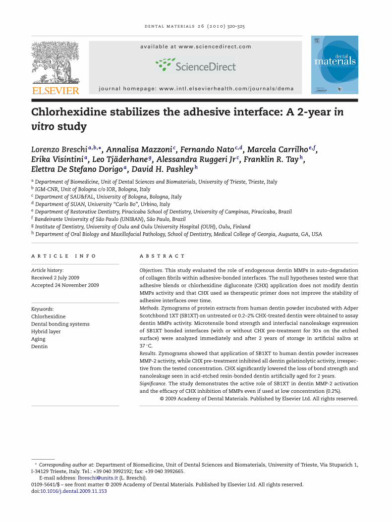
d e n t a l m a t e r i a l s 2 6 ( 2 0 1 0 ) 320–325
avai lab le at www.sc iencedi rec t .com
journa l homepage: www. int l .e lsev ierhea l th .com/ journa ls /dema
Chlorhexidine stabilizes the adhesive interface: A 2-year invitro study
Lorenzo Breschia,b,∗, Annalisa Mazzoni c, Fernando Natoc,d, Marcela Carrilhoe,f,Erika Visintinia, Leo Tjäderhaneg, Alessandra Ruggeri Jr c, Franklin R. Tayh,Elettra De Stefano Dorigoa, David H. Pashleyh
a Department of Biomedicine, Unit of Dental Sciences and Biomaterials, University of Trieste, Trieste, Italyb IGM-CNR, Unit of Bologna c/o IOR, Bologna, Italyc Department of SAU&FAL, University of Bologna, Bologna, Italyd Department of SUAN, University “Carlo Bo”, Urbino, Italye Department of Restorative Dentistry, Piracicaba School of Dentistry, University of Campinas, Piracicaba, Brazilf Bandeirante University of São Paulo (UNIBAN), São Paulo, Brazilg Institute of Dentistry, University of Oulu and Oulu University Hospital (OUH), Oulu, Finlandh Department of Oral Biology and Maxillofacial Pathology, School of Dentistry, Medical College of Georgia, Augusta, GA, USA
a r t i c l e i n f o
Article history:
Received 2 July 2009
Accepted 24 November 2009
Keywords:
Chlorhexidine
Dental bonding systems
Hybrid layer
Aging
Dentin
a b s t r a c t
Objectives. This study evaluated the role of endogenous dentin MMPs in auto-degradation
of collagen fibrils within adhesive-bonded interfaces. The null hypotheses tested were that
adhesive blends or chlorhexidine digluconate (CHX) application does not modify dentin
MMPs activity and that CHX used as therapeutic primer does not improve the stability of
adhesive interfaces over time.
Methods. Zymograms of protein extracts from human dentin powder incubated with Adper
Scotchbond 1XT (SB1XT) on untreated or 0.2–2% CHX-treated dentin were obtained to assay
dentin MMPs activity. Microtensile bond strength and interfacial nanoleakage expression
of SB1XT bonded interfaces (with or without CHX pre-treatment for 30 s on the etched
surface) were analyzed immediately and after 2 years of storage in artificial saliva at
37 ◦C.
Results. Zymograms showed that application of SB1XT to human dentin powder increases
MMP-2 activity, while CHX pre-treatment inhibited all dentin gelatinolytic activity, irrespec-
tive from the tested concentration. CHX significantly lowered the loss of bond strength and
nanoleakage seen in acid-etched resin-bonded dentin artificially aged for 2 years.
Significance. The study demonstrates the active role of SB1XT in dentin MMP-2 activation
and the efficacy of CHX inhibition of MMPs even if used at low concentration (0.2%).
© 2009 Academy of Dental Materials. Published by Elsevier Ltd. All rights reserved.
∗ Corresponding author at: Department of Biomedicine, Unit of Dental Sciences and Biomaterials, University of Trieste, Via Stuparich 1,I-34129 Trieste, Italy. Tel.: +39 040 3992192; fax: +39 040 3992665.
E-mail address: [email protected] (L. Breschi).0109-5641/$ – see front matter © 2009 Academy of Dental Materials. Published by Elsevier Ltd. All rights reserved.doi:10.1016/j.dental.2009.11.153
2 6
1
Da(ttshb[pmltw
ptna
sodnaove
tttitibatoiC[
atsaan
2
2
RU
d e n t a l m a t e r i a l s
. Introduction
espite successful immediate bonding, the longevity of thedhesive interface remains questionable due to physicalocclusal forces, expansion and contraction stresses relatedo temperature changes) and chemical factors challenginghe adhesive interface [1,2]. The hybrid layer created by two-tep etch-and-rinse adhesives containing high percentages ofydrophilic monomers, results in the formation of a porousonded interface [3] that behaves as a permeable membrane
4] allowing elution of unreacted monomers, water sorption,olymer swelling and resin hydrolysis [5,6]. In addition, enzy-atic activity can degrade the exposed type I collagen fibrils
ocated at the bottom of the hybrid layer due to the activa-ion of endogenous collagenolytic factors previously identifiedithin the dentin organic matrix [7–9].
These enzymes belong to the family of matrix metallo-roteinases (MMPs) and are involved in connective tissueurn-over, degrading almost all extracellular matrix compo-ents [10,11]. Recent findings indicate that MMP-2, -3, -8, -9nd -20 are present within the human dentin matrix [12–16].
Although these enzymes are involved in dentinogene-is and in caries progression mechanisms [12,17], the rolef dentin MMPs in mature dentin is still unclear. Humanentin matrices exhibit variable collagenolytic and gelati-olytic activities when mixed with dentin/enamel bondinggents with different pHs [8,9]. Thus, the simple applicationf adhesive systems on acid-etched dentin substrate can acti-ate dentinal MMPs, initiating autolytic phenomena that willventually affect the hybrid layer.
Chlorhexidine digluconate (CHX) has the ability to inac-ivate MMP-2, -8 and -9 [18]. Despite the use of CHX as aherapeutic primer to stabilize the adhesive interface overime [19–22], the relationship between the collagenolytic activ-ty of dentin, MMPs role in hybrid layer degradation, andhe mechanism of CHX inhibition of MMPs needs to be clar-fied. Aqueous 2% CHX prevented much of the decline inond strength and increased nanoleakage seen both in vitrond in vivo in deciduous and permanent teeth [19–21]. Addi-ionally, a recent in vitro study confirmed the protective rolef 0.2% CHX after 1 year of storage of bonded specimens
n artificial saliva, suggesting that lower concentration ofHX can be equally effective compared to 2% concentrations
22].The purpose of this study was to evaluate the effect of 0.2
nd 2% CHX on the adhesive–dentin interfaces created by awo-step etch-and-rinse adhesive system. The null hypothe-es tested were that (1) dentin MMPs activity is not affected bydhesive or CHX application and (2) the use of CHX as a ther-peutic primer does not affect bond strength and interfacialanoleakage expression after 2 years.
. Material and methods
.1. Zymographic analysis
eagents were purchased from Sigma Chemical (St Louis, MO,SA) unless otherwise specified.
( 2 0 1 0 ) 320–325 321
Ten freshly extracted human molars were selected afterpatient’s informed consent was obtained under a protocolapproved by the University of Trieste. Enamel, roots andremnant pulp tissue were removed and dentin powder wasobtained by pulverizing liquid nitrogen-frozen coronal dentinwith a steel mortar/pestle (Reimiller, Reggio Emilia, Italy).Five aliquots of 1 g each of dentin powder were obtainedand treated as follow. Group 1: untreated mineralized dentinpowder; Group 2: dentin powder demineralized in 1% aque-ous H3PO4 for 10 min; Group 3: mineralized dentin powdertreated with 3 mL of Adper Scotchbond 1XT (SB1XT, 3M ESPE,St Paul, MN, USA) for 24 h at 4 ◦C in dark condition; Groups4 and 5: mineralized dentin powder was treated either with0.2 or 2% CHX water solution (Groups 4 and 5, respectively)for 30 min at 4 ◦C, rinsed with 1 mL of distilled water (fivetimes), then incubated with 3 mL of SB1XT for 24 h at 4 ◦C indark condition. All specimens were thoroughly rinsed with4 mL of acetone and then centrifuged for 10 min (14,000 rpm)at 4 ◦C. Specimens were then re-suspended in 4 mL extrac-tion buffer (50 mM Tris–HCl pH 6, containing 5 mM CaCl2,100 mM NaCl, 0.1% Triton X-100), 0.1% non-ionic detergentP-40 (0.1 mM ZnCl2, 0.02% NaN3) and EDTA-free proteaseinhibitor cocktail (Roche Diagnostics GmbH, Germany) for24 h. Specimens were then centrifuged, supernatants werecollected and protein content was precipitated with 25 wt%trichloroacetic acid (TCA) at 4 ◦C. TCA precipitates were re-solubilized in loading buffer (25% Trizma–HCl 1 M pH 6.8,8% (w/v) sodium dodecyl sulfate, 40% glycerol and distilledwater).
Total protein concentration of the extracts of mineralizedand partially demineralized dentin powder was determinedusing the Bradford assay. Proteins were electrophorizedunder non-reducing conditions on 7.5% SDS-polyacrylamidegels copolymerized with 2 g/L gelatin (porcine skin). Acti-vation of gelatinase proforms was achieved with 2 mMp-aminophenylmercuric acetate (APMA) for 1 h at 37 ◦C andthen incubated for 24 h at 37 ◦C in zymography buffer (CaCl2,NaCl and Tris–HCl, pH 8.0). Gels were stained in 0.2%Coomassie Brilliant Blue R-250 and destained in destainingbuffer (50% methanol, 10% acetic acid, 40% water).
Control zymograms were incubated in the presence of5 mM EDTA and 2 mM 1,10-phenanthroline to inhibit gelati-nases.
2.2. Specimens preparation for microtensile bondstrength test
An additional 48 non-carious extracted human molars wereselected and flat surfaces of middle/deep dentin were exposedwith a slow speed diamond saw (Micromet, Bologna, Italy).Smear layer-covered dentin surfaces were etched with 35%phosphoric acid for 15 s (etching gel, 3M ESPE), rinsed andsurfaces were blot dried according to the wet bonding tech-nique. Specimens were randomly assigned to the followingtreatments (N = 16 teeth in each group). Groups 1 and 2: acid-etched dentin surfaces were treated with aqueous solutions
of either 0.2 or 2% CHX, respectively, for 30 s, blot dried andbonded with SB1XT; Group 3: received no pre-treatment beforeSB1XT application (control). SB1XT was applied in accordancewith manufacturers’ instructions and light-cured. Resin com-
l s 2 6 ( 2 0 1 0 ) 320–325
Fig. 1 – Gelatin zymogram of MMPs from dentin extractsafter sonication, TCA precipitation, resuspension andactivation with 2 mM APMA for 1 h. Molecular masses,expressed in kDa, are reported in Std lane. Lane 1: Absenceof any gelatinolytic activities in proteins extracted frommineralized dentin powder; Lane 2: Identification of MMP-2and -9 isoforms in H3PO4-treated dentin; Lane 3: MMPsisoforms detected in mineralized dentin powder afterincubation with SB1XT for 24 h produced an increase ingelatinolytic activity of MMP-2 and a decrease of MMP-9compared to the H3PO4-demineralized dentin; Lanes 4 and5: Incubation with 0.2 or 2% CHX respectively and SB1XT,
322 d e n t a l m a t e r i a
posite build-ups were created with Filtek Z250 (3M ESPE, StPaul, MN, USA).
2.3. Microtensile bond strength evaluation
Resin–dentin sticks with cross-sectional area of approx-imately 0.9 mm2 were obtained in accordance with thenon-trimming technique [23]. Each stick was measured andrecorded for bond strength calculation. Sticks were divided intwo equal groups and either stored for 24 h (T0) or for 2 years(T2 years) in artificial saliva (prepared in accordance with theprotocol of Pashley et al. [7], but without protease inhibitors)at 37 ◦C. Sticks were stressed until failure with a simplifieduniversal testing machine at a crosshead speed of 1 mm/min(Bisco Inc., Schaumburg, IL, USA). Failure modes were evalu-ated as described by Breschi et al. [22].
2.4. Statistical analysis
As values were normally distributed (Kolmogorov–Smirnoftest), data were analyzed with a two-way ANOVA (tested vari-ables were: CHX concentration, time of storage) and Tukey’spost hoc test. To analyze the effect of chlorhexidine on fracturemodes, mixed and dentin cohesive failures were combined.Wilcoxon Signed Ranks Test was used to analyze the differ-ences in failure modes between T0 and T2 years for each group,and Kruskal–Wallis test was used to compare the fracturemodes between the groups within each time points. Statisticalsignificance was set at p < 0.05.
2.5. Nanoleakage evaluation
Twelve additional teeth (N = 4/group) were prepared andbonded as previously described. Specimens were then verti-cally cut into 1-mm thick slabs to expose the bonded surfacesand submitted to the two storage times in artificial salivaat 37 ◦C: T0 and T2 years. Specimens were then submerged in50 wt% ammoniacal silver nitrate for 24 h, rinsed, photodevel-oped and processed for nanoleakage analysis under TEM andexamined under TEM (Philips CM-10) operating at 70 kV [24].
3. Results
3.1. Zymographic analysis
No enzymatic activity was found in proteins extracted frommineralized dentin (Group 1, Fig. 1, Lane 1). Zymograms ofH3PO4-demineralized dentin extracts (Group 2) showed mul-tiple forms of gelatinolytic enzymes (Fig. 1, Lane 2), with the66-kDa as MMP-2 active form, a fainter band of 86 kDa cor-responding to the active form of MMP-9 and other minorgelatinolytic bands with lower molecular weights (Fig. 1, Lane2). Zymograms of SB1XT-treated dentin (Group 3) showed anintense band at 66-kDa identified as MMP-2 active form, a72-kDa band corresponding to the proform of MMP-2 and a
fainter band at 86-kDa corresponding to a slight activity ofMMP-9 active form. In addition, lower molecular weight bandswere detected (Fig. 1, Lane 3). Incubation of mineralized dentinpowder with 0.2 or 2% CHX solutions followed by SB1XT appli-produced complete inhibition of all forms of MMP-2 and -9activity.
cation resulted in complete inhibition of both MMP-2 and -9activity (Fig. 1, Lanes 4 and 5).
Control zymograms incubated with 5 mM EDTA and 2 mM1,10-phenanthroline showed no enzymatic activity (data notshown).
3.2. Microtensile bond strength and fracture modeanalyses
Mean bond strength obtained at T0 and T2 years andfailure mode distributions are summarized in Table 1.At T0, there were no differences between the bondstrength of control versus the two CHX-tested concen-trations (0.2% CHX = 39.2 ± 9.3 MPa; 2% CHX = 41.2 ± 9.6 MPa;control = 40.8 ± 8.7 MPa; p > 0.05). Conversely, after 2 years ofin vitro storage (T2 years) the bond strengths of SB1XT-controlgroup decreased approximately 67% (control = 13.4 ± 4.9 MPa),while in CHX-treated groups the decrease of bond strengthwas limited between 16 and 30%, depending on whether theacid-etched dentin was primed with 0.2 or 2% CHX, respec-tively (0.2% CHX = 32.6 ± 8.3 MPa; 2% CHX = 28.5 ± 7.2 MPa).Fracture mode analysis did not demonstrate any statisti-cally significant differences between the groups or within thegroups at any time points (Table 1).
3.3. Interfacial nanoleakage analysis
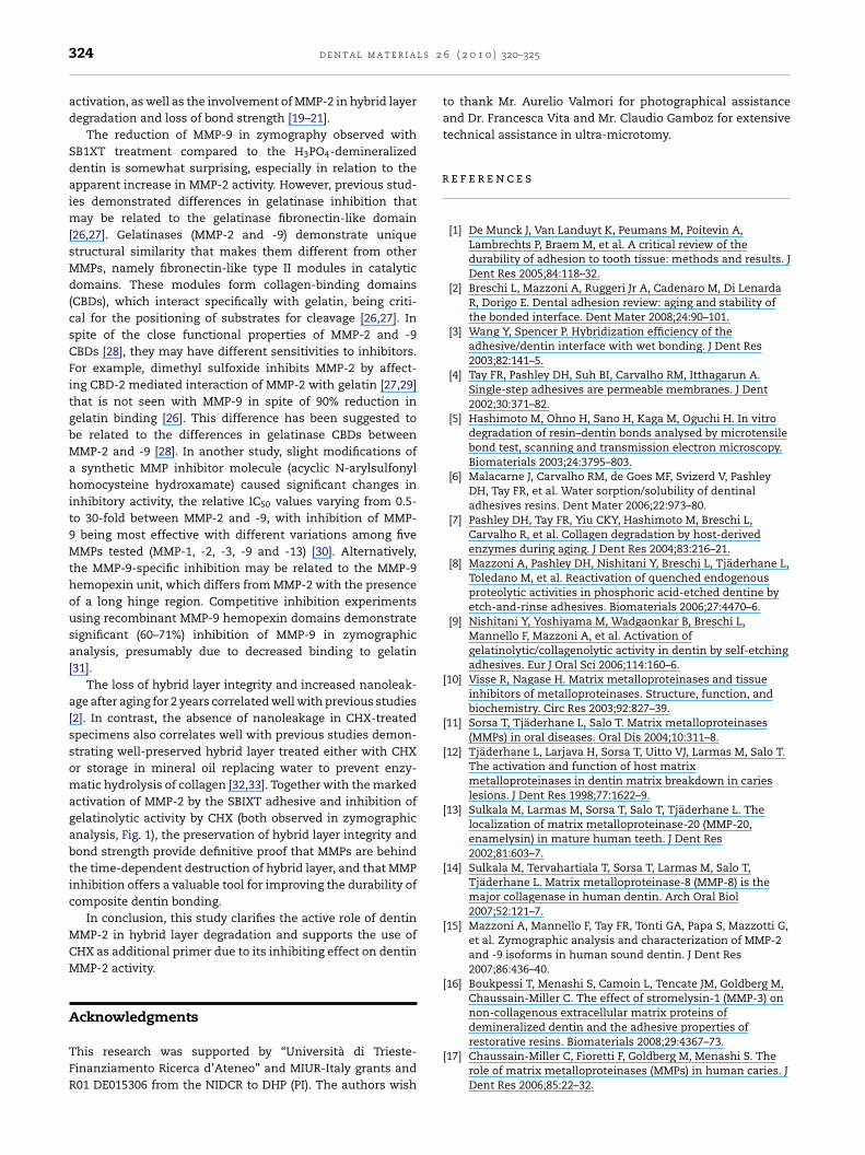
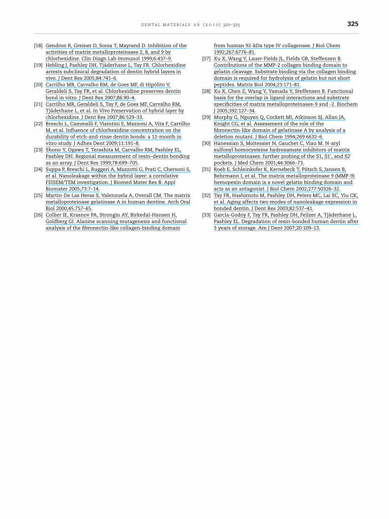
In CHX-treated adhesive interfaces, minor scattered silverparticles were observed, with no large-scale silver grain accu-
mulation (Fig. 2). In control specimens immersed in silvernitrate there was marked uptake of silver into the middle ofthe hybrid layer that was manifested as a large cluster of blacksilver grains (Fig. 3).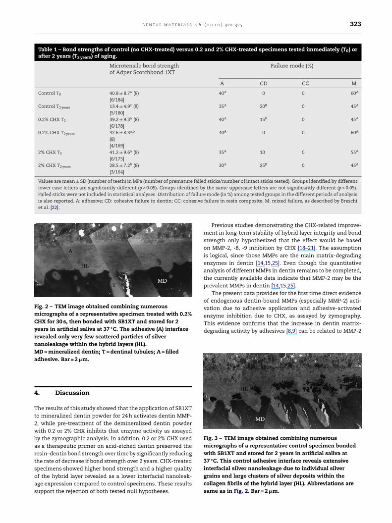
d e n t a l m a t e r i a l s 2 6 ( 2 0 1 0 ) 320–325 323
Table 1 – Bond strengths of control (no CHX-treated) versus 0.2 and 2% CHX-treated specimens tested immediately (T0) orafter 2 years (T2 years) of aging.
Microtensile bond strengthof Adper Scotchbond 1XT
Failure mode (%)
A CD CC M
Control T0 40.8 ± 8.7a (8)[6/184]
40A 0 0 60A
Control T2 years 13.4 ± 4.9c (8)[5/180]
35A 20B 0 45A
0.2% CHX T0 39.2 ± 9.3a (8)[6/178]
40A 15B 0 45A
0.2% CHX T2 years 32.6 ± 8.3a,b
(8)[4/169]
40A 0 0 60A
2% CHX T0 41.2 ± 9.6a (8)[6/175]
35A 10 0 55A
2% CHX T2 years 28.5 ± 7.2b (8)[3/164]
30A 25B 0 45A
Values are mean ± SD (number of teeth) in MPa (number of premature failed sticks/number of intact sticks tested). Groups identified by differentlower case letters are significantly different (p < 0.05). Groups identified by the same uppercase letters are not significantly different (p > 0.05).Failed sticks were not included in statistical analyses. Distribution of failureis also reported. A: adhesive; CD: cohesive failure in dentin; CC: cohesiveet al. [22].
Fig. 2 – TEM image obtained combining numerousmicrographs of a representative specimen treated with 0.2%CHX for 30 s, then bonded with SB1XT and stored for 2years in artificial saliva at 37 ◦C. The adhesive (A) interfacerevealed only very few scattered particles of silvernanoleakage within the hybrid layers (HL).MD = mineralized dentin; T = dentinal tubules; A = filleda
4
Tt2wbartsoas
enzyme inhibition due to CHX, as assayed by zymography.This evidence confirms that the increase in dentin matrix-degrading activity by adhesives [8,9] can be related to MMP-2
Fig. 3 – TEM image obtained combining numerousmicrographs of a representative control specimen bondedwith SB1XT and stored for 2 years in artificial saliva at37 ◦C. This control adhesive interface reveals extensiveinterfacial silver nanoleakage due to individual silver
dhesive. Bar = 2 �m.
. Discussion
he results of this study showed that the application of SB1XTo mineralized dentin powder for 24 h activates dentin MMP-, while pre-treatment of the demineralized dentin powderith 0.2 or 2% CHX inhibits that enzyme activity as assayed
y the zymographic analysis. In addition, 0.2 or 2% CHX useds a therapeutic primer on acid-etched dentin preserved theesin–dentin bond strength over time by significantly reducinghe rate of decrease if bond strength over 2 years. CHX-treatedpecimens showed higher bond strength and a higher quality
f the hybrid layer revealed as a lower interfacial nanoleak-ge expression compared to control specimens. These resultsupport the rejection of both tested null hypotheses.mode (in %) among tested groups in the different periods of analysisfailure in resin composite; M: mixed failure, as described by Breschi
Previous studies demonstrating the CHX-related improve-ment in long-term stability of hybrid layer integrity and bondstrength only hypothesized that the effect would be basedon MMP-2, -8, -9 inhibition by CHX [18–21]. The assumptionis logical, since those MMPs are the main matrix-degradingenzymes in dentin [14,15,25]. Even though the quantitativeanalysis of different MMPs in dentin remains to be completed,the currently available data indicate that MMP-2 may be theprevalent MMPs in dentin [14,15,25].
The present data provides for the first time direct evidenceof endogenous dentin-bound MMPs (especially MMP-2) acti-vation due to adhesive application and adhesive-activated
grains and large clusters of silver deposits within thecollagen fibrils of the hybrid layer (HL). Abbreviations aresame as in Fig. 2. Bar = 2 �m.
l s 2
r
324 d e n t a l m a t e r i a
activation, as well as the involvement of MMP-2 in hybrid layerdegradation and loss of bond strength [19–21].
The reduction of MMP-9 in zymography observed withSB1XT treatment compared to the H3PO4-demineralizeddentin is somewhat surprising, especially in relation to theapparent increase in MMP-2 activity. However, previous stud-ies demonstrated differences in gelatinase inhibition thatmay be related to the gelatinase fibronectin-like domain[26,27]. Gelatinases (MMP-2 and -9) demonstrate uniquestructural similarity that makes them different from otherMMPs, namely fibronectin-like type II modules in catalyticdomains. These modules form collagen-binding domains(CBDs), which interact specifically with gelatin, being criti-cal for the positioning of substrates for cleavage [26,27]. Inspite of the close functional properties of MMP-2 and -9CBDs [28], they may have different sensitivities to inhibitors.For example, dimethyl sulfoxide inhibits MMP-2 by affect-ing CBD-2 mediated interaction of MMP-2 with gelatin [27,29]that is not seen with MMP-9 in spite of 90% reduction ingelatin binding [26]. This difference has been suggested tobe related to the differences in gelatinase CBDs betweenMMP-2 and -9 [28]. In another study, slight modifications ofa synthetic MMP inhibitor molecule (acyclic N-arylsulfonylhomocysteine hydroxamate) caused significant changes ininhibitory activity, the relative IC50 values varying from 0.5-to 30-fold between MMP-2 and -9, with inhibition of MMP-9 being most effective with different variations among fiveMMPs tested (MMP-1, -2, -3, -9 and -13) [30]. Alternatively,the MMP-9-specific inhibition may be related to the MMP-9hemopexin unit, which differs from MMP-2 with the presenceof a long hinge region. Competitive inhibition experimentsusing recombinant MMP-9 hemopexin domains demonstratesignificant (60–71%) inhibition of MMP-9 in zymographicanalysis, presumably due to decreased binding to gelatin[31].
The loss of hybrid layer integrity and increased nanoleak-age after aging for 2 years correlated well with previous studies[2]. In contrast, the absence of nanoleakage in CHX-treatedspecimens also correlates well with previous studies demon-strating well-preserved hybrid layer treated either with CHXor storage in mineral oil replacing water to prevent enzy-matic hydrolysis of collagen [32,33]. Together with the markedactivation of MMP-2 by the SBIXT adhesive and inhibition ofgelatinolytic activity by CHX (both observed in zymographicanalysis, Fig. 1), the preservation of hybrid layer integrity andbond strength provide definitive proof that MMPs are behindthe time-dependent destruction of hybrid layer, and that MMPinhibition offers a valuable tool for improving the durability ofcomposite dentin bonding.
In conclusion, this study clarifies the active role of dentinMMP-2 in hybrid layer degradation and supports the use ofCHX as additional primer due to its inhibiting effect on dentinMMP-2 activity.
Acknowledgments
This research was supported by “Università di Trieste-Finanziamento Ricerca d’Ateneo” and MIUR-Italy grants andR01 DE015306 from the NIDCR to DHP (PI). The authors wish
6 ( 2 0 1 0 ) 320–325
to thank Mr. Aurelio Valmori for photographical assistanceand Dr. Francesca Vita and Mr. Claudio Gamboz for extensivetechnical assistance in ultra-microtomy.
e f e r e n c e s
[1] De Munck J, Van Landuyt K, Peumans M, Poitevin A,Lambrechts P, Braem M, et al. A critical review of thedurability of adhesion to tooth tissue: methods and results. JDent Res 2005;84:118–32.
[2] Breschi L, Mazzoni A, Ruggeri Jr A, Cadenaro M, Di LenardaR, Dorigo E. Dental adhesion review: aging and stability ofthe bonded interface. Dent Mater 2008;24:90–101.
[3] Wang Y, Spencer P. Hybridization efficiency of theadhesive/dentin interface with wet bonding. J Dent Res2003;82:141–5.
[4] Tay FR, Pashley DH, Suh BI, Carvalho RM, Itthagarun A.Single-step adhesives are permeable membranes. J Dent2002;30:371–82.
[5] Hashimoto M, Ohno H, Sano H, Kaga M, Oguchi H. In vitrodegradation of resin–dentin bonds analysed by microtensilebond test, scanning and transmission electron microscopy.Biomaterials 2003;24:3795–803.
[6] Malacarne J, Carvalho RM, de Goes MF, Svizerd V, PashleyDH, Tay FR, et al. Water sorption/solubility of dentinaladhesives resins. Dent Mater 2006;22:973–80.
[7] Pashley DH, Tay FR, Yiu CKY, Hashimoto M, Breschi L,Carvalho R, et al. Collagen degradation by host-derivedenzymes during aging. J Dent Res 2004;83:216–21.
[8] Mazzoni A, Pashley DH, Nishitani Y, Breschi L, Tjäderhane L,Toledano M, et al. Reactivation of quenched endogenousproteolytic activities in phosphoric acid-etched dentine byetch-and-rinse adhesives. Biomaterials 2006;27:4470–6.
[9] Nishitani Y, Yoshiyama M, Wadgaonkar B, Breschi L,Mannello F, Mazzoni A, et al. Activation ofgelatinolytic/collagenolytic activity in dentin by self-etchingadhesives. Eur J Oral Sci 2006;114:160–6.
[10] Visse R, Nagase H. Matrix metalloproteinases and tissueinhibitors of metalloproteinases. Structure, function, andbiochemistry. Circ Res 2003;92:827–39.
[11] Sorsa T, Tjäderhane L, Salo T. Matrix metalloproteinases(MMPs) in oral diseases. Oral Dis 2004;10:311–8.
[12] Tjäderhane L, Larjava H, Sorsa T, Uitto VJ, Larmas M, Salo T.The activation and function of host matrixmetalloproteinases in dentin matrix breakdown in carieslesions. J Dent Res 1998;77:1622–9.
[13] Sulkala M, Larmas M, Sorsa T, Salo T, Tjäderhane L. Thelocalization of matrix metalloproteinase-20 (MMP-20,enamelysin) in mature human teeth. J Dent Res2002;81:603–7.
[14] Sulkala M, Tervahartiala T, Sorsa T, Larmas M, Salo T,Tjäderhane L. Matrix metalloproteinase-8 (MMP-8) is themajor collagenase in human dentin. Arch Oral Biol2007;52:121–7.
[15] Mazzoni A, Mannello F, Tay FR, Tonti GA, Papa S, Mazzotti G,et al. Zymographic analysis and characterization of MMP-2and -9 isoforms in human sound dentin. J Dent Res2007;86:436–40.
[16] Boukpessi T, Menashi S, Camoin L, Tencate JM, Goldberg M,Chaussain-Miller C. The effect of stromelysin-1 (MMP-3) onnon-collagenous extracellular matrix proteins of
demineralized dentin and the adhesive properties ofrestorative resins. Biomaterials 2008;29:4367–73.[17] Chaussain-Miller C, Fioretti F, Goldberg M, Menashi S. Therole of matrix metalloproteinases (MMPs) in human caries. JDent Res 2006;85:22–32.
2 6
d e n t a l m a t e r i a l s[18] Gendron R, Greiner D, Sorsa T, Mayrand D. Inhibition of theactivities of matrix metalloproteinases 2, 8, and 9 bychlorhexidine. Clin Diagn Lab Immunol 1999;6:437–9.
[19] Hebling J, Pashley DH, Tjäderhane L, Tay FR. Chlorhexidinearrests subclinical degradation of dentin hybrid layers invivo. J Dent Res 2005;84:741–6.
[20] Carrilho MR, Carvalho RM, de Goes MF, di Hipólito V,Geraldeli S, Tay FR, et al. Chlorhexidine preserves dentinbond in vitro. J Dent Res 2007;86:90–4.
[21] Carrilho MR, Geraldeli S, Tay F, de Goes MF, Carvalho RM,Tjäderhane L, et al. In Vivo Preservation of hybrid layer bychlorhexidine. J Dent Res 2007;86:529–33.
[22] Breschi L, Cammelli F, Visintini E, Mazzoni A, Vita F, CarrilhoM, et al. Influence of chlorhexidine concentration on thedurability of etch-and-rinse dentin bonds: a 12-month invitro study. J Adhes Dent 2009;11:191–8.
[23] Shono Y, Ogawa T, Terashita M, Carvalho RM, Pashley EL,Pashley DH. Regional measurement of resin–dentin bondingas an array. J Dent Res 1999;78:699–705.
[24] Suppa P, Breschi L, Ruggeri A, Mazzotti G, Prati C, Chersoni S,et al. Nanoleakage within the hybrid layer: a correlativeFEISEM/TEM investigation. J Biomed Mater Res B: ApplBiomater 2005;73:7–14.
[25] Martin-De Las Heras S, Valenzuela A, Overall CM. The matrix
metalloproteinase gelatinase A in human dentine. Arch OralBiol 2000;45:757–65.[26] Collier IE, Krasnov PA, Strongin AY, Birkedal-Hansen H,Goldberg GI. Alanine scanning mutagenesis and functionalanalysis of the fibronectin-like collagen-binding domain
( 2 0 1 0 ) 320–325 325
from human 92-kDa type IV collagenase. J Biol Chem1992;267:6776–81.
[27] Xu X, Wang Y, Lauer-Fields JL, Fields GB, Steffensen B.Contributions of the MMP-2 collagen binding domain togelatin cleavage. Substrate binding via the collagen bindingdomain is required for hydrolysis of gelatin but not shortpeptides. Matrix Biol 2004;23:171–81.
[28] Xu X, Chen Z, Wang Y, Yamada Y, Steffensen B. Functionalbasis for the overlap in ligand interactions and substratespecificities of matrix metalloproteinases-9 and -2. BiochemJ 2005;392:127–34.
[29] Murphy G, Nguyen Q, Cockett MI, Atkinson SJ, Allan JA,Knight CG, et al. Assessment of the role of thefibronectin-like domain of gelatinase A by analysis of adeletion mutant. J Biol Chem 1994;269:6632–6.
[30] Hanessian S, Moitessier N, Gauchet C, Viau M. N-arylsulfonyl homocysteine hydroxamate inhibitors of matrixmetalloproteinases: further probing of the S1, S1′, and S2′
pockets. J Med Chem 2001;44:3066–73.[31] Roeb E, Schleinkofer K, Kernebeck T, Pötsch S, Jansen B,
Behrmann I, et al. The matrix metalloproteinase 9 (MMP-9)hemopexin domain is a novel gelatin binding domain andacts as an antagonist. J Biol Chem 2002;277:50326–32.
[32] Tay FR, Hashimoto M, Pashley DH, Peters MC, Lai SC, Yiu CK,
et al. Aging affects two modes of nanoleakage expression inbonded dentin. J Dent Res 2003;82:537–41.[33] García-Godoy F, Tay FR, Pashley DH, Feilzer A, Tjäderhane L,Pashley EL. Degradation of resin-bonded human dentin after3 years of storage. Am J Dent 2007;20:109–13.
Copyright © 2022 FDOKUMEN