Nanoparticle Delivery Platforms for RNAi Therapeutics ... - MDPI

RSC Advances
PAPER
Publ
ishe
d on
11
Nov
embe
r 20
13. D
ownl
oade
d by
Uni
v L
ille
1 on
03/
12/2
013
09:3
7:51
.
View Article OnlineView Journal | View Issue
aInstitut de Recherche Interdisciplinaire (IRI,
de la Haute Borne, 50 Avenue de Halley, BP
E-mail: [email protected] of Fine Organic Synthesis,
Petrochemistry NAS of Ukraine, 1 MurmanscTaras Shevchenko University, 60 VladimirskdNational Institute of Materials Physics,
RomaniaeEA 4479, IFR 147, Universite Lille 1, 59658
Cite this: RSC Adv., 2014, 4, 865
Received 31st October 2013Accepted 11th November 2013
DOI: 10.1039/c3ra46307a
www.rsc.org/advances
This journal is © The Royal Society of C
Insulin loaded iron magnetic nanoparticle–graphene oxide composites: synthesis,characterization and application for in vivo deliveryof insulin
Kostiantyn Turcheniuk,ab Manakamana Khanal,a Anastasiia Motorina,ac
Palaniappan Subramanian,a Alexandre Barras,a Vladimir Zaitsev,c Victor Kuncser,d
Aurel Leca,d Alain Martoriati,e Katia Cailliau,e Jean-Francois Bodart,e
Rabah Boukherrouba and Sabine Szunerits*a
One of the focal subjects in insulin delivery is the development of insulin formulations that protect the native
insulin from degradation under acidic pH in the stomach. In this work we show, for the first time, that a
graphene oxide (GO) based matrix can ensure the stability of insulin at low pH. GO and GO modified
with 2-nitrodopamine coated magnetic particle (GO–MPdop) matrices loaded with insulin were prepared
and the pH triggered release of the insulin was studied. The loading of insulin on the GO nanomaterials
proved to be extremely high at pH < 5.4 with a loading capacity of 100 � 3% on GO and 88 � 3% on
GO–MPdop. The insulin-containing GO matrices were stable at acidic pH, while insulin was released
when exposed to basic solutions (pH ¼ 9.2). Using Xenopus laevis oocytes as a model we showed that
the meiotic resumption rate of GO and GO–MPdop remained unaltered when pre-treated in acidic
conditions, while pre-incubated insulin (without GO nanomaterials) has lost almost entirely its
maturation effect. These results suggest that GO based nanomatrices are promising systems for the
protection of insulin.
1. Introduction
The use of nanomaterials as carriers of glycans,1,2 drugs,3,4
genes,5,6 and other biologically active compounds7 has become awidely investigated research eld. Most recently, graphene, atwo-dimensional nanomaterial, has been intensively explored asan alternative nanocarrier for biological materials due to itslarge surface area, rich surface chemistry and its potential forcrossing the plasma membrane and promoting the cellularuptake of molecules.8,9 The interest in using graphene and gra-phene oxide (GO) for loading and release of chemical and bio-logical molecules is in addition linked to the different ways themolecule can be linked to the graphene matrix: hydrogenbonding, hydrophobic, p–p stacking and electrostatic interac-tions can act as anchors that are sensitive to external stimuli
USR CNRS 3078), Universite Lille 1, Parc
70478, 59658 Villeneuve d’Ascq, France.
Institute of Bioorganic Chemistry and
ka Str., 02660, Kiev, Ukraine
aya str., Kiev, Ukraine
Atomistilor 105 bis, 077125 Magurele,
Villeneuve d’Ascq, France
hemistry 2014
(pH, temperature, chemical substances, electrical eld, etc.),enabling controlled release.10–14 Since the pioneering work of Daiand colleagues13,15 on the use of PEGylated (PEG ¼ polyethyleneglycol) GO as a nanocarrier to load anticancer drugs via non-covalent physisorption and study its cellular uptake, severalpapers have been devoted to improving the loading efficiencyand release of anticancer drugs such as doxorubicin (DOX)16 orto the preparation of multi-functionalized graphene nano-materials.17–20 Besides graphene and GO, graphene/iron oxidenanoparticles composite materials have shown great promise asdrug carriers4,20,21 and for the immobilization and enrichment ofbiomolecules.22 The magnetic particles modied graphenesheets were synthesized by in situ oxidation of Fe2+ salts to Fe3+
and deposited as Fe3O4 particles onto GO, being at the sametime reduced to reduced graphene oxide (rGO).23 Otherapproaches exploited the strong complexation of the carboxylateanions of GO with FeCl3 and FeCl2, before precipitating Fe3O4
nanoparticles onto GO by treatment with sodium hydroxide.4,20
It is worth mentioning that in the earlier reports themagnetic particles were not chemically protected and therefore,they were prone to corrosion upon immersion in cell culturemedia. Here, we report a different strategy for the preparation ofGO–magnetic nanoparticles composite. It is based on the ex situsynthesis of chemically stabilized magnetic particles with
RSC Adv., 2014, 4, 865–875 | 865

Fig. 1 (A) Synthetic route of 2-nitrodopamine and functionalization of magnetic nanoparticles (MP) with 2-nitrodopamine. (B) Insulin loading onGO and GO–MPdop. (A).
RSC Advances Paper
Publ
ishe
d on
11
Nov
embe
r 20
13. D
ownl
oade
d by
Uni
v L
ille
1 on
03/
12/2
013
09:3
7:51
. View Article Online
2-nitrodopamine, followed by their insertion onto the GOmatrix (Fig. 1). This approach prevents any subsequent reduc-tion of water soluble GO to water insoluble rGO and ensures theformation of a chemically stable GO–magnetic nanoparticlesinterface. This GO matrix is well suited for the uptake ofbiomolecules such as insulin.
Insulin, a polypeptide composed of 51 amino acid residuesand secreted by the pancreas, plays an important role in thecontrol of blood glucose. Diabetic people suffer from low levels ofinsulin production and/or from abnormal resistance to theinsulin hormone. Current treatment methods involve regularinjections of insulin, which can be both painful and inconve-nient. In order to overcome these hurdles, the oral route isconsidered as one of themost convenient means of drug uptake.However, oral administration of hydrophilic macromoleculessuch as insulin encounters (or faces) major problems such ashydrolysis in the low pH of gastric medium, splitting byproteinases in the stomach and weak penetration through themembrane of epithelial cells of the intestine.24,25One of themostpromising strategies to achieve oral insulin uptake is the use ofmicrosphere systems, which act both as protease inhibitors byprotecting the encapsulated insulin from enzymatic degradationand as permeation enhancers by effectively crossing the epithe-lial layer aer oral administration.26–31 Behavior, toxicity and
866 | RSC Adv., 2014, 4, 865–875
biocompatibility of nanomaterials in vivo is associated to size,surface of coating andadministration routes.32Nevertheless, oraladministration appears as an appealing strategy. Indeed, a recentstudy underlined the biocompatibility of PEGylated GO deriva-tives aer oral administration since the injected materialexhibited a long-term retention but no toxicity.33
2. Experimental part2.1. Materials
Graphite powder (<20 microns), hydrogen peroxide (H2O2),sulfuric acid (H2SO4), dimethylsulfoxide (DMSO), acetonitrile(CH3CN), ammonium hydroxide (NH4OH), iron(II) chloride tet-rahydrate (FeCl2$4H2O), iron(III) chloride hexahydrate(FeCl3$6H2O), dopamine hydrochloride, sodium nitrite, insulin(from bovine pancreas, code 10516), dispase, and collagenasewere purchased from Sigma-Aldrich and used as received. 3-(4,5-Dimethylthiazol-2-yl)-2,5-diphenyltetrazolium bromide(MTT) was obtained from invitrogen.
2.2. Preparation of graphene oxide (GO)
Graphene oxide (GO) was synthesized from graphite powder bya modied Hummers method.34 5 mg of the synthesized GO was
This journal is © The Royal Society of Chemistry 2014

Paper RSC Advances
Publ
ishe
d on
11
Nov
embe
r 20
13. D
ownl
oade
d by
Uni
v L
ille
1 on
03/
12/2
013
09:3
7:51
. View Article Online
dispersed in 1 mL of water and exfoliated through ultra-sonication for 3 h. This aqueous suspension of GO was used as astock solution in subsequent experiments.
2.3. Synthesis of 2-nitrodopamine
2-Nitrodopamine was synthesized according to ref. 35. Dopa-mine hydrochloride (1.90 g, 10mmol) and sodiumnitrite (1.52 g,22 mmol) were dissolved in water (25 mL) and cooled to 0 �C.Sulfuric acid (17.4 mmol in 10 mL of water) was added slowly tothemixture, and a yellow precipitate was formed. Aer stirring atroom temperature overnight, the precipitate was ltered andrecrystallized from water to give a product as a hemisulfate salt.Yield 1.9 g (77%). 1H NMR (DMSO-d6, 300 MHz, ppm): 3.10(br s, 4H, CH2CH2), 6.85 (s, 1H), 7.47 (s, 1H).
2.4. Preparation of 2-nitrodopamine modied magneticparticles (MPdop)
Magnetic particles (MP) were prepared as reported previously.36
FeCl2$4H2O (0.34 g, 1.7 mmol) and FeCl3$6H2O (0.95 g, 3.5mmol) were dissolved in deareated water (20 mL) and subse-quently added to a nitrogen-protected three-necked ask undersonication. The resulting mixture was heated at 50 �C for30 min. Then concentrated ammonium hydroxide (2 mL) wasadded dropwise and kept at constant temperature (50 �C) for30 min. The system was nally cooled to room temperature andthe solid product was isolated via a non-uniform magnetic eldgenerated by a Nd–Fe–B permanent magnet. The resultingFe3O4 particles were washed six times with Milli-Q water toremove unreacted chemicals and then stored in water.
A water dispersion of bare MP (10mgmL�1, 1 mL) was mixedwith 2-nitrodopamine (7 mg) and sonicated for 1 h at roomtemperature. The nitrodopamine modied MP (MPdop) wereisolated by means of magnet and puried through six consec-utive wash/precipitation cycles with water to ensure completeremoval of unreacted dopamine. The precipitate was dried in anoven at 50 �C.
2.5. Preparation of GO–MPdop nanohybrid
2 mL of GO in water (2 mg mL�1) was sonicated for 1 h before2 mL of MPdop (1 mg mL�1) were added and further sonicatedfor 2 h at 30 �C under N2. In a rst step, the resulting precipitatewas isolated by centrifugation at 13.500 rpm (20 min) andpuried through two consecutive wash/centrifugation cycles at13.500 rpm (20 min) with water. Further purication was ach-ieved by magnetic separation to separate the magnetic GO–MPdop hybrid from the non-magnetic phase. This procedureyielded z5 mg of GO–MPdop hybrid.
2.6. Loading of insulin onto GO and GO–MPdop hybrid
GO or GO–MPdop nanohybrid (150 mg mL�1) was sonicated withthe desired concentration of insulin for 2 h and then stirred for22 h at room temperature. All samples were centrifuged at13.500 rpm for 30 min. The concentration of insulin in thesupernatant was determined using a standard insulin concen-tration curve generated with a UV/Vis spectrophotometer at
This journal is © The Royal Society of Chemistry 2014
275 nm from a series of insulin solutions of differentconcentrations.
2.7. Release of insulin from GO and GO–MPdop hybrid
The release behavior of insulin from the GO–MPdop hybrid andGO was investigated at 37 �C under stirring at 40 � 10 rpm byvarying the pH. At predetermined time-points, samples werecentrifuged at 13.500 rpm for 30 min and the supernatant wasanalyzed using a UV/Vis spectrometer at 275 nm. The precipi-tate was redispersed in 1 mL of fresh PBS and release studieswere continued.
2.8. Cell viability/cytotoxicity studies (MTT test) on HEKcells
HEK cells were seeded in 96 wells plate at a density of 3 � 104
cells per well at 37 �C. Aer 24 h of culture, the medium in thewells was replaced with fresh medium, containing the GO–MPdop insulin hybrid in varying concentrations. Aer incuba-tion of the HEK cells for 24 hours, themediumwas replaced and10 mL of MTT (12 mM in sterile PBS) was added in each well andincubated for 4 h at 37 �C. Then medium was carefully removedand formed formazan crystals were solubilized with DMSO(50 mL). The absorbance of each well was read on a microplatereader (PHERAstar FS, BMG LABTECH) at 540 nm. Eachcondition was replicated for four times and wells without GO–MPdop insulin hybrid were taken as negative control.
2.9. Biological assays: cytotoxicity and M-phase entry inXenopus oocytes
Adult Xenopus females were purchased from University of Ren-nes I, France. Aer anesthetizing Xenopus females by immersionin MS222 solution (tricaine methane sulfonate, 1 g L�1), ovarianlobes were surgically removed and kept in ND96 physiologicalmedium (96mMNaCl, 2mMKCl, 1.8 mMCaCl2, 1 mMMgCl2, 5mM HEPES–NaOH, pH 7.5). For follicular cells removal fromoocytes, fragments of ovarian lobes were treated by dispase (3 h,0.4mg L�1), rinsed and bathed in collagenase (1 h, 0.4mg L�1) toend defolliculation. Fully-grown stage VI oocytes were selectedaccording to their morphology.37 The latter oocytes are arrestedat the G2/M border of the rst meiotic division and resumemeiosis in response to hormonal stimulation in vitro uponprogesterone or insulin addition in the medium.38,39 Oocyteswere stored at 14 �C in ND96 medium until use.
In the case of GO and GO–MPdop, the oocytes were pre-incu-bated for 30minwith theGOmatrix before hormonal stimulationby insulin. In the case of GO–insulin and GO–MPdop–insulin, thematrix was directly added to the oocytes. The pH media wereadjusted using HCl/NaOH solutions at pH 1, 2, 5.3, 7.4 and 9.2.Kinetic of Germinal Vesicle Breakdown (GVBD)was scored by theappearance of a white spot (WS) at the animal pole of the cell,which attests of the M-phase entry and meiosis resumption.
2.10. Instrumentation
2.10.1. Fourier transformed infrared (FTIR) spectroscopy.Fourier transform infrared (FT-IR) spectra were recorded using
RSC Adv., 2014, 4, 865–875 | 867
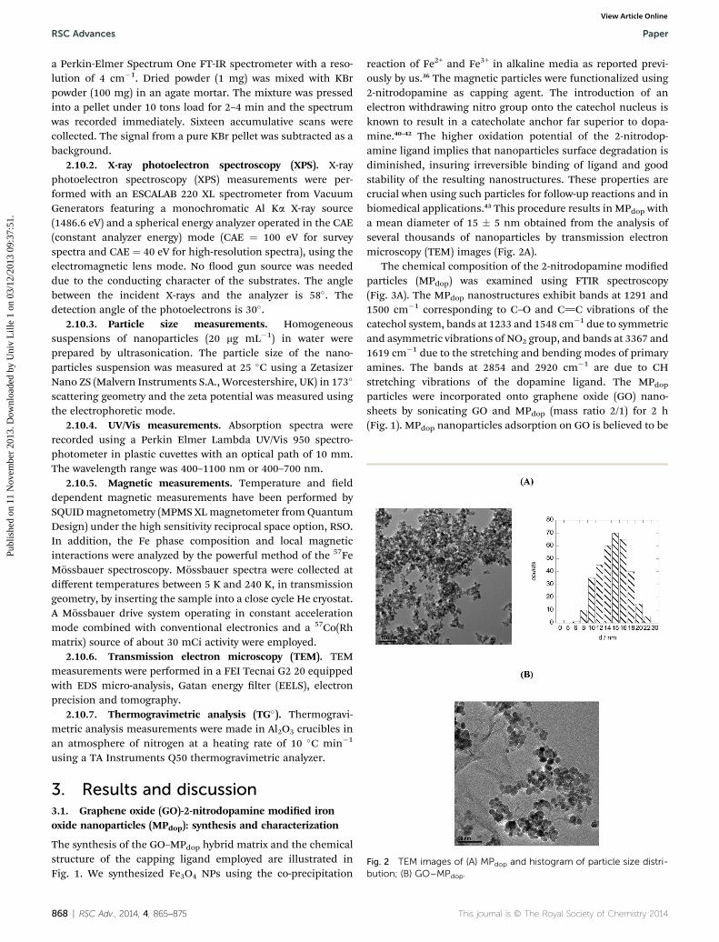
Fig. 2 TEM images of (A) MPdop and histogram of particle size distri-bution; (B) GO–MPdop.
RSC Advances Paper
Publ
ishe
d on
11
Nov
embe
r 20
13. D
ownl
oade
d by
Uni
v L
ille
1 on
03/
12/2
013
09:3
7:51
. View Article Online
a Perkin-Elmer Spectrum One FT-IR spectrometer with a reso-lution of 4 cm�1. Dried powder (1 mg) was mixed with KBrpowder (100 mg) in an agate mortar. The mixture was pressedinto a pellet under 10 tons load for 2–4 min and the spectrumwas recorded immediately. Sixteen accumulative scans werecollected. The signal from a pure KBr pellet was subtracted as abackground.
2.10.2. X-ray photoelectron spectroscopy (XPS). X-rayphotoelectron spectroscopy (XPS) measurements were per-formed with an ESCALAB 220 XL spectrometer from VacuumGenerators featuring a monochromatic Al Ka X-ray source(1486.6 eV) and a spherical energy analyzer operated in the CAE(constant analyzer energy) mode (CAE ¼ 100 eV for surveyspectra and CAE ¼ 40 eV for high-resolution spectra), using theelectromagnetic lens mode. No ood gun source was neededdue to the conducting character of the substrates. The anglebetween the incident X-rays and the analyzer is 58�. Thedetection angle of the photoelectrons is 30�.
2.10.3. Particle size measurements. Homogeneoussuspensions of nanoparticles (20 mg mL�1) in water wereprepared by ultrasonication. The particle size of the nano-particles suspension was measured at 25 �C using a ZetasizerNano ZS (Malvern Instruments S.A., Worcestershire, UK) in 173�
scattering geometry and the zeta potential was measured usingthe electrophoretic mode.
2.10.4. UV/Vis measurements. Absorption spectra wererecorded using a Perkin Elmer Lambda UV/Vis 950 spectro-photometer in plastic cuvettes with an optical path of 10 mm.The wavelength range was 400–1100 nm or 400–700 nm.
2.10.5. Magnetic measurements. Temperature and elddependent magnetic measurements have been performed bySQUIDmagnetometry (MPMS XLmagnetometer from QuantumDesign) under the high sensitivity reciprocal space option, RSO.In addition, the Fe phase composition and local magneticinteractions were analyzed by the powerful method of the 57FeMossbauer spectroscopy. Mossbauer spectra were collected atdifferent temperatures between 5 K and 240 K, in transmissiongeometry, by inserting the sample into a close cycle He cryostat.A Mossbauer drive system operating in constant accelerationmode combined with conventional electronics and a 57Co(Rhmatrix) source of about 30 mCi activity were employed.
2.10.6. Transmission electron microscopy (TEM). TEMmeasurements were performed in a FEI Tecnai G2 20 equippedwith EDS micro-analysis, Gatan energy lter (EELS), electronprecision and tomography.
2.10.7. Thermogravimetric analysis (TG�). Thermogravi-metric analysis measurements were made in Al2O3 crucibles inan atmosphere of nitrogen at a heating rate of 10 �C min�1
using a TA Instruments Q50 thermogravimetric analyzer.
3. Results and discussion3.1. Graphene oxide (GO)-2-nitrodopamine modied ironoxide nanoparticles (MPdop): synthesis and characterization
The synthesis of the GO–MPdop hybrid matrix and the chemicalstructure of the capping ligand employed are illustrated inFig. 1. We synthesized Fe3O4 NPs using the co-precipitation
868 | RSC Adv., 2014, 4, 865–875
reaction of Fe2+ and Fe3+ in alkaline media as reported previ-ously by us.36 The magnetic particles were functionalized using2-nitrodopamine as capping agent. The introduction of anelectron withdrawing nitro group onto the catechol nucleus isknown to result in a catecholate anchor far superior to dopa-mine.40–42 The higher oxidation potential of the 2-nitrodop-amine ligand implies that nanoparticles surface degradation isdiminished, insuring irreversible binding of ligand and goodstability of the resulting nanostructures. These properties arecrucial when using such particles for follow-up reactions and inbiomedical applications.43 This procedure results in MPdop witha mean diameter of 15 � 5 nm obtained from the analysis ofseveral thousands of nanoparticles by transmission electronmicroscopy (TEM) images (Fig. 2A).
The chemical composition of the 2-nitrodopamine modiedparticles (MPdop) was examined using FTIR spectroscopy(Fig. 3A). The MPdop nanostructures exhibit bands at 1291 and1500 cm�1 corresponding to C–O and C]C vibrations of thecatechol system, bands at 1233 and 1548 cm�1 due to symmetricand asymmetric vibrations of NO2 group, and bands at 3367 and1619 cm�1 due to the stretching and bending modes of primaryamines. The bands at 2854 and 2920 cm�1 are due to CHstretching vibrations of the dopamine ligand. The MPdopparticles were incorporated onto graphene oxide (GO) nano-sheets by sonicating GO and MPdop (mass ratio 2/1) for 2 h(Fig. 1). MPdop nanoparticles adsorption on GO is believed to be
This journal is © The Royal Society of Chemistry 2014
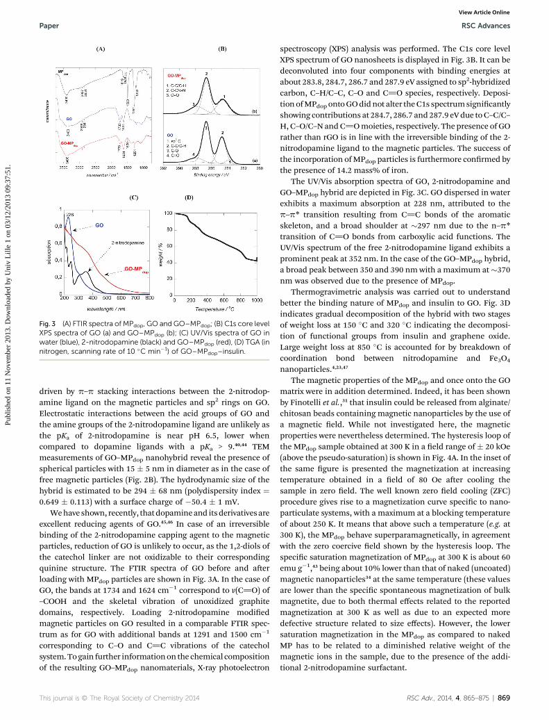
Fig. 3 (A) FTIR spectra of MPdop, GO and GO–MPdop; (B) C1s core levelXPS spectra of GO (a) and GO–MPdop (b); (C) UV/Vis spectra of GO inwater (blue), 2-nitrodopamine (black) and GO–MPdop (red), (D) TGA (innitrogen, scanning rate of 10 �C min�1) of GO–MPdop–insulin.
Paper RSC Advances
Publ
ishe
d on
11
Nov
embe
r 20
13. D
ownl
oade
d by
Uni
v L
ille
1 on
03/
12/2
013
09:3
7:51
. View Article Online
driven by p–p stacking interactions between the 2-nitrodop-amine ligand on the magnetic particles and sp2 rings on GO.Electrostatic interactions between the acid groups of GO andthe amine groups of the 2-nitrodopamine ligand are unlikely asthe pKa of 2-nitrodopamine is near pH 6.5, lower whencompared to dopamine ligands with a pKa > 9.40,44 TEMmeasurements of GO–MPdop nanohybrid reveal the presence ofspherical particles with 15 � 5 nm in diameter as in the case offree magnetic particles (Fig. 2B). The hydrodynamic size of thehybrid is estimated to be 294 � 68 nm (polydispersity index ¼0.649 � 0.113) with a surface charge of �50.4 � 1 mV.
Wehave shown, recently, thatdopamineand itsderivativesareexcellent reducing agents of GO.45,46 In case of an irreversiblebinding of the 2-nitrodopamine capping agent to the magneticparticles, reduction of GO is unlikely to occur, as the 1,2-diols ofthe catechol linker are not oxidizable to their correspondingquinine structure. The FTIR spectra of GO before and aerloading with MPdop particles are shown in Fig. 3A. In the case ofGO, the bands at 1734 and 1624 cm�1 correspond to v(C]O) of–COOH and the skeletal vibration of unoxidized graphitedomains, respectively. Loading 2-nitrodopamine modiedmagnetic particles on GO resulted in a comparable FTIR spec-trum as for GO with additional bands at 1291 and 1500 cm�1
corresponding to C–O and C]C vibrations of the catecholsystem.Togain further informationon the chemical compositionof the resulting GO–MPdop nanomaterials, X-ray photoelectron
This journal is © The Royal Society of Chemistry 2014
spectroscopy (XPS) analysis was performed. The C1s core levelXPS spectrum of GO nanosheets is displayed in Fig. 3B. It can bedeconvoluted into four components with binding energies atabout 283.8, 284.7, 286.7 and 287.9 eV assigned to sp2-hybridizedcarbon, C–H/C–C, C–O and C]O species, respectively. Deposi-tion ofMPdop ontoGOdidnot alter theC1s spectrumsignicantlyshowing contributions at 284.7, 286.7 and287.9 eVdue toC–C/C–H, C–O/C–NandC]Omoieties, respectively. The presence of GOrather than rGO is in line with the irreversible binding of the 2-nitrodopamine ligand to the magnetic particles. The success ofthe incorporation ofMPdop particles is furthermore conrmed bythe presence of 14.2 mass% of iron.
The UV/Vis absorption spectra of GO, 2-nitrodopamine andGO–MPdop hybrid are depicted in Fig. 3C. GO dispersed in waterexhibits a maximum absorption at 228 nm, attributed to thep–p* transition resulting from C]C bonds of the aromaticskeleton, and a broad shoulder at �297 nm due to the n–p*transition of C]O bonds from carboxylic acid functions. TheUV/Vis spectrum of the free 2-nitrodopamine ligand exhibits aprominent peak at 352 nm. In the case of the GO–MPdop hybrid,a broad peak between 350 and 390 nm with a maximum at�370nm was observed due to the presence of MPdop.
Thermogravimetric analysis was carried out to understandbetter the binding nature of MPdop and insulin to GO. Fig. 3Dindicates gradual decomposition of the hybrid with two stagesof weight loss at 150 �C and 320 �C indicating the decomposi-tion of functional groups from insulin and graphene oxide.Large weight loss at 850 �C is accounted for by breakdown ofcoordination bond between nitrodopamine and Fe3O4
nanoparticles.4,23,47
The magnetic properties of the MPdop and once onto the GOmatrix were in addition determined. Indeed, it has been shownby Finotelli et al.,31 that insulin could be released from alginate/chitosan beads containing magnetic nanoparticles by the use ofa magnetic eld. While not investigated here, the magneticproperties were nevertheless determined. The hysteresis loop ofthe MPdop sample obtained at 300 K in a eld range of � 20 kOe(above the pseudo-saturation) is shown in Fig. 4A. In the inset ofthe same gure is presented the magnetization at increasingtemperature obtained in a eld of 80 Oe aer cooling thesample in zero eld. The well known zero eld cooling (ZFC)procedure gives rise to a magnetization curve specic to nano-particulate systems, with a maximum at a blocking temperatureof about 250 K. It means that above such a temperature (e.g. at300 K), the MPdop behave superparamagnetically, in agreementwith the zero coercive eld shown by the hysteresis loop. Thespecic saturation magnetization of MPdop at 300 K is about 60emu g�1,43 being about 10% lower than that of naked (uncoated)magnetic nanoparticles34 at the same temperature (these valuesare lower than the specic spontaneous magnetization of bulkmagnetite, due to both thermal effects related to the reportedmagnetization at 300 K as well as due to an expected moredefective structure related to size effects). However, the lowersaturation magnetization in the MPdop as compared to nakedMP has to be related to a diminished relative weight of themagnetic ions in the sample, due to the presence of the addi-tional 2-nitrodopamine surfactant.
RSC Adv., 2014, 4, 865–875 | 869

Fig. 4 Magnetic properties of MPdop particles (A) and of GO–MPdop (B). Main graphs show the corresponding hysteresis loops in 20 kOe at 300 K,down insets show the dependence of the magnetization versus temperature after zero field cooling and subsequent measuring at increasingtemperatures in a field of 80 Oe and upper inset in (B) shows the hysteresis in 20 kOe of a reference GO sample.
RSC Advances Paper
Publ
ishe
d on
11
Nov
embe
r 20
13. D
ownl
oade
d by
Uni
v L
ille
1 on
03/
12/2
013
09:3
7:51
. View Article Online
The hysteresis loop of the GO–MPdop sample obtained insimilar conditions as for sample MPdop is shown in Fig. 4B. Inthe down inset of the same gure is also presented the ZFCmagnetization curve in 80 Oe, which is clearly different fromthat of the MPdop sample. It is worth mentioning the increase ofthe magnetization at lower temperature (e.g. from 150 K downto 10 K), suggesting the presence of a ferromagnetic-like inter-action among the nanoparticles as compared to the antiferro-magnetic dipolar type usually observed in non-dilutednanoparticulate systems. Such interactions seem to be presentalso at higher temperatures, leading to a consistent shi of theblocking temperature well above 290 K. If the nature of suchunusual interactions requires additional studies of the GO–MPdop mixtures, the consistent difference of the ZFC curve ofthe GO–MPdop sample as compared to the ZFC curve of theMPdop sample clearly proves the formation of the GO–MPdophybrid with specic properties induced by the strong interac-tions of the nanoparticles via the GO support. In the upper insetof the same Fig. 4B is shown the hysteresis loop of the GOsubstrate, collected in similar conditions as for the GO–MPdophybrid. It is observed that in the maximum eld of 20 kOe, themagnetization of GO (0.008 emu g�1) is 1000 times lower thanthat of the hybrid sample (about 8 emu g�1) and therefore canbe clearly neglected. Hence, the saturation magnetization of theGO–MPdop hybrid is just 13% from the saturation magnetiza-tion of MPdop sample, inferring an equivalent (13 mass%) ofloading magnetic material in the analyzed sample. This is inaccordance with XPS analysis where a 14.3 mass% of iron wasdetermined.
3.2. Insulin loading and release
The kind of interactions of GO, MPdop and insulin is of utter-most importance not only for the construction of a stable GO–MPdop hybrid but also for insulin loading and release strategies.As discussed above, 2-nitrodopamine ligands are used ascapping agent for the formed magnetic particles. The linkageinsuring an irreversible binding of the ligand. Indeed, nodegradation of the MPdop particles size and chemical compo-sition was observed upon immersion for 4 h into aqueous
870 | RSC Adv., 2014, 4, 865–875
solutions of low pH (pH ¼ 1), as might be observed underbiological conditions. The interaction of the dopamine ligandto the iron oxide nanoparticles is not disrupted in the lower pHrange. Interaction of the dopamine-capped MP particles withthe GO matrix is mostly over p–p stacking interactions betweenthe hexagonal cells of graphene and the aromatic ring structureof dopamine. As the diol functions of the used catechol are notavailable, the formation of ortho-quinol structures is inhibitedin this case and a further covalent binding not feasible.45
The loading capacity of insulin onto GO–MPdop can beevaluated by measuring the concentration of insulin using UV/Vis spectra at 275 nm in solution before and aer insulinloading. The difference corresponds to insulin loaded onto theGO–MPdop matrix (Fig. 5A). Indeed, due to the high UV/Visabsorbance of GO occurring in the same spectral area asinsulin, a direct determination of the insulin concentration onthe GO–MPdop matrix is not possible. The insulin loadingcapacity of the GO–MPdop nanohybrid was calculated accordingto eqn (1):
Loading capacity ¼
c0 � csup
cGO�MPdop
!� 100% (1)
where c0 is the initial concentration of insulin added to GO, csupis the concentration of insulin in the supernatant aer reactiondetermined by UV/Vis and cGO–MPdop
is the concentration of GO–MPdop (150 mg mL�1).
Many studies have shown that aromatic molecules includingchemotherapy drugs such as doxorubicin can be loaded ontothe surface of graphene via p–p stacking interactions.12,15,21 Inthe case of insulin, a polypeptide composed of 51 amino acidresidues, electrostatic forces will additionally affect insulinloading as the isoelectronic point (pI) of insulin is reported to be5.4.27 The inuence of the pH on the loading of a xedconcentration of insulin onto GO–MPdop is displayed in Fig. 5B.Using a loading time of 24 h, increased insulin loading wasobserved at pH < 5.5 in accordance with an insulin pI ¼ 5.4.27
Below pH 5.4, insulin is positively charged and interacts morestrongly with the negatively charged GO–MPdop matrix. Thisinteraction is weakened at pH > 5 and at pH 7 the loading
This journal is © The Royal Society of Chemistry 2014

Fig. 5 Insulin loading on GO and GO–MPdop: (A) UV/Vis spectra offree insulin at different concentrations and the corresponding cali-bration curve (inset); (B) insulin loading capacity of GO (blue) andGO–MPdop (red) as a function of pH.
Fig. 6 Insulin release from GO (A) and GO–MPdop (B) for different pHand at different time points (error bars are based on triplicatemeasurements).
Paper RSC Advances
Publ
ishe
d on
11
Nov
embe
r 20
13. D
ownl
oade
d by
Uni
v L
ille
1 on
03/
12/2
013
09:3
7:51
. View Article Online
capacity of insulin is 3 times lower as only p–p stacking inter-actions and/or hydrophilic interactions will prevail betweeninsulin and GO–MPdop. The loading capacity of GO–MPdop is ashigh as 88 � 3%, although some areas of the multifunctionalGO has been previously occupied with magnetic particles.Indeed, this results in a decrease in the loading capacity whencompared to GO (100 � 3%), but is still remarkably high whencompared to other nanostructures.47,48 For mesoporous silicainsulin loading of 15% was reported,47 while poly(lactide-ethylene glycol) nanoparticles showed a maximal insulinloading of 58.8%.48
To understand better which of the materials, GO or MPdop ismore effectively loading insulin, the loading capacitance ofMPdop was in addition determined. No detectable amounts ofinsulin could be determined by the colorometic assay, indi-cating that all the insulin reacts with the GO matrix rather thanthe magnetic particles.
Insulin release from GO and GO–MPdop hybrid at pH 5 wasanalyzed by incubating the matrices at 37 �C at different pHwhile shaking. Fig. 6 shows the cumulative release of insulinfrom GO (Fig. 6A) and GO–MPdop (Fig. 6B) matrices as a func-tion of pH. At pH ¼ 2, even though a small release of insulin is
This journal is © The Royal Society of Chemistry 2014
observed, probably due to weakly bound insulin, GO and GO–MPdop appear to have a high insulin retention capacity.Comparable behavior was observed on core–shell poly(ethyleneglycol)polyhedral oligosilsesquioxane nanoparticles.30
Following a pH change to 5, insulin release is initiated andsustained for the rst 90 min. The amount of insulin released ishighly pH dependent with about 28 � 3% of insulin released atpH 9 for GO–MPdop and 40 � 3% for GO. The insulin releasefrom the GO–MPdop hybrid turns to be less successful than fromGO alone, which likely accounts for some levels of interactionbetween insulin and nanoparticles within the hybrid. Therelease at pH 9 is most likely due to electrostatic repulsionbetween negatively charged insulin and negatively charged GOand GO–MPdop. The release is fast and low when compared tomesoporous silica nanoparticles with a maximal release of 77%at pH 8.5 aer 10 h.47 It is comparable to poly(lactide-ethyleneglycol) nanoparticles with a release of 59% aer 10 days,48 oralginate/chitosan microcapsules with a release of 18% in therst hours and about 45% aer 3 days.31
The high insulin retention capacity at low pH, comparable tothat of gastric pH suggests that insulin is well protected on GOand GO–MPdop hybrid, while at intestinal pH (pH 6–7) insulin isactivated and released. We thus investigated, if GO and GO–MPdop hybrids could be used as potential carriers for an insulindrug delivery system.
3.3. Cell viability assay of GO and GO–MPdop
Two different cell viability assays were performed to obtaininformation about the cytotoxicity of the GO–MPdop hybrid. TheMTT (3-(4,5-dimethylthiazol-2-yl)-2,5-diephenyltetrazoliumbromide) assay is a simple colorimetric assay to measure cellcytotoxicity, proliferation or viability and used in this work. Asseen in Fig. 7A under the investigated concentration range ofGO–MPdop and GO–MPdop–insulin no cytotoxicity to HEK cellsis observed.
In Xenopus laevis large number of oocytes are easily obtainedat all stages of maturation, making this organism an excellentmodel for studying the role of insulin and insulin growthfactors on the development of the organism.49,50 Fully grown
RSC Adv., 2014, 4, 865–875 | 871

Fig. 7 (A) Cytotoxicity of GO–MPdop (grey) and GO–MPdop insulin (blue) to HEK cell lines; (B) schematic illustration of insulin induced process ofmeiotic resumption of fully grown Xenopus laevis oocytes: Xenopus oocytes (stage VI) before (a) and after treatment with insulin (10 mg mL�1) atpH ¼ 9.2 (b). A typical white spot, attesting for the germinal vesicle breakdown (GVBD) transition from the G2 to the M phase of the cell cycle, isseen; (C) meiotic resumption rate of Xenopus oocytes incubated for 24 h with GO (black) and GO–MPdop (red) at different concentrations andafter injection of insulin (c ¼ 50 mg mL�1); (D) meiotic resumption rate as a function of pH.
RSC Advances Paper
Publ
ishe
d on
11
Nov
embe
r 20
13. D
ownl
oade
d by
Uni
v L
ille
1 on
03/
12/2
013
09:3
7:51
. View Article Online
Xenopus laevis oocytes are physiologically arrested at theprophase of the rst meiotic divisions. These oocytes mustresume meiosis and proceed to the metaphase of meiosis II
872 | RSC Adv., 2014, 4, 865–875
before fertilization is possible. The process which enablesfertilization and drive the oocyte from prophase of rst meioticdivision to a block in metaphase of second meiotic division,
This journal is © The Royal Society of Chemistry 2014

Paper RSC Advances
Publ
ishe
d on
11
Nov
embe
r 20
13. D
ownl
oade
d by
Uni
v L
ille
1 on
03/
12/2
013
09:3
7:51
. View Article Online
termed maturation, is triggered in vivo by a preovulatorygonadotropin surge followed by follicular production ofprogesterone.39,51–53 In addition to progesterone and otherhormones, both insulin and insulin-like growth factor-1 (IGF-1)can induce meiotic resumption and oocyte maturation.36,39
Fig. 7A shows photographs of Xenopus laevis oocytes (stage VI)before and aer treatment with insulin (10 mg mL�1) at pH 9.2.A typical white spot, attesting for the germinal vesicle break-down (GVBD), is observed under the binocular at the animalpole of the oocytes. We use the monitoring of oocyte meioticresumption in this study for testing their viability and respon-siveness towards insulin aer being released from GO and GO–MDdop nanostructures. To ensure that GO and GO–MDdop
nanostructures without insulin have no cytotoxic effect onXenopus oocytes, the fully-grown stage VI oocytes were exposedfor 24 h to increasingly high concentrations of GO and GO–MPdop at pH 7.4. Fig. 7B shows the meiotic resumption rateupon insulin induction and indicates that exposure to GO andGO–MPdop even at high concentrations is not toxic for oocytes,showing a comparable meiotic resumption rate when the Xen-opus laevis oocytes were not pre-incubated with the nano-structures. The meiotic resumption rate of Xenopus laevisoocytes upon injection of insulin (50 mg mL�1) at different pHwas investigated to insure that insulin release at higher pHwould have an important inuence on oocytes. As seen fromFig. 7C, no signicant changes inmeiotic resumption rates wereobserved when insulin induction was performed at pH above 5.However, at pH ¼ 2, the meiotic resumption rate is signicantlydecreased, indicating deviations from native insulin most likelylinked to conformational changes that have occurred in thepolypeptide chains of insulin.54
Fig. 8 Meiotic resumption response curves of Xenopus oocytes: (A)influence of the insulin concentration (0–10 mg mL�1) and the solutionpH; progesterone (10 mg mL�1) was used as positive control. (B)Influence of the concentration of GO–insulin and GO–MPdop–insulinon the meiotic resumption rate at pH 9.2; GO was used as negativecontrol and insulin (5.6 mg mL�1) as positive control; meioticresumption rate of pre-incubated (pH¼ 2; 6 h) insulin, GO–insulin andGO–MPdop–insulin.
3.4. Meiotic resumption rates of Xenopus laevis oocytesupon addition of GO–insulin and GO–MPdop–insulin
The dose effect of insulin at pH 5 and 9.2 on the meioticresumption rate of oocytes was investigated. As seen in Fig. 8A,at pH ¼ 9.2, the minimal insulin concentration resulting inhigh rates of meiotic resumption is around 1.2 mg mL�1. Belowthis concentration level, no meiotic resumption was observed.At pH 5, this concentration limit was shied to higher insulinconcentrations. A comparable concentration range was thuschosen for the insulin loaded GO and GO–MPdop nano-structures. Fig. 8B compares the meiotic resumption rates of avariety of different experimental set-ups. GO (5.6 mg mL�1) andinsulin (5.6 mg mL�1) were used as negative and positivecontrols in this comparative experiment. GO–insulin and GO–MPdop–insulin nanostructures showed a dose-dependenceresponse: while at a concentration of 0.8 mg mL�1, bothmatrices exhibited only low meiotic resumption rates at pH 9.2,concentrations higher than 5.6 mg mL�1 resulted in high levelsof meiotic resumption as for free insulin. While this behaviouris expected, a surprisingly different meiotic resumptionbehaviour was observed once insulin, GO–insulin and GO–MPdop–insulin were pre-incubated for 5 h at pH¼ 2. For insulin,the meiotic resumption rate was highly decreased in line withthe observation in Fig. 7C. However, acid pre-treated
This journal is © The Royal Society of Chemistry 2014
GO–insulin and GO–MPdop–insulin nanostructures did notshow any altered meiotic resumption characteristics. This invitro experiment proves that the insulin incorporated onto thenanostructures is not affected by the low pH, and the GO“protects” insulin from acidic degradation. The GO–insulin andGO–MPdop–insulin nanostructures might be thus considered asnovel insulin formulations next to microcapsules, polymers andothers.26,31,48,54,55 The appealing character of GO–insulin andGO–MPdop–insulin nanostructures is that the nanocompositesare easy to prepare and can be produced on a larger scale. Theincorporation of magnetic particles does not alter the meioticresumption prole of Xenopus laevis oocytes, used as modelsystem here. The attractiveness of the incorporation of the
RSC Adv., 2014, 4, 865–875 | 873

RSC Advances Paper
Publ
ishe
d on
11
Nov
embe
r 20
13. D
ownl
oade
d by
Uni
v L
ille
1 on
03/
12/2
013
09:3
7:51
. View Article Online
magnetic particles is that insulin controlled release can beenhanced in the presence of a magnetic eld, as previouslydemonstrated by Finotelli et al. using alginate/chitosan beadscontaining magnetic particles.31
4. Conclusions
In this study, we have demonstrated that graphene oxidematrices can be easily loaded with different carriers. In our case2-nitrodopamine coated magnetic particles and/or insulin wasincorporated onto the GO nanosheets. The insulin loadingcapacity on the GO nanomaterials was pH-dependent, butproved to be extremely high at pH lower than 5.4 with 100 � 3%and 88 � 3% loading on GO and GO–MPdop, respectively.Insulin-loaded on GO matrices was stable at acidic pH, but wasreleased when exposed to basic solutions (pH ¼ 9.2). Insulinretained its native structure when released from the matrix. Inaddition, the insulin loaded on GO and GO–MPdop were stronglyresistant to acidic pH, as for that encountered in the gastricenvironment. These results open new avenues for furtherinvestigations of the potential application of insulin loaded onGO matrices for treatment of patients with insulin deciency.
Acknowledgements
A.B, R.B and S.S. gratefully acknowledge nancial support fromthe Centre National de Recherche Scientique (CNRS), theUniversite Lille 1, the Nord Pas de Calais region, and the InstitutUniversitaire de France (IUF). Support from the EuropeanUnion through a FP7-PEOPLE-IRSES (PHOTORELEASE) isacknowledged. Support from the Romanian project PNII IDEI75/2011 is gratefully acknowledged.
References
1 A. Barras, F. A. Martin, O. Bande, J. S. Baumann, J.-M. Ghigo,R. Boukherroub, C. Beloin, A. Siriwardena and S. Szunerits,Nanoscale, 2013, 5, 2307.
2 M. Durka, K. Buffet, J. Iehl, M. Holler, J.-F. Nierengarten,J. taganna, J. Bouckaert and S. P. Vicent, Chem. Commun.,2011, 47, 1321.
3 J. J. Shi, A. R. Votruba, O. C. Farokhzad and R. Langer, NanoLett., 2010, 10, 3223.
4 X. Yang, X. Zhang, Y. Ma, Y. Huan, Y. Wang and Y. Chen,J. Mater. Chem., 2009, 19, 2710.
5 X. Yang, G. Niu, X. Cao, Y. Wen, R. Xiang, H. Duan andY. Chen, J. Mater. Chem., 2012, 22, 6649.
6 L. Zhang, Z. Lu, Q. H. Zhao, J. Hunag, H. Shen andZ. J. Zhang, Small, 2011, 7, 460.
7 K. E. Sapsford, W. R. Algar, L. Berti, K. Boeneman Gemmill,B. J. Casey, E. Oh, M. H. Stewart and I. L. Medintz, Chem.Rev., 2013, 113, 1904.
8 H. Y. Mao, S. Laurent, W. chen, O. Akhavan, M. Imani,A. A. Ashkarran and M. Mahmoudi, Chem. Rev., 2013, 113,3407–3424.
9 K. Yang, L. Feng, X. Shi and Z. Liu, Chem. Soc. Rev., 2013, 42,530.
874 | RSC Adv., 2014, 4, 865–875
10 Y. Pan, H. Bao, N. G. Sahoo, T. Wu and L. Li, Adv. Funct.Mater., 2011, 21, 2754.
11 U. Dembereldorj, M. Kim, S. Kim, E.-O. Ganbold, S. Y. Leeand S.-W. Joo, J. Mater. Chem., 2012, 22, 22845.
12 W. Zhang, Z. Guo, D. Huang, Z. Liu, X. Guo and H. Zhong,Biomaterials, 2011, 32, 8555.
13 Z. Liu, J. T. Robinson, X. M. Sun and H. J. Dai, J. Am. Chem.Soc., 2008, 130, 10876.
14 J. Hong, N. J. Shah, A. C. Drake, P. C. DeMuth, J. B. Lee,J. Chen and P. T. Hammond, ACS Nano, 2012, 6, 81.
15 X. M. Sun, Z. Liu, K. Welsher, et al., Nano Res., 2008, 1, 203.16 X. Yang, X. Zhang, Z. Liu, Y. Ma, Y. Huang and Y. Chen,
J. Phys. Chem. C, 2008, 112, 17554.17 W. Miao, G. Shim, S. Lee, S. Lee, Y. S. Choe and Y.-K. Oh,
Biomaterials, 2013, 34, 3402.18 L. Zhang, J. Xia, Q. Zhao, L. H. Liu and Z. Zhang, Small, 2010,
6, 537.19 Y. Yang, Y.-M. Zhang, Y. Chen, D. Zhao, J.-T. Chen and
Y. Liu, Chem.–Eur. J., 2012, 18, 4208.20 X. Yang, Y. Wang, X. Huang, Y. Ma, Y. Huang, R. Yang,
H. Duan and Y. Chen, J. Mater. Chem., 2011, 21, 3448.21 X. Ma, H. Tao, K. Yang, L. Feng, L. Chen, X. Shi, Y. Li, L. Guo
and Z. Liu, Nano Res., 2012, 5, 199.22 G. Cheng, Y.-L. Liu, Z.-G. Wang, J.-L. Zhang, D.-H. Sun and
J.-Z. Ni, J. Mater. Chem., 2012, 22, 21998.23 Y. Xue, H. Y. Chen, D. Yu, S. Wang, M. Yardeni, Q. Dai,
M. Guo, Y. Liu, F. Lu, J. Qu and L. Dai, Chem. Commun.,2011, 47, 11689.
24 M. Goldberg and I. Gomez-Orellana, Nat. Rev. Drug Discovery,2003, 2, 289.
25 N. A. Peppas and N. J. Kavimandan, Eur. J. Pharm. Sci., 2006,29, 183.
26 F. Wang, Y. Chen and H. A. E. Benson, Open Drug Delivery J.,2008, 2, 1.
27 F. Cui, K. Shi, L. Zhang, A. Tao and Y. Kawashima,J. Controlled Release, 2006, 114, 242.
28 G. P. Carino and E. Mathiowitz, Adv. Drug Delivery Rev., 1999,35, 249.
29 D. P. Huynh, M. K. Nguyen, B. S. Pi, M. S. Kom, S. Y. Chae,K. C. Lee, B. S. Kim, S. W. Kim and D. S. Lee, Biomaterials,2008, 29, 2527.
30 K.-O. Kim, B.-S. Kim and I.-S. Kim, J. Biomater.Nanobiotechnol., 2011, 2, 201.
31 P. V. Finotelli, D. Da Silva, M. Sola-Penna, A. Malta Rossi,M. Farina, L. R. Andrada, A. Y. Takeuchi and M. Rocha-Leao, Colloids Surf., B, 2010, 81, 206.
32 J. Liu, L. Cui and D. Losic, Acta Biomater., 2013, 9, 9243–9257.33 K. Yang, H. Gong, X. Shi, J. Wan, Y. Zhang and Z. I. Liu,
Biomaterials, 2013, 34, 2787.34 O. Fellahi, M. R. Das, Y. Coffinier, S. Szunerits, T. Hadjersi,
M. Maamache and R. Boukherroub, Nanoscale, 2011, 3, 4662.35 M. Rodenstein, S. Zurcher, S. G. Tosatti and N. D. Spencer,
Langmuir, 2010, 26, 16211.36 M. Mazur, A. Barras, V. Kuncer, A. Galatanu, V. Zaitzev,
P. Woisel, J. Lyskawa, W. Laure, A. Siriwardena,R. Boukherroub and S. Szunerits, Nanoscale, 2013, 5, 2692.
37 J. N. Dumont, J. Morphol., 1972, 136, 153.
This journal is © The Royal Society of Chemistry 2014

Paper RSC Advances
Publ
ishe
d on
11
Nov
embe
r 20
13. D
ownl
oade
d by
Uni
v L
ille
1 on
03/
12/2
013
09:3
7:51
. View Article Online
38 F. Baert, J. F. Bodart, B. Bocquet-Muchembled, A. Lescuyer-Rousseau and J. P. Vilain, J. Biol. Chem., 2003, 278, 49714.
39 M. El-Etr, S. Schorderet-Slatkine and E. E. Baulieu, Science,1979, 205, 1397.
40 E. Amstad, A. U. Gehring, H. Fisher, V. V. Nagaiyanallur,G. Hahner, M. Textor and E. Reimhult, J. Phys. Chem. C,2011, 115, 683.
41 E. Amstad, T. Gillich, I. Bilecka, M. Textor and E. Reimhult,Nano Lett., 2009, 9, 4042.
42 E. Amstad, M. Textor and E. Reimhult, Nanoscale, 2011, 3,2819.
43 S. J. Clarke, C. A. Hollmann, Z. Zhang, S. Suffern,S. E. Bradforth, N. M. Dimitrijevic, W. G. Minarik andJ. L. Nadeau, Nat. Mater., 2006, 5, 409.
44 A. K. L. Yuen, G. A. Hutton, A. F. Masters and T. Maschmeyer,Dalton Trans., 2012, 41, 2545.
45 I. Kaminska, M. R. Das, Y. Coffinier, J. Niedziolka-Jonsson,J. Sobczak, P. Woisel, J. Lyskawa, M. Opallo,R. Boukherroub and S. Szunerits, ACS Appl. Mater.Interfaces, 2012, 4, 1016.
This journal is © The Royal Society of Chemistry 2014
46 I. Kaminska, W. Qi, A. Barras, J. Sobczak, J. Niedziolka-Jonsson, P. Woisel, J. Lyskawa, W. Laure, M. Opallo, M. Li,R. Boukherroub and S. Szunerits,Chem.–Eur. J., 2013, 19, 8673.
47 L. Sun, X. Zhang, C. Zhen, Z. s. Wu and C. Li, J. Phys. Chem. B,2013, 117, 3852.
48 L. Tomar, C. Tyagi, M. Kumar, P. Kumar, H. Singh,Y. E. Choonara and V. Pillay, Int. J. Nanomed., 2013, 8, 505.
49 L. Scavo, A. R. Shuldiner, J. Serrano, R. Dashner, J. Roth andF. De Pablo, Proc. Natl. Acad. Sci. U. S. A., 1991, 88, 6214.
50 I. B. Dawid, T. D. Sargent and F. Rosa, Curr. Top. Dev. Biol.,1990, 24, 2.
51 L. Zhu, N. Ohan, Y. Agazie, C. Cummings, S. Farah andX. J. Li, Endocrinology, 1997, 139, 949.
52 Y. Mazui and H. J. Clarke, Int. Rev. Cytol., 1979, 57, 185.53 L. D. Smith, Development, 1989, 107, 685.54 K. Yoshida, K. Sato and J.-I. Anzai, J. Mater. Chem., 2010, 20,
1546.55 R. A. Shimkunas, E. Robinson, R. Lam, S. Lu, X. H. Xu,
X.-Q. Zhang, H. Huang, E. Osawa and D. Ho, Biomaterials,2009, 30, 5720.
RSC Adv., 2014, 4, 865–875 | 875
Copyright © 2022 FDOKUMEN