Insights into the Metabolism of Elemental Sulfur by the Hyperthermophilic Archaeon Pyrococcus...
11
JOURNAL OF BACTERIOLOGY, June 2007, p. 4431–4441 Vol. 189, No. 12 0021-9193/07/$08.000 doi:10.1128/JB.00031-07 Copyright © 2007, American Society for Microbiology. All Rights Reserved. Insights into the Metabolism of Elemental Sulfur by the Hyperthermophilic Archaeon Pyrococcus furiosus : Characterization of a Coenzyme A- Dependent NAD(P)H Sulfur Oxidoreductase † Gerrit J. Schut, Stephanie L. Bridger, and Michael W. W. Adams* Department of Biochemistry & Molecular Biology, University of Georgia, Athens, Georgia 30602 Received 7 January 2007/Accepted 5 April 2007 The hyperthermophilic archaeon Pyrococcus furiosus uses carbohydrates as a carbon source and produces acetate, CO 2 , and H 2 as end products. When S 0 is added to a growing culture, within 10 min the rate of H 2 production rapidly decreases and H 2 S is detected. After 1 hour cells contain high NADPH- and coenzyme A-dependent S 0 reduction activity (0.7 units/mg, 85°C) located in the cytoplasm. The enzyme responsible for this activity was purified to electrophoretic homogeneity (specific activity, 100 units/mg) and is termed NAD(P)H elemental sulfur oxidoreductase (NSR). NSR is a homodimeric flavoprotein (M r , 100,000) and is encoded by PF1186. This designation was previously assigned to the gene encoding an enzyme that reduces coenzyme A disulfide, which is a side reaction of NSR. Whole-genome DNA microarray and quantitative PCR analyses showed that the expression of NSR is up-regulated up to sevenfold within 10 min of S 0 addition. This primary response to S 0 also involves the up-regulation (>16-fold) of a 13-gene cluster encoding a membrane- bound oxidoreductase (MBX). The cluster encoding MBX is proposed to replace the homologous 14-gene cluster that encodes the ferredoxin-oxidizing, H 2 -evolving membrane-bound hydrogenase (MBH), which is down-regulated >12-fold within 10 min of S 0 addition. Although an activity for MBX could not be demon- strated, it is proposed to conserve energy by oxidizing ferredoxin and reducing NADP, which is used by NSR to reduce S 0 . A secondary response to S 0 is observed 30 min after S 0 addition and includes the up-regulation of genes encoding proteins involved in amino acid biosynthesis and iron metabolism, as well as two so-called sulfur-induced proteins termed SipA and SipB. This novel S 0 -reducing system involving NSR and MBX has been found so far only in the heterotrophic Thermococcales and is in contrast to the cytochrome- and quinone- based S 0 -reducing system in autotrophic archaea and bacteria. The hyperthermophilic archaea are a group of microorgan- isms that grow optimally at temperatures of 80°C and above (48). Most of these microorganisms utilize elemental sulfur (S 0 ) as a terminal electron acceptor and reduce it to H 2 S (17, 19). Those that use molecular H 2 as the electron donor, such as Thermoproteus tenax and Acidianus ambivalens, are thought to have a respiratory system analogous to that found in meso- philic S 0 -reducing bacteria such as Wolinella succinogenes, where S 0 reduction is accomplished by a membrane-bound respiratory system (17). On the other hand, the mechanism of S 0 reduction by the heterotrophic hyperthermophilic archaea, such as Pyrococcus and Thermococcus species, is completely unknown. These organisms grow by fermentation with pep- tides as the carbon source, and most of them appear to be obligately dependent on S 0 for optimal growth (48). The ex- ceptions are those that are able to grow by fermentation of carbohydrates; one such exception is Pyrococcus furiosus, which grows equally well with or without S 0 (9). Herein we have exploited this property to investigate the mechanism by which this prototypical heterotrophic hyperthermophile re- duces S 0 to H 2 S. P. furiosus utilizes a range of both simple and complex car- bohydrates and converts them to acetate, to CO 2 , to H 2 , and, if S 0 is present, to H 2 S. Its glycolytic pathway has been exten- sively studied and served as one of the model systems for elucidating the modified Embden-Meyerhof pathway in ar- chaea (39, 40, 51). The key feature of this pathway is that the classical enzymes glyceraldehyde-3-phosphate dehydrogenase (GAPDH) and phosphoglycerate kinase are replaced by a single ferredoxin-linked enzyme, glyceraldehyde-3-phosphate ferredoxin oxidoreductase (GAPOR, EC 1.2.1.-). This converts glyceraldehyde-3-phosphate to glycerate-3-phosphate without the generation of ATP (51). Consequently, only ferredoxin serves as the electron acceptor in glycolysis and no NAD(P)H is formed. The reason for this became apparent upon the discovery in P. furiosus of a membrane-bound hydrogenase (MBH) which evolved H 2 from reduced ferredoxin in an en- ergy-conserving manner via a proton motive force (41). The other oxidation step in the conversion of glucose to acetate is also coupled to the reduction of ferredoxin, and this is cata- lyzed by pyruvate ferredoxin oxidoreductase (POR) (4). Hence, in the production of acetate from glucose, all of the reductant is generated as reduced ferredoxin. Its oxidation via the MBH is thought to result in the conservation of energy equivalent to 1.2 ATP per glucose (41). Two cytoplasmic hy- drogenases from P. furiosus have also been characterized (29), but these use NADPH, rather than ferredoxin, as the electron carrier, and their functions in fermentative metabolism are unclear. It is possible that one or both serve to recycle the H 2 * Corresponding author. Mailing address: Department of Biochem- istry and Molecular Biology, Life Sciences Bldg., University of Geor- gia, Athens, GA 30602-7229. Phone: (706) 542-2060. Fax: (706) 542- 0229. E-mail: [email protected]. † Supplemental material for this article may be found at http://jb .asm.org/. Published ahead of print on 20 April 2007. 4431 on March 31, 2016 by guest http://jb.asm.org/ Downloaded from
-
Upload
independent -
Category
Documents
-
view
7 -
download
0
Transcript of Insights into the Metabolism of Elemental Sulfur by the Hyperthermophilic Archaeon Pyrococcus...

JOURNAL OF BACTERIOLOGY, June 2007, p. 4431–4441 Vol. 189, No. 120021-9193/07/$08.00�0 doi:10.1128/JB.00031-07Copyright © 2007, American Society for Microbiology. All Rights Reserved.
Insights into the Metabolism of Elemental Sulfur by the HyperthermophilicArchaeon Pyrococcus furiosus: Characterization of a Coenzyme A-
Dependent NAD(P)H Sulfur Oxidoreductase�†Gerrit J. Schut, Stephanie L. Bridger, and Michael W. W. Adams*
Department of Biochemistry & Molecular Biology, University of Georgia, Athens, Georgia 30602
Received 7 January 2007/Accepted 5 April 2007
The hyperthermophilic archaeon Pyrococcus furiosus uses carbohydrates as a carbon source and producesacetate, CO2, and H2 as end products. When S0 is added to a growing culture, within 10 min the rate of H2production rapidly decreases and H2S is detected. After 1 hour cells contain high NADPH- and coenzymeA-dependent S0 reduction activity (0.7 units/mg, 85°C) located in the cytoplasm. The enzyme responsible forthis activity was purified to electrophoretic homogeneity (specific activity, 100 units/mg) and is termedNAD(P)H elemental sulfur oxidoreductase (NSR). NSR is a homodimeric flavoprotein (Mr, 100,000) and isencoded by PF1186. This designation was previously assigned to the gene encoding an enzyme that reducescoenzyme A disulfide, which is a side reaction of NSR. Whole-genome DNA microarray and quantitative PCRanalyses showed that the expression of NSR is up-regulated up to sevenfold within 10 min of S0 addition. Thisprimary response to S0 also involves the up-regulation (>16-fold) of a 13-gene cluster encoding a membrane-bound oxidoreductase (MBX). The cluster encoding MBX is proposed to replace the homologous 14-genecluster that encodes the ferredoxin-oxidizing, H2-evolving membrane-bound hydrogenase (MBH), which isdown-regulated >12-fold within 10 min of S0 addition. Although an activity for MBX could not be demon-strated, it is proposed to conserve energy by oxidizing ferredoxin and reducing NADP, which is used by NSRto reduce S0. A secondary response to S0 is observed 30 min after S0 addition and includes the up-regulationof genes encoding proteins involved in amino acid biosynthesis and iron metabolism, as well as two so-calledsulfur-induced proteins termed SipA and SipB. This novel S0-reducing system involving NSR and MBX hasbeen found so far only in the heterotrophic Thermococcales and is in contrast to the cytochrome- and quinone-based S0-reducing system in autotrophic archaea and bacteria.
The hyperthermophilic archaea are a group of microorgan-isms that grow optimally at temperatures of 80°C and above(48). Most of these microorganisms utilize elemental sulfur(S0) as a terminal electron acceptor and reduce it to H2S (17,19). Those that use molecular H2 as the electron donor, such asThermoproteus tenax and Acidianus ambivalens, are thought tohave a respiratory system analogous to that found in meso-philic S0-reducing bacteria such as Wolinella succinogenes,where S0 reduction is accomplished by a membrane-boundrespiratory system (17). On the other hand, the mechanism ofS0 reduction by the heterotrophic hyperthermophilic archaea,such as Pyrococcus and Thermococcus species, is completelyunknown. These organisms grow by fermentation with pep-tides as the carbon source, and most of them appear to beobligately dependent on S0 for optimal growth (48). The ex-ceptions are those that are able to grow by fermentation ofcarbohydrates; one such exception is Pyrococcus furiosus,which grows equally well with or without S0 (9). Herein wehave exploited this property to investigate the mechanism bywhich this prototypical heterotrophic hyperthermophile re-duces S0 to H2S.
P. furiosus utilizes a range of both simple and complex car-bohydrates and converts them to acetate, to CO2, to H2, and,if S0 is present, to H2S. Its glycolytic pathway has been exten-sively studied and served as one of the model systems forelucidating the modified Embden-Meyerhof pathway in ar-chaea (39, 40, 51). The key feature of this pathway is that theclassical enzymes glyceraldehyde-3-phosphate dehydrogenase(GAPDH) and phosphoglycerate kinase are replaced by asingle ferredoxin-linked enzyme, glyceraldehyde-3-phosphateferredoxin oxidoreductase (GAPOR, EC 1.2.1.-). This convertsglyceraldehyde-3-phosphate to glycerate-3-phosphate withoutthe generation of ATP (51). Consequently, only ferredoxinserves as the electron acceptor in glycolysis and no NAD(P)His formed. The reason for this became apparent upon thediscovery in P. furiosus of a membrane-bound hydrogenase(MBH) which evolved H2 from reduced ferredoxin in an en-ergy-conserving manner via a proton motive force (41). Theother oxidation step in the conversion of glucose to acetate isalso coupled to the reduction of ferredoxin, and this is cata-lyzed by pyruvate ferredoxin oxidoreductase (POR) (4).Hence, in the production of acetate from glucose, all of thereductant is generated as reduced ferredoxin. Its oxidation viathe MBH is thought to result in the conservation of energyequivalent to 1.2 ATP per glucose (41). Two cytoplasmic hy-drogenases from P. furiosus have also been characterized (29),but these use NADPH, rather than ferredoxin, as the electroncarrier, and their functions in fermentative metabolism areunclear. It is possible that one or both serve to recycle the H2
* Corresponding author. Mailing address: Department of Biochem-istry and Molecular Biology, Life Sciences Bldg., University of Geor-gia, Athens, GA 30602-7229. Phone: (706) 542-2060. Fax: (706) 542-0229. E-mail: [email protected].
† Supplemental material for this article may be found at http://jb.asm.org/.
� Published ahead of print on 20 April 2007.
4431
on March 31, 2016 by guest
http://jb.asm.org/
Dow
nloaded from

produced by the membrane-bound enzyme to generateNADPH for biosynthesis (29, 47).
For P. furiosus and related heterotrophic Thermococcales, S0
reduction, like H2 production, was proposed to be a mecha-nism for disposing of excess reductant (9, 43). H2 production isnow regarded as an energy-conserving process (41), but it isnot known if this is also true of S0 reduction. The S0 reductionsystem of the mesophilic bacterium Wolinella succinogenes isgenerally accepted as a model system for anaerobic S0 respi-ration in which H2S production is coupled to energy conser-vation (17). W. succinogenes uses H2 or formate as the electrondonor, and their oxidation is coupled through cytochrome band quinones to a membrane-bound, molybdopterin-contain-ing sulfur reductase (8). A similar S0-reducing respiratory sys-tem has been characterized for other autotrophs, including thehyperthermophilic bacterium Aquifex aeolicus (13) and the hy-perthermoacidophilic archaeon A. ambivalens (27), and it ap-pears to be present in the H2-oxidizing hyperthermophilic ar-chaea Pyrodictium brockii (36) and Pyrodictium abyssi (24).
P. furiosus and the other heterotrophic hyperthermophilicarchaea seem to have a mechanism for S0 reduction that isdifferent from that found for the autotrophic species. Theavailable genome sequences of three Pyrococcus and one Ther-mococcus species (7, 11, 23, 38) do not contain obvious ho-mologs of the molybdenum-containing sulfur reductase of W.succinogenes or A. ambivalens (2). There are also no reports ofthe presence of quinones or cytochromes in these organisms.Three enzymes from P. furiosus, the two cytosolic hydroge-nases (29) and a sulfide dehydrogenase (30), have been previ-ously reported to possess S0 reductase activity in vitro. How-ever, both the activity and the expression of the twohydrogenases dramatically decreased in cells grown in thepresence of S0 (1, 46). Similarly, the sulfide dehydrogenase isnow thought to function in vivo as a ferredoxin:NADPH oxi-doreductase (28), and the expression of its genes is related tothe carbon source rather than to S0 (44). Consequently, noneof these three enzymes is likely to play a role in S0 reduction invivo.
In a previous study with P. furiosus, the effect of S0 on theexpression of a selected group of genes (271) was investigatedusing a targeted DNA microarray by comparing cells grown formany generations (as batch cultures) in the presence or ab-sence of S0 (46). While a significant number of genes wereaffected, including those encoding the two cytoplasmic hydro-genases (which were down-regulated in S0-grown cells), thisstudy was limited by (i) the small number of genes analyzedand (ii) the use of batch-grown cells, where regulated genesinvolved in the primary metabolism of S0 could not be distin-guished from those causing secondary or other effects. In thepresent work, the primary response of P. furiosus to S0 hasbeen investigated using a kinetic approach, where S0 is addedto a log-phase culture and changes in gene expression areanalyzed using a complete genome DNA microarray. In addi-tion, the enzyme responsible for NAD(P)H-linked S0 reduc-tion was characterized independently by biochemical ap-proaches, and the up-regulation of the gene encoding it(PF1186) was shown to be a component of the primary re-sponse of P. furiosus to S0 addition.
MATERIALS AND METHODS
Growth conditions. P. furiosus (DSM 3638) was grown in the presence andabsence of S0 with maltose as the primary carbon source. The growth mediumwas the same as previously reported (1) except that the yeast extract was 1.0g/liter and cysteine (3 mM) was the reducing agent (Na2S was not added).Growth experiments to determine the effects of S0 addition were carried out in100-ml serum bottles with 50-ml stirred (300 rpm) cultures or in a 20-liter customfermentor (1). Cultures were grown until they reached mid-log phase (0.8 � 108
cells/ml), and S0 (J. T. Baker, Phillipsburg, NJ) was added to final concentrationsof 5 and 2 g/liter, respectively. To prepare cell extracts, cells were rapidly cooledby pumping the culture from the 20-liter fermentor through a glass cooling coil,and the cells were collected by centrifugation (10,000 � g, 10 min) and fraction-ated as previously described (1). Approximately 7 liters of the culture washarvested before (time zero) and 1 h after S0 addition. To obtain RNA formicroarray and quantitative PCR (QPCR) analyses, samples (2 liters each) wereremoved from the fermentor before and at various time points after the additionof sulfur and were cooled in ice and fractionated as described previously (46).
Gas analyses. By use of the 100-ml cultures, samples (500 �l each) were takenfrom the liquid and headspace, and these were injected into 10-ml anaerobic vialscontaining an inner reaction vial (500-�l Eppendorf tube) surrounded by 0.1 MNaOH (1 ml) to capture H2S. Sulfuric acid (100 �l, 2.0 M) was added to the innervial to release acid-labile sulfide from the liquid culture. H2S production wasmeasured in the NaOH phase of the double-vial system with the methylene blueassay (6). H2 gas was detected in the headspace of the vials with a gas chromato-graph (Shimadzu GC-8A). The Bradford method was used to estimate proteinconcentration for harvested cells with bovine serum albumin as a standard (5).
Enzyme assays. Intact P. furiosus cells were harvested from fermentor mid-log-phase cultures by centrifugation (10,000 � g, 10 min) and were gently resus-pended in fresh growth medium without S0 or maltose to a final protein con-centration of �15 mg/ml. The production of H2 and H2S by intact cells usingmaltose (50 mM) as the source of reductant was measured with the 10-mldouble-vial system described above containing 0.1 M NaOH (1 ml). The innerreaction vial (500 �l) contained the reaction mixture (100 �l), including 12.8g/liter colloidal sulfur (Fluka, Milwaukee, WI) where indicated. NAD(P)H-dependent elemental sulfur oxidoreductase (NSR) activity was measured usingthe same double-vial system. The standard NSR assay mixture (50 �l) contained50 mM phosphate buffer (pH 7.0), 10 mM NAD(P)H, 6.4 g/liter (wt/vol) colloidalsulfur, and 200 �M coenzyme A (CoA). The mixture was incubated for 5 min at85°C, the reaction was stopped, and H2S was released by the addition to thereaction mixture of 100 �l 2 M H2SO4 and quantitated as described above. Oneunit of NSR catalyzed the production of 1 �mol of H2S per min under theseconditions. Ferredoxin-linked H2S production was measured by the samemethod except that NADPH was omitted and the electron donor was ferredoxinreduced by the POR of P. furiosus. The 50-�l assay mixture contained 50 mMphosphate buffer (pH 7.0), 10 mM pyruvate, 1 mM CoA, 5 mM ADP, 10 �g ml�1
POR, 10 �M ferredoxin, and 6.4 g/liter colloidal sulfur. The addition of ADPallows the acetyl-CoA that is generated by the POR reaction to be utilized byacetyl-CoA synthetase (present in the cell extract), thereby preventing accumu-lation of acetyl-CoA. The reaction mixture was incubated at 85°C for 10 min, andH2S formation was determined as described above. The kinetic analyses werecarried out under the same conditions except that NADPH, NADH, CoA, andCoA disulfide (CoA-S-S-CoA) concentrations were varied as indicated. PORand ferredoxin were purified from P. furiosus as described previously (45). Poly-sulfide was prepared as described previously (30), and the final concentration inthe assay mixture was 11 mM. NAD(P)H, CoA, CoA-S-S-CoA, coenzyme M,glutathione, and dephospho- and desulfo-CoA were purchased from Sigma (St.Louis, MO).
RNA extraction and DNA microarray analyses. Total RNA was extracted fromcell extracts of P. furious by use of acid-phenol (46) and stored at �80°C untilneeded. The design and construction of DNA microarrays containing all of thepredicted 2,192 open reading frames (ORFs) in the annotated genome of P.furiosus (37), preparation of cDNA from the RNA samples, and hybridizationexperiments were all performed as previously described (46). Fluorescently la-beled cDNA was prepared using an ARES DNA labeling kit (Molecular Probes,Eugene, OR). The resulting amine-modified cDNA was purified using a QIA-quick PCR purification kit (QIAGEN, Valencia, CA) according to the manu-facturer’s instructions except that the wash buffer was replaced with 75% (vol/vol) ethanol and the cDNA was eluted with 45 �l of distilled water and driedunder vacuum. The amine-modified cDNA was labeled with Alexa dye 488, 546,594, or 647 (Molecular Probes) according to the manufacturer’s instructions. Thelabeled cDNA was purified using a QIAquick PCR purification kit (QIAGEN)and dried under vacuum. Differentially labeled cDNAs derived from P. furiosus
4432 SCHUT ET AL. J. BACTERIOL.
on March 31, 2016 by guest
http://jb.asm.org/
Dow
nloaded from

cells grown in the absence of S0 or from cells harvested at various times after S0
addition (up to 60 min) were pooled and hybridized to the DNA microarray, andthe fluorescence intensities for the Alexa dyes were measured as describedpreviously (46). For the microarray experiments, each log2 value represents anaverage of two hybridization experiments performed in triplicate using cDNAderived from two different cultures of P. furiosus. The log2 ratios were subjectedto an unpaired t test function with two-tailed distribution to get the raw P values.The raw P values were subjected to a family-wise error rate correction by use ofthe Holm’s step-down (18) procedure to give final P values. All microarray dataare deposited in the NCBI GEO database (reference number GPL4688).
QPCR. RNA was isolated as described above and further purified twice usingthe Absolutely RNA cleanup kit (Stratagene, La Jolla, CA) with an intermediateDNase (Ambion, Austin, TX) treatment (30 min, 37°C). cDNA was then pre-pared as described previously (53). The genes PF1186, PF2051, PF2052, PF1441-PF1453, PF1423-PF1436, and PF2025 were selected for study, and the constitu-tively expressed gene encoding the POR gamma subunit (PF0971) was selectedas a control. Primers for the genes were designed using the program ArrayDesigner v.1.16 (Premier Biosoft International, Palo Alto, CA). All QPCRexperiments were carried out using an Mx3000P instrument (Stratagene) usingthe Brilliant SYBR green QPCR master mix (Stratagene). The comparative cyclethreshold method was used to analyze the resulting data (2a), which are ex-pressed as log2 changes (n-fold).
Purification of NSR from P. furiosus. Frozen P. furiosus cell paste (100 g)grown on peptides in the presence of S0 (50) was lysed anaerobically by osmoticshock in 200 ml of 50 mM Tris-HCl (pH 8.0) under argon followed by sonication(Branson sonifier, 10 min, power setting 4). The cell extract was centrifuged at120,000 � g for 1 h to fractionate the soluble cytoplasmic fraction from theinsoluble membrane fraction. NSR was purified from the cytoplasmic fraction byanaerobic multistep chromatography using an Akta Basic (GE Healthcare, Pis-cataway, NJ). Unless otherwise stated, 50 mM Tris-HCl (pH 8.0) buffer was used,and all column chromatography materials were obtained from GE Healthcare.The cytoplasmic fraction (340 ml, 5,243 mg, 5,774 units) was loaded onto aDEAE-Sepharose column (150 ml) at a flow rate of 15 ml min�1. The columnwas washed with 2 column volumes (CV) of buffer and eluted with a NaClgradient (0 to 1.0 M) over 20 CV. NSR eluted as 200 to 290 mM NaCl wasapplied to the column. Fractions with the highest specific activity were pooled(330 ml, 1,046 mg, 3,815 units) and, after being diluted with an equal volume ofbuffer, were loaded onto a Blue Sepharose column (40 ml) at a flow rate of 15ml min�1, washed with 2 CV, and eluted with a linear gradient of NaCl (0 to 2.0M over 20 CV). NSR eluted as a broad peak between 0.5 and 1.9 M NaCl. Activefractions were pooled (275 ml, 198 mg, 2,163 units), diluted threefold with buffer,loaded onto a hydroxyapatite (Bio-Rad) column (40 ml) at a flow rate of 10 mlmin�1, and washed with 10 CV of buffer containing 5 mM phosphate (pH 7.4).Some of the activity (254 ml, 33 mg, 491 units) eluted during the wash and waspurified separately, while the remainder (75 ml, 39 mg, 577 units) eluted when129 to 162 mM phosphate was applied as part of a linear gradient of phosphate(5 to 500 mM in 18 CV). The active fractions from the wash and gradient elutionsteps were pooled separately, and each was diluted with an equal volume of 2.0M (NH4)2SO4 in 50 mM Tris (pH 8.0) and loaded onto a Phenyl Sepharose HPcolumn (20 ml) at a flow rate of 10 ml min�1. The column was washed with 2 CVof 1.0 M (NH4)2SO4 in 50 mM Tris (pH 8.0) and eluted with decreasing con-centrations of (NH4)2SO4 (1.0 to 0 M over 20 CV). NSR activity was eluted as575 to 465 mM (NH4)2SO4 was applied. The two NSR samples (11 ml, 3 mg, 317units, and 12 ml, 2.3 mg, 224 units) from the Phenyl Sepharose columns wereconcentrated separately using a Q-Sepharose FF column (5 ml), and each wasloaded onto a Bioscale Q5 column (Bio-Rad, Hercules, CA). NSR activity elutedas 130 to 150 mM NaCl was applied. The two NSR samples were indistinguish-able by sodium dodecyl sulfate (SDS) gel analysis and specific activity and werecombined to yield 2.0 mg with a total activity of 221 units.
Purification of recombinant NSR. To clone the gene encoding NSR (PF1186),attB PCR primers were designed based on the Gateway cloning technology(Invitrogen) with a tobacco etch virus protease cleavage site two residues up-stream of the start site on the N terminus. The forward and reverse primers wereCTTACAAGTTTGTACAAAAAAGCAGGCTTAGAAAACCTGTATTTTCAGGGAGGAGAAAAGAAAAAGGTAGTCATAAT and CTTACCACTTTGTACAAGAAAGCTGGGTGTCACAAAACCCTGGCGAGGAC, respectively.Pfu polymerase (Stratagene) was used to amplify the gene of interest from P.furiosus genomic DNA, and the resulting PCR product was purified using aQIAquick PCR purification kit (QIAGEN). This was cloned into the destinationvector pDEST C1 containing a six-His tag according to the manufacturer’sprotocols (Invitrogen). This destination vector was then transformed into theexpression strain of Escherichia coli BL21(DE3)pRIL (Stratagene). These cellswere grown aerobically on 2XYT medium in 2.8-liter Fernbach flasks (1 liter
medium) at 37°C for 6 h with shaking at 200 rpm before induction with 1 mMisopropyl-�-D-thiogalactopyranoside (IPTG). After 16 h at 16°C, cells were har-vested (10,000 � g, 20 min), resuspended in 100 ml 50 mM Tris-HCl (pH 7.4),degassed under argon, and frozen at �20°C. All purification procedures werecarried out under anaerobic conditions, and all chemicals were acquired fromSigma (St. Louis, MO). Frozen cells (28 g [wet weight]) were thawed in thepresence of lysozyme (200 �g ml�1), DNase I (5 �g ml�1), and phenylmethyl-sulfonyl fluoride (1 mM) and incubated with shaking at 37°C for 1 h. The cellextract was sonicated (Branson sonifier, 10 min, power setting 4), incubated at80°C for 30 min, and then centrifuged (40,000 � g for 45 min) to removedenatured proteins. The heat-treated cytoplasmic extract (108 ml, 206 mg) con-taining 1,650 units of NSR activity was loaded onto a 12-ml Ni-nitrilotriaceticacid drip column (HIS-Select nickel affinity gel; Sigma) equilibrated with 50 mMTris-HCl (pH 8.0) containing 0.5 M NaCl. NSR was eluted with 300 mM imi-dazole in the same buffer. The eluted His-tagged NSR protein (20 ml, 31 mg,1,352 units) was incubated at 23°C for 3 h with AcTEV protease according to themanufacturer’s protocols (Invitrogen), diluted 30-fold in 50 mM Tris-HCl (pH8.0), and loaded onto the second Ni-nitrilotriacetic acid column. Protease-cleaved NSR without the N-terminal His tag (97 ml, 14.6 mg, 1,443 units) did notbind to the column, while residual His-tagged protein (18.6 ml, 14.9 mg, 1,044units) was eluted with imidazole as described above. Both proteins were con-centrated separately using a Q-Sepharose HP column (5 ml), where purifiedrecombinant nontagged NSR (10 mg, 1,672 units) and His-tagged NSR (12.5 mg,1,400 units) were obtained.
Other methods. SDS-polyacrylamide gel electrophoresis analysis of purifiedNSR was performed using 4 to 20% Long Life gels (Life Therapeutics, Australia)with a Tris-HEPES buffer system. Samples were heated at 100°C for 10 min priorto loading. Gel filtration chromatography was performed using a Superdex S-200column (320 ml; GE Healthcare) equilibrated with 50 mM Tris-HCl buffer (pH8.0) containing 300 mM NaCl. Matrix-assisted laser desorption ionization wasperformed on a Bruker Autoflex (time of flight) mass spectrometer. SDS-poly-acrylamide gel electrophoresis gel bands of purified NSR were excised,destained, and dehydrated with 50% acetonitrile in 50 mM NH4HCO3 and thendigested with 15 �l of 10 �g/ml trypsin for 16 h. Peptides were then extractedfrom the gel slice by three 15-min washes (once with 50 mM NH4HCO3 andtwice with 75% acetonitrile, 0.5% trifluoroacetic acid). Peptides were purifiedusing NuTip C18 tips (Glygen Corp., Columbia, MD) and spotted (1 �l, contain-ing �-cyano-4-hydroxycinnamic acid) directly on a matrix-assisted laser desorp-tion ionization plate. Data analysis was performed in Protein Prospector v 3.2.1using MS-Fit (http://prospector.ucsf.edu/).
RESULTS AND DISCUSSION
Effects of S0 on H2 and H2S production by intact cells. P.furiosus has been known to reduce S0 to H2S since its discovery(9). Under the growth conditions used here, the amount ofH2S produced by cells grown for many generations in thepresence S0 is comparable to the amount of H2 produced (peramount of cell protein) by cells grown without S0 (see Fig. S1and S2 in the supplemental material), and cells produce insig-nificant amounts of the other gas (H2 in the presence of S0 andH2S in the absence of S0). Both sulfide and hydrogen produc-tion rates were closely correlated with cell density, althoughsome abiotic production of sulfide was apparent at the earlystage of growth (see Fig. S1 in the supplemental material). Thisis consistent with the results from a control experiment usingthe same S0-containing medium but lacking cells, whichshowed that sulfide was produced abiotically, reaching a con-centration of approximately 0.5 mM after 1 h of incubation(data not shown). The effect of adding S0 to a growing P.furiosus culture is shown in Fig. 1. Within 10 min, H2S can bedetected, and the rate of production rapidly increases in par-allel with cell growth. Conversely, the rate of H2 productiondecreases to almost zero within 1 h after S0 addition. Clearly,P. furiosus prefers to use S0 as an electron acceptor rather thanprotons, and it rapidly adapts when S0 becomes available. Inaddition, when S0 is added, cell growth (as measured by total
VOL. 189, 2007 P. FURIOSUS NAD(P)H SULFUR REDUCTASE 4433
on March 31, 2016 by guest
http://jb.asm.org/
Dow
nloaded from

cellular protein) appears to stall for approximately 20 minbefore growth is coupled to H2S production. This suggests thata dramatic physiological response occurs within minutes of S0
addition.To determine if the decrease in H2 production by whole cells
upon S0 addition was related to an effect of S0 on hydrogenaseactivity in vivo, cells were harvested before and 1 h after S0
addition and resuspended in fresh medium, and intact cell H2
production was measured using maltose as the source of re-ductant. As shown in Fig. 2A, both cell types exhibited high H2
production activities, showing that even after 1 h of exposure toS0, cells contained comparable amounts of the H2-producingMBH. Surprisingly, however, when the same assays were con-ducted with S0 added to the assay mixture (Fig. 2B), H2 pro-
duction was clearly inhibited in both cell types. Under theseconditions, both cell types have high S0 reduction activities(Fig. 2C). Consequently, growing cells exposed to S0 for 1 h, asillustrated in Fig. 1, have high hydrogenase activity but do notproduce H2, since reductant is channeled preferentially to S0.Such cells therefore produce H2S rather than H2 due tochanges in the pathway of electron flow rather than to theabsence or presence of key enzymes. The enzyme primarilyresponsible for S0 reduction is discussed below.
Elemental sulfur reductase activities in cell extracts. Cellextracts were prepared from intact cells harvested before and1 h after the addition of S0 (see Fig. 1). Attempts were madeto measure S0 reductase activity using either P. furiosus ferre-doxin (reduced by P. furiosus POR) or NAD(P)H as the elec-tron donor with cell extracts and with the cytoplasmic andmembrane fractions after a 100,000 � g centrifugation step.Sodium dithionite and reduced dyes such as benzyl viologenand methyl viologen could not be used in these assays, as theyreadily reduce S0 abiotically. Significant ferredoxin-linked S0
reductase activity could not be measured (above the back-ground) in any fraction, even though cell extracts and themembrane fraction exhibited ferredoxin-linked H2 production(data not shown; see reference 42). These results are in con-trast to those obtained using the assays described above withintact cells, where S0 reductase activity, as well as hydrogenaseactivity, was measured using maltose as the electron donor.
In contrast to ferredoxin-linked activity, NADPH-depen-dent S0 reductase activity was readily measured in cell extractsand in the cytosolic fraction but not in the membranes. Asshown in Fig. 3, the activity was greatly stimulated by theaddition of CoA to give a specific activity in the cytoplasm of0.7 units/mg. The enzyme responsible for the activity was pu-rified from the cytoplasm by use of anaerobic column chroma-tography. Only one peak of this NADPH- and CoA-dependentactivity eluted from the first chromatography column, togetherwith a minor CoA-independent peak of NADPH-linked S0-reducing activity, which was due to ferredoxin:NADPH oxi-
FIG. 1. Effect of S0 availability on the growth and production of H2and H2S by P. furiosus. S0 (5 g/liter) was added (as indicated by thearrow) to a stirred maltose-grown culture (100 ml). Samples for totalcell protein (closed diamonds), H2 production (open circles), and H2Sproduction (open squares) were taken at the indicated times.
FIG. 2. Effect of elemental sulfur on H2 and H2S production usingintact P. furiosus cells. Cells were harvested from a maltose-grownculture before (white bar) and 1 h after (gray bar) the addition of S0.Cells were resuspended in fresh medium (lacking S0 or maltose), andtheir ability to produce H2 was measured using maltose as the electrondonor in the absence (A) and presence (B) of S0 (colloidal sulfur, 12.8g/liter) in the assay medium. In the case where S0 was present, theability of the intact cells to produce H2S (C) was also measured in thesame assay vial.
FIG. 3. NADPH- and CoA-dependent S0 reductase activities incellular fractions of P. furiosus. The cells were obtained from a malt-ose-grown culture prior to (white bars) and 1 h after (gray bars) theaddition of S0. The fractions are cell extract (CE), cytoplasm (CT[supernatant after 1 h at 120,000 � g]), and membrane (M [pellet after1 h 120,000 � g]). Elemental sulfur reductase activity is expressed asunits/mg.
4434 SCHUT ET AL. J. BACTERIOL.
on March 31, 2016 by guest
http://jb.asm.org/
Dow
nloaded from
doreductase (28). The primary S0-reducing enzyme was puri-fied by three subsequent chromatography steps to a specificactivity in S0 reduction of approximately 100 units/mg, whichrepresented a greater-than-140-fold purification compared tothe cell extract. This NADPH NSR has the highest activityreported for any sulfur reductase-type enzyme system (13, 30,33). The purified NSR preparation gave rise to a single proteinband when analyzed by SDS-polyacrylamide electrophoresis(see Fig. S3 in the supplemental material), and this corre-sponded to a molecular weight of approximately 50,000. Theapparent mass of the holoenzyme from analytical gel filtrationwas approximately 100 kDa, suggesting that NSR is a ho-modimer (data not shown). Analysis of the SDS gel band bytrypsin digestion/mass spectrometry revealed that NSR corre-sponds to PF1186 (43% coverage), which is predicted to en-code a protein of 48,720 Da, in agreement with the biochem-ical analyses of NSR.
The recombinant form of NSR with an N-terminal His tagwas obtained by expression of PF1186 in E. coli. The corre-sponding enzyme was purified anaerobically by following itsNADPH- and CoA-dependent S0 reductase activity. The pu-rified preparation gave rise to a single band after electro-phoretic analysis (see Fig. S3 in the supplemental material),and it eluted as an apparent homodimer after analytical gelfiltration (data not shown). Purified recombinant NSR had aspecific activity of 112 units/mg in the standard NSR assay.Although the sulfur reductase activity of NSR requires anaer-obic conditions (the product sulfide is oxidized by oxygen),neither the native nor recombinant enzymes were oxygen sen-sitive (no loss of activity after exposure to air for 16 h at 23°C),and the two forms had very similar kinetic properties withrespect to NADPH and CoA. The calculated apparent Km
(and maximum rate of metabolism) values (for the native en-zyme) were 8.5 mM (225 units/mg, using 200 �M CoA) and 18�M (271 units/mg, using 10 mM NADPH) for NADPH andCoA, respectively. It should be noted, however, that the en-zyme did not exhibit linear kinetics with respect to NADPH asthe substrate (see Fig. S4 in the supplemental material), andthis issue is discussed further below. The native and recombi-nant enzymes also utilized NADH and CoA-S-S-CoA as sub-strates, although the activities were about 50% lower (data notshown). The calculated apparent Km (and maximum rate ofmetabolism) values (for the native enzyme) were 3.3 mM (143units/mg, using 200 �M CoA) and 10 �M (147 units/mg, using10 mM NAPDH) for NADH and CoA-S-S-CoA, respectively.Colloidal sulfur at a concentration of 6.4 g/liter was used inthese assays, and this appeared to be saturating. Approxi-mately half-maximal activity was measured using 0.64 g/liter ofcolloidal sulfur.
In the NSR assays, the rate of sulfide production from col-loidal sulfur was linear (up to 10 min) suggesting that this is thetrue substrate for the enzyme. A lag phase in sulfide produc-tion would be expected if polysulfide, which is generated by thereaction of sulfide with elemental sulfur, was the substrate forNSR. Accordingly, a less-than-twofold increase in activity wasobserved, both at pH 7.0 and at pH 9.0, when polysulfide (11mM) was used as the substrate compared to when elementalsulfur (6.4 g/liter) was used. Polysulfide is stable only at pHvalues of �8 and readily dissociates to colloidal sulfur andsulfide at neutral pH (14). A much greater stimulation of
activity would be observed if polysulfide were the preferred sub-strate, particularly at the higher pH. Presumably, the colloidalsulfur generated from polysulfide is a better substrate for NSRthan the elemental sulfur typically added to the assay mixture(leading to an �2-fold increase in activity). The pH optimum forsulfide production (pH 6.5 [data not shown]) is also consistentwith elemental sulfur rather than polysulfide being the substrateof NSR. Given the CoA dependence of the reaction, it is possiblethat polysulfide derivatives of CoA are generated during catalysis,and this is currently under investigation.
PF1186 is a member of a large family of flavin adeninedinucleotide (FAD)-dependent pyridine nucleotide-disulfideoxidoreductase genes (InterPro IPR013027). Accordingly,both native and recombinant forms of NSR were yellow incolor and exhibited a UV/visible spectrum characteristic offlavoprotein (A460/A280 � 0.13 for the native enzyme). PF1186and its homolog (PH0572) from P. horikoshii were previouslyproposed to function as NAD(P)H-dependent CoA-S-S-CoAreductase (CoADR) genes, and the recombinant form of the P.horikoshii enzyme was characterized (15). The aerobically pu-rified P. horikoshii CoADR apoprotein was reconstituted withFAD and had a specific activity for CoA-S-S-CoA reduction ofapproximately 8.3 �mol/min/mg (at 75°C) using NADPH asthe electron donor. The reported Km value for CoA-S-S-CoA(30 �M) is comparable to what was found (10 �M) in thepresent study using P. furiosus NSR in the S0 reduction assay,although the Km value for NADPH and P. horikoshii CoADR(9 �M [15]) is 3 orders of magnitude lower than that whichwas determined with P. furiosus NSR (8.5 mM). The reason forthis discrepancy is discussed below. It should be noted that forthe P. furiosus enzyme, the specific activity for CoA-S-S-CoAreduction (6.0 �mol CoA-S-S-CoA reduced/min/mg) is about20-fold lower than the activity that this enzyme exhibits in theS0 reduction assay. A second discrepancy is that P. horikoshiiCoADR was reported to be a homotetramer (198 kDa [15]).This is in contrast to results presented here for P. furiosus NSR(92% sequence identity), which indicate that it is a homodimer.It was also reported that CoADR activity could not be purifiedfrom P. furiosus (15), but this attempt utilized cells grown inthe absence of S0. Such cells would be expected to have a muchlower content of the product of PF1186, according to the datapresented in Fig. 2, a conclusion supported by the molecularanalyses described below.
These results therefore suggest that the previously reportedCoADR activity of the PF1186 homolog (in P. horikoshii) rep-resents only a partial reaction of its true physiological function,which is now proposed to be CoA-dependent S0 reduction.Purified P. furiosus NSR did not reduce S0 to H2S in detectableamounts in the absence of CoA (or CoA-S-S-CoA), and thiscofactor could not be replaced by dephospho- or desulfo-CoA,nor could it be replaced by glutathione or the methanogeniccofactor coenzyme M. The Km value for CoA (10 �M) is muchlower than the intracellular concentration in P. furiosus (860�M [20]), indicating that NSR would normally be saturated.The affinity of P. furiosus NSR for NADPH (Km, 8.5 mM) issurprisingly low, however, given the intracellular nicotinamidenucleotide concentration in P. furiosus (0.5 mM [34]). Thishigh apparent Km value for NADPH may be related to theproposed mechanism of NADPH oxidation by NSR. P. hori-koshii CoADR (15) is thought to react with two NADPH
VOL. 189, 2007 P. FURIOSUS NAD(P)H SULFUR REDUCTASE 4435
on March 31, 2016 by guest
http://jb.asm.org/
Dow
nloaded from

molecules, one to reduce the active site Cys (from the sulfenicacid derivative) to give the reduced enzyme (“EH2,” whichcould react with CoA-S-S-CoA, generating CoA) and one toproduce the EH2NADPH active state (in which the FAD re-mains oxidized). Consequently, the prior kinetic analyses (15)might have measured only the first reaction, while the S0 re-duction assay reported herein measures the second reaction.This would explain the difference in the kinetic constants forNADPH between this study and the earlier one (15), the non-linear kinetics observed for S0 reduction (see Fig. S4 in thesupplemental material), and the apparent high Km value forNADPH. A detailed study of the mechanism of S0 reduction byNSR is currently under way. Suffice it to say that since NSRcontains only one cysteinyl residue, it is feasible that CoAprovides the active site with a second thiol group to enable thetwo-electron reduction of S0, where the resulting disulfide isreduced by NADPH. Consequently, the ability of NSR to re-duce CoA-S-S-CoA appears to be an artifactual side reactionof the catalytic mechanism and is not thought to have anyphysiological relevance.
Transcriptional analyses. Growth studies showed that P.furiosus has a rapid response to the addition of S0, with H2Sdetected within 10 min and a pause in growth for approxi-mately 20 min (Fig. 1). Transcriptional analyses of the com-
plete genome (2,192 ORFs [37]) were therefore conducted onRNA extracted from cells harvested 10, 20, 30, and 60 minafter S0 addition. In spite of the apparent dramatic physiolog-ical response (Fig. 1), only 19 ORFs were significantly up-regulated more than threefold (P 0.05) within 10 min ofadding S0, and a total of 34 were down-regulated. These ORFsare shown in Tables 1 and 2, respectively, together with thosethat are part of corresponding and potentially regulated oper-ons. The regulation of these 34 ORFs is proposed to representthe primary response to S0. QPCR analyses were carried outwith selected key ORFs to assess the validity of the DNA
TABLE 1. ORFs whose expression is up-regulated within 10 minafter the addition of elemental sulfur to growing P. furiosus cells
Function and ORFa Description Foldchangeb
PF0094 Protein disulfideoxidoreductase (12)
3.7
Unknown transporter�PF0261c Nucleic acid binding, OB-fold� 3.5PF0262c Acriflavin resistance protein� 2.9
PF1186 NAD(P)H sulfur reductase(this work)
3.7
Membrane-boundoxidoreductase (42)�
PF1441 MbxN 4.3PF1442 MbxL 5.0PF1443 MbxK NDPF1444 MbxJ 8.7PF1445 MbxM 4.6PF1446 MbxH 5.5PF1447 MbxH 6.0PF1448 MbxG 6.9PF1449 MbxF 4.7PF1450 MbxD 8.0PF1451 MbxC 8.6PF1452 MbxB 9.2PF1453 MbxA 12
Transcriptionalregulation�
PF2051 Bacterial regulatory proteinArsR�
6.5
PF2052 Conserved hypotheticalprotein�
5.6
a Potential operons and their potential functions are indicated in bold. De-scriptions for the ORFs are derived from the best hit in the Interpro database(http://www.ebi.ac.uk/interpro/), given within square brackets, and/or are fromthe indicated references/source in cases where there are experimental data tosupport the ORF assignment (given without brackets).
b All changes (n-fold) are statistically significant (P 0.05) unless indicated.ND, not determined.
c No homolog present in the genome sequence of T. kodakaraensis.
TABLE 2. ORFs whose expression is down-regulated within 10 minafter the addition of elemental sulfur to growing P. furiosus cells
Function and ORFa Description Foldchangeb
PF0450 Glutamine synthetase, catalytic region� 4.2
Cobalt transport�PF0529d Cobalt transport protein (CbiQ)� 5.1PF0530d Conserved hypothetical protein� 2.6PF0531d Cobalamin (vitamin B12) biosynthesis
CbiM�5.6
PF0559 Hydrogenase maturation protein HypF� 6.3PF0736e Conserved hypothetical protein� 5.0PF0736.1e Conserved hypothetical protein� 4.1
Hydrogenase I (31)PF0891 Hydrogenase I beta 4.4PF0892 Hydrogenase I gamma 4.9PF0893 Hydrogenase I delta 4.0PF0894 Hydrogenase I alpha 2.9
PF0913c Formylmethanofuran dehydrogenase,subunit E�
6.1
PF0915 Cytochrome c biogenesis protein� 3.0
Unknown�PF0925f Radical SAM� 9.4PF0926f Conserved hypothetical protein� 10.2
Hydrogenase IIg (32)PF1329 Hydrogenase II beta 11.3PF1330 Hydrogenase II gamma 7.8PF1331 Hydrogenase II delta 10.0PF1332 Hydrogenase II alpha 4.4
Membrane-boundhydrogenase (42)
PF1423 MbhA 4.4PF1424 MbhB 7.2PF1425 MbhC 6.5PF1426 MbhD 8.0PF1427 MbhE 6.8PF1428 MbhF 6.7PF1429 MbhG NDPF1430 MbhH 5.2PF1431 MbhI 5.7PF1432 MbhJ NDPF1433 MbhK 4.0PF1434 MbhL 3.7PF1435 MbhM 2.5PF1436 MbhN 2.4
PF1621d Fibronectin, type III-like fold� 4.2
a For explanation, see Table 1, footnote a.b All changes (n-fold) are statistically significant (P 0.05) unless indicated.
ND, not determined.c No homolog present in the genome sequence of T. kodakaraensis.d No homologs in the genome sequences of P. horikoshii and P. abyssi.e Unique to P. furiosus.f No homologs in the genome sequences of T. kodakaraensis, P. horikoshii, and
P. abyssi.g No homologs in the genome sequences of T. kodakaraensis and P. horikoshii.
4436 SCHUT ET AL. J. BACTERIOL.
on March 31, 2016 by guest
http://jb.asm.org/
Dow
nloaded from
microarray data, and the results (Fig. 4) are discussed below. Inanalyzing the data, advantage was taken of the availability ofthe genome sequences of three other members of the Thermo-coccales order, Pyrococcus abyssi, P. horikoshii, and Thermo-coccus kodakaraensis (7, 11, 23). All three utilize S0, and it isassumed that they use the same mechanism as that of P. furio-sus. Therefore, if an S0-regulated ORF in P. furiosus is notconserved in the other three species, this calls into question adirect role in primary S0 metabolism.
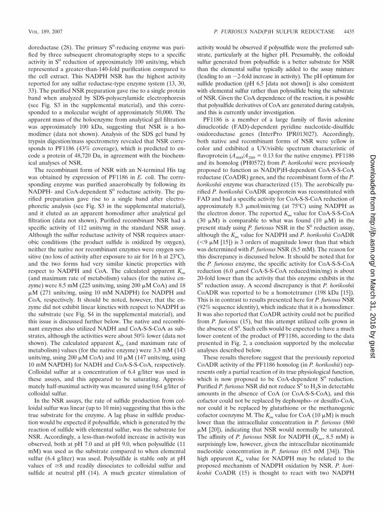
Primary response to S0 (up-regulated ORFs). Within 10 minof the addition of S0 to a growing culture of P. furiosus, theexpression of the gene encoding NSR (PF1186) was up-regu-lated by 3.7-fold according to the DNA microarray (Table 1)and by 7.0-fold using QPCR analysis (Fig. 4). These data areconsistent with NSR playing a key and primary role in theresponse of P. furiosus to S0. As shown in Table 1, of theremaining 18 ORFs whose expression is immediately up-reg-ulated upon S0 addition, 13 of them are arranged as a genecluster, PF1441-PF1453. QPCR shows that they are up-regu-lated by an average of 16-fold (Fig. 4). This 13-ORF clusterwas previously proposed to encode a second MBH, a mem-brane-bound oxidoreductase (MBX), as it shows high se-quence identity and conservation of gene order with the 14ORFs that encode the MBH (42). However, MBX lacks twokey residues that coordinate the NiFe catalytic site of thehydrogenase (47). Moreover, the up-regulation of MBX is aprimary response to S0, and it has been previously shown thatcells grown for multiple generations with S0 lack significanthydrogenase activity (1), in further support of the contentionthat MBX is not a hydrogenase. Accordingly, the 14 ORFs thatencode MBH, in addition to the 8 ORFs that encode the twocytoplasmic hydrogenases, hydrogenases I and II, are dramat-ically down-regulated within 10 min of S0 addition (Table 2 andFig. 4). These data therefore indicate that, as a primary re-
sponse to S0, the MBX cluster replaces the homologous MBHcluster (Fig. 4). The Mbh and Mbx operons are both highlyconserved, both in sequence and in gene order, in P. horikoshii,P. abyssi, and T. kodakaraensis (49), in agreement with theproposed metabolic importance of these complexes in S0 andH2 metabolism.
MBH and MBX are part of the NADH dehydrogenase com-plex I/hydrogenase family, which includes NADH:quinone oxi-doreductases (NUO or complex I), F420 quinone oxidoreduc-tases, and energy-converting hydrogenases (Ech) (10, 16).These complexes consist of a core of six homologous subunitsand through evolution and recruitment of additional subunitsdiverged into complexes with different physiological functions(10). For example, the core enzymes of MBH and MBX(MbxH, J-N and MbhH, J-N, respectively) are supplementedwith six subunits of a ubiquitous family of cation/proton anti-porters (49). Previous studies have shown that MBH is anenergy-conserving complex in which the oxidation of ferre-doxin and the reduction of protons are coupled to the gener-ation of a proton motive force (41). MBH lacks homologs ofthe NADH- and flavin-binding subunits (NuoEFG) of the typ-ical complex I (10, 16) and couples the oxidation of ferredoxin,rather than NADH, to proton pumping and in this case H2
production. The remarkable gene conservation and sequencesimilarity between MBH and MBX suggests that they havevery similar functions.
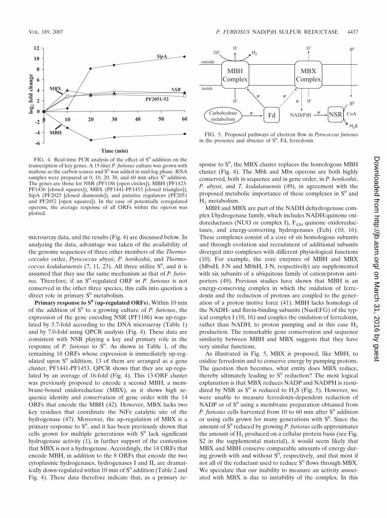
As illustrated in Fig. 5, MBX is proposed, like MBH, tooxidize ferredoxin and to conserve energy by pumping protons.The question then becomes, what entity does MBX reduce,thereby ultimately leading to S0 reduction? The most logicalexplanation is that MBX reduces NADP and NADPH is reoxi-dized by NSR as S0 is reduced to H2S (Fig. 5). However, wewere unable to measure ferredoxin-dependent reduction ofNADP or of S0 using a membrane preparation obtained fromP. furiosus cells harvested from 10 to 60 min after S0 additionor using cells grown for many generations with S0. Since theamount of S0 reduced by growing P. furiosus cells approximatesthe amount of H2 produced on a cellular protein basis (see Fig.S2 in the supplemental material), it would seem likely thatMBX and MBH conserve comparable amounts of energy dur-ing growth with and without S0, respectively, and that most ifnot all of the reductant used to reduce S0 flows through MBX.We speculate that our inability to measure an activity associ-ated with MBX is due to instability of the complex. In this
FIG. 4. Real-time PCR analysis of the effect of S0 addition on thetranscription of key genes. A 15-liter P. furiosus culture was grown withmaltose as the carbon source and S0 was added in mid-log phase. RNAsamples were prepared at 0, 10, 20, 30, and 60 min after S0 addition.The genes are those for NSR (PF1186 [open circles]), MBH (PF1423-PF1436 [closed squares]), MBX (PF1441-PF1453 [closed triangles]),SipA (PF2025 [closed diamonds]), and putative regulators (PF2051and PF2052 [open squares]). In the case of potentially coregulatedoperons, the average response of all ORFs within the operon wasplotted.
FIG. 5. Proposed pathways of electron flow in Pyrococcus furiosusin the presence and absence of S0. Fd, ferredoxin.
VOL. 189, 2007 P. FURIOSUS NAD(P)H SULFUR REDUCTASE 4437
on March 31, 2016 by guest
http://jb.asm.org/
Dow
nloaded from
regard, it should be noted that MBH readily loses its ability touse ferredoxin as an electron donor during membrane frac-tionation (42, 47). Similarly, bacterial complex I has yet to beisolated as an intact complex (54). Attempts to stabilize P.furiosus MBX are in progress.
In addition to NSR and MBX, the DNA microarray data(Table 1) show that the expression of the gene encoding aglutaredoxin-like protein termed protein disulfide oxidoreduc-tase (PDO) (12, 25), PF0094, is up-regulated almost fourfoldas part of the primary response to S0 (Table 1). A specific rolefor PF0094 as a PDO in P. furiosus has yet to be established. Ithas been proposed that in Pyrococcus species PDO is reducedby a thioredoxin reductase (PF1442 [22]) and that it could bean electron carrier for ribonucleotide reductase (RNR, PF0440[3]). However, neither PF1442 nor PF0440 were part of theprimary response to S0, although their expressions were up-regulated 60 min after S0 addition (by 2.3- and 3.8-fold, re-spectively; data not shown). Since homologs of PDO and thi-oredoxin reductase (and RNR) are widespread throughout thearchaea, including those that do not utilize S0 (25, 35), it wouldseem unlikely that PDO has a specific role in reducing S0. Toinvestigate this, the recombinant form of the PF0094 proteinwas obtained using published procedures (12). However, thepurified protein had no effect on the S0 reduction activity ofpurified NSR activity or on the ability of P. furiosus membranesto couple ferredoxin oxidation to the reduction of NADP or S0
(data not shown).In addition to those encoding NSR, MBX, and PDO, there
are only four other ORFs that are significantly up-regulatedwithin 10 min of S0 addition (Table 1), and these appear to bepresent as two operons. PF2051 and PF2052 are both anno-tated as transcriptional regulators, but only PF2051 containsregulatory domains (IPR001845), while PF2052 contains anucleotide triphosphate pyrophosphohydrolase domain (IPR004518). Both ORFs most likely form an operon, as theirsequences overlap by 22 nucleotides, and this synteny is con-served in the genome sequences of the other three Thermo-coccales. It is possible that these regulators play a key role inthe primary response to S0, although the nature of the effectoris unknown. The other two ORFs that are regulated, PF0261and PF0262, overlap by 12 nucleotides. PF0262 shows homol-ogy to multidrug efflux systems (IPR001036), while PF0261 hasa nucleic acid binding fold (IPR008994). Homologs of bothORFs are found in the genomes of the other two Pyrococcusspecies but not in the genome sequence of T. kodakaraensis, sotheir role in S0 metabolism is unclear.
Primary response to S0 (down-regulated ORFs). The moststriking feature of the list of 34 ORFs whose expression isdown-regulated within 10 min of S0 addition (Table 2) is that22 of them contain the structural genes of the three hydroge-nases and another involves hydrogenase maturation. It wasalready known that cells grown for many generations with S0
contain only very low hydrogenase activity (1), and it is nowapparent that the biosynthesis of all three hydrogenases israpidly curtailed within minutes of S0 addition, indicating acomplete shutdown at the genetic level of H2 metabolism inthe presence of S0 (Table 2 and Fig. 4). Cells continue toproduce H2 for 2 h or so after S0 addition (Fig. 1) due to theexisting hydrogenase protein in the cell, but by 1 h the rate is10% of the rate of H2S production, and eventually no H2 is
produced. The small amount of H2 subsequently consumed(Fig. 1) may reflect a differential stability between the MBH(less stable) and the cytoplasmic enzymes, which are proposedto consume H2 and reduce NADP (29). The hydrogenasegenes are well conserved within the Thermococcales order,although hydrogenase II, whose exact function is unknown, isabsent from P. horikoshii and T. kodakaraensis (29).
It is difficult to rationalize the roles of the remaining 11ORFs that are part of the primary response to S0 (Table 2),particularly when not all of them are conserved in the otherthree Thermococcales. PF0450 encodes a putative glutaminesynthetase but it was not regulated in cells grown for manygenerations with S0 (46), and the fact that it is affected soquickly after S0 addition is puzzling. While PF0450 has ho-mologs in the other sequenced Thermococcales, this is not thecase for the other S0-responsive ORFs, suggesting that theymay not be playing essential roles in S0 metabolism. PF0528-PF0531 appears to form an operon and is annotated as a cobalttransporter. This operon is conserved in T. kodakaraensis butnot in P. horikoshii or P. abyssi. Similarly, PF1621 contains afibronectin-like fold (IPR008957) but has a homolog only in T.kodakaraensis. Since P. horikoshii and P. abyssi do not utilizesugars like P. furiosus and T. kodakaraensis, perhaps the lattertwo ORF systems are involved in sugar metabolism. PF0925-PF0926 is predicted to contain a radical SAM domain, butclose homologs are absent from P. horikoshii, P. abyssi, and T.kodakaraensis. PF0736 and PF0736.1 are hypothetical ORFson opposing strands and show little sequence similarity to anyprotein in the NCBI database (2). PF0913 is a homolog ofsubunit E of formylmethanofuran dehydrogenase, an enzymefound in methanogens, but this is not the catalytic subunit andits function is unknown (52).
Secondary response to S0. The ORFs that are up-regulatedonly 10 min after S0 addition are assumed to represent theprimary response, and all remain up-regulated at 30 min.At this time, an additional 27 ORFs are up-regulated morethan threefold (or are part of a potentially regulated operon),and these appear to represent a secondary response to S0
(Table 3). This is supported by the fact that most of them (15of 27) are involved in metabolism of glutamate or branched-chain amino acids. PF0204-PF206 and PF1852 are potentiallyinvolved in glutamate biosynthesis (21, 44) and these mightcompensate for down-regulation (by 4.1-fold) of the ORF thatencodes glutamate dehydrogenase (data not shown), althoughwhy is not clear. Three ORFs involved in iron metabolism arealso up-regulated by S0 (Table 3). Presumably, the productionof intracellular sulfide (by cytoplasmic NSR) might lead toinsoluble iron sulfides, and ORFs involved in ferrous irontransport (PF0857) and iron-sulfur cluster biosynthesis (PF1285,PF1286) are up-regulated in response to the products of S0
reduction.In a previous study (46) involving cells grown for many
generations with S0, two highly regulated S0-dependent ORFswere characterized as “sulfur-induced proteins” A and B (SipAand SipB). We were surprised to find that these are part of thesecondary, rather than the primary, response to S0, althoughthe response is quite dramatic and appears to represent anon/off switch. By QPCR analysis, SipA and SipB are up-regu-lated over 400-fold and 26-fold, respectively, 30 min after S0
addition (Fig. 4). They are conserved in the four Thermococ-
4438 SCHUT ET AL. J. BACTERIOL.
on March 31, 2016 by guest
http://jb.asm.org/
Dow
nloaded from

cales members whose genomes have been sequenced, buttheir physiological function still remains a mystery. A po-tential transcription factor, PF0986, which is up-regulated30 min after S0 addition might be involved in coordinatingthe secondary responses to S0. PF0986 is a homolog of thecharacterized TFIIS in Methanococcus thermolithotrophicus,which is involved in RNA proofreading (26). PF0986 ispotentially in an operon with PF0984, encoding a small59-residue protein, both of which are conserved in the se-quenced Thermococcales (2). However, the relationship ofthese ORFs to transcriptional regulation and S0 metabolismis not clear at this point.
In conclusion, while the addition of S0 to a culture of P.furiosus cells causes growth to stall, indicating a large metabolic
shift (Fig. 1), only two key enzymes that appear to be directlyinvolved in S0 reduction, MBX and NSR, were identified. Asshown in Fig. 5, MBX and NSR are proposed to be the keyenzymes responsible for the reoxidation of ferredoxin andNAD(P)H, respectively. Another primary response to S0 avail-ability is the concomitant shutdown of H2 metabolism, result-ing in the preferential transfer of reducing equivalents to thereduction of S0 rather than protons (Fig. 5). This novel S0-reducing system involving NSR and MBX is so far unique tothe heterotrophic Thermococcales and contrasts with the cyto-chrome- and quinone-based S0-reducing system in autotrophicarchaea (and bacteria). Future research will focus on elucidat-ing the precise role of MBX and the mechanism of S0 reduc-tion by NSR.
TABLE 3. ORFs whose expression is up-regulated within 30 min after the addition of elemental sulfur to growing P. furiosus cells
Function and ORFa Description Foldchangeb
Glutamate biosynthesis�PF0204f Glutamate synthase, large subunit� 4.0PF0205f Ferredoxin-dependent glutamate synthase� 2.8PF0206f Glutamate synthase, large subunit region 3� 3.2
PF0686e Hypothetical protein� 3.1PF0704 Protein of unknown function DUF302� 3.1PF0857 Ferrous iron transport protein B, N terminal� 3.2
Branched-chain amino acid biosynthesis�PF0935 Acetolactate synthase, large subunit� 4.6PF0936 Acetohydroxy acid isomeroreductase� 4.7PF0937 Pyruvate carboxyltransferase� 5.0PF0938 3-Isopropylmalate dehydratase large subunit� 3.5PF0939 3-Isopropylmalate dehydratase small subunit� 4.3PF0940 Isocitrate/isopropylmalate dehydrogenase� 3.7PF0941 2-Isopropylmalate/homocitrate synthase� 3.1PF0942 6-Phosphogluconate dehydratase� 3.2
Transcription factor�PF0984 Hypothetical protein� 3.3PF0986 Transcription factor TFIIS� 3.4
Iron-sulfur cluster assembly�PF1285f SufBD� 2.3PF1286f SufBD� 3.2
Branched-chain amino acid biosynthesis�PF1678 Alpha-isopropylmalate/homocitrate synthase� 5.2PF1679 3-Isopropylmalate dehydratase large subunit� 6.9PF1680 3-Isopropylmalate dehydratase small subunit� 4.9
Ribosome�PF1823 Ribosomal L23 protein� 2.6PF1824 Ribosomal protein L4/L1e� 4.0
PF1852 Glutamate synthase (21)� 3.4
Sulfur-induced proteins(46)�PF2025 SipA 9.3PF2026 SipB 6.5
PF2029 Hypothetical protein� 3.7
a For explanation, see Table 1, footnote a.b All changes (n-fold) are statistically significant (P 0.05) unless indicated. ND, not determined.c No homolog present in the genome sequence of T. kodakaraensis.d No homologs in the genome sequences of P. horikoshii and P. abyssi.e Unique to P. furiosus.f No homologs in the genome sequences of T. kodakaraensis, P. horikoshii, and P. abyssi.
VOL. 189, 2007 P. FURIOSUS NAD(P)H SULFUR REDUCTASE 4439
on March 31, 2016 by guest
http://jb.asm.org/
Dow
nloaded from
ACKNOWLEDGMENTS
This research was funded by grants (FG05-95ER20175 and FG02-05ER15710) from the Department of Energy.
We thank Peter S. Horanyi for providing the pDEST C1 Gatewayexpression vector, Cindy Lim for preliminary QPCR studies, Scott D.Hamilton-Brehm for assistance in constructing the microarrays, FrankE. Jenney, Jr. and Angeli L. Menon for many helpful discussions, andFarris L. Poole II for bioinformatics analyses.
REFERENCES
1. Adams, M. W. W., J. F. Holden, A. L. Menon, G. J. Schut, A. M. Grunden,C. Hou, A. M. Hutchins, F. E. Jenney, Jr., C. Kim, K. Ma, G. Pan, R. Roy,R. Sapra, S. V. Story, and M. F. Verhagen. 2001. Key role for sulfur inpeptide metabolism and in regulation of three hydrogenases in the hyper-thermophilic archaeon Pyrococcus furiosus. J. Bacteriol. 183:716–724.
2. Altschul, S. F., T. L. Madden, A. A. Schaffer, J. Zhang, Z. Zhang, W. Miller,and D. J. Lipman. 1997. Gapped BLAST and PSI-BLAST: a new generationof protein database search programs. Nucleic Acids Res. 25:3389–3402.
2a.Applied Biosystems. 2001. Bulletin 2. Applied Biosystems, Foster City, CA.3. Arner, E. S., and A. Holmgren. 2000. Physiological functions of thioredoxin
and thioredoxin reductase. Eur. J. Biochem. 267:6102–6109.4. Blamey, J. M., and M. W. Adams. 1993. Purification and characterization of
pyruvate ferredoxin oxidoreductase from the hyperthermophilic archaeonPyrococcus furiosus. Biochim. Biophys. Acta 1161:19–27.
5. Bradford, M. M. 1976. A rapid and sensitive method for the quantitation ofmicrogram quantities of protein utilizing the principle of protein-dye bind-ing. Anal. Biochem. 72:248–254.
6. Chen, J. S., and L. E. Mortenson. 1977. Inhibition of methylene blue for-mation during determination of the acid-labile sulfide of iron-sulfur proteinsamples containing dithionite. Anal. Biochem. 79:157–165.
7. Cohen, G. N., V. Barbe, D. Flament, M. Galperin, R. Heilig, O. Lecompte, O.Poch, D. Prieur, J. Querellou, R. Ripp, J. C. Thierry, J. Van der Oost, J.Weissenbach, Y. Zivanovic, and P. Forterre. 2003. An integrated analysisof the genome of the hyperthermophilic archaeon Pyrococcus abyssi. Mol.Microbiol. 47:1495–1512.
8. Dietrich, W., and O. Klimmek. 2002. The function of methyl-menaquinone-6and polysulfide reductase membrane anchor (PsrC) in polysulfide respirationof Wolinella succinogenes. Eur. J. Biochem. 269:1086–1095.
9. Fiala, G., and K. O. Stetter. 1986. Pyrococcus furiosus sp-nov represents anovel genus of marine heterotrophic archaebacteria growing optimally at100-degrees C. Arch. Microbiol. 145:56–61.
10. Friedrich, T., and D. Scheide. 2000. The respiratory complex I of bacteria,archaea and eukarya and its module common with membrane-bound mul-tisubunit hydrogenases. FEBS Lett. 479:1–5.
11. Fukui, T., H. Atomi, T. Kanai, R. Matsumi, S. Fujiwara, and T. Imanaka.2005. Complete genome sequence of the hyperthermophilic archaeon Ther-mococcus kodakaraensis KOD1 and comparison with Pyrococcus genomes.Genome Res. 15:352–363.
12. Guagliardi, A., D. de Pascale, R. Cannio, V. Nobile, S. Bartolucci, and M.Rossi. 1995. The purification, cloning, and high level expression of a glu-taredoxin-like protein from the hyperthermophilic archaeon Pyrococcusfuriosus. J. Biol. Chem. 270:5748–5755.
13. Guiral, M., P. Tron, C. Aubert, A. Gloter, C. Iobbi-Nivol, and M. T. Giudici-Orticoni. 2005. A membrane-bound multienzyme, hydrogen-oxidizing, andsulfur-reducing complex from the hyperthermophilic bacterium Aquifexaeolicus. J. Biol. Chem. 280:42004–42015.
14. Gun, J., A. D. Modestov, A. Kamyshny, D. Ryzkov, V. Gitis, A. Goifman, O.Lev, V. Hultsch, T. Grischek, and E. Worch. 2004. Electrospray ionizationmass spectrometric analysis of aqueous polysulfide solutions. Microchim.Acta 146:229–237.
15. Harris, D. R., D. E. Ward, J. M. Feasel, K. M. Lancaster, R. D. Murphy,T. C. Mallet, and E. J. Crane III. 2005. Discovery and characterization of acoenzyme A disulfide reductase from Pyrococcus horikoshii. Implications forthis disulfide metabolism of anaerobic hyperthermophiles. FEBS J. 272:1189–1200.
16. Hedderich, R., and L. Forzi. 2005. Energy-converting [NiFe] hydrogenases:more than just H2 activation. J. Mol. Microbiol. Biotechnol. 10:92–104.
17. Hedderich, R., O. Klimmek, A. Kroger, R. Dirmeier, M. Keller, and K. O.Stetter. 1999. Anaerobic respiration with elemental sulfur and disulfides.FEMS Microbiol. Rev. 22:353–381.
18. Holm, S. 1979. A simple sequentially rejective multiple test procedure.Scand. J. Stat. 6:65–70.
19. Huber, R., H. Huber, and K. O. Stetter. 2000. Towards the ecology ofhyperthermophiles: biotopes, new isolation strategies and novel metabolicproperties. FEMS Microbiol. Rev. 24:615–623.
20. Hummel, C. S., K. M. Lancaster, and E. J. Crane III. 2005. Determinationof coenzyme A levels in Pyrococcus furiosus and other Archaea: implicationsfor a general role for coenzyme A in thermophiles. FEMS Microbiol. Lett.252:229–234.
21. Jongsareejit, B., R. N. Rahman, S. Fujiwara, and T. Imanaka. 1997. Gene
cloning, sequencing and enzymatic properties of glutamate synthase from thehyperthermophilic archaeon Pyrococcus sp. KOD1. Mol. Gen. Genet. 254:635–642.
22. Kashima, Y., and K. Ishikawa. 2003. A hyperthermostable novel protein-disulfide oxidoreductase is reduced by thioredoxin reductase from hyper-thermophilic archaeon Pyrococcus horikoshii. Arch. Biochem. Biophys. 418:179–185.
23. Kawarabayasi, Y., M. Sawada, H. Horikawa, Y. Haikawa, Y. Hino, S.Yamamoto, M. Sekine, S. Baba, H. Kosugi, A. Hosoyama, Y. Nagai, M.Sakai, K. Ogura, R. Otsuka, H. Nakazawa, M. Takamiya, Y. Ohfuku, T.Funahashi, T. Tanaka, Y. Kudoh, J. Yamazaki, N. Kushida, A. Oguchi, K.Aoki, and H. Kikuchi. 1998. Complete sequence and gene organization ofthe genome of a hyper-thermophilic archaebacterium, Pyrococcus horikoshiiOT3. DNA Res. 5:55–76.
24. Keller, M., and R. Dirmeier. 2001. Hydrogen-sulfur oxidoreductase complexfrom Pyrodictium abyssi. Methods Enzymol. 331:442–451.
25. Ladenstein, R., and B. Ren. 2006. Protein disulfides and protein disulfideoxidoreductases in hyperthermophiles. FEBS J. 273:4170–4185.
26. Lange, U., and W. Hausner. 2004. Transcriptional fidelity and proofreadingin Archaea and implications for the mechanism of TFS-induced RNA cleav-age. Mol. Microbiol. 52:1133–1143.
27. Laska, S., F. Lottspeich, and A. Kletzin. 2003. Membrane-bound hydroge-nase and sulfur reductase of the hyperthermophilic and acidophilic archaeonAcidianus ambivalens. Microbiology 149:2357–2371.
28. Ma, K., and M. W. Adams. 2001. Ferredoxin:NADP oxidoreductase fromPyrococcus furiosus. Methods Enzymol. 334:40–45.
29. Ma, K., and M. W. Adams. 2001. Hydrogenases I and II from Pyrococcusfuriosus. Methods Enzymol. 331:208–216.
30. Ma, K., and M. W. Adams. 1994. Sulfide dehydrogenase from the hyperther-mophilic archaeon Pyrococcus furiosus: a new multifunctional enzyme in-volved in the reduction of elemental sulfur. J. Bacteriol. 176:6509–6517.
31. Ma, K., R. N. Schicho, R. M. Kelly, and M. W. Adams. 1993. Hydrogenaseof the hyperthermophile Pyrococcus furiosus is an elemental sulfur reductaseor sulfhydrogenase: evidence for a sulfur-reducing hydrogenase ancestor.Proc. Natl. Acad. Sci. USA 90:5341–5344.
32. Ma, K., R. Weiss, and M. W. Adams. 2000. Characterization of hydrogenaseII from the hyperthermophilic archaeon Pyrococcus furiosus and assessmentof its role in sulfur reduction. J. Bacteriol. 182:1864–1871.
33. Ng, K. Y., R. Sawada, S. Inoue, K. Kamimura, and T. Sugio. 2000. Purifi-cation and some properties of sulfur reductase from the iron-oxidizing bac-terium Thiobacillus ferrooxidans NASF-1. J. Biosci. Bioeng. 90:199–203.
34. Pan, G., M. F. Verhagen, and M. W. Adams. 2001. Characterization ofpyridine nucleotide coenzymes in the hyperthermophilic archaeon Pyrococ-cus furiosus. Extremophiles 5:393–398.
35. Pedone, E., D. Limauro, R. D’Alterio, M. Rossi, and S. Bartolucci. 2006.Characterization of a multifunctional protein disulfide oxidoreductase fromSulfolobus solfataricus. FEBS J. 273:5407–5420.
36. Pihl, T. D., L. K. Black, B. A. Schulman, and R. J. Maier. 1992. Hydrogen-oxidizing electron transport components in the hyperthermophilic archae-bacterium Pyrodictium brockii. J. Bacteriol. 174:137–143.
37. Poole, F. L., II, B. A. Gerwe, R. C. Hopkins, G. J. Schut, M. V. Weinberg,F. E. Jenney, Jr., and M. W. Adams. 2005. Defining genes in the genome ofthe hyperthermophilic archaeon Pyrococcus furiosus: implications for allmicrobial genomes. J. Bacteriol. 187:7325–7332.
38. Robb, F. T., D. L. Maeder, J. R. Brown, J. DiRuggiero, M. D. Stump, R. K.Yeh, R. B. Weiss, and D. M. Dunn. 2001. Genomic sequence of hyperther-mophile, Pyrococcus furiosus: implications for physiology and enzymology.Methods Enzymol. 330:134–157.
39. Ronimus, R. S., and H. W. Morgan. 2003. Distribution and phylogenies ofenzymes of the Embden-Meyerhof-Parnas pathway from archaea and hyper-thermophilic bacteria support a gluconeogenic origin of metabolism. Ar-chaea 1:199–221.
40. Sakuraba, H., and T. Ohshima. 2002. Novel energy metabolism in anaerobichyperthermophilic archaea: a modified Embden-Meyerhof pathway. J. Bio-sci. Bioeng. 93:441–448.
41. Sapra, R., K. Bagramyan, and M. W. Adams. 2003. A simple energy-con-serving system: proton reduction coupled to proton translocation. Proc. Natl.Acad. Sci. USA 100:7545–7550.
42. Sapra, R., M. F. Verhagen, and M. W. W. Adams. 2000. Purification andcharacterization of a membrane-bound hydrogenase from the hyperthermo-philic archaeon Pyrococcus furiosus. J. Bacteriol. 182:3423–3428.
43. Schicho, R. N., K. Ma, M. W. Adams, and R. M. Kelly. 1993. Bioenergeticsof sulfur reduction in the hyperthermophilic archaeon Pyrococcus furiosus. J.Bacteriol. 175:1823–1830.
44. Schut, G. J., S. D. Brehm, S. Datta, and M. W. Adams. 2003. Whole-genomeDNA microarray analysis of a hyperthermophile and an archaeon: Pyrococ-cus furiosus grown on carbohydrates or peptides. J. Bacteriol. 185:3935–3947.
45. Schut, G. J., A. L. Menon, and M. W. W. Adams. 2001. 2-Ketoacid oxido-reductases from Pyrococcus furiosus and Thermococcus litoralis. MethodsEnzymol. 331:144–158.
46. Schut, G. J., J. Zhou, and M. W. W. Adams. 2001. DNA microarray analysis
4440 SCHUT ET AL. J. BACTERIOL.
on March 31, 2016 by guest
http://jb.asm.org/
Dow
nloaded from
of the hyperthermophilic archaeon Pyrococcus furiosus: evidence for a newtype of sulfur-reducing enzyme complex. J. Bacteriol. 183:7027–7036.
47. Silva, P. J., E. C. van den Ban, H. Wassink, H. Haaker, B. de Castro, F. T.Robb, and W. R. Hagen. 2000. Enzymes of hydrogen metabolism in Pyro-coccus furiosus. Eur. J. Biochem. 267:6541–6551.
48. Stetter, K. O. 1996. Hyperthermophilic procaryotes. FEMS Microbiol. Rev.18:149–158.
49. Swartz, T. H., S. Ikewada, O. Ishikawa, M. Ito, and T. A. Krulwich. 2005. TheMrp system: a giant among monovalent cation/proton antiporters? Extremo-philes 9:345–354.
50. Verhagen, M. F., A. L. Menon, G. J. Schut, and M. W. Adams. 2001.Pyrococcus furiosus: large-scale cultivation and enzyme purification. MethodsEnzymol. 330:25–30.
51. Verhees, C. H., S. W. Kengen, J. E. Tuininga, G. J. Schut, M. W. Adams,W. M. De Vos, and J. Van Der Oost. 2003. The unique features of glycolyticpathways in Archaea. Biochem. J. 375:231–246.
52. Vorholt, J. A., M. Vaupel, and R. K. Thauer. 1996. A polyferredoxin witheight [4Fe-4S] clusters as a subunit of molybdenum formylmethanofurandehydrogenase from Methanosarcina barkeri. Eur. J. Biochem. 236:309–317.
53. Weinberg, M. V., G. J. Schut, S. Brehm, S. Datta, and M. W. Adams. 2005.Cold shock of a hyperthermophilic archaeon: Pyrococcus furiosus exhibitsmultiple responses to a suboptimal growth temperature with a key role formembrane-bound glycoproteins. J. Bacteriol. 187:336–348.
54. Yagi, T., and A. Matsuno-Yagi. 2003. The proton-translocating NADH-quinone oxidoreductase in the respiratory chain: the secret unlocked. Bio-chemistry 42:2266–2274.
VOL. 189, 2007 P. FURIOSUS NAD(P)H SULFUR REDUCTASE 4441
on March 31, 2016 by guest
http://jb.asm.org/
Dow
nloaded from