Vibration Control of Resonant Vibratory Feeders With Electromagnetic Excitation

PHYSICAL REVIEW B, VOLUME 64, 115418
Sulfur on TiO 2„110… studied with resonant photoemission
E. L. D. Hebenstreit,1,2 W. Hebenstreit,1 H. Geisler,3 S. N. Thornburg,4 C. A. Ventrice, Jr.,4 D. A. Hite,5,6
P. T. Sprunger,5,6 and U. Diebold11Department of Physics, Tulane University, New Orleans, Louisiana 70118
2Institut fur Allgemeine Physik, Vienna University of Technology, Wiedner Hauptstrasse 8–10, A-1040 Vienna, Austria3Department of Chemistry, Xavier University of Louisiana, New Orleans, Louisiana 70125
4Department of Physics, University of New Orleans, New Orleans, Louisiana 701485Center for Advanced Microstructurs and Devices (CAMD), Louisiana State University, Baton Rouge, Louisiana 70806
6Department of Physics and Astronomy, Louisiana State University, Baton Rouge, Louisiana 70803~Received 23 February 2001; published 29 August 2001!
Adsorption of sulfur on TiO2(110) at room temperature~RT! and 350 °C has been studied with ultravioletphotoelectron spectroscopy. A TiO2(110) (131) surface with a small amount of oxygen vacancies wasprepared by sputtering and annealing in ultrahigh vacuum. Oxygen vacancies induce a defect state that pins theFermi level just below the conduction-band minimum. Sulfur adsorption at room temperature leads to thedisappearance of this vacancy-related band-gap state, indicating that the surface oxygen vacancies are filled bysulfur. Sulfur-induced valence-band features are identified at binding energies of 3.4 and 8 eV. Adsorption of Sat 350 °C forms a (431) superstructure at high coverages@'0.9 monolayer~ML !# that is visible withlow-energy electron diffraction. In a previously proposed model for this superstructure, sulfur replaces half ofthe in-plane oxygen atoms and all the bridging oxygen atoms are removed. In agreement with this model, theoxygen 2s peak is decreased significantly and the defect state is increased. Two additional valence features areobserved: one at 2.7 eV and one at 3.9 eV. Due to those features the band gap vanishes. In resonant photo-emission, these features show a similar, but weaker, resonance profile than the vacancy-related defect state.Hybridized Ti-derived states extend across the whole valence-band region. Generally, a higher resonant photonenergy is found for valence-band states with lower binding energies, indicating mainly 3p24s transitions inthe upper valence band. Adsorption of sulfur reduces the strength of the resonances.
DOI: 10.1103/PhysRevB.64.115418 PACS number~s!: 68.47.Gh, 68.43.Fg, 79.60.2i
nid
ha
itheerean
segeeghth-
a
urdgdme
theand
I. INTRODUCTION
Because sulfur is a common poison for catalytic reactiothe adsorption of sulfur compounds on metal and metal oxsurfaces has received considerable attention.1,2 In particular,the adsorption of sulfur compounds on titanium oxidesbeen studied extensively.3–8
We have studied the interaction of elemental sulfur wthe TiO2(110) surface. In our previous work we found thrdifferent adsorption sites for sulfur and a variety of structuwith long-range order that depend on sulfur coveragesample temperature during adsorption.9,10 The experimentswere performed with a clean UHV-annealed TiO2(110) sur-face that exhibits a few percent of oxygen vacancies@see Fig.1~A!#. At low concentrations these vacancies are disperacross the surface.11 At room temperature scanning tunnelinmicroscope~STM! studies indicate that sulfur binds to thtitanium rows.9 However, it could not be determined if thsulfur also adsorbs at the point defects. A very recent hiresolution photoelectron spectroscopy study indicatesthis is the case.12 Adsorption on the hot surface in the temperature range of 3502400 °C leads to the formation of(431) superstructure at a coverage of'0.9 monolayer~ML !. In Ref. 10 a model was proposed in which sulfreplaces 50% of the in-plane oxygen atoms while the briing oxygen atoms are removed. In order to test this moand to clarify the binding sites for S adsorption at rootemperature~RT!, we performed photoemission measurments of the valence band and the shallow core levels.
0163-1829/2001/64~11!/115418~11!/$20.00 64 1154
s,e
s
sd
d
-at
-el
-
Several photoemission studies of the clean TiO2(110) sur-face have been performed previously. In Refs. 13 and 14clean, almost stoichiometric surface was investigated,the results were compared toab initio calculations. For ob-
FIG. 1. ~A! Atomic model of the clean TiO2(110) surface;~B!atomic model and STM image~95 Å 3 95 Å, 1.4 V, 0.2 nA! of Sadsorbed at RT on TiO2(110) ~Ref. 9!. The sulfur atoms, visible aswhite spots, are located on the bright titanium rows.
©2001 The American Physical Society18-1

eeso3
nth
2fohiesats
nthtioam
w
-
wi
ia
a
c
ss2ae-
nd
nttene
Ad-
ere
rgyd toti-
ure-act
Oeral
itheal-ntha-
is-as
to
neell.RT
sur-nare
f Sfteror-Dcedure-theub-
theund
nons
on.ws.bein
asa-
E. L. D. HEBENSTREITet al. PHYSICAL REVIEW B 64 115418
taining a deeper understanding of the interaction betwtitanium and oxygen, it is valuable to make use of the renant photoemission effect for photon energies near thep23d and 3p24s transitions of Ti. This effect is explained idetail in Refs. 15 and 16. Resonant photoemission ofclean surface was studied by several groups17–20 and filmdeposition of various elements was analyzed in Refs. 21–Additional investigations using resonant photoemissionTi compounds were performed in Refs. 26 and 27. In twork we exploit the resonant photoemission effect to invtigate the bonding between surface atoms and adsorbThe measurements were performed with photon energie29–94 eV, covering the range for the 3p23d and 3p24stransitions in Ti.
Because resonant photoemission has already beenplained in detail in the references mentioned above, we osummarize briefly the main concept. It is based onquantum-mechanical interference between two excitaprocesses that transform a certain initial state to the sfinal state via two different pathways.
The direct photoemission process produces electronsthe kinetic energyE:
3p63dn1hn⇒3p63dn211e2~E!. ~1!
Concurrent to Eq.~1!, optical adsorption may lead to excitation of the 3p to the 3d ~or 4s) level:
3p63dn1hn⇒@3p53dn11#* . ~2!
The excited state decays and an electron is emittedthe same kinetic energy as for process~1!:
@3p53dn11#* ⇒3p63dn211e2~E!. ~3!
For photon energies in the resonant region, the peaktensity should exhibit a characteristic, asymmetric peshape~Fano profile!.15 In relatively light transition metalssuch as Ti, additional excitations accompany the opticalsorption causing changes in the resonance.26 In addition, theresonance is moved to higher photon energies than expefrom the separation of the contributing energy levels~ap-proximately 47 eV for the 3p23d transition instead of 33eV! due to the exchange interaction between 3p and 3dlevels.16
Because the photoexcitation and recombination procespatially localized, resonant emission in the mainly Opderived valence band is only possible if oxygen statesstrongly hybridized with the titanium orbitals. This is thcase for the Ti–O bonds of TiO2(110) and resonant photoemission is found for the whole valence band.17 In amolecular-orbital picture bonding orbitals with strong Ti 3dcharacter dominate at higher binding energy. For lower biing energy, nonbonding orbitals with mainly O 2p contribu-tion are found.18 The minimum peak intensity for resonaphotoemission is found at a photon energy of approxima36 eV. A resonant enhancement is found up to photon egies of approximately 60 eV.
11541
n-
e
5.rs-es.of
ex-lyene
ith
th
n-k
d-
ted
is
re
-
lyr-
II. EXPERIMENTAL PROCEDURE
The measurements were performed at the Center forvanced Microstructures and Devices~CAMD! on the PGMbeamline. The angle resolved photoemission spectra wtaken at normal emission withp-polarized light. The angle ofincidence of the photon beam was 45°. The photon eneranged between 29 and 95 eV. All spectra were normalizethe photon flux using a tungsten grid placed within the opcal beam path. The Fermi level was calibrated by measments of the Fermi edge of a piece of Ta in electrical contwith the sample.
All measurements were carried out on a dark blue Ti2crystal that was reduced by heating up to 700 °C for sevhours. The sample was cleaned by ion bombardment w1-keV Ne1 ions at room temperature and subsequent anning in UHV for 10 min at 650 °C. This sample treatmeproduces a (131) surface which exhibits point defects wita density of a few percent.11 The base pressure in the prepration chamber was in the low 10210-mbar range.
The elemental sulfur was produced by electrolytical dsociation of Ag2S in a sulfur cell. The cell was constructeddescribed in Ref. 28. The cell itself was constantly heateda temperature in the range of 1502165 °C. The dosing pro-cedure is described in detail in Ref. 10. A coverage of omonolayer is defined as one S atom per substrate unit c
The photoemission measurements were performed atafter adsorbing a saturation coverage of S on the cleanface. According to previous low-energy electron diffractio~LEED! and STM measurements no superstructuresformed by S when adsorbed at RT.9,10 To minimize the effectof photoinduced desorption during the measurements owhen adsorbed at RT, the sample was moved laterally ataking 2–3 spectra. For adsorption of S at 350 °C, the fmation of a (431) superstructure was confirmed with LEEbefore taking the photoemission spectra. No photoindudesorption was found during the photoemission measments from this superstructure. Upon normalization tophoton flux the analysis of all spectra was done after sstracting an integrated background~Shirley background! toaccount for the inelastically scattered electrons. Onlyspectra in Figs. 3 and 11 are presented without backgrosubstraction.
III. RESULTS
A. Valence-band spectra of SÕTiO2„110… adsorbed at RT
An STM image for S/TiO2(110) adsorbed at RT is showin Fig. 1~B! together with a geometric model that is basedprevious results.9 Sulfur adsorbs on the bright titanium rowas indicated in the model in Fig. 1~B!. Usually a few percentof point defects~i.e., missing bridging oxygen atoms! arepresent on the surface after annealing in UHV@see Fig.1~A!#, and are known to be very active sites for adsorptiGenerally, defects are imaged as bright spots on dark roHowever, depending on the tip state, they may not alwaysvisible.11 This makes a statistic evaluation of defect sitesSTM images difficult. In addition, sulfur atoms appearvery large protrusions, possibly shielding neighboring v
8-2

oem
nth
a
anoityitth
reigah, t3
thurth
ethhleur
outc-
een-gend
-henthe
theicnsi-the
in-thect-n aate.ad-in
herisonotoner-
areTiom-ct-
se-of
t RTfad-V,rve
ngsure
fiveandlfur
the
eent
ad
rigeV
SULFUR ON TiO2(110) STUDIED WITH RESONANT . . . PHYSICAL REVIEW B 64 115418
cancy sites, and are quite mobile when adsorbed at rotemperature. Therefore STM alone is insufficient to decidthese vacancies are possible S adsorption sites. Photoesion experiments of the valence band~VB! are well suited toaddress this question. The presence of oxygen defects oTiO2(110) surface leads to an excess of electrons inneighborhood of the defect that results in a defect state~la-beledg1 in the photoemission spectra shown below! withinthe band gap. The formal oxidation state of the titanium cions next to a defect is reduced from 41 to 31. It is knownthat O2 adsorbs dissociatively at RT at oxygen vacanciesfills these defects.29 The adsorption process is not precursmediated, i.e., only these oxygen molecules that directly hvacancy dissociate and adsorb on the surface by occupthe defects. When oxygen is dosed at room temperaturesurface remains unchanged except for the population ofdefects.
Photoemission spectra of the VB region were measubefore and after sulfur and oxygen adsorption at RT. In F2 three spectra from three differently prepared surfacescompared at a photon energy of 52 eV, which is within trange of the Ti resonance energies. At this photon energydefect state within the band gap is enhanced due top23d excitation and can be compared more easily fordifferent surfaces. The VB of the clean UHV annealed sface shows a defect state at approximately 1.1 eV belowFermi level. After dosing 210 Langmuire~L! molecular oxy-gen at RT the defect state~see Fig. 2! has almost completelyvanished, in agreement with the expected filling of oxygvacancies on the surface at RT. Adsorption of S at RT onclean UHV annealed surface leads to similar results. Tdefect state has completely vanished after exposure tothan 100 L. According to previous experiments this expos
FIG. 2. Photoemission spectra of the valence band~normalemission! of TiO2(110) athn552.4 eV~in resonance!. Annealingin UHV creates point defects in the rows of bridging oxgyenindicated by the defect state in the band gap. The defect stateappears when sulfur or oxygen is adsorbed at RT. Adsorptionoxygen and sulfur adsorption at room temperature cause ashift of the spectra to lower binding energy by 0.2 and 0.4respectively.
11541
mifis-
thee
t-
dranghee
d.reehe
e-e
neesse
is sufficient to produce a saturation coverage which is ab0.7 ML for adsorption of S at RT. This indicates that S ocupies the defects~see Sec. IV A!, in addition to the titaniumrows shown in Fig. 1~B!.
Another effect that is seen in Fig. 2 is the shift of thwhole VB for S and O2 adsorption at RT as compared to thclean UHV annealed surface. The shift to lower binding eergies for S is about 0.4 eV and for O at RT it is in the ranof 0.2–0.3 eV. This shift is most likely caused by a babending effect30 as discussed in Sec. IV A.
The intensity of the VB emission was significantly lowered by the adsorption of sulfur at both RT and 350 °C. Tprimary reason for this phenomenon lies in the differephotoionization cross sections of sulfur and oxygen. Tatomic cross section for photoionization of the O 2p orbitalat a photon energy of 40.8 eV is 6.83106 b.31 For the samephoton energy the cross section for ionizing the S 3p orbitalin a S atom is ten times lower with 0.63106 b. The kineticenergy of the photoelectrons in our spectra are close tominimum of the well-known universal curve for inelastmean free path lengths, which ensures a high surface setivity. The presence of S causes inelastic scattering ofphotoelectrons from the underlying layers~thus attenuatingthe valence band!, and any new features related to the S 3ppeak are very weak because of the low cross section.
To investigate how sulfur adsorption at different sitesfluences the valence band, sulfur was adsorbed at RT onclean surface with and without oxygen defects. The defefree surface was prepared by first dosing oxygen at RT oclean, UHV annealed surface that exhibited the defect stBoth adsorption experiments provide surfaces with sulfursorbed on the titanium rows. The only difference is thatthe first case the defects are filled by sulfur while in tsecond case the defects are occupied by oxygen. Compabetween the valence bands of these two surfaces in a phenergy range from 34 to 74 eV shows no significant diffences in the spectra~spectra not shown!.
In order to analyze the valence-band features thatcaused by sulfur adsorption on the fivefold coordinatedatoms, we adsorbed S on the defect-free surface and cpared it with the clean defect-free surface. Again, the defefree surface was prepared by UHV annealing with subquent adsorption of oxygen at RT. The complete seriesvalence-band spectra for the defect-free surface and S aare presented in Figs. 3~A! and~B!. The valence emission othe clean defect-free surface and the surface after sulfursorption are shown Fig. 4 for a photon energy of 39 ewhich is below resonance. For easier comparison the cuafter S adsorption is shifted by 0.2 eV to higher bindienergies to compensate for band bending effects and enan overlap of the valence bands in the graph. At leastdifferent peaks could be distinguished in the valence bfor both surfaces. The peak positions in the case of suadsorption~after the 0.2-eV shift! are at 4.3–4.6 eV~A!, 5.3–5.6 eV~B!, 6.5–7 eV~C!, 7.5–8 eV~D!, and 8.5–9 eV~E!plus an additional shoulder around 3 eV, see the labels onspectrum forhn538.7 eV in Figs. 4 and 3~B!. ~For somephoton energies it was difficult to identify the features. Wtherefore give a range for the binding energy of the differ
sis-ofid,
8-3

ce
C.in
E. L. D. HEBENSTREITet al. PHYSICAL REVIEW B 64 115418
FIG. 3. Photoemission spectra of the valenband ~normal emission! of differently preparedTiO2(110) surfaces.~A! After oxygen adsorptionat RT ~defect-free surface!, ~B! after S adsorptionat RT on the defect-free surface,~C! the cleanUHV annealed surface, and~D! the (431) –S su-perstructure produced by S adsorption at 350 °The labeled black dots indicate peaks analyzedFigs. 5 and 9.
ai
ande
mv
Sye
ens
llr
unda-sur-
ionind-di-
resinstith
rbit-c-edure;w-ap-
andeak
peaks. This accounts for dispersion as well as the uncertin the determination of the accurate peak position.! The peakpositions for the defect-free clean surface~oxygen adsorptionat RT! are within the same range as for sulfur except for peC which is located at slightly lower binding energies arou6.3 eV. This stands in good agreement to the values in R14 for a clean surface with very few oxygen defects. At sophoton energies an additional shoulder at 7 eV becomesible but it never appears as distinct peak.
Despite the low cross section for photoemission from3p, two features are identified in Fig. 4 as being caused badsorption. The first one is a small shoulder at around 3which appears at the valence-band maximum of TiO2. Thisregion shows a somewhat higher intensity for all photonergies as compared to the clean surface. In a photoemisstudy of Ti11xS2 ~Ref. 27! the emission from S 3p orbitals isassigned to a peak at a binding energy of 3.4 eV as weintensity in the regions around 2.2 and 5 eV. The shouldeFig. 4 at 3 eV can therefore be related to a sulfur 3p feature.
11541
ny
k
f.eis-
SV,
-ion
asin
The second S-related feature in our spectra is located aro8 eV. This is the only binding-energy region within the vlence band where the emission from the sulfur coveredface for low photon energies (,48 eV! is clearly higher thanfor the clean surface. The ratio of the valence-band emissfor S adsorption/O adsorption exhibits a peak around a bing energy of 8 eV for all photon energies. This is an adtional indication for a S-related emission in this area.
To estimate the resonant behavior of the various featuin the valence band, the peak heights were plotted agaphoton energy. In Ref. 18 the valence band was fitted wthree Gaussians, attributed to bonding and nonbonding oals as well as ‘‘overlap’’ and the intensity variation as a funtion of photon energy was followed. The angle-resolvspectra presented in this work show much more structtherefore peak fitting would clearly be unreasonable. Hoever, we are aware that using peak heights is a roughproximation, and only the general trends of the valence-bresonances will be deduced from the results. The p
8-4

aarr-
bwiea
totteB
ver,rest
ad-e6.3aneaneV,ur,s a
d theeak
he
our
inof
iledheith
aTi
oxy-tiongesng
inold
a
e
be-
SULFUR ON TiO2(110) STUDIED WITH RESONANT . . . PHYSICAL REVIEW B 64 115418
heights of the valence-band features A–E for the cledefect-free surface after adsorption of oxygen at RTshown in Fig. 5~A!. The corresponding plots for the sulfucovered surface are shown in Fig. 5~B!. In Fig. 5~B! all peaksshow resonances at different energies. There appears togeneral trend that peaks at higher binding energies shomaximum in the resonance profile at lower photon energOnly the peak at 8.9 eV represents an exception withadditional maximum at higher photon energies. The phoenergy dependence of the S shoulder at 3 eV was not ploBecause of the small peak size and overlap with the V
FIG. 4. Photoemission spectra of the valence band~normalemission! of TiO2(110) at hn538.7 eV ~below resonance!. Thespectrum with adsorbed S is shifted by 0.2 eV to higher bindenergy to compensate band bending effects and to facilitate cparison of the two spectra. The right plot shows the small shouat 3 eV for different photon energies.
FIG. 5. Peak heights of selected features in the valence b@see Figs. 3~A! and ~B!# versus photon energy.~A! Adsorption ofoxygen at RT.~B! Adsorption of sulfur at RT on the defect-fresurface. The curves are offset for clarity.
11541
ne
e aa
s.nnd.a
detailed evaluation was not possible. It appeared, howethat this peak does not have the resonant behavior of theof the valence band.
The spectra from the clean surface in Fig. 5~A! show asimilar photon energy dependence as the spectra aftersorption of sulfur at RT. Differences are found mainly for thpeaks with the highest intensity, located around 5.4 andeV. After sulfur adsorption the peak at 5.4 eV is higher ththe peak at 6.3 eV for most photon energies. For the clsurface both peaks show a similar intensity. The peak at 8which is assigned at least partially to emission from sulfshows a resonant behavior similar to the clean surface. Ameasure for the strength of the resonances, we calculateratio between the peak height in resonance and the pheight at a photon energy of 34 eV~near the minimum in theresonance! for each peak. Figure 6 gives the values for trespective peaks.
B. Valence-band spectra of the TiO2„110… „4Ã1…–Ssuperstructure
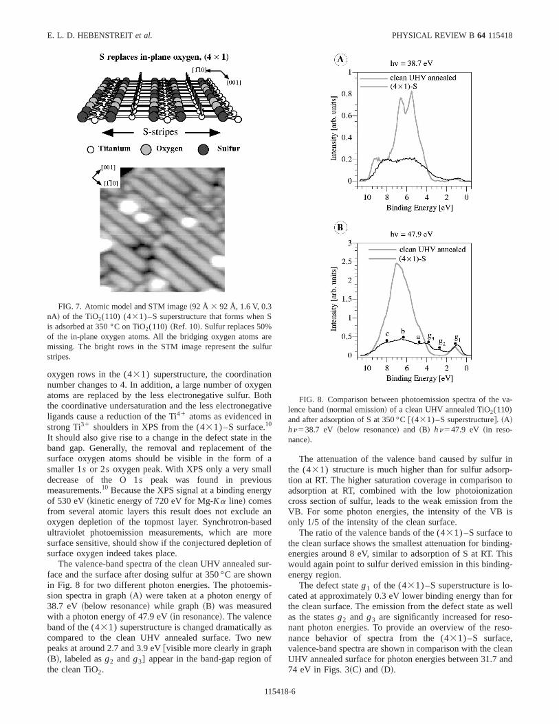
High coverages of sulfur adsorbed at 350 °C~saturationcoverage approximately 1 ML! lead to the formation of a(431) superstructure. A model has been proposed inprevious work,10 which is consistent with the STM, x-rayphotoelectron spectroscopy~XPS!, and LEED results. AnSTM image of the superstructure and the model is shownFig. 7. The bridging oxygen atoms are removed and 50%the in-plane oxygen atoms are replaced by sulfur. Detaexplanations of this model can be found in Ref. 10. In tfollowing, some aspects of this model are investigated wsurface sensitive photoemission spectra.
The removal of the bridging oxygen atoms induceschange in the coordination number of the neighboringatoms as compared to the clean surface. When a singlegen atom is missing from the clean surface the coordinanumber of the Ti atoms underneath the vacancy chanfrom 6 to 5. For Ti atoms underneath the former bridgi
gm-er
nd
FIG. 6. Ratio of peak heights in resonance to peak heightslow resonance (hn between 34 and 39 eV! for features A–E~seeFig. 5!, the defect state g1 of the clean surface~Fig. 10! and the(431) –and S superstructure@see Figs. 3~D! and 9#.
8-5
ngeo
atn
thth
fllsg
aseon
suowisof
ae
f
r inp-tontheis
ing-hisg-
-forwell-so-
leanand
S
arlfu
va-
E. L. D. HEBENSTREITet al. PHYSICAL REVIEW B 64 115418
oxygen rows in the (431) superstructure, the coordinationumber changes to 4. In addition, a large number of oxyatoms are replaced by the less electronegative sulfur. Bthe coordinative undersaturation and the less electronegligands cause a reduction of the Ti41 atoms as evidenced istrong Ti31 shoulders in XPS from the (431) –S surface.10
It should also give rise to a change in the defect state inband gap. Generally, the removal and replacement ofsurface oxygen atoms should be visible in the form osmaller 1s or 2s oxygen peak. With XPS only a very smadecrease of the O 1s peak was found in previoumeasurements.10 Because the XPS signal at a binding enerof 530 eV~kinetic energy of 720 eV for Mg-Ka line! comesfrom several atomic layers this result does not excludeoxygen depletion of the topmost layer. Synchrotron-baultraviolet photoemission measurements, which are msurface sensitive, should show if the conjectured depletiosurface oxygen indeed takes place.
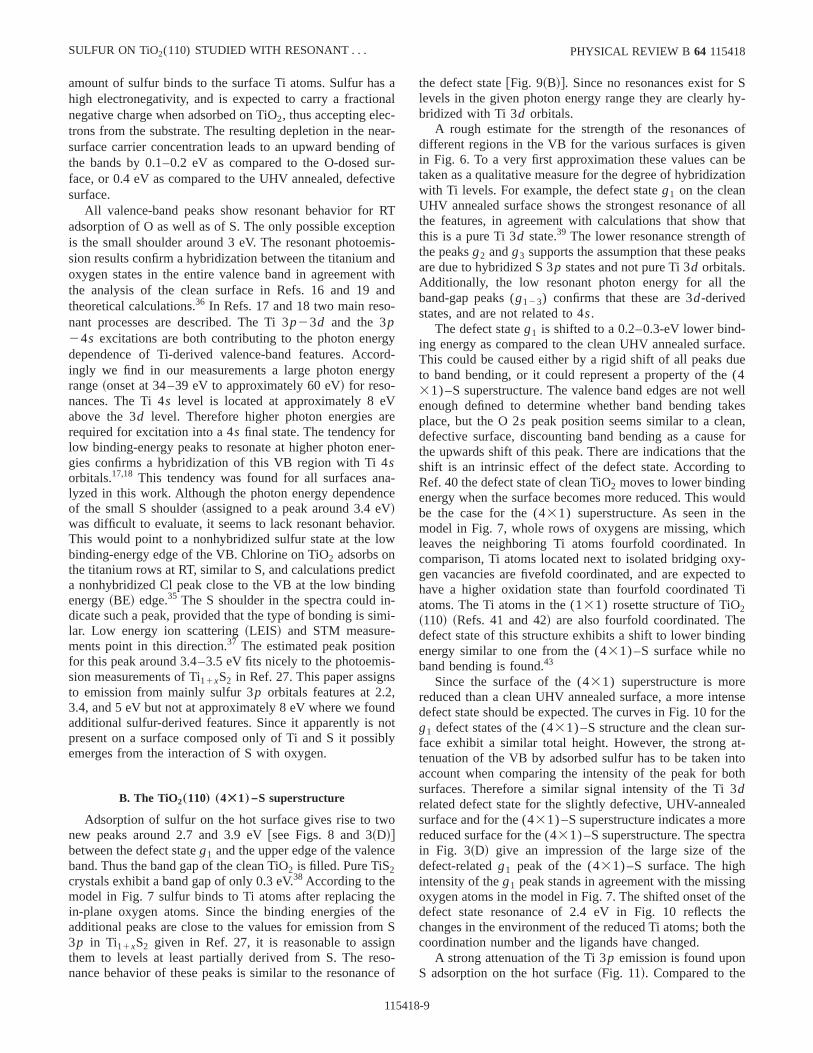
The valence-band spectra of the clean UHV annealedface and the surface after dosing sulfur at 350 °C are shin Fig. 8 for two different photon energies. The photoemsion spectra in graph~A! were taken at a photon energy38.7 eV ~below resonance! while graph~B! was measuredwith a photon energy of 47.9 eV~in resonance!. The valenceband of the (431) superstructure is changed dramaticallycompared to the clean UHV annealed surface. Two npeaks at around 2.7 and 3.9 eV@visible more clearly in graph~B!, labeled asg2 andg3] appear in the band-gap region othe clean TiO2.
FIG. 7. Atomic model and STM image~92 Å 3 92 Å, 1.6 V, 0.3nA! of the TiO2(110) (431) –S superstructure that forms whenis adsorbed at 350 °C on TiO2(110) ~Ref. 10!. Sulfur replaces 50%of the in-plane oxygen atoms. All the bridging oxygen atomsmissing. The bright rows in the STM image represent the sustripes.
11541
nthive
ee
a
y
nd
reof
r-n
-
sw
The attenuation of the valence band caused by sulfuthe (431) structure is much higher than for sulfur adsortion at RT. The higher saturation coverage in comparisonadsorption at RT, combined with the low photoionizatiocross section of sulfur, leads to the weak emission fromVB. For some photon energies, the intensity of the VBonly 1/5 of the intensity of the clean surface.
The ratio of the valence bands of the (431) –S surface tothe clean surface shows the smallest attenuation for bindenergies around 8 eV, similar to adsorption of S at RT. Twould again point to sulfur derived emission in this bindinenergy region.
The defect stateg1 of the (431) –S superstructure is located at approximately 0.3 eV lower binding energy thanthe clean surface. The emission from the defect state asas the statesg2 and g3 are significantly increased for resonant photon energies. To provide an overview of the renance behavior of spectra from the (431) –S surface,valence-band spectra are shown in comparison with the cUHV annealed surface for photon energies between 31.774 eV in Figs. 3~C! and ~D!.
er
FIG. 8. Comparison between photoemission spectra of thelence band~normal emission! of a clean UHV annealed TiO2(110)and after adsorption of S at 350 °C@(431) –S superstructure#. ~A!hn538.7 eV ~below resonance! and ~B! hn547.9 eV ~in reso-nance!.
8-6

v
ntioeV
rgthm
nfotiosrggu-
(g
oy
ieeinT
f taluddc
efarg
re-xi-
ron-is
reeseO
ov-ass of
led-fter
l
tely
dus
2.532tl
ts aof
-O
tio -
tions
SULFUR ON TiO2(110) STUDIED WITH RESONANT . . . PHYSICAL REVIEW B 64 115418
In the spectra for the clean UHV annealed surface, fipeaks were assigned at the binding energies 4.6–4.9 eV~A!,5.4–5.8 eV~B!, 6.5–6.9 eV~C!, 7.7–8.1 eV~D!, and 8.9–9.1 eV ~E!. These values are close to the peak positiowhich are found for the defect-free surface after adsorpof O at RT, except for an offset of approximately 0.1–0.2due to band bending as shown in Fig. 2. In Fig. 3~C! thepeaks A–E are labeled for the spectrum with a photon enehn538.7 eV. The resonant profiles of these peaks are rasimilar to the clean defect-free surface expect for a sowhat higher peak C.
Regardless of the photon energy, the shape of the valeband of the (431) –S surface is much less defined thanthe clean surface, and a reliable assignment of peak posias well as the determination of the total number of peaknot possible. To extract information about the photon enedependence, the intensity of the upper and lower VB edaround 4.8 and 8.1 eV, respectively, and of the center aro6.3 eV are plotted in Fig. 9~A!. The labels a–c in the spectrum athn543.4 eV in Figs. 3~D! and 8~B! mark the energypositions where these data were taken. The defect state1)and the two additional peaks (g2 , g3) within the band gapcan clearly be identified and are labeled in Fig. 8~B! as wellas in Fig. 3~D! at a photon energy of 43.4 eV. The heightsthese three peaks are plotted against the photon energFig. 9~B!. They show maxima at rather low photon energwith a similar resonance profile. In contrast, the thrvalence-band peaks follow the scheme that lower bindenergies exhibit resonances at higher photon energies.resonance strength was again determined by the ratio ohighest peak height~at resonance! and the peak height atphoton energy at the minimum in the resonance. The vaare presented in Fig. 6. The defect state and the two ational peaks in the former band gap exhibit very distinresonances with the ratio for the peakg1 being twice as highas the new peaksg2 andg3. The resonances of the valencband are not as pronounced as those of the clean surFigure 10 shows a direct comparison of the photon ene
FIG. 9. Peak heights of valence-band peaks at selected posi@see Fig. 3~D!# versus photon energy for the (431) –S superstruc-ture. ~A! Three positions in the valence band~a–c!, ~B! band-gapstates. The curves are offset for clarity.
11541
e
sn
yere-
cernsisy
esnd
fin
seghehe
esi-
t
ce.y
dependence of the defect state intensity (g1) of the(431) –S surface and the clean UHV annealed surface,spectively. The onset of the resonance differs by appromately 2.4 eV. The resonance for the clean surface is stger ~see Fig. 6! but the overall shape of the two curvesquite similar.
C. Photoemission from the shallow core levels
The influence of sulfur adsorption on the shallow colevels is examined in the overview spectra in Fig. 11. Thwere taken with a photon energy of 95 eV to monitor the2s and Ti 3p peaks. As mentioned above the saturation cerage for adsorption of S at RT is about 0.7 ML, whereadsorption on the hot surface leads to saturation coverageapproximately 1 ML. Figure 11~A! compares adsorption of Sat RT with adsorption of O at RT on the clean UHV anneasurface. In Fig. 11~B! the spectrum of the clean UHV annealed surface is plotted together with the spectrum aadsorption of S at 350 °C.
In all spectra the shape of the O 2s peak has an unusuaappearance@arrow in Fig. 11~B!#. It seems to consist of twopeaks which are separated from each other by approxima4.4 eV. The literature values for the O 2s and the O 1s peakpositions are given as 23 and 531 eV, respectively.32 TheXPS O 1s binding energies of the clean UHV annealeTiO2(110) surface was determined as 530.5 eV in previomeasurements.9 The high binding-energy part of the O 2speak of the clean UHV annealed surface is located at 2eV. The offset of 0.5 eV between the value given in Ref.and the measured value of the O 1s peak would be consistento the offset of the O 2s position. The origin of the smalpeak~in our measurements at approximately 18.1 eV! is ad-dressed in Ref. 33. The authors conclude that it represensatellite of the valence band due to inelastic scatteringphotoexcited O 2p electrons and it is not correlated to oxygen 2s derived emission. To estimate the attenuation of the
ns FIG. 10. Peak heights of theg1 defect state versus photon energy for the clean UHV annealed surface and the (431) –S super-structure in dependence of the photon energy. The peak posiare at binding energies of 1.1 and 0.8–0.9 eV, respectively.
8-7

er
thbaengao
ehnerf
-
hastaled
theA.
omectsu-hegenthisre-
tedtheeri-di-
or-entthe
r isffer-lfur.
etriclyueies
rio-is
thep
atesde-s ahis
by
issult-
-
intumu-ndby
igh
o-a
an
g
E. L. D. HEBENSTREITet al. PHYSICAL REVIEW B 64 115418
2s peaks of the sulfur covered surfaces we compared thfore only the peak heights of the O 2s feature at approxi-mately 22.5 eV.
After adsorption of S at RT~S on the Ti rows! both theoxygen and the titanium peak are slightly attenuated andpeak shapes remain the same. In agreement with thebending effects observed in the valence-band region, th2s peak shifts by approximately 0.4–0.5 eV to lower bindienergies as compared to the clean UHV annealed surfThe Ti peak exhibits a peak area of approximately 80%the value for the clean defect-free surface. The oxygen pheight is reduced to approximately 72%. If we define tO/Ti ratio before adsorption as 1, the ratio after adsorptioapproximately O/Ti5 0.9. For adsorption of sulfur on thhot surface the oxygen peak height and titanium peak aare attenuated to 42 and 69%, respectively, of the valuesthe clean UHV annealed surface~with point defects!. The
FIG. 11. Photoemission spectra of shallow core levelsTiO2(110) with hn595 eV. ~A! Comparison between oxygen adsorption at RT with sulfur adsorption on the defect-free surfaceRT. ~B! Comparison between the clean UHV annealed surfacethe (431) –S superstructure. The O 2s/Ti 3p ratio of the cleansurfaces was defined as unity. After adsorption of S at RT it chanto 0.9 and after adsorption at 350 °C to 0.6.
11541
e-
endO
ce.f
akeis
eaor
O/Ti ratio after adsorption~before adsorption it is again defined as 1! changes to 0.6. The peak shape of the Ti 3p peakhas changed and a strong low binding-energy shoulderappeared. The O 2s peak of the (431) –S structure does noappear to be shifted as compared to the clean, UHV annesurface.
IV. DISCUSSION
A. Sulfur adsorbed at RT
The photoemission data provide strong arguments foradsorption of S on point defects at RT as shown in Sec. IIIAnother possible explanation for the reduced intensity frthe defect state could be an electron transfer from the defto the S atoms on the Ti rows. However, the following argments support that the filling of the defects by sulfur is tmost probable mechanism. Sulfur replaces bridging oxyatoms when adsorbed on the hot surface indicating thatposition is an energetic favorable adsorption site. Measuments of Cl adsorption on TiO2 ~Ref. 34! exhibit many simi-larities to the adsorption of S on TiO2. Cl adsorbs at RT onthe Ti rows and replaces bridging oxygen atoms at elevatemperatures. Similar to S, Cl adsorption at RT causesdisappearance of the defect state in photoemission expments. STM images show adsorption of Cl at defects. Adtionally, ab initio calculations of Cl on TiO2 ~Ref. 35! revealthat Cl adsorbed on a defect is energetically the most favable position. As conclusion the photoemission measuremresults are pointing to S adsorption at defects, indicatinghigh reactivity of these sites.
To quench the defect state in the band gap, less sulfuneeded than molecular oxygen. This demonstrates a dience in the adsorption process between oxygen and suBecause molecular oxygen does not adsorb on stoichiomTiO2(110) at RT, vacancies are only filled when hit directby an O2 molecule. In contrast, sulfur fills the vacancies dto two different processes. Similar to oxygen the vacancare filled when directly hit by sulfur. Additionally, sulfusticks to the fivefold coordinated Ti atoms of the stoichmetric TiO2(110) surface. Sulfur bound to these Ti atomsvery mobile, as seen in STM, and diffuses mainly along@001# direction.9 If it passes an oxygen vacancy it can hointo it. The precursor-mediated process naturally acceleradsorption, and lower doses are required to fill all thefects. The chemical similarity of S and O probably causesimilar appearance in STM when adsorbed on a defect. Tinhibits a reliable detection of sulfur on a defect siteSTM.
The rigid shift of the valence band displayed in Fig. 2readily explained by charge transfer processes and the reing band bending effects inn-type semiconductors.30 Whentaking the~approximately! defect-free surface~annealed inUHV and dosed with oxygen at RT! as a reference, the spectrum with defects~before the adsorption of oxygen! is shiftedtowards higher binding energies by 0.2–0.3 eV. The podefects donate electrons to the substrate, causing an acclation layer in the near-surface region and downward babending. This effect is reversed when the defects are filledeither O or S. After dosing S at room temperature, a h
f
td
es
8-8
sna-eagstiv
Rio
nwan-
rgorrg
Vrerne
ac
Viow
icngn-im-niss,nnoib
o
ce
heth
nsoe
Shy-
ofenbetion
f allthatf
aks
e
-ace.ue(4wellkesn,
fortheto
ouldechIn
xy-d toTi
eng
nsether-at-ntooth
ledrerae
ngthethethe
SULFUR ON TiO2(110) STUDIED WITH RESONANT . . . PHYSICAL REVIEW B 64 115418
amount of sulfur binds to the surface Ti atoms. Sulfur hahigh electronegativity, and is expected to carry a fractionegative charge when adsorbed on TiO2, thus accepting electrons from the substrate. The resulting depletion in the nsurface carrier concentration leads to an upward bendinthe bands by 0.1–0.2 eV as compared to the O-dosedface, or 0.4 eV as compared to the UHV annealed, defecsurface.
All valence-band peaks show resonant behavior foradsorption of O as well as of S. The only possible exceptis the small shoulder around 3 eV. The resonant photoemsion results confirm a hybridization between the titanium aoxygen states in the entire valence band in agreementthe analysis of the clean surface in Refs. 16 and 19theoretical calculations.36 In Refs. 17 and 18 two main resonant processes are described. The Ti 3p23d and the 3p24s excitations are both contributing to the photon enedependence of Ti-derived valence-band features. Accingly we find in our measurements a large photon enerange~onset at 34–39 eV to approximately 60 eV! for reso-nances. The Ti 4s level is located at approximately 8 eabove the 3d level. Therefore higher photon energies arequired for excitation into a 4s final state. The tendency folow binding-energy peaks to resonate at higher photon egies confirms a hybridization of this VB region with Ti 4sorbitals.17,18 This tendency was found for all surfaces anlyzed in this work. Although the photon energy dependenof the small S shoulder~assigned to a peak around 3.4 e!was difficult to evaluate, it seems to lack resonant behavThis would point to a nonhybridized sulfur state at the lobinding-energy edge of the VB. Chlorine on TiO2 adsorbs onthe titanium rows at RT, similar to S, and calculations preda nonhybridized Cl peak close to the VB at the low bindienergy~BE! edge.35 The S shoulder in the spectra could idicate such a peak, provided that the type of bonding is slar. Low energy ion scattering~LEIS! and STM measurements point in this direction.37 The estimated peak positiofor this peak around 3.4–3.5 eV fits nicely to the photoemsion measurements of Ti11xS2 in Ref. 27. This paper assignto emission from mainly sulfur 3p orbitals features at 2.23.4, and 5 eV but not at approximately 8 eV where we fouadditional sulfur-derived features. Since it apparently ispresent on a surface composed only of Ti and S it possemerges from the interaction of S with oxygen.
B. The TiO2„110… „4Ã1…–S superstructure
Adsorption of sulfur on the hot surface gives rise to twnew peaks around 2.7 and 3.9 eV@see Figs. 8 and 3~D!#between the defect stateg1 and the upper edge of the valenband. Thus the band gap of the clean TiO2 is filled. Pure TiS2crystals exhibit a band gap of only 0.3 eV.38 According to themodel in Fig. 7 sulfur binds to Ti atoms after replacing tin-plane oxygen atoms. Since the binding energies ofadditional peaks are close to the values for emission from3p in Ti11xS2 given in Ref. 27, it is reasonable to assigthem to levels at least partially derived from S. The renance behavior of these peaks is similar to the resonanc
11541
al
r-ofur-e
Tnis-dithd
yd-y
r-
-e
r.
t
i-
-
dtly
eS
-of
the defect state@Fig. 9~B!#. Since no resonances exist forlevels in the given photon energy range they are clearlybridized with Ti 3d orbitals.
A rough estimate for the strength of the resonancesdifferent regions in the VB for the various surfaces is givin Fig. 6. To a very first approximation these values cantaken as a qualitative measure for the degree of hybridizawith Ti levels. For example, the defect stateg1 on the cleanUHV annealed surface shows the strongest resonance othe features, in agreement with calculations that showthis is a pure Ti 3d state.39 The lower resonance strength othe peaksg2 andg3 supports the assumption that these peare due to hybridized S 3p states and not pure Ti 3d orbitals.Additionally, the low resonant photon energy for all thband-gap peaks (g123) confirms that these are 3d-derivedstates, and are not related to 4s.
The defect stateg1 is shifted to a 0.2–0.3-eV lower binding energy as compared to the clean UHV annealed surfThis could be caused either by a rigid shift of all peaks dto band bending, or it could represent a property of the31) –S superstructure. The valence band edges are notenough defined to determine whether band bending taplace, but the O 2s peak position seems similar to a cleadefective surface, discounting band bending as a causethe upwards shift of this peak. There are indications thatshift is an intrinsic effect of the defect state. AccordingRef. 40 the defect state of clean TiO2 moves to lower bindingenergy when the surface becomes more reduced. This wbe the case for the (431) superstructure. As seen in thmodel in Fig. 7, whole rows of oxygens are missing, whileaves the neighboring Ti atoms fourfold coordinated.comparison, Ti atoms located next to isolated bridging ogen vacancies are fivefold coordinated, and are expectehave a higher oxidation state than fourfold coordinatedatoms. The Ti atoms in the (131) rosette structure of TiO2~110! ~Refs. 41 and 42! are also fourfold coordinated. Thdefect state of this structure exhibits a shift to lower bindienergy similar to one from the (431) –S surface while noband bending is found.43
Since the surface of the (431) superstructure is morereduced than a clean UHV annealed surface, a more intedefect state should be expected. The curves in Fig. 10 forg1 defect states of the (431) –S structure and the clean suface exhibit a similar total height. However, the strongtenuation of the VB by adsorbed sulfur has to be taken iaccount when comparing the intensity of the peak for bsurfaces. Therefore a similar signal intensity of the Ti 3drelated defect state for the slightly defective, UHV-anneasurface and for the (431) –S superstructure indicates a moreduced surface for the (431) –S superstructure. The spectin Fig. 3~D! give an impression of the large size of thdefect-relatedg1 peak of the (431) –S surface. The highintensity of theg1 peak stands in agreement with the missioxygen atoms in the model in Fig. 7. The shifted onset ofdefect state resonance of 2.4 eV in Fig. 10 reflectschanges in the environment of the reduced Ti atoms; bothcoordination number and the ligands have changed.
A strong attenuation of the Ti 3p emission is found uponS adsorption on the hot surface~Fig. 11!. Compared to the
8-9

ts
itio
oa7
ek
hn
m
o
ts
at
-sigaiteth
nbe
ure
a
ceherpse
va-ap-amegen
onpegen
dtednede
reddingen-his
gy
ofed
at
hichi-heza-n-it isighfurefur.anddi-
-in
E. L. D. HEBENSTREITet al. PHYSICAL REVIEW B 64 115418
clean surface, the Ti 3p peak area is reduced to 69% of ioriginal value. This needs to be compared to a reduction80% for adsorption at RT. The stronger attenuation isagreement with the higher sulfur coverage for hot adsorp~1 ML as compared to 0.7 ML!. In addition to the sulfurincluded in the (431) –S superstructure, a certain amountunstructured, excess sulfur is usually present on a saturhot surface~see the bright spots in the STM image in Fig.!and further decreases the ‘‘nonsulfur’’ emission.
The O 2s peak decreases to 42% of its original valumuch more than the Ti 3p feature. Furthermore, the peaposition of the S 2p peak in previous XPS measurements10
clearly indicates sulfur bonds to Ti and not to oxygen. Tstrong decrease in the O/Ti ratio~defined as 1 for the cleasurface! to 0.6 @for the (431) –S superstructure# indicates adecrease in the concentration of surface oxygen, in confirtion of the (431) –S model as presented in Fig. 7~B!.
The strong Ti shoulder at the low binding-energy sidethe Ti 3p peak in Fig. 11~B! indicates the presence of Ti31
cations. This is in agreement with previous XPS resul10
which showed a strong Ti312p3/2 signal, contributing up to15.6% to the total Ti 2p3/2 peak area.
The strength of the resonances of the main valence-bpeaks are clearly decreased after adsorption of sulfur onhot surface~see Fig. 6!. This could point to a reduced hybridization of the O 2p and Ti orbitals, as fewer Ti atomhave O neighbors. It is not clear why the comparatively hemission at 8 eV@peak C in Fig. 6#, which was assigned tocontribution of sulfur, exhibits a weak resonance. Definconclusions cannot be drawn easily because the strengthe resonance is affected by several parameters.15 Detailedcalculations would be necessary to understand the origithe emission at 8 eV in combination with the resonancehavior.
V. SUMMARY
Normal emission photoemission spectroscopy measments with photon energies between 29 and 95 eV wanalyzed for five differently prepared TiO2(110) surfaces:the clean UHV annealed surface with surface oxygen vaccies, after adsorption of O2 at RT on this surface~which fillsthe vacancies!, after adsorption of sulfur at RT on the surfawith vacancies, after adsorption of sulfur at RT without tvacancies, and after adsorption of sulfur at 350 °C. Adsotion of S and O at RT on the surface with vacancies cauan upward bending of the bands by 0.4 and 0.2 eV, resptively.
ys
11541
tonn
fted
,
e
a-
f
ndhe
h
of
of-
e-re
n-
-esc-
When oxygen adsorbs at RT at the bridging oxygencancies the defect state within the band gap totally dispears as expected. Adsorption of S at RT leads to the sresult. This indicates that sulfur occupies the reactive oxyvacancies on the surface, in addition to the adsorption sitefivefold coordinated Ti sites identified with STM. The shaof the valence band is the same when either element, oxyor sulfur, is occupying the oxygen defects.
After adsorption of S at RT emission from sulfur-derivestates was found at approximately 3.4 and 8 eV. As indicaby resonance effects when the photon energy is scanacross the Ti 3p ioniziation threshold, the whole valencband~except for the sulfur-related peak at 3.4 eV! is hybrid-ized with Ti states. On both the clean and the S-covesurface, the resonant photon energy depends on the binenergy of valence-band features: the higher the bindingergy the higher is the photon energy for resonances. Tmay indicate a hybridization of the lower binding-enerpart of the VB with mainly Ti 4s orbitals and of the higherbinding-energy part with Ti 3d orbitals.
Generally, the valence-band emission after adsorptionsulfur at RT or 350 °C is strongly attenuated. This is causby the low photoioniziation cross section of the S 3p orbitalswhich, in free atoms, is ten times smaller than for oxygenthe photon energies used in this study.
Sulfur adsorption on the hot surface results in a (431)superstructure. Peaks around 2.7 and 3.9 eV are found wcan be assigned to sulfur 3p orbitals. These resonate at simlar photon energies and exhibit a similar profile as tvacancy-derived defect state, indicating a strong hybridition with Ti 3d orbitals. The defect state is strongly ehanced and shifts to lower binding energies by 0.3 eV, asexpected for a more reduced surface. Again a relatively hintensity is found around 8 eV which is assigned to sullevels. The oxygen 2s peak is strongly reduced due to thremoval and partial replacement of surface oxygen by sulThe strength of the resonances for the main valence bafter sulfur adsorption are clearly decreased, possibly incating less hybridization with O 2p orbitals. The results support the model for the (431) –S superstructure proposedRef. 10 and depicted in Fig. 7.
ACKNOWLEDGMENTS
This work was supported by NSF-CAREER~Tulane!, theLouisiana Board of Regents~UNO!, and U.S. DOE~No. DE-FG02-98ER45712! ~CAMD!.
L.
R.
ci.
1J. A. Rodriguez, S. Chaturvedi, M. Kuhn, and J. Hrbek, J. PhChem. B102, 5511~1998!.
2J. A. Rodriguez and J. Hrbek, Acc. Chem. Res.32, 719 ~1999!.3K. E. Smith and V. E. Henrich, Phys. Rev. B32, 5384~1985!.4K. E. Smith, J. L. Mackay, and V. E. Henrich, Phys. Rev. B35,
5822 ~1987!.5K. E. Smith and V. E. Henrich, Surf. Sci. Lett.217, L445 ~1989!.
. 6K. E. Smith and V. E. Henrich, J. Vac. Sci. Technol. A7, 1967~1989!.
7D. R. Warburton, D. Purdie, C. A. Muryn, K. Prabhakaran, P.Wincott, and G. Thornton, Surf. Sci.269Õ270, 305 ~1992!.
8H. Raza, S. P. Harte, C. A. Muryn, P. L. Wincott, G. Thornton,Casanova, and A. Rodriguez, Surf. Sci.366, 519 ~1996!.
9E. L. D. Hebenstreit, W. Hebenstreit, and U. Diebold, Surf. S
8-10

ci
lli,
iti,
. L,
. L
E
Ays
ev
i,
a-
rf.
nd
s
n,
N,
e,
U.
nd
e,
ett.
.
n-
SULFUR ON TiO2(110) STUDIED WITH RESONANT . . . PHYSICAL REVIEW B 64 115418
461, 87 ~2000!.10E. L. D. Hebenstreit, W. Hebenstreit, and U. Diebold, Surf. S
470, 347 ~2001!.11U. Diebold, J. Lehman, T. Mahmoud, M. Kuhn, G. Leonarde
W. Hebenstreit, M. Schmid, and P. Varga, Surf. Sci.411, 137~1998!.
12J. A. Rodriguez, J. Hrbek, J. Dvorak, T. Jirsak, and A. MaChem. Phys. Lett.336, 377 ~2001!.
13P. J. Hardman, G. N. Raikar, C. A. Muryn, G. van der Laan, PWincott, G. Thornton, D. W. Bullett, and P. A. D. M. A. DalePhys. Rev. B49, 7170~1994!.
14G. N. Raikar, P. J. Hardman, C. A. Muryn, G. van der Laan, PWincott, and G. Thornton, Solid State Commun.80, 423~1991!.
15L. C. Davis, J. Appl. Phys.59, R25 ~1985!.16E. Bertel, R. Stockbauer, R. L. Kurtz, T. E. Madey, and D.
Ramaker, Surf. Sci.152Õ153, 776 ~1985!.17R. Heise, R. Courths, and S. Witzel, Solid State Commun.84, 599
~1992!.18Z. Zhang, S.-P. Jeng, and V. E. Henrich, Phys. Rev. B43, 12 004
~1991!.19J. Nerlov, Q. Ge, and P. J. Mo” ller, Surf. Sci.348, 28 ~1996!.20K. C. Prince, V. R. Dhanak, P. Finetti, J. F. Walsh, R. Davis, C.
Muryn, H. S. Dhariwal, G. Thornton, and G. van der Laan, PhRev. B55, 9520~1997!.
21U. Diebold, H.-S. Tao, N. D. Shinn, and T. E. Madey, Phys. RB 50, 14 474~1994!.
22S. Fischer, F. Schneider, and K. D. Schierbaum, Vacuum47, 1149~1996!.
23S. Fischer, J. A. Martı´n-Gago, E. Roma´n, K. D. Schierbaum, andJ. L. de Segovia, J. Electron Spectrosc. Relat. Phenom.83, 217~1997!.
24P. J. Mo” ller, Z. S. Li, T. Egebjerg, M. Sambi, and G. GranozzSurf. Sci.402-404, 719 ~1998!.
25J. Biener, M. Ba¨umer, and R. J. Madix, Surf. Sci.432, 178~1999!.
26K. E. Smith and V. E. Henrich, Phys. Rev. B38, 9571~1988!.
11541
.
.
.
.
..
.
27Y. Ueda, H. Negishi, M. Koyano, M. Inoue, K. Soda, H. Sakmoto, and S. Suga, Solid State Commun.57, 839 ~1986!.
28W. Heegemann, K. H. Meister, E. Bechtold, and K. Hayek, SuSci. 49, 161 ~1975!.
29M. A. Henderson, W. S. Epling, C. L. Perkins, C. H. F. Peden, aU. Diebold, J. Phys. Chem. B103, 5328~1999!.
30V. E. Henrich and P. A. Cox,The Surface Science of Metal Oxide~Cambridge University Press, Cambridge, England, 1994!.
31J. J. Yeh and I. Lindau, At. Data Nucl. Data Tables32, 1 ~1985!.32J. F. Moulder, W. F. Stickle, P. E. Sobol, and K. D. Bombe
Handbook of X-ray Photoelectron Spectroscopy~Perkin-ElmerCorporation, Physical Electronics Division, Eden Prairie, M1992!.
33K. W. Goodman and V. E. Henrich, Phys. Rev. B50, 10 450~1994!.
34E. L. D. Hebenstreit, W. Hebenstreit, H. Geisler, C. A. VentricJr., and P. T. Sprunger, Surf. Sci.486, L467 ~2001!.
35D. Vogtenhuber, R. Podloucky, and J. Redinger, Surf. Sci.454-456, 369 ~2000!.
36C. Sousa and F. Illas, Phys. Rev. B50, 13 974~1994!.37W. Hebenstreit, E. L. D. Hebenstreit, D. Vogtenhuber, and
Diebold ~unpublished!.38C. H. Chen, W. Fabian, F. C. Brown, K. C. Woo, B. Davies, a
B. DeLong, Phys. Rev. B21, 615 ~1980!.39P. J. D. Lindan, N. M. Harrison, M. J. Gillan, and J. A. Whit
Phys. Rev. B55, 15 919~1997!.40V. E. Henrich, G. Dresselhaus, and H. J. Zeiger, Phys. Rev. L
36, 1335~1976!.41M. Li, W. Hebenstreit, and U. Diebold, Surf. Sci. Lett.414, L951
~1998!.42M. Li, W. Hebenstreit, L. Gross, U. Diebold, M. A. Henderson, D
R. Jennison, P. A. Schultz, and M. P. Sears, Surf. Sci.437, 173~1999!.
43H. Geisler, C. A. Ventrice, Jr., W. Hebenstreit, E. L. D. Hebestreit, P. T. Sprunger, and U. Diebold~unpublished!.
8-11
Copyright © 2022 FDOKUMEN