Conductive electrospun PANi-PEO/TiO 2 fibrous membrane for photo catalysis
Adsorption and Solar Light Decomposition of Acetone on Anatase TiO2 and NiobiumDoped TiO2 Thin Films
Andreas Mattsson,† Michael Leideborg,‡ Karin Larsson,‡ Gunnar Westin,‡ andLars O2 sterlund* ,†
Department of EnVironment and Protection, FOI NBC Defence, SE-901 82 Umeå, Sweden, and Department ofMaterials Chemistry, Ångstro¨m Laboratory, Uppsala UniVersity, SE-751 21 Uppsala, Sweden
ReceiVed: October 4, 2005; In Final Form: NoVember 3, 2005
Adsorption and solar light decomposition of acetone was studied on nanostructured anatase TiO2 and Nb-doped TiO2 films made by sol-gel methods (10 and 20 mol % NbO2.5). A detailed characterization of thefilm materials show that films contain only nanoparticles with the anatase modification with pentavalent Nboxide dissolved into the anatase structure, which is interpreted as formation of substituted NbdO clusters inthe anatase lattice. The Nb-doped films displayed a slight yellow color and an enhanced the visible lightabsorption with a red-shift of the optical absorption edge from 394 nm for the pure TiO2 film to 411 nm for20 mol % NbO2.5. In-situ Fourier transform infrared (FTIR) transmission spectroscopy shows that acetoneadsorbs associatively withη1-coordination to the surface cations on all films. On Nb-doped TiO2 films, thecarbonyl bonding to the surface is stabilized, which is evidenced by a lowering of theν(CdO) frequency byabout 20 cm-1 to 1672 cm-1. Upon solar light illumination acetone is readily decomposed on TiO2, andstable surface coordinated intermediates are formed. The decomposition rate is an order of magnitude smalleron the Nb-doped films despite an enhanced visible light absorption in these materials. The quantum yield isdetermined to be 0.053, 0.004 and 0.002 for the pure, 10% Nb:TiO2, and 20%Nb:TiO2, respectively. Usingan interplay between FTIR and DFT calculations we show that the key surface intermediates are bidentatebridged formate and carbonate, and H-bonded bicarbonate, respectively, whose concentration on the surfacecan be correlated with their heats of formation and bond strength to coordinatively unsaturated surface Tiand Nb atoms at the surface. The oxidation rate of these intermediates is substantially slower than the initialacetone decomposition rate, and limits the total oxidation rate at t>7 min on TiO2, while no decrease of therate is observed on the Nb-doped films. The rate of degradation of key surface intermediates is different onpure TiO2 and Nb-doped TiO2, but cannot explain the overall lower total oxidation rate for the Nb-dopedfilms. Instead the inferior photocatalytic activity in Nb-doped TiO2 is attributed to an enhanced electron-hole pair recombination rate due to NbdO cluster and cation vacancy formation.
1. Introduction
Titanium dioxide photocatalysis has for the past three decadesbeen a promising technology for air and water remediation.1-4
This field is now maturing into a viable commercial technology,where a broadened application area includes coating technologyand maintenance-free surfaces in general. The wide band gapof TiO2 (3.2 and 3.0 eV for anatase and rutile, respectively)necessitates, however, UV light to excite electrons over the bandgap. In many applications, including those utilizing solar lightas the energy source, it is desirable to exploit a larger portionof the spectrum, i.e., to extend the band gap excitations intothe visible regime. A second generation of efficient UV-visibleactive photocatalytic materials are therefore currently soughtwhere doped TiO2 has received considerable attention.5 Severalmethods and materials have been employed, including aniondoping by replacing lattice oxygen with N,6,7 C,8,9 F,10,11or S,12
and cation doping, whereby Ti cations are substituted with, e.g.,V, Cr, Mn, Fe, and Ni atoms.5 Other candidates for visible activematerials are binary oxides5,13and perovskites.14 It is a delicate
matter to introduce dopants into TiO2 to lower the optical bandgap energy, while at the same time avoiding creation ofunwanted recombination centers or localized band gap states,which prevent efficient separation of excited electron-holepairs. Furthermore, a slight lowering of the conduction bandmay place the conduction band edge below the O2 affinity level(O2/O2
-), thus jeopardizing the formation of oxygen radicals,which are key intermediates in photocatalytic oxidation (PCO)reactions.3
In the present paper, we explore the potential of using Nbdoped TiO2 as a visible active photocatalytic material. Niobiumoxide and, in particular, its most stable form, Nb2O5, is aninteresting semiconductor with a band gap of about 3.9 eV(decreasing to about 3.5 eV in the amorphous state15), with ahigh dielectric constant and a high index of refraction, whichhas found important applications in electronics and opticalapplications, including thin films (antireflective coatings, solarcontrol, etc.16). In a recent report,17 it was shown that Nb dopedanatase yields films with high electron conductivity, which areinteresting in, e.g., photoelectrochemical systems. In the presentpaper, we present results for sol-gel synthesized Nb dopedanatase films, which we previously also have shown to have
* Corresponding author. Email: [email protected]. Tel:+46 90106900. Fax:+46 90 106802.
† FOI NBC Defence.‡ Uppsala University.
1210 J. Phys. Chem. B2006,110,1210-1220
10.1021/jp055656z CCC: $33.50 © 2006 American Chemical SocietyPublished on Web 12/27/2005
high conductivity, and explore their use as a potential photo-catalyst material.
Thermal and photochemical reactions with acetone have beensubjected to a large number of studies, because of its importanceas a reactant, intermediate, and product in many fine chemicalor catalytic oxidation reactions, as well as being a common airpollutant.2,3,18-21 Our results show that Nb doped TiO2 withfairly high doping concentrations (10-20 mol %) is a poor solaractive photocatalyst material compared to TiO2, despite the factthat Nb doping extends the optical absorption into the visibleand utilizes a larger portion of the sunlight. Furthermore, wediscuss in detail the importance of cation coordination andbonding of intermediate reaction products for the total photo-catalytic oxidation rate using an interplay between FTIRmeasurements and density functional theory (DFT) calculations.Even though our results are negative in the sense that thequantum yield for acetone degradation is lowered upon Nbdoping, we believe that our results are instructive and point tointrinsic difficulties that must be solved when the cation dopingroute is used to improve the photocatalytic performance of TiO2.
2. Experimental Section
2.1. Material Synthesis.Alkoxides and ammonia were usedas purchased (Aldrich), while the 99.5% ethanol was dried bydistillation over CaH2. The alkoxide precursors were weighedand mixed in an Ar-filled glovebox (Braun MB 200). Ti(OPri)4
was mixed with Nb(OEt)5 to the desired proportions under inertatmosphere, and ethanol was added to provide 1 M solutions.After heating at 60°C for 1 h, the solutions were cooled toambient temperature, and concentrated ammonia providing 5H2O per alkoxo group was added under vigorous stirring. Thematerial was subsequently heated at 80°C for 8 h andultrasonically dispersed before it was put in an autoclave forhydrothermal treatment at 200°C for 15 h. After cooling, theautoclaved material was again ultrasonically treated and con-centrated under vacuum, and carbowax was added to yield awhite paste. Before use, the paste was diluted with water to adry content of ca. 0.7 wt %. The solution of the nondoped titaniawas also made acidic by the addition of a small amount of aceticacid.
Diluted pastes of 0, 10, 15, and 20 mol % NbO2.5 in TiO2
were deposited as thin films on either Si(100) wafers (10× 10× 0.25 mm3) or R-Al2O3 substrates (25× 1 mm2) byevaporating the 0.7 wt % sol in air and subsequently heat-treatedin air at 450°C for 30 min.
2.2. Materials Characterization.The structural, optical, andchemical properties of the synthesized materials were character-ized by a range of different techniques. Scanning electronmicroscope (SEM) images were obtained with a FEG-SEM Leo1550 Gemini instrument. Transmission electron microscopy(TEM) was done with a JEOL 2000 FXII instrument equippedwith an EDS module (Link AN 1000). Reflectance micro-Ramanspectra of samples heat-treated in air for 30 min at 723 K wererecorded with a Renishaw 2000 spectrometer using a 783 nmlaser diode light source. Grazing incidence X-ray diffractograms(GI-XRD) were obtained for samples heat-treated at 723 K witha Siemens D-5000 instrument. After phase identification of thematerials, the XRD peak positions and (hkl) values were usedas input to extract the lattice parameters with the softwareFullprof.22 Room-temperature UV-vis spectra were measuredin transmission mode with a Perkin-Elmer Lambda 19 instru-ment on thin films deposited onR-Al2O3 windows. Thedeposition of films was made in the same way as those depositedon the silicon wafers for FTIR analysis. The absorbance in the
film, A (whereA ) -log(T)), was measured using a noncoatedR-Al2O3 window as a transmission reference, viz.,Afilm )-log(10-AAl203+film+AAl203) ) AAl203+film - AAl203. Finally, X-rayphotoelectron spectroscopy (XPS) measurements were madewith a Kratos Axis Ultra electron spectrometer using a monoAl K R source operated at 225 W and a low-energy electron gunfor charge compensation. High-resolution scans were acquiredusing a pass energy of 20 eV and an energy resolution of 0.1eV. The hybrid lens mode was used for all measurements. Thebinding energy scale was calibrated against the C 1s line ofaliphatic carbon, set at 285.0 eV. Atomic concentrations (atom%) were derived from photoelectron peak areas using Shirleyor linear background subtraction, the instrumental sensitivityfactors, and a transmission function appropriate to the acquisitionconditions.
2.3. Reactivity Measurements.Fourier transform infrared(FTIR) transmission measurements were made in a vacuum-pumped spectrometer (Bruker IFS 66v/S) equipped with atransmission reaction cell, which allowed for simultaneous UVand IR illumination of the sample. The IR light was collectedwith an LN2 cooled narrow-band HgCdTe detector. RepeatedFTIR spectra were measured with 4 cm-1 resolution and 135scans (corresponding to 30 s measurement time per spectra)and 30 s dwell time between consecutive spectra. All spectrawere smoothed with a Savitzky-Golay algorithm using a9-point window. Deconvolution of FTIR peaks was performedby fitting the experimental curves with Gauss-Lorentziancurves, after appropriate baseline corrections of the spectra.
The FTIR spectra reported here were all acquired with thesamples kept at 299 K in a 100 mL/min feed flow of syntheticair (20% O2 and 80% N2) through the reaction cell. The feedflow was controlled by a set of mass flow controllers, and thegas purity was 99.994% (N2) and 99.999% (O2). The gas wasused as delivered without further purification. Prior to eachmeasurement, the samples were cleaned at 673 K for 15 min insynthetic air and subsequently cooled to 299 K in the same feed.The TiO2 samples are thus pre-oxidized.
Acetone (analytical grade, Scharlau) was added to the feedgas through a home-built gas generator, consisting of a capillarytube in contact with a liquid acetone reservoir maintained atconstant temperature (T ) 303 ( 0.2 K) controlled by a PIDregulator. The independently calibrated acetone injection in thefeed obtained with these parameters was 0.29( 0.02 mg/min,which corresponds to an ideal steady-state concentration of 1232( 109 ppm in the feed. No other gas species than acetone weredetected mass-spectrometrically with this gas evaporation setup.In each experiment, the sample was exposed to 15 min ofacetone. After acetone dosing, the sample was left for 20 minin the pure feed gas before the solar light illumination wasstarted. In all measurements, the FTIR background was collectedon a clean sample during 1 min (265 scans) in a synthetic airfeed at 299 K.
2.4. Solar Light Illumination. Simulated solar light wasgenerated by a Xe arc lamp source operated at 200 W and a setof filters (AM1.5). To reduce the infrared part of the emissionspectrum, the light was first directed through a 75-mm-longwater filter. The light was collected into a fused silica fiberbundle and directed onto the sample cell through a CaF2 windowat an angle of 25° to the surface normal. The measured photonpower after the fiber bundle was 173.5 mW/cm2 betweenλ )200 and 800 nm as measured with a calibrated thermopiledetector. Taking into account the absorption efficiency of thedetector (96%) and the UV transmission through the cell window(92%), we determined the photon power on the sample to be
Adsorption and Solar Light Decomposition of Acetone J. Phys. Chem. B, Vol. 110, No. 3, 20061211
166 mW/cm2. The measured irradiance spectrum through thefilter combination and fiber optics is given in Figure 2 (rightordinate), together with the corresponding AM1.5 standard solarirradiance spectrum.23
2.5. DFT Calculations. To gain further insight into theelectronic and structural properties of the key surface species,we have performed density functional theory (DFT) calculationunder periodic boundary conditions (using the program packageCASTEP from Accelrys, Inc.). Exchange and correlation effectshave been included within the generalized gradient approxima-tion (GGA) developed by Perdew and Wang.24 The atoms arerepresented by ultrasoft nonlocal pseudopotentials in the Klein-man-Bylander fully separable form.25 The electronic wavefunctions were expanded in terms of plane-waves, and theelectronic minimization was performed using a band-by-bandconjugate-gradients minimization technique.26 The specifick-points were generated by the Monkhorst-Pack scheme, whichproduces a uniform mesh ofk-points in the reciprocal space.27
The adsorbate and the upper two atomic layers of the surfacewere allowed to fully relax in all of the calculations using theBFGS algorithm (Broyden-Fletcher-Goldfarb-Sharmo).28 Therest of the atoms were kept fixed in order to hold thecharacteristics of the crystal lattice. Moreover, the covalent bond
strengths within the bonds between the adsorbate and thesurface, as well as within the adsorbate as a function ofadsorption, were estimated by calculating the electron bondpopulations by projecting the plane-wave states onto thelocalized basis set by means of Mulliken electron populationanalysis.29
3. Results and Discussions
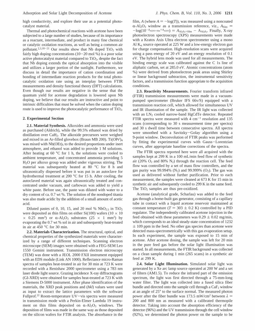
3.1. Material Characterization. In Figure 1 is shown SEMimages of the 10% Nb doped films. Analysis of the SEM imagesshowed that the pure films were ca. 2.5µm thick consisting ofca. 20 nm spherical-like particles with crystallite edges clearlyresolvable for pure TiO2.
The Nb doped TiO2 films consist of slightly larger, elongated(“rectangular”) structures. From the weight and the volumederived from the deposited area and film thickness, an estimatedporosity of ca. 47% was obtained. A rough estimation of thesurface area (assuming spherical particles) gives a theoreticalsurface area of ca. 62 m2 g-1. The estimated geometrical surfacearea decreases somewhat with increased Nb content, which isin qualitative agreement with the change of the particle shapes.TEM measurements give particle sizes of 15-30 nm for pureanatase TiO2 and 15-70 nm for 10% NbO2.5:TiO2. Mostlyspherical TiO2 particles are found in TEM, while 10% and 20%Nb doped TiO2 contain both elongated and spherical particles.The average particle sizes are compiled in Table 1.
The thickness determined from SEM is in good agreementwith independent measurements of the measured interferencepattern due to reflections at the film interface in transmissionFTIR, which yieldsd ) 2.1, 1.6, and 1.9µm for TiO2, 10%Nb:TiO2, and 20% Nb:TiO2, respectively, as obtained from theequation
where N ) number of interference maximum (or minima)
Figure 1. SEM images of the nanostructured anatase TiO2 film doped with 10%NbO2.5: Cross-sectional view showing the films thickness (left),and top-view showing the elongated particle structure (right).
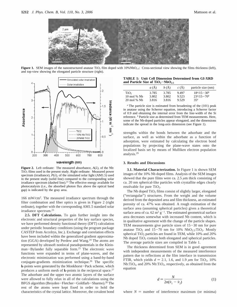
Figure 2. Left ordinate: The measured absorbance,A(λ), of the Nb:TiO2 films used in the present study. Right ordinate: Measured powerspectrum (irradiance),P(λ), of the simulated solar light (AM1.5) usedin the present study (solid line) compared to the corresponding solarirradiance spectrum (dashed line).23 The effective energy available forphotocatalysis (i.e., the absorbed photon flux above the optical bandgap) is indicated by the gray area.
TABLE 1: Unit Cell Dimension Determined from GI-XRDand Particle Size of TiO2-NbO2.5
a (Å) b (Å) c (Å) particle size (nm)
TiO2 3.785 3.785 9.497 18a/15-30b
10 mol % Nb 3.802 3.802 9.523 23a/15-70b
20 mol % Nb 3.816 3.816 9.529 35a
a The particle size is estimated from broadening of the (101) peakin anatase using the Scherrer equation, introducing a Scherrer factorof 0.9 and obtaining the internal error from the line-width of the Sireference.b Particle size as determined from TEM measurements. Here,some of the Nb-doped particles appear elongated, and the dimensionsindicate the spread in the long-axis dimension (see Figure 1).
d ) N2n(νj1 - νj2)
(1)
1212 J. Phys. Chem. B, Vol. 110, No. 3, 2006 Mattsson et al.
between the wavenumbersνj1 andνj2 (in units of cm-1), andn) refractive index of film (n ) 2.5 for anatase).
The measured absorbance,A, of the films from the UV-vismeasurements is plotted in Figure 2 (left ordinate). For indirect-gap semiconductors, such as TiO2, Rhν ) (hν - Eg)2, whereRis the absorption coefficient.30 A least-squares fit ofxAhν ∝hν yielded a straight line near the absorption threshold, whichconfirmed that the Nb doped materials are also indirect-gapsemiconductors. By extrapolation of the least-squares fit lines,the optical absorption edges were determined to beEg ) 3.15eV (394 nm), 3.11 eV (399 nm), and 3.02 eV (411 nm) forTiO2, 10% Nb:TiO2, and 20% Nb:TiO2, respectively. The datafor the Nb doped films varies from batch to batch, but the red-shift due to Nb doping occurs consistently. The red-shift in theUV-vis spectrum and the resulting pale yellow color of theNb doped films are typical for pentavalent Nb oxide (in contrastto the transparent films reported in ref 17) and suggest that aBurstein-Moss effect due to conduction band filling does notoccur (or is minor) in our sol-gel synthesis route with NbO2.5
doping.31,32 Since charge injection from HOMO Nb derivedstates (notably 5s) may be expected to occur for single-atomNb doping, this further supports that NbO2.5 nuclei are dissolvedin the TiO2 lattice and that NbdO clusters are responsible forthe red-shift of the optical absorption. This provides anexplanation of the different results compared with those reportedby Furubayashi et al.17
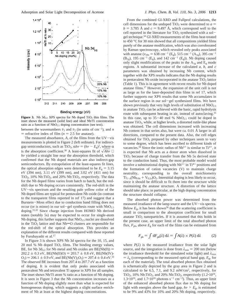
In Figure 3 is shown XPS Nb 3d spectra for the 10, 15, and20 mol % Nb doped TiO2 films. The binding energy values,BE, for Nb 3d5/2 for Nb metal and Nb oxides are BE(Nb(0)))202.2( 0.3 eV, BE(Nb(II)O)) 203.7( 0.8 eV, BE(Nb(IV)-O2) ) 206.1( 0.9 eV, and BE(Nb(V)2O5) ) 207.4( 0.4 eV.33
The observed BE increases from 207.4 to 207.7 eV as a functionof doping. It is evident that only peaks associated withpentavalent Nb and tetravalent Ti appear in XPS for all samples.The inset shows Nb/Ti atom % ratio as a function of Nb doping.It is seen in Figure 3 that the Nb concentration increases as afunction of Nb doping slightly more than what is expected forhomogeneous doping, which suggests a slight surface enrich-ment of Nb at least at the highest doping concentrations.
From the combined GI-XRD and Fullprof calculations, thecell dimensions for the undoped TiO2 were determined toa )b ) 3.785 Å andc ) 9.497 Å, which correspond well to thecell reported in the literature for TiO2 synthesized with a sol-gel technique.34 GI-XRD measurements of the films heat-treatedto 450°C for 30 min showed that all compositions yielded filmspurely of the anatase modification, which was also corroboratedby Raman spectroscopy, which revealed only peaks associatedwith anatase (νobs ) 638 cm-1 (Eg), 515 cm-1 (A1g), 395 cm-1
(B1g), 195 cm-1 (Eg), and 142 cm-1 (Eg)). Nb doping causedonly slight modifications of the peaks in the A1g and Eg moderegions. A substantial increase of the calculateda, b, and cparameters was obtained by increasing Nb content, whichtogether with the XPS results indicates that the Nb doping resultsin pentavalent Nb oxide incorporated in the anatase TiO2 lattice(Table 1). This is in agreement with recent results for Nb dopedanatase films.17 However, the expansion of the unit cell is notas large as for the laser-deposited thin films in ref 17, whichfurther supports our XPS results that some Nb accumulates inthe surface region in our sol-gel synthesized films. We haveshown previously that very high levels of substitution of NbO2.5
in anatase TiO2 can be achieved with the basic, rapid hydrolysisroute with subsequent heating in air, instead of autoclaving.35
In this case, up to 35-40 mol % NbO2.5 could be doped inanatase TiO2, while, at higher levels, a distorted rutile-like phasewas obtained. The cell dimensions increased with increasingNb content in that series also, but were ca. 0.01 Å larger in alldirections, compared to the present data. Also, the cell edgesobtained for TiO2 prepared by other techniques seem to varyto some degree, which has been ascribed to different kinds ofvacancies.34 Since the ionic radius of Nb5+ is similar to Ti4+, itis expected that Nb acts as a donor atom when dissolved inTiO2 because of charge transfer from the Nb 5s derived stateto the conduction band. Thus, the most probable model wouldinvolve a substitutional doping with Nb5+ in Ti4+ positions andthe formation of cation vacancies (V) to maintain chargeneutrality, corresponding to the overall stoichiometryTi1-x(Nb0.8x + V0.2x)O2. Interstitial doping is less likely to occur,since it should be difficult to fit them into the structure whilemaintaining the anatase structure. A distortion of the latticeshould take place; in particular, at the high doping concentrationthe structure should collapse.
The absorbed photon power was determined from themeasured irradiance of the lamp source and the UV-vis spectra.Below Eg, it has been shown36 that the scattering coefficient issmall in comparison to the absorption coefficient for smallanatase TiO2 nanoparticles. If it is assumed that this holds inthe present case, an upper limit of the total absorbed photonflux, Fph, aboveEg for each of the films can be estimated from
where P(λ) is the measured irradiance from the solar lightsource, and the integration is done fromλmin ) 200 nm (belowthe irradiance threshold for the simulated solar light) andλmax
) λg (corresponding to the measured optical band gap,Eg, foreach of the material). The total absorbed photon flux obtainedis schematically depicted by the gray area in Figure 2 and iscalculated to be 6.5, 7.1, and 9.2 mW/cm2, respectively, forTiO2, 10% Nb:TiO2, and 20% Nb:TiO2, respectively (1.2‚1016,1.3‚1016, and 1.7‚1016 photons s-1 cm-2). Thus, an upper limitof the enhanced absorbed photon flux due to Nb doping forlight with energies above the band gap,hν > Eg, is estimatedto be 9% and 43% for 10% and 20% Nb doping, respectively.
Figure 3. Nb 3d5/2 XPS spectra for Nb doped TiO2 thin films. Theinset shows the measured (solid line) and ideal Nb/Ti concentrationratio as a function of NbO2.5 doping concentration (see text).
Fph ) ∫ dFph(λ) dλ ) ∫A(λ) × P(λ) dλ (2)
Adsorption and Solar Light Decomposition of Acetone J. Phys. Chem. B, Vol. 110, No. 3, 20061213
For comparison, the corresponding solar AM1.5 irradiancespectrum is also shown in Figure 2.
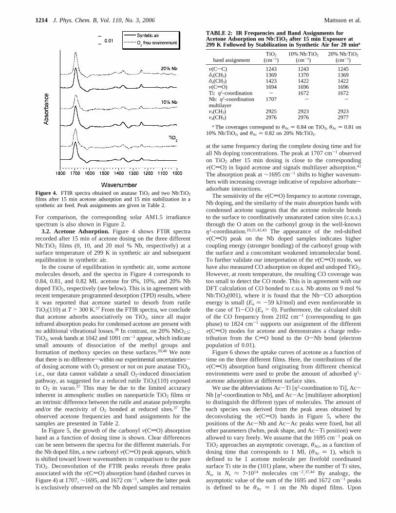
3.2. Acetone Adsorption. Figure 4 shows FTIR spectrarecorded after 15 min of acetone dosing on the three differentNb:TiO2 films (0, 10, and 20 mol % Nb, respectively) at asurface temperature of 299 K in synthetic air and subsequentequilibration in synthetic air.
In the course of equilibration in synthetic air, some acetonemolecules desorb, and the spectra in Figure 4 corresponds to0.84, 0.81, and 0.82 ML acetone for 0%, 10%, and 20% Nbdoped TiO2, respectively (see below). This is in agreement withrecent temperature programmed desorption (TPD) results, whereit was reported that acetone started to desorb from rutileTiO2(110) atT > 300 K.37 From the FTIR spectra, we concludethat acetone adsorbs associatively on TiO2, since all majorinfrared absorption peaks for condensed acetone are present withno additional vibrational losses.38 In contrast, on 20% NbO2.5:TiO2, weak bands at 1042 and 1091 cm-1 appear, which indicatesmall amounts of dissociation of the methyl groups andformation of methoxy species on these surfaces.39,40 We notethat there is no differenceswithin our experimental uncertaintiessof dosing acetone with O2 present or not on pure anatase TiO2,i.e., our data cannot validate a small O2-induced dissociationpathway, as suggested for a reduced rutile TiO2(110) exposedto O2 in vacuo.37 This may be due to the limited accuracyinherent in atmospheric studies on nanoparticle TiO2 films oran intrinsic difference between the rutile and anatase polymorphsand/or the reactivity of O2 bonded at reduced sites.37 Theobserved acetone frequencies and band assignments for thesamples are presented in Table 2.
In Figure 5, the growth of the carbonylν(CdO) absorptionband as a function of dosing time is shown. Clear differencescan be seen between the spectra for the different materials. Forthe Nb doped film, a new carbonylν(CdO) peak appears, whichis shifted toward lower wavenumbers in comparison to the pureTiO2. Deconvolution of the FTIR peaks reveals three peaksassociated with theν(CdO) absorption band (dashed curves inFigure 4) at 1707,∼1695, and 1672 cm-1, where the latter peakis exclusively observed on the Nb doped samples and remains
at the same frequency during the complete dosing time and forall Nb doping concentrations. The peak at 1707 cm-1 observedon TiO2 after 15 min dosing is close to the correspondingν(CdO) in liquid acetone and signals multilayer adsorption.41
The absorption peak at∼1695 cm-1 shifts to higher wavenum-bers with increasing coverage indicative of repulsive adsorbate-adsorbate interactions.
The sensitivity of theν(CdO) frequency to acetone coverage,Nb doping, and the similarity of the main absorption bands withcondensed acetone suggests that the acetone molecule bondsto the surface to coordinatively unsaturated cation sites (c.u.s.)through the O atom on the carbonyl group in the well-knownη1-coordination.19,21,42,43 The appearance of the red-shiftedν(CdO) peak on the Nb doped samples indicates highercoupling energy (stronger bonding) of the carbonyl group withthe surface and a concomitant weakened intramolecular bond.To further validate our interpretation of theν(CdO) mode, wehave also measured CO adsorption on doped and undoped TiO2.However, at room temperature, the resulting CO coverage wastoo small to detect the CO mode. This is in agreement with ourDFT calculation of CO bonded to c.u.s. Nb atoms on 9 mol %Nb:TiO2(001), where it is found that the NbsCO adsorptionenergy is small (Ea ) -59 kJ/mol) and even nonfavorable inthe case of Ti-CO (Ea > 0). Furthermore, the calculated shiftof the CO frequency from 2102 cm-1 (corresponding to gasphase) to 1824 cm-1 supports our assignment of the differentν(CdO) modes for acetone and demonstrates a charge redis-tribution from the CdO bond to the OsNb bond (electronpopulation of 0.01).
Figure 6 shows the uptake curves of acetone as a function oftime on the three different films. Here, the contributions of theν(CdO) absorption band originating from different chemicalenvironments were used to probe the amount of adsorbedη1-acetone adsorption at different surface sites.
We use the abbreviations Ac-Ti [η1-coordination to Ti], Ac-Nb [η1-coordination to Nb], and Ac-Ac [multilayer adsorption]to distinguish the different types of molecules. The amount ofeach species was derived from the peak areas obtained bydeconvoluting theν(CdO) bands in Figure 5, where thepositions of the Ac-Nb and Ac-Ac peaks were fixed, but allother parameters (fwhm, peak shape, and Ac-Ti position) wereallowed to vary freely. We assume that the 1695 cm-1 peak onTiO2 approaches an asymptotic coverage,θAc, as a function ofdosing time that corresponds to 1 ML (θAc ) 1), which isdefined to be 1 acetone molecule per fivefold coordinatedsurface Ti site in the (101) plane, where the number of Ti sites,Ns, is Ns ≈ 7‚1014 molecules cm-2.37,44 By analogy, theasymptotic value of the sum of the 1695 and 1672 cm-1 peaksis defined to beθAc ) 1 on the Nb doped films. Upon
Figure 4. FTIR spectra obtained on anatase TiO2 and two Nb:TiO2
films after 15 min acetone adsorption and 15 min stabilization in asynthetic air feed. Peak assignments are given in Table 2.
TABLE 2: IR Frequencies and Band Assignments forAcetone Adsorption on Nb:TiO2 after 15 min Exposure at299 K Followed by Stabilization in Synthetic Air for 20 mina
band assignmentTiO2
(cm-1)10% Nb:TiO2
(cm-1)20% Nb:TiO2
(cm-1)
ν(CsC) 1243 1243 1245δs(CH3) 1369 1370 1369δa(CH3) 1423 1422 1422ν(CdO)Ti: η1-coordinationNb: η1-coordinationmultilayer
1694-
1707
16961672
-
16961672
-
νs(CH3) 2925 2923 2923νa(CH3) 2976 2976 2977
a The coverages correspond toθAc ) 0.84 on TiO2, θAc ) 0.81 on10% Nb:TiO2, andθAc ) 0.82 on 20% Nb:TiO2.
1214 J. Phys. Chem. B, Vol. 110, No. 3, 2006 Mattsson et al.
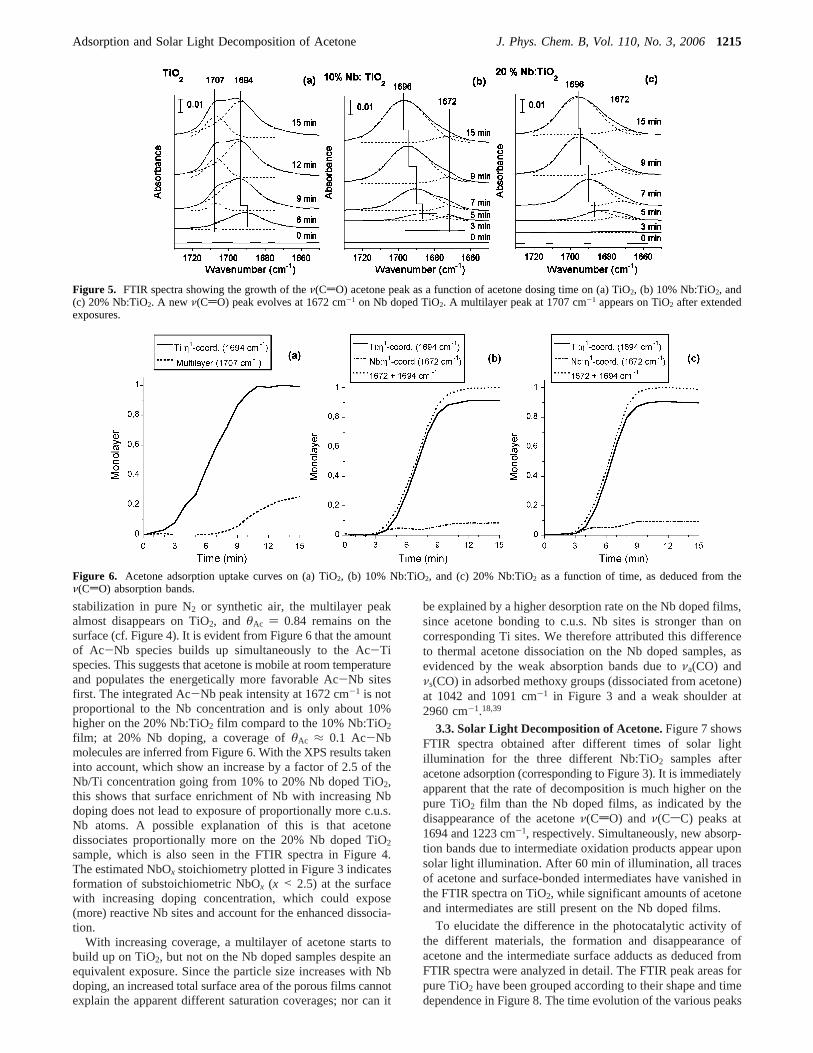
stabilization in pure N2 or synthetic air, the multilayer peakalmost disappears on TiO2, and θAc ) 0.84 remains on thesurface (cf. Figure 4). It is evident from Figure 6 that the amountof Ac-Nb species builds up simultaneously to the Ac-Tispecies. This suggests that acetone is mobile at room temperatureand populates the energetically more favorable Ac-Nb sitesfirst. The integrated Ac-Nb peak intensity at 1672 cm-1 is notproportional to the Nb concentration and is only about 10%higher on the 20% Nb:TiO2 film compard to the 10% Nb:TiO2film; at 20% Nb doping, a coverage ofθAc ≈ 0.1 Ac-Nbmolecules are inferred from Figure 6. With the XPS results takeninto account, which show an increase by a factor of 2.5 of theNb/Ti concentration going from 10% to 20% Nb doped TiO2,this shows that surface enrichment of Nb with increasing Nbdoping does not lead to exposure of proportionally more c.u.s.Nb atoms. A possible explanation of this is that acetonedissociates proportionally more on the 20% Nb doped TiO2
sample, which is also seen in the FTIR spectra in Figure 4.The estimated NbOx stoichiometry plotted in Figure 3 indicatesformation of substoichiometric NbOx (x < 2.5) at the surfacewith increasing doping concentration, which could expose(more) reactive Nb sites and account for the enhanced dissocia-tion.
With increasing coverage, a multilayer of acetone starts tobuild up on TiO2, but not on the Nb doped samples despite anequivalent exposure. Since the particle size increases with Nbdoping, an increased total surface area of the porous films cannotexplain the apparent different saturation coverages; nor can it
be explained by a higher desorption rate on the Nb doped films,since acetone bonding to c.u.s. Nb sites is stronger than oncorresponding Ti sites. We therefore attributed this differenceto thermal acetone dissociation on the Nb doped samples, asevidenced by the weak absorption bands due toνa(CO) andνs(CO) in adsorbed methoxy groups (dissociated from acetone)at 1042 and 1091 cm-1 in Figure 3 and a weak shoulder at2960 cm-1.18,39
3.3. Solar Light Decomposition of Acetone.Figure 7 showsFTIR spectra obtained after different times of solar lightillumination for the three different Nb:TiO2 samples afteracetone adsorption (corresponding to Figure 3). It is immediatelyapparent that the rate of decomposition is much higher on thepure TiO2 film than the Nb doped films, as indicated by thedisappearance of the acetoneν(CdO) and ν(CsC) peaks at1694 and 1223 cm-1, respectively. Simultaneously, new absorp-tion bands due to intermediate oxidation products appear uponsolar light illumination. After 60 min of illumination, all tracesof acetone and surface-bonded intermediates have vanished inthe FTIR spectra on TiO2, while significant amounts of acetoneand intermediates are still present on the Nb doped films.
To elucidate the difference in the photocatalytic activity ofthe different materials, the formation and disappearance ofacetone and the intermediate surface adducts as deduced fromFTIR spectra were analyzed in detail. The FTIR peak areas forpure TiO2 have been grouped according to their shape and timedependence in Figure 8. The time evolution of the various peaks
Figure 5. FTIR spectra showing the growth of theν(CdO) acetone peak as a function of acetone dosing time on (a) TiO2, (b) 10% Nb:TiO2, and(c) 20% Nb:TiO2. A new ν(CdO) peak evolves at 1672 cm-1 on Nb doped TiO2. A multilayer peak at 1707 cm-1 appears on TiO2 after extendedexposures.
Figure 6. Acetone adsorption uptake curves on (a) TiO2, (b) 10% Nb:TiO2, and (c) 20% Nb:TiO2 as a function of time, as deduced from theν(CdO) absorption bands.
Adsorption and Solar Light Decomposition of Acetone J. Phys. Chem. B, Vol. 110, No. 3, 20061215
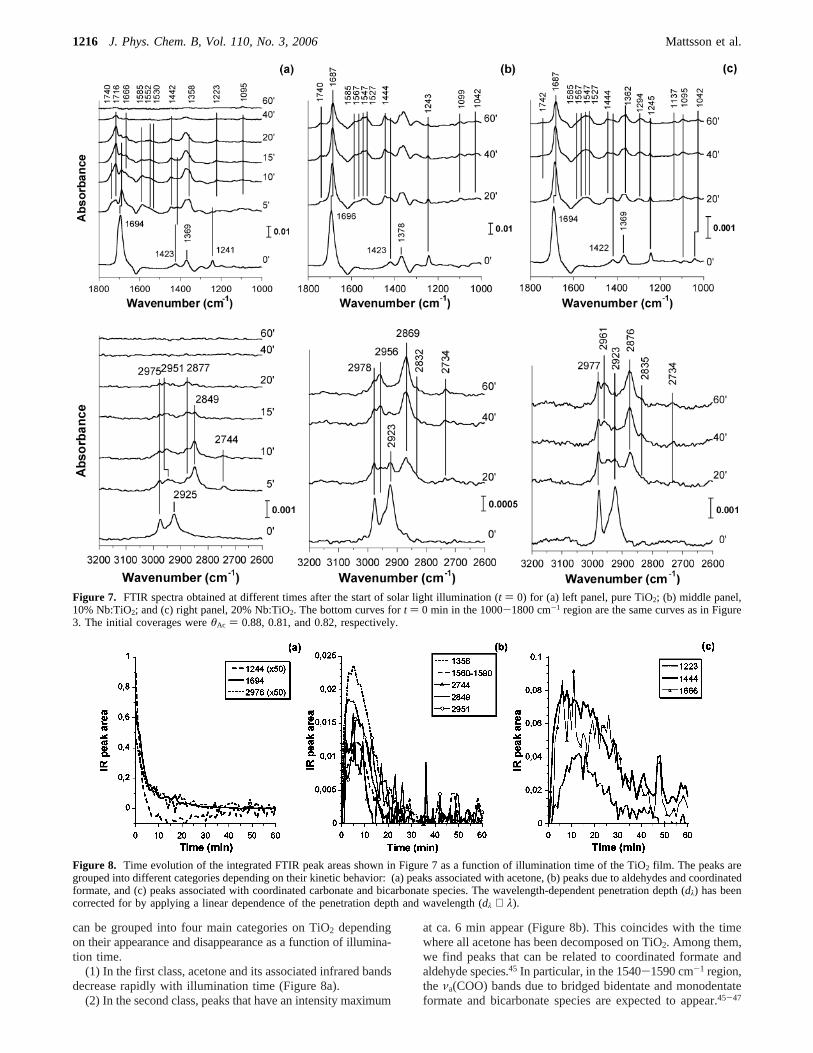
can be grouped into four main categories on TiO2 dependingon their appearance and disappearance as a function of illumina-tion time.
(1) In the first class, acetone and its associated infrared bandsdecrease rapidly with illumination time (Figure 8a).
(2) In the second class, peaks that have an intensity maximum
at ca. 6 min appear (Figure 8b). This coincides with the timewhere all acetone has been decomposed on TiO2. Among them,we find peaks that can be related to coordinated formate andaldehyde species.45 In particular, in the 1540-1590 cm-1 region,the νa(COO) bands due to bridged bidentate and monodentateformate and bicarbonate species are expected to appear.45-47
Figure 7. FTIR spectra obtained at different times after the start of solar light illumination (t ) 0) for (a) left panel, pure TiO2; (b) middle panel,10% Nb:TiO2; and (c) right panel, 20% Nb:TiO2. The bottom curves fort ) 0 min in the 1000-1800 cm-1 region are the same curves as in Figure3. The initial coverages wereθAc ) 0.88, 0.81, and 0.82, respectively.
Figure 8. Time evolution of the integrated FTIR peak areas shown in Figure 7 as a function of illumination time of the TiO2 film. The peaks aregrouped into different categories depending on their kinetic behavior: (a) peaks associated with acetone, (b) peaks due to aldehydes and coordinatedformate, and (c) peaks associated with coordinated carbonate and bicarbonate species. The wavelength-dependent penetration depth (dλ) has beencorrected for by applying a linear dependence of the penetration depth and wavelength (dλ ∝ λ).
1216 J. Phys. Chem. B, Vol. 110, No. 3, 2006 Mattsson et al.

Detailed analysis reveals absorption bands at 1585 and 1552cm-1, which are due to theνa(COO) in HCOO- ion andbidentate bridged HCOO (µ-formate), respectively. The associ-ated formateνa(COO) + δ(CH) combination band appears at2951 cm-1 and the ν(CH) band at 2877 cm-1.45-48 Theabsorption band at 2744 cm-1 is attributed to the typicalν(CHO)stretching frequency in an aldehyde48,49The 1740 cm-1 absorp-tion band apparent in Figure 7 falls into the same category 2group as the 2744 cm-1 and is also attributed to formaldehyde(not shown).
(3) The third class of species (Figure 8c) reaches a maximumconcentration after 12 min illumination and is mainly due tobicarbonate and coordinated carbonate species.45-47 The peakat 1442 cm-1 can be assigned to the symmetric carbonatemode in bicarbonate ions in hydrogen-bonded networks([HCO3
-δ‚‚‚HsO]).45 Furthermore, the 1666, 1223, and 1095cm-1 bands are assigned to theν(CO) modes in bidentatebridged carbonate (µ-carbonate) on the basis of previous reports(see below).46,48,50
(4) Finally, we note that the most persistent peak on TiO2 isthe one at 1715 cm-1, which reaches a maximum after 17 min.The origin of this peak is not settled, but it is tempting toassociate these bands to asymmetric coordinated CO2 spe-cies.45,47,51
The band assignment on TiO2 is summarized in Table 3. Onthe basis of the results in Table 3 and Figure 8, it is evidentthat on TiO2 the rate of decomposition of formate, bicarbonate,and carbonate species varies considerably. Evidently, formateis transformed into carbonate
In the course of the reaction, water is produced, and the R-CO2
species reacts with OH and H2O to form a mixed layer ofbicarbonate and carbonate species
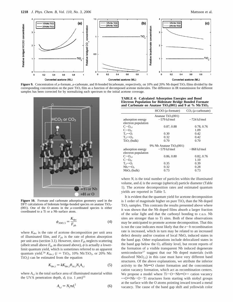
Pursuing the same analysis as in Figure 8 for Nb doped filmsreveals that both the rates of formation and of decompositionof formate, carbonate, and bicarbonate species are significantlyslower on the Nb doped films. In Figure 9 are shown theconcentrations ofµ-formate (1552 cm-1), µ-carbonate (1666and 1223 cm-1 peaks), and H-bonded bicarbonate (1444 cm-1)
on Nb doped TiO2 as a function of decomposed acetonemolecules relative to the corresponding concentration on TiO2.Values> 1 (< 1) indicate a higher (lower) surface concentrationcompared to pure TiO2 and can be interpreted as stabilization(destabilization) of the surface species due to Nb. Here, eachabsorption band has been corrected for variations in the IRabsorption in each measurements by normalizing the peaks tothe integratedν(CsC) peaks in acetone att ) 0 (prior toillumination). The most striking difference here is that on theNb:TiO2 films µ-carbonate is destabilized, while the H-bondedbicarbonate concentration is higher at a given amount ofdecomposed acetone. Furthermore,µ-formate (1552 cm-1)appears stabilized by Nb doping, but the uncertainty is greaterhere because of overlapping absorption bands. The large increaseof the relative µ-formate concentration in Figure 9 withincreasing acetone conversion is due to the depletion of formateon TiO2 and the simultaneous accumulation on Nb doped TiO2.Finally, the weaker 1585 cm-1 peak on Nb doped TiO2 suggeststhat HCOO- ions are readily transformed to bicarbonate orµ-formate.
To further corroborate our spectral band assignment andunderstand the observed trends, we have calculated the adsorp-tion energies (Ea) and the electron population forµ-formate andµ-carbonate bonded to c.u.s. Ti only and with one bond to aNb surface atoms (see Figure 10).
The µ-coordination bonding geometry of formate to rutileTiO2(110) is well-known.46,52,53We assume that this is also thepreferred coordination on the anatase phase, and we use thenon-reconstructed (001) facet to mimic the bonding to crystallineanatase nanoparticles. We employ the same bonding geometryin the Nb doped sample. The results from our DFT calculationsare shown in Table 4. It is seen thatEa for µ-formate is similarfor TiO2 and Nb doped TiO2, while the metal-O bondingstrength as measured by the electron population is strengthenedby ca. 10% upon Nb doping, in fair agreement with theexperimental results. In contrast, carbonate formation is sig-nificantly favored on the Nb doped films compared to pure TiO2
(by ca. 145 kJ/mol). Our FTIR results indicate, however, thatµ-carbonate is significantly more stable on TiO2. This can beunderstood by inspecting the electron population, which showsthat the metal-O bond is substantially weaker on Nb dopedTiO2 than on pure TiO2 (by more than 20%). The result is thatbicarbonate rather than carbonate formation is favored on Nbdoped TiO2, which is in good agreement with the FTIR data.
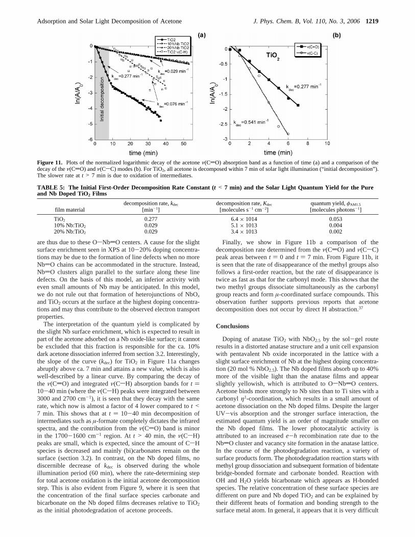
3.4. Rate of Acetone Decomposition and Quantum Yield.In Figure 11a are shown plots of ln(A/A0) vs time, t, for thedifferent materials, whereA0 denoted the deconvoluted peakarea of the acetoneν(CdO) absorption band (∝θAc).
For the Nb doped films, the curves are linear during the wholeillumination period, while it is linear only during the first 7min for pure TiO2. This suggests that the initial decompositionrate can be described by a first-order reaction, viz.
wherekdec is the rate constant for decomposition of acetone onthe films and is determined from a linear least-squares fit tothe data in Figure 11a. The quantum yield,φ, is defined as theratio of the rate of (initial) photoreaction to the rate of absorbedphotons at a given wavelength.54 We define here the solar light(AM1.5) quantum yield,φAM1.5, obtained by illuminating ourthin films precovered with acetone (coverage,θi) and illuminatedby simulated solar light (AM1.5) as
TABLE 3: IR Band Assignments of Intermediates Formedon the TiO2 Films after Solar Light Illumination
moleculeband
assignmentwavenumber
[cm-1]
∆ ) νa(COO)-νs(COO)[cm-1]
proposedstructure
bicarbonateνa(CO) 1528 180 bridgedνs(CO) 1444ν(OH) 3635
carbonate ν(CO) 1666 446 bridgedν(CO) +
δ(OCO)1223
formate νa(CO) 1552, 1585 194, 227 bridged, ionνs(CO) 1358ν(CH) 2877νa(CO) +
δ(CH)2951
aldehyde ν(CHO) 2744 - monodentateν(CO) 2735
HCOO- (Ti)2,bridged+ O298hν+Ti2
0
OCOO- (Ti)2,bridged+ H2O
HCOO- (Ti)2,bridged+ OCOO- (Ti)2,bridged+ H2O98hν+Ti2
0
HCO3 - Ti + H2CO3 + HCO3δ-
-dθAc
dt) kdecθAc (3)
Adsorption and Solar Light Decomposition of Acetone J. Phys. Chem. B, Vol. 110, No. 3, 20061217
whereKdec is the rate of acetone decomposition per unit areaof illuminated film, andFph is the rate of photon absorptionper unit area (section 3.1). However, sinceFph neglects scattering(albeit small aboveEg, as discussed above),φ is actually a lower-limit quantum yield, which is sometimes referred to an apparentquantum yield.54 Kdec,i (i ) TiO2, 10% Nb:TiO2, or 20% Nb:TiO2) can be estimated from the equation
whereAs,i is the total surface area of illuminated material withinthe UVA penetration depth,dλ (ca. 1µm)55
whereNi is the total number of particles within the illuminatedvolume, anddi is the average (spherical) particle diameter (Table1). The acetone decomposition rates and estimated quantumyields are reported in Table 5.
It is evident that the quantum yield for acetone decompositionis 1 order of magnitude higher on pure TiO2 than the Nb dopedTiO2 samples. This contrasts the results presented above whereit was shown that the Nb doped films absorb a larger fractionof the solar light and that the carbonyl bonding to c.u.s. Nbsites are stronger than to Ti sites. Both of these observationsmay be anticipated to promote acetone decomposition. That thisis not the case indicates most likely that thee-h recombinationrate is increased, which in turn may be related to an increaseddefect density and/or creation of local NbOx induced states inthe band gap. Other explanations include delocalized states inthe band gap below the O2 affinity level, but recent reports ofthe formation of a visible transparent Nb induced degeneratesemiconductor17 suggest that our Nb doped materials (withdissolved NbO2.5) in this case must have very different bandstructures. Of the above explanations, we attribute the inferioractivity to the NbdO cluster formation and the concomitantcation vacancy formation, which act as recombination centers.We propose a model where TisOsNbdO‚‚‚ cation vacancy‚‚‚OdNbsOsTi structures form starting with niobyl groupsat the surface with the O atoms pointing inward toward a metalvacancy. The cause of the band gap shift and yellowish color
Figure 9. Concentration ofµ-formate,µ-carbonate, and H-bonded bicarbonate, respectively, on 10% and 20% Nb doped TiO2 films divided by thecorresponding concentration on the pure TiO2 film as a function of decomposed acetone molecules. The difference in IR transmission for differentsamples has been corrected for by normalizing each spectrum to the initial acetone coverage.
Figure 10. Formate and carbonate adsorption geometry used in theDFT calculations of bidentate bridge-bonded species on anatase TiO2-(001). One of the O atoms in theµ-coordinated species is eithercoordinated to a Ti or a Nb surface atom.
φAM1.5 )Kdec
Fph(4)
Kdec,i ) kKdec,iθAc,iNsAs,i (5)
As,i ) Niπdi2 (6)
TABLE 4: Calculated Adsorption Energies and BondElectron Population for Bidentate Bridge Bonded Formateand Carbonate on Anatase TiO2(001) and 9 at % Nb:TiO2
HCOO (µ-formate) CO3 (µ-carbonate)
Anatase TiO2(001)adsorption energy -579 kJ/mol -724 kJ/molelectron populationC-Oi,ii 0.87, 0.88 0.78, 0.76C-Oiii 1.09Tii‚‚‚Oi 0.30 0.42Tii‚‚‚Oii 0.32 0.42TiO2 (bulk) 0.70 0.70
9% Nb Anatase TiO2(001)adsorption energy -579 kJ/mol -868 kJ/molelectron populationC-Oi,ii 0.86, 0.80 0.82, 0.76C-Oiii 1.10Tii‚‚‚Oi 0.35 0.39Nbi‚‚‚Oii 0.32 0.28NbO2 (bulk) 0.73 0.73
1218 J. Phys. Chem. B, Vol. 110, No. 3, 2006 Mattsson et al.
are thus due to these OsNbdO centers. A cause for the slightsurface enrichment seen in XPS at 10-20% doping concentra-tions may be due to the formation of line defects when no moreNbdO chains can be accommodated in the structure. Instead,NbdO clusters align parallel to the surface along these linedefects. On the basis of this model, an inferior activity witheven small amounts of Nb may be anticipated. In this model,we do not rule out that formation of heterojunctions of NbOx
and TiO2 occurs at the surface at the highest doping concentra-tions and may thus contribute to the observed electron transportproperties.
The interpretation of the quantum yield is complicated bythe slight Nb surface enrichment, which is expected to result inpart of the acetone adsorbed on a Nb oxide-like surface; it cannotbe excluded that this fraction is responsible for the ca. 10%dark acetone dissociation inferred from section 3.2. Interestingly,the slope of the curve (kdec) for TiO2 in Figure 11a changesabruptly above ca. 7 min and attains a new value, which is alsowell-described by a linear curve. By comparing the decay ofthe ν(CdO) and integratedν(CsH) absorption bands fort )10-40 min (where theν(CsH) peaks were integrated between3000 and 2700 cm-1), it is seen that they decay with the samerate, which now is almost a factor of 4 lower compared tot <7 min. This shows that att ) 10-40 min decomposition ofintermediates such asµ-formate completely dictates the infraredspectra, and the contribution from theν(CdO) band is minorin the 1700-1600 cm-1 region. At t > 40 min, theν(CsH)peaks are small, which is expected, since the amount of CsHspecies is decreased and mainly (bi)carbonates remain on thesurface (section 3.2). In contrast, on the Nb doped films, nodiscernible decrease ofkdec is observed during the wholeillumination period (60 min), where the rate-determining stepfor total acetone oxidation is the initial acetone decompositionstep. This is also evident from Figure 9, where it is seen thatthe concentration of the final surface species carbonate andbicarbonate on the Nb doped films decreases relative to TiO2
as the initial photodegradation of acetone proceeds.
Finally, we show in Figure 11b a comparison of thedecomposition rate determined from theν(CdO) andν(CsC)peak areas betweent ) 0 andt ) 7 min. From Figure 11b, itis seen that the rate of disappearance of the methyl groups alsofollows a first-order reaction, but the rate of disappearance istwice as fast as that for the carbonyl mode. This shows that thetwo methyl groups dissociate simultaneously as the carbonylgroup reacts and formµ-coordinated surface compounds. Thisobservation further supports previous reports that acetonedecomposition does not occur by direct H abstraction.37
Conclusions
Doping of anatase TiO2 with NbO2.5 by the sol-gel routeresults in a distorted anatase structure and a unit cell expansionwith pentavalent Nb oxide incorporated in the lattice with aslight surface enrichment of Nb at the highest doping concentra-tion (20 mol % NbO2.5). The Nb doped films absorb up to 40%more of the visible light than the anatase films and appearslightly yellowish, which is attributed to OsNbdO centers.Acetone binds more strongly to Nb sites than to Ti sites with acarbonylη1-coordination, which results in a small amount ofacetone dissociation on the Nb doped films. Despite the largerUV-vis absorption and the stronger surface interaction, theestimated quantum yield is an order of magnitude smaller onthe Nb doped films. The lower photocatalytic activity isattributed to an increasede-h recombination rate due to theNbdO cluster and vacancy site formation in the anatase lattice.In the course of the photodegradation reaction, a variety ofsurface products form. The photodegradation reaction starts withmethyl group dissociation and subsequent formation of bidentatebridge-bonded formate and carbonate bonded. Reaction withOH and H2O yields bicarbonate which appears as H-bondedspecies. The relative concentration of these surface species aredifferent on pure and Nb doped TiO2 and can be explained bytheir different heats of formation and bonding strength to thesurface metal atom. In general, it appears that it is very difficult
Figure 11. Plots of the normalized logarithmic decay of the acetoneν(CdO) absorption band as a function of time (a) and a comparison of thedecay of theν(CdO) andν(CsC) modes (b). For TiO2, all acetone is decomposed within 7 min of solar light illumination (“initial decomposition”).The slower rate att > 7 min is due to oxidation of intermediates.
TABLE 5: The Initial First-Order Decomposition Rate Constant ( t < 7 min) and the Solar Light Quantum Yield for the Pureand Nb Doped TiO2 Films
film materialdecomposition rate,kdec
[min-1]decomposition rate,Kdec
[molecules s-1 cm-2]quantum yield,φAM1.5
[molecules photons-1]
TiO2 0.277 6.4× 10¹⁴ 0.05310% Nb:TiO2 0.029 5.1× 10¹³ 0.00420% Nb:TiO2 0.029 3.4× 10¹³ 0.002
Adsorption and Solar Light Decomposition of Acetone J. Phys. Chem. B, Vol. 110, No. 3, 20061219
to improve the photocatalytic activity of TiO2 by cation doping,since charge transfer to the conduction band (ref 17), defectand vacancy site formation (because of NbdO clusters), andbond formation at the surface (e.g., an increased formate bondstrength) all must be tuned appropriately. This has hitherto notproven possible with Nb doping, employing different dopingmethods.
Acknowledgment. This work was supported by the SwedishDepartment of Defence (project no. 430-A4515).
References and Notes
(1) Fujishima, A.; Hashimoto, K.; Watanabe, T.TiO2 Photocatalysis.Fundamentals and Applications; BKC, Inc.: Tokyo, 1999.
(2) Teichner, S. J.; Formenti, M. InPhotoelectrochemistry, Photoca-talysis, and Photoreactors; Schiavello, M., Ed.; Reidel Publishing Com-pany: Dordrecht, 1985; p 457 ff.
(3) Photocatalytic Purification and Treatment of Water and Air; Ollis,D. F., Al-Ekabi, H., Eds.; Elsevier: Amsterdam, 1993.
(4) Anpo, M. InGreen Chemistry; Tundo, P., Anastas, P., Eds.; OxfordUniversity Press: Oxford, 2000.
(5) Anpo, M. Bull. Chem. Soc. Jpn.2004, 77, 1427.(6) Asahi, R.; Morikawa, T.; Ohwaki, T.; Aoki, K.; Taga, Y.Science
2001, 293, 269.(7) Lindgren, T.; Mwabora, J. M.; Avendano, E.; Jonsson, J.; Hoel,
A.; Granqvist, C.-G.; Lindquist, S.-E.J. Phys. Chem. B2003, 107, 5709.(8) Sakthivel, S.; Kisch, H.Angew. Chem., Int. Ed.2003, 42, 4908.(9) Khan, S. H. M.; Al-Shahry, M.; Ingler, W. B., Jr.Science2002,
297, 2243.(10) Hattori, A.; Yamamoto, M.; Tada, H.; Ito, S.Chem. Lett.1998,
27, 707.(11) Subbarao, S. N.; Yun, Y. H.; Kershaw, R.; Dwight, K.; Wold, A.
Inorg. Chem.1979, 18, 488.(12) Umebayashi, T.; Yamaki, T.; Itoh, H.; Asai, K.Appl. Phys. Lett.
2002, 81, 454.(13) Anpo, M.; Nakaya, H.; Kodama, S.; Kubokawa, Y.; Domen, K.;
Onishi, T.J. Phys. Chem.1986, 90, 1633.(14) Miyauchi, M.; Takashio, M.; Tobimatsu, H.2004, 20, 232.(15) Venkataraj, S.; Drese, R.; Liesch, C.; Kappertz, O.; Jayavel, R.;
Wuttig, M. J. Appl. Phys.2002, 91, 4863.(16) Granqvist, C. G.Handbook of Inorganic Electrochromic Systems,
2nd ed.; Elseview: Amsterdam, 2002.(17) Furubayashi, Y.; Hitosugi, T.; Yamamoto, Y.; Inaba, K.; Kinoda,
G.; Hirose, Y.; Shimada, T.; Hasegawa, T.Appl. Phys. Lett.2005, 86,252101.
(18) Busca, G.Catal. Today1996, 27, 457.(19) Coronado, J. M.; Kataoka, S.; Tejedo-Tejedor, I.; Anderson, M.
A. J. Catal.2003, 219, 219.(20) El-Maazawi, M.; Finken, A. N.; Nair, A. B.; Grassian, V. H.J.
Catal. 2000, 191, 138.(21) Zaki, M. I.; Hasan, M. A.; Pasupulety, L.Langmuir2001, 17, 768.(22) Rodriguez-Carvajal, J.Physica B1993, 192, 55.(23) http://rredc.nrel.gov/solar/spectra/am1.5/.(24) Perdew, J. P.; Wang, Y.Phys. ReV. B 1992, 42, 13244.(25) Kleinman, L.; Bylander, D. M.Phys. ReV. Lett. 1982, 48, 1425.
(26) Teter, M. P.; Payne, M. C.; Allan, D. C.Phys. ReV. B 1989, 40,12255.
(27) Monkhorst, H. J.; Pack, J. D.Phys. ReV. B 1976, 13, 5188.(28) Molecular Modeling and Simulation: An Interdisciplinary Guide;
Antman, S. S., Marsden, J. E., Sirovich, L., Eds.; Springer-Verlag: NewYork, 2002.
(29) Segall, M. D.; Shah, R.; Pickard, C. J.; Payne, M. C.Phys. ReV. B1996, 54, 16317.
(30) Tang, H.; Prasad, K.; Sanjine`s, R.; Schmid; Le´vy, F.J. Appl. Phys.1994, 75, 2042.
(31) Burstein, E.Phys. ReV. 1954, 93, 632.(32) Moss, T. S.Proc. Phys. Soc. London, Ser. B1954, 67, 775.(33) Wagner, C. D.; Naumkin, A. V.; Kraut-Vass, A.; Allison, J. W.;
Powell, C.; Ramble, J. R., Jr., Eds.NIST Standard Reference Database 20,version 3.4 (Web version); National Institute of Standards and Technol-ogy: Gaithersburg, MD, 20899; accessed August 2003.
(34) Milella, F.; Gallardo-Amores, J. M.; Baldi, M.; Busca, G.J. Mater.Chem.1998, 8, 2525.
(35) Leideborg, M.; Westin, G. Preparation of Ti-Nb-O Nano Powdersand Studies of the Structural Development on Heat Treatment. Ceramics:Getting into the 2000’s, Part B;AdVances in Science and Technology, Vol.14;Proceedings of the 9th CIMTEC World Ceramics Congress and Forumon New Materials; Vincenzini, P., Ed.; June 14-19, 1998, Florence, Italy.
(36) Loddo, V.; Addamo, M.; Augugliaro, V.; Di Paola, A.; Garcı´a-Lopez, E.; Marci, G.; Palmisano, L.; Schiavello, M. HeterogeneousPhotocatalysis: Optical Characterisation of polycrystalline Photocatalystsfor Pollutant Degradation in Aqueous Media. 8th International Conferenceon Solar Energy and Applied Photochemistry, 2005, Luxor, Egypt.
(37) Henderson, M. A.J. Phys. Chem. B2004, 108, 18932.(38) Stein, S. E. Infrared Spectra. NIST Mass Spec Data Center; National
Institute of Standards and Technology: Gaithersburg, MD; June 2005.(39) Rusu, C.; Yates, J. T., Jr.J. Phys. Chem. B2000, 104, 12292.(40) Glisenti, J.J. Mol. Catal., A2000, 153, 169.(41) Stein, S. E. Infrared Spectra. NIST Mass Spec Data Center;NIST
Chemistry WebBook, NIST Standard Reference Database Number 69;Linstrom, P. J., Mallard, W. G., Eds.; National Institute of Standards andTechnology: Gaithersburg, MD, March 2003; http://webbook.nist.gov.
(42) Henderson, M. A.Surf. Sci.1998, 400, 203.(43) Griffith, D. M.; Rochester, C.J. Chem. Soc., Faraday Trans. 1
1978, 74, 403.(44) Diebold, U.Surf. Sci. Rep.2003, 48, 53.(45) Osterlund, L.Int. J. Nanotechnol.In press.(46) Busca, G.; Lorenzelli, V.Mater. Chem.1982, 7, 89.(47) Nakamoto, K.Infrared and Raman Spectra of Inorganic and
Coordination Compounds, 5th ed.; John Wiley & Sons: New York, 1997.(48) Tanaka, K.; White, J. M.J. Phys. Chem.1982, 86, 4708.(49) Sokrates, G.Infrared Characteristic Group Frequencies, 2nd ed.;
John Wiley & Sons: Chichester, 1994.(50) Davydov, A. A.Infrared Spectroscopy of Adsorbed Species on the
Surface of Transition Metal Oxides; John Wiley & Sons: New York, 1990.(51) Mascetti, J.; Tranquille, M.J. Phys. Chem.1988, 92, 2177.(52) Hayden, B. E.; King, A.; Newton, M. A.J. Phys. Chem. B1999,
103, 203.(53) Rotzinger, F. P.; Kesselman-Truttmann, J. M.; Hug, S. J.; Shklover,
V.; Gratzel, M.J. Phys. Chem. B2004, 108, 5004.(54) Serpone, N.; Emeline, A. V.Int. J. Photoenergy2002, 91-131.(55) Courbon, H.; Formenti, M.; Juillet, F.; Lisachenko, A. A.; Martin,
J.; Teichner, S. J.Kinet. Catal.1973, 14, 84.
1220 J. Phys. Chem. B, Vol. 110, No. 3, 2006 Mattsson et al.
Copyright © 2022 FDOKUMEN