Differential cellular proliferation underlies heterochronic ...
Upload
independentCategory
view
0download
0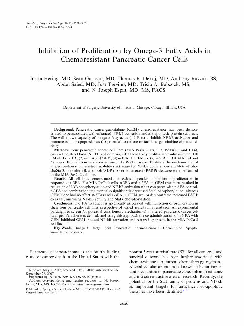
Inhibition of Proliferation by Omega-3 Fatty Acids inChemoresistant Pancreatic Cancer Cells
Justin Hering, MD, Sean Garrean, MD, Thomas R. Dekoj, MD, Anthony Razzak, BS,Abdul Saied, MD, Jose Trevino, MD, Tricia A. Babcock, MS,
and N. Joseph Espat, MD, MS, FACS
Department of Surgery, University of Illinois at Chicago, Chicago, Illinois, USA
Background: Pancreatic cancer-gemcitabine (GEM) chemoresistance has been demon-strated to be associated with enhanced NF-kB activation and antiapoptotic protein synthesis.The well-known capacity of omega-3 fatty acids (n-3 FAs) to inhibit NF-kB activation andpromote cellular apoptosis has the potential to restore or facilitate gemcitabine chemosensi-tivity.Methods: Four pancreatic cancer cell lines (MIA PaCa-2, BxPC-3, PANC-1, and L3.6),
each with distinct basal NF-kB and differing GEM sensitivity profiles, were administered: 100uM of (1) n-3FA, (2) n-6FA, (3) GEM, (4) n-3FA + GEM, or (5) n-6FA + GEM for 24 and48 hours. Proliferation was assessed using the WST-1 assay. To define the mechanism(s) ofaltered proliferation, electron mobility shift assay for NF-kB activity, western blots of pho-shoStat3, phosphoIjB, and poly(ADP-ribose) polymerase (PARP) cleavage were performedin the MIA PaCa-2 cell line.Results: All cell lines demonstrated a time/dose-dependent inhibition of proliferation in
response to n-3FA. For MIA PaCa-2 cells, n-3FA and n-3FA + GEM treatment resulted inreduction of I-kB phosphorylation and NF-kB activation when compared with n-6FA control.n-3FA and combination treatment also significantly decreased Stat3 phosphorylation, whereasGEM alone had no effect. n-3FAs and n-3FA + GEM groups demonstrated increased PARPcleavage, mirroring NF-kB activity and Stat3 phosphorylation.Conclusions: n-3 FA treatment is specifically associated with inhibition of proliferation in
these four pancreatic cell lines irrespective of varied gemcitabine resistance. An experimentalparadigm to screen for potential contributory mechanism(s) in altered pancreatic cancer cel-lular proliferation was defined, and using this approach the co-administration of n-3 FA withGEM inhibited GEM-induced NF-kB activation and restored apoptosis in the MIA PaCa-2cell-line.Key Words: Omega-3 fatty acid—Pancreatic adenocarcinoma—Gemcitabine—Apopto-
sis—Chemoresistance.
Pancreatic adenocarcinoma is the fourth leadingcause of cancer death in the United States with the
poorest 5-year survival rate (5%) for all cancers,1 andsurvival outcome has been further associated withchemoresistance to current chemotherapy regimens.Altered cellular apoptosis is known to be an impor-tant mechanism in pancreatic cancer chemoresistanceand is a current active area of research. Recently, thepotential for the Stat family of proteins and NF-jBas important targets for anticancer/pro-apoptotictherapies have been identified.2–4
Received May 8, 2007; accepted July 7, 2007; published online:September 26, 2007.Supported by: NIDDK K08 DK DK60778 (Espat)Address correspondence and reprint requests to: N. Joseph
Espat, MD, MS, FACS; E-mail: [email protected]
Published by Springer Science+Business Media, LLC � 2007 The Society ofSurgical Oncology, Inc.
Annals of Surgical Oncology 14(12):3620–3628
DOI: 10.1245/s10434-007-9556-8
3620

In concert and independently, Stat3 and NF-jBhave been implicated as mediators of antiapoptoticphenomena in pancreatic malignancy.5 Stat3 andStat5 are known to be powerful mediators in thecellular response to growth factors, cytokines, andantiapoptotic signaling. Within the Stat family,upregulated Stat3 has been shown to participate inthe progression/survival of human cancer cells. Fur-thermore, Stat3 has been demonstrated to be onco-genic, to be overactive in pancreatic cancers, and topossess tumorigenic potential when overexpressed incertain models.3–5
NF-jB is considered a significant and key com-ponent in the regulation of cellular activity based onits role in the initiation, progression, and dysfunctionof malignant cells as well as the elaboration ofinflammatory cascades associated with malignancies.6
Furthermore, NF-jB has been demonstrated to be akey mediator in pancreatic cancer cell chemoresis-tance to gemcitabine.7
Clinical data suggest a potential role for the ome-ga-3 fatty acids present in deep-water marine fish oils,such as eicosapentaenoic acid (EPA), in the treatmentof pancreatic adenocarcinoma patients to improvefunctional status, weight gain, and treatment-relatedconsequences.8–10 Experimentally, n-3 polyunsatu-rated fatty acids (n-3FA) also referred to as x(ome-ga)-3 fatty acids when incubated with various cancercells types in vitro have been shown to have an an-tiproliferative and proapoptotic effects.11–13 Theseeffects occur specifically in pancreatic cancer cells aswell,14 though many of the molecular mechanismsresponsible remain unknown.At present, the standard pancreatic cancer che-
motherapeutic agent, gemcitabine (GEM) (Gemzar),has been shown in experimental models to induceNF-jB activation in pancreatic cell lines,15 which is apotentially proproliferative and antiapoptotic event.Based on the known biologic activities for n-3FA, thepotential to overcome gemcitabine associated NF-jBactivation was postulated.16
In the present experiments, it was hypothesizedthat four pancreatic cancer cell lines, representing aspectrum of cancer cell profiles (NF-jB expression,GEM sensitivity), would have an alteration in theircellular proliferation profiles after treatment ofn-3FA, GEM, or n-3FA in combination with GEM.In order to establish an experimental paradigm toexamine the mechanisms of these effects on prolifer-ation, Stat3 and NF-jB activation were studied aswell as subsequent apoptotic markers followingtreatment with n-3FA, GEM, or combination in theintermediate profile Mia Paca cell line.
MATERIALS AND METHODS
Materials
The pancreatic cancer cell lines Mia Paca andBxPC-3 (purchased from American Type CultureCollection (ATCC, Rockville, MD) and PANC-1and L3.6 (gift from Gary Gallick PhD, MDAnderson Cancer Center Texas) were used. N-3 FArich emulsion (Omegaven) and n-6 FA richemulsion (Lipovenoes) were purchased from Frese-nius-Kabi (Bad-Homburg, Germany). To ensureexperiment comparability, all same production‘‘lot’’ lipid emulsions were used. Gemcitabine(Gemzar) was purchased from Eli Lilly (Indianap-olis, IN). Fetal bovine serum (FBS) was purchasedfrom Hyclone (Logan, UT). All other cell culturereagents were purchased from Invitrogen/Gibco(Carlsbad, CA). Cell Proliferation Reagent WST-1kit was purchased from Roche Applied Science(Indianapolis, IN). Anti-poly(ADP-ribose) poly-merase (anti-PARP) (H-250) antibody was pur-chased from Santa Cruz Biotechnology (SantaCruz, CA). Phospho-IkappaB-alpha (Ser32) Anti-body and PhosphoPlus Stat3 (Tyr705) AntibodyKit were purchased from Cell Signaling Technology(Beverly, MA). b-actin antibody was purchasedfrom Sigma-Aldrich (St. Louis, MO). Western Blotreagents were purchased from Biorad (Hercules,CA). Nuclear extraction materials were purchasedfrom Active Motif, LLC (Carlsbad, CA).
Cells and Culture Conditions and Experimental Design
MIA PaCa-2, BxPC-3, PANC-1, and L3.6 cellswere grown in Dulbecco�s modification of Eagle�smedium (DMEM) supplemented by 10% FBS, peni-cillin 100 units/mL, and streptomycin 100 lg/mL at37�C in 5% CO2. Cells were frozen for stock in 95%growth medium and 5% dimethyl sulfoxide (DMSO)in liquid nitrogen. Trypsin-EDTA was used for cellharvest and passage. Cells were plated at 600,000cells/100 mm tissue culture dish and allowed toadhere overnight (18 hours) before any treatmentbegan. MIA PaCa-2, BXPC-3, Panc-1, and L3.6pancreatic cancer cell lines were treated with 0, 25, 50,100, and 150 lM n-3FA or n-6FA, as a lipid control,at time points 24 and 48 hours for time and doseproliferation studies. Cells were then treated withmedia, 100 lM n-3FA, n-6FA, GEM, n-3FA +GEM, or n-6FE + GEM, at time points 24 and 48hours. For Western Blot and electrophoretic mobilityshift assay (EMSA), MIA PaCA-2 treated cells were
N-3 FAS IN PANCREATIC CANCER CELLS 3621
Ann. Surg. Oncol. Vol. 14, No. 12, 2007
exposed to a 100 lM concentration of GEM, n-3FA,and n-6FA.All experiments were performed, at minimum, in
triplicate.
Cell Proliferation Assay
WST-1 reagent was used according to kit specifi-cations and experimental conditions as describedabove. To establish the mechanism of action of pro-liferation alteration, we performed the following inthe MIA PaCa-2 cell line.
Cell Protein Extraction ProtocolAfter treatment, the cells destined for protein
analysis were first washed twice with phosphate-buf-fered saline (PBS). Whole cell (for IjB and Stat3expression) and nuclear extracts (for PARP expres-sion) were collected using the reagents and the pro-tocol provided by Active Motif, LLC, NuclearExtract Kit. Total protein concentrations for bothnuclear and whole-cell extracts were determined usingthe DC protein assay (Biorad, Hercules, CA). Whole-cell or nuclear extracts were then stored at )80�C forWestern blot analysis.
Western Blot ProtocolAn equal amount of total protein was aliquoted for
each well (60 lg). Cell extracts were boiled for 5minutes prior to loading of the 10% SDS-PAGEgel (for PARP, a 7.5% SDS-PAGE gel was used)and then transferred to a nitrocellulose membrane.The membranes were placed in a blocking solution(5% w/v nonfat dry milk in 1X TBS with 0.1%Tween-20) for 1 hour. The blot membrane was thenincubated with an antiprotein-specific primary anti-body overnight at 4�C, washed three times with TBS/T, and then incubated with a horseradish peroxidase(HRP)-conjugated secondary antibody. After wash-ing, the membrane was visualized with 10 mLenhanced chemiluminescent (ECL) detection reagentand exposed to X-ray film for 10–60 seconds. Blotswere then stripped with 3% hydrogen peroxide andreprobed with anti-b-actin antibody, exposed to theHRP conjugated antibody and visualized as above.The b-actin blot densities were then compared byKodak 1D v3.5.4.14 application (Eastman Kodak,Rochester, NY, USA) to the original films respec-tively to normalize for variance in the amount ofprotein loaded into each well. The antibody dilutionsfor Western blot analysis were as follows: forIjB—10lL:10mL, for PhosStat3—10lL:10mL, forPARP—50lL:10mL.
NF-jB Electrophoretic Mobility Shift Assay (EMSA)Nuclear protein extraction and EMSA. Cells were
grown to confluence (1 · 107 cells) in 100-mm platesand treated as described in experimental design. Afterlipid and GEM exposure, cells were washed twicewith ice-cold PBS, and total nuclear extract wasprepared using the reagents and protocol describedby Active Motif LLC (Carlsbad, CA). For theEMSA, T4 polynucleotide kinase, poly(dI-dC),[32P]ATP, and the Sephadex G-50M column werepurchased from Amersham Biosciences (Piscataway,NJ). All other reagents for this experiment were ob-tained from Sigma Chemical unless otherwise speci-fied. The probe was a 24-bp double-strandedconstruct of the NF-jB consensus sequence (5¢-AG-GGACTTTCCGCTG GGACTTTCC-3¢), which wasend-labeled using T4 polynucleotide kinase and[32P]ATP. The labeled probe was purified on theSephadex G-50M column. For each sample, 5 lg oftotal nuclear protein were incubated with the labeleddouble-stranded probe (�50,000 cpm) and 5 lg ofpoly(dI-dC) in binding buffer (10 mM Tris Æ HCl, pH7.5, 100 mM NaCl, 1 mM EDTA, 0.2% Nonidet P-40, and 0.5 mM dithiothreitol) for 20 minutes at25�C. Specific competition was performed by adding100 ng of unlabeled NF-jB double-stranded bindingprobe to the reaction. The mixtures were run on 5%polyacrylamide gel electrophoresis in 1· Tris-glycine-EDTA buffer. The gels were then vacuum dried andexposed to radiographic film (Eastman Kodak, NewHaven, CT).
AnalysisAll experiments were performed, at minimum, in
triplicate. They were analyzed using the SPSS statis-tics program (Chicago, IL). Data are presented asmeans ± SE when possible. The data were analyzedby one-way ANOVA, as well as additional ANOVApost hoc analysis (Tukey, Scheffe�s and LSD) asappropriate. Statistical significance was defined at theP < .05 level unless otherwise stated.
RESULTS
WST-1 Proliferation Assay
Figure 1 displays the results from the WST-1 re-agent assay as a measure of proliferation. Prolifera-tion is presented as a percentage of the proliferationof cells under no treatment conditions. A significanttime and dose dependent decrease in cellular prolif-eration was observed across all four pancreatic cancer
J. HERING ET AL.3622
Ann. Surg. Oncol. Vol. 14, No. 12, 2007

cell lines. The effects were significant at 24 hours, butoptimized at 48 hours (Fig. 1), with an effectivetreatment concentration of 100 lM seen across all celllines. This is a commonly used in vitro n-3FA con-centration.17,18 Specifically, at 24 hours, 100 lMn-3FA inhibited proliferation in MIA PaCa-2 cells by52%, BXPC-3 by 53%, Panc-1 by 38% and L3.6pl by73%. At 48 hours, 100 lM EPA inhibited prolifera-tion in MIA PaCa-2 cells by 79%, BXPC-3 by 63%,Panc-1 by 55%, and L3.6pl by 70%.
MIA PaCa-2, BxPC-3, PANC-1, and L3.6 cellswere then incubated for 24 or 48 hours with 100 lMn-3FA, n-6FA, GEM, or a combination of lipid +GEM. 100 lM was chosen because of the measurableeffects across all cell lines, and our observation thatthis dose does not produce a toxic effect on the cells.At 24 hours, all n-3FA treated cell lines had signifi-cantly reduced proliferation when compared to mediaalone control or n-6FA lipid control (Fig. 2). Thissame result followed for the 48-hour treatment
L3.6 24hr WST-1
0
20
40
60
80
100
[µM]
N-3
N-6p=.0p=.0
p=.2
p=.0
L3.6 48hr WST-1
0
20
40
60
80
100
[µM]
N-3
N-6p=.p=.p=.
p=
PANC-1 24hr WST-1
0
20
40
60
80
100
[µM]
N-3
N-6
p=0p=0
p=.05p=4
BxPC-3 24hr WST-1
0
20
40
80
100
120
[µM]
60
N-3
N-6
p=.008p=.00
p=.00p=.0
MIA PaCa-2 24hr WST-1
01020
708090
100
[µM]
N-3
30405060
N-6p=0
p=0
p=0p=1
MIA PaCa-2 48hr WST-1
0
20
80
100
25 50 100 150
25 50 100 150
25 50 100 150
25 50 100 15025 50 100 150
25 50 100 150
25 50 100 150
25 50 100 150
[µM]
N-6
p=0
40
60N-3
p=001p=00
p=0
MIA PaCa-2 48hr WST-1
0
20
40
60
80
100
[µM]
N-3p=.0
N-6
p=.001p=00
p=.0
PANC-1 48hr WST-1
0
20
40
60
80
100
[µM]
N-3
N-6
p=0p=p=0p=0
Per
cen
t o
f C
on
tro
l
Per
cen
t o
f C
on
tro
lP
erce
nt
of
Co
ntr
ol
Per
cen
t o
f C
on
tro
lP
erce
nt
of
Co
ntr
ol
Per
cen
t o
f C
on
tro
lP
erce
nt
of
Co
ntr
ol
Per
cen
t o
f C
on
tro
l
FIG. 1. Dose and time response of n-3 FA and n-6 FA treatment on proliferation of four different pancreatic cancer cell lines at 24 and 48hours. Media-alone treatment used as control. Significance defined at P < .05 indicated on the linear graph for each treatment concentration.All cell lines treated demonstrated a significant time and dose dependent inhibition of cellular proliferation with n-3 FA treatment.
N-3 FAS IN PANCREATIC CANCER CELLS 3623
Ann. Surg. Oncol. Vol. 14, No. 12, 2007

(Fig. 2). Proliferation is decreased in the presence ofn-3FAs regardless of the presence of GEM.Those cell lines that are resistant to GEM, notably
PANC-1, and relatively resistant, BxPC-3 and MIAPaCa-2, produce a reduced proliferation with com-bination n-3FA + GEM treatment when comparedwith GEM alone at 24 and 48 hours (Fig. 2). TheGEM-resistant cell lines produced a significant inhi-bition of proliferation with the addition of n-3FA to
the treatment condition. The L3.6 cell line is veryGEM sensitive and in the 24-hour treatment showsan additive effect of combination treatment. There isa greater significant decrease in proliferation whenthe cells are exposed to n-3FA versus n-6FA in thesecell lines. N-6FA is used as a lipid control. Toexamine the mechanisms of this inhibition of prolif-eration, NF-jB activation and production andSTAT3 phosphorylation was explored.
FIG. 2. Alterations in cellular proliferation in the presence n-3FA, n-6FA, GEM, n-3FA + GEM, and n-6FA + GEM treatments at 24and 48 hours. Media-alone treatment used as control. All concentrations are 100 lM. * = P < .05.
J. HERING ET AL.3624
Ann. Surg. Oncol. Vol. 14, No. 12, 2007
I-jB Western Blot
IjB phosphorylation is a measure of the likelihoodof the NF-jB molecule to translocate into the nucleusto produce its effects on the transcription of propro-liferative proteins. The amount of phosphorylatedI-jB protein present was evaluated in MIA PaCa-2cells treatedunder the following conditions at 24hours:media alone, n-3FA, n-6FA, GEM, n-3FA + GEM,and n-6FA + GEM—all at 100 lM. The band islighter in the n-3FA and n-3FA + GEM-treated cellswhen compared with control. These data suggest astabilization of the IjB-NF-jB complex by n-3FAs.Figure 3 displays an example of the Western blot.
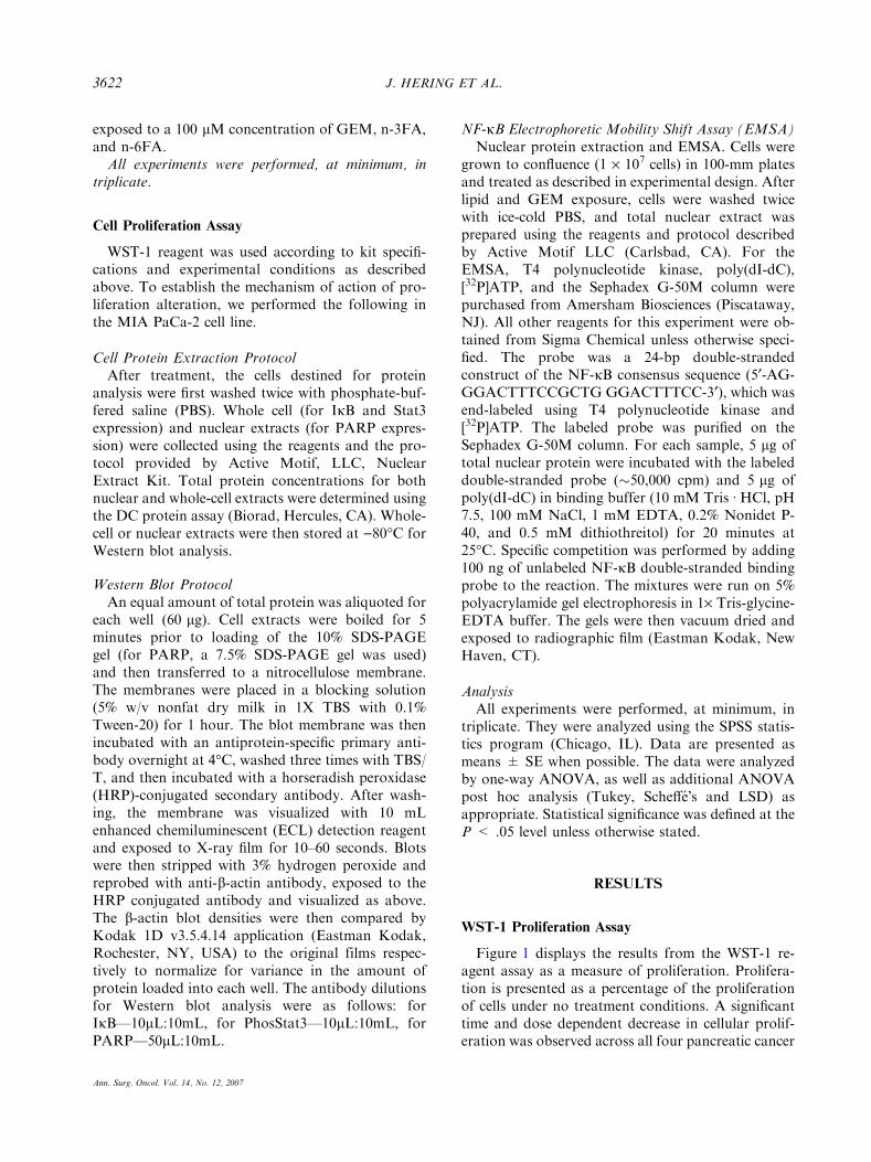
NF-jB EMSA
Figure 4 is a representative EMSA demonstratingNF-jB activation on our treated cells . This assay givesa measure of the binding activity of the molecule toDNA in MIA PaCa-2 cells treated in the followingmanner: media alone, n-3FA, GEM, and n-3FA +GEM. The n-3FA emulsion-treated group producedthe greatest decrease in the amount of binding com-pared with control. The GEM group shows no signif-icant change from control, and the combo group showsa smaller decrease in binding compared with the con-trol. To further examine the mechanisms of prolifera-tion inhibition Stat3 was studied.
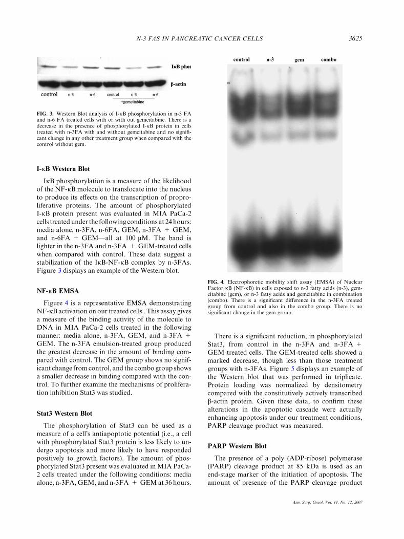
Stat3 Western Blot
The phosphorylation of Stat3 can be used as ameasure of a cell�s antiapoptotic potential (i.e., a cellwith phosphorylated Stat3 protein is less likely to un-dergo apoptosis and more likely to have respondedpositively to growth factors). The amount of phos-phorylated Stat3 present was evaluated inMIA PaCa-2 cells treated under the following conditions: mediaalone, n-3FA,GEM, and n-3FA + GEMat 36 hours.
There is a significant reduction, in phosphorylatedStat3, from control in the n-3FA and n-3FA +GEM-treated cells. The GEM-treated cells showed amarked decrease, though less than those treatmentgroups with n-3FAs. Figure 5 displays an example ofthe Western blot that was performed in triplicate.Protein loading was normalized by densitometrycompared with the constitutively actively transcribedb-actin protein. Given these data, to confirm thesealterations in the apoptotic cascade were actuallyenhancing apoptosis under our treatment conditions,PARP cleavage product was measured.
PARP Western Blot
The presence of a poly (ADP-ribose) polymerase(PARP) cleavage product at 85 kDa is used as anend-stage marker of the initiation of apoptosis. Theamount of presence of the PARP cleavage product
FIG. 3. Western Blot analysis of I-jB phosphorylation in n-3 FAand n-6 FA treated cells with or with out gemcitabine. There is adecrease in the presence of phosphorylated I-jB protein in cellstreated with n-3FA with and without gemcitabine and no signifi-cant change in any other treatment group when compared with thecontrol without gem.
FIG. 4. Electrophoretic mobility shift assay (EMSA) of NuclearFactor jB (NF-jB) in cells exposed to n-3 fatty acids (n-3), gem-citabine (gem), or n-3 fatty acids and gemcitabine in combination(combo). There is a significant difference in the n-3FA treatedgroup from control and also in the combo group. There is nosignificant change in the gem group.
N-3 FAS IN PANCREATIC CANCER CELLS 3625
Ann. Surg. Oncol. Vol. 14, No. 12, 2007
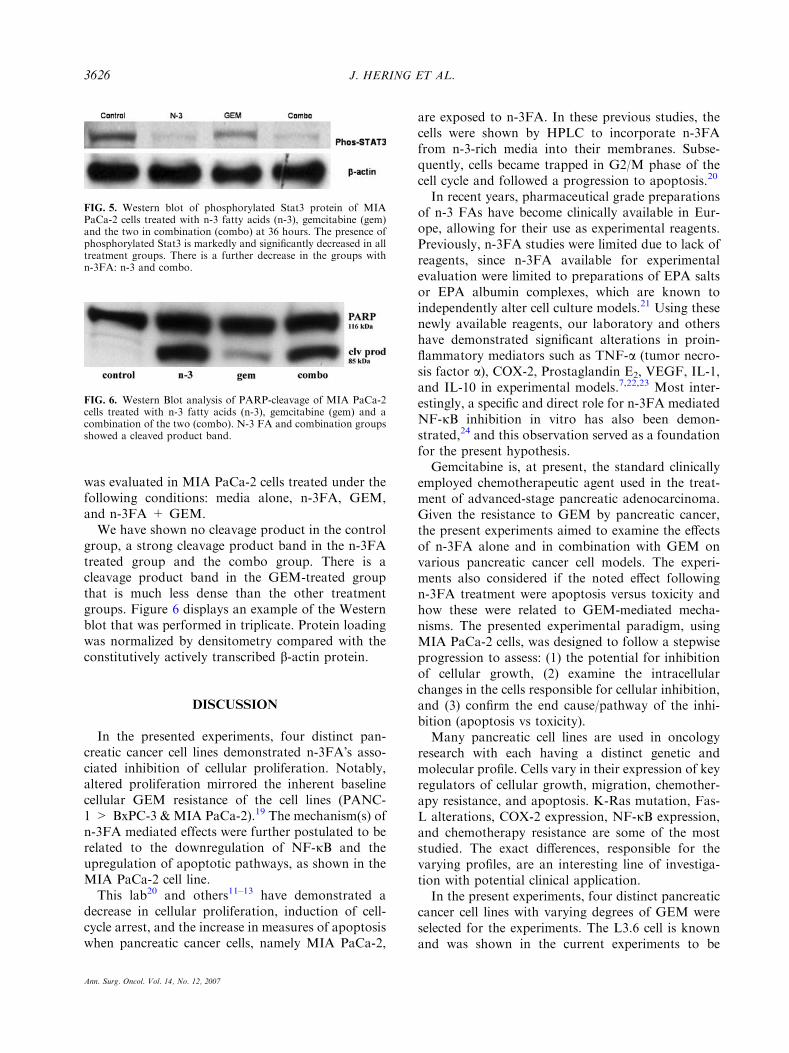
was evaluated in MIA PaCa-2 cells treated under thefollowing conditions: media alone, n-3FA, GEM,and n-3FA + GEM.We have shown no cleavage product in the control
group, a strong cleavage product band in the n-3FAtreated group and the combo group. There is acleavage product band in the GEM-treated groupthat is much less dense than the other treatmentgroups. Figure 6 displays an example of the Westernblot that was performed in triplicate. Protein loadingwas normalized by densitometry compared with theconstitutively actively transcribed b-actin protein.
DISCUSSION
In the presented experiments, four distinct pan-creatic cancer cell lines demonstrated n-3FA�s asso-ciated inhibition of cellular proliferation. Notably,altered proliferation mirrored the inherent baselinecellular GEM resistance of the cell lines (PANC-1 > BxPC-3 & MIA PaCa-2).19 The mechanism(s) ofn-3FA mediated effects were further postulated to berelated to the downregulation of NF-jB and theupregulation of apoptotic pathways, as shown in theMIA PaCa-2 cell line.This lab20 and others11–13 have demonstrated a
decrease in cellular proliferation, induction of cell-cycle arrest, and the increase in measures of apoptosiswhen pancreatic cancer cells, namely MIA PaCa-2,
are exposed to n-3FA. In these previous studies, thecells were shown by HPLC to incorporate n-3FAfrom n-3-rich media into their membranes. Subse-quently, cells became trapped in G2/M phase of thecell cycle and followed a progression to apoptosis.20
In recent years, pharmaceutical grade preparationsof n-3 FAs have become clinically available in Eur-ope, allowing for their use as experimental reagents.Previously, n-3FA studies were limited due to lack ofreagents, since n-3FA available for experimentalevaluation were limited to preparations of EPA saltsor EPA albumin complexes, which are known toindependently alter cell culture models.21 Using thesenewly available reagents, our laboratory and othershave demonstrated significant alterations in proin-flammatory mediators such as TNF-a (tumor necro-sis factor a), COX-2, Prostaglandin E2, VEGF, IL-1,and IL-10 in experimental models.7,22,23 Most inter-estingly, a specific and direct role for n-3FA mediatedNF-jB inhibition in vitro has also been demon-strated,24 and this observation served as a foundationfor the present hypothesis.Gemcitabine is, at present, the standard clinically
employed chemotherapeutic agent used in the treat-ment of advanced-stage pancreatic adenocarcinoma.Given the resistance to GEM by pancreatic cancer,the present experiments aimed to examine the effectsof n-3FA alone and in combination with GEM onvarious pancreatic cancer cell models. The experi-ments also considered if the noted effect followingn-3FA treatment were apoptosis versus toxicity andhow these were related to GEM-mediated mecha-nisms. The presented experimental paradigm, usingMIA PaCa-2 cells, was designed to follow a stepwiseprogression to assess: (1) the potential for inhibitionof cellular growth, (2) examine the intracellularchanges in the cells responsible for cellular inhibition,and (3) confirm the end cause/pathway of the inhi-bition (apoptosis vs toxicity).Many pancreatic cell lines are used in oncology
research with each having a distinct genetic andmolecular profile. Cells vary in their expression of keyregulators of cellular growth, migration, chemother-apy resistance, and apoptosis. K-Ras mutation, Fas-L alterations, COX-2 expression, NF-jB expression,and chemotherapy resistance are some of the moststudied. The exact differences, responsible for thevarying profiles, are an interesting line of investiga-tion with potential clinical application.In the present experiments, four distinct pancreatic
cancer cell lines with varying degrees of GEM wereselected for the experiments. The L3.6 cell is knownand was shown in the current experiments to be
FIG. 5. Western blot of phosphorylated Stat3 protein of MIAPaCa-2 cells treated with n-3 fatty acids (n-3), gemcitabine (gem)and the two in combination (combo) at 36 hours. The presence ofphosphorylated Stat3 is markedly and significantly decreased in alltreatment groups. There is a further decrease in the groups withn-3FA: n-3 and combo.
FIG. 6. Western Blot analysis of PARP-cleavage of MIA PaCa-2cells treated with n-3 fatty acids (n-3), gemcitabine (gem) and acombination of the two (combo). N-3 FA and combination groupsshowed a cleaved product band.
J. HERING ET AL.3626
Ann. Surg. Oncol. Vol. 14, No. 12, 2007
highly GEM sensitive. The BxPC-3 and MIA-PaCA-2 cell lines have an intermediate sensitivity, while thePANC-1 line is resistant to GEM, as confirmed in thepresent experiments. The MIA-PaCa-2 cell line waschosen to conduct the mechanism experiments forseveral reasons: (1) our lab has extensive experiencewith this cell line, (2) it is ubiquitous in the literature,and (3) it has an intermediate GEM sensitivity inrelation to other cell lines. Future experiments, tofurther validate the paradigm, are proceeding on theremaining three cell lines to further support thehypothesis.The completed experiments support several
important and novel findings with possible clinicalimplications: (1) the proliferation data demonstratethat when pancreatic cancer cells, of varying geneticexpressions, are treated with n-3FA�s baseline pro-liferation is significantly reduced. Even the cell linePANC-1, which proliferates in the presence of GEMalone demonstrates inhibited proliferation whenn-3FA�s are added. (2) The I-jB and NF-jB datasupport that n-3FA�s attenuate the progressiontoward the transcriptional sequelae of NF-jB acti-vation that has been previously described.25 GEMdoes not significantly change the phosphorylation ofIjB, but there is a significant change in the combi-nation treatment results as demonstrated by theNF-jB EMSA data. (3) Phosphorylation of Stat3was observed to be significantly decreased subsequentto n-3FA�s treatment. Similarly, GEM had a similareffect on the phosphorylation. (4) The presence of thePARP cleavage product supports progressionthrough apoptosis rather than toxicity. N-3FAtreatment, in this model, shows a strong positivecorrelation with this marker; while there does notseem to be a synergistic effect supported by the PARPcleavage product data, there is a mild proapoptoticeffect noted with GEM only treatment.A proposed mechanism of n-3FA activity on the
described pancreatic cancer cell resistance is that theapoptotic regulatory processes are restored to a sys-tem that responds to apoptotic signals, through thedownregulation of activated NF-jB and Stat3. Thisconcept of ‘‘restored apoptosis’’ has been described inthe literature26,27 in other pancreatic cancer models,and this approach has the potential to overcometumoral chemoresistance, as was observed in ourn-3FA model.Taken as a whole, these data suggest that pancre-
atic cancer cells treated with n-3FA alone or incombination with GEM produce an increased inhi-bition of proliferation, and the mechanism of thisinhibition is through progression to apoptosis, not
toxicity. A further important clinical implication forthese findings is that there is no negative effect for theuse of n-3FA as an adjunct with gemcitabine, spe-cifically; gemcitabine-induced cell killing is not less-ened by n-3FA presence in vitro. N-3FA�s may be animportant nutritional addition to current pancreaticcancer treatment regimens.
REFERENCES
1. American Cancer Society: Cancer Facts and Figures 2007.Available http://www.cancer.org/downloads/STT/CAFF2007PWSecured.pdf. [accessed April 18, 2004].
2. Bromberg JF, Wrzeszczynska MH, Devgan G, Zhao Y, PestellRG, Albanese C, Darnell JE Jr. Stat3 as an oncogene. Cell1999; 98:295–303.
3. Catlett-Falcone R, Landowski TH, Oshiro MM, Turkson J,Levitzki A, Savino R, Ciliberto G, Moscinski L, Fernandez-Luna JL, Nunez G, Dalton WS, Jove R. Constitutive activa-tion of Stat3 signaling confers resistance to apoptosis in humanU266 myeloma cells. Immunity 1999; 10:105–15.
4. Garcia R, Jove R. Activation of STAT transcription factors inoncogenic tyrosine kinase signaling. J Biomed Sci 1998; 5:79–85.
5. Greten FR, Weber CK, Greten TF, Schneider G, Wagner M,Adler G, Schmid RM. Stat3 and NF-kappaB activation pre-vents apoptosis in pancreatic carcinogenesis. Gastroenterology2002; 123:2052–63.
6. Garcea G, Dennison AR, Steward WP, Berry DP. Role ofinflammation in pancreatic carcinogenesis and the implicationsfor future therapy. Pancreatology 2005; 5:514–29.
7. Arlt A, Gehrz A, Muerkoster S, Vorndamm J, Kruse ML,Folsch UR, Schafer H. Role of NF-kappaB and Akt/PI3K inthe resistance of pancreatic carcinoma cell lines against gem-citabine-induced cell death. Oncogene 2003; 22:3243–51.
8. Falconer JS, Fearon KC, Ross JA, Carter DC. Polyunsatu-rated fatty acids in the treatment of weight-losing patients withpancreatic cancer. World Rev Nutr Diet 1994; 76:74–6.
9. Wigmore SJ, Barber MD, Ross JA, Tisdale MJ, Fearon KC.Effect of oral eicosapentaenoic acid on weight loss in patientswith pancreatic cancer. Nutr Cancer 2000; 36:177–84.
10. Fearon KC, Von Meyenfeldt MF, Moses AG, Van GeenenR, Roy A, Gouma DJ, Giacosa A, Van Gossum A, Bauer J,Barber MD, Aaronson NK, Voss AC, Tisdale MJ. Effect ofa protein and energy dense N-3 fatty acid enriched oralsupplement on loss of weight and lean tissue in cancer ca-chexia: a randomised double blind trial. Gut 2003; 52:1479–86.
11. Lai PB, Ross JA, Fearon KC, Anderson JD, Carter DC. Cellcycle arrest and induction of apoptosis in pancreatic cancercells exposed to eicosapentaenoic acid in vitro. Br J Cancer1996; 74(9):1375–83.
12. Jordan A, Stein J. Effect of an omega-3 fatty acid containinglipid emulsion alone and in combination with 5-fluorouracil(5-FU) on growth of the colon cancer cell line Caco-2. Eur JNutr 2003; 42:324–31.
13. Sharma A, Belna J, Logan J, Espat J, Hurteau JA. Theeffects of Omega-3 fatty acids on growth regulation of epi-thelial ovarian cancer cell lines. Gynecol Oncol 2005; 99:58–64.
14. Falconer JS, Ross JA, Fearon KC, Hawkins RA, O�RiordainMG, Carter DC. Effect of eicosapentaenoic acid and otherfatty acids on the growth in vitro of human pancreatic cancercell lines. Br J Cancer 1994; 69:826–32.
N-3 FAS IN PANCREATIC CANCER CELLS 3627
Ann. Surg. Oncol. Vol. 14, No. 12, 2007
15. Babcock TA, Helton WS, Hong D, Espat NJ. Omega-3 fattyacid lipid emulsion reduces LPS-stimulated macrophage TNF-alpha production. Surg Infect 2002; 3:145–9.
16. Babcock TA, Novak T, Ong E, Jho DH, Helton WS, EspatNJ. Modulation of lipopolysaccharide-stimulated macrophagetumor necrosis factor-alpha production by omega-3 fatty acidis associated with differential cyclooxygenase-2 proteinexpression and is independent of interleukin-10. J Surg Res2002; 107:135–9.
17. Shirota T, Haji S, Yamasaki M, Iwasaki T, Hidaka T,Takeyama Y, Shiozaki H, Ohyanagi H. Apoptosis in humanpancreatic cancer cells induced by eicosapentaenoic acid.Nutrition 2005; 21:1010–7.
18. Ross JA, Maingay JP, Fearon KC, Sangster K, Powell JJ.Eicosapentaenoic acid perturbs signaling via the NFkB tran-scriptional pathway in pancreatic tumour cells. Int J Onc 2003;23:1733–8.
19. Doxbury M, Ito H, Zinner M, Ashley S, Whang E. siRNA di-rected against c-SRC enhances pancreatic adenocarcinoma cellgemcitabine chemosensitivity. M. JACS V 198, 6, pp. 953–9.
20. G2/M Cell-Cycle Arrest and Apoptosis by n-3 Fatty Acids in aPancreatic Cancer Model. Dekoj T, Lee S, Desai S, Trevino J,Babcock TA, Helton WS, Espat NJ. J Surg Res 2007 May 1;139(1):106–12.
21. Tevar R, Jho DH, Babcock T, Helton WS, Espat NJ. Omega-3fatty acid supplementation reduces tumor growth and vascular
endothelial growth factor expression in a model of progressivenon-metastasizing malignancy. J Parenter Enteral Nutr 2002;26:285–9.
22. Dommels YE, Haring MM, Keestra NG, Alink GM, vanBladeren PJ, van Ommen B. The role of cyclooxygenase in n-6and n-3 polyunsaturated fatty acid mediated effects on cellproliferation, PGE(2) synthesis and cytotoxicity in humancolorectal carcinoma cell lines. Carcinogenesis 2003; 24:385–92.
23. Novak TE, Babcock TA, Jho DH, Helton WS, Espat NJ.NF-kappa B inhibition by omega -3 fatty acids modulatesLPS-stimulated macrophage TNF-alpha transcription. Am JPhysiol Lung Cell Mol Physiol 2003; 284:L84–L89.
24. Ko AH, Tempero MA. Systemic therapy for pancreatic cancer.Semin Radiat Oncol 2005; 15:245–53.
25. Schniewind B, Christgen M, Kurdow R, Haye S, Kremer B,Kalthoff H, Ungefroren H. Resistance of pancreatic cancer togemcitabine treatment is dependent on mitochondria-mediatedapoptosis. Int J Cancer 2004; 109:182–8.
26. Shi X, Liu S, Kleeff J, Friess H, Buchler MW. Acquiredresistance of pancreatic cancer cells towards 5-Fluorouraciland gemcitabine is associated with altered expression ofapoptosis-regulating genes. Oncology 2002; 62:354–62.
27. Ross JA, Maingay JP, Fearon KC, Sangster K, Powell JJ.Eicosapentaenoic acid perturbs signalling via the NFkappaBtranscriptional pathway in pancreatic tumour cells. Int J Oncol2003; 23:1733–8.
J. HERING ET AL.3628
Ann. Surg. Oncol. Vol. 14, No. 12, 2007
Copyright © 2022 FDOKUMEN