Considering the role of pyruvate in tumor cells during hypoxia

ChREBP binding to fatty acid synthase and L-type
pyruvate kinase genes is stimulated by glucose in
pancreatic b-cells
Gabriela da Silva Xavier,1,† Guy A. Rutter,1,† Frederique Diraison,* Chrysovalantis Andreolas,*and Isabelle Leclerc2,†
From the Henry Wellcome Laboratories for Integrated Cell Signalling,* Department of Biochemistry,University of Bristol, Bristol, BS8 1TD; and Section of Cell Biology,† Division of Medicine, Imperial College,Exhibition Road, London SW7 2AZ, UK
Abstract Pancreatic b-cell dysfunction is central to thepathogenesis of type 2 diabetes and may involve secretoryfailure through glucolipotoxity. The relative importance of thetranscription factors carbohydrate-responsive element bind-ing protein (ChREBP), sterol-responsive element bindingprotein-1c (SREBP-1c), andupstream stimulatory factor (USF)in the induction of lipogenic genes by glucose remains unclear.By confocal imaging, we show that ChREBP translocates to thenucleus in MIN6 b cells in response to glucose. Both ChREBPandSREBP-1cwere required for the induction of the fatty acidsynthase (FAS) promoter by glucose, and chromatin immuno-precipitation (ChIP) assay revealed that glucose induced thebinding of both ChREBP and SREBP-1c to the FAS promoterwithout affecting USF2 binding. By contrast, ChIP assay re-vealed that high glucose prompted direct binding of ChREBP,but not SREBP-1c or USF2, to the liver-type pyruvate kinase(L-PK) promoter. This event was indispensable for the in-duction of the L-PK gene by glucose, as demonstrated by RNAsilencing, single-cell promoter analysis, and quantitative real-time PCR. We conclude that ChREBP is a critical regulatorof lipogenic genes in the b cell and may play a role in thedevelopment of glucolipotoxicity and b cell failure throughalteration of gene expression in type 2 diabetes.—da SilvaXavier, G., G. A. Rutter, F. Diraison, C. Andreolas, and I.Leclerc. ChREBP binding to fatty acid synthase and L-typepyruvate kinase genes is stimulated by glucose in pancreaticb-cells. J. Lipid Res. 2006. 47: 2482–2491.
Supplementary key words glucolipotoxicity . lipogenic genes .
chromatin immunoprecipitation
Carbohydrate response element binding protein (ChREBP,also known as WBSCR14 or Mondo B) (1) is a recentlydescribed transcription factor belonging to the basic helix-loop-helix leucine zipper family (2), which regulates car-bohydrate metabolism in the liver (3). Mice deleted forboth alleles of ChREBP (4) are glucose intolerant and in-
sulin resistant and have diminished rates of glycolysis andlipogenesis. This results in high liver glycogen content, lowplasma free fatty acids levels, and reduced adipose tissuemass. Plasma insulin levels are normal despite elevatedglucose concentrations, suggesting a defect in pancreaticb cell glucose sensing and/or insulin secretion.
In hepatocytes, ChREBP activity is regulated acutely by aseries of phosphorylation/dephosphorylation reactions, al-though this has recently been challenged (5). At low glucoseconcentrations, ChREBP exists in an inactive, phosphory-lated state in the cytosol. There are twomajor protein kinaseA (PKA) phosphorylation sites, the first located near thenuclear localization signal at Ser196 and the second near theDNA binding site at Thr666. Dephosphorylation of Ser196
allows ChREBP entry into the nucleus, whereas dephos-phorylation of Thr666 allows DNA binding. AMP-activatedprotein kinase (AMPK) also regulates ChREBP by phos-phorylation of Ser568, a modification that diminishes itsDNA binding ability (6–8). Max-like protein X is the func-tional heteromeric partner of ChREBP in regulating theexpression of glucose-responsive genes in hepatocytes (9).
Sustained hyperglycemia promotes fatty acid synthesisand triglyceride accumulation in the b cell. This “gluco-lipotoxicity” has been suggested by others (10), and our-selves (11, 12), to contribute to insulin secretory defectsseen in type 2 diabetes. We have previously identified sterol-responsive element binding protein-1c (SREBP-1c) as animportant transcription factor responsible for lipid accu-mulation in pancreatic islets in response to high glucose
Manuscript received 5 July 2006 and in revised form 2 August 2006.
Published, JLR Papers in Press, August 4, 2006.DOI 10.1194/jlr.M600289-JLR200
Abbreviations: ACC, acetyl-CoA carboxylase; AMPK, AMP-activatedprotein kinase; ChIP, chromatin immunoprecipitation; ChoRE, carbo-hydrate-responsive element; ChREBP, carbohydrate-responsive elementbinding protein; FAS, fatty acid synthase; L-PK, liver-type pyruvatekinase; PKA, protein kinase A; siRNA, small interfering RNA; SREBP-1c,sterol-responsive element binding protein-1c; TG, triglyceride; USF,upstream stimulatory factor.
1 G. da Silva Xavier and G. A. Rutter contributed equally to this work.2 To whom correspondence should be addressed.e-mail: [email protected]
Copyright D 2006 by the American Society for Biochemistry and Molecular Biology, Inc.
This article is available online at http://www.jlr.org2482 Journal of Lipid Research Volume 47, 2006
by guest, on Novem
ber 21, 2013w
ww
.jlr.orgD
ownloaded from

through stimulation of acetyl-CoA carboxylase-1 (ACC1)(11) and fatty acid synthase (FAS) (12) gene expression.
Because ChREBP is expressed in the pancreatic islets ina glucose-inducible manner, and forced overexpression ofChREBP in INS1 cells resulted in a left-shift of the liver-type pyruvate kinase (L-PK) gene in response to glucose(13), we have explored the role of ChREBP in the regu-lation of lipogenic genes in the b cell using both inac-tivation and overexpression strategies. Our results indicatethat ChREBP binds directly to the endogenous promotersof the L-PK and FAS genes in a glucose-dependent butinsulin-independent manner in MIN6 cells and is abso-lutely required for their induction by glucose. ChREBP istherefore potentially involved in the alteration of geneexpression during the b cell compensation phase in theearly stage of type 2 diabetes and could contribute to thedevelopment of b cell failure through lipoapoptosis.
MATERIALS AND METHODS
Materials
The Silencer small interfering RNA (siRNA) construction kitwas from Ambion (Huntingdon, UK). Primers for siRNA con-struction and PCR were from MWG Biotech (Milton Keynes,UK). TransITTM -TKO transfection reagent was from Mirus Corp.(Madison, WI). Rat insulin radioimmunoasssay kit was from Linco(St. Charles, MO). Tissue culture reagents were from Sigma-Aldrich (Dorset, UK) or Gibco-BRL (Paisley, UK). Lipofectamine2000TM and SYBR Green platinum were from Invitrogen (Paisley,UK). Anti-upstream stimulatory factor-2 (anti-USF2) antibodywas a kind gift from Dr. Benoit Viollet (Institut Cochin, Paris).Anti-SREBP-1c antibody was from Santa Cruz Biotechnology, Inc.Other reagents were from Sigma or BDH.
Plasmids and adenoviruses
pChREBP was generated by RT-PCR fromMIN6 cell total RNAusing the following primers: 59-ATG GAA CAG AAG CTT ATTTCT GAA GAA GAT CTT GCG CGC GCG CTG GCG GAT CTATCC GTG AAC-39 (c-myc sequence underlined) and 59-CGC GGAGCT CTT ATA ATG GTC TCC CCA GGG TGC CCT CTG TGACTG C-39. The resulting cDNA was phosphorylated by T4 poly-nucleotide kinase (Roche) and subcloned into pcDNA3 (Invi-trogen) vector linearized with EcoRV. pFAS.LucFF was generatedby subcloning the XhoI fragment (�2187 to 165) of the rat FASgene (14) into pGL3 basic (Promega) vector linearized with XhoI.All the constructs were verified by DNA sequencing. pLPK.LucFFhas been described previously (15), and plasmids encoding con-stitutively active and dominant-negative forms of SREBP-1c wereas described in (11). Adenovirus encoding for the SREBP-1cdominant negative (SREBP-DN) control adenovirus expressinggreen fluorescent protein alone, and the infection procedurewere as described (11).
Generation of anti-ChREBP antibody
Polyclonal anti-ChREBP antibody was generated in rabbitsimmunized with a synthetic peptide corresponding to the C ter-minus of mouse and rat ChREBP [(C)ATRAVTEGTLGRPL],conjugated to keyhole limpet hemocyanin (KLH, Calbiochem)using sulfosuccinimidyl 4-(N-maleimidomethyl) cyclohexane-1-carboxylate (Sulfo-SMCC; Pierce, Tattenhall, UK). Briefly, 10 mgKLH was gently dissolved in 2 ml PBS on a wheel rotator before
the addition of 2 mg Sulfo-SMCC and left on the wheel rotatorfor a further 3 h at room temperature. A Sephadex G-25 column(PD-10, Pharmacia Biotech) was equilibrated with 25 ml PBS, andthe 2 ml KLH/Sulfo-SMCC mixture was loaded onto the columnand eluted with 3.5 ml PBS. ChREBP peptide antigen (2.5 mg)was added to the eluate and left on a wheel rotator for 1 h at roomtemperature and overnight at 4jC. A Sephadex G-25 column wasequilibrated with 25 ml H2O, loaded with the 3.5 ml peptide-antigen mixture and eluted with 3.5 ml H2O. The resultingconjugated peptide was stored at �20jC until further use. Im-mediately before rabbit immunization, 1 ml of conjugated pep-tide antigen was emulsified with an equal volume of CompleteFreund’s Adjuvant (Sigma). After taking preimmune serum fromthe rabbits, 800 ml of the emulsion was injected per rabbit, sub-cutaneously, at four different sites (200 ml/site) on the back, atweek 0. Three booster injections in which the peptide antigenwas emulsified with Incomplete Freund’s Adjuvant (Sigma) weregiven as above at weeks 3, 6, and 9. Serum was taken at weeks 5and 8, and a termination bleed was done at week 10. Purificationof total IgG fraction was performed using caprylic acid and ammo-nium sulfate sequential precipitations, as described in (16).
MIN6 cell culture
Cells were cultured in a humidified atmosphere at 37jC with5% CO2. MIN6 b cells (17) were used between passages 19 and 30and grown in DMEM containing 15% (v/v) heat-inactivated fetalcalf serum, 25 mM glucose, 5.4 mM KCl, 2 mM glutamine, 25 mMb-mercaptoethanol, 100 IU/ml�1 penicillin, and 100 mg/ml�1
streptomycin. MIN6 cells were seeded on poly-L-lysine-coatedcoverslips prior to microinjection. Plasmids and siRNAs weretransfected into cells with Lipofectamine 2000TM and TransITTM-TKO, respectively. Transduced cells were cultured in normalmedium for 48 h and further cultured in medium containing3 mM glucose 16 h prior to experiments.
siRNA construction
siRNAs were generated using the Ambion Silencer siRNA con-struction kit according to the manufacturer’s protocol. Startingprimerpairs weredesignedas suggested in theAmbionprotocol andaccording to published guidelines (18–20). Sense and anti-sensetarget sequences were derived from cDNA for ChREBP (GenBankaccessionnumberAB074517)with senseprimer sequence as follows:508-AAACCTGAGGCTGTTGTCTTCCTGTCTC-527 (T7 promotersequence at the 39 end underlined), with the anti-sense primerhaving the complimentary sequence. Control siRNAs were gener-ated with primers derived by scrambling the siRNA sequencefor ChREBP, with the sense primer sequence as follows: AAGGC-TGTTTCACCTGAGTTCCTGTCTC. All primer sequences weresubjected to BLAST searches to ensure that there were no matcheswith known sequences of other genes.MIN6b cells were transfectedwith siRNA at a final concentration of 0.5 nM, according to themanufacturer’s instructions and as described in (21).
Western (immuno) blot analysis
Cells were washed twice in ice-cold PBS, scraped in ice-cold lysisbuffer (PBS, 1% Triton X-100, 5 mg/ml�1 pepstatin, 5 mg/ml�1
antipain, 5 mg/ml�1 leupeptin, 2 mM benzamidine and 0.5 mMdithiothreitol) and vortex-mixed. Protein content was assayedusingaBCATM protein assay kit (Pierce; Rockford, IL), against BSAType V (Sigma) standards. Total protein extracts (50 mg) wereresolved by SDS-PAGE (10% w/v acrylamide) and transferred topolyvinylidene difluoride membranes (Immobilon-P, Millipore),followed by immunoblotting with anti-ChREBP antibody used at1:1000. Anti-rabbit secondary antibody (Amersham) was revealed
ChREBP regulation of lipogenic genes in pancreatic b-cells 2483
by guest, on Novem
ber 21, 2013w
ww
.jlr.orgD
ownloaded from

using BM chemiluminescence blotting substrate (Roche Diag-nostics; Lewes, UK).
Single-cell reporter gene assay
Intranuclear microinjection of plasmids, antibodies, andsiRNAs was performed using an Eppendorf 5121/5246 micro-manipulator at plasmid concentrations of 0.1 (pLPK.LucFF,pPdx1.LucFF, and pFAS.LucFF)) and 0.05 (pCMV-RL) mg/ml�1,and antibody against ChREBP and SREBP at 1 mg/ml�1 as de-scribed (15, 22). Antibody was injected into the nucleus andcytosol of the cell. Individual experiments involved injection of100–200 separate cells per condition, with an efficiency of 5–20%productive injection as assessed by expression of Renilla reniformisluciferase activity. MIN6 cells were imaged 6 h after microinjec-tion and culture under the conditions described in the figurelegends. Photon counting imaging of firefly and R. reniformis lu-ciferase activities was performed in single living cells using anOlympus IX-70 inverted microscope (310 air objective, 0.4 NA)and an intensifed charge-coupled device camera (Photek; EastSussex, UK) as previously described (15, 23).
Insulin secretion
MIN6 cells, seeded in 6-well plates, were grown to 50% con-fluency and transfected with control or anti-ChREBP siRNA for48 h (see above). Culture was continued for 16 h in DMEM con-taining 3 mM glucose. Cells were washed in PBS and incubatedin Krebs Ringer Bicarbonate (KRB) medium containing eitherlow (3 mM) or high (30 mM) glucose concentrations. Incuba-tions were performed for 20 min at 37jC in a shaking water bath.Total and released insulin was measured by radioimmuno-assay (24).
Real-time RT-PCR
Total mRNA was isolated and specific messages quantifiedusing TaqManTM analysis as in (21), and TaqManTM primers andinternal probes or real-time primers (SYBR Green) for amplifi-cation of FAS, b-actin, and cyclophilin were as described in (21)and for L-PK as in (25).
Chromatin immunoprecipitation assay
Chromatin immunoprecipitation was performed as described in(26). MIN6 b cells were cultured in medium containing 3 mMglucose for 16 h prior to stimulation with medium containing 3 or30 mM glucose for 6 h prior to cross-linking with formaldehyde.DNA-protein complexes were immunoprecipitated with antibodiesagainst ChREBP, SREBP-1c, and USF2 and then digested withproteinase K. After phenol-chloroform extraction and ethanol pre-cipitation of the resulting DNA, PCR was performed to amplify theL-PK and FAS promoters. Theprimers used to amplify themouse L-PK promoter from �392 to 194 bp were 59-AAGTCTGCTTTAAG-TGGGGCT-39 and 59-TCCCCTCCAAGTTCCCTCCAT-39; for theFAS promoter from �135 to �14 bp, 59-CGGCGCGCCGGG-TCCCGGGG-39 and 59-CCGCGCCGGCGCTATTTAAA-39; and forthe FAS promoter from �360 to �222 bp, 59-CGCCCGACCCA-CACTG-39 and 59-CGGACGAAGGCAGGGGC-39.
Measurement of triglyceride content
Cells were transfected with control or ChREBP siRNA for 48 h,maintained for 16 h at 3 mM glucose, and then in 3 or 30 mMglucose for 24 h before measuring triglycerides (TGs) and pro-tein concentrations. Total lipids were extracted using chloro-form-methanol (2:1; v/v) as in (27). Extracted lipids were airdried, and 10 ml of a detergent (Thesit; Fluka, Gillingham,
Dorset, UK) was added to the dry pellet. Samples were air driedagain and resuspended in 50 ml H2O (28). TG was measuredusing a commercial kit (InfinityTM Triglyceride Reagent, Sigma)and a standard curve of triolein (Sigma) treated in parallel withthe samples. Protein concentration was determined using a BCAkit (Pierce; Rockford, IL).
Immunocytochemistry
Cells were fixed in 4% (v/v) paraformaldeyhde and 0.3%Triton X-100 and stained with anti-c -myc antibody (9E:10; 1:200),and revealed with fluorescein-conjugated goat anti-mouse IgG(Sigma) (22). Images were captured using a Leica SP2 laserscanning confocal microscope (363 objective).
Statistical analysis
Data are given as means 6 SEM. Comparisons between meanswere performed by unpaired one-tailed Student’s t-test withBonferroni correction as appropriate, using Microsoft Excel.
RESULTS
Elevated glucose concentrations cause nucleartranslocation of ChREBP in MIN6 bcells
To visualize the subcellular localization of ChREBP at lowand high glucose concentrations, the relatively well differ-entiated b cell line MIN6 was transfected with c -myc-taggedpChREBP expression vector and incubated for 24 h at 3mMglucose and then at 30mMglucose for a further 16 and 24 hbefore fixation and immunocytochemical analysis,Hoeschststaining, and confocal imaging as described inMaterials andMethods (Fig. 1A, B). The proportion of cells expressingChREBP in the nucleus increased from 31% to 55% after16 h at high glucose and to 69% after 24 h.
ChREBP silencing decreases triglyceride content andpotentiates glucose-stimulated insulin secretion inMIN6 cells
To determine the importance of ChREBP in the occur-rence of glucolipotoxicity in MIN6 cells, ChREBP expres-sion was silenced using siRNAs. ChREBP protein contentwas decreased by 87.32 6 0.25% and 65.59 6 0.52% at48 and 72 h, respectively, as assessed by Western blot(Fig. 1C), and ChREBP message content was decreased by.95% at 48 h as assessed by quantitative real-time RT-PCR(Fig. 1D). ChREBP cellular depletion significantly de-creased TG content at both 3 and 30 mM glucoseconcentrations (Fig. 1E) and slightly enhanced secretedinsulin at 30 mM glucose (Fig. 1F).
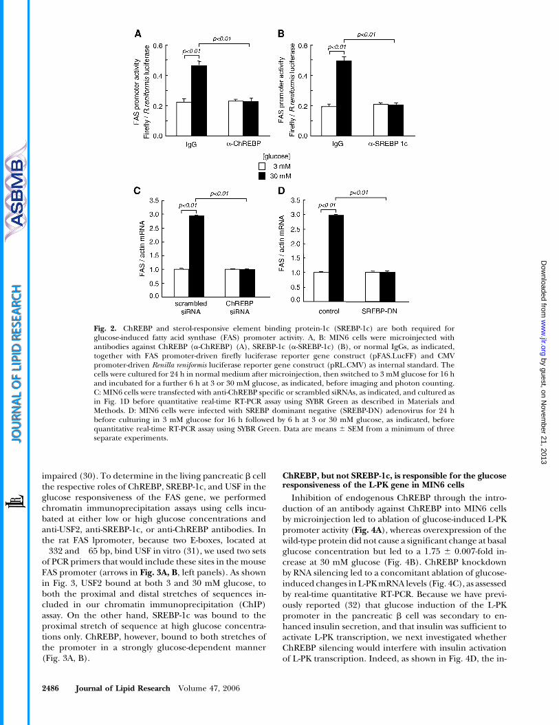
Both ChREBP and SREBP-1c are required forglucose-stimulated FAS gene expression in MIN6 cells
Given the decrease in MIN6 cellular TG content fol-lowing ChREBP silencing (Fig. 1E), we next investigatedthe relative importance of ChREBP versus SREBP-1c in theregulation of lipogenic genes by glucose in this cell type.Figure 2 shows that both ChREBP and SREBP-1c arerequired for glucose-stimulated FAS gene expression insingle MIN6 cells. Thus, the glucose responsiveness of themouse FAS promoter was completely abolished following
2484 Journal of Lipid Research Volume 47, 2006
by guest, on Novem
ber 21, 2013w
ww
.jlr.orgD
ownloaded from

cellular microinjection of antibodies against eitherChREBP (Fig. 2A) or SREBP-1c (Fig. 2B) together withpFAS.LucFF and pRL.CMV constructs. Similarly, glucoseinduction of endogenous FAS mRNAs was inhibited in cellpopulations following ChREBP silencing using siRNA(Fig. 2C) or SREBP-1c inactivation with SREBP-1c-DN ade-novirus (Fig. 2D).
Glucose promotes the binding of ChREBP and SREBP-1cto the FAS promoter in vivo
Previous studies using SREBP-1�/� knock-out mice haveshown the absolute requirement for this factor for theinduction of the FAS gene by fasting/refeeding in the liver(29). Also, in USF1 and USF2�/� knock-out mice, theinduction of FAS by fasting/refeeding was significantly
Fig. 1. Glucose regulates carbohydrate-responsive element binding protein (ChREBP) cellular localizationand expression in MIN6 cells. A: MIN6 cells were seeded on poly-L-lysine-coated coverslips and transfectedwith pChREBP using Lipofectamine 2000TM. The cells were cultured in normal medium (25 mM glucose) for24 h after transfection and then incubated in 3 mM glucose for 16 h and in either 3 or 30mM glucose for afurther 24 h, as indicated. ChREBP distribution and nuclear DNA were detected using anti-c -myc monoclonalantibody and Hoechst 33258 staining, respectively, prior to confocal imaging and cell counting (see Materialsand Methods). B: Graphical representation of the experiment described in (A). C, D: MIN6 cells weretransfected with ChREBP specific and scrambled small interfering RNAs (siRNAs) using TransITTM-TKO andcultured in normal medium for 48 h or 72 h, as indicated, before Western blotting (C), or for 48 h in normalmedium and 16 h in 3 mM glucose, followed with 6 h at 3 or 30 mM glucose, as indicated, before quantitativereal-time RT-PCR assay (D). E, F: MIN6 cells were cultured as in (C) and triglycerides content and insulinsecretion were performed as described in Materials and Methods. Data are means 6 SEM from a minimumof three separate experiments. **P , 0.05 for the effect of specific versus scrambled siRNAs.
ChREBP regulation of lipogenic genes in pancreatic b-cells 2485
by guest, on Novem
ber 21, 2013w
ww
.jlr.orgD
ownloaded from
impaired (30). To determine in the living pancreatic b cellthe respective roles of ChREBP, SREBP-1c, and USF in theglucose responsiveness of the FAS gene, we performedchromatin immunoprecipitation assays using cells incu-bated at either low or high glucose concentrations andanti-USF2, anti-SREBP-1c, or anti-ChREBP antibodies. Inthe rat FAS lpromoter, because two E-boxes, located at�332 and �65 bp, bind USF in vitro (31), we used two setsof PCR primers that would include these sites in the mouseFAS promoter (arrows in Fig. 3A, B, left panels). As shownin Fig. 3, USF2 bound at both 3 and 30 mM glucose, toboth the proximal and distal stretches of sequences in-cluded in our chromatin immunoprecipitation (ChIP)assay. On the other hand, SREBP-1c was bound to theproximal stretch of sequence at high glucose concentra-tions only. ChREBP, however, bound to both stretches ofthe promoter in a strongly glucose-dependent manner(Fig. 3A, B).
ChREBP, but not SREBP-1c, is responsible for the glucoseresponsiveness of the L-PK gene in MIN6 cells
Inhibition of endogenous ChREBP through the intro-duction of an antibody against ChREBP into MIN6 cellsby microinjection led to ablation of glucose-induced L-PKpromoter activity (Fig. 4A), whereas overexpression of thewild-type protein did not cause a significant change at basalglucose concentration but led to a 1.75 6 0.007-fold in-crease at 30 mM glucose (Fig. 4B). ChREBP knockdownby RNA silencing led to a concomitant ablation of glucose-induced changes in L-PKmRNA levels (Fig. 4C), as assessedby real-time quantitative RT-PCR. Because we have previ-ously reported (32) that glucose induction of the L-PKpromoter in the pancreatic b cell was secondary to en-hanced insulin secretion, and that insulin was sufficient toactivate L-PK transcription, we next investigated whetherChREBP silencing would interfere with insulin activationof L-PK transcription. Indeed, as shown in Fig. 4D, the in-
Fig. 2. ChREBP and sterol-responsive element binding protein-1c (SREBP-1c) are both required forglucose-induced fatty acid synthase (FAS) promoter activity. A, B: MIN6 cells were microinjected withantibodies against ChREBP (a-ChREBP) (A), SREBP-1c (a-SREBP-1c) (B), or normal IgGs, as indicated,together with FAS promoter-driven firefly luciferase reporter gene construct (pFAS.LucFF) and CMVpromoter-driven Renilla reniformis luciferase reporter gene construct (pRL.CMV) as internal standard. Thecells were cultured for 24 h in normal medium after microinjection, then switched to 3 mM glucose for 16 hand incubated for a further 6 h at 3 or 30 mM glucose, as indicated, before imaging and photon counting.C: MIN6 cells were transfected with anti-ChREBP specific or scrambled siRNAs, as indicated, and cultured asin Fig. 1D before quantitative real-time RT-PCR assay using SYBR Green as described in Materials andMethods. D: MIN6 cells were infected with SREBP dominant negative (SREBP-DN) adenovirus for 24 hbefore culturing in 3 mM glucose for 16 h followed by 6 h at 3 or 30 mM glucose, as indicated, beforequantitative real-time RT-PCR assay using SYBR Green. Data are means 6 SEM from a minimum of threeseparate experiments.
2486 Journal of Lipid Research Volume 47, 2006
by guest, on Novem
ber 21, 2013w
ww
.jlr.orgD
ownloaded from

duction by insulin of L-PK mRNA at low glucose concen-tration was blunted following ChREBP knockdown. On theother hand, overexpression of the constitutively activenuclear fragment of SREBP-1c had no effect on L-PK pro-moter activity (Fig. 4E), nordid theoverexpressionofdomi-nant negative SREBP-1c affect L-PK mRNA levels (Fig. 4F).
ChREBP, but not SREBP-1c, binds to the L-PK promoterin a glucose-dependent but insulin-independent manner inliving cells
The carbohydrate-responsive element (ChoRE) of theL-PK promoter is well characterized and is centered at�177 bp on the mouse promoter (Fig. 5A, underlined)(33). ChIP assay revealed that ChREBP bound directly tothis segment of the L-PK promoter at 30 but not 3 mMglucose. In contrast, chromatin immunoprecipitationusing anti-USF2 or anti-SREBP-1c antibodies failed topull down this segment of the L-PK promoter (Fig. 5B). Todetermine whether insulin stimulated L-PK promoter ac-tivity directly through ChREBP binding, we performed aChIP assay using ChREBP antibody at low and high glu-cose, and in the presence or absence of 1 nM insulin, orwhile inhibiting insulin release with the hyperpolarizingagent diazoxide. As shown in Fig. 5C, the addition of 1 nMinsulin to MIN6 cells cultured at low glucose did not causeany increase in ChREBP binding, although this maneuver
resulted in the stimulation of L-PK gene expression as as-sessed by quantitative RT-PCR (Fig. 4D). However, in-hibition of glucose-induced insulin secretion with 100 mMdiazoxide completely abolished ChREBP binding at highglucose (Fig. 5C).
DISCUSSION
ChREBP is a key regulator of lipogenic genes in the b cell
We have previously demonstrated that SREBP-1c is animportant regulator of lipogenesis in b cells (11, 12).Thus, overexpression or inactivation of SREBP-1c stronglymodulates the ACC1 (pII) promoter (11). We show herethat SREBP-1c inhibition, achieved through antibodymicroinjection of adenoviral overexpression of SREBP-DN, also abolishes the induction by glucose of FAS pro-moter and mRNA levels in MIN6 cells (Fig. 2). By contrast,SREBP-1c overexpression and inhibition had no effect onL-PK transcription at either low or high glucose concen-trations (Fig. 4E, F), implying a role for other transcriptionfactor(s) in the response of the latter gene to glucose.ChREBP is emerging as a key regulator of lipogenic genesin the liver. Very recently, ChREBP-deficient mice wereintercrossed with ob/ob mice, and the resulting offspringhad a better metabolic profile, as well as decreased ap-
Fig. 3. ChREBP and SREBP-1c directly bind to the FAS promoter in a glucose-dependent manner in vivo.A, B: MIN6 cells were incubated in 3 mM glucose for 16 h and for a further 6 h at 3 or 30 mM glucose,as indicated, before a chromatin immunoprecipitation (ChIP) assay was performed as described in Ma-terials and Methods using antibodies against upstream stimulatory factor-2 (USF2) (a-USF2), SREBP1-c(a- SREBP1-c), or ChREBP (a-ChREBP), as indicated in the figures. The left panels show the primers used(arrows) and the sequence amplified by PCR. The consensus E-boxes and sterol-responsive element site arealso indicated. The right panels show the resulting PCR products using the input fractions (i) and followingimmunoprecipitation with the specific antibodies (ab) at either 3 or 30 mM glucose. Data are representativeof three independent experiments.
ChREBP regulation of lipogenic genes in pancreatic b-cells 2487
by guest, on Novem
ber 21, 2013w
ww
.jlr.orgD
ownloaded from

petite and adiposity, suggesting a role for ChREBP inperipheral tissues, including the brain (34). We show herethat overexpression of ChREBP potentiates the effects ofglucose on the L-PK promoter in b cells (Fig. 4B), con-sistent with previous findings in INS-1 cells (13) as well ashepatocytes (3). However, we now report for the first timethat inactivation of ChREBP, achieved by RNA silencing orantibody microinjection, inhibits the induction of L-PKby glucose in b cells (Fig. 4A, C).
Using direct ChIP assay, we also provide the novel ob-servation that in MIN6 cells, glucose signaling promotesbinding of ChREBP to the carbohydrate response elementof the L-PK promoter. By contrast, neither SREBP-1c norUSF binding to this site was observed at either high or low
glucose concentrations (Fig. 5). These findings wouldappear to question earlier results from this laboratoryimplicating USF as an important regulator of the L-PKgene in b cells (15), possibly reflecting the uncertainspecificity of the antibodies used in these earlier microin-jection studies, but they are consistent with the findings ofKaytor, Shih, and Towle (35), who argue against a role forUSF in the regulation of L-PK gene expression in the liver,and the findings of Wang and Wollheim (13) in INS-1cells. We propose that in b cells, the absence of ChREBPbinding to the L-PK ChoRE at low glucose concentrationsmay be explained by both nuclear exclusion (Fig. 1A) andposttranscriptional modifications, as in hepatocytes (6, 8).However, in contrast to the liver system, the actions of
Fig. 4. ChREBP, but not SREBP-1c, mediates glucose responsiveness of the liver-type pyruvate kinase (L-PK) promoter in MIN6 cells. A, B, E: MIN6 cells were microinjected with normal IgG or anti-ChREBPantibodies (a-ChREBP) (A), with pcDNA3 or pChREBP (B), or SREBP-CA (E), as indicated, together withpLPK.LucFF and pRL.CMV plasmids before incubation and photon counting as in Fig. 2. C, D, F: MIN6 cellswere transfected with scrambled or specific ChREBP siRNAs (C, D) or infected with control or SREBP-DNadenovirus (F), cultured for 24 h in normal medium, then incubated in 3 mM glucose for 16 h andstimulated with 3 or 30 mM glucose, as indicated, for another 6 h before RNA extraction and real-time RT-PCR assay using TaqMan, as described inMaterials andMethods. Data are means6 SEM from aminimum ofthree separate experiments.
2488 Journal of Lipid Research Volume 47, 2006
by guest, on Novem
ber 21, 2013w
ww
.jlr.orgD
ownloaded from

glucose on L-PK expression in MIN6 cells are in large partmediated by the secretion of insulin (32). Although in-sulin is not sufficient to promote ChREBP binding to theL-PK promoter in vivo (Fig. 5C), insulin stimulates the L-PK promoter at low glucose concentrations and ChREBP isnecessary for insulin stimulation of the L-PK promoter(Fig. 4D). Also, inhibiting glucose-induced insulin secre-tion by the use of diazoxide prevents ChREBP binding onthe L-PK promoter (Fig. 5C). The latter observation couldsuggest a role for changes in intracellular free Ca21 con-centrations in the activation of ChREBP, although moreexperiments are needed to confirm this hypothesis. Alter-natively, insulin could be necessary for binding or ac-tivation of a distinct transcription factor that works inconjunction with ChREBP. Further work is required to
determine the respective roles of intracellular glucosemetabolism, insulin release, and the activation or inhibi-tion of protein kinases [e.g., PKA, AMPK, CalmodulinKinase kinase (CamKK)] in modulating the subcellularlocalization of ChREBP and its transcriptional activity inthe pancreatic b cell.
In contrast to the L-PK gene, we demonstrate here thatboth ChREBP and SREBP-1c are necessary for the glucoseresponsiveness of the FAS promoter (Fig. 2). Moreover,both ChREBP and USF2 bound to both stretches of theFAS promoter examined (containing the �65 and �332E-boxes, Fig. 5), ChREBP in a glucose-dependent mannerand USF2 constitutively. The apparent increase in inten-sity following immunoprecipitation with anti-USF2 ob-served at high glucose was not always observed between
Fig. 5. ChREBP binds to the L-PK promoter in a glucose- and calcium-dependent but insulin-independentmanner in vivo. MIN6 cells were incubated for 16 h at 3 mM glucose and then for 6 h at either 3 or 30 mMglucose, in the presence or absence of 1 nM insulin and 100 mM diazoxide, as indicated, before cross-linkingand ChIP assay as in Fig. 3. A: The primers (arrows) and the amplified sequence of the mouse L-PK promoterby PCR are shown. The carbohydrate-responsive element is underlined. B, C: Shown are the resulting PCRproducts following immunoprecipitation using anti-USF2 (a-USF2), anti-SREBP (a-SREBP), or anti-ChREBP(a-ChREBP) antibodies as indicated. i, input fraction; ab, antibody fraction. Data are representative of threeindependent experiments.
ChREBP regulation of lipogenic genes in pancreatic b-cells 2489
by guest, on Novem
ber 21, 2013w
ww
.jlr.orgD
ownloaded from

our replicates. SREBP-1c also bound to the FAS promoterin response to elevated glucose concentrations, but only tothe more proximal segment containing the �65 E-box.The elegant work of Sul et al. (36), using in vivo chromatinimmunoprecipitation from extracts of livers of transgenicmice harboring various truncations and mutations of anFAS promoter CAT reporter gene construct, showed thatSREBP-1c binds only to the �150 sterol response element(SRE) and not to the �65 region containing an E-box andan overlapping SRE (shown in Fig. 3B). The resolution ofthe ChIP method is largely dependent on the fragmentsize obtained on shearing the chromatin, rather than onthe primer sites chosen for PCR. Although the �150 SREsite is theoretically excluded from our ChIP assay, it islocated just outside our �135 primer and may have beenpulled down during immunoprecipitation using theSREBP1 antibody. It is also possible that the transcriptionfactor recruitment might be different in b cells comparedwith liver.
Taken together, these results suggest an important rolefor ChREBP in b cell failure due to lipid accumulationsecondary to chronic high glucose exposure through ac-tivation of lipogenic genes, because ChREBP silencingdecreases triglyceride content at both basal and elevatedglucose concentrations (Fig. 1E), and increases insulinsecretion at high glucose concentrations (Fig. 1F). How-ever, although MIN6 cells are a nice in vitro model, morework is needed to confirm a role for ChREBP in gluco-lipotoxicity in primary islets in vivo.
This work was supported by grants from Diabetes UK (I.L. andG.A.R.), the Medical Research Council, the Biotechnology andBiological Sciences Research Council, and the Wellcome Trust(Program Grant 067081) to G.A.R. We thank Prof. Elek Molnar(Department of Anatomy, University of Bristol) for help in thegeneration of the ChREBP antibody and Dr. Marc Montminy(Salk Institute, San Diego, CA) for advice on ChIP assays. G.A.Rand I.L. are Research Leave and Advanced Fellows, respectively,of the Wellcome Trust.
REFERENCES
1. de Luis, O., M. C. Valero, and L. A. Jurado. 2000. WBSCR14, aputative transcription factor gene deleted in Williams-Beuren syn-drome: complete characterisation of the human gene and themouse ortholog. Eur. J. Hum. Genet. 8: 215–222.
2. Atchley, W. R., and W. M. Fitch. 1997. A natural classification of thebasic helix-loop-helix class of transcription factors. Proc. Natl. Acad.Sci. USA. 94: 5172–5176.
3. Yamashita, H., M. Takenoshita, M. Sakurai, R. K. Bruick, W. J.Henzel, W. Shillinglaw, D. Arnot, and K. Uyeda. 2001. A glucose-responsive transcription factor that regulates carbohydrate metab-olism in the liver. Proc. Natl. Acad. Sci. USA. 98: 9116–9121.
4. Iizuka, K., R. K. Bruick, G. Liang, J. D. Horton, and K. Uyeda.2004. Deficiency of carbohydrate response element-binding pro-tein (ChREBP) reduces lipogenesis as well as glycolysis. Proc. Natl.Acad. Sci. USA. 101: 7281–7286.
5. Li, M. V., B. Chang, M. Imamura, N. Poungvarin, and L. Chan. 2006.Glucose-dependent transcriptional regulation by an evolutionarilyconserved glucose-sensing module. Diabetes. 55: 1179–1189.
6. Kawaguchi, T., K. Osatomi, H. Yamashita, T. Kabashima, and K.Uyeda. 2002. Mechanism for fatty acid “sparing” effect on glucose-induced transcription: regulation of carbohydrate-responsive
element-binding protein by AMP-activated protein kinase. J. Biol.Chem. 277: 3829–3835.
7. Uyeda, K., H. Yamashita, and T. Kawaguchi. 2002. Carbohydrate re-sponsive element-binding protein (ChREBP): a key regulator of glu-cose metabolism and fat storage. Biochem. Pharmacol. 63: 2075–2080.
8. Kawaguchi, T., M. Takenoshita, T. Kabashima, and K. Uyeda. 2001.Glucose and cAMP regulate the L-type pyruvate kinase gene byphosphorylation/dephosphorylation of the carbohydrate responseelement binding protein. Proc. Natl. Acad. Sci. USA. 98: 13710–13715.
9. Stoeckman, A. K., L. Ma, and H. C. Towle. 2004. Mlx is the func-tional heteromeric partner of the carbohydrate response element-binding protein in glucose regulation of lipogenic enzyme genes.J. Biol. Chem. 279: 15662–15669.
10. Prentki, M., E. Joly, W. El-Assaad, and R. Roduit. 2002. Malonyl-CoAsignaling, lipid partitioning, and glucolipotoxicity: role in beta-cell adaptation and failure in the etiology of diabetes. Diabetes. 51(Suppl.): 405–413.
11. Andreolas, C., G. da Silva Xavier, F. Diraison, C. Zhao, A. Varadi,F. Lopez-Casillas, P. Ferre, F. Foufelle, and G. A. Rutter. 2002.Stimulation of acetyl-CoA carboxylase gene expression by glucoserequires insulin release and sterol regulatory element binding pro-tein 1c in pancreatic MIN6 beta-cells. Diabetes. 51: 2536–2545.
12. Diraison, F., L. Parton, P. Ferre, F. Foufelle, C. P. Briscoe, I. Leclerc,and G. A. Rutter. 2004. Over-expression of sterol-regulatory-element-binding protein-1c (SREBP1c) in rat pancreatic islets in-duces lipogenesis and decreases glucose-stimulated insulin release:modulation by 5-aminoimidazole-4-carboxamide ribonucleoside(AICAR). Biochem. J. 378: 769–778.
13. Wang, H., and C. B. Wollheim. 2002. ChREBP rather than USF2regulates glucose stimulation of endogenous L-pyruvate kinase ex-pression in insulin-secreting cells. J. Biol. Chem. 277: 32746–32752.
14. Rolland, V., X. L. Liepvre, D. B. Jump, M. Lavau, and I. Dugail.1996. A GC-rich region containing Sp1 and Sp1-like binding sites isa crucial regulatory motif for fatty acid synthase gene promoteractivity in adipocytes. Implication in the overactivity of FAS pro-moter in obese Zucker rats. J. Biol. Chem. 271: 21297–21302.
15. Kennedy, H. J., B. Viollet, I. Rafiq, A. Kahn, and G. A. Rutter. 1997.Upstream stimulatory factor-2 (USF2) activity is required for glu-cose stimulation of L-pyruvate kinase promoter activity in singleliving islet beta-cells. J. Biol. Chem. 272: 20636–20640.
16. McKinney, M. M., and A. Parkinson. 1987. A simple, non-chromatographic procedure to purify immunoglobulins fromserum and ascites fluid. J. Immunol. Methods. 96: 271–278.
17. Miyazaki, J., K. Araki, E. Yamato, H. Ikegami, T. Asano, Y. Shibasaki,Y. Oka, and K. Yamamura. 1990. Establishment of a pancreatic betacell line that retains glucose-inducible insulin secretion: specialreference to expression of glucose transporter isoforms. Endocri-nology. 127: 126–132.
18. Tuschl, T., P. D. Zamore, R. Lehmann, D. P. Bartel, and P. A. Sharp.1999. Targeted mRNA degradation by double-stranded RNA invitro. Genes Dev. 13: 3191–3197.
19. Elbashir, S. M., J. Harborth, W. Lendeckel, A. Yalcin, K. Weber,and T. Tuschl. 2001. Duplexes of 21-nucleotide RNAs me-diate RNA interference in cultured mammalian cells. Nature. 411:494–498.
20. Elbashir, S. M., W. Lendeckel, and T. Tuschl. 2001. RNA inter-ference is mediated by 21- and 22-nucleotide RNAs. Genes Dev. 15:188–200.
21. Da Silva Xavier, G., Q. Qian, P. J. Cullen, and G. A. Rutter. 2004.Distinct roles for insulin and insulin-like growth factor-1 receptorsin pancreatic beta-cell glucose sensing revealed by RNA silencing.Biochem. J. 377: 149–158.
22. Rafiq, I., G. da Silva Xavier, S. Hooper, and G. A. Rutter. 2000.Glucose-stimulated preproinsulin gene expression and nucleartrans-location of pancreatic duodenum homeobox-1 require activa-tion of phosphatidylinositol 3-kinase but not p38 MAPK/SAPK2.J. Biol. Chem. 275: 15977–15984.
23. Rutter, G. A., H. J. Kennedy, C. D. Wood, M. R. White, and J. M.Tavare. 1998. Real-time imaging of gene expression in single livingcells. Chem. Biol. 5: R285–R290.
24. Ainscow, E. K., and G. A. Rutter. 2001. Mitochondrial primingmodifies Ca21 oscillations and insulin secretion in pancreatic islets.Biochem. J. 353: 175–180.
25. da Silva Xavier, G., I. Leclerc, I. P. Salt, B. Doiron, D. G. Hardie, A.Kahn, and G. A. Rutter. 2000. Role of AMP-activated protein kinasein the regulation by glucose of islet beta cell gene expression. Proc.Natl. Acad. Sci. USA. 97: 4023–4028.
2490 Journal of Lipid Research Volume 47, 2006
by guest, on Novem
ber 21, 2013w
ww
.jlr.orgD
ownloaded from

26. Asahara, H., B. Santoso, E. Guzman, K. Du, P. A. Cole, I. Davidson,and M. Montminy. 2001. Chromatin-dependent cooperativity be-tween constitutive and inducible activation domains in CREB. Mol.Cell. Biol. 21: 7892–7900.
27. Folch, J., M. Lees, and G. H. Sloane Stanley. 1957. A simple methodfor the isolation and purification of total lipides from animaltissues. J. Biol. Chem. 226: 497–509.
28. Briaud, I., J. S. Harmon, C. L. Kelpe, V. B. Segu, and V. Poitout.2001. Lipotoxicity of the pancreatic beta-cell is associated withglucose-dependent esterification of fatty acids into neutral lipids.Diabetes. 50: 315–321.
29. Shimano, H., N. Yahagi, M. Amemiya-Kudo, A. H. Hasty, J. Osuga,Y. Tamura, F. Shionoiri, Y. Iizuka, K. Ohashi, K. Harada, et al. 1999.Sterol regulatory element-binding protein-1 as a key transcriptionfactor for nutritional induction of lipogenic enzyme genes. J. Biol.Chem. 274: 35832–35839.
30. Casado, M., V. S. Vallet, A. Kahn, and S. Vaulont. 1999. Essentialrole in vivo of upstream stimulatory factors for a normal dietary re-sponse of the fatty acid synthase gene in the liver. J. Biol. Chem. 274:2009–2013.
31. Griffin, M. J., and H. S. Sul. 2004. Insulin regulation of fatty acid
synthase gene transcription: roles of USF and SREBP-1c. IUBMBLife. 56: 595–600.
32. da Silva Xavier, G., A. Varadi, E. K. Ainscow, and G. A. Rutter. 2000.Regulation of gene expression by glucose in pancreatic beta-cells(MIN6) via insulin secretion and activation of phosphatidylinositol39-kinase. J. Biol. Chem. 275: 36269–36277.
33. Shih, H. M., Z. Liu, and H. C. Towle. 1995. Two CACGTG motifswith proper spacing dictate the carbohydrate regulation of hepaticgene transcription. J. Biol. Chem. 270: 21991–21997.
34. Iizuka, K., B. Miller, and K. Uyeda. Deficiency of a carbohydrate-activated transcription factor, ChREBP, prevents obesity and im-proves plasma glucose control in leptin deficient (ob/ob) mice. Am.J. Physiol. Endocrinol. Metab. Epub ahead of print. May 16, 2006;doi:10.1152/ajpendo.00027.2006.
35. Kaytor, E. N., H. Shih, and H. C. Towle. 1997. Carbohydrate regu-lation of hepatic gene expression. Evidence against a role for theupstream stimulatory factor. J. Biol. Chem. 272: 7525–7531.
36. Latasa, M. J., M. J. Griffin, Y. S. Moon, C. Kang, and H. S. Sul. 2003.Occupancy and function of the�150 sterol regulatory element and�65 E-box in nutritional regulation of the fatty acid synthase genein living animals. Mol. Cell. Biol. 23: 5896–5907.
ChREBP regulation of lipogenic genes in pancreatic b-cells 2491
by guest, on Novem
ber 21, 2013w
ww
.jlr.orgD
ownloaded from
Copyright © 2022 FDOKUMEN