Inhibition of carrageenan-induced acute inflammation in mice by oral administration of anthocyanin...
10
Hindawi Publishing Corporation BioMed Research International Volume 2013, Article ID 146716, 10 pages http://dx.doi.org/10.1155/2013/146716 Research Article Inhibition of Carrageenan-Induced Acute In�ammation in Mice by Oral Administration of Anthocyanin Mixture from Wild Mulberry and Cyanidin-3-Glucoside Neuza Mariko Aymoto Hassimotto, 1, 2 Vanessa Moreira, 3 Neide Galvão do Nascimento, 3 Pollyana Cristina Maggio de Castro Souto, 3 Catarina Teixeira, 3 and Franco Maria Lajolo 1, 2 1 Laboratório de Química e Bioquímica e Biologia Molecular de Alimentos, Departamento de Alimentos e Nutrição Experimental, FCF, Universidade de São Paulo, Avenida Professor Lineu Prestes 580, Bloco 14, 05508-000 São Paulo, SP, Brazil 2 Núcleo de Apoio à Pesquisa em Alimentos e Nutrição (NAPAN), Universidade de São Paulo, 05508-000 São Paulo, SP, Brazil 3 Laboratório de Farmacologia, Unidade de �n�amação, �nstituto Butantan, Avenida �ital Brasil 1500, 05503-000 São Paulo, SP, Brazil Correspondence should be addressed to Franco Maria Lajolo; [email protected] Received 6 September 2012; Accepted 9 December 2012 Academic Editor: José Carlos Tavares Carvalho Copyright © 2013 Neuza Mariko Aymoto Hassimotto et al. is is an open access article distributed under the Creative Commons Attribution License, which permits unrestricted use, distribution, and reproduction in any medium, provided the original work is properly cited. Anthocyanins are �avonoids which demonstrated biological activities in in vivo and in vitro models. Here in the anti-in�ammatory properties of an anthocyanin-enriched fraction (AF) extracted from wild mulberry and the cyanidin-3-glucoside (C3G), the most abundant anthocyanin in diet, were studied in two acute in�ammation experimental models, in the peritonitis and in the paw oedema assays, both of which were induced by carrageenan (cg) in mice. In each trial, AF and C3G (4 mg/100 g/animal) were orally administered in two distinct protocols: 30 min before and 1 h aer cg stimulus. e administration of both AF and C3G suppresses the paw oedema in both administration times ( ). In the peritonitis, AF and C3G reduced the polymorphonuclear leukocytes (PMN) in�ux in the peritoneal exudates when administered 1 h aer cg injection. AF was more e�cient reducing the PMN when administered 30min before cg. Both AF and C3G were found to suppress mRNA as well as protein levels of COX-2 upregulated by cg in both protocols, but the inhibitory effect on PGE 2 production in the peritoneal exudates was observed when administered 30 min before cg ( ). Our �ndings suggest that AF and C3G minimize acute in�ammation and they present positive contributions as dietary supplements. 1. Introduction Anthocyanins, glycosylated polyphydroxy, and polymethoxy derivates of �avilium salt are natural colorants belonging to the �avonoid family and largely intaken from vegetable foods [1]. ese pigments are responsible for the pink, red, violet, and blue colours in the �owers, fruits, and vegetables. ere is a great variety of anthocyanins spread in the nature but only six are the most common: cyanidin, pelargonidin, malvidin, peonidin, petunidin, and del�nidin [2]. Interest in biological effects of anthocyanins has increased during the last decade because of increasing evidence demonstrating their potential therapeutic effects. Some anthocyanins have demonstrated to inhibit the growth of cancerous cells [3–5], to decrease hyperglycemic levels [6] and to promote antiobesity effects [7, 8]. Furthermore, anthocyanins possess antioxidant [9, 10] and anti-in�ammatory [11–13] properties. is group of compounds has been demonstrated to modulate in�am- mation process dependent on the COX-2 pathway in vitro experimental protocols [14–18] and through the inhibition of nitric oxide biosynthesis [10].
-
Upload
independent -
Category
Documents
-
view
1 -
download
0
Transcript of Inhibition of carrageenan-induced acute inflammation in mice by oral administration of anthocyanin...

Hindawi Publishing CorporationBioMed Research InternationalVolume 2013, Article ID 146716, 10 pageshttp://dx.doi.org/10.1155/2013/146716
Research ArticleInhibition of Carrageenan-Induced Acute In�ammation inMiceby Oral Administration of AnthocyaninMixture fromWildMulberry and Cyanidin-3-Glucoside
Neuza Mariko Aymoto Hassimotto,1, 2 Vanessa Moreira,3 Neide Galvão do Nascimento,3
Pollyana CristinaMaggio de Castro Souto,3 Catarina Teixeira,3 and FrancoMaria Lajolo1, 2
1 Laboratório de Química e Bioquímica e Biologia Molecular de Alimentos, Departamento de Alimentos e Nutrição Experimental,FCF, Universidade de São Paulo, Avenida Professor Lineu Prestes 580, Bloco 14, 05508-000 São Paulo, SP, Brazil
2 Núcleo de Apoio à Pesquisa em Alimentos e Nutrição (NAPAN), Universidade de São Paulo, 05508-000 São Paulo, SP, Brazil3 Laboratório de Farmacologia, Unidade de �n�amação, �nstituto Butantan, Avenida �ital Brasil 1500,05503-000 São Paulo, SP, Brazil
Correspondence should be addressed to Franco Maria Lajolo; [email protected]
Received 6 September 2012; Accepted 9 December 2012
Academic Editor: José Carlos Tavares Carvalho
Copyright © 2013 Neuza Mariko Aymoto Hassimotto et al.is is an open access article distributed under the Creative CommonsAttribution License, which permits unrestricted use, distribution, and reproduction in any medium, provided the original work isproperly cited.
Anthocyanins are �avonoids which demonstrated biological activities in in vivo and in vitromodels. Here in the anti-in�ammatoryproperties of an anthocyanin-enriched fraction (AF) extracted from wild mulberry and the cyanidin-3-glucoside (C3G), the mostabundant anthocyanin in diet, were studied in two acute in�ammation experimental models, in the peritonitis and in the pawoedema assays, both of which were induced by carrageenan (cg) in mice. In each trial, AF and C3G (4mg/100 g/animal) wereorally administered in two distinct protocols: 30min before and 1 h aer cg stimulus. e administration of both AF and C3Gsuppresses the pawoedema in both administration times (𝑃𝑃 𝑃 𝑃𝑃𝑃𝑃). In the peritonitis, AF andC3G reduced the polymorphonuclearleukocytes (PMN) in�ux in the peritoneal exudates when administered 1 h aer cg injection. AF was more e�cient reducing thePMN when administered 30min before cg. Both AF and C3G were found to suppress mRNA as well as protein levels of COX-2upregulated by cg in both protocols, but the inhibitory effect on PGE2 production in the peritoneal exudates was observed whenadministered 30 min before cg (𝑃𝑃 𝑃 𝑃𝑃𝑃𝑃). Our �ndings suggest that AF and C3G minimize acute in�ammation and they presentpositive contributions as dietary supplements.
1. Introduction
Anthocyanins, glycosylated polyphydroxy, and polymethoxyderivates of �avilium salt are natural colorants belonging tothe �avonoid family and largely intaken from vegetable foods[1]. ese pigments are responsible for the pink, red, violet,and blue colours in the �owers, fruits, and vegetables.ere isa great variety of anthocyanins spread in the nature but onlysix are the most common: cyanidin, pelargonidin, malvidin,peonidin, petunidin, and del�nidin [2]. Interest in biologicaleffects of anthocyanins has increased during the last decade
because of increasing evidence demonstrating their potentialtherapeutic effects. Some anthocyanins have demonstratedto inhibit the growth of cancerous cells [3–5], to decreasehyperglycemic levels [6] and to promote antiobesity effects[7, 8]. Furthermore, anthocyanins possess antioxidant [9,10] and anti-in�ammatory [11–13] properties. is groupof compounds has been demonstrated to modulate in�am-mation process dependent on the COX-2 pathway in vitroexperimental protocols [14–18] and through the inhibitionof nitric oxide biosynthesis [10].
2 BioMed Research International
Wild black mulberry (Morus nigra L.) extracts containshigh level of anthocyanins. e identi�ed anthocyaninsare mainly cyanidin-3-glucoside (C3G), and in minor levelcyanidin-3-rutinoside and pelargonidin derivate [19]. Wepreviously reported that the anthocyanin-enriched extract(AF) obtained from wild black mulberry increased theplasma antioxidant capacity and the plasma catalase activityaer oral intake in human [20]. Also, AF demonstratedinhibitory effect on the migration and invasion of a humanlung cancer cell [5]. However, there are few studies that usein vivo experimental protocols, in order to demonstrate iforal intake of anthocyanins could affect in�ammation. Sincethe anthocyanin is commonly intake daily from vegetablefoods, it is important to establish evidence for the effect ofanthocyanin consumption on health.
In�ammatory responses are a series of well-coordinatedevents that depend on the increase in vascular permeabilityand sequential release of in�ammatory mediators, leading tooedema and arrival of in�ammatory leukocytes to the siteof in�ammation, where neutrophils and macrophages areknown to recruit and play pivotal roles in acute and chronicin�ammation, respectively [21]. Cyclooxygenases (COXs) arethe key enzymes in the synthesis of lipid mediators calledprostaglandins observed in in�ammation events. COXs con-vert free arachidonic acid, following its release from mem-brane phospholipids by phospholipases A2, to prostaglandinH2, the common precursor for all prostanoids. Nowadays,there are three COX isoforms named COX-1, COX-2, andCOX-3 [22, 23]. COX-1 is a housekeeping enzyme, con-stitutively expressed in most mammalian tissues, and itis responsible for maintaining normal cellular physiologicfunctions. COX-2 is also present at a basal level in certaintissues, but its expression is induced in in�ammatory cellsand tissues in response to cellular activation by endotoxin,cytokines, mitogens, and other stimulus [24, 25]. COX-2 isthe main enzyme providing a mechanism for the generationof proin�ammatory prostanoids, such as prostaglandin E2(PGE2), a potent vasodilator, which enhances oedema forma-tion [26, 27]. COX-3, in turn, has been cloned [28, 29], but itsfunction have yet to be well studied.
erefore, in this study, we have examined, in mice,the anti-in�ammatory activity of oral administration of ananthocyanin-enriched extract obtained from mulberry andits ma�or component, the C3G, in the acute in�ammation,peritonitis and paw oedema assays, induced by carrageenan,mainly on COX-2 mRNA and protein expression and PGE2production.
2. Material andMethods
2.1. Mulberry Anthocyanin Preparations. e anthocyanin-enriched fraction (AF) was prepared from wild blackmulberry according to the previously published method[19]. Brie�y, the sample (approximately 5 g) was extractedthree times with 100mL of methanol:water:acetic acid(70 : 30 : 5, v : v : v) (Brinkmann homogeniser, Polytron-Kinematica GmbH, Kriens-Luzern, Sweden) in an icebath. e homogenate was �ltered under reduced pressure
through �lter paper (Whatman number 06). e methanolextract obtained was concentrated, under vacuum untilmethanol content elimination, using a rotary evaporator(Rotavapor RE 120, Buchi, Flawil, Sweden) and made up to50 mL with distilled water. e extract (25mL) was passedthrough polyamide (CC-6, Macherey-Nagel, Germany)column (10 g/60mL) previously conditioned with 50mLof methanol and 100mL of distilled water. Impurities werewashed out with distilled water and retained �avonoids wereeluted with 120mL of methanol acidi�ed with 0.1%HCl.e�ow rate through the columns was controlled by means of avacuum manifold Visiprep 24DL (Supelco, Bellefonte, PA).e eluate was evaporated to dryness under reduced pressureat 40∘C and dissolved in distilled water prior administration.is fraction corresponds to AF. C3G was further puri�edfrom AF according to Chen et al. [5] by passing it through aBio-Gel P-2 column (40 cm × 2.5 cm) (Bio-Rad Laboratories,Hercules, CG), eluting it with aqueous acetic acid, pH 2.5,and monitoring it by spectrophotometer at 520 nm (HitachiL-4000 UV-vis detector). e fraction corresponding toC3G, which was con�rmed by HPLC-DAD, was collectedand lyophilized. C3G was dissolved in distilled water priorto administration.
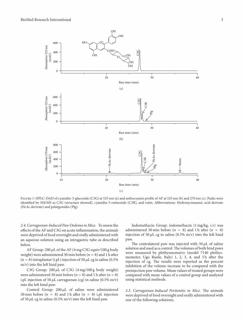
2.2. Anthocyanin �uanti�cation. For anthocyanin quanti�-cation, aliquots of AF and C3G were diluted with methanol:acetic acid (99 : 5, v : v) and �ltered through a 0.45 𝜇𝜇m PTFE�lter (Milipore Ltd., Bedford MA) prior to quanti�cationby HPLC-DAD [19]. e column used was a Prodigy 5𝜇𝜇mODS3 (250mm × 4.6mm i.d., Phenomenex Ltd.) and elutionsolvents were (A) water : THF : TFA (98 : 2 : 0.1, v : v : v) and(B) acetonitrile. Solvent gradient consisted of 8% B at thebeginning, 10% at 5min, 17% at 10min, 25% at 15min,50% at 25min, 90% at 30min, 50% at 32min, and 8% at35min (run time, 35min). Eluates weremonitored at 270 and525 nm. Flow rate was 1mL/min; column temperature was30∘C. Peak identi�cation was performed by comparison ofretention times and diode array spectral characteristics withthe standards and the library spectra. Cochromatographywas used when necessary. C3G, C3R, and pelargonidin (Plg)(Extrasynthese, Genay, France) were used as standard. etotal anthocyanin content of AF was expressed as C3Gequivalent. e anthocyanin composition of AF is 85% C3G,12% C3R, and 3% Plg derivate and they were previouslyidenti�ed by LC-MS [19]. e anthocyanin pro�le of AF andthe purity of C3G are shown in Figure 1.
2.3. Animals. Male Swiss mice, weighing 18–20 g (approx-imately four weeks old), were acclimated to housing for atleast 1 week prior to investigation. e night before theexperiment, food was withdrawn from the cages but waterwas given ad libitum. Animals were randomly assigned toeach treatment group and all testing was performed between8:00 and 9:00 a.m. All animals were handled and experimentswere conducted in accordance to the Guidelines for AnimalExperimentation of the University of São Paulo, Brazil, aerapproval by the Ethics Committee of the Pharmacy Faculty ofthe University of São Paulo (Protocol number 53, FCF-USP).
BioMed Research International 3
OH
OH
OHOH
OH
OH
HO
HOO
O
O+
0
200
400
600
C3
G
(mA
U)
Ab
sorp
tio
n 5
25
nm
20 30 40
Run time (min)
(a)
0
200
400
600
Plg
C3RC
3G
(mA
U)
Ab
sorp
tio
n 5
2 5 n
m
20 30 40
Run time (min)
(b)
10 20 30 40
0
200
400
600
800
HcA
c d
eriv
ate
HcA
c d
eriv
ate
Ru
tin
C3R
C3G
Plg
(mA
U)
Ab
sorp
tio
n 2
7 0 n
m
Run time (min)
(c)
F 1: HPLC-�A� of cyanidin-3-glucoside (C3G) at 525 nm (a) and anthocyanin pro�le of AF at 525 nm (b) and 270 nm (c). Peaks wereidenti�ed by M�/M� as C3G (structure showed), cyanidin-3-rutinoside (C3R), and rutin. Abbreviations: Hydro�ycinnamic acid derivate(HcAc derivate) and pelargonidin (Plg).
2.4. Carrageenan-Induced PawOedema inMice. To assess thee�ects of the AF andC3G on acute in�ammation, the animalswere deprived of food overnight and orally administeredwithan aqueous solution using an intragastric tube as describedbelow.
AF Group: 200𝜇𝜇L of the AF (4mg C3G equiv/100 g bodyweight) were administered 30min before (𝑛𝑛 𝑛 𝑛) and 1 h aer(𝑛𝑛 𝑛 𝑛) intraplantar (i.pl.) injection of 50 𝜇𝜇L cg in saline (0.5%m/v) into the le hind paw.
C3G Group: 200𝜇𝜇L of C3G (4mg/100 g body weight)were administered 30min before (𝑛𝑛 𝑛 𝑛) and 1 h aer (𝑛𝑛 𝑛 𝑛)i.pl. injection of 50 𝜇𝜇L carrageenan (cg) in saline (0.5% m/v)into the le hind paw.
Control Group: 200 𝜇𝜇L of saline were administered30min before (𝑛𝑛 𝑛 𝑛) and 1 h aer (𝑛𝑛 𝑛 𝑛) i.pl. injectionof 50 𝜇𝜇L cg in saline (0.5%m/v) into the le hind paw.
Indomethacin Group: indomethacin (1mg/kg, i.v) wasadministered 30min before (𝑛𝑛 𝑛 𝑛) and 1 h aer (𝑛𝑛 𝑛 𝑛)injection of 50 𝜇𝜇L cg in saline (0.5% m/v) into the le hindpaw.
e contralateral paw was injected with 50 𝜇𝜇L of salinesolution and used as a control.e volumes of both hind pawswere measured by plethysmometry (model 7140 plethys-mometer, Ugo Basile, Italy) 1, 2, 3, 4, and 5 h aer theinjection of cg. e results were reported as the percentinhibition of the volume increase to be compared with thepreinjection paw volume.Mean values of treated groups werecompared with mean values of a control group and analyzedusing statistical methods.
2.5. Carrageenan-Induced Peritonitis in Mice. e animalswere deprived of food overnight and orally administeredwithone of the following solutions.
4 BioMed Research International
AF Group: 200𝜇𝜇L of the AF (4mg C3G equiv/100 g bodyweight) were administered 30min before (𝑛𝑛 𝑛 𝑛) and 1 h aer(𝑛𝑛 𝑛 𝑛) intraperitoneal (i.p.) injection of 1mL of cg in sterilesaline (0.3%, m/v).
C3G Group: 200 𝜇𝜇L of the C3G (4mg/100 g body weight)were administered 30min before (𝑛𝑛 𝑛 𝑛) and 1 h aer (𝑛𝑛 𝑛 𝑛)i.p. injection of 1mL of cg in sterile saline (0.3%, m/v).
Carrageenan Control Group: 200 𝜇𝜇L of saline solutionwere administered 30min before (𝑛𝑛 𝑛 𝑛) and 1 h aer (𝑛𝑛 𝑛 𝑛)i.p. injection of 1mL of cg in sterile saline (0.3%, m/v).
Indomethacin Group: 200𝜇𝜇L indomethacin (4mg/100 gbody weight) were administered 30min before (𝑛𝑛 𝑛 𝑛) and1 h aer (𝑛𝑛 𝑛 𝑛) i.p. injection of 1mL of cg in sterile saline(0.3%, m/v).
SalineControlGroup: 200 𝜇𝜇L saline solutionwere admin-istered 30min before (𝑛𝑛 𝑛 𝑛) and 1 h aer (𝑛𝑛 𝑛 𝑛) i.p.injection of 1mL of sterile saline solution.
ree hours aer cg injections, the animals were killedby overexposure to CO2 and the peritoneal exudate waswithdrawn aer washing the peritoneal cavity with 2mLof saline solution. Aliquots of the washes were used todetermine total cell counts. An aliquot of the 1 × 10⁶
and 3 × 10⁶ cells was centrifuged at 800 g/6min/22∘C andused for COX-2 expression analysis by western blotting andRT-PCR, respectively. e supernatant was used for PGE2quanti�cation.
2.6. Leukocyte Harvesting and Counting. Aliquots of theperitoneal washes were used to determine total cell countsin a Newbauer chamber aer dilution (1 : 20, v : v) in Turk’ssolution (0.2% crystal violet dye in 30% acetic acid). Fordifferential cell counts, cytospin preparations were stainedwith Hema3 stain. Differential cell counts were performedby counting at least 100 cells, which were classi�ed aseither polymorphonuclear or mononuclear cells, based onconventional morphological criteria.
2.7. Western Blotting. e precipitate of cells (1 × 10⁶)was lisate with 100 𝜇𝜇L of sample buffer [30] and heatedfor 10min/100∘C. An aliquot of 14 𝜇𝜇L of the lisate wasseparated on SDS-polyacrylamide gels (10%) at 150V andelectrophoretically transferred to nitrocellulose membrane(GE Healthcare, Buckinghamshire, UK). e membrane wasblocked with 5% nonfat milk in Tris buffered saline with0.05% Tween 20 and incubated 1 h at room temperature withthe antibody against COX-2 (1 : 1500) (Cayman Chemicals,Ann Arbor, MI) followed by incubation in the same bufferwith the appropriate anti-rabbit horseradish peroxidase-conjugated secondary antibody (GE Healthcare, Bucking-hamshire, UK) for 1 h at room temperature (1 : 1500). Fur-ther, the membrane was also incubated with the antibodyagainst 𝛽𝛽-actin (1 : 2000) (Sigma, St. Louis, USA) followedby incubation with the anti-mouse secondary horseradishperoxidase-conjugate (1 : 2000) (GE Healthcare, Bucking-hamshire, UK). Immunoreactive bands were detected usingECL kit (GEHealthcare, Buckinghamshire, UK). Densities ofthe bands were determined by a GS 700 Densitometer (Bio-Rad Laboratories, Richmond, CG) using the image analysis
soware from Molecular Analyst (Bio-Rad Laboratories,Richmond, CG).
2.8. RNA Preparation and Reverse Transcription-PolymeraseChain Reaction (RT-PCR). Cells (3 × 10⁶) were washedonce with sterile saline and mixed with 500 𝜇𝜇L of Trizolreagent (Invitrogen, Rockville, MD, EUA) and the RNAwas extracted according to the manufacturer’s instructions.Complementary DNA was synthesized using an Improm-II Reverse Transcription System (Promega, Madison, WI,USA) according to the manufacturer’s instructions and con-ducted at a thermocycler Gene AMp (PCR System 2400,Applied Biosystems). PCR was performed by denaturingat 94∘C for 60 s, annealing at 57∘C (COX-2) and 60∘C(𝛽𝛽-actin) for 1min and by extension at 72∘C for 60 s.irty additional cycles for COX-2 and 25 cycles for 𝛽𝛽-actin were used for ampli�cation. e primer pairs used foranalysis were 5′-TTTGTTGAGTCGTTCGCCGGACGGA-3′ and 5′-CGGTATTGAGGAGAAGAGATGGGATT-3′ forsense and antisense primers of the COX-2 gene, respectively[31]; 5′-TGGAATCCTGTGGCGTCCGTGAAAC-3′ and 5′-TAAAACGCGGCTCGGTAACGGTCCG-3′ for sense andantisense primers of the 𝛽𝛽-actin gene, respectively [32], usedas an inner control.
2.9. PGE2 �uanti�cation. Concentrations of PGE2 weredetermined by a speci�c enzyme immunoassay [33] using acommercial kit (Cayman Chemical Company, Ann Arbor,MI). e extraction of PGE2 was performed on Sep PakC18 columns (Waters Corporation, Milford, MA) and elutedwith ethanol. In brief, 50 𝜇𝜇L aliquots of each extractedsample were incubated with the PGE2 conjugated withacetylcholinesterase and the speci�c rabbit antiserum in 96-well plates, coated with anti-rabbit IgG mouse monoclonalantibody. Aer addition of the substrate, the absorbance ofthe samples was recorded at 405 nm in a microplate reader(Labsystem Multiscan), and concentrations of eicosanoidswere estimated from standard curves.
2.10. Statistical Analysis. Results were presented as mean ±EPM. e statistical analyses were performed by one wayanalysis of variance (ANOVA) and Tukey posthoc test forcomparison, using the Statistic soware package version 5.0(StatSo, Inc.). Results were considered statistically signi�-cant for 𝑃𝑃 values <0.05.
3. Results and Discussion
3.1. Effect of C3G and AF on Carrageenan-Induced Paw Oe-dema. e oral dose of both extracts and the two protocolsapplied in this study (30min before or 1 h aer in�ammationstimulus) were chosen in order to provide high concentrationof C3G in the plasma based in its rapid absorption andexcretion [20].
e in�ammatory response to subplantar oedemainduced by cg in mice was signi�cantly reduced by priorand aer oral administration of AF and C3G. Figures 2(a)
BioMed Research International 5
and 2(b) show the time course of the paw oedema aer i.pl.injection of cg (0.5% m/v). Carrageenan caused progressiveincrease in the paw oedema 1 h aer the injection, presentingthe maximum peak at 4 h, decreasing to basal level aer 5 h.Before and aer treatment of animals with indomethacinsigni�cantly reduced cg-induced paw oedema as expected,in comparison with the respective controls (saline). C3G(4mg/100 g body weight), administered by gavage either30min before or 1 h aer the cg stimulus signi�cantlydecreased (𝑃𝑃 𝑃 𝑃𝑃𝑃𝑃) the paw oedema (around 40% andup to 80%, resp.) at the fourth hour aer cg injection whencompared with the control group (Figures 2(a) and 2(b)).Also, the oral administration of AF decreased the pawoedema approximately 40% in both administration times.
e dose of AF and C3G used in the present study is ten-times lower than that necessary of the anthocyanin mix fromtart cherry to suppress the 25% complete Freund’s adjuvantand cg-induced paw oedema [13] but closer than ginkgobiloba extract concentration necessary to inhibit the pawoedema induced by cg in rats [34]. is fact suggested thatC3G is one of the anthocyanins that presented high anti-in�ammatory activity.
It has been established that the paw oedema induced bythe subplantar injection of cg is biphasic; the early phaseinvolves the release of the mediators serotonin, histamine,and kinins, while the late phase is characteri�ed by the in�l-tration of leukocytes and mediated only by prostaglandins[35]. ese results suggest that the inhibitory effect of AFor C3G on oedema formation is due to the inhibition of thesynthesis and/or release of these mediators, in the early phaseof in�ammatory effect of cg, especially by inhibiting probablycyclooxygenase products. To support this observation, thedata indicate that C3G promoted similar effectiveness insuppressing oedema, when compared to the inhibitory pro�leof indomethacin, a COX activity inhibitor, on cg-inducedin�ammation.
3.2. Effect of C3G and AF on Carrageenan-Induced CellularIn�ux into Peritoneal Cavity. Intraperitoneal administrationof cg produces a sustained increase in postcapillary venulepermeability, thereby leading to increased cellular in�ltra-tion, particularly of neutrophils [36]. e recruitment ofleukocytes from the circulation to sites of in�ammation isenhanced by a series of proin�ammatory mediators, suchas IL-8 and vasoactive amines, ICAM and VCAM, thatare produced and released into the tissue by mast cells,macrophages, and activated endothelial cells, as well astransmigrated leukocytes [36].
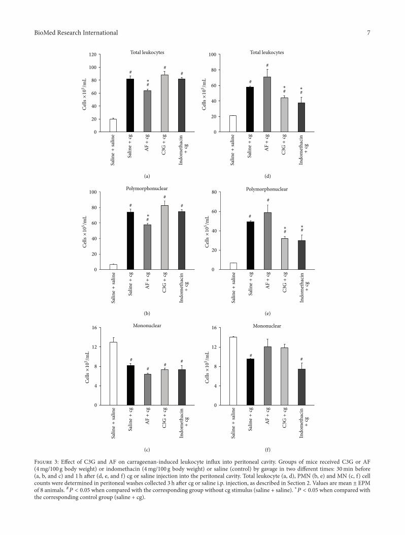
Figure 3 presents the total leukocyte in�ux and differen-tial cell into the peritoneal cavities aer oral administrationof C3G or AF (4mg/100 g body weight) or indomethacin(4mg/100 g body weight) or saline (control) 30min beforeand 1 h aer i.p. injection of cg (0.3% w/v) or saline solution(without stimulus).
e oral administration of AF 30min before the i.p.injection of cg caused a signi�cant decrease (𝑃𝑃 𝑃 𝑃𝑃𝑃𝑃)in the number of total leukocytes (29% decrease) (Figure3(a)), but not when administered 1 h aer the stimulus.
No reduction of total leukocytes in peritoneal exudate wasobserved when indomethacin was injected 30min before cg.On the other hand, the C3G decreases the number of totalleukocytes when administered 1 h aer the cg stimulus (38%decrease) (Figure 3(d)). Similar effects were obtained withindomethacin administration, which promoted reduction ofleukocytes (55% decrease) when administered 1 h aer i.p.injection of cg.
Differential cell counts showed that leukocytes presentin the peritoneal cavity, aer i.p. injection of cg, werepredominantly polymorphonuclears (PMN), mainly neu-trophils, when compared with the group that received saline(without stimulus). e mean values of PMN were 74 ±4 × 1𝑃𝑃 cells/mL, and 𝑃1 ± 1 × 1𝑃𝑃 cells/mL, in the groupsthat received saline by gavage 30min before and 1 h aer cginjection, respectively (Figures 3(b) and 3(e)). On the otherhand, in the group that received saline instead of cg (withoutstimulus), in both administration times, the mononuclearleukocytes (MN) were predominant (13 ± 1 × 1𝑃𝑃 cells/mL).In addition, our results showed that cg injection caused adecrease in the number of MN in the peritoneal cavity (7𝑃1 ±𝑃𝑃1 × 1𝑃𝑃 cells/mL) (Figures 3(c) and 3(f)).
Like what occurred with the total leukocytes, the numberof PMN in peritoneal �uid in mice was signi�cantly reducedwhen treated with C3G (39% decrease) or indomethacin(40% decrease) 1 h aer the i.p cg stimulus, when comparedto the control group that received saline orally (Figure 3(e)).On the other hand, AF administered 30min before cg,promoted a signi�cant decrease in the recruited PMN (24%decrease), compared to the control group (Figure 3(b)).
ese results were different from those observed in othertissues, such as air pounch cg in�ammation inmice and acutelung in�ammation in rats where a decrease in the in�ux of cellwas observed when C3G was previously administered beforethe cg stimulus [14, 17].
In relation to MN in�ux, C3G or AF or indomethacinadministrated 30min before cg injection did not change thedecrease counts of MN promoted by cg injection (Figures3(c) and 3(f)), when compared with the group without cgstimulus.
Since C3G was detected intact and in low concentrationin plasma of rats aer mulberry juice intake [20], the oralintake performed 1 h aer cg stimulus probably could providean ideal concentration of C3G in plasma, resulting in theobserved effect. However, this experimental protocol showedthat AF is more effective than C3G as a preventive compoundagainst leukocyte migration, suggesting that the complexmixtures of anthocyanins in AF may provide antileukocytein�ux effectmainly through a combination of additive and/orsynergistic effects.
3.3. Effect of C3G and AF on Carrageenan-Induced Cycloox-ygenase-2 Expression in Peritonitis. e effect of C3G orAF (4mg/100 g body weight) on cg-induced COX-2 tran-scription was measured in peritoneal leukocytes by RT-PCR.As shown in Figures 4(a) and 4(c), the i.p. injection of cg(0.3% w/v) drastically increased COX-2 mRNA and proteinexpression. On the other hand, the oral administration of

6 BioMed Research International
0 1 2 3 4 5
0
20
40
60
80In
crea
se i
n h
ind
paw
siz
e (%
)
Saline + cg
C3G + cg
AF + cg
Indomethacin + cg
∗
∗∗
∗
∗
Time (h)
(a)
0 1 2 3 4 5
0
20
40
60
80
Time (h)
Incr
ease
in
hin
d p
aw s
ize
(%)
Saline + cg
C3G + cg
AF + cg
Indomethacin + cg
∗
∗∗
∗
∗
∗∗
(b)
F 2: Effect of C3G and AF on carrageenan-induced paw oedema. Footpad oedema was induced by injection of cg (0.5% w/v in saline,i.pl.) and was evaluated by plethysmometry. C3G or AF (4mg/100 g body weight) or indomethacin (1mg/kg, i.v.) or saline (control oedema)was orally administered in two different times: 30min before (a) and 1 h aer (b) i.pl. injection of cg. e increase paw size was measured 1,2, 3, 4, and 5 h aer cg injection.e time zero corresponds to cg injection.e results were expressed as mean ± EPM of 8 mice. Statisticallysigni�cant difference regarding saline (control group) and C3G and AF and Indomethacin groups is expressed as ∗𝑃𝑃 𝑃 𝑃𝑃𝑃𝑃.
C3G and AF, either 30min before and 1 h aer cg i.p.injection, clearly downregulated COX-2 mRNA expression(50% reduction) and decreased the levels of COX-2 proteinexpression, when compared with the control group.
Although some studies have documented that antho-cyanins inhibit COX-2 expression in human keratinocyte cellline [15] and cultured macrophages [37, 38] and in asthmamodel [16], our study provides the �rst evidence that ananthocyanin mixture or C3G can inhibit, both preventivelyand therapeutically, the expression of COX-2 protein with asingle oral dose. Several lines of evidence clearly established,in in vitro models, that the inhibition of some in�ammatorycytokines [12, 16] and inhibition of activation of nuclearfactor pathway, such as NF-𝜅𝜅B [10, 15], could explain themechanisms of action of anthocyanins on the inhibition ofCOX-2 expression.
Also, some sources of anthocyanins, such as black soy-bean anthocyanin and anthocyanins from sweet purple haveshowed inhibition the COX-2 expression through NF-𝜅𝜅Binhibition when administered before the stimulus in in�am-mation models [11, 12].
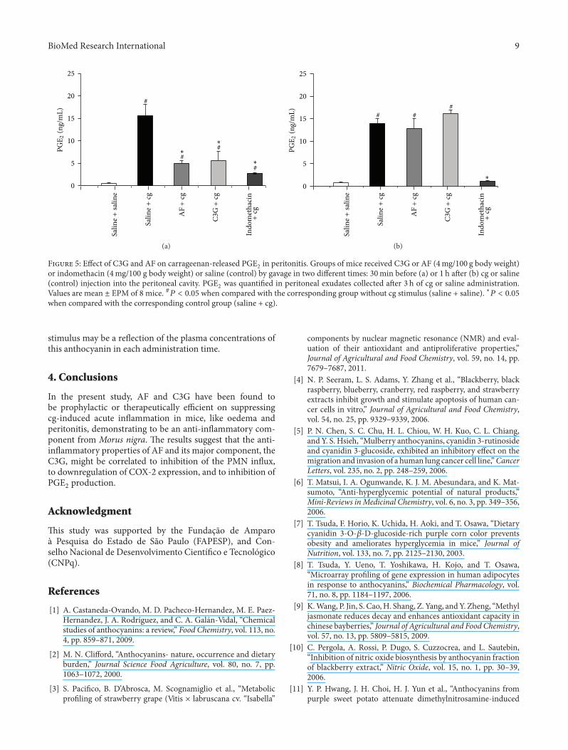
3.4. Effect of C3G and AF on Carrageenan-Released PGE2in Peritonitis. Further, this study investigated the effect ofC3G and AF (4mg/100 g body weight) on PGE2 production,the main in�ammatory prostaglandin produced by COXactivity, in peritoneal exudates from mice induced by cg.Figures 5(a) and 5(b) showed that i.p. administration of cginduced more than a 25-fold (14𝑃𝑃 ± 2𝑃𝑃 ng/mL) increasein PGE2 generation compared with the groups without thecg stimulus (𝑃𝑃𝑃𝑃 ± 𝑃𝑃𝑃𝑃 ng/mL). e PGE2 concentration
was signi�cantly decreased by the oral pretreatment withC3G, AF, and indomethacin, 30min before cg injection(4𝑃𝑃 ± 1𝑃𝑃 ng/mL, 𝑃𝑃𝑃 ± 2𝑃𝑃 ng/mL and 2𝑃1 ± 𝑃𝑃1 ng/mL,resp.). In this administration time, the AF andC3Gpromotedapproximately 70% reduction in PGE2 production by cg(Figure 5(a)). On the other hand, the oral treatment ofAF or C3G, 1 h aer i.p. injection of cg, did not induceany modi�cation in the high levels of PGE2 release by cg(Figure 5(b)). However, in such experimental condition,the indomethacin suppressed the PGE2 production by cgstimulus.
Prostaglandin E2 is a product generated by cyclooxyge-nases from arachidonic acid, and it is an important mediatorin the in�ammatory process. In this study, it was observedthat aer 3 h of administration, cg produced an increase inPGE2 levels into peritoneal cavity. In parallel, the resultsshowed that C3G produced signi�cant inhibition of PGE2production when injected 30min before cg. However, C3Gdid not produce such equivalent effectiveness towards cg-induced PGE2 release when administered 1 h aer cg injec-tion.ese results are curious because in both administrationtimes used in the present study it was possible to observethat the oral intake of C3G was effective in inhibitingCOX-2 expression. erefore, this suggests that althoughCOX-2 mRNA and protein expression were detected at 3 haer cg injection, this isoform of COX did not presentcatalytic activity in this period of time. In fact, studies havedemonstrated that cg-induced PGE2 are produced by COX-1 in the �rst phase, while COX-2-derived PGE2 turned to beinvolved in the second phase induced by cg injection [35].In parallel, our data demonstrated that indomethacin waseffective to inhibit PGE2 production in both administration
BioMed Research International 7
0
20
40
60
80
100
120
##
#
Total leukocytes
#
Sali
ne+
sali
ne
Sali
ne+
cg
AF+
cg
C3G+
cg
∗
0
20
40
60
80
100
#
#
#
#
Polymorphonuclear
Sali
ne+
sali
ne
Sali
ne+
cg
AF+
cg
C3G+
cg
∗
0
4
8
12
16
#
##
#
Mononuclear
Sali
ne+
sali
ne
Sali
ne+
cg
AF+
cg
C3G+
cg
0
20
40
60
80
100
##
#
#
Total leukocytes
Sali
ne+
sali
ne
Sali
ne+
cg
AF+
cg
C3G+
cg
∗∗
Polymorphonuclear
0
20
40
60
80
#
#
# #Sa
lin
e+
sali
ne
Sali
ne+
cg
AF+
cg
C3G+
cg
Ind
om
eth
acin
+cg
Ind
om
eth
acin
+cg
Ind
om
eth
acin
+cg
Ind
om
eth
acin
+cg
Ind
om
eth
acin
+cg
Ind
om
eth
acin
+cg
∗∗
Mononuclear
0
4
8
12
16
##
Sali
ne+
sali
ne
Sali
ne+
cg
AF+
cg
C3G+
cg
(a) (d)
(e)(b)
(c) (f)
Cel
ls×1
05 /
mL
Cel
ls×1
05 /
mL
Cel
ls×1
05 /
mL
Cel
ls×1
05 /
mL
Cel
ls×1
05 /
mL
Cel
ls ×
105 /
mL
F 3: Effect of �3� and �F on carrageenan�induced leukocyte in�u� into peritoneal cavity. �roups of mice received �3� or �F(4mg/100 g body weight) or indomethacin (4mg/100 g body weight) or saline (control) by gavage in two different times: 30min before(a, b, and c) and 1 h aer (d, e, and f) cg or saline injection into the peritoneal cavity. Total leukocyte (a, d), PMN (b, e) and MN (c, f) cellcounts were determined in peritoneal washes collected 3 h aer cg or saline i.p. injection, as described in Section 2. Values are mean ± EPMof 8 animals. #𝑃𝑃 𝑃 𝑃𝑃𝑃𝑃 when compared with the corresponding group without cg stimulus (saline + saline). ∗𝑃𝑃 𝑃 𝑃𝑃𝑃𝑃 when compared withthe corresponding control group (saline + cg).

8 BioMed Research International
3
2
1
0
#∗
#∗
#
Saline+saline
Saline+cg
AF+cg
C3G+cg —
COX-2
(a)
3
2
1
0
#
∗
∗
Saline+saline
Saline+cg
AF+cg
C3G+cg —
COX-2
(b)
COX-2
3
2
1
0
#∗
#∗
#
Saline+saline
Saline+cg
AF+cg
C3G+cg —
(c)
3
2
1
0
#
∗
∗
Saline+saline
Saline+cg
AF+cg
C3G+cg —
COX-2
(d)
F 4: Effect of C3G andAF on carrageenan-induced cyclooxygenase-2 expression in peritoneal leukocytes. Groups of mice received C3Gor AF (4mg/100 g body weight) or saline by gavage in two different times: 30min before or 1 h aer cg (0.3% w/v) or saline injection intothe peritoneal cavity. Peritoneal leukocytes were collected 3 h aer i.p. administration of either cg or saline and whole cells were analyzed forCOX-2 expression by RT-PCR and western blotting performed, as described in Section 2. (a and c) RT-PCR of COX-2, and 𝛽𝛽-actin (loadingcontrol); Bar graph shows densitometric analysis of mRNA COX-2. (b and d) Western blotting of COX-2, and 𝛽𝛽-actin (loading control) ofleukocytes present in the in�ammatory exudates; bar graph shows densitometric analysis of protein COX-2. �e densities (in densitometryunits) were normalized with those of 𝛽𝛽-actin. Results were expressed as mean ± EPM from 8 mice. #𝑃𝑃 𝑃 𝑃𝑃𝑃𝑃 when compared with thecorresponding group without cg stimulus (saline + saline). ∗𝑃𝑃 𝑃 𝑃𝑃𝑃𝑃 when compared with the corresponding control group (saline + cg).
times. Although it is generally accepted that nonsteroidalanti-in�ammatory drugs such as aspirin and indomethacinare inhibitors of activity of both isoforms ofCOXs, it is knownthat these compounds inhibit COX-1 activity more potently
thanCOX-2 in broken cells and in intact cells ofmice [39, 40].In addition, the absence of PGE2 inhibition when C3G wasadministered 1 h aer cg stimulus compared to the preventiveeffect obtained by C3G when administered 30min before the
BioMed Research International 9
0
5
10
15
20
25
∗#
#
∗#
∗#
Saline+saline
Saline+cg
AF+cg
C3G+cg
PGE2(ng/mL)
Indomethacin
+cg
(a)
0
5
10
15
20
25
# ##
∗
Saline+saline
Saline+cg
AF+cg
C3G+cg
Indomethacin
+cg
PGE2(ng/mL)
(b)
F 5: Effect of C3G and AF on carrageenan-released PGE2 in peritonitis. Groups of mice received C3G or AF (4mg/100 g body weight)or indomethacin (4mg/100 g body weight) or saline (control) by gavage in two different times: 30min before (a) or 1 h aer (b) cg or saline(control) injection into the peritoneal cavity. PGE2 was quanti�ed in peritoneal exudates collected aer 3 h of cg or saline administration.Values are mean ± EPM of 8 mice. #𝑃𝑃 𝑃 𝑃𝑃𝑃𝑃 when compared with the corresponding group without cg stimulus (saline + saline). ∗𝑃𝑃 𝑃 𝑃𝑃𝑃𝑃when compared with the corresponding control group (saline + cg).
stimulus may be a re�ection of the plasma concentrations ofthis anthocyanin in each administration time.
4. Conclusions
In the present study, AF and C3G have been found tobe prophylactic or therapeutically efficient on suppressingcg-induced acute in�ammation in mice, like oedema andperitonitis, demonstrating to be an anti-in�ammatory com-ponent from Morus nigra. e results suggest that the anti-in�ammatory properties of AF and its major component, theC3G, might be correlated to inhibition of the PMN in�ux,to downregulation of COX-2 expression, and to inhibition ofPGE2 production.
Acknowledgment
is study was supported by the Fundação de Amparoà Pesquisa do Estado de São Paulo (FAPESP), and Con-selho Nacional de Desenvolvimento Cientí�co e Tecnol�gico(CNPq).
References
[1] A. Castaneda-Ovando, M. D. Pacheco-Hernandez, M. E. Paez-Hernandez, J. A. Rodríguez, and C. A. Galán-Vidal, “Chemicalstudies of anthocyanins: a review,” Food Chemistry, vol. 113, no.4, pp. 859–871, 2009.
[2] M. N. Clifford, “Anthocyanins- nature, occurrence and dietaryburden,” Journal Science Food Agriculture, vol. 80, no. 7, pp.1063–1072, 2000.
[3] S. Paci�co, B. D�Abrosca, M. Scognamiglio et al., “Metabolicpro�ling of strawberry grape (Vitis × labruscana cv. “Isabella”
components by nuclear magnetic resonance (NMR) and eval-uation of their antioxidant and antiproliferative properties,”Journal of Agricultural and Food Chemistry, vol. 59, no. 14, pp.7679–7687, 2011.
[4] N. P. Seeram, L. S. Adams, Y. Zhang et al., “Blackberry, blackraspberry, blueberry, cranberry, red raspberry, and strawberryextracts inhibit growth and stimulate apoptosis of human can-cer cells in vitro,” Journal of Agricultural and Food Chemistry,vol. 54, no. 25, pp. 9329–9339, 2006.
[5] P. N. Chen, S. C. Chu, H. L. Chiou, W. H. Kuo, C. L. Chiang,and Y. S. Hsieh, “Mulberry anthocyanins, cyanidin 3-rutinosideand cyanidin 3-glucoside, exhibited an inhibitory effect on themigration and invasion of a human lung cancer cell line,”CancerLetters, vol. 235, no. 2, pp. 248–259, 2006.
[6] T. Matsui, I. A. Ogunwande, K. J. M. Abesundara, and K. Mat-sumoto, “Anti-hyperglycemic potential of natural products,”Mini-Reviews inMedicinal Chemistry, vol. 6, no. 3, pp. 349–356,2006.
[7] T. Tsuda, F. Horio, K. Uchida, H. Aoki, and T. Osawa, “Dietarycyanidin 3-O-𝛽𝛽-D-glucoside-rich purple corn color preventsobesity and ameliorates hyperglycemia in mice,” Journal ofNutrition, vol. 133, no. 7, pp. 2125–2130, 2003.
[8] T. Tsuda, Y. Ueno, T. Yoshikawa, H. Kojo, and T. Osawa,“Microarray pro�ling of gene expression in human adipocytesin response to anthocyanins,” Biochemical Pharmacology, vol.71, no. 8, pp. 1184–1197, 2006.
[9] K.Wang, P. Jin, S. Cao,H. Shang, Z. Yang, andY. Zheng, “Methyljasmonate reduces decay and enhances antioxidant capacity inchinese bayberries,” Journal of Agricultural and Food Chemistry,vol. 57, no. 13, pp. 5809–5815, 2009.
[10] C. Pergola, A. Rossi, P. Dugo, S. Cuzzocrea, and L. Sautebin,“Inhibition of nitric oxide biosynthesis by anthocyanin fractionof blackberry extract,” Nitric Oxide, vol. 15, no. 1, pp. 30–39,2006.
[11] Y. P. Hwang, J. H. Choi, H. J. Yun et al., “Anthocyanins frompurple sweet potato attenuate dimethylnitrosamine-induced
10 BioMed Research International
liver injury in rats by inducing Nrf2-mediated antioxidantenzymes and reducing COX-2 and iNOS expression,” Food andChemical Toxicology, vol. 49, no. 1, pp. 93–99, 2011.
[12] S. L. Yeh, H.M.Wang, P. Y. Chen, and T. C.Wu, “Interactions of𝛽𝛽-carotene and�avonoids on the secretion of pro-in�ammatorymediators in an in vitro system,” Chemico-Biological Interac-tions, vol. 179, no. 2-3, pp. 386–393, 2009.
[13] J. M. Tall, N. P. Seeram, C. Zhao, M. G. Nair, R. A. Meyer, andS. N. Raja, “Tart cherry anthocyanins suppress in�ammation-induced pain behavior in rat,” Behavioural Brain Research, vol.153, no. 1, pp. 181–188, 2004.
[14] S.W.Min, S. N. Ryu, andD. H. Kim, “Anti-in�ammatory effectsof black rice, cyanidin-3-O-𝛽𝛽-d-glycoside, and its metabolites,cyanidin and protocatechuic acid,” International Immunophar-macology, vol. 10, no. 8, pp. 959–966, 2010.
[15] K. Tsoyi, H. B. Park, Y. M. Kim et al., “Anthocyanins fromblack soybean seed coats inhibit UVB-induced in�amma-tory cylooxygenase-2 gene expression and PGE2 productionthrough regulation of the nuclear factor-𝜅𝜅B and phosphatidyli-nositol 3-kinase/Akt pathway,” Journal of Agricultural and FoodChemistry, vol. 56, no. 19, pp. 8969–8974, 2008.
[16] S. J. Park, W. H. Shin, J. W. Seo, and E. J. Kim, “Anthocyaninsinhibit airway in�ammation and hyperresponsiveness in amurine asthma model,” Food and Chemical Toxicology, vol. 45,no. 8, pp. 1459–1467, 2007.
[17] A. Rossi, I. Serraino, P. Dugo et al., “Protective effects ofanthocyanins from blackberry in a rat model of acute lungin�ammation,” Free Radical Research, vol. 37, no. 8, pp.891–900, 2003.
[18] N. P. Seeram, R. A. Momin, M. G. Nair, and L. D. Bourquin,“Cyclooxygenase inhibitory and antioxidant cyanidin glyco-sides in cherries and berries,” Phytomedicine, vol. 8, no. 5, pp.362–369, 2001.
[19] N. M. A. Hassimotto, M. I. Genovese, and F. M. Lajolo,“Identi�cation and characterisation of anthocyanins from wildmulberry (Morus nigra L.) growing in Brazil,” Food Science andTechnology International, vol. 13, no. 1, pp. 17–25, 2007.
[20] N. M. A. Hassimotto, M. I. Genovese, and F. M. Lajolo,“Absorption and metabolism of cyanidin-3-glucoside andcyanidin-3-rutinoside extracted from wild mulberry (Morusnigra L.) in rats,”Nutrition Research, vol. 28, no. 3, pp. 198–207,2008.
[21] R.Medzhitov, “Origin and physiological roles of in�ammation,”Nature, vol. 454, no. 7203, pp. 428–435, 2008.
[22] F. A. Fitzpatrick, “Cyclooxygenase enzymes: regulation andfunction,” Current Pharmacology Design, vol. 10, no. 6, pp.577–588, 2004.
[23] T. D. Warner and J. A. Mitchell, “Cyclooxygenases: new forms,new inhibitors, and lessons from the clinic,” FASEB Journal, vol.18, no. 7, pp. 790–804, 2004.
[24] W. L. Smith, D. L. DeWitt, and R. M. Garavito, “Cyclooxy-genases: structural, cellular, and molecular biology,” AnnualReview of Biochemistry, vol. 69, pp. 145–182, 2000.
[25] H. R. Herschman, J. J. Talley, and R. DuBois, “Cyclooxygenase2 (COX-2) as a target for therapy and noninvasive imaging,”Molecular Imaging and Biology, vol. 5, no. 5, pp. 286–303, 2003.
[26] J. P. Portanova, Y. Zhang, G. D. Anderson et al., “Selec-tive neutralization of prostaglandin E2 blocks in�ammation,hyperalgesia, and interleukin 6 production in vivo,” Journal ofExperimental Medicine, vol. 184, no. 3, pp. 883–891, 1996.
[27] T. G. Brock and M. Peters-Golden, “Activation and regulationof cellular eicosanoid biosynthesis,” eScienti�c�orldJournal,vol. 7, pp. 1273–1284, 2007.
[28] N. V. Chandrasekharan, H. Dai, K. L. T. Roos et al., “COX-3, a cyclooxygenase-1 variant inhibited by acetaminophenand other analgesic/antipyretic drugs: cloning, structure, andexpression,” Proceedings of the National Academy of Sciences ofthe United States of America, vol. 99, no. 21, pp. 13926–13931,2002.
[29] B. Kis, J. A. Snipes, T. Isse, K. Nagy, and D. W. Busija, “Putativecyclooxygenase-3 expression in rat brain cells,” Journal of Cere-bral Blood Flow and Metabolism, vol. 23, no. 11, pp. 1287–1292,2003.
[30] U. K. Laemmli, “Cleavage of structural proteins during theassembly of the head of bacteriophage T4,”Nature, vol. 227, no.5259, pp. 680–685, 1970.
[31] J. S. Kang, Y. J. Jeon, S. K. Park, K. H. Yang, and H. M.Kim, “Protection against lipopolysaccharide-induced sepsisand inhibition of interleukin-1𝛽𝛽 and prostaglandin E2 synthesisby silymarin,” Biochemical Pharmacology, vol. 67, no. 1, pp.175–181, 2004.
[32] Y. Bezugla, A. Kolada, S. Kamionka, B. Bernard, R. Scheibe, andP. Dieter, “COX-1 and COX-2 contribute differentially to theLPS-induced release of PGE2 and TxA2 in liver macrophages,”Prostaglandins and Other Lipid Mediators, vol. 79, no. 1-2, pp.93–100, 2006.
[33] P. Pradelles, J. Grassi, and J.Maclouf, “Enzyme immunoassays ofeicosanoids using acetylcholine esterase as label: an alternativeto radioimmunoassay,” Analytical Chemistry, vol. 57, no. 7, pp.1170–1173, 1985.
[34] O. M. E. Abdel-Salam, A. R. Baiuomy, S. El-batran, and M. S.Arbid, “Evaluation of the anti-in�ammatory, anti-nociceptiveand gastric effects of Ginkgo biloba in the rat,” PharmacologicalResearch, vol. 49, no. 2, pp. 133–142, 2004.
[35] I. Posadas, M. Bucci, F. Roviezzo et al., “Carrageenan-inducedmouse paw oedema is biphasic, age-weight dependent anddisplays differential nitric oxide cyclooxygenase-2 expression,”British Journal of Pharmacology, vol. 142, no. 2, pp. 331–338,2004.
[36] H. L. Malech and J. I. Gallin, “Current concepts: immunol-ogy—neutrophils in human diseases,”eNew England Journalof Medicine, vol. 317, no. 11, pp. 687–694, 1987.
[37] X. H. Jin, K. Ohgami, K. Shiratori et al., “Effects of blue hon-eysuckle (Lonicera caerulea L.) extract on lipopolysaccharide-induced in�ammation in vitro and in vivo,” Experimental EyeResearch, vol. 82, no. 5, pp. 860–867, 2006.
[38] K. J. Woo, Y. J. Jeong, H. Inoue, J. W. Park, and T.K. Kwon, “Chrysin suppresses lipopolysaccharide-inducedcyclooxygenase-2 expression through the inhibition of nuclearfactor for IL-6 (NF-IL6) DNA-binding activity,” FEBS Letters,vol. 579, no. 3, pp. 705–711, 2005.
[39] J. C. Fr�lich, “A classi�cation of NSAIDs according to therelative inhibition of cyclooxygenase isoenzymes,” Trends inPharmacological Sciences, vol. 18, no. 1, pp. 30–34, 1997.
[40] J. A. Mitchell, P. Akarasereenont, C.iemermann, R. J. Flower,and J. R. Vane, “Selectivity of nonsteroidal antiin�ammatorydrugs as inhibitors of constitutive and inducible cyclooxyge-nase,” Proceedings of the National Academy of Sciences of theUnited States of America, vol. 90, no. 24, pp. 11693–11697, 1993.