Chemical Analysis and Screening as Anticancer Agent of Anthocyanin-Rich Extract from Uva Caimarona...
11
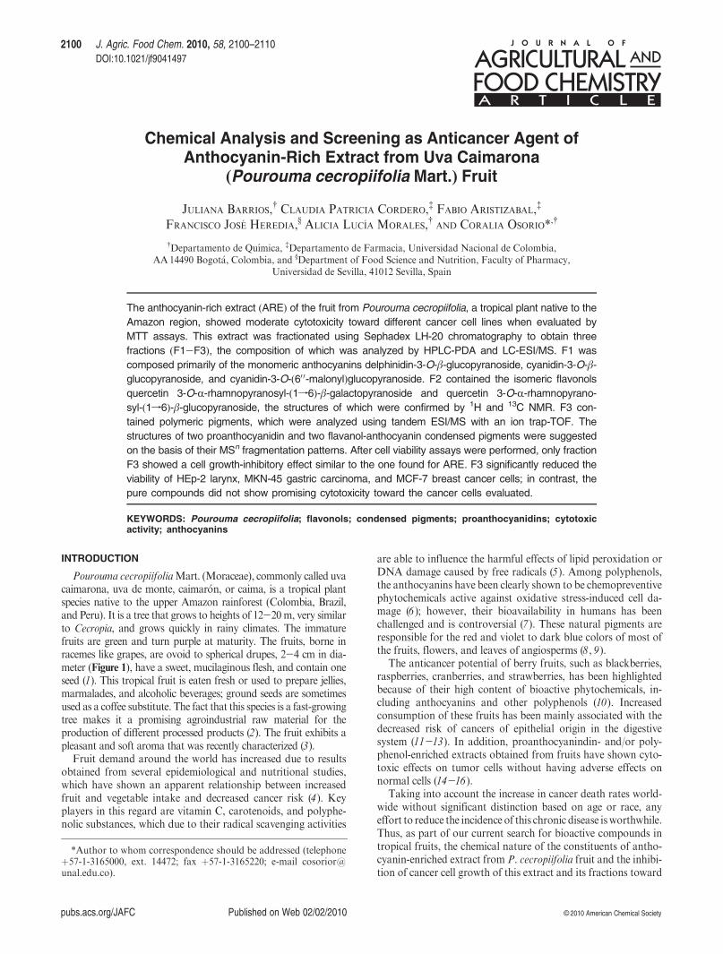
pubs.acs.org/JAFC Published on Web 02/02/2010 © 2010 American Chemical Society 2100 J. Agric. Food Chem. 2010, 58, 2100–2110 DOI:10.1021/jf9041497 Chemical Analysis and Screening as Anticancer Agent of Anthocyanin-Rich Extract from Uva Caimarona (Pourouma cecropiifolia Mart.) Fruit JULIANA BARRIOS, † CLAUDIA PATRICIA CORDERO, ‡ FABIO ARISTIZABAL, ‡ FRANCISCO JOSE ´ HEREDIA, § ALICIA LUCI ´ A MORALES, † AND CORALIA OSORIO* ,† † Departamento de Quı´mica, ‡ Departamento de Farmacia, Universidad Nacional de Colombia, AA 14490 Bogot a, Colombia, and § Department of Food Science and Nutrition, Faculty of Pharmacy, Universidad de Sevilla, 41012 Sevilla, Spain The anthocyanin-rich extract (ARE) of the fruit from Pourouma cecropiifolia, a tropical plant native to the Amazon region, showed moderate cytotoxicity toward different cancer cell lines when evaluated by MTT assays. This extract was fractionated using Sephadex LH-20 chromatography to obtain three fractions (F1-F3), the composition of which was analyzed by HPLC-PDA and LC-ESI/MS. F1 was composed primarily of the monomeric anthocyanins delphinidin-3-O-β-glucopyranoside, cyanidin-3-O-β- glucopyranoside, and cyanidin-3-O-(6 00 -malonyl)glucopyranoside. F2 contained the isomeric flavonols quercetin 3-O-R-rhamnopyranosyl-(1f6)-β-galactopyranoside and quercetin 3-O-R-rhamnopyrano- syl-(1f6)-β-glucopyranoside, the structures of which were confirmed by 1 H and 13 C NMR. F3 con- tained polymeric pigments, which were analyzed using tandem ESI/MS with an ion trap-TOF. The structures of two proanthocyanidin and two flavanol-anthocyanin condensed pigments were suggested on the basis of their MS n fragmentation patterns. After cell viability assays were performed, only fraction F3 showed a cell growth-inhibitory effect similar to the one found for ARE. F3 significantly reduced the viability of HEp-2 larynx, MKN-45 gastric carcinoma, and MCF-7 breast cancer cells; in contrast, the pure compounds did not show promising cytotoxicity toward the cancer cells evaluated. KEYWORDS: Pourouma cecropiifolia; flavonols; condensed pigments; proanthocyanidins; cytotoxic activity; anthocyanins INTRODUCTION Pourouma cecropiifolia Mart. (Moraceae), commonly called uva caimarona, uva de monte, caimar on, or caima, is a tropical plant species native to the upper Amazon rainforest (Colombia, Brazil, and Peru). It is a tree that grows to heights of 12-20 m, very similar to Cecropia, and grows quickly in rainy climates. The immature fruits are green and turn purple at maturity. The fruits, borne in racemes like grapes, are ovoid to spherical drupes, 2-4 cm in dia- meter (Figure 1), have a sweet, mucilaginous flesh, and contain one seed ( 1). This tropical fruit is eaten fresh or used to prepare jellies, marmalades, and alcoholic beverages; ground seeds are sometimes used as a coffee substitute. The fact that this species is a fast-growing tree makes it a promising agroindustrial raw material for the production of different processed products ( 2). The fruit exhibits a pleasant and soft aroma that was recently characterized ( 3). Fruit demand around the world has increased due to results obtained from several epidemiological and nutritional studies, which have shown an apparent relationship between increased fruit and vegetable intake and decreased cancer risk ( 4 ). Key players in this regard are vitamin C, carotenoids, and polyphe- nolic substances, which due to their radical scavenging activities are able to influence the harmful effects of lipid peroxidation or DNA damage caused by free radicals ( 5 ). Among polyphenols, the anthocyanins have been clearly shown to be chemopreventive phytochemicals active against oxidative stress-induced cell da- mage ( 6 ); however, their bioavailability in humans has been challenged and is controversial ( 7 ). These natural pigments are responsible for the red and violet to dark blue colors of most of the fruits, flowers, and leaves of angiosperms ( 8 , 9 ). The anticancer potential of berry fruits, such as blackberries, raspberries, cranberries, and strawberries, has been highlighted because of their high content of bioactive phytochemicals, in- cluding anthocyanins and other polyphenols ( 10 ). Increased consumption of these fruits has been mainly associated with the decreased risk of cancers of epithelial origin in the digestive system ( 11 -13 ). In addition, proanthocyanindin- and/or poly- phenol-enriched extracts obtained from fruits have shown cyto- toxic effects on tumor cells without having adverse effects on normal cells ( 14 -16 ). Taking into account the increase in cancer death rates world- wide without significant distinction based on age or race, any effort to reduce the incidence of this chronic disease is worthwhile. Thus, as part of our current search for bioactive compounds in tropical fruits, the chemical nature of the constituents of antho- cyanin-enriched extract from P. cecropiifolia fruit and the inhibi- tion of cancer cell growth of this extract and its fractions toward *Author to whom correspondence should be addressed (telephone þ57-1-3165000, ext. 14472; fax þ57-1-3165220; e-mail cosorior@ unal.edu.co).
-
Upload
independent -
Category
Documents
-
view
3 -
download
0
Transcript of Chemical Analysis and Screening as Anticancer Agent of Anthocyanin-Rich Extract from Uva Caimarona...
pubs.acs.org/JAFC Published on Web 02/02/2010 © 2010 American Chemical Society
2100 J. Agric. Food Chem. 2010, 58, 2100–2110
DOI:10.1021/jf9041497
Chemical Analysis and Screening as Anticancer Agent ofAnthocyanin-Rich Extract from Uva Caimarona
(Pourouma cecropiifolia Mart.) Fruit
JULIANA BARRIOS,† CLAUDIA PATRICIA CORDERO,‡ FABIO ARISTIZABAL,‡
FRANCISCO JOSE HEREDIA,§ ALICIA LUCIA MORALES,† AND CORALIA OSORIO*,†
†Departamento de Quımica, ‡Departamento de Farmacia, Universidad Nacional de Colombia,AA14490 Bogot�a, Colombia, and §Department of Food Science and Nutrition, Faculty of Pharmacy,
Universidad de Sevilla, 41012 Sevilla, Spain
The anthocyanin-rich extract (ARE) of the fruit from Pourouma cecropiifolia, a tropical plant native to the
Amazon region, showed moderate cytotoxicity toward different cancer cell lines when evaluated by
MTT assays. This extract was fractionated using Sephadex LH-20 chromatography to obtain three
fractions (F1-F3), the composition of which was analyzed by HPLC-PDA and LC-ESI/MS. F1 was
composed primarily of the monomeric anthocyanins delphinidin-3-O-β-glucopyranoside, cyanidin-3-O-β-glucopyranoside, and cyanidin-3-O-(60 0-malonyl)glucopyranoside. F2 contained the isomeric flavonols
quercetin 3-O-R-rhamnopyranosyl-(1f6)-β-galactopyranoside and quercetin 3-O-R-rhamnopyrano-
syl-(1f6)-β-glucopyranoside, the structures of which were confirmed by 1H and 13C NMR. F3 con-
tained polymeric pigments, which were analyzed using tandem ESI/MS with an ion trap-TOF. The
structures of two proanthocyanidin and two flavanol-anthocyanin condensed pigments were suggested
on the basis of their MSn fragmentation patterns. After cell viability assays were performed, only fraction
F3 showed a cell growth-inhibitory effect similar to the one found for ARE. F3 significantly reduced the
viability of HEp-2 larynx, MKN-45 gastric carcinoma, and MCF-7 breast cancer cells; in contrast, the
pure compounds did not show promising cytotoxicity toward the cancer cells evaluated.
KEYWORDS: Pourouma cecropiifolia; flavonols; condensed pigments; proanthocyanidins; cytotoxicactivity; anthocyanins
INTRODUCTION
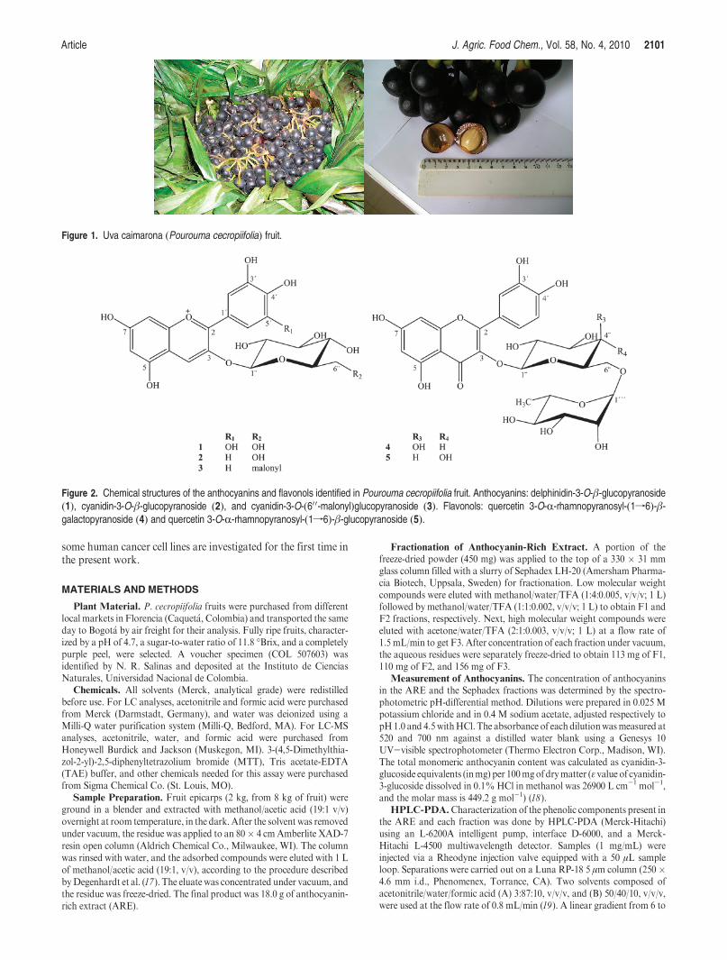
Pourouma cecropiifoliaMart. (Moraceae), commonly called uvacaimarona, uva de monte, caimar�on, or caima, is a tropical plantspecies native to the upper Amazon rainforest (Colombia, Brazil,and Peru). It is a tree that grows to heights of 12-20m, very similarto Cecropia, and grows quickly in rainy climates. The immaturefruits are green and turn purple at maturity. The fruits, borne inracemes like grapes, are ovoid to spherical drupes, 2-4 cm in dia-meter (Figure 1), have a sweet, mucilaginous flesh, and contain oneseed (1). This tropical fruit is eaten fresh or used to prepare jellies,marmalades, and alcoholic beverages; ground seeds are sometimesused as a coffee substitute. The fact that this species is a fast-growingtree makes it a promising agroindustrial raw material for theproduction of different processed products (2). The fruit exhibits apleasant and soft aroma that was recently characterized (3).
Fruit demand around the world has increased due to resultsobtained from several epidemiological and nutritional studies,which have shown an apparent relationship between increasedfruit and vegetable intake and decreased cancer risk (4). Keyplayers in this regard are vitamin C, carotenoids, and polyphe-nolic substances, which due to their radical scavenging activities
are able to influence the harmful effects of lipid peroxidation orDNA damage caused by free radicals (5). Among polyphenols,the anthocyanins have been clearly shown to be chemopreventivephytochemicals active against oxidative stress-induced cell da-mage (6); however, their bioavailability in humans has beenchallenged and is controversial (7). These natural pigments areresponsible for the red and violet to dark blue colors of most ofthe fruits, flowers, and leaves of angiosperms (8, 9).
The anticancer potential of berry fruits, such as blackberries,raspberries, cranberries, and strawberries, has been highlightedbecause of their high content of bioactive phytochemicals, in-cluding anthocyanins and other polyphenols (10). Increasedconsumption of these fruits has been mainly associated with thedecreased risk of cancers of epithelial origin in the digestivesystem (11-13). In addition, proanthocyanindin- and/or poly-phenol-enriched extracts obtained from fruits have shown cyto-toxic effects on tumor cells without having adverse effects onnormal cells (14-16).
Taking into account the increase in cancer death rates world-wide without significant distinction based on age or race, anyeffort to reduce the incidence of this chronic disease isworthwhile.Thus, as part of our current search for bioactive compounds intropical fruits, the chemical nature of the constituents of antho-cyanin-enriched extract from P. cecropiifolia fruit and the inhibi-tion of cancer cell growth of this extract and its fractions toward
*Author to whom correspondence should be addressed (telephoneþ57-1-3165000, ext. 14472; fax þ57-1-3165220; e-mail [email protected]).
Article J. Agric. Food Chem., Vol. 58, No. 4, 2010 2101
some human cancer cell lines are investigated for the first time inthe present work.
MATERIALS AND METHODS
Plant Material. P. cecropiifolia fruits were purchased from differentlocal markets in Florencia (Caquet�a, Colombia) and transported the sameday to Bogot�a by air freight for their analysis. Fully ripe fruits, character-ized by a pH of 4.7, a sugar-to-water ratio of 11.8 �Brix, and a completelypurple peel, were selected. A voucher specimen (COL 507603) wasidentified by N. R. Salinas and deposited at the Instituto de CienciasNaturales, Universidad Nacional de Colombia.
Chemicals. All solvents (Merck, analytical grade) were redistilledbefore use. For LC analyses, acetonitrile and formic acid were purchasedfrom Merck (Darmstadt, Germany), and water was deionized using aMilli-Q water purification system (Milli-Q, Bedford, MA). For LC-MSanalyses, acetonitrile, water, and formic acid were purchased fromHoneywell Burdick and Jackson (Muskegon, MI). 3-(4,5-Dimethylthia-zol-2-yl)-2,5-diphenyltetrazolium bromide (MTT), Tris acetate-EDTA(TAE) buffer, and other chemicals needed for this assay were purchasedfrom Sigma Chemical Co. (St. Louis, MO).
Sample Preparation. Fruit epicarps (2 kg, from 8 kg of fruit) wereground in a blender and extracted with methanol/acetic acid (19:1 v/v)overnight at room temperature, in the dark. After the solvent was removedunder vacuum, the residue was applied to an 80� 4 cmAmberlite XAD-7resin open column (Aldrich Chemical Co., Milwaukee, WI). The columnwas rinsed with water, and the adsorbed compounds were eluted with 1 Lof methanol/acetic acid (19:1, v/v), according to the procedure describedbyDegenhardt et al. (17). The eluate was concentrated under vacuum, andthe residue was freeze-dried. The final product was 18.0 g of anthocyanin-rich extract (ARE).
Fractionation of Anthocyanin-Rich Extract. A portion of thefreeze-dried powder (450 mg) was applied to the top of a 330 � 31 mmglass column filled with a slurry of Sephadex LH-20 (Amersham Pharma-cia Biotech, Uppsala, Sweden) for fractionation. Low molecular weightcompounds were eluted with methanol/water/TFA (1:4:0.005, v/v/v; 1 L)followed by methanol/water/TFA (1:1:0.002, v/v/v; 1 L) to obtain F1 andF2 fractions, respectively. Next, high molecular weight compounds wereeluted with acetone/water/TFA (2:1:0.003, v/v/v; 1 L) at a flow rate of1.5 mL/min to get F3. After concentration of each fraction under vacuum,the aqueous residues were separately freeze-dried to obtain 113 mg of F1,110 mg of F2, and 156 mg of F3.
Measurement of Anthocyanins. The concentration of anthocyaninsin the ARE and the Sephadex fractions was determined by the spectro-photometric pH-differential method. Dilutions were prepared in 0.025 Mpotassium chloride and in 0.4 M sodium acetate, adjusted respectively topH1.0 and 4.5withHCl. The absorbance of each dilutionwasmeasured at520 and 700 nm against a distilled water blank using a Genesys 10UV-visible spectrophotometer (Thermo Electron Corp., Madison, WI).The total monomeric anthocyanin content was calculated as cyanidin-3-glucoside equivalents (inmg) per 100mgofdrymatter (ε value of cyanidin-3-glucoside dissolved in 0.1%HCl in methanol was 26900 L cm-1 mol-1,and the molar mass is 449.2 g mol-1) (18).
HPLC-PDA.Characterization of the phenolic components present inthe ARE and each fraction was done by HPLC-PDA (Merck-Hitachi)using an L-6200A intelligent pump, interface D-6000, and a Merck-Hitachi L-4500 multiwavelength detector. Samples (1 mg/mL) wereinjected via a Rheodyne injection valve equipped with a 50 μL sampleloop. Separations were carried out on a Luna RP-18 5 μm column (250�4.6 mm i.d., Phenomenex, Torrance, CA). Two solvents composed ofacetonitrile/water/formic acid (A) 3:87:10, v/v/v, and (B) 50/40/10, v/v/v,were used at the flow rate of 0.8 mL/min (19). A linear gradient from 6 to
Figure 1. Uva caimarona (Pourouma cecropiifolia) fruit.
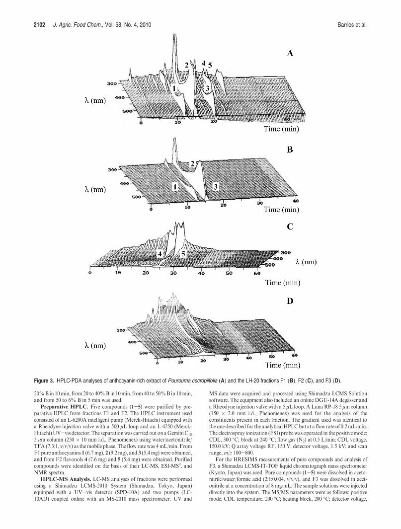
Figure 2. Chemical structures of the anthocyanins and flavonols identified in Pourouma cecropiifolia fruit. Anthocyanins: delphinidin-3-O-β-glucopyranoside(1), cyanidin-3-O-β-glucopyranoside (2), and cyanidin-3-O-(600-malonyl)glucopyranoside (3). Flavonols: quercetin 3-O-R-rhamnopyranosyl-(1f6)-β-galactopyranoside (4) and quercetin 3-O-R-rhamnopyranosyl-(1f6)-β-glucopyranoside (5).
2102 J. Agric. Food Chem., Vol. 58, No. 4, 2010 Barrios et al.
20%B in 10min, from 20 to 40%B in 10min, from40 to 50%B in 10min,and from 50 to 6% B in 5 min was used.
Preparative HPLC. Five compounds (1-5) were purified by pre-parative HPLC from fractions F1 and F2. The HPLC instrument usedconsisted of an L-6200A intelligent pump (Merck-Hitachi) equipped witha Rheodyne injection valve with a 500 μL loop and an L-4250 (Merck-Hitachi)UV-vis detector. The separationwas carried out onaGeminiC18
5 μm column (250 � 10 mm i.d., Phenomenex) using water/acetonitrile/TFA (7:3:1, v/v/v) as themobile phase. The flow rate was 4mL/min. FromF1 pure anthocyanins 1 (6.7mg), 2 (9.2mg), and 3 (5.4 mg) were obtained,and from F2 flavonols 4 (7.6 mg) and 5 (5.4 mg) were obtained. Purifiedcompounds were identified on the basis of their LC-MS, ESI-MSn, andNMR spectra.
HPLC-MS Analysis. LC-MS analyses of fractions were performedusing a Shimadzu LCMS-2010 System (Shimadzu, Tokyo, Japan)equipped with a UV-vis detector (SPD-10A) and two pumps (LC-10AD) coupled online with an MS-2010 mass spectrometer. UV and
MS data were acquired and processed using Shimadzu LCMS Solutionsoftware. The equipment also included an online DGU-14A degasser anda Rheodyne injection valve with a 5 μL loop. A Luna RP-18 5 μm column(150 � 2.0 mm i.d., Phenomenex) was used for the analysis of theconstituents present in each fraction. The gradient used was identical tothe one described for the analyticalHPLCbut at a flow rate of 0.2mL/min.The electrospray ionization (ESI) probewas operated in the positivemode:CDL, 300 �C; block at 240 �C; flow gas (N2) at 0.5 L/min; CDL voltage,150.0 kV; Q array voltage RF, 150 V; detector voltage, 1.5 kV; and scanrange, m/z 100-800.
For the HRESIMS measurements of pure compounds and analysis ofF3, a Shimadzu LCMS-IT-TOF liquid chromatograph mass spectrometer(Kyoto, Japan) was used. Pure compounds (1-5) were dissolved in aceto-nitrile/water/formic acid (2:1:0.004, v/v/v), and F3 was dissolved in acet-onitrile at a concentration of 8 mg/mL. The sample solutions were injecteddirectly into the system. The MS/MS parameters were as follows: positivemode; CDL temperature, 200 �C; heating block, 200 �C; detector voltage,
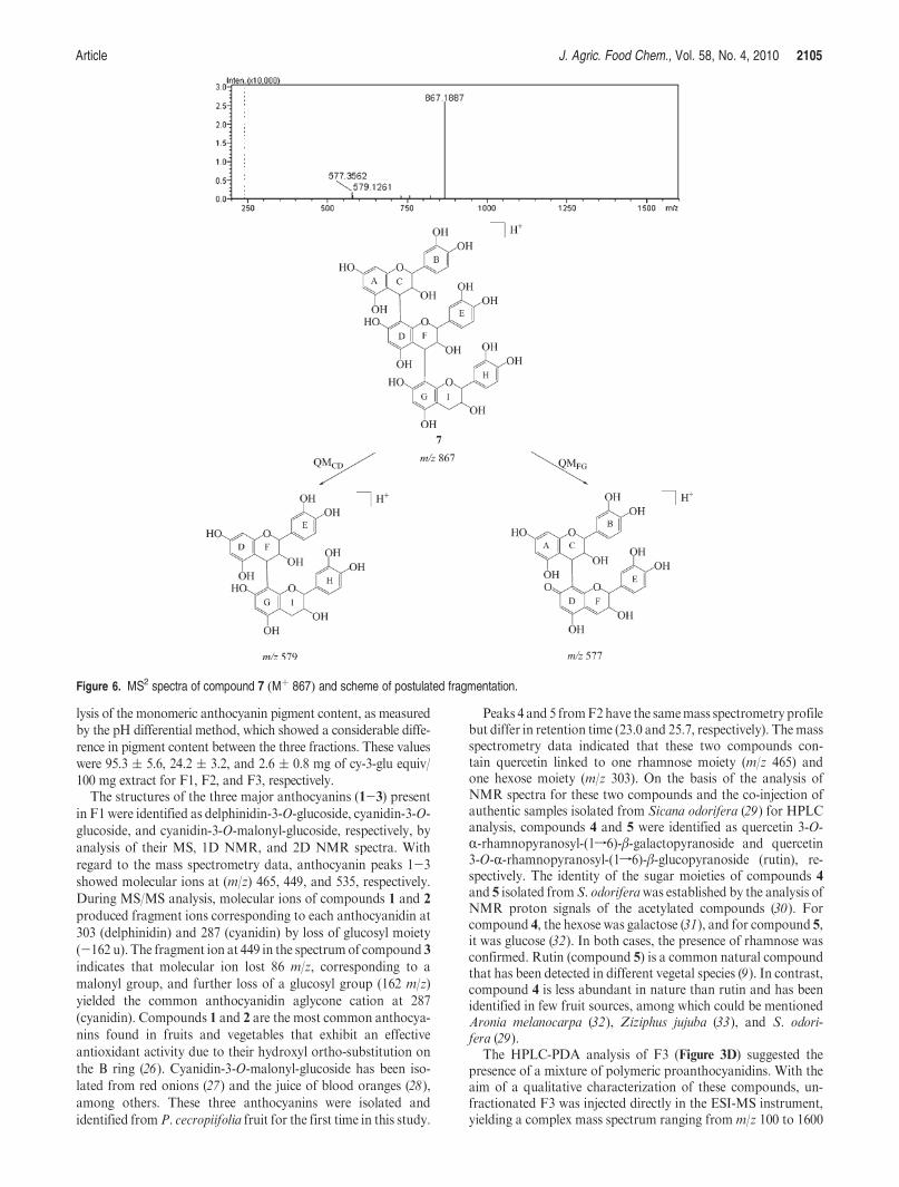
Figure 3. HPLC-PDA analyses of anthocyanin-rich extract of Pourouma cecropiifolia (A) and the LH-20 fractions F1 (B), F2 (C), and F3 (D).

Article J. Agric. Food Chem., Vol. 58, No. 4, 2010 2103
1.55 kV; flow rate, 1.5 L/min; ion accumulation, 20ms; and scan range,m/z200-1600. The energy of the collision gas (argon) was fixed at 15% for theanthocyanins and at 10% for the flavonols and the polymeric F3 fraction.The LCMS Solution software was used for data collection and analysis.
NMRAnalysis. 1HNMR(500.13MHz) and 13CNMR(125.77MHz)spectra of anthocyanins and flavonols were measured in CD3OD/CF3COOD (19:1, v/v) and DMSO-d6, respectively, on a Bruker Avance500 spectrometer at 303 K. The deuteriomethyl 13C signal and theresidual 1H signal of the solvents were used as secondary references (forspectra recorded in CD3OD, these peaks were located at δ 49.0 and3.40; inDMSO-d6, the solvent peaks were at δ 39.5 and 2.50).
1H chemicalshifts were assigned using 1D and 2D 1H NMR (gCOSY), and13C resonances were assigned using 2D NMR (gHSQC and gHMBC)techniques.
Spectroscopic Data. In this section the spectral data of compounds1-5 are reported. Numbering of compounds refers to Figure 2.
Delphinidin-3-O-β-glucopyranoside (1):HRESIMS 465.1028 [calcd forC21H21O12 (M)þ 465.1033]; ESIMS, m/z 465 [M]þ; MS/MS 303 [M -162]þ. 1H and 13C NMR data were in agreement with those published byAzuma et al. (20).
Cyanidin-3-O-β-glucopyranoside (2): HRESIMS 449.1078 [calcd forC21H21O11 (M)þ 449.1084]; ESIMS, m/z 449 [M]þ; MS/MS 287 [M -162]þ. 1H and 13C NMR data were in agreement with those published inthe literature previously (21).
Cyanidin-3-O-(60 0-malonyl)glucopyranoside (3): HRESIMS 535.1091[calcd for C24H23O14 (M)þ 535.1088]; ESIMS,m/z 535 [M]þ; MS/MS 449[M- 86]þ, 287 [M- 162- 86]þ. 1H and 13CNMRdatawere in agreementwith those published in the literature previously (21).
Quercetin 3-O-R-rhamnopyranosyl-(1f6)-β-galactopyranoside (4):HRESIMS 611.1606 [calcd for C27H31O16 (M þ H)þ 611.1613]; ESIMS,m/z 611 [M þH]þ; MS/MS 465 [M - 146 þ H]þ, 303 [M - 162 - 146 þH]þ; 1H NMR [500MHz, DMSO-d6] δ 7.55 (1H, br d, J=7.8 Hz, 60-H),7.53 (1H, br s, 20-H), 6.84 (1H, d, J= 8.2 Hz, 50-H), 6.38 (1H, br s, 8-H),
6.19 (1H, br s, 6-H), 5.34 (1H, d, J=7.4Hz, 10 0-H), 4.39 (1H, br s, 10 0 0-H),3.71 (1H, d, J=10.7Hz, 60 0a-H), 3.06-3.29 (9H, m), 0.99 (3H, d, J=5.4Hz, 60 0 0-3H); 13C NMR [125 MHz, DMSO-d6] δ 177.4 (C-4), 164.1 (C-7),161.2 (C-5), 156.3 (C-2, *), 156.1 (C-9), 148.4 (C-40), 144.8 (C-30), 133.3 (C-3), 121.5 (C-60), 121.1 (C-10), 116.2 (C-20), 115.2 (C-50), 104.0 (C-10), 100.9(C-10 0), 100.4 (C-10 0 0), 98.5 (C-6), 93.4 (C-8), 77.5 (C-20 0), 76.4 (C-50 0), 74.0(C-30 0), 71.6 (C-40 0 0), 70.2 (C-40 0, *), 70.0 (C-20 0 0, *), 69.7 (C-30 0 0), 67.6 (C-50 0 0), 66.7 (C-60 0), 18.0 (C-60 0 0) (* exchangeable).
Quercetin 3-O-R-rhamnopyranosyl-(1f6)-β-glucopyranoside (5):HRE-SIMS 611.1413 [calcd for C27H31O16 (M þ H)þ 611.1613]; ESIMS, m/z611 [MþH]þ; MS/MS 465 [M- 146þH]þ, 303 [M- 162- 146þH]þ;1H NMR [500 MHz, DMSO-d6] δ 7.54 (1H, br d, J= 7.8 Hz, 60-H), 7.53(1H, br s, 20-H), 6.80 (1H, d, J= 8.0 Hz, 50-H), 6.38 (1H, br s, 8-H), 6.18(1H, br s, 6-H), 5.45 (1H, d, J=7.5Hz, 10 0-H), 4.22 (1H, br s, 10 0 0-H), 3.57(1H, d, J=11.5 Hz, 60 0a-H), 3.06-3.40 (9H, m), 0.85 (3H, d, J=5.4 Hz,60 0 0-3H).
Cell Culture. Cell lines derived from human larynx (HEp-2), colon(HT-29), gastric (MKN-45), breast (MCF-7), and cervical (HeLa) cancercells were obtained from American Type Culture Collection (ATCC,Rockville, MD). Cells were grown in minimum essential medium (MEM;Sigma) supplemented with 5% fetal bovine serum (Vitacell, ATCC, VA),penicillin/streptomycin (100 UI-100 μg/mL), and 50 μg/mL gentamycin.The cells were grown in 75 mL flasks at 37 �C in a constant humidifiedatmosphere of 5:95 CO2/air (22).
Cell Viability Assay. Culture flasks in a 90% confluence weretrypsinized and counted in a Neubauer chamber using a trypan blueexclusion procedure. Cells were plated into 96-well, flat-bottom plates andallowed to attach for 24h.Treatmentswere added and plates incubated for48 h. Microtitration colorimetric method of MTT reduction was used todetermine surviving cells at the end of the treatment period. The basicprotocol described by Mosmann (23) was used with some modifica-tions (24). Percentages of cell survival relative to growth control wells(wells containing only cells and medium) were calculated, and IC50
Figure 4. Electrospray mass spectrum of polymeric fraction F3.
Table 1. Major m/z Fragments of Polyphenols in Fraction F3 by ESI/MSn Analysis and Their Tentative Identities
identity precursor ion MS2 MS3
dimeric (epi)catechin 6 579 579/427/409/291/275/247 -a
trimeric (epi)catechin 7 867 867/579/577 -(epi)catechin-4f8-cyanidin-3-glucoside 8 737 737/575 557/449/423
(epi)catechin-(epi)catechin-4f8-cyanidin 3-glucoside 9 1025 1025/863 863/711/573
a-, not recorded.

2104 J. Agric. Food Chem., Vol. 58, No. 4, 2010 Barrios et al.
(concentration that reduces survival of the exposed sample to 50%) valueswere calculated. The ARE, its fractions, and pure compounds (1-5) were
dissolved in DMSO at 10 mM as a stock solution and conserved at 4 �C.Dilutions with culture media were prepared just prior to addition to test
plates with final doses ranging from 0.5 to 50 μg/mL (25). At the end of the
treatment, the medium was removed and 100 μL of 0.25 mg/mL
MTT solution in serum-free medium was added to each well. After 4 h
of incubation, the medium was removed and the formazan crystals
present in each well were dissolved in 100 μL of DMSO. Plates were
mixed for 5 min, and the absorbance at 570 nm was read on a Bio-Rad
550 spectrophotometer. Maximum concentration of DMSO in wells was
0.1% v/v. Doxorubicin-HCl was used as positive control and
vehicle (MEM þ DMSO) as blank. All treatments were evaluated in
triplicates, and three independent experiments were done in different
weeks.Statistical Analyses. Significant differences between treatments
were determined by using analysis of variance (ANOVA). The IC50 valueswere obtained by nonlinear regression using GraphPad 4.0-Prism soft-ware.
RESULTS AND DISCUSSION
Fractionation and Analysis of Anthocyanin-Rich Extract. Theepicarps of P. cecropiifolia fruits are rich in anthocyanin pigments(200( 44 mg/100 g of epicarp). These anthocyanin pigments werethe focus of this study not only because they exhibited cytotoxicactivity but also because they could be used in the development ofvalue-added food products. Thus, the ARE of P. cecropiifoliaobtained by selective retention on Amberlite XAD-7 resin wasfractionated on a Sephadex LH-20 column, giving three fractions(F1-F3). The composition of the fractions was analyzed byHPLC-PDA. As can be seen in Figure 3, the LH-20 purificationeffectively fractionated the ARE, as the composition of each frac-tion was clearly different. The F1 fraction was principally consti-tuted of monomeric anthocyanins (λmax = 520 nm), F2 containedflavonols (λmax=350 nm), andF3was a polymericmixture, with amaximum absorption at 280 nm and some minor absorption at520 nm. These results were in agreement with the results of the ana-
Figure 5. MS2 spectra of compound 6 (Mþ 579) and scheme of postulated fragmentation.
Article J. Agric. Food Chem., Vol. 58, No. 4, 2010 2105
lysis of the monomeric anthocyanin pigment content, as measuredby the pH differential method, which showed a considerable diffe-rence in pigment content between the three fractions. These valueswere 95.3 ( 5.6, 24.2 ( 3.2, and 2.6 ( 0.8 mg of cy-3-glu equiv/100 mg extract for F1, F2, and F3, respectively.
The structures of the three major anthocyanins (1-3) present
in F1were identified as delphinidin-3-O-glucoside, cyanidin-3-O-
glucoside, and cyanidin-3-O-malonyl-glucoside, respectively, by
analysis of their MS, 1D NMR, and 2D NMR spectra. With
regard to the mass spectrometry data, anthocyanin peaks 1-3
showed molecular ions at (m/z) 465, 449, and 535, respectively.
During MS/MS analysis, molecular ions of compounds 1 and 2
produced fragment ions corresponding to each anthocyanidin at
303 (delphinidin) and 287 (cyanidin) by loss of glucosyl moiety
(-162 u). The fragment ion at 449 in the spectrumof compound 3
indicates that molecular ion lost 86 m/z, corresponding to a
malonyl group, and further loss of a glucosyl group (162 m/z)
yielded the common anthocyanidin aglycone cation at 287
(cyanidin). Compounds 1 and 2 are the most common anthocya-
nins found in fruits and vegetables that exhibit an effective
antioxidant activity due to their hydroxyl ortho-substitution on
the B ring (26). Cyanidin-3-O-malonyl-glucoside has been iso-
lated from red onions (27) and the juice of blood oranges (28),
among others. These three anthocyanins were isolated and
identified fromP. cecropiifolia fruit for the first time in this study.
Peaks 4 and 5 fromF2have the samemass spectrometry profilebut differ in retention time (23.0 and 25.7, respectively). Themassspectrometry data indicated that these two compounds con-tain quercetin linked to one rhamnose moiety (m/z 465) andone hexose moiety (m/z 303). On the basis of the analysis ofNMR spectra for these two compounds and the co-injection ofauthentic samples isolated from Sicana odorifera (29) for HPLCanalysis, compounds 4 and 5 were identified as quercetin 3-O-R-rhamnopyranosyl-(1f6)-β-galactopyranoside and quercetin3-O-R-rhamnopyranosyl-(1f6)-β-glucopyranoside (rutin), re-spectively. The identity of the sugar moieties of compounds 4
and 5 isolated from S. odoriferawas established by the analysis ofNMR proton signals of the acetylated compounds (30). Forcompound 4, the hexose was galactose (31), and for compound 5,it was glucose (32). In both cases, the presence of rhamnose wasconfirmed. Rutin (compound 5) is a common natural compoundthat has been detected in different vegetal species (9). In contrast,compound 4 is less abundant in nature than rutin and has beenidentified in few fruit sources, among which could be mentionedAronia melanocarpa (32), Ziziphus jujuba (33), and S. odori-fera (29).
The HPLC-PDA analysis of F3 (Figure 3D) suggested thepresence of a mixture of polymeric proanthocyanidins. With theaim of a qualitative characterization of these compounds, un-fractionated F3 was injected directly in the ESI-MS instrument,yielding a complex mass spectrum ranging from m/z 100 to 1600
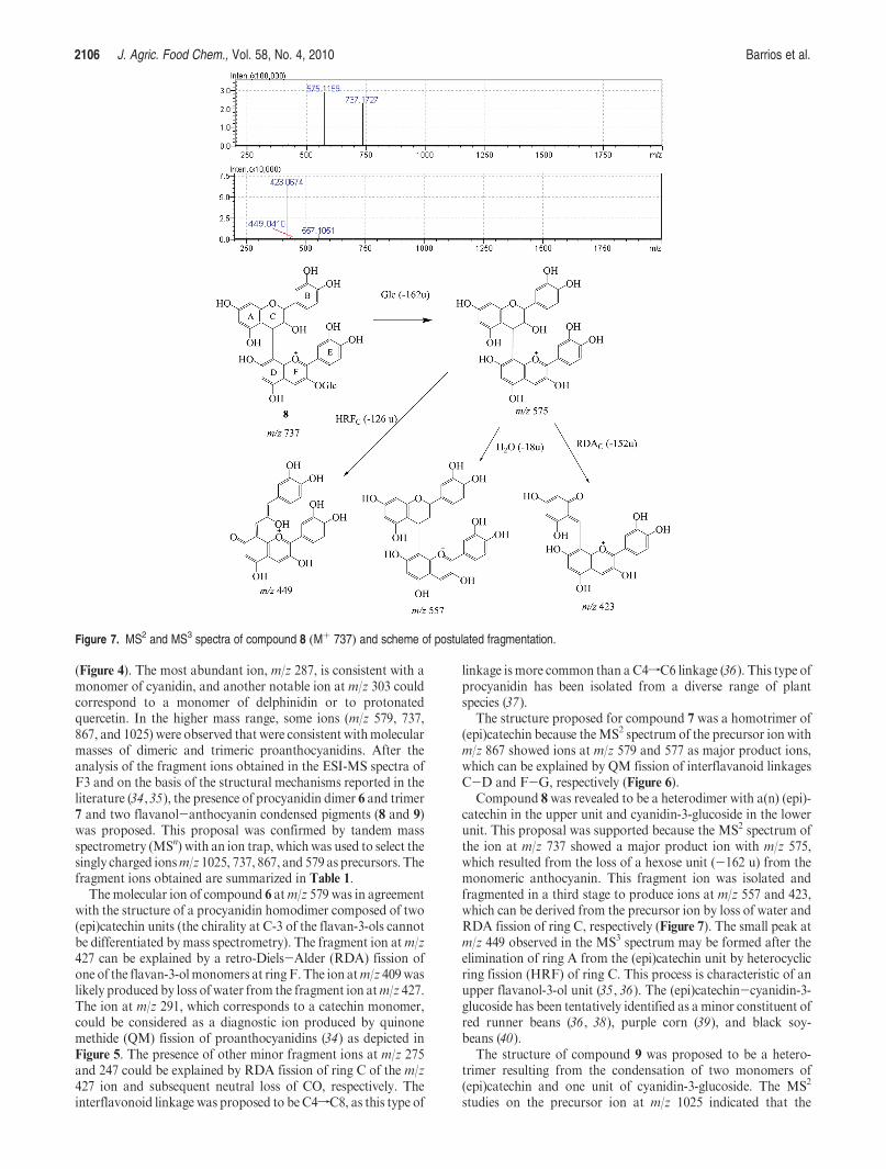
Figure 6. MS2 spectra of compound 7 (Mþ 867) and scheme of postulated fragmentation.
2106 J. Agric. Food Chem., Vol. 58, No. 4, 2010 Barrios et al.
(Figure 4). The most abundant ion, m/z 287, is consistent with amonomer of cyanidin, and another notable ion at m/z 303 couldcorrespond to a monomer of delphinidin or to protonatedquercetin. In the higher mass range, some ions (m/z 579, 737,867, and 1025) were observed that were consistent withmolecularmasses of dimeric and trimeric proanthocyanidins. After theanalysis of the fragment ions obtained in the ESI-MS spectra ofF3 and on the basis of the structural mechanisms reported in theliterature (34,35), the presence of procyanidin dimer 6 and trimer7 and two flavanol-anthocyanin condensed pigments (8 and 9)was proposed. This proposal was confirmed by tandem massspectrometry (MSn) with an ion trap, whichwas used to select thesingly charged ionsm/z 1025, 737, 867, and 579 as precursors. Thefragment ions obtained are summarized in Table 1.
The molecular ion of compound 6 atm/z 579 was in agreementwith the structure of a procyanidin homodimer composed of two(epi)catechin units (the chirality at C-3 of the flavan-3-ols cannotbe differentiated by mass spectrometry). The fragment ion atm/z427 can be explained by a retro-Diels-Alder (RDA) fission ofone of the flavan-3-olmonomers at ringF.The ion atm/z 409waslikely produced by loss of water from the fragment ion atm/z 427.The ion at m/z 291, which corresponds to a catechin monomer,could be considered as a diagnostic ion produced by quinonemethide (QM) fission of proanthocyanidins (34) as depicted inFigure 5. The presence of other minor fragment ions at m/z 275and 247 could be explained by RDA fission of ring C of the m/z427 ion and subsequent neutral loss of CO, respectively. Theinterflavonoid linkage was proposed to beC4fC8, as this type of
linkage ismore common than a C4fC6 linkage (36). This type ofprocyanidin has been isolated from a diverse range of plantspecies (37).
The structure proposed for compound 7 was a homotrimer of(epi)catechin because theMS2 spectrum of the precursor ion withm/z 867 showed ions at m/z 579 and 577 as major product ions,which can be explained by QM fission of interflavanoid linkagesC-D and F-G, respectively (Figure 6).
Compound 8 was revealed to be a heterodimer with a(n) (epi)-catechin in the upper unit and cyanidin-3-glucoside in the lowerunit. This proposal was supported because the MS2 spectrum ofthe ion at m/z 737 showed a major product ion with m/z 575,which resulted from the loss of a hexose unit (-162 u) from themonomeric anthocyanin. This fragment ion was isolated andfragmented in a third stage to produce ions at m/z 557 and 423,which can be derived from the precursor ion by loss of water andRDA fission of ring C, respectively (Figure 7). The small peak atm/z 449 observed in the MS3 spectrum may be formed after theelimination of ring A from the (epi)catechin unit by heterocyclicring fission (HRF) of ring C. This process is characteristic of anupper flavanol-3-ol unit (35, 36). The (epi)catechin-cyanidin-3-glucoside has been tentatively identified as a minor constituent ofred runner beans (36, 38), purple corn (39), and black soy-beans (40).
The structure of compound 9 was proposed to be a hetero-trimer resulting from the condensation of two monomers of(epi)catechin and one unit of cyanidin-3-glucoside. The MS2
studies on the precursor ion at m/z 1025 indicated that the
Figure 7. MS2 and MS3 spectra of compound 8 (Mþ 737) and scheme of postulated fragmentation.
Article J. Agric. Food Chem., Vol. 58, No. 4, 2010 2107
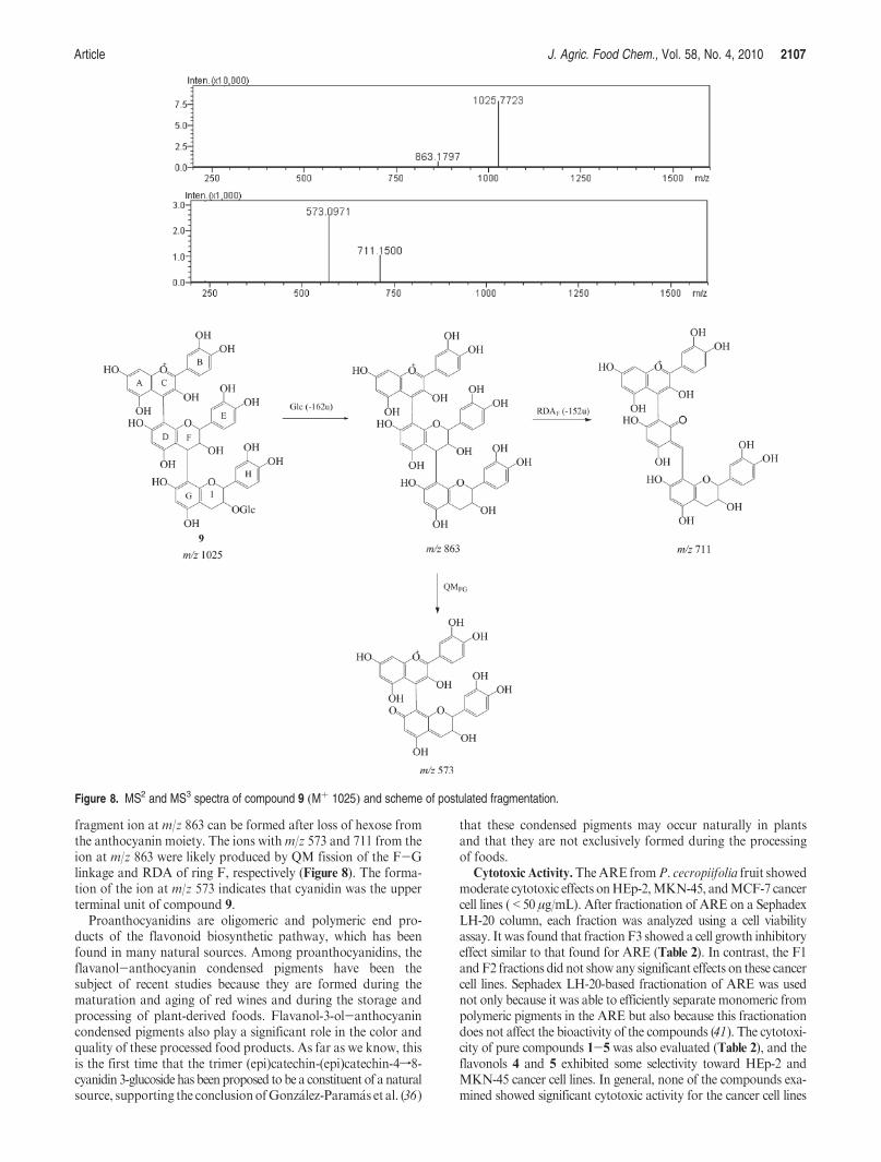
fragment ion at m/z 863 can be formed after loss of hexose fromthe anthocyanin moiety. The ions withm/z 573 and 711 from theion at m/z 863 were likely produced by QM fission of the F-Glinkage and RDA of ring F, respectively (Figure 8). The forma-tion of the ion at m/z 573 indicates that cyanidin was the upperterminal unit of compound 9.
Proanthocyanidins are oligomeric and polymeric end pro-ducts of the flavonoid biosynthetic pathway, which has beenfound in many natural sources. Among proanthocyanidins, theflavanol-anthocyanin condensed pigments have been thesubject of recent studies because they are formed during thematuration and aging of red wines and during the storage andprocessing of plant-derived foods. Flavanol-3-ol-anthocyanincondensed pigments also play a significant role in the color andquality of these processed food products. As far as we know, thisis the first time that the trimer (epi)catechin-(epi)catechin-4f8-cyanidin 3-glucoside has been proposed to be a constituent of a naturalsource, supporting the conclusionofGonz�alez-Param�as et al. (36)
that these condensed pigments may occur naturally in plantsand that they are not exclusively formed during the processingof foods.
Cytotoxic Activity.TheARE fromP. cecropiifolia fruit showedmoderate cytotoxic effects onHEp-2,MKN-45, andMCF-7 cancercell lines (<50 μg/mL). After fractionation of ARE on a SephadexLH-20 column, each fraction was analyzed using a cell viabilityassay. It was found that fraction F3 showed a cell growth inhibitoryeffect similar to that found for ARE (Table 2). In contrast, the F1andF2 fractions did not show any significant effects on these cancercell lines. Sephadex LH-20-based fractionation of ARE was usednot only because it was able to efficiently separate monomeric frompolymeric pigments in the ARE but also because this fractionationdoes not affect the bioactivity of the compounds (41). The cytotoxi-city of pure compounds 1-5 was also evaluated (Table 2), and theflavonols 4 and 5 exhibited some selectivity toward HEp-2 andMKN-45 cancer cell lines. In general, none of the compounds exa-mined showed significant cytotoxic activity for the cancer cell lines
Figure 8. MS2 and MS3 spectra of compound 9 (Mþ 1025) and scheme of postulated fragmentation.
2108 J. Agric. Food Chem., Vol. 58, No. 4, 2010 Barrios et al.
evaluated (the reference value recommended by the U.S. NationalCancer Institute for promising cytotoxic compounds is <4 μg/mL) (42, 43). These results are in agreement with previous studieswhich demonstrated that the in vitro cytotoxic and antiproliferativeactivities toward some cancer cell lines are less effective in antho-cyanin-rich fractions than the original extracts (15). This fact has
been explained as a synergistic or additive effect of anthocyaninswith other active components in the extracts (16).
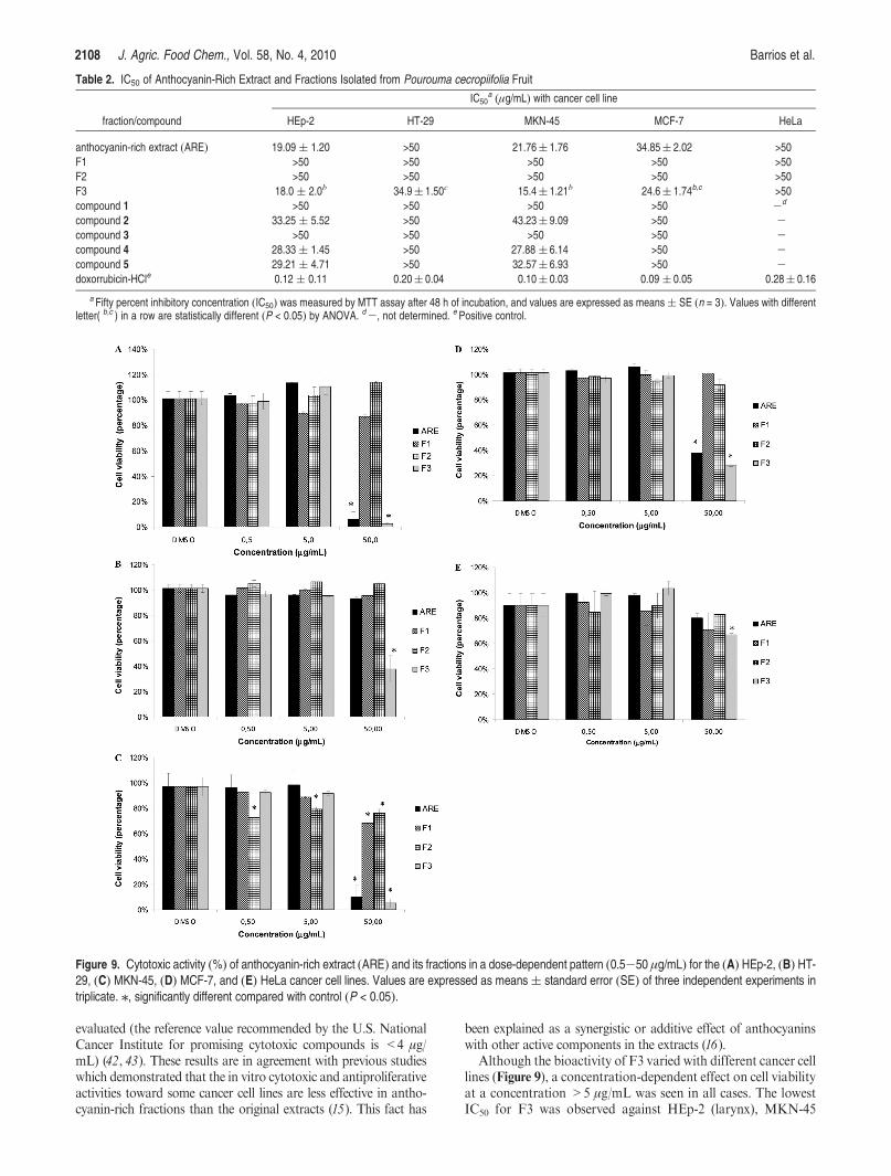
Although the bioactivity of F3 varied with different cancer celllines (Figure 9), a concentration-dependent effect on cell viabilityat a concentration >5 μg/mL was seen in all cases. The lowestIC50 for F3 was observed against HEp-2 (larynx), MKN-45
Table 2. IC50 of Anthocyanin-Rich Extract and Fractions Isolated from Pourouma cecropiifolia Fruit
IC50a (μg/mL) with cancer cell line
fraction/compound HEp-2 HT-29 MKN-45 MCF-7 HeLa
anthocyanin-rich extract (ARE) 19.09 ( 1.20 >50 21.76( 1.76 34.85( 2.02 >50
F1 >50 >50 >50 >50 >50
F2 >50 >50 >50 >50 >50
F3 18.0 ( 2.0b 34.9( 1.50c 15.4( 1.21b 24.6( 1.74b,c >50
compound 1 >50 >50 >50 >50 -d
compound 2 33.25 ( 5.52 >50 43.23( 9.09 >50 -compound 3 >50 >50 >50 >50 -compound 4 28.33 ( 1.45 >50 27.88 ( 6.14 >50 -compound 5 29.21 ( 4.71 >50 32.57( 6.93 >50 -doxorrubicin-HCle 0.12 ( 0.11 0.20( 0.04 0.10( 0.03 0.09 ( 0.05 0.28( 0.16
a Fifty percent inhibitory concentration (IC50) was measured by MTT assay after 48 h of incubation, and values are expressed as means( SE (n = 3). Values with differentletter( b,c ) in a row are statistically different (P < 0.05) by ANOVA. d-, not determined. ePositive control.
Figure 9. Cytotoxic activity (%) of anthocyanin-rich extract (ARE) and its fractions in a dose-dependent pattern (0.5-50 μg/mL) for the (A) HEp-2, (B) HT-29, (C) MKN-45, (D) MCF-7, and (E) HeLa cancer cell lines. Values are expressed as means( standard error (SE) of three independent experiments intriplicate. /, significantly different compared with control (P < 0.05).
Article J. Agric. Food Chem., Vol. 58, No. 4, 2010 2109
(gastric), and MCF-7 (breast) cancer cell lines, which suggestsselective activity toward these lines. A remarkable activity of F3against HT-29 colon cancer cell line was also found: exposure ofthese cells to 50 μg/mL of F3 resulted in a growth inhibition ofapproximately 60%. For the anthocyanin fraction F1 at aconcentration of 50 μg/mL, the highest bioactivities were foundagainst the HEP-2, MKN-45, and HeLa cancer cell lines, withgrowth inhibition values close to 20, 30, and 30%, respectively. Inthe flavonol fraction (F2), the lowest cell viability was seen for theMKN-45 cell line when a concentration of 50 μg/mL was used.
Some studies have also shown a relationship between proantho-cyanidin mixtures and cytotoxic (44) and antitumor activities (45).Their mechanisms of action in cancer cells remain unclear; how-ever, Mantena et al. (14) suggested that the grape seed proantho-cyanidins induce apoptosis and inhibit tumor growth andmetastasis of breast cancer cells through disruption of mitochon-drial pathways.Grape seed proanthocyanidins have been shown tohave a cytotoxic effect on tumor cells without having an adverseeffect on normal cells (46); in contrast, strawberry polyphenolswere found to be equally cytotoxic to tumor cell lines, lymphocyte,or normal human breast and prostate cell lines (47). For the case ofP. cecropiifolia polymeric fraction F3, experiments to evaluate itsgrowth inhibitory effect on normal cells need to be done.
In conclusion, we identified fractions of P. cecropiifolia fruitextract that have promising cytotoxic effects on larynx, gastric, andbreast cancer cell lines. The cytotoxic activities are promising be-cause the calculated IC50 values were <100 μg/mL, which is thereference value recommended byNCI for extracts or fractions (42).The presence of cytotoxic compounds in P. cecropiifolia fruit high-lights the potential for use of thisAmazonian natural resource as ananticancer natural agent; however, further research is required toprove these results in vivo. These findings are also importantbecause the epicarp of the fruit is usually considered to be a wastematerial.
ACKNOWLEDGMENT
We thank Liliana Santacruz for collaboration on the LC-MS/MS analyses. We also thank Prof. Alberto Fajardo from Uni-versidad de La Amazonia for providing the Pourouma cecropii-folia fruits used in this study. We are grateful to Miguel Anguloand Maria Eugenia Soria from CITIUS, Universidad de Sevilla,Spain, for their technical assistance with the NMR analyses.
LITERATURE CITED
(1) Fruits from America. An ethnobotanical inventory. http://www.ciat.cgiar.org/ipgri/fruits_from_americas/frutales/Ficha%20Pouroma%20cecropiaefolia.htm, Oct 2008.
(2) Villachica, H. U. Pourouma cecropiaefolia Mart. In Frutales yHortalizas Promisorios de la Amazonıa; Tratado de Cooperaci�onAmaz�onica, Secretaría Pro Tempore: Lima, Peru, 1996.
(3) Barrios, J.; Sinuco, D.; Morales, A. L. Compuestos vol�atiles libres yenlazados glicosıdicamente en la pulpa de la uva Caimarona(Pourouma cecropiifolia Mart.). Acta Amazon. 2010, in press.
(4) Terry, P.; Terry, J. B.; Wolk, A. Fruit and vegetable consumptionin the prevention of cancer: an update. J. Intern. Med. 2001, 250,280–290.
(5) Olsson,M. E.; Gustavsson, K.-E.; Andersson, S.; Nilsson, A.; Duan,R.-D. Inhibition of cancer cell proliferation in vitro by fruit andberry extracts and correlations with antioxidant levels. J. Agric. FoodChem. 2004, 52, 7264–7271.
(6) Hou, D. X. Potential mechanisms of cancer chemoprevention byanthocyanins. Curr. Mol. Med. 2003, 3, 149–159.
(7) Mazza, G.; Kay, C. D. Bioactivity, absorption, and metabolismof anthocyanins. In Recent Advances in Polyphenols Research;Lattanzio, V., Daayf, V. F., Eds.; Blackwell Publishing: Oxford, U.K.,2008; Vol. I.
(8) Clifford, M. N. Anthocyanins - nature, occurrence and dietaryburden. J. Sci. Food Agric. 2000, 80, 1063–1072.
(9) Andersen, Ø. M.; Jordheim, M. The anthocyanins. In FlavonoidsChemistry, Biochemistry andApplications, Andersen, Ø.M.,Markham,K. R., Eds.; CRC Taylor and Francis: Boca Raton, FL, 2005; p 471.
(10) Seeram, N. P. Berry fruits for cancer prevention: current status andfuture prospects. J. Agric. Food Chem. 2008, 56, 630–635.
(11) Kresty, L. A.; Frankel, W. L.; Hammond, C. D.; Baird, M. E.; Mele,J. M.; Stoner, G. D.; Fromkes, J. Transitioning from preclinical toclinical chemopreventive assessments of lyophilized black raspber-ries: interim results show berries modulate markers of oxidativestress in Barret’s esophagus patients.Nutr. Cancer 2006, 54, 148–156.
(12) Jing, P.; Bomser, J. A.; Schwartz, S. J.; He, J.; Magnuson, B. A.;Giusti,M.M. Structure-function relationships of anthocyanins fromanthocyanin-rich extracts on the inhibition of colon cancer cellgrowth. J. Agric. Food Chem. 2008, 56, 9391–9398.
(13) Netzel, M.; Netzel, G.; Kammerer, D. R.; Schieber, A.; Carle, R.;Simons, Ll.; Bitsch, I.; Bitsch, R.; Konczak, I. Cancer cell anti-proliferation activity and metabolism of black carrot anthocyanins.Innovative Food Emerg. Technol. 2007, 8, 365–372.
(14) Mantena, S. K.; Baliga, M. S.; Katiyar, S. K. Grape seed proantho-cyanidins induce apoptosis and inhibit metastasis of highly meta-static breast carcinoma cells. Carcinogenesis 2006, 27, 1682–1691.
(15) McDougall, G. J.; Ross, H. A.; Ikeji, M.; Stewart, D. Berry extractsexert different antiproliferative effects against cervical and coloncancer cells grown in vitro. J. Agric. Food Chem. 2008, 56, 3016–3023.
(16) Dai, J.; Gupte, A.; Gates, L.; Mumper, R. J. A comprehensive studyof anthocyanin-containing extracts from selected blackberry culti-vars: extraction methods, stability, anticancer properties and me-chanisms. Food Chem. Toxicol. 2009, 47, 837–847.
(17) Degenhardt, A.; Knapp, H.; Winterhalter, P. Separation and pur-ification of anthocyanins by high-speed countercurrent chromatog-raphy and screening for antioxidant activity. J. Agric. Food Chem.2000, 48, 338–343.
(18) Giusti, M. M.; Wrolstad, R. E. Anthocyanins. Characterization andmeasurement with UV-visible spectroscopy. InCurrent Protocols inFood Analytical Chemistry; Wrolstad, R. E., Ed.; Wiley: New York,2001; Unit F1.2, pp 1-13.
(19) Schwarz, M.; Hofmann, G.; Winterhalter, P. Investigations onanthocyanins in wines from Vitis vinifera cv. Pinotage: factorsinfluencing the formation of pinotin A and its correlation with wineage. J. Agric. Food Chem. 2004, 52, 498–504.
(20) Azuma, K.; Ohyama, A.; Ippoushi, K.; Ichiyanagi, T.; Takeuchi, A.;Saito, T.; Fukuoka, H. Structures and antioxidant activity ofanthocyanins in many accessions of eggplant and its related species.J. Agric. Food Chem. 2008, 56, 10154–10159.
(21) Andersen, Ø. M.; Fossen, T. Characterization of anthocyanins byNMR. In Current Protocols in Food Analytical Chemistry; Wrolstad,R. E., Ed.; Wiley: New York, 2001; Unit F 1.4, pp 47-54.
(22) Freshney, I. Culture of Animal Cells: A Manual of Basic Technique;Wiley: Chichester, U.K., 2000; pp 329-343.
(23) Mosmann, T. Rapid colorimetric assay for cellular growth andsurvival: application to proliferation and cytotoxicity assays. J.Immunol. Methods 1983, 65, 55–63.
(24) Cordero, C.; Aristizabal, F. Evaluaci�on preliminar in vitro decitotoxicidad de extractos vegetales empleando metodos colorime-tricos. Rev. Col. Biotecnol. 2002, 4, 100–106.
(25) Cordero, C. P.; Morantes, S. J.; P�aez, A.; Rinc�on, J.; Aristiz�abal, F.A. Cytotoxicity of withanolides isolated from Acnistus arborescens.Fitoterapia 2009, 80, 364–368.
(26) Rice-Evans, C.; Miller, N.; Paganga, G. Structure-antioxidantactivity relationships of flavonoids and phenolic acids. Free RadicalBiol. Med. 1996, 20, 933–956.
(27) Fossen, T.; Slimestad, R.; Andersen, Ø. M. Anthocyanins with 40-glucosidation from red onion Allium cepa. Phytochemistry 2003, 64,1367–1374.
(28) Hillebrand, S.; Schwarz, M.; Winterhalter, P. Characterization ofanthocyanins and pyranoanthocyanins from blood orange[Citrus sinensis (L.) Osbeck] juice. J. Agric. Food Chem. 2004, 52,7331–7338.
2110 J. Agric. Food Chem., Vol. 58, No. 4, 2010 Barrios et al.
(29) Jaramillo, K.; David, C.; Gonz�alez, I.; Hoffman, T.; Fujimoto, Y.;Osorio, C. Quercetin glycosides from Sicana odorifera fruit peel. J.Agric. Food Chem. 2010, manuscript in preparation.
(30) Wang, S.-H.; Ge, W.-Z.; Liu, H.-M.; Zou, D.-P.; Yan, X.-B.Syntheses of acetylated glycosides and selective cleavage of O-acetylgroups in sugar moiety. Steroids 2004, 69, 599–604.
(31) K€uc-€ukislamoglu, M.; Ratli, N.; S-ent€urk, H.; Genc-, H. Flavonol glycosidesfrom Consolida armeniaca. Turk. J. Chem. 2000, 24, 191–197.
(32) Slimestad, R.; Torskangerpoll, K.; Nateland, H. S.; Johannessen, T.;Giske, N. H. Flavonoids from black chokeberries, Aronia melano-carpa. J. Food Compos. Anal. 2005, 18, 61–68.
(33) Pawlowska, A. M.; Camangi, F.; Bader, A.; Braca, A. Flavonoids ofZizyphus jujuba L. and Zizyphus spina-christi (L.) willd(Rhamnaceae) fruits. Food Chem. 2009, 112, 858–862.
(34) Li, H.-J.; Deinzer, M. L. Tandem mass spectrometry for sequencingproanthocyanidins. Anal. Chem. 2007, 79, 1739–1748.
(35) Alcalde-Eon, C.; Escribano-Bail�on,M. T.; Santos-Buelga, C.; Rivas-Gonzalo, J. C. Identification of dimeric anthocyanins and newoligomeric pigments in red wine by means of HPLC-DAD-ESI/MSn. J. Mass Spectrom. 2007, 42, 735–748.
(36) Gonz�alez-Param�as, A. M.; Lopes da Silva, F.; Martın-L�opez, P.;Macz-Pop, G.; Gonz�alez-Manzano, S.; Alcalde-Eon, C.; Perez-Alonso, J. J.; Escribano-Bail�on, M. T.; Rivas-Gonzalo, J. C.;Santos-Buelga, C. Flavanol-anthocyanin condensed pigments inplant extracts. Food Chem. 2006, 94, 428–436.
(37) Haslam, E. Plant Polyphenols, Vegetable Tannins Revisited; Cam-bridge University Press: Cambridge, U.K., 1989; pp 14.
(38) Macz-Pop, G. A.; Gonz�alez-Param�as, A. M.; Perez-Alonso, J. J.;Rivas-Gonzalo, J. C. New flavanol-anthocyanin condensedpigments and anthocyanin composition in Guatemalan beans(Phaseolus spp.). J. Agric. Food Chem. 2006, 54, 536–542.
(39) Gonz�alez-Manzano, S.; Perez-Alonso, J. J.; Salinas-Moreno, Y.;Mateus, N.; Silva, A. M. S.; de Freitas, V.; Santos-Buelga, C.Flavanol-anthocyanin pigments in corn: NMR characterisationand presence in different purple corn varieties. J. Food Compos.Anal. 2008, 21, 521–526.
(40) Lee, J. H.; Kang, N. S.; Shin, S.-O.; Shin, S.-H.; Lim, S.-G.; Suh, D.-Y.; Baek, I.-Y.; Park, K.-Y.; Ha, T. J. Characterisation of antho-
cyanins in the black soybean (Glycine max L.) by HPLC-DAD-ESI/MS analysis. Food Chem. 2009, 112, 226–231.
(41) Alwerdt, J. L.; Seigler, D. S.; Gonzalez de Mejia, E.; Yousef, G. G.;Lila, M. A. Influence of alternative liquid chromatography techni-ques on the chemical complexity and bioactivity of isolatedproanthocyanidin mixtures. J. Agric. Food Chem. 2008, 56, 1896–1906.
(42) Boyd, M. The NCI in vitro anticancer drug discovery screen.Concept, implementation, and operation, 1985-1995. In AnticancerDrug Development Guide: Preclinical Screening, Clinical Trials andApproval; Teicher, B., Totowa, N. J., Eds.; Humana Press: New York,1997; pp 23;42.
(43) Betancur-Galvis, L.; Zuluaga, C.; Arn�o, M.; Gonz�alez, M. A.;Zaragoza, R. J. Structure-activity relationship of in vitro antiviraland cytotoxic activity of semisynthetic analogues of scopadulanediterpenes. J. Nat. Prod. 2001, 64, 1318–1321.
(44) Jo, J. Y.; Gonzalez deMejia, E.; Lila,M.A. Cytotoxicity of bioactivepolymeric fractions from grape cell culture on human hepatocellularcarcinoma, murine leukemia and non-cancerous PK15 kidney cells.Food Chem. Toxicol. 2006, 44, 1758–1767.
(45) Zhang, X. Y.; Li, W. G.; Wu, Y. J.; Zheng, T. Z.; Li, W.; Qu, S. Y.;Liu, N. F. Proanthocyanidin from grape seeds potentiates anti-tumor activity of doxorubicin via immunomodulatory mechanism.Int. Immunopharmacol. 2005, 5, 1247–1257.
(46) Ye, X.; Krohn, R. L.; Liu, W.; Joshi, S. S.; Kuszynski, C. A.;McGinn, T. R.; Bagchi, M.; preuss, H. G.; Stohs, S. J.; Bagchi, D.The cytotoxic effects of a novel IH636 grape seed proanthocyanidinextract on cultured human cancer cells. Mol. Cell. Biochem. 1999,196, 99–108.
(47) Weaver, J.; Briscoe, T.; Hou, M.; Goodman, C.; Kata, S.; Ross, H.;McDougall, G.; Stewart, D.; Riches, A. Strawberry polyphenols areequally cytotoxic to tumourigenic and normal human breast andprostate cell lines. Int. J. Oncol. 2009, 34, 777–786.
Received for review July 31, 2009. Revisedmanuscript received January
10, 2010. Accepted January 16, 2010. This research was supported by
grants from Colciencias and DIB-Universidad Nacional de Colombia.