Infrared neural stimulation fails to evoke neural activity in the deaf guinea pig cochlea
24
Infrared neural stimulation fails to evoke neural activity in the deaf guinea pig cochlea Alexander C. Thompson a , James B. Fallon b,c,d , Andrew K. Wise b,c,d , Scott A. Wade a , Robert K. Shepherd b,c,d , Paul R. Stoddart a,* a Faculty of Science, Engineering and Technology, Swinburne University of Technology, Hawthorn, Australia b Bionics Institute, East Melbourne, Australia c Medical Bionics Department, University of Melbourne, East Melbourne, Australia d Department of Otolaryngology, University of Melbourne, East Melbourne, Australia Abstract At present there is some debate as to the processes by which infrared neural stimulation (INS) activates neurons in the cochlea, as the lasers used for INS can potentially generate a range of secondary stimuli e.g. an acoustic stimulus is produced when the light is absorbed by water. To clarify whether INS in the cochlea requires functioning hair cells and to explore the potential relevance to cochlear implants, experiments using INS were performed in the cochleae of both normal hearing and profoundly deaf guinea pigs. A response to laser stimulation was readily evoked in normal hearing cochlea. However, no response was evoked in any profoundly deaf cochleae, for either acute or chronic deafening, contrary to previous work where a response was observed after acute deafening with ototoxic drugs. A neural response to electrical stimulation was readily evoked in all cochleae after deafening. The absence of a response from optical stimuli in profoundly deaf cochleae suggests that ✩ NOTICE: this is the authors version of a work that was accepted for publication in Hearing Research. Changes resulting from the publishing process, such as peer review, editing, corrections, structural formatting, and other quality control mechanisms may not be reflected in this document. Changes may have been made to this work since it was submitted for publication. A definitive version was subsequently published at doi:10.1016/j.heares.2015.03.005. * Corresponding author Email addresses: [email protected] (Alexander C. Thompson), [email protected] (Paul R. Stoddart) Preprint submitted to Hearing Research March 21, 2015
-
Upload
independent -
Category
Documents
-
view
14 -
download
0
Transcript of Infrared neural stimulation fails to evoke neural activity in the deaf guinea pig cochlea
Infrared neural stimulation fails to evoke neural activity
in the deaf guinea pig cochlea
Alexander C. Thompsona, James B. Fallonb,c,d, Andrew K. Wiseb,c,d, ScottA. Wadea, Robert K. Shepherdb,c,d, Paul R. Stoddarta,∗
aFaculty of Science, Engineering and Technology, Swinburne University of Technology,Hawthorn, Australia
bBionics Institute, East Melbourne, AustraliacMedical Bionics Department, University of Melbourne, East Melbourne, Australia
dDepartment of Otolaryngology, University of Melbourne, East Melbourne, Australia
Abstract
At present there is some debate as to the processes by which infrared neuralstimulation (INS) activates neurons in the cochlea, as the lasers used forINS can potentially generate a range of secondary stimuli e.g. an acousticstimulus is produced when the light is absorbed by water. To clarify whetherINS in the cochlea requires functioning hair cells and to explore the potentialrelevance to cochlear implants, experiments using INS were performed in thecochleae of both normal hearing and profoundly deaf guinea pigs. A responseto laser stimulation was readily evoked in normal hearing cochlea. However,no response was evoked in any profoundly deaf cochleae, for either acute orchronic deafening, contrary to previous work where a response was observedafter acute deafening with ototoxic drugs. A neural response to electricalstimulation was readily evoked in all cochleae after deafening. The absenceof a response from optical stimuli in profoundly deaf cochleae suggests that
INOTICE: this is the authors version of a work that was accepted for publication inHearing Research. Changes resulting from the publishing process, such as peer review,editing, corrections, structural formatting, and other quality control mechanisms maynot be reflected in this document. Changes may have been made to this work sinceit was submitted for publication. A definitive version was subsequently published atdoi:10.1016/j.heares.2015.03.005.
∗Corresponding authorEmail addresses: [email protected] (Alexander C. Thompson),
[email protected] (Paul R. Stoddart)
Preprint submitted to Hearing Research March 21, 2015
the response from INS in the cochlea is hair cell mediated.
Keywords: infrared neural stimulation, INS, cochlear implant, opticalstimulation, optoacoustic
1. Introduction
Despite the great success of the cochlear implant, used by over 300,000patients worldwide (NIH, 2014), many patients are limited in their abilityto understand speech in noisy environments or appreciate music (Friesenet al., 2001). This limitation appears to be primarily due to spreading of5
electrical current in the cochlea (Friesen et al., 2001), which reduces theeffective number of independent channels and thus, spectral information thatcan be delivered. Present research is exploring a range of approaches toimprove delivery of spectral information, including techniques to reduce thespread of current from electrodes (George et al., 2014) and the use of optical10
stimulation techniques (Hernandez et al., 2014; Izzo et al., 2006).Infrared neural stimulation (INS) is a proposed alternative to electrical
stimulation using micro- to milli-second duration pulses of infrared light toactivate neurons (Richter et al., 2011; Chernov and Roe, 2014). Compared totraditional electrical stimulation, reports in the sciatic nerve have suggested15
that INS can be more spatially localised (Wells et al., 2007) and, unlike opto-genetic and caged molecule techniques (Kramer et al., 2009), modification ofthe target tissue is not required (Richter et al., 2011). INS has been demon-strated in vivo in a number of targets, including: peripheral nerves (Wellset al., 2005b), visual cortex (Cayce et al., 2014), embryonic heart (Jenkins20
et al., 2010) and the cochlea (Izzo et al., 2006). The mechanism of INS hasbeen a topic of much discussion in the literature (Brown et al., 2013; Shapiroet al., 2012; Albert et al., 2012). The potential mechanisms that have beenidentified for INS include a change in the cell membrane capacitance in re-sponse to rapid heating (Shapiro et al., 2012) and activation of TRPV heat25
sensitive ion channels (Albert et al., 2012).Electrical stimulation in the cochlea targets the primary auditory neurons
known as spiral ganglion neurons (SGNs). Applying INS to the cochlea hasbeen an area of particular interest, as the potential for an improvement in thespatial selectivity of INS over electrical stimulation may be advantageous for30
use in cochlear implants (Thompson et al., 2013b). Previous work has shownthat INS activates spatial regions similar to acoustic tones, suggesting im-
2

proved spatial localisation compared to electrical stimulation. Furthermore,excitation is largely restricted to SGNs in the beam path. However, muchexcitation appears to be on side of the cochlea opposite the optical fibre,35
where both SGNs and hair cells would be exposed to the light. Rather thanthose directly in front of the optical fibre (Moreno et al., 2011), where onlySGNs were exposed. It should also be noted that radiant exposure thresh-olds for neural activation reported for INS in the cochlea (3 − 20 mJ.cm−2
(Izzo et al., 2006; Richter et al., 2011, 2008; Thompson et al., 2013a)) are40
much lower than those reported for other neural targets, where thresholds of320 − 710 mJ.cm2 are commonly required (Wells et al., 2005a; Teudt et al.,2007), suggesting differences in the mechanisms associated with evoking theneural response.
Many reports of INS in the cochlea use normal hearing animals (Matic45
et al., 2013; Izzo et al., 2007). Some previous experimental reports discussthe ability of INS to evoke neural activity in animals acutely deafened withototoxic aminoglycosides (Izzo et al., 2006; Richter et al., 2008), which dis-able and eliminate the hair cells. This suggests that this INS-evoked neuralactivity is a direct interaction between the laser and the SGNs. However, the50
deafening techniques used in these reports have shown varying levels of hear-ing impairment. Furthermore, INS in chronically deaf animals could onlyevoke a response when one could also be evoked with an acoustic stimulus(Richter et al., 2008).
This has led to some doubt about the stimulation mechanism in cochlear55
studies, which questions as to whether INS directly activated the SGNs, orwhether SGN activation occurred as a result of hair cell mediated responses(Schultz et al., 2014). This is an important distinction for the applicationof INS with a cochlear implant where recipients typically have severe toprofound deafness with few (if any) functional hair cells remaining. Laser60
sources used for INS have been shown to generate an acoustic click from rapidexpansion of heated water, resulting in an optoacoustic mediated response(Teudt et al., 2011). Optoacoustic generation of sound has been shown tobe applicable to many different wavelengths, including those used by INS,and appears to be driven by water or haemoglobin absorption (Schultz et al.,65
2012; Rettenmaier et al., 2014). Therefore, if the deafening process is notcomplete and functional hair cells remain, the generation of an acoustic clickduring INS may lead to a response mediated by activation of residual haircells by this acoustic artefact. Furthermore, a recent report of INS in the ratcochlear nucleus, an auditory structure remote from hair cells of the cochlea,70
3
found no response after deafening that was achieved by cutting the auditorynerve at the internal auditory meatus (Verma et al., 2014).
Given the conflicting results described above, further investigation is re-quired to determine the mechanism of INS in the cochlea and to clarify thepotential benefits of the technique. In this paper, results of INS in severe-75
profoundly deaf guinea pig cochleae are reported. Both acute and chronicdeafening techniques have been used, in order to investigate the potential roleof the deafening procedure in determining the outcome of INS experiments.
2. Methods
2.1. Animal Preparation80
All experimental procedures involving animals were performed in accor-dance with the Australian Code of Practice for the Care and Use of Animalsfor Scientific Purposes and with the guidelines laid down by the NationalInstitutes of Health in the USA regarding the care and use of animals forexperimental procedures. These procedures were approved by the Royal85
Victorian Eye and Ear Hospital Animal Research and Ethics Committee.Four (n = 4) young adult male or female pigmented guinea pigs were
used to collect data for the experimental studies presented in this paper. Intwo animals both cochleae were tested with INS, while the other two wereonly tested on one side, giving a total of six cochlea examined (ncochlea = 6).90
Two were used for acutely deafened experiments, while two were chronicallydeafened four weeks before stimulation experiments. All acutely deafenedanimals were first tested as control normal hearing animals, with INS experi-ments performed before the deafening process was carried out. In chronicallydeafened animals INS experiments were carried out on both cochleae.95
Anaesthesia was induced with an intramuscular delivery of ketamine(60 mg.kg−1) and xylazine (4 mg.kg−1) and was maintained with top-updoses of ketamine (40 mg.kg−1) and xylazine (4 mg.kg−1), administered atone third to one sixth of the induction volume every 40 – 50 min, or if a toepinch withdrawal reflex was present. The animal was kept on a thermostatic100
heating pad at 38 ◦C to maintain body temperature in the normal range.Animals were prepared for surgery by shaving the area near the neck and
skull and then injecting a local anaesthetic (lignocaine, 0.1 ml) at the incisionsite. A post-auricular incision was made and the temporalis muscle retracted,exposing the tympanic bulla. The dorsal region of the bulla was then drilled105
with a 2 mm cutting burr to expose the cochlea. A cochleostomy was drilled
4
into the otic capsule of the basal turn using a diamond burr to thin thecochlea wall. After clearing the bone debris, the endosteum was perforatedto expose the scala tympani and the modiolus (Figure 1). In some animalsa cochleostomy was performed in a more apical turn to assess whether the110
spiral ganglion neuron population at this location responded differently tothose located in the basal turn.
At the conclusion of the experiment, the animals were sacrificed with anoverdose of anaesthetic sodium pentobarbital (150 mg.kg−1) and intracar-dially perfused with formalin fixative. Cochleae were collected for histological115
analysis.
2.2. Deafening
To prevent hair cell mediated electrophonic or optoacoustic (Teudt et al.,2011) activity from being confused with a direct neural interaction with thelaser, animals were deafened using procedures previously shown to produce120
acute hearing loss (Hardie and Shepherd, 1999) or chronic, bilaterally sym-metric, hearing loss (Landry et al., 2011; Kral et al., 2002). These proceduresdiffer from those commonly used in previously reported cochlear INS studies.
Acute deafening was performed unilaterally by opening both the ovaland round windows and gently aspirating neomycin (10 mg.ml−1) though125
the cochlea from the round window to oval window. Aspiration through thecochlea ensured exposure of the apical region of the cochlea to neomycin,which may not be thoroughly exposed without aspirating. The procedurewas repeated at least three times to ensure thorough ototoxin exposure. Theacute deafening procedure was performed after taking normal hearing control130
recordings with INS and electrical stimulation.Neomycin has been shown to block calcium channels (Zhou et al., 1996)
and calcium appears to be involved in the response attributed to INS (Dit-tami et al., 2011; Paviolo et al., 2013; Lumbreras et al., 2014). Therefore itis possible that neomycin may interfere with INS and block a response from135
being observed in the SGNs of acutely deafened animals. In chronically deafanimals, ototoxic drugs should have cleared by the time of the experiment.To investigate if neomycin may block a response, the ability of INS to evokea response in chronically deaf animals was also investigated.
Systematic chronic deafening was performed four weeks before experi-140
mentation, under gaseous anaesthesia (1–2% isoflurane in O2, 1 L.min−1)using a combination of frusemide (130 mg.kg−1, administered intravenously)and kanamycin (420 mg.kg−1, administered subcutaneously) (Landry et al.,
5
2011). The hearing status of chronically deafened animals was assessed underanaesthesia with click stimuli one week before deafening and one week after145
deafening to confirm normal hearing status and then confirm hearing loss.The animal was defined as severe-profoundly deaf if the hearing thresholdincreased by ≥ 50 dB. After the experiment was terminated, histologicalanalysis of the exposed cochlea was performed to confirm hair cells loss.
2.3. Infrared and Electrical Stimulation150
An Aculight Renoir laser (Lockheed Martin Aculight Corp., Bothell, WA)provided the optical stimulus. This laser has been used in a number ofprevious INS studies (e.g. (Richter et al., 2008)). The laser was coupled to200 µm core diameter fibre (NA = 0.22) that was attached to a quartz glassmicropipette. This micropipette was mounted on a 2662 Micropositioner155
(Kopf, USA) which controlled the insertion of the optical fibre in the cochlea.Pulse energy at the fibre tip was measured in air with a Coherent Fieldmasterand Coherent Model LM-10 detector head at the beginning and end of eachexperiment. Unless otherwise stated, a wavelength of 1869 nm was used,giving an optical penetration depth (1/µa) in water of approximately 500 µm160
(Thompson et al., 2012). Pulse durations of 100 µs were typically used, with arepetition rate of 20 Hz and energy of up to 120 µJ giving a maximum radiantexposure of 382 mJ.cm−2 at the fibre tip, similar to parameters successfullyused for cochlear INS in the literature (e.g. Ref (Moreno et al., 2011)). Arange of stimulus durations from 100 µs to 10 ms were used, with the longer165
pulses providing correspondingly greater pulse energies similar to those usedfor other INS targets (e.g. the sciatic nerve (Wells et al., 2007)). For pulsesof 1 ms or longer, the repetition rate was reduced to 2 Hz, to minimise thepotential for heat-induced damage to the cochlea (Thompson et al., 2013b).
Monopolar electrical stimuli were delivered by a platinum ball electrode,170
placed inside the cochlea through the cochleostomy and stainless steel returnelectrode placed in the neck. Electrical stimuli were presented with singlebiphasic anodic leading phase square current pulses, either 25 µs/phase with8 µs interphase gap, or 100 µs/phase with 50 µs interphase gap at a repe-tition rate of 20 Hz. Before beginning INS stimulation experiments, normal175
cochlea function was determined by recording auditory brainstem responsesto electrical or acoustic stimuli.
6
2.4. ABR Recordings
The response of the cochlea and auditory system was assessed by record-ing the auditory brainstem response (ABR) to acoustic (aABR), electrical180
(eABR) and optical stimulation (oABR), using standard techniques (Landryet al., 2011; Coco et al., 2007). All recordings were performed in an electri-cally and acoustically isolated Faraday room. Three stainless steel electrodeswere used (vertex positive, neck negative and thorax ground), to differentiallyrecorded the signal. ABRs were averaged across 100 trials, presented at a185
rate of 20 Hz, and at least two sets of recordings were obtained at each stim-ulus intensity level. ABRs were recorded with Igor Pro software (Wavemet-rics, USA) via a NI USB-6251 data acquisition device (National Instruments,USA) (100 kHz sampling). After acquisition the first ∼ 1 ms of the signal wasdiscarded to remove any electrical artefact and the resultant waveform was190
digitally filtered with a 300 – 3000 Hz bandpass filter (Butterworth, secondorder).
2.5. Histological Preparation
The osseous spiral lamina was fractured by the optical fibre at the con-clusion of the experiment in order to confirm the location and orientation of195
the fibre during the stimulation experiments (1, arrow at circle). Followingthe acute experiment the animals were euthanised (150 mg.kg−1 lethabarb;intraperitoneal) and transcardially perfused with 0.9% NaCl (37◦C) followedby 10% Neutral Buffered Formalin (10% NBF; 4◦C). The cochleae were re-moved and the round and oval windows were opened before post-fixation in200
10% NBF. The cochleae were then placed in 10% ethylenediamine tetraaceticacid (EDTA) in PBS at room temperature for decalcification. Following de-calcification the cochleae were cryo-protected in 15% and then 30% sucroseand then embedded in Tissue-Tek O.C.T. cryosectioning compound (Sakura,Japan) and frozen, as described previously (Wise et al., 2011). Cochleae were205
sectioned in 12 µm slices using a CM 1900 UV cryostat (Leica, Germany)and mounted onto Superfrost-Plus slides (Menzel-Glaser, Braunschweig, Ger-many). A representative series of cochlear sections were stained with Mayer’shaemotoxylin and Putt’s eosin (H&E) for histological examination.
7

Figure 1: Histological image of a mid modiolar cochlear section from a normal hearinganimal indicating the cochleostomy and the target spiral ganglion neurons (oval). Theorgan of Corti containing the intact sensory hair cells (arrow) is intact in this example.The osseous spiral lamina was fractured by the optical fibre (arrow at circle). Solid linesrepresent optical fibre position when targeting the SGNs and dashed lines show opticalfibre next to, but not pointing to the cochlea. Scale bar = 500 µm.
3. Results210
3.1. Normal hearing and acutely deafened animals
Infrared stimulation was attempted in two animals, acutely deafened withneomycin. The first animal was initially exposed to INS before deafening asa normal hearing control. As the pulses used by INS lasers are capableof generating sound in a humid atmosphere or water (Teudt et al., 2011),215
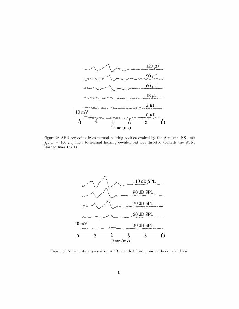
the pure optoacoustic response of the normal hearing cochlea was assessedbefore performing the cochleostomy. The optoacoustic response was found byplacing the optical fibre next to the intact cochlea, but not oriented towardsthe SGNs (Figure 1, dashed line). The ABR recording (Figure 2) showsa clear response to the optoacoustic stimulus generated by the laser pulse.220
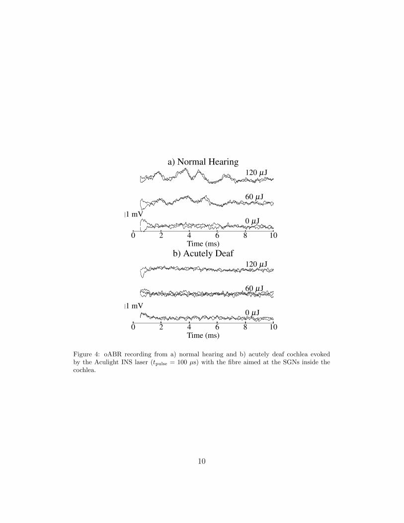
As expected, the ABR recording to the optoacoustic stimulus shows similarwaveform shape to that in response to an acoustic click (Fig 3).
After opening the cochlea, the fibre was inserted through the cochleostomyand directed towards the SGNs (Fig 1, solid line). Accurate positioning ofthe fibre was confirmed by retracting the fibre after attempting optical stim-225
ulation and damaging the osseous spiral lamina with the optical fibre at the
8
0 2 4 6 8 10Time (ms)
0 µJ
2 µJ
18 µJ
60 µJ
90 µJ
120 µJ
10 mV
Figure 2: ABR recording from normal hearing cochlea evoked by the Aculight INS laser(tpulse = 100 µs) next to normal hearing cochlea but not directed towards the SGNs(dashed lines Fig 1).
0 2 4 6 8 10Time (ms)
30 dB SPL
50 dB SPL
70 dB SPL
90 dB SPL
110 dB SPL
10 mV
Figure 3: An acoustically-evoked aABR recorded from a normal hearing cochlea.
9
0 2 4 6 8 10Time (ms)
0 µJ
60 µJ
120 µJa) Normal Hearing
1 mV
0 2 4 6 8 10Time (ms)
0 µJ
60 µJ
120 µJb) Acutely Deaf
1 mV
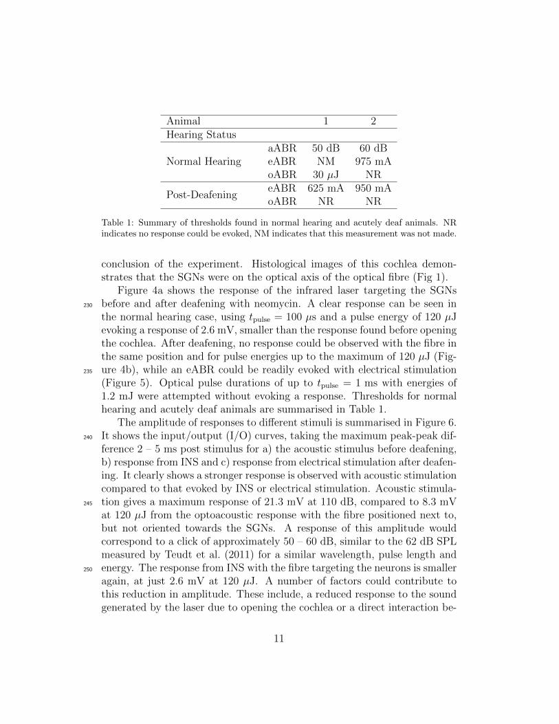
Figure 4: oABR recording from a) normal hearing and b) acutely deaf cochlea evokedby the Aculight INS laser (tpulse = 100 µs) with the fibre aimed at the SGNs inside thecochlea.
10
Animal 1 2Hearing Status
Normal HearingaABR 50 dB 60 dBeABR NM 975 mAoABR 30 µJ NR
Post-DeafeningeABR 625 mA 950 mAoABR NR NR
Table 1: Summary of thresholds found in normal hearing and acutely deaf animals. NRindicates no response could be evoked, NM indicates that this measurement was not made.
conclusion of the experiment. Histological images of this cochlea demon-strates that the SGNs were on the optical axis of the optical fibre (Fig 1).
Figure 4a shows the response of the infrared laser targeting the SGNsbefore and after deafening with neomycin. A clear response can be seen in230
the normal hearing case, using tpulse = 100 µs and a pulse energy of 120 µJevoking a response of 2.6 mV, smaller than the response found before openingthe cochlea. After deafening, no response could be observed with the fibre inthe same position and for pulse energies up to the maximum of 120 µJ (Fig-ure 4b), while an eABR could be readily evoked with electrical stimulation235
(Figure 5). Optical pulse durations of up to tpulse = 1 ms with energies of1.2 mJ were attempted without evoking a response. Thresholds for normalhearing and acutely deaf animals are summarised in Table 1.
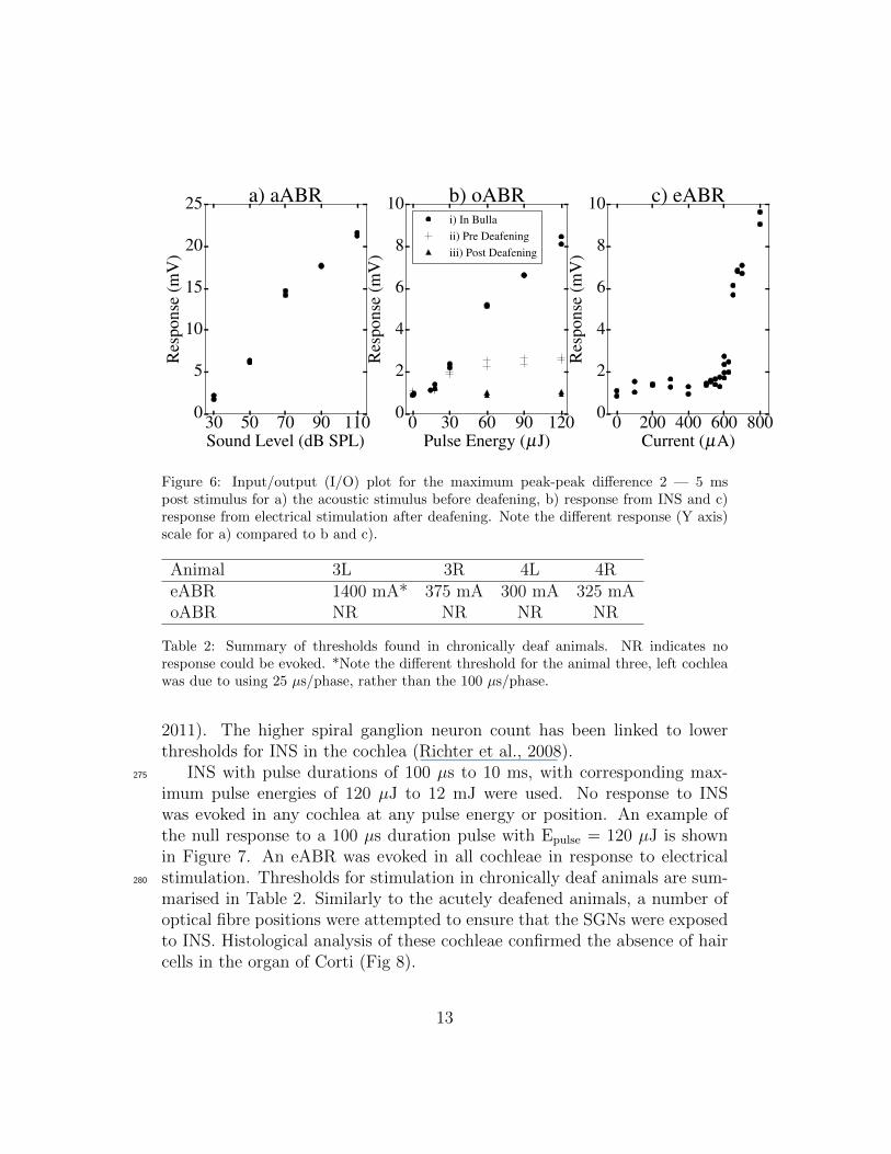
The amplitude of responses to different stimuli is summarised in Figure 6.It shows the input/output (I/O) curves, taking the maximum peak-peak dif-240
ference 2 – 5 ms post stimulus for a) the acoustic stimulus before deafening,b) response from INS and c) response from electrical stimulation after deafen-ing. It clearly shows a stronger response is observed with acoustic stimulationcompared to that evoked by INS or electrical stimulation. Acoustic stimula-tion gives a maximum response of 21.3 mV at 110 dB, compared to 8.3 mV245
at 120 µJ from the optoacoustic response with the fibre positioned next to,but not oriented towards the SGNs. A response of this amplitude wouldcorrespond to a click of approximately 50 – 60 dB, similar to the 62 dB SPLmeasured by Teudt et al. (2011) for a similar wavelength, pulse length andenergy. The response from INS with the fibre targeting the neurons is smaller250
again, at just 2.6 mV at 120 µJ. A number of factors could contribute tothis reduction in amplitude. These include, a reduced response to the soundgenerated by the laser due to opening the cochlea or a direct interaction be-
11

0 2 4 6 8 10Time (ms)
500 µA
600 µA
625 µA
650 µA
675 µA
700 µA
5 mV
Figure 5: Typical eABR recording from electrical stimulation after deafening the cochleawith neomycin. Here monopolar stimulation was delivered via a ball electrode placedinside the cochleostomy with 25 µs per phase.
tween the laser and neurons. As no oABR was evoked after acute deafening,it is possible the reduction in amplitude is due to opening the cochlea.255
Although the targeting of the optical fibre was confirmed to be accuratethrough subsequent histological analysis, many adjustments were made tothe fibre position during the experiment in case there was a more sensitivelocation. The fibre was also positioned so it was touching the osseous spirallamina, thus maximising the energy delivered at the neurons by reducing260
the absorption losses in the intermediate fluid (Thompson et al., 2012). Noadjustments to the optical fibre position were effective in evoking a responsefrom INS.
3.2. Chronically deafened animals
All chronically deafened animals exhibited a severe-profound hearing loss.265
To investigate whether the presence of neomycin may interfere with an INSmediated neural response, and to evaluate the effectiveness of INS in caseswhere there has been significant spiral ganglion neuron loss due to prolongeddeafness, INS was performed in both cochleae of two chronically deafenedanimals. In two of the cochleae, the more apical turns were also exposed to270
INS in addition to the basal turn, as there is often a higher rate of spiralganglion neuron survival after deafening in the apical turns (Landry et al.,
12
30 50 70 90 110Sound Level (dB SPL)
0
5
10
15
20
25
Res
pons
e(m
V)
a) aABR
0 30 60 90 120Pulse Energy (µJ)
0
2
4
6
8
10
Res
pons
e(m
V)
b) oABRi) In Bullaii) Pre Deafeningiii) Post Deafening
0 200 400 600 800Current (µA)
0
2
4
6
8
10
Res
pons
e(m
V)
c) eABR
Figure 6: Input/output (I/O) plot for the maximum peak-peak difference 2 — 5 mspost stimulus for a) the acoustic stimulus before deafening, b) response from INS and c)response from electrical stimulation after deafening. Note the different response (Y axis)scale for a) compared to b and c).
Animal 3L 3R 4L 4ReABR 1400 mA* 375 mA 300 mA 325 mAoABR NR NR NR NR
Table 2: Summary of thresholds found in chronically deaf animals. NR indicates noresponse could be evoked. *Note the different threshold for the animal three, left cochleawas due to using 25 µs/phase, rather than the 100 µs/phase.
2011). The higher spiral ganglion neuron count has been linked to lowerthresholds for INS in the cochlea (Richter et al., 2008).
INS with pulse durations of 100 µs to 10 ms, with corresponding max-275
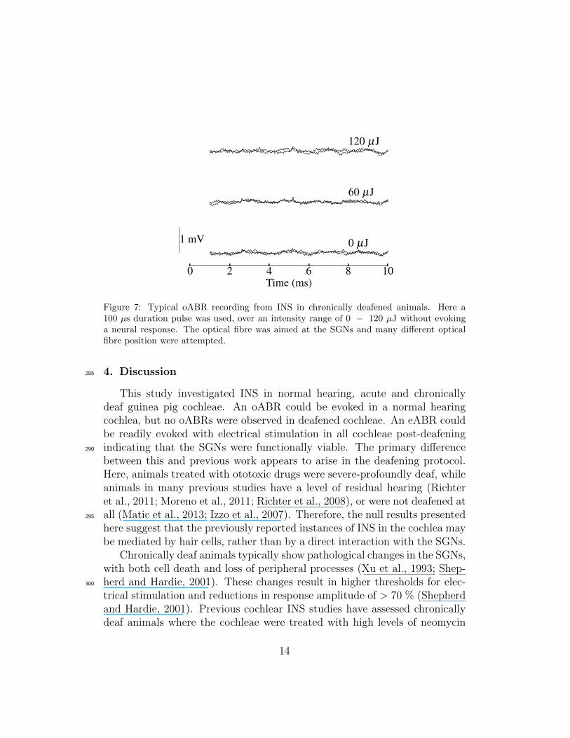
imum pulse energies of 120 µJ to 12 mJ were used. No response to INSwas evoked in any cochlea at any pulse energy or position. An example ofthe null response to a 100 µs duration pulse with Epulse = 120 µJ is shownin Figure 7. An eABR was evoked in all cochleae in response to electricalstimulation. Thresholds for stimulation in chronically deaf animals are sum-280
marised in Table 2. Similarly to the acutely deafened animals, a number ofoptical fibre positions were attempted to ensure that the SGNs were exposedto INS. Histological analysis of these cochleae confirmed the absence of haircells in the organ of Corti (Fig 8).
13
0 2 4 6 8 10Time (ms)
0 µJ
60 µJ
120 µJ
1 mV
Figure 7: Typical oABR recording from INS in chronically deafened animals. Here a100 µs duration pulse was used, over an intensity range of 0 − 120 µJ without evokinga neural response. The optical fibre was aimed at the SGNs and many different opticalfibre position were attempted.
4. Discussion285
This study investigated INS in normal hearing, acute and chronicallydeaf guinea pig cochleae. An oABR could be evoked in a normal hearingcochlea, but no oABRs were observed in deafened cochleae. An eABR couldbe readily evoked with electrical stimulation in all cochleae post-deafeningindicating that the SGNs were functionally viable. The primary difference290
between this and previous work appears to arise in the deafening protocol.Here, animals treated with ototoxic drugs were severe-profoundly deaf, whileanimals in many previous studies have a level of residual hearing (Richteret al., 2011; Moreno et al., 2011; Richter et al., 2008), or were not deafened atall (Matic et al., 2013; Izzo et al., 2007). Therefore, the null results presented295
here suggest that the previously reported instances of INS in the cochlea maybe mediated by hair cells, rather than by a direct interaction with the SGNs.
Chronically deaf animals typically show pathological changes in the SGNs,with both cell death and loss of peripheral processes (Xu et al., 1993; Shep-herd and Hardie, 2001). These changes result in higher thresholds for elec-300
trical stimulation and reductions in response amplitude of > 70 % (Shepherdand Hardie, 2001). Previous cochlear INS studies have assessed chronicallydeaf animals where the cochleae were treated with high levels of neomycin
14
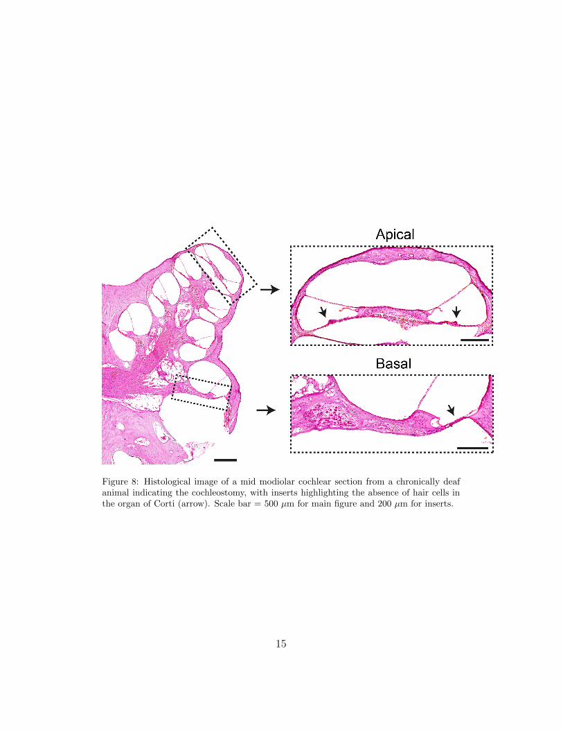
Figure 8: Histological image of a mid modiolar cochlear section from a chronically deafanimal indicating the cochleostomy, with inserts highlighting the absence of hair cells inthe organ of Corti (arrow). Scale bar = 500 µm for main figure and 200 µm for inserts.
15
(Richter et al., 2008). In that case, if the cochlea showed no response toinfrared stimulus, it was also found to give no response to acoustic or elec-305
trical stimuli (Richter et al., 2008). In the present work, a clear electricalresponse was evoked in all chronically deafened animals, but no response toINS or acoustic stimulation was evoked. This implies that the neurons werefunctional but not excitable with INS.
4.1. Optoacoustic Responses310
Given that the response to infrared laser stimulation disappeared afterdeafening in this work, it is plausible that the response attributed to INSin prior reports in the cochlea is due to the optoacoustic effect as previ-ously reported by Teudt et al. (2011). A purely acoustic effect would alsocorrelate well with a recent report of INS targeting the rat cochlea nucleus,315
which reported no response after deafening by cutting the auditory nerveat the internal auditory meatus (Verma et al., 2014). Cutting the auditorynerve prevented transmission of acoustically evoked responses generated inthe cochlea from reaching the the recording site in the auditory brainstem,while still allowing responses evoked in the cochlea nucleus to be transmit-320
ted to the recording site, thus preventing acoustically evoked neural activityfrom reaching the nerves being stimulated and recorded.
A similar result has been seen using nanosecond duration laser pulses withwavelengths other than those typically used for INS (420 – 2150 nm) (Schultzet al., 2012; Wenzel et al., 2009), with a clear response observed in normal325
hearing animals and no response after acute deafening with neomycin. Theabsence of a response after deafening is a strong indication of an optoacous-tically mediated response to the nanosecond duration laser pulses.
However, a purely optoacoustic mechanism is not consistent with all pre-viously reported results of INS in the cochlea. For example, Moreno et al.330
(2011) showed excitation of SGNs was largely restricted to the area in thebeam path. This would suggest a direct interaction with SGNs or hair cells,rather than an acoustic stimulation of hair cells.
4.2. Other Mechanisms
A number of other mechanisms may exist to explain hair cell involvement335
in the response attributed to INS in the cochlea without invoking a purelyoptoacoustic mechanism. For example, vestibular hair cells have been shownto respond to INS (Rajguru et al., 2011). Although the detailed mechanisms
16
in the vestibular system also remain unclear, there is evidence that the re-sponse was not due to an optoacoustic process, but rather direct stimulation340
of the hair cell; and that the mechanism may also apply to hair cells of thecochlea (Rajguru et al., 2011). Another possibility is that the infrared laserpulses may cause release of synaptic vesicles from the hair cells resulting inpost-synaptic currents in the neurons (Liu et al., 2014). If the synapses be-tween the hair cells and spiral ganglion neurons are exposed to infrared light,345
this could drive the response observed. Furthermore, the results of Morenoet al. (2011) have shown excitation on the side of the cochlea spiral oppositeto the optical fibre. In this work, little excitation was observed near theoptical fibre, where only the SGNs would have been exposed to the light. Onthe side of the cochlea opposite to the optical fibre, strong excitation was350
observed. Here, both the SGNs and hair cells would have been exposed tothe light, again suggesting an interaction with the hair cells.
Another potential mechanism comes from recent work showing that themotor protein in outer hair cells, prestin, can respond to rapid temperaturechanges such as those produced by exposure to infrared illumination (Oku-355
nade and Santos-Sacchi, 2013). If outer hair cells are exposed to infraredlaser light, it is possible that this could mediate the response observed. Notethat these putative mechanisms rely on some level of residual activity in thehair cells, which is consistent with all of the reported cases of INS in acutelydeafened cochleae, as well as the absence of response in severe to profoundly360
deafened animals (both acute and chronic).Hair cells are not present in the majority of neural targets where INS
has been demonstrated. The radiant exposure threshold and correspondingtemperature change required for neural activation is much greater in thosecases than the for the cochlea (Thompson et al., 2013b). The tempera-365
ture increase observed (Wells et al., 2007) and predicted (Thompson et al.,2013b) suggests that other mechanisms such as activation of heat sensitiveion channels (Albert et al., 2012) changes in cell membrane capacitance maybe responsible.
If hair cells respond to pulses of infrared light, it may provide a new370
technique to interrogate their function and the mechanisms of hearing. Un-derstanding the exact interaction between infrared laser and the cochlea willrequire further careful experimentation. Understanding the full mechanismmay involve in vitro excitation of hair cells, both in isolation and in tissueslices.375
17
4.3. Methodological Considerations
The number of cochleae used in this study is relatively small (n = 6) andit could be argued that it is insufficient to make strong statements about themechanisms of INS in the cochlea. However, an eABR could be evoked inall of the cochleae tested, thus indicating normal neural function. In each380
of the cochleae tested, a full range of INS pulse durations and energies wereused. Furthermore, a range of fibre positions were attempted and correct po-sitioning of the fibre was confirmed through subsequent histological analysis.Without a clear avenue to advancing the procedure for further experimenta-tion and a realistic expectation that this would change the outcome, further385
animal experimentation was not considered ethically justified.
5. Conclusion
No INS evoked oABR was observed in any deaf cochleae, while normalhearing cochlea responded to the infrared laser stimulus. These results sug-gest that to evoke an oABR from INS requires functional hair cells. Identi-390
fying the mechanism, which may be optoacoustic or some other interactionwith the infrared light, will require further work. It appears unlikely thatINS in an implantable device will show clinical benefits for profoundly deafpatients without significant further development. However, if the INS inter-action is mediated by hair cells, use of infrared light may provide novel ways395
of interrogating hair cell function and provide further insights into to themechanisms of hearing.
Acknowledgements
The authors thank C.-P. Richter of Northwestern University for the loanof the Aculight laser and helpful discussions. S. Irving of the Bionics Insti-400
tute kindly assisted with aspects of the in vivo experiments. This work wassupported by Cochlear Ltd and the Australian Research Council under grantLP120100264. The Bionics Institute acknowledges the financial support itreceives from the Victorian State Government.
References405
Albert, E. S., Bec, J.-M., Desmadryl, G., Chekroud, K., Travo, C., Gabo-yard, S., Bardin, F., Marc, I., Dumas, M., Lenaers, G., Hamel, C., Muller,
18

A., and Chabbert, C. (2012). TRPV4 channels mediate the infraredlaser evoked response in sensory neurons. Journal of Neurophysiology,107(12):3227–3234. doi: 10.1152/jn.00424.2011.410
Brown, W. G. A., Needham, K., Nayagam, B. A., and Stoddart, P. R. (2013).Whole cell patch clamp for investigating the mechanisms of infrared neuralstimulation. Journal of Visualized Experiments, (77):e50444. doi: 10.3791/50444.
Cayce, J. M., Friedman, R. M., Chen, G., Jansen, E. D., Mahadevan-Jansen,415
A., and Roe, A. W. (2014). Infrared neural stimulation of primary visualcortex in non-human primates. NeuroImage, 84:181–190. doi: 10.1016/j.neuroimage.2013.08.040.
Chernov, M. and Roe, A. W. (2014). Infrared neural stimulation: a newstimulation tool for central nervous system applications. Neurophotonics,420
1(1):011011–011011. doi: 10.1117/1.NPh.1.1.011011.
Coco, A., Epp, S. B., Fallon, J. B., Xu, J., Millard, R. E., and Shepherd,R. K. (2007). Does cochlear implantation and electrical stimulation affectresidual hair cells and spiral ganglion neurons? Hearing Research, 225(1-2):60–70. doi: 10.1016/j.heares.2006.12.004.425
Dittami, G. M., Rajguru, S. M., Lasher, R. A., Hitchcock, R. W., and Rab-bitt, R. D. (2011). Intracellular calcium transients evoked by pulsed in-frared radiation in neonatal cardiomyocytes. The Journal of Physiology,589(6):1295–1306. doi: 10.1113/jphysiol.2010.198804.
Friesen, L. M., Shannon, R. V., Baskent, D., and Wang, X. (2001). Speech430
recognition in noise as a function of the number of spectral channels: com-parison of acoustic hearing and cochlear implants. The Journal of theAcoustical Society of America, 110:1150–1163.
George, S. S., Wise, A. K., Shivdasani, M. N., Shepherd, R. K., andFallon, J. B. (2014). Evaluation of focused multipolar stimulation for435
cochlear implants in acutely deafened cats. Journal of Neural Engineering,11(6):065003. doi: 10.1088/1741-2560/11/6/065003.
Hardie, N. A. and Shepherd, R. K. (1999). Sensorineural hearing loss duringdevelopment: morphological and physiological response of the cochlea and
19

auditory brainstem. Hearing Research, 128(1-2):147–165. doi: 10.1016/440
S0378-5955(98)00209-3.
Hernandez, V. H., Gehrt, A., Reuter, K., Jing, Z., Jeschke, M., Men-doza Schulz, A., Hoch, G., Bartels, M., Vogt, G., Garnham, C. W., Yawo,H., Fukazawa, Y., Augustine, G. J., Bamberg, E., Kugler, S., Salditt, T.,de Hoz, L., Strenzke, N., and Moser, T. (2014). Optogenetic stimulation of445
the auditory pathway. Journal of Clinical Investigation, 124(3):1114–1129.doi: 10.1172/JCI69050.
Izzo, A. D., Richter, C.-P., Jansen, E. D., and Walsh Jr., J. T. (2006).Laser stimulation of the auditory nerve. Lasers in Surgery and Medicine,38(8):745–753.450
Izzo, A. D., Walsh Jr., J. T., Jansen, E. D., Bendett, M., Webb, J., Ralph,H., and Richter, C.-P. (2007). Optical parameter variability in laser nervestimulation: A study of pulse duration, repetition rate, and wavelength.IEEE Transactions on Biomedical Engineering, 54(6):1108–1114.
Jenkins, M. W., Duke, A. R., Gu, S., Doughman, Y., Chiel, H. J., Fujioka,455
H., Watanabe, M., Jansen, E. D., and Rollins, A. M. (2010). Opticalpacing of the embryonic heart. Nature Photonics, 4(9):623–626. doi: 10.1038/nphoton.2010.166.
Kral, A., Hartmann, R., Tillein, J., Heid, S., and Klinke, R. (2002). Hearingafter congenital deafness: Central auditory plasticity and sensory depriva-460
tion. Cerebral Cortex, 12(8):797–807. doi: 10.1093/cercor/12.8.797.
Kramer, R. H., Fortin, D. L., and Trauner, D. (2009). New photochemicaltools for controlling neuronal activity. Current Opinion in Neurobiology,19(5):544–552. doi: 10.1016/j.conb.2009.09.004.
Landry, T. G., Wise, A. K., Fallon, J. B., and Shepherd, R. K. (2011). Spiral465
ganglion neuron survival and function in the deafened cochlea followingchronic neurotrophic treatment. Hearing Research, 282(1-2):303–313. doi:10.1016/j.heares.2011.06.007.
Liu, Q., Frerck, M. J., Holman, H. A., Jorgensen, E. M., and Rabbitt, R. D.(2014). Exciting cell membranes with a blustering heat shock. Biophysical470
Journal, 106(8):1570–1577. doi: 10.1016/j.bpj.2014.03.008.
20

Lumbreras, V., Bas, E., Gupta, C., and Rajguru, S. M. (2014). Pulsed in-frared radiation excites cultured neonatal spiral and vestibular ganglionneurons by modulating mitochondrial calcium cycling. Journal of Neuro-physiology. doi: 10.1152/jn.00253.2014.475
Matic, A. I., Robinson, A. M., Young, H. K., Badofsky, B., Rajguru, S. M.,Stock, S., and Richter, C.-P. (2013). Behavioral and electrophysiologicalresponses evoked by chronic infrared neural stimulation of the cochlea.PLoS ONE, 8(3):e58189. doi: 10.1371/journal.pone.0058189.
Moreno, L. E., Rajguru, S. M., Matic, A. I., Yerram, N., Robinson, A. M.,480
Hwang, M., Stock, S., and Richter, C.-P. (2011). Infrared neural stimu-lation: Beam path in the guinea pig cochlea. Hearing Research, 282(1-2):289–302. doi: 10.1016/j.heares.2011.06.006.
National Institute on Deafness and Other Communication Disorders.http://www.nidcd.nih.gov/health/hearing/pages/coch.aspx Accessed:485
2015-02-27.
Okunade, O. and Santos-Sacchi, J. (2013). IR laser-induced perturbations ofthe voltage-dependent solute carrier protein SLC26a5. Biophysical Journal,105(8):1822–1828. doi: 10.1016/j.bpj.2013.09.008.
Paviolo, C., Haycock, J. W., Cadusch, P. J., McArthur, S. L., and Stoddart,490
P. R. (2013). Laser exposure of gold nanorods can induce intracellularcalcium transients. Journal of Biophotonics, 7(10):761–765. doi: 10.1002/jbio.201300043.
Rajguru, S. M., Richter, C.-P., Matic, A. I., Holstein, G. R., Highstein, S. M.,Dittami, G. M., and Rabbitt, R. D. (2011). Infrared photostimulation of495
the crista ampullaris. The Journal of Physiology, 589(6):1283–1294. doi:10.1113/jphysiol.2010.198333.
Rettenmaier, A., Lenarz, T., and Reuter, G. (2014). Nanosecond laser pulsestimulation of spiral ganglion neurons and model cells. Biomedical OpticsExpress, 5(4):1014. doi: 10.1364/BOE.5.001014.500
Richter, C.-P., Bayon, R., Izzo, A. D., Otting, M., Suh, E., Goyal, S., Ho-taling, J., and Walsh Jr., J. T. (2008). Optical stimulation of auditoryneurons: Effects of acute and chronic deafening. Hearing Research, 242(1-2):42–51.
21
Richter, C.-P., Matic, A. I., Wells, J. D., Jansen, E. D., and Walsh, J. T.505
(2011). Neural stimulation with optical radiation. Laser & PhotonicsReviews, 5(1):68–80. doi: 10.1002/lpor.200900044.
Schultz, M., Baumhoff, P., Kallweit, N., Sato, M., Kruger, A., Ripken, T.,Lenarz, T., and Kral, A. (2014). Optical stimulation of the hearing anddeaf cochlea under thermal and stress confinement condition. volume 8928,510
pages 892816–892816–7.
Schultz, M., Baumhoff, P., Maier, H., Teudt, I. U., Kruger, A., Lenarz, T.,and Kral, A. (2012). Nanosecond laser pulse stimulation of the inner eara wavelength study. Biomedical Optics Express, 3(12):3332–3345. doi:10.1364/BOE.3.003332.515
Shapiro, M. G., Homma, K., Villarreal, S., Richter, C.-P., and Bezanilla, F.(2012). Infrared light excites cells by changing their electrical capacitance.Nature Communications, 3:736. doi: 10.1038/ncomms1742.
Shepherd, R. K. and Hardie, N. A. (2001). Deafness-induced changes inthe auditory pathway: implications for cochlear implants. Audiology and520
Neurotology, 6(6):305–318.
Teudt, I. U., Maier, H., Richter, C.-P., and Kral, A. (2011). Acoustic eventsand ”optophonic” cochlear responses induced by pulsed near-infraredLASER. IEEE Transactions on Biomedical Engineering, 58(6):1648 –1655.doi: 10.1109/TBME.2011.2108297.525
Teudt, I. U., Nevel, A. E., Izzo, A. D., Walsh, J. T., and Richter, C.-P. (2007).Optical stimulation of the facial nerve: a new monitoring technique? TheLaryngoscope, 117(9):1641–1647. doi: 10.1097/MLG.0b013e318074ec00.
Thompson, A. C., Wade, S. A., Brown, W. G. A., and Stoddart, P. R. (2012).Modeling of light absorption in tissue during infrared neural stimulation.530
Journal of Biomedical Optics, 17(7):075002–075002–6. doi: 10.1117/1.JBO.17.7.075002.
Thompson, A. C., Wade, S. A., Cadusch, P. J., Brown, W. G. A., andStoddart, P. R. (2013a). Modeling of the temporal effects of heating duringinfrared neural stimulation. Journal of Biomedical Optics, 18(3):035004–535
035004. doi: 10.1117/1.JBO.18.3.035004.
22
Thompson, A. C., Wade, S. A., Pawsey, N. C., and Stoddart, P. R. (2013b).Infrared neural stimulation: Influence of stimulation site spacing and rep-etition rates on heating. IEEE Transactions on Biomedical Engineering,60(12):3534–3541. doi: 10.1109/TBME.2013.2272796.540
Verma, R. U., Guex, A. A., Hancock, K. E., Durakovic, N., McKay, C. M.,Slama, M. C. C., Brown, M. C., and Lee, D. J. (2014). Auditory responsesto electric and infrared neural stimulation of the rat cochlear nucleus. Hear-ing Research, 310:69–75. doi: 10.1016/j.heares.2014.01.008.
Wells, J., Kao, C., Jansen, E. D., Konrad, P., and Mahadevan-Jansen,545
A. (2005a). Application of infrared light for in vivo neural stimulation.Journal of Biomedical Optics, 10(6):064003–064003–12. doi: 10.1117/1.2121772.
Wells, J., Kao, C., Konrad, P., Milner, T., Kim, J., Mahadevan-Jansen,A., and Jansen, E. D. (2007). Biophysical mechanisms of transient optical550
stimulation of peripheral nerve. Biophysical Journal, 93(7):2567–2580. doi:10.1529/biophysj.107.104786.
Wells, J., Kao, C., Mariappan, K., Albea, J., Jansen, E. D., Konrad, P.,and Mahadevan-Jansen, A. (2005b). Optical stimulation of neural tissuein vivo. Optics Letters, 30(5):504–506. doi: 10.1364/OL.30.000504.555
Wenzel, G. I., Balster, S., Zhang, K., Lim, H. H., Reich, U., Massow,O., Lubatschowski, H., Ertmer, W., Lenarz, T., and Reuter, G. (2009).Green laser light activates the inner ear. Journal of Biomedical Optics,14(4):044007–044007. doi: 10.1117/1.3174389.
Wise, A. K., Fallon, J. B., Neil, A. J., Pettingill, L. N., Geaney, M. S.,560
Skinner, S. J., and Shepherd, R. K. (2011). Combining cell-based thera-pies and neural prostheses to promote neural survival. Neurotherapeutics,8(4):774–787. doi: 10.1007/s13311-011-0070-0.
Xu, S.-A., Shepherd, R. K., Chen, Y., and Clark, G. M. (1993). Pro-found hearing loss in the cat following the single co-administration of565
kanamycin and ethacrynic acid. Hearing Research, 70(2):205–215. doi:10.1016/0378-5955(93)90159-X.
Zhou, Y., Gopalakrishnan, V., and Richardson, J. S. (1996). Actions of neu-rotoxic β-amyloid on calcium homeostasis and viability of PC12 cells are
23
blocked by antioxidants but not by calcium channel antagonists. Jour-570
nal of Neurochemistry, 67(4):1419–1425. doi: 10.1046/j.1471-4159.1996.67041419.x.
24