Influence of SEM vacuum on bone micromechanics using in situ AFM
7
JOURNAL OF THE MECHANICAL BEHAVIOR OF BIOMEDICAL MATERIALS 5 (2012) 149–155 Available online at www.sciencedirect.com journal homepage: www.elsevier.com/locate/jmbbm Research paper Influence of SEM vacuum on bone micromechanics using in situ AFM Ines Jimenez-Palomar a , Anna Shipov b , Ron Shahar b , Asa H. Barber a,* a Department of Materials, School of Engineering & Materials Science, Queen Mary University of London, London E1 4NS, UK b Koret School of Veterinary Medicine, The Hebrew University of Jerusalem, Israel ARTICLE INFO Article history: Received 17 May 2011 Received in revised form 17 August 2011 Accepted 23 August 2011 Published online 13 September 2011 Keywords: Bone Hydration Micromechanics AFM FIB ABSTRACT The mechanical properties of rat bone at micron length scales have been evaluated as a function of environmental conditions using an in situ atomic force microscope (AFM) setup while observing using scanning electron microscopy (SEM). Focused ion beam fabricated rat bone cantilever samples were tested in both low and high vacuum conditions in the SEM as well as wet in air using the AFM to measure their elastic modulus. The elastic modulus of rat bone at micron length scales is shown to be independent of the environmental testing conditions and indicates water is bound to bone material even under relatively high vacuum conditions. Our work therefore shows how in situ mechanical testing of bone while observing using high resolution SEM can provide results similar to testing wet in air. Crown Copyright c 2011 Published by Elsevier Ltd. All rights reserved. 1. Introduction Bone is a complex biological composite material that per- forms a variety of mechanical functions, most notably load bearing. Bone achieves the ability to perform its mechani- cal functions both through the use of a variety of component phases and organization of these phases across a range of dif- ferent length scales. A solid mechanics approach can define bone components predominantly as a soft collagen organic matrix incorporating a stiffer mineral phase. The mechanical properties can therefore be directly related to the amount of mineral phase within the bone (Currey, 1999b). For example, deer antler is a form of bone with a relatively low volume frac- tion of mineral, resulting in a low elastic modulus, whereas * Corresponding author. Tel.: +44 2078827763. E-mail address: [email protected] (A.H. Barber). larger mineral volume fractions in equine femur (Currey, 2002) provide correspondingly higher elastic modulus values. Extensive studies indicate that the compositional changes, described in terms of increasing mineral volume fraction, lead to an increase in the elastic modulus but a decrease in the ultimate stress and strain, and therefore work to fail- ure of bone material (Currey, 2002). The relationship between bone composition and mechanical functions has been inves- tigated by a series of experiments evaluating the mechan- ical properties of bone components, especially at relatively small length scales where the structural hierarchy of bone can be largely ignored. Examples include nanoindentation (Tai et al., 2007) and tensile testing of individual mineralized col- lagen fibrils (Hang and Barber, 2011), as well as elucidation of 1751-6161/$ - see front matter Crown Copyright c 2011 Published by Elsevier Ltd. All rights reserved. doi:10.1016/j.jmbbm.2011.08.018
Transcript of Influence of SEM vacuum on bone micromechanics using in situ AFM

J O U R N A L O F T H E M E C H A N I C A L B E H AV I O R O F B I O M E D I C A L M A T E R I A L S 5 ( 2 0 1 2 ) 1 4 9 – 1 5 5
Available online at www.sciencedirect.com
journal homepage: www.elsevier.com/locate/jmbbm
Research paper
Influence of SEM vacuum on bone micromechanics usingin situ AFM
Ines Jimenez-Palomara, Anna Shipovb, Ron Shaharb, Asa H. Barbera,!
aDepartment of Materials, School of Engineering & Materials Science, Queen Mary University of London, London E1 4NS, UKbKoret School of Veterinary Medicine, The Hebrew University of Jerusalem, Israel
A R T I C L E I N F O
Article history:Received 17 May 2011Received in revised form17 August 2011Accepted 23 August 2011Published online 13 September 2011
Keywords:BoneHydrationMicromechanicsAFMFIB
A B S T R A C T
The mechanical properties of rat bone at micron length scales have been evaluated as afunction of environmental conditions using an in situ atomic force microscope (AFM) setupwhile observing using scanning electronmicroscopy (SEM). Focused ion beam fabricated ratbone cantilever samples were tested in both low and high vacuum conditions in the SEM aswell as wet in air using the AFM to measure their elastic modulus. The elastic modulusof rat bone at micron length scales is shown to be independent of the environmentaltesting conditions and indicates water is bound to bone material even under relatively highvacuum conditions. Our work therefore shows how in situmechanical testing of bone whileobserving using high resolution SEM can provide results similar to testing wet in air.
Crown Copyright c" 2011 Published by Elsevier Ltd. All rights reserved.
1. Introduction
Bone is a complex biological composite material that per-forms a variety of mechanical functions, most notably loadbearing. Bone achieves the ability to perform its mechani-cal functions both through the use of a variety of componentphases and organization of these phases across a range of dif-ferent length scales. A solid mechanics approach can definebone components predominantly as a soft collagen organicmatrix incorporating a stiffer mineral phase. The mechanicalproperties can therefore be directly related to the amount ofmineral phase within the bone (Currey, 1999b). For example,deer antler is a form of bone with a relatively low volume frac-tion of mineral, resulting in a low elastic modulus, whereas
! Corresponding author. Tel.: +44 2078827763.E-mail address: [email protected] (A.H. Barber).
larger mineral volume fractions in equine femur (Currey,2002) provide correspondingly higher elastic modulus values.Extensive studies indicate that the compositional changes,described in terms of increasing mineral volume fraction,lead to an increase in the elastic modulus but a decreasein the ultimate stress and strain, and therefore work to fail-ure of bone material (Currey, 2002). The relationship betweenbone composition and mechanical functions has been inves-tigated by a series of experiments evaluating the mechan-ical properties of bone components, especially at relativelysmall length scales where the structural hierarchy of bone canbe largely ignored. Examples include nanoindentation (Taiet al., 2007) and tensile testing of individual mineralized col-lagen fibrils (Hang and Barber, 2011), as well as elucidation of
1751-6161/$ - see front matter Crown Copyright c" 2011 Published by Elsevier Ltd. All rights reserved.doi:10.1016/j.jmbbm.2011.08.018
150 J O U R N A L O F T H E M E C H A N I C A L B E H AV I O R O F B I O M E D I C A L M A T E R I A L S 5 ( 2 0 1 2 ) 1 4 9 – 1 5 5
micromechanics from larger bone specimen deformation byX-ray scattering (Krauss et al., 2009) and observation of frac-ture (Koester et al., 2008). Critically, mechanical testing ofbone material while observing deformation and failure us-ing scanning electronmicroscopy (SEM) has been shown to beparticularly effective for elucidating bone structure–functionbehaviour (Hang and Barber, 2011; Koester et al., 2008). De-spite the potential benefits of this in situ mechanical testingmethodology, the effects of the SEM vacuum conditions onbonemechanics are at this time unclear. Indeed, any materialthat is at least partially hydrated may become modified whenintroduced into the vacuum chamber of an SEM, resulting ina change in its mechanical properties relative to the hydratedstate. As bone is physiologically in a hydrated state, a numberof studies have attempted to define the effects of water on themechanical properties of bone. First investigations of bonemechanical behaviour under different hydrated conditionsrevealed an increase in strength but no change in elastic mod-ulus associated with drying the samples in air after one hour,with a significant increase in elastic modulus observed onlyafter drying in an incubator at 105 #C for a week (Sedlin andHirsch, 1966). Currey later performed mechanical investiga-tions to assess the effect of rehydrating bone after holding inair for 25 days. Currey observed little change in bone mechan-ical performance between the initial hydrated state and therehydrated state (Currey, 1988). Recent work has indicated anincrease in the elastic modulus and strength, with a decreasein failure strain, of dry deer antler bone relative to its hydratedstate using bending mechanical testing (Currey et al., 2009).Morais et al. have indicated similar increases in elastic modu-lus but decrease inwork of fracture for bovine cortical bone ondehydration (Morais et al., 2010). Further studies on elk antlerbone also highlight the increase in strength and decrease infailure strain upon dehydrating, although the elastic modu-lus of the bone showed little dependence on the hydrationstate (Chen et al., 2009). These previous works examined theeffects of dehydration on bone mechanics by critically evalu-ating samples with relatively large length scales of the orderof millimetres. Mechanical testing of bone at smaller lengthscales is particularly advantageous when local property mea-surements defining the properties of the bonematerial are re-quired. Nanoindentation has provided to be the most widelyused technique for probing mechanical properties of bone atmicron to sub-micron length scales with a comprehensive re-view of nanoindentation (Lewis and Nyman, 2008) highlight-ing local mechanical property measurements and effects ofdehydration. In particular, nanoindentation at numerous po-sitions across an osteon within human cortical bone revealeda lower elastic modulus of the wet bone relative to the dehy-drated state (Hoffler et al., 2005). Individual bone trabeculaefrom cancellous human vertebrae also showed a raised elasticmodulus with dehydration (Wolfram et al., 2010). The generaltrend in both large and small scale mechanical testing revealsan increase of the elastic modulus of bone with dehydrationand a corresponding lowering of the work of fracture. Thisobservation is irrespective of the testing method used, withbending tests employed extensively at macroscopic samplelength scales whereas indentation was employed at micronlength scales. The lack of scaling effects in mechanical prop-erty changes in bone with dehydration suggests that water
content affects themechanical properties, as indicated by Ny-man et al. (Nyman et al., 2006), and does not depend on bonestructural hierarchy.
A mechanism to describe the effects of water loss duringdehydration on bone mechanical properties was proposed byNyman et al. for human cortical bone (Nyman et al., 2006).Bone samples held at room temperature showed similar elas-tic moduli to hydrated bone, slight increases in bendingstrength and loss of toughness. Samples dried at higher tem-peratures show an increase in the elastic modulus comparedto hydrated samples, with increasing temperature accelerat-ing losses in toughness but significantly showing decreasesin strength. Nyman et al. proposed a mechanism where wa-ter loss in collagen causes a lowering of bone toughness butwater loss at mineral surfaces decreases both bone tough-ness and strength. An energy-based approach explained howbinding of water to mineral is stronger than water–collagenbinding. Thus, higher temperatures remove water in collagenand at mineral surfaces but lower temperatures only removewater from the collagen. NMR studies provided further sup-port for this model by defining three states of water withinbone (Nyman et al., 2006; Wilson et al., 2006). The first state isfree water found in bone pores such as Haversian and Volk-mann’s canals, canaliculi and lacunae (Yan et al., 2008). Thiswater is expected to play only a minor role in the mechani-cal properties of bone and its removal during dehydration isnot accounted for by Nyman et al. (Nyman et al., 2006). Thesecond state is water loosely bound to collagen via hydrogenbonds; this water fraction can be removed at room temper-ature and acts as a plasticizer, which protects the collagenby reducing the stress transferred between collagen fibrils inbone when an external load is applied (Wilson et al., 2006).Reducing the stress transfer in bone allows the components,in this case the collagen and the mineral, to slide betweenone another and avoid failure. Conversely, high stress trans-fer in bone indicates strong binding between components,which would be effective in transferring the loads through-out the bone. Bone toughness is known to be critically de-pendent on the stress transfer between components, definedby the hydration state (Nyman et al., 2006; Yan et al., 2008).Loss of the second water state in bone ensures increasedstress transfer in components but removes bone plasticiza-tion as the components are bound too tightly to slide. Thus,the removal of bone plasticization provides lower overall bonetoughness. Finally, mineral-bound structural water has beenshown to exist in the imperfect carbonated apatite crystal lat-tice, providing stability via hydrogen bonding with neighbour-ing ions and preventing the crystallites from collapsing or re-arranging. The removal of this water occurs only at highertemperatures and has been shown to destabilize the mineraland cause significant strength reductions of the bone (Nymanet al., 2006; Wilson et al., 2006; Yan et al., 2008). Similarly thetriple helix of tropocollagen would also lose stability if thewater molecules forming a highly ordered network with thetropocollagen were removed. The stability of the tropocolla-gen molecule arises from the formation of additional water-mediated hydrogen bonds in the remaining backbone peptidegroups, which would not exist without water due to spatialconstraints (Beck and Brodsky, 1998; Bella et al., 1995; Wil-son et al., 2006; Yan et al., 2008). Further dehydration models
J O U R N A L O F T H E M E C H A N I C A L B E H AV I O R O F B I O M E D I C A L M A T E R I A L S 5 ( 2 0 1 2 ) 1 4 9 – 1 5 5 151
Table 1 – Dehydration of bone using increasingconcentrations of ethanol in water.
% Ethanol Time (min)
70% Storage90% 6095% 30
100% 30100% 30
have been suggested based on the amount of mineral presentwithin the bone material (Currey, 1999a; Currey et al., 2009;Wilson et al., 2006). As biomineralization in bone occurs byreplacing water, bone with low mineral content will containmore water than highly mineralized bone. Dehydration pro-cesses will therefore cause more structural, and thus me-chanical, changes in bone with relatively low mineral content(Currey, 1999a; Utku et al., 2008).
While the studies detailed above examine the dehydra-tion of bone and the corresponding effects on its mechan-ical behaviour, little work has been done to examine theeffects of vacuum conditions on bone structure. Observationof water loss from various regions resulting in dimensionalcontractions in bone has been directly observed by environ-mental scanning electron microscopy (Utku et al., 2008) butthe corresponding effects onmechanical behaviour have beenlacking. The evaluation of bone mechanics using techniquesinvolving SEM has distinct advantages compared to previ-ous works. Principally, observation and manipulation of rel-atively small volumes of bone can be achieved using theSEM, potentially in conjunction with other testing methodssuch as in situ mechanical testing (Hang and Barber, 2011;Koester et al., 2008), so that the properties of the bone ma-terial can be evaluated. The mechanical testing of these rel-atively small volumes is advantageous when compared tolarger scale testing as structural hierarchy effects can be po-tentially ignored or simplified. Such simplification allows thestudy of the material properties of bone and not whole bonebehaviour. Discrete bone volume mechanical testing can alsobe used in order to assess the effects of the SEM vacuumchamber on potential structural changes in bone due to waterremoval.
Previous literature has highlighted the use of focused ionbeam (FIB) microscopy to isolate micron-sized cantileversfrom teeth for subsequent bending tests (Chan et al., 2009).The dual beam system setup allows FIB technology to becombined with the structural analysis of a scanning electronmicroscope (SEM) (Utku et al., 2008). The capacity to isolatediscrete bone volumes within a dual beam system using theFIB and observe structural changes using SEM is thereforepersuasive but mechanical testing capability within the SEMis required. Critically, dual beam instruments operate inpartial vacuum that have the capacity to dehydrate bonematerial. Recent work has used an atomic force microscope(AFM) within a dual beam system to mechanically testindividual a range of biological and synthetic nanofibreswithin a vacuum environment (Hang and Barber, 2011; Hanget al., 2011). Collagen fibrils from antler bone mechanicallytested using AFM within the chamber of the SEM wascritically shown to fail heterogeneously (Hang and Barber,2011), displaying similar behaviour to fully hydrated antlerbone evaluated using small angle X-ray scattering (Krauss
et al., 2009). The similar mechanical behaviour of collagenfibrils in the SEM and a fully hydrated sample indicatesthat dehydration of bone in a vacuum chamber does nothave an effect on the mechanical properties at nanometrelength scales. This paper extends mechanical testing of bonematerial further by using a combination of AFM and dualbeam system to bend micro cantilevers of bone. In particular,the effects of different environmental conditions on themechanical properties of bone at micron length scales usingAFM while observing using in situ SEM is investigated.
2. Materials and methods
Femora from 8 month old sprague dawley rats were used inall of our investigations. The mechanical properties of in-dividual micron-sized units of bone were evaluated by iso-lating individual beams from the parent material using amulti-stage process. The diaphysis from the extracted rat fe-mur was first isolated using a water cooled diamond bladeslow speed circular saw (Buehler, USA). Beams of cortical bonewith dimensions of 12$1$1mmwere produced and stored in70% ethanol. Bone samples were further dehydrated by sub-merging in a series of water/ethanol solutions, summarizedin Table 1, prior to FIB milling. The dehydrated bone was goldcoated and, using a two part epoxy glue (Poxipol, Arg.), fixedto the sample stage of a dual beam microscope (Quanta 3D,FEI, USA/EU) incorporating both SEM and FIB. Samples are re-quired to be dehydrated and gold coated in the dual beaminstrument to avoid charging effects that could interfere withthe FIB milling process and to prevent drying cracks occurringin the sample from the vacuum pumping system. FIB millingwas performed by a succession of processes summarized inFig. 1. Briefly, FIB was used to remove material in order to pro-duce discrete beam volumes by first polishing a bone sampleedge using a high current ion beam of 65 nA and acceleratingvoltage of 30 kV. Flattening of the bone edges allows furtherremoval of smaller bone volumes using smaller ion beam cur-rents down to 0.1 nA. The smaller ion beam currents avoidobservable ion beam damage. In addition, the FIB milling isalways performed parallel to the produced sample faces toreduce embedding the impinging gallium ions from the FIBwithin the discrete beam volumes produced. The resultantFIB process allowed the fabrication of micro cantilever beamswith dimensions 10$2$2 µmas shown in Fig. 2, with the longaxis of the cantilever beam parallel to the long axis of the ratfemur.
Samples were rehydrated before mechanical testing byremoving from the dual beam chamber and placing fortwo hours in a closed vessel containing a high vapourconcentration of Hank’s buffer solution. We note that equinebone samples have been observed to require only 45 minfor rehydration (Utku et al., 2008). Such a hydration processwas considered more suitable than submerging the samplein water, which in our experience results in the surfacetension of the water deforming the relatively fragile microcantilever beams sufficiently to cause fracture. Mechanicaltesting of the individual bone beams, as shown in Fig. 2(b),was performed using a custom build AFM (Attocube GmbH,Ger.) incorporated within the dual beam chamber. Mechanicaltesting was facilitated by moving the AFM probe into contactwith the FIB milled micro cantilever beams in three different

152 J O U R N A L O F T H E M E C H A N I C A L B E H AV I O R O F B I O M E D I C A L M A T E R I A L S 5 ( 2 0 1 2 ) 1 4 9 – 1 5 5
Fig. 1 – Schematic of the Focused Ion Beam (FIB) milling process. The black rectangles mark the FIB milled area. (a) bonesample mounted on AFM steel sample stage, (b) initial edge cleaning cut using a current of 65 nA, (c) separation of bulkfrom edge using 15 nA, (d) isolation of beams using 1 nA, (e) fine cutting and shaping of beams using 0.1 nA, (f) finalizedsample showing 8 beams across. All cuts performed with an acceleration voltage of 30 kV.
Fig. 2 – SEM micrographs indicating (a) a series of beamsfabricated from the parent rat bone sample and (b) highermagnification image showing an individual bone microcantilever beam.
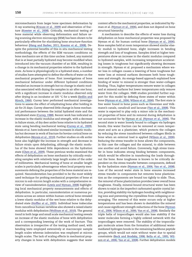
environments: (i) high vacuum (5.25$10%4 Pa) in the chamberof the dual beam system, (ii) low vacuum (120 Pa pressureprovided by water vapour) in the chamber of the dual beamsystem and (iii) removal of the sample from the vacuumchamber for beam bending wet, in air. This testing in airenvironment consisted of placing wet tissue around the bonesample and covering in order to provide a high humidityenvironment for sample rehydration. Samples were kept inthe closed vessel for two hours to ensure the samples werefully hydrated prior to testing in each of the vacuum and airconditions. Fig. 3 shows an SEM image of the AFM bendingtests on an individual bone micro cantilever beam. The bonemicro cantilever beam was deformed using a FIB flattenedAFM probe, to avoid AFM probe indentation into the sample,with a AFM cantilever spring constant of 28 N m%1 operatingat an approximate testing rate of 0.2 µm s%1. AFM beambending was performed for two hours in each of the threedifferent environments. Three small cantilever beams weretested in all three environments. The testing procedure tooka total of 12 h for each beam: two hours of testing in eachenvironment with intervals of two hours inside the closed
vessel with a high vapour concentration of Hank’s buffersolution in order to rehydrate the samples before each test.
To assess the rehydration processes used in this paper, theweight of whole 2 mm thick cross-sections of diaphysis ofrat femora were measured following the same preparationprocess as described for the micro-beam samples above.Table 2 records the weight loss measured for three rat femorasamples subjected to the various environments used in theexperimental preparation. Bone samples were initially heldin Hank’s buffer solution for 2 h. The bone samples wereremoved from the solution and, after removing excess surfacewater with filter paper, weighed using an electronic analyticalmicrobalance (Sartorius, Germany) with µg accuracy. Theweights of these bone samples were taken as fully hydratedbone weight. Further preparation processes were recorded bythe percentage of weight lost by the bone samples relativeto this fully hydrated bone weight as shown in Table 2. Thebone samples were first exposed to ethanol treatments, asindicated in Table 1, and resulted in a bone weight loss ofapproximately 3%. Exposure to the vacuum conditions ofthe SEM chamber used for FIB milling caused a further 9%weight loss in the bone samples. The total weight loss fromthe bone samples during the ethanol and vacuum exposurewas 12.56 ± 0.97%. Previous literature indicates that up to12% weight loss during dehydration of bone is due to theremoval of free water from pores such as Haversian andVolkmann’s canals, canaliculi and lacunae (Nyman et al.,2006; Yan et al., 2008). The initial bone weight loss of3% during ethanol treatment therefore indicates a partialremoval of the free water in bone whereas exposure to theSEM vacuum removes the remaining free water. Rehydrationof bone samples for two hours in a high vapour concentrationof Hank’s buffer solution recovers some of the free water andreduces the weight loss of bone to 3.63 ± 0.49% relative to thefully hydrated bone weight. Repeating the bone exposure tovacuum conditions removes all of the contained free water,resulting in the total bone weight loss of 12%. Repeatingthe rehydration of bone and subsequent vacuum exposure
J O U R N A L O F T H E M E C H A N I C A L B E H AV I O R O F B I O M E D I C A L M A T E R I A L S 5 ( 2 0 1 2 ) 1 4 9 – 1 5 5 153
Fig. 3 – SEM micrographs showing (a) the AFM probe before contact with an individual rat bone beam and (b) contact of theprobe with the bone beam for mechanical bending tests.
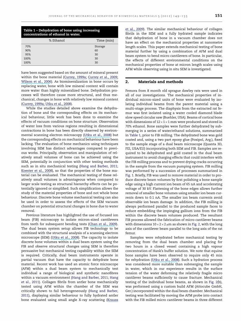
Table 2 – Change in weight, as a percentage, of whole rat bone femur cross sections with exposure to variousenvironmental conditions. The exposure conditions mimic those of the mechanically tested bone micro cantilevers. Atotal of three samples were tested and the weight difference shown relative to bone rehydrated in Hank’s buffer solution.
Time Loss in boneweight relative tohydrated in Hank’sbuffer solution (%)
Soaked in Hank’s buffer solution 2 h 0Dehydrated (see Table 1) 3.5 h 3.29 ± 0.5Vacuum dried (High Vacuum 1.51 $ 10%3 Pa) 2 h 9.27 ± 0.47Rehydrated in closed vessel with high vapour concentration of Hank’s buffer solution 2 h 3.63 ± 0.49Vacuum dried (High Vacuum 1.51 $ 10%3 Pa) 2 h 9.97 ± 0.55Rehydrated in closed vessel with high vapour concentration of Hank’s buffer solution 2 h 3.70 ± 0.27
produced repeatable weight loss values, as would be expectedif the rehydrating conditions replacing water and the vacuumconditions removing the water were consistent. We thereforeconclude that the rehydration and dehydration processesremove free water only and not the bound states of water inthe bone.
3. Results and discussion
The force applied by the AFM causes a correspondingdeflection in the bone beam during the mechanical bendingtests. The AFM data is gathered from an interferometerwhich measure the changes in the distance between theAFM probe and the optical fibre as the AFM probe bends.Instead of a linear curve as produced by a typical mechanicaltesting machine, the AFM system outputs a sinusoidalcurve representing the change in distance in terms of stagemovement versus the change of laser intensity caused bythe change in distance due to the bending of the AFMcantilever. The sinusoidal curve is then translated intoa force–displacement curve. Further details of the AFMmechanical testing have been published previous and shownto be effective in measuring even subtle changes in themechanical performance of a range of materials sensitive tohydration effects (Hang et al., 2011).
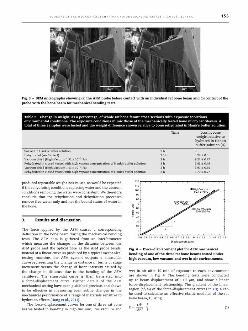
The force–displacement curves for one of three rat bonebeams tested in bending in high vacuum, low vacuum and
Fig. 4 – Force–displacement plot for AFM mechanicalbending of one of the three rat bone beams tested underhigh vacuum, low vacuum and wet in air environments.
wet in air after 10 min of exposure to each environmentare shown in Fig. 4. The bending tests were conductedup to beam displacement of &1.5 µm, and show a linearforce–displacement relationship. The gradient of the linearregion (df/d!) of the force–displacement curves in Fig. 4 canbe used to calculate an effective elastic modulus of the ratbone beam, E, using:
E = 12l3
3bh3· f!
(1)

154 J O U R N A L O F T H E M E C H A N I C A L B E H AV I O R O F B I O M E D I C A L M A T E R I A L S 5 ( 2 0 1 2 ) 1 4 9 – 1 5 5
Fig. 5 – Plot of calculated elastic modulus, determined fromAFM bending of one of the three rat bone beams, with timeunder high vacuum, low vacuum and wet in airenvironments.
where l,b and h are the length from the base of the sampleto testing contact point, breadth and height of the rat bonebeam respectively. Typical geometric values of the rat bonebeam are l = 10 µm,b = 2 µm and h = 2 µm.
The elastic modulus values calculated from Eq. (1) forone of the three rat bone beams tested under differentenvironmental conditions with increasing exposure time areshown in Fig. 5, with the error in E values calculated fromthe standard deviation of the values for the elastic modulusarising, among other things, from the changes of the contactpoint during testing. The elastic modulus shows little changeeither with environment or time of exposure in the SEMchamber. This result is somewhat surprising as an increasein the elastic modulus is expected because of dehydrationeffects (Currey et al., 2009; Hoffler et al., 2005; Lewis andNyman, 2008; Morais et al., 2010; Nyman et al., 2006; Wilsonet al., 2006). Fig. 5 indicates that the elastic modulus of the ratbone beam in high vacuum is 4.98 ± 0.25 GPa, low vacuumis 5.24 ± 0.11 GPa and wet, in air is 5.22 ± 0.15 GPa anddoes not vary greatly over the time period examined. Previousmechanical testing on fully hydrated whole rat bone femurusing 3-point bending configuration gives an elastic modulusof 5.12 ± 0.77 GPa (Kasra et al., 1997), 8.0 ± 0.4 GPa (Barengoltset al., 1993), 6.88 ± 0.31 GPa (Jorgensen et al., 1991), and4.9 ± 0.4 GPa (Ejersted et al., 1993) which is similar to thecalculated elastic modulus values in our work and indicatesthat the vacuum of the SEM chamber does not have aneffect on elastic modulus of the samples over the time periodinvestigated. Interestingly, the similarity between the elasticmodulus of our relatively small bone volumes andwhole bonetesting suggests an effective transfer of stresses throughoutthe bone material, indicating that all bone components arenot failing or slipping but are tightly bound to one another.Wenote that the calculated elastic modulus values using Eq. (1)assume shear within the beam is insignificant. Using classicbeam bending theory (Blodgett, 1991), the deflection of thebeam consists of a deflection due to shear and a deflectiondue to bending. The total deflection can therefore be writtenas:
! = !bending + !shear = Pl3
3EI+ 6Pl
5AG(2)
where ! is total beam deflection, P is load, l is length fromthe base to the contact point, E is elastic modulus, I isthe moment of inertia, A is the cross sectional area andG is the shear modulus which is calculated theoreticallyfrom G = E/[2(1 + ")], with " = 0.35 (Akiva et al., 1998).The beams used in this work have an aspect ratio of 5:1.Therefore, using Eq. (2) above, the shear contribution tothe total deflection is &3.5%. This shear contribution ismuch smaller than the bending contribution, indicating thatthe majority of beam mechanical deformation results frompure bending. In addition, possible indentation of the AFMprobe with the bone beam sample will cause inaccuracies inthe calculated elastic modulus. However, neither direct SEMimaging of the mechanical testing procedure or subsequentSEM examination of the AFM probe–sample contact pointshowed evidence of indentation on the surface of thecantilever bone beams being tested.
Fig. 5 highlights the lack of environmental influence onthe mechanical properties of the bone micro cantilevers.The removal of water using ethanol treatments described inTable 1 and the vacuum conditions used have been shownonly to remove the free water in bone samples as shownin Table 2. The mechanical properties of the bone microcantilevers tested in this work therefore do not change as onlyfree water state is removed during exposure to the differentenvironmental conditions used. Our results correlate withprevious work suggesting that the effects of free water on themechanical properties of bone is minor (Nyman et al., 2006).Previous work indicating changes in the elastic modulus ofbone with dehydration time (Currey et al., 2009; Hoffler et al.,2005; Lewis and Nyman, 2008; Morais et al., 2010; Nymanet al., 2006; Wilson et al., 2006) use more aggressive dryingconditions, which would be expected to remove not onlyfree water but also the bound water states that influencemechanical behaviour. Our recorded bone elastic modulusvalues suggest that water removal responsible for mechanicalproperty changes during dehydration is due to removal of thesecond and third states of water, as defined by Nyman et al.(Nyman et al., 2006), or potentially operate at length scalesabove those of the micro cantilever beams used in this work.Thus, the second and third states of bound water in collagenand at mineral surfaces in bone respectively are not removedevenwith the highest vacuum conditions of the SEM. The SEMhigh vacuum conditions must also be less invasive than theapplication of the high temperatures (reported in the rangeof 60–140 #C) used to remove water during complete collagendehydration (Renugopalakrishnan et al., 1989).
4. Conclusions
Mechanical beam bending of discrete rat cortical bonevolumes of the order of 40 µm3, isolated as micro cantileverbeams using FIB, was performed by AFM within the chamberof a dual beam system. The bending behaviour of the ratbone beams was recorded in high and low vacuum SEMenvironments as well as in air. Calculated elastic modulusvalues of the rat bone were found to be independent ofboth the environment used and the time of exposure toeach of these environments. Our results suggest that thebone material still remains partially hydrated even in themost potentially dehydrating conditions of high vacuum
J O U R N A L O F T H E M E C H A N I C A L B E H AV I O R O F B I O M E D I C A L M A T E R I A L S 5 ( 2 0 1 2 ) 1 4 9 – 1 5 5 155
and indicate that a time window of opportunity existsto mechanically test bone material in high vacuum whileretaining the mechanical properties of hydrated bone. Theseresults indicate an opportunity to mechanically deformhydrated bone materials in vacuum environments whileobserving using in situ scanning electron microscopy.
Acknowledgements
The authors thank Dr Zofia Luklinska, Dr Ken Png and MickWillis for the help with sample preparation, the NanoVisionCentre at Queen Mary, University of London for use offacilities and Carlos J. Pasquali for developing Matlab andOctave scripts for AFM data analysis.
R E F E R E N C E S
Akiva, U., Wagner, H.D., Weiner, S., 1998. Modelling the three-dimensional elastic constants of parallel-fibred and lamellarbone. Journal of Materials Science 33, 1497–1509.
Barengolts, E.I., Curry, D.J., Bapna, M.S., Kukreja, S.C., 1993.Effects of endurance exercise on bone mass and mechanicalproperties in intact and ovariectomized rats. Journal of Boneand Mineral Research 8, 937–942.
Beck, K., Brodsky, B., 1998. Supercoiled protein motifs: thecollagen triple-helix and the #-helical coiled coil. Journal ofStructural Biology 122, 17–29.
Bella, J., Brodsky, B., Berman, H.M., 1995. Hydration structure of acollagen peptide. Structure 3, 893–906.
Blodgett, O.W., 1991. Design of Welded Structures, 14th ed. TheJames F. Lincoln Arc Welding Foundation, Cleveland, Ohio.
Chan, Y.L., Ngan, A.H.W., King, N.M., 2009. Use of focused ionbeam milling for investigating the mechanical propertiesof biological tissues: a study of human primary molars.Journal of the Mechanical Behavior of Biomedical Materials 2,375–383.
Chen, P.Y., Stokes, A.G., McKittrick, J., 2009. Comparison of thestructure andmechanical properties of bovine femur bone andantler of the North American elk (Cervus elaphus canadensis).Acta Biomaterialia 5, 693–706.
Currey, J.D., 1988. The effects of drying and re-wetting onsome mechanical properties of cortical bone. Journal ofBiomechanics 21, 439–441.
Currey, J.D., 1999a. The design ofmineralised hard tissues for theirmechanical functions. The Journal of Experimental Biology202, 3285–3294.
Currey, J.D., 1999b. What determines the bending strength ofcompact bone? The Journal of Experimental Biology 202,2495–2503.
Currey, J.D., 2002. Bones Structure and Mechanics, 2nd ed.Princeton University Press.
Currey, J.D., Landete-Castillejos, T., Estevez, J., Ceacero, F., Olguin,A., Garcia, A., Gallego, L., 2009. The mechanical properties ofred deer antler bone when used in fighting. The Journal ofExperimental Biology 212, 3985–3993.
Ejersted, C., Andreassen, T.T., Oxlund, H., Jorgensen, P.H.,Bak, B., Haggblad, J., Torring, O., Nilsson, M.H.L., 1993.Human parathyroid hormone (1–34) and (1–84) increase themechanical strength and thickness of cortical bone in rats.Journal of Bone and Mineral Research 8, 1097–1101.
Hang, F., Barber, A.H., 2011. Nano-mechanical properties ofindividual mineralized collagen fibrils from bone tissue.Journal of the Royal Society Interface 8, 500–505.
Hang, F., Lu, D., Bailey, R., Jimenez-Palomar, I., Stachewicz,U., Cortes-Ballesteros, B., Davies, M., Zech, M., Bödefeld, C.,Barber, A.H., 2011. In situ tensile testing of nanofibers bycombining atomic force microscopy and scanning electronmicroscopy. Nanotechnology 22, 365708.
Hoffler, C.E., Guo, X.E., Zysset, P.K., Goldstein, S.A., 2005.An application of nanoindentation technique to measurebone tissue lamellae properties. Journal of BiomechanicalEngineering 127, 1046–1054.
Jorgensen, P.H., Bak, B., Andreassen, T.T., 1991. Mechanicalproperties and biochemical composition of rat cortical femurand tibia after long-term treatment with biosynthetic humangrowth hormone. Bone 12, 353–359.
Kasra, M., Vanin, C.M., MacLusky, N.J., Casper, R.F., Grynpas, M.D.,1997. Effects of different estrogen and progestin regimens onthe mechanical properties of rat femur. Journal of OrthopaedicResearch 15, 118–123.
Koester, K.J., Ager, J.W., Ritchie, R.O., 2008. The true toughness ofhuman cortical bone measured with realistically short cracks.Nature Materials 7, 672–677.
Krauss, S., Fratzl, P., Seto, J., Currey, J.D., Estevez, J.A., Funari, S.S.,Gupta, H.S., 2009. Inhomogeneous fibril stretching in antlerstarts after macroscopic yielding: indication for a nanoscaletoughening mechanism. Bone 44, 1105–1110.
Lewis, G., Nyman, J.S., 2008. The use of nanoindentation forcharacterizing the properties of mineralized hard tissue: state-of-the art review. Journal of Biomedical Materials ResearchPart B: Applied Biomaterials 87B, 286–300.
Morais, J.J.L., deMoura, M.F.S.F., Pereira, F.A.M., Xavier, J., Dourado,N., Dias, M.I.R., Azevedo, J.M.T., 2010. The double cantileverbeam test applied to mode I fracture characterization ofcortical bone tissue. Journal of the Mechanical Behavior ofBiomedical Materials 3, 446–453.
Nyman, J.S., Roy, A., Shen, X., Acuna, R.L., Tyler, J.H., Wang,X., 2006. The influence of water removal on the strengthand toughness of cortical bone. Journal of Biomechanics 39,931–938.
Renugopalakrishnan, V., Chandrakason, G., Moore, S., Hutson,T.B., Berney, C.V., Bhatnagar, R.S., 1989. Bound water incollagen. Evidence from Fourier transform infrared andFourier transform infrared photoacoustic spectroscopic study.Macromolecules 22, 4121–4124.
Sedlin, E.D., Hirsch, C., 1966. Factors affecting the determinationof the physical properties of femoral cortical bone. ActaOrthopaedica Scandinavica 37, 29–48.
Tai, K., Dao, M., Suresh, S., Palazoglu, A., Ortiz, C., 2007. Nanoscaleheterogeneity promotes energy dissipation in bone. NatureMaterials 6, 454–462.
Utku, F.S., Klein, E., Saybasili, H., Yucesoy, C.A., Weiner, S., 2008.Probing the role of water in lamellar bone by dehydration inthe environmental scanning electron microscope. Journal ofStructural Biology 162, 361–367.
Wilson, E.E., Awonusi, A., Morris, M.D., Kohn, D.H., Tecklenburg,M.M.J., Beck, L.W., 2006. Three structural roles for water inbone observed by solid-state NMR. Biophysical Journal 90,3722–3731.
Wolfram, U.,Wilke, H.J., Zysset, P.K., 2010. Rehydration of vertebraltrabecular bone: influences on its anisotropy, its stiffness andthe indentation work with a view to age, gender and vertebrallevel. Bone 46, 348–354.
Yan, J., Daga, A., Kumar, R., Mecholsky, J.J., 2008. Fracturetoughness and work of fracture of hydrated, dehydrated, andashed bovine bone. Journal of Biomechanics 41, 1929–1936.