Infant Skull and Suture Properties: Measurements and Implications for Mechanisms of Pediatric Brain...
8
Susan S. Margulies e-mail: [email protected] Kirk L. Thibault Department of Bioengineering, University of Pennsylvania, Philadelphia, PA 19104 Infant Skull and Suture Properties: Measurements and Implications for Mechanisms of Pediatric Brain Injury The mechanical properties of the adult human skull are well documented, but little infor- mation is available for the infant skull. To determine the age-dependent changes in skull properties, we tested human and porcine infant cranial bone in three-point bending. The measurement of elastic modulus in the human and porcine infant cranial bone agrees with and extends previous published data [McPherson, G. K., and Kriewall, T. J. (1980), J. Biomech., 13, pp. 9 – 16] for human infant cranial bone. After confirming that the porcine and human cranial bone properties were comparable, additional tensile and three-point bending studies were conducted on porcine cranial bone and suture. Comparisons of the porcine infant data with previously published adult human data demonstrate that the elastic modulus, ultimate stress, and energy absorbed to failure increase, and the ultimate strain decreases with age for cranial bone. Likewise, we conclude that the elastic modu- lus, ultimate stress, and energy absorbed to failure increase with age for sutures. We constructed two finite element models of an idealized one-month old infant head, one with pediatric and the other adult skull properties, and subjected them to impact loading to investigate the contribution of the cranial bone properties on the intracranial tissue deformation pattern. The computational simulations demonstrate that the comparatively compliant skull and membranous suture properties of the infant brain case are associated with large cranial shape changes, and a more diffuse pattern of brain distortion than when the skull takes on adult properties. These studies are a fundamental initial step in predicting the unique mechanical response of the pediatric skull to traumatic loads asso- ciated with head injury and, thus, for defining head injury thresholds for children. @S0148-0731~00!00904-3# Introduction The response of the head to traumatic loading is intrinsically linked to the anatomy and mechanical properties of the develop- ing skull/brain structure. The skull of the newborn consists of thin, flexible plates composed of partially calcified bony tissue joined at their margins by patent, membranous sutures. In contrast to the stiff adult cranium, the infant skull is a compliant structure ca- pable of substantial deformation under external loading. Previous studies have reported the quasi-static elastic modulus of cranial bone @1,2# and the structural stiffness of fetal parietal bone @3# but have not characterized the cranial sutures. However, there is a paucity of dynamic mechanical property data for the tissues constituting the infant brain case. Therefore, previous bio- mechanical studies of traumatic pediatric head injury @4–7# have neglected the role of the skull when determining the mechanical response of the pediatric head to trauma, or have relied on quali- tative approximations of its mechanical properties. The aim of this paper is to define the mechanical properties of the developing skull and sutures and to demonstrate their influ- ence in the biomechanical response of the infant brain case to impact loading. We report our results from a series of mechanical tests on cranial bone from infant humans, and cranial bone and suture from infant pigs. Specifically, we define the dynamic prop- erties of human infant cranial bone, correlate the mechanical be- havior of human infant cranial bone with porcine infant cranial bone, and extend the results of the human cranial bone tests with the porcine data. As a first-order demonstration of the influence that these age- dependent mechanical properties of cranial bone and suture have in protecting the infant brain from deformation during an impact, an idealized finite element model of the infant head was con- structed. Two models were constructed, a first with infant cortical bone and suture properties determined in the present study, and a second with adult cortical bone and suture properties obtained from the literature. The two model formulations were exposed to identical loads, and the intracranial strains were compared. To- gether, the experimental and computational studies reported in this communication provide a foundation for predicting the unique mechanical response of the pediatric skull to traumatic loads ap- plied to the head. Methods Human Infant Cranial Bone: Harvest and Sample Prepara- tion. Human infant cranial bone specimens were obtained at au- topsy ~N 54 subjects! from the morgue at the Children’s Hospital of Philadelphia under the approval of the Institutional Review Board ~IRB! for human subject research. Subjects ranged from 25 weeks gestation to six months of age and were free from any craniofacial surgical procedures or injuries to the head. For each subject, age, sex, date of death, and cause of death were recorded. Due to standard autopsy procedures for removing the brain en bloc, no suture material could be collected from these subjects. Bilateral strips of parietal bone adjacent to the sagittal suture were excised and placed in a sealed container over a saline-soaked gauze pad. The strips were then frozen at 24°C until preparation for testing. To determine the age-related properties of cranial bone inde- pendent of any inherent anisotropy or inhomogeneity, all beam- shaped specimens were cut from the parietal bone parallel to the sagittal suture ~Fig. 1! at room temperature, and the location and Contributed by the Bioengineering Division for publication in the JOURNAL OF BIOMECHANICAL ENGINEERING. Manuscript received by the Bioengineering Divi- sion: January 7, 1999; revised manuscript received March 28, 2000. Associate Tech- nical Editor: R. C. Haut. 364 Õ Vol. 122, AUGUST 2000 Copyright © 2000 by ASME Transactions of the ASME Downloaded 06 May 2010 to 158.130.76.120. Redistribution subject to ASME license or copyright; see http://www.asme.org/terms/Terms_Use.cfm
-
Upload
independent -
Category
Documents
-
view
1 -
download
0
Transcript of Infant Skull and Suture Properties: Measurements and Implications for Mechanisms of Pediatric Brain...

nfor-skull
. Thes with), J.e
pointof thet theimateodu-. Wee withg tosuetivelyiatedhantep insso-
en.
Downlo
Susan S. Marguliese-mail: [email protected]
Kirk L. Thibault
Department of Bioengineering,University of Pennsylvania,
Philadelphia, PA 19104
Infant Skull and SutureProperties: Measurements andImplications for Mechanisms ofPediatric Brain InjuryThe mechanical properties of the adult human skull are well documented, but little imation is available for the infant skull. To determine the age-dependent changes inproperties, we tested human and porcine infant cranial bone in three-point bendingmeasurement of elastic modulus in the human and porcine infant cranial bone agreeand extends previous published data [McPherson, G. K., and Kriewall, T. J. (1980Biomech.,13, pp. 9–16] for human infant cranial bone. After confirming that the porcinand human cranial bone properties were comparable, additional tensile and three-bending studies were conducted on porcine cranial bone and suture. Comparisonsporcine infant data with previously published adult human data demonstrate thaelastic modulus, ultimate stress, and energy absorbed to failure increase, and the ultstrain decreases with age for cranial bone. Likewise, we conclude that the elastic mlus, ultimate stress, and energy absorbed to failure increase with age for suturesconstructed two finite element models of an idealized one-month old infant head, onpediatric and the other adult skull properties, and subjected them to impact loadininvestigate the contribution of the cranial bone properties on the intracranial tisdeformation pattern. The computational simulations demonstrate that the comparacompliant skull and membranous suture properties of the infant brain case are assocwith large cranial shape changes, and a more diffuse pattern of brain distortion twhen the skull takes on adult properties. These studies are a fundamental initial spredicting the unique mechanical response of the pediatric skull to traumatic loads aciated with head injury and, thus, for defining head injury thresholds for childr@S0148-0731~00!00904-3#
al
av
u
s
e
o
i
havect,on-calnd anedto
To-d inueap-
au-lw25
anyachrded.en
cts.ereaked
de-m-
nd
iT
IntroductionThe response of the head to traumatic loading is intrinsic
linked to the anatomy and mechanical properties of the deveing skull/brain structure. The skull of the newborn consists of thflexible plates composed of partially calcified bony tissue joinat their margins by patent, membranous sutures. In contrast tostiff adult cranium, the infant skull is a compliant structure cpable of substantial deformation under external loading.
Previous studies have reported the quasi-static elastic modof cranial bone@1,2# and the structural stiffness of fetal parietbone@3# but have not characterized the cranial sutures. Howethere is a paucity of dynamic mechanical property data fortissues constituting the infant brain case. Therefore, previousmechanical studies of traumatic pediatric head injury@4–7# haveneglected the role of the skull when determining the mechanresponse of the pediatric head to trauma, or have relied on qtative approximations of its mechanical properties.
The aim of this paper is to define the mechanical propertiethe developing skull and sutures and to demonstrate their inence in the biomechanical response of the infant brain casimpact loading. We report our results from a series of mechantests on cranial bone from infant humans, and cranial bonesuture from infant pigs. Specifically, we define the dynamic prerties of human infant cranial bone, correlate the mechanicalhavior of human infant cranial bone with porcine infant cranbone, and extend the results of the human cranial bone teststhe porcine data.
As a first-order demonstration of the influence that these a
Contributed by the Bioengineering Division for publication in the JOURNAL OFBIOMECHANICAL ENGINEERING. Manuscript received by the Bioengineering Divsion: January 7, 1999; revised manuscript received March 28, 2000. Associatenical Editor: R. C. Haut.
364 Õ Vol. 122, AUGUST 2000 Copyright ©
aded 06 May 2010 to 158.130.76.120. Redistribution subject to ASME l
llyop-in,edthe
a-
ulusler,
thebio-
icalali-
offlu-
toicalandp-be-alwith
ge-
dependent mechanical properties of cranial bone and suturein protecting the infant brain from deformation during an impaan idealized finite element model of the infant head was cstructed. Two models were constructed, a first with infant cortibone and suture properties determined in the present study, asecond with adult cortical bone and suture properties obtaifrom the literature. The two model formulations were exposedidentical loads, and the intracranial strains were compared.gether, the experimental and computational studies reportethis communication provide a foundation for predicting the uniqmechanical response of the pediatric skull to traumatic loadsplied to the head.
Methods
Human Infant Cranial Bone: Harvest and Sample Prepara-tion. Human infant cranial bone specimens were obtained attopsy~N54 subjects! from the morgue at the Children’s Hospitaof Philadelphia under the approval of the Institutional RevieBoard~IRB! for human subject research. Subjects ranged fromweeks gestation to six months of age and were free fromcraniofacial surgical procedures or injuries to the head. For esubject, age, sex, date of death, and cause of death were recoDue to standard autopsy procedures for removing the brainbloc, no suture material could be collected from these subjeBilateral strips of parietal bone adjacent to the sagittal suture wexcised and placed in a sealed container over a saline-sogauze pad. The strips were then frozen at24°C until preparationfor testing.
To determine the age-related properties of cranial bone inpendent of any inherent anisotropy or inhomogeneity, all beashaped specimens were cut from the parietal boneparallel to thesagittal suture~Fig. 1! at room temperature, and the location a
-ech-
2000 by ASME Transactions of the ASME
icense or copyright; see http://www.asme.org/terms/Terms_Use.cfm

e
os
r
d
ef
t
aoy
l
c
ip
g
k
tom-
h anta-
anate
ell
intter-
and
re
h ofith
achm/byereturelesh-
m.theeci-
state-tllet
themea-omned
de-hintioneci-s ofction
ge
on-
ithrior
to-
ningne,our-eled
herwithandp-
Downlo
orientation of the samples remained constant across all subjTest specimens were typically 3–5 mm wide and 20–25 mm lodepending upon the size of the original donor sample. All spemens were cut with a diamond-coated cutting wheel~StoeltingCo., Wood Dale, IL! mounted in a variable-speed Dremel MotTool ~Dremel, Racine, WI! under a constant drip of saline. Tespecimens were refrozen at24°C until testing.
Porcine Cranial Bone and Suture: Harvest and SamplePreparation. Neonate pigs~age 2–3 days,N530! were sacri-ficed by a lethal injection of KCl as detailed in experimental ptocols approved by the Animal Care and Use Committee ofUniversity of Pennsylvania. Time of death and age were recorfor each animal immediately post-mortem. The cranial vaulteach animal was excised and frozen at24°C until preparation fortesting. Samples were randomized with respect to right or left sbut the location and orientation of the samples remained consacross all studies.
Infant Cranial Bone and Suture: Three-Point Bending Spemens. The left or right side frontoparietal bones from exciscranial vaults from 20 of the 30 infant pigs were preparedtesting in three-point bending to failure. Beam-shaped test spmens of parietal bone alone and frontoparietal bone with a porof coronal suture penetrating the cross section of the beam wcut from the cranial vault parallel to the sagittal suture~Fig. 1!.Porcine specimen preparation and storage was identical to thathe human.
Infant Cranial Bone and Suture: Tension Specimens.The leftor right side frontoparietal bones from excised cranial vaultsthe remaining 10 infant pigs were prepared for testing in tensto failure. Rectangular prismatic ‘‘blanks’’ of cranial bone or crnial bone with a portion of coronal suture penetrating the crsection of the sample were prepared as described previouslthree-point bending samples. Each blank was machined totensile testing configuration with a typical gage section of 3mm wide and 8–11 mm long. Tensile specimens were shawith an aluminum oxide grinding wheel mounted in a variabspeed Dremel Moto-Tool under a constant drip of saline. The eof each machined specimen were potted in polymethylmethalate ~PMMA, Hygenic Corp., Akron, OH! to facilitate gripping ofthe specimen without crushing it.
Care was taken to ensure that the potential for the sample towithin the PMMA blocks during testing was reduced by maching a sloping profile in the embedded portion of the tissue samSlippage of the sample within the grips or the PMMA blocks wmonitored carefully during each test and was identified as a reof constant force on a force-displacement curve. Three ofinfant test specimens exhibited slippage within the PMMA blocand were eliminated from analysis.
Fig. 1 Infant cranial vault: Shaded areas represent approxi-mate size and location of skull and suture samples removed.
Journal of Biomechanical Engineering
aded 06 May 2010 to 158.130.76.120. Redistribution subject to ASME l
cts.ng,ci-
-t
o-theedof
idetant
ci-doreci-ionere
t for
ofion-ssforthe
–5pede-ndsry-
slipn-le.
asionthes,
Three-Point Bending Test Protocol. Samples of cranialbone and suture were tested in three-point bending on a cusdesigned fixture with an adjustable span~10–300 mm! built for anInstron servohydraulic materials testing machine~Instron Corp.,Canton, MA!. A stainless steel loading nose and supports witradius of 3.2 mm support were used to avoid excessive indetion and stress concentrations within the specimen~ASTM D 790-98!. Three-point bending is suitable for ultimate strains less thor equal to 5 percent, and four-point bending for larger ultimstrains ~ASTM D 790-98!. Ultimate strain data~determined intension! presented in Table 3 show that skull properties are wwithin the accepted range, and suture ultimate strains~averaging 6percent! are slightly higher than the accepted limit for three-pobending tests. Centerline deflection of the specimen was demined from the displacement of the servohydraulic actuator,force was measured with a 50 lb isometric load cell~Interface,Inc., Scottsdale, AZ!. Centerline deflection and force data werecorded via a PC-based, digital data acquisition system~NationalInstruments, Austin, TX! as well as anX–Y recorder~HewlettPackard, Loveland, CO! for hardcopy backup.
Bone samples were warmed to room temperature in a batsaline, placed on the bending fixture in a manner consistent wsimply supported boundary conditions and loaded midspan. Econstant rate bending test to failure was performed at 2.54 mmin or 2540 mm/min while centerline deflection, as measuredthe displacement of the servohydraulic actuator, and force wrecorded. All tests were conducted at ambient room tempera~25°C!. A constant span of 17 mm was used for all skull samp~human and porcine! except for the samples from the six-montold human subject, which could accommodate a span of 30 mIn all tests, failure occurred directly under the point of load, ontensile face of the beam, and the width and depth of each spmen were measured directly adjacent to this point.
Tension Test Protocol. A custom-designed set of tensile tegrips was designed for use with an Instron servohydraulic mrials testing machine~Instron Corp.!. The ends of the tensile tesspecimen embedded in PMMA were placed in the brass cogrips, and an extensometer~Instron Corp.! fastened to each of thePMMA blocks with elastic bands measured the strain insample. Because excessive vibration was encountered whilesuring low forces at the higher loading rate in tension, data frinfant porcine cranial bone with and without sutures was obtaiin tension at 2.54 mm/min only.
Extensometer displacement and force were recorded asscribed earlier for three-point bending. The displacement witthe system was assumed to occur entirely within the gage secof the tissue sample. The gage length of each undeformed spmen was measured prior to testing, and the width and thickneseach test specimen were measured adjacent to the cross sewhere fracture occurred. All samples failed within the gasection.
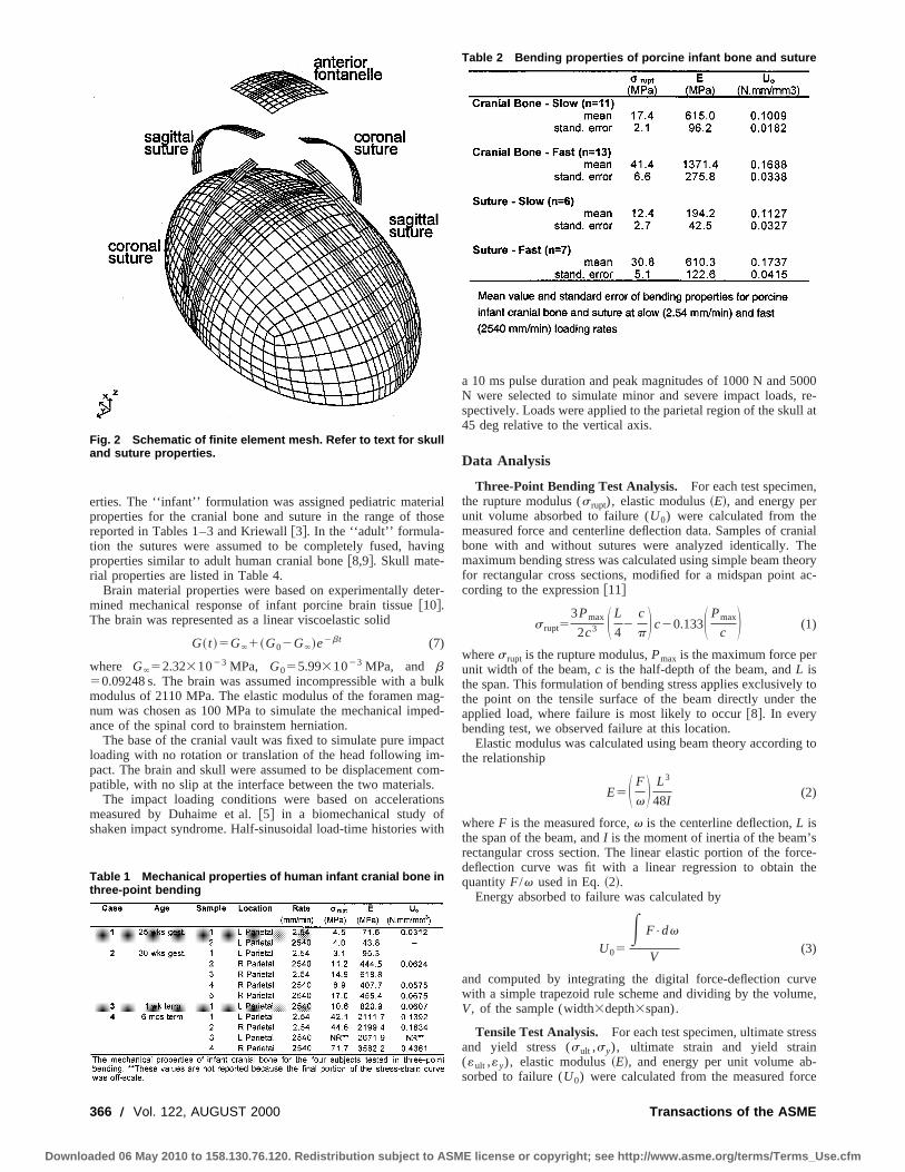
Idealized Finite Element Model Development. A finite ele-ment model of an idealized one-month-old infant head was cstructed using ANSYS 5.3~ANSYS Inc., Houston, PA!. Themodel consisted of a rigid impactor plate, and a skull/brain wfive regions: the cranial bone of the skull, the sutures, the antefontanel, the foramen magnum, and the brain~Fig. 2!. The contactproblem was solved using LS-DYNA3D’s automatic surface-surface contact formulation~LSTC, Livermore, CA!. The modelconsisted of 12,772 elements and 11,823 nodes, and a zostudy was performed to verify convergence. The cranial bosuture, fontanel, and foramen magnum were modeled using fnoded linear shell elements. The brain and impactor were modusing eight-noded linear brick elements.
Two formulations, one using an infant cranial bone and anotusing adult cranial bone representations, were constructedthe same geometry, boundary conditions, brain properties,loading conditions, differing only in the braincase material pro
AUGUST 2000, Vol. 122 Õ 365
icense or copyright; see http://www.asme.org/terms/Terms_Use.cfm
i
v
b
p
sif
000, re-ll at
,
anialheeoryac-
y tothe
g to
sce-
the
rvee,
ss
-ce
Downlo
erties. The ‘‘infant’’ formulation was assigned pediatric materproperties for the cranial bone and suture in the range of threported in Tables 1–3 and Kriewall@3#. In the ‘‘adult’’ formula-tion the sutures were assumed to be completely fused, haproperties similar to adult human cranial bone@8,9#. Skull mate-rial properties are listed in Table 4.
Brain material properties were based on experimentally demined mechanical response of infant porcine brain tissue@10#.The brain was represented as a linear viscoelastic solid
G~ t !5G`1~G02G`!e2bt (7)
where G`52.3231023 MPa, G055.9931023 MPa, and b50.09248 s. The brain was assumed incompressible with amodulus of 2110 MPa. The elastic modulus of the foramen mnum was chosen as 100 MPa to simulate the mechanical imance of the spinal cord to brainstem herniation.
The base of the cranial vault was fixed to simulate pure imploading with no rotation or translation of the head following impact. The brain and skull were assumed to be displacement cpatible, with no slip at the interface between the two material
The impact loading conditions were based on acceleratmeasured by Duhaime et al.@5# in a biomechanical study oshaken impact syndrome. Half-sinusoidal load-time histories w
Fig. 2 Schematic of finite element mesh. Refer to text for skulland suture properties.
Table 1 Mechanical properties of human infant cranial bone inthree-point bending
366 Õ Vol. 122, AUGUST 2000
aded 06 May 2010 to 158.130.76.120. Redistribution subject to ASME l
alose
ing
ter-
ulkag-ed-
act-om-.
ons
ith
a 10 ms pulse duration and peak magnitudes of 1000 N and 5N were selected to simulate minor and severe impact loadsspectively. Loads were applied to the parietal region of the sku45 deg relative to the vertical axis.
Data Analysis
Three-Point Bending Test Analysis. For each test specimenthe rupture modulus (s rupt), elastic modulus~E!, and energy perunit volume absorbed to failure (U0) were calculated from themeasured force and centerline deflection data. Samples of crbone with and without sutures were analyzed identically. Tmaximum bending stress was calculated using simple beam thfor rectangular cross sections, modified for a midspan pointcording to the expression@11#
s rupt53Pmax
2c3 S L
42
c
p D c20.133S Pmax
c D (1)
wheres rupt is the rupture modulus,Pmax is the maximum force perunit width of the beam,c is the half-depth of the beam, andL isthe span. This formulation of bending stress applies exclusivelthe point on the tensile surface of the beam directly underapplied load, where failure is most likely to occur@8#. In everybending test, we observed failure at this location.
Elastic modulus was calculated using beam theory accordinthe relationship
E5S F
v D L3
48I(2)
whereF is the measured force,v is the centerline deflection,L isthe span of the beam, andI is the moment of inertia of the beam’rectangular cross section. The linear elastic portion of the fordeflection curve was fit with a linear regression to obtainquantityF/v used in Eq.~2!.
Energy absorbed to failure was calculated by
U05E F•dv
V(3)
and computed by integrating the digital force-deflection cuwith a simple trapezoid rule scheme and dividing by the volumV, of the sample (width3depth3span).
Tensile Test Analysis. For each test specimen, ultimate streand yield stress (sult ,sy), ultimate strain and yield strain(«ult ,«y), elastic modulus~E!, and energy per unit volume absorbed to failure (U0) were calculated from the measured for
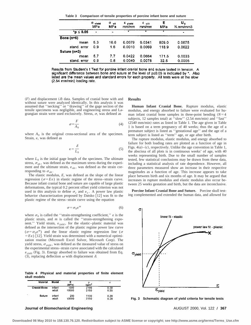
Table 2 Bending properties of porcine infant bone and suture
Transactions of the ASME
icense or copyright; see http://www.asme.org/terms/Terms_Use.cfm
Journal of Biom
Downloaded 06 May 2010
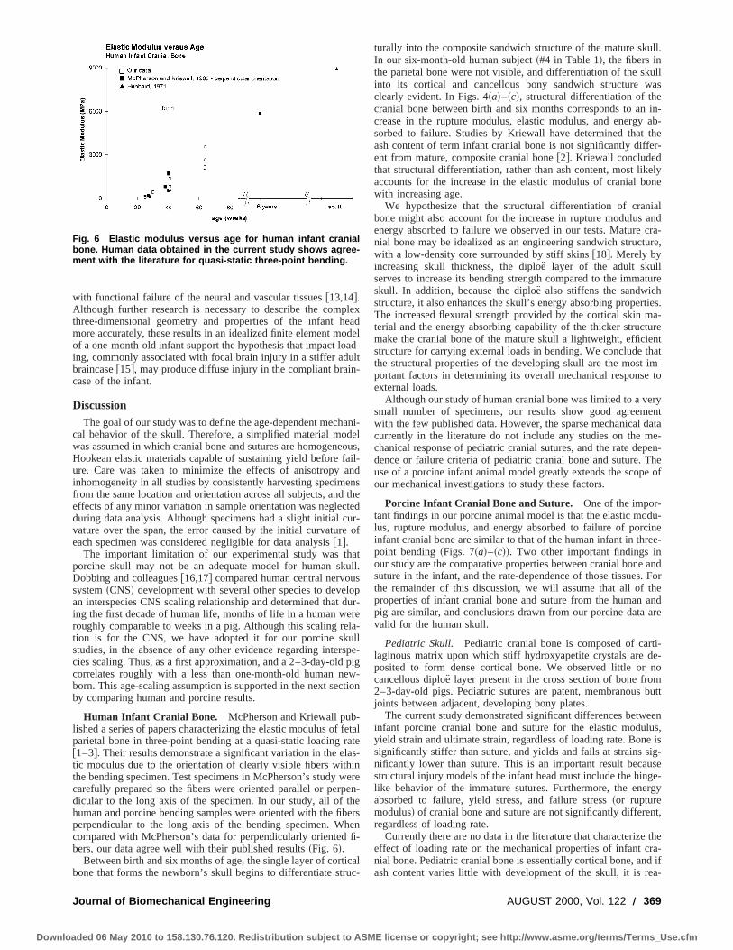
Table 3 Comparison of tensile properties of porcine infant bone and suture
e
r
a
ln
s
(
l
hu-
leof aof a
d toin
1,40lesata,
, allctivetakethatbe-
sive.
d for
~F! and displacement~d! data. Samples of cranial bone with anwithout suture were analyzed identically. In this analysis it wassumed that ‘‘necking’’ or ‘‘drawing’’ of the gage section of thtensile specimens was negligible, and engineering stress andgrangian strain were used exclusively. Stress,s, was defined as
s5F
A0(4)
where A0 is the original cross-sectional area of the specimStrain,«, was defined as
«5d
L0(5)
whereL0 is the initial gage length of the specimen. The ultimastress,sult , was defined as the maximum stress during the expment and the ultimate strain,«ult , was defined as the strain coresponding tosult .
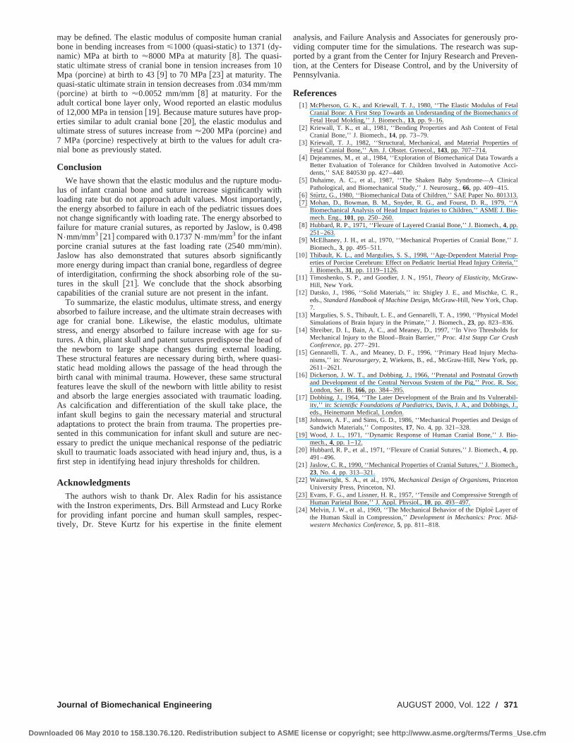
The elastic modulus,E, was defined as the slope of the lineregression (s5E«) in elastic regime of the stress–strain curvBecause infant cranial bone and suture are capable of large pdeformations, the typical 0.2 percent offset yield criterion wasused in this analysis to definesy and «y . A power law plasticbehavior characterization proposed by Datsko@12# was fit to theplastic regime of the stress–strain curve using the equation
s5s0«m (6)
wheres0 is called the ‘‘strain-strengthening coefficient,’’« is theplastic strain, andm is called the ‘‘strain-strengthening exponent.’’ Yield strain, «yield , for the elastic–plastic material wadefined as the intersection of the plastic regime power law cu(s5s0«m) and the linear elastic regime regression lines5E«) @12#. Yield strain was determined with a numerical optimzation routine ~Microsoft Excel Solver, Microsoft Corp!. Theyield stress,syield , was defined as the measured value of stressthe experimental stress–strain curve associated with the calcu«yield ~Fig. 3!. Energy absorbed to failure was obtained from E~3!, replacing deflectionv with displacementd.
Table 4 Physical and material properties of finite elementskull models
echanical Engineering
to 158.130.76.120. Redistribution subject to ASME l
daseLa-
n.
teeri--
re.asticot
-
rve
i-
onatedq.
Results
Human Infant Cranial Bone. Rupture modulus, elasticmodulus, and energy absorbed to failure were evaluated forman infant cranial bone samples in three-point bending~N54subjects, 12 samples total! at ‘‘slow’’ ~2.54 mm/min! and ‘‘fast’’~2540 mm/min! rates as listed in Table 1. The age given in Tab1 is based on a term pregnancy of 40 weeks; thus the agepremature subject is listed as ‘‘gestational age’’ and the ageterm subject is listed as ‘‘term’’ age, or age after birth.
The rupture modulus, elastic modulus, and energy absorbefailure for both loading rates are plotted as a function of ageFigs. 4~a!–~c!, respectively. Unlike the age convention in Tablethe abscissa of all plots is in continuous weeks’ of age, withweeks representing birth. Due to the small number of samptested, few statistical conclusions may be drawn from these dincluding a statistical analysis of rate dependence. Howeverthree parameters measured show an increase in their respemagnitudes as a function of age. This increase appears toplace between birth and six months of age. It may be arguedincreases in rupture modulus and elastic modulus also occurtween 25 weeks gestation and birth, but the data are inconclu
Porcine Infant Cranial Bone and Suture. Porcine skull test-ing complemented and extended the human data, and allowe
Fig. 3 Schematic diagram of yield criteria for tensile tests
AUGUST 2000, Vol. 122 Õ 367
icense or copyright; see http://www.asme.org/terms/Terms_Use.cfm

dpr
-
aa
er-
sig-ing
eThenifi-ate.that
sticonen attric.
ed in
edu-fail-re.ointfail-
intests ontestin a,
hly
thehat00
dis-heop-s.
ated
Downlo
more extensive analysis of the rate dependence of bone anture. The mechanical property data measured for the infantcine cranial bone are presented in two portions: results from thpoint bending tests and results from tensile tests.
Three-Point Bending Test Results.The elastic modulus, rupture modulus, and energy absorbed to failure were determinedporcine cranial bone and suture at slow~2.54 mm/min! and fast~2540 mm/min! loading rates. Table 2 lists the mean and standerror of the bending properties of porcine infant cranial bonesuture for slow and fast loading rates. The data were arranged
Fig. 4 Human infant cranial bone specimens: „a… rupturemodulus, „b… elastic modulus, and „c… energy absorbed to fail-ure plotted against age
368 Õ Vol. 122, AUGUST 2000
aded 06 May 2010 to 158.130.76.120. Redistribution subject to ASME l
su-or-ee-
for
rdndinto
four groups for parametric analysis to facilitate analysis of diffences in mechanical behavior between tissue types~bone versussuture! and rates~slow versus fast!. A two-tailed Student’st-testassuming unequal variances was used to determine whether anificant difference existed between each pair of correspondmean values at the level ofp<0.05. The elastic modulus of bonwas significantly greater than suture at both loading rates.rupture modulus and energy absorbed to failure were not sigcantly different between bone and suture at either loading rThe comparison between fast and slow loading rates revealedloading rate significantly affected the rupture modulus and elamodulus. The rupture modulus and elastic modulus of both band suture were significantly greater at the fast loading rate thathe slow loading rate. The energy absorbed to failure in pediabone and suture was not significantly affected by loading rate
Tensile Test Results. The results of the tensile tests of porcininfant cranial bone and suture at 2.54 mm/min are presenteTable 3. Significant differences~p<0.5, using two-tailed Stu-dent’st-test assuming unequal variances! between bone and suturwere found in the yield strain, ultimate strain, and elastic molus. The yield stress, ultimate stress, and energy absorbed toure were not significantly different between bone and sutuThese results are consistent with the findings from the three-pbending tests; however, only the value of energy absorbed toure in tension was statistically similar to bending (p.0.05) at theslow loading rate. The disparity between the elastic modulusbending and in tension is most likely due to the differences inmethods and analysis. Specifically, the bending analysis reliebeam theory to calculate the modulus whereas the tensilemeasures the modulus directly. Likewise, the maximum stressbending test~rupture modulus! is calculated from beam theorywhereas the maximum stress in a tensile test~ultimate stress! ismeasured directly.
Idealized Finite Element Model Results.The deformation ofthe braincase and the resulting intracranial strains were higsensitive to the properties of the cranial vault~Fig. 5!. The peakintrusion of the impactor was more than 100 percent greater insimulation with the infant cranial bone representation than twith the adult~4 versus 2 mm, and 10 versus 4 mm for the 10and 5000 N loads, respectively!. Furthermore, impact loading tothe infant braincase resulted in diffuse, bilateral hemispherictribution of maximum principal strains greater than zero in tbrain, whereas impact to the model with adult cranial bone prerties produced primarily focal, unilateral brain deformationMaximum principal strains in excess of 0.15 have been associ
Fig. 5 Maximum principal strain in idealized infant head finiteelement model at tÄ5 ms „at peak load … for adult braincaseproperties and pediatric properties, for half-sine load magni-tudes of 1000 N and 5000 N, respectively
Transactions of the ASME
icense or copyright; see http://www.asme.org/terms/Terms_Use.cfm
peo
u
defa
e
l
e
ur
e
-frl
w
bh
iu
ull.
ullas
in-ab-
ther-
elyone
ialandcra-ture,
ture
ties.a-
urenthatim-
to
ryent
datae-
pen-he
e of
du-ine
ee-
and. Fortheandare
ti-e-nombutt
eenlus,e isig-
usege-rgy
nt,
thera-d ifa-
Downlo
with functional failure of the neural and vascular tissues@13,14#.Although further research is necessary to describe the comthree-dimensional geometry and properties of the infant hmore accurately, these results in an idealized finite element mof a one-month-old infant support the hypothesis that impact loing, commonly associated with focal brain injury in a stiffer adbraincase@15#, may produce diffuse injury in the compliant braincase of the infant.
DiscussionThe goal of our study was to define the age-dependent mech
cal behavior of the skull. Therefore, a simplified material mowas assumed in which cranial bone and sutures are homogenHookean elastic materials capable of sustaining yield beforeure. Care was taken to minimize the effects of anisotropyinhomogeneity in all studies by consistently harvesting specimfrom the same location and orientation across all subjects, andeffects of any minor variation in sample orientation was neglecduring data analysis. Although specimens had a slight initial cvature over the span, the error caused by the initial curvatureach specimen was considered negligible for data analysis@1#.
The important limitation of our experimental study was thporcine skull may not be an adequate model for human skDobbing and colleagues@16,17# compared human central nervousystem~CNS! development with several other species to devean interspecies CNS scaling relationship and determined thating the first decade of human life, months of life in a human wroughly comparable to weeks in a pig. Although this scaling retion is for the CNS, we have adopted it for our porcine skstudies, in the absence of any other evidence regarding intecies scaling. Thus, as a first approximation, and a 2–3-day-oldcorrelates roughly with a less than one-month-old human nborn. This age-scaling assumption is supported in the next secby comparing human and porcine results.
Human Infant Cranial Bone. McPherson and Kriewall published a series of papers characterizing the elastic modulus ofparietal bone in three-point bending at a quasi-static loading@1–3#. Their results demonstrate a significant variation in the etic modulus due to the orientation of clearly visible fibers withthe bending specimen. Test specimens in McPherson’s studycarefully prepared so the fibers were oriented parallel or perpdicular to the long axis of the specimen. In our study, all of thuman and porcine bending samples were oriented with the fiperpendicular to the long axis of the bending specimen. Wcompared with McPherson’s data for perpendicularly orientedbers, our data agree well with their published results~Fig. 6!.
Between birth and six months of age, the single layer of cortbone that forms the newborn’s skull begins to differentiate str
Fig. 6 Elastic modulus versus age for human infant cranialbone. Human data obtained in the current study shows agree-ment with the literature for quasi-static three-point bending.
Journal of Biomechanical Engineering
aded 06 May 2010 to 158.130.76.120. Redistribution subject to ASME l
lexaddel
ad-lt-
ani-elous,
ail-nd
ensthe
tedur-
of
atull.sopdur-rela-ll
spe-pigw-tion
etalateas-inere
en-heersenfi-
calc-
turally into the composite sandwich structure of the mature skIn our six-month-old human subject~#4 in Table 1!, the fibers inthe parietal bone were not visible, and differentiation of the skinto its cortical and cancellous bony sandwich structure wclearly evident. In Figs. 4~a!–~c!, structural differentiation of thecranial bone between birth and six months corresponds to ancrease in the rupture modulus, elastic modulus, and energysorbed to failure. Studies by Kriewall have determined thatash content of term infant cranial bone is not significantly diffeent from mature, composite cranial bone@2#. Kriewall concludedthat structural differentiation, rather than ash content, most likaccounts for the increase in the elastic modulus of cranial bwith increasing age.
We hypothesize that the structural differentiation of cranbone might also account for the increase in rupture modulusenergy absorbed to failure we observed in our tests. Maturenial bone may be idealized as an engineering sandwich strucwith a low-density core surrounded by stiff skins@18#. Merely byincreasing skull thickness, the diploe¨ layer of the adult skullserves to increase its bending strength compared to the immaskull. In addition, because the diploe¨ also stiffens the sandwichstructure, it also enhances the skull’s energy absorbing properThe increased flexural strength provided by the cortical skin mterial and the energy absorbing capability of the thicker structmake the cranial bone of the mature skull a lightweight, efficiestructure for carrying external loads in bending. We conclude tthe structural properties of the developing skull are the mostportant factors in determining its overall mechanical responseexternal loads.
Although our study of human cranial bone was limited to a vesmall number of specimens, our results show good agreemwith the few published data. However, the sparse mechanicalcurrently in the literature do not include any studies on the mchanical response of pediatric cranial sutures, and the rate dedence or failure criteria of pediatric cranial bone and suture. Tuse of a porcine infant animal model greatly extends the scopour mechanical investigations to study these factors.
Porcine Infant Cranial Bone and Suture. One of the impor-tant findings in our porcine animal model is that the elastic molus, rupture modulus, and energy absorbed to failure of porcinfant cranial bone are similar to that of the human infant in thrpoint bending~Figs. 7~a!–~c!!. Two other important findings inour study are the comparative properties between cranial bonesuture in the infant, and the rate-dependence of those tissuesthe remainder of this discussion, we will assume that all ofproperties of infant cranial bone and suture from the humanpig are similar, and conclusions drawn from our porcine datavalid for the human skull.
Pediatric Skull. Pediatric cranial bone is composed of carlaginous matrix upon which stiff hydroxyapetite crystals are dposited to form dense cortical bone. We observed little orcancellous diploe¨ layer present in the cross section of bone fro2–3-day-old pigs. Pediatric sutures are patent, membranousjoints between adjacent, developing bony plates.
The current study demonstrated significant differences betwinfant porcine cranial bone and suture for the elastic moduyield strain and ultimate strain, regardless of loading rate. Bonsignificantly stiffer than suture, and yields and fails at strains snificantly lower than suture. This is an important result becastructural injury models of the infant head must include the hinlike behavior of the immature sutures. Furthermore, the eneabsorbed to failure, yield stress, and failure stress~or rupturemodulus! of cranial bone and suture are not significantly differeregardless of loading rate.
Currently there are no data in the literature that characterizeeffect of loading rate on the mechanical properties of infant cnial bone. Pediatric cranial bone is essentially cortical bone, anash content varies little with development of the skull, it is re
AUGUST 2000, Vol. 122 Õ 369
icense or copyright; see http://www.asme.org/terms/Terms_Use.cfm

uon
di
asticthatimi-
ni-fferstic120
Pa,of
inpec-
rti-ies
reatrichibitssue
anden-
fantdur-
andbony
thethe
con-icaltinc-nt
ed of
nesent
tod inheon-im-res-
aceith
imes
thatthe
ockay
ve
-ingthekull
Downlo
sonable to compare our infant porcine data with previously plished investigations of the rate-dependent mechanical behaviadult human cranial cortical bone. Wood tested human cracortical bone in tension without the diploe¨ layer and reported, asfunctions of increasing loading rate, increases in the elastic molus and ultimate stress, a decrease in the ultimate strain, ansignificant rate-dependent effect on energy absorbed to fa@19#. Our bending tests of infant porcine cranial bone show s
Fig. 7 „a… Elastic modulus of porcine and human infant cranialbone determined from three-point bending tests; „b… rupturemodulus of porcine and human infant cranial bone; „c… energyabsorbed to failure of porcine and human infant cranial bone;slow rate „2.54 mm Õmin … and fast rate „2540 mm Õmin …
370 Õ Vol. 122, AUGUST 2000
aded 06 May 2010 to 158.130.76.120. Redistribution subject to ASME l
b-r ofial
du-no
lureig-
nificant rate-dependent increases in rupture modulus and elmodulus, but not in energy absorbed to failure. We concludeporcine infant cranial bone exhibits rate-dependent behavior slar to adult human cortical bone.
Although their rate-dependent behavior is similar, the magtude of porcine infant cranial bone mechanical properties diconsiderably from those of adult human cortical bone. The elamodulus for human neonatal parietal bone ranges betweenand 4240 MPa in quasi-static three-point bending~@1# and currentstudy!, and porcine cranial bone ranges between 615 and 809 Min quasi-static~2.54 mm/min! three-point bending and tensionrespectively. Wood reported a quasi-static elastic modulusadult cortical bone in tension of'12,000 MPa@19#. Likewise, theultimate stress and ultimate strain for pediatric cranial bonequasi-static tension are 10.5 MPa and 0.034 mm/mm, restively, as compared to Wood’s values of'70 MPa and'0.007mm/mm. The differences between pediatric bone and adult cocal bone may be due to incomplete calcification or interspecdifferences.
Pediatric Suture. Currently there are no data in the literaturegarding the rate-dependent mechanical properties of pedisutures. Our study has also shown that pediatric sutures exrate-dependent mechanical behavior. In bending, suture tishows significant rate-dependent increases in rupture moduluselastic modulus, but no significant rate-dependent increase inergy absorbed to failure. We conclude that the suture of the inskull does not demonstrate enhanced ability to absorb energying high rate loading.
The immature suture is composed of a series of periostealendosteal membranes spanning the gap between adjacentplates of the pediatric skull. In bending and tension, failure ofsuture was caused by a separation of the membrane fromadjacent bone, and not a rupture of the membrane itself. Weclude that significant differences exist between the mechanproperties of immature cranial bone and suture, and these distions must be included in a structural injury model of the infahead.
Adult Cranial Bone and Sutures. Adult human cranial boneresembles a classical engineering sandwich structure composstiff facings of cortical bone~the inner and outer tables! and acompliant, lightweight core of cancellous bone~the diploe!. Adultcranial sutures are the highly complex structures joining the boof the skull that are composed of interdigitated edges of adjaccranial bone spanned by collagenous fibers.
As a stiff functional unit, the cranial bones and sutures serveprotect the brain from impact injury. Sutures have been testebending@20,21# and in pendulum impact tests to determine tmechanical properties of the joints in the skull. Jaslow demstrated that frontoparietal sutures absorb more energy duringpact than adjacent cranial bone, and hypothesized that the pence of collagen within the suture and the highly irregular surfcreated during failure are thought to provide the suture whigher energy absorbing capabilities than bone@21#. In support ofthis theory, collagen has been shown to absorb at least 100 tmore energy than bone per unit volume of tissue@22#, and exami-nation of fracture surfaces of cranial bone and suture indicatethe fracture surface of the suture is much more irregular thanplanar fracture surfaces of the bone~@21#; our observations!. To-gether, the present study and previous data@8,20,21# support theconcept that the sutures in the adult skull may function as shabsorbers during impact loading of the skull, while bone mcarry and distribute the load.
Adult human cranial bone and its individual components habeen tested in tension@9,19,23#, bending @8#, compression@9,23,24# and simple shear@9# in order to characterize the mechanical response of the skull to traumatic loads. By combinthe human and porcine skull data from the current study withdata in the literature, the age-dependent properties of the s
Transactions of the ASME
icense or copyright; see http://www.asme.org/terms/Terms_Use.cfm
/
a
dd
a
e
ar
u
uidhup
i
e
pro-up-en-of
tals of
tal
of
s ai-
al
3.‘Ao-
J.
p-,’’
.,
el
or
a-
wthoc.
il-,
n of
io-
ch.,
th of
-
Downlo
may be defined. The elastic modulus of composite human crabone in bending increases from<1000~quasi-static! to 1371~dy-namic! MPa at birth to'8000 MPa at maturity@8#. The quasi-static ultimate stress of cranial bone in tension increases fromMpa ~porcine! at birth to 43@9# to 70 MPa@23# at maturity. Thequasi-static ultimate strain in tension decreases from .034 mm~porcine! at birth to '0.0052 mm/mm@8# at maturity. For theadult cortical bone layer only, Wood reported an elastic moduof 12,000 MPa in tension@19#. Because mature sutures have proerties similar to adult cranial bone@20#, the elastic modulus andultimate stress of sutures increase from'200 MPa~porcine! and7 MPa ~porcine! respectively at birth to the values for adult crnial bone as previously stated.
ConclusionWe have shown that the elastic modulus and the rupture mo
lus of infant cranial bone and suture increase significantly wloading rate but do not approach adult values. Most importanthe energy absorbed to failure in each of the pediatric tissuesnot change significantly with loading rate. The energy absorbefailure for mature cranial sutures, as reported by Jaslow, is 0.N•mm/mm3 @21# compared with 0.1737 N•mm/mm3 for the infantporcine cranial sutures at the fast loading rate~2540 mm/min!.Jaslow has also demonstrated that sutures absorb significmore energy during impact than cranial bone, regardless of deof interdigitation, confirming the shock absorbing role of the stures in the skull@21#. We conclude that the shock absorbincapabilities of the cranial suture are not present in the infant.
To summarize, the elastic modulus, ultimate stress, and enabsorbed to failure increase, and the ultimate strain decreasesage for cranial bone. Likewise, the elastic modulus, ultimstress, and energy absorbed to failure increase with age fotures. A thin, pliant skull and patent sutures predispose the heathe newborn to large shape changes during external loadThese structural features are necessary during birth, where qstatic head molding allows the passage of the head throughbirth canal with minimal trauma. However, these same structfeatures leave the skull of the newborn with little ability to resand absorb the large energies associated with traumatic loaAs calcification and differentiation of the skull take place, tinfant skull begins to gain the necessary material and structadaptations to protect the brain from trauma. The propertiessented in this communication for infant skull and suture are nessary to predict the unique mechanical response of the pedskull to traumatic loads associated with head injury and, thus,first step in identifying head injury thresholds for children.
AcknowledgmentsThe authors wish to thank Dr. Alex Radin for his assistan
with the Instron experiments, Drs. Bill Armstead and Lucy Rorfor providing infant porcine and human skull samples, resptively, Dr. Steve Kurtz for his expertise in the finite eleme
Journal of Biomechanical Engineering
aded 06 May 2010 to 158.130.76.120. Redistribution subject to ASME l
nial
10
mm
lusp-
-
du-ithtly,oesto
498
ntlygreeu-g
rgywithtesu-
d ofing.asi-theralsting.eralre-
ec-atricis a
cekec-
nt
analysis, and Failure Analysis and Associates for generouslyviding computer time for the simulations. The research was sported by a grant from the Center for Injury Research and Prevtion, at the Centers for Disease Control, and by the UniversityPennsylvania.
References@1# McPherson, G. K., and Kriewall, T. J., 1980, ‘‘The Elastic Modulus of Fe
Cranial Bone: A First Step Towards an Understanding of the BiomechanicFetal Head Molding,’’ J. Biomech.,13, pp. 9–16.
@2# Kriewall, T. K., et al., 1981, ‘‘Bending Properties and Ash Content of FeCranial Bone,’’ J. Biomech.,14, pp. 73–79.
@3# Kriewall, T. J., 1982, ‘‘Structural, Mechanical, and Material PropertiesFetal Cranial Bone,’’ Am. J. Obstet. Gynecol.,143, pp. 707–714.
@4# Dejeammes, M., et al., 1984, ‘‘Exploration of Biomechanical Data TowardBetter Evaluation of Tolerance for Children Involved in Automotive Accdents,’’ SAE 840530 pp. 427–440.
@5# Duhaime, A. C., et al., 1987, ‘‘The Shaken Baby Syndrome—A ClinicPathological, and Biomechanical Study,’’ J. Neurosurg.,66, pp. 409–415.
@6# Sturtz, G., 1980, ‘‘Biomechanical Data of Children,’’ SAE Paper No. 80131@7# Mohan, D., Bowman, B. M., Snyder, R. G., and Fourst, D. R., 1979, ‘
Biomechanical Analysis of Head Impact Injuries to Children,’’ ASME J. Bimech. Eng.,101, pp. 250–260.
@8# Hubbard, R. P., 1971, ‘‘Flexure of Layered Cranial Bone,’’ J. Biomech.,4, pp.251–263.
@9# McElhaney, J. H., et al., 1970, ‘‘Mechanical Properties of Cranial Bone,’’Biomech.,3, pp. 495–511.
@10# Thibault, K. L., and Margulies, S. S., 1998, ‘‘Age-Dependent Material Proerties of Porcine Cerebrum: Effect on Pediatric Inertial Head Injury CriteriaJ. Biomech.,31, pp. 1119–1126.
@11# Timoshenko, S. P., and Goodier, J. N., 1951,Theory of Elasticity, McGraw-Hill, New York.
@12# Datsko, J., 1986, ‘‘Solid Materials,’’ in: Shigley J. E., and Mischke, C. Reds.,Standard Handbook of Machine Design, McGraw-Hill, New York, Chap.7.
@13# Margulies, S. S., Thibault, L. E., and Gennarelli, T. A., 1990, ‘‘Physical ModSimulations of Brain Injury in the Primate,’’ J. Biomech.,23, pp. 823–836.
@14# Shreiber, D. I., Bain, A. C., and Meaney, D., 1997, ‘‘In Vivo Thresholds fMechanical Injury to the Blood–Brain Barrier,’’Proc. 41st Stapp Car CrashConference, pp. 277–291.
@15# Gennarelli, T. A., and Meaney, D. F., 1996, ‘‘Primary Head Injury Mechnisms,’’ in: Neurosurgery, 2, Wiekens, B., ed., McGraw-Hill, New York, pp.2611–2621.
@16# Dickerson, J. W. T., and Dobbing, J., 1966, ‘‘Prenatal and Postnatal Groand Development of the Central Nervous System of the Pig,’’ Proc. R. SLondon, Ser. B,166, pp. 384–395.
@17# Dobbing, J., 1964, ‘‘The Later Development of the Brain and Its Vulnerabity,’’ in: Scientific Foundations of Paediatrics, Davis, J. A., and Dobbings, J.eds., Heinemann Medical, London.
@18# Johnson, A. F., and Sims, G. D., 1986, ‘‘Mechanical Properties and DesigSandwich Materials,’’ Composites,17, No. 4, pp. 321–328.
@19# Wood, J. L., 1971, ‘‘Dynamic Response of Human Cranial Bone,’’ J. Bmech.,4, pp. 1–12.
@20# Hubbard, R. P., et al., 1971, ‘‘Flexure of Cranial Sutures,’’ J. Biomech.,4, pp.491–496.
@21# Jaslow, C. R., 1990, ‘‘Mechanical Properties of Cranial Sutures,’’ J. Biome23, No. 4, pp. 313–321.
@22# Wainwright, S. A., et al., 1976,Mechanical Design of Organisms, PrincetonUniversity Press, Princeton, NJ.
@23# Evans, F. G., and Lissner, H. R., 1957, ‘‘Tensile and Compressive StrengHuman Parietal Bone,’’ J. Appl. Physiol.,10, pp. 493–497.
@24# Melvin, J. W., et al., 1969, ‘‘The Mechanical Behavior of the Diploe¨ Layer ofthe Human Skull in Compression,’’Development in Mechanics: Proc. Midwestern Mechanics Conference, 5, pp. 811–818.
AUGUST 2000, Vol. 122 Õ 371
icense or copyright; see http://www.asme.org/terms/Terms_Use.cfm