Skull views - CUTM Courseware
38
Skull views
-
Upload
khangminh22 -
Category
Documents
-
view
7 -
download
0
Transcript of Skull views - CUTM Courseware

Skull views

Radiographic landmarks1. Nasion : a point between both eyes , where root of the nose starts.2. Glabella : A point in saggital plane in midline , at the top of forehead about 3" from rootbof the nose.3. External canthus : the outer most point of lateral aspect of eyes.4.External auditory meatus : the bony ring of external ear in temporal bone.5.vertex : the highest point on cranila vault.6. Submental : the region below the mandible.7. Angle of mandible : the part of body of mandible, which turns upwards to join the temporal bone.

8. Median saggital plane (MSP) : The plane of skull cutting through its center, in its longitudinal plane.9. Inter- pupillary line (IPL) : the line joining pupils of both eyes . This is also known as inter orbital line.10 Radiographic base line(RBL) : the line joining External canthus of each eye to respective external auditory meatus . This is also known as orbito meatal line.


Occipitofrontal view (PA 20 °) Also known as Caldwell view.The projection is used to demonstrate the frontal and
ethmiod sinuses especially for frontal sinus.

Contraindications
Pregnancy

Preparation①Collect History from patient.②Patient is asked to remove all radiopaque
materials.③Wear hospital gown. ④If female patient come to x ray ,
chaperone is necessary.⑤Shielding is provided to patient.

Positioning Patient lies prone with midline of his body on midline of x
ray table. The chest is raised and rested on folded arms. Nose and forehead are placed on the midline table
touching it. Head is positioned such that MSP and RBL are both at
90° to table top. It is important to ensure that the skull is not rotated. All the relevant sinuses should be included with in the
image. Head is immobalised , side is marked and beam is
collimated.


CenteringIt is done on nasion in midllne.Central beam is directed 15 to 20°
to vertical in caudad direction , passing through the occipital bone.

TechniqueKVP : 80mAs : 90Fss : small.FFD : 100 CMS.WITH GRIDCASSETTE SIZE : 12" X 10"

Fronto-Occiptal view.(AP 30°)
Also known as Towne's view.The Towne view is an angled AP radiograph
of the skull, used to evaluate for fractures of the skull and neoplastic changes.This view is used for study of petrous bone
pyramids, mastoid air cells, dorsum sella, foramen magnum, temporal bones and pareital bones.

Towne's view


PositioningPatient is supine in midline of x ray table,
with chin fully touched to to chest and occipital of skull with midline of x ray table.Head is adjusted so that ,MSP and RBL are
perpendicular to table top.It is ensured that head is not rotated.Head is immobalised, side is marked and
beam is collimated.

Centering①It is done in midline on
forehead ( anterior hair line).②Central beam is directed 30°
caused to vertical, so that it enters through glabella and exist through foramen magnum.

TechniqueKVP : 80mAs : 100Fss : small.FFD : 100 CMS.WITH GRIDCASSETTE SIZE : 12" X 10"


Lateral view of skullIndications
• Fracture of cranium and skull base.• Sinusitis.• Tumours of pituatory gland.• Abnormal head shapes.• Multiple myeloma: The plasma cells are a type of
white blood cell in the bone marrow. With this condition, a group of plasma cells becomes cancerous and multiplies. The disease can damage the bones, immune system, kidneys and red blood cell count.

PositioningPatient lies prone on x ray table.Only head is
turned to true lateral position , affected side in contact with table top.
Chin is supported by fist of patient.His head is adjusted such that MSP is parllel to
table top as is the RBL.Head is immobalised side is marked and beam is
collimated.

CenteringIt is done on temporal region midway
between glbella and occipital protuberance.Central beam is vertically at 90° to the
table top.

TechniqueKVP : 80mAs : 60Fss : small.FFD : 100 CMS.WITH GRIDCASSETTE SIZE : 12" X 10"
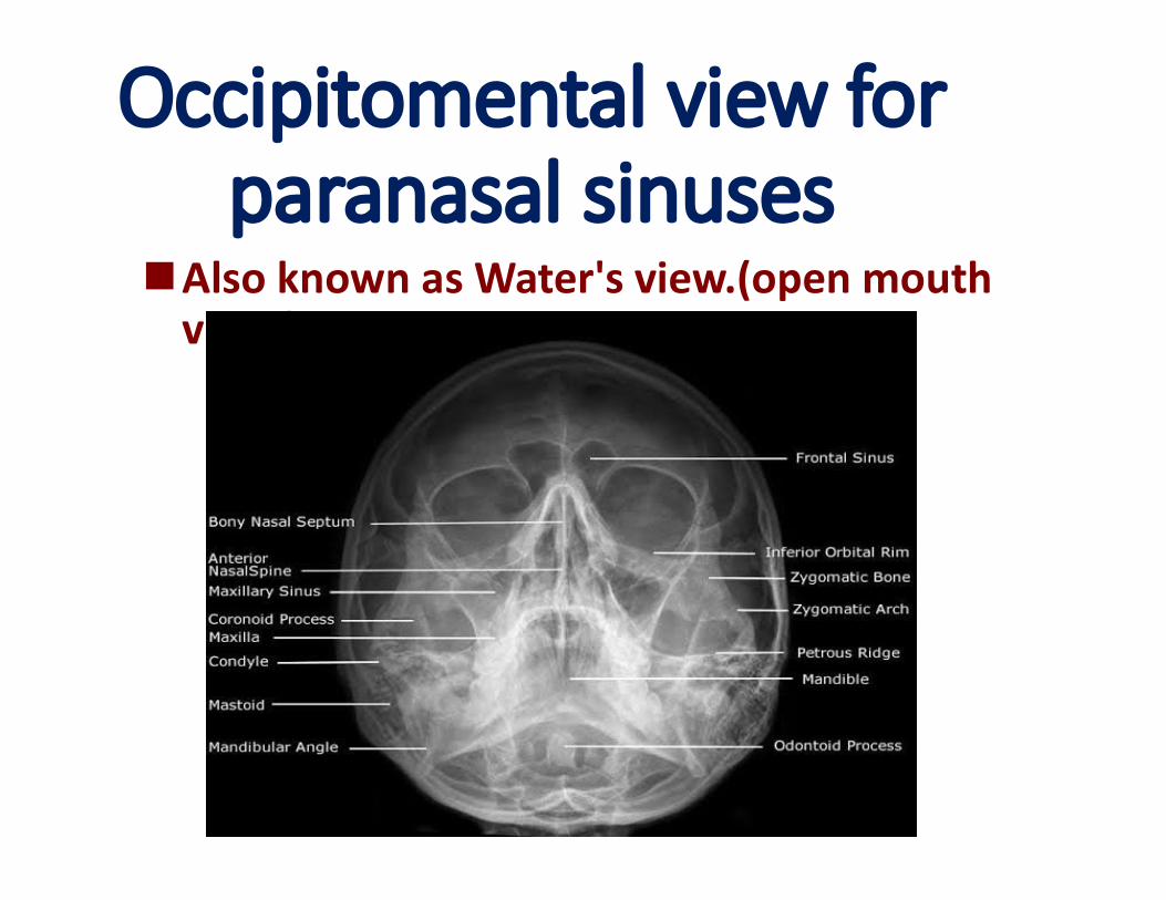
Occipitomental view for paranasal sinuses
Also known as Water's view.(open mouth view.)

Positioning Patient lies in prone position , in midline of x ray
table.His hands and arms are rested by side of the body. The nose and chin are placed in contact with table
top. Mouth is open throughout exposure.Head is positioned so that , MSP is 90° to table top
and RBL is at 45 ° to table top.Head is immobalised side is marked and beam is
collimated.

Centering• It is done in the middle through
vertex to the level of lower orbital margin.
• Central beam is directed vertically at 90 ° to table top.

TechniqueKVP : 80mAs : 100Fss : small.FFD : 100 CMS.WITH GRIDCASSETTE SIZE : 12" X 10"

Submentovertical view(Base of skull view)
view used to evaluate fractures and displacement of a fractured zygomatic arch.
contraindicated with patients who have a suspected spinal injury.
reveals the position and orientation of the occipital condyles, the sphenoid sinus, the curvature of the mandible, the lateral wall of the maxillary sinuses.
good visualization of the base of the skull with foramina and the medial and lateral pterygoid plates.
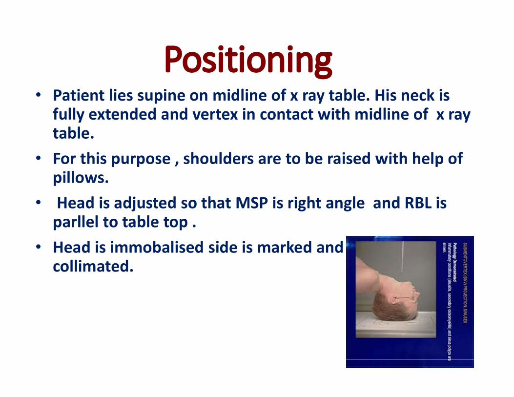
Positioning• Patient lies supine on midline of x ray table. His neck is
fully extended and vertex in contact with midline of x ray table.
• For this purpose , shoulders are to be raised with help of pillows.
• Head is adjusted so that MSP is right angle and RBL is parllel to table top .
• Head is immobalised side is marked and beam is collimated.

Centering• It is done in midline between angles of
mandible.• Central beam is directed at 5° cephalad
(cranial) to vertical.• This enters a point midway between
angles of mandible and exists through vertex, which is at the center of cassette.

TechniqueKVP : 80mAs : 100Fss : small.FFD : 100 CMS.WITH GRIDCASSETTE SIZE : 12" X 10"


Lateral oblique view of mastoids (schuller view)
Indications Ear pain(otalgia) and tenderness. swelling in the mastoid region. the ear or mastoid region may be red
(erythematous). Fever or headaches may also be
present. Drainage from the ear occurs in more
serious cases, often manifest as brown discharge .

Positioning• Patient lies in prone position in midline of x ray table ,
with head in lateral position.• The side under study contact with table top midline.• The pinna of ear folded forwards.• Head is adjusted so that MSP is parllel and IOML is
perpendicular to table top.

Centering• It is done 5 cm above and 2.5 CMS
behind the external auditory meatus.
• The central beam is directed at 25°caused to vertical.

TechniqueKVP : 70mAs : 60Fss : small.FFD : 100 CMS.WITH GRIDCASSETTE SIZE : 12" X 10"