The HDAC6/APOBEC3G complex regulates HIV-1 infectiveness by inducing Vif autophagic degradation
Upload
transumanistiCategory
view
3download
0
©2008
Lan
des B
iosc
ienc
e. D
o no
t dist
ribut
e.
1042 Autophagy 2008; Vol. 4 Issue 8
[Autophagy 4:8, 1042-1053; 16 November 2008]; ©2008 Landes Bioscience
Adaptation to hypoxia through activation of the hypoxia induc-ible factor-1 (HIF-1) is crucial for tumor cells survival. Here we describe the antitumoral effects of the new molecule CR 3294 on tumor cells in the presence of hypoxia. Treatment of the breast carcinoma cell line MDA-MB-231 with CR 3294 in 1% O2 resulted in an in vivo and in vitro inhibition of tumor growth. CR 3294 induced accumulation of autophagosomes in hypoxic MDA-MB-231 cells as assessed by both transmission electron microscopy (TEM) and the autophagic marker LC3-II. TEM analysis revealed the presence of invaginations of the cytoplasm into the nucleus. Autophagosomes were present in such invagi-nations. Moreover, CR 3294 inhibited both the DNA binding of HIF-1α and VEGF mRNA synthesis. Immunoprecipitation and immunofluorescence studies showed an interaction between LC3 and HIF-1α. We next detailed the effect of inhibitors and activators of autophagy on both HIF-1α and LC3. In particular, 3 methyladenine (3MA) and wortmannin, two macroautophagic inhibitors, prevented both the decrease of HIF-1α protein levels and LC3 processing in cells treated with CR 3294. Bafilomycin and leupeptin, inhibitors of lysosomes, prevented HIF-1α decrease without affecting LC3 processing. By contrast, treating hypoxic MDA-MB-231 cells with trifluoperazine (TFP) or serum with-drawal (SW), two activators of autophagy, diminished HIF-1α levels and stimulated LC3 processing. These results indicate that activation of the autophagic pathway in hypoxic cells by the new molecule CR 3294, as well as by TFP or SW, can have potentially important implications for cancer treatment.
Introduction
Hypoxia is a common feature of the inner region of a tumor and is due to the rapid growth of the tumor cells.1 A high percentage of tumor cells are killed by such extreme conditions. On the other hand, these same conditions may cause the selection of cells capable of adapting to hypoxia through changes in glucose metabolism or synthesis of growth factors.2,3 Such selection through adaptation is one of the reasons for the chemotherapy resistance observed in many tumors.
The major molecular mechanism by which tumor cells can adapt to hypoxia is through a transcription factor called hypoxia induc-ible factor-1 (HIF-1). HIF-1 is a heterodimeric protein composed of two subunit α and β. Under normoxia HIF-1α is degraded by the ubiquitin-proteasome system, but when the intracellular oxygen concentration drops, HIF-1α is stabilized.4 Following stabilization, HIF-1α translocates to the nucleus where it binds to HIF-1β. The HIF-1α and β dimer activates the transcription of a plethora of genes involved in angiogenesis, glucose transport, apoptosis resistance, metastasis, inflammation, etc.5,6 Such activation of transcription is achieved by the binding of HIF-1α to hypoxia responsive element (HRE) located on the promoter of the target gene.7 Several studies have shown that in breast cancer patients there is a strict correlation between HIF-1α activation and increased proliferation, metastasis and poor prognosis.8,9
Autophagy is a cellular mechanism in response to a variety of stimuli, particularly starvation.10 Like apoptosis, autophagy has been suggested as an important mechanism in maintaining cellular homeostasis and during development of multicellular organisms.11 Molecular aspects of autophagy have been detailed in yeast and have shown a tightly regulated process involving at least 27 genes.12 The autophagic pathway begins with the formation of the autophago-some, which consists of cytoplasmic material sequestered inside a double membrane vescicle.12 Subsequently, the autophagosome fuses with the lysosome and the cytoplasmic material is digested
*Correspondence to: Marco Tafani; Department of Cellular and Molecular Pathology; IRCCS San Raffaele Pisana; Rome Italy; Tel.: 06.66130420; Fax: 0666130407; Email: [email protected]
Submitted: 03/28/08; Revised: 09/19/08; Accepted: 09/24/08
Previously published online as an Autophagy E-publication: http://www.landesbioscience.com/journals/autophagy/article/7070
Research Paper
Induction of autophagic cell death by a novel molecule is increased by hypoxiaMarco Tafani,1,* Luana Schito,2 Tahira Anwar,2 Manuela Indelicato,1,3 Patrizio Sale,1,2 Maura Di Vito,2 Emanuela Morgante,2 Rosanna Beraldi,4,5,† Francesco Makovec,6 Ornella Letari,6 Gianfranco Caselli,6 Corrado Spadafora,4 Bruna Pucci1 and Matteo A. Russo1,2
1Department of Cellular and Molecular Pathology; IRCCS San Raffaele Pisana; Rome Italy; 2Department of Experimental Medicine; University of Rome “La Sapienza”; Rome Italy; 3Department of Biomedical Science; University of Catania; Catania, Italy; 4Istituto Superiore di Sanità; Rome Italy; 5Department of Pediatrics and Obstetrician; University of Siena; Siena Italy; 6Rottapharm SpA; Monza, Milan Italy
†Present address: Hematech Inc. Department of Epigenetics and Development; Sioux Falls, South Dakota USA
Abbreviations: HIF-1α, hypoxia inducible factor-1α; 3MA, 3-methyil adenine; LC3, microtubule associated light chain protein 3; RPMI, roswell park memorial institute medium; PBS, phosphate-buffered saline; Me2SO, dimethyl sulfoxide; SDS, sodium dodecyl sulfate; NF-1, nuclear factor-1; MMT-PyVT, mouse mammary tumor polyoma virus middle T oncogene
Key words: autophagocytosis, LC3, HIF-1α, cell death, tumor growth

©2008
Lan
des B
iosc
ienc
e. D
o no
t dist
ribut
e.
Activation of autophagocytosis by CR 3294
www.landesbioscience.com Autophagy 1043
The in vivo effects of CR 3294 are shown in Figure 1C–F. In particular, Figure 1C and D show that administration of 10 mg/kg of CR 3294 significantly reduced tumor growth in female MMT-PyVT mice without affecting body weight. Moreover, MMT-PyVT mouse females at 12 weeks of age, were either left untreated or treated with CR 3294 for 8 weeks. In the control mouse (Fig. 1E, left), several tumoral masses close to the mammary glands are visible. However, when the MMT-PyVT mice were treated with CR 3294 for 8 weeks, no tumoral mass was macroscopically visible (Fig. 1E, right). Histological examination of tumor sections showed the presence of a significant necrosis in CR 3294 treated MMT-PyVT mice in comparison with control untreated MMT-PyVT mice, as indicated by the arrows in Figure 1F.
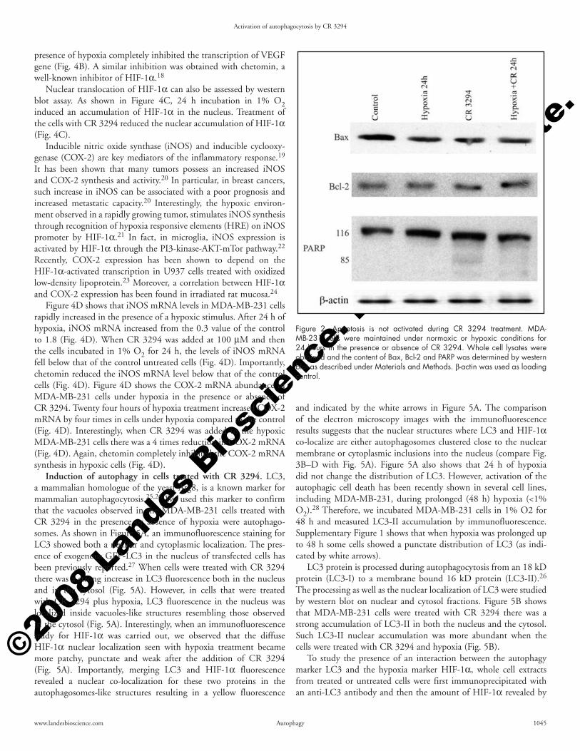
Autophagy and not apoptosis is induced during the treatment of hypoxic MDA-MB-231 cells with CR 3294. Apoptosis, or programmed cell death, is a highly regulated process that involves activation of a series of molecular events leading to cell death. Regulation of the apoptotic pathway is controlled by the Bcl-2 family of protein. Members of this family are divided in pro- and anti-apoptotic.16 A molecular analysis aimed to study the activation of the apoptotic pathway by CR 3294 is shown in Figure 2. The pro-apoptotic protein Bax did not increase and the anti-apoptotic protein Bcl-2 did not decrease during treatment of MDA-MB-231 cells with CR 3294 in the presence or absence of hypoxia (Fig. 2). Similarly, PARP, a substrate processed by caspase-3 during the executionary phase of apoptosis, was not significantly cleaved during the treatment with CR 3294 (Fig. 2).
Autophagy is initially a mode of cell survival where the cytosol and organelles of a cell are encapsulated in multi-membrane vacuoles and degraded after fusing with lysosomes.17 Examination of hypoxic MDA-MB-231 cells by electron microscopy after 24 h treatment with CR 3294 revealed a marked accumulation of autophagosomes (Fig. 3B white arrowheads). The nuclear membrane was still intact. However, several double membrane vesicles were visible into the nucleus and nucleolus (Fig. 3B and C white arrows). As shown in Figure 3C, these vacuoles contained cytoplasmic material and/or double membrane vesicles (white arrow) suggesting that such vacuoles in the nucleus were “inclusions” representing invaginations of the cytoplasm into the nucleus due to the increase in membrane flux going on in the injured cells. Moreover, autophagosomes were observed close to the nuclear membrane (Fig. 3C) and, sometimes, autophagosomes were present in invaginations of the cytoplasm into the nucleus (Fig. 3D, black arrows). The plasma membrane was also intact (Fig. 3B).
CR 3294 prevents accumulation of HIF-1α in MDA-MB-231 cells. The observation that induction of autophagy and cell killing by CR 3294 increased when the cells were under hypoxia led us to investigate if this molecule could have some effect on the pathway activated by the cells when adapting to a low oxygen tension. The major transcription factor activated by hypoxia is HIF-1α. Therefore, in order to study the effect of CR 3294 treatment on HIF-1α, we incubated the breast carcinoma cell line MDA-MB-231 in 1% O2 in the presence or absence of CR 3294. Subsequently, nuclear fractions were obtained and loaded on a 96 well plate previously coated with oligonucleotides bearing an HRE motif. Figure 4A shows the nuclear accumulation of HIF-1α in MDA-MB-231 cells incubated in 1% O2 in the presence or absence of CR 3294. The presence of 1% O2
by hydrolases.12 In mammalian cells, autophagy is a largely unknown process and only recently mammalian homologues of yeast genes have been discovered and used as molecular markers for autophagy.12
The new molecule CR 3294 belongs to a new series of anti-inflam-matory agents able to inhibit the expression of the inducible enzyme nitric oxide synthase (iNOS). CR 3294 displayed a particular selec-tivity of effect in validated animal models of intestinal inflammation (e.g., TNBS-induced colitis in rats), resulting in a better control of macroscopic and microscopic lesion scores, and in a larger inhibition of the increased protein expression and activity of iNOS and MMPs, compared to standard therapeutic agents and other investigational compounds for inflammatory bowel disease (IBD). CR 3294 may therefore represent a novel and innovative therapeutic approach in Crohn’s disease, ulcerative colitis and other forms of IBD. The pre-clinical package is ready to allow phase I clinical studies for intestinal inflammatory diseases.
Genetically modified mice with a target gene mutation that mimics the gene alteration in human cancers provide a powerful model system to assess the effect of pharmacological treatments on tumor growth.13 In this study we used transgenic mice carrying the polyoma virus middle T oncogene under the control of the mouse mammary tumor virus promoter/enhancer (MMT-PyVT), a well-established model for mammary adenocarcinoma onset and metastatic propagation.13,14 Female MMT-PyVT mice develop palpable mammary adenocarcinomas as early as 4–5 weeks of age.15 The MMT-PyVT transgenic mouse model has been chosen to have mammary adenocarcinomas with metastasis in 100 percent of mice reducing experimental variability.
This study demonstrates that the new molecule CR 3294 acti-vated autophagocytosis in the breast cancer cell line MDA-MB-231. In this cell line, CR 3294 reduced the nuclear content of HIF-1α. Importantly, when autophagy was inhibited with 3MA, bafilomycin or leupeptin, no loss in HIF-1α protein levels was observed. By contrast, when autophagy was induced by treating hypoxic cells with either trifluoperazine (TFP) or serum withdrawal (SW), a similar decrease in nucler HIF-1α was observed.
Results
In vitro and in vivo effects of CR 3294. The structure of CR 3294 is shown in Figure 1A. CR 3294 inhibits iNOS expression acting as a potent anti-inflammatory compound. However, we wanted to study if CR 3294, as many other anti-inflammatory compounds, had also anti-tumoral activity. The mechanism of action of CR 3294 was investigated by studying the in vitro effect of this molecule on different cell lines. We observed that the anti-tumoral activity of CR 3294 on HeLa, K562 and MDA-MB-231 cells was more evident in the presence of hypoxia. Figure 1B shows the induction of cell death by CR 3294 on the cell line MDA-MB-231. Similar results were obtained with both HeLa and K-562 cell line (not shown). Under normoxic conditions, CR 3294 did not induce a significant cell death that reached 20% after three days. However, when administered under hypoxia, CR 3294 killed about 40% of the cells after three days (Fig. 1B). Under hypoxic condi-tions, CR 3294 potentiated the cell killing in the presence of a low dose (10 μM) of celecoxib. In fact, CR 3294 plus celecoxib killed 20% of the cells under normoxia, whereas 70% of the cells were dead under hypoxia (Fig. 1B).

©2008
Lan
des B
iosc
ienc
e. D
o no
t dist
ribut
e.
Activation of autophagocytosis by CR 3294
1044 Autophagy 2008; Vol. 4 Issue 8
an inhibition started at 6 h but was significant at 17, 24 and 48 hours when the decrease in optical density was more than 50% (Fig. 4A).
Figure 4B shows the mRNA levels of the HIF-1α target gene VEGF. Synthesis of VEGF mRNA increased by 8 times after 24 h of hypoxia. Treatment of the cells with CR 3294 for 24 h in the
readily increased the amount of HIF-1α in the nucleus. In fact, a marked increase was observed after 3 h and continued at 6, 17 and 24 h. HIF-1α levels started to decrease at 48 h and continued to decrease at 3 and 4 days. However, when CR 3294 was added to the hypoxic cells there was a great reduction in HIF-1α binding (Fig. 4A). Such
Figure 1. In vitro and in vivo effects of CR 3294. (A) Structure of CR 3294. (B) MDA-MB-231 cells were maintained under normoxic or hypoxic conditions for the times indicated in the presence or absence of CR 3294 100 μM and celecoxib 10 μM. The percentage of cell killing was measured as described under Materials and Methods. (C) CR 3294 effects on tumor growth and (D) body weight. MMT-PyVT transgenic mice were treated with vehicle control or with 10 mg/kg or CR 3294 by gavage for 8 weeks. Data represents means + SD; n = 10 per group; *p < 0.05. (E) MMT-PyVT transgenic mice were maintained for twelve weeks and then treated with vehicle control (left) or with 10 mg/kg of CR 3294 for eight weeks (right). (F) H&E-stained tumor sections from control and CR 3294 treated MMT-PyVT mouse. Black arrows indicate areas of necrosis.
©2008
Lan
des B
iosc
ienc
e. D
o no
t dist
ribut
e.
Activation of autophagocytosis by CR 3294
www.landesbioscience.com Autophagy 1045
and indicated by the white arrows in Figure 5A. The comparison of the electron microscopy images with the immunofluorescence results suggests that the nuclear structures where LC3 and HIF-1α co-localize are either autophagosomes clustered close to the nuclear membrane or cytoplasmic inclusions into the nucleus (compare Fig. 3B–D with Fig. 5A). Figure 5A also shows that 24 h of hypoxia did not change the distribution of LC3. However, activation of the autophagic cell death has been recently shown in several cell lines, including MDA-MB-231, during prolonged (48 h) hypoxia (<1% O2).28 Therefore, we incubated MDA-MB-231 cells in 1% O2 for 48 h and measured LC3-II accumulation by immunofluorescence. Supplementary Figure 1 shows that when hypoxia was prolonged up to 48 h some cells showed a punctate distribution of LC3 (as indi-cated by white arrows).
LC3 protein is processed during autophagocytosis from an 18 kD protein (LC3-I) to a membrane bound 16 kD protein (LC3-II).26 The processing as well as the nuclear localization of LC3 were studied by western blot on nuclear and cytosol fractions. Figure 5B shows that MDA-MB-231 cells were treated with CR 3294 there was a strong accumulation of LC3-II in both the nucleus and the cytosol. Such LC3-II nuclear accumulation was more abundant when the cells were treated with CR 3294 and hypoxia (Fig. 5B).
To study the presence of an interaction between the autophagy marker LC3 and the hypoxia marker HIF-1α, whole cell extracts from treated or untreated cells were first immunoprecipitated with an anti-LC3 antibody and then the amount of HIF-1α revealed by
presence of hypoxia completely inhibited the transcription of VEGF gene (Fig. 4B). A similar inhibition was obtained with chetomin, a well-known inhibitor of HIF-1α.18
Nuclear translocation of HIF-1α can also be assessed by western blot assay. As shown in Figure 4C, 24 h incubation in 1% O2 induced an accumulation of HIF-1α in the nucleus. Treatment of the cells with CR 3294 reduced the nuclear accumulation of HIF-1α (Fig. 4C).
Inducible nitric oxide synthase (iNOS) and inducible cyclooxy-genase (COX-2) are key mediators of the inflammatory response.19 It has been shown that many tumors possess an increased iNOS and COX-2 synthesis and activity.20 In particular, in breast cancers, such increase in iNOS can be associated with a poor prognosis and increased metastatic capacity.20 Interestingly, the hypoxic environ-ment observed in a rapidly growing tumor, stimulates iNOS synthesis through recognition of hypoxia responsive elements (HRE) on iNOS promoter by HIF-1α.21 In fact, in microglia, iNOS expression is activated by HIF-1α through the PI3-kinase-AKT-mTor pathway.22 Recently, COX-2 expression has been shown to depend on the HIF-1α-activated transcription in U937 cells treated with oxidized low-density lipoprotein.23 Moreover, a correlation between HIF-1α and COX-2 expression has been found in irradiated rat mucosa.24
Figure 4D shows that iNOS mRNA levels in MDA-MB-231 cells rapidly increased in the presence of a hypoxic stimulus. After 24 h of hypoxia, iNOS mRNA increased from the 0.3 value of the control to 1.8 (Fig. 4D). When CR 3294 was added at 100 μM and then the cells incubated in 1% O2 for 24 h, the levels of iNOS mRNA fell below that of the control untreated cells (Fig. 4D). Importantly, chetomin reduced the iNOS mRNA level below that of the control cells (Fig. 4D). Figure 4D shows the COX-2 mRNA abundance in MDA-MB-231 cells under hypoxia in the presence or absence of CR 3294. Twenty four hours of hypoxia treatment increased COX-2 mRNA by four times in cells under hypoxia compared to the control (Fig. 4D). Interestingly, when CR 3294 was added to the hypoxic MDA-MB-231 cells there was a 4 times reduction in COX-2 mRNA (Fig. 4D). Again, chetomin completely inhibited the COX-2 mRNA synthesis in hypoxic cells (Fig. 4D).
Induction of autophagy in cells treated with CR 3294. LC3, a mammalian homologue of the yeast Atg8, is a known marker for mammalian autophagocytosis.25,26 We used this marker to confirm that the vacuoles observed in the MDA-MB-231 cells treated with CR 3294 in the presence or absence of hypoxia were autophago-somes. As shown in Figure 5A, an immunofluorescence staining for LC3 showed both a nuclear and cytoplasmic localization. The pres-ence of exogenous GFP-LC3 in the nucleus of transfected cells has been previously reported.27 When cells were treated with CR 3294 there was a strong increase in LC3 fluorescence both in the nucleus and in the cytosol (Fig. 5A). However, in cells that were treated with CR 3294 plus hypoxia, LC3 fluorescence in the nucleus was localized inside vacuoles-like structures resembling those observed in the cytosol (Fig. 5A). Interestingly, when an immunofluorescence study for HIF-1α was carried out, we observed that the diffuse HIF-1α nuclear localization seen with hypoxia treatment became more patchy, punctate and weak after the addition of CR 3294 (Fig. 5A). Importantly, merging LC3 and HIF-1α fluorescence revealed a nuclear co-localization for these two proteins in the autophagosomes-like structures resulting in a yellow fluorescence
Figure 2. Apoptosis is not activated during CR 3294 treatment. MDA-MB-231 cells were maintained under normoxic or hypoxic conditions for 24 hours in the presence or absence of CR 3294. Whole cell lysates were obtained and the content of Bax, Bcl-2 and PARP was determined by western blot as described under Materials and Methods. β-actin was used as loading control.

©2008
Lan
des B
iosc
ienc
e. D
o no
t dist
ribut
e.
Activation of autophagocytosis by CR 3294
1046 Autophagy 2008; Vol. 4 Issue 8
vacuoles, prevented the decrease of HIF-1α levels by CR 3294 (Fig. 6A left). A similar effect was obtained with the lysosome inhibitor bafilomycin A (Fig. 6A left). However, recently bafilomycin has been reported to increase the binding of subunit c in V0 sector of V-ATPase (ATP6V0C) to HIF-1α. Such a binding would then dissociate von Hippel-Lindau tumor suppressor protein (pVHL) from HIF-1α with the stabilization of the latter.29 Similarly, we observed a nuclear increase in HIF-1α levels when the cells were treated with bafilomycin in the presence or absence of hypoxia
western blot. Figure 5C shows that when CR 3294 was given to the hypoxic cells, there was a strong increase in HIF-1α protein levels compared to both control or hypoxia samples (Fig. 5C). The same cells extracts were immunoprecipitated with an anti-rabbit IgG and stained for HIF-1α to exclude any nonspecific binding (Fig. 5C).
Effect of autophagy inhibition on HIF-1α. The levels of HIF-1α after induction of autophagy by CR 3294 were studied using different inhibitors of autophagy and examining the effects. Figure 6A shows that 3MA, an inhibitor of the formation of autophagic
Figure 3. CR 3294 induces autophagy in MDA-MB-231 cells. (A) MDA-MB-231 cells were maintained under hypoxic conditions for 24 h. The cells were then processed for electron microscopy as described under Materials and Methods. (Magnification 4.900x). (B) MDA-MB-231 cells were maintained under hypoxic conditions in the presence of CR 3294 for 24 h. The cells were then processed for electron microscopy as described under Materials and Methods. White arrowheads point to autophagosomes in the cytoplasm. White arrows show cytoplasmic invaginations into the nucleus. (Magnification 7.000x). (C) MDA-MB-231 cells were maintained under hypoxic conditions in the presence of CR 3294 for 24 h. Black arrows point to autophagosomes close to the nuclear membrane. White arrow indicates a cytoplasmic invagination. (Magnification 27.500x). (D) MDA-MB-231 cells were maintained under hypoxic conditions in the presence of CR 3294 for 24 h. Black arrows show autophagosomes inside a deep cytoplasmic invagination into the nucleus. (Magnification 27.500x).

©2008
Lan
des B
iosc
ienc
e. D
o no
t dist
ribut
e.
Activation of autophagocytosis by CR 3294
www.landesbioscience.com Autophagy 1047
of this protein after incubation with bafilomycin in the presence or in the absence of hypoxia (Fig. 6B right).
Importantly, Figure 6C shows that CR 3294 alone or in combi-nation with autophagy inhibitors did not increase the total protein ubiquitination.
Effect of autophagy activation on HIF-1α. Induction of autophagy can be achieved by growing cells in absence of serum and/or amino acids or in the presence of different molecules. A recent study has screened for potential compounds that regulate autophagy.30 In this work, a combination of image-based and long-lived protein degradation assays was used to identify compounds regulating autophagy.30 The anti-psychotic drug trifluoperazine was identified as a molecule that can truly induce autophagic degradation without causing obvious cellular damage.30
(Fig. 6A right). However, treatment of MDA-MB-231 cells with leupeptin, an inhibitor of lysosomal proteases, prevented the decrease of HIF-1α protein levels by CR 3294 (Fig. 6A right). By contrast, the microtubule disrupting agent nocodazole and the inhibitor of actin polymerization cytochalasin D did not prevent the effect of CR 3294 on HIF-1α levels (Fig. 6A right).
Treatment of MDA-MB-231 cells with these inhibitors in the presence or absence of CR 3294 was also used to assess both the induction of autophagy and the autophagic flux by CR 3294. Figure 6B shows that both 3MA and wortmannin inhibited LC3-II accu-mulation in the presence of CR 3294. By contrast, bafilomycin and leupeptin inhibited the lysosomal degradation of both LC3-I and II that, in turn, accumulated in the cells (Fig. 6B left). The lysosomal turnover of LC3 in these cells was demonstrated by the accumulation
Figure 4. HIF-1α DNA binding and VEGF transcription in the presence of CR 3294. (A) Nuclear fractions were obtained from cells cultured under hypoxic conditions in the presence or absence of CR 3294 for the indicated time. The amount of HIF-1α was measured by the ELISA-based TransAM method as described under Material and Methods. Results are the mean ± S.D. of three separate experiments. (B) Real Time PCR analysis of VEGF mRNA after 24 hours incubation of MDA-MB-231 cells under hypoxia in the presence or absence of CR 3294 or Chetomin. Data are represented as mean ± S.D. (C) MDA-MB-231 cells were maintained under normoxic or hypoxic conditions in the presence or absence of CR 3294. After the times indicated, the nuclear and cytosolic fractions were isolated. The content of HIF-1α was determined by SDS-PAGE and western blotting. Nuclear factor-1 (NF-1) and β-actin were used as controls for the nuclear and cytosolic fraction, respectively. (D) MDA-MB-231 cells were cultured under normoxic (control) or hypoxic conditions. Hypoxic exposure was for 24 hours in the presence or absence of 100 μM CR 3294 or chetomin. The relative amount of iNOS mRNA or COX-2 mRNA was measured by Real Time PCR as described under Materials and Methods. Data are represented as mean ± S.D.

©2008
Lan
des B
iosc
ienc
e. D
o no
t dist
ribut
e.
Activation of autophagocytosis by CR 3294
1048 Autophagy 2008; Vol. 4 Issue 8
the possibility that this molecule could have some direct or indi-rect effect on the hypoxia inducible transcription factor HIF-1α. In fact, when MDA-MB-231 cells were treated with CR 3294 and incubated in hypoxia (1% O2) the binding of HIF-1α to HRE bearing oligonucleotides was inhibited (Fig. 4A). Such an inhibi-tion was accompanied by abolition of VEGF mRNA synthesis (Fig. 4B). Interestingly, western blot analysis of HIF-1α in the presence of hypoxia and CR 3294 revealed a reduction of HIF-1α protein levels (Fig. 4C). Furthermore, CR 3294 treatment of hypoxic cells inhibited also the transcription of the inducible enzymes iNOS and COX-2 (Fig. 4D).
The formation of autophagosomes was confirmed by using the autophagy marker LC3. Immunofluorescence studies revealed a clear accumulation of LC3 in the autophagosomes of CR treated cells. The fluorescence pattern of LC3 changed from a diffuse to a punctated one (Fig. 5A). A double immunofluorescence study confirmed a colocalization of the autophagic marker LC3 and the hypoxia inducible factor HIF-1α (Fig. 5A). Such a colocalization was observed in both the cytoplasm and the nucleus of hypoxic cells treated with CR 3294. However, a comparison of this result with the electron microscopy images (Fig. 3B–D) suggested that LC3 and HIF-1α colocalized in autophagosomes either clustered around the nucleus and/or present in invaginations of the cytoplasm into the
Therefore, we examined the effects of autophagy activation by trifluoperazine (TFP) and serum withdrawal (SW) on HIF-1α protein levels. Figure 7A shows that activation of autophagy by TFP in hypoxic MDA-MB-231 cells resulted in the decrease of HIF-1α protein levels. Autophagic activation by TFP was confirmed by the accumulation of LC3-II (Fig. 7A). Similarly, SW in hypoxic MDA-MB-231 cells inhibited accumulation of HIF-1α while acti-vating the autophagic pathway as assessed by LC3-II accumulation (Fig. 7B). Cells treated for 24 h with TFP or SW did not show any obvious damage (not shown).
Discussion
In this study we have shown that a novel molecule, CR 3294, acti-vates autophagy causing an in vivo and in vitro inhibition of tumor growth. In particular, we observed that CR 3294 was highly effective in increasing the percentage of dead cells in several tumorigenic cell lines and in inhibiting tumor growth in the MMT-PyVT transgenic mouse model (Fig. 1). A molecular analysis of the mechanism(s) revealed that apoptotic pathway was not activated by CR 3294 (Fig. 2). By contrast, electron microscopy analysis showed a marked activation of autophagy with accumulation of autophagosomes (Fig. 3). The observation that CR 3294 was most effective in killing the MDA-MB-231 cells when given under hypoxia, led us to explore
Figure 5. Colocalization of HIF and LC3 in cells treated with CR 3294. (A) MDA-MB-231 cells were maintained under normoxic or hypoxic conditions for 24 hours in the presence or absence of CR 3294. Nuclear colocalization of LC3 (red fluorescence) and HIF-1α (green fluorescence) was studied by immu-nofluorescence as described under Materials and Methods. Yellow fluorescence represents areas of HIF-1α and LC3 colocalization as evidenced by white arrows. (B) MDA-MB-231 cells were maintained under normoxic or hypoxic conditions for 24 hours in the presence or absence of CR 3294. Nuclear and cytosolic fractions were obtained, electrophoresed on a SDS-polyacrylamide gel, and immunoblotted for LC3. Nuclear factor-1 (NF-1) and β-actin were used as controls for the nuclear and cytosolic fraction, respectively. (C) MDA-MB-231 cells were maintained under normoxic or hypoxic conditions for 24 hours in the presence or absence of CR 3294. Nuclear fractions were immunoprecipitated with either an antibody anti-LC3 or an anti rabbit IgG, electrophoresed on a SDS-polyacrylamide gel, and immunoblotted with a mouse anti HIF-1α as described under Materials and Methods. H. chain: antibody heavy chain.

©2008
Lan
des B
iosc
ienc
e. D
o no
t dist
ribut
e.
Activation of autophagocytosis by CR 3294
www.landesbioscience.com Autophagy 1049
Figure 6. For figure legend, see page 1050.
nucleus. Importantly, LC3 and HIF-1α could be co-immunopre-cipitated from hypoxic cells treated with CR 3294 (Fig. 5C). The co- localization and co-immunoprecipitation of LC3 and HIF-1α observed in the cells treated with CR 3294 suggests the presence
of an interaction between these two proteins. Recent studies have observed that the nuclear accumulation of HIF-1α requires an intact cytoskeleton and can be inhibited by microtubules interacting drugs such as taxol or 2-methoxyestradiol.31,32 LC3 is a microtubule-asso-

©2008
Lan
des B
iosc
ienc
e. D
o no
t dist
ribut
e.
Activation of autophagocytosis by CR 3294
1050 Autophagy 2008; Vol. 4 Issue 8
ciated protein whose physiological function is still obscure but that is associated to the forming autophagosome through the conversion of LC3-I (cytosolic) to LC3-II (membrane bound). Therefore, our data suggest the possibility that during the treatment with CR 3294, HIF-1α can be one of the long-lived proteins contained inside the forming autophagosomes. The observation that LC3 (Fig. 5A and B) is present also in the nucleus would suggest that HIF-1α can be sequestered in this cellular compartment, too, by a yet unknown mechanism. Interestingly, GFP-LC3 accumulation in the nucleus has been recently described in MEFs transfected with a plasmid coding for such protein.27 In this case, the effect of such an interaction would prevent HIF-1α to bind to the HRE.
However, more experiments will be needed to demonstrate a direct link between the anti-tumoral effect and the decrease of HIF-1α observed in the presence of CR 3294. In fact, one of the many molecules influenced by hypoxia, besides HIF-1α, could be responsible for this effect as well.
Another possible explanation for the absence of HIF-1α in the nucleus could be that CR activates a process called piecemeal microautophagy of the nucleus (PMN). PMN has been observed in Saccharomyces cerevisiae and described as a mechanism where teardrop-like blebs are pinched from the nucleus, released into the vacuole lumen, and degraded by soluble hydrolases.32 Studies showed that PMN is a distinct process that does not depend on the proteins involved in macroautophagy.33 However, activation of PMN in the MDA-MB-231 cells treated with CR was excluded by the electron microscopy experiments (Fig. 3).
The accumulation of LC3-II as well as the levels of HIF-1α were studied using both inhibitors and inducers of autophagy. 3MA and wortmannin were used to inhibit autophagosomes formation. Treatment of hypoxic MDA-MB-231 cells with CR in the pres-ence of 3MA or wortmannin prevented the decrease of protein levels of both LC3 and HIF-1α (Fig. 6A and B). Bafilomycin and leupeptin were used to inhibit the lysosomal activity and to study the autophagic flux. In fact, the lysosomal turnover of LC3-II rather than the cellular level of LC3 is considered as a real indicator for the activation of autophagy.34 Treatment of hypoxic cells with CR and bafilomycin maintained HIF-1α protein levels while determining an increase in both LC3-I and II (Fig. 6A and B). However, the levels of HIF-1α in the presence of bafilomycin can be influenced by the recent observation that bafilomycin stabilizes HIF-1α.29 In fact, we, too, observed a nuclear increase of HIF-1α in cells treated with bafi-lomycin in the presence or absence of hypoxia (Fig. 6A). Importantly, HIF-1α protein levels were maintained also after treating the cells with leupeptin (Fig. 6A and B). Therefore, CR 3294 activates the autophagic flux with lysosomal degradation of HIF-1α.
Recent reports have shown activation of the autophagic pathway by hypoxia.28,35,36 In particular, Azad et al., have shown that 24 h of hypoxia resulted in LC3 processing in the U87 cell line. Furthermore, the same authors demonstrated that prolonged hypoxia (<1% O2 for
Figure 6. Effects of autophagy inhibition on HIF-1α and LC3 levels. (A) MDA-MB-231 cells were left untreated. Alternatively, cells were treated for 24 h with CR 3294 in the presence or absence of 3MA, wortmannin, bafilomycin, leupeptin, nocodazole and cytochalasin D as indicated. Nuclear fractions were obtained and the content of HIF-1α was determined by SDS-PAGE and western blotting. Lamin A/C was used as control for the nuclear fraction. (B) MDA-MB-231 cells were left untreated. Alternatively, cells were treated for 24 h with CR 3294 in the presence or absence of 3MA, wortmannin, bafilomycin, leupeptin, nocodazole and cytochalasin D as indicated. Whole cell lysates were obtained and the content of LC3-I and II was determined by SDS-PAGE and western blotting. β-actin was used as loading control. (C) MDA-MB-231 cells were left untreated. Alternatively, cells were treated for 24 h with CR 3294 in the presence or absence of 3MA, wortmannin, bafilomycin, leupeptin, nocodazole and cytochalasin D as indicated. Whole cell lysates were obtained and the content of ubiquitin was determined by SDS-PAGE and western blotting.
Figure 7. Effects of autophagy induction on HIF-1α and LC3 levels. (A) MDA-MB-231 cells were left untreated. Alternatively, cells were treated for 24 h with hypoxia in the presence or absence of TFP. Nuclear and cytosolic fractions were obtained, electrophoresed on a SDS-polyacrylamide gel, and immunoblotted for HIF-1α and LC3 respectively. Lamin A/C was used as control for the nuclear fraction. β-actin was used as control for the cytosolic fraction. (B) MDA-MB-231 cells were left untreated. Alternatively, cells were treated for 24 h with hypoxia in the presence or absence of serum (SW). Nuclear and cytosolic fractions were obtained, electrophoresed on a SDS-polyacrylamide gel, and immunoblotted for HIF-1α and LC3 respectively. Lamin A/C was used as control for the nuclear fraction. β-actin was used as control for the cytosolic fraction.

©2008
Lan
des B
iosc
ienc
e. D
o no
t dist
ribut
e.
Activation of autophagocytosis by CR 3294
www.landesbioscience.com Autophagy 1051
and embedded in paraffin. Serial cross-sections (5 μm) were stained with hematoxylin-eosin and observed under microscopy.
Measurement of cell viability. Cell viability was determined using (3-(4,5-dimethylthiazol-2-yl)-5-(3-carboxymethoxyphenyl-2-(4-sulfophenyl)-2H-tetrazolium inner salt (MTS) and phenazine methosulfate (PMS) according to the manufacturer’s instructions (Promega Corp.,). The absorbance was determined at 490 nm on a Titertek Multiscan Plus plate reader. At the same time, cell viability was measured by Trypan Blue exclusion.
Treatment protocols and antibodies. In all experiments CR 3294 (generously provided by Rottapharm, Milan, Italy) was dissolved in water and added daily to a final concentration of 100 μM. 3-Methylad enine (Sigma) was dissolved in water, boiled for 1 minute and added to a final concentration of 10 mM. Celecoxib (LKT Laboratories, St. Paul, Minnesota) was dissolved in Me2SO and added to a final concentration of 10 μM. Chetomin (Axxora, San Diego, California) was dissolved in Me2SO and added to a final concentration of 50 nM. Wortmannin (Sigma) was dissolved in Me2SO and added to a final concentration of 400 nM. Bafilomycin A (Sigma) was dissolved in Me2SO and added to a final concentration of 100 nM. Leupeptin (Sigma) was dissolved in water and added to a final concentration of 100 μg/ml. Nocodazole (Sigma) was dissolved in Me2SO and added to a final concentration of 2.5 μg/ml. Cytochalasin D(Sigma) was dissolved in Me2SO and added to a final concentration of 30 μM. Trifluoperazine (Sigma) was dissolved in water and added to a final concentration of 10 μM. Control experiments showed that Me2SO had no effect on any of the parameters measured.
The following primary antibodies were used: mouse anti-HIF-1α antibody (BD Bioscience, San Jose, California), mouse anti-Lamin A/C antibody (BD Bioscience, San Jose, California), rabbit anti-nuclear factor 1 (NF-1) (Santa Cruz, California), rabbit anti-LC3B (Novus Biological, Littleton, Colorado), mouse anti-ubiquitin anti-body (Santa Cruz, California), rabbit anti-Bax antibody (Santa Cruz, California), mouse anti-Bcl-2 antibody (Santa Cruz, California), rabbit anti-PARP antibody (Santa Cruz, California), mouse anti-β actin (Santa Cruz, California). The following secondary antibodies were used: mouse anti-rabbit Alexa 488 (Molecular Probes, Eugene, Oregon) goat anti-rabbit Alexa 594 (Molecular Probes), mouse anti-rabbit HRP, goat anti-mouse HRP (Amersham Biosciences, Piscataway, New Jersey), donkey anti-goat HRP (Santa Cruz, California).
Hypoxia. Hypoxic conditions were achieved by incubating cells in a hypoxia chamber (Billups-Rothenberg Inc., California) where a 1% oxygen mix was flushed in for 4 minutes according to the manu-facturer’s instructions.
Immunofluorescence and confocal analysis. Cells were plated on 35 mm glass bottom dishes (MatTek, Ashland, Massachusetts) in complete medium and incubated overnight at 37°C under an atmosphere of 95% air and 5% CO2. The day after, cells were washed with PBS and fixed in 4% paraformaldehyde for 20 minutes. The cells were then washed twice with PBS and permeabilized with 0.5% Triton X-100 for 6 min. After a PBS wash, the cells were incu-bated for 4 hours at room temperature with the primary antibody diluted in PBS-1% BSA. After staining with a fluorescent secondary antibody, the cells were incubated in the dark for 1 h at room temperature and washed twice with PBS. Cover slips were mounted in 50% glycerol solution and stored in the dark at 4°C. Fluorescence
48 h) increased the percentage of cells with punctate GFP-LC3 in several cell lines including the MDA-MB-231 where 60% of the transfected cells had a punctated GFP-LC3 pattern. Since our results on this same cell line did not show any detectable induction of autophagy after 24 h we prolonged the hypoxic stimulus up to 48 h and measured the distribution of LC3. Supplemental Figure 1 shows that, after 48 h of hypoxia, some cells showed a punctate pattern for LC3 in the cytosol. The apparent discrepancy in the timing of LC3 processing could be due to the amount of O2 used that is 1% in our system and <1% in theirs. In fact, incubation of SiHa and RKO cells in <0.01% O2 activated LC3-II accumulation after 12 and 24 h respectively,36 suggesting an important role for the cell line and the percentage of O2 for the activation of LC3 processing. It is also worth noting that while some authors have shown that hypoxia-induced autophagy involved the activity of 5' AMP activated kinase (AMPK) independently from HIF-1α and BNIP3,36 others have instead indicated a role for BNIP3 and HIF-1α in regulating hypoxia-induced autophagocytosis.28,35 Therefore, activation of the autophagic pathway during prolonged hypoxia requires additional investigations.
Autophagy is characterized by the degradation of long-lived protein.37 Hypoxia increases the life of HIF-1α from 5 minutes to days.38 Our results suggest the possibility that, in the presence of an intact autophagic machinery, the activation of autophagy under hypoxia decreases the amount of HIF-1α. If this is the case, then activation of autophagy under hypoxia by other stimuli than CR 3294 should inhibit HIF-1α accumulation. Therefore, autophagy was induced by treating the cells with TFP or SW (Fig. 7). Both treatments caused a marked reduction of HIF-1α and an accumula-tion of LC3-II (Fig. 7A and B).
In summary, we demonstrated that the anti-inflammatory compound CR 3294 inhibits tumoral growth rate through the acti-vation of autophagocytosis and that such activity of CR 3294 was more pronounced under hypoxia.
Materials and Methods
Cell cultures. The MDA-MB-231 breast carcinoma cell line, (American Type Culture Collection, Manassas, Virginia) was main-tained in 75-cm2 polystyrene flasks (Corning Costar Corp., Oneonta, New York) with RPMI 1640 medium (Mediatech, Inc., Herndon, Virginia), containing 100 units/ml penicillin, 0.1 mg/ml strepto-mycin, and 10% heat-inactivated foetal bovine serum. The cells were incubated under an atmosphere of 95% air and 5% CO2. For serum starvation experiments, cells were collected, washed with PBS and seeded in Hank’s Balanced Salt Solution (HBSS) (Sigma, St. Louis, Missouri) medium without serum for the times indicated.
Animals. MMT mice expressing the polyoma middle T antigen (PyVT)13 were purchased from Charles River Laboratories (Wilmington, Massachusetts). MMT-PyVT mice were kept in cages under controlled pathogen-free conditions throughout the duration of the experiments. Three-month-old mice were either left untreated or treated with 10 mg/kg of CR 3294. CR 3294 was administered daily by gavage. Experiments were done according to Italian and European guidelines. Tumors were measured with calipers, and the tumor volume was calculated as 0.5 x length x width2. Images of the mice were taken after 8 weeks of treatment. Mice were then sacrificed; breast tumors samples were fixed in phosphate-buffered formalin,

©2008
Lan
des B
iosc
ienc
e. D
o no
t dist
ribut
e.
Activation of autophagocytosis by CR 3294
1052 Autophagy 2008; Vol. 4 Issue 8
1 hour with HRP-conjugated secondary antibody followed by the addition of a developing solution. A stop solution was added to block the reaction and the color intensity was read in a plate reader with a 450 nm filter. The color intensity in each well is proportional to the amount of the transcription factor bound to the oligonucleotides.
Immunoprecipitation assay. Nuclear fractions were obtained as mentioned above. After determining protein concentration using the Bradford assay, 140 μg of the lysate was brought to a final volume of 1 ml with PBS. Lysates were precleared with 1 μg of appropriate control IgG and 20 μl of Protein A/G PLUS-Agarose (Santa Cruz Biotechnology) and kept on a rotator for 1 hour at 4°C. Lysates were centrifuged (500 xg for 5 min at 4°C) and 2 μg of anti-LC3 antibody (Novus Biological, Littleton, Colorado) was added to the precleared lysates and kept on ice for approximately 3–5 hours. Following incu-bation, 30 μl of Protein A/G PLUS-Agarose was added to each tube and kept on a rotator overnight at 4°C. Lysates were then centrifuged at 500 xg for 5 min at 4°C. The pellets were washed 4 times with PBS and then resuspended in 20 μl of loading buffer. Samples were elec-trophoresed on a 8% SDS-polyacrylamide gel and immunoblotted for HIF-1α with a mouse monoclonal antibody (BD Bioscience, San Jose, California).
RNA isolation and real-time quantitative polymerase chain reaction. Cells were plated in 100 mm dishes in complete RPMI medium. Following treatment, cells were washed in PBS, removed from the dish using a rubber policeman and transferred to a conical tube. Cells were then collected by centrifugation at 500 xg for 5 minutes. Afterwards, the PBS was discarded and the cell pellet lysed to extract total RNA by using the BioRobot EZ1 workstation and the EZ1 RNA cell mini-kit (Qiagen, Milan, Italy) following manu-facturer’s instructions. The integrity of total RNA was evaluated by using the Agilent Technology 2100 instrument (Agilent, Milan, Italy). Approximately 1μg of RNA was reverse transcribed and aliquots of cDNA were subjected to real time PCR with a Taqman universal PCR Master Mix to detect Vascular endothelial growth factor (VEGF), inducible nitric oxide synthase (iNOS), and induc-ible cyclooxygenase (COX-2) mRNA. Amplification of 18S rRNA was used in the same reaction as internal reference gene. Taqman probes for real-time PCR were ordered as Assay on Demand (Applied Biosystems, Foster City, California). All PCR reactions were done in triplicate. PCR amplifications were performed as follows: 50°C for 2 minutes, 95°C for 10 minutes, and 40 cycles each with 95°C for 15 seconds and 60°C for 1 minute using an ABI PIRSM 7000 sequence detector (Applied Biosystems, Foster City, California). Amplification data were analyzed using the Sequence Detector version 1.7 software (Applied Biosystems). Statistical analysis of real-time PCR results were done using mean normalized cycle threshold (ΔCt) values and the pooled standard deviation of the mean ΔCt. Experiments were repeated at least three times with different cell preparations.
Note
Supplementary materials can be found at:www.landesbioscience.com/supplement/TafaniAUTO4-8-Sup.pdf
intensity was visualized with a confocal microscope (Nikon Eclipse TE 2000-E).
Electron microscopy. After treatment the cells were trypsinized and pelleted. The cells were then suspended and fixed overnight at 4°C in 2% glutaraldehyde with 1% tannic acid in 0.1 M sodium cacodylate, pH 7.3. The cells were rinsed 3 times in the sodium cacodylate buffer and then incubated in 2% osmium tetroxide in the same buffer for 2 hours at room temperature. The cells were then rinsed 3 times in distilled water and exposed to 1% uranyl acetate in water for 15 min at room temperature. The cells were rinsed twice in distilled water, spun down into 3% agarose at 45°C, and cooled to form blocks. The agarose blocks were dehydrated in graded steps of acetone and embedded in Spurr’s low viscosity media. Following polymerization overnight at 65°C, 80-nm sections were cut on a Reighert-Jung Ultra cut E ultramicrotome and picked up on copper grids. The grids were post-stained in uranyl acetate and bismuth subnitrate. The sections were observed in a Philips CM-10 TEM and micrographs recorded on Kodak 4489 sheet film.
Isolation of cytosol and nuclear fractions. Cells (2 x 10⁶) were plated in 100 mm dishes. The nuclear and cytosolic fractions were isolated using the nuclear extraction kit from Active Motif, (Carlsbad, California) following manufacturer’s instructions. Briefly, following treatment, cells were scraped off the plate in a PBS/Phosphatase inhibitor buffer using a rubber policeman. Cells were centrifuged 5 minutes at 500 rpm at 4°C and the supernatant discarded. The cell pellet was lysed in hypotonic buffer for 15 minutes in ice followed by centrifugation (30 seconds at 14,000 rpm at 4°C). The super-natant (cytosolic fraction) was transferred to a new tube, while the pellet (nuclear fraction) was lysed in a lysis buffer in the presence of protease inhibitors. The cytosolic fraction was concentrated through a Microcon YM-10 Centrifugal Filter Device (Millipore, Bedford, Massachusetts). Protein concentration of the fractions was deter-mined by the Bradford assay (Bio-Rad, Hercules, California).
Western blot assay. Cells (1 x 10⁶) for whole cell lysate were pelleted at 700 xg (5 min at 4°C) and lysed in 50 μl of cell lysis buffer (20 mM Tris, pH 7.4, 100 mM NaCl, 1% Triton, 1 mM phenylmethylsulfonyl fluoride, 10 μg/ml leupeptin, 10 μg/ml aprotinin). After 30 min on ice, the lysates were clarified by centrifu-gation (10 min at 4°C) and the supernatant was collected. Protein concentration was determined by the Bradford assay (Bio-Rad, Hercules, CA). Equivalent amounts of protein were electrophoresed on SDS-polyacrylamide gels. Kaleidoscope Prestained Standards (Bio-Rad) were used to determine molecular weight. The gels were then electroblotted onto PVDF membranes. After blocking with 5% milk, membranes where incubated with the primary antibody over-night. Finally, the relevant protein was visualized by staining with the appropriate secondary horseradish peroxidase-labeled antibody for 1 hour followed by enhanced chemiluminescence.
Elisa assay. Nuclear and cytoplasmic extracts were obtained as described above and used to measure the relative amount of HIF-1α using the TransAM kit from Active Motif and following manufactur-er’s instructions. Briefly, equivalent amounts of protein from nuclear and cytoplasmic extracts were loaded on a 96 well plate coated with oligonucleotides containing hypoxia responsive elements (HRE). After 1 hour incubation at room temperature, wells were washed 3 times in wash buffer and incubated 1 hour with a primary antibody at room temperature. Subsequently, wells were washed and incubated

©2008
Lan
des B
iosc
ienc
e. D
o no
t dist
ribut
e.
Activation of autophagocytosis by CR 3294
www.landesbioscience.com Autophagy 1053
28. Azad MB, Chen Y, Henson ES, Cizeau J, McMillan-Ward E, Israels SJ, Gibson SB. Hypoxia induces autophagic cell death in apoptosis-competent cells through a mechanism involving BNIP3. Autophagy 2008; 164:195-204.
29. Lim JH, Park JW, Kim SJ, Kim MS, Park SK, Johnson RS, Chun YS. ATP6V0C competes with von Hippel-Lindau protein in hypoxia-inducible factor 1alpha (HIF-1alpha) binding and mediates HIF-1alpha expression by bafilomycin A1. Mol Pharmacol 2007; 71:942-8.
30. Zhang L, Yu J, Pan H, Hu P, Hao Y, Cai W, Zhu H, Yu AD, Xie X, Ma D, Yuan J. Small molecule regulators of autophagy identified by an image-based high-throughput screen. Proc Natl Acad Sci USA 2007; 104:19023-8.
31. Mabjeesh NJ, Escuin D, LaVallee TM, Pribluda VS, Swartz GM, Johnson MS, Willard MT, Zhong H, Simons JW, Giannakakou P. 2ME2 inhibits tumor growth and angiogenesis by disrupting microtubules and dysregulating HIF. Cancer Cell 2003; 3:363-75.
32. Escuin D, Kline ER, Giannakakou P. Both microtubule-stabilizing and microtubule-destabilizing drugs inhibit hypoxia-inducible factor-1alpha accumulation and activity by disrupting microtubule function. Cancer Res 2005; 65:9021-8.
33. Roberts P, Moshitch-Moshkovitz S, Kvam E, O’Toole E, Winey M, Goldfarb DS. Piecemeal Microautophagy of Nucleus in Saccharomyces cerevisiae. Mol Biol Cell 2003; 14:129-41.
34. Tanida I, Minematsu-Ikeguchi N, Ueno T, Kominami E. Lysosomal turnover, but not a cellular level, of endogenous LC3 is a marker for autophagy. Autophagy 2005; 1:84-91.
35. Bohensky J, Shapiro IM, Leshinsky S, Terkhorn SP, Adams CS, Srinivas V. HIF-1 regulation of chondrocyte apoptosis: induction of the autophagic pathway. Autophagy 2007; 3:207-14.
36. Papandreou I, Lim AL, Laderoute K, Denko NC. Hypoxia signals autophagy in tumor cells via AMPK activity, independent of HIF-1, BNIP3, and BNIP3L. Cell Death Differ 2008; 15:1572-81.
37. Roberts EA and Deretic V. Autophagic proteolysis of long-lived proteins in nonliver cells. Methods Mol Biol 2008; 445:111-7.
38. Berra E, Richard DE, Gothie E, Pouyssegur J. HIF-1-dependent transcriptional activity is required for oxygen-mediated HIF-1 degradation. FEBS Lett 2001; 491:85-90.
References 1. Zhong H, De Marzo AM, Laughner E, Lim M, Hilton DA, Zagzag D, Buechler P,
Isaacs WB, Semenza GL, Simons JW. Overexpression of Hypoxia-inducible Factor 1α in Common Human Cancers and Their Metastases. Cancer Res 1999; 59:5830-5.
2. Maher JC, Wangpaichitr M, Savaraj N, Kurtoglu M, Lampidis TJ. Hypoxia-inducible factor-1 confers resistance to the glycolytic inhibitor 2-deoxy-D-glucose. Mol Cancer Ther 2007; 6:732-41.
3. Airley RE, Mobasheri A. Hypoxic regulation of glucose transport, anaerobic metabo-lism and angiogenesis in cancer: novel pathways and targets for anticancer therapeutics. Chemotherapy 2007; 53:233-56.
4. Pouysségur J, Dayan F, Mazure NM. Hypoxia signalling in cancer and approaches to enforce tumour regression. Nature 2006; 441:437-43.
5. Yeoa EJ, Chunb YS, Park JW. New anticancer strategies targeting HIF-1. Biochem Pharm 2004; 68:1061-9.
6. Mobasheri A, Richardson S, Mobasheri R, Shakibaei M, Hoyland JA. Hypoxia inducible factor-1 and facilitative glucose transporters GLUT1 and GLUT3: putative molecular components of the oxygen and glucose sensing apparatus in articular chondrocytes. Histol Histopathol 2005; 20:1327-38.
7. Cummins EP, Taylor CT. Hypoxia-responsive transcription factors. Pflugers Arch 2005; 450:363-71.
8. Kimbro KS, Simons JW. Hypoxia-inducible factor-1 in human breast and prostate cancer. Endocr Relat Cancer 2005; 13:739-49.
9. Van der Groep P, Bouter A, Menko FH, van der Wall E, van Diest PJ. High frequency of HIF-1alpha overexpression in BRCA1 related breast cancer. Breast Cancer Res Treat 2007; [Epub ahead of print].
10. Klionsky DJ. Autophagy: from phenomenology to molecular understanding in less than a decade. Nat Rev Mol Cell Biol 2007; 8:931-7.
11. Pua HH, He YW. Maintaining T lymphocyte homeostasis: another duty of autophagy. Autophagy 2007; 3:266-7.
12. Demarchi F, Bertoli C, Copetti T, Tanica I, Brancolini C, Eskelinen EL, Schneider C. Calpain is required for macroautophagy in mammalian cells. J Cell Biol 2006; 175:595-605.
13. Guy CT, Cardiff RD and Muller WJ. Induction of mammary tumors by expression of polyomavirus middle T oncogene: A transgenic mouse model for metastatic disease. Mol Cell Biol 1992; 12:954-61.
14. Xia J, Tanaka Y, Koido S, Liu C, Mukherjee P, Gendler SJ, Gong J. Prevention of spon-taneous breast carcinoma by prophylactic vaccination with dendritic/tumor fusion cells. J Immunol 2003; 170:1980-6.
15. Dwenger A, Rosenthal F, Machein M, Waller C, Spyridonidis A. Transplanted bone marrow cells preferentially home to the vessels of in situ generated murine tumors rather than of normal organs. Stem Cells 2004; 22:86-92.
16. Youle RJ and Strasser A. The BCL-2 protein family: opposing activities that mediate cell death. Nat Rev Mol Cell Biol 2008; 9:47-59.
17. Yorimitsu T, Klionsky DJ. Autophagy: Molecular machinery for self-eating. Cell Death Differ 2005; 12:1542-52.
18. Kung AL, Zabludoff SD, France DS, Freedman SJ, Tanner EA, Vieira A, Cornell-Kennon S, Lee J, Wang B, Wang J, Memmert K, Naegeli HU, Petersen F, Eck MJ, Bair KW, Wood AW, Livingston DM. Small molecule blockade of transcriptional coactivation of the hypoxia-inducible factor pathway. Cancer Cell 2004; 6:33-43.
19. Murakami A and Ohigashi H. Targeting NOX, INOS and COX-2 in inflammatory cells: chemoprevention using food phytochemicals. Int J Cancer 2007; 121:2357-63.
20. Nakamura Y, Yasuoka H, Tsujimoto M, Yoshidome K, Nakahara M, Nakao K, Nakamura M, Kakudo K, et al. Nitric oxide in breast cancer: induction of vascular endothelial growth factor-C and correlation with metastasis and poor prognosis. Clin Cancer Res 2006; 12:1201-7.
21. Melillo G, Taylor LS, Brooks A. Functional Requirement of the Hypoxia-responsive Element in the Activation of the Inducible Nitric Oxide Synthase Promoter by the Iron Chelator Desferrioxamine. J Biol Chem 1997; 272:12236-43.
22. Lu DY, Liou HC, Tang CH, Fu WM. Hypoxia-induced iNOS expression in microglia is regulated by the PI3-kinase/Akt/mTOR signaling pathway and activation of hypoxia induc-ible factor-1alpha. Biochem Pharmacol 2006; 72:992-1000.
23. Jiang G, Li T, Qiu Y, Rui Y, Chen W, Lou Y. RNA interference for HIF-1alpha inhibits foam cells formation in vitro. Eur J Pharmacol 2007; 562:183-90.
24. Feng CJ, Guo JB, Jiang HW, Zhu SX, Li CY, Cheng B, Chen Y, Wang HY. Spatio-temporal localization of HIF-1alpha and COX-2 during irradiation-induced oral mucositis in a rat model system. Int J Radiat Biol 2008; 84:35-45.
25. Kabeya Y, Mizushima N, Ueno T, Yamamoto A, Kirisako T, Noda T, Kominami E, Ohsumi Y, Yoshimori T. LC3, a mammalian homologue of yeast Apg8p, is localized in autophagosome membranes after processing. EMBO J 2000; 21:5720-8.
26. Wu J, Dang Y, Su W, Liu C, Ma H, Shan Y, Pei Y, Wan B, Guo J, Yu L. Molecular clon-ing and characterization of rat LC3A and LC3B Two novel markers of autophagosome. Biochem Biophys Res Commun 2006; 339:437-42.
27. Wang QJ, Ding Y, Kohtz S, Mizushima N, Cristea IM, Rout MP, Chait BT, Zhong Y, Heintz N, Yue Z. Induction of Autophagy in Axonal Dystrophy and Degeneration. J Neurosci 2006; 26:8057-68.
Copyright © 2022 FDOKUMEN