MicroRNA210 Controls Mitochondrial Metabolism during Hypoxia by Repressing the Iron-Sulfur Cluster...
21
MicroRNA-210 Controls Mitochondrial Metabolism during Hypoxia by Repressing the Iron-Sulfur Cluster Assembly Proteins ISCU1/2 Stephen Y. Chan 1,3 , Ying-Yi Zhang 2,3 , Craig Hemann 4 , Christopher E. Mahoney 2,3 , Jay L. Zweier 4 , and Joseph Loscalzo 2,3 1 Division of Cardiology, Department of Medicine, Massachusetts General Hospital 2 Division of Cardiovascular Medicine, Department of Medicine, Brigham and Women’s Hospital 3 Harvard Medical School, Boston, MA 02115, USA 4 Department of Medicine, Ohio State University, Columbus, OH, USA Summary Repression of mitochondrial respiration represents an evolutionarily ancient cellular adaptation to hypoxia and profoundly influences cell survival and function; however, the underlying molecular mechanisms are incompletely understood. Primarily utilizing pulmonary arterial endothelial cells as a representative hypoxic cell type, we identify the iron-sulfur cluster assembly proteins (ISCU1/2) as direct targets for repression by the hypoxia-induced microRNA-210 (miR-210). ISCU1/2 facilitate the assembly of iron-sulfur clusters, prosthetic groups that are critical for electron transport and mitochondrial oxidation-reduction reactions. Under in vivo conditions of up-regulating miR-210 and repressing ISCU1/2, the integrity of iron-sulfur clusters is disrupted. In turn, by repressing ISCU1/2 during hypoxia, miR-210 decreases the activity of prototypical iron-sulfur proteins controlling mitochondrial metabolism, including Complex I and aconitase. Consequently, miR-210 represses mitochondrial respiration and associated downstream functions. These results identify important mechanistic connections among microRNA, iron-sulfur cluster biology, hypoxia, and mitochondrial function, with broad implications for cellular metabolism and adaptation to cellular stress. Introduction The processes that govern cellular adaptation to hypoxia are complex and incompletely defined. One fundamental and highly conserved response to hypoxia was originally described by Louis Pasteur in the nineteenth century who identified a metabolic shift in hypoxic cells, later discovered to result from repression of the tricarboxylic acid (TCA) cycle, mitochondrial electron transport, and oxidative phosphorylation in favor of glycolysis (Aisenberg and Potter, 1957). This “Pasteur effect” profoundly influences cell survival and function in a variety of cell and tissue types and is thought to optimize the efficiency by which energy is generated (ATP) while avoiding excessive generation of toxic reactive oxygen species (ROS) (Semenza, © 2009 Elsevier Inc. All rights reserved. Correspondence: Joseph Loscalzo, M.D., Ph.D. Brigham and Women’s Hospital New Research Building, Room 630 77 Avenue Louis Pasteur Boston, MA 02115 617-732-6340 617-732-6439 (fax) [email protected]. Publisher's Disclaimer: This is a PDF file of an unedited manuscript that has been accepted for publication. As a service to our customers we are providing this early version of the manuscript. The manuscript will undergo copyediting, typesetting, and review of the resulting proof before it is published in its final citable form. Please note that during the production process errors may be discovered which could affect the content, and all legal disclaimers that apply to the journal pertain. NIH Public Access Author Manuscript Cell Metab. Author manuscript; available in PMC 2010 October 1. Published in final edited form as: Cell Metab. 2009 October ; 10(4): 273–284. doi:10.1016/j.cmet.2009.08.015. NIH-PA Author Manuscript NIH-PA Author Manuscript NIH-PA Author Manuscript
-
Upload
independent -
Category
Documents
-
view
4 -
download
0
Transcript of MicroRNA210 Controls Mitochondrial Metabolism during Hypoxia by Repressing the Iron-Sulfur Cluster...
MicroRNA-210 Controls Mitochondrial Metabolism duringHypoxia by Repressing the Iron-Sulfur Cluster Assembly ProteinsISCU1/2
Stephen Y. Chan1,3, Ying-Yi Zhang2,3, Craig Hemann4, Christopher E. Mahoney2,3, Jay L.Zweier4, and Joseph Loscalzo2,31Division of Cardiology, Department of Medicine, Massachusetts General Hospital2Division of Cardiovascular Medicine, Department of Medicine, Brigham and Women’s Hospital3Harvard Medical School, Boston, MA 02115, USA4Department of Medicine, Ohio State University, Columbus, OH, USA
SummaryRepression of mitochondrial respiration represents an evolutionarily ancient cellular adaptation tohypoxia and profoundly influences cell survival and function; however, the underlying molecularmechanisms are incompletely understood. Primarily utilizing pulmonary arterial endothelial cells asa representative hypoxic cell type, we identify the iron-sulfur cluster assembly proteins (ISCU1/2)as direct targets for repression by the hypoxia-induced microRNA-210 (miR-210). ISCU1/2 facilitatethe assembly of iron-sulfur clusters, prosthetic groups that are critical for electron transport andmitochondrial oxidation-reduction reactions. Under in vivo conditions of up-regulating miR-210 andrepressing ISCU1/2, the integrity of iron-sulfur clusters is disrupted. In turn, by repressing ISCU1/2during hypoxia, miR-210 decreases the activity of prototypical iron-sulfur proteins controllingmitochondrial metabolism, including Complex I and aconitase. Consequently, miR-210 repressesmitochondrial respiration and associated downstream functions. These results identify importantmechanistic connections among microRNA, iron-sulfur cluster biology, hypoxia, and mitochondrialfunction, with broad implications for cellular metabolism and adaptation to cellular stress.
IntroductionThe processes that govern cellular adaptation to hypoxia are complex and incompletelydefined. One fundamental and highly conserved response to hypoxia was originally describedby Louis Pasteur in the nineteenth century who identified a metabolic shift in hypoxic cells,later discovered to result from repression of the tricarboxylic acid (TCA) cycle, mitochondrialelectron transport, and oxidative phosphorylation in favor of glycolysis (Aisenberg and Potter,1957). This “Pasteur effect” profoundly influences cell survival and function in a variety ofcell and tissue types and is thought to optimize the efficiency by which energy is generated(ATP) while avoiding excessive generation of toxic reactive oxygen species (ROS) (Semenza,
© 2009 Elsevier Inc. All rights reserved.Correspondence: Joseph Loscalzo, M.D., Ph.D. Brigham and Women’s Hospital New Research Building, Room 630 77 Avenue LouisPasteur Boston, MA 02115 617-732-6340 617-732-6439 (fax) [email protected]'s Disclaimer: This is a PDF file of an unedited manuscript that has been accepted for publication. As a service to our customerswe are providing this early version of the manuscript. The manuscript will undergo copyediting, typesetting, and review of the resultingproof before it is published in its final citable form. Please note that during the production process errors may be discovered which couldaffect the content, and all legal disclaimers that apply to the journal pertain.
NIH Public AccessAuthor ManuscriptCell Metab. Author manuscript; available in PMC 2010 October 1.
Published in final edited form as:Cell Metab. 2009 October ; 10(4): 273–284. doi:10.1016/j.cmet.2009.08.015.
NIH
-PA Author Manuscript
NIH
-PA Author Manuscript
NIH
-PA Author Manuscript

2007). In particular, this response may be especially important to the pulmonary vascularendothelium, which directly responds to hypoxia by controlling the release of vasoactiveeffectors, thereby critically influencing normal vascular physiology and the development ofpulmonary hypertension (Stenmark et al., 2006).
The underlying mechanisms of this highly coordinated metabolic response are only beginningto be understood at the molecular level. Hypoxia inducible factor-1 alpha (HIF-1α), a masterregulator of the cellular hypoxic response, has been shown to control mitochondrial function(Chavez et al., 2008) and is essential for this repression of mitochondrial respiration (Seagroveset al., 2001) during hypoxia. Furthermore, a limited number of factors have been identifiedthat regulate shifts in hypoxic mitochondrial respiration, including pyruvate dehydrogenasekinase (PDK1), lactate dehydrogenase A (LDHA), cytochrome c oxidase (COX subunits 4-1and 4-2), and the mitochondrial protease LON (Semenza, 2007). However, owing to the stillincompletely understood nature of the interaction between HIF-1α and mitochondria, wehypothesized that additional factors likely mediate this fundamental metabolic event.
Endogenous microRNA molecules (miRNA) have been identified as essential mediators ofnumerous cellular processes, potentially including the response to hypoxia (Ivan et al., 2008).In particular, microRNA-210 (miR-210) is specifically induced by HIF-1α during hypoxia(Kulshreshtha et al., 2007). The cell cycle regulator E2F3 (Giannakakis et al., 2008), thereceptor tyrosine kinase ligand ephrin A3 (Fasanaro et al., 2008), and the DNA repair proteinRAD52 (Crosby et al., 2009) have been studied as targets for repression by miR-210, butotherwise the complete functions of this miRNA have yet to be delineated. Because miRNAsquickly and efficiently regulate post-transcriptional changes in target gene expression, wehypothesized that this HIF-1α-dependent miRNA represents an ideal factor that maydynamically modulate mitochondrial metabolism.
By analysis of multiple bioinformatic algorithms, a highly conserved binding site for miR-210is predicted in the 3′ UTR of the transcripts of the iron-sulfur cluster assembly proteins, ISCU1and ISCU2. In mammalian cells, two splice isoforms of ISCU exist. Both transcripts carry anidentical 3′ UTR, but they differ in their location: ISCU1 is located in the cytosol, whereasISCU2 is located in the mitochondria (Tong and Rouault, 2000). In order to highlight theirfunctional and structural similarities, these proteins are described here by a single term,ISCU1/2. ISCU1/2 facilitate the assembly of [4Fe-4S] and [2Fe-2S] iron-sulfur clusters,prosthetic groups that promote electron transport and oxidation-reduction reactions integral tonumerous cellular processes ranging from ribosome biogenesis, purine catabolism, hemebiosynthesis, DNA repair, and iron metabolism, among others (Rouault and Tong, 2008). Inparticular, iron-sulfur clusters are incorporated into enzymes that are responsible formitochondrial respiration and energy production. These include the enzymatic isoforms ofaconitase, which are integral to the TCA cycle, and the mitochondrial respiratory complexes(Complexes I, II, and III), which facilitate electron transport (Rouault and Tong, 2008).Depending upon the level of ambient oxygen exposure, alteration of these and other iron-sulfurdependent mitochondrial activities can lead to distinct downstream consequences on ROSproduction and cellular survival (Semenza, 2007). Despite their established importance in thesecellular redox reactions, regulation and function of these critical prosthetic groups in hypoxicmammalian cells are poorly characterized.
Because of its robust up-regulation of miR-210 in response to hypoxia and its pivotal functionin mediating hypoxic effects in pulmonary vascular tissue, the human pulmonary arterialendothelial cell (HPAEC) was selected as a model cell type to study the specific functions ofmiR-210 during hypoxic stress. In doing so, we identify ISCU1/2 as direct targets for repressionby miR-210. By down-regulating expression of ISCU1/2 during hypoxia, miR-210 decreasesthe specific activity of prototypical iron-sulfur enzymes regulating mitochondrial function,
Chan et al. Page 2
Cell Metab. Author manuscript; available in PMC 2010 October 1.
NIH
-PA Author Manuscript
NIH
-PA Author Manuscript
NIH
-PA Author Manuscript
mitochondrial Complex I and aconitase, leading to downstream phenotypic consequencesintegral to the Pasteur effect. As a result, both miR-210 and ISCU1/2 are identified as importantand essential factors in the regulation of mitochondrial respiration and metabolism duringhypoxic stress. These results augment our fundamental understanding of this critical metabolicevent, common to primary mammalian cell types and nearly all eukaryotes.
ResultsMiR-210 is Robustly Induced by Hypoxia in Primary and Transformed Cells
Hypoxic stress in cell culture was utilized to screen for quantitative changes in mature formsof miRNA in HPAECs. After exposure of HPAECs to certain levels of hypoxia (0.2%-12%O2) for 24 hours as compared with standard non-hypoxic cell culture conditions (20% O2),miRNA levels were quantified by reverse transcription and quantitative polymerase chainreaction (RT-QPCR). This approach was confirmed as a standardized system of hypoxic stressby the ability to recapitulate changes in transcript levels of known hypoxia-related genes (datanot shown). To identify a complete profile of up-regulated miRNA, a Taqman Low DensityArray (TLDA) card (Applied Biosystems) was utilized to screen for the expression levels of365 mature human miRNA species via RT-QPCR high-throughput amplification followed byvalidation of these results by standard RT-QPCR. Furthermore, the levels of specific miRNAwere assessed, if they were previously reported as up-regulated by hypoxia (Kulshreshtha etal., 2008) or by hypoxia-associated stimuli (such as inflammation).
In HPAECs, only a few miRNA are induced by greater than 1.5-fold during hypoxia -- namely,miR-210 as well as miR-21, miR-26b, miR-146a, miR-146b, miR-150, and miR-328 (Fig. 1A).Some, but not all, of these miRNA have been identified in other transformed (Kulshreshtha etal., 2007) or primary endothelial cells (Fasanaro et al., 2008) exposed to hypoxia. Furthermore,in contrast to the other identified miRNA, only miR-210 is up-regulated by hypoxia in a robust(>25-fold increase) (Fig. 1A) and oxygen-dependent manner (Fig. 1B) in HPAECs. In fact,exposure of murine PAECs (MPAECs) to hypoxia leads to >100-fold up-regulation of miR-210(Fig. 1C). Exposure of human aortic endothelial cells (HAECs) to hypoxia also leads to >20-fold up-regulation of miR-210 (Fig. 1C), suggesting this phenomenon is common to endothelialcells derived from either the pulmonary or peripheral vasculature. In addition, hypoxictreatment increases miR-210 in a variety of transformed cell types (i.e., embryonic kidney,HEK293; colonic adenocarcinoma, HT-29; breast carcinoma, MCF-7; and promyelocyticleukemia, HL-60, Fig. 1D). Notably, the absolute levels of miR-210 induced by hypoxia aremostly similar across these tested cell types (Fig. 1E). However, endothelial cell types andHL-60 cells carry particularly low levels of miR-210 during exposure to 20% O2 (Fig. 1E,Supp. Fig. 1), perhaps suggestive of an augmented action of this miRNA in these cellularcontexts when induced by hypoxia. Nonetheless, while heterogeneity exists in the baselinelevel of expression and relative degree of up-regulation, hypoxia increases expression ofmiR-210 in a variety of both primary and transformed mammalian cell types, likely reflectinga fundamental and highly conserved action of this miRNA.
MiR-210 Directly Represses Expression of ISCU1/2To consolidate the list of putative mRNA targets of miR-210 from three in silico algorithms(TargetScan, Sanger MiRBase, and MiRTarget2), only predicted target mRNA with 100%complementarity to the miR-210 5′-seed sequence (positions 2-8) were considered. We furtherrestricted putative targets to those potentially associated with the hypoxic cellular response.Using these criteria, one target was represented in all three of the above algorithms: the iron-sulfur cluster assembly proteins ISCU1/2.
Chan et al. Page 3
Cell Metab. Author manuscript; available in PMC 2010 October 1.
NIH
-PA Author Manuscript
NIH
-PA Author Manuscript
NIH
-PA Author Manuscript
To validate this prediction in HPAECs, we found that ISCU1/2 protein levels (Fig. 2A), butnot mRNA transcript levels (Fig. 2B), are significantly down-regulated after exposure to 0.2%O2 as compared with 20% O2. Using gel densitometry, protein levels of ISCU1/2 on averageafter hypoxic exposure are 0.5-fold of expression at 20% O2 (see Fig. 2A, Supp. Fig. 2A, C).Notably, although typical gel electrophoresis does not routinely resolve the ISCU isoforms dueto minimal size discrepancy, suitable assessment of changes in global ISCU protein expressioncan still be appreciated. Comparable levels of down-regulation occur after exposure to 0.2%O2 or 1% O2 (Supp. Fig. 2A), suggesting a range of physiologically relevant hypoxic conditionsin which this pathway is active and perhaps a critical range of miR-210 levels which can inducesimilar levels of target gene repression. HAECs exhibit similar levels of down-regulation in0.2% O2 (Supp. Fig. 2A), demonstrating this regulatory pathway in endothelial cells derivedfrom multiple vascular beds. Additionally, ISCU1/2 levels remain repressed for up to 60 hoursof hypoxic exposure (Supp. Fig. 2C), reflecting the up-regulation of miR-210 under the sameconditions (Supp. Fig. 2B). In contrast to HIF proteins which increase during acute hypoxiabut are inactivated with chronic hypoxia (i.e., > 24 hours of hypoxia) (Ginouves et al., 2008),miR-210 and repression of ISCU1/2 display sustained periods of action and, may, thereforereflect a chronic, rather than transient, adaptation to hypoxia.
Transfection of miR-210 duplexes (denoted as miR-210-D) in HPAECs was employed toincrease intracellular levels of mature miR-210 in the absence of hypoxia. Similar to thehypoxic response, when transfected at relatively low transfection concentrations (2 nM),miR-210-D decreases ISCU1/2 to nearly 0.3-fold of expression with transfected miR-controlduplexes (miR-Cont-D) (Fig. 2A). In this setting, ISCU1/2 transcripts are decreased to levelsthat are 0.4-fold of control (Supp. Fig. 2E). In contrast, at a lower concentration of transfectedmiR-210-D (0.02 nM), ISCU1/2 are decreased to levels that are 0.6-fold of control, butISCU1/2 transcripts remain unchanged (Supp. Fig. 2D-E). Therefore, depending on itsconcentration, this miRNA can suppress ISCU1/2 expression either at the translational levelalone or at the pre-translational level. Finally, ISCU1/2 expression during hypoxia was assessedin the setting of antisense inhibition of miR-210 (AS-210). AS-210 increases ISCU1/2 by 1.65-fold during hypoxia as compared to AS-Cont and, thus, substantially rescues expression nearlyto non-hypoxic levels (Fig. 2A). Certainly, other factors independent of miR-210 could alsocontribute to the control of ISCU1/2 during hypoxia. Nonetheless, through both gain-of-function and loss-of-function assays, these data demonstrate that miR-210 is necessary andsufficient for the down-regulation of ISCU1/2 during hypoxia.
To demonstrate down-regulation of ISCU1/2 during HIF-1α expression in vivo, tissue wasanalyzed from mice carrying tamoxifen-dependent, conditionally inactivated von HippelLindau alleles (VHL -/-), an established model of sustained stabilization of HIF proteins duringnon-hypoxic, or “normoxic,” conditions (Haase et al., 2001). In comparison to tamoxifen-treated VHL flox/flox littermate control mice (referred to as “WT”), VHL -/- null micedemonstrate significantly higher levels of miR-210 in kidney, liver, and heart (Fig. 2C),corresponding with elevated HIF-1α expression (Young et al., 2008). Importantly, VHL -/- nullmice also exhibit robust down-regulation of ISCU1/2 expression in kidney, liver, and heart ascompared with VHL WT mice (Fig. 2D). Variability in the level of down-regulation amonglittermate pairs likely reflects variation in the degree of VHL inactivation after tamoxifenexposure (Minamishima et al., 2008). Nonetheless, taken together with the mechanistic datain Fig. 2A and Supp. Fig. 2, these data reflect the in vivo relevance of this HIF-dependent andmiR-210-dependent repression of ISCU1/2.
To prove that miR-210 directly recognizes the identical predicted target site in the 3′ UTR ofISCU1 and ISCU2, that target sequence was cloned into the 3′ UTR of a Renilla luciferasereporter vector (psicheck-2, Promega) to generate psicheck-ISCU. COS7 cells were transfectedwith psicheck-ISCU or a control (psicheck-cont) without a miR-210 target sequence. In each
Chan et al. Page 4
Cell Metab. Author manuscript; available in PMC 2010 October 1.
NIH
-PA Author Manuscript
NIH
-PA Author Manuscript
NIH
-PA Author Manuscript
case, these cells were co-transfected with either expression vectors encoding for miR-210(plenti-210) or control (plenti-cont). Renilla luciferase activity was measured and normalizedto the firefly luciferase signal, separately encoded in the psicheck-2 parent vector. MiR-210(plenti-210) significantly down-regulates Renilla luciferase activity as compared to control(plenti-cont) in the presence of psicheck-ISCU. In contrast, Renilla luciferase activity is notsignificantly altered in the presence of psicheck-cont (Fig. 2E). Similarly, during hypoxia whenendogenous miR-210 is up-regulated, Renilla luciferase activity is more robustly repressedwhen driven by psicheck-ISCU (AS-Cont) as compared with psicheck-cont (AS-Cont).Demonstrating the essential role of miR-210 in this response, inhibition of miR-210 (AS-210)during hypoxia increases Renilla luciferase activity encoded by psicheck-ISCU, rescuingactivity nearly back to the level driven by psicheck-cont (AS-Cont) (Supp. Fig. 2F). Takentogether with the bioinformatic predictions and the down-regulation of endogenous ISCU1/2by miR-210, these results demonstrate that miR-210 represses gene expression by recognizingthe predicted target sequence in the 3′ UTR of ISCU1/2.
Disruption of the Integrity of Iron-Sulfur Clusters in VHL -/- Murine TissueBecause ISCU1/2 are essential for the biogenesis of iron-sulfur clusters in vivo, miR-210 andHIF-1α would be expected to decrease levels of these clusters during hypoxia via down-regulation of ISCU1/2. To determine if a HIF-dependent mechanism such as miR-210 up-regulation can modulate the integrity of iron-sulfur clusters, electron paramagnetic resonancespectroscopy (EPR) was performed to quantify iron-sulfur cluster levels in intact cells. Due ofhigh levels of mitochondrial and iron-sulfur cluster content, murine liver, heart, and kidneytissue were selected for analysis by EPR. VHL -/- liver tissue (N=3 mice) displays an EPRspectroscopic signal with a peak-to-peak amplitude (g=1.93, representing intact iron-sulfurclusters) that is nearly 45% lower than that seen in VHL WT tissue (N=3 mice) (Fig. 3A).Comparable decreases are also noted in VHL -/- heart (mean decrease of 43.3%, N=2 mice)and VHL -/- kidney (mean decrease of 45.7%, N=2 mice) tissue, as compared with WT tissue(N=2 mice). As a result, VHL- and HIF-dependent mechanisms increase miR-210, decreaseISCU1/2, and consequently disrupt intact iron-sulfur cluster levels in vivo.
MiR-210 Disrupts the Activity of Iron-Sulfur Cluster-Dependent Metabolic ProteinsTo explore a link between miR-210 and the activity of prototypical iron-sulfur enzymescontrolling metabolism, regulation of aconitase activity by miR-210 was assessed. In HPAECs,miR-210-D leads to a nearly 40% reduction in the specific activity of aconitase (Fig. 3B).Conversely, inhibition of miR-210 (AS-210) during hypoxia increases the specific activity ofaconitase by greater than 60%, as compared with antisense control (AS-Cont) (Fig. 3D), thusdemonstrating that endogenous miR-210 represses aconitase activity during hypoxia. Notably,miR-210-D induces negligible changes in protein levels of either isoform of aconitase (IRP-1/cytoplasmic aconitase or ACO-2/mitochondrial aconitase) over a time frame of 48 hours post-transfection (Fig. 3C). Therefore, correlating with the known function of ISCU1/2 inmaintaining the iron-sulfur clusters in aconitase (Tong and Rouault, 2006) and the disruptionof iron-sulfur clusters in miR-210-positive tissue (Fig. 3A), this disruption of aconitase activitylikely results from miR-210- and ISCU-dependent repression of iron-sulfur cluster biogenesisand integrity.
To ascertain a link between miR-210 and activity of mitochondrial iron-sulfur proteins essentialfor electron transport, Complex I activity was assessed, given its dependence on multiple iron-sulfur clusters for proper function (Hinchliffe and Sazanov, 2005). In HPAECs, miR-210-Ddecreases the specific activity of Complex I by 25% (Fig. 3E), and does so in the absence ofexpression changes of a representative component of Complex I, NDUFA9 (Fig. 3F). Incorrelation, hypoxia decreases specific activity of Complex I by 30%, as compared withexposure to 20% O2 (Fig. 3G). Repression of Complex I activity is abolished by inhibition of
Chan et al. Page 5
Cell Metab. Author manuscript; available in PMC 2010 October 1.
NIH
-PA Author Manuscript
NIH
-PA Author Manuscript
NIH
-PA Author Manuscript
miR-210 (AS-210). Thus, miR-210 is necessary and sufficient to disrupt Complex I activityduring hypoxia. These data again suggest that disruption of iron-sulfur cluster assembly,independent of down-regulation of Complex I subunit expression, likely drives the decreaseof electron transport activity.
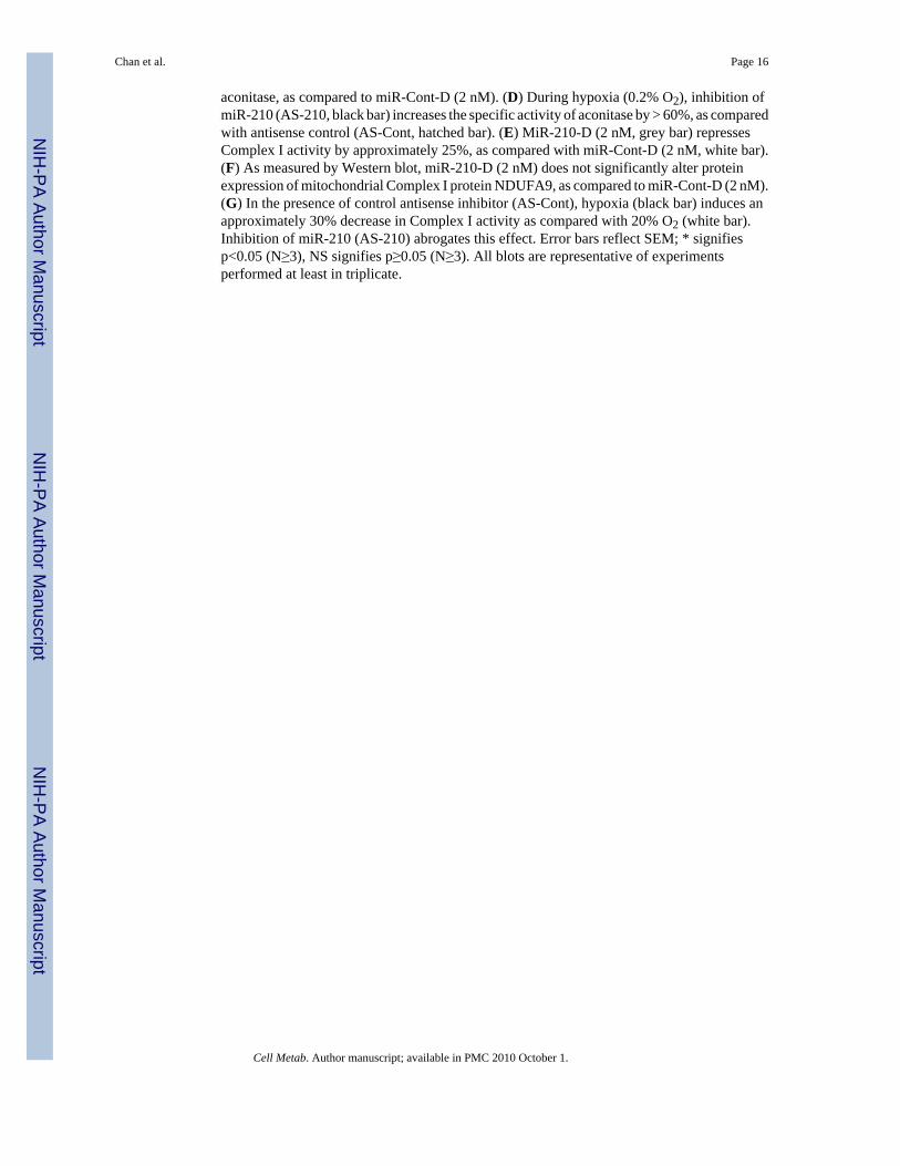
MiR-210 and ISCU1/2 Control Identical Downstream Metabolic FunctionsUnder normoxic conditions, disruption of Complex I activity and mitochondrial electrontransport leads to direct downstream functional consequences on energy production, includingdecreased oxygen consumption, decreased oxidative phosphorylation (OXPHOS), and reducedATP generation. Correspondingly, in 20% O2, transfection of miR-210-D leads to anapproximate 25% decrease in total oxygen consumption (Fig. 4A) in HPAECs and anapproximate 18% decrease in free ATP levels (Fig. 4C). The degree of alteration in theseparameters is consistent with the dependence of cultured endothelial cells on OXPHOS for aminority of total ATP production (Mann et al., 2003). In correlation, after transfection of RNAi(si-ISCU), which represses ISCU1/2 expression in HPAECs (data not shown), similarphenotypes are induced, independent of the presence of miR-210 (Fig. 4B, 4D). Thus, miR-210and ISCU1/2 control similar functions in maintaining mitochondrial respiration and energyproduction.
In addition to direct regulation of mitochondrial metabolism, disruption of the balance ofelectron transport relative to the intracellular levels of oxygen can affect apoptotic potentialand ROS production (Semenza, 2007). Under normoxia, alteration of the delivery of electronsin mitochondria, either by pharmacologic inhibitors of electron transport (i.e., rotenone)(Wolvetang et al., 1994) or by disruption of iron-sulfur clusters in the mitochondrial complexesthemselves (Napoli et al., 2006), leads to increased apoptosis and increased ROS flux.Accordingly, in 20% O2, miR-210-D increases apoptotic caspase 3,7 activity in full growthmedia (Fig. 4E), while it further increases caspase activity (Fig. 4E) and annexin V staining(Fig. 4G) to a greater extent when serum and growth factors are withheld. Furthermore,miR-210-D increases ROS flux, as reflected by increased fluorescent staining by the oxidant-sensitive fluorophore, DCFDA (Fig. 4H). Again, similar phenotypes are induced by RNAirepression of ISCU1/2 independent of miR-210 (Fig. 4F, 4H). Therefore, these multiplefunctions of miR-210 and ISCU1/2 appear convergent, being associated with perturbations iniron-sulfur-dependent aspects of mitochondrial electron transport and respiration.
The miR-210-ISCU1/2 Regulatory Pathway is Active in Transformed CellsSimilar to the results with primary endothelial cells and VHL -/- tissue, hypoxia up-regulatesmiR-210 (Fig. 1) and down-regulates ISCU1/2 in numerous transformed cell types (Supp. Fig.3A). Upon further interrogation of HEK293 cells, we found that miR-210-D represses mRNA(Supp. Fig. 3B) and protein expression (Supp. Fig. 3C) of ISCU1/2. Mirroring its effects inHPAECs, miR-210-D represses Complex I activity in HEK293 cells by over 35% (Supp. Fig.3D), and, in turn, decreases total oxygen consumption by nearly 40% (Supp. Fig. 3E).Therefore, the miR-210-ISCU1/2 axis represents a regulatory pathway shared among differingcell types, likely reflecting its fundamental importance in cellular metabolism during hypoxicstress.
Repression of ISCU1/2 is Necessary for miR-210 to Control Mitochondrial MetabolismFinally, to demonstrate that the above cellular functions controlled by miR-210 are directlycaused by repression of ISCU1/2, alteration of these phenotypes by miR-210 was assessedduring constitutive expression of ISCU1/2. To do so, separate lentiviral constructs were madethat carry either a hemagglutinin-tagged ISCUI-HA or a myc epitope-tagged ISCU2 (ISCU2-myc), but neither with an intact 3′UTR and, thus, missing a miR-210 target sequence (Tongand Rouault, 2000). While endogenous ISCU1/2 is down-regulated by miR-210-D in HPAECs,
Chan et al. Page 6
Cell Metab. Author manuscript; available in PMC 2010 October 1.
NIH
-PA Author Manuscript
NIH
-PA Author Manuscript
NIH
-PA Author Manuscript

protein expression of ISCU1-HA and ISCU2-myc appears unaffected by miR-210-D,consistent with the importance of the 3′UTR target sequence for gene repression (Fig. 5A). Incorrelation, while miR-210-D decreases both aconitase and Complex I activities in the presenceof control GFP expression, miR-210-D induces no significant change in these enzymaticactivities in the presence of constitutive expression of ISCU1-HA and ISCU2-myc (Fig. 5B-C). Conversely, while induction of miR-210 in 0.2% O2 decreases Complex I activity ascompared to 20% O2 in GFP-expressing HPAECs, hypoxia has negligible effects on ComplexI activity during constitutive expression of ISCU1-HA and ISCU2-myc (Fig. 5D). As a result,repression of ISCU1/2 is necessary and essential for down-regulating both aconitase andComplex I activity via miR-210. Furthermore, while miR-210-D decreases ATP, constitutiveexpression of ISCU1-HA and ISCU2-myc reverses this change in ATP levels (Fig. 5E).Induction of apoptotic caspase 3, 7 activity by miR-210-D is also significantly decreased byconstitutive expression of ISCU1-HA and ISCU2-myc (Fig. 5F), yet not fully reversed afterlonger periods of exposure to miR-210-D (Fig. 5G). Similar to the upstream effects on aconitaseand Complex I activity, down-regulation of ISCU function as a whole is necessary forcontrolling the downstream effects of miR-210 on repressing ATP levels and, at least in part,increasing apoptotic potential. Taken together, these data establish a causative role for ISCU1/2in miR-210-dependent actions, integral to the repression of mitochondrial respiration thatdefines the Pasteur effect.
DiscussionTeleological Significance of the miR-210-ISCU1/2 Pathway on Hypoxic Metabolic Regulation
Based on these data, we conclude that miR-210 down-regulates expression of ISCU1/2 duringhypoxia in order to repress key metabolic processes that predominate in the Pasteur effect,including mitochondrial electron transport and the TCA cycle (Fig. 6). The elucidation of themiR-210-ISCU1/2 regulatory pathway substantially clarifies the nature of the complexcrosstalk between HIF-1α and mitochondria (Seagroves et al., 2001) and complements otherHIF-dependent processes proposed to regulate the mismatch of reduced oxygen tension andmitochondrial electron transport. First, by induction of PDK1, HIF-1α decreases the activityof the TCA cycle, increases glycolysis, and reduces mitochondrial electron flux (Kim et al.,2006). Second, HIF-1α induces a COX4-1 to COX4-2 subunit switch during hypoxia toimprove the efficiency of electron transfer to oxygen at Complex IV (Fukuda et al., 2007). Bytargeting the function of iron-sulfur cluster-dependent enzymes in both the TCA cycle(aconitase) and the electron transport chain (Complex I), miR-210 and ISCU1/2 provideoverlapping control over this metabolic switch. Thus, these coordinated changes allowHIF-1α to manipulate multiple targets and ensure precise, fail-safe control over energyproduction and cellular survival during hypoxic stress (Semenza, 2007).
Identification of this pathway also indicates that disruption of iron-sulfur clusters themselvesis likely an essential feature of this metabolic transition in hypoxia. Our current in vivoknowledge of deficient iron-sulfur cluster biogenesis in mammalian cells is indirect, basedmostly on studies of frataxin, a protein that cooperates with ISCU2 in mitochondrial iron-sulfurcluster synthesis. Studies in frataxin-deficient mice (Thierbach et al., 2005) have revealed itscritical role in augmenting OXPHOS and altering iron homeostasis. In correlation, a mutationin ISCU1/2 has recently been linked to a human genetic syndrome leading to myopathy andlactic acidosis (Mochel et al., 2008). When interpreted together with the data presented here,these convergent lines of evidence indicate that not only does ISCU1/2 regulate mitochondrialrespiration and metabolism but also that such control during hypoxia likely relies uponmaintenance of iron-sulfur cluster integrity. The characterization of this type of prostheticgroup as a key determinant of this metabolic shift during hypoxia correlates with the knownfunction in bacteria of iron-sulfur clusters as intracellular sensors of oxygen (Lazazzera et al.,
Chan et al. Page 7
Cell Metab. Author manuscript; available in PMC 2010 October 1.
NIH
-PA Author Manuscript
NIH
-PA Author Manuscript
NIH
-PA Author Manuscript
1996). In mammalian cells, it is possible that a combination of acute sensitivity of iron-sulfurcluster integrity to oxidative stress (Beinert et al., 1997) along with repression of iron-sulfurcluster biogenesis via down-regulated ISCU1/2 may provide a real-time redox-sensitive“switch” that is triggered by hypoxia to induce downstream metabolic changes. As a result, aconnection linking miR-210 and ISCU1/2 to hypoxia and mitochondrial respiration is logicalfrom a teleological perspective, relying upon the well-conserved iron-sulfur cluster moiety toact as a direct switch that signals for shifts in aerobic respiration.
Although the miR-210-ISCU1/2 regulatory mechanism is active in various cell types, cellularcontext likely still influences how this pathway is utilized. For example, in contrast to non-transformed cell types, tumor cells display elevated rates of glycolysis and repressed rates ofOXPHOS during normoxia (the “Warburg effect”). It is an intriguing possibility that themiR-210-ISCU1/2 pathway may influence both the Pasteur effect as well as the Warburg effect,especially in renal tumors that constitutively express HIF-1α under normoxia. Additionally,miR-210 is specifically induced by HIF-1α but not HIF-2α (Camps et al., 2008), a secondtranscriptional regulator of the hypoxic response, which carries partial but not full redundancyin function with HIF-1α (Wang et al., 2005). As a result, miR-210 may not be up-regulated torobust levels in all tissue types, especially in those where HIF-1α expression is diminished andHIF-2α predominates (Sowter et al., 2003). In those cases, other putative HIF-dependentmiRNA may regulate iron-sulfur cluster biology. Alternatively, miR-210 may modulatemetabolic functions via alternative but synergistic mechanisms independent of ISCU1/2. Thus,it may prove worthwhile to validate other in silico predicted targets of miR-210 or HIF-inducedmiRNA that could work in concert with ISCU1/2 or its protein partners to control iron-sulfurcluster integrity for more efficient metabolism and energy production.
Additional Implications of Disrupted Iron-Sulfur Cluster Biogenesis During HypoxiaBesides mitochondrial metabolism, the miR-210-ISCU1/2 axis may regulate additionalhypoxia-dependent and iron-sulfur cluster-dependent processes. These may include ironmetabolism, which is controlled by ISCU1/2 activity (Tong and Rouault, 2006) and hypoxia(Meyron-Holtz et al., 2004), via mechanisms that have only been partially delineated.Furthermore, hypoxic repression of mitochondrial function by miR-210 and ISCU1/2 carriesthe theoretical potential to trigger mitophagy — a form of autophagy in which defectivemitochondria are selectively delivered to lysosomes for degradation. Mitophagy has beencharacterized as a HIF-dependent mechanism (Zhang et al., 2008). It remains to be seen ifmiR-210 itself induces such mitophagy and if that would lead to further adaptive ordegenerative processes.
The control of apoptosis and ROS flux by the miR-210-ISCU1/2 pathway may offer insightinto the complex processes by which ROS levels are regulated by hypoxia. It has been proposedthat hypoxia initially up-regulates mitochondrial ROS flux and consequently induces HIF-1αdue to a mismatch of electron transport and decreased oxygen partial pressure. Upon inductionof HIF-1α and HIF-dependent compensatory mechanisms, however, repression of electrontransport corrects the mismatch with reduced oxygen tension, thereby decreasing ROSproduction, decreasing apoptosis, and optimizing energy production in the hypoxic cell(Semenza, 2007). Based on this model, the repression of ISCU1/2 and iron-sulfur clusterbiogenesis by miR-210 during normoxia would create a mismatch in electron transport andoxygen concentration that leads to an increase in toxic ROS production and increased apoptosis,as corroborated by our data (Fig. 4E-H). In contrast, during hypoxia, miR-210 should inducea decrease of ROS flux by acting as a homeostatic mechanism to relieve, rather than aggravate,such a mismatch of electron transport and oxygen concentration. In corroboration, inhibitionof miR-210 in HPAECs appears to increase ROS flux after hypoxic exposure (Supp. Fig. 4A)and to decrease ATP levels as compared with antisense control (Supp. Fig. 4B), indicating an
Chan et al. Page 8
Cell Metab. Author manuscript; available in PMC 2010 October 1.
NIH
-PA Author Manuscript
NIH
-PA Author Manuscript
NIH
-PA Author Manuscript
adaptive role for this miRNA during hypoxic stress. Notably, apoptotic caspase activity (Supp.Fig. 4C) and cell viability (Supp. Fig. 4D) of HPAECs are not significantly affected byinhibiting miR-210 under these cell-culture conditions of hypoxia. Only under extreme stressof growth factor and serum deprivation in the setting of hypoxia does inhibition of miR-210induce a modest increase of caspase 3,7 signaling and a correlative decrease in cell viability(Supp. Fig. 4E-F). These subtle effects on cell survival likely reflect the activity of additionalcompensatory mechanisms to maintain cell viability, which may be especially prominent inthe HPAEC, a cell type which is exceptionally resistant to hypoxia-induced cell death(Tretyakov and Farber, 1995). Nonetheless, when interpreted with other studies demonstratingthe repression of apoptosis by miR-210 during hypoxia in other cellular contexts (Fasanaro etal., 2008; Kulshreshtha et al., 2007), these data lend credence to a model in which miR-210carries a maladaptive role during normoxia, causing a mismatch of ambient oxygen andelectron transport, but an adaptive role during hypoxia due to the homeostatic correction of theimbalance of electron transport and oxygen exposure.
Complete understanding of this action of miR-210 is nonetheless difficult. While our datasuggest a minimal change in ROS levels after hypoxic exposure of HPAECs (Supp. Fig. 4A),controversy exists as to whether hypoxia decreases or increases ROS flux in the pulmonaryvasculature, given the technical hurdles of accurately assessing these oxygen species underlow oxygen tension (Moudgil et al., 2005). Furthermore, if miR-210 alternatively regulatesiron metabolism or preferentially allows for release of free iron from modified iron-sulfurclusters during hypoxia (Beinert et al., 1997), redox-sensitive iron species may serve asadditional sources of ROS generation, independent of the activity of mitochondrial electrontransport. As a result, future and more definitive studies will be critical for defining the effectsof miR-210 on the control of ROS generation. This experimental direction will be particularlyimportant in the endothelium, owing to the importance of potential ROS-dependentphenotypes, such as permeability, thrombosis, vasomotor tone, and angiogenesis.
In conclusion, we identify miR-210 and ISCU1/2 as critical regulatory factors in hypoxia thatcontrol mitochondrial respiration and, thus, modulate a fundamental shift in cellularmetabolism. Consequently, these data should serve as a foundation for studies designed toexplore further the actions of miR-210 and other hypoxia-induced miRNA on mitochondriaand metabolism, additional functions of iron-sulfur cluster biology in hypoxia, and the morecomplex effects of miR-210 and ISCU1/2 in vivo under physiological and hypoxia-dependentdisease states.
Experimental ProceduresCells
Primary HPAECs and human PASMCs were purchased and propagated in EGM-2 andSmGM-2 media, respectively (Lonza). Experiments were performed at passages 5-9. COS7,HEK293, MCF-7, and HT-29 cells were grown in DMEM with 10% serum. HL-60 cells weregrown in RPMI1640 with 4 mM L-Glutamine and 10% serum.
Exposure to HypoxiaHPAECs were exposed for 24 hours (unless otherwise stated) either to standard non-hypoxiccell culture conditions, (20% O2, 5% CO2, with N2 balance at 37°C) or to hypoxia (0.2%-12%O2, 5% CO2, with N2 balance at 37°C), either in a modular hypoxia chamber or tissue cultureincubator. Conditions were based on studies of hypoxic exposure of HPAECs (Manalo et al.,2005) to allow for “steady state” adaptation without non-specific cell death.
Chan et al. Page 9
Cell Metab. Author manuscript; available in PMC 2010 October 1.
NIH
-PA Author Manuscript
NIH
-PA Author Manuscript
NIH
-PA Author Manuscript
RNA Extraction and RT-QPCRRNA extraction (miRNeasy kit, Qiagen) and reverse transcription of 10 ng of total RNA wereperformed to generate cDNA representing levels of either mRNA (High Fidelity cDNAAmplification Kit, Applied Biosystems) or mature miRNA molecules (MicroRNA Assay Kit,Applied Biosystems). Owing to the stem loop structures of the miRNA primers, only maturemiRNA molecules were amplified into cDNA. cDNA was amplified via fluorescently labeledTaqman probe and primer sets using an Applied Biosystems 7900HT Fast Real Time PCRdevice. Details of high-throughput RT-PCR screening of miRNA are described inSupplemental Methods.
MicroRNA Overexpression and RepressionChemically synthesized miRNA duplexes (Ambion) were transfected to mimic the mature formof miR-210, as compared with control. Oligonucleotides (Dharmacon) were transfected thatcarry specific antisense sequences to miR-210, as compared with control. RNA interferenceof ISCU1/2 expression was performed using custom designed inhibitory RNA (Stealth siRNAfrom Invitrogen).
Additional InformationSee Supplemental Methods for information on in silico miRNA target prediction, EPRspectroscopy, animal manipulation, plasmid construction, lentivirus production, transfection,immunoblotting, and standardized assays of metabolic and mitochondrial functions, ROS flux,and cell survival.
Statistical AnalysisData are presented either as a representative example of a single experiment, repeated at leastin triplicate, or as mean ± standard error of the mean for three or more experiments. Pairedsamples were compared by Student’s t test; multiple comparisons were made with a one-wayANOVA followed by a Bonferroni post hoc test. Values of p<0.05 are considered significant.
Supplementary MaterialRefer to Web version on PubMed Central for supplementary material.
AcknowledgmentsThis work was supported by MGH Cardiology NIH-T32 grant (S.Y.C.), AHA grant 0825906D (S.Y.C.), and NIHgrants HL 61795, NO1 HV 28178, PO1 81587, U54 HL 70819 (J.L., Y.Z.). No conflicts of interest are reported. Wethank the Loscalzo laboratory and R. Liao for fruitful discussions; N. Kelly (bioinformatics); W. Kaelin, J. Moslehi,and Y. Minamishima (VHL WT and -/- murine tissue); H. Ardehali (miR-210 expression vector); T. Rouault (ISCUexpression vectors); V. Mootha (technical aid in measuring oxygen consumption); S.K. Chan and J.W. Snow (criticalreading of the manuscript); and S. Tribuna (administrative assistance).
ReferencesAisenberg AC, Potter VR. Studies on the Pasteur effect. II. Specific mechanisms. J Biol Chem
1957;224:1115–1127. [PubMed: 13405938]Beinert H, Holm RH, Munck E. Iron-sulfur clusters: nature’s modular, multipurpose structures. Science
1997;277:653–659. [PubMed: 9235882]Camps C, Buffa FM, Colella S, Moore J, Sotiriou C, Sheldon H, Harris AL, Gleadle JM, Ragoussis J.
hsa-miR-210 Is induced by hypoxia and is an independent prognostic factor in breast cancer. ClinCancer Res 2008;14:1340–1348. [PubMed: 18316553]
Chavez A, Miranda LF, Pichiule P, Chavez JC. Mitochondria and hypoxia-induced gene expressionmediated by hypoxia-inducible factors. Ann N Y Acad Sci 2008;1147:312–320. [PubMed: 19076453]
Chan et al. Page 10
Cell Metab. Author manuscript; available in PMC 2010 October 1.
NIH
-PA Author Manuscript
NIH
-PA Author Manuscript
NIH
-PA Author Manuscript

Crosby ME, Kulshreshtha R, Ivan M, Glazer PM. MicroRNA Regulation of DNA Repair GeneExpression in Hypoxic Stress. Cancer Res 2009;69:1221–1229. [PubMed: 19141645]
Fasanaro P, D’Alessandra Y, Di Stefano V, Melchionna R, Romani S, Pompilio G, Capogrossi M, MartelliF. MicroRNA-210 modulates endothelial cell response to hypoxia and inhibits the receptor tyrosinekinase ligand Ephrin-A3. J Biol Chem 2008;283:15878–15883. [PubMed: 18417479]
Fukuda R, Zhang H, Kim JW, Shimoda L, Dang CV, Semenza GL. HIF-1 regulates cytochrome oxidasesubunits to optimize efficiency of respiration in hypoxic cells. Cell 2007;129:111–122. [PubMed:17418790]
Giannakakis A, Sandaltzopoulos R, Greshock J, Liang S, Huang J, Hasegawa K, Li C, O’Brien-JenkinsA, Katsaros D, Weber BL, Simon C, Coukos G, Zhang L. miR-210 links hypoxia with cell cycleregulation and is deleted in human epithelial ovarian cancer. Cancer Biol Ther 2008;7:255–264.[PubMed: 18059191]
Ginouves A, Ilc K, Macias N, Pouyssegur J, Berra E. PHDs overactivation during chronic hypoxia“desensitizes” HIFalpha and protects cells from necrosis. Proc Natl Acad Sci U S A 2008;105:4745–4750. [PubMed: 18347341]
Haase VH, Glickman JN, Socolovsky M, Jaenisch R. Vascular tumors in livers with targeted inactivationof the von Hippel-Lindau tumor suppressor. Proc Natl Acad Sci U S A 2001;98:1583–1588.[PubMed: 11171994]
Hinchliffe P, Sazanov LA. Organization of iron-sulfur clusters in respiratory complex I. Science2005;309:771–774. [PubMed: 16051796]
Ivan M, Harris AL, Martelli F, Kulshreshtha R. Hypoxia response and microRNAs: no longer two separateworlds. J Cell Mol Med 2008;12:1426–1431. [PubMed: 18624759]
Kim JW, Tchernyshyov I, Semenza GL, Dang CV. HIF-1-mediated expression of pyruvatedehydrogenase kinase: a metabolic switch required for cellular adaptation to hypoxia. Cell Metab2006;3:177–185. [PubMed: 16517405]
Kulshreshtha R, Davuluri RV, Calin GA, Ivan M. A microRNA component of the hypoxic response. CellDeath Differ 2008;15:667–671. [PubMed: 18219318]
Kulshreshtha R, Ferracin M, Wojcik S, Garzon R, Alder H, Agosto-Perez F, Davuluri R, Liu C, CroceC, Negrini M, Calin G, Ivan M. A microRNA signature of hypoxia. Mol Cell Biol 2007;27:1859–1867. [PubMed: 17194750]
Lazazzera BA, Beinert H, Khoroshilova N, Kennedy MC, Kiley PJ. DNA binding and dimerization ofthe Fe-S-containing FNR protein from Escherichia coli are regulated by oxygen. J Biol Chem1996;271:2762–2768. [PubMed: 8576252]
Manalo D, Rowan A, Lavoie T, Natarajan L, Kelly B, Ye S, Garcia J, Semenza G. Transcriptionalregulation of vascular endothelial cell responses to hypoxia by HIF-1. Blood 2005;105:659–669.[PubMed: 15374877]
Mann GE, Yudilevich DL, Sobrevia L. Regulation of amino acid and glucose transporters in endothelialand smooth muscle cells. Physiol Rev 2003;83:183–252. [PubMed: 12506130]
Meyron-Holtz E, Ghosh M, Rouault T. Mammalian tissue oxygen levels modulate iron-regulatory proteinactivities in vivo. Science 2004;306:2087–2090. [PubMed: 15604406]
Minamishima YA, Moslehi J, Bardeesy N, Cullen D, Bronson RT, Kaelin WG Jr. Somatic inactivationof the PHD2 prolyl hydroxylase causes polycythemia and congestive heart failure. Blood2008;111:3236–3244. [PubMed: 18096761]
Mochel F, Knight M, Tong W, Hernandez D, Ayyad K, Taivassalo T, Andersen P, Singleton A, RouaultT, Fischbeck K, Haller R. Splice mutation in the iron-sulfur cluster scaffold protein ISCU causesmyopathy with exercise intolerance. Am J Hum Genet 2008;82:652–660. [PubMed: 18304497]
Moudgil R, Michelakis E, Archer S. Hypoxic pulmonary vasoconstriction. J Applied Physiol2005;98:390–403. [PubMed: 15591309]
Napoli E, Taroni F, Cortopassi GA. Frataxin, iron-sulfur clusters, heme, ROS, and aging. Antioxid RedoxSignal 2006;8:506–516. [PubMed: 16677095]
Rouault TA, Tong WH. Iron-sulfur cluster biogenesis and human disease. Trends Genet 2008;24:398–407. [PubMed: 18606475]
Chan et al. Page 11
Cell Metab. Author manuscript; available in PMC 2010 October 1.
NIH
-PA Author Manuscript
NIH
-PA Author Manuscript
NIH
-PA Author Manuscript
Seagroves TN, Ryan HE, Lu H, Wouters BG, Knapp M, Thibault P, Laderoute K, Johnson RS.Transcription factor HIF-1 is a necessary mediator of the pasteur effect in mammalian cells. Mol CellBiol 2001;21:3436–3444. [PubMed: 11313469]
Semenza G. Oxygen-dependent regulation of mitochondrial respiration by hypoxia-inducible factor 1.Biochem J 2007;405:1–9. [PubMed: 17555402]
Sowter HM, Raval RR, Moore JW, Ratcliffe PJ, Harris AL. Predominant role of hypoxia-inducibletranscription factor (Hif)-1alpha versus Hif-2alpha in regulation of the transcriptional response tohypoxia. Cancer Res 2003;63:6130–6134. [PubMed: 14559790]
Stenmark KR, Fagan KA, Frid MG. Hypoxia-induced pulmonary vascular remodeling: cellular andmolecular mechanisms. Circ Res 2006;99:675–691. [PubMed: 17008597]
Thierbach R, Schulz T, Isken F, Voigt A, Mietzner B, Drewes G, von Kleist-Retzow J, Wiesner R,Magnuson M, Puccio H, Pfeiffer A, Steinberg P, Ristow M. Targeted disruption of hepatic frataxinexpression causes impaired mitochondrial function, decreased life span and tumor growth in mice.Human Mol Genet 2005;14:3857–3864. [PubMed: 16278235]
Tong WH, Rouault T. Distinct iron-sulfur cluster assembly complexes exist in the cytosol andmitochondria of human cells. EMBO J 2000;19:5692–5700. [PubMed: 11060020]
Tong WH, Rouault TA. Functions of mitochondrial ISCU and cytosolic ISCU in mammalian iron-sulfurcluster biogenesis and iron homeostasis. Cell Metab 2006;3:199–210. [PubMed: 16517407]
Tretyakov AV, Farber HW. Endothelial cell tolerance to hypoxia. Potential role of purine nucleotidephosphates. J Clin Invest 1995;95:738–744. [PubMed: 7860755]
Wang V, Davis DA, Haque M, Huang LE, Yarchoan R. Differential gene up-regulation by hypoxia-inducible factor-1alpha and hypoxia-inducible factor-2alpha in HEK293T cells. Cancer Res2005;65:3299–3306. [PubMed: 15833863]
Wolvetang E, Johnson K, Krauer K, Ralph S, Linnane A. Mitochondrial respiratory chain inhibitorsinduce apoptosis. FEBS Lett 1994;339:40–44. [PubMed: 8313978]
Young AP, Schlisio S, Minamishima YA, Zhang Q, Li L, Grisanzio C, Signoretti S, Kaelin WG Jr. VHLloss actuates a HIF-independent senescence programme mediated by Rb and p400. Nat Cell Biol2008;10:361–369. [PubMed: 18297059]
Zhang H, Bosch-Marce M, Shimoda LA, Tan YS, Baek JH, Wesley JB, Gonzalez FJ, Semenza GL.Mitochondrial autophagy is an HIF-1-dependent adaptive metabolic response to hypoxia. J BiolChem 2008;283:10892–10903. [PubMed: 18281291]
Chan et al. Page 12
Cell Metab. Author manuscript; available in PMC 2010 October 1.
NIH
-PA Author Manuscript
NIH
-PA Author Manuscript
NIH
-PA Author Manuscript
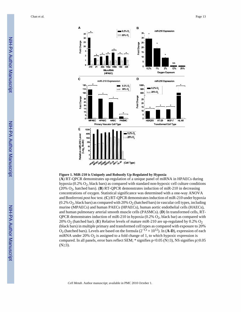
Figure 1. MiR-210 is Uniquely and Robustly Up-Regulated by Hypoxia(A) RT-QPCR demonstrates up-regulation of a unique panel of miRNA in HPAECs duringhypoxia (0.2% O2, black bars) as compared with standard non-hypoxic cell culture conditions(20% O2, hatched bars). (B) RT-QPCR demonstrates induction of miR-210 in decreasingconcentrations of oxygen. Statistical significance was determined with a one-way ANOVAand Bonferroni post hoc test. (C) RT-QPCR demonstrates induction of miR-210 under hypoxia(0.2% O2, black bars) as compared with 20% O2 (hatched bars) in vascular cell types, includingmurine (MPAECs) and human PAECs (HPAECs), human aortic endothelial cells (HAECs),and human pulmonary arterial smooth muscle cells (PASMCs). (D) In transformed cells, RT-QPCR demonstrates induction of miR-210 in hypoxia (0.2% O2, black bar) as compared with20% O2 (hatched bar). (E) Relative levels of mature miR-210 are up-regulated by 0.2% O2(black bars) in multiple primary and transformed cell types as compared with exposure to 20%O2 (hatched bars). Levels are based on the formula (2−Ct × 1012). In (A-D), expression of eachmiRNA under 20% O2 is assigned to a fold change of 1, to which hypoxic expression iscompared. In all panels, error bars reflect SEM; * signifies p<0.05 (N≥3), NS signifies p≥0.05(N≥3).
Chan et al. Page 13
Cell Metab. Author manuscript; available in PMC 2010 October 1.
NIH
-PA Author Manuscript
NIH
-PA Author Manuscript
NIH
-PA Author Manuscript
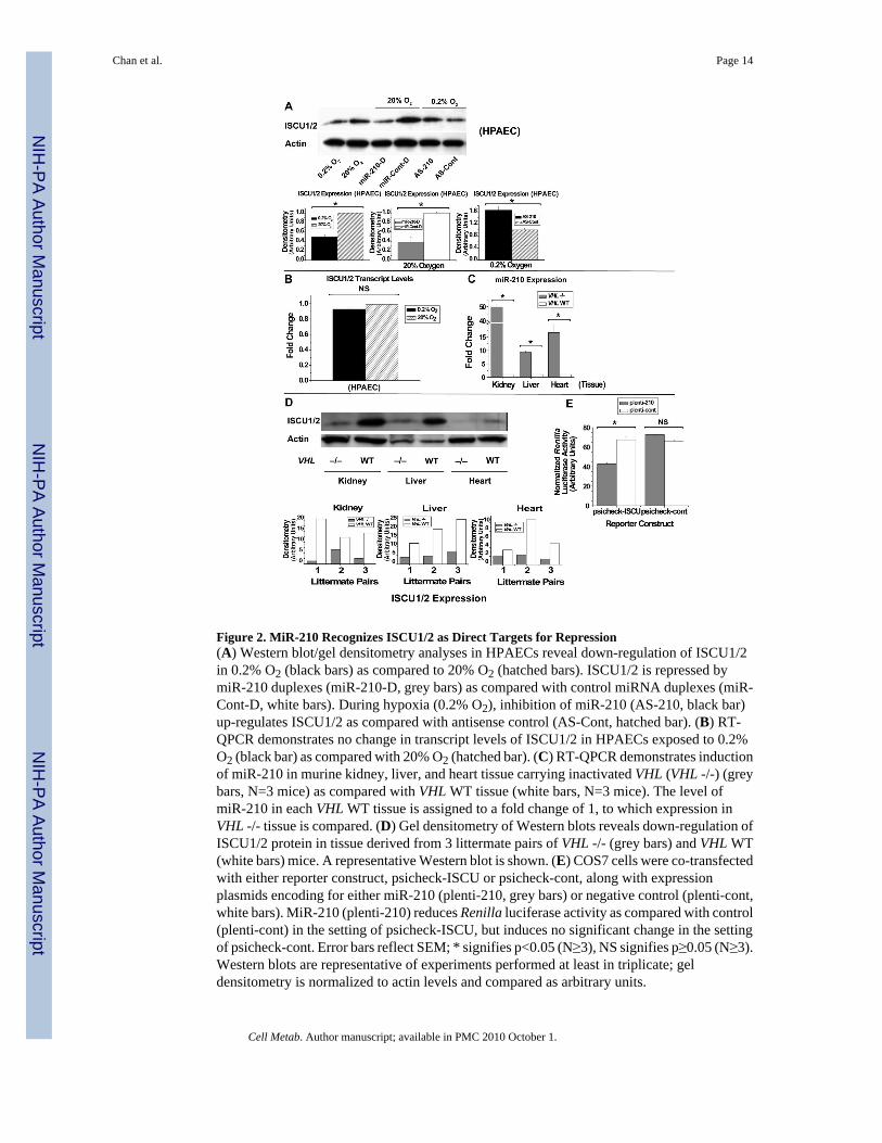
Figure 2. MiR-210 Recognizes ISCU1/2 as Direct Targets for Repression(A) Western blot/gel densitometry analyses in HPAECs reveal down-regulation of ISCU1/2in 0.2% O2 (black bars) as compared to 20% O2 (hatched bars). ISCU1/2 is repressed bymiR-210 duplexes (miR-210-D, grey bars) as compared with control miRNA duplexes (miR-Cont-D, white bars). During hypoxia (0.2% O2), inhibition of miR-210 (AS-210, black bar)up-regulates ISCU1/2 as compared with antisense control (AS-Cont, hatched bar). (B) RT-QPCR demonstrates no change in transcript levels of ISCU1/2 in HPAECs exposed to 0.2%O2 (black bar) as compared with 20% O2 (hatched bar). (C) RT-QPCR demonstrates inductionof miR-210 in murine kidney, liver, and heart tissue carrying inactivated VHL (VHL -/-) (greybars, N=3 mice) as compared with VHL WT tissue (white bars, N=3 mice). The level ofmiR-210 in each VHL WT tissue is assigned to a fold change of 1, to which expression inVHL -/- tissue is compared. (D) Gel densitometry of Western blots reveals down-regulation ofISCU1/2 protein in tissue derived from 3 littermate pairs of VHL -/- (grey bars) and VHL WT(white bars) mice. A representative Western blot is shown. (E) COS7 cells were co-transfectedwith either reporter construct, psicheck-ISCU or psicheck-cont, along with expressionplasmids encoding for either miR-210 (plenti-210, grey bars) or negative control (plenti-cont,white bars). MiR-210 (plenti-210) reduces Renilla luciferase activity as compared with control(plenti-cont) in the setting of psicheck-ISCU, but induces no significant change in the settingof psicheck-cont. Error bars reflect SEM; * signifies p<0.05 (N≥3), NS signifies p≥0.05 (N≥3).Western blots are representative of experiments performed at least in triplicate; geldensitometry is normalized to actin levels and compared as arbitrary units.
Chan et al. Page 14
Cell Metab. Author manuscript; available in PMC 2010 October 1.
NIH
-PA Author Manuscript
NIH
-PA Author Manuscript
NIH
-PA Author Manuscript
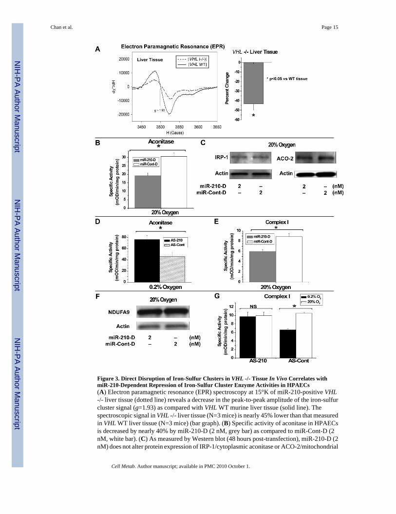
Figure 3. Direct Disruption of Iron-Sulfur Clusters in VHL -/- Tissue In Vivo Correlates withmiR-210-Dependent Repression of Iron-Sulfur Cluster Enzyme Activities in HPAECs(A) Electron paramagnetic resonance (EPR) spectroscopy at 15°K of miR-210-positive VHL-/- liver tissue (dotted line) reveals a decrease in the peak-to-peak amplitude of the iron-sulfurcluster signal (g=1.93) as compared with VHL WT murine liver tissue (solid line). Thespectroscopic signal in VHL -/- liver tissue (N=3 mice) is nearly 45% lower than that measuredin VHL WT liver tissue (N=3 mice) (bar graph). (B) Specific activity of aconitase in HPAECsis decreased by nearly 40% by miR-210-D (2 nM, grey bar) as compared to miR-Cont-D (2nM, white bar). (C) As measured by Western blot (48 hours post-transfection), miR-210-D (2nM) does not alter protein expression of IRP-1/cytoplasmic aconitase or ACO-2/mitochondrial
Chan et al. Page 15
Cell Metab. Author manuscript; available in PMC 2010 October 1.
NIH
-PA Author Manuscript
NIH
-PA Author Manuscript
NIH
-PA Author Manuscript
aconitase, as compared to miR-Cont-D (2 nM). (D) During hypoxia (0.2% O2), inhibition ofmiR-210 (AS-210, black bar) increases the specific activity of aconitase by > 60%, as comparedwith antisense control (AS-Cont, hatched bar). (E) MiR-210-D (2 nM, grey bar) repressesComplex I activity by approximately 25%, as compared with miR-Cont-D (2 nM, white bar).(F) As measured by Western blot, miR-210-D (2 nM) does not significantly alter proteinexpression of mitochondrial Complex I protein NDUFA9, as compared to miR-Cont-D (2 nM).(G) In the presence of control antisense inhibitor (AS-Cont), hypoxia (black bar) induces anapproximately 30% decrease in Complex I activity as compared with 20% O2 (white bar).Inhibition of miR-210 (AS-210) abrogates this effect. Error bars reflect SEM; * signifiesp<0.05 (N≥3), NS signifies p≥0.05 (N≥3). All blots are representative of experimentsperformed at least in triplicate.
Chan et al. Page 16
Cell Metab. Author manuscript; available in PMC 2010 October 1.
NIH
-PA Author Manuscript
NIH
-PA Author Manuscript
NIH
-PA Author Manuscript
Figure 4. MiR-210 and ISCU1/2 Control Identical Mitochondrial Metabolic Functions in HPAECs(A) MiR-210-D (2 nM, grey bar) represses oxygen consumption as compared with miR-Cont-D (2 nM, white bar) by nearly 25%. (B) Inhibitory RNA recognizing ISCU1/2 (si-ISCU, 40nM) represses oxygen consumption by nearly 30% as compared with inhibitory RNA control(si-Cont, 40 nM). (C) MiR-210-D (2 nM, grey bar) down-regulates free levels of ATP byapproximately 18%. (D) Inhibitory si-ISCU (40 nM) down-regulates free ATP levels by >40%. (E) MiR-210-D (2 nM, grey bar) induces apoptotic caspase 3,7 activity by >5-foldcompared with miR-Cont-D in HPAECs cultured in full growth media; similarly, miR-210-Dincreases caspase 3,7 activity by >5-fold compared with miR-Cont-D in basal media (withoutadded growth factors or serum) and by >50 fold compared with miR-210-D in full growth
Chan et al. Page 17
Cell Metab. Author manuscript; available in PMC 2010 October 1.
NIH
-PA Author Manuscript
NIH
-PA Author Manuscript
NIH
-PA Author Manuscript

media. (F) Inhibitory si-ISCU (40 nM) up-regulates caspase 3,7 activity by >2-fold under fullgrowth media. (G) When cultured in basal media and exposed to annexin V-fluorescein,miR-210 (2 nM) increases fluorescent staining in HPAECs as compared with miR-Cont-D (2nM). (H) MiR-210-D (2 nM) increases fluorescent staining in HPAECs by the oxidant-sensitive fluorophore DCFDA, as compared with miR-Cont-D (2 nM). Transfection of si-ISCU(40 nM) also increases fluorescent staining by DCFDA, as compared with si-Cont (40 nM).All fluorescent mages were acquired under a green filter (516 nm). Error bars reflect SEM; *signifies p<0.05 (N≥3), NS signifies p≥0.05 (N≥3).
Chan et al. Page 18
Cell Metab. Author manuscript; available in PMC 2010 October 1.
NIH
-PA Author Manuscript
NIH
-PA Author Manuscript
NIH
-PA Author Manuscript
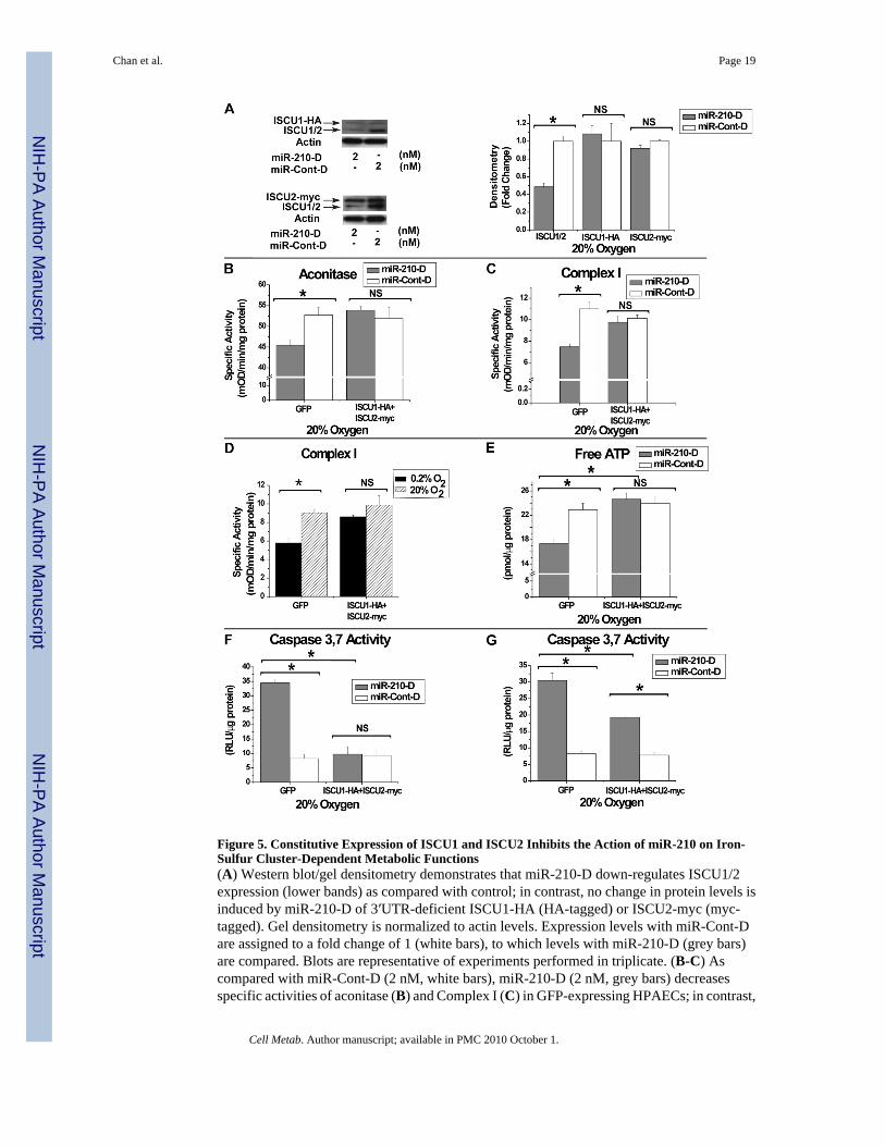
Figure 5. Constitutive Expression of ISCU1 and ISCU2 Inhibits the Action of miR-210 on Iron-Sulfur Cluster-Dependent Metabolic Functions(A) Western blot/gel densitometry demonstrates that miR-210-D down-regulates ISCU1/2expression (lower bands) as compared with control; in contrast, no change in protein levels isinduced by miR-210-D of 3′UTR-deficient ISCU1-HA (HA-tagged) or ISCU2-myc (myc-tagged). Gel densitometry is normalized to actin levels. Expression levels with miR-Cont-Dare assigned to a fold change of 1 (white bars), to which levels with miR-210-D (grey bars)are compared. Blots are representative of experiments performed in triplicate. (B-C) Ascompared with miR-Cont-D (2 nM, white bars), miR-210-D (2 nM, grey bars) decreasesspecific activities of aconitase (B) and Complex I (C) in GFP-expressing HPAECs; in contrast,
Chan et al. Page 19
Cell Metab. Author manuscript; available in PMC 2010 October 1.
NIH
-PA Author Manuscript
NIH
-PA Author Manuscript
NIH
-PA Author Manuscript
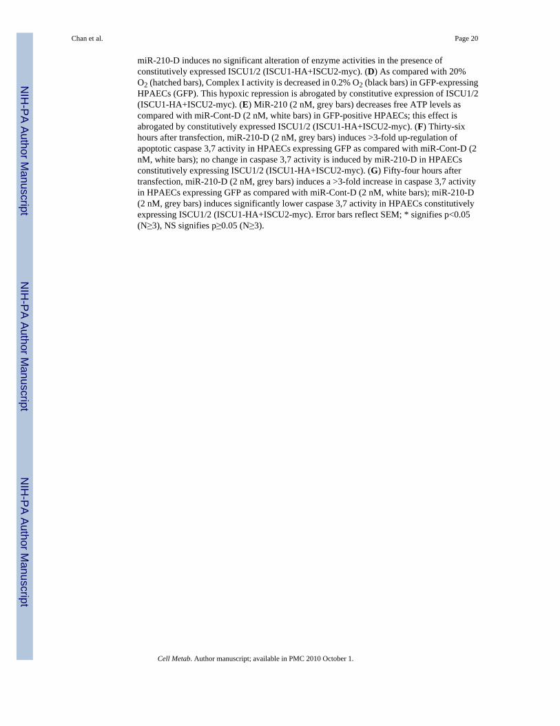
miR-210-D induces no significant alteration of enzyme activities in the presence ofconstitutively expressed ISCU1/2 (ISCU1-HA+ISCU2-myc). (D) As compared with 20%O2 (hatched bars), Complex I activity is decreased in 0.2% O2 (black bars) in GFP-expressingHPAECs (GFP). This hypoxic repression is abrogated by constitutive expression of ISCU1/2(ISCU1-HA+ISCU2-myc). (E) MiR-210 (2 nM, grey bars) decreases free ATP levels ascompared with miR-Cont-D (2 nM, white bars) in GFP-positive HPAECs; this effect isabrogated by constitutively expressed ISCU1/2 (ISCU1-HA+ISCU2-myc). (F) Thirty-sixhours after transfection, miR-210-D (2 nM, grey bars) induces >3-fold up-regulation ofapoptotic caspase 3,7 activity in HPAECs expressing GFP as compared with miR-Cont-D (2nM, white bars); no change in caspase 3,7 activity is induced by miR-210-D in HPAECsconstitutively expressing ISCU1/2 (ISCU1-HA+ISCU2-myc). (G) Fifty-four hours aftertransfection, miR-210-D (2 nM, grey bars) induces a >3-fold increase in caspase 3,7 activityin HPAECs expressing GFP as compared with miR-Cont-D (2 nM, white bars); miR-210-D(2 nM, grey bars) induces significantly lower caspase 3,7 activity in HPAECs constitutivelyexpressing ISCU1/2 (ISCU1-HA+ISCU2-myc). Error bars reflect SEM; * signifies p<0.05(N≥3), NS signifies p≥0.05 (N≥3).
Chan et al. Page 20
Cell Metab. Author manuscript; available in PMC 2010 October 1.
NIH
-PA Author Manuscript
NIH
-PA Author Manuscript
NIH
-PA Author Manuscript
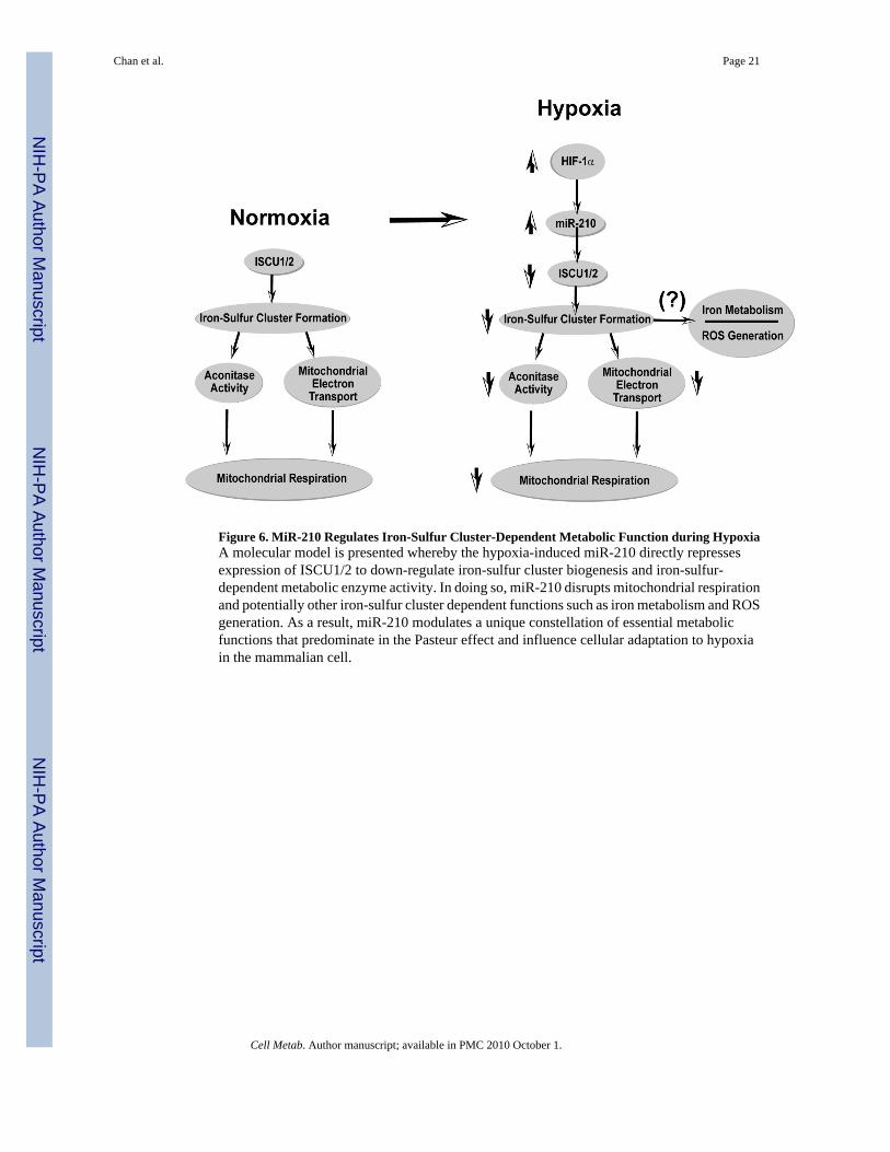
Figure 6. MiR-210 Regulates Iron-Sulfur Cluster-Dependent Metabolic Function during HypoxiaA molecular model is presented whereby the hypoxia-induced miR-210 directly repressesexpression of ISCU1/2 to down-regulate iron-sulfur cluster biogenesis and iron-sulfur-dependent metabolic enzyme activity. In doing so, miR-210 disrupts mitochondrial respirationand potentially other iron-sulfur cluster dependent functions such as iron metabolism and ROSgeneration. As a result, miR-210 modulates a unique constellation of essential metabolicfunctions that predominate in the Pasteur effect and influence cellular adaptation to hypoxiain the mammalian cell.
Chan et al. Page 21
Cell Metab. Author manuscript; available in PMC 2010 October 1.
NIH
-PA Author Manuscript
NIH
-PA Author Manuscript
NIH
-PA Author Manuscript