Nitric oxide permits hypoxia-induced lymphatic perfusion by controlling arterial-lymphatic conduits...
12
Nitric oxide permits hypoxia-induced lymphatic perfusion by controlling arterial-lymphatic conduits in zebrafish and glass catfish Lasse Dahl Ejby Jensen a , Renhai Cao a , Eva-Maria Hedlund a , Iris So ¨ ll b , Jon O. Lundberg c , Giselbert Hauptmann b , John Fleng Steffensen d , and Yihai Cao a,1 Departments of a Microbiology, Tumor and Cell Biology and c Physiology and Pharmacology, Karolinska Institute, SE-171 77 Stockholm, Sweden; b Department of Life Sciences, So ¨ derto ¨ rn University and BioNut, Karolinska Institute, Huddinge, Sweden; and d Marine Biological Laboratory, University of Copenhagen, Strandpromenaden 5, Helsingor, DK-3000, Denmark Edited by Robert Langer, Massachusetts Institute of Technology, Cambridge, MA, and approved September 4, 2009 (received for review July 9, 2009) The blood and lymphatic vasculatures are structurally and func- tionally coupled in controlling tissue perfusion, extracellular inter- stitial fluids, and immune surveillance. Little is known, however, about the molecular mechanisms that underlie the regulation of bloodlymphatic vessel connections and lymphatic perfusion. Here we show in the adult zebrafish and glass catfish (Kryptopterus bicirrhis) that blood-lymphatic conduits directly connect arterial vessels to the lymphatic system. Under hypoxic conditions, arterial- lymphatic conduits (ALCs) became highly dilated and linearized by NO-induced vascular relaxation, which led to blood perfusion into the lymphatic system. NO blockage almost completely abrogated hypoxia-induced ALC relaxation and lymphatic perfusion. These findings uncover mechanisms underlying hypoxia-induced oxygen compensation by perfusion of existing lymphatics in fish. Our results might also imply that the hypoxia-induced NO pathway contributes to development of progression of pathologies, includ- ing promotion of lymphatic metastasis by modulating arterial- lymphatic conduits, in the mammalian system. blood vessel lymphatic vessel T he physiological function of the lymphatic vascular network is thought to be the draining of extravasated fluids and macromolecules to regional lymph nodes for immune surveil- lance and to transport back to the blood vessels for circulation (1–3). Emerging evidence has shown that in mammals and zebrafish, the lymphatic system originates from the cardinal vein during embryonic development and subsequently segregates from the blood vasculature (3–6). Several recent studies have demonstrated that capillaries and microlymphatic vessels remain connected in the adult (7–10). The structural features and molecular players that operate blood-lymphatic conduits (BLCs), as well as their biological functions in adult organisms, have not yet been described. Dysfunction of the lymphatic system could lead to disease progression and tissue edema. For example, a variety of solid tumor produce multiple lymphangiogenic factors that induce intratumoral lymphangiogenesis or dilation of peritumor lym- phatics, which significantly contribute to lymphatic metastasis (3, 11). Importantly, tumor hypoxia could further elevate the expression levels of lymangiogenic factors such as VEGF-A and VEGF-C to further facilitate the metastatic cascade of malig- nant diseases (12, 13). It is known that malignant cells could be further spread from the blood stream into the lymphatic system (3, 14). However, the molecular mechanisms by which tumor cell dissemination from the circulation into the lymphatics remain poorly understood. In this study, we examined the physical connections between the lymphatic system and arterial vessels in zebrafish and Kryptopterus bicirrhis. We assessed the effects of hypoxia on these connections and considered how these connections may play a role in cancer-related metastasis. Results Characterization of the Thoracic Duct Lymphatic Vessel in Adult Zebrafish and K. bicirrhis. To characterize the lymphatic system in both zebrafish and K. bicirrhis, the tail regions of adult fish were histologically analyzed. Thoracic ducts (TDs) were readily de- tectable in both species (Fig. S1). Coexistence of the dorsal aorta, the cardinal vein, and the TD in zebrafish was further validated using fli1:EGFP (enhanced green f luorescent protein) transgenic zebrafish (15), in which both blood vessel endothelial cells (BVECs) and lymphatic endothelial cells (LECs) express EGFP (Fig. S1 B and D). Electron microscopy examination further showed that the TD consisted of a single thin layer of LECs (Fig. S1C). TD also exhibited positive staining for prox-1, which is specifically expressed in LECs (16) (Fig. S1D). To further characterize the lymphatic system in fish, we chose K. bicirrhis (17), which is completely transparent and allowed us to non-invasively study lymphatic perfusion in adult fish (Fig. S1 E). Similar to zebrafish, K. bicirrhis also had TD that specifically expressed prox-1 (Fig. S1 F–H) and lacked blood cells (Fig. S1H). The anatomical relationships of the dorsal aorta (DA), posterior cardinal vein (PCV), TD, and the descending branches in K. bicirrhis are schematically shown (Fig. S1I). These findings demonstrate that similar lymphatic systems exist in zebrafish and K. bicirrhis. Hypoxia Induces Blood Perfusion in the Lymphatics of the Zebrafish Tailfin. The functional relationship between the blood vascula- ture and the lymphatic system has not been previously studied. Tissue hypoxia is known to alter angiogenesis, vascular archi- tecture, vascular permeability, and blood perfusion (18–20). To study blood perfusion and the relationship between the mi- crovessels of blood and lymphatic vasculatures, we chose the zebrafish tailfin for further characterization. Similar to fli1:EGFP fish strain, the VEGFR2:EGFP fish strain was gener- ated to express EGFP under VEGFR2 promoter and vascular endothelial cells primarily express EGFP (21). Intriguingly, fli1:EGFP and VEGFR2:EGFP transgenic strains of zebrafish had overlapping but distinct patterns of EGFP expression in the vasculature (Fig. 1A). The VEGFR2:EGFP strain displayed a relatively restricted staining pattern, consistent with the re- stricted expression of VEGFR2 in BVECs but not in LECs. In Author contributions: L.D.E.J., R.C., J.O.L., J.F.S., and Y.C. designed research; L.D.E.J. and R.C. performed research; E.-M.H., I.S., J.O.L., G.H., J.F.S., and Y.C. contributed new reagents/ analytic tools; L.D.E.J., R.C., J.F.S., and Y.C. analyzed data; and L.D.E.J., J.F.S., and Y.C. wrote the paper. The authors declare no conflict of interest. This article is a PNAS Direct Submission. 1 To whom correspondence should be addressed. E-mail: [email protected]. This article contains supporting information online at www.pnas.org/cgi/content/full/ 0907608106/DCSupplemental. 18408 –18413 PNAS October 27, 2009 vol. 106 no. 43 www.pnas.orgcgidoi10.1073pnas.0907608106
Transcript of Nitric oxide permits hypoxia-induced lymphatic perfusion by controlling arterial-lymphatic conduits...

Nitric oxide permits hypoxia-induced lymphaticperfusion by controlling arterial-lymphatic conduitsin zebrafish and glass catfishLasse Dahl Ejby Jensena, Renhai Caoa, Eva-Maria Hedlunda, Iris Sollb, Jon O. Lundbergc, Giselbert Hauptmannb,John Fleng Steffensend, and Yihai Caoa,1
Departments of aMicrobiology, Tumor and Cell Biology and cPhysiology and Pharmacology, Karolinska Institute, SE-171 77 Stockholm, Sweden; bDepartmentof Life Sciences, Sodertorn University and BioNut, Karolinska Institute, Huddinge, Sweden; and dMarine Biological Laboratory, University of Copenhagen,Strandpromenaden 5, Helsingor, DK-3000, Denmark
Edited by Robert Langer, Massachusetts Institute of Technology, Cambridge, MA, and approved September 4, 2009 (received for review July 9, 2009)
The blood and lymphatic vasculatures are structurally and func-tionally coupled in controlling tissue perfusion, extracellular inter-stitial fluids, and immune surveillance. Little is known, however,about the molecular mechanisms that underlie the regulation ofbloodlymphatic vessel connections and lymphatic perfusion. Herewe show in the adult zebrafish and glass catfish (Kryptopterusbicirrhis) that blood-lymphatic conduits directly connect arterialvessels to the lymphatic system. Under hypoxic conditions, arterial-lymphatic conduits (ALCs) became highly dilated and linearized byNO-induced vascular relaxation, which led to blood perfusion intothe lymphatic system. NO blockage almost completely abrogatedhypoxia-induced ALC relaxation and lymphatic perfusion. Thesefindings uncover mechanisms underlying hypoxia-induced oxygencompensation by perfusion of existing lymphatics in fish. Ourresults might also imply that the hypoxia-induced NO pathwaycontributes to development of progression of pathologies, includ-ing promotion of lymphatic metastasis by modulating arterial-lymphatic conduits, in the mammalian system.
blood vessel � lymphatic vessel
The physiological function of the lymphatic vascular networkis thought to be the draining of extravasated fluids and
macromolecules to regional lymph nodes for immune surveil-lance and to transport back to the blood vessels for circulation(1–3). Emerging evidence has shown that in mammals andzebrafish, the lymphatic system originates from the cardinal veinduring embryonic development and subsequently segregatesfrom the blood vasculature (3–6). Several recent studies havedemonstrated that capillaries and microlymphatic vessels remainconnected in the adult (7–10). The structural features andmolecular players that operate blood-lymphatic conduits(BLCs), as well as their biological functions in adult organisms,have not yet been described.
Dysfunction of the lymphatic system could lead to diseaseprogression and tissue edema. For example, a variety of solidtumor produce multiple lymphangiogenic factors that induceintratumoral lymphangiogenesis or dilation of peritumor lym-phatics, which significantly contribute to lymphatic metastasis(3, 11). Importantly, tumor hypoxia could further elevate theexpression levels of lymangiogenic factors such as VEGF-A andVEGF-C to further facilitate the metastatic cascade of malig-nant diseases (12, 13). It is known that malignant cells could befurther spread from the blood stream into the lymphatic system(3, 14). However, the molecular mechanisms by which tumor celldissemination from the circulation into the lymphatics remainpoorly understood.
In this study, we examined the physical connections betweenthe lymphatic system and arterial vessels in zebrafish andKryptopterus bicirrhis. We assessed the effects of hypoxia onthese connections and considered how these connections mayplay a role in cancer-related metastasis.
ResultsCharacterization of the Thoracic Duct Lymphatic Vessel in AdultZebrafish and K. bicirrhis. To characterize the lymphatic system inboth zebrafish and K. bicirrhis, the tail regions of adult fish werehistologically analyzed. Thoracic ducts (TDs) were readily de-tectable in both species (Fig. S1). Coexistence of the dorsalaorta, the cardinal vein, and the TD in zebrafish was furthervalidated using fli1:EGFP (enhanced green fluorescent protein)transgenic zebrafish (15), in which both blood vessel endothelialcells (BVECs) and lymphatic endothelial cells (LECs) expressEGFP (Fig. S1 B and D). Electron microscopy examinationfurther showed that the TD consisted of a single thin layer ofLECs (Fig. S1C). TD also exhibited positive staining for prox-1,which is specifically expressed in LECs (16) (Fig. S1D). Tofurther characterize the lymphatic system in fish, we chose K.bicirrhis (17), which is completely transparent and allowed us tonon-invasively study lymphatic perfusion in adult fish (Fig. S1E).Similar to zebrafish, K. bicirrhis also had TD that specificallyexpressed prox-1 (Fig. S1 F–H) and lacked blood cells (Fig. S1H).The anatomical relationships of the dorsal aorta (DA), posteriorcardinal vein (PCV), TD, and the descending branches in K.bicirrhis are schematically shown (Fig. S1I). These findingsdemonstrate that similar lymphatic systems exist in zebrafish andK. bicirrhis.
Hypoxia Induces Blood Perfusion in the Lymphatics of the ZebrafishTailfin. The functional relationship between the blood vascula-ture and the lymphatic system has not been previously studied.Tissue hypoxia is known to alter angiogenesis, vascular archi-tecture, vascular permeability, and blood perfusion (18–20). Tostudy blood perfusion and the relationship between the mi-crovessels of blood and lymphatic vasculatures, we chose thezebrafish tailfin for further characterization. Similar tofli1:EGFP fish strain, the VEGFR2:EGFP fish strain was gener-ated to express EGFP under VEGFR2 promoter and vascularendothelial cells primarily express EGFP (21). Intriguingly,fli1:EGFP and VEGFR2:EGFP transgenic strains of zebrafishhad overlapping but distinct patterns of EGFP expression in thevasculature (Fig. 1A). The VEGFR2:EGFP strain displayed arelatively restricted staining pattern, consistent with the re-stricted expression of VEGFR2 in BVECs but not in LECs. In
Author contributions: L.D.E.J., R.C., J.O.L., J.F.S., and Y.C. designed research; L.D.E.J. andR.C. performed research; E.-M.H., I.S., J.O.L., G.H., J.F.S., and Y.C. contributed new reagents/analytic tools; L.D.E.J., R.C., J.F.S., and Y.C. analyzed data; and L.D.E.J., J.F.S., and Y.C. wrotethe paper.
The authors declare no conflict of interest.
This article is a PNAS Direct Submission.
1To whom correspondence should be addressed. E-mail: [email protected].
This article contains supporting information online at www.pnas.org/cgi/content/full/0907608106/DCSupplemental.
18408–18413 � PNAS � October 27, 2009 � vol. 106 � no. 43 www.pnas.org�cgi�doi�10.1073�pnas.0907608106

addition, prox-1-positive lymphatic vessels in the tailfin were alsoEGFP positive in the fli1:EGFP strain (Fig. 1B).
Under hypoxic conditions, the prox-1�/VEGFR2- lymphaticvessels in both proximal and distal regions of the tailfin becameperfused with blood cells (Fig. 1C and Movies S1 and S2). Thehypoxia-induced lymphatic perfusion was further demonstratedby intra-cardiac injection of fluorescence-labeled dextran (Fig.1D). Lymphatic flow and the number of circulating cells weremarkedly increased in the lymphatics under tissue hypoxia (Fig.2 B and C). The hypoxia-induced blood perfusion into thelymphatic system was further demonstrated by crossing twotransgenic fish lines carrying fli1:EGFP and gata-1:dsRed, anearly marker of the erythroid lineage. In agreement with ourdata showing increased blood flow as revealed by light micros-copy and rhodamine-labeled dextran, fli1:EGFP and gata-1:dsRed double-transgenic zebrafish exhibited erythrocyte per-fusion into the lymphatics under hypoxic conditions (Fig. 2 A).
In addition to tissue hypoxia, exposure of zebrafish to chelat-ing agents such as CoCl2 also significantly increased bloodperfusion into the lymphatics (Fig. 2 B and C). Hypoxia-inducedlymphatic perfusion appeared to be separated from stress-induced responses. For example, theophylline, a pan-adenosinereceptor antagonist (22), did not block hypoxia-induced bloodperfusion (Fig. 2 B and C). These findings demonstrate that
tissue hypoxia controls lymphatic perfusion via an adenosine-independent mechanism.
Hypoxia-Induced Dilation and Alteration of the Architecture of ALCs.The increase in blood perfusion and erythrocytes into thelymphatics under hypoxic conditions led us to further study themolecular mechanism underlying the regulation of this transi-tion. To identify the structural basis for hypoxia-induced lym-phatic perfusion, we carefully examined both zebrafish and K.bicirrhis. Under normoxia, a significant number of ALCs werepresent in the intersegmental vasculature (Fig. 3B). ALCsappeared as tightly interwoven vascular clusters that buddedfrom the segmental arteries and collected into the segmentallymphatics (Fig. 3 A and B). Under hypoxic conditions, however,the compact and interwoven ALCs became linearized anddilated (Fig. 3B and Movie S3). Similar to K. bicirrhis, consid-erable numbers of ALCs were also found in the tail region offli1:EGFP zebrafish (Fig. 3C). Again, dilated and less-interwoven ALCs were found under hypoxic conditions (Fig. 3C, F, and G). Consistent with structural and diameter changes inthe ALCs, the collecting lymphatic vessel (CLV), which isperfused with few blood cells under physiological normoxicconditions, was tightly packed with blood cells when tissue wasexposed to hypoxia (Fig. 3D and Movie S4). Interestingly, the TD
Fig. 1. Hypoxia-induced lymphatic perfusion in the tailfin. (A) EGFP-positive signals in the tailfin of fli1:EGFP fish were compared with those in VEGFR2:EGFPfish. The tailfin of VEGFR2:EGFP zebrafish shows a subset of EGFP-positive structures as compared with those of fli1:EGFP zebrafish. Schematic presentationindicates the proximal (P) and distal (D) regions of the tailfin. Blue represents veins, red represents arteries, and green indicates lymphatic vessels. (B) Prox-1immunohistochemical staining shows that a portion of the fli1:EGFP-positive vasculature is stained with prox-1 (red color, arrows). (Scale bar, 50 �m.) (C)Bright-field microscopic examination of proximal and distal regions of the tailfin under conditions of tissue normoxia and hypoxia. In both distal and proximalregions, hypoxia increases lymphatic dilation (diameters of vessels marked by blue dashed lines) and blood perfusion (blood cells in proximal region, markedwith red dots in the distal region). FR, fin ray; M, melanocyte. (Scale bar, 50 �m.) (D) Hypoxia-induced rhodamine (Rh)-dextran perfusion in the lymphatic systemof both proximal and distal regions. The bottom row of figures shows merged images of fli1:EGFP expression and Rh-dextran fluorescence. Arrows indicatelymphatic vessels. (Scale bar, 50 �m.)
Jensen et al. PNAS � October 27, 2009 � vol. 106 � no. 43 � 18409
PHYS
IOLO
GY

also became dilated and filled with blood cells under hypoxicconditions (Fig. 3E). These findings demonstrate that the ALCis the structural basis involved in the switch that permits bloodperfusion into the lymphatic system in both zebrafish andK. bicirrhis.
NO Controls Lymphatic Blood Perfusion. We next studied the mo-lecular players that control lymphatic blood perfusion. NOrelaxes and dilates arterial vessels by acting on vascular smoothmuscle cells (VSMCs) (23). Because of the unique structuralfeatures and specific location of ALCs as the continuation ofarteries, we hypothesized that as in arteries, the wall of ALCsmight contain numerous VSMCs that might respond similarly toNO. Intriguingly, the addition of sodium nitroprusside (SNP), anNO donor (24), recapitulated the hypoxic effect by increasingblood cell perfusion into the lymphatics (Fig. 4 A, B, and I, andMovie S5). To determine whether NO mediated this hypoxia-induced lymphatic perfusion, zebrafish were exposed to hypoxicconditions in the presence or absence of the NO synthase (NOS)inhibitor NG-monomethyl L-arginine (l-NMMA) (25). Remark-ably, l-NMMA almost completely blocked the hypoxia-inducedlymphatic perfusion (Fig. 4 C, D, and I, and Movie S5). Tofurther validate the role of NO in mediating hypoxia-inducedlymphatic perfusion, the inhibitor of the soluble guanylyl cyclase(sGC) 1H-[1,2,4]oxadiazolo[4,3,-a]quinoxalin-1-one (ODQ) wasused (26). As expected, ODQ was also sufficiently potent inblocking hypoxia-induced lymphatic perfusion (Fig. 4 E and I).Similarly, NO-scavenger carboxy-PTIO (cPTIO), a specific NOscavenger (27), almost completely inhibited hypoxia-inducedlymphatic perfusion (Fig. 4 F and I). Additionally, we performedtwo NO rescue experiments to determine the role of the NOsystem in the regulation of lymphatic perfusion. First, l-NMMAand SNP were simultaneously added to hypoxia-exposed ze-brafish, and lymphatic perfusion was monitored. SNP signifi-cantly restored lymphatic perfusion under the l-NMMA-mediated blockade because l-NMMA acts upstream of the NO
signaling pathway and the actions of SNP were not affected bythis inhibitor (Fig. 4 G and I). Also, ODQ blocked hypoxia- andSNP-induced lymphatic perfusion (Fig. 4 H and I), indicatingthat the effects of NO are primarily mediated by cGMP viaactivation of sGC.
Taken together, these results provide compelling evidencethat hypoxia-induced lymphatic blood perfusion is dependent onactivation of the NO signaling pathway.
DiscussionThe vascular and lymphatic systems are anatomically and func-tionally coupled in both vertebrate and zebrafish embryos andadults (6, 9, 10). However, the functional relationship betweenblood and lymphatic vessels that coordinate and contribute topathogenesis under conditions such as tissue hypoxia remainsunknown. In this study, we provide compelling evidence thathypoxia induces a switch that allows lymphatic vessels to becomeperfused with blood via NO-regulated ALCs in zebrafish and K.bicirrhis. This mechanism seems to be triggered by an acuteresponse to hypoxia. In this system, the compensatory circula-tory system is switched on, allowing maintenance of physiolog-ical functions in the body and survival of the organism. Thissystem is suited for response to severe hypoxia, because it is rapidand practical to use existing lymphatics as blood vessels ratherthan growing new vessels and generating new blood cells.
Hypoxia-inducible factor-1 alpha (HIF-1�) is known mediatehypoxia-induced transcriptional activation of vessel dilation,angiogenesis and erythrogenesis via upregulation of e-NOS,VEGF, and erythropoietin (EPO) (28, 29). Although HIF-1� isalso expressed in zebrafish (30), and hypoxia could lead to anincrease in transcriptional activation and stabilization of mRNAas seen in mammalian cells (31), HIF-1� does not seem to beinvolved in switching from lymphatic to blood vessels in ourmodels. Instead, the HIF-1�-mediated hypoxic responses areprobably involved in chronic hypoxia, which leads to upregula-tion of VEGF to stimulate angiogenesis (18) and EPO to
Fig. 2. Increase in blood perfusion in the lymphatics by hypoxia. (A) Hypoxia-induced blood perfusion was revealed using fli1:EGFP and gata1:dsReddouble-transgenic fish. Arrows indicate lymphatic vessels. (Scale bar, 20 �m.) Quantification of (B) lymphatic perfusion and (C) blood cells in zebrafish lymphaticsunder normoxia, hypoxia, exposure to CoCl2, and hypoxia plus theophylline exposure. *, P � 0.05, **, P � 0.01, and ***, P � 0.001.
18410 � www.pnas.org�cgi�doi�10.1073�pnas.0907608106 Jensen et al.

increase the production of erythrocytes (32). Both VEGF- andEPO-induced compensatory responses are delayed relative tothe acute response observed in our models because of the timerequired for transcriptional activation. In our zebrafish and K.bicirrhis models, hypoxia triggered lymphatic perfusion withinminutes, suggesting that mechanisms other than those mediatedby HIF-1� are responsible.
The arterial-like features of ALCs, which often appear as tangledcorkscrew vascular structures, suggest that these vascular segmentscontains VSMCs. There are, however, no reliable markers fordetecting fish VSMCs. NO induces immediate vascular dilation byrelaxing VSMCs (33, 34). Using various NO donors and antago-nists, we provide compelling evidence that NO controls the switchof hypoxia-induced lymphatic perfusion. ALCs seem to act as agatekeeper for lymphatic perfusion, and hypoxia-induced eNOSmight switch the functional identity of these vessels from lymphaticto blood by relaxation or constriction.
Evidence suggests that ALCs might be found in mammals. Forexample, inter-connective structures between mesentery bloodvessels and lymphatic vessels have been reported (10). Severalrecent studies using genetic deletion of mouse lymphangiogen-esis-related genes including mucin-type O-glycans, Syk, andSLP-76 show a similar phenotype of blood perfusion into thelymphatics (7, 35, 36). Collectively, these findings demonstratethe presence of intimate structural and functional connectionsbetween the two systems. In addition, fast-growing malignanttissues usually experience tissue hypoxia, which promotes cancercell invasion and metastasis (37–40). Tumor lymphatics areoften filled with malignant and blood cells, although the under-lying mechanisms that lead to this accumulation remain un-known (41–43). Given our present findings, it is possible thattumor hypoxia could also permit blood perfusion into thelymphatic system. If so, this could be a mechanism by whichtumor cells further spread from the bloodstream into the lym-phatic system. Thus, our findings provide further support for theidea that tumor hypoxia is crucial for metastasis.
Taken together, our findings in zebrafish and K. bicirrhis haveuncovered a function of the lymphatic system under hypoxicconditions. Our results provide information about the molecularmechanisms by which hypoxia-induced NO functions as a switchfor permitting lymphatic perfusion. Our findings could be ex-tended to better understand the mechanism of lymphatic per-fusion in hypoxia-associated pathological settings such as cancerand metastasis. On the other hand, hypoxia-triggered lymphaticperfusion could be beneficial for improving tissue and bloodperfusion in tissues such as the ischemic myocardium. It isplausible that in those patients whose myocardia are poorlyprotected from the ischemic insult due to a failure of lymphaticperfusion in muscle tissues.
Materials and MethodsZebrafish and K. bicirrhis Strains and Husbandry. Zebrafish (Danio rerio) strainsused in this study include transgenic fli1:EGFPy1 (ZFIN), transgenicVEGFR2:EGFP, and double-transgenic fli1:EGFPy1 x gata1:dsRed (both kindlyprovided by Dr. Tim Chico, University of Sheffield). K. bicirrhis were obtainedfrom a local pet store. All fish were kept at the Marine Biological Laboratory,University of Copenhagen, Denmark; Sodertorn University, Huddinge, Swe-den; or the Department of Microbiology, Tumor and Cell Biology, KarolinskaInstitute, Stockholm, Sweden, as previously described (44). All experimentswere performed in accordance with ethical permissions granted by the ethicalcouncils in Copenhagen, Denmark and Stockholm, Sweden. Adult fish used inthese experiments were 6 months or older.
conditions. (F) Quantification of averages of vessel diameters of the ALC,lymphatic vessel, and segmental artery under normoxia and hypoxia. ***, P �0.001. NS, not significant. The open bars presents the values under normoxiaand filled bars indicate the values under hypoxia. (G) Quantification of lin-earization of ALCs under normoxia and hypoxia. **, P � 0.01.
Fig. 3. Hypoxia-induced linearization of arterial-lymphatic conduits (ALCs),lymphatic dilation, and blood perfusion in zebrafish and K. bicirrhis. (A)Schematic presentation of the ALC in the adult K. bicirrhis, with an expandedview of an ALC and its associated segmental artery (SA). DA, dorsal aorta; CLV,collecting lymphatic vessel; PCV, posterior cardinal vein; SL, segmental lym-phatic; TD, thoracic duct. (B) Bright-field micrographs of ALCs in K. bicirrhisunder normoxia and hypoxia. Black boxes show the ALCs. Dashed lines markthe SAs. (Scale bar, 50 �m.) (C) ALCs are found in the zebrafish tail region closeto the SA and the lymphatic vessel (LV), as shown by EGFP-positive structuresin fli1:EGFP zebrafish. ALCs budded from SAs and appeared as ‘‘tangled’’compact corkscrew vascular plexuses (red dashed lines). Under hypoxic con-ditions, the compact, tangled architecture became linearized. (Scale bar, 20�m.) (D) Hypoxia-induced dilation and blood perfusion in the CLV. Dashedblue lines mark the border of the CLV. B, bone; P, pigment; FR, fin ray. (Scalebar, 100 �m.) (E) Dilation of the TD under hypoxic conditions. Dashed bluelines mark the border of the TD, which became perfused under hypoxic
Jensen et al. PNAS � October 27, 2009 � vol. 106 � no. 43 � 18411
PHYS
IOLO
GY

Treatment Protocol for the Pharmacological Study. Exposure of fish to hypoxiawas carried out using our previously described protocols (44). Fish wereindividually placed into a 50-mL tube connected to a reservoir where wateroxygen tension was tightly controlled by an oxygen regulatory system and towhich l-NMMA, ODQ, CoCl2, c-PTIO, or SNP (all from Sigma) were added asindicated. Water flow was driven by a pump at approximately 0.7 mL/s fromthe reservoir to the tube and back again in a closed circuit. Fish were incubatedunder these conditions for approximately 30 min before microscopicexamination.
Microangiography. Vascular perfusion of macromolecules was performed byinjection of approximately 1 �L rhodamine-conjugated dextran (70 kDa,Sigma) into the ventricle of each adult zebrafish. Before injection, fish wereanesthetized with 0.02% tricaine (MS-222, Sigma) until their gills stoppedmoving, and a small incision was made on the ventral side, slightly posteriorto the gills. The injection was done under a stereomicroscope using a standardmicroinjection setup equipped with a pump and a micromanipulator (Eppen-dorf). Fish were examined immediately after the injection for a maximum of10 min. The entire procedure was finished within 15 min, and the fish recov-ered within 5 min of being transferred back to tank water. No adverse effectswere seen in the fish during the measurement procedure.
Histology and Immunohistochemistry. For histological analysis, fish were anes-thetized with 0.03% tricaine for at least 20 min after gill activity had stopped,
and their brains were crushed with sharp forceps. For frozen sections, fishtissues were snap frozen in OCT Tissue Tec and cut into 10-�m-thick sectionswith a cryostat. For other staining procedures, tissues were fixed with 4%paraformaldehyde (Sigma) for 24 h at 4 °C. Fixed fish tissues were decalcifiedwith 0.5 M EDTA for 7 days at room temperature and were dissected. Follow-ing fixation, some tissues were embedded in paraffin and others were used forwhole-mount staining or were mounted directly onto glass slides in Vectash-ield (Vector Laboratories). Paraffin-embedded tissues were sectioned into5-�m-thick sections and stained with hematoxylin and eosin (H&E) or wereused for immunohistochemical staining. Immunohistochemical staining wasperformed with the primary rabbit anti-human prox-1 antibody (a kind giftfrom Dr. S. Baxendale, University of Sheffield) at a dilution of 1:200 and thesecondary antibody, Cy3-conjugated goat anti-rabbit IgG (1:200; Chemicon);sections were then mounted in Vectashield.
Electron Microscopy. Fish tissues were immersed in 3% glutaraldehyde in 0.1mol/L sodium cacodylate-HCl buffer (pH 7.3) with 0.05 M sucrose. A few hourslater, tissues were cut into small pieces and placed into fresh fixative. Afterrinsing with buffer, the specimens were postfixed in 1.5% osmium tetroxidein 0.1 mol/L cacodylate buffer (pH 7.3) with 0.7% potassium ferrocyanate for1.5 h at 4 °C, dehydrated in ethanol (70, 95, and 100%), stained with 2% uranylacetate in ethanol, and embedded in Spurr low-viscosity epoxy resin. Thinsections were examined in a Philips CM120 electron microscope at 80 kV. Thespecimens were photographed using a Kodak MegaPlus CCD camera.
Fig. 4. Hypoxia-induced lymphatic perfusion is dependent on NO. (A) Tailfin lymphatic perfusion under normoxia. (Scale bar, 50 �m.) (B) Tailfin lymphaticperfusion under normoxia in zebrafish treated with 100 �M sodium nitroprusside (SNP). Under hypoxic conditions, tailfin lymphatic perfusion of (C) controlzebrafish (vehicle), and zebrafish treated with (D) 1 �M l-NMMA, (E) 1 �M ODQ, (F) 10 �M c-PTIO, (G) 1 �M l-NMMA � 100 �M SNP, and (H) 1 �M ODQ � 100�M SNP. Dashed blue lines mark the border of the lymphatics, and red dots indicate blood cells. (I) Quantification of blood cells in tailfin lymphatics underdifferent conditions. ***, P � 0.001; NS, not significant.
18412 � www.pnas.org�cgi�doi�10.1073�pnas.0907608106 Jensen et al.

Microscopy and Imaging. Images of immunostained and fixed tissues wererecorded with a fluorescence microscope equipped with a high-resolutioncolor camera (Nikon).
Photos of living fish, as well as video sequences, were taken followinganesthesia. The fish were examined for a maximum of 10 min (zebrafish) or45 min (K. bicirrhis) and then were transferred to tank water where theyfully recovered within 5 min. Videos were analyzed with a Vernier LoggerPro 3.4.5 program (Vernier Sofware and Technology). Blood flow rates (thespeed of a non-rolling red cell in pixels per second), vessel diameters, andthe number of cells passing a particular vessel cross section per second wererecorded.
Statistics. Data were presented as means � SEM, and statistics were analyzedusing a standard Student t test with *, P � 0.05, **, P � 0.01, and ***, P � 0.001.
ACKNOWLEDGMENTS. WethankDr.Bertil FredholmforvaluablediscussionandPeter V. Skov, Peter D.E. Jensen, Skjalm Biba, and Rasmus Jensen for technicalassistance. This work was supported by the laboratory of Y.C. through researchgrants from the Swedish Research Council, the Swedish Heart and Lung Founda-tion, the Swedish Cancer Foundation, the Karolinska Institute Foundation, theKarolinska gender foundation, the Torsten and Ragnar Soderberg’s Foundation,and by European Union Integrated Projects of Angiotargeting Contract 504743(to Y.C.) and VascuPlug Contract STRP 013811 (to Y.C.). Y.C. is also a Chang Jiangscholar at the Shandong University, China.
1. Achen MG, McColl BK, Stacker SA (2005) Focus on lymphangiogenesis in tumormetastasis. Cancer Cell 7:121–127.
2. Alitalo K, Tammela T, Petrova TV (2005) Lymphangiogenesis in development andhuman disease. Nature 438:946–953.
3. Cao Y (2005) Opinion: Emerging mechanisms of tumour lymphangiogenesis andlymphatic metastasis. Nat Rev Cancer 5:735–743.
4. Karkkainen MJ, et al. (2004) Vascular endothelial growth factor C is required forsprouting of the first lymphatic vessels from embryonic veins. Nat Immunol 5:74–80.
5. Oliver G, Harvey N (2002) A stepwise model of the development of lymphatic vascu-lature. Ann N Y Acad Sci 979:159–165, discussion 188–196.
6. Hogan BM, et al. (2009) Ccbe1 is required for embryonic lymphangiogenesis andvenous sprouting. Nat Genet 41:396–398.
7. Fu J, et al. (2008) Endothelial cell O-glycan deficiency causes blood/lymphatic miscon-nections and consequent fatty liver disease in mice. J Clin Invest 118:3725–3737.
8. Roozendaal R, Mebius RE, Kraal G (2008) The conduit system of the lymph node. IntImmunol 20:1483–1487.
9. Murfee WL, Rappleye JW, Ceballos M, Schmid-Schonbein GW (2007) Discontinuousexpression of endothelial cell adhesion molecules along initial lymphatic vessels inmesentery: The primary valve structure. Lymphat Res Biol 5:81–89.
10. Schmid-Schonbein GW (2003) The second valve system in lymphatics. Lymphat Res Biol1:25–29, discussion 29–31.
11. Cao R, et al. (2004) PDGF-BB induces intratumoral lymphangiogenesis and promoteslymphatic metastasis. Cancer Cell 6:333–345.
12. Erler JT, et al. (2009) Hypoxia-induced lysyl oxidase is a critical mediator of bonemarrow cell recruitment to form the premetastatic niche. Cancer Cell 15:35–44.
13. Schoppmann SF, et al. (2006) Hypoxia inducible factor-1alpha correlates with VEGF-Cexpression and lymphangiogenesis in breast cancer. Breast Cancer Res Treat 99:135–141.
14. Cao Y (2008) Why and how do tumors stimulate lymphangiogenesis? Lymphat Res Biol6:145–148.
15. Weinstein BM, Stemple DL, Driever W, Fishman MC (1995) Gridlock, a localized heri-table vascular patterning defect in the zebrafish. Nat Med 1:1143–1147.
16. Hong YK, et al. (2002) Prox1 is a master control gene in the program specifyinglymphatic endothelial cell fate. Dev Dyn 225:351–357.
17. Struik ML, et al. (2001) Simultaneous measurements of calcium mobilization andafferent nerve activity in electroreceptor organs of anesthetized Kryptopterus bicir-rhis. Comp Biochem Physiol A Mol Integr Physiol 130:607–613.
18. Makino Y, et al. (2001) Inhibitory PAS domain protein is a negative regulator ofhypoxia-inducible gene expression. Nature 414:550–554.
19. Mandriota SJ, et al. (2002) HIF activation identifies early lesions in VHL kidneys:Evidence for site-specific tumor suppressor function in the nephron. Cancer Cell1:459–468.
20. Shweiki D, Itin A, Soffer D, Keshet E (1992) Vascular endothelial growth factor inducedby hypoxia may mediate hypoxia-initiated angiogenesis. Nature 359:843–845.
21. Jin SW, et al. (2005) Cellular and molecular analyses of vascular tube and lumenformation in zebrafish. Development 132:5199–5209.
22. Fredholm BB (1980) Theophylline actions on adenosine receptors. Eur J Respir Dis Suppl109:29–36.
23. Rapoport RM, Draznin MB, Murad F (1983) Endothelium-dependent relaxation in rataorta may be mediated through cyclic GMP-dependent protein phosphorylation.Nature 306:174–176.
24. Lavi S, Egbarya R, Lavi R, Jacob G (2003) Role of nitric oxide in the regulation of cerebralblood flow in humans: Chemoregulation versus mechanoregulation. Circulation107:1901–1905.
25. Desai KM, Sessa WC, Vane JR (1991) Involvement of nitric oxide in the reflex relaxationof the stomach to accommodate food or fluid. Nature 351:477–479.
26. Fukai T, et al. (2000) Regulation of the vascular extracellular superoxide dismutase bynitric oxide and exercise training. J Clin Invest 105:1631–1639.
27. Kalra D, et al. (2000) Nitric oxide provokes tumor necrosis factor-alpha expression inadult feline myocardium through a cGMP-dependent pathway. Circulation 102:1302–1307.
28. Cao Y, Hong A, Schulten H, Post MJ (2005) Update on therapeutic neovascularization.Cardiovasc Res 65:639–648.
29. Arbiser JL, et al. (2007) Solenopsin, the alkaloidal component of the fire ant (Solenopsisinvicta), is a naturally occurring inhibitor of phosphatidylinositol-3-kinase signalingand angiogenesis. Blood 109:560–565.
30. Rojas DA, et al. (2007) Cloning of hif-1alpha and hif-2alpha and mRNA expressionpattern during development in zebrafish. Gene Expr Patterns 7:339–345.
31. van Rooijen E, et al. (2009) Zebrafish mutants in the von Hippel-Lindau (VHL) tumorsuppressor display a hypoxic response and recapitulate key aspects of Chuvash poly-cythemia. Blood.
32. Boutin AT, et al. (2008) Epidermal sensing of oxygen is essential for systemic hypoxicresponse. Cell 133:223–234.
33. Griffith TM, et al. (1984) The nature of endothelium-derived vascular relaxant factor.Nature 308:645–647.
34. Tsurumi Y, et al. (1997) Reciprocal relation between VEGF and NO in the regulation ofendothelial integrity. Nat Med 3:879–886.
35. Abtahian F, et al. (2003) Regulation of blood and lymphatic vascular separation bysignaling proteins SLP-76 and Syk. Science 299:247–251.
36. Backhed F, Crawford PA, O’Donnell D, Gordon JI (2007) Postnatal lymphatic partition-ing from the blood vasculature in the small intestine requires fasting-induced adiposefactor. Proc Natl Acad Sci USA 104:606–611.
37. Bautch VL (2009) Endothelial cells form a phalanx to block tumor metastasis. Cell136:810–812.
38. Erler JT, et al. (2006) Lysyl oxidase is essential for hypoxia-induced metastasis. Nature440:1222–1226.
39. Jung CR, et al. (2006) E2-EPF UCP targets pVHL for degradation and associates withtumor growth and metastasis. Nat Med 12:809–816.
40. Wei J, et al. (2004) Embryonic endothelial progenitor cells armed with a suicidegene target hypoxic lung metastases after intravenous delivery. Cancer Cell 5:477–488.
41. Padera TP, et al. (2002) Lymphatic metastasis in the absence of functional intratumorlymphatics. Science 296:1883–1886.
42. Skobe M, et al. (2001) Induction of tumor lymphangiogenesis by VEGF-C promotesbreast cancer metastasis. Nat Med 7:192–198.
43. Stacker SA, et al. (2001) VEGF-D promotes the metastatic spread of tumor cells via thelymphatics. Nat Med 7:186–191.
44. Cao R, et al. (2008) Hypoxia-induced retinal angiogenesis in zebrafish as a model tostudy retinopathy. PLoS ONE 3:e2748.
Jensen et al. PNAS � October 27, 2009 � vol. 106 � no. 43 � 18413
PHYS
IOLO
GY

Supporting InformationJensen et al. 10.1073/pnas.0907608106
Fig. S1. The lymphatic system in zebrafish and K. bicirrhis. Cross-sections of the fli1:EGFP zebrafish trunk region were (A) stained with H&E or (B) examinedby fluorescence microscopy for EGFP expression (green). (C) Electron microscopic examination of the thoracic duct (TD). Arrow indicates a lymphatic valve of theTD. (D) A cross-section of fli1:EGFP zebrafish trunk tissue was immunohistochemically stained with anti-prox-1 (red), demonstrating overlapping positive signalswith EGFP (arrows). (E) The boxed region of transparent K. bicirrhis was used to examine blood flow and the TD. (F) Whereas blood flow and red blood cells wereeasily detectable in the dorsal aorta (DA) and portal cardinal vein (PCV), the TD lacked blood perfusion. (G) H&E staining showed the location of the TD nextto the PCV. (H) Immunostaining of K. bicirrhis trunk tissue with anti-prox-1 (red) and with DAPI to show erythrocyte nuclei (blue) showed that the TD was positivefor prox-1 (arrows) and lacked erythrocytes (encircled with dashed lines). (I) Schematic presentation of the anatomy of the arterial, venous, and lymphatic systems[adapted from Steffensen JF, Lomholt JP, Vogel WOP (1986) In vivo observations on a specialized microvasculature, the primary and secondary vessels in fishes.Acta Zoologica (Stockh.) 67:193–200]. SA, segmental artery; SV, segmental vein; FR, fin ray; CLV, collecting lymphatic vessel; AFCN, anal fin capillary network.(Scale bar, 50 �m.)
Jensen et al. www.pnas.org/cgi/content/short/0907608106 1 of 6

Movie S1 (MOV)
Movie S1. Hypoxia-induced lymphatic perfusion in the proximal tailfin. Bright field microscopic videos of the proximal region of zebrafish tailfins (also shownin Fig. 2C) recorded at 25 fps. Hypoxia induced high rates of blood flow containing high number blood cells in lymphatic vessels.
Jensen et al. www.pnas.org/cgi/content/short/0907608106 2 of 6

Movie S2 (MOV)
Movie S2. Hypoxia-induced lymphatic perfusion in the distal tailfin. Bright field microscopic videos of the distal region of zebrafish tailfins (also shown in Fig.2C) recorded at 25 fps. Hypoxia induced high rates of blood flow containing high number blood cells in lymphatic vessels.
Jensen et al. www.pnas.org/cgi/content/short/0907608106 3 of 6

Movie S3 (MOV)
Movie S3. Hypoxia induced linearization of ALCs, lymphatic dilation and blood perfusion in Kryptopterus bicirrhis. Bright field microscopic videos ofrepresentative ALCs close to segmental arteries in the region shown in Fig. 4 A and B. Videos were recorded at 25 fps. Hypoxia induced both dilation andlinearization of the ALC, and high rates of blood perfusion in the lymphatics.
Jensen et al. www.pnas.org/cgi/content/short/0907608106 4 of 6
Movie S4 (MOV)
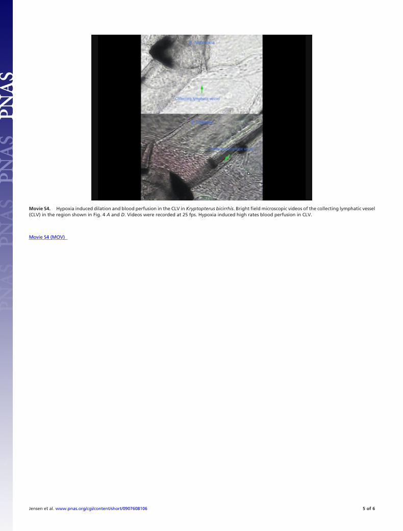
Movie S4. Hypoxia induced dilation and blood perfusion in the CLV in Kryptopterus bicirrhis. Bright field microscopic videos of the collecting lymphatic vessel(CLV) in the region shown in Fig. 4 A and D. Videos were recorded at 25 fps. Hypoxia induced high rates blood perfusion in CLV.
Jensen et al. www.pnas.org/cgi/content/short/0907608106 5 of 6
Movie S5 (MOV)
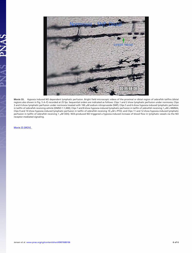
Movie S5. Hypoxia induced NO dependent lymphatic perfusion. Bright field microscopic videos of the proximal or distal region of zebrafish tailfins (distalregions also shown in Fig. 5 A–F) recorded at 25 fps. Sequential orders are indicated as follows: Clips 1 and 2 show lymphatic perfusion under normoxia; Clips3 and 4 show lymphatic perfusion under normoxia treated with 100 �M sodium nitroprusside (SNP); Clips 5 and 6 show hypoxia-induced lymphatic perfusionin tailfin of zebrafish receiving vehicle (DMSO 1:1,000); Clips 7 and 8 show hypoxia-induced lymphatic perfusion in tailfin of zebrafish receiving 1 �M L-NMMA;Clips 9 and 10 show hypoxia-induced lymphatic perfusion in tailfin of zebrafish receiving 10 �M c-PTIO; and Clips 11 and 12 show hypoxia-induced lymphaticperfusion in tailfin of zebrafish receiving 1 �M ODQ. NOS-produced NO triggered a hypoxia-induced increase of blood flow in lymphatic vessels via the NOreceptor-mediated signaling.
Jensen et al. www.pnas.org/cgi/content/short/0907608106 6 of 6