Indian Pediatrics April 2022 Issue
110
VOLUME 59 NUMBER 4 April 2022 E-copy Copyright of Indian Pediatrics. It is meant for personal use only, and not to be shared widely over social media platforms, blogs and mass e-mails.
-
Upload
khangminh22 -
Category
Documents
-
view
0 -
download
0
Transcript of Indian Pediatrics April 2022 Issue

VOLUME 59
NUMBER 4
April 2022
E-copy
Copyright of Indian Pediatrics. It is meant for personal use only, and not to be shared widely over social media platforms, blogs and mass e-mails.


INDIAN PEDIATRICS 267 VOLUME 59__APRIL 15, 2022
ADVERTISEMENT3

INDIAN PEDIATRICS 268 VOLUME 59__APRIL 15, 2022
ADVERTISEMENT 4

269
April 2022 Volume 59 Number 4
CONTENTS
PRESIDENTIAL ADDRESS
59th National Conference of Indian Academy of Pediatrics (PEDICON)
19-23 March, 2022, Greater NOIDA–REMESH KUMAR R 271
INVITED COMMENTARY
Breast Milk Monthly D-livery–SARA S OBERHELMAN-EATON, TOM D THACHER 274
RESEARCH PAPERS
Effect of Maternal Supplementation With Two Different Doses of Vitamin DDuring Lactation on Vitamin D Status, Anthropometry and Bone Mass ofInfants: A Randomized Controlled Trial–REKHA RAMOT, SWATI YADAV, SKVISHNOI, PRAMOD SHARMA, RAJESH KHADGAWAT, RAKESH JORA 276
Pediatric Hemophagocytic Lymphohistiocytosis - A Single Center Study–TANUSREE PAUL, MANAS KALRA, ARUN DANEWA, PALLAVI SACHDEVA, KASIBHARATHI THATIKONDA, DIVIJ SACHDEVA, ANUPAM SACHDEVA 283
Profile of Functional Constipation in Children at a Referral Hospital–VIKAS ARVINDBHAI MAKHWANA, KAKOLI ACHARYYA, SAUGATA ACHARYYA 287
Hepatitis B Vaccination Coverage of Preschool Children in Libreville,Gabon: Prevalence and Determining Factors–S MINTO’O, E KUISSI KAMGAING,U BISVIGOU, FC LOEMBE, D ZOUANZE, E NGOUNGOU, SJ ATEGBO 290
Profile of Girls With Adnexal Torsion: Single Center Experience–PATRYCJA SOSNOWSKA-SIENKIEWICZ, PRZEMYSLAW MANKOWSKI 293
Hindi Translation and Validation of Childhood Asthma Control Test (C-ACT)–PRAWIN KUMAR, CHIRAG THAKUR, JAGDISH P GOYAL, JAYKARAN CHARAN,KULDEEP SINGH 296
RECOMMENDATIONS
Association of Child Neurology (AOCN) Consensus Statement on theDiagnosis and Management of Febrile Seizures–JAYA SHANKAR KAUSHIK,VISHAL SONDHI, SANGEETA YOGANATHAN, RACHANA DUBEY, SUVASINI SHARMA,KOLLENCHERI PUTHENVEETTIL VINAYAN, PIYUSH GUPTA, REKHA MITTAL FORAOCN EXPERT COMMITTEE 300
UPDATE
Diagnosis and Management of Pediatric Acute Liver Failure: ESPGHANand NASPGHAN 2022–SRUTI MISHRA, PALLAVI PALLAVI 307
JOURNAL CLUB
Short Course of Daily Prednisolone During Upper Respiratory Tract Infectionfor Children With Relapsing Steroid Sensitive Nephrotic Syndrome
IndianPediatrics
Editor-in-Chief Devendra MishraExecutive Editor AP DubeyManaging Editor Rakesh LodhaAssociate Editors
Anup MohtaPooja Dewan
Joseph L MathewAashima Dabas
Abhijit SahaExecutive Members
Sunita Bijarnia-MahayJS Kaushik
Ujjal PoddarKirtisudha Mishra
Somshekhar NimbalkarAshish Jain
Kana Ram JatSumaira KhalilRomit Saxena
Amit DevganNidhi SugandhiRajesh Meena
International Advisory BoardPrashant Mahajan
Sanjay MahantPSN Menon
John M PettiforSudhin Thayyil
Gaurishankar ShahNational Advisory BoardCentral Zone Anil Kumar P
Mahesh MaheshwariEast Zone Mritunjay Pao
Santanu DebNorth Zone Anurag Tomar
Muzaffar JanSouth Zone Raghunath CN
Riaz IWest Zone Kavita Shrivastav
Samir R ShahChief Advisor Siddharth RamjiCentral IAP Advisors (ex-officio)
R Remesh KumarUpendra S Kinjawadekar
S ThangaveluVineet K Saxena
Biostatistics Amir M KhanRajeev K Malhotra
Electronic media Sanjeev GoelEthics Jagdish ChinappaOffice Matters AS Vasudev
Peeyush JainSocial Media Arva Bhavnagarwala
Chandermohan KumarWebsite C Vidyashankar

CONTENTS (contd.)
270
Address for ordinary letters: The Editor-in-Chief, Indian Pediatrics, P. Box No. 3889, New Delhi-110 049, India.Address for registered/speed post letters: Dr. Devendra Mishra, Editor-in-Chief, Indian Pediatrics,
115/4, Ground Floor, Gautam Nagar, New Delhi 110 049, India. Tel: (011) 46052593E-mail: [email protected]; Website: www.indianpediatrics.net; LinkedIn: indianpediatrics
Facebook: www.facebook.com/Indianpediatrics; Twitter: @EditorIndPed; Instagram: @indianpediatrics
All rights reserved. The views and opinions expressed in the articles are of the authors and not of the journal.Indian Pediatrics does not guarantee directly or indirectly the quality or efficacy of any product or service featured inthe advertisements in the journal, which are purely commercial.
Indian Pediatrics, the official journal of the Indian Academy of Pediatrics, is a peer-reviewed journal with monthlycirculation of print/e-copies to over 33,000 pediatrician members. The journal is indexed in Current Contents/ClinicalMedicine, Science Citation Index Expanded, Medline, PubMed, Indian Science Abstracts, getCITED, POPLINE,CANCERLIT, TOXLINE, Psych Line, and Dermline. The journal gives priority to reports of outstanding clinical andexperimental work, as well as important contributions related to common and topical problems related to children andadolescents. Information for Authors is available on the website of the journal. Indian Pediatrics is available free online atwww.indianpediatrics.net. There are no charges for publishing in the journal.
Evidence-Based Medicine Viewpoint–JOSEPH L MATHEW 312
Contemporary Researcher’s Viewpoint–ARVIND BAGGA, ADITI SINHA 316
Pediatric Nephrologist’s Viewpoint–RAJIV SINHA 317
Pediatrician’s Perspective–JANANI SANKAR 319
RESEARCH METHODOLOGY SERIES
Systematic Reviews and Meta-Analysis: A Guide for Beginners–JOSEPH L MATHEW 320
MEDICAL EDUCATION
The Concept of Self-Directed Learning: Implications for Practice in the Undergraduate Curriculum–ANSHU, PIYUSH GUPTA, TEJINDER SINGH 331
RESEARCH LETTER
Cardiac Evaluation in Multisystem Inflammatory Syndrome in Children (MIS-C) Associated With COVID-19–PRIYANKAR PAL, JIGNA N BATHIA, MIMI GANGULY, PURBASHA GUPTA, HRIDAY DE, ANIL KUMAR SINGHI 339
CLINICAL CASE LETTERS
Hypersenstivity Signs of Tuberculosis – Is It Synonymous of Latent Tubercular Infection?–MADHU S PUJAR,VINEELA MIKKILINENI, MEGHA P 341
Two Faces of Brugada Syndrome–PIOTR KÊDZIORA, ALEKSANDRA STASIAK 342
Infantile Anti-N-Methyl-D-Aspartate Receptor Encephalitis Post-SARS-CoV-2 Infection–PRABHJOT KAUR,VINAY MV, BABU S MADARKAR 343
CORRESPONDENCE 345
ICONIC PEDIATRIC INSTITUTIONS OF INDIA
Bai Jerbai Wadia Hospital for Children and Institute of Child Health and Research, Mumbai–NC JOSHI, SHAKUNTALA S PRABHU 348
NEWS IN BRIEF 299,340
CLIPPINGS 275,282
ERRATA 344
ADVERTISEMENTS 266-68,273,286,289,311,330,352-56
Impact Factor 2020 of Indian Pediatrics is 1.411

INDIAN PEDIATRICS 271 VOLUME 59__APRIL 15, 2022
59th National Conference of Indian Academy of Pediatrics (PEDICON)19-23 March, 2022, Greater NOIDA
REMESH KUMAR RNational President, Indian Academy of Pediatrics 2022
families and feel pride in nurturing the future of humanity.Through IAP, I was able to see the vast canvas of ourprofession and got opportunities to contribute myknowledge and resources to make a difference to childhealth. As the President, I have identified two broader armsfor the activities in my presidential tenure: primarily,Contribution to child health, and Contribution to theprofession through IAP.
Going forward in this track, IAP today stands at a newjuncture whereby we propose to become a broad basedorganization and focus on the more difficult phase ofchildhood, namely adolescence. This is in preparation toour future role as a profession dealing with both Pediatricsand adolescence. In this context, I must congratulate mypredecessor Dr Piyush Gupta for his sustained efforts toget the domain of Pediatrics to be renamed as ‘Pediatricsand Adolescent Medicine,’ to which the Government ofIndia has already agreed.
One immediate and primary concern on child healthshould be reduction of under-5 mortality. As of 2020, under-5 mortality rate for India revolves around 35, which means35 out of 1000 children born in our country do not get theopportunity to celebrate their fifth birthday. IAP hasidentified the goal of achieving under-5 mortality rate of 25by the year 2025 as an important component in thecountry’s journey to Sustainable Development Goals 2030.This is a stiff target, but nothing is impossible if we aim it ona mission mode. In our country, under-5 mortality rate haswide disparity interstate as well as intra state.With anindepth district wise secondary data analysis, we haveidentified 57 high priority districts with high U5MR forurgent intervention.We understand one size does not fit all.As such, district-specific road maps have been devised.We are primarily aiming at catalyzing the activities ofGovernment of India in these focused districts with thecollaboration of other major stake holders like WHO,UNICEF, FOGSI, NNF and TNAI.
My dears, it is a fact of life that children cannot express alltheir needs. We, the pediatricians, as the custodians of child
At the outset, on behalf of the entire IAP Community, I paytribute and homage to the COVID martyrs. The only thing Iwould say is that- we will never forget your sacrifices!
To begin with, I take the opportunity to express mysincere gratitude to every member of the Academy for theconfidence reposed in me by electing me as the President ofthe second largest specialty body of pediatricians in theworld. I am humbled by the affection and love showered byone and all in the fraternity, and it shall be my earnestendeavor to give my very best to the Academy.
As I assume office, I am aware of the weight ofresponsibility that comes in leading a great organization.Certainly, I am inheriting the legacy of great stalwartsculminating with my predecessor Prof. Piyush Gupta, whohas led IAP with great determination through the difficultdays of the pandemic. I will strive to continue his goodwork and pass the baton on an equally strong platform tomy successor, President-Elect Dr. Upendra Kinjawadekar.
Today I have the privilege of sharing this platform withour distinguished Chief Guest, Honorable Minister forWomen and Child Development Smt Smriti Irani ji, who hasset her goal for an India witnessing “rapid transition fromwomen’s development to women-led development”. Childwelfare has been very close to her heart. She has been theactive proponent of the telemedicine support systemintroduced last year with the participation of IAP for thechild care institutions across the country.
I am delighted to have Dr. Mahesh Sharmaji, formerunion minster and the incumbent Member of Parliamentrepresenting Gautam Budha Nagar. Being one amongstourselves in the medical profession, he has gone to greatlengths to support us in organizing this event. I must say hehas been a mentor and patron to us and showed us that heis not just an MP for Noida, but an MP for the medicalfraternity too.
A friend, choosing to be a pediatrician has been one ofthe most worthy choices in my life. Pediatrics is a specialtywhich enables you to touch the lives of hundreds of
PPPPP RRRRR EEEEE SSSSS IIIII DDDDD EEEEE NNNNN TTTTT IIIII AAAAA L AL AL AL AL A DDDDD DDDDD RRRRR EEEEE SSSSS SSSSS

INDIAN PEDIATRICS 272 VOLUME 59__APRIL 15, 2022
health need to raise our voice for them. Our responsibility forthe child’s well-being goes way beyond the physical health ofindividual patients. IAP needs to be more vociferous andvisible in various domains of child advocacy like child rights,child abuse, gender discrimi-nation and many more. We needto continue our proactive support to child health initiativesfrom GOI like INAP (India Newborn Action Plan), AnemiaMukth Bharath, NSSK and the various nutrition supportprograms. I humbly request our strong force of 35,000 IAPmembers to continue to remain socially committed in all theserealms of public health and augment the Governmentprograms in your own regions.
In this digital era, we propose to set up an onlineMember Benefits Portal, which will be giant stride inorganization and profession development. A wide range ofMember Benefits are envisioned in this portal, the mostsignificant of which will be the QR Code based digital IDCards, the PeD card, which apart from providing IAPidentity, will privilege the members for loyalty services frommany renowned service providers. I am sure, all these willresult in a complete transformation of our organizationfunctioning and make IAP more member and branchfriendly.
Friends, academics is our forte. Keeping pace with newdevelopments and recapping the basics is vital to optimalpatient care. To this end, we have formulated the StandardTreatment Guidelines in Pediatric Office Practice. We havestarted releasing guidelines on one common pediatric ill-ness every Monday, Wednesday and Friday from January1, 2022 Academy year. I am proud that the STG Team hasbeen on the dot and as of today, we have been able topublish 33 guidelines in the last 11 weeks. Parallelly, wehave 12 physical modules including the ambitious ECD-Early Childhood Program and 5 virtual academic modulesalready lined up for the year, with 8 TOTs happening atNoida itself with PEDICON.The 300 workshops on TBeradication planned in next two years is a reflection of ourtrue commitment to Honorable PM Shri Narendra Modiji’svision of End TB 2025 project. I assure that all the 340branches of IAP will have the opportunity to conduct oneor the other of these as district level workshops in physicalreal time mode with academic grant from Central IAP Office.
My dear friends, as I stand before you as the Presi-dent, I look back and recap my journeyof 5½ decades. Thedream of a rustic boy from Kuttanad, the water loggedbackwater village of Kerala to come up high in the medicalprofession would not have been realistic, but for the vision,nurturing care and the sacrifices made my late parents. Onthis very special occasion, I offer my respectful pranams tomy father Shri PG Ramachandran Nair and mother Smt.Kalyani Amma L, both of whom earnestly desired to see me
as a successful doctor. They even blessed me with a namewith a unique spelling – Remesh – which is spelled with an‘e’ rather than an ‘a’ – and which has raised many aneyebrow at every occasion I have to spell my name.
In conclusion, I thank every member, friend and colle-ague who has enabled me to attain this coveted position.The staff of IAP central office has been most cooperativeand are the pillars of strength to any President. I thank themall. I have no words to thank my wife Dr. Jayalakshmy andson Arjun who by themselves have been like co opted IAPmembers and were always positive in their thoughts forIAP, and allowed me to spare much more time and effort forthe organization. My special gratitude to my immediatecolleagues in Pediatrics and management of Apollo AdluxHospital, Cochin, especially Medical Director Dr Anil, CEOMr Neelkannan, and my colleague, Senior Consultant inPediatrics, Dr Saju, for their constant encouragement andsupport.
On this occasion, my thoughts go out to my respectedteacher at Government Medical College, Kottayam and IAPPresident 2011, Dr TU Sukumaran who handheld me to IAP2½ decades back, and Dr. MKC Nair and Dr. SachidanandaKamath, Past National Presidents of IAP, who ably guidedme at the organizational level. I am very much indebted toDr. Santhosh Soans and Dr. Diganth Shastri, who, as myPresidents, nurtured my organizational aptitude in mytenure as Honorary Secretary General of CIAP in 2018 and2019. If I am to come this far as the President of IAP, thecredit should truly be placed at the feet of my Friends atIAP Kerala who always cared and inspired me by theirwarmth and affection.
I am very much thankful to the OB and EB of 2022 forthe whole hearted support in their proactive commitment tothe Academy. My HSG, Dr Vineet Saxena, is my pillar ofsupport and as always is a perfect task master at the backoffice. It is well said that “The best preparation fortomorrow is doing your best today.” The organizing teamof PEDICON 2022 has strived every day for the last two-and-a-half years, even in the face of a raging pandemic, tomake today a reality. With this TEAM at the helm, we all aresure to have the life time toast of an academic, gastro-nomicand social feast at India Expo Mart, Greater Noida in theweek ahead.
Dears, the Covid pandemic makes it pertinent to reflecton a famous saying “This too shall pass.” Life is like a road,there are bumps, cracks, U turns, road blocks, but the onlyimportant thing is life goes on. I end by quoting theChandogya Upanishad:
yovaibhûmâ, tatsukhaC, nâlpesukhamasti,bhûmaivasukha Cbhûmâtvevavijijñâsitavyaiti
8 PRESIDENT’S PAGE

INDIAN PEDIATRICS 273 VOLUME 59__APRIL 15, 2022
“There is no joy in the finite; there is joy only in theinfinite.”
Friends, we have been through the terrible experienceof a Pandemic. It was a situation of despair and no hope, yetlife is now returning to normal. Let us all orient ourselves tothis healing process of humanity and contribute our mite to
the emerging world of peace and prosperity for all. I wishyou all a safe, healthy and most enjoyable post-COVID eracoming up. I will and shall always remain as your humbleservant and friend in IAP!
Jai Hind! Jai IAP!
Funding: None; Competing interests: None stated.
PRESIDENT’S PAGE 9
KEM Hospital, Pune is a 650 bed multi-speciality tertiary care teaching hospital. The Department ofPediatrics has a state of the art PICU 10 beds (700 admissions/year), NICU having 50 beds (1500admissions/year), General Pediatrics 70 beds (2500 admissions/year).Additionally, Pediatric Oncology, Pediatric Gastroenterology, Pediatric Nephrology, Pediatric Neurologyare extremely vibrant departments with a variety of case mix. The NICU/PICU has 25 ventilators(conventional, HFO, HHHFNC), portable 2D Echo, bedside EEG, NIRS, TPN facilities, nitric oxide, wholebody cooling, human milk bank, etc. We also offer specialised services like genetics, pediatric surgery,neurosurgery, complex trauma, child rehabilitation, infectious disease etc.The department is recognised for DNB General Paediatrics for the last 30 years, DNB and Fellowship inPediatric Nephrology for 5 years, fellowship Gastroenterology 6 years, PICU (College of Pediatric CriticalCare- PICC - IAP) and NICU fellowships (IAP and NNF) for last 12 years and NICU fellowships (IAP andNNF) for last 10 years.We invite applications for the year 2022 for following posts (within 15 days by email).
Course Eligibility Duration
PICU Fellowship (PICC – IAP), 2 posts MD / DNB (Ped) / DCH 1 year / 2 yearsNeonatology Fellowship (IAP, NNF, Institutional), 1 post MD / DNB (Ped) / DCH 1 year / 1.5 yearsPediatric Hemato Oncology (Senior Resident Post), 1 post MD / DNB / DCH 6 months Ad hocPediatric Emergency (KEMH Certificate) MD / DNB (Ped) / DCH 6 months – 1 year
For PICU Fellowship Apply: For Pediatric Hemato Oncology Post For NICU Fellowships Apply:Dr Madhumati Otiv, DNB Dr Sarita Verma Kokane, DNB, FIAP (PHO) Dr Umesh Vaidya, MD, DNB
[email protected] / 9822040950 [email protected] / 9619284695 [email protected] / 9822031151
Dr Anand Pandit, MD,FRCPCH(UK) DR DD SHETTY, MD, DMRDDirector, Department of Pediatircs Director Academics
KEM Hospital, Pune 411 011 KEM Hospital, Pune 411 011
King Edward Memorial (KEM) Hospital, Pune(PICU/NICU/Hemato Oncology/Pediatric
Emergency Vacancies)
ADVERTISEMENT
INDIAN PEDIATRICS 274 VOLUME 59__APRIL 15, 2022
Breast Milk Monthly D-liverySARA S OBERHELMAN-EATON, TOM D THACHER
Department of Family Medicine, Mayo Clinic, Rochester MN 55905Correspondence to: Dr. Tom D Thacher, Professor of Family Medicine, College of Medicine, Mayo Clinic, Rochester, USA.
Nutritional rickets continues to have seriousconsequences in infants and childrenworldwide, and it remains prevalent inAfrican and South Asian countries, like India.
Bone pain, skeletal abnormalities, stunted growth, develop-mental delays, and life-threatening hypocalcemic seizures andcardiomyopathy are entirely preventable outcomes withadequate calcium intake and vitamin D supplementation.
Vitamin D can either be ingested or synthesized cutane-ously following ultraviolet light exposure. An estimated 70-100% of the general population of India is vitamin D deficient,likely secondary to limited vitamin D food fortification,restricted sunlight exposure and darkly pigmented skin [1].Vitamin D deficiency is common among lactating women andhuman milk contains low amounts of vitamin D, increasing therisk of nutritional rickets in Indian infants.
The Indian Academy of Pediatrics [2] and a GlobalConsensus Group [3] recommend that all infants receive 400IU/day of vitamin D for the first year of life for prevention ofrickets. However, adherence with this recommendation is low.In a study of vitamin D adherence in India, 41.5% of infantsreceived vitamin D at any point during the study, but only8.8% received routine appropriate vitamin D dosing [4].Similar findings of low adherence have been reportedworldwide, despite a multitude of recommendations forinfant supplementation to reduce rates of rickets. Theinconvenience of providing supplemental drops to anexclusively breastfed infant may be one reason parents do notroutinely provide the recommended supplementation.Therefore, maternal supplementation for a breastfeeding dyadhas been utilized for improved ease of administration.
Ramot and colleagues [5] compared two monthly vitaminD doses for 12 months in lactating mothers and found that12,000 IU monthly did not result in sufficient infant vitamin Dstatus, but 120,000 IU monthly achieved vitamin D sufficiency(25(OH)D>20 ng/mL) in the majority (95%) of infants. Theirfindings are important for two reasons. First, this is the firststudy of maternal bolus dosing of vitamin D to span theinfant’s entire first year and demonstrated both maternal and
infant safety. Second, this study provides new data toestablish the ideal maternal dose for monthly administration.Previous studies of maternal supplementation with 100,000 IUor less monthly did not assure infant vitamin D sufficiency [6].This study showed that 120,000 IU monthly providedsufficient vitamin D in most but not all infants. Another studyof 150,000 IU resulted in sufficiency for all infants after onemonth [7]. Thus, the optimal monthly maternal dose forlactating mothers is likely between 120,000 and 150,000 IU.
Daily maternal supplementation with vitamin D 5000-6400IU safely achieves sufficient serum 25(OH)D concen trationsin breastfed infants [7-9]. Maternal supplemen-tation waspreferred over infant supplementation by mothers inMinnesota, USA: 88.4% of the surveyed mothers preferred tosupplement themselves rather than their infants [10]. Amongfamily medicine clinicians, 87.5% would recommend eithermaternal supplementation (37.5%) or allow parents to choosebetween maternal or infant supplementation (50%) [11].Vitamin D supplementation of lactating mothers has theadditional advantages of protecting the infant from vitamin Dtoxicity related to dosing errors, promoting the completenessof breast milk nutrition, and simpler adherence than infantdrops.
From a public health perspective, the option to providematernal monthly administration of vitamin D for infantbenefit is quite important. With proper infra-structure andoversight, maternal vitamin D adminis-tration couldtheoretically be coupled with well-baby examinations and/orpublic health initiatives focusing on women and children.Large proportions of mothers (43%) [10] and clinicians (30%)[11] in a single community in the United States would prefer amonthly regimen. Similar investigations have not been donein India, but one could hypothesize a similar willingness toaccept a monthly administration schedule.
Nutritional rickets can be eradicated, and Ramot andcolleagues [5] demonstrate a feasible way to prevent vitaminD deficiency through maternal monthly supple-mentation.Given the safety and efficacy of maternal vitamin Dsupplementation for infant vitamin D sufficiency, national
IIIII NNNNN VVVVV IIIII TTTTT EEEEE D C O M M E N T A R YD C O M M E N T A R YD C O M M E N T A R YD C O M M E N T A R YD C O M M E N T A R Y

INDIAN PEDIATRICS 275 VOLUME 59__APRIL 15, 2022
and global recommendations should be updated to reflectmaternal supplementation as a viable option. Public healthinitiatives should explore local opportunities to includemonthly maternal supplementation in already establishedmother/child interventions. These initiatives could preventvitamin D deficiency and decrease the incidence of nutritionalrickets in India and globally.
Funding: None; Competing interests: None stated.
REFERENCES
1. Ritu G, Gupta A. Vitamin D deficiency in India: prevalence,causalities and interventions. Nutrients. 2014;6:729-75.
2. Gupta P, Dabas A, Seth A, et al. Indian Academy of PediatricsRevised (2021) Guidelines on Prevention and Treatment ofVitamin D Deficiency and Rickets. Indian Pediatr. 2021 Dec29:S097475591600382. Epub ahead of print.
3. Munns CF, Shaw N, Kiely M, et al. Global ConsensusRecommendations on Prevention and Management ofNutritional Rickets. J Clin Endocrinol Metab. 2016;101:394-415.
4. Meena P, Saran AN, Shah D, et al. Compliance to prescriptionof routine vitamin D supplementation in infants. Indian Pediatr.2020;57:1067-9.
5. Ramot R, Yadav S, Vishnoi SK, Sharma P, Khadgawat R, Jora
R. Effect of maternal supplementation with two different dosesof vitamin D during lactation on vitamin D status,anthropometry and bone mass of infants: a randomizedcontrolled trial. Indian Pediatr. 2022;59:276-82.
6. Wheeler BJ, Taylor BJ, Herbison P, et al. High dose monthlymaternal cholecalciferol supplementation during breast-feedingaffects maternal and infant vitamin D status at 5 monthspostpartum: a randomized controlled trial. J Nutr.2016;146:1999-2006.
7. Oberhelman SS, Meekins ME, Fischer PR, et al. Maternalvitamin D supplementation to improve the vitamin D status ofbreastfed infants: a randomized control trial. Mayo Clinc Proc.2013;88:1378-87.
8. Hollis BW, Wagner CL, Howard CR, et al. Maternal versusinfant vitamin D supplementation during lactation: arandomized controlled trial. Pediatrics. 2015;136:625-34.
9. Wagner CL, Hylsey TC, Fanning D, et al. High dose vitamin D3supplementation in a cohort of breastfeeding mothers and theirinfants: a 6-month follow-up pilot study. Breastfeed Med.2006;1:59-70.
10. Umaretiya PJ, Oberhelman SS, Cozine EW, et al. Maternalpreferences for vitamin D supplementation in breastfed infants.Ann Fam Med. 2017;15:68-70.
11. Oberhelman SS, Cozine EW, Umaretiya PJ, et al. Vitamin D andthe breastfeeding infant: family medicine clinicians’ know-ledge,attitudes and practices. J Hum Lact. 2018;34:331-6.
INVITED COMMENTARY 11
Evaluation of daily low-dose prednisolone duringupper respiratory tract infection to prevent relapse inchildren with relapsing steroid-sensitive nephroticsyndrome - The PREDNOS 2 randomized clinicaltrial (JAMA Pediatr. 2022;176:236-43)
A number of studies including, randomized controlled trials,have established that increasing the maintenance dose ofcorticosteroids during upper respiratory tract infections(URTI) for 5-7 days can reduce the risk of relapse in nephroticsyndrome. PREDNOS 2 is a phase 3, double blind, placebo-controlled randomized clinical trial which evaluated the efficacyof daily (for 5-7 days) low dose prednisolone (15 mg/m2/day)during an episode of URTI in reducing the risk of relapse among365 children with or without background immunosuppressivetreatment in the United Kingdom. The primary outcome wasthe incidence of first upper respiratory tract infection-relatedrelapse (URR). Secondary outcomes were overall rate ofrelapse, changes in background immunosuppressive treatment,cumulative dose of prednisolone, rates of serious adverseevents, incidence of corticosteroid adverse effects, and qualityof life. In intention to treat analysis, the number of patientsexperiencing URR was 56 of 131 (42.7%) in the prednisolonearm and 58 of 131 (44.3%) in the placebo arm (adjusted riskdifference, 0.02; 95% CI, 0.14 to 0.10; P=0.70). No significantdifferences were observed in secondary outcomes as wellbetween the treatment arms. It was concluded that daily shortcourse of low dose prednisolone at the time of URTI does notreduce the risk of relapse in children with nephrotic syndrome.
DR PRAJAL [email protected]
CLIPPINGS
Whole-exome sequencing and variant spectrum inchildren with suspected inherited renal tubulardisorder: The East India Tubulopathy Gene Study(Pediatr Nephrol. 2022 Jan 10. doi: 10.1007/s00467-021-05388-y)
A multicenter, descriptive cross-sectional study was performed in77 children (73% male) in Eastern India. Children less than 18years with clinically suspected tubulopathy were enrolled in thestudy and whole exome sequencing (WES) was performed in allthe cases. Sanger sequencing and Multiplex ligation-dependentprobe assay (MLPA) were also done when indicated. The variantswere classified as pathogenic/likely pathogenic (P/LP) inaccordance with American College of Medical Genetics andGenomics, 2015. Fifty five (24 novel) P/LP variants wereidentified and genetic diagnosis was established in 54 children(70%). Clinically, distal renal tubular acidosis (32.4%) was themost commonly identified tubular disorder but the diagnosticyield of WES was highest for nephrogenic diabetes insipidus(100%). Barakat syndrome and Renal cyst with diabetessyndrome were the rare disorders identified. WES led to revisionof clinical diagnosis in 14 children (26% of those with a confirmedgenetic diagnosis and 18% of the overall cohort) and detection ofunidentified co-morbidities (sensorineural deafness n=5,hemolytic anemia n=2, dental changes n=1). The authorssuggested that WES is an essential tool in the diagnosis andmanagement of inherited tubulopathies in India.

INDIAN PEDIATRICS 276 VOLUME 59__APRIL 15, 2022
RRRRR EEEEE SSSSS EEEEE AAAAA RRRRR CCCCC H PH PH PH PH P AAAAA PPPPP EEEEE RRRRR
Effect of Maternal Supplementation With Two Different Doses of VitaminD During Lactation on Vitamin D Status, Anthropometry and Bone Mass ofInfants: A Randomized Controlled TrialREKHA RAMOT,1 SWATI YADAV,2 SK VISHNOI,2 PRAMOD SHARMA,2 RAJESH KHADGAWAT,1 RAKESH JORA2
From 1Department of Endocrinology and Metabolism, All India Institute of Medical Sciences, New Delhi; 2Department of Pediatrics,Dr S N Medical College, Jodhpur, Rajasthan.Correspondence to: Prof Rakesh Jora, Department of Pediatrics, Dr SN Medical College, Jodhpur, [email protected]: March 10, 2021; Initial review: June 09, 2021; Accepted: December 18, 2021.
Background: There is a high prevalence of vitamin D deficiency(VDD) in exclusively breast-fed infants in the absence ofappropriate vitamin D supplementation.
Objective: To evaluate the efficacy of two doses of maternalvitamin D supplementation on vitamin D levels of mother-infantpairs and to assess its effect on growth parameters (weight,length and head circumference) and bone mass of infants.
Study design: Randomized controlled trial.
Participants: Lactating mother-infant pairs (n=220).
Intervention: Maternal oral vitamin D supplementation in twodoses (group 1: 1,20,000 IU/month and group 2: 12,000 IU/month)for 12 months.
Main outcomes: Maternal and infant serum 25OHD levels, andinfants’ growth and bone mass.
Results: There was high prevalence of VDD at baseline inmothers (94%) as well as infants (98.5%), which was reducedto 43.1% in (mothers) and 46.5% in infants after 12 months.Significantly higher median (IQR) serum 25OHD levels (ng/mL)were observed among mothers in group 1 compared to group 2[46 (17-159) vs 18 (6-64); P<0.01] and in infants [36.5 (15-160)vs 17 (7-32); P<0.01]. No significant association was observedbetween growth parameters or bone mass and serum 25OHDlevels of mother or infant between the two groups. Four mothers(3.6%) and two infants (1.8%) in group I had serum 25OHD>100ng/mL, but without hypercalciuria or hypercalcemia.
Conclusion: Bolus vitamin D supplementation in the dose of1,20,000 IU/month was more efficacious in improving maternaland infant vitamin D status at 12 months, as compared to 12,000IU/month.Keywords: Bone densitometry, DXA, Lactating mothers,Vitamin D deficiency.
infants up to one year of age [8]. However, the practicalapplicability of this recommendation is questionable asadherence was found to be less than <20% [9]. Therefore,high-dose vitamin D supplementation to lactating mothersseems to be a better approach to address the dual problem ofVDD in lactating mother-infant pairs [5].
Vitamin D deficiency (VDD) in infancy andchildhood is a serious public health concern inAsia, Middle East, and North Africa [1]. Ahigh prevalence of VDD is reported among
infants, depending on the definition and the latitude of thepopulation studied [2]. The prevalence of VDD amongnursing mothers and their breast-fed infants has beenwidely reported from India [3,4].
Breast milk is a poor source of vitamin D (~5-80 IU/L)[5], which predisposes exclusively breast-fed infants to anincreased risk of developing rickets as compared to vitaminD fortified formula-fed infants [6]. A strong positivecorrelation has been reported between vitamin D intake oflactating mothers and serum 25-hydroxy vitamin D(25OHD) levels of infants. A sufficient maternal vitamin Dintake is associated with optimal vitamin D transfer viabreast milk which is adequate to meet infant needs [7]. TheIndian Academy of Pediatrics recommends oral supple-mentation of 400 IU/day of vitamin D to all breastfed
Published online: January 09, 2022; PII: S097475591600396
Invited Commentary: Pages 274-75
Indian data on optimal dose of vitamin Dsupplementation among lactating mothers to improvevitamin D status of infants is scarce. Therefore, thepresent study was planned with the primary objective toevaluate the efficacy of two vitamin D supplementationdoses (1,20,000 IU/month vs 12,000 IU/month) to lactatingmothers on serum 25OHD levels of mother-infant pairs.The effect of maternal vitamin D supplementation oninfant’s anthropometry and whole body bone mass werealso studied as secondary objectives.

INDIAN PEDIATRICS 277 VOLUME 59__APRIL 15, 2022
RAMOT, ET AL.
METHODS
The present randomized controlled trial was conductedfrom December, 2014 to December, 2017 after ethicalapproval. The subjects were enrolled after writteninformed consent.
Healthy breast-fed mother-infant pairs within onemonth of delivery, willing to follow-up for 12 months wereincluded. Mothers with pre-existing type 2 diabetes,hypertension, chronic renal or liver disease, antipsychoticdrug exposure, clinical osteomalacia or severe vitamin Ddeficiency or exposure to medications known to affectvitamin D metabolism were excluded. Infants with congenitalmalformations and birth asphyxia were excluded.Additionally, mothers with serum calcium >11mg/dL, serum25OHD level >100 ng/mL, liver enzymes elevated >3 timesupper limit of normal (ULN) and serum creatinine above ULNfor age at screening were also excluded.
The mothers were randomized (using computer-generated simple random code) into two groups (1:1 ratio)of oral vitamin D supplementation: 1,20,000 IU/month(group 1) and 12,000 IU/month (group 2) for 12 months. Thevitamin D dose 400 IU/day (group 2) was chosenconsidering high prevalence VDD in India and ICMR-NINrecommendation [10], while the dose of 4000 IU/day(group I) was chosen based on recommendations of theEndocrine Society to maintain serum 25OHD ≥30ng/mL inexclusively breast-fed infants not on vitamin D supple-ments [11]. The vitamin D supplements were administeredas telephonically supervised monthly bolus doses forbetter compliance. All the subjects were advised toregularly go-out in sun on a daily basis (the city wherestudy was conducted has abundant sunshine throughoutthe year). Vitamin D preparations were provided as oraltablets (strength 12,000 IU and 1,20,000 IU); unlabelled fordose and identical in all aspects of colour, taste, andexternal appearance (Torrent Pharmaceuticals).
The safety of intervention was assessed by measure-ment of corrected total serum calcium and urinary calcium:creatinine ratio (non-fasting, second void sample) atbaseline, six months, and 12 months. Hypercalcemia wasdefined as a total serum calcium level of >11 mg/dL andhypercalciuria as urinary (spot urine sample) calcium:creatinine ratio >0.4 [12]. Subjects with urinary calcium:creatinine ratio of >0.4 without hypercalcemia were re-evaluated with a timed 24-hour urine calcium excretion and 4mg/kg excretion was considered as abnormal. Any subject,who developed both hypercalcemia and hypercalciuria wasexcluded from further intervention.
The biochemical parameters (complete blood counts,liver and renal function tests, total serum calcium,
phosphate, total alkaline phosphatase, and bloodglucose) were measured using Roche Hitachi 912Chemistry Analyzer (GMI, Inc.), serum 25OHD wasassessed using chemiluminescent assay using LIASON(DiaSorin Inc.) auto analyzer. The reproducibility of theassay ranged from 6% to 12%, and the laboratory wasregistered with UK-DEQAS vitamin D assay externalquality control assessment program (www.deqas.org).The vitamin D status was categorized as: severe deficiency,deficiency, insufficiency, and sufficiency based on serum25OHD levels (ng/mL) of <10, <20, 20-29, and ≥30,respectively [13].
The whole body bone mass of infant was assessed bydual-energy X-ray absorptiometry (DXA) using GE LunarProdigy Advance instrument 8743 (GE Medical systems) at12 months (±15 days). The scans were conducted withuniform swaddling of infants in fed and sleeping statewithout sedation. In order to obtain artefact-free scans,appropriate positioning of infants was achieved bysecuring the infant’s upper extremities away from the trunkregion and gently binding of both the upper and lowerextremities using a cotton blanket. The scans withmovement artefacts were excluded.
The birthweight of infants was measured to the nearest10 g using an electronic weighing scale, length to nearest0.5 cm using an infant measuring board, and head circum-ference to the nearest 0.1 cm using a non-stretchable tape.
The sample size was calculated based on thepresumption that 10% subjects in the control arm and 40%subjects in the intervention group would achieve maternalserum 25OHD >30 ng/mL after one year. Hundred subjectsin each arm were required to detect the above differencewith 90% power and 97.5% confidence levels. Anticipating20% dropouts, 110 subjects were required to be enrolled ineach arm.
Statistical analysis: This was carried out usingSTATA14.2 (StataCorp LLC). The appropriately codeddata were entered in Microsoft Excel from case recordforms, and extreme values (beyond 1.5 times of inter-quartile range below Q1 or above Q3) were excluded.Continuous variables were compared by independent t-test (normally distributed) or Wilcoxson rank sum test(non-normally distributed) and within group comparisonwas assessed using paired t-test (normally distributed).The linear regression was applied to assess theassociation between maternal and infant vitamin D statusas well as the association of infant vitamin D status withbone mass.
RESULTS
A total of 220 mother-infant pairs (138 multiparaous
13

INDIAN PEDIATRICS 278 VOLUME 59__APRIL 15, 2022
MATERNAL VITAMIN D SUPPLEMENTATION AND INFANT OUTCOMES
mothers; 114 male infants) were randomized into twogroups (Fig. 1). Baseline demographic characteristics ofthe study population are presented in Table I.
The median (range) of maternal and infant serum 25OHDlevels of entire group at baseline were 8.3 (0.4-30.1) and 5.8(0.2-33.8) ng/mL, respectively, which was not significantlydifferent between the two groups (Table II and III). There wasa high prevalence of VDD in mothers (94%) and infants(98.5%) at baseline (Web Table I).
Maternal serum 25OHD levels of ≥30 ng/mL and
>20-30 ng/mL were seen in 73 (73.7%) and 18 (18.2%) ingroup 1, and 5 (5%) and 17 (17%) in group 2 at 12 months.Among infants, 75 (75.7%) and 19 (19.2) had serum 25OHD≥30 and >20-30 mg/mL in group 1, while in group 2, only 5and 13 infants had ≥30 and >20-29 ng/mL serum 25OHDlevels, respetively. The proportion of infants with serum25OHD <20 ng/mL reduced to 5 (5%) in group I butincreased to 82 (82%) in group 2 (Web Table I). Thecomparison of vitamin D status of mothers and infantswith respect to supplementation groups is presented inFig. 2.
Increased (mean) urinary calcium:creatinine ratio (>0.4)was observed in 3 (0.63), 5 (0.73), and 1 (0.88) mother atbaseline, six months, and 12 months, respectively. However,none of these subjects developed hypercalcemia (sympto-matic or asymptomatic) and hypercalciuria, or both. Only twoinfants had serum 25OHD levels >100ng/mL after 12 monthsof supplementation (both belonged to group 1); however,none of them developed hypercalciuria or hypercalcemia.
There was no significant difference in anthropometricgrowth parameters (length, weight and head circumference)of infants between the two groups at baseline as well as atone year (P>0.05) (Table IV).
The vitamin D status of mother and infant wassignificantly correlated at baseline as well as at 12 months.With each ng/mL increase in maternal serum 25OHD,infant serum 25OHD increased by 0.55 ng/mL (95% CI 0.36to 0.74) after 12 months of supplementation.
Assessed for eligibility (n=260)
Refused consent for infant DXA scan (n=40)
Enrolled and randomized (n=220)
Intervention Group 1Vitamin D dose 1,20,000 IU/month
(n=110)
Intervention Group 2Vitamin D dose 12,000 IU/month
(n=110)
Lost to follow-up(n=10)
Lost to follow-up (n=11)
Completed 12 monthsupplementation
(n=99)
Completed 12 monthsupplementation
(n=100)
Analyzed (n=99) Analyzed (n=100)
Fig. 1 Flow chart of study participants.
Screening
Enrollment
Randomization↓
↓ ↓
→Follow-up
↓ ↓
↓ ↓
↓
→
14
Table I Baseline Maternal and Infant Characteristics
Variable Group 1 Group 2
Maternal characteristics (n=110) (n=110)Age, y 25.3 (4.6) 24.8 (4.3)Height, cm 156.05 (6.4) 157.6 (5.7)BMI, kg/m2 23.07 (3.9) 23.6 (4.3)Gestational age, wk 38.6 (1.01) 38.2(1.3)Infant characteristicsNormal birthweight (n=90) (n=95)
Weight, kg 2.8 (0.4) 2.9 (0.4)Length, cm 48.4 (1.66) 48.4 (1.96)Head circumference, cm 33.3 (1.10) 33.1 (1.18)
Low birthweight (n=20) (n=15)Weight, kg 2.3 (0.13) 2.20 (0.19)Length, cm 46.9 (2.7) 44.5 (1.8)Head circumference, cm 32.1 (1.38) 32 (1.26)
Data expressed as mean (SD). BMI: body mass index.

INDIAN PEDIATRICS 279 VOLUME 59__APRIL 15, 2022
RAMOT, ET AL.
Table II Maternal Biochemical Parameters at Baseline and 12 Months in the Two Groups
Parameter Group I Group IIBaseline Follow-up Mean diff Baseline Follow up Mean diff(n=110) (n=99) (95% CI) (n=110) (n=100) (95% CI)
Calcium; mg/dL 9.2 (0.88) 8.7 (0.87) -0.46 9 (0.92) 8.8 (0.87) -0.25(-0.71, -0.21)b (-0.51, 0.02)
Phosphate; mg/dL 4.8 (0.59) 4.5 (0.87) -0.35 5.01(0.62) 4.5 (0.85) -0.46(-0.56, -0.14)b (-0.66, 0.26)b
ALP, IU/L 214.9 (69.95) 170.6 (39) -44.3 226.6 (78.54) 163.6 -62.3(-58.6, -29.9)b (36.48) (-79.9, 44.6)b
25OHD, ng/mLa 9.2 (6.3, 12.5) 46 (29, 69) 44.9 (39.4, 50.4)b 7.8 (4.1,12.2) 18 (16, 20) 9.9(8.1, 11.8)b
Albumin, g/dL 2.9 (0.36) 3.6 (0.74) 0.69 (0.52, 0.85)b 2.9 (0.38) 3.5 (0.74) 0.66(0.51, 0.81)b
Urinary calcium/ 0.07 0.09 (0.04, 0.16) - 0.07 (0.03,0.15) 0.07 (0.04, 0.14) -creatininea (0.04, 0.13)Data represented as mean (SD) or amedian (IQR). Maternal vitamin D supplementation Group I- 120000 IU/mth and Group II- 12000 IU/mth.bP<0.05. ALP-alkaline phosphatase; 25OHD-25-hydroxy vitamin D.
Table III Infant Biochemical Parameters at Baseline and 12 Months in the Two Groups
Serum levels Group I Group IIBaseline Follow-up Mean diff Baseline Follow up Mean diff(n=110) (n=99) (95% CI) (n=110) (n=100) (95% CI)
Calcium, mg/dL 9.2 (1.0) 8.8 (0.89) -0.48 (-0.74, -0.21)b 9.3 (0.9) 8.8 (0.88) -0.43 (-0.66, 0.19)b
Phosphate, mg/dL 4.9 (0.85) 4.9 (0.84) 0.006 (-0.23, 0.24) 5.01 (0.73) 5.09 (0.80) 0.07 (-0.16, 0.31)ALP, IU/L 259.2 (113.09) 280 (112.2) 20.9 (-9.16, 50.9) 252.1 (108.53) 296 (105.85) 43.9 (15.9,71.9)Albumin, g/dL 3.03 (0.49) 3.6 (0.67) 0.56 (0.39, 0.73)b 3.1 (0.45) 3.6 (0.80) 0.47 (0.28, 0.66)b
25OHD, ng/mLa 7.1 (4.3, 9.2) 36.5 (30.5, 56) 36.9 (32.7, 41.2)b 4.8 (2.7 to 9.2) 17 (14.2,19) 12.05 (10.6, 14.4)b
Data represented as mean (SD) or amedian (IQR). Maternal vitamin D supplementation Group I-120000 IU/mth and Group II-12000 IU/mth. bP<0.05. ALP: alkaline phosphatase; 25OHD: 25-hydroxy vitamin D.
There was no significant difference in infant bonemass parameters between the two groups after one year ofsupplementation (Table IV). The mean BMC, BMD, andbone area of LBW infants were significantly lower ascompared to the corresponding values of normal birthweight infants (P<0.05). The infant bone mass was notsignificantly associated with maternal age, BMI, andmaternal serum 25OHD parameters (baseline, at 12 monthsand delta-change) in both the groups. Similarly, the infant’svitamin D level at baseline, at 12 months, and delta-changein serum 25OHD levels were also not significantlyassociated with bone mass parameters (Web Table II).
The infant’s weight at birth as well as 12 months wassignificantly associated with bone mass parameters in boththe groups (all P<0.05). Each 100 g increase in birth weightwas associated with a mean (95% CI) increase in BMC,BMD and bone area by 0.004 (0.001 to 0.004) g, 0.26 (0.79 to4.61) g/cm2 and 0.0002 (0.0016 to 0.002) cm2, respectively for
group 1, and for group 2, 0.005 (0.001 to 0.006) g, 0.28 (0.35to 5.17) g/cm2 and 0.0002 (0.0005 to 0.003) cm2, respec-tively. Similar increases were also observed irrespective ofthe groups without any significant difference between thegroups.
DISCUSSION
The present study assessed the effects of vitamin Dsupplementation of two doses (1,20,000) IU/month vs12,000 IU/month) for 12 months on serum 25O HD levels oflactating mothers and infants, and reports a significantimprovement in vitamin D status of both mothers andinfants. The serum 25O HD levels of mothers and infantsrandomized to higher dose were significantly higher ascompared to the lower dose group.
In comparison to the global data, a higher prevalenceof VDD has been reported across all age groups in theIndian population [3,4,14,15]. Exclusively breastfed infants
15

INDIAN PEDIATRICS 280 VOLUME 59__APRIL 15, 2022
MATERNAL VITAMIN D SUPPLEMENTATION AND INFANT OUTCOMES
are at higher risk of developing VDD as breastmilk hasinsufficient vitamin D content (10 to 20% of maternal bloodvitamin D levels) [16]. This is further compromised by ahigh prevalence of VDD in the mother. Daily maternalvitamin D supplementation of 4000-6400 IU/day to mothersis recommended to maintain serum 25OHD concentration>30 ng/mL in exclusively breastfed infants not on vitaminD supplements [7,17]. The cholecalciferol readily passes
to breast milk by simple diffusion across the cellmembranes into the milk while 25OHD requires thepresence of vitamin D binding proteins (megalin-cubilinendocytotic system) [18]. It has been suggested that forevery 1000 IU per day cholecalciferol intake by mother, milkantirachitic activity would increase by <80 IU/L [19].
The MAVID randomized controlled trial comparedvitamin D supplementation of 1200 IU/day to mothers with400 IU/day given to babies and reported a similar increasein serum 25OHD level of infants in both groups. However,mothers in the first group had significantly higher serum25OHD levels [20]. Similarly, another study, using a higherdose of cholecalciferol supplementation (6400 vs 300 IU/day to mothers for six months) showed significantly higherserum 25OHD levels in mothers and breast milk, but not ininfants [19]. Similar results have also been reported in otherstudies [17,21,22]. Our study also reported similar results,with a significant increase in serum 25OHD levels ofmothers as well as infants in both the groups. Thesedifferences with earlier studies (VDD in <30% subjects)could be because of a large difference in baseline vitaminD status.
Supplementation of vitamin D in daily dose is morephysiological; however, the bolus dose (weekly ormonthly) is equally effective in terms of improving vitaminD status with a higher adherence rate [23]. Comparison ofdaily vs bolus dose of vitamin D supplementation inlactating mothers showed equal efficacy [24]. We usedbolus doses of vitamin D for supplementation, which gaveus a very high compliance rate with minimal dropouts (<5%in both groups). Maintaining a high compliance rate for thestudy population, which is not highly educated (~60% ofthe study population was educated up to middle school
Table IV Comparison of Anthropometry and Bone Mass ofInfants in the Two Groups
Parameter Group 1 Group 2 P value
AnthropometryNormal birthweight n=90 n=95
Weight, kg 8.5 (0.98) 8.4 (0.97) 0.47Length, cm 74 (3.19) 74 (2.80) 0.98Head circumference, cm 44.2 (1.51) 44.1 (1.41) 0.78
Low birthweight n=20 n=15Weight, kg 8.1 (0.9) 7.8 (0.9) 0.38Length, cm 72.8 (3.61) 72.9 (2.72) 0.92Head circumference, cm 43.4 (1.22) 43.9 (1.24) 0.29
Bone mass parametersNormal birthweight n=90 n=95
BMC, g 126.3 (29.67) 123.2 (23.65) 0.42BMD, g/cm2 0.323 (0.04) 0.320 (0.03) 0.57Area, cm2 387.6 (53.46) 383.5 (47.58) 0.56
Low birthweight n=20 n=15BMC, g 110.2 (27.09) 106.4 (19.73) 0.65BMD, g/cm2 0.297 (0.04) 0.297 (0.03) 0.97Area, cm2 366.8 (49.9) 355.3 (34.77) 0.45
Data expressed as mean (SD). P<0.05 for intragroup comparisonbetween normal birthweight and low birthweight infants. BMC: bonemineral content, BMD: bone mineral density. Maternal vitamin Dsupplementation Group I- 1,20,000 IU/mth and Group II- 12,000IU/mth.
Fig. 2 Vitamin D status in the two treatment groups (a) mothers and (b) infants.a b
16

INDIAN PEDIATRICS 281 VOLUME 59__APRIL 15, 2022
RAMOT, ET AL.
only, data not presented), is a significant advantage forcountries with limited health resources.
There is limited evidence on whether maternal vitaminD supplementation during lactation improves infantgrowth. No effect on infant’s weight, length, and headcircumference was reported earlier even after controllingfor confounding factors, similar to the present study.However, the majority of subjects did not have VDD [21].Studies from regions where VDD is common have alsoshown similar results [25]. The present study also had ahigh proportion of maternal VDD but did not show anysignificant difference in infant’s anthropometry.
The effects of maternal vitamin D supplementation oninfant bone mass parameters have not been clearlyevaluated. The MAVID trial reported no significantdifferences in infant whole-body BMC or BMD betweenthe intervention (1200 IU/day) vs the control group (400IU/day) of maternal vitamin D supplementation for sixmonths [20]. Likewise, greater than 90% of the studysubjects in the present study had VDD at baseline, butsignificant difference was not seen in whole body bonemass parameters between the two groups.
Due to logistic issues, we could not carry outestimation of vitamin D content in breast milk, which wouldhave given an insight regarding the appropriate dose ofmaternal vitamin D supplementation. The details ofsupplementary feeding, which might have contributed toadditional vitamin D intake by the infant, were notcaptured. Similarly, the details of sun exposure by mothers-infants and seasonal variability were not captured.However, we presume these variables would have affectedboth the groups similarly as subjects were randomized. Itwould have been better if infants were supplementeddirectly (like 400 IU/day) and compared with supple-mentation of lactating mother in improving vitamin Dstatus of infant. However, in view of poor compliance ofdirect vitamin D supplementation in infant [9], this was notplanned. The estimation of serum PTH was not planneddue to logistic reasons (storage and transportation).
In conclusion, the present study shows that bolusvitamin D supplementation of lactating mothers (starting
from the first postpartum month) in the dose of 1,20,000 IU/month was more efficacious to improve maternal andinfant vitamin D status in comparison to 12,000 IU/month.However, vitamin D supplementation did not affectgrowth and bone mass parameters of infants.
Ethics clearance: Ethics Committee, Dr SN Medical College; No.F.1/Acad/MC/JU/13/ 16276, dated August 21, 2013.Contributors: RK, RJ: contributed in conceptualising, planningand design of the study, data collection, analysis and inter-pretation of results and writing of manuscript; RR: involved indata collection, analysis and manuscript writing; SKV,PS,SW:involved in data collection. All authors approved the final versionof manuscript, and are accountable for all aspects related to thestudy.Funding: Department of Health Research (DHR), Ministry ofHealth and Family Welfare, Government of India (Grant No-GIA/68/2014-DHR).Competing interests: None stated.Note: Additional material related to this study is available withthe online version at www.indianpediatrics.net
REFERENCES1. Thacher TD, Fischer PR, Strand MA, Pettifor JM.
Nutritional rickets around the world: causes and futuredirections. Ann Trop Paediatr. 2006;26:1-16.
2. Rovner AJ, O’Brien KO. Hypovitaminosis D amonghealthy children in the United States: A review of thecurrent evidence. Arch Pediatr Adolesc Med. 2008;162:513-19.
3. Bhalala U, Desai M, Parekh P, et al. Subclinical hypovita-minosis D among exclusively breastfed young infants.Indian Pediatr. 2007;44:897-901.
4. Seth A, Marwaha RK, Singla B, et al. Vitamin D nutritionalstatus of exclusively breast fed infants and their mothers. JPediatr Endocrinol Metab. 2009;22:241-6.
5. Food and Nutrition Board. Standing Committee on thescientific evaluation of dietary reference intakes. DietaryReference Intakes for Vitamin D and Calcium. NationalAcademy Press; 2010.
6. Widdowson EM. Food intake and growth in the newly-born. Proc Nutr Soc. 1971;30:127-35.
7. Thiele DK, Senti JL, Anderson CM. Maternal vitamin Dsupplementation to meet the needs of the breastfed infant:A systematic review. J Hum Lact. 2013;29:163-70.
8. Khadilkar A, Khadilkar V, Chinnappa J, et al. Preventionand Treatment of Vitamin D and Calcium Deficiency inChildren and Adolescents: Indian Academy of Pediatrics
WHAT IS ALREADY KNOWN?
• Maternal vitamin D supplementation improves maternal and infant vitamin D status, and may be given inhigher doses than those currently recommended.
WHAT THIS STUDY ADDS?
• Maternal vitamin D supplementation with a dose of 1,20,000 IU per month is more efficacious and safe than12,000 IU per month in Indian population.
17

INDIAN PEDIATRICS 282 VOLUME 59__APRIL 15, 2022
MATERNAL VITAMIN D SUPPLEMENTATION AND INFANT OUTCOMES
(IAP) Guidelines. Indian Pediatr. 2017;54:567-73.9. Perrine CG, Sharma AJ, Jefferds ME, et al. Adherence to
vitamin D recommendations among US infants. Pedia-trics. 2010; 125:627-32.
10. Indian Council of Medical Research. Nutrient Require-ments and Recommended Dietary Allowances for Indians2010. Accessed December 02, 2021. Available from: https://www.icmr.nic.in/content/nutrient-requirements-recommended-dietary-allowances-indians
11. Holick MF, Binkley NC, Bischoff-Ferrari HA, et al.Evaluation, Treatment, and Prevention of Vitamin DDeficiency: An Endocrine Society Clinical PracticeGuideline. J Clin Endocrinol Metab. 2011;96:1911-3.
12. Vieth R, Chan PC, MacFarlane GD. Efficacy and safety ofvitamin D3 intake exceeding the lowest observed adverseeffect level. Am J Clin Nut. 2001;73:288-94.
13. Holick MF. Vitamin D deficiency. N Engl J Med. 2007;357:266-81.
14. Khadgawat R, Marwaha RK, Garg MK, et al. Impact ofvitamin D fortified milk supplementation on vitamin Dstatus of healthy school children aged 10-14 years.Osteoporos Int. 2013;24:2335-43.
15. Jain V, Gupta N, Kalaivani M, et al. Vitamin D deficiencyin healthy breastfed term infants at 3 months and theirmothers in India: Seasonal variation and determinants.Indian J Med Res. 2011;133:267-73.
16. Hollis BW, Frank NE. Quantitation of vitamin D2, vitaminD3, 25-hydroxyvitamin D2, and 25-hydroxyvitamin D3 inhuman milk. Methods Enzymol. 1986;123:167-76.
17. Hollis BW, Wagner CL, Howard CR, et al. Maternal versusinfant vitamin D supplementation during lactation: Arandomized controlled trial. Pediatrics. 2015;136:625-34.
18. Hollis BW, Wagner CL. The role of the parent compound
vitamin D with respect to metabolism and function: Whyclinical dose intervals can affect clinical outcomes. J ClinEndocrinol Metab. 2013;98:4619-28.
19. Wagner CL, Hulsey TC, Fanning D, et al. High-dose vitaminD3 supplementation in a cohort of breastfeeding mothersand their infants: A 6-month follow-up pilot study.Breastfeed Med. 2006;1:59-70.
20. Czech-Kowalska J, Latka-Grot J, Bulsiewicz D, et al.Impact of vitamin D supplementation during lactation onvitamin D status and body composition of mother-infantpairs: A MAVID randomized controlled trial. PLoS One.2014; 9:e107708.
21. Hollis BW, Wagner CL. Vitamin D requirements duringlactation: High-dose maternal supplementation as therapyto prevent hypovitaminosis D for both the mother and thenursing infant. Am J Clin Nutr. 2004;80:1752S-58S.
22. Dawodu A, Salameh KM, Al-Janahi NS. The effect of high-dose postpartum maternal vitamin D supplementationalone compared with maternal plus infant vitamin Dsupplementation in breastfeeding infants in a high-riskpopulation: A randomized controlled trial. Nutrients.2019;11:1632.
23. Meekins ME, Oberhelman SS, Lee BR, et al. Pharma-cokinetics of daily versus monthly vitamin D3 supplemen-tation in non-lactating women. Eur J Clin Nutr. 2014;68:632-34.
24. Oberhelman SS, Meekins ME, Fischer PR, et al. Maternalvitamin D supplementation to improve the vitamin D statusof breast-fed infants: A randomized controlled trial. MayoClin Proc. 2013;88:1378-87.
25. Roth DE, Morris SK, Zlotkin S, et al. Vitamin D supple-mentation in pregnancy and lactation and infant growth. NEngl J Med. 2018; 379:535-46.
18
CLIPPINGS
C3 Glomerulopathy and related disorders in children -Etiology-phenotype correlation and outcomes (Clin JAm Soc Nephrol. 2021;16:1639-51)
Membranoproliferative glomerulonephritis (MPGN) is ahistopathological entity characterized by increased mesangialmatrix and cellularity along with thickening of glomerularcapillary walls, resulting from dysregulation of the alternativecomplement pathway. It is broadly classified into C3glomerulopathy [C3 glomerulonephritis (C3GN) and Densedeposit disease (DDD)] and immune complex MPGN. Thismulticenter observational cohort study enrolled 80 pediatric (2-15 years) patients with MPGN/C3 glomerulopathy to determinethe phenotype and were followed up for a median of 5.18 (IQR,2.13-8.08) years within the National Registry of Rare KidneyDiseases (RaDaR). C3GN was more common than immunecomplex MPGN (39 vs 31 patients) while 10 patients wereidentified with immune complex GN. Acquired (anticomplement
autoantibodies) alternate pathway dysregulation was detected in46% patients across all groups while genetic alterationscontributed to only 9% of patients. Hematuria was the mostcommon presentation (91%) and low estimated glomerularfiltration rate (eGFR) was detected in 44% patients atrecruitment. Importantly, severe kidney dysfunction (eGFR <30mL/min per 1.73 m2) was observed only in patients with C3GN.On follow up, complete or partial remission was observed in 28patients (71%) with C3GN and 36 patients (88%) with immunecomplex MPGN. Eleven patients (14%) progressed to renalfailure and histopathologic evidence of >50% crescents wasfound to be the only risk factor for renal failure in multivariateanalysis (hazard ratio, 6.2; 95% confidence interval, 1.05 to 36.6;P<0.05). Nine transplants were performed in eight patients but 2of these failed due to recurrent disease. The authors concludedthat presenting eGFR and crescentic disease are importantprognostic markers of C3GN in pediatric patients, and eventhough acquired complement pathway abnormalities are commonamong these patients, they do not contribute to renal failure.
DR PRAJAL [email protected]

INDIAN PEDIATRICS VOLUME 59__APRIL 15, 2022
RAMOT, ET AL.
Web Table I Vitamin D Status of Mother and Infants
Serum 25OHD, ng/mL
Group 1# n(%)
Group 2# n(%)
Baseline (n=110)
Follow-up (n=99)
Baseline (n=110)
Follow-up (n=99)
Mother <10 10-20 21-30 ≥30
58(58.6) 35(35.3) 5(5.05) 1(1.01)
- 8(8.1) 18(18.2) 73(73.7)
63(63) 31(31) 6(6) -
5(5) 73(73) 17(17) 5(5)
Infant
<10 10-20 21-30 ≥30
78(78.7) 19(19.2) 2 (2.02) -
- 5(5.05) 19(19.2) 75(75.7)
78(78) 21(21) 1(1) -
5(5) 77(77) 13(13) 5(5)
#Represents dose of maternal vitamin D supplementation ;Group I- 120000IU/month and Group II- 12000IU/month 25OHD – 25 – hydroxyl vitamin D
Web Table II Variation in Bone Mass Based on Maternal and Infant Vitamin D Status
Serum 25OHD, ng/mL
BMC (g) BMD (g/cm2) Area (cm2) Group 1 Group 2 P Value Group 1 Group 2 P Value Group 1 Group 2 P Value
Maternal baselinea <10 123.5(28.27) 121.6(22.6) 0.68 0.320(0.04) 0.317(0.03) 0.71 383(54.54) 381.5(44.39) 0.86 10-20 131.7(32.6) 126.6(27.45) 0.47 0.329(0.05) 0.325(0.03) 0.68 395.7(53.61) 385.9(57.12) 0.47 21-29 121.5(26.14) 125.4(11.08) 0.75 0.317(0.04) 0.319(0.01) 0.91 381.2(47.40) 392.2(25.26) 0.63
Maternal 1 year <10 - 103.7(17.45) - - 0.292(0.03) - - 352.7(32.98) - 10-20 110.9(22.13) 124.5(22.53) 0.11 0.322(0.06) 0.319(0.03) 0.81 346(46.84) 388(43.06) 0.05 21-29 126.3(19.91) 126.8(28.62) 0.95 0.322(0.03) 0.330(0.04) 0.50 391.4(36.62) 382.7(58.11) 0.59 ≥30 128(32.16) 112.2(20.10) 0.28 0.323(0.04) 0.321(0.01) 0.91 391.2(56.32) 351.4(72.13) 0.14
Infant baselinea <10 126.4(29.88) 122.3(22.45) 0.33 0.325(0.04) 0.319(0.03) 0.37 385.9(55.33) 381.9(45.67) 0.62 10-20 126.8(31.65) 126(30.44) 0.94 0.319(0.04) 0.322(0.04) 0.83 392.5(51.33) 386.8(59.4) 0.76 21-29 # # - # # - # # -
Infant 1 yearb 10-20 126.6(30.1) 123.1(24.53) 0.39 0.323(0.04) 0.320(0.03) 0.56 387.9(54.25) 382.7(49.35) 0.50 21-29 126.5(30.25) 126.1(17.74) 0.96 0.322(0.04) 0.320(0.03) 0.91 388.9(53.05) 393.3(40.06) 0.77
≥30 127.3(29.94) # - 0.322(0.04) # - 390.9(52.67) # - Data expressed as Mean(SD); # Represents single observation, so Mean(SD) could not be calculated. aNone had value >30ng/mL. b None had value <10ng/mL

INDIAN PEDIATRICS 283 VOLUME 59__APRIL 15, 2022
Pediatric Hemophagocytic Lymphohistiocytosis - A Single Center StudyTANUSREE PAUL, MANAS KALRA, ARUN DANEWA, PALLAVI SACHDEVA, KASI BHARATHI THATIKONDA, DIVIJ SACHDEVA,ANUPAM SACHDEVAFrom Department of Pediatric Hematology Oncology and BMT Unit, Institute of Child Health, Sir Ganga Ram Hospital, Delhi.
Objective: To describe the epidemiological features, outcomes and prognostic factors indiagnosis of pediatric hemophagocytic lymphohistiocytosis (HLH). Methods: 118 childrenfulfilling the inclusion criteria for HLH were identified from review of hospital records for periodJanuary, 2010 to December, 2019. Result: Median age at diagnosis was 4 years (range13days-15 years). Presenting features were fever (100%), hepatosplenomegaly (91%),neurological symptoms (23%), bicytopenia (76%), transaminitis (67.3%), increased solubleinterleukin-2 receptor) (sIL-2R) (78%) and hemophagocytosis on bone marrow (75%). Medianfollow-up duration was 13.5 months (3 days to 102 months). Primary HLH was identified in 27(23%) patients. Etiology of secondary HLH was infections in 53 (45%), rheumatologicillnesses in 21 (18%) and malignancies in 8 (6%) children. Treatment modalities were steroidonly (25%), anti-infectious agent (58%), multi-agent chemotherapy (43%) and HSCT (40%);mortality among above treatment groups were 25%, 58%, 43% and 40%, respectively. 15patients (13%) had relapsed/refractory HLH who were treated with salvage chemotherapyand hematopoietic stem cell transplantation (HSCT). The overall mortality rate was 39%;mortality within 30 days seen in 23%. Estimated overall survival (OS) and event free survival(EFS) at 3 years were 62% and 61%, respectively. Conclusion: Pediatric HLH is anaggressive disease with high mortality. Hyponatremia, hyperbilirubinemia, coagulopathy andincreased sIL2 receptor level at diagnosis predicts poor outcome.
Keywords: Management, Outcome, Prognostic factor, Soluble IL-2 receptor.
Correspondence to: Dr AnupamSachdeva, Director PediatricHematology Oncology and BMT,Institute of Child Health,Sir Ganga Ram Hospital,Rajinder Nagar,New Delhi 110 [email protected]: June 02, 2020;Initial review: July 07, 2020;Accepted: April 01, 2021.
Hemophagocytic lymphohistocytosis (HLH) isan aggressive and frequently fatal hyper-inflammatory syndrome, characterized byexcessive activation of lymphocytes and
macrophages that produce high level of cytokines. Primaryform includes familial HLH (FHL) and immune deficiency-associated HLH [1]. Secondary HLH is associated withinfections, autoimmune diseases and malignant disorderswithout an identifiable underlying genetic trigger [1]. Thediagnosis of HLH is usually made as per the HLH 2004diagnostic criteria laid down by the Histiocytic society [1].
In India, there is limited data on pediatric HLH [2-12].This study describes the clinical features, outcomes andprognostic factors in a large sample of pediatric HLH from asingle center.
METHODS
This is a retrospective analysis of data on children agedless than 16 years, diagnosed with HLH between January,2010 and December, 2019 in the department of pediatrics ofour tertiary care center. Patients fulfilling five out of theeight criteria i.e., fever, splenomegaly, bi- or pan-cytopenia,hypertriglyceridemia and/or hypofibrino-genemia,
hemophagocytosis, low/absent NK-cell activity,hyperferritinemia, and high soluble interleukin-2-receptor(sIL-2R) levels, family history or confirmed molecularmutation were diagnosed as HLH [1]. Bone marrowexamination was performed in patients presenting withcytopenias and other clinical and laboratory parameterssuggestive of HLH. After the availability of next generationsequencing (NGS) in 2016, it was used for diagnosis forpatients who fulfilled the criteria or where index of suspicionof HLH was very high. A double sandwich ELISA techniquewas used to measure sIL-2r after its standardization in 2014.
Remission was defined as no fever, no splenomegaly,no cytopenia (hemoglobin ≥90 g/L, platelets ≥100×109/L,neutrophils >0.5×10⁹/L), no hypertriglyceridemia (trigly-ceride <3 mmol/L), no hyperferritinemia (ferritin <500 ng/mL), and a normal cerebrospinal fluid (CSF) examination[14]. Refractory HLH was defined as failure to achieve atleast a partial response two weeks following standard HLHtherapy. Central nervous system (CNS) involvement waslabelled when neurological symptoms were present orpleocytosis and/or proteinosis was found in CSF, orabnormalities on magnetic resonance imaging weredocumented. Overall survival (OS) was measured as the
RRRRR EEEEE SSSSS EEEEE AAAAA RRRRR CCCCC H PH PH PH PH P AAAAA PPPPP EEEEE RRRRR
Published online: May 20, 2021; PII: S097475591600325

INDIAN PEDIATRICS 284 VOLUME 59__APRIL 15, 2022
PEDIATRIC HEMOPHAGOCYTIC LYMPHOHISTIOCYTOSIS
time from HLH diagnosis to the date of death from anycause or the last follow-up. Event free survival was definedas the probability of being alive and in conti-nuouscomplete remission at last follow up. Relapse, graft failureor refractory disease was considered as an event.
Statistical analysis: The Mann-Whitney U test wasperformed to find the risk factors for mortality. A Kaplan-Meir curve was used to describe mortality. All analyseswere performed using SPSS 15.0. A P value < 0.05 wasconsidered significant in the multiple regression analysis.RESULTSOf the 122 children diagnosed with HLH, data of 118patients (66% boys) were included in the analysis. Median(13 days -15 year) age at diagnosis was 4 years; 7 patientswere <28 day in age. Of these, 89 patients fulfilled the HLHdiagnostic criteria. In 29 patients, the criteria were notfulfilled. However, based on high clinico-pathologicalsuspicion, they were managed as per HLH treatmentstrategy after a multi-disciplinary team discussion. Medianfollow up duration was 13.5 months (3 days to 102 months).The median (range) serum ferritin was 6504 ng/mL (44-297,000 ng/mL); ferritin >500 ng/mL and >10,000 ng/mLwere observed in 72% and 42% patients, respectively.Three patients presented with serum ferritin>100,000 ng/mLat the time of diagnosis.
Neurological presentation was seen in 28 patients(23%), but only 66.7% (12/18) patients had CSFpleocytosis. Neuroimaging was done in 25 patients; 8 ofthese had brain parenchymal changes. Intrathecal metho-trexate was administered as per protocol in 12 patients. Theetiology of primary and secondary HLH are described inTable I.
Most patients (42%) were treated only with steroids;dexamethasone being the drug of choice. Another 24patients (20%) were treated with anti-infectious agents andintensive supportive care. Thirty five patients (29%) weretreated with multi-agent chemotherapy including etoposideand steroids, of which 13 children completed 40 weekstherapy as per HLH protocol. Cyclosporine and IVIG weregiven to 36 (30%) and 32 (27%) patients, respectively.Thirty-two patients (27%) (20 primary HLH, 9 refractoryHLH and 3 relapse) were advised HSCT out of which, 10patients (9%) underwent the procedure after diseaseremission. The overall mortality rate was 38% (n=45) andamong them early mortality within 30 days was seen in 23%(n=28). Mortality among above treatment groups were 25%(12/49) in steroid only, 58% (14/24) in anti-infectioustherapy, 43% (15/35) in chemotherapy and 40% (4/10) inHSCT group. Aggressive supportive care including bloodproducts with or without intensive care was given to all thegroups. The outcome is shown in Web Fig. I.
Eight patients with primary HLH and two patients ofrefractory HLH underwent allogeneic HSCT. Geneticdiagnosis was FHLH 2 (n=2), FHLH 3 (n=3), recurrent EBVtriggered HLH (n=2) and in the remaining three patientsNGS did not reveal any abnormality. Donors for transplantwere HLA-matched, non-affected siblings in two patientsand unrelated matched donor in four patients. The decisionto perform haploidentical HSCT was made in four patientswhen a suitable unrelated donor was not available.Transplant-related mortality was seen in four patients, theetiology being veno-occlusive disease and acute GvHD inone patient each, and sepsis in another two. The remainingsix patients are alive and in remission. Stable mixedchimerism with disease free survival was observed in sixpatients till median (range) 14 (4.8-64) months follow-up.
Table I Etiology of Pediatric Hemophagocytic Lympho-histiocytosis (N=118)
Etiology No (%)
Primary HLHFHLH 2 10 (9)FHLH 3 7 (6)Griselli syndrome (type 2) 3 (2.5)Mutation not identified 7 (6)
Secondary HLHIAHLH
Virus AHLHa
Dengue 19 (16)EBV 10 (9)Dengue/EBV coinfection 1 (1)CMV 5 (4.5)HSV 2 (1.5)
Pyogenic infectionb
Salmonella typhi 4 (3)Scrub typhus 3 (2.5)E.coli sepsis 2 (1.5)
OthersLeishmaniasis 1 (1)Not identified 2 (1.5)
Autoimmune diseasec
Systemic onset juvenile idiopathic arthritis 15 (13)Systemic lupus erythematosus 4 (3)
MAHLH at the time of diagnosis 3 (2.5)Anaplastic large cell lymphoma 2 (1.5)Hodgkin lymphoma 1 (1)
During chemotherapy 5 (4.5)Etiology not detected 6 (5)aParvovirus and influenza virus infection in 1 each; bPseudomonassepsis and disseminated tuberculosis in 1 each; cKikuchi disease andKyasanur forest disease were associated with SLE in 1 each. IAHLH:infection associated HLH, MAHLH: malignancy associated HLH.
20

INDIAN PEDIATRICS 285 VOLUME 59__APRIL 15, 2022
PAUL, ET AL.
Fifteen patients (13%) had relapsed/refractory HLH.Out of which, 11 patients (9%) died and the remaining fourpatients (3%) are disease free. Six patients died within onemonths of diagnosis. They were unresponsive to chemo-immunotherapy. Three patients relapsed on continuationtherapy of HLH 2004 protocol and were started on salvagechemotherapy using L-DEP chemo-therapy [13]. They wererefractory to L-DEP protocol and died. One patient had CNSrelapse while on HLH 2004 protocol and died due torefractory seizures and prog-ressive CNS worsening. Outof the four patients who survived, two patients underwentHSCT. One patient underwent haploidentical HSCT andanother matched sibling donor HSCT after diseaseremission. They are now disease free. One patient on LDEPprotocol is disease free and is awaiting HSCT, and anotherpatient who was refractory to steroid initially achieveddisease remission after receiving to cilizumab.
The estimated overall survival (OS) and event freesurvival (EFS) at 3 years were 62% and 61%, respectively.We did not find hyperferritinemia at presentation as astatistically significant prognostic factor for mortality(P=0.39). The common causes of mortality were sepsis in58% (26/45) and refractory disease in 25% (11/45). Othercauses of mortality were refractory shock (n=3), seizure(n=1), acute GvHD (n=1), VOD (n=1) and pulmonaryhemorrhage (n=1).
The odds of death were higher for patients withhyponatremia [OR (95% CI) 3.48 (1.35-8.99); P=0.008]hyperbilirubinemia [OR (95% CI) 2.04 (0.88-4.75); P=0.002],coagulopathy [OR (95% CI) 2.92 (1.15-7.38); P=0.02] andsIL-2r levels ≥2400 U/mL [OR (95% CI) 9.05 (1.06-77.5);P=0.03).
DISCUSSION
In this retrospective study, we report on clinicoetiologicalfactors and outcome of 118 pediatric patients with HLH.The median age at diagnosis in this study was similar to aprevious Indian study [7] but higher than other studies[8,9], possibly due to lesser number of familial HLH cases inour study. We suspect that most primary HLH patientssuccumb to their illness because of lack of early recog-nition of this entity. Hyperferritinemia, increased LDH,bicytopenia and increased sIL-2R were the most commonlaboratory abnormalities in patients with HLH, as alsoreported previously [2,10].
We found that with rising ferritin values, the chances ofmortality also increases. Lin, et al. [11] reported thatpatients with <50% drop in ferritin level after startingtreatment had a 17-fold increased chance of dying ascompared with a 96% decline in ferritin. High ferritin levels(>50,000 ng/mL) have also been reported to correlate with
30-day mortality [19]. In another pediatric HLH study fromIndia [5], repeat ferritin levels at or near discharge fellsignificantly (from admission values) in survivors but not innon-survivors.
Among the patients with secondary HLH, infectionswere the most common cause of HLH, which was consis-tent with previous reports from India [2,7] and outside [10].In a case series by Oguz, et al. [10], EBV-triggered HLHconsisted one third of the patients and in one Chinesestudy, almost 75% of HLH patients were associated withEBV [8]. This vulnerability to EBV infection in Chinesepediatric patients with HLH may be related to differentgenetic backgrounds. A Japnese study [13] reported thatsurvival rate was significantly lower for the group withelevated blood sIL-2R levels (>10,000 U/mL) than for thegroup without elevated levels of this cytokine. However,no association was found in an adult study [14]. In 95Chinese patients with HLH, hypoalbu-minemia, increasedLDH and IL10 at diagnosis were independent prognosticfactors of early death within 30 days [9]. Qiong, et al. [13]described hyperbilirubinemia, severe neutropenia andhypoalbuminemia as poor prog-nostic factors for mortality.However, Ramchandran, et al. [2] did not find any factorsignifi-cantly associated with mortality.
In 1993, a study of 122 patients reported an estimatedoverall 5-year survival of 22% [16]. Patients with secondaryHLH treated with chemotherapy-based protocols have hadonly a 55% survival at 3 years, and early mortality wasrelated to hemorrhages and infections [17]. We report ahigher (62%) overall survival at three years. Some studieshave reported a higher overall survival rate (76%). Thedifference in the overall survival in various studies may beattributed to the difference in proportions of patient withsecondary HLH and the treatment protocol used [2].
Limitations of our study include lack of genetic testingfor all patients with HLH. We have not evaluated childrenwith a secondary HLH for an underlying primary HLHmutation. Also, this is a retrospective study limiting theknown advantages of a prospectively done trial. We reporthyponatremia, hyperbilirubinemia, coagulopathy andincreased sIL-2r as poor prognostic markers of HLH inIndian children. Prompt intervention in such patients mayprevent early mortality and improve outcomes.
Acknowledgments: Mrs. Parul Chugh for her assistance with thestatistics used in this study.Ethics clearance: IEC, Sir Ganga Ram Hospital, New Delhi; No.EC/10/20/1739, dated March 24, 2021.Contributors: TP, MK, AD: designed the study; TP, PS, KBT:collected the data; KBT, DS, PS: did the data analysis andinterpretation; TP, AD, PS: wrote the first draft; MK, AS:critically reviewed the manuscript. All authors approved the finalversion of manuscript, and are accountable for all aspects related
21

INDIAN PEDIATRICS 286 VOLUME 59__APRIL 15, 2022
PEDIATRIC HEMOPHAGOCYTIC LYMPHOHISTIOCYTOSIS
to the study.Funding: None; Competing interest: None stated.Note: Additional material related to this study is available with theonline version at www.indianpediatrics.net
REFERENCES1. Henter JI, Horne A, Aricó M, et al. HLH 2004: diagnostic and
therapeutic guidelines for hemophagocytic lympho-histiocytosis. Pediatr Blood Cancer. 2007;48:124-31.
2. Ramchandran B, Balasubranian S, Abhishek N, et al. Profileof haemophagocytic lymphohistiocytosis in children in atertiary care hospital. Indian Pediatr. 2011;48:31-5.
3. Rajajee S, Ashok I, Manwani N, et al. Profile of hemopha-gocytic lymphohistiocytosis; Efficacy of intravenous immu-noglobulin therapy. Indian J Pediatr. 2014;81:1337-41.
4. Ramzan M, Yadav SP, Kharya G, et al. Hemophagocyticlymphohistiocytosis in infants: a single center experiencefrom India. Pediatr Hematol Oncol. 2014;31:285-92.
5. Basu S, Maji B, Barman S, Ghosh A. Hyperferritinemia inhemophagocytic lymphohistiocytosis: A single institutionexperience in pediatric patients. Indian J Clin Biochem.2018;33:108-12.
6. Bhattacharya D, Angurana SK, Nallasamy K, et al. Severedengue and associated hemophagocytic lymphohistio-cytosis in PICU. Indian J Pediatr. 2019;86:1094-98.
7. Nandhakumar D, Loganatha A, Sivasankaran M, et al.Hemophagocytic lymphohistiocytosis in children. Indian JPediatr. 2020;87:526-31.
8. Xu XJ, Wang HS, Ju XL, et al. Clinical presentation andoutcome of pediatric patients with hemophagocytic lympho-histiocytosis in China: A Retrospective multicenter study.
Pediatr Blood Cancer. 2017;64:e26264.9. Luo ZB, Chen YY, Xu XJ, et al. Prognostic factors of early
death in children with hemophagocytic lymphohistio-cytosis.Cytokine. 2017;97:80-5.
10. Oguz MM, Sahin G, AltinelAcoglu E, et al. Secondaryhemophagocytic lymphohistiocytosis in pediatric patients:A single center experience and factors that influenced patientprognosis. Pediatr Hematol Oncol. 2019;36:1-16.
11. Lin TF, Ferlic-Stark LL, Allen CE, et al. Rate of decline offerritin in patients with hemophagocytic lymphohistio-cytosis as a prognostic variable for mortality. Pediatr BloodCancer. 2011;56:154-5.
12. Otrock ZK, Eby CS. Clinical characteristics, prognosticfactors, and outcomes of adult patients with hemophago-cyticlymphohistiocytosis. Am J Hematol. 2015;90: 220-4.
13. Imashuku S, Hibi S, Todo S. Hemophagocytic lympho-histiocytosis in infancy and childhood. J Pediatr. 1997; 130:352-7.
14. Hayden A, Lin M, Park S, et al. Soluble interleukin-2 receptoris a sensitive diagnostic test in adult HLH. Blood Adv.2017;1:2529-34.
15. Bin Q, Gao JH, Luo JM. Prognostic factor of early outcomein Pediatric HLH an analysis of 116 cases. Ann Hematol.2016;95:1411-18.
16. Arico M, Janka G, Fischer A, et al. Hemophagocyticlymphohistiocytosis. Report of 122 children from theInternational Registry. Leukemia. 1996;10:197-203.
17. Henter JI, Samuelsson-Horne A, Arico M, et al. Treatment ofhemophagocytic lymphohistiocytosis with HLH-94immunochemotherapy and bone marrow transplantation.Blood. 2002;100:2367-73.
WHAT THIS STUDY ADDS?
• Hyponatremia, hyperbilirubinemia, coagulopathy and increased sIL-2r were poor prognostic markers atdiagnosis in children with hemophagocytic lymphohistiocytosis.
22
FRIGE’s Institute of Human Genetics, FRIGE House, Ahmedabad
As a part of Research project from Department of Biotechnology [DBT] on sequencingmolecular study of 23 common lysosomal storage disorders in Indian patients that can detectSNP and CNV together obviating the need for MLPA and creation of biobankThe study will be free of cost. Detailed clinical proforma and consent is must.We also carry out all cytogenetic, microarray, molecular genetics by Sanger sequencing and NGS,Bioinformatics study, Cancer genomics and Biochemical genetics study and first Institute to identifyburden of Lysosomal storage disorders in India.
For more details, contact:Dr Jayesh Sheth Dr Harsh Sheth Dr Frenny Sheth
[email protected] [email protected]: 079-26921414/26921415; www.geneticcentre.org
ADVERTISEMENT

INDIAN PEDIATRICS VOLUME 59__APRIL 15, 2022
PAUL, ET AL.
RR-R
elap
se/R
efra
ctor
y, C
SA-C
yclo
spor
in, I
VIG
-Int
rave
nous
Imm
unog
lobu
lin
Web
Fig
. 1 O
utco
me
in P
edia
tric
Hem
opha
gocy
tic L
ymph
ohist
iocy
tosi
s.
122
patie
nts
Lost
to fo
llow
up
4
Elig
ible
pat
ient
s (11
8)
Seco
ndar
y HL
H (9
1)
Prim
ary
HLH
(27)
HLH
2004
pro
toco
l
Deat
h du
ring
initi
atio
n th
erap
y (3
)
RR (3
) De
ath
(3)
Aliv
e (2
4)
Rem
issio
n (2
1)
HSCT
(8)
Aliv
e (4
) De
ath
(4)
Off t
hera
py (1
3)
Aliv
e (1
1)
Deat
h (2
)
IAHL
H (5
3)
EBV
HLH
(10)
O
ther
Infe
ctio
n (4
3)
HLH
2004
Pr
otoc
ol
Rem
issio
n (8
) De
ath
(3)
RR
(2)
Salv
age
Chem
othe
rapy
Re
miss
ion
HSCT
(2)
Aliv
e (2
)
Anti
-in
fect
ious
ag
ent
Aliv
e (1
0)
Deat
h(1
4)
Ster
oid
+ An
ti-in
fect
ious
Al
ive
(12)
De
ath
(7)
Mal
HLH
(8)
Ster
oid
+ Ch
emo
Rem
issio
n (3
) Al
ive
(3)
RR (5
) Al
ive
(2)
Deat
h (3
)
Auto
imm
une
dise
ase
(21)
Met
hylp
red
+ IV
IG+-
CSA Re
miss
ion
(16)
Al
ive
(16)
RR
(5)
Deat
h (5
)
Etio
logy
not
id
entif
ied
(9)
Ster
oid
Rem
issio
n (9
)Al
ive
(9)
INDIAN PEDIATRICS 287 VOLUME 59__APRIL 15, 2022
Profile of Functional Constipation in Children at a Referral HospitalVIKAS ARVINDBHAI MAKHWANA, KAKOLI ACHARYYA, SAUGATA ACHARYYAFrom Department of Pediatrics, Calcutta Medical Research Institute, Kolkata, West Bengal.
Objective: To study the social, demographic and clinical profile of functional constipation(FC) in children. Methods: A cross-sectional study was performed in a tertiary-carehospital to assess prevalence and profile of functional constipation among children (1-18years) using Rome IV diagnostic criteria. Results: Children with FC constituted 5.56% (87/1565) of hospital attendees. 64.4% were between 2-6 years old and 48.3% had a pasthistory of use of laxatives. Painful defecation was the commonest (62.1%) presentingsymptom, while avoidance to school toilet was the commonest (25.3%) precipitating factor.Fecal impaction was present in 70.1% children. Conclusion: Functional constipation wasthe commonest cause of constipation, and a majority of these children had associated fecalimpaction.Keywords: Fecal impaction, Functional gastrointestinal disorder, Painful defecation.
Correspondence to: Dr SaugataAcharyya, Senior Consultant andHead, Department of Pediatrics,Calcutta Medical Research Institute,Kolkata 700 027, West [email protected]: June 10, 2021;Initial review: July 08, 2021;Accepted: September 21, 2021.
Constipation is a frequently encounteredproblem in pediatric age group. Constipation isusually defined as infrequent defecation,painful defecation, or both. Acute constipation
persists for less than 4 weeks, beyond which it becomechronic. The diagnosis of functional constipation is mostlyby exclusion of organic etiology. The Rome IV criteria providea symptom-based diagnostic tool for functional gastro-intestinal disorders (FGID) in children and adolescents [1].
There is very little Indian data on the clinical profile ofchildhood functional constipation (FC), diagnosed as per thenewer Rome IV criteria. Hence, the present study wasconducted to evaluate the proportion of children with FCusing Rome IV diagnostic criteria, along with assessment oftheir demographic and clinical profile.
METHODS
A questionnaire-based cross-sectional study was under-taken at the pediatric department of Calcutta MedicalResearch Institute between April, 2019 and March, 2020, afterobtaining approval from the institutional ethics committee.From the children and adolescents aged 1-18 years whoattended our hospital outpatient department during the studyperiod, those with infrequent and/or painful defecation werereferred to the constipation clinic for further evaluation.Children with potential alarm features for organic causes ofconstipation and those with irritable bowel syndrome-constipation variant (IBS-C), as per Rome IV criteria, wereexcluded. Children with recurrent abdominal pain and thosereceiving analgesic/antispasmodic medi-cation for their
symptoms were also excluded. A trained pediatricgastroenterologist evaluated all children attending theconstipation clinic to identify those with FC. A symptom-based clinical diagnosis of children with FC was made on thebasis of history and physical examination. Following theconsensus guidelines of European and North AmericanSocieties of Pediatric Gastroenterology, Hepatology andNutrition [2], a digital rectal examination was performed onlyto confirm the diagnosis of fecal impaction and to excludeunderlying medical conditions. A plain abdominal radio-graphwas performed in children with suspected fecal impaction, inwhom a digital rectal examination was either unreliable or notpossible. None of the children had barium enema andmanometry, as these do not constitute essential first lineinvestigations in constipation. Likewise, routine allergytesting for cow milk allergy or laboratory screening tests forhypothyroidism, celiac disease and hypercalcemia were notundertaken, as we had already excluded those children withpotential alarm features.
A detailed clinical and demographic profile of childrenwith FC were noted. These included mean age at presen-tation, sex, duration of symptoms at the time of enrolment,nutritional and socioeconomic status (Kuppuswamy scale),treatment history, common clinical presentations andprecipitating factors. The data were collected using directinterview by a single investigator to minimize subjective bias.Diagnosis of behavioral problems like temper tantrum orschool phobia was made as per standard guidelines [DSM-5].
Statistical analysis: Categorical variables are expressed asnumber (percent), and continuous variables as mean (SD).
RRRRR EEEEE SSSSS EEEEE AAAAA RRRRR CCCCC H PH PH PH PH P AAAAA PPPPP EEEEE RRRRR
Published online: January 09, 2022; PII: S097475591600399

INDIAN PEDIATRICS 288 VOLUME 59__APRIL 15, 2022
FUNCTIONAL CONSTIPATION IN CHILDREN
RESULTS
Out of a total of 1565 children aged 1-18 years presenting inthe outpatient department (OPD) during the study period,176 (11.25%) were referred to the constipation clinic. Out ofthem, 23 declined to participate in the study. Of the remaining153 children, 19 (12.4%) had alarm features suggestingorganic etiologies for constipation, hence were excluded. Ofthese 19 children, six had Hirschprung disease, three hadneurodevelopmental disorder, two had neural tube defects,and one each had congenital hypothy-roidism, celiacdisease and cystic fibrosis. Another 14 children (9.2%) weresuspected to have IBS-C and were excluded from the study.Thirty three (21.6%) children with constipation hadpresented with recurrent abdominal pain, and of these 13 hadused oral paracetamol, 11 had used oral drotaverin and 9 hadused a combination of oral dicyclo-mine and simethicone,without long-lasting relief of their symptoms. Since thesemedications had the potential to contribute to their symptomof constipation, these were excluded. Thus, 87 (56.9%, 49females) were diagnosed to have FC (5.6% of thoseattending the OPD).
The mean (SD) age at presentation of the children withFC was 58.3 (26.5) months, with a mean (SD) duration ofsymptoms of 74.5 (25.4) days. The commonest age range ofchildren with FC was between 2-6 years (64.4%). Amongchildren with FC, 79.3% had urban residence; and 42.5% (37/87), 25.3% and 13.8% were from upper middle, lower middleand upper lower socio economic class.
Amongst the 48 (55.2%) children with FC aged 1 to 5years, Out of the 39 (44.8%) children and adolescentsbetween 5-18 years, 25 (64.1%) had normal BMI and 9(23.1%) children were either overweight or obese.
Out of the 87 children with FC, 42 (48.3%) had a pasthistory of using oral laxatives (26 oral lactulose, 16 oralpolyethylene glycol), without any long term relief ofsymptom severity. The mean (SD) duration of oral laxativeuse was 45.4 (10.08) days. Sixty one (70.1%) had fecalimpaction; of these, 32 (52.5%) had impaction confirmed bydigital rectal examination, 18 (29.5%) had palpable fecolith onabdominal palpation, and 11 (18.0%) had it verified byabdominal X-rays. Painful defecation (62.1%) was thecommonest clinical presentation (Table I). Aversion to useschool toilet was the commonest precipitating factor (25.3%)for FC (48.7% in those aged 5-18 years).
DISCUSSION
The proportion of children with FC in our study (5.6%) islower than the pooled prevalence of 9.5% reportedpreviously [3]. This is despite the fact that we had used theRome IV criteria (with a lower symptom duration fordiagnosis) than previous studies using Rome III criteria. This
may be due to the exclusion of those children with ‘potentialalarm features’ and those with constipation and recurrentabdominal pain, who were treated with anal-gesics andantispasmodics. In our study, FC was the commonest causeamong all constipated children, which is in concurrence withother studies from India using Rome III criteria [4,5]. Themedian age of children at the time of enrolment in our studywas more than the median age reported in a previous largerIndian study using the Rome III criteria [6]. We have foundthat children between 2-6 years age group are the onesaffected most with FC, which is similar to a previous study [7].In our study the commonest presenting clinical symptom ofFC was painful defecation (62.1%), this is similar to studiesdone in Sri Lanka [8] and US [9] and but higher than studiesdone in India [10]. Proportion of fecal incontinence [10,11]and fecal impaction on abdominal and digital rectalexamination [11] are similar to previous Indian studies. Wefound aversion to use the school toilet as the commonestprecipitating factor for FC, which was also reported in aprevious study [11].
Use of Rome IV criteria for diagnosis and direct interviewby a single investigator for data collection are the mainstrengths of our study. However, FC was diag-nosed on thebasis of recall of symptoms over past one month, whichmight lead to a recall bias. Profile of FC from different regionsusing recent criteria would further inform the pediatrician forearly diagnosis and management.
Ethics clearance: IEC. Calcutta Medical Research Institute; No.PROT/DNB/96/PED/10/2019, dated Sep 10, 2019.Contributors: SA, KA: conceived and planned the study,supervised the conduct of the study; MV: data collection byinterviewing the participants/parents. All the authors wereinvolved in preparation and drafting of the manuscript along withliterature search. All authors approved the final version of themanuscript.Funding: None; Competing interest: None stated.
Table I Clinical Presentation and Associated Factors ofFunctional Constipation (N=87)Parameters No. (%)
Presenting featuresFecal impaction 61 (70.1)Painful defecation 54 (62.1)Fecal incontinence 23 (26.4)Withholding behavior 21 (24.1)Blood streaked stool 12 (13.8)Urinary symptoms 9 (10.3)Associated factorsAversion to use school toilet 22 (25.3)School/playgroup phobia 19 (21.8)Temper tantrum/separation anxiety 17 (19.5)Sibling rivalry 14 (16.1)
24

INDIAN PEDIATRICS 289 VOLUME 59__APRIL 15, 2022
MAKHWANA, ET AL.
REFERENCE
1. Hyams SJ, Lorenzo DC, Saps M et al. Childhood functionalgastrointestinal disorders: child/adolescent. Gastroentero-logy.2016;150:1456-68.
2. Tabbers MM, Benninga MA, Di Lorenzo c, et al. Evalua-tionand treatment of functional constipation in infants and children:Evidence based recommendations from ESPGHAN andNASPGHAN. J Pediatr Gastroenterol Nutr. 2014; 58: 258-74.
3. Koppen IJN, Vriesman MH, Saps M, et al. Prevalence offunctional defecation disorders in children: A systematic reviewand meta-analysis. J Pediatr. 2018;198: 121-130.e6.
4. Kondapalli CS, Gullapalli S. Constipation in children: incidence,causes in relation to diet pattern and psycho-social aspects. IntJ Contemp Pediatr. 2018;5:6-13.
5. Raju BB, Sumathi B. Constipation in children. Indian Journal ofPractical Pediatrics. 2008;10:201-07.
6. Poddar U, Singh S, Pawaria A, et al. Aetiological spectrum,
clinical differentiation and efficacy of polyethylene glycol overlactulose in children with constipation: Experience of 316 cases.J Paed Child Health. 2019;55.2:162-67.
7. Mugie SM, Benninga MA, Di Lorenzo C. Epidemiology ofconstipation in children and adults: a systematic review. BestPract Res Clin Gastroenterol. 2011;25:3-18.
8. Rajindrajith S, Devanaryana NM, Adhikari C, et al.Constipation in children: An epidemiological study in Sri Lankausing Rome III criteria. Arch Dis Child. 2012; 97:43-5.
9. Borowitz SM, Cox DJ, Tam A, et al. Precipitants of consti-pation during early childhood. J Am Board Fam Pract.2003;16:213-8.
10. Bansal R, Agarwal AK, Chaudhary SR, et al. Clinical mani-festations and etiology of pediatric constipation in North India.International Journal of Scientific Studies. 2016;4:185-90.
11. Khanna V, Poddar U, Yachha SK. Etiology and clinicalspectrum of constipation in Indian children. Indian Pediatr.2010;47:1025-30.
WHAT THIS STUDY ADDS?
• We report the profile of children with functional constipation diagnosed using the Rome IV criteria.
25
ADVERTISEMENT
Workshop onDevelopmental Assessment Scales for Indian Infants (DASII) and
Follow up of High Risk Newborns and Neurodevelopmental Assessment Tests• Conducted by: Department of Pediatrics and TDH Rehab Centre / Morris Child Development Centre
KEM Hospital Research Centre, Pune 411 011.Date: 30th May 2022 to 4th June 2022Venue: KEM Hospital, Pune 411 011.
• Course Fee: Rs.18,000 + (18% GST - Rs. 3240) to be sent by Cash / DD in the name ofKEM Hospital Research Centre, Pune.
Faculty: Eminent Pediatricians and Developmental Psychologists• Accommodation: Nearby hotel bookings should be done one month in advance
For any other Details Contact:Premalatha: 020-66037342/9921179844 or Dr Gayatri: 8605959745Email: [email protected]: Dr Sudha ChaudhariAddress for Correspondence: Dr Sudha Chaudhari, Consultant, Department of Pediatrics, KEM
Hospital, Pune 411011.
List of Hotels and their Phone nosHotel Shantai 020-26130095/6/7/8 Hotel Ashirwad 020-26128585/6Hotel Rajdhani 020-26061326/26061090/91 8793328899 Hotel Sunder 020-26061491/26062046Hotel Aurora Tower 020-26131818 Hotel Sagar Plaza 020-26205000/26122622YMCA 020-26134842 Woodland Hotel 020-26212121/ 9422024556

INDIAN PEDIATRICS 290 VOLUME 59__APRIL 15, 2022
Hepatitis B Vaccination Coverage of Preschool Children in Libreville,Gabon: Prevalence and Determining FactorsS MINTO’O,1 E KUISSI KAMGAING,1 U BISVIGOU,2 FC LOEMBE,1 D ZOUANZE,1 E NGOUNGOU,2 SJ ATEGBO1
From 1Department of Pediatrics, Faculty of Medicine, University of Health Sciences; 2Department of Public Health and Biostatistics,Faculty of Medicine, University of Health Sciences, Libreville, Gabon.
Objective: We aimed to assess hepatitis B vaccination coverage (vaccine coverage)among preschool children in Libreville, Gabon, and determine associated factors.Methods: A cross-sectional study was done evaluating hepatitis B vaccination records,by cluster random sampling of children aged 4 months to 5 years from 5 medical centresResults: Of the 500 children (243 males) included, we found a hepatitis B vaccinecoverage of 78.6% (95% CI 75% to 82.2%). Factors significantly associated with vaccinecoverage included parental confidence in the vaccine (OR=2.2;95% CI 1.4-5.5), the numberof children at home lower than the median (aOR=1.6; 95% CI ; 1.3-3.7). and workingmothers/fathers. Conclusion: Hepatitis B vaccine coverage in Libreville is lower than WHOobjectives. Healthcare providers have a crucial role in building up confidence amongparents.Keywords: pre-school children, national immunization, Africa, WHO, vaccination coverage.
Correspondence to:Dr Steeve Minto’o,Faculty of Medicine,P.O. Box 4009 Libreville,[email protected]: November 10, 2020;Initial review: November 14, 2020;Accepted: October 08, 2021.
As per the World Health Organization (WHO)2015 estimates, the worldwide prevalence ofHepatitis B (HBV) infection in under-5children has declined to 1.3% from 4.7% in the
pre-vaccination era.The Pacific region with 6.2% and theAfrican Region with 6.1% of their people infected, areareas of high endemicity [1]. Hepatitis B vaccination is themost effective interventional strategy available to achievethe WHO objective of eliminating HBV as a public healththreat by the year 2030 [2,3].
To reduce the burden of chronic HBV infection, HBVwas introduced in the Gabonese Expanded Program onImmunization (EPI) in 2004, as a 3-dose schedule, com-bined with Diphtheria-Tetanus-Pertussis-Haemophilusinfluenzae b vaccines at 6, 10, and 14 weeks of life, withouta birth dose [4], similar to many countries in the WHOAfrican Region [5]. In Libreville, healthcare is free in thepublic sector, unlike the private sector. As per WHO 2015report, the coverage of three-doses of the HBV vaccineadministered in all 47 member states was 77%. A lowcoverage of 11% was reported in the few countries wherehepatitis B birth-dose was introduced [8].The absence ofan institutional surveillance system for hepatitis B infectionpresents a challenge for the estimation of HBV vaccinecoverage (vaccine coverage). In 2010, a study conducted inLibreville showed a prevalence of the third dose of thehepatitis B vaccine of 71.3% [6].
METHODS
We conducted a multi-center cross-sectional study, fromApril to December, 2019 in Libreville, Gabon in fiverandomly selected medical centres. Taking the target popu-lation of children from 0 to 6 years as 170000, expecting aprevalence of complete vaccine coverage at one yearof 36 % [4] in Gabon, an acceptable margin of error of 5%and 95% CI, factor mitigating our design effect as 1.3, weincluded five clusters (5 randomly selected hospitals andmedical centers of Libreville, Gabon) resulting in a clustersize of 92 cases and a total sample size of 460 subjects.Participants included children aged four months to sixyears, coming for consultation, having a vaccinationbooklet and being accompanied by either parent or currentguardian, who gave informed consent for partici-pation. Vaccination coverage (vaccine coverage) wascalculated as subjects having received at least 3 dosesof hepatitis B vaccine. Data regarding socio-demographicsettings, education and employment status of parents,acceptance of parents for HBV vaccination as well as thenumber of HBV vaccine doses, were recorded. To preventbias, investigation days were fixed and investigators couldmeet the same practitioner no more than two times and amaximum number of five subjects could be included in aday by an investigator. This survey received clearancefrom Gabon EPI National Directorate, the directions ofuniversity hospitals, and participating health centers. As
RRRRR EEEEE SSSSS EEEEE AAAAA RRRRR CCCCC H PH PH PH PH P AAAAA PPPPP EEEEE RRRRR
Published online: January 09, 2022; PII: S097475591600395

INDIAN PEDIATRICS 291 VOLUME 59__APRIL 15, 2022
MINTO’O, ET AL.
per the directors’ instructions, information concerning thereligion or the ethnicity was not collected.
Statistical analysis: The prevalence of vaccine coveragewas expressed as percentage with 95% CI. Univariate andmultivariable logistic regression analysis were done toassess the association between parent’s parameters andvaccine coverage. Statistical significance was taken atP<0.05. The analysis was carried out using Epi Info 7.2.2 fromCDC.
RESULTS
A total of 500 children from 500 different households (243(48.6%) males), with a median age of 11 months (IQR), wereincluded. The median birth order of the children was secondand the median number of children at home was 2 (min=1;max=13). The socio-demographic characteristics of theparents are summarized in Table I.
A significant proportion of parents [107 (21.4%) hadrelocated (P<0.001) and had a job loss [16 (3.2%)] within thefirst 6 months of the child’s life (P<0.001). A total of 393(78.6%; 95% CI 75% to 82.2%) children had received at leastthree doses of vaccine, while 107 (21.4%; 95% CI 19.6%-23.2%) children had received between 0 and 2 doses ofvaccine. Table II shows the factors associated with hepa-titis B vaccination coverage. Unemployed father vs otherswas associated with a lack of hepatitis B vaccination [aOR 3.7(95% CI 2.2-5.9); P<0.001].
DISCUSSION
We found a vaccine coverage of 78.6%, which is not anappreciable improvement over a vaccine coverage of 71.3%in 2010 [6], falling far short of the WHO target of 90%intended for 2020 [1]. The vaccine coverage of Librevillematches with urban areas of Senegal (76.5%) [11], while thenearby country of Cameroon reported a prevalence of 66.7%in 2018 [12]. On the other hand, French Polynesia reported avaccine coverage of 98% in six-year-old children [7]. Also,Southeast Asian countries have shown a consistent
improvement in hepatitis B vaccine coverage [13]. Our studyshowed that parents’ confidence in vaccines was animportant factor favouring vaccinecoverage. The importance of the role of counsell-ing byhealth care providers and parents’ knowledge about thedisease and the vaccine has been reported in India, as well asin a systematic review on the factors influencing vaccinecoverage [14,15]. In the Zhejiang province of China, fixedresidence of the child was associated with better vaccinecoverage compared to migrant children [19]. This datacorroborates with our results.
Other studies have demonstrated associations ofvaccine coverage with tribal groups and religion. Christianchildren showed better vaccine coverage compared tomuslims in Cameroon [12]. Severe ethnicity compared toPoular and residence in the western region compared to thesouthern region were associated with better vaccinecoverage rates in Senegal [11]. However, we could notanalyze these factors because the medical committees of thehospitals did not distinguish ethnicity or religion of thepatients. Further, in our study, unemploy-ment amongparents was significantly associated with lack of hepatitis Bvaccination.
The random sampling method used in this study, and thesociodemographic and educational characteristics ofparents are similar to those of 2012 Demographic and HealthSurvey (DHS) [4]. Random cluster sampling was also used inQatar, while in USA, the government uses data from theNational Immunization system-Child Monitoring (Nis-Child), which allows real-time vaccination monitoring ofchildren born and living in the USA [9]. In contrast, in France,85% of vaccination is done by private sector, which is notrecorded in a central database. So, vaccine reimbursementdata obtained from National Health Insurance InformationSystem has been used to estimate vaccine coverage [10].
To conclude, the prevalence of vaccine coverage in ourstudy is lower than 2020 WHO objectives for our health
Table I Characteristics of the Parents of the Childrenincluded in the study
Characteristics Mothers Fathers(N =500) (N=500)
Age (y), mean (SD) 27.2 (6) 33.5 (7.5) Status of a parent at childbirthPupil or student 225 (45) 69 (13.8)Unemployed 120 (24) 20 (4)Liberal 54 (10.8) 138 (27.6)Employed 101 (20.2) 273 (54.6)All values are in number (%) unless mentioned otherwise.
Table II Factors Associated With Good VaccinationCoverage
Factors aOR (95% CI) P
Number of children at home 1.62 (1.3-3.7) <0.001(Less than median vs more than median)Employed mothers vs 1.09 (0.9-2.2) 0.02
unemployedNo change in residence in the 1.7 (1.2-3.4) 0.002
first 6 monthsParents’ confidence in vaccine 2.2 (1.4-5.50) <0.001aOR: adjusted odds ratio.
27
INDIAN PEDIATRICS 292 VOLUME 59__APRIL 15, 2022
HEPATITIS B VACCINE COVERAGE IN GABON
region. Analysis of the determinants of vaccine coverageshowed the crucial role of health care providers in educatingand counselling parents.
Ethics clearance: Gabon Ministry of Health, EPI NationalDirectorate, University Hospitals and Health Centres partici-pating to the study.Acknowledgements: Directorates of institution: Centre Hospita-lierUniversitaire de Libreville, Centre Hospitalier Mère et EnfantFondation Jeanne Ebori, Centre de Santé d’Awendjé, Centre deSanté de NzengAyong, Centre de Santé de La Peyrie.Contributors: SM, UB, EKK, EN, SJA: design of the study wasdiscussed and decided; DNZ, FCL, SM, UB: The data collect wasrealised; MS, UZN, UB: The data analysis was performed; MS,UB, UZN, FCL, EKK, EN, SJA: The discussion before writingwas made unanimously. MS: wrote the draft of the article, and allthe members made their comments and inputs. The original articlewas written in French, and the translation made by SM and checkedwith Grammarly software.Funding: None; Competing interest: None stated.
REFERENCES1. World Health Organization. Global hepatitis report; 2017.
Accessed September 05, 2021. Available from: http://[email protected]/iris/bitstream/10665/255016/1/9789241565455-eng.pdf?ua=1
2. World Health Organization. Guidelines for the prevention, careand treatment of persons with chronichepatitis B infection.2015. Accessed September 05, 2021. Available from:9789241549059_eng.pdf (who.int)
3. Groc S, Abbate JL, Le Gal F, Gerber A, Tuaillon E. Highprevalence and diversity of hepatitis B and hepatitis delta virusin Gabon. J Viral Hepat. 2019;26:170-82.
4. DGS (Direction Générale de la Statistique du Gabon) / ICFInternational. 2012.Gabon Demographic and Health Survey2012: Synthesis Report. [Enquête Démographique et de Santédu Gabon 2012 : Rapport de synthèse]. Calverton, Maryland,USA: DGS et ICF International. Accessed September 05,2021. Available from: https://www.dhsprogram.com/pubs/pdf/SR198/SR198.pdf
5. Al-Romaihi H, Al-Masri H, Shawky S, et al. Assessment ofhepatitis B immunization program among school students inQatar. East Mediterr Health J. 2018;24:736-44.
6. Ategbo S, Ngoungou EB, Koko J, et al. Étude de la couverturevaccinale chez les enfants de 0 à 5 ans à Libreville (Gabon)[Immunizationcoverage of children aged 0 to 5 years inLibreville (Gabon)]. Sante. 2010;20:215-9.
7. Patel MK, Le Calvez E, Wannemuehler K, Ségalin JM.Hepatitis B vaccination coverage and prevalence of hepatitis Bsurface antigen among children in French Polynesia, 2014. AmJ Trop Med Hyg. 2016;94:1370-75.
8. Abara WE, Collier MG, Teshale EH. Impact of universalinfant hepatitis B vaccination in the US- affiliated PacificIslands, 1985-2015.Vaccine. 2017;35:997-1000.
9. Hill HA, Elam-Evans LD, Yankey D, Singleton JA, Kang.Vaccination coverage among children aged 19-35 months –United states, 2016.US Department of Health Service/Centerfor Disease Control and Prevention. Morbidity and MortalityWeekly Report. 2017;66:1171-77.
10. Fonteneau L, Ragot M, Guthmann JP, Levy Bruhl D. Use ofhealth care reimbursement data to estimate vaccinationcoverage in France: example of hepatitis B, meningitis C, andHuman papillomavirus vaccination. Rev Epidemol SantePublique. 2015;63:293-98.
11. Sarker AR, Akram R, Ali N, Chowdhury ZI, Sultana M.Coverage and determinants of full immunization: vacci-nationcoverage among Senegalese children. Medicina. 2019; 55:480.
12. Dionne-Odom J, Westfall AO, Nzuobontane D, et al.Predictors of infant hepatitis B immunization in Cameroon:data to inform implementation of a hepatitis B birth dose.Pediatr Infect Dis J. 2018;37:103-07.
13. Childs L, Roesel S, Tohme RA. Status and progress ofhepatitis B control through vaccination in the South-East AsiaRegion, 1992-2015. Vaccine. 2018;36:6-14.
14. Steinhoff M, Simon A. Parental attitudes towards inûuenzavaccination for children in South India. World Journal ofPediatrics. 2017;13:84-90.
15. Smith LE, Amlôt R, Weinman J, Yiend J, Rubin GJ. Asystematic review of factors affecting vaccine uptake in youngchildren.Vaccine. 2017;35:6059-69.
WHAT THIS STUDY ADDS?
• Hepatitis B vaccination coverage is suboptimal in preschool children in Gabon.
28

INDIAN PEDIATRICS 293 VOLUME 59__APRIL 15, 2022
Profile of Girls With Adnexal Torsion: Single Center ExperiencePATRYCJA SOSNOWSKA-SIENKIEWICZ, PRZEMYSLAW MANKOWSKIFrom Department of Pediatric Surgery, Traumatology and Urology, Poznan University of Medical Sciences, Poland
Objective: To compare features of pre-menarchal and post-menarchal patients with adnexaltorsion. Methods: We reviewed hospital records to note examination findings, laboratorywork-up, imaging results, operative findings and course during hospital stay for 56 girls agedfrom 7 days to 17 years with adnexal torsion presenting between January, 2012 andDecember, 2020. Results: 31 girls were pre-menarchal. Pain was the most commonsymptom. There were significant differences in the volume of the ovary visualized inultrasound in amenorrheic and menstruating girls [median (IQR) 78234 (39600, 183600) mm3
vs 243432 (158661, 388800) mm3; P=0.004]. Pain was the most common symptom. Over theyears, there was an increase in laparoscopic procedures, and efforts to preserve the ovaryafter the torsion. Conclusions: The differential diagnosis in the case of abdominal painshould include adnexal torsion both in non-menstruating and menstruating girls.Key words: Abdominal pain, Laparoscopy, Ovarian torsion, Outcome.
Correspondence to:Dr Patrycja Sosnowska-Sienkiewicz,Department of Pediatric Surgery,Traumatology and Urology,Poznan University of Medical Sciences,ul. Szpitalna 27/33, 60-572 Poznan,[email protected]: July 04, 2021;Initial review: October 09, 2021;Accepted: December 15, 2021
Adnexal torsion is the fifth most commongynecological emergency, accounting forapproximately 15% of all cases of torsion in thepediatric and adolescent age group. In neo-
nates, torsion is rare, and occurs in only 16% of girls youngerthan 1 year [1]. Most often the child presents with abdominalpain, nausea, vomiting and fever, although not all of thesesymptoms always occur [2]. The first imaging examination inthe case of suspected adnexal torsion is an abdominalultrasound (USG), wherein lesions in the ovary may or maynot be visible. Furthermore, it is often difficult to assessproper vascularization of the organ [3]. There-fore, if adnexaltorsion is suspected, immediate laparo-scopic evaluation isindicated in order to preserve ovarian function and futurefertility [1,2,4]. The surgical treatment may include detorsionalone, detorsion with oophoropexy, or oophorectomy [5].
The aim of this study was to analyze and compare pre-menarchal and post-menarchal patients with adnexal torsion,treated surgically at a single pediatric surgery departmentover 10 years.
METHODS
The study was a retrospective chart review of 56 girls agedfrom 7 days to 17 years, who had undergone surgical treat-ment of adnexal torsion at our center between January, 2010and December, 2020. Medical charts were analyzed for thefollowing data: age at time of surgery, occurrence ofmenarche, laboratory test results including tumor markers,results of imaging examination, mean volume of the twisted
ovary, type of surgical treatment, time of oophoropexy,defects in the structure or pathological lesions of the adnexa,symptoms on admission, median score on the pain scale andthe course of hospitalization. Tumor markers were evaluatedif a lesion was detected in the ovary during ultrasoundexamination.
Institutional ethics committee waived-off the need toobtain individual participant’s consent.
Statistical analysis: We used Statistica 10 (StatSoft Inc.) foranalysis. Statistical significance was set at P<0.05. Toexamine the relationship between the described variables, theMann-Whitney and Fisher-Freeman-Halton tests were used.
RESULTS
Data of 56 girls (31 premenarchal) were analyzed. Baselinecharacteristics and the presenting complaints are shown inTable I. Ultrasound examination was performed on admissionin all patients, which identified adenexal torsion in 72%. In theremaining, the examination was inconclusive and torsionfeatures were not visualized.
Pain was reported in 44 girls (76%), with post-menarchalgirls reporting more severe pain than amenorrheic girls (Fig.1a). The volume of the ovary visualized in ultrasound inamenorrheic girls was significantly smaller than menstruatinggirls [median (IQR) volume 78234 (39600, 183600) mm3 vs243432 (158661, 388800) mm3; P=0.004] (Fig. 1b).
Laboratory tests were normal in most of the patients,except six girls with increased C- reactive protein (CRP) and
RRRRR EEEEE SSSSS EEEEE AAAAA RRRRR CCCCC H PH PH PH PH P AAAAA PPPPP EEEEE RRRRR
Published online: January 09, 2022; PII: S097475591600401

INDIAN PEDIATRICS 294 VOLUME 59__APRIL 15, 2022
ADNEXAL TORSION IN CHILDREN
elevated leukocyte counts. All these patients had theirovaries removed due to complete necrosis.
Over 10 years, 34 laparoscopies and 33 laparotomieswere performed. There was an increase in the use oflaparoscopic technique over the years. During laparoscopy,24% of the patients were diagnosed with an excessively longfallopian tube, elongated mesentery of the fallopian tube and
ovary or elongated ligaments of the ovary. Planned,postponed oophoropexy was performed in all of these girls.During the surgery of de-torsion, three patients hadoophoropexy performed simultaneously, in two cases due toovary torsion in the past and in one girl because it was asingle ovary.
Twenty-seven girls were preoperatively diagnosed withan accompanying ovarian lesion. A simple ovarian cyst wasdiagnosed in 23 patients, teratomas in two patients, andgranulosa cell tumor and chocolate cyst in 1 patient each.Over the 10 years, there was an increasing tendency topreserve the ovaries (P<0.001).
DISCUSSION
Adnexal torsion is a pediatric condition with symptomssimilar to other more common diseases, which causesdifficulty in timely recognition [6]. Diagnostic delay can leadto adnexal necrosis and the necessity of its resection [7].Similar to our data, other centers also report approximatelyhalf of the children with ovarian torsion being pre-menarchal [7,8]. Menstruating girls experienced pain morestrongly than non-menstruating girls, which may be due totheir greater awareness of pain or greater anxiety about lossof an ovary. Trans-abdominal ultra-sonography has asensitivity of 92% and specificity of 96% in detectingadnexal torsion [9]. The use of Doppler studies in detectingadnexal torsion is limited because of their low sensitivityand dependence on the radiologist’s experience. Sasaki, etal. [10] reported that normal Doppler arterial flow waspresent in 60% of surgically confirmed cases of adnexaltorsion. That is why Doppler flow alone cannot guideclinical decision making.
Table I Characteristics of Girls With Adnexal Torsion(N=56)
Characteristics Pre-menarchal Post-menarchal(n=31) (n=25)
Age, ya 6 (4.75) 13 (2.84)Ultrasound at admission 31 (53) 27 (47)
Features of ovarian torsion 20 (64) 20 (74)Inconclusive examination 11 (35) 7 (26)
Ovary volume (mm3) b,c 78234 243432(39600, 183600) (158661, 388800)
Chief complaintAbdominal pain 20 24Nausea 14 19Vomiting 1 7Restlessness 6 0Pain score (1-10) b 6 (5,9) 7 (5,10)Laparoscopy 15 (48) 19 (70)Laparotomy 16 (59) 7 (26)Coexisting lesion in the ovary 13 (42) 14 (52)Defect in adnexal structure 9 (29) 5 (19)Oophoropexyd 10 (32) 7 (26)
All values in no. (%) or amean (SD) and bmedian (IQR). cVolumeof twisted ovary; dDone during ovarian torsion surgery in one pre-menarchal and two post-menarchal girls.
Fig. 1 a) Comparison of score on 1-10 pain scale for pre-menarchal and post-menarchal patients, b) Comparison of volume of torsedovaries in pre-menarchal and post-menarchal patients.
30

INDIAN PEDIATRICS 295 VOLUME 59__APRIL 15, 2022
SOSNOWSKA-SIENKIEWIC & MANKOWSKI
A minimally invasive approach involving laparoscopy ispreferred as the operating technique. It enables safe accessand does not require long, painful healing of extensivepostoperative wounds as in the case of laparotomy [11,12].For this reason, the tendency to increase the number oflaparoscopies performed, and to reduce the number oflaparotomies in patients due to ovarian torsion has changedover the years; although, it is sometimes limited by access toappropriate equipment and expertise.
Kives, et al. [13] reported that congenitally long ovarianligaments, excessive laxity of the pelvic ligaments, or arelatively small uterus may be predisposing factors foradnexal torsion, which were seen in a quarter of our patients.Sasaki, et al. [10] reported that adnexal torsion in pediatric andadolescent females involves an ovary without an associatedmass or cyst in as many as 46% of cases, as opposed to adultwomen, where coexistence of ovarian lesions that inducetorsion is more frequent.
Based on the available literature, the aims of surgerycurrently are to detorse the adnexa and to preserve the ovaryregardless of its appearance and the timing of presentation[14,15]. A tendency not to remove the ovaries and to leavethem in the abdomen for further observation was also seen inour study.
We conclude that the differential diagnosis in the case ofabdominal pain should include adnexal torsion both in non-menstruating and menstruating girls. Ultrasoundexamination plays an important role in the differentialdiagnosis of abdominal pain in girls, and laparoscopy is themethod of choice when ovarian torsion is suspected.Ethics clearance: Bioethics Committee of Poznan University ofMedical Sciences; No. KB-470/21 dated June 09, 2021.Contributors: PS-S, PM: conceptualization, methodology,validation, formal analysis, data curation, original draft preparation,writing, project administration, funding acquisition; PS-S,software, investigation, resource, visualization; PM: supervision.Both the authors have approved the final version of manuscript,and are accountable for all aspects related to the study.Funding: None; Competing interest: None stated.
WHAT THIS STUDY ADDS?
• The differential diagnosis in the case of abdominal pain should include adnexal torsion both in non-menstruating and menstruating girls and ultrasound examination plays an important role in this differentiation.
REFERENCES1. Childress KJ, Dietrich JE. Pediatric ovarian torsion. Surg Clin
North Am. 2017;97:209-21.2. ACOG Committee Opinion No, 783. Adnexal Torsion in
Adolescents. Obstet Gynecol. 2019;134:e56-e63.3. Sintim-Damoa A, Majmudar AS, Cohen HL, et al. Pediatric
ovarian torsion: Spectrum of imaging findings. Radio-graphics. 2017;37:1892-908.
4. Schuh AM, Klein EJ, Allred RJ, et al. Pediatric adnexal torsion:not just a postmenarchal problem. J Emerg Med. 2017;52:169-75.
5. Dasgupta R, Renaud E, Goldin AB, et al. Ovarian torsion inpediatric and adolescent patients: A systematic review. JPediatr Surg. 2018;53:1387-91.
6. Abes M, Sarihan H. Ooophoropexy in children with ovariantorsion. Eur J Pediatrc Surg. 2004;14:168-71.
7. Oltmann SC, Fischer A, Barber R et al. Cannot exclude torsion-a 15-year review. J Pediatr Surg. 2009;44:1212-17.
8. Oskayli MC, Durakbasa CU, Masrabaci K, et al. Surgicalapproach to ovarian torsion in children. J Pediatr AdolescGynecol. 2015;28:343-7.
9. Casey RK, Damle LF, Gomez-Lobo V. Isolated fallopian tubetorsion in pediatric and adolescent females: a retrospectivereview of 15 cases at a single institution. J Pediatr AdolescGynecol. 2013;26:189-92.
10. Sasaki KJ, Miller CE. Adnexal torsion: Review of the literature.J Minim Invasive Gynecol. 2014;21:196-202.
11. Rousseau V, Massicot R, Darwish AA, et al. Emergencymanagement and conservative surgery of ovarian torsion inchildren: a report of 40 cases. J Pediatr Adolesc Gynecol.2008;21:201-6.
12. Hubner N, Langer JC, Kives S, et al. Evolution in themanagement of pediatric and adolescent ovarian torsion as aresult of quality improvement measures. J Pediatr AdolescGynecol. 2017;30:132-7.
13. £uczak J, Bag³aj M. Selecting treatment method for ovarianmasses in children - 24 years of experience. J Ovarian Res.2017;10:59.
14. Fuchs N, Smorgick N, Tovbin Y, et al.. Oophoropexy to preventadnexal torsion: How, when, and for whom? J Minim InvasiveGynecol. 2010;17:205-8.
15. Dasgupta R, Renaud E, Goldin AB, et al. Ovarian torsion inpediatric and adolescent patients: A systematic review. JPediatr Surg. 2018;53:1387-91.
31

INDIAN PEDIATRICS 296 VOLUME 59__APRIL 15, 2022
Hindi Translation and Validation of Childhood Asthma Control Test (C-ACT)PRAWIN KUMAR,1 CHIRAG THAKUR,1 JAGDISH P GOYAL,1 JAYKARAN CHARAN,2 KULDEEP SINGH1
From Departments of 1Pediatrics and 2Pharmacology, All India Institute of Medical Sciences Jodhpur, Rajasthan.
Objective: Hindi translation and validation of the Childhood Asthma Control Test (C-ACT).Methods: Children aged 5-11 years with newly diagnosed asthma were enrolled andfollowed every 4-weeks for 12 weeks. Asthma control was assessed with C-ACT andGlobal Initiative for Asthma (GINA) criteria. Results: 60 children (34 boys, 56%) wereenrolled. C-ACT showed a statistically significant correlation with GINA criteria at all visits.Cronbach’s alpha to assess the internal consistency was 0.74, and the intraclasscorrelation coefficient to measure test-retest reliability was 0.83. The maximum area underthe curve (AUC) for C-ACT was 0.95 (95% CI: 0.89-1.0; P<0.001). At a cutoff score of ≥20,the sensitivity, specificity, positive predictive value, and negative predictive value of C-ACTwere 97.9%, 25%, 88.7%, and 87.5%, respectively. Conclusions: Hindi version of the C-ACT score is valid, reliable, and correlates well with the GINA criteria for asthma control inchildren. It has a high sensitivity at a cutoff score of ≥20, but the specificity was poor indifferentiating asthma control.
Keywords: Assessment, Management, Questionnaire.
Correspondence to: Dr Prawin Kumar,Associate Professor, Pediatrics, AllIndia Institute of Medical Sciences,Jodhpur 342 005, [email protected]: June 22, 2021;Initial review: August 03, 2021;Accepted: December 08, 2021.
Assessment of symptom control is a crucialstep in asthma management in children, whichcan be assessed either by the clinician or theirparents/caregiver [1-3]. The Global Initiative for
Asthma (GINA) symptom control tool is simple and widelyused in clinical practice [2]. However, it is primarily a clinician-based assessment and may under or overesti-mate asthmacontrol, especially in younger children [4]. Moreover, GINAcriteria help clinical decision-making; a more objective tool isdesirable for research purposes [2].
Several validated tools are available for the objectiveassessment of asthma control. The childhood asthma controltest (C-ACT) is validated and widely used in children 4-11years [4]. It was developed by Liu, et al. [5] as a self-administered questionnaire, incorporating input from bothchildren and their caregivers. The major draw-back in thewidespread use of these tools is the non-availability in locallanguages. Therefore, we performed Hindi translation andvalidation of C-ACT against the GINA criteria in asthmaticchildren aged 5-11 years.
METHODS
This prospective study was a part of an experimentalresearch designed to assess the role of vitamin D3 in asthma[6]. It was carried out in the pediatric chest clinic of a tertiarycare institute from July, 2018 to July, 2019. The study wasapproved by the institute’s ethics committee. We took
written, informed consent from the parents or caretakers.Children from 5-11 years with a recent asthma diagnosisrequiring step III therapy as per the GINA guideline wereincluded. We excluded children already on asthmamedication, unwilling to participate in the study, or havingother chronic respiratory diseases. The enrolled childrenwere followed up every four weeks till 12 weeks of therapy.
We used the GINA symptom control tool and C-ACT toassess asthma control at each visit. The GINA symptomcontrol tool classifies asthma control into three groups: well-controlled, partly controlled, and uncontrolled [2]. In thisstudy, we took partly or uncontrolled children together. TheC-ACT is a 27-point scale consisting of 7 question items.Children need to respond to item no. 1-4, while parents haveto answer item no. 5-7. Asthma was considered to control ifthe C-ACT score was ≥20, while uncontrolled if the scorewas ≤19 [5]. We took prior permission from the developer ofC-ACT. We followed the International Linguistic ValidationGuideline of a Clinical Outcome Assessment (Mapi ResearchTrust, 2016).
Statistical analysis: We analyzed the data with SPSS v23software. Cronbach-α was used to measure internalconsistency and intraclass correlation at all visits. Criterionvalidity was assessed with the Spearman correlationcoefficient. Wilcoxon ranked sum test was used to determinediscriminative validity between C-ACT and GINA criteria ateach visit. The responsiveness of C-ACT was evaluated with
RRRRR EEEEE SSSSS EEEEE AAAAA RRRRR CCCCC H PH PH PH PH P AAAAA PPPPP EEEEE RRRRR
Published online: January 09, 2022; PII: S097475591600400

INDIAN PEDIATRICS 297 VOLUME 59__APRIL 15, 2022
KUMAR, ET AL.
the Wilcoxon ranked-sum test and Jonckheere-Terpstra test.A receiver operating character-istics (ROC) curve analysiswas performed to assess the diagnostic value of C-ACTagainst GINA criteria. We used the Youden index for decidingthe optimal discriminative threshold (cutoff value) for the C-ACT. A P value <0.05 was considered significant.
RESULTS
A total of 60 children (34 boys, 56%) were enrolled (mean(SD) age 8.9 (1.6) years). The baseline demographiccharacteristics and pulmonary function test (PFT) valuesare shown in Table I. As per GINA criteria, 48 (80%), 49(81.6%), and 52 (86.6%) children had well-controlled, while12 (20%), 11 (18.4%), and 8 (13.4%) children had partly orpoorly controlled asthma at 4, 8 and 12 weeks, respectively.The median C-ACT score was 17 (15,19), 24 (22,25), 25(23,26), 25 (24,26) at baseline, 4, 8 and 12 weeks of follow-up,respectively.
The Cronbach’s alpha for the C-ACT score was 0.6, 0.67,and 0.74 at 4, 8, and 12 weeks, respectively. The intraclasscorrelation coefficient of the C-ACT score was 0.41 (P=0.02).The criterion validity of C-ACT score was 0.42 (P<0.001),0.47 (P<0.001), 0.54 (P <0.001) at 4, 8 and 12 weeks,respectively. C-ACT scores showed significant discrimi-native validity in differentiating asthma control at eachfollow-up visit (Web Fig. 1).
The Jonckheere-Terpstra test showed a moderatecorrelation (r=0.49, P<0.001) between GINA criteria and C-ACT score from the 4 to 12 weeks of follow-up. In the ROC,
the maximum area under the curve (AUC) for C-ACT scoresagainst the GINA criteria was 0.95 (95% CI: 0.89-1.0; P<0.001)(Fig. 1). The sensitivity, specificity, PPV, NPV, and diagnosticaccuracy at different C-ACT cutoff values have been shownin Web Table I. At a cutoff score of ≥20, the sensitivity was97.9%, while the specificity was only 25%. The maximumYouden index was 0.79 at a C-ACT cutoff score of 23.
DISCUSSION
We validated the Hindi translation of C-ACT against theGINA criteria of asthma control, and observed that the Hindiversion of the C-ACT score is a reliable and valid tool forassessing asthma control in children aged 5-11 years. It has agood sensitivity at a cutoff score of ≥20 but has poorspecificity in discriminating asthma control.
Of the various translated versions of C-ACT, thesensitivity and specificity at the cutoff point of ≥20 variesacross studies [4]. Koolen, et al. [7] reported that the C-ACTscores correlates well with the GINA criteria of asthmacontrol. Moreover, they found that detecting uncontrolledasthma at a cutoff score of ≤19 was 100% specific but only33% sensitive. In a cross-sectional study, Chen, et al. [3]reported a significant correlation of C-ACT with physicianevaluation score (PES) in children with persistent asthma butnot for those having intermittent asthma. They reportedChinese version of C-ACT to be a reliable, valid, andresponsive tool for assessing asthma control in children from4-11 years [3]. The Spanish and Brazilian Portugueseversions of C-ACT have also shown good test
Table I Baseline Demographic Characteristics andPulmonary Function Test of Children With AsthmaEnrolled in the Study (N=60)
Characteristics Number (%)
Age (y), mean (SD) 8.9 (1.6)Male gender 34 (56.7)BMI z-score, mean (SD) 1.9 (0.12)Family history of asthma 28 (46.7)History of exclusive breastfeeding 36 (60)Rural residence 31 (51.7)Exposure to smoke 35 (58.3)Pet at home 33 (55)Cockroach at home 8 (13.4)Use of mosquito coil 8 (13.4)Pulmonary function test indices, mean (SD)FEV1 75.5 (21.5)FVC 75.8 (20.3)FEV1/FVC 97.4 (20.3)Values in no. (%) or as stated. BMI-body mass index, FEV1-forcedexpiratory volume in one second; FVC-forced Vital capacity.
Fig. 1 Receiver operating characteristics (ROC) curve for Hindiversion of Childhood Asthma Control Test (C-ACT) scoresagainst the Global Initiative for Asthma (GINA) criteria.
33

INDIAN PEDIATRICS 298 VOLUME 59__APRIL 15, 2022
HINDI TRANSLATION OF CHILDHOOD ASTHMA CONTROL TEST
characteristics [8,9]. They also found a good sensitivity,internal consistency (α=0.82), and reliability in assessingasthma control in children 4-11 years [8].
We measured asthma control at 4, 8, and 12 weeks afterinitiating the ICS therapy. Like other studies, Hindi versionof C-ACT also showed an outstanding AUC against theGINA criteria of asthma control. Sommanus, et al. [10], in theirstudy, translated C-ACT into Thai and measured it at 3-timepoints viz., 3, 6, and 12 months. They found a significant AUC(> 80%) at each visit against the GINA criteria. Sekerel, et al.[11] followed children at three visits of 2-months intervalswith Turkish version of C-ACT. They also found a significantAUC (80.3%) in discriminating asthma control as per GINAcriteria. Similar results were found with the Arabic version[12].
Although C-ACT was not previously translated in Hindi,Somashekar, et al. [13] validated the English version againstGINA criteria. They found a statistically signi-ficant AUC(0.7) in differentiating asthma control. The sensitivity andspecificity of the English version at a cutoff score of 20 were74.1% and 58.6%, respectively. They concluded that C-ACTcorrelates with GINA criteria and can be used for asthmacontrol [13]. In most studies, a cutoff score of ≥20 indicatedadequate asthma control. However, a few authors[10,11,14,15] have suggested different values to differentiateuncontrolled asthma. We also observed higher specificityand PPV at a cutoff score of ≥23 without changing thesensitivity and NPV.
A few studies have also attempted to compare the C-ACT with PFT indices. Sommanus, et al. [10] found a signi-ficant correlation between Thai version of C-ACT and FEV1at 3-months and 1-year of follow-up. Similarly, Sekerel, et al.[11] observed a significant correlation between Turkishversion of C-ACT scores and FEV1 at visit-1 but not on visit-2 and 3. Furthermore, in their study, Oliveira, et al. [9] did notfind a significant correlation between Bralizion version of C-ACT and PFT indices. Similar to our results, most authors didnot find a statistically significant correlation between PFTand C-ACT scores at any visit [9-11].
The major strength of the study is that children with onlynewly physician-diagnosed asthma were included.Secondly, C-ACT was validated against the GINA criteria,which is the gold-standard test for asthma control. The majorlimitation of the study was the relatively small sample size;
WHAT THIS STUDY ADDS?
• Hindi version of the Childhood Asthma Control Test (C-ACT) is valid, reliable, and correlates with GlobalInitiative for Asthma ( GINA) criteria of asthma control in children 5-11 years of age.
although, we have assessed at multiple time points viz.,baseline, 4,8, and 12 weeks of follow-up. Secondly, this was asingle-center study and restricted to only one geographicalpart of India, so results may not be generalizable. It mayrequire subsequent validation from other parts of India.
In conclusion, Hindi version of the C-ACT is a valid andreliable tool for assessing asthma control in children 5-11years. It has shown maximum AUC in the ROC curve at acutoff score of ≥20 in differentiating asthma control asdefined by GINA criteria.
Acknowledgment: Mapi Research Trust, Lyon, France, forpermitting Hindi translation of C-ACT.Ethics clearance: This study was part of another research,approved by IEC, AIIMS, Jodhpur; No. AIIMS/IEC/2018/1141,dated April 30, 2018.Contributors: PK: conceptualize and design the study, tookpermission from the developer, assist in Hindi translation, preparean initial draft, literature review JPG: conceptualization and assist inHindi translation, manuscript writing, and literature review. CT:data collection and literature review. JC: Statistical analysis andliterature review KS: manuscript edit and literature review. All theauthors approved the final version of the manuscript.Funding: None; Competing interests: None stated.Conference presentation: The study was presented at the EuropeanRespiratory Society (ERS) International Congress, 2020. Theabstract was published in Eur Respir J. 2020;56:679. Note: TheMapi Research Trust has the final copyright for the Hindi versionof C-ACT, and permission is needed before using it (https://eprovide.mapi-trust.org/instruments/childhood-asthma-control-test). Additional material related to this study is available with theonline version at www.indianpediatrics.net
REFERENCES
1. Cloutier MM, Baptist AP, Blake KV, et al. 2020 FocusedUpdates to the Asthma Management Guidelines: A Reportfrom the National Asthma Education and Prevention ProgramCoordinating Committee Expert Panel Working Group. JAllergy Clin Immunol. 2020;146:1217-70.
2. Global Initiative for Asthma. Global Strategy for AsthmaManagement and Prevention Updated 2020 [Internet]. GlobalInitiative for Asthma. 2020. Accessed December 24, 2020.Available from: www.ginasthma.org
3. Chen HH, Wang JY, Jan RL, et al. Reliability and validity ofchildhood asthma control test in a population of Chineseasthmatic children. Qual Life Res. 2008;17:585-93.
4. Alzahrani YA, Becker EA. Asthma control assessment tools.Respir Care. 2016;61:106-16.
5. Liu AH, Zeiger R, Sorkness C, et al. Development and cross-
34

INDIAN PEDIATRICS 299 VOLUME 59__APRIL 15, 2022
KUMAR, ET AL.
sectional validation of the Childhood Asthma Control Test. JAllergy Clin Immunol. 2007;119:817-25.
6. Thakur C, Kumar J, Kumar P, et al. Vitamin-D supplemen-tation as an adjunct to standard treatment of asthma in children:A randomized controlled trial (ViDASTA Trial). PediatrPulmonol. 2021;56:1427-33.
7. Koolen BB, Pijnenburg MWH, Brackel HJL, et al. ComparingGlobal Initiative for Asthma (GINA) criteria with theChildhood Asthma Control Test (C-ACT) and AsthmaControl Test (ACT). Eur Respir J. 2011;38:561-6.
8. Rodríguez-Martínez CE, Melo-Rojas A, Restrepo-Gual-terosSM, et al. Validation of the Spanish version of the childhoodasthma control test (cACT) in a population of Hispanicchildren. J Asthma. 2014;51:855-62.
9. Oliveira SG, Sarria EE, Roncada C, et al. Validation of theBrazilian version of the childhood asthma control test (c-ACT). Pediatr Pulmonol. 2016;51:358-63.
10. Sommanus S, Direkwattanachai C, Lawpoolsri S, et al.Accuracy of childhood asthma control test among thaichildhood asthma patients. Asian Pacific J Allergy Immunol.
2018;36:152-8.11. Sekerel BE, Soyer OU, Keskin O, et al. The reliability and
validity of Turkish version of Childhood Asthma ControlTest. Qual Life Res. 2012;21:685-90.
12. Al Saleh Al Teneiji M, AlBlooshi A, Al Kalbani A, et al.Validating the Arabic version of childhood asthma control testamong Emirati children aged 4-11 years. 2017;PA1332.
13. Somashekar AR, Ramakrishnan KG. Evaluation of asthmacontrol in children using childhood—asthma control test (C-ACT) and Asthma therapy assessment questionnaire(ATAQ). Indian Pediatr. 2017;54:746-8.
14. Yu HR, Niu CK, Kuo HC, et al. Comparison of the globalinitiative for asthma guideline-based asthma control mea-sureand the childhood asthma control test in evaluating asthmacontrol in children. Pediatr Neonatol. 2010;51:273-8.
15. Liu AH, Zeiger RS, Sorkness CA, et al. The childhood asthmacontrol test: Retrospective determination and clinicalvalidation of a cut point to identify children with very poorlycontrolled asthma. J Allergy Clin Immunol. 2010;126:267-73.e1.
35
A National Emergency In Children’s Mental Health!
The American Academy of Pediatrics and the American Academyof Child and Adolescent Psychiatry has declared a nationalemergency in children’s mental health. Mental health issues havebeen steadily gaining ground over the last decade, but the COVIDpandemic has catapulted it to centre stage. In early 2021, suicideattempts by girls between 12- 17 years rose by 51% compared to2019. Emergency visits for mental health issues rose by 24% inchildren between 5-11 years and 31% in children 12-17 yearsbetween March and October, 2020.
Isolation, lack of social interaction, which had been possiblein school, death of family members and excessive exposure tosocial media and online entertainment are considered some of thepossible causes. One possible solution to this complex problemis physical sports. A study comparing the rates of mild tomoderate depression in athletes, pre- and post-pandemic found ajump from 10% to 33%. Another study found a whopping 37%incidence of anxiety after the pandemic started. The AmericanAcademy of Pediatrics has recommended safe participation insports and given guidelines for pediatricians to enforce this. They
have also suggested that mental health surveillance be integratedinto every hospital visit. Techniques to promote resilience likemindfulness, progressive relaxation and meditation are othermethods which may be employed. (www.aap.org)
Clinician scientists - a new breed
The Indian Institute of Science, Bangalore, which is a premierresearch institute has now decided to start a PostgraduateMedical College and multi-specialty hospital. They will offer anew kind of course called integrated MD-PhD dual degree. Thisaims to train physicians in both clinical and researchmethodology. Students will have access to state of the art medicalfacilities as well as science and engineering laboratories. It isanticipated to kickstart cutting edge research in several fieldssuch as oncology, neurology, robotics, organ transplant etc.
Philantropists Susmita and Subroto Bagchi as well as Radhaand NS Parthasarathy have contributed 425 crores for this. IITKharagpur is the second technical institute which will be startinga medical institute. This amalgamation of medical and technicalsciences may open the floodgates of research in the biologicalsciences in India.(The Times of India 14 February 2022)
Gouri Rao [email protected]
NEWS IN BRIEF

INDIAN PEDIATRICS VOLUME 59__APRIL 15, 2022
HINDI TRANSLATION OF CHILDHOOD ASTHMA CONTROL TEST
Web Fig.1 Discriminative validity of Hindi translation of childhood asthma control test (C-ACT) against Global Initiative for Asthma (GINA) criteria at each follow-up visit.
Web Table I Diagnostic Accuracy of Hindi Version of Childhood Asthma Control Test (C-ACT) in Predicting Well-Controlled Asthma
Cut-off score Sensitivity (%) Specificity (%) PPV (%) NPV (%) Accuracy (%)
≥ 19 100 0 85.7 85.7 85.7
≥ 20 97.9 25 88.7 66.7 87.5
≥ 21 97.9 37.5 90.4 75 89.3
≥ 22 97.9 37.5 90.4 75 89.3
≥ 23 97.9 50 92.2 80 91.1
≥ 24 79.2 100 100 44.4 82.1
PPV: positive predictive value, NPV: negative predictive value.

INDIAN PEDIATRICS 300 VOLUME 59__APRIL 15, 2022
Association of Child Neurology (AOCN) Consensus Statement on theDiagnosis and Management of Febrile SeizuresJAYA SHANKAR KAUSHIK,1 VISHAL SONDHI,2 SANGEETA YOGANATHAN,3 RACHANA DUBEY,4 SUVASINI SHARMA,5KOLLENCHERI PUTHENVEETTIL VINAYAN,6 PIYUSH GUPTA,7 REKHA MITTAL8 FOR AOCN EXPERT COMMITTEE*From 1Department of Pediatrics, Pandit Bhagwat Dayal Sharma Post Graduate Institute of Medical Sciences, Rohtak, Haryana;2Department of Pediatrics, Armed Forces Medical College, Pune, Maharasthra; 3Department of Pediatric Neurology, ChristianCollege of Medical Sciences, Vellore, Tamil Nadu; 4Department of Pediatric Neurology, Medanta Hospital, Indore, Madhya Pradesh;5Department of Pediatrics, Lady Hardinge Medical College and Kalawati Saran Children Hospital, New Delhi; 6Department ofPediatric Neurology, Amrita Institute of Medical Sciences, Kochi, Kerala; 7Department of Pediatrics, University College of MedicalSciences, Delhi; 8Department of Pediatric Neurology, Madhukar Rainbow Children Hospital, Malviya Nagar, Delhi.*Full list of Committee members provided as Annexure.Correspondence to: Dr Rekha Mittal, Additional Director (Pediatric Neurology), Madhukar Rainbow Children Hospital, MalviyaNagar, Delhi. [email protected]
Justification: Febrile seizures are quite common in children but there are controversies in many aspects of their diagnosis andmanagement. Methods: An expert group consisting of pediatric neurologists and pediatricians was constituted. The modified Delphimethod was used to develop consensus on the issues of definitions and investigations. The writing group members reviewed theliterature and identified the contentious issues under these subheadings. The questions were framed, pruned, and discussed amongthe writing group members. The final questions were circulated to all experts during the first round of Delphi consensus. The results ofthe first round were considered to have arrived at a consensus if more than 75% experts agreed. Contentious issues that reached a 50-75% agreement was discussed further in online meetings and subsequently voting was done over an online platform to arrive at aconsensus. Three rounds of Delphi were conducted to arrive at final statements. Results: The expert group arrived at a consensuson 52 statements. These statements pertain to definitions of febrile seizures, role of blood investigations, urine investigations,neuroimaging, electroencephalography (EEG), cerebrospinal fluid analysis and screening for micronutrient deficiency. In addition, roleof rescue medications, intermittent anti-seizure medication and continuous prophylaxis, antipyretic medication and micronutrientsupplementation have been covered. Conclusion: This consensus statement addresses various contentious issues pertaining to thediagnosis and management of febrile seizures. Adoption of these statements in office practice will improve and standardize the care ofchildren with this disorder.Keywords: Complex febrile seizure, Clobazam, Febrile status epilepticus, Simple febrile seizure, Valproate.
RRRRR EEEEE CCCCC OOOOO MMMMM MMMMM EEEEE NNNNN DDDDD AAAAA TTTTT IIIII OOOOO NNNNN SSSSS
Febrile seizures (FS) refer to seizures that occur inassociation with fever but do not have any otherdefinable cause of the seizure. Febrile seizures areone of the most common neurological complaints in
emergency and outpatient units. The most common infectionassociated with FS is respiratory tract infection [1]. The threemost common viral isolates in children with FS includeinfluenza virus, adenovirus, and parainfluenza virus. In India,tropical infections such as malaria and dengue are alsoimportant causes of febrile seizures. There have beenconsiderable advances in the understanding of FS in the pastdecade [2-4]. Considering the difference in ethnicity,demographics, and epidemiology of febrile seizures in Indianchildren, the Association of Child Neurology (AOCN)proposed to develop a consensus statement for evaluationand management of FS in Indian children.
OBJECTIVE
The objective was to review the literature and develop a
consensus statement on evaluating and managing childrenwith FS in India. These recommendations are targeted atgeneral practitioners, pediatricians, emergency physicians,and primary care physicians.
PROCESS
The consensus among the experts was achieved using theDelphi method. A modified Delphi method was adopted withthree rounds of Delphi group consensus (Web Fig. 1).
Expert group formation: The AOCN formed a core workinggroup of eight members, with six members in the writinggroup and two senior moderators. The group consisted ofseven pediatric neurologists and one clinical pediatricianwith a core interest in medical research. Apart from theseeight experts, 25 subject experts, pediatric neurologists(AOCN members) and senior pediatricians, were contactedto form the expert group (n=33). All expert members had beenin clinical practice for a minimum of 5 years.
Published online: 10 December, 2021; PII: S097475591600379

INDIAN PEDIATRICS 301 VOLUME 59__APRIL 15, 2022
KAUSHIK, ET AL.
Problem identification: The topic of febrile seizures wascovered under the following six heads: case definitions, therole of neuroimaging, electroencephalography, lumbarpuncture, emergency treatment, and long-term manage-ment, including domiciliary management in febrile seizures.Each of the six writing group members were assigned onetopic. They were asked to review the literature extensivelyand identify the questions that remain unaddressed from theliterature. A google group was formed of the core groupmembers. The review of literature and questions from eachwriting member were posted and discussed extensively.Overlaps in the questions were removed, some questionswere pruned, and 43 questions were finalized for the firstround.First round of Delphi meeting: These 43 questions wereinitially circulated to 33 experts through Google forms. Mostquestions had a closed-ended response, with the last optionbeing open-ended. All 33 experts gave their opinion in thefirst round of Delphi. Categorical responses, where morethan 75% of experts agreed on a single response, wereconsidered to have reached a consensus. [5] Of the initial 43questions, the questions and the corresponding consensusstatements that reached >75% agreement (n=20) in the first-round consensus were presented by the moderator. Thesewere not deliberated further. The contentious statements(n=23, 50-75% agreement) were presented by the moderatorone by one, and discussed in the group, followed by onlinepolling (www.polltab.com). The open-ended responses (ifany) obtained during the first round were qualitativelyanalyzed using content analysis [6]. The initial statementswere further expanded to cover all domains related to febrileseizures, which resulted in a total of 104 question [40questions on definitions, 11 questions on investigation, 5questions on neuroimaging, 6 questions on EEG, 37questions related to management and 9 questions related tovaccination] (Web Table I).Second and third round of Delphi: The second round ofDelphi virtual meeting (Zoom video conferencing platform)was conducted; 28 of 33 experts attended this. All theidentified questions were discussed over three virtualmeetings lasting for a total duration of 4.5 hours. Of the 107statements, 67 statements (64.4%) reached >75% agree-mentand were considered to have achieved consensus, and notdeliberated further. However, the statements whereconsensus was not reached (50-75% agreement) in thesecond round (n=28, 26.9%) were discussed again. Thestatements (n=6, 5.8%) were reframed based on experts’discussion and suggestions and polled again (third round).Those statements which did not reach consensus even afterthe third round (n=3, 2.9%) were considered to have failed toreach an agreement. Final statements: The final statements (n=52) were
categorized into 13 subheadings: definitions, blood investi-gation, micronutrient deficiency, urine analysis, neuro-imaging, electroencephalography (EEG), cerebro-spinal fluid(CSF) analysis, genetic testing, domiciliary care, acutemanagement of a febrile seizure, intermittent prophylaxis,continuous prophylaxis, antipyretic medication, and role ofmicronutrient supplementation. Each subheading had one ormore consensus statements about that topic, leading to atotal of 52 statements. These statements were circulatedamong all experts for approval.
RECOMMENDATIONS
The final group consensus statements related to definitions(Table I), investigations (Table II and III) and management(Table IV) have been outlined. The key messages have beensummarized in Box I.
Definitions
Definitions of febrile seizure, simple FS (SFS), and complexFS (CFS) are similar to the definitions adopted by otherinternational guidelines. CFS traditionally includes thosethat are multiple, focal, and/or prolonged (>15 minutes).Literature suggests that children with multiple episodes aredefined by some authors as SFS plus and are consi-dered tobehave like SFS instead of CFS. However, the groupdisagreed on the usage of this separate terminology of SFSplus [7]. Other terms like ‘fever triggered epilepsy,’ ‘atypicalfebrile seizure,’ ‘febrile seizure alone’ used by variousauthors were considered confusing and not recommendedfor clinical use by the expert group.
Investigations
Serum electrolyte abnormalities, including hypocalcemia, areuncommon in children with FS. Considering the limitedimportance of serum ferritin and serum vitamin D levels, theseinvestigations were considered redundant among childrenwith FS unless clinically indicated. As most children do nothave a focus of infection, urine analysis may be consideredamong those younger than 18 months with a febrile seizure.The clinician must consider further evaluation of centralnervous system for infection if consciousness has notreturned to pre-seizure state within one hour of onset.Lumbar puncture should be considered for children less than12 months of age, and in children more than 12 months whohave been pre-treated with antibiotics.
Routine neuroimaging is not recommended in childrenwith SFS [2,4,8-9]. There is a diversity of opinion onrecommending brain CT and/or MRI in children with CFS [8-9]. Emergent non-contrast CT brain may be indicated if thereis a history of trauma, status epilepticus, clinical suspicion ofraised intracranial pressure or presence ofventriculoperitoneal shunt in a child with fever and seizures
37

INDIAN PEDIATRICS 302 VOLUME 59__APRIL 15, 2022
AOCN CONSENSUS ON FEBRILE SEIZURE
Table II Group Consensus on Investigations in FebrileSeizure
Blood investigationsComplete blood count (CBC) is not required among all childrenwith simple febrile seizures.• CBC with C-reactive protein (CRP) could be considered for
children with complex febrile seizures and those with febrilestatus epilepticus.
Routine blood sugar, serum electrolytes (sodium and potassium),and serum calcium testing are not required in children with simplefebrile seizures.• Routine blood sugar, serum electrolytes (sodium), and serum
calcium testing may be considered among those broughtconvulsing to the emergency room, including those withfebrile status epilepticus.
• Serum calcium may be considered among infants (<1 year)with simple febrile seizures.
Blood sugar testing and serum calcium testing may be consideredamong children with complex febrile seizures.Serum sodium and potassium estimation are not required in allchildren with complex febrile seizures.Serum magnesium levels are not indicated among children withsimple and complex febrile seizures.• The group could not reach any consensus on its estimation
among children with febrile status epilepticus, consideringthe paucity of literature.
Micronutrient deficiencyAll children with febrile seizures need not be screened for irondeficiency.• It may be considered among those with clinical pallor on
examination.Routine assessment of serum phosphorus, alkaline phosphatase,and vitamin D is not required in febrile seizures.• These tests may be performed if the child has clinical features
of rickets or if the child has hypocalcemia.
[10]. MRI brain with epilepsy protocol was considered theneuroimaging modality of choice by the expert group oncethe child has been stabilized. The purpose of MRI in the firstepisode of a CFS would be to look for features of viralencephalitis, acute disseminated encephalomyelitis, virusassociated encephalopathy, intra-cranial space occupyinglesions, cortical malformations and for hippocampalabnormalities.
In retrospective studies, prolonged FS have been notedas a significant risk factor for the development of mesialtemporal sclerosis and consequent temporal lobe epilepsy[11]. The FEBSTAT study is an ongoing pros-pective cohortstudy planned to follow-up children with febrile statusepilepticus to study the development of hippocampalsclerosis and temporal lobe epilepsy. In the first of the reportsof MRI abnormalities in the FEBSTAT study, Shinnar, et al.[12] reported 11.5% of children with febrile status epilepticushad increased T2 signal in the hippocampus as compared tonone in children with SFS, when imaged within 72 hours ofthe onset of seizure. Subsequently, Chan, et al. [13] reportedthe presence of hippocampal malrotation, a likelypathological error in brain development, in 8.8% of childrenwith febrile status epilepticus as compared to 2.1% of thecontrols. Lewis, et al. [14] performed a follow-up study to seeif the abnormal signal abnormalities in the hippocampusresulted in hippocampal sclerosis. MRI obtained after 1 yearin 14/22 children with acute T2 hyperintensities in thehippocampus showed hippocampal sclerosis in 10 children.These results indicate that acute stage T2 hyperintensitiesafter prolonged FS may lead to hippocampal sclerosis.However, whether this leads to temporal lobe epilepsy onfollow-up remains to be seen. Moreover, the therapeuticimplications of finding these abnormalities on the acute stageimaging are not clear at present. With this background, thegroup consensus was developed on obtaining an early MRI
Table I Consensus Definitions of Febrile Seizures
Terminology Consensus definition
Febrile seizures A seizure accompanied by fever (temperature>38.4° C or 101° F) without central nervous systeminfection, metabolic disturbances, or a history of afebrile seizure or any acute neurological insult(severe electrolyte imbalance, meningitis, trauma) in children aged 6 months to 6 years.a
Simple febrile seizures Febrile seizures without a focal component, which last less than 15 minutes, and do not recur within24 hours
Complex febrile seizures Febrile seizures that are focal and/or prolonged for more than 15 minutes and/or recur within 24 hoursFebrile status epilepticus Febrile seizure lasting for 30 minutes or moreFebrile seizure plus Febrile seizures that continue past the usual age where they are expected to resolve (6 years) and/or
accompanied by afebrile generalized (tonic-clonic, atonic, myoclonic, myoclonic-atonic, or absence)or focal seizures
Genetic epilepsy with febrile Febrile seizures plus with a family history of febrile seizures, febrile seizures plus, orseizure plus (GEFS+) afebrile seizures like generalized tonic clonic, myoclonic, absence atonic and focal seizures.aFS may rarely occur in children younger than 6 months of age.
38

INDIAN PEDIATRICS 303 VOLUME 59__APRIL 15, 2022
KAUSHIK, ET AL.
Brain, preferably within 72 hours, for children with focal,prolonged FS, including those with febrile status epilepticus.However, apart from ruling out the differential diagnoses asmen-tioned earlier, the therapeutic and prognosticsignificance of hippocampal abnormalities seen on MRI inthe acute stage is not clear at present.
EEG is not recommended in developmentally normalchildren with SFS as it does not predict the recurrence of FSor subsequent epilepsy [15]. The role of EEG in CFS is notclear. EEG may be useful in the acute setting if the childremains encephalopathic after the seizure and is notregaining the baseline status, primarily to rule out ongoingelectrographic events. Though some guidelines recommendperforming EEG in CFS, a Cochrane review concluded thatthere are no randomized trials to support or refute EEG use
and its appropriate timing in children with CFS [8,16]. EEGmay show non-specific abnormalities such as slowing orepileptiform abnormalities. But whether such abnormalitiespredict the future development of epilepsy is notunderstood. Conversely, a normal EEG does not exclude thedevelopment of future epilepsy. Hence, the groupconsensus was to consider EEG for children with CFS with arider that the prognostic and therapeutic implications of theEEG findings are not clear at present.
Management
Parental counseling is an important part of treatment, as FSare by and large benign. Many parents are afraid that theirchild may die, when they witness the first episode of febrileseizure. The pediatrician should educate the family that even
Table III Group Consensus on Specific Investigations in Febrile Seizure
1. Neuroimaging1.1 In children with simple febrile seizures, neuroimaging is NOT indicated.1.2 In children with the first episode of complex febrile seizure with prolonged or focal features, MRI brain should be considered
within 72 hours.a,b
1.3 Routine follow-up imaging is NOT required for those children whose initial neuroimaging did not suggest an alternate diagnosis.2. Electroencephalography (EEG)2.1 Routine EEG is NOT indicated among children with simple febrile seizure.2.2 EEG may be considered in children with complex febrile seizures; however, the prognostic significance of the abnormalities to
predict future epilepsy is unclear.2.3 Additionally, EEG may be considered among those children with focal findings on neuroimaging.2.4 EEG, where indicated, should be performed within one week of febrile seizure or at the earliest feasibility.2.5 EEG protocol should include a minimum of 30-minute record and must include both sleep and awake state3. Cerebrospinal fluid analysis (Lumbar puncture)3.1 Lumbar puncture should be considered in children less than 12 months of age with first episode of FS, especially if they have not
received immunization against Streptococcal pneumoniae and Hemophilus influenzae type B.3.2 Lumbar puncture should be considered among children more than 12 months who have been pre-treated with antibiotics.c3.3 CSF analysis is NOT required among children aged 12-18 months who have not received a full course of Hib and pneumococcal
vaccination and there are no clinical features of meningitis.3.4 Lumbar puncture is NOT required for ALL children with complex febrile seizure3.5 All children with febrile status epilepticus as the first presentation of FS must be subjected to CSF analysis.3.6 Lumbar puncture is NOT indicated among children brought to emergency services in the sedated state after receiving
benzodiazepines. If the child’s sensorium continues to be obtunded after sufficient time elapses, then Lumbar puncture should beconsidered.
3.7 Lumbar puncture should be preferably preceded by neuroimaging in children with focal neurological deficits, clinical symptoms,and signs of raised intracranial pressure.
3.8 Routine CSF viral or bacterial panel is NOT indicated for all patients with febrile seizures; it is indicated only if the routine CSFanalysis is indicative of meningitis.
3.9 Lumbar puncture should be performed in FS in any age group if there are clinical features of meningitis.4. Genetic testing4.1 Genetic testing for Dravet syndrome may be considered in children recurrent febrile status epilepticus, onset of prolonged
hemiconvulsive seizures below 1 year age. However, decision to order genetic investigations for screening for SCN1A must bemade in consultation with a pediatric neurologist or geneticist with appropriate genetic counselling to understand the implicationsof these findings
aThis is to look for features of neuro-infection or ADEM which may have treatment implications. bIn children with febrile status epilepticus, acutehippocampal changes and structural hippocampal abnormalities have been described but the therapeutic and prognostic significance of theseabnormalities is unclear at present. cHowever, the decision is left at the discretion of the treating physician based on duration, route, and type ofantibiotic received and the clinical condition of the child.
39

INDIAN PEDIATRICS 304 VOLUME 59__APRIL 15, 2022
AOCN CONSENSUS ON FEBRILE SEIZURE40
Table IV Group Consensus Statement on Management of Febrile Seizure
1. Domiciliary care1.1 Domiciliary care should be taught to parents of children with febrile seizure, including an explanation of recovery position, dose
and route of abortive medication, when to administer repeat dose, and when to bring the child to the hospital.1.2 Duration of seizure after which abortive medication should be instituted in the non-hospital setting is 3-5 minutes.1.3 Intranasal midazolam (0.2 mg/kg) is recommended as abortive medication for domiciliary management of acute seizures.1.4 Intranasal midazolam may be preferred over rectal diazepam or buccal lorazepama
1.5 The abortive medication can be repeated after 5 minutes in case of prolonged seizures.2. Intermittent prophylaxis2.1 Intermittent prophylaxis is NOT recommended for the first episode of simple febrile seizure.2.2 Intermittent prophylaxis may be considered among children with frequent recurrent simple febrile seizures with parental
anxiety, and residence far from medical facilities and those with complex febrile seizure who have not been started on continuousprophylaxis.
2.3 The drug of choice for intermittent prophylaxis is clobazam (0.5-1 mg/kg/day in two divided doses for 3 days maximum dose 20mg/day). There is no need to taper the drug while stopping after 3 days.
2.4 Parents should initiate intermittent prophylaxis if the child develops fever (>38 C) or when they administer antipyreticmedication.
3. Continuous prophylaxis3.1 Continuous prophylaxis with anti-seizure medication may be considered among children with febrile status epilepticus, febrile
seizures in a children with neurodevelopmental delay, frequent complex febrile seizuresb and children with FS+/GEFS+ withafebrile seizures.
3.2 Continuous prophylaxis is NOT recommended in simple febrile seizures3.3 Drug of choice for continuous prophylaxis is sodium valproatec. Baseline investigations like liver function tests are NOT
required in an otherwise healthy child before starting sodium valproate.3.4 Once initiated, the anti-seizure medication should be considered for a 2 years seizure freedom period or guided individually based
on primary syndrome (GEFS+/Dravet syndrome).3.5 Management of febrile status epilepticus must be similar to management of convulsive status epilepticus. In children who
present to emergency services with febrile status epilepticus and who are already diagnosed with Dravet syndrome, FS+,GEFS+, sodium channel blockers (phenytoin) may be avoided.
4. Antipyretic medication: Paracetamol 15 mg/kg/dose 6 hourly may be considered for the febrile episode duration. Antipyreticmedications administered round the clock for the duration of fever do NOT prevent occurrence or recurrence of seizure but willmake the child comfortable.
5. Micronutrient supplementation: There is no role of empirical supplementation with oral iron, zinc, or vitamin D among childrenwith a febrile seizure.
6. Parental education: The parental education and counseling should cover the following aspects:• Explanation about why febrile seizures occur and they do not constitute epilepsy• The risk of death during the seizure is negligible• Simple febrile seizures do not lead to epilepsy or intellectual impairment• Explanation about the risk of febrile seizure recurrence• Explanation on what is to be done if the child has fever.• Explanation on what is to be done if the child has a seizure• Basic first aid and recovery position to be taught to parents.• Advice regarding rescue medication to be explained to parents and when to give this medication.• Explanation about the danger signs, and when the child should be brought to medical attention.
aBuccal lorazepam is not available in India. Rectal diazepam gel also has availability issues, and also there are social issues in rectaladministration. bThere was no group consensus on number of episodes to define “frequent”; thus this was intentionally kept it flexible to enablethe treating physician to take a decision. cSodium valproate is not preferred in children with suspected inborn errors of metabolism; the treatingphysician may consider use of alternative medication in such cases.
though dramatic in appearance, these seizures do not lead toneurological disease or dysfunction. The more parentsunderstand about this condition, the less likely it is that theywill rush to the emergency room. However, parents shouldalso be educated on when to bring the child with a seizure tothe emergency department because in some cases the causemay be a virus or a bacterial infection of the brain.
Six-hourly paracetamol may be advised for the first 48hours in case of future episodes of fever. Antipyreticmedications administered round the clock for the duration offever may not prevent occurrence or recurrence of seizuresbut will make the child less uncomfortable. Parents must beeducated and trained in the home management of seizuresand the use of abortive medication. Rescue seizure

INDIAN PEDIATRICS 305 VOLUME 59__APRIL 15, 2022
KAUSHIK, ET AL.
medication should be considered when the febrile seizurelasts longer than 3-5 minutes. The FEBSTAT study team hasshown that prolonged FS are unlikely to stop spontaneously[17]. Intranasal midazolam was considered abortive rescuemedication of choice for domiciliary management by theexpert group. In case this is not available, rectal diazepam gelmay be considered; although, it may also have availabilityissues.
The use of intermittent anti-seizure prophylaxis forsimple FS is controversial. Its prescription should be avoidedfor the first episode of SFS. Given the overall benign nature ofa SFS compared with anti-seizure medications’ potentialtoxicities, treatment risks seem to outweigh the benefits. Itmay be considered for children with frequent recurrent SFS,parental anxiety and residence far from medical facilities, andfor children with CFS, if not on continuous prophylaxis. Thedrug of choice is oral clobazam, considering its easyavailability and low cost. The group reviewed reports ofintermittent leveti-racetam for FS, but considering thepaucity of robust evidence, the group did not consider thesame as an alternative [18].
The decision for continuous prophylaxis is based onweighing the benefits of preventing FS recurrence vs risks of
possible adverse effects of anti-seizure medi-cation. Theindications are limited to those with febrile status epilepticus,FS+, and those with pre-existing neurodevelopmentaldisorders like cerebral palsy, global developmental delay orautism spectrum disorder. The FEBSTAT study had revealedthat 14 of 22 children with acute hippocampal changes onMRI performed within 72 hours, had developed mesialtemporal sclerosis on follow up MRI [14]. Considering therisk of hypoxic injury as well as higher chances of futurefebrile status epilepticus, the group included febrile statusepilepticus as one of the indications for continuousprophylaxis other than FS+ and those with neuro-developmental delay. Management of individual episodes offebrile seizures and febrile status epilepticus must be in linewith the standard protocols for management of acuteseizures and convulsive status epilepticus, except for thosealready diagnosed with FS+/GEFS+ spectrum where sodiumchannel blockers like phenytoin may be avoided.
Two topics were considered out of the ambit of thisconsensus: immunization in children with FS and the role ofgenetic investigations in FS. Pediatricians are advised tofollow the Immunization Schedule (2020-21) recommendedby Indian Academy of Pediatrics (IAP) for guidance onimmunization [19]. The decision to order genetic investi-
41
Box I Key Recommendations for Diagnosis and Management of Febrile Seizures
First episode of suspected febrile seizure: aRoutine complete blood count, blood sugar, serum electrolytes (sodium), andserum calcium testing are not required among all children with simple febrile seizure but may be considered among those broughtconvulsing to the emergency room, including those with febrile status epilepticus, or if clinically indicated.First episode of suspected febrile seizure: aRoutine assessment of serum calcium, serum phosphorus, alkaline phosphatase,and vitamin D and screening for iron deficiency anemia is not required in febrile seizure.Magnetic resonance imaging (MRI)
a brain with epilepsy protocol may be considered within 72 hours among children withcomplex febrile seizure with prolonged or focal features. If MRI is not available, CT scan can be done.Electroencephalography (EEG)a should be considered in children with complex febrile seizure; however, the prognosticsignificance of the abnormalities to predict future epilepsy is unclear.Lumbar puncture
a should be considered for children less than 12 months of age, and in children more than 12 months who havebeen pre-treated with antibiotics.The mainstay of management is parental education and counseling about the overall benign nature of the condition, goodprognosis for future neurodevelopment outcome, and low likelihood of developing epilepsy.Six hourly paracetamol may be advised for the first 48 hours in future episodes of fever. Antipyretic medications administeredround the clock for the duration of fever do not prevent occurrence or recurrence of seizures but will make the child comfortableParents must be educated and trained in the home management of seizure and the use of abortive medication (intranasalmidazolam or rectal diazepam).Intermittent prophylaxis [clobazam (0.5-1 mg/kg)] may be considered among children with one or more of:i ) frequent recurrent simple febrile seizure,i i) parental anxiety, andi i i) residence far from medical facilities, complex febrile seizure, including febrile status epilepticus.Continuous prophylaxis with anti-seizure medication (sodium valproate) may be considered among children withi ) febrile status epilepticus,i i) febrile seizures in a child with neurodevelopmental delay, andi i i) children with FS+/GEFS+ with afebrile seizure.aIn subsequent episodes, investigations may be considered as per the clinical indications.

INDIAN PEDIATRICS 306 VOLUME 59__APRIL 15, 2022
AOCN CONSENSUS ON FEBRILE SEIZURE
gations for screening for SCN1A must be made in consul-tation with a geneticist, with appropriate genetic counsellingto understand the implications of these findings.
CONCLUSION
This consensus statement has been prepared consideringthe available evidence and expert opinion in situations where(frequently) evidence is lacking. However, there are certainlimitations with Delphi method, which include firstly, fatigueamong experts who are required to respond to same or similarquestions in multiple rounds; second is lack of reliability asthe same expert may answer the same question differentlywhen it is administered multiple times; and third that it is atime consuming and laborious exercise for both researcherand participants with participant drop-outs [20]. Despitethese limitations, Delphi method is a well-accepted robustmethod to reach at a consensus among experts. To conclude,the present consensus docu-ment aims to provide someclarity on the diagnosis and management of children with afebrile seizure, which will be useful for office practice. Asmore evidence is available from ongoing studies, theserecommendations will be updated.
Contributors: SS, RM, JSK: conceptualized the idea; JSK, VS, SY,RD, SS, KPV, PG, RM: constituted the writing committee anddrafted the manuscript; JSK and VS were involved inadministration of Delphi process. All authors approved the finalversion of manuscript.Funding: None; Competing interests: None stated.Note: Additional material related to this study is available with theonline version at www.indianpediatrics.net
REFERENCES1. Chung B, Wong V. Relationship between five common viruses
and febrile seizure in children. Arch Dis Child. 2007;92:589-93.2. Subcommittee on Febrile Seizures, American Academy of
Pediatrics. Neurodiagnostic evaluation of the child with a simplefebrile seizure. Pediatrics. 2011;127:389-94.
3. Natsume J, Hamano SI, Iyoda K, et al. New guidelines for mana-gement of febrile seizures in Japan. Brain Dev. 2017;39:2-9.
4. Steering Committee on Quality Improvement, ManagementSubcommittee on Febrile Seizures, American Academy ofPediatrics. Febrile seizures: Clinical practice guideline for thelong-term management of the child with simple febrile seizures.Pediatrics. 2008;121:1281-6.
5. McMillan SS, King M, Tully MP. How to use the nominal groupand Delphi techniques. Int J Clin Pharm. 2016;38:655-62.
6. Bengtsson M. How to plan and perform a qualitative study usingcontent analysis. NursingPlus Open. 2016;2:8-14.
7. Grill MF, Ng YT. Simple febrile seizures plus (SFS+)”: Morethan one febrile seizure within 24 hours is usually okay. EpilepsyBehav. 2013;27:472-6.
42
8. Capovilla G, Mastrangelo M, Romeo A, et al. Recommendationsfor the management of “febrile seizures”: Ad Hoc Task Force ofLICE Guidelines Commission. Epilepsia. 2009;50:2-6.
9. DiMario FJ, Jr. Children presenting with complex febrile seizuresdo not routinely need computed tomography scanning in theemergency department. Pediatrics. 2006;117:528-30.
10. Lyons TW, Johnson KB, Michelson KA, et al. Yield ofemergent neuroimaging in children with new-onset seizure andstatus epilepticus. Seizure. 2016;35:4-10.
11. Theodore WH, Bhatia S, Hatta J, et al. Hippocampal atrophy,epilepsy duration, and febrile seizures in patients with partialseizures. Neurology. 1999;52:132-6.
12. Shinnar S, Bello JA, Chan S, et al. MRI abnormalities followingfebrile status epilepticus in children: The FEBSTAT study.Neurology. 2012;79:871-7.
13. Chan S, Bello JA, Shinnar S, et al. Hippocampal malrotation isassociated with prolonged febrile seizures: Results of theFEBSTAT Study. AJR Am J Roentgenol. 2015;205:1068-74.
14. Lewis DV, Shinnar S, Hesdorffer DC, et al. Hippocampal sclerosisafter febrile status epilepticus: the FEBSTAT study. Ann Neurol.2014;75:178-85.
15. Practice parameter: The neurodiagnostic evaluation of the childwith a first simple febrile seizure. American Academy of Pedia-trics. Provisional Committee on Quality Improvement, Sub-committee on Febrile Seizures. Pediatrics. 1996;97:769-72.
16. Shah PB, James S, Elayaraja S. EEG for children with complexfebrile seizures. Cochrane Database Syst Rev. 2020;4:CD009196.
17. Hesdorffer DC, Shinnar S, Lewis DV, et al. Design and pheno-menology of the FEBSTAT study. Epilepsia. 2012;53:1471-80.
18. Hu LY, Shi XY, Li H, et al. Intermittent oral levetiracetamreduced recurrence of febrile seizure accompanied withepileptiform discharge: A pilot study. Ital J Pediatr. 2018;44:70.
19. Kasi SG, Shivananda S, Marathe S, et al. Indian Academy ofPediatrics (IAP) Advisory Committee on Vaccines andImmunization Practices (ACVIP): Recommended ImmunizationSchedule (2020-21) and Update on Immunization for ChildrenAged 0 Through 18 Years. Indian Pediatr. 2021;58:44-53.
20. Fink-Hafner D, Dagen T, Doušak M, et al. Delphi Method:strengths and weaknesses. Metodoloski Zv. 2019;16:1-9.
ANNEXURE IList of Expert MembersAnju Agarwal, Delhi; Satinder Aneja, Noida; Rachana Dubey,Indore; Sheffali Gulati, Delhi; Piyush Gupta; Delhi; Saji James,Chennai; Sujata Kanhere, Mumbai; Jaya Shankar Kaushik, Rohtak;Ajay Kumar, Patna; Ravi Kumar, Bengaluru; Ranjith KumarManokaran, Chennai; Devendra Mishra, Delhi; Rekha Mittal,Delhi (Convenor); Neeta Naik, Mumbai; HansashreePadmanabhan, Bengaluru; Debasis Panigrahi, Bhubaneswar;Rajniti Prasad, Varanasi; Surekha Rajadhyaksha, Pune; KamerSingh Rana, Delhi; Mini Sreedharan, Trivandrum; Deepak Sachan,Delhi, Abhijeet Saha, Delhi; Arushi Gahlot Saini, Chandigarh;Suvasini Sharma, Delhi; Jigyasha Sinha, Kolkata; Vishal Sondhi,Pune; Vrajesh Udani, Mumbai, Prashant Utage, Hyderabad;Kollencheri Puthenveettil Vinayan, Kochi; Sangeetha Yoganathan,Vellore.

INDIAN PEDIATRICS VOLUME 59__APRIL 15, 2022
KAUSHIK, ET AL.
Web Fig. 1 Study methodology.

KAUSHIK ET AL. AOCN CONSENSUS ON FEBRILE SEIZURE
INDIAN PEDIATRICS VOLUME 59-APRIL 15, 2022
Web Table I Summary of Responses from Round 1. The Questions That Do Not Need any Further Deliberations and have been Ac-cepted are Shaded Green. The Questions That have been Completely Refuted by More Than 50% of Respondents Have Been Shaded Grey and will not be Deliberated Further. The Questions Where the Responses are Between 50 to 75% Range have been Modified Based on Suggestions Received in the Responses. These have been Shaded Yellow.
Question
Number of responses
Response 1
N (%) Response 2
N (%) Other responses
N (%)
A. Febrile seizures are defined as seizure accompanied by fever (temperature>38.4 C or 101 F) without central nervous system infection, metabolic disturbances or a history of afebrile seizure or any acute neurological insult (severe electrolyte imbalance, men-ingitis, trauma) occurring in infants and children aged 6-60 months of age. 1. Do you perceive the need to define “febrile seizure” as a term?
28 Yes 28 (100%) No 0 0
2. Do we need to mention “without central nervous sys-tem infection”?
27 Yes 21 (77.8%) No 3 (11.1%) 3 (11.1%)
3. Do we need to mention “without metabolic disturb-ance”?
28 Yes 22* (78.6%) No 2 (7.1%) 4 (14.3%)
4. Do we need to mention without a “history of afebrile seizure”?
28 Yes 21^ (75%) No 7 (25%) 0
5. Do we need to mention “any acute neurological insult (se-vere electrolyte imbalance, meningitis, trauma)”?
28 Yes 26# (92.9%) No 2 (7.1%) 0
6. Do you want to include “in-fants and children aged 6-60 months of age”
28 Yes 21 (75%) No 3 (10.7%) Other age limits
4 (14.3%)
7. Do you want to revise the upper limit of considering fe-brile seizures?
28 No 13 (46.4%) Yes, 6 years
10 (35.7%) Others 5 (17.9%)
8. Do you want to revise the lower limit of considering fe-brile seizures?
28 No 20 (71.4%) Yes, 3months
6 (21.4%) Others 2 (7.1%)
B1. Simple febrile seizures are defined as febrile seizures that are generalized (without a focal component), duration lasting less than 15 minutes and not recurring within 24 hours9. Do you agree with above definition? 
28 Yes 8 (28.6%) No 20 (71.4%)
B2. Simple febrile seizures are defined with following variables a. Patient aged 6 months to 5 years b. Generalized (without a focal component), c. Spontaneous cessation of convulsion within 15 minutes d. One convulsion within a 24-hour period e. Return to alert mental status after convulsion f. Absence of pre-existing neurological abnormality g. Documentation of fever (>38.4 C)
10. Do you agree with above definition? 
28 Yes 17 (60.7%) No 11 (39.3%)
11. In the definition, do we need to mention “Patient aged 6 months to 5 years”
17 Yes 17 (100%) No 0
12. In the definition, do we need to mention “spontaneous cessation of convulsion within 15 minutes”
17 Yes 16*a (94.1%)
No 1 (5.9%)
13. In the definition, do you need to include “one convul-sion within 24-hour period”
17 Yes 16*b (94.1%)
No 1 (5.9%)
14. In the definition, do you need to include “Return to alert mental status after convulsion”
17 Essential 7 (41.2%) Describe more
5 (29.4%) No need to mention
5 (29.4%)

KAUSHIK ET AL. AOCN CONSENSUS ON FEBRILE SEIZURE
INDIAN PEDIATRICS VOLUME 59-APRIL 15, 2022
15. In the definition, do we need to include “Absence of pre-existing neurological ab-normality”?
17 Yes 14 (82.4%) No 3 (17.6%)
16. In the definition, do we need to include “Documenta-tion of fever (>38.4 C)”
17 Yes 8 (47.1%) No 9 (52.9%)
C. Complex febrile seizures are febrile seizures that are focal, prolonged (>15 minutes) and/or occurring in a flurry (more than one episode of seizure within first 24 hours of fever) 17. Are you okay with the term ‘focal”?
28 Yes 23 (82.1%) No, need separate term as “focal febrile seizure”
3 (10.7%) 2 (7.1%)
18. Regarding the terminology “complex febrile seizure”, my opinion is:
27 Retain this terminol-ogy of complex febrile seizure
14 (51.9%) Prefer to consider prolonged, recurrent, and focal febrile seizures separately
6 (22.2%) Prefer to use the term CFS-M (multi-ple), CFS-P (pro-longed), CFS-F (focal) instead of the above two op-tions
7 (25.9%)
19. Regarding the proposed definition of complex febrile seizure, are you okay with the term “prolonged (>15 minutes)”?
28 Yes 26*b (78.8%)
No, I would prefer to keep this as a sepa-rate entity and call it “pro-longed febrile seizure”
1 (3.6%) No, pro-longed should be defined as >30 minutes.
1 (3.6%)
20. Regarding the proposed definition of complex febrile seizure, what is your opinion on “occurring in a flurry (more than one episode of seizure within the first 24 hours of fever)”
28 Yes, but need mod-ifications
22*c (78.6%)
No, I think it should be consid-ered as a separate entity
3 (11.1%) Others 3 (11.1%)
21. Do you want to define what is “multiple”?
28 Anything more than one
22 (78.6%) No need to define
4 (14.3%) Others 2 (7.1%)
22. Prolonged febrile seizure (PFS) is defined as a febrile seizure that lasts for more than 15 minutes. What is your opin-ion about including this as a separate terminology?
28 I want to consider as com-plex fe-brile sei-zures
18 (64.3%) I agree it’s a separate entity
9 (32.1%) Others 1 (3.6%)
23. Febrile status epilepticus is defined as febrile seizure last-ing 30 minutes or more and is considered the extreme end of the complex febrile seizure.
28 I want to consider it, but have sug-gested modifica-tions
27*d (96.4%)
I don’t agree to consider as a sepa-rate entity. It should be part of complex febrile seizures
1 (3.6%)

KAUSHIK ET AL. AOCN CONSENSUS ON FEBRILE SEIZURE
INDIAN PEDIATRICS VOLUME 59-APRIL 15, 2022
24. Regarding the duration of FSE, the literature mentions it as 30 minutes. What is your suggestion re-garding duration herein?
27 It should be like any other status epilepti-cus with T1>5minutes and T2 >30minutes
16 (59.3%) I agree with 30 min
10 Include 5 minutes operation-al defini-tion
1 (3.7%)
25. “SFS+NDD”. If a child with pre-existing neurological abnormality de-velops a simple febrile seizure, we can call it as SFS+NDD
27 Yes, this is good, I agree
10 (37%) No, there is no need for such terminol-ogy
9 (33.3%) Others 8 (29.6%)
26. If a child has more than two complex features (focal, multi-ple, prolonged), we can label it depending on the features as CFS-FM, CFS-MP, CFS-FP, CFS-FMP and so on.
27 Yes, this is good, I agree
11 (40.7%) No, there is no need for such terminol-ogy, we can call it as CFS alone
12 (44.4%) Other 4 (14.8%)
D. Febrile Seizures Plus: Febrile seizures that continue past the usual age where they are expected to resolve and/or are accompa-nied by afebrile generalized (tonic-clonic, atonic, myoclonic, myoclonic-atonic, or absence) or focal seizures will be considered as febrile seizure plus 27. Regarding the proposed definition of FS+, what is your overall opinion
28 I com-pletely agree with this ILAE terminol-ogy
21 (75%) I do not agree with this defi-nition proposed
3 (10.7%) Other 4 (14.3%)
28. Regarding the proposed definition of FS+, what do you think about “that continue past the usual age”?
28 I am okay with above phrase
14*e (50%)
why don’t we define it as 6 years
10 (35.7%) Others 4 (14.3%)
29. Regarding the proposed definition of FS+, what do you think about “where they are expected to resolve”?
28 I am okay with above phrase
22 (78.6%) Others 6 (21.4%)
30. Regarding the definition of FS+, what do you think about the phrase “and/or”
28 I am okay with above phrase
21 (75%) No, this is not ac-ceptable
7 (25%) Others 0
31. One of proposed definition of FS+ (by one of the writing member) is as follows: Febrile seizures that continue past the usual age where they are ex-pected to resolve with or with-out afebrile generalized (tonic-clonic, atonic, myoclonic, my-oclonic-atonic, or absence) or focal seizures OR febrile sei-zures with afebrile generalized (tonic-clonic, atonic, myo-clonic, myoclonic-atonic, or absence) or focal seizures.( as in Dravet’s syndrome)
27 I com-pletely agree with this defi-nition
7 (25.9%) I do not agree with this defi-nition proposed
8 (29.6%) Other comments
12 (44.4%)
E. Genetic epilepsy febrile seizure plus (GEFS+) is defined as a familial epilepsy syndrome in which affected individuals may have variety of epilepsy phenotype, the most common being febrile seizure and febrile seizure plus32. What is your opinion on this definition of GEFS+
28 I com-pletely agree with this defi-nition
24 (85.7%) I do not agree with the defini-tion
3 (10.7%) Others 4 (14.2%)

KAUSHIK ET AL. AOCN CONSENSUS ON FEBRILE SEIZURE
INDIAN PEDIATRICS VOLUME 59-APRIL 15, 2022
33. Can we define GEFS+ simply as “FS+ with positive family history of FS”
28 Yes, this sounds better
10 (35.7%) I do not agree to this pro-posal
11 (39.3%) Others 7 (25%)
F. Other questions related to definitions 34. What’s your opinion on the term “atypical febrile seizure”
28 No need to mention it any-where
9 (32.1%) We can mention it that this term is same as CFS and is no longer used
17 (60.7%)
Other 1 (7.1%)
35. What’s your opinion on the term “convulsive status epilepticus”
28 I do not think we need to use this terminol-ogy
17 (60.7%) I think we need to define convul-sive status epilepti-cus using standard definition of T1 and T2
11 (39.3%)
36. What is your opinion on the term “febrile seizure with later epilepsy”? Literature defines it as Individuals where epilepsy (recurrent afebrile seizures) develops after the febrile sei-zure
27 I do not think we need to use this terminol-ogy in this document.
21 (77.7%) It is okay to retain this term
6 (22.2%)
37. What is your opinion on the term “fever provoked epilepsy” or “fever triggered epilepsy”?
28 I do not think we need to use this terminology
17 (60.7%) It is okay to retain this term
10 (35.7%) Others 1 (3.6%)
G. Duration of seizure for labelling simple/ complex/ febrile status epilepticus 38. The duration of seizure for defining it as simple febrile seizure should be
31 ≤15 min 16 (51.6%) ≤5min 14 (45.2%) 10 min 1 (3.2%)
39. The duration of seizure for defining it as complex febrile seizure should be
31 ≥15 min 20 (64.5%) ≥5min 10 (32.3%) 10 min 1 (3.2%)
40. The duration of seizure while labelling it as febrile status epilepticus should be
31 ILAE definition of T1 and T2
24 (77.4%) ≥30 min 7 (24%)
H1. Investigations: Lumbar Puncture/ CSF analysis41. CSF analysis should preferably be performed for a) All children ≤12 months 31 Yes 29 (93.5%) No 2 (6.5%) b) All patients with febrile status epilepticus
31 Yes 16 (51.6%) No 15 (48.3%)
c) Children who have not re-ceived full course of HiB and pneumococcus vaccination
31 Yes 12 (38.7%) No 19 (61.3%)
d) All children with complex febrile seizures
31 Yes 7 (22.6%) Yes 24 (77.4%)
42. Among children with age >12 months lumbar puncture should be performed fora) Children with signs of me-ningoencephalitis like neck rigidity, positive kernigs/ Bru-dzinski
31 Yes 31 (100%) No 0
b) Children who have received a single dose of IV cephalo-sporin or amikacin; or children
31 Yes 24 (77.4%) No 7 (22.6%)

KAUSHIK ET AL. AOCN CONSENSUS ON FEBRILE SEIZURE
INDIAN PEDIATRICS VOLUME 59-APRIL 15, 2022
wo have received >24 hours of oral cephalosporin/ amoxycillin or sulfa drugs c) LP decision can be individu-alized based on clinicians expe-rience
31 Yes 17 (54.8%) No 14 (41.2%)
43. CSF in a child with febrile seizures should be analyzed fora) Cytology gram stain, pro-teins and sugar
31 Yes 31 (100%) -
b) Viral or bacterial panel should be done only if routine CSF analysis is suggestive of bacterial/ viral meningitis
31 Yes 20 (64.5%) No 11 (35.5%)
c) Routine CSF viral panel is NOT indicated for all patients with febrile seizures
31 Yes 27 (87.1%) No 4 (12.9%)
44. In a patient needing lumbar puncture, a) Lumbar puncture should be preferably preceded by neu-roimaging wherever feasible
31 Yes 31 (100%)
b) If neuroimaging is not feasi-ble, then lumbar puncture may be considered if all of follow-ing conditions are met: seizure was generalized; there is no focal neurological deficit; there is no papilledema; and there are no clinical features of raised ICP
31 Yes 26 (83.9%) No 5 (16.1%)
H2. Laboratory investigations 45. In children with febrile seizures with no localizing clinical features, the CBC ±ESR±CRP should be considered a) For all patients with Febrile status epilepticus
31 Yes 25 (80.6%) No 6 (19.4%)
b) For all patients with com-plex febrile seizures
31 Yes 22 (71%) No 9 (29%)
c) For all patients with simple febrile seizures
31 Yes 2 (6.5%) No 29 (93.5%)
46. In children with febrile seizures with no localizing clinical features, urinalysis should be considered for a) all children ≤18m without any focus of infection
31 Yes 25 (80.6%) No 6 (19.4%)
b) Children >18m with clinical features suggestive of UTI (dysuria/ frequency/ urgency)
31 Yes 25 (80.6%) No 6 (19.4%)
47. Blood glucose should be checked for a) All patients with febrile status epilepticus/ ongoing seizures when seen
31 Yes 29 (93.5%) No 2 (6.5%)
b) All patients with complex febrile seizures
31 Yes 24 (77.4%) No 7 (22.6%)
c) All patients with simple febrile seizures
31 Yes 24 (77.4%) No 7 (22.6%)
48. Serum Ca should be checked for a) All patients with febrile status epilepticus/ ongoing seizures when seen
31 Yes 29 (93.5%) No 2 (6.5%)
b) All patients with complex febrile seizures
31 Yes 24 (77.4%) No 7 (22.6%)
c) All patients with simple febrile seizures
31 Yes 24 (77.4%) No 7 (22.6%)
49. Serum Na and K should be checked for all patients witha) All patients with febrile status epilepticus/ ongoing seizures when seen
31 Yes 26 (83.9%) No 5 (16.1%)
b) All patients with complex febrile seizures
31 Yes 14 (45.2%) No 17 (54.8%)

KAUSHIK ET AL. AOCN CONSENSUS ON FEBRILE SEIZURE
INDIAN PEDIATRICS VOLUME 59-APRIL 15, 2022
c) All patients with simple febrile seizures
31 Yes 6 (19.4%) No 25 (80.6%)
50. Serum Mg should be checked for a) All children with febrile status epilepticus
30 Yes 18 (60%) No 12 (40%)
b) All children with complex febrile seizures
30 Yes 3 (10%) No 27 (90%)
c) All children with simple febrile seizures
30 Yes 3 (10%) No 27 (90%)
51. Screening for Fe deficiency a) All children with febrile seizures need not be screened for iron deficiency.
30 Yes 27 (90%) No 3 (10%)
b) Screening should be done only if child has pallor on clini-cal examination.
30 Yes 30 (100%) -
c) Screening should include CBC and PBS for all patients
30 Yes 27 (90%) No 3 (10%)
d) Screening. preferably should include serum ferritin
30 Yes 25 (80.6%) No 6 (19.4%)
52. Routine assessment of Se-rum Phosphorus, alkaline phosphatase and vitamin D is not needed. These tests may be performed if the child has clin-ical features of rickets or if the child has hypocalcemia
31 Yes 26 (83.9%) No 5 (16.1%)
H3. Neuroimaging 53. Urgent neuroimaging is indicated in a) Febrile status epilepticus 31 Yes 26 (83.9%) No 5 (16.1%) b) Simple febrile seizures 31 Yes 2 (6.5%) No 29 (93.5%) c) In complex febrile seizures urgent neuroimaging should be considered if the child has focal seizures
31 Yes 17 (54.8%) No 14 (45.2%)
d) Neuroimaging should be considered if the child has focal findings on EEG
31 Yes 22 (71%) No 9 (29%)
54. The neuroimaging modality of choice in these children should be MRI Brain epilepsy protocol ± contrast
31 Yes 28 (90.3%) No 3 (9.7%)
55. The ideal time to perform neuroimaging should bea) in Febrile status epilepticus 31 First 72h 25 (80.6%) 72h to 7
days6 (19.4%)
b) in complex febrile seizures 21 First 72h 14 (66.7%) 72h to 7 days
7 (33.3%)
56. Routine follow up neu-roimaging is not needed for these children if the initial neu-roimaging did not suggest. An alternate diagnosis
30 Yes 28 (93.3%) No 2 (6.67%)
57. If the MRI findings are equivocal
30 Nuclear imaging should be performed to rule out MTS
9 (30%) Nuclear imaging is not need-ed
21 (70%)
H4. EEG 58. Children with simple fe-brile seizures do not need a routine EEG
31 Yes 31 (100%)
59. Routine EEG is indicated for a) Febrile status epilepticus 31 Yes 26 (83.9%) No 5 (16.1%) b) Complex febrile seizures 31 Yes 20 (64.5%) No 11 (35.5%)

KAUSHIK ET AL. AOCN CONSENSUS ON FEBRILE SEIZURE
INDIAN PEDIATRICS VOLUME 59-APRIL 15, 2022
c) EEG should be done for patients focal findings on neu-roimaging
31 Yes 19 (61.3%) No 12 (38.7%)
60. The ideal time to perform an EEG is a) Complex Febrile Seizure 31 1 day to 1
week23 (76.7%) Other 8 (25.8%)
b) Febrile. Status epilepticus 31 1 day to 1 week
13 (41.9%) First 24h 16 (53.3%)
61. EEG protocol should in-clude minimum 30 minute record and should include both awake and asleep states
31 Yes 31 (100%) -
H5. Genetic Testing 62. Indications for genetic testing includes a) Child aged 12 to 25 months with >1 febrile seizure before age 18 months and myoclonic and/or atypical absence sei-zures refractory to one or more antiseizure drug
31 Yes 25 (80.6%) No 6 (19.4)
b) All children with family history of epileptic encephalo-pathy
31 Yes 25 (80.6%) No 6 (19.4)
c) All children with family history of GEFS+
31 Yes 18 (58.1%) No 13 (41.9%)
63. Genetic testing is not indicated for a) All children with simple febrile seizures
31 Yes 31 (100%)
b) All children with complex febrile seizures
31 Yes 30 (96.8%) No 1 (3.2%)
c) All children with family history of febrile seizures
31 Yes 30 (96.8%) No 1 (3.2%)
64. Genetic testing of choice in these patients should be
30 Targeted exome se-quencing
26 (86.7%) Whole ex-ome se-quencing
4 (13.3%)
I. Domiciliary Care 65. Domiciliary abortive care should be taught to
31 All pa-tients with febrile seizures
28 (90.3%) Only pa-tients with complex febrile seizures/ febrile status
3 (9.7%)
66. Duration of seizure after abortive treatment should be instituted in non-hospital set-ting is
31 2 min 13 (41.9%) 5 min 16 (51.6%) Others 2 (6.5%)
67. Which drug do you recom-mended for domiciliary (non-hospital setting) abortive sei-zure management
31 Midazo-lam
31 (100%)
68. Dose and route of midazo-lam used for domiciliary abor-tive seizure management
31 Intranasal midazo-lam 0.2mg/kg
28 (90.3%) Buccal midazo-lam 0.2mg/kg
3 (9.7%)
69. Preferably, Midazolam should be used for abortive treatment at home instead of diazepam or lorazepam
31 Yes 24 (77.4%) No 7 (2.6%)
70. Repeat dose of abortive treatment should be adminis-tered
31 Repeat dose after 5 minutes if seizures are not controlled
26 (83.9%) Do not repeat dose
3 (9.7%) Others 2 (6.5)

KAUSHIK ET AL. AOCN CONSENSUS ON FEBRILE SEIZURE
INDIAN PEDIATRICS VOLUME 59-APRIL 15, 2022
on ˡrst drug de-livery
71. Parental education for sei-zure control should be done by
31 Doctor 25 (80.6%) Nurse 4 (12.9%) Others 2 (6.5)
72. The mode of parental edu-cation for domiciliary seizure control should be
31 Video 27 (87.1%) Practical demon-stration
27 (87.1%) Printed leaflets
25 (80.6%)
73. The information about domiciliary seizure control should include Recovery posi-tion Drug/ Dose/ route Repeat dose When to bring to hospital
31 Yes 31 (100%)
J. Intermittent Antiseizure drugs 74. Intermittent antiseizure drug should be administered toa) All children with febrile status epilepticus or seizures lasting for ≥5 minutes
31 Yes 27 (87.1%) No 4 (12.9%)
b) Recurrent febrile seizures (≥2 episodes)
31 Yes 25 (80.6%) No 6 (19.4%)
c) All patients with complex febrile seizures
31 Yes 25 (80.6%) No 6 (19.4%)
d) All patients with febrile seizures including simple fe-brile seizures
31 Yes 10 (32.2%) No 21 (67.7%)
75. Antiseizure drug of choice for intermittent prophylaxisClobazam 31 Yes 31 (100%) - - 76. If intermittent prophylaxis has been advised for a child, in which of following scenarios do you recommend that parents should initiate intermittent prophylaxis a) Fever >38.4 C or when they start administering antipyretics
31 Yes 30 (96.8%) No 1 (3.2%)
b) With any illness like ARI/ AGE
31 Yes 6 (19.45%) No 25 (80.6%)
77. Duration of AED when initiated for intermittent prophylaxis 31 3 days 27 (87.1%) During the
complete febrile period
3 (9.7%) 2 days 1 (3.2%)
78. Should ASDs be tapered after intermittent prophylaxis 31 No 30 (96.8%) Over 2-3
days1 (3.2%)
79. ASD and dose for intermittent prophylaxis 31 Clobazam
0.5 to 1 mg/kg in 2 divided doses
25 (80.6%) Clobazam 1 mg/kg in 2 divid-ed doses
4 (12.9%) Others 2 (6.5)
K. Antipyretics 80. Should antipyretics be administered around the clock prophylactically 31 Yes, should
be adminis-tered for all patients with febrile sei-zures. They do not pre-vent sei-zures, but make the child com-fortable
28 (90.3%) Not rec-ommend-ed
3 (9.7%)
81. Antipyretic of choice for management of fever in these children is 31 Paraceta- 25 (80.6%) Ibuprofen 4 (12.9%) PCM + 2 (6.5)

KAUSHIK ET AL. AOCN CONSENSUS ON FEBRILE SEIZURE
INDIAN PEDIATRICS VOLUME 59-APRIL 15, 2022
mol (15mg/kg/dose q6h)
Ibuprofen/ Others
82. Minimum duration for use of prophylactic antipyretic should be 31 Duration
of febrile period
25 (80.6%) First 2-3 days
4 (12.9%) not do-main of neurologist to give recom-menda-tions for antipyretic use
1 (3.2%)
L. Continuous Anti-Seizure Drug prophylaxis 83. In which of following conditions, should continuous antiseizure drugs be administereda) Febrile status epilepticus 31 Yes 26 (83.9%) No 5 (16.1%) b) Febrile seizures in a child with neurodevelopmental delay
31 Yes 29 (93.5%) No 2 (6.5%)
c) GEFS+ 31 Yes 29 (93.5%) No 2 (6.5%) d) Focal seizures/ complex febrile seizures
31 Yes 8 (25.8%) No 23 (74.2%)
84. ASD used for continuous prophylaxis should be 31 VPA 25 (80.6%) LEV 3 (9.7%) Others 2 (6.5)85. Do you recommend any tests before initiating continuous ASD 31 None 25 (80.6%) Genetic
tests2 (6.7%) LFT 7 (22.5%)
86. Duration for which. Antiseizure drugs should be continued once initiated 31 2 years of
seizure free peri-od; has to be guided individu-ally based on prima-ry syn-drome (GEFS+/ Dravet)
27 (87.1%) Others 4 (12.9%)
M. Management of Febrile Status Epilepticus 87. For the management of febrile status epilepticus, the benzodiazepine of choice in hospital setting should be 31 Loraze-
pam31 (100%)
88. The number of doses of benzodiazepines that should be administered before administering second line agent should be 2 doses 25
(80.6%)One dose 6 (19.4%) 2 doses
89. Should phenytoin be used for management of febrile status epilepticus 31 Yes 9 (29%) No 22 (71%) 90. In a child with febrile status epilepticus, the second line antiseizure drug should bea) Valproate 31 Yes 17 (54.8%) No 14 (45.2%) b) Phenytoin 31 Yes 9 (29%) No 22 (71%) c) Levetiracetam 31 Yes 4 (12.9%) No 27 (87.9%) 91. The third line ASD in febrile status epilepticus should be 31 Le-
vetirace-tam
16 (51.6%) Phenobar-bitone
8 (25.8%) Others 8 (25.8%)
N. Primary prevention/ prevention of recurrences 92. Should iron prophylaxis be used for primary prevention or prevention of recurrences 31 Yes (3-
6mg/kg/day)
4 (12.9%) No 27 (87.1%)
93. Should Zinc supplementation be recommended for prevention of recurrences 31 Yes 1 (3.2%) No 28 (90.3%) Others 2 (6.5)94. Should Vitamin D supplementation be recommended for prevention of recurrences 31 Yes, for 4 (12.9%) No 25 (80.6%) Others 2 (6.5)

KAUSHIK ET AL. AOCN CONSENSUS ON FEBRILE SEIZURE
INDIAN PEDIATRICS VOLUME 59-APRIL 15, 2022
children on long term ASDs
95. Should tepid sponging be advised for fever, in a child with febrile seizures 31 Yes 19 (61.3%) No 12 (38.7%) 96. If a child has a febrile seizure, should the vaccine schedule be changed/ interrupted 31 Yes 2 (6.5) No 29 (93.5%) 97. If a child has a febrile seizure, after what duration (in months), can we administer vaccines? 22 0-3
months18 (81.8%) >3m 4 (18.2%)
98. Can we give MR/ MMR and varicella vaccine at same visit in a child with past history of febrile seizure? 31 Yes 25 (80.6%) No 6 (19.4%) 99. Should acelluar pertussis/ Pentavalent vaccine be given in a child who has a febrile seizure? 31 Yes 30 (96.8%) No 1 (3.2%) 100. If a child has a vaccine associated febrile seizure, should the child be administered same vaccine in future? 31 Yes, but
can avoid whole cell pertussis and if needed then ad-minister clobazam prophy-laxis
25 (80.6%) No 6 (19.4%)
101. Should prophylactic antipyretics be advised along with vaccines to reduce the risk of febrile seizures? 31 Yes 21 (67.7%) No 10 (32.3%) 102. Can the following vaccines be administered to patients who had febrile seizuresa) MMR/ MR 31 Yes 25 (80.6%) No 6 (19.4%) b) PCV 31 Yes 27 (87.1%) No 4 (12.9%) c) Influenza 31 Yes 27 (87.1%) No 4 (12.9%) d) Whole cell Pertussis 31 e) Acellular pertussis 31 Yes 30 (96.8%) No 1 (3.2%) 103. Should routine post vaccination clobazam or diazepam be advised for children with febrile seizures? 31 Yes 6 (19.4%) No 25 (80.6%) 104. Parental counselling in febrile seizures should includeda) Reassurance about the over-all benign nature of the prob-lem
31 Yes 31 (100%) No 0
b) Home management of sei-zure
31 Yes 31 (100%) No 0
c) Risk of recurrence of FS 31 Yes 30 (96.8%) No 1 (3.2%) d) Advice regarding paraceta-mol
31 Yes 28 (90.3%) No 3 (9.7%)
e) Advice regarding vaccina-tions
31 Yes 28 (90.3%) No 3 (9.7%)
* Elaborate what is metabolic disturbance (n=6) ^ Reframe as “prior history of afebrile seizure” # We can omit meningitis (n=1); omit/ modify bracketed conditions (n=6); elaborate bracketed conditions especially metabolic disturbances (n=1) ¶ Three responders did not agree to both definitions. *a 11/17 respondents (64.7%) agreed to statement without modifications. Suggestions for modifications were given (n=6): avoid use of term convul-sion (n=6); reduce duration of cessation of seizure to 5 min (n=2). *b 17/28 (60.7%) respondents agreed to statement without modifications. Suggestions for modifications were given (n=9): include any child brought convulsing to casualty (n=4); reduce seizure duration to 5 minutes (n=4); reduce seizure duration to 10-15 minutes (n=1). *c The definition is fine as it is (n=2). Suggested modifications (n=20): Use multiple instead of flurry (n=16), more than one episode in 24 hours (n=1), cluster instead of flurry (n=1), recurrence rather than flurry (n=1), and clarify and/or (n=1). *d Agree with the definition as it is (n=5). Suggested changes: remove portion suggesting that it is extreme form of complex febrile seizure (n=10); include child brought convulsing to casualty (n=10); re-assess the time in accordance with recent definitions of status epilepticus (n=2) *e Six (21.4%) respondents agreed to definition without any modifications. Eight respondents wanted the exact age to be mentioned instead of “past the usual age”.

INDIAN PEDIATRICS 307 VOLUME 59__APRIL 15, 2022
Diagnosis and Management of Pediatric Acute Liver Failure: ESPGHANand NASPGHAN 2022SRUTI MISHRA, PALLAVI PALLAVIFrom Department of Pediatrics, Maulana Azad Medical College and Lok Nayak Hospital (Delhi University), BSZ Marg, Delhi.Correspondence to: Dr Pallavi, Assistant Professor, Department of Pediatrics, Maulana Azad Medical College, BSZ Marg, [email protected]
Guidelines for management of pediatric acute liver failure (PALF) were recently published by the North American Society for PediatricGastroenterology, Hepatology, and Nutrition (NASPGHAN) and the European Society for Pediatric Gastroenterology, Hepatology, andNutrition (ESPGHAN). We, herein, update the readers about the diagnosis and management of PALF with emphasis on the changes inassessment and staging of hepatic encephalopathy, administration and goals of supportive therapy, frequency of monitoring, andmanagement of complications.Keywords: Hepatic encephalopathy, Liver transplant.
UUUUU PPPPP DDDDD AAAAA TTTTT EEEEE
Pediatric acute liver failure (PALF) is characterizedby acute liver dysfunction with heterogenousunderlying etiologies and a rapid progressionresulting in significant morbidity and mortality.
Manage-ment of PALF requires rapid age-based pertinentdiag-nostic evaluation, prompt initiation of specific andsuppor-tive therapy with monitoring for early identificationand management of complications. Recently, guidelines forthe diagnosis and management of PALF were released by theNorth American Society for Pediatric Gastroenterology,Hepatology, and Nutrition (NASPGHAN) and the Euro-peanSociety for Pediatric Gastroenterology, Hepatology, andNutrition (ESPGHAN) [1]. We summarize these guide-linesto update the readers on this important entity [2].
Definition
Due to the difficulty in precise assessment of hepaticencephalopathy in infants and children, the currentguidelines suggest PALF Study Group (PALFSG)consensus entry criteria for the diagnosis of PALF similarto the previous guidelines (Fig. 1). These criteria areintended to identify cases of severe acute liver injury thatcould cause significant clinical deterioration resulting ineither death or liver transplant. However, these criteria donot include pediatric chronic liver diseases that present asALF. Reconsideration of the criteria for initiation ofoptimal interventions in such cases has been suggested.
Clinical Presentation and Examination
History pertaining to exposure to infectious agents (likerecent travel or any contact) or toxins (like chemicals,medications, illicit drugs or wild mushrooms) should beactively asked for. Family history of consanguinity,
autoimmune diseases, liver disease, multiple late mis-carriages, early infant death or developmental delay issuggestive of underlying autoimmune or metabolic causefor PALF.
Examination should focus on early identification offeatures of hepatic encephalopathy: mental (altered sleep-wake cycle, confusion, disorientation) as well as neuro-logical (increased tone, brisk reflexes, positive Babinskisign). Clinical features suggestive of chronic liver disease(like hepatosplenomegaly, ascites, pedal edema, stunting,signs of vitamin D deficiency, spider angiomas etc.)should be ruled out on initial examination.
Diagnostic Evaluation and Management
Current guidelines suggest a 4-step symbiotic diagnosticand management approach for any child meeting PALFSGentry criteria: i) Liver function test to assess liver injury, ii)Tests for suspected underlying etiology, iii) Laboratorytests as well as periodic examination to identify compli-cations of PALF, and iv) Baseline hematological, renal,pancreatic and electrolyte assessment.
Once PALF is diagnosed, supportive managementshould be initiated promptly, irrespective of the underlyingetiology. Clinical monitoring at appropriate intervalsshould also be done in intensive care unit. Early transfer orcontact with the liver transplant center must be consideredwherever indicated. Laboratory assessment should also becommenced immediately along with supportive treatment.Diagnostic tests for specific etiology should be carried outbased on age, and clinical suspicion, with prioritization ofinvestigations to minimize volume of blood drawn forinvestigations, and reach a diagnosis in the short time

INDIAN PEDIATRICS 308 VOLUME 59__APRIL 15, 2022
PEDIATRIC ACUTE LIVER FAILURE
PALFSG Entry Criteria for Child With Acute Liver Failure
(All three components required)
Acute onset liver disease with no evidence of chronic liver diseaseBiochemical evidence of severe liver injuryCoagulopathy not corrected by vitamin K:
• PT ≥15 or INR ≥1.5 with encephalopathy or• PT ≥20 or INR ≥2 with or without encephalopathy
INITIAL EVALUATION
Liver functionPT/INRBilirubin (total/fractionated)Total protein and albuminAmmoniaGlucoseLiver injuryAST/ ALTGGTFerritinMultisystem assessmentBasal metabolic profileCBC (DLC, Platelet count)CalciumMagnesiumPhosphorusAmylase and lipaseBlood gas
↓↓DIAGNOSTIC TESTING (Based on age & suspected etiology)Suspected etiology Recommended test<3 monthsGALD S. FerritinGalactosemia Newborn screeningTyrosinemia Urine succinyl acetoneHepatitis B Maternal hepatitis B serology3 months to 18 yearsAPAP Acetaminophen levelHepatitis A HAV IgMHepatitis B HBsAgEBV infection EBV VCA IgM or PCRAIH ANA, anti-SMA, anti-LKM, IgGWilson disease S. Ceruloplasmin, 24 hr urinary copperAll age groupsInfection Blood cultureViral infection Herpes simplex, Adenovirus,
Enterovirus, HHV-6, Parvovirus,Influenza
HLH sIL2R, ferritin, triglyceride levelUrea cycle disorder Serum amino acid profileFAOD Plasma acylcarnitine profileMitochondrial screen Lactate, pyruvateDILI/HILI Drug historyVascular/anatomical Abdominal USG with Dopplerabnormality
Supportive therapySpecific diagnosis basedtherapy
↓
MONITORING (Based on hepatic encephalopathy stage)HE q 60 min q 2 h q 4 h q 6 h q 8 h q 12 h0- 1 NE Vitals Dextrose Dextrose, CBC, BMP, liver
function and injury,ammonia, magnesium
2 NE Vitals Dextrose, CBC, BMP, Liver function and injurymagnesium, ammonia
3-4 Vitals Dextrose CBC, BMP, Mg, ammonia Liver function and injury
INR: International normalized ratio, LKM: liver-kidney-microsomal antigen, NE: neurological examination, SMA: smoothmuscle antigen, PT: prothrombin time, CBC-complete blood count.
Fig. 1 Summary of diagnosis, evaluation, management and monitoring in pediatric acute liver failure (as per Squires, et al. [1]).
←
↓
44

INDIAN PEDIATRICS 309 VOLUME 59__APRIL 15, 2022
UPDATE
allowed by the rapid clinical progression seen in PALF.Fig. 1 illustrates the approach to a patient meetingPALFSG entry criteria.
In developed countries, acetaminophen toxicity is themost frequently identified cause of PALF accounting for13.3% of the cases, with no etiology identified in 30-50%.Application of standardized diagnostic test recommen-dations helped reduce the percentage of IndeterminatePALF from 48% during first two phases of a study to30.8% in the third phase [5]. However, studies in Indiashow viral infections as the most common cause followedby metabolic liver diseases and drug induced liver injurywith only 9-14.6% cases being of indeterminate etiology[3,4]. The diagnostic evaluation should be planned takinginto consideration these differences in underlying etiologyin different settings. Liver biopsy in indeterminate PALF(IND-PALF) cases can help in identification of a recentlydefined sub-set, which has dense CD103+ CD8+ T-cellhepatic infiltrates and may benefit from immuno-suppressive therapy [6]. The evaluation and managementof common causes of PALF in current guidelines is similarto the 2017 NASPGHAN and ESPGHAN guidelines.
Also, all PALF patients need to be assessed for thepresence and severity of hepatic encephalopathy;although, identifying it can be quite challenging in infantsand young children. Hence, current guidelines havebranched hepatic encephalopathy assessment betweenyoung children (<4 years) and older children (>4 years)and added neurological signs and EEG [1]. The frequencyof monitoring profile in PALF is also now recommended tobe based on the HE stage (Fig. 1).
Timely initiation of the treatment protocol in PALF canprevent complications and improve the survival. Table Isummarizes the current guidelines on management ofPALF and its complications along with comparison withthe prior guidelines. It is recommended to start fluidtherapy with 90% of maintenance requirement to preventover-hydration that can precipitate cerebral edema,pulmonary and peripheral edema; and under-hydrationthat can cause hepatorenal syndrome [8], hypotensionand worsening of encephalopathy. In PALF with cardio-vascular dysfunction, noradrenaline is the vasopressor ofchoice but if requirements escalate, low dose vasopressincan be added [10]. Based on recent evidence demons-
Table I Important Changes in ESPGHAN and NASPGHAN Position Paper on Diagnosis and Management of PALF, 2022 [1]
2022 Guidelinesa 2017 Guidelines
Etiology and diagnostic Specific diagnostic tests based on age suggested Liver Liver biopsy considered ifevaluation biopsy can be performed safely, especially by transvenous diagnosis unclear. Usually done
(e.g., transjugular) approach & it helps guide management by transjugular approach.especially in IND-PALF.
Fluid and electrolytes Fluids: IVF: Intravenous fluids to be 90% of total mainte- Maintain adequate intravascularnance fluids; Initial fluid: one- half normal saline with 10% volume status.dextrose with 15 mEq/L potassium; Avoid fluids with pre-determined electrolyte concentration (like RL).Glucose: Target serum glucose: 90-120 mg/dL No specific target glucose rangeSodium: Target: 145-155 mEq/L, Avoid hyponatremia,Requirement: 2-3 mEq/kg/day. hypokalemia, hypophos-Treat hyponatremia if: Symptomatic or Na < 120 mEq/L or phatemia, hypocalcemiawhen fluid restriction is not possible. and hypomagnesemia.Phosphate: Maintain serum phosphate >3 mg/dL No target range mentioned.
Hyperammonemia and hepatic Staging of hepatic encephalopathy in children < 4 years Hepatic encephalopathy stageencephalopathy (HE) and > 4 years specified separately. Based on mental status, 0-5, same for all ages based on
reflexes, neurological signs and EEG changes. mental status and reflexes only.Arterial ammonia levels are ideal Restrict protein intake to 1gm/kg/dayLactulose: 0.5 to Use of lactulose and non-butfree flowing venous blood is 30 mL/kg/dose, adjusted to produce 2-4 loose stools per absorbable antibioticsconvenient. day.
Rifaximin: Efficacy in paediatrics is unknownEmpiric antibiotics and Extracorporeal support devices tobe considered.
Cerebral edemab ICP monitoring is recommended in patients with: Stage III Data insufficient to recommendor IV encephalopathy; CT scan suggestive of edema; EEG routine use of ICP monitoring inwith slowing; Hyperammonia; Mechanical ventilation. PALF.
Table contd.
45

INDIAN PEDIATRICS 310 VOLUME 59__APRIL 15, 2022
PEDIATRIC ACUTE LIVER FAILURE
No recommendation on prophylactic antiseizure medication Continuous EEG: Useful as aTherapies specific for raised ICP: screening toolHyperosmolar therapy: No role of prophylacticMannitol: at 0.5-1.0 g/kg; antiseizure medication.Hypertonic saline 2%-23.4% to maintain S. Na 145-155 Therapy for raised ICP:mEq/L. Mannitol: 0.25 mg/kg/dose forForced hyperventilation: Brief (20 min.) bursts to reduce acute rise in ICP (InsufficientpCO2<34 evidence); Hypertonic saline 3%bAdditional targets for raised ICP provided. to 30% to maintain S. Na 145-
150 mEq/LCoagulopathy IV Vitamin KPlatelet and/or FFP: Only in active bleeding Vitamin K(Deranged INR and or invasive procedureAvoid transfusion only to improve FFP transfusion: Only in activedecreased fibrinogen, platelet or INR. bleeds or before invasivefactor V and factor VII) Cryoprecipitate transfusion in low fibrinogen state procedure
(<100 mg/dL) Platelet transfusion: If PlateletRecombinant factor VII: Used before ICP monitor placement <50,000 with bleed or < 10,000only. Its expensive with risk of thrombosis. irrespective of bleed
Recombinant VII: Preferredbefore invasive procedure inpatient with associated renalinsufficiency. Carries risk ofvenous thrombosis.
Kidney injury Continuous veno-venous hemofiltration or renal replace- Insufficient data to recommendment therapy (CRRT) is recommended in AKI renal replacement therapy in
PALF.Infection In patients with clinical suspicion or biochemical changes, Prophylactic antibiotics or
broad spectrum antibiotics should be started after obtaining antifungals: Not recommended.blood and tracheal cultures.Discontinue when cultures Start antibiotics in patients withturn negative. SIRS or stage 3/4 HE.
Nutrition Enteral feeding is preferred over TPN Enteral feeding preferred as farTPN (maximum calories with minimum volume): as possible.Restrict protein to 1 gm/kg/day if hyperammonemia present Ensure adequate calorieLipids to be used except if FAOD or mitochondrial disease provision, euglycemia, adequateis suspected. protein without causing
hyperammonemia.Monitoring Frequency of monitoring specified based on stage of No specific frequency suggested.
encephalopathy.Neuroimaging CT recommended in HE. Head CT scan: Other causes of
acute deterioration of sensorium(like ICH)
Plasmapheresis/plasma Evidence in PALF sparse. Recommended with other extra- No significant change inexchange corporeal therapy as a bridge to liver transplant survival.Prognostic markers KCHC and LIU not valid if LT and death outcomes are Predictors of outcome provided
separated. Serum bilirubin, Prothrombintime, Ammonia, WBC count,onset of hepaticencephalopathy.
Prepared from Squires JE, et al [1] and Lutfi R, et al [2]. aNo major changes in recommendations for cardiovascular dysfunction, sedation,role of N-acetyl cysteine, liver support therapies and liver transplant. CRRT-Continuous Renal Replacement Therapy, GIR Glucose InfusionRate; FAOD- Fatty Acid Oxidation Defects, FFP-Fresh Frozen Plasma, ICH-Intracranial Hemorrhage, ICP-Intracranial pressure, IND-PALF Indeterminate Pediatric Acute Liver Failure, IVF- Intravenous Fluids, KCHC- Kings College Hospital Criteria, NIRS- Near-InfraredSpectroscopy, PELD- Pediatric End-stage Liver Disease, SIRS- Systemic Inflammatory Response Syndrome, TCD-Trans-cranial Doppler,TMD-Tympanic membrane displacement, RL- Ringer lactate; TPN-Total Parenteral Nutrition.
2022 Guidelinesa 2017 Guidelines
Contd. from pre-page
46

INDIAN PEDIATRICS 311 VOLUME 59__APRIL 15, 2022
UPDATE
trating beneficial effect of lactulose in prevention andmanagement of hepatic encephalopathy [9], it is nowrecommended in pediatric ALF. There is limited data onrole of invasive intracranial pressure (ICP) monitoring inPALF and decision regarding ICP-monitoring needs to becase specific. The conditions where ICP monitoring can beconsidered and the goals of the monitoring have beenadded to facilitate the decision.
The recent guidelines emphasize on promptidentification of PALF, age-appropriate evaluation forhepatic encephalopathy, timely institution of supportivetherapy and laboratory evaluation coupled with carefulmonitoring at adequate intervals for identification andmanagement of complications. Investigations to establishan etiological diagnosis should be based on age of thechild and clinical features. Liver transplantation can belifesaving and plan for the same should be established,whenever indicated.
Outlining these essential components, the 2022guidelines are envisioned to improve patient survival andprompt further studies for better non-invasive diagnostictechniques, neuromonitoring and new therapeuticmodalities.
Contributors: SM: drafted the manuscript; PP: reviewed andedited the manuscript with important intellectual inputs. Thefinal draft of the manuscript was approved by both the authors.
Funding: None; Competing interest: None stated.
REFERENCES1. Squires JE, Alonso EM, Ibrahim SH, et al. North American
Society for Pediatric Gastroenterology, Hepatology, andNutrition Position Paper on the Diagnosis and Management ofPediatric Acute Liver Failure. J Pediatr Gastroenterol Nutr.2022;74:138-58.
2. Lutfi R, Abulebda K, Nitu ME, et al. Intensive care management ofpediatric acute liver failure. J Pediatr Gastroenterol Nutr. 2017;64:660-70.
3. Alam S, Khanna R, Sood V, et al. Profile and outcome of first 109cases of paediatric acute liver failure at a specialized paediatric liverunit in India. Liver Int. 2017;37:1508-14.
4. Kaur S, Kumar P, Kumar V, et al. Etiology and prognostic factors ofacute liver failure in children. Indian pediatr. 2013;50:677-9.
5. Narkewicz MR, Horslen S, Hardison RM, et al. A learningcollaborative approach increases specificity of diagnosis of acuteliver failure in pediatric patients. Clin Gastroenterol Hepatol. 2018;16:1801-10.
6. Chapin CA, Burn T, Meijome T, et al. Indeterminate pediatric acuteliver failure is uniquely characterized by a CD103+ CD8+ T cellinfiltrate. Hepatology. 2018;68:1087-100.
7. Chapin CA, Mohammad S, Bass LM, et al. Liver biopsy can be safelyperformed in pediatric acute liver failure to aid in diagnosis andmanagement. J Pediatr Gastroenterol Nutr. 2018;67:441-5.
8. Lui PMF, de Carvalho ST, Fradico PF, et al. Hepatorenal syndrome inchildren: A review. Pediatr Nephrol. 2021;36:2203-15.
9. Gludd LJ, Vilstrup H, Morgan MY. Nonabsorbable disaccharides forhepatic encephalopathy: a systematic review and meta-analysis.Hepatology. 2016;64:908-22.
10. Wendon J, Panel M, Cordoba J, et al. EASL Clinical Practicalguidelines on management of acute (fulminant) liver failure. JHepatol. 2017;66:1047-81.
47
IAP Chapter of Neuro Developmental PediatricsIAP Fellowship in Developmental and Behavioral Pediatrics
Indian Academy of PediatricsIAP Chapter of Neuro Developmental Pediatrics announces opening of the invitation of applications fromvarious Child Development Centers / Institutes / Medical Colleges for IAP Accreditation for the IAPFellowship in Developmental and Behavioral Pediatrics for the year 2022 under the aegis of IndianAcademy of PediatricsLast date of submission: 30th May 2022How to apply: Please download Application form, from the website www.iapndp.orgPlease fill the form and mail a scanned document to: [email protected] send the filled application forms by speed post/courier to:
Dr S Sitaraman, Member, Accreditation CommitteeFlat no. 203, Pearl Avantika Apartments, S-14 Mahaveer Marg, C Scheme, Jaipur-302001
For further enquiries contact:Dr Kawalji S Multani Dr Samir Dalwai Dr Shambhavi SethNational Secretary National Coordinator National Co-coordinator
08472087960 09820026503 09811206798
IAP Chapter of Neurodevelopmental Pediatrics
ADVERTISEMENT
INDIAN PEDIATRICS 312 VOLUME 59__APRIL 15, 2022
JJJJJ OOOOO UUUUU RRRRR NNNNN AAAAA L CL CL CL CL C LLLLL UUUUU BBBBB
SUMMARY
PREDNOS 2 was a double blind placebo controlled trialdone to investigate the use of daily low-doseprednisolone for the treatment of upper respiratory tractinfection –related relapses. It evaluated 365 children withrelapsing steroid-sensitive nephrotic syndrome with andwithout background immunosuppressive treatment at 122pediatric departments in the UK from February 1, 2013, toJanuary 31, 2020. At the beginning of an upper respiratorytract infection, children received 6 days of prednisolone,15 mg/m2 daily, or matching placebo preparation. Thosealready taking alternate-day prednisolone rounded theirdaily dose using trial medication to the equivalent of 15mg/m2 daily or their alternate-day dose, whichever wasgreater. The primary outcome was the incidence of firstupper respi-ratory tract infection-related relapse. Themodified intention-to-treat analysis population comprised271 children (mean (SD) age, 7.6 (3.5) years; 64.2% male),with 134 in the prednisolone arm and 137 in the placeboarm. The number of patients experiencing an upperrespiratory tract infection-related relapse was 56 (42.7%) inthe predniso-lone arm and 58 (44.3%) in the placebo arm(adjusted risk difference, 0.02; 95% CI, 0.14 to 0.10; P=0.70). No evidence was found that the treatment effectdiffered according to background immuno-suppressivetreatment. A post hoc subgroup analysis assessing theprimary outcome in 54 children of South Asian ethnicity(risk ratio, 0.66; 95% CI, 0.40-1.10) vs 208 children of otherethnicity (risk ratio, 1.11; 95% CI, 0.81-1.54) found nodifference in efficacy of intervention in those of SouthAsian ethnicity (test for interaction P=0.09). The authorsconcluded that, results of PREDNOS 2 suggest thatadministering 6 days of daily low-dose prednisolone at thetime of an upper respiratory tract infection does not reducethe risk of relapse of nephrotic syndrome in children in theUK and further work is needed to study the inter-ethnicdifferences in the study response.
COMMENTARIES
Evidence-Based Medicine Viewpoint
A group of researchers in the United Kingdom conducteda randomized controlled trial (RCT) to evaluate whether ashort course of daily prednisolone administered tochildren with steroid sensitive relapsing nephrotic synd-rome, at the onset of upper respiratory infection (URI)episodes, would reduce the occurrence of URI associatedrelapses [1]. Although, they did not specify a clinicalquestion in the PICOT format, it can be deduced from theinformation provided, as follows. Population (P): Children(1-18y old) with relapsing nephrotic syndrome (irrespec-tive of current treatment); Intervention (I): Oral pre-dnisolone (dose at least 15 mg/m2) for six days, started atthe onset of a URI episode; Comparison (C): Placebo takenat the onset of a URI episode; Outcomes (O): URI-asso-ciated relapse, other relapses, cumulative dose of steroid,adverse events, behavior and quality of life indices; Time-frame of outcome measurement (T): 12 months fromenrolment. The RCT is summarized in Table I.
Critical Appraisal
Overall, the trial was well designed and meticulouslyconducted. The investigators chose an appropriate studydesign, used a placebo for comparison of the trialintervention, and minimized common sources of bias.There were several refinements in the RCT, notably the useof strict definitions for frequently used concepts such asrelapse, URI episode, and adherence. This diminishessubjective variations and fosters confidence. Theinvestigators paid particular attention to the ethnicbackground of the RCT participants, given that all theprevious four trials were conducted in Asian countries. Adetailed critical appraisal of the trial methodology usingthe currently applicable Cochrane risk-of-bias tool forrandomized trials version 2 (RoB 2) [3], is summarized in
Short Course of Daily Prednisolone During Upper Respiratory TractInfection for Children With Relapsing Steroid Sensitive NephroticSyndromeSource Citation: Christian MT, Webb NJA, Mehta S, et al. Evaluation of daily low-dose prednisolone during upperrespiratory tract infection to prevent relapse in children with relapsing steroid-sensitive nephrotic syndrome: ThePREDNOS 2 randomized clinical trial. JAMA Pediatr. 2021: e215189.

INDIAN PEDIATRICS 313 VOLUME 59__APRIL 15, 2022
JOURNAL CLUB
Table I Summary of the Trial
Study setting A total of 122 Pediatrics departments across the United Kingdom were involved, 13 of which offeredspecialist Pediatric Nephrology services.
Study duration February 2013 to January 2020. Follow-up was conducted for 12 months after enrolment.Inclusion criteria Age 1-18y, with steroid sensitive nephrotic syndrome (not defined further), with >2 relapses during
the preceding year.Exclusion criteria Steroid resistant nephrotic syndrome (no definition specified), cyclophosphamide or rituximab
therapy (current or within the previous 3 months), daily steroid therapy, or alternate day steroidtherapy if the dose exceeded 15 mg/m2.
Recruitment procedure A Participant Information Sheet (PIS) was posted to families of potentially eligible children,approximately 1-2 weeks before scheduled clinic visits. Eligibility criteria were assessed (although it isnot mentioned when and where), and participants were recruited when they visited the clinic.
Execution of the Intervention Participants were provided trial medication (as 5mg prednisolone or placebo tablets) by post. They(and Comparison) were instructed to start treatment as per the number of tablets prescribed, for a total of 6 days. The
dosage was calculated as follows. Those not already taking prednisolone received 15 mg/m2 (upperlimit 40 mg) per day; those already taking prednisolone received their alternate day dosage, or 15 mg/m2 (upper limit 40 mg) per day, whichever was greater. Those in the Comparison group receivedplacebo tablets in an identical fashion. Participants were instructed to identify an upper respiratoryinfection on the basis of presence for >24 hours of >2 among: sore throat, ear ache, ear discharge, runnynose, cough, hoarseness, or fever (tympanic temperature >37 deg C). Those with URI were instructedto start the trial medication.Participants were taught to identify a relapse defined as >3+ proteinuria ondipstick on 3 consecutive mornings, or the combination of generalized edema with proteinuria >3+ ondipstick. The relapse was deemed to be caused by the URI, if it occurred within 14 days of the URIepisode. Relapses were treated with the usual (standard-of-care) treatment for relapses, with cessationof the trial medication if required. Current therapy was escalated in those who experienced >2 relapseswithin 6 months, or unacceptable side effects of steroids; these participants received a new immune-modulator agent. Therapy was reduced by omitting any ongoing immune-modulator medication inthose who experienced remission for 6 months, or unacceptable adverse effects of current therapy.
Outcomes The primary outcome was the proportion of participants with a URI-related relapse. Other outcomeswere the overall rate of relapses, need for escalation of current therapy, reduction in current therapy,cumulative prednisolone dosage during 12 months, serious adverse events, adverse events, adherenceto trial medication, behaviour and quality of life indices, and cost.
Follow-up protocol Participants made 3-montly clinic visits. At each visit, they underwent clinical examination, andoutcomes were recorded.
Sample size The researchers assumed that URI was associated with a 50% relapse rate in children with relapsingsteroid sensitive nephrotic syndrome. In order to detect a 35% relative reduction to 32.5%, 250participants were required allowing for 80% power, and 5% Type I error. They planned to enroll atleast 300 participants, making allowance for 15% drop-out. The sample size had to be increased to 360during the trial because several enrolled participants did not qualify to receive the intervention (orplacebo) throughout their participation in the trial.
Data analysis Intention-to-treat analysis was planned, however rather than including all those who were randomized,data were analysed only in those who qualified to receive the trial medication. Thus, children who didnot experience a URI episode during the 12 months following enrolment (hence were ineligible toreceive trial medication) were excluded. Appropriate statistical methods were used to analyze the data.
Comparison of groups at Mean age, gender distribution, multiple anthropometric parameters, age at diagnosis, and durationbaseline from previous relapses to randomization, and mean dose of current prednisolone therapy, were
comparable between the groups. The groups also had similar proportions of children taking notreatment, long-term prednisolone, combination of prednisolone with immune-modulator, and onlyimmunomodulator. Ethnic background of participants was also comparable.
Summary of results Intervention vs ComparisonPrimary outcome:• Proportion with URI related relapse: 56/134 vs 58/137
Contd...
49

INDIAN PEDIATRICS 314 VOLUME 59__APRIL 15, 2022
JOURNAL CLUB
Table II. Other than lack of clarity about blinding ofoutcome assessors, there were no other major concerns.
Although, there are no major lacunae in the RCT, someaspects merit consideration. The investigators chose trialmedication dosages based on body surface area, but did notreport the surface area of the participants at baseline or at anyof the follow-up visits. The basis for choosing aprednisolone dose of 15 mg/m2 for six days, was notexplained.
It is unclear why the investigators defined a ‘URI relatedrelapse’ as occurring within 14 days of a URI episode. On theone hand, this wide interval is beneficial, as it wouldpresumably not miss any URI-related relapse. On the otherhand, most URI episodes resolve within the first week ofonset, suggesting that some of the relapses counted as URI-related relapses, may not have been so. It can also be arguedthat the duration of trial therapy i.e., six days may have beenchosen to coincide with the usual upper limit of a URIepisode. Therefore, it may be worth re-examining the data tocheck whether there was any difference in the proportion ofchildren experiencing relapse within the first week of a URIepisode.
In this RCT, 30% of participants in the interventiongroup, and 25% in the comparison group, were receivinglong-term maintenance prednisolone at the time ofenrolment. Considering the anthropometric parametersreported, this would translate to fairly robust dose of
steroids. Since the maintenance doses were not ceasedduring the trial, it is somewhat surprising that the cumulativemedian dosage of prednisolone over the entire 12-month trialperiod was just around 2g in both the groups. In fact, a table inthe publication reported that the mean pre-trial prednisolonedose was only 0.3 mg/kg on alternate days, which was lowerthan in other trials. One wonders if this could be a reason that55% children in either group in this trial required escalation oftherapy.
The investigators reported that the trial concluded on 31January, 2020. Presumably, this means that the last follow-upvisit of all children was concluded before that date. If yes,then there would be no COVID-19 related URI in the studypopulation. However, if recruitment ended in January, 2020with a further 12-month follow-up, then COVID-associatedURI could be a cause for some relapses, in which case theshort course of low dose prednisolone may make nodifference.
The investigators’ assumption that 50% URI episodeslead to relapses did not hold, as only about 20% of theseepisodes led to relapse in the non-intervention arm. Someexperts may contend that a much larger sample size would berequired to detect clinically meaningful differences with thisrelatively infrequent background event rate.
In the study population, more than 50% participantswere overweight or obese. The situation may be quitedifferent in other population settings, which should be kept
Secondary outcomes:• Number of single, double, triple and quadruple ‘URI related relapses’: 36 vs 41, 15 vs 10, 4 vs 7,
and 1 vs 0, respectively.• Proportion with any relapse: 91/134 vs 98/137• Number of 1, 2, 3, 4, 5 and ≥6 relapses: 28 vs 39, 24 vs 24, 2 vs 11, 11 vs 14, 6 vs 5, and 0 vs 5,
respectively.• Proportion with escalation of current immunomodulator therapy: 72/130 vs 71/128• Proportion with reduction in current immunomodulator therapy: 55/128 vs 62/129• Median (IQR) cumulative prednisolone dosage in mg: 2060 (1128, 3355) vs 1880 (1115, 3295)• Serious adverse events: No difference reported (data in a Supplementary file)• Adverse events: No difference reported (data in a Supplementary file)• Adherence to trial medication:
- Timely initiation of trial medication during URI episodes: 328/384 vs 363/407- Median (IQR) time to starting trial medication: 0 (0,1) vs 0 (0,1)- Rate of adherence (at 3 monthly intervals): Reported as similar, but data not shown.
• Behavior score: No difference reported (data in a Supplementary file)• Quality of life score: No difference reported (data in a Supplementary file)• Cost: GBP 252 vs GBP 254 (reported in another publication) [2]None of the differences was statistically significant.
from pre-page
50

INDIAN PEDIATRICS 315 VOLUME 59__APRIL 15, 2022
JOURNAL CLUB
Table II Critical Appraisal of the Study
Criteria Response Comments
Domain 1: Risk of bias arising from the randomization processWas the allocation sequence random? Yes An internet-based randomization program was used to
generate the allocation sequence, although no details werespecified. Randomization was stratified on the basis ofcurrent treatment.
Was the allocation sequence concealed until participants Yes The allocation sequence was not available to investigatorswere enrolled and assigned to interventions? enrolling trial participants. At the time of enrolment,
allocation was done wither using the internet program, orby a phone call to the coordinating centre.
Did baseline differences between intervention groups No As shown in Table 1, the groups were comparable.suggest a problem with the randomization process? However, body surface area of participants was not
reported.Domain 2: Risk of bias due to deviations from the intended interventions.Were participants aware of their assigned intervention Unclear It was reported that families of participating children, asduring the trial? well as the investigators were blinded to the allocation.
However, it is unclear whether they were (or remained)blinded to the intervention after allocation.
Were carers and people delivering the interventions aware Unclear This was not reported in the trial.of participants’ assigned intervention during the trial?Were there deviations from the intended intervention that Noarose because of the trial context?Was an appropriate analysis used to estimate the effect of Yes The investigators used a modified intention to treatassignment to intervention? analysis (as described in Table 1).Was there potential for a substantial impact (on the result) No There were no protocol deviations reported i.e., parti-of the failure to analyse participants in the group to which cipants received the medications as per the allocationthey were rendomized. sequence.Domain 3: Risk of bias due to missing outcome dataWere data for the outcomes available for all, or nearly all, Yes All randomized participants who qualified to receive theparticipants randomized? trial intervention (or comparison) were included in the
analysis (of all outcomes), whereas those who remained inthe trial without receiving the intervention (or com-parison) were not included in the analysis. There was avery low drop-out rate.
Is there evidence that the result was not biased by missing No Although there was low attrition, no additional analysesoutcome data? were performed to ensure that the overall result was not
biased by missing data.Could missingness in the outcome depend on its true value? No The attrition rate appears to be too low to influence the
overall result.Is it likely that missingness in the outcome depended on its Notrue value?Domain 4: Risk of bias in measurement of the outcomeWas the method of measuring the outcome inappropriate? No However, all the outcomes were patient/family reported
outcomes. Although the determination of these outcomesis not complex, a moderate level of education/empowerment may be necessary for reliableascertainment and reporting.
Could measurement or ascertainment of the outcome have Unclear The baseline literacy level of parents/children was notdiffered between intervention groups? described.Were outcome assessors aware of the intervention Unclear This was not specifically reported.received by study participants?Could assessment of the outcome have been influenced by Yes Although fairly objective criteria were used to define
Contd...
51

INDIAN PEDIATRICS 316 VOLUME 59__APRIL 15, 2022
JOURNAL CLUB
in mind, if trial results are extrapolated to other settings.Conclusion: This well-designed RCT did not demonstrateany benefit of administering a short course of prednisolone (6days at 15 mg/m2) at the onset of URI episodes, in childrenwith frequently relapsing steroid sensitive nephroticsyndrome.Funding: None; Competing interests: None stated.
JOSEPH L MATHEWDepartment of Pediatrics,
PGIMER, Chandigarh, [email protected]
REFERENCES1. Christian MT, Webb NJA, Mehta S, et al. Evaluation of daily low-
dose prednisolone during upper respiratory tract infection toprevent relapse in children with relapsing steroid-sensitivenephrotic syndrome: The PREDNOS 2 randomized clinical trial.JAMA Pediatr. 2022; 176:236-43.
2. Christian MT, Webb NJ, Woolley RL, et al. Daily low-doseprednisolone to prevent relapse of steroid-sensitive nephroticsyndrome in children with an upper respiratory tract infection:PREDNOS2 RCT. Health Technol Assess. 2022; 26:1-94.
3. Higgins JPT, Savoviæ J, Page MJ, Sterne JAC. Revised Cochranerisk-of-bias tool for randomized trials (RoB 2). Accessed March13, 2022. Available: https://drive.google.com/file/d/1Q4Fk3HCuBRwIDWTGZa5oH11OdR4Gbhdo/view
Contemporary Researcher’s Viewpoint
Idiopathic nephrotic syndrome, characterized by heavyproteinuria, hypoalbuminemia and edema, is the mostcommon chronic kidney disease of childhood [1]. Steroidsensitive nephrotic syndrome, constituting the majority ofcases, is a self-limiting disease with favorable long-termoutcomes. However, the occurrence of frequent relapses isassociated with significant morbidities due to the illness andthe toxicity of medications. Even short-term use of high dosecorticosteroids has significant implications: every 1 mg/kgincrement in dose increases the risk of adverse events 2.5-
fold, comprising of 1.4- to 3.6-fold risk of hypertension,obesity, diabetes and fractures [2]. Therefore, preventingfrequent relapses is a major goal when managing nephroticsyndrome [1]. Therapy with prednisolone in low doses onalternate days (AD) is usually the first strategy; however,breakthrough relapses are common, and corticosteroidadverse effects may necessitate use of steroid-sparingagents [3].
Almost one-half of disease relapses are precipitated byminor infections, usually of the upper respiratory tract(URTI). Encouraging findings from a prospective study [1]were confirmed by two randomized controlled trials (RCTs)from South Asia that found that giving the AD dose ofprednisolone daily for 5-7 days, beginning with the onset ofinfection, reduces the risk of relapses in patients withfrequently relapsing nephrotic syndrome managed on ADprednisolone. The placebo-controlled cross-over trial fromSri Lanka on 48 patients [4], and the open-label RCT fromIndia on 100 patients [5], formed the basis for the KidneyDisease Improving Global Outcomes (KDIGO) 2012 [6] andIndian Society of Pediatric Nephrology 2021 [1]recommendations that, in patients receiving long termalternate-day prednisolone, the same dose should beadministered daily for 5-7 days during fever or respiratorytract infections. Based on additional evidence from anotherplacebo-controlled cross-over RCT from Sri Lanka on 48patients not on corticosteroids at the time of a similarintervention [7], the KDIGO 2021 extended the recommen-dation to use prednisone at 0.5 mg/kg daily for 5-7 daysduring episodes of URTI and other infections to reduce therisk of relapse in all patients, whether on or off corticosteroids[8].
Results from the recent PREDNOS 2 have cast doubtover the utility of this strategy in preventing infection-associated relapses of nephrotic syndrome. This multi-
knowledge of intervention received? concepts like URI, relapse, and URI related relapse, thesecould have been influenced by knowledge of the allocation.
Is it likely that assessment of the outcome was influenced Unclear No data were provided to interpret whether participantsby knowledge of intervention received? could guess their allocation.Domain 5: Risk of bias in selection of the reported resultWere the data that produced this result analysed in accor- Yes There were no apparent deviations in the analysis plandance with a pre-specified analysis plan that was finalized from that reported in the Trial registration.before unblinded outcome data were available for analysis?Is the numerical result being assessed likely to have been Noselected, on the basis of the results, from multiple eligibleoutcome measurementsIs the numerical result being assessed likely to have been Noselected, on the basis of the results, from multiple eligibleanalyses of the data?
from pre-page
52

INDIAN PEDIATRICS 317 VOLUME 59__APRIL 15, 2022
JOURNAL CLUB
center, prospective, double-blind, placebo-controlled RCTrandomized 365 patients with relapsing nephrotic syndrome(≥2 relapses/year) during 2013-2020 at 122 centers across theUnited Kingdom to receive either prednisolone or matchingplacebo at 15 mg/m2 daily for 6 days, beginning at the start ofan URTI. Baseline characteristics and outcomes arepresented only for the 271 patients who reportedexperiencing an URTI during the 1-yr follow-up. Almost halfof these patients were on non-steroidal immunosuppression,and 23% were off immuno-suppressive medications; one-fifth of patients reported South Asian ancestry. Similarproportions of patients in the prednisolone and placebogroups experienced an infection-associated relapse (42.7%vs 44.3%) or any relapse (68.9% vs 74.2%). Post hoc analysisruled out any influence of ethnicity or concomitantimmunosuppression on the direction and size of intergroupdifferences. Strengths of the PREDNOS 2 include its largesize, placebo-controlled design, inclusion of diverse ethnicgroups, and generalizability to all patients with steroid-sensitive nephrotic syndrome.
The reasons for differences in results between this andthe prior RCTs are unclear, and may reflect variations inpatient characteristics and study methodologies. The priorRCTs have been criticized for being single-center smallstudies that were at high risk of bias due to either cross-overor open-label design. The inclusion of participants onlevamisole in the Indian study might have introducedheterogeneity and/or attenuated efficacy estimates [5].However, relapse is an objective outcome that is unlikely tobe influenced by biased assessment, and the finding ofstatistically significant differences despite the small studysizes, supports the use of the intervention. PREDNOS 2included patients with infrequent relapses as well as patientswith frequent relapses managed on otherimmunosuppressive agents [9]. While this strategyimproved its generalizability, it may have attenuated theimpact of the intervention since both category of patientsmay be inherently less prone to develop relapses followinginfections. Other concerns, stemming from pragmaticchoices made when planning study methods, include a high(34.7%) proportion of post-randomization exclusions, anddependence on patient reporting for both the interventionand assessment of outcome.
Infection-associated relapses are as relevant indeveloped as developing countries, as was illustrated by thereduced incidence of relapses during lockdowns imposedduring the SARS-CoV-2 pandemic [10]. Differences inclimate, hygiene and ethnicity are unlikely to influencecorticosteroid efficacy in suppressing relapses followinginfections. While awaiting consensus, it appears prudent tocontinue to recommend the use of daily prednisolone duringepisodes of URTI in patients with frequently relapsing or
steroid-dependent nephrotic syndrome who are usingaltenate day prednisolone as maintenance therapy.Prednisolone use should not be advocated duringinfections in patients not on mainte-nance therapy withaltenate day prednisolone, nor in those receiving otherimmuno-suppressive agents. Future trials should eitherfocus on, or examine in adequately-sized subgroups,participants with frequent or infrequent relapses, and thosereceiving altenate day prednisolone, other agents and notherapy.Funding: None; Competing interests: None stated
ARVIND BAGGA, ADITI SINHADepartment of Pediatrics,AIIMS, New Delhi, India.
1. Sinha A, Bagga A, Banerjee S, et al. Expert Group of IndianSociety of Pediatric Nephrology. Steroid Sensitive NephroticSyndrome: Revised Guidelines. Indian Pediatr. 2021; 58: 461-81.
2. Oh GJ, Waldo A, Paez-Cruz F, et al. Steroid-associated side effectsin patients with primary proteinuric kidney disease. Kidney IntRep. 2019; 4: 1608-16.
3. Mattoo TK, Mahmoud MA. Increased maintenance cortico-steroids during upper respiratory infection decrease the risk ofrelapse in nephrotic syndrome. Nephron. 2000; 85: 343-45.
4. Abeyagunawardena AS, Trompeter RS. Increasing the dose ofprednisolone during viral infections reduces the risk of relapse innephrotic syndrome: a randomised controlled trial. Arch DisChild. 2008; 93:226-28.
5. Gulati A, Sinha A, Sreenivas V, et al. Daily corticosteroids reduceinfection-associated relapses in frequently relapsing nephroticsyndrome: a randomized controlled trial. Clin J Am Soc Nephrol.2011; 6: 63-69.
6. Kidney Disease: Improving Global Outcomes (KDIGO)Glomerulonephritis Work Group. KDIGO Clinical PracticeGuideline for Glomerulonephritis. Kidney Int Suppl. 2012;2:139-274
7. Abeyagunawardena AS, Thalgahagoda RS, Dissanayake PV, etal. Short courses of daily prednisolone during upper respiratorytract infections reduce relapse frequency in childhood nephroticsyndrome. Pediatr Nephrol. 2017; 32: 1377-82.
8. Kidney Disease: Improving Global Outcomes (KDIGO)Glomerular Diseases Work Group. KDIGO 2021 ClinicalPractice Guideline for the Management of Glomerular Diseases.Kidney Int. 2021; 100 (4S): S1–S276.
9. Christian MT, Webb NJA, Mehta S, et al. Evaluation of daily low-dose prednisolone during upper respiratory tract infection toprevent relapse in children with relapsing steroid-sensitivenephrotic syndrome: The PREDNOS 2 randomized clinical trial.JAMA Pediatr. 2022; 176: 236-43.
10. Crane C, Bakhoum C, Ingulli E. Rates of idiopathic childhoodnephrotic syndrome relapse are lower during the COVID-19pandemic. Pediatr Nephrol. February: 24, 2022. P.1-7.
Pediatric Nephrologist’s Viewpoint
Globally nephrotic syndrome remains the most commonglomerular disease encountered by pediatricians with higherincidence reported among South Asians [1]. Corticosteroidsremain the first line therapy but although over 80% are
53

INDIAN PEDIATRICS 318 VOLUME 59__APRIL 15, 2022
JOURNAL CLUB
steroid sensitive (SSNS), 50% -70% of SSNS may evolve intofrequent relapsers (FRNS) or steroid dependent (SDNS)requiring multiple courses of steroids and predisposing themto short- and long-term complications of steroid therapy [2].Trial of long-term alternate day steroid (LTAD) at low dosehas been advocated to avoid steroid toxicity while keepingthese children in remission. If this strategy fails then the childis usually tried on various steroids sparing agent whichincludes levamisole, cyclophosphamide, mycophenolatemofetil, calcineurin inhibitors and rituximab [3, 4]. Despiteuse of these strategies, breakthrough relapses are commonand studies have shown that nearly 50% of relapses aretriggered by viral upper respiratory infections (URTI) andvis-à-vis over half of URTI may precipitate a relapse [4-8].Although the mechanism by which infections result inrelapses is not clear, it is postulated that viral URTI results in Tlymphocyte up-regulation which results in cytokine releasewhich plays a key role in inducing relapse [9, 10]. Few studieshave also shown that LTAD steroid increases the risk ofadreno-cortical suppression and children with suppressedadreno-cortical axis are at increased risk for relapse [11].Hence increasing the dose of steroid at onset of viral URTIseems rational as this may attenuate the up regulation of Tcells and prevent infection associated relapses [12].Previous studies [13-16] primarily from Asian continent haveconsistently supported this hypothesis (Web Table I) andthis strategy has also been endorsed by recent guidelineupdates [3,4]. Kidney Disease Improving Global Outcome(KDIGO) glomerular disease guideline 2021 recommendssingle dose daily glucocorticoids at 0.5 mg/kg/day forepisodes of upper respiratory tract and other infections for 5-7 days [4] whereas Indian Society of Pediatric Nephrology(ISPN) guideline recommends switch to daily steroid for asimilar duration if on alternate day steroid regime at onset ofinfection [3]. Despite this, one needs to remember that mostof the previous studies on which these endorsements arebased had various methodological flaws including lack ofblinding, small sample size, post-randomization exclusionsand crossover design as highlighted by the Cochrane report[17]. Additionally, these studies did not explore theusefulness of increasing steroid dose for URTI among thoseon other steroid sparing agents’ particularly potent agentssuch as mycophenolate mofetil or calcineurin inhibitors.Lastly, these studies were done among Asians, making theirextrapolation to multi-ethnic populations tricky. With thisperspective, the pediatric nephrology community waseagerly awaiting the outcome of the PREDNOS 2 trialwherein they re-examined whether increasing steroidsduring viral URTI decreases relapse rates in a multi-ethnicpopulation [18].
PREDNOS 2 had multiple strengths. It was a wellconducted study with robust trial design and their cohort
strength far exceeded the combined number of childrenrecruited in the four previous studies (Web Table I). Thislarge cohort size is likely to have significant influence in anyfuture meta-analysis. Additionally, unlike previous studies,the cohort was multi-ethnic, included children on all type ofbackground treatment and systematically recordedcorticosteroid adverse events including effects on behavior.Evidence based medicine has always been a rapidlychanging paradigm and newer evidence through wellconducted studies with robust methodology negatingpreviously accepted notions is not uncommon. A recentexample in pediatric nephrology being the various RCTsover the last decade questioning the utility of prolongedtapering of steroids after first episode nephrotic syndrome inreducing subsequent relapse rates [19].
Keeping these in perspective, should we in India changeour practice of switching to daily steroid at onset of viralURTI among FRNS/SDNS which has been advocated even inthe recent ISPN guideline [3]. While acknowledging therobust clinical design of PREDNOS 2 and its large cohort size,it might be still too hasty for us to change our Indianguidelines. Even in PREDNOS 2, lower rate of URR wasnoted among South Asian population; and although this wasnot statistically significant, South Asians only comprised afifth of the total cohort and the trial was not powered enoughto show significant difference among various ethnic subgroups. Moreover, the PREDNOS 2 trial was done in UK,which has a temperate climate which is quite different to themostly tropical climate of Indian subcontinent and mightexplain their significantly lower URTI episodes than thosereported from the Indian sub-continent. Etiology of theunderlying viral URTI was also not explored and one canargue this to be a confounding factor as the etiologies mightdiffer between Asia and UK. Lastly, although URR arecommon, some children do relapse without any evidence ofURTIs [5,7]. PREDNOS 2 did not attempt to differentiatebetween these two groups at onset and it may be argued thatsteroids might be more useful among those children whohave frequent URR than those who usually relapse withoutany URTIs. Hence, a repeat of PREDNOS 2 among a SouthAsian population with a high incidence of URTI and URR ina tropical country might give different results. We alwayshave a tendency to generalize our findings but unfortunatelyone size fits all formula hardly works in medical science andpersonalized medicine is increasingly been recognized as theoptimum goal [20].
In conclusion, a pragmatic approach might be to identifythe sub group of children who have high URR and implementthe strategy of increasing steroids during URTI attacksamong them. As the PREDNOS 2 trial did not show anydifference in side effects between the steroid and the placebogroup, it might be justified to continue the current
54

INDIAN PEDIATRICS 319 VOLUME 59__APRIL 15, 2022
JOURNAL CLUB
recommendation of increasing steroid during URTI in oursub-continent till availability of robust trials focusing on thesub population likely to show more benefit from suchstrategy.
Funding: None; Competing interests: None stated
RAJIV SINHADepartment of Pediatric Nephrology,
Institute of Child Health, Kolkata, India. [email protected]
REFERENCES1. Chanchlani R, Parekh RS. Ethnic differences in childhood
nephrotic syndrome. Front Pediatr. 2016; 4:39.2. Hodson EM, Craig JC, Willis NS. Evidence-based manage-ment
of steroid-sensitive nephrotic syndrome. Pediatr Nephrol. 2005;20:1523-30.
3. Sinha A, Bagga A, Banerjee S, et al; Expert Group of IndianSociety of Pediatric Nephrology. Steroid Sensitive NephroticSyndrome: Revised Guidelines. Indian Pediatr. 2021; 58:461-81.
4. Rovin BH, Adler SG, Barratt J, et al. Executive summary of theKDIGO 2021 Guideline for the Management of GlomerularDiseases. Kidney Int. 2021; 100:753-79.
5. Alwadhi RK, Mathew JL, Rath B. Clinical profile of children withnephrotic syndrome not on glucorticoid therapy, but presentingwith infection. J Paediatr Child Health. 2004; 40:28-32.
6. Arun S, Bhatnagar S, Menon S, et al. Efficacy of zinc supplementsin reducing relapses in steroid-sensitive nephrotic syndrome.Pediatr Nephrol. 2009; 24:1583-86.
7. MacDonald NE, Wolfish N, McLaine P, Phipps P, Rossier E.Role of respiratory viruses in exacerbations of primarynephrotic syndrome. J Pediatr. 1986;108(3):378-82.
8. Moorani KN, Khan KMA, Ramzan A. Infections in childrenwith nephrotic syndrome. J Coll Physicians Surg Pak. 2003;13:337-39.
9. Yap HK, Cheung W, Murugasu B, et al. Th1 and Th2 cytokinemRNA profiles in childhood nephrotic syndrome: evidence forincreased IL-13 mRNA expression in relapse. J Am Soc Nephrol.1999; 10:529-37.
10. Bruneau S, Dantal J. New insights into the pathophysiology ofidiopathic nephrotic syndrome. Clin Immunol. 2009;133: 13–21.
11. Leisti S, Hallman N, Koskimies O, et al. Association ofpostmedication hypocortisolism with early first relapse ofidiopathic nephrotic syndrome. Lancet. 1977; 2:795-96.
12. Leisti S, Koskimies O, Perheentupa J, et al. Idiopathic nephroticsyndrome: prevention of early relapse. Br Med J.1978;1(6117):892.
13. Mattoo TK, Mahmoud MA. Increased maintenance cortico-steroids during upper respiratory infection decrease the risk ofrelapse in nephrotic syndrome. Nephron. 2000; 85:343-45.
14. Abeyagunawardena AS, Trompeter RS. Increasing the dose ofprednisolone during viral infections reduces the risk of relapse innephrotic syndrome: a randomized controlled trial. Arch Dis
Child. 2008; 93:226-28.15. Gulati A, Sinha A, Sreenivas V, et al. Daily corticosteroids reduce
infection-associated relapses in frequently relapsing nephroticsyndrome: a randomized controlled trial. Clin J Am Soc Nephrol.2011; 6:63-69.
16. Abeyagunawardena AS, Thalgahagoda RS, Dissanayake PV, etal. Short courses of daily prednisolone during upper respiratorytract infections reduce relapse frequency in childhood nephroticsyndrome. Pediatr Nephrol. 2017;32(8): 1377-82.
17. Hahn D, Hodson EM, Willis NS, Craig JC. Corticosteroid therapyfor nephrotic syndrome in children. Cochrane Database SystRev. 2015 Mar 18;2015(3):CD001533.
18. Christian MT, Webb NJA, Mehta S, et al. Evaluation of daily low-dose prednisolone during upper respiratory tract infection toprevent relapse in children with relapsing steroid-sensitivenephrotic syndrome: The PREDNOS 2 randomized clinical trial.JAMA Pediatr. 2021: e215189.
19. Sinha A, Bagga A, Iyengar A, Mathew JL. Is two-month initialprednisolone treatment for nephrotic syndrome inferior tolonger duration therapy? Indian Pediatr. 2014; 51:811-17.
20. Mayer G. Precision medicine in nephrology. Nephrol DialTransplant. 2021;36(S 2):1-2.
Pediatrician’s Perspective
The PREDNOS 2 study is a double-blind placebo controlstudy to evaluate the usefulness of short course of steroidsduring upper respiratory infections to prevent relapse inchildren with SSNS. The study concluded that there was nodifference in the relapse rates in both groups.
In my opinion, giving intermittent short course ofsteroids during episodes of upper respiratory infections willlead to overuse of steroids as these infections are commonand are bound to occur frequently in children attendingdaycare and schools. There should also be clear criteria todefine upper respiratory infection as allergic rhinitis can beconfused as upper respiratory infection and will again lead tooveruse of steroids.
Relapses during upper respiratory infections does notusually occur and aiming at reducing the risk of relapse bygiving short course of steroids for 5-6 days is not reallynecessary nor is it helpful. When we weigh the risk vsbenefits, the risks of overuse of steroids and the danger ofself-medication by parents are more than the benefits.Funding: None; Competing interests: None stated
JANANI SANKARDepartment of Pediatric Medicine and Infectious Disease,
Kanchi Kamakoti CHILDS Trust Hospital, Chennai, [email protected]
55

Web Table I Summary of the Studies
Study, year, Type of study Study population Intervention arm Control arm Outcomesample sizea
Pre PREDNOS 2Mattoo et al., 2000, Single center study Those on low dose Group 1: Group2: Total number ofn=36 (not reported) with a follow up of 2 (0.5 mg/kg) a/d main- At onset of viral Advised to continue relapses over the 2-
years wherein study tenance prednisolone. URTI a/d predniso- on a/d steroid as year period in grouppopulation was divi- 14 had received lone dose was before despite viral 1 was 40 with aded in two arms cyclophosphamide. switched to daily URTI mean of 2.2 (0.87)alternately dose for 5 d. per patient, and in
Included cohorts group 2 it was 99were not on any with mean of 5.5other immuno- (1.33) persuppressant. patient (P=0.04)
Abeyagunawardena, Single center rando- Those on low dose The first viral infection was treated with 48% relapses wereet al., 2008, n=48 mized double-blind (0.1-0.6mg/kg) a/d placebo in 22 children and with pred in noted when URTI (40) placebo controlled maintenance pred. 18, and a relapse of NS was seen in 10 and was treated with
cross over trial. At Included cohorts four children, respectively. As this was a placebo and 18%onset of URTI pati- were not on any crossover trial, the second viral infection relapses were notedentwas randomly other immuno- was treated with placebo in 18 children when episodesallocated to either suppressant. and with prednisolone in 22. A relapse treated with extraplace-bo arm or pred was noted in nine and three children dose of steroidarm for the first viral respectively. (P=0.014).
URTI and the otherarm for the secondviral URTI.
Gulati, et al., 2011, Single center open Those on low dose At onset of viral Advised to continue Lower IAR in then=100 (89) label parallel group (0.5 to 0.75 mg/kg) URTI a/d pred dose on a/d steroid as intervention arm
randomized control a/d maintenance pre- was switched to before despite viral (rate difference, 0.7trial. dnisolone with daily for 7 d. URTI episodes / patient
vermisole (n=32) or per year; 95% CIwithout levamisole 0.3, 1.1). 59%(n=68). reduction inThose with frequency ofsteroid threshold relapses seen in>1mg/kg were intervention armexcluded. (rate ratio, 0.41;Included cohorts 95% CI 0.3, 0.6).were not on any Reduction inother immuno- IAR was not signi-suppressant. ficant among those
on levamisole alongwith low dose a/dsteroid.
Abeyagunawardena, Single center rando- Previous SDNS but In group 1, the 19 patients who completed Within the inter-et al., 2017, n=48 mized double blind currently off any the study received pred for the first year of vention group,(33) placebo controlled immunosuppressant observation and placebo for the secondyear. 65.6% did not
cross over trial. If for ≥3 mo. In group 2, the 14 patients who completed relapse in contrast,the criteria for viral the study received placebo for the first year to 40.6% in theURTI were met, the and pred for the second year. The study was control grouppatient was randomly completed in 2 years. (P=0.014).allocated to eitherplacebo arm or predarm for the first year.The allocation wasswitched for the next year.
aFinal number assessed given in parentheses.

INDIAN PEDIATRICS 320 VOLUME 59__APRIL 15, 2022
Systematic Reviews and Meta-Analysis: A Guide for BeginnersJOSEPH L MATHEWFrom Department of Pediatrics, Advanced Pediatrics Centre, PGIMER, Chandigarh.Correspondence to: Prof Joseph L Mathew, Department of Pediatrics, Advanced Pediatrics Centre, PGIMER [email protected]
Systematic reviews involve the application of scientific methods to reduce bias in review of literature. The key components of asystematic review are a well-defined research question, comprehensive literature search to identify all studies that potentially addressthe question, systematic assembly of the studies that answer the question, critical appraisal of the methodological quality of theincluded studies, data extraction and analysis (with and without statistics), and considerations towards applicability of the evidencegenerated in a systematic review. These key features can be remembered as six ‘A’; Ask, Access, Assimilate, Appraise, Analyze andApply. Meta-analysis is a statistical tool that provides pooled estimates of effect from the data extracted from individual studies in thesystematic review. The graphical output of meta-analysis is a forest plot which provides information on individual studies and the pooledeffect. Systematic reviews of literature can be undertaken for all types of questions, and all types of study designs. This articlehighlights the key features of systematic reviews, and is designed to help readers understand and interpret them. It can also help toserve as a beginner’s guide for both users and producers of systematic reviews and to appreciate some of the methodological issues.
Keywords: Forest plot, Pooled estimates, Risk of bias, Secondary research.
Evidence-based (or evidence-informed) health-care requires the integration of high-qualityresearch evidence, clinical expertise and patient(con- sumer) values [1]. However, the immense
volume of primary research and its diversity in terms ofmethodology, necessitate that it be reviewed andsynthesized to make rational interpretations and decisions.This necessity has led to an entire field of secondary researchto synthesize data from primary research. Systematic reviewsare the key pillar of such secondary research. The broadprinciple of systematic reviews is to apply “scientificstrategies that limit bias to the systematic assembly, criticalappraisal, and synthesis of all relevant research studies on aspecific topic” [2]. Thus, in contrast to traditional narrativereviews, there is a rigorous attempt to limit bias in the processof selecting, reviewing and synthesizing primary researchstudies in systematic reviews. These efforts at minimizingbias have led systematic reviews to be regarded superior toprimary research study designs, thereby finding a place atthe top of the hierarchy of research evidence. In terms ofresearch methodology, bias can be described as systematicerror that leads away from the truth [3]. This is largelyavoidable, in contrast to random error which occurs bychance [3], and hence, is unpredictable. The ultimate goal ofsystematic reviews is to facilitate healthcare decisions thatare objective, reproducible and transparent.
Meta-analysis is a statistical tool that is used tomathematically pool data derived from a systematic review,
and generate a summary conclusion [4]. Meta-analysis ofdata is inappro-priate if not derived from a systematic review.It would be akin to applying statistical tests on data whichare not derived from primary research studies.
This article highlights the key features and methodo-logical issues of systematic reviews and is designed to helpreaders understand and interpret them. This article is notintended to be a comprehensive handbook to inter-pret orconduct systematic reviews but can serve as a beginner’sguide for both users and producers of systematic reviews.
Systematic reviews are initiated after preparing,registering, and publishing a review protocol. The process issimilar to preparing protocols for primary research studies.Registration of systematic review protocols is broadly similarto registration of clinical trial protocols; however, differentplatforms are used. One such platform is PROSPERO, whichserves as a database for registering protocols of systematicreviews [5]. This promotes transparency in the reviewprocess.
High quality reviews such as Cochrane reviews, publishsystematic review protocols after stringent peer review.Some journals also publish systematic review protocols,whereas others expect them to be available online for accessby anyone. Currently, it is difficult to publish a good qualitysystematic review without prior registration and publication(or disclosure) of the protocol. This is to ensure thatappropriate methodology is used, detailed methods are
RESEARCH METHODOLOGY SERIESRESEARCH METHODOLOGY SERIESRESEARCH METHODOLOGY SERIESRESEARCH METHODOLOGY SERIESRESEARCH METHODOLOGY SERIES
Published online: June 28, 2021; PII: S097475591600350

INDIAN PEDIATRICS 321 VOLUME 59__APRIL 15, 2022
JOSEPH L MATHEW
disclosed beforehand (a priori), and no modifications aremade after data become available (post hoc). This makes thereview process and the product, systematic, objective,reproducible, and trans-parent (summarized by the acronymSORT).
MAKING SENSE OF A SYSTEMATIC REVIEW
Healthcare professionals reading, appraising or conduc-tinga systematic review should focus on six key aspects (Table I).
Ask (Research Question)
The science of evidence-based medicine hinges on the art offraming and addressing research questions [6]. This is themost important step in any research study, includingsystematic review. The ‘PICO format’ [7] of researchquestions is better expanded to ‘PICOTS’ as follows.
• P (Population and/or Patient and/or Problem): It refersto the people in/for whom the systematic review isexpected to be applied.
• I (Intervention): In the context of systematic reviewsexamining effects of treatment, ‘I’ encompassesmedicines, procedures, health education, public healthmeasures, or bundles/combinations of these. ‘I’ alsoincludes preventive measures such as vaccination,prophylaxis, health education tools, and packages ofsuch interventions. In some contexts, the intervention isnot administered by the study investigators, but by
nature, and the investigators are merely observing theeffects. Therefore, ‘I’ can be better expressed as‘Exposure’ abbreviated as ‘E’. This is also true forsystematic review of diagnostic test studies (whereinparticipants are ‘exposed to’ diagnostic tests), prog-nostic markers (wherein participants are exposed to oneor more factors), and prevalence of certain conditions(wherein participants are naturally exposed to thecondition).
• C (Comparison): People not receiving the interventioncould receive an alternate intervention, or placebo, ornothing (depending on the research question). However,for some study designs and/or research questions, itmay not be feasible to include a Comparison.
• O (Outcome): This refers to the broad parameters bywhich the effect of ‘I’ on ‘P’ in comparison to ‘C’ can bemeasured. In general, systematic reviews of inter-ventions focus on efficacy, safety, and sometimes cost.Systematic reviews of diagnostic tests focus onmeasures of accuracy, reliability, and cost. Multiplespecific outcome measures can be analyzed for eachoutcome being evaluated.
• T (Time-frame): Outcomes are meaningful only when thetime-frame in which they are recorded are specified. Forexample, ‘mortality’ as an outcome can be recorded invarious time-frames. Different outcomes in a systematicreview may have different time-frames which should be
Table I Key Aspects of Systematic Reviews
Key principle Interpretation Remarks
Ask What is the specific research question ‘asked’ or The entire methodology of a systematic review inter-addressed in the systematic review? pretation of findings, and conclusions, depend on this.
Access What literature sources were accessed (or searched) The focus is to ensure that no study that can potentiallyto identify the primary search studies to be included answer the research question, gets missed.in the systematic review? What was the ‘searchstrategy’?
Assimilate What strategies were used to assimilate or synthesize In order to minimize bias, most systematic reviewor ‘put together’ the primary research studies? prudently limit the included studies to those conforming
to the best, or sometimes most appropriate studydesigns that can answer the research question.
Appraise How were the included studies critically appraised This is to estimate the risk of bias in the primary studies,for methodological quality? and the potential impact on the systematic review
results and conclusions.Analyze What data was extracted from each primary study The data extracted from the primary studies could be
for synthesis? How were the data analyzed? What examined with a combination of qualitative and are the main findings? What is the level of confidence quantitative methods. Meta-analysis helps to obtain ain these findings, based on the methodological pooled estimate of the included data.aspects of the included studies?
Apply Can the findings of the systematic review be applied Conclusions of a systematic review have to be integratedin the patient or population of your interest? with clinical expertise and patient preferences/values
for a truly evidence-informed healthcare decision.
57

INDIAN PEDIATRICS 322 VOLUME 59__APRIL 15, 2022
SYSTEMATIC REVIEWS AND META-ANALYSIS
specified clearly.
• S (Study design): Multiple study designs may be used inprimary studies to address the same research question.However, study designs have inherent risks of bias (byvirtue of the design itself) which results in a hierarchy ofpri mary research study designs. Rando-mized controlledtrials (RCT) are associated with the least risk of bias forevaluating interventions. Bias increases in non-randomized trials, other clinical trials, cohort studies(with and without comparison groups), case-controlstudies, case series, and case reports (in that order).Since the focus of systematic reviews is to reviewliterature minimizing bias as far as possible, somesystematic reviews include only methodologi-cally high-quality study designs (such as RCT), whereas othersmay include various study designs and examine theimpact of lower-quality designs separately.
There are other formats (besides PICOTS) for framingand/or presenting research questions. The SPICE acronymcovers issues such as setting, population, intervention,comparison and evaluation [8]. It is generally consideredhelpful to develop questions relating to qualitative research,and for evaluating project proposals and qualityimprovement. Another tool is SPIDER, which helps tostructure qualitative research questions. It summarizessample, phenomenon of interest, design, evaluation andresearch type [9]. Yet another format is ECLIPSE [10], that isreportedly helpful for questions addressing healthcarepolicies or services. The acronym covers expectation, client,location, impact, professionals, and service.
However, the PICO format remains the most popularversion perhaps because it is the oldest, covers a variety ofresearch questions, is ‘portable’ across study designs, andcan be extended to secondary research, health tech-nologyassessment, guidelines, and policy issues.
The research question in a systematic review is usuallyclearly specified in the introduction section. Often, noresearch question may be found but enough information maybe provided for readers to frame one in the PICOTS format.However, systematic reviews that do not specify a researchquestion, or facilitate the construction of one by readers, arelikely to result in biased interpretations and should be readwith caution. Research questions that have very narrow orhighly focused ‘P’ run the risk of producing systematicreviews with limited generalizability. On the other hand, verybroad questions can generate more noise than signal. Thekey is to have a research question where-in the elements arebalanced to include the population of interest in a non-restrictive manner, yet have a high signal to noise ratio. ThePICOTS template is applicable for systematic reviewsaddressing all types of research questions (Table II).
Access (Literature Search)
This step is designed to identify all literature that canpotentially answer the research question. It includes severalcomponents to facilitate systematic, objective, reproducible,and transparent (SORT) search and inclusion of studies.
Types of studies: Systematic review authors may include onlystudies conforming to the most appropriate study design, orchoose to include various types of study designs. Theadvantage of the first approach is that studies with higherrisk of bias are eliminated upfront; however, thedisadvantage is that there may be insufficient studies of highmethodological quality, and these may not truly representthe real-world scenario. The second approach may yieldmore studies (hence larger sample size) but reduce theconfidence in the overall result due to inclusion of lowerquality primary studies. The way out is for systematic reviewauthors to either include only the highest quality studydesign, or include multiple designs but perform separateanalyses of high quality versus lower quality designs, andexplore the difference.
Types of participants: This refers to the participantcharacteristics in the primary studies, such as age group,socio-demographic characteristics, duration of disease, andseverity. Here also, choosing a very narrow set of criterialimits the generalizability of the systematic review; whereas,very broad criteria may end up combining apples andoranges to obtain a pooled result. A useful method is toensure that the inclusion criteria are broad, but includeobjective methods of diagnosis and measurement of diseaseseverity. For example, in diagnostic test studies, theparticipants should include people ‘suspected to have thedisease’ or those ‘with potential to have the disease’, and notonly those confirmed to have the disease.
Types of intervention/exposure: The PICOTS question in theIntroduction section identifies the broad contours of theintervention/exposure, whereas the methods sectionprovides greater detail of the intervention such as, dosage,frequency of administration, mode of adminis-tration, dura-tion of administration, and similar issues. When the interven-tion is a procedure, the skill/training of the operator and thehealthcare setting may be additional factors. For studiesmeasuring behavior change (in res-ponse to healtheducation, legislation etc.), the ‘inter-vention’ may consist ofa ‘bundle’ involving many different components, with orwithout reinforcement.
The intervention is actually an ‘exposure’ in diagnostictest studies, prognosis studies, and prevalence/incidencestudies.
Types of comparison: All the details specified for theintervention should be specified for the comparison also. In
58

INDIAN PEDIATRICS 323 VOLUME 59__APRIL 15, 2022
JOSEPH L MATHEW
intervention studies, the comparator may be anotherintervention (such as the current standard of care), placebo(if that is deemed safe and appropriate on ethical grounds), orno intervention (if safe/appropriate). In diagnostic teststudies, there is no separate group of individuals forcomparison, but the same group of participants receives theindex test (exposure) and the reference test (comparison).Some primary research studies may not have comparisongroup (examples are clinical trials without a comparisongroup, cohort studies without comparison, and prevalence/incidence studies). The information derived from suchstudies is inferior to those with comparison groups.
Types of outcome measures: Just as in primary researchstudies, systematic reviews generally have one primaryoutcome and multiple secondary outcomes. Each outcomemay have several methods of measurement/recording. Thusthe broad term ‘efficacy’ may include outcomes like clinicalcure, resolution, survival/mortality, need for escalation oftherapy, duration of hospitalization, or quality of lifemeasurements. Other surrogate outcomes of efficacy couldbe laboratory parameters, biomarker levels, radiological
findings, or results of combinations of investigations. Eachof these outcomes could be measured in multiple ways, andmay be recorded at multiple time points, and/or using multipleinstruments/tools, all of which are generally reported in thesystematic review. Similarly, safety outcomes could includedevelopment of adverse events, count of serious adverseevents, number of patients developing such events, numberof events per patient, need for enhanced monitoring, etc. It isimpossible to include every possible outcome measure in asystematic review. However, no important outcomes shouldbe missed; patient-centric outcomes should be included;outcomes measured objectively are preferred; hardoutcomes are considered superior to soft outcomes, andpurely indirect/surrogate outcomes are less preferred. Themethods section should include the time-frame of recordingeach of the included outcomes. Where the outcomes arerecorded multiple times, separate analyses would benecessary for each.Search methods for identification of studiesWhere? This section defines the literature databasesaccessed to identify all the relevant evidence. High quality
59
Table II Applicability of PICOTS to Systematic Reviews Addressing Various Types of Research Questions
Research Intervention Diagnosis Prognosis Prevalence/ Associationquestion Incidence
Example Is plasma exchange Can ‘loss of smell’ Do people with What proportion of Does internationaltherapy beneficial in be used to diagnose COVID-19 patients with travel result inCOVID-19? COVID-19? having co-existing COVID-19, have or COVID-19?
diabetes or hyper- develop acute res-tension, fare worse? piratory distress
syndrome?P = Patient/ People with severe People with sus- People with confir- People with Indian citizens, resi-Population COVID-19 pected COVID-19 med COVID-19 COVID-19 ding in the country.I = Intervention Plasma exchange Confirmation of (Controlled and un- International travelor Exposure therapy ‘loss of smell’ controlled) Diabetes, (within the
or Hypertension preceding 21 days)C = Comparison No plasma exchange Reverse transcrip- None of the above No international
tase PCR for novel travel (within theCoronavirus preceding 21 days)
O = Outcomes Mortality, Need for Diagnostic Disease severity, Acute respiratory Development ofinvasive ventilation, accuracy, Cost Need for intensive distress syndrome COVID-19Side effects, Cost care, Mortality (ARDS)
T = Time-frame Within 30 days of Not applicable* From diagnosis to From diagnosis to Within 28 days oftreatment (for all recovery or dis- recovery or dis- the date of con-outcomes) charge or death. charge or death. clusion of the travel.
S = Study design RCT Diagnostic test Cohort study with Cross-sectional Case-control study.study comparison group study (for preval-
ence) Cohort study(for incidence)
*Diagnostic test studies are cross-sectional in the sense that the index test (confirmation of loss of smell) and reference test (RT-PCR) shouldideally be performed at the same time, or if that is not feasible, within a narrow interval, during which there is no probability of a change in thediagnostic status of a given patient (from negative to positive, or vice versa). Similarly, the gap between the index test and diagnostic test shouldnot be such that people who receive one test may get cured, or drop-out, or die before the other test is administered.

INDIAN PEDIATRICS 324 VOLUME 59__APRIL 15, 2022
SYSTEMATIC REVIEWS AND META-ANALYSIS
systematic reviews search multiple electronic databases suchas Medline, Embase, Cochrane Register of Trials, and otherrepositories. At the very least, two databases should besearched. Depending on the review question, additionalliterature databases may also be searched. In addition, mostreviewers search other sources of literature includingreference lists of included studies (this is referred to as hand-searching), clinical trials registries (for registered trials),conference abstract books/proceedings, and databases ofnon-indexed journals. In the Indian context, many journals areindexed in IndMED [11], although not in Medline. Similarly,Wangfang Data is a source of Chinese literature [12], andLILACS database includes Latin American and Caribbeanliterature [13]. There are also specific databases for differenttypes of clinical problems and/or healthcare specialists. Allthese additional searches are focused on published sourcesof evidence. Some authors go further and search sources ofunpublished literature (sometimes referred to as greyliterature). These may be available through repositories ofsuch studies (for example OpenGrey database includes over7 lakh references of grey literature in Europe) [14].
How? Databases of published and unpublished literaturehave specific approaches to ensure comprehensive searc-hes for all eligible primary studies. Systematic reviews thusundertake multiple searches of each database, with variouscombinations of keywords, exploiting the inbuilt filters insome of the databases. Although it may be convenient tosearch only English language publications, high-qualityreviews do not restrict by language or any other criteria. Thisis so that no bias creeps in through selective inclusion (orexclusion) of primary studies. Such rigour increases the cost,duration, and workload of syste-matic review authors, butminimizes a major source of bias.
When? Systematic review authors are expected to declare thedate of literature search, period over which each databasewas searched, and also provide updated searches just beforethe systematic review is published. All these efforts ensurethat the evidence is current and the searches arereproducible.
Who? Literature searching is a key step of systematicsreviews, and is generally conducted independently by morethan one author. The outputs, eligibility, and selection arecompared and is resolved by another independent authorwhere there is mismatch. Although not essential, referencemanagers such as Endnote, Zotero, or Mendeley can be usedto compile the search output, remove duplicate publicationsand obtain the final list of the preliminary search.
Assimilate (Inclusion and Exclusion of Studies)Generally, a three-step approach is used to confirm theeligibility of primary studies for inclusion in the SR. Thisincludes a preliminary screening of each study title, followed
by screening the abstract of short-listed titles. The third stepis to read the full-text of the short-listed abstracts to matchagainst the set of eligibility criteria described above, fordeciding on inclusion into the systematic review (orotherwise). Here too, the PICOTS framework is very helpful.Each step is carefully recorded and reasons for exclusion aredocumented for the studies excluded in the third step. This isdone to ensure transparency and objectivity in studyselection. It is good practice to ensure that screening of titles,abstracts, and full text for potential inclusion, is done bymore than one reviewer, working independently.
It is also helpful to prepare a flow diagram showing theresults of the literature searches, exclusion of publicationswith reasons, and the pathway to final inclusion of eligiblestudies. This is similar to the flow-diagram of participantrecruitment in trials.Appraise (Critical Appraisal of Included Studies)All systematic reviews undertake critical appraisal ofincluded studies for methodological quality. This refers toassessment of efforts made by investigators of primarystudies to minimize bias during the conduct of their study.Bias or systematic error can creep into primary researchstudies with inappropriate study designs, and inappropriatestudy methods. The former includes choosing studydesigns that inherently have high(er) risk of bias, andinsufficient precautions to address the common sources ofbias within each study design. For example, in studiesexamining interventions, RCT is the ideal study design, andwithin RCT, sources of bias include selection bias, allocationbias, performance bias, and out-come reporting bias.Inappropriate study methods include using inappropriatetools for measuring outcomes, lack of calibration of instru-ments used to record outcomes, inappropriate recordingmethods, inappropriate/insufficient follow-up, etc.
Appraisal in systematic reviews is generally restricted toexamination of study design issues and efforts to minimizebias due to this. There are standard online tools available foreach type of study design. The Cochrane Risk of Bias tool[15] is considered a standard tool for RCT and includesappraising the methods used (and adequacy thereof) for keydesign elements in intervention trials viz., random sequencegeneration, concealment of allocation, blinding of studyparticipants, blinding of outcome assessors, incompleteoutcome reporting, and selective outcome reporting. There isan additional element for appraising any other bias. Softwaretools for systematic reviews, such as the Cochrane ReviewManager or RevMan [16] have options for the pictorialrepresentation of quality appraisal of included studies.
The Newcastle Ottawa Scale (NOS) is often used toassess the quality of non-randomized studies including case-control, cohort studies, and even qualitative studies [17]. The
60

INDIAN PEDIATRICS 325 VOLUME 59__APRIL 15, 2022
JOSEPH L MATHEW
NOS contains eight items, categorized into three broadperspectives: selection of the study groups; comparability ofthe groups; and ascertainment of either the exposure oroutcome of interest (for case-control or cohort studies,respectively). For each item, a star system is used to allow asemi-quantitative assessment of study quality. High-qualitystudies are defined by a score 6 or more of 9 total points [18].
Another popular tool for non-RCT studies is the Risk ofBias in Non-Randomized Studies of Interventions tool,abbreviated as ROBINS-I [19]. It includes assessments ofbias in pre-intervention (biases due to confounding as well asparticipant selection), at intervention (bias in classification ofinterventions), and post-intervention (biases due to devia-tions from the intended inter-ventions, missing data,measure ment of outcomes, and selective reporting).
The QUADAS-2 tool [20] can be used to evaluate the riskof bias of diagnostic test accuracy studies. It examines therisk of bias in four broad domains viz. patient selection, indextest, reference standard, and flow and timing. Among these,the first three are also evaluated in terms of applicability.
There are specific tools for assessing quality ofenvironmental health studies. These include toolsdeveloped by the Office of Health Assessment andTranslation (OHAT) and Integrated Risk Information System(IRIS) [21]. There are also additional tools specific for animalstudies. For example, SYCRLE’s tool is an adaptation of theCochrane Risk of Bias tool, and is used to assess internalvalidity, addressing selection, perfor-mance, detection,attrition and reporting biases [22].
Analyze (Data Extraction and Analysis)
Systematic reviewers prepare data extraction forms (that arenot published, although Cochrane reviews present thesedetails) which include the following information from eachincluded study: i) Identification characteristics (authors,source, year); ii) Study characteristics (enrol-ment criteria,sample size, PICOTS information), iii) Appraisal for bias(using standard tools/checklists), iv) Data reflecting theoutcomes specified in PICOTS, and v) Additional notes, ifany.
Data to be analyzed could include descriptive data andquantitative data. Narrative synthesis of the extracted data ishelpful to understand the perspectives of the primarystudies in terms of the PICO elements. A table highlightingthe descriptive characteristics of the included studies is veryhelpful for readers. Quantitative data are extracted for eachoutcome measure (specified in the review protocol). Dataextraction is also generally done independently by more thanone reviewer, with provision to resolve discrepancies.Sometimes, published versions of indivi-dual studies lackpieces of data that are important for the review. In such
situations, the systematic review authors correspond withstudy authors to obtain missing data (and record theprocess).
In intervention reviews, numerical data of outcomemeasures (from included studies) usually conform to eitherdichotomous data (expressed as proportions) or continuousdata (expressed as mean with standard deviations, orvariations of this). Other forms of presentation includemedian (with interquartile ranges). In diagnostic test reviews,each included study provides information on the number oftrue positive, false positive, true negative, and false negativetest results.
The extracted data may be considered for pooledanalysis if there is sufficient data (although there is no strictdefinition for this), and the data are in a format conducive forpooling. For example, data from a study presenting anoutcome as mean (standard deviation) is not amenable forpooling with data from another study presenting the sameoutcome as median (IQR), unless mathematical conversiontechniques are applied to con-vert medians to means.Likewise, in studies reporting diagnostic tests, if only dataon sensitivity and specificity are reported without thenumbers from which they are derived, it is difficult to poolthem. Such problems can be resolved if systematic reviewauthors have access to the raw data from primary studies,and/or are able to undertake individual patient meta-analysis[23].
Meta-Analysis
The statistical procedure for pooling data from individualstudies is called meta-analysis. Meta-analysis presents theestimate of effect from each included study, relative weight ofeach study in the pool, and the pooled estimate of effect. Therelative weight depends on the variance in the result, which isimpacted by the sample size and width of the confidenceinterval of the effect. In general, studies with less variance(i.e., narrower confidence interval of the effect) have greaterrelative weight, and studies with large sample sizes andnarrow interval have the greatest weight. Understanding theconcept of study weights is important because the pooledestimate of effect is not a mathematical average of the datafrom individual studies, but a weighted average.
The graphical output of meta-analysis is referred to as aforest plot. Although they may seem intimidating, a step-wise approach (Fig. 1) makes it easier to understand andinterpret forest plots. Fig. 1 presents a meta-analysis (from afictitious systematic review) of six hypothetical RCTscomparing Option A vs Option B for a clinical condition.
Step 1: What is the comparison? This is presented at the topof the forest plot and shows the interventions beingcompared as well as the outcome.
61

INDIAN PEDIATRICS 326 VOLUME 59__APRIL 15, 2022
SYSTEMATIC REVIEWS AND META-ANALYSIS
Step 2: What outcome measure is being compared? Eachoutcome can be represented by several measures. Eachoutcome measure is analyzed in a separate forest plot.
Step 3: How is the data presented? Dichotomous data arecompared using odds ratio (OR), risk ratio or relative risk(RR), or risk difference (RD). All are valid measures. OR aremathematically purer, but RR are easier to understand. RDcan be used to calculate the number needed to treat (NNT).Continuous data are presented as mean difference (MD), orweighted mean difference (WMD), or standardized meandifference (SMD). All measures are presented withconfidence intervals (usually 95%, but modifiable).
Step 4: Which statistical model is used? There are twostatistical models viz. fixed effect (FE) and random effects(RE). The FE model assumes that there is a single commonestimate of effect, and all studies aim to estimate thatcommon effect. In contrast, the RE model assumes that thereis no single common effect, but a distribution of true effects,which varies from study to study [24]. This model considersheterogeneity among studies in terms of participants,biological characteristics, disease characteristics,measurement tools, etc. Thus, in the FE model, it is assumedthat studies do not estimate the true effect because ofrandom error, whereas in the RE model, both random errorand heterogeneity affect the pooled estimate of effect. WebFig. 1 presents the differences between FE and RE models ofanalysis, using the forest plot presented in Fig. 1.
Step 5: Examine individual studies. The forest plot showsthe outcome data for each study, its effect (with confi-denceinterval), relative weight in the pooled analysis, and apictorial presentation of this data (which is usually a squarewhose position represents the effect, size represents theweight, and a horizontal line through the square representsthe confidence interval).
Step 6: Examine pooled effect. The pooled effect ispresented numerically as well as graphically. It represents aweighted average estimate of effect. The pictorialrepresentation is with a diamond whose center corres-ponds to the pooled effect, and width represents theconfidence interval.
A vertical line in the center of the forest plot representsthe line of no effect. In the case of RR and OR, thiscorresponds to 1.0 and implies that the risk ratio (or oddsratio) is 1.0, confirming the absence of a difference betweenthe groups. For mean differences, the line of no effectcorresponds to zero, confirming that there is no differencebetween the groups. Therefore, it is obvious that confi-dence intervals whose bounds (limits) are on the same sideof the line of no effect, suggest a statistically significantresult, whereas confidence intervals crossing the line of no
effect represent estimates that could lie on either side. Nofurther tests of statistical significance are required; however,some forest plots present additional tests for this. Similarly,narrower confidence intervals suggest more preciseestimates, and vice versa.
Step 7: Examine and explore heterogeneity. Hetero-geneityamong studies refers to variation in the effect, which couldbe due to random chance or other factors. Random chancewould be the only explanation for differences in estimates ofeffects if all studies were conducted in exactly the same way.In reality, studies are conducted somewhat differently, hencedifferences in effect result from random chance plusadditional factors. This heterogeneity can be apparent byvisual inspection of the pooled data wherein confidenceintervals that fail to overlap suggest (but not confirm) thepresence (but not the degree) of heterogeneity.
Currently, the Cochran statistic or more recently, the Isquare test (I2) is used to mathematically calculate the degreeof heterogeneity [25]. Currently, I2 <50% is accepted as lowdegree of heterogeneity, I2 between 50-75% as moderatedegree, and I2 >75% as high degree of heterogeneity. A Pvalue of <0.10 suggests a statistically significant degree ofheterogeneity, which should be explored to identify possiblereasons. The RE model is generally preferred when there issignificant heterogeneity among studies, for the reasonscited previously.
It may also be worth considering sub-group analysiswhen significant heterogeneity is evident. Here, studiessharing common characteristics are grouped together andpooled estimates of each sub-group are presented alongwith the overall estimate. Web Fig. 2 presents an examplewherein the studies presented in Web Fig. 1 have been splitinto two sub-groups based on underlying disease severity. Itis to be noted that the outcome presented in Web Fig. 2 isdifferent from that in Web Fig. 1.
It should be remembered that studies could have signi-ficant heterogeneity if they were so different so as to be non-amenable to pooling in a meta-analysis in the first place.
Authors have the option of undertaking sensitivityanalysis of the results of meta-analysis. Here, studies withlow(er) methodological quality are excluded from theanalysis, and the pooled estimates of effect of only the high-quality studies are examined. This helps to determine how‘sensitive’ the pooled estimates are to the exclusion ofmethodologically lower quality studies. Lower qualitystudies are prone to higher risk of bias and tend to over-estimate the effect of interventions. Results that are notsensitive to the exclusion of lower quality studies (meaningthat the overall effect remains unchanged, even if themagnitude changes) are expressed as robust results.
62

INDIAN PEDIATRICS 327 VOLUME 59__APRIL 15, 2022
JOSEPH L MATHEW
Step 8: Interpret the forest plot. The above steps facilitateinterpretation of the pooled estimate of effect of theinterventions being compared for one specific outcome, interms of the parameter used to present the pooled esti-mateand the statistical model used to combine the data.Additionally, this is done considering the number of studiescontributing to the pooled estimate, total number ofparticipants, their individual characteristics and effects,methodological quality, and degree of heterogeneity.
Publication bias: Despite best efforts of systematic reviewauthors to include all relevant studies addressing theresearch question, a review may be hampered by the non-availability of primary studies. Generally, primary studieswith positive results (i.e. showing evidence of efficacy ofinterventions) are more likely to be published than thoseshowing negative results. This can result in publication bias,wherein the publication (or non-publication of somestudies) determines the direction or strength of the overallevidence [26]. This is why high quality systematic reviewsmake tremendous efforts to search for unpublishedliterature.
There are several methods to assess the probability ofpublication bias in systematic reviews. Begg and Mazumdarrank correlation test [27] for publication bias correlates theranks of effect sizes (of various studies in the meta-analysis)against the ranks of the variance in the treatment effect.
One of the popular methods to assess publication bias, isusing funnel plots. This refers to a scatter plot of all thestudies in a meta-analysis with effect size on the x-axis andstandard error on the y-axis. Ideally the plot also shows theestimated effect size (with confidence intervals) and thepredicted effect size (with confidence intervals). The plotalso shows a vertical line that runs through the (adjusted)combined effect and the corresponding lower and upperbounds of the confidence interval. Such a plot visuallyhighlights whether there is asymmetry in the distribution ofthe included studies, which hints at publication bias. Thisapproach works only where there are more than ten studiesin the meta-analysis. Egger regression method shows “thedegree of funnel plot asymmetry as measured by theintercept from regression of standard normal deviatesagainst precision” [28].
Fig. 1 Step-wise interpretation of a forest plot.
63

INDIAN PEDIATRICS 328 VOLUME 59__APRIL 15, 2022
SYSTEMATIC REVIEWS AND META-ANALYSIS
When publication bias is suspected, systematic reviewauthors should measure the impact of this on the estimatedeffect. This can be done using Duval and Tweedle trim and filltechnique [29], which mathematically adjusts the pooledeffect, accounting for funnel plot asymmetry.
In reviews showing efficacy of interventions withpublication bias, Rosenthal analysis or the ‘fail-safe Nmethod’ was used to try and identify the number of additio-nal studies (with negative results) that would be needed tomake the pooled estimate statistically insignificant [30]. Ofcourse, this depends on making assumptions of data inunobserved/unpublished studies, hence is itself fraughtwith bias(es).
Apply (Considerations About Application of theResults of Systematic Reviews)
Both users and producers of systematic reviews have tomake value-based judgements on three important issuesviz., i) What does the evidence (accessed, assimilated,appraised and analyzed to answer the research question)show; ii) What is the quality of the overall evidence and thelevel of confidence that can be placed in it; and iii) Can theevidence be considered for use in clinical situations? Carefulanalysis of these three issues leads to the next and final stepin evidence-informed healthcare practice viz., discussion ofthe evidence with individual patients by healthcarepersonnel with clinical expertise, to arrive at a shareddecision.
Several new initiatives have been introduced to helpsystematic review users make better sense of the datapresented. One of these is the Summary of Findings Table(SoFT) [31], that shows the absolute as well as relative effectof the intervention (including parameters like number neededto treat), the quantity of evidence, and the certainty ofavailable evidence (which is an indirect measure of quality).SoFT are prepared for each of the key outcomes.
Another approach is to grade the evidence quality usingan approach popularized by the acronym GRADE (Gradingof Recommendations, Assessment, Development andEvaluation) [31]. This approach allows systematic reviewproducers and users to apply semi-objective judg-ments onfactors that may limit the quality of evidence in the review.The key factors used are study limitations (viz., risk of bias),inconsistency (due to heterogeneity), indirectness,imprecision, and publication bias. A detailed explanation ofthe GRADE approach is outside the scope of this article.
Often the various analyses in systematic reviews do notpoint in the same direction. A common situation is onewherein some measures of efficacy favor one treatment,whereas other measures do not. Further, sometimes effica-cious interventions may be less safe, or there is insuffi-cient
data to confirm safety. Therefore, the overall decision onwhether to use the intervention may need more informationthan that reported in a systematic review.
It should be emphasized that evidence-based practice isnot the mere application of systematic review findings topatients (healthcare consumers). The best research evidencethat needs to be integrated with clinical expertise and patientvalues and preferences, to arrive at a shared decision(between the healthcare recipient and provider). Thusparadoxically, a shared decision to not apply the findings of asystematic review, on account of issues related to clinicalexpertise and/or patient values, is also well-aligned with theprinciples of evidence-based healthcare.
Strengths, limitations and challenges of systematic reviews:Systematic reviews of well-designed and well-conductedstudies are the keystone of high-quality research evidence.The information from systematic reviews can be included indevelopment of evidence-based guidelines and recommen-dations, health technology assessment, healthcare policydecisions, or health payment/reimbursement decisions.However, systematic reviews only provide researchevidence on what works in research settings (referred to asefficacy), but not necessarily on what will work in real-worldsettings (referred to as effectiveness). The gap betweenefficacy versus effectiveness, and methods to plug it, arebeyond the scope of this article. Second, users of systematicreviews look for answers to decision questions (exemplifiedby: Shall I use this intervention?) whereas producers ofsystematic reviews generate answers to research questions(exemplified by: Does this intervention work?). Thedifference between answers to research questions anddecision questions needs to be clearly understood forappropriate use of systematic reviews in clinical practice.
Although systematic reviews include many metho-dological refinements to reduce bias, they are completelydependent on the quantity and quality of the primary studiesavailable to answer the research question. This can lead tothe piquant situation where an excellent systematic reviewfinds limited (or no) evidence, and concludes the need ofmore research. Although this does not diminish the value ofthe systematic reviews, it may sometimes be unhelpful fordecision-makers.
Despite attempts to minimize bias, certain forms of biascan creep into systematic reviews. These include publicationbias, sponsorship bias (sponsored studies are publishedmore often, especially when they show signi-ficant results),and intentional or unintentional emphasis of systematicreview authors to highlight only some aspects of thesystematic review [32]. Some of these anticipated biases canbe addressed by ensuring that the conduct and reporting ofsystematic review conform to guidelines established for the
64

INDIAN PEDIATRICS 329 VOLUME 59__APRIL 15, 2022
JOSEPH L MATHEW
purpose. These are exemplified by the PRISMA tool [33,34].PRISMA is an acronym for ‘Preferred Reporting Items forSystematic reviews and Meta-Analyses’. The checklistcomprises 27 individual items that systematic review authorsare expec-ted to report. It also includes a flow chartsummarizing the output of literature search in terms ofstudies identified, screened (after removal of duplicatepublications), eligible for inclusion, those excluded, andthose actually included. Extensions of the original PRISMAtool include PRISMA-P for systematic review protocols,PRISMA-IPD for reviews with individual patient data, andPRISMA-NMA for network meta-analyses.
Finally, users of systematic reviews should not blindlybelieve everything presented in the review, but learn tocritically appraise systematic reviews for validity,significance and applicability. Standard tools and checklistsavailable for the purpose can be very helpful [35]. Last butnot the least, readers of Indian Pediatrics may benefit fromthe Journal Club section wherein systematic reviews havebeen critically appraised from time to time.
Note: Additional material related to this paper is available with theonline version at www.indianpediatrics.net
REFERENCES
1. Sackett D, Strauss S, Richardson W, et al. Evidence-BasedMedicine: How to practice and teach EBM. 2nd ed. ChurchillLivingstone: 2000.
2. Cook DJ, Mulrow CD, Haynes RB. Systematic reviews:Synthesis of best evidence for clinical decisions. Ann InternMed. 1997;126:376-80.
3. PennState Eberley College of Science. Lesson 4: Bias andRandom Error. Accessed October 01, 2020. Available from:https://online.stat.psu.edu/stat 509/node/26/
4. Comprehensive Meta-analysis. Accessed October 01, 2020.Available from: https://www.meta-analysis.com/pages/why_do.php?cart=
5. National Institute for Health Research. PROSPEROInternational prospective register of systematic reviews.Accessed October 01, 2020. Available from: https://utas.libguides.com/SystematicReviews/Protocol
6. Mathew JL, Singh M. Evidence based child health: Fly butwith feet on the ground! Indian Pediatr. 2008;45:95-8.
7. Virginia Commonwealth University. How to conduct aliterature review (Health Sciences). Accessed October 01,2020. Available from: https://guides.library. vcu.edu/health-
sciences-lit-review/question8. http://www.knowledge.scot.nhs.uk/k2atoolkit/source/
identify-what-you-need-to-know/spice.aspx9. Cooke A., Smith D, Booth A. Beyond PICO: The SPIDER
tool for qualitative evidence synthesis. Qualitative HealthResearch. 2012;22:1435-43.
10. Booth A, Noyes J, Flemming K, et al. Formulating questionsto explore complex interventions within quali-tative evidencesynthesis. Accessed October 01, 2020. Available from: https://library.nd.edu.au/evidencebased practice/ask/question
11. Infolibrarian. Bibliographic databases. Accessed October 01,2020. Available from: http://infolibrarian.com/edb.html
12. E-Resources for China Studies. Accessed October 01, 2020.Available from: http://www.wanfangdata.com
13. LILACS, health information from Latin America and theCaribbean countries. Accessed October 01, 2020. Availablefrom: https://lilacs.bvsalud.org/en/
14. OpenGrey. System for information on grey literature inEurope. Accessed October 01, 2020. Available from: http://www.opengrey.eu
15. Sterne JAC, Savoviæ J, Page MJ, et al. RoB 2: A revised toolfor assessing risk of bias in randomised trials. BMJ.2019;366:l4898.
16. Cochrane Training. RevMan 5. Accessed October 01, 2020.Available from: https://training.cochrane.org/online-learning/core-software-cochrane-reviews/revman/revman-5-download.
17. Wells GA, Shea B, O’Connell D, et al. The Newcastle-OttawaScale (NOS) for assessing the quality of non-randomisedstudies in meta-analyses. Accessed October 02, 2020.Available from: http://www.ohri.ca/programs/clinical-epidemiology/oxford.asp
18. Stang A. Critical evaluation of the Newcastle-Ottawa scale forthe assessment of the quality of nonrandomized studies inmeta-analyses. Eur J Epidemiol. 2010;25:603-5.
19. Sterne JA, Hernan MA, Reeves BC, et al. ROBINS-I: A toolfor assessing risk of bias in non-randomised studies ofinterventions BMJ. 2016;355:i4919.
20. University of Bristol. QUADAS-2. Accessed October 01,2020. Available from: https://www.bristol.ac.uk/population-health-sciences/projects/quadas/quadas-2/
21. OHAT Risk of Bias Rating Tool for Human and AnimalStudies. Accessed February 27, 2021. Available from: https://ntp.niehs.nih.gov/ntp/ohat/pubs/riskofbiastool _508.pdf
22. Hooijmans CR, Rovers MM, De Vries RB, et al. SYRCLE’srisk of bias tool for animal studies. BMC Med Res Meth.2014;14:1-9.
23. Cochrane Methods. About IPD meta-analyses. AccessedOctober 01, 2020. Available from: https://methods.
Key Messages
• Systematic reviews involve the application of scientific methods to reduce bias in review of literature.• The key components of systematic reviews can be summarized as: Ask, Access, Assimilate, Appraise, Analyze
and Apply.• Meta-analysis is a statistical tool that provides pooled estimates of effect from the data extracted from individual
studies included in the review.
65

INDIAN PEDIATRICS 330 VOLUME 59__APRIL 15, 2022
SYSTEMATIC REVIEWS AND META-ANALYSIS
cochrane.org/ipdma/about-ipd-meta-analyses24. Borenstein M, Hedges L, Rothstein H. Meta-analysis. Fixed
effect vs. random effects. Accessed October 01, 2020.Available from: https://www.meta-analysis.com/downloads/M-a_f_e_v_r_e_sv.pdf
25. Heterogeneity in Meta-analysis. Accessed October 01, 2020.Available from: https://www.statsdirect.com/help/meta_analysis/heterogeneity.htm
26. Dalton JE, Bolen SD, Mascha EJ. Publication bias: Theelephant in the review. Anesth Analg. 2016;123:812-3.
27. Begg CB, Mazumdar M. Operating characteristics of a rankcorrelation test for publication bias. Biometrics. 1994;50:1088-101.
28. Egger M, Smith GD, Schneider M, et al. Bias in meta-analysis detected by a simple, graphical test. BMJ.1997;315:629-34.
29. Duval S, Tweedie R. Trim and fill: A simple funnel-plot–basedmethod of testing and adjusting for publication bias in meta-analysis. Biometrics. 2000;56:455-63.
30. Rosenthal R. The file drawer problem and tolerance for null
results. Psycholog Bulletin. 1979;86:638-41. 31. Schünemann HJ, Higgins JPT, Vist GE, et al. Chapter 14:
Completing ‘Summary of findings’ tables and grading thecertainty of the evidence. Accessed October 01, 2020.Available from: www.training.coachrane.org/handbook/current/chapter-14
32. Drucker AM, Fleming P, Chan AW. Research techniquesmade simple: Assessing risk of bias in systematic reviews. JInvest Dermatol. 2016;136:e109-e14.
33. Preferred Reporting Items for Systematic reviews and Meta-Analyses (PRISMA). Accessed October 01, 2020. Availablefrom: http://www.prisma-statement.org
34. Moher D, Liberati A, Tetzlaff J, Altman DG, The PRISMAGroup. Preferred Reporting Items for Systematic reviews andMeta-Analyses: The PRISMA Statement. PLoS Med.2009;6:e1000097.
35. Critical Appraisal Skills Programme. 10 questions to help youmake sense of a Systematic review. Accessed October 01,2020. Available from: https://casp-uk.net/wp-content/uploads/2018/01/CASP-Systematic-Review-Checklist_ 2018.pdf
66
ADVERTISEMENT
Contact: Dr Rachna Seth, Organizing Chairperson, 9971188687; [email protected]

INDIAN PEDIATRICS 331 VOLUME 59__APRIL 15, 2022
The Concept of Self-Directed Learning: Implications for Practice in theUndergraduate CurriculumANSHU,1 PIYUSH GUPTA,2 TEJINDER SINGH3
From 1Department of Pathology, Mahatma Gandhi Institute of Medical Sciences, Sevagram, Wardha, Maharashtra; 2Departmentof Pediatrics, University College of Medical Sciences, New Delhi; 3 Departments of Pediatrics and Medical Education, SGRDInstitute of Medical Sciences and Research, Amritsar, Punjab.Correspondence to: Dr Tejinder Singh, Professor of Pediatrics and Medical Education, SGRD Institute of Medical Sciences andResearch, Amritsar 143 501, Punjab. [email protected]
Self-directed learning (SDL) is a modality where learners are expected to take responsibility for their own learning, diagnose gaps in theirlearning, frame their own goals and resources for learning, implement appropriate learning strategies and evaluate learning outcomes.Flexibility and creativity in designing assignments for students to work individually or collaboratively are the keys to promoting SDL. Therecent competency-based curriculum document from the National Medical Commission does not elaborate the concept orimplementation of SDL, leaving it open to individual interpretation. We, herein, discuss the concept of SDL, address commonmisconceptions surrounding SDL, and elucidate strategies by which SDL skills can be inculcated in medical students using pre-existingopportunities in the curriculum. Flipped classrooms, reciprocal teaching, technology-enhanced methods, problem-based learning, andgroup projects are excellent ways of promoting SDL. SDL requires efforts and policies both at the teachers’ level and at the institutionallevel; and is an important input to achieve the goal of being a lifelong learner by the Indian medical graduate.Keywords: Competency-based curriculum, Indian medical graduate, Lifelong learner, Medical education, Self-directed learning.
MMMMM EEEEE DDDDD IIIII CCCCC AAAAA LLLLL E E E E E DDDDD UUUUU CCCCC AAAAA TTTTT IIIII OOOOO NNNNN
The concept of self-directed learning (SDL) isprobably as old as mankind. One of the best-known examples of SDL is the story ofEkalavya from the Indian epic, the Mahabharata
[1]. Each one of us has had our own Ekalavya experiences,where we have learnt a skill or an art by ourselves, without ateacher or the pressure of examinations. We charted out ourtargets all by ourselves in both formal and informalsituations, created our own learning goals, made decisionsabout what and where to learn from, developed our own timeframes for learning, and eventually, decided the levels ofproficiency or expertise which satisfied us. Institutiona-lization of SDL, on the other hand, is a relatively recentphenomenon.
While the term has existed in general education, and the1997 Medical Council of India regulations [2] did mentionself-learning, its importance in medical education in India wasemphasized in 2019, when the Medical Council of Indiamooted a new curricular model. Since the term ‘self-directedlearning’ first appeared in the amended Regulations onGraduate Medical Education (GMER) [3] and dedicated timewas allocated, it has become the new buzz word. However, inthe absence of any guidelines, many teachers and studentsfind it difficult to apply the concepts of SDL in routineundergraduate teaching.
WHAT IS SELF-DIRECTED LEARNING?
The most popular and accepted definition of SDL is thatgiven by Malcolm Knowles in 1975 [4]: “Self-directedlearning is a process in which individuals take initiative,with or without the help of others, in diagnosing their ownlearning needs, formulating goals, identifying human andmaterial resources for learning, choosing and imple-menting appropriate learning strategies, and evaluatinglearning outcomes”. While Knowles [4] identified the fivecognitive activities that need to be undertaken whenfollowing an SDL process, Sargeant, et al. [5] added anotherelement, which is the willingness of the learners to drive theirown learning (Box I).
Educational psychologists view SDL as a complexprocess that comprises psychological characteristics,
Box I Activities to be Undertaken During a Self-Directed Learning Process
• Diagnose one’s own learning needs• Formulate goals• Identify resources for learning• Choose and implement appropriate learning strategies• Evaluate learning outcomes• Willingness to drive one’s own learning

INDIAN PEDIATRICS 332 VOLUME 59__APRIL 15, 2022
SELF-DIRECTED LEARNING
personal characteristics, and personal actions. Theseinclude components such as self-efficacy, intrinsic moti-vation, self-assessment, beliefs, learning styles and ability toset goals.
SDL is rooted in the application of critical thinking, self-management skills, social skills, communication skills,analytical skills, and research skills. So, learners with goodSDL skills will be able to independently find resources eggedon by their curiosity to learn, connect newer concepts withtheir previous knowledge, monitor their comprehension,inquire about things they do not understand, synthesizewhat they have learnt, and apply that learning in a practicalcontext. As can be seen, these are skills which transcendspecific disciplines, but are necessary for the overall personaland professional development of a learner.
Some Myths About SDL
SDL means self-learning under the directions of a teacher:In essence, all learning is self-learning. Even when a teachergives a lecture, it is the student who is learning. However,SDL is not synonymous with self-learning. Telling studentsto sit in the classroom or library and read a chapter is not SDL.What makes self-directed learning different is the ‘locus ofcontrol.’ In SDL, it is the learner who takes the initiative andcontrols the direction of learning. ‘Locus of control’ refers tolearners’ belief in their abilities to control life events [6].Individuals who have a predominantly internal locus ofcontrol believe they have the power to direct and control theevents which affect their lives [7]. On the other hand,individuals who have an external locus of control believethat events in their life are controlled by factors such as fate,chance or fortune, which are beyond their control.
To help learners become self-directed, responsibility forlearning must be gradually shifted from the teacher to thestudent. Teachers must purposefully move the onus oflearning from teacher-as-model, to joint responsibility ofteacher and learner, to independent practice, and appli-cation of knowledge by learner [8-10]. It is quite like teachinga child to ride a bicycle, where you gradually run alongside,steady her when the bike wobbles, and eventually allow herto ride independently minus any scaffolds. Our task asteachers will be to gradually nudge students to shift theirlocus of control internally, so that they enjoy the learningprocess and move towards deep learning, rather thanbecome exam-oriented rote learners who are satisfied bysuperficial learning.
The teacher has no role to play in SDL: The term ‘self-directed learning’ does not imply that there is no need for ateacher. Let us recall Knowles’ [4] definition here, where hetalks about learning ‘with or without help’ from others. Oneof the key skills of self-directed learners is to know when to
seek help or support [4]. A learner may choose to learn on hisown, or with the help of others, or to learn with others. It isthe student’s prerogative to ask for help if required. When alearner seeks help, a teacher must be accessible or availableto help. The teacher’s role is that of a facilitator of learning,rather than a dispenser of content. They ensure that thelearner does not deviate from the intended learningobjectives.
SDL is a teaching strategy: Self-directed learning is anunderlying principle of adult learning. It is not a teachingstrategy or a special session. The idea of providing protec-ted time for SDL perhaps was to provide time in the schedulefor informal learning. Using this time to ask students to ‘sitquietly and do SDL’ is inappropriate. In a lighter vein, thismisconception can be compared to the erroneous belief thatmedical education is only for medical education departments,or preventive advice is to be given only by preventivemedicine departments. Nothing could be more damaging tothe cause of SDL. SDL should be conceptualized as a set ofskills which need to be inculcated by students and requiresspecial training like other skills [11]. SDL as an approachneeds to percolate into the way the students learn, andlearner autonomy must be promoted irrespective of whatteaching strategies we use. Restricting it to a few sessionswould be extremely counterproductive.
SDL means learning alone: Learning can happen in differentsettings. The social interaction between peers in aneducational environment is key to constructing one’s ownlearning. This is the concept of ‘community of inquiry’ [12],where collaboration and sharing enable cross-pollination ofideas, shapes understanding and helps learners to constructtheir own meaning out of content. Teachers need to findopportunities to allow students to explore different contextsin groups where they see the functioning of healthprofessionals or understand processes and systems.
TEACHING AND ASSESSMENT STRATEGIES
Assessing readiness for self-directed learning
Readiness is a combination of ability and intrinsic moti-vation. Signs of readiness for SDL include self-discipline,being organized, ability to work autonomously, ability tocommunicate well, openness to accept feedback, and abilityto self-reflect. Readiness is situational – a student maybeself-directed in one subject, and maybe a dependent learnerin another. It may even be task specific. A student may “notbe able” or “not willing” or “not motivated” to do a certaintask at hand. Learners are at different stages of readiness forself-directed learning (Fig 1) [13].
Gerald Grow [10] proposed the Staged self-directedlearning (SSDL) model, which suggests that teachers canhelp or hinder students advance through the stages of
68

INDIAN PEDIATRICS 333 VOLUME 59__APRIL 15, 2022
ANSHU, ET AL.
increasing self-direction by providing them feedback afterassessment. This is done by giving them greater autonomyand training them to shift to an internal locus of control. InTable I, we have summarized the various teaching andassessment strategies that can be utilized for different stagesof self-directed learners according to the SSDL model [10].
Mismatch between learner SDL level and teachingstyle
Problems arise when the teaching style does not match withthe learner’s stage of self-directedness. Say for example, ifstage 4 learners are taught by an authoritarian teacher, somelearners might still function and retain their autonomy; butlack of challenge might cause others to retreat into boredomor resent such teaching.
Teachers require to balance their teaching styles withthe students’ level of self-direction, and to graduallyempower them to reach higher levels. A ‘good teacher’ is notone who delivers exhorbitant content, but one who can beflexible and alter teaching styles according to the learnerneeds. Specific teaching strategies work for teachingstudents at each stage, and several different strategies canwork. Unless flexibility in methodology is allowed, SDLcannot be achieved to its fullest potential.
PUTTING SDL INTO PRACTICE
Several instructional strategies have been shown to beuseful in promoting SDL. Many concepts included in thenew curriculum, such as the student-doctor concept [14,15],early clinical exposure [16], problem-based learning [17,18],case-based learning [19], reflective practice [20], or theflipped classroom concept [21,22], have self-directedness ofvarying degrees.
Fig. 2 summarizes different strategies by which SDL canbe promoted in the undergraduate curriculum. Additional
ways to incorporate self-directedness into educationalpractice are suggested below.
Identify pre-existing opportunities in the curriculum:Students who are exposed to problem-based or case-basedlearning, group projects, community visits, flippedclassroom models etc. already have some experience of self-directed learning. These tasks can be honed further topromote higher levels of self-directedness. For example,some institutes have a village adoption scheme wherestudents are allotted families in the community to follow-upthroughout their course. Here students are asked to exploreand perform different tasks (e.g., conducting a dietarysurvey, collecting data about immunization, gather-inginformation about ventilation or sanitation in the house-holds etc.) within their adopted families. Such tasks allowstudents to learn on their own in the community context.
Fig. 2 Some strategies to promote self-directed learning.
Fig. 1 Levels of self-directed learners.
69

INDIAN PEDIATRICS 334 VOLUME 59__APRIL 15, 2022
SELF-DIRECTED LEARNING
Table I Teaching and Assessment Strategies to Use with Different Levels of Learners
Stages of SDL in learner Teaching approaches to use Assessment strategies
Stage 1: Dependent learnerThe student is a dependent learner. - Establish purpose and relevance of the - Formative assessments can includeThey need explicit instructions on what session summarizing and questioning to checkto do, how to do and when to do something. - Share learning objectives with learners for understandingThey prefer teachers who are credible - Organize content clearly - Ask students to reflect on learningauthorities or coaches who will “make them - Formal lectures, structured tutorials - Give well-designed assignments withlearn”. work; some may require one-to-one defined assessment criteria
coaching - Be strict about deadlines- Think aloud. Allow students to see your - Give frequent, timely, constructive
cognitive and metacognitive processes. feedback, correct errors immediately- Talk about common mistakes - Reward success and uplift self-esteem- Modelling and demonstration: when
teaching structured content or skills,e.g.: steps of resuscitation.
- Notice whether learners are under-standing. Pay attention to the individuallearner
- Gradually move learners away fromdependency by involving them in designand content of learning
Stage 2: Interested learnerThe student is an interested learner like - Bring motivation and enthusiasm into - Introduce the topic followed by anmoldable clay. Approachable teachers the classroom assignment where students havewith a charismatic personality work well - Teaching here is directive, but also something hands-on to dowith these learners. Learners will respond supportive: pay attention to both content - When providing feedback, graduallywell to personal interaction. The first part and process of learning phase out praise (extrinsic motivation)of the interaction involves explaining - Listen carefully to what students are and phase in encouragement (whichconcepts and the next part involves getting saying; keep a two-way dialogue builds intrinsic motivation)the learner to express their own under- happening with learners - Maintain records of students’ progressstanding and exhibit their learning. - Strategies such as lecture followed by
demonstration or discussion, or assign-ments given after an introduction to thetopic work here.
- Anticipate misconceptions and correctthem
- Use scaffolding: use questions to checkfor understanding; use prompts to buildbridges with background knowledge;provide cues to allow students find theirown answers
- Teacher-led discussions work; teach thegroup process
- Set high standardsStage 3: Involved learnerThe student is an involved learner with her - Make learners conscious of learning - Individual or group projects withown experiences. Teachers and students strategies, tools and techniques which faculty facilitatorshare decision-making, with students work best for them. - Give students open-ended assignmentsincreasingly taking over this role. They see - Use student-led discussions. Listen to which require them to apply theirthemselves as participants in their own them, draw them to share their ideas and knowledge and create something new.learning.The teacher’s role here is that of experiences E.g.: designing a management plan in aa facilitator of learning. Gradually students - The teacher works with the learners as an certain patient contextmust be helped to transition towards inde- equal, stepping in and out of the group - Provide them written assessmentpendence through use of collaborative when required criteria and checklists to monitor theirlearning and teamwork. - Give open-ended scenarios which encour- own performance
contd...
70

INDIAN PEDIATRICS 335 VOLUME 59__APRIL 15, 2022
ANSHU, ET AL.
Clarify the learning goals: One of the challenges whichstudents face in SDL is not knowing “what to learn” and theother is not knowing “when to stop”. Once teachers explainthe relevance of the topic in the beginning, it motivatesstudents to pursue learning. Students must be introduced tothe learning goals of the task or assignment, made aware ofwhat is expected of them, how they will be assessed, andgiven deadlines of submission. These individual or teamassignments must give learners the flexibility to choose theirown learning methods and give them the freedom to becreative and express themselves.
Help students identify gaps in their learning: Use ofknowledge maps (in the form of mind maps and conceptmaps) not only help in creation of additional knowledge, butalso identify gaps in learning [23]. This is an important step indeveloping metacognitive skills. The construction ofknowledge maps demands a lot of content knowledge, and
deep understanding of concepts. When working in a team todevelop these maps, students learn analytical skills,communication skills and collaborative skills, thus fosteringSDL.Gradually shift the onus of learning to the student: Theclerkship or student-doctor phase allows student to learnabout the continuity of health care [15]. This concept allowsthe student to first observe, then work under supervision,and then gain skills to function independently. The flippedclassroom, which is increasingly finding favour in healthsciences education, shifts from passive learning toaccelerated learning. It fosters skills at cognitivelydemanding levels such as analysis, synthesis, andevaluation [24,25].Design challenging tasks appropriate to the learner level:The Daloz model [26] talks about the need to have the rightmix of support and challenge to enable the growth of
age problem solving; Increase task - Ask questions which require studentscomplexity to find evidence to support their claims
- Encourage activities which require critical or justify their answersthinking such as seminars and group - Provide records of learner progressdiscussions
- Introduce collaborative activities whichencourage students to relate their ownexperiences to the course content
- Let students make mistakes; teach learnersto be accountable for individual work aswell as to the group
- Form learning contracts with themStage 4: Self-directed learnerStudents take responsibility for the direc- - Delegate independent work to students; - Provide broad templates of what istion of their learning. They set their own supervise them expected as the final assessmentgoals and are able to monitor and assess - Allow students to monitor their own producttheir own performance with or without the - Individual assignment or self-directed group projectpresence of teachers. They have meta- learningcognitive and self-regulative ability, as well - Set a structured challenge and leave the - Dissertation and internships are otheras, time management and project manage- learner to carry it out on their own examples of independent workment skills. They are able to gather required - Emphasize tasks which are important in - Supervise students by holdinginformation and critique the quality of the the long-term outside the class meetings to check progress, discussresources. - Encourage collaborative work between problems, monitor group work and
learners give timelines- The relationship between the students
A teacher’s role for these autonomous and the task takes precedence, ratherlearners is that of a consultant. Note here than the teacher-student relationship.that self-directed learners need not be loners. - Focus on the product rather than theThey have to acquire collaborative and processsocial skills to work in teams. - Teachers need to be available to suggest
changes, supervise or monitor progress,but essentially, they empower the learnerto be independent.
Stages of SDL in learner Teaching approaches to use Assessment strategies
contd. from pre-page
71

INDIAN PEDIATRICS 336 VOLUME 59__APRIL 15, 2022
SELF-DIRECTED LEARNING
students. For example, instead of simply listing out principlesof correct prescription writing, they could be asked tocritically appraise actual prescriptions and learn about thecommon errors. One could move to more complex grouptasks where an audit of appropriate antibiotic usage could bedone.
Gradually allow the learner to become autonomous: Thedifficulty level of the tasks can gradually be increased as thelearners progress over various stages of the under-graduatecourse. At the early stages, more didactic teaching might helpto scaffold learning, and students might be given a list ofrecommended reading. As they progress to higher classes,they might be expected to search for their own resources anddemonstrate their understanding through assignments(using the concept of “assessment as learning”). A problemthat teachers could face is dealing with different levels oflearners in the same class. One way of managing this is togive students some options or variety in assignments. Not alltasks will be equally complex, but they will all be designed tofulfil the same learning objectives.
Design collaborative tasks: As explained earlier, learningcannot occur in isolation. So, the design of the course mustincorporate collaborative activities which allow student-student interaction. When collaborative tasks are given, it ispreferable to have a mix of learners at different levels in thegroups. Peer teaching helps. Also, seeing some studentsachieve a task improves learners’ beliefs in their self-efficacy.Group research projects with a teacher as a facilitator, wherestudents themselves decide which sub-ject they wish toexplore, is an excellent way to propagate deep learning athigher levels. By providing a tangible output, projects makeassessment of SDL easy.
Another way to encourage SDL through collaborationfor lower levels of self-directed learners is to practicereciprocal teaching [27]. Reciprocal teaching involves a two-way dialogue where reading sessions are carried out in smallcollaborative groups. Reciprocal teaching promotes enquiry,metacognitive skills, self-monitoring, immediate feedback,and critiquing skills. Small group teaching, groupdiscussions, tutorials and integrated sessions are the bestplace to introduce reciprocal teaching.
Give students freedom to learn at their own time and pace:Teachers need to learn to give up control and allow studentsautonomy. Getting students to develop their own learninggoals, question what they have learnt, find their own learningresources, developing learning contracts, writing reflections,giving them tasks to work on autonomously – all haveelements of SDL.
Give opportunities to practice SDL: SDL is a set of skillswhich require practice to make it a habit [11]. Teachers need
to design tasks which allow students to gain profi-ciency byproviding opportunities for deliberate practice.
Problem-based learning (PBL): PBL is one of the bestexamples of using SDL skills. Here the tutor tries to pushstudents to the brink of their knowledge, from where theyconstruct new knowledge. It must not be confused withproblem-solving, which is perhaps why many presume thatSDL can be restricted to a session. Many excellent reviewsdescribe how the PBL process fosters SDL skills [28-30]. Thelearning strategies used in these methods emphasize activelearning, self-assessment, metacognition, and reflection. Itcan be pointed out here that it is not necessary to implementthe ‘classical’ models; and even partial implementation (withpartial benefits) may be useful.
Flipped classrooms: Flipped classrooms involve a kind ofreversal in the sequence of teachers’ and students’ roles,thereby promoting SDL. Here students are given some pre-reading material or asked to search their own material prior toclass. Pre-reading might be given in the form of handouts,slide presentations or case-based triggers. Wheneveravailable, technology can be invoked, and online quizzes orvideos can be provided [22]. This preparation helps studentsself-regulate their learning, select appropriate study material,develop their own study strategies, and learn to pace theirlearning. The classroom time is used to elaborate difficultconcepts, have collaborative discussions, or clarify doubts.After the classroom time, students could be asked to applytheir knowledge to some tasks. Best practices and tips forusing flipped class to promote SDL have already beendescribed [22,25].
Use technology to promote SDL: Technology canstrengthen SDL skills in several ways [31]. It can provideinteresting, interactive, and pedagogically useful platforms,which can amplify the benefits of learning. Record keepingand retrieval can be made easier with optimal use oftechnology. Assessments with defined criteria help inensuring that the learner’s progress is documented over time.It is important to celebrate progress and achieve-ment, ratherthan being focused only on the final exami-nation marks.
Encourage reflective practice: Allowing students to reflectabout the process of learning is an essential compo-nent ofSDL. Reflective practice helps learners link new knowledgewith the old, promote higher order thinking and take onfurther responsibility for their learning. Reflections can beeither used as standalone interventions, or over time, thesecan become part of student learning portfolios. Portfolios aresystematic collections of work done by the student withevidence of their learning [32]. A major feature whichdistinguishes portfolios from logbooks is the element ofreflection, which promotes metacognitive skills. Use ofportfolios as learning and assessment tools has been
72

INDIAN PEDIATRICS 337 VOLUME 59__APRIL 15, 2022
ANSHU, ET AL.
described earlier [33]. Since the new CBME curriculummandates the use of logbooks, adding an element ofreflection to promote SDL should be easy [32].
Provide honest and specific feedback: Students often over-estimate or under-estimate their potential. Students withhigher self-efficacy beliefs tends to have higher goals thanthose who have low belief in their worth [34]. It is here thathonest and explicit feedback helps them understand thereasons for their success or failures better, and this enhancestheir self-efficacy belief [35,36]. Furthermore, a safe non-threatening learning environment, where it is acceptable tofalter and fail, is essential to bolster self-efficacy.
Develop learning contracts: Another technique which canbe used to enhance SDL skills is a learning contract. Alearning contract is a form of an agreement that a studentmakes with herself, to learn [37]. The student writes adocument, which specifically states what and how she willlearn in a defined time in the presence of a teacher. Both thestudent and the instructor agree to this plan of action. This isnot a commitment to work for an instructor; the instructor isonly a witness to the contract. Writing and adhering tolearning contracts is an easily adaptable intervention. Wefeel that this should be increasingly used in our system aftertraining of teachers and students in framing a learningcontract. The Foundation course can be a usefulopportunity to achieve this. We have provided someexamples of learning contracts in Web Table I-IV. Thesecontracts include not only knowledge, but also skills,attitudes, and communication competencies.
Challenges in Implementation
Whenever a change is introduced, resistance is expected.Regulatory norms might not be sufficient to implementchange. Faculty training is mandatory to erase any mis-givings that they might have about losing control oradopting an unfamiliar approach. Secondly, one mightencounter dependent students who are so used to directedteaching that they might be reluctant to move towards self-directedness. This might be a flaw in our education system,which needs to be corrected [38]. One way to do this will beto structure the course in such a way that learners aregradually empowered over time to take up responsibilitytowards their own learning.
One must be conscious that learners might be at differentlevels of readiness towards SDL. Pushing only oneeducational strategy in the form of SDL might not work in acontext where learners are not used to workingautonomously. Hence teachers will have to use a mix ofteaching-learning styles until all students are comfortablewith working on their own.
When designing courses which use the principles of
SDL, it is important to plan for flexibility. When learners aregiven an opportunity to learn autonomously, each learnerwill experience it differently. As long as they adhere to thelearning objectives defined in the course, learners are free touse different formats to demonstrate that they have actuallylearnt something. Self-directed learners need time,opportunities, and freedom to explore. These must be builtinto the educational environment.
Promotion of SDL is not a task which can be success-fulif only one or few teachers veer students towards self-directedness. It requires a collective effort of the entireinstitution as a policy. This holds true in our circum-stanceswhere students might not have been exposed to SDL in theirschool years. The transition to self-directed-ness in theselearners must be done in a phased manner, graduallynudging them towards SDL. These expectations need to becommunicated and clarified right in the beginn-ing ofprofessional courses. In fact, right in the Foundation course,learners can be oriented towards the concept ofmetacognition (awareness and understanding of one’s ownlearning), self-regulated learning (ability to under-stand andcontrol one’s learning environment), and SDL (taking chargeof their own learning process), and pro-vided with guidanceon how they might learn skills of time management, andproject management. In all, the educatio-nal environmentdetermines whether students will embrace SDL.
CONCLUSION
SDL is a set of skills that can be taught, learned, and acquired.It is not a teaching strategy, but a philosophy to be imbibed.The SDL process needs personal and environ-mentalcharacteristics for identification and correction of gaps inunderstanding [11]. SDL is a habit of practice. Teachers needto provide opportunities for students to inculcate this habit.Restricting SDL to only a few sessions or only forknowledge-based tasks is an error, which needs to beavoided. The educational environment should be tailored toallow flexibility in methodology to achieve SDL goals.
Contributors: Anshu: conceptualized the model, developed itsdetailed outline and prepared the initial draft of the manuscript;TS,PG: provided critical comments on the manuscript anddeveloped learning contracts. All authors approved the finalversion of manuscript.Funding: None; Competing interest: None stated.Note: Additional material related to the paper is available atwww.indianpediatrics.net
REFERENCES1. Pattanaik D. Ekalavya. In: Jaya: An Illustrated Retelling of the
Mahabharata. 2010; Penguin Books.p.64-65.2. National Medical Commission. Regulations on Graduate Medical
Regulations (Amendment), 2019 (online). Accessed Dec 12,2021. Available from: https://www.nmc.org.in/ActivitiWeb Client/open/getDocument?path=/Documents/Public/Portal/Gazette/
73

INDIAN PEDIATRICS 338 VOLUME 59__APRIL 15, 2022
SELF-DIRECTED LEARNING
GME-06.11.2019.pdf3. Medical Council of India. Competency based undergraduate
curriculum for the Indian Medical Graduate. Vol 1-3 (2019).Accessed Dec 12, 2021. Available from: https://www.nmc.org.in/information-desk/for-colleges/ug-curriculum/
4. Knowles MS. Self-Directed Learning: A Guide for Learners andTeachers. Cambridge Adult Education, Prentice Hall Regents;1975.
5. Sargeant J, Bruce D, Campbell CM. Practicing physicians’ needsfor assessment and feedback as part of professionaldevelopment. J Contin Educ Health Prof. 2013;33: S54-S62.
6. Strauser DR, Ketz K, Keim J. The relationship between self-efficacy, locus of control and work personality. Journal ofRehabilitation. 2002;68:20-26.
7. El-Hosany WAE, Sleem WF. Nursing student’s experience onlocus of control and its relationship with learning perfor-manceand academic support: A comparative study. Am J NursingScience. 2017;6:315-23.
8. Duke NK, Pearson PD. Effective practices for developingreading comprehension. In: Farstup AE, Samuels SJ (Eds.). WhatResearch Has to Say About Reading Instruction. 3rd edition.Intern Read Association.2002:205-42.
9. Fisher D, Frey N. Better Learning Through Structured Teaching:A Framework for Gradual Release of Responsibility. Secondedition. ASCD; 2014.
10. Grow, GO. Teaching learners to be self-directed. Adult Educ Qrt.1991;41:125-49.
11. Ginzburg SB, Santen SA, Schwartzstein RM. Self-directedlearning: A new look at an old concept. Med SciEduc. 2020;31:229-30.
12. Garrison DR, Anderson T, Archer W. Critical inquiry in a text-based environment: Computer conferencing in higher educa-tion model. Internet and Higher Education. 2000;2:87-105.
13. Pearson PD, Gallagher G. The gradual release of responsibilitymodel of instruction. Contemporary Educational Psychology.1983;8:112-23.
14. Checkley EW, Prosser CJ, Sandler RD. Student doctors:Learning from the front line. Med Sci Educ. 2021;31:1549-50.
15. Poncelet AN, Hudson JN. Student continuity with patients: Asystem delivery innovation to benefit patient care and learning(continuity patient benefit). Healthcare (Basel). 2015;3:607-18.
16. Satishkumar S, Thomas N, Tharion E, et al. Attitude of medicalstudents towards early clinical exposure in learning endocrinephysiology. BMC Med Educ. 2007;7:30.
17. Nerali JT, Telang LA, Telang A, et al. The role of self-directedlearning in problem-based learning. Health ProfessionsEducation. 2016;4:125-6.
18. Choi E, Lindquist R, Song Y. Effects of problem-based learningvs. traditional lecture on Korean nursing students’ criticalthinking, problem-solving, and self-directed learning. NurseEduc Today. 2014;34:52-6.
19. McLean SF. Case-based learning and its application in medicaland health-care fields: A review of worldwide literature. J MedEduc Curric Dev. 2016;3:S20377.
20. Al-Shehri A. Learning by reflection in general practice: A studyreport. Educ Gen Pract. 1995;7:237-48.
21. Zainuddin Z, Perera CJ. Supporting students’ self-directedlearning in the flipped classroom through the LMS TESBlendSpace. On the Horizon. 2018;26:281-90.
22. Singh K, Mahajan R, Gupta P, Singh T. Flipped classroom: Aconcept for engaging medical students in learning. Indian Pediatr.2018;55:507-12.
23. Hanewald R. Cultivating lifelong learning skills in undergraduatestudents through the collaborative creation of digital knowledgemaps. Procedia- Social and Behavioral Sciences. 2012;69:847-53.
24. Moravec M, Williams A, Aguilar-Roca N, O’Dowd DK. Learnbefore lecture: A strategy that improves learning outcomes in alarge introductory biology class. CBE Life Sci Educ. 2010;9:473-81.
25. Schwartzstein RM, Roberts DH. Saying goodbye to lectures inmedical school - paradigm shift or passing fad? N Engl J Med.2017;377:605-7.
26. Daloz L. Effective Teaching and Mentoring: Realizing theTrans-formational Power of Adult Learning Experiences.Jossey-Bass;1986.
27. Palincsar AS, Klenk L. Dialogues promoting reading compre-hension. In: Means B, Chelemer C, Knapp MS, editors. TeachingAdvanced Skills to At-risk Students. Jossey-Bass;1991.p. 112-40.
28. Virk A, Mahajan R, Singh T. Conceptualizing problem-basedlearning: an overview. International J App Basic Med Res.2022; 12: 1-3
29. Dolmans DHJM, Snellen-Balendong H, Wolfhagen IHAP, vander Vleuten CPM. Seven principles of effective design for aproblem-based curriculum. Med Teach. 1997;19:185-9.
30. Albanese MA, Mitchell S. Problem-based learning: a review ofliterature on its outcomes and implementation issues. Acad Med.1993;68:52-81.
31. Saxena S. How technology supports SDL. Accessed Jan 6, 2022.Available from: https://edtechreview.in/news/824-how-technology-supports-self-directed-learning
32. Shah N, Singh T. The promising role of the logbook andportfolio in the new competency-driven medical curriculum inIndia. South-East Asian J Med Educ. 2021; 15:18-21.
33. Joshi M, Gupta P, Singh T. Portfolio based learning andassessment. Indian Pediatr. 2015;52:231-34.
34. Locke EA, Latham GP. Building a practically useful theory ofgoal setting and task motivation: A 35-year odyssey. AmPsychol. 2002;57:705-17.
35. Hattie J, Timperley H. The power of feedback. Rev Educ Res.2007;77:81-112.
36. van de Ridder JMM, Stokking KM, McGaghie WC, et al. What isfeedback in clinical education? Med Educ. 2008;42:189-97.
37. Boone WR, Dickey JF, Keller DF. Contract learning: A tool formotivating dairy students. J Dairy Science. 1979;62:1848-51.
38. Premkumar K, Vinod E, Sathishkumar S, et al. Self-directedlearning readiness of Indian medical students: A mixed methodstudy. BMC Med Educ. 2018;18:134.
74

INDIAN PEDIATRICS VOLUME 59__APRIL 15, 2022
ANSHU, ET AL.
Web Table I: Competency Addressed: Abdominal Examination
PE 26.7 Perform examination of abdomen, demonstration of organomegaly, ascites etc. PE 29.12 Perform examination of the abdomen, demonstrate organomegaly Timeline: One week
Learning Objectives Learning resources Evidence Criteria for assessment Action plan for future 1. To gain appropriate
knowledge about the contents of abdomen, and placement of various organs in it.
2. To be able to examine the abdomen of children of various ages in a compassionate and systematic manner, and describe the findings correctly
3. To be able to detect any organomegaly ascites, or palpable masses with appropriate examination technique and describe the findings
1. Read the clinical methods book and view authenticated videos available offline/online related to correct procedure of examination of abdomen
2. Observe examination of abdomen by faculty/ residents in pediatric OPD/wards
3. Ask my seniors/peers to help me in identifying abnormal abdominal examination findings in hospitalized patients
4. Practice abdominal examination on bedside as desired and record video (with patient permission)
1. Demonstrate my examination technique to peers/tutors, at bedside
2. Get my recordings reviewed by peers/tutors and obtain their feedback
3. Write my reflections on the process (in the logbook)
1. Tutor/resident will validate my examination techniques directly (at bedside) or review my video recording and give feedback
2. Compare how I perform in an OSCE station on examination of abdomen and demonstration of organomegaly/ ascites (compared to a standardized itemized checklist or assessed on global rating scale by a skilled examiner)
1. Practice in more complex patients
Web Table II: Competency addressed: Intravenous Cannulation
24.16 Perform IV cannulation in a model Timeline: Two weeks
Learning objectives Learning resources Evidence Criteria for assessment
Action plan for future
1. To gain appropriate knowledge of surface marking of common veins used for IV cannulation, and demonstrate them
2. To be able to identify and select the age-appropriate device for cannulation, and
3. To demonstrate preparation of cannulation site, with all aseptic precautions
4. To correctly insert IV cannula in a model, fix it, and dispose waste as per standard guidelines
1. Revise surface anatomy of superficial veins by visiting the Anatomy Museum and learning resources (textbook). Practice on peers
2. Observe all devices being used for IV cannulation in the Pediatric Emergency/wards
3. View videos on preparation of site, and aseptic precautions which are available online or offline
4. Retrieve/prepare a checklist for correct procedure on insertion and waste disposal
5. Visit the skills lab and practice insertion and fixing IV cannula on model
1. Record my practice sessions
2. My reflections on the exercise
3. Observing free flow of blood after insertion, on a model
4. Obtain Feedback from peers/tutors on my recordings
1. Validation by tutor of my recording and filled up checklists on at least 3 occasions
2. OSCE station used by the department during next round of OSCE
1. Practice IV cannulation in real patients under supervision
2. When confident perform IV cannulation independently in real patients

INDIAN PEDIATRICS VOLUME 59__APRIL 15, 2022
SELF-DIRECTED LEARNING
Web Table III: Competency Addressed: Communication with Patients
AETCOM 23: Demonstrate ability to communicate with patients in a patient, respectful, non-threatening, non-judgmental and empathetic manner Timeline: One week
Learning Objectives Learning resources Evidence Criteria for assessment
Action plan for future
1. To gain appropriate knowledge and skills to communicate with a patient.
2. To use appropriate communication skills.
1. Observation of my communication by residents/ senior residents of the department.
2. Role plays with peers
3. Facility for recording (my mobile!)
4. Kalamazoo consensus statement regarding communication
1. Review of recording of my communication with peers during role plays using checklist given on page 85 of AETCOM booklet.
2. Feedback from peers/tutors/ patients
3. My reflections on the exercise
1. Tutor/ senior resident will review the recording and provide feedback using checklist
2. OSCE station used by the department during next round of OSCE
3. Comparison with Kalamazoo criteria
1. Compare progress in my OSCE scores over time
2. Practice communication in more settings
Web Table IV: Competency Addressed: The Role of the Physician in the Community
PE 35.1 Identify, discuss, and defend medicolegal, socio-cultural and ethical issues as they pertain to health care in children (including parental rights and right to refuse treatment) Timeline: 2 weeks
Learning Objectives Learning
resources Evidence Criteria for
assessment Action plan for future
1. To gain appropriate knowledge about children’s rights, parents’ rights and responsibilities
2. Learn laws related to care of children
3. Ethical issues in healthcare of children
4. Common social and cultural issues related to children in Indian context
1. Books on ethical issues
2. Journals 3. Internet
resources 4. From legal
experts related to child laws
5. From social activists dealing with issues related to children
1. Identify issues in children in care homes
2. Write a case study raising the problems and solutions
3. Feedback from peers/tutors/’patient’
4. My reflections on the exercise
1. Tutor/Sr. Resident will review case study and reflections and provide feedback
1. Discuss some of the issues with health activists and see how they approach and deal with simple and complex cases

R E S E A R C H L E T T E RR E S E A R C H L E T T E RR E S E A R C H L E T T E RR E S E A R C H L E T T E RR E S E A R C H L E T T E R
INDIAN PEDIATRICS 339 VOLUME 59__APRIL 15, 2022
Cardiac Evaluation in MultisystemInflammatory Syndrome in Children(MIS-C) Associated With COVID-19
Multisystem inflammatory syndrome in children (MIS-C) is notori-ous for its cardiac involvement. We present a single center data of71 children, of which 57.7% had myocarditis and 26.8% had coro-nary artery aneurysms. 45.1% required intensive care supportand 29.6% needed inotropes - 91.5% received IVIG. All patientsresponded to therapy with no mortality.
Keywords: Coronary artery aneurysm, Myocarditis.
Multisystem inflammatory syndrome in children (MIS-C)associated with severe acute respiratory syndrome corona-virus (SARS-CoV-2) is a stormy multisystem disease with thebrunt of the disease on the heart, causing sudden severemyocarditis, shock and coronary artery aneurysms (CAA) [1].
Patients satisfying the WHO MIS-C criteria admitted atthe Institute of Child Health, Kolkata, a tertiary care hospital,between July and December, 2020 were evaluated for cardiacaffection clinically, through laboratory investigations andecho-cardiography at admission and post- treatment. Ethicalclearance was taken from the Institutional Ethics Committeeand written informed consent was taken from the parents/guardians. Treat-ment protocols and outcomes were noteddown. Follow up echo-cardiography was done at 2,6 weeks, 3and 6 months. The initial and follow up echocardiographieswere performed by a trained pediatric cardiologist.
Seventy-one MIS-C patients with a median age of 6 yearswere admitted. (Quartile 1 being 3 and quartile 3 being 8, IQR 5).Of these, 41 (57.7%) had myocarditis (disproportionate tachy-cardia, electrocardiogram changes and echocardiographicchanges), and 22 (30.9%) had low ejection fraction (EF) (35-47%). Cardiac symptoms manifested unpredictably around 3 to7 days of fever and the usual clinical presentation wasdisproportionate tachycardia and sudden onset hypotension.Intensive care admission was needed by 45.1% and 29.6%required inotropic support. Cardiac affection accounted for themost important cause of intensive care admission. None had anyevidence of valvular involvement or heart block.
CAA (>2 z-score) and Kawasaki disease (KD) likemanifestations were seen in 26.8%. Four had left anteriordescending (LAD) artery dilatation (mean +3.18 z-score), threehad left main coronary artery (LMCA) dilatation (mean +2.51z-score) and four had both (mean LMCA +3.57 z-score andLAD +3.31 z-score). Two had multiple CAAs involving LAD,right coronary artery (RCA), and LMCA. One child had onlyRCA dilatation (+2.87 z-score), and five had z-score <+2.5 z-score. None had z- scores >5.
Sixty five (91.5%) children received intravenous immuno-globulin (IVIG), mostly at 2g/kg. However, 7 adolescents,because of the need for large dose and consequent financialburden, were administered 1 g/kg along with methylpredni-solone (MP). Of these 65 children, 43 also received MP. Theremaining 8.5% received MP only. EF improved after 48 to 72hours of initiation of therapy. Patients presenting with shockand requiring inotropes, were initiated on MP together withIVIG. Fourteen patients required respiratory support (supple-mental oxygen, non-invasive ventilation) and four had to beintubated. All patients additionally received 5 mg/kg of aspirinfor 6 weeks (Table I).
Following initiation of immunotherapy, inotropes could betapered off over 48 to 72 hours and all children had normali-zation of EF within 5 to 7 days. Three patients with CAAs hadpersistent dilatations at discharge and two had transient increasein size following initial IVIg therapy. 89.5% patients withCAAs had regression by 6 weeks and the remaining dilatationsnormalized over 6 months.
Since the very first reports and case series on MIS-C,cardiac involvement is reported as the major cause of morbidity[1,2]. Affecting almost half the patients, the lesions range fromventricular dysfunctions, coronary dilatations, arrhythmias toheart blocks and they usually require ICU support [3,4]. Thepathogenesis of cardiac dysfunction remains unclear. Post-infectious hyperinflammation is commonly postulated thoughdirect viral injury has also been thought of. MIS-C has somesimilarities to KD but these are usually older children withhigher frequency of ventricular dysfunction, higher NT-pro-BNP and thrombocytopenia. Coronary artery dilatation inMISC is mostly mild to moderate but few giant aneurysms havebeen reported [5,8].
Management of MIS-C has been extrapolated from KD andadult studies and is being regularly updated [7,8]. Initially,starting therapy with IVIG with or without steroids was pro-Table I Clinical Characteristic and Management inChildren With MIS-C (N=71)
Characteristics No (%)
Myocarditis 41 (57.7)Low ejection fraction 22 (30.9)Coronary artery dilatation 19 (26.8)ManagementIntravenous immunoglobulin ± methylprednisolone 65 (91.5)Methylprednisolone 6 (8.5)Intensive care admission 32 (45.1)Inotrope requirement 21 (29.6)Respiratory supporta 14 (19.7)Mechanical ventilation 4 (5.6)MISC: multisystem inflammatory syndrome in children associatedwith COVID-19. aMoist oxygen, non-invasive ventilation.

INDIAN PEDIATRICS 340 VOLUME 59__APRIL 15, 2022
RESEARCH LETTER
posed. However, with time, the threshold for institutingsteroids has decreased. In unresponsive cases, pulse methyl-prednisolone is advocated with tapering on follow-up. Aspirinis added in anti-platelet doses. In cases with giant aneurysm orthrombosis enoxaparin is given. Successful usage of interleukin1 blocker anakinra has been demonstrated. Due to lack ofknowledge regarding the long-term complications, moderate tolongterm follow-up is required both clinically and echocardio-graphically.
Acute myocarditis with or without CAA is thepredominant cardiac affection seen in MIS-C. Echocardio-graphy is an essential tool in early diagnosis as well as indeciding optimum treatment. Early identification, supportivecare by a multidisciplinary team preferably in an intensive careunit, and aggressive immuno-therapy reverts the inflammationrapidly without significant residual lesions.
Ethics clearance: IEC, Institute of Child Health; No. EC/250/2021 dated August 25, 2021.Contributors: PL: Case diagnosis, management, preparation offinal manuscript; JNB: Case management, preparation andediting the manuscript; MG, PG, HD: Case management,preparation of manuscript; AKS: Case diagnosis, echocardio-graphic evaluation.Funding: None; Competing interest: None stated.
PRIYANKAR PAL,1 JIGNA N BATHIA,* MIMI GANGULY,2PURBASHA GUPTA,2 HRIDAY DE,2 ANIL KUMAR SINGHI3
Departments of 1Pediatric Rheumatology, and2Pediatric Medicine, Institute of Child Health;
3Pediatric Cardiology, Medica Superspeciality Hospital;
Kolkata, West Bengal.*[email protected]
REFERENCES1. Verdoni L, Mazza A, Gervasoni A, et al. An outbreak of severe
Kawasaki-like disease at the Italian epicentre of the SARS-CoV-2epidemic: an observational cohort study. Lancet. 2020;395:1771-78.
2. Feldstein LR, Rose EB, Horwitz SM, et al. Multisystem inflam-matory syndrome in US children and adolescents. N Engl JMed. 2020;383:334-46.
3. Pignatelli R, Antona CV, Rivera IR, et al. Pediatric multisystemSARS-CoV-2 with versus without cardiac involvement: a multi-center study from Latin America. Eur J Pediatr. 2021;180:2879-88.
4. Clark BC, Sanchez-de-Toledo J, Bautista-Rodriguez C, et al.Cardiac abnormalities seen in pediatric patients during the SARS-CoV-2 pandemic: An international experience. J Am HeartAssoc. 2020;9:e018007.
5. Belhadjer Z, Méot M, Bajolle F, et al. Acute heart failure in multi-system inflammatory syndrome in children in the context ofglobal SARS-CoV-2 pandemic. Circulation. 2020;142:429-36.
6. Matsubara D, Kauffman HL, Wang Y, et al. Echocardiographicfindings in pediatric multisystem inflammatory syndromeassociated with COVID-19 in the United States. J Am Coll Cardiol.2020;76:1947-61.
7. Valverde I, Singh Y, Sanchez-de-Toledo J, et al. Acute cardio-vascular manifestations in 286 children with multisystem inflam-matory syndrome associated with COVID-19 infection in Europe.Circulation. 2021;143:21-32.
8. Sperotto F, Friedman KG, Son MBF, et al. Cardiac manifestationsin SARS-CoV-2-associated multisystem inflammatory syndromein children: A comprehensive review and proposed clinicalapproach. Eur J Pediatr. 2021;180:307-22.
76
NEWS IN BRIEF
The MELODY trial for RSV
Nirsevimab is a long acting monoclonal antibody against thefusion protein of the respiratory syncytial virus. In the phaseIII MELODY trial recently published – a single dose ofNirsevimab reduced the incidence of medically attended RSVinfections by 74.5%. The other option so far had beenPalivizumab, another monoclonal antibody, which could protectinfants for only 1 month and needed 5 doses to cover the entireRSV season. The secondary end point of the trial washospitalizations. This occurred in 6 infants (0.6%) in thenirsevimab group and in 8 infants (1.6%) in the placebo group(P=0.07). Nirsevimab has also been used in high risk infantswith congenital heart disease and chronic lung disease andshown adequate safety and tolerability.
Since the lifting of curbs after the COVID pandemic, therewas a spike in RSV infections in 2021. Nirsevimab may offer aray of hope, especially to high risk premies.(NEJM 3 March 2022)
Upper age limit for NEET-UG entrance removed
Since 2017, there had been an upper age limit for appearing inthe national eligibility-cum-entrance test for undergraduatetraining (NEET-UG) in India. It was 25 years for unreservedcandidates and 30 years for reserved candidates. This has nowbeen scrapped by the National Medical Commission.
Prior to the NEET, the age cut-off in the AIPMT examapplied to only 15% of the seats. When the NEET phased outthe AIPMT, this age limit became applicable to all seats. Thiswas challenged in May, 2018 in the Supreme Court. Thepetitioners felt the rule was unfair to women and candidatesfrom underprivileged backgrounds, who may not be able tofollow the same timeline as others. The removal of the age limitwill allow more candidates to apply including those who missedthe age cutoff due to delay or postponement of examinations,which occurred in 2021 and 2022.(The Indian Express 10th March 2022)
Gouri Rao [email protected]

CLINICAL CASE LETTERSCLINICAL CASE LETTERSCLINICAL CASE LETTERSCLINICAL CASE LETTERSCLINICAL CASE LETTERS
INDIAN PEDIATRICS 341 VOLUME 59__APRIL 15, 2022
asked to continue it for 6 months.
The current diagnostic tools in LTBI are TST (tuberculin skintest) and IGRA (interferon gamma release assay), but thedefinitive diagnosis of LTBI is still complicated. Currently thereis no gold standard diagnostic tool for LTBI [2]. LTBI screening isindicated in populations with high risk of progression totuberculosis disease. The high risk population include householdcontacts of confirmed pulmonary tuberculosis cases (particularlychildren <5 years of age), people living with HIV, patientsinitiating anti–TNF treatment and on dialysis. As the risk ofprogression from LTBI to active tuberculosis is maximum inunder-five children, WHO recommends treatment of LTBI in thisage group. However, in high burden countries like ours, WHOalso recommends treatment of LTBI in children older than 5 yearand adults, though the evidence for the same is not very con-clusive [3]. Treatment regimens available are – isoniazid mono-therapy, rifampicin mono-therapy, isoniazid plus rifampicincombination and isoniazid and rifapentine combination. Isoniazidmonotherapy for 6-12 months has efficacy in preventingprogression to tuberculosis disease in 90% [4]. There is a need toidentify other risk groups where LTBI treatment may be warranted.
In our case, though the child was older than 5 year, we startedhim on isoniazid prophylaxis, as there was a history of householdcontact (open case) with tuberculosis and presence of clinicalfeatures of hypersensitivity. Upon subsequent follow-up, thechild’s rash and phlycten had disappeared, and he was comfortablewith no pain in the knees. An adult patient presenting withtuberculous rheumatism and erythema nodusum with positivetuberculin test has previously been reported [5], but all the hyper-sensitivity signs seen in a single patient have not been previouslyreported. We suggest that presence clinical hypersensitivity signs ina patient could be included as one of the diagnostic criteria todiagnose LTBI.
Note: Additional material related to this study is available with theonline version at www.indianpediatrics.net
MADHU S PUJAR,* VINEELA MIKKILINENI, MEGHA PDepartment of Pediatrics, JJM Medical College,
Davanagere, Karnataka.*[email protected]
REFERENCES1. Wiriyachai T, Boonsathorn S, Apiwattanakul N, et al. A rare case of
primary sinonasal tuberculosis presented with phlyctenular-keratoconjunctivitis in a pediatric patient. Medicine (Baltimore). 2021; 100:e24787.
2. Chapman HJ, Lauzard M. Advances in diagnosis and treatment oflatent tuberculosis infection. J Am Board Fam Med. 2014;27:704-12.
3. Jain A, Lodha R. Management of latent tuberculosis infection inchildren from developing countries. Indian J Pediatr. 2019;86: 740-45.
4. Saha S, Kumar A, Saurabh K, et al. Current status of treatment of latenttuberculosis infection in India. Indian J Med Sci. 2019;71: 54-59.
5. Rizvi Z, Iqbal T, Javed A, et al. Erythema nodosum: A conse-quenceof tuberculosis. Cureus. 2019;11:e4724.
Diagnosing and treating latent tubercular infection (LTBI) is animportant strategy to accelerate the decline in global tuberculosisand to achieve elimination. Presence of clinical signs of hyper-sensitivity reaction to tubercular protein is an important mani-festation of tubercular infection in children. Some of the mani-festations seen in children are erythema nodosum (EN),phlyctenular keratoconjunctivitis (PKC) and tuberculous rheuma-tism. Tuberculous rheumatism is a form of reactive arthritischaracterized by non-erosive symmetric polyarthritis that occursin the presence of tubercular infection where no other knowncause of polyarthritis can be detected. PKC is a non-infectiousinflammatory process, with morphologic expression of a delayed-type hypersensitivity reaction to diverse antigens [1].
An 11-year-old boy with normal development and anthro-pometry presented with redness and itching in the right eye since1 week, painful skin rashes on the shin, and with pain in both theknee joints associated with difficulty in walking. The pain signi-ficantly limited his routine activities and worsened with activity.There was no associated morning stiffness. The patient did notreport any other symptoms like fever, loss of appetite, weightloss, chest pain and cough. There was no significant past medicalhistory. He was not taking any medications. His childhoodimmunizations were complete and included BCG vaccine at birth.There was a history of contact with a person in the household whohad pulmonary tuberculosis and was on treatment. There was nofamily history of rheumatologic disease or autoimmune disease.
On ocular examination, lids and adnexa were normal. Anelevated pinkish white nodule of approximately 1×1 mm in sizewith surrounding engorged hyperemic vessels was present at oneO’clock position at limbus (Web Fig. 1a). Multipleerythematous and tender nodules were present over the shin oftibia, which were 2-4 cm in diameter and poorly demarcated (WebFig. 1b). Bilateral knee joint tenderness was present withlimitation in range of movements and with no other signs ofinflammation. There was no generalized lymphadenopathy.Cardiovascular and respiratory examination was normal.Laboratory tests revealed a normal blood count; erythrocytesedimentation rate was 30 mm at the first hour. C-reactiveprotein, rheumatoid factor and anti-streptolysin titer werenormal. Chest radiograph was not suggestive of activetuberculosis infection. HIV status was non reactive. Inducedsputum was negative for acid fast bacilli and CBNAAT was alsonegative. X-ray of both the knee joints was reported as normal.Tuberculin skin test was done using 2 TU of PPD RT 23 andreaction read at 48 hour was 24 mm with blistering. Thus, thepatient was diagnosed to have LTBI presenting as hypersensitivityreactions. The child was started on Isoniazid prophylaxis, and was
Hypersenstivity Signs of Tuberculosis –Is It Synonymous of Latent TubercularInfection?
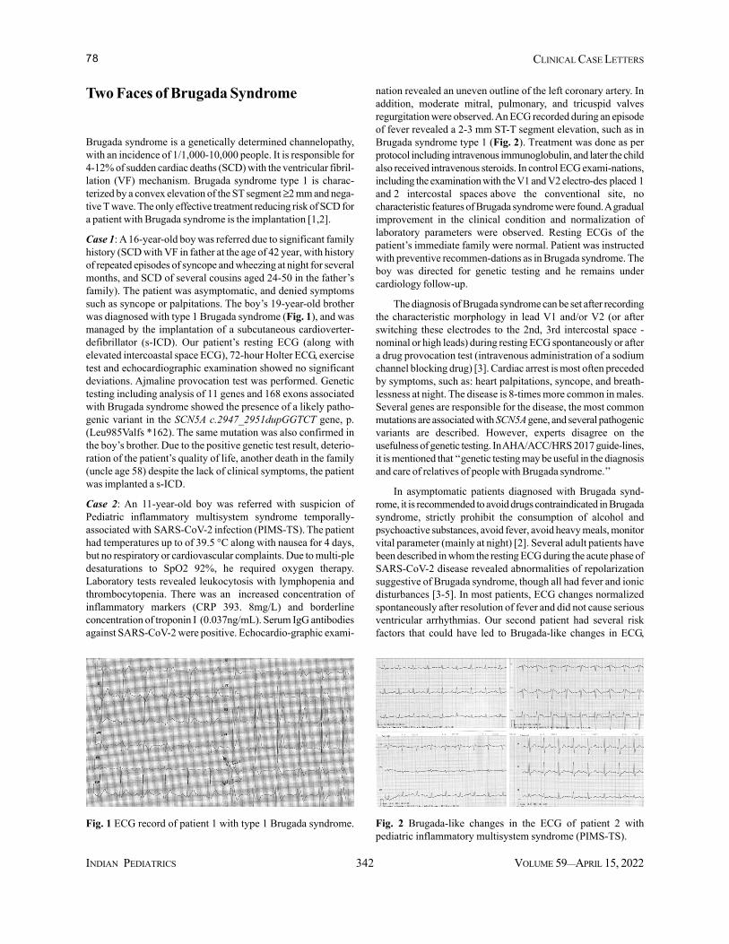
INDIAN PEDIATRICS 342 VOLUME 59__APRIL 15, 2022
CLINICAL CASE LETTERS
Brugada syndrome is a genetically determined channelopathy,with an incidence of 1/1,000-10,000 people. It is responsible for4-12% of sudden cardiac deaths (SCD) with the ventricular fibril-lation (VF) mechanism. Brugada syndrome type 1 is charac-terized by a convex elevation of the ST segment ≥2 mm and nega-tive T wave. The only effective treatment reducing risk of SCD fora patient with Brugada syndrome is the implantation [1,2].
Case 1: A 16-year-old boy was referred due to significant familyhistory (SCD with VF in father at the age of 42 year, with historyof repeated episodes of syncope and wheezing at night for severalmonths, and SCD of several cousins aged 24-50 in the father’sfamily). The patient was asymptomatic, and denied symptomssuch as syncope or palpitations. The boy’s 19-year-old brotherwas diagnosed with type 1 Brugada syndrome (Fig. 1), and wasmanaged by the implantation of a subcutaneous cardioverter-defibrillator (s-ICD). Our patient’s resting ECG (along withelevated intercoastal space ECG), 72-hour Holter ECG, exercisetest and echocardiographic examination showed no significantdeviations. Ajmaline provocation test was performed. Genetictesting including analysis of 11 genes and 168 exons associatedwith Brugada syndrome showed the presence of a likely patho-genic variant in the SCN5A c.2947_2951dupGGTCT gene, p.(Leu985Valfs *162). The same mutation was also confirmed inthe boy’s brother. Due to the positive genetic test result, deterio-ration of the patient’s quality of life, another death in the family(uncle age 58) despite the lack of clinical symptoms, the patientwas implanted a s-ICD.
Case 2: An 11-year-old boy was referred with suspicion ofPediatric inflammatory multisystem syndrome temporally-associated with SARS-CoV-2 infection (PIMS-TS). The patienthad temperatures up to of 39.5 °C along with nausea for 4 days,but no respiratory or cardiovascular complaints. Due to multi-pledesaturations to SpO2 92%, he required oxygen therapy.Laboratory tests revealed leukocytosis with lymphopenia andthrombocytopenia. There was an increased concentration ofinflammatory markers (CRP 393. 8mg/L) and borderlineconcentration of troponin I (0.037ng/mL). Serum IgG antibodiesagainst SARS-CoV-2 were positive. Echocardio-graphic exami-
Two Faces of Brugada Syndrome nation revealed an uneven outline of the left coronary artery. Inaddition, moderate mitral, pulmonary, and tricuspid valvesregurgitation were observed. An ECG recorded during an episodeof fever revealed a 2-3 mm ST-T segment elevation, such as inBrugada syndrome type 1 (Fig. 2). Treatment was done as perprotocol including intravenous immunoglobulin, and later the childalso received intravenous steroids. In control ECG exami-nations,including the examination with the V1 and V2 electro-des placed 1and 2 intercostal spaces above the conventional site, nocharacteristic features of Brugada syndrome were found. A gradualimprovement in the clinical condition and normalization oflaboratory parameters were observed. Resting ECGs of thepatient’s immediate family were normal. Patient was instructedwith preventive recommen-dations as in Brugada syndrome. Theboy was directed for genetic testing and he remains undercardiology follow-up.
The diagnosis of Brugada syndrome can be set after recordingthe characteristic morphology in lead V1 and/or V2 (or afterswitching these electrodes to the 2nd, 3rd intercostal space -nominal or high leads) during resting ECG spontaneously or aftera drug provocation test (intravenous administration of a sodiumchannel blocking drug) [3]. Cardiac arrest is most often precededby symptoms, such as: heart palpitations, syncope, and breath-lessness at night. The disease is 8-times more common in males.Several genes are responsible for the disease, the most commonmutations are associated with SCN5A gene, and several pathogenicvariants are described. However, experts disagree on theusefulness of genetic testing. In AHA/ACC/HRS 2017 guide-lines,it is mentioned that ‘‘genetic testing may be useful in the diagnosisand care of relatives of people with Brugada syndrome.’’
In asymptomatic patients diagnosed with Brugada synd-rome, it is recommended to avoid drugs contraindicated in Brugadasyndrome, strictly prohibit the consumption of alcohol andpsychoactive substances, avoid fever, avoid heavy meals, monitorvital parameter (mainly at night) [2]. Several adult patients havebeen described in whom the resting ECG during the acute phase ofSARS-CoV-2 disease revealed abnormalities of repolarizationsuggestive of Brugada syndrome, though all had fever and ionicdisturbances [3-5]. In most patients, ECG changes normalizedspontaneously after resolution of fever and did not cause seriousventricular arrhythmias. Our second patient had several riskfactors that could have led to Brugada-like changes in ECG,
Fig. 1 ECG record of patient 1 with type 1 Brugada syndrome. Fig. 2 Brugada-like changes in the ECG of patient 2 withpediatric inflammatory multisystem syndrome (PIMS-TS).
78

INDIAN PEDIATRICS 343 VOLUME 59__APRIL 15, 2022
CLINICAL CASE LETTERS 79
including fever, ionic disturbances (hyponatremia, hypo-phosphatemia), as well as damage to the myocardium itself in thecourse of PIMS-TS syndrome (i.e., changes in the coronaryarteries) with negative family history [5-7].
In conclusion, Brugada syndrome is a disease, which, whendetected too late, can result in SCD. However, as our two casesshow, its diagnosis as well as the implementation of appropriatepreventive therapy is not always easy.
Note: Both authors contributed equally to this work.
PIOTR KÊDZIORA,* ALEKSANDRA STASIAKDepartment of Pediatric Cardiology and Rheumatology,
Medical University of Lodz, Poland.*[email protected]
REFERENCES1. Gonzalez Corcia MC, Sieira J, Pappaert G, et al. implantable
cardioverter-defibrillators in children and adolescents with
Brugada syndrome. Am Coll Cardiol. 2018;71:148-57.2. Siera J, Brugada P. The definition of Brugada syndrome. Eur
Heart J. 2017;38:3029-34.3. Papadakis M, Papatheodorou E, Mellor G, et al. The diagnostic
yield of Brugada syndrome after sudden death with normal au-topsy. J Am Coll Cardiol. 2018;71:1204-14.
4. Al-Khatib SM, Stevenson WG, Ackerman MJ, et al. 2017AHA/ACC/HRS Guideline for Management of Patients WithVentricular Arrhythmias and the Prevention of Sudden CardiacDeath: ExecutiveSummary. Circulation. 2018;138:210-71.
5. Long B, Brady WJ, Bridwell RE, et al. Electrocardiographicmanifestations of COVID-19. Am J Emerg. 2021;41: 96-103.
6. Vidovich MI. Transient Brugada-like electrocardiographic pat-tern in a patient with COVID-19. JACC: Case Reports. 2020;2:1241-49.
7. van de Poll SWE, van der Werf C. Two patients with COVID-19and a fever-induced Brugada-like electrocardiographic pattern.Neth Heart J. 2020;28:431-36.Chang D, Saleh M, Garcia-Bengo Y, et al. COVID-19 infectionunmasking Brugada syndrome. Heart Rhythm Case Rep. 2020;6:237-40.
Infantile Anti-N-Methyl-D-AspartateReceptor Encephalitis Post-SARS-CoV-2Infection
The spectrum of neurological conditions associated with severeacute respiratory syndrome coronavirus-2 (SARS-CoV-2) isevolving. Here, we describe a case of N-methyl-D-aspartatereceptor encephalitis (NMDAR-E) with possible temporalassociation with SARS-CoV-2.
A 10-month-old typically developing boy presented withpoor feeding and irritability for 5 days. On day 3 of illness, hedeveloped fever and loose stools with 2 episodes of convulsionson day 5 of illness, when he was brought to our hospital. It wasassociated with loss of pre-morbidly normal eye contact. He hadan upper respiratory tract infection (URTI) 40 days prior toillness onset. At presentation, his axillary temperature was98.20 F, pulse rate was 92/minute, respiratory rate was 24/minuteand blood pressure was 84/54 mmHg. General physical andsystemic examinations were unremarkable. On neurologicalexamination, baby was not interested in surroundings and hadpoor interaction with caregivers. Cranial nerve examination wasunremarkable. Motor system examination revealed normal powerand tone, with brisk deep tendon reflexes. Peri-oral dyskinesiasand bilateral striatal toe were present. Cerebellar and meningealsigns were absent. In view of fever, diarrhea, seizures, acute onsetencephalopathy with extrapyramidal movements, possibilitiesconsidered at admission were post-infectious immune-mediatedconditions (central nervous system demyelination, autoimmuneencephalitis, post-COVID multisystem inflammatory syndrome(MIS-C)) and inherited metabolic disorder. Prior to referral, babyhad a normal cerebrospinal fluid (CSF) study and C-reactiveprotein (CRP) with elevated white cell count (WBC, 26×109/L).
Initial investigations at our center revealed elevated WBC (24×109/L, N61L30), normal CRP (1 mg/L) and procalcitonin (0.25ng/mL). SARS-CoV-2 IgG antibodies were strongly positive(index-20.7, >1.0 positive). Erythrocyte sedimentation rate (22mm/first hour), lactate dehydrogenase (515 U/L), ferritin (19.5 ng/mL) and echocardiography (normal) were not consistent withMIS-C.
Over the next 24 hours, extrapyramidal movements worsenedwith appearance of generalized and oro-linguo-buccal dystoniawith athetosis. Hence, possibility of anti-NMDA encephalitiswas considered. MRI brain was normal. CSF showed 20 cells(95%L), sugar 65 mg/ dL (blood sugar: 102 mg/dL), protein 27 mg/dL. CSF-polymerase chain reaction (PCR) was negative(Eschershia coli K1, Hemophlilus influenzae, Listeria mono-cytogenes, Streptococcus agalactiae, Streptococcus pneumonia,Cytomegalovirus, enterovirus, HSV1, HSV2, HHV6, Humanparechovirus, Varicella zoster virus and Crypto-coccus neo-formans/gatii). CSF sample for NMDA antibodies was sent to thelaboratory.
Child was started on intravenous immunoglobulin (2 g/kg)and pulse methylprednisolone (30 mg/kg/day for 5 days) on day8 of illness. CSF sample was reported strongly positive for anti-NMDA antibodies (indirect immunofluorescence assay). Com-puted tomography (CT) of abdomen and pelvis for tumorscreening was negative. By day 5 of pulse steroids, there was noimprovement in extrapyramidal movements or encephalopathy.Considering severe infantile form of anti-NMDAR encephalitispoorly responsive to first line therapy, weekly rituximab infusion(375 mg/m2/dose/week for 4 doses) was initiated in the secondweek of illness, along with addition of azathioprine for long-termimmunosuppression (2 mg/kg/day). Two weeks after lastrituximab dose, baby remained encephalopathic. Extrapyramidalmovements were partially controlled with clonidine, baclofen andclonazepam. In view of refractory disease, monthly cyclophos-

INDIAN PEDIATRICS 344 VOLUME 59__APRIL 15, 2022
CLINICAL CASE LETTERS80
phamide (750 mg/m2/dose) was administered for 3 doses.Following the first dose, baby achieved sustained eye contact andneck-control within a week. By one month, he could recognizeparents, sit with support, creep and vocalize; mild oro-motordyskinesia and bilateral hand athetosis persisted. Symp-tomscompletely resolved after the second cyclophosphamide dose.Steroids were tapered off over 3 months after initial pulse dose.At the time of last follow-up, extrapyramidal movements werewell controlled and baby was regaining age-appropriatemilestones.
To the best of our knowledge, this is the first case of anti-NMDAR-E associated with SARS-CoV-2 in an infant aged <12months. Anti-NMDAR-E, characterized by severe movementwith encephalopathy, can be triggered by viral infections ortumors. Herpes simplex virus (HSV) encephalitis is the mostcommonly associated viral trigger, and can result in anti-NMDAR-E 4-6 weeks, or longer after an acute encephalitisepisode [1].
SARS-CoV-2 is known to result in strong immune activation,which is broadly termed as MIS-C [2]. Post-SARS-CoV-2immune-mediated manifestations can present within two weeksto a median of 25-45 days after an acute infection [3]. Anti-NMDAR-E associated with SARS-CoV-2 has been reported inonly three children aged 23 months [4], 7 years [5] and 14 years[6]. All three children had a positive SARS-CoV-2 RT-PCR withevolution to encephalitis from acute infection in two children, andno clinical infection in one child. In our case, only IgG antibodieswere positive, indicating a prior infection, which on history maybe correlated with the preceding URTI. Considering the highpopulation seropositivity, a true cause-effect relation cannot beascertained. A positive RT-PCR test during the acute URTIepisode and a positive family history would have streng-thened
the causal association. Molecular mimicry probably best explainsthe pathogenesis for SARS-CoV-2 associated anti-NMDAR-E.Whether it can result in late-onset CNS ence-phalitis, similar toHSV encephalitis, remains to be elucidated. The present report, inconjunction with previous reports, supports the association ofSARS-CoV-2 with NMDARE. Future research focusing onassociation between SARS-CoV-2 and early autoimmunity canhelp understand the underlying pathogenesis.
PRABHJOT KAUR,1 VINAY MV,2 BABU S MADARKAR3*
Divisions of 1Pediatric Neurology, 2Pediatric Hematology-Oncology, and 3Neonatology, Department of Pediatrics,
Rainbow Children’s Hospital, Bengaluru, Karnataka.*[email protected]
REFERENCES1. Sahar N, Nurre M, Simon Q. Infectious trigger for autoimmune
encephalitis: A case report and literature review. Case RepInfect Dis. 2019;2019:0-4.
2. Desai I, Manchanda R, Kumar N, et al. Neurological mani-festations of coronavirus disease 2019: Exploring past to under-stand present. Neurol Sci. 2021;11:1-13.
3. Ramos-Casals M, Brito-Zerón P, Mariette X. Systemic andorgan-specific immune-related manifestations of COVID-19.Nat Rev Rheumatol. 2021;17:315-32.
4. Burr T, Barton C, Doll E, et al. N-Methyl-d-aspartate receptorencephalitis associated with COVID-19 infection in a toddler.Pediatr Neurol. 2021;114:75-6.
5. Sarigecili E, Arslan I, Ucar HK, et al. Pediatric anti-NMDAreceptor encephalitis associated with COVID-19. Childs NervSyst. 2021;37:3919-22.
6. Sánchez-Morales AE, Urrutia-Osorio M, Camacho-Mendoza E,et al. Neurological manifestations temporally associated withSARS-CoV-2 infection in pediatric patients in Mexico. ChildsNerv Syst. 2021;1-8.
ERRATA
Please note following corrections in the article titled “Early goal-directed therapy with and without intermittent superior vena cavaoxygen saturation monitoring in pediatric septic shock: A randomized controlled trial.” published in Indian Pediatr. 2021;58:1124-30.
In Table I, which shows baseline characteristics of the study participants, mean (SD) lactate value in the control group should be‘4.6 (2.9) mmol/L’ in place of ‘66.3 (10.4) mmol/L.’ in the same table, mean (SD) ScvO2 in the intervention group should be‘66.3(10.4)%’ in place of ‘4.6 (2.9)%.’
Appropriate corrections have been done in the web version at https://www. indianpediatrics.net/dec2021/1124.pdf
Please note following corrections in the article titled “Low-dose (0.05 unit/kg/hour) vs standard-dose (0.1 unit/kg/hour) insulin in themanagement of pediatric diabetic ketoacidosis: A randomized double-blind controlled trial” published in Indian Pediatr. 2021;58:617-23.
On page 620, column II, first para, second sentence should be “The hypokalemia was more in malnourished children in thestandard-dose group (P=0.31), and more children in the standard-dose group required a higher concentration of dextrose and taperingof insulin infusion at least once to counter the falling blood glucose (Table II).” in place of “The hypokalemia was more inmalnourished children in the standard-dose group (P=0.31), and more children in the standard-dose group required a higherconcentration of dextrose and tapering of insulin infusion at least once to counter the falling blood glucose P>0.005).”
Appropriate corrections have been done in the web version at https://www. indianpediatrics.net/july2021/617.pdf

INDIAN PEDIATRICS VOLUME 59__APRIL 15, 2022
CLINICAL CASE LETTERS
Web Fig. 1a Hypersensitivity reactions in LTBI.(a) Right eye showing elevated pinkish white noduleat 1 o’clock position at the limbus surroundingengorged hyperemic vessels.
Web Fig. 1b Lower limbs showing multipleerythematous, papulonodular skin lesions, 2-4 mmdiameter over the shin of tibia.

INDIAN PEDIATRICS 345 VOLUME 59__APRIL 15, 2022
CCCCC OOOOO RRRRR RRRRR EEEEE SSSSS PPPPP OOOOO NNNNN DDDDD EEEEE NNNNN CCCCC EEEEE
Encephalitis-like Presentation in Infantsof Bodo Tribe – Thiamine Deficiency orLeigh-like Disease?
REFERENCES1. Webster JJ, Beryl S, George K, et al. Basal ganglia disease
mimicking acute encephalitis syndrome among infants ofbodo tribe, Assam. Indian Pediatr. 2021;58:1183-4.
2. Rao SN, Mani S, Madap K, et al. High prevalence of infantileencephalitic beriberi with overlapping features of Leigh’sdisease. J Trop Pediatr. 2008;54:328-32.
3. Nazir M, Lone R, Charoo BA. Infantile thiamine deficiency:New insights into an old disease. Indian Pediatr. 2019;56:673-81.
4. Solomon T, Thao TT, Lewthwaite P, et al. A cohort study toassess the new WHO Japanese encephalitis surveillancestandards. Bull World Health Organ. 2008;86:178-86.
5. Sastry UMK, Jayranganath M, Kumar RK, et al. Thiamine-responsive acute severe pulmonary hypertension inexclusively breastfeeding infants: a prospective observationalstudy. Arch Dis Child. 2021;106:241-46.
AUTHORS’ REPLYWe thank the readers for their interest in our clinicalobservations [1].
i) According to the WHO, clinically a case of acuteencephalitis syndrome (AES) is defined as a person of anyage, at any time of year, with the acute onset of fever and achange in mental status (including symptoms such asconfusion, disorientation, coma, or inability to talk) and/ornew onset of seizures (excluding simple febrile seizures) [2].We agree that only 70 % of our patients had associated fever;though, all had new-onset seizure and altered sensorium.
Our study from North-eastern India sheds light on atreatable cause of possible thiamine deficiency, whichpresents clinically like an acute encephalitis syndrome.Rao, et al. [3] also reported a cohort of infants fromsouthern India, responding to thiamine. Large community-based studies are needed to find the etiology as to whether itis thiamine deficiency or either of infantile Leigh-likeSLC19A3 gene defect, THTR2 deficiency, or biotin-thiamine responsive basal ganglia disease [1].
ii) We agree to this possibility; though, available literature doesnot support this contention. There are case reports ofvitamin B12 deficiency causing bilateral globus pallidusabnormalities in adults. In the myriad of presentations ofvitamin B12 deficiency, basal ganglia involvement is one ofthe rarest associations [4]. Among infants, infantile tremorsyndrome is associated with vitamin B12 deficiency ormutations affecting the metabolic pathway. However,cortical atrophy, and prominence of ventricular system andsubarachnoid space are the frequently reported radiologicalmanifestations [5].
iii) Table I shows the laboratory parameters of the infants inboth the groups. Cerebrospinal fluid analysis was done for8 infants, which were unremarkable (6 in the non-exposuregroup and 2 in the exposure group).
We read with interest the recent study [1] on basal ganglia diseasemimicking acute encephalitis syndrome (AES) among infants ofBodo tribe, Assam. Studies from India [2,3] have previously alsodescribed clinical improvement in AES following thiamineadministration. We seek the authors’ responses to the followingobservations:
i) According to World Health Organization clinical casedefinition of AES [4], acute onset of fever with change inmental status is required. Thus, how appropriate is the termAES in the absence of fever in about 30% of the studyparticipants in their study [1].
ii) Injectable medication given to the patients containedthiamine along with other vitamins, deficiency of whichmay also cause encephalopathy with basal gangliainvolvement. How was the clinical response ascribed onlyto thiamine and not to other vitamins?
iii) Of the 50 infants studied in the current study, lactate levelswere done in only four infants; however, other investigations,if done, are not mentioned. The study included infants withbasal ganglia changes with all the study participants havingseizures, but there is no data related to involuntarymovements such as dystonia. The lack of features likedystonia, in spite of radiologic evidence of basal gangliainvolvement, is not explained. What were the otherneuroimaging findings apart from those in the basal ganglia –were the brainstem and cerebellar structures involved?
iv) Sastry, et al. [5] reported life-threatening cardiac failure withpulmonary hypertension in infants who were exclusivelybreastfed by mothers that followed vegetarian diet, with aprompt response to thiamine. What was the incidence ofpulmonary hypertension/cardiac failure in the currentstudy? Rao, et al. [2] described overlapping features, betweenLeigh disease, an inherited metabolic disorder, and thiaminedeficiency. Was there any associated significant familyhistory present in these infants?
v) What were the dietary habits of mothers of these infants?Majority of the infants were malnourished, what were theother nutritional deficiencies noted, if any?
VYKUNTARAJU K GOWDA,* UDDHAVA KINHALDepartment of Pediatric Neurology,
Indira Gandhi Institute of Child Health,Bangalore, Karnataka.

INDIAN PEDIATRICS 346 VOLUME 59__APRIL 15, 2022
CORRESPONDENCE
iv) No patient was in cardiac failure clinically. None of theinfants underwent echocardiography. During the periodcovered in the report, we did not have the facility to do RBCtransketolase activity, genetic analysis and vitamin B12levels. No infant had dystonia. However, among the twopatients with neurological sequelae followed-up asoutpatients, one had raised tone in all limbs and the otherhad spastic diplegia.
v) The Bodo community daily diet consists of polished ricewith lentil soup, tubers and meat. The rice is taken alongwith boiled vegetables and leaves. Raw dried freshwater fishare also part of their diet. They are known to consume a lotof tea. Paan, a preparation combining betel leaf with arecanut, sometimes with tobacco, is commonly consumed. Heat-stable thiamine antagonists are known to be present inseveral plants including tea and betel nut. They includepolyphenols; these and related compounds are found in redbeets, red cabbage, betel nuts, coffee and tea [6]. They reactwith thiamine to yield the non-absorbable thiaminedisulfide. Thiamine deficiency in Thailand was reported tobe linked with tea drinking and chewing of fermented tealeaves; tannins being the major component having anti-thiamine activity. Thiaminases are present in the raw
tissues of many fishes, chiefly freshwater fishes. These areheat labile and can be effective antagonists of the vitaminwhen consumed without heat treatment [7].
Maternal subclinical thiamine deficiency could be a possiblefactor for thiamine deficiency in these exclusively breast-fedinfants. Maternal thiamine deficiency during pregnancy often leadsto infantile beriberi in such communities and is thought to accountfor a large proportion of the high infant mortality rates found inthe Philippines, Burma, Cambodia, Laos, Vietnam, and probablyalso in other rice-eating countries [8]. Thankaraj, et al. [9] alsoreported infantile cardiac beriberi responding to thiamine innortheastern rural India, where 23 (92%) infants presenting withacute cardiac failure recovered with thiamine administration.
Response to thiamine was dramatic in our cohort of patients.Clinically they presented with an acute encephalitis syndrome-like picture. With many cases of AES having no definite etiology,our study suggests the addition of thiamine in the treatmentprotocol for AES, and also informs readers to consider thepossibility of thiamine deficiency in a patient with encephalo-pathy without apparent etiology and in an appropriate setting.
JEMIN J WEBSTERBaptist Christian Hospital,
Mission Chariali,Tezpur, Assam.
1. Webster JJ, Beryl S, George K, et al. Basal ganglia diseasemimicking acute encephalitis syndrome among infants ofBodo tribe, Assam. Indian Pediatr. 2021;58:1183-84.
2. Solomon T. New WHO Japanese encephalitis surveillancestandards. Bull World Health Organ. 2008;86.
3. Rao SN, Mani S, Madap K, et al. High prevalence of infantileencephalitic beriberi with overlapping features of Leigh’sdisease. J Trop Pediatr. 2008;54:328-32.
4. Sharrief AZ, Raffel J, Zee DS. Vitamin B(12) deficiency withbilateral globus pallidus abnormalities. Arch Neurol. 2012;69:769-72.
5. Holla RG, Prasad AN. Infantile tremor syndrome. Med JArmed Forces India. 2010;66:186-7.
6. Hilker DM, Somogyi JC. Antithiamins of plant origin: Theirchemical nature and mode of action. Ann NY Acad Sci. 1982;378:137-45.
7. World Health Organization. Thiamine deficiency and its pre-vention and control in major emergencies. WHO;1999.
8. Austin B. Micronutrients I: Vitamins in Health and Disease.Mother Child Nutr. Trop. Subtrop., 2016. p. 317-21.
9. Thankaraj S, Koshy RM, Ismavel V. Infantile cardiac beriberiin rural north east India. Indian Pediatr. 2020;57:859-60.
Table I Laboratory Parameters of the Two Groups
Parameter Non-exposure Exposure groupgroup (n=23) (n=27)
Hemoglobin (g/dL) 9.4 (0.9) 9.3 (1.0)Platelets (109L) 481 (147) 535 (15)Total counts (cell/mm3) 12560 (6895) 14434 (7438)Polymorph (%) 56 (17) 58 (18)Lymphocytes (%) 35 (16) 32 (16)Sodium (mmol/L) 131 (7.5) 134 (7)Potassium (mmol/L) 5 (0.6) 5.1 (0.5)Creatinine (mg/dL) 0.65 (0.15) 0.53 (0.27)CSF n=6 n=2Total counts (cell/mm3) 7 (1.5) 4 (2.8)Neutrophils (%) Nil NilTotal protein (mg/dL) 45.2 (21.3) 38.4 (10)Glucose (mg/dL) 82 (40) 95 (30)Values are presented as mean (SD). CSF-cerebrospinal fluid.
82

INDIAN PEDIATRICS 347 VOLUME 59__APRIL 15, 2022
CORRESPONDENCE
Early Goal-Directed Therapy andSuperior Vena Cava Oxygen SaturationMonitoring in Pediatric Septic Shock:Few Concerns
We read with interest the recent publication on early goal-directedtherapy [1]. We have the following concerns:
Among the therapeutic end-points of shock, lactate <1.6mmol/L was taken as one of the therapeutic end-points butSepsis-3 guidelines [2] have defined lactate >2 mmol/L as one ofthe parameters for the definition of septic shock [2]. The reasonfor the same should be clarified for the benefit of the readers.
It would have added value to the study if therapeutic end-points of shock had also included cardiac index, as recommendedby American College of Critical Care Medicine [3].
GOLLA RAMAKRISHNA, DAISY KHERA*Department of Pediatrics,
AIIMS, Jodhpur,Rajasthan.
REFERENCES1. Jain P, Rameshkumar R, Satheesh P, Mahadevan S. Early goal-
directed therapy with and without intermittent superior venacava oxygen saturation monitoring in pediatric septic shock: Arandomized controlled trial. Indian Pediatr. 2021;58:1124-30.
2. Singer M, Deutschman CS, Seymour CW, et al. The ThirdInternational Consensus Definitions for Sepsis and Septic Shock(Sepsis-3). JAMA. 2016;315:801-10.
3. Davis AL, Carcillo JA, Aneja RK, et al. American College ofCritical Care Medicine Clinical Practice Parameters forHemodynamic Support of Pediatric and Neonatal Septic Shock.Crit Care Med. 2017;45:1061-93.
AUTHORS’ REPLYWe thank the readers for their interest in our study [1]. In ourstudy, the therapeutic end-points of shock cut-off for lactate as<1.6 mmol/L or decreasing trend was taken from a similar studypublished by Sankar, et al. [2]. Our study started in 2015, whilethe Sepsis-3 guidelines were published in 2016 [3].
The target of therapeutic end-point of septic shockrecommended by the American College of Critical Care Medicine(ACCM) includes cardiac index between 3.3 and 6.0 L/min/m2 [4].The level of evidence was graded as grade 2C as per the GRADEsystem mentioned by surviving sepsis campaign [5]. Thoughechocardiography facility was available in our setting, based on theavailable evidence, concern about pediatric age group cardiac indexdata (validation and normality) at the time of protocol preparationand study from similar study setting [2], We decided to adopt theend-point of septic shock without cardiac index.
R RAMESHKUMAR,* PJAINDepartment of Pediatrics, JIPMER,
Puducherry 605 006.*[email protected]
REFERENCES1. Jain P, Rameshkumar R, Satheesh P, et al. Early goal-directed
therapy with and without intermittent superior vena cavaoxygen saturation monitoring in pediatric septic shock: Arandomized controlled trial. Indian Pediatr. 2021;58:1124-30.
2. Sankar J, Sankar MJ, Suresh CP, et al. Early goal-directedtherapy in pediatric septic shock: Comparison of outcomes‘with’ and ‘without’ intermittent superior venacaval oxygensaturation monitoring: A prospective cohort study. Pediatr CritCare Med. 2014;15:e157-67.
3. Singer M, Deutschman CS, Seymour CW, et al. The ThirdInternational Consensus Definitions for Sepsis and Septic Shock(Sepsis-3). JAMA. 2016;315:801-10.
4. Davis AL, Carcillo JA, Aneja RK, et al. American College ofCritical Care Medicine Clinical Practice Parameters forHemodynamic Support of Pediatric and Neonatal Septic Shock.Crit Care Med. 2017;45:1061-93.
5. Dellinger RP, Levy MM, Rhodes A, et al. Surviving SepsisCampaign: International Guidelines for Management of SevereSepsis and Septic Shock: 2012. Crit Care Med. 2013;41:580-637.
83

INDIAN PEDIATRICS 348 VOLUME 59__APRIL 15, 2022
Bai Jerbai Wadia Hospital for Children and Institute of Child Health andResearch, MumbaiNC JOSHI,1 SHAKUNTALA S PRABHU21Department of Pediatrics; and 2Medical Director; Bai Jerbai Wadia Hospital for Children and Institute of Child Health andResearch, Parel, Mumbai 400012, Maharashtra. [email protected]
THE BEGINNING
With a legacy of more than nine decades, Bai Jerbai WadiaHospital for Children and Institute of Child Health andResearch is the first and the largest hospital in the countrydevoted for child health and welfare. It is the first everpublic hospital established in India exclusively for childrenby a joint agreement between the Municipal Corporation ofGreater Mumbai and the Wadia Family in the year 1929.Late Sir Cusrow Wadia and Late Sir Ness Wadia, the twosons of Bai Jerbai Nowrosjee Wadia built this childrenhospital in her memory and declared it open on 12December, 1929. Realization of the need for special care forchildren and establishing a hospital for them in the heart ofthe city, amidst the working classes of Mumbai, by itselfdisplays the vision and philanthropic ideals and legacy ofserving the poor on part of the founders.
Over the years, the foresight of the management andthe faculty heads, who were committed to strike a balanceamongst the four pillars of medical academia as in patientcare, administrative, education and research, has modelledthis institution to have a pride of place among the pediatrichospitals in India.
The Early Years
The hospital was started with a bed strength of 126 withmedical and surgical indoor and a novel outpatientdepartment in a separate building with 24 hour emergencywhich was the first in India for children. Basic pathologyinvestigations and radiology services were also madeavailable. Departments of physiotherapy and occupationaltherapy were started to complement Orthopedic depart-ment and for rehabilitation of handicapped children. SocialService Department with Child Guidance Clinic renderingservices for the deserving, a special school for bedriddenchildren and a recreation center were the other features inthe initial period.
Dr. RN Cooper was the founder principal medical officerfollowed by Dr. Arthur De Sa who also established the
pediatric surgery department. The recognition of thishospital in the early years was due to the dedicated andpioneering efforts of Dr. Cooper, Dr. De Sa, Dr. RV Sanzgiriand Dr. Katrak. They were pioneers in recognizingpediatric medicine and surgery as separate specialties.During this period training of residents was the onlyteaching activity.
The Middle Years – 1950 to 1990
During the next phase of expansion of the institute, onemore floor was added to the hospital in 1964 and bedstrength went up to 300 beds. There were paralleldevelopments of both pediatric medicine and surgerywhen Dr. RA Irani, Dr. SM Merchant, Dr. SJ Dalal and later,Dr. Jer Master, Dr. MP Desai, Dr. KP Mehta and Dr. KNShah joined the institute. The main focus during this timewas to make the institute a high quality and cost-effectivetertiary health care center by adding pediatric andneonatal intensive care units. Pediatric intensive care wasestablished by Dr. Uma Ali and Neonatal intensive careunit was coordinated by Dr. Prem Sheth.
In 1975, the Burns unit and Skin Bank were establishedby Dr. SS Keswani and Dr. Arvind Vartak. The hospital wasthen recognized by University of Bombay for under-graduate and post graduate training in pediatrics throughits affiliation to Seth G S Medical College. The College ofPhysicians and Surgeons also recognized this hospital forDCH and FCPS. The hospital was also recognized byRoyal College of Physicians and Child Health and RoyalCollege of Surgeons. A full-fledged post-graduate trainingprogram took shape consisting of daily ward rounds, postemergency teaching clinics, weekly inter hospital pediatricmeetings, monthly grand rounds and postgraduateseminars which are still a routine now. The Dr. SMMerchant Auditorium is used for such teaching programs,workshops and small conferences.
The novel feature of this hospital was the establish-ment of specialty clinics in cardiology, endocrinology,
ICONIC PEDIATRIC INSTITUTIONS OF INDIAICONIC PEDIATRIC INSTITUTIONS OF INDIAICONIC PEDIATRIC INSTITUTIONS OF INDIAICONIC PEDIATRIC INSTITUTIONS OF INDIAICONIC PEDIATRIC INSTITUTIONS OF INDIA

INDIAN PEDIATRICS 349 VOLUME 59__APRIL 15, 2022
PRABHU & JOSHI
nephrology and epilepsy with EEG for the first time inIndia. The aim was to provide adequate and essentialdiagnostic services for this subset of commonly seenconditions along with clinical research for greater insightinto their epidemiology, etiopathogenesis and clinicalpatterns in Indian settings, thereby offering greateropportunities for learning and training in these specialties.
The Hospital in the Later Years and Today (1990-till date)
After 2000, these specialty clinics were expanded furtherinto divisions offering cutting-edge sub-specialty care,simultaneously maintaining basic general pediatrics.
Pediatric Cardiology division, which was the firstsubspecialty to be established in 1968 by Dr Jer Masterand later assisted by Dr NC Joshi, has now become a stateof art center for cardiac sciences with cardiac surgery andinterventional cardiology.
The Thyroid clinic was started by Dr Meena Desai in1969 and she did exemplary work in congenital hypo-thyroidism. It has now become one of the largest pediatricendocrinology divisions in the country.
Nephrotic syndrome clinic was started in 1975 by Dr.Kumud Mehta. First peritoneal dialysis was done in 1980and Hemodialysis department was started in 2010, andnow is a full-fleged pediatric nephrology department with arenal transplant center.
Epilepsy Clinic was started in 1989 by Dr KN Shah. Since1994, it has become a full-fledged, busy neurosciencesdepartment. Pediatric neurosurgery department under DrChandrashekhar Deopujari was established in 1996.
Dr Zinet Currimbhoy started the hematology andimmunology clinic in 1984, which has now evolved into asuper specialty hemato-onco unit with Bone MarrowTransplant and Stem Cell Therapy.
Pediatric HIV was started by Dr Rashid Merchant in1998 and TB clinic was started in 2007. Pediatric drug-resistant TB center, first one in India, was established in2018.
Dr. Saroj Parekh started the Liver Clinic, which has nowevolved into a full fledged Pediatric Gastroenterologydivision with endoscopy and motility lab under Dr IraShah. In 2015, pediatric pulmonology services were startedby Dr. YK Amdekar.
Another major milestone during this stage was theestablishment of a research laboratory which had modern,sophisticated, diagnostic equipment with Radioimmuno-assay, Hematology and Immunology sections and recentlythe Molecular laboratory. This research laboratory is
recognized by Department of Science and Technology,ICMR and WHO. ICMR has also established anindependent Genetic Research Laboratory here.
Pediatric Surgery Department flourished under Dr S JDalal’s guidance with Dr Vinod Kapur starting laparo-scopy in 1974, Dr Ashok Mathure taking care ofrespiratory anomalies and Dr Vishnu Waingankar and Dr IlaMeisheri having started the Urosurgery division.Currently Dr Pradnya Bendre has set up a ‘state of art’endoscopy and minimal access surgery center. There isalso a perinatal surgery division which holds credit forhaving operated on conjoined twins. We have a specialcenter devoted to cleft lip and palate surgeries under DrMukund Thatte.
The Pediatric Orthopedic surgery department startedsurgery for complex deformities and spine problems underthe luminaries Dr Yagnik, and Dr Ashok Johari. The Clubfoot clinic is one of the busiest in Western India.
The radiology services were started very early underthe guidance of Dr Nadkarni and presently the hospitalhas round the clock dedicated pediatric ultrasound andCT/MRI services.
The Pediatric palliative care department is first of itskind in India and specialized anesthesia and rehabilitationservices are there to compliment patient care.
COMMUNITY INITIATIVES
BJWHC has conducted numerous health camps forchildren across many states including several areas inMaharashtra like Palghar, wherein the impact can alreadybe measured in the community with decrease in number ofmalnourished children. BJWHC is also providing supportfor capacity building, disaster management, screening andtreatment of health conditions and knowledge manage-ment for Government and non-profit organizations.
State Center of Excellence in Pediatric Nutrition,supported by UNICEF, is a hub of State nutritionprograms.
BJWHC has received Nursing Excellence Certificatefrom National Accreditation Board for Hospitals andHealthcare for 2 consecutive terms, which is rare for apublic hospital. The hospital has NABH and NABLaccreditation and moving towards complete digitization.
Every year the entire community comes together toparticipate in the Little Hearts Marathon (LHM), which is asocial event to engage the community and help spreadawareness for prevention of cardiac diseases in children.More than hundred thousand children have supported thecause of LHM over the last 6 years.
85

INDIAN PEDIATRICS 350 VOLUME 59__APRIL 15, 2022
ICONIC PEDIATRIC INSTITUTIONS OF INDIA
EDUCATION AND TRAINING
There are 35 pediatric super specialty programs along withundergraduate and postgraduate degree and diplomacourses. Fifteen fellowship courses, under the aegis ofMaharashtra University of Health Sciences and IndianAcademy of Pediatrics as well as DNB super specialtycourses in pediatric hematology, neonatology andpediatric cardiology, pediatric intensive care, pediatricgastroenterology are run by the hospital
Since 2004, a biannual week-long national post-graduate clinical teaching program is being held along witha month-long clinical DCH and DNB clinical observership.The faculty over the last many decades has been feted innumerous national and international platforms.
The hospital has won over 50 national and inter-national health sector service awards and the motto of thehospital – In deo fide et perseverantia, meaning Trust inGod and Perseverance, sums up its philosophy.
What keeps the hospital at the forefront ofPediatrics in India and beyond, is the professionalcompetence, continuous innovation and dedication ofall departments who manned the hospital during last ninedecades.
THE ICONS OF THE INSTITUTIONThe path-breaking personalities responsible for the initialgrowth and development of the hospital were Prof. SMMerchant.
Dr SM Merchant joined the hospital in 1949 and wasprimarily associated with its initial growth and alsomentored future leaders. He promoted establishment ofsubspecialties and was also a research innovator and setup the research laboratory. He was a teacher par excellenceand one of the towering figures of pediatric medicine inIndia. He used to consider the hospital as his family, andused to say that people working here are the foundationsof the institute and they should be brought together,helped to flourish academically, and made to feel valued.
Recently, since 2012, the hospital has grown by leaps andbounds under the visionary leadership of Dr MinnieBodhanwala. Due to her pioneering efforts, the hospital nowhas 525 beds, of which 225 are intensive care beds.Acknowledgements: The authors would like to thank Dr MBodhanwala, CEO Wadia Hospitals for permission to usephotographs from the hospital collection and also providingvaluable inputs. Dr Sanjay Prabhu and Dr Mihir Oswalcoordinated the manuscript development.Funding: None. Competing interests: Both authors are working asfaculty members in the institute.
86

INDIAN PEDIATRICS 351 VOLUME 59__APRIL 15, 2022
PRABHU & JOSHI
Aerial View of BJWHC.
Dr. SM Merchant
The hospital in1929. The hospital in 2019.
Legends of Bai Jerbai Wadia Hospital for Children – 15 February, 2018.
87

INDIAN PEDIATRICS 352 VOLUME 59__APRIL 15, 2022
ADVERTISEMENT 88

INDIAN PEDIATRICS 353 VOLUME 59__APRIL 15, 2022
ADVERTISEMENT89

INDIAN PEDIATRICS 354 VOLUME 59__APRIL 15, 2022
ADVERTISEMENT 90

Printed and published by Dr Devendra Mishra on behalf of Indian Academy of Pediatrics and printed atCambridge Press, Kashmere Gate, Delhi-110006 and published at 115/4, Ground Floor,
Gautam Nagar, New Delhi 110 049. Editor: Dr Devendra Mishra