
Developmental performance of hospitalized ... - BMC Pediatrics
Upload
khangminh22Category
view
0download
0

2

USMLE® Step 3Lecture Notes
2017–2018
PediatricsObstetrics/Gynecology
SurgeryEpidemiology/Biostatistics
Patient Safety
4

Table of ContentsUSMLE® Step 3 Lecture Notes: Pediatrics Obstetrics/Gynecology SurgeryEpidemiology/Biostatistics Patient Safety
CoverTitle PageCopyrightEditorsFeedback Page
Section I: PediatricsChapter 1: Pediatrics
Case 1Case 2Case 3Case 4Case 5Case 6Case 7Case 8Case 9Case 10Case 11Case 12Case 13Case 14Case 15Case 16Case 17Case 18Case 19Case 20Case 21Case 22Case 23Case 24Case 25Case 26Case 27
Section II: Obstetrics and GynecologyChapter 2: Obstetrics
Case 1Case 2Case 3Case 4Case 5Case 6Case 7Case 8
5

Case 9Case 10Case 11Case 12Case 13Case 14Case 15Case 16
Chapter 3: GynecologyCase 1Case 2Case 3Case 4Case 5Case 6Case 7Case 8Case 9Case 10Case 11Case 12Case 13Case 14Case 15Case 16
Section III: SurgeryChapter 4: Surgery
Case 1Case 2Case 3Case 4Case 5Case 6Case 7Case 8Case 9Case 10Case 11Case 12Case 13Case 14Case 15Case 16Case 17Case 18Case 19Case 20Case 21Case 22Case 23
6

Case 24Case 25
Section IV: Epidemiology and BiostatisticsChapter 5: Epidemiology
Key DefinitionsTypes of PreventionTypes of Prevention: Answers and ExplanationsMeasures of Morbidity and MortalitySpecific and Adjusted RatesSpecific and Adjusted Rates: Answers and ExplanationsMeasures of MorbidityMeasures of Morbidity: Answers and ExplanationsVital Statistics and RatesVital Statistics and Rates: Answers and ExplanationsYears of Potential Life Lost and Survival AnalysisUnderstanding Screening TestsUnderstanding Screening Tests: Answers and ExplanationsStudy DesignsStudy Designs: Answers and ExplanationsStudy Designs: Bias in ResearchStudy Designs: Bias in Research: Answers and Explanations
Chapter 6: BiostatisticsProbability BasicsProbability Basics: Answers and ExplanationsDescriptive StatisticsInferential StatisticsSelecting a Statistical TestSelecting a Statistical Test: Answers and Explanations
Chapter 7: Interpretation of Medical LiteratureIntroductionIntroduction: Answers and Explanations
Section V: Patient Safety and Quality ImprovementChapter 8: Clinical Applications of Patient Safety and Quality Improvement
Principles of Patient SafetyUnderstanding Medical ErrorSystems-Based PracticeQuality Improvement PrinciplesBuilding a Safer Health SystemCare Well DoneChapter SummaryChapter Summary: Answers and Explanations
Chapter 9: Population Health ManagementDefining Population HealthValue-Based CareImplementation of Population Health ManagementChapter SummaryKey DefinitionsKey Definitions: Answers and Explanations
Section VI: Medical AbbreviationsChapter 10: Medical Abbreviations
7

Medical Abbreviations
8

USMLE® is a joint program of the Federation of State Medical Boards (FSMB) and theNational Board of Medical Examiners (NBME), neither of which sponsors or endorses thisproduct.
This publication is designed to provide accurate information in regard to the subject mattercovered as of its publication date, with the understanding that knowledge and best practiceconstantly evolve. The publisher is not engaged in rendering medical, legal, accounting, orother professional service. If medical or legal advice or other expert assistance is required, theservices of a competent professional should be sought. This publication is not intended for usein clinical practice or the delivery of medical care. To the fullest extent of the law, neither thePublisher nor the Editors assume any liability for any injury and/or damage to persons orproperty arising out of or related to any use of the material contained in this book.
© 2017 by Kaplan, Inc.
Published by Kaplan Medical, a division of Kaplan, Inc.750 Third AvenueNew York, NY 10017
10 9 8 7 6 5 4 3 2 1
9

Editors
PEDIATRICS
Eduardo Pino, M.D.Associate Professor, Department of Pediatrics
Marshall University School of Medicine Medical Director, Pediatric ICU
Cabell Huntington Hospital Huntington, WV
GYNECOLOGY AND OBSTETRICS
Elmar Peter Sakala, M.D., M.A., M.P.H., F.A.C.O.G.Professor of Gynecology and Obstetrics
Division of Maternal Fetal Medicine Department of Gynecology and Obstetrics
Loma Linda University School of Medicine Loma Linda, CA
SURGERY
Adil Farooqui, M.D., F.R.C.S.Clinical Assistant Professor of Surgery
Keck School of Medicine, University of Southern California
10

Kaiser Permanente, West Los Angeles Medical Center Los Angeles, CA
Mark Nolan Hill, M.D., F.A.C.S.Professor of Surgery
Chicago Medical School Chicago, IL
PATIENT SAFETY AND QUALITY IMPROVEMENT
Ted A. James, M.D., M.S., F.A.C.S.Medical Director, Clinical Simulation and Patient Safety
Director, Skin & Soft Tissue Surgical Oncology Associate Professor of Surgery
University of Vermont College of Medicine Burlington, VT
Contributors
GYNECOLOGY AND OBSTETRICS
Jason M. Franasiak, M.D.Clinical Fellow
Reproductive Endocrinology & Infertility Rutgers University, Robert Wood Johnson Medical School
Reproductive Medicine Associates of New Jersey New Brunswick, NJ
11

Joshua P. Kesterson, M.D.Assistant Professor
Department of Obstetrics and Gynecology, Division of Gynecologic Oncology Penn State College of Medicine Hershey, PA
PEDIATRICS
SURGERY
Gary Schwartz, M.D.Divisions of Cardiac and Thoracic Surgery
Baylor University Medical CenterDallas, TX
Hena Akhtar, M.D.Assistant Professor
Department of Pediatrics SUNY Stony Brook School of Medicine Stony Brook, NY
Pediatric Hospitalist Children’s Medical Center Winthrop University Hospital Mineola, NY
12

We want to hear what you think. What do you like or not like about the Notes? Please email usat [email protected].
13

Section I
PEDIATRICS
14

1
Pediatrics
Case 1
Chief Complaint
“My eye is red.”
History and Physical Examination
A 16-year-old girl comes to the office with a 2-day history of a watery, red left eye with ayellowish discharge. She reports crusting upon awakening. She denies eye pain and doesnot recall any trauma to the eye. Physical examination reveals diffuse conjunctivalhyperemia of the left eye associated with a mucoid discharge and pupils that dilatenormally with mild photophobia in the affected eye. There is no adenopathy. Thefunduscopic examination, eye movement, and visual acuity are all normal.
CLINICAL PEARL
Always ask about history of trauma or chemical contact.
15

Differential Diagnosis
Clues
CLINICAL PEARL
Because this is an adolescent, be sure to ask about sexual activity. Always consider N.gonorrhoeae, C. trachomatis, and herpes simplex (if vesicles are present). Unilateralversus bilateral erythema is a good first step in thinking about the differentialdiagnosis.
Bacterial conjunctivitis1.Viral conjunctivitis2.Acute glaucoma3.Anterior uveitis4.Trauma: corneal abrasion/foreign body5.Allergic conjunctivitis6.Chemical conjunctivitis7.Periorbital/orbital cellulitis8.
UnilateralNo eye painNo traumaMucoid dischargeNormal pupillary dilatationNormal eye movementNormal visual acuity
16

Initial Management
Setting: outpatient workup and treatment
Diagnostic/Therapeutic Plan
Test Results
Assessment
CCS NOTE
Snellen chart visual acuityConsider Gram stain and culture for N. gonorrhoeae and PCR or antigen test for C.trachomatis, depending on possible history of sexual activity or child abuse
Visual acuity: normalGram stain/culture: negative
Unilateral red eye with mucoid, yellowish discharge and crusting upon awakening is theclassic presentation of a bacterial conjunctivitis. Viral conjunctivitis is likely to be bilateralwith a watery discharge.Allergic conjunctivitis should have itchiness, be bilateral and watery, and have a seasonalcomponent and perhaps other history of allergic disease.With no history of pain or trauma caused by a foreign body, chemical conjunctivitis orcorneal abrasion is highly unlikely.Normal vision and no ophthalmoplegia or proptosis, normal and symmetric pupillary sizeand reaction rule out anterior uveitis, orbital cellulitis, and glaucoma. Also, there is nodescription of periorbital or orbital erythema/edema and no fever or other systemic findings.
17

Always check vision, eye movement, pupillary size, and reactivity. Performfundoscopic examination.
Further Management Plan/Results
Bacterial conjunctivitis will most often resolve spontaneously after several days. However, itis most commonly treated with a topical ophthalmic antibiotic.
Do not use topical steroids if you are unsure of the diagnosis.
CLINICAL PEARL
Routine prophylactic antibiotic eye ointment at birth may delay the presentation ofophthalmia neonatorum from hrs to days.
Discussion
Trimethoprim-polymixin dropsPolymixin-bacitracin ointmentFluoroquinolones (not to be given parenterally if age <18 yrs)
Neonatal red eyeWith eye drainage: perform Gram stain and culture for N. gonorrhoeae and PCR orantigen test for C. trachomatis (ophthalmia neonatorum)
If positive for N. gonorrhoeae treat for both: ceftriaxone IM × 1 with eye irrigationuntil clear for N. gonorrhoeae and erythromycin for 14 days with eye irrigation untilclear for C. trachomatis
–
18

Otherwise, treat for positive C. trachomatis–
With no eye drainage and suggestive of glaucoma: megalocornea, tearing,photophobia, blepharospasm (refer to ophthalmologist)With no eye drainage or evidence of glaucoma: conjunctival hemorrhage, cornealabrasion (uncommon in neonate)
Infant, child, adolescent red eyeHistory of or suspicion of contact with a chemical: irrigate and refer to ophthalmologistHistory of foreign body: examine eye and irrigate if nonpenetrating foreign body;otherwise, refer to ophthalmologistTrauma, no foreign body
Topical anesthetic (tetracaine) x1 for thorough eye exam and vision testing–Fluorescein with Wood lamp examination: corneal abrasion–Topical ophthalmic antibiotic drops–Patching no longer recommended–
Nonconjunctival inflammationBlepharitis: crusting, redness, itching of eyelids–Dacryocystitis: inflammation inferomedial to inner canthus–Dacryoadenitis: inflammation of upper, outer eyelid–Periorbital cellulitis: periorbital erythema, edema, warmth–Orbital cellulitis, proptosis, pain, visual changes, ophthalmoplegia; refer toophthalmologist immediately
–
Signs and symptoms suggestive of glaucoma: refer to ophthalmologistSigns not suggestive of glaucoma:
Keratitis: adenovirus, herpes simplex–Anterior uveitis: IBD, juvenile immune arthritis, Reiter’s syndrome, sarcoidosis,Behçet’s disease, early disseminated Lyme disease, early in Kawasaki’s
–
19

Remember, if it hurts, it is serious.
CLINICAL PEARL
For both periorbital and orbital cellulitis, cover for S. aureus, S. pneumoniae, and H.influenza, and consider possible resistance. Orbital cellulitis is an emergent conditionand needs immediate referral to ophthalmology. MRI of the orbit and surrounding tissueshould be performed to aid surgical drainage. Start IV antibiotics immediately.
Final Diagnosis
Bacterial conjunctivitis
CASE REVIEW
Bacterial Conjunctivitis
Viral Conjunctivitis
Itching, tearing, conjunctival edema, seasonal: allergic conjunctivitis
Red eye with dischargeHistory of crustingTypically painlessTopical antibiotic treatment
Associated with upper respiratory infection
20

Glaucoma
Corneal Abrasion
Uveitis
Look for clear discharge
Painful, blepharospasm, tearing, megalocorneaAbnormal pupil responseOphthalmologist consultation necessary
PainfulPhotophobiaTrauma with abrasionFluorescein stain for corneal abrasionProphylactic topical antibiotics
PainfulVisual disturbancesAbnormal pupilsSlit lamp examination: inflammatory cells in anterior chamberTreat underlying disease with or without topical steroids
21

Case 2
Chief Complaint
Rash on legs
History and Physical Examination
A 6-year-old boy is brought to the office because of a rash that started as a superficialaccumulation of several small vesicles on his legs below the knees. There is no fever orchills. He lives in the suburbs and often walks outside in the local woods in short pants.Vital signs are T 37.0° C (98.6° F), BP 110/70 mm Hg, pulse 84/min, and respirations16/min. Physical examination shows honey-brown, crusted lesions with an erythematousbase on both legs. There are other lesions as well, in various stages of crusting andopenness. The examination is otherwise normal.
Differential Diagnosis
Impetigo1.Cellulitis2.Erysipelas3.Varicella4.Folliculitis (S. aureus)5.Herpes simplex6.Scabies7.Contact dermatitis (e.g., poison ivy)8.Dyshidrotic eczema9.
22

Clues
Initial Management
Setting: outpatient workup and treatment
Diagnostic/Therapeutic Plan
Expected Etiology
S. pyogenes (group A strep or S. aureus [can present as non-bullous])
Assessment
This is the classic description of a streptococcal impetigo (lower extremities, honey-brownlesions with an erythematous base, combination of ulcerated and crusted lesions). The presenceof lesions on exposed legs (the patient wore short pants) and walking outside in the woodssuggest some form of problem secondary to contact. The absence of fever andconstitutional/systemic findings should lead you to assume a benign, localized disease.
Skin lesions due to staphylococcal infections are more likely to occur at the base of folliclesand will range from papulovesicular to bullae. There is no evidence of any deeper or morewidespread disease, such as cellulitis, erysipelas, or staphylococcal scalded skin syndrome.Patients with staphylococcal scalded skin appear much more toxic.
Honey-brown, crusted lesions with an erythematous baseVarious stages of crusting and openness
No cultures need to be done; this is a classic description and the diagnosis is clinicallyobvious
23

Management Plan/Results
Topical antibiotic treatment is warranted if disease is mild and localized: mupirocin is usuallyfirst-line. (Over-the-counter combinations of bacitracin-neomycin-polymixin B are lesseffective.)
Otherwise, treatment is oral, with dicloxacillin/cloxacillin, cephalexin, or clindamycin. Oraltreatment is warranted if there are widespread lesions or bullous lesions.
CLINICAL PEARL
Discussion
A summary of vesicles and bullae is as follows.
With increasing resistance of S. pyogenes and S. aureus, macrolides should not beconsidered.If methicillin-resistant S. aureus (MRSA) is considered, then trimethoprim-sulfamethoxazole or clindamycin is a good choice.Vancomycin is not recommended for oral use but is highly effective intravenously,especially for MRSA.
Age birth to 2 mosGeneralized
Disseminated herpes simplexDisseminated candidiasisErythema toxicum: common, benign neonatal rashEpidermolysis bullosa: inherited blistering disorder; usually begins in neonatal period;
24

blistering in areas of trauma or pressure (extensor surfaces)Incontinentia pigmenti: hereditary multisystem disorder (hair, eyes, CNS, teeth); mostlyfemales; usually starts in first 2 wks of life with crops of bullae on trunk or extremitiesthat become verrucous and then pigmentedCongenital varicellaStaphylococcal scalded skin syndrome
LocalizedSucking blisters: dorsal surfaces of hands, fingers, forearmsBullous impetigoLocalized herpes simplexLocalized candidiasis (perineal)
Age >2 mosWith systemic illness
VaricellaHand-foot-mouth disease: coxsackie virus; painful, ulcerating vesicles on mouth, hands,and feetStaphylococcal scalded skin syndromeStevens-Johnson’s syndrome: most serious form of erythema multiforme; need skin and 2mucous membranesToxic epidermal necrolysis (Lyell’s disease): widespread erythema and tenderness,followed by separation of skin between epidermis and dermis; also with bullae andpositive Nikolsky’s sign
Without systemic illnessGeneralized
Impetigo, bullous impetigo–Scabies: especially palms and soles; extremely pruritic; excoriates easily–Erythema multiforme: vesicobullous, hypersensitivity eruption; target lesions–
25

CLINICAL PEARL
Nikolsky’s sign is seen in staphylococcal scalded-skin syndrome (SSSS) or toxicepidermal necrolysis. With touch or pressure, skin will easily exfoliate.
Follow-up Management and Prevention
Typically, 7 days of therapy is sufficient for mild cases.
Epidermolysis bullosa–Pemphigus: usually seen in adolescents or adults; severe chronic blistering disorder–
LocalizedImpetigo, bullous impetigo–Contact dermatitis–Insect bites–Burns–Herpes–Dyshidrotic eczema–Scabies–Epidermolysis bullosa–Pemphigus–
Crusted lesions may be washed gently.Scratching can spread the organism.As with most infectious diseases, good handwashing can help prevent spread of disease.
26

Complications of impetigo include post-streptococcal glomerulonephritis. Treatment of skininfection does not seem to prevent this complication.
Final Diagnosis
Impetigo
CASE REVIEW
Impetigo
Cellulitis
Erysipelas
Varicella
Superficial infection of epidermisTypically caused by group A streptococcus, S. aureus, maybe MRSAAssociated with hot conditions, poor hygiene, and crowding“Honey-colored,” crusted lesionTopical antibiotics for mild diseaseSystemic antibiotics for more severe disease
Deeper infectionTenderness, erythemaNo crusts, vesicles
Entirely in dermis, raised, indurated, sharply demarcated, tender, bright redNo weeping
27

Folliculitis
Crops of lesions: papules, vesicles, ulcers, crustsPruritisFever
StaphBase of hair folliclesPapulovesicular to bullous
28
Case 3
Chief Complaint
Neonate with nasal flaring, cyanosis, and grunting
History and Physical Examination
A 17-year-old African American girl (G2P1) with unknown LMP and no prenatal carecomes to the emergency department complaining of contractions every 5 minutes.Obstetric U/S is consistent with a gestational age of 28 weeks. Estimated fetal weight is1,000 g and the baby is in breech presentation. The patient is started on MgSO4 fortocolysis and betamethasone for fetal lung maturity. Contractions space out within 1 hour,and the patient is transferred to the antepartum service.
Forty-eight hours later, spontaneous rupture of membranes is confirmed, and the patient istransferred to the labor and delivery floor for further monitoring. There she develops latedecelerations and is thus prepared for an emergent cesarean section, which is uneventful.She delivers a baby girl with Apgar scores of 7 at 1 min, 8 at 5 mins, and weighing 1,140grams. The baby is transferred to the neonatology ICU. Within an hour, the baby developstachypnea, nasal flaring, subcostal and intercostal retractions, cyanosis, and expiratorygrunting.
CLINICAL PEARL
29

Apgar scores are done every 5 mins, as long as there are still resuscitative efforts.
Differential Diagnosis
Clues
CLINICAL PEARL
Pulmonary artery hypertension is more commonly secondary to hypoxia because of aprimary underlying cardiopulmonary problem, compared with primary (idiopathic)pulmonary hypertension of the newborn.
Respiratory distress syndrome (RDS)1.Pneumonia2.Meconium aspiration syndrome3.Transient tachypnea of the newborn (TTN)4.Nonpulmonary causes, i.e., hypothermia, hypoglycemia, anemia, polycythemia,metabolic acidosis
5.
Congenital heart disease6.Diaphragmatic hernia/other congenital pulmonary anomalies7.Primary pulmonary hypertension of the newborn8.
PrematurityThe physical findings are indicative of respiratory distress in general. “Grunting” isexpiring against a partially closed glottis, and indicates that the baby is making attempts toincrease alveolar end-expiratory pressure to preserve oxygenation.
30

Initial Management
Setting: inpatient in delivery room
Diagnostic/Therapeutic Plan
Intervention Results
Done
HR 160/min; RR 60/min
Done
Oxygen saturation 88% in room air; 99% in
Place baby in open radiant warmer with skin
electrodes attached
Place on cardiac and respiratory monitors
Place in 100% oxygen (hood or nasal canula)
Place pulse oximeter on baby
31

100% oxygen
Pulse oximetry 95%
Done
Done
ABG: pH 7.29, pCO2 50, pO2 90, BE –1; other
labs WNL
Baby has less grunting; O2 sats 98%
Done
Done
PATIENT SAFETY NOTE
Wean oxygen so that pulse oximetry is 93–
96%
Order chest x-ray and echocardiogram
Place umbilical venous catheter (UVC) for
fluids/medications and umbilical arterial
catheter (UAC) for blood draws/ABGs; begin
10% dextrose solution via UVC
Draw and send ABG, and send blood to lab
for CBC and differential platelets, glucose,
calcium, and blood culture
Place baby on nasal CPAP of +4–5 cm H2O
with continued O2
Perform sterile suprapubic bladder tap for
U/A and C and S
Transfer to neonatal ICU
32

Don’t forget about hyperoxia and the risk of retinopathy of prematurity.
Initial Assessment
A premature baby with respiratory distress and hypoxemia most likely has RDS (surfactantimmaturity and deficiency). The most important first steps are to place the baby on a heatsource (radiant warmer), place monitors, provide oxygen, and then re-evaluate.
Although the pulse oximeter shows good oxygen saturation, the continued grunting with thehypoxemia, hypercarbia, and acidosis indicate that the baby continues to have respiratorydistress. Nasal CPAP (n-CPAP) would be the first step to open collapsed alveoli in an attemptto improve the respiratory problem. Re-evaluation is needed.
Always perform a sepsis workup (CBC, blood culture, sterile urine culture, ± CSF) and thenbegin empiric antibiotics in any sick newborn; infection is always a possibility. Also check forhypoglycemia and hypocalcemia.
Initial fluid in a newborn is a source of dextrose only (usually D10); electrolytes will not beneeded until at least another 24 hrs; may need calcium depending on serum level (transienthypoparathyroidism).
CLINICAL PEARL
Don’t forget to cover for group B streptococcal pneumonia, Escherichia coli, andListeria monocytogenes. Ampicillin and gentamicin are appropriate if meningitis is not
33

suspected. Otherwise, ampicillin + a third-generation cephalosporin (e.g., cefotaxime)are needed.
CLINICAL PEARL
Although this is the classic appearance of RDS, group B streptococcal pneumonia has asimilar appearance.
Further Diagnostic Plan/Results
Done
Decreased lung volume; reticulogranular pattern
with air bronchograms
ABG in 70% O2: pH 7.28, pCO2 52 mm Hg,
pO2 50 mm Hg, BE –1
Done
Being done
Large persistent ductus arteriosus; no cyanotic
Start antibiotics: ampicillin and gentamicin1.
Obtain CXR results2.
Re-evaluate respiratory status3.
Intubate, place on ventilator, and give
surfactant via the endotracheal tube
4.
Follow oxygen saturations and wean FIO2 as
needed; follow ABGs carefully and wean
ventilator as needed
5.
Obtain cardiac echocardiogram results6.
34

heart lesions
Done
CLINICAL PEARL
We know there will be a persistent ductus arteriosus. The echocardiogram is to rule outcyanotic heart disease. Never give indomethacin until this is known because it willadversely affect a ductal-dependent heart lesion.
BASIC SCIENCE CORRELATE
Begin indomethacin therapy for persistent
ductus arteriosus
7.
Surfactant is responsible for decreasing surface tension, thereby preventing alveolarcollapse and atelectasis. It is produced in the type II alveolar cells, which start toform at about 24 wks’ gestation.
Surfactant composition–Lipid-mostly phosphatidylcholineProtein-surfactant proteins A, B, C, D
Preterm babies have decreased quantity and quality of surfactant–Incidence of RDS drops as gestational age increases; incidence significantlydrops after 35–36 wks’ gestation
–
35

Discussion
The best initial diagnostic test for sorting out the various etiologies of respiratory distress ischest x-ray. Look for the following:
The definitive test is amniotic fluid or tracheal aspirate for lecithin-sphingomyelin (L/S) ratio(part of the full lung profile).
A major problem with RDS is hypoxemia due to alveolar collapse at end-expiration, andtherefore, ventilation-perfusion mismatching. Hypercarbia and acidosis occur later withrespiratory failure and the effects of a left-to-right shunt via a persistent ductus arteriosus.
Immediate intubation is not always necessary with RDS. One can either start oxygen and n-CPAP before re-evaluating or perform intubation and give prophylactic surfactant. Bothmethods are considered appropriate.
Oxygenation must be followed carefully to prevent hyperoxia and ventilation must beperformed carefully to prevent barotrauma. The patient must be weaned constantly based onexamination, pulse oximetry, blood gases (and pulmonary function measurements).
Final Diagnosis
Respiratory distress syndrome (RDS)
Ground glass appearance (reticulogranular pattern)Air bronchogramsAtelectasis
Done on amniotic fluid before deliveryRatio >2 indicates low risk for RDS
36

CASE REVIEW
Respiratory Distress Syndrome (RDS)
Pneumonia
Meconium Aspiration Syndrome
TTN
Early-onset respiratory distressCaused by surfactant deficiency and biochemical immaturityChest x-ray shows diffuse granular pattern with air bronchogramsManage with oxygen, CPAP or ventilation with PEEP, and surfactant therapy
Group B streptococcal pneumonia (most common) can look like RDS on chest x-rayTreat with ampicillin/aminoglycoside or ampicillin/cefotaxime
Look for history of meconium-stained amniotic fluidGood suctioning of oropharynx and the trachea via an endotracheal tube in alldepressed newborns with thick, particulate meconiumAspiration pneumonia on chest x-rayPredisposes to persistent pulmonary hypertension of the newborns
Rapid second stage of labor or delivery by cesarean sectionTerm newbornFluid in fissure on chest x-ray
37

38

Case 4
Chief Complaint
“My baby has been vomiting for 2 days.”
History and Physical Examination
A 3-week-old boy is brought to the emergency room with a history of vomiting after eachfeeding. The symptoms started 2 days ago and have been worsening over the last 24 hours.The mother describes the vomiting as projectile and occurring within 30 minutes after hefeeds. He has appeared to be a bit lethargic over the last 12 hours. There is no history offever or diarrhea. The mother states that the infant wants to have the formula and is hungry.She just fed him 20 minutes ago, and urine was passed within the hour. There is no historyof recent upper respiratory infection or exposure to any such cases. The infant was fedformula from birth, and for the first month had an intake of about 4 ounces every 4 hrs.There was the passage of 5–6 stools per day of a pasty consistency and yellow in color,along with the passage of urine about 7 times per day.
The mother is a 24-year-old white woman who had regular prenatal care and a normalspontaneous vaginal delivery. The newborn weighed 6 lb, 4 oz and had an uneventfulpostnatal course; the mother and newborn left the hospital within 3 days after delivery.The infant appears very irritable but consolable in his mother’s arms. Physicalexamination shows no acute respiratory distress or evidence of an exanthem. His skin iswarm to the touch with average skin turgor. Further examination shows:
HEENT: normocephalic, atraumatic; anicteric sclera, pink conjunctiva, positive red reflex
39

CLINICAL PEARL
A major indicator to the severity of illness is that the neonate still wants the formulaand remains hungry despite the vomiting.
bilaterally; anterior fontanelle open and soft; external auditory canals are WNL bilaterally;bilateral tympanic membranes with positive light reflex; nasal septum in midline, noevidence of polyps; oral cavity shows moist mucosa; no exudates or other abnormalities
Neck: supple, no adenopathy, no thyroid masses
Lung: good respiratory effort, clear to auscultation within the limits of examination
Cardiovascular: SI, S2 regular; no murmurs appreciated
Abdomen: positive bowel sounds, no organomegaly appreciated; during examination, infanthad nonbilious projectile vomiting; on palpation after the vomiting, a palpable, hard,mobile, nontender mass was appreciated in the epigastrium to the right of midline
Genitalia: Tanner stage l, testicles descended bilaterally; no inguinal masses found
Extremities: full ROM; no cyanosis or edema; no subluxation click found
Neurology: no focal deficits; normal infant with positive Moro reflex; good muscle tone inall extremities
In any patient with vomiting, diarrhea, or decreased oral intake, always establishstate of hydration through history and physical examination.Vomiting alone in a neonate should be considered GI obstruction until proven
40
CLINICAL PEARL
This is the description of an “olive.” The best time to feel for this mass is after anepisode of vomiting.
Initial Management
Setting: initially outpatient, but inpatient admission will be necessary
Differential Diagnosis
Clues
otherwise.
Pyloric stenosis1.Gastroesophageal reflux2.Viral gastroenteritis3.Malrotation with intermittent volvulus4.Infection5.Inborn error of metabolism6.
Male, firstborn, CaucasianAge 3–4 wksNonbilious projectile vomitingRemains hungry, wants to feedPalpable “olive”
41

Initial Diagnostic Plan/Results
WBC 12,700/mm3; neutrophils: 68%, bands:
6%, lymphocytes: 24%
Na 129, Cl 92, HCO3 28, K+ 3.1, BUN 24,
glucose 92, creatinine 0.8
Normal, except specific gravity 1.030
Hypertrophied pylorus
If U/S is unavailable, perform an upper GI series. Results would show vigorous gastricperistalsis with little emptying of the contrast material. An elongated narrow pyloric canal seenas a single tract (string sign).
CLINICAL PEARL
In the evaluation of hydration status, it is always appropriate to check electrolytes,BUN/creatinine, glucose, and urinalysis.
Assessment
Classic lab findings are hypokalemic, hypochloremic metabolic alkalosis (loss of Cl andHCO3 and shift of K). The child may have hyponatremia or isonatremia.
CBC with differential1.
SMA-72.
Urinalysis3.
U/S4.
42

The diagnostic procedure of choice is the U/S. The “target lesion” is seen—a dark outer ringof hypertrophied pylorus with small, white central lumen.
Treatment
Treatment is surgical (pyloromyotomy). Start IV fluids to correct any dehydration andelectrolyte abnormalities (Na, Cl, K). After surgery, slowly increase intake to full feeds over48–72 hrs.
Initial Workup
Treatment depends upon diagnosis and whether surgery is needed.
If evidence of infection: diarrhea (check stool for blood, cells), known exposures, feverIf evidence of partial or complete obstruction: plain radiographs, barium studies
Decreased or lack of passage of stools–Abdominal distension, decreased bowel sounds–Radiograph to check air/fluid level, lack of distal bowel gas–
When there is likelihood of pyloric stenosis, do an U/S of the pyloric regionEvaluation of hydration status (history, examination, electrolytes, U/A) with fluid/electrolytetreatment (oral or IV)When evidence of acute abdomen, get surgical consultFor possible gastroesophageal reflux
Fluid maintenance and replacement: for most cases use small, frequent volumes of oralrehydrating solution; for severe cases that have intractable vomiting or shock, use IVfluids/electrolytesIf there is a viral infection, use only fluid management; bacterial enteritis (positive stoolculture) may need antibiotic treatment, depending on organism and patient’s ageWith any decreased peristalsis or ileus (with abdominal distension), use nasogastric tube
43

Final Diagnosis
Pyloric stenosis
CASE REVIEW
Pyloric Stenosis
Gastroesophageal Reflux (GER)
Viral Gastroenteritis
Duodenal Atresia
with low suction; follow electrolytes and replace losses as needed
Projectile vomitingOliveClassic electrolytes
Spitting up and vomiting; usually not projectileDifficulty feedingMay be associated with a cough or wheezingUsually does not result in electrolyte imbalanceTreat with thickened feeds, gravity H2 blockers, proton pump inhibitors
Associated with diarrhea, low-grade fevers, and vomitingSelf-limiting
Bilious vomiting from birth“Double bubble” on abdominal film, no distal bowel gas (compared with
44

intermittent volvulus)Association with trisomy 21
45

Case 5
Chief Complaint
“My son has had belly pain and blood in his stool.”
History and Physical Examination
A 2-year-old boy is brought to the emergency department by his concerned motherbecause he just passed a stool with red blood and mucus. He has been irritable for the last36 hours, with intermittent attacks of belly pain, accompanied by straining efforts and loudcries. He also had some episodes of emesis, but not in the last 24 hours. He had milddiarrhea a week ago and a URI that resolved spontaneously over the last 5 days. He hasno known medical problems and is growing and developing well.
Physical examination shows a pale, very irritable child with T 39.0 C (102.2 F), pulse120/min, and BP 90/60 mm Hg. Further examination shows normal HEENT, pale skinwith no rash, RRR with no murmur, and clear lungs. The abdomen is distended, and ondeep palpation a slightly tender, sausage-shaped mass is felt in the right upper abdomenwith its axis cephalocaudal. There is no hepatosplenomegaly. Capillary refill is 2seconds. Neurologic exam is nonfocal and cranial nerves are intact.
Differential Diagnosis
Intussusception1.Meckel’s diverticulum2.Henoch-Schönlein (anaphylactoid) purpura with possible secondary intussusception3.
46

CLINICAL PEARL
In a young child, abdominal pain with blood in the stool is intussusception until provenotherwise.
Clues
CLINICAL PEARL
A tender, sausage-shaped mass is a pathognomonic physical finding.
Initial Management
Setting: emergency department
Diagnostic/Therapeutic Plan
Bacterial/parasitic enteritis4.
Age of patientStool color (“currant jelly”)Intermittent painPrior infection
CBC
47

Test Results
PATIENT SAFETY NOTEAir contrast enema is now preferred to barium enema because it has a significantlylower rate of perforation. Enema is contraindicated if there is any evidence ofintestinal perforation.
CLINICAL PEARL
U/S is the imaging modality of choice for the diagnosis and exclusion ofintussusception showing a target-like mass ~3 cm in diameter. This, therefore, isindicated in the stable patient immediately after the history and physical and prior toany radiographic intervention for reduction.
Place IV, give bolus of 20 cc/kg NS; follow with IV hydration therapyNotify surgeon of caseAbdominal flat plate, followed by air contrast enema (diagnostic and therapeutic)
WBC: 14,000/mm3 with neutrophils 85% (bands and segmental); hemoglobin 10 g/dL;platelets 230,000/mm3
Abdominal flat plate: density in RUQ; no free air; air contrast enema diagnostic forintussusception; radiologist will attempt reduction
48

Assessment
The following findings are diagnostic for intussusception.
History
Physical Examination
Diagnostic Findings
Treatment
Acute, severe, paroxysmal attacks of abdominal painTypically occurs approximately 1 week after nonspecific viral illnessSevere irritability and strainingAdopts lateral recumbent position, with hips and legs flexedMay have bloody stool or can present early without any bleedingMost have vomiting
Toxic appearing, febrileOften unconsolableTender, sausage-shaped cephalocaudal mass in right upper quadrantAbdominal distensionTypical currant jelly stool
Plain film nonspecific; obtain prior to air contrast to make sure that there is no perforation;if there is perforation, do immediate surgeryAir contrast enema shows sudden interruption of air into a coil-like intestine.
Portion of proximal bowel telescopes into distal–Most are ileocolic or ileoileocolic–
No hydrostatic reduction if there are signs of shock, perforation, peritoneal irritation
49

Discussion
Initial studies for any GI bleeding include stool guaiac, CBC, platelets, reticulocytes, PT/PTT,bleeding time, comprehensive metabolic panel. Consider microbiologic studies if suggested byhistory.
Workup of rectal bleeding in a child age >3 mos is detailed below.
Melena
IV resuscitation as needed–Immediate surgery–
Success rate of air contrast reduction under fluoroscopy or U/S is 70–90% if performedwithin first 48 hrs of symptoms, and 50% after 48 hrsIf surgical manual operative reduction is not possible or if any bowel is not viable, thenresection with end-to-end anastomosis
Suspect Meckel diverticulum (“brick-red” blood)Yes, suspect: perform Meckel scan–
Positive: Meckel diverticulumNegative: upper GI barium study (upper GI imaging) or endoscopy
No, don’t suspect: perform UGI or endoscopy–Normal or unable to perform: bleeding scan or angiogramAbnormal> Peptic ulcer disease> GER/esophagitis> Mallory-Weiss syndrome> Varices> Volvulus
50

Hematochezia
Does examination show fissures or hemorrhoids? If no, then consider microbiologic studies ifsuggested by history. After that:
Final Diagnosis
> Intestinal duplications> Vascular malformations> Tumor
Suspect Meckel diverticulumYes, suspect: as above for Melena–
No, don’t suspect: colonoscopy or barium enemaIf normal, perform UGIIf abnormal, consider:> Fissures> Polyps> Hemorrhoids> Colitis (infectious, allergic)> Vascular malformation> Vasculitis (e.g., Henoch-Schönlein purpura)> Inflammatory bowel disease> Hemolytic uremic syndrome> Tumor
Suspect intussusception: plain film followed by air contrast reduction (if notcontraindicated)
Normal: colonoscopy as above–Abnormal: intussusception–
51

Intussusception
CLINICAL PEARL
Intussusception is the most common cause of intestinal obstruction age 3 mos to 6 yrs.Most of the differential diagnoses are not very common.
52

CASE REVIEW
Intussusception
Gastroenteritis
Colicky, abdominal painPalpable, sausage-shaped abdominal massAir contrast enema both diagnostic and therapeutic
May have vomitingOther ill contactsUsually no blood (unless bacterial)Pain more diffuse, less regular
53

Meckel Diverticulum
Henoch-Schönlein Anaphylactoid Purpura
Painless rectal bleedingDisease of “2’s” (2 inches long, 2 feet from ileocecal valve, 2% of population,commonly presents in first 2 yrs of life, and may have 2 types of epithelia)Meckel scan (technetium scan)May cause intussusception
Characteristic rashJoint symptomsCentral nervous system and renal involvementMay cause intussusception
54
Case 6
Chief Complaint
“Why is my child so short?”
History and Physical Examination
A 7-year-old boy is brought to the office because of parental concern about his growth.His mother had an uneventful pregnancy and delivery was via NSVD. Both the mother andnewborn left the hospital within 2 days, and the child was breastfed for the first 6 mos. Hesat up at age 7 mos and walked with assistance at age 11 mos. All developmentalmilestones were WNL; however, he was found on the growth chart to be at the fifthpercentile during early childhood and has remained there until this time. There was noprior history of any systemic illness, and he has a good appetite, being at the 30thpercentile for weight. There are 2 other siblings, a brother (age 2) in good health and atthe 15th percentile for height. His sister (age 12) is in good health. His father is 183 cm (6feet) tall, and his mother is 165 cm (5 feet 5 inches) tall. His father, on questioning, statesthat he was generally shorter than his friends in school until age 14. There is no history ofheadache, vomiting, visual disturbance, anorexia, or diarrhea. There is also no history ofpolyuria or polydipsia. The patient appears to be in no acute distress and is verycooperative and friendly. His short stature is the only obvious abnormality.
Physical examination shows:
HEENT: head is normocephalic, atraumatic; pupils equally reactive to light, anictericsclera, pink conjunctiva; fundi without any gross abnormalities; no frontal bossing or flat
55

Differential Diagnosis
nasal bridge; nasal septum is in midline; no nasal polyps; bilateral external ear canals WNL;tympanic membranes with positive light reflex bilaterally; oral cavity shows moist mucosawithout any exudates; mild tonsillar hypertrophy bilaterally without any midlineapproximation
Neck: supple, with full ROM; no adenopathy is appreciated; no thyroid masses; trachea is inmidline
Lungs: clear to auscultation bilaterally
Cardiovascular: S1, S2 regular; no murmurs appreciated
Abdomen: positive bowel sounds, no tenderness, no organomegaly appreciated, no truncalfat deposition
Genitalia: Tanner stage I, testicles descended bilaterally; no inguinal adenopathy
Extremities: full ROM, no cyanosis, clubbing, or edema; pulses +2 symmetrically bilaterally
Neurology: no focal deficits
Constitutional growth delay1.Familial short stature2.Short stature due to chronic disease (chronic infection, rheumatic disease, chronicanemia)
3.
Short stature due to endocrine disease (growth hormone deficiency, hypothyroidism,diabetes)
4.
Deprivational short stature (failure to thrive)5.Genetic/syndromic short stature (Turner syndrome, Down syndrome)6.
56

CLINICAL PEARL
The particular history of the father’s growth is very important. Be sure to ask aboutboth parents’ growth and measure the heights of both parents.
Clues
CLINICAL PEARL
Both parents will be short with familial short stature.
Initial Management
Setting: outpatient
Initial Diagnostic Plan/Results
Normal velocity but growing along the fifth
percentile
Normal developmentNormal childNormal growth velocityParents of normal height
Examine growth curve1.
57

Radiologist reports bone age consistent with 5
yrs of age
CLINICAL PEARL
Failure to thrive secondary to decreased nutrient intake, absorption, or utilization, orincreased caloric expenditure. Look for psychosocial and environmental issues.
Diagnostic/Therapeutic Plan
Assessment
Based on the history, this is a clear case of constitutional delay, i.e., a delay of growth andpubertal maturation until later in adolescence.
A careful history and physical examination is warranted.
Request bone age, i.e., x-ray of left hand and
wrist
2.
None; follow growth and pubertal development; give reassurance to patient and family
History of at least 1 parent is the same as that of patientEnd-growth will be normal based on parents’ heightsBiggest concern is typically delayed pubertal development
Exclude nutritional issues: examine weight attainment and weight for heightExamine psychosocial/environmental issuesPerform thorough past medical history and review of systems for any evidence ofundiagnosed illness
58

CCS NOTE
With the history presented, no other tests would be required at this stage. One couldeven make a point of not performing the bone age now in light of the positive familyhistory
Examine the growth curve. Is the child growing normally along a growth percentile or has therebeen a “flattening” or “falling off” of the curve? In other words, is there normal versusabnormal growth velocity?
Discussion
Do a workup of short or tall stature. First, define growth:
Perform complete and careful physical exam, including neurologic; look for any dysmorphicfeatures
Accurate measurements of weight, height, occipital-frontal head circumferenceGrowth curve
Measurements should be plotted at each well-child visit and ideally at each sick visit aswell.
–
Growth velocity (GV) = yearly increments of growth (most sensitive indicator of child’sgrowth) (normal versus abnormal)
–
Chronological age (CA) of patient = actual age, in yrs and mosBone age (BA) = plain radiograph of left hand and wristDefinition of short stature is growth below 3rd percentileDefinition of tall stature is growth above 97th percentile
59

The ideal (i.e., perfectly average) situation is CA = BA, with normal GV.
CLINICAL PEARL
The radiologist interprets the bones of the hand and wrist, comparing them with‘normals’ for the age. The report will then state the bone age as consistent with aparticular age. It will therefore be normal for age, delayed, or advanced.
Short Stature
CA > BA Normal GV: diagnosis = constitutional growth
delay
Abnormal GV: diagnosis = chronic
systemic/endocrine disease or nutritional
problem
CA = BA Normal GV: diagnosis = familial short stature;
small for gestational age (SGA) infants
Abnormal GV: diagnosis =
genetic/chromosomal/syndrome
Tall Stature
CA
<
BA
Normal GV: obesity, familial tall stature
60

Abnormal GV: diagnosis = precocious puberty; hyperthyroidism; growth hormone excess;
congenital adrenal hyperplasia; certain genetic/syndrome illnesses
Final Diagnosis
Constitutional growth delay
Differential Diagnosis and Therapy of Short Stature
Panhypo-
pituitarism
Constitutional
Delay
Familial
Short
Stature
Deprivational
Dwarfism
Turner
Syndrome
Family history Rare Frequent Always No No
Sex Both Males >
females
Both Both Female
Dentition Delayed Slight delay Normal Variable Normal
Facies Immature
or
dysmorphic
Normal Normal Normal Turner facies or
normal
Sexual
development
Delayed Delayed Normal Delayed Prepubertal
Bone age Delayed Delayed Normal Usually
delayed;
growth arrest
lines
Slight delay
61

Hypoglycemia Variable No No No No
Chromosomes Normal Normal Normal Normal 45,XO or partial
deletion of X
chromosomes
or mosaic
T4 Low or
normal
Normal Normal Normal or low Normal:
hypothyroidism
may be acquired
Growth
hormone
Low Normal Normal,
rarely
biologically
active GH
Low Normal,
occasionally low
Insulin-like
growth factor
Low Normal for
maturation
Normal Low Normal
Tests Stimulated
GH
secretion,
cranial CT
None specific None
specific
Observation in
controlled
environment
Karyotype
Therapy Replace
cortisol, T4,
and GH
deficits as
indicated
Reassurance;
testosterone to
initiate
secondary sex
changes in
selected
patients
None: GH
therapy is
controversial
Improve
environment
Sex hormone
replacement,
GH/oxandrolone
appears useful
62

63

CASE REVIEW
Constitutional Growth Delay
Familial Short Stature
Pathologic short stature
Normal growth velocityNormal physical examParents of normal heightBone age less than chronologic age
Normal growth velocityNormal physical examRelatives with short statureBone age equal to chronologic age
Abnormal growth velocity (falling off growth curve)Development may be delayedPossibly abnormal physical exam (look for dysmorphisms)Bone age equal or greater than chronologic ageAlso think of endocrine, genetic, nutritional causes
64

65

Case 7
Chief Complaint
Newborn with tachypnea and duskiness
History and Physical Examination
The pediatrician on call is notified by one of the newborn nursery nurses that a neonatewhom he has not seen is now breathing fast and has a dusky appearance. It is an 11-hour-old male infant who was born at 37 wks’ gestation to a 19-year-old G1P1 woman whohad no prenatal care. According to the mother, her pregnancy went well and withoutproblems. She had an NSVD. The baby weighs 6 lb and has Apgar scores of 7 and 8. Onlystimulation is required in the delivery room.
The baby is taken to the newborn nursery and starts to bottle-feed shortly after admission.He takes the first 3 feedings without difficulty; prior to the next feeding he is examined bythe nurse. Vital signs are respirations 60/min, pulse 160/min, and BP 80/40 mm Hg in theleft arm. Compared with his previous color, he now appears to be somewhat dusky. It is atthis time that the nurse calls the pediatrician. A physical examination is conducted,revealing the following:
General: patient is awake and alert with vigorous respiratory pattern (60/min) and mildsubcostal retractions
Skin: dusky overall with cyanosis of fingers and toes
66

CLINICAL PEARL
HEENT: circumoral cyanosis and mild nasal flaring
Chest: there is good bilateral air entry. There are no adventitious sounds. The chest issymmetric, with mild subcostal retractions.
Cardiovascular: heart rate and rhythm are regular; S1 is normal and S2 is soft and single;grade IV/VI SEM is heard at mid to upper left sternal border with a thrill felt at midsternalborder; pulses are 1+ and symmetric; capillary refill is 2+
Abdomen: soft without mass; liver edge is palpated 2 cm below right costal margin; spleenis not palpated, and kidneys feel normal
Genitourinary: patient is a normal male with bilaterally descended testes.
Extremities: cyanosis and perfusion are noted as above; no gross malformations ordeformations
Neural: patient is alert, reactive to painful stimuli; tone is symmetric but mildly decreased;DTRs are 1+ and symmetric
The presence of a thrill denotes at least a grade IV murmur; a thrill is alwaysabnormal.A single S2 occurs in pulmonary or aortic atresia or severe stenosis, truncusarteriosus, and often in transposition (as the aorta is to the right and anterior to thepulmonary valve, thus obscuring the sound of pulmonic closure).
67

Differential Diagnosis
Clues
Initial Management
Setting: inpatient, wellborn nursery
Initial Diagnostic and Treatment Plan Results
68%
Done
pH 7.42, pCO2 38 mm Hg, PaO2 43 mm Hg,
base deficit –1
Done
pH 7.43, pCO2 37 mm Hg, PaO2 48 mm Hg,
Cardiac disease1.CNS disease/depression2.Primary pulmonary disease3.Pulmonary artery hypertension4.
Age of patientDuskiness (cyanosis)Murmur, single S2 (therefore, severe pulmonary stenosis)
Pulse oximetry1.
Prepare to put baby in 100% oxygen2.
Arterial blood gas in room air3.
Start peripheral IV and IV fluids with 10%
dextrose; also start PGE1 drip
4.
Repeat arterial blood gas in 100% oxygen5.
68

Base deficit 0
Hb 17.3 g/dL, Hct 55 , WBC 11,300/mm3, 66%
polys, 8% bands, 20% lymphocytes, 6%
basophils, platelets 230,000/mm3
Done
Done
SG 1.019, dipstick negative, no WBC or RBC
70 mg/dL
9.2 mg/dL
Decreased lung markings; heart is normal size
with apex lifted off diaphragm
Right ventricular hypertrophy and right axis
deviation
Done
CLINICAL PEARL
Blood work:6.
CBC, differential, platelets
Blood culture
Urine culture
Urinalysis
Serum glucose
Serum calcium
Chest radiograph7.
Electrocardiogram8.
Start IV ampicillin and gentamicin9.
69

Always perform a sepsis workup and start antibiotics in any sick neonate. Also, checkserum glucose and calcium for hypoglycemia and hypocalcemia, respectively.
CCS NOTE
If a congenital cyanotic heart lesion is suspected, always initiate a PGE1 drip to keepthe ductus arteriosus open and allow for pulmonary blood flow, as you do not initiallyknow if it is a ductal-dependent lesion).
BASIC SCIENCE CORRELATE
Assessment
For the initial evaluation of a cyanotic infant, use the following guidelines.
Hypoxia causes pulmonary vasoconstriction in an effort to shunt blood to ventilatedareas.Pulmonary artery hypertension may be secondary to hypoxia or may be primary(idiopathic). Increased pulmonary vascular resistance usually is responsive tooxygen, hypocarbia, alkalosis, and pulmonary vasodilators.
Breathing patternWeak, irregular, and weak suck = CNS disease–
70
New differential diagnosis:
Decreased lung markings on chest radiograph suggest a lesion producing decreased pulmonaryblood flow. The combination of heart appearance, electrocardiogram, and clinical findingssuggests tetralogy of Fallot.
CLINICAL PEARL
Vigorous or labored with tachypnea = primary respiratory or cardiac disease–Clinical appearance suggests either primary respiratory or cardiac disease–
Hyperoxia test: arterial blood gases first in room air, then in 100% oxygenWith CNS disease, PaO2 completely normalizes–With pulmonary disease, PaO2 increases significantly as V/Q inequalities are overcomeby oxygen
–
If PaO2 >150 mm Hg in 100% oxygen, intracardiac shunt is usually excluded–The results are most consistent with a fixed intracardiac shunt, i.e., a congenital cyanoticheart lesion
–
Tetralogy of FallotTransposition of the great arteriesTruncus arteriosusTricuspid atresiaTotal anomalous venous returnEbstein anomalyHypoplastic left heartOther complex congenital heart disease
71

To rule out pulmonary artery hypertension, the patient should be ventilated to attainmild hypocarbia and alkalosis.
Further diagnostic and treatment plan Results
Pulmonary valve and infundibular stenosis;
unrestricted ventral septal defect with overriding
aorta and right ventricular hypertrophy
Done
Discussion
Congenital heart disease occurs in 0.5–0.8/100 live births.
CLINICAL PEARL
Cardiologist arrives and performs an
echocardiogram
1.
Transport is arranged to nearest tertiary care
facility
2.
Most lesions occur between 15–80 days gestation.Fetal oxygenation takes place at placental level.Foramen ovale and ductus arteriosus are necessary in utero for proper blood flow.At delivery with first breath, pulmonary vascular resistance begins to drop, ductusarteriosus starts to close in response to oxygen tension, and normal circulatory patterns areachieved.Ductus dependent lesions require prostaglandin initially pending definitive treatment.Echocardiogram is the best test for the diagnosis of congenital heart disease.Surgery is the definitive treatment.
72

Familiarity with fetal circulation is important in anticipating results of diagnostic testson the exam (e.g., EKG, chest x-ray, echo).
Tetralogy of Fallot is the most common cyanotic congenital heart lesion (5–7% of all), whileventricular septal defect is most common overall (25–30%). Presentation of cyanosis dependson the severity of pulmonic stenosis and, therefore, the degree of right-to-left shunting.Diagnosis may not be established until after ductus arteriosus closes. The chest radiographappearance with significant stenosis is that of a “boot-shaped heart.” This represents the apexof the heart lifted off the diaphragm due to significant right ventricular hypertrophy (also seenin electrocardiogram).
73

The most common cyanotic lesion presenting in immediate newborn period (first day of life) istransposition of the great arteries (3–5%).
Common lesions with decreased pulmonary blood flow (darkened lung fields) are indicative oftetralogy of Fallot, tricuspid atresia, and Ebstein anomaly; others have increased pulmonaryblood flow (increased lung markings). Significant murmur (i.e., ≥grade III) generally suggests aheart lesion. The presence of a thrill (grade IV) is always abnormal. A diastolic murmur shouldalso be considered abnormal.
Definitive diagnosis of a congenital heart lesion is established by echocardiography.
Treatment
The patient needs to be transferred to an appropriate facility. Treatment depends on the severityof pulmonary stenosis and subsequent shunting.
Final Diagnosis
Tetralogy of Fallot
CASE REVIEW
Tetralogy of Fallot
Consists of pulmonic valve + infundibular stenosis (i.e., right ventricular outflowtract); unrestricted VSD; overriding aorta; and right ventricular hypertrophy
Medical management is warranted until definitive surgical correctionSystemic to pulmonary shunt (modified Blalock-Taussig shunt) is palliativeHypercyanotic (Tet) spells are indication for surgery
74

Tricuspid Atresia
Transposition of the Great Vessels
Truncus Arteriosus
Always associated with a VSD (sits directly under the truncal valve)
Total Anomalous Pulmonary Venous Return, SupradiaphragmaticType (most common)
Boot-shaped heart with decreased pulmonary blood flow on chest x-raySingle S2 on auscultation with significant pulmonic stenosis, RVH on EKGHypercyanotic (Tet) spells
Single S2 and cyanosis as in tetralogy of FallotDark lung fields on x-rayLeft ventricular hypertrophy on EKG
Cyanosis in the first day of life“Egg on a string” on x-raySounds like a single S2 (as explained above)If there is D-TGA + VSD, may also hear a holosystolic murmur
Cyanosis depends on amount of mixingWill present with overwhelming respiratory distress and heart failure as pulmonaryvascular resistance decreases into the second week of lifeSingle S2 due to only a truncal valve
“Snowman” or “figure 8” appearance on x-ray due to confluence of 4 pulmonaryveins with superior and inferior vena cavae directly above an enlarged heart (RA
75

VSD
Patent Ductus Arteriosus
Coarctation of the Aorta
and RV)Total mixing lesion so mild cyanosisOverwhelming right-sided and hence pulmonary blood flow as PVR decreases insecond week of life
Most common congenital heart defectHolosystolic murmurMay close spontaneouslyCan lead to Eisenmenger’s syndrome
Common in premature infants, may respond to indomethacin treatment; if not, ductalligation is neededContinuous murmur (machinery-like)Bounding pulses, wide pulse pressure (significant decreases in diastolic BP)
Two types: tubular hypoplasia (preductal) and juxtaductular (small discrete area ofnarrowing)Tubular hypoplasia will present in immediate neonatal period (or as ductus starts toclose) with differential cyanosis and then decompensationLoss of lower extremity pulses as ductus closesJuxtaductal should present at age 3 yrs when BP should start to be taken. Finding isupper extremity hypertension and drop-off of BP and pulses in lower extremity (inother words, exactly opposite of what is normal)Associated with Turner’s syndrome
76

77

Case 8
Chief Complaint
Fever and a rash
History and Physical Examination
A 3-year-old boy has a 6-day history of fever up to 38.0° C (100.4° F), irritability, and arash. For the last 24 hrs he has had diarrhea and vomiting after eating. He isnoncooperative and irritable. Heart rate is 140/min.
Physical examination shows bilateral conjunctival injection, fissured lips, a strawberrytongue, and diffuse injection of oral and pharyngeal mucosa. Tender cervicallymphadenopathy is present. Cardiovascular exam reveals tachycardia, normal rhythm,and no murmurs. The lungs are clear to auscultation and the abdominal exam is benign.There is swelling of the hands and feet with reddening of the palms. Neurologic exam isnonfocal.
Differential Diagnosis
Initial Management
Kawasaki’s syndrome1.Systemic viral illness2.Streptococcal disease3.Systemic onset juvenile rheumatoid arthritis4.
78

Setting: inpatient
Clues
Initial diagnostic plan and treatment Results
CBC with differential, platelets Leukocytosis (30,000 cells/mm3), hematocrit
32%, platelets 500,000
ESR 45 mm/hr
Comprehensive metabolic panel: electrolytes,
BUN, creatinine, glucose, liver enzyme
Na 134, Cl 104, CO2 23, BUN 18, Cr 0.9,
glucose 80, AST 90, ALT 50
Urinalysis Proteinuria
Admit to hospital with presumptive diagnosis of
Kawasaki’s
Done
Start IV fluids for rehydration and maintenance Done
Begin IVIG infusion and start high-dose ASA
orally in divided doses
Done
Chest x-ray Normal
Electrocardiogram Prolonged PR-QT intervals, abnormal Q wave,
and low-voltage ST-T changes
FeverConjunctivitisOral findingsAdenitisHand findings
79

Two-dimensional echocardiogram Pericardial effusion
Assessment
Fever ≥5 days and the presence of 4 of 5 of the following fit the criteria for diagnosis ofKawasaki’s disease.
The other potential diagnoses can effectively be ruled out based on the history and physical. Infebrile illnesses with multiple findings, always get a thorough history of exposures to otherpersons with illnesses, travel history, history of exposure to animals (pets and farm animals),recent dietary intake, and history of anyone else at home or in the community with a similarillness.
CLINICAL PEARL
Do not make the diagnosis of Kawasaki’s disease until the criteria are met. Kawasaki’sis always managed on an inpatient basis.
Further Treatment Plan
Beginning on day 2, start high-dose salicylate therapy (80 to 100 mg/kg/day) and continue untilafebrile for at least 48 hrs. Switch to low-dose salicylate therapy (5 to 10 mg/kg/day) for
Conjunctival infectionOral manifestationsSkin rashAdenopathy/adenitisHand findings
80

remainder of illness (4 to 6 wks) and until there is no evidence of coronary aneurysms, andplatelet count and CRP normalize.
After an initial baseline echocardiogram, repeat at wks 2–3 of illness, again at wks 4–6, andthen every 2–3 mos if there continues to be any evidence of aneurysm.
Follow the platelet count.
Discussion
Characteristics of Kawasaki’s disease (mucocutaneous lymph node syndrome [MCLNS]) areas follows:
Must present with feverAbrupt and high-spiking–Initially unresponsive to antipyretics–Mean duration if untreated—11 days–Universal irritability–
Must have 4 of 5 of the following:Conjunctival infection–
Bulbar injectionNo purulent dischargeResolves in 7–10 daysAlso early anterior uveitis
Oral manifestations–Dry, fissured lipsStrawberry tongueDiffuse, intraoral erythemaNo enanthem, stomatitis, or exudative pharyngitis
81

Other characteristic findings are:
CLINICAL PEARL
Extremities–Early induration of hands and feetErythema of palms and solesNo discrete lesionsDesquamation of fingers and toes in late phase
Lymphadenopathy (up to 70% of patients)–Large (>1.5 cm)Painful, erythematousUsually unilateral
Skin rash–Polymorphous
Cardiovascular: resting tachycardia, early myocarditis, pericardial effusionGI: hydrops of gallbladderRenal: sterile pyuria, proteinuriaOrthopedic: arthritis of large, weight-bearing jointsNeurologic: irritability, lymphocytic pleocytosisAnemiaLeukocytosis with left shiftESR, CRP must be elevatedIncreasing thrombocytosisElevated hepatic transaminases
82

Be aware of the differences with respect to streptococcal pharyngitis.
CLINICAL PEARL
Consider other diseases with hand/palm and foot/toe findings.
Cardiovascular complications are:
CLINICAL PEARL
During the first 1–2 wks, we see the acute inflammatory findings.
Final Diagnosis
Kawasaki’s disease
CASE REVIEW
Myocarditis early, aneurysms later (beginning at wks 2–3)Platelets rise in wks 2–3: may lead to thrombosesCardiology follow-up: baseline echocardiogram; repeat at wks 2–3 and again at wks 4–6;long-term, low-dose acetylsalicylic acid if there are aneurysms (50% resolve within 1 year)
83

Kawasaki’s Syndrome (Mucocutaneous Lymph Node Syndrome)
Viral Myocarditis
Steven-Johnson’s Syndrome
Febrile disease with vasculitis, especially of the coronary vesselsEtiology unknownUsually age <5 yrsEvery patient suspected: echocardiogramTreatment: IV immunoglobulin and high-dose aspirin
Myocarditis: inflammation of the myocardiumAdenovirus and coxsackie B virus the most common causesHeart failure the most common presentationUsually preceded by a viral infectionLab tests: serum viral titers, polymerase chain reactions, chest radiograph,electrocardiogram, and echocardiogramEndomyocardial biopsy confirms diagnosisTreatment: management of heart failure and arrhythmiasHeart transplant for patients with refractory heart failurePrognosis poor without treatment
Rash of erythematous macules that develop central necrosis and form vesicles,bullae, and denudation of the face, trunk, and extremitiesLesions involve mucosal surfacesIncreased insensible fluid lossHigh risk of bacterial superinfectionMycoplasma pneumoniae and drugs such as sulfonamides, NSAIDs, and phenytoinfrequently associatedTreatment: supportive and symptomatic
84

Post-Streptococcal Disease
Juvenile Immune Arthritis (JIA)
Antibiotics given for urinary or cutaneous infections and in suspected sepsis
Acute nephritic syndromeGross hematuria, edema, hypertension, and renal insufficiency“Nephritogenic” strains of group A b-hemolytic streptococciInfection follows throat or skin infection by 1 to 2 wksRare before age of 3 yrsRed blood cell casts may be seen in the urineDiagnosis requires evidence of streptococcal infection: deoxyribonuclease (DNAseB) antigenC3 lowComplications may include renal failureTreatment: treat renal failure and antibiotic therapy with penicillin indicatedPrognosis good
Chronic nonsuppurative inflammation of the synovium of the jointsSpecific etiology unknown, but an autoimmune origin suggestedMay have morning stiffness, joint pain with swelling, and decreased ROMMay have low-grade fever and malaiseThree types of JIA:
Polyarticular, affecting many small joints–Type RF+: older at onset, associated with nodules, and predilection for handsand feet; more common in girlsType ANA+: girls at a younger age; good prognosis; seronegative
Pauciarticular, affecting fewer but larger joints–Type ANA+: most common form of JIA; usually seen before age 4, affects
85

knees, elbows, and ankles; common in girls; iridocyclitis; excellent prognosisType RF+: erosions, polyarthritis; and a poor prognosisType HLAB27+: common in males; associated with ankylosing spondylitis
Systemic, with extra-articular manifestations–
Sedimentation rate and C-reactive protein elevated in the active phase of the diseaseLeukocytosis and thrombocytosis seen; radiographs show soft tissue swellingGoal of therapy is to preserve joint function and approximate normal lifestyleFirst-line therapy nonsteroidal; second-line therapy methotrexate; physical therapyand ophthalmology follow-up
86

Case 9
Chief Complaint
Irritability, malaise, and abdominal pain
History and Physical Examination
A 4-year-old boy is referred to the office by his nursery school teachers who areconcerned about his increasing irritability, poor attention span, and lethargy. He also hasbeen vaguely complaining of recurrent bellyaches. He has otherwise been healthy, lives ina crowded pre-World War II building in Chinatown, and has a history of pica. Accordingto his mother, he has a poor appetite and has been constipated. No dyspnea or fever isreported. He is noncooperative, pale, and afebrile, with BP 90/60 mm Hg, pulse 120/min,and respirations 18/min.
Physical examination shows HEENT to be benign and the neck supple. Cardiovascularexam shows a normal rate and rhythm and II/VI systolic ejection murmur, nonradiating.Lungs are clear to auscultation. The abdomen has no masses or hepatosplenomegaly butmild diffuse tenderness is noted on deep palpation. The extremities have a full ROM withno joint tenderness. Neurologic exam is nonfocal.
Differential Diagnosis
Lead poisoning1.Iron deficiency anemia2.Hepatitis3.
87

Clues
Initial Management
Setting: outpatient
CLINICAL PEARL
Because the diagnosis appears to be straightforward, no other lab studies arewarranted at this point. Check the serum lead level and evaluate the degree of anemia.
Initial diagnostic plan Results
WBC 9,600/mm3, hemoglobin 7.3 g/dL,
platelets 230,000/mm3
50 mg/dL (normal <10)
Parasitic infection4.Hypothyroidism5.Attention-deficit disorder6.
Behavioral issuesAbdominal painEnvironmentPica
CBC and smear1.
Serum lead level2.
88

Assessment
Lead poisoning suggested by:
Evaluate possible sources:
Further Management Plan
Get a referral from the Department of Health, and then evaluate and remove sources. Educatethe family. Because the lead level here is 44–70 µg/dL, indicated treatment is oral chelationtherapy with dimercaptosuccinic acid. Monitor lead levels closely to ensure normalization.
Discussion
The evaluation of anemia of underproduction (reticulocytes <2%) is as follows:
Microcytic, hypochromic anemia with basophilic stippling on smearGI findings: poor appetite, constipation, abdominal pain/tendernessBehavioral/neurologic findings: irritability, lethargyPoor attention spanPersonality changesEncephalopathy: vomiting, ataxia, seizures, papilledema, impaired consciousness
Old buildings, peeling lead paint, lead plumbing jointsLead-glazed potteryLead-based cosmeticsProximity to factories using leadHousehold member working in lead factoryFumes from burning batteries
Mean corpuscular volume (MCV) low: consider iron studies, hemoglobin electrophoresis,
89

The approach to anemia is as follows:
bone marrow aspirateIron deficiency–Thalassemia trait–Lead poisoning–Anemia of chronic disease–Copper deficiency–
MCV normal: consider iron studies, serum creatinine, thyroid and liver tests, bone marrowaspirate
Isolated anemia–Iron deficiency anemiaAcute infectionTransient erythroblastopeniaAnemia of chronic diseaseRenal diseaseLiver diseaseHypothyroidism
Pancytopenia: leukemia, malignancy, aplastic anemia–
MCV high: consider serum vitamin B12 and folate, thyroid and liver function, bone marrowaspirate
Megaloblastic anemia–Diamond-Blackfan anemia–Fanconi anemia–Liver disease–Hypothyroidism–
90

Blood lossAcquired–
TraumaUpper/lower GI bleedDisseminated intravascular coagulation
Inherited–Von WillebrandHemophilia AHemophilia B
Acquired platelet disorder–Platelet dysfunctionImmune thrombocytopenic purpura
Impaired production of red blood cellsSpecific deficiency–
IronFolateVitamin B6, B12Vitamin CMalnutritionAnemia of chronic disease
Marrow failure–InheritedAcquired
Excessive destruction of red blood cellsInherited–
91
Chelation therapy for lead poisoning (serum lead level >10 µg/dL):
Final Diagnosis
Lead poisoning
CASE REVIEW
Lead Poisoning
Membrane defects: spherocytosisEnzyme defects: G6PDQualitative globin defects: sickle cellQuantitative globin defects: thalassemia
Acquired–Autoimmune hemolytic anemiaSecondary to autoimmune disease
Lead level 5–14 µg/dL: evaluate sources and remove them; educate family; repeat levelwithin 3 mosLead level 15–19 µg/dL: same as above, and Department of Health referral; repeat levelwithin 2 mosLead level 20–44 µg/dL: same as above; repeat level within 1 monthLead level 44–70 µg/dL: same as above; treat with single drug, preferablydimercaptosuccinic acid (succimer, oral)Lead level >70 µg/dL: same as above; immediate hospitalization and drug treatment
Without encephalopathy: ethylenediaminetetraacetic acid (EDTA) (versenate, IV) + eitherdimercaptosuccinic acid or British anti-Lewisite (BAL) (IV, dimercaprol)
–
With encephalopathy: EDTA + BAL–
92

Iron-Deficiency Anemia
Chronic disorder seen in children with environmental exposureMay be a history of picaSome risk factors/etiology: paint chips, dust, and contaminated soil and waterPresentation depends on age and amount of exposureSymptoms of chronic exposure: apathy, clumsiness, nausea, and emesisAcute symptoms: apathy, nausea, and vomiting, followed by coma and seizuresChildren at risk screened by finger stick blood leadBlood levels of >5 mg/dL abnormalRoentgenogram findings may include lead lines at the metaphyses of the long bones;radiopaque foreign material within the small bowelGoal of therapy: remove child from source of leadTherapy for mild lead intoxication: decrease in environmental intoxication; formoderate lead intoxication: chelation with oral succimer; for severe leadintoxication: chelate with IV calcium EDTAEncephalopathy can occur after 3–6 wks of actively ingesting lead (<3 yrs)
Microcytic, hypochromic anemiaRisk factors/etiology: low birth weight, inadequate dietary intake, and blood lossTypical patient has diet of mainly milkMay be asymptomatic or may have lethargy, anorexia, irritability, and fatigueMay be pale, tachycardic, and have a systolic murmurKoilonychia (spoon nails) may be presentReticulocyte count minimum; serum iron and serum ferritin levels decreased; iron-binding capacity and free erythrocyte protoporphyrin level increasedTreatment: ferrous sulfate (6 mg/kg/day of elemental iron)Reticulocytosis seen in 72 hrs; hemoglobin increases in 3–4 wksDietary counselingRepeat reticulocyte count in 5 days after starting iron; check hematocrit level in amonth
93

Hepatitis A
Hypoglycemia
Consider when there is a history of jaundice in family contacts, friends,schoolmates, playmates, or travel to endemic areasTransmission typically fecal-oralClinically: fever, nausea, emesis, and abdominal painDiarrhea often in children; constipation in adultsMay present with jaundice, right upper quadrant pain, and dark-colored urineDiagnosis: immunoglobulin M anti-hepatitis A virus (HAV) detectable in acuteinfectionLiver enzymes do not help differentiate the etiologyAlways measure the prothrombin time in child with hepatitis to determine the extentof liver damageIf prothrombin time prolonged, child needs hospitalizationTreatment supportiveChronic disease does not occurChildren with HAV should be excluded from childcare settings for 1 week after theonset of the illness
Hypoglycemia defined as having a blood glucose level <40 mg/dLLong-term sequelae of hypoglycemia: seizures and mental retardationMay occur because of congenital or acquired deficiencies in hormones that opposethe hypoglycemic effects of insulinTreatment for neonates: 2 mL/kg of D10W, followed by continuous infusion ofglucose; if no response, add steroids and growth hormoneTreatment for older children: oral diazoxide or somatostatin analogPancreatectomy not optimal therapy because of permanent diabetes and exocrinepancreatic insufficiency
94

Hypothyroidism
Clinical manifestations: “failure to thrive,” constipation, cold intolerance, anddecreased energySchoolwork usually doesn’t suffer, even in severely hypothyroid childrenLabs: T4 low, thyroid-stimulating hormone high, and T3 may be normalTreatment of choice: sodium-L-thyroxine
95

Case 10
Chief Complaint
Altered consciousness after falling off the bed
History and Physical Examination
A 2-year-old boy is bought to the emergency department by his mother for evaluation afterfalling off the bed several hours ago and sustaining trauma to the left side of the head. Themother is an 18-year-old single parent who did not graduate from high school and nowworks part-time to support her child. The child was at the babysitter’s house at the time ofthe fall, along with the mother’s boyfriend who occasionally helps to babysit as well.
The child was delivered via normal spontaneous vaginal delivery, and the mother hadsporadic prenatal care. The newborn and mother left the hospital within 3 days, and thechild was formula-fed from birth. Development milestones were appropriate for age, withheight and weight at the 40th percentile. As explained by the mother today, the 18-monthbooster immunization was lacking due to an ongoing viral illness. The child last saw thepediatrician at age 20 mos for an acute respiratory illness.
Upon questioning about the fall a few hours ago, the mother states that her boyfriend toldher that, while asleep, the child rolled over in bed and fell off. The mother was notpresent at that time. The bed was about 2.5 feet from the floor, and the child hit the groundon the left side, making contact with his left arm, head, and flank. The boyfriend told themother that the child cried immediately and was easily consoled.
96

The mother arrived home about 3 hrs after the incident to find the child asleep in bed.When she asked about feeding, her boyfriend stated that the child had not had any formulafor the last several hours. When she tried to wake the child for a feeding, he was barelyarousable. At that time, EMS was called, and the child was brought to the emergencydepartment for further evaluation. The mother denies any history of prior trauma and ishesitant to answer questions about her relationship with her boyfriend.
Physical examination shows the child to be very quiet and lethargic. No acute respiratorydistress is noted. The child is not responsive to verbal stimuli. There is a left temporalhematoma, 5 × 4 cm. The right arm has an ecchymotic area and the right-upper quadrantregion shows an old resolving bruise. The lower lumbosacral area has several areas ofmild, yellowish discoloration in a transverse linear distribution. Skin turgor is good.Sclera is anicteric, and conjunctiva is pink. Further examination shows:
Fundi: possible right retinal hemorrhage at 3 o’clock position
Oral: moist mucosa, no lesions
Ears: tympanic membranes with positive light reflexes bilaterally, external canals WNLbilaterally; no evidence of bleeding in the canals
Neck: supple, passive ROM; minor, old, right-sided cervical abrasion noted; no cervicaladenopathy, no thyroid masses
Lungs/chest: decreased respiratory effort with shallow respirations; lungs clear toauscultation; left lower costal margin healed contusion
Cardiovascular: S1, S2 regular, no murmurs
Abdomen: positive bowel sounds, minimal response to gentle palpation; possible right flank
97

PATIENT SAFETY NOTERegardless of cause, this patient’s neurologic status suggests an emergency situation.Always address the ABCs first.
Differential Diagnosis
discomfort with palpation; lower lumbosacral area with several areas of yellowishdiscoloration as described
Genitalia: Tanner stage l, testicles descended bilaterally; no inguinal masses
Extremities: passive ROM present bilaterally; no cyanosis, clubbing, or edema; upper rightarm ecchymotic area; bilateral infrapatellar minor abrasions without any significantdiscoloration.
Neurology: child lethargic, not responsive to verbal stimuli; pupils reactive to lightbilaterally but sluggish (L > R); deep tendon reflexes present bilaterally but somewhatdecreased symmetrically
Child abuse1.Infection: sepsis, meningitis2.Trauma: accidental injury3.Metabolic: rickets4.Congenital: osteogenesis imperfecta, mongolian spots, minor skeletal anomalies5.Immunologic: Henoch-Schönlein purpura, immune thrombocytopenic purpura6.Hematologic: leukemia, hemophilia, von Willebrand disease7.
98

Clues
Initial Diagnostic and Treatment Plan Results
Done
Breath sounds clear bilaterally; HR 100/min;
pulses full and equal; BP left arm 90/54 mm Hg
Done
Subdural hematoma in left temporal region; no
midline shift
Done
Physical exam not consistent with historyBruising in various stages of healingDelayed immunizations (may indicate neglect)Social/environmental red flags
Intubate, and place on ventilator for airway
and respiratory control
Check breath sounds, heart rate, pulse, and
BP
Start IV with maintenance fluids
Emergency CT scan
Notify neurosurgeon
Document physical findings and other
injuries
Notify child protective services and law
enforcement
99

Assessment
The extent and pattern of injuries do not fit the description relayed by the boyfriend (multiplebruises in various stages of healing). The risk factors for abuse in this case are teenage andsingle parent, low level of education, no reliable child-care, uncertain relationship with theboyfriend, behind on immunizations, and hesitation of mother to discuss relationship withboyfriend.
Lab results show no evidence of bleeding diathesis. Increased liver enzymes are suggestive ofblunt abdominal trauma. Elevated CPK is probably from old injuries.
A major management issue is to recognize the emergent nature of this obtunded child.
When abuse is suspected, notify the hospital on-call person for this service (it may be a socialworker), along with child protective services or the police.
Further Diagnostic Plan/Results
Skeletal survey Several areas of bony injury, including healed
left rib fracture and right arm metaphyseal
Discuss with parents
Move child to surgery and then place in child
protective custody pending legal proceedings
Intubation and ventilation are warranted.Emergency CT scan of head is needed.Neurosurgeon needs to be notified.
100

fracture
Treatment Plan
Drainage of subdural hematoma by neurosurgeon
Discussion
Head trauma requires an immediate physical examination (including neurologic), withchildhood Glasgow coma assessment.
If you suspect child abuse, use the following general guidelines:
Resuscitate per airway, breathing, circulationSignificant head trauma standard of care: head CT
Focal, evacuable hemorrhage: immediate neurosurgical intervention–
Minor closed head traumaAmerican Academy of Pediatrics (AAP) recommendations–
With no loss of consciousness: observe for 24 hrs at home, under competentcaregiverWith loss of consciousness <1 min: observe in hospital or do CT scan (no skull x-ray)
HistoryDoes the history fit the examination?–What is the past history if there are injuries?–Was there an unexpected delay in seeking medical care?–Is the child developmentally capable of causing a self-inflicted injury?–
Physical examination
101

Skin injuries are the most common manifestation of physical abuse.
Regarding fractures, several single fractures are diagnostic of abuse.
Multiple systems involved–Multiple injuries at different stages of healing–Unexplained injuries–Patterned injuries–
Bruises: look for different stages of healingFew are pathognomonic for abuse–May be external indicator of more serious internal injury–
Inflicted burns: children age <5 yrs at highest riskImmersion burns
Accidental: splash marks, varying burn depths, indistinct borders, and multiple areas ofburn (struggling)
–
Deliberate: uniform depth, unvaried appearance, distinct borders–
Contact burnsHot solids burn by contact–Accidental: brief contact with small portion of object and thus small burn area withslurred margins
–
Abuse: prolonged, steady contact with large portion of object and thus symmetric, deepimprints with crisp margins
–
Suspicious: fractures in multiple stages of healing and spiral fractures in nonambulatorychildrenSkeletal survey is indicated for all children age <2 yrs with a suspicious fracture or othersuspicious injuries; not universally recommended for children age 2–5 yrs; not
102

Nonaccidental head trauma (shaking, blunt impact, or both) is a major cause of mortalityand morbidity.
Retinal hemorrhages are not pathognomonic of abuse. They are seen in 30% of newbornsdelivered via vaginal route but usually resolve within several days (though they can last 5–6wks).
If you suspect child abuse, take the following steps:
recommended for children age >5 yrsBone scan may be used as adjunct for more subtle injuries.
Perpetrators are usually a caregiverInfants are more frequent victims than toddlersHallmarks are subdural hemorrhage, retinal hemorrhage, brain injuryAll head trauma requires ophthalmologic examinationCT imaging is initial method of choice, possibly followed by MRI
Outside newborn period, severe accidental and inflicted injuries are leading causesMay also be seen with increased intracranial pressure, severe hypertension, carbonmonoxide poisoning, meningitis, and coagulopathy
Create thorough documentation; add drawings and dimensions of injuriesReport to social services, child protective services, policeWith the parents:
Hold private discussions.–Focus on well being and safety of child.–Review findings.–Ask them if they feel anybody is abusing the child.–Inform them of the need to file a report with child protective services.–
103

CLINICAL PEARL
Tap water causes a large percentage of abusive burns and only a small percentage ofaccidental burns in children.
Final Diagnosis
Child abuse, subdural hematoma
CASE REVIEW
Physical Child Abuse
Do not apportion blame.–Explain your ethical and legal obligation to conduct investigation.–
Some risk factors of the adult abuser: stress, loneliness, job loss, substance abuse,and history of being abusedPhysical findings not consistent with the historyHead trauma most common cause of death from physical abuseFractures and bruises in various stages of healingPattern bruises, burns, bites, intra-abdominal injuries, and retinal hemorrhages mayalso be foundPlatelets, prothrombin time, and partial thromboplastin time obtained if bruising ispresentRoentgenologic bone survey in children age <2 yrs; consider for children age <5 yrsSamples of urine and stool should be tested for blood if abdominal trauma suspected
104

Subdural Hematoma
Infection (sepsis and meningitis)
Trauma (accidental Injury)
Metabolic
The physician should report all cases suspicious for child abuse
Tearing of a bridging vein between the cerebral cortex and draining venous sinusA crescent-shaped hemorrhage compressing the brain noted on CT of the headTreatment: surgical evacuationComplications: uncal herniation, focal neurologic deficits, death
Fever may be presentPatient may have history of being ill prior to presentationYoung children may be restless, irritable, have poor feeding and a bulging fontanelleOlder children complain of headache, neck pain, photophobia, and emesisLethargy and purpuraBruises and fractures in various stages not presentDiagnosis made by lumbar puncture and examination of CSFTreatment depends on type of meningitis or sepsis suspected
History should match physical examinationShould not be a delay in seeking medical helpOld healing fractures, pattern injuries, and bruises in various stages of healingabsent
Rickets: a metabolic problem caused by a deficiency in vitamin DMay have craniotabes, enlargement of the costochondral junction, thickening of the
105

Congenital
Immunologic
Henoch-Schönlein Purpura
Idiopathic Thrombocytopenic Purpura (ITP)
wrists and ankles, and frontal bossingMay appear to have knock-kneesMay be developmental delay in standing and walking
Mongolian spots: gray birthmarks usually found in the sacral gluteal area; do notchange in colorOsteogenesis imperfecta sometimes confused with child abuse; however, triad ofblue sclerae, early deafness, and fragile bones may be present
Henoch-Schönlein purpura: a vasculitis of small vessels affecting the skin,synovium, GI tract, and kidneysMay be a rash (palpable purpura) typically distributed on the buttocks and the legsManifestations: hematuria, proteinuria, intussusception, and seizuresTreatment supportive; corticosteroids for GI and central nervous systemcomplications
Bruising and petechial rash 1–4 wks after viral infectionIntracranial hemorrhage in <1%Physical examination normal except for bruising and petechiaeExcellent prognosis even without treatmentTreatment may include IV immunoglobulin to increase platelet countCorticosteroids useful in severe cases
106

Hematologic
Leukemia
Hemophilia
Von Willebrand Disease
May have splenomegaly, petechiae, and mucous membrane bleedingBone pain a chief complaint
Deficiency of factor VIIIExcessive bruising may be visibleBruising with ambulation, resulting in hemarthrosisPTT prolonged and platelet count normalTreatment: replacement with factor VIII (hemophilia A) and factor IX (hemophiliaB)Avoid aspirin and antiplatelet medications
Deficiency of von Willebrand protein; platelets have decreased adhesivenessClinically, prolonged bleeding seenBleeding history of patient and family members help to make the diagnosisTreatment: fresh frozen plasma (or cryoprecipitate) and DDAVP
107

Case 11
Chief Complaint
Painless abdominal mass
History and Physical Examination
A 3½-year-old boy is brought to the office for evaluation of a painless abdominal massthat the child’s mother noted while bathing him. According to her, he has been acting andplaying as usual. She reports regular bowel movements, no vomiting, no change ofappetite, and no trauma. Prenatal routine sonogram was remarkable for horseshoe kidneyon the right side.
Physical examination shows an afebrile, playful child, with BP 120/75 mm Hg and pulse90/min. Normal HEENT, clear skin, and supple neck. Heart rate is regular with nomurmur and lungs are clear to auscultation. Extremities are normal and there is noadenopathy in the nodes. The abdomen is soft, and a nontender, tennis-ball-sized hardmass is noted on deep palpation, slightly to the right at the level of the umbilicus. Theliver and spleen are not palpable, and no bruits or pulsations are appreciated.
CLINICAL PEARL
On the exam you will not be expected to know different BPs for different ages. Youwill be given obviously high BP.
108

Differential Diagnosis
Initial diagnostic plan Results
CBC WBC 8,200/mm3, hemoglobin 15.5 g/dL
Comprehensive metabolic panel WNL
Urinalysis Dip negative for blood
Abdominal sonogram 8- to 10-cm mass arising from lower pole of the
right kidney, which is horseshoe-shaped; great
vessels patent; no evidence of renal vein
invasion
Urinary catecholamines Vanillylmandelic acid and homovanillic acid
WNL
Assessment
This is a very typical presentation.
Wilms tumor1.Neuroblastoma2.Choledochal cyst3.Ectopic or horseshoe kidney4.Renal vein thrombosis5.Teratoma6.Fecal material7.Pancreatic cyst8.
109

Combined clinical, lab, and radiographic pictures in a child of this age are consistent withWilms tumor.
Further Diagnostic Plan
After the diagnosis is made, further workup is needed to define the extent and prognosis.
Further Management Plan
Discussion
Painless abdominal mass noted while bathingNo other symptoms presentHistory of a renal abnormality (horseshoe kidney)Acute elevation of BPNontender mass on palpation, paraumbilicalNoncalcified renal mass
MRI of the abdomen to verify extent of the disease1.Examine for renal vein, contralateral kidney, and liver or node involvement2.CT of the chest to rule out metastatic disease3.Echocardiogram to detect tumor in the right atrium and to evaluate pretreatment cardiacfunction
4.
Coagulation studies5.Laparotomy and ideally complete excision of tumor6.Surgical specimens to be sent for chromosomal studies (11p13 is pathognomonic)7.
Surgical removal of massRadiation therapy is not needed unless it is a tumor with unfavorable histology or for stageIII, IV.Chemotherapy regimen and duration depends on staging
110

It is important to distinguish Wilms tumor from neuroblastoma.
Final Diagnosis
Wilms tumor
Wilms Tumor
Nephroblastoma
Associations
Most are sporadic
Clinical: abdominal mass
Mixed embryonal neoplasm of kidney
Second most common malignant abdominal
tumor
May involve both kidneys: bilateral in 7% of
patients
Usually age 2–5 yrs
Hemihypertrophy (Beckwith-Wiedemann
syndrome)
Aniridia
Other anomalies, usually genitourinary
1–2% with family history (autosomal
dominant)
Favorable and unfavorable histology (ectopia,
anaplasia)
Mother usually notices while bathing or
physician during well-child exam
Smooth and firm, may cross midline
May have abdominal pain and vomiting
111

Diagnosis
Stages
Treatment
Hematuria not common
Hypertension secondary to renal ischemia
CBC, liver function test, renal function test
Tumor markers (chromosomal)
Abdominal flat plate, U/S, CT, MRI; detect
intrarenal origin
Extent: examine inferior vena cava prior to
surgery (angiography); bone scan; CXR,
chest CT
Stage I: confined to kidney
Stage II: kidney, but penetrates capsule
Stage III: postsurgical residual
nonhematogenous extension confined to
abdomen
Stage IV: hematogenous metastases; liver,
lungs
Stage V: bilateral renal
Surgery: patency of inferior vena cava prior
to surgery; if not patent, then preoperative
chemotherapy
Chemotherapy, radiation
If inoperable: chemotherapy, partial
resection, radiation
If bilateral: unilateral nephrectomy and partial
contralateral
112

Prognosis
Neuroblastoma
Embryonal cancer of peripheral sympathetic
nervous system
Most arise in abdomen, either adrenal or
retroperitoneal sympathetic ganglia
30% originate from cervical, thoracic, or pelvic
ganglia
Most common metastases: long bones, skull,
bone marrow, liver, lymph nodes, skin
Increase in vanillylmandelic acid (VMA) and
homovanillic acid (HVA) in urine in 95%
Stages
2-year 56–98%
4-year 54–97%
8% of childhood malignancies
Most frequent tumor of neonates
Median age is 2 yrs (90% age <5 yrs)
Succession of mutation events prenatally and
perinatally
Environmental and genetic factors
Familial: 1–2%
Firm, nodular, palpable mass at flank or
midline
Calcification or hemorrhage
Stage I: confined to organ of origin
Stage II: beyond organ but not across midline
Stage III: beyond midline
Stage IV: disseminated
Stage IV-S: age <1 year with disseminated
113
CASE REVIEW
Wilms Tumor
Neuroblastoma
disease, but no bone involvement and
primary tumor that would otherwise be stage
1 or 2; usually regresses in observation
Renal neoplasm that occurs in childhoodMay be associated with congenital anomalies of the genitourinary tract, aniridia, andhemihypertrophyFrequently appears as an asymptomatic abdominal massHypertension present in 60% of patientsUrinalysis may have microscopic or gross bloodTest of choice: U/STumor biopsy will stage diseaseMetastases in lungs, liver, bone, and brainTherapy: 1) surgical resection, 2) radiotherapy, and 3) chemotherapy withactinomycin D and vincristine sulfate
Malignancy of neural crest cellsPredominantly in preschool children, and slightly more frequent in boys andCaucasiansSeventy-percent of the tumors abdominal; 50% of these adrenal, but other tumorsites include the mediastinum, paraspinal, and retro-orbital areasHematogenous spread to liver and bone marrowAbdominal CT scan shows extrarenal tumor
114

Choledochal Cyst
Ectopic or Horseshoe Kidney
Renal Vein Thrombosis
Tumor markers vanillylmandelic acid (VMA) and homovanillic acid (HVA)elevated in 95% of the casesMetastasis to bone and brainBone marrow biopsy and bone scan to look for metastatic diseaseTumors should be staged, and treatment depends on staging. Stages I and II: surgicalresection; stages III and IV: chemotherapy; and stage IV-S: high rate of spontaneousregression without any therapy in infants younger than age 1 year
Congenital dilatations of the biliary tractAssociated with jaundice, fever, pain, and sometimes pancreatitisAbdominal mass palpable on rare occasionsDiagnosis made by U/S, CT scan, or biliary tract scanTreatment: excision of the cyst and a Roux-en-Y choledochojejunostomy
During renal development, normal development of kidney may be incomplete,resulting in ectopia. Renal fusion anomalies also commonHorseshoe kidney results from fusion of lower poles of the kidneys in midlineWilms tumors more frequent in children with any genitourinary anomalyComplications: stone disease and hydronephrosis
Two distinct patterns: (a) in newborns and infants, associated with sepsis,dehydration, asphyxia, and shock; and (b) in older children, associated withnephrotic syndrome, cyanotic heart disease, inherited hypercoagulable states, withthe use of angiographic contrast agentsIn infants, gross hematuria and unilateral or bilateral flank masses
115

Teratoma
Pancreatic Cyst
In older children, microscopic or gross hematuria and flank painDisease more commonly unilateralClinical diagnosis made by development of flank masses, hematuria, and a history ofpredisposing factorsDiagnostic studies: U/S (increased kidney size) and radionuclide studies (revealsamount of renal function)Contrast studies routinely avoided to evade risk of further vascular damageTreatment: supportive and involves correcting electrolyte abnormalities, antibioticsfor infection, and anticoagulation with heparin. Thrombectomy and use offibrinolytic agents also options
Masses in many locationsSacrococcygeal region most frequent locationMay be present at birthTreatment: if benign, surgery (curable); teratomas with malignant elements needsurgical resection followed by chemotherapyMay sometimes be mistaken for an abdominal mass; however, location of “mass”may move with time
Occurs in von Hippel-Lindau’s diseaseNatural history of the cysts unknown, but metastasis has been reportedRecommended therapy at this time: surgical excision
116

117

Case 12
Chief Complaint
Worsening behavioral problems in a 3-year-old girl
History and Physical Examination
A 3-year-old girl is brought to the office because of worsening behavioral problems overthe last 2 yrs. Her grandmother is the legal guardian and has been taking care of her sinceage 2 mos. Her mother is age 24 and has been in a drug rehabilitation program for the last2.5 yrs with mixed results. Immunizations are up to date. The child has no significant pastmedical history. Her mother used cigarettes, alcohol, and sporadic illegal drugs during thepregnancy. Prenatal care was sporadic at best. The child was born at term, was small forgestational age, and spent 1 week in the hospital for observation. She appears to be veryactive, impulsive, and outgoing. Her height is at the fifth percentile for age. Her headappears to be small. There are increased epicanthal folds and a low nasal bridge. Themaxillary areas are underdeveloped with a thin upper lip. Physical examination shows:
Neck: supple and with no masses
Lungs: clear
Cardiovascular: S1, S2, mild pectus excavatum, physiologic splitting of S2
Abdomen: positive bowel sounds, no organomegaly
118

Differential Diagnosis
CLINICAL PEARL
An exact diagnosis often can be made via careful dysmorphologic evaluation.
Clues
CLINICAL PEARL
“Small for gestational age” means there was intrauterine growth retardation. In thiscase, there are a number of possible reasons for the intrauterine growth retardation.
Extremities: no cyanosis/clubbing, short fifth finger on both hands, bilateral limitedmovement of fingers and elbows
Neuro: problems with coordination, hyperactive and alert, very easily startled
Fetal alcohol syndrome1.Failure to thrive2.
Behavioral problemsSocial issuesSmall for gestational ageSmall postnatalDysmorphic
119

Initial Diagnostic Plan
Treatment Plan
Physical therapy, occupational therapy; special education program; psychosocial counseling
Assessment
The findings are characteristic for fetal alcohol syndrome.
Discussion
No specific tests for FAS1.
Mother used alcohol during pregnancyIntrauterine growth retardationBehavioral abnormalitiesSmall postnatal statureDysmorphic features
Microcephaly–Epicanthal folds–Low nasal bridge–Underdeveloped maxillae–Thin upper lip–Pectus excavatum–Short fifth fingers–Limited movement of fingers and elbows–
Neurologic abnormalities
120

The etiology of fetal alcohol syndrome (FAS) is ethanol or byproducts. The least significantdefect, seen at 2 drinks per day, is slight, small gestational age. It is not until 4–6 drinks perday that other features become evident. The most significant cases of FAS are seen in thosewhose mothers who have had ≥8–10 drinks per day.
BASIC SCIENCE CORRELATEAlcohol causes irreversible CNS effects in utero throughout pregnancy. The fetus doesnot eliminate alcohol as effectively as the mother, and is also re-exposed byswallowing amniotic fluid containing ethanol.
The risk of having a serious problem (mostly mental deficiencies) in children born to alcoholicwomen is 30–50%.
Diagnostic criteria for FAS is a confirmed phenotype, with or without evidence of maternalalcohol exposure.
Face: abnormal facial features consistent with FAS or 2 of following:Short palpebral fissures–Abnormal philtrum (maxillary hypoplasia)–Thin upper lip–Other facial features: epicanthal folds, micrognathia–Smooth philtrum–
CNS: any one of the followingOccipital frontal circumference <10% at birth or at any age–Standardized measure of intellectual function ≤1 standard deviation below mean–
121

Natural history of FAS:
Therapy for FAS is supportive, with counseling for reoccurrence.
Prognosis of FAS:
Standardized measure of developmental delay ≤1 standard deviation below mean–Developmental delay or mental retardation diagnosed by qualified examiner–ADHD–
Growth: symmetrical IUGR plus any one of the following:Intrauterine height or weight corrected for gestational age <10%–Postnatal weight or height <10% for age–Postnatal weight or height <10%–
Cardiac (not present in all patients): primarily septal defectsMinor limb and joint abnormalities
Some restriction of movement–Altered palmar crease pattern–Brachydactyly–
May be tremulousness in neonatal periodPostnatal linear growth remains retarded with thin adipose tissue; often looks like failure tothriveInfants tend to be irritable and later hyperactive as childrenUsually more social as young adultsOften dental malalignment, malocclusion, eustachian tube dysfunction, and poor visual-spatial coordination
Severity of maternal alcoholism and extent and severity of pattern of malformations are most
122

PATIENT SAFETY NOTESince there is no known safe limit of alcohol intake during pregnancy, it should beavoided altogether.
Final Diagnosis
Fetal alcohol syndrome
CASE REVIEW
Fetal Alcohol Syndrome
predictive of ultimate prognosisAt age 18 yrs, average functioning at fourth grade levelAbnormal behaviors: poor judgment, distractibility, difficulty recognizing social cues
Occurs with both moderate and high levels of alcohol intake during early pregnancy.Facial abnormalities include short, palpebral fissures, epicanthal folds, thin upperlip, small maxilla, and micrognathiaCardiac defectsMicrocephalyDevelopmental delayNo specific therapyElimination of alcohol after conception will prevent this syndrome
123

Failure to Thrive
Caused by failure of parent to provide sufficient calories; failure of child to takesufficient calories; or failure of child to retain sufficient caloriesOrganic failure to thrive may include problems with the heart, lungs, GI, endocrine,and neurologic systems. The underlying medical condition should be treated.If the patient has caloric deprivation, then calories should be given.Hospitalization should be indicated for severe malnutrition, in need of furtherdiagnostic and lab workup, lack of catch-up growth, and to observe parent–patientinteraction.
124

Case 13
Chief Complaint
Failure to gain weight
History and physical exam
An 11-month-old boy is seen in the office for failure to gain weight. He was born at termto a 24-year-old primigravida after an unremarkable pregnancy, labor, and delivery. Birthweight was 7 pounds 10 ounces, Apgar scores of 8 and 9.
The patient’s first bowel movement was at 36 hrs of age, consisting of thick meconium.Since then, he has had poor weight gain despite adequate caloric intake. His motherdescribes bulky greasy stools. She also relates repeated episodes of wheezing andpersistent coughing, and states that he tastes salty when she kisses him. On physicalexamination his weight is 17 pounds. He is thin with little adipose tissue or muscle mass.Lung exam is remarkable for scattered crackles and wheezes.
Differential Diagnosis
Cystic fibrosis (CF)1.Celiac disease2.Bronchiolitis3.Asthma4.Immotile cilia syndrome5.Shwachman-Diamond syndrome6.
125

Clues
Initial Management
Setting: outpatient workup and treatment
Diagnostic/Therapeutic Plan
Test Results
Assessment
Failure to thrive despite adequate caloriesThick meconium, delayed productionBulky, greasy stoolsRepeated respiratory symptomsSalty
Chest x-rayComplete chemistry profileSweat chloride
Chest x-ray: hyperinflation, evidence of diffuse obstructionHypoalbuminemiaSweat chloride 75 mEq/L Cl
Always plot height and weight on growth charts when evaluating failure to thrive, andattempt to obtain previous medical records that may have recorded growth parameters.Document the caloric intake.Associated respiratory symptoms should raise suspicion of CF.Additionally, nasal polyps or rectal prolapse in a child should raise suspicion of CF and
126

Further Management Plan
Follow-up Management
The goal is to clear airway secretions and control infections. This is done using thefollowing:
Bronchodilators Albuterol/saline
Antibiotics Cover for Pseudomonas, Staph aureus,
Burkholderia cepacia
Oral, IV, aerosolized
Pseudomonas requires 2 antibiotics to reduce
resistance
Steroids, recombinant DNase
Chest physical therapy
Nutrition plays a role in CF. More than 85% of children with the condition have exocrinepancreatic insufficiency, resulting in inadequate digestion of fats and proteins. Their stools are
prompt you to obtain a sweat chloride test.Clubbing is associated with CF.Look for GI symptoms associated with introduction of foods (e.g., gluten and celiacdisease).
Repeat sweat chloride (done 2x on separate days)Treat respiratory symptomsParental counseling and education
127

frequent, bulky, and greasy. Additionally, patients often fail to gain weight and require dietadjustment with pancreatic enzyme replacement and vitamin replacement (ADEK).
Fluid replacement is essential when exercising, as CF patients have excessive loss of salt; theyare predisposed to salt-depletion episodes, especially during warm weather. Patients willpresent with a hypochloremic alkalosis. Additionally, the parents may notice a salt “frosting”of the skin or a salty taste when they kiss their child.
Education of the patient and parent is important. Do follow-up evaluations every 2–3 mos.
Discussion
CF is the most common lethal recessive inherited disease in whites. Failure to clear mucoussecretions is hallmark of disease, resulting in primarily respiratory manifestations. However, itis a multisystem disease, which also has GI, endocrine, exocrine, and reproductivemanifestations.
BASIC SCIENCE CORRELATEAutosomal-recessive conditionDefect is on chromosome 7, CFTR (cystic fibrosis transmembrane conductanceregulator) gene mutation, which affects chloride secretionMost common mutation is delta F508Common respiratory pathogens: Pseudomonas aeroginosa, Staph aureus,Burkholderia cepacia complexSweat chloride test is pilocarpine iontophoresis
128

The sweat chloride test is the gold standard for diagnosis (>60 mEq/L). However, the newdiagnostic criteria for CF include:
Presence of any of the Following Plus Any of the Following
Typical clinical features (respiratory, GI, GU) 2 positive sweat chloride tests on 2 days
History of a sibling with CF Identification of 2 CF mutations
Positive newborn screen Increased nasal potential difference
Two positive sweat chloride results are required—the first for diagnosis and the second forconfirmation.
CLINICAL PEARL
In an infant with suspicion of CF, sweat testing should be delayed until age ≥2 wks and≥2 kg in weight.
Multiple organ systems can be affected with CF.
Respiratory: bronchiolitis, bronchiectasis, nasal polyps, atelectasis, pneumothorax, clubbingGI: meconium ileus is often the first manifestation found in the newborn infant;malabsorption; bulky, fatty stools; failure to thrive; rectal prolapseEndocrine: pancreatic insufficiency (exocrine and endocrine)GU: delayed sexual development, azoospermia, decreased fertilitySweat glands: salty taste, propensity to salt depletion with exercise, hot weather
129

CF requires aggressive antibiotic therapy, chest physical therapy, and clearing secretions.
Final Diagnosis
Cystic fibrosis
CASE REVIEW
Celiac Disease
Bronchiolitis
Can cause malabsorption, failure to thriveLook for symptoms (anorexia, emesis, and/or diarrhea) after introduction of gluten-containing foodsAnti-IgA antibody testing (it is estimated that 2–3% of patients are IgA deficient)Small intestine biopsy is the gold standardTreatment requires lifelong strict gluten-free diet, i.e., eliminaton of all wheat, rye,and barley
Usually caused by respiratory syncytial virusCommon in infants, in winter–Crackles, wheezing–CXR-hyperinflation, atelectasis–
Treatment depends on the severity of the illness. The primary treatment is supportivecare. If hypoxic give cool humidified oxygen; bronchodilators may produce someimprovement; corticosteroids are controversial. Ribavirin is used for patients withcongenital heart or chronic lung disease.
130

Asthma
Immotile Cilia Syndrome: Defect in Dynein Tubules
Shwachman-Diamond Syndrome: Neutropenia, Thrombocytopenia,Pancreatic Insufficiency
Recurrent episodes of wheezingFamily history of atopy, allergies, asthma–No clubbing, as asthma is a reversible airway disease–
Treatment varies depending on the clinical presentation:Emergency management (supplemental oxygen, inhaled β-agonists, systemicglucocorticoids)
–
Hospital management (supplemental oxygen, inhaled β-agonists, systemicglucocorticoids; in the critically ill child, IV theophylline, heliox, IV magnesiumsulfate, and mechanical ventilation may be indicated)
–
Home management (nebulized cromolyn, nebulized budesonide, and montelukast)–
Variable presentation: otitis, sinusitis, coughKartagener’s syndrome: bronchiectasis, sinusitis, situs inversusTreatment: symptomatic (encourage cough, chest physiotherapy, antibiotics forinfection, and bronchodilators for wheezing)
Autosomal recessiveFailure to thrive secondary to diarrheaChondrodysplasia/dwarfismDelayed pubertyAlso chromosome 7Treatment: pancreatic enzyme supplementation, granulocyte-colony stimulatingfactor (GCSF), and bone marrow transplantation
131

132

Case 14
Chief Complaint
Toe walking
History and Physical Examination
A 2-year-old boy is seen by the primary care provider because he walks on his toes. Hewas born at term to a 26-year-old G2P2. Pregnancy was complicated by a maternal UTI.Newborn screen was normal. The patient did not start walking until 18 mos. Physicalexam reveals a small left hand and left foot pointed downward. Ankle clonus is present,and deep tendon reflexes are increased.
Differential Diagnosis
Clues
Cerebral palsy (CP)1.Degenerative diseases2.Metabolic disorders3.Spinal cord tumor4.Muscular dystrophy5.
Delayed motor milestonesContracture of footAnkle clonus and increased DTRs
133

Initial Management
Setting: outpatient
Diagnostic/Therapeutic Plan
Test Results
There is no best test for the diagnosis of CP. It is a diagnosis of exclusion.
Assessment
Most children with CP have normal labor and delivery. Around 80% of cases have antenatalinsult, causing abnormal brain development. Intrapartum asphyxia occurs in <10%.
Look for intrauterine exposure or maternal infection. There is increased prevalence in lowbirth weight <1000 grams, usually secondary to intraventricular hemorrhage or periventricularleukomalacia.
Genetic evaluation is warranted if there are congenital anomalies or metabolic disorders.Check newborn screen.
Treatment Plan
Spinal MRIBrain MRISerum creatine kinase
Spinal MRI: normalBrain MRI: atrophic cerebral hemisphere on the rightSerum creatine kinase: normal
134

CP requires a team approach: occupational therapy, physical therapy, speech therapy, socialwork, surgery, and pediatric neurology.
Discussion
CP describes a myriad of motor syndromes. It has a wide range of causes: developmental,genetic, metabolic, ischemic, and infectious.
CP results as an injury to the developing brain. It is a static encephalopathy, which means theinjury itself is non-progressive. Clinical changes in the patient give the appearance ofprogression of the disease, but these are merely physical manifestations of the prior injury. CPcan be associated with seizures, abnormalities of speech, vision and intellect.
There are 3 types of CP. With spastic hemiplegia, there is decreased spontaneous movement onaffected side.
With spastic diplegia, there is bilateral spasticity legs > arms. Patients have an excellentprognosis for normal intellectual development with low seizure risk.
Teach special parenting skills secondary to abnormal muscle toneProvide orthotics, walkersUse heel cord releases, spinal rodsDecrease spasticity: dantrolene, benzodiazepines, baclofen, and botulinum toxinOffer feeding devicesTreat other underlying problems, e.g., seizures
Early hand preferenceDelayed walking, toe walkers (spasticity)Seizures in 1/3, mental retardation in 1/4MRI is most sensitive test
135

With spastic quadriplegia, the most severe form of CP, all of the extremities are affected.There is a high association with mental retardation and seizures.
Final Diagnosis
Cerebral palsy
CASE REVIEW
Cerebral Palsy
Although CP is called a static encephalopathy, signs/symptoms do change or progressover time.
Legs drag when crawlingBilateral BabinskiScissoring of legsWalking delayed, toe walkersDisuse atrophy of muscles
Swallowing problems lead to aspirationIncreased tone, spasticity, contractures, increased deep tendon reflexes
No best test for CP, but MRI is most sensitiveLook for low birth weight, intraventricular hemorrhage [IVH], periventricularleukomalacia [PVL], though that may not be helpfulAlways rule out degenerative disease, mitochondrial disorders. Metabolic disordersmay present in a variety of ways, including metabolic acidosis, failure to thrive,developmental delay; elevated blood or urine levels of a particular metabolite, orphysical changes like hepatomegaly. A few common findings in older children
136

Spinal Cord Tumor
Muscular Dystrophy
include developmental regression, motor deficits, and seizures.
Approximately 20% of neuraxial tumorsClassified as:
Intramedullary tumors (usually cervical region, e.g., astrocytoma, ganglioglioma,and ependymoma)
–
Extramedullary intradural tumors (usually benign, e.g., neurofibroma,ganglioneuroma, and meningioma)
–
Extramedullary extradural tumors (usually metastatic, e.g., neuroblastoma,sarcoma, and lymphoma)
–
Group of unrelated diseases, caused by a different genetic trait for each, withvariance in the clinical course and expressionFour criteria are:
Primary myopathy–Genetic basis–Progressive degeneration–Death of muscle fibers–
137

Case 15
Chief Complaint
Recurrent ear infections
History and Physical Examination
A 2-year-old boy is seen for an ear infection. According to his chart, this is his 4thinfection of the year. In addition, he had 3 episodes of otitis media last year, as well as 2hospitalizations for pneumonia. He is an only child and is cared for at home by his mother.Maternal uncles have a history of pneumonia. Physical exam reveals a small boy, <5% forweight and height. Both tympanic membranes are red and bulging. Tonsils are not visibleand no cervical lymph nodes are palpable. The rest of the exam is normal.
Differential Diagnosis
Clues
Bruton’s agammaglobulinemia (X-linked)1.Transient hypogammaglobulinemia of childhood2.Common variable immunodeficiency3.IgA deficiency4.DiGeorge’s syndrome5.Severe combined immunodeficiency (SCID)6.Wiskott-Aldrich syndrome7.Recurrent otitis media8.
138
Initial Management
Setting: outpatient workup and treatment; referral to infectious disease or immunologycolleagues will be warranted after initial evaluation
Diagnostic/Therapeutic Plan
Test Results
Assessment
Suspect immunodeficiency syndromes when:
Male sexRecurrent infectionsNo real exposuresLymphoid hypoplasia
CBCSerum immunoglobulins
CBC normalSerum immunoglobulins: decreased IgG, IgA, and IgM
≥1 systemic bacterial infection (sepsis, meningitis)≥2 serious respiratory or documented bacterial infections (cellulitis, draining otitis media,pneumonia)Serious infections at unusual sitesInfections with unusual pathogensCommon infections of unusual severityAlso: ≥8 otitis media/year, ≥2 serious infections/year
139

Features:
Always look for multiple infections, patterns. Ask for family history of recurrent infections.Look for other non-immune features of immunodeficiency (e.g., facial features andDiGeorge’s).
Defect Type of Infection Organisms
T-cell Fungal, viral, parasitic CMV, herpes, varicella, Pneumocystis, Candida
B-cell Bacterial, esp. encapsulated Strep pneumoniae, H. influenzae., N. meningitidis
Complement Bacterial, esp. encapsulated N. meningitidis
Phagocytic Bacterial, fungal Staph aureus, E. coli, Candida, Pseudomonas
Further Management Plan
Discussion
Failure to thriveRash-eczema, telangiectasiaDiarrhea, malabsorptionRecalcitrant thrushNo lymph nodes, tonsils especially when illWeight loss, feverLess common: lymphadenopathy, hepatosplenomegaly, recurrent meningitis
Treat otitis media with antibiotics1.Monthly administration of IV immunoglobulin (IVIG)2.
140

BASIC SCIENCE CORRELATEThe immune system originates from embryonic gut-associated tissue:
The components of the above are T, B, and NK cells, antibody production, cellularimmunity, phagocytes, and complement.
Screening Test for Immunodeficiencies
Most immune deficiencies can be evaluated initially at minimal cost with proper screeningtests.
Primary lymphoid organsThymus–Bone marrow–
Secondary lymphoid organsSpleen–Lymph nodes–Tonsils–Peyer’s patches–
CBC with manual differentialNormal platelets exclude Wiskott-Aldrich–Normal absolute neutrophil count excludes neutropenia, leukocyte adhesion defects–Normal absolute lymphocyte count excludes severe T-cell defects–Markedly increased neutrophils in absence of signs of infection; think of leukocyteadhesion defect
–
Absent Howell-Jolly bodies excludes congenital asplenia–
141

CLINICAL PEARL
Leukocyte adhesion defects are associated with delayed separation of the umbilicalcord.
Suspected
Deficiency
Clinical Presentation Initial Labs Definitive
B cell
T cell
Phagocytic
Complement
Candida skin test: most cost effective test of T cell functionESR: normal excludes chronic bacterial or fungal infectionSerum immunoglobulin levels: IgG, IgA, IgM
Encapsulated bacteria
Enterovirus, hepatitis
Quantitative Ig
Normal IgA excludes most
Flow cytometry
Molecular Dx if no FH
Opportunistic
infections
Chronic diarrhea
FTT
Absolute lymphocyte
count
Normal precludes Dx
If low, Candida skin test
Normal precludes all
Mitogen stimulation
Flow cytometry
Molecular Dx if no FH
Staph and gram-
negative
Respiratory burst assay
with rhodamine
Flow cytometry
Molecular Dx
Pyogenic infections CH50 Specific complement
142

Bruton’s (X-linked hypogammaglobulinemia)
Final Diagnosis
Bruton’s X-linked hypogammaglobulinemia
CASE REVIEW
Transient Hypogammaglobulinemia of Childhood
Common Variable Immunodeficiency (CVI)
Life threatening
septicemia
Neisseria infections
assay
Molecular Dx
Clinically normal until 6 mos; maternal antibody protectsThen, recurrent pyogenic infections: bacterial otitis media, bronchitis, pneumonia,meningitisLook for Strep pneumoniae, H. influenzae BApproximately 20% have affected male maternal relativesNo lymphoid tissueTreat with IVIG50% survival by age 50
Similar to Bruton’s but normal IgA, IgMLateral x-ray of nasopharynx shows lymphoid tissueNo real need for IVIG unless infectionsMay last up to 6 yrs
143

IgA Deficiency
DiGeorge’s Syndrome
CATCH-22: Cardiac, Abnormal facies, Thymic hypoplasia, Cleft palate,Hypocalcemia
Presents later in lifeRecurrent and chronic sinopulmonary infectionsOccurs in children: less severe than X-linkedMay have lymphadenopathyMay have associated auto-immune disease (SLE, ITP, Graves)Treat with monthly IVIG, unless anti-IgA antibodies are present, then the IgA must beremoved from the IVIG by the blood bank
Most common of all immunodeficiencies, usually asymptomaticInfections occur in first decadeSerum and secretory IgA are lowSometimes no infections due to IgG2 and IgG4 taking place of IgA in mucosaFirst manifestation may be transfusion reaction (always use washed cells)Higher risk for malignancyNo effective treatment other than monitor and treat infections
Congenital thymic aplasia; disorder of pharyngeal pouchesParathyroid gland may be affected; leads to neonatal hypocalcemiaSpectrum of disease: total aplasia to small but functioning thymusTypical facies: low set ears, micrognathia, hypertelorism, fish mouthAssociated cardiac defects: truncus arteriosus, tetralogy of FallotImmune function may be normal for 1 yearRisk of graft versus host; irradiate blood prior to transfusionInfections usually not serious
144

Severe Combined Immunodeficiency Syndrome (SCID)
Wiskott-Aldrich Syndrome
Treat with thymic transplant
Type 1Recurrent/chronic infections within 6 mos of birth, death by 1 year–Autosomal recessive, X-linked–Viral (CMV, herpes) fungal, protozoal, bacterial infections–Oral candidiasis, otitis, pneumonia, diarrhea–Susceptible to graft versus host from maternal and transfused cells–
Type 2T-cell defect present within days of life–No lymphoid tissue–Must diagnose early–Treat with bone marrow transplant–
X-linked recessiveEczema, thrombocytopenia, recurrent infectionsBleeding often first problemEczema can become secondarily infectedDeath from infections, secondary malignancies (lymphoma)Treat infections, bone marrow transplant
145

Case 16
Chief Complaint
Routine health supervision visit for 1-year-old girl
History and Physical Examination
A 1-year-old child is seen in your office for a routine health supervision visit. The motherhas no concerns at this time. The patient is eating table foods and starting to walkunassisted. She is using 1–3 words. Immunizations are up-to-date.
On physical examination she is at the 50th percentile for weight and 75th percentile forheight. HEENT is normal. She has 8 teeth. Lungs are clear to auscultation. Heart iswithout murmur, and she has good capillary refill and no pulse lag. Abdomen is soft topalpation without organomegaly. Genitalia is normal female Tanner 1. She is ambulatingin the exam room.
Discussion
This is a normal 1-year-old child. The health supervision visit is an excellent opportunity toassess the nutrition, growth and development of the patient, as well as administerimmunizations. It is also a time to evaluate interactions between the parents and child. Inaddition to addressing any concerns on part of the parents (and the child if age appropriate),anticipatory guidance and advice on patient safety should be given. It is the model forpreventive care medicine.
146

Many of the topics discussed are determined by the developmental age of the patient. Beloware some common topics of anticipatory guidance.
Car seats (AAP recommendations)
Children should be placed in appropriate sized car seats.
Water safety
Never leave children alone near a pool or spa. Pools, spas, buckets, and tubs should bedrained when not in use.
Sun safety
Car seats should be rear facing and placed in the back seatChild should stay rear-facing until at least age 2 yrsA rear facing seat should never be placed in the front seat with a passenger air bagChildren can be placed in booster seats starting at age 4 yrs until age 8 yrsChildren should not ride in the front seat until age 13 yrsEvery occupant of the vehicle should be appropriately restrained
An adult should be within arm’s reach of all toddlers near water.Pool fences should be at least 4 feet high around all sides of the pool; they should not be“climbable.”Pool gates should be self-closing and self-latching, and should open “out” from the pool.Rescue equipment—preferably fiberglass or another non-conducting material—should bekept nearby.Children should never swim alone.Swim lessons can be started as young as age 1 year; however, do not assume the child is“drown proof” simply because she has had lessons
147

Children age <6 mos should avoid sun exposure and should be dressed in lightweight clothesthat cover the skin.
For all other children:
Firearm safety
Do not keep guns in the house. If guns are present, they should be unloaded and locked up.Make sure to inquire about possible guns at playmate’s homes.
Environmental safety
There are several steps to childproofing a home.
Bike safety
Child should wear a properly fitting helmet and use an appropriate-sized bike.
Cover up: hats, sunglasses, tightly woven clothesStay in shade during peak hrs of 10 AM-4 PM
Use sunscreen with SPF 15 or greater; reapply every 2 hrs, after swimming or sweating
Set gates at stairs and guards on windows higher than first floorCover outletsClose doorsFace all pot handles inSet water heater no greater than 120° FDiscourage the use of walkersKeep poison center phone numbers handy
148

Screening
Newborn screenAge 12 and 24 mos: lead screenAge 9–11 yrs and 17–21 yrs: dyslipidemiaEach health supervision visit: developmentAge 11–21 yrs: depression
AdolescenceSexually related behaviorTobacco, alcohol and other drug useIntentional/unintentional injuriesViolence, bullyingMental health
149

Case 17
Chief Complaint
“Why is my baby yellow?”
History and Physical Examination
A 30-year-old woman, estimated gestational age 40 wks, comes to the labor and deliveryfloor with contractions of 3 mins and spontaneous rupture of membranes for 2 hrs. She hasgestational diabetes, which is diet-controlled. Her blood type is A-positive. Pelvicexamination reveals 4 cm of dilation and 90% effacement and the vertex at –2 station.Labor is augmented with oxytocin and progresses without complications. A girl (Apgarscore 9 at 1 min and 9 at 5 mins) is delivered, forceps assisted, over a right mediolateralepisiotomy. Her birth weight is 3,800 g. The newborn is examined by the pediatrician andnoted as appropriate for gestational age. She is breast-feeding without difficulty upondischarge from the hospital.
Two days after discharge, the mother brings the newborn to the office because she notesher baby appears to be “yellow.” Physical examination is WNL, except for the clinicaljaundice, especially in the face.
Clues
The A+ blood type in the mother rules out Rh and most ABO isoimmunization. There is stilla possibility of minor-blood-group incompatibility.An early cause of jaundice is lack of calories secondary to poor breast-feeding (it may
150

Differential Diagnosis
CCS NOTE
Although isoimmunization is unlikely, the direct Coombs test is a good idea for aninitial screen if there is an ABO and or Rh setup with anemia.
Initial diagnostic plan Results
9 mg predominantly unconjugated
Normal hemoglobin, normal WBC (not
suggestive of infection)
Negative
BASIC SCIENCE CORRELATE
occur with formula-fed infants, as well). This is not the same as breast-milk jaundice.
Physiologic jaundice1.Pathologic jaundice2.
Direct hyperbilirubinemiaIndirect hyperbilirubinemia
Bilirubin (total, conjugated, unconjugated)1.
CBC2.
Coombs test3.
151

Bilirubin is the byproduct of heme metabolism. Bilirubin itself is conjugated by theenzyme uridine diphosphogluconurate glucuronysltransferase (UGTA1), resulting inbilirubin diglucuronides, which are soluble and then secreted into the bile.Enterohepatic circulation of bile refers to the uptake of unconjugated bilirubin viaabsorption through the intestines, and then transported back into the biliary system.
Breast milk jaundice refers to jaundice that occurs after the first week of life and lastsfor 3–12 wks. Breast milk causes increased intestinal absorption of bilirubin due toincreased enterohepatic circulation.
Anticipatory guidance: continue to encourage and support mothers in their decision tobreastfeed; recommend exclusive breastfeeding for the first 6 mos of life andappropriate frequency: 8–10×/24 hrs, and continued monitoring of frequency of urinediapers – minimum of 6 per 24 hrs.
Assessment
Physiologic jaundice is very common. It is caused by an increased RBC mass with asubsequent increased level of RBC metabolism in the presence of immature hepatic enzymes,leading to hyperbilirubinemia.
Characteristics
Rate of rise <5 mg/dL in 24 hrsJaundice visible on second to third day of lifeBilirubin peaks on days 2–4 of lifeTotal bilirubin decreases to <2 mg/dL between days 5 and 7Small number of infants will have bilirubin >12.9 mg/dL
152

Search for the cause (possible pathologic jaundice):
Indirect hyperbilirubinemia is increased unconjugated bilirubin and is the most commonhyperbilirubinemia. Direct hyperbilirubinemia is conjugated (with glucuronide), defined asdirect bilirubin >2 mg/dL.
BASIC SCIENCE CORRELATEPhototherapy converts bilirubin to lumirubin by structural isomerization.
Further Management Plan
In this case, we would simply monitor the bilirubin levels and assure good feeding. Ask aboutthe number of wet diapers and stooling per day. In general, with higher levels of bilirubin,phototherapy is used. There are no universal standards for starting and stopping phototherapy,but it is based on bilirubin level, day of life, and gestational age at birth and infant riskassessment.
Appears in first 24 hrs of lifeRate >5 mg/dL in 24 hrsTotal bilirubin >12 mg/dL in full-term infant or >10 to 14 mg/dL in pretermPersists >10–14 daysDirect bilirubin >2 mg/dL at any time
Consider when total bilirubin is approximately 2/3 of the exchange level.Expect exchange level to be 25–30 mg/dL in an otherwise healthy term infant, and muchlower for preterms and ill neonates.
153

Continue phototherapy until there is a consistent decrease to <12 to 14 mg/dL. Bilirubinfollow-up levels are needed to ensure return to lower levels.
Discussion
Workup for pathologic hyperbilirubinemia starts with the question, Is bilirubin direct versusindirect (conjugated vs. unconjugated)?
There is increased risk of kernicterus (indirect bilirubin crossing the blood-brain barrierand affecting basal ganglia) with total bilirubins persistently >30 mg/dL.
DirectInfectious: sepsis, intrauterine infection (TORCH)Anatomic: congenital hepatitis (biliary atresia); choledocal cyst
154

CCS NOTE
Endocrine/metabolic: hypothyroidism; galactosemia; cystic fibrosis; alpha-1 antitrypsindeficiency; prolonged total parenteral nutrition
Indirect: Coombs testPositive: isoimmunization (ABO, Rh, minor blood groups)Negative:
[Hb] high = polycythemia–Delayed cord clamp; maternal-fetal transfusion; twin-twin transfusion (monozygotic);intrauterine growth retardation; infants of diabetic mothers
[Hb] normal or low–Reticulocyte count increased
> Characteristic RBC morphology; spherocytosis; elliptocytosis> Noncharacteristic RBC morphology: G6PD deficiency; pyruvate kinasedeficiency
Reticulocytes normal, nonspecific RBCs> Enclosed hemorrhage (cephalohematoma)> Increased enterohepatic circulation> Asphyxia
Septic workup is needed if sepsis is suspected.Check IgM-specific titres for TORCH (urine culture X 3 for CMV, PCR of any fluidfor herpes).Start with U/S of liver and bile ducts; cholangiography/biopsy may be needed forbiliary atresia.Check lab results for any questionable prenatal screens.
155

Final Diagnosis
Physiologic hyperbilirubinemia
CASE REVIEW
Physiologic
Hemolysis
Hemoglobinopathy
Biliary Atresia
Look for reducing substance in urine for galactosemia.
Not present in first 24 hrsBilirubin concentration peak by day 3–4Resolves by 7 to 10 daysUnconjugated
ABO, Rh incompatibility; also G6PD deficiency, spherocytosisPositive Coombs test with isoimmunizationUnconjugated
Usually presents later due to presence of fetal hemoglobinThalassemia, sickle cell, hemoglobin C
ConjugatedClay-colored, acholic stools
156

Kernicterus
Posturing, opisthotonusSeizuresHigh-pitched cry
157

Case 18
Chief Complaint
Mild pain in right groin and inner thigh
History and Physical Examination
An 8-year-old boy is brought to the office because of a limp of the right leg for 3 months.He states that he limps intermittently after activity, but for the last 2 days it has beenconstant. He also complains of mild pain in the right groin and inner thigh. He is afebrile,has normal vital signs, and appears well. Physical examination shows:
HEENT: normal
Neck: supple with no lymphadenopathy
Lungs: clear to auscultation bilaterally
Cardiovascular: normal rhythm, no murmurs
Abdomen: benign
Extremities: decreased ROM of right hip in abduction and internal rotation
Neurology: nonfocal, cranial nerves II–XII intact, normal muscular tone, no clonus; reflexes2+ and symmetric
158

Differential Diagnosis
CLINICAL PEARL
The presence of a limp for 3 mos rules out a significant infection, i.e., septic arthritis orosteomyelitis.
Clues
Knee pain may indeed be due to referred pain from hip pathology or as is suggested here,localized to the soft tissue around the hip. Pertinent negatives:
Initial Management
Setting: outpatient
Joint disease1.Infection2.Trauma3.Connective tissue disease4.Neoplastic disease5.Other orthopedic disorders: leg length inequality, vertebral disorders, osteoid osteoma(neck of femur common)
6.
No joint swelling, erythema, or tendernessPresence of a limp for 3 mos rules out significant infection, i.e., septic arthritis orosteomyelitis
159

Diagnostic/Therapeutic Plan
CCS NOTE
This is always a good initial workup to evaluate a limp, especially if the hip issuspected. Otherwise, x-rays of specific bones can be ordered. In this case it is clearthis cannot be a septic hip or osteomyelitis. In evaluating the hip, the combination of thestanding AP x-ray and frog-leg gives the radiologist the information needed toappropriately interpret any abnormalities of the hip joint.
Test Results
Assessment
Presentation in a prepubertal boy with a chronic limp, referred pain to groin and inner thigh,normal CBC and ESR, and the characteristic pelvic x-ray translates to Legg-Calvé-Perthesdisease (avascular necrosis of femoral head).
Discussion of Differential Diagnosis:
X-rays: standing anteroposterior and frog-leg lateral of pelvisCBCESR
X-rays: complete collapse of femoral headCBC: normalESR: normal
160

CLINICAL PEARL
Slipped capital femoral epiphysis (SCFE) is most common adolescent hip disorder; oftenseen in obese or tall males with rapid growth spurt; with acute and acute-on-chronic SCFEoften presenting with a history of minor trauma. Pain is often referred to the knee and x-raysshow a widening of growth plate with or without slip. Treatment is with admission to thehospital and placement on bedrest, after which in situ pinning is performed of the epiphysisand displaced femoral head.Developmental dysplasia of the hip (DDH): Risk factors in a neonate are breech positioningin utero, positive family history, female sexInfection
Osteomyelitis or septic arthritis: acute with systemic illness; may be toxic-appearing;significant pain and tenderness to palpation; elevated WBC with left shift and increasedESR (and CRP)
–
Transient synovitis: age 3–8 yrs; affected joint is usually of the hip; pain preceded bynonspecific viral illness (URI); no fever or mild temperature elevation, no systemicfindings; mild WBC and ESR elevations; no abnormal x-ray findings; resolves in 7 to 10days
–
Connective tissue disease: juvenile idiopathic arthritis (JIA) is most common but typicallyinitially would not involve the hip; search for other joint swelling, erythema, and tenderness;consider ESR, CRP, rheumatoid factor, and ANA as screensNeoplastic disease: most common primary orthopedic malignancies in second decade oflife; use initial plain radiograph to evaluate tumor versus trauma
Ewing sarcoma: plain x-ray shows “onion-skin” pattern–Osteogenic sarcoma: plain x-ray shows “sunburst” pattern–Other malignancies may present with joint or bone pain: leukemia, lymphoma, metastaticneuroblastoma
–
161

Always consider systemic disease as an etiology, especially if a child has bilateralSCFE, though the presentation may be asynchronous (occur at different times). Anendocrine workup should be obtained, e.g., to rule out hypothyroidism or growthhormone deficiency if there is clinical evidence of this. Other risk factors for SCFEinclude renal failure and irradiation.
PATIENT SAFETY NOTEGenerally with trauma, a history often can be elicited from the child or caregiver.
Treatment Plan
The aim is to contain the femoral head within the acetabulum in order to maintain the naturalshape of the hip joint, thereby preserving normal function. This may be accomplished bynonsurgical containment with bed rest and abduction exercises.
In a child with a limp, use the following guidelines:
Limp that is painfulHistory of trauma -> yes–
Abnormal x-rays: fracture, SCFENormal x-rays: non-bony injury
History of trauma: no–Positive fever with acute systemic symptoms: check CBC, ESR/CRP, x-rays, ± bonescan, U/S, MRI
162

Discussion
Legg-Calvé-Perthes disease presents between age 3–12, and is defined as idiopathicosteonecrosis or avascular necrosis of the hip. There are, however, known causes that maycause avascular necrosis as well, e.g., systemic glucocorticoid use or renal failure. Fordiagnosis, initially x-rays may be performed. The films may appear normal in the early stages.Other findings that can be visualized are an irregular physis or a widened medial joint space. If
> Abnormal results: osteomyelitis, septic arthritis, transient synovitis, rheumaticdisease> Normal results: transient synovitis, rheumatic disease
No fever or acute illness: consider x-rays> Trauma, abuse> Transient synovitis> Legg-Calvé-Perthes disease, slipped capital femoral epiphysis> Rheumatic disease> Malignancy> Neuromuscular disorders
Limp that is not painfulFirst noticeable when patient started to walk (age ~1 year): no–
Refer to No history of trauma above
First noticeable when patient started to walk (age ~1 year): yes–Careful neurologic exam
> Normal: developmental hip dysplasia; leg length inequality; foot abnormality(e.g., clubfoot)> Abnormal: cerebral palsy; anatomic spinal abnormality; other neuromusculardisease
163

the x-rays are normal, a bone scan or MRI may be obtained. Treatment is orthopedic referraland non-surgical containment of the femoral head.
Final Diagnosis
Legg-Calvé-Perthes disease
CASE REVIEW
Legg-Calvé-Perthes Disease
Septic Arthritis
Transient Synovitis
Slipped Capital Femoral Epiphysis
Avascular necrosis of the femoral head
FebrileExquisite painJoint swellingArthrocentesis for diagnosis
Preceding upper respiratory infection or varicella infectionNegative findings
Look for overweight adolescentCharacteristic x-rayAssociated endocrine problems
164

Juvenile Idiopathic Arthritis
Fracture
Tumor
Longer courseJoint effusionESR elevatedSystemic findings
Prior injuryPoint tendernessSwellingAbnormal radiographs
Usually adolescentsPoint tenderness/massAbnormal radiographs
165

Case 19
Chief Complaint
Rash
History and Physical Examination
A 6-year-old boy has not been feeling well for the last week. He has had a low-gradetemperature, joint pains, and abdominal pain with occasional vomiting. His motherbecame more concerned this morning when she noticed a rash on the backs of his legs. Heappears tired but in no acute distress.
Vital signs are T 38.1 C (100.6 F), pulse 100/min, respirations 20/min, and BP 96/60 mmHg. Eyes are anicteric, and tympanic membranes are normal. Nares are without discharge;his throat is pink without lesions or exudate and his neck is supple without adenopathy.Lungs are clear. Heart sounds are normal, and there is no murmur. His abdomen is softwith active bowel sounds and mild diffuse tenderness; no mass or organomegaly; noguarding, rigidity, or rebound is present. Extremities are without cyanosis, clubbing, oredema. There are well-defined hyperemic macules scattered over the legs and buttocks,with a rare lesion on the back of the arms. Some are raised and palpable, and none blanchwith pressure.
CLINICAL PEARL
166

There is a misconception that the pupuric rash of HSP only appears on the backs of thelower parts of the body; that is not always the case.
Differential Diagnosis
CLINICAL PEARL
RMSF typically begins as a rash on the hands and feet, and then spreads to the rest ofthe body. It becomes purpuric.
Clues
Henoch-Schönlein purpura (HSP)1.Rocky Mountain spotted fever (RMSF)2.Kawasaki disease3.Meningococcemia4.Systemic juvenile idiopathic arthritis5.
This patient clearly does not meet the criteria for Kawasaki disease.This would not be the pattern of the rash of meningococcemia. By the time the rashappears, the patient is usually obtunded.
Location of rashDescription of rashAbdominal pain, vomitingExamination showing diffuse tenderness
167

Initial Management
Setting: inpatient workup and treatment
Diagnostic/Therapeutic Plan
CCS NOTE
The presence of occult blood in the stool and the abnormal urinalysis are verysignificant for this case, and will change the location of management of this patient.
Test Results
Stool for occult bloodESRCBCUrinalysisAdmit to hospital for close monitoring:
Follow abdominal findings carefully for possibility of acute abdomen–Follow urine, electrolytes, and renal function carefully; obtain renal consult if noimprovement or worsening
–
IV hydration–IV steroids: methylprednisolone 1–2 mg/kg every 24 h; may reduce abdominal pain butthere is current debate as to overall effectiveness
–
Pain control with acetaminophen–
Stool: positiveESR: 18 mm/h
168

Assessment
Clinical diagnosis of Henoch-Schönlein purpura is based on the following:
CCS NOTE
The presence of abdominal and/or renal findings in Henoch-Schönlein purpurawarrants hospitalization.
Discussion
Henoch-Schönlein purpura (anaphylactoid purpura) is the most common cause ofnonthrombocytopenic purpura in children. It typically follows an upper respiratory infection.
CBC: WBC 9,900/mm3, hematocrit 42%, platelets 320,000/mm3
Urinalysis: 2+ protein with RBC and WBC casts
Constitutional symptomsJoint painCharacteristic rash located on dependent, legs, buttocks, lower back, back of arms. The rashbegins as hyperemic macules and progresses to purpura and petechiae; may at first beconfused with immune thrombocytopenic purpura (ITP) but normal platelet count rules thisoutAbdominal findingsRenal findings
Vasculitis of small vesselsMost cases are in winter or spring; not common in summer
169

Clinical:
Edema and damage to vessels of GI tract (most common acute complication):
Renal involvement (25–50% of patients; can present up to 6 mos after initial diagnosis)
Males > females
Low-grade fever and fatigueRash begins as erythematous maculopapulesPetechiae and then PALPABLE (diagnostic criteria) purpura follows, evolving from red topurple and then to rust-brown before fading and may reappear up to 4 mos after initialappearanceOccurs in crops that last 3–10 daysRash is non-pruritic and present on dependent areasArthritis: usually localized to knees and ankles
Colicky abdominal painDiarrhea, vomitingOccult blood (or gross) in stoolHematemesisIntussusceptionPerforation
ProteinuriaInterstitial nephritisGlomerulonephritisRenal insufficiency/failure
170
Other system involvement (rare): seizures, paresis, focal neurologic deficits; scrotalinvolvement
Labs neither specific nor diagnostic:
Further Management Plan
Treatment is symptomatic with bed rest at home and pain control if there are only constitutionalsymptoms plus rash and joint pain. The presence of GI or renal symptoms requireshospitalization.
If there is no improvement with other symptoms or if condition worsens, consider a renalbiopsy and treatment per specific pathology.
BASIC SCIENCE CORRELATEThe pathologic characteristic of HSP in the GI tract is bowel wall edema and evenhemorrhage. This is due to an immune-mediated vasculitis resulting in IgA deposition.The bowel wall edema and inflammation can be the means for a lead point forintussusception, as well as ischemia and then perforation. Because of the bowel walledema, IV steroids will be absorbed better than PO. Major complications of Henoch-
Moderate thrombocytosis and leukocytosis (acute-phase reactants)Acute GI blood loss, leading to anemiaAbnormal urinalysis
For GI symptoms, use IV steroids for severe pain and/or symptomsFor renal symptoms, follow renal function, BP, chemistries, urinalysis
171

Schonlein purpura are renal failure and bowel perforation or intussusception in the GItract.
Follow-up Management and Prevention
Patients with HSP should be followed closely after discharge from the hospital. Renalinvolvement can manifest up to 6 mos after initial presentation. Patients should be seen every1–2 wks for the first 2 mos, and then every 1–2 mos for 1 year. Obtain BP at each visit, as wellas urinalysis and monitoring for proteinuria and hematuria. If either BP or urinalysis isabnormal, draw serum BUN and creatinine (to monitor renal function) and refer to anephrologist.
Final Diagnosis
Henoch-Schönlein purpura
CASE REVIEW
Henoch-Schönlein Purpura
IgA-mediated vasculitis of the small vesselsNonthrombocytopenic purpuraCommonly follows an upper respiratory tract infectionSkin lesions the hallmark of disease; distribution usually on the buttocks and legsMay have arthritis, GI symptoms, and renal manifestationsMost lab tests normal; however, sedimentation rate may be increased, anemia maybe present, urine may have RBCs, WBC casts, or albumin. Coagulation studiesnormal
172

Rocky Mountain Spotted Fever
Kawasaki Disease
Meningococcemia
Treatment: supportive; corticosteroids used for GI and renal complications
Agent: Rickettsia rickettsiiTriad of illness: headache, fever, and pale, rose-red maculopapular rashDiagnosis made by history (tick exposure, fever, headache) and physicalexamination (rash, especially on palms and soles)Confirmation made on serology, usually indirect immunofluorescence antibody assayTreatment of choice is doxycyclineDelay in treatment can result in death
Febrile illness with vasculitis, especially of the coronary vesselsEtiology unknownConjunctivitis, strawberry tongue, cracked lips, swollen red hands and feet, rash,cervical lymphadenopathy (usually unilateral), plus high fever without a source for>5 daysEchocardiography should be performed on all suspected casesTreatment: IV immunoglobulin (IVIG) and high-dose aspirin
Agent: Neisseria meningitidisMay cause meningitisClinical symptoms may include headache, neck pain, photophobia, vomiting, andlethargyPetechiae and purpura; purpura fulminans in up to 25% of patientsDefinitive diagnosis by lumbar puncture and examination of CSFCSF may be negative if patient received previous antibiotic therapy
173

Systemic Juvenile Idiopathic Arthritis (JIA)
Initial extraarticular manifestations such as fever and rashSalmon-pink rash evanescent and maculopapularJoint manifestations within a few months of the feverSwelling does not extend beyond the jointGoal of therapy: preserve joint function and approximate normal lifestyleFirst-line drugs used: non-steroidal anti-inflammatory agents
174

Case 20
Chief Complaint
Fever and irritability for a couple of days
History and Physical Examination
A 19-month-old girl is brought to the office because of a sudden-onset fever of 40.2 C(104.4 F) and right ear pain. She has generally been in good health but has recentlyattended a day care center. Two days ago she developed a low-grade temperature withcolorless rhinorrhea and some irritability. She seemed slightly better with over-the-counter decongestant medications. This morning, she was much more irritable thanpreviously, pulling at her right ear. She appears easily consoled by her mother.
Physical examination shows that the anterior fontanelle is closed. The left tympanicmembrane is normal, and the right tympanic membrane is erythematous and bulging. Nareshave minimal watery discharge with colorless crusting. The throat is slightly hyperemicwithout lesions or exudate, and the neck is supple without adenopathy. Lungs are clear.Heart sounds are normal with 1/6 systolic ejection murmur at the left sternal border. Theabdomen is soft, not tender, with no mass or organomegaly appreciated. Extremities arewithout cyanosis, clubbing, or edema.
CLINICAL PEARL
175

The ear canals must be cleared of cerumen to obtain a good view of the tympanicmembranes. Always perform pneumatic otoscopy to determine the degree of tympanicmembrane movement.
Differential Diagnosis
Acute otitis media of bacterial or viral etiology
Initial Management
Setting: outpatient workup and treatment
Initial diagnostic plan Results
Usually diagnosed by history and exam.
Insufflation on pneumatic otoscopy Diminished mobility on the right
Assessment
This presentation fits the criteria for acute otitis media:
Treatment Plan
Acute onset of signs and symptomsPresence of middle ear effusion: bulging or fullness, limited or absent mobility, air fluidlevel or bubble, otorrheaSigns and symptoms of middle ear inflammation: tympanic membrane bulging or full, andeither distinct tympanic membrane erythema or otalgia
Pain relief is essential: acetaminophen, NSAIDs (except acetylsalicylic acid because of1.
176
Discussion
risk of Reye syndrome)Initial observation for older child if:2.
Nonsevere (mild pain, T <39.0 C [102.2 F])Uncertain acute otitis media, a follow-up in 48 hrs can be ensured, and if parents arereceptive to treatment
Amoxicillin (high-dose) drug of choice for most3.For patients age <5 yrs, and severe: 10-day course, divided bidFor patients age >5 yrs, and mild: 5- to 7-day course
For clinical failures at 48–72 hrs, start antibiotics in an observed patient, or changeantibiotics in amoxicillin-treated (amoxicillin-clavulanate, cefuroxime axetil, cefdinir,intramuscular ceftriaxone) patients
4.
Epidemiology of acute suppurative otitis mediaPeak prevalence 6–20 mosHeritable componentInverse prevalence with socioeconomic factorsLess in those fed with breast milk than those with formulaHigher prevalence with tobacco smokeHigher prevalence with exposure to other children (day care)Higher incidence in cold months in temperate climatesHigher incidence with congenital facial/neck/airway abnormalitiesTwo-thirds of cases involve both bacteria and viruses
Most common pathogenic bacteria isolated from middle ear fluid are S. pneumonia,nontypeable H. influenza, and Moraxella catarrhalis
–
PathogenesisEustachian tube obstruction
177

Final Diagnosis
Otitis media
Impaired middle ear ventilationSubtle immune defectsCraniofacial anomalies
Otitis media with effusionContinued effusion for at least 3 mos is chronic otitis media with effusionAntibiotics not recommendedNo use of antihistamines/decongestantsTest hearing and continue to monitorTympanostomy tubes recommended after 4–6 mos of bilateral otitis media with effusionwith any bilateral hearing defect
Evaluation of ear pain: tympanic membrane and canal normal?Yes
Referred pain, temporal mandibular joint disease, tonsillitis/pharyngitis, herpes zoster,dental condition, e.g., abscess, decay, tooth eruption
–
NoPositive external ear findings (localized tenderness/swelling in outer third of canal =furuncle; pain on movement of pinna or tragus, swollen and erythematous canal ±discharge (otitis externa, foreign body)
–
Positive middle ear findings (tympanic membrane red and bulging, decreased mobility,effusion = acute otitis media; tender swelling of skull posterior to auricle = mastoiditis;tympanic membrane smooth and noninflammatory, decreased mobility = otitis media witheffusion; sac-like structure behind or adjacent to tympanic membrane ± discharge =cholesteatoma
–
178

CASE REVIEW
Symptoms
Findings
Treatment
Complications
OtalgiaFeverIrritability, anorexiaDiarrhea
Inflamed, bulging tympanic membraneLoss of landmarks and light reflexDecreased motility
AnalgesicsAntibiotics
MastoiditisMeningitisEffusionHearing loss, slowed speech development, decreased school performance
179

180

Case 21
Chief Complaint
Rash, joint pain of the hands
History and Physical Examination
A 17-year-old girl has a 2-day history of facial erythema and rash, which also appearedtoday over the trunk. She also noted some swelling of her hands, which is painful whenshe tries to clutch objects. These symptoms were preceded by low-grade fever notexceeding 38.7 C (101.65 F), runny nose, and pink eyes for the last 2 days. She hadpreviously been in good health. Her immunization record is up to date, except for theMMR vaccine, which she received at age 14 mos. Vital signs are T 38.2 C (100.8 F),pulse 92/min, and BP 115/75 mm Hg. Physical examination shows:
HEENT: discrete erythematous maculopapules involving the entire face, extending behindthe ears; some confluent erythematous areas; mild bilateral conjunctivitis and pharyngealerythema notable, along with retroauricular, postoccipital, and posterior cervicallymphadenopathy; no photophobia
Skin: rash over entire trunk similar to the face, but larger areas of confluence; minimalitching, no desquamation
Neck: supple
CV: RRR, no murmurs
181

Differential Diagnosis
Clue
Initial Management
Setting: outpatient workup and treatment
Diagnostic/Therapeutic Plan
Test Results
Lungs: clear to auscultation
Abdomen: benign, no hepatosplenomegaly
Extremities: full ROM with mild swelling of the small joints of the hands bilaterally
Neurology: nonfocal, cranial nerves intact
Rubella1.Measles2.Scarlet fever3.Infectious mononucleosis4.Juvenile idiopathic arthritis, systemic onset5.
Patient didn’t receive a second dose of MMR
CBC
WBC 6,000/mm3
182

CLINICAL PEARL
There is no necessary test. This case is based on clinical diagnosis.
Assessment
In this case, with these clinical findings, the diagnosis is rubella.
Further Diagnostic Plan
Ask about any possibility of pregnancy. No other lab analyses are warranted. This is a clinicaldiagnosis.
Lymphocytes 60% (none are atypical)Hemoglobin 12.5 g/dLPlatelets 230,000/mm3
Evaluation of childhood exanthematous illnessesProdromal symptoms: 2-day history of low-grade fever, rhinorrhea, and pink eyesNature of rash is erythematous, maculopapularWhere did the rash begin? face and behind earsHow is the rash spreading? craniocaudalEnanthem: noneLymphadenopathy? yes = retroauricular, posterior cervical, occiptalOther associated signs and symptoms: swelling of hands, mild swelling of small joints ofhandsKnown exposures/other information: no exposures, no second MMR
183

Treatment Plan
Supportive care is sufficient. There is no specific therapy for infection with rubella.Amantadine has been shown to be effective in vitro but it has not been successful in infantswith congenital rubella.
Discussion
Some common rashes are the following:
Petechial or purpuric: CBC, differential, platelets, consider PT/PTT, blood culture,CSD
Viruses: enteroviruses, congenital rubella, cytomegalovirus, atypical measles,hemorrhagic varicella, HIVBacteria and Rickettsia: sepsis, endocarditis, Pseudomonas aeruginosa, RockyMountain spotted fever, endemic typhusOthers: Henoch-Schönlein purpura, vasculitis, thrombocytopenia
Macular or maculopapularViruses: roseola, measles, rubella, fifth disease, Epstein-Barr, adenovirus, enteroviruses,hepatitis BBacteria and Rickettsia: Mycoplasma pneumoniae, scarlet fever, leptospirosis,meningococcal (early), Lyme disease, Rocky Mountain spotted fever (early), typhusOther: Kawasaki disease, Coccidioides immitis
Diffuse erythrodermaScarlet feverOther streptococciToxic shock syndromeStaphylococcal scalded skin
184

Final Diagnosis
Rubella
Common Childhood Infections With Exanthems
Measles Rubella Mumps Varicella Fifth
UrticarialViruses: Epstein-Barr, hepatitis B, HIV, enterovirusesBacteria: M. pneumoniae, group A strepOther: parasites, insect bites, drug reaction
Vesicular, bullous, pustular: consider Gram stain and culture of lesion, Tzanck preparation,PCR testing
Erythema nodosum: consider strep culture or antigen detection tests, hepatitis B serology,purified protein derivative test, chest x-ray
Viruses: Epstein-Barr, hepatitis BBacteria: group A strep, TB, Yersinia, cat-scratch diseaseFungi: coccidioidomycosis, histoplasmosisOther: sarcoidosis, IBD, systemic lupus erythromatosus, Behçet disease
Distinctive rashesEcthyma gangrenosum → Pseudomonas aeruginosaErythema chronicum migrans → Lyme diseaseNecrotic eschar → Aspergillosis, mucormycosisErysipelas → group A strepKoplik spots → measlesErythema marginatum → rheumatic fever
185

Disease
Virus RNA-
Paramyxo
RNA-
Rubivirus
RNA-
Paramyxo
Neurotropic
herpes
DNA-
erythro
Incubation 8–12 days 14–23 days 16–18 days 14–16 days 4–14 days
Prodrome Cough,
coryza,
conjunctivitis;
high fever
Mild
constitutional
symptoms
Hepatitis A,
fever, malaise,
muscle pain
Low-grade
fever, malaise,
URI
symptoms
Mild URI
symptoms
Enanthem Koplic spots Forschheimer
spots
Glandular
swelling
None None
Exanthem Macules—
hairline, face,
neck, then to
trunk and
extremities
Similar to
measles;
posterior
cervical and
auricular
nodes
Swollen
parotid and
submandibular
glands
Crops of
papules,
vesicles, crusts
at same time;
central to
peripheral
Slapped
cheeks,
then to
trunk, and
then
central
clearing—
lacy
Complications Pneumonia,
encephalitis;
subacute
sclerosing
Congenital
rubella—
teratogenic
Encephalitis,
orchitis,
pregnancy-
aqueductal
Superinfection,
zoster,
pneumonia,
hepatitis,
Aplastic
anemia
186

panencephalitis stenosis;pancreatitis
encephalitis,congenital
varicella
Return to
School
At least 2 wks
after
appearance of
rash
7 days after
onset of rash
9 days after
onset of
parotid
swelling
After all
lesions have
crusted
Once rash
develops,
no longer
infectious
CASE REVIEW
Rubella (German Measles; 3-Day Measles)
Roseola Infantum (Exanthem Subitum)
Agent: rubella, an RNA virusIncubation period: 14 to 21 daysIncidence high in teenagersRetroauricular and posterior occipital lymphadenopathyMay have pharyngitis, headache, and low-grade feverForchheimer spots on soft palateErythematous and maculopapular rash; begins on face and spreads to bodyPolyarthritis may be seen in femalesDiagnosis made clinically but should be confirmed by serology and viral cultureTreatment: supportivePrevention: immunization
Agent: human herpesvirus 6 (HHV-6)Peak incidence: age 6 to 15 mosHigh fever for 3 to 5 days
187

Measles
Scarlet Fever
Fever resolves acutely after 3 to 5 days, followed by maculopapular rash on thetrunk, arms, neck, and faceNo diagnostic studies necessary; diagnosis made clinicallyTreatment: supportive; no specific control measure
RNA virus paramyxovirusProdrome: the “3 C’s”: coryza, conjunctivitis, and coughKoplik spotsRash accompanied by fever; rash is macular; starts at the head; spreads downward,and fades in similar mannerDiagnosis made clinically. Presence of multinucleated giant cells in nasal mucosalsmears and rises in convalescent serum help confirm diagnosisComplications: otitis media (most common), pneumonia, and encephalitisTreatment: supportive. Vitamin A recommended for some children with vitamin Adeficiency, malabsorption, malnutrition, and for immigrants from countries with ahigh mortality from measles, as well as young, hospitalized infants with measlesPrevention by immunization
Agent is group A beta-hemolytic streptococciUsually associated with a throat infectionFever, chills, headache, and abdominal pain may be presentMaculopapular “sandpaper” rashRash begins in axilla, groin, neck, then generalizes in 24 hrsStrawberry tonguePastia lines, circumoral pallor, and miliary sudaminaRapid strep or throat culture swab should be obtained in patients with pharyngitisPenicillin drug of choice
188

Infectious Mononucleosis
Erythromycin, clindamycin, or first-generation cephalosporins may be used forpatients allergic to penicillinChildren should not return to childcare or school until at least 24 hrs afterappropriate therapy initiated
Agent: Epstein Barr Virus (EBV)Spread by intimate contact (saliva)Prodrome of malaise, headache, nausea, and fatiguePharyngitis with exudate and petechiaeGeneralized lymphadenopathy, splenomegaly, and hepatomegaly“Ampicillin rash” a morbiliform rash which occurs after a patient with infectiousmononucleosis consumes ampicillin or amoxicillin. Though the rash is non-allergicin nature, it can however manifest as an urticarial rash.Complications: airway obstruction, hepatitis, and splenic ruptureDiagnosis made on clinical presentation plus atypical lymphocytes in the peripheralbloodSerologic testing confirms diagnosisTreatment: supportive therapy and avoidance of contact sports until splenomegalyresolvesPatients with recent history of EBV should not donate blood
189

Case 22
Chief Complaint
Fever and difficulty breathing
History and Physical Examination
A 2-month-old boy is brought to the office during the winter with a 2-day history of afever of 38.3 C (100.9 F) and difficulty breathing. His mother reports that he also has apoor appetite and clear nasal discharge. His 4-year-old sibling has a “cold.” The patienthas an unremarkable birth history and no past medical history. He appears to be active butin mild respiratory distress. His pulse is 160/min, respirations 60/min, and temperature37.0 C (98.6 F).
Physical examinations shows a supple neck; bilateral expiratory wheezing, subcostalretractions; normal heart rate and rhythm with no murmurs; benign abdomen; no cyanosisand good perfusion in the extremities; and clear skin with no rashes.
BASIC SCIENCE CORRELATEWheezing is primarily expiratory and occurs in the intrathoracic airways. Uponexpiration, the intrathoracic airways become smaller and the extrathoracic airwaysenlarge.
190

Differential Diagnosis
Initial Management
Setting: emergency department
CCS NOTE
With a history of poor oral intake and concern about the child’s hydration status,consider ordering a chem 7 and urinalysis as well.
Diagnostic/Therapeutic Plan
Test Results
Assessment
Respiratory syncytial virus (RSV)1.Chlamydia trachomatis pneumonia2.Bacterial pneumonia3.Asthma4.
Chest x-rayCBC
X-ray: air trapping with hyperexpansion; peribronchial thickening and interstitial pneumoniaCBC: WNL
191

Bronchiolitis is a clinical diagnosis, and the most common etiologic agent is RSV. The RSVseason is usually November to April. Other less common organisms are rhinovirus,parainfluenza virus, adenovirus, human metapneumovirus, and influenza.
Look for the following:
If in doubt of diagnosis, obtain a nasopharyngeal swab for rapid antigen detection ofrespiratory syncytial virus, and chest x-ray (if concerned about pneumonia). The need forhospitalization depends on the need for monitoring respiratory distress; hypoxia, dehydrationwith poor oral intake, and level of comfort of caregivers.
Treatment Plan
Admit inpatient for observation and symptomatic treatment. Consider a trial of beta-2 agonistand/or racemic epinephrine. If child worsens with concern for impending respiratory failureand needs intensive care, first consider non-invasive ventilation, e.g., continuous positiveairway pressure (CPAP).
CLINICAL PEARL
Use of CPAP currently lacks sufficient data.
Discussion
Initial nonspecific upper respiratory tract infection; often household contact with URIUsually low-grade fever, may go higherCough becomes worse with onset tachypnea, dyspnea, and wheezingCan occur with respiratory syncytial viral pneumonia
192

Bronchiolitis is mostly caused by respiratory syncytial virus. Evaluation includes thefollowing:
Consider hospitalization if:
Treatment
Past medical historyPrematurityChronic lung diseaseCongenital heart diseaseImmune-deficient states
Current severity of illnessFever, irritabilityOral intake, hydration statusBreath sounds, air movementWheezingWork of breathing, degree of distressPulse oximetry
HypoxemiaWork of breathing interfering with oral intakeRapidly worsening courseHigh-risk patient
Beta agonists (for some patients)Inhaled racemic epinephrine (for airway edema relief in some patients)Aerosolized hypertonic salineDo not use steroidsSupportive care
193

CLINICAL PEARL
Bronchodilators may or may not be effective. A trial is warranted. If ineffective,discontinue.
Follow-up Management
Prevention
Final Diagnosis
Respiratory syncytial virus bronchiolitis
Oxygen as needed
There is an increased risk for recurrent wheezing after an infection with RSV.It is not predictive of asthma unless other predictive indices exist.
Palivizumab (monoclonal antibody against an RSV glycoprotein) is administered viaintramuscular injection 1× month during respiratory syncytial virus season to specific high-risk populations only, e.g., children age <2 who are premature, who have chronic lungdisease, and who have significant congenital heart disease.Handwashing at all times; in addition, in hospital setting, maintain contact and dropletprecautions including gloves, gown, and maskEliminate exposure to cigarette smokeAs much as possible, prevent exposure to other children with URIs, especially if thechildren are in high risk category
194

CASE REVIEW
Respiratory Syncytial Virus (Bronchiolitis)
Chlamydia Trachomatis Pneumonia
Lower respiratory disease in infants caused by inflammatory obstruction of the smallairwaysRespiratory syncytial virus is the leading cause of bronchiolitis; however,rhinovirus, parainfluenzae type 3 virus, mycoplasma, and adenovirus may also causebronchiolitis.Usually involves an upper respiratory infection, rhinorrhea, and sneezing.Premature infants, or those age <2 mos may present with apneaPhysical examination may be pertinent for tachypnea, wheezing, rales, andintercostal and subcostal retractionsVirus may be detected in nasopharyngeal secretionsTreatment depends on severity of bronchiolitis and may include humidified air oroxygen, trial of bronchodilators, or aerosolized epinephrine or hypertonic saline.Ribavirin (an antiviral) may be considered for patients with impending respiratoryfailure and immunocompromised status.High-risk children (e.g., premature infants and those with congenital heart disease orimmunodeficiency) should be hospitalizedAt-risk infants for bronchiolitis (infants age <2 yrs with prematurity or chronic lungdisease) should receive monoclonal antibody (palivizumab) to RSV before andduring RSV season.
Agent is Chlamydia trachomatis“Staccato” coughUsual is age 1–3 mosChest x-ray may show interstitial infiltrates and hyperinflationTreatment: erythromycin administered orally
195

Bacterial Pneumonia
Asthma
“Triad”: fever, cough, and tachypneaAgents from birth to 2 mos: group B streptococci, Escherichia coli, and Listeriamore likely but can get a consolidated pneumonia due to S. pneumoniae at any age,especially if vaccination has not yet started (i.e., at 2 mos).
Complete sepsis workup, including lumbar puncture–Admit and treat with IV antibiotics (ampicillin plus an aminoglycoside orampicillin plus a third-generation cephalosporin)
–
Reversible obstructive airway disease affecting both small and large airwaysUsually not diagnosed at age 2 mos
196

Case 23
Chief Complaint
Cough and shortness of breath for 5 days
History and Physical Examination
A 5-year-old boy is brought to the emergency department because of a cough andshortness of breath, progressive over the last 5 days. About 3 days ago, he was noted tohave upper respiratory symptoms with a low-grade temperature and rhinorrhea. Hedeveloped a nonproductive cough with an increased respiratory rate and exerciseintolerance. This is the first time the child has experienced these symptoms. He appearsalert and cooperative. Vital signs are T 37.7 C (99.9 F), pulse 120/min, respirations36/min, and BP 90/50 mm Hg.
Physical examination shows anicteric sclera, normal tympanic membranes, watery nasaldischarge, and nontender sinuses. His throat is pink without lesions or exudate, and theneck is supple without adenopathy. There is mild accessory muscle use. Lungs havediffuse expiratory wheezes without crackles or egophony. Heart sounds are normalwithout murmur. Abdomen is soft, not tender, with no mass. Extremities are withoutedema, and nail beds are pink.
Differential Diagnosis
Asthma1.Foreign body aspiration2.
197

Initial diagnostic and treatment plan Results
Oxygen saturations are 91% on room air
70% of predicted
Allergic reaction3.Bronchiolitis4.Pneumonia5.URI6.Congenital heart disease with increased pulmonary circulation7.Cystic fibrosis8.Chronic aspiration9.Immunodeficiency10.Ciliary dyskinesia11.Congenital airway anomaly12.Extrinsic airway compression (e.g., vascular ring or sling)13.
Pulse oximetry1.
Peak expiratory flow measurement2.
Oxygen by nasal cannula3.
Albuterol nebulizers4.
Oral prednisone5.
Chest x-ray6.
198

CLINICAL PEARL
Because this is the initial presentation, a baseline chest x-ray is warranted.
Assessment
Acute asthma most typically presents the first time after a viral upper respiratory infection.
Treatment of choice for any acute flare-up is a short-acting beta-2 agonist (albuterol) byinhalation, along with PO or IV corticosteroids e.g., prednisone 3–7 day course. Need toobserve in emergency room or office for any rebound after treatment; also, monitor closely athome and schedule follow-up visit.
Discussion
Worsening of symptoms: cough, respiratory distress with development of wheezingNo description of stridor; therefore differential involves intrathoracic airway processesAlways consider foreign body aspiration: look for asymmetryExamination shows mild respiratory distress: mild accessory muscle use with diffuseexpiratory wheezes; no description of focal or unilateral decrease in breath sounds; nocrackles (rales)Lack of previous atopic history or positive family history goes against atopic asthma (IgE-mediated); symptoms most likely to regress from early childhood to adolescencePulse oximetry in room air shows borderline saturations and peak expiratory flow aremoderately reduced
No routine use of arterial blood gases–Perform arterial blood gases if continued hypoxia on oxygen; deterioration; air leak(pneumothorax) AAP states no ROUTINE use of ABGs (severe cases)
–
199

Asthma is defined as the chronic inflammatory disorder of airways. It includes recurrentepisodes of wheezing, breathlessness, chest tightness, coughing. There is widespread butvariable airflow obstruction, which is reversible. Asthma is (1) hyperresponsiveness ofairways, which is at least partially reversible, and (2) acute and chronic inflammation.
Classification and Treatment
Intermittent asthma: rule of 2’sDaytime symptoms no more than 2x per weekNighttime awakenings no more than 2x per monthExacerbations are briefStep 1 therapy: PRN use of inhaled short-acting beta-2 agonists; if >1 canister per month,need to increase therapy; no regularly scheduled daily use; treat flares with a course ofsystemic steroids
Mild persistent asthmaSymptoms >2x per week but <1x per dayNighttime symptoms 3 to 4x per monthWorsening of symptoms may cause minimal interference in activityStep 2 therapy: long-term control with low-dose inhaled corticosteroids (preferred) in allage groups; alternate is nebulized cromolyn (less effective than inhaled corticosteroids)
Moderate persistent asthmaDaily symptoms and use of inhaled short-acting beta-2 agonistsNighttime symptoms >1x per week, though not every nightSymptoms affect activityStep 3 therapy:
Age 0–4 yrs: increase the inhaled corticosteroid to medium-dose–Age ≥5 yrs: low-dose inhaled steroid + long-acting beta agonist (salmeterol), combinedinto one metered-dose inhalant; alternate is medium-dose or low-dose inhaled steroid +leukotriene receptor antagonist (montelukast)
–
200
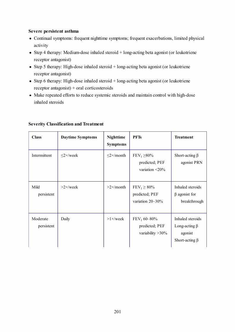
Severity Classification and Treatment
Class Daytime Symptoms Nighttime
Symptoms
PFTs Treatment
Intermittent ≤2×/week ≤2×/month
>2×/week >2×/month FEV1 ≥ 80%
predicted; PEF
variation 20–30%
Daily >1×/week
Severe persistent asthmaContinual symptoms: frequent nighttime symptoms; frequent exacerbations, limited physicalactivityStep 4 therapy: Medium-dose inhaled steroid + long-acting beta agonist (or leukotrienereceptor antagonist)Step 5 therapy: High-dose inhaled steroid + long-acting beta agonist (or leukotrienereceptor antagonist)Step 6 therapy: High-dose inhaled steroid + long-acting beta agonist (or leukotrienereceptor antagonist) + oral corticosteroidsMake repeated efforts to reduce systemic steroids and maintain control with high-doseinhaled steroids
FEV1 ≥80%
predicted; PEF
variation <20%
Short-acting β
agonist PRN
Mild
persistent
Inhaled steroids
β agonist for
breakthrough
Moderate
persistent
FEV1 60–80%
predicted; PEF
variability >30%
Inhaled steroids
Long-acting β
agonist
Short-acting β
201

Frequent
The goals of asthma management are to achieve minimal or no chronic symptoms day or night,and minimal or no exacerbations. Additionally:
CLINICAL PEARL
forbreakthrough
Leukotrine-
receptor
Severe
persistent
Continual; limited
activities; frequent
exacerbations
FEV1 ≤60%
predicted; PEF
variability >30%
High-dose
inhaled
steroid
Long-acting β
agonist
Short-acting β
agonist
Systemic
steroids
Leukotrine-
receptor
No limitation on activitiesMaintain near normal pulmonary functionMinimal use of short-acting beta agonists (<1x per day, <1 canister per month)Minimal or no adverse effects from medications
202

Along with these therapies, don’t forget to reassess allergic triggers and make efforts tocontrol the child’s environment, as well as address second-hand smoke exposure if any.
Final Diagnosis
Acute asthma exacerbation
CASE REVIEW
Asthma
Croup
Reversible obstructive airway disease affecting both small and large airwaysThree components: bronchospasm, airway edema, and mucus productionEtiology unknown but thought to be a combination of genetic and environmentalfactorsPresentation varies with the severity of the asthma exacerbationMay be a history of cough, especially at nightClassifications are intermittent, mild persistent, moderate persistent, and severepersistent; the classification helps determine which types of medication are mostappropriateExercise-induced asthma best prevented by beta 2 agonist administration 10–15mins before exerciseTreatment: avoidance of asthma “triggers” (pollen, dust, cold air, pets, tobaccosmoke, etc.) for which the individual is susceptible
Parainfluenza virus most common cause of croupUsually age 6 mos to 6 yrs
203

Epiglottitis
Low-grade fever, barking cough, inspiratory stridorStridor at rest occurs as upper airway obstruction increases“Steeple sign” noted on x-rays of the nasopharynx and upper airway, demonstratingnarrow subglottic space (not always present)Stridor at rest indicates severity of illness, along with increased work of breathing.Treatment includes racemic epinephrine and dexamethasone. Racemic epinephrine’smechanism of action is via vasoconstriction of vasculature, thereby decreasingedema and obstruction. Dexamethasone is a long-acting corticosteroid and may beadministered PO, IM, IV. The child is observed for 4–6 hrs and if there is norebound of symptoms, he may be discharged home.
Inflammation of the epiglottis that causes airway obstructionUsually age 2–7 but may occur at any age; more likely to occur in an unimmunizedchildEtiologic agents include S. aureus (including MRSA), Streptococcus, MycoplasmaPatient appears toxic on presentation with high fever, drooling, dysphagia, andrespiratory distress“Tripod” position in younger children, with neck hyperextension and body leaningforward in order to maximize passage of air through a narrowed airwayLateral neck x-ray: “thumbprint” sign, signifying a swollen, inflamed epiglottisIf epiglottitis suspected, do not insert a tongue blade to visualize the epiglottis, asthat may cause laryngospasm; keep child as comfortable as possible in order toavoid laryngospasm, e.g., reclining in mother’s arms with her administeringhumidified oxygenObtain an airway in operating room under controlled conditions. After intubation,get a blood culture and culture from epiglottis; give IV fluids and IV antibiotics (i.e.,third-generation cephalosporin or ampicillin-sulbactam along with clindamycin orvancomycin for MRSA coverage if needed)
204

Foreign Body Aspiration
Allergic Reaction
Items such as hot dogs, peanuts, coins, nuts, balloons, and batteries are sometimesaspirated and may enter the esophagus or airwayRespiratory distress may occur when a foreign body lodges in airway (larynx,trachea, bronchioles)Children age <4 yrs at risk for foreign body aspirationRight main stem bronchus common site for foreign body aspirations in children age>1 yearMay cause sudden respiratory distress, hoarseness, cough, and shortness of breathChest x-ray (inspiratory and expiratory films) may demonstrate air-trapping due toforeign body creating a ball-valve mechanismDefinitive diagnosis by visualization with a rigid bronchoscope, as well as removalof foreign body
IgE-mediated responseSome causes of allergic reaction: food (e.g., peanuts, tree nuts, eggs), drugs,environmental agents, soaps, detergentsPruritic rash consisting of well-circumscribed erythematous rash (wheals or welts)found on physical examinationAnaphylaxis is a life-threatening condition and must be managed promptly. Multipleorgan systems may be involved:Respiratory system with stridor, wheezing, and manifestations of respiratory distressGI: nausea, vomiting, and abdominal painCardiovascular: hypotensionSkin and mucosa: urticaria (as described above), flushing, lip and tongue swellingUrticaria is usually self-limited and requires only antihistamines. If patient exhibitsevidence of anaphylaxis, administer immediate epinephrine 1:1,000 intramuscularly,as well as an antihistamine (e.g., IV diphenhydramine) and systemic corticosteroids
205
Bronchiolitis
Pneumonia
(e.g., methylprednisolone). A normal saline bolus should also be initiated. Monitorthe patient carefully.
Usually occurs in children age <2 yrsLower respiratory infection that causes inflammatory obstruction of the smallairways of the lower respiratory tractPatient usually has history of upper respiratory infection, i.e., cold symptoms, fever,and poor feeds and may later develop respiratory distress.Physical examination: may have wheezing, rales, nasal flaring, intercostalretractions, and evidence of air hungerHyperinflation of the lungs seen on chestVirus detected in nasopharyngeal secretions with antigen testing, polymerase chainreaction, or cultureHospitalize high-risk infants (e.g., immunocompromised, premature, chronic lungand congenital heart disease, and those age <3 mos), those with respirations>60/min, or if patient has evidence of hypoxia, apnea, or feeding difficulties, or ifparent is not reliable.Treatment depends on severity and in mild cases is symptomatic with antipyretics tothe wheezing patient. Aerosolized epinephrine useful.
“Triad”: fever, cough, and tachypneaViral pneumonia most common type of pneumonia in childhoodPatients with viral pneumonia have cough, wheezing, and stridor. Chest x-ray showsstreaky infiltrates. Treatment is symptomatic.Patients with pneumonia due to Chlamydia trachomatis present with “staccato”cough and may have history of eye discharge; age typically 2 wks to 5 mos; chest x-ray: hyperinflation or ground-glass appearance; treatment is erythromycin by mouth
206
for 14 daysPatients with bacterial pneumonia present with cough, high fever, and shortness ofbreath; chest x-ray: lobar infiltrate; agents in infants age birth to 2 mos are group Bstreptococci, E. coli, and Listeria. Treatment is complete septic workup, includinglumbar puncture; treatment is admission to hospital and IV antibiotics (ampicillinplus an aminoglycoside or ampicillin plus third-generation cephalosporin)S. pneumoniae, H. influenzae, and Staphylococcus are the common bacterialetiologies in patients age 2 mos to 5 yrs. For inpatient therapy, cephalosporin givenintravenously; for outpatient therapy, oral antibiotics such as amoxicillin oramoxicillin-clavulanate. S. pneumoniae may also be seen in patients age >5 yrs.Patients with Mycoplasma pneumoniae, i.e., “walking pneumonia”; age typically >5yrs; typical complaints are dry hacking cough and appearance less ill than chest x-ray suggests. Chest x-ray has an interstitial pattern found most commonly in lowerlobes; diagnosis is supported by positive serology of IgM for M. pneumoniae,and/or PCR; treatment is erythromycin, azithromycin, or clarithromycin.Patients with aspiration pneumonia have history of aspirating food or inhaling atoxin; may present with wheezing; chest roentgenograms show alveolar infiltratesand, rarely, reticular infiltrates usually localized but are often bilateral; treat only ifinfection or secondary infection suspected
207

Case 24
Chief Complaint
“My child needs shots, but I’m unsure which ones.”
History and Physical Examination
A 5-year-old boy who immigrated to the United States from Ecuador 6 mos ago is broughtto the office by his mother before he starts school. The mother has lost his immunizationcard and is unsure which immunizations he was previously given in Ecuador. Physicalexamination shows a cooperative boy with a benign eye, ear, nose, and throat exam;supple neck, normal heart rate and rhythm with no murmurs, clear lungs, and benignabdomen. He has full ROM in the extremities and no rashes on the skin.
CLINICAL PEARL
Written immunization records are valid if the vaccine, date of administration, intervalbetween doses, and patient’s age are appropriate for a comparable vaccine in theUnited States.
Assessment
With no immunization documentation, this patient would be considered to be unimmunized. Forthis 5-year-old, the recommended catch-up schedule would be:
208

Discussion
The following is what we really need to know about childhood immunizations. Go to cdc.govfor details about childhood immunizations.
DTaP, HBV, MMR and IPV (no Hib or PCV if age ≥5 yrs and a normal child)1 month later: DTaP and HBV2 mos after first visit: DTaP, IPVAt least 8 mos after first visit: DTaP, HBV, IPVAt age 11–12: MMR5 yrs after last DTaP: Td and then every 10 yrsSingle dose of varicella vaccine
Classification of vaccinesLive attenuated: MMR, varicella, intranasal influenza, oral rotavirusInactivated (killed):
Whole: viral (polio)–Fractional–
Protein-based: subunit (hepatitis B, influenza, acellular pertussis) or toxoid(diphtheria, tetanus)Polysaccharide-based: pneumococcal 23-valent, meningococcal conjugate: Hib,pneumococcal 13 valent
Contraindications and precautions of vaccinesAbsolute contraindications
Severe allergy/anaphylaxis to a prior dose of a vaccine or a component–Encephalopathy following pertussis vaccine (now rare with DTaP, but can happen; evenmore reactogenic with any FULL-DOSE pertussis given after age 7 yrs, so iscontraindicated (partial dose pertussis is okay, i.e., the Tdap).
–
Relative contraindications
209

Do not reduce dosage or change schedule for preterm infant; immunize as one would for a full-term infant. The only exception is a first dose of hepatitis B in infants <2000 g.
CLINICAL PEARL
There is no evidence linking vaccines for MMR or any other neurologic/degenerativedisease to autism. The Centers for Disease Control (CDC) states that vaccines are safe,and the issue has now been put to rest.
Pregnancy: okay to give 1 dose of Tdap vaccine if patient is due vaccine or unimmunized–Immunosuppression: in general, give no live viral vaccines except for MMR andvaricella with HIV if (a) no AIDS-related illnesses, and (b) CD4 >15% totallymphocytes at the time of the vaccine
–
Hepatitis B vaccine (HBV)The following are recommendations as per the CDC.
If mother is HBsAG-negativeGive first dose at birth, prior to leaving the birth hospital. Give next 2 doses at age 1–2 mosand 6–18 mos.
If mother is HBsAG-positiveGive first dose with hepatitis B immune globulin (HBIG) within the first 12 hrs after birth,administered at different injection sites.
Up to 95% rate in prevention of perinatal transmissionComplete schedule with 2 more doses of HBV given at 1 and 6 mosTest infants for HBsAg and anti-HBs 1–2 mos after completion of series and at age 9–18
210

mos
If mother’s hepatitis B status is unknownDraw blood on mother for HBsAg testing; while waiting for results, administer HBV tonewborn within 12 hrs of birth. If mother is HBsAg-positive, administer HBIG to newbornas soon as possible but no later than age 7 days.
Diphtheria, tetanus, acellular pertussis (DTaP)
Tetanus Prophylaxis in Wound Management
History of Doses of Tetanus Toxoid Clean, Minor Wounds All Others*
Acellular pertussis introduced due to concern about encephalopathy with previous pertussisvaccine but rarely may still occur (see above)May give vaccine with minor illnesses and feverFive doses of DTaP;The childhood tetanus (Tdap) is given one time ideally at the 11–12 year old visit. Ifmissed, it should be given one time any time thereafter upt to age 65 yrs (an attempt todecrease incidence of pertussis, which has increased to epidemic proportions since the late1990s)The following individuals should receive tetanus prophylaxis (Td and tetanus-immuneglobulin):
Those with wounds at increased risk of contamination with dirt, feces, saliva; woundswith necrotic or gangrenous tissue; frostbite; crush and avulsion injuries; and burns
–
If <3 doses or status of tetanus immunization is unknown: with clean, minor wounds, giveTd only; all other wounds give both Td and TIG
–
If ≥3 doses of tetanus immunization: clean, minor wounds do not need Td or TIG unlesslast Td was given >10 yrs ago; if so, give Td. All other wounds do not need Td or TIG,unless it has been more than 5 yrs since last Td. If so, then give Td.
–
211

Td TIG Td TIG
<3 or unknown Yes No Yes Yes
≥3 No† No No‡ No
TIG, tetanus immune globulin; Td, tetanus and diptheria vaccine.
*All other wounds = increased risk of tetanus: dirt, saliva, feces, avulsions, frostbite, puncture,
crush, burns, and missiles.
†Unless >10 years from last dose.
‡Unless >5 years from last dose.
Inactivated polio vaccine (IPV) has taken the place of oral polio vaccine (previoussubclinical polio-like illnesses)
4 doses; last booster is given at age 4–6 yrs, before starting school
Haemophilus influenzae type b conjugate vaccine (Hib)4 doses; last booster is given age 12–18 mosIf child is unvaccinated and receives first dose after age 15 mos, only 1 dose of Hib vaccineis requiredNo dose in a normal child after age 5 yrs if under- or nonvaccinated
Measles, mumps, rubella (MMR) is grown in chick embryo fibroblast cultures, thus is safe inegg-allergic children (may vaccinate without testing).
Give first dose at or after age 12 mosSecond dose is recommended at age 4–6 yrs, but may be given any time if at least 4 wkshave elapsed after first dose; if second dose has not been received, complete by age 11–12The following individuals should receive immune globulin for measles:
212

If immune globulin was received after exposure, then measles vaccine should be given 6 mosafter immune globulin administration, if patient is at least age 12 mos.
PATIENT SAFETY NOTEDo not administer to patients with known allergy or anaphylaxis to neomycin or gelatin,both of which are present in the vaccine.
Measles
Age Management (post-exposure)
0−6 mo Nothing, unless mother is not immune (if not
immune, Ig only)
6−12 mo Ig plus vaccine
>12 mo Vaccine only within 72 hours of exposure for
susceptible individuals
Susceptible people as prevention or modification within 6 days of exposure–Susceptible household contacts of patients with measles, of age, pregnant women, and theimmunocompromised
–
Infants in first 6 mos of life if mother has no evidence of immunity. Can give vaccine from6–12 mos for postexposure prophylaxis but still need the normally-timed 2 (more, so 3total) vaccines
–
213

Definition of abbreviation: Ig, immunoglobulin.
Pregnant or immunocompromised Ig only
VaricellaAt any visit at or after age 12 mos with no reliable history of chickenpoxGive 2 doses (4 wks apart)Fully (100%) protective against severe disease (mild varicella is still visible)Live vaccine contraindications:
Moderate to severe acute illness–Malignancy or T-cell defect, including HIV with CD4 counts <25%–High-dose steroid treatment–Pregnancy–Component allergy–
Who should receive varicella-zoster immune globulin (VZIG)?Immunocompromised children (and adults) without history of varicella or varicellaimmunization
–
Susceptible pregnant women–Newborns whose mother had onset of chickenpox from 5 days before delivery to <48 hrsafter delivery
–
Hospitalized premature infants of ≥28 wks’ gestation, whose mother lacks reliablehistory of chickenpox or serologic evidence
–
Hospitalized premature infants <28 wks’ gestation or whose birth weight ≤1000 g,regardless of mother’s history or serologic status
–
Pneumococcal vaccine (PCV): 2 types13-valent PCV
Recommended for all children aged 2 to 23 mos–Give 4 doses; fourth dose by age 15 mos–
214

Both intramuscular injection and nasal-influenza vaccine are grown in embryonated eggs andthus have a potential problem with egg allergy.
PATIENT SAFETY NOTE
23-valent PCV, PP SV23In addition to PCV for high- and moderate-risk groups (SS, HIV, immune deficiency,chronic cardiopulmonary disease, chronic renal insufficiency, DM, day-care, AfricanAmerican/American Indian/Alaskan Native descent)
–
PPSV23: protective antibody responses to the most common pneumococcal serotypes inchildren age ≥2 yrs; given at age 24 mos and 5 yrs after first dose for patients who areimmunocompromised or those with asplenia (functional or anatomic)
–
Meningococcal conjugated vaccine-MCV4Active immunization against serotypes A, C, Y and W-135Give to:
Travelers to areas with high rates of infection–College freshmen in dormitories–Military–Asplenia and other high-risk conditions–
Influenza vaccineRecommended annually for ALL children age 6 mos to 18 yrs at the beginning of each annualinfluenza season. Certain children are at high risk and should be vaccinated first during seasonif inadequate supply of vaccine. All adults age >50 yrs and all women who are pregnant or maybe pregnant during influenza season should receive the vaccine early on, i.e., October or firsthalf of November.
Patients who have had a severe allergic reaction to the influenza vaccine should not
215

Nasal influenza vaccine is live attenuated (need live viral vaccine precautions). Dosing forpatients age 2–49 yrs is as follows:
Final Diagnosis
Catch-up immunization
CLINICAL PEARL
PPD (purified protein derivative) may be administered the same day as the MMR;however, if administered separately, there must be at least 4 wks after the MMRvaccine before placing a PPD. This is because the measles vaccine may suppress thereactivity to the PPD.
receive it in the future.However, patients who have had an allergic reaction to egg (including havingreceived epinephrine or having had respiratory distress or angioedema) may stillreceive the influenza vaccine if administered in a controlled setting. That settingshould include appropriate personnel available who can both immediately identifyand manage anaphylaxis.
All children age ≤8 yrs receive 2 doses, 4 wks apart, for first immunization only and 1 doseyearly thereafterChildren age 6–36 mos receive half-dose (0.25 mL)All others receive 0.5-mL dose (i.e., those age ≥3 yrs)
216

CASE REVIEWIn this case, the parent is unsure of vaccination status and we have no record availablefor reference. Therefore, we must assume the patient to be unvaccinated. If a child, onthe other hand, has received some vaccines but is not up-to-date on all of them, we willfollow the catch-up immunization schedule and start where the child left off.
Note: There is no need to repeat vaccines that have already been administered, butrather continue from the last vaccine, making sure that the required minimum time haselapsed before the next dose can be given (refer to Catch-up Immunization Schedule).
Certain vaccines are not routinely (i.e., without risk factors) administered beyond acertain age: Hib beyond age 36 mos and PCV 13 beyond age 5 yrs (again, without riskfactors). This child who is age 5, would not require these.
To summarize, regarding immunization catch-up schedules, the following rules apply:
There is no need to restart any series, regardless of elapsed timeFollow minimum dosing intervals for each vaccine (see chart)Fifth dose of DTaP is not necessary if fourth dose was given after fourth birthdayIf all IPV series were received, fourth dose is not necessary if third dose was givenat age ≥4 yrsIPV is not recommended for those age ≥18 yrs (unless adult is nonimmunized andgoing to a country with polio or working with wild strains of polio)Begin HBV series at any visit if patient has not been immunizedGive second dose of MMR at age 11–12 yrs or at least 4 wks after first doseHib is not recommended for those age ≥5 yrsPCV is not recommended for those age ≥5 yrsVaricella: give 2 dose series (4 wks apart) to all susceptible patients age ≥13 yrs
217

218

Case 25
Chief Complaint
Sore throat of 1 day’s duration
History and Physical Exam
A 6-year-old girl is brought to the office with a 1-day-history of abrupt onset of sorethroat and fever. She has had 2 episodes of vomiting. She denies cough or rhinorrhea.Physical exam is remarkable for T 38.8 C (102 F), erythematous, enlarged tonsils withexudates, and palatal petechiae. Enlarged, tender anterior cervical lymph nodes arepalpable. A diffuse, erythematous, sandpaper-like rash is present, more prominent in theflexural areas.
Differential Diagnosis
Clues
Streptococcal pharyngitis (group A streptococcus [GAS], strep pyogenes) GABHS(group A, beta-hemolytic strep)
1.
Viral pharyngitis: adenovirus, coronavirus, rhinovirus, respiratory syncytial virus(RSV), coxsackievirus
2.
Abrupt onsetExudative pharyngitis without ulcerationAbdominal pain, along with nausea and vomiting; however, diarrhea is not common withstreptococcal pharyngitis
219

CLINICAL PEARL
The presence of cough, rhinorrhea, and/or conjunctivitis points toward a viral etiology:
Initial Management
Setting: outpatient workup and treatment
Initial diagnostic plan Results
Positive
Assessment
Some overlap can be found between the clinical presentations of viral and bacterialpharyngitis. Group A strep usually has a more acute onset, and is more common in late autumn,winter, and spring. When Group A strep is associated with an erythematous macular/papular,i.e., sandpaper-like rash, that is called scarlet fever. The rash of scarlet fever progresses in acraniocaudal direction, culminating in desquamation of the hands and feet, with sparing of thepalms and soles. Notably, the rash is more prominent in the flexural areas, called Pastia’s lines.
Headache
Mononucleosis (EBV)Other bacteria: Arcanobacterium haemolyticum, Mycoplasma pneumoniae,Corynebacterium diphtheriae
Rapid streptococcal antigen test1.
220

Viral pharyngitis has a more gradual onset, associated with upper respiratory infection, and ismore common in fall, winter, and spring.
Adenovirus should be considered for patients presenting with pharyngitis and conjunctivitis.
Coxsackievirus (herpangina) is characterized by fever, sore throat, dysphagia and ulcerativelesions on the anterior tonsillar pillars, soft palate, uvula, tonsils, and posterior pharyngealwall. Odynophagia is a common presenting symptom, and as such, supportive care shouldinclude pain management along with IV fluid administration if the child is unable to drinkfluids, in order to prevent dehydration.
Mononucleosis (EBV) is associated with fever, fatigue, significant lymph gland swelling andexudative tonsillitis, muscle aches, rash, and hepatosplenomegaly. Approximately 5% of casesof EBV-associated infectious mononucleosis have a positive throat culture for group A strepwhich represents pharyngeal streptococcal carrier state. Infectious mononucleosis should besuspected in a patient diagnosed with group A strep who fails to respond to treatment after 48–72 hrs or who develops a diffuse macular erythematous rash if amoxicillin was given(interaction of amoxicillin with EBV).
Corynebacterium diphtheriae has a unilateral or bilateral leather-like adherent tonsillarmembrane that can extend to affect the uvula, soft palate, posterior oropharynx, hypopharynx,and glottic areas. Underlying soft tissue edema and enlarged lymph nodes can cause a bull-neckappearance. The lack of fever and dysphagia help to differentiate C. diphtheriae from group Astrep and EBV infections.
Further Diagnostic Plan
The antigen screen has high specificity. However, a negative result requires a backup throatculture (gold standard).
221

Treatment Plan
Prevention of rheumatic fever is the primary goal of treatment. Penicillin is the drug of choice,but amoxicillin is often preferred because of palatability. Clinical recovery is hastened byantibiotic treatment but by only 12–24 hrs. The main reason for the Rx is prevention of ARF; itwill be prevented if started within 9 days of the start of of illness.
However, if the antigen screen is negative and a backup throat culture is required, one shouldwait for the results of the throat culture prior to initiating antibiotic treatment.
Penicillin effectively prevents primary attacks of rheumatic fever, even when initiated as longas 9 days after the acute onset of the illness. Therefore, a brief delay of 24–48 hrs forprocessing the throat culture prior to starting antibiotic therapy does not increase the risk ofrheumatic fever. With penicillin allergy, use an oral macrolide. However, there has beenincreasing resistance to macrolides due to overuse of these drugs, so depending on the previousreaction, a first-generation cephalosporin may be an alternative.
Indications for treatment include the following:
The patient is no longer contagious after 24 hrs of therapy. Repeat the throat culture only if sorethroat or mild fever persists after antibiotic treatment.
Discussion
Symptomatic pharyngitis and a positive rapid strep test, or positive throat cultureClinical diagnosis of scarlet fever (sore throat, sand-paper type rash, Pastia’s lines,circumoral pallor, strawberry tongue, enlarged tonsils)Household contact with documented strep pharyngitisRecent history of acute rheumatic fever in a family member
222

Viral pharyngitis predisposes to bacterial otitis media. GAS pharyngitis complications includeperitonsillar abscess, retropharyngeal abscess, rheumatic fever, and glomerulonephritis.
Final Diagnosis
Group A beta-hemolytic streptococcal pharyngitis (scarlet fever)
CASE REVIEW
GABHS Pharyngitis
Viral Pharyngitis
Peak age 5–10 yrs; uncommon in children age <3 yrs (called “Streptococcosis” inchildren age <3 yrs, presenting with prolonged nasal discharge, tender cervicallymphadenopathy, and low-grade fever)Winter/spring more commonAcute onsetAssociated with headache, abdominal pain, vomitingMay have findings of scarlet fever (strawberry tongue, sand paper rash, circumoralpallor, Pastia’s lines)Rapid antigen test is first choice; if it is negative, do not treat with antibiotics. Thenext step is to send a throat culture, which is the gold standard but there are stillfalse negatives (depending on how good the swab is or if the caregivers have beengiving antibiotics. If the throat culture is positive for group A strep, treat withantibiotics; if it is negative, no treatment is indicated.Penicillin is the treatment of choice (pill form or injection); an oral macrolide is thetreatment of choice for the penicillin-allergic patientComplications: abscess, rheumatic fever, glomerulonephritis
223

Viral Versus Bacterial Pharyngitis
Viral Bacterial
Age (in years) <5, >10 5−10
Onset Gradual Acute
Upper respiratory infection +++ −
Rash + + + +
Exudate + + + +
Ulcers +++ −
Rapid strep screen − + + +
Throat culture − + + + + +
Vomiting/Abdominal pain + + +
Lymph nodes + + + +
Conjunctivitis + + −
Palatal petechiae + + + +
Preceded by cold symptoms (rhinorrhea, cough, diarrhea)Winter/spring/fallConjunctivitis is associated with adenovirusOral ulcers are associated with coxsackievirusHepatosplenomegaly is associated with EBV (mononucleosis)Supportive care only; antibiotics not indicated
224

225

Case 26
Chief Complaint
Burning on urination
History and Physical Examination
A 5-year-old girl comes in with complaints of burning on urination for 3 days. She has hadfever for 1 day’s duration. She denies vomiting or diarrhea. She has a history of takingbubble baths. Past medical history is remarkable for a urinary tract infection 1 year ago.Her T is 38.7 C (101.8 F) and BP 128/90 mm Hg. Physical exam is remarkable for mildright-sided CVA tenderness. A mass is palpable in the right upper quadrant. Urinalysisshows 25–50 WBCs per high power field. The next day, the urine culture grows 100,000colonies of E. coli and she is treated with antibiotics.
Differential Diagnosis
Initial diagnostic plan Results
Enlarged kidney on right
Vesicoureteral reflux (VUR)1.Urinary tract infection (UTI)2.Hypertension3.Hydronephrosis4.
Renal U/S1.
226

Dilated tortuous ureter on right with reflux to
enlarged right kidney
Assessment
Urine culture is the gold standard in diagnosis of urinary tract infection. In young children,think of renal causes for hypertension (e.g., renal parenchymal disease due to chronicpyelonephritis, or chronic glomerulonephritis, ureteral obstruction, renovascular disease, renaltrauma, etc.).
VCUG is necessary for diagnosis and grading of VUR.
Treatment Plan
Treat UTI with antibiotics such as cephalosporins, amoxicillin-clavulanate, or trimethoprim-sulfamethoxazole (TMP-SMZ). Clinicians should be aware of local susceptibility patterns. Ifpyelonephritis, use broad spectrum antibiotics. If dehydrated, vomiting, or <1-month-old, useIV antibiotics (ceftriaxone or ampicillin/gentamicin for <1-month-old).
Perform renal U/S to rule out congenital anatomic abnormalities, hydronephrosis, abscessformation. Also perform VCUG if:
Voiding cystourethrogram (VCUG)2.
International Classification of Vesicoureteral RefluxGrade I: reflux into non-dilated ureterGrade II: reflux into upper collecting system without dilatationGrade III: reflux into dilated ureter and/or calyceal bluntingGrade IV: reflux into grossly dilated ureterGrade V: massive reflux, tortuous ureter, hydronephrosis
Recurrent febrile UTI or UTI with complicationsU/S shows renal abnormality; e.g. hydronephrosis, scarring, or concern for vesicoureteral
227

Discussion
Vesicoureteral reflux is an abnormal backflow of urine from the bladder to the ureter and renalpelvis. This backflow of urine aids in transporting bacteria from the bladder to the upperurinary tract, causing a predisposition to UTI, i.e., pyelonephritis, and as a consequence, ifpersistent or worsening, renal scarring, poor renal growth and decreasing renal function
Extensive scarring leads to hypertension. A higher reflux grade has a higher likelihood of renalinjury. Reflux can be due to an abnormal ureterovesicular junction (primary VUR), orsecondary to anatomic abnormalities (posterior urethral valves); or a neuropathic bladder(myelomeningocele).
If hydronephrosis is noted on the prenatal U/S, one should consider vesicoureteral reflux as anetiology. Reflux may also be discovered during evaluation of a UTI. After VUR is diagnosed,upper tract imaging should be done to determine renal size and scarring. This can be done usingU/S to monitor for the presence of renal scarring, or renal scintigraphy. If scarring is present,follow the creatinine.
reflux (VUR)
First, U/S for anatomyThen, VCUG for pathophysiologic reason for recurrent UTI, i.e., VUR
228

Further Treatment Plan
Recent studies show that grades I–III VUR in children usually resolves with time; follow withVCUG and radionuclide scan. Studies also now show (and the policy has been adopted by theAAP) that antibiotic prophylaxis is not effective. The best management is to follow carefullyfor any sign of UTI and screen with urine cultures; early aggressive therapy is the besttreatment for the prevention of long-term complications and slow resolution of the naturalhistory over time. Voiding dysfunction is a comorbidity and should be managed with behavioraltherapy/bladder training in order to obtain the best possible outcome.
For grades IV–V, surgical re-implantation of ureters is the mainstay of treatment and may beconsidered if there is a breakthrough UTI, new renal scars, or worsening of reflux or renalfunction.
PATIENT SAFETY NOTEIt is of utmost importance to treat UTI in a child promptly. Once diagnosis has beenmade, parents should be instructed to watch for fever; in that case they must seekmedical care within 48 hrs in order to obtain a urinalysis and urine culture. Eachepisode of UTI will place the child at risk for increased scarring, loss of healthyparenchymal tissue, and future renal complications, such as high BP and decreasedrenal function.
Final Diagnosis
Finally, if clinically significant VUR (II-V), a radionuclide scan (best test for renal size,scars and function); IVP is rarely performed in children any longer
229

Vesicoureteral reflux
CASE REVIEW
Urinary Tract Infection (UTI)
Hypertension
UTI may present with fever, nausea, vomiting, and in young children with non-specific symptoms such as poor feeding or fussiness. Older children and adolescentscan present with and communicate symptoms of abdominal pain or back pain, alongwith urinary symptoms of increased frequency along with hesitancy, urgency, anddysuria.Risk factors: constipation, voiding dysfunction including both stool and urinewithholding, neurogenic bladder, anatomic abnormalitiesDiagnosis: urine culture (gold standard)>100,000 CFU/mL on a clean catch signifies a UTI or fewer colonies of a singleorganism (no less than 10,000) in a symptomatic childChildren who are not yet toilet-trained should undergo catheterization in order toobtain a sterile urine sample. >50,000 CFU/mL will signify a UTI.Initial therapy can be based on local sensitivities but can then be changed to reflectresults of urine culture after the organism has been identified and sensitivities areavailableMost common organism is E. coli.Empiric therapy may include TMP/SMX, cephalosporin or amoxicillin-clavulanate,until sensitivities are available.Indications to admit for inpatient therapy include: vomiting (not tolerating POfeeds), dehydration, age <3 mos, caregiver concern or inability to care for at homeTherapy duration: 10 days
230

Hydronephrosis
Diagnosis: obtain BP using appropriate-sized cuff, at 3 separate office visits, atleast 1 week apart
Stage 1 HTN: systolic and/or diastolic BP at the 95th percentile to 5 mm Hgabove 99th percentile
–
Stage 2 HTN: systolic and/or diastolic BP >5 mm above 95th percentile–
In young children, think of renal causes for hypertension: secondary hypertension(e.g., renal parenchymal disease due to chronic pyelonephritis or chronicglomerulonephritis, ureteral obstruction, renovascular disease, renal trauma, etc.).Adolescents and postpubertal children are more likely to have primary hypertensionRisk factors are obesity/increased BMI, family history, and ethnicityA thorough history and physical exam will aid in narrowing the differential. Obtainthe history regarding the patient’s stay in neonatal ICU (if any) and if an umbilicalarterial catheter was placed at the time. Ask about use of medications, e.g., OTCcold preparations and corticosteroids. Palpate for the presence of both femoralpulses, listen for an abdominal bruit, observe for a difference in BP in the upper andlower extremities, and obtain a urinalysis to look for proteinuria.First level of therapy is diet and lifestyle modification: salt restriction, exercise, andweight reductionMedical therapy for primary hypertension: if severe HTN, symptoms, or end-organdamage, start with an ACE inhibitor or thiazide diuretic
Prenatal hydronephrosis refers to dilatation of the renal pelvis, with or withoutinvolvement of the calycesIf hydronephrosis is noted on the prenatal U/S, one should consider vesicoureteralreflux as an etiologyU/S should be obtained in neonatal period to look for any cause of obstructionleading to the hydronephrosisIf bilateral hydronephrosis is found, consider causes that may cause bilateral
231

obstruction, e.g., posterior urethral valves or ureteroceleVCUG should be the next imaging study to pursue in order to look for VUR, and alsoto observe for posterior urethral valves
232

Case 27
Chief Complaint
Small jaundiced baby
History and Physical Examination
You are called to the newborn nursery to evaluate an 8-hour-old infant. The patient wasborn at 37 wks’ gestation to a 21-year-old primigravida. Mother is blood type A positive,group B strep negative, VDRL negative, rubella status unknown. Pregnancy wasremarkable for a fever and rash in the first trimester. A low-grade fever was reportedduring labor. Birth weight was 4 pounds 7 ounces, Apgars 8/9.
Physical exam reveals a small neonate with little subcutaneous fat. He is noticeablyjaundiced, with several purpuric lesions scattered about. HEENT is remarkable forbilateral cataracts. Lungs are clear, heart exam is positive for a to-and-fro machinerymurmur. Abdominal exam reveals an enlarged liver. Rest of the physical examination isnormal.
Differential Diagnosis
Congenital rubella syndrome1.Toxoplasmosis2.Syphilis3.Congenital cytomegalovirus4.Herpes5.
233

Clues
Initial Management
Setting: newborn nursery
Initial diagnostic plan Results
Normal
Elevated liver function tests and liver enzymes
(they are 2 different things)
Elevated direct and indirect (this is an LFT)
Pending
Pending
A-positive, negative
Group B streptococcal sepsis6.Hepatitis7.Physiologic jaundice8.
Maternal fever and rash in first trimesterNewborn with jaundice, purpura, cataracts, machinery murmur, hepatomegaly
CBC1.
Complete chemistry profile2.
Bilirubin3.
Blood culture4.
Urine culture5.
Blood type, Rh, Coombs6.
234

Treatment Plan
Give empiric antibiotics, e.g. ampicillin and gentamicin, pending the culture results; no othertreatment is necessary. Also immunize the mother.
Assessment
Prenatal history is the baby’s past medical history: maternal exposures, immunization status,infections, medications all affect the fetus. Jaundice in the first 24 hrs of life is alwayspathologic and should be investigated. Always fractionate bilirubin (direct/indirect) to helpwith the differential diagnosis. Blood type, Rh, and Coombs tests are essential in the evaluationof jaundice as well.
TORCH titers (IgM) help to differentiate congenitally acquired infections and are the bestscreening test. Significant overlap exists in presentation of TORCH infections. Sequelae aremost common if the infection occurs in the first trimester. Best for CMV are 3 separate urineCMV cultures; for herpes it is PCR of bodily (vesicular) fluid
Further Diagnostic Plan/Results
Discussion
Infection results from transplacental infection with rubella virus. Rubella in pregnancy isgenerally a mild illness, presenting usually with a macular rash that spreads craniocaudal,starting on the face and eventually reaching the feet. The rash resolves in the same direction aswell. The risk for congenital defects is greatest before 11 wks (first trimester). Nerve deafness
Rubella IgM titers: elevatedEchocardiogram: patent ductus arteriosusHearing screen: abnormal
235

is the most common finding, while patent ductus arteriosus (PDA) is most common cardiacdefect. Cataracts are a common eye finding.
CLINICAL PEARL
Patients with congenital rubella syndrome are at increased risk for developing insulin-dependent diabetes mellitus in older childhood and adulthood.
Most patients have intrauterine growth retardation, low birth weight; <25% of patients havepurpura (“blueberry muffin” rash). Many patients have chorioretinitis (“salt and pepper”appearance), and some patients have panencephalitis, mental retardation.
The most common diagnostic test is rubella IgM assay. The result may be a false positive dueto competing IgG antibodies, so obtain a PCR or culture to confirm. In addition, persistent IgGrubella antibody may also signify congenital rubella syndrome.
Therapy is supportive: cardiac, audiologic, ophthalmologic, neurologic consultation/follow-up. Prevent with immunization prior to pregnancy. There is no antiviral treatment.
Manifestations of Congenital Infections
Pathogen Manifestation
Rubella Cataracts, heart defects, hydrocephalus,
microcephaly, microophthalmia, petechiae,
purpura, thrombocytopenia, retinitis, seizures,
deafness, mental retardation
236

Toxoplasmosis Hydrocephalus, intracranial calcifications,
microcephaly, retinitis, thrombocytopenia,
seizures, eye pathology, mental retardation
Syphilis Anemia, hepatosplenomegaly, osteitis, snuffles,
rhagades, dental
Cytomegalovirus Periventricular calcifications, microcephaly,
hepatitis, petechiae, purpura, retinitis,
thrombocytopenia, seizures, deafness, mental
retardation
Herpes Hydrocephalus, microcephaly, encephalitis,
hepatitis, skin lesions, vesicles
Final Diagnosis
Congenital rubella syndrome
CASE REVIEW
Toxoplasmosis
From infected meat, cat fecesMaternal infection usually asymptomaticWide variation of manifestations:
Small for gestational age, jaundice, prematurity–Chorioretinitis, hydrocephalus, cerebral calcifications–Hepatosplenomegaly, seizures, microcephaly, developmental delays–
Prenatal diagnosis with PCR from amniotic fluid
237

Syphilis
Postnatal diagnosis with rising IgM titersTreat with pyrimethamine for 1 year, plus sulfadiazine and leucovorin. Leucovorin(folinic acid) is administered as part of the therapeutic regimen due topyrimethamine’s side effect of neutropenia. Continue to monitor CBCs along witheye exams for development or progression of chorioretinitis.Prevention: avoid rare or undercooked meat during pregnancy and avoid contactwith cat litter
Untreated syphilis during pregnancy has 70–100% transmission rate with up to 40%fetal/perinatal mortalityEarly signs (age <2 yrs):
Snuffles: may be the initial presenting symptom, manifesting in first week of life.The nasal secretions contain live spirochetes and are highly contagious. Becareful while handling the secretions. The spirochetes can be visualized bydarkfield microscopy.
–
Rash: usually presents after “snuffles,” initially pink in color which laterundergoes crusting and becomes copper colored or dark red
–
Osteochondritis–Increased liver enzymes–Coombs negative hemolytic anemia–Thrombocytopenia–
Late signs (age >2 yrs): usually skeletalHutchinson teeth–Frontal bossing–Saddle nose–Clutton joints–
VDRL and RPR are useful screening tests
238

Cytomegalovirus
Herpes Simplex
FTA ABS is confirmatory testVDRL on CSF for neurosyphilisEvaluate at-risk infants if:
Inadequate, undocumented, or unknown maternal treatment–Maternal treatment ≤30 days prior to therapy–Mother treated with non-penicillin regimen–Maternal titers did not decrease sufficiently–
Risk of fetal infection is much greater with primary maternal infection90% of congenital infections are subclinicalIntrauterine growth retardation, prematurityHepatosplenomegaly, jaundiceBlueberry muffin rash, thrombocytopenia, purpuraMicrocephaly, intracranial (periventricular) calcifications, chorioretinitisPCR best test or virus isolation (urine, saliva)Can shed virus in urine for several yrsGanciclovir may prevent hearing deterioration
Risk is greatest with primary or nonprimary first infection (infected with one typenow infected with other type)Acquired in utero, during birth (most common), or as neonateIn utero: see skin vesicles/scarring, chorioretinitis, microcephalyDelivery, post partum: 3 patterns:
Skin, eyes, mouth (SEM): 5–11 days of life, vesicles, can progress to type 2 or 3–Encephalitis (CNS disease): 8–21 days of life, similar to meningitis, seizures,irritability; high mortality, severe sequelae
–
Disseminated: 5–11 days of life, similar to sepsis; 90% mortality–
239

Group B Streptococcal Sepsis
PCR, culture (gold standard)Treat with acyclovir (high dose 60 mg/kg/24h divided q8h), 21 days if CNS diseaseReduced risk if delivered by cesarean
Most common cause of neonatal bacterial sepsisTwo patterns:
Early onset (<7days of age)–Maternal colonization of vagina/rectum is source (30% of pregnancies)Early onset risk factors
> Prolonged rupture of membranes> Intrapartum fever> Prematurity
May appear ill at delivery, or usually within 24 hrsSepsis, pneumonia, meningitisHypothermia/fever, irritability, lethargy, apneaTachypnea, grunting, acidosis, shock
Late onset (age >7 days)–Bacteremia, meningitis, urinary tract infection, pneumonia
Diagnose with positive culture: blood, urine, CSFPenicillin G treatment of choice once confirmedCan prevent early onset by treating GBS + mother with penicillin during labor, atleast 4 hrs before delivery
240

241

Section II
OBSTETRICS ANDGYNECOLOGY
242

2
Obstetrics
Case 1
Chief Complaint
“I am 2 months late for my period.”
History and Physical Examination
A 20-year-old G0 P0 woman comes to the office stating that her last menstrual period was2 months ago. She previously has had regular menses. She became sexually active acouple of months ago and had been using a diaphragm for contraception irregularly. She isnot in a stable relationship and has multiple sexual partners. She states she has beendiagnosed with some sexually transmitted disease but does not remember what it was. Shehas experienced severe nausea and vomiting for the past week. Also, she has noticedfullness in her breasts with increased tenderness for a few weeks. She also complains ofconstantly feeling fatigued even after she has had adequate sleep. Vital signs are stable.
General physical examination is unremarkable. Vital signs are stable. Abdomen is flat,soft and non-tender. Visual inspection of external genitalia shows normal perineum.Speculum exam shows a well estrogenized vaginal mucosa and normal appearing cervix.
243

Bimanual exam reveals a non-tender, anteverted uterus enlarged to approximately 10weeks’ size.
Differential Diagnosis
CLINICAL PEARL
When patients complain of severe nausea and vomiting in the first trimester, it isimperative to rule out other etiologies before attributing it to normal pregnancy. Molarpregnancies and hyperthyroidism can also present with nausea and vomiting.
Initial Management
Setting: outpatient office
Diagnostic/Therapeutic Plan
CLINICAL PEARL
Secondary amenorrhea probably due to pregnancy1.Hyperemesis gravidarum2.
Qualitative urine β-hCGUrine dip for ketones and specific gravityPatient’s weight
244

The most common cause of secondary amenorrhea in the reproductive years ispregnancy. Always get a pregnancy test.
Test Results
Assessment
Probable intrauterine pregnancy; hyperemesis is ruled out
BASIC SCIENCE CORRELATEPregnancy tests detect levels of human chorionic gonadotropin (hCG) which iscomposed of an α-subunit and a β-subunit. The β-hCG subunit is specific fortrophoblastic tissue. A negative test virtually rules out pregnancy. The test can bequalitative (positive or negative) or quantitative (measures levels). The urine test isalways qualitative with a sensitivity threshold minimum of 25–50 IU/L. The serum testcan be either quantitative or qualitative, with the latter having a sensitivity thresholdminimum of β-hCG is 5–10 IU/L. Although the more sensitive method for detecting β-hCG is serum, a positive urine β-hCG usually suffices and is more cost-effective.
Urine ketones assess the degree of carbohydrate starvation. Urine specific gravity is asimple way of assessing adequacy of fluid intake. Values >1.030 indicate possibledehydration. Serial changes in patient weight are the best indicator of long-termchanges in fluid status.
Urine β-hCG positiveUrine dip positive for trace ketones; specific gravity 1.015Weight 120 lbs (unchanged from past few months)
245

Further Management Plan/Results
Final Assessment
Normal-appearing intrauterine pregnancy, size equals dates
Discussion
Pregnancy should be suspected whenever a woman in her childbearing years misses amenstrual period. Common signs and symptoms include amenorrhea, nausea, fatigue, breasttenderness and frequent urination. The symptoms of nausea and vomiting usually resolve by endof first trimester. An office urine pregnancy test is an easy quick tool to assess for possiblepregnancy. β-hCG is highly specific for trophoblastic villi and pregnancy.
Once pregnancy is confirmed the next step in management is to determine gestational age. Usingthe woman’s LMP and Naegele’s rule the EDD (estimated due date) can be determined bysubtracting 3 months and adding 7 days. Pregnancy wheels are used to calculate the EDD whenthe LMP is known. However, remember these calculations assume a 28-day menstrual cyclewith conception occurring at day 14. Most women will have a cycle length more or less than28 days so the EDD may need to be adjusted either forwards or backwards.
Vaginal obstetric U/S Normal-appearing intrauterine singleton pregnancy, crown-rump
length of 10 wks; fetal cardiac motion present 145/min
TSH and free T4 Within normal range for pregnancy
246

Obstetric U/S can determine if pregnancy is intrauterine or extrauterine, gestational age, aswell as assess for viability. The uterus is a pelvic organ up to 12 wks. In the first trimester, avaginal U/S gives much better visualization and resolution than an abdominal U/S. Agestational sac can be visible at 4.5–5 wks and a yolk sac around 5–6 wks. A fetal pole withcardiac activity can be detected around 5.5–6 wks.
Follow-up Management and Prevention
Obtain routine prenatal lab tests.CBC (anemia)–Urinalysis (UTI), urine culture (UTI)–Blood type and Rh (need for RhoGAM)–Atypical antibody screen (alloimmunization)–RPR (syphilis)–Rubella IgG titer (need for postpartum immunization)–Hepatitis B surface antigen (carrier for HBV)–HIV screen (HIV)–Gonorrhea and chlamydia PCR/culture (screen for cervical infection)–Pap smear if indicated (cervical dysplasia)–Screen for lead exposure–
Discuss first-trimester genetic screening at wks 11–13: U/S for nuchal translucency alongwith serum free β-hCG and pregnancy-associated plasma protein-A (PAPP-A)General pregnancy counseling
Avoidance of cigarette smoking, alcohol, use of drugs, changing cat litter box, consumingundercooked or raw meats, and environmental/work hazards
–
Appropriate weight gain–Use of seat belts–Infection precautions–Review of safe medications–
247

CCS NOTE
To order pregnancy counseling, press the ORDER icon and type in “counseling.” Whenthe drop-down menu appears, select “counsel patient, pregnancy.”
CCS NOTE
The order “prenatal labs” is not recognized by the CCS software. It does not appear inthe drop-down menu. Because each component would need to be ordered separately,this case simulation is unlikely to appear on the CCS part of the exam.
Final Diagnosis
Normal pregnancy
Nutritional counseling: increase calorie intake by 300/dayNausea and vomiting counseling: encourage frequent small meals, try bland foods (such astoast, apple sauce, rice), avoid fried foodsReturn prenatal visits: every 4 wks up to 28 wks, then every 2 wks up to 36 wks, then everywk until deliveryStart iron and folic acid nutritional supplementation
248

249

Case 2
Chief Complaint
“I’m pregnant and have vaginal bleeding.”
History and Physical Examination
A 24-year-old primigravida woman at 8 weeks’ gestation by dates presents to your officebecause of vaginal bleeding and minimal lower abdominal cramping for 1 day. She deniestrauma, nausea, vomiting and dysuria. Her last sexual intercourse was 2 weeks ago. Thiswas a planned pregnancy but took 8 months to conceive after discontinuing OCP. She isvery anxious and worried about what is happening.
Today she is afebrile with stable vital signs. Her abdomen is soft, non-distended withminimal suprapubic tenderness. Inspection of external genitalia shows normal labiaminora and majora but there is old blood on the perineum. Speculum examination revealsa small amount of dark red blood in the vagina. The external cervical os is closed and nolesions are seen. Bimanual examination shows an 8-week sized enlarged, symmetricalnon-tender uterus. No cervical motion tenderness is noted and no adnexal mass ispalpated.
Differential Diagnosis
Threatened abortion1.Missed abortion2.Inevitable abortion3.
250

CLINICAL PEARL
Choose a cost-effective procedure when possible.
Initial Management
Setting: outpatient workup and treatment
Diagnostic/Therapeutic Plan
Incomplete abortion4.Completed abortion5.Hydatidiform mole6.Unruptured ectopic pregnancy7.
LEEP is an outpatient office procedure performed under local anesthesia, so is lessexpensive.CKC is an in-hospital operating room procedure performed under anesthesia, so ismore expensive.
Quantitative serum β-hCG titer for a baseline if pregnancy viability is uncertain. It isimportant to know the serum titer if early pregnancy viability is in question. The titer shoulddouble every 2–3 days with a normal intrauterine pregnancy. It is also necessary if ectopicpregnancy is a possibility.CBC to assess for anemia. It is important to have a baseline hemoglobin in any patient whois bleedingVaginal speculum exam. Assesses if any lower genital bleeding lesions are present as wellas whether the cervix is closed or open.
251

Test Results
Assessment
This case is consistent with threatened abortion. This diagnosis occurs in approximately 20–25% of all pregnancies with only about 1/2 of these pregnancies ending in complete abortion.About 80% of all spontaneous abortions occur in the first trimester. The following criteria mustbe met:
Diagnostic modalities. The etiology of early pregnancy bleeding is assessed by:
Vaginal U/S to assess for intrauterine pregnancy and viability. The sorting out of thedifferential diagnosis is dependent on whether the vaginal U/S shows an intrauterinepregnancy and whether it is viable.Blood type and screen to assess if RhoGAM is indicated. RhoGAM may be given if aD&C is required in management.Atypical antibody screen (indirect Coombs test) to rule out alloimmunization
Serum β-hCG titer is 46,000 mIUHemoglobin: 12 g/dL; Hematocrit: 37%; WBC 6,300/dLVaginal speculum exam: no lower genital lesions and cervix is closedVaginal U/S: normal appearing intrauterine pregnancy with a single grossly normalappearing embryo with crown-rump length consistent with 8 wks’ gestation. Cardiac motionis noted.Blood type is A+Atypical antibody screen is negative.
Early pregnancy bleeding with no tissue has been passed vaginally.Speculum exam reveals there are no lower genital tract lesions or cervical dilation.Vaginal U/S reveals a normal appearing viable embryo.
252

BASIC SCIENCE CORRELATE
Treatment Plan
First, a vaginal speculum exam to determine if lower genital tract lesions (vagina orcervix) are present and if the cervical canal is open or closedSecond, a vaginal U/S to visualize whether or not the patient has a viable intrauterinepregnancy
The majority of early pregnancy losses are due to aneuploidy (cytogeneticabnormalities of the embryo). This includes autosomal trisomies and Turnersyndrome. Lethal Mendelian dominant or recessive syndromes can also cause earlylosses.Gestational dating in very early pregnancy can be performed by measuring thediameter of the gestational sac by vaginal U/S. However, the most accurate firsttrimester pregnancy dating is obtained by measuring the crown-rump length of theembryo/fetus. The accuracy of crown-rump length dating in the first trimester is +/–5days.If the difference in gestational age using menstrual dating compared with the vaginalU/S dating is within the +/–5 days, then the menstrual dating should be used. If thedifference between the two ages is >5 days, the U/S dating should be used.
Reassure patient of the favorable prognosis, in light of the positive fetal cardiacactivity
1.
Routine follow-up: instruct her to call if she has worsening pain, bleeding, fever, chills,or passage of tissues
2.
253

Discussion
Approximately 15–20% of all pregnancies terminate in clinically recognized abortions. Oncethere is fetal HR activity detected by U/S, there is only a 5% first trimester loss rate.
The following are almost always first-trimester diagnoses and should be considered inpatients with early pregnancy bleeding:
Missed abortion is diagnosed with or without early pregnancy bleeding with no tissuepassed. On speculum exam there are no lower genital tract lesions or cervical dilation.Vaginal U/S shows a non-viable pregnancy (either absent fetal pole or no cardiac motion).Management in a stable patient is largely determined by her choice. There are 3 options:
Wait to see if the POC will be spontaneously expelled–Administer misoprostol vaginally to induce contractions leading to POC expulsion–Be scheduled for a suction dilatation & curettage (D&C)–
Inevitable abortion is diagnosed with early pregnancy bleeding with no tissue passed. Onspeculum exam no lower genital tract lesions are seen but the internal cervical os is open(admits the tip of a ring forceps without resistance). Vaginal U/S findings may show either aviable or non-viable pregnancy. Management in a stable patient is largely determined by herchoice. There are 3 options:
Wait to see if the POC will be spontaneously expelled–Administer misoprostol vaginally to induce contractions leading to POC expulsion–Be scheduled for a suction dilatation & curettage (D&C); heavy bleeding would indicatean emergency D&C
–
Incomplete abortion is diagnosed with early pregnancy bleeding with some tissue POCpassed vaginally. However, some POC remain in the uterus as demonstrated by vaginal U/S.Speculum exam shows no lower genital tract lesions but internal cervical os is open.Management in a stable patient is largely determined by her choice. There are 3 options:
Wait to see if the POC will be spontaneously expelled–
254

It is important to administer RhoGAM for Rh-negative patients who undergo a D&C.
Final Diagnosis
Threatened abortion
Administer misoprostol vaginally to induce contractions leading to POC expulsion–Be scheduled for a suction dilatation & curettage (D&C); heavy bleeding would indicatean emergency D&C
–
Completed abortion is diagnosed with early pregnancy bleeding and a previouslyconfirmed intrauterine pregnancy. Speculum exam shows no lower genital tract lesions andcervical canal is closed. Vaginal U/S reveals an empty uterus without gestational sac with athick endometrial stripe. There is only 1 option: conservative management.Hydatidiform mole is a placental tumor diagnosed by obstetrical U/S, which shows theclassic “snow-storm” appearance and multiple small sonolucent areas corresponding to the“grape-like” vesicles seen on gross pathology. The serum β-hCG is markedly elevated,usually >100,000. Management is suction D&C to remove the POC which are sent topathology to identify benign versus malignant findings.Unruptured ectopic pregnancy is diagnosed in stable patients when vaginal U/S shows theabsence of intrauterine pregnancy in the presence of a quantitative serum β-hCG >1500 mIU.Management is medical with parenteral methotrexate in early gestations but is surgical withlaparoscopy in advanced gestations.
255

Case 3
Chief Complaint
“I’m worried that my baby won’t be healthy because I am so old.”
History and Physical Examination
A 39-year-old multigravida woman at 12 weeks’ gestation comes to the clinic for aroutine prenatal visit. She has 15-year history of idiopathic seizure disorder successfullycontrolled with valproic acid. Her obstetric and gynecologic histories are otherwiseunremarkable. A distant relative and a friend have both delivered children with Down’ssyndrome after conceiving in their early 40s, and she is worried about her own risk. Sheprefers to avoid invasive prenatal diagnostic procedures. General physical examination isunremarkable. Uterus is 12-week size. Gestational age is confirmed by a 9-weeksonogram.
Differential Diagnosis
Advanced maternal age1.Patient meets criteria for AMA by obstetric terms, given that she is age >35–
Chromosome abnormality2.Patients with AMA are at increased risk for chromosomal abnormalities especiallytrisomy 13, 18, or 21
–
Open neural tube defect3.
256

Initial Management
Setting: outpatient workup and treatment
Diagnostic/Therapeutic Plan
Test Results
Assessment
Further Management Plan/Results
Patient takes valproic acid, which increases the risk of open neural tube defects–At this point in the pregnancy the neural tube is closed (closure occurs at 5th and 6thwk of gestation), but it is too early to screen for an abnormality with maternal serumalpha-fetoprotein or view with a second-trimester anatomy U/S. This will have to bedone later in the pregnancy.
–
U/S to measure fetal nuchal translucency (NT)Serum free β-hCGPregnancy-associated plasma protein-A (PAPP-A)
U/S to measure fetal NT: thickened nuchal translucencySerum free β-hCG: elevatedPAPP-A: low
Abnormal NT and serum markersFirst trimester screening yields a 1:20 risk for Down’s syndrome for the patient
Genetic counselor meeting to discuss valproic acid-
related increased risk of neural tube defects and AMA
Fetal karyotype is 46,XX (normal)
257

The patient is still at risk for having an open NT. Testing will include a second-trimester serumalpha-fetoprotein and anatomic fetal U/S.
Discussion
This patient’s fetus is at increased risk for both neural tube defects caused by valproic acid andincreased risk of Down’s syndrome because of her age. Patients will always have an age-related risk of Down’s, which can be adjusted up or down based upon screening performed inthe first- or second-trimester. In this case the pattern of serum results, as well as the nuchaltranslucency thickening, increases her age-adjusted risk to 1:20. The serum marker pattern fortrisomy 13 and 18 seen on the first-trimester screen is a low free β-hCG and low PAPP-A.
After a positive screening test, patients should meet with a genetic counselor to discuss theresults along with diagnostic and management options. Nondirective information enables theparents to balance the risks, limitations, and benefits of prenatal screening and diagnostictesting with the medical, educational and social issues involved in raising a child with Down’ssyndrome or pregnancy termination. Women with positive screening results are offereddefinitive fetal karyotype determination by chorionic villus sampling (CVS) if they present <14wks of gestation.
Antenatal genetic screening/diagnosis: Invasive testing (CVS or amniocentesis) is offered topregnant women age ≥35 because of the significantly increased risk for Down’s syndrome(1/1,250 ages 15–19 versus 1/900 at age 30 versus 1/350 at age 35). First-trimester screening
increased risk of trisomy 21
CVS was offered and patient accepted False-positive first-trimester
screening test
258

can be offered if the patient presents early enough (<13 wks). Testing includes nuchaltranslucency, serum hCG, and serum PAPP-A.
If the patient presents later in pregnancy, the quadruple marker screen should be offered to allpregnant women (alpha-fetal protein, hCG, inhibin A, and estriol) and neural tube defects(alpha-fetal protein). Elevated hCG and inhibin A in conjunction with decreased maternalserum alpha-fetal protein and estriol is suspicious for trisomy 21. When all 4 markers are low,it suggests either dating error or trisomy 18. Patients should be counseled regarding theconsiderable false-positive rate.
Etiology of genetic anomalies: Most Down’s syndrome occurs because of nondisjunction.Risk increases with advancing maternal age, though most Down’s syndrome babies are born towomen age <35 because most babies are born to younger women. The highest risk of Down’ssyndrome is a balanced parental Robertsonian translocation, involving the D group ofacrocentric chromosomes 13, 14, and 15 and the G group of chromosomes 21 and 22. Thetheoretical risk for Down’s syndrome in a child from parents with this disorder is 33%.
Patient counseling: Genetic counseling should never be used to coerce a patient to undergo orforego certain tests for pregnancy management decisions. Information should be obtained andprovided in a nondirective fashion so that the counselor acts in a completely objective manner,never interjecting his or her own personal opinions.
If the fetus is diagnosed with Down’s syndrome, the patient may continue with the pregnancy orterminate the pregnancy by dilation and evacuation or induction of labor with vaginalprostaglandins.
If the patient elects to continue the pregnancy, a diagnostic workup should be carried out toevaluate for fetal endocardial cushion defects and duodenal atresia, both of which areincreased in Down’s syndrome.
259

CLINICAL PEARL
Amniocentesis is the most common invasive diagnostic test performed in pregnancyand is usually done at 15–20 wks gestation. Pregnancy loss rates are 0.25–0.5%.
Final Diagnosis
Advanced maternal age with false-positive first-trimester genetic screening
260

Case 4
Chief Complaint
Abnormal RBC antibodies
History and Physical Examination
A 32-year-old woman, G2 P1, comes to the outpatient office for her second prenatal visitat 12 weeks gestation by dates. Her first visit was 4 weeks ago and routine prenatallaboratory tests were ordered. She has no complaints. She denies bleeding, cramping,nausea, vomiting or dysuria. Her first pregnancy was 5 years ago and her prenatal coursewas uncomplicated. She went into spontaneous labor at 39 weeks, undergoing anunremarkable spontaneous vaginal delivery of a health 3200 gram daughter who is aliveand well. Her 38-year-old sister just delivered a term baby with trisomy 21.
An obstetrical U/S performed today shows a normal intrauterine pregnancy with a singlefetus with crown-rump length consistent with 12 weeks gestation. Prenatal laboratorypanel reveals blood type O negative. Atypical antibody titer (indirect Coombs test) ispositive. She states has been married for 10 years to the same husband, who is the fatherof both pregnancies. His blood type is AB positive. Their first child is B positive.
Differential Diagnosis
Maternal alloimmunization with antibodies against either the D antigen or someother RBC antigen
1.
This is the only diagnosis possible.–
261

Initial Management
Setting: outpatient workup and treatment
Diagnostic Testing
Test Results
Assessment
The anti-Kell antibodies are associated with severe hemolytic disease of the newborn.
BASIC SCIENCE CORRELATEIn this case, the positive atypical antibody test (indirect Coombs test) confirms thediagnosis of alloimmunization. The mixing of foreign RBCs with the patient’s RBC mayoccur after transfusion of mismatched RBCs (rare) but most likely it is from feto-maternal bleeding in a previous pregnancy (common). We can assume that the mother’sRBCs are negative for the Kell antigen because otherwise, she would be producingauto-antibodies that would hemolyze her own RBCs. This would result in an auto-immune hemolytic anemia.
The only question now is to identify whether the antibodies in the mother’s bloodpose a danger to the fetus.
–
Typing of the atypical antibody by the blood bank (to identify the antibody and determinewhether it is associated with hemolytic disease of the newborn)
Antibodies are identified as anti-Kell
262

The potential danger to this pregnancy is that if the fetal RBCs possess the Kell antigen,the IgG anti-Kell antibodies in the mother’s circulation could cross the placenta into thefetal circulation. Those antibodies, if present in a high enough concentration, couldcause hemolysis of the fetal RBCs resulting in fetal anemia. If the anemia is severeenough it could to cause non-immune fetal hydrops, high-output cardiac failure, andeven lead to death.
The fetus is only at risk if its RBCs are Kell antigen-positive. We can test RBCs fromthe biological father-of-the-baby and if they were found to be negative, the fetus wouldhave to also be negative. Alternatively the definitive identification of fetal Kell antigenstatus, if paternity is uncertain, is by obtaining fetal RBCs by percutaneous umbilicalblood sampling. This is an invasive procedure requiring placement of a needle into theumbilical vein.
Further Diagnostic Plan
Further Test Results
Further Treatment Plan
Follow monthly anti-Kell titers. If the antibody titer should rise to >1:8, it is essential to ruleout fetal anemia by using a non-invasive test, Doppler U/S measurement of fetal arterial bloodflow. The circle of Willis is easily visualized in the fetal head and the middle cerebral artery
Determine if the father of the baby is Kell positiveDetermine the anti-Kell antibody titer
Father of the baby is Kell antigen positveAnti-Kell titer is 1:2
263

peak systolic velocity (MCA-PSV) is measured. The MCA-PSV blood flow is inverselyrelated to the degree of fetal anemia.
Discussion
The risk of fetal anemia is predicated on presence of 5 prerequisites:
All 5 must be present for the fetus to be at risk. If even one is absent there is no risk.
Just because the patient is Rh negative, we cannot assume the atypical antibody is directedagainst the D antigen. It is important for the blood bank to specifically type the antibody anddetermine whether it is associated with HDN. Some antibodies, such as anti-Lewis, do notcause RBC hemolysis regardless of how high their titer might be. Since anti-Kell antibodiesare associated with HDN it is important to determine the titer.
If the father had been Kell antigen negative, the fetal RBC would also be Kell antigen negativeand therefore not susceptible to hemolysis from the atypical antibodies. No intervention wouldbe needed. With the father Kell antigen positive, the first 4 prerequisites for fetal anemia arepresent. However, the low antibody titer means the fetus is not currently at significant risk. Aslong as the titer stays less than 1:8, just follow monthly titers and observe.
Prevention
To prevent isoimmunization in RhD-negative women, IM RhoGAM is given. RhoGAMcontains passive IgG antibodies that are injected into the mother’s blood stream. Those anti-D
Mother is antigen negative1)Father is antigen positive2)Atypical antibodies are present in the mother’s circulation3)Antibodies are associated with HDN4)Antibody titer is >1:8.5)
264

antibodies search out any Rh (D) positive RBC, attach to the RBC D-antigen and the antibody-antigen reaction causes hemolysis and destruction of the foreign RBC. This hemolysis needs totake place before the mother’s own lymphocytes recognize the Rh (D) positive RBCs and startto make endogenous RhoGAM.
Routine administration of RhoGAM is at 28 wks and repeated after delivery if the baby is Rhpositive. Special administration is recommenced after events associated with increased risk offeto-maternal bleeding, such as ectopic pregnancy, amniocentesis, D&C, abruptio placenta, andplacenta previa. One vial of RhoGAM will neutralize 15 mL of fetal RBCs.
Final Diagnosis
Alloimmunization to Kell antigen
265
Case 5
Chief Complaint
Abnormal quadruple-marker screen
History and Physical Examination
A 30-year-old primigravida woman comes to the clinic at 17 weeks’ gestation by dates.She recently underwent quadruple marker genetic screening that resulted in a maternalserum alpha-feto protein (MS-AFP) value 3.0 multiples of the median, which isabnormally high.
On examination the uterus is at her umbilicus. Fetal heart tones are present at 155/min byDoppler stethoscope.
Differential Diagnosis
More advanced gestational age1.This is the most common reason for an MS-AFP. The test is gestational-age-dependent and thus accurate dating is required to interpret the test.
–
Neural tube defects2.If dating is accurate, an elevated MS-AFP is a marker for open neural tube defects orabdominal wall defects.
–
Multiple gestation3.
266

Initial Management
Setting: outpatient workup and treatment
Diagnostic/Therapeutic Plan
Test Results
Assessment
With the U/S showing a fetus appropriate for dates, the most common cause of abnormal MS-AFP—dating error—is ruled out. If the U/S gestational age is significantly more advanced than
The values for MS-AFP are calibrated based upon singleton gestations. If anotherfetus is present and producing the protein, a false positive will result.
–
Ventral wall defects4.If dating is accurate, an elevated MS-AFP is a marker for abdominal wall defects.–
Fetal demise5.Fetal death causes elevated AFP. This is not the case here given a positive FHT.–
Congenital nephrosis6.Placental bleeding7.
Abdominal obstetric U/S
Singleton live fetus with fetal measurements (biparietal diameter, head circumference,abdominal circumference, and femur length) consistent with 17 week’s gestationFetal anatomy: unremarkableAmniotic fluid volume: normal
267

dates (and no fetal anomaly is seen), no further workup is needed. Other factors which can alterthe interpretation of serum alpha-fetal protein are maternal weight (protein is diluted in thelarger volume of distribution of obese patients), multiple gestations (serum alpha-fetal proteinis proportional to the number of fetuses), and fetal viability (fetal death raises alpha-fetalprotein).
However, in the present case, correct dating of a singleton pregnancy has been confirmed.Thus, workup is offered, which includes further genetic counseling and amniocentesis.Amniotic-fluid alpha-fetoprotein (AF-AFP) has a much higher specificity than the serum AFP.
Further Management Plan/Results
CCS NOTE
The CCS software does not recognize the order “AF-AFP” or “AF-AChE.” Enter:“Amniotic fluid chromosome analysis.”
Discussion
Genetic counseling
U/S guided amniocentesis for amniotic fluid AFP and acetyl
cholinesterase (AChE)
Fetal karyotype is 46,XX
AF-AFP and AF-AChE
are WNL
268
Termination of a pregnancy should not be recommended on the basis of MS-AFP testing alone.It is only a screening test to define who is at risk and is never a diagnostic test.
Alpha-fetoprotein is synthesized by the fetal yolk sac, GI tract, and liver. It is the major serumprotein of the embryo and early fetus. Fetal urine is the major source of AF-AFP.
MS-AFP levels are drawn at 15–20 weeks’ gestation and measured in ng/mL. Most positivehigh MS-AFP values arise because of a pregnancy dating error. Because normal ranges of MS-AFP are gestational-age related, errors in gestational age calculation result in MS-AFP valuesbeing interpreted as falsely positive high or falsely positive low, depending on which directionthe error in dating occurred.
Causes: True positive high MS-AFP values occur with fetal or placental abnormalities thatallow unusually large quantities of AFP to leak out and get into the maternal circulation. Theyinclude:
Workup: When an MS-AFP screening test is positive (>2.5 multiples of the median), furtherevaluation is required to determine if true pathology (neural tube or ventral wall defects) are
False-positive test occurs when screening test is abnormal but fetus is normalTrue-positive test occurs when screening test is abnormal and fetus is abnormalFalse-negative test occurs when screening test is normal but fetus is abnormalTrue-negative test occurs when screening test is negative but fetus is normal
Open neural tube defects (such as anencephaly and open spina bifida)Ventral wall defects (such as gastroschisis)Renal disease (such as congenital nephrosis)Multiple gestation (with >1 fetus generating alpha-fetoprotein)Placental bleedingFetal demise
269

present. All positive results should undergo an U/S examination to confirm gestational age,fetal viability, and number of fetuses, and to perform a detailed fetal anatomic survey. If theU/S reveals an apparently normal, singleton fetus at the correct gestational age, furtherevaluation is usually indicated.
Amniocentesis can assess gross cytogenetic abnormalities only through karyotyping but cannotidentify single gene disorders (such as autosomal dominant or recessive diseases). Karyotypingfrom fetal amniocytes (cells desquamated from the fetal skin that are aspirated along with theamniotic fluid) has an error rate <1%.
AF-AFP and AF-AChE are the primary biochemical tests performed on amniotic fluid fordetection of open neural tube defects.
Follow-up Management
At this point the patient may resume routine prenatal care.
Final Diagnosis
False-positive elevated MS-AFP screening test but normal fetus
AF-AFP is more specific than MS-AFPAChE is an enzyme contained in blood cells, muscle, and nerve tissue that elevates in theevent there is a neural tube defectElevation of both substances is >90% accurate for detecting an open neural tube defect
270

Case 6
Chief Complaint
“My baby is not moving as much as she used to.”
History and Physical Examination
A 39-year-old woman, G4 P3, comes to the outpatient office for her return prenatal visitstating that over the past 24 hours her baby has not been moving as much as it used to. Sheis 32 weeks gestation by dates confirmed by a 9-week U/S. Her pregnancy is complicatedby chronic hypertension, diagnosed 5 years ago and managed with oral labetalol, as wellas type 2 DM, diagnosed 3 years ago and managed with 2x daily injections of NPH insulinand injections of insulin lispro before meals. Her hemoglobin A1C 2 weeks ago was 9.1g/dL. Serial obstetric ultrasounds have shown a single female fetus with estimated fetalweight at the 5th percentile of expected. Last ultrasound was 10 days ago. Weeklyumbilical artery Doppler studies have shown normal diastolic flow with the lastassessment made 4 days ago. At that time the NST was reactive with 4-quadrant AFIshowing 12 cm of fluid and maximum vertical pocket (VP) 4 cm.
Today, maternal BP is 135/85 mm Hg. Random glucose is 150 mg/dL and fundal height 29cm. Auscultation of the FHR using an obstetric Doppler device shows HR 135/min.
Differential Diagnosis
Fetus is hypoxic and/or acidotic so not moving.1.The patient’s chronic hypertension, type 2 diabetes, and IUGR put the fetus at risk–
271

Initial Management
Setting: outpatient workup and treatment
Diagnostic/Therapeutic Plan
Modified biophysical profile, consisting of 2 components:
Results
for hypoxia and acidosis. However, it is important to remember that while antenatalfetal tests are highly accurate in confirming fetal well-being, they are poorpredictors of fetal jeopardy.
Fetus is sleeping so not moving.2.Fetal movements will normally decrease when the fetus is asleep. Wake-sleepcycles in the normal fetus are usually less than one hour in duration.
–
Fetal movements are present but not perceived by patient.3.A fetus can be expected to normally move at least 10 times in 2 hrs. Studiescomparing the maternal sensation of fetal movements with sonographicallyvisualized fetal movements show that half the time, even though the fetus is moving,the mother may not perceive the movements.
–
Non-stress test (NST)AFI and MVP
NST is initially non-reactive after observing for 30 min (no FHR accelerations seen). Aftervibroacoustic stimulation is applied, 3 FHR accelerations occur in the next 7 min, makingthe NST reactive.The 4-quadrant AFI is performed with obstetric U/S and noted to be 14 cm. The MVP of
272

Assessment
Fetal oxygenation is satisfactory, as documented by multiple fetal movements (accelerations)on the NST. Placental perfusion of the fetal kidneys is adequate, as documented by the normalfetal urination/amniotic fluid.
BASIC SCIENCE CORRELATEThe NST is assessed as either reactive or non-reactive based on the presence orabsence of FHR accelerations. Accelerations are the fetal response to movement andare mediated by the sympathetic nervous system.
amniotic fluid is 3.5 cm.
Accelerations are abrupt increases in FHR above the baseline lasting <2 min; theyare unrelated to contractions.An external FHR monitoring device is used to detect the presence or absence ofaccelerations. If accelerations meet criteria, the NST is reactive. If they don’tmeet criteria, the NST is non-reactive. The criteria vary by gestational age.
At <32 wks, the FHR increase should be ≥10 beats/min lasting ≥10 sec–At ≥32 wks, the FHR increase should be ≥15 beats/min lasting ≥15 sec–
Accelerations occur in response to fetal movements and are always reassuring offetal well-being. With a reactive NST, the likelihood of fetal death in the next weekis <3 per 1000 if there are no changes in maternal status.
273

BASIC SCIENCE CORRELATEThe contraction stress test (CST) assesses the ability of the fetus to tolerate transitorydecreases in intervillous blood flow, which occur with uterine contractions. It usesexternal FHR and contraction monitoring devices.
Antepartum Fetal Testing Sequence
The initial fetal assessment tests should be the NST and AFI. Together these 2 tests are knownas the modified biophysical profile. There are no contraindications to the NST/AFI.
Fetal well-being is assessed on the presence or absence of late decelerations. Latedecelerations are gradual decreases in FHR below the baseline with onset to nadir≥30 sec. The deceleration onset and end is delayed in relation to contractions.An external FHR monitoring device is used to detect the presence or absence ofaccelerations. An external toco (tocodynamometer) monitoring device is used todetect the beginning and ending of uterine contractions. If 3 contractions in 10 minare not spontaneously present, they may be induced with either IV oxytocin infusionor nipple stimulation using warm towels placed on the breast.Late decelerations are thought to be caused by uteroplacental insufficiency. With anegative CST, the likelihood of fetal death in the next week is <1 per 1000 if thereare no changes in maternal status.
If the NST is reactive and AFI is normal, the testing is over. If the NST is non-reactive after40 min, then a VAS should be used to stimulate fetal movement. If the NST becomesreactive, the testing is over.If the NST is persistently non-reactive in spite of VAS, then proceed to the complete BPP.There are no contraindications to the BPP. If BPP score is 8 or 10, the testing is over.If BPP score is 0 or 2, prompt delivery should occur. If BPP score is 4 or 6 at term, delivery
274

The NST indirectly assesses the frequency of fetal movements by looking for FHRaccelerations.
Vibroacoustic stimulation. Since accelerations of the fetal HR are a response to movement,the NST will not show accelerations when the fetus is sleeping and will be called nonreactive.The VAS uses an artificial larynx which is placed on the maternal abdomen where the fetalhead is located. The vibrations and high-decibel sound are designed to wake the fetus out of itssleep, causing it to move and prompt an FHR acceleration. Use of the VAS in antenatal testingreduces the incidence of nonreactive NSTs as well the overall testing time.
A complete BPP measures 5 components of fetal well-being: NST, amniotic fluid volume,fetal gross body movements, fetal extremity tone, and fetal breathing movements. The last 4components are assessed using obstetric U/S.
should occur.If BPP score is 4 or 6 prior to 36 weeks, proceed to the CST. There are contraindications tothe CST. If the CST is negative, the testing is over. If the CST is positive, proceed todelivery.
Reactive NST requires the presence of 2 accelerations in a 20-min window of time meetingthe above criteria. This is reassuring and highly predictive for fetal well-being.Management is to repeat the test weekly or biweekly as indicated.Nonreactive NST is diagnosed when any criteria for reactivity has not been met: either thenumber of accelerations in 20 min or the amplitude/duration of the acceleration. Eightypercent of nonreactive NSTs are false-positives (meaning the fetus is not hypoxemic).Nonhypoxemic causes include fetal sleep, prematurity, drug effects, and CNS anomalies.Management of a persistently nonreactive NST is stimulation of the fetus with VAS to seewhether there is movement and reactivity. If the NST is persistently nonreactive, perform abiophysical profile.
275

Scores given for each component are 0 or 2, with maximum possible score 10, and minimumscore 0.
A modified BPP includes only the NST and amniotic fluid volume. Its negative predictivevalue is almost as high as a complete BPP.
The CST is rarely performed because of the cost and the personnel time required. The mostcommon indication is a BPP of 4 or 6.
Contraindications: CST should never be performed when contractions would be hazardous tothe mother or fetus, e.g., previous classical uterine incision, previous myomectomy, placentaprevia, incompetent cervix, preterm membrane rupture, or preterm labor.
Final Diagnosis
Nonreactive NST due to fetal sleep
Score of 8 or 10: highly reassuring of fetal well-being. Management is to repeat the testweekly or as indicated. Fetal death rate is only 1 per 1,000 in the next week.Score of 4 or 6: worrisome. Management is delivery if fetus >36 wks, or repeat the test in12–24 h if fetus <36 wks. An alternative is to perform a CST.Score of 0 or 2: highly predictive of fetal hypoxia with low probability of false-positive.Management is prompt delivery regardless of gestational age.
Negative CST: requires the absence of any late decelerations with contractions. This isreassuring and highly indicative of fetal well-being. Management is to repeat the testweekly or as indicated.Positive CST: worrisome. Requires the presence of late decelerations associated with atleast 50% of contractions. Fifty percent of positive CSTs are false-positive (meaning thefetus is not hypoxemic). They are associated with good FHR variability. The 50% of truepositives are associated with poor or absent variability. Management is prompt delivery.
276

277

Case 7
Chief Complaint
“My baby is too small.”
History and Physical Examination
A 31-year-old multiparous woman comes to the outpatient clinic for a routine prenatalvisit at 32 weeks gestation by dates. A first trimester U/S confirmed her gestational age.Her pregnancy is complicated by chronic hypertension managed by alpha-methyldopa.Her total pregnancy weight gain to date is 10 lb. She admits to cigarette-smoking 1pack/day but denies alcohol and illicit drug use. Her appetite has been poor lately butdenies nausea, vomiting, or diarrhea. She reports that fetal movements are active andunchanged over the last month.
On examination today her BP is 140/90 with normal pulse and respirations. Fundal heightis 28 cm, which is unchanged from what it was 4 weeks ago. Fetal heart tones could notbe identified with a Doppler stethoscope.
Differential Diagnosis
Gestational dating error1.Ruled out by gestational age confirmed by first trimester U/S–
Fetal demise2.Must always be considered when FHT cannot be obtained with Doppler–
278

Initial Management
Setting: outpatient workup
Diagnostic/Therapeutic Plan
Test Results
Initial Assessment
Further Management Plan/Results
Intrauterine growth restriction [IUGR]Patient has risk factors: chronic hypertension, smoking
–
Oligohydramnios3.Can occur when decreased placental function leads to decreased fetal renal bloodflow, which results in decreased fetal urine
–
Abdominal obstetric U/S
Single live fetus with composite average U/S gestational age of 29 wks; fetal HR is 145/minFetal biparietal diameter and head circumference is 31 wk sizeFetal abdominal circumference is 25 wk size and femur length is 27 wk sizeFour-quadrant amniotic fluid index (AFI) is 7 cm; MVP is 2 cmGross body movement, breathing movements and extension-flexion are noted
Fetal demise is ruled outAsymmetric IUGRChronic hypertension reasonably controlled
279

Treatment Plan
Discussion
In the last half of pregnancy, after 20 weeks, the height of the uterine fundus (measured from thepubic symphysis) should approximate (in cm) the number of weeks. In this case the gestationalage by dates is 32 weeks, so fundal height should be 32 + 3 cm. The actual measurement is 28cm, which is 4 cm less than expected.
When the difference between expected and actual measured fundal height is >3 cm, it needs tobe investigated. Before assuming a problem exists with the pregnancy, however, check to seewhether examiner’s measurement error exists, due to maternal obesity or true gestational agebeing less than thought. Obstetrical U/S should be ordered to assess for actual fetal size, whichcan rule out a dating error.
There are 2 other possible causes of a uterus smaller than expected: IUGR andoligohydramnios.
Modified biophysical profile (BPP) Reactive NST
AFI 7 cm (normal 9–24 cm); MSVP 2 cm (normal >2
cm)
Outpatient follow-upSerial NSTs, AFIs and biophysical profiles to evaluate fetal well-being weeklySerial U/S to monitor fetal growth every 2–3 weeksSerial weekly umbilical artery Doppler diastolic flow assessmentDelivery if fetal compromise evidentAssess optimum mode of delivery (C-section vs vaginal delivery) since some compromisedfetuses cannot tolerate labor
280

Intrauterine growth restriction (IUGR). This obstetrical complication is defined ascomposite fetal size by U/S <10th percentile of expected. Accuracy of dating is critical toknow what the true gestational age is. A synonym for IUGR is small for gestational age (SGA).
Oligohydramnios is diagnosed when the sum of the deepest vertical pockets of amniotic fluidseen by U/S in the 4-quadrants (RUQ, RLQ, LLQ and LUQ) is <5 cm. This is known as the 4-quadrant amniotic fluid index (AFI). AFI 5–8 cm is borderline, with 9–25 cm normal. An MVPof amniotic fluid should be >2 cm.
Asymmetric IUGR is assessed when U/S findings show fetal size less than expected withthe measurements of the fetal biparietal diameter and head circumference relatively normalbut the fetal abdominal circumference is relatively small. This is due to decreased placentalperfusion due to maternal vascular disease (e.g., chronic hypertension, lupus, type 1diabetes). The fetus has normal growth potential but is prevented from actualizing it by alack of nutritional substrates from insufficient placental function (most commonly frommaternal hypertension as is probable in this case). The fetus preferentially shunts blood tothe brain, thereby preserving the head growth. Decreased amniotic fluid may be seen inthese cases and testing for fetal well-being is imperative.Symmetric IUGR is assessed when the U/S findings show a failure of all the fetalmeasurement parameters (BPD, HC and AC) to follow normal growth curves. This isconsistent with an intrinsic fetal problem of long duration which is associated withdecreased growth potential. Examples include:
Cytogenetic abnormalities [e.g., trisomy 18, trisomy 13, Turner syndrome]–First trimester intrauterine infections [e.g., toxoplasmosis, cytomegalovirus]–Gross structural anomalies [e.g., gastroschisis, congenital heart disease]–
While most small-for-gestational-age fetuses are constitutionally small, this is a diagnosis ofexclusion. Thus, it is important to monitor fetal growth and well-being to determine when tocontinue observing and when to intervene and deliver.
281

The main source of amniotic fluid is fetal urination. Hypertension can cause placentalinsufficiency, which can decrease renal blood flow leading to oligohydramnios. Decreasedamniotic fluid can result from fetal anomalies involving the kidney, ureters, bladder or urethraas well as maternal medications (e.g., indomethacin and ACE inhibitors).
Umbilical artery Doppler systolic flow is used as an assessment of fetal well-being only inIUGR pregnancies. The test measures the ratio of systolic to diastolic flow in the umbilicalartery. With advancing pregnancy, the vessel resistance usually decreases, resulting in anincreased diastolic flow. Nonreassuring findings, which may indicate the need for delivery ofan IUGR fetus, are absent diastolic flow and, even worse, reversed diastolic flow.
Final Diagnosis
Asymmetric intrauterine growth restriction
282

Case 8
Chief Complaint
“I’m having abdominal pain every 8–10 minutes.”
History and Physical Examination
A 24-year-old G2P1 at 28 weeks presents to the outpatient maternity unit complaining ofon-and-off back pain for the past 24 hrs. She states that the back pain comes and goesevery 8–10 minutes. Her prenatal course was complicated by early pregnancy bleeding at10 weeks gestation. Obstetric U/S at that time confirmed a viable pregnancy with crown-rump length appropriate for dates. Her cervix remained closed and the bleedingspontaneously resolved. She is taking prenatal vitamins with folic acid. She deniesvaginal fluid gush or leakage. She is unsure if she is having contractions.
On presentation at L&D she seems nervous and uncomfortable. General physical exam isunremarkable. Uterine fundal height is 28 cm with fetal heart tones noted in the RUQ to bein the 140s.
Differential Diagnosis
Preterm labor1.Requires cervical dilation and/or effacement–
Preterm contractions2.No cervical dilation or effacement–
283
Initial Management
Setting: outpatient maternity unit
Diagnostic/Therapeutic Plan
CLINICAL PEARL
UTI3.Uterus can be irritated by bladder infection–
Chorioamnionitis4.Unlikely without membrane rupture–
Concealed abruption5.Could explain preterm contractions without bleeding–
Maternal VS (assess fever if possible chorioamnionitis)Review of obstetric and medical history (assess for history of previous preterm deliveries)Physical exam (to assess for uterine contractions and tenderness)Speculum exam for fetal fibronectin (specimen must be collected before vaginal U/S ordigital cervical exam)Test for group B streptococcus (obtain a rectovaginal swab)Digital cervical exam (for cervical position, dilation, effacement, consistency and fetalstation)Bedside abdominal U/S (for presentation, placenta location, and amniotic fluid)Vaginal U/S for cervical length (with labor progression cervix shortens)Urinalysis (looking for WBC and bacteria)
284
All pregnant women should be screened for preterm delivery risk: history of previouspreterm delivery and cervical length on 18–20 week U/S.
Test Results
Assessment
The patient meets the 3 criteria for preterm labor.
Gravidas with previous preterm delivery and normal cervical length should beoffered weekly IM 17-OH-progesterone (17-OH-P) starting 16–20 wks continuingto 36 wks.Gravidas with cervical length <20 mm but no preterm delivery history should beoffered daily vaginal progesterone treatment until 36 wks.
VS: afebrile; BP 115/65 mm Hg; HR 95/min; R 18/minObstetric history: first pregnancy was complicated by idiopathic preterm labor leading to a30-week spontaneous vaginal delivery, but patient has not being receiving 17-OH-P; nopregnancy terminations; no history of surgery on cervix (LEEP or cone biopsy)Physical exam: abdomen is soft and non-tender; uterus is soft and non-tenderFetal fibronectin test: positiveGroup B streptococcus culture: pendingDigital cervical exam: mid position, 3 cm dilated, 50% effaced, soft consistency, –2 stationBedside abdominal U/S: cephalic presentation, posterior placenta without previa and noretroplacental hematoma, AFI 12 cm (normal 8–25 cm), MVP is 4 cm (normal ≥2 cm)Vaginal U/S: cervical length 15 mm (normal ≥25 mm) with significant funnellingUrinalysis negative with no WBC or bacteria
First, assess the patient’s vitals and take a targeted history. This patient has had goodprenatal care and an uncomplicated prenatal course. If gestational age was unknown, a
1.
285

Further Management Plan
Discussion
Preterm birth occurs in 12% of births in the United States and is responsible for 70% ofneonatal morbidity and mortality. Preterm labor precedes half of preterm births. All pregnantwomen should be educated about the symptoms of preterm labor. They should be assessed tosee if they have any of the risk factors predicting preterm birth.
Diagnosis. Preterm labor is diagnosed when all of the following 3 criteria are present:
bedside U/S could determine an estimated age using parameters such as biparietaldiameter and femur length.Next, assess the patient for abdominal tenderness and any signs of abruption. If fetalmonitoring is available, place patient on continuous fetal HR monitoring to assess fetalwell-being and continuous tocometer for frequency of contractions.
2.
A speculum exam should be undertaken before digital exam, given her gestational ageand unknown if fetal membranes are intact. If patient is determined to be unruptured,then do a culture for group B streptococcus and fetal fibronectin. A bimanual exam canthen be undertaken. These digital exams should be limited.
3.
IV MgSO4 infusion (for fetal neuroprotection)IM antenatal corticosteroids (for induction of fetal pulmonary surfactant maturity)Antibiotics for group B streptococcus prophylaxis (to prevent early onset of group Bstreptococcus sepsis)Tocolytic agents (to allow antenatal steroid effect)
Gestational age 20–37 weeksAssociated with regular uterine contractions (≥3 in 30 minutes)With cervical dilation of ≥2 cm or a change in cervical dilation, effacement or both
286

Symptoms. Suspect preterm labor if a patient presents with any of the following:
Risk factors. Women at greatest risk of preterm labor have 1 or more of the following:
Fetal fibronectin is a protein matrix produced by fetal cells that acts as a biological glue,binding the trophoblast to the maternal decidua. It “leaks” into the vagina if preterm birth islikely and can be measured with a rapid test using a vaginal swab through a speculum.
Contractions every 10 min or more oftenChange in vaginal discharge (fluid leakage or bleeding)Pelvic pressure (feeling that the baby is pushing down, low dull backache, menstrual-likecramping, abdominal cramps with or without diarrhea)
History of previous preterm birthCurrent overdistended uterus (e.g., multiple fetuses, polyhydramnios)Mullerian anomalies (e.g., didelphys or bicornuate uterus)History of cervical procedures (e.g., LEEP, cone biopsy)Current infections (bladder, vagina or chorioamnionitis)Current cigarette smoking and substance abuse
Prerequisites. All of the following criteria should be met:22–35 weeks gestation–Cervical dilation <3 cm–Membranes intact–
Method. The fetal fibronectin specimen is obtained with a swab of the posterior cervicalfornix through a speculum. To avoid a false positive test, pelvic exam or vaginal U/S shouldbe avoided for 24 hrs prior to testing.Interpretation. The main value of the test is a negative result, which predicts the chance ofpreterm birth in the next week is <1%. With a positive result, the likelihood of preterm birth
287

Once a diagnosis of preterm labor is established, the following interventions may helpdecrease the neonatal morbidity and mortality:
Tocolytic agents cannot be relied on to delay preterm delivery for >48 hrs. However, thiswindow of time allows for 2 important interventions:
is 50%.
IV magnesium sulfate for fetal neuroprotectionMay reduce the severity and risk of cerebral palsy in surviving very preterm neonates–Start infusion if <32 weeks gestation regardless of delivery route–Aim to have infusion going for >4 hrs prior to delivery to reach steady state level in fetus–
IM antenatal corticosteroid therapyMay lower the severity or frequency of respiratory distress syndrome, intracranialhemorrhage, necrotizing enterocolitis, and death
–
A single course of corticosteroids is recommended with gestational age 24–34 weeks atrisk for preterm delivery within 7 days
–
A complete course is 2 IM 12-mg doses of betamethasone given 24 hrs apart–
IV penicillin G prophylaxisMay reduce the likelihood of group B streptococcus early-onset sepsis, which isincreased in premature neonates
–
Obtain vaginal and rectal culture for group B streptococcus before starting antibiotics;continue antibiotics until cultures come back negative or delivery occurs
–
Administer penicillin G, 5 million units IV initial dose, then 2.5 million units IV every 4hrs until delivery; alternatively use clindamycin, erythromycin, or vancomycin
–
Time to administer a 48-hour course of betamethasone to enhance fetal pulmonary surfactantTime to transport the mother and fetus in-utero to a facility with neonatal ICU.
288

Oral tocolytic agents are no more effective than placebo.
Final Diagnosis
Preterm labor
Magnesium sulfateTocolytic effect is from competitive inhibition of calcium–Clinical monitoring based on decreasing but maintaining detectable deep tendon reflexes–Side effects include muscle weakness, respiratory depression, and pulmonary edema–Contraindications include renal insufficiency and myasthenia gravis–
β-adrenergic agonists include terbutaline.Tocolytic effect depends on the β-adrenergic receptor myometrial activity.–Side effects include cardiovascular side effects (hypertension, tachycardia),hyperglycemia, hypokalemia, and pulmonary edema.
–
Contraindications include cardiac disease, DM, uncontrolled hyperthyroidism–
Calcium-channel blockers include nifedipine.Tocolytic effect depends on decreasing intracellular calcium.–Side effects include tachycardia, hypotension, and myocardial depression.–Contraindications include hypotension.–
Prostaglandin synthetase inhibitors include indomethacin.Tocolytic effect depends on decreasing smooth muscle contractility by decreasingprostaglandin production.
–
Side effects include oligohydramnios, in-utero ductus arteriosus closure, and neonatalnecrotizing enterocolitis.
–
Contraindications include gestational age <34 weeks–
289

290

Case 9
Chief Complaint
Abnormal 1-hour glucose screen
History and Physical Examination
A 32-year-old Latina multigravida at 29 weeks gestation comes to the outpatient clinic fora routine prenatal visit. She has just undergone a 1-hour 50 gram oral glucose tolerancetest (OGTT) screening for gestational diabetes. The glucose value came back at 175mg/dL. Height is 5’ 4” (1.62 m) tall and weight is 180 lb (81.6 kg). Her pregnancy weightgain has been 25 lb (11.3 kg) thus far. Her previous pregnancy was in Mexico and shedoesn’t remember if she had gestational diabetes or not. She did not have any U/S examsperformed and she went into labor spontaneously on her due date. Her labor was only 6hours long and she pushed for only 30 minutes. However the male neonate weighed 4280grams and the baby’s shoulders got stuck. It took 2 minutes to get the baby out and he hadtrouble moving his right arm for a few days after delivery. Her fundal height today is 33cm. FHT are 135/min in the LLQ.
Differential Diagnosis
Gestational diabetes (GDM)1.The development of true GDM occurs because a patient’s genetic predisposition toglucose intolerance is combined with increasing human placental lactogen (hPL)levels in the last half of the pregnancy. Even though the patient’s insulin levels riseduring the pregnancy, the anti-insulin effect of hPL results in an inability of the
–
291

Initial Management
Setting: outpatient workup and treatment
Diagnostic/Therapeutic Plan
Test Results
Assessment
Gestational diabetes is diagnosed when at least 2 of 4 values are above the upper limits ofnormal. In this case, 3 of the 4 values are abnormal. Generally speaking, while 15% of allgravidas have a positive 1-hour screening test, only 15% of women with a positive 1-hourscreening test will be found to have gestational diabetes. Therefore a positive 1-hour screeningtest must be followed up with a 3-hour 100 gram OGTT.
patient to keep glucose levels within the normal range. Hyperglycemia is the endresult.
False positive diabetic screen2.The majority of gravidas who have a positive 1-hour 50 gram OGTT will have anegative 3-hour 100 gram OGTT. This is a reflection of the poor specificity of thetest. However, the sensitivity of the test is more important since we do not want tomiss those patients who truly do have GDM.
–
3-hr 100 gram OGTT
FBS 106 mg/dL (high)1 hr: 190 mg/dL (high)2 hr: 160 mg/dL (high)3 hr: 120 mg/dL
292

Treatment Plan
Discussion
All pregnant women are at risk for glucose intolerance. Since screening for diabetes using riskfactors alone results in 50% of women with gestational diabetes being missed, the currentrecommendations are for universal screening of all women with a 1-hour 50 gram glucose loadat 24–28 wks followed up with a definitive test, the 3-hour 100 gram OGTT.
Gestational diabetes (GDM) is diagnosed with 2 abnormal values on a definitive 3-hour 100gram oral glucose tolerance test performed after a fast of 9–14 hrs following 3 days ofunrestricted carbohydrate loading. Any glucose intolerance diagnosed in pregnancy isdiagnosed as gestational diabetes, even though some patients will have new onset type 1 ortype 2 diabetes that just happened to start during pregnancy.
Overt diabetes is diagnosed when pre-gestational glucose intolerance is present. Type 1diabetes (or juvenile onset diabetes) is characterized by insulinopenia due to pancreatic islet-cell destruction. Type 2 diabetes (or adult-onset diabetes) is characterized by insulinresistance.
The primary methods for assessing success of glycemic control are home blood glucose testingwith target values (a) fasting blood glucose <90 mg/dL, and (b) 1-hour postprandial values<140 mg/dL. All GDM patients should have a trial of diet therapy before giving medications.
Education of the patient regarding a diabetic dietEducation of the patient regarding the importance to the fetus of glucose controlHome blood glucose monitoringWeekly prenatal office visitsMonthly U/S for fetal growth
293

BASIC SCIENCE CORRELATEThe mechanism of pregnancy-induced glucose intolerance largely involves hormonalfactors. The main factor is the anti-insulin effect of high levels of human placentallactogen; levels start low in pregnancy and steadily increase as pregnancy progresses.This explains why GDM is mostly diagnosed in the last half of pregnancy. Othercontributors include placental insulinase which degrades insulin, and elevated freecortisol and progesterone which antagonize insulin.
Genetic predisposition is a major factor with GDM, with Latina women at 3:1 riskcompared to Caucasian women.
Further Management if Diet Therapy Fails
The outcome of the neonate is directly related to the degree of glucose control in the mother,where the goal is euglycemia. If diet therapy fails, as demonstrated by the home glucoserecords, with fasting values consistently >90 mg/dL and 1-hr postprandial values consistently>140, then medications are required. While these values are not an indication for insulintherapy in the non-pregnant woman, they are an indication in the pregnant woman. Insulininjections can be initiated on an outpatient basis.
Oral hypoglycemic agents used to be contraindicated in pregnancy because of concerns ofneonatal hypoglycemia, but glyburide crosses the placenta only minimally. Initiate outpatientinsulin injections or glyburide to bring the glucose values to the target ranges.
Antenatal fetal well-being assessment in fetuses of mothers with diabetes should be managedas follows:
Uncomplicated GDM
294

Shoulder dystocia is increased with maternal diabetes.
Fetal demise prior to 40 wks is not increased in GDM pregnancies with good glucoselevels. Therefore, patients on diet therapy alone do not require NST or AFI testing priorto 40 wks. Induction of labor or delivery at 40 wks should be considered. Antepartumfetal well-being assessment in diet-controlled euglycemic gestational diabetic gravidas isonly indicated if the pregnancy goes beyond 40 wks.
–
Complicated GDM or overt diabetesFetal demise is a significant concern if risk factors are present, e.g., insulin or glyburideadministration, prior unexplained stillbirth, co-existing hypertension. In these cases,NSTs and AFIs should be started 2x/weekly at 32 wks and continued until delivery(which should be planned at 39 wks).
–
Risk factors. Shoulder dystocia is diagnosed when the head delivers but the remainder ofthe body cannot due to the fetal shoulders being wedged behind the pubic symphysis. This iscommonly associated (though not necessarily) with an abnormally prolonged labor. Those atincreased risk for shoulder dystocia include:
Diabetic patients, because of the commonly associated macrosomia; macrosomic fetuseshave a disproportionate increase in fetal abdominal circumference and shoulder diameterrelative to the head
–
Obese patients, with prior deliveries of babies weighing >4,000 g and prolongedgestations
–
Management. A number of maneuvers have been described, the most common of which isthe McRobert’s maneuver, which entails flexing the maternal thighs and rotating the legs outlaterally to increase the size of the maternal pelvis. Suprapubic pressure is also easilyperformed and may result in mobilizing the impacted fetal shoulder, permitting delivery ofthe anterior shoulder.
Other maneuvers include (a) delivery of the posterior shoulder, (b) rotating the fetus in–
295

A planned cesarean section should be offered to patients if estimated fetal weight (EFW) byobstetrical U/S suggests a high risk of shoulder dystocia. This would include EFW of >4,500 gin diabetic mothers and EFW of >5,000 g in non-diabetics.
Neonatal complications of maternal diabetes are as follows:
Postpartum maternal monitoring needs to be done in cases of maternal diabetes.
utero, and (c) pushing the fetal head back into the pelvis for Cesarean delivery,essentially reversing the cardinal movements of labor
Prevention. A high suspicion for shoulder dystocia should be entertained in diabeticmothers if the following intrapartum risk factors are present:
Prolonged second stage of labor–Need for oxytocin augmentation of labor–Use of assisted vaginal delivery with vacuum extractor or obstetrical forceps–
Infants of diabetic mothers (IDMs) are at risk for a number of complications that are notissues for newborns of non-diabetic women. Many of these neonates will need to beobserved closely in the NICU.IDMs should be monitored for hypoglycemia, hypocalcemia, polycythemia,hyperbilirubinemia, and RDS. Even though the baby is large-for-gestational-age it maymanifest immaturity in many organ systems.
Postpartum hemorrhage (PPH) occurs much more frequently in diabetic mothers for anumber of reasons. The most common cause is uterine atony, which is due to uterineoverdistention resulting from a macrosomic fetus and lacerations of the perineum and vulvaresulting from difficult vaginal delivery of a big neonate.It is important to check for elevated maternal postpartum glucose values to determinepersistent need for insulin. Assessment for new onset of overt diabetes during the pregnancy
296

Final Diagnosis
Gestational diabetes
can be done by performing either 2 FBS after 6 wks or by doing a 2-hour 75 gm OGTT after6 wks.
297

Case 10
Chief Complaint
“I woke up an hour ago to find blood all over my nightgown.”
History and Physical Examination
A 30-year-old multigravida at 30 weeks gestation by dates comes to the emergencydepartment complaining of painless vaginal bleeding. She awoke an hour ago after feelingblood trickling down her legs. She noted a pool of blood on her bedsheets and nightgown.She has minimal uterine tightening. She denies recent sexual intercourse. She states thatthere is good fetal movement. She has had no prenatal care. Her first pregnancy endedwith an emergency low transverse cesarean section at term for breech presentation. Shesmokes 1 pack of cigarettes a day and admits to occasional cocaine use.
Vital signs are stable. General exam is unremarkable. The uterus is soft and non-tender.Fundal height is 28 cm. FHR by Doppler stethoscope is 135/min. Bright red blood is seenemerging from her introitus.
Differential Diagnosis
Placenta previa1.Placental abruption2.Vasa previa3.Uterine rupture4.Bloody show5.
298
Initial Management
Setting: emergency department with move to inpatient
Initial Diagnostic Plan
Test Results
Assessment
The definitive diagnosis is obtained by U/S imaging showing complete placenta previa. Thisconfirms the clinical suspicion with symptoms of painless third-trimester bleeding.
Treatment Plan
Lower genital tract lesion6.
Abdominal obstetric U/SBiophysical profile with AFI/MVPCBCType and screen
U/S: single live fetus with composite measurements of 30 wks gestation in transverse lie.FHR is 145/min. Complete central placenta previa.NST is reactive. Gross body movement, breathing movements and extension-flexion arenoted. AFI is 11 cm and MVP is 2.5 cm.CBC: Hb 9 g/dL, Hct 27%, platelets 210,000
Start IV with 16-gauge needle running isotonic fluidsType and cross-match for 2 units of packed RBCPlace Foley catheter to measure urine outputDo not perform a pelvic exam
299

Discussion
The first priority is to ensure hemodynamic stability by establishing IV access. The uterusreceives approximately 20% of total cardiac output at a rate of 500cc/minute. Young womenusually do not show clinical manifestations of hypovolemic shock until they’ve lost 15%-20%of their total blood volume and can thus rapidly deteriorate. It is important to be vigilantregarding the possibility of acute decompensation and maintain adequate volume replacement.Several complications of excessive blood loss include hemorrhagic shock, renal-corticalnecrosis, Sheehan syndrome and DIC.
It is crucial to not perform a pelvic exam until placenta previa is ruled out by U/S. If a pelvicexam must be done, it should occur as a double set up exam (with anesthesia present, nursesand room prepared for emergency C-section).
Further Management Plan/Results
Further Treatment Plan
Kleihauer-Betke test to detect fetal RBCs in maternal
circulation
Negative for fetal RBCs
Weigh the pads and sponges to accurately assess on-
going blood loss
Pad-weighing shows 25 mL over
past hour
Serial CBCs to assess for blood loss Hgb 9 g/dL, Hct 27% (unchanged)
Admit to maternity unitBetamethasone IM, 2 injections 24 hrs apart, to enhance fetal pulmonary surfactantIV MgSO4 for fetal neuroprotection if delivery anticipated shortly
300

Discussion
Third trimester bleeding occurs in approximately 4 percent of pregnancies. RhoGAM shouldbe administered to Rh negative gravidas. The Kleihauer-Betke test will estimate the volume offetal blood in the maternal circulation. Each 300 mg vial of RhoGAM will neutralize 15 mL offetal RBCs.
Abruptio placenta is the most common serious cause of late-pregnancy painful bleeding.
Placenta previa is a common cause of late second or third trimester painless vaginalbleeding.
The pain is often described as intense, localized and constant. The diagnosis is madeclinically. The mechanism of bleeding in abruptio placenta is best described by the avulsionof anchoring placental villi from stretching of the lower uterine segment.Risk factors including pregnancy-induced hypertension, maternal blunt trauma (e.g., motorvehicle accident, domestic violence), cocaine use. In all these situations the placental villilose their vascular integrity leading to hemorrhage into the decidua basalis resulting in ahematoma formation. It can be an ongoing propagation ending with complete separation ofthe placenta. If the hematoma does not dissect through the margin of the placenta it canremain concealed or internal, but if the blood makes its way through to the cervical os andout the vagina it becomes overt or external.Management depends on maternal and fetal status, gestational age and cervical dilation.
The diagnosis is made by OB U/S which reveals placental implantation in the lower uterinesegment. The placental edge may be completely over the internal cervical os (total previa),partially over the os (partial previa) or only close to the os (marginal previa). The uterus issoft and nontender.Lower uterine segment implantation is common in early pregnancy. Atrophy of the lowerplacental edge combined with hypertrophy of the upper placenta gives rise to placental“migration” with advancing pregnancy.
301
Vasa previa is a rare cause of painless vaginal bleeding but is life-threatening for the fetus.
Uterine rupture is a rare cause of painful late pregnancy bleeding.
Fetal malpresentation is present in a third of cases (transverse lie or breech presentation).Sexual intercourse can result in significant vaginal bleeding and patients are at an increasedrisk if they have a history of prior placenta previa or C-sections.Placenta accreta / percreta / increta (trophoblast growing into the myometrium or deeper)is associated anterior placenta previa when the placenta is implanted over the previous C-section scar. Cesarean hysterectomy is necessary to control blood loss in these cases.Management of placenta previa depends on maternal and fetal status, gestational age, andthe type of previa.
Prerequisites are fetal vessels running over the fetal membranes unprotected by theumbilical cord. If the vessels are over the membranes overlying the cervix, whenmembranes rupture the fetal vessels are torn, resulting in fetal hemorrhage and frequentlyexsanguination. The diagnosis is made clinically.Risk factors are a velamentous cord insertion or accessory placental lobe. The classicclinical triad is (1) membrane rupture followed by (2) immediate vaginal bleeding followedby (3) fetal bradycardia. Diagnosis is rare prior to the acute bleed.Management is emergency crash cesarean delivery.
This involves full thickness disruption of the uterine wall. Massive hemorrhage is commonwith 50% perinatal mortality and 5% maternal mortality.Risk factors include previous uterine scar (e.g., classical uterine incision, myomectomy thatentered uterine cavity), excessive oxytocin and uterine overdistention. This is a life-threatening emergency on the part of the mother and fetus.Management is emergency crash laparotomy to save the fetus if possible and repair theuterine defect if possible.
302

Bloody show is the passage of a small amount of blood-tinged mucus through the vagina nearthe end of pregnancy. It can occur just before labor or in early labor. It is evidence of cervicaleffacement and/or dilation. As the cervix changes shape, mucus and blood that occupied thecervical glands or cervical os is freed to pass into the vagina. Bloody show is a relativelycommon feature of pregnancy, and it does not signify increased risk to the mother or baby.
Lower genital tract lesion is an anatomic cause of vaginal bleeding that may come from acervical or vaginal laceration or lesion. Possible sources of blood could be a vaginallaceration or varicosity, a cervical polyp or ulcer. Diagnosis is made by visualization of thelower genital tract using a vaginal speculum. Digital exam or speculum exam should not bedone in the late second trimester or third trimester until U/S has confirmed the absence of aplacenta previa.
Final Diagnosis
Complete central placenta previa
303

Case 11
Chief Complaint
Elevated blood pressure
History and Physical Examination
A 16-year-old woman G1 P0 comes to the clinic for a routine return prenatal visit at 32weeks’ gestation (confirmed by 18-week U/S). She states that over the past 2 weeks shehas developed swelling of her face and hands. Her prenatal course up to this point hasbeen unremarkable, with blood pressures averaging 120/70 mm Hg. She denies anychange in mental status, headaches, scotomata, blurred vision, shortness of breath, andright upper quadrant pain. She states her fetus is moving well. She has a positive familyhistory for preeclampsia and diabetes.
On examination she appears puffy-faced and edematous with a 7-lb weight gain over thelast 2 weeks. Blood pressure is 160/90 mm Hg. Retinal examination showsvasoconstriction but no exudates or neovascularization. She has a benign abdominal examwithout tenderness, but deep tendon reflexes are brisk. Fundal height of the uterus is 29cm and fetal heart tones 130/min in the left lower quadrant. Pelvic exam reveals cervixlong and closed with membranes intact, and there is no vaginal discharge.
Differential Diagnosis
Gestational hypertension1.Can be ruled out if proteinuria is present–
304

Initial Management
Setting: outpatient office
Diagnostic/Therapeutic Plan
Test Results
Preeclampsia without severe features2.No end-organ involvement – managed conservatively–
Preeclampsia with severe features3.End-organ involvement managed aggressively–
Eclampsia4.Requires unexplained generalized seizures–
Chronic hypertension5.Hypertension precedes pregnancy or <20 wks–
Chronic hypertension with superimposed preeclampsia6.HELLP syndrome7.
Repeat BP in 15 minutesUrinalysis for proteinuriaHemoglobin, hematocritPlatelet countBUN, creatinine, uric acidALT, ASTUrine drug screen for toxicology
305

CLINICAL PEARL
A diagnosis of hypertension requires BP elevation ≥140/90 mm Hg on 2 occasions, atleast 4 hrs apart at bed rest.
Assessment of Mother
Preeclampsia without severe features, mother is stable
CLINICAL PEARL
A diagnosis of proteinuria requires 24-hour urine >300 mg OR protein/creatinine ratio>0.3 OR urine dipstick reading 1+ (used only if the 2 quantitative methods are notavailable).
Repeat BP: 155/95 mm HgUrinalysis: 2+ proteinHemoglobin, hematocrit: 15 g/dL and 42% (elevated for pregnancy)Platelet count: 230,000/mm3 (normal)BUN, creatinine, uric acid: 18 mg/dL, 1.0 mg/dL, 7.5 mg/dL (all elevated for pregnancy)ALT, AST: 25 U/L, 35 U/L (both normal)Urine toxicology: negative
Mild hypertension = ↑ BP 140/90–160/110 mm HgSevere hypertension = ↑ BP >160/110 mm Hg
306
Discussion
This patient has the classic findings of preeclampsia without severe features: sustained mildhypertension and proteinuria developing after 20 wks’ gestation in a woman with previouslynormal blood pressure. There are no symptoms: headache, epigastric pain, or visualdisturbances. Edema may or may not be present, but it is not a criteria for preeclampsia. Theincreased hemoglobin, hematocrit, BUN, and uric acid are consistent with thehemoconcentration seen with preeclampsia but are not criteria for preeclampsia.
Note that the term “mild preeclampsia” is no longer used. The preferred term is “preeclampsiawithout severe features.” It is diagnosed with sustained mild hypertension in the presence ofproteinuria without end-organ effects. It is typically managed conservatively until 37 wksgestation.
CLINICAL PEARL
Preeclampsia is 8x more common in the first pregnancy, especially in women at theextremes of reproductive life. This 16-year old is a prime candidate for preeclampsia.
Similarly, the term “severe preeclampsia” is no longer used. The preferred term is“preeclampsia with severe features.” It is diagnosed in the presence of any ONE of thefollowing findings with or without proteinuria:
BP: systolic BP >160 mm Hg or diastolic BP >110 mm Hg on 2 occasions, at least 4 hrsapart, while patient is on bed restPlatelets: thrombocytopenia <100,000/μLImpaired liver function: elevated AST or ALT to twice the normal concentrations, severepersistent unexplained RUQ or epigastric pain
307

Further Management Plan/Results
Assessment of Fetus
Fetal growth is appropriate for dates and fetal oxygenation and placental function is good.
Initial Management
In the presence of preeclampsia without severe features <37 wks, with maternal and fetal well-being confirmed, the management is inpatient conservative observation to allow fetalmaturation to take place. No IV MgSO4 is started, but serial parameters of maternal and fetalstatus are followed to identify if severe features develop. Two doses of IM betamethasone 12hrs apart are given to enhance fetal lung maturity at <34 wks’ gestation. At 37 0/7 wks’gestation or greater, the patient will be delivered with induction of labor or cesarean ifobstetrically indicated.
Progressive renal insufficiency: elevated serum creatinine >1.1 mg/dL or doubling ofprevious creatininePulmonary edemaSymptoms: new onset cerebral or visual disturbances.
Obstetric U/S for fetal growth
assessment
Single live fetus with composite measurements of 31
wks’ gestation in cephalic presentation
Fetal HR 145/min
Biophysical profile with AFI for
fetal oxygenation assessment
Non-stress test is reactive
Gross body movement, breathing movements, and
extension-flexion noted
Amniotic fluid MVP 4 cm
308

The vasoconstriction of preeclampsia will not be ultimately reversed until after the pregnancyis over. However, the decision for delivery requires a judicious balance between the hazard tothe mother of waiting and the hazard to the fetus of delivery early. The presence or absence ofsevere features will tip the scale to either conservative or aggressive inpatient management.With a gestational age in this case of only 32 wks, the risk of prematurity is significant and therisk to the mother of waiting until 37 wks is minimal. Serial assessment of maternal and fetalstatus will take place.
One Week Later on the Maternity Unit
At 33 wks’ gestation the patient is complaining of persistent headache and epigastric pain. HerBP has been creeping up and is now persistently 165/115 mm Hg. A 24-hour urine proteincollection revealed 7.5 g of protein. AST is 250 U/L, and ALT 270 U/L. Platelet count is now85,000/mm3. Non-stress test is reactive. Fetus is cephalic presentation. Cervical examinationis 1 cm dilated, 50% effaced, soft, -2 station.
Further Assessment/Treatment
The worsening clinical findings have unequivocally moved the patient from preeclampsiawithout severe features to preeclampsia with severe features.
CCS NOTE
When you order “magnesium sulfate” or “MgSO4” in the ORDER INPUT form, a drop-down menu will appear in the ORDER QUALIFIER form to ask you to specify the
Start IV magnesium sulfate infusion to prevent eclampsiaLower blood pressure with IV hydralazine or IV labetalolStart IV oxytocin to induce labor
309

route of administration (i.e., intramuscular, IV, or oral). You will then be asked to selectthe dosing frequency (one-time bolus or continuous).
Even though the fetus is only 33 wks’ gestation, the hazard to the mother outweighs the risk ofprematurity to her baby. The management goals of severe preeclampsia include:
Vaginal delivery is a reasonable goal as long as mother and fetus are stable.
CLINICAL PEARL
If you overtreat hypertension, lowering BP to normal range, uteroplacental perfusionmay be jeopardized and thus risk fetal well-being. Keep diastolic values between 90–100 mm Hg.
Two Hours Later on the Maternity Unit
An hour ago the patient received a 5-g IV loading dose of MgSO4 over 20 minutes followed byan IV infusion of 2 g/hour. Her urine output has been 15 mL over the past hour. Herrespirations, which were 20/min, are now 5/min. Continuous fetal monitor shows a baselinefetal HR of 140/min with minimal variability. There are no accelerations, but also nodecelerations. Deep tendon reflexes are not obtainable.
IV MgSO4 to prevent convulsionsControl blood pressure with IV hydralazine or labetalol to keep diastolic values 90–100mm Hg and ensure adequate placental blood flowPrompt delivery
310

Further Assessment/Treatment
This is probable magnesium toxicity.
Magnesium is excreted through the kidney. If renal function is impaired, as it may be withpreeclampsia, magnesium levels can rise and cause Mg toxicity and respiratory depression.MgSO4 is contraindicated in women with myasthenia gravis since it can precipitate a severecrisis. If renal function is good, magnesium toxicity is rare. At serum Mg concentrations of 9.6–12 mg/dL, loss of deep tendon reflexes occurs; at 12–18 mg/dL respiratory paralysis takesplace, and at 24–30 mg/dL cardiac arrest can develop. Calcium gluconate 1-gram IV over 5–10min should be given to reverse life-threatening symptoms of magnesium toxicity.
BASIC SCIENCE CORRELATEThe pathogenesis of preeclampsia involves a loss of the normal refractory state ofpregnancy against the pressor effects of renin and angiotensin. There is an alteredbalance between the vasoactive prostaglandins: the vasodilating effect of prostacyclinis overbalanced by the vasoconstricting effect of thromboxane. This results in diffusevasospasm, reducing systemic perfusion to vital organs and causing capillaryendothelial damage. Preeclampsia is associated with a decreased intravascularvolume.
Discontinue IV magnesium sulfate infusion.Administer supplemental oxygen 8 to 10 liters by mask.Place mother on oxygen saturation monitor.Administer IV calcium gluconate
311

Differential Diagnosis Summary
Gestational hypertension. Symptoms (headache, epigastric pain, visual changes) areabsent. Signs are sustained mild hypertension after 20 wks’ gestation. Lab findings are thosewhich are normal for pregnancy, including absence of significant proteinuria. Management iswatch for development of preeclampsia and delivery at 37 wks.Preeclampsia without severe features. Symptoms are absent. Signs are sustained mildhypertension after 20 wks’ gestation. Laboratory findings show proteinuria and mildhemoconcentration but no thrombocytopenia, liver enzyme elevation, creatinine elevation.Management is conservative and delivery at 37 wks. No MgSO4 is necessary peripartum.Preeclampsia with severe features. At least sustained mild hypertension and evidence ofend-organ involvement symptoms may or may not be present. It is diagnosed in the presenceof any ONE of the following findings with or without proteinuria; systolic BP >160 mm Hgor diastolic BP >110 on 2 occasions at least 4 hrs apart while patient is on bed rest;thrombocytopenia <100,000/μL; elevated AST or ALT to 2x normal concentrations, severepersistent unexplained RUQ or epigastric pain; elevated serum creatinine >1.1 mg/dL ordoubling of previous creatinine; pulmonary edema; new onset cerebral or visualdisturbances. Management is aggressive inpatient: prompt delivery, but not necessarilycesarean; IV MgSO4 is intrapartum continued 24 hrs postpartum; lower BP with IVhydralazine or labetalol to keep diastolic pressure between 90–100 mm Hg.Eclampsia. This diagnosis is made in the presence of new-onset generalized tonic-clonicseizures in a woman with preeclampsia. Eclampsia can develop before, during, or afterlabor. Seizures are treated and prevented with IV MgSO4. Management is otherwiseaggressive inpatient, as with preeclampsia with severe features.Chronic hypertension. Symptoms are absent. Signs are sustained elevation of BP at least inthe mild range before pregnancy or prior to 20 wks’ gestation. Baseline laboratory findingsmay be normal or abnormal depending on duration and end-organ damage. Management isoutpatient conservative with labetalol, nifedipine or methyldopa only if BP >160 mm Hgsystolic and 105 mm Hg diastolic. Deliver at 38 wks.Chronic hypertension with superimposed preeclampsia. Symptoms may include those ofpreeclampsia with severe features. Signs are those of chronic hypertension plus rising BP
312

Final Diagnosis
Preeclampsia with severe features
and/or those of preeclampsia with severe features. Laboratory findings are those ofpreeclampsia with severe features. Management is aggressive inpatient same as forpreeclampsia with severe features.HELLP syndrome. Symptoms may be present or absent. Signs may be absent or variable.Laboratory findings are hemolysis, elevated liver enzymes and low platelets. Management isaggressive inpatient same as for preeclampsia with severe features.
313

Case 12
Chief Complaint
Nonreassuring fetal monitor tracing
History and Physical Examination
A 31-year-old primigravida at 40 5/7 weeks gestation is on the maternity unit in activelabor. Her prenatal course was unremarkable and she arrived with spontaneous uterinecontractions at 4 cm dilation. She is now 7 cm dilated, 100% effaced, 0 station with thefetus in cephalic presentation. She has Ringer’s lactate infusing in an IV line at 125 cc/hr.IV oxytocin augmentation of labor is being administered because of arrest of cervicaldilation at 6 cm. Fetal membranes are intact. An external fetal HR monitor is in placealong with an external uterine contractions monitor. The L&D nurse notifies you that theexternal EFM tracing which was initially normal now shows FHR baseline at 170/minwith absent variability and repetitive late decelerations. Uterine contractions areoccurring every 2 minutes lasting 90 seconds. There is no bleeding.
Differential Diagnosis
Initial Management
Uterine tachysystole1.Umbilical cord compression2.Fetal head compression3.Placental insufficiency4.
314

Setting: inpatient workup and treatment
Assessment
Category III EFM tracing due to excessive oxytocin stimulation resulting in tachysystole,inadequate intervillous space perfusion, and placental insufficiency
Management Plan
As a result of the interventions, FHR tracing normalizes with baseline now 140/min andmoderate variability. Accelerations are noted with no decelerations.
Discussion
It is important to recognize that most “abnormal” tracings are not due to fetal hypoxia. Ask ifthe tracing has biological plausibility. Could there be a non-hypoxic explanation of the tracing(e.g., uterine tachysystole, maternal fever, complete fetal heart block, fetal tachyarrythmia,maternal atropine)?
Ensure IV access with 16 gauge needle (to allow rapid infusion of isotonic fluids)Turn off IV oxytocin infusion (to space out uterine contractions and enhance intervillousplacental blood flow)Administer a 500 mL bolus of IV Ringer’s lactate (to counteract possible intravasculardeficit and enhance utero-placental infusion)Administer 8–10 L of oxygen by face mask (to enhance oxygen transfer across theplacental gradient thereby increasing oxygen delivery to the fetus)Change maternal position (to decrease umbilical cord compression or inferior vena cavacompression)Perform a digital vaginal exam (to rule out prolapsed umbilical cord)Perform a digital scalp stimulation (to observe for FHR accelerations, which would bereassuring of fetal status)
315

The following steps should be taken whenever the EFM tracing is not reassuring.
A 3-tiered system for the categorization of FHR patterns is the current standard of care.Recognize that FHR tracing patterns provide information only on the current acid-base status ofthe fetus.
Examine the EFM strip carefully looking for: baseline HR (normal 110–160/min),degree of variability (normal 6–25 beats/min), and presence of periodic changes(accelerations, which are always reassuring, early decelerations [from headcompression] which are benign, variable decelerations [from umbilical cordcompression] which are concerning only if severe, late decelerations [fromuteroplacental insufficiency] which are always concerning).
1.
Confirm abnormal finding: tachycardia, bradycardia, absent or decreased variability,severe variable decelerations or any late decelerations.
2.
Identify non-hypoxic causes present which could explain the abnormal findings:maternal fever, medication effect, prematurity, fetal arrhythmia.
3.
Initiate the Intrauterine Resuscitation measures (described above) to enhance placentalperfusion and fetal oxygenation.
4.
Observe for normalization of the EFM tracing.5.Prepare for delivery promptly if these interventions do not normalize the EFM tracing.6.
Categorization evaluates the fetus at that point in time but is not fixed.FHR tracing may move back and forth between the categories, depending on the clinicalsituation and management strategies used.
Category I: FHR tracings are normal. “GREEN light, GO!”Criteria include all of the following:
Baseline rate: 110–160 beats/min–Baseline FHR variability: moderate–Late or variable decelerations: absent–
316

Intrauterine resuscitation interventions are used whenever the EFM strip showscharacteristics of category II or III status. They are generic, non-specific measures designed toincrease oxygen transfer from patient to fetus.
Early decelerations: present or absent–Accelerations: present or absent–
Interpretation: strongly predictive of normal fetal acid-base status at the time ofobservationAction: monitoring in a routine manner, with no specific action required
Category II: FHR tracings are indeterminate. “YELLOW light, CAUTION”Criteria include all FHR tracings not categorized as category I or III (may represent asignificant proportion of EFM tracings seen)Interpretation: not predictive of abnormal or normal fetal acid-base statusAction: close surveillance and reevaluation, taking into account the entire associatedclinical circumstances.
Category III: FHR tracings are abnormal. “RED light, STOP!”Criteria include absent baseline FHR variability and any of the following:
Recurrent late decelerations–Recurrent variable decelerations–Bradycardia–Sinusoidal pattern–
Interpretation: associated with abnormal fetal acid-base status at the time of observation –requires prompt evaluation.Action: initiate expeditious intrauterine resuscitation to resolve the abnormal FHR pattern –if tracing does not resolve with these measures, prompt delivery should take place.
317

Final Diagnosis
Category III intrapartum fetal monitor tracing due to excessive oxytocin administration
Decrease uterine contractions: Turn off any IV oxytocin infusion or administer terbutaline0.25 mg subcutaneously to enhance intervillous placental blood flowAugment IV fluid volume: Infuse 500 mL of normal saline rapidly to enhanceuteroplacental infusionAdminister high-flow oxygen: Give patient 8–10 L of oxygen by facemask to increasedelivery of maternal oxygen to the placentaPerform amnioinfusion: useful for eliminating or reducing the severity of variabledecelerationsChange position: Removing the parturient from the supine position decreases inferior venacava compression and enhances cardiac return, thus cardiac output to the placenta. Turningthe parturient from one lateral position to the other may relieve any umbilical cordcompression that is present.Vaginal examination: Perform digital vaginal examination to rule out possible prolapsedumbilical cordScalp stimulation: Perform digital scalp stimulation observing for accelerations, whichwould be reassuring of fetal condition
318

Case 13
Chief Complaint
“I am 2 weeks past my due date.”
Part I
A 26-year-old multigravida at 42 weeks gestation by dates comes to the outpatient clinicfor a prenatal visit. She has occasional uterine contractions. Fetal movements are presentand normal. Her prior pregnancy resulted in a vaginal delivery of a 4100g meconium-stained infant at 43 weeks gestation.
Vital signs are stable. Fundal height is 42 cm. FHT are 125/min in the RLQ. On pelvicexam the fetus is cephalic presentation with cervix closed, thick, high, posterior and firm.
Differential Diagnosis
Initial Management
Setting: outpatient office
Diagnostic/Therapeutic Plan
Post-term pregnancy1.Inaccurate pregnancy dating2.
Menstrual history reviewed
319

Test Results
Initial Assessment
CLINICAL PEARL
Fundal height 42 cm suggests a large, possibly macrosomic, fetus. The unfavorablecervical exam suggests a long induction of labor may be likely. This is not a goodcombination.
Further Diagnostic Plan
Further Test Results
Clinical landmarks reviewedU/S reviewed
LMP is normal and sure; pregnancy was plannedDoppler fetal heart tones were heard at 11 wks with quickening at 16 wks18-wk U/S: consistent with dates
Post-term pregnancyUnfavorable cervix
Nonstress test (NST)Amniotic fluid index (AFI) and MVP
NST is reactiveAFI is 4 cm (normal 9–25 cm); MVP is 1 cm (normal >2 cm)
320

CLINICAL PEARL
AFI 3 cm and/or SDVP 1 cm is concerning. Amniotic fluid is largely derived from fetalurine, which is produced by the fetal kidneys. A low AFI and/or SDVP suggestspossible inadequate placental perfusion of fetal kidneys, resulting in decreased urineproduction.
Initial Treatment Plan
Cervical ripening and induction of labor with vaginal prostaglandin E2 gel
Discussion
The terminology for identifying term and post-term pregnancies is as follows:
The most common cause of true post-term cases is idiopathic (no known cause). It does occurmore commonly in young primigravidas and rarely with placental sulfatase deficiency.Pregnancies with anencephalic fetuses are the longest pregnancies reported.
Morbidity and mortality. Perinatal M & M in post-term pregnancies is increased two- tothreefold largely from effects on placental functioning.
Early term: 37 to 38 6/7 wksFull term: 39 to 40 6/7 wksLate term: 41 to 41 6/7 wksPost term: 42 wks or more
In 20% of cases, placental function deteriorates, leading to dysmaturity syndrome. This
321

Importance of dating. The accuracy of diagnosis is predicated on precision of gestationaldating. Confirmation of gestational age is based on assessment of menstrual history accuracy,documentation of early clinical parameters and early U/S.
Management. Patient suspected to be post-term or post dates are triaged into one of 3 groups:
results in inadequate nutrition and oxygenation in a starved, hypoxic fetus. Such a fetus is atrisk for emergency cesarean delivery for fetal distress.In 80% of cases, placental function is maintained, leading to macrosomic syndrome. Thisprovides fetal nutrition, resulting in an excessively large baby. Such a fetus is at risk for anoperative or difficult delivery with potential birth trauma.
LMP tends to be reliable: (1) if the patient is sure of dates, (2) if the pregnancy wasplanned, and (3) if her menstrual cycle was normal in time as well as length and duration.Recent oral contraceptive use, abortion, pregnancy or lactation can delay ovulation. Thiswould cause a discrepancy between assumed conception (presumed to occur 14 days afterthe onset of the LMP) and the actual conception.Early clinical parameters are helpful when documented at the earliest gestational age theparameter could be identified. Uterine size is best estimated in the first trimester. Early fetalheart tones can be heard by a Doppler stethoscope at 10–12 wks. Quickening occurs at 18–20 wks for primiparas and 16–20 wks for multiparas. FHT with fetuscope are heard at 18–20 wks.Early U/S is more accurate in pregnancy dating than a later one. A 10-wk U/S is accurate indating to plus or minus 5 days. An 18-wk U/S is accurate in dating to plus or minus 10 days.
Dates sure and cervix favorable (Bishop score ≥8). There is no benefit to the motheror fetus to continue the pregnancy, so induce labor with oxytocin and amniotomy.
1.
Dates sure but cervix unfavorable (Bishop ≤5). There is no benefit to the mother orfetus to continue the pregnancy. However, the unfavorable cervix (low Bishop score)can limit successful labor induction with oxytocin alone. Therefore, use either vaginal
2.
322

Later on the Maternity Unit
After receiving cervical ripening with vaginal prostaglandin E2, the patient progressed intoactive labor. She is 7 cm dilated, 100% effaced, -1 station. External electronic FHR monitorshows baseline HR 155/min with moderate variability and irregularly occurring decelerationswith rapid drops of 25 beats/min returning rapidly to baseline after 15 sec. Uterinecontractions are occurring every 3 min lasting 50 sec. Amniotomy is performed, revealingminimal amniotic fluid with thick, “pea-soup” colored meconium.
Differential Diagnosis of Meconium
Diagnostic/Therapeutic Plan/Results
Scalp electrode confirms external FHR monitor
findings
Decelerations have disappeared
prostaglandin E or placement of a trans-cervical Foley bulb for cervical ripening,followed by IV oxytocin when the cervix softens and dilates.Dates unsure. If the true gestational age is unknown, management is conservative with2x/week modified biophysical profile (NSTs and AFI). Delivery is indicated if there issuggestion of fetal deterioration: non-stress test is persistently non-reactive oroligohydramnios develops.
3.
Real life. Most obstetricians induce labor at 41 wks, when the risk of intrauterinefetal demise starts to increase. In this case the finding of a 4-quadrant AFI and MVPoutside the normal is concerning, and suggests that delivery is indicated.
–
Fetal hypoxia due to placental insufficiency1.Normal physiological fetal GI motility2.
Placement of internal fetal scalp
electrode
Amnioinfusion with saline
323

CCS NOTE
The order “amnioinfusion” does not appear in the order menu and is not recognized bythe CS software.
Assessment
Treatment Plan
Discussion
The mechanisms for passage of meconium in-utero are either acidosis-mediated loss of analsphincter tone or maturity-mediated intestinal peristalsis. Since the fetal monitor pattern is notsuggestive of acidosis, the most likely explanation is fetal GI tract maturity.
It used to be thought that meconium aspiration syndrome (MAS) was an intrapartumphenomenon, so the routine advised was suctioning the neonatal nares/pharynx on the perineumand laryngoscopic neonatal visualization of the vocal cords to suck out any meconium.
Post-term pregnancy in active labor.Intrapartum meconium secondary to GI tract motilityMild variable decelerations, resolved, with reassuring fetal status
Continue amnioinfusion until deliveryDo not suction fetal nose and pharynx after delivery of the headDo not perform neonatal laryngoscopic tracheal aspiration after delivery unless baby isdepressed
324

We now know that MAS is not an intrapartum event but rather occurs well before delivery.Therefore, the current recommendations are not to suction the nares/pharynx on the perineumand only to visualize the vocal cords if the neonate is depressed.
Since the FHR decelerations are unrelated to contractions, they are neither early decelerations(from head compression) nor late decelerations (from myocardial depression). The rapid dropand rapid return of the FHR rate suggests a vagally mediated event. Umbilical cordcompressions are the most likely explanation for these variable decelerations, particularlywith the membranes being ruptured resulting in decreased amniotic fluid.
If we assume that decreased amniotic fluid volume is a causative factor in the variabledecelerations, a reasonable intervention would be to replace fluid into the uterine cavity. Thisis achieved through a procedure known as amnioinfusion. Amnioinfusion is performed byplacing an intrauterine catheter into the uterus and allowing gravity drainage of room-temperature normal saline into the amniotic cavity. The replaced fluid volume presumablyallows space for the umbilical cord to float without being compressed.
Final Diagnosis
Post-term pregnancy
325

Case 14
Chief Complaint
“I am not going into labor.”
History and Physical Examination
A 30-year-old primigravid woman comes to the labor ward at 42 weeks gestationconfirmed by an 18 week U/S examination. She had an elevated 1-hour 50 g glucosescreening test but underwent a 3-hour 100 g oral glucose tolerance test which was normal.She has no other medical conditions. She takes no medications other than prenatalvitamins. She denies any allergies and previous surgeries. She was offered induction oflabor 1 week ago at 41 weeks gestation but she deferred wanting to experiencespontaneous onset of labor. At that time the modified biophysical profile showed areactive NST and AFI 12 cm and MVP 4 cm.
She comes in today with irregular contractions every 8–10 minutes. Her vital signs arestable. The tocometer shows she is having irregular mild contractions coming every 8–10minutes, lasting 25 seconds. Fetal heart tones with a Doppler stethoscope are 135/min.The uterus is soft between contractions but can be indented with a finger even duringcontractions.
Differential Diagnosis (questions that need to be answered)
Post-term pregnancy1.Gestational age is ≥42 wks–
326

Initial Management
Setting: labor triage
Diagnostic/Therapeutic Plan
Test Results
Assessment
Patient has a post-term pregnancy (now at 42 wks gestation), not in labor (cervix is closed anduneffaced). Fetal status is concerning with oligohydramnios.
Further Management Plan
Is the patient in labor?2.Is there cervical dilation and effacement?–
What is fetal status?3.How is fetus doing on modified biophysical profile?–
Sterile vaginal digital examination for cervical statusModified biophysical profile (NST and AFI/MVP)Bedside U/S for fetal presentation
Cervix is closed without any effacementNST is reactive but AFI is only 3 cm (normal 9–25 cm) and MVP 1 cmFetus in cephalic presentation
The patient has a Foley balloon placed and 5 hrs later it comes out.Oxytocin is started and the patient has regular contractions reaching >200 Montevideo units.
327

BASIC SCIENCE CORRELATEGiven that the patient now has a post-term pregnancy, management is induction of labor.An induction of labor can be recommended in late- or post-term pregnancy to reducethe risk of stillbirth without increasing the risk of cesarean section. Labor inductiontypically involves 2 phases: cervical ripening and uterine contractions:
Further Assessment
She experienced spontaneous rupture of membranes and reaches a cervical dilation of 7 cm.However, despite regular contractions every 2–3 minutes, she does not dilate any furtherover the next 4 hrs.
Cervical ripening. Before the cervix can dilate, it needs to soften and efface. Thisinvolves breaking the disulfide cross-linkages between the cervical collagen fibers.Prostaglandins (e.g., misoprostol and dinoprostol) are pharmacologic agents usedfor this ripening. Another method is mechanical, and utilizes a Foley balloon placedthrough the cervix, then inflated with water in the lower uterine segment and placedon tension.Uterine contractions. Once the cervical ripening has occurred, regular uterinecontractions are induced utilizing oxytocin. This is a very potent drug and must beused with caution. When used excessively in an uncontrolled fashion, oxytocin canproduce titanic contractions, jeopardizing both mother and fetus. A low infusion rateis initially used, titrating slowly until adequate labor is achieved.
Stage 1 of laborArrest of active phase
328

Management of this is urgent primary cesarean section.
Discussion
The stages of normal labor are as follows:
The stages of abnormal or dysfunctional labor are as follows:
Follow-up Management
Transition the patient to routine postoperative care, with attention turned to assisting withbreastfeeding and maternal infant bonding, prevention of postoperative complications, and safedischarge from the hospital.
Final Diagnosis
Stage 1: starts onset of regular contractions and ends with complete dilationLatent phase: main task is cervical effacement; ends at 6 cm dilation–Active phase: main task is cervical dilation; ends at 10 cm dilation–
Stage 2: starts with complete cervical dilation; ends with expulsion of fetusStage 3: starts with expulsion of neonate; ends with delivery of placenta
Stage 1: criteria for arrest of DILATIONNo cervical change after ≥4 hrs of adequate contractions (≥200 Montevideo units) OR–No cervical change after ≥6 hrs of inadequate contractions (≤200 Montevideo units).–
Stage 2: criteria for arrest of DESCENTNo change in station with >2 hrs of pushing in a multiparous woman.–No change in station with >3 hrs of pushing in a nulliparous woman.–Longer durations may be allowed additionally if epidural anesthesia is used.–
329

Post-term pregnancy with failed induction of laborActive phase arrest of stage 1 of labor
330

Case 15
Chief Complaint
“I am bleeding heavily after having my baby.”
History and Physical Examination
A 24-year-old G2, now P2, woman has had her pregnancy complicated by gestationaldiabetes. She tried to follow the American Diabetes Association diet and carefully checkblood glucose levels regularly. However she was unable to maintain her glucose values inthe target range. When she was put on glyburide, her glucose values normalized.However, serial obstetric sonograms showed her male fetus growing at 90 percentile. Shedesperately wanted a vaginal delivery so labor induction was begun at 40 weeks gestationeven though her cervix was not favorable. Her labor induction was prolonged with 15hours of IV oxytocin. She underwent a 3-hour second stage of labor and finally delivereda 4,300 gram boy via outlet forceps-assisted vaginal delivery. There was no shoulderdystocia but she sustained a second-degree laceration during delivery. She has passed asignificant volume of blood clots that weighed 800 grams. Estimated total blood loss untilnow is 1500 mL of blood.
On exam today she appears pale. You notice a large amount of blood in the bed. Pulse is125/min and BP 80/40 mm Hg.
Differential Diagnosis
Uterine atony1.
331

Initial Management
Setting: inpatient workup and treatment
Diagnostic/Therapeutic Plan
Most common etiology of postpartum hemorrhage–
Genital lacerations2.Can involve the vagina, perineum, or cervix–
Retained placenta3.Incomplete separation of the placenta can leave fragments in the uterus–
Uterine inversion4.Rare finding; involves the uterus turning inside out–
Disseminated intravascular coagulation5.Rare finding; produces a consumptive coagulopathy–
Focused exam to determine the cause of bleeding.Palpate uterus to assess for uterine atony (most common cause of PPH)–Perform vaginal exam to assess for vaginal or cervical lacerations–Examine both fetal and maternal surfaces of the placenta to identify retained fragments–
Simultaneously, with assistance of other personnel, stabilize the patient’s vital signsTwo IV lines with large bore needles to ensure adequate IV access–Isotonic fluid bolus of normal saline or Ringer’s lactate–Foley catheter to monitor urine output–
Appropriate laboratory tests
332

Test Results
Further Management Plan
CCS NOTE
When you order “prostaglandin” in the ORDER INPUT form, the ORDERVERIFICATION drop-down menu will ask you to specify prostaglandin E1 or E2.Prostaglandin E1 can be administered only via the IV route. Prostaglandin E2 can beadministered only via the vaginal route. Specify a one-time bolus rather than continuousadministration.
CBC, type and cross for at least 2 units of packed RBCs–DIC panel (as clinically indicated)–
Uterine fundus: soft and doughy feeling above the umbilicusVaginal exam: blood coming through cervical os; no cervical laceration; second-degreevaginal laceration well-approximated with no bleedingPlacenta examined: noted to be complete1 liter IV fluid bolus administered: BP increases to 100/50 mm HgFoley catheter: 400 cc of clear urine returnedCBC: results pendingType and cross 2 units: ordered
Vigorous bimanual uterine massage (can be initiated immediately and should be the firststep in management)
1.
Administer IV oxytocin as first drug of choice; if no response, proceed to intramuscularmethylergonovine; if still no response, give intramuscular 15 methyl-F2α prostaglandin
2.
333

Discussion
Postpartum hemorrhage is a true obstetrical emergency. It is defined as blood loss >500 mLwith a vaginal delivery or >1000 mL with a cesarean delivery. Careful monitoring andquantification of bleeding is imperative by measuring actual blood loss and weighing pads, lapsponges and gauzes.
The most critical initial steps in management are to stabilize the patient’s vital signs and ensureadequate perfusion of vital organs. Establishing IV access with a large bore needle and rapidadministration of isotonic fluids as a bolus are essential.
Uterine atony. Hemostasis postpartum is dependent on uterine contractions that close thevenous sinuses of the placental attachment site. Twenty percent of maternal cardiac output goesthrough these vessels. Inadequate uterine contractions are by far the most common cause ofpostpartum hemorrhage. Risk factors include:
Clinical findings include a soft, boggy uterus palpable above the umbilicus.
Genital lacerations. Risk factors include uncontrolled vaginal delivery (most common),difficult delivery, and operative vaginal delivery. Genital lacerations must be ruled out afterevery delivery with a cervico-vaginal exam. If lacerations are found they must be surgicallyrepaired.
Retained placenta. Risk factors include accessory placental lobe (most common) andabnormal trophoblastic invasion (i.e., placenta accreta, increta, percreta). The maternal andfetal surfaces of the placenta should be inspected after every delivery to ensure there are no
Prolonged oxytocin infusionInfected uterus (e.g., chorioamnionitis)Overdistended uterus (e.g., macrosomic fetus or polyhydramnios)Use of tocolytic agents (e.g., MgSO4, terbutaline, nifedipine, indomethacin)
334

missing cotyledans, which may be in utero. If retained placental fragments are suspected, thetreatment is removal of retained fragments by manual uterine exploration followed, if needed,by U/S-guided uterine curettage.
Uterine inversion. This is a rare finding and involves the uterus turning inside out. The uteruscannot be palpated on abdominal exam. On pelvic exam you will see a beefy-appearingbleeding mass in the vagina. Management involves immediate replacement of the uterus backinto the abdomen by placing the hand in the vagina followed by IV oxytocin to ensure uterinecontraction.
Disseminated intravascular coagulation (DIC). This is a rare finding and produces aconsumptive coagulopathy. Risk factors include abruptio placenta (most common), severepreeclampsia, amniotic fluid embolism, prolonged fetal demise, history of excessive bleeding.Clinical findings would include generalized bleeding in presence of a contracted uterus.Treatment is removal of any products of conception, ICU support, and selective blood productreplacement.
Unexplained bleeding. This is defined as persistent ongoing blood loss, after all the abovecauses are ruled out. It is a diagnosis of exclusion and may require vessel ligation (uterineartery or internal iliac) or, rarely, hysterectomy.
Final Diagnosis
Post-partum hemorrhage due to uterine atony
335

Case 16
Adolescents
Case: A 16-year-old primigravid adolescent girl comes to Labor and Delivery withregular uterine contractions. She is accompanied by her 35-year-old mother and 10-year-old sister. She has been seen for prenatal care since 20 wks gestation and her pregnancyhas progressed unremarkably. Membranes are intact and cervical exam reveals cervicalexam with 4 cm dilation, 50% effacement, minus 2 station. Bedside U/S shows the fetus tobe in footling breech presentation. An emergency primary cesarean is recommended. Whowill sign the surgical consent?
Discussion: In matters involving pregnancy and contraception, teenagers are often legallyconsidered emancipated minors and do not require parental consent for pregnancy care,abortion or contraception. In most states adolescents may seek reproductive health care withoutparental consent. The rationale is that it is preferable for adolescents to have access to suchcare rather than to forego it because they are reluctant or unable to obtain parental approval.With respect to abortion, many states require no parental consent while others require theconsent of one parent. However, in such cases a judge can waive that requirement, especially ifthe pregnancy is the result of a rape or assault.
Married or Single Women
Case: A 35-year-old multiparous woman comes to the family planning clinic requestingan elective sterilization. She has been diagnosed with chronic hypertension and type 2DM. She has had 4 term vaginal deliveries and has 4 living children. She states all of herchildren were fathered by her husband who is her only current and lifetime sexual partner.
336

She describes her marriage as emotionally but not physically abusive. She does not wanther husband to know about her sterilization procedure. Who needs to sign the surgicalconsent? Will you notify the patient’s husband?
Discussion: In the past spouses were often required to provide consent for their partner to besterilized. Currently women do not require husband’s consent for procedures includingsterilization. They do not require consent for abortion from male who fathered the pregnancy.
Adult Women With Intellectual Disabilities
Case: A 24-year-old woman who has never been pregnant is brought to the familyplanning clinic by her parents who request a sterilization. The patient has a moderateintellectual disability. She was born with brain damage and epilepsy. She is the youngestof 5 children and has an IQ in low 50s; she cannot read, write, tell time, or care forherself. Can the sterilization be performed?
Discussion: The right of women with intellectual disabilities to sexuality and contraception isprotected by law. People with intellectual disabilities have the same basic rights as othercitizens. Among these rights are the rights—in conformance with state and local law—to marry,to engage in sexual activity, to have children and to control one’s own fertility by any legalmeans available. Since sterilization is a method of contraception available to most adults, thisoption should be open to most intellectually disabled citizens as well. However, in order toperform a sterilization procedure without their consent will require the consent of a court-appointed conservator, not just the parents. A person who has been legally declaredincompetent by a judge can be sterilized without his or her consent.
Fetal Rights
Case: A 27-year-old G3 P2 woman comes to the maternity unit with regular uterinecontractions at 37 wks gestation. Her pregnancy has been complicated by chronichypertension requiring antihypertensive medications. Her BMI is 40. Serial OB U/S has
337

shown fetal growth is at the 5th percentile. Twice weekly antepartum fetal testing hasdemonstrated reactive NSTs and normal amniotic fluid. On exam today the fetus iscephalic presentation. The patient is 6 cm dilated and membranes are ruptured. A fetalscalp electrode is placed due to inadequate fetal tracing on external monitoring as a resultof maternal obesity. The internal fetal monitor tracing shows absent variability andrepetitive late decelerations that have not improved in spite of IV fluid bolus, oxygen byface mask at 8 L, and scalp stimulation. Because of a category III fetal monitor strip, yourecommend an emergent cesarean section. The patient refuses to sign the operativeconsent in spite of urgent appeals by both you and her nurse. What is your next step inmanagement? Should you get a court order for a cesarean?
Discussion: The fetus is not considered a “person” in the eyes of the law so has no legal“rights.” An emergency cesarean section for fetal jeopardy can only be performed withmother’s consent. In situations involving refusal of a medically advised procedure leading toissues of maternal-fetal conflict, it is important that (instead of seeking legal intervention) thephysician and patient work together in establishing a plan that respects the voice and autonomyof the patient and the concerns of the physician. According to the American Congress ofObstetricians and Gynecologists, efforts to protect a fetus by restricting a woman’s autonomyare not ethically or legally justified, and ultimately recommends that pregnant women’sdecisions must be respected.
The Newborn
Case: A 31-year-old primigravida comes to the maternity unit at 23 4/7 wks gestationwith regular painful contractions that started 4 hrs ago. She has been on vaginalprogesterone suppositories because of a finding of cervical length of 10 mm on an OB U/Sexamination at 19 wks gestation. Cervical exam now shows she is 8 cm dilated andmembranes are bulging. You inform the patient that delivery is imminent and the fetus is atthreshold of viability. The patient and her husband are both pediatricians and understandvery well the morbidity of extreme preterm neonates. They request that no resuscitative
338

efforts be used on the newborn and that they be allowed to hold the baby until it dies. Areyou obligated to follow their request?
Discussion: After delivery, the neonate is a “person” in the eyes of the law and is affordedlegal protection. Even though the mother/parents, as the child’s primary caregivers, are askedfor consent for medical treatment, they cannot withhold care from the child if deemed essentialfor the newborn’s safety. A court order may be obtained by the health care professionals toallow that care to be given. Given the substantial, but improving, confidence interval onprenatal estimates of gestational age and fetal weight, neonatologists generally reserve the finaldecision about delivery room resuscitation for a fetus at the threshold of viability until theinfant has been delivered and can be assessed. The ethical justification of this practice hasbeen the independent moral obligation of the clinician to act in the “best interests” of the infant.When the right decision is unclear, the wishes of the parents become determinative.
Respect for Autonomy
Case: A 31-year-old G2 now P2 woman is in the operating room after just undergoing anemergency total abdominal hysterectomy for uterine atony unresponsive to vigorous andprolonged uterine massage as well as multiple uterotonic agents. She was 42 wksgestation when she underwent a spontaneous vaginal delivery of a 4400 g male neonate.The delivery was complicated by shoulder dystocia and there were vaginal and perineallacerations that needed suturing. Estimated blood loss is 3,000 mL. Preoperativehemoglobin was 5 g/dL and she continues to bleed. She has been advised she should havea blood transfusion but she refuses any blood products. During her prenatal care herposition has been consistent and she made it clear she would choose to die beforereceiving blood transfusion. Will you get a court order to give life-saving blood?
Discussion: The informed consent and request of a patient with a “sound mind” must berespected even in life-death decisions where the patient refuses therapy or treatment (e.g.,blood transfusion). The duty of a physician is to support patients even when their views aredifferent from his or her own. The woman here should be kept fully informed about what is
339

happening in a professional way, ideally by someone she knows and trusts. If standardtreatment is not controlling the bleeding, she should be advised that blood transfusion isstrongly recommended.
Any patient is entitled to change her mind about a previously agreed treatment plan. The doctormust be satisfied that the woman is not being subjected to pressure from others. It is reasonableto ask the accompanying persons to leave the room for a while so that she can be asked whethershe is making her decision of her own free will. If she maintains her refusal to accept blood orblood products, her wishes should be respected. The legal position is that any adult patientwho has the necessary mental capacity to do so is entitled to refuse treatment, even if it islikely that refusal will result in the patient’s death.
340

3
Gynecology
Case 1
Chief Complaint
“My vagina has a bad odor.”
History and Physical Exam
A 42-year-old G3 P3 woman presents to the office with complaint of a malodorousvaginal discharge. She reports that she noticed the odor a few days ago after her period.She denies any abnormal vaginal bleeding, fever or pain. Her medical history issignificant for systemic lupus erythematosis for 8 years for which she has been onplaquenil and prednisone intermittently. Her past surgical history is significant for a tuballigation 10 years ago. She reports regular menstrual cycles every 30 days. She states thatshe has been married for the past 20 years and is sexually active in a monogamousrelationship with her husband.
On exam she is conversational and in no acute distress. Vital signs are WNL. Theabdomen is soft, non-tender with no rebound or guarding. On pelvic exam there are novulvar or perianal lesions. On speculum exam the vagina is not erythematous but aneffusive, watery-gray discharge is noted with a noticeably ‘fishy’ odor. On bimanual exam
341

there is no vaginal tenderness, no cervical motion tenderness and no palpable adnexalmasses.
Differential Diagnosis
Initial Management
Setting: outpatient workup and treatment
Initial Diagnostic Plan
Test Results
Assessment
Bacterial vaginosis
Treatment Plan
Bacterial vaginosis (BV)1.Trichomoniasis vaginitis2.Candida vaginitis3.
Whiff testWet/saline prepKOH/wet prepVaginal pH
Whiff test: positive with fishy odorWet/saline prep: clue cells presentKOH prep: no pseudohyphaeVaginal pH: 6.0
342

Discussion. Vaginal discharge is a common gynecologic complaint. A uniform approach shouldbe undertaken for these complaints to determine their etiology, including a detailed history andphysical with a speculum exam with sampling of the vaginal discharge for testing including thewhiff test, KOH/wet prep and pH.
CLINICAL PEARL
The diagnosis with vaginal discharge is based on laboratory, not a clinical diagnosis. Itis essential to obtain the proper tests needed to sort among the differential diagnoses.
Laboratory testing. The wet prep involves placing a smear of the vaginal discharge on a glassmicroscope slide. On half of the slide, a drop of saline (‘wet’) is placed and covered with acover slip. On the other half, a drop of KOH is placed and covered with a cover slip. TheKOH prep dissolves cellular material so that the pseudohyphae of candida become visible.The saline prep looks for clue cells (vaginal epithelial cells covered with bacteria) whichindicate altered vaginal flora typical of bacterial vaginosis. Lactobacilli decrease in numberwith marked overgrowth with anaerobes and facultative aerobes.
The whiff test involves placing a drop of KOH on the vaginal discharge. A positive test isabnormal and consists of a characteristic fishy odor.
Vaginal microbiome. The dynamics of a normal vaginal ecosystem involve a homeostasisinvolving a low pH (3.8–4.2) maintained by lactic acid produced by high concentrations oflactobacilli. Lactobacilli protect against bacterial and candidal infections by interfering with
Metronidazole 500 mg, orally bid for 7 daysNo need to treat sexual partner
343

adherence to epithelial cells. Broad-spectrum antibiotics alter the normally high lactobacillusconcentrations, resulting in overgrowth of pathogenic organisms. Any factor that increasesvaginal pH can predispose to bacterial vaginosis because of a predominance of bacterialvaginosis pathogens, including Bacteroides species, Peptostreptococcus, and Gardnerellavaginalis. As lactobacilli decrease in concentration, the vaginal pH rises.
Bacterial vaginosis. This entity is not a true infection but rather a reflection of altered vaginalbacterial flora as noted above. Physical exam reveals an absence of an inflammatory response:no swelling, redness, pain. Vaginal pH is elevated, clue cells are present on the saline prep,and the whiff test is positive. Treatment is metronidazole as described above. Althoughassociated with sexual activity, BV is not a sexually transmitted disease; thus, sexual partnersof patients do not require treatment.
Trichomoniasis. Unlike BV, this is a sexually transmitted disease, caused by the flagellatedprotozoan Trichomonas vaginalis. Women may present with a purulent, malodorous vaginaldischarge, often described as green-yellow in color and “frothy.” On examination women mayhave punctate hemorrhages on the cervix (“strawberry cervix”) but this occurs only in aminority of women and is not necessary to make the diagnosis. The diagnosis is basedprimarily on the wet prep, on which motile trichomonads are seen. Vaginal pH is elevated andwhiff test is negative. Treatment is metronidazole for both patient and sexual partner(s).
Candidiasis. A majority of cases are caused by Candida albicans. Risk factors include:diabetes, antibiotic use, immunosuppression. Women with vulvo-vaginal candidiasis oftenpresent with pruritis, as well as burning and irritation. Physical exam shows erythema and awhite, thick, clumpy discharge. Vaginal pH is normal, whiff test is negative, but KOH prepshows pseudohyphae. Initial treatment for symptomatic women is an oral or intra-vaginalantifungal including either single oral dose of fluconazole or multiple-day treatment with azolecreams. Sexual partners do not need to be treated.
Final Diagnosis
344

Bacterial vaginosis
345

Case 2
Chief Complaint
Abnormal Pap smear
History and Physical Exam
A 25-year-old woman presents for follow-up after an “abnormal” Pap smear. Thecervical cancer screening was performed at a routine well-woman visit last week. Shedenies any gynecologic complaints. Her past medical history is unremarkable. She had alaparoscopic appendectomy when she was age 11. Her menses are regular, every 28 dayson oral contraceptive pills. She works as a waitress. She smokes half a pack of cigarettesper day. She is in a monogamous relationship with her boyfriend.
Vital signs are stable. General exam is unremarkable with a non-tender abdomen withoutmasses. On pelvic exam you visualize normal external female genitalia without lesions ortenderness. On speculum examination she has well-estrogenized vaginal mucosa, a grosslynormal appearing cervix, without any bleeding or exophytic lesions. On bimanual examthere is no cervical motion tenderness, the uterus is normal sized, symmetrical, mobileand non-tender with no palpable adnexal masses. Her Pap smear showed high-gradesquamous intraepithelial lesion (HSIL).
Differential Diagnosis
Cervical infection or inflammation1.Pre-malignant cervical lesion2.
346

CLINICAL PEARL
Since a Pap is only a screening test, any abnormality will require further evaluationusing a diagnostic test.
Initial Management
Setting: outpatient/clinic
Diagnostic/Therapeutic Plan
CCS NOTE
When you order a colposcopy, an ob/gyn consult is required. You will need to provideyour indication for the colposcopy in ≤10 words. Enter “Pap smear shows HSIL”where HSIL is high-grade squamous intraepithelial lesion.
Test Results
Malignant cervical lesion3.
Colposcopy of cervix
Transformation zone is visualizedThere are areas of “acetowhite” epithelium on anterior lip of the cervix
347

Assessment
HSIL Pap with areas suspicious for dysplasia on colposcopy
Further Management Plan
Biopsies consistent with cervical intraepithelial neoplasia 3
(CIN 3)
CCS NOTE
The CCS clerk does not recognize the order “exocervical biopsy.” Enter: “Biopsy,cervix uteri.” An ob/gyn consult will be required, and you will need to provide yourindication in ≤10 words. Enter: “Colposcopy shows cervical mosaicism with Papsmear showing HSIL.” This can be done on a routine basis rather than stat basis. Whenyou order an endocervical curettage, ob/gyn consult is not required.
Treatment Plan
Discussion
Mosaicism is seen at 3 and 9 o’clock positions
Colposcopic visually-directed
biopsies
Loop electrosurgical excision procedure (LEEP) orCold knife cone (CKC)
348

Current Pap guidelines. Recommendations for Pap screening for ‘average risk’ women, i.e.immunocompetent and/or with no recent cervical dysplasia are as follows:
Cytology versus histology. While a Pap smear specimen represents cytologic screening only,a tissue biopsy represents histology and a definite diagnosis. The histologic diagnosis and itsconcordance with cytology will dictate further treatment and follow-up. The Pap is a screeningtest for cervical dysplasia. It involves scraping cells from the ectocervix and endocervix andthen examining them under the microscope.
The purpose of a Pap smear is to detect abnormal pre-cancer cells from the transformationzone of the cervix, caused by human papillomavirus (HPV), before they progress to cancer.
Pathophysiology. Dysplasia represents premalignant changes and is caused by HPV infection.The majority of women will clear HPV on their own without any intervention and not developdysplasia. Thus, a majority of all Pap smears will be normal, i.e. negative for intraepithelialneoplasia (NIL).
Classification. A Pap smear may be interpreted as:
Age <21: do not perform Pap smearAge 21–29: Pap smear every 3 years without HPV screeningAge 30–65: Pap smear every 3 years if only cytology is performed or every 5 years if bothcytology and HPV screening are performedAge ≥65: no annual Pap screening exams or HPV testing needed
Negative for intraepithelial lesion or malignancy (NIL)Atypical squamous cells (ASC)
Of undetermined significance (ASC-US)–Cannot exclude HSIL (ASC-H)–
349

Role of HPV DNA typing. The mildest abnormal Pap (e.g., ASC-US) has cells that appearabnormal, but it is undetermined if that is because they are precancerous or only inflamed andreactive. To determine if the cells are dysplastic from HPV infection, perform reflex testing forhigh-risk HPV types on a liquid-based Pap specimen. High-risk HPV types are those that areoncogenic, meaning they can result in cancer development, and include types 16 and 18.
A majority of all cervical cancers are caused by HPV types 16 and 18. If the Pap is HPV-negative, then routine screening per age-specific guidelines can be continued. If the specimendemonstrates high-risk HPV types, then colposcopy is warranted.
Colposcopic findings. Colposcopy is a magnification of the cervix which uses acetic acid tovisualize the cervical pathology. An endocervical curettage (ECC) is a part of the colposcopicevaluation except in the pregnant state.
Preinvasive cervical lesions (dysplasias to carcinoma in situ) have no gross cervicalabnormality seen on the cervix, and must therefore be magnified to visualize the abnormality.The abnormalities are usually caused by dysplasia-induced thickened keratin layers (e.g., whiteepithelium) or abnormal vascular patterns (e.g., punctation, mosaicism). Once the abnormal-appearing epithelium has been visualized colposcopically, the areas need to be biopsied.Colposcopy with visually directed biopsies should be the modality to guide treatmentdecisions.
Low grade squamous intraepithelial lesion (LSIL)High grade squamous intraepithelial lesion (HSIL)Atypical glandular cells (AGS)
Atypical–Atypical, favor neoplastic–
Other (endometrial cells)
350

Management of abnormal cytology. A Pap smear result worse than ASC-US (ASC-H, LSIL,HSIL, AGUS [AGS]) will warrant colposcopy with biopsy.
Abnormal Pap smears during pregnancy are evaluated as in the non-pregnant state, but anendocervical curettage is not done. Pregnancy has no effect on the progression of pre-invasivecervical lesions. In fact, after delivery, 75% of mild dysplasia spontaneously regresses.
Cervical intraepithelial neoplasia (CIN). If the cervical biopsy shows the basementmembrane is intact, then the specimen is labeled as cervical intraepithelial neoplasia (CIN).Metastasis is not possible if the lesion is intraepithelial because the epithelium has nolymphatics or blood vessels in it. CIN is graded as:
If the cervical biopsy demonstrates invasion of the basement membrane, i.e., invasive cancer,then metastasis is possible. Treatment is based on FIGO stage and the presence/absence oflymph-vascular space invasion (LVSI).
Management of abnormal histology. CIN1 represents a transient lesion, most likely to regresswithout any intervention: recommended management is repeat cervical cytology +/– HPV
ASC-US: HPV testing is preferred but repeat Pap in 1 year is acceptableASC-H or HSIL: all patients should undergo colposcopy, regardless of HPV statusLSIL: management will be dictated by HPV testing. In women age >30 who had co-testingas part of their routine screening, if HPV negative, they can repeat co-testing in 12 months.LSIL Pap and no HPV typing or high-risk HPV positive testing: women should undergocolposcopyAGS Pap: colposcopy with endocervical and endometrial sampling is needed
CIN1 (mild dysplasia)CIN2 (moderate dysplasia)CIN3 (severe dysplasia or carcinoma in situ)
351

typing in 1 year.
CIN2 and CIN3 lesions are likely to persist or progress, so women with biopsy-confirmedCIN2 or 3 should undergo excision or ablation of the cervical transformation zone. The mostcommon excision procedure is LEEP. The most common ablation procedure is cryotherapy.
Young women (age 21–24) with CIN2 may forgo cervical excision in favor of cervicalcytology and colposcopy at 6 and 12 months. When the biopsy is consistent with CIN3 orcolposcopy is inadequate, treatment is preferred. After cervical excision or ablation, follow-upconsists of co-testing (cytology and HPV typing) at 12 and 24 months. After 2 consecutivenegative results, women may return to routine screening. If any of the initial follow-up co-testing is abnormal, colposcopy with endocervical sampling is indicated.
Final Diagnosis
Cervical intraepithelial neoplasia (CIN) 3
352

Case 3
Chief Complaint
“I have a fever and pelvic pain.”
History and Physical Examination
A 19-year-old nulligravida presents to the emergency department complaining of 3 days oflower abdominal/pelvic pain that is getting worse. She reports that she developed a fevertoday. She admits to a vaginal discharge. Her review of symptoms is positive for anorexiaand generalized malaise. She has no significant medical or surgical history. She has hadunprotected intercourse with several partners over the last month, with intermittentcondom use. Her last menstrual period was 1 week ago.
On physical exam she is awake but appears tired and diaphoretic. Vital signs are T 39.1(102.4), BP 100/60 mm Hg, pulse 110/min, and respirations 20/min. On exam she has dryoral mucosa, clear breath sounds, and sinus tachycardia. On palpation of the abdomenthere is generalized bilateral lower quadrant tenderness and voluntary guarding. Bowelsounds are auscultated as hypoactive. On pelvic exam she has no vulvar or perianallesions. Speculum exam reveals mucopurulent cervical discharge. On palpation of theuterus, cervical motion tenderness (CMT) with bilateral adnexal tenderness is noted, butwithout palpable masses. Uterus is normal in size and mobile.
Differential Diagnosis
Pelvic inflammatory disease (PID)1.
353

Initial Management
Setting: workup and diagnosis should start promptly in the emergency department
Diagnostic/Therapeutic Plan
Most likely working diagnosis in this patient who easily meets the CDC minimalcriteria
–
Appendicitis2.Pain is localized only to right lower quadrant–
Cervicitis3.Infection is only in lower genital tract, so there will not be adnexal or uterinetenderness
–
Ectopic pregnancy4.Diagnosis can be ruled out with negative β-hCG–
Endometriosis5.Pain will be chronic, not acute, and is usually associated with infertility–
Ovarian cyst6.Sonogram would show an adnexal cystic structure–
Inflammatory bowel disease7.Unlikely, with no GI complaints–
Urine or serum qualitative β-hCGCBCChlamydia and gonorrhea specimens
354

Test Results
Assessment
Management
Inpatient treatment with IV cefotetan (for gonorrhea) and IV doxycyline (for chlamydia) untilafebrile for 48 hrs and tenderness has disappeared. Then continue outpatient for a total of 14days as follows: ceftriaxone IM x 1 plus doxycycline PO bid for 14 days with/withoutmetronidazole.
Discussion
Acute PID is caused by an ascending infection from the lower genital tract structures (cervix,vagina) to the upper genital tract structures (uterus, fallopian tubes, ovaries). The most likelycausative agents are Neisseria gonorrhoeae and Chlamydia trachomatis, but anaerobes andmycoplasma may also be involved. The dissemination and internalization of this infectionresult in systemic findings (i.e. fever, peritoneal signs, elevated WBC). Antibiotics are neededto treat the infection. They should be effective against N. gonorrhoeae, C. trachomatis, as wellas anaerobes, E. coli, and group B streptococcus.
STD screening, including syphilis and HIV
β-hCG: negativeCBC: hemoglobin 13 g/dL, WBC count 20,000/mLGonorrhea and chlamydia cervical specimens: pendingSyphilis serology and HIV screening tests: pending
Pregnancy is ruled outAcute PID/acute salpingo-oophoritis
355

CLINICAL PEARL
The diagnosis of acute PID is clinical, not laboratory. The CDC is so concerned aboutfailure to treat acute PID with potential resulting chronic pain and infertility. Do notwait for lab tests to come back before initiating treatment for presumptive PID.
Diagnosis. The threshold for diagnosis has been lowered to only 3 minimal criteria, made onthe basis of clinical findings as follows:
Sequelae. It is necessary to maintain a high clinical suspicion of PID and a low threshold totreat empirically. The sequelae of PID untreated or suboptimally treated can be devastating,including sepsis, infertility, chronic pelvic pain. The common thread of all long-term adverse
Minimal Criteria (needed to make a diagnosis)Sexually active young womanPelvic or lower abdominal painTenderness: cervical motion or uterine or adnexal
Supportive Criteria (but not necessary to make a diagnosis)Oral temperature >38.3° C (>101° F)Abnormal cervical or vaginal mucopurulent dischargePresence of abundant WBC on vaginal fluid saline microscopyElevated erythrocyte sedimentation ratePositive lab findings of cervical N. gonorrhoeae or C. trachomatisMost specific criteria for diagnosis:
Endometrial biopsy showing endometritis–Vaginal sonogram or MRI imaging showing abnormal adnexae–Laparoscopic abnormalities consistent with PID–
356

sequelae is the formation of pelvic adhesions and scar tissue. Infertility risk is 15% after 1episode of PID, 25% after 2 episodes, and 60% after 3 episodes.
Fimbrial adhesions and distal tubal occlusion are the anatomic findings. Chronic pelvic pain isthe result of diffuse adhesions, which bind the pelvic organs into a “frozen pelvis,” notallowing them to slide smoothly over their serosal surfaces. Dyspareunia (pain withintercourse) is a common complaint with this pathophysiology. Ectopic pregnancy can alsooccur.
Once PID is diagnosed, the next question is location of treatment—outpatient or inpatient. Thecriteria for inpatient treatment are:
The antimicrobial agents selected are designed to attack chlamydia, gonorrhea and anaerobes.
CLINICAL PEARL
Presence of an abscessEvidence of diffuse, systemic involvement with high temperature (>39 C [102.2 F]),abdominal guarding-tenderness, and/or patient septic appearanceOutpatient treatment failureIUD in placeUncertain diagnosis
Inpatient treatment for acute PID is cefotetan 2 g IV every 12 hrs OR cefoxitin 2 g IV every6 hrs PLUS doxycycline 100 mg orally or IV every 12 hrs.Outpatient treatment for acute PID is ceftriaxone 250 mg IM in a single dose PLUSclindamycin 450 mg orally 4 times daily for 14 days WITH OR WITHOUT metronidazole500 mg orally twice a day for 14 days.Sexual partners should also be treated if cultures are positive for gonorrhea or chlamydia.
357

If the patient is asymptomatic but testing results come back positive, the CDCrecommends treatment for both gonorrhea and chlamydia if gonorrhea is present. Iftesting comes back positive for only chlamydia, treat only for it.
Cervicitis. If this patient had presented prior to systemic complaints with a mucopurulentdischarge, then cervicitis would be in the differential diagnosis. In general, C. trachomatis andN. gonorrhoeae are the most common causes of cervicitis. The diagnosis of cervicitis isclinical and is made on the basis of mucopurulent cervical discharge or friability of theexternal cervical os columnar epithelium. Definitive diagnosis includes nucleic acidamplification testing (NAAT) for chlamydia and gonorrhea. The goals of treatment are relief ofsymptoms and prevention of ascending infection into the upper reproductive tract with resultantacute PID. Women with cervicitis should receive empiric therapy at the time of diagnosis andevaluation. Treatment should include a single dose of azithromycin 1 gram orally (forchlamydia) and a single dose of cefixime 400 mg orally (for gonorrhea). Sexual partnersshould also be treated if cultures are positive.
There are other possible causes for pelvic pain.
Appendicitis. Patients can present with fever, anorexia and lower abdominal pain, thoughthe pain is usually localized to the right lower quadrant; management is surgicalappendectomyEctopic pregnancy. Patients usually present with a history of amenorrhea and pelvic painthat is unilateral; can be ruled out with a negative β-hCGEndometriosis. The presence of endometrial glands and stroma outside the uterus can resultin multiple sequelae, including infertility, dyspareunia, dysmenorrhea and dyschezia. Itspresentation is usually chronic, not acute. Diagnosis is confirmed by laparoscopicvisualization and biopsy.Ovarian cyst. Patients usually present with unilateral pain and tenderness and a masspalpable on exam and/or visualized on imaging modalities (e.g., U/S, CT scan); sonogram
358

Final Diagnosis
Acute salpingo-oophoritis
would show an adnexal cystic structureInflammatory bowel disease (e.g., Crohn’s disease, ulcerative colitis). Symptoms are GI innature and include watery diarrhea with bloody or mucoid stools, in addition to fever andabdominal pain. This patient does not have GI complaints, making this diagnosis unlikely.
359

Case 4
Chief Complaint
“I haven’t had my period since my baby was born 15 months ago.”
History and Physical Examination
A 34-year-old G2 P1 Ab1 woman has not had a menstrual period since her last delivery15 months ago. She had a small portion of placenta retained, which required removalimmediately after delivery using a large uterine curette under U/S guidance. She states shesuccessfully breastfed her son for 6 months after delivery and electively stopped to goback to work. She has not been taking oral contraceptives or any other contraceptives. Shedenies any nipple discharge or secretions. General examination is unremarkable. Pelvicexam shows no vulvar, vaginal, or cervical lesions. Vaginal mucosa is pink and moist.Her uterus is asymmetrically minimally enlarged but non-tender and mobile. No adnexalmasses are present.
Differential Diagnosis
Pregnancy1.Most common cause of secondary amenorrhea; should always be ruled out (both in testsituations and in the clinic); since this patient has not been utilizing contraception, it ispossible she became pregnant again
Hypothalamic, pituitary, ovarian axis2.Once pregnancy has been ruled out, nearly 90% of the causes of secondary amenorrhea
360

CLINICAL PEARL
The criteria for secondary amenorrhea are:
Initial Management
Setting: outpatient workup and treatment
Diagnostic/Therapeutic Plan
will fall into this category. The most common etiologies include:Functional hypothalamic amenorrhea: abnormal GnRH secretion due to excessiveexercise, inadequate caloric intake, systemic illness, or eating disorder.
–
Pituitary dysfunction: due to a prolactinoma or abnormal thyroid function.–Ovarian: polycystic ovarian syndrome causing anovulation or premature ovarianfailure (estrogen insufficiency).
–
Reproductive tract outflow obstruction (about 5% of cases)3.In patients who have already established menses, outflow tracts such as imperforatehymen and transverse vaginal septum can be ruled out.
–
The most common cause in this category for secondary amenorrhea is uterine scarring(Asherman’s syndrome).
–
Absence of menses for 3 months with previously regular mensesAbsence of menses for 6 months with previously irregular menses
Urine β-hCGSerum TSH
361

Test Results
Initial Assessment
CLINICAL PEARL
If prolactin was elevated, this should be repeated when the patient is fasting. Ifpersistently elevated, an MRI of the sella turcica is indicated to rule out a pituitaryprolactinoma.
Given that these tests are normal, the next step is to evaluate for adequate levels ofestrogen in the serum, which would cause proliferation of the lining to the extentmenses could be induced with the presence of progesterone.
Serum prolactin levelSerum FSH
Urine β-hCG: negativeSerum TSH: 1.5 uIU/mL (normal)Serum prolactin level: 8 ng/mL (normal)FSH: 4.4 mIU/mL (normal)
Pregnancy, one of the most common causes of secondary amenorrhea, is ruled out by anegative urine β-hCG.Hypothyroidism and hyperprolactinemia, common correctible causes of anovulation, areruled out by normal TSH and prolacin levels.Premature ovarian failure is ruled out by a normal FSH level.
362

Further Management Plan
Progesterone challenge test: administer medroxyprogesterone acetate (MPA) 10 mg orally for10 days
Further Management Results
No withdrawal bleeding occurs; progesterone challenge test negative
Assessment
Secondary amenorrhea is not due to anovulation
Discussion
If the patient is chronically anovulatory, she has adequate estrogen but lacks progesterone tobring on a withdrawal bleed. In order to induce menses in the presence of adequate estrogen, aprogesterone challenge test is performed by giving oral MPA. If no bleeding results whenprogesterone is stopped, it suggests the amenorrhea is not due to anovulation (or progesteronedeficit). Thus, further testing is indicated.
At this point, the patient is either hypoestrogenic or her uterine lining is unable to respond toestrogen (most commonly due to scarring). The normal FSH indicates she is nothypoestrogenic.
The final diagnostic step is an estrogen/progesterone challenge which will differentiate thetwo. The test is performed by administering estrogen orally for 21 days followed byprogesterone orally for 7 days. Combination estrogen/progesterone oral contraceptive pills arean alternative.
Further Management Plan/Results
363

CCS NOTE
The CCS software does not recognize the order “progesterone challenge test.” It doesnot appear in the drop-down menu. You will need to order “progesterone therapy” andspecify PO administration for 10 days.
Further Assessment
The secondary amenorrhea is not due to estrogen deficiency.
CCS NOTE
When ordering an estrogen-progesterone challenge test, you will need to type in“estrogen progestins combined” and specify oral route of administration and continuousdosing.
Discussion
The test is performed and the patient does not experience vaginal bleeding. Any woman with ananatomically normal reproductive tract should experience withdrawal bleeding after anestrogen-progesterone challenge test. The failure of bleeding suggests a structural abnormalityrather than hormonal etiology. The final step is a radiographic assessment of the uterine cavity,either by hysterosalpingogram (HSG) or by saline infused sonohysterogram (SIS).
Estrogen/progesterone challenge test: negative; no withdrawal bleeding occurs
364

Further Management Plan/Results
CCS NOTE
When ordering a hysterosalpingography, you will need an ob/gyn consult. You willneed to provide your indication for the hysterosalpingography in ≤10 words. Enter“secondary amenorrhea with negative estrogen-progesterone challenge test.”
Further Assessment
Asherman syndrome
Summary Discussion
Secondary amenorrhea is defined as absence of a menstrual period for 3 months withpreviously regular cycles or 6 months with previous irregular menses. There are a number ofcauses and a step-wise approach to the differential is important.
Pregnancy is the most common cause of secondary amenorrhea. Signs and symptoms includeuterine enlargements and amenorrhea. Laboratory findings will be significant for a positiveurine or serum pregnancy test. If positive, U/S should be obtained to determine if a gestationcan be identified in the uterus. Management is then based upon these findings.
The remaining disorders fall into 2 categories:
Hysterosalpingogram: obliteration of the uterine cavity due to uterine synechiae (uterinescarring)
365

Disorders of the hypothalamic, pituitary, ovarian axis (nearly 90% of cases, once pregnancyis ruled out). The most common etiologies include:
Reproductive tract outflow obstruction (about 5% of cases). The most common cause in thiscategory for secondary amenorrhea is uterine scarring (Asherman syndrome). These patientswill have a negative estrogen + progesterone test and will have findings onhysterosalpingogram or saline infused sonohysterogram.
This case depicts Asherman’s syndrome. Intrauterine adhesions develop as the result of traumaor inflammation in the uterine cavity. Over 90% of Asherman’s cases are caused by dilationand curettage procedures, most commonly those performed for obstetric indications (missedabortion, retained placental tissue). The uterine lining is made up of the uterine functionalis anduterine basalis. It is the damage to the basalis layer that causes adhesive disease and alteredproliferation. Another cause for uterine adhesive disease is intrauterine infection, though thatrepresents the minority.
Follow-up Management and Prevention
Treatment for uterine adhesive disease involves uterine hysteroscopy and lysis of adhesions.Post-operative care may include:
Functional hypothalamic amenorrhea: abnormal GnRH secretion due to excessiveexercise, inadequate caloric intake, systemic illness, or eating disorder; patients haveestrogen insufficiency and thus will respond to an estrogen + progesterone challengePituitary dysfunction: due to a prolactinoma or abnormal thyroid function; patients willhave findings on serum prolactin or serum TSH levels.Ovarian: This includes patients with polycystic ovarian syndrome causing anovulation;patients will have a positive progesterone challenge test
Short-term use of estrogen to proliferate the uterine liningBrief use of an intrauterine balloon to allow for healing of the uterine walls and prevention
366

Final Diagnosis
Asherman syndrome (uterine adhesive disease)
of repeat adhesion formation
367

Case 5
Chief Complaint
“My stomach hurts and I’ve been spotting.”
History and Physical Examination
A 26-year-old woman comes to the emergency department because of a sudden onset ofright-sided lower abdominal pain that began 3 hours ago and has been increasing inintensity. She noticed vaginal spotting 2 hours ago. Her last menstrual period was 6 weeksago. She has been sexually active since age 17 with multiple partners. She uses foam forcontraception. She had an appendectomy in the past. She is afebrile, and vital signs arestable.
On examination she has flat abdomen with lower quadrant tenderness and rebound, rightmore than the left. Speculum examination reveals only minimal dark vaginal blood withoutcervical purulence. Bimanual examination reveals a closed, thick cervix moderatelytender to motion. Her uterus is slightly enlarged and softened. Right adnexal tenderness ispresent, but there is no palpable mass.
Differential Diagnosis
Ectopic pregnancy1.Whenever a patient in the reproductive years presents with amenorrhea, vaginal bleeding,and acute pelvic pain, think of ectopic pregnancy.
368

Initial Management
Setting: emergency department workup
Diagnostic/Therapeutic Plan
Endometriosis2.When patients have chronic pelvic pain, endometriosis is always something on thedifferential. In this case, given the patient’s LMP 6 wks ago and no prior history of pelvicpain, endometriosis becomes less likely.
Pelvic inflammatory disease (PID)3.Given that the patient has had multiple sexual partners, she is at a higher risk for PID.However, given the lack of purulent discharge from the cervix and the fact she is afebrile,PID is less likely.
Ovarian cyst4.The rupture of ovarian cysts can cause patients to present with pain and should beconsidered. They are typically associated with ovulation (hemorrhagic cysts) and thispatient has not had a period for 6 wks.
Appendicitis5.When a female presents with low abdominal/pelvic pain, it is important that non-gynecologic diagnoses remain on the differential. However, this patient has had anappendectomy and therefore this is lowest on the differential.
Serum quantitative β-hCG: a quantitative test is imperative to help interpret U/S findingsBlood type and Rh status: will determine whether patient requires RhoGAMCBCTransvaginal U/S: important to identify that an intrauterine gestational sac can be seen
369

Test Results
CCS NOTE
The β-hCG can be ordered as qualitative (positive or negative) or quantitative (i.e., atiter is given). Urine can be ordered as qualitative, and serum either as qualitative orquantitative. In ruling out ectopic pregnancy, it is important to obtain a titer. This canonly be done with a serum quantitative test.
BASIC SCIENCE CORRELATEA quantitative serum β-hCG titer of 3,500 mIU/mL exceeds the discriminatory thresholdof 1,500 mIU/mL, which is the serum β-hCG titer at which a gestational sac should beseen within the uterine cavity on vaginal U/S. In an early pregnancy, the mass ofchorionic villi is not large enough to produce a β-hCG titer to the discriminatorythreshold. As the placental tissue grows, the serum β-hCG titer rises, doublingapproximately every 48 hrs (minimum rise by 50% over 48 hrs).
Assessment
Serum quantitative β-hCG: 3,500 mIU/mLBlood type and Rh status: O+CBC: hemoglobin 13.0Transvaginal U/S: normal uterus without an intrauterine gestational sac
370

Unruptured ectopic pregnancy
Further Management Plan
Treatment will include intramuscular methotrexate therapy, as well as baseline CBC and bloodchemistries. Methotrexate is metabolized by the liver and excreted by the kidneys.Subsequently, the patient will need hCG levels drawn on days 4 and 7 to ensure that a drop inhCG occurs, indicating successful treatment. Then, follow hCG levels weekly to ensure that alltrophoblastic tissue has been destroyed. Levels should drop to zero.
Discussion
In a patient of reproductive age, ectopic pregnancy should be suspected in the presence of theclinical triad of amenorrhea, acute abdominal pain, and bleeding. The serum β-hCG titer hasexceeded the discriminatory threshold, yet no intrauterine gestational sac is seen. Thiseffectively diagnoses an ectopic pregnancy in this case.
Risk factors are found in only 20% of women who experience ectopic pregnancy. The site ofimplantation in 95% of ectopic pregnancies is in the oviduct. All risk factors are associatedwith congenital or acquired abnormal tubal anatomy leading to delayed or obstructed passageof the developing conceptus through the fallopian tube. These include surgical procedures onthe oviduct and previous ectopic pregnancies.
The most significant risk factor is a history of previous PID, which increases risk eightfold.The other differential diagnoses (PID, appendicitis, endometriosis, ovarian cyst), although theyare all associated with pelvic pain, are excluded by a positive β-hCG test.
Follow-up Management and Prevention
Whereas some ectopic pregnancies spontaneously resolve, they have significant potential forinvading vascular structures leading to life-threatening hemorrhage.
371

Preventive management should involve a discussion with the patient regarding effective formsof contraception. This patient is using an ineffective form of contraception, placing her at highrisk for a subsequent undesired pregnancy and, given her present condition, increased risk for asubsequent ectopic pregnancy. She should be offered effective forms of reversiblecontraception which can be utilized until she is ready to begin her family.
Final Diagnosis
Unruptured ectopic pregnancy
If diagnosis is uncertain (i.e., no intrauterine gestational sac on vaginal U/S with β-hCG<1,500 mIU/mL), follow serial quantitative serum β-hCGs, which should double every 2–3days, and repeat the vaginal U/S. A slower rate of β-hCG progression or a plateau of valuesis suspicious for ectopic pregnancies.If patient is hemodynamically unstable (tachycardic and hypotensive) with findingssuggestive of intraperitoneal bleeding (abdominal guarding and rebound), emergencylaparotomy to stop the hemorrhage is indicated.Methotrexate is the treatment of choice in stable early ectopic pregnancies. Ideal candidateattributes include β-hCG titer <5,000 mIU/mL, compliant patient, and ectopic mass <3.5 cmwith no cardiac activity. This cytotoxic folate antagonist destroys rapidly growing chorionicvilli wherever their anatomic site. Follow-up quantitative β-hCG titers are essential to ruleout persistent and/or progressive disease.Rh-negative patients should be administered IM RhoGAM.In more advanced cases, where β-hCG titer >6,000 mIU/mL, ectopic mass >3.5 cm, orcardiac activity is seen, laparoscopic surgery is the treatment of choice. If the patient hascompleted childbearing, a salpingectomy should be performed. Fertility-preservingprocedures include a linear salpingostomy with unruptured ampullary ectopics andsegmental resection with proximal isthmic ectopics.
372

373

Case 6
Chief complaint
“I have pelvic pressure.”
History and Physical Examination
A 25-year-old woman, G1 P0 Ab1, comes to the office for a well-woman visit. Her onlycomplaint is a feeling of occasional pelvic pressure. Onset of menarche was age 13 withinitially irregular menses for 2 years. Since age 15 she has had regular 30-day menstrualcycles, with her LMP starting 12 days ago and lasting 4 days. She denies urgency,frequency, or burning with urination. She has been married for 2 years and her husband isher only sexual partner. They are considering trying for pregnancy in the next year. Sheuses a diaphragm for contraception, which was fitted 2 years ago.
Inspection of the external genitalia reveals a normal urethral meatus and vaginal introitus.There are no vulvar or perianal lesions. On speculum examination you note a pink andmoist vaginal epithelium with no discharge. The cervical mucus is clear, thin, and watery.On bimanual examination the uterus is normal size, symmetrical, mobile and nontender.There is no cervical motion tenderness. However there is a 6 cm, cystic, mobile, non-tender, right adnexal mass. Rectovaginal exam confirms your finding.
Differential Diagnosis
There are 3 organ systems in the pelvis from which pelvic masses could originate.
374

Initial Management
Setting: outpatient workup
Diagnostic/Therapeutic Plan
Test Results
Assessment
Functional ovarian cyst, probably follicular cyst
Reproductive tract1.Pregnancy (most common cause of a pelvic mass in reproductive years)Functional ovarian cyst (most common cause of a pelvic mass if pregnancy ruled out)Benign ovarian neoplasm (most common in young women is benign cystic teratoma)Malignant ovarian neoplasm (most common in young women is germ cell tumor)
Urinary tract2.Pelvic kidney (rare, usually asymptomatic)Hydroureter (rare, usually from obstruction)
GI tract3.Diverticulosis (unlikely in a young woman)
Urine β-hCGPelvic sonogram
Urine β-hCG: negativePelvic sonogram: normal uterus and left adnexae. There is a right sided 5 x 5 x 6 cm thin-walled, fluid filled simple cyst without septations or calcifications.
375

Treatment Plan
Discussion
There are 2 diagnostic tests for premenopausal pelvic masses.
Pregnancy test: The most common cause of pelvic mass in the reproductive years ispregnancy. A negative urine β-hCG easily rules out pregnancy. A qualitative β-hCG that gives asimple “positive” or “negative” is cost-effective and is adequate for this case. This should bethe first test with a pelvic mass of unknown etiology regardless of whether a patient admits ordenies sexual activity.
Pelvic ultrasound: The next diagnostic test needed once pregnancy has been ruled out is pelvicsonogram. The value of an U/S imaging study is to identify the anatomic location of the massand assess its sonographic characteristics. U/S appearance helps differentiate masses ofovarian origin as opposed to those arising from the urinary tract or GI tract. It is essential toknow if the pelvic mass is a simple cyst, a complex mass or a solid mass.
Further Management Plan/Results
Pelvic sonogram 6 wks later finds normal uterus and bilateral adnexae with no evidence ofcystic masses. Functional ovarian cyst is retrospectively confirmed. Routine follow-up isrequired.
Subsequent Discussion
The most common cause of a simple cyst of the ovary in the reproductive years is aphysiological cyst. During the reproductive years the ovaries are functionally active, producing
Conservative outpatient observationFollow-up exam and sonogram in 6–8 wks
376

a dominant follicle in the first half of the cycle and a corpus luteum in the second half of themenstrual cycle. Either of these structures—the ovarian follicle or the corpus luteum—can befluid-filled and enlarged, producing a functional cyst. If their size >5 cm, they are palpable on abimanual examination of the pelvis. Other diagnoses of thin-walled, fluid-filled adnexal cysticstructures could include a paratubal cyst or hydrosalpinx.
BASIC SCIENCE CORRELATEA functional ovarian mass is part of the physiologic changes a normal ovarian follicleundergoes throughout the menstrual cycle, in response to the rising and falling levels ofFSH and LH from the anterior pituitary. A follicle cyst is seen during the pre-ovulatoryperiod of the cycle as FSH stimulates the formation of the dominant follicle to produceestrogen. A corpus luteum cyst is seen during the post-ovulatory period of the cycleafter the mid-cycle LH surge stimulates ovulation leading the formation of the corpusluteum at the site of ovulation producing progesterone.
A functional mass will always disappear over time. The management of a presumptivefunctional or physiologic ovarian cyst is conservative observation. Follow-up examinationshould be in 6–8 weeks, at which time the functional cyst should have spontaneouslydisappeared. If the cystic mass is still there after the observation time period, the assumption isthe cyst is not functional and surgical evaluation and removal by laparoscopy will need to beperformed.
BASIC SCIENCE CORRELATE
377

If the normal hypothalamic-pituitary gonadotropins are suppressed by steroidcontraceptives for ≥2 months, there should be no stimulation of ovarian follicles byFSH or LH surge, which is needed for formation of the corpus luteum. Therefore,physiologic ovarian cysts should not occur if the patient is using combination steroidcontraception (e.g., OCPs, vaginal Nuva ring, transdermal Ortho Evra patch).
The most common benign, complex adnexal mass in young women is a dermoid cyst or benigncystic teratoma. They can be bilateral in 15% of cases. They are benign tumors that can containcellular tissue from all 3 germ layers including hair, sebaceous glands, teeth and thyroid tissue.All complex masses require surgical exploration and should not be managed conservatively.Ideally, with benign conditions, a cystectomy can be performed with a laparoscope shelling outthe abnormal tissue and preserving the normal ovarian tissue. The pathology report willconfirm the precise diagnosis. Rarely teratomas can be malignant.
The most common malignant solid adnexal mass in young women is dysgerminoma, a germ celltumor of the ovary. All dysgerminomas are considered malignant, but only one third ofdysgerminomas behave aggressively. Dysgerminomas have most commonly been associatedwith elevations in LDH, although it is not elevated in all cases. All solid masses requiresurgical exploration and should not be managed conservatively. If malignancy is suspected,surgical staging is essential to plan appropriate treatment.
If the adnexal mass is large (>8 cm in diameter) and mobile, regardless of etiology, it canundergo torsion or twisting, cutting off the blood supply causing severe sudden onset ofabdominal/pelvic pain. This is a surgical emergency requiring untwisting of the pedicle andcystectomy allowing revitalization if ischemic injury has not occurred. If the ovary is necroticand non-viable, a unilateral salpingo-oophorectomy must be performed.
Final Diagnosis
378

Functional ovarian cyst, resolved
379

Case 7
Chief Complaint
“My breasts are sore and I feel bloated and moody.”
History and Physical Examination
A 31-year-old single woman comes to the clinic because of emotional lability, bloating,and breast tenderness. The symptoms are so severe in the week before her menses that shehas difficulty functioning in the home and at work. She has been eating and drinking morefluids lately, especially coffee, because of fatigue, but says she has recently been undermore stress at work, which makes her anxious, irritable, and often depressed when shegoes home. She has a 26-day menstrual cycle and 5 days of menstrual flow. She has beendiagnosed with fibromyalgia for which she takes pregabalin (Lyrica).
General physical examination is unremarkable. Diffuse minimal bilateral breasttenderness is noted, with no dominant masses.
Differential Diagnosis
Pregnancy1.Premenstrual syndrome2.Thyroid dysfunction3.Stress/anxiety4.Depression5.
380

Initial Management
Setting: outpatient office
Diagnostic/Therapeutic Plan
Test Results
Assessment
Premenstrual syndrome (PMS)
Further Management Plan
Many treatments have been proposed, though none is universally effective.
Urine β-hCGMenstrual diary over 3 monthsEvaluation of medications including hormonal treatment
Urine β-hCG: negativeMenstrual diary: symptoms predictably occur only in second half of menstrual cycleMedications: none
Simple physician reassurance for mild casesElimination of coffee and caffeine from the diet to relieve breast painSpironolactone alone to decrease edema by inhibiting aldosterone concentrations, which areusually increased in late luteal phaseSSRIs such as fluoxetine and the combination oral contraceptive containing the progestindrospirenone have been approved by the FDA to relieve premenstrual syndrome
381

CLINICAL PEARL
There are no specific lab tests for PMS. The diagnosis is clinical and utilizes asymptom diary for 3 months. The temporal sequence of symptoms is important todetermine if a relationship to the menstrual cycle exists.
Discussion
PMS is a constellation of physical and behavioral symptoms that occur in the luteal phase(second half of the menstrual cycle). Mild symptoms that do not cause distress or functionalimpairment are considered premenstrual symptoms, not syndrome. It is unlikely to bepremenstrual syndrome if:
Whereas depression can be associated with some somatic symptoms, this case presentation istypical of premenstrual syndrome, with breast tenderness/swelling and trace edema.
Diagnostic criteria. Diagnosis is based on the findings of a menstrual diary the patient keeps,tracking all the physical and behavioral symptoms quantified on a 1 to 5 rating scale and notingwhich day of the cycle the symptoms occur. The diary must show that symptoms have thefollowing characteristics:
Symptoms are present throughout the cycleSymptoms come and go throughout the cycleSymptoms are present in some cycles but not others
Are associated with economic or social dysfunctionBegin during the 5 days before the onset of mensesEnd within 4 days of the onset of menses
382

Symptom clusters. Premenstrual syndrome symptoms can be grouped under symptom clusters:
The most common affective or behavioral symptom is mood swings. The most commonphysical manifestation is abdominal bloating and extreme sense of fatigue.
Treatment options should target the specific symptom cluster that seems to dominate a patient’scomplaints. Generic treatments may include lifestyle changes (relaxation methods, supportgroup, exercise), nutritional changes (decrease caffeine, simple sugars, and salt), andmedications.
Pharmacologic treatment may include SSRIs administered during the luteal phase of themenstrual cycle, effective in 60–70% of patients. However, a concerning side effect of SSRIsis decreased libido, something that occurs in 40–50% of patients. Alternatively, the oralcontraceptive Yaz may be helpful if administered with a shortened pill-free interval. Yaz isgiven for 24 days with a 4-day hormone-free period rather than the 21–7 day sequence usedwith most oral contraceptive pills.
Follow-up Management
Prospective monitoring of symptom improvement with self-rating scale. The daily record ofseverity of problems (DRSP) form, which has been validated as a self-administeredquestionnaire is commonly used.
Are severe enough to interfere with normal activityAre present in at least 3 consecutive menstrual cycles
Fluid retention (breast tenderness, edema, weight gain, bloating)Autonomic symptoms (heart pounding, confusion, insomnia, fatigue)Emotional symptoms (tension, forgetfulness, mood swings, irritability)Musculoskeletal symptoms (muscle aches, joint aches, cramps)
383

Final Diagnosis
Premenstrual syndrome
384

Case 8
Chief Complaint
“I have heavy periods every few weeks or so.”
History and Physical Examination
A 32-year-old G4 P4 woman comes to the office complaining of irregular bleeding inbetween normal menstrual cycles. She has had 2 vaginal deliveries and 2 cesareansections. She underwent a tubal sterilization procedure 3 years ago with her last cesarean.She has been prescribed proton pump inhibitors for GI reflux. She was diagnosed withirritable bowel syndrome 6 years ago but is not taking any medications for it other thanstool softeners. She denies a history of bleeding disorders and easy bruising. Vital signsare stable.
General physical exam is unremarkable. Pelvic examination reveals no vulvar, vaginal, orcervical lesions on visual inspection. Speculum exam reveals dark blood in the vaginalvault. Cervical mucus is profuse, thin, and watery. Vaginal mucosa is pink and moist. Theuterus is asymmetrically minimally enlarged but non-tender and mobile. No adnexalmasses are present.
Differential Diagnosis
PALM-COEIN
Polyp1.
385

CLINICAL PEARL
PALM-COEIN is a mnemonic that includes all possible causes of abnormal uterinebleeding. It is endorsed by the International Federation of Gynecologists andObstetricians (FIGO).
Initial Management
Setting: outpatient office
Diagnostic/Therapeutic Plan
Adenomyosis2.Leiomyoma3.Malignancy and hyperplasia4.
Coagulopathy5.Ovulatory dysfunction6.Endometrial7.Iatrogenic8.Not yet classified9.
Urine qualitative β-hCG (to rule out pregnancy-related causes of bleeding)CBC (to assess severity of bleeding and possible anemia)Menstrual history
Irregular, unpredictable cycles are predictive of ovulatory dysfunction–Regular cycles with inter-menstrual bleeding are suggestive of an anatomic lesion–
386

Test Results
Assessment
Further Management Plan/Results
Further Assessment
Contraceptive/medication historyCertain medications and steroid contraceptives can cause breakthrough bleeding–
Physical exam (to determine source of bleeding: lower or upper genital tract, urinary tract,GI tract)
Urine β-hCG: negativeCBC: Hgb 12.2 mg/dL; Hct 36Menstrual history: regular cyclic bleeding with inter-menstrual bleedingContraceptive history: tubal ligation 10 yrs agoPelvic exam: (no vulvar, vaginal, or cervical lesions)
The patient is not pregnant since the negative β-hCG virtually rules out any viabletrophoblastic villi.Bleeding has not been excessive with a normal hemoglobin/hematocrit.Menstrual history is suggestive but not diagnostic of an anatomic cause of bleeding but thethin, watery cervical mucus suggests anovulation.Steroid contraceptives are not a cause of the bleeding.Lower genital tract etiology is ruled out by the normal pelvic exam.Coagulopathy is unlikely with no known bleeding disorders or easy bruising.
Progestin trial (medroxyprogesterone acetate PO x 7 days): bleeding does not stop
387

Anovulation is ruled out by the absence of bleeding response to progestin trial. If the bleedingwere due to unopposed estrogen, characteristic of anovulation, it should have stopped.
Discussion
Anovulation symptoms include irregular, unpredictable vaginal bleeding without cramping.Signs include thin, watery cervical mucus and uniphasic basal body temperature. Laboratoryfindings include low serum progesterone levels and possible low TSH and high prolactin.
It is now important to visualize the endometrial cavity to identify either a submucousleiomyoma or endometrial polyp. A history of irregular bleeding between normal predictablemenstrual cycles is classic for an anatomic lesion. This could involve the lower genital tract(evaluated by speculum exam) or upper genital tract (evaluated by hysteroscopy).
Follow-up Management
Hysteroscopy: endometrial polyps
Further Assessment
The patient has bleeding caused by endometrial polyps. These abnormal benign endometrialfolds do not respond to progestins in the same way as normal endometrium. The polyps can beresected at the time of hysteroscopic visualization.
Discussion
U/S visualization of the endometrium can be performed on an outpatient basis by using a salinehystero-ultrasound. This procedure is only diagnostic because anatomic lesions are imaged butnot removed.
388

Hysteroscopy, on the other hand, is diagnostic and therapeutic; it allows for direct visualizationof the endometrial cavity. Uterine anatomic lesion symptoms include abnormal bleeding inbetween regular menses. Signs may be absent. Hysteroscopy or saline U/S may show anintrauterine endometrial polyp or submucous leiomyoma.
Management is hysteroscopic resection of lesion seen at time of procedure.
Final Diagnosis
Endometrial polyps
389

Case 9
Chief Complaint
“I can’t get pregnant.”
History and Physical Examination
A 28-year-old nulligravida comes to the clinic because of an 18-month history ofinfertility in spite of unprotected mid-cycle intercourse. She states her menses are usuallyregular, lasting 4 days, but she does skip periods occasionally. She denies dysmenorrhea.She has been treated in the past for sexually transmitted diseases, whose names she doesnot recall. Intercourse with her husband is regular (1 to 2 times a week) with no sexualdysfunction.
General physical examination is unremarkable. Pelvic examination shows normal vulva,perianal area, vagina, and cervix without lesions. Clear, thin, watery cervical mucus isseen. Bimanual examination reveals a symmetrical, mobile, nulliparous uterus. There ismild right adnexal tenderness but no palpable masses. Rectovaginal examination isnormal.
Differential Diagnosis
Male factor infertility1.Male factor infertility due largely to altered semen parameters is causal in 25% of cases
Fallopian tube disease2.
390

Initial Management
Setting: outpatient workup and treatment
Diagnostic/Therapeutic Plan
Patient has a history of an STI, a risk factor for tubal occlusive disease. Tubal disease iscausal in 15% of cases.
Ovulatory dysfunction3.Issues related to ovulation can be determined in large part by menstrual cycle history.These are often related to thyroid disease, hyperprolactinemia, polycystic ovariandisease, or premature ovarian insufficiency. This patient has overall normal periods,making these options less likely.
Endometriosis4.Although seen in 5–8% of patients with infertility, endometriosis is not likely whendysmenorrhea and pelvic pain are absent and there is a normal pelvic examination.
Coital dysfunction5.Although seen in 5–8% of couples, the history solicited indicates regular coitus withoutdysfunction that would impair reproductive efficiency.
Unexplained infertility6.Although it accounts for >25% of cases, unexplained infertility is a diagnosis ofexclusion.
Semen analysisHysterosalpingogram (HSG)Serum TSH and prolactin levelsLuteal phase progesterone level to assess for ovulation
391

Test Results
Assessment
Primary infertility due to tubal disease.
Further Management Plan/Results
Follow-up Management and Prevention
Diagnostic laparoscopy is in order to assess if surgical repair of tubal disease is feasible. Inthe event the tubal reconstruction cannot be performed, more advanced treatment will benecessary. This treatment will require subspecialist referral to a reproductive endocrinologistand the patient will undergo in vitro fertilization (IVF).
Discussion
Day 3 FSH and estradiol levels to assess ovarian function
Semen analysis: normalHSG: normal uterine cavity, bilateral distal fallopian tube occlusion and hydrosalpingesTSH 1.5 uIU/mL; prolactin 8 ng/mL (normal)Luteal phase progesterone level 5 ng/mL (both normal)Day 3 FSH 5 mIU/mL; day 3 estradiol 35 pg/mL (both normal)
Gonorrhea and chlamydia testing should be performed in patients who have evidence offallopian tubes such as this patient. Ascending pelvic infection is a common cause ofabnormal intrinsic fallopian tube disease as revealed by this patient’s HSG.Doxycycline 100 mg 2x/day for 5 days should be given to patients found on HSG to haveevidence of dilated fallopian tubes to prevent a post-procedure infection.
392

Infertility is diagnosed by failure to conceive with regular intercourse without contraception(a) for 12 months in women age <35 or (b) for 6 months in women age ≥35. Eighty to 90% ofcouples will conceive after 12 months of trying. The 3 most common causes of infertility are:
BASIC SCIENCE CORRELATEWhen evaluating ovarian reserve, obtain both an FSH and estradiol on day 3. This isbecause FSH levels are influenced by the feedback of estradiol on the hypothalamusand pituitary. Recall normal hormonal regulation:
Male factor. A semen analysis is an effective, inexpensive, and non-invasive diagnostic testthat is performed on an outpatient basis after 2–3 days of abstinence.Ovulatory function. Assessment begins with a complete menstrual cycle history and initialserum testing to rule out thyroid disease and hyperprolactinemia. Confirmation of ovulationis achieved by observing an elevation in the luteal phase progesterone level. Ovarianfunction can also be assessed utilizing day 3 FSH and estradiol testing. Elevated FSH mayindicate ovarian insufficiency as a cause of infertility.Fallopian tube disease. An HSG is the test of choice for evaluating fallopian tube patency.It entails injecting radio-opaque dye through the cervix into the uterus and watching forbilateral intraperitoneal dye spillage from the tubes. A hydrosalpinx is a collection of fluidwithin the fallopian tube most commonly due to a distal obstruction. Patients exhibiting thisfinding at the time of hysterosalpingogram are at risk for infection and should be treated withantibiotics.
FSH influences the granulosa cells and LH theca cells. Although both are presentthroughout the cycle, FSH predominates in the follicular phase and LH predominatesin the luteal phase.Similarly, estradiol is the primary sex steroid produced in the follicular phase andprogesterone is the predominant sex steroid produced in the luteal phase.
393

CLINICAL PEARL
If present, an elevated fasting prolactin level may represent a pituitary lactotrophadenoma. MRI imaging of the sella turcica is indicated.
Discussion
Infertility is defined as inability to achieve pregnancy after 12 months of unprotected andfrequent intercourse. Both male and female factors have to be included when evaluating thecauses of infertility. Around 15% of American couples suffer infertility.
Initial Noninvasive Tests
As follicles are depleted in later reproductive life, FSH rises earlier in the priorluteal phase of the menstrual cycle with a corresponding earlier rise in estradiollevel. This increased estradiol feeds back on the hypothalamus and pituitary andlowers the FSH.Thus, you can have a falsely low day 3 FSH in older patients. An estradiol level>80 pg/mL on day 3 is suspicious for ovarian insufficiency, even with a normalFSH.
Semen analysisNormal values. Expected findings are volume >2 mL; pH 7.2–7.8; sperm density >20million/mL; sperm motility >50%; and sperm morphology >50% normal. If values areabnormal, repeat the semen analysis in 4–6 wks.Minimally abnormal. If sperm density is mild to moderately lower than normal, intrauterine
394

Follow-Up Invasive Tests
insemination (IUI) may be used. Washed sperm are directly injected into the uterine cavity.Idiopathic oligozoospermia is the most common male infertility factor.Severely abnormal. If semen analysis shows severe abnormalities, intracytoplasmic sperminjection (ICSI) may be used in conjunction with in vitro fertilization (IVF) and embryotransfer.No viable sperm. With azoospermia or failed ICSI, artificial insemination by donor (AID)may be used.
Ovulation assessmentHistory. The patient typically gives a history of irregular, unpredictable menstrual bleeding,most often associated with minimal or no uterine cramping.Objective data. A basal body temperature (BBT) chart will not show the typical midcycletemperature elevation. A serum progesterone level will be low. An endometrial biopsyshows proliferative histology.Correctible causes. Hypothyroidism or hyperprolactinemia are causes of anovulation thatcan be treated.Ovulation induction. The agent of choice is clomiphene citrate administered orally for 5days beginning on day 5 of the menstrual cycle. The biochemical structure of clomiphene isvery similar to estrogen, and clomiphene fits into the estrogen receptors at the level of thepituitary. The pituitary does not interpret clomiphene as estrogen and perceives a lowestrogen state, therefore producing high levels of gonadotropins. hMG is administeredparenterally and is used to induce ovulation if clomiphene fails. Careful monitoring ofovarian size is important because ovarian hyperstimulation is a major side effect ofovulation induction. When a patient is given clomiphene, her own pituitary is beingstimulated to secrete her own gonadotropins, whereas when a patient is administered hMG,the patient is being stimulated by exogenous gonadotropins.
Hysterosalpinogram and laparoscopy
395

Final Diagnosis
Primary infertility due to fallopian tube disease
Tubal disease. Assessment of fallopian tube abnormalities is the next step if the semen analysisis normal and ovulation is confirmed.
Hysterosalpingogram (HSG). In this imaging procedure, a catheter is placed inside theuterine cavity, and contrast material is injected. The contrast material should be seen on x-ray images spilling bilaterally into the peritoneal cavity. It should be scheduled during theweek after the end of menses and prophylactic antibiotics should be given to patients notedto have a hydrosalpinx (fluid filled fallopian tubes) to prevent causing a recurrent acutesalpingitis. No further testing is performed if the HSG shows normal anatomy. If abnormalfindings are seen, the extent and site of the pathology is noted and laparoscopy considered.Chlamydia antibody. A negative IgG antibody test for chlamydia virtually rules outinfection-induced tubal adhesions.Laparoscopy. If potentially correctible tubal disease is suggested by the HSG, the next stepin management is to visualize the oviducts and attempt reconstruction if possible(tuboplasty). If tubal damage is so severe surgical therapy is futile, then IVF should beplanned.
396

Case 10
Chief Complaint
“My periods are painful.”
History and Physical Examination
A 21-year-old woman comes to the clinic complaining of recurrent, severe, disabling painon the first 3 days of her menses. She describes the pain as cramping nature and located inher lower abdomen and pelvis. The pain is so severe that she has difficulty functioning.The cramping is associated with nausea, vomiting, diarrhea, tiredness, and headache. Shehas no pain with intercourse (she has had 2 partners in the past 6 months) or bowelmovements. Her menstrual periods are regular, 28 days apart and lasting 3 days. She didnot have any pain with menses initially after starting her periods at age 12. The onset ofthe pain was within 2 years of menarche at age 14.
Physical examination is unremarkable, except for moderate obesity. Her vulva and vaginahave no lesions or discharge. Her uterus is small, anteverted, non-tender, and mobile.There are no palpable adnexal masses. Recto-vaginal examination is normal.
Differential Diagnosis
Primary dysmenorrhea1.Can be suspected when a patient has pelvic pain that is (a) cramping in nature (fromsmooth muscle contractions of the uterus); (b) onset within 2 yrs of menarche(regular ovulatory cycles take 2 yrs to stabilize), and (c) absence of pathological
–
397

Initial Management
Setting: outpatient workup and treatment
Diagnostic/Therapeutic Plan
findings on pelvic exam.
Endometriosis2.Ectopic endometrial tissue that is responsive to hormones is a common cause ofcyclic pelvic pain. The amount of endometriosis does not correlate well with thesymptoms caused by the lesions.
–
Uterine polyps3.Uterine polyps can cause pain but are more commonly associated with abnormaluterine bleeding. Most commonly, they manifest with inter-menstrual bleeding.
–
Chronic PID4.Ascending infections of the uterus and fallopian tubes (caused most commonly bychlamydia and/or gonorrhea) can be a source of pelvic pain. In addition, theadhesive disease that can complicate these ascending infections may also contributeto pain.
–
Uterine leiomyoma5.Uterine fibroids are common and will often cause abnormalities in uterine bleedingand/or pelvic pain/pressure symptoms due to compression on surrounding structuresor outgrowth of their blood supply, which causes degeneration.
–
Adenomyosis6.This is when endometrial glands and stroma are found in the uterine musculature; canpresent with abnormal bleeding and/or pelvic pain.
–
398

Test Results
Further Management Plan
Assessment
Primary dysmenorrhea is the most likely diagnosis. Primary dysmenorrhea is diagnosed in thepresence of painful severe menstrual cramps without any evident pathology to account for them.Pathophysiology is an excessive response to prostaglandins released from the endometriumduring menstruation. Intense dysrhythmic uterine contractions result in increased myometrialtone and increased intrauterine pressure reducing uterine blood flow causing ischemia andpain. Diagnosis rests on a good history with negative pelvic evaluation findings.
The first line of treatment for primary dysmenorrhea is medical therapy using prostaglandinsynthetase inhibitors or NSAIDs. These agents suppress prostaglandin release and decrease theinflammatory reaction that occurs at the time of menses and contributes to the pain. Oralcontraceptive pills are another helpful adjuvant. They interrupt the hypothalamic-pituitary-ovarian axis and eliminate the menses. This interrupts the inflammatory cascade involvingprostaglandins thereby alleviating the pelvic pain.
Follow-up Management and Prevention
Pelvic U/SCervical cultures for gonorrhea and chlamydia
Pelvic U/S: normal uterine contour and size. No leiomyoma seen. Uterine lining is thin andregular. Ovaries of normal size and volume. No cysts or masses.Cervical cultures: negative.
Non-steroidal anti-inflammatory agents (NSAIDs)1.Continuous oral contraceptive pills (OCPs)2.
399

The patient should be followed up in 3 months to assess the pelvic pain. With the use ofNSAIDs and OCPs (thereby eliminating menses) her pain should improve. If no improvementis seen, a discussion of additional diagnostic tools must be had. The next step would be adiagnostic laparoscopy to further assess the pelvis for anatomic pathology and evaluate theentire pelvis for evidence of endometriosis. If endometriosis is present, ablation of the lesionsshould be performed.
The pelvic U/S can rule out many anatomic pathologic processes that are related to pelvic pain.In this case the uterus is of normal size and contour (no adenomyosis, characterized by a large,symmetric, globular uterus) and no uterine fibroids (characterized by an asymmetric uterus) arenoted. The endometrial lining is thin and regular (no polyps). Finally, her cervical cultures arenegative, ruling out the most common causes for PID.
General Discussion
Chronic pelvic pain is defined as pain that is present for at least 6 months in duration, is foundbelow the umbilicus, and is severe enough to impair daily function. The differential diagnosisfor pelvic pain is broad and can include GI, urologic, gynecologic, psychologic,musculoskeletal, and neurologic causes.
When evaluating gynecologic causes, a complete history and physical are important. Thehistory should include a detailed menstrual history as many of the causes of pelvic pain centeraround menstruation and/or involve the organs that regulate the process. Laboratory testing isof limited value when evaluating pelvic pain, although infectious disease testing is helpful.Limited radiologic testing is warranted and can be helpful. Ultimately, laparoscopy may berequired in patients with persistent pain.
Some of the most common gynecologic causes of chronic pelvic pain include:
Secondary Dysmenorrhea
400

Endometriosis
Endometriosis is a benign condition in which endometrial glands and stroma are seen outsidethe uterus.
Chronic Pelvic Inflammatory Disease (PID)
Chronic PID is a benign condition in which painful pelvic adhesions remain after healing of aninadequately treated acute salpingo-oophoritis.
Symptoms usually begin in the twenties and thirties and may be present pre- and/or post-menses. The pain is usually described as dull and aching but may be variable. It may beassociated with dyspareunia (painful intercourse), infertility, and abnormal bleeding.Mechanism of secondary dysmenorrhea is often anatomic, leading to an abnormal pelvicexamination.Management varies according to the diagnosis.
Symptoms are infertility and pain with intercourse and bowel movements arising fromscarring and adhesions from irritation from the blood shed from the endometrial tissue.Signs include the characteristic findings of uterosacral ligament nodularity and cul-de-sacadhesions leading to a fixed, retroverted uterus.Diagnosis is by laparoscopic visualization and biopsy of lesions.Medical management involves preventing progesterone withdrawal bleeding throughcontinuous steroid contraceptives (pseudopregnancy) or suppression of estrogen throughantiestrogens, such as Danocrine, or GnRH agonists, such as leuprolide (pseudomenopause).
Symptoms are chronic pelvic pain.Signs include bilateral adnexal tenderness and cervical-motion tenderness.Diagnosis is made by laparoscopic visualization of adhesions.Management may be medical (outpatient mild analgesics) or surgical (pelvic cleanoutrequiring total abdominal hysterectomy, bilateral salpingo-oophorectomy).
401

Adenomyosis
Adenomyosis is a benign condition in which endometrial glands and stroma are found withinthe wall of the uterus.
Leiomyomas
Leiomyomas are benign smooth muscle tumors of the uterine wall. They may enlarge withrising levels of estrogen.
CCS NOTE
In the event diagnostic laparoscopy is required, you will need a gynecology consult.
Final Diagnosis
Primary dysmenorrhea
Symptoms include chronic cyclic pelvic pain.Signs include symmetrically enlarged, soft, and tender uterus.Sonography may show a thickened myometrium containing cystic areas.Diagnosis is confirmed histologically on the hysterectomy specimen.Management is hysterectomy.
Symptoms may be absent or may include pelvic pain or bleeding between regular menses.Signs include asymmetrically enlarged, firm, and nontender uterus.Sonography may show subserosal, intramural, or submucosal locations.Management may be medical (leuprolide for premyomectomy shrinkage) or surgical(myomectomy or hysterectomy).
402

403

Case 11
Chief Complaint
“I don’t want any more children.”
History and Physical Examination
A 30-year-old multipara woman comes to the clinic requesting permanent sterilization.She has 3 healthy children. She and her husband have discussed the issue at length, andthey agree they have completed their family. They have discussed birth control pills butare unwilling to try them. She states she just “wants her tubes tied.” Her last menstrualperiod was 3 weeks ago. Physical and pelvic examinations are unremarkable.
Initial Management
Setting: outpatient workup, office-based or operating room-based treatment
Diagnostic/Therapeutic Plan
Test Results
Urinary β-hCGPre-operative CBC
Urinary β-hCG: negativeCBC: normal
404

Assessment
This couple has come to discuss long-term family planning. They state that they have completedtheir family with 3 children. Nevertheless, she is young and a complete conversation about allavailable options along with their risks, benefits, and alternatives is in order. A full discussionshould take place regarding all forms of contraception available including oral contraceptivepills, diaphragms, IUDs, condoms, tubal occlusion and vasectomy.
Possible options are as follows:
Further Management Plan/Results
After a complete discussion, the patient elects to have a copper IUD placed. The basis of herchoice is the following:
Intrauterine Contraception
There are 2 types of medicated intrauterine contraception.
Female sterilization: transection or occlusion of the fallopian tubesMale sterilization: vasectomy, a fast and effective form of sterilizationFemale long-acting reversible contraception: progesterone or copper IUDs andimplantable progesterone devices; it is important to discuss these options
Can be placed in outpatient office (rather than operating room)Long duration of action (FDA-approved for 10 yrs)Easily reversed
Copper IUD: The Paraguard has copper as the active ingredient and does not alter bleedingpatterns; in fact, bleeding may at times become slightly heavier. It needs replacement in 10yrs.
405

There are several factors that should be considered when deciding to use an IUD.
Barrier-Spermicidal Methods
Progestin IUD: The Mirena has levonorgestrel as the active ingredient and is associatedwith lighter periods and at times, even amenorrhea (though patients continue to beovulatory). Bleeding may be intermittent and unpredictable. It needs replacement in 5 yrs. Asmaller version of Mirena is known as the Skyla and needs replacement in 3 yrs.
Benefits. Effectiveness is high similar to permanent sterilization with failure rates <1%.Only one decision is needed that lasts 3–10 yrs.
No systemic side effects–Okay to use in diabetics and HIV patients–Has user satisfaction with continuation rates (% of women still using a method after oneyear) of 80%
–
Does not cause abnormal Pap smears–
Risks. Expulsion rates are low with most occurring in first 6 wks. Uterine perforation israre with most occurring at the time of placement. Risk of ectopic pregnancy is low but ifpregnancy occurs with an IUD in place, it is more likely to be an ectopic pregnancy.Contraindications. Known or suspected pregnancy. Unexplained vaginal bleeding.Distorted uterine cavity. Acute PID (but not a history of PID).
These are locally active devices preventing sperm entry into the cervix. They becomeincreasingly effective with advancing age with the associated natural decline in fertility.Male and female condoms do not require the individual fitting. Vaginal diaphragms needindividual fitting and are used with spermicidal jelly or cream. The diaphragm can beplaced an hour before intercourse and should be left in place for 6 hrs after.Condoms should be recommended for patients with multiple sexual partners who are at highrisk for sexually transmitted disease.Patients using barrier contraception alone should be counseled on how to obtain and use
406

Steroid Contraception
Female Sterilization
CCS NOTE
emergency contraception. Also known as postcoital contraception, this consists of a single-dose of levonorgestrel 1.5 mg PO taken within 72 hrs of intercourse but ideally taken assoon as possible. Effectiveness is 80–90%.
Estrogen-progestin modality. This includes oral contraceptives, transdermal patch (OrthoEvra) and vaginal ring (Nuva Ring). Gonadotropins are suppressed so ovulation does notoccur. Venous and arterial thombosis risk is low but is dose-related. Non-contraceptivebenefits include: improved pain with primary and secondary dysmenorrhea, treatment ofanovulatory bleeding, decreased functional ovarian cysts and iron deficiency anemia, 50%decrease in ovarian and endometrial carcinoma. Assessing for thromboembolic risk(smoking history, history to assess for migraines, measurement of blood pressure, etc.) areimportant before initiating combined estrogen-progestin contraception.Progestin-only modality. Daily oral pill (“Minipill”), monthly IM injection (DMPA), goodfor 3 yrs subcutaneous implant (Nexplanon), one dose Emergency Contraception (Plan B),levonorgestrel IUD (Mirena). No effect on venous or arterial thrombosis.Contraindications. Known or suspected pregnancy, acute liver disease, hormonally-dependent cancer, smoker age >35 (only for EP modality).
This procedure can be done using laparoscopy (transaction or occlusion with cautery oroccluding devices) or using hysteroscopy (inserting occlusive devices through the cervixinto the uterus and finally into the oviduct).Risk factors for regret (wishing they had not been sterilized) include: age <30, pressurefrom family, ambivalence about decision, decision after a fetal or neonatal death.Procedure should be considered permanent and irreversible with a failure rate of 1 in 200.
407

When ordering tubal ligation, you will need to specify whether it will be performed bylaparotomy or laparoscopy. In an uncomplicated patient, order laparoscopy. An ob/gynconsult will be required, and you will need to provide your indication in ≤10 words.Enter: “Elective female sterilization.”
Final Diagnosis
Multiparity desiring long-term contraception with IUD
408

Case 12
Chief Compaint
“I can’t control my bladder.”
History and Physical Examination
A 58-year-old woman, G3 P3, comes to the outpatient clinic with involuntary loss ofurine. The symptoms began 4 years ago after she experienced menopause, but have beengradually getting more noticeable. She takes no hormone therapy. She denies burning onurination and loss of urine at night, though she loses urine in small spurts when coughingand sneezing. Ten years ago she was diagnosed with type 2 DM, for which she is beingtreated with metformin. She does home glucose checking irregularly and her bloodglucose control is suboptimal. Five years ago she also was diagnosed with chronichypertension, for which she is being treated with thiazide diuretics. BP is 155/95 mm Hg,HR 80/min, and respirations 15/min.
General exam is unremarkable with the exception of obesity. On pelvic examination asyou are inspecting the vulva you note normal labia minora, labia majora and perianal areawithout lesions. Visualization of the perineum reveals a protrusion of the upper anteriorvaginal wall to the level of the introitus. Speculum exam shows mildly atrophic vaginalepithelium and cervix without lesions or bleeding. On bimanual examination the uterus ismidline, small, symmetrical, mobile and nontender. No ovaries or adnexal masses arepalpable.
Differential Diagnosis
409

Initial Management
Setting: outpatient workup
Diagnostic/Therapeutic Plan
Test Results
Irritative incontinence1.Look for abnormal urinalysis–
Genuine stress incontinence2.Look for no urine lost at night–
Hypertonic urge incontinence3.Look for involuntary detrusor contractions–
Hypotonic overflow incontinence4.Look for overdistended bladder–
Fistula or bypass incontinence5.Look for continuous urine loss–
Urinalysis (U/A)Urine cultureVoiding diary for 3 daysQ-tip testCystometric studies
UA: no RBC, WBC or bacteriaUrine culture: negative
410

Assessment
Genuine stress urinary incontinence. This is based on the negative U/A, classic history of lossin small spurts, absence of night symptoms and normal cystometric studies.
Management Plan
Medical therapy is used for mild symptoms and includes Kegel exercises and pelvic floorexercises; first line of treatment; local or systemic estrogen has not been shown to improvesymptoms
When medical therapy fails or there are severe symptoms, surgical therapy is used; the goal isto elevate the urethral sphincter—which has lost its anatomic support—with a urethropexyprocedure so proximal urethra is again supported.
Discussion
The prevalence of involuntary loss of urine steadily increases with advancing age, with up to50% of elderly women experiencing problems. The history and physical exam are the first andmost important steps in evaluation.
Voiding diary: urine is lost intermittently during daytime hours but never at night; urine islost in small spurts and is more common when coughing or sneezingQ-tip test: urethral angle changes >45 degreesCystometric studies
Residual volume 60 cc–Sensation of fullness volume 250 cc–Urge-to-void volume 400 cc–No involuntary detrusor contractions are seen–
A prospectively collected 3-day voiding diary is a practical and reliable method of
411

Irritative Incontinence
Genuine Stress Incontinence
Hypertonic, Urge Incontinence
obtaining information on voiding behavior because patient recall by history taking may beunreliable.A clean-catch urinalysis should be obtained to rule out a lower UTI (WBCs and bacteria)and bladder irritation sources (RBCs)Office-based simple cystometry is a test of detrusor function and can assess bladdersensation, capacity and compliance as well as identify involuntary detrusor contractions.
Pathophysiology: bladder pathology: infection, stone, tumor or foreign bodyLaboratory findings: abnormal urinalysis (either WBCs and bacteria or RBCs)Diagnosis: urine culture (causing infection) or cystoscopic visualization of the lesions(causing microscopic hematuria)Management: varies according to diagnosis
Most common form of true urinary incontinence.Pathophysiology: rises in bladder pressure caused by intra-abdominal pressure increases(e.g., coughing and sneezing) are transmitted to the bladder but are not transmitted to theproximal urethra due to urethral hypermobility and loss of support.Symptoms: absence of urine loss at night (this is a unique finding for stress incontinence).Exam: cystocele may or may not be present. Neurologic findings are normal. Q-tip testshows a change in the urethral angle of ≥30 degrees.Cystometric studies: normal residual volume, normal sensation of fullness volume, normalurge to void volume, and absence of spontaneous detrusor muscle contractions.Management: medical (Kegel exercises and pelvic floor exercises) or surgical (elevatethe urethral sphincter with a urethropexy or sling procedure so that the proximal urethra isagain in an intra abdominal location).
412

Mixed Incontinence
Hypotonic, Overflow Incontinence
Pathophysiology: involuntary rises in bladder pressure from idiopathic detrusorcontractions that cannot be voluntarily suppressed.Symptoms: loss of urine in large amounts often without warning day and night. The mostcommon symptom is urgency. Signs include normal pelvic examination and normalneurological examination.Exam: normal pelvic and neurologic findings.Cystometric studies: normal residual volume, reduced sensation of fullness volume andreduced urge to void volume. Presence of spontaneous detrusor muscle contractions is aunique finding.Management is medical: bladder training (timed voiding exercises) and anticholinergicmedications (e.g., oxybutynin, tolterodine) or NSAIDs to inhibit detrusor contractions.
Up to 1/3 of patients can have mixed incontinence, i.e., both genuine stress incontinence andhypertonic, urge incontinence.It is crucial that a correct diagnosis be made because the treatments vary significantly.Stress incontinence can be managed either medically or surgically, whereas hypertonicincontinence is only managed medically.
Pathophysiology: rises in bladder pressure occurring gradually from an overdistended,hypotonic bladder. Involuntary urine loss occurs but only until the bladder pressure exceedsurethral pressure; however, the bladder never empties. Then the process begins all overagain. Systemic medication or chronic neurologic disease may be underlying causes.Symptoms: intermittent urine loss occurs continually day and night.Signs: normal pelvic anatomy. However, an enlarged, overdistended bladder is a uniquefinding. Neurologic examination shows decreased pudenal nerve sensation (S 2,3,4)Cystometric studies: markedly increased residual volume, but involuntary detrusorcontractions do not occur.
413

Fistula, Bypass Incontinence
Final Diagnosis
Genuine stress incontinence
Management: intermittent self-catheterization or discontinuance of the offending systemicmedications. Cholinergic medications can stimulate bladder contractions, and alphaadrenergic agonists can relax the bladder neck.
Pathophysiology: ischemic devascularization of pelvic structures from either radical pelvicsurgery or pelvic radiation therapy. Vesico-vaginal fistula is most common finding.Symptoms: loss of urine continually in small amounts day and night is a unique finding.Diagnostic studies: IV pyelogram demonstrates dye leakage from a urinary tract fistula.With a urinary tract-vaginal fistula, IV indigo carmine dye leaks onto a vaginal tampon andturns it blue.Management is surgical repair of the fistula.
414

Case 13
Chief Complaint
“I am bleeding from my vagina.”
History and Physical Examination
A 60-year-old obese woman presents to the clinic with complaint of vaginal bleeding. Shereports that she has been having vaginal bleeding intermittently for the last 2 months. Shestates that this is her first episode since she went through menopause at age 52. Hermedical history is significant for obesity (BMI = 44 kg/m2), non-insulin dependentdiabetes and osteoarthritis. Her past surgical history is significant for a laparoscopiccholecystectomy and a remote history of a dilation and curettage for heavy, irregularvaginal bleeding. Her most recent Pap smear 2 years ago was normal. She has never beenpregnant despite her and her husband not using any contraception.
Vital signs are WNL except BP 155/90 mm Hg. Physical exam reveals a heavy-set womanin no acute distress. She has a large panus. Pelvic exam show a normal appearing vulva,well estrogenized vaginal mucosa with normal rugae and no cervical lesions. There issome dark blood visible in the cervical os. On bimanual exam the uterus is midline,symmetrical, mobile and normal sized. No adnexal masses are palpable.
Differential Diagnosis
Endometrial cancer1.Needs to be first on the differential diagnosis even though only 15% of women with–
415

CLINICAL PEARL
Postmenopausal bleeding should never be considered normal or physiologic andshould always be evaluated. An endometrial tissue sample is imperative for definitivediagnosis.
Initial Management
Setting: outpatient workup
Diagnostic/Therapeutic Plan
postmenopausal bleeding have endometrial cancer
Endometrial hyperplasia2.Can be with or without atypia; the former is a premalignant condition–
Endometrial polyps (overgrowths of localized endometrial tissue that can bemalignant)
3.
Vaginal atrophy4.Unlikely, considering speculum exam demonstrated an estrogen effect–
Cervical cancer5.Unlikely, considering normal-appearing cervix and normal Pap 2 yrs ago–
Endometrial sampling via endometrial pipelle (performed in office) or dilation andcurettage (D&C) (performed as an outpatient surgical procedure) ORVaginal probe U/S to evaluate endometrial lining thickness (a thin endometrial stripe can
416

CCS NOTE
When you order an endometrial biopsy, an ob/gyn consult will be required, and youwill need to provide your indication in 10 words or less. Enter “postmenopausalbleeding.”
Test Results
Assessment
Endometrial cancer
Further Management
Staging laparotomy, including total abdominal hysterectomy with bilateral salpingo-oophorectomy (TAH-BSO) and lymphadenectomy.
CCS NOTE
When ordering total abdominal hysterectomy with bilateral salpingo-oophorectomy,you can enter simply “TAH BSO.” You will need to specify that the procedure will be
rule out pathology)
Endometrial biopsy: endometrioid adenocarcinoma (Grade 1) in a background ofendometrial hyperplasia
417

done by laparotomy. The order converts to “laparotomy,” and an ob/gyn consult is notrequired. If you order the procedure unnecessarily, a PATIENT UPDATE screen willappear, stating, “After discussion of potential benefits and risks with the patient,appropriate family member(s), or guardian, the procedure is declined at this time.”
Discussion
This patient has endometrial cancer, the most common gynecologic malignancy in the UnitedStates. While only a minority of women with post-menopausal bleeding have endometrialcancer, it represents the most serious diagnosis and thus must be diagnosed promptly andtreated appropriately. Any bleeding or spotting that occurs after menopause must be evaluatedby endometrial biopsy to rule out endometrial cancer.
Most women present with post-menopausal bleeding and are diagnosed at an early stage, oftencured with hysterectomy. Endometrial cancer is surgically staged and treated with TAH-BSOand lymphadenectomy. The most common stage is stage I. There is no screening test forendometrial cancer.
Women at highest risk for endometrial hyperplasia and cancer have unopposed estrogen: highestrogen levels without any progesterone to mature the endometrium. These include:
Women on tamoxifen (a selective estrogen-receptor modulator) have an increased risk ofendometrial cancer. Tamoxifen, used to treat estrogen-receptor positive breast cancer, has anestrogen-antagonist effect on the breast, but an estrogen-agonist effect on the endometrium.
Exogenous unopposed estrogen replacement therapyObese women who aromatize androstenedione to estrone in their peripheral adiposeWomen with an estrogen-producing tumor
418

Hyperplasia. Endometrial hyperplasia is an abnormal proliferation of endometrial glands andstroma. It represents a premalignant state. Hyperplasia with complex structure and atypicalcells on microscopic examination may be an indicator of an underlying endometrial cancer andshould be treated with a hysterectomy and BSO. A high-dose progestin is an alternative tohysterectomy in patients who are a poor operative risk and in those whose hyperplasia does notcontain atypical cells.
Workup. Endometrial cancer and endometrial hyperplasia are histologic diagnoses that requiretissue sampling. They both result from excess estrogen stimulation and present with vaginalbleeding. They are associated with a thick endometrial lining so as an alternative toendometrial sampling, women can have a vaginal probe U/S and if the endometrial lining isthin (<5 mm) endometrial cancer can be ruled out.
Final Diagnosis
Endometrial adenocarcinoma
419

Case 14
Chief Complaint
“My periods have stopped, and my moods are unpredictable.”
History and Physical Examination
A 50-year-old woman whose last menstrual period was 12 months ago comes to theoutpatient clinic stating that she has been depressed, angry, easily agitated, and havingdifficulty concentrating. She has frequent hot flashes that can come at both day and nightand result in trouble sleeping. Her vagina feels dry, resulting in painful sexual intercourse.She also has urinary frequency and dysuria. On examination she has flushed features andis somewhat irritable. Pelvic examination reveals normal external genitalia with dryvaginal mucosa. The remainder of the examination is normal.
Differential Diagnosis
Pregnancy1.Although unlikely here, pregnancy must always be considered when a patientpresents with secondary amenorrhea.
–
Menopause2.The patient meets criteria for menopause, which requires 12 months without mensesin the absence of a pathophysiologic cause. The average age for menopause is age51. The presenting and most troublesome features are vasomotor symptoms, whichthis patient exhibits. Obese patients are less likely to have these symptoms because
–
420

Initial Management
Setting: outpatient workup and treatment
Diagnostic/Therapeutic Plan
Test Results
Assessment
The patient appears to be menopausal. Although pregnancy is very unlikely, it should still beruled out in the setting of amenorrhea without a history of irregular menses. In menopause,FSH, LH, and GnRH are all elevated. FSH is a sensitive marker and the most cost-effective;however, it is not required to make the diagnosis.
Further Management Plan/Results
Serum FSH elevated
Discussion
of persistent estrogen levels from peripheral conversion of adrenal androgens in theadipose tissues.
Oligomenorrhea3.Although possible here, this is unlikely in light of patient’s other symptoms such asvaginal dryness, hot flushes, and dyspareunia.
–
Urinary β-hCG test
Urinary β-hCG test negative
421

Menopause is defined as 12 months without menses in the absence of pathophysiologic causes.The transition is marked by irregular menstrual cycles and is often accompanied by vasomotorcomplaints, sleep disturbances, and changes in sexual function. As menopause progresses,genitourinary atrophy symptoms including vaginal dryness, dyspareunia, and sometimes sexualdysfunction arise. These symptoms occur when insufficient ovarian follicles result ininadequate estrogen levels to maintain estrogen-dependent tissues. When endometrialproliferation is inadequate to yield menstruation, then menses cease.
Age 51 is the mean age at which menopause occurs; it is unaffected by race, socioeconomicstatus, education, age of menarche, parity, oral contraceptive use, and alcohol use. Cigarettesmoking is the only thing to have been shown to hasten follicular exhaustion. Obesity,hypertension, and diabetes do not affect the age of menopause.
Endocrine changes: With no functional ovarian follicles left, estrogen, progesterone, andinhibin levels are decreased. With estrogen levels low, there is minimal feedback to thehypothalamus/pituitary. Thus, GnRH levels are high, which stimulates increased levels ofpituitary gonadotropins. Thus, FSH and LH are increased.
Follow-up Management and Prevention
The patient should be counseled about her diagnosis and given expectation for the menopausaltransition. In addition, the option of HRT should be discussed.
The patient elects to proceed with HRT for her vaginal dryness, dyspareunia, and hot flushsymptoms. In patients with a uterus (i.e. they have not had a hysterectomy) it is important toprescribe combination (both estrogen and progesterone) therapy. Estrogen-only therapy willtreat her symptoms but unopposed estrogen puts her at risk for endometrial hyperplasia andcancer. Thus, her prescription should include: conjugated estrogen 0.3 mg PO daily andmedroxyprogesterone acetate 1.5 mg PO for 1 wk per month.
422

The patient should return in 3 months to assess symptoms. The treatment goal should be toutilize the lowest dose of hormones for the shortest period possible to alleviate symptoms.
Discussion
Estrogen receptors are ubiquitous in a woman’s body. Organ systems with estrogen receptorsinclude the lower and upper reproductive tract, the urinary tract, the musculoskeletal system,the cardiovascular system, and the liver, brain, and breasts. The most pronounced clinicalevidences of estrogen deficiency are noted in the genital and urinary tract. The vaginalepithelium undergoes atrophy, the vaginal diameter narrows, and the vaginal pH increases,making the vagina more susceptible to infections.
Osteoporosis complications occur in 1 out of 3 postmenopausal American women. It involvesloss of trabecular, not cortical, bone. Whether or not it develops depends on a combination ofestrogen levels, calcium intake, and weight-bearing exercise. Osteoclastic resorption of boneexceeds osteoblastic deposition of bone.
Although the Women’s Health Initiative (WHI) study (2002) showed that estrogen and progestinhormone replacement therapy (EP-HRT) was found to decrease the risk of osteoporosis, itshould not be used solely for osteoporosis prevention; that is because there are othermedications that can prevent osteoporosis/fractures and appear to carry lower risks for breastcancer and heart disease.
Cardiovascular disease is the main cause of mortality in postmenopausal women, causingtwice as many deaths as cancer. Estrogen deficiency results in increased levels of LDLcholesterol and decreased levels of HDL cholesterol. Vaginal pH increases. Estrogen
The most common site of osteoporotic fractures in postmenopausal women is the vertebra,with a female-to-male ratio of 10:1.The second most common site is the hip, with a female-to-male ratio of 4:1.The third most common site is the wrist.
423

replacement as a prevention of heart disease is not indicated. On the basis of the WHI study,EP-HRT is no longer recommended to prevent heart disease in healthy women (primaryprevention) or to protect women with pre-existing heart disease (secondary prevention). TheWHI data suggest that not only does EP-HRT not work, it may also actually increase the risk ofheart attack and stroke.
Contraindications to estrogen-replacement include breast/endometrial cancer, activethrombophlebitis, undiagnosed vaginal bleeding, and active liver disease.
Although the WHI showed a significantly decreased risk of colon cancer and bone fractures inEP-HRT users, it also showed a small but significant increase in breast cancer, as well asthrombotic and cardiovascular events. This difference was enough to stop the EP-HRT arm ofthe study. It is significant that the estrogen-only arm of the WHI did not show any increase inbreast cancer or cardiovascular disease.
The WHI conclusions are valid for women in their sixties without vasomotor symptoms.However, there are significant limitations in the WHI study that raise questions regarding theapplication of its conclusions to the average woman entering menopause in their fifties. Thelimitations include the following:
Hormone replacement therapy is the most effective treatment for the relief of vasomotorsymptoms, such as hot flashes and sleep disturbances, which can affect both physical andmental health. It’s also effective in treating genitourinary symptoms, such as vaginal dryness. Ifhormone replacement therapy is used, it should be administered at the lowest possible effectivedose for up to 4 yrs and subject to annual review with the physician.
Subjects with significant vasomotor symptoms (the most common indication for hormonetherapy) were excluded from the study groupsMean age of study subjects at enrollment was age 63, 12 yrs past the mean age of menopauseAll subjects received the same dose of hormones, regardless of age
424

All women given hormone replacement therapy should receive continuous estrogenreplacement. Women with a uterus need continuous or intermittent (at least 4 times a year)progestin to prevent unopposed estrogen stimulation of endometrial hyperplasia and carcinoma.
Final Diagnosis
Menopause
425

Case 15
Chief Complaint
“My abdomen feels bloated and full.”
History and physical exam
A 63-year-old woman presents to the clinic complaining of the sensation of increasingabdominal bloating. She reports an increasing abdominal girth, with her pants fittingtighter. She admits to early satiety and urinary frequency without dysuria. Her medicalhistory is significant for hyperlipidemia. Prior to her current presentation, she states thatshe was healthy. She went through menopause at age 50 and denies any subsequentepisodes of vaginal bleeding. Her family history is significant for an aunt who wasdiagnosed with breast cancer at age 42.
On physical exam she is alert and oriented. Her lungs are clear to auscultation. Notachycardia. Her abdomen is distended with fluid. There is no palpable hepatomegaly. Afluid wave is present. On bimanual pelvic exam a large, firm, fixed right-sided pelvicmass is palpated.
Differential Diagnosis
Epithelial ovarian cancer1.Uterine mass/fibroid uterus2.Metastatic tumor to ovary3.Old tubo-ovarian abscess (TOA)4.
426

Initial Management
Setting: outpatient workup
Initial Diagnostic Plan
Test Results
Initial Discussion
Ascites and/or an ovarian mass in a post-menopausal woman is never normal and shouldalways be evaluated for its etiology. A post-menopausal woman does not have functionalovarian follicles and therefore cannot have a functional ovarian cyst. A renal or hepaticetiology of the ascites can be ruled out with a normal creatinine and liver function tests. U/Scharacteristics that make an ovarian mass suspicious for malignancy include: large size, solidcomponents and irregular contours.
CA-125 is a nonspecific tumor biomarker. While it is elevated in a majority of women withadvanced stage epithelial ovarian cancer, it is not used for screening for ovarian malignancy.Any condition resulting in peritoneal inflammation or irritation can cause it to be elevated, e.g.,peritonitis, diverticulitis, endometriosis. However, in the setting of ascites and a pelvic mass, amarkedly elevated CA-125 is highly suspicious and predictive of malignancy.
Pelvic U/SCA-125 levelCBC, metabolic panel
U/S: multicystic, complex right ovarian mass measuring 8 x 12 cmFree fluid in pelvisCA-125 level 650 U/mL (normal <35 U/mL)Hgb 10.2 gm/dL, WBC 6,000, creatinine 0.7, liver enzymes normal
427

Further Diagnostic Plan
Cytoreductive surgery. This includes total hysterectomy, bilateral salpingo-oophorectomy,omentectomy, and removal of ascitic fluid.
Further Results
The pathology report shows both ovaries and fallopian tubes with serous adenocarcinoma.Omentum is noted to have “caking” secondary to tumor involvement. There was diffuseperitoneal “studding” with tumor implants. The ascitic fluid is positive for carcinoma.
Further Treatment
Chemotherapy
Discussion
Epidemiology. Ovarian cancer is the second most common gynecologic malignancy, with over20,000 cases diagnosed each year in the United States. A woman’s lifetime risk of developingovarian cancer is 1 in 70. A majority of cases are sporadic. However, women with a familyhistory and/or an inherited genetic predisposition (e.g., BRCA1, BRCA2) are at increased riskof developing ovarian cancer. Secondary to a lack of effective screening, a majority of casesare diagnosed at an advanced stage (i.e. stage III or IV).
Symptoms. They are often non-specific, including bloating, early satiety and urinarycomplaints. Women with findings suspicious for ovarian cancer (e.g., ovarian mass, ascites,worrisome symptoms) should undergo a work up for ovarian cancer, including imaging andCA-125 level. The exact etiology of ovarian cancer is unknown. Protective factors includethose that decrease the number of ovulatory cycles in a woman’s lifetime (e.g., oralcontraceptive use, pregnancy, breast feeding). There is no screening test for ovarian cancer.
428

Method of spread. The disease first spreads to the abdomen by exfoliation of malignant cellsinto the peritoneal fluid and direct extension. Metastatic disease can involve the liverparenchyma, lungs, and pleural space. Stage I disease is confined to one or both ovaries. StageII is disease confined to pelvis. Stage III is cancer that has spread to the abdomen (often theomentum resulting in thickening with tumor “caking”). Stage IV describes metastatic disease,often pleural fluid and/or liver parenchyma involvement.
Management. The initial management involves surgical resection, with the goal of removingall grossly visible disease. The purpose of surgery is fourfold:
Women with advanced stage disease and those with early stage disease with risk factors(poorly differentiated and/or aggressive histologic subtypes) warrant chemotherapy.Recommended chemotherapy is a platinum-based combination of 2 agents, e.g., carboplatin andpaclitaxel. Women with advanced stage disease should be considered for intraperitonealadministration of chemotherapy, as it has proven to result in improved progression-free andoverall survival when compared with IV administration. A majority of patients respond toinitial chemotherapy. Ultimately, though, most patients will relapse and eventually succumb totheir disease.
CLINICAL PEARL
Treatment of advanced ovarian cancer is primarily surgical debulking andchemotherapy. Even though the seeding of the peritoneal cavity is often diffuse, externalbeam radiation therapy is contraindicated because of severe morbidity and mortality.
Obtain a tissue diagnosis1.Assess extent and spread of disease2.Remove symptomatic disease burden3.Increase efficacy of chemotherapy4.
429

Final Diagnosis
Metastatic serous ovarian carcinoma, stage III
430

Case 16
Chief Complaint
“I have vulvar itching.”
History and Physical Examination
A 64-year-old woman comes to the outpatient office with complaints of vulvar itching.She has a 30 pack-year history of tobacco use and she partakes of wine with her dinnerdaily. She was diagnosed with chronic hypertension 15 years ago and BP is controlled onhydrochlorthiazide daily. She states she has hyperlipidemia for which she has beenprescribed statins. She reports that she first noticed the vulvar itching 6 months ago. Shegets some relief from scratching. She reports having been treated for the same complaintwith a steroid cream without relief. More recently she felt a “bump” on her labia. Shereports that the palpable lesion and itching are confined to her left labia. Inspection ofexternal genitalia reveals a hyperemic, raised, excoriated 1 x 2 cm left vulvar lesion.
Differential Diagnosis
Squamous hyperplasia1.Lichen sclerosis2.Vulvar intraepithelial neoplasia (VIN)3.Vulvar cancer4.Paget’s disease5.
431

CLINICAL PEARL
The most common symptom of both benign and malignant lesions is vulvar itchingresulting in scratching. All vulvar lesions of uncertain etiology should be biopsied.Patients with vulvar pruritus should be considered for the possibility of preinvasive orinvasive vulvar carcinomas if there is a vulvar lesion.
Initial Management
Setting: outpatient workup
Diagnostic Plan
Vulvar biopsy
Results
Vulvar biopsy: Well-differentiated squamous cell carcinoma, keratinizing type; depth ofinvasion 3 mm
Assessment
Vulvar carcinoma
CLINICAL PEARL
Beware the 3 P’s of vulvar cancer: Persistent Postmenopausal Pruritis
432

Treatment
Radical vulvectomy with ipsilateral inguinofemoral lymphadenectomy
Discussion
Epidemiology. In the United States, there are nearly 4,000 new cases of vulvar cancerdiagnosed each year. This is primarily a disease of post-menopausal women; however, there isa trend toward an earlier age at presentation. Risk factors include:
The variety of risk factors highlights the existence of 2 distinct pathways for the developmentof vulvar cancer. One pathway involves chronic inflammation while the other is related to aninfectious etiology, specifically human papillomavirus. There is no screening test for vulvarcancer.
Squamous cell carcinoma (SCC) is the most common type of vulvar cancer. Pruritus and thepresence of a mass are the most common presenting symptoms. Vulvar cancer is surgicallystaged. The standard initial management of SCC of vulva involves surgical resection. Theextent of surgery is dependent upon the size of the lesion, its location, depth of invasion andrisk for lymph node metastasis.
Vulvar dystrophyHPV infectionCigarette smokingCervical dysplasia and/or cancerImmunocompromised state
Patients with lesions with ≤1 mm stromal invasion have a negligible risk of lymph nodemetastases and therefore do not require a lymphadenectomy and can be cured with widelocal excision (WLE) with the goal of a 1.5 cm tumor-free margin.
433

Melanoma is the second most common malignant tumor of the vulva. It constitutes 5–10% ofall vulvar malignancies. This tumor frequently affects post-menopausal white women.Presenting symptoms for vulvar melanoma include pruritis, bleeding, and ulceration.
Paget’s disease is a rare slow-growing malignancy. It presents with vulvar pruritis and a red,excoriated appearing lesion. There may be an invasive component beneath the surface andsurgical excision is needed to rule this out and for symptom relief. Women with Paget’s shouldbe evaluated for possible synchronous neoplasms, as they are at risk for noncontiguouscancers, e.g., breast, rectum.
Vulvar dystrophies, including lichen sclerosis and squamous hyperplasia, also present withpruritis and a vulvar lesion. Diagnosis is confirmed with biopsy.
Final Diagnosis
Vulvar squamous cell carcinoma
For patients with stromal invasion >1 mm, the risk of lymph node metastasis is great enoughto warrant an inguinofemoral lymphadenectomy. Unilateral lymph node dissection isappropriate for unilateral involvement with bilateral procedures reserved for bilaterallesions. A vulvar tumor-free margin of at least 1.5 cm should be obtained.
Lichen sclerosis is classically described as “parchment-like” appearance. On histology,epidermal thinning and loss of rete ridges is seen. Treatment is clobetasol cream.In contrast, squamous hyperplasia will show hyperplasia of the epithelium with overlyinghyperkeratosis. Treatment is fluorinated corticosteroid cream.
434

Section III
SURGERY
435

4
Surgery
Case 1
Chief Complaint
Right-sided abdominal pain
History and Physical Examination
A 24-year-old man comes to the emergency department complaining of right-sidedabdominal pain. He was well until the pain began in the periumbilical area 24 hours ago,followed by some episodes of nausea and vomiting. It has now moved to the right lowerquadrant and remains there. The patient is anorexic. He denies any significant medicalhistory. He states that he has not had any episodes like this prior to this one. He deniesblood, mucus, constipation, diarrhea, dysuria, or blood in urine. He is a well-appearingmale in mild distress, lying still. Vital signs are temperature 37.8 C (100.0 F), pulse96/min, and BP 130/75 mm Hg.
On physical examination the abdomen is soft and non-distended, and there is right lowerquadrant tenderness with rebound and guarding. There are no masses, hernias, orcostovertebral angle tenderness. Psoas and obturator signs are positive. Rectal
436

examination shows right-sided tenderness but no palpable masses. The remainder of thephysical examination is unremarkable.
Differential Diagnosis
Appendicitis1.Consistent with history and physical examination findings–
Renal colic2.Can present with similar history and physical examination findings, but typically hasmore back pain and costovertebral tenderness to palpation; low grade fever may bepresent due to imminent UTI
–
Peptic ulcer3.Often presents initially with periumbilical region, but typically radiates toepigastrum or right upper quadrant.
–
Gallstones/cholecystitis4.Can present as periumbilical pain which then radiates to the right upper quadrant,but can also present as right lower quadrant pain; often presents with low-gradefever
–
Diverticulitis5.Periumbilical pain, low grade fever, and rectal exam findings are all consistent withthis diagnosis, although the patient is not in the typical age group
–
Inflammatory bowel disease (IBD)6.More consistent with this patient’s age, should be strongly considered in thedifferential; pain and tenderness are usually not as focal as presented in thisscenario; no family history is given but this should be considered in assessing thelikelihood of this diagnosis.
–
437

CLINICAL PEARL
Acute appendicitis should always be considered in the differential diagnosis ofabdominal pain even if physical examination findings are negative.
Initial Management
Setting: emergency department
Diagnostic/Therapeutic Plan
Test Results
CLINICAL PEARL
CBCComprehensive metabolic panelAbdominal x-rayChest x-rayUrinalysis
CBC: WBC count 15,000/mm3
Comprehensive metabolic panel: normalAbdominal x-ray: fecalith identified in right lower quadrantChest x-ray: no pleural effusion or pneumoperitoneumUrinalysis: 5 WBC, no RBCs or bacteria
438

It is not uncommon to have WBCs in the urine secondary to bladder irritation fromappendicitis. This is not due to a primary UTI.
Assessment
This patient has acute appendicitis based on the history, physical examination, laboratoryresults, and the presence of a fecalith on x-ray.
Additionally, a comprehensive metabolic panel includes hepatic function tests, which would beat least slightly abnormal. Appendicitis is a clinical diagnosis, and a fecalith is seen on x-rayin only 15–20% of cases. Be sure to rule out mild menstrual pain, ovarian cysts, and torsion inwomen.
Further Management Plan
Discussion
Renal colic is generally associated with RBCs in the urine and a migrating colicky pain.A perforated peptic ulcer is usually associated with free air on chest x-ray, or a leak may bedemonstrated with a gastrografin upper GI series; there is usually a history of dyspepsia.Diverticulitis is rare in this age group, and other types of IBD (e.g., Crohn’s disease, UC)are associated with mucus or blood in diarrhea and a history of chronic GI symptoms.Cholecystitis is associated with a positive Murphy sign in the right upper quadrant, andgallstones may be seen on x-ray 15% of the time.
NPO (nothing by mouth)IV antibioticsAppendectomy
439

Appendicitis is the most common cause of abdominal pain requiring surgery. The incidence is10% worldwide. The lumen of the appendix becomes obstructed by a fecalith, lymphoidhyperplasia, a foreign body (e.g., popcorn), a tumor (e.g., carcinoid, pancreatic rests), orstrictures. The bacteriology typically associated with appendicitis is E. coli, B. fragilis, andPseudomonas.
The diagnosis is clinical in 80% of cases, with the pain starting in the periumbilical region(visceral) and later moving to the right lower quadrant, where it is more sharp and localized(somatic). Seventy-five percent of cases have anorexia or vomiting associated with pain.Laboratory studies often show mild-to-moderate leukocytosis (12,000−16,000/mm3), with aleft-sided shift and low-grade fever, all suggesting acute inflammation. However, WBC countmay be normal, even in the presence of perforation.
Referred tenderness in the right lower quadrant when palpated in the left lower quadrant iscalled Rovsing’s sign and when present is pathognomonic for acute appendicitis. The psoassign (pain in right lower quadrant with hyperextension of right hip joint) and the obturator sign(pain in right lower quadrant on flexion and internal rotation of right hip joint) may be positivein acute appendicitis. However, the most constant signs of acute appendicitis are pain andtenderness localized to right lower quadrant.
BASIC SCIENCE CORRELATEWith acute appendicitis, to understand why the pain “moves” from periumbilical toright lower quadrant requires an understanding of the embryology, anatomy, naturalhistory, and clinical presentation of the disease.
Embryologically, the intestines are divided into the foregut, midgut, and hindgut.The visceral peritoneal innervation of the regions of the gut tube is provided by theANS.
440

When there is an inflammation of the visceral peritoneum of the gut tube, the sensationconveyed by visceral pain fibers will be REFERRED to a specific area based on itsembryologic origin and the cord segments that receive the visceral pain fibers.
The somatic nervous system innervates the body wall and the parietal peritoneum that covers it.If an inflamed gut structure directly involves the body wall, it is—in contrast to visceral pain—very sharp and “localized.” Therefore, the clinical presentation of appendicitis, periumbilicalreferred pain that “moves” to right lower quadrant of the body wall reflects progression of thenatural history of the disease: visceral peritoneal involvement of a structure derived from themidgut to direct involvement of body wall (parietal peritoneum) adjacent to the actual locationof the appendix.
Remember that it is parietal peritoneal involvement which produces on physical exam the“acute abdomen”—involuntary guarding, rigidity, and rebound. However, a retrocecalappendix that is inflamed may present with atypical pain and a less impressive abdominalexam.
Parasympathetic nerves (vagus; CN X and pelvic splanchnics) provide thefunctional innervation of the gut, controlling peristalsis and glandular secretions.
–
Sympathetic innervation of the gut is largely inhibitory, but is important becausesympathetic nerves carry visceral pain fibers from the region of the gut that theyinnervate.
–
Foregut visceral peritoneal inflammation refers pain to the epigastrium, because foregutvisceral pain enters the cord from about T5- T9.The midgut refers pain to the periumbilical area T9- T11, and the hindgut refers pain to thesuprapubic area (T10- L1). Visceral pain referred to the body wall is not a terribly “sharp”pain, but rather a kind of “dull ache and discomfort.”
441

Even though the diagnosis is usually established clinically, image studies are sometimes useful.
The appropriate management is to keep the patient NPO, begin IV antibiotic therapy with athird-generation cephalosporin, and prepare the patient for immediate appendectomy. Aruptured appendix is much more serious than an acute appendicitis. Always err on the side ofcaution; it is safer to remove a normal appendix than to miss a ruptured appendix.
There are several complications of appendicitis:
Abdominal x-rays can infrequently show a fecalith.Sonogram is most useful in children and occasionally in women of childbearing age.CT scan is indicated only in cases where the history and physical examination aresuspicious for acute appendicitis, but tests are inconclusive and sigmoid diverticulitis, cecalneoplasm, or tuboovarian pathology cannot be excluded. CT findings consistent withappendicitis include a distended, thick-walled appendix with inflammatory streaks of fat, apericecal phlegmon or abscess, or a fecalith.
Wound infection (most common complication)
To prevent this, prophylactic antibiotics are generally given at the time of induction. In caseswhere the appendix has already perforated and there has been gross spillage, the surgicalincision site should not be closed but instead packed with wet gauze until it heals withgranulation tissue. Alternatively, laparoscopic approach helps avoid larger wounds andhence less chances for a wound infection.
Intra-abdominal infection
Pelvic abscesses present with rising fevers, leukocytosis, and diarrhea with tenesmus.(Rectal examination can reveal a boggy, painful mass.) CT scan can typically establish thediagnosis, and treatment can be performed with CT-guided percutaneous draining of the
442

Final Diagnosis
Acute appendicitis
CASE REVIEWAbdominal pain can reflect a wide spectrum of pathophysiologic processes. The firststep in the development of a differential diagnosis is based on age and gender.
In Children
abscess. Open drainage can be performed transabdominally or transrectally (if abscess isadjacent to the rectum). In the current surgical practice, laparotomy to drain theappendicular abscess is rare, as CT-guided drainage is now an established practice.However, in cases of abscess rupturing and generalized peritonitis, open surgical lavage isan appropriate option.
Enterocutaneous fistula
This may develop from a nonhealing appendicular stump; a right hemicolectomy may berequired. This is also called a “fecal fistula.”
Small bowel obstruction, secondary to adhesions
If a patient presents 5–7 days after the onset of pain, an appendiceal mass develops, and thisis referred to as a “missed appendicitis.” Historically, this was managed with antibioticsand interval appendectomy at 6–8 weeks to prevent recurrence of appendicitis, whichoccurs in 5–10% of patients. Currently, this is more controversial, with many advocating nosurgery unless recurrent appendicitis occurs because the risk is so low.
443

In Women
GastroenteritisNausea, vomiting, emesis, abdominal pain, generalized malaise, fever, anddiarrheaAbdominal pain is poorly localized
Mesenteric LymphadenitisCentral and right lower quadrant abdominal pain without rebound tendernessUsually proceeded by upper respiratory tract infectionYersinia, Shigella, Mycobacterium tuberculosis
Meckel’s DiverticulitisClinically indistinguishable from acute appendicitis and always considered indifferentialUsually seen in infantsCheck small bowel for Meckel’s intraoperatively if negative for appendicitis
PyelonephritisFever, rigors, chills, costovertebral angle tendernessUrinalysis/urine culture positive for significant bacteria
Ureteric ColicCostovertebral angle tenderness radiating to groinCT scan of abdomen, pelvis for diagnosisIV pyelography typically confirms the diagnosis
Pelvic Inflammatory DiseaseCervical motion tendernessHistory of unprotected sexVaginal discharge
444

In Adults
Pain is usually bilateralVaginal U/S may identify tubo-ovarian abscessOften confused with appendicitis
Ectopic PregnancyAmenorrhea, abdominal pain, and vaginal bleedingPositive hCG; do transvaginal U/S and quantitative hCGsImportant to rule out in all patients presenting with lower lateral abdominal pain
Crohn’s DiseaseCan present like acute appendicitisLook for “creeping fat” in ileumIf explored by mistake, do appendectomy as long as cecum is grossly normalIf cecum appears inflamed, appendectomy is contraindicated becauseappendicular stump will not heal, leading to fecal fistula
Perforated Peptic UlcerPain begins in epigastrium and later localizes in right lower quadrantResults from fluid from perforated ulcer draining and accumulating via rightparacolic gutter to right lower quadrantDiagnosis can be confirmed by upper GI contrast study; CT scan now morecommonly used for diagnosisSometimes plain abdominal x-ray shows gas in right psoas shadow, consistentwith perforation of peptic ulcer in the retroperitoneum
Sigmoid DiverticulitisLeft lower quadrant pain radiating to midlineHowever, in some situations, a right lower quadrant abscess arising from sigmoiddiverticulitis can present like appendicitis
445

CT scan can help diagnose this condition
Acute CholecystitisUsually signs are confined to RUQHowever, a distended gallbladder fundus can present in right lower quadrant,presenting like acute appendicitisCareful history and examination, with U/S of RUQ showing a thick-walledgallbladder with gallstones, confirm diagnosis
TyphlitisInflammation of cecal wall (CT scan)Seen in immunosuppressed patients, classically with tuberculosisManagement is nonsurgical
446

Case 2
Chief Complaint
“I’m having trouble breathing.”
History and Physical Examination
A 23-year-old man is brought in by paramedics to the emergency department after beinginvolved in a motor vehicle accident. The patient was the driver of an automobiletraveling at 40 mph. He lost control and struck a telephone pole. He denies any loss ofconsciousness. He was not wearing his seatbelt at the time and recalls striking the steeringwheel. He complains of chest pain and shortness of breath. He admits that he had been outdrinking. Past medical history is otherwise negative. He appears to be a healthy man inmoderate distress. Vital signs are BP 110/70 mm Hg, pulse 115/min, and respirations30/min. Airway is patent, and he is moving air without difficulty. He is awake andresponsive. Nasogastric aspirate is bilious. Further examination reveals:
Head: no traumatic injuries; no oral or nasal blood; no ecchymoses or raccoon eyes
Neck: in a cervical collar; no point tenderness, jugular distension, hematoma, or crepitus
Lungs: clear bilaterally; mild sternal tenderness to palpation but no deformity
Chest: small abrasions are visible on skin, no ecchymosis
Heart: regular rhythm but tachycardic; no muffling or murmur
447

Differential Diagnosis
BASIC SCIENCE CORRELATEDeceleration injuries such as motor vehicle accidents or falls from significant heightscan result in a descending aortic dissection due to the aorta’s relative mobility exceptat the distal aortic arch. This is where it is fixed at the ligamentum arteriosum, theremnant of the ductus arteriosus which connected the distal aortic arch to the pulmonaryartery in utero. Significant blunt force will result in a tear at this fixed point, whichpropagates down the aorta in the form of a dissection.
Abdomen: nondistended, soft, mild mid-epigastric tenderness to palpation
Pelvis: stable, tender anteriorly; no blood at urethral meatus
Extremities: no musculoskeletal deformities; palpable distal pulses
Rectal: no gross blood, guaiac negative; prostate not palpable
Neurologic: answers appropriately, cranial nerves intact, no focal deficits
Blunt trauma to the abdomen or pelvis: visceral injury, hemorrhage, pelvic fracture1.Blunt trauma to chest: aortic dissection, pulmonary contusion, cardiac contusion, sternaor rib fractures
2.
A pulmonary laceration resulting in pneumothorax or hemothorax is less likely giventhe presence of bilateral breath sounds.
–
448

Initial Management
CLINICAL PEARL
Radiographic signs of an aortic injury include a widened mediastinum, pleuraleffusion, apical cap (due to extrapleural blood), left main stem bronchus displacement(downward), and nasogastric tube displacement (lateral).
Test Results
Transfer to trauma evaluation roomContinuous monitoring of heart rate and oxygen saturation; serial blood pressure monitoringChest x-ray, pelvis x-rayFocused abdominal sonography for trauma (FAST)Blood work: type and cross match, serum electrolytes, CBC, coagulation profile, amylase,blood alcohol level12-lead electrocardiogram (ECG)Foley catheter insertionNasogastric tube placement and aspiration
Continuous monitoring of heart rate and oxygen saturation; serial blood pressure monitoring:HR consistently sinus rhythm 100–120 bpm, O2 saturation 96–98% on 2L nasal cannula; BP90–120/40–60 mmHg.Chest x-ray: no pneumothorax, hemothorax, widened mediastinum, sternal or rib fracturesPelvis x-ray: non-displaced right superior ramus fractureFocused abdominal sonography for trauma (FAST): no intra-abdominal free fluid, nopericardial fluidBlood work: type and cross match, serum electrolytes, CBC, coagulation profile, lipase,
449
Assessment
This patient is hemodynamically stable following blunt trauma in the setting of alcoholintoxication, with the only finding on initial assessment being a non-displaced minor pelvicfracture. Although a major thoracic injury is unlikely given the findings, he is still at risk forsignificant intracranial, spinal, and abdominal or pelvic injuries.
Alcohol intoxication and other distracting injuries make neurological and abdominal examsunreliable. Although the FAST exam is reassuring for no intra-abdominal injury, it is user-dependent and only evaluates the intra-abdominal compartment, not the entire pelvis and anyretroperitoneal structures. Accordingly, CT scan of the head, cervical spine, and abdomen andpelvis with IV contrast is indicated. A hemodynamically unstable patient with a pelvic fracturebelongs in the interventional radiology suite for angiographic embolization of the internal iliacarteries.
Further Management Plan
blood alcohol level: pending12-lead electrocardiogram (ECG): no dysrhythmiaFoley catheter insertion: clear urineNasogastric tube placement and aspiration: bilious drainage, no blood
CT head: no intracranial injuryCT c-spine: no cervical fracture or dislocationCT abdomen/pelvis: no intra-abdominal fluid, no retroperitoneal injury, no solid organinjury; 2x2 cm hematoma around the right superior pelvic ramus which is fractured but non-displacedAdmit patient to monitored settingPain controlSerial hemoglobin checksOrthopaedic consultation
450

Discussion
The primary survey of trauma involves:
Airway: Loss of airway function can lead to irreversible brain damage within minutes.Cervical spine control must be maintained during airway assessment and management. Possiblemethods are:
If the patient’s condition as assessed by the Glasgow Coma Scale (GCS) is <8, the airwaycontrol of choice is endotracheal intubation.
Airway and cervical spinal cordBreathingCirculationDisabilityExposure/Environment control
Clear oropharynx of secretions: blood, mucus, and vomitus can be removed by suctionDo jaw thrust to prevent tongue obstructionRemove dentures or loose teeth (potential obstructors of the airway in unconscious andsemiconscious patients)
If endotracheal intubation is not possible (i.e., patient has a midface fracture), an immediateairway is achieved by cricothyroidotomy.If long-term airway management is required, then a tracheostomy can be placedsubsequently to cricothyroidotomy.In a child age <12, cricothyroidotomy is contraindicated because of the risk of subglotticstenosis.In a child with a midface fracture, emergent airway control is achieved with needlecricothyroidotomy with pulse oxygen therapy, which allows brain oxygenation for 20–40min but is inadequate for ventilation (carbon dioxide accumulation). However, it allows
451

Breathing: Assess for oxygen saturation, bilateral symmetric chest rise, and auscultate forbreath sounds. Palpate the trachea for deviation and also the chest wall to check for fracture oremphysema (crepitus).
Tension pneumothorax is characterized by profound hypotension, distended neck viens,tachycardia, decreased breath sounds, and tracheal deviation to the opposite side. Fracturedribs or a penetrating injury to the chest are often the cause. (Rib fractures can also lead to aflail chest and hemothorax.) Air becomes trapped in pleural space; this results in a progressivebuild up of tension on the affected hemithorax. Treatment is with needle thoracostomy in themidclavicular line at the second intercostal space or with tube thoracostomy in the midaxillaryline at the fifth intercostal space.
CLINICAL PEARL
Needle thoracostomy is a one-way valve in which pleural pressure is greater thanatmospheric pressure. This converts a tension pneumothorax to a simple pneumothorax.A chest tube is still required.
Flail chest occurs with multiple rib fractures that allow a segment of the chest wall to cave induring inspiration and bulge out during expiration (paradoxical breathing). The real problem isthe underlying pulmonary contusion. A contused lung is very sensitive to fluid overload, thustreatment includes fluid restriction, use of colloids, plasma or albumin, rather than crystalloids(regular IV fluids) and diuretics. Pulmonary dysfunction may develop, so arterial blood gasseshave to be monitored. Pulmonary contusion can be secondary to rib fracture or from penetratingor blunt chest injury. A contused lung is perfused but hypoventilated (ventilation−perfusionmismatch), leading to hypoxemia and to acute respiratory distress syndrome. If the patient
more time for a more definitive surgical tracheostomy.
452

desaturates in spite of supplemental oxygen and undergoes clinical respiratory embarrassment,intubation may become necessary. Always drain pleural spaces prophylactically even with nopneumothorax, before placing the flail chest cases on ventilators lest tension pnuemothoraxdevelops.
Hemothorax occurs when there is blood in the pleural space secondary to a rib fracture or apenetrating injury. A “white out” on a chest x-ray is diagnostic. The affected side will be dullto percussion. Treatment is urgent thoracostomy drainage. When bleeding is from a systemicvessel (typically an intercostal artery), thoracotomy is needed. Delayed treatment may causecomplications such as empyema or fibrothorax. Factors which dictate the need for surgeryinclude:
Circulation: Shock occurs in trauma by one of the following mechanisms:
Cardiac tamponade can result from blunt or penetrating chest trauma. It is characterized byBeck’s triad: elevated jugular venous pressure, decreased BP, and muffled heart sounds. Aslittle as 20 mL of blood in pericardial space can be hemodynamically significant. Treatment iswith pericardiocentesis or open drainage.
Air embolism presents similarly with elevated jugular venous pressure and decreased BP, butthere is sloshing—not muffled—heart sounds. It can be caused by iatrogenic injury (e.g.,central line insertion), neck wound, or a pulmonary parenchymal laceration. Treatment includes
Recovery of ≥1,500 mL of blood when chest tube is inserted into pleural spaceDiscovery of ≥250 mL/h of blood in tube drainage over ensuing 4 hrs or 2.5 liter blood lossover 24 hrs
Cardiogenic: tension pneumothorax, cardiac tamponade, and air embolismHypovolemic: hemorrhage, dehydrationNeurogenic: spinal cord injury
453

placing the patient in the Trendelenburg position with the right side up, and performingthoracotomy with air aspiration.
Hypovolemic shock is characterized by decreased BP, increased heart rate, cold and clammyskin, weak pulse, and oliguria. Treat with volume replacement, not pressors.
Neurogenic shock is characterized by decreased BP, decreased heart rate (inappropriatebradycardia), warm skin, loss of rectal tone, and loss of sweating. Treat with alpha-agonists(phenylephrine drip leads to pharmacologic restoration of sympathetic tone) and volumereplacement.
All shock patients are monitored by good end-organ perfusion, as measured by adequate urineoutput and improvement of metabolic acidosis on arterial blood gas.
Disability: Neurologic evaluation including mental status as assessed by GCS and any focalneurologic deficits.
Exposure: Completely examine patient from head to toe; be sure to keep patient warm astrauma patients tend to be hypothermic and this can propagate coagulopathy and bleeding.
Secondary survey then ensues incorporating chest x-ray, pelvic x-ray, and FAST whenindicated. CT scan may be necessary as outlined above. Should there be any deterioration inhis clinical status, the appropriate interventions would be necessary based on the organ systeminvolved. If respiratory deterioration develops, then repeat chest x-ray is indicated to excludedelayed pneumothorax or aspiration pneumonitis. The most commonly injured abdominalorgans are the spleen and liver.
Management Blunt Trauma: Trauma evaluations, both in real life and on examinations, arestandardized to take into account the variety of mechanisms of injury. Always stick with theABCDE evaluation, followed by a secondary survey including proper imaging modalities.
454

Final Diagnosis
Blunt trauma, nondisplaced pelvic fracture
Blunt head trauma with loss of consciousness or retrograde amnesia or neurologic signs isan indication for head CT.Blunt abdominal trauma should raise alarms as to the operative ability as well as patientselection. After the initial volume replacement and FAST, if the patient appears STABLE,the best way to evaluate blunt abdominal trauma is CT scan of the abdomen and pelvis.Blunt chest trauma can be more easily evaluated based on examination and chest x-ray.
455

Case 3
Chief Complaint
“My foot hurts.”
History and Physical Examination
A 67-year-old man comes to the emergency department complaining of an acute onset ofleft foot pain that began one hour ago. He denies any history of trauma or previousepisodes such as this one. He still has feeling in the foot and is able to move it. Thepatient has a history of a myocardial infarction and atrial fibrillation. He stopped takinghis warfarin 2 weeks ago for unclear reasons. He does not smoke. His only currentmedication is a beta-blocker.
Vital signs are temperature 37.0 C (98.6 F), BP 120/70 mm Hg, and pulse 70/min. Hischest is clear to auscultation, heart rhythm is irregular, but without murmurs, and hisabdomen is soft, nontender, and without any palpable masses. Examination of theextremities shows bilaterally strong femoral pulses. Right popliteal and pedal pulses arenormal. Left popliteal and pedal pulses are absent. There is no evidence of chronicischemic disease in either extremity. The left foot is pale and cool to the touch to theankle. Sensation is intact, and there is voluntary motion. Thigh and calf muscles are softand not tender to palpation.
Differential Diagnosis
Arterial embolus1.
456

Initial Management
Setting: emergency department
Diagnostic/Therapeutic Plan
Test Results
Most likely diagnosis given history of atrial fibrillation without anticoagulation–
Arterial thrombosis2.Less likely, as this is due to chronic atherosclerotic disease and leads to longstanding signs and symptoms of ischemia, which are not present
–
Gout3.Can present as acute onset foot pain, but is typically localized to the first digit andnot associated with loss of pulses
–
Blood workCBCComprehensive metabolic panelCreatinine phosphokinase (CPK)Coagulation profileType and cross matchElectrocardiogramChest x-ray
Blood work: unremarkable other than INR 1.4CPK: normalECG: atrial fibrillation at 70/min; no ST or T changesChest x-ray: unremarkable
457

CLINICAL PEARL
Suspicion of ischemia should prompt concern for compartment syndrome. Edema ofthe lower extremity muscle compartments, which are fixed by their fascial investments,results in ischemia. This can cause an elevated CPK, which can result in acute kidneyinjury, but there is a delay in elevation of serum creatinine. An early rise in CPK orserum potassium is a sign of potential ischemia and should prompt urgent intervention.
Assessment
The acute onset of pain in a lower extremity, associated with loss of pulses, is characteristic ofan embolic event. Atrial fibrillation predisposes the patient to systemic emboli.
Further Management Plan
Discussion
In 70% of cases, acute embolic occlusion occurs as a result of embolic material originating inthe heart. This is typically due to stagnant flow due to one of the following:
Systemic heparinizationAngiogram: sharp cutoff at level of the left popliteal artery with no filling of distal vesselsSurgical embolectomy: fresh clot removed from distal arterial tree with good back bleedingand restoration of popliteal and pedal pulses; if evidence of limb ischemia or prolongeddelay from onset of symptoms to intervention (>6 hours), perform a lower extremityfasciotomy
Atrial fibrillation
458

Classic presentation is the “5 P’s”:
The time interval between onset of pain and presentation is critical, as irreversible ischemiacan develop after 6 hours of occlusion.
BASIC SCIENCE CORRELATEThe “P’s” of acute arterial embolism primarily involve the sensory nerves, which arequite sensitive to ischemia: paresthesia, pain, and poikilothermia. Motor nerves aremuch more resilient than sensory nerves and can generally withstand ischemia better;
Post-myocardial infarctionEndocarditisAortic dissectionUlcerated plaque in the aortaArterial traumaIatrogenic injury
PallorPainParalysisPulselessnessPoikilothermia
All patients should be heparinized and immediate angiogram performed.If angiogram confirms diagnosis of embolism, emergent embolectomy is indicated.If the patient presents late (4−6 hours) after the onset of pain, perform an emergentembolectomy with an on-table angiogram because there is no time for a preoperativeworkup.
459

therefore, when a patient has paralysis, this reflects very severe ischemia and shouldprompt a higher level of concern.
Ongoing ischemia, as well as post-embolectomy reperfusion injury, can lead to swelling andultimately, compartment syndrome. The resultant myoglobinuria can lead to nephropathy,hyperkalemia, malignant arrhythmia, and death. Compartment pressures should be evaluatedeither by physical exam or direct invasive pressure monitoring.
Follow-up Management and Prevention
After recovery from this acute event, the patient should be restarted on systemicanticoagulation. Management of atrial fibrillation can include rate control with a beta-blockeror another class of anti-arrhythmics and potentially electrical cardioversion.
Final Diagnosis
Arterial embolization
CASE REVIEWAcute onset of lower extremity pain can be due to an acute embolic or thromboticevent, a venous thrombosis, or potentially an episode of gout. These entities can be
Elevated compartment pressures consistent with compartment syndrome should be treatedwith fasciotomy to relieve the edema.Elevated potassium and serum creatinine should be treated with aggressive fluidresuscitation, sodium bicarbonate to alkalinize the urine and increase clearance, andhemodialysis if necessary.
460

differentiated as follows:
Acute Embolic Occlusion
Chronic Thrombotic Occlusion
Venous Gangrene
Acute Gout
Sudden onset of painClassic 5 P’s: pain, pallor, poikilothermia, paralysis, pulselessnessNo evidence of chronic arterial insufficiency (thin, atrophic skin; loss of hair;deformed nails; muscular atrophy) and no history of intermittent claudicationHistory of cardiac arrhythmias or myocardial infarction; confirm withelectrocardiogramAngiogram confirms “sudden cutoff” point in a vessel that is otherwise free ofdisease
History of intermittent claudication, eventually progressing to pain at restEvidence of chronic arterial insufficiency (thin, atrophic skin; loss of hair; deformednails; muscular atrophy)Angiogram shows multiple areas of disease with atherosclerosis; presence ofextensive collateral vessels may be notedNot treated by emergent surgery
Phlegmasia cerulea dolens and phlegmasia alba dolens also lead to ischemia(venous gangrene), but foot and leg are massively edematous (not seen with arterialocclusion)Duplex of venous system confirms deep venous thrombosis of proximal veinsDoppler signs positive for arterial pulses in the feet
461

Compartment Syndrome
Exclusively affects joints; classically affects first metatarsal-phalangeal jointBlood levels of uric acid increasedPedal pulses intact
Presents in many clinical scenarios, all associated with vascular compromise of themuscles within the affected compartmentCould be due to vascular occlusion, blunt injury to muscles, orthopedic injuryassociated with long bone fracture and venous bleeding within the compartment,third-degree circumferential burns (eschar) or a compressed vessel for a longperiod of time due to tight bandages or castsStep 3 vignettes may include a patient who has a stiff and painful compartment withnormal vascular exam and normal capillary refill. These normal findings can makethe diagnosis of compartment syndrome difficult and can delay decompressivefasciotomy.If one waits for absent pulses to diagnose compartment syndrome, it’s already toolate and ischemia has progressed to necrosis. Ischemia is reversible, but notnecrosis. The only option left will be amputation.It is important to keep a low threshold for diagnosis of compartment syndrome anderr toward fasciotomy when in doubt. That is the safest answer for the Step 3 exam.
462

Case 4
Chief Complaint
“My belly hurts.”
History and Physical Examination
A 33-year-old woman comes to the emergency department complaining of increasing RUQpain that began earlier in the day. She has a fever and chills, and had an episode of emesisthis evening. She has had similar type pain in the past but never this severe. It is typicallyassociated with bloating after eating fatty foods and is sometimes accompanied byanorexia. The attacks usually last a few hours to 1 day. She denies jaundice or a change incolor of stool or urine.
She is a well-appearing female in mild distress. Vital signs are temperature 38.3 C (101F), BP 130/70 mm Hg, and pulse 105/min. Her skin is not jaundiced and sclera arenonicteric. Heart and lung examinations are normal. Abdominal examination showsmarked RUQ and midepigastric tenderness with rebound tenderness and some mildguarding. No discreet masses are palpable. The peritoneal signs seem to be localized tothe RUQ. Rectal examination is unremarkable. Pelvic examination is unremarkable withno bleeding or adnexal masses.
CCS NOTE
463

Be sure to select a HEENT exam to check for scleral icterus.
Differential Diagnosis
Acute cholecystitis1.Most likely diagnosis given the patient’s demographics and history–
Acute cholangitis2.Possible based on physical exam, fever, and tachycardia; however, patient is non-icteric and would be expected to be in more acute distress
–
Pancreatitis3.Possible given history and physical–
Ulcer disease4.History is compatible but physical exam makes this less likely and fever unexplainedwithout frank perforation, which would result in a more acute presentation
–
Viral hepatitis5.Typically has more indolent symptoms with less acuity–
Appendicitis6.Possible given history and physical, and can result in RUQ and not RLQ tenderness–
Ruptured ovarian cyst7.Must be considered in any woman of childbearing age, but is less likely to presentwith upper abdominal symptoms; a negative pelvic exam makes this less likely
–
Ectopic pregnancy8.Must be ruled out in any woman of childbearing age, but typically presents with–
464
Initial Management
Setting: emergency department
Diagnostic/Therapeutic Plan
Test Results
Assessment
A 33-year-old woman with recurrent post-prandial RUQ pain, fever, tachycardia, and RUQtenderness on physical exam has acute cholecystitis until proven otherwise. Additionalevaluation will help confirm this diagnosis.
Further Management Plan
lower abdominal pain and evidence of hemorrhage
Renal colic9.Can present as upper abdominal pain, but less likely given post-prandial symptomsand patient demographics
–
CBC with differentialSerum electrolytes with lipaseAbdominal x-rayUrinalysis, urine HCG
CBC: WBC count 15,000/mm3
Serum electrolytes with lipase: normalAbdominal x-ray: RUQ calcification; normal bowel gas pattern; no pneumoperitoneumUrinalysis, urine HCG: urinalysis without blood cells, HCG negative for pregnancy
465

Discussion
Cholecystitis is a complication of gallstones. Most gallstones are asymptomatic; however, theycan cause biliary colic, which is characterized by RUQ pain radiating to the back along withnausea and vomiting. Pain is colicky and builds up to a crescendo. It is usually associated withfatty foods and results from intermittent obstruction of a cystic duct with stones.
In acute cholecystitis, the stone gets impacted in the cystic duct and causes continuous,unyielding, RUQ pain with signs of localized peritonitis (Murphy’s sign), leukocytosis, andfever. If not treated, acute cholecystitis can progress to gangrenous cholecystitis andperforation of the gallbladder, which could lead to biliary peritonitis.
CLINICAL PEARL
Murphy’s sign is often erroneously documented as pain upon palpation of the rightupper quadrant; it is actually defined as cessation of inspiration during palpation of theabdomen and is due to the downward motion of the inflamed gallbladder against theanterior abdominal wall and the examiner’s hand as the diaphragm descends duringinspiration.
RUQ U/S: gallstones present in gallbladder with a mildly thickened wall; the commonbile duct is normal in size
1.
HIDA scan: nonvisualization of gallbladder after 4 hrs, with emptying into theduodenum indicative of cystic duct obstruction; this test is indicated only if U/S fails todefinitively diagnose cholecystitis
2.
NPO3.IV antibiotics4.Cholecystectomy5.
466

Cholecystitis must be differentiated from cholangitis, another dangerous complication ofgallstones. With cholangitis, the gallbladder is not obstructed but the escaped stones haveobstructed the common bile duct (choledocholithiasis). This biliary stasis can lead to infectionwith E. coli (90% of cases) or less commonly, Klebsiella.
Cholangitis is characterized by Charcot’s triad of RUQ pain, fever, and jaundice. If untreated, itcan progress to Reynold’s pentad, which is Charcot’s triad plus hypotension and mental statuschanges. This is a perimoribund condition requiring immediate decompression of the biliarytree, either by percutaneous transhepatic cholangiography (PTC), endoscopic retrogradecholangiopancreatography (ERCP), or operative exploration and decompression. Operativeexploration is reserved as a last resort as PTC or ERCP are associated with a decreasedmorbidity.
Calculous cholecystitis is the most common type of cholecystitis, but acalculous cholecystitis(diagnosed with HIDA scan) and biliary dyskinesia (diagnosed with CCK-HIDA scan) aresometimes seen as well. The CCK-HIDA exploits the function of the endogenous hormone thatcontracts the gallbladder and relaxes the sphincter of Oddi, which is normally released inresponse to fats and amino acids into the duodenum.
Final Diagnosis
Acute cholecystitis
467

CLINICAL PEARL
468

Always check the coagulation profile in cases of obstructed bile duct, as bile is neededfor vitamin K absorption (as well as Vitamins A, D, and E).
Follow-up Management and Prevention
In cases of acute cholecystitis, the established practice for decades has been urgentcholecystectomy done laparoscopically. When laparoscopy was first introduced, the practicewas to first stabilize the patient with antibiotics and then do an elective laparoscopiccholecystectomy. That is no longer the case, as there has been extensive research studying thisapproach.
Furthermore, if a patient has gallstones complicated by acute pancreatitis, one should first restthe pancreas by keeping the patient NPO, and consider surgical removal of the gallbladder onlyonce the pain and lipase levels stabilize. One should try to perform the surgery during the sameadmission period to decrease the chances of further attacks of pancreatitis if the patient isallowed to go home without cholecystectomy.
The same principles apply to cholangitis, which require biliary decompression with ERCPfollowed by laparoscopic cholecystectomy on the same admission.
After cholecystectomy, fatty foods will be more difficult to digest as the GI tract relies onactive biliary secretion by the liver and there is no reservoir to store excess bile. Accordingly,patients should be counseled to avoid eating large amounts of fatty foods, or the malabsorptionwill result in symptoms of diarrhea.
CASE REVIEWAbdominal pain localizing to the RUQ has a very broad differential but it can benarrowed by history, physical, and non-invasive imaging (U/S):
469

Acute Cholecystitis
Biliary Colic (Symptomatic Cholelithiasis)
Cholangitis
Pancreatitis
History of biliary colicConstant, unremitting pain in RUQPositive Murphy’s signLeukocytosis with feverU/S shows thick-walled gallbladder (>3 mm) with gallstonesPericholecystic fluid and streaking of fat around gallbladder
Episodic RUQ pain radiating to the backFatty food intoleranceAbsence of Murphy’s sign, fever, or leukocytosisU/S shows gallstones without gallbladder-wall thickness; no pericholecystic fluid
History of gallstonesPresence of pain, jaundice, and fever (Charcot’s triad)Pain and tenderness in right-upper quadrant epigastric areaLFTs show hyperbilirubinemiaLeukocytosisU/S shows gallstones in gallbladder; common bile and intrahepatic ducts are dilated
Pain in epigastrium radiates to the back; associated with nausea and vomitingElevated serum amylase and lipaseHistory of gallstones or alcoholismPain tends to become generalized to the entire abdomen
470

Peptic Ulcer Disease
Hepatitis
Appendicitis
Ruptured Ovarian Cyst
History of epigastric pain (with meals, as seen with gastric ulcers, and withoutmeals, as seen with duodenal ulcers)Can be confused with biliary obstruction if the symptoms are not classicPain does not radiate to the back unless the ulcer erodes posteriorly into thepancreasUpper GI endoscopy confirms presence of ulcersU/S confirms absence of gallstones
History of RUQ pain and tendernessAnorexiaJaundice; LFTs elevated, particularly transaminasesHepatitis panel positive for hepatitis B or C infectionUltrasonography confirms absence of gallstones
History of pain in right-lower quadrantRovsing’s, psoas, and obturator signs positiveSigns of peritonitis eventually localize to right-lower quadrantUltrasonography confirms appendicitis and rules out gallstonesCT scan is 96% sensitive in diagnosis of acute appendicitis
Diffuse lower abdominal painTemporal relationship with monthly cycles (mild type)Absence of GI symptoms
471

Renal Colic
U/S confirms absence of gallstones
Right renal colic starts in the right flank and radiates down the right groinHematuriaUltrasonography shows no gallstones; can show right hydronephrosis or stone inureterNoncontrast CT scan confirms the diagnosis
472

Case 5
Chief Complaint
Abdominal distention
History and Physical Examination
A 65-year-old man comes to the emergency department complaining of abdominaldistention. He has been in his usual state of health, but in the last 2 weeks has had a senseof fullness in the lower abdomen. Yesterday he stopped passing gas, did not have anybowel movement, and became markedly more distended over the course of the night. Hehas vomited once and is somewhat nauseated since then. He also notes a 10-lb weight lossover the last 3 months. He has recently noted that he is having more constipation withoccasional diarrhea and stringier-looking bowel movements. He denies gross blood butnotices mucus and occasional dark stool. He denies dysuria. He has no significant pastmedical history.
The patient is a somewhat thin, pale-appearing male in no apparent distress. Vital signsare temperature 37.0 C (98.6 F), BP 100/70 mm Hg, and pulse 110/min. Physicalexamination shows a non-tender, distended abdomen with hypoactive bowel sounds andtympany on percussion. There are no surgical scars, hernias, or palpable masses. There isno hepatomegaly. Rectal examination shows that the vault is empty with no stool availablefor guaiac-staining.
Differential Diagnosis
473

Initial Management
Test Results
Large bowel obstruction secondary to a mass1.Tympany and distention are consistent with an air-filled hollow viscus such as thebowel; the lack of upper GI symptoms such as vomiting and lack of stool in therectal vault support a large bowel obstruction
–
Diverticulitis2.Can present with abdominal distention but typically have fever and tenderness aswell
–
Pseudo-obstruction (Ogilvie’s syndrome)3.Will present with same symptoms as a true large bowel obstruction and should beconsidered, but this patient does not have the typical risk factors
–
Sigmoid volvulus4.Can present with same symptoms as large bowel obstruction and should beconsidered, but is more common in the elderly, sedentary patient, often with chronicconstipation. It also presents more acutely than the patient described in this vignette.
–
CBCSerum electrolytesAbdominal x-ray
CBC: hemoglobin 10 g/dLSerum electrolytes: BUN 28 mg/dL, creatinine 1.4 mg/dLAbdominal x-ray: dilated small bowel with air-fluid levels and air in right colon; no airvisualized in left colon or pelvis
474

Assessment
This patient has a large bowel obstruction. Tympany on percussion indicates large amounts ofair in the abdomen. The lack of tenderness and fever on the clinical exam goes against thediagnosis of an inflammatory process such as diverticulitis. Although diverticulosis withoutdiverticulitis could explain the low hemoglobin, it typically presents with active rectalbleeding, not dark stool associated with a slow bleeding process.
The abdominal x-ray excludes volvulus and pseudo obstruction. Overall, the lack oftenderness, the obstruction, stool changes, and slowly developing anemia point to a mass that islikely a neoplasm.
Further Management Plan
CLINICAL PEARL
Water soluble or barium contrast may be safely used as the enema study is distal to theobstruction. In small bowel obstructions, contrast should be used judiciously as thestudy is proximal to the obstruction and can reflux causing vomiting. Having said
Inpatient admissionNPOIV hydrationNasogastric tube placement (for proximal decompression)Rigid sigmoidoscopy: negative for any lesions up to 20 cmContrast enema study: filling of the colon from the rectum to the mid-descending colon thenan abrupt cutoff with no passage of contrast in the rest of the colon.After resuscitation, exploratory laparotomy, possible bowel resection and colostomycreation.
475

which, osmotic gradient from water soluble contrast can sometimes help resolvepartial small bowel obstruction.
BASIC SCIENCE CORRELATEObstruction results in bowel edema and decreased oral intake, which leads todehydration, which results in electrolyte disturbances. Specifically, potassium andmagnesium are vital for bowel motility, and therefore hypokalemia andhypomagnesemia worsen the obstruction, causing a vicious cycle. These electrolytesmust be aggressively repleted during resuscitation.
Discussion
Ten percent of left-sided colon cancers present as acute colonic obstruction with abdominaldistension, constipation, pain, and vomiting. Plain abdominal x-ray may reveal a massivelydilated colon to the point of obstruction with a sudden cutoff point and with no gas in the distalcolon and rectum. The patient should be resuscitated with IV fluids. Diagnosis is confirmed bywater-soluble contrast enema and colonoscopy, followed by emergent surgery.
Preoperative colonoscopy may help differentiate between mechanical obstruction of the colondue to tumor and functional obstruction without a mass, i.e. Ogilvie’s syndrome; however,extreme care must be taken to not insufflate the bowel too much as extremely dilated bowel isat risk for perforation and peritonitis. Whereas obstructing colon carcinoma is treatedsurgically, treatment of Ogilvie’s syndrome is primarily medical. The operative options areeither one stage or two:
476

CLINICAL PEARL
Hartmann’s procedure refers to sigmoid resection with end colostomy and closure ofthe distal rectum. Reversal of this operation at a later time refers to closure of thecolostomy and reconnection of the bowel ends. Although this strategy was firstdescribed in reference to the management of sigmoid carcinoma (now typically done ina single stage), it is now primarily used to refer to the management of acute sigmoiddiverticulitis.
Abdominoperineal resection refers to resection of the rectum all the way to the anuswith surgical closure of the anus and a permanent end colostomy. This is done in caseswhere the rectal carcinoma has invaded the anal muscles or when the cancer originateswithin the anus.
In the elective setting, colon cancer presents with a history of altered bowel habits, weightloss, anorexia, anemia, and in some cases, altered or fresh bleeding from the rectum. Mostright- sided causes present with weight loss, anorexia, and anemia. Most left-sided causespresent with obstructive symptoms (cecal diameter is larger than left colon diameter, andobstruction is seen in very advanced cases of cecal cancer only). For the same reason, leftcolon cancer is symptomatic earlier in its course and hence has a better prognosis comparedwith right colon lesions. Right-sided-colon cancers, in addition to presenting with anemia, canpresent with “slimy” diarrhea-due to the mucinous nature of these right colon cancers
Primary resection with anastomosis if bowel is not too dilated and patient is stableProximal diverting loop colostomy followed by resection of the tumor and anastomosis at alater time
477

Diagnosis of colon cancer is with a colonoscopy and biopsy (gold standard). The entire colonmust be evaluated even if a lesion is found in order to rule out a synchronous lesion. If this isdiscovered, treatment of choice is a total abdominal colectomy, and not two or more segmentalresections.
To determine the stage of the disease, use chest x-ray and CT scan. Bowel preparation is withGoLYTELY® and antibiotics, and radical resection is indicated for a tumor and its draininglymph nodes. If a left-sided tumor is identified, either on colonoscopy or contrast study, acomplete colonoscopy must be performed to rule out synchronous tumors in the right ortransverse colon.
478

Postoperative Staging
Dukes A
Stage I
Muscularis is free of invasion 90%
5-year survival
Dukes B
Stage II
Penetrates muscularis 60–80%
5-year survival
479

Adjuvant Treatment
Stages I and II Colon carcinoma: surgery and close follow-up
Stages II and
III
Colon carcinoma: surgery and chemotherapy; 5-fluorouracil plus levamisole or
leucovorin
Radiotherapy has no standard role in advanced colon cancer.
Follow-Up
Year 1 Every 3 months
CEA and LFTs every 3 months
Year 2 Every 6 months
Colonoscopy and chest x-ray yearly
Year 3+ PET scan and CT scan if CEA levels rise
Dukes C
Stage III
Lymph nodes positive 20–50%
5-year survival
Dukes D
Stage IV
Distant metastasis <5%
5-year survival
480

For rectal cancer, surgery should be performed in stage I, or surgery and radiotherapy in stagesII and III.
Follow-up Management and Prevention
Ideally, colon cancer is managed electively and a metastatic workup is performed. However,when patients present in extremis, e.g., with large bowel obstruction, they must be managedimmediately and evaluated later for the possibility of distant disease.
In this patient, chest x-ray can be performed at admission to evaluate for pulmonary nodules,and post-operatively serum LFTs and CT scan of the abdomen to evaluate for liver metastasis.
If neurologic symptoms suggestive of brain metastases are present, MRI of the brain isindicated.If distant disease is present, post-operative chemo- and radiotherapy are indicated.Depending on the pathological stage of the tumor, systemic chemotherapy may be indicated
481

Screening for colon cancer in patients with no risk factors begins at age 50 with a colonoscopy.Historically, flexible sigmoidoscopy and serial fecal occult blood testing were utilized, but inthe current era—and on the USMLE—colonoscopy has a higher sensitivity and an acceptablylow morbidity.
Risk factors that determine an earlier screening age include:
Appropriate screening allows for earlier diagnosis and intervention and thereby a lowermortality from colon cancer.
Final Diagnosis
Large bowel obstruction secondary to colon cancer
CASE REVIEW
for this patient.
Strong family historyHistory of familial cancer syndromesIBD
482

Colon Cancer
Pseudo-Obstruction (Ogilvie’s Syndrome)
Most common cause of large bowel obstructionAltered bowel habits with weight loss and bright-red blood per rectumMicrocytic hypochromic anemiaBarium enema shows “apple-core” lesion in colon (usually left-sided tumors)Colonoscopy confirms obstructive lesion; confirm with biopsy
Elderly patient with massive abdominal distensionRadiograph of kidney, ureter, and bladder shows massively distended colon with
483

Colonic Volvulus
Diverticulitis
sudden cutoff point at splenic flexure with no gas in distal colonAssociated with trauma, pneumonia, altered electrolyte levels (especiallyhypokalemia)Gastrografin enema or colonoscopy rules out mechanical causes of obstruction andis also therapeutic.
Second most common cause of large bowel obstructionElderly “institutionalized” individualsPrevious history of attacks with spontaneous resolutionRadiograph of kidney, ureter, and bladder shows large, massively distended loop ofsigmoid colon; coffee-bean sign, omega sign, or “bent inner-tube” signConfirm with sigmoidoscopy, CT scan, or contrast enemaSurgery for recurrent or nonviable bowel (acute abdomen)
Left-lower-quadrant pain with tendernessFever, leukocytosisHistory of constipation with small, pellet-like stoolsCT scan confirms “phlegmon” of sigmoid colonBarium enema or colonoscopy after repeat episodes of acute diverticulitis mayidentify obstruction from a diverticular stricture
484

485

Case 6
Chief Complaint
Abdominal pain with nausea and vomiting
History and Physical Examination
A 67-year-old woman comes to the emergency department complaining of diffuse, crampy,abdominal pain with nausea and vomiting. The pain began gradually in the epigastrium 2days ago, is crampy in nature, and in the last 24 hours has worsened. She reports somecurrent nausea and vomiting. The pain is somewhat relieved after vomiting. The patient’slast bowel movement, or passage of flatus, was last night. She was well and on nomedications prior to this episode. Her past medical history is significant for ahysterectomy for fibroids 2 years ago. She denies any recent change in bowel or bladderhabits or any groin, vulvar, or thigh pain.
Her temperature is 37.0 C (98.6 F), BP is 120/70 mm Hg, and pulse is 110/min. Herabdomen is distended and tympanitic with hyperactive bowel sounds. It is non-tender withno peritoneal signs. There is a lower midline scar extending from the umbilicus to thesuprapubic region. No incisional hernias are palpated. There are no inguinal masses orvulvar masses identified. Rectal examination shows a minimal amount of guaiac-negativestool. Pelvic examination is negative for masses and tenderness. The remainder of thephysical examination is unremarkable.
Differential Diagnosis
486

Initial Management
CLINICAL PEARL
Never use barium for an antegrade study because its viscous nature can worsen anobstruction. Use water-soluble contrast judiciously, as it can injure the pulmonaryepithelium and result in aspiration pneumonitis if vomited.
Test Results
Assessment
SBO secondary to adhesions1.SBO secondary to incarcerated hernia2.Biliary tract disease with pancreatitis3.Gastric outlet obstruction secondary to ulcer disease4.Gastroenteritis5.
CBCSerum electrolytes, LFTs, and amylase/lipaseAbdominal x-ray
CBC: significant for WBC 10,000/mm3 and hemoglobin 14 mg/dLSerum electrolytes, LFTs, amylase/lipase: significant for BUN 28 mg/dL, creatinine 1.3mg/dL, and amylase 40 U/LAbdominal x-ray: dilated small bowel with multiple air-fluid levels and air in the colon
487

The history, physical, laboratory, and radiologic studies are all consistent with a small bowelobstruction. These are most commonly due to adhesions from prior surgery, hernias, or intra-abdominal malignancies.
Pancreatitis is unlikely with amylase 40 U/L and abdominal x-rays showing obstruction ratherthan an ileus.
Further Management Plan
BASIC SCIENCE CORRELATEThe most common electrolyte disturbance seen with a SBO is hypochloremic metabolicalkalosis with hypokalemia from loss of gastric contents. Potassium must beaggressively repleted to prevent dysrhythmias as well as to improve bowel motility.
Nasogastric tube placement: will decompress the GI tract and hopefully allow theobstruction to self-resolve. At the very least the patient will feel better and preventvomiting.IV fluid administration: bowel edema leads to intravascular hypovolemia and dehydration,as evidenced by this patient’s BUN and creatinine.Foley catheter placement: vital signs and urine output are the best indicators of adequateresuscitation.Admit to surgical ward for serial physical exams; if abdominal exam worsens or patientshows evidence of peritonitis consistent with bowel ischemia or perforation, immediateoperative exploration is warranted. Operation at the minimum is a lysis of adhesions, andcould potentially require a bowel resection if the intestine is too ischemic or franklygangrenous.
488

Discussion
The primary features of SBO are crampy abdominal pain, vomiting, and constipation orobstipation (no bowel movement or flatus). The most common causes of SBOs are post-operative adhesions, hernias, and tumors. It is crucial to distinguish small bowel obstruction asmechanical/functional and complete/incomplete.
A mechanical obstruction is due to a physical blockage, such as scar tissue (adhesions), afascial defect (hernia), or a mass. This should be differentiated from a paralytic ileus, whichoccurs secondary to surgery in the immediate postoperative period and is a functionalproblem.
An incomplete small bowel obstruction typically presents with intermittent, colicky abdominalpain (crescendo−decrescendo pattern). Patients are able to pass flatus, and an abdominalradiograph shows multiple air-fluid levels with gas in the rectum. Patients have a soft,nontender abdomen, with no leukocytosis or fever. The following treatment will resolve 75%of cases:
If the patient’s pain worsens, if leukocytosis progresses, if radiographs show persistent small-bowel dilatation, or if the patient does not move the bowels by 12 hours of nonoperativemanagement, surgery is essential.
Complete obstruction typically presents with absolute constipation; constant, unyielding painwith signs of peritonitis; leukocytosis; increased fever; and increased heart rate. Radiographsmay show multiple air-fluid levels with no gas in the rectum. Aggressive management with
Making the patient NPOAdministration of IV fluidsPlacement of nasogastric tubeSerial radiographs (kidney, ureter, bladder)Physical examinations
489

isotonic fluids is required, along with surgical exploration before gangrene of the bowel setsin.
Prognosis of small bowel obstruction is as follows:
Follow-up Management and Prevention
Unfortunately, managing patients with bowel obstructions surgically continues the viciouscycle: more scar tissue develops, putting the patient at risk for future obstruction fromadhesions.
Complete: 8% mortalityGangrene: 25% mortalityIncomplete: 2% mortality
490

There is some evidence that laparoscopic surgery decreases the amount of intra-abdominalinflammation and therefore scar tissue, so the long-term risk of small bowel obstruction islower. Intra- and post-operative hemorrhage is also a risk factor for future adhesions, someticulous hemostasis must be obtained at every single operation.
CT scan is critical to diagnosing the obstruction and identifying the transition point. This isaugmented by oral contrast, though care must be taken when administering to avoid vomitingand even worsening the obstruction. IV contrast can define the bowel wall and identify areas ofischemia, but should be used judiciously as it can be nephrotoxic, especially in a patient whoalready has an acute kidney injury from dehydration.
If the patient is not in extremis, a reasonable strategy is to resuscitate with IV fluids for severalhours, place a nasogastric tube to decompress the proximal GI tract, and then perform CT scanwith oral and IV contrast.
Patients should be adequately screened for age- and gender-appropriate malignancies,including colon, rectal, cervical, and prostate. Successful screening could prevent latediagnosis when a mass has already invaded other structures resulting in an obstruction.
CLINICAL PEARL
The Step 3 exam vignettes can help direct the management of the patient when readcarefully. All SBO cases fall into operative versus non-operative management. The keyis in the patient’s history.
If the patient has intermittent pain with periods of no pain, it is likely a partialobstruction and hope for a non-operative resolution remains valid.If the patient has persistent pain with no relief, it is likely an early sign of ischemiaof an obstructed bowel loop; one need not wait for fever, leucocytosis or even
491

Good surgical management of SBO entails intervention when the bowel is ischemic andnot necrotic.
Final Diagnosis
Small bowel obstruction
CASE REVIEW
Small Bowel Obstruction Secondary to Adhesions
Incarcerated Hernia
worse, frank peritonitis, to take surgical action.
Most common cause of small bowel obstructionNausea, vomiting, abdominal pain and distension, and constipationHistory of previous abdominal surgeryX-ray with multiple air-fluid levels in dilated loops of small bowels and with littleor no gas in the colonCT scan confirms “area of transition”: dilated loops of bowel proximal toobstruction and collapsed loops of bowel distal to obstruction.
Second most common cause of bowel obstructionGroin or incisional painPresents with obstructionPhysical examination confirms hard, tender mass in the groin, which previouslywould reduce spontaneously
492

Pancreatitis
Gastroenteritis
Noncrampy, epigastric pain radiating to the back; associated with nausea andvomitingX-ray confirms paralytic ileus, which is dilated loops of the bowel, and gas in smalland large bowel; “sentinel” loop may be identifiedBowel sounds absentSerum amylase and lipase levels elevated
Profuse vomiting and diarrheaNo evidence of multiple air-fluid levels in small bowelMay have history of eating in a restaurant or picnic, with other members of thefamily being sick simultaneously
493

Case 7
Chief Complaint
Irregular heartbeat and severe abdominal pain
History and Physical Examination
A 74-year-old woman with a history of atrial fibrillation is brought to the emergencydepartment because of a sudden onset of severe, diffuse abdominal pain. There is nonausea, vomiting, or recent change in bowel habits. There are no urinary symptoms. Shestopped taking her warfarin 3 weeks ago due to insurance problems. She is veryuncomfortable and writhing in pain. Her temperature is 37.0 C (98.6 F), blood pressure140/90 mm Hg, pulse 105/min, and respirations 30/min.
Upon physical examination the heartbeat is irregular with variable S1. The lungs areclear. The abdomen is slightly distended with hypoactive bowel sounds. She is diffuselytender without guarding, rigidity, or rebound; there are no pulsatile masses. Rectalexamination shows brown stool that is trace guaiac-positive; there are no rectal masses.
Differential Diagnosis
Mesenteric ischemia1.Perforated viscus2.Pancreatitis3.Appendicitis4.Diverticulitis5.
494

Initial Management
Setting: emergency department
Diagnostic/Therapeutic Plan
Test Results
Assessment
Any of the diagnoses in the differential could cause abdominal pain. Diverticulitis andappendicitis are less likely because of the sudden onset of symptoms. They are often associatedwith fever, though this is an unreliable sign in an elderly patient who can be afebrile in thepresence of sepsis. Diffuse tenderness indicates peritonitis, but the lack of free air on x-rayhelps exclude perforated viscus.
The lack of guarding or rebound tenderness, guaiac-positive stools, sudden onset, and atrialfibrillation point to a diagnosis of mesenteric ischemia. Mesenteric ischemia is classicallyassociated with “pain out of proportion to physical findings.”
CLINICAL PEARL
Blood work: CBC with differential, serum electrolytes, lactic acid, LFTs, lipase, andarterial blood gasAbdominal x-rayChest x-ray
WBC 13,000/mm3; potassium 5.3 mM; amylase 54 U/L; pH 7.26; lactic acid 2.6 mg/dLX-rays: slightly distended small bowel, no evidence of obstruction, no pneumoperitoneum,no infiltrate
495
Although all of the differential diagnoses must be considered, think mesentericischemia when there are no significant findings on physical exam.
Further Management Plan
Discussion
Atrial fibrillation predisposes this patient to systemic embolization of cardiac thrombus, whichmay travel to the mesenteric artery. Warfarin anticoagulation prevents cardiac thrombus, whichis the source of these emboli, and the patient has stopped taking hers. Also, because she is notcurrently anticoagulated, the guaiac-positive stool also suggests mucosal damage secondary toischemia.
Mesenteric vascular disease is classified as acute vs. chronic and arterial vs. venous. In thiscase, the patient has acute arterial mesenteric vascular disease.
Acute mesenteric occlusion is most commonly seen in the superior mesenteric artery. Mostoften, it involves:
Emergency mesenteric angiography: sharp cutoff several centimeters from the origin ofthe superior mesenteric artery
1.
Systemic heparinization2.Surgical exploration for embolectomy and possible bowel resection if gangrenouschanges are found.
3.
Cardiac patient with history of atrial fibrillation on warfarin and is noncompliantPatient recently admitted to cardiac care unit after an acute myocardial infarction who hasdeveloped arrhythmiaUsually followed by “severe” abdominal pain; however, on clinical examination theabdomen is soft and nontender (typically referred to as “pain out of proportion to physical
496

The bowel may not always be well-demarcated between grossly ischemic and grossly necroticsegment. In these cases, a ‘second-look operation’ 24–48 hours later is done to reassess abowel which looked suspiciously nonviable in the first operation but in due course declareswhether it is truly necrotic or not. This approach helps to preserve bowel length and avoidshort gut syndrome.
Chronic mesenteric ischemia is seen in elderly patients with atherosclerosis and is caused byatherosclerotic stenosis of the superior mesenteric artery and collateral blood flow tointestines. The classic presentation is “abdominal angina” (post-prandial pain). This leads to“food fear,” an avoidance of food, and eventual malnutrition and weight loss. Diagnosis isestablished with a mesenteric angiogram. Some patients have mild atherosclerotic disease thatis exacerbated with dehydration, and can be managed with IV hydration and anticoagulationalone; more severe cases require surgical bypass of the affected area using an autologous veinor a synthetic vascular graft.
The most common cause of venous occlusion of the portal vein is typically seen inhypercoagulable patients, i.e., protein C and S deficiency, antithrombin III deficiency, factor VLeiden, and homocystinemia. It often presents with abdominal pain, distension, diarrhea, or anincidental finding on laparotomy. CT scan confirms the diagnosis. Treatment is nonsurgicalwhen attempting to correct hypercoagulation with long-term anticoagulation with warfarin.Surgery is needed only if gangrene of the bowel is suspected. No attempt should be made tothrombectomize the portal vein.
findings”)Increased WBC, increased potassium, and persistent metabolic acidosis may also be seenDiagnosis is based on a high index of suspicionGold standard test is mesenteric angiogramTreatment is emergent embolectomy and resection of the obviously necrotic bowel segmentwith anastomosis
497

Follow-up Management and Prevention
Atrial fibrillation must be managed to avoid the development of thrombus in the left atrialappendage that is at risk for embolization. Management includes anti-arrhythmic medicationsand systemic anticoagulation, and possibly electrical cardioversion if no current thrombus isvisualized on echocardiography.
Final Diagnosis
Ischemic bowel disease
498

CASE REVIEW
Mesenteric Ischemia
Perforated Viscus
Pancreatitis
Appendicitis
History of ischemic heart disease with arrhythmiasAbdominal pain out of proportion to physical findingsMay or may not have peritonitis, leukocytosis, or bleeding per rectum; lactate levelsand pH mildly increasedAngiogram confirms diagnosis by showing cutoff point 1–2 cm from the origin ofsuperior mesenteric artery (most common site of embolus)
History of sudden onset of pain that becomes generalizedTenderness and guarding are prominent on physical exam (abdominal pain is not outof proportion to physical findings)Fever and leukocytosis are principle findingsFree air under the diaphragm on upright chest x-rayCT scan confirms diagnosis but is not needed if free air is noted on upright chest x-ray
Pain in epigastrium radiates to the backHistory of gallstones or alcoholismIncreased serum amylase and lipaseCT scan confirms edema and peripancreatic fluid
Signs and symptoms localized to right lower quadrant
499

Diverticulitis
Leukocytosis and feverCT scan 95% sensitive when in doubt about the diagnosis
Signs and symptoms: localized positive left lower quadrantHistory of constipation and passage of pellet-shaped stoolsCT scans shows “phlegmon” of sigmoid colon
500

Case 8
Chief Complaint
Sudden onset of pain in the groin and lower abdomen
History and Physical Examination
A 16-year-old boy comes to the emergency department complaining of sudden onset groinand lower-abdominal pain. He experienced severe lower-abdominal and scrotal paincausing him to double over during football practice. He denies any trauma to the groin,hematuria, or dysuria. He is not sexually active. He is currently nauseous.
His temperature is 37.0 C (98.6 F). He is a healthy-appearing male in moderate to severedistress. The examination is all WNL, except for a swollen, mildly erythematous, right-sided scrotal sac with extreme testicular tenderness, slight elevation, and a horizontalorientation of the right testicle as compared with the left. There is no adenopathy. There isno prostatic tenderness or mass on rectal examination. No groin hernia is palpable, but itis difficult to assess the right side.
Differential Diagnosis
Testicular torsion1.Incarcerated inguinal hernia2.Tumor hemorrhage3.Epididymitis4.Orchitis5.
501

Initial Management
Setting: emergency department
Diagnostic/Therapeutic Plan
Test Results
CLINICAL PEARL
Order an abdominal x-ray to look for signs of a hernia (e.g., dilated small bowel, airbelow inguinal ligament).
Assessment
The sudden onset of groin pain and the lack of white cells in the urine or blood go against aninfectious process such as epididymitis or orchitis. In addition, the patient is not sexuallyactive, making him less likely to have these infections. Although his history is consistent with apossible incarcerated hernia, the physical examination did not show a hernia mass. Because hisexam was difficult, the abdominal x-ray also helps exclude this diagnosis, as no bowel loop is
CBCUrinalysisAbdominal x-ray
CBC: normalUrinalysis: no WBCsX-ray: no dilated bowel
502

visibly trapped below the level of the inguinal ligament. The sudden, extreme pain, thehorizontal orientation, and elevation of the testicle are classic for torsion. Cancer cannot beabsolutely excluded by clinical examination and is dependent on tissue diagnosis.
Further Management Plan
Discussion
This patient needs to undergo immediate surgical exploration to physically correct (untwist) thetesticular torsion and affix the testicle (orchiopexy) to the surrounding soft tissue to preventrecurrent torsion. A bilateral orchiopexy should be performed to prevent future contralateraltorsion. This must be treated as a surgical emergency, as the only hope of salvaging the testicleis to restore blood flow within a few hours.
Testicular torsion results from a congenital defect in fixation. The problem is due to a highinsertion of the tunica vaginalis, which allows for 360° torsion of poorly fixated testicles. Thisdefect is by definition bilateral, so one should always fix both sides.
Also, if the testicle is obviously necrotic (delay in treatment beyond 4 hrs), it must be removedbefore an auto-immune reaction attacks the sperm, rendering the child sterile for life.
Testicular necrosis can occur within 6 hours. This patient’s clinical history and the horizontalorientation of the testicle on examination are pathognomonic for torsion. A nuclear-medicinescan to evaluate the testicle for blood flow (decreased in torsion, increased in inflammation)would delay time in getting to the operating room and could result in testicular loss. If unusualfindings, such as a tumor, are noted in operating room, biopsy and resection would beundertaken as indicated.
Immediate surgical detorsion (untwisting) and bilateral orchiopexy
503

CLINICAL PEARL
When a presentation is atypical, U/S or nuclear scan is very helpful in the diagnosis.Viability can be determined visually and, if necessary, by biopsy.
Follow-up Management and Prevention
Post-operatively, follow-up with urology should be at around 1 week to assess scrotal woundhealing and evaluate for hematoma. No long-term sequelae should develop from this problem.
Bilateral orchiopexy is performed at the time of operation to prevent torsion of thecontralateral testis.
Final Diagnosis
Testicular torsion
CASE REVIEW
Testicular Torsion
Surgical emergencyGenerally seen in young malesSudden onset of pain radiates to lower back; associated with nausea and vomitingAffected testis higher and transversely lying to the opposite sideIn advanced stages, overlying skin may be erythematous and edematousNuclear scan or U/S confirms the diagnosis, showing decreased or absent vascular
504

Incarcerated Hernia
Epididymitis, Orchitis
perfusion; however, if onset of symptoms is >4 hrs, patient should be taken tooperating room without further delay.
Surgical emergencySwelling is felt separate from testisCannot get above the swellingTestis is normal, nontender to palpation
Tender, swollen testis; elevation may relieve painTestis in normal position (no transverse lie)Urinalysis with WBCs and RBCsHistory of sexual activityNuclear scan will show increased vascularity due to inflammationManage with antibiotics, scrotal support, and analgesia
505

Case 9
Chief Complaint
“He’s been stabbed!”
History and Physical Examination
An 18-year-old man is brought to the emergency department with a stab wound to the mid-abdomen. He was stabbed approximately 20 minutes earlier with a 6-inch butcher knifewhile backing away from his attacker. He has been awake and alert and is brought to theemergency department by paramedics who report stable vital signs in transit. Past medicalhistory is negative. Tetanus immunization is up to date. The patient is awake and alert withan adequate airway; he is breathing without difficulty. His BP is 120/70 mm Hg and pulseis 90/min.
Upon physical examination the head is without trauma, neck is without tenderness, andchest is clear bilaterally. The abdomen is soft and non-tender. There is a 1-inch stabwound to the right abdominal wall below the umbilicus, but there is no active bleeding orlarge hematoma. There is no peritonitis on examination, and he is mildly tender only at thestab site. Rectal examination is unremarkable, and the prostate is in normal position.Extremities are without injury.
Differential Diagnosis
Superficial skin laceration1.Rectus laceration2.
506

CLINICAL PEARL
You must assume peritoneal penetration until proven otherwise.
Initial Management
Setting: emergency department
Diagnostic/Therapeutic Plan
Test Results
Assessment
The patient was “moving away” while stabbed, making severe injury less likely. The centralissue in evaluating a stab wound is to determine the depth of penetration and excludepossible organ injury. The fact that the abdominal exam is benign does not completely rule outinjury, because blood in the abdominal cavity may not produce tenderness on examination.
Intra-abdominal injury3.
Blood work: CBC, electrolytes, type and cross matchFoley catheter placementChest and abdominal x-rays
Blood work normalFoley catheter: no bloodX-rays: no pneumoperitoneum, no pleural effusion
507

Further Management Plan/Results
CLINICAL PEARL
There is a 25% chance of visceral injury with violation of the anterior fascia.
Discussion
Historically, operative exploration was mandatory for all penetrating abdominal trauma.Currently, admission and abdominal examination every 3–4 hours is justified in a patient withintact mental status and no evidence of fascial penetration on local wound exploration.
When abdominal wall fascia is penetrated, exploration is warranted to rule out organ injury,and the fascial hernia must be closed. Laparoscopic examination in a stable patient has beenwell documented, but it is too early in the experience and is associated with a high percentageof missed injuries, therefore should be avoided on the USMLE. It is always safer to do anegative laparotomy than to miss an intra-abdominal injury.
At operative exploration for penetrating trauma, vascular injuries resulting in hemorrhageshould be controlled first, followed by GI injuries to control spillage and contamination. If
Local wound exploration: penetration of the anterior rectus sheathCT scan of the abdomen and pelvis with IV contrast: no free air or fluidExploratory laparotomy: a small laceration of the small bowel serosa is identified without afull-thickness enterotomy; it is oversewn. No other intra-abdominal injuries are found aftercareful exploration. The laparotomy incision is closed in a standard fascia, including skin.The fascia at the stab wound site is debrided and closed and the skin is left open and packedto avoid infection.
508

necessary, resuscitation should be done after all bleeding is controlled before moving ahead todefinitive management of injuries.
Follow-up Management and Prevention
If splenectomy is performed, vaccination against capsulated organisms must be performedprior to discharge from hospital. No long-term follow-up is necessary.
Unfortunately, many population-based studies have demonstrated a high recidivism rate despitesocial programs aimed at decreasing violence and the incidence of penetrating trauma.
Final Diagnosis
Penetrating abdominal trauma
CASE REVIEW
Liver lacerationCT scan shows hematoma: observeActive bleeding: angiographic embolizationBile leak: stents/drains
Splenic lacerationStable hematoma: observeUncontrolled bleed: splenectomy
If there is hemodynamic instability, peritonitis, or abdominal distension, proceedimmediately to the operating room.Local (i.e., scalpel) exploration of wound of abdomen or flank (never chest) afterlocal lidocaine
509

CT scan with IV and upper and lower GI contrast can be used to evaluate theperitoneal and retroperitoneal areas as well.
Diagnostic peritoneal lavage has no role in blunt or penetrating abdominal traumatoday since FAST and CT scan are available everywhere.
–
Remember, CT scan cannot rule out diaphragmatic injuries.–If the penetration is suspicious for possible diaphragmatic injury(thoracoabdominal junction zone) even though the patient may be fine, diagnosticlaparoscopy has become the gold standard to rule out diaphragmatic lacerations.
–
510

511

Case 10
Chief Complaint
Distended abdomen
History and Physical Examination
An 85-year-old nursing-home patient is transferred to the emergency department becauseof a distended abdomen. The patient suffers from Alzheimer’s disease and also hasdifficulty walking because of osteoarthritis. The patient is brought in by her aide, whotells you that the patient was in her usual health until 24 hours earlier, when her abdomenbecame bloated and no passage of flatus or stool was noted since that time. The patienthad one episode like this about a week and a half ago, and it seemed to go away on itsown. She has not been nauseous or vomiting. She denies any history of surgery or anyintestinal problems. She has had no change in bowel habits, although she has had a long-term history of constipation. Her temperature is 37.0 C (98.6 F) and BP is 108/75 mm Hg.
Upon physical examination you find a slightly confused elderly woman in no apparentdistress. Heart and lung examinations are normal. The abdomen is soft, distended, andtympanitic with hypoactive bowel sounds. There are no discrete masses, hernias, or scars.Rectal examination shows no stool in the vault and no masses.
Differential Diagnosis
Large bowel obstruction secondary to a mass, fecal impaction, or volvulus1.Most likely diagnosis given the history and physical–
512

Initial Management
Setting: emergency department
Diagnostic/Therapeutic Plan
Test Results
Assessment
Ogilvie’s syndrome (i.e., pseudo-obstruction of the colon)2.Possible and fits the patient’s demographic, but should be a diagnosis of exclusiononce a true mechanical obstruction has been ruled out.
–
Small bowel obstruction due to occult incarcerated hernia3.Would expect more nausea and vomiting, but would also produce abdominaldistention
–
Mesenteric ischemia (less likely)4.Typically presents as “pain out of proportion to exam,” whereas this patient hasphysical exam findings and is not in particular distress; nonetheless, must beconsidered for its significant potential sequelae
–
CBCSerum electrolytes, lactic acidAbdominal x-ray
CBC: normalBUN 22 mg/dL; creatinine 1.2 mg/dL; lactic acid normalX-ray: dilated loop of large bowel, emanating from the pelvis up into the RUQ, shapedsomewhat like the letter omega (“bent inner tube”)
513
This patient has a sigmoid colonic volvulus. Her advanced age and inactivity predispose her tocolonic volvulus, with the pathognomonic omega loop on x-ray. In addition, the history of atransient episode of similar symptoms—which remitted spontaneously—suggests volvulus.Obstruction from a mass or hernia does not usually remit.
In Ogilvie’s syndrome, there might be a similar presentation, but abdominal x-ray will showmegacolon (i.e., widely dilated throughout), not just an omega loop.
CLINICAL PEARL
Sigmoid colonic volvulus is the second most common cause of large bowelobstruction.
Further Management Plan
Discussion
Rigid sigmoidoscopy: normal, healthy-appearing mucosa to about 18 cm, then a clearcollapse of the bowel, consistent with a twistRectal tube placement: gush of air and colonic contents are obtained as the bowel untwists;further inspection of the mucosa above the area of the twist shows the bowel to be viablewith no evidence of necrosisInpatient admissionNPO/IV hydrationMonitoring of vital signs, including urine output, and serial abdominal examinations toevaluate for ischemia
514

The rectal tube can be left in situ but should not remain in >48 hrs due to risk of erosion ofrectal or colonic mucosa. After resuscitation and stabilization for a couple of days, the patientshould undergo bowel preparation and barium enema or colonoscopy to rule out an underlyinglesion. An elective resection of the sigmoid colon should be performed, as these types ofpatients have a propensity for recurrent volvulus.
The rigid sigmoidoscope reaches in only 20 cm. A soft rubber rectal tube is used to reach thetwist causing obstruction when it is beyond the reach of the sigmoidoscope. The gush of air andstool indicates successful untwisting.
Sigmoid volvulus accounts for 80–90% of all volvulus seen in elderly, institutionalizedindividuals with neurologic impairment.
CLINICAL PEARL
CT scan with rectal contrast will also show this characteristic deformity.
BASIC SCIENCE CORRELATE
Presents with abdominal pain, constipation, and distensionAbdominal x-ray shows “bent inner-tube” and “coffee-bean” shapesWater-soluble enema shows “bird’s beak”−type deformityTreatment is first with sigmoidoscopy and rectal tube placement; perform sigmoid resectionduring same hospital admissionEmergent sigmoidectomy is necessary if peritonitis is present.
515

Neostigmine is an acetylcholinesterase inhibitor that acts as a parasympathomimetic bydecreasing degradation of acetylcholine. Although the exact mechanism of Ogilvie’ssyndrome is unclear, it can resolve the condition by stimulating the parasympatheticnervous system and increasing bowel motility.
PATIENT SAFETY NOTENeostigmine induces a systemic parasympathetic response, not just in the GI tract. Theeffect on the heart is bradycardia and potentially life-threatening bradyarrhythmias; theeffect on the airway is bronchospasm. Accordingly, a patient undergoing treatment forOgilvie’s with neostigmine should be transferred to the ICU for continuous heart rateand airway monitoring.
Follow-up Management and Prevention
After appropriate recovery from surgery, the patient should be transferred back to her nursingfacility with routine follow-up about 2 weeks after surgery to assess incisional healing andbowel habits. No interventions have been demonstrated to prevent this problem in the elderly,although high fiber diets may improve bowel motility and potentially be helpful.
Final Diagnosis
Colonic volvulus
516

CASE REVIEW
Large Bowel Obstruction
Ogilvie’s Syndrome
Caused by mass, fecal impaction, or volvulusFirst step is endoscopic evaluation for possible diagnosis, disimpaction, ordetorsionManagement depends on which etiology is present, with potential resolution withendoscopy aloneSurgery may be necessary if a mass is identified; depending on the severity, this maybe surgical resection or proximal decompression with a colostomy
Pseudo-obstruction of the colon: not a mechanical blockage but decreased bowelmotility results in dilation and abdominal distentionUnderlying pathophysiology is unclear, likely related to autonomic dysfunctionOccurs in the elderlyManaged with electrolyte normalization, endoscopic decompression, andneostigmine
517

Case 11
Chief Complaint
Left-sided chest pain
History and Physical Examination
A 29-year-old man comes to the emergency department complaining of left-sided chestpain. He was out with his friends drinking and had a violent fit of wretching and vomitingwith some blood specks. Shortly afterward, he noticed pain in the left side of his chest.He denies any history of cardiopulmonary disease or similar episodes in the past. He andhis friends deny any illicit drug use. He is in mild distress. Vital signs are temperature37.0 C (98.6 F), BP 100/60 mm Hg, and pulse 120/min. Physical examination showsdiminished breath sounds at the left base. His heart sounds are rapid without any murmurs.Abdominal examination is negative for signs of peritonitis.
Differential Diagnosis
Esophageal perforation (Boerhaave’s syndrome)1.Not common, but foremost in diagnosis due to severity–
Spontaneous pneumothorax2.Can occur with fits of coughing, especially in younger males; would result in absent,not decreased breath sounds, but may be partial
–
Myocardial ischemia3.
518

Initial Management
Setting: emergency department
Diagnostic/Therapeutic Plan
Test Results
Assessment
Unlikely in this demographic, but can cause the symptoms described and can beinduced by illicit drug use, specifically cocaine
–
Aspiration pneumonitis4.Can cause vomiting; possible here given history of alcohol ingestion and depressedmental status
–
Esophageal spasm5.Can cause similar chest pain, but is typically retrosternal and not acute-onset–
Blood work: CBC, serum electrolytesElectrocardiogramChest x-rayAbdominal x-ray
Blood work: normalElectrocardiogram: sinus tachycardia, no acute ischemic changesChest x-ray: blunting of the left costophrenic angle with minimal amount of air in themediastinum; no evidence of pneumothoraxAbdominal x-ray: no pneumoperitoneum, no dilated bowel
519

The history and chest x-ray findings are most consistent with esophageal perforation, withresultant pneumomediastinum and a left pleural effusion.
Pneumothorax has been ruled out on x-ray. Myocardial ischemia has been ruled out by ECG.Aspiration pneumonitis is unlikely without an infiltrate on chest x-ray. Esophageal spasm ismost likely to cause pain with swallowing, and be progressive and not acute onset.
Further Management Plan/Results
Discussion
The prognosis for esophageal perforation varies depending on location and timeliness ofdiagnosis and management. A long-standing perforation can result in mediastinitis, with a veryhigh mortality rate. The patient is resuscitated in the emergency department and started on IV,broad-spectrum antibiotics. The patient is then quickly brought to the operating room andintubated with a double-lumen endotracheal tube. With the use of a left thoracotomy, the area ofperforation is identified. Because this case has a relatively fresh perforation (it occurred only 3hrs before), the proper treatment is debridement of the area, closure of the esophagealperforation, and buttressing the closure with a pleural flap. Wide drainage of the mediastinumand thorax region is next accomplished with chest tubes.
Had this been a perforation of longer duration (i.e., >24 hours), the prognosis would have beenpoorer regardless of treatment. Wide drainage, in addition to esophageal exclusion (e.g., anesophagostomy at the level of the neck and stapling off of the gastroesophageal junction), and a
IV hydration and broad-spectrum antibiotics (based on clinical suspicion; do not delayantibiotics for final diagnostic confirmation as patient could become septic)Upper GI study with water soluble contrast: extravasation of contrast into left chestIf an upper GI study is not available or non-diagnostic, perform CT scan with oral and IVcontrastOperative exploration and repair
520

gastrostomy tube for feeding would have been indicated. These drastic measures are taken forthe occasional patient with sepsis due to major esophageal leak and massive mediastinalcontamination.
If this had been a cervical esophageal perforation, the patient would be observed and keptNPO. Antibiotics would be administered. Neck exploration is reserved for the larger cervicalperforations. Because this perforation is located in the thoracic esophagus, surgical explorationis mandatory.
An abdominal exam is important because pneumoperitoneum can “travel” up and createpneumomediastinum, so be sure the perforation is in the thorax and not abdomen beforeproceeding to operation.
There are 2 classic types of esophageal emergency that can occur after episodes of repeated,forceful vomiting following alcohol consumption or eating a rich meal: spontaneous ruptureof the esophagus or profuse upper GI bleeding (Mallory-Weiss syndrome) from mucosallaceration at the gastroesophageal junction. The latter does not have pain; the former does nothave hematemesis. A Mallory-Weiss tear typically resolves without intervention.
The most common part of the esophagus that ruptures is the distal third, with the esophagealcontents spilling into the mediastinum and/or left pleural space. Classic symptoms includedysphagia, odynophagia, fever, leukocytosis, tachycardia, tachypnea, and shock. Pain isretrosternal or epigastric and is usually very intense. “Crepitus” at the root of the neck orpneumomediastinum on chest x-ray with left pleural effusion can be seen. The gold standard ofdiagnosis is a water-soluble study (Gastrografin) or a barium swallow with extravasation ofcontrast in the mediastinum or pleural space. If diagnosis is still in doubt, an endoscopy can beused to see mucosal discoloration. CT scan can also be performed and would show an air−fluid level in the mediastinum.
521

CCS NOTE
Select gastrografin, a water-soluble contrast, and not barium. Barium can cause severeperitonitis and pleural inflammation if it extravasates through a perforation.
An early intervention (<24 hrs from onset of symptoms) has a better outcome than a late one(>24 hrs). Treatment should proceed as follows:
Final Diagnosis
Esophageal perforation
Resuscitate with IV fluids and antibiotics1.Diagnose with contrast study2.Drain the pleural space with a chest tube3.Explore: divert or repair4.
522

CASE REVIEW
Esophageal Perforation
History of food or alcohol bingeing with profuse vomitingPain in chest or abdomen radiating to backFever, tachycardia, leukocytosis, diaphoresis
523

Myocardial Ischemia
Spontaneous Pneumothorax
Esophageal Spasm
Chest x-ray shows right or left pleural effusion, pneumomediastinum, and surgicalemphysema (subcutaneous air in root of neck)Gastrografin or barium swallow confirms diagnosis in 90% of casesEndoscopy reveals discolored mucosa at the site of perforationCT scan of chest reveals air-fluid level in the mediastinum.
Chest pain radiates to back, left arm, lower jawAssociated with vomiting after pain developsChest x-ray shows posterolateral effusion absentEchocardiography shows change in ST wavesTroponin levels elevated
Chest pain with shortness of breathNot associated with food or alcohol bingeingChest x-ray shows pneumothorax or pneumomediastinum without fever,leukocytosis, etc.Gastrografin swallow shows no extravasationCT scan of chest shows absence of air–fluid level in the mediastinum
History of retrosternal chest painRelated to food ingestionChest x-ray shows no pleural effusion or pneumomediastinumGastrografin swallow shows “corkscrew” esophagusManometry confirms the diagnosis
524

Aspiration Pneumonitis
History of choking with difficulty in breathingFever, leukocytosis, and diffuse rales on auscultation of chestChest x-ray shows pulmonary infiltration; may or may not show plural effusion, butnever pneumomediastinumGastrografin swallow shows no extravasations of contrastCT scan confirms pneumonia consolidation and absence of air–fluid level in themediastinum
525

Case 12
Chief Complaint
“I’ve been stabbed!”
History and Physical Examination
An 18-year-old man is brought to the emergency department by paramedics after beingstabbed with an ice pick on the right side of the back, just left of the scapula. Hecomplains of right-sided chest pain with inspiration and shortness of breath. No otherinjuries occurred. The patient is otherwise healthy. His tetanus immunization is up to date.
He is a healthy-appearing male in moderate distress. Vital signs are BP 120/70 mm Hg,pulse 110/min, and respirations 25/min. His head and neck are without evidence of injury;there is no jugular venous distension or crepitus. His heart is normal without any murmursor muffling of heart sounds. Auscultation of the chest shows diminished breath sounds onthe right side, but the left sounds normal. His abdomen is soft and nontender. On his backthere is a 1-cm laceration just medial to the right scapula. There is no active bleeding orhematoma. There is no crepitus.
Differential Diagnosis
Initial Management
Pneumothorax1.Mediastinal injury2.
526

Setting: emergency department
Diagnostic/Therapeutic Plan
Test Results
Assessment
Although the diagnosis of pneumothorax appears obvious from the history, stab wounds in thislocation of the back can potentially cause life-threatening mediastinal structure injuries. Thelack of jugular venous distension on examination and the normal tracheal position on x-ray areinconsistent with a tension pneumothorax. Thus, this patient has a “simple” pneumothorax. Thelack of crepitus and mediastinal air on x-ray help exclude esophageal perforation. The normalcardiac silhouette and normal-sized mediastinal shadow, along with normal (not muffled) heartsounds, make it unlikely that there is blood in the mediastinum or pericardium due to aortic orpericardial laceration.
Further Management Plan
Continuous vital sign monitoring in the trauma evaluation area, including pulse oximetryBlood work: CBC, serum electrolytes, type and cross matchChest x-ray
Oxygen saturation 93% on 2 L nasal cannulaBlood work: normalX-ray: 40% pneumothorax on the right side with a small pleural effusion; normal cardiacsilhouette; normal mediastinum that is not wide and without pneumomediastinum
Oxygen administration: maintains adequate arterial oxygen levels, but also increases theoxygen content in the pleural space, which increases resorptionChest tube placement: with follow-up repeat chest x-ray to confirm lung re-expansion; if a
527
Discussion
Penetrating trauma to “the box” must be evaluated for a mediastinal vascular injury. This ispreliminarily done as part of the Focused Abdominal Sonography for Trauma (FAST) in theevaluation for pericardial fluid. CT angiogram is the most sensitive test to rule out a majormediastinal vascular injury. The lack of air in the mediastinum would usually rule outesophageal injury, but an upper GI swallow is indicated if there is any clinical suspicion. If thestab occurs below the sixth to seventh intercostal space posteriorly, or below the fourth to fifthintercostal space laterally or anteriorly, then intraabdominal trauma must be ruled out, andlaparotomy is indicated.
Follow-up Management and Prevention
Once the air leak resolves, the chest tube can be removed. A follow-up x-ray 4 hours laterconfirms no residual pneumothorax and the patient can be discharged home. No long termfollow-up is necessary.
Final Diagnosis
Traumatic pneumothorax
CASE REVIEW
significant amount of blood is evacuated, a traumatic hemothorax or iatrogenic injury mustbe considered.AnalgesiaInpatient admission until air leak resolves and tube can be safely removed
528

For lung contusion and flail chest, admit the patient to a monitored bed and observe fordesaturation and respiratory failure, with a low threshold for intubation and mechanicalventilation. Most contusions are self-limiting, so maintain a state of relative dryness(therefore, use caution when administering IV fluids).
Hemothorax requires immediate drainage to avoid post-traumatic fibrothorax. If initialchest tube drainage is >1500 mL of blood or 250 mL/hr for 4 consecutive hrs,operative exploration via thoracotomy is indicated.
“The box” is the anatomical region defined as the anterior chest between the nipplesfrom the clavicles to the level of the costal margins, and the posterior chest between thescapular borders and the same cranial/caudal levels. Penetrating trauma to this regionmust be evaluated for a major mediastinal vascular injury.
Simple rib fractures are treated with analgesia utilizing local anesthesia, intercostalnerve blocks, or an epidural catheter if multiple ribs are involved. Aggressive
529

pulmonary toilet is mandatory to resolve the inevitable atelectasis, which could lead topneumonia, particularly in elderly patients.
Be aware of tension pneumothorax. Although most simple pneumothoraces are self-limiting, if the air leak persists for >72 hrs, a bronchopleural fistula may be present,possibly necessitating surgical repair.
CLINICAL PEARL
A flail chest is secondary to blunt trauma with multiple rib fractures.
530

Case 13
Chief Complaint
Abdominal wall defect at birth
History and Physical Examination
At birth, a newborn presents with the bowel contents outside of the abdomen due to adefect of the abdominal wall.
Differential Diagnosis
Initial Management
Setting: emergency department
Diagnostic/Therapeutic Plan
Test Results
Omphalocele1.Gastroschisis2.Umbilical hernia3.
CBC and serum electrolytesIV hydration
531

Assessment
This patient has a congenital abdominal wall defect and requires emergent surgicalintervention, as the infant is at risk for sepsis from peritonitis and severe dehydration.
Further Management Plan
Move to the operating room for surgical closure: reduction of bowel contents and closure ofthe fascia and skin.
Discussion
Omphalocele is a “central or midline” anatomic defect in the abdominal wall where the midgutloop that forms much of the small intestines protrudes through the umbilical ring. Anomphalocele has a peritoneal “covering,” thus protecting the intestines from the amniotic fluid.
CBC and serum electrolytes: normal
If the fascia cannot be closed, close the skin and repair the resultant hernia at a later date.If neither the fascia nor the skin can be closed, perform a silo procedure (temporaryprosthetic covering of the intestines) until the abdominal wall expands to a point where thefascia and/or skin can be closed; until that time, keep the child in ICU on antibiotics due tothe risk of infection.
Omphalocele occurs centrally and the bowel contents have a peritoneal covering.Gastroschisis occurs laterally and the bowel contents have no covering, which causessignificant edema of the intestines.Umbilical hernia is rarely seen at birth and has intact skin over the hernia. This is treated ata much later age.
532

Gastroschisis is a “lateral” anatomic defect in the abdominal wall caused by a defect in lateralbody folding to the right of the umbilical ring. It has no peritoneal covering, thus the intestinesfloat in the amniotic fluid. It is commonly thought that amniotic fluid would be non-irritating tothe intestines but that is not the case. It causes an intense inflammatory response and increasescapillary permeability.
The intestines in omphalocele are protected from the amniotic fluid and look like fine spaghetti.The intestines in gastroschisis look like sausages and are very inflamed.
The surgical intervention for gastroschisis is usually more difficult because there is significantbowel edema, secondary to exposure of the intestines to the amniotic fluid, which causesinflammation. Gastroschisis is also associated with intestinal atresia.
Follow-up Management and Prevention
If only the skin can be closed over the defect, by definition a hernia will persist. This can bere-addressed at a later date when the infection and dehydration risks are lower.
True primary umbilical hernias are rarely present at birth, often close by age 5, and rarelyincarcerate. Therefore, repair is often delayed until age 5.
Final Diagnosis
Abdominal wall defect, omphalocele versus gastroschisis
CASE REVIEW
Omphalocele
Centrally located
533

Gastroschisis
Umbilical Hernia
Intact peritoneal covering of intestinesUsually associated with other congenital abnormalitiesRequires emergent surgical intervention
Laterally locatedNo peritoneal covering of intestines, which causes edema of intestinesAssociated with atresia of the intestinesRequires emergent surgical intervention
Centrally located but has intact skin over the herniaUsually closes spontaneously by age 5Unless symptomatic or incarcerated, surgical intervention is not done until age 5, ifthe hernia persists.
534
Case 14
Chief Complaint
“The wound on my foot is getting worse.”
History and Physical Examination
A 73-year-old woman who is in the hospital for management of her diabetes has aworsening foot ulcer. She has had hypertension and adult-onset diabetes for 20 years.Over the last few weeks an ulcerated lesion has developed on the first metatarsal-phalangeal joint of her right foot, which has grown to the size of a nickel. There is a smallamount of cloudy drainage that she says hadn’t changed much since she noticed it. Shedenies any pain in her calves and feet on either side. She wears regular shoes most of thetime. She denies any trauma to the area. Her regular medications are insulin and a beta-blocker.
She is a well-appearing female in no apparent distress. She is afebrile with normal vitalsigns. Heart and lung examinations are normal. The abdomen is soft and nontender with nopalpable masses. Examination of the lower extremities shows that the arterial pulses areintact and strong bilaterally at the femoral, popliteal, and both the posterior tibial anddorsalis pedis (the pedal pulses). On the ventral surface of her right foot, there is a 1 × 2cm ulcer at the first metatarsal-phalangeal joint. There is a small amount of drainage.Probing the ulcer with a cotton-tipped swab demonstrates extension of the ulcer with atract into the first metatarsal−phalangeal joint space. There is also a significant callousaround the ulcer. There is minimal edema of the foot, and she has decreased sensation topinprick bilaterally.
535

Differential Diagnosis
Initial Management
Setting: inpatient
Diagnostic/Therapeutic Plan
CLINICAL PEARL
Diabetic foot ulcer1.Most likely diagnosis based on history and physical exam–
Possible foreign body2.Can cause soft tissue infection with a similar presentation, and diabetic patients withdecreased sensation (neuropathy) are at higher risk
–
Joint space infection (synovitis/osteomyelitis)3.Possibly present given tracking of the ulcer into the metatarsal-phalangeal space andpresence of drainage; it is unclear here if this is present, and if so, whether it is theprimary or secondary problem
–
Blood work: CBC, comprehensive metabolic panel, C-reactive protein, erythrocytesedimentation rate, foot x-rayBroad spectrum antibioticsInsulin for blood glucose controlAdmission to inpatient setting
536

C-reactive protein and erythrocyte sedimentation rate are general systemic markers ofinflammation. Although not diagnostic, they would be expected to be elevated higher inosteomyelitis than in a simple soft tissue infection. More important, their trends can beused as an adjunct to evaluate response to treatment.
Test Results
Assessment
This patient most likely has a diabetic foot ulcer secondary to the peripheral neuropathy ofdiabetes. Despite strong pulses, the microvascular angiopathy adds an additional risk ofinfection seen in diabetics. The lack of sensation may add to the risk of an unknown foreignbody that the patient stepped on but could not feel; however, this is visualized on x-ray. Themild periosteal changes consistent with osteomyelitis and the extension of the ulcer tract intothe joint make this a serious infection requiring immediate attention.
Further Management Plan/Results
Discussion
WBC: elevated at 15,000/µLBlood sugar: elevated at 350 mg/dLX-ray: soft tissue edema but no air; no foreign body present; periosteal changes concerningfor osteomyelitis
MRI scan is optimal to evaluate for osteomyelitis. Bone scan is less sensitive and is not thefirst choice to evaluate for osteomyelitis.Surgical debridement with possible amputation of the great toe and metatarsal head or moreproximally; send tissue for culture and antibiotic sensitivity.
537

The most common causes of ulcers in the lower extremities are arterial insufficiency, venousinsufficiency, and DM.
BASIC SCIENCE CORRELATENerves have the GLUT-1 transporter, which is not insulin-dependent. High plasmaglucose results in high levels of glucose in the neurons, which in turn disrupts thenormal metabolism. Ultimately, neuropathy will result.
Note that hyperglycemia results in protein glycation, impairing host proteins involvedin the following:
Hyperglycemia changes the binding of host surface receptors for pathogens. Theresulting macrophage dysfunction further enhances the virulence of pathogens, causingserious and progressive soft tissue infections.
Ulcers from arterial insufficiency are characterized by chronic ischemia, a history ofclaudication/rest pain, and diminished or absent pulses. These ulcers are usually located onthe tips of digits.Ulcers of venous insufficiency are characterized by hyperpigmentation of the skin, locationin the gaiter area (the lower half of the leg above and around the ankle), chronic edema, andvaricose veins, most commonly anterior to the medial malleolus (great saphenous vein).Ulcers which result from DM usually have associated neuropathy and vasculopathy. Themost common site is the first metatarsal-phalangeal joint followed by the calcaneus.
Complement activationBacterial uptakePhagocytic killing
538

Peripheral neuropathy is the most important cause of diabetic foot ulcers. Patients are notaware of the injury because they feel no pain. Stepping on a nail or wearing ill-fitting shoescommonly causes the initial infection, and the secondary infection allows the injury toprogress. Motor neuropathy can cause abnormal weight bearing results, as seen in Charcotjoints, and deformities, like hammertoes or hallux valgus, which shift weight bearing proximalto metatarsal heads. Ulcers develop on the dorsal aspect of hammertoes.
With ischemia, distal pulses may or may not be diminished in diabetes, but the microangiopathyresults in poor microcirculation of the distal tissue of toes and feet, enhancing the life of ulcers.Most commonly, ulcers erode to the joint spaces, exposing cartilage and causing osteomyelitis.MRI or bone scan (less optimal) can confirm the diagnosis of osteomyelitis. In such cases, ifantibiotic treatment is unsuccessful, the only salvage is amputation of the affected toe,debridement of cartilage and bone, and healing by the secondary intention.
Diabetics should be evaluated for good perfusion of distal extremities by noninvasive vascularstudies (toe pressure and tissue-oxygen content) to predict amputation site before surgery. Ifarterial pressures are poor, revascularization by bypass surgery or an endovascularintervention is indicated prior to amputation. If the wound is grossly infected and an abscess ispresent, or if there is crepitus in soft tissues with cellulitis, an emergent debridement ofinfection should be performed to prevent necrotizing fasciitis (guillotine-open amputation).Absolute non-weight bearing is the key to healing diabetic ulcers.
Follow-up Management and Prevention
Follow-up management of diabetic foot ulcers involves a combination of wound care andantibiotics. Wound care is determined by depth of wound, presence of infection, vascularsupply allowing healing, and extent of surgical debridement. If infected tissue is still present,e.g. clinical osteomyelitis when infected bone is exposed, long term IV antibiotics may benecessary.
539

CLINICAL PEARL
If osteomyelitis is present and proves to be refractory to antibiotics and surgicaldebridement, amputation may be necessary. It is crucial to evaluate the patient’s largevessel anatomy before amputation to ensure sufficient blood flow to heal a majoramputation.
The key to chronic management of diabetic foot ulcers is prevention. Rigorous diet and medicalmaintenance of glucose is essential in preventing progression of disease and the neuropathywhich leads to foot ulcers and chronic wounds. Regular podiatry evaluation is crucial toevaluate for wounds before they progress and provide appropriate off-loading footwear toprevent skin and soft tissue breakdown from otherwise normal pressure points.
Final Diagnosis
Infected diabetic foot ulcer
CASE REVIEWInfected foot ulcers in diabetics are dangerous and can be underdiagnosed due toneuropathy.
The most common location is the base of the first toe at the first metatarsophalangealjoint, due to the repetitive pressure and weight during ambulation.Extension of the infection into deep tissues requires immediate extensivedebridement and drainage with probable amputation of the affected toe orproximally.
540

In this patient, the clinical finding of extension of the wound into the joint spacemandates amputation of the toe, the joint, and the metatarsal head. Failure to do socould result in extension of the infection along fascial planes, leading to a foot or moreproximal amputation. Broad-spectrum antibiotics should be used because most diabeticfoot infections are of mixed flora. Operating room cultures should be taken and theinformation used to tailor the antibiotic regimen based on sensitivities.
Blood sugar control is crucial, as all patients–even those without diabetes–will haveelevated glucose in the face of infection, and insulin requirements may increasetemporarily. Ischemia of an extremity due to associated vascular disease should beassessed, as that affects healing and the delivery of IV antibiotics to the site ofinfection. If distal pulses are intact and strong, likelihood is good for healing. If theyare poor, noninvasive and invasive vascular evaluation should be initiated withpossible intervention.
541

Case 15
Chief Complaint
“I feel something pulsating in my belly.”
History and Physical Examination
A 68-year-old man with hypertension comes to the clinic because he “feels a pulsatingmass in my abdomen.” He denies any abdominal/back pain, pain in the legs whilewalking, shortness of breath, and chest pain. His current medications include a calcium-channel blocker and beta-blocker. He had a myocardial infarction 20 years ago. Heappears well and in no apparent distress. His blood pressure is 185/125 mm Hg, equal inboth arms, and pulse is 68/min.
Heart and lung examinations are within normal limits. The abdomen is soft and not tender.There is a 6-cm pulsatile mass palpable in the mid-abdomen. Rectal exam shows guaiac-negative stool and is negative for masses. The prostate is large and rubbery with nopalpable masses. Examination of the extremities shows no evidence of ischemic changes;femoral, popliteal, and pedal pulses are strong and equal.
Differential Diagnosis
Initial Management
Abdominal aortic aneurysm (AAA)1.Intra-abdominal or retroperitoneal tumor2.
542

Setting: outpatient
Diagnostic/Therapeutic Plan
Test Results
Assessment
The history of vascular disease, as evidenced by a prior myocardial infarction, suggests otherlocations of vascular pathology. The pulsatile mass found on examination is either anabdominal aortic or iliac artery aneurysm, and the diagnosis is made with CT scan. Althoughsome colon tumors may feel pulsatile on exam, the negative stool guaiac and normal CBC helpexclude this.
BASIC SCIENCE CORRELATEThe presence of vascular pathology in one location is a marker for disease in others.The combination of genetics, smoking, and elevated LDL results in systemic
Blood work: CBC, serum electrolytesElectrocardiogram (ECG)Chest x-rayCT scan (abdomen and pelvis) with IV contrast
Blood work: normalECG: evidence of an old myocardial infarction but no acute changesChest x-ray: normalCT scan (abdomen and pelvis): >6 cm pulsatile mass appearing to arise from the infrarenalaorta
543

atheromatous disease due to LDL oxidation and macrophage-proteoglycan mediatedplaque formation. In the coronary and peripheral arteries this leads to ischemia andpossible aortic ulceration and media weakening.
Discussion
An abnormal aortic aneurysm is predominantly a male disease (M:F = 3–8:1). Familialclustering is observed. Ninety percent are secondary to atherosclerosis and 5% areinflammatory; 95% are infra renal; and 75% are asymptomatic and discovered incidentally.The rest are symptomatic or result in a rupture. Symptoms include body aches and flank orabdominal pain. An abnormal aortic aneurysm can be confused with renal colic, perforatedsigmoid diverticulitis, sciatica, osteoarthritis of the spine, pancreatitis, thromboembolicphenomena, or compression of the duodenum.
CLINICAL PEARL
Although AAA’s are usually appreciable on physical exam, they may not be in obesepatients. It should be considered in the differential diagnosis for abdominal or backpain, especially in patients with a cardiac or vascular history.
Although U/S can identify large AAAs, it is limited by user subjectivity, patient body habitus,and intraluminal thrombus. The gold standard of diagnosis is CT scan with IV contrast.
Indications for surgery include the following:
Size >5 cm
544

The most common cause of preoperative mortality is myocardial infarction. Othercomplications include renal insufficiency, lower extremity ischemia, ischemic colitis,aortoenteric fistula, sexual dysfunction, and paraplegia secondary to spinal cord ischemia. U/Sis now commonly used for screening AAA. If you suspect AAA, the first diagnostic test shouldbe CT scan.
Follow-up Management and Prevention
Pre-operative cardiopulmonary assessment is required, followed by elective AAA repair(either open or endovascular, depending on vascular anatomy and comorbidities). Goldmanclassification is used to assess cardiac risk.
Although this list is extensive, the overall risk of morbidity for elective AAA surgery is 10–20% and mortality around 2%, whereas with unrepaired rupture the risk of mortality nears100%. The risk of rupture increases with size, with aneurysms >6 cm carrying a 10–20% riskof rupture per year.
For this reason, even small aneurysms (<4 cm) must be evaluated regularly, with annual U/Sand/or CT scan.
Final Diagnosis
Growth >0.5 cm per yearSymptomatic presentationRupture
Class I and II patients, with careful perioperative monitoring, have a low riskHigher-risk patients need a coronary artery workup and management.Perioperative risks include bleeding, infection, myocardial infarction, renal, hepatic, andrespiratory failure, lower extremity or intestinal ischemia, paralysis, and death.
545

Abdominal aortic aneurysm
CASE REVIEWAAA’s present as pulsatile masses in the epigastrium and can cause back or abdominalpain. They are diagnosed by CT scan.
Management is operative, but more commonly, endovascular techniques are used. Athorough pre-operative cardiologic evaluation is mandated before all electiveinterventions.
546

Case 16
Chief Complaint
“I have blood coming out of my nipple.”
History and Physical Examination
A 36-year-old woman comes to the clinic complaining of intermittent bloody dischargefrom her right nipple over the last 2 months. It is spontaneous in nature without anyrelation to her menses. She is not currently sexually active. She does not take anymedications or use birth control pills. She denies any associated breast mass with thedischarge. She denies trauma. In general, she is a well appearing female in no apparentdistress with stable vital signs.
Physical examination shows that both breasts are symmetrical with no evidence of nippleretraction. There is no axillary adenopathy. Both nipples appear normal with no evidenceof any lesions. At the 2 o’clock position of the right nipple, a small, pea-sized nodule ispalpable, and a small amount of serosanguineous discharge can be expressed whenpalpating this region. The left nipple has no palpable masses. The remaining breast tissueis without discrete masses bilaterally. The remainder of the examination is normal.
Differential Diagnosis
Intraductal papilloma1.Most common cause of bloody nipple discharge, but you must always rule out cancer–
547

CLINICAL PEARL
The #1 risk factor for breast cancer is a personal history of cancer in the contralateralbreast. The second most common risk factor is a positive family history.
Initial Management
Setting: outpatient
Diagnostic/Therapeutic Plan
Test Results
Breast carcinoma2.Must always be considered with any breast pathology including mass and discharge–
Trauma3.Patient denies history but could be unaware; can lead to hematoma presenting as amass
–
Breast abscess4.Palpable nodule can be an abscess, less likely due to absence of fever, erythema, orfluctuance
–
MammographyU/S
Mammography: negative for any suspicious lesions
548

Assessment
Palpable masses and nipple discharge may be associated with intraductal papilloma or a truebreast malignancy. Although mammogram is somewhat helpful, 15% may be negative despitethe presence of a significant lesion. This is especially true in premenopausal women who havedenser breast tissue, making the mammogram more difficult to read.
The final diagnosis cannot be made without histologic analysis of the mass.
Further Management Plan
Excisional biopsy of mass: benign intraductal papilloma
CLINICAL PEARL
MRI is more sensitive than mammography especially in women age <40 where thebreasts are usually dense. In this case, however, there is a strong clinical suspicion ofintraductal papilloma with a palpable mass, so regardless of MRI findings one mustbiopsy the mass. If a mass was not palpable, MRI or ductogram would be indicated.
Discussion
In premenopausal women, bloody discharge is most often associated with intraductalpapilloma, unless there is a significant family history of early breast cancer. Intraductalpapilloma is a benign lesion, and no further treatment is necessary once the tissue diagnosis has
U/S: small solid mass in palpable area
549

been made. The patient should continue with self-breast exams and yearly clinical examsthereafter.
For most women, mammography is recommended as a baseline study beginning age 40. It isdone earlier if there is a strong history of breast or ovarian cancer in first-degree relatives, orif the patient presents with suspicious symptoms of a persistent lump, discharge, or skinchanges.
Breast carcinoma can be either in situ or infiltrating. In situ carcinomas can also be lobular orductal. Infiltrating carcinoma can be lobular or ductal, inflammatory, mucoid, or colloid.
Ductal carcinoma in situ (DCIS) is treated as a malignancy.
Lobular carcinoma in situ (LCIS) is an incidental pathologic finding in a breast biopsysample that has been done for some other reason.
Invasive breast cancer is considered a systemic disease. It is the lack of this understanding thatled to a century of failed treatments and death related to recurrence years after the surgery, as
Malignant cells usually have not infiltrated the basement membrane. They appear asclustered, pleomorphic calcifications on the mammogram.Because most lesions are not palpable, a needle localization or stereotactic biopsy isrequired to obtain pathologic samples.Treatment is simple mastectomy or breast conserving therapy with radiotherapy.No chemotherapy is needed.
Not a cancer in itself but rather a marker for increased cancer risk at 1% per year.The cancer can be either ductal- or lobular-infiltrating, and can occur in either breast.Treatment is close surveillance or bilateral mastectomy in cases with strong family historyof breast cancer (BRCA1 positive).
550

most thought surgery alone was the only treatment. Chemotherapy was reserved for selectadvance cases that were node-positive. Obviously, that was ineffective in curing cancer.
Today breast cancer is considered a systemic illness, having 2 components from the start:
Loco regional disease is treated by surgery (modified radical mastectomy) or breastconservation treatment (lumpectomy with radiotherapy to breast). Outcome is the same for bothapproaches; thus, the majority of women (90%) opt for breast conservation.
Systemic cancer is present even if not visualized by scans or proved by nodal histology, dueto the biology of the tumor and consistent historical failure to cure breast cancer over thedecades past. Hence, systemic load of cancer is assumed in all cases of invasive cancer.Treatment is chemotherapy and hormonal therapy as well if receptor-positive.
The only women who should not receive chemotherapy for invasive breast cancer are thosewho fulfill all the following 4 criteria.
In these cases the systemic component of the disease is assumed to be very limited, andhormonal therapy alone is considered adequate treatment.
Locally advanced breast cancer should initially be staged by bone scan and CT of the chest andabdomen. If >4 nodes are positive, consider high-dose chemotherapy or autologous bone
Loco regionalSystemic
PostmenopausalSentinel node negative for metastasisEstrogen-receptor positive/progesterone-receptor positiveTumor size <2 cm
551

marrow transplant.
Management of recurrent breast cancer depends on the site of recurrence and initialmanagement. After breast-conserving therapy, salvage is through mastectomy.
Follow-up Management and Prevention
Final Diagnosis
Intraductal papilloma
CASE REVIEWBloody nipple discharge can be due to intraductal papilloma, carcinoma, or trauma.They can be differentiated as follows:
Intraductal Papilloma
Carcinoma
Recurrence in the axilla is managed with radiotherapy.Recurrence in the chest wall after mastectomy is treated with surgical excision withradiotherapy.
No further treatmentContinued observation with monthly self breast examinations and yearly mammography
Spontaneous serosanguineous discharge from nippleDuctogram confirms presence of intraductal lesionTreat with excision of duct or mass
552

Trauma
Spontaneous serosanguineous discharge from one nippleDuctogram confirms presence of intraductal lesion10% of lesions are malignant; hence, excisional biopsy is recommended fordiagnosis
History of traumaSelf-limitingDucts cannot be identified as a source of bleeding
553

Case 17
Chief Complaint
“I have a lump in my neck.”
History and Physical Examination
A 42-year-old woman comes to the office with a lump in her anterior neck. She states thatshe has been in her usual health when she noticed a small lump at the base of her neck onthe right side. Over the course of 1 month, it has not changed. She denies any associatedsymptoms such as palpitations, hot flashes, and jitteriness. She denies any history ofcancer in the head/neck and any history of radiation to her neck. She denies any change invoice and hoarseness. She does not smoke.
In general, she appears healthy and is in no apparent distress. Examination shows a 2-cm,soft, fixed nodule in the right lobe of the thyroid. There is no evidence of any otheradenopathy in the neck. The remainder of the physical examination is unremarkable.
Differential Diagnosis
Initial Management
Setting: outpatient
Nodular goiter1.Thyroid cancer2.Thyroid adenoma3.
554

Diagnostic/Therapeutic Plan
Test Results
Assessment
A 42-year-old woman with no significant past medical history presents with a neck mass.Physical exam suggests a thyroid origin, and further workup with TFTs and FNA are consistentwith papillary thyroid cancer.
Further Management Plan/Results
Discussion
A thyroid mass can be an adenoma, nodular goiter, or malignancy. A single mass is moresuggestive of malignancy.
Thyroid function tests (TFTs)Fine needle aspiration (FNA); ultrasound-guided if necessary
TFTs: normalFNA: highly suspicious papillary cells
Chest x-ray: no evidence of metastatic diseaseTotal thyroidectomy, with possible limited lymph node dissection
If TFTs are normal, a radioactive iodine nuclear scan is rarely helpful and so workup shouldproceed to FNA.If TFTs are abnormal, a scan may be helpful in assessing risk.
A “hot” nodule (increased radioactive iodine uptake) is typically associated with benignlesions.
–
555

CLINICAL PEARL
“Hot” or benign thyroid nodules can be suppressed with iodine treatment.
FNA with evaluation of cytology is the first step in the evaluation of a thyroid mass. It isminimally invasive with low morbidity and is 96–100% accurate for papillary cancer.
However, it is not as reliable for other types of thyroid tumors, e.g., follicular cancer versusbenign follicular adenoma, as cytology alone cannot differentiate benign from malignantfollicular cells.
If a diagnosis of thyroid cancer has been made, evaluation for metastatic disease should beperformed using chest x-ray. If cervical lymph nodes are palpated on physical exam or there isconcern for airway or recurrent laryngeal nerve involvement, neck CT is indicated.
BASIC SCIENCE CORRELATEThe worst prognostic factor is capsular invasion, not lymph node metastasis.
Thyroid carcinoma can be papillary, follicular, medullary, or anaplastic. Risk factors forthyroid carcinoma include:
A “cold” nodule (no uptake) is more likely to be malignant.–
556

Papillary thyroid cancer is the most common type (60–70%) and is associated with radiationexposure. Women are affected by papillary carcinoma 2–3x more than men.
PATIENT SAFETY NOTEThe most significant complication of thyroid surgery is injury to the recurrent laryngealnerve. Great care must be taken during the dissection to visualize and preserve thenerve. Some guidelines advocate the use of intra-operative nerve monitoring, althoughits ability to prevent injury has not been proven. The most likely place where nerveinjury occurs is not during ligation of the inferior thyroid arteries, but more often during
Elderly ageHistory of ionizing radiationFamily history of thyroid cancerRapid nodular growthCompressive symptomsHoarseness of voicePresence of a firm, irregular, fixed nodule or cold nodule on thyroid scan
There is a bimodal frequency, and peaks occur in the second and third decades and againlater in life.This tumor grows slowly and spreads via lymphatics after many years.Treatment is surgical resection.
Lobectomy alone may be sufficient for small (<1 cm), low-risk, unifocal, intrathyroidalpapillary carcinomas with no evidence of cervical nodal involvement. This procedure iswell tolerated with a low risk of morbidity, but 5–10% of patients will have a recurrencein the thyroid. Accordingly, total thyroidectomy is recommended due to the highincidence of multicentric involvement and possibility of dedifferentiation of any residualtumor to the anaplastic cell type.
–
557

division of the suspensory ligament of Berry. Injury to the recurrent laryngeal nerveleads to vocal cord paralysis and if bilateral, it may lead to airway occlusionsecondary to the vocal cords being paralyzed in adduction. This will require apermanent tracheostomy.
Another important complication after surgery is post-op stridor and hypoxia in therecovery room due to a hematoma under the deep cervical fascia. If found, patientsneed evacuation of the hematoma by opening the wound at bedside in the recoveryroom to prevent hypoxic insult to the brain.
Follicular carcinoma (15% of all thyroid cancers) occurs mostly in elderly patients. Womenare affected more than men. This tumor is more aggressive than papillary carcinoma. Follicularcarcinoma spreads hematogenously with distant metastasis to the lung and bone. Treatmentrequires total thyroidectomy with postoperative radioiodine ablation. Again, FNA cytologyalone cannot differentiate between benign and malignant tumors.
Anaplastic carcinoma (10% of all thyroid cancer) occurs mostly in elderly patients. Womenare affected more than men. This is highly malignant with rapid and painful enlargement. Eightypercent of patients die within 1 year of diagnosis. Treatment is not surgical, except forpalliation (e.g., tracheostomy).
Medullary carcinoma (5% of all thyroid cancer) occurs as a sporadic or familial form. Itstumor arises from parafollicular cells of the thyroid and is more malignant than follicularcarcinoma. The tumor often produces calcitonin as a tumor marker.
Medullary carcinoma is the component of 2 types of MEN (multiple endocrine neoplasia):
MEN Type 2A (Sipple syndrome): pheochromocytoma, medullary thyroid carcinoma, and
558

Medullary carcinoma may also occur in families without other associated endocrinedysfunctions. The only effective therapy is thyroidectomy, which should be performed afterpheochromocytoma resection if an MEN syndrome is present.
Follow-up Management and Prevention
Thyroid cancer staging is typically done after total thyroidectomy. As this is the only tissue inthe body which uptakes and concentrates iodine, total body radioiodine scan offers a uniqueopportunity to stage the disease for distant metastasis. The premise is that if the entire thyroidgland is removed, radioiodine injected will not be taken up by any part of the body unless thereis a metastatic focus of thyroid tissue.
If the scan is indeed positive with metastatic foci, therapeutic dose of radioiodine is injectedthat will selectively be absorbed by metastasis and in the process the cancer foci in the bodywill be ablated. This is a very good example of highly selective anti-cancer therapy that willonly destroy cancer without affecting normal tissue.
Follow up for future cancer recurrence will be based on serial estimation of thyroglobulinprotein, which is only produced by thyroid follicles. After thyroidectomy, its levels areundetectable. Recurrence is suspected when the thyroglobulin begins to rise in the serum.
Final Diagnosis
Papillary thyroid cancer
CASE REVIEW
(in 50% of cases) parathyroid hyperplasiaMEN Type 2B: pheochromocytoma, medullary carcinoma, and neuromas
559

Differential diagnosis of thyroid mass: nodular goiter, thyroid cancer, and thyroidadenoma
An intra-thyroid mass can be differentiated from a thyroglossal duct cyst:
Thyroid cancer has 4 subtypes with varying histology, management, and prognosis. Themost common type is papillary, which is treated with total thyroidectomy withpossible lymph node dissection, post-operative radioactive iodine and lifelonglevothyroxine supplementation.
Nodule moves with deglutitionThyroid scan:
Euthyroid: adenoma or nodular goiter–Hot nodule: hyperactive adenoma–Cold nodule: thyroid cancer–
FNA confirms diagnosis
Moves with deglutition but also moves with tongue protrusionThyroid scan may reveal ectopic thyroid tissue in the cystTreatment is with Sistrunk procedure: complete excision of cyst with thyroglossaltract to the base of tongue and excision of hyoid bone
560

Case 18
Chief Complaint
“I have a hard time swallowing food.”
History and Physical Examination
A 65-year-old man comes to the office complaining of difficulty swallowing. The patientwas in his usual state of health until approximately 1 month ago, when he noted difficultyswallowing, especially solid foods. He does not seem to have a problem swallowingliquids. He also notes a 10-lb weight loss in the last few weeks. His temperature is 37.0C (98.6 F), and blood pressure is 125/80 mm Hg.
The patient has a 50-pack-year smoking history and drinks 3–4 ounces of vodka a night.He had an episode of pneumonia about 8 months ago. He is a thin man in no apparentdistress. Upon physical examination there is some evidence of bitemporal wasting. Thereis no head and neck adenopathy. The lungs are clear to auscultation, and cardiac exam isnormal. The abdomen is soft with no palpable masses. Rectal examination reveals guaiac-positive stool and no palpable masses.
BASIC SCIENCE CORRELATECachexia related to cancer is due to the high metabolic activity of tumors and therelease of tumor necrosis factor alpha (TNFα).
561

CLINICAL PEARL
Temporal wasting is a sign of significant weight loss and is a suggestion for cancer.
Differential Diagnosis
Initial Management
Setting: outpatient
Diagnostic/Therapeutic Plan
Esophageal cancer1.Explains presenting symptoms and patient’s history puts him at high risk–
Gastric cancer2.Explains presenting symptoms and patient’s history puts him at high risk–
Gastroesophageal reflux disease (GERD) with stricture3.Weight loss may be from reduced oral intake–
Esophageal diverticulum4.Weight loss may be from reduced oral intake–
Blood work: CBC, comprehensive metabolic panelChest x-ray
562

Test Results
Assessment
Based on the anemia, weight loss, and results of the upper GI swallow, this is likely carcinomaof the distal esophagus. The appearance of a mass on the upper GI swallow effectivelyexcludes a benign condition such as a stricture or diverticulum, and localizes the lesion to theesophagus rather than the stomach.
Biopsy is essential to confirm the diagnosis and histology.
Further Management Plan/Results
CLINICAL PEARL
Upper GI swallow
Hemoglobin 10 g/dL (normal 13–15 g/dL), electrolytes and liver function panel normalX-ray: no masses identified, normal cardiac silhouetteUpper GI swallow: ~3 cm-long mass in distal esophagus just proximal to theesophagogastric junction
Upper endoscopy with biopsy: squamous cell carcinomaCT scan of the chest and abdomen: negative for adenopathy or distant lesionsEndoscopic U/S: T3N0 lesion confined to the distal esophagusPET scanCardiopulmonary assessment: within normal limitsSurgical resection: esophagectomy
563

The best test for assessing local invasion of tumors of the GI tract is endoscopic U/S.This modality can assess depth as well as the presence and extent of adenopathy, and isminimally invasive. It is essential for the workup of esophageal and rectal carcinoma.
Discussion
Years ago most malignant esophageal malignancies were squamous cell carcinomas and oftenassociated with cigarette smoking and prolonged alcohol abuse. Today, adenocarcinomas aremore common and associated with GERD (the fast food culture of pizza and soda drinks,burgers and fries is rampant and responsible for GERD, epidemic in the United States). Thetreatment of choice for a patient with a localized distal esophageal cancer is surgical resection.Depending on the location of the tumor and presence/absence of lymph node involvement,neoadjuvant chemotherapy and radiation may be indicated.
There are several approaches to the surgical procedure: transhiatal with neck incision;laparotomy and right thoracotomy (“Ivor-Lewis”); laparotomy, right thoracotomy, and neckincision (“McKeown”); and left thoracoabdominal. In the workup of esophageal cancer, onemust also consider bronchoscopy for lesions in the mid or proximal esophagus because of thepotential of invasion of the tracheobronchial tree.
For unresectable esophageal cancer, dysphagia can be palliated by dilation, laser therapy, orstenting. Radiation therapy and chemotherapy are used but increased overall survival has yet tobe proven.
Follow-up Management and Prevention
Esophageal carcinoma is restaged pathologically after surgical resection. Thepresence/absence of residual tumor and lymph node involvement dictates adjuvant therapy,typically with platinum-based chemotherapy after appropriate recovery. Nutrition is of utmostimportance in the post-operative patient, with the majority of surgeons placing an enteral
564

feeding distal to the pylorus at the time of resection, most commonly with a feedingjejunostomy.
Modifiable risk factors for esophageal carcinoma include smoking and alcohol consumption,both of which are modifiable and should be addressed with every patient at risk. Barrett’sesophagus is the presence of metaplasia of the esophagus from the normal stratified squamousepithelium to a columnar epithelium with goblet cells due to chronic gastric acid reflux.Patients with Barrett’s are screened regularly with upper endoscopy with random biopsies. Thepresence of high-grade dysplasia is an indication for surgical resection as this histology ishighly predictive of progression to carcinoma.
Final Diagnosis
Esophageal cancer, squamous cell
CASE REVIEW
Esophageal Cancer
Achalasia
Dysphagia to solids (less than to liquids)Weight loss with anemiaRecent onset of symptoms (<1 yr)Endoscopy confirms partial mechanical obstruction; biopsy confirms diagnosis
Dysphagia to liquids more than solidsLong-standing historyEndoscopy rules out mechanical obstruction, as this is a functional (physiologic)obstruction
565

Peptic Stricture of Esophagus
Cervical Esophageal Diverticulum (Zenker’s Diverticulum)
Barium swallow shows a peristaltic, dilated esophagus with an air-fluid levelManometry confirms the diagnosisCommonly associated with epiphrenic diverticulum
History of GERDBiopsy or endoscopy consistent with inflammation24-hour pH monitoring confirms GERD
Usually seen in elderlyCan hear gurgling sounds when patient attempts to swallow food with dysphagiaBarium swallow reveals the diverticulumMost common complication is aspirationWhen deciding to work up the esophagus with endoscopy, it is the Zenker’s thatshould be watched out for. Barium swallow is a rule with every case of dysphagiabefore endoscopy, lest we intubate the diverticulum unintentionally and perforate it.
566

Case 19
Chief Complaint
Right upper quadrant pain
History and Physical Examination
A 64-year-old man comes to the office complaining of epigastric and right upper quadrant(RUQ) pain. He was in his usual state of health until approximately 2 months ago, when hestarted noticing epigastric pain that radiated to the RUQ and to the back. He has also had a6-lb weight loss and has noticed a darkening of his urine. His past medical history issignificant for stable angina. His temperature is 37.0 C (98.6 F) and BP 150/80 mm Hg.His sclera is mildly icteric. His lungs are clear and heart has a regular rate and rhythm.His abdomen is soft, non-tender, and non-distended. There is fullness appreciated uponpalpation of the RUQ. Rectal examination shows guaiac-negative stool and no palpablemasses.
Differential Diagnosis
Initial Management
Setting: outpatient
Intrinsic obstruction of common bile duct: choledocholithiasis, cholangiocarcinoma, orbiliary stenosis
1.
Extrinsic compression of common bile duct: pancreatic mass, duodenal mass orinflammation, or lymph nodes
2.
567

Diagnostic/Therapeutic Plan
Test Results
BASIC SCIENCE CORRELATEBile is necessary to absorb fat-soluble vitamins A, D, E, and K. Vitamin K is necessaryfor hepatic synthesis of clotting factors, so patients with obstructive jaundice canpresent with coagulopathy. This is especially important to assess in considering anoperative intervention of any sort (including an ERCP for sphincterotomy).
Assessment
The elevated bilirubin and sonographic findings suggest obstructive jaundice secondary to apancreatic mass. The possibility of obstructive jaundice secondary to a benign process, such ascommon bile duct stones, is less likely with a sonogram suggesting “fullness” at the head of thepancreas. The pain radiating to his back is also common in a pancreatic process. The finding ofa painless palpable gallbladder (i.e., Courvoisier’s sign) is associated with a proximalpancreatic mass.
Blood work: serum electrolytes, LFTs, CBC, coagulation profileRUQ U/S
LFTs: significant for a direct bilirubin of 5 mg/dL, alkaline phosphatase 420 U/L;electrolytes, CBC, albumin and coagulation profile all normalU/S: dilated gallbladder and common bile duct; fullness in region of the head of thepancreas, no stones
568

Further Management Plan/Results
CLINICAL PEARL
Endoscopic U/S is useful for assessing the extent of a mass. It can guide biopsy aswell, although when surgical resection is being considered, plan carefully, as seedingof the area with tumor can possibly worsen prognosis. Similarly, routine biliarystenting by ERCP should not be performed as it is associated with increased post-operative infection and overall morbidity, and offers little benefit outside of an acuteobstructive process or cholangitis.
Discussion
This patient has a malignant mass localized to the head of the pancreas, and ERCP shows noinvolvement of the ducts. Surgical exploration, i.e. the Whipple procedure, in an attempt toentirely resect the cancer is the treatment of choice. If at the time of exploration distantmetastases are identified, then a palliative surgical bypass of the common bile duct should beconsidered. Similarly, if impending obstruction of the gastric outlet is identified, bypass with agastrojejunostomy should also be considered. Palliation can also be accomplished withpercutaneous transhepatic drainage. Percutaneous endoscopic gastrostomy can also be used forGI decompression for palliation. A celiac axis block at the time of operative exploration canalso be performed for palliation.
CT scan of abdomen with IV contrast: mass in the head of the pancreas; dilated common bileduct, pancreatic duct and gallbladder; no evidence of any other masses in the liverERCP: no intraductal mass identifiedSerum CA 19–9: 100 U/mL (normal <40 U/mL)Pancreaticoduodenectomy (Whipple procedure)
569

Pancreatic cancer is the fifth most common cause of cancer-related death. It is associated withsmoking, benzidine, alcohol, diabetes, and chronic pancreatitis. Eighty percent are ductal celladenocarcinoma, and 80% involve the pancreatic head. Pancreatic cancer is characterized bypainless jaundice, anemia, and weight loss. Pain is more commonly seen with body/tail tumors,often with celiac plexus involvement.
Courvoisier’s sign is seen in about 50% of pancreatic tumors at the head of the pancreas. Otherpossible findings are cholangitis and migratory thrombophlebitis. Laboratory tests showelevated LFTs with predominant direct hyperbilirubinemia and increased alkaline phosphatase.The tumor marker is CA 19–9. The gold standard for diagnosis is a CT scan, but ERCP andMRCP are also useful. Duplex U/S can add additional helpful information in assessing vesselencasement and therefore operability.
A mass is considered unresectable if:
In these cases, palliate by drainage of the biliary tree via stent or open surgery, andpercutaneous endoscopic gastrostomy.
If resectable (confirmed to pancreas and not encasing portal vein), the procedure of choice isthe Whipple procedure. Consider postoperative chemotherapy for cases involving systemicdisease. There is an approximate 5–20% chance of a 5-year survival. If the tumor size is <2 cmand confined to the pancreas, then the 5-year survival rate is 40%.
Follow-up Management and Prevention
Systemic chemotherapy is not very effective with pancreatic cancer, but is the only option inlate stage disease. It should be considered in the adjuvant setting based on lymph node staging.Unfortunately there is no effective screening tool for pancreatic cancer.
Encasing of a vessel or lymph node is positive in the hilum of the liverThere are liver metastases or multiple peritoneal metastases.
570

Final Diagnosis
Pancreatic cancer
CASE REVIEW
Extensive Compression of Common Bile Duct
Jaundice and anemia
571

Common Bile Duct Stone
Cholangiocarcinoma
Biliary Stenosis
Jaundice is progressive and nonfluctuating; bilirubin level progressively worsensWeight lossCourvoisier’s gallbladderNo stone in gallbladder or common bile duct on sonogramCT scan confirms mass in head of pancreas
Jaundice is fluctuating; bilirubin levels go up and downStone in gallbladder or common bile duct on sonogramGallbladder not palpableCT scan rules out a pancreatic massERCP confirms and treats gallstones in common bile duct
Anemia and progressive jaundiceWeight lossCourvoisier gallbladderNo gallstone on sonogramCT scan shows mass in the liver, hilum, or common bile ductPercutaneous transhepatic cholangiography or ERCP confirms diagnosis if tumor isproximal or distal in biliary tree, respectively
History of trauma to the biliary tree, i.e., previous surgery (open common bile ductexploration or sphincteroplasty)ERCP confirms diagnosisTreatment with ERCP, sphincterotomy, stent, or open resection
572

573

Case 20
Chief Complaint
Left lower quadrant abdominal pain and fever
History and Physical Examination
A 60-year-old man, otherwise healthy, is brought to the emergency department because ofa sudden onset of severe left lower quadrant (LLQ) abdominal pain. He has no nausea,vomiting, or recent changes in bowel habits. On physical examination his vital signs are:temperature 38.3 C (101 F), pulse 100/min, BP 120/80 mm Hg, and respirations 22/min.On abdominal exam, the patient has a moderately distended abdomen, with tenderness topalpation and guarding in the LLQ. There are no hernias. The rectal exam is negative.Stool is negative for occult blood.
Differential Diagnosis
Acute diverticulitis1.Most consistent with the patient’s history and physical; no information is givenregarding the patient’s history of previous episodes, family history, or colonoscopy,but this is still the leading diagnosis.
–
Perforated colon carcinoma2.Given the patient’s age, this is a significant concern and if perforated, can presentsimilar to an acute episode of diverticulitis.
–
574

Initial Management
Setting: emergency department
Diagnostic/Therapeutic Plan
Test Results
Assessment
Any of the diagnoses in the differential could cause this abdominal pain and explain the CTscan findings. Without any prior GI symptoms, diverticulitis would be most likely.
If the patient had prior GI symptoms such as altered bowel habits, bleeding per rectum, andweight loss, the more likely diagnosis will be colon cancer or IBD. A family history would behelpful as well.
Further Management Plan
Inflammatory bowel disease (IBD)3.An acute exacerbation with possible bowel perforation could explain the findings,and ulcerative colitis does have a second peak in 6th decade of life, making this apossible diagnosis.
–
Blood work: CBC with differential, serum electrolytesChest x-rayCT scan of abdomen and pelvis with IV contrast
WBC 12,000/mL; electrolytes normalChest x-ray: no pneumoperitoneumCT scan: significant LLQ phlegmon
575
Discussion
If the patient responds to the above treatment and this is the first episode, no further treatment isnecessary except for dietary fiber intake. He will absolutely need a colonoscopy inapproximately 6 wks to evaluate for a malignancy. Colonoscopy is not performed in the acutesetting for potential perforation risk.
If the patient responds to the above treatment but this has been one of multiple episodes,consideration should be toward elective resection of the involved colon. If the patient does notrespond or worsens, intervention is necessary. If CT scan demonstrates a pericolonic abscess,percutaneous drainage should be performed to potentially avoid a surgical intervention.Surgical intervention usually involves resection with end colostomy (Hartmann’s procedure).
Follow-up Management and Prevention
If the patient recovers from this episode without surgery, colonoscopy is performed no soonerthan 6 weeks later to evaluate for malignancy, either at the site of perforation or a synchronouslesion elsewhere in the large intestine. If the patient undergoes surgery, pathologicalassessment of the specimen will dictate further management. If malignancy is present, degree oflocal invasion and further evaluation for metastatic disease will determine whether systemicchemotherapy is warranted. Colostomy reversal should be considered at a later time afteradequate recovery from the acute process and/or systemic antineoplastic therapy, if indicated.
Final Diagnosis
Acute diverticulitis of the sigmoid colon
NPO1.IV hydration2.IV antibiotics3.
576
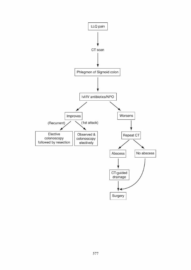
577

CASE REVIEW
Acute Diverticulitis of Sigmoid Colon
Perforated Carcinoma of the Colon
Inflammatory Bowel Disease (IBD)
Can present as an acute abdomenInitial cases can be treated nonoperatively unless unresponsive to medical therapyRecurrent cases should be considered for elective resectionComplex cases, including pericolonic abscess, should have CT-guided drainageprior to definite surgical managementAfter the acute event resolves, colonoscopy is necessary to rule out neoplasm
Can present as an acute abdomenUsually has prior symptoms of altered bowel habits, blood per rectum, and weightlossIn view of the acute abdomen, secondary to the perforation, colonoscopy iscontraindicatedMay be unable to differentiate between this and diverticulitis until surgicalintervention
Can present as an acute abdomen; usually has prior symptoms of diarrhea, mucusand blood, fever, and abdominal painsDefinitive diagnosis is based upon colonoscopy and biopsy performed once theacute event resolvesPrimary management is medical and surgery is indicated for complications of thedisease (intractability, perforation, abscess, stricture, fistula, and toxic megacolon)
578

579

Case 21
Chief Complaint
Bulge with tenderness in the abdominal wall
History and Physical Examination
A 40-year-old man with previous history of emergent laparotomy for perforated pepticulcer now presents with a 6-month history of an enlarging tender bulge of his abdominalwall. It is made worse by coughing, sneezing, or straining. There are no other GIcomplaints. On physical exam, the patient is afebrile and has normal vital signs.Examination is normal except for his abdomen, which has a midline scar and a reducible,slightly tender hernia. The overlying skin is normal.
Differential Diagnosis
Initial Management
Setting: outpatient
Ventral incisional hernia1.Most common cause of ventral hernia–
Epigastric hernia2.Diastasis recti3.
Not a true hernia–
580
Diagnostic/Therapeutic Plan
Test Results
Assessment
An uncomplicated, symptomatic ventral incisional hernia should be repaired surgically. Theapproach is at the discretion of the surgeon and can be open or laparoscopic and commonlyincludes the use of mesh or a primary suture repair. No one approach has been demonstrated tobe more superior.
Should the patient have associated GI complaints, a CT scan should be obtained to rule outunderlying intra-abdominal pathology. If the patient has evidence of incarceration (inability toreduce the hernia) or strangulation (compromised blood flow with overlying skin changes),emergent surgical intervention is required.
Further Management Plan
Discussion
Ventral hernias are most commonly secondary to prior abdominal operations. The decisionwhether to do a laparoscopic or open approach, with or without mesh, is at the discretion ofthe surgeon. If the hernia is secondary to an intra-abdominal pathology (carcinoma), this shouldbe addressed first.
Blood work: CBC and electrolytes
Blood work within normal limits
CT scan: fascial defect without any other abnormalitiesSurgical repair
581

Epigastric hernias are spontaneous hernias not due to prior surgical intervention. Diastasisrecti is not a true hernia (no fascial defect), but rather, a separation of the rectus abdominus atthe linea alba. It needs no surgical intervention.
Final Diagnosis
Ventral incisional hernia
CASE REVIEW
Ventral Incisional Hernia
Caused by previous abdominal surgeryMust rule out underlying abdominal pathology if GI symptoms present (CT scan)
582

Epigastric Hernia
Diastasis Recti
Should be repaired with surgery (laparoscopic, open, with or without prostheticmesh)
Not caused by previous abdominal surgeryMust rule out underlying abdominal pathology if GI symptoms present (CT scan)Should be repaired with surgery (laparoscopic, open, with or without prostheticmesh)
Not a true hernia (intact fascia)No treatment necessary
583

Case 22
Chief Complaint
Pain and tingling in fingers
History and Physical Examination
A 30-year-old female computer programmer has had progressive symptoms of pain andtingling in the thumb, index finger, long finger, and ring finger of her right hand for oneyear. This is made worse when she spends long hours typing and at night. She is otherwisehealthy.
On physical examination the patient is afebrile and vital signs are normal. Examination isnormal except for right hand and wrist, which reveal decreased sensation over the mediannerve distribution. Motor function is intact. Pulses are normal. There is no muscle atrophyor skin changes. Tinel’s and Phalen’s signs are both positive.
CLINICAL PEARL
Tinel’s sign is replication of symptoms by tapping the median nerve at the wrist.
Phalen’s sign is replication of symptoms caused by flexion of the wrist joints for 2 min.
584

Differential Diagnosis
Initial Management
Setting: outpatient
Diagnostic/Therapeutic Plan
Test Results
Assessment
Carpal tunnel syndrome due to median nerve compression
Further Management Plan
Carpal tunnel syndrome1.Cervical radiculopathy2.Osteoarthritis3.Tendonitis4.
Blood work: CBC and electrolytesWrist x-rays, AP and lateral viewsCervical x-rays if indicated
Blood work: normalX-rays: normal
Rest to the wrist joint1.Orthotic splints2.NSAIDS3.Steroidal injections4.
585

Discussion
Usually non-operative treatment of carpal tunnel syndrome is successful. Should it fail, surgicalintervention with decompression of the carpal tunnel is indicated. Nerve conduction velocities(NCV) and electromyography (EMG) are mandatory prior to surgery to confirm the diagnosis.
Final Diagnosis
Carpal tunnel syndrome
CASE REVIEW
Carpal Tunnel Syndrome
Cervical Radiculopathy
Osteoarthritis of the Wrist
Usually related to repetitive motions at the wristCan be associated with diabetes, arthritis, hypothyroidism, and pregnancyDiagnosis is based on symptoms in the anatomical distribution of median nerveNon-surgical treatment (rest, splints, medications, and injection) are usuallysuccessfulSurgical intervention requires confirmation of diagnosis by abnormal NCV andEMG tests
Symptoms can mimic carpal tunnel syndromeNegative Tinel and Phalen signsAbnormality is in cervical spine and treatment should be directed accordingly
Symptoms can mimic carpal tunnel syndrome
586

Tendinitis
Usually occurs at older age or with antecedent injuryNegative Tinel and Phalen signsX-rays are usually diagnosticTreatment is non-surgical
Symptoms can mimic carpal tunnel syndromeUsually occurs by repetitive wrist joint movementNegative Tinel and Phalen signsX-rays are normalTreatment is rest, splints, and medications
587

Case 23
Chief Complaint
Pain in the wrist after fall
History and Physical Examination
A 30-year-old woman slipped and fell and landed on her outstretched hand, resulting inimmediate pain in the wrist. She is otherwise healthy with no complaints. On physicalexamination the patient is afebrile with normal vital signs. She has exquisite tenderness inthe wrist at the anatomical snuffbox. Neurovascular exam is normal. There is no obviousdeformity of the wrist joint and there is a full range of motion.
Differential Diagnosis
Initial Management
Setting: outpatient
Diagnostic/Therapeutic Plan
Scaphoid fracture1.Colles’ fracture: fracture of the distal radius and ulnar styloid2.Supracondylar fracture of the humerus3.
Blood work: CBC and electrolytesAnteroposterior and lateral x-ray of the wrist
588

Test Results
Assessment
Scaphoid fracture
Further Management Plan
Immobilization (splint and/or cast)
Discussion
Not all scaphoid fractures are immediately visualized on initial x-ray evaluation. Failure tomake this diagnosis can result in avascular necrosis of the scaphoid, which can result in severedisability. Therefore, if the x-rays are negative and the clinical suspicion is high, treatment isperformed as if this is a scaphoid fracture.
Subsequent follow-up should include one of the following:
Follow-up Management and Prevention
Repeat x-rays in 1–2 weeks to assess healing of the fracture. If x-ray is indeterminate, MRI isindicated.
Final Diagnosis
Blood work: CBC and electrolytes: normalX-ray: scaphoid fracture
Repeat x-ray in 1–2 wksBone scanMRI (most sensitive of these tests) to confirm or rule out the diagnosis
589

Scaphoid fracture
CASE REVIEW
Scaphoid Fracture
Colles’ Fracture
Supracondylar Fracture of the Humerus
Usually occurs in young adults (age 25–45) subsequent to a fall on the outstretchedhandClassically the pain is in the anatomical snuffboxX-rays are not always diagnostic and follow-up x-ray, bone scan, or MRI (mostsensitive) may be necessaryAvascular necrosis is a complication which has significant disabilityAll clinically suspicious cases with negative x-rays should be treated as if they havea fracture until proven otherwise by subsequent follow-up and examination
Usually occurs in older adults subsequent to a fall on the outstretched handThe fracture of the distal radius and ulnar styloid can result in a fork-like deformityof the wristTreatment is by immobilization with a splint or a cast
Usually occurs in children subsequent to fall on the outstretched handPain is not in the wrist but in the elbow jointX-rays are diagnosticComplications include neurovascular injury – Volkmann’s contracture, which is dueto ischemic fibrosis of flexor muscles.
590

591

Case 24
Chief Complaint
Abdominal pain and fullness
History and Physical Examination
A 55-year-old man has noticed for the past 2 months progressive discomfort and fullnessin his abdomen. He also complains of early satiety. Three months ago he was involved ina severe motor vehicle accident, where he had a steering wheel injury to his chest andabdomen. He has no other GI symptoms. He does not drink alcohol and has no history ofbiliary tract disease.
On physical examination the patient is afebrile with normal vital signs. He is anicteric.Examination is normal except for the abdomen, which reveals epigastric fullness withmoderate tenderness. Bowel sounds are normal and there are no hernias. There are nodiscrete masses or organomegaly. Rectal examination is negative.
Differential Diagnosis
Initial Management
Pancreatic pseudocyst1.Intra-abdominal malignancy2.Bowel obstruction3.Bilary tract disease4.
592

Setting: inpatient
Diagnostic/Therapeutic Plan
Test Results
Assessment
Most likely this represents a pancreatic pseudocyst secondary to blunt abdominal trauma. Thiscould represent a cystic pancreatic neoplasm, but this is less likely, given the history of trauma.
Further Management
Internal surgical drainage (cystogastrostomy, cystoduodenostomy, cystojejunostomy).
Discussion
Pancreatic pseudocyst can occur from pancreatitis due to any cause. In this case, the history oftrauma is suspect. However, cystic neoplasms of the pancreas must be always be considered,as the traumatic incident may not be related. Complications from pancreatic pseudocyst includeextrinsic obstruction, infection, and hemorrhage.
Blood work: CBC, serum electrolytes, LFTs, lipase, amylaseChest and abdominal x-rayAbdominal U/SCT scan of the abdomen with oral and IV contrast
Blood work: normal except for slight lipase elevationX-ray: no pneumoperitoneum, no dilated loops of bowelU/S: no gallstones; ill-defined cystic mass in the area of the mid-pancreasCT scan: 8 cm cystic mass in mid-portion of the pancreas
593

Pancreatic pseudocysts that are asymptomatic and small can be observed and followed byimaging studies. Maturation of the pseudocyst (satisfactory thickness of the pseudocyst wall tohold sutures) must be present (determined by CT scan and may take as long as 6–8 weeks)before surgical intervention. In general terms, the management of pancreatic pseudocysts is 6cm diameter, 6 mm thick wall, and 6 weeks old before surgery is considered.
As noted, internal drainage is the best treatment, but if the cyst is infected, percutaneousdrainage is the right thing to do.
Final Diagnosis
Pancreatic pseudocyst
594

CASE REVIEW
Pancreatic Pseudocyst
Intra-abdominal Malignancy
Bowel Obstruction
Bilary Tract Disease
Occurs after acute pancreatitis from any causeComplications include extrinsic obstruction, infection, and bleedingImaging studies confirm diagnosis (CT scan is most accurate)Internal surgical drainage after maturation of the pseudocyst wall is gold standardof treatmentSmall asymptomatic pseudocysts can be observed
Cystic masses can be malignant neoplasms. Take biopsy of the cyst wall duringsurgery to confirmBe cautious that history of trauma may be unrelated
Usually presents with GI symptoms including vomiting, distention, constipation
Most commonly related to cholelithiasis
595

Case 25
Chief Complaint
Newborn in severe respiratory distress
History and Physical Examination
A newborn presents with severe respiratory distress following a normal pregnancy anddelivery. On physical exam, the infant is afebrile with marked tachypnea and using allaccessory muscles of respiration. There is no evidence of cyanosis. HEENT is normalwith trachea in the midline. There are decreased breath sounds in left chest but normalbreath sounds in right chest. Heart has no murmurs or gallops. Abdomen is scaphoid, soft,no masses, or organomegaly. There are normal bowel sounds.
Differential Diagnosis
Initial Management
Setting: emergency department
Diagnostic/Therapeutic Plan
Congenital diaphragmatic hernia (CDH)1.Spontaneous pneumothorax2.Chylothorax3.
Blood work: CBC and electrolytes
596

Test Results
Assessment
Acute respiratory distress secondary to CDH
Further Management
Discussion
As there was clear evidence of bowel in the chest and no evidence of pneumothorax orchylothorax, the diagnosis is CDH. This infant needs emergent surgical intervention. The repairof the diaphragm is done through the abdomen and is in fact not difficult to do.
The complications of this disease do not relate to the surgery but to the hypoplasia of bilaterallungs. The subsequent inability to ventilate this infant because of the lung hypoplasia is themajor contributor of this infant’s morbidity and mortality. Extracorporeal membrane
Continuous pulse oximetrySerial arterial blood gasesChest x-ray
Blood work: normalOximetry: O2 saturation in mid 80%sBlood gases: severe hypoxia and acidosisX-ray: air fluid levels in left chest; absence of significant lung markings
ABCs of resuscitation, including emergent endotracheal or nasogastric intubation1.NG tube decompression2.Operative repair of diaphragmatic hernia3.
597

oxygenation may be helpful to oxygenate the infant until the lungs develop further and recover.Prior to delivery, CDH can only be diagnosed in utero by U/S.
Final Diagnosis
CDH
CASE REVIEW
CDH (Congenital Diaphragmatic Hernia)
Spontaneous Pneumothorax
Chylothorax
Presents with acute respiratory distress at birthCannot make diagnosis prenatally except by U/SComplications revolve around bilateral hypoplasia of lungs and subsequentpulmonary hypertension, not the diaphragmatic defect in itselfSurgical approach is through the abdomen
Presents with acute respiratory distress from rupture of congenital bullaeTreated with needle decompression and chest tube insertionIf a tension pneumothorax, trachea will be shifted away from the site of pathology
Very rare and complex congenital abnormality in which there is chyle in the pleuralspace, leading to respiratory compromise
598

599

Section IV
EPIDEMIOLOGY ANDBIOSTATISTICS
600

5
Epidemiology
Key DefinitionsEpidemiology. Study of the distribution and determinants of health-related states within apopulation. It refers to the patterns of disease and the factors that influence those patterns.
Clinical epidemiology. Study and application of population-based data with patient decision-making.
Endemic. The usual, expected rate of disease over time. The disease is maintained withoutmuch variation within a region.
Epidemic. Occurrence of disease in excess of the expected rate. Epidemiology is the “study ofepidemics.” Usually presents in a larger geographic span than endemics.
Pandemic. A worldwide epidemic.
Epidemic curve. A visual description of an epidemic curve is disease cases plotted againsttime. The classic signature of an epidemic is a “spike” in time.
601

An explosive point-source outbreak of measles in an Elgin, Illinois school caused by asingle index case whose hacking cough produced an aerosol of measles virus.
602

A food-borne outbreak of hepatitis A among patrons of a Fort Lauderdale, Floridarestaurant.
603

Continuous increase in U.S. AIDS cases from the original report of 5 cases in 1981through the 500,000th case reported in 1995, and beyond.
604

Types of PreventionPrevention goals in medicine promote health, preserve health, restore health when it isimpaired, and minimize suffering and distress. These goals aim to minimize both morbidity andmortality.
Primary prevention is the promotion of health at both individual and community levels byfacilitating health-enhancing behaviors, preventing the onset of risk behaviors, and diminishingexposure to environmental hazards. Primary prevention efforts decrease disease incidence.
Secondary prevention is the screening for risk factors and early detection of asymptomatic ormild disease, permitting timely and effective intervention and curative treatment. Secondaryprevention efforts decrease disease prevalence.
Tertiary prevention is the reduction of long-term impairments and disabilities and preventionof repeated episodes of clinical illness. The goals of tertiary prevention are to preventrecurrence and to slow progression.
Examples of Prevention for Coronary Heart Disease
Primary prevention Health education programs to promote healthy lifestyles and prevent
onset of heart disease risk factors. An example would be the “Hearty
Heart” nutrition program for elementary school children.
Secondary prevention Community blood pressure screening.
Physician support to quit smoking cigarettes.
605

Tertiary prevention Graded aerobic physical activity program prescribed to patients during
recovery from first myocardial infarction.
Review Questions
PREVENTION
Response options for Questions 1–4:
Health promotion(A)Primary prevention(B)Secondary prevention(C)Tertiary prevention(D)Palliative care(E)
1. Breast self-examination.
2. Physical therapy/rehabilitation and ergonomic training program for blue-collarworkers recovering from severe back strain injury sustained on the job.
3. School-based sexual health education program for middle school students.
4. Confidential PPD testing to detect latent tuberculosis infection conducted atcommunity clinics by county health department personnel.
606

KEY DEFINITIONS
Response options for Questions 5–7:
Hypoendemic(A)Endemic(B)Epidemic(C)Hyperendemic(D)Pandemic(E)Holoendemic(F)
5. A multinational outbreak of influenza
6. The rapid rise in AIDS cases among drug injectors in Bangkok in the late 1980s
7. The long-term, relatively constant rate of occurrence of colorectal cancer in U.S.women
607

Answers and Explanations
REVIEW QUESTIONS
Prevention1.
1. Answer: C.Self-screening for early detection leading to early diagnosis and effective, life-savingtreatment.
2. Answer: D.Rehabilitation following an episode of injury with a concurrent focus on preventingsubsequent injury.
3. Answer: B.Prevention of onset of risky sexual behaviors.
4. Answer: C.Screening to detect tuberculosis (TB) infection, to be followed by therapy to preventprogression to active TB.
Key Definitions6.
5. Answer: E.A pandemic is an epidemic that crosses national borders.
608

6. Answer: C.AIDS appeared suddenly, and the epidemic increased exponentially.
7. Answer: B.When disease cases are plotted over time, a flat horizontal line depicts an endemic pattern.
609

Measures of Morbidity and Mortality
RATES
Rate
Rate Equation
where the numerator is the number of epidemiologic events, the denominator is the number ofpeople in the population of interest, and the multiplier is selected so that the result of the ratecomputation generally yields a number in the range from 1 to 100.
Multipliers
For major vital statistics, such as birth rate, death rate, and infant mortality rate, thepreferred multiplier is 1,000. The result is expressed as a “rate per 1,000.”
Rate is the frequency of occurrence of epidemiologic events in populations. They are usedto compare epidemiologic events among populations.Rates allow direct comparisons of “events per identical number” of people in the two ormore populations.Rates permit comparisons of epidemiologic events occurring in a single population assessedat several points in time.
610

For individual diseases, the most common multiplier is 100,000. The result is expressed as a“rate per 100,000.”
Matching Numerator and Denominator
Essential rule: Match the numerator with the denominator.Match on person, place, and time characteristics.
611

Specific and Adjusted Rates
SPECIFIC RATESDefinition: Specific rates “specify” a subset of the total population that is singled out forspecial examination or comparison with other subsets of the population. Use the followingformula:
Common demographic variables used for specific rates:
(Populations can be stratified on two or more demographic variables at a time.)
Matching numerator and denominator is the most important concept for computing specificrates. Example:
“Event” of interest: Cancer deaths
Age groupGenderRace/ethnicityHighest level of education attainedMarital statusSocioeconomic status
612

Place: State of Nevada
Time: Calendar year, 1996
Rate of interest: Age-specific rate* for ages 45–64
Formula:
*Age-specific rate: a rate for a specified age group
ADJUSTED RATES (OR STANDARDIZED)Definition: Rates calculated after using statistical procedures to minimize demographicdifferences between populations being compared. Comparisons of rates between two groupsmay be misleading if the composition of the groups differs on important demographiccharacteristics. Adjustment improves the validity of the comparison.
The following two cases are examples of comparisons between groups where rate adjustmentis clearly essential.
Adjustment for gender differences is warranted. First, the two populations differ on ademographic characteristic: Automotive workers tend to be men; textile workers tend to bewomen. Second, the disease/disorder is related to the same demographic: Alcohol problems
The rate of alcoholism and alcohol abuse is found to be higher among workers in anautomobile assembly plant compared with same-age workers at a textile mill in the samecity.
1.
613

are more prevalent in men. The higher observed rate in automotive workers may be due to themarked differences in gender in the two employee populations.
Adjustment for level of education is warranted. First, the two populations differ on ademographic characteristic: Factory workers tend to have a low level of education; computerprogrammers are likely to be college graduates. Second, the disease/disorder is related to thesame demographic: The major cause of lung cancer is cigarette smoking. People with lowerlevels of education have higher smoking rates; college graduates have the lowest smokingrates. The differences in lung cancer rates may reflect expected differences in smokingprevalence rates for workers with different levels of education.
Properties of a board-style adjusted rate problem:
Disease Rates Positively Correlated with Age
Population A Population B Population C
Cases Population Cases Population Cases Population
Younger 1/1,000 1 1,000 2 2,000 3 3,000
The rate of lung cancer is found to be higher among male factory workers ages 50–64,than among male computer programmers ages 50–64, in the same company.
1.
A significant difference in the rate of disease is declared to exist between two groups. Thecompared rates are unadjusted.The two groups differ on a key demographic variable.The disease is known to be related to the same demographic variable.Adjustment will tend to make the observed difference between unadjusted rates disappear.
614

Intermediate 2/1,000 4 2,000 4 2,000 4 2,000
Older 3/1,000 9 3,000 6 2,000 3 1,000
14 6,000 12 6,000 10 6,000
Crude Rates Per/1,000 2.3 2.0 1.6
Review Questions
ADJUSTED RATES8. In the United States, the suicide rate for physicians is significantly higher than the
corresponding rate for the general population. What is the most appropriateinterpretation of this finding?
Higher suicide rates in physicians are likely to be related to job stress,including life-and-death decision making for patients in the care of thephysician.
(A)
Higher rates of suicide in physicians are likely to be related to constantexposure to human suffering, trauma, and death.
(B)
Physicians have higher rates of suicide than the general population; no furtherinterpretation is possible from the information presented.
(C)
While the unadjusted rate of suicide is higher for physicians, failure to adjustfor differences between physicians and the general population onsocioeconomic status precludes meaningful interpretation of this finding.
(D)
The finding of statistical significance proves that physicians are at higher riskfor suicide than nonphysicians.
(E)
615

616

Answers and Explanations
REVIEW QUESTIONS
Adjusted Rates1.
8. Answer: D.When a significant relationship is stated but the comparison groups have some obviousdemographic difference, look for the answer that suggests conclusions may be invalid unlessrates are “adjusted” or “standardized” to compensate for the demographic disparities.
In this instance, physicians are generally a higher socioeconomic status (SES) group relativeto the general population. Suicide rates are elevated for high-SES people. Once adjusted forSES differences, the finding of higher suicide rates in physicians no longer stands.
Note: Strongly suspect any response that claims that “proof” has been demonstrated. Nosingle study can achieve proof. Furthermore, no investigator would be so self-aggrandizingas to claim to have conducted the definitive study. Such a response option (“distractor”) isalmost always wrong.
617

Measures of Morbidity
INCIDENCE AND PREVALENCE
Prevalence Rate: All Cases
Prevalence rate is the proportion of individuals with existing disease at a point in time (pointprevalence). It is the proportion of individuals with existing disease during a period of time(period prevalence).
Incidence Rate: New Cases Only
The focus is on chronic conditions.The numerator refers to ALL individuals who have the illness at the time(s) in question.
The incidence rate is the proportion of individuals developing new disease during a periodof time.It is the rate of new disease events in a population during a period of time.Incidence rates can be calculated only over a period of time, not at a single point.The focus is on acute conditions.
618

Attack rate is a type of incidence rate that focuses on a known exposure or risk. For example, if10 of 100 children who attend daycare A, and 40 of 100 children who attend daycare Bdevelop diarrhea, the attack rate would be 10% for attendance at daycare A and 40% forattendance at daycare B.
CCS NOTE
Relationship between Incidence and Prevalence:
“PREVALENCE POT”A “prevalence pot” is a common portrayal of the concept of prevalence and its relationship toincidence. At the first moment of observation, the count of cases “in the pot” provides anestimate of point prevalence. Incident cases are observed over time. These new cases areadded to the pre-existing cases. As long as clinical illness persists, cases remain in the pot.Cases leave the prevalence pot in one of two ways, through recovery or death. Changes inprevalence over time can be determined by monitoring trends in incidence, recovery, anddeath.
Prevalence = Incidence × Duration (conceptual formula, not computational)
Changes in incidence, duration, or both will ultimately affect prevalence.
619

Number needed to treat (NNT): How many in general population need to be treated toprevent one case?
Equations for Common Epidemiological Measures
Measure Equation Notes
Incidence rate
Prevalence rate
The inverse of the incidence rateIf incidence = 16 per 1,000 = 16/1,000Inverse = 1,000/16 = 625 = NNT
Acute cases–
New cases only–
Chronic cases–
620

Prevalence Incidence × duration
Duration
Numbers needed
to treat
*Point prevalence will have denominator at a specific point in time, whereas period prevalence will
include a specific period of time.
Review Questions
INCIDENCE AND PREVALENCE
Do not include in numerator
any deaths or cases that
recovered from disease, as
they are no longer existing
cases of illness
–
Assuming incidence and
duration are stable
–
Inverse of incidence rate–
9. A pharmaceutical corporation completes trials on a vaccine for a severe strain ofinfluenza virus demonstrating high vaccine efficacy. The Food and DrugAdministration approves the vaccine for use in the U.S. As the influenza pandemicapproaches U.S. borders, the Centers for Disease Control and Prevention launches anationwide campaign to vaccinate the population using local public health department
621

Questions 11–14
Among 245 college students who dedicated one month of summer break to building homes forHabitat for Humanity, 12 developed back strains on the job. Based on the diagram of these 12episodes of back strain, answer the following questions:
personnel throughout the country to ensure that the vaccine is available, free ofcharge, to all people. Assuming that a high degree of vaccine coverage is achieved,what is the expected impact of this major public health initiative?
Decreased duration of influenza illness leading to decreased prevalence(A)Decreased incidence of influenza illness leading to decreased prevalence(B)Decreased incidence offset by increased duration: no change in prevalence(C)No change in observed incidence or duration: no change in prevalence(D)Effects on prevalence cannot be determined from the information provided(E)
10. A new, effective treatment for a common disease, leading to complete cure, isdeveloped. Which of the following impacts on disease occurrence is expected?
Decreased duration of illness, leading to decreased prevalence(A)Decreased incidence of illness, leading to decreased prevalence(B)Decreased incidence and duration of illness, leading to decreased prevalence(C)No change in observed incidence or duration: no change in prevalence(D)Effects on prevalence cannot be determined from the information provided(E)
622

Response options for Questions 11–14:
2/242(A)2/244(B)2/245(C)3/242(D)3/245(E)8/242(F)8/244(G)8/245(H)10/244(I)10/245(J)12/245(K)
11. What is the point prevalence rate on July 15, 12:01 AM?
623

12. What is the point prevalence rate on July 31, 11:59 PM?
13. What is the incidence rate for the period July 15–July 31?
14. What is the period prevalence for July 15–July 31?
624

Answers and Explanations
REVIEW QUESTIONS
Incidence and Prevalence1.
9. Answer: B.Vaccination decreases the likelihood of development of new infection and clinical disease.In turn, the prevalence during the peak of the influenza season will be decreased.
10. Answer: A.An effective treatment will move people more quickly toward recovery. Average duration ofillness will decrease. Prevalence, the proportion of people ill with the disease at a point intime, will also decrease. This will apply to both acute and chronic diseases. Whenimplementing a new treatment, incidence is not affected for a chronic disease. Also, thetreatment per se will not affect incidence for an acute disease.
11. Answer: C.On July 15, two students had symptoms of back strain (E, K).
12. Answer: E.On July 31, three students had symptoms of back strain (B, G, I).
13. Answer: H.Eight new cases of back strain had onset between July 15 and July 31 (A, B, D, F, G, I, J, L).
625

14. Answer: J.A total of 10 students had symptoms of back strain at some time during the period July 15–July 31, including two with onset prior to July 15 (E, K) and eight with onset during theperiod July 15–31 (A, B, D, F, G, I, J, L).
626

Vital Statistics and Rates
BIRTH RATEDefinition: Rate of live births in a population during a time period (usually the calendar year).
Interpretation: “Births per 1,000 population”
U.S. birth rate: 13.0 births/1,000 population (in 2010)
FERTILITY RATEDefinition: Rate of live births among women of childbearing age (ages 15–44) in a populationduring a time period (usually the calendar year).
Interpretation: “Births per 1,000 women of child-bearing age”
U.S. fertility rate: 64.1 births/1,000 women of child-bearing age (in 2010)
MORTALITY RATE
627

Definition: Rate of deaths in a population during a time period (usually the calendar year).
Interpretation: “Deaths per 1,000 population.”
Synonyms: death rate, crude death rate
U.S. mortality rate: 8.4 deaths/1,000 population (in 2010)
INFANT MORTALITY RATEDefinition: Yearly rate of deaths among children age <1 in relation to the number of live birthsduring the same year. Within a population, the infant mortality rate is a key indication of thepopulation’s health status.
Interpretation: “Infant deaths per 1,000 live births”
U.S. infant mortality rate: 6.14 infant deaths/1,000 live births (in 2010)
Infant mortality rate: neonatal mortality rate + postneonatal mortality rate
628

Perinatal mortality rate: Stillbirths and deaths in the first week of life/Live births × 1,000
INFANT MORTALITY
Rates per 1,000 live births (2010)
5.33
12.40
5.39
6.39
Top 3 reasons
24% of cases
18% of cases
16% of cases
Other facts
Whites:
African Americans:
Hispanics:
Overall:
Birth defects:
Low birth weight (<1,500 grams)/respiratory distress:
SIDS:
Blacks have highest rates of infant mortality from low birth weight and infections. Numberone killer of black infants is low birth weight.Native Americans have highest SIDS rate.Hispanic profile is similar to whites, but slightly higher.
629

Sociologic risk factors for children
Facts about adolescent pregnancy
Consequences for teenage pregnancy
For mother. Often drop out of school. Many never work and become welfare dependent.
For child. Neonatal deaths and prematurity are common. Possible lower level of intellectualfunctioning. Problems for children of single-parent families can include an increased risk ofdelinquency.
Adolescent sexual behavior
SIDS rates reduced sharply by avoiding having infants sleep on their stomachs.
Maternal immaturity: Risk of premature birth increases dramatically below age 19Poverty is a major risk factor for prematurity and other unfavorable outcomes.The single-parent family is also correlated with child abuse, childhood suicide, truancy, anddelinquency.
Roughly, one million U.S. teenage girls (10% of total) become pregnant each year.Of girls aged 15–19 years, 33% have at least one unwanted pregnancy.Fifty percent have the child, 20% have spontaneous abortions, and 30% have an electiveabortion.More than half of adolescents do not use contraceptives the first time they haveintercourse.
Eighty percent of boys and 70% of girls are sexually active by the age of 18.More than 20% of all sexually active girls become pregnant at least once before the age of20 years.
630

Maternal mortality rate
Definition: Yearly rate of deaths in women from causes associated with childbirth in relationto the number of live births during the same year.
Interpretation: “Maternal deaths per 100,000 live births”
U.S. maternal mortality rate: 7.1 maternal deaths/100,000 live births
Case fatality rate (CFR)
Definition: Percentage of cases of an illness or medical condition that result in death within aspecified time period.
Interpretation: Proportion of cases that end in death (fatality)
Example: In a population of 200 people, 25 become ill, and 5 die from the illness.
Proportionate mortality rate (PMR)
Pregnancy is the leading cause of school dropout among girls.Roughly 80% of sexually active adolescents do not use birth control.One out of five teenagers will have a sexually transmitted disease.
631

Definition: Percentage of deaths from all causes that are due to a specified cause during aspecified time period.
Interpretation: Proportion of deaths from a specific cause.
The PMR is used for the most common causes of death in a population.
Types of Measured Rates
Crude mortality rate Deaths per population
Cause-specific mortality rate Deaths from a specific cause per population
Case-fatality rate Deaths from a specific cause per number of persons with the
disease
Proportionate mortality rate
(PMR)
Deaths from a specific cause per all deaths
Review Questions
RATES
Response options for Questions 15–18:
632

Questions 19 and 20 are based on the following table:
Incidence and Mortality of Disease
Birth rate(A)Fertility rate(B)Infant mortality rate(C)Maternal mortality rate(D)Age-adjusted rate(E)Case-fatality rate(F)Sex-adjusted rate(G)Proportionate mortality rate(H)Age-specific rate(I)Sex-specific rate(J)Age- and sex-specific rate(K)Age- and sex- and race/ethnicity-specific rate(L)
15. Rate of live births among women of childbearing age.
16. The proportion of cases of a disease that die from that disease.
17. Prevalence rate of obesity in women, ages 45–64.
18. Rate of homicide in black men, ages 15–24.
633

Age Disease A Disease B Total
Groups Cases Deaths Cases Deaths Deaths Population
0–12 2 1 300 1 40 22,000
13–24 101 34 267 0 30 18,000
25–64 50 42 1,042 2 125 50,000
>64 0 0 986 95 303 30,000
Totals 153 77 2,595 98 498 120,000
19. The case-fatality rate for Disease A is
77/120,000 × 1,000(A)77/120,000 × 100,000(B)153/120,000 × 100,000(C)153/498 × 100(D)77/153 × 100(E)
20. The proportionate mortality rate for Disease B is
98/120,000 × 100,000(A)2,595/120,000 × 100,000(B)98/2,595 × 100(C)98/498 × 100(D)Cannot be determined(E)
634

635

Answers and Explanations
REVIEW QUESTIONS
Rates1.
15. Answer: B.Restatement of definition of fertility rate.
16. Answer: F.Restatement of definition of case-fatality rate.
17. Answer: K.Age- and sex-specific rate; prevalence rate restricted to women in the age range 45–64.
18. Answer: L.Homicide rate restricted to black men in the age range 15–24.
19. Answer: E.20. Answer: D.
636

Years of Potential Life Lost and SurvivalAnalysis
YEARS OF POTENTIAL LIFE LOST (YPLL) (INDICATOROF PREMATURE DEATH):The YPLL for a particular cause of death is the sum, over all persons dying from the cause, ofthe years that these persons would have lived had they experienced normal life expectancy.Assume life expectancy is 75 years. A person who dies at age 65 would be dying 10 yearsprematurely (75 − 65 = 10 YPLL). For 100 such people, the YPLL calculation would be: 100 ×(75 − 65) = 1,000 YPLL.
SURVIVAL ANALYSISSurvival analysis is a class of statistical procedures for estimating the proportion of peoplewho survive in relation to the length of survival time. The starting point is 100% survival. In2000, the median survival time was 78 years.
A survival curve is a curve that starts with 100% of the study population and shows thepercentage of the population still surviving at successive times for as long as information isavailable.
In the United States, the leading cause of YPLL is unintentional injury before age 65.
637

638

639

Understanding Screening TestsDefinition. Screening is the process of using tests to permit early detection of risk factors,asymptomatic infection, or early stages of clinical disease, thus permitting early diagnosis andearly intervention or treatment.
CLASSIC 2 × 2 TABLEThe 2 × 2 table is the standard form for displaying screening test results in relation to diseasestatus. Disease status categories (diseased and well) are diagrammed in the vertical columns.Screening test results (positive, negative) are diagrammed in the horizontal dimension.
Classic 2 × 2 Table
Disease No Disease Totals
Positive True Positive [TP] False Positive [FP] TP + FP
Negative False Negative [FN] True Negative [TN] TN + FN
Screening is usually applied to populations of apparently well individuals. Illness, ifpresent, is asymptomatic (subclinical, inapparent).Screening tests allow for earlier detection and earlier diagnosis. Hopefully, earliertreatment will effect a more favorable clinical course.Screening test results are classified as “positive” (presumed by the test to be diseased) or“negative” (presumed by the test to be well).
640

Totals TP + FN TN + FP TP + TN + FP + FN
Cells in the 2 × 2 Table
Screening Results in a 2 × 2 Table
Disease
Present Absent Totals
Screening Test Results Positive TP 80 FP 40 TP + FP
Negative FN 20 TN 60 TN + FN
Totals TP + FN TN + FP TP + TN + FP + FN
TP = true positives; TN = true negatives; FP = false positives; FN = false negatives
MEASURES OF SCREENING TEST PERFORMANCE
Sensitivity and Specificity
Positive (P) and Negative (N) refer to the actual screening test results.True (T) and False (F) refer to the agreement of screening test results with the “goldstandard.”True Positives: Diseased people who are correctly classified as positive.True Negatives: Well people who are correctly classified as negative.False Positives: Well people who are misclassified as positive.False Negatives: Diseased people who are misclassified as negative.
Sensitivity: The proportion of people with disease who are correctly classified by thescreening test as positive.
1.
641

3a. Positive Predictive Value (PPV): The proportion of people with a positive screeningtest result who are diseased. (i.e., that a person with a positive test is a true positive)
3b. Negative Predictive Value (NPV): The proportion of people with a negative screeningtest result who are well. (i.e., that a person with a negative test is a true negative)
Sensitivity = TP/All people with diseaseSensitivity = TP/(TP + FN)Location on 2 × 2 table: left columnHighly sensitive tests identify most, if not all, possible casesImportant to consider when there is a consequence associated with missing the detectionof disease
Specificity: The proportion of well people who are correctly classified by the screeningtest as negative.
2.
Specificity = TN/All well peopleSpecificity = TN/(TN + FP)Location on 2 × 2 table: right columnHighly specific tests identify most, if not all, well people (i.e., not diseased), will givefew FP resultsConsidered when FP results can harm the patient
Predictive Values: A measure of the test which represents the percentage of test results thatmatch the diagnosis of the patient. These values are predicted by the disease prevalence inthe given population.
3.
Positive Predictive Value = TP/All people with a positive test resultPositive Predictive Value = TP/(TP + FP)Location on 2 × 2 table: top row↑ specificity = ↑ PPV
Negative Predictive Value = TN/All people with a negative test result
642

Negative Predictive Value = TN/(TN + FN)Location on 2 × 2 table: bottom row↑ sensitivity = ↑ NPV
Accuracy: The proportion of all screened people who are correctly classified by thescreening test.
4.
Accuracy = (TP + TN)/All screened peopleAccuracy = (TP + TN)/(TP + TN + FP + FN)Location on 2 × 2 table: main diagonalCan be used to summarize overall value of a test
Prevalence: The proportion of screened people who have disease.5.Prevalence can be estimated only if the entire population or a representative sample ofthe population is screened.Prevalence = (TP + FN)/(TP + TN + FN + FP)↑ prevalence of a disease usually equals ↑ PPV and ↓ NPV↓ prevalence of a disease usually equals ↓ PPV and ↑ NPV
Likelihood ratio: The expression of how many more (or less) likely a test result is to befound in nondiseased (or diseased) compared with diseased (or nondiseased).
6.
Positive likelihood ratio (LR+) is the proportion of diseased people to that ofnondiseased people with a positive test result
Negative likelihood ratio (LR-) is the proportion of diseased people to that ofnondiseased people with a negative test result
643

SCREENING TEST DIAGRAMThe screening test diagram displays the distributions of the screening test measure separatelyfor people with disease and people with no disease.
The cutoff point or criterion point divides screened people into test-positives and test-negatives.
People with no disease (Figure 1–7; solid line) are either correctly classified as TN ormisclassified as FP. Diseased people (Figure 1–7; dashed line) are either correctly classifiedas TP or misclassified as FN.
The screening test diagram is a useful model of the real world in which values of screening testmeasures (such as blood pressure) are generally different for diseased (hypertensive) andnondiseased (normotensive) people, but the distributions overlap.
The measures of screening test performance can be displayed on the screening test diagram byidentifying the appropriate areas under the curves. For example, the numerator for sensitivity isTP, whereas the denominator is everyone under the curve labeled “disease.”
644

Summarized 2 × 2 Tables
Disease Predictive Values
Measure Present Absent Equation Notes
Positive TP FP Each value
looks at a
‘positive’
Negative FN TN Each value
looks at a
‘negative’
Screening
Test
Equation
645

MeasuresNotes Want to identify all possible causes. Use everything in
‘diseased’ column = TP + FN.
Want to
more
specifically
identify those
that do not
have disease.
Use
everything in
‘no diseased;
column.
Review Questions
SCREENING TESTS
Response options for Questions 21–26:
A new screening test is applied to a representative sample of 1,000 people in the population.Based on the data presented in the following table, calculate the requested screening testmeasures.
Diseased Well
Positive 90 60 150
Negative 10 840 850
646

100 900 1,000
90/150(A)90/100(B)90/1,000(C)90(D)60(E)10(F)840/850(G)840/900(H)930/1,000(I)900/1,000(J)100/1,000(K)Cannot be calculated(L)
21. What is the sensitivity of the screening test?
22. What is the specificity of the screening test?
23. What is the positive predictive value of the screening test?
24. What is the accuracy of the screening test?
25. What is the number of false positive test results?
647

Questions 27–32
The Centers for Disease Control and Prevention is concerned about optimizing the detection ofa disease that poses a serious public health threat. CDC health officials are consideringlowering the usual screening test cutoff point from X to Y.
26. What is the prevalence of disease, assuming screening of a representative sample?
27. Moving cutoff in the manner being considered by the CDC causes the number of falsepositives to
increase(A)decrease(B)remain unchanged(C)cannot be determined(D)
28. Moving the cutoff in the manner being considered by the CDC causes the positivepredictive value to
648

increase(A)decrease(B)remain unchanged(C)cannot be determined(D)
29. Moving the cutoff in the manner being considered by the CDC causes the accuracy to
increase(A)decrease(B)remain unchanged(C)cannot be determined(D)
30. Moving the cutoff in the manner being considered by the CDC causes the sensitivityto
increase(A)decrease(B)remain unchanged(C)cannot be determined(D)
31. Assuming that everyone who receives a positive test result is referred for medicalfollow-up, moving the cutoff in the manner being considered by the CDC will causethe numbers of screened people who are referred for follow-up to
649

increase(A)decrease(B)remain unchanged(C)Cannot be determined(D)
32. At Cutoff Point X, sensitivity is
100%(A)85%(B)50%(C)25%(D)0%(E)
33. A physician interviews an 18-year-old female patient who mentions that she has justreceived a negative syphilis test result from the county health department. Shedescribes her sense of relief at receiving the test result. She discloses that she is a sexworker who “works the stroll” four to five nights a week. She has been “tricking” forthe past 18 months. Typically, she has oral or vaginal sex with five to eight customersper night. For a higher fee, she will have sex without requiring her customer to weara condom. On the basis of these findings, the physician is likely to be most concerningwith which of the following screening test measures?
Sensitivity(A)Specificity(B)Positive predictive value(C)Negative predictive value(D)Accuracy(E)
650

34. A 55-year-old man visits his primary care physician with a complaint of urinaryinfrequency. Examination finds a 1-cm nodule on his prostate gland. The physicianorders a prostate-specific antigen (PSA) serum test. By common standards, a PSAlevel >4 ng/mL is considered abnormal. Using this standard, this test has a sensitivityof 80% and a specificity of 90%. A recently published epidemiologic article foundthat in a cross-sectional study, 10% of men of this age have prostate cancer. Theresult on the patient’s PSA is 7 ng/mL. What is your best estimate of the likelihoodthat this man actually has prostate cancer?
13%(A)25%(B)36%(C)47%(D)58%(E)69%(F)72%(G)81%(H)
651

Answers and Explanations
REVIEW QUESTIONS
Screening Tests1.
21. Answer: B.Sensitivity = TP/All diseased people = 90/100
22. Answer: H.Specificity = TN/All well people = 840/900
23. Answer: A.PPV = TP/All test positives = 90/150
24. Answer: I.Accuracy = (TP + TN)/All screened people = 930/1,000
25. Answer: E.False positives = Well people who are misclassified by the test = 60
26. Answer: K.Prevalence = All diseased people/All screened people = 100/1,000
27. Answer: A.At Y, FP will increase as more well people are misclassified.
652

28. Answer: B.Although there will be more TP at Cutoff Y, there will be a large increase in numbers of FP.The ratio, TP/(TP + FP), will decrease. A positive test result will be less predictive ofactual disease.
29. Answer: B.X is the point of overlap and the point of maximal accuracy. Moving to Y will decreaseaccuracy.
30. Answer: A.At Y, more diseased people will receive a (correct) positive test result. They will be TP.TP, the numerator for sensitivity, will increase while the denominator (total people withdisease) will be unchanged.
31. Answer: A.Larger numbers of people would be screened positive at Cutoff Y and referred for follow-up.
32. Answer: B.Notice that Cutoff Point X separates the curve of diseased people into two areas; above thecutoff point, approximately 85% of diseased people receive a (correct) positive test result.They are true positives. Sensitivity = TP /All people with disease.
33. Answer: D.34. Answer: D.
653

Study DesignsThe following form is used for displaying the relationship of exposure to disease status:
2 × 2 Table Format
Disease No Disease
Exposed a b a + b
Nonexposed c d c + d
a + c b + d a + b + c + d
When epidemiologists observe the relationships between exposures and disease outcomes infree-living populations, they are conducting observational studies. When epidemiologists orclinicians test interventions aimed at minimizing the disease-producing exposures andoptimizing health-promoting exposures or factors, they are performing experimental studies.
In observational studies, nature is allowed to take its course; no intervention.
In experimental studies, there is an intervention and the results of the study assess the effectsof the intervention.
OBSERVATIONAL STUDIES
654

Case report: Brief, objective report of a clinical characteristic or outcome from a singleclinical subject or event, n = 1. For example, a 23-year-old man with treatment-resistant TB.No control group.
Case series report: Objective report of a clinical characteristic or outcome from a group ofclinical subjects, n >1, i.e., patients at local hospital with treatment-resistant TB. No controlgroup.
Cross-sectional study: The presence or absence of disease and other variables aredetermined in each member of the study population or in a representative sample at aparticular time. The co-occurrence of a variable and the disease can be examined.
Case-control study: Identifies a group of people with the disease and compares them with asuitable comparison group without the disease. It is almost always retrospective, e.g.,comparing cases of treatment-resistant TB with cases of nonresistant TB.
Disease prevalence rather than incidence is recorded.The temporal sequence of cause and effect cannot usually be determined in a cross-sectionalstudy, e.g., who in the community now has treatment-resistant TB.
Cannot assess incidence or prevalence of diseaseCan help determine causal relationshipsVery useful for studying conditions with very low incidence or prevalence
655

Cohort study: Population group is identified who has been exposed to risk factor is followedover time and compared with a group not exposed to the risk factor. Outcome is diseaseincidence in each group, e.g., following a prison inmate population and marking thedevelopment of treatment-resistant TB.
Analyzing Observational Studies (measure of effect)
For cross-sectional studies, use Chi-square (χ2); for cohort studies, use relative risk and/orattributable risk.
Relative risk (RR): Comparative probability asking, “How much more likely is the exposedperson going to get the disease compared to the nonexposed?”
Attributable risk (AR) (Also called absolute risk reduction): Comparative probabilityasking, “How many more cases in one group?”
Allows you to evaluate whether potential risk factors are related to subsequent outcomesProspective; subjects tracked forward in timeCan determine incidence and causal relationshipsMust follow population long enough for incidence to appearHistorical examples: Framingham study
Incidence rate of exposed group divided by the incidence rate of the unexposed group. Howmuch greater chance does one group have of contracting the disease compared with the othergroup?For example, if infant mortality rate in whites is 8.9 per 1,000 live births and 18.0 in blacksper 1,000 live births, then the relative risk of blacks versus whites is 18.0 divided by 8.9 =2.02. Compared with whites, black infants are twice as likely to die in the first year of life.For statistical analysis, it yields a p value.
Incidence rate of exposed group minus the incidence rate of the unexposed group
656

For case-control studies: Use odds ratio (OR).
Odds ratio (OR): Looks at the increased odds of getting a disease with exposure to a riskfactor versus nonexposure to that factor.
Using the same example, attributable risk is equal to 18.0 − 8.9 = 9.1. Of every 1,000 blackinfants, there were 9.1 more deaths than were observed in 1,000 white infants. In this case,attributable risk gives the excess mortality.Note that both relative risk and attributable risk tell us if there are differences but do not tellus why those differences exist.
Odds of exposure for cases divided by odds of exposure for controlsThe odds that a person with lung cancer was a smoker versus the odds that a person withoutlung cancer was a smoker
Odds Ratio
Lung Cancer No Lung Cancer
Smokers 659 (A) 984 (B)
Nonsmokers 25 (C) 348 (D)
Use OR = AD/BC as the working formula.
For the above example:
657

Review Questions
STUDY DESIGN
The odds of having been a smoker are more than 9 times greater for someone with lungcancer compared with someone without lung cancer.OR approaching 1 = increased risk of outcome with exposure
35. How would you analyze the data from this case-control study?
No Colorectal
Cancer
Colorectal
Cancer
TOTALS
120 60 180
200 20 220
TOTALS 320 80 400
ANSWER: OR = 5.0
Family history of colorectal
cancer
No family history of colorectal
cancer
658

Differentiating Observational Studies
Characteristic Cross-Sectional
Studies
Case-Control Studies Cohort Studies
Time One time point Retrospective Prospective
Incidence No No Yes
Prevalence Yes No No
Causality No Yes Yes
Role of
disease
Measure disease Begin with disease End with disease
Assesses Association of risk
factor and disease
Many risk factors for
single disease
Single risk factor affecting
many diseases
Data analysis Chi-square to assess
association
Odds ratio to estimate
risk
Relative risk to estimate
risk
Computational Measures by Type of Observational Study
Measure Cross-Sectional
Study
Case-Control
Study
Cohort
Study
Prevalence of disease Yes No No
Prevalence of exposure Yes No No
Odds ratio No Yes No
Incidence rate in the exposed No No Yes
659

Incidence rate in the
nonexposed
No No Yes
Relative risk No No Yes
Attributable risk No No Yes
EXPERIMENTAL STUDIES: CLINICAL TRIALSClinical trials (intervention studies): Research that involves the administration of a testregimen to evaluate its safety and efficacy.
Control group: Subjects who do not receive the intervention under study; used as a sourceof comparison to be certain that the experiment group is being affected by the interventionand not by other factors. In clinical trials, this is most often a placebo group. Note thatcontrol group subjects must be as similar as possible to intervention group subjects.For Food and Drug Administration (FDA) approval, 3 phases of clinical trials must bepassed.
Phase 1: Testing safety in healthy volunteersPhase 2: Testing protocol and dose levels in small group of patient volunteersPhase 3: Testing efficacy and occurrence of side effects in larger group of patientvolunteers. Phase 3 is considered the definitive test.
Randomized controlled clinical trial (RCT):Subjects in study are randomly allocated into “intervention” and “control” groups toreceive or not receive an experimental preventive or therapeutic procedure orintervention.
1.
Generally regarded as the most scientifically rigorous studies available inepidemiology.
2.
Double-blind RCT is the type of study least subject to bias, but also the most expensive3.
660

Comparison of Case-Control and Cohort Studies
Case-Control Study Cohort Study
Small number of subjects Large number of subjects
Lower cost Higher cost
Short time period Longer time period
One disease: multiple past exposures One exposure: multiple future diseases
Low prevalence or high prevalence diseases High incidence diseases only
Major source of bias: recall Major source of bias: selection
to conduct. Double-blind means that neither subjects nor researchers who have contactwith them know whether the subjects are in the treatment or comparison group.
Community trial: Experiment in which the unit of allocation to receive a preventive ortherapeutic regimen is an entire community or political subdivision. Does the treatmentwork in real world circumstances?Crossover study: For ethical reasons, no group involved can remain untreated. All subjectsreceive intervention but at different times (e.g., AZT trials). Assume double-blind design.For example, Group A receives AZT for 3 months; Group B is control. For the second 3months, Group B receives AZT and Group A is control.
661

Answers and Explanations
REVIEW QUESTIONS
Study Design
35. This means that the odds of having a family history of colorectal cancer are 5 times greaterfor those who have the disease than for those who do not.
662

Study Designs: Bias in Research
Bias in research
Bias in research is deviation from the truth of inferred results.
Reliability: Ability of a test to measure something consistently, either across testing situations(test–retest reliability), within a test (split half reliability), or across judges (inter-raterreliability). Think of the clustering of rifle shots at a target (precision).
Validity: Degree to which a test measures that which was intended. Think of a marksmanhitting the bulls-eye. Reliability is a necessary, but insufficient, condition for validity(accuracy).
Types of bias
Selection bias (sampling bias): The sample selected is not representative of the population.Examples:
Measurement bias: Information is gathered in a manner that distorts the information.Examples:
Predicting rates of heart disease by gathering subjects from a local health clubUsing only hospital records to estimate population prevalence (Berkson’s bias)People included in study are different from those who are not (nonrespondent bias)
Measuring patients’ satisfaction with their respective physicians by using leading questions,
663
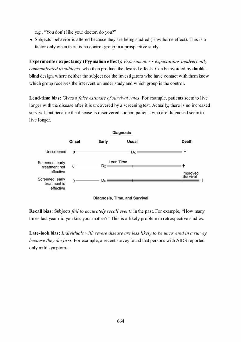
Experimenter expectancy (Pygmalion effect): Experimenter’s expectations inadvertentlycommunicated to subjects, who then produce the desired effects. Can be avoided by double-blind design, where neither the subject nor the investigators who have contact with them knowwhich group receives the intervention under study and which group is the control.
Lead-time bias: Gives a false estimate of survival rates. For example, patients seem to livelonger with the disease after it is uncovered by a screening test. Actually, there is no increasedsurvival, but because the disease is discovered sooner, patients who are diagnosed seem tolive longer.
Recall bias: Subjects fail to accurately recall events in the past. For example, “How manytimes last year did you kiss your mother?” This is a likely problem in retrospective studies.
Late-look bias: Individuals with severe disease are less likely to be uncovered in a surveybecause they die first. For example, a recent survey found that persons with AIDS reportedonly mild symptoms.
e.g., “You don’t like your doctor, do you?”Subjects’ behavior is altered because they are being studied (Hawthorne effect). This is afactor only when there is no control group in a prospective study.
664

Confounding bias: The factor being examined is related to other factors of less interest.Unanticipated factors obscure a relationship or make it seem like there is one when there is not.More than one explanation can be found for the presented results. An example would becomparing the relationship between exercise and heart disease in two populations when onepopulation is younger and the other is older. Are differences in heart disease due to exercise orto age?
Type of Bias in Research and Important Associations
Type of Bias Definition Important
Associations
Solutions
Selection
Measurement
Lead-time
Recall
Sample not representative Berkson’s bias,
nonrespondent
bias
Random, independent
sample
Gathering the information
distorts it
Hawthorne effect Control group/placebo
group
Experimenter
expectancy
Researcher’s beliefs affect
outcome
Pygmalion effect Double-blind design
Early detection confused
with increased survival
Benefits of
screening
Measure “back-end”
survival
Subjects cannot remember
accurately
Retrospective
studies
Confirm information
with other sources
665

Late-look Early mortality
Case: Within the past 2 years, a major tertiary care referral hospital experiences separatecases of a blood transfusion reaction due to incompatibility, 2 inpatient falls leading tosignificant injury, a wrong-site surgery, and a medication-dosing error causing a patientdeath.
Case: After an unexpected 3-hour delay in the operating room due to a problem in theelectrical system, an operating room team rushes to get started in order to complete thescheduled elective procedures. The team elects not to perform the mandatory sponge countat the end of the first surgery in order to get the next case started sooner. The patientreturns 2 weeks later with abdominal pain and is found on x-ray to have a retained foreignobject (a sponge) in the abdominal cavity.
Review Questions
Severely diseased
individuals are not
uncovered
Stratify by severity
Confounding
obscure
results
Unanticipated factors Hidden factors
affect results
Multiple studies, good
research design
What is the most probable single underlying cause behind these medical errors?Systems failures due to the complexity of health care delivery
Error due to ‘violation’ in policy
666

Response options for Questions 36–41:
Disease Well
Exposed 520 175 695
Nonexposed 80 225 305
600 400 1,000
520/695(A)600/1,000(B)520/600(C)695/1,000(D)80/305(E)(520/695)/(80/305)(F)(520 × 225)/(175 × 80)(G)(520/695) − (80/305)(H)Cannot be determined for this type of study(I)
36. Assume the table represents a cohort study: What is the incidence rate in theexposed?
37. Assume the table represents a cross-sectional study: What is the relative risk?
38. Assume the table represents a case-control study: What is the odds ratio?
667

39. Assume the table represents a cross-sectional study: What is the prevalence ofdisease?
40. Assume the table represents a disease outbreak investigation: What is the attack ratefor people who did not eat the food?
41. Assume the table represents a cohort study: What is the attributable risk?
42. A study compares the effectiveness of a new medication for treatment of latenttuberculosis infection with the standard medication, isoniazid. Subjects with latentTB infection are sorted with equal likelihood of selection to receive the newmedication or isoniazid. Neither the subjects themselves nor the clinicians know thetreatment condition for each patient. This study is best described as a
double-blind randomized cohort study(A)randomized controlled trial with crossover design(B)double-blind randomized clinical trial(C)double-blind randomized clinical trial with crossover design(D)double-blind quasi-experimental trial(E)
43. A group of 200 hypertensive subjects and a comparable group of 200 normotensivesubjects are recruited and enrolled into a longitudinal study to examine the effect of adiagnosis of hypertension on subsequent occurrence of coronary heart disease. Studysubjects are followed for 5 years. Final data are presented in the table below. Whatis the attributable risk for hypertension?
668

CHD No CHD Total
Hypertension 25 175 200
No hypertension 10 190 200
Total 35 365 400
0.075(A)2.5(B)2.7(C)0.125(D)Cannot be computed for this type of study(E)
44. A study is conducted relating percentage of calories from fat in the habitual diet tosubsequent incidence of clinical diabetes mellitus. Four groups of inially wellpersons are selected from the community to represent persons within each of fourcategories of fat intake. The percentages of daily calories from fat are: <20%,20−40%, 35−49%, >50%. The groups are followed longitudinally for 5 years andassessed annually for diabetes. The type of study design is best described as a
case-series trial(A)case-control study(B)cross-sectional study(C)cohort study(D)community trial(E)
45. Alcohol consumption and cigarette smoking both contribute causally to theoccurrence of esophageal cancer. These risk factors are not independent; in fact, they
669

operate synergistically. A study of cigarette smoking in relation to esophageal cancerthat fails to stratify or otherwise control for level of alcohol consumption would beguilty of which of the following threats to validity?
Ascertainment bias(A)Confounding bias(B)Design bias(C)Lead time bias(D)Observer bias(E)Recall bias(F)Response bias(G)Selection bias(H)
670

Answers and Explanations
REVIEW QUESTIONS36. Answer: A.37. Answer: I.38. Answer: G.39. Answer: B.40. Answer: E.41. Answer: H.42. Answer: C.43. Answer: A.44. Answer: D.45. Answer: B.
671

6
Biostatistics
Probability BasicsCombine probabilities for independent events by multiplication.
Combine probabilities for mutually exclusive events by addition.
Events are independent if the occurrence of one tells you nothing about the occurrence ofanother.If the chance of having blond hair is 0.3 and the chance of having a cold is 0.2, the chance ofmeeting a blond-haired person with a cold is 0.3 × 0.2 = 0.06 (or 6%).If events are nonindependent, then multiply the probability of one times the probability of thesecond, given that the first has occurred. For example, if one has a box with 5 white and 5black balls in it, the chance of picking two black balls is (5/10) × (4/9) = 0.5 3 0.44 = 0.22(or 22%).
Mutually exclusive means that the occurrence of one event precludes the occurrence of theother (i.e., cannot both happen). If a coin lands heads, it cannot be tails; the two are mutuallyexclusive. For example, if a coin is flipped, the chance that it will be either heads or tails is0.5 + 0.5 = 1.0 (or 100%).If two events are not mutually exclusive, the combination of probabilities is accomplishedby adding the two together and subtracting out the multiplied probabilities. For example, if
672

Survival Rates After Surgery
N 1 Year 2 Year 3 Year 4 Year
183 90% 75% 50% 40%
the chance of having diabetes is 10%, and the chance of someone being obese is 30%, thechance of meeting someone who is obese or has diabetes is 0.1 + 0.30 − (0.1 × 0.30) = 0.37(or 37%).
1. What is the life expectancy after surgery?
2. If a patient survives for 2 years, what is the chance of surviving for 3 years?
3. In an effort to evaluate healthy lifestyle influences at home, a study is conducted tosee how many pediatric patients have parents who exercise regularly. Parents atpediatric offices are questioned and it is concluded that 40% of pediatric patientshave parents who exercise regularly. Assuming the events are independent, what is
673

the probability that 2 pediatric patients with parents who exercise regularly willcome into the office on the same day?
0.16(A)0.4(B)0.8(C)0.96(D)0.08(E)0.04(F)
674

Answers and Explanations1. Answer: (3 years).2. Answer: (50/75).3. Answer: A.
(choice A; this requires the multiplication rule)
675

Descriptive Statistics
MEASURES (INDICES) OF CENTRAL TENDENCYMeasures of central tendency is a general term for several characteristics of the distributionof a set of values or measurements around a value at or near the middle of the set.
Mean (synonym: “average”): The sum of the values of the observations divided by thenumbers of observations.
Median:
Mode: The most frequently occurring value in a set of observations.
NORMAL DISTRIBUTIONNormal distribution is continuous frequency distribution of infinite range defined by a specificmathematical function with the following properties:
Simplest division of a set of measurements is into two parts—the upper and lower halfPoint on the scale that divides the group in this way is the medianMeasurement below which half the observations fall: the 50th percentile
A continuous, symmetrical distribution; both tails extend to infinity.
676

DISPERSION OF DATAThe dispersion of data helps us identify the spread, or the variation, of the data.
The arithmetic mean, mode, and median are identical.The shape is completely determined by the mean and standard deviation.
677

Deviation score: The distance from the mean. Found by subtracting the distribution mean fromthe distribution values you are evaluating. For example, the mean of the distribution is 120 andyou want to know the deviation score of the value 150.
This is used to obtain the variance of a distribution.
Range: The difference between the largest and smallest values in a distribution.
Variance: A measure of the variation shown by a set of observations, defined by the sum of thesquares of deviation scores of each value divided by the number of degrees of freedom in theset of observations or n − 1.
Standard deviationThe most widely used measure of dispersion of a frequency distributionIt is equal to the positive square root of the variance.Whereas the mean tells where the group of values are centered, the standard deviation is asummary of how widely dispersed the values are around the center.
678

679

The standard deviation (s or sd) is stated in score units. The normal curve has the property thatwithin one standard deviation a certain proportion of the cases is included. The property is asfollows: Between the mean and the value of one standard deviation from the mean in eitherdirection there will be 34% of the cases; there will be 68% of the cases between the score at1s above and 1s below the mean. Within two standard deviations of the mean are 95.5% of thecases. Between 1s and 2s from the mean in either direction, there will be 13.5% of the cases,or 27% for both. Within 3s of the mean are 99.7% of the cases. And between 2s and 3s fromthe mean there will be almost 2.5% of the cases, 4.7% for the two extremes together. Therewill be a few cases, of course, 0.3%, beyond 3s from the mean both above and below themean. You must know these figures. For example: What percentage of the cases are below 2sbelow the mean? (2.5%)
Students will not be asked to calculate a standard deviation or a variance on the exam, butstudents need to know what they are and how they relate to the normal curve.
680

681

Inferential Statistics
GENERALIZATIONS FROM A SAMPLE TO THEPOPULATION AS A WHOLEThe purpose of inferential statistics is to designate how likely it is that a given finding issimply the result of chance. Inferential statistics would not be necessary if investigatorsstudied all members of a population. However, because we can rarely observe and study entirepopulations, we try to select samples that are representative of the entire population so that wecan generalize the results from the sample to the population.
Inferential statistics focuses on drawing conclusions about an entire population (i.e.,parameter) based on information in a sample.
CONFIDENCE INTERVALSConfidence intervals are a way of admitting that any measurement from a sample is only anestimate of the population. Although the estimate given from the sample is likely to be close,the true values for the population may be above or below the sample values. A confidenceinterval specifies how far above or below a sample-based value the population value lieswithin a given range, from a possible high to a possible low. The true mean, therefore, is mostlikely to be somewhere within the specified range.
Confidence Interval of the Mean
682

The confidence interval contains two parts: 1) An estimate of the quality of the sample for theestimate, known as the standard error of the mean; and 2) the degree of confidence providedby the interval specified, known as the standard or Z-score. The confidence interval of themean can be calculated by:
Mean ± appropriate Z-score × standard error of the mean
Standard error of the mean is the standard deviation divided by the square root of the samplesize. It demonstrates the sample mean deviation from the true population mean.
The Z-score or standard score is a score from a normal distribution with a mean of 0 and astandard deviation of 1. Any distribution can be converted into a Z-score distribution using theformula:
or Z = Sample mean − population mean/Standard deviation
Z-scores are used in computing confidence intervals to set the level of confidence. Recallthat in a normal distribution, 95.5% of the cases are within two standard deviations (2s) of the
Increasing sample size will narrow the confidence interval.
If the standard deviation is larger, the chance of error in the estimate is greater.If the sample size is larger, the chance of error in the estimate is less.
The Z-score distribution is easy to use for calculations because it has simple values. Allpoints in a Z-score distribution are represented in standard deviation units.Positive scores are above the mean; negative scores are below the mean. Therefore, a Z-score of +2.0 is exactly two standard deviations above the mean; a Z-score of −1.5 isexactly 1.5 standard deviations below the mean.
683

mean. To get 95% confidence and 99% confidence, all we need to know is what symmetric Z-score to use to contain exactly 95% and 99% of the cases.
Confidence Intervals for Relative Risk and Odds Ratios
If the given confidence interval contains 1.0, then there is no statistically significant effect ofexposure. Example:
Relative Risk 95% Confidence Interval Interpretation
1.57 (1.12–2.25) Statistically significant (increased risk)
1.65 (0.89–2.34) Not statistically significant (risk is the same)
0.76 (0.56–0.93) Statistically significant (decreased risk)
HYPOTHESIS TESTINGA hypothesis is a statement that postulates a difference between 2 groups. Statistics are used toevaluate the possibility that this difference occurred by chance.
For 95% confidence = 1.96; for calculation purposes, use Z-score of 2.0.For 99% confidence = 2.58; for calculation purposes, use Z-score of 2.5.Note that a 99% confidence interval will be wider than a 95% interval.
Null hypothesis says that the findings are the result of chance or random factors. If youwant to show that a drug works, the null hypothesis will be that the drug does not work.
One-tailed, i.e., directional or “one-sided,” such that one group is either greater than, orless than, the other. For example, Group A is not < Group B, or Group A is not > GroupB.
–
Two-tailed, i.e., nondirectional or “two-sided,” such that two groups are not the same.–
684

SIGNIFICANCE TESTINGTo test your hypothesis, you would draw a random sample from a population (e.g., men withhypertension) and make an inference. But before you sample, you set a significance level,alpha, which is the risk of error you are willing to tolerate. Customarily, the level ofsignificance is set at 0.05 and the risk is associated with the rejection of the null hypothesis,even though it is true (e.g., type I error).
INTERPRETATION
p-Value
The p-value and alpha level are very similar, usually set at 0.05, and both symbolizesignificance. They are only slightly different in that the alpha level represents risk and isindependent of data, whereas p-value measures the strength (i.e., significance) of the dataagainst the null hypothesis.
A p-value is for interpreting output from a statistical test; focus on the p-value. The term refersto two things. In its first sense, the p-value is a standard against which we compare our results.In the second sense, the p-value is a result of computation.
The computed p-value is compared with the p-value criterion to test statistical significance.If the computed value is less than the criterion, we have achieved statistical significance. Ingeneral, the smaller the p the better.
For example, Group A = Group B
Alternative hypothesis says what is left after defining the null hypothesis. In this example,the drug actually does work.
685

The p-value criterion is traditionally set at p ≤ 0.05. (Assume that these are the criteria if noother value is explicitly specified.) Using this standard:
Therefore:
Types of errors
Just because we reject the null hypothesis, we are not certain that we are correct. For somereason, the results given by the sample may be inconsistent with the full population. If this istrue, any decision we make on the basis of the sample could be in error. There are two possibletypes of errors that we could make:
If p ≤ 0.05, reject the null hypothesis (reached statistical significance).If p > 0.05, do not reject the null hypothesis (has not reached statistical significance).
If p = 0.13, fail to reject the null hypothesis, i.e., decide that the drug does not work.If p = 0.02, reject the null hypothesis, i.e., decide that the drug works.
686

Type I error (α error): rejecting the null hypothesis when it is really true, i.e., assuming astatistically significant effect on the basis of the sample when there is none in the population orasserting that the drug works when it doesn’t. The chance of a Type I error is given by the p-value. If p (or α) = 0.05, then the chance of a Type I error is 5 in 100, or 1 in 20.
Type II error (β error): failing to reject the null hypothesis when it is really false, i.e.,declaring no significant effect on the basis of the sample when there really is one in thepopulation or asserting the drug does not work when it really does. The chance of a Type IIerror cannot be directly estimated from the p-value.
β Error can be calculated by subtracting power from 1: 1 − Power = β.
Meaning of the p-value
Limits to the p-value
The p-value does not tell us 1) the chance that an individual patient will benefit, 2) thepercentage of patients who will benefit, and 3) the degree of benefit expected for a givenpatient.
TYPES OF SCALES
Power is the capacity to detect a difference if there is one.Increasing sample size (n) increases power.
Provides criterion for making decisions about the null hypothesis.Quantifies the chances that a decision to reject the null hypothesis will be wrong.Tells statistical significance, not clinical significance or likelihood of benefit.Generally, p-value is considered statistically significant if it is equal to or less than 0.05.
687

To convert the world into numbers, we use 4 types of scales: nominal, ordinal, interval, andratio scales.
Types of Scales in Statistics
Type of Scale Description Key Words Examples
Nominal or categorical scale
A nominal scale puts people into boxes, without specifying the relationship between the boxes.Sex is a common example of a nominal scale with two groups, male and female. Anytime youcan say, “It’s either this or that,” you are dealing with a nominal scale. Other examples: cities,drug versus control group.
Nominal (Categorical) Different groups This or that or that Gender, comparing among
treatment interventions
Ordinal Groups in
sequence
Comparative quality,
rank order
Olympic medals, class rank
in medical school
Interval Exact differences
among
groups
Quantity, mean, and
standard
deviation
Height, weight, blood
pressure, drug dosage
Ratio Interval + true
zero point
Zero means zero Temperature measured in
degrees Kelvin
688

Ordinal scale
Numbers can also be used to express ordinal or rank-order relations. For example, we say Benis taller than Fred. Now we know more than just the category in which to place someone. Weknow something about the relationship between the categories (quality). What we do not knowis how different the two categories are (quantity). Class rank in medical school and medals atthe Olympics are examples of ordinal scales.
Interval scale
An interval scale uses a scale graded in equal increments. In the scale of length, we know thatone inch is equal to any other inch. Interval scales allow us to say not only that two things aredifferent, but by how much. If a measurement has a mean and a standard deviation, treat it as aninterval scale. It is sometimes called a “numeric scale.”
Ratio scale
The best measure is the ratio scale. This scale orders things and contains equal intervals, likethe previous two scales, but it also has one additional quality: a true zero point. In a ratioscale, zero is a floor—you can’t go any lower. Measuring temperature using the Kelvin scaleyields a ratio scale measurement.
689

Selecting a Statistical Test
Types of Scales and Basic Statistical Tests
Variables
Name of Statistical Test Interval Nominal Comment
Pearson Correlation 2 0 Is there a linear relationship?
Chi-square 0 2 Any # of groups
t-test 1 1 2 groups only
One-way ANOVA 1 1 2 or more groups
Matched pairs t-test 1 1 2 groups, linked data pairs, before and after
Repeated measures ANOVA 1 1 More than 2 groups, linked data
META-ANALYSISA statistical way of combining the results of many studies to produce one overallconclusionA mathematic literature review
690

CORRELATION ANALYSIS (r, RANGES FROM −1 TO+1)
Graphing correlations using scatterplots
A positive value means that two variables go together in the same direction, e.g., MCATscores have a positive correlation with medical school grades.A negative value means that the presence of one variable is associated with the absence ofanother variable, e.g., there is a negative correlation between age and quickness ofreflexes.The further from zero, the stronger the relationship (r = 0).A zero correlation means that two variables have no linear relation to one another, e.g.,height and success in medical school.
A scatterplot will show points that approximate a line.Be able to interpret scatter plots of data: positive slope, negative slope, and which of a setof scatterplots indicates a stronger correlation.
691

Types of correlations
There are two types of correlations. Pearson correlation compares two interval levelvariables, and the Spearman correlation compares two ordinal level variables.
692

t-TESTSThe output of a t-test is a “t” statistic.
ANALYSIS OF VARIANCE (ANOVA)
Comparing the means of two groups from a single nominal variable, using means from aninterval variable to see whether the groups are differentUsed for two groups only, i.e., compares two means. For example, do patients with MI whoare in psychotherapy have a reduced length of convalescence compared with those who arenot in therapy?Pooled t-test: regular t-test, assuming the variances of the two groups are the sameMatched pairs t-test: if each person in one group is matched with a person in the second.Applies to before and after measures and linked data.
Output from an ANOVA is one or more “F” statistics.One-way: Compares means of many groups (two or more) of a single nominal variableusing an interval variable. Significant p-value means that at least two of the tested groupsare differentTwo-way: Compares means of groups generated by two nominal variables using an
693

CHI-SQUARE
Chi-Square Analysis for Nominal Data
New Drug Placebo Totals
Recovered 45 35 80
Not Recovered 15 25 40
Totals 60 60 120
Review Questions
interval variable. Can test effects of several variables at the same time.Repeated measures ANOVA: multiple measurements of same people over time.
Nominal data onlyAny number of groupsTests to see whether two nominal variables are independent, e.g., testing the efficacy of anew drug by comparing the number of recovered patients given the drug with those who arenot
1. The American Medical Association commissions a health study of a representativesample of U.S. physicians. Enrolled physicians complete detailed surveys andundergo an extensive battery of medical tests. For a number of analyses, physiciansare classified by subspecialty. Although numerous physiologic measures areassessed, the following questions describe analyses of just one of these, mean fasting
694

plasma glucose. Select the appropriate statistical test for a comparison of meanfasting plasma glucose values for representative samples of surgeons andcardiologists.
t-test(A)Matched pairs t-test(B)One-way ANOVA(C)Two-way ANOVA(D)Chi-square(E)
2. An experimenter conducts a test of a new medication compared with the currentstandard medication. Alpha is selected to be 0.05. At the conclusion of the trial, thesample of patients receiving the new medication shows more improvement than thecomparison group on the standard medication. The p-value is 0.002. What will theexperimenter conclude?
Do not reject the null hypothesis.(A)The new medication has more clinical benefits than the standard medication.(B)The likelihood that a type I error has actually been committed is less than themaximum risk the experimenter was willing to accept.
(C)
The result is not significant.(D)A type II error has been committed.(E)
3. Body mass index (BMI) is found to correlate to the following physiologic measures.For which measure is the correlation the strongest?
695

Physical activity (r = −0.56)(A)Percentage of calories from complex carbohydrates (r = −0.32)(B)Systolic blood pressure (r = +0.43)(C)Triglycerides (r = +0.37)(D)LDL cholesterol (r = +0.49)(E)
4. A new treatment for elevated cholesterol is piloted on a sample of 100 men, ages 45–59 with total serum cholesterol in the range of 260–299 mg/dL at entry. Following 3months on the medication, the mean cholesterol for the treatment group was 250mg/dL with a standard deviation of 20 mg/dL. What is the 95% confidence interval onthe mean for this study?
210–290 mg/dL(A)230–270 mg/dL(B)246–254 mg/dL(C)248–252 mg/dL(D)249–251 mg/dL(E)
5. The Wechsler Adult Intelligence Scale–Revised (WAIS-R) is a standardized IQ testwith a mean of 100 and a standard deviation of 15. A person with an IQ of 115 is atwhat percentile of IQ?
50th(A)68th(B)84th(C)95th(D)99th(E)
696

6. From a published article describing the results of the study presented above, thefollowing data table is abstracted. This table presents the relative risks (RR) ofclinical diabetes for each of the categories of fat intake relative to the baselinecategory of <20%. Interpret the study findings from the tabular data.
% of Calories from Fat RR for Diabetes 95% Confidence Interval
Baseline <20 1 —
Level 2 20–34 1.3 0.8–1.8
Level 3 35–49 2 1.6–2.6
Level 4 >50 3 2.7–3.3
Levels 2, 3, and 4 have significantly elevated risks for diabetes relative tobaseline.
(A)
Levels 2 and 3 are significantly different from each other.(B)Levels 3 and 4 are significantly different from baseline and risk elevating.(C)Levels 3 and 4 are not significantly different from each other.(D)RR for levels 2, 3, and 4 are numerically different but not significantly differentfrom baseline.
(E)
697

Answers and Explanations
REVIEW QUESTIONS1. Answer: A.2. Answer: C.3. Answer: A.4. Answer: C.5. Answer: C.6. Answer: C.
698

7
Interpretation of MedicalLiterature
IntroductionThe purpose of this chapter is to provide you with an approach to reading and understandingresearch articles and pharmaceutical advertisements. It is based on principles of epidemiology.
An understanding of these concepts is fundamental to the comprehension of medical literature.We have sacrificed depth for the sake of brevity since our goal was to provide a fewfundamental tools and avoid complexity.
RESEARCH ABSTRACT 1
Wedge resection or lobectomy: comparison of tumor recurrencerates and overall survival in NSCLC patients receiving preoperativechemotherapy
Wedge resection for non-small-cell lung cancer (NSCLC) stage I patients still remainscontroversial with many physicians. The primary outcomes of tumor recurrence and
699

overall survival (OS) remain unclear when compared with complete lobectomy, whichhas traditionally been considered a far more effective procedure. However, a recentcompilation of case reports and case series reports have validated impressive tumorrecurrence and OS rates that were previously only believed to be seen in patientsreceiving lobectomy. Our primary objective was to compare and analyze the tumorrecurrence rates and OS for both wedge resection and lobectomy in patients with stageI NSCLC following preoperative chemotherapy.
Methods
We systematically reviewed individual case reports and case series reports from 152institutions in the United States for patients who first received preoperativechemotherapy and then underwent either wedge resection (248 patients) or lobectomy(329 patients). A propensity score algorithm was used to reduce the confounding thatcan occur when examining the effects and variables related to both treatment measures.Following the procedures, tumor recurrence and OS was assessed at 3 and 5 years inall patients.
Results
Preoperative mortality related to chemotherapy complications for patients scheduled tohave wedge resection or lobectomy was 0.8% and 1.5%, respectively (P = 0.22).Perioperative mortality in patients undergoing lobectomy was 3.8% versus 0.8% inthose receiving wedge resection (P = 0.02). During the predetermined follow-up timesat 3 and 5 years, overall tumor recurrence (both locoregional and metastases) wereassessed:
At the 3 year follow-up, overall tumor recurrence was 5.9% for wedge resectionand 4.2% for lobectomy (P = 0.41).At the 5-year follow-up, overall tumor recurrence was 6.3% for wedge resectionand 6.1% for lobectomy (P = 0.29).
700

When comparing the OS for wedge resection with lobectomy the 3-year OS rates were82% vs 71%, respectively; (P = .09) and 5-year OS rates were 69% v 68%,respectively; (P = .29). Wedge resection was not found to be an independent predictorof tumor recurrence (hazard ratio, 1.23; 99% CI, 0.96 to 1.15) or OS (hazard ratio,1.43; 99% CI, 0.92 to 1.23).
Conclusion
Wedge resection and lobectomy are associated with similar overall tumor recurrenceand overall survival rates when performed after preoperative chemotherapy. However,postoperative complications and mortality are significantly lower in patients receivingwedge resection compared with lobectomy. Since patients generally maintain superioroverall lung function with wedge resection, we recommended that wedge resection beperformed in all eligible patients with Stage I NSCLC unless there is a compellingreason to perform a lobectomy.
Practice Questions1. Information from the abstract most strongly supports which of the following
conclusions?
701

Both wedge resection and lobectomy have lower mortality and tumorrecurrence rates when patients first receive preoperative chemotherapy.
(A)
Perioperative mortality was lower in patients undergoing wedge resection.(B)Postoperative complications were lower in patients undergoing wedgeresection.
(C)
Pulmonary function tests at 1 year were significantly higher in patientsreceiving wedge resection.
(D)
The overall survival for wedge resection at 3 years was proven to be higherthan that of lobectomy.
(E)
2. Which of the following best describes the type of study performed?
Case-control study(A)Crossover study(B)Meta-analysis(C)Propensity-matched analysis(D)Randomized, controlled clinical trial(E)
3. The next step in follow-up of these research results would be to conduct which typeof study?
Case-control study(A)Cohort study(B)Cross sectional study(C)Randomized, controlled clinical trial(D)Replication in a different biological model(E)
702

RESEARCH ABSTRACT 2
Mekanib improved overall survival and decreased vemurafenibresistance in BRAF-mutated metastatic melanoma
BRAF mutations have been observed in approximately 50% of all malignantmelanomas. The most predominant BRAF mutations found in melanoma are those thatintroduce an amino acid substitution at valine 600. Approximately 80–90% of thesemutations are classified as BRAF V600E. Other predominant BRAF mutations includeV600K, V600R and V600D. All of these mutations result in heightened BRAF kinaseactivity and amplified phosphorylation of downstream targets, which in particularincludes MEK. BRAF inhibitor therapy (with vemurafenib or dabrafenib) is associatedwith well-documented clinical benefit in most patients with BRAF V600E-mutatedmelanoma (and other subtypes). However, resistance to these drugs and tumorprogression generally occurs in patients within the first year. It is believed that BRAFmutations stimulate melanoma cell proliferation and survival predominantly throughactivation of MEK. The purpose of this study was to determine if the addition of theallosteric MEK1/MEK2 inhibitor mekanib (KAP071714) to vemurafenib delayedexpected vemurafenib resistance as well as improved progression free survival (PFS)and overall survival (OS) in comparison with dacarbazine.
Methods
This was a phase 3, multicenter, double-blinded, randomized clinical trial comparingthe effectiveness of mekanib (KAP071714) in 447 total participants with previouslyuntreated, metastatic melanoma with the BRAF V600E mutation. Patients wererandomly assigned into 2 cohorts. Cohort A (222 participants) received dacarbazine(1000 mg per square meter of body-surface area intravenously every 3 weeks); CohortB (225 participants) received vemurafenib (960 mg orally twice daily) + mekanib (150mg orally daily). PFS was the primary end point and OS was a secondary end point.
703

Results
Median PFS was 11.6 months in the mekanib group and 2.3 months in the dacarbazinegroup (hazard ratio for disease progression or death in the mekanib group, 0.23; 95%confidence interval [CI], 0.30 to 0.58; P<0.007). At 15 months, the rate of overallsurvival was 78% in the mekanib group and 42% in the dacarbazine group (hazardratio for death, 0.43; 95% CI, 0.52 to 0.88; P = 0.02). Elevated hepatic enzymes, rash,diarrhea, and hypertension were the most common toxic effects in the mekanib group.Nausea, vomiting alopecia, facial flushing, myalgia, leukopenia and hepatotoxicitywere the most common toxic effects in the dacarbazine group. There were 8 patients inthe mekanib group and 15 patients in the dacarbazine group who withdrew from thestudy due to severe side effects. Secondary skin neoplasms were not observed in eithergroup.
Conclusions
Mekanib, as compared with traditional dacarbazine chemotherapy, improved rates ofPFS and OS among patients with the BRAF-mutated metastatic melanoma as well asdelayed vemurafenib drug resistance. Mekanib should be considered for use inconjunction with vemurafenib for the treatment of BRAF-mutated metastatic melanoma.
(Funded by SMILE Pharmaceuticals, ClinicalTrials.gov numberNCT0123456789101112)
Practice Questions1. Information from the abstract above most strongly supports which of the following
conclusions about mekanib?
704

In the treatment of select cases of metastatic melanoma, mekanib alone provideshigher rates of PFS and OS than dacarbazine alone.
(A)
Mekanib does not produce severe side effects.(B)Mekanib produces fewer side effects than dacarbazine.(C)Metastatic melanoma patients with BRAF V600K mutations have improvedPFS and OS rates when taking vemurafenib + mekanib versus dacarbazine.
(D)
Most metastatic melanoma patients appropriately prescribed vemurafenib andmekanib are likely to complete their treatment regimen.
(E)
2. In the conclusion section of the abstract, the authors indicate that when mekanib wasadded to vemurafenib the drug delayed vemurafenib drug resistance. Which of thefollowing is the most likely reason that the reader should question the validity of thisclaim?
Insufficient follow-up of study participants(A)Insufficient information on adverse effects and drug-drug interactions(B)Lack of an appropriate control group(C)Subject attrition(D)Use of hazard ratio instead of relative risk(E)
3. In the background section of the abstract, researchers state that purpose of the studywas to determine if the addition of the allosteric MEK1/MEK2 inhibitor mekanib(KAP071714) to vemurafenib-delayed drug resistance as well as improvedprogression free survival (PFS) and overall survival (OS) in comparison todacarbazine. Which of the following study design changes could have been made toappropriately evaluate all the specified outcomes?
705

Add a vemurafenib-only cohort to the study(A)Prescribe all 3 medications to each participant but at different dosage ranges(B)Replace the dacarbazine cohort with a vemurafenib-only cohort(C)Use a crossover study instead of a randomized clinical trial(D)No changes were needed since the study was properly designed to meet thespecified outcomes
(E)
706

PHARMACEUTICAL AD 1
707

708

HIGHLIGHTS OF PRESCRIBING INFORMATIONPlease see Tazofect (tanzopanib) drug package insert for complete prescribing
information
Indications and Usage: Tazofect (tanzopanib) is a kinase inhibitor indicated for first-line treatment of NSCLC with EGFR exon 19 deletions and EGFR exon 21 (L858R)substitution mutations in patients age 18 years and older.
Mechanism of Action: Tanzopanib is a kinase inhibitor that acts by inhibitingintracellular tyrosine kinase domain of epidermal growth factor receptor (EGFR) thusresulting in cell cycle arrest and angiogenesis inhibition. Tanzopanib has an eliminationhalf-life of approximately 28 hours in patients with normal hepatic and renal function.
Dosage and Administration: Treatment of NSCLC with EGFR exon 19 deletions andEGFR exon 21 (L858R) substitution mutations in patients aged 18 years and older withnormal hepatic and renal function: 10–20 mg daily until clinically significant diseaseprogression.
Contraindications: Hypersensitivity to tanzopanib; use in patients with severe hepaticimpairment, active infection and thrombocytopenia.
Warnings and Precautions: May cause reactivation of tuberculosis and hepatitis B.Use caution in patients receiving other chemotherapeutic agents, thyroid disorders,dehydration, mild to moderate renal and hepatic dysfunction
Adverse Reactions:
Common (≥5%): elevated AST & ALT (15%), diarrhea (15%), fatigue (13%),elevated bilirubin (12%), infection (10%), cough (8%), thrombocytopenia (7%)
709

Practice Questions
Less Common (<5%): hepatorenal syndrome (2%), hepatotoxicity (2%), toxicepidermal necrolysis (1%), Stevens-Johnson syndrome (1%), acute renal failure (1%),hypothyroidism (1%), hemolytic anemia (<1%)
1. The data provided in the drug advertisement most strongly supports which of thefollowing conclusions?
In the treatment of cancer, Tazofect and erlotinib can be used interchangeably.(A)Tazofect is not indicated for treatment of EGFR exon 19 insertion in non-smallcell lung cancer.
(B)
Tazofect should be considered for use in patients with PIK3CA mutatedNSCLC.
(C)
The combination of Tazofect and erlotinib will improve the PFS to a greaterextent than either agent alone.
(D)
The dose of Tazofect should be adjusted in patients with hepatic dysfunction.(E)
2. Consider the following statement: “Tazofect was proven to provide approximatelydouble the PFS in carboplatin resistant NSCLC study participants over thosereceiving Tarceva® (erlotinib).” When evaluating the drug ad and highlights ofprescribing information, which of the following provides the best evidence that thisstatement is inaccurate?
710

Number of patients treated in the carboplatin resistant group for both drugs(A)The calculation of months of PFS for the carboplatin resistant graph(B)The confidence interval for the carboplatin resistant graph(C)The p value for the carboplatin resistant graph(D)The y axis data points for the carboplatin resistant graph(E)
3. Shortly after Tazofect is released for use in the general population, the FDA and drugmanufacturer begin to receive numerous reports of complete treatment failure in bothcarboplatin resistant patients and first line therapy patients as well as higher thanexpected percentages of adverse events in all patients. Which of the following is themost likely reason for these reports on Tazofect?
Insufficient follow-up of study participants(A)Insufficient information on adverse effects(B)Insufficient information on drug indications(C)Subject attrition(D)Type II error was committed(E)
711

PHARMACEUTICAL AD 2
712

713

HIGHLIGHTS OF PRESCRIBING INFORMATIONPlease see GluSense (glugliflozin) drug package insert for complete prescribing
information.
Indications and Usage: GluSense (glugliflozin) is an SGLT2 inhibitor with insulin-sensitizing properties, indicated for the treatment of type 2 diabetes in conjunction withdiet and exercise as monotherapy, and in combination with metformin in patients aged18 years and older.
Mechanism of Action: Glugliflozin is an SGLT2 inhibitor with insulin-sensitizingproperties. This agent has a dual mechanism of action. It acts by:
Pharmacokinetics
Inhibiting the sodium-glucose cotransporter 2 (SGLT2), thereby reducing glucosereabsorption and increasing urinary glucose excretionDecreasing insulin in the periphery and liver, resulting in increased insulin-dependent glucose disposal and decreased hepatic glucose output. Glugliflozin is anagonist for peroxisome proliferator-activated receptor-gamma (PPARγ). Activationof PPARγ nuclear receptors in the liver, skeletal muscle, and adipose tissuemodulates the transcription of a number of insulin responsive genes involved in thecontrol of glucose and lipid metabolism.Other: antagonizes peripheral alpha-1 adrenergic receptors
Glugliflozin has an elimination half-life of approximately 16 hours in patients withnormal hepatic and renal function.Following oral administration of glugliflozin, Tmax occurs within 3 hours.Glugliflozin is extensively metabolized by hydroxylation and oxidation; themetabolites also partly convert to glucuronide or sulfate conjugates.
714

Dosage and Administration: Treatment of type 2 diabetes in patients aged 18 years orolder who have failed to meet glycemic goals with diet and exercise alone:
Contraindications: Type 1 diabetes mellitus, hypersensitivity to glugliflozin and/orsulfonamides; NYHA class III or IV heart failure, severe hepatic impairment,hyperkalemia, use with medications causing hyperkalemia and diabetic ketoacidosis
Warnings and Precautions: May cause hypoglycemia, hypotension, and AST/ALTelevation. Caution use in elderly patients with poorly controlled diabetes and patientswith past history of cardiovascular disease.
Adverse Reactions (for a complete list, see drug package insert)
Common (≥5%): Less Common (<5%):
Hyperkalemia Fatigue
Hypoglycemia Hepatic dysfunction
Orthostatic hypotension Thirst
Dizziness Fainting
Tachycardia Mental impairment
Following oral administration of glugliflozin, approximately 15–20% of the drugdose is recovered in the urine.
Monotherapy: 150–300 mg PO qd; start at 75 mg PO qd and increase by 75 mg qwk;max dose 450 mg/dayCombination with metformin: same as monotherapy and standard metformin dose of2000 mg daily (in divided doses)
715

Hyperhidrosis Pancreatitis
Drug Interactions (see drug package insert)
Practice Questions1. The data provided in the drug advertisement most strongly supports which of the
following conclusions?
GluSense is a substitute for diet and exercise in type 2 diabetes due to itsweight loss properties.
(A)
GluSense is recommended for use in patients with a history of myocardialinfarction.
(B)
GluSense is safer to use in patients with type 2 diabetes than metformin.(C)The antihypertensive effects of GluSense are comparable to some currentlyavailable antihypertensive medications.
(D)
The combination use of GluSense and a sulfonylurea is recommended for thosewho initially fail sulfonylurea monotherapy.
(E)
2. Of the initial trial participants, 175 persons from the GluSense-only group and aneven large number from the GluSense and metformin group withdrew from the study.Which of the following is the most likely reason for participant withdrawal?
716

Appearance of drug interactions(A)Hypersensitivity to sulfonamides(B)Severe hypoglycemia(C)Severe hypotension(D)Severe GI side effects(E)
3. A 64-year-old man comes to the physician with complaints of increasing polyuria andpolydipsia. His past medical history is significant for type 2 diabetes, hypertension,hyperlipidemia, and a myocardial infarction 4 years ago. Allergy history includes ananaphylactic reaction to levofloxacin. He is currently receiving metformin 1000 mg2x daily, enalapril 10 mg daily, pravastatin 20 mg daily, and spironolactone 25 mgtwice daily. Physical examination shows blood pressure of 126/82 mm Hg, heart rate62/min, height 172.7 cm (5 feet, 8 inches), weight 88.6 kg (195 lbs), and BMI 29.6.
Laboratory studies show:
If the attending physician is considering the addition of GluSense to this patient’smedication regimen, which of the following is a contraindication for prescribing this
Blood glucose: 215 mg/dLA1c: 10.5%Albumin: 3.8 g/dLCreatinine: 1.3 mg/dLAST: 20 IU/LALT: 22 IU/LSodium: 138 mEq/LPotassium: 4.9 mEq/LCalcium: 9.6 mg/dLEjection fraction: 66%
717

medication?
Allergy contraindication(A)Cardiovascular contraindication(B)Drug interaction contraindication(C)Hepatic contraindication(D)Renal contraindication(E)There is no contraindication in this patient and the medication can beprescribed
(F)
718

Answers and Explanations
PRACTICE QUESTIONS1. The correct answer is choice B.
You are asked to determine which answer choice is most strongly supported by theinformation provided in the abstract. In this type of question, the correct answer is found inthe abstract itself and the reader needs only to interpret the information. Of the answerchoices, choice B is most supported by the information provided in the drug abstract. Thestatement, “Perioperative mortality was lower in patients undergoing wedge resection” issupported by the data provided in the Results section. We are told that perioperativemortality in those receiving lobectomy was 3.8% versus 0.8% for those receiving wedgeresection (P = 0.02). This data shows that mortality in those receiving a lobectomy wasalmost 5x higher than seen in those receiving wedge resection. Furthermore, the p value is0.02, which shows statistical significance.
The stated objective of the researchers was to “compare and analyze the tumor recurrencerates and OS for both wedge resection and lobectomy in patients with stage I NSCLCfollowing preoperative chemotherapy.” In other words, researchers assessed tumorrecurrence and OS in patients receiving 2 different surgical procedures. Since all patientsreceived preoperative chemotherapy, one cannot draw a conclusion about the impact ofpreoperative chemotherapy based on the information presented (choice A). Remember, therewould have to be a subset of patients who did not receive preoperative chemotherapy inorder for a comparative analysis to be performed.
Postoperative complications (choice C) were not discussed in the abstract.
719

A clinician could reasonably conclude that pulmonary function tests would be higher at 1year in patients receiving wedge resection when compared with lobectomy (choice D).However, this “reasonable assumption” is not supported, as data regarding lung function at 1year was not presented in the abstract.
Choice E states “The overall survival for wedge resection at 3 years was proven to behigher than that of lobectomy.” In the Results section of the abstract, it says, “Whencomparing the OS for wedge resection with lobectomy, the 3-year OS rates were 82% vs71%, respectively; (P = .09).” At first glance it may appear to be a correct statement;however, the p value is 0.09. Therefore, the 2 percentages are not statistically different.
2. The correct answer is choice D.You are asked to determine what type of study/analysis the researchers performed. Theresearchers reviewed individual case reports and case series reports from a number ofinstitutions. After reviewing and compiling the data, they used an algorithm to reduceconfounding variables and subsequently analyze the data. Based on this information, we canconclude that the researchers performed a propensity-matched analysis. Propensity scorematching (PSM) is used in the statistical analysis of observational data. PSM is a statisticalmatching technique which attempts to approximate the effect of a treatment by accounting forthe covariates that predict receiving a given treatment. This type of statistical analysis isused to reduce bias caused by confounding variables. Propensity scores (obtained from apropensity-matched analysis) are valuable when attempting to draw causal conclusions fromobservational studies (such as case reports) where the “treatment” or “independentvariable” was not originally randomly assigned.
Casecontrol studies (choice A) are retrospective observational studies used to identify riskfactors that are believed to be associated with a particular disease or condition. Subjectsare initially classified as having or not having the disease in question and then their historiesare explored to identify the presence or absence of any risk factors. Data are usuallyanalyzed by means of an odds-ratio, and interpreted such that if something occurs in the
720

history of the diseased group, but not in the non-diseased group, then it will be identified asa risk factor.
Cross-over studies (choice B) are clinical trials in which 2 comparison groups (forexample) both receive the drug being tested and the comparative intervention (often aplacebo) at different times. This interventional study will generally begin with one group(group A) receiving the investigational drug while a comparison group (group B) receives aplacebo. Then, at some predetermined time, there will be a washout period and then GroupA is switched to the placebo, while the Group B is given the investigational drug. This studydesign allows comparison of those on and off the drug, but also satisfies the ethicalrequirement that everyone in the study is exposed to whatever benefit the experimental drugmay provide.
A meta-analysis (choice C) will meticulously examine several interventional clinicalstudies on a particular disease state (or treatment measure) and then combine the resultsusing an acceptable statistical methodology. The results will be presented as if they werefrom 1 large study. The classical meta-analysis compares 2 types of treatment measureswhile multiple treatment meta-analysis (or network meta-analysis) can provide estimates oftreatment efficacy of multiple treatment regimens, even when direct comparisons areunavailable. One of the key differences between a meta-analysis and a propensity matchedanalysis is that a meta-analysis is used with interventional studies, and a propensity-matchedanalysis is used with observational reports or studies.
A randomized, controlled clinical trial (choice E) is a type of interventional study where aresearcher will administer a medication or treatment measure to one group of participantsand evaluate its effects against a control group who receives another treatment measure orplacebo. Subjects in the study are randomly allocated into “intervention” and “control”groups to receive or not receive an experimental preventive or therapeutic procedure orintervention. In the “wedge resection” analysis, researchers compiled the results from
721

several observational studies. The data evaluated was derived from case reports wherepatients were NOT originally assigned to receive either a wedge resection or lobectomy.
3. The correct answer is choice D.In the current study, researchers reviewed and compiled the data from numerous casereports and case series reports. They then attempted to draw causal conclusions from theseobservational studies where the treatment was not originally randomly assigned. Using thisapproach, researchers are able to determine if further investigation is warranted. In thisparticular analysis, researchers identified a higher than expected overall survival rate andlower than expected tumor recurrence rate associated with a procedure (wedge resection)that is believed to be associated better postoperative lung function as compared tolobectomy. Since the results of their analysis essentially showed no real difference inoverall tumor recurrence rates and overall survival rates, the next step would be to furthervalidate these results with an interventional study, such as a prospective, randomizedcontrolled trial (RCT). In an RCT, researchers will likely randomly assign patients toreceive either wedge resection or lobectomy following preoperative chemotherapy.Researchers will then be able to determine if there is a statistical difference between thetwo treatment options.
Case-control studies (choice A) are retrospective observational studies used to identify riskfactors that are believed to be associated with a particular disease or condition. Subjectsare initially classified as having or not having the disease in question and then their historiesare explored to identify the presence or absence of any risk factors.
Cohort studies (choice B) are observational studies in which subjects are classified ashaving or not having a risk factor and then followed forward in time so incidence rates forthe two groups can be compared. Although cohort studies are a type of prospective study,the next step would be to use an “interventional” prospective study, such as a randomizedcontrolled clinical trial.
722

Cross sectional studies (choice C) are observational studies used to assess the prevalenceof a disease in a given population and the factors which co-occur with that disease at aparticular time.
Replication in a different model (choice E) is a type of study generally used in early animaltesting of experimental medications. For example, early animal testing for a new compoundmay involve a small number of rats. Once data is obtained from a single animal test, there isstill a lot of information that needs to be obtained and questions that need answered beforethis new compound (experimental drug) can be considered for human trials. Therefore,researchers often perform several different types of animal tests using a variety of ratspecies followed by testing in other animal models.
1. The correct answer is choice E.You are being asked to determine which answer choice is most supported by the informationprovided in the abstract. While several answer choices might “look good,” you will be ableto eliminate the incorrect answer choices once you examine the meaning of each statement.Of the answer choices, choice E is most supported by the information provided in the drugabstract. The Results section indicates that “Eight patients in the mekanib group and 15patients in the dacarbazine group withdrew from the study due to severe side effects.” Of the225 patients originally enrolled in the mekanib + vemurafenib arm of the study, 217 personsor 96% of the original study group completed the study. Hence, you can reasonably concludethat most metastatic melanoma patients appropriately prescribed vemurafenib and mekanibare likely to complete their treatment regimen.
The statement, “In the treatment of select cases of metastatic melanoma, mekanib aloneprovides higher rates of PFS and OS than dacarbazine alone” can be eliminated (choice A)since the study was not designed to evaluate mekanib versus dacarbazine. This studyevaluated mekanib PLUS vemurafenib versus dacarbazine.
723

“Mekanib does not produce severe side effects” (choice B) is an incorrect statementbecause the abstract only lists a few of the most common side effects. It does not mention thesevere (and less common) side effects. These findings are likely to be found in the body ofthe published study. Remember, this is an abstract and only provides limited information.
“Mekanib produces fewer side effects than dacarbazine” (choice C) is incorrect because theabstract only lists a few of the most common side effects for both drugs. It does not outlinethe number and frequency of occurrence of side effects. These findings are likely to be foundin the complete study.
The statement “Metastatic melanoma patients with BRAF V600K mutations have improvedPFS and OS rates when taking vemurafenib + mekanib versus dacarbazine” can beeliminated (choice D), because the study was only performed in metastatic melanomapatients with BRAF V600E mutations. Hence, the reader cannot draw conclusions about theeffect of vemurafenib plus mekanib in this patient population.
2. The correct answer is choice C.You are asked to determine the most likely reason why one should question the validity ofthe claim that mekanib delays vemurafenib-resistance. The correct answer is lack of anappropriate control group. In order for researchers to conclude that mekanib decreasesvemurafenib resistance, the control group must be vemurafenib alone and the study groupmust be vemurafenib PLUS mekanib. In this study, the control group was dacarbazine andstudy group was vemurafenib plus mekanib; hence, there is not an appropriate control groupto answer the question “Does mekanib delay vemurafenib resistance?” In other words, thereis no data available to support the claim that the addition of mekanib did in fact decreasevemurafenib resistance. Furthermore, the background states that “resistance to these drugs(vemurafenib and dabrafenib) and tumor progression generally occurs in patients within thefirst year” and the Results section states that the median PFS was 11.6 months in themekanib group. The median PFS is a little less than a year; hence, the reader should actuallyquestion if mekanib actually provided any benefit at all.
724

The Results section provides information about median PFS and survival rates at 15 months.The length of the study was sufficient to assess the effects it was designed to assess (choiceA).
The Results section provides information on adverse effects but does not provide anyinformation on drug-drug interactions (choice B). Although a drug interaction couldpotentially decrease the effectiveness of mekanib, the most likely reason to question thevalidity of the claim (in the question stem) is because of a lack of an appropriate controlgroup.
The Results section states that “Eight patients in the mekanib group and 15 patients in thedacarbazine group withdrew from the study due to severe side effects.” Out of an original447 patients, only 23 patients withdrew from the study. Hence, the subject attrition rate islow for this study (choice D).
By definition, the hazard ratio is a measure of relative risk over time in situations where theresearchers are interested not only in the total number of events, but also in the timing ofthese events. For example, the event of interest may be subject death or it could be a non-fatal event such as readmission or symptom change. The use of a hazard ratio in thisparticular study is appropriate (choice E).
3. The correct answer is choice C.You are asked to determine what changes could have been made to the original study designso that the 3 initial study outcomes could be appropriately evaluated. Based on the purposeoutlined in the question stem, the 3 outcomes being evaluated are as follows:
Decreased vemurafenib resistance when mekanib is added1.Improved PFS for vemurafenib + mekanib compared to dacarbazine2.Improved OS for vemurafenib + mekanib compared to dacarbazine3.
725

The current study design appropriately evaluates PFS and OS between vemurafenib +mekanib AND dacarbazine because participants were administered either vemurafenib +mekanib OR dacarbazine. However, the only way to assess whether mekanib decreasesvemurafenib-resistance is to evaluate this regimen against a vemurafenib-only cohort.Hence, in order to appropriately evaluate all 3 outcomes described in the question stem,there would need to be 3 cohorts:
If researchers prescribed all 3 medications to each participant but at different dosage ranges(choice B), then none of the initial 3 outcomes could have been measured because there isno comparison against either dacarbazine only or vemurafenib only.
If researchers replaced the dacarbazine cohort with a vemurafenib-only cohort (choice C),then researchers would be able to assess the “resistance outcome.” However, they wouldnot be able to assess the effects of mekanib + vemurafenib against dacarbazine.
In cross-over studies, all subjects receive both interventions unless it is a placebo-controlled study then all participants receive treatment and placebo. If a crossover studydesign were used with the existing study, then group A (for example) would receivedacarbazine only and group B would receive vemurafenib + mekanib. Then at somepredetermined point there would be a washout period, and group B would receivedacarbazine only and group A would receive vemurafenib + mekanib. This type of studydesign (choice D) would not be able to assess the “vemurafenib resistance outcome” asoutlined above.
Dacarbazine only1.Vemurafenib only2.Vemurafenib + mekanib3.
1. The correct answer is choice B.
726

The key to answering this type of question is to first rapidly scan the drug ad and highlightsof prescribing information so that you are able to obtain a general sense of how the contentis arranged. Then read the question and quickly search for each of the answer choices in thebody of the drug ad itself. In the Indications section of the prescribing information, thefollowing is stated. “Tazofect (tanzopanib) is a kinase inhibitor indicated for first-linetreatment of NSCLC with EGFR exon 19 deletions and EGFR exon 21 (L858R) substitutionmutations in patients aged 18 years and older.” There is no mention of “EGFR exon 19insertions.” That is not to say that the drug cannot be used in NSCLC patients with EGFRexon 19 insertions. However, Tazofect is not indicated (FDA approved) for use in thesepatients by the FDA. Hence this is a true statement and the correct answer.
Both Tazofect and erlotinib are indicated for EGFR exon 19 deletions or exon 21 (L858R)substitution mutations. Also both drugs are noted to have similar side effect profiles (asindicated in the primary drug ad). However, erlotinib is also indicated for the treatment ofpancreatic cancer. Since erlotinib has a broader range of clinical indications and choice Astates “in the treatment of cancer,” these agents are not interchangeable. It should also bepointed out that almost half of the Tazofect patients dropped out of the trial. Without knowingthe reasons why, it would not be advisable to interchange Tazofect with erlotinib. Choice Ais a false statement.
Choice C states that “Tazofect should be considered for use in patients with PIK3CAmutated NSCLC.” Although the main drug ad states that “Tazofect has shown efficacy inPIK3CA, PTEN and KRAS Mutated NSCLC,” there is no data in the prescribinginformation or drug ad itself to support this claim. Also what exactly does “shown efficacy”mean? The drug may be marginally effective in a small percentage of PIK3CA patients, forexample. In other words, there is no data to support this claim in the drug ad. Choice C is anincorrect statement.
Choice D states that “The combination of Tazofect and erlotinib will improve the PFS to agreater extent than either agent alone.” There is no information indicating whether the
727

combination of the 2 agents will provide more benefit, less benefit or the same benefit aseither agent used alone. Choice D is an incorrect statement.
Choice E refers to making a dosing adjustment in patients with hepatic dysfunction. In theprescribing information section, there is a contraindication for use in severe hepaticimpairment as well as a precaution about use in patients with mild-moderate hepaticdysfunction. However, there is no information provided in the drug ad related to a dosingadjustment in patients with hepatic dysfunction. Choice E is an incorrect statement.
2. The correct answer is choice C.You are asked to evaluate a statement found on the main drug ad and then indicate whatinformation provided in the drug ad invalidates this statement. Of all the answer choices, thedata provided on the confidence interval for the carboplatin resistant graph provides thebest evidence that the statement is inaccurate. A confidence interval gives an estimatedrange of values which is likely to include an unknown parameter (such as actual PFS), theestimated range being calculated from a given set of sample data. In the original statement,the drug company claimed that their drug (Tazofect) was proven to provide approximatelydouble the PFS in carboplatin resistant NSCLC study participants over those receivingTarceva3® (erlotinib). However, the confidence interval provided with the carboplatinresistant graph contains the number 1. If the 95% confidence interval for a study includes1.0, then there is >1 in 20 chance that random variation in outcome incidence among thestudy groups (Tazofect-study and erlotinib-control) is what produced the observedcorrelation between treatment and outcome. In the instance the p value is also likely to be>0.05. In summary if the confidence interval contains the relative risk of 1.00, the result isnot significant. As discussed, this should also lead the reader to believe that the P-value(provided on the same graph, choice D) is also inaccurate. However, without the data seenwith the confidence interval, the reader would have no way of suspecting that the providedP-value is also likely inaccurate. Therefore, choice C is the best answer
728

In the key under the 3 graphs, it is stated that 103 Tazofect patients and 102 erlotinib patientswere classified as carboplatin resistant. This is a sufficient number of patients in each group(choice A).
The statement makes reference to the number of months of PFS in the Tazofect group being“almost double” the erlotinib group in carboplatin resistant patients. The PFS for Tazofect is8.6 months and the PFS for erlotinib is 4.8 months. This statement could have been phraseddifferently, but is not completely inaccurate (choice B).
When comparing the data points on the y-axes of the 3 graphs, the y-axis on the carboplatinresistant group was clearly manipulated so that a more “profound graphical representation”of the actual results is evident. Although this should cause the reader to question the integrityof the authors, choice C is still the best answer.
3. The correct answer is choice D.In the question stem we are told that shortly after the drug is used in the general populationthere are reports of treatment failure in both carboplatin resistant patients and first linetreatment patients. We are also told that higher-than-expected percentages of adverse eventsare occurring. The question is asking for the most likely cause of this occurrence. The mostlikely reason based on the data provided in the drug ad and highlights of prescribinginformation is subject attrition. Under the 3 graphs it is stated that “Of the 800 initialparticipants enrolled in the phase 3, randomized, double blinded multicenter trial, 225 (of398) participants completed the study in the Tazofect group and 388 (of 402) participantscompleted the study in the erlotinib group.” Approximately half (225/398 participants) ofthe original Tazofect study participants never completed the trial. Furthermore, the authorsdid not provide an explanation as to why they did not complete the study. Is it likely that theydid not complete the trial because of severe adverse effects and/or death?
Without knowing the reasons why the participants never completed the trial, it is difficult toevaluate the safety and efficacy of Tazofect in both first line therapy and carboplatin
729

resistant patients. Also, it is quite possible that only a small percentage of the 103participants in the carboplatin resistant arm of the study never completed the study. Withoutmore information, it is hard for the reader to make a valid conclusion. In summary, theauthors should have indicated why almost half of the study participants never completed thestudy; hence, the primary reason why these reports are occurring (due to treatment failuresand increased adverse effect occurrence) is directly related to the circumstancessurrounding the high level of subject attrition in this trial.
The phase 3 trial for Tazofect lasted in each patient until clinically significant diseaseprogression occurred plus an additional 2 months unless mortality occurred. Furthermore,the average follow-up time for patients who completed the study was listed. The length ofthe study was sufficient to assess the effects it was designed to assess. Choice A is anincorrect response.
At the bottom of the highlights of prescribing information page of the drug ad, there is anextensive list of adverse effects and percentage of occurrence of each of these side effects.Hence, sufficient information on these adverse effects was provided. Choice B is anincorrect response. However, this information was based on the number of patients whocompleted the clinical trial. Since almost half of the study participants (in the Tazofect arm)never completed the trial, an accurate accounting of side effect appearance was notavailable. This is directly related to subject attrition.
At the top of the highlights of prescribing information page of the drug ad, it clearly statesthat “Tazofect (tanzopanib) is a kinase inhibitor indicated for first-line treatment of NSCLCwith EGFR exon 19 deletions and EGFR exon 21 (L858R) substitution mutations in patientsaged 18 years and older.” The drug is NOT indicated for use in carboplatin resistantpatients. Although there is a graph on the first page of the drug ad and comments aboutproven effects, the drug ad never claimed that the drug was “indicated” for use incarboplatin patients. Choice C is an incorrect response.
730

A type II or beta error is where the researcher fails to reject the null hypothesis when it isreally false. In other words, the researcher declared that there was no significant effect onthe basis of the sample when there really is one in the population. The likely impact of thistype of error is that the drug (Tazofect) would NOT obtain FDA approval and the generalpopulation would not receive this medication. Choice E is an incorrect response.
1. The correct answer is choice D.This type of question generally requires a process of elimination. The statement “Theantihypertensive effects of GluSense are comparable to some currently availableantihypertensive medications” is most strongly supported by the drug ad. Relevantinformation to support this statement can be found in several places: First in the table,GluSense is associated with 9.1% decrease in average systolic blood pressure. Thispercentage decrease is comparable to the diuretics, low-moderate doses of ACE inhibitors,alpha antagonists as well as varying doses of other drugs from different drug classes.Second, the mechanism of action section of the highlights of prescribing information statesthat this drug antagonizes peripheral alpha-1 adrenergic receptors. This is the samemechanism of action as drugs like terazosin and doxazosin. Finally, the side effects of thedrug (orthostatic hypotension, dizziness, and tachycardia) also support its antihypertensiveproperties since these are side effects commonly seen in alpha antagonists. Hence, out of allof the answer choices, this statement is most strongly supported by the drug ad.
There are several places which indicate GluSense is used in conjunction with diet andexercise, such as the key under the chart on the main ad page and in the Indications andUsage section in the highlights of prescribing information. Although the drug promotesweight loss, GluSence is not a substitute for diet and exercise (choice A).
The Warnings and Precautions section states that GluSense should be used cautiously inpatients with past history of cardiovascular disease. Furthermore, in Contraindications, it isstated that GluSense is contraindicated for use in patients with NYHA Class III or IV heartfailure. Since myocardial infarction (choice B) is a form of cardiovascular disease and a
731

common precipitating cause of heart failure, GluSense would not be recommended for use inthese cases. GluSense may potentially be used “cautiously” in patients with a mild form ofcardiovascular disease but is not “recommended.”
The drug ad does not have a safety profile comparison between GluSense and metformin(choice C). The only related comparison between the drugs is the appearance of severe GIside effects leading to withdrawal from the study.
The only statement relating to the use of GluSense and another drug is found in the main areaof the drug ad: “GluSense has shown efficacy when used in conjunction with other oralhypoglycemic agents.” It does not specify the names or drug classes of the other agents(choice E). Furthermore, it does not provide any data to support this claim.
2. The correct answer is choice C.You are asked to determine the most likely reason why participants withdrew from the study.In the key under the graph on page 1, it states “The primary reason (as stated by the patient)for withdrawing from the study was unwanted side effects.” However, it is not stated whatside effect caused them to withdraw. Therefore, you must determine the most likely reasonbased on information provided in the drug ad. The Adverse Reactions section of thehighlights of prescribing information provides only a “partial” list of side effects with apercent occurrence above and below 5% so this section alone cannot be used to answer thequestion. The correct answer can be derived from the section on the bottom right of the maindrug ad. It states that GluSense has been proven to reduce A1c in type 2 diabetes (T2DM)patients by an average of 3.4% as monotherapy (P<0.001) and in combination withmetformin an average of 4.9% (P<0.002). The mean baseline A1c was 8.05% for studyparticipants. If the mean baseline A1c was 8.05%, that means that some patients likelystarted with an A1c around 7%. Remember that an A1c 6% is an average daily glucoselevel of 126 mg/dL. If you lower this A1c by 3.4% (GluSense only) or 4.9% (Glusense +metformin), the resulting A1c levels are 3.6% and 2.1%, respectively. Since the A1c is along-term average of the daily blood glucose levels, it is likely that this agent caused severe
732

hypoglycemia in participants; hence, the likely reason for withdrawal from the study.Furthermore, it is stated that hypoglycemia is one of the most common adverse effects.Choice C is the best answer choice.
The drug ad does not specifically mention any problems with drug-drug interactions (choiceA) in the clinical trial and there is a comment indicating that the reader should please seeGluSense (glugliflozin) drug package insert for complete prescribing information. Based onthis information, it is unlikely that drug-drug interactions are the primary reason for patientwithdrawal.
The Contraindications section states that GluSense is contraindicated for use in patients withsulfonamide hypersensitivity (choice B). However, there is nothing which would lead thereader to believe this is the primary reason for withdrawal from the study.
The bottom right of the ad states that GluSense has been proven to reduce baseline SBP(systolic blood pressure) in T2DM patients by an average of 9.1% as monotherapy(P<0.006) and in combination with metformin an average of 9.6% (P<0.001). The meanparticipant baseline SBP was 177 mm Hg. Even if the starting blood pressure was 100 mmHg, the patient would still not be hypotensive with a 9.6% drop in blood pressure. Note, toothat orthostatic hypotension is listed as a common side effect, but with the informationpresented it is unlikely that was the primary reason for patient withdrawal (choice D).
It is unlikely that severe GI side effects (choice E) were the primary reason for participantwithdrawal since the table shows that the GluSense-alone arm had almost no withdrawalsfrom study. GluSense also improved the GI side effect withdrawal rate for patientsreceiving metformin when the 2 medications were combined.
3. The correct answer is choice C.You are being asked for the most likely reason to not prescribe this medication to a givenpatient. Therefore, you need to look for either an absolute or relative contraindication for
733

prescribing this medication in the drug ad. The Contraindications section states thatGluSense is contraindicated for “use with medications causing hyperkalemia.” The patientis currently receiving enalapril and spironolactone. Both of these medications areassociated with the development of hyperkalemia. Furthermore, the patient’s potassiumlevel is 4.9 mEq/L, which is at the high level of normal. The patient is likely to becomehyperkalemic once starting this medication. Based on this information, a drug-druginteraction (choice C) between GluSense and both enalapril and spironolactone is the mostlikely contraindication for use of this medication in this patient. Choice C is correct andchoice F is incorrect.
The patient has a history of anaphylaxis to the fluoroquinolone levofloxacin. AlthoughGluSense is contraindicated for use in patients with a sulfonamide allergy, there is noallergy contraindication for using this medication in patients with a fluoroquinolone allergy(choice A).
The only cardiovascular contraindication (choice B) listed for GluSense is NYHA Class IIIor IV heart failure. This patient has a normal ejection fraction of 66% (normal 55–70%) sodoes not meet the cardiovascular contraindication criteria for this drug. Although thepatient’s past history of myocardial infarction predisposes him to heart failure, the patientcurrently does not have heart failure so there is no contraindication. However, there is awarning for use of GluSense in patients with cardiovascular disease. As indicated, thispatient has a past history of a myocardial infarction as well as hyperlipidemia andhypertension. Therefore, this medication should be used cautiously in this patient. IfGluSense is prescribed, the patient should be monitored closely but there is nocardiovascular contraindication for the use of this drug in this patient.
The patient has normal hepatic function (AST: 20 IU/L (normal <35 IU/L) and ALT 22 IU/L(normal <35 IU/L)); hence, there is no hepatic contraindication for using GluSense in thispatient (choice D).
734

The patient has normal renal function (creatinine: 1.3 mg/dL (normal 0.5–1.4 mg/dL));hence, there is no renal contraindication for using GluSense in this patient (choice E).
735

Section V
PATIENT SAFETY ANDQUALITY IMPROVEMENT
736

8
Clinical Applications of PatientSafety and Quality Improvement
Principles of Patient SafetyCase: Within the past 2 years, a major tertiary care referral hospital experiences separatecases of a blood transfusion reaction due to incompatibility, 2 inpatient falls leading tosignificant injury, a wrong-site surgery, and a medication-dosing error causing a patientdeath.
Health care is not a single system, but rather multiple systems which all interact. These clinicalmicrosystems are defined as a group of clinicians and staff working together with a sharedclinical purpose to provide health care for a population of patients. Individual health careorganizations contain multiple microsystems which evolve over time. It is the complexity ofthese systems that predispose patients to harm from medical error.
Health care in the United States is capable of achieving incredible results for even the mostseverely ill patients. However, it does not do so reliably and consistently. Medical errorsplague our health delivery systems. The Institute of Medicine (IOM) estimates that 44,000–
What is the most probable single underlying cause behind these medical errors?Systems failures due to the complexity of health care delivery
737

98,000 patients die each year in the United States from preventable medical errors. Thistranslates to more annual deaths than motor vehicles accidents, HIV, and breast cancer. Inaddition to the toll that this takes in the form of human suffering, medical errors also represent asignificant source of inefficiency and increased cost in the health care system.
The causes of these adverse events are not usually from people intentionally seeking to harmpatients, but rather from the complexity of the health care system together with the inherentcapability for human error. The causes of these errors are varied, and can include failuresmade in administering medication, performing surgery, reporting lab results and making adiagnosis, to name a few. The most severe of these medical errors are referred to as sentinelevents. A sentinel event is an adverse event in which death or serious harm to a patient hasoccurred; it usually refers to an event that is not at all expected or acceptable (e.g., operatingon the wrong patient or body part, abduction of an infant from the hospital, patient suicidewhile admitted to the hospital). The choice of the word sentinel reflects the egregiousness ofthe injury (e.g., amputation of the wrong leg) and the likelihood that investigation of such anevent will reveal serious problems in current policies or procedures.
It is unacceptable for patients to suffer preventable harm caused by a health care system whosepurpose is to provide healing and comfort. Improving patient safety is the responsibility ofevery health care professional and requires a comprehensive team effort. Collectively, healthcare needs to learn from past errors and develop systems of care which prevent future errorsfrom harming patients (e.g., process of root cause analysis).
Systems in health care delivery can be redesigned to make it difficult for health carepersonnel to do the wrong thing and easier to consistently do the right thing.
738

Understanding Medical Error
CLASSIFICATIONS OF MEDICAL ERRORSMedical errors can be classified as errors of commission (doing something wrong) or errorsof omission (failing to do the right thing). Errors of omission are more difficult to recognizethan errors of commission, but are thought to represent a larger percentage of medical errors.
Examples are ordering a medication for a patient with a documented allergy to that medication(error of commission), and failing to prescribe low-dose unfractionated heparin as venousthromboembolism prophylaxis for a patient undergoing hip replacement surgery (error ofomission).
Case: A 47-year-old man presents to the outpatient clinic with complaints of shoulderpain and is diagnosed with arthritis. The clinician treating him administers a shouldercorticosteroid injection without reviewing the patient’s medication list prior to theprocedure. The patient has been taking Coumadin for atrial fibrillation and developshemarthrosis.
Lapses are missed actions or omissions (e.g., forgetting to monitor serum sodium in a patientundergoing diuresis for congestive heart failure). Lapses are not directly observable (i.e., youcannot directly ‘see’ a lack of memory). Slips are observed actions that are not carried out asintended (e.g., accidentally injecting a medication intravenously when it was meant to be givensubcutaneously). Mistakes are a specific type of error brought about by a faulty plan or
Error classified as a lapse or omission
739

incorrect intentions; the intended action is wrong (e.g., barium swallow on a patient withsuspected esophageal perforation or giving steroids to a patient with acute glaucoma).
The figure below clarifies the relationship further.
Case: After an unexpected 3-hour delay in the operating room due to a problem in theelectrical system, an operating room team rushes to get started in order to complete thescheduled elective procedures. The team elects not to perform the mandatory sponge countat the end of the first surgery in order to get the next case started sooner. The patientreturns 2 weeks later with abdominal pain and is found on x-ray to have a retained foreignobject (a sponge) in the abdominal cavity.
Violations are conscious failures to adhere to procedures or regulation. Violations differ fromslips, lapses and mistakes because they are deliberate actions, i.e., intentionally doing
Error due to ‘violation’ in policy
740

something against the rules. Reasons for violations may include time constraints, unfamiliaritywith policy, or motivation by personal gain. A health care professional may consider that aviolation is well-intentioned; however, if it results in an adverse event it would stilltechnically constitute a ‘violation’ rather than an error.
Case: A 65-year-old man presents to the emergency department with sudden epigastricpain. He has a history of alcoholism, and the treating physician suspects a diagnosis ofpancreatitis. Despite the fact that the patient denies alcohol use for several years, hasnormal blood levels of pancreatic enzymes, and has an abnormal EKG, he is treated forpancreatitis and the actual diagnosis of myocardial infarction is delayed.
Anchoring bias describes when a clinician relies on and clings steadfastly to the initialdiagnostic impression, despite subsequent information to the contrary. In many cases thefeatures of a patient’s presentation allow the clinician to make a correct initial diagnosticimpression; however, in certain cases subsequent developments in the patient’s course willprove inconsistent with the first impression. Anchoring bias refers to the tendency to hold on tothe initial diagnosis, even in the face of disconfirming evidence.
Case: A 33-year-old woman with a breast lump is asked if it is tender. When she says thatit is tender, the clinician confirms the suspected diagnosis of a cyst. No further history isobtained and the clinician fails to realize there has been an increase in size, associatedadenopathy and fixation to the chest wall (hence the tenderness), all suggesting breastcancer.
Confirmation bias may accompany anchoring, and refers to the tendency to focus on evidencethat supports an initial diagnosis, rather than to look for evidence that refutes it or provides
Error due to ‘anchoring bias’
Error due to ‘confirmation bias’
741
greater support to an alternative diagnosis.
Case: A 24-year-old sexually active woman is seen by her ob/gyn physician forcomplaints of abdominal pain. She is evaluated briefly and treated for a UTI without anyother tests being performed. The next day, the patient presents to the emergencydepartment and is diagnosed with a ruptured appendicitis.
Premature closure is acceptance of a diagnosis before it has been fully vetted by consideringalternative diagnoses or searching for data that contradict the initial diagnosis. In this case thephysician finds a cause that fits the clinical picture and ceases to search for other diagnosticpossibilities.
Case: A 4-week-old infant is brought to the emergency department by his parents after hedevelops an episode of emesis with an observed period of apnea. Three other infantswere seen in the emergency department earlier this week with the flu. The infant isdischarged home with instructions for flu management, but the parents return with himlater, reporting that the child had another episode of apnea. The patient is furtherevaluated and subsequently transferred to the children’s hospital with the clinicaldiagnosis of apnea from gastroesophageal reflux.
Availability bias/heuristic is the tendency to make the diagnosis of a current patient biased byrecent or vividly recalled cases or events, rather than on prevalence or probability.
Case: During her third visit to an outpatient clinic for shortness of breath, a 57-year-oldwoman with documented pneumonia is treated with antibiotics and sent home. She later
Error defined as ‘premature closure’
Cognitive error classified as ‘availability bias/heuristic’
742

presents to the emergency department with exacerbation of dyspnea and is admitted to themedical service, where she is found to have hypoxia from heart failure.
Diagnosis momentum is a bias that occurs when the diagnosis considered by one clinicianbecomes a definitive diagnosis as it is passed from one clinician to the next; it then becomesaccepted without question by clinicians down the line. It is the medical equivalent of“following the crowd.”
Case: A patient with a known heroin addiction presents with abdominal pain. The treatingphysician assumes the pain to be a sign of opiate withdrawal and manages the patientaccordingly with admission to the inpatient med-psychiatry ward. Later during thehospital stay the patient’s pain increases and he develops peritonitis from a missed bowelperforation.
Framing effects: Diagnostic decision-making unduly biased by subtle cues and collateralinformation. This can lead to diagnostic error by allowing the way the story is framed toinfluence the diagnosis.
HUMAN FACTORS THAT CAUSE/INFLUENCEMEDICAL ERRORSAn understanding of medical error requires comprehension of the personal situations andfactors associated with the risk of error. Human beings have limited memory and attentioncapacity. People can make errors when distracted or overtasked. The risk of error isexacerbated by conditions of fatigue, stress, and illness.
Error due to ‘diagnosis momentum’
Error related to ‘framing effects’
743

Case: A 9-year-old-boy is admitted to the pediatric oncology service for the treatment of ahemolytic malignancy, and is started on chemotherapy ordered from the pharmacy. Thehospital pharmacist is working a double shift because 2 other pharmacists called in sick.The hospital is particularly busy and the pharmacist has not had a break all day. Heaccidentally sends the wrong dose of chemotherapy to the floor, after which the patientdevelops a hypotensive reaction. The patient is successfully resuscitated with fluids andsupportive care.
The risk of medical error is increased when health care professionals work under less thanideal circumstances, especially when well-designed safety systems are not in place. Poorworking conditions include:
A helpful acronym which can be used by health care providers to assess their suitability toprovide patient care is IM SAFE.
What contributed to this adverse patient event?
Inexperience (especially when combined with lack of supervision)Time pressuresPoor safety procedures (e.g., lack of staffing, lack of safety policies)Poorly designed human-equipment interfaces (e.g., difficult to program infusion pumps)Inadequate information (e.g., missing or outdated labs, illegible written orders, failure tocommunicate change in status, language barriers)
IllnessMedication
StressAlcoholFatigue
744

The following actions have been demonstrated to limit errors caused by human factors.
Emotion
Avoid reliance on memory or vigilance.Simplify processes when possible.Standardize common procedures and processes.Routinely use checklists.
745

Systems-Based PracticeLessons from high-reliability organizations (e.g., aviation, nuclear power plants) emphasize theimportance of approaching errors on a systems level rather than a personal level with blame. Itis easier to redesign the conditions under which people work than to attempt to change falliblehuman nature. When a system fails (i.e., medical error occurs), the immediate question shouldbe why did it fail, not ‘who caused it to fail.’
A classic example of a systems-based approach to patient safety is the removal of concentratedpotassium from general hospital wards. This action was intended to prevent the inadvertentpreparation of IV solutions with concentrated potassium, an error that had produced small butconsistent numbers of deaths for many years. This particular approach is called a ‘forcingfunction,’ where the system is redesigned in a way that forces an individual to avoid making theerror due to process design, rather than relying on individual memory. Think of a car that won’tallow you to start the engine unless your foot is on the brake.
The “Swiss-cheese model of error” (James Reason, 1991) helps to identify the multiple factorsthat can often contribute to an error resulting in patient harm.
746

Source: James Reason, 1991
The layers represent barriers which prevent human error from causing patient harm. In aperfect world, these defenses would be impenetrable and patients would always be safe. Inreality, these defenses have holes (hence, ‘Swiss cheese’), which represent latent hazards (e.g.,poor system design, lack of supervision, equipment defects). Occasionally the holes line up anda patient is injured.
Patient harm can be avoided by building systems with successive layers of protection (e.g.,awareness, alarms, policies) and removal of latent errors (i.e., plug the holes).
Case: A 45-year-old man presents for treatment of acute sinusitis. He is prescribedantibiotics, after which he suffers a severe allergic reaction requiring hospitalization.Despite attempts of resuscitation, the patient sustains a cardiac arrest and dies. Laterreview of his medical record reveals a documented allergy to the antibiotic that wasprescribed.
747

An example of the “Swiss cheese model” follows below.
This example details a medication error. The patient’s medication allergy is not obtained inthe initial history, thus leading to the wrong medication being prescribed by the clinician, filledby the pharmacist, and administered by the nurse. The final result is the patient’s death.
Applying ‘systems-thinking’ here, the question to be addressed is, “How can the system beredesigned so it is able to absorb the error before it reaches the patient?”
How do we learn from this event to prevent a similar occurrence in the future?
748

A systems-based redesign seeks not to remove the possibility of error, but rather tocreate/reinforce barriers to harm. For this case, one example would have been to implementa computer physician order entry (CPOE) based on the patient’s electronic health record,which could have alerted the prescriber and pharmacist to the allergy.
DISCLOSURE OF MEDICAL ERRORSKnown medical errors should be openly disclosed to the affected patient or, in certaincircumstances, their families. During error disclosure, it is crucial to prepare the appropriateenvironment for disclosure. Be sure to arrange to have the proper time, place, and peopleinvolved, including arrangement of follow-up care and psychosocial support.
Case: A 29-year-old man is brought to the emergency department after falling from aladder. He is evaluated in the trauma bay and subsequently admitted to the hospital with abilateral calcaneal fracture and stable L4/L5 compression fracture of the spine. The nursenotices that the blood pressure cuff used on the patient had blood stains on it from a priorpatient treated for a motor vehicle collision. The prior patient was known to have
749

hepatitis C. Somehow the cuff was not changed or cleaned before being used on the newpatient, thus potentially exposing him to hepatitis C.
An error disclosure should include the following 3 elements:
Often the most senior physicians responsible for the patient and most familiar with the casewill make the official disclosure.
What information should be conveyed to the patient who was exposed?
Accurate description of the events and their impact on the patient1.Sincere apology showing care and compassion2.Assurance that steps are being taken to prevent the event from happening in the future3.
750

Quality Improvement PrinciplesOnly 5% of patient harm is directly due to individual incompetence or poor intentions. Peopleneed to be accountable, but system-based changes are needed to truly transform care. Blamingindividuals and taking punitive actions for honest mistakes/errors do little to improve theoverall safety of the health system. The most effective approach is to find out how the errorhappened, rather than who did it, and then fix the system to prevent a similar error fromcausing harm to patients in the future.
Case: Two days after undergoing a hysterectomy for uterine fibroids, a woman is restartedon her outpatient dose of rivaroxaban (a new oral anticoagulant). The patient has a knownhistory of deep venous thrombosis, for which she receives pain control via an epiduralcatheter. Before removal of the epidural catheter, the anesthesia intern on the pain servicereviews the medication list for anticoagulants, yet does not realize that rivaroxaban is ananticoagulation agent. Five days after removal of the catheter, the patient develops anepidural hematoma and sustains paraplegia.
CLINICAL PEARL
What should be done with the intern to improve safety in the future?Find out how the intern made this error (i.e., how the system allowed the error to occurand result in harm to the patient) and then fix the system to prevent a similar errorfrom causing injury to patients in the future.
751

Be aware of the second victim of medical error: the health care professionals involvedin the adverse event. Studies report that these individuals often have strong feelings ofself-doubt, self-disappointment, shame, and fear, and in fact directly blame themselvesfor the event.
Without the proper support, this can lead to significant depression, and in extremecases, suicide. It is important to support colleagues who have been involved in medicalerror, and to seek counseling and support for yourself if you yourself have beeninvolved. As much as possible, the goal is to learn from the error and move on.
ERROR REPORTINGCollecting data on medical errors is essential for improving patient care. Reporting errorsprovides this data and allows opportunities to improve care by learning from failures of thehealthcare system. Error reporting is facilitated by
Note that while ‘near misses’ do not necessarily need to be disclosed to patients, they shouldbe reported to the system so they can be studied and used to inform system changes. It isimportant to prevent what was a ‘near miss’ this time from potentially harming a patient in thefuture.
ROOT CAUSE ANALYSIS
Anonymous reportingA simple and easy-to-use systemTimely feedbackAbsence of punitive actions
752

“Root cause analysis” (RCA) is a retrospective approach to studying errors. It allows a team toidentify problems in the system or process of care. It should be conducted by a knowledgeableteam (consisting of representatives from all the specialties/professions involved in the event),focus on systems/process analysis rather than individual performance, and identify potentialimprovements that can be made to reduce the chance of similar errors in the future.
Case: A 16-year-old patient comes to deliver her baby. During the process of her care, aninfusion intended exclusively for the epidural route is connected instead to the peripheralIV line and infused by pump. Within minutes, the patient experiences cardiovascularcollapse. A cesarean section results in the delivery of a healthy infant, but the medicalteam is unable to resuscitate the mother.
The fishbone diagram (also known as a ‘Cause and Effect’ or Ishikawa diagram) is used toexplore all the potential causes that result in a poor outcome. An example is as follows:
In the case presented here, systemic problems identified by the RCA would includemedications being kept in the room, communication problems, inexperienced staff, and
Describe an effective approach to studying this error so that future cases of patient harm areprevented.
753

technology failures. Many solutions were then generated, including the removal of barriers tobarcode scanning and changing the current medication ordering and dispensing policy. Anotherconsideration would be to add a ‘forcing function,’ by redesigning the Luer lock on the epiduralbag to be unable to connect to an IV line.
FAILURE MODE AND EFFECTS ANALYSISThe Failure Mode and Effects Analysis (FMEA) is a systematic tool that allows practitionersto anticipate what might go wrong with a device, product or process; determine the impact ofthat failure; and determine the likelihood of failure being detected before it occurs. Unlike theretrospective nature of RCA, the FMEA is a proactive approach to patient safety. It produces arisk priority number (RPN) based on the probability and relative impact of a failure.
RPN = severity of the effect × probability of occurrence of the cause × probability of the detection
For example: inadvertent esophageal intubation during elective surgery can severely affectpatient outcome (rating of 10), but it has a low level of occurrence (2) and can be detectedfairly easily (3).
Therefore, RPN for this failure mode = 10 x 2 x 3 = 60.
754

Building a Safer Health SystemIn 2001 the IOM provided 6 aims to improve patient safety and quality; health care should beSafe, Timely, Equitable, Efficient, Effective, and Patient-centered (STEEEP). Basic conceptsfor building a health care system which achieves these aims include:
SURGERYPatient safety in surgery is similar to patient safety in non-surgical settings, and involves manyof the same issues including medication error, hospital-acquired infection (HAI), andreadmissions. It also includes some errors specific to procedures including wrong-site surgery,retained foreign objects, and surgical site infections.
A wrong-site procedure is an operation or procedure done on the wrong part of the body or onthe wrong person. It can also mean the wrong surgery or procedure was performed. Wrong-siteprocedures are rare and preventable, but they do still occur. Using a standard system ofconfirming the patient, site, and intended procedure with the medical team and patient before
Standardizing care whenever possible, reducing reliance on memory (e.g., using checklistsfor important steps)Using systems-based approaches to build safety nets into the health care delivery process tocompensate for human errorOpenly report and study errors (e.g., using RCA to learn from error)Engaging with patients (i.e., patient education is a powerful tool for safety)Improving communication and teamwork
755

the procedure starts is a widely employed method of reducing or eliminating these types oferrors.
Case: A 59-year-old man with unresectable lung cancer presents to the emergencydepartment with acute shortness of breath. A chest radiograph demonstrates a right sidedmalignant pleural effusion. The thoracic surgeon intending to drain the pleural effusionmistakenly places the chest tube on the left side after reading an x-ray of another patient.Post-procedure chest x-ray shows a persistent pleural effusion on the right lung. A secondchest tube is then placed, this time in the patient’s right chest. The patient remains stableand his breathing improves. The left chest tube is removed after confirmation that there isno air leak. There are no further sequelae.
A team supported by the World Health Organization’s “Safe Surgery Saves Lives” programdesigned a surgical safety checklist designed to improve team communication and consistencyof care with the intent of reducing complications and deaths associated with surgery. Thepremise of the safe surgical checklist is that many common surgical complications arepreventable. Implementation of the checklist was associated with significant reductions in therates of death and complications including wrong-site surgery.
Among other benefits, the surgery checklist helps ensure appropriately administered antibioticprophylaxis which reduces the incidence of surgical wound infection. The timing of antibioticadministration is critical to efficacy.
How could this adverse event be prevented?
The first dose should be given preferably within 30 minutes before incision.Re-dosing at 1 to 2 half-lives of the antibiotic is recommended for the duration of theprocedure.In general, postoperative administration is not recommended.
756

Antibiotic selection is influenced by the organism most likely to cause a wound infection in thespecific procedure.
Common Elements of Safe Surgery Checklist
MEDICATIONSMedication errors occur when a patient receives the wrong medication or where the patientreceives the right medication but in the wrong dosage or manner (e.g., medication given orallyinstead of IV, or correct medication given at the wrong time). These errors represent one of themost common causes of preventable patient harm.
Case: A 54-year-old woman (Susan Jones) is admitted to the hospital and diagnosed withmetastatic breast cancer for which chemotherapy is administered. During herhospitalization she mistakenly receives an anticoagulation medication intended for thewoman next to her in the room who has a similar name (Suzanne Jonas). The mistake isrecognized after the first dose and the medication discontinued without any complications.Later during the same admission, she is inadvertently given an overdose of Dilaudid whenthe verbal order of 2 mg is administered intravenously instead of orally. She experienceslethargy and hypotension which resolve with supportive care during a brief stay in theICU.
Confirm patient identity, planned procedure and marking of siteReview patient allergiesEnsure necessary equipment is present (e.g., pulse-oximetry)Introduce team members to each otherReview critical steps of the procedureAddress need for preoperative antibioticsDetermine airway riskDetermine estimated blood loss
757

Several factors can increase the risk of medication errors:
Look-alike Medications
The Joint Commission recently created a “Do Not Use” list of abbreviations for healthprofessionals.
Official “Do Not Use” List1
Source: jointcommission.org
Do Not Use Potential Problem Use Instead
U, u (unit) Mistaken for “0” (zero), the
number “4” (four) or “cc”
Write “unit”
What are the risk factors contributing to the occurrence of these medication errors?
Inadequate confirmation of patient identity prior to medication administrationLook-alike and sound-alike (rifampin/rifaximin) medications
Illegible hand-written prescriptions/orders can result in a pharmacist or nurse administeringthe wrong drug or wrong dose of medicationUse of certain abbreviations can result in misinterpretation of the order
758

IU (International Unit) Mistaken for IV (intravenous) orthe number 10 (ten)
Write “International Unit”
Decimal point is missed
1 Applies to all orders and all medication-related documentation that is handwritten (including free-
text computer entry) or on pre-printed forms.
The “5R’s” describe a strategy used to help prevent medication error by confirming thefollowing 5 items prior to administering any medication.
Q.D., QD, q.d., qd (daily)
Q.O.D., QOD,
q.o.d, qod (every other day)
Mistaken for each other
Period after the Q mistaken
for “I” and the “O” mistaken
for “I”
Write “daily”
Write “every other day”
Trailing zero (X.0 mg)*
Lack of leading zero (.X
mg)
Write X mg
Write 0.X mg
MS
MSO4 and MgSO4
Can mean morphine sulfate or
magnesium sulfate
Confused for one another
Write “morphine sulfate”
Write “magnesium sulfate”
Right drugRight patientRight doseRight routeRight time
759

Performing medication reconciliation (a review of the patient’s complete medication listduring any transition of care) is also intended to prevent inadvertent inconsistencies in themedication regimen.
Other systems changes that have saved countless lives:
The integration of information technology has also helped to reduce medication errors.Studies have shown that Computerized Physician Order Entry (CPOE) is an effective means ofreducing medication error. It involves entering medication orders directly into a computersystem rather than on paper or verbally. CPOE can decrease prescribing errors byautomatically alerting the prescriber or pharmacist to allergies, potential drug-drug interactionsor an incorrect dose.
Other technologies that have been designed to improve medication errors include barcoding toconfirm correct patient identity and smart-pumps to prevent inappropriate dosage of IVmedications.
INFECTIONSHospital-acquired infections (HAI) can be avoided. They are preventable, adverse eventswhich may be caused by failing to adhere to evidence-based prevention strategies. CommonHAIs include UTI (most common 35–40%), hospital-acquired pneumonia/ventilator-acquiredpneumonia (15–20%), surgical site infection (20%), and central line infection (10–15%).
Removal of high-risk medications from certain clinical settings‘Unit dose administration,’ in which medications packaged in ready-to-use units areprepared by the pharmacy and delivered to the clinical floor (this practice has resulted infewer medication errors compared with having nurses perform mixing and dispensing on thefloor)
760

Case: A 42-year-old man has surgery to repair a right inguinal hernia. His post-operativecourse is complicated by excessive post-op pain requiring IV narcotics. Ten hours aftersurgery he develops pubic pain. He has not voided since before surgery. A bedsideultrasound confirms a distended bladder indicating acute urinary retention. A urinarycatheter is placed by a new nurse who is not familiar with sterile technique. The catheterimmediately yields 800 cc of urine and the patient’s pubic pain resolves. The patientrequests to have the catheter left in place over the next 2 days. On post-operative day 3 thepatient develops a fever to 101°C. A urine analysis and culture reveal an acute urinaryinfection.
There are some common approaches which can help to reduce HAI:
PRESSURE ULCERSPressure, or decubitus, ulcers are often preventable. Approaches to avoid this complicationinclude performing risk assessments to identify vulnerable patients (e.g. paraplegics, diabetics,
What steps can be taken to reduce the likelihood of this complication?
Hand washingUse of sterile techniqueUse of preoperative prophylactic antibiotics (SSI)Elevating the head of the bed (ventilation associated pneumonia)Limiting use and duration of indwelling urinary catheters (UTI)Following evidence-based protocols for central line placement
Hand washing prior to procedure–Wearing a cap, mask, sterile gown and gloves–Preparation of site with chlorhexidine–Use of sterile barrier–Removal of the line as soon as possible–
761

malnutrition, immobility, etc.).
Case: A 65-year-old woman with type 2 diabetes and BMI 44 is being treated in thehospital for diabetic ketoacidosis. She has a urinary catheter in place to monitor urineoutput and does not get out of bed to go to the bathroom. She has refused ambulation orgetting out of bed to a chair due to feeling very fatigued. Later during the hospital stay shedevelops a fever. Physical exam reveals a stage III infected decubitus ulcer over thesacral prominence.
Preventive activities for high-risk patients include daily inspection of skin, appropriate skincare and minimizing pressure through frequent repositioning and use of pressure relievingsurfaces (e.g., airbeds).
PATIENT FALLSPatient falls are a common cause of injury, both within and outside of health care settings. Morethan one-third of adults over 65 fall each year. Injuries can include bone fractures and headinjury/intracranial bleeding, which both can lead to death.
Case: A 70-year-old woman is admitted to the nursing home after being treated in thehospital for a hip fracture sustained during a fall at home. She had an intramedullary nailplaced and is currently able to ambulate with a walker. In addition to her hypertensionmedication, anxiolytic, dementia pills and a beta-blocker, she also takes post-operativepain medication every 4–6 hours. The patient was also placed on warfarin for DVTprophylaxis. On her way to the bathroom at night, she slips and falls, sustaining a headinjury and significant intracranial hemorrhage.
How could this complication have been prevented?
What steps can be taken to reduce the risk of serious injury from a fall?
762

Performing a fall risk assessment will help to select patients who can benefit from preventativeresources (e.g. one-to-one observation, non-slip flooring, lowering the bed height). It isimportant to identify patients at high risk of sustaining serious injury from a fall. The followingare known risk factors for patient fall:
UNPLANNED READMISSIONSUnplanned hospital readmissions following discharge are recognized as a serious cause ofdecreased quality and often result from complications or poor coordination of care. Improvingcommunication, reinforcing patient education, and providing appropriate support to patients atrisk for readmissions are all strategies to reduce unplanned readmissions.
Case: A 79-year-old patient is admitted to the cardiology service and treated for acuteCHF. He is started on a new medication regimen including a diuretic which relieves hissymptoms and improves his cardiac function. He is discharged home, though he returns tothe hospital 10 days later with another episode of CHF. During the readmission, the teamnotices that the patient never filled his new prescriptions and was not taking theprescribed diuretic while at home.
Recommendations to improve the discharge process and prevent readmissions are as follows:
Advanced age (age >60)Muscle weaknessUse of >4 prescription medicationsImpaired memoryDifficulty walking (e.g., use of a cane or walker).
What actions can be taken to prevent this from happening again?
Provide timely access to care following a hospitalization
763

TEAMWORKProviding safe health care relies on health care professionals working together as a team. Well-functioning teams deliver higher quality and safer care. The need for improved teamwork hasled to the application of teamwork training principles, originally developed in aviation, to avariety of health care settings. Simple changes to behavior and culture have had a profoundimpact on the culture of teamwork and safety in patient care.
Case: A resident responds to a cardiac code 10 minutes late because he was not awarethat he was on code-duty. Upon arrival the patient is actively having chest compressionsperformed by a physician assistant. A nurse brings in the cardiac arrest cart and arespiratory technician places on oxygen mask on the patient and begins bag-maskventilation. The resident asks for a blood pressure and heart rate to be checked. Therespiratory tech and physician assistant both attempt to find a pulse on the patient’s wrist,interrupting chest compressions and ventilation. The nurse simultaneously lowers the bedto place electrodes for an ECG which makes the oxygen mask fall off to the floor. TheECG demonstrates ventricular fibrillation and the resident calls to “shock the patient.” Noone is certain how to work the defibrillator. The patient expires.
Effective teams share the following characteristics:
Communicate and coordinate care plan with patients and other providersImprove the discharge planning and transition processesEnsure patient education and support to optimize home care
How can teamwork be improved to achieve a better outcome during the next cardiac code?
Common purpose/shared mental modelMeasurable goalsEffective leadership
764

Briefs and huddles are effective tools for teamwork. The team brief is used for planning, andis a short ‘time-out’ prior to starting the delivery of care in order to discuss team formation,assign essential roles, establish expectations and climate, and anticipate outcomes and likelycontingencies. The huddle is used for team problem-solving, and is performed on an ad hocbasis to reestablish situational awareness, reinforce plans already in place, and assess the needto adjust the plan.
CLINICAL COMMUNICATION SKILLSCommunication failures have been identified as a root cause in the majority of serious patientsafety events. Patient safety and quality in health care improve when physicians communicateeffectively with colleagues, patients, and families. Several techniques have been developed toenhance clinical communication skills.
Case: A 25-year-old woman is admitted to the ICU following a motor vehicle collision,during which she sustained a significant head injury. She is intubated and monitored forincreased ICP. The nurse coming on the night shift notices that the patient’s pupils aredilated, and she is uncertain if this is a change in the patient’s status. The nurse pages theresident on-call to see the patient. The resident evaluates the patient but does not speakwith the nurse and is not aware of the nurse’s concern of a change in status. Nointervention is taken. The following morning during rounds the neurosurgical team findsthe patient brain dead from herniation.
Effective communicationMutual supportRespect value of all team members
How could communication be improved to prevent this error?
765

SBAR is a form of structured communication first developed for use in naval militaryprocedures. It has been adapted for health care as a helpful technique used for communicatingcritical information that requires immediate attention and action concerning a patient’scondition.
The following is an example of SBAR communication:
Case: During resuscitation of a cardiac code, the physician running the code states that shethinks epinephrine should be given intravenously. The nurse is uncertain if this was anorder and believes that the doctor may have been just thinking out loud. No epinephrine isgiven. The doctor mistakenly assumes that the drug was administered and that it was noteffective in reviving the patient. Precious time is lost until it is realized that no medicationhas been given.
Situation: What is going on with the patient? “I am calling about Mr. Smith in room 432 whois complaining of shortness of breath.”Background: What is the clinical background or context? “The patient is a 67-year-old manpost-operative day one from a left total hip replacement. He has no previous history ofpulmonary or cardiac disease.”Assessment: What do I think the problem is? “His breath sounds are decreased bilaterallyand his oxygenation is only 87% on room air. He was getting IV Ringer’s lactate at a rate of150 cc/hour, in addition to 5 liters fluid replacement and 4 units of blood in the operatingroom. I would like to rule out acute pulmonary congestion from fluid overload.”Recommendation: What would I do to correct it or what action is being requested? “I’vealready started supplemental oxygen and I feel strongly that the patient should be assessedfor pulmonary overload, his fluids stopped and potentially given a diuretic. Are youavailable to come in?”
What communication technique can be used to avoid this error?
766

A call-out is a strategy used to communicate important or critical information. The goals of acall-out are to inform all team members simultaneously during team events, help team membersanticipate next steps, and help create a shared mental model.
Case: A hospital lab technician phones a nurse to inform him of a critical serum calciumvalue in one of his patients. The nurse mistakenly hears a different number and believesthe calcium to be only mildly elevated. The patient develops a symptomatic arrhythmiaand requires transfer to the ICU for further appropriate care.
A read-back or check-back is a communication technique commonly used in the military andaviation industry, and is now increasingly employed in health care to guard againstmiscommunication. Safety organizations encourage health care professionals to make a routinepractice of reading back verbal orders or critical labs to ensure accuracy.
Case: During a clinical rotation on the pediatric ICU, you are invited by the chief residentto observe the operative repair of a congenital heart lesion in the pediatric cardiacsurgery operating room. When you arrive in the OR the patient is already intubated andanesthetized, and procedures are underway to prep the patient for surgery. During the startof the case you see that an operative team member inserts the urinary catheter with a clearbreach in sterile technique. This is neither noticed by the team member inserting thecatheter nor mentioned by anyone else in the room. Being new to this setting, you areunaware whether different practices for sterile insertion are used in pediatric patients.
Critical language is a form of assertive structured communication which provides key wordsthat enable members of the team to speak when patient safety concerns arise. These key phrasesare uniformly understood by all to mean “stop and listen to me; we have a potential problem.”
How can techniques in effective communication be used to prevent this error?
What would you do to address your concern?
767

The acronym CUS is used to remember these key words.
Speaking up for patient safety is the responsibility of every member of the health care team. Itis important to speak up for the patient. It may be intimidating to speak up when you are themost junior member of the team and at times uncertain if a safety issue is actually in question;however, as people with the privilege of caring for others, health care workers have to valueour responsibility to the patient above all else. Speak up if you witness an error or thepotential for an error. Make sure to report adverse events so others can study and learn fromthem—informing system-based approaches to improving patient safety.
HANDOFFSErrors during handoffs and sign-outs can be mitigated by ensuring an accurate and effectivetransfer of pertinent patient information to the receiving health care professional. This hasimmediate applications to on-call sign-outs and changes of shift, but it also affects otherscenarios such as hospital- and unit-floor-transfers.
Case: A diabetic patient with an ankle fracture is signed-out to the covering intern from ateam member in a hurry to leave the hospital. Later that night the patient develops sinustachycardia thought be related to pain, and the covering intern orders more painmedication. Unknown to the covering intern, the patient was found earlier to have anincidental pulmonary embolism. This information was forgotten during the hurried sign-out. The patient develops chest pain, dyspnea and ultimately dies from progression of thePE.
“I’m concerned”“I’m uncomfortable”“I think this is a safety issue”
How can this adverse event be avoided in the future?
768

An effective handoff encompasses the following principles:
QUALITY IMPROVEMENT ROADMAPThe methods used to approach quality and process improvement are as follows:
Case: A hospital is interested in reducing the number of medication errors in the inpatientgeriatric unit. The current medication ordering system has been in place for 15 years andconsists of written orders on slips of paper being sent to pharmacy by pneumatic tubes,and then receiving the medication in a batched collection system on the unit. Nurses arerequired to then sort through the batched medications to identify the correct one for theirpatient(s). Over the past year, the severity of the admitted geriatric patients has increased,along with the number of medications required. There have been reports of possibleincreased rates of medication errors over the past 6 months.
Active processPrioritize sick patientsVerbal + writtenHave a set systemLimit distractionsAllow sufficient timeEnsure updated information
Identify the problem.1.Measure the problem.2.Organize a team.3.Flowchart the process.4.Develop a range of interventions to fix the problem.5.Measure the impact of the interventions.6.
769

The following tools are commonly used in quality improvement:
Flow chart: map of all the steps in the current clinical process being evaluated
Pareto analysis: process of rank-ordering quality improvement opportunities to determinewhich of the various potential opportunities should be approached first
Run chart (time plot): graphical record of a quality characteristic measured over time
How will you approach improving the current process?
Flow charting a process helps the team clearly see the complexity of the process and theopportunities for improvement.
Run charts help the team determine if a change is a true improvement over time or just arandom fluctuation.
A trend is defined as ≥5 consecutive points constantly increasing or constantlydecreasing. If a trend is detected, it might indicate a non-random pattern that should beinvestigated.
–
A shift is a run containing ≥6 data points all above or all below the median and indicatesa non-random pattern that should be investigated.
–
770

Sample Run Chart Plotting Patient FallsAHRQ.gov
Control chart: method used to distinguish between variations in a process due to commoncauses and those due to special causes. It is constructed by obtaining measurements of somecharacteristic of a process, summarizing with an appropriate statistic, and grouping the data bytime period, location, or other process variables.
There are many different types of control charts, depending on the statistic analyzed on thechart.
Common cause variation is an inherent part of every process. It is random and due to naturalor ordinary fluctuations in the system.Reducing variation improves the predictability of outcomes and helps reduce the frequencyof adverse outcomes for patients.Special cause variation is due to irregular or unnatural causes that are neither predictablenor inherent to the process. Special cause variation should be identified and eliminatedbefore making QI changes to a process.
771

Interventions can take many forms, including automation, standardized process, andchecklists. A forcing function is a very effective intervention for patient safety, as it does notrely on human memory or vigilance. A forcing function is an aspect of a design that prevents atarget action from being performed. Examples are:
Measurements of quality include structure, process, outcomes, and balancing measures.
Computer system that does not allow a drug to be ordered at a dose outside known safetyparametersEnteral tubing designed to prevents accidental connections with IV ports
Structure refers to equipment, resources, or infrastructure (e.g., number of ICU beds,certified infectious disease specialist on staff, ratio of nurses to patients)Process measures relate to an action involved in the care of patients that is believed to beassociated with a particular outcome (e.g., use of preoperative antibiotics to reduce surgicalsite infections, using 2 means of patient identification prior to blood transfusion).
Typically easier to measure than outcome measures, and often serve as surrogates tooutcomes
–
Outcome measures reflect results related directly to the patient (e.g., survival, infectionrates, number of admissions for heart failure)Balancing measures monitor for unintended consequences of a change or intervention madeto a process or system. Some well-intended interventions can create unanticipated negativeresults in quality and safety.
For example, alarms have been placed on a number of medical devices and equipment toalert for problems (e.g., oxygen saturation falling below a set level). One negative resulthas been ‘alarm fatigue.’ Studies indicate that 85–99% of hospital alarms do not requireclinical attention, but failure to respond to the rare critical alarm has resulted in patientdeath. This is a type of ‘boy who cried wolf’ phenomenon, where the frequency andprevalence of hospital alarms reduces our attention to them. Strategies are in place to
–
772

Quality models are specific techniques used in improving patient care.
PDSA (plan-do-study-act) refers to a rapid cycle of activities involved in achieving processor system improvement. It is a form of trial and error and consists of planning an intervention,trying it out (i.e. small scale pilot), observing results (e.g. data collection of quality measures),and acting on what is learned (e.g. implement change system-wide or go back to the planningstage with a new intervention).
Six Sigma is a data-driven, patient-centered approach focused on reducing variability. Thisorganized and systematic method for strategic process improvement uses a step-by-stepDMAIC method.
Lean process focuses on removing waste from the process or system and adopting a value-added philosophy of patient care. Value-stream maps are created to optimize activities that addvalue from the patient point-of-view and remove activities that do not.
The following are steps that any health care practitioner can apply to improve safety andquality for patients.
customize alarms to alleviate some of the problem.
Define: define the problemMeasure: measure key quality metricAnalyze: identify root causesImprove: determine optimal solutionsControl: strive for sustainability of implemented change
Follow safety protocols (e.g., hand washing)Speak up when there are safety concerns (e.g., medical errors and near misses)Practice good communication skills (e.g., SBAR)
773

Educate patients about their careTake care of yourself (e.g., get appropriate sleep and control stress)Practice patient-centered care/recognize opportunities to enhance value for patients
774

Care Well DoneThe case below describes the incredible potential of the health care system. Applying theprinciples of patient safety and quality improvement to clinical care will enable health care tomove closer to the goal of getting it right for every patient, every time.
A 3-year-old girl falls into an icy fishpond in a small Austrian town in the Alps. She islost beneath the surface for 30 minutes before her parents find her on the pond bottom andpull her up. CPR is started immediately by the parents on instruction from an emergencyphysician over the phone, and EMS arrives within 8 minutes. The girl has a bodytemperature of 36° C and no pulse. Her pupils are dilated and do not react to light. Ahelicopter takes the patient to a nearby hospital, where she is wheeled directly to anoperating room. A surgical team puts her on a heart-lung bypass machine, her bodytemperature increases almost 10 degrees, and her heart begins to beat. Over the next fewdays her body temperature continues to rise to normal and her organs start to recover.While she suffered extensive neurologic deficits during this event, by age 5 with the helpof extensive outpatient therapy, she recovers completely and is like any other little girl herage.
775

Chapter Summary
HIGH YIELD FACTS
Medical errors result from the complexity of health care combined with the reality of humanfailure. Although accountability and responsibility are important, simply blaming people forerrors they did not intend to commit does not address underlying failures in the system and isan ineffective way of improving safety.System-based redesigns in health care delivery are required and hold the greatest potentialfor advancing patient safety and quality improvement.Improving communication, teamwork and the culture of safety are effective methods inimproving patient safety.Safety is a team effort requiring everyone on the care team to work in partnership with oneanother and with patients and families.
Systems-based approaches to improving health care are superior to individual-level effortsor blamePreoperative checklists can prevent perioperative complications and safety eventsEvidence-based bundles (protocols) prevent central line infectionLimiting the duration of urinary catheters decreased hospital acquired infectionsHead-of-bed elevation and oral care prevents ventilator associate pneumoniaMedication reconciliation helps to prevent medication errors during transitionsHand hygiene is an important component of infection controlAvoid the use of hazardous abbreviationsComputerized physician order entry helps improve medication safetyIdentification of high risk patients is a key step in fall prevention
776

Practice Questions
Team training and communication can improve quality and safety
1. A 36-year-old woman with HIV/AIDS and B-cell lymphoma is hospitalized forClostridium difficile–associated diarrhea. Following treatment, the patient isdischarged home with a prescription for a 14-day course of oral vancomycin. She isunable to fill the prescription at her local pharmacy because of a problem with herinsurance coverage. While awaiting coverage approval, she receives no treatment.Her symptoms soon return, prompting an emergency department visit where she isdiagnosed with toxic megacolon. Which of the following should be addressed inorder to bring about changes that improve patient safety?
Prescribing physician(A)Pharmacist(B)Insurance company(C)Patient(D)Discontinuity of care(E)
2. A 23-year-old man with a history of depression is admitted to the inpatient psychiatryward after his third attempt at suicide with an intentional drug overdose. The patientis stabilized medically; however, he is put under 24-hour monitoring by the nursingstaff due to repeated attempts at self-harm. During a change of shift, there is a mistakein communication and no one is assigned to the patient. The mistake is noticed 15minutes into the new shift, and a member of the nursing team is assigned to watch thepatient. Fortunately, during that 15-minute period, the patient made no attempt to harmhimself. Which of the following statements is correct about this event?
777

This is a sentinel event and should be reported to the medical board.(A)This is a sentinel event and should be reported to the hospital and family.(B)This is a near-miss and should be reported to the hospital.(C)This is a near-miss and should be reported to the patient and family.(D)This is a near-miss and no reporting is required since the patient was notharmed.
(E)
3. An 85-year-old woman is being transferred to an acute rehabilitation facilityfollowing a hospital admission for hip replacement surgery. Postoperatively duringher hospital stay, she is started on deep vein thrombosis (DVT) prophylaxismedication with plans to continue the medication upon discharge. The intern andnurse who are discharging the patient fail to convey this new medication to thereceiving treatment team at the rehabilitation center. The patient is not continued onher anticoagulation medication and sustains a DVT, leading to a fatal pulmonaryembolus 3 weeks after transfer. Which of the following actions will facilitate qualityimprovement and the prevention of a similar error in the future?
Determine which staff member(s) failed to order the medication(A)Develop a process to increase the use of medication reconciliation(B)Send a memo to all staff about the importance of DVT prophylaxis(C)Educate patients about the dangers of DVT following hip surgery(D)Conduct monthly audits to monitor medication errors at transitions of care(E)
778

Answers and Explanations
PRACTICE QUESTIONS1. Answer: E.
The main failure in this case occurred upon transition of care from the hospital to home.Addressing the discontinuities in care which arise at the time of transition has the greatestpotential to improve patient safety.
Rather than dispensing blame to any of the parties involved in the error (choices A–D),focus should be given to implementing systems-based transformations to support patientsduring a transition (e.g. post-discharge telephone follow-up to identify and resolve potentialmedication issues early).
2. Answer: C.The event described is a near-miss; there was an error which fortunately did not result inpatient harm. Most near-misses need not be disclosed to patients or families (choice D), butthey should be reported to the hospital so that the error can be studied and thus prevented inthe future. A sentinel event (choices A and B) is an adverse event resulting in serious orpermanent injury to a patient.
3. Answer: B.The goal of quality improvement (QI) is to achieve improvement by measuring the currentstatus of care and then developing systems-based approaches to making things better. Itinvolves both prospective and retrospective reviews and specifically attempts to avoidattributing blame. QI seeks to create systems to prevent errors from happening. In this case,
779

developing a process to increase the use of medication reconciliation would be followingthe principles of QI. The other interventions in the answer choices are QA-based and/orsimply not as effective in creating and sustaining a positive change. Quality assurance (QA)is an older term describing a process that is reactive and retrospective in nature; it is a formof ‘policing’ to ensure that quality standards have been followed. It often relies on auditsand traditionally has focused on punitive actions for failures in quality, i.e., determining whowas at fault after something goes wrong. QA has not proven to be very effective intransforming care.
780

9
Population Health Management
Defining Population HealthWhat is population health?
Case example: A 65-year-old woman presents to the emergency department at 3:00 AMwith the acute onset of an asthma attack. She is treated with steroids and nebulizertreatments to stabilize her respiratory status. This is the third such presentation in the past9 months. During her course of treatment it becomes evident that the patient is not able toget time off from work to see her primary care physician during clinic hours, did notreceive an influenza vaccination this year, and continues to smoke 1 pack of cigarettes perday.
Health care in the United States has traditionally focused on the management of acute medicalproblems such as trauma, myocardial infarction, and stroke. Incredible advances have beenmade in these areas and outcomes from acute presentation of disease have steadily improvedover the years, with outcomes among some of the best observed in any health system in theworld.
What day-to-day factors are present which impact this patient’s health outcomes withasthma?How would you to help to optimize her long-term management of asthma?
781

However, the health care system here has lagged significantly in the area of disease preventionand health maintenance. Major disparities in access to preventative care services such asprenatal care, cancer screening and diabetes management; together with social inequalitieswith respect to patient education and income; as well as persistent individual behaviors suchas poor diet, lack of exercise and cigarette smoking have contributed to the very poor overallhealth status observed in the United States.
Problems with patient safety and variations in care that do not follow evidence-basedstandards further erode the value of patient care. Ironically, the United States spends more onhealth care than any other nation in the world, yet ranks among the lowest in health measures,compared to other developed nations. Furthermore, the current rate of health care spending inthe United States is unsustainable.
Population health is an approach to health care which addresses both individual and publichealth concerns in order to achieve optimal patient results. It is an approach to patient carewhich understands that health is influenced by several factors outside of traditional health caredelivery models, including (but not limited to) social, economic, and environmental factors.
Population health management is fundamental to the transformation of health care delivery. Itsprinciples recognize the importance of focusing attention not only on improving individual
BIG GEMS (mnemonic for determinants of health)BehaviorIncomeGenetics
GeographyEnvironmentMedical careSocial-cultural
782

patient care, but also on improving the health of an entire population. In fact, direct health careaccounts for only a small proportion of premature deaths in the United States.
Population health management is, in effect, about coordinating care and improving access inorder to enhance patient/family engagement and reduce variation in care to achieve better long-term outcomes at a reduced cost. The Institute for Healthcare Improvement (IHI) listsimproving the health of the population as one of the 3 dimensions of its Triple Aim approach tooptimizing health system performance.
IHI Triple Aim:
For example, the leading causes of premature death—smoking (435,000 deaths/year),obesity (400,000 deaths/year), and alcohol abuse (85,000 deaths/year)—are all preventablethrough interventions driven by population health management.
Improve the patient experience of care (including quality and satisfaction)Improve the health of populationsReduce the per capita cost of health care
783

Population health management focuses on high-risk patients who are responsible for themajority of health care utilization while simultaneously addressing preventative and chroniccare needs of the entire population. One of the first steps in this process is to define the targetpopulation (e.g., a hospital or clinic’s entire service area or any subset, whether economic,geographic or demographic, or individuals with certain health conditions). Another importantstep is to identify the specific health status and needs of that group and deploy interventions andprevention strategies to improve the health of the group. The interventions target individuals,but they affect the entire population.
The incorporation of technology (e.g., electronic health records) and innovations in health care(e.g., digital home health monitoring) provide the infrastructure to support efforts in successful
784

population health management. A key factor for the success of population health programs isautomation, as managing populations can be highly complex. Technology-enabled solutions areessential to the efficient management of a program.
Let’s say a primary care clinic is interested in improving population health for its diabeticpatients.
First, the clinic analyzes the patient registry generated by its electronic health records toidentify high-risk type 2 diabetic patients who are not compliant with their medication andwho frequently fail to keep their clinic appointments.Next, those patients are offered enrollment in a home hemoglobin A1C monitoring program,using a system which digitally records hemoglobin A1C levels taken in the home and thenelectronically transfers the results to the clinic.The system sends an alert to the clinical team when patients’ hemoglobin A1C levels areconsistently higher than a predetermined threshold.A nurse coordinator contacts these patients by phone to help manage medication compliance,answer patient questions, and encourage timely follow-up with clinic visits.A nutritionist works with patients to encourage healthy dietary choices, while a socialworker addresses any financial constraints to following medical recommendations.
785

Value-Based CareThe traditional health care system operates under a fee-for-service model, where a fee iscollected for each provision of health care service. For example, hospitals and physicianscollect a fee each time a patient comes to the hospital for the treatment of congestive heartfailure (CHF), including any diagnostic tests or procedures (e.g. chest x-ray, B-type natriureticpeptide, cardiac angiogram).
A new model of health care in the United States, supported by legislation, is accountable care.Under the Accountable Care Act, the fee-for-service model is being replaced with value-based care, where health care professionals are rewarded for keeping entire populations ofpatients healthy.
Using the CHF example, a value-based system would reward health care professionals forencouraging lifestyle changes that prevent hospital admissions for CHF, such as promoting aheart healthy diet, monitoring home fluid intake, and motivating patients to engage in regularexercise. Instead of rewarding exclusively for the treatment of acute medical problems, the newsystem provides incentives for the health care system to maintain healthy populations, preventdisease, and avoid acute medical problems through the active monitoring and management ofchronic disease. Quality in health care is measured by outcomes achieved, rather than thevolume of services delivered.
Note: Value in patient care can be defined as quality of care divided by total cost of care.
Strategies that increase quality and reduce unnecessary costs result in improved value forpatients. Unnecessary costs may be generated from the following examples:
786

Failures in preventive health also lead to avoidable health care spending, as in hospitalizationfor the treatment of acute pneumonia in a patent who did not receive an influenza vaccination.Shifting the focus from volume of care to value of care will improve the overall status of healthcare in the United States and contain the currently unsustainable costs of care.
It is important not to confuse value-based care with rationing of care, which seeks to reduceneeded services in order to preserve resources. Value-based care seeks to reduce unnecessaryor unwanted waste in care which increases cost without increasing quality of care to thepatient.
Duplication of services (e.g., a surgeon orders a routine pre-operative ECG for a patientundergoing elective surgery, not realizing the same test was done 1 week ago in the primarycare physician’s office and was normal)Non evidence-based care (e.g., ordering antibiotics for a viral infection)Avoidable inefficiencies in care (e.g., a patient returns to the hospital with acute CHF 1week after being treated for the same condition because he was unaware that a new diuretichad been started in the hospital and was therefore never filled upon discharge)
Studies, for instance, have shown that performing stress cardiac imaging or advanced non-invasive imaging in patients without symptoms on a serial or scheduled pattern (e.g., every1–2 years or at a heart procedure anniversary) rarely results in any meaningful change inpatient management. This practice may, in fact, lead to unnecessary invasive procedures andexcess radiation exposure without any proven impact on patients’ outcomes.
An exception to this rule would be for patients >5 years after a bypass operation.–
Similarly, using antibiotics for a sore throat or runny nose that is due to a viral infection notonly provides no immediate benefit to the patient, it may also increase harm from adversedrug reactions or development of antibiotic resistant bacterial strains.
787

Many health care organizations are developing guidelines and recommendations to promotevalue-based care. These approaches motivate patients and their clinicians to follow effectivecare practices and guide them away from unnecessary and ineffective care; the result is greatervalue and effectiveness of healthcare utilization. For example, Choosing Wisely™(choosingwisely.org) is a national initiative of the American Board of Internal MedicineFoundation which promotes conversations between patients and physicians about unnecessarymedical tests/procedures that increase cost without enhancing patient outcomes.
Population health management employs value-based care principles by promoting preventivecare, encouraging care patterns that have been proven effective, and reducing waste andunnecessary care.
Value equation in health care:
788

Implementation of Population HealthManagementThe goal of population health management is to keep a patient population as healthy aspossible. The components required to achieve this goal include the following:
This requires clinicians to identify target populations of patients who may benefit fromadditional services, such as patients who require reminders for preventative care appointmentsor patients not meeting management goals. Continual access to patient data and analysis ofoutcomes is the key to providing proactive, preventive care.
Delivery of patient care through multidisciplinary teamsCoordination of care across care settingsIncreased access to primary carePatient education in disease self-managementEmphasis on health behaviors and lifestyle choicesMeaningful use of health information technology for data analysis, clinical communication,and outcome measurement
Steps in Population Health ManagementStep 1: Define populationStep 2: Identify care gapsStep 3: Stratify risksStep 4: Engage patientsStep 5: Manage careStep 6: Measure outcomes
789

Several advances in technology are required to perform effective population healthmanagement and accomplish risk stratification; identify gaps in care; achieve patient education,compliance education, disease state monitoring; ensure general wellness; as well as toimplement and assess specific interventions targeted to selected populations.
However, technology alone will not be sufficient for population health management; effectiveteamwork in patient care is also important. Effective population health involves establishingmultidisciplinary care teams to coordinate care throughout the entire continuum of care. High-performance clinical care teams can manage a greater number of patients and morecomprehensively respond to patient care needs compared with individual clinicians working inisolation. Care teams can include physicians, nurses, nurse practitioners, physician assistants,pharmacists, patient navigators, medical assistants, dieticians, physical therapists, socialworkers, and care managers, and others.
The electronic health record can produce integrated, accessible population-wide datasystems capable of generating reports that drive effective quality and care managementprocesses.Web-based tools designed to educate patients about their condition, promote self-care, andencourage preventative behaviors have been used successfully to reduce hospitalizationrates by enabling patients to take charge of their health.Telemedicine programs have been implemented to establish remote care in order tofacilitate patient outreach, allow patient follow-up after discharge from the hospital, andimprove health care in rural populations.The automation of processes and programs is essential in order to make population healthmanagement feasible, scalable, and sustainable, such as a health IT system which targetspatients in greatest need of services, generates alerts to those patients seeking appropriateand timely appointments with clinicians, and alerts clinicians in real-time to patient careneeds.
790

The patient-centered medical home (PCMH) is one emerging model used to deliver patient-centered, value-based care, and it plays an important role in population health management.The medical home model emphasizes care coordination and communication beyond episodiccare in order to transform primary care. It stresses prevention, early intervention and closepartnerships with patients to tightly manage chronic conditions and maintain health. The PCMHis not necessarily a physical place, but rather an organizational model that delivers the corefunctions of primary health care. Key principles in this model include:
The medical home is intended to result in more personalized, coordinated, effective andefficient care. Many of the goals of PCMH directly support efforts in population health.
In 2006, the Massachusetts General Hospital (MGH) worked with the U.S. Centers forMedicare and Medicaid to establish 1 of 6 population health demonstration projectsnationwide. During the 3-year demonstration, the MGH implemented strategies to improvehealth care delivery to its most vulnerable high risk patients—those with multiple healthconditions and chronic disease. The hospital system took steps to address the needs of 2,500 oftheir highest-risk patients.
Access to a personal physician who leads the care team within a medical practiceAdoption of a whole-person orientation to providing patient careIntegrated and coordinated careFocus on quality and safety
Each patient was assigned to a comprehensive care team consisting of a primary carephysician, experienced nurse case manager, social worker, and pharmacist.A non-clinical community resource specialist was employed to work with the care teams inaddressing non-clinical factors influencing health outcomes (for example, if the patient wasnot able to come to the primary care office for a scheduled visit because of transportationissues, this specialist connected the patient to local transportation resources).
791

This structure of care allowed clinicians to focus the majority of their time on patients’ medicalneeds. The results revealed a decrease in hospital readmissions by 20%, and a decrease inemergency room visits by 13% for the patients enrolled in the program. Satisfaction wasextremely high among both patients and caregivers, and the system was associated withsignificant cost-savings. This is one example of using population health to increase qualitywhile decreasing costs, thereby increasing value in patient care.
792

Chapter Summary
HIGH YIELD TOPICS
Population health management is an important strategy for improving the quality of patientoutcomes, containing costs, and promoting health maintenance.Successful population health management requires data-driven clinical decision-making,transformations in primary care leadership, meaningful use of health technology and patient-family engagement.Accountable care involves an integrated, proactive approach to improving the quality ofhealth in identified patient populations.
Understanding and managing population risk (e.g., identifying care gaps)Care teams coordinating home health between clinic visits as well as during clinicencountersInformatics: sharing information seamlessly with EHR and patient portalsEngaging patients in health maintenance: screening, prevention and behavioral healthMeasuring outcomesReducing waste in the system (e.g., duplication, non-value added interventions)Improving chronic care: keeping patients out of hospital (optimize home and outpatient care)
793

Key DefinitionsCare cycle: array of health services and care settings which address health promotion,disease prevention, and the diagnosis, treatment, management, and rehabilitation of disease,injury, and disabilityClinical care pathway: integrated, multidisciplinary outline of anticipated care placed in anappropriate timeframe to help patients with a specific condition/set of symptoms moveprogressively through a clinical experience to positive outcomesClinical outcome: end result of a medical intervention, such as survival or improved healthClinical variation: variation in the utilization of health care services that cannot beexplained by variation in patient illness or patient preferences (Wennberg JH 2010)Continuum of care: concept involving an integrated system of care which guides and trackspatients over time through a comprehensive array of health services spanning all levels ofintensity of careCost-effectiveness analysis: analytic tool in which the costs and effects of at least 1alternative are calculated and presented, as in a ratio of incremental cost to incrementaleffect; the effects are health outcomes (e.g., cases of disease prevented, years of life gained,or quality-adjusted life years) rather than monetary measures (e.g., cost-benefit analysis)(Gold et al. 1996)Evidenced-based medicine: applying the best available research results (evidence) whenmaking decisions about health care
Health care professionals who perform evidence-based practice use research evidence,along with clinical expertise and patient preferences. Systematic reviews (summaries ofhealth care research results) provide information which aids in the process of evidence-based practice.
–
For example, a health care provider recommends acetaminophen to treat arthritis painin a patient who has recently had stomach bleeding. The health care provider makes
794

this recommendation because research shows that acetaminophen is associated withless risk for stomach bleeds than other common pain relievers. The health careprovider’s recommendation is an example of evidence-based practice.
Health: a state of complete physical, mental and social well-being, and not merely theabsence of disease or infirmity (WHO definition)Health inequity: those inequalities in health deemed to be unfair or to stem from some formof injustice; the dimensions of being avoidable or unnecessary have often been added to thisconcept (Kawachi, Subramanian, and Almeida-Filho 2002)Health-related quality of life: impact of the health aspects of an individual’s life on hisquality of life or overall well-being (Gold et al. 1996)Intervention: any type of treatment, preventive care, or test that a person could take orundergo to improve health or to help with a particular problem
Health care interventions include drugs (prescription drugs or drugs that can be boughtwithout a prescription), foods, supplements (such as vitamins), vaccinations, screeningtests (to rule out a certain disease), exercises (to improve fitness), hospital treatment, andcertain kinds of care (such as physical therapy).
–
Life expectancy: average amount of time a person will live after a certain starting point,such as birth or the diagnosis of a disease
The calculation is based on statistical information comparing people with similarcharacteristics, such as age, gender, ethnicity, and health. In the United States, forexample, the life expectancy from birth for men and women combined is 78.1 years. InEngland, it is 78.7, and in China it is 72.9 years.
–
Patient-centered: approach to patient care that focuses on the priorities, preferences, andbest interests of the patient
It is a partnership among practitioners, patients, and their families to ensure that (a)decisions respect patients’ wants, needs, and preferences, and (b) patients have theeducation and support needed to make decisions and participate in their own care.
–
795

Patient centered medical home: care delivery model whereby patient treatment iscoordinated through the primary care physician to ensure that the patient receives thenecessary care when and where she needs it, in a manner she can understand
The goal is to have a centralized setting which facilitates partnerships between individualpatients, their personal physician, and when appropriate, their family. Care is facilitatedby registries, information technology, health information exchange, and other means toassure that patients get optimal care.
–
Population: any group of individuals for whom consideration of health or health care at thelevel of the group is likely to advance healthPopulation health: health of a population as measured by health status indicators, and asinfluenced by social, economic, and physical environments; personal health practices;individual capacity and coping skills; human biology; early childhood development; andhealth services (Dunn and Hayes 1999)Public health: activities that a society undertakes to assure the conditions in which peoplecan be healthy; these include organized community efforts to prevent, identify, and counterthreats to the health of the public (Turnock 2004)Quality of life: a broad construct reflecting a subjective or objective judgment concerningall aspects of an individual’s existence, including health, economic, political, cultural,environmental, aesthetic, and spiritual aspects (Gold, Stevenson, and Fryback 2002)Quality measure: clinical quality measures (CQMs) are a mechanism for assessingobservations, treatment, processes, experience, and/or outcomes of patient care
In other words, CQMs assess “the degree to which a provider competently and safelydelivers clinical services that are appropriate for the patient in an optimal timeframe.
–
Registry: organized system which uses observational study methods to collect uniform data(clinical and other) to evaluate specified outcomes for a population defined by a particulardisease, condition, or exposure, and that serves 1 or more predetermined scientific, clinical,or policy purposesRisk factor: aspect of personal behavior/lifestyle, environmental exposure, or
796

Practice Questions
inborn/inherited characteristic that, on the basis of epidemiologic evidence, is known to beassociated with health-related condition(s) considered important to prevent. (Last 2001)Screening: using tests or other methods of diagnosis to find out whether a person has aspecific disease/condition before it causes any symptoms
For many diseases (e.g., cancers), starting treatment earlier leads to better results. Thepurpose of screening is to find the disease so that treatment can be started as early aspossible. For example, a breast exam and mammogram are both screening tests used tofind small breast cancers.
–
Social determinant: proposed or established causal factor in the social environment whichaffects health outcomes (e.g., income, education, occupation, class, social support)Target population: entire service area or any subset, whether economic, geographic, ordemographic, or individuals with certain health conditionsUpstream determinants: features of the social environment, such as socioeconomic statusand discrimination that influence individual behavior, disease, and health status
1. A 59-year-old man with a history of type 2 diabetes is diagnosed with diabeticretinopathy and referred to ophthalmology for additional management. The patient’sprimary care physician is interested in reducing the number of patients in the practicewho develop similar long-term complications from type 2 diabetes mellitus. Whichone of the following is the most important next step?
797

Develop an intervention to monitor blood glucose levels for all patients in thepractice
(A)
Utilize the patient registry to identify high-risk patients comprising the targetpopulation
(B)
Train staff in the clinic to identify early signs of retinopathy(C)Request to have an ophthalmologist perform fundoscopic exams on all patientsin the practice
(D)
Place a sign in the office depicting the dangers of diabetes(E)
2. An 8-year-old boy is brought to the emergency department by his mother after hedevelops acute shortness of breath and wheezing. The boy appears anxious but isalert and responsive. He is afebrile and responds well to supplemental oxygen andinitial respiratory treatment. He has a history of asthma and has presented withsimilar symptoms 4 times in the past 12 months. The mother smokes 1–2 packs ofcigarettes per day while at home with her son. Which of the following addresses anupstream determinant of health amenable to population health management to improvethe patient’s long-term outcome?
Rapid use of nebulizer treatments in the emergency department(A)Administration of weight adjusted dose of steroid treatment(B)Asking the mom to purchase an inhaler to keep at the home(C)Parent education on second-hand smoking risk and enrollment in a smokingcessation program
(D)
Prophylactic antibiotics(E)
798

799

Answers and Explanations
PRACTICE QUESTIONS1. Answer: B.
One of the first steps in designing a population health management program is to define thetarget population and identify common risk factors or gaps in care. Ideally, this should bedone prior to implementing any intervention, so that it is clear which patients have thegreatest need for the intervention and what risk factor(s) the intervention should address.
Monitoring blood glucose for all patients, even those without diabetes or not at risk fordiabetes, may not be a practical use of resources.Training staff to identify retinopathy or having an ophthalmologist perform fundoscopicexams will identify patients who already have long-term complications, rather thanadjusting behaviors to prevent complications.A sign depicting the dangers of diabetes is not a proactive measure, does not optimallyengage patients in self-care, and may only help those who are already in the clinic.
2. Answer: D.Educating parents about the risks of second-hand smoke to children—especially one with ahistory of asthma—and offering parents enrollment in a smoking-cessation program mayhave a dramatic benefit to the health of the child and help prevent future asthma attacks. Useof nebulizers or steroids in the emergency department may be necessary to treat the acuteepisode of care; however, will not help prevent future attacks. The use of antibiotics withoutindications of bacterial infection (e.g. no fever) is not warranted.
800

801

Section VI
MEDICAL ABBREVIATIONS
802

10
Medical Abbreviations4Q 4 quadrants
yo or y/o year old
m male
f female
b black
w white
L left
R right
Hx history
h/o history of
c/o complaining/complaints of
c with
NL normal limits
r/o rule out
803

s without
w/ with
WNL within normal limits
Ø without or no
+ positive
– negative
A&0x3 alert & oriented to person, place, and time
AA Alcoholics Anonymous
AAA abdominal aortic aneurysm
abd abdomen
ABG arterial blood gas
AC abdominal circumference
ACE angiotensin-converting enzyme
ACTH adrenocorticotropic hormone
ADH antidiuretic hormone
AFB acid-fast bacilli
AFI amniotic fluid index
afib atrial fibrillation
AIDS acquired immune deficiency syndrome
804

Alb albumin
ALP alkaline phosphatase
ALS amyotrophic lateral sclerosis
ANS autonomic nervous system
ant anterior
A&P auscultation and percussion
ARDS acute respiratory distress syndrome
AV arteriovenous
AP anteroposterior
ASA aspirin
AST aspartate transaminase
ALT alanine transaminase
ANS autonomic nervous system
b/l bilateral
BID twice a day
BM bowel movement
BMI body mass index
BP blood pressure
805

BPD biparietal diameter
BPH benign prostatic hypertrophy
BPP biophysical profile
BSA body surface area
BUN blood urea nitrogen
Ca calcium
CA cancer
CABG coronary artery bypass grafting
CAD coronary artery disease
cath catheterization
CBC complete blood count
cc chief complaint
CCB calcium channel blocker
CCU cardiac care unit
CEA carcinoembryonic antigen
CF cystic fibrosis
chemo chemotherapy
CHF congestive heart failure
chol cholesterol
806

Cl chloride
CLL chronic lymphocytic leukemia
CML chronic myelogenous leukemia
CMV cytomegalovirus
CN cranial nerve
CNS central nervous system
COPD chronic obstructive pulmonary disease
CPAP continuous positive airway pressure
CPK creatine phosphokinase
CPR cardiopulmonary resuscitation
Cr creatinine
C&S, C/S culture and sensitivity
CSF cerebrospinal fluid
CST contraction stress test
CT computed tomography
CTA clear to auscultation
CVA cerebrovascular accident
CVP central venous pressure
Cx cervix
807

CXR chest x-ray
D&C dilatation and curettage
DI diabetes insipidus
DM diabetes mellitus
DSRP daily record of severity of problems
DTR deep tendon reflexes
DVT deep vein thrombosis
dx diagnosis
ECC endocervical curettage
ECG/EKG electrocardiogram
EEG electroencephalogram
ED emergency department
EGD esophagogastroduodenoscopy
EMG electromyogram
ENT ears, nose, and throat
EOMI extraocular muscles intact
ERCP endoscopic retrograde
cholangiopancreatography
808

ESR erythrocyte sedimentation rate
EtOH alcohol
ext extremities
FBS fasting blood sugar
Fe iron
fFN fetal fibronectin
FSH follicle stimulating hormone
FH family history
FUO fever of unknown origin
Fx fracture
GA general appearance
GERD gastroesophageal reflux disorder
GI gastrointestinal
Glu glucose
GnRH gonadotropin-releasing hormone
GTT glucose tolerance test
GU genitourinary
809

HA headache
HBsAg hepatitis B surface antigen
HC head circumference
Hct hematocrit
HDN hemolytic disease of the newborn
HEENT head, eyes, ears, nose, and throat
Hgb hemoglobin
HIV human immunodeficiency virus
HOCM hypertrophic cardiomyopathy
HRT hormone replacement therapy
HPI history of present illness
HR heart rate
HSM hepatosplenomegaly
HTN hypertension
IBD inflammatory bowel disease
IBS irritable bowel syndrome
ICU intensive care unit
IDDM insulin-dependent diabetes mellitus
810

IM intramuscularly
INR international ratio
IUD intrauterine device
IV intravenously
JVD jugular venous distension
JVP jugular venous pressure
K potassium
KUB kidney, ureter, and bladder
LAD lymphadenopathy or left anterior descending
LDH lactate dehydrogenase
LE lower extremity
LH luteinizing hormone
LLL left lower lobe
LLQ left lower quadrant
LMP last menstrual period
loc loss of consciousness
LP lumbar puncture
811

LUL left upper lobe
LUQ left upper quadrant
M, R, G murmurs, rubs, or gallops
mets metastases
MI myocardial infarction
MRI magnetic resonance imaging
MRSA methicillin-resistant Staphylococcus aureus
MS multiple scleroris
MVP mitral valve prolapse (cardiology) or maximum
vertical pocket (obstetrics)
Na sodium
NC/AT normocephalic atraumatic
neuro neurologic
NIDDM non–insulin-dependent diabetes mellitus
NKA no known allergies
NKDA no known drug allergy
NPH normal pressure hydrocephalus
NSR normal sinus rhythm
812

NST non-stress test
NSVD normal spontaneous vaginal delivery
N/V nausea and vomiting
occ occasional
OCP oral contraceptive pills
OD right eye
OS left eye
OTC over-the-counter
PA posteroanterior
PAN polyarteritis nodosa
PCP primary care provider
PCP Pneumocystis carinii pneumonia
PE physical examination
PE pulmonary embolus
PEEP positive end-expiratory pressure
PERRLA pupils are equal, round, and reactive to light and
accommodation
PET positron emission tomography
813

PFTs pulmonary function tests
PID pelvic inflammatory disease
PMI point of maximum impulse
po orally
POC products of conception
pos positive
PPD packs per day
PPD purified protein derivative
PRN as needed
PSA prostatic specific antigen
PT prothrombin time
PTSD post-traumatic stress disorder
PTT partial prothrombin time
PUD peptic ulcer disease
PVD peripheral vascular disease
RA rheumatoid arthritis
RBC red blood cells
RDS respiratory distress syndrome
814

RLL right lower lobe
RLQ right lower quadrant
RMG rubs, murmurs, or gallops
ROM range of motion
RR respiratory rate
RRR regular rate and rhythm
RUL right upper lobe
RUQ right upper quadrant
SBO small bowel obstruction
SCFE slipped capital femoral epiphysis
SERM selective estrogen receptor modulator
SH social history
SLE systemic lupus erythematosus
SLR straight leg raising
sob shortness of breath
SQ subcutaneous
Staph staphylococcus
STD sexually transmitted disease
Strep streptococcus
815
SAB subarachnoid bleed
T temperature
T1DM type 1 diabetes mellitus
T2DM type 2 diabetes mellitus
TB tuberculosis
TIA transient ischemic attack
TSH thyroid stimulating hormone
TURP transurethral prostatectomy
TVF tactile vocal fremitus
U/A urinalysis
UE upper extremity
UGI upper gastrointestinal
URI upper respiratory tract infection
U/S ultrasound
UTI urinary tract infection
vag vaginal
816

VAS vibroacoustic stimulation (vibration and sound)
VCUG voiding cystourethrogram
VDRL Venereal Disease Research Laboratory
vs vital signs
WBC white blood cells
wks weeks
WNL within normal limits
wt weight
817

Table of Contents
USMLE® Step 3 Lecture Notes: Pediatrics Obstetrics/GynecologySurgery Epidemiology/Biostatistics Patient Safety 2
Cover 2Title Page 4Copyright 9Editors 10Feedback Page 13
Section I: Pediatrics 14Chapter 1: Pediatrics 15
Case 1 15Case 2 22Case 3 29Case 4 39Case 5 46Case 6 55Case 7 66Case 8 78Case 9 87Case 10 96Case 11 108Case 12 118Case 13 125Case 14 133Case 15 138Case 16 146Case 17 150Case 18 158Case 19 166Case 20 175Case 21 181Case 22 190Case 23 197Case 24 208Case 25 219Case 26 226Case 27 233
Section II: Obstetrics and Gynecology 242Chapter 2: Obstetrics 243
Case 1 243Case 2 250
818

Case 3 256Case 4 261Case 5 266Case 6 271Case 7 278Case 8 283Case 9 291Case 10 298Case 11 304Case 12 314Case 13 319Case 14 326Case 15 331Case 16 336
Chapter 3: Gynecology 341Case 1 341Case 2 346Case 3 353Case 4 360Case 5 368Case 6 374Case 7 380Case 8 385Case 9 390Case 10 397Case 11 404Case 12 409Case 13 415Case 14 420Case 15 426Case 16 431
Section III: Surgery 435Chapter 4: Surgery 436
Case 1 436Case 2 447Case 3 456Case 4 463Case 5 473Case 6 486Case 7 494Case 8 501Case 9 506Case 10 512
819

Case 11 518Case 12 526Case 13 531Case 14 535Case 15 542Case 16 547Case 17 554Case 18 561Case 19 567Case 20 574Case 21 580Case 22 584Case 23 588Case 24 592Case 25 596
Section IV: Epidemiology and Biostatistics 600Chapter 5: Epidemiology 601
Key Definitions 601Types of Prevention 605Types of Prevention: Answers and Explanations 608Measures of Morbidity and Mortality 610Specific and Adjusted Rates 612Specific and Adjusted Rates: Answers and Explanations 617Measures of Morbidity 618Measures of Morbidity: Answers and Explanations 625Vital Statistics and Rates 627Vital Statistics and Rates: Answers and Explanations 636Years of Potential Life Lost and Survival Analysis 637Understanding Screening Tests 640Understanding Screening Tests: Answers and Explanations 652Study Designs 654Study Designs: Answers and Explanations 662Study Designs: Bias in Research 663Study Designs: Bias in Research: Answers and Explanations 671
Chapter 6: Biostatistics 672Probability Basics 672Probability Basics: Answers and Explanations 675Descriptive Statistics 676Inferential Statistics 682Selecting a Statistical Test 690Selecting a Statistical Test: Answers and Explanations 698
Chapter 7: Interpretation of Medical Literature 699Introduction 699
820

Introduction: Answers and Explanations 719Section V: Patient Safety and Quality Improvement 736
Chapter 8: Clinical Applications of Patient Safety and Quality Improvement 737Principles of Patient Safety 737Understanding Medical Error 739Systems-Based Practice 746Quality Improvement Principles 751Building a Safer Health System 755Care Well Done 775Chapter Summary 776Chapter Summary: Answers and Explanations 779
Chapter 9: Population Health Management 781Defining Population Health 781Value-Based Care 786Implementation of Population Health Management 789Chapter Summary 793Key Definitions 794Key Definitions: Answers and Explanations 800
Section VI: Medical Abbreviations 802Chapter 10: Medical Abbreviations 803
Medical Abbreviations 803
821
Copyright © 2022 FDOKUMEN