Monica H. Green, "Genecia Cleopatre" (The Gynecology of Cleopatra) - working notes (2015)
Upload
khangminh22Category
view
2download
0
Founded in 1920
TABLE OF CONTENTS November 2005
EDITORIAL
Dexamethasone to improve maternal outcome in women with hemolysis, elevatedliver enzymes, and low platelets syndrome
1587
Baha M. Sibai, MD, John R. Barton, MDCincinnati, OH, and Lexington, KY
Dexamethasone does not improve maternal outcome in women withHELLP syndrome.
EDITORS’ CHOICE
Dexamethasone treatment does not improve the outcome of women with HELLPsyndrome: A double-blind, placebo-controlled, randomized clinical trial
1591
Javier E. Fonseca, MD, MSc, Fabian Mendez, MD, PhD, Claudia Catano, MD,Fernando Arias, MD, PhDCali, Colombia, and Toledo, OH
Dexamethasone treatment does not improve the outcome of women withHELLP syndrome.
CLINICAL OPINION
WHI clinical trial revisit: Imprecise scientific methodology disqualifies thestudy’s outcomes
1599
Adam Ostrzenski, MD, PhD, Katarzyna M. Ostrzenska, MDSt Petersburg, FL
The Women’s Health Initiative clinical trial did not provide evidence for or againstthe use of hormone replacement therapy in postmenopausal women because of thedeficiency in the study’s methodology and its execution.
CommentaryIn this issue of the Journal, Ostrzenski and Ostrzenska challenge some of the conclusions of theWomen’s Health Initiative (WHI) Estrogen plus Progestin trial. Specifically, they question some of themethodology employed in the trial. Because of the importance of the study and the significant impact onwomen’s health care issue, we have invited the WHI investigators to respond to the conclusions of theseauthors. Barbara Cochrane et al responded, and their comprehensive reply can also be found in thisissue. The WHI represents a massive undertaking, and investigators will continue to examine itsconclusions. This effort will continue to enhance our understanding of the important findings ofthis trial.
Contents continued on page 5A
The American Journal of Obstetrics and Gynecology (ISSN 0002-9378) is published monthly (twelve issues per year), by ElsevierInc., 360 Park Avenue South, New York, NY 10010-1710. Business Office: 1600 John F. Kennedy Blvd., Suite 1800, Philadelphia, PA19103. Editorial Office: 360 Park Avenue South, New York, NY 10010. Accounting and Circulation Offices: 6277 Sea Harbor Drive,Orlando, FL 32887-4800. Periodicals postage paid at Orlando, FL 32862 and additional mailing offices. POSTMASTER: Sendaddress changes to The American Journal of Obstetrics and Gynecology, Elsevier Periodicals Customer Service, 6277 Sea HarborDrive, Orlando, FL 32887-4800.
2A November 2005

TABLE OF CONTENTS continued
WHI response to Ostrzenski and Ostrzenska 1605
Barbara B. Cochrane, PhD, RN, David H. Barad, MD, MS, Margery Gass,MD, Robert L. Brunner, PhD, Cora E. Lewis, MD, MSPH, Marcia L.Stefanick, PhDSeattle, WA, Bronx, NY, Cincinnati, OH, Reno, NV, Birmingham, AL, andPalo Alto, CA
REVIEW ARTICLE
Evidence-based surgery for cesarean delivery 1607
Vincenzo Berghella, MD, Jason K. Baxter, MD, MSCP, Suneet P. Chauhan, MDPhiladelphia, PA, and West Allis, WI
Cesarean delivery techniques that are supported by good quality recommendationsshould be performed routinely; cesarean delivery techniques that have lower qualityrecommendations deserve further research.
GENERAL OBSTETRICS AND GYNECOLOGY: GYNECOLOGY
Uterine artery embolization versus hysterectomy in the treatment of symptomaticuterine fibroids (EMMY trial): Peri- and postprocedural results from a randomizedcontrolled trial
1618
Wouter J. K. Hehenkamp, MD, Nicole A. Volkers, MD, Peter F. J.Donderwinkel, MD, Sjoerd de Blok, MD, PhD, Erwin Birnie, PhD,Willem M. Ankum, MD, PhD, Jim A. Reekers, MD, PhDAmsterdam and Groningen, The Netherlands
The safety of uterine artery embolization in a randomized comparison tohysterectomy for the treatment of uterine fibroids that cause menorrhagia wasdemonstrated.
Ovarian cancer screening in the Prostate, Lung, Colorectal and Ovarian(PLCO) cancer screening trial: Findings from the initial screen of arandomized trial
1630
Saundra S. Buys, MD, Edward Partridge, MD, Mark H. Greene, MD, Philip C.Prorok, PhD, Douglas Reding, MD, Thomas L. Riley, Patricia Hartge, ScD,Richard M. Fagerstrom, PhD, Lawrence R. Ragard, MD, David Chia, PhD,Grant Izmirlian, PhD, Mona Fouad, MD, Christine C. Johnson, PhD,John K. Gohagan, PhD, for the PLCO Project TeamSalt Lake City, UT, Birmingham, AL, Bethesda and Rockville, MD,Marshfield, WI, Los Angeles, CA, and Detroit, MI
The initial ovarian cancer screening with transvaginal ultrasound and CA-125 in28,816 women enrolled in the PLCO Trial identified 29 neoplasms, including 9borderline tumors.
Endometrial cancer in women 45 years of age or younger: A clinicopathologicalanalysis
1640
Gilbert P. Pellerin, MD, Michael A. Finan, MDNew Orleans, LA
Prognostic factors and survival are analyzed in young women withendometrial cancer.
Contents continued on page 6A
November 2005 5A

TABLE OF CONTENTS continued
Laparoscopic staging in patients with incompletely staged cancers of the uterus,ovary, fallopian tube, and primary peritoneum: A Gynecologic Oncology Group(GOG) study
1645
Nick M. Spirtos, MD, Scott M. Eisekop, MD, Guy Boike, MD,John B. Schlaerth, MD, James O. Cappellari, MDLos Gatos, Tarzana, and Pasadena, CA, Saginaw and East Lansing, MI, andWinston-Salem, NC
Interval staging using laparoscopy can safely identify occult disease in selectedpatients; risks of laparotomy and visceral injury should not be understated.
Uterine innervation after hysterectomy for chronic pelvic pain with, andwithout, endometriosis
1650
Gurprit Atwal, MB ChB, Daniel du Plessis, MD, Gordon Armstrong, MD,Richard Slade, MD, Martin Quinn, MB ChB, MDManchester, UK
Histopathologic features associated with reinnervation have been observed in theuterine isthmus after hysterectomy for chronic pelvic pain.
GENERAL OBSTETRICS AND GYNECOLOGY: OBSTETRICS
Maternal complications with vaginal birth after cesarean delivery:A multicenter study
1656
George A. Macones, MD, MSCE, Jeffrey Peipert, MD, MPH, Deborah B.Nelson, PhD, Anthony Odibo, MD, Erika J. Stevens, MA, David M.Stamilio, MD, MSCE, Emmanuelle Pare, MD, Michal Elovitz, MD,Anthony Sciscione, DO, Mary D. Sammel, ScD, Sarah J. Ratcliffe, PhDPhiladelphia, PA, and Providence, RI
Most research on vaginal birth after cesarean delivery has focused on tertiary careinstitutions and has been of limited sample size.
Increased intrauterine frequency of Ureaplasma urealyticum in women withpreterm labor and preterm premature rupture of the membranes andsubsequent cesarean delivery
1663
Armin Witt, MD, Angelika Berger, MD, Christian J. Gruber, MD, LjubomirPetricevic, MD, Petra Apfalter, MD, Christof Worda, MD, Peter Husslein, MDVienna, Austria
In patients with preterm premature rupture of membranes and/or preterm laborand subsequent preterm cesarean delivery, intra-amniotic colonization withUreaplasma urealyticum was increased significantly compared with patientswith other indications for preterm cesarean delivery.
Persistance of adverse obstetric and neonatal outcomes in monochorionic twinsafter exclusion of disorders unique to monochorionic placentation
1670
Line Leduc, MD, Larissa Takser, MD, PhD, Denyse Rinfret, RNMontreal, Quebec, Canada
Monochorionic placentation remains a risk factor for adverse obstetric and neonataloutcomes even after exclusion of disorders unique to monochorionic gestations.
6A November 2005

Hypertensive disease in pregnancies complicated by systemic lupus erythematosus 1676
Robert S. Egerman, MD, Risa D. Ramsey, PhD, Lu W. Kao, RN,Jay J. Bringman, MD, Andrew J. Bush, PhD, Jim Y. Wan, PhDMemphis, TN
The percentage of parturients with systemic lupus erythematosus who developpreeclampsia is increased, regardless of the presence of underlying chronichypertension.
Gestational age-specific predicted risk of neonatal respiratory distress syndromeusing lamellar body count and surfactant-to-albumin ratio in amniotic fluid
1680
Raymond Karcher, PhD, Elizabeth Sykes, MD, Daniel Batton, MD, Zi Uddin,PhD, Gary Ross, DO, Elaine Hockman, PhD, George H. Shade, Jr, MDRoyal Oak, Detroit, and Madison Heights, MI
Gestational age-specific predicted risk of respiratory distress syndrome in newbornfrom lamellar body count and surfactant-to-albumin ratio in amniotic fluid areequally accurate.
The mean weekly increment of amniotic fluid TDx-FLM II ratio is constant duringthe latter part of pregnancy
1685
Ibrahim Bildirici, MD, Christopher N. Moga, MD, Ann M. Gronowski, PhD,Yoel Sadovsky, MDSt Louis, MO
The mean weekly increment in TDx-FLM II ratio that was obtained by serialamniocentesis is constant between 31 and 38 weeks of gestation.
Maternal plasma concentrations of IGF-1, IGFBP-1, and C-peptide in earlypregnancy and subsequent risk of gestational diabetes mellitus
1691
Chunfang Qiu, MD, MS, Surab Vadachkoria, MD, PhD, Lois Meryman, BA,Ihunnaya O. Frederick, MPH, Michelle A. Williams, ScDSeattle, WA
Alterations of maternal plasma free IGF-1, IGFBP-1, and C-peptide concentrationsin early pregnancy are associated with GDM risk.
Protein Z in patients with pregnancy complications 1698
Florence Bretelle, MD, PhD, Dominique Arnoux, PhD, Raha Shojai, MD,Claude D’Ercole, MD, Jose Sampol, PhD, Francoise Dignat, PhD,Laurence Camoin-Jau, PhDMarseille, France
An association between Protein Z deficiency and pregnancy complications such asintrauterine demise and intrauterine growth restriction has been found.
A free radical scavenger, edaravone, inhibits lipid peroxidation and the productionof nitric oxide in hypoxic-ischemic brain damage of neonatal rats
1703
Jesmin I. Noor, MD, Tomoaki Ikeda, MD, Yuto Ueda, MD,Tsuyomu Ikenoue, MDMiyazaki, Japan
A potent free radical scavenger, edaravone, inhibits both lipid peroxidation andnitric oxide production in neonatal rats and may be a highly effective and potent drugfor the prevention of hypoxic-ischemic encephalopathy in newborn infants.
Contents continued on page 8A
November 2005 7A

TABLE OF CONTENTS continued
Embryogenesis of fused umbilical arteries in human embryos 1709
Shigehito Yamada, MD, Junzo Hamanishi, MD, Shozo Tanada, MD,Mitsuhiro Tachibana, MD, Rokuro Mimura, MD, Shingo Fujii, MD,Kohei Shiota, MDKyoto and Amagasaki, Hyogo, Japan
Embryogenesis of fused umbilical arteries was examined in early human embryos,and its relevance to fetal pathologic condition was discussed.
Insulin and fatty acids regulate the expression of the fat droplet-associated proteinadipophilin in primary human trophoblasts
1716
Uriel Elchalal, MD, W. Timothy Schaiff, PhD, Steven D. Smith, BA, Eli Rimon,MD, Ibrahim Bildirici, MD, D. Michael Nelson, MD, PhD, Yoel Sadovsky, MDSt. Louis, MO
Insulin and fatty acids enhance adipophilin expression and the formation of fatty aciddroplets in primary human trophoblasts.
Human preterm amnion cells cultured in 3-dimensional collagen I and fibrinmatrices for tissue engineering purposes
1724
Grozdana Bilic, PhD, Heike Hall, PhD, Anne Greet Bittermann, PhD,Prisca Zammeretti, PhD, Tilo Burkhart, MD, Nicole Ochsenbein-Kolble, MD,Roland Zimmermann, MDZurich, Switzerland
Three-dimensional fibrin matrices might be useful in amnion cell tissue engineering,including cell-matrix transplantation.
EDUCATION
The American Association of Obstetricians and Gynecologists Foundation ScholarsProgram: Additional data on research-related outcomes
1733
Georgine M. Pion, PhD, Charles B. Hammond, MDNashville, TN, and Durham, NC
MDs with AAOGF-sponsored research training have actively pursued researchcareers in academic medicine, as indicated by their faculty status, receipt of NIHfunding, and publications.
CASE REPORTS
Ischiorectal abscess after sacrospinous ligament suspension 1740
Michael Hibner, MD, Jeffrey L. Cornella, MD, Javier F. Magrina, MD,Jacques P. Heppell, MDScottsdale, AZ
An ischiorectal abscess, an uncommon complication after sacrospinous fixationresolved after treatment with a perianal incision, drainage, and intravenousantibiotics.
Application of the three-dimensional maximum mode in prenatal diagnosis ofApert syndrome
1743
Tilman Esser, MD, Patrick Rogalla, MD, Christian Bamberg, MD,Karim D. Kalache, MDBerlin, Germany
Three-dimensional maximum mode is an important tool for the correct prenataldiagnosis of Apert syndrome, because it helps to demonstrate the specific cranialdeformities.
8A November 2005

Placement of a temporary vena cava filter during labor 1746
Steven L. Clark, MD, Duane D. Blatter, MD, G. Marc Jackson, MDSalt Lake City, UT
We describe the placement of a removable vena cava filter during labor.
Maternal death caused by midgut volvulus after bariatric surgery 1748
Paul V. Loar, III, MD, Luis Sanchez-Ramos, MD, Andrew M. Kaunitz, MD,Andrew J. Kerwin, MD, Jesus Diaz, MDJacksonville, FL
A pregnancy after laparoscopic gastric bypass surgery was complicated by midgutvolvulus, intestinal ischemia, small bowel perforation, and death.
Vulvar cellular angiofibroma: A case report 1750
Ryan Kerkuta, MD, Colleen M. Kennedy, MD, MS, Jo A. Benda, MD,Rudolph P. Galask, MD, MSIowa City, IA
We present a case of a woman with vulvar cellular angiofibroma treatedwith simple excision. There has been no recurrence after 10 monthsof follow-up.
A tale of 2 pedunculated myomas 1753
Ihab M. Usta, MD, Elie M. Hobeika, MD, Anwar H. Nassar, MDBeirut, Lebanon
Pedunculated myomas might have unusual configuration and behavior,which are factors that should be considered in decisions regarding theiroperative treatment.
Temporary balloon occlusion of the common iliac artery: Newapproach to bleeding control during cesarean hysterectomy forplacenta percreta
1756
Jin-Chung Shih, MD, Kao-Lang Liu, MD, Ming-Kwang Shyu, MDTaipei, Taiwan
Temporary balloon occlusion of bilateral common iliac arteries is a simple andefficient technique that provides satisfactory bleeding control during cesareanhysterectomy for placenta percreta.
Episiotomy dehiscence that required intestinal diversion 1759
Carl H. Rose, MD, Kristi L. Blessitt, MD, Farshid Araghizadeh, MD,John C. Morrison, MDJackson, MS
Episiotomy infection requires early recognition and thorough evaluationto exclude occult rectal injury.
Endometriotic umbilical port site metastasis after laparoscopy 1761
Umut Barbaros, MD, Ahmet Cem Iyıbozkurt, MD, Mıne Gulluoglu, MD,Merve Barbaros, MD, Yesım Erbıl, Vahıt Tunalı, Selcuk MercanIstanbul, Turkey
Cyclic pain and color on umbilical region should alert the physicians to suspect eitherumbilical primary endometriosis or umbilical port site metastasis of endometriosisfollowing a minimal invasive treatment of this disorder.
Contents continued on page 10A
November 2005 9A

TABLE OF CONTENTS continued
CLASSIC PAGES IN OBSTETRICS AND GYNECOLOGY
On the Sites of the Negative and Positive Feedback Actions of Estradiol in theControl of Gonadotropin Secretion in the Rhesus Monkey
1764
Y. Nakai, T. M. Plant, D. L. Hess, E. J. Keogh, Ernst Knobil
An excerpt from Endocrinology 1978;102:1008-1014 with a commentaryby Lawrence D. Longo, MD, followed by Discovery of the hypothalamicgonadotropin-releasing hormone pulse generator and of its physiologicsignificance by Ernst Knobil, MD, reprinted from Endocrinology1992;131:1005-1006.
TRANSACTIONS FOR THE 2005 COUNCIL ON RESIDENT EDUCATION INOBSTETRICS AND GYNECOLOGY AND THE ASSOCIATION OFPROFESSORS OF GYNECOLOGY AND OBSTETRICS ANNUAL MEETING
1767
Steadfastly forward 1768
Timothy R. B. Johnson, MDAnn Arbor, MI
‘‘Every time I look back, I meet the eyes of my foreparents looking steadfastlyforward.’’
—Professor Roger ShinnActive transformation of our specialty will ensure a positive future.
The essential elements of undergraduate medical education in obstetrics andgynecology: A comparison of the Association of Professors of Gynecology andObstetrics Medical Student Educational Objectives and the National Board ofMedical Examiners Subject Examination
1773
Maya M. Hammoud, MD, Susan M. Cox, MD, Barbara Goff, MD, AliceGoepfert, MD, Aggie Butler, PhD, David B. Swanson, PhD, Kathleen Z.Holtzman, BS, Krista Allbee, BA, Nadine T. Katz, MD, Sonya S. Erickson, MDAnn Arbor, MI, Dallas, TX, Seattle, WA, Birmingham, AL, Philadelphia, PA,New York, NY, and Denver, CO
The Association of Professors of Gynecology and Obstetrics Medical StudentEducational Objectives and the National Board of Medical ExaminersObstetrics and Gynecology Subject Examination agree on the essential elementsof Undergraduate Medical Education in obstetrics-gynecology.
Medical students self-reported work hours: Perception versus reality 1780
Colleen Casey, MD, Sangeeta Senapati, MD, Casey B. White, PhD,Larry D. Gruppen, PhD, Maya M. Hammoud, MDAnn Arbor, MI
Medical students self-reported work hours during the obstetrics and gynecology third-year clerkship are higher than the actual number of hours they are scheduled to work.
Development and assessment of a Web-based evaluation and managementcoding curriculum for residents
1785
Paul M. Lemen, MDMilwaukee, WI
An electronic learning module on evaluation and management coding for residents,using scoring based on reimbursement schedules, is effective for instruction andassessment and well accepted by learners.
10A November 2005
Teaching residents coding and documentation: Effectiveness of aproblem-oriented approach
1790
Sawsan As-Sanie, MD, MPH, Denniz Zolnoun, MD, MPH, Mary EllenWechter, MD, Georgine Lamvu, MD, MPH, Frank Tu, MD, MPH,John Steege, MDChapel Hill, NC, Ann Arbor, MI, and Chicago, IL
Problem-oriented interactive learning appears to be an effective method ofteaching residents proper coding and documentation.
Implementation and evaluation of a genetics curriculum to improveobstetrician-gynecologist residents’ knowledge and skills in geneticdiagnosis and counseling
1794
Charles J. Macri, MD, Nancy D. Gaba, MD, Lauren M. Sitzer, MD,Lisa Freese, MS, Susanne L. Bathgate, MD, John W. Larsen Jr, MDWashington, DC
To determine whether a genetics curriculum for obstetrician-gynecologistresidents using needs assessment, pretest, educational intervention, Web-basedresources, and case-based learning with standardized patients improves posttestperformance.
Obstetrics-gynecology resident satisfaction 1798
Bridgette A. Blazek, MD, Terrell W. Zollinger, DrPH, Katherine Y. Look, MDIndianapolis, IN
Relevant training, collegiality, adequate resources, workload, care continuity,supportive coworkers, learning environment, autonomy, supportive facultypositively have an impact on obstetrics-gynecology resident satisfaction.
Residency attrition rate in obstetrics and gynecology: Are we losing morepostgraduates today?
1804
Maria Manriquez Gilpin, MDMorgan Hill, CA
The resident attrition rate is higher today than in previous reports;however, most residents are transferring to other obstetrics and gynecologyresidencies.
Personality type and clinical evaluations in an obstetrics/gynecology medicalstudent clerkship
1807
Katrina R. Davis, MD, Joseph A. Banken, PhDLittle Rock, AR
Clinical evaluation data of medical students in an obstetrics/gynecology clerkshipare not correlated with National Board of Medical Examiners scores but arecorrelated with Myers-Briggs type inventory extraversion.
Focused assessment of surgical performance: Difficulty with faculty compliance 1811
Gabriella G. Gosman, MD, Hyagriv N. Simhan, MD, MSCR, Richard S.Guido, MD, Ted T. M. Lee, MD, Suketu M. Mansuria, MD,Joseph S. Sanfilippo, MD, MBAPittsburgh, PA
Faculty members had a low rate of compliance when asked to rate resident surgicalperformance with a validated instrument after every surgical case.
Contents continued on page 12A
November 2005 11A

TABLE OF CONTENTS continued
Self-assessment of resident surgical skills: Is it feasible? 1817
Lynn S. Mandel, PhD, Barbara A. Goff, MD, Gretchen M. Lentz, MDSeattle, WA
Resident self-assessment of surgical proficiency on the Objective Structured Assessmentof Technical Skills correlates highly with faculty ratings of the same skills.
Resident job satisfaction: One year of duty hours 1823
Kirsten J. Lund, MD, Stephanie B. Teal, MD, MPH,Ruben Alvero, MDDenver, CO
Accreditation Council for Graduate Medical Education duty-hour restrictions maynot change overall resident job satisfaction significantly or increase time spentteaching, but they may increase satisfaction with specific aspects of training.
The influence of an audience response system on knowledge retention: Anapplication to resident education
1827
Archana Pradhan, MD, MPH, Dina Sparano, BS, Cande V.Ananth, PhD, MPHNew Brunswick, NJ
The audience response system is an effective teaching tool for resident educators toincrease knowledge retention of lecture material.
Obstetrics and gynecology residents as teachers of medical students: Predictorsof excellence
1831
Joseph A. (Tony) Ogburn, MD, Eve L. Espey, MD, MPH, Maxine H.Dorin, MD, Chen Ming, MS, William F. Rayburn, MDAlbuquerque, NM
Work experience, age, and male gender, not measures of academic performance, arethe major predictors of intern applicants later found to be excellent teachers.
A multicenter study to determine motivating factors for residents pursuingobstetrics and gynecology
1835
May Hsieh Blanchard, MD, Amy M. Autry, MD, Haywood L. Brown, MD,John R. Musich, MD, Leah Kaufman, MD, Dylan R. Wells, MD, Robert D.Stager, MD, Jennifer L. Swanson, MD, Kirsten J. Lund, MD, Donald W.Wiper III, MD, Jennifer L. Bailit, MD, MPHCleveland, OH, San Francisco, CA, Durham, NC, Royal Oak, MI, New HydePark, NY, Memphis, TN, Augusta, GA, Pittsburgh, PA, Denver, CO, andPortland, ME
Residents pursuing training in obstetrics and gynecology choose the field for surgicalopportunities and variety in day-to-day practice.
Candid candidate comments: The relationship between residency programselection factors and match list placements from ranked applicants
1842
Marilyn J. Raymond, PhD, PT, Robert J. Sokol, MD, Louis A.Vontver, MD, MEd, Kenneth A. Ginsburg, MDDetroit, MI, and Seattle, WA
Three residency program selection factors, meeting candidates’ career goals,faculty-resident relationships, and location, were associated with receiptof top match list ratings from ranked applicants.
12A November 2005

Educational games in an obstetrics and gynecology core curriculum 1848
Sharon O’Leary, MD, Lisa Diepenhorst, MD, Ruth Churley-Strom, MS, RN,Diane Magrane, MDYpsilanti and Ann Arbor, MI
Academic instruction using an interactive game to teach ectopic pregnancy content tothird-year medical students is as effective and more enjoyable than traditional lecture.
Medical student identification of domestic violence as measured on an objective,standardized clinical examination
1852
Susan E. Hoffstetter, PhD, WHNP, Robert J. Blaskiewicz, MD,Gail E. Furman, PhD, Jennifer A. McCabe, BSSt Louis, MO
A low rate of domestic violence screening using history by medical students wasfound in a standardized patient setting of nonspecific abdominal pain.
A new curriculum for hysteroscopy training as demonstrated by an objectivestructured assessment of technical skills (OSATS)
1856
Amy L. VanBlaricom, MD, Barbara A. Goff, MD, Michael Chinn, MD,Melodie M. Icasiano, MD, Peter Nielsen, MD, Lynn Mandel, PhDSeattle, WA
A hysteroscopy curriculum improved knowledge and technical skill inobstetrics and gynecology residents.
Measurement of endometrial stripe thickness by obstetrics and gynecologyresidents
1866
Daniel M. Breitkopf, MD, Edward R. Smith, PhD, William N. P. Herbert, MDGalveston, TX, and Charlottesville, VA
By the end of residency, obstetrics and gynecology residents can accurately measurethe endometrial stripe thickness with transvaginal sonography.
Impact of 1996 Residency Review Committee obstetrics-gynecology primary carerequirements on residency training and surgical procedures
1870
Margaret W. Chu, MD, Martha J. Rall, MS, Linda M. Frazier, MD, MPH,Douglas V. Horbelt, MD, Travis W. Stembridge, MDWichita, KS
Recent residency graduates felt better trained in primary care and had adequateexperience in major inpatient surgical procedures but thought obstetrics-gynecologyis not a primary care specialty.
Improved performance and student satisfaction after implementation of aproblem-based preclinical obstetrics and gynecology curriculum
1874
Petra M. Casey, MD, Diane Magrane, MD, Timothy G. Lesnick, MSRochester, MN
Students’ performance on written examinations and key aspects of student satisfactionimproved significantly when problem-based learning was introduced into preclinicalobstetrics and gynecology.
Contents continued on page 14A
November 2005 13A

TABLE OF CONTENTS continued
LETTERS TO THE EDITORS
Risk assessment for neonatal respiratory distress syndrome with FLM IIcombined with gestational age
1879
Michael G. Pinette, MD, Joseph Wax, MDPortland, ME
Reply 1880
C. A. Parvin, PhD, L. A. Kaplan, PhD, J. F. Chapman, DrPH,T. G. McManamon, PhD, A. M. Gronowski, PhDSt. Louis, MO
Lymphatic mapping for endometrial cancer: Is hysteroscopic injection a safetechnique for sentinel lymph node biopsy?
1880
Emmanuel Barranger, MD, Serge Uzan, MD, Emile Darai, MD, PhDParis, France
Reply 1881
Francesco Raspagliesi, MD, Antonino Ditto, MD, Shigeki Kusamura, MD,Maria L. Carcangiu, MD, Francesca Vecchione, MD, Marco Maccauro, MD,Eugenio Solima, MDMilan, Italy
Evidence does not support cervical preservation 1882
Oz Harmanli, MD, Stephen A. Metz, MDSpringfield, MA
Reply 1883
Todd R. Jenkins, MDCharlotte, NC
Fetal monitoring with the ST analyser: Need for a long-term follow-upof the infants
1884
Georges J. J. Boog, MDNantes, France
Reply 1885
Robert Gagnon, MD, Kristina L. DervaitisLondon, Ontario, Canada
READER SERVICES
Information for Readers 17a
Professional and educational opportunities 19a
Change of address 1629
14A November 2005

American Journal of Obstetrics and Gynecology (2005) 193, 1587–90
www.ajog.org
EDITORIAL
Dexamethasone to improve maternal outcome inwomen with hemolysis, elevated liver enzymes,and low platelets syndrome
Baha M. Sibai, MD,a,* John R. Barton, MDb
Department of Obstetrics & Gynecology, University of Cincinnati, Cincinnati, OHa; Director of Perinatal Center,Central Baptist Hospital, Lexington, KYb
The syndrome of hemolysis, elevated liver enzymes, andlow platelets (HELLP) has been described in womenwith preeclampsia for several decades. For the past 50years, it has been recognized that the presence of allthese laboratory abnormalities in preeclampsia-eclamp-sia is usually associated with increased rates of maternalcomplications (Table).1-4 The rate of these complica-tions will depend on the criteria used to establish thediagnosis of HELLP syndrome, the population studied(hospital based or tertiary referral center), and thepresence of associated medical, surgical, or obstetriccomplications (abruption placentae, eclampsia, peripar-tum hemorrhage).
More than 2 decades since the term HELLP syn-drome was first coined by Weinstein5 and after thepublication of extensive literature in which the termHELLP syndrome has been described, there is stillconsiderable disagreement with regard to the diagnosisand treatment of this syndrome.1 This disagreement isdue in part to a lack of uniform diagnostic criteria forthis syndrome and in part to a lack of well-designed,randomized studies for the treatment of this syndrome.1
The diagnostic criteria for HELLP syndrome, and itssubclasses, are not as rigorous as one thinks and havevaried among investigators from the same country andeven between countries.1 For example, a significant per-centage of published reports on the subject include
* Reprint requests: Baha M. Sibai, MD, Professor, University of
Cincinnati, 231 Albert Sabin Way, Cincinnati, OH 45267.
E-mail: [email protected]
0002-9378/$ - see front matter � 2005 Mosby, Inc. All rights reserved.
doi:10.1016/j.ajog.2005.08.006
patients who had no evidence of hemolysis, which isconsidered the hallmark of the triad of HELLP syn-drome. The term elevated liver enzymes is also a point ofconfusion because there is no consistency in the literaturewith regard to which liver function test to use or whatdegree of elevation in the tests should be used to diagnoseelevated liver enzymes. Some studies included elevatedamino asparate transferase (AST) only and some includedeither elevated AST or amino alanine transferase, andthe test values considered abnormal ranged from 17 to72 IU/L.1 In addition, different cut-off values were usedfor the diagnosis of low platelet count, ranging from lessthan 100,000 to less than 150,000/mm3.
The term HELLP syndrome is also used to describe aheterogenous group of patients with severe preeclampsiawho have a wide spectrum of clinical findings, labora-tory abnormalities, and maternal complications. Initialstudies on HELLP syndrome included mainly ante-partum patients with misdiagnosed and complicatedpreeclampsia who were symptomatic and had severelaboratory abnormalities at the time of hospitaliza-tion.5,6 More recently, however, the diagnosis ofHELLP syndrome is often reported in asymptomaticpregnant women with hypertension or preeclampsiawho are incidentally found to have increased liverfunction tests or thrombocytopenia.7 In essence, thesepatients have biochemical findings for HELLP syn-drome discovered as a consequence of the increasedfrequency of obtaining laboratory evaluation in womenwith preeclampsia. Consequently, these studies referredto a variety of conditions that may have a similardefinition of HELLP syndrome but may not necessarily

1588 Sibai and Barton
reflect the same pathophysiology or result in the sameadverse maternal outcomes.
Management of HELLP syndrome
The clinical course of women with true HELLP syn-drome is usually characterized by progressive and some-times sudden deterioration in the maternal condition.Therefore, once the diagnosis of HELLP syndrome isconfirmed, a decision must be made concerning the needfor delivery.1 Because the presence of this syndrome hasbeen associated with increased rates of maternal mor-bidity and mortality, some authors considered its pres-ence an indication for immediate delivery.5,8
There is also a consensus of opinion that promptdelivery is indicated if the syndrome develops beyond34 weeks of gestation, or earlier if there is multiorgandysfunction, disseminated intravascular coagulopathy,liver infarction or hemorrhage, renal failure, suspectedabruption placentae, or nonreassuring fetal status. Inthese situations, the treatment of women with HELLPsyndrome has been prophylaxis against convulsions withmagnesium sulfate, control of severe hypertension, sta-bilization of maternal condition, and then delivery.1
There is considerable disagreement, however, as tothe treatment of women with HELLP syndrome at orbefore 34 weeks of gestation when the maternal condi-tion is stable except for mild to moderate abnormalitiesin blood test results and a reassuring fetal condition. Insuch patients, some authors recommend the administra-tion of corticosteroids to accelerate fetal lung maturityfollowed by delivery within 48 hours,1,7 whereas othersrecommend administration of corticosteroids for bothmaternal and fetal benefit.9-16 In the latter case, somerecommend corticosteroid use only antepartum,9 othersrecommend only postpartum10,14,15 and some recom-mend both antepartum and postpartum use.11-13,16
Thematernal benefits of corticosteroids inwomenwithHELLP syndrome were first reported by Thiagarajahet al.17 In this study, the authors noted improvement inlaboratory tests in 5 patients treated with corticoste-roids for lung maturity enhancement. Since then cor-
Table Serious maternal complications in HELLP syndrome
� Cardiorespiratory: pulmonary edema, pleural effusions, adultrespiratory distress syndrome
� Renal: acute renal failure, dialysis� Hepatic: infarction, hemorrhage, subcapsular hematoma,liver failure
� Disseminated intravascular coagulopathy� Need for blood products: packed red blood cells, platelets,and fresh frozen plasma
� Stroke, cerebral edema, ischemia, hemorrhage� Death
ticosteroids have been suggested as safe and effectivedrugs for improving maternal and neonatal outcome inwomen with HELLP or partial HELLP syndrome.7,9-16
Reported maternal benefits of corticosteroids haveincluded improvement in laboratory values, improve-ment in hourly urine output, improvement in bloodpressure, shorter hospital stay, and an increased use ofregional anesthesia.9-16 Most of the reported studiessuggesting the benefit from maternal corticosteroids areretrospective and have critical design flaws, mainly inthe inclusion criteria, choice of historical controls, and/or the clinical outcome reported.11-13 There are only 4randomized trials that compare dexamethasone plusstandard treatment versus standard treatment alone inHELLP syndrome.9,10,14,15 These studies included atotal of 129 patients; only 25 of the randomizedpatients had the syndrome in the antepartum period.In addition, these studies were not placebo controlled.
The results of these randomized trials demonstratedimproved maternal laboratory values and improvedurine output in patients receiving dexamethasone butno differences in serious maternal morbidly such as theneed for transfusion, pulmonary edema, renal failure, orserious hepatic complications.9,10,14,15 Consequently, arecent Cochrane review on the subject concluded thatthere is insufficient evidence to determine whetheradjunctive corticosteroid use in HELLP syndrome de-creases major maternal morbidity.18
In this issue of the journal, Fonseca et al19 report theresults of the first randomized, double-blind, placebo-controlled clinical trial of the use of dexamethasonetreatment to improve maternal outcome in patients withHELLP syndrome. A total of 132 women with HELLPsyndrome (60 antepartum and 72 postpartum) weretreated with either dexamethasone intravenously or amatching placebo. Pregnant women assigned dexameth-asone received 10 mg intravenously every 12 hours untildelivery and 3 additional doses after delivery. Postpar-tum women assigned dexamethasone received 3 10-mgdoses after delivery. Only patients with platelet countless than 100,00/mm3, AST greater than 70 IU/L andlactic dehydrogenase greater than 600 IU/L were in-cluded in the study. In addition, most patients hadelevated bilirubin values, and 49 (37%) had a plateletcount 50,000 or less. The primary outcome of the trialwas mean days of hospitalization from randomizationto discharge. Secondary outcomes were time to recoveryof laboratory parameters and maternal complications.
The investigators found that the additional treat-ment with intravenous dexamethasone did not reducethe duration of hospitalization in patients with HELLPsyndrome. In addition, the rates of platelets and freshfrozen plasma transfusions as well as maternal compli-cations such as acute renal failure, pulmonary edema,and oliguria were not reduced by treatment with dexa-methasone. A surprising finding of this trial was that the

Sibai and Barton 1589
time of recovery of laboratory tests was not shortenedby treatment. In addition, in contrast to previousstudies,9-11 AST recovery was slower in patients assignedto dexamethasone as compared with those assigned toplacebo. Moreover, the results were similar whetherdexamethasone was given to women with HELLPsyndrome before delivery or to those who had postpar-tum HELLP. It is important to note that this is the firstplacebo-controlled trial and had the largest sample size.These results do not support the recommendations forroutine use of high-dose dexamethasone in patients withHELLP syndrome.
The study by Fonseca et al19 warrants severalcomments.The primary outcome of the trial was dura-tion of hospitalization rather than serious maternalmorbidity or mortality. Second, although no statisticallysignificant differences were found in the duration ofhospitalization and the average time to platelet countrecovery, subgroup analysis according to severity of dis-ease showed that among patients with class 1 HELLP(platelet count less than 50,000 mm3), there was ashorter average platelet count recovery and less durationof hospitalization in those receiving dexamethasone.Third, the authors provided no information about therates of blood transfusions, regional anesthesia, orcesarean delivery in the study groups.
Several questions remain unanswered by this trial andprevious trials with regard to corticosteroid use inHELLP syndrome.
1. Should corticosteroids be used to prolong gestationin women with HELLP syndrome before 32 weeks?Currently there are no randomized trials evaluatingthe benefit or safety of this therapy. The trial byFonseca et al19 did not provide any answers in thisregard. Despite the fact that some studies haveshown laboratory values transiently improve duringexpectant management of HELLP syndrome (withor without dexamethasone),11,12,20,21 the safety ofsuch an approach remains unknown. Indeed, wehave found that laboratory abnormalities inHELLP syndrome do not correlate with the severityof hepatic histopathology findings22 or hepaticimaging findings.23
2. Should dexamethasone be given to all women withHELLP syndrome or restricted to those with severethrombocytopenia with evidence of hemolysis?It is well established that serious maternal morbid-ity and the need for transfusion of blood and bloodproducts in women with HELLP syndrome issignificantly higher in those with severe thrombo-cytopenia in association with frank hemolysis ascompared with those with minimal laboratoryabnormalities in the absence of hemolysis. Thestudy by Fonseca et al19 suggested that there is apotential maternal benefit in those with class
1 HELLP syndrome; however, the authors admitthat this was a post hoc analysis. Thus, the poten-tial benefits of dexamethasone in women with class1 HELLP syndrome remain unanswered.
3. Should dexamethasone be administered to patientswith antepartum HELLP syndrome to make themsuitable candidates for regional anesthesia?Regional anesthesia has been shown to avoidcomplications of exacerbated hypertension, aspira-tion, and failed intubation attributable to generalanesthesia in this population. A retrospective studyby O’Brien et al13 found that administration ofdexamethasone increased the rate of regional an-esthesia use in women with antepartum HELLPsyndrome, particularly in those who achieved alatency of 24 hours before delivery.
Therefore, there is definite need for placebo-con-trolled trials with adequate sample size to answer thesequestions. Until then, the use of high-dose dexametha-sone to improve maternal outcome in women withHELLP syndrome beyond 34 weeks’ gestation and/orin the postpartum period remains experimental.
References
1. Sibai BM. Diagnosis, controversies, and management of the
syndrome of hemolysis, elevated liver enzymes, and low platelet
count. Obstet Gynecol 2004;103:981-91.
2. Pritchard JA, Weisman R, Ratoff OD, Vosburg GF. Intravascular
hemolysis, thrombocytopenia, and other hematologic abnormali-
ties associated with severe toxemia of pregnancy. N Engl J Med
1954;250:89-98.
3. Killiam AP, Dillard SH, Patton RC, Pederson PR. Pregnancy-
induced hypertension complicated by acute liver disease and
disseminated intravascular coagulation. Am J Obstet Gynecol
1975;123:823-8.
4. Lopez-LleraM, EspinosaM, DeLeonMD, Linares UR. Abnormal
coagulation and fibrinolysis in eclampsia: a clinical and laboratory
correlation study. Am J Obstet Gynecol 1976;124:681-6.
5. Weinstein L. Syndrome of hemolysis, elevated liver enzymes, and
low platelet count: a severe consequence of hypertension in
pregnancy. Am J Obstet Gynecol 1982;142:159-67.
6. Sibai BM, Taslimi MM, El-Nazer A, Amon E, Mabie CB.
Maternal-perinatal outcome associated with the syndrome of
hemolysis, elevated liver enzymes, and low platelets in severe
preeclampsia-eclampsia. Am J Obstet Gynecol 1986;155:501-9.
7. Tompkins MJ, Thiagarajah S. HELLP (hemolysis, elevated liver
enzymes, and low platelet count) syndrome: the benefit of corti-
costeroids. Am J Obstet Gynecol 1999;181:304-9.
8. Rath W, Loos W, Kuhn W, Graeff H. The importance of early
laboratory screening methods for maternal and fetal outcome in
cases of HELLP syndrome. Eur J Obstet Gynecol Reprod Biol
1990;36:43-51.
9. MagannEF,BassD,ChauhanSP,SullivanDL,MartinRW,Martin
JN Jr. Antepartum corticosteroids: disease stabilization in patients
with the syndrome of hemolysis, elevated liver enzymes, and low
platelets (HELLP). Am J Obstet Gynecol 1994;171:1148-53.
10. Magann EF, Perry KO Jr, Meydrech EF, Harris RL, Chauhan
SP, Martin JN Jr. Postpartum corticosteroids: accelerated re-
covery from the syndrome of hemolysis, elevated liver enzymes,

1590 Sibai and Barton
and low platelets (HELLP). Am J Obstet Gynecol 1994;171:
1154-8.
11. Martin JN Jr, Thigsen BD, Rose CH, Cushman J, Moore A, May
WL. Maternal benefit of high-dose intravenous corticosteroid
therapy for HELLP. Am J Obstet Gynecol 2003;189:830-4.
12. Rose CH, Thigpen BD, Bofill JA, Cushman J, May WL, Martin
JN Jr. Obstetric implications of antepartum corticosteroid therapy
for HELLP syndrome. Obstet Gynecol 2004;104:1011-4.
13. O’Brien JM, Shumate SA, Satchwell SL, Milligan DA, Barton JR.
Maternal benefit of corticosteroid therapy in patients with HELLP
(hemolysis, elevated liver enzymes, and low platelet count) syn-
drome: impact on the rate of regional anesthesia. Am J Obstet
Gynecol 2002;186:475-9.
14. Vigil-DeGracia P, Garcia-Caceres E. Dexamethasone in the post-
partum treatment of HELLP syndrome. Int J Gynaecol Obstet
1997;59:217-21.
15. Yalcin OT, Sener T, Hassa H, Ozalp S, Okur A. Effects of
postpartum corticosteroids in patients with HELLP syndrome.
Int J Gynaecol Obstet 1998;61:141-8.
16. Isler CM, Barrilleaux PS, Magann EF, Bass D, Martin JN Jr.
A prospective, randomized trial comparing the efficacy of dexa-
methasone and betamethasone for the treatment of antepartum
HELLP syndrome. Am J Obstet Gynecol 2001;184:1332-9.
17. Thiagarajah S, Bourgeois FJ, Harbert GM, Caudle MR. Throm-
bocytopenia in preeclampsia: associated abnormalities and man-
agement principles. Am J Obstet Gynecol 1984;150:1-7.
18. Matchaba P, Moodley J. Corticosteroids for HELLP syndrome in
pregnancy. Cochrane Database Syst Rev 2004:CD002076.
19. Fonseca JE, Mendez F, Catano C, Arias F. Dexamethasone
treatment does not improve the outcome of women with HELLP
syndrome: a double blind, placebo controlled, randomized clinical
trial. Am J Obstet Gynecol 2005;193:1591-8.
20. Visser W, Wallenburg HCS. Temporising management of severe
preeclampsia with and without the HELLP syndrome. Br J Obstet
Gynaecol 1995;101:111-7.
21. Van Pampus MG, Wolf H, Ilsen A, Treffers PE. Maternal
outcome following temporizing management of HELLP syn-
drome. Hypertens Pregnancy 2000;19:211-20.
22. Barton JR, Riely CA, Adamec TA, Shanklin DR, Khoury AD,
Sibai BM. Hepatic histopathologic condition does not correlate
with laboratory abnormalities in HELLP syndrome (hemolysis,
elevated liver enzymes, and low platelet count). Am J Obstet
Gynecol 1992;167:1538-43.
23. Barton JR, Sibai BM. Hepatic imaging in HELLP syndrome
(hemolysis, elevated liver enzymes, and low platelet count).
Am J Obstet Gynecol 1996;174:1820-7.

American Journal of Obstetrics and Gynecology (2005) 193, 1591–8
www.ajog.org
EDITORS’ CHOICE
Dexamethasone treatment does not improve the outcomeof women with HELLP syndrome: A double-blind,placebo-controlled, randomized clinical trial
Javier E. Fonseca, MD, MSc,a,b,c Fabian Mendez, MD, PhD,a Claudia Catano, MD,b,c
Fernando Arias, MD, PhDd
School of Public Healtha and School of Medicine,b Universidad del Valle; Department of Gynecology and Obstetrics,Hospital Universitario del Valle,c Cali, Colombia; Department of Obstetrics and Gynecology, The Toledo Hospital,Medical College of Ohio, Toledo, OHd
Received for publication March 15, 2005; revised April 4, 2005; accepted July 5, 2005
KEY WORDSDexamethasone
HELLP (hemolysis,elevated liverenzymes and low
platelet count)syndrome
Clinical trial
Objective: The purpose of this study was to determine the efficacy of dexamethasone fortreatment of HELLP (hemolysis, elevated liver enzymes and low platelet count) syndrome.Study design: A prospective, double-blind clinical trial was conducted among 132 women with
HELLP syndrome who were assigned randomly to treatment or placebo groups. Pregnant womenin the experimental group received 10-mg doses of dexamethasone intravenously every 12 hoursuntil delivery and 3 additional doses after delivery. Puerperal women received 3 10-mg doses ofdexamethasone after delivery. The same schedule was used in the placebo group. The main
outcome variable was the duration of hospitalization. In addition, we evaluated treatment effectson the time to recovery of laboratory and clinical parameters and on frequency of complications.Results: The mean duration of hospitalization of patients who received dexamethasone therapy
was shorter than in the placebo group (6.5 vs 8.2 days), but this difference was not statisticallysignificant (P = .37). No significant differences were found in the time to recovery of plateletcounts (hazard ratio, 1.2; 95% CI, 0.8-1.8), lactate dehydrogenase (hazard ratio, 0.9; 95% CI, 0.5-
1.5), aspartate aminotransferase (hazard ratio, 0.6; 95% CI, 0.4-1.1) and to the development ofcomplications. The results were found in both pregnant and puerperal women.Conclusion: The results of this investigation do not support the use of dexamethasone for
treatment of HELLP syndrome.� 2005 Mosby, Inc. All rights reserved.
Supported in part by the Valle State Secretariat of Health; the
dexamethasone and placebo were provided by Organon Laboratories,
The Netherlands.
Reprints not available from the authors. Address correspondence to
Javier E. Fonseca, Universidad del Valle, Obstetrics and Gynecology,
Hospital Universitario del Valle, Cali, Valle 57, Colombia.
E-mail: [email protected]
0002-9378/$ - see front matter � 2005 Mosby, Inc. All rights reserved.
doi:10.1016/j.ajog.2005.07.037
Multisystemic abnormalities that are associated withadverse maternal and fetal outcomes have been recog-nized for many years in women with preeclampsia1-3;HELLP (hemolysis, elevated liver enzymes and lowplatelet count) syndrome4 is one of its most dangerouspresentations.5 Women with HELLP syndrome may beclassified by the degree of thrombocytopenia into

1592 Fonseca et al
HELLP 1 (%50,000 platelets/mm3), HELLP 2 (between50,000 and 100,000 platelets/mm3), and HELLP 3 (be-tween 100,000 and 150,000 platelets/mm3).6
Treatment of HELLP syndrome usually is restrictedto measures of support and treatment of complications.However, since a first report in 1993,7 several clinicaltrials have suggested that corticosteroids, mainly dexa-methasone therapy, can ameliorate and stabilize thedisease in the antepartum period and accelerate recoveryafter delivery.8-11 However, these studies were not dou-ble-blind or placebo-controlled trials and had smallsample size, and there is a definite need for additionalrandomized clinical trials.5 For this reason, we con-ducted a study that was aimed to determine the efficacyof dexamethasone for the treatment of HELLPsyndrome classes 1 and 2.
Material and methods
This was a double-blind, placebo-controlled randomizedclinical trial that involved pregnant and puerperalwomen who were admitted to the Hospital Universitariodel Valle in Cali, Colombia, between October 2001 andSeptember 2003. Women who were at O20 weeks ofgestation or during the first 3 days of puerperium wereasked to participate in the study if hypertension devel-oped during the pregnancy or the puerperium and metthe criteria for complete HELLP syndrome as definedby Sibai12: platelet count, %100,000/mm3; aspartateaminotransferase (AST), O70 U/L; lactate dehydrogen-ase (LDH), O600 U/L. Exclusion criteria were oraltemperature O37.5(C and diabetic ketoacidosis. Be-cause of the potential for spontaneous recovery, puer-peral women were excluded if randomization was notaccomplished in the first 24 hours after diagnosis. Thestudy was approved by the Institutional Review Boardsof the Hospital and the Medical School.
Pregnant and puerperal women were assigned ran-domly to treatment or placebo groups, with the use ofstratified and random permuted blocks of 4. Theassignment was kept inside consecutively numberedopaque envelopes that were labeled as pregnant orpuerperal and that were opened after informed consenthad been obtained. Pregnant women in the experimentalgroup received 10-mg doses of dexamethasone sodiumphosphate (Oradexon) intravenously every 12 hoursuntil delivery and 3 additional doses after delivery.Puerperal women received 3 10-mg doses after delivery.The same schedule was used in the control group toadminister sterile water as placebo. Dexamethasone andplacebo were packed in identical vials in sealed boxesthat were labeled with the corresponding treatmentcodes. Codes were not broken until the end of theunivariate analysis. Treatment was to be discontinued iforal temperature rose above 37.5(C.
All patients received 1 to 1.5 g/hr of magnesiumsulfate intravenously. Nifedipine, 10 mg orally every6 hours, was administered to women with diastolicarterial pressure O100 mm Hg. Another antihyperten-sive medication (clonidine and amlodipine) was admin-istered if the diastolic pressure remained elevated. Inaddition, patients received 1000 mL of normal salinesolution during the first 2 hours and 1000 mL of normalsaline solution every 6 hours afterwards. If the urinaryoutput remained !30 mL/hr, an additional 500 mL ofnormal saline solution was given during 1 hour; ifoliguria persisted, 20 mg of furosemide was adminis-tered intravenously. Renal failure was diagnosed if theserum creatinine level was O1.5 mg/dL and if pulmo-nary edema was diagnosed by physical examination andchest radiography. When surgery was indicated, 8 unitsof platelets were transfused to women with plateletcounts !50,000/mm3. Because the standard of care ofthe community is interruption of pregnancy after diag-nosis of HELLP, induction of labor or cesarean deliverywere performed, depending on the maternal and fetalcondition. If the gestational age was between 26 to 34weeks, betamethasone (12 mg intramuscularly) wasgiven every 24 hours for up to 2 doses before delivery.Withholding steroids was considered unacceptable bythe investigators and the Institutional Review Board.
Duration of hospitalization was measured from ran-domization to discharge. The duration of hospitaliza-tion of the 4 maternal deaths was excluded for thecalculation of mean and median but was included andtreated as censored data in survival analysis. Criteria fordischarge included a platelet count O100,000/mm3,regardless of AST or LDH levels. However, if evidenceof organ damage or other clinical complications waspresent, the patients remained in the hospital untilrecovery.
Measurements of blood pressure and urine outputwere carried out every 2 hours. Baseline and follow-uplaboratory studies included platelet count, AST, LDH,and serum creatinine measurements every 12 hours.Platelet counts were performed by automated counting;LDH and AST were processed at 25(C, with a referencepattern of 120 to 240 U/L and 0 to 18 U/L, respectively.
Medical personnel were trained on protocol proce-dures for enrollment and follow-up of patients. Qualitychecks of clinical and laboratory forms were carried outbefore data entry. After entry, programs were run toverify the consistency of responses within each ques-tionnaire. Any detected inconsistencies were resolved bycorrection against original data sheets.
Sample size was calculated by the duration of hospi-talization, as defined earlier.13 We assumed an averagehospital stay of 6 days in the control group andconsidered as significant a 33% decrease in stay forthe experimental group, with a significance level of .01and a power of 90%. The required sample size was

Fonseca et al 1593
Figure 1 Trial profile.
67 subjects per group. Analysis was carried out on thebasis of intention to treat. A planned subgroup analysiswas performed according to pregnant and puerperalstrata. An additional, unplanned subgroup analysis wasconducted by severity of clinical presentation at diag-nosis. We carried out an interim analysis, at a samplesize of 50 individuals, with no differences with finalresults. Continuous variables were analyzed with un-paired t-test or Mann-Whitney test, according to theirdistribution. If needed, transformations were carried outto allow for normal based statistics. Time to recovery oflaboratory parameters (platelets, LDH, and AST) andduration of hospitalization was analyzed by Kaplan-Meier. Categoric variables were compared by chi-squared test or Fisher’ s exact test. Multivariate analysis
was performed with linear, logistic, or Cox regression,correspondingly. In addition to treatment (ie, the expo-sure of interest), other variables were considered in thefinal model if their probability values were !.2 in theunivariate analysis.14-16 Antepartum use of betametha-sone up to 2 weeks before randomization was alsoconsidered during modeling. Where appropriate, resultsare presented as relative risk with 95% CI, odds ratio,and hazard ratio.
Results
A total of 144 patients were considered eligible and wereinvited to participate in the study. Two patients (1.4%)were excluded because of fever, and 2 patients (1.4%)

1594 Fonseca et al
declined to participate. Eight puerperal women (5.5%)were not allocated to treatment during the first 24 hoursafter diagnosis and were also excluded. After these ex-clusions, 132 women were eligible for randomization: 60women were still pregnant, and 72 women were in thepuerperal state. Two patients received only 2 doses ofsteroid after delivery because of death. Two women (inthe experimental and control groups) received 1 dose ofdexamethasone that was not provided by the study(Figure 1).
The mean age of enrolled women was 25.3 years(range, 14-44 years); the mean gestational age was 33.6weeks (range, 20-41 weeks), and the mean parity was 2.4pregnancies (range, 0-12 pregnancies). Platelet countsranged from 13,000 to 100,000/mm3, with mean and
Table I Baseline characteristics
Therapy
CharacteristicPlacebo(n = 66)
Dexamethasone(n = 66)
Age (y)* 26.2 G 7.20 24.5 G 7.00Gestational age (wk)* 33.5 G 4.20 33.8 G 4.50Parity (n)* 2.0 G 2.06 1.2 G 1.25Platelets (n/mm3)* 58,446 G 21,053 61,171 G 18,912AST (U/L)* 492 G 579 573 G 621Alanine
aminotransferase(U/L)*
229 G 251 281 G 300
LDH (U/L)* 2,242 G 1,671 2,124 G 1,849Total bilirubin
(mg/mL)*3.7 G 4.67 3.3 G 3.20
Creatinine (mg;/mL)* 0.9 G 0.53 0.9 G 0.49Urinary
output (mL/min)*85 G 65.50 92 G 59.70
Systolic pressure(mm Hg)*
141 G 21.01 145 G 21.00
Diastolic pressure(mm Hg)*
93 G 13.40 93 G 12.61
Qualitative protein in urine (n)Positive 57 (87.36%) 55 (83.33%)Negative 4 (6.06%) 4 (6.6%)Unknown/
unavailable5 (7.58%) 7 (10.61%)
Acute renalfailure atenrollment
4 (6.06%) 6 (9.09%)
Pulmonary edema 1 (1.52%) 1 (1.52%)
HELLP class at enrollment (n)HELLP 1 28 (42.42%) 21 (32.31%)HELLP 2 38 (57.58%) 44 (67.69%)
Steroid use up to 2 weeks before delivery (n)No 44 (66.67%) 48 (72.73%)Yes 21 (31.82%) 16 (24.24%)Unknown 1 (1.52%) 2 (3.03%)
* Data are given as mean G SD.
median values of 57,798/mm3 and 59,200/mm3, respec-tively. The mean LDH was 2183 U/L, with a median of1701 (range, 623-12,600); the mean AST was 532 U/L,with a median of 280 (range, 71-2870). Baseline charac-teristics according to study groups were similar (Table I).
Duration of hospitalization
The distribution of duration of hospitalization wastransformed (1/duration of hospitalization) to allowthe use of statistical methods that are based on theGaussian distribution. The mean duration of hospital-ization was shorter among patients who received dexa-methasone therapy; however, this difference was notstatistically significant. Median and interquartile rangesalso were found to be no different (Table II). Theunivariate and multivariate analysis showed that alonger duration of hospitalization was associated witha lower urinary output (!30 mL/h) and higher LDHlevels. Survival analysis of time to discharge showedno differences (Figure 2,A; hazard ratio, 1.3; 95% CI,0.9-1.9).
Time to recovery of laboratory tests
There was no statistically significant difference betweendexamethasone-treated and placebo-treated patientswith respect to the time that was required to achieve aplatelet count of O100,000/mm3 (Figure 2,B; Table III).Recovery of the platelet count was more likely to occuramong patients with urinary output at enrollment ofO30 mL/h and less likely among those with renalfailure, although these findings were not significant atthe multivariate analysis. Platelet counts did not reachlevels of O100,000/mm3 in all 4 maternal deaths.
LDH levels of !600 U/L were not reached beforedischarge by 72 patients (38 experimental patients and34 control patients), which included the 4 deaths and 68patients who were discharged when their platelet countswere O100,000/mm3. There was no statistically signif-icant difference between treated and control patientswho reached an LDH of !600 U/L before dischargeregarding their time to recovery (Figure 2,C; Table III)or other patient characteristics at enrollment.
Table II Duration of hospitalization according to treatment
Duration ofhospitalization (days) Placebo (66) Dexamethasone (66)
Mean (D.S.) 8.2 (12.55) 6.5 (9.66)Median 4 4Range 2-89 2-64Interquartile range 3-8 3-6
Difference of mean: 1.7 (�2.28-5.61), P = .37

Fonseca et al 1595
Figure 2 Cumulative risk of (A) discharge, (B) platelet recovery (count O100,000), (C) LDH recovery (!600 U/L), (D) AST
recovery (!70 U/L) according to treatment received.
AST levels of !70 U/L were not reached by 53patients (32 experimental patients and 21 control pa-tients), which included 2 of the maternal deaths and 51patients who were discharged once their platelet countsreached O100,000/mm3. Among patients with an ASTlevel of !70 U/L before discharge, there was a trend tofaster recovery among those patients who receivedplacebo (log rank test probability value, .07) that wasnot significant after adjustment for renal failure, ethnic-ity, and parity (hazard ratio, 0.7; 95% CI,0.4-1.1; Figure2,D; Table III).
Recovery of clinical parameters
No significant differences in urinary output were foundbetween the 2 treatment groups. Furosemide wasrequired in 23 patients: 13 patients received placebo,and 10 patients received dexamethasone therapy (rela-tive risk, 0.8; 95% CI, 0.4-1.6). All patients werehypertensive, and 124 patients (93.9%) required nifed-ipine therapy; therefore, we were not able to evaluatechanges in blood pressure, because they could havebeen associated with antihypertensive use. The additionof a second antihypertensive drug was necessary in 30patients, 13 of whom received dexamethasone therapy
(P= .32). A third antihypertensive drug was required in8 patients, 4 per treatment group.
Complications and blood transfusion
There were 4 maternal deaths, 3 deaths in the dexa-methasone group and 1 death in the placebo group.Three of the maternal deaths occurred in women withliver failure and severe hemolysis, with AST and LDHlevels of O2600 U/L and O6450 U/L, respectively. Theother death was due to a cerebrovascular accident. Thetreatment groups were not different regarding develop-ment of complications or transfusion need (Table IV).Interestingly, there were a higher number of infectionsamong those patients who received placebo, and mater-nal death and pulmonary edema were more frequentamong those women who received dexamethasone ther-apy, even after adjustment. Infections were found to beassociated independently with admission to the intensivecare unit (odds ratio, 10.4; 95% CI, 2.2-48.2), renalfailure (odds ratio, 8.8; 95% CI, 1.9-38.8), and vaginaldelivery (odds ratio, 6.7; 95% CI, 1.3-33.3). The higherodds of infection with vaginal delivery remained afteradjustment for the occurrence of premature rupture ofmembranes and the use of antibiotics and steroids

1596 Fonseca et al
Table III Determinants of duration of hospitalization, platelet count, LDH and AST recovery by univariate Cox regression
Duration of hospitalization Platelets* LDHy ASTz
Hazard ratiox 95% CI Hazard ratiox 95% CI Hazard ratiox 95% CI Hazard ratiox 95% CI
TreatmentPlacebok 1 1 1 1Dexamethasone 1.3 0.87-1.94 1.2 0.80-1.77 0.9 0.53-1.52 0.6 0.39-1.05
Steroid use up to 2 weeks before deliveryNok 1 1 1 1Yes 0.9 0.60-1.46 0.9 0.59-1.43 1.0 0.58-1.88 1.2 0.73-2.12
DeliveryVaginalk 1 1 1 1Cesarean 0.8 0.51-1.27 0.7 0.47-1.09 1.2 0.68-2.17 1.4 0.8-2.33
Class HELLP at admisionHELLP 1k 1 1 1 1HELLP 2 0.8 0.54-1.28 1.2 0.79-1.81 1.3 0.76-2.32 0.9 0.56-1.5
Acute renal failure at admissionNok 1 1 1 1Yes 0.3 0.15-0.44 0.5 0.22-0.99 0.3 0.07-1.17 0.5 0.17-1.36
Urinary output at admision!30 cc/hourk 1 1 1 131-100 cc/hour 4.7 1.94-11.52 2.4 1.07-5.17 1.2 0.47-3.26 1.3 0.50-3.46O100 cc/hour 5.8 2.13-15.80 2.6 1.07-6.22 0.8 0.23-2.45 1.9 0.64-5.46
* Platelets above 100,000/mm3.y LDH below 600 U/L.z AST below 70 U/L.x Non-adjusted Hazard ratio.k Reference category.
before randomization. Platelet transfusion was found tobe associated with the development of renal failure(odds ratio, 4.0; 95% CI, 1.1-14.3) and AST levels atenrollment.
Subgroup analysis by pregnantand puerperal patients
Stratified analysis of pregnant and puerperal groupsshowed no differences in the occurrence of complica-
Table IV Complications associated with HELLP syndromeaccording to steroids use
ComplicationPlacebon (%)
Dexamethasonen (%)
R.R. crude(95% CI)
Acute renalfailure*
8 (12.9) 6 (10.0) 0.8 (0.29-2.10)
Oliguria 4 (6.06) 5 (7.58) 1.3 (0.35-4.45)Pulmonary
edema*1 (1.54) 3 (4.62) 3.1 (0.32-28.09)
Eclampsia 10 (15.15) 8 (13.79) 0.8 (0.34-1.90)Infections 10 (15.15) 5 (7.58) 0.5 (0.18-1.38)Dead 1 (1.52) 3 (4.62) 3.0 (0.32-28.1)Platelets
transfusion10 (15.15) 12 (18.18) 1.2 (0.56-2.58)
Plasmatransfusion
6 (9.09) 5 (7.58) 0.8 (0.27-2.60)
* Only included patients without the event before randomization.
tions, recovery of laboratory parameters, transfusionneed, or duration of hospitalization. Among puerperalwomen, the mean duration of hospitalization tended tobe lower in those women who received placebo than inthose women who received dexamethasone therapy (6.8vs 8.2 days), but this difference was not significant.Median duration was 4 days in both groups; theinterquartile ranges were 3 to 9 days and 3 to 6 days,respectively. Among pregnant women, the duration ofhospitalization was lower in the women who receiveddexamethasone therapy (4.5 vs 9.9 days), but thisdifference did not reach statistical significance. TheFigure 3 Interval between randomization and recovery of theplatelet count to O100,000/mm3 by study group amongpatients with HELLP 1.

Fonseca et al 1597
Table V Randomized clinical trials of corticosteroids in HELLP syndrome
Author Year Number of subjects Antepartum Postpartum Placebo controlled Double blind Benefical effect Ref
Magann 1994 25 Yes No No No Yes 8Magann 1994 25 No Yes No No Yes 9Vigil-De Gracia 1997 34 No Yes No No Yes 10Yalcin 1998 30 No Yes No No Yes 11Isler 2003 32 No Yes No No Yes 19Present work 2005 132 Yes Yes Yes Yes No
median duration was 4 days in both groups, and theinterquartile ranges were 3 to 4.5 days and 3 to 7 daysfor dexamethasone therapy and placebo, respectively.
Subgroup analysis by severity
The time to recovery of the platelet count was found tobe heterogeneous when the cases were stratified byHELLP class at the time of enrollment (Mantel-Coxtest: chi-squared test, 4.76; P = .03). Therefore, weperformed a subgroup analysis according to severity atenrollment. No differences were found among patientswho were classified as HELLP class 2 and controlsubjects; however, among 49 patients with HELLP1 (28 patients with placebo and 21 patients with dexa-methasone therapy), the conditional probability ofplatelet recovery was higher in those patients whoreceived dexamethasone therapy (Figure 3), even afteradjustment for potential confounders (hazard ratio, 3.4;95% CI, 1.3-8.5). Also, the duration of hospitalizationwas shorter among women who received dexamethasonetherapy when means (4.6 and 10.4 days), medians (3.5and 5.0 days), and interquartile ranges were compared.This trend persisted after adjustment (P = .03).
Comment
Several randomized clinical trials have been published toevaluate the effect of dexamethasone therapy in womenwith HELLP syndrome.9-11 Although they indicate thatdexamethasone therapy is beneficial, the strength of thisconclusion is limited because of small number of patientsin each trial, the lack of blinding and placebo controls,the inclusion of women with mild forms of the disease,and the lack of an strict definition of the syndrome (TableV). Observational studies have also found better out-comes in patients with HELLP syndrome who receiveddexamethasone therapy. However, 2 of the studies wereretrospective, with historic control groups,17,18 and theother 2 studies compared different steroids.19,20
To the best of our knowledge, this is the largestreported clinical trial to evaluate the use of dexameth-asone therapy in HELLP syndrome and the first that isdouble blind and placebo controlled. This is also the first
study among patients with HELLP syndrome thatreport sample size estimation. The estimate was basedon the duration of hospitalization because it has beenwidely accepted that this outcome variable reflects thedevelopment of complications and the rate of recoveryof clinical and laboratory variables and because it is auseful indicator for patients and clinicians. Other valu-able features in this study that support its internalvalidity are the study design (stratified randomizationin blocks and double blind) and the small number ofprotocol violations that result in compliance with theassigned treatment of O95%. The external validity ofthis study is also high probably because of the largenumber of eligible patients who accepted randomiza-tion, the adoption of a widely accepted dose of dexa-methasone for the treatment group, the use ofbetamethasone in preterm deliveries, and the clinicalrelevance of the outcome measures.
A weakness of this study is that 28.03% of ourpatients received betamethasone during the 2 weeksbefore delivery for the purpose of accelerating fetal lungmaturity and preventing neonatal intracranial bleeding.However, analyses were carried out to adjust by previ-ous steroid administration (Table III). Furthermore,analyses were carried out that included only women whodid not received other steroids before delivery (45women in the placebo group and 50 women in thedexamethasone group; power, O90%; a, .05) withsimilar results; therefore, previous administration ofbetamethasone did not affect final results.
The results of this study indicate that the adminis-tration of dexamethasone in patients with complete class1 and 2 HELLP syndrome, when compared with similarpatients who received placebo, does not reduce thenumber of complications or the need for blood productsadministration or shorten platelets and LDH recovery.These results were consistent in global and plannedsubgroup analysis. Contrasted with previous studies,AST recovery was slower in patients who were assignedto dexamethasone therapy than to placebo, and thisfinding remained in the subgroup analysis. Unfortu-nately, we cannot speculate about the clinical implica-tions of the last finding, given that patient follow-upevaluations finished at the time of platelets recovery,regardless of AST levels.
1598 Fonseca et al
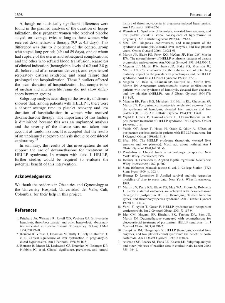
Although no statistically significant differences werefound in the planned analysis of the duration of hospi-talization, those pregnant women who received placebostayed, on average, twice as long as those women whoreceived dexamethasone therapy (9.9 vs 4.5 days). Thisdifference was due to 2 patients of the control groupwho stayed long periods (49 and 89 days), one of whomhad rupture of the uterus and subsequent complications,and the other who refused blood transfusion, regardlessof clinical indication (hemoglobin levels of 6.2 and 2.8 g/dL before and after cesarean delivery), and experiencedrespiratory distress syndrome and renal failure thatprolonged the hospitalization. These 2 outliers affectedthe mean duration of hospitalization, but comparisonsof median and interquartile range did not show differ-ences between groups.
Subgroup analysis according to the severity of diseaseshowed that, among patients with HELLP 1, there werea shorter average time to platelet recovery and lessduration of hospitalization in women who receiveddexamethasone therapy. The importance of this findingis diminished because this was an unplanned analysisand the severity of the disease was not taken intoaccount at randomization. It is accepted that the resultsof an unplanned subgroup analysis should be consideredexploratory.21
In summary, the results of this investigation do notsupport the use of dexamethasone for treatment ofHELLP syndrome. In women with class 1 HELLP,further studies would be required to evaluate thepotential benefit of this intervention.
Acknowledgments
We thank the residents in Obstetrics and Gynecology atthe University Hospital, Universidad del Valle, Cali,Colombia, for their help in this project.
References
1. Pritchard JA, Weisman R, Ratoff OD, Vosburg GJ. Intravascular
hemolysis, thrombocytopenia, and other hematologic abnormali-
ties associated with severe toxemia of pregnancy. N Engl J Med
1954;250:89-98.
2. Romero R, Vizoso J, Emamian M, Duffy T, Riely C, Halford T,
et al. Clinical significance of liver dysfunction in pregnancy-in-
duced hypertension. Am J Perinatol 1988;5:146-51.
3. Romero R, Mazor M, Lockwood CJ, Emamian M, Belanger KP,
Hobbins JC, et al. Clinical significance, prevalence, and natural
history of thrombocytopenia in pregnancy-induced hypertension.
Am J Perinatol 1989;6:32-8.
4. Weinstein L. Syndrome of hemolysis, elevated liver enzymes, and
low platelet count: a severe consequence of hypertension in
pregnancy. Am J Obstet Gynecol 1982;142:159-67.
5. Sibai BM. Diagnosis, controversies, and management of the
syndrome of hemolysis, elevated liver enzymes, and low platelet
count. Obstet Gynecol 2004;103:981-91.
6. Martin JN, Blake PG, Perry KG, McCaul JF, Hess LW, Martin
RW. The natural history of HELLP syndrome: patterns of disease
progression and regression.Am JObstetGynecol 1991;164:1500-13.
7. Magann EF, Martin RW, Isaacs JD, Blake PG, Morrison JC,
Martin JN. Corticosteroids for the enhancement of fetal lung
maturity: impact on the gravida with preeclampsia and the HELLP
syndrome. Aust N Z J Obstet Gynaecol 1993;2:127-31.
8. Magann EF, Bass D, Chauhan SP, Sullivan DL, Martin RW,
Martin JN. Antepartum corticosteroids: disease stabilization in
patients with the syndrome of hemolysis, elevated liver enzymes,
and low platelets (HELLP). Am J Obstet Gynecol 1994;171:
1148-53.
9. Magann EF, Perry KG, Meydrech EF, Harris RL, Chauchan SP,
Martin JN. Postpartum corticosteroids: accelerated recovery from
the syndrome of hemolysis, elevated liver enzymes and low
platelets (HELLP). Am J Obstet Gynecol 1994;171:1154-8.
10. Vigil-De Gracia P, Garcia-Caceres E. Dexamethasone in the
post-partum treatment of HELLP syndrome. Int J Gynecol Obstet
1997;59:217-21.
11. Yalcin OT, Sener T, Hassa H, Ozalp S, Okur A. Effects of
postpartum corticosteroids in patients with HELLP syndrome. Int
J Gynecol Obstet 1998;61:141-8.
12. Sibai BM. The HELLP syndrome (hemolysis, elevated liver
enzymes and low platelets): Much ado about nothing? Am J
Obstet Gynecol 1990;162:311-6.
13. Piantadosi S. Clinical trials: a methodologic perspective. New
York: Wiley-Interscience; 1997.
14. Hosmer D, Lemeshow S. Applied logistic regression. New York:
Wiley-Interscience; 1989. p. 307.
15. Stata Reference Manual: release 6. vol. 3. College Station (TX):
Stata Press; 1999. p. 392-4.
16. Hosmer D, Lemeshow S. Applied survival analysis: regression
modeling of time to event data. New York: Wiley-Interscience;
1999.
17. Martin JN, Perry KG, Blake PG, May WA, Moore A, Robinette
L. Better maternal outcomes are achieved with dexamethasone
therapy for postpartum HELLP (hemolysis, elevated liver en-
zymes, and thrombocytopenia) syndrome. Am J Obstet Gynecol
1997;177:1011-7.
18. Varol F, Aydin T, Gucer F. HELLP syndrome and postpartum
corticosteroids. Int J Gynecol Obstet 2001;73:157-9.
19. Isler CM, Magann EF, Rinehart BK, Terrone DA, Bass JD,
Martin JN. Dexamethasone compared with betamethasone for
glucocorticoid treatment of postpartum HELLP syndrome. Int J
Gynecol Obstet 2003;80:291-7.
20. Tompkins JM, Thiagarajah S. HELLP (hemolysis, elevated liver
enzymes, and low platelet count) syndrome: the benefit of corti-
costeroids. Am J Obstet Gynecol 1999;181:304-9.
21. Assmann SF, Pocock SJ, Enos LE, Kasten LE. Subgroup analysis
and other (mis)uses of baseline data in clinical trials. Lancet 2000;
355:1064-9.

American Journal of Obstetrics and Gynecology (2005) 193, 1599–604
www.ajog.org
CLINICAL OPINION
WHI clinical trial revisit: Imprecise scientific methodologydisqualifies the study’s outcomes
Adam Ostrzenski, MD, PhD,a,* Katarzyna M. Ostrzenska, MDb
Professor of Obstetrics and Gynecology (Affiliate), University of South Florida, and Director of the Institute ofGynecology, Inca; Director of the Bay Medical Center, and Department of Internal Medicine, Bayfront MedicalCenter,b St Petersburg, FL
Received for publication August 20, 2004; revised January 24, 2005; accepted July 29, 2005
KEY WORDSMenopause
Hormone replacementtherapy
Prempro
Estrogen replacementtherapy
Women’s Health
Initiative Study
We analyzed The Women’s Health Initiative (WHI) Study because it had a significant impact onclinical practice, both nationally and internationally. However, despite the widespread public andprofessional awareness of the results, an independent, nonbiased analysis of the quality of the
methodology of the study has not been available. We find the study design and its executionquestion the validity of the results, making it difficult to apply the WHI results to healthypostmenopausal women, different ethnic groups, or as general postmenopausal prevention.
� 2005 Mosby, Inc. All rights reserved.
Initial publications of the Women’s Health Initiative(WHI) study results1-3 have had profound influence onclinical management of postmenopausal women. TheWHI clinical trial is considered by many as a landmarkstudy of postmenopausal hormone therapy. Publishedfindings from this study have concentrated on interpre-tation and applicability of the results. Such interpreta-tions of the results led to several recommendations forpractitioners and women and were published by, butnot limited to, the American College of Obstetrics andGynecology.4,5 In order to accept results of any study,an objective scientific scrutiny of methodology andexecution of the study design is essential. Our objective
* Reprint requests: Adam Ostrzenski, MD, PhD, 14808 Gulf Blvd,
Madeira Beach, FL 33708.
E-mail: [email protected]
0002-9378/$ - see front matter � 2005 Mosby, Inc. All rights reserved.
doi:10.1016/j.ajog.2005.07.085
was to evaluate the WHI study’s methodology and itsexecution using Sackett’s6 procedures for identifyingevidence-based medicine in reviewing the WHI pub-lished results.
A review of the existing world body of the literaturetrough PubMED, ACOGNET, ProQUEST, OVID,Cochrane Collection, The Lancet on Line Collection,MDConsult, New England Journal of Medicine, Amer-ican College of Physicians on Line Resources, HighwireJournal, and Citation Index Reference computerizeddatabases was conducted on the following subjects: men-opause, hormone replacement therapy, Prempro, estro-gen replacement therapy, and Women’s Health InitiativeStudy.Also, amanual search and analysis of the literatureon of these subjects were performed. Both computerizedand manual searches failed to identify a publicationrelated to the WHI study scientific scrutiny of the meth-odology and its execution.

1600 Ostrzenski and Ostrzenska
Study design
A review and analysis of the methodology and itsexecution were conducted based upon the originalpublications of the WHI study.
Results
WHI Study design
TheHRT clinical primary prevention trial selected a totalof 27,500 women for double-blind comparison in each ofthe 3 initial arms of the clinical trials. These women wererandomly placed into the study groups. Sixteen thousandsix hundred and eight postmenopausal women wereplaced, with a ratio of 1:1 for the Prempro (WyethPharmaceuticals, Philadelphia, PA) group and the pla-cebo product. The interventional group (n = 8506) tookPrempro 1 tablet daily continuously (1 tablet of Premprocontains conjugated equine estrogens, 0.625 mg, andmedroxyprogesterone acetate 2.5 mg). Both groups wereon diet of low-fat dietary pattern (total dietary fatO20%and saturated fat O7%), with a calcium carbonatesupplement tablet (1000 mg daily) and a dietary calciumintake of 500 mg daily for a total of 1500 mg/day, anda vitamin D3 supplementation of 400 IU daily.3
Arms of the WHI Study
The following arms of the study were created: 1) un-opposed estrogen and women who had a hysterectomy(study halted in 2004); 2) estrogen/progestin (Prempro)and the intact uterus (study halted in 2003); 3) unop-posed estrogen (Premarin 0.625 mg [Wyeth Pharmaceu-ticals, Philadelphia, PA]) and the intact uterus (studyhalted in 1996). Three hundred and thirty-one subjectsparticipated in this group and the participants weretransferred to the Prempro group without additionalrandomizing.
The power of the clinical trial
In general, the power of the WHI was adequate in bothgroups for Caucasian women: the interventional group8506 and placebo group 8102. In the interventionalgroup, 7140 (83.94%) were Caucasian women, 549(6.45%), Black women, 472 (5.54%) Hispanic women,26 (0.30%) American Indian women, 194 (2.28%)Asian/Pacific Islander women, and 125 (1.46%) ofunknown ethnicity.
The age of the subjects
The intervention groupThe mean age of the subjects in the study was 63.2 yearswith 2-standard deviation G 7.1 years (range from 56.1and 70.3). The women’s age group between 60 and 79years as n = 5.667 (69.94%).2
The placebo groupThe mean age was 63.3 G 7.1 (56.2–70.4). The women’sage group between 60 and 79 years was 5.667 (69.94%).2
Yearly dropout
The intervention groupThe yearly dropout was 39.26%.
The placebo groupThe yearly dropout was 47.29%.
Hormone replacement therapy used by thesubjects before entering WHI Study
The interventional groupCurrent users at the screening were n = 548 (6.44%)and past users were 1674 (19.68%). Total number ofsubjects exposed to HRT was 2222 (26.12%) at the timeof screening.
The placebo groupCurrent users were n = 487 (6.0%), past users were n =1588 (19.6%), and total number of subjects exposed toHRT was n = 2075 (25.75%) at the time of screening.
Medical conditions at the screening
The interventional groupBody mass index (BMI) 25 to 29 was identified in 2992subjects (35.17%); BMI R30 was in 2899 (34.08%).The total number of subjects overweight and obese was5891 women (69.25%).
Elevated systolic blood pressure was present in 127subjects (1.5%); hypertension in 3039 (35.72%); diabetesin 374 (4.39%); hypercholesterolemia in 944 (11.09%),subjects on a statinmedication 590 (6.93%), total numberof subjects being treated for hypercholesterolemia was1543 women (18.03%). The number of subjects on dailyaspirin was 1623 (19.08%). Total number of sick subjectsat the screening of the WHI study was 6570 (77.23%).
The placebo groupBody mass index 25 to 29 was present in 2479 subjects(30.59%); BMI R30 was in 2737 subjects (33.78%).Elevated systolic blood pressure was present in 127 sub-jects (1.57%); hypertension in 2949 women, (36.39%)diabetes in 360 subjects, (2.91%); hypercholesterolemia in962 subjects (11.87%), subjects on a statinmedication 548(6.76%), total number of subjects being treated forhypercholesterolemia was 1510 women (18.63%). Thenumber of subjects on daily aspirin was 1631 (20.02%).Total number of sick women at the screening of theWHI study was 6450 (79.60%) subjects.
Medical history of illnesses
The interventional groupOne hundred and thirty-nine (1.63%) subjects had amedical history of myocardial infarct; 238 (2.79%)

Ostrzenski and Ostrzenska 1601
subjects had a history of angina; 95 (1.11%) subjectshad coronary artery graft or percutaneous transluminalangioplasty. The total number of subjects included inthe intervention group with a history of heart illnesseswas 472 (5.54%). Medical history of deep vein throm-bosis (DVT) or pulmonary embolism (PE) was presentin 79 subjects (0.92%); 61 (0.71%) women had a historyof stroke. The total number of subjects with history ofillness was 612 (7.19%).
The placebo groupOne hundred and fifty-seven (1.93%) subjects had amedical history of myocardial infarct; 234 (2.88%) hada history of angina; 120 (1.48%) subjects had coronaryartery graft, percutaneous transluminal angioplasty, orstroke. The total number of women included in theplacebo group with a history of heart illnesses was 511(0.63%). Medical history of DVT or PE was presentin 62 subjects (0.76%); 77 (0.95%) women had a historyof stroke. The total number of subjects with historyof illness was 650 (8.02%) subjects.
Family breast cancer at the screening
The interventional groupThe number of female relatives diagnosed with breastcancer was 1286 (15.11%).
The placebo groupThe number of female relatives diagnosed with breastcancer was 1175 (14.5%).
Comment
Evidence-based methodology
Before study results can be interpreted objectively, ascientific scrutiny of the methodology is imperative. Abasic concept of any clinical trial should be that themethod of design and execution of the study will hold upto accepted standards of evidence-based medicine. Thequalities of implementation of methodology before andduring the study will invariably determine the overallvalue of the study findings. The present and long-termimplications of the WHI clinical trial should require theestablishment of the reliability of the study results andtheir applicability to healthy individuals and for the useof Prempro for prevention. The original title of the WHIstudy publications were ‘‘Risk and benefits of estrogenplus progestin in healthy postmenopausal women.’’1
and ‘‘Failure of estrogen and progestin therapy forprevention.’’2 The words healthy and prevention in thetitles implied that the WHI study would evaluate theeffect of estrogen/progestin on healthy postmenopausalwomen, and appraise the use of Prempro for postmen-opausal women health prevention.1,2 The JAMA reportof the WHI study results1 left the impression that these
clinical trial results can be applied to all healthy post-menopausal women as a preventive approach regardlessof age and race. Scientific scrutiny of the WHI study’smethodology fails to support this suggestion. Based onthe WHI study group, implementation of the resultsinto clinical practice has little, if any, scientific basis.The outline of the WHI study methodologic weaknessis presented below.
WHI Study design
Low-fat dietary pattern and dietary supplements‘‘Women are screened for participation in one orboth of the componentsddietary modification (DM)or hormone replacement therapy (HRT).’’3 Such com-pound regiments being incorporated into the studydesign created at least 3 scientific deficiencies: 1) het-erogeneous medications tested (Prempro, low-fat dietand calcium carbonate, and vitamin D3 supplements);2) inadequate outcome measure related only to Pre-mpro; and 3) interpretation of the results couldn’t bespecifically assigned to either Prempro or low-fat dietpattern or calcium carbonate and vitamin D3. Eachremedy used in the study has an individual clinicaleffect on the outcome measure; therefore, the validityof the outcome measures and the degree of effect eachmedication had on the outcome measure could not bedetermined.
This lack of the study identifying which remedy had aclinical effect on outcome measures leaves room forspeculation of the validity of the results. The WHI studyprimary outcome measure was coronary heart disease(CHD) associated with Prempro, low-fat dietary patterntreatment, and calcium carbonate and vitamin D3 sup-plements. This combination did not distinguish betweenHRT and the low-fat diet pattern, and to what degreeeach remedy influenced outcome of CHD and stroke.Also, nutrition can be a fundamental factor in breast orcolon cancers development,3 and a low-fat diet patternunquestionably will influence the outcome measure-ments of breast and colon cancer in this clinical trial.The WHI study conclusion that Prempro decreased thecolon cancer prevalence was inaccurate because of thepresence of the other documented factors that mightinfluence in the study outcome. The reporting of thebreast cancer prevalence influences cannot be objectivelyinterpreted for the same reasons.
Calcium carbonate and vitamin D3 supplementationhave been documented as affecting the outcome ofbone fracture prevalence, including hip fracture.3 TheWHI study, by having included the use of these dietarysupplements in both groups, invalidated the positionthat Prempro alone had a significant beneficial effect onthe incident of bone fracture. Using several remedies inthe studies (estrogen/progestin, calcium carbonate, vita-min D3, low-fat diet) significantly reduced the validity

1602 Ostrzenski and Ostrzenska
that Prempro alone was the beneficial cause of bonefracture reduction.
The power of the studyThe original study plan was to incorporate 20% ofoverall minority ethnic group enrollment3; however,only 16.03% of the enrollment target was achieved. Inaddition, there were significant differences in the numberof subjects representing minority groups. The singleethnic group in the WHI study did not meet the mini-mum quota of 348 subjects necessary to qualify for‘‘grade A’’ evidence-based medicine analysis, when theyearly dropout of 39.26% in the interventional groupwas subtracted. In general, the power of the WHI studywas not sufficient to represent ethnic groups as a whole,or any specific ethnic group. Such misrepresentationamong ethnic ratio distribution makes the applicabilityof the results of the WHI study to minority groupsimpractical. The power of the WHI study was adequatefor Caucasian women.
To satisfy the power of the WHI study for any givenethnic group required 348 subjects. Black postmenopau-sal women were the highest group by number amongthe minority group subjects (549, equal to 6.45% of thetotal study’s population). The number of subjects thatdropped out indicated that the power of the studywas notsatisfied. The WHI study publication did not disclosenumerical information for dropout of each specific ethnicgroup. Applying the general 39.26% dropout ratio to theblack population, at least 214 black women dropped outfrom the study. The remaining 335 black women weresubmitted to global study analysis instead of creating aseparate black woman subgroup for statistical analysis.As a result, this group neither satisfied a power study forblack postmenopausalwomen, nor represented a separatesubgroup within the overall WHI study results analysis.The poor accounting of ethnic ratio distribution is notsufficient to support the conclusion that the WHI studyresults can be applied to any ethnic or minority groups.
Validity of the WHI study’s results for any minoritygroup is lacking for 3 reasons: 1) insufficient power ofthe study for minority groups; 2) the absence of aspecific subgroup analysis; and 3) inadequate inclusion/exclusion criteria.
Reallocation of the subgroup results to a generalpopulation analysis of the study does not reflect asubgroup response to the tested multiagents. Differentethnic groups will demonstrate different predispositionsto medical conditions and how the use of Prempro andthe low-fat diet pattern with supplements of calciumcarbonate and vitamin D3 would affect women associ-ated with medical conditions differently than healthysubjects. Inadequate exclusion/inclusion criteria, theabsence of adequate subgroups, and insufficient powerof the study for minority groups make WHI studyresults impossible to apply to any minority group.
The age of the subjectsThe median age of menopause among women in the USis 51.1 years.6 Approximately 70% of the WHI partic-ipants were women between the age of 60 years and79 years, with a mean age of 63.2 years G 7.1 years.Consequently, women in the study below 56.1 years ofage would fall out of a 2-standard deviation analysis andbecome a statistically insignificant group. Simply, theapplicability of the WHI study’s findings to youngerwomen, between age 51.1 and 56.1 years and younger,is unknown. The age gap itself between the median ageof menopause and the median age of the WHI studyparticipants have disqualified the WHI label of aprimary prevention trial. The WHI study can honestlyonly be described as a clinical trial of older women,being that there is a 12-year difference in median age ofmenopausal women nationally and those who were inthis trial.
Yearly dropoutIn the intervention group and in the placebo group,yearly dropout at the end of the study was 39.26% and47.29%, respectively. In the WHI study, the majority ofwomen were free from vasomotor symptoms (VMS).2-4
This fact, in conjunction with undesirable side effectsrelated to Prempro itself (breast tenderness, bloating,endometrial bleeding, or mood changes2) can providean explanation for 39.26% dropout from the interven-tion group. Because younger postmenopausal womenexperience more severe VMS, then, most likely such ayounger group will adhere to the protocol closely, andthis may have reduced the number of yearly dropouts.Although it is clearly stated in the WHI study’s re-ports2-4 that this trial was not designed to evaluatepostmenopausal VMS, psychologic, or peripheral symp-toms, the absence of the symptoms among subjectscontributed to the higher rate of dropout from the study.
Arms of the WHI studyThe unopposed estrogen group was selected amongwomen with an intact uterus. This group was originallyrandomized between 1992 and 1996, and the study washalted in 1996. The justification for discontinuation ofthe study was given as ‘‘.long-term adherence tounopposed estrogen was not feasible in women with auterus.’’ Consequently, this group was not rerandom-ized and arbitrarily was relocated from the Premaringroup to the Prempro group. Such contamination of thestudy population disqualifies Prempro result interpreta-tion because 2 agents were used by the same subject(estrogen alone and later estrogen/progestin regiment)at different times. The studies1-3 indicated that sufficienttime lapsed (4 weeks) to eliminate the short-term effectsof estrogen. This time period would not definitelyeliminate the long-term effect of estrogen use. Each ofthe defined regimens will have its own long-term effect
Ostrzenski and Ostrzenska 1603
on the outcome measures in the WHI study. The clinicalparameters outcome measure of the WHI study of 2independent agents, estrogen alone between 1992 and1996, and estrogen/progestin from 1996 to 2003, isunfeasible for results interpretation. Estrogen alonewould have had an effect on clinically measurableoutcome parameters, and estrogen/progestin has appar-ently different effect on measurable clinical parameters(CHD, DVT, stroke, breast cancer, hip fracture, colo-rectal cancer, etc).
Medical conditions and history of illnessesat the screeningIn the interventional group, a total of 6645 (78.12%)women were sick or had a history of illnesses. Thesesubjects were identified as having been treated for beingoverweight; obesity; elevated systolic, diastolic bloodpressure; hypertension; diabetes; hypercholesterolemia;or use of daily aspirin (coronary heart disease or strokeprevention) at the initial screening of the WHI study.These medical conditions by themselves would predis-pose subjects to CHD, stroke, or DVT, or PE, withoutHRT. It is obvious that 78.12% of the subjects did notqualify as a healthy population. The 22% remainingwomen would have been the only valid participants toreflect the stated main outcome to appraise the effectHRT on CHD.
The combination of both an older age group ofparticipants and preexisting medical conditions invali-dated the concept that the WHI studies were designedto evaluate the effect of estrogen/progestin on healthywomen or an evaluation study for prevention. The highpercentage (78.12%) of subjects with a preexisting illnessincluded in the WHI study should have disqualified theresults of this clinical trial application to a healthypopulation. Included in the clinical trial were subjectswith previous medical conditions (myocardial infarct,angina, angioplasty, DVT or PE, or stroke). The totalnumber of subjects with a history of illness was 612(7.19%).
Also, 1286 (15.11%) female relatives had docu-mented breast cancer. Numerous epidemiologic studiesindicate that a family history of breast cancer, particu-larly the immediate relatives, has significant influence onthe breast cancer incidence. At least 1286 (15.11%) ofthe subjects in the WHI study had a family predisposi-tion for breast cancer before being exposed to Prempro.A special subgroup should have been created for eval-uation of the effect of estrogen/progestin on the subjectswith a family history of breast cancer, and this subgroupshould not have been included in the general studypopulation.
Evidence-based medicine has already established thatthe above-mentioned medical conditions or history ofillnesses would constitute either indirect or direct con-traindication in the use of HRT. Under no circumstance
would this subject group meet the criteria for healthycandidates. All of these unhealthy populations in theWHI study should have been organized into separatesubgroups according to the medical condition, andpresented with separate and specifically designed in-formed consent and excluded from the general studypopulation. The exclusion and inclusion criteria of theWHI study revealed serious scientific concerns; thisabsence of specific medical condition subgroups madethe WHI study invalid for healthy postmenopausalwomen and unhealthy subjects as well because healthyand unhealthy subject were mixed in the results.
The WHI clinical trial can be considered as anenormous undertaking and effort; however, it shouldnot be considered as a clinical landmark or revolution-ary trial. The lack of a sound scientific design andexecution invalidated the study from the primary selec-tion and methodology used. The professional commu-nity and the public-at-large were presented with theresults of the WHI study without scientific scrutiny ofthis clinical trial. Some professionals among medicalauthorities and the US media have concluded that ahormone replacement therapy is bad for women’s healthbased on the WHI study. After careful review of theWHI reports, adherence to the principles of evidence-based medicine, our analysis can neither confirm thenotion that HRT is bad for postmenopausal womenhealth, nor that HRT is good for postmenopausalwoman’s health. Our analysis simply suggests that theexperienced clinical scientists must evaluate the designand execution of the WHI study in order to establisheffectiveness and benefit/risk of the HRT, if not com-pletely redo the study itself.
Conclusions
Only 1 dose of Prempro has been tested in the WHIstudy and the russets can not be applied to the low doseof Prempro.
Using combination of conventional medicine (estro-gen/progestin) and alternative remedies (calcium carbon-ate, vitaminD3, low-fat diet) significantly obscured, if notcompletely eliminated, interpretation of the benefit/riskeffect of Prempro on CHD, stroke, breast cancer, bonefracture.
Inadequate implementation of exclusion/inclusion criteria
Insufficient power of the study for different minoritygroups made WHI study results impossible to apply toany of the minority groups.
The applicability of the WHI findings to women be-tween age of 51.1 and 56.1 years and younger is unknown.
The age gap itself between the median age of men-opause and the median age of the subjects in the study

1604 Ostrzenski and Ostrzenska
disqualifies the WHI study’s label of a primary preven-tion trial.
The WHI study can only be described as a clinicaltrial of older women, being that there is a 12-yeardifference in the median age of menopause and themedian age of those women who were participated inthe WHI study.
The reduced or absence postmenopausal symptomsamong older women contributed to the higher rate ofdropout from the study.
The women from the Premarin group (the subjectswith an intact uterus being on unopposed estrogen)were not rerandomized and arbitrarily relocated to thePrempro interventional group without creating a homo-genous placebo group.
The WHI study’s outcome measure was contami-nated by using 2 independent agents among the samesubjects: estrogen alone (Premarin) between 1992 and1996 and estrogen/progestin (Prempro) between 1996and 2003. Such contamination made the WHI study’sresults interpretation impractical.
The unhealthy population in the WHI study was notorganized into separate subgroups according to themedical condition, was not presented with specificallydesigned informed consent, and was not excluded fromthe general study population.
The high percentage (78.12%) of subjects with apreexisting illness included in the WHI study disquali-fied the results applicability to healthy postmenopausalwomen.
The absence of specific medical condition subgroupsmade the WHI study results invalid for healthy post-menopausal women as well as for unhealthy subjects,since healthy and unhealthy results were mixed.
The overall scientific quality of the WHI study isfeeble.
The applicability of theWHI study’s results to healthypostmenopausal women or different ethnic groupwomen, or as general prevention measures, is impracticalbecause of the imprecise study’s methodology.
Methodologic deficiency disqualifies outcome mea-sures.
The faulty study design and its execution call thevalidity of the results into question.
The WHI clinical trial can be considered as anenormous undertaking and effort; however, it shouldnot be considered as a clinical landmark or revolution-ary trial.
Our analysis can neither confirm the notion that theHRT constitutes a health-hazard for postmenopausalwomen, nor that HRT is beneficial for postmenopausalwomen health.
The WHI clinical trial should be completely rede-signed and redone in order to determine the HRT safetyand effectiveness.
Acknowledgments
The authors are very appreciative to Joyce C. Burkhart,MS, and Robert W. Burkhart, MS, for assisting in themanuscript preparation.
References
1. Writing Group for the Women’s Health Initiative Investigators.
Risk and benefits of estrogen plus progestin in healthy postmeno-
pausal women: principal results from the Women’s Health Initiative
Randomized Controlled Trial. JAMA 2002;288:321-33.
2. Fletcher SW, Colditz GA. Failure of estrogen plus progestin
therapy for prevention. JAMA 2002;288:366-86.
3. The Women’s Health Initiative Study Group. Design of Women’s
Health Initiative clinical trial and observational study. Control Clin
Trials 1998;19:61-109.
4. American College of Obstetricians and Gynecologists. HRT recom-
mendation for physicians. ACOG, Washington, DC, August 9,
2002.
5. American College of Obstetricians and Gynecologists. HRT recom-
mendation for women. ACOG, Washington, DC, August 9,
2002.
6. Stanford JL, Hartge P, Brinton LA, Hoover RN, Brookmeyer R.
Factors influencing the age at natural menopause. J Chronic Dis
1987;40:995-1002.

American Journal of Obstetrics and Gynecology (2005) 193, 1605–6
www.ajog.org
WHI response to Ostrzenski and Ostrzenska
Barbara B. Cochrane, PhD, RN,a,* David H. Barad, MD, MS,b Margery Gass, MD,c
Robert L. Brunner, PhD,d Cora E. Lewis, MD, MSPH,e Marcia L. Stefanick, PhDf
Public Health Sciences Division, Fred Hutchinson Cancer Research Center,a Seattle, WA; Department ofObstetrics and Gynecology and Women’s Health, Albert Einstein College of Medicine,b Bronx, NY;Department of Obstetrics and Gynecology, University of Cincinnati College of Medicine,c Cincinnati, OH; Departmentof Family and Community Medicine, University of Nevada School of Medicine,d Reno, NV; Division ofPreventive Medicine, University of Alabama at Birmingham,e AL; Stanford Center forResearch in Disease Prevention, Stanford University,f Palo Alto, CA
Received for publication June 14, 2005; accepted July 29, 2005
Contrary to Ostrzenski’s and Ostrzenska’s1 premise,the Women’s Health Initiative (WHI) Estrogen plusProgestin (ECP) Trial2 has generated considerable sci-entific scrutiny, from a JAMA editorial describing theresults as ‘‘strong evidence,’’3 to the American College ofObstetricians and Gynecologists characterizing the trialas ‘‘.the largest, most statistically valid, and well-analyzed research to evaluate the use of HRT in healthypostmenopausal women,’’4 and the American Societyfor Reproductive Medicine writing ‘‘The data emergingfrom this methodologically sound randomized con-trolled trial appear incontrovertible.’’5 Sackett, whoseprinciples of evidence-based medicine6 Ostrzenski andOstrzenska endorse, described the trial as a ‘‘rigorous’’randomized clinical trial, designed and executed toanswer the study question.7
We address below some of Ostrzenski’s and Ostrzen-ska’s claims about the WHI findings:
The Women’s Health Initiative (WHI) program is funded by the
National Heart, Lung and Blood Institute, US Department of Health
and Human Services.
* Reprint requests: Barbara B. Cochrane, PhD, RN, Fred Hutch-
inson Cancer Research Center, 1100 Fairview Ave N, M3-A410, PO
Box 19024, Seattle, WA 98109-1024.
E-mail: [email protected]
0002-9378/$ - see front matter � 2005 Mosby, Inc. All rights reserved.
doi:10.1016/j.ajog.2005.07.099
The majority of participants were ‘‘sick’’
The presence of risk for chronic disease (eg, hyperten-sion, obesity) does not mean that a person is ‘‘sick’’; infact, WHI participants at baseline were healthier thanthe general population of US women aged 50 to 79.8
This was a clinical trial of ‘‘older’’ women
Over 12% of participants were aged 50 to 54 at baseline(n = 2029), making it the largest randomized ECP trialever conducted in this age group. Subgroup analyses didnot indicate significant interactions with age for ECPeffects on major outcomes.
Yearly dropout was high and compromisedstudy power
The yearly dropout rates cited by Ostrzenski and Ostr-zenska are not accurate. The dropout rates reported in theECP trial (defined as stopping study pills at any time,whether or not prescription hormones were started orstudy pills were restarted) were cumulative (42% forECP, 38% for placebo),2 with first-year rates beingsimilar to or lower than those reported in other hormonetrials9,10 and community studies,11 and never exceeding9.7% in any given year for either treatment arm (range

1606 Cochrane et al
3.9-9.7%). Despite these dropout rates, the study hadadequate statistical power for testing the primary hy-potheses.
Dropout in the intervention group wouldhave been lower if women had vasomotorsymptoms
In fact, many of these participants did have moderateor severe vasomotor symptoms at baseline (n = 574 or28.3% of participants aged 50-54).12
Power was not sufficient among minoritygroups to support the conclusions
As with most medical studies, the results apply to theentire study population and are not specific to a partic-ular racial/ethnic group. Although we did not reportsignificant differences in outcomes among these groups,the 16% racial/ethnic minority participation doesrepresent greater diversity than in previous hormonetrials.9,10,13
Reassignment of participants with a uterusfrom estrogen-alone to active EDPcontaminated the intervention arm
Only 331 women (4%) were reassigned, and findings didnot differ substantially in analyses performed withoutthese participants.2
We emphasize that in randomized clinical trials(RCTs), such as the WHI, study groups are balancedwith regard to risk factors. As a result, RCTs arestatistically more robust for determining treatmenteffects than other types of studies.14 The WHI investi-gators continue to examine data from the trial and inviteinformed discussions that enhance our understanding ofthe findings.
References
1. Ostrzenski A, Ostrzenska KM. WHI clinical trial revisit: imprecise
scientific methodology disqualifies the study’s outcomes. Am J
Gynecol Obstet 2005;193:1599-604.
2. Writing Group for the Women’s Health Initiative Investigators.
Risks and benefits of estrogen plus progestin in healthy post-
menopausal women: Principal results from the Women’s
Health Initiative Randomized Controlled Trial. JAMA 2002;
288:321-33.
3. Fletcher SW, Colditz GA. Failure of estrogen plus progestin
therapy for prevention. JAMA 2002;288:366-8.
4. American College of Obstetricians and Gynecologists. ACOG
issues state-of-the-art guide to hormone therapy. Available at:
http://www.acog.org/from_home/publications/press_releases/nr09-
30-04-2.cfm. Accessed February 2, 05.
5. American Society for Reproductive Medicine Statement on Com-
bined Hormone Therapy and the Women’s Health Initiative.
Available at: http://www.asrm.org/Media/misc_announcements/
whi2.html. Accessed August 10, 2005.
6. Sackett DL, Straus SE, Richardson WS, Rosenberg W, Baynes
RB. Evidence-based medicine: How to practice and teach EBM.
New York: Churchill Livingstone; 2000.
7. Sackett DL. The arrogance of preventive medicine. CMAJ 2002;
167:363-4.
8. Stefanick ML, Cochrane BB, Hsia J, Barad DH, Liu JH, Johnson
SR. The Women’s Health Initiative postmenopausal hormone
trials: Overview and baseline characteristics of participants. Ann
Epidemiol 2003;13:S78-86.
9. Hulley S, Grady D, Bush T, Furberg C, Herrington D, Riggs B,
et al. Randomized trial of estrogen plus progestin for secondary
prevention of coronary heart disease in post-menopausal women:
Heart and Estrogen/progestin Replacement Study (HERS) Re-
search Group. JAMA 1998;280:605-13.
10. Utian WH, Shoupe D, Bachmann G, Pinkerton JV, Pickar JH.
Relief of vasomotor symptoms and vaginal atrophy with lower
doses of conjugated equine estrogens and medroxyprogesterone
acetate. Fertil Steril 2001;75:1065-79.
11. Pilon D, Castilloux A, Lelorier J. Estrogen replacement therapy:
determinants of persistence with treatment. Obstet Gynecol
2001;97:97-100.
12. Hays J, Ockene J, Brunner R, Kotchen J, Manson JE, Patterson R,
et al. Effects of estrogen plus progestin on health-related quality of
life. N Engl J Med 2003;348:1839-54.
13. The Writing Group for the PEPI Trial. Effects of estrogen or
estrogen/progestin regimens on heart disease risk factors in post-
menopausal women. JAMA 1995;273:199-208.
14. Meinert CL. Clinical trials: Design, conduct and analysis. New
York: Oxford University Press; 1986.

REVIEW ARTICLE
American Journal of Obstetrics and Gynecology (2005) 193, 1607–17
www.ajog.org
Evidence-based surgery for cesarean delivery
Vincenzo Berghella, MD,a Jason K. Baxter, MD, MSCP,a Suneet P. Chauhan, MDb
Department of Obstetrics and Gynecology, Division of Maternal-Fetal Medicine, Jefferson Medical College ofThomas Jefferson University, Philadelphia, PAa; Aurora Health Care, West Allis, WIb
Received for publication January 21, 2005; revised March 18, 2005; accepted March 26, 2005
KEY WORDSCesarean deliverySurgeryRandomized
controlled trialsEvidence-based
medicine
Objective: The purpose of this study was to provide evidenced-based guidance for surgicaldecisions during cesarean delivery.
Study design: We performed MEDLINE, PubMed, EMBASE, and COCHRANE searches withthe terms cesarean section, cesarean delivery, cesarean, pregnancy, randomized trials, and eachtechnical aspect of cesarean delivery. All randomized trials that covered a surgical aspect of
cesarean delivery were included in the review. Each surgical step of cesarean delivery wasreviewed separately.Results: US Preventive Services Task Force recommendations favor blunt uterine incision
expansion, prophylactic antibiotics (either ampicillin or first-generation cephalosporin for just1 dose), spontaneous placental removal, non-closure of both visceral and parietal peritoneum,and suture closure or drain of the subcutaneous tissue when thickness is R2 cm.Conclusion: Cesarean delivery techniques that are supported by good quality recommendations
should be performed routinely. All technical aspects that have recommendations with lowerquality should be researched with adequately powered and designed trials.� 2005 Mosby, Inc. All rights reserved.
The word cesarean is derived from the Latin caesare,which means ‘‘to cut.’’ It is unlikely that Caesar wasdelivered by this technique. The first documented cesar-ean delivery (CD) was in 1020 AD.1 In 1882, the era ofmodern CD began when Saenger advocated closing alluterine incisions immediately after surgery. The loweruterine segment incision was introduced by Kronig in1912 and popularized in the United States by DeLee in1922. The transverse uterine incision was described byMunro-Kerr in 1926.2 CD has been associated withrelatively low maternal mortality rates for O100 years.Safety has improved in the last 50 years, because these
Reprints not available from the authors. Address correspondence to
Vincenzo Berghella.
E-mail: [email protected]
0002-9378/$ - see front matter � 2005 Mosby, Inc. All rights reserved.
doi:10.1016/j.ajog.2005.03.063
techniques have become more routine and antibioticshave been introduced.
CD is now the most common surgical procedure inthe United States, with O1 million procedures per-formed per year. Its incidence increased to 27.6% ofdeliveries in 2003.3 This increase was fueled, at least inpart, by the increased incidence of multiple gestationsand the decreased incidences of vaginal births after CDand vaginal breech deliveries. Recently, women’s de-mands for elective CD have increased because compli-cations from the procedure have diminished, women arehaving fewer children, and fear and concerns aboutvaginal delivery have not abated.
The aim of this manuscript was to review the evidencefor the technique of CD and to offer recommendationsbased (where available) on randomized trials. Proper

1608 Berghella, Baxter, and Chauhan
technique, which has proved to be associated with theleast complications, will minimize the morbidity andpossibly the deaths that can be associated with CD. Weaspire to stimulate better clinical treatment, promoteeducation, foster research trials in areas of uncertainty,and focus on the prevention of possible complicationsrather than on treatment. We wanted to provide obste-tricians with evidenced-based guidance for surgical deci-sions that are made in the operating room during a CD.We did not seek to review other aspects of CD (such aspreoperative and postoperative care). We assume thatduring CD obstetricians would adhere to the best surgicaltechniques to decrease adhesions, to minimize tissuetrauma, and to avoid ischemia and inflammation. Wepresent one aspect of the CD technique at a time to showthe effect of each step of the procedure individually.Therefore, we will not discuss proposed general ap-proaches that involve the comparison of several steps atthe same time, because direct comparison of the effects ofeach individual step is not possible.4,5
Material and methods
To achieve our aim, we performed MEDLINE,PubMed, EMBASE, and COCHRANE searches withthe terms cesarean section, cesarean delivery, cesarean,pregnancy, randomized trials, plus each technical aspect(eg, lateral tilt, skin cleansing). The search was between1966 and 2004 and was not restricted by language.
Each retrieved manuscript or Cochrane review wascarefully evaluated, and any pertinent references from themanuscripts were obtained and reviewed. Our evidence-based review integrates individual clinical expertise withthe best available external clinical evidence from system-atic research.6 All randomized trials that covered surgicaland selected nonsurgical aspects of CD were included inthe review. In the absence of trials that adequately coveredthe aspect, analytic data were reviewed. In the absence ofexperimental or analytic data, observational data wereevaluated. Each step of CD was reviewed separately.After each technical step was reviewed, evidence levelsand recommendation levels, according to the newmethodoutlined by the US Preventive Services Task Force,7 werereported (Table I).
Results
Lateral tilt involves tilting the woman towards her leftside 10 to 15 degrees to avoid vena caval compressionby the gravid uterus. Lateral tilt is compared with thesupine position in 3 methodologically poor randomizedtrials that involved 293 women8-10 and that were sum-marized in a Cochrane Review.11 Lateral tilt did not
provide fetal/neonatal benefit in Apgar scores (oddsratio [OR], 0.53; 95% CI, 0.25-1.16) or umbilical arterypH (mean difference, 0.03; 95% CI, 0.01-0.04). Fetaloxygen saturation was improved in a nonrandomizedstudy of women in labor12 (recommendation: I; evi-dence: poor; Table I).
Skin cleansing techniques for CD have been studiedonly once in a randomized trial.13 One hundred womenwere assigned randomly to parachlorometaxylenol scrubfor 5 minutes or not, after which all women received7.5% povidone-iodine scrub and then povidine-iodine10% solution. In the women who had received prophy-lactic antibiotics for CD, no significant differences inincidences of endometritis (OR, 2.7; 95% CI, 0.8-8.9) orwound infection (2/25 vs 0/25 women; OR, not calcu-lable) were found between the 2 skin cleansing groups.This study was limited by a possible type II error, giventhe small numbers. This initial aspect of skin cleansingbefore CD is grossly understudied. Skin is impossible tosterilize. In nonpregnant adults, a meta-analysis ofseveral trials found no differences in wound infectionwith different types and times of scrubs.14 Therefore, theuse of an iodine solution alone (better than salinesolution) is considered reasonable. Some investigatorsinsist on the importance of letting the iodine dry for bestinfection prophylaxis (recommendation: I; quality:poor; Table I).
Adhesive drapes for CD were studied in 2 randomizedtrials, which included 1943women.15,16 The 2 studies eachreported no benefit of adhesive drapes for the preventionof wound infection. No Cochrane Review is available. Inour own meta-analysis, the incidence of wound infectionwas higher in the adhesive drape group (13.8%; 133/967women) compared with the control group (10.7%; 104/996 women; relative risk [RR] 1.34; 95% CI, 1.02-1.76).Therefore, adhesive drapes should not be recommendedfor the prevention of wound infection at CD (recommen-dation: D; quality: fair; Table I).
Skin incision techniques for CD were studied sepa-rately from other aspects of CD in 2 randomized trials,including 411 women.17,18 In general, a transverse skinincision is recommended, because this is associated withless postoperative pain and improved cosmetic effectcompared with a vertical incision19 (recommendation: B;quality: fair; Table I).
The Pfannenstiel incision (slightly curved, 2 to 3 cmor 2 fingers above the symphysis pubis, with the midportion of the incision within the shaved area of thepubic hair) and Joel-Cohen incision (straight, 3 cmbelow the line that joins the anterior superior iliac spinesand therefore slightly more cephalad than the Pfannen-stiel) are the preferred transverse incisions. These 2incisions were compared in the 2 trials that have beenpublished; no Cochrane Review is available.17,18 In anearlier trial on this issue,20 the Pfannenstiel group hadthe peritoneum closed, and the Joel-Cohen group did

Berghella, Baxter, and Chauhan 1609
Table I Standard recommendation language and quality of evidence according to the method outlined by the US Preventive ServicesTask Force (USPSTF)7
Recommendation
A. The USPSTF strongly recommends that clinicians provide [the service] to eligible patients. The USPSTF found good evidence that [theservice] improves important health outcomes and concludes that benefits substantially outweigh harms.
B. The USPSTF recommends that clinicians provide [this service] to eligible patients. The USPSTF found at least fair evidence that [theservice] improves important health outcomes and concludes that benefits outweigh harms.
C. The USPSTF makes no recommendation for or against routine provision of [the service]. The USPSTF found at least fair evidence that[the service] can improve health outcomes but concludes that the balance of benefits and harms is too close to justify a generalrecommendation.
D. The USPSTF recommends against routinely providing [the service] to asymptomatic patients. The USPSTF found at least fair evidencethat [the service] is ineffective or that harms outweigh benefits.
I. The USPSTF concludes that the evidence is insufficient to recommend for or against routinely providing [the service]. Evidence thatthe [service] is effective is lacking, of poor quality, or conflicting, and the balance of benefits and harms cannot be determined.
Quality of evidenceGood: Evidence includes consistent results from well-designed, well-conducted studies in representative populations that directlyassess the effects on health outcomes.
Fair: Evidence is sufficient to determine the effects on health outcomes, but the strength of the evidence is limited by thenumber, quality, or consistency of the individual studies, generalizability to routine practice, or indirect nature of the evidenceon health outcomes.
Poor: Evidence is insufficient to assess the effects on health outcomes because of a limited number or power of studies, importantflaws in their design or conduct, gaps in the chain of evidence, or a lack of information on important health outcomes.
not, which makes a comparison of this single aspect ofCD impossible. In the larger, better designed trial,17 nodifferences in total operative time (32 vs 33 minutes),intra- and postoperative complications, and neonataloutcomes were found. The extraction time was 50seconds shorter for the Joel-Cohen group (median, 240seconds [range, 50-600 seconds] vs 190 seconds [range,60-600 seconds]). Considering the absence of clinicalbenefits to the mother and fetus, these authors con-cluded that there is no clear indication for the perfor-mance of a Joel-Cohen incision. In contrast, the smaller,less-well designed trial18 showed significantly shorteroperating times and reduced blood loss and postopera-tive discomfort associated with the Joel-Cohen incisioncompared with the Pfannenstiel incision (recommenda-tion: C; quality: fair; Table I).
Skin incision length has not been studied in a trial.Two non-randomized studies suggest that abdominalsurgical incision size should provide at least 15 cm (sizeof a standard Allis clamp) of exposure to assure optimaloutcome of both mother and fetus21,22 (recommenda-tion: I; quality: poor; Table I).
Changing to a second scalpel after the first scalpel hasbeen used for skin incision versus no such change hasnever been evaluated in a trial or in any obstetric litera-ture. One randomized study in general surgery patientsdid not show any benefit from discarding the first knifeafter skin incision and concluded that one scalpel isadequate to use throughout the whole surgical proce-dure23 (recommendation: D; quality: fair; Table I).
Subcutaneous incision/opening has not been studiedseparately in a trial. Most clinicians use the scalpel as
little as possible, opening layers bluntly from medial tolateral to avoid injury to tissue and the inferior epigas-tric vessels. Blunt dissection has been associated withshorter operating times. There are no trials to evaluatethe safety or efficacy of electrosurgery, electrocautery,or diathermy (Bovie) during CD (recommendation: I;quality: poor; Table I).
Fascial incision has not been studied separately in atrial. Most experts recommend a transverse incision thatis performed with the scalpel and then extended withscissors. Some clinicians have advocated digital exten-sion, which can be accomplished by separating the fore-fingers in a cephalad-caudad direction after the fingers areinserted into a small, midline transverse fascial incision24
(recommendation: I; quality: poor; Table I).Rectus muscle cutting has been studied in 3 trials that
included 313 women21,25,26; no Cochrane Review isavailable. These womenwere assigned randomly to eitherMaylard (muscle cutting) or Pfannenstiel (no musclecutting) techniques. Transecting the rectus muscle wasnot associated with any difference in operative morbidity,difficult deliveries, postoperative complications, or painscores in these studies. One study showed that abdominalmuscle strength at 3 months was also similar, with a trendfor better strength in the Pfannenstiel group.26 Therefore,rectus muscle cutting is probably not necessary (recom-mendation: D; quality: fair; Table I).
Dissection of fascia off the recti muscles has not beenstudied separately in a trial. Several investigators havecast doubt on the necessity of this commonly usedtechnical step of CD4,5,24 (recommendation: I; quality:poor; Table I).

1610 Berghella, Baxter, and Chauhan
Opening of the peritoneum has not been studiedseparately in a trial. The peritoneum usually is openedcarefully with blunt or sharp dissection and bluntexpansion, high above the bladder, which avoids injuryto the organs below (recommendation: I; quality: poor;Table I).
Bladder flap development versus no such develop-ment was studied in one randomized trial.27 This trialrandomly assigned 102 women to either incision andopening of the bladder flap or to direct incision 1 cmabove the bladder fold. Bladder flap development wasassociated with longer incision to delivery interval (7 vs5 minutes; P ! .001), longer total operating time (40 vs35 minutes; P = .004), and greater change in hemoglo-bin level (1 vs 0.5g/dL; P = .009). Forming a bladderflap was also associated with more postoperative micro-hematuria (47% vs 21%; P ! .01) and greater need foranalgesia (55% vs 26%; P = .006) at 2 days after CD.There was only 1 postoperative fever in each group. Nolong-term effects (eg, adhesions, bladder function, fer-tility) were evaluated. Because bladder injury at CD isan uncommon event (1-3/1000 births), a sample size ofO40,000 women would be required to show a differencein this outcome.27 Given these results and consider-ations, not developing a bladder flap at CD may bejustified4,5 (recommendation: D; quality: fair; Table I).
The use of a bladder blade to protect the bladder hasnot been studied separately in a trial (recommendation:I; quality: poor; Table I).
Uterine incision type has not been studied separatelyin a trial. The transverse incision in the lower uterinesegment is recommended by most experts and by retro-spective case-control studies.28,29 Some experts advocatethe classic vertical or at least low-vertical incision if thelower uterine segment is not large enough to allow atransverse incision (eg, for the very preterm [!28 weeksof gestation] uterus, fibroids), but this has been associ-ated with increased blood loss compared with lowtransverse incision28 (recommendation: B; quality: fair;Table I).
Uterine stapling device versus traditional opening andclosure of the uterine scar has been evaluated in 4trials,30-33 which involved 526 women and was summa-rized in a Cochrane Review.34 The Cochrane Reviewstates that there was no difference in total operating timebetween the stapling technique and other techniques toextend the incision (weighted mean difference, 1.17minutes; 95% CI, �3.57-1.22). However, stapling de-vices increased the time that was needed to deliver thebaby (weighted mean difference, 0.85 minutes; 95% CI,0.48-1.23). Blood loss was lower with the use of staples(weighted mean difference, �41.22 mL; 95% CI, �50.63to �31.8). No significant differences between staplingand other techniques were detected for other perinatalmorbidity outcomes. The Cochrane reviewers concludedthat there is not enough evidence to justify the routine
use of stapling devices to extend the uterine incision atlower segment cesarean delivery. There is a possibilitythat stapling could cause harm, by prolonging the timeto deliver the baby (recommendation: D; quality: fair;Table I).
Expansion of uterine incision either bluntly or byscissors was evaluated in 2 randomized trials,35,36 withno Cochrane Review. The first trial involved 147 sharp(scissors) versus 139 blunt (fingers) uterine incisionexpansions.35 The incidence of unintended extensions(14% vs 12%; P = .61) and postpartum hemoglobinlevels (9.9 vs 10.3 g/dL; P = .12) were similar. Noserious morbidities occurred. In the second larger andmore recent trial, 470 sharp versus 475 blunt uterineincision expansions were compared.36 Sharp uterineincision expansions were associated with increased esti-mated blood loss (886 vs 843 mL; P = .001), change inhematocrit (6.1% vs 5.5%; P = .003), incidence ofpostpartum hemorrhage (13% vs 9%; RR, 1.23; 95%CI, 1.03-1.46), need for transfusion (2% vs 0.4%; RR,1.65; 95% CI, 1.25-2.21), and total number of extensions(RR, 1.66; 95% CI, 1.47-1.86). In summary, these 2trials show that sharp expansion significantly increasesblood loss and the need for transfusion and is associatedwith more extensions. Because it is also quicker and isassociated with less risk of inadvertently cutting theneonate or cord, blunt expansion should be preferred tosharp expansion of the uterine incision (recommenda-tion: A; quality: good; Table I).
Instrumental delivery of the fetal head by eithervacuum or forceps compared with manual means wasevaluated only in a pilot randomized trial of 44 womenwith cephalic presentation who underwent CD.37 Nocontrolled data are available on instrumental delivery ofthe aftercoming head at CD with breech presentation.Therefore, no firm recommendations can be made.Because instrumentation has been associated with ma-ternal (especially for forceps) or fetal (especially forvacuum) harm in vaginal deliveries,38 the principle ofprimum non nocere (first do no harm) should be appliedin this setting; therefore, manual delivery of the fetalhead should be favored whenever possible until furtherdata are available (recommendation: I; quality: poor;Table I).
Prophylactic antibiotics for CD have been evaluatedin at least 81 randomized trials. This is certainly thebest-studied aspect of CD. The comprehensive Co-chrane Review shows benefit in both elective (non-laboring) and nonelective (laboring) CD.39 Thedecrease in incidence of endometritis was O60% inboth elective (RR, 0.38; 95% CI, 0.22-0.64) and non-elective (RR, 0.39; 95% CI, 0.34-0.46) CD, with O2000women who were randomly assigned in each analysis.The decrease in wound infection was approximately25% (RR, 0.73; 95% CI, 0.53-0.99) in elective CD andapproximately 65% (RR, 0.36; 95% CI, 0.26-0.51) in

Berghella, Baxter, and Chauhan 1611
nonelective CD. Overall, fever and urinary tract infec-tions are also markedly decreased. These results justify arecommendation of prophylactic antibiotics in womenwho undergo any (elective or nonelective) CD (recom-mendation: A; quality: good; Table I).
Fifty-one trials have evaluated the appropriate anti-biotic to give. The efficacy of ampicillin is equivalent tothat of first-generation cephalosporins, such as cefazolin(Ancef); later-generation, more expensive broad-spec-trum agents do not improve efficacy further40 (recom-mendation: A; quality: good; Table I).
Systemic versus lavage routes of antibiotic adminis-tration seem to have similar efficacy40 (recommendation:A; quality: good; Table I).
Multiple systemic doses do not improve efficacy overa single dose40 (recommendation: D; quality: good;Table I).
Timing of antibiotic administration (at cord clampversus preoperative) has been evaluated in 3 separate,underpowered studies that have shown no differencein infectious morbidity rates41-43 (recommendation: I;quality: poor; Table I).
If ampicillin or a first-generation cephalosporin hasalready been given in labor, there may be no need foradditional prophylactic antibiotics at CD (recommen-dation: I; quality: poor; Table I).
Prevention of uterine atony and postpartum hemor-rhage has not been studied for CD but has been studiedextensively for the third stage of labor after vaginaldelivery.44 In the setting of vaginal delivery, bothintravenous and intramuscular oxytocin effectively re-duce postpartum hemorrhage and the need for thera-peutic uterotonics by at least 40%, compared withplacebo or no routine prophylactic agent. Oxytocin isas effective and has fewer side effects than ergot alka-loids (recommendation: I; quality: poor; Table I).
Regarding oxytocin infusion rates, in the setting ofCD, only 1 blinded trial compared 10 units of oxytocinin 500 mL (infusion rate, 333 mU/min; n = 163 women)vs 80 units of oxytocin in 500 mL (infusion rate, 2667mU/min; n = 158 women) lactated Ringer’s salinesolution over 30 minutes after cord clamping in 321women.45 The lower infusion was associated with anincreased need for another uterotonic medication (39%vs 19%; RR, 2.1; 95% CI, 1.4-3.0). The incidence ofhypotension (approximately 40% in both groups), theestimated blood loss, and the change in hematocrit levelwere similar in both groups. Given that this is the onlytrial on this subject and that many obstetricians usedifferent infusion rates, the optimal infusion rate foroxytocin at CD is still unclear (recommendation: I;quality: poor; Table I).
Carbetocin as a single 100-mg dose was comparedwith 8- or 16-hour oxytocin infusion for the preven-tion of uterine atony at CD in 2 Canadian trialsthat included O750 women.46,47 They reported more
effective prevention of uterine atony and lower needfor additional uterotonics with carbetocin. Carbetocin(where available) may be recommended over oxytocinfor prevention of uterine atony (recommendation: C;quality: fair; Table I).
Placental removal options of either spontaneous (withgentle cord traction) or manual placental removal atCD have been studied in 6 randomized trials thatincluded O1700 women.48-54 The Cochrane Review ofthis topic has not been substantively updated since thepublication of the 3 most recent studies.55 Our meta-analysis of the data shows a significant reduction inpostoperative endometritis (OR, 0.62; 95% CI, 0.48-0.80) for spontaneous removal in the 5 studies thatreported on this outcome (n = 1699 women).49-54 Noreduction of wound infection for the spontaneousremoval group was shown (OR, 0.43; 95% CI, 0.15-1.26) in the only study that reported on this outcome(n = 335 women).53 The few studies that recorded bloodloss or changes in hemoglobin/hematocrit level usuallyreported a benefit for spontaneous removal, whichincluded less feto-maternal hemorrhage. Most investi-gators hypothesize that blood loss is increased in man-ual removal because dilated sinuses in the uterine wallare not closed yet. Bacterial contamination of the loweruterine segment and incision may contaminate thesurgeon’s dominant hand56; therefore, the upper seg-ment in manual removal or the glove itself may becontaminated. Spontaneous placental removal shouldbe preferred to manual removal, given the significantdecrease in endometritis (recommendation: A; quality:good; Table I).
One trial evaluated the benefit of changing theoperator’s glove before manual removal of the placenta,but this did not alter the incidence of endometritis52
(RR, 1.0, 95% CI, 0.8-1.3; recommendation: D; quality:fair; Table I).
Uterine exteriorization has been compared with leav-ing the uterus intra-abdominally for uterine incisionrepair in 6 randomized trials that involved 1221women,50,51,57-60 which were summarized in a CochraneReview.61 Exteriorization was associated with a signif-icant decrease in fever for O3 days (OR, 0.41; 95% CI,0.17-0.97). There were no other statistically significantdifferences for other important outcomes, which in-cluded bleeding. For most outcomes, relatively fewstudies contributed data. The balance of the benefitsand harms is too close to justify a general recommen-dation (recommendation: C; quality: fair; Table I).
Cleaning any placental remnants or blood clots fromthe uterus with a sponge or other means is a techniquethat is used frequently after placental removal but thathas not been studied in any trial (recommendation: I;quality: poor; Table I).
Closure of uterine incision with either a single ordouble layer of suture was studied in 2 trials that
1612 Berghella, Baxter, and Chauhan
involved 1006 women62,63 and was summarized in aCochrane Review.64 The larger trial (n = 906 women)63
used chromic catgut and found a significant decrease inoperating time of 5.6 minutes (P = .0001). Blood loss,need for transfusion, and endometritis rates were notdifferent in the 2 groups in this study. The smaller studyfound a lower incidence of abnormal scar (smootherhealing) for the single-layer group during hysterographyat the 3-month follow-up examination (26% vs 88%;RR, 0.30; 95% CI, 0.18-0.48).62 Blinding was notmentioned in this last study. Chapman et al65 followed145 of the 906 women who experienced subsequentlabor in the Hauth study.63 One of 70 women in the 1-layer versus none of 75 women in the 2-layer group haddehiscence; none of the women had uterine rupture; andall newborn infants did well. Unfortunately, thesenumbers are still too small to detect a significantdifference in these rare, but extremely important, long-term outcomes. Three larger retrospective reviewsyielded contradictory results.66-68 The first 2 series66,67
used chromic catgut as the suture, with the first layerinterlocked, and the third series68 used polyglactin 910suture (0-Vicryl). Continuous nonlocking single-layerclosure was associated with decreased operating timesand no adverse outcomes, compared with 2-layerrepair of a locking suture that was followed by animbricating layer in a case-control study with no long-term follow-up information.69 Because there is as of yetno trial that has demonstrated a benefit from 2- versus1-layer uterine closure, it might be reasonable to omitthe second layer if the woman is planning no morepregnancies (eg, receives tubal ligation). For womenwho are planning future pregnancies, the uterus couldbe closed in 2 layers. In a retrospective study, a single-layer surgical closure, compared with a double-layerclosure, has been found to be a highly significantrisk factor for uterine rupture during attempted vaginalbirth after cesarean delivery.67 There appear to be noadvantages for the routine use of single-layer closurecompared with 2-layer closure, except perhaps ashorter operation time64 (recommendation: C; quality:fair; Table I).
Experts usually advocate the incorporation of all ofthe muscle up to the serosa in a 1-layer closure to avoidbleeding from edges, but this aspect of CD has neverbeen studied properly (recommendation: I; quality:poor; Table I). It is unclear whether one needs toincorporate the deciduas or not (recommendation: I;quality: poor; Table I). In a small case-control study(n = 81 women), continuous single-layer closure savedoperating time and reduced blood loss compared withinterrupted single-layer closure70 (recommendation: B;quality: fair; Table I).
Closure of the uterus, peritoneum, and rectus sheathwith blunt or sharp needles was evaluated in 1 random-ized trial that involved 203 women.71 No significant
difference in outcome was found in these 2 groups(recommendation: I; quality: poor; Table I).
Peritoneal closure versus nonclosure was evaluated in9 trials that included 1811 women72-80 and was summa-rized in a Cochrane Review.81 Some of these studiesevaluated either visceral or parietal peritoneal closureindividually, although some of the studies evaluatedboth together. Nonclosure of the peritoneum reducedoperating time whether both or either peritoneal layerwas not sutured. For both layers, the operating time wasreduced by 7.33 minutes (95% CI, �8.43 to �6.24).There was significantly less postoperative fever andreduced postoperative hospital stay for visceral perito-neum and for both layer nonclosure. There were noother statistically significant differences. The trend foranalgesia requirement and wound infection tended tofavor nonclosure. The long-term follow-up82 of onetrial77 after 7 years showed no differences in pain,fertility, urinary symptoms, and adhesions. A recentreview, which also included a separate review of generalsurgery and gynecologic data, concluded that ‘‘weencourage clinicians not to close both parietal andvisceral peritoneum.’’83 Observational studies haveshown that the peritoneum regenerates in 5 to 6 days.Long-term studies after CD are limited, but data fromother surgical procedures are reassuring. The hypotheticbenefits of closing these layers for anatomic barrier,reduction of wound dehiscence, and minimization ofadhesion have not been proven and, in fact, have beeninvalided by these trials. There is, at present, no evidenceto justify the time taken and cost of peritoneal closure(recommendation: A; quality: good; Table I).
Intra-abdominal irrigation with 500 to 1000 mL ofnormal saline solution versus no irrigation before ab-dominal wall closure to decrease maternal morbiditieswas evaluated in 1 trial that involved 196 women.84 Nosignificant differences in blood loss, intrapartum com-plications, hospital stay, return of gastrointestinal func-tion, or incidence of infectious complications werefound (recommendation: D; quality: fair; Table I).
Reapproximation of rectus muscles has not beenstudied in any trial. Most clinicians agree that themuscles find the right anatomic location by themselvesand that suturing them together can cause unnecessarypain when the woman starts to move after surgery(recommendation: I; quality: poor; Table I).
Techniques of fascial closure have not been studied inany trial of CD. Most experts suggest continuousnonlocking closure with delayed-absorbable suture (rec-ommendation: I; quality: poor; Table I).
Irrigation of the subcutaneous tissue to minimizewound infections and other complications has notbeen studied versus no irrigation in a randomized CDtrial. The type of irrigation, with saline or antibioticsolution, has also not been studied in a trial (recom-mendation: I; quality: poor; Table I).

Berghella, Baxter, and Chauhan 1613
Table II Evidenced-based recommendations for CD
CD technical aspect Recommendation* Quality* Comment
Lateral tilt I Poor 15 degrees to left
Skin cleaning I Poor Iodiney
Adhesive drapes D Fair Not recommended
Skin incisionType C Fair Pfannenstiel or Joel-Cohen
Length I Poor 15 cmChanging to a second scalpel D Fair Not recommended
Subcutaneous incision I Poor y
Fascial incision I Poor y
Rectus muscle cutting D Fair Not recommended
Dissection of fascia off rectus I Poor y
Opening of peritoneum I Poor y
Bladder flapDevelopment D Fair y
Use of bladder blade I Poor Operator preferenceUterine incision
Type B Fair TransverseStapling device D Fair Not recommended
Expansion of uterine incision A Good BluntInstrumental delivery I Poor y
Prophylactic antibioticsYes or no A Good Yes (all CDs)
Antibiotic type A Good z
Route of administration A Good Systemic/lavage (equivalent)
Multiple systemic doses D Good Not recommendedTiming I Poor y
Prevention of uterine atonyOxytocin vs placebo I Poor Oxytociny,x
Oxytocin infusion rate I Poor Optimal rate uncleary
Carbetocin vs oxytocinx C Fair Carbetocin, if availabley
Placental removalSpontaneous vs manual A Good Spontaneous
Glove change D Fair Not recommendedUterine exteriorization C Fair Operator preference
Cleaning of uterus I Poor y
Closure of uterine incision
Two vs one layers C Fair y
Incorporation decidua/serosa I Poor y
Continuous vs interrupted B Fair ContinuousSharp vs blunt needles I Poor y
Peritoneal closure D Good Not recommended (for bothparietal and visceral)
Intra-abdominal irrigation:
Saline vs none
D Fair Not recommended
Reapproximation of rectus muscles I Poor y
Techniques of fascial closure I Poor y
Irrigation of the subcutaneous tissue I Poor y
Subcutaneous tissueAny thickness
Closure vs non-closure D Fair Closure not recommendedDrain vs no drain D Fair Drain not recommended
R2 cm thicknessClosure vs nonclosure A Good Closure recommended
Drain vs no drain A Good Drain recommendedClosure vs drain C Fair y
Closure of skin: Staples vssubcuticular suture
I Poor y
* Level of evidence was based on the USPSTF recommendations (Table I).7
y See text for more detail.z Ampicillin or first-generation cephalosporin for just 1 dose before or after cord clamping.x Based on trials of women with vaginal delivery, not CD.
1614 Berghella, Baxter, and Chauhan
Subcutaneous tissue closure versus nonclosure hasbeen evaluated in 6 studies85-90 and was summarized ina recent meta-analysis91 and in a Cochrane Review.92
The Cochrane Review did not analyze the trails bythe thickness of the subcutaneous tissue, which is howmost trials were done. Some studies have evaluateddrainage of subcutaneous tissue.93 Most studies used 3-0Vicryl for closure. These studies and their results shouldbe stratified by subcutaneous tissue thickness, becauseresults differ markedly by this variable.
Suture closure versus nonclosure of subcutaneous fatin women with any subcutaneous thickness was evaluatedin 3 trials that involved 875 women.85,87,90 Wounddisruption was decreased in the closure group (RR,0.56; 95% CI, 0.36-0.86). Inability to blind represents apossible source of bias. The 2 trials that separatelyreported on subcutaneous depth of !2 cm did not showa difference (RR, 1.01; 95% CI, 0.46-2.20).87,89 There-fore, routine subcutaneous tissue closure in women witha depth !2 cm cannot be recommended (recommenda-tion: D; quality: fair; Table I).
Drainage of subcutaneous tissue in women with anythickness was evaluated in 1 trial that involved 242women.93 No prophylactic antibiotics were given. Thewomen in the study group received a 2-cm corrugatedrubber drain, which was left to drain open, coming outof 1 end of incision. The drain was removed the nextday. A trend towards increased wound infection wasseen (RR, 1.91; 95% CI, 0.81-4.50). Therefore, routinesubcutaneous tissue drainage in women with a depth!2cm cannot be recommended (recommendation: D; qual-ity: fair; Table I).
Suture closure versus nonclosure of subcutaneous fatin women with R2-cm subcutaneous thickness has beenevaluated in 5 trials that involved 887 women.84-90
Suture closure was associated with a significant decreasein wound disruptions (RR, 0.66; 95% CI, 0.48-0.91).Wound disruptions usually were defined as any woundcomplication that required intervention. Suture closurewas associated with a significant decrease in seromas(RR, 0.42; 95% CI, 0.24-0.75) in the 4 trials thatinvolved 852 women in which it was evaluated. Theevidence supports routine subcutaneous suture closurein women with a depth of R2 cm (recommendation: A;quality: good; Table I).
Drainage of subcutaneous fat versus no drainage orversus suture closure in women with R2-cm thicknesswas studied in 2 trials, with the use of a 7-mm Jackson-Pratt drain with closed suction cup.88,90 Compared withno drain or no closure, drainage was associated with adecrease in wound complications (OR, 0.44; 95% CI,0.26-0.74; n = 600 women). Drainage was associatedwith an incidence of wound complications that wassimilar to suture closure (OR, 0.67; 95% CI, 0.39-1.20;n = 573 women). Therefore, although suture closure ordrainage in women with R2-cm thickness is associated
with benefit compared with no suture or no drainage, itis not yet clear whether any of these prophylacticinterventions is superior to the other. Drainage isrecommended over no drainage (recommendation: A;quality: good; Table I).
Closure of skin by either staples or subcuticular suturewas evaluated in only 1 trial94 and was reported in aCochrane Review.95 The 50 women (with complete data)who had received a Pfannenstiel incision were random-ized equally to either staples or 4-0 Vicryl subcuticularsuture. Staples were associated with decreased operativetime (!1 vs 10 minutes; P ! .001) but with increasednumber of pain pills in the hospital (24.6 vs 19.7;P = .008) and increased pain scale at hospital discharge(6.6 vs 5.1; P = .003) and after delivery (2.0 vs 0.5; P !.001). Staples were associated with similar appearanceby the linear trend test, as rated by physician (7/25 vs 2/25 patients; OR, 4.47; 95% CI, 0.83-24.19) and bypatient (4/25 vs 0/25 patients; OR, 10.67; 95% CI, 0.54-209.66) in the short-term (6-week) assessment. Therewas no blinding possible. No long-term outcome wasreported. The general surgery literature seems to sup-port the evidence that subcuticular suture is associatedwith less pain and better cosmesis. CD is considered aclean-contaminated procedure, with a relatively highrisk of wound infection. In clean-contaminated proce-dures, the general surgical literature would suggestavoidance of a continuous method of wound closure.In contrast with interrupted closure (with staples orsutures), continuous wound closure does not allowselective minimal wound opening in cases of infectionor collections. Therefore, there is no conclusive evidenceabout how the skin should be closed after CD (recom-mendation: I; quality: poor; Table I).
Comment
Approximately 2 CDs are started every minute in theUnited States alone; this is the most frequent majoroperation that is performed in this country. Good fetaland maternal outcome is more important than anypreset incidence of CD. As CDs are performed morefrequently, it is imperative to use the CD technique thatis safest for mother and fetus. Because several major andminor morbidities are possible at CD, adherence toproper technique allows the prevention of complica-tions. Instead, even after dozens of trials and hundredsof excellent articles, expert opinion frequently guidesdecisions about the technical details of CD. Too oftenwe do not take advantage of all the knowledge that isalready available, either because of ignorance or offeelings of superiority. Complications that occur be-cause of improper technique put both mother and fetusat unnecessary but significant risks.
Table II summarizes our evidence-based recom-mendations. Clinically, good-quality recommendations
Berghella, Baxter, and Chauhan 1615
favor blunt uterine incision expansion, prophylacticantibiotics (either ampicillin or first-generation cepha-losporin for just 1 dose), spontaneous placental removal,nonclosure of both visceral and parietal peritoneum,and suture closure or drain of the subcutaneous tissuewhen the thickness is R2 cm. These CD techniquesshould be performed routinely. All technical aspects thathave recommendations with less than good qualityshould be researched properly with adequately poweredand designed trials. Obstetricians who are in practice ortraining should be educated to be better aware of theseclinical and research recommendations.
References
1. Khatamee MA. Historical perspective: rostam is born: How? By
cesarean section (940-1020 AD). Washington (DC): The College;
2000. ACOG Clin Rev.
2. Kerr JMM. The technique of cesarean section, with special
reference to the lower uterine segment. Am J Obstet Gynecol
1926;12:729-34.
3. CDC National Vital Statistics Report. Births: preliminary data
for 2003. Last accessed: December 2004. Available at: http://
www.cdc.gov/nchs/data/nvsr/nvsr53/nvsr53_09tables.pdf.
4. Stark M, Chavkin Y, Kupfersztain C, Guedj P, Finkel AR.
Evaluation of combinations of procedures in cesarean section.
Int J Gynaecol Obstet 1995;48:273-6.
5. Pelosi MA, Pelosi MA III, Giblin S. Simplified cesarean section.
Contemp Obstet Gynecol 1995;40:89-100.
6. Enkin M, Keirse MJNC, Neilson J, Crowther C, Duley L, Hodnett
E, et al. A guide to effective care in pregnancy and childbirth.
Oxford, UK: Oxford University Press; 2000.
7. US Preventive Services Task Force. Agency for health care research
and quality. Available at: www.ahcpr.gov/clinic/ajpmsuppl/
harris3.htm. Accessed March 16, 2005.
8. Crawford JS, Burton M, Davies P. Time and lateral tilt at
caesarean section. Br J Anesth 1972;44:477-84.
9. Clemetson CA, Hassan R, Mallikarjuneswara VR, Wallace G.
Tilt-bend cesarean section. Obstet Gynecol 1973;42:290-8.
10. Downing JW, Coleman AJ, Mahomedy MC, Jeal DE, Mahomedy
YH. Lateral table tilt for caesarean section. Anaesthesia
1974;29:696-703.
11. Wilkinson C, Enkin MW. Lateral tilt for caesarean section.
Cochrane Database Syst Rev 2005;1.
12. Carbonne B, Benachi A, Leveque ML, Cabrol D, Papiernik E.
Maternal position during labor: effects on fetal oxygen saturation
measured by pulse oximetry. Obstet Gynecol 1996;88:797-800.
13. Magann EF, Dodson MK, Ray MA, Harris RL, Martin JN,
Morrison JC. Preoperative skin preparation and intraoperative
pelvic irrigation: impact on post cesarean endometritis and wound
infection. Obstet Gynecol 1993;81:922-5.
14. Edwards PS, Lipp A, Holmes A. Preoperative skin antiseptics for
preventing surgical wound infections after clean surgery. Cochrane
Database Syst Rev 2005;1.
15. Cordtz T, Schouenborg L, Lauren K, Daugaard HO, Buur K,
Munk Christensen B, et al. The effect of incision plastic drapes and
redisinfection of operation site on wound infection following
caesarean section. J Hosp Infect 1989;13:267-72.
16. Ward HRG, Jennings OGN, Potgieter P, Lombard CJ. Do plastic
adhesive drapes prevent post caesarean wound infection? J Hosp
Infect 2001;47:230-4.
17. Franchi M, Ghezzi F, Raio L, DiNaro E, Miglierina M, Agosti M,
et al. Joel-Cohen or Pfannenstiel incision at cesarean delivery:
Does it make a difference? Acta Obstet Gynecol Scand 2002;81:
1040-6.
18. Mathai M, Ambersheth S, George A. Comparison of two trans-
verse abdominal incisions for cesarean delivery. Int J Gynecol
Obstet 2002;78:47-9.
19. Mowatt J, Bonnar J. Abdominal wound dehiscence after cesarean
section. BMJ 1971;2:256-7.
20. Franchi M, Ghezzi F, Balestreri D, Beretta P, Maymon E,
Miglierina M, et al. A randomized clinical trial of two surgical
techniques for cesarean section. Am J Perinatol 1998;15:589-94.
21. Ayers JWT, Morley GW. Surgical incision for cesarean section.
Obstet Gynecol 1987;70:706-8.
22. Finan MA, Mastrogiannis DS, Spellacy WN. The ‘‘Allis’’ test for
easy cesarean delivery. Am J Obstet Gynecol 1991;164:772-5.
23. Hasselgren PO, Hagberg E, Malmer H, Saljo A, Seeman T. One
instead of two knives for surgical incision: Does it increase the risk
of postoperative wound infection? Arch Surg 1984;119:917-20.
24. Joel-Cohen S. Abdominal and vaginal hysterectomy. 2nd ed.
Philadelphia: Lippincott; 1977.
25. Berthet J, Peresse JF, Rosier P, Racinet C. Comparative study of
Pfannestiel’s incision and transverse abdominal incision in gyne-
cologic and obstetric surgery. Presse Med 1989;18:1431-3.
26. Giacalone PL,Daures JP, Vignal J, HerissonC,HedonB, Laffargue
F. Pfannenstiel versus Maylard incision for cesarean delivery: a
randomized controlled trial. Obstet Gynecol 2002;99:745-50.
27. Hohlagschwandtner M, Ruecklinger E, Husslein P, Joura EA. Is
the formation of a bladder flap at cesarean necessary? A random-
ized trial. Obstet Gynecol 2001;98:1089-92.
28. Lao TT, Halpern SH, Crosby ET, Huh C. Uterine incision and
maternal blood loss in preterm caesarean section. Arch Gynecol
Obstet 1993;252:113-7.
29. Schutterman EB, Grimes DA. Comparative safety of the low
transverse versus the low vertical uterine incision for cesarean
delivery of breech infants. Obstet Gynecol 1983;61:593-7.
30. Dargent D, Audra G, Noblot G. Utilization de la pince POLY CS
57 pour l’operation cesarienne. Un essai randomise [in French].
J Gynecol Obstet Biol Reprod (Paris) 1990;18:961-2.
31. Hoskins IA, Ordorica SA, Frieden FJ, Young BK. Performance of
cesarean section using absorbable staples. Surg Gynecol Obstet
1991;172:108-12.
32. Van Dongen PWJ, Nijhuis JG, Jongsma HW. Reduced blood loss
during caesarean section due to a controlled stapling technique.
Eur J Obstet Gynecol Reprod Biol 1989;32:95-102.
33. Villeneuve MG, Khalife S, Marcoux S, Blanchet P. Surgical staples
in cesarean section: a randomized controlled trial. Am J Obstet
Gynecol 1990;163:1641-6.
34. Wilkinson C, Enkin MW. Absorbable staples for uterine incision
at caesarean section. Cochrane Database Syst Rev 2005;1.
35. Rodriguez AI, Porter KB, O’Brien WF. Blunt versus sharp
expansion of the uterine incision in low-segment transverse cesar-
ean section. Am J Obstet Gynecol 1994;171:1022-5.
36. Magann EF, Chauhan SP, Bufkin L, Field K, Roberts WE,
Martin JN. Intra-operative haemorrhage by blunt versus sharp
expansion of the uterine incision at caesarean delivery: a rando-
mised clinical trial. BJOG 2002;109:448-52.
37. Bofill JA, Lencki SG, Barhan S, Ezenagu LC. Instrumental
delivery of the fetal head at the time of elective repeat cesarean:
a randomized pilot study. Am J Perinatol 2000;17:265-9.
38. Johanson RB, Menon V. Vacuum extraction versus forceps for
assisted vaginal delivery. Cochrane Database Syst Rev 2005;1.
39. Smaill F, Hofmeyr GJ. Antibiotic prophylaxis for cesarean section.
Cochrane Database Syst Rev 2005;1.
40. Hopkins L, Smaill F. Antibiotic prophylaxis regimens and drugs
for cesarean section. Cochrane Database Syst Rev 2005;1.
41. Gordon HR, Phelps D, Blanchard K. Prophylactic cesarean
section antibiotics: maternal and neonatal morbidity before or
after cord clamping. Obstet Gynecol 1979;53:151-6.
1616 Berghella, Baxter, and Chauhan
42. Cunningham FG, Leveno KJ, DePalma RT, Roark M, Rosenfeld
CR. Perioperative antimicrobials for cesarean delivery: Before or
after cord clamping? Obstet Gynecol 1983;62:151-4.
43. Wax JR, Hersey K, Philput C, Wright MS, Nichols KV, Eggleston
MK, et al. Single dose cefazolin prophylaxis for postcesarean
infections: before vs after cord clamping. J Matern Fetal Med
1997;6:61-5.
44. Elbourne DR, Prendiville WJ, Carroli G, Wood J, McDonald S.
Prophylactic use of oxytocin in the third stage of labor. Cochrane
Database Syst Rev 2005;1.
45. Munn MB, Owen J, Vincent R, Wakefield M, Chestnut DH,
Hauth JC. Comparison of two oxytocin regimens to prevent
uterine atony at cesarean delivery: a randomized controlled trial.
Obstet Gynecol 2001;98:386-90.
46. Boucher M, Horbay GLA, Griffin P, Deschamps Y, Desjardins C,
Schulz M, et al. Double-blind, randomized comparison of the
effect of carbetocin and oxytocin on intraoperative blood loss and
uterine tone of patients undergoing cesarean section. J Perinatol
1998;18:202-7.
47. Dansereau J, Joshi AK, Helewa ME, Doran TA, Lange IR, Luther
ER, et al. Double-blind comparison of carbetocin versus oxytocin
in prevention of uterine atony after cesarean section. Am J Obstet
Gynecol 1999;180:670-6.
48. Notelovitz M, Dalrymple D, Grobbelaar B, Gibson M. Transpla-
cental haemorrhage following caesarean section. S Afr J Obstet
Gynaecol 1972;10:28-30.
49. McCurdy CM, Magann EF, McCurdy CJ, Slatzman AK. The
effect of placental management at cesarean delivery on operative
blood loss. Am J Obstet Gynecol 1992;167:1363-7.
50. Magann EF, Dodson MK, Allbert JR, McCurdy CM, Martin
RW, Morrison JC. Blood loss at the time of cesarean section by
method of placental removal and exteriorization versus in situ
repair of the uterine incision. Surg Gynecol Obstet 1993;177:
389-92.
51. Magann EF, Washburne JF, Harris RL, Bass JD, Duff WP,
Morrison JC. Infectious morbidity, operative blood loss, and
length of the operative procedure after cesarean delivery by
method of placental removal and site of uterine repair. J Am
Coll Surg 1995;181:517-20.
52. Atkinson MW, Owen J, Wren A, Hauth JC. The effect of manual
removal of the placenta on post-cesarean endometritis. Obstet
Gynecol 1996;87:99-102.
53. Lasley DS, Eblen A, Yancey MK, Duff P. The effect of placental
removal method on the incidence of postcesarean infections. Am J
Obstet Gynecol 1997;176:1250-4.
54. Chandra P, Schiavelo HJ, Kluge JE, Holloway SL. Manual
removal of the placenta and postcesarean endometritis. J Reprod
Med 2002;47:101-6.
55. Wilkinson C, Enkin MW. Manual removal of placenta at cesarean
section. Cochrane Database Syst Rev 2004;4.
56. Yancey MK, Clark P, Duff P. The frequency of glove contamina-
tion during cesarean delivery. Obstet Gynecol 1994;83:538-42.
57. Hershey DW, Quilligan EJ. Extraabdominal uterine exterioriza-
tion at cesarean section. Obstet Gynecol 1978;52:189-92.
58. Magann EF, Dodson MK, Harris RL, Floyd RC, Martin JN,
Morrison JC. Does method of placental removal or site of uterine
incision repair alter endometritis after cesarean delivery? Infect Dis
Obstet Gynaecol 1993;1:65-70.
59. Edi-Osagie ECO, Hopkins RE, Ogbo V, Lockhat-Clegg F, Ayeko
M, Akpala WO, et al. Uterine exteriorisation at caesarean section:
influence on maternal morbidity. BJOG 1998;105:1070-8.
60. WahabMA, Karantzis P, Eccersley PS, Russell IF, Thompson JW,
Lindow SW. A randomised, controlled study of uterine exterior-
isation and repair at caesarean section. BJOG 1999;106:913-6.
61. Jacobs-Jokhan D, Hofmeyr GJ. Extra-abdominal versus intra-
abdominal repair of the uterine incision at caesarean section.
Cochrane Database Syst Rev 2005;1.
62. Lal K, Tsomo P. Comparative study of single layer and conven-
tional closure of uterine incision in cesarean section. Int J Gynecol
Obstet 1988;27:349-52.
63. Hauth JC, Owen J, Davis RO. Transverse uterine incision closure:
one versus two layers. Am J Obstet Gynecol 1992;167:1108-11.
64. Enkin MW, Wilkinson C. Single versus two layer suturing for
closing the uterine incision at caesarean section. Cochrane Data-
base Syst Rev 2005;1.
65. Chapman SJ, Owen J, Hauth JC. One-versus two-layer closure of
a low transverse cesarean: the next pregnancy. Obstet Gynecol
1997;89:16-8.
66. Tucker M, Hauth JC, Hodgkins P, Owen J, Winkler CL. Trial of
labor after a one- or two-layer closure of a low transverse uterine
incision. Am J Obstet Gynecol 1993;168:545-6.
67. Bujold E, Bujold C, Hamilton EF, Harel F, Gauthier RJ. The
impact of a single-layer or double-layer closure on uterine rupture.
Am J Obstet Gynecol 2002;186:1326-30.
68. Durnwald C, Mercer B. Uterine rupture, perioperative and
perinatal morbidity after single layer and double-layer closure
at cesarean delivery. Am J Obstet Gynecol 2003;189:925-9.
69. Jelsema RD, Wittingen JA, Vander Kolk KJ. Continuous, non-
locking single-layer repair of the low-transverse uterine incision.
J Reprod Med 1993;38:393-6.
70. Hohlagschwandtner M, Chalubinski K, Nather A, Husslein P,
Joura EA. Continuous vs interrupted sutures for single-layer
closure of uterine incision at cesarean section. Arch Gynecol
Obstet 2003;268:26-8.
71. Stafford MK, Pitman MC, Nanthakumaran N, Smith JR. Blunt-
tipped versus sharp-tipped needles: wound morbidity. J Obstet
Gynecol 1998;18:18-9.
72. Chanrachakul B, Hamontri S, Herabutya T. A randomized com-
parison of postcesarean section pain between closure and non
closure of peritoneum. Eur J Obstet Gynecol Reprod Biol
2002;101:31-5.
73. Galaal KA, Krolikkowski A. A randomized controlled study
of peritoneal closure at cesarean section. Saudi Med J 2000;21:
759-61.
74. Grundsell HS, Rizk DEE, Kumar MR. Randomized study of non-
closure of peritoneum in lower segment cesarean section. Acta
Obstet Gynecol Scand 1998;77:110-5.
75. Hojberg K, Aagaard J, Laursen H, Diab L, Secher NJ. Closure
versus non-closure of peritoneum at cesarean section - evaluation
of pain. Acta Obstet Gynecol Scand 1998;77:741-5.
76. Hull DB, Varner MW. A randomized study of closure of the
peritoneum at cesarean delivery. Obstet Gynecol 1991;77:818-21.
77. Irion O, Luzuy F, Beguin F. Non closure of the visceral and
parietal peritoneum at cesarean section: a randomised controlled
trial. BJOG 1996;103:690-4.
78. Nagele F, Karas H, Spitzer D, Staudach A, Karasegh S, Beck A,
et al. Closure or non closure of the visceral peritoneum at
caesarean delivery. Am J Obstet Gynecol 1996;174:1366-70.
79. Pietrantoni M, Parsons MT, O’Brien WF, Collins E, Knuppel RA,
Spellacy WN. Peritoneal closure or non-closure at cesarean. Obstet
Gynecol 1991;77:293-6.
80. Rafique Z, Shibli KU, Russell LF, Lindow SW. A randomised
controlled trial of the closure or non-closure of peritoneum
at caesarean section: effect on post-operative pain. BJOG
2002;109:694-8.
81. Bamigboye AA, Hofmeyr GJ. Closure versus non-closure of the
peritoneum at caesarean section. Cochrane Database Syst Rev
2005;1.
82. Roset E, Boulvain M, Irion O. Nonclosure of the peritoneum
during cesarean section: long term follow-up of a randomized
controlled trial. Eur J Obstet Gynecol Reprod Biol 2003;108:
40-4.
83. Tulandi T, Al-Jaroudi D. Nonclosure of peritoneum: a reappraisal.
Am J Obstet Gynecol 2003;189:609-12.

Berghella, Baxter, and Chauhan 1617
84. Harrigill KM, Miller HS, Haynes DE. The effect of intraab-
dominal irrigation at cesarean delivery on maternal morbidity:
a randomized trial. Obstet Gynecol 2003;101:80-5.
85. DelValle GO, Combs P, Qualls C, Curet LB. Does closure of
camper fascia reduce the incidence of post-cesarean superficial
wound disruption? Obstet Gynecol 1992;80:1013-6.
86. Naumann RW, Hauth JC, Owen J, Hodgkins PM, Lincoln T.
Subcutaneous tissue approximation in relation to wound disrup-
tion after cesarean delivery in obese women. Obstet Gynecol 1995;
85:412-6.
87. Cetin A, Cetin M. Superficial wound disruption after cesarean
delivery: effect of the depth and closure of subcutaneous tissue.
Int J Gynecol Obstet 1997;57:17-21.
88. Allaire AD, Fisch J, McMahon MJ. Subcutaneous drain vs. suture
in obese women undergoing cesarean delivery: a prospective,
randomized trial. J Reprod Med 2000;45:327-31.
89. Chelmow D, Huang E, Strohbehn K. Closure of the subcutaneous
dead space and wound disruption after cesarean delivery. J Matern
Fetal Neonatal Med 2002;11:403-8.
90. Magann EF, Chauhan SP, Rodts-Palenik S, Bufkin L, Martin JN,
Morrison JC. Subcutaneous stitch closure versus subcutaneous
drain to prevent wound disruption after cesarean delivery: a
randomized clinical trial. Am J Obstet Gynecol 2002;186:1119-23.
91. Chelmow D, Rodriguez EF, Sabatini MM. Suture closure of
subcutaneous fat and wound disruption after cesarean delivery: a
meta-analysis. Obstet Gynecol 2004;103:974-80.
92. Anderson ER, Gates S. Techniques and materials for closure of the
abdominal wall in caesarean section. Cochrane Database Syst Rev
2005;1.
93. Loong RLC, Rogers MS, Chang AMZ. A controlled trial on
wound drainage in caesarean section. Aust N Z J Obstet Gynecol
1988;28:266-9.
94. Frishman GN, Schwartz T, Hogan JW. Closure of Pfannenstiel
skin incisions: staples vs subcuticular suture. J Reprod Med
1997;42:627-30.
95. Alderdice F, McKenna D, Dorman J. Techniques and materials
for skin closure in caesarean section. Cochrane Database Syst Rev
2005;1.

American Journal of Obstetrics and Gynecology (2005) 193, 1618–29
www.ajog.org
GENERAL OBSTETRICS AND GYNECOLOGY: GYNECOLOGY
Uterine artery embolization versus hysterectomy inthe treatment of symptomatic uterine fibroids(EMMY trial): Peri- and postprocedural resultsfrom a randomized controlled trial
Wouter J. K. Hehenkamp, MD,a,* Nicole A. Volkers, MD,b Peter F. J. Donderwinkel,MD,c Sjoerd de Blok, MD, PhD,e Erwin Birnie, PhD,d Willem M. Ankum, MD, PhD,a
Jim A. Reekers, MD, PhDb
Department of Gynecology,a Department of Radiology,b Department of Public Health Epidemiology,d AcademicMedical Center, Amsterdam, The Netherlands; Department of Gynecology,c Martini Hospital, Groningen, TheNetherlands; Department of Gynecology,e Onze Lieve Vrouwe Gasthuis, Amsterdam, The Netherlands
Received for publication February 14, 2005; revised March 23, 2005; accepted May 3, 2005
KEY WORDSUterine artery
embolizationFibroidsMenorrhagia
Randomizedcontrolled trial
SafetyHysterectomy
Objective: This was a randomized controlled trial to evaluate the safety of uterine arteryembolization (UAE) compared with hysterectomy.Study design: Twenty-eight Dutch hospitals recruited 177 patients with symptomatic uterinefibroids and menorrhagia who were eligible for hysterectomy. Patients were randomized to UAE
(n = 88) or hysterectomy (n = 89). In this paper we evaluate the peri- and postproceduralcomplications, length of hospital stay, unscheduled visits, and readmission rates up to 6 weeks’post-intervention. Analysis was by intention to treat.
Results: Bilateral UAE failure occurred in 4 patients (4.9%). Major complications occurred in4.9% (UAE) and 2.7% (hysterectomy) of cases (P = .68). The minor complication rate fromdischarge until 6 weeks after was significantly higher in the UAE group than in the hysterectomy
group (58.0% vs 40.0%; RR 1.45 [1.04-2.02]; P = .024). UAE patients were more oftenreadmitted (11.1% vs 0%; P = .003). Total length of hospital stay was significantly shorter inUAE patients (mean [SD]: 2.5 [2.7] vs 5.1 [1.3], P ! .001).
Conclusion: UAE is a procedure similar to hysterectomy with a low major complication rate andwith a reduced length of hospital stay. Higher readmission rates after UAE stress the need forcareful postprocedural follow-up.� 2005 Mosby, Inc. All rights reserved.
The Emmy study is funded by ZonMw ‘Netherlands Organisation for Health Research and Development’ (grant application number 945-01-017),
and supported by Boston Scientific Corporation, The Netherlands.
Drs Hehenkamp and Volkers contributed equally to this paper.
* Reprint requests: W. J. K. Hehenkamp, MD, Academic Medical Center, Department of Gynecology, Meibergdreef 9; 1105 AZ Amsterdam,
The Netherlands.
E-mail: [email protected]
0002-9378/$ - see front matter � 2005 Mosby, Inc. All rights reserved.
doi:10.1016/j.ajog.2005.05.017

Hehenkamp et al 1619
Uterine artery embolization (UAE) for the treatmentof heavy menstrual bleeding caused by uterine fibroidswas first described in 1995.1 Since then, several largecase series have been published describing the risks andbenefits of UAE.2-6 These reports suggest that UAE mayhave advantages over surgery, but are hampered by theinclusion of patients with strong treatment preferencesand the lack of a control group. Obviously, this seri-ously affects the validity and generalizability of theirresults.
To evaluate the safety and efficacy of UAE in com-parison to the standard treatment, ie, hysterectomy, weinitiated a prospective, multicenter, randomized con-trolled trial comparing UAE with hysterectomy for thetreatment of menorrhagia caused by uterine fibroids. Inthe trial, patients were followed until 2 years after theintervention. In this report, we present the baseline andprocedural characteristics, peri- and postproceduralcomplications, duration of hospital stay, unscheduledvisits, and readmissions up to 6 weeks’ post-intervention.
Material and methods
Study design
The EMbolization versus hysterectoMY (EMMY) studyis a multicenter, randomized controlled trial, conductedin The Netherlands. Five university hospitals and 29general hospitals participated in the trial.
Patients visiting the gynecologic outpatient clinicswere asked to participate if they met the followingcriteria: 1) the clinical diagnosis of uterine fibroids hadbeen confirmed by ultrasonography; 2) menorrhagia(subjectively reported by the patient as increased orprolonged menstrual blood loss which causes dysfunc-tion in daily life) was their predominant complaint,among other possibly fibroid-related signs and symp-toms; 3) they were premenopausal; and 4) they were tobe scheduled for a hysterectomy. Whenever other treat-ment options were still available, women were not askedto participate, but were treated otherwise.
Women were excluded if: 1) preservation of theuterus was warranted for future pregnancy; 2) renalfailure (creatinine O150 mmol/L), active pelvic infec-tion, or clotting disorders were clinically established; 3)they were allergic to contrast material; 4) uterine malig-nancy was suspected; 5) submucosal fibroids with 50%of their diameter within the uterine cavity or dominantpedunculated serosal fibroids were present.
After written informed consent had been obtained theattending gynecologist contacted the trial bureau bytelephone, where the patient was registered and ran-domly assigned (1:1) to UAE or hysterectomy, using acomputer-based minimization scheme (‘balancingprocedure’), and stratified for study center. The ran-domization result was recorded electronically.
According to Dutch guidelines, the study was ap-proved by the Central Committee Involving HumanSubjects (www.ccmo.nl) and by local ethics committeesof participating hospitals.
Preassessment
All clinical data were prospectively recorded in a stan-dardized case record form during the entire studyperiod. All patients underwent a pelvic ultrasound eithertransvaginally or transabdominally. The uterus and thelargest fibroid were measured in 3 dimensions, ie,longitudinal (D1), anterior-posterior (D2), and trans-verse (D3). Volumes were calculated using the formula(0.5233 ! D1 ! D2 ! D3).7
Procedures
Uterine artery embolization
Patients were advised to discontinue any GnRH ana-logues treatment at least 1 month before the UAE.8,9
UAE was performed in all participating hospitals.The first 2 to 3 procedures were supervised by aninterventional radiologist (J.R.) with ample experiencein UAE. All radiologists were experienced in interven-tion radiology, including various embolization tech-niques in general. At the start of the study UAE wasnot a routine procedure for all radiologists. Sevenradiologists were considered experienced in UAE group(having performed O10 UAE procedures), and 19interventional radiologists had less experience in UAE(having performed !10 UAE procedures). Patientsreceived an intravenous line and a Foley catheter beforeUAE. UAE was performed under local or epidural/spinal anesthesia. The use of analgesics and antibioticswas not standardized. Femoral artery access could beunilateral or bilateral. A 4-F or 5-F catheter wasintroduced into the femoral artery and advanced overthe aortic bifurcation to the contralateral internal iliacartery to identify the origin of the uterine artery. In caseof spasm, the policy was to wait, but a microcatheterand/or spasmolytics could be used within the studyprotocol. When catheters were placed correctly, theactual embolization was carried out. Polyvinyl alcoholparticles (PVA, Contour, Boston Scientific, Beek, TheNetherlands) with a size of 355 to 500 mm, were used.Only if an anastomosis with the ovarian artery wasobserved were 500 to 700 mm particles used. PVA, mixedwith contrast medium and saline, was injected into eachuterine artery until parenchyma filling of the fibroidshad stopped (target embolization), or until the mainuterine artery was blocked with stasis of contrast(selective embolization). After the procedure, groinpressure was applied for 10 to 15 minutes.
According to the Cardiovascular and InterventionalRadiology Society of Europe guidelines, UAE was

1620 Hehenkamp et al
considered successful whenever bilateral UAE wasestablished; unilateral UAE was only considered asuccessful procedure if single-sided uterine arterial flowto the fibroids was present.10
If a uterine artery was absent and flow to the fibroidscame solely from the ovarian artery, the procedure wasstopped because of risk for ovarian damage, and con-sidered unsuccessful. Also, in case of extensive collat-erals to the cervix and vaginal wall, the procedure wasstopped and considered unsuccessful.
Unsuccessful procedures may not always result fromthe technical inability to selectively catheterize theuterine artery. Therefore, we also calculated the truetechnical failure rate as the total number of arteries thatcould be embolized (ie, arteries were present withoutextensive collaterals with the cervico-vaginal vascularsystem), but which were not embolized because oftechnical inabilities to do so.
The type of anesthesia, type of UAE, the amount ofPVA vials used, the amount of blood loss, the proce-dural complications, and the duration of the procedurewere recorded. After the procedure, women were ad-mitted to the gynecology ward for further care. Allpatients were advised to stay in hospital for at least1 night. At discharge, all patients were no longer usingopiates and received clear instructions on pain medica-tion regiments. They also received written instructionwith contact numbers to contact their gynecologistwhenever uncontrollable pain, persistent fever, or ex-pulsion of fibroids occurred.
Hysterectomy
The type of hysterectomy and the route of access wereleft at the discretion of the attending gynecologist inorder to keep as close to daily practice as possible. Thefollowing procedures were allowed: abdominal hyster-ectomy, either by median or a pfannenstiel incision,vaginal hysterectomy, laparoscopically assisted vaginalhysterectomy (LAVH), and laparoscopic hysterectomy.Both supravaginal and total hysterectomieswere allowed.We used no guidelines for: antibiotic prophylaxis; type ofanesthesia; removal or ablation of endocervical tissue inthe supravaginal hysterectomy group; concomitant ad-nexal surgery; wound closure; evaluation and treatmentof fever; or hospital discharge criteria. Prospectivelyrecorded were: prescription of antibiotics, type of anes-thesia, type of hysterectomy, removal of the cervix,ovaries, or other procedures, complications, blood loss,and duration of procedure. At discharge, patients wereinstructed in a similar fashion as for the UAE patients.
Follow-up
Complications were classified as ‘‘major’’ when the eventswere potentially life-threatening, could lead to perma-nent sequelae, or required surgical intervention. Other
complications were listed as ‘‘minor.’’ Nausea, pain, andfever were considered ‘‘general’’ complications. When-ever a definite cause of fever was identified (eg, urinarytract infection), this was listed under minor or majorcomplications, using the criteria described above.
Complications were separately listed for 2 timeintervals: the hospitalization period (ie, occurring duringand after the procedure) and the first 6 weeks thereafter(ie, between discharge and first routine visit at 6 weeksafter the procedure). Complication rates were expressedas the occurrence of at least 1 complication within apatient and calculated for minor and major complica-tions separately in both time intervals and overall.
All UAE patients were routinely telephoned by thegynecologist 1 week after discharge to inquire abouttheir health status.
At the first routine visit (6 weeks after the procedure),complications after discharge, unscheduled visits, read-missions, and reinterventions were recorded.
Sample size and end points
The primary end point of this trial was the eliminationof menorrhagia after a follow-up period of 2 years. UAEwas considered equivalent to hysterectomy when men-orrhagia resolved in at least 75% of patients,11,12 withpreservation of the uterus and no significant differencesin major complications between both procedures. Toreject the null hypothesis that UAE and hysterectomyare not clinically equivalent (expected effectiveness ofUAE = 0.87513-16; expected effectiveness of hysterec-tomy = 0.999; threshold value D = 0.25; a = 0.05 (one-sided); 1-b = 0.90), at least 2 ! 60 (=120) analyzablepatients had to be included.
The objective of the present study was to compare thefollowing end points between both interventions: tech-nical failures, procedure safety, complications, durationof hospital stay (discharge date minus procedure date),and the occurrence of unscheduled visits, readmissions,and reinterventions. For this analysis, no separate powercalculation was made.
Statistical analysis
All data entries were visually double checked by anindependent second investigator. Analyses were doneusing SPSS statistical software (version 11.5.1, Chicago,IL).
Study outcomes were analyzed according to originaltreatment assignment (intention to treat). Differences inbaseline characteristics were tested with multiple logisticregression analysis. Differences in complications be-tween groups were expressed in absolute numbers, rates,and relative risks (RR) with 95% CI. Confidence inter-vals were calculated with Statcalc (EpiInfo version 5,Centers for Disease Control and Prevention, Atlanta,GA). Differences in hospital stay were tested with the
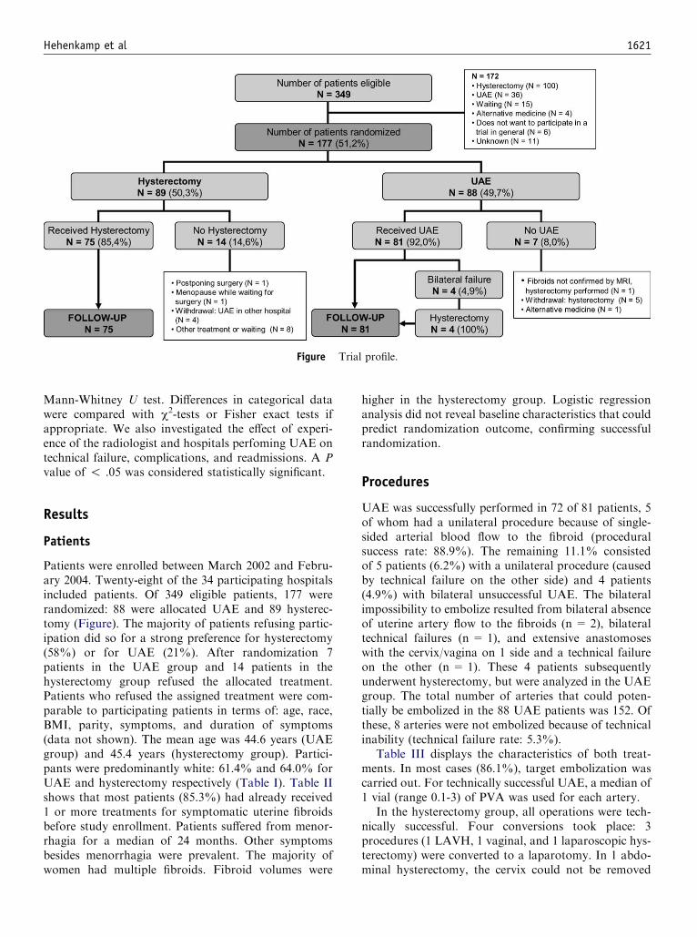
Hehenkamp et al 1621
Figure Trial profile.
Mann-Whitney U test. Differences in categorical datawere compared with c2-tests or Fisher exact tests ifappropriate. We also investigated the effect of experi-ence of the radiologist and hospitals perfoming UAE ontechnical failure, complications, and readmissions. A Pvalue of ! .05 was considered statistically significant.
Results
Patients
Patients were enrolled between March 2002 and Febru-ary 2004. Twenty-eight of the 34 participating hospitalsincluded patients. Of 349 eligible patients, 177 wererandomized: 88 were allocated UAE and 89 hysterec-tomy (Figure). The majority of patients refusing partic-ipation did so for a strong preference for hysterectomy(58%) or for UAE (21%). After randomization 7patients in the UAE group and 14 patients in thehysterectomy group refused the allocated treatment.Patients who refused the assigned treatment were com-parable to participating patients in terms of: age, race,BMI, parity, symptoms, and duration of symptoms(data not shown). The mean age was 44.6 years (UAEgroup) and 45.4 years (hysterectomy group). Partici-pants were predominantly white: 61.4% and 64.0% forUAE and hysterectomy respectively (Table I). Table IIshows that most patients (85.3%) had already received1 or more treatments for symptomatic uterine fibroidsbefore study enrollment. Patients suffered from menor-rhagia for a median of 24 months. Other symptomsbesides menorrhagia were prevalent. The majority ofwomen had multiple fibroids. Fibroid volumes were
higher in the hysterectomy group. Logistic regressionanalysis did not reveal baseline characteristics that couldpredict randomization outcome, confirming successfulrandomization.
Procedures
UAE was successfully performed in 72 of 81 patients, 5of whom had a unilateral procedure because of single-sided arterial blood flow to the fibroid (proceduralsuccess rate: 88.9%). The remaining 11.1% consistedof 5 patients (6.2%) with a unilateral procedure (causedby technical failure on the other side) and 4 patients(4.9%) with bilateral unsuccessful UAE. The bilateralimpossibility to embolize resulted from bilateral absenceof uterine artery flow to the fibroids (n = 2), bilateraltechnical failures (n = 1), and extensive anastomoseswith the cervix/vagina on 1 side and a technical failureon the other (n = 1). These 4 patients subsequentlyunderwent hysterectomy, but were analyzed in the UAEgroup. The total number of arteries that could poten-tially be embolized in the 88 UAE patients was 152. Ofthese, 8 arteries were not embolized because of technicalinability (technical failure rate: 5.3%).
Table III displays the characteristics of both treat-ments. In most cases (86.1%), target embolization wascarried out. For technically successful UAE, a median of1 vial (range 0.1-3) of PVA was used for each artery.
In the hysterectomy group, all operations were tech-nically successful. Four conversions took place: 3procedures (1 LAVH, 1 vaginal, and 1 laparoscopic hys-terectomy) were converted to a laparotomy. In 1 abdo-minal hysterectomy, the cervix could not be removed

1622 Hehenkamp et al
as planned because of adhesions, and a supravaginalhysterectomy was carried out instead. Furthermore, in1 vaginal hysterectomy, morcellation was necessary fora large fibroid. Most hysterectomies were performedtransabdominally (84.0%).
UAE procedures on average took shorter than hys-terectomy procedures (79.0 vs 95.4 minutes, P = .007).
Table I Baseline characteristics: patient demographics
UAE(n = 88)
Hysterectomy(n = 89)
No. (%) No. (%)
Age (y)! 35 1 (1.1) 0 (0)35-40 17 (19.3) 9 (10.1)40-45 28 (31.8) 29 (32.6)45-50 33 (37.5) 40 (44.9)O50 9 (10.2) 11 (12.4)Mean (SD) 44.6 (4.8) 45.4 (4.2)
Body mass index (weight [kg]/length [m2])!18.5 2 (2.3) 0 (0)18.5-24.9 33 (37.5) 44 (50)25-29.9 32 (36.4) 34 (38.6)O30 21 (23.9) 10 (11.4)Mean (SD) 26.7 (5.6) 25.4 (4.0)
Parity0 30 (34.1) 20 (22.5)R1 58 (65.9) 69 (77.5)
EthnicityBlack 24 (27.3) 20 (22.5)White 54 (61.4) 57 (64.0)Other 10 (11.4) 12 (13.5)
Marital statusSingle 16 (18.2) 13 (14.8)Married 55 (62.5) 54 (61.4)Living apart together 5 (5.7) 4 (4.5)Divorced 12 (13.6) 15 (17.0)Widow 0 (0) 2 (2.3)
Employment statusEmployed 68 (77.3) 69 (78.4)Unemployed 20 (22.7) 19 (21.6)
Smoking statusCurrent smoker 21 (23.9) 23 (25.8)Former smoker 11 (12.5) 14 (15.7)Nonsmoker 56 (63.6) 52 (58.4)
Highest educational level*Elementary school 3 (3.4) 6 (6.9)Lower vocational, lowersecondary school
29 (33.0) 32 (36.8)
Intermediate and highervocational, highersecondary school
26 (29.5) 27 (31.0)
College/University 28 (31.8) 22 (25.3)Other 2 (2.3) 0 (0)
Data were available for all or all but 1 patient, unless stated otherwise.
Logistic regression analysis did not reveal baseline characteristics that
could predict randomization outcome.
* Missing: 2
Patients subject to UAE had significantly less blood lossthan those undergoing hysterectomy (30.9 and 436.1mL, respectively; P ! .001). Total admission time wassignificantly (P! .001) shorter in the UAE group (mean2.0 days; SD 2.1; range 0-13 days) than in the hyster-ectomy group (mean 5.1 days; SD 1.3; range 2-8 days).
Complications during hospital stay
Table IV lists complications occurring during and afterthe procedures.
Intraprocedural complications were uncommon inboth groups. In the UAE group, 7 minor complicationsoccurred: 5 postpuncture hematomas, 1 blood clot in thegluteal artery, which resolved spontaneously, and 1 caseof nausea during the procedure. In the hysterectomygroup, 2 minor complications occurred: 1 allergic reac-tion to an anesthetic agent and 1 small tear in the rectusmuscle.
During hospital stay febrile morbidity was signifi-cantly less common in the UAE group (4.9%) than afterhysterectomy (20.0%; P = .006; RR 0.25; 95%CI 0.09-0.72). Postintervention fever occurred less frequently inpatients who received antibiotics for both the hysterec-tomy (16.4% vs 50.0%; P = .046; RR 0.33; 95%CI0.14-0.79) and UAE group (3.4% vs 5.8%; P = .99(FE); RR 0.60; 95%CI 0.07-5.49).
Hematomas occurred significantly more frequentlyafter UAE, while the hysterectomy group experiencedmore urinary tract infections and urinary retention. Nopatients in the UAE group required a blood transfusion,compared with 10 patients (13.3%) in the hysterectomygroup. The minor complication rates were 22.2%(95%CI 13.7-32.8) in the UAE group and 30.7%(95%CI 20.5-42.4) in the hysterectomy group (RR0.72; 95%CI 0.43-1.23; P = .23). Major complicationswere rare and concerned 2 cases of pulmonary embo-lisms, 1 in each group. The major complication rate was1.2% (95%CI 0.03-7.2) and 1.3% (95%CI 0.03-7.2) forUAE and hysterectomy respectively (RR 0.93; 95%CI0.06-14.54; P= .99). Both minor and major complicationrates did not differ significantly between the 2 groups.
Follow-up
Table V describes the unscheduled visits within the first6 weeks after discharge. In the UAE group, 30 patients(37.0% with a total of 46 visits) consulted a physician,mainly for pain and/or fever. In the hysterectomy group,19 patients (25.3% with a total of 24 visits) consulted aphysician after discharge for various reasons. Thisdifference was not significant (RR 1.45; 95%CI 0.90-2.37, P = .12).
Readmissions (Table VI) were significantly morecommon in the UAE group: 9 patients versus 0 patientsin the hysterectomy group (P = .0032). In the UAE

Hehenkamp et al 1623
Table II Baseline characteristics: symptoms, previous treatment and uterus/fibroid characteristics
UAE (n = 88) Hysterectomy (n = 89)No. (%) No. (%)
Previous treatmentNone 11 (12.5) 15 (16.9)Hormonal 59 (67.0) 59 (66.3)Nonsteroidal anti-inflammatory drugs/tranexaminacid 45 (51.1) 41 (46.1)Iron-supplement/blood transfusion 50 (56.8) 52 (58.4)Surgical procedures* 17 (19.3) 11 (12.4)Hysteroscopic myomectomy 6 (6.8) 8 (9.0)Laparoscopic myomectomy 0 (0) 3 (3.4)Laparotomic myomectomy 7 (8.0) 2 (2.2)Hysteroscopic endometrium resection 3 (3.4) 1(1.1)Curettage 3 (3.4) 0 (0)
SymptomsMenorrhagia 88 (100) 89 (100)Dysmenorrhea 47 (53.4) 50 (56.2)Pain (not during menstruation) 15 (17.0) 14 (15.7)Urinary symptoms 13 (14.8) 20 (22.5)Defecation problems 5 (5.7) 5 (5.6)Anemia 43 (48.9) 42 (47.2)Pressure symptoms 23 (26.1) 25 (28.1)Other symptoms 6 (6.8) 11 (12.4)
Duration of symptoms (m)Median (range) 24 (3-250) 24 (4-240)
Duration of menstruation (d)Total days (median, range) 7 (4-28) 8 (3-42)Heavy days (median, range) 3 (1-28) 4 (1-21)
Number of fibroidsy
1 35 (39.8) 25 (28.1)2 13 (14.8) 16 (18.0)3 17 (19.3) 25 (25.8)O3 18 (20.5) 14 (15.7)Median (range) 2 (1-20) 2 (1-9)
Uterine volume (cm3)z,k
0-250 33 (37.9) 26 (32.5)251-500 26 (29.9) 30 (37.5)501-1000 19 (21.8) 16 (20.0)O1000 9 (10.3) 8 (10.0)Median (range) 321 (31-3005) 313 (58-3617)
Fibroid volume (dominant fibroid, cm3)x,{
0-100 55 (63.2) 41 (52.6)101-200 14 (16.1) 20 (25.6)201-400 11 (12.6) 12 (15.4)O400 7 (8.9) 5 (6.4)Median (range) 59 (1-673) 87 (4-1641)
Number of fibroids and uterine/fibroid volume were calculated by ultrasound unless stated otherwise. Data were available for all or all but 1 patient,
unless stated otherwise. Logistic regression analysis did not reveal baseline characteristics that could predict randomization outcome.
* The surgical treatments do not add up because some patients had several treatments.y UAE missing: 5, hysterectomy missing: 11.z UAE missing: 1, hysterectomy missing: 9.x UAE missing: 1, hysterectomy missing: 11.k MRI measurements were used in 5 patients.{ 1 patient in the UAE group because of missing ultrasound data.
group, 7 of the 9 (77.8%) readmissions occurred withinthe first week after discharge from the hospital. Patientswere readmitted for pain (22.2%), fever (22.2%), or acombination of both (44.4%). One patient (11.1%) was
readmitted for expulsion of a necrotic fibroid. Hystero-scopic removal was attempted, but failed because ofcervical dilation which interfered with uterine dilatation.Antibiotics were administered intravenously and the

1624 Hehenkamp et al
patient stayed in the hospital until fever and pain hadsubsided. The mean admission time for UAE increasedfrom 2.0 to 2.5 days (SD 2.7; range 0-16 days) as a result
Table III Procedural characteristics
UAE(n = 81)
Hysterectomy(n = 75)
No. No.
Type of UAETarget embolization*Left uterine artery 65 –Right uterine artery 59 –
Selective embolization*Left uterine artery 8 –Right uterine artery 12 –
Type of hysterectomy (n = 4)Abdominal hysterectomy (2) 63Pfannenstiel incision (1) 50Median incision (1) 13
Vaginal hysterectomy (1) 8Vaginal hysterectomywith morcellator
(1) 1
LH with morcellator – 2LAVH – 1
CervixConservation of cervix (2) 22
Other proceduresRemoval of hydrosalpinx – 1Adhesiolysis (1) –Salpingo-oophorectomyUnilateral (1) 2Bilateral – 1
AnesthesiaLocal 71 –Epidural 9 1Spinal 1 (D1) 3General anesthesia (2) 52General and epidural (1) 17General and spinal – 2
Duration of procedure (min)Mean (SD) 79 (30.5)y;
(109 (59.2))95.4(30,9)y
Median (range) 75 (30-165);(90 (60-195))
90(45-175)
Blood loss (mL)Mean (SD) 30.9 (23.8)z;
(1000 (823.6))436.1(474.5)z
Median (range) 20 (5-150);(850 (300-2000))
300(10-2500)
AntibioticsAntibiotics administered 29 (35.8%);
(4 (100.0%))67(89.3%)
Abbreviations: LAVH, Laparoscopic-assisted vaginal hysterectomy; LH,
laparoscopic hysterectomy. Characteristics of hysterectomies per-
formed after bilaterally failed embolizations are presented in (bold)in the UAE column.
* For successful procedures.y P = .007, compared with hysterectomy group.z P ! .001, compared with hysterectomy group.
of readmissions, but remained significantly shortercompared with hysterectomy (P ! .001).
Complications and symptoms between dischargefrom the hospital and the first routine visit at 6 weeksare shown in Table IV. UAE patients complained ofvaginal discharge in 21.0% compared with 8.0% of thehysterectomy patients (P = .022). A percentage (14.8%)of UAE patients experienced vaginal loss of fibroidtissue. Hot flashes were present in 19.8% (UAE) and20.0% (hysterectomy) of patients. Four cases of painand/or fever that required readmission were classified asminor complications because the definition of majorcomplications which we used (as described in themethods section) did not apply here.
Three patients (3.7%) in the UAE group had majorcomplications: pneumonia in a patient with a history ofrecurrent pneumonia caused by asthmatic disease(n = 1); reintervention because of an incomplete fibroidexpulsion (n = 1); and septicemia (n = 1). One patient(1.3%) in the hysterectomy group was diagnosed with avesicovaginal fistula, which was surgically repaired be-yond the 6 weeks’ follow-up period (not reported inTable VI).
The minor complication rate in the first 6 weeks afterdischarge was significantly higher in the UAE groupthan in the hysterectomy group: 58.0% (95%CI 46.5-68.9) and 40.0% (95%CI 28.9-52.0), respectively (RR1.45; 95%CI 1.04-2.02; P = .024). The major complica-tion rate in the first 6 weeks after discharge was 3.7%(95%CI 0.8-10.4) and 1.3% (95%CI 0.03-7.2) for UAEand hysterectomy, respectively (RR 2.78; 95%CI 0.30-26.13; P = .62), and did not differ significantly.
The overall minor complication rate (ie, from theprocedure until the 6-week routine visit) was 64.2%(95%CI 52.8-74.6) (52 patients) in the UAE groupcompared with 56.0% (95%CI 44.1-67.4) (42 patients)in the hysterectomy group (RR 1.12; 95%CI 0.87-1.46;P = .38). The overall major complication rate was 4.9%(95%CI 1.4-12.2) (4 patients) in the UAE group com-pared with 2.7% (95%CI 0.3-9.3) (2 patients) in thehysterectomy group (RR 1.85; 95%CI 0.35-9.82;P = .68). Both findings were not statistically significant.Also, when only abdominal hysterectomies were com-pared with UAE, overall major and minor complicationrates did not differ significantly (P = .28 and P = .70).The difference in hospitalization time remained statisti-cally significant (P ! .001). Radiologists’ experiencewith UAE was not associated with the technical failurerate. Less experienced hospitals were not associated withhigher complication or readmission rates.
Comment
Present knowledge on UAE derives from numerousuncontrolled case series and only 1 small pre-consent

Hehenkamp et al 1625
Table IV Complications until the first scheduled visit (6 weeks after the procedure)
Hospital stay 6 weeks after discharge
UAEa
(n = 81)Hyst.(n = 75) Relative risk
UAEb
(n = 81)Hyst.(n = 75) Relative risk
Complication n n RR (95%CI) n n RR (95%CI)
GeneralNausea 52 42 1.15 (0.89-1.48) 25 11 2.10 (1.11-3.97)c
Pain 72 71 0.94 (0.85-1.03) 57 52 1.01 (0.83-1.25)Febrile morbidity (O38.5(C) 4 15 0.25 (0.09-0.71) 17 8 1.97 (0.90-4.29)
Minor complicationsVaginal discharge – – – 17 6 2.62 (1.09-6.30)d
Pain requiring readmission – – – 2 – N/APain/fever requiringreadmission
– – – 2 – N/A
Fibroid expulsion not requiringreintervention
– – – 12 – N/A
Hematoma 13 4 3.01 (1.03-8.82)e 3 2 1.39 (0.24-8.08)Wound abscess 1f 0 0 1 �Woundbleeding 1 1 0.93 (0.06-14.54) – – –Wound dehiscence 0 0 0 1 �Urinary tract infection 0 3 � 5g 2 2.31 (0.46-11.57)Urinary retention 0 3 � 1 1 0.93 (0.06-14.54)Urinary incontinence – – – 6 4 1.39 (0.41-4.73)Endometritis 0 – N/A 2 – N/AHot flashes – – – 16 15 0.99 (0.53-1.86)Anemia requiring transfusion 0 10 � – – –Hypertension 7h 1 6.48 (0.82-51.45) 0 1 �Hypotension 0 2 � – – –Other 1i 2j 0.46 (0.04-5.00) 2k 1l –Total 23 (in 18
patients)26 (in 23patients)
0.72 (0.43-1.23)P = .23
68 (in 47patients)
34 (in 30patients)
1.45 (1.04-2.02)P = .024
Major complicationsPneumonia 0 0 � 1 0 �Ileus 0 0 � 0 0 �Thrombosis 0 0 � 0 0 �Vesicovaginal fistula – – – 0 1 �Pulmonary embolism 1 1 0.93 (0.06-14.54) 0 0 �Intra-abdominal infection 0 0 � 0 0 �Sepsis 0 0 � 1m 0 �Fibroid expulsion requiringre-intervention
0 0 � 1n 0 �
Death 0 0 � 0 0Total 1 (in 1
patient)1 (in 1patient)
0.93 (0.06-14.54)P = .99
3 (in 3patients)
1 (in 1patient)
2.78 (0.30-26.13)P = .62
N/A, Not applicable.a The UAE group comprises both failed and successful embolizations.b Complications of patients with hysterectomies after failed embolizations are described for the 6 weeks after discharge after their hysterectomy
procedure.c P = .016.d P = .022.e P = .03.f Occurred in a hysterectomy performed after bilaterally failed UAE.g Complication led to a readmission in 3 patients.h Including 1 patient that was admitted to the medium care unit for extreme hypertension.i Spontaneous blood clot in gluteal artery during procedure.j Small tear of m. rectus abdominis during surgery, allergic reaction to anesthetic agent during surgery.k Gout attack, liquor spill after epidural anesthesia.l Headache after epidural anesthesia.m Complication led to readmission in 1 patient.n Readmission, attempt to remove necrotic fibroid hysteroscopically, which only partly succeeded.

1626 Hehenkamp et al
Table V Unscheduled visits after discharge until first routine visit (6 weeks after procedure)
UAE (n = 81) Hysterectomy (n = 76)Contact Symptom(s) Number of contacts Number of contacts
General physician Pain 3 3Fever 5 0Vaginal bleeding 0 3Groin hematoma 1 0Constipation 0 2Blood pressure issues 1 2Other 3* 4y
Total 13 (in 10 patients) 14 (in 12 patients)
Gynecologist Fever 4 0Fever and pain 7 0Fever and vaginal bleeding 0 1Pain 12 2Pain and vaginal discharge 2 1Pain and vaginal bleeding 3 0Vaginal bleeding 0 3Vaginal discharge 2 0Wound dehiscence 0 2z
Other 0 1x
Total 30 (in 22 patients) 10 (in 8 patients)
Lung specialist Fever, dyspnea: pneumonia 3 0Total 3 (in 1 patient) 0
Total number of visits 45 24Total number of patients 30k 19
* Checking hemoglobin level; gout attack; sensitive breast.y Coughing, dizzy, and constipation; stomach pain; urge incontinence complaints; vaginal itch.z Same patient.x Severe hair loss.k RR 1.45 (95%CI 0.90-2.37; P = .12), compared with number of patients in the hysterectomy group.
randomized trial of moderate quality.17 According tothe National Institute of Clinical Excellence (NICE), thelimitations of the available literature only allow tenta-tive conclusions about the safety and efficacy of UAE,18
especially since highly selected patient inclusion andhigh loss to follow-up bias the results. NICE stronglyrecommends the initiation of randomized controlledtrials, which is exactly what we did here.
We deliberately chose to compare UAE with hyster-ectomy, not with myomectomy,19 for several reasons.First, hysterectomy is the standard procedure of choiceto eliminate all fibroid-related complaints. In our view,myomectomy should be preserved for those women withsymptomatic uterine fibroids with a strong desire forfuture pregnancy. Because UAE is considered to becontraindicated for women desiring pregnancy, a ran-domized comparison with myomectomy might even beconsidered unethical at this stage.20 In the absence ofrandomized data, we judged it more ethical to perform astudy at the other end of the clinical spectrum, ie, inwomen facing hysterectomy as the last resort for theirfibroid-related complaints. Although hysterectomy is anabsolute cure for menorrhagia, possible sequalae, eg,
incontinence, vaginal vault prolapse, risk for prematureovarian failure, long recovery, high costs, and the desireof some patients to preserve their uterus, justify seriousconsideration of alternative therapies such as UAE. Theprocedural success rate (88.9%) was comparable to theresults of the aforementioned small semi-randomizedtrial, but lower than the success rates reported in mostother studies, thereby illustrating the necessity of ran-domized data collection.17
The technical failure rate (5.3%) was higher than the0.5% to 2.5% reported in large case series,2,3,5,21 butsimilar to the technical failure rate of 5.0% reported inthe only semi-randomized trial,17 because of severalpossible reasons. First, our study is a mix of bothacademic and nonteaching hospitals, whereas UAE inthe reported case series was mostly performed in highlyspecialized single-centers, decreasing the generalizabilityof those results.22 However, in our study, experience ofthe interventional radiologist was not associated withoutcome. Moreover, one series performed a secondembolization attempt after initial technical failure,which obviously improves technical success rates.4 Gen-erally, results from randomized controlled trials can

Hehenkamp et al 1627
Table VI Readmissions after UAE until first routine visit (6 weeks after the procedure)
Reason for readmission n CourseDays afterdischarge
Lengthof stay
Fever 1 Antibiotics administered, urine and cervixcultures positive for Streptococcus
4 3
1 Antibiotics administered for urinarytract infection
4 4
Fever and pain 1 Analgesics and antibiotics administeredfor urinary tract infection
3 5
2 Analgesics and antibiotics administered,no definite diagnosis
3 2
3 4Pain 2 Analgesics administered 1 1
4 1Septicemia 1 MRI scan revealed an infectious process
cranial in the uterus, which draineditself vaginally. Antibiotics andanalgesics administered
51 11
Myoma nascens 1 Failed attempt to hysteroscopically removenecrotic tissue due to cervix dilation.Antibiotics and analgesics administered.
23 6
No hysterectomy patients were readmitted to the hospital.
substantially differ from those in case series because ofpublication bias and patient selection criteria.23,24
Mean hospital stay was significantly shorter for UAEthan for hysterectomy. Mean hospital stay for UAE waslonger than in some studies, but in our experience mostpatients need more care.25,26 Therefore, we would notrecommend performing UAE as an outpatient proce-dure. Surprisingly, the number of unscheduled visits washigher in the UAE group. Readmission rates after UAEwithin the first 6 weeks (11.1%) were higher comparedwith other reports (2.9%-5.0%), although the reasonsfor readmission were similar.17,27 The experimentalstatus of the UAE procedure could be the reason whyphysicians were more inclined to see patients andreadmit them more quickly. Most readmissions in ourstudy occurred within the first week after discharge(77.8%), underlining the need for adequate follow-upduring this period. None of our hysterectomy patientswere readmitted within the first 6 weeks, while Pintoet al found a readmission rate of 5.0% after hysterec-tomy.17 Because most hysterectomy patients were still inthe hospital when most readmissions in the UAE groupoccurred, the comparison is not completely fair: if UAEpatients would stay in the hospital as long as hysterec-tomy patients, only 2 readmissions would have oc-curred.
Overall major and minor complication rates in bothgroups were comparable, but minor complications in theperiod between discharge and the first 6 weeks’ visitwere significantly higher in the UAE group. Our study,therefore, cannot support the suggestion made byothers28 that UAE has a lower complication rate than
hysterectomy, again stressing the need for randomizedstudies. A detailed comparison of complication rateswith other studies is hampered by the fact that variousstudies apply different classification systems for report-ing complications. Major complications were rare inour study. Although many series reported emergencyhysterectomy rates up to 1.3% within the first weeksafter UAE, no such procedures occurred in ourpatients.3,4,21,27
There are several limitations to our study. First, 21(11.9%) patients withdrew from the study after ran-domization before treatment. Their baseline character-istics, however, did not differ from those being treated.Second, given the low major complication rates, ourstudy size was too small to detect any difference in majorcomplication rates, and definite conclusions, therefore,cannot be drawn. In contrast, we did find differences inminor complication rates and length of hospital stay, solack of power was not an issue here. We used noobjective criteria for menorrhagia but relied on subjec-tive appreciations of our patients. By doing so, thegeneralizability of our findings is probably enhanced:included patients represent those seen in daily practicewhere the decision to perform a hysterectomy is notbased on objective measurements (eg, pictorial charts)either. We could not find any differences in majorcomplication rates between UAE and hysterectomy.Unsuccessful UAE procedures, however, seem to occurmore often than previously reported. Hospital stayis significantly shorter for UAE. The higher minorcomplication rate after discharge in the UAE group,as well as the readmission rates and unscheduled visits,

1628 Hehenkamp et al
emphasize the necessity for careful follow-up and clearinstructions to the patient. Although the study resultsare supportive for UAE, the question as to whetherUAE is a good alternative for hysterectomy depends onthe balance of efficacy, costs, and quality of life, and stillremains to be answered.
Acknowledgments
We would like to thank all participating patients,EMMY-trial group members, nurses, and other con-tributors who made the trial possible. The EMMY-trialparticipants and hospitals: J. Reekers, W. Ankum, M.Burger, G. Bonsel, Erwin Birnie, W. Hehenkamp, N.Volkers (Academic Medical Center, Amsterdam); S. deBlok, C. de Vries (Onze Lieve Vrouwe Gasthuis,Amsterdam); T. Salemans, G. Veldhuyzen van Zanten(Atrium Medical Centre, Heerlen); D. Tinga, T. Prins(Groningen University Hospital, Groningen); P.Sleijffers, M. Rutten (Bosch Medical Centre, DenBosch); M. Smeets, N. Aarts (Bronovo Hospital, TheHague); P. van der Moer, D. Vroegindeweij (MedicalCentre Rijnmond-Zuid, Rotterdam); F. Boekkooi, L.Lampmann (St. Elisabeth Hospital, Tilburg); G. Klei-verda, (Flevo Hospital, Almere); R. Dik, J. Marsman(Gooi-Noord Hospital, Laren); C. de Nooijer, I. Hen-driks, G. Guit (Kennemer Gasthuis, Haarlem); H.Ottervanger, H. van Overhagen, (Leyenburg Hospital,The Hague); A. Thurkow (St. Lucas/Andreas Hospital,Amsterdam); P. Donderwinkel, C. Holt (Martini Hos-pital, Groningen); A. Adriaanse, J. Wallis, (MedicalCenter Alkmaar, Alkmaar); J. Hirdes, J. Schutte, W. deRhoter (Medical Center Leeuwarden, Leeuwarden); P.Paaymans, R. Schepers-Bok (Hospital Midden-Twente,Hengelo); G. van Doorn, H. Franke, J. Krabbe, A.Huisman, (Medisch Spectrum Twente, Enschede); M.Hermans, R. Dallinga (Reinier de Graaf Gasthuis,Delft); F. Reijnders, J. Spithoven, (Slingeland Hospital,Doetichem); W. Jager, P. Veekmans, (St. Jans Gasthuis,Weert); P. van der Heijden, M. Veereschild, J. van denHout, (Twenteborg Hospital, Almelo); I. van Seumeren,A. Heinz, R. Lo, W. Mali (University Hospital Utrecht,Utrecht); J. Lind, Th. de Rooy (Westeinde Hospital,The Hague); M. Bulstra, F. Sanders (DiakonessenhuisUtrecht, Utrecht); J. Doornbos (De Heel Hospital,Zaandam); P. Dijkhuizen, M. van Kints (RijnstateHospital, Arnhem); Ph. Engelen, R. Heijboer (Sloter-vaart Hospital, Amsterdam).
References
1. Ravina JH, Herbreteau D, Ciraru-Vigneron N, Bouret JM,
Houdart E, Aymard A, et al. Arterial embolisation to treat uterine
myomata. Lancet 1995;346:671-2.
2. Pron G, Bennett J, Common A, Wall J, Asch M, Sniderman K.
The Ontario Uterine Fibroid Embolization Trial. Part 2. Uterine
fibroid reduction and symptom relief after uterine artery emboli-
zation for fibroids. Fertil Steril 2003;79:120-7.
3. McLucas B, Adler L, Perrella R. Uterine fibroid embolization:
nonsurgical treatment for symptomatic fibroids. J Am Coll Surg
2001;192:95-105.
4. Walker WJ, Pelage JP. Uterine artery embolisation for sympto-
matic fibroids: clinical results in 400 women with imaging follow
up. BJOG 2002;109:1262-72.
5. Spies JB, Ascher SA, Roth AR, Kim J, Levy EB, Gomez-Jorge J.
Uterine artery embolization for leiomyomata. Obstet Gynecol
2001;98:29-34.
6. Pelage JP, Soyer P, Le Dref O, Dahan H, Coumbaras J, Kardache
M, et al. Uterine arteries: bilateral catheterization with a single
femoral approach and a single 5-F catheterdtechnical note.
Radiology 1999;210:573-5.
7. Orsini LF, Salardi S, Pilu G, Bovicelli L, Cacciari E. Pelvic organs
in premenarcheal girls: real-time ultrasonography. Radiology
1984;153:113-6.
8. Ravina JH, Bouret JM, Ciraru-Vigneron N, Repiquet D, Herbre-
teau D, Aymard A, et al. [Recourse to particular arterial embo-
lization in the treatment of some uterine leiomyoma] Recours a
i’embolisation arterielle particulaire dans le traitement de certains
fibromyomes uterins. Bull Acad Natl Med 1997;181:233-43.
9. Bradley EA, Reidy JF, Forman RG, Jarosz J, Braude PR.
Transcatheter uterine artery embolisation to treat large uterine
fibroids. BJOG 1998;105:235-40.
10. Hovsepian DM, Siskin GP, Bonn J, Cardella JF, Clark TW,
Lampmann LE, et al. Quality improvement guidelines for uterine
artery embolization for symptomatic leiomyomata. Cardiovasc
Intervent Radiol 2004;27:307-13.
11. Bongers MY, Mol BW, Dijkhuizen FP, Brolmann HA. Is balloon
ablation as effective as endometrial electroresection in the treat-
ment of menorrhagia? J Laparoendosc Adv Surg Tech A
2000;10:85-92.
12. Loffer FD, Grainger D. Five-year follow-up of patients partici-
pating in a randomized trial of uterine balloon therapy versus
rollerball ablation for treatment of menorrhagia. J Am Assoc
Gynecol Laparosc 2002;9:429-35.
13. Worthington-Kirsch RL, Popky GL, Hutchins FL Jr. Uterine
arterial embolization for the management of leiomyomas: quality-
of-life assessment and clinical response. Radiology 1998;208:625-9.
14. Hutchins FL Jr, Worthington-Kirsch R, Berkowitz RP. Selective
uterine artery embolization as primary treatment for symptomatic
leiomyomata uteri. J Am Assoc Gynecol Laparosc 1999;6:279-84.
15. GoodwinSC,McLucasB,LeeM,ChenG,PerrellaR,VedanthamS,
et al. Uterine artery embolization for the treatment of uterine
leiomyomatamidtermresults. JVasc IntervRadiol 1999;10:1159-65.
16. Spies JB, Scialli AR, Jha RC, Imaoka I, Ascher SM, Fraga VM,
et al. Initial results from uterine fibroid embolization for sympto-
matic leiomyomata. J Vasc Interv Radiol 1999;10:1149-57.
17. Pinto I, Chimeno P, Romo A, Paul L, Haya J, De La Cal MA,
et al. Uterine fibroids: uterine artery embolization versus abdom-
inal hysterectomy for treatment a prospective, randomized, and
controlled clinical trial. Radiology 2003;226:425-31.
18. National Institute for Clinical Excellence. Uterine artery emboli-
sation for fibroids. NICE Interventional Procedures Guidance
No.1. London: National Institute for Clinical Excellence. Avail-
able from: www.nice.org.uk. Accessed August 12, 2003.
19. Manyonda IT, Sinthamoney E, Lotfallah H, Belli AM. Uterine
artery embolisation for symptomatic fibroids: clinical results in 400
women with imaging follow up. BJOG 2003;110:1139.
20. ACOG Committee Opinion. Uterine artery embolization. Obstet
Gynecol 2004;103:403-4.
21. Pelage JP, Le Dref O, Soyer P, Kardache M, Dahan H, Abitbol M,
et al. Fibroid-related menorrhagia: treatment with superselective
embolization of the uterine arteries and midterm follow-up.
Radiology 2000;215:428-31.

Hehenkamp et al 1629
22. Broder MS, Landow WJ, Goodwin SC, Brook RH, Sherbourne
CD, Harris K. An agenda for research into uterine artery embo-
lization: results of an expert panel conference. J Vasc Interv Radiol
2000;11:509-15.
23. McKee M, Britton A, Black N, McPherson K, Sanderson C, Bain
C. Methods in health services research. Interpreting the evidence:
choosing between randomised and non-randomised studies. BMJ
1999;319:312-5.
24. Kunz R, Oxman AD. The unpredictability paradox: review of
empirical comparisons of randomised and non-randomised clinical
trials. BMJ 1998;317:1185-90.
25. Pourrat XJ, Fourquet F, Guerif F, Viratelle N, Herbreteau D,
Marret H. Medico-economic approach to the management of
uterine myomas: a 6-month cost-effectiveness study of pelvic
embolization versus vaginal hysterectomy. Eur J Obstet Gynecol
Reprod Biol 2003;111:59-64.
26. Vashisht A, Studd JW, Carey AH, McCall J, Burn PR, Healy JC,
et al. Fibroid embolisation: a technique not without significant
complications. BJOG 2000;107:1166-70.
27. Spies JB, Spector A, Roth AR, Baker CM, Mauro L, Murphy-
Skrynarz K. Complications after uterine artery embolization for
leiomyomas. Obstet Gynecol 2002;100:873-80.
28. Spies JB, Cooper JM, Worthington-Kirsch R, Lipman JC, Mills
BB, Benenati JF. Outcome of uterine embolization and hysterec-
tomy for leiomyomas: results of a multicenter study. Am J Obstet
Gynecol 2004;191:22-31.

American Journal of Obstetrics and Gynecology (2005) 193, 1630–9
www.ajog.org
Ovarian cancer screening in the Prostate, Lung,Colorectal and Ovarian (PLCO) cancer screening trial:Findings from the initial screen of a randomized trial
Saundra S. Buys, MD,a,* Edward Partridge, MD,b Mark H. Greene, MD,c
Philip C. Prorok, PhD,d Douglas Reding, MD,e Thomas L. Riley,f
Patricia Hartge, ScD,g Richard M. Fagerstrom, PhD,d Lawrence R. Ragard, MD,h
David Chia, PhD,i Grant Izmirlian, PhD,d Mona Fouad, MD,b
Christine C. Johnson, PhD,j John K. Gohagan, PhD,k for the PLCO Project Team
Huntsman Cancer Institute at the University of Utah,a Salt Lake City, UT; University of Alabama at Birmingham,b
Birmingham, AL; Clinical Genetics Branch, Division of Cancer Epidemiology and Genetics,c Biometry ResearchGroup, Division of Cancer Prevention,d National Cancer Institute (NCI), National Institutes of Health (NIH),Department of Health and Human Services (DHHS), Bethesda, MD; Marshfield Clinic Research Foundation,e
Marshfield, WI; Information Management Services, Inc,f Rockville, MD; Epidemiology and Biostatistics Program,Division of Cancer Epidemiology and Genetics, NCI, NIH, DHHS,g Bethesda, MD; Westat,h Rockville, MD; UCLAImmunogenetics Center,i Los Angeles, CA; Josephine Ford Cancer Center, Henry Ford Health System,j Detroit, MI;Early Detection Research Group, Division of Cancer Prevention, NCI, NIH, DHHS,k Bethesda, MD
Received for publication November 3, 2004; revised April 2, 2005; accepted May 2, 2005
KEY WORDSOvarian cancerScreeningTransvaginal
ultrasoundCA-125
Objective: Ovarian cancer screening with transvaginal ultrasound (TVU) and CA-125 wasevaluated in the Prostate, Lung, Colorectal and Ovarian (PLCO) Trial.
Study design: This was a randomized controlled trial of screening versus usual care. Baselinescreening results are reported.Results: Of 39,115 women randomized to receive screening, 28,816 received at least 1 test. Abnor-mal TVUwas found in 1338 (4.7%), and abnormal CA-125 in 402 (1.4%). Twenty-nine neoplasms
were identified (26 ovarian, 2 fallopian, and 1 primary peritoneal neoplasm). Nine were tumors oflow malignant potential and 20 were invasive. The positive predictive value for invasive cancer was3.7% for an abnormalCA-125, 1.0% for an abnormal TVU, and 23.5% if both tests were abnormal.
Conclusion: The effect of screening on ovarian cancer mortality in the PLCO cohort has yet to beevaluated and will require longer follow-up. Screening identified both early- and late-stageneoplasms, and the predictive value of both tests was relatively low.
� 2005 Mosby, Inc. All rights reserved.
Supported by individual contracts from the National Cancer Institute to each of the 10 screening centers and to the coordinating center.
* Reprint requests: Saundra Buys, MD, University of Utah, 2000 Circle of Hope, Suite 2100, Salt Lake City, UT 84112-5550.
E-mail: [email protected]
0002-9378/$ - see front matter � 2005 Mosby, Inc. All rights reserved.
doi:10.1016/j.ajog.2005.05.005
Buys et al 1631
Advances in the treatment of ovarian cancer in thepast decade have led to incremental improvements insurvival. For example, it is now known that initialsurgery by an experienced gynecologic oncologist in-creases the likelihood of optimal debulking and isassociated with an improved long-term survival rate.1-3
The introduction of platinum-based, multiagent chemo-therapy regimens, the more recent addition of thetaxanes into clinical practice, the widespread use ofsalvage chemotherapy for recurrent cancer, and theincreasing use of chemotherapy in less advanced stagedisease have all contributed to improved survival.4
Despite these advances, ovarian cancer remains a fataldisease for most women in whom it is diagnosed, and ithas the highest mortality rate of all the gynecologicmalignancies. Ovarian cancer is diagnosed in 25,400women and results in 14,300 deaths annually in the US.5
Most cases present at an advanced stage, and long-termsurvival is achieved in less than a third of patients.However, early stage ovarian cancer has a much highersurvival rate, and it is possible that early detectionthrough screening could significantly reduce mortality.
Screening for early disease is useful if the disease inquestion has a presymptomatic stage in which treatmentis more effective than treatment administered for symp-tomatic disease. Effective screening also requires theavailability of screening procedures acceptable to pa-tients that can be performed at a reasonable cost.Finally, screening tests must have a sensitivity highenough to detect a significant fraction of all existingcases in the population and, at the same time, asufficiently high specificity to avoid generating an exces-sive number of false-positive screens. False-positive testresults are a particular problem in diseases with lowprevalence in the target population and in diseases forwhich further evaluation of an abnormal screen oftenincludes an invasive surgical procedure. Both concernsare true for ovarian cancer. One method to address theproblem of balancing sensitivity and specificity is toutilize more than 1 screening test in combination, eitherin parallel or sequentially.
Bimanual palpation of the ovaries is widely used todetect ovarian pathology, but it is insensitive for detec-tion of early stage cancer, and its widespread applicationhas not resulted in a significant shift to earlier-stageovarian cancer.6 Both CA-125 and ultrasound, particu-larly transvaginal ultrasound (TVU), have been advo-cated as potential ovarian cancer screening modalities.TVU screening has generally been associated with a highrate of false-positive screens, leading to a large numberof surgical procedures that did not identify cancer.7-9
CA-125 is reported to be elevated in about half ofwomen with early-stage ovarian cancer, but it is alsoincreased in other cancers and in various nonmalignantconditions such as liver disease and heart failure.10-13
Studies to date have not clarified the efficacy of TVU
and CA-125, performed either separately or together,for ovarian cancer screening.14 In this report, we de-scribe the characteristics of the 39,115 women random-ized to the screened arm of the Prostate, Lung,Colorectal and Ovarian (PLCO) Cancer Screening Trialand report the results of the initial (baseline) ovariancancer screening examinations in 28,816 women. TVUand CA-125 were performed at study entry and wereadministered concurrently.
Material and methods
Study design
The design of the PLCO Trial has been described indetail elsewhere.15 Briefly, the objectives are to deter-mine in healthy subjects aged 55-74 at entry whether:1) screening with flexible sigmoidoscopy can reducemortality from colorectal cancer in males and females;2) screening with chest x-ray can reduce mortality fromlung cancer in males and females; 3) screening withdigital rectal examination plus serum prostate specificantigen (PSA) can reduce mortality from prostate can-cer; and 4) screening with CA-125 and transvaginalultrasound can reduce mortality from ovarian cancer.
The study is a 2-armed trial in which half of subjectswere randomized to receive screening, and half to usualcare. Enrollment was initiated in the fall of 1993 andcompleted in the summer of 2001. Ten screening centersare participating: the University of Colorado HealthSciences Center; Lombardi Cancer Research Center ofGeorgetown University; Pacific Health Research Insti-tute, Honolulu; Henry Ford Health System; Universityof Minnesota School of Public Health/Virginia L. PiperCancer Institute; Washington University School ofMedicine; University of Pittsburgh, Pittsburgh CancerInstitute and Magee-Women’s Hospital; University ofUtah Health Sciences Center; Marshfield Clinic Re-search Foundation; and the University of Alabama atBirmingham. Each institution obtained local Institu-tional Review Board approval to carry out the study.CA-125 and PSA testing is performed centrally at theUniversity of California, Los Angeles ImmunogeneticsCenter. A biorepository for the collection and storage ofblood samples and tissue is an integral component of thetrial. Participants will be followed for at least 13 yearsfrom entry.
Women in the intervention arm are screened forovarian cancer using CA-125 annually for 6 years andTVU annually for 4 years. Both studies are performedconcurrently at entry into the study. Baseline ovariancancer screening tests were performed on the firstrandomized subject on November 15, 1993 and on thelast subject on December 13, 2001. The screeningprotocol originally included bimanual physical examof the ovaries as one of the screening modalities. This

1632 Buys et al
procedure was dropped in 1998 after review of the datadetermined that no ovarian cancers had been detectedwith this modality alone. This experience is consistentwith previous reports suggesting that pelvic examinationis not a satisfactory screening tool for ovarian cancer.6
In addition, more than two thirds of all women enteringthe study had routine bimanual pelvic examinations aspart of their ongoing medical care, thus compromisingour ability to compare the impact of bimanual exami-nation on ovarian cancer mortality between the screenedand unscreened arms.
Eligibility
The target population for the study included subjectsfrom 55 to 74 years of age who had not been diagnosedpreviously with prostate, lung, colorectal, or ovariancancer. Criteria for exclusion included current treatmentfor cancer other than basal cell and squamous cell skincancer, and enrollment in another cancer screening orprevention trial. Beginning on April 15, 1995, individ-uals who had received a colonoscopy, sigmoidoscopy, orbarium enema in the past 3 years were excluded. Womenon tamoxifen were initially excluded because, at the timeof study initiation, the relationship between tamoxifenand ovarian cancer risk was unknown. This restrictionwas lifted in April 1999 when it became clear that thiswas a null association.16 Individuals with previoussurgical removal of 1 lung or the entire colon werealso excluded. Initially, women who had undergoneoophorectomy were ineligible but in 1996 this restrictionwas lifted because low accrual of women threatened tojeopardize screening end points for lung and coloncancer. Women who had undergone oophorectomywere not offered either ovarian cancer screening test.
Screening procedures
TVU was performed by qualified sonographers using a5-7.5 MHz transvaginal probe. The examiner imagedboth ovaries in the transverse and longitudinal planes.At least 5 minutes were spent looking for each ovary toensure an adequate search; however, if the iliac vesselswere visualized without ovaries being seen the examinerconcluded the search for the ovaries.17 Ovaries weremeasured along the major and minor axes in bothtransverse and longitudinal planes, and the prolateellipsoid formula (width ! height ! thickness !0.523) was used to calculate the volume of each ovaryand/or cyst. The following TVU test results were clas-sified as abnormal (positive): ovarian volume O10 cm3;cyst volume O10 cm3; any solid area or papillaryprojection extending into the cavity of a cystic ovariantumor of any size; or any mixed (solid/cystic) compo-nent within a cystic ovarian tumor.
CA-125 was measured on serum obtained and frozenwithin 2 hours of blood draw at each of the 10 screening
centers, then shipped to the UCLA ImmunogeneticsCenter on dry ice. Samples were stored at �70(C andaliquots thawed for assay. Assays were performed ac-cording to the instructions from the manufacturer (Cen-tocor, Inc, Malvern, PA). Samples were run in duplicate.Any sample with a result over 35U/mLwas reanalyzed toverify the value. Samples showing discrepant resultsbetween duplicate results (coefficient of variation over10%) were reanalyzed. Quality assurance for the mea-surement of CA-125 was done in accordance with themanufacturer’s suggested protocol using manufacturer-supplied samples, as well as additional control samplesobtained from Bio-Rad (Hercules, CA). The assayprecision is represented by its coefficient of variation(CV). The CVs (and 95% CIs) were found to be 4.07%(3.92-4.22) at the lower concentration of 52.7 U/mL and3.78% (3.64-3.92) at the higher concentration of 106.5U/mL. These results are in good agreement with thosereported by the manufacturers on the product inserts.
The original Centocor CA-125 radioimmunoassay(RIA) assay was replaced with the Centocor CA-125IIRIA assay on October 1, 1995. All samples tested usingthe original CA-125 assay were retested using theCA-125II assay. Of 5371 samples analyzed with bothtests, the mean value rose from 9.4 to 13.1 U/mL. Onehundred and twenty-two samples changed screeningresults, with 109 converting from negative to positivewith the new assay (2.0%). Three of these subjects werediagnosed with cancer after the baseline screen; all 3 hadan abnormal TVU. Four of the other 106 subjects alsohad an abnormal TVU but did not have ovarian cancer.Thirteen samples converted from positive to negative(0.24%). None of these subjects were diagnosed withovarian cancer. The original CA-125 had a positivityrate of 0.6%, and the CA-125II assay had a positivityrate of 2.4% in these 5371 samples.
CA-125 results R35 U/mL were classified as abnor-mal (positive).
Results of both screening tests were sent to partici-pants and their personal physicians within 3 weeks ofspecimen submission. Evaluation and follow-up ofwomen with an abnormal screening test were at thediscretion of the participant’s physician.
Follow-up of abnormal screening tests
Medical records of all procedures done to evaluate anabnormal screen were obtained by study personnel andrecorded on standardized reporting forms. Pathologyreports from all ovarian neoplasms were abstracted bytrained certified tumor registrars and were reviewed byone of the authors (E.P.). Neoplasms with an ICD-O-2behavior code of 3 (malignant neoplasms) and diag-nosed in the screened arm within 12 months of a positivebaseline screen are included in this report. (The NCI’sSurveillance, Epidemiology and End Results Program
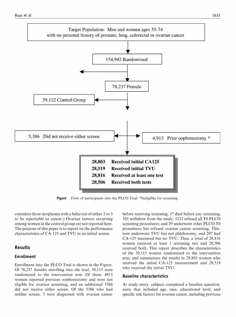
Buys et al 1633
Figure Flow of participants into the PLCO Trial. *Ineligible for screening.
considers those neoplasms with a behavior of either 2 or 3to be reportable as cancer.) Ovarian tumors occurringamong women in the control group are not reported here.The purpose of this paper is to report on the performancecharacteristics of CA-125 and TVU in an initial screen.
Results
Enrollment
Enrollment into the PLCO Trial is shown in the Figure.Of 78,237 females enrolling into the trial, 39,115 wererandomized to the intervention arm. Of these, 4913women reported previous oophorectomy and were noteligible for ovarian screening, and an additional 5386did not receive either screen. Of the 5386 who hadneither screen, 3 were diagnosed with ovarian cancer
before receiving screening; 17 died before any screening;105 withdrew from the study; 5232 refused all T0 PLCOscreening procedures; and 29 underwent other PLCO T0procedures but refused ovarian cancer screening. Thir-teen underwent TVU but not phlebotomy, and 297 hadCA-125 measured but no TVU. Thus, a total of 28,816women received at least 1 screening test and 28,506received both. This report describes the characteristicsof the 39,115 women randomized to the interventionarm, and summarizes the results in 28,803 women whoreceived the initial CA-125 measurement and 28,519who received the initial TVU.
Baseline characteristics
At study entry, subjects completed a baseline question-naire that included age, race, educational level, andspecific risk factors for ovarian cancer, including previous

1634 Buys et al
Table I Characteristics of participants in the intervention arm
Screened Not screened Previous oophorectomy
Number % Number % Number %
Age group55–59 9974 34.6 1698 31.5 1787 36.460–64 8871 30.8 1533 28.5 1370 27.965–69 6242 21.7 1251 23.2 1095 22.370–74 3729 12.9 904 16.8 661 13.5
RaceWhite 25,618 88.9 3868 71.8 4342 88.4Black 1533 5.3 356 6.6 281 5.7Hispanic 417 1.4 93 1.7 95 1.9Asian 984 3.4 159 3.0 116 2.4Other 200 0.7 51 0.9 32 0.7Missing response 64 0.2 859 15.9 47 1.0
Education level!High school 1685 5.8 483 9.0 352 7.212 y/completed high school 11,427 39.7 1866 34.6 2006 40.8Some college 6587 22.9 1086 20.2 1238 25.2College graduate 4624 16.0 545 10.1 659 13.4Postgraduate 4405 15.3 523 9.7 609 12.4Missing response 88 0.3 883 16.4 49 1.0
Had previous pelvic surgeryBilateral oophorectomy 2 0.0 3 0.1 59 1.2Hysterectomy 7768 27.0 1098 20.4 199 4.1Bilateral oophorectomy and hysterectomy 83 0.3 105 1.9 4601 93.6Neither 20,811 72.2 3295 61.2 8 0.2Missing response 152 0.5 885 16.4 46 0.9
Ever taken oral contraceptivesNo 13,168 45.7 2327 43.2 2127 43.3Yes 15,554 54.0 2175 40.4 2736 55.7Missing response 94 0.3 884 16.4 50 1.0
Number of times pregnantNone 2084 7.2 384 7.1 371 7.61 1640 5.7 253 4.7 309 6.32 5091 17.7 782 14.5 887 18.13-4 11,729 40.7 1738 32.3 1988 40.55-9 7625 26.5 1244 23.1 1232 25.110 or more 539 1.9 114 2.1 72 1.5Missing response 108 0.4 871 16.2 54 1.1
Personal history of breast cancerNo 27,718 96.2 4381 81.3 4718 96.0Yes 1040 3.6 148 2.7 150 3.1Missing response 58 0.2 857 15.9 45 0.9
Family history of breast and ovarian cancerNeither 23,517 81.6 3732 69.3 3964 80.7Breast cancer 3623 12.6 547 10.2 629 12.8Ovarian cancer 948 3.3 128 2.4 175 3.6Breast and ovarian cancer 120 0.4 13 0.2 30 0.6Missing response 608 2.1 966 17.9 115 2.3
Had baseline questionnaireNo 54 0.2 855 15.9 45 0.9Yes 28,762 99.8 4531 84.1 4868 99.1
Total 28,816 100.0 5386 100.0 4913 100.0
gynecologic surgery, use of oral contraceptives, parity,personal history of breast cancer, and family history ofbreast and ovarian cancer in first-degree relatives. Data
from the baseline questionnaire are shown for the 39,115women randomized to the intervention arm (Table I).Table I is divided into 3 groups: those who received any

Buys et al 1635
baseline ovarian cancer screening; those who did notreceive either baseline ovarian cancer screen; and thosewho were ineligible for screening because of previousoophorectomy. Women provided information aboutprevious oophorectomy both as part of the baselinequestionnaire (‘‘had previous pelvic surgery’’ on Table I)and at the time ovarian cancer screening was scheduled.Women who at that time reported that they were noteligible for ovarian cancer screening because of previousoophorectomy (‘‘previous oophorectomy’’ on Table I)did not receive ovarian cancer screening regardless oftheir report on the baseline questionnaire; similarly,women who reported no previous oophorectomy re-ceived screening despite their conflicting baseline ques-tionnaire report. For this reason there are someinconsistencies in Table I. Women in the ‘‘not screened’’group were on average older than those in the other 2groups and were less educated. They also had a higherrate of noncompliance with the baseline questionnaire(15.9%) than women in the screened (0.2%) or oopho-rectomy (0.9%) groups. Enrollment declined with ad-vancing age, with subjects aged 55 to 59, 60 to 64, 65 to69, and 70 to 74 years comprising 34.4%, 30.1%, 22.0%,and 13.5% of participants, respectively. Subjects wereprimarily white (86.5%) and educated (over 50%reported having at least some college education). Al-though some women did not complete the baselinequestionnaire or did not answer questions about gyne-cologic surgery, data were collected from over 97% ofthe participants.
Compliance
Compliance rates for the baseline screening procedureswere 83.4% for TVU and 84.2% for CA-125. The rateof compliance with each screening test declined slightlywith advancing age; among subjects 55 to 59 and 70to 74 years of age, compliance rates were 84.5% and79.5%, respectively, for TVU, and 85.5% and 80.5%,respectively, for CA-125. Women who did not have ascreen because of previous oophorectomy were excludedfrom the compliance calculations.
Table II CA-125 and TVU screening results combinations
TVU
CA-125 C � Total
# 34 365 399C % 0.1 1.3 1.4
# 1304 26,803 28,107� % 4.6 94.0 98.6
# 1338 27,168 28,506Total % 4.7 95.3 100.0
Only women having results for both TVU and CA-125 appear in this
table. Three hundred and ten subjects were excluded from table
because they had only 1 of the screening tests.
Screening results
The baseline TVU was abnormal in 1338 (4.7%) of28,519 baseline examinations. As previously reported,18
the ability of the examiner to visualize the ovaries byTVU decreased with advancing age of the participant,but age otherwise had no effect on TVU results (data notshown). A small number of examinations (1.9%) wereclassified as inadequate for interpretation.
Abnormal values for CA-125 were found in 402(1.4%) of 28,803 baseline measurements. Age had aminimal effect on CA-125 concentration. Three fourthsof the CA-125 values in each age group fell between5 and 15 U/mL. A small number of samples (0.3%)contained insufficient serum to perform the assay.
At the individual level, there was very little overlap inabnormal results for the 2 screening tests (Table II).Among 28,506 women with results for both tests, 1703had at least 1 abnormal test; 1338 had an abnormalTVU, 399 had an abnormal CA-125, and only 34 (2% ofthose with an abnormal screen) had abnormalities inboth.
As a result of the baseline screening examination andsubsequent follow-up procedures, 29 malignant neo-plasms were identified (Table III). Twenty-six arose inthe ovary, 9 of which were tumors of low malignantpotential, 1 was an ovarian stromal tumor (granulosacell), and 16 were invasive epithelial ovarian cancers.Three additional epithelial neoplasms were identified, 2arising in the fallopian tube and 1 in the peritoneum. All29 are designated ‘‘ovarian neoplasms’’ for subsequentanalyses.
Table III shows the relationship between CA-125 andTVU results and ovarian neoplasms. Invasive cancersand tumors of low malignant potential are shownseparately. CA-125 alone was abnormal in 7 (24%),and TVU alone was abnormal in 13 (45%). Both testswere abnormal in 9 subjects (31%).
An additional 25 subjects had abnormalities in bothTVU and CA-125 during the initial examination. Sev-enteen of these had surgery with benign findings, and the
Table III CA-125 and TVU results for confirmed neoplasms
Neoplasm type CA-125 TVU # %
Low MalignantPotential
C C 1 3.4
� C 8 27.6Granulosa � C 1 3.4Invasive Ovarian C C 7 24.1
� C 4 13.8C � 5 17.2
Fallopian Tube C C 1 3.4C � 1 3.4
Peritoneal C � 1 3.4Total 29 100.0

1636 Buys et al
Table IV Histopathologic type, histopathologic grade, and stage by screening results
Primary site Invasive Histopathology Grade Stage Screen resultsCA-125result
Days betweenpos screenand diagnosis
Ovary No Serous cystadenoma LMP* IA TVU positive 15 221Ovary No Serous cystadenoma LMP* IA CA-125 & TVU C 51 79Ovary No Serous cystadenoma LMP* IA TVU positive 18 55Ovary No Serous cystadenoma LMP* IA TVU positive 26 30Ovary No Serous cystadenoma LMP* IA TVU positive 8 91Ovary No Serous cystadenoma LMP* IA TVU positive 15 39Ovary No Mucinous cystadenoma LMP* IA TVU positive 17 46Ovary No Serous cystadenoma LMP* IC TVU positive 28 23Ovary No Serous cystadenoma LMP* IC TVU positive 26 80Ovary Yes Malignant granulosa Grade III IA TVU positive 8 316Ovary Yes Serous cystadenocarcinoma Grade III IB CA-125 & TVU C 793 40Ovary Yes Serous cystadenocarcinoma Grade II IIA TVU positive 13 213Ovary Yes Papillary serous & endometrial Grade III IIC CA-125 & TVU C 456 42Ovary Yes Endometrioid adenocarcinoma Grade III IIIA TVU positive 15 157Ovary Yes Serous cystadenocarcinoma Grade III IIIB TVU positive 9 72Ovary Yes Adenocarcinoma, NOS Grade III IIIB CA-125 positive 1260 49Ovary Yes Serous cystadenocarcinoma Grade III IIIC CA-125 & TVU C 1556 65Ovary Yes Serous cystadenocarcinoma Grade III IIIC CA-125 positive 2182 76Ovary Yes Serous cystadenocarcinoma Grade II IIIC CA-125 & TVU C 73 34Ovary Yes Serous cystadenocarcinoma Grade III IIIC CA-125 positive 406 49Ovary Yes Serous cystadenocarcinoma Grade III IIIC CA-125 positive 875 80Ovary Yes Adenocarcinoma Grade III IIIC CA-125 & TVU C 1143 104Ovary Yes Mucin-producing adenocarcinoma Grade IIIy IIIC CA-125 positive 78 44Ovary Yes Papillary adenocarcinoma Grade III IIIC CA-125 & TVU C 123 23Ovary Yes Carcinoma, NOS Grade III IIIC CA-125 & TVU C 410 59Ovary Yes Serous cystadenocarcinoma Grade III IV TVU positive 31 237Fallopian Tube Yes Endometrioid adenocarcinoma Grade III IIB CA-125 & TVU C 96 21Fallopian Tube Yes Papillary adenocarcinoma, NOS Grade III IV CA-125 positive 85 96Peritoneum Yes Carcinoma, NOS Grade III IIIC CA-125 positive 69 83
* Low malignant potential.y Grade determined from metastasis.
8 who did not have surgery were not diagnosed withcancer during the subsequent 12 months.
Table IV shows the characteristics of tumors detectedduring the baseline screen. The 9 tumors of low malig-nant potential were all stage I, as was the malignantgranulosa cell tumor. The stages of the 19 invasiveepithelial tumors included 1 stage I, 3 stage II, 13 stageIII, and 2 stage IV.
Table IV also shows the time interval betweenthe positive screening test and the diagnosis of cancer.The PLCO protocol allowed up to 3 weeks between thescreening test and results being sent to the physician.Further evaluation and follow-up resulted in a lengthydelay in surgery for some subjects; however, only 5 ofthe 29 cases had a delay of over 120 days from the initialscreen to surgery. These included a stage IA serouscystadenoma (221 days), a stage IA malignant granulosatumor (316 days), a stage IIA serous cystadeno-carcinoma (213 days), a stage IIIA endometrioid
adenocarcinoma (157 days), and a stage IV serouscystadenocarcinoma (237 days). In each of these casesthe TVU result was positive but the CA-125 was normal.This delay could have contributed to the high stage ofsome of the neoplasms, particularly the stage IV serouscystadenocarcinoma.
The positive predictive value (PPV) of each test, andof the 2 tests together, was calculated (Table V). Forexample, 28,803 women had a CA-125 test, 402 of whichwere abnormal (1.4%). Sixty-two women underwentbiopsy (15.4% of positive screens), 16 of which (25.8%of biopsies) were diagnosed with a neoplasm, for a PPVof 4.0% (16 neoplasms in 402 positive screens). Similarcalculations demonstrate a PPV of 1.6% for TVU, and26.5% if both tests were abnormal. Table V also showssimilar data excluding tumors of low malignant poten-tial (indicated as ‘‘# invasive cancer’’). With this restric-tion, the PPV was 3.7% for an abnormal CA-125, 1.0%for an abnormal TVU, and 23.5% if both tests were
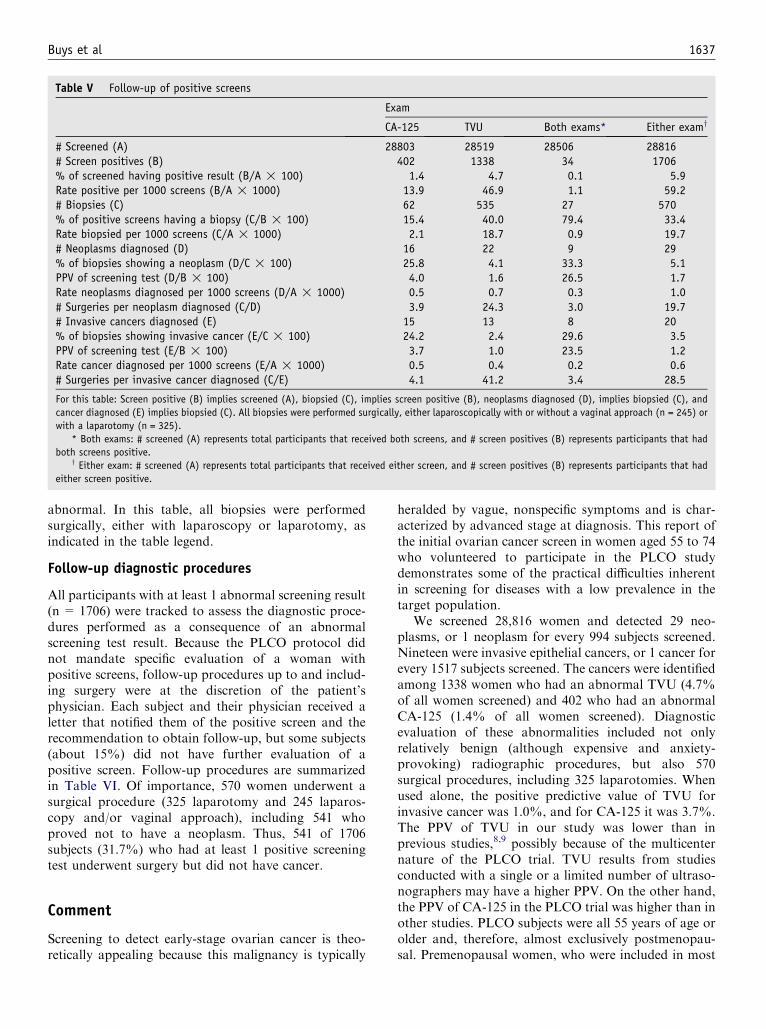
Buys et al 1637
Table V Follow-up of positive screens
Exam
CA-125 TVU Both exams* Either examy
# Screened (A) 28803 28519 28506 28816# Screen positives (B) 402 1338 34 1706% of screened having positive result (B/A ! 100) 1.4 4.7 0.1 5.9Rate positive per 1000 screens (B/A ! 1000) 13.9 46.9 1.1 59.2# Biopsies (C) 62 535 27 570% of positive screens having a biopsy (C/B ! 100) 15.4 40.0 79.4 33.4Rate biopsied per 1000 screens (C/A ! 1000) 2.1 18.7 0.9 19.7# Neoplasms diagnosed (D) 16 22 9 29% of biopsies showing a neoplasm (D/C ! 100) 25.8 4.1 33.3 5.1PPV of screening test (D/B ! 100) 4.0 1.6 26.5 1.7Rate neoplasms diagnosed per 1000 screens (D/A ! 1000) 0.5 0.7 0.3 1.0# Surgeries per neoplasm diagnosed (C/D) 3.9 24.3 3.0 19.7# Invasive cancers diagnosed (E) 15 13 8 20% of biopsies showing invasive cancer (E/C ! 100) 24.2 2.4 29.6 3.5PPV of screening test (E/B ! 100) 3.7 1.0 23.5 1.2Rate cancer diagnosed per 1000 screens (E/A ! 1000) 0.5 0.4 0.2 0.6# Surgeries per invasive cancer diagnosed (C/E) 4.1 41.2 3.4 28.5
For this table: Screen positive (B) implies screened (A), biopsied (C), implies screen positive (B), neoplasms diagnosed (D), implies biopsied (C), and
cancer diagnosed (E) implies biopsied (C). All biopsies were performed surgically, either laparoscopically with or without a vaginal approach (n = 245) or
with a laparotomy (n = 325).
* Both exams: # screened (A) represents total participants that received both screens, and # screen positives (B) represents participants that had
both screens positive.y Either exam: # screened (A) represents total participants that received either screen, and # screen positives (B) represents participants that had
either screen positive.
abnormal. In this table, all biopsies were performedsurgically, either with laparoscopy or laparotomy, asindicated in the table legend.
Follow-up diagnostic procedures
All participants with at least 1 abnormal screening result(n = 1706) were tracked to assess the diagnostic proce-dures performed as a consequence of an abnormalscreening test result. Because the PLCO protocol didnot mandate specific evaluation of a woman withpositive screens, follow-up procedures up to and includ-ing surgery were at the discretion of the patient’sphysician. Each subject and their physician received aletter that notified them of the positive screen and therecommendation to obtain follow-up, but some subjects(about 15%) did not have further evaluation of apositive screen. Follow-up procedures are summarizedin Table VI. Of importance, 570 women underwent asurgical procedure (325 laparotomy and 245 laparos-copy and/or vaginal approach), including 541 whoproved not to have a neoplasm. Thus, 541 of 1706subjects (31.7%) who had at least 1 positive screeningtest underwent surgery but did not have cancer.
Comment
Screening to detect early-stage ovarian cancer is theo-retically appealing because this malignancy is typically
heralded by vague, nonspecific symptoms and is char-acterized by advanced stage at diagnosis. This report ofthe initial ovarian cancer screen in women aged 55 to 74who volunteered to participate in the PLCO studydemonstrates some of the practical difficulties inherentin screening for diseases with a low prevalence in thetarget population.
We screened 28,816 women and detected 29 neo-plasms, or 1 neoplasm for every 994 subjects screened.Nineteen were invasive epithelial cancers, or 1 cancer forevery 1517 subjects screened. The cancers were identifiedamong 1338 women who had an abnormal TVU (4.7%of all women screened) and 402 who had an abnormalCA-125 (1.4% of all women screened). Diagnosticevaluation of these abnormalities included not onlyrelatively benign (although expensive and anxiety-provoking) radiographic procedures, but also 570surgical procedures, including 325 laparotomies. Whenused alone, the positive predictive value of TVU forinvasive cancer was 1.0%, and for CA-125 it was 3.7%.The PPV of TVU in our study was lower than inprevious studies,8,9 possibly because of the multicenternature of the PLCO trial. TVU results from studiesconducted with a single or a limited number of ultraso-nographers may have a higher PPV. On the other hand,the PPV of CA-125 in the PLCO trial was higher than inother studies. PLCO subjects were all 55 years of age orolder and, therefore, almost exclusively postmenopau-sal. Premenopausal women, who were included in most

1638 Buys et al
previous studies of screening, have a larger number offalse positive CA-125 values than older women. Notsurprisingly, an abnormal TVU resulted in a higher rateof surgeries (535 of 1338, or 40%) than an abnormalCA-125 (62 of 403, or 15.4%). Knowing that manyother (nonmalignant) conditions may influence CA-125levels, it is harder to justify performing surgery in anasymptomatic subject with an elevated CA-125 than in asimilar patient with an adnexal mass found on TVU.Abnormalities in both tests were found in only 34subjects, in whom the PPV for the 2 tests combinedwas 23.5%. However, if one chose to evaluate onlysubjects in whom both screening tests were abnormal, 20of the 29 ovarian neoplasms (and 12 of the 20 invasivecancers) would have been missed. We identified 1 neo-plasm for every 20 surgical procedures performed. Ifsurgery was done to evaluate an abnormal CA-125,1 neoplasm was identified per 3.9 surgeries (16 of 62),whereas 24 surgeries were required to identify 1 neo-plasm based on an abnormal TVU (22 of 535). Oneneoplasm was found per 3 surgeries (9 of 27) if bothTVU and CA-125 were abnormal.
Almost 2% of women who were screened at the timeof entry into the PLCO trial (570 of 28,816) underwentabdominal surgery in the course of evaluating abnor-malities detected by the PLCO ovarian cancer screeningstrategy. Several factors may have contributed to thehigh rate of pelvic surgery among the women in thisseries. 1) Many of the subjects may have had previouslyexisting indications for elective pelvic surgery (bladderdysfunction, etc), with the finding of an abnormal TVUor CA-125 simply being the final impetus for performingthe procedure. 2) Follow-up may have been particularlyaggressive because subjects were part of a nationalcancer screening study, and physicians may have felt
Table VI Participant-based diagnostic procedures followinga positive screen
No neoplasm Neoplasms
Diagnostic procedures n % n %
CA-125 377 22.5 12 41.4Ultrasounds 721 43.0 12 41.4Chest radiograph 68 4.1 7 24.1Surgery* 541 32.3 29 100.0CT scan/MRI 150 8.9 12 41.4Needle aspiration, culdocentesis,
or paracentesis22 1.3 1 3.4
IVP 8 0.5 � �Barium enema 7 0.4 � �No diagnostic procedure
recorded260 15.5 � �
Total number of participants 1677 100.0 29 100.0
* Two hundred ninety-eight of the surgeries of the participants
without neoplasms were a laparotomy; 27 of the surgeries of the
participants with neoplasms were a laparotomy.
that their medical decisions would be closely scrutinized.However as noted above, 15% of subjects with apositive screen had no follow-up. 3) Ovarian screeningwith TVU and CA-125 is not done routinely, and thefalse-positive rate is not as well defined as that for othermore common screening procedures. Because ovariancancer is notoriously a highly lethal disease, physiciansmay not have been comfortable with a ‘‘watch and wait’’approach to those with an abnormal screening test. 4)The definitive diagnostic test for an abnormal ovaryseen on TVU is oophorectomy. Percutaneous biopsy isnot utilized as routinely for diagnosis of ovarian massesas it is for many other organs, because of the risk offalse-negative studies and the theoretical possibility ofseeding the peritoneum with cancer cells if the biopsiedlesion proves to be malignant. 5) Hysterectomy with orwithout oophorectomy has been performed very com-monly in the past, particularly among women in thePLCO age range. By way of illustration, 35.6% ofwomen enrolling in the PLCO Trial (13,918 of 39,115;see Table I) had undergone hysterectomy and/or ooph-orectomy before joining the study. Additionally, ooph-orectomy can often be done by laparoscopy. Physicianstherefore may have had a low threshold for recommend-ing this common procedure.
In addition to the invasive tumors, 9 cystadenomas oflow malignant potential (‘‘borderline’’) tumors wereidentified, representing 31% of the total malignantneoplasms found. This entity typically comprises onlyabout 15% of all ovarian tumors as they presentclinically in the general population.19 While the numberof cancer cases in this report is small, this observationsuggests that screening as performed in the PLCO Trial,particularly with TVU, may preferentially detect low-grade tumors. This ‘‘length’’ bias (ie, slowly growingtumors are more likely than rapidly growing tumors tobe detected with screening) has the ultimate effect offinding neoplasms that might go undetected for thelifetime of the patient, and therefore, not contribute tocancer mortality (‘‘overdiagnosis’’). Using mortality asthe end point of a screening trial (as will be done for thePLCO study when follow-up is complete) eliminates thelength bias. The detection of ovarian tumors of lowmalignant potential is unlikely to affect ovarian cancermortality, since even stage III borderline tumors have a90% 10-year disease-free survival.20 Because only datafrom the initial (baseline) ovarian cancer screen havebeen analyzed thus far, the effect of repeated annualscreens on detection rates andmortality, as is the protocolin the PLCO Trial, is currently unknown. The predictivevalue of these tests may be improved if post hoc analysisidentifies a specific pattern of change over time in eitherTVUorCA-125 results, or if the combination of a specificTVU imaging pattern and a relatively high CA-125correlates with a greater likelihood of having ovariancancer. Fifteen of the 19 invasive neoplasms (79%) found

Buys et al 1639
with the initial (prevalent) screen were stage III or IVcancers. Now that these high-stage cancers have beenremoved from the screened population, it is possible thatsubsequent screens will identify a group of early stageovarian cancers with improved survival.
Further follow-up from the PLCO Trial will providevaluable data regarding the effect of annual screening onovarian cancer mortality. At the present time, nothing inthe findings reported here suggests a need to revise thepresent (1996) ovarian cancer screening guidelines of theUS Preventive Services Task Force,21 which state ‘‘rou-tine screening for ovarian cancer by ultrasound, themeasurement of serum tumor markers, or pelvic exam-ination is not recommended.’’
References
1. Junor EJ, Hole DJ, McNulty L, Mason M, Young J. Specialist
gynaecologists and survival outcome in ovarian cancer: A Scottish
national study of 1866 patients. BJOG 1999;106:1130-6.
2. Olaitan A, Weeks J, Mocroft A, Smith J, Howe K, Murdoch J.
The surgical management of women with ovarian cancer in the
south west of England. Br J Cancer 2001;85:1824-30.
3. Bristow RE, Tomacruz RS, Armstrong DK, Trimble EL, Montz
FJ. Survival effect of maximal cytoreductive surgery for advanced
ovarian carcinoma during the platinum era: A meta-analysis.
J Clin Oncol 2002;20:1248-59.
4. Ries LAG, Kosary CL, Hankey BF, Miller BA, Edwards BK,
editors. SEER cancer statistics review, 1973-1995. Bethesda, MD:
National Cancer Institute; 1998.
5. Jemal A, Murray T, Samuels A, Ghafoor A, Ward E, Thun MJ.
Cancer statistics, 2003. CA Cancer J Clin 2003;53:5-26.
6. Smith HL, Oi RH. Detection of malignant ovarian neoplasms:
a review of the literature. I. Detection of the patient at risk; clinical,
radiological and cytological detection. Obstet Gynecol Surv
1984;39:313-28.
7. DePriest PD, Gallion HH, Pavlik EJ, Kryscio RJ, van Nagell JR
Jr. Transvaginal sonography as a screening method for the
detection of early ovarian cancer. Gynecol Oncol 1997;65:408-14.
8. van Nagell JR Jr, DePriest PD, Reedy MB, Gallion HH, Ueland
FR, Pavlik EJ, et al. The efficacy of transvaginal sonographic
screening in asymptomatic women at risk for ovarian cancer.
Gynecol Oncol 2000;77:350-6.
9. Sato S, Yokoyama Y, Sakamoto T, Futagami M, Saito Y.
Usefulness of mass screening for ovarian carcinoma using trans-
vaginal ultrasonography. Cancer 2000;89:582-8.
10. Einhorn N, Sjovall K, Knapp RC, Hall P, Scully RE, Bast RC Jr,
et al. Prospective evaluation of serum CA 125 levels for early
detection of ovarian cancer. Obstet Gynecol 1992;80:14-8.
11. Jacobs IJ, Skates S, Davies AP, Woolas RP, Jeyerajah A,
Weidemann P, et al. Risk of diagnosis of ovarian cancer after
raised serum CA 125 concentration: a prospective cohort study.
BMJ 1996;313:1355-8.
12. Skates SJ, Menon U, MacDonald N, Rosenthal AN, Oram DH,
Knapp RC, et al. Calculation of the risk of ovarian cancer from
serial CA-125 values for preclinical detection in postmenopausal
women. J Clin Oncol 2003;21(Suppl 10):206-10.
13. D’Aloia A, Faggiano P, Aurigemma G, Bontempi L, Ruggeri G,
Metra M, et al. Serum levels of carbohydrate antigen 125 in
patients with chronic heart failure: relation to clinical severity,
hemodynamic and Doppler echocardiographic abnormalities, and
short-term prognosis. J Am Coll Cardiol 2003;41:1805-11.
14. Schapira MM, Matchar DB, Young MJ. The effectiveness of
ovarian cancer screening. A decision analysis model. Ann Intern
Med 1993;118:838-43.
15. Gohagan JK, Levin DL, Prorok PC, Sullivan D, editors. The
Prostate, Lung, Colorectal and Ovarian (PLCO) Cancer Screening
Trial. Control Clin Trials 2000;21(Suppl):249S-406S.
16. Fisher B, Costantino JP, Wickerham DL, Redmond CK, Kavanah
M, Cronin WM, et al. Tamoxifen for prevention of breast cancer:
report of the National Surgical Adjuvant Breast and Bowel Project
P-1 Study. J Natl Cancer Inst 1998;90:1371-88.
17. Prorok PC, Andriole GL, Bresalier RS, Buys SB, Chia D,
Crawford ED, et al. Design of the Prostate, Lung, Colon and
Ovarian (PLCO) cancer screening trial. Control Clin Trials
2000;21:273S-309S.
18. Hartge P, Hayes R, Reding D, Sherman ME, Prorok P, Shiffman
M, et al. Complex ovarian cysts in postmenopausal women are not
associated with ovarian cancer risk factors: preliminary data from
the Prostate, Lung, Colorectal and Ovarian Screening Trial. Am J
Obstet Gynecol 2000;183:1232-7.
19. Mink PJ, Sherman ME, Devesa SS. Incidence patterns of invasive
and borderline ovarian tumors among white women and black
women in the United States. Cancer 2002;95:2380-9.
20. Zanetta G, Rota S, Chiari S, Bonazzi C, Bratina G, Mangioni C.
Behavior of borderline tumors with particular interest to persis-
tence, recurrence, and progression to invasive carcinoma: a pro-
spective study. J Clin Oncol 2001;19:2658-64.
21. U.S. Preventive Services Task Force. Guide to clinical preventive
services, 2nd ed. Baltimore: Williams & Wilkins; 1996.

American Journal of Obstetrics and Gynecology (2005) 193, 1640–4
www.ajog.org
Endometrial cancer in women 45 years of age or younger:A clinicopathological analysis
Gilbert P. Pellerin, MD, Michael A. Finan, MD
Department of Obstetrics and Gynecology, Ochsner Clinic Foundation, New Orleans, LA
Received for publication November 4, 2004; revised April 6, 2005; accepted May 2, 2005
KEY WORDSEndometrial cancerAdenocarcinoma
endometriumUterine cancerEndometrial biopsy
Objective: The purpose of this study was to evaluate the experience with endometrial carcinoma
in women %45 years of age at Ochsner Clinic Foundation, New Orleans, La.Study design: We evaluated the clinical history, treatment, and follow-up of 38 women %45years of age diagnosed with endometrial cancer.
Results: Thirty-eight patients received primary treatment for endometrial cancer: stage I, 32(84.2%); stage II, 1 (2.6%); stage III, 4 (10.5%); stage IV, 1 (2.6%). Tumors were welldifferentiated in 20 (52.6%), moderately differentiated in 10 (26.3%), and poorly differentiated in8 (21.1%). At end of study period 32 women (84.2%) were alive with no evidence of disease, 5 had
died of recurrent disease, and 1 died of metastatic breast cancer.Conclusion: Patients %45 years of age had lower incidence of advanced stage disease, higherdegree of tumor differentiation, and better prognosis compared to patients older than 45 years.
� 2005 Mosby, Inc. All rights reserved.
Endometrial carcinoma is the most common malig-nancy of the lower female genital tract in the US.1
Approximately 40,880 new cases develop in the US eachyear, according to the year 2005 figures from theAmerican Cancer Society. This is about 1.3 times thefrequency of ovarian cancer and approximately twicethe number of new cases of cervical cancer.1 Overall,about 1 woman in 50 in the US will develop endometrialcarcinoma during her life.
Endometrial adenocarcinoma occurs during the re-productive and menopausal years. The median age foradenocarcinoma of the uterine corpus is 61 years; mostpatients are between the ages of 50 and 59 years.Approximately 5% of women will have adenocarcinoma
Abstract presented at the American College of Obstetricians and
Gynecologists district VII meeting, Washington, D.C., October 2004.
Reprints not available from the authors.
0002-9378/$ - see front matter � 2005 Mosby, Inc. All rights reserved.
doi:10.1016/j.ajog.2005.05.003
before age 40 years, and 20% to 25% will be diagnosedbefore menopause.2 Multiple factors increase the risk ofdeveloping endometrial carcinoma, including obesity,nulliparity, unopposed estrogen, and late menopause.Most young patients who develop endometrial cancerare obese, in many instances massively overweight, oftenwith anovulatory menstrual cycles. Combination oralcontraceptive use decreases the risk for development ofendometrial cancer.2
Postmenopausal bleeding and abnormal premeno-pausal bleeding are the primary symptoms of endome-trial carcinoma. All postmenopausal women withuterine bleeding should be evaluated for endometrialcancer. As a patient’s age increases after menopause, theprobability that uterine bleeding is caused by endome-trial cancer progressively increases.2 According to Feld-man et al, a woman O70 years has about a 50% chanceof having cancer when vaginal bleeding is present.3 A

Pellerin and Finan 1641
high index of suspicion must be maintained if thediagnosis of endometrial cancer is to be made in theyoung patient. Prolonged and heavy menstrual periods,and intermenstrual spotting, may indicate cancer, andendometrial sampling is advised. Most young patientswho develop endometrial cancer are obese, in manyinstances massively overweight, often with anovulatorymenstrual cycles.
Adenocarcinoma, the most common histologic typeof endometrial cancer, is sometimes preceded by apredisposing lesion: atypical endometrial hyperplasia.Patients with atypical endometrial hyperplasia have a30% to 50% risk of simultaneous invasive endometrialcancer.4 There are various types of endometrial carci-noma.1
Endometrial cancer is staged using the surgicopatho-logic staging system adopted by the International Fed-eration of Gynecology and Obstetrics (FIGO) in 1988.5
Tumor stage is a well-recognized prognostic factor forendometrial carcinoma; higher stage of disease indicatesa worse prognosis.
Another major determinant of prognosis is the his-tologic grade of the tumor.2 As the tumor loses itsdifferentiation, survival decreases. A better prognosisfor endometrial carcinoma is associated with endome-trioid adenocarcinoma, as well as better differentiationof the tumor with or without squamous elements, andsecretory carcinomas. Approximately 80% of all endo-metrial carcinomas fall into the favorable category.1
Poor prognostic histologic types are papillary serouscarcinomas, clear cell carcinomas, and poorly differen-tiated carcinoma with or without squamous elements.1
The incidence of endometrial adenocarcinoma hasincreased in recent years,6 but the disease remainsuncommon in premenopausal women. Depending onthe age cut-off used for the study, proportions between2% and 14% have been reported.7-9 A literature reviewsuggests that endometrial adenocarcinoma in youngwomen is often associated with early stage disease,high differentiation of the tumor, and a good progno-sis.7,9-17 Endometrial cancer is frequently unsuspectedclinically or pathologically in women under the age of45 years because greater than 75% of cases occur inpatients over 50.18 The presence of obesity and abnor-mal vaginal bleeding should encourage a greater practiceof endometrial sampling.
The purpose of this retrospective study was to eval-uate the risk factors and outcomes of 38 women %45years of age with endometrial cancer.
Material and methods
From 1982 through 2002, 463 women underwent ther-apy at Ochsner Clinic Foundation and Hospital, NewOrleans, LA, for adenocarcinoma of the endometrium.Patients with other types of malignancies (leiomyosar-
coma, fibrosarcoma, carcinosarcoma, or choriocarci-noma) were excluded. Thirty-eight patients were %45years of age, and 425 patients were O45 years of age.
From the hospital records of the 38 patients, datapertaining to age, parity, body mass index (BMI),prediagnostic hormone treatment, result of the lastPapanicolaou smear, preceding symptoms, smokinghistory, past medical history, family history, and post-operative hormone replacement therapy were tabulated.International Federation of Gynecology and Obstetrics(FIGO) staging and grading were investigated.
Tumors were staged according to the FIGO guide-lines of 1988.5 The BMI was calculated as body weightdivided by the square of height (kg/m2). A BMI of 25indicated the patient was overweight and above 30 asobese.19
Statistical analysis consisted of the use of Pearsonchi-square or Fisher exact test where appropriate.Survival curves were estimated using the Life-tableanalysis method. Statistical significance was defined asP ! .05. SPSS 6.1 statistical analysis software was used(Chicago, IL).
Results
Of the 38 patients in the study group, the mean age atdiagnosis was 39 years (range 28-45). The clinical detailsof each patient are summarized in Table I. Clinicaldetails for the 425 patients O45 years of age are limitedto stage and survival data. Three (7.9%) patients had aprevious history of colon cancer, 1 (2.6%) had a historyof breast cancer, and 1 (2.6%) had a history of thyroidcancer. Thirteen (34.2%) women had 1 first-degreerelative with a history of malignant disease, whichinvolved breast, ovarian, endometrial, or colon cancer.Three (7.9%) women had more than 1 first-degreerelative with a history of malignant disease.
The presenting symptom was abnormal uterine bleed-ing in 35 (92.1%) cases and pelvic pain related to apelvic mass in 1 (2.6%). One patient was diagnosed bydilatation and curettage (D&C) secondary to the eval-uation of a high grade squamous intraepithelial lesionpap, and another patient had an endocervical biopsythat revealed atypical hyperplasia, prompting a D&Cthat resulted in the diagnosis of endometrial cancer. Thediagnosis of endometrial cancer was made by endome-trial biopsy in 14 (36.8%) patients and by D&C in 18(47.4%). Six (15.8%) patients failed to have carcinomadiagnosed before hysterectomy.
All women underwent total abdominal hysterectomyand bilateral salpingo-oophorectomy. In 25 (65.8%)instances, pelvic lymph node dissections were alsoperformed. Tumors were classified as endometrioidadenocarcinoma in 34 (89.5%), adenosquamous carci-noma in 3 (7.9%), and adenoacanthoma in 1 (2.6%).Stage I disease was found in 32 (84.2%), stage II in

1642 Pellerin and Finan
1 (2.6%), stage III in 4 (10.5%), and stage IV in1 (2.6%). Tumors were classified as well differentiatedin 20 (52.6%), moderately differentiated in 10 (26.3%),and poorly differentiated in 8 (21.1%). Eleven patientshad postoperative radiation therapy.
Patient follow-up after surgery ranged from1month to260 months. The follow-up period was !3 years for 13patients, between 3 and 5 years for 10, and O5 years for15. During the follow-up period, 5 patients died ofprogression or recurrence of endometrial carcinoma,and 1 patient who had no sign of recurrence of endome-trial cancer died of widely metastatic breast cancer. TheFigure shows the Life-table analysis curves for patients 45years of age or younger (n= 38) and for those older than45 years (n= 425). The estimated 5-year survival rate forall patients diagnosed with endometrial cancer during thestudy periodwas 71%.The estimated 5-year survival ratesof women age 45 years or younger and women over theage of 45 were 82% and 70%, respectively. The 5-yearsurvival for patients 45 years of age or younger andpatients greater than 45 years of age are shown inTable II.There were not enough patients with stage II or stage IVdisease to calculate a survival curve.
Table I Clinical details of all patients
No. ofpatients Percent
Age (28–45 y) !40 1941–45 19
Body mass index Low (!20) 2 5.3%(18–63) Normal (20–25) 4 10.5%
Overweight (25–30) 2 13.2%Obese (O30) 27 71.0%
Parity (0–3) Para 0 23 60.5%Para 1 4 10.5%Para 2 8 21.1%Para 3 3 7.9%
Stage Stage Ia 12 31.5%Stage Ib 20 52.6%Stage II 1 2.7%Stage III 4 10.5%Stage IV 1 2.7%
Histology G1 20 52.6%G2 10 26.3%G3 8 21.1%
Follow-up !3 years 13 34.2%(1–260 months) 3–5 years 10 26.3%
O5 years 15 39.5%Medical history Diabetes 9 23.7%
Hypertension 8 21.1%Cervical cytology Normal 19 79.2%
Squamous abnormal 3 12.5%Glandular abnormal 2 8.3%
Smoking history Current 8 22.2%Never used 28 77.7%
Oral contraceptiveuse
Yes 8 21.0%No 30 79.0%
CommentEndometrial cancer in young women can be difficult todiagnose. It is uncommon and usually presents asabnormal uterine bleeding at an age when dysfunctionalbleeding is much more common. Obesity seems to bestrongly associated with the development of endometrialcancer in young women.7,12,20-22 This theory is sup-ported in the present review by the finding that 71% ofthe patients studied were classified as obese with a BMIgreater than 30.
In our study, 92.1% of patients presented to theirphysicians with abnormal vaginal bleeding. The mostcommon physical abnormality was patient obesity. Thecombination of abnormal vaginal bleeding and obesityshould alert the physician to the possibility of underly-ing endometrial abnormalities, yet the diagnosis ofadenocarcinoma was first made after hysterectomy in15.8% of cases.
No statistically significant correlation was foundwhen age, reproductive history, smoking history, race,past medical history, or family history was comparedwith histologic grade or surgical stage of the tumor.
The incidence of endometrial carcinoma in ourpatients who were 45 years of age or younger was8.1%, similar to the findings in other reports.7,11,12,20,21,23
Table II Five-year survival by stage for patients less than orequal to 45 years of age versus patients greater than 45 yearsof age
Age
Stage %45 years R46 years
1 93.1 79.92 0.0 68.73 66.7 42.34 0.0 13.2
Figure Survival curve (Life-table analysis curves). Age lessthan or equal to 45 years and combined stage versus age
greater than 45 years and combined stage.

Pellerin and Finan 1643
Young patients with endometrial carcinoma tend tohave prognostically favorable histologic types, such aswell-differentiated adenocarcinoma (G1) and adenoac-anthoma.12 In addition, young patients with endome-trial carcinoma were more likely to present with stage Idisease. Quinn et al15 reviewed the literature and theirown series of endometrial cancer in premenopausalwomen and reported that 154 of 231 patients (67%)had a well-differentiated tumor, but on the other hand,11 of 231 patients (4.8%) exhibited adenosquamouscarcinoma, a relatively unfavorable histologic type. Of106 premenopausal patients investigated by Quinn et al,96 (91%) of patients were diagnosed with stage I disease.In the series by Crissman et al,7 all 32 patients had well-differentiated tumors and 12 of the 32 were adenoacan-thoma. In the series by Yasumizu et al,24 30 of the 76patients (39.5%) had a well-differentiated tumor with 7patients (9.2%) having adenosquamous carcinoma. Inaddition, 40 out of 76 patients (52.6%) had stage Idisease.
In our study, the association between cancer cell typeand stage of disease was statistically significant. Patientswith endometrial adenocarcinoma were more likely topresent with stage I disease, whereas patients withadenosquamous carcinoma were more likely to have ahigher stage of disease (P = .040). There was no statis-tical significance in relation to cell type versus grade oftumor.
In our study, 84.2% of patients presented with stage Idisease. The incidence of early stage disease and well-differentiated tumors in our series is more consistentwith the results obtained by Quinn et al,15 and slightlyhigher than the results obtained by Yasumizu et al.24
In general, endometrial carcinoma in young womenhas a favorable histology (well-differentiated tumor) andearly stage disease (stage I). In our study, 32 of 37patients were alive and without evidence of tumorrecurrence, with 15 patients (39.5%) having a follow-up time greater than 60 months. Of the 6 remainingpatients, 5 died of disease and 1 died of widely meta-static breast cancer 4 years after the diagnosis ofendometrial cancer.
Statistical analysis revealed a positive associationbetween both recurrence and survival compared withstage of disease and histologic grade of tumor. In termsof recurrence, there was a statistically significant corre-lation between tumor recurrence and both stage ofdisease and histologic grade, P = .001 and P = .002,respectively. Patients with early stage disease or a highdegree of tumor differentiation (G1) had a lower rate ofrecurrence than patients with advanced disease orpoorly differentiated tumor (G2 or G3).
Analysis of survival curves demonstrates that patientswith endometrial carcinoma who are 45 years of age oryounger have a higher 5-year survival rate comparedwith those older than 45, 82% versus 70%, respectively.
Patients 45 years of age or younger with endometrialcancer also had a higher incidence of stage I disease thanthose older than 45, 84% versus 72%, respectively. The5-year survival for patients with stage I disease was 92%for those 45 years of age or younger compared with 79%for those older than 45. Stage for stage, patients withendometrial cancer 45 years of age or younger had abetter 5-year survival than those older than 45. Thereason for improved survival in younger patients com-pared with older patients cannot be determined fromour small sample size. However, likely reasons forimproved survival would include a higher ratio of gradeI tumor and stage I disease in our patient population.Endometrial carcinoma related to obesity generally hasan improved prognosis in younger patients than endo-metrial carcinoma in older patients with a low BMI.2
We have found this to be true in our practice as well(Table I). Patients who are younger, obese, and whopresent with abnormal bleeding should undergo endo-metrial biopsy. The clinician’s index of suspicion ofendometrial cancer should be raised in those patientswho are at an increased risk.
The clinicopathologic comparison between the pa-tients 45 years or younger and those older than 45demonstrated that the ratio of well-differentiated tumorswas higher, the incidence of advanced stage tumorssignificantly lower, and the prognosis of endometrialcarcinoma significantly better in those 45 or younger.
In conclusion, patients with endometrial carcinoma45 years of age or younger were more likely to be obese.Histologically, the tumors of these patients had prog-nostically favorable histologic findings, such as well-differentiated tumors (G1) and early stage disease (stageI). Thus, the survival rate of patients 45 years of age oryounger was significantly better that of patients olderthan 45 in our series.
Acknowledgments
Special thanks to Liz Adams, Director, Cancer Registry,Ochsner Clinic Foundation, New Orleans.
References
1. Stenchever MA, Droegemueler W, Herbst AL, Mishell DR.
Comprehensive gynecology, 4th ed. St Louis: Mosby; 2001.
p. 919-54.
2. DiSaia PJ, Creasman WT. Clinical gynecologic oncology, 6th ed.
St Louis: Mosby; 2002. p. 137-71.
3. Feldman S, Cook EF, Harlow BL, Berkowitz RS. Predicting
endometrial cancer among older women who present with abnor-
mal vaginal bleeding. Gynecol Oncol 1995;56:376-81.
4. Finan MA, Kline RC. Current management of endometrial cancer.
Contemp Ob Gyn 1998;43:86-99.
5. International Federation of Gynecology and Obstetrics. An-
nouncements: FIGO stagesd1988 revision. Gynecol Oncol
1989;35:125-7.

1644 Pellerin and Finan
6. Parazzini F, Vecchia La, Bocciolone L, Franceschi S. The epide-
miology of endometrial cancer. Gynecol Oncol 1991;41:1-16.
7. Crissman JD, Azoury RS, Barnes AE, Schellhes HF. Endometrial
carcinoma in women 40 years of age or younger. Obstet Gynecol
1981;57:699-704.
8. McGee WB. Carcinoma of the endometrium in women under
forty years of age: report of three cases. Obstet Gynecol 1958;11:
388-90.
9. Gallup DG, Stock RJ. Adenocarcinoma of the endometrium in
women 40 years of age or younger. Obstet Gynecol 1984;64:417-20.
10. Cheon HK. Prognosis of endometrial carcinoma. Obstet Gynecol
1969;34:680-4.
11. Geisler HE, Huber CP, Rogers S. Carcinoma of the endometrium
in premenopausal women. Am J Obstet Gynecol 1969;104:657-63.
12. Silverberg SG, Makowski EL, Roche WD. Endometrial cancer in
women under 40 years of age: comparison of cases in oral
contraceptive users and non-users. Cancer 1977;39:592-8.
13. Frick HC II, Munnell EW, Richart RM, Berger AP, Lawry MF.
Carcinoma of the endometrium. Am J Obstet Gynecol 1973;115:
663-76.
14. Connelly PJ, Alberhasky RC, Christopherson WM. Carcinoma of
the endometrium. III. Analysis of 865 cases of adenocarcinoma
and adenoacanthoma. Obstet Gynecol 1982;59:569-75.
15. Quinn MA, Kneale BJ, Fortune DW. Endometrial carcinoma
in premenopausal women: a clinicopathological study. Gynecol
Oncol 1985;20:298-306.
16. Lee KR, Scully RE. Complex endometrial hyperplasia and carci-
noma in adolescents and young women 15 to 20 years of age. A
report of 10 cases. Int J Gynecol Pathol 1989;8:201-13.
17. Kaku T, Matsuo K, Tsukamoto N, Shimamoto T, Sugihara K,
Tsuruchi N, et al. Endometrial carcinoma in women aged 40 years
or younger: a Japanese experience. Int J Gynecol Cancer 1993;3:
147-53.
18. Hoskins WJ, Perez CA, Young RC, Barakat RR, Markman M,
Randall ME. Principles and practice of gynecologic oncology,
4th ed. Philadelphia: LippincottWilliams&Wilkins; 2005. p. 823-63.
19. National Heart, Lung, and Blood Institute in Cooperation with the
National Institute of Diabetes and Digestive and Kidney Diseases.
Clinical guidelines on the identification, evaluation and treatment
of overweight and obesity in adults: the evidence report. NIH
Publication No. 98-4083. Bethesda, MD: National Institutes of
Health; 1998.
20. La Vecchia C, Franceschi S, Decarli A, Gallus G, Tognoni G. Risk
factors for endometrial cancer at different ages. J Natl Cancer Inst
1984;73:667-71.
21. Henderson BE, Casagrande JT, Pike MC, Mack T, Rosario I,
Duke A. The epidemiology of endometrial c-ancer in young
women. Br J Cancer 1983;47:749-56.
22. Koss LG, Schreiber K, Oberlander SG, Moussouris HF, Lesser M.
Detection of endometrial carcinoma and hyperplasia in asymp-
tomatic women. Obstet Gynecol 1984;64:1-11.
23. Jeffery JD, Taylor R, Robertson DI, Stuart GC. Endometrial
carcinoma occurring in patients under the age of 45 years. Am J
Obstet Gynecol 1987;156:366-70.
24. Yasumizu T, Ogawa K, Kato J. Comparison of the clinicopath-
ological characteristics of premenopausal and postmenopausal
endometrial carcinomas: analysis of endocrinologically evaluated
cases. Jpn J Clin Oncol 1996;26:152-6.

American Journal of Obstetrics and Gynecology (2005) 193, 1645–9
www.ajog.org
Laparoscopic staging in patients with incompletely stagedcancers of the uterus, ovary, fallopian tube, and primaryperitoneum: A Gynecologic Oncology Group (GOG) study
Nick M. Spirtos, MD,a,* Scott M. Eisekop, MD,b Guy Boike, MD,c John B. Schlaerth,MD,d James O. Cappellari, MDe
Women’s Cancer Center,a Los Gatos, CA; Women’s Cancer Center at Encino-Tarzana Regional Medical Center,b
Tarzana, CA; Gynecologic Oncology, Synergy Medical Alliance, Saginaw, MI; Department of Obstetrics andGynecology, Michigan State University,c East Lansing, MI; Women’s Cancer Center at Pasadena,d Pasadena, CA;Department of Pathology, Wake Forest University School of Medicine,e Winston-Salem, NC
Received for publication January 16, 2004; revised April 1, 2005; accepted May 2, 2005
KEY WORDSLaparoscopyInterval stagingGynecologic
malignancy
Objective: The purpose of this study was to determine the feasibility of laparoscopically stagingpatients with incompletely staged cancers of the uterus, ovary, fallopian tube, and primary
peritoneum, and to evaluate related effects.Study design: Patients without evidence of metastatic disease had laparoscopic bilateral para-aortic and pelvic lymph node dissection. Other procedures were individualized based on extent ofthe primary surgery; laparotomy was undertaken for identified resectable disease.
Results: Ninety-five eligible patients were entered on 2 Gynecologic Oncology Group (GOG)protocols. Eleven were excluded. Fifty-eight patients (69%) underwent complete endoscopicstaging with photographic documentation. Nine others (10%) were incompletely staged.
Seventeen patients (20%) had laparotomy. In patients undergoing laparoscopy, 6% had bowelcomplications; 11% were found to have more advanced disease. Hospital stay was significantlyshorter with laparoscopy alone (3 vs 6 days, P = .04).
Conclusion: Interval laparoscopic staging of gynecologic malignancies can be successfullyundertaken in selected patients, but laparotomy for adhesions or metastatic disease and risk ofvisceral injury may be anticipated.� 2005 Mosby, Inc. All rights reserved.
Supported by National Cancer Institute grants to the Gynecologic Oncology Group (GOG) Administrative Office (CA 27469), and the GOG
Statistical and Data Center (CA 37517).
The following GOG institutions participated in this study: Rush-Presbyterian-St Luke’s Medical Center, Women’s Cancer Center, Walter Reed
Army Medical Center, University of Minnesota Medical School, University of Mississippi Medical Center, and University of Oklahoma.
Reprint requests: Ms Denise Mackey, Gynecologic Oncology Group, 1600 John F. Kennedy Blvd, Suite 1020, Philadelphia, PA 19103.
E-mail: [email protected]
* Dr Spirtos’ current address is 3131 La Canada Street, Suite 110, Las Vegas, NV 89109.
0002-9378/$ - see front matter � 2005 Mosby, Inc. All rights reserved.
doi:10.1016/j.ajog.2005.05.004

1646 Spirtos et al
Except in cervical cancer, surgical staging has sur-passed clinical staging as the recognized standard for allgynecologic malignancies. Until recently, patients withincompletely staged gynecologic cancers faced eitherexploratory laparotomy to perform staging, or potentialunder- or overtreatment. The introduction of minimallyinvasive surgical techniques has led to the evaluation oftheir utility in the management of patients with incom-pletely staged gynecologic malignancies. Case reportsand small series of patients describe the successful use ofminimally invasive techniques to stage patients withincompletely staged cancers of the reproductive or-gans.1-5 Claims of decreased morbidity, shortened hos-pital stays, and early return to normal activity gave riseto evaluating these techniques on a prospective basis.
In July of 1992, the Gynecologic Oncology Group(GOG) initiated protocols #9302 and #9402 to deter-mine the feasibility of completing surgical staging inpatients with incompletely staged ovarian, fallopiantube, endometrial, and primary peritoneal cancers, andto assess the associated adverse effects.
Materials and methods
In January of 1993, a subcommittee of the GOGGynecologic Oncology Management Committee metand, with input from the GOG Statistical and DataCenter, agreed that it would require approximately 50fully evaluable patients to assess morbidity and mortal-ity associated with minimally invasive surgery in thissetting, as well as to assess the feasibility of successfullycompleting surgical staging.
Eligibility required confirmation of primary ovarian,uterine, fallopian tube, or peritoneal carcinoma in which,at minimum, a retroperitoneal lymph node dissection hadnot been undertaken at initial surgery. A GOG perfor-mance status of 0 or 1, and normal bone marrow, renal,and hepatic functions were also required. The intervalbetween surgical procedures must have been less than 10weeks, and patientsmust have had aQuetelet index of lessthan 35.6 In addition, patients with previous retroperito-neal surgery, abdominal/pelvic radiation, known residualmetastatic disease, or previous or concomitant malignan-cies were excluded, as were patients with any contraindi-cation to laparoscopy. Contraindications included, butwere not limited to, bowel obstruction, ileus, peritonitis,or diaphragmatic hernia diagnosed preoperatively. Theprotocol was approved by participating institutions’review board, and before study entry all patients provideda written informed consent meeting all federal, state, andlocal institutional requirements.
During the minimally invasive surgery, all proceduresoutlined in the GOG Surgical Procedures Manual nec-essary to complete staging based on the presumedprimary disease were to be performed.7 All pathology
slides were reviewed by the GOG Pathology Committee.Two reviewers (NMS, JBS) examined either video printsor videotape to document the adequacy of the lymphnode dissection. This required documentation of theremoval of all node-bearing tissue within the boundariesoutlined in the GOG Surgical Manual. If the videodocumentation was absent, the patient was consideredinevaluable. Patients in whom it was reasonable toretain their remaining reproductive organs were notexcluded. Lymph node counts were performed, but themethod used was not standardized. Patients withoutnodal tissue identified at the time of pathology reviewwere considered incompletely staged.
Variables evaluated included age, Quetelet index,operative times, estimated blood loss, lymph nodecounts, and hospital stay for patients undergoing lapa-roscopy and laparotomy.
Postoperative morbidity and mortality was gradedaccording to GOG criteria and limited to occurrencewithin 30 days of the second surgical procedure.
Comparison of means within groups of patients wasaccomplished with the independent samples t test.Analysis of discrete and binomial data utilized the chi-square test. Statistical analysis was accomplished usingthe SPSS statistical package, version 10.0 (SPSS Incor-porated, Chicago, IL). Comparison of the results fromthis investigation with other published outcomes wasaccomplished with a literature review using the Un-abridged Medline as accessed through version 4.27 ofKnowledge Finder software (Aries Systems Corpora-tion, North Andover, MD).
Results
Ninety-five patients were entered from nine institutionsover a period of 7 years: 73 patients with ovarian,fallopian tube, or primary peritoneal cancer, and22 patients with uterine cancer. Eleven patients wereexcluded based on pathology review, progression ofdisease, incomplete video or photographic documenta-tion, or refusal to participate. Nine patients wereincompletely staged laparoscopically, lacking peritonealbiopsies, bilateral lymph nodes, or cytology. Seventeenpatients required laparotomy, 13 resulting from poorexposure as a result of adhesions, 3 caused by compli-cations, and 1 caused by metastatic macroscopic disease,leaving 58 evaluable patients, all with a performancestatus = 0, having had complete surgical staging usingminimally invasive techniques. A comparison of varia-bles evaluated including age, Quetelet index, operativetimes, estimated blood loss, lymph node counts, andhospital stay is presented for patients undergoing lapa-roscopy and laparotomy (Table I). These results includedata for 75 patients, the 58 evaluable and 17 patientsundergoing laparotomy. Those patients undergoing

Spirtos et al 1647
laparotomy regardless of the cause differed in somerespects from those having surgical staging completedusing minimally invasive techniques (Table I).
Excluding the 11 ineligible or inevaluable patients,complications in the remaining 84 cases associatedwith laparoscopy included: bowel injury (5); cystotomy(1); small bowel obstruction (1); fever (1); venotomy(1); and excess blood loss requiring transfusion (2). Onebowel injury, the cystotomy, and the venotomy wererecognized immediately, and laparotomy undertaken.The other bowel injuries were recognized in the post-operative period before discharge, and the patientsunderwent corrective surgery without deleterious long-term effect. There were no intra- or postoperativedeaths. One patient died from progressive disease beforesurgery.
Six patients with ovarian cancer and 2 with endome-trial cancer were found to have advanced (stage II or III)disease. Two patients were found to have diseaseinvolving the opposite ovary, unrecognized at primarysurgery. Details of histopathology and staging changesare seen in Table II.
Comment
These phase II studies were undertaken to determine ifcomplete surgical staging could be successfully under-taken laparoscopically in patients having recently (within10 weeks) undergone incomplete surgical staging. Addi-tionally, adverse effects related to laparoscopy in thissetting were to be evaluated. Both objectives were met.
Of 84 eligible patients, 17 (20%) required immediatelaparotomy because of either poor exposure or surgicalcomplications. Fifty-eight patients met all protocolrequirements, including photographic documentationof the surgical procedure. Nine patients had incompletestaging undertaken according to the protocol; some, butnot all, of the required peritoneal biopsies were taken.Only unilateral lymph node dissection was undertaken;
Table I Patient characteristics (n = 75)
Characteristic
Laparoscopy:completelyevaluable (58)
Laparotomy:caused byadhesions (14) orcomplications (3)
Pvalue*
Age 43.9 G 13.8 47.1 G 1.98 O.05Quetelet index 24.6 G 4.6 29.2 G 3.8 !.001EBL 171.9 G 128.0 352.8 G 415.0 =.01OR time 187.9 G 59.8 218 G 72.8 O.05No. RPLN 9.6 G 5.2 9.14 G 9.9 O.05No. LPLN 9.0 G 6.0 8.2 G 5.0 O.05No. RALN 5.0 G 3.4 4.4 G 2.7 O.05No. LALN 5.3 G 3.3 4.6 G 2.6 O.05Hospital stay 3.35 G 5.10 7.31 G 9.3 =.04
* No significant difference.
in 1 case, no lymph nodes were identified in the specimenfrom a single site of dissection. Six patients had nophotographic documentation and were inevaluable.Over 70% of the patients had complete surgical stagingusing minimally invasive techniques and, allowing forindividualization of patient care, it could be argued that6 additional patients had adequate surgery, for a total ofapproximately 75%.
Need for immediate laparotomy in 17 (20%) patients,14 caused by adhesions and 3 from complications, helpsestablish a baseline in this patient population. In 2 otherseries describing similar groups of patients, laparotomywas required in 6% and 10% of the patients, andcomplications reported in 14% and 20% of those samepatients, respectively.2,4 The relatively high conversionrate in this multi-institutional trial might be explained ifconsideration was given to the experience of the sur-geons in the referenced series compared with thosecontributing patients to these trials. Even though thenumber of patients accrued by institution is identifiable,it is not possible to quantify the experience of anyindividual surgeon. Moreover, there is no method for adirect comparison of surgical experience between stud-ies. As adhesions resulted in the need for laparotomy inmost patients, perhaps the timing of the procedurecontributed to the rate of conversion to laparotomy.This supposition is not supportable, as the intervalbetween surgical procedures in other comparable re-ports is similar to the standard (!10 weeks) used forthis study.1,4
Of much more concern are the 5 patients withunrecognized bowel injury. In reports describing asimilar group of patients undergoing laparotomy, Soperet al reported an incidence of 20% for both vascular andbowel injury.8 Stier et al reported no serious intra-operative bowel or vascular injury in 47 patients.9
Specific concerns relating to patients undergoing lapa-roscopy stem from the fact that electrosurgical instru-mentation with shafts extending beyond visual fieldsare used; thus, injury to the small bowel resulting frominadvertent contact or poor insulation is possible. These
Table II Changes in stage
Primary siteand numberof patients
Initialstage
Final stagePositive site
Initial/finalgrade
Uterus/endo (1) Ib IIA (tube) 2/2Uterus/endo (1) Ib IIIC (nodes) 2/3Fallopian tube (1) Ia IIa (peritoneum) 2/2Ovary (2) Ia Ib 3/3; 0/0Ovary (3) Ia IIIC (LN-2;
omentum-1)3/3; 2/3; 3/3
Ovary (3) Ia IIa (uterus-1;fallopian tube-1;peritoneum-1)
3/3; 2/2; 2/2

1648 Spirtos et al
results confirm that this risk is not theoretical, and maybe increased in the acute postoperative setting (!10weeks) because of adhesions. The adhesions can obscurethe surgeon’s view of the operative field. The presence ofadhesions often results in dissectional enterotomies orcystotomies in this setting, and should not be consideredunexpected. If identified, these injuries are not difficultto repair, nor associated with a poor surgical outcome.In most, repair can be accomplished laparoscopically.Those patients in whom the problem was immediatelyidentified did not have particularly long hospitalizationsor long-term morbidity. The mean hospital stay forpatients undergoing laparoscopy only was 3.35 days(standard deviation [sd] 5.10; standard error [se] .68),while those undergoing immediate laparotomy had amean hospital stay of 7.31 days (sd 9.33; se 2.59).However, in those with bowel complications not imme-diately identified, mean hospital stay was 13.5 days (sd11.12; se 5.56). These data suggest that great care mustbe given to evaluating intraabdominal organs for signsof injury before terminating the surgical procedure. Anycompromise of visual inspection of the operative field inthis setting may be more problematic than in thosepatients undergoing laparotomy because surgeons per-forming laparoscopic procedures are unable to palpatetissues and, therefore, may not appreciate organ injuryusing only visual signs.
It is of paramount importance that patients be awareof these risks, in particular, and that they be instructedto seek medical attention immediately should symptomsand signs of abdominal organ injury develop. Morbidityassociated with visceral injury after a laparoscopicprocedure might not become apparent until after dis-charge after a seemingly successful procedure.
Only 2 patients required transfusion. One required 2units of packed red blood cells after a venotomy thatoccurred during laparoscopy and resulted in the patientundergoing laparotomy. Febrile morbidity was noted in3 patients. These results were comparable to other serieswhether surgery was performed using traditional, orminimally invasive, surgical techniques.
Eight of 74 patients with ovarian, fallopian tube, orprimary peritoneal cancers were found to have moreadvanced disease, while 2 of 21 patients with uterinecancers were found to have advanced disease thatotherwise would have gone undetected.
Two patients initially thought to have ovarian cancerof low malignant potential were reclassified as invasivecancer based on complete histologic review.
Other series have reported a larger percentage ofpatients with advanced stage disease identified at thetime of surgical restaging.3,4 Similar results have beenreported by Stier et al.9 This difference might be causedby the high percentage of grade 1 endometrial cancersincluded in this study (48%) and the study requirementthat preoperative radiologic evaluation be negative for
metastatic disease. In other series, patients with diseaseidentifiable radiologically would not have necessarilybeen excluded.3,4
Not surprisingly, the Quetelet index was significantlydifferent when comparing those patients who requiredlaparotomy with those undergoing laparoscopic staging.So, too, was the estimated blood loss and hospital stay(Table I). In this study, the completeness of the lymphnode dissection achieved using minimally invasive tech-niques is supported by the lymph node counts and thephotographic documentation required. It should benoted that, to our knowledge, no other surgical tech-nique has been subjected to such scrutiny by a cooper-ative group or other body. Objective evidence ofequivalency between new techniques and an establishedstandard of care should be elucidated before using theminterchangeably. This is especially important in theclinical research setting.
Minimally invasive surgical techniques can be usedto complete surgical staging in selected patients withmost gynecologic malignancies. This study confirmsthat patients not completely staged are at risk for under-treatment, as 10 in 90 patients (11%) were found tohave more advanced disease than was apparent at thetime of the first surgical evaluation. Although approx-imately two thirds of the study population had success-ful and complete staging performed laparoscopically,the presence of adhesions was the most common find-ing, resulting in conversion from laparoscopy to lapa-rotomy. The estimated blood loss, hospital stay, andQuetelet index was also greater in patients requiringlaparotomy compared with those undergoing laparos-copy. The number of aortic and pelvic lymph nodesresected was not affected by the surgical technique used.Complications appear to be manageable, but specialcare must be taken in evaluating the gastrointestinaltract or other viscera for injury before terminating theprocedure.
Feasibility studies such as this serve as the founda-tion for randomized prospective studies, such as a trialcurrently being conducted by the GOG comparinglaparoscopy to laparotomy in patients with endometrialcancer.
References
1. Spirtos NM, Schlaerth JB, Spirtos TW, Schlaerth AC, Indman PD,
Kimball RE. Laparoscopic bilateral pelvic and para-aortic lymph
node sampling: an evolving technique. Am J Obstet Gynecol
1995;173:105-11.
2. Pomel C, Provencher D, Dauplat J, Gauthier P, LeBouedec G,
Drouin P, et al. Laparoscopic staging of early ovarian cancer.
Gynecol Oncol 1995;58:301-6.
3. Childers JM, Spirtos NM, Brainard P, Surwit EA. Laparoscopic
staging of the patient with incompletely staged early adenocarci-
noma of the endometrium. Obstet Gynecol 1994;83:597-600.
4. Childers JM, Lang J, Surwit EA, Hatch KD. Laparoscopic surgical
staging of ovarian cancer. Gynecol Oncol 1995;59:25-33.

Spirtos et al 1649
5. Querleu D, LeBlanc E. Laparoscopic infrarenal paraaortic lymph
node dissection for restaging of carcinoma of the ovary or fallopian
tube. Cancer 1994;73:1467-71.
6. Khosla I, Lowe CR. Indices of obesity derived from body weight
and height. Br J Prev Med Soc 1967;21:122-7.
7. Surgical Procedures Manual. Gynecology Oncology Group. Buffalo
(NY): Statistical & Data Center Group; 1989. p. 48-50.
8. Soper JT, Johnson P, Johnson J, Berchuck A, Clarke-Pearson
DL. Comprehensive restaging laparotomy in women with
apparent early ovarian carcinoma. Obstet Gynecol 1992;169:
226-32.
9. Stier EA, Barakat RR, Curtin JP, Brown CL, Jones WB, Hoskins
WJ. Laparotomy to complete staging of presumed early ovarian
cancer. Obstet Gynecol 1996;87:737-40.
American Journal of Obstetrics and Gynecology (2005) 193, 1650–5
www.ajog.org
Uterine innervation after hysterectomy for chronicpelvic pain with, and without, endometriosis
Gurprit Atwal, MB ChB,a Daniel du Plessis, MD,a Gordon Armstrong, MD,a
Richard Slade, MD,b Martin Quinn, MB ChB, MDb,*
Departments of Histopathologya and Gynaecology,b Hope Hospital, Manchester, UK
Received for publication March 23, 2005; accepted May 6, 2005
KEY WORDSChronic pelvic pain
EndometriosisReinnervation
Objective: Chronic pelvic pain is associated with a wide range of clinical conditions that includeendometriosis. The precise cause, mechanisms of pain, and natural history are imprecise. Patternsof uterine innervation have been studied after hysterectomy for chronic pelvic pain with andwithout endometriosis.
Study design: Tissue blocks were taken from the lower one half of the uterus after hysterectomy foradvanced endometriosis (n = 16 specimens; group 1) and for chronic pelvic pain withoutendometriosis (n = 15 specimens; group 2). The control group consisted of uteri that were removed
for painless gynecologic conditions (n = 25 specimens; group 3). Tissue sections from the lower onehalf of the uteruswere stainedwith anti-S100 to demonstrate patterns of innervation, andnerve fiberprofiles were counted by standardized techniques; qualitative differences were also recorded.
Results: In uteri fromwomen with advanced endometriosis, there were increased numbers of nervefiber profiles compared with control specimens (group 1 vs group 3; P = .0013, Mann Whitney Utest). There were also increased numbers of nerve fiber profiles in uteri that were associated withchronic pelvic pain without endometriosis (group 2 vs group 3; P = .04, Mann Whitney U test).
There were no differences in nerve fiber count in uteri from groups 1 and 2 (P = .35,MannWhitneyU test). Comparing both groups of uteri with controls (groups 1 and 2 vs 3) demonstrated markeddifferences in nerve fiber counts (P = .002, Mann Whitney U test). Two distinctive patterns of
reinnervation that were observed: disruption of nerve bundles (collateral sprouting withmicroneuroma formation) and ingrowth around blood vessels (perivascular nerve fiber prolifer-ation). Therewere increased numbers ofmicroneuromas (groups 1 and 2 vs 3;P = .001, chi-squared
test withYates correction) and perivascular nerve fiber proliferation (groups 1 and 2 vs 3;P = .008,chi-squared test with Yates correction) in themyometrium in chronic pelvic pain with, and without,endometriosis compared with the control group.
Conclusion: Nerve fiber proliferation and other features of reinnervation have been observed in theisthmic regions of uteri that were removed at hysterectomy for chronic pelvic pain with and withoutendometriosis. There were no quantitative differences between the groups with chronic pelvic painand endometriosis. These observations provide an alternative explanation for the source of pain and
other clinical symptoms in these clinical settings.� 2005 Mosby, Inc. All rights reserved.
* Reprint requests: M. J. Quinn, MD, Department of Obstetrics & Gynaecology, Hope Hospital, Stott Lane, Salford, Manchester M6 8HD, UK.
E-mail: [email protected]
0002-9378/$ - see front matter � 2005 Mosby, Inc. All rights reserved.
doi:10.1016/j.ajog.2005.05.035

Atwal et al 1651
Epidemiologic studies have found an annual preva-lence of chronic pelvic pain of 38 per 1000 women thatfrequently is associated with other sensory pelvic prob-lems including cystitis and irritable bowel syndrome.1-4
Endometriosis is found in association with chronicpelvic pain and may be present in as many as 10% ofwomen aged 15 to 45 years.1 Laparoscopy demon-strates variable appearances on peritoneal surfaces inthe pouch of Douglas in women with the typicalsymptoms of chronic pelvic pain, dysmenorrhea, dys-pareunia, and subfertility; severe dysmenorrhea is themost consistent symptom in the advanced stages of thecondition.5 The enigma of endometriosis remains thevariable relationship between symptoms and the extentof ectopic endometrium at laparoscopy, when severepain may be associated with minor endometrial de-posits; however, extensive ovarian endometriomas mayhave no associated symptoms. Many women are foundto have chronic pelvic pain without ectopic endome-trium being detected at laparoscopy; convincing evi-dence for the attribution of pelvic pain to the ectopicendometrial deposits is limited. Treatment with gonad-otropin-releasing hormone agonists provides sympto-matic relief for women with chronic pelvic pain with orwithout endometriosis.6 One randomized, controlledtrial of gonadotropin-releasing hormone agonists inwomen with chronic pelvic pain found that almost allwomen had improvements in pain scores with gonad-otropin-releasing hormone agonists compared withplacebo, although not all the women had endometriosiswhen laparoscopy was performed after the course oftreatment.6,7 Chronic pelvic pain with or withoutendometriosis may require hysterectomy with bilateralsalpingo-oophorectomy for effective control of symp-toms; however, recurrence of pain after surgery is notuncommon.8
Sampson originally proposed the theory of retrogrademenstruation, where endometrium passes into the peri-toneal cavity in a reverse direction along the Fallopiantubes.9 This process may be observed in asymptomaticwomen with normal menstrual cycles; an explanation ofthe reason that some women experience symptoms ofendometriosis and other women do not has not beenresolved.10 Defects in both cell-mediated and humoralimmunity have been demonstrated, though they do notexplain the rate of tissue deposition if endometrium werebehaving as a transplanted graft.11 Inoculation of endo-metrial cells in the cornea has achieved successful im-plantation, and primary tissue injury along the needletrack may have contributed to these results.12 Coelomicmetaplasia, vascular spread, and genetic theories havealso been proposed to explain endometriosis, althoughnone have been substantiated fully.13-15
We have proposed that processes of denervationsucceeded by reinnervation cause chronic pelvic pain,dysmenorrhea, menorrhagia, dyspareunia, and subfer-
tility in severe endometriosis.16 The nerve supply isdelivered with the uterine arteries in the parametrialtissues at the level of the uterine isthmus and secondarilywithin the fascial supports, which include the uterosa-cral-cardinal ligament complex.17 These ligaments sus-pend the lower genital tract through variable insertionsinto the cervix and upper vagina that are demonstratedreadily at laparoscopy.18 Asymmetric damage to theuterine supports is frequently observed in parous womenwith chronic pelvic pain that is associated with difficultintrapartum episodes and typical features of reinnerva-tion.19,20 This study investigates patterns of uterineinnervation after hysterectomy for chronic pelvic painwith, and without, endometriosis.
Methods
Patients
Samples from the lower one half of the uterus of 16consecutive patients with advanced endometriosis (re-vised American Fertility Society, grades III-IV) whounderwent hysterectomy for endometriosis (group 1;mean age, 41.5 years [range, 27-53 years]; nulliparous, 8/16 samples; multiparous, 8/16 samples) were collected.The diagnosis of endometriosis was confirmed by histo-logic examination in 14 of 16 specimens. Eight uteri hadincidental fibroid tumors, and 2 uteri had mild adeno-myosis. Samples were also collected from 15 uteri thatwere removed at hysterectomy for chronic pelvic painwithout endometriosis (group 2; mean age, 39.5 years[range, 31-46 years]; mean parity, 1.6 [range, 0-3]). Sevenuteri had associated small, fibroid tumors (!3 cm), and2 uteri had mild adenomyosis.
Control samples (group 3) were obtained from 2sources: 17 parous uteri that had been removed forpainless gynecologic indications (mean age, 42.0 years[range, 34-47 years]; mean parity, 2.4 [range, 1-4]; 4 forovarian cysts, 4 during reconstructive surgery, 6 formenstrual dysfunction, 3 for miscellaneous indications)and 8 nulliparous uteri (mean age, 40 years [range, 30-52years]; removed during surgery for single fundal fibroidtumors (4/8 samples) and for incidental reasons (4/8samples) from the tissue archive. Tissue blocks werecollected in the sagittal plane from the lower one half ofthe uterus to include both the endometrium and theserosal margin. All tissue samples were fixed in 10%phosphate buffered formalin then processed into paraf-fin wax. Ethics approval was obtained from the LocalResearch Ethics Committee, and each woman gaveconsent for the tissue samples to be studied.
Immunohistochemistry
Three-micron sections were cut from the tissue blocksand stained with hematoxylin and eosin and anti-S100

1652 Atwal et al
protein. Deparaffinized sections were pretreated withtrypsin (Difco 215230; DakoCytomation A/S, Glostrup,Denmark) before polyclonal anti-S100 protein (DakoZ0311; DakoCytomation A/S) was applied. Endogenousperoxidase activity was eliminated by the application of acommercial peroxidase blocking solution (Dako S2023).We stained the sections for S100 to detect nerves alongwith 2 positive controls on each slide. The sections werestained on an automated immunohistochemical stainerwith detection kit (Dako K5001; ChemMate TM; Dako-Cytomation A/S). The nuclei were counterstained withhematoxylin.
Tissue analysis
Tissue sections were examined on a microscope withboth semiquantitative and qualitative techniques. Im-munoreactive nerve profiles were counted in 6 randomgrids that measured 1 mm2 each, at least 2 mm apart. Toensure representative sampling across the section, myo-metrial sections were divided into inner and outerhalves, and a total of 6 grids were counted, with 3 gridsin each half of the section. The observers were blind tothe source of the material and counted representativefields rather than areas of high nerve fiber density. Areasof artifact, or those areas that were associated with highvessel density, were avoided along with foci of adeno-myosis or leiomyomas. To ensure consistency, similarrandom fields from the same section were re-counted bya second observer. Further blocks were taken from theupper half of the uterus in a subset of cases to excludereinnervation at this site.
Figure 1 Normal myometrium is innervated sparsely with
small nerve bundles that are distributed with blood vessels.There are nerve fiber concentrations at the endometrial-myo-metrial interface and in the subserosal layers. Nerves arestained with anti-S100 (objective magnification, !10).
Tissue sections were also examined for the presenceof microneuromas and perivascular nerve fiber prolifer-ation. In morphologic terms, microneuromas consistedof a central nerve trunk, with splaying of individualnerve fibers, surrounded by further nerve fibers in themyometrial stroma.21,22 Differing degrees of perivascu-lar nerve fiber proliferation were noted in severalsections. It was recorded if individual nerve fibers wereseen within the adventitia of the artery in R1 layers,over one half of the circumference of the vessel. Statis-tical analysis was performed by standard, nonparamet-ric techniques.
Results
Increased numbers of nerve profiles were observed in themyometrium of the lower half of the uterus in endome-triosis (group 1 vs group 3; P = .0013, Mann WhitneyU test) and chronic pelvic pain (group 2 vs group 3;P = .04) compared with controls (Table; Figures 1-4).There were no significant differences in the nerve countsbetween these 2 groups (group 1 vs group 2; P = .35,Mann Whitney U test).
A comparison of uteri from women with chronicpelvic pain with and without endometriosis (groups1 and 2) with control uteri (group 3) demonstrated cleardifferences in nerve fiber counts (P = .002, MannWhitney U test; Table). In addition, increased numbersof microneuromas (Figures 3 and 4) were observed inthe combined group (groups 1 and 2), compared withthe control group (group 3; 16/31 vs 2/25; P = .001, chi-squared test with Yates correction). Perivascular nerve
Figure 2 Reinnervation in the myometrial stroma after
hysterectomy for advanced endometriosis. Widespread, cha-otic reinnervation was observed in the lower one half of theuterus (objective magnification, !10).

Atwal et al 1653
Figure 3 Microneuroma formation in the myometrial stroma
with disruption of the nerve bundle and perineural nerve fiberproliferation (collateral sprouting) against a background ofmyometrial nerve fiber proliferation (objective magnification,!20).
Figure 4 Perivascular nerve fiber proliferation with nerves
regrowing along small arteries after hysterectomy for stage IVendometriosis (objective magnification, !10).
Table Differences in myometrial innervation in the lower one half of the uterus in endometriosis (group 1), chronic pelvic painwithout endometriosis (group 2), and control subjects (group 3)
Group
Variable 1 (n = 16) 2 (n = 15) 3 (n = 25) P value*
Verve fiber profiles per 0.5 mm2y 31.5 (17-53) 22 (14-66) 12 (6-29) .002, Mann WhitneyMicroneuromas (n) 9 7 2 .001, chi-squared, Yates correctionPerivascular nerve fiber proliferation (n) 13 9 8 .008, Fisher’s exact test
* Groups 1 and 2 versus 3.y Data are given as median (interquartile range).
fiber proliferation (Figures 3 and 4) was also morefrequent in chronic pelvic pain with or without endo-metriosis (22/31 vs 8/25; P = .008, Fisher’s exact test).
Observations in the uterine cervix showed somefeatures of reinnervation, although there were no differ-ences in nerve counts among the 3 groups. There wereno signs of abnormal innervation in the uterine fundus.
Comment
Nerve fiber proliferation in the lower half of the uterushas been observed in women with chronic pelvic painwith and without endometriosis that may contribute toclinical symptoms in both groups. There were no quan-titative differences in nerve counts that suggested asimilar cause for chronic pelvic pain, irrespective ofthe presence of ectopic endometrium. Nerve fiber pro-liferation was extensive, chaotic, and, in some blocks,asymmetric, affecting one half of the uterus to a greaterdegree.21 Other features that were associated with rein-nervation included collateral sprouting of nerve bundles
(Figures 3 and 4) and perivascular nerve fiber prolifer-ation (Figures 3 and 4), both of which were morecommon in the groups with chronic pelvic pain.
Traumatic disruption of nerve bundles initiates pro-cesses of axonal regeneration along the line of the nervebundle results in the appearance of collateral sprouting,which has also been termed microneuroma forma-tion.21,22 Varying degrees of collateral sprouting havebeen reported in the uterus and attributed to thetraumatic effects of parturition.21 In this study, wefound microneuromas in the myometrium that weretypical of the appearances after traumatic injury.23
Nerve fiber proliferation was seen to varying degreesaround the circumference of the vessel and, in severecases, in concentric layers. This observation has beenreported previously in the vulva24 and may contribute tocyclic, gynecologic symptoms in which increases inblood flow in the second half of the menstrual cycleare associated with the severe dysmenorrhea that mayrespond to treatment that reduces pelvic blood flow.6,8
Perivascular nerve fiber proliferation may representthe consequences of injury to branches of the uterine

1654 Atwal et al
neurovascular bundles through processes of straining (eg,constipation, second stage of labor, repetitive lifting).25
Patterns of reinnervation in a retrospective survey ofstored uteri have been reported,21 though there are noother quantitative studies for comparison. It is impor-tant to note that the quantitative analysis in this reportrefers to the mean nerve count in random fields byblinded observers; peak nerve counts may have empha-sized the differences although the precise orientation ofsome of the tissue blocks was unknown (Figures 3 and4). Both adenomyosis and fibroid tumors were recordedin the study groups (groups 1 and 2); for the most part,these were incidental pathologic findings that wereavoided during the quantitative analysis and did notcontribute to differences in myometrial nerve counts.The selection of nulliparous control samples in this serieswas limited because it is relatively unusual to removenulliparous uteri. Preliminary studies within our groupconfirm that uteri that have been removed for single largefibroid tumors (4/8 nulliparous control samples) havenormal innervatory patterns; however, 2 nulliparousuteri that had been removed for menstrual disordersshowed minor increases in nerve fiber counts. There wereseveral uteri with increased nerve fiber counts in theparous control samples that may have been caused byprevious vaginal delivery. In the endometriosis group, 2parous uteri were removed in association with endo-metriotic cysts. Myometrial nerve counts were similar toparous control samples, which suggests the possibility ofa different cause for ovarian endometriomas in thesecircumstances. Each of these 3 features would serve toreduce the quantitative and qualitative differences be-tween the control and study groups.
This study was not large enough to differentiate theeffects of parity, although these preliminary observa-tions suggest that perivascular nerve fiber proliferationwas more frequent in nulliparous uteri and that micro-neuromas were more frequent in parous uteri fromwomen with chronic pelvic pain. Several nulliparouswomen in this study gave a history of prolongedconstipation although this observation was not exam-ined formally in this study; most of the parous subjectshad difficult intrapartum episodes in their first labor.That these histologic findings were present occasionallyin the control group suggests a universal pattern of thiskind of injury, such as excessive abdominal strainingthat may occur during daily activities. Injury to theparametrial tissues and uterosacral-cardinal ligamentsduring the second stage of labor may account forinjuries to nerve bundles in parous women.19,20 Pro-longed maternal voluntary efforts that complicatedmalpositions, big babies, and operative vaginal deliverymay be significant in this respect.
Successful implantation of endometrium has followedneedle inoculation in the cornea,12 and endometriosis isalso found in abdominal and perineal incisions after
cesarean delivery and episiotomy, respectively.1 Adher-ence of ectopic endometrium may reflect its availabilityat the time of tissue injury. The reason that some womenwith chronic pelvic pain have endometriosis and otherwomen do not, may be attributable to the availability ofectopic endometrium at the time of the injury. Avulsionof the uterine supports during single intrapartum epi-sodes or recurrent abrasions and petechial hemorrhagesto the uterosacral ligaments that were caused byprolonged constipation may provide injured tissue sur-faces. The choice of breast or bottle feeding mayinfluence the availability of endometrium during tissuerepair in the early puerperium. These observations mayexplain the positive response to treatment with gonad-otropin-releasing agonists in women with chronic pelvicpain with or without endometriosis.6 Disruption ofuterine innervation also interrupts normal fundocervicalpolarity of uterine contractility and may promote ret-rograde menstruation.25 The spectrum of intrapelvicmanifestations of the condition may be further extendedby pathologic processes that cause tissue injury withoutaccompanying reinnervation, such as pelvic infection orserosal damage to small bowel in Crohns disease.
Nerve fiber proliferation has been reported in allfemale pelvic viscera and may account for commonclinical presentations that include pelvic pain, dysmen-orrhea, dyspareunia, vulval pain, and urinary and bowelurgency.20-29 Denervation that is caused by injuries touterine neurovascular bundles and myofascial supportsis succeeded by reinnervation that may provide anexplanation for some forms of chronic pelvic pain thatis associated with endometriosis. Retrograde delivery ofectopic endometrium to injured peritoneal surfaces maydetermine the varying laparoscopic appearances of dif-ferent stages of the condition; processes of denervation-reinnervation in the uterine isthmus and myofascialsupports may account for some clinical symptoms. Ifneurologic processes such as those described in thisarticle contribute to the cause of the condition, then itmay explain the response to treatment that reducespelvic blood flow in the second half of the menstrualcycle. It may also have some bearing on the recurrenceof pain after hysterectomy with bilateral oophorectomyor other surgery that is directed at the interruption ofnerve pathways. In many women, ectopic endometriummay represent an epiphenomenon to underlying pro-cesses of denervation-reinnervation that may be respon-sible for persistent dysmenorrhea and chronic pelvicpain.
Acknowledgment
We thank Peter Clark for the immunohistochemicalstudies and Andy Vail for the statistical analysis.

Atwal et al 1655
References
1. Barbieri RL. Etiology and epidemiology of endometriosis. Am
J Obstet Gynecol 1990;162:565-7.
2. Cunanan RG, Courey NG, Lippes J. Laparoscopic findings in
patients with pelvic pain. Am J Obstet Gynecol 1983;146:587-91.
3. Zonderman KT, Yudkin PL, Vessey MP, Dawes MG, Barlow DH,
Kennedy SH. Prevalence and incidence of chronic pelvic pain in
primary care: evidence from a national general practice database.
BJOG 1999;106:1149-55.
4. Zonderman KT, Yudkin PL, Vessey MP, Dawes MG, Barlow DH,
KennedySH. Patterns of diagnosis and referral inwomen consulting
for chronic pelvic pain inUKprimary care.BJOG1999;106:1156-61.
5. Forman RG, Robinson JN, Mehta Z, Barlow DH. Patient history
as a simple predictor of pelvic pathology in subfertile women. Hum
Reprod 1993;8:53-5.
6. Ling FW. Randomized controlled trial of depot leuprolide in
patients with chronic pelvic pain and clinically suspected endome-
triosis. Obstet Gynecol 1999;93:51-8.
7. American College of Obstetricians and Gynecologists. Clinical
management guidelines for obstetricians-gynecologists, number 51:
chronic pelvic pain. Obstet Gynecol 2004;103:589-605.
8. Beard RW, Kennedy RG, Gangar KF. Bilateral oophorectomy
and hysterectomy in the treatment of intractable pelvic pain
associated with pelvic congestion. BJOG 1991;98:988-92.
9. Sampson JA. Peritoneal endometriosis due to the menstrual
dissemination of endometrial tissue into the peritoneal cavity.
Am J Obstet Gynecol 1927;14:422-69.
10. Halme J, Hammond MG, Hulka JF, Raj SG, Talbert LM.
Retrograde menstruation in healthy women and in patients with
endometriosis. Obstet Gynecol 1984;64:151-4.
11. Dmowski WP, Radwanska E, Binor Z. Immunological aspects in
endometriosis. Am J Obstet Gynecol 1981;141:377-83.
12. Ridley JH, Edwards JK. Experimental endometriosis in the
human. Am J Obstet Gynecol 1958;76:783-9.
13. Scott RB, Novak RJ, Tindale RM. Umbilical endometriosis and
Cullen’s sign: study of lymphatic transport from pelvis to umbil-
icus in monkeys. Obstet Gynecol 1958;11:556-8.
14. Simpson JL, Elias S, Malinak LR, Buttram VC. Heritable aspects
of endometriosis: I, genetic studies. Am J Obstet Gynecol
1980;137:327-31.
15. Malinak LR, Buttram VC, Elias S, Simpson JL. Heritable aspects
of endometriosis: II, clinical characteristics of familial endometri-
osis. Am J Obstet Gynecol 1980;137:332-7.
16. Quinn MJ. Endometriosis: the consequences of neurological dys-
function? Med Hypoth 2004;63:602-8.
17. Krantz KE. Innervation of the human uterus. Ann NY Acad Sci
1959;75:770-84.
18. Umek WH, Morgan DM, Ashton-Miller JA, DeLancey JO.
Quantitative analysis of uterosacral ligaments origin and
insertion points by magnetic resonance imaging. Obstet Gynecol
2004;103:447-51.
19. Chapron C, Fauconnier A, Dubuisson JB, Vieira M, Bonte H,
Vacher-Laveu MC. Does deep endometriosis infiltrating the uter-
osacral ligaments present an asymmetric lateral distribution?
BJOG 2001;108:1021-4.
20. Quinn MJ, Kirk N. Uterosacral reinnervation in parous endome-
triosis. J Obstet Gynaecol 2004;24:189-90.
21. Quinn MJ, Kirk N. Differences in uterine innervation at hyster-
ectomy. Am J Obstet Gynecol 2002;187:1515-20.
22. Quinn MJ, Armstrong GR. Cervical nerve fibre proliferation and
genital prolapse. J Obstet Gynaecol 2004;24:714-5.
23. Tiltman AJ, Duffield MS. Postpartum microneuromas of the
uterine cervix. Histopathology 1996;28:153-6.
24. Westrom LV, Willen R. Vestibular nerve fiber proliferation in
vulvar vestibulitis syndrome. Obstet Gynecol 1998;91:572-6.
25. Quinn MJ, Armstrong G. Uterine nerve fibre proliferation in
advanced endometriosis. J Obstet Gynaecol 2004;24:932-3.
26. Kunz G, Leyendecker G. Uterine peristaltic activity during the
menstrual cycle: characterization, regulation, function and dys-
function. Reprod Biomed Online 2002;4(suppl):5-9.
27. Chan CL, Facer P, Davis JB, Smith GD, Egerton J, Bountra C,
et al. Sensory fibre expression of capsaicin receptor TRPV-1 in
patients with rectal hypersensitivity and faecal urgency. Lancet
2003;361:385-91.
28. Christmas TJ, Rode J, Chapple CR, Milroy EJG, Turner-Warwick
RT. Nerve fibre proliferation in interstitial cystitis. Virchow Archiv
Pathol Anat 1990;416:447-51.
29. Boreham M, Wai MD, Miller RT, Schaffer JI, Word RA.
Morphometric properties of the posterior vaginal wall in
women with pelvic organ prolapse. Am J Obstet Gynecol
2002;187:1501-8.

American Journal of Obstetrics and Gynecology (2005) 193, 1656–62
www.ajog.org
GENERAL OBSTETRICS AND GYNECOLOGY: OBSTETRICS
Maternal complications with vaginal birth after cesareandelivery: A multicenter study
George A. Macones, MD, MSCE,a,b,c,* Jeffrey Peipert, MD, MPH,d
Deborah B. Nelson, PhD,a,b Anthony Odibo, MD,a,b Erika J. Stevens, MA,a,b
David M. Stamilio, MD, MSCE,a,b Emmanuelle Pare, MD,a,b Michal Elovitz, MD,a
Anthony Sciscione, DO,e Mary D. Sammel, ScD,b Sarah J. Ratcliffe, PhDb
Departments of Obstetrics and Gynecology,a Biostatistics and Epidemiology,b and Leonard Davis Institute forHealth Economics,c University of Pennsylvania, Philadelphia, PA; Department of Obstetrics and Gynecology,Women and Infants Hospital, Brown Medical School,d Providence, RI; Drexel University School of Medicine,e
Philadelphia, PA
Received for publication January 5, 2005; revised March 1, 2005; accepted April 1, 2005
KEY WORDSVaginal birth after
cesarean deliveryUterine rupture
Tertiary care
Objective: This study was undertaken to determine incidence and risk factors for uterine rupture inwomen attempting vaginal birth after cesarean delivery (VBAC) in awide range of hospital settings.
Study design: We performed a case-control study nested within a cohort of women who have had aprior cesarean to determine the incidence and risk factors for uterine rupture in women attemptingVBAC.
Results: The incidence rate of uterine rupture in thosewho attemptVBACwas 9.8 per 1000.Apriorvaginal delivery was associated with a lower risk of uterine rupture (adjusted odds ratio[OR] = 0.40, 95% CI 0.20-0.81). Although prostaglandins alone were not associated with uterine
rupture, sequential use of prostaglandin and pitocin was associated with uterine rupture (adjustedOR= 3.07, 95% CI 0.98-9.88).Conclusion: Women with a prior cesarean should be offered VBAC, and women with a priorcesarean and prior vaginal delivery should be encouraged to VBAC. Although other studies have
suggested that prostaglandins should be avoided, we suggest that inductions requiring sequentialagents be avoided.� 2005 Mosby, Inc. All rights reserved.
Supported by a grant from NICHD (RO1 HD 35631). Dr Macones
is a recipient of a K24 grant from NICHD (K24 HD01289), which
partially supports this work and Dr Peipert is a recipient of a K24
grant from NICHD (K24 HD01298) which partially supports this
work.
* Reprint requests: George A. Macones, MD, 2000 Courtyard
Building, 3400 Spruce St, Philadelphia. PA 19104.
E-mail: [email protected]
0002-9378/$ - see front matter � 2005 Mosby, Inc. All rights reserved.
doi:10.1016/j.ajog.2005.04.002
A goal of Healthy People 2010 is to reduce both theprimary and repeat cesarean delivery rates.1 One way toaccomplish the latter would be to increase the propor-tion of women who attempt a vaginal birth after a priorcesarean. Unfortunately, the rate of vaginal birth aftercesarean (VBAC) continues to fall in the United States,primarily because of concerns about complications suchas uterine rupture.2-4

Macones et al 1657
Prior studies outlining the incidence and risk factorsfor uterine rupture among a cohort of women attempt-ing VBAC have come mainly from university/tertiarycare centers with most studies reporting relatively fewcases of uterine rupture. Thus, questions remain con-cerning the adequacy of the sample size and the gener-alizability of the study results. The goal of this study wasto improve upon prior work and to assess the incidenceand risk factors of uterine rupture in a cohort of whoattempt VBAC, both in community and tertiary carehospital settings. We report the largest series of vali-dated cases of uterine rupture in women who attemptVBAC, and focus on whether there are antepartum orearly intrapartum predictors that can help guide patientcounseling and clinical management.
Methods
We performed a multicenter, case-control study within aretrospective cohort (1996-2000) to assess maternaloutcomes among women with a prior cesarean delivery.There were 17 participating hospitals in this study, 16 ofwhich were in a defined geographic area of SoutheasternPennsylvania; the other was a large teaching hospital inRhode Island. Given that a major goal of this study wasto assess clinical outcomes in a mix of hospitals, weincluded both tertiary care hospitals and communityhospitals (with and without obstetric/gynecology resi-dency programs). Six of the included sites are tertiary,university hospitals and 5 of sites did not have aresidency program in obstetrics and gynecology. Insti-tutional Review Board approval was obtained fromeach hospital before the conduct of this study.
The participants included in the retrospective cohortportion of the study were delivered women with ahistory of a prior cesarean identified by an inclusiveInternational Classification of Disease (ICD) code-based search at each of the participating hospitals. TheICD code search included the term ‘‘previous cesareandelivery, delivered’’ which, necessarily included bothwomen who had an attempt at VBAC as well as thosewomen who underwent an elective repeat cesareandelivery. The sensitivity of this ICD code-based searchhad been validated in several pilot studies that predatedthe start of this study. The medical records from thisICD-based search were requested from each of theparticipating institutions.
A team of trained nurse abstractors reviewed eachmedical record for this cohort, using standardized,closed-ended data collection forms. Approximately 3%of records requested were never found (despite multiplerequests). During this initial review, each medical recordwas reviewed briefly (approximately 20 minutes) andinformation concerning the demographic, obstetric/medical history, and clinical outcomes (VBAC attempt/
elective repeat cesarean, success/failure, rupture, majorcomplications) for each woman was abstracted. Thepurpose of this brief review was to identify the subset ofwomen who attempted VBAC, from which cases ofuterine rupture and controls would be identified (Fig-ure). We excluded subjects with either an unknown priorcesarean scar as well as women with a prior classicaluterine incision. At the start of the study, and at severalpoints during the review period, the abstractors under-went training to further ensure data validity. Using thisstrategy, we identified cases of uterine rupture amongwomen with a prior cesarean attempting VBAC fromeach of the 17 participating hospitals. Because uterinerupture, the outcome of interest, can be confused withan asymptomatic dehiscence of the prior scar, wedefined uterine rupture a priori as separation of theuterine scar (determined at laparotomy), immediatelypreceded by either a nonreassuring fetal heart ratepattern (determined by the treating obstetrician) or bysigns/symptoms of acute maternal bleeding (SBP !70mm Hg, DBP !40 mm Hg, HR O120) or by thepresence of blood in the maternal abdomen at the timeof laparotomy. All possible cases of uterine rupture werereviewed by the principal investigator (G.M.) to becertain that the classification was correct. Controls wererandomly selected from the set of women who attemp-ted VBAC but did not experience a uterine rupture. Thisrandom selection was accomplished by using a randomnumbers generated sequence applied to subjects whoattempted VBAC but did not meet the case definition.
Among the subset of cases and selected controls, theinpatient records were reabstracted in detail by researchnurses trained specifically for this more complex datacollection. We were interested in a variety of types ofpotential predictors for uterine rupture, including patientdemographics, obstetric history, medical history, andsocial history. We were also specifically interested inexamining several pregnancy complications as potentialrisk factors for uterine rupture, including gestationaldiabetes and preeclampsia. Detailed information on theprocess of laborwas also collected, with specific interest inwhether labor occurred spontaneously, was induced, oraugmented. We also collected information on medica-tions for cervical ripening and induction/augmentation oflabor, such as pitocin, prostaglandins, or Foley bulbs.
The data from the case and control records were thenentered into a relational database, with frequent qualityassurance procedures implemented to ensure qualitydata entry. Data analysis was performed in severalsteps. For descriptive purposes, comparisons of demo-graphic and historical factors between women with aprior cesarean who attempted VBAC and women with aprior cesarean who opted for elective repeat cesareanwere performed with standard bivariate techniques.Major and minor complications between these 2 groupswere expressed as relative risks (unadjusted and 95%

1658 Macones et al
Figure Subjects included in study.
CIs). For this study, the primary analysis was betweenthe group of women with a prior cesarean attemptingVBAC with a uterine rupture (cases) and the group ofwomen with a prior cesarean attempting VBAC withouta uterine rupture (controls). First, descriptive statisticswere conducted to explore the risk factors for uterinerupture. Second, baseline characteristics of cases ofuterine rupture and controls were compared, by usingunpaired t tests (for normally distributed continuousvariables), Mann-Whitney U tests (for non-normallydistributed variables), and c2/Fisher exact for categor-ical variables. The results from these bivariate analyseswere used to select variables for our multivariable logisticmodel for uterine rupture.5 Backward selection was usedto reduce the number of variables, provided that remov-ing the variable did not greatly affect any of theremaining estimates. Potential confounders were in-cluded based on their known, or suspected relationship,with uterine rupture. These variables were includedregardless of their statistical significance. Indicator var-iables for the study sites were included in all comparisons.
We had specific interest in induction/augmentation oflabor (and specific medications for this purpose) anduterine rupture. Because of the complex relationshipbetween the type of labor and medications used, 3models were developed. In 1 model, labor was coded as‘‘spontaneous, induced, augmented,’’ which ignores thespecific medication used. In the second model, weassessed the specific medication used (none, prostaglan-
din, pitocin, both prostaglandin and pitocin)dthis doesnot account for whether labor was induced or aug-mented (ie, some patients may have labor induced withpitocn alone). The third model accounts for both thespecific agent and whether labor was induced/aug-mented/spontaneous.
A priori, we performed a sample size calculation todetermine the number of subjects required for the nestedcase-control portion of the study. We made the follow-ing assumptions: 2-sided alpha level (type I error) of .05,a beta level (type II error) of .2 (power of 80%), and acontrol/case ratio of 5:1. To detect an odds ratio (OR) of2.0 or greater for risk factors with a prevalence ofgreater than 15%, 134 cases of uterine rupture will berequired.
Results
We reviewed the records of 25,005 women with a priorcesarean section, of which 13,706 (53.7%) underwent aVBAC attempt and 11,299 (44.3%) underwent an elec-tive repeat cesarean (Figure). Fifty-nine percent ofsubjects were delivered at nonuniversity hospitals and41% at university hospitals.
Patients with a prior cesarean section who attemptVBAC differ from those who opted for an elective repeatcesarean (Table I).Women who attempt VBAC tended tohave fewer prior cesarean sections, fewer prepregnancy

Macones et al 1659
medical problems, and fewer antepartum complications.There was a statistically significant, but clinically unim-portant, difference in the mean gestational age at deliveryandmean birthweight between those who attempt VBACand those who received repeat elective cesareans. Amongthose women who attempted VBAC (Figure), the vaginaldelivery rate was 75.5%, and this was similar among thegroup of women attempting VBAC with a single priorcesarean (75.5%) and the group of women attemptingVBAC with 2 or more prior cesareans (75.0%). Thesesuccess rates are consistent with prior work.6,7
Table I Characteristics of women who attempt VBAC com-pared with those who undergo elective repeat cesarean section
VBACattempt(n = 13,706)
Electiverepeatcesareansection(n = 11,299) P
Maternal age (y) 30.2 (5.5) 31.5 (5.1) !.001EthnicityWhite 56.8% (7785) 67.1% (7581) !.001Black 30.7% (4208) 22.7% (2565)Hispanic 5.5% (754) 4.8% (543)Asian 2.3% (315) 1.7% (192)Other 4.7% (644) 3.7% (418)
Delivered atuniversityhospital
55% (7538) 65% (7344) !.001
Prior spontaneousabortion
27.3% (3742) 29.7% (3356) !.001
Prior electiveabortion
21.2% (2906) 19.6% (2215) .003
Prior vaginaldelivery
36.3% (4975) 13.9% (1570) !.001
2 or more priorcesareandeliveries
8.5% (1165) 32.3% (3650) !.001
Chronichypertensionin indexpregnancy
2.8% (384) 4.0% (452) !.001
Gestationaldiabetes inindex pregnancy
4.4% (603) 7.6% (859) !.001
Preeclampsia inindex pregnancy
2.6% (356) 3.1% (350) .009
Asthma in indexpregnancy
8.3% (1138) 8.4% (949) .76
Preexistingdiabetes
1.0% (137) 2.1% (237) !.001
Self-reportedtobacco use
18.7% (2563) 17.1% (1932) .001
Self-reportedcocaine use
3.8% (521) 2.4% (271) !.001
Birth weight (g) 3334 (SD = 672) 3358 (SD = 706) .009Gestational age atdelivery (wks)
38.6 (SD = .8) 37.9 (SD = 2.3) !.001
Major complications, defined as uterine rupture, blad-der injury, or other major operative complications (bowelinjury, uterine artery laceration)8 were more commonamong the women who attempt VBAC, whereas minorcomplications (blood transfusion, postpartum fever)weremore common among the group of women who under-went elective repeat cesarean (Table II).We identified 134cases of uterine rupture among the group of women whoattempted VBAC, which yields a cumulative incidence of9.8 per 1000 (95%CI: 8.1-11.4 per 1000). Uterine rupturewas more common among women with 2 or more priorcesareans (200/1000) compared with women with only asingle prior cesarean (87/1000).
The bivariate analysis of historical factors related touterine rupture among women attempting VBAC sug-gested that nonwhite race was protective for uterinerupture (Table III). A prior vaginal delivery wasstrongly protective, whereas other obstetric historicalfactors were largely unrelated to uterine rupture amongthis group. There was a trend toward a positive associ-ation between the number of prior cesareans and therisk of uterine rupture. We found no maternal medicalfactors or social factors (smoking/drug use) associatedwith uterine rupture among women attempting VBAC.Delivery beyond 37 weeks in the index pregnancy wasassociated with a small increase in the rate of uterinerupture, though birth weight (categorized as %4000 g)was not associated with uterine rupture. Compared withthe group of patients attempting VBAC who hadspontaneous labor, both women who had either inducedor augmented labors were more than 3 times more likelyto experience a uterine rupture. However, medicationsused for labor stimulation (pitocin/prostaglandin) werenot individually associated with uterine rupture, thoughwhen sequential prostaglandin-pitocin was used, therewas an increase in risk. Importantly, only PGE-2 pros-taglandins were used in subjects in this study.
The bivariate results were used to select variable forinclusion in the multivariable model to explore theindependent factors related to uterine rupture among
Table II Major and minor morbidities comparing womenattempting VBAC and women with an elective cesarean section
VBAC
attempt
Elective
repeat
cesarean
section RR (95% CI) P
Major morbidity
Uterine rupture 0.9% 0.004% 21.1 (8.6-51.5) !.001
Bladder injury 0.4% 0.4% 1.05 (0.71-1.51) .79
Other major
operative injury
0.9% 0.6% 1.52 (1.14-2.02) .003
Minor morbidity
Blood transfusion 0.7% 1.2% 0.58 (0.45-0.75) !.001
Postpartum fever 9.4% 13.0% 0.73 (0.68-0.78) !.001

1660 Macones et al
Table III Bivariate analysis comparing women attempting VBAC with a uterine rupture and women attempting VBAC without a uterinerupture: Results from nested case-control study
Tables risk factor(ref. group)
Case (n = 134)no. (%) ormean (median)
Control (n = 665)no. (%) ormean (median)
Odds ofrupture 95% CI P value
DemographicsAge (y) 31.95 (33) 30.61 (31) 1.05 1.01-1.09 .010Nonwhite 37 (27.8) 293 (44.0) 0.49 0.32-0.75 !.001Married 95 (71.9) 445 (69.9) 1.10 0.73-1.67 .647Private/HMO 50 (37.5) 281 (42.2) 0.80 0.54-1.20 .225Nonuniversity hospital 90 (67.1) 421 (63.3) 1.19 0.80-1.76 .397
Obstetric history2 or more prior cesarean sections 22 (16.4) 79 (11.8) 1.46 0.87-2.44 .151Prior term pregnancy 123 (92.4) 634 (95.3) 0.60 0.27-1.35 .187Prior vaginal delivery 25 (18.7) 251 (37.7) 0.38 0.23-0.62 !.001Prior abortion 49 (36.8) 277 (41.6) 0.82 0.55-1.22 .352Prior cesarean section term 59 (56.1) 115 (49.3) 1.32 0.83-2.09 .245Prior cesarean section birthweight %4000 g
105 (81.4) 405 (71.9) 1.71 1.06-2.76 .029
Medical historyChronic hypertension 5 (3.7) 50 (7.5) 0.48 0.15-1.22 .12Diabetes 3 (2.2) 36 (5.4) 0.41 0.08-1.31 .13Autoimmune disease 2 (1.5) 6 (0.9) 1.67 0.16-9.49 .53Asthma 13 (9.7) 61 (9.1) 1.07 0.52-2.05 .83Gestational diabetes 5 (3.7) 30 (4.5) 0.82 0.24-2.19 .69
Social FactorsSmoking 22 (16.5) 140 (21.0) 0.74 0.43-1.23 .24Cocaine use 2 (0.01) 21 (0.03) 0.47 0.05-1.98 .30Alcohol use 7 (0.05) 36 (0.05) 0.98 0.36-2.31 .96
Current obstetric dataTerm delivery 61 (45.8) 240 (36.7) 1.46 1.00-2.13 .048Gestational age at delivery (wks) 39.11 (39) 38.39 (39) 1.14 1.03-1.27 .011Birth weight O4000 g 21 (17.3) 78 (12.9) 1.41 0.83-2.39 .203Birth weight (kg) 3.52 (3.51) 3.35 (3.41) 1.46 1.07-1.99 .017
Intrapartum factorsLabor typeSpontaneous 22 (16.4) 273 (41.0) 1.0 (reference)Induced 69 (51.4) 230 (34.7) 3.68 2.21-6.14 !.0001Augmented 43 (32.0) 162 (24.4) 3.26 1.88-5.64 !.0001
Labor medications (none)None 68 (50.7) 430 (64.6) 1.0 (reference)Pitocin only 37 (27.6) 188 (28.2) 1.25 0.81-1.92 .325Prostaglandin only 3 (2.24) 25 (3.7) 0.76 0.22-2.58 .659Both pitocin and prostaglandin 26 (19.4) 22 (3.3) 7.47 4.01-13.93 !.0001
women with a prior cesarean section attempting VBAC.After adjustment for confounding, the only historicalfactor significantly associated with uterine rupture was aprior vaginal delivery, which reduced the odds of rup-ture by 60% (Table IV).
Neither induction nor augmentation of labor wasassociated with uterine rupture, compared to womenwho labor spontaneously (model 1) (Table V). However,the analysis of labor stimulating agents (models 2 and 3)suggested that the risk of uterine rupture was increasedonly when both pitocin and prostaglandins were used forlabor induction. In addition, we did not find any evidence
of effect modification, when considering the relationshipbetween induction/augmentation and uterine rupture,stratified by gestational age. The analysis was alsounchanged after restricting the analysis to those casesand controls with only a single prior cesarean section.
Comment
The results of this study support several conclusions.First, women with a prior cesarean section who choose toattempt VBAC differ from those who opt for electiverepeat cesarean. In general, women who attempt VBAC

Macones et al 1661
tend to have fewer preexisting medical problems andcurrent pregnancy complications. Second, rates of majorcomplications among those women who attempt VBACare low, though higher than the women who opt forelective repeat cesarean. As reported previously and asconfirmed in this larger study, found in this study, minorcomplications are more common among the group ofwomen who opt for an elective repeat cesarean.8,9 Wereport the incidence of uterine rupture among womenwith a prior cesarean attemptingVBACwas less than 1%,which is of importance because some have suggested thatthe occurrence of uterine rupture is on the rise in theUnited States. Third, the case-control portion of studyaimed to identify predictors of uterine rupture amongwomen with a prior cesarean section attempting VBAC.Unfortunately, this study demonstrated that there are fewreliable predictors of this catastrophic event.
In a prior cohort study that used theWashington StateBirthEventsDatabase, Lydon-Rochelle et al10 found thatinduction of labor with prostaglandins increased the riskof uterine rupture more than 15-fold (RR [relativerisk] = 15.6, 95%CI 8.1-30.0). Therewas no informationon type of prostaglandin used in that study, and uterinerupture was identified from hospital discharge codes (aswas prostaglandin use). Both of these are prone tomisclassification. A recent American College of Obstetri-cians andGynecologistsCommitteeOpinion andPracticeBulletin discourages the use of prostaglandins basedlargely on these data.11,12 Our study, in which all subjectsreceived intravaginal prostaglandin (not misoprostol),does not support the strong association between theseagents and uterine rupture as suggested reported byLydon-Rochelle et al.10 In fact, only the sequential useof prostaglandin and pitocin was associated with anincreased odds of uterine rupture. Even with prostaglan-dins andpitocin, the odds of rupturewas increased only 3-fold compared with those who labor spontaneously.Although somewhat limited by the possibility of a typeII error (and a small number of cases of rupture in thoseinducedwith prostaglandins alone), our data suggest that,inductions requiring sequential prostaglandin-pitocinmay be associated with an increase in risk. Thus, the
Table IV Multivariate analysis: Historical risk factors foruterine rupture
Risk factorsAdjustedOR 95% CI P
Maternal age(continuous)
1.09 1.03-1.15 .003
Nonwhite race 0.78 0.38-1.57 .48Nonuniversity hospital 0.71 0.38-1.33 .28Private/HMO insurance 1.08 0.59-1.99 .80Prior vaginal delivery 0.40 0.20-0.81 .012 or more cesarean sections 1.45 0.64-3.27 .36Delivery gestational age 1.13 0.97-1.30 .11
previously reported association between the use of pros-taglandins and uterine rupture may be a by-product ofconfounding by indication rather than a true relationship.
Consistent with other work, a vaginal delivery pre-ceding a VBAC attempt protected against uterine rup-ture.13-15 We found a prior vaginal delivery wasassociated with a 60% reduction in the odds of rupture.Thus, it would seem reasonable to encourage womenwith a prior vaginal delivery to consider a VBACattempt. Unfortunately, no other obstetric or historicalfactors were accurate predictors of uterine rupture.
Prior work on VBAC safety has come from mainlytertiary care institutions.14,16-20 An advantage of ourstudy is that we have included tertiary and communityhospitals, and those with and without obstetrics/gyne-cology residency programs, making our results moregeneralizable to a wider spectrum of obstetric patients.Another advantage of our analysis, compared withothers on VBAC safety, is that all inpatient medicalrecords were reviewed, rather than relying on ICD codesor birth certificates for both exposure and outcomeinformation. Lastly, our study represents the largestseries to date on uterine rupture, in which both exposureand outcome information were validated from records.Still, despite these strengths, our study has severallimitations. First, given that all information was ob-tained from the inpatient record, some data are subjectto misclassification. For example, information on sub-stance abuse is based solely on patient report. Webelieve that such misclassification is likely to be non-differential, and would likely bias the results toward thenull. Second, although we report 1 of the largest studieson VBAC safety, we still have limited power to assess
Table V Multivariate analysis of labor type/medications anduterine rupture
Adjusted OR 95% CI P
Model 1Spontaneous labor 1.0 (reference)Labor induction 1.01 0.43-2.34 .97Labor augmentation 1.72 0.80-3.64 .32
Model 2Spontaneous labor 1.0 (reference)Pitocin 0.77 0.32-1.83 .56Prostaglandin 1.41 0.24-8.23 .70ProstglandinC pitocin 3.07 0.98-9.88 .05
Model 3Spontaneous 1.0 (reference)Augmented 1.61 0.76-3.40 .22Induced without pitocin
or prostaglandin0.85 0.23-3.15 .81
Induced with only pitocin 1.46 0.60-3.57 .41Induced with only
prostaglandin1.90 0.37-9.65 .44
Induced with pitocinC prostaglandin
4.54 1.66-12.42 .003

1662 Macones et al
possible risk factors of low prevalence. Third, ouranalysis focuses on maternal outcomes after VBAC,and does not consider neonatal outcomes.21 This is ofrelevance, because there is a recent report that suggeststhat the rate of hypoxic ischemic encephalopathy isincreased in the newborn infants of women who under-went an elective cesarean compared with VBAC (al-though the absolute risk is quite small).22
In this large, generalizable, observational study ofmaternal VBAC safety, we found that the overallincidence of uterine rupture in those attempting VBACis quite low. Based on our data, we believe that womenwith a prior cesarean should be offered VBAC, andwomen with a prior cesarean and prior vaginal deliveryshould be encouraged to VBAC.
References
1. Healthy People 2010. Washington (DC): US Dept of Health and
Human Services; 2000.
2. Scott J. Mandatory trial of labor after cesarean delivery: an
alternative viewpoint. Obstet Gynecol 1991;77:811-4.
3. Greene M. Vaginal delivery after cesarean sectiondis the risk
acceptable? N Engl J Med 2001;345:54-5.
4. Martin J, Harris B, Huddleston J. Vaginal delivery following
previous cesarean birth. Am J Obstet Gynecol 1983;146:255-62.
5. Hosmer D, Lemeshow S. Applied logistic regression. New York:
John Wiley; 2002.
6. Troyer L, Parisi V. Obstetric parameters affecting success in a trial
of labor: designation of a scoring system. Am J Obstet Gynecol
1992;167:1099-104.
7. Macones G, Hausman N, Edelstein R, Stamilio DM, Marder SJ.
Predicting outcomes of trials of labor in women attempting
vaginal birth after cesarean delivery: a comparison of multivariable
methods and neural networks. Am J Obstet Gynecol 2001;184:
409-13.
8. McMahon M, Luther ER, Bowes WA Jr, Olshan AF. Comparison
of a trial of labor with an elective second cesarean section. N Engl
J Med 1996;335:689-95.
9. Rosen M, Dickinson J, Westhoff C. Vaginal birth after cesarean:
a meta-analysis of morbidity and mortality. Obstet Gynecol
1991;77:465-70.
10. Lydon-Rochelle M, Holt VL, Easterling TR, Martin DP. Risk of
uterine rupture during labor among women with a prior cesarean
delivery. N Engl J Med 2001;345:3-8.
11. American College of Obstetricians and Gynecologists. Induction
of labor for vaginal birth after cesarean delivery. Washington
(DC): The College; 2002. ACOG Committee Opinion 271.
12. American College of Obstetricians and Gynecologists. Vaginal
birth after previous cesarean delivery. Washington (DC): The
College; 2004. ACOG Practice Bulletin 54.
13. Caughey A, Shipp TD, Repke JT, Zelop C, Cohen A, Lieherman
E. Trial of labor after cesarean delivery: the effect of previous
vaginal delivery. Am J Obstet Gynecol 1998;179:938-41.
14. Caughey A, Shipp TD, Repke JT, Zelop CM, Cohen A, Lieber-
man E. Rate of uterine rupture during a trial of labor in women
with one or two prior cesarean deliveries. Am J Obstet Gynecol
1999;181:872-6.
15. Zelop C, Shipp TD, Repke JT, Cohen A, Lieberman E. Effects
of previous vaginal delivery on the risk of uterine rupture during a
subsequent trial of labor. Am J Obstet Gynecol 2000;183:1184-6.
16. Miller D, Diaz F, Paul R. Vaginal birth after cesarean: a 10 year
experience. Obstet Gynecol 1994;84:255-8.
17. Flamm B, Geiger A. Vaginal birth after cesarean delivery: an
admission scoring system. Obstet Gynecol 1997;90:907-10.
18. Flamm B, Goings JR, Fuelberth NJ, Fischermann E, Jones C,
Hersh E. Oxytocin during labor after previous cesarean section:
results of a multicenter study. Obstet Gynecol 1987;70:709-12.
19. Flamm B, Goings JR, Liu Y, Wolde-Tsadik G. Elective repeat
cesarean delivery versus trial of labor: a prospective multicenter
study. Obstet Gynecol 1994;83:927-32.
20. Flamm B, Newman LA, Thomas SJ, Fallon D, Yoshida MM.
Vaginal birth after cesarean delivery: results of a 5-year multicenter
collaborative study. Obstet Gynecol 1990;76:750-4.
21. Smith GC, Pell JP, Cameron AD, Dobbie R. Risk of perinatal
death associated with labor after previous cesarean delivery in
uncomplicated term deliveries. JAMA 2002;287:2684-90.
22. Landon M, Hauth JC, Leveno KJ, Spong CY, Leindecker S,
Varner MW, et al. Maternal and perinatal outcomes associated
with a trial of labor after prior cesarean delivery. N Engl J Med
2004;351:2581-9.

American Journal of Obstetrics and Gynecology (2005) 193, 1663–9
www.ajog.org
Increased intrauterine frequency of Ureaplasmaurealyticum in women with preterm labor and pretermpremature rupture of the membranes and subsequentcesarean delivery
Armin Witt, MD,a,* Angelika Berger, MD,b Christian J. Gruber, MD,c
Ljubomir Petricevic, MD,a Petra Apfalter, MD,c,d Christof Worda, MD,a
Peter Husslein, MDa
Departments of Obstetrics and Gynecology,a Pediatrics, Division of Neonatology,b Gynecologic Endocrinologyand Reproductive Medicine,c and Clinical Microbiology,d Vienna Medical School, Vienna, Austria
Received for publication December 14, 2004; revised March 9, 2005; accepted March 30, 2005
KEY WORDSUreaplasma
urealyticumPreterm delivery
Cesarean deliveryColonizationAmniotic cavity
Objective: The purpose of this study was to evaluate the prevalence of microbial invasion of theamniotic cavity at the time of preterm cesarean delivery for therapy-resistant preterm labor or
preterm premature rupture of membranes, which are events that commonly are induced byinfection, and to compare this group of patients with a group of patients who underwent pretermcesarean delivery for indications other than preterm labor or preterm premature rupture of
membranes.Study design: We studied 207 consecutive women between 23 and 34 weeks of gestation whounderwent cesarean delivery. These patients were divided into 3 groups according to the
indication for cesarean delivery: patients with preterm labor (group 1), patients with pretermpremature rupture of membranes (group 2), and patients with other indications (group 3). In thecourse of the surgical procedure, amniotic fluid, amniotic membrane, and placental tissuespecimens were collected for the detection of pathogens.
Results: Ureaplasma urealyticum was detected in 43.9% (58/132) of the patients of groups 1 and2, with no significant difference between these 2 subgroups. In group 3, which served as thecomparison group, Ureaplasma urealyticum was isolated in only 2.7% (2/75) of the patients.
Ureaplasma urealyticum as a single pathogen was more frequent than all obligate pathogenstogether (43.9% vs 39.3%).Conclusion: Our results provide evidence for an association between intrauterine colonization
with Ureaplasma urealyticum and both therapy-resistant preterm labor and preterm prematurerupture of membranes.� 2005 Mosby, Inc. All rights reserved.
* Reprint requests: Armin Witt, MD, Department of Obstetrics and Gynaecology, University of Vienna Medical School, Austria, Wahringer
Gurtel 18-20; A-1090 Vienna, Austria.
E-mail: [email protected]
0002-9378/$ - see front matter � 2005 Mosby, Inc. All rights reserved.
doi:10.1016/j.ajog.2005.03.067
1664 Witt et al
Microbial invasion of the amniotic cavity is presentin up to one half of patients with preterm labor (PL)and preterm premature rupture of the membranes(pPROM).1 Ureaplasma urealyticum, a small prokaryotethat belongs to the class of molecules, is a microorgan-ism that is isolated from the vagina of 40% to 80% ofwomen.1 Numerous previous reports proposed thatmicrobial invasion of the amniotic cavity withU urealyticum is associated with adverse pregnancyoutcome.2-6 Additionally, there is evidence that coloni-zation of the respiratory tract of premature infants withU urealyticum favors the development of chronic lungdisease,7-11 which is a correlation that has not beenconfirmed by other reports.12,13 Infection of the amni-otic cavity by U urealyticum also has been implicated inthe development of postcesarean endometritis.14 Thecollection of amniotic fluid by amniocentesis is the mostcommon method for the diagnosis of an intrauterinecolonization in cases of PL and pPROM. There areseveral studies about microbial invasion of the amnioticcavity that was diagnosed by amniocentesis that re-ported prevalences for U urealyticum between 10% and28%.4-6,15,16 However, in the case of pPROM, amnio-centesis is not always feasible because of a lack ofamniotic fluid.
Previous publications have shown a correlation be-tween the detection of U urealyticum in placental tissueand premature onset of labor and pPROM,17,18 withplacentas that were obtained after vaginal delivery.Other authors found that, among women who under-went cesarean delivery after PL, only 50% with micro-bial colonization of the chorioamnion also had themicrobial colonization of the amniotic fluid.19
The purpose of this study was to evaluate thefrequency of microbial invasion of the amniotic cavityat the time of preterm cesarean delivery for therapy-resistant PL or pPROM. As a new approach, wecollected material from amniotic fluid, placenta, andamniotic membrane during the course of the surgicalprocedure. The intrauterine detection rates of U urealy-ticum and other pathogens were compared with patientswho underwent preterm cesarean delivery for otherindications.
Material and methods
To improve the detection rate of intrauterine coloniza-tion, an expanded infection screening program wasintroduced at the Departments of Gynaecology andObstetrics and Neonatology of the Vienna UniversityHospital in May 2001. All consecutive preterm parturi-ents between 23 and 34 weeks of gestation who signedthe informed consent and underwent preterm cesareandelivery between May 2001 and October 2003 wereincluded in the screening program. Women with multi-
ple pregnancy and women who were delivered vaginallywere excluded from the study. It is the policy of ourDepartment to perform cesarean delivery in the case of apreterm delivery at !34 weeks of gestation, only mul-tiparous women with cephalic presentation are excludedfrom this procedure.
The patients were divided into 3 groups according tothe indication for cesarean delivery: patients with ther-apy-resistant PL (group 1), patients with pPROM(group 2), and patients with other indications forpreterm cesarean delivery (such as HELLP [hemolysis,elevated liver enzymes, and low platelet count], pree-clampsia, intrauterine growth retardation, and patho-logic cardiotocography or Doppler ultrasonography(group 3). Cases with PL plus pPROM were assignedto the pPROM group. In the case of pPROM, vaginalsmears for bacterial culture were performed at the timeof admission, if feasible. In the case of PL or otherindications we did not perform smears routinely. De-mographic and clinical data were obtained from theindividual patient medical records.
All patients with pPROM (group 2) routinely re-ceived intravenous ampicillin at a dose of 3 ! 4 g or, inthe case of allergy, cefuroxime at a dose of 3 ! 1.5 g fora maximum of 7 days. Following fetal indications,cesarean deliveries were performed after a median of 2days of antibiotic treatment. Patients with PL (group 1)received antibiotic treatment only if clinical or labora-tory signs of infection were present. Patients whounderwent preterm cesarean delivery for other indica-tions (group 3) did not receive antibiotics. Labor wasnot induced in any patient. In patients with PL, toco-lytics (usually Atosiban [Tractocile]) at the recommen-ded daily dosage were given in the case of objectivelabor (cervical length, %20 mm; a positive fibronectintest or R4 contractions per 30 minutes).
In the course of the surgical procedure, amniotic fluidwas collected by puncturing the amniotic membrane andsubsequent aspiration with a sterile syringe. After thedelivery of the fetus, the placenta was expressed by cord-traction and placed on a sterile surface. An approximate1-cm3 portion of the fetal side and 2 cm2 of themembrane were excised with a sterile scalpel blade.
This prospective cohort study was approved bythe institutional review board of the Vienna MedicalSchool. Informed consent was obtained with the pa-tients’ admissions to hospital.
Transport media
Amniotic fluid was transported by means of the PORTA CUL transport system (Becton Dickinson Microbi-ology Systems, Cockeyville, MD), and amniotic mem-brane and placental tissue specimens were placedimmediately into PORT A GERM transport media

Witt et al 1665
(BioMerieux, Marcy-l’Etoile, France). Specimens weretransported to the laboratory within 1 hour and subse-quently were processed as described later.
Culture media and detection of pathogens
Amniotic fluid was centrifuged at 4000g for 10 minutesat room temperature, and the pellet was used for furtheranalysis. Placenta and amniotic membrane tissues werethoroughly homogenized by means of sterile glass tissuegrinders and vortexed before being cultured for Molli-cutes and for fast-growing aerobic and anaerobic bac-teria.
Amnion fluid, membranes, and placenta werecultured for Mycoplasma hominis and U urealyticumwith the commercially available ready-to-use urea- andarginine-containing broth-based detection system forurogenital mycoplasmas, Mycroscreen (InternationalMicrobio, Signes, France), and A7 agar medium(Heipha, Biotest, Heidelberg, Germany). Mycroscreenis used together with A7 agar (modified Shepardmedium) for diagnostic purposes because it is a fastmethod that can differentiate commensal growth (%103
color-changing units [CCU]/mL) from those likely to beinvolved in an infectious process (R104 CCU/mL).
Briefly, 3 drops of amnion fluid or in the case ofplacenta and membranes, homogenized specimens(ground in brain heart infusion) each were inoculatedsimultaneously onto A7 agar medium and A3 medium(the base with which the arginine- and urea-containingtrays of the microscreen test system are inoculated).
One hundred fifty microliters and 100 mL of A3suspension were added in the urea wells and argininewell of Mycroscreen, respectively. One drop of Mhominis supplement (which contained growth factors)was added to the arginine wells before paraffin oil wasadded to all wells to ensure anaerobic growth condi-tions. Trays were incubated at 35(C for 24 hours beforethe results were read by the change of color of theindicator in the medium from yellow to bright pink(for urea) or orange red (for arginine) because of theproduction of ammonia, which makes the mediumalkaline. If the urea well changes to bright pink within24 hours, U urealyticum is very likely to be present in thespecimen at a level of R104 CCU/mL; if the argininewell appears orange red within 24 hours, M hominis islikely to be present at a concentration ofR104 CCU/mL.
If the Mycroscreen test produced unequivocal ornegative results, the agar medium A7 was checked after48 hours (anaerobic conditions, 35(C) microscopically(phase contrast, !100) for the growth of mycoplasmaand ureaplasma, which both were identified by theircharacteristic morphologic colonies. In the case ofgrowth onto A7 agar only, colony counts were estimatedto be %103/ml as recommended by the manufacturer.
For data analysis, all positive specimens were taken intoaccount.
In addition, all specimens were cultured by conven-tional microbiologic methods with Columbia agar thatcontained 5% sheep blood (35(C, carbon dioxide, 2days), McConkey (35(C, aerobic, 1 day), chocolate agar(35(C, carbon dioxide, 2 days) for aerobic Gram-positive and Gram-negative bacteria and Schaedler(35(C, anaerobic, 6 days) and blood agar that wassupplemented with kanamycin and vancomycin (35(C,anaerobic, 2 days) for anaerobic bacteria. For culturingGardnerella vaginalis, Gardnerella Gelose Agar (bio-Merieux) was used (35(C, microaerophil, 2 days). Inaddition, brain heart infusion broth (35(C, 2 days) wasused for enrichment culturing.
Microbial isolates were divided into the following 3groups: (1) M hominis/U urealyticum; (2) obligate path-ogens: group B streptococci, Escherichia coli, Entero-coccus faecalis, Staphylococcus aureus, Pseudomonasaeruginosa, Acinetobacter spp, Enterobacter spp, Prevo-tella spp, Bacteroides spp, G vaginalis, Candida spp; and(3) bacteria considered to be skin contaminants: coag-ulase-negative staphylococci, alpha hemolytic strepto-cocci, Bacillus spp, Corynebacteria spp, and anhemolyticstreptococci.
Intra-amniotic colonization was defined as thepresence of a positive culture result for group 1 or 2pathogens or both in either amniotic fluid, placenta,and/or amniotic membrane.
Statistics
Chi-squared and, where appropriate, Fisher’s exact testswere used to assess the statistical significance of crosstabulations.
Results
During the study period, there were 595 preterm deliv-eries between 23 and 34 weeks of gestation. Two-hundred ninety-six of those women who were deliveredvaginally; 90 women underwent cesarean delivery with-out being included in the screening program. Within thestudy group, 209 women underwent preterm cesareandelivery for the following indications: 39 women(18.8%) for therapy-resistant PL, 94 women (45.0%)for pPROM, and 76 women (36.4%) for other indica-tions (8.0% HELLP syndrome, 33.3% preeclampsia,14.7% intrauterine growth retardation, 29.4% patho-logic cardiotocography or Doppler ultrasonography,and 14.6% for maternal indications [such as nephrop-athy, epilepsy]). Two of the 209 patients were excludedfrom the final analysis because no culture results wereavailable. The median time interval between pPROM
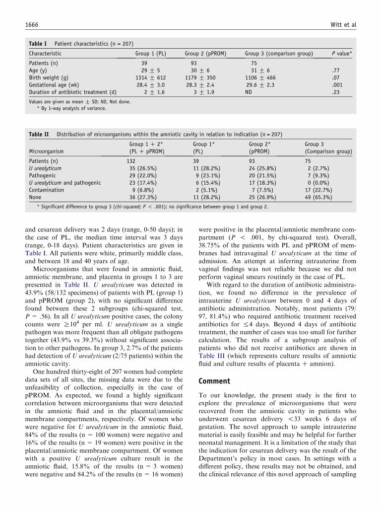
1666 Witt et al
Table I Patient characteristics (n = 207)
Characteristic Group 1 (PL) Group 2 (pPROM) Group 3 (comparison group) P value*
Patients (n) 39 93 75Age (y) 29 G 5 30 G 6 31 G 6 .77Birth weight (g) 1314 G 612 1179 G 350 1106 G 466 .07Gestational age (wk) 28.4 G 3.0 28.3 G 2.4 29.6 G 2.3 .001Duration of antibiotic treatment (d) 2 G 1.6 3 G 1.9 ND .23
Values are given as mean G SD; ND, Not done.
* By 1-way analysis of variance.
Table II Distribution of microorganisms within the amniotic cavity in relation to indication (n = 207)
MicroorganismGroup 1 C 2*(PL C pPROM)
Group 1*(PL)
Group 2*(pPROM)
Group 3(Comparison group)
Patients (n) 132 39 93 75U urealyticum 35 (26.5%) 11 (28.2%) 24 (25.8%) 2 (2.7%)Pathogenic 29 (22.0%) 9 (23.1%) 20 (21.5%) 7 (9.3%)U urealyticum and pathogenic 23 (17.4%) 6 (15.4%) 17 (18.3%) 0 (0.0%)Contamination 9 (6.8%) 2 (5.1%) 7 (7.5%) 17 (22.7%)None 36 (27.3%) 11 (28.2%) 25 (26.9%) 49 (65.3%)
* Significant difference to group 3 (chi-squared; P ! .001); no significance between group 1 and group 2.
and cesarean delivery was 2 days (range, 0-50 days); inthe case of PL, the median time interval was 3 days(range, 0-18 days). Patient characteristics are given inTable I. All patients were white, primarily middle class,and between 18 and 40 years of age.
Microorganisms that were found in amniotic fluid,amniotic membrane, and placenta in groups 1 to 3 arepresented in Table II. U urealyticum was detected in43.9% (58/132 specimens) of patients with PL (group 1)and pPROM (group 2), with no significant differencefound between these 2 subgroups (chi-squared test,P = .56). In all U urealyticum positive cases, the colonycounts were R104 per ml. U urealyticum as a singlepathogen was more frequent than all obligate pathogenstogether (43.9% vs 39.3%) without significant associa-tion to other pathogens. In group 3, 2.7% of the patientshad detection of U urealyticum (2/75 patients) within theamniotic cavity.
One hundred thirty-eight of 207 women had completedata sets of all sites, the missing data were due to theunfeasibility of collection, especially in the case ofpPROM. As expected, we found a highly significantcorrelation between microorganisms that were detectedin the amniotic fluid and in the placental/amnioticmembrane compartments, respectively. Of women whowere negative for U urealyticum in the amniotic fluid,84% of the results (n = 100 women) were negative and16% of the results (n = 19 women) were positive in theplacental/amniotic membrane compartment. Of womenwith a positive U urealyticum culture result in theamniotic fluid, 15.8% of the results (n = 3 women)were negative and 84.2% of the results (n = 16 women)
were positive in the placental/amniotic membrane com-partment (P ! .001, by chi-squared test). Overall,38.75% of the patients with PL and pPROM of mem-branes had intravaginal U urealyticum at the time ofadmission. An attempt at inferring intrauterine fromvaginal findings was not reliable because we did notperform vaginal smears routinely in the case of PL.
With regard to the duration of antibiotic administra-tion, we found no difference in the prevalence ofintrauterine U urealyticum between 0 and 4 days ofantibiotic administration. Notably, most patients (79/97, 81.4%) who required antibiotic treatment receivedantibiotics for %4 days. Beyond 4 days of antibiotictreatment, the number of cases was too small for furthercalculation. The results of a subgroup analysis ofpatients who did not receive antibiotics are shown inTable III (which represents culture results of amnioticfluid and culture results of placenta C amnion).
Comment
To our knowledge, the present study is the first toexplore the prevalence of microorganisms that wererecovered from the amniotic cavity in patients whounderwent cesarean delivery !33 weeks 6 days ofgestation. The novel approach to sample intrauterinematerial is easily feasible and may be helpful for furtherneonatal management. It is a limitation of the study thatthe indication for cesarean delivery was the result of theDepartment’s policy in most cases. In settings with adifferent policy, these results may not be obtained, andthe clinical relevance of this novel approach of sampling

Witt et al 1667
Table III Subgroup analysis of the amniotic fluid in women who did not receive antibiotics (n = 128) and of the placenta/membraneof women who did not receive antibiotics (n = 128)
Microorganism Group 1: PL (n) Group 2: pPROM (n) Group 3: Comparison group (n)
Amniotic fluidPatients 25 (100%) 33 (100%) 70 (100%)U urealyticum 5 (20%)* 3 (9.1%)y 0Pathogenic 1 (4%) 7 (21.2%)y 2 (2.9%)U urealyticum and pathogenic 2 (8%)* 0 0Contamination 2 (8%) 0 3 (4.3%)None 7 (28%) 10 (30.3%) 49 (70%)Not done 8 (32%) 13 (39.4%) 16 (22.9%)
Placenta/membranePatients 25 (100%) 33 (100%) 70 (100%)U urealyticum 8 (32%)* 10 (30.3)y 2 (2.9%)Pathogenic 6 (24%)* 3 (9.1%)y 4 (5.7%)U urealyticum and pathogenic 5 (20%)* 5 (15.2%)y 0Contamination 1 (4%) 6 (18.2%) 14 (20%)None 5 (20%) 9 (27.3%) 50 (71.4%)
* Significant to group 3 (P ! .001); no significant difference between group 1 and group 2.y Significant to group 3 (P ! .01); no significant difference between group 1 and group 2.
would be reduced. Although clear evidence for theoptimal mode of delivery in this early gestational weeksis missing,20 there is a general trend towards primarycesarean delivery in our country. We are aware of thefact that this policy is not very common in othercountries, but this discussion is not the topic of thepresent study. However, we found a significantly higherrate of amniotic cavity U urealyticum in patients withtherapy-resistant PL or pPROM compared with patientswho underwent cesarean delivery for other indications.U urealyticum represented the far most prevalent singleintrauterine pathogen in these patients.
Previous publications have assessed placental tissuesfor the presence of U urealyticum in patients withpreterm vaginal delivery18; even though these reportsfound a strong correlation among microbial invasion,preterm delivery, and an increase in perinatal morbidityand mortality rates,17 the interpretation of these resultsis somewhat problematic because of the mode of deliv-ery. After vaginal delivery, it is not possible to differen-tiate between the colonization of the amniotic cavity andvaginal cross contamination as a result of the birthprocess. Furthermore, cesarean delivery offers the pos-sibility to detect microorganisms from all compartmentsof the amniotic cavity. The prevalence of microbialinvasion of the amniotic cavity with U urealyticum inour study, which affected 43.9% of patients in groups1 and 2, is much higher compared with previous studiesthat reported data from amniocentesis of patients withpPROM.4,15 One possible explanation for that differ-ence is the combined sampling of amniotic fluid andspecimens of placenta and amniotic membranes in ourstudy. The latter 2 specimen types consist of morecellular material compared with amniotic fluid. Thus,
the possibility of the detection of U urealyticum wasprobably higher. Furthermore, in our setting, we opti-mized factors such as the transport media, the timebetween sampling, and the beginning of detection pro-cedure.
We acknowledge that the fact that the exact locationwhere portions of placental and amniotic membranewere excised was not defined is another limitation of thestudy. One can argue that the location could have beennear the internal cervical os and that vaginal ascensiontherefore could be conceivable. At this point, it has to beacknowledged that the question ‘‘time of invasion’’ isnot elucidated clearly. It is possible that the amnioticcavity became invaded as a result of PL or pPROM. Theresults of the current study do not indicate a significantdifference in the intrauterine colonization of U urealy-ticum between patients with PL and pPROM. Thisfinding weakens the theory of late ascension of micro-organisms through ruptured amniotic membranes butfavors the possibility of microbial ascension at thebeginning of or during the early weeks of gestation.The assumption that colonization develops early in thecourse of pregnancy is also supported by studies thatshow a clear association between U urealyticum coloni-zation and spontaneous abortion21,22 and by reports onadverse pregnancy outcome after amniotic fluid isola-tion of U urealyticum during genetic amniocentesis.23-26
Prophylactic erythromycin has not been shown to be ofbenefit,27,28 and this finding may well be due to thelimited transplacental transfer of macrolide antibi-otics.29
With regard to the duration of antibiotic administra-tion, we found no difference in the rate of intrauterineU urealyticum between 0 and 4 days of antibiotics, which

1668 Witt et al
suggests that the short-term administration of ampicillindoes not result in the selection of U urealyticum. We areaware of the possible interference of our data becauseof the routine administration of antibiotics in the caseof pPROM, but it is unlikely that U urealyticumwas selected if they were not present at the beginningof antibiotic prophylaxis; in the comparison group,U urealyticum was detectable in only 2.7%. Asidefrom that, in cases of PL, we found rather similarintrauterine detection rates, although those patients didnot receive antibiotics routinely. To avoid the potentialconfounder ‘‘antibiotic administration,’’ we performed asubgroup analysis of patients who did not receiveantibiotics. Furthermore, we split the culture resultsinto amniotic fluid and placenta/membrane results.Although the number of patients was subsequentlylow, we found the same statistical significances betweengroup 1 and 2 versus group 3 and no significantdifference between groups 1 and 2 (even not betweeninvasion rates of the amniotic fluid; P= .068).
Our observations are unrelated to the clinical signif-icance of neonatal morbidity that is caused byU urealyticum, such as chronic lung disease, but itindirectly increases neonatal morbidity and mortalityrates by decreasing the duration of pregnancy. Themechanism of U urealyticum colonization and its role inthe cascade of inflammation has not been elucidatedfully. In this context, several reports show an associationof U urealyticum and elevated cytokines in the fetal,amniotic, and maternal compartments6 and theU urealyticum–induced production of proinflammatorycytokines by macrophages.30
The approach of culturing amniotic fluid, placenta,and membranes is a simple and effective diagnosticmeasure, which allows microbial colonization to bediagnosed and adequate treatment for the neonate tobe initiated early. Particularly in patients in whomamniocentesis cannot be performed, this method there-fore may be associated with postpartum benefits for bothmother and child. It is well-known that the diagnosis ofearly-onset infection, particularly the identification of thepathogens that are involved, remains a dilemma inpreterm infants.31 The rate of culture-proven early onsetsepsis is low in this patient population, although manyneonates have clinical and laboratory evidence of early-onset sepsis.31 We therefore considered it an ideal com-promise to identify microorganisms from the amnioticcavity of the mother to increase the detection rate ofpathogens that are potentially responsible for neonatalinfections. Additional research is being conducted cur-rently to elucidate whether the assessment of infectionparameters and culture results at the time of cesareandelivery can improve neonatal outcome.
In summary, this is the first study to demonstrate asignificantly higher rate of amniotic cavity U urealyticumin patients who undergo cesarean delivery for PL or
pPROM compared with patients who undergo pretermcesarean delivery for other indications. This findingsupports numerous previous reports about the role ofU urealyticum in the pathogenesis of preterm birth andcan be interpreted as additional evidence. Regarding thetrue nature of the association that was found in thisstudy, it is still not evident whether there is a causativerole for U urealyticum colonization and preterm deliveryor whether these 2 phenomena are merely the result ofan obscure common factor. PL and pPROM constitutedthe major cause of preterm delivery in our collective.Any approach at preventing preterm delivery thereforehas to focus on combating and controlling reproductivetract infection, given the association of PL and pPROMwith U urealyticum.
References
1. Cassell GH, Waites KB, Watson HL, Crouse DT, Harasawa R.
Ureaplasma urealyticum intrauterine infection: role in prematurity
and disease in newborns. Clin Microbiol Rev 1993;6:69-87.
2. Abele-Horn M, Peters J, Genzel-Boroviczeny O, Wolff C,
Zimmermann A, Gottschling W. Vaginal Ureaplasma urealyticum
colonization: influence on pregnancy outcome and neonatal
morbidity. Infection 1997;25:286-91.
3. Gravett MG, Hummel D, Eschenbach DA, Holmes KK. Preterm
labor associated with subclinical amniotic fluid infection and with
bacterial vaginosis. Obstet Gynecol 1986;67:229-37.
4. Romero R, Mazor M, Morrotti R, Avila C, Oyarzun E, Insunza
A, et al. Infection and labor: VII, microbial invasion of the
amniotic cavity in spontaneous rupture of membranes at term. Am
J Obstet Gynecol 1992;166:129-33.
5. Yoon BH, Romero R, Park JS, Chang JW, Kim YA, Kim JC,
et al. Microbial invasion of the amniotic cavity with Ureaplasma
urealyticum is associated with a robust host response in fetal,
amniotic, and maternal compartments. Am J Obstet Gynecol 1998;
179:1254-60.
6. Yoon BH, Chang JW, Romero R. Isolation of Ureaplasma
urealyticum from the amniotic cavity and adverse outcome in
preterm labor. Obstet Gynecol 1998;92:77-82.
7. Groneck P, Goetze-Speer B, Speer CP. Inflammatory broncho-
pulmonary response of preterm infants with microbial colonisation
of the airways at birth. Arch Dis Child Fetal Neonatal Ed 1996;74:
F51-5.
8. Pacifico L, Panero A, Roggini M, Rossi N, Bucci G, Chiesa C.
Ureaplasma urealyticum and pulmonary outcome in a neonatal
intensive care population. Pediatr Infect Dis J 1997;16:579-86.
9. Agarwal P, Rajadurai VS, Pradeepkumar VK, Tan KW. Urea-
plasma urealyticum and its association with chronic lung disease in
Asian neonates. J Paediatr Child Health 2000;36:487-90.
10. Hannaford K, Todd DA, Jeffery H, John E, Blyth K, Gilbert GL.
Role of Ureaplasma urealyticum in lung disease of prematurity.
Arch Dis Child Fetal Neonatal Ed 1999;81:F162-7.
11. Ollikainen J, Hiekkaniemi H, Korppi M, Sarkkinen H, Heinonen
K. Ureaplasma urealyticum infection associated with acute respi-
ratory insufficiency and death in premature infants. J Pediatr 1993;
122:756-60.
12. SaxenH,HakkarainenK, PohjavuoriM,MiettinenA.Chronic lung
disease of preterm infants in Finland is not associated with Urea-
plasma urealyticum colonization. Acta Paediatr 1993;82:198-201.
13. Taylor-Robinson D, Furr PM, Liberman MM. The occurrence of
genital mycoplasmas in babies with and without respiratory
distress. Acta Paediatr Scand 1984;73:383-6.

Witt et al 1669
14. Andrews WW, Shah SR, Goldenberg RL, Cliver SP, Hauth JC,
Cassell GH. Association of post-cesarean delivery endometritis
with colonization of the chorioamnion by Ureaplasma urealyticum.
Obstet Gynecol 1995;85:509-14.
15. Yoon BH, Romero R, Kim M, Kim EC, Kim T, Park JS, et al.
Clinical implications of detection of Ureaplasma urealyticum in the
amniotic cavity with the polymerase chain reaction. Am J Obstet
Gynecol 2000;183:1130-7.
16. Gauthier DW, Meyer WJ, Bieniarz A. Expectant management of
premature rupture of membranes with amniotic fluid cultures
positive for Ureaplasma urealyticum alone. Am J Obstet Gynecol
1994;170:587-90.
17. Kundsin RB, Driscoll SG, Monson RR, Yeh C, Biano SA,
Cochran WD. Association of Ureaplasma urealyticum in the
placenta with perinatal morbidity and mortality. N Engl J Med
1984;310:941-5.
18. Kundsin RB, Leviton A, Allred EN, Poulin SA. Ureaplasma
urealyticum infection of the placenta in pregnancies that ended
prematurely. Obstet Gynecol 1996;87:122-7.
19. Andrews WW, Hauth JC, Goldenberg RL, Gomez R, Romero R,
Cassell GH. Amniotic fluid interleukin-6: correlation with upper
genital tract microbial colonization and gestational age in women
delivered after spontaneous labor versus indicated delivery. Am J
Obstet Gynecol 1995;173:606-12.
20. Lumley J. Method of delivery for the preterm infant. BJOG 2003;
110:88-92.
21. Naessens A, Foulon W, Cammu H, Goossens A, Lauwers S.
Epidemiology and pathogenesis of Ureaplasma urealyticum in
spontaneous abortion and early preterm labor. Acta Obstet
Gynecol Scand 1987;66:513-6.
22. Joste NE, Kundsin RB, Genest DR. Histology and Ureaplasma
urealyticum culture in 63 cases of first trimester abortion. Am J
Clin Pathol 1994;102:729-32.
23. Cassell GH, Davis RO, Waites KB, Brown MB, Marriott PA,
Stagno S, et al. Isolation of Mycoplasma hominis and Ureaplasma
urealyticum from amniotic fluid at 16-20 weeks of gestation:
potential effect on outcome of pregnancy. Sex Transm Dis 1983;
10:294-302.
24. Horowitz S, Mazor M, Romero R, Horowitz J, Glezerman M.
Infection of the amniotic cavity with Ureaplasma urealyticum in the
midtrimester of pregnancy. J Reprod Med 1995;40:375-9.
25. Gray DJ, Robinson HB, Malone J, Thomson RB Jr. Adverse
outcome in pregnancy following amniotic fluid isolation of Ure-
aplasma urealyticum. Prenat Diagn 1992;12:111-7.
26. Gerber S, Vial Y, Hohlfeld P, Witkin SS. Detection of Ureaplasma
urealyticum in second-trimester amniotic fluid by polymerase chain
reaction correlates with subsequent preterm labor and delivery.
J Infect Dis 2003;187:518-21.
27. Berg TG, Philpot KL, Welsh MS, Sanger WG, Smith CV.
Ureaplasma/Mycoplasma-infected amniotic fluid: pregnancy out-
come in treated and nontreated patients. J Perinatol 1999;19:
275-7.
28. Ogasawara KK, Goodwin TM. The efficacy of prophylactic
erythromycin in preventing vertical transmission of Ureaplasma
urealyticum. Am J Perinatol 1997;14:233-7.
29. Heikkinen T, Laine K, Neuvonen PJ, Ekblad U. The transplacen-
tal transfer of the macrolide antibiotics erythromycin, roxithro-
mycin and azithromycin. BJOG 2000;107:770-5.
30. Li YH, Brauner A, Jonsson B, van der Ploeg I, Soder O, Holst M,
et al. Ureaplasma urealyticum-induced production of proinflam-
matory cytokines by macrophages. Pediatr Res 2000;48:114-9.
31. Stoll BJ, Gordon T, Korones SB, Shankaran S, Tyson JE, Bauer
CR, et al. Early-onset sepsis in very low birth weight neonates: a
report from the National Institute of Child Health and Human
Development Neonatal Research Network. J Pediatr 1996;129:
72-80.

American Journal of Obstetrics and Gynecology (2005) 193, 1670–5
www.ajog.org
Persistance of adverse obstetric and neonatal outcomesin monochorionic twins after exclusion of disordersunique to monochorionic placentation
Line Leduc, MD,* Larissa Takser, MD, PhD, Denyse Rinfret, RN
Maternal Fetal Medicine Division, Department of Obstetrics and Gynecology, Sainte-Justine hospital.Montreal, Quebec, Canada
Received for publication June 19, 2004; revised March 16, 2005; accepted April 1, 2005
KEY WORDSPerinatal outcomes
in twinsChorionicity
Preterm birthFetal growth
restriction
Objective: This study was undertaken to assess obstetric and neonatal outcomes in dichorionictwins and monochorionic-diamniotic twins after exclusion of twin-to-twin transfusion syndrome
and twin reversed arterial perfusion sequence.Study design: Data from a tertiary center were collected in twin gestations between 1994 and2002. Chorionicity was defined by standard echographic criteria and placental examination at
delivery. Neonatal outcomes were compared between monochorionic and dichorionic gestations.Results: This study included 503 women: 378 (75%) dichorionic and 125 (25%) monochorionictwin gestations. Monochorionic twin gestations had a higher risk of preterm deliveries between 30and 34 weeks’ gestation than pregnancies with dichorionic twins (P ! .01). Monochorionic twins
had a higher number of birth weight less than 10th percentile (P ! .001) discordancy 25% orgreater (P ! .02), admission to neonatal intensive care unit (P ! .03), and intraventricularhemorrhage grade 3 and 4 (P ! .007) than dichorionic twins even after adjusting for gestational
age.Conclusion: Monochorionic diamniotic twins have a higher risk of perinatal complications thandichorionic twin gestations, even after exclusion of disorders unique to monochorionic
placentation.� 2005 Mosby, Inc. All rights reserved.
Twin gestation is associated with increased perinatalmorbidity and mortality rates compared with single-tons.1,2 Twins account for 10%of the perinatal mortality,although they represent only 3.0% of the pregnancies.1,2
Presented at the Twenty-fourth Annual Meeting of the Society for
Maternal-Fetal Medicine in New Orleans, La, February 2-7, 2004.
* Reprint requests: Line Leduc, MD, Hopital Sainte-Justine, De-
partment Obstetrics and Gynecology, Maternal Fetal Medicine Divi-
sion, 3175 Cote Sainte-Catherine, Montreal, Quebec, Canada.
E-mail: [email protected]
0002-9378/$ - see front matter � 2005 Mosby, Inc. All rights reserved.
doi:10.1016/j.ajog.2005.04.007
The poor perinatal outcome is the result of prematurity,3
fetal growth restriction,4 and both structural and chro-mosomal anomalies.5,6 The type of placentation appearsto influence perinatal morbidity because monochorionictwins have higher perinatal morbidity and mortality thandichorionic twins.7-11 Disorders unique to monochori-onic twins, such as twin-twin transfusion syndrome(TTTS),12 twin reversed arterial perfusion (TRAP)sequence,13 and the complications of monoamniocity14
appear responsible for the poor perinatal outcomes in

Leduc, Takser, and Rinfret 1671
monochorionic twins. Although it has been reported thatcomplications such as prematurity and impaired fetalgrowth have a role in the outcome of monochorionic anddichorionic twins, previous studies have included eithersmall cohorts or severely discordant twins of variouscauses.4,15-18 Therefore, we undertook an observationalhospital based cohort study to assess obstetric and neo-natal outcomes in twin pregnancies in relation to chorio-nicity. Monoamniotic twins and complications unique tomonochorionic placentation were excluded.
Material and methods
Data were prospectively collected in all twins monitoredat our tertiary care center between April 1994 andJanuary 2002. This database review was approved byour ethics board committee. Twin pregnancies compli-cated by TTTS, chromosomal and structural anomalies,monoamniotic twins, and intrauterine fetal death of1 co-twin were not included in the analysis as well as anytransfer to our center that occurred after 21 week’sgestation. The diagnosis of TTTS was made accordingto Quintero’s criteria19 and the presence of fetal echo-cardiographic findings suggesting this syndrome.20 Cho-rionicity was assessed by standard echographic criteriaand by meticulous placental examination at delivery. Adetailed level II ultrasound was performed on allpatients between 16 to 20 weeks of gestation. From 20weeks of gestation until delivery, all women had thesame prenatal care with clinical visits at 1- to 2-weekintervals, cervical examination at each visit after 24weeks of gestation, and multivitamin supplementation.Serial ultrasound examinations were performed every 2-to 4-week interval depending of fetal growth, for fetalbiometry, and amniotic fluid assessment. Doppler stud-ies were undertaken as soon as fetal growth restrictionwas suspected. Fetal growth restriction was defined asan estimated fetal weight less than the 10th percentileaccording to the Canadian birth weight growth curve fortwins.21 Discordance was defined as a 25% or greaterdifference in birth weights and was calculated by usingthe following formula: larger twin weight – smaller twinweight/larger twin weight! 100. All women had a mater-nity work leave from 24 weeks of gestation until deliv-ery. Delivery was indicated between 32 to 34 weeks ofgestation when fetal growth restriction less than thethird percentile was present in at least 1 co-twin, or at 39weeks’ gestation for postdates or for any standardobstetric indications. Preeclampsia was defined accord-ing to the Canadian classification.22 All pregnant womenwere screened for gestational diabetes mellitus (GDM)between 24 and 28 weeks’gestation. The screening forGDM was a 1-hour plasma glucose measurement after a50-g glucose load given at any time of the day. If the 1-hour plasma glucose was 10.3 mmol/L or greater, GDM
was confirmed. If the 1-hour plasma glucose was 7.8 to10.2 mmol/L, a 75-g oral glucose tolerance test (OGTT)was conducted.23
Maternal demographic and obstetric data as well asneonatal data were recorded. Perinatal mortality wasreported as the neonatal death in the first month of life.Perinatal morbidity was documented with the followingvariables: the proportion of deliveries that occurred 34or less, 32 or less, and 30 or less weeks of gestation, theproportion of twins with a birth weight discordance25% or greater, a birth weight less than the 10thpercentile, and a very low birth weight (VLBW) lessthan 1500 g. Neonatal morbidity was defined by theproportion of neonates admitted to the neonatal inten-sive care unit (NICU), the rate of endotracheal intuba-tion, and the rate of intraventricular hemorrhage (IVH)grade 3 or 4. Obstetric morbidity was described in usingthe following parameters: the rate of elective cesareansection and urgent cesarean section on the second twin.Finally, maternal morbidity was assessed with the prev-alences of gestational diabetes requiring insulin andpreeclampsia. Perinatal outcomes in monochorionictwins were compared with those of dichorionic twins.Finally, discordant twins were compared according tochorionicity.
Statistical analysis was performed in using unpairedt test for continuous variables such as: meanmaternal ageand gestational age at delivery. Thec2 test was performedfor the following categorical variables: the rates of electiveand urgent cesarean section, the rate of both vaginaldeliveries, the incidence of preeclampsia and gestationaldiabetes, the sex of fetuses, the rate of preterm deliveriesless than 34 weeks, 32 or less weeks, and 30 or less weeks.
Mixed procedure (SAS version 8.02, SAS Institute,Cary, NC)24 for repeated measures was used to estimatethe differences in birth weight between monochorionicand dichorionic pregnancies taking into account withinpair covariance. To compare the rate of discordant (morethan 25% discordance) twin pairs, and birth weight less
Table I Maternal data in MC and DC twins
Variables DC MC P
Mean maternalage (y)
30.6 G 5.3 29.1 G 6.1 .01
Gestational ageat 1st US
18.7 G 3.6 18.6 G 3.7 NS
Elective CS 168/378 (44.4%) 57/125 (45.6%) NSUrgent CS onfetus 2
21/210 (10%) 11/68 (16.2%) .01
Both vaginaldeliveries
189/210 (90%) 57/68 (83.8%) NS
% Preeclampsia 25/378 (6.6%) 12/125 (9.6%) NS% Gestationaldiabetes
53/370 (14.3%) 15/122 (12.3%) NS
US, Ultrasound; CS, cesarean section.

1672 Leduc, Takser, and Rinfret
Table II Neonatal data in MC and DC twins
Variables DC MC P
N (women) 378 125Mean gestational age at delivery (y) 35.7 G 2.9 34.7 G 2.8 .01Female sex 391/754 (51.9%) 129/250 (51.6%) NSMale sex 363/754 (48.1%) 121/250 (48.4%) NS% DeliveriesO34 wks 293/378 (77.5%) 82/125(65.6%) .05%34 wks 85/378 (22.5%) 43/125 (34.4%) .05%32 wks 37/378 (9.8%) 21/125 (16.8%) .05%30 wks 23/378 (6.1%) 7/125 (5.6%) NS
Mean birth weightFemaleThe smaller twin 2199 G 589 1934 G 556 .007The bigger twin 2521 G 620 2295 G 600 .007
MaleThe smaller twin 2301 G 544 2058 G 657 .002The bigger twin 2577 G 588 2372 G 622 .002
!10th percentile 58/378 (15.4%) 39/125 (31.2%) .001VLBW (!1500 g) 44/378 (11.7%) 26/125 (20.8%) .02Discordance R25% 40/378 (10.3%) 25/125 (20.8%) .008Admission at NICU 98/756 (13%) 63/250 (25.2%) .001Endotracheal intubation 47/756 (6.2%) 23/250 (9.2%) NSIVH 3/756 (0.4%) 4/250 (1.6%) .07Mortality rate 3/756 (0.4%) 2/250 (0.8%) NS
than 10th percentile or less than 1500 g for monochori-onic and dichorionic pregnancies, the General Estimat-ing Equations (GEE) modelling (GENMOD) procedurewas used.25 Crude odds ratio (OR) was calculated andthen gestational age at birth was introduced in GEEmodel setting for adjusted OR.
Results
During the study period, 781 twin pregnancies weredelivered at our center. Of these, 278 cases wereexcluded from the study because of the diagnosis ofTTTS (n = 77), intrauterine fetal death of 1 or bothfetuses (n = 31), chromosomal or structural anomalies(n = 20), monoamniotic twins (n = 21), or for transferto our center after 21 weeks’ gestation (n = 106).Twenty-three twin pairs were lost to follow-up. Thestudy included 503 sets of twins with 378 dichorionic(DC) and 125 monochorionic (MC) twin pairs thatrepresent 75% and 25% of the cohort, respectively.
Table I reports maternal data. The mean maternal ageis statistically different in both groups. The mean gesta-tional age at time of the first ultrasound is the same. Therate of elective cesarean section is about the sameirrespective of chorionicity but urgent cesarean sectionon the second twin happensmore frequently inMC twins.
Table II reports neonatal data. Overall, MC twinshave an increased perinatal morbidity with an earliermean gestational age at delivery. The proportion of
deliveries equal or less than 34 and 32 weeks’ gestationare significantly higher in MC group. However, un-expectantly, the rate of preterm deliveries less than 30weeks’ gestation did not vary significantly when chorio-nicity was taken into account. MC twins had systemat-ically a lower birth weight with an average of 212 g orless for the female and 260 g or less for the male fetuses.Finally, when gestational age at delivery is not takeninto account in the analysis, MC twins are associatedwith lower birth weight less than 10th percentile,VLBW, discordance 25% or greater, or admission tothe NICU and IVH.
Table III shows the GEE analysis. The effect ofmonochorionicity on perinatal morbidity was still pre-sent for almost all studied variables and this, even after
Table III GEE analysis showing the crude and adjusted forgestational age odds ratios of perinatal morbidity in MC twins
VariablesCrude OR(95% CI) P
Adjusted OR(95% CI) P
!10th percentile 1.1 (1.0-1.2) .001 2.0 (1.3-3.1) .001VLBW (!1500 g) 1.9 (1.1-3.2) .02 1.7 (0.8-3.5) NSDiscordance %25% 2.1 (1.2-3.6) .008 1.9 (1.1-3.3) .02Admission at NICU 2.2 (1.4-3.5) .001 2.1 (1.1-4.0) .03EndotrachealIntubation 1.5 (0.8-2.9) NS 1.3 (0.6-2.9) NSIVH 4.1 (0.9-18.2) .07 7.4 (1.7-31.2) .007Mortality rate 2.3 (0.7-7.1) NS 2.2 (0.6-7.7) NS
Values are given as OR (95% CI).

Leduc, Takser, and Rinfret 1673
Table IV Perinatal outcome in discordant twins according to chorionicity
MC DC
Discordance !25% R25% !25% R25%N (women) 100 25 (20%)* 338 40 (10.6%)Mortality rate 0/200 (0%) 2/50 (4%) 3/676 (0.4%) 0/80 (0%)Gestational age at delivery (wks) 35.1 G 2.7 33.2 G 2.7* 35.8 G 2.9 35.0 G 2.9% DeliveriesO34 wks 71/100 (71%) 11/25 (44%) 267/338 (79%) 26/40 (65%)*%34 wks 29/100 (29%) 14/25 (56%)* 71/338 (21%) 14/40 (35%)%32 wks 21/100 (21%) 9/25 (36%)* 30/338 (8.9%) 7/40 (18%)%30 wk 7/100 (7%) 2/25 (8%) 19/338 (5.6%) 4/40 (10%)
Elective CS 39/100 (39%) 18/25 (72%) 143/338 (42%) 25/40 (62%)!10th percentile 20/100 (20%) 19/25 (76%) 32/338 (9.5%) 26/40 (65%)VLBW (!1500 g) 31/100 (31%) 13/25 (52%) 15/338 (4.4%) 11/40 (27.5%)Admission to the NICU 22/100 (22%) 15/25 (62%)y 50/338 (15%) 10/40 (25%)EndotrachealIntubation 11/200 (5.5%) 12/50 (24%) 38/676 (5.6%) 9/80 (11.3%)IVH 2/200 (1%) 2/50 (4%) 2/676 (0.3%) 1/80 (1.25%)
The comparison is between discordant MC and DC twins.
* P ! .01.y P = .006.
adjusting for gestational age at birth. However, the riskto be VLBW or to be intubated does not vary withchorionicity. The mortality rate was not greater for MCtwins.
Table IV details the perinatal outcome in discordanttwins according to chorionicity. Overall, discordanttwins do worse than concordant twins. However, dis-cordant MC twins deliver at an earlier gestational agewith preterm birth 34 weeks or less and admission to theNICU than discordant DC twins. When the GEE modelis applied (Table V) and results are adjusted for gesta-tional age at birth, perinatal morbidity of discordantMC twins is not greater than for discordant DC twins.We do not observe proportionally more neonates withlow birth weight or VLBW, endotracheal intubation, orIVH among the discordant MC twins. Finally, in ourseries, discordance 25% or greater was a sign of growthrestriction less than the 10th percentile in 76% of cases(19/25) but the positive predictive value still remains lowwith only 48.7% (19/39) of cases that were detected withthis threshold.
Comment
Our study examined a cohort of twin pregnanciesfollowed at a single tertiary care center with a consistentmethod of managing twins and providing appropriateneonatal support. We compare the perinatal outcome inDC and MC twins after exclusion of conditions uniqueto MC twins such as TTTS and TRAP sequence. Thesepeculiar complications of MC twin gestation play animportant role in terms of morbidity and mortality intwins. We observed a higher risk of perinatal morbidity
in MC diamniotic twins than DC twins, independentof complications such as TTTS and TRAP sequence.Previous studies performed on the same topic haveeither included a small number of cases or consideredall the MC twin cases.7-11,26-28 These data support theneed for diagnosis of early chorionicity as well as closesonographic surveillance independently by the diag-nosis of diseases such as TTTS, TRAP sequence, andmonoamniocity.
The perinatal mortality was not different between theMC and DC twins once TTTS and TRAP sequence havebeen excluded. These observations may either be real orinsufficient number of patients may explain these results.These data are different than previously reported,7-11,26-28
but these studies usually considered all MC twin cases.In our study, the proportion of pregnancies with a
birth weight discordance 25% or greater was double thatof the MC group as well as the number of neonates less
Table V GEE analysis showing the crude and adjusted forgestational age ORs of discordant MC twins
VariablesCrude OR(95% CI) P
Adjusted OR(95% CI) P
!10th percentile 1.2 (0.7-2.1) NS 1.0 (0.58-1.7) NSVLBW 2.5 (1.0-6.7) .05 1.0 (0.4-2.3) NSAdmission toNICU
4.4 (1.5-12.4) .006 4.6 (1.0-21.5) .05
EndotrachealIntubation 2.5 (0.8-7.9) NS 1.3 (0.3-4.9) NSIVH 3.3 (0.3-36.3) NS 2.2 (0.2-9.5) NSMortality rate 2.8 (0.4-17.7) NS 1.7 (0.2-13.1) NS
Values are given as OR (95% CI).

1674 Leduc, Takser, and Rinfret
than the 10th percentile. We observed a higher number ofdeliveries between 30 and 34 weeks in severely discordantMC twins. These results suggest an association betweenpreterm delivery and severe discordance or fetal growthrestriction. This association may result from increasedobstetric interventions in cases with severe discordance,leading to prematurity. On occasion, discordance 25%or greater can be explained by the presence of a large forgestational age neonate rather than a growth-restrictedfetus and therefore may lead to iatrogenic deliveries ofappropriate growth fetuses. Moreover, as Blicksteinet al16 previously reported, our cohort suggests thatdiscordance was a sign of fetal growth restriction inapproximately 50% of cases and therefore, wheneversevere discordance is suspected, it remains crucial toexclude the presence of fetal growth restriction beforeundertaking any medical intervention.
Recently, Amaru et al17 published their results from aretrospective study, hospital-based cohort of twin ges-tations with 2 live births delivered at 24 weeks or laterfrom 1992 to 2001. The authors tried to define whetherdiscordance was an independent risk factor for adverseneonatal outcome and they concluded that discordancewas not an independent risk factor for serious neonatalmorbidity or mortality. These apparently controversialresults are the consequence of different definition usedin the 2 studies. First, we used twin norms to defineappropriate for gestational age (AGA) and small forgestational age (SGA) rather than singleton norms, thuswe may have included neonates who actually met thedefinitions of low birth weights because 76% and 65%of our MC and DC twins with a birth weight discor-dance 25% or greater were less than the 10th percentile;second, we used a threshold of 25% discordance ratherthan 20%, which more likely included a population ofneonates at higher risk for SGA and adverse outcome.The differences in study design may explain the differ-ent results. Therefore, we suspect that the poor out-comes observed in our cohort more likely result fromthese neonates with fetal growth restriction and not fromgrowth discordance itself. Discordant MC twins do nothave aworse perinatal outcome thandiscordantDC twinsin our setting. These findings are opposite to those ofVictoria et al27 who reported greater morbidity in dis-cordant MC twins. However, TTTS was not excludedfrom their cohort. These observations warrant furtherinvestigation to clarify whether iatrogenic prematurityor impaired fetal growth result in worse outcome.
Maternal morbidity was significant in women carry-ing twins for gestational diabetes and preeclampsia.Although these complications do not appear directlyrelated to or associated with chorionicity, preeclampsiamay result in preterm birth and occurs more frequentlyin twin pregnancy. Some authors have previouslyreported the same findings and prevalence in a cohortof twin pregnancies.29-30
Likewise, we documented a significant rate of gesta-tional diabetes requiring insulin in our cohort. Theprevalence of GDM is population specific and has beenpreviously described to be higher than expected in theCanadian Founder people.31 The design of the studycannot, of course, address the issue of the incidence ofGDM in twins because no control group was recruitedin the singleton population.
Finally, the rate of cesarean section in the MCsubgroup was significant. Most of them are the resultof the choice of the attending physician when fetalgrowth restriction was documented. However, the inci-dence of urgent cesarean section in which vaginaldelivery was planned is similar to what has been previ-ously reported in the literature.32
This study compared the perinatal outcome in DCand MC diamniotic twins and observed that MC twinshave a higher risk of perinatal morbidity even afterexclusion of complications unique to MC placentation.These data support the need for early diagnosis ofchorionicity as well as close sonographic surveillanceof MC twin pregnancies, irrespective of the diagnosisof specific high-risk conditions such as TTTS, TRAPsequence, and monoamniocity.
References
1. Martin JA, Hamilton BE, Sutton PD, Ventura SJ, Menacker F.
Births: final data for 2002. CDC Natl Vital Stat Rep 2003;52:1-113.
2. Malone FD, D’Alton ME. Multiple gestation: clinical character-
istics and management. In: Maternal-fetal medicine: principles and
practice. 5th ed. St Louis: Elsevier Inc; 2004. p. 513-36.
3. Cooperstock MS, Tummaru R, Bakewell J, Schramm W. Twin
birth weight discordance and risk of preterm birth. Am J Obstet
Gynecol 2000;183:63-7.
4. Demisse K, Ananth CV, Martin J, Hanley ML, MacDorman MF,
Rhoads GG. Fetal and neonatal mortality among twin gestations
in the United States: the role of intrapair birth weight discordance.
Obstet Gynecol 2002;100:474-80.
5. Schinzel AA, Smith DW, Miller JR. Monozygotic twins and
structural defects. J Pediatr 1979;95:921-30.
6. Rodis JF, Egan JF, Craffey A, Ciarleglio L, Greenstein RM,
Scorza WE. Calculated risk of chromosomal abnormalities in twin
gestations. Obstet Gynecol 1990;76:1037-41.
7. Sebire NJ, Snijders RJM, Hughes K, Sepulveda W, Nicolaides
KH. The hidden mortality of monochorionic twin pregnancies.
BJOG 1997;104:1203-7.
8. Hatkar PA, Bhide AG. Perinatal outcome of twins in relation to
chorionicity. J Postgrad Med 1999;45:33-7.
9. Minakami H, Honma Y, Matsubara S, Uchida A, Shiraishi H,
Sato I. Effects of placental chorionicity on outcome in twin
pregnancies. J Reprod Med 1999;44:595-600.
10. Lynch A, McDuffie R Jr, Stephens J, Murphy J, Faber K, Orleans
M. The contribution of assisted conception, chorionicity and other
risk factors to very low birthweight in a twin cohort. BJOG
2003;110:405-10.
11. Snijder MJ, Wladimiroff W. Fetal biometry and outcome in
monochorionic vs. dichorionic twin pregnancies: a retrospective
cross-sectional matched-control study. Ultrasound Med Biol
1998;24:197-201.

Leduc, Takser, and Rinfret 1675
12. Duncombe GI, Dickinson JE, Evans SF. Perinatal characteristics
and outcomes of pregnancies complicated by twin-twin transfusion
syndrome. Obstet Gynecol 2003;101:1190-6.
13. Van Allen M, Smith D, Shepard T. Twin reversed arterial
perfusion (TRAP) sequence: a study of 14 twin pregnancies with
acardius. Semin Perinatol 1983;7:285-93.
14. Rodis JF, McIlveen PF, Egan JFX, Borgida AF, Turner GW,
Campbell WA. Monoamniotic twins: improved perinatal survival
with accurate prenatal diagnosis and antenatal surveillance. Am J
Obstet Gynecol 1997;177:1046-9.
15. Branum AM, Schoendorf KC. The effect of birth weight discor-
dance on twin neonatal mortality. Obstet Gynecol 2003;101:570-4.
16. Blickstein I, Keith LG. Neonatal mortality rates among growth-
discordant twins, classified according to the birth weight of the
smaller twin. Am J Obstet Gynecol 2004;190:170-4.
17. Amaru RC, Bush MC, Berkowitz RL, Lapinski RH, Gaddipati S.
Is discordant growth in twins an independent risk factor for
adverse neonatal outcome? Obstet Gynecol 2004;103:71-6.
18. Demissie K, Ananth CV, Martin J, Hanley ML, MacDorman MF,
Rhoads GG. Fetal and neonatal mortality among twin gestations
in the United States: the role of intrapair birth weight discordance.
Obstet Gynecol 2002;100:474-80.
19. Quintero R, Morales W, Allen M, Bornick P, Krueger M. Staging
of twin-twin transfusion syndrome. J Perinatol 1999;19:550-5.
20. Lachapelle M-F, Leduc L, Cote J-M, Grignon A, Fouron J-C.
Potential value of fetal echocardiography in the differential
diagnosis of twin pregnancy with presence of polyhydramnios-
oligohydrramnios syndrome. Am J Obstet Gynecol 1997;177:
388-94.
21. Arbuckle TE, Wilkins R, Sherman GJ. Birth weight percentiles by
gestational age in Canada. Obstet Gynecol 1993;81:39-48.
22. Helewa ME, Burrows RF, Smith J, Williams K, Brain P, Rabkin
SW. Report of the Canadian Hypertension Society Consensus
Conference: 1, definitions, evaluation, and classifications of
hypertensive disorders in pregnancy. Can Med Assoc J 1997;157:
715-25.
23. Metzger BE, Coustan DR. Summary and recommendations of the
Fourth International Workshop-Conference on Gestational Dia-
betes Mellitus. Diabetes Care 1998;21:1361-7.
24. SAS/STAT user’s guide, version 8. Cary (NC): SAS Institute Inc;
1999.
25. Lipsitz SH, Kim K, Zhao L. Analysis of repeated categorical data
using generalized estimating equations. Stat Med 1994;13:1149-63.
26. Dube J, Dodds L, Armson A. Does chorionicity or zygosity
predict adverse perinatal outcomes in twins? Am J Obstet Gynecol
2002;186:579-83.
27. Victoria A, Mora G, Arias F. Perinatal outcome, placental
pathology, and severity of discordance in monochorionic and
dichorionic twins. Obstet Gynecol 2001;97:310-5.
28. Baghdadi S, Gee TT, Whittle MJ, Khan KS. Twin pregnancy out-
come and chorionicity. Acta Obstet Gynecol Scand 2003;82:18-21.
29. Campbell DM, MacGillivray I. Preeclampsia in twin pregnancies:
incidence and outcome. Hypertens Pregnancy 1999;18:197-207.
30. Savvidou MD, Karanastasi E, Skentou C, Nicolaides KH. Twin
chorionicity and pre-eclampsia. Ultrasound Obstet Gynecol
2001;18:228-31.
31. Harris SB, Caulfield LE, Sugamori ME, Whalen EA, Henning B.
The epidemiology of diabetes in pregnant native Canadians: a risk
profile. Diabetes Care 1997;20:1422-5.
32. Wen SW, Fung KF, Oppenheimer L, Demissie K, Yang Q, Walker
M. Occurrence and predictors of cesarean delivery of the first twin.
Obstet Gynecol 2004;103:413-9.

American Journal of Obstetrics and Gynecology (2005) 193, 1676–9
www.ajog.org
Hypertensive disease in pregnancies complicated bysystemic lupus erythematosus
Robert S. Egerman, MD,a,b Risa D. Ramsey, PhD,a Lu W. Kao, RN,a
Jay J. Bringman, MD,a Andrew J. Bush, PhD,c Jim Y. Wan, PhDc
Departments of Obstetrics and Gynecology,a Internal Medicine,b and Preventive Medicine,c University of TennesseeHealth Science Center, Memphis, TN
Received for publication December 13, 2004; revised March 8, 2005; accepted March 29, 2005
KEY WORDSPregnancySystemic lupus
erythematosusPreeclampsiaHypertension
Objective: The purpose of this study was to assess the percentage of hypertensive disease in
pregnancies complicated by systemic lupus erythematosus at a single institution.Study design: We conducted a retrospective analysis of medical records between 1992 and 2003of 68 pregnancies that were complicated by systemic lupus erythematosus from 48 parturients.
Patients were categorized into 3 groups: no chronic hypertension (n = 49 women), chronichypertension–no medication (n = 6 women) and chronic hypertension–treated (n = 13 women).Analyses of variance (with Tukey-Kramer adjusted follow-up evaluation) and chi-squared/
Fisher’s exact tests were used for the analyses of continuous and categoric variables, respectively.Significance was defined by a probability value of %.05.Results: Chronic hypertension complicated 28% of systemic lupus erythematosus pregnancies.Mean systolic blood pressures at intake were significantly different between the normotensive and
no chronic hypertension groups and between the chronic hypertension–no medication andchronic hypertension–treated groups; the differences in diastolic pressures reached significanceonly between the no chronic hypertension and the chronic hypertension–treated groups. Maternal
age, gestational age at delivery, birth weight, lowest platelet count, and highest serum creatininelevels were similar between the hypertensive and the nonhypertensive groups. There were nodifferences in the percentage of aspirin or heparin treatments among the groups, but the
percentage of the chronic hypertension–treated group who received steroids was significantlygreater than the percentage of women who received steroids in the other 2 groups (P ! .05).Preeclampsia developed in 23% of the no chronic hypertension pregnancies and in 32% of the
hypertensive pregnancies (P = .54). When pregnancies that were treated with prednisone (n = 34pregnancies) were compared with those pregnancies that were managed with other agents (n = 34pregnancies), the percentages of preeclampsia were similar (26% and 24%, respectively; P = .78).Conclusion: The percentage of parturients with systemic lupus erythematosus in whom
preeclampsia develops is increased, regardless of the presence of underlying chronic hypertension.Prednisone therapy was not associated with a higher risk of preeclampsia in this series.� 2005 Mosby, Inc. All rights reserved.
Presented at the 25th Annual Meeting of the Society for Maternal-Fetal Medicine, Reno, NV, February 7-12, 2005.
Reprints not available from the authors.
0002-9378/$ - see front matter � 2005 Mosby, Inc. All rights reserved.
doi:10.1016/j.ajog.2005.03.073

Egerman et al 1677
Table I Maternal characteristics and outcomes of pregnancies complicated by SLE
Group (mean G SD)
Characteristic/outcomeI: No CHTN(n = 49)
II: CHTN-No Rx(n = 6)
III: CHTN-Rx(n = 13) P value
Maternal age (y) 25.7 G 6.0 26.0 G 6.8 24.8 G 5.2 NSGestational age at delivery (wks) 36.0 G 4.7 33.3 G 6.7 34.2 G 4.1 NSBlood pressure (mm Hg)Initial visitSystolic 116.5 G 10.1 138.5 G 8.2 129.2 G 21.6 !.05Diastolic 68.0 G 8.0 74.5 G 12.2 84.3 G 15.0 !.05 (I vs III)
At deliverySystolic 143.8 G 19.3 140.0 G 8.8 173.0 G 23.0 !.05 (I vs III, II vs III)Diastolic 89.5 G 15.0 81.8 G 9.5 109.0 G 13.0 !.05 (I vs III, II vs III)
Birth weight (g) 2601 G 953 2207 G 1311 1948 G 865 NSPlatelet count (!103/mm3; lowest) 202.0 G 76.4 167.4 G 69.4 204.9 G 76.6 NSHighest creatinine level (mg/dL) 0.88 G 1.0 0.54 G 0.15 1.02 G 0.38 NS
Data are given as mean G SD. NS, Not significant.
Systemic lupus erythematosus (SLE) has a propensityto affect women of child-bearing age.1 Wide variationsexist regarding the hazards of SLE on the parturient, andthe course during pregnancy is difficult to portend.2-4
Intuitively, patients with SLE with no renal involve-ment (as reflected by normal serum creatinine level),absence of an active urinary sediment (cellular casts,hematuria), and no underlying hypertension seem tohave perinatal outcomes similar to that of the generalpopulation.5 We investigated whether normotensivepatients with SLE had a lesser risk of the developmentof preeclampsia than those patients with SLE andunderlying chronic hypertension.
Material and methods
After obtaining institutional review board approval fromthe University of Tennessee Health Science Center, weidentified obstetric patients with an antecedent diagnosisof SLE by standard criteria through discharge diagnosesbetween the years 1992 and 2003. Sixty-eight pregnanciesfrom 48 parturients were identified and studied retro-spectively. Demographic variables included maternalage, race, a history of chronic hypertension, and treat-ment for chronic hypertension. Outcome variables in-cluded gestational age at delivery, birth weight, use ofglucocorticoids for treatment of SLE at anytime duringthe pregnancy, highest serum creatinine level, lowestperipartal platelet count, and the development of hyper-tensive disease during pregnancy. Chronic hypertensionwas defined as systolic blood pressure of R140 mm Hgor diastolic blood pressure ofR90 mmHg (as defined bythe Joint National Committee on Detection, Evaluation,and Treatment of High Blood Pressure [JNC VI]) thatoccurred before the 20th week of gestation or, if bloodpressure criteria were not met, by an a priori diagnosis of
chronic hypertension under current treatment. Defini-tions of preeclampsia included either the development ofproteinuria (O300 mg/24-hour collection) and systolicblood pressure of R140 mm Hg or diastolic bloodpressure of R90 mm Hg in a patient with no underlyingchronic hypertension that occurred after the 20th week ofgestation, the development of HELLP (hemolysis, ele-vated liver function tests, low platelet count) syndrome asdefined by Sibai,6 difficult to control hypertension thatoccurred in a patient whose condition was previouslystable, or maternal signs of severe preeclampsia.
Patients were stratified into 1 of 3 categories: nor-motensive (No CHTN), underlying chronic hyperten-sion that did not require treatment (CHTN-No Rx), andunderlying chronic hypertension that required treatment(CHTN-Rx). Differences in means were assessed byanalysis of variance (with Tukey-Kramer adjusted fol-low-up evaluation); differences in percentages wereassessed by chi-squared/Fisher’s exact tests. Statisticalsignificance was established by probability values of%.05.
Results
Chronic hypertension complicated 28% of 68 pregnan-cies with SLE. Maternal age, gestational age at delivery,birth weight, lowest platelet count, and highest serumcreatinine levels were similar between the hypertensiveand the nonhypertensive groups (Table I). Interestingly,although the complications did not reach statisticalsignificance, we noted a trend toward higher birthweights in the normotensive group when comparedwith the hypertensive groups, despite similar meangestational ages at delivery.
Mean systolic blood pressures at intake were signif-icantly different between the normotensive and CHTN-No

1678 Egerman et al
Table II Maternal treatment in patients with SLE
Group (n)
Treatment No CHTN (n = 49) CHTN–No Rx (n = 6) CHTN-Rx (n = 13) P value
Aspirin 13 (26.5%) 1 (16.7%) 3 (23.1%) NSHeparin 13 (26.5%) 2 (33.3%) 3 (23.1%) NSSteroids 19 (38.8%) 3 (50.0%) 12 (92.3%) !.05
NS, Not significant.
Rx groups and between the CHTN-No Rx andCHTN-Rx groups, whereas the differences in diastolicpressures reached significance only between the NoCHTN and the CHTN-Rx groups. Choice of antihyper-tensive agents that were used in this series were nearlyequally distributed between calcium channel blockers(nifedipine), beta blockers (predominantly labetolol) andalpha-methyldopa. At delivery, significantly higher sys-tolic and diastolic pressures were identified in the CHTN-Rx group compared with the other 2 groups. Of the 13pregnancies in the CHTN-Rx group, more than a singleantihypertensive agent was necessary in 2 pregnancies.
Maternal age, gestational age at delivery, birthweight, lowest platelet count, and highest serum creat-inine levels were similar between the hypertensive andthe nonhypertensive groups. There were no differencesin the percentage of patients who received aspirin orheparin treatments among the groups (Table II); how-ever, the percentage of the CHTN-RX group whoreceived steroid therapy was significantly greater thanthe percentage patients who received steroid therapy inthe other 2 groups (P ! .05). In comparing patientswith No CHTN with all patients with hypertension(CHTN-No Rx and CHTN-Rx), preeclampsia devel-oped in 23% of the No CHTN and in 32% of thehypertensive pregnancies (P = .54; Table III). No pa-tients in the CHTN-No Rx group had preeclampsia,whereas 6 of 13 patients from the CHTN-Rx werediagnosed with preeclampsia. When comparing preg-nancies that were treated with prednisone (n = 34pregnancies) to those pregnancies that were treatedwith other agents (n = 34 pregnancies), the percentagesof preeclampsia were similar (26% and 24%, respec-tively; P = .78; Table III).
Three pregnancy losses occurred among the 68 preg-nancies (at 23, 24, and 31 weeks of gestation), for aperinatal mortality rate of 44 of 1000 births. Two losseswere complicated by preeclampsia in the No CHTNgroup (losses at 23 and 31 weeks of gestation), and theremaining loss that was complicated by preeclampsia (24weeks of gestation) was from the CHTN-No Rx group.The 2 early losses were from the same parturient whosepregnancy was complicated by antiphospholipid anti-body syndrome. The prevalence of antiphospholipidantibody syndrome was unknown in this series becausethis information was not uniformly available.
Comment
SLE affects a multitude of organ systems with consid-erable variance in disease manifestation and progressionbetween patients. The severity of renal involvementfrom SLE is a well-accepted indicator of debility.Hypertension that is associated with this disease eitherreflects or contributes to progressive renal dysfunction.Similarly, the contribution of underlying renal dysfunc-tion on the severity of hypertension is apparent inadvanced disease.7 Correspondingly, we sought to de-termine the role of SLE on hypertensive disease inpregnancy. In this retrospective analysis, patients withan antecedent diagnosis of SLE were stratified into 3groups: normotensive, underlying chronic hypertensionthat did not require treatment, and underlying chronichypertension that required treatment. We demonstratedan increased risk of preeclampsia whether the patientwas normotensive (23%) or hypertensive (32%) inpregnancies that were complicated by SLE. Otherinvestigators have reported a lower rate of preeclampsia,despite overt renal involvement from SLE (15%), whichindicates that additional genetic or environmental fac-tors are likely to be contributory.8
The distinction of preeclampsia from a lupus flare isambiguous many times. Maternal symptoms that are
Table III Percentage of preeclampsia in pregnancies withSLE that were complicated by chronic hypertension (combinedtreatment and nontreatment groups) and the percentage ofpreeclampsia in pregnancies with SLE that were treated withsteroids
Preeclampsia (n)
ComplicationYes(n = 17)
No(n = 51) Odds ratio
Chronic hypertensionYes (n = 19) 6 (31.6%) 13 (68.4%)No (n = 49) 11 (22.5%) 38 (77.5%)
1.59 (95% CI,0.49-5.17)
SteroidsYes (n = 34) 9 (26.0%) 25 (74.0%)No (n = 34) 8 (24.0%) 26 (76.0%)
1.17 (95% CI,0.39-3.51)

Egerman et al 1679
consistent with previously known exacerbations (par-ticularly joint involvement or fever) and laboratoryindicators of autoimmune dysfunction (decreasing com-plement levels) are helpful. Also, superimposed pree-clampsia can occur regardless of the overt clinicalrheumatologic manifestations, which further encumbersan attempt at a distinct diagnosis. Criteria in this studyregarding the development of preeclampsia were basedon readily available data of maternal symptoms andsigns and laboratory data. Difficult to control bloodpressure, in a previously stable pregnant woman whoreceives antihypertensive therapy, was another clue ofdeveloping superimposed preeclampsia. Relying on dis-charge diagnoses and physician and nurses’ progressnotes is a limitation of any retrospective analysis andrecognizing the imperfection of this, we relied on thejudgment of the treating physicians at the time inrendering the patients’ diagnoses.
Regarding outcomes in a subgroup of patients whorequired O1 antihypertensive agent, our number ofpatients in this category was too few to report mean-ingful results. Further, this retrospective review did notenable us to ascertain compliance among our patientswho were receiving antihypertensive therapy.
In conclusion, SLE appears to contribute to anincreased risk of preeclampsia, irrespective of the pres-ence of an underlying maternal hypertensive disorder.Practitioners should remain vigilant during these preg-
nancies, given the unpredictability of this disease, andappropriate counseling should be offered to the patientduring the periconceptional period.
References
1. Moss KE, Ioannou Y, Sultan SM, Haq I, Isenberg DA. Outcome of
a cohort of 300 patients with systemic lupus erythematosus attend-
ing a dedicated clinic for over two decades. Ann Rheum Dis
2002;61:409-13.
2. Wong K-L, Chan F-Y, Lee C-P. Outcome of pregnancy in patients
with systemic lupus erythematosus: a prospective study. Arch Intern
Med 1991;151:269-73.
3. Le Thi Huong D, Wechsler B, Piette JC, Bletry O, Godeau P.
Pregnancy and its outcome in systemic lupus erythematosus. QJM
1994;87:721-9.
4. Kiss E, Bhattoa HP, Bettembuk P, Balogh A, Szegedi G. Pregnancy
in women with system lupus erythematosus. Eur J Obstet Gynecol
Reprod Biol 2002;101:129-34.
5. Hankins GDV, Suarez VR. Rheumatologic and connective tissue
disorders. In: Creasy RK, Resnik R, Iams JD, editors. Maternal-
fetal medicine. principles and practice. 5th ed. Philadelphia: Sa-
unders; 2004. p. 1147-63.
6. Sibai BM. Diagnosis, controversies, and management of the syn-
drome of hemolysis, elevated liver enzymes, and low platelet count.
Obstet Gynecol 2004;103:981-91.
7. Jones DC, Hayslett JP. Outcome of pregnancy in women with
moderate or severe renal insufficiency. N Engl J Med 1996;335:
226-32.
8. Moroni G, Quaglini S, Banfi G, Caloni M, Finazzi S, Ambroso G,
et al. Pregnancy in lupus nephritis. Am J Kidney Dis 2002;40:
713-20.

American Journal of Obstetrics and Gynecology (2005) 193, 1680–4
www.ajog.org
Gestational age-specific predicted risk of neonatalrespiratory distress syndrome using lamellarbody count and surfactant-to-albumin ratioin amniotic fluid
Raymond Karcher, PhD,a Elizabeth Sykes, MD,a Daniel Batton, MD,b Zi Uddin, PhD,c
Gary Ross, DO,d Elaine Hockman, PhD,e George H. Shade, Jr, MDf,*
Departments of Clinical Pathologya and Pediatrics,b William Beaumont Hospital, Royal Oak, MI; Department ofClinical Pathology,c St. John Detroit Riverview Hospital, Detroit, MI; Department of Quality Management,d St. JohnOakland Hospital, Madison Heights, MI; Division of Computing and Information Technology,e Wayne StateUniversity; Department of Obstetrics and Gynecology, Sinai-Grace Hospital,f Wayne State University,School of Medicine, Detroit, MI
Received for publication March 8, 2005; accepted March 29, 2005
KEY WORDSLecithin/sphingomylein
ratioLamellar body countSurfactant-to-albumin
ratio (TDx-FLMII)
Amniotic fluidFetal lung maturity
Objective: This study was undertaken to study the statistical correlation between lecithin/sphingomylein (L/S) ratio, percent phosphatidylglycerol (%PG), lamellar body count (LBC), andsurfactant-to-albumin ratio (TDx-FLMII) in amniotic fluid (AF); and derive gestational age-specific (GA) predicted risk of neonatal respiratory distress syndrome (RDS) for LBC and TDx-
FLMII.Study design: AF specimens (238) were collected by transabdominal amniocentesis. L/S ratio,%PG, LBC, and TDx-FLMII were determined by established procedures. RDS diagnosis was
ascertained by a neonatalogist, and statistical analyses were performed with the use of the SPSSsoftware program (SPSS Inc, Chicago, Ill).Results: Significant correlation was obtained among the 4 variables (L/S ratio, %PG, LBC, and
TDx-FLMII). Independent linear regression analyses between L/S ratio versus LBC and TDx-FLMII provided acceptable correlation. Multiple regression analysis showed a significant (P !.001) contribution from TDx-FLMII and GA for predicting the L/S ratio. Receiver operatingcharacteristic curve analysis provided the immature cutoffs (LBC= ! 30.0 ! 103/mL; TDx-
FLMII = ! 40.0 mg/g). Total accuracy (either positive or negative) for RDS was similar for LBC(75.5%) and TDx-FLMII (76.7%).Conclusion: LBC and TDx-FLMII are equally accurate. GA-specific predicted risk of RDS by
both tests significantly eliminated L/S ratio identified false positive cases of fetal lung maturity.� 2005 Mosby, Inc. All rights reserved.
* Reprint requests: George H. Shade, Jr, MD, FACOG, FACPE, Department of Obstetrics and Gynecology, Sinai-Grace Hospital, Wayne State
University, School of Medicine, Suite M541, 6071 W Outer Dr, Detroit, MI 48235.
E-mail: [email protected]
0002-9378/$ - see front matter � 2005 Mosby, Inc. All rights reserved.
doi:10.1016/j.ajog.2005.03.080

Karcher et al 1681
It is of concern to the obstetrician to ensure fetal lungmaturity (FLM) in the newborn infant to limit the riskof respiratory distress syndrome (RDS).1 Gluck et al2
first described the prenatal prediction of FLM on thebasis of the analysis of the lecithin/sphingomylein (L/S)ratio in amniotic fluid (AF). The percent phosphatidyl-glycerol (%PG)3 was later added to the L/S ratio, andthese 2 tests are regarded as the ‘‘gold standard’’ ofFLM in the neonate.
The L/S ratio and %PG are time-consuming, expen-sive, and not readily available around the clock. Theselimitations are of concern and inconvenience to obste-tricians. Several attempts were made to replace the L/Sratio and %PG tests with more readily available tests.4
The purpose of this study was to evaluate the 2 currentlyreadily available tests, ie, lamellar body count (LBC)5-9
and surfactant-to-albumin ratio (the TDx-FLMII),10-13
as potential replacements for the L/S ratio and %PG.Gluck et al2 first established FLM by a yes/no
(Boolean) result; however, Tanasijevic et al14 observedthat GA determines the risk of RDS. These authors14
devised from multivariate logistic analysis a predictivemodel for FLM based on both GA and TDx-FLMI
assay on AF. The manufacturer improved this test in1995 and replaced it with a second-generation assaynamed TDx-FLMII.
Recently McElrath et al15 published the GA-specificpredicted risk of RDS using the currently used second-generation procedure for the TDX-FLMII in AF. It hasbeen known for a long time that any test of FLM mustcorrelate with the progression of the pregnancy.16 It wasenvisaged by Stubblefield17 and Pinette et al18 that allFLM tests must be interpreted as a GA-specific pre-dicted risk rather than a simplistic yes or no answer forRDS. Our study derives GA-specific predicted risk forboth LBC and TDx-FLMII.
Materials and methods
AF specimens (238) were procured from participatinghospitals (February 2003-June 2004). All the infants weredelivered within 48 hours of amniocentesis, except in7 cases where repeat amniocentesis was performed 4 to7 days later. The hospital’s human investigation commit-tee approved the project, and it was carried out withoutthe knowledge of the obstetricians and neonatalogistsinvolved. Although the overall rate of cesarean sectionsfor uncomplicated deliveries at our associated hospitals is32%, in these higher risk cases, 65% of the infants weredelivered by cesarean section. It has been recentlyreported that the rising rate of cesarean sections is a resultof physician and hospital behavior rather than inherentpatient characteristics.19
The clinical diagnosis of RDS in the newborn infantwas made by the presence of respiratory distress (tach-ypnea, retractions, and/or nasal flaring) shortly after
delivery and a persistent need for respiratory support(oxygen or positive pressure) for more than 24 hoursalong with a typical chest radiograph (reticular granularappearance of pulmonary parenchyma). Neither new-born pneumonia nor transient tachypnea was diagnosedas RDS. Maternal and neonatal characteristics aredescribed in Table I.
All AF specimens were centrifuged at 500g for 5minutes before analysis. Physical appearance of the AF(color, presence of meconium or red blood cell count, orturbidity) was noted for each specimen, but this had nobearing on the statistical analysis because every speci-men was assayed. The L/S ratio and %PG in AF weredetermined by 2-dimensional thin-layer chromatographyand reflectance densitometry. This procedure is essen-tially the original Kulovich and Gluck method.3 FLM is
Table I Maternal and neonate characteristics
Characteristic Number
A. MaternalAge (y)� 18-29 72� 30-39 155� 40-45 11GA* at amniocentesis (wk)� 33-34 7� 35-36 113� 37-38 111� 39-40 4Clinical characteristicsy
� Premature labor 4� Preeclampsia/eclampsia 13� Premature rupture of membranes 2� Twin gestations 7� Breech presentation 11� Previous cesarean section 72� Elective cesarean section 151� Diabetes mellitus (type 1 or 2) 83� Gestational diabetes 9� Fetal distress 12� Hemoglobinopathy 0� Rh sensitivity 3� Substance abuse 0� Fetal malformations 4� Meconium-stained AF 3� Placenta previa 33� Cholestasis of pregnancy 8� IUGR 16� Hypertension 4
B. Neonate� No RDSz 207� RDS 13� No data 18
IUGR, Intrauterine growth rate.
* GA not determined accurately in 3 patients.y The total number of patients listed exceeds 238, because some
patients had multiple clinical characteristics.z Seven patients had repeat amniocentesis.

1682 Karcher et al
Table II Accuracy* of prediction of RDS or no RDS for the TDx-FLMII, LBC, %PG, and L/S ratio based on the clinical diagnosis of theneonatalogist
% Accurately predicted % Inaccurately predicted
Predictor With RDS Without RDS With RDS Without RDS
TDx-FLMII 92.3% (12/13) 77.6% (159/205) 7.7% (1/13) 22.4% (46/205)LBC 84.6% (11/13) 75.2% (155/206) 15.4% (2/13) 24.8% (51/206)%PG 92.3% (12/13) 67.0% (128/191) 7.7% (1/13) 33.0% (63/191)L/S 61.5% (8/13) 89.4% (168/188) 38.5% (5/13) 10.6% (20/188)
* Using FLM cutoffs of TDx-FLMII (40), LBC (30), %PG (0.5), and L/S (2.5).
predicted in this procedure, whenever the L/S ratio isgreater than 2.0 and PG is present. However, because anL/S value of 2.0 still carries a small risk of RDS, we useda maturity cutoff of 2.5. A %PG of greater than 0.5 wasused to indicate presence so this result could be evalu-ated statistically as a continuous variable. In 12 speci-mens, the %PG was not available because of procedurallimitations.
LBC (number of lamellar bodies per microliter ofamniotic fluid) was determined using the platelet chan-nel on the Sysmex XE- 2100 (Toa Medical Electronics,Los Alamitos, CA). TDx-FLMII results expressed asmilligrams surfactant per gram albumin were obtainedby using the TDx (Abbott Diagnostics, Abbott Park, IL).
Statistical analyses (receiver operating characteristiccurve [ROC] analysis, analysis of variance, c2, t test, andLevene’s test for equality of variances, linear, multiple,and multiple logistic regression) were preformed withthe SPSS, version 12 (SPSS Inc, Chicago, IL).
Results
LBC showed no statistical difference between spun (5minutes at 500 g) and nonspun AF (paired t= 0.006,n = 24). For consistency, all AF specimens were centri-fuged before the assay of the 4 parameters (L/S ratio,%PG, LBC, and TDx-FLMII). Results of all 4 variableswere significantly higher in diabetic (80) than in nondia-betic (129) patients, and Levene’s test for equality ofvariances showeda greater variability in%PG(P= .016),LBC (P= .001), andTDx-FLMII (P= .005) in the diabeticgroup as well. There was no significant difference in L/Svariances. Significant correlation (P ! .001) was observedamong the 4 variables. Acceptable correlation fromindependent linear regression analysis was determinedbetween L/S ratio versus TDx-FLMII, and LBC [a:TDx-FLMII = 13.26 C 8.9 L/S (r = 0.79, P ! .001); b:LBC = 2.77 C 12.0 L/S (r = 0.58, P ! .001)].
Multiple regression analyses presented significantcontributions from TDx-FLMII and LBC in predictingthe L/S ratio:
(a) L=S ¼ 0:032 PGC0:066 TDx-FLMII � 0:131GAC5:456
(b) L=S ¼ 0:088 PGC0:016 LBCC0:015 GAC2:240
For (a), R = 0.806, P ! .001. Coefficients for bothPG (P = .021) and TDx-FLMII (P ! .001) are bothsignificant. For (b), R= 0.640, P ! .001. Coefficientsfor PG (P ! .001) and LBC (P ! .001) are bothsignificant.
With the use of the ‘‘gold standard’’ (L/S ratio O2.5and%PGO0.5 as cutoffs for FLM), ROC curve analysisprovided values for immature cutoffs for LBC and TDx-FLMII: (a) LBC less than 30 ! 103/mL (area under thecurve [AUC] = 0.966, sensitivity [SEN] = 90.9%, spec-ificity [SPEC] = 89.7%); (b) TDx-FLMII less than 40.00mg/g (AUC = 0.978, SEN = 93.1%, SPEC = 90.2%).These immaturity cutoffs of less than 30 ! 103/mL forLBC and less than 40.00mg/g for TDx-FLMII provided ahigh index of agreement between the 2 categorical vari-ables (kappa = 0.68G 0.054, t= 10.35,P! .001). Totalagreement was 86.8% (21.7% at risk and 65.1% not atrisk of RDS). The 13.2% nonagreement between TDx-FLMII and LBC was evenly divided between the 2patterns of disagreement. This indicated that both tests(LBC and TDx-FLMII) are equally reliable in the predic-tion of RDS.
With the use of the ‘‘gold standard’’ established in ourlaboratory for FLM cutoffs for L/S ratio (O2.5) and%PG (O0.5), and the ROC curve analysis-derived cut-offs from the present data for LBC (O30 ! 103/mL) andTDx-FLMII (O40.00 mg/g), the total accuracy values(either positive or negative) for RDS for the 4 tests inamniotic fluid were as follows: (a) L/S ratio = 86.8%, (b)%PG = 65.8%, (c) TDx-FLMII = 76.7%, and (d)LBC = 75.5%. For TDx-FLMII, SEN was 93.3%,SPEC 94.2%, positive predictive value 80.0%, and neg-ative predictive value 98.3%. For LBC, the results forthese 4 indices were as follows: 90.0%, 91.8%, 73.0%,and 97.4%, respectively. By using the same cutoffs(described previously) for the 4 parameters, and theclinical diagnosis of the newborn infant as determined bythe neonatalogist, we calculated the percentage of correctand incorrect predictions for these 4 laboratory tests forFLM (Table II).
GA-specific predicted risk of RDS (Table III) valuesexpressed as percentages for TDx-FLMII were calculatedfrom multiple logistic regression analysis: Probability

Karcher et al 1683
Table III GA-specific % predicted risk of RDS from TDx-FLMII and LBC
TDx-FLMII (mg/g) and LBC (Sysmex XE-2100) (#/mL ! 10�3)
GA (wk) !16 16-20 21-25 26-30 31-35 36-40 41-45 46-50 51-55 56-60 61-65 66-70
TDx-FLMII
34 95 80 56 29 11 4 1 !1 !1 !1 !1 !135 97 81 56 38 16 5 2 !1 !1 !1 !1 !136 98 91 75 49 22 8 3 !1 !1 !1 !1 !137 96 94 82 58 30 12 4 1 !1 !1 !1 !138 99 96 87 66 39 17 6 2 !1 !1 !1 !139 99 97 91 76 49 23 9 3 !1 !1 !1 !140 100 98 94 83 60 31 12 4 1 !1 !1 !1
LBC (Sysmex XE-2100)34 74 47 33 21 12 7 4 2 1 !1 !1 !135 72 46 31 20 12 7 4 2 1 !1 !1 !136 71 44 30 19 11 6 3 2 1 !1 !1 !137 70 42 28 18 10 6 3 2 1 1 !1 !138 68 41 27 17 10 5 3 2 1 1 !1 !139 67 39 26 16 9 5 3 2 1 !1 !1 !140 66 38 25 15 9 5 3 1 1 !1 !1 !1
(event) = 1/1C e–Z, where Z= 0.240 TDx-FLMII –0.422 GAC 8.560. Similarly, the GA-specific risk ofRDS(Table III) values forLBCwere calculated frommul-tiple logistic regression analysis: Probability (event) = 1/1C e–Z, where Z= 0.125 LBCC 0.066 GA – 4.386.Both of these probabilities correlated well (R= 0.82,P ! .001), and the mean difference was only 0.0034(t=0.325, P= .746). The TDx-FLMII mean probabil-ities for RDS (13) and no RDS (202) were significantlydifferent (0.486 vs 0.897), with F (1,213) = 36.37,P ! .001. Similarly, the LBC mean probabilities forRDS (13) and no RDS (202) were also significantlydifferent (0.549 vs 0.885), with F (1,213) = 32.82,P ! .001.
Comments
In obstetric practice there is always a finite possibility ofRDS. Except in a few cases (eg, severe preeclampsia orclinical chorioamnionitis, where the mother and fetalwell-being can be jeopardized), obstetricians prefer tohave evidence of neonatal FLM before iatrogenic ter-mination of pregnancy. Sometimes a simplistic answer,ie, ‘‘mature’’ or ‘‘immature’’ based on an L/S ratio canbe misleading as documented by Pinette et al.18
We found that in 5 pregnancies (cases A, D, G, J, andM) the L/S ratio was indicative of FLM (L/SO2.5), whenin actuality all 5 infants had RDS. The GA-specificpredicted risks of RDS (Table IV) for the same 5 caseson the basis of TDx-FLMII andGA using the equation inthe results section were as follows: A = 5%, D = 33%,G= 11%, J = 82%, and M = 1%. Similar GA-specificpredicted risks of RDS (Table IV) based on LBC were asfollows: A = 31%, D = 32%, G= 15%, J = 57%, and
M = 6%. It is clear from our data as well as the recom-mendations of others15,17,18 that if the physicians at ourinstitutions had access to GA-specific percent predictedrisk of RDS data, at least 4 of 5 cases of RDS could havebeen predicted on the basis of LBC results, and 3 of 5 onthe basis of TDx-FLMII results.
Our data suggested identical statistical and clinicalperformance for both LBC and TDx-FLMII. We rec-ommend that the GA-specific predicted risk of RDStables should replace the misleading cutoff values ofmature, indeterminate, and immature for these 2 FLMindices (LBC and TDx-FLMII). We also recommend
Table IV FLM indices in clinically confirmed RDS cases and %predicted risk of RDS from TDx-FLMII or LBC and GA
Caseno. GA DM
L/Sratio PG TDx-FLMII* LBC*
A 35 No 2.8 Negative 38 (5) 23 (31)B 35 No 2.1 Negative 23 (66) 16 (51)C 35 No 1.1 Negative 7 (99) 3 (84)D 34 Yes 2.6 Negative 27 (33) 23 (32)E 36 Yes 1.7 Negative 27 (53) 20 (48)F 36 No 2.0 Negative 25 (60) 19 (41)G 37 Yes 2.7 Negative 38 (11) 29 (15)H 36 No 2.2 Negative 28 (50) 34 (9)I 36 No 1.3 Negative 12 (98) 5 (80)J 37 No 2.7 Negative 23 (82) 13 (57)K 35 No 1.0 Negative 23 (66) 9 (72)L 37 No 2.4 Negative 30 (45) 9 (69)M 37 No 3.7 1.4% 51 (1) 37 (6)
DM, Diabetes mellitus.
* Percent predicted (in parenthesis) risk of RDS on the basis of
TDx-FLMII or LBC and GA using equation described in the Results
section.

1684 Karcher et al
that in view of the poor concordance among variousinstruments for the assay of LBC,20 a cooperative effortamong the laboratories using exactly the same hematol-ogy analyzer should provide a better GA-specific per-cent predicted risk table of RDS.
References
1. American College of Obstetricians and Gynecologists. Assessment
of fetal lung maturity. Washington (DC): ACOG; 1996. ACOG
Educational Bulletin 230.
2. Gluck L, Kulovich MV, Borer RC, Brenner PH, Anderson GG,
Spellacy WN. Diagnosis of the respiratory distress syndrome by
amniocentesis. Am J Obstet Gynecol 1971;109:440-5.
3. Kulovich M, Hallman MB, Gluck L. The lung profile: I, normal
pregnancy. Am J Obstet Gynecol 1979;135:57-63.
4. Field NT, Gilbert WM. Current status of amniotic fluid tests of
fetal maturity [review]. Clin Obstet Gynecol 1997;40:366-86.
5. Lewis PS, Lauria MR, Dzieczkowski J, Utter GO, Dombrowski
MP. Amniotic fluid lamellar body count: cost-effective screening
for fetal lung maturity. Obstet Gynecol 1999;93:387-91.
6. Neerhof MG, Dohnal JC, Ashwood ER, Lee I-S, Anceschi MM.
Lamellar body counts: a consensus on protocol. Obstet Gynecol
2001;97:318-20.
7. Ross GE, Bever FN, Uddin Z, Hockman EM, Herman BA.
Decreased laboratory testing for lecithin-to-sphingomyelin ratio
and phosphatidylglycerol after fetal lung maturity assessment from
lamellar body count in amniotic fluid. J Am Osteopath Assoc
2002;102:423-8.
8. Chapman JF, Ashwood ER, Feld R, Wu AH. Evaluation of two-
dimensional cytometric lamellar body counts on the ADVIA 120
hematology system for estimation of fetal lung maturation. Clin
Chim Acta 2004;340:85-92.
9. Wijnberger LD, de Kleine M, Voorbij HA, Arabin B, van de
Leur JJ, Bruinse HW, et al. The effect of clinical characteristics
on the lecithin/sphingomyelin ratio and lamellar body count: a
cross-sectional study. J Matern Fetal Neonatal Med 2003;14:
373-82.
10. Kaplan LA, Chapman JF, Bock JL, Santa Maria E, Clejan S,
Huddleston DJ, et al. Prediction of respiratory distress syndrome
using the Abbott FLM-II amniotic fluid assay. Clin Chim Acta
2002;326:61-8.
11. Fantz C, Powell C, Karon B, Parvin CA, Hnakins K, Dayal M,
et al. Assessment of diagnostic accuracy of the TDx-FLM II to
predict fetal lung maturity. Clin Chem 2002;48:761-5.
12. Kesselman EJ, Reinaldo F, Garry D, Maulik D. The usefulness of
theTDx/TDxFLx fetal lung maturity II assay in the initial evalu-
ation of fetal lung maturity. Am J Obstet Gynecol 2003;188:
1220-2.
13. Pinette MG, Wax JR. The usefulness of the TDx/TDxFLx fetal
lung maturity II assay. Am J Obstet Gynecol 2004;190:870-1.
14. Tanasijevic MJ, Wybenga DR, Richardson D, Greene MF, Lopez
M, Winkelman JW. A predictive model for fetal lung maturity
employing gestational age and test results. Am J Clin Pathol
1994;102:788-93.
15. McElrath TF, Colon I, Hecht J, Tanasijevic MJ, Norwitz ER.
Neonatal respiratory distress syndrome as a function of gestational
age and an assay for surfactant-to-albumin ratio. Obstet Gynecol
2004;103:463-8.
16. American College of Obstetricians and Gynecologists. Reading the
the medical literature. Washington (DC): The College; 1998.
17. Stubblefield PG. Using the TDX-FLM assay and gestational age
together for more accurate prediction of risk for neonatal respi-
ratory distress syndrome. Am J Obstet Gynecol 2002;187:1429-30.
18. Pinette MG, Blackstone J, Wax JR, Cartin A. Fetal lung maturity
indicesda plea for gestational age-specific interpretation: a case
report and discussion. Am J Obstet Gynecol 2002;187:1721-2.
19. Bailit J, Love TE, Mercer B. Rising cesarean rates: are patients
sicker? Am J Obstet Gynecol 2004;19:800-3.
20. Szallasi A, Gronowski AM, Eby CS. Lamellar body count in
amniotic fluid: a comparative study of four different hematology
analyzers. Clin Chem 2003;49:994-7.

American Journal of Obstetrics and Gynecology (2005) 193, 1685–90
www.ajog.org
The mean weekly increment of amniotic fluid TDx-FLM IIratio is constant during the latter part of pregnancy
Ibrahim Bildirici, MD,a Christopher N. Moga, MD,a Ann M. Gronowski, PhD,b
Yoel Sadovsky, MDa,c
Departments of Obstetrics and Gynecology,a Pathology and Immunology,b and Cell Biology and Physiology,c
Washington University School of Medicine, St Louis, MO
Received for publication November 16, 2004; revised March 11, 2005; accepted March 30, 2005
KEY WORDSFetal lung maturityTDx-FLM II
Amniocentesis
Objective: The purpose of this study was to determine the mean weekly increment in amniotic
fluid TDx-FLM II ratio during the latter part of pregnancy.Study design: All women who underwent O1 amniocentesis for the determination of fetal lungmaturity between 1998 and 2004 were identified retrospectively. Clinical information and TDx-
FLM II ratios were collected from the participant’s chart and analyzed.Results: The gestational age of all participants at the first TDx-FLM II test was 31.2 to 37.5weeks (mean, 34.7 G 1.4 weeks of gestation). The median interval between the 2 tests was 7 days
(range, 5-36 days). We found that the mean weekly increment of TDx-FLM II was 14.4 G 9.9mg/g (surfactant to albumin) and remained constant across the gestational ages.Conclusion: The mean weekly increment of TDx-FLM II is 14.4 G 9.9 mg/g and is constantduring the latter part of pregnancy. This information, combined with the gestational age, should
be useful in treating women with an initial immature test.� 2005 Mosby, Inc. All rights reserved.
Hyaline membrane disease (HMD) is a major causeof morbidity and death in the newborn infant.1 In 1998it was ranked among the 4 leading causes of infantdeaths in the United States.2 The use of biochemicalindices of fetal lung maturity has been instrumental inthe assessment of newborn risk of HMD.3,4 TDx-FLMis an automated fluorescence polarization assay thatdetermines the ratio of surfactant (expressed in milli-grams) to albumin (expressed in grams) in amniotic fluidsamples.5-8 TDx-FLM requires less amniotic fluid, ex-hibits reduced operator variance compared with tradi-tional tests, and costs less.8 The reliability of TDx-FLM
Reprints not available from the authors.
0002-9378/$ - see front matter � 2005 Mosby, Inc. All rights reserved.
doi:10.1016/j.ajog.2005.03.070
results has been validated in diverse obstetric popula-tions, including women with diabetes mellitus.9,10 Im-portantly, when combined with gestational age, TDx-FLM serves to predict the probability of HMD and hasbecome the test of choice in many institutions.11-15
Surprisingly, little is known about the increment inTDx-FLM ratio over time. Such information mayenhance the ability of the clinician to extrapolate therisk of HMD after an initial estimate of lung immatu-rity, primarily when the result is combined with gesta-tional age.14,16 This risk estimate may help to determinethe timing of repeat amniocentesis for documentation offetal lung maturity before a planned preterm delivery.We sought to determine the weekly increment in TDx-FLM ratio during the third trimester, when testing is
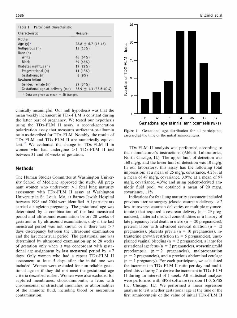
1686 Bildirici et al
clinically meaningful. Our null hypothesis was that themean weekly increment in TDx-FLM is constant duringthe latter part of pregnancy. We tested our hypothesisusing the TDx-FLM II assay, a second-generationpolarization assay that measures surfactant-to-albuminratio as described for TDx-FLM. Notably, the results ofTDx-FLM and TDx-FLM II are numerically equiva-lent.17 We evaluated the change in TDx-FLM II inwomen who had undergone O1 TDx-FLM II testbetween 31 and 38 weeks of gestation.
Methods
The Human Studies Committee at Washington Univer-sity School of Medicine approved the study. All preg-nant women who underwent O1 fetal lung maturityassessment with TDx-FLM II assay at WashingtonUniversity in St. Louis, Mo, at Barnes Jewish Hospitalbetween 1998 and 2004 were identified. All participantscarried a singleton pregnancy. The gestational age wasdetermined by a combination of the last menstrualperiod and ultrasound examination before 20 weeks ofgestation or by ultrasound examination, only if the lastmenstrual period was not known or if there was O7days discrepancy between the ultrasound examinationand the last menstrual period. The gestational age wasdetermined by ultrasound examination up to 28 weeksof gestation only when it was concordant with gesta-tional age assignment by last menstrual period by !7days. Only women who had a repeat TDx-FLM IIassessment at least 5 days after the initial one wasincluded. Women were excluded for non-reliable gesta-tional age or if they did not meet the gestational agecriteria described earlier. Women were also excluded forruptured membranes, chorioamnionitis, a fetus withchromosomal or structural anomalies, or abnormalitiesof the amniotic fluid, including blood or meconiumcontamination.
Table I Participant characteristic
Characteristic Measure
MotherAge (y)* 28.8 G 6.7 (17-46)Nulliparous (n) 13 (15%)Race (n)White 46 (54%)Black 39 (46%)
Diabetes mellitus (n) 19 (22%)Pregestational (n) 11 (13%)Gestational (n) 8 (9%)
Newborn infantGender: Female (n) 29 (34%)Gestational age at delivery (mo) 36.9 G 1.3 (33.6-40.4)
* Data are given as mean G SD (range).
TDx-FLM II analysis was performed according tothe manufacturer’s instructions (Abbott Laboratories,North Chicago, IL). The upper limit of detection was160 mg/g, and the lower limit of detection was 10 mg/g.In our laboratory, this assay has the following totalimprecision: at a mean of 25 mg/g, covariance, 4.2%; ata mean of 49 mg/g, covariance, 3.9%; at a mean of 97mg/g, covariance, 4.3%; and using patient-derived am-niotic fluid pool, we obtained a mean of 20 mg/g,covariance, 11%.
Indications for fetal lungmaturity assessment includedprevious uterine surgery (classic cesarean delivery, O2low transverse cesarean deliveries or multiple myomec-tomies) that required a cesarean delivery (n = 29 preg-nancies), maternal medical comorbidities or a history oflate pregnancy fetal death in utero (n = 20 pregnancies),preterm labor with advanced cervical dilation (n = 12pregnancies), placenta previa (n = 10 pregnancies), in-trauterine growth restriction (n = 5 pregnancies), unex-plained vaginal bleeding (n = 2 pregnancies), a large forgestational age fetus (n = 2 pregnancies), worseningmildpreeclampsia (n = 2 pregnancies), malpresentation(n = 2 pregnancies), and a previous abdominal cerclage(n = 1 pregnancy). For each participant, we calculatedthe increment in TDx-FLM II ratio per day and multi-plied this value by 7 to derive the increment in TDx-FLMII during an interval of 1 week. All statistical analyseswere performed with SPSS software (version 11.0; SPSSInc, Chicago, IL). We performed a linear regressionanalysis to test whether gestational age at the time of thefirst amniocentesis or the value of initial TDx-FLM II
Figure 1 Gestational age distribution for all participants,assessed at the time of the initial amniocentesis.

Bildirici et al 1687
Figure 2 The TDx-FLM II ratios according to participants’ gestational ages.
ratio predicted the weekly increment in TDx-FLM IIratio. TheStudent t test and analysis of variancewere usedfor comparison of weekly changes in TDx-FLM II valuesamong participant groups. A probability value of !.05was considered significant.
Results
Our study included 85 participants who underwent 2consecutive amniocentesis procedures for the determi-nation of fetal lung maturity (Table I). Although 15participants underwent a third amniocentesis, theseadditional data were not included in the analysis. Themean gestational age at the time of the first TDx-FLMII test was 34.7 G 1.4 weeks (range, 31.2-37.5 weeks;Figure 1). The mean time interval between the 2 amni-ocentesis procedures was 10 G 5.1 days (range, 5-36days; median, 7 days). The TDx-FLM II ratios for allparticipants are presented in Figure 2. The mean valueof weekly TDx-FLM II increments between the first andsecond amniocentesis for all participants was 14.4 G 9.9mg/g (95% CI, 12.3-16.5; median, 12.7 mg/g).
Using a linear regression (Figure 3), we found thatthe mean weekly increment in TDx-FLM II did notchange significantly with advancing gestational agebetween 31 and 38 weeks (P = .335). Women at %34and O34 weeks of gestation had a mean weekly incre-ment of 13.0 G 10.0 mg/g and 15.0 G 9.9 mg/g,respectively (P = .42). Furthermore, as shown in Figure4, the mean weekly increment in TDx-FLM II did notsignificantly change as a function of the initial TDx-FLM II ratio that ranged between 10 and 55 mg/g(P = .284). In addition, maternal race, diabetic status,or fetal gender did not significantly impact the meanweekly increment in TDx-FLM II (not shown).
Of all 85 participants, 46 women received steroids forthe enhancement of fetal lung maturity, which includeddexamethasone in 60% of the women and betametha-sone in 40% of the women. The timing of steroidadministration is shown in Table II. The mean weeklyincrement in TDx-FLM II among all 46 women whoreceived steroids was similar to those who did notreceive steroids (Table II). The timing of steroid admin-istration relative to consecutive amniocentesis proceduresand corresponding weekly increments in TDX-FLM II

1688 Bildirici et al
is summarized in Table II. As expected, the meanweekly increment in TDx-FLM II in the group ofwomen who received steroids immediately after theinitial amniocentesis and before the second amniocen-tesis was increased. However, the number of partici-pants in each of the subcategories presented in Table IIwas too small for meaningful conclusions. Twelve ofthe 85 newborn infants in our study were admitted tothe special care nursery or the newborn intensive careunit. The range of their last TDx-FLM II was 12.9 to41.4 mg/g.
Comment
The results support our null hypothesis that the meanweekly increment of 14.4 G 9.9 mg/g in TDx-FLM IIratio is relatively constant between 31 and 38 weeks ofgestation. In addition, the observed mean weekly incre-ment in TDx-FLM II did not significantly change acrossa wide range of initial TDx-FLM II values. Similarresults regarding the increment in lecithin/sphingomye-lin ratio were reported recently in an abstract form byDeering et al.18 Notably, our data are based on 2sequential amniocentesis procedures in a large groupof women and not on several procedures in individuals.Therefore, the finding of a relatively constant meanweekly TDx-FLM II increment during the latter part of
Figure 3 The weekly TDx-FLM II increment as a function of
gestational age. A scatter plot for the weekly increment inTDx-FLM II, according to the corresponding gestational agesat the time of initial amniocentesis. A linear regression line that
represents the best fit is shown (P = .335; b coefficient, 0.106;R2 = 0.011). The dotted line represents 95th percentile for allcases.
pregnancy does not rule out the possibility that in anindividual the weekly TDx-FLM II increment mayaccelerate transiently. Nevertheless, our data effectivelyrule out the presence of a specific time point between 31
Figure 4 The weekly TDx-FLM II increment for each
participant as a function of initial TDx-FLM II value. Alinear regression line that represents the best fit is shown(P = .284; b coefficient, .118; R2 = 0.014).
Table II The influence of steroid exposure on mean weeklyTDx-FLM-II increment
Steroids*Gestationalage (wk)
Weekly TDx-FLMII increment
Not administered(n = 34; 40%)
35.6 G 1.0 15.4 G 11.4
Administered(n = 46; 54%)y
34.0 G 1.4z 14.1 G 9.2
Steroids beforethe initialamniocentesis
23 11.1 G 4.4
Steroids after theinitial amniocentesis
18 18.7 G 12.4
Steroids before andafter the initialamniocentesis
3 9.1 G 6.8
* Information on steroid administration was not available for 5
women.y The exact timing of steroid administration was uncertain for two
women.z P ! .001, when compared with the gestational age of women
who did not receive steroids. All women who did not receive steroids
were at a gestational age of O34 weeks. The numbers for the
subgroups of participants who received steroids was too small for
meaningful conclusions.

Bildirici et al 1689
and 38 weeks of gestation when such acceleration mightoccur. Whereas 15 participants in our study underwent athird amniocentesis, the data from the additional pro-cedure were not included in our analysis. Inclusion ofthese additional data would not have changed ourestimate of the mean weekly increment (14.3 G 9.6mg/g). The exclusion of 8 cases with a weekly incrementabove 95th percentile confidence interval (Figure 1)would have yielded a more conservative estimate ofthe weekly increment (11.8G 5.6 mg/g). Last, we notedthat, whereas most subjects underwent 2 consecutivetests in 1 to 2 weeks, in 4 women this interval was O20days. Although the weekly increment for these womenwas not markedly different from those with an intervalof 1 week, an assumption of linearity for such a longperiod might have introduced a small error into theweekly estimates. We determined the power of our study(a= .05) to find a relevant difference in mean weeklyTDx-FLM-II ratio increment between participants at agestational age above or below 34 weeks. Based on ourdata, our study had a power of 84% to detect adifference of 7 mg/g in the weekly TDx-FLM II incre-ment between the 2 groups.
We also found that maternal age, parity, race, or fetalgender did not alter the weekly increment in TDx-FLMII. We noted that women who were carrying malefetuses were over-represented in our series (Table I).Although this could be a mere coincidence, it might alsoreflect the parents’ expectation of a more favorableoutcome for preterm female infants and therefore lessenthusiasm for amniocentesis for female fetuses. Never-theless, in our series the mean weekly increment in TDx-FLM II was not statistically different for women whowere carrying male or female fetuses. Interestingly, wedid not observe a difference in TDx-FLM II incrementbetween diabetic and nondiabetic women. Our data areconsistent with the possibility that the weekly incrementin fetal lung maturity testing in diabetic women takesplace at a pace similar to that of nondiabetic women, yetthe process may begin at a later gestational age.19
Our retrospective study was not designed to interro-gate the influence of steroids on fetal lung maturation.Although we found that the weekly increment in TDx-FLM II was not significantly different in women whoreceived steroids at any time point and those womenwho did not, the mean weekly increment in TDx-FLMII in women who received steroids immediately after theinitial amniocentesis was increased. However, the smallnumber of participants precluded a meaningful conclu-sion. Women who received steroids were at a signifi-cantly younger gestational age than those women whodid not receive steroids. In addition, among all womenwho received steroids, there was an average of 3-weekinterval between the steroid administration and theinitial amniocentesis. It is likely that steroid administra-tion impacted fetal lung maturity in many of the study
participants, but not necessarily during the weeklyinterval that was assessed in this study.
TDx-FLM is among the most commonly used clinicaltests for the prediction of fetal lung maturity.7-13,20,21
Indeed, Mackenzie et al22 described the influence of 10-unit increment in TDx-FLM and the change in the riskof HMD. Conclusions from our study, combined withgestational age, may assist the practitioner in extrapo-lating the risk of HMD and in optimizing the timing ofrepeat fetal lung maturity testing.
Acknowledgment
We thank Lori Rideout for administrative assistance.
References
1. Hallman M, Kulovich M, Kirkpatrick E, Sugarman RG, Gluck L.
Phosphatidylinositol and phosphatidylglycerol in amniotic fluid:
indices of lung maturity. Am J Obstet Gynecol 1976;125:613-7.
2. Guyer B, Hoyert DL, Martin JA, Ventura SJ, MacDorman MF,
Strobino DM. Annual summary of vital statistics: 1998. Pediatrics
1999;104:1229-46.
3. Hack M, Fanaroff AA, Klaus MH, Mendelawitz BD, Merkatz IR.
Neonatal respiratory distress following elective delivery: A pre-
ventable disease? Am J Obstet Gynecol 1976;126:43-7.
4. Goldenberg RL, Nelson K. Iatrogenic respiratory distress syn-
drome: an analysis of obstetric events preceding delivery of infants
who develop respiratory distress syndrome. Am J Obstet Gynecol
1975;123:617-20.
5. Ashwood ER. Standards of laboratory practice: evaluation of fetal
lung maturity: National Academy of Clinical Biochemistry. Clin
Chem 1997;43:211-4.
6. Committee on Educational Bulletins of the American College of
Obstetricians and Gynecologists. Assessment of fetal lung matu-
rity: ACOG educational bulletin no.: 230. Int J Gynaecol Obstet
1997;56:191-8.
7. Herbert WN, Chapman JF, Schnoor MM. Role of the TDx FLM
assay in fetal lung maturity. Am J Obstet Gynecol 1993;168:
808-12.
8. Hagen E, Link JC, Arias F. A comparison of the accuracy of the
TDx-FLM assay, lecithin-sphingomyelin ratio, and phosphatidyl-
glycerol in the prediction of neonatal respiratory distress syn-
drome. Obstet Gynecol 1993;82:1004-8.
9. Del Valle GO, Adair CD, Ramos EE, Gaudier FL, Sanchez-
Ramos L, Morales R. Interpretation of the TDx-FLM fluores-
cence polarization assay in pregnancies complicated by diabetes
mellitus. Am J Perinatol 1997;14:241-4.
10. Livingston EG, Herbert WN, Hage ML, Chapman JF, Stubbs
TM. Use of the TDx-FLM assay in evaluating fetal lung maturity
in an insulin-dependent diabetic population: The Diabetes and
Fetal Maturity Study Group. Obstet Gynecol 1995;86:826-9.
11. Pinette MG, Blackstone J, Wax JR, Cartin A. Fetal lung maturity
indices-a plea for gestational age-specific interpretation: a case
report and discussion. Am J Obstet Gynecol 2002;187:1721-2.
12. Tanasijevic MJ, Wybenga DR, Richardson D, Greene MF, Lopez
R, Winkelman JW. A predictive model for fetal lung maturity
employing gestational age and test results. Am J Clin Pathol
1994;102:788-93.
13. McElrath TF, Colon I, Hecht J, Tanasijevic MJ, Norwitz ER.
Neonatal respiratory distress syndrome as a function of gestational

1690 Bildirici et al
age and an assay for surfactant-to-albumin ratio. Obstet Gynecol
2004;103:463-8.
14. Stubblefield PG. Using the TDx-FLM assay and gestational age
together for more accurate prediction of risk for neonatal
respiratory distress syndrome. Am J Obstet Gynecol 2002;187:
1429-30.
15. Parvin CA, Kaplan LA, Chapman JF, McManamon TG, Gro-
nowski AM. Predicting respiratory distress syndrome using gesta-
tional age and fetal lung maturity by fluorescent polarization.
Am J Obstet Gynecol 2005;192:199-207.
16. Richardson DK, Heffner LJ. Fetal-lung maturity: tests mature,
interpretation not. Lancet 2001;358:684-6.
17. PinetteMG. Reply to letter. Am J Obstet Gynecol 2003;189:1513-4.
18. Deering SH, Poggi SH, Spong CY, Barton FB, Goodwin KM,
Ghidini A. Repeated assessment of the lecithin-sphingomyelin
ratio: What is the expected change over time? [abstract]. J Soc
Gynecol Investig 2004;11:104A.
19. Moore TR. A comparison of amniotic fluid fetal pulmonary
phospholipids in normal and diabetic pregnancy. Am J Obstet
Gynecol 2002;186:641-50.
20. Fantz CR, Powell C, Karon B, Parvin CA, Hankins K, Dayal M,
et al. Assessment of the diagnostic accuracy of the TDx-FLM II to
predict fetal lung maturity. Clin Chem 2002;48:761-5.
21. Kesselman EJ, Figueroa R, Garry D, Maulik D. The usefulness of
the TDx/TDxFLx fetal lung maturity II assay in the initial
evaluation of fetal lung maturity. Am J Obstet Gynecol 2003;
188:1220-2.
22. Mackenzie MW, Srundell C, Stubblefield PG. Fluorescence polar-
ization testing of amniotic fluid as a predictor of neonatal
respiratory outcome. J Matern Fetal Med 1995;4:179-85.

American Journal of Obstetrics and Gynecology (2005) 193, 1691–7
www.ajog.org
Maternal plasma concentrations of IGF-1, IGFBP-1, andC-peptide in early pregnancy and subsequent risk ofgestational diabetes mellitus
Chunfang Qiu, MD, MS,a,* Surab Vadachkoria, MD, PhD,a Lois Meryman, BA,a
Ihunnaya O. Frederick, MPH,a Michelle A. Williams, ScDa,b
Center for Perinatal Studies,a Swedish Medical Center; Department of Epidemiology,b University of WashingtonSchool of Public Health and Community Medicine, Seattle, WA
Received for publication November 5, 2004; revised March 17, 2005; accepted April 1, 2005
KEY WORDSIGF-1IGFBP-1C-peptide
Gestational diabetesmellitus
Objective: Insulin-like growth factor-1 (IGF-1) and IGF binding protein-1 (IGFBP-1) may beimportant determinants of glucose homeostasis. We examined the association between circulating
concentrations of IGF-1, IGFBP-1 in early pregnancy and development of gestational diabetesmellitus (GDM).Study design: Maternal plasma (collected at 13 weeks) IGF-1, IGFBP-1, and C-peptide were
measured using immunoassay. Relative risks (RR) and 95% CIs were calculated.Results: The percentage of the cohort that developed GDM was 5.8% (n = 804). Free IGF-1and IGFBP-1 were inversely associated with GDM risk, while C-peptide was positively associated
with GDM risk (P for trend test ! .05). Women with free IGF-1 R1.08 ng/mL experienced a69% reduced risk of GDM (CI 0.12-0.75) compared with women having concentrations !0.80ng/mL. There was a 57% reduced risk of GDM among women with IGFBP-1 R68.64 ng/mL
(RR = 0.43, CI 0.18-1.05). Women with C-peptide R3.00 ng/mL experienced a 2.28-foldincreased risk of GDM (CI 1.00-5.19) compared with women who had concentrations !1.45ng/mL.Conclusion: These associations may help to further elucidate the pathologic process of GDM.
� 2005 Mosby, Inc. All rights reserved.
Emerging evidence indicates that insulin-like growthfactor-1 (IGF-1), and insulin-like growth factor bindingproteins (IGFBPs), are involved in a myriad of physi-ologic and pathologic processes related to reproductive
Supported in part by an award from the National Institutes of
Health (R01-HD/HL 32562).
* Reprint requests: Dr Chunfang Qiu, Center for Perinatal Studies,
Swedish Medical Center, 747 Broadway (Suite 4 North), Seattle, WA,
98122.
E-mail: [email protected]
0002-9378/$ - see front matter � 2005 Mosby, Inc. All rights reserved.
doi:10.1016/j.ajog.2005.04.015
endocrinology,1 bone metabolism,2 and the growth ofvarious cancers.3 The structural homology betweenIGF-1 and insulin and the hypoglycemic activity regu-lated by IGFBPs also suggest that IGF-1 and its bindingproteins have an intrinsic role in glucose metabolismand homeostasis.4 To date, 6 binding proteins regulatingIGF-1 bioactivity have been characterized.5 A large pro-portion of IGF-1 in circulation is bound to IGFBP-3.5
The free fraction of IGF-1, which is the biologicallyactive form and comprises less than 1% of the total
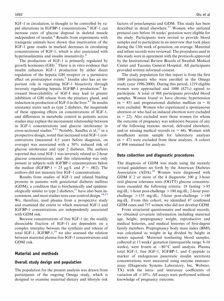
1692 Qiu et al
IGF-1 in circulation, is thought to be controlled by ra-pid alterations in IGFBP-1 concentrations.6 IGF-1 canincrease rates of glucose disposal in skeletal muscleindependent of insulin.4 Results from experiments withtransgenic animals have shown that inactivation of theIGF-1 gene results in marked decreases in circulatingconcentrations of IGF-1, which is also associated withhyperinsulinemia and insulin insensitivity.4
The production of IGF-1 is primarily regulated bygrowth hormones (GH).7 There is in vitro evidence thatinsulin enhances IGF-1 production by either directregulation of the hepatic GH receptor or a permissiveeffect on postreceptor events.8 Insulin also has an im-portant role in regulating IGF-1 bioactivity throughinversely regulating hepatic IGFBP-1 production.6 In-creased bioavailability of IGF-I may lead to greaterinhibition of GH release from the pituitary gland, andreduction in production of IGF-I in the liver.6,7 In insulinresistance states such as type 2 diabetes, the magnitudeof these opposing effects on concentrations of IGF-1,and differences in metabolic control in patients acrossstudies may explain the inconsistent relationship betweenin IGF-1 concentrations and glucose intolerance incross-sectional studies.9-11 Notably, Sandhu et al,12 in aprospective design, noted that increased total IGF-1 con-centrations (measured 4.5 years before diagnosis, onaverage) was associated with a 50% reduced risk ofglucose intolerance and type 2 diabetes. The authorsreported that total IGF-1 was inversely related to 2-hourglucose concentrations, and this relationship was onlypresent in subjects with IGFBP-1 concentrations belowthe median (IGFBP-1 = 25 mg/L and P = .002). Theauthors did not measure free IGF-1 concentrations.
Results from studies of IGF-1 and related bindingproteins in patients with gestational diabetes mellitus(GDM), a condition that is biochemically and epidemi-ologically similar to type 2 diabetes,13 have also been in-consistent, andmost studies were case-control design.14,15
We, therefore, used plasma from a prospective studyand examined the extent to which maternal IGF-1 andIGFBP-1 concentrations are independently associatedwith GDM risk.
Because concentrations of free IGF-1 (ie, the readilydissociable fraction of IGF-1) are dependent on acomplex interplay between the synthesis and release oftotal IGF-1, IGFBP-1,16 we also assessed the relationbetween maternal plasma free IGF-1 concentrations andGDM risk.
Material and methods
Overall study design and population
The population for the present analysis was drawn fromparticipants of the ongoing Omega study, which isdesigned to examine maternal dietary and lifestyle risk
factors of preeclampsia and GDM. This study has beendescribed in detail elsewhere.17 Women who initiatedprenatal care before 16 weeks’ gestation were eligible forthe study. Participants were invited to provide bloodsamples and to participate in an interview that took placeduring the 13th week of gestation, on average. Maternaland infant records were reviewed. The procedures used inthis study were in agreement with the protocols approvedby the Institutional Review Boards of Swedish MedicalCenter and Tacoma General Hospital. All participantsprovided written informed consent.
The study population for this report is from the first1000 participants who were enrolled in the Omegastudy (year 1996-2000). During this period, 1219 eligiblewomen were approached and 1000 (82%) agreed toparticipate. A total of 968 participants provided bloodsamples. Women found to have chronic hypertension(n = 45) and pregestational diabetes mellitus (n = 4)were excluded. Women who experienced a spontaneousabortion or who had an induced abortion were excluded(n = 22). Also excluded were those women for whomthe outcome of pregnancy was unknown because of anyof the following reasons: moved, delivered elsewhere,and/or missing medical records (n = 46). Women withinsufficient serum sample for laboratory analyses(n = 47) were excluded from these analyses. A cohortof 804 remained for analysis.
Data collection and diagnostic procedures
The diagnosis of GDM was made using the recentlyrevised guidelines set forth by the American DiabetesAssociation (ADA).18 Women were diagnosed withGDM if 2 or more of the 4 diagnostic 100 g 3-houroral glucose tolerance tests (OGTT) glucose concentra-tions exceeded the following criteria: 18 fasting O95mg/dL; 1-hour post-challenge O180 mg/dL; 2-hour post-challenge O155 mg/dL; 3-hour post-challenge O140mg/dL. From this cohort, we identified 47 confirmedGDM cases and 757 women who did not develop GDM.
From structured questionnaire and medical records,we obtained covariate information including maternalage, height, prepregnancy weight, reproductive andmedical histories, and medical histories of first-degreefamily members. Prepregnancy body mass index (BMI)was calculated as weight in kg divided by height inmeters squared. Maternal nonfasting blood samples,collected at 13 weeks’ gestation (interquartile range: 8-16weeks), were frozen at �80(C until analysis. Plasmatotal IGF-1, free IGF-1, IGFBP-1, and C-peptide (amarker of endogenous pancreatic insulin secretion)concentrations were measured using enzyme immuno-assay (Diagnostic Systems Laboratory, Inc, Webster,TX) with the intra- and interassay coefficients ofvariation all !10%. All assays were performed withoutknowledge of pregnancy outcome.

Qiu et al 1693
Table I Characteristics of study cohort members according to maternal plasma insulin-like growth factor binding protein-1 (IGFBP-1)concentration measured in early pregnancy, Seattle and Tacoma, Washington, 1996-2000
Entire cohortn = 804
Tertiles
Characteristics 1 (Low) n = 268 2 (Mid) n = 268 3 (High) n = 268
Plasma IGFBP-1 (ng/mL) !35.96 35.96-68.63 R68.64
% % % %Maternal Age (y)* 32.0 G 0.3 32.4 G 0.3 31.7 G 0.3 32.0 G 0.3!35 70.9 65.3 73.9 73.5R35 27.9 33.6 24.6 25.4
Maternal race/ethnicityNon-Hispanic white 83.6 82.5 84.7 83.6African American 2.5 3.0 2.6 1.9Other 12.7 13.4 11.2 13.4
Unmarried 11.2 13.4 10.8 9.3%12 years’ education 4.0 4.9 4.1 3.0Nulliparous 84.0 77.6 86.9 87.3Family history of diabetes 14.8 18.7 15.7 10.1Smoker 6.3 9.3 4.1 5.6No exercise 17.5 17.9 15.4 19.3
Prepregnancy BMI (kg/m2)!20.0 19.9 13.1 19.0 27.620.0-24.9 54.2 45.2 56.3 61.225.0-29.9 15.6 21.3 18.3 7.1R30.0 8.7 18.3 5.2 2.6
Gestational age at blood collection (wk) 12.9 G 0.2* 12.6 G 0.2 13.1 G 0.2 13.1 G 0.2
* Mean G SE = mean G standard error.
Statistical analytic procedures
Women were grouped according to tertiles determinedby the distribution of plasma IGFBP-1 concentrationsamong the entire cohort. We examined frequency dis-tributions of maternal sociodemographic characteristicsand medical and reproductive histories according toplasma IGFBP-1 tertiles. Spearman’s correlation coeffi-cients were used to measure the closeness of a linearrelationship between maternal plasma IGF-1, IGFBP-1,and C-peptide concentrations and selected characteris-tics. We estimated the relative association betweenvarying tertiles of plasma IGF-1, IGF-BP1, and risk ofGDM using Stata (version 7.0) software (Stata, CollegeStation, TX). We fitted generalized linear models toderive risk ratios (RR) and 95% CIs.19 In multivariableanalyses, we evaluated linear trends in risk by treatingthe tertiles as a continuous variable. To assess con-founding, we entered variables into a generalized linearmodel 1 at a time, and then compared the adjusted andunadjusted risk ratios. Final generalized linear modelsincluded covariates that altered unadjusted risk ratios byat least 10%, as well as those covariates of a prioriinterest (eg, maternal age, nulliparity, prepregnancyBMI, and family history of type 2 diabetes mellitus).Statistical significance was not used as a criterion forselecting confounders. Maternal race, parity (as a con-
tinuous variable), and smoking status, as well as phys-ically inactivity during pregnancy, were not confoundersin this analysis.
Results
The characteristics of women in the study cohortaccording to tertiles of plasma IGFBP-1 concentrationsare summarized in Table I.
Maternal plasma free IGF-1 and IGFBP-1 concen-trations were inversely correlated with prepregnancyBMI (r= �0.14 and r= �0.34, respectively, P ! .01).Both determinants were also inversely correlated withmaternal plasma leptin (r= �0.09, P = .01 andr = �0.17, P ! .01, respectively), as well as C-reactiveprotein (CRP) (r= �0.23, P! .01 and r = �0.13, P!.01, respectively). These concentrations were not corre-lated with maternal age, gestational age at bloodcollection, or infant birth weight. As marker of pancre-atic insulin secretion, plasma C-peptide concentrationswere significantly correlated with GDM risk factors,such as prepregnancy BMI (r = 0.23), triglyceride(r= 0.29), leptin (r = 0.25), and CRP concentrations(r= 0.15). Total IGF-1 concentrations were inverselycorrelated with CRP concentrations (r= �0.15, P !.01), but with no other factors (Table II).

1694 Qiu et al
GDM risk was 3.25-fold higher among women withC-peptide concentrations R3.00 ng/mL, as comparedwith those women whose concentrations were!1.45 ng/mL (95%CI 1.47-7.18) (Table III). This association wasattenuated after adjustment for potential confoundingby maternal age, parity, family history of diabetes, andprepregnancy BMI (RR = 2.28, 95%CI 1.00-5.19).
Compared with women in the lowest tertile (totalIGF-1 !154.18 ng/mL), women having intermediatetotal IGF-1 concentrations (154.18-215.04 ng/mL) ex-perienced a 1.71-fold (95%CI 0.82-3.58) increased riskof developing GDM after accounting for the potentialconfounders. The corresponding RR for women withthe highest tertile of total IGF-1 (O215.05 ng/mL) was1.47 (95%CI 0.70-3.08). However, this association didnot reach statistical significance.
As seen in Table III, risk of GDM decreased acrosssuccessively higher tertiles of free IGF-1 concentrations(RRs: 1.00, 0.60, and 0.24; P value for trend = .01).Relative to women in the lowest tertile of maternalplasma free IGF-1 concentrations (!0.80 ng/mL),women with concentrations in the most upper tertile(R10.08 ng/mL) experienced a 76% reduced risk ofGDM (RR = 0.24; 95%CI 0.10-0.58). After adjustmentfor confounders, the RR was attenuated slightly (ad-justed RR = 0.31; 95%CI 0.12-0.75). A similar patternwas seen for IGFBP-1. The risk of GDM decreasedacross successively higher tertiles of IGFBP-1 concentra-tions (RRs: 1.00, 0.72, and 0.43; P value fortrend = .06). There was a 57% reduced risk of GDMamong women with IGFBP-1 concentrations R68.64ng/mL (RR = 0.43, 95%CI 0.18-1.05) compared to
Table II Spearman correlation coefficients (g) between ma-ternal plasma insulin-like growth factor-1 and binding proteinconcentrations and selected characteristics, as well as somebiomarkers, Seattle and Tacoma, Washington, 1996-2000
Characteristics C-peptideTotalIGF-1
FreeIGF-1 IGFBP-1
Maternal age (y) �0.02 �0.04 0.02 �0.03Prepregnancy body
mass index (kg/m2)0.23* �0.01 �0.14* �0.34*
High-densitylipoportein (mg/dL)
�0.08y 0.04 0.02 0.26
Low-densitylipoprotein (mg/dL)
0.06 0.01 �0.13* 0.05
Triglyceride (mg/dL) 0.29* 0.03 �0.13* 0.06Leptin (ng/mL) 0.25* 0.04 �0.09y �0.17*C-reactive protein
(mg/L)0.15* �0.15* �0.23* �0.13*
Gestational age atblood collection (wk)
�0.01 0.03 0.01 0.09y
Infant birth weight (g) �0.13* �0.03 �0.01 0.04
* P value ! .01.y P value ! .05.
women with concentrations in the lowest tertile(!35.96 ng/mL) after adjusting for confounders.
Comment
GDM, a heterogeneous complication of pregnancy, ischaracterized by carbohydrate intolerance of variableseverity with onset or first recognition during pregnancy.Insulin resistance and b-cell dysfunction are thought tobe major determinants of its development.18 Its patho-physiologic and epidemiologic characteristics in manyways resemble that of type 2 diabetes.13 An expandingbody of evidence documents similarities between GDMand the metabolic syndrome.20 Our finding of an asso-ciation between elevated C-peptide in early pregnancyand subsequent GDM risk suggests that maternalhyperinsulinemia and/or insulin resistance is detectableabout 15 weeks before GDM is typically diagnosed.Our findings are consistent with results from other stud-ies.21,22 Kautzky-Willer et al21 reported that C-peptidewas 30% higher (P! .05) in subjects with GDM than incontrols or in nonpregnant lean women. These investi-gators also noted that b-cell sensitivity to glucose forinsulin release was decreased by 40% to 50% (P ! .01)in subjects with GDM. Winkler et al22 reported that C-peptide was 6.0 G 2.7 and 3.1G1.7 mg/L in 30 GDMcases and 35 pregnant controls, respectively (P ! .01).
A finding pertaining to total IGF-1 is in agreementwith observations from some case control studies whereplasma concentrations of this analyte were determinedat delivery. Luthman et al14 reported that there was nodifference of total IGF-1 concentrations between womenwith GDM and controls. Our findings and those ofLuthman, however, are inconsistent with findings re-ported by Sandhu12 or Hughes et al,15 who noted thattotal IGF-1 concentrations (measured in samples col-lected in the third trimester) were higher in women withGDM than in controls.
Although we are not aware of other published reportsregarding maternal free IGF-1 concentrations andGDM risk, our observation of an inverse associationbetween plasma free IGF-1 and GDM risk is in agree-ment with other studies involving metabolic syndrome.23
For instance, Heald et al23 reported that elevated freeIGF-I and IGFBP-1 concentrations were associatedwith decreased risk of metabolic syndrome (OR =0.46, 95%CI 0.22-0.96; OR = 0.58, 95%CI 0.44-0.76,respectively).
Our finding of an inverse association betweenIGFBP-1 and subsequent GDM risk is not in agreementwith a report from O’Leary et al.24 In a case-controlstudy where maternal concentrations of IGFBP-1 weremeasured after the clinical diagnosis of GDM, theauthors reported that IGFBP-1 levels was higher inGDM cases than controls.24 However, our results areconsistent with a large body of literature documenting a
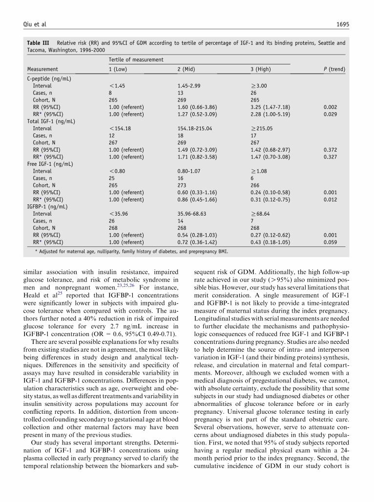
Qiu et al 1695
Table III Relative risk (RR) and 95%CI of GDM according to tertile of percentage of IGF-1 and its binding proteins, Seattle andTacoma, Washington, 1996-2000
Tertile of measurement
Measurement 1 (Low) 2 (Mid) 3 (High) P (trend)
C-peptide (ng/mL)Interval !1.45 1.45-2.99 R3.00Cases, n 8 13 26Cohort, N 265 269 265RR (95%CI) 1.00 (referent) 1.60 (0.66-3.86) 3.25 (1.47-7.18) 0.002RR* (95%CI) 1.00 (referent) 1.27 (0.52-3.09) 2.28 (1.00-5.19) 0.029
Total IGF-1 (ng/mL)Interval !154.18 154.18-215.04 R215.05Cases, n 12 18 17Cohort, N 267 269 267RR (95%CI) 1.00 (referent) 1.49 (0.72-3.09) 1.42 (0.68-2.97) 0.372RR* (95%CI) 1.00 (referent) 1.71 (0.82-3.58) 1.47 (0.70-3.08) 0.327
Free IGF-1 (ng/mL)Interval !0.80 0.80-1.07 R1.08Cases, n 25 16 6Cohort, N 265 273 266RR (95%CI) 1.00 (referent) 0.60 (0.33-1.16) 0.24 (0.10-0.58) 0.001RR* (95%CI) 1.00 (referent) 0.86 (0.45-1.66) 0.31 (0.12-0.75) 0.012
IGFBP-1 (ng/mL)Interval !35.96 35.96-68.63 R68.64Cases, n 26 14 7Cohort, N 268 268 268RR (95%CI) 1.00 (referent) 0.54 (0.28-1.03) 0.27 (0.12-0.62) 0.001RR* (95%CI) 1.00 (referent) 0.72 (0.36-1.42) 0.43 (0.18-1.05) 0.059
* Adjusted for maternal age, nulliparity, family history of diabetes, and prepregnancy BMI.
similar association with insulin resistance, impairedglucose tolerance, and risk of metabolic syndrome inmen and nonpregnant women.23,25,26 For instance,Heald et al25 reported that IGFBP-1 concentrationswere significantly lower in subjects with impaired glu-cose tolerance when compared with controls. The au-thors further noted a 40% reduction in risk of impairedglucose tolerance for every 2.7 ng/mL increase inIGFBP-1 concentration (OR= 0.6, 95%CI 0.49-0.71).
There are several possible explanations for why resultsfrom existing studies are not in agreement, the most likelybeing differences in study design and analytical tech-niques. Differences in the sensitivity and specificity ofassays may have resulted in considerable variability inIGF-1 and IGFBP-1 concentrations. Differences in pop-ulation characteristics such as age, overweight and obe-sity status, aswell as different treatments and variability ininsulin sensitivity across populations may account forconflicting reports. In addition, distortion from uncon-trolled confounding secondary to gestational age at bloodcollection and other maternal factors may have beenpresent in many of the previous studies.
Our study has several important strengths. Determi-nation of IGF-1 and IGFBP-1 concentrations usingplasma collected in early pregnancy served to clarify thetemporal relationship between the biomarkers and sub-
sequent risk of GDM. Additionally, the high follow-uprate achieved in our study (O95%) also minimized pos-sible bias. However, our study has several limitations thatmerit consideration. A single measurement of IGF-1and IGFBP-1 is not likely to provide a time-integratedmeasure of maternal status during the index pregnancy.Longitudinal studies with serialmeasurements are neededto further elucidate the mechanisms and pathophysio-logic consequences of reduced free IGF-1 and IGFBP-1concentrations during pregnancy. Studies are also neededto help determine the source of intra- and interpersonvariation in IGF-1 (and their binding proteins) synthesis,release, and circulation in maternal and fetal compart-ments. Moreover, although we excluded women with amedical diagnosis of pregestational diabetes, we cannot,with absolute certainty, exclude the possibility that somesubjects in our study had undiagnosed diabetes or otherabnormalities of glucose tolerance before or in earlypregnancy. Universal glucose tolerance testing in earlypregnancy is not part of the standard obstetric care.Several observations, however, serve to attenuate con-cerns about undiagnosed diabetes in this study popula-tion. First, we noted that 95% of study subjects reportedhaving a regular medical physical exam within a 24-month period prior to the index pregnancy. Second, thecumulative incidence of GDM in our study cohort is

1696 Qiu et al
consistent with observations in other setting.18Hence, thelikelihood that our findings are due entirely to the prev-alence of undiagnosed pregestational diabetes in thiscohort is very low. Third, some RRs are statisticallyunstable due to our small sample size. Finally, the currentstudy population was primarily focused on the primipa-rous women, and the cohort was mainly well-educatedwhite women. Therefore, caution must be taken whengeneralizing our results.
The physiologic processes underlying the role of IGF-1in glucose regulation are unclear. However, it is knownthat infusion of IGF-1 will suppress glucose counter-regulatory hormones, such as glucagon and GH, andmay speed up oxidation of lipids.7 IGF-1 may alsocontribute to changes in insulin sensitivity and glucose-uptake via direct IGF receptor-mediated effects onskeletal muscle.4 Alterations in IGFBP-1 concentrationsmay represent variations in insulin secretion or hepaticinsulin sensitivity, both of which are important compo-nents in glucose regulation.6 Further studies are neededto evaluate these hypotheses and to comprehensivelyevaluate the precise roles that IGFs and IGFBPs play ininsulin resistance and the development of GDM. Futurestudies may also elucidate the extent to which reducedIGFBP-1 concentrations may be secondary to maternalhyperinsulinemia and insulin resistance.
In summary, we have shown that alteration inmaternal plasma free-IGF 1 and IGFBP-1 concentra-tions are evident in pregnancies destined to developGDM as early as 13 weeks’ gestation. The findings fromthis prospective study are generally consistent with thereports of previous investigations.14,23,24 Our findingsare also consistent with a much larger body of evidencefrom experimental, clinical, and epidemiologic investi-gations, which suggest that the IGF system is animportant mediator of glucose homeostasis in hu-mans.1,4-12 Taken together with the available literature,our results suggest that dysregulation of the synthesisand release of IGF-1 and IGFBP-1 may be of etiologicimportance in GDM.
Acknowledgments
The authors are indebted to staff at the Center forPerinatal Studies, Swedish Medical Center.
References
1. Druckmann R, Rohr UD. IGF-1 in gynecology and obstetrics:
update 2002. Maturitas 2002;41(Suppl 1):S65-83.
2. Rosen CJ. IGF-1 and osteoprosis. Clin Lab Med 2000;20:591-602.
3. Kaaks R, Toniolo P, Akhmedkhanov A, Lukanova A, Biessy C,
Dechaud H, et al. Serum C-peptide, insulin-like growth factor
(IGF)-1, IGF-binding proteins, and colorectal cancer risk in
women. JNCI 2000;92:1592-600.
4. Sjogren K, Wallenius K, Liu JL, Bohlooly-Y M, Pacini G,
Svensson L, et al. Liver derived IGF-1 is of importance for normal
carbohydrate and lipid metabolism. Diabetes 2001;50:1539-45.
5. Ferry RJ, Cerri RW, Cohen P. Insulin-like growth factor binding
proteins: new proteins, new functions. Horm Res 1999;51:34-41.
6. Lee PD, Giudice LC, Conover CA, Power DR. Insulin-like growth
factor binding protein-1: recent findings and new directions. Proc
Soc Exp Biolo Med 1997;216:319-57.
7. Jones JI, Clemmons DR. Insulin-like growth factors and their
binding proteins: biological functions. Endocrine Rev 1995;16:
3-34.
8. Leung K, Rajkovic IA, Peters E, Markus I, Van Wyk JJ, Ho KK.
Insulin-like growth factor I and insulin down-regulate growth
hormone (GH) receptors in rat osteoblasts: evidence for a
peripheral feedback loop regulating GH action. Endocrinology
1996;137:2694-702.
9. Frystyk J, Skjaerbaek C, Vestbo E, Fisker S, Orskov H. Circulat-
ing levels of free insulin-like growth factors in obese subjects: the
impact of type 2 diabetes. Diabetes Metab Res Rev 1999;15:
314-22.
10. Dohm GL, Elton CW, Raju MS, Mooney ND, DiMarchi R,
Pories WJ, et al. IGF-I–stimulated glucose transport in human
skeletal muscle and IGF-I resistance in obesity and NIDDM.
Diabetes 1990;39:1028-32.
11. Abou-Seif MA, Youssef AA. Oxidative stress and male IGF-1,
gonadotropin and related hormones in diabetic patients. Clin
Chem Lab Med 2001;39:618-23.
12. Sandhu MS, Heald AH, Gibson JM, Cruickshank JK, Dunger
DB, Wareham NJ. Circulating concentrations of insulin-like
growth factor-1 and development of glucose intolerance: a pro-
spective observational study. Lancet 2002;359:1740-5.
13. Pendergrass M, Fazioni E, Defronzo RA. Non-insulin-dependent
diabetes mellitus and gestational diabetes mellitus: same disease,
another name? Diabetes Reviews 1995;3:566-83.
14. Luthman M, Stock S, Werner S, Bremme K. Growth hormone-
binding protein in plasma is inversely correlated to placental
lactogen and augmented with increasing body mass index in
healthy pregnant women and women with gestational diabetes
mellitus. Gynecol Obstet Invest 1994;38:145-50.
15. Hughes SC, Johnson MR, Heinrich G, Holly JM. Could abnor-
malities in insulin-like growth factors and their binding proteins
during pregnancy result in gestational diabetes? J Endocrinol
1995;147:517-24.
16. Frystyk J. Free insulin-like growth factorsdmeasurements and
relationships to growth hormone secretion and glucose homeosta-
sis. Growth Horm IGF Res 2004;14:337-75.
17. Qiu C, Luthy DA, Zhang C, Walsh SW, Leisenring WM, Williams
MA. A prospective study of maternal serum C-reactive protein
concentrations and risk of preeclampsia. Am J Hypertens
2004;17:154-60.
18. Expert Committee on the diagnosis and classification of diabetes
mellitus. Report of the Expert committee on the diagnosis and
classification of Diabetes Mellitus. Diabetes Care 2002;25:S5-20.
19. Hardin JW, Hilbe J. Generalized linear models and extensions.
College Station, TX: Stata Press; 2001.
20. Clark CM Jr, Qiu C, Amerman B, Porter B, Fineberg N,
Aldasouqi S, et al. Gestational diabetes: should it be added
to the syndrome of insulin resistance? Diabetes Care 1997;20:
867-71.
21. Kautzky-Willer A, Prager R, Waldhausl W, Pacini G, Thomaseth
K, Wagner OF, et al. Pronounced insulin resistance and inade-
quate beta-cell secretion characterize lean gestational diabetes
during and after pregnancy. Diabetes Care 1997;20:1717-23.
22. Winkler G, Cseh K, Baranyi E, Melczer Z, Speer G, Hajos P, et al.
Tumor necrosis factor system in insulin resistance in gestational
diabetes. Diabetes Res Clin Pract 2002;56:93-9.
23. Heald AH, Anderson SG, Ivison F, Laing I, Gibson JM,
Cruickshank K. C-reactive protein and the insulin-like growth
factor (IGF)-system in relation to risk of cardiovascular disease in
different ethnic groups. Atherosclerosis 2003;170:79-86.

Qiu et al 1697
24. O’Leary PC, Longley M. Serum insulin-like growth factor binding
protein-1 in pregnant women: decreased concentrations following
an oral glucose load. Ann Clin Biochem 1994;31:40-5.
25. Heald AH, Cruickshank JK, Riste LK, Cade JE, Anderson S,
Greenhalgh A, et al. Close relation of fasting insulin-like growth
factor binding protein-1 (IGFBP-1) with glucose tolerance and
cardiovascular risk in two populations. Diabetologia 2001;44:
333-9.
26. Harrela M, Koistinen R, Tuomilehto J, Nissinen A, Seppala M.
Low serum insulin-like growth factor-binding protein-1 is associ-
ated with an unfavourable cardiovascular risk profile in elderly
men. Ann Med 2000;32:424-8.

American Journal of Obstetrics and Gynecology (2005) 193, 1698–702
www.ajog.org
Protein Z in patients with pregnancy complications
Florence Bretelle, MD, PhD,a,* Dominique Arnoux, PhD,b Raha Shojai, MD,c
Claude D’Ercole, MD,c Jose Sampol, PhD,d,e Francoise Dignat, PhD,d,e
Laurence Camoin-Jau, PhDd,e
Service de Gynecologie-Obstetrique, Pr. B. Blanc, Centre Hospitalo-Universitaire de la Conception,a Laboratoired’Hematologie, Pr G. Sebahounb; and Service de Gynecologie-Obstetrique, Pr. L Boubli, Centre Hospitalo-Universitaire Nord c; Laboratoire d’Hematologie, Pr. J. Sampol, Centre Hospitalo-Universitaire de la Conceptiond;UMR Inserm 608, Laboratoire d’Hematologie et d’Immunologie, UFR de Pharmacie,e Marseille, France
Received for publication July 24, 2004; accepted April 1, 2005
KEY WORDSProtein Z
PreeclampsiaIntrauterine growth
restriction
Intrauterine fetaldemise
Objective: This study was undertaken to evaluate the association between protein Z concentra-tion and pregnancy complications.Study design: A prospective case-control study was conducted over a 2-year period to evaluatethe prevalence of protein Z deficiency in pregnancy complications. Protein Z levels were measured
at the time of diagnosis of complications such as preeclampsia, intrauterine growth restriction,and intrauterine fetal demise. Protein Z deficiency was defined as a plasma level below 1.2 mg/L.In addition to patients presenting with pregnancy complications, healthy age-matched nonpreg-
nant and pregnant women were invited to participate.Results: A total of 145 women were included in the study: 50 nonpregnant women, 34 healthypregnant women, 29 women with preeclampsia, 25 women presented with intrauterine growth
restriction, and 7 women with intrauterine fetal demise. The median protein Z level was similar inhealthy pregnant and nonpregnant women (1.63 [0.47-3.1] mg/L and 1.69 [0.7-3] mg/L,respectively). Three women with normal pregnancies had a low protein Z level (8.8%), compared
with 8 patients presenting with intrauterine growth restriction (33.3%) and 8 patients withintrauterine fetal demise (50%). Compared with normal pregnancy, the frequency of decreasedprotein Z was significantly higher in cases of intrauterine growth restriction and in intrauterinefetal demise (relative risk [RR] 1.96, 95% CI 1.16-3.32; P = .041 and RR 3.36, 95% CI 1.65-6.8;
P = .0031, respectively), but not in preeclampsia (RR 1.6, 95% CI 0.9-2.8; P = .23). Placentahistologic examination revealed vascular lesions in 50% of patients with protein Z deficiency andin 33% of patients with normal levels of protein Z (RR 0.84; 95% CI 0.6-1.2).
Conclusion: Protein Z deficiency is associated with late fetal demise and intrauterine growthrestriction. The pathophysiologic role of protein Z deficiency, either congenital or caused by thepresence of specific antibodies remains unclear and should be further investigated.
� 2005 Mosby, Inc. All rights reserved.
This study was promoted by a national grant (PH-RC).
* Reprint requests: Florence Bretelle, MD, PhD, Centre Hospitalier Universitaire de la Conception, Service de Gynecologie Obstetrique B Pr.
Blanc, 147 boulevard Baille, 13385 Marseille Cedex, France.
E-mail: [email protected]
0002-9378/$ - see front matter � 2005 Mosby, Inc. All rights reserved.
doi:10.1016/j.ajog.2005.04.006
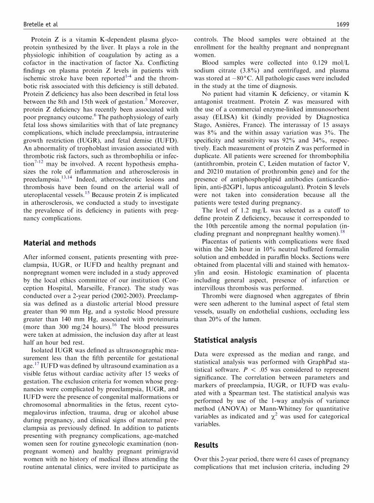
Bretelle et al 1699
Protein Z is a vitamin K-dependent plasma glyco-protein synthesized by the liver. It plays a role in thephysiologic inhibition of coagulation by acting as acofactor in the inactivation of factor Xa. Conflictingfindings on plasma protein Z levels in patients withischemic stroke have been reported1-4 and the throm-botic risk associated with this deficiency is still debated.Protein Z deficiency has also been described in fetal lossbetween the 8th and 15th week of gestation.5 Moreover,protein Z deficiency has recently been associated withpoor pregnancy outcome.6 The pathophysiology of earlyfetal loss shows similarities with that of late pregnancycomplications, which include preeclampsia, intrauterinegrowth restriction (IUGR), and fetal demise (IUFD).An abnormality of trophoblast invasion associated withthrombotic risk factors, such as thrombophilia or infec-tion7-12 may be involved. A recent hypothesis empha-sizes the role of inflammation and atherosclerosis inpreeclampsia.13,14 Indeed, atherosclerotic lesions andthrombosis have been found on the arterial wall ofuteroplacental vessels.15 Because protein Z is implicatedin atherosclerosis, we conducted a study to investigatethe prevalence of its deficiency in patients with preg-nancy complications.
Material and methods
After informed consent, patients presenting with pree-clampsia, IUGR, or IUFD and healthy pregnant andnonpregnant women were included in a study approvedby the local ethics committee of our institution (Con-ception Hospital, Marseille, France). The study wasconducted over a 2-year period (2002-2003). Preeclamp-sia was defined as a diastolic arterial blood pressuregreater than 90 mm Hg, and a systolic blood pressuregreater than 140 mm Hg, associated with proteinuria(more than 300 mg/24 hours).16 The blood pressureswere taken at admission, the inclusion day after at leasthalf an hour bed rest.
Isolated IUGR was defined as ultrasonographic mea-surement less than the fifth percentile for gestationalage.17 IUFD was defined by ultrasound examination as avisible fetus without cardiac activity after 15 weeks ofgestation. The exclusion criteria for women whose preg-nancies were complicated by preeclampsia, IUGR, andIUFD were the presence of congenital malformations orchromosomal abnormalities in the fetus, recent cyto-megalovirus infection, trauma, drug or alcohol abuseduring pregnancy, and clinical signs of maternal pree-clampsia as previously defined. In addition to patientspresenting with pregnancy complications, age-matchedwomen seen for routine gynecologic examination (non-pregnant women) and healthy pregnant primigravidwomen with no history of medical illness attending theroutine antenatal clinics, were invited to participate as
controls. The blood samples were obtained at theenrollment for the healthy pregnant and nonpregnantwomen.
Blood samples were collected into 0.129 mol/Lsodium citrate (3.8%) and centrifuged, and plasmawas stored at �80(C. All pathologic cases were includedin the study at the time of diagnosis.
No patient had vitamin K deficiency, or vitamin Kantagonist treatment. Protein Z was measured withthe use of a commercial enzyme-linked immunosorbentassay (ELISA) kit (kindly provided by DiagnosticaStago, Asnieres, France). The interassay of 15 assayswas 8% and the within assay variation was 3%. Thespecificity and sensitivity was 92% and 34%, respec-tively. Each measurement of protein Z was performed induplicate. All patients were screened for thrombophilia(antithrombin, protein C, Leiden mutation of factor V,and 20210 mutation of prothrombin gene) and for thepresence of antiphosphoplipid antibodies (anticardio-lipin, anti-b2GP1, lupus anticoagulant). Protein S levelswere not taken into consideration because all thepatients were tested during pregnancy.
The level of 1.2 mg/L was selected as a cutoff todefine protein Z deficiency, because it corresponded tothe 10th percentile among the normal population (in-cluding pregnant and nonpregnant healthy women).18
Placentas of patients with complications were fixedwithin the 24th hour in 10% neutral buffered formalinsolution and embedded in paraffin blocks. Sections wereobtained from placental villi and stained with hematox-ylin and eosin. Histologic examination of placentaincluding general aspect, presence of infarction orintervillous thrombosis was performed.
Thrombi were diagnosed when aggregates of fibrinwere seen adherent to the luminal aspect of fetal stemvessels, usually on endothelial cushions, occluding lessthan 20% of the lumen.
Statistical analysis
Data were expressed as the median and range, andstatistical analysis was performed with GraphPad sta-tistical software. P ! .05 was considered to representsignificance. The correlation between parameters andmarkers of preeclampsia, IUGR, or IUFD was evalu-ated with a Spearman test. The statistical analysis wasperformed by use of the 1-way analysis of variancemethod (ANOVA) or Mann-Whitney for quantitativevariables as indicated and c2 was used for categoricalvariables.
Results
Over this 2-year period, there were 61 cases of pregnancycomplications that met inclusion criteria, including 29

1700 Bretelle et al
Table I Characteristics of healthy pregnant women and patients with preeclampsia, isolated IUGR, and IUFD
PE(n = 29)
Isolated IUGR(n = 25)
IUFD(n = 16)
Normal pregnancies(n = 34)
Nonpregnantwomen (n = 50) P
Age (y) 29 [23-37] 31 [19-37] 30 [24-40] 30 [19-43] 29 [23-37] nsGestational age atsampling (wks)
31 [22-42] 32 [19-40] 28 [16-40] 31 [22-39] d ns
Fetal weight at delivery (g) 1490 [400-4325] 1225 [500-2590] 1300 [400-3100] 3300 [2880-3900] d !.0001*Systolic blood pressure(mm Hg)
160 [130-200] 120 [100-135] 140 [110-200] 110 [90-130] d !.0001y
Diastolic blood pressure(mm Hg)
90 [80-110] 70 [60-90] 80 [60-110] 80 [50-90] d !.0001z
Uric acid (mmol/mL) 319 [176-498] 295 [108-383] 230 [153-379] 238 [176-498] d !.05x
Proteinuria (g/24h) 2.7 [0.4-6.6] 0.3 [0-3] 2.5 [0-6.6] d d !.0001k
Protein Z (mg/L) 1.75 [0.48-3.1] 1.79 [0.75-2.87] 1.57 [0.78-3.1] 1.63 [0.47-3.01] 1.69 [0.7-3] ns25th 1.32 1.24 0.88 1.29 1.4175th 2.12 2.21 2.65 2.26 2.3
Blood samples were collected at the inclusion. Gestational ages are given at sampling. Values are represented as median and [range], and upper and lower
quartiles for protein Z. Difference between groups was analyzed by the Kruskall-Wallis and post hoc test.
* P ! .05; all groups are different from normal pregnancy.y P ! .05; PE and IUFD groups are different from normal pregnancies and IUGR.z P ! .05; all groups are different from PE.x P !.05; PE group is different from normal pregnancies.k P ! .05; IUGR is different from PE and IUFD.
preeclampsia and 25 isolated IUGR. In the group ofIUFD (n = 16), 7 patients presented with isolated IUFDwithout IUGR or preeclampsia; the other patients hadeither preeclampsia (n = 2) or IUGR (n = 7). In addi-tion, 34 normal pregnancies and 50 healthy, nonpreg-nant, age-matched women were enrolled. Protein Z levelsand patient characteristics are described in Table I. Theoverall median level of protein Z was 1.63 with a range of0.47 to 3.10 mg/L. Levels of protein Z were not differentamong the groups. Protein Z levels were not differentamong the different pregnancy periods between 19 to 29,30 to 36, and 37 to 42 weeks of gestation (the medianwere 1.73G 0.6, 1.75G 0.7, and 1.62G 0.6, respectively;P = .85). The percentage of protein Z-deficient womenwas elevated in pregnancies complications (n = 21)compared with normal pregnancies (n = 3) (34.5% vs8.8%; RR 1.55, 95% CI 1.2-2; P = .0065) (Table II).Patients with a protein Z level less than 1.2 mg/Lrepresented 20.6% of the preeclampsia group (n = 6)(RR 1.6, 95% CI 0.9-2.8; P = .23), 33.3% of the groupwith isolated IUGR (n = 8) (RR 1.96, 95% CI 1.16-3.32; P = .041) and 50% of the group with IUFD(n = 8) (RR 3.36, 95% CI 1.65-6.8; P = .0031). Afterexclusion of the patients presenting with IUGR or withpreeclampsia, among the 7 remaining: 4 patients had aprotein Z level less than 1.2 mg/L (57%) (RR 2.1, 95%CI 0.9-5; P = .01). Protein Z deficiency was significantlymore frequent in isolated IUGR and in IUFD (Figure).In our population, gestational age did not influenceprotein Z levels (r = 0.056, P = .64).
The results of biologic screening for thrombophilia arereported in Table II. Among patients with protein Z levels
below 1.2mg/L, 2 patients were carriers of a heterozygousLeiden mutation of factor V and 1 also had anticardio-lipin antibodies. Another patient with protein Z defi-ciency had lupus anticoagulant. These 3 patients hadsevere pregnancy complications: all presented withIUFD, which was associated with a severe preeclampsia(blood pressure greater than 160/100 mm Hg associatedwith proteinuria greater than 4 g/24 hours) in 1 patientand with IUGR in the 2 others. In the group of patientswith IUFD, 2 patients had a history of unexplainedthrombosis. None of the patients with low level of proteinZ presented with vitamin K deficiency and all had normallevels of other vitamin K-dependent factors.
The histologic examinations of the placenta revealedthrombi or ischemic lesions in 50% of patients with
Table II Characteristics of studied population for thrombo-philias and antibodies screening
Complicatedpregnancies(n = 61) %
Normalpregnancies(n = 34) % P
AT 3.6 0 nsPC 5.4 0 nsFVL 3.4 2.9 ns20210a 0 0 nsaPL 9 0 !.05Protein Z deficiency 34.5 8.8 !.01
AT, Antithrombin deficiency; PC, protein C deficiency; FVL, Leiden
mutation of factor V; 20210a, mutation of 20210 allele of prothrombin
gene; aPL, antiphospholipid antibodies and lupus anticoagulant;
Protein Z deficiency, protein Z levels !1.2 mg/L.

Bretelle et al 1701
protein Z levels below 1.2 mg/L, and in 33% of patientswith normal protein Z levels (RR 0.84, 95% CI 0.6-1.2).
Comment
Protein Z deficiency is a recently recognized factor inthrombotic events. It has been implicated in early fetalloss5 and in complicated pregnancies.19 Protein Z defi-ciency has also been reported to be common in theantiphospholipid syndrome, which is known to be asso-ciated with recurrent fetal losses.20 In the current study,we found a high prevalence of protein Z deficiency inpatients with IUFD or in IUGR. In the IUFD group,half of the patients had a protein Z level below 1.2 mg/L.This rate is greater than the 22% that has been reportedin patients with 1 early fetal loss between 10 and 19weeks5 but is comparable with that reported in patho-logic conditions such as ischemic stroke.1 We studied lateIUFD (mean gestational age: 28 weeks), but the highprevalence of protein Z deficiency compared with theprevious studies cannot be explained by gestational age,because our results seem to show that protein Z levels arestable throughout pregnancy. IUFD subgroup analysis,after exclusion of patients presenting with preeclampsiaand IUGR, was not significant as well. This may be dueto confounding factors or to the small effective of thissubgroup. Instead, protein Z deficiency may be impli-cated in IUFD through the constitution of thromboticlesions in the placental vascular bed. Nevertheless, pla-cental examinations showed thrombi to be present in50% of low protein Z levels cases and in 33% of theremaining cases. This result was not statistically signif-icant and might also be due to the effective of the study.
Atherosclerosis could be a feature shared by compli-cated pregnancies of vascular origin and ischemicstroke,21 in which protein Z deficiency has also been
Figure Prevalence (%) of Protein Z ! 1.2 mg/L among thepopulation studied was 8.8% in normal pregnancies, 20.6% in
PE (RR 1.6 CI 95% [0.9-2.8]; P Z 0.23), 33.3% in isolatedIUGR (RR 1.96 CI 95% [1.16-3.32]; *P Z .041) and 50% inIUFD (RR 3.36 CI 95% [1.65-6.8]; **P Z .0031.
implicated. Moreover, lesions similar to those found inatherosclerosis have been observed in examination ofplacentas from women with preeclampsia.16 Depositsof protein Z have been shown in vascular lesions ofpatients with atherosclerosis.22 It could be postulatedthat protein Z could similarly be detected in theplacental vascular bed, suggesting that protein Z defi-ciency could be secondary to pregnancy complicationsrather than causal. Against this hypothesis, Gris et al5
found an association between pregnancy complicationsand low levels of protein Z measured at least 6 monthsafter the last obstetric events. This makes the possibilityof protein Z consumption in the placental vascular bedunlikely. Even if thrombophilia predisposes to pree-clampsia or IUGR, other factors such as modificationsof inflammatory response23 or previous infection24,25
may also play a role. Protein Z deficiency may be oneelement of a complex series of events leading to theclinical manifestations. In our study, the 3 patients inwhom protein Z deficiency was associated to otherthrombophilic abnormalities with particularly unfavor-able outcomes. Protein Z deficiency may thus increasethe risk of vascular events in association with otherpredisposing factors as described in the antiphospholidsyndrome.19,20
It has been suggested that in severe preeclampsia andunexplained fetal death, antiprotein Z antibodies couldcause protein Z inhibition.26 We did not test ourpopulation for these antibodies, but this also might bean explanation for the decreased protein Z in compli-cated pregnancies. Protein Z deficiency thus seems toplay a role in the physiopathology of pregnancy com-plications such as IUGR and IUFD. These findingsshould be validated in a larger study.
References
1. Vasse M, Guegan-Massardier E, Borg JY, Woimant F, Soria C.
Frequency of protein Z deficiency in patients with ischemic stroke.
Lancet 2001;357:933-4.
2. Fedi S, Soki F, Brogi D, Tellini I, Cesari F, Sestini I, et al. Low
protein Z plasma levels are independently associated with acute
coronary syndromes. Thromb Haemost 2003;90:1173-8.
3. McQuillan AM, Eikelboom JW, Hankey GJ, Baker R, Thom J,
Staton J, et al. Protein Z in ischemic stroke and its etiologic
subtypes. Stroke 2003;34:2415-9.
4. Kobelt K, Biasiutti FD, Mattle HP, Lammle B, Wuillemin WA.
Protein Z in ischaemic stroke. Br J Haematol 2001;114:169-73.
5. Gris JC, Quere I, Dechaud H, Mercier E, Pincon C, Hoffet M,
et al. High frequency of protein Z deficiency in patients with
unexplained early fetal loss. Blood 2002;99:2606-8.
6. Gris JC, Mercier E, Quere I, Lavigne-Lissalde G, Cochery-
Nouvellon E, Hoffet M, et al. Low molecular weight heparin
versus low-dose aspirin women with one fetal loss and a consti-
tutional thrombophilic disorder. Blood 2004;103:3695-9.
7. Khong TY, De Wolf F, Robertson WB, Brosens I. Inadequate
maternal vascular response to placentation in pregnancies compli-
cated by preeclampsia and by small-for-gestational age infants.
BJOG 1986;93:1049-59.

1702 Bretelle et al
8. Van Pampus MG, Dekker GA, Wolf H, Huijgens PC, Koopman
MM, von Blomberg BM, et al. High prevalence of hemostatic
abnormalities in women with a history of severe pre-eclampsia.
Am J Obstet Gynecol 1999;88:1146-50.
9. Kupferminc MJ, Eldor A, Steinman N, Many A, Bar-Am A, Jaffa
A, et al. Increased frequency of genetic thrombophilia in women
with complications of pregnancy. N Engl J Med 1999;340:9-13.
10. Dekker GA, de Vries JI, Doelitchz PM, Huijgens PC, von
Blomberg BM, Jacobs C, et al. Underlying disorders associated
with severe early onset pre-eclampsia. Am J Obstet Gynecol
1995;173:1042-8.
11. Verspyck E, Le CD, Goffinet F, Tron F, Marpeau L, Borg JY.
Thrombophilia and immunological disorders in pregnancies as risk
factors for small for gestational age infants. BJOG 2002;109:28-33.
12. Von Dadelszen P, Magee LA, Krajden M, Alasaly K, Popovska V,
Devarakonda RM, et al. Levels of antibodies against cytomega-
lovirus and Chlamydophila pneumoniae are increased in early onset
preeclampsia. BJOG 2003;110:725-30.
13. Redman CW, Sargent IL. Preeclampsia, the placenta and the
maternal systemic inflammatory response. Placenta 2003;24:21-7.
14. Vickers M, Ford I, Morrison R, Prescott G, Watson S, Hannaford
P, et al. Markers of endothelial activation and atherothrombosis in
women with history of preeclampsia or gestational hypertension.
Thromb Haemost 2003;90:1192-7.
15. De Wolf F, Brosens I, Renaer M. Fetal growth retardation and the
maternal arterial supply of the human placenta in the absence of
sustained hypertension. BJOG 1980;87:678-85.
16. Roberts JM, Taylor RN, Musci TJ, Rodgers GM, Hubel CA,
Mclaughin MK. Preeclampsia: an endothelial cell disorder. Am J
Obstet Gynecol 1989;161:1200-4.
17. Chitty LS, Altman DG, Henderson A, Campbell S. Charts of fetal
size. BJOG 1994;101:35-43, 125-35.
18. DahmA,VanHylckamaVliegA,BendzB,RosendaalF,BertinaRM,
Sandset PM. Low levels of tissue factor pathway inhibitor (TFPI)
increase the risk of venous thrombosis. Blood 2003;101:4387-92.
19. Paidas MJ, Ku DHW, Arkel YS, Reharber A, Lee MJ, Roshan D,
et al. First trimester maternal protein Z levels are lower in patients
with complicated pregnancies and patients with thrombophilia and
subsequent adverse pregnancy outcome. Br J Haematology
2003;120:907-15.
20. Forastiero RR, Martinuzzo ME, Lu L, Broze JR. Autoimmune
antiphospholipid antibodies impair inhibition of activated factor X
by protein Z/protein Z-dependent protease. J Thromb Haemost
2003;1:764-70.
21. Sattar N, Bendomir A, Berry C, Shepherd J, Greer IA, Packard
CJ. Lipoprotein subfraction concentrations in preeclampsia:
pathogenic parallels to atherosclerosis. Obstet Gynecol 1997;89:
403-8.
22. Greten J, Kreis I, Liliensiek B, Allenberg J, Amiral J, Ziegler R,
et al. Localisation of protein Z in vascular lesions of patients with
atherosclerosis. Vasa 1998;27:144-8.
23. von Dadelszen P, Magee LA. Could an infectious trigger explain
the differential maternal response to the shared placental pathol-
ogy of preeclampsia and normotensive intrauterine growth restric-
tion? Acta Obstet Gynecol Scand 2002;81:642-8.
24. Savige JA, Gilbert GL, Fairley KF, McDowall DR. Bacteriuria
due to Ureaplasma urealyticum and Gardnerella vaginalis in women
with preeclampsia. J Infect Dis 1983;148:605.
25. Mittendorf R, Lain KY, Williams MA, Walker CK. Preeclampsia:
a nested, case-control study of risk factors and their interactions.
J Reprod Med 1996;41:491-6.
26. Gris JC, Amadio C, Mercier E, Lavigne-Lissalde G, Dechaud H,
Hoffet M, et al. Anti-protein Z antibodies in women with path-
ologic pregnancies. Blood 2003;101:4850-2.

American Journal of Obstetrics and Gynecology (2005) 193, 1703–8
www.ajog.org
A free radical scavenger, edaravone, inhibits lipidperoxidation and the production of nitric oxide inhypoxic-ischemic brain damage of neonatal rats
Jesmin I. Noor, MD,a Tomoaki Ikeda, MD,a Yuto Ueda, MD,b Tsuyomu Ikenoue, MDa,*
Departments of Obstetrics and Gynecologya and Psychiatry,b Faculty of Medicine, University of Miyazaki, Japan
Received for publication November 15, 2004; revised February 28, 2005; accepted March 30, 2005
KEY WORDSEdaravoneLipid peroxidationNitric oxide
metaboliteHypoxic-ischemic
brain damage
Objective: The purpose of this study was to elucidate a role for edaravone, a free radicalscavenger 3-methyl-1-phenyl-2-pyrazolin-5-one, in neonatal hypoxic-ischemic brain damage. We
determined the level of thiobarbituric acid reactive substances as an index of lipid peroxidationand nitric oxide metabolites as nitric oxide production.Study design: Seven-day-old Wistar rats were subjected to left common carotid artery ligation
followed by 2 hours of 8% oxygen exposure. Then, the rats were administered edaravone (9 mg/kg) or saline solution intraperitoneally. Cerebrospinal fluid was withdrawn just before the ratswere killed at 2, 5, 24, and 48 hours after hypoxia, and brains were removed. The thiobarbituric
acid reactive substances and nitric oxide metabolites levels were measured in the brain tissue andcerebrospinal fluid, respectively.Results: On the ligated side, edaravone significantly decreased thiobarbituric acid reactivesubstances levels at 5 and 24 hours after hypoxia, compared with saline group (P ! .01).
Edaravone significantly decreased the nitric oxide metabolites level in the cerebrospinal fluid onlyat 5 hours, compared with saline group (P ! .01).Conclusion: Edaravone potently and transiently inhibited lipid peroxidation and the production
of nitric oxide in the neonatal rat brain after hypoxic-ischemic insult.� 2005 Mosby, Inc. All rights reserved.
The free radical scavenger 3-methyl-1-phenyl-2-pyrazolin-5-one, edaravone, was endorsed by the Min-istry of Health, Labor and Welfare of Japan in April2001 for the treatment of acute cerebral infarction inadult patients. Previous studies have shown the neuro-protective and antioxidative mechanisms of edaravone
* Reprint requests: Tsuyomu Ikenoue, MD, Department of Ob-
stetrics and Gynecology, Faculty of Medicine, University of Miyazaki,
5200 Kihara, Kiyotake, Miyazaki 889-1692, Japan.
E-mail: [email protected]
0002-9378/$ - see front matter � 2005 Mosby, Inc. All rights reserved.
doi:10.1016/j.ajog.2005.03.069
in adult ischemic animal models.1-5 For example, edar-avone dose-dependently prevented brain edema that wasinduced by hemispheric embolization by suppressing theproduction of lipoxygenase metabolites.1 Edaravone hasbeen also reported to prevent the peroxidative vascularendothelial cell damage caused by hydroperoxyeicosate-traenoic acid in vitro study.2 Furthermore, edaravonewaseffective in the treatment of adult rat brain edema becauseof focal ischemia by inhibiting both non-enzymatic freeradical reactions and lipoxygenase activity in vitro.3
Postischemic treatment with edaravone significantly
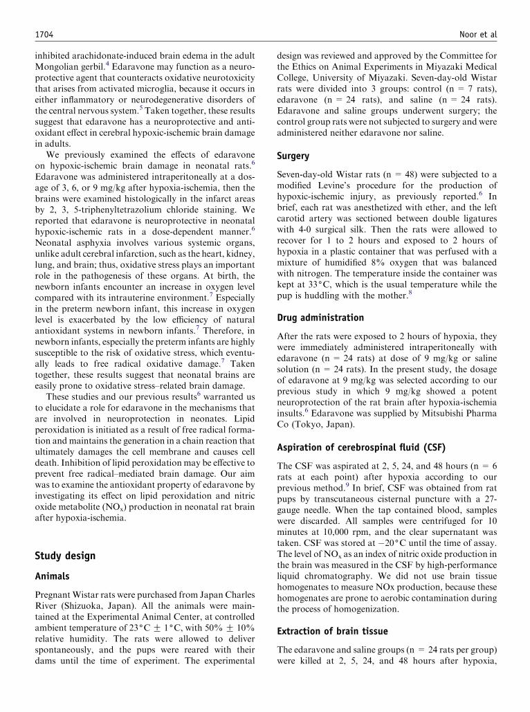
1704 Noor et al
inhibited arachidonate-induced brain edema in the adultMongolian gerbil.4 Edaravone may function as a neuro-protective agent that counteracts oxidative neurotoxicitythat arises from activated microglia, because it occurs ineither inflammatory or neurodegenerative disorders ofthe central nervous system.5 Taken together, these resultssuggest that edaravone has a neuroprotective and anti-oxidant effect in cerebral hypoxic-ischemic brain damagein adults.
We previously examined the effects of edaravoneon hypoxic-ischemic brain damage in neonatal rats.6
Edaravone was administered intraperitoneally at a dos-age of 3, 6, or 9 mg/kg after hypoxia-ischemia, then thebrains were examined histologically in the infarct areasby 2, 3, 5-triphenyltetrazolium chloride staining. Wereported that edaravone is neuroprotective in neonatalhypoxic-ischemic rats in a dose-dependent manner.6
Neonatal asphyxia involves various systemic organs,unlike adult cerebral infarction, such as the heart, kidney,lung, and brain; thus, oxidative stress plays an importantrole in the pathogenesis of these organs. At birth, thenewborn infants encounter an increase in oxygen levelcompared with its intrauterine environment.7 Especiallyin the preterm newborn infant, this increase in oxygenlevel is exacerbated by the low efficiency of naturalantioxidant systems in newborn infants.7 Therefore, innewborn infants, especially the preterm infants are highlysusceptible to the risk of oxidative stress, which eventu-ally leads to free radical oxidative damage.7 Takentogether, these results suggest that neonatal brains areeasily prone to oxidative stress–related brain damage.
These studies and our previous results6 warranted usto elucidate a role for edaravone in the mechanisms thatare involved in neuroprotection in neonates. Lipidperoxidation is initiated as a result of free radical forma-tion and maintains the generation in a chain reaction thatultimately damages the cell membrane and causes celldeath. Inhibition of lipid peroxidation may be effective toprevent free radical–mediated brain damage. Our aimwas to examine the antioxidant property of edaravone byinvestigating its effect on lipid peroxidation and nitricoxide metabolite (NOx) production in neonatal rat brainafter hypoxia-ischemia.
Study design
Animals
PregnantWistar rats were purchased from Japan CharlesRiver (Shizuoka, Japan). All the animals were main-tained at the Experimental Animal Center, at controlledambient temperature of 23(CG 1(C, with 50%G 10%relative humidity. The rats were allowed to deliverspontaneously, and the pups were reared with theirdams until the time of experiment. The experimental
design was reviewed and approved by the Committee forthe Ethics on Animal Experiments in Miyazaki MedicalCollege, University of Miyazaki. Seven-day-old Wistarrats were divided into 3 groups: control (n = 7 rats),edaravone (n = 24 rats), and saline (n = 24 rats).Edaravone and saline groups underwent surgery; thecontrol group rats were not subjected to surgery and wereadministered neither edaravone nor saline.
Surgery
Seven-day-old Wistar rats (n = 48) were subjected to amodified Levine’s procedure for the production ofhypoxic-ischemic injury, as previously reported.6 Inbrief, each rat was anesthetized with ether, and the leftcarotid artery was sectioned between double ligatureswith 4-0 surgical silk. Then the rats were allowed torecover for 1 to 2 hours and exposed to 2 hours ofhypoxia in a plastic container that was perfused with amixture of humidified 8% oxygen that was balancedwith nitrogen. The temperature inside the container waskept at 33(C, which is the usual temperature while thepup is huddling with the mother.8
Drug administration
After the rats were exposed to 2 hours of hypoxia, theywere immediately administered intraperitoneally withedaravone (n = 24 rats) at dose of 9 mg/kg or salinesolution (n = 24 rats). In the present study, the dosageof edaravone at 9 mg/kg was selected according to ourprevious study in which 9 mg/kg showed a potentneuroprotection of the rat brain after hypoxia-ischemiainsults.6 Edaravone was supplied by Mitsubishi PharmaCo (Tokyo, Japan).
Aspiration of cerebrospinal fluid (CSF)
The CSF was aspirated at 2, 5, 24, and 48 hours (n = 6rats at each point) after hypoxia according to ourprevious method.9 In brief, CSF was obtained from ratpups by transcutaneous cisternal puncture with a 27-gauge needle. When the tap contained blood, sampleswere discarded. All samples were centrifuged for 10minutes at 10,000 rpm, and the clear supernatant wastaken. CSF was stored at �20(C until the time of assay.The level of NOx as an index of nitric oxide production inthe brain was measured in the CSF by high-performanceliquid chromatography. We did not use brain tissuehomogenates to measure NOx production, because thesehomogenates are prone to aerobic contamination duringthe process of homogenization.
Extraction of brain tissue
The edaravone and saline groups (n = 24 rats per group)were killed at 2, 5, 24, and 48 hours after hypoxia,

Noor et al 1705
immediately after aspiration of CSF. The control ratswere killed on day 7. The brain, excluding the cerebellumand the olfactory lobes, were immediately removed andsectioned into 2 hemispheres (right and left), and eachhemisphere was weighed. Phosphate-buffered saline so-lution was added 10 times the weight of each hemisphereand then homogenized by a homogenizer (Polytron PT3000; Brinkmann Instruments, Inc, Westbury, NY).Brain tissue was homogenized within 8 minutes afterdeath. The homogenates were stored at �84(C untilassay.
Thiobarbituric acid reactive substance(TBARS) assay
TBARS was measured according to the method ofOhkawa et al.10 In brief, 0.1 mL of brain homogenate,0.1 mL of 8.1% sodium dodecylsulfate, 2 mL of 20%acetate buffer (pH 3.5), 1.5 mL of 1.0% sodium thio-barbiturate, and 0.6 mL of distilled water were addedand mixed. The mixture was incubated at 100(C for 60minutes. TBARS were then extracted with a mixture ofn-butanol and pyridine (15:1, vol/vol). The fluorescenceof the extracted solution was analyzed by spectrofluo-rometry, under the following condition: excitation wave-length, 515 nm; emission wavelength, 553 nm.
Statistical analysis
Statistical analysis was carried out with 2-way (group !time course) analysis of variance, followed by Newman-Keuls’s test for multiple comparisons. A probabilityvalue of !.05 was considered significant.
Results
The TBARS level of the control was 35.5 G 2 mmol/mgbrain tissue. Figure 1, A, shows the comparison ofTBARS level in the ligated side of cerebral hemispherebetween the edavarone and saline solution groups. TheTBARS level significantly decreased after edaravoneadministration compared with the saline solution groupat 5 and 24 hours after hypoxia (21.2 G 4.4 mmol/mg vs225.7G 61.8 mmol/mg [P! .01]; 59.2G 9.8 mmol/mg vs273.4 G 42.8 mmol/mg [P ! .01] brain tissue, respec-tively). No significant difference was observed betweenthe 2 groups at 2 or 48 hours after hypoxia (39.5 G 3.9mmol/mg vs 55.3 G 16.1 mmol/mg; 82.5 G 9.8 mmol/mgvs 103.8 G 16.8 mmol/mg brain tissue; respectively).Figure 1, B, shows the comparison of TBARS level in thenon-ligated side of the cerebral hemisphere. No signifi-cant difference was observed between the edaravone andsaline group at any time point. In the saline group,TBARS level in the ligated side was significantly de-creased at 48 hours compared with 5 and 24 hours afterhypoxia (103.8 mmol/mg G 16.8 vs 227.7 G 61.8 mmol/
mg [P ! .01] and 103.8 G 16.8 mmol/mg vs 238.417 G42.7533 mmol/mg [P ! .01] brain tissue; respectively).
Figure 2 shows the NOx level in the CSF after theadministration of edaravone or saline solution afterhypoxia. In the edaravone group, the NOx level was sig-nificantly decreased compared with saline solution groupat only 5 hours after hypoxia (203.30G 16.4 pmol/10 mLvs 273.5 G 8.1 pmol/10 mL; P ! .05). No significantdifference was observed at the 2-, 24-, and 48-hour pointbetween the edaravone and saline solution groups.
Figure 1 Effect of edaravone on lipid peroxidation in neo-
natal Wistar rats after hypoxic-ischemic insult. The TBARSlevel was measured in homogenized brain tissue of both rightand left hemisphere. A, The sequential changes of TBARS levelin the left hemisphere of saline–treated (closed circles), edar-
avone (open circles), and control group (closed squares). The #denotes P ! .01, saline vs edaravone; the * denotes P ! .01,control vs saline. B, The sequential changes of TBARS level in
the right hemisphere of saline–treated (open diamond), edar-avone-treated (closed diamond) group, and control group(closed squares). The data represent mean G SEM. The saline
group (n = 6) and edaravone group (n = 6) at each timepoint; control (n = 7).

1706 Noor et al
Comment
Our study showed for the first time the inhibitory effectsof edaravone on lipid peroxidation and NOx productionin the neonatal rat brain after hypoxic-ischemic insult. Inacute brain injury, oxygen free radicals are thought tocause oxidative damage to a variety of molecular andcellular targets, which include proteins, DNA, lipids,mitochondria, and membrane structures.11 Oxygen freeradicals of potential importance in cerebral ischemiainclude hydroxyl and superoxide radicals. Among thefree radicals, hydroxyl radical is a potent radical and caninitiate lipid peroxidation in the cell membrane.12,13 Celldamage can be prevented by detoxification of free rad-icals, which eventually will prevent the progress of lipidperoxidation. Edaravone has been reported to scavengethe reactive oxygen free radicals such as hydroxyl,superoxide, and peroxyl radicals.14,15
We analyzed the effect of edaravone on the progressof lipid peroxidation in hypoxic-ischemic neonatal braintissue by measuring the levels of TBARS. The TBARSassay is 1 of the commonly used methods for the mea-surement of free radical reactions indirectly, is easy touse to study the effects of different treatments on lipidperoxidation, and can be applied to crude biologicextracts. The specificity of TBARS to measure the levelof lipid peroxidation has been criticized by certaininvestigators.16-18 Goda and Marnett17 showed thatthe quantitative analysis of malonaldehyde level inbiologic fluids can be measured by high-performance
Figure 2 Effect of edaravone on NOx level in neonatal Wistarrats after hypoxic-ischemic insult. Sequential changes in NOxlevel in the CSF of the saline group (open triangle) and the
edaravone group (closed triangle). The * denotes P! .05, salinevs edaravone group. The data represent mean G SEM. Saline(n = 6) and edaravone (n = 6) at each time point.
liquid chromatography with electrochemical detection.Kosugi and Kikugawa18 reported that the TBARS testmay not reflect the components of malonaldehyde alonebut may also reflect the components of malonaldehyde,alk-2-enals, alka-2,4-dienals, and hydroperoxide func-tions. In the present experiment, the purpose was not tomeasure the absolute values of lipid-peroxidation but tocompare the values of edaravone-treated and salinesolution–treated groups. Therefore, we used a relativelycrude but widely used assay19-23 that has been acceptedas an empiric window for the examination of the com-plex process of lipid peroxidation in various biologicextracts.22 In our study, the TBARS levels increase inthe left hemisphere of the saline solution group. Thereason behind this could be re-oxygenation after hy-poxic/ischemic insult that resulted in an increase of freeradical production, which suggests an increased lipidperoxidation level in the neuronal cell membrane.12 Thisincreased level of lipid peroxidation persisted until 24hours after the hypoxic insult, which is consistent withthe study of Hsiang et al24 in which they showed thatlipid peroxidation was significantly high until 24 hoursafter diffuse brain injury in rats. However, the TBARSlevel significantly decreased, which can be related to along-term compensatory mechanism that involves themodulation and synthesis of the activity of enzymesthat are related to reactive oxygen species.20 We haveshown that an administration of edaravone significantlysuppressed the TBARS level until 24 hours after thehypoxic insult. Taken together, our study demonstratesthat edaravone can prevent the progress of lipidperoxidation.
There are 3 major mechanisms that are involved inneurotoxicity produced by nitric oxide radicals. First,nitric oxide radical react with super oxide radicals toform highly toxic peroxynitrite anions, which easilydecompose to produce hydroxyl radicals and nitrites25,26
and eventually attack the cell membrane. Second, nitricoxide that is produced in the donor neuron can attackthe DNA of the target neuron directly. In the targetneuron, a great amount of adenosine triphosphate isconsumed through poly adenosine diphosphate–ribosesynthetase activation for repairing the damaged DNA,which leads to energy depletion and cell death.27 Finally,nitric oxide attacks the mitochondrial electron–transfersystem and injures the iron/sulfur protein–containingenzymes that leads to an inhibition of glycolysis andmitochondrial respiration.28 Nitric oxide is a free radicalsynthesized by nitric oxide synthase (NOS) in the endo-thelial cells and neurons in response to increased intra-cellular calcium levels. Nitric oxide has dual action onhypoxic-ischemic brain damage and is not always neu-rotoxic, in fact nitric oxide also can be neuroprotective.Nitric oxide produced by NOS in the neurons is mainlyneurotoxic; however, it is resistant to excitatory stimu-lation, which sometimes can be neuroprotective. On the

Noor et al 1707
other hand, nitric oxide that is produced by NOS in theneurons gives mainly neuroprotection by vasodilationand increases microcirculation in the ischemic areas.Nitric oxide that is produced by inducible nitric oxidesynthase is mainly neurotoxic to the surrounding tis-sues.29 Citrulline, nitric oxide, and water are producedfrom arginine, whereas reduced nocotinamide adeninedinucleotide phosphate and oxygen by NOS.30-32 Re-oxygenation during reperfusion provides oxygen to sus-tain neuronal viability and also provides oxygen as asubstrate for numerous enzymatic oxidation reactionsthat produce reactive oxidants12; as a result, nitric oxideis synthesized during the reperfusion phase. Nitric oxideradicals are oxidized within seconds to the more stableoxidation products nitrite and nitrate in vivo. Thus,assays of nitrites and nitrate are used widely as indicatorsof nitric oxide formation. Because direct and selectivemeasurement of nitric oxide is still very difficult, themeasuring of NOx demonstrates the nitric oxide produc-tion more precisely. The generation of nitric oxide isassociated predominantly with cytotoxic effects. Wetherefore investigated the effects of edaravone on theproduction of NOx, which can be any degraded productsof nitric oxide metabolite (such as NO2 and/or NO3)after hypoxic-ischemic insult. We showed that the NOx
level increased in the CSF at 5 hours, which eventuallydecreased within 24 hours of the insult. This suggeststhat free radical formation may be a transient phenom-enon, whereas the progress of lipid peroxidation con-tinues for a longer duration after hypoxic/ischemic braindamage. Edaravone suppressed the NOx levels in theCSF, which indicates the potent scavenging action onNOx and thus the prevention of the free radical–medi-ated brain damage.
Edaravone, for the following reasons, may be thedrug of choice in the treatment of neonatal hypoxic-ischemic encephalopathy in newborn infants. Edaravonehas shown to have minimal adverse effects since itsinception.7 It can act in the state of highly oxidativestress,1,3-7,12 the condition the newborn infants are proneto in a deficient antioxidant system because of physio-logic hyperoxygenation. Last, it can prevent oxidativestress–related pathogenesis in neonatal infarction.6 Asdrug of choice in cerebral infarction in adult patients,edaravone may have a greater beneficial effect onhypoxic-ischemic neonatal brain damage than on adultbrain damage.
In conclusion, our study showed 2 important prop-erties of edaravone, which are of great clinical impor-tance in the prevention of neonatal hypoxic/ischemicbrain damage. First, edaravone can prevent the progressof lipid peroxidation and, second, can decrease theproduction of NOx in neonatal hypoxia-ischemia. Thesedata indicate that edaravone may be a highly effectiveand potent drug for the prevention of hypoxic-ischemicencephalopathy in newborn infants.
Acknowledgments
We thank Ms Hiroko Taniguchi for her research assis-tance.
References
1. Nishi H, Watanabe T, Sakurai H, Yuki S, Ishibashi A. Effect of
MCI-186 on brain edema in rats. Stroke 1989;20:1236-40.
2. Watanabe T, Morita I, Nishi H, Murota S. Preventive effect of
MCI-186 on 15-HPETE induced vascular endothelial cell injury in
vitro. Prostaglandins Leukot Essent Fatty Acids 1988;33:81-7.
3. Abe K, Yuki S, Kogure K. Strong attenuation of ischemic and
post-ischemic brain edema in rats by a novel free radical scavenger.
Stroke 1988;19:480-5.
4. Watanabe T, Egawa M. Effects of an antistroke agent MCI-186 on
cerebral arachidonate cascade. JPharmacolExpTher1994;271:1624-9.
5. Banno M, Mizuno T, Kato H, Zhang G, Kawanokuchi J, Wang J,
et al. The radical scavenger edaravone prevents oxidative neuro-
toxicity induced by peroxynitrite and activated microglia. Neuro-
pharmacology 2005;48:283-90.
6. Ikeda T, Xia YX, Kaneko M, Sameshima H, Ikenoue T. Effect of
the free radical scavenger, 3-methyl-1phenyl-2pyrazolin-5-one
(MCI-186), on hypoxia-ischemia-induced brain injury in neonatal
rats. Neurosci Lett 2002;329:33-6.
7. Giuseppe B, Serefina P, Radolfo B. Free radicals and brain
damage in the newborn. Biol Neonate 2001;79:180-6.
8. Mortola JP, Dotta A. Effects of hypoxia and ambient temperature
on gaseous metabolism of newborn rats. Am J Physiol 1992;263:
R267-72.
9. Xia YX, Ikeda T, Xia XY, Ikenoue T. Differential neurotrophin
levels in cerebrospinal fluid and their changes during development
in newborn rat. Neurosci Lett 2000;280:220-2.
10. Ohkawa H, Ohishi N, Yagi K. Assay for lipid peroxides in
animal tissues by thiobarbituric acid reaction. Anal Biochem
1995;351-8.
11. Saugestad OD. Mechanisms of tissue injury by oxygen radicals:
implications for neonatal disease. Acta Paediar 1996;85:1-4.
12. Pak HC. Role of oxidants in ischemic brain damage. Stroke
1996;27:1124-9.
13. Girotti AW. Mechanism of lipid peroxidation. J Free Radic Biol
Med 1985;1:87-95.
14. Houkin K, Nakayama N, Kamada K, Noujou T, Abe H,
Kashiwaba T. Neuroprotective effect of the free radical scavenger
MCI-186 in patients with cerebral infarction: clinical evaluation
using magnetic resonance imaging and spectroscopy. J Stroke
Cerebrovasc Dis 1998;17:315-22.
15. Mizuno A, Umemura K, Nakashima M. Inhibitory effect of
MCI-186, a free radical scavenger, on cerebral ischemia follow-
ing rat middle cerebral artery occlusion. Gen Pharmacol 1998;
30:575-8.
16. Knight JA, Pieper RK, McClellan L. Specificity of the thiobarbi-
turic acid reaction: its use in studies of lipid peroxidation. Clin
Chem 1998;34:2433-8.
17. Goda Y, Marnett LJ. High-performance liquid chromatography
with electrochemical detection for determination of the major
malonaldehyde-guanine adduct. Chem Res Toxicol 1991;4:520-4.
18. Kosugi H, Kikugawa K. Potential thiobarbituric acid-reactive
substances in peroxidized lipis. Free Radic Biol Med 1989;7:205-7.
19. Ueda Y, Komatsu M, Hiramatsu M. Effect of Japanese herbal
medicine, Toki-Shakuyaku-San (TJ-23), on the neurotransmitters,
SOD and TBARS in the brain of aged rat. Neurochem Res
1996;21:915-20.
20. Dal-Pizzol F, Klamt F, Vianna MMR, Schroder N, Quevedo J,
Benfato MS, et al. Lipid peroxidation in hippocampus early and

1708 Noor et al
late after status epilepticus induced by pilocarpine or kainic acid
in Wistar rats. Neurosci Lett 2000;291:179-82.
21. Hasegawa K, Yoshioka H, Sawada T, Nishikawa H. Lipid
peroxidation in neonatal mouse brain subjected to two different
types of hypoxia. Brain Dev 1991;13:101-3.
22. Bagenholm R, Nilssion AU, Kjellmer I. Formation of free radicals
in hypoxic ischemic brain damage in the neonatal rat, assessed
by and endogenous spin trap and lipid peroxidation. Brain Res
1993;773:132-8.
23. Sakamoto A, Ohnishi T, Ohnishi T, Ogawa R. Relationship
between free radical production and lipid peroxidation during
ischemia-reperfusion injury in the rat brain. Brain Res 1991;554:
186-92.
24. Hsiang JNK, Wang JY, Ip SM, Ng HK, Stadlin A, Yu ALM, et al.
The time course and regional variation of lipid peroxidation after
diffuse brain injury in rats. Acta Neurochir 1997;139:464-8.
25. Beckman JS, Beckman TW, Chen J, Marsshall PA, Freeman BA.
Apparent hydroxyl radical production by peroxynitrite: implica-
tions for endothelial injury from nitric oxide and superoxide. Proc
Nat Acad Sci USA 1990;87:1620-4.
26. Tabuchi M, Umegaki K, Ito T, Suzuki M, Tomita I, Ikeda M,
et al. Fluctuation of serum NOX concentration at stroke onset in a
rat spontaneous stroke model (M-SHRSP): peroxynitrite forma-
tion in brain lesions. Brain Res 2002;13(949):47-56.
27. Zhang ZG, Chopp M, Gautam S, Zhang RL, Schmidt HHHW,
Pollock JS, et al. Upregulation of neuronal nitric oxide synthase and
mRNA, and selective sparing of nitric oxide synthase-containing
neurons after focal cerebral ischemia in rat. Brain Res 1994;654:
85-9.
28. Milstien S, Sakai N, Brew BJ, Krieger C, Vickers JH, Saito K,
et al. Cerebrospinal fluid nitrite/nitrate levels in neurologic dis-
eases. J Neurochem 1994;63:1178-80.
29. Niwa M, Inao S, Takyasu M, Kawai T, Kajita Y, Hihashi T, et al.
Time course of expression of three nitric oxide synthase isoforms
after transient middle cerebral artery occlusion in rats. Neurol Med
Chir 2001;41:63-73.
30. Castillo J, Rama R, Davalos A. Nitric oxide-related brain damage
in acute ischemic stroke. Stroke 2000;31:835-57.
31. Celik O, Hasacalik S, Turkoz Y, Hascalik M, Gokdeniz R.
Cerebrospinal fluid nitric oxide level changes in pre-eclampsia.
Eur J Obstet Gynecol Reprod Biol 2003;111:141-5.
32. Brown GC. Nitric oxide regulates mitochondrial respiration and
cell function by inhibiting cytochrome oxidase. FEBS Lett 1995;
369:136-9.

American Journal of Obstetrics and Gynecology (2005) 193, 1709–15
www.ajog.org
Embryogenesis of fused umbilical arteries inhuman embryos
Shigehito Yamada, MD,a,b,* Junzo Hamanishi, MD,b Shozo Tanada, MD,d
Mitsuhiro Tachibana, MD,e Rokuro Mimura, MD,e Shingo Fujii, MD,b
Kohei Shiota, MDa,c
Congenital Anomaly Research Center,a Departments of Gynecology and Obstetrics,b Anatomy &Developmental Biology,c Kyoto University Graduate School of Medicine, Kyoto, Japan; Department of Obstetrics andGynecology,d Division of Pathology,e Hyogo Prefectural Amagasaki Hospital, Amagasaki, Hyogo, Japan
Received for publication December 17, 2004; revised March 3, 2005; accepted March 30, 2005
KEY WORDSUmbilical arteryIntrauterine growth
retardationHuman embryo
Objective: The objective of this study was to elucidate the embryologic basis of fused umbilical
arteries in the human.Study design: Twenty-nine human embryo specimens at Carnegie stages 11 through 15 (4-5weeks after fertilization) were examined histologically, with special reference to the development
of umbilical arteries.Results: All embryos at Carnegie stage 11 and 12 had fused umbilical arteries, and 66% ofCarnegie stage13 embryos and 29% of Carnegie stage 14 embryos still had the condition. None of
the embryos at Carnegie stage 15 or older had fused umbilical arteries, but there were always 2arteries present in their umbilical cords.Conclusion: Our data suggest that (1) a single umbilical artery splits into 2 as the developmentalstage of the embryo advances, (2) that fused umbilical arteries represent a remnant of the
embryonic phenotype, and (3) that fused umbilical arteries are embryologically distinct from truesingle umbilical artery.� 2005 Mosby, Inc. All rights reserved.
Obstetric sonographic examination sometimes revealsfused umbilical arteries (FUA) in the fetus, whichusually occurs near the placental cord insertion.1 FUAis a condition in which 2 umbilical arteries fuse with
Supported by Japanese Ministry of Education, Culture, Sports,
Science and Technology: grant number 13557001, 13470003; Japanese
Ministry of Health, Labour and Welfare: grant number 14A-4.
* Reprint requests: Shigehito Yamada, MD, Congenital Anomaly
Research Center, Graduate School of Medicine, Kyoto University,
Yoshida-Konoe-cho, Sakyo-ku, Kyoto, Japan.
E-mail: [email protected]
0002-9378/$ - see front matter � 2005 Mosby, Inc. All rights reserved.
doi:10.1016/j.ajog.2005.03.066
each other through their course in the umbilical cord(Figure 1).2 Clinically, FUA is encountered more fre-quently than single umbilical artery (SUA), which is oneof the common cord anomalies in humans, and is oftenassociated with congenital malformations such as mus-culoskeletal, cardiovascular, and urogenital anomalies(25%-50%).3-6 Because the association with malforma-tions is rare (0.2%-1%) in newborn babies with FUA,1,7
FUA may be pathologically distinct from true SUA.Therefore, this condition should be diagnosed correctlyduring the prenatal period or at the time of delivery toavoid misdiagnosis as true SUA. Such misdiagnosis

1710 Yamada et al
Figure 1 Transverse sections of the umbilical cord of a newborn baby with intrauterine growth retardation. The mother was a 27-year-old nulliparous woman with pregnancy-induced hypertension. The cesarean delivery was performed at 28 weeks of gestation
because of the cessation of fetal growth. A-D are arranged in a fetal body to placental sequence. A, Two arteries (arrowhead) and1 vein are observed in the cord. B, The walls of the arteries are partially fused (arrowhead). C, The arterial walls are continuous(arrowhead). D, Near the placental end of the cord, 1 artery and 1 vein are recognized. The bar equals 1 mm.
could be avoided by careful examination of the numberof vessels throughout the entire length of the cord.1 Fox5
and Benirschke and Kaufmann4 proposed that SUAshould be diagnosed by sectioning the umbilical cord atmultiple sites and checking the number of vessels at boththe placental and fetal ends of the umbilical cord atbirth, even when the delivery was uneventful. Thefrequency of FUA is uncertain, but recently, Fujikura7
examined O700 placentae and estimated the frequencyof FUA to be 3.1%. Although only approximately 30cases of FUA have been reported to date in theliterature, Fujikura7 claimed that this condition is notrare and that many cases are overlooked by obstetri-cians. The embryologic basis of FUA has not been well
illustrated to date. In the present report, we describe thehistologic findings of umbilical arteries in staged humanembryos at 4 to 5 weeks after fertilization and discussthe embryogenesis of FUA.
Material and methods
Since 1961, O40,000 human conceptuses have beenplaced in the Kyoto Collection of Human Embryos, ina collaborative effort of several hundred obstetricians.In most of the cases, pregnancies were terminated forsocial reasons, mostly in healthy women during the firsttrimester of pregnancy (Maternity Protection Law ofJapan). The terminations of pregnancy were performed
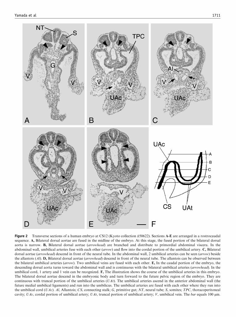
Yamada et al 1711
Figure 2 Transverse sections of a human embryo at CS12 (Kyoto collection #50622). Sections A-E are arranged in a rostrocaudalsequence. A, Bilateral dorsal aortae are fused in the midline of the embryo. At this stage, the fused portion of the bilateral dorsalaorta is narrow. B, Bilateral dorsal aortae (arrowhead) are branched and distribute to primordial abdominal viscera. In the
abdominal wall, umbilical arteries fuse with each other (arrow) and flow into the cordal portion of the umbilical artery. C, Bilateraldorsal aortae (arrowhead) descend in front of the neural tube. In the abdominal wall, 2 umbilical arteries can be seen (arrow) besidethe allantois (Al). D, Bilateral dorsal aortae (arrowhead) descend in front of the neural tube. The allantois can be observed between
the bilateral umbilical arteries (arrow). Two umbilical veins are fused with each other. E, In the caudal portion of the embryo, thedescending dorsal aorta turns toward the abdominal wall and is continuous with the bilateral umbilical arteries (arrowhead). In theumbilical cord, 1 artery and 1 vein can be recognized. F, The illustration shows the course of the umbilical arteries in this embryo.The bilateral dorsal aortae descend in the embryonic body and turn forward to the future pelvic region of the embryo. They are
continuous with truncal portion of the umbilical arteries (UAt). The umbilical arteries ascend in the anterior abdominal wall (thefuture medial umbilical ligaments) and run into the umbilicus. The umbilical arteries are fused with each other where they run intothe umbilical cord (UAc). Al, Allantois; CS, connecting stalk; G, primitive gut; NT, neural tube; S, somites; TPC, thoracoperitoneal
cavity; UAc, cordal portion of umbilical artery; UAt, truncal portion of umbilical artery; V, umbilical vein. The bar equals 100 mm.

1712 Yamada et al
Figure 3 Slightly oblique transverse sections of a human embryo at CS14 (Kyoto collection #365). Sections A-G are arranged in acaudorostral sequence as shown in H. In A-G, upward is the ventral side of the body. A, The aorta (arrowheads) is cut where itcurves in the future pelvic region. B, The single aorta is observed in front of the notochord and neural tube (arrowhead). C, The
aorta bifurcates and runs into umbilical arteries (arrowheads). D, The left umbilical artery (asterisk) runs forward, and bothumbilical arteries run cranially in the abdominal wall (arrowheads). E, The 2 umbilical arteries run into the umbilical cord(arrowheads). F, Bilateral umbilical arteries fuse with each other in the umbilical cord (arrowhead). G, An SUA (UAc) and vein (V)run parallel in the umbilical cord. H, The illustration shows the direction of the sections and the course of umbilical arteries. The
bilateral dorsal aortae descend in the embryonic body and turn forward in the future pelvic region. The aorta bifurcates into 2umbilical arteries in the pelvis and fuse with each other in the umbilical cord (UAc). The dorsal halves of sections A-G are not shownin the photos (shadowed portion in H). G, Gut; MD, mesonephric duct; MN, mesonephros; NT, neutral tube; PC, peritoneal cavity;
T, tail; V, umbilical vein. The bar equals 500 mm.
mainly by curettage procedures. Further details of theembryo collection have been described in previousreports.8-11
In the present study, serial histologic sections of 29human embryos at Carnegie stages (CS) 11 to 15 wereexamined.12 The distribution of the embryos by CS was2 cases at CS 11 (27 days after fertilization), 3 cases atCS 12 (30 days), 9 cases at CS 13 (32 days), 7 cases at CS14 (34-35 days), and 8 cases at CS 15 (36 days).9,13 Theirsections were examined serially, with special reference tothe umbilical cord and umbilical vessels. Embryosgenerally were sectioned transversely, but some caseswere sectioned in other planes. The planes of section aredescribed in the Figures.
This study, which used the human embryo specimens,was approved by the Institutional Review Board ofKyoto University Graduate School of Medicine.
Results
Umbilical arteries arise from the bilateral internal iliacarteries. During early embryonic stages, 2 aortae existbilaterally beside the spine (the right and left dorsal
aortae). Our histologic observation demonstrated thatthe 2 dorsal aortae communicate with each other in 4-week human embryos (Figure 2, A) to fuse and becomea single aorta. The bilateral dorsal aortae had somebranches in the pelvic region and were continuous withumbilical arteries (Figure 2, B-E). The umbilical arteriesthat run along the anterior abdominal wall (the futuremedial umbilical ligaments) were found to ascend besidethe allantois (Figure 2, C and D), exit the embryonicbody, and run into the umbilical cord (Figure 2, E). Inyounger embryos at CS 11 to 14, the fusion point ofumbilical arteries was observed in the umbilical cord(Figure 2, B). At CS 11 and 12, all the embryos that wereexamined had FUA, as seen in Figure 2, B. The point offusion of the umbilical arteries was located in theumbilical cord, just outside the abdominal wall. In6 of 9 CS 13 embryos (66%), umbilical arteries werefound to fuse near the abdominal wall (Figure 2, B). Inthe other 3 CS 13 embryos, 2 arteries were recognizedthroughout the length of the umbilical cord and fusionof umbilical arteries was not observed. At CS 14, FUAwas found in 2 cases among 7 embryos that wereobserved (29%). A CS 14 embryo is shown in Figure 3,
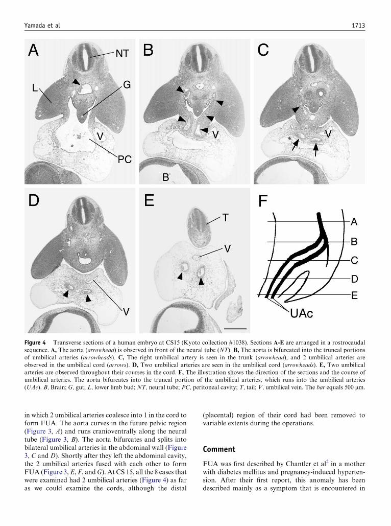
Yamada et al 1713
Figure 4 Transverse sections of a human embryo at CS15 (Kyoto collection #1038). Sections A-E are arranged in a rostrocaudalsequence. A, The aorta (arrowhead) is observed in front of the neural tube (NT). B, The aorta is bifurcated into the truncal portionsof umbilical arteries (arrowheads). C, The right umbilical artery is seen in the trunk (arrowhead), and 2 umbilical arteries are
observed in the umbilical cord (arrows). D, Two umbilical arteries are seen in the umbilical cord (arrowheads). E, Two umbilicalarteries are observed throughout their courses in the cord. F, The illustration shows the direction of the sections and the course ofumbilical arteries. The aorta bifurcates into the truncal portion of the umbilical arteries, which runs into the umbilical arteries
(UAc). B, Brain; G, gut; L, lower limb bud; NT, neural tube; PC, peritoneal cavity; T, tail; V, umbilical vein. The bar equals 500 mm.
in which 2 umbilical arteries coalesce into 1 in the cord toform FUA. The aorta curves in the future pelvic region(Figure 3, A) and runs cranioventrally along the neuraltube (Figure 3, B). The aorta bifurcates and splits intobilateral umbilical arteries in the abdominal wall (Figure3, C and D). Shortly after they left the abdominal cavity,the 2 umbilical arteries fused with each other to formFUA (Figure 3,E, F, andG). At CS 15, all the 8 cases thatwere examined had 2 umbilical arteries (Figure 4) as faras we could examine the cords, although the distal
(placental) region of their cord had been removed tovariable extents during the operations.
Comment
FUA was first described by Chantler et al2 in a motherwith diabetes mellitus and pregnancy-induced hyperten-sion. After their first report, this anomaly has beendescribed mainly as a symptom that is encountered in

1714 Yamada et al
ultrasound sonographic prenatal diagnosis.1,14 Althoughonly approximately 30 cases of FUA have been reportedto date in the literature, Fujikura7 estimated the frequencyof FUA to be 3.1%on the basis of his careful examinationof O700 placentae7 and claimed that this condition maybe overlooked in many cases.
Although most reported cases of FUA are associatedwith a normal perinatal outcome,1,7,14 the association ofFUA with Hallermann-Streiff syndrome and patent ura-chus has been suggested.1,2 Our case that is shown inFigure 1 was associated with severe intrauterine growthretardation, which has not been suggested previously. It isnot clear whether such associations are merely due tochance, and further studies are needed to clarify thepathologic significance of these associations.
The development of umbilical arteries was discussedby Monie,15 on the basis of his examination of umbilicalarteries in human embryos of 2.9 to 8.5 mm in crown-rump length. He showed that the right and left umbilicalarteries existed in the embryonic body (the truncalportion of the umbilical artery) at all stages of humandevelopment, but 2 arteries were not always present inthe umbilical cord (the cordal portion of the umbilicalartery). He observed that umbilical arteries formed aplexus in the umbilical cord at the earliest stage (2.9-mmembryo) and that later they fused with each other toform a single artery by the 3.4-mm stage. The singleartery became ‘‘split’’ into 2 vessels from the truncaltoward the placental ends. This splitting process wasconsidered to result in the formation of 2 umbilicalarteries.
In our observation of the embryo cases in the KyotoCollection, all the embryos at CS 11 and 12 had FUA.Their fusion point was recognized in the truncalportion of umbilical arteries. In some cases at CS 13(66%) and CS 14 (29%), the fusion point of umbilicalarteries was observed in the truncal or umbilicalportion of umbilical arteries, which seemed more distalthan that observed in CS 12 embryos. In the remainingcases at CS 13 or 14, the fusion of arteries was notrecognized in the cord. In embryos older than CS 15,the umbilical cord had been cut near the embryonicbody, and it was not possible to follow the entirecourse of umbilical arteries in the cord, but there werealways 2 arteries in the cord, as far as we couldobserve. Therefore, it appeared that the fusion point ofthe umbilical arteries moves distally from the embry-onic end as the developmental stage advances.
FUA is considered to be formed by the failure of theumbilical artery to split into 2 throughout the entirelength of the umbilical cord. A major cause of FUA maybe Hyrtl’s anastomosis of 2 umbilical arteries,16 which isobserved commonly near the placental end of the cordand is considered to represent a normal physiologicfusion of the arteries.4,17 Although Monie15 categorizeda 6.0-mm embryo with FUA as ‘‘abnormal,’’ similarly to
those with SUA, we found that normal embryos at CS13 (4.0-6.0 mm crown-rump length) and CS 14 (5.0-7.0mm crown-rump length) sometimes had FUA, whichmost likely represents a normal developmental process.Therefore, it appears that the SUA described by Monieincluded not only true SUA but also nonpathologicFUA. Our present study is the first study that hasexamined as many as 29 human embryo specimens atthe developmental stage when an SUA splits into 2 andprovides a new insight into the development of umbilicalarteries.
Acknowledgment
We thank Dr Hiroyuki Koshiyama, Kitano Hospital,Osaka, for stimulating discussion and helpful com-ments; the collaborating technicians in the Division ofPathology, Hyogo Prefectural Amagasaki Hospital, fortheir contribution; and Dr Murray Smith, University ofNew South Wales, Sydney, Australia, for his criticalreading of the manuscript and helpful comments.
References
1. Sepulveda W, Dezerega V, Carstens E, Gutierrez J. Fused umbil-
ical arteries: prenatal sonographic diagnosis and clinical signifi-
cance. J Ultrasound Med 2001;20:59-62.
2. Chantler C, Baum JD, Wigglesworth JS, Scopes JW. Giant
umbilical cord associated with a patent urachus and fused umbil-
ical arteries. J Obstet Gynaecol Br Commonw 1969;76:273-4.
3. Heifetz SA. Single umbilical artery: a statistical analysis of 237
autopsy cases and review of the literature. Perspect Pediatr Pathol
1984;8:345-78.
4. Benirschke K, Kaufmann P. Pathology of the human placenta.
New York: Springer-Verlag; 2000.
5. Fox H. Pathology of the placenta. London: Saunders; 1997.
6. Ullberg U, Sandstedt B, Lingman G. Hyrtl’s anastomosis, the only
connection between the two umbilical arteries: a study in full term
placentas from AGA infants with normal umbilical artery blood
flow. Acta Obstet Gynecol Scand 2001;80:1-6.
7. Fujikura T. Fused umbilical arteries near placental cord insertion.
Am J Obstet Gynecol 2003;188:765-7.
8. Nishimura H. Prenatal versus postnatal malformations based on
the Japanese experience on induced abortions in the human being.
In: Blandau RJ, editor. Aging gametes. Basel, Switzerland: Karger;
1975. p. 349-68.
9. Nishimura H, Takano K, Tanimura T, Yasuda M. Normal and
abnormal development of human embryos: first report of the
analysis of 1,213 intact embryos. Teratology 1968;1:281-90.
10. Shiota K. Development and intrauterine fate of normal and abnor-
mal human conceptuses. Congenit Anom Kyoto 1991;31:67-80.
11. Yamada S, Uwabe C, Fujii S, Shiota K. Phenotypic variability in
human embryonic holoprosencephaly in the Kyoto Collection.
Birth Defects Res Part A Clin Mol Teratol 2004;70:495-508.
12. O’Rahilly R, Muller F. Developmental stages in human embryos:
including a revision of Streeter’s ‘‘Horizons’’ and a survey of the
Carnegie collection. Washington (DC): Carnegie Institution of
Washington; 1987. p. 637.
13. Nishimura H, Tanimura T, Semba R, Uwabe C. Normal devel-
opment of early human embryos: observation of 90 specimens at
Carnegie stages 7 to 13. Teratology 1974;10:1-5.

Yamada et al 1715
14. Rosenak D, Meizner I. Prenatal sonographic detection of single
and double umbilical artery in the same fetus. J Ultrasound Med
1994;13:995-6.
15. Monie IW. Genesis of single umbilical artery. Am J Obstet
Gynecol 1970;108:400-5.
16. Hyrtl J. Die Blutgefaesse der menschlichen Nachgeburt in nor-
malen und abnormalen Verhaeltnissen. Vienna: Braumuller Ver-
lag; 1870.
17. Priman J. A note on the anastomosis of the umbilical arteries.
Anat Rec 1959;134:1-5.
American Journal of Obstetrics and Gynecology (2005) 193, 1716–23
www.ajog.org
Insulin and fatty acids regulate the expression of thefat droplet-associated protein adipophilin in primaryhuman trophoblasts
Uriel Elchalal, MD, W. Timothy Schaiff, PhD, Steven D. Smith, BA, Eli Rimon, MD,Ibrahim Bildirici, MD, D. Michael Nelson, MD, PhD, Yoel Sadovsky, MD
Departments of Obstetrics and Gynecology and Cell Biology and Physiology, Washington University School ofMedicine, St. Louis, MO
Received for publication October 25, 2004; revised March 15, 2005; accepted April 1, 2005
KEY WORDSPlacentaTrophoblastInsulin
AdipophilinFatty acids
Objective: This study was undertaken to test the hypothesis that insulin and fatty acids regulateadipophilin expression in cultured human trophoblasts.
Study design: Cytotrophoblasts isolated from term human placentas were cultured in the absenceor presence of insulin (10 nmol/L), and a mix of oleic and linoleic acid in serum-free medium. Theexpression of adipophilin as well as the fatty acid transport proteins (FATP) 2, 3, 4 and 6 was
examined. Fat accumulation was quantified by BODIPY staining and fat uptake determinedusing [3H]-oleic acid.Results: A combination of insulin and fatty acids enhanced the expression of adipophilin (2.3-
fold, P ! .05). In contrast, the expression of FATPs was unchanged. Furthermore, insulin andfatty acids increased the accumulation of fat droplets in trophoblasts by 4- to 5-fold (P ! .05),but had no effect on oleic acid uptake.
Conclusion: Insulin and fatty acids enhance the expression of adipophilin and the formation offatty acid droplets in term human trophoblasts.� 2005 Mosby, Inc. All rights reserved.
Supply of fatty acids to the fetus is particularly highduring the third trimester of pregnancy, when the fetusmore than doubles its size.1 Because de novo lipidsynthesis in the placenta is insufficient, transplacentaltransport of maternal fatty acids is paramount to thedeveloping fetus. Not surprisingly, maternal serum lipid
Supported by NIH ES-11597 and HD-45671 (both to Y.S.), and
HD-29190 (D.M.N.).
Reprints not available from the authors.
0002-9378/$ - see front matter � 2005 Mosby, Inc. All rights reserved.
doi:10.1016/j.ajog.2005.04.012
levels are elevated during pregnancy.1,2 Although themix of fatty acids delivered to the fetus is largelydetermined by maternal serum lipoproteins and fattyacid composition, the placenta is capable of regulatedpreferential transfer of long chain polyunsaturatedfatty acids. In addition, the fetus is unable to synthesizen-3 and n-6 unsaturated fatty acids, and thereforedepends on maternal transport for these essential fattyacids.1,2
Cell types that rely on the supply of fatty acids, suchas adipocytes, cardiocytes and hepatocytes, expressspecific fatty acid transport proteins. Several fatty acid

Elchalal et al 1717
transporters have been identified in trophoblasts, in-cluding fatty acid transport proteins (FATPs), placentalmembrane fatty acid binding protein (p-FABPpm), andfatty acid translocase (FAT/CD36).1,3 Adipophilin andits murine ortholog, adipocyte differentiation-relatedprotein (ADRP), are associated with cellular lipiddroplets and implicated in cellular fatty acid uptakeand storage of neutral lipids in adipocytes.4-6 We pre-viously demonstrated that adipophilin is expressed inhuman villous trophoblasts in vivo and in cultured pri-mary human trophoblasts in vitro.7 Moreover, weshowed that the expression of adipophilin is regulatedby the insulin-sensitizer peroxisome proliferator-activatedreceptor-g (PPARg),7 a protein that plays a pivotal rolein development of the murine fetoplacental unit anddifferentiation of human trophoblasts.8,9 Consistent withthese findings, Tarrade et al10 demonstrated enhancedaccumulation of fatty acids during cytotrophoblastdifferentiation into syncytiotrophoblasts. Ablation ofPPARg is associated with a reduced number of fattyacid droplets in the murine placenta.8 Taken together,these findings suggest that accumulation of fatty acidsin trophoblasts is necessary for fetal growth and isregulated during placental development and trophoblastdifferentiation.
The placenta in women with poorly controlled ges-tational diabetes mellitus (DM) and type-2 DM isexposed to hyperinsulinemia11 and increased supply ofnutrients. In the current work we surmised that insulin,acting in a hyperlipidemic environment, regulates fattyacid accumulation in human trophoblasts. We hypoth-esized that insulin combined with fatty acids regulatethe expression of adipophilin and fat accumulation introphoblasts. To test this hypothesis we analyzed theinfluence of insulin, in the presence or absence of fattyacids, on adipophilin expression as well as fatty aciduptake and accumulation in cultured term humantrophoblasts.
Materials and methods
Trophoblast isolation and culture
Our study was approved by the Institutional ReviewBoard of Washington University School of Medicine.Placentas were obtained immediately after term single-ton deliveries after uncomplicated pregnancies. Cyto-trophoblasts were isolated by the trypsin-DNase, percoll(Sigma, St. Louis, MO) gradient centrifugation methoddescribed by Kliman et al,12 with modifications.13 Non-adherent cells and syncytial fragments were removedafter 4 hours by washing 3 times with phosphate-buffered saline (PBS) solution, and cytotrophoblastswere cultured in Earle’s medium 199 supplemented with10% fetal bovine serum (Hyclone, Logan, UT) and
antibiotics in a standard tissue culture atmosphere of5% CO2 and 20% O2. After 24 hours in culture the cellswere washed and the medium replaced with fresh serum-containing or serum-free medium, supplemented whereindicated by insulin (10 nmol/L, Sigma) dissolved in0.1% insulin-free and fatty acid-free albumin (Sigma), amixture of 400 mmol/L oleic acid and 800 mmol/Llinoleic acid in albumin (Sigma), or vehicle control.These concentrations were selected on the based ofpublished information14,15 and the company’s recom-mendations, and were designed to assure that thesaturating levels of fatty acids were well dissolved inthe culture medium. All cells were harvested 48 hoursafter plating. The phosphatidylinositol 3-kinase (PI3K)inhibitor LY 294002 (10 mmol/L, Cell Signaling Tech-nology, Beverly, MA) was added to some of the cellcultures.
RNA and protein expression
At the end of the culture period the plates were rinsedwith PBS. RNA was purified with the use of TriReagent(Molecular Research Center, Inc, Cincinnati, OH)according to the manufacturer’s instructions. DNAse I(Ambion, Austin, TX) was added to the purified RNAfor 1 hour at 37(C to remove contaminating DNA.Complementary DNA (cDNA) was prepared from 1 mgRNA with the use of the TaqMan Gold RT-PCR kitwith the supplied random hexamer primers (AppliedBiosystems, Foster City, CA). Polymerase chain reac-tion (PCR) was performed on reverse transcripts (RTs)by using primer pairs listed in the Table. Specificity ofeach primer pair was confirmed by BLAST analysis.Quantitative real-time PCR was performed on duplicate3 mL samples of cDNA with the use of SYBR GreenPCR Master Mix (Applied Biosystems) in a total reac-tion volume of 50 mL containing 300 nmol/L each offorward and reverse primers. Reactions were run andanalyzed with the use of an Applied Biosystems Ge-neamp 5700 System. Dissociation curves were run on allreactions to ensure amplification of a single productwith the appropriate melting temperature. Samples werenormalized to parallel reactions with the use of primersspecific for 18S RNA. The fold increase relative tocontrol cultures was determined by the 2-DDCT method.16
The fold change values were calculated by using geo-metric means of individual assays.
Western immunoblotting was performed as we pre-viously described.7 Samples consisted of 20 to 30 mg perlane, which were analyzed with the use of monoclonalanti-adipophilin antibody (1:100 RDI, Flanders, NJ) orpolyclonal goat anti-b–actin (1:1000, Santa Cruz Bio-tech, Santa Cruz, Calif), followed by the correspondinghorseradish peroxidase-linked secondary antibodies(1:2000, Santa Cruz). Chemiluminescence was analyzedwith the use of Epichemi-3 darkroom (UVP BioImaging

1718 Elchalal et al
Table Primer sequences used for real-time quantitative PCR of adipophilin and FATPs
Name Accession no. Real-time quantitative PCR Direction and site sequence
Adipophillin NM_001122 F(326w346) GGCAGAGAACGGTGTGAAGACgi: 34577058 R(390w372) TCTGGATGATGGGCAGAGC
FATP-1 NM_198580 F(2952w2973) CCGGAATTGACTGTGACCACTTgi: 38524615 R(3065w3048) CACGCAGTGCAGGGTTCA
FATP-2 NM_003645 F(1829w1852) CTTTCAGCACATTGCTGATTACCTgi: 23111065 R(1908w1884) CAGTGATCTCAATGGTGTCCTGTAT
FATP-3 NM_024330 F(2098w2122) CAGAGACCTTCAAACAGCAGAAAGTgi: 13236578 R(2180w2158) CAGAACGTACAGTGGGTCAGACA
FATP-4 NM_005094 F(460w478) GAAGGCAAAGGTGCGACAGgi: 40807356 R(540w522) GCCGAACGGTAGAGGCAAA
FATP-6 NM_014031 F(542w559) GCGTGGTGGCCTTTCTCAgi: 13325054 R(606w590) ACAGGCGCGGATGCAAT
18S M10098 F(1049w1069) CGCCGCTAGAGGTGAAATTCTgi:337376 R(1149w1129) CGAACCTCCGACTTTCGTTCT
System, Upland, CA). Protein density was normalizedto b–actin expression.
Lipid accumulation assay
After 2 washes in PBS, the cells were fixed in 2%paraformaldehyde for 20 minutes at room temperature.Each plate was washed 2 additional times and the cellspermeabilized with 1 mL of Triton-X-100 0.1% in PBSfor 5 minutes, followed by 2 PBS washes. The wells werecovered with BODIPY 493/503 (Molecular Probes,Eugene, OR), which was dissolved in ethanol for astock solution of 0.1%, and further diluted 1:1000 inPBS before assay. After 15 minutes of exposure toBODIPY, the cells were washed 4 times in PBS. Cellnuclei were counterstained with TO-PRO 3-iodide 642/661 (Molecular Probes), rinsed in PBS, mounted withGelMount (Biomeda, Foster City, CA), and viewedunder a Nikon E800 microscope equipped with epifluor-escence optics and a Nikon C1 confocal imaging pack-age (Nikon, Tokyo, Japan). Images (n = 18) of 6random 0.25 mm2 microscopic fields were captured,and the number of cells stained for BODIPY 493/503along with the total number of nuclei for each field wasrecorded. Lipid droplets were quantified from digitalimages by using Analysis digital imaging software (SoftImaging Corp, Lakewood, CA). Nuclei were manuallycounted from the same images. Each experiment wasperformed in duplicate.
Fatty acid uptake assay
Transport of fatty acids into cultured trophoblasts wasassayed in triplicates with radiolabeled oleic acid withthe use of a modification of a previously describedassay.17 Uptake medium consisted of a 1:1 ratio of fattyacid-free bovine serum albumin (BSA) (Sigma) and
unlabeled oleic acid (Nu Chek Prep, Elysian, MN) at80 mmol/L each, spiked with [3H]-oleic acid (Perkin-Elmer, Boston, MA) at 1 ! 107 dpm/mL. The transportmedium was prepared by adding the unlabeled andtritiated oleic acid dropwise with stirring to a 60(Csolution of BSA in Hank’s balanced salt solution(HBSS, Sigma). The solution was stirred until cleared,then allowed to cool to room temperature. Trophoblastswere cultured in 6-well plates for 24 hours, and thenchanged to serum-free medium supplemented with in-sulin or fatty acids, as noted. At the end of the cultureperiod the cells were incubated 1 hour at 37(C in serum-free Dulbecco modified Eagle (DME) medium (Invi-trogen/Gibco, Carlsbad, CA). After incubation, theculture plates were allowed to equilibrate to roomtemperature for 10 minutes. Transport medium (800mL) was added to each well after HBSS rinse, andincubated at room temperature for 3 minutes. Theuptake was stopped by the addition of 3.5 mL per wellice-cold stop solution that consisted of 0.1% fatty acid-free BSA and 500 mmol/L phloretin (Sigma) in PBS. Thewells were rinsed 2 additional times with ice-cold stopsolution, the cells were lysed with 500 mL of 0.2%sodium dodecyl-sulfate (SDS) and the lysates added to10 mL ScintiVerse II scintillation fluid (Fisher Scientific,Fair Lawn, NJ). The wells were rinsed with additional500 mL 0.2% SDS and pooled into their respectivescintillation vials. Samples were counted on a Packard2500TR Liquid Scintillation Analyzer (Packard,Downers Grove, IL). Nonspecific association of labeledfatty acid with trophoblasts was determined with the useof duplicate cultures assayed in the presence of 500mmol/L phloretin. In these cultures, phloretin was addedto the incubation solution, washes, and transport me-dium. Specific transport of fatty acids was calculated bysubtracting counts in the presence of phloretin fromcounts in the absence of phloretin.

Elchalal et al 1719
Statistical analysis
Data are expressed as mean G SEM. All comparisonswere made by either paired t test or analysis of variance(ANOVA) with post-hoc Student–Newman-Keuls test,where appropriate (Primer of Biostatistics McGraw-HillNY, NY). Significance was determined at P ! .05.
Results
We initially examined the influence of insulin on theexpression of adipophilin messenger RNA (mRNA) inhuman trophoblasts, derived from normal term preg-nancies and cultured over a 48-hour period. By usingreal-time quantitative PCR, we found that the effect ofinsulin on the expression of adipophilin mRNA wasmaximal after 4 hours in culture and at a concentrationof 10 nmol/L (Figure 1, A and B). Notably, theseexperiments were performed in culture medium thatcontained 5 mmol/L of glucose and serum. To determinethe influence of serum components on adipophilinmRNA expression, we repeated the experiment in theabsence of 10% fetal bovine serum. The medium alsocontained 25mmol/L of glucose, amixture of 400 mmol/Loleic acid and 800 mmol/L linoleic acid, or both. Asshown in Figure 1, C, in the absence of serum theexpression of adipophilin mRNA was increased in allparadigms. In contrast, the effect of glucose or fattyacids was insignificant.
To examine the influence of insulin and fatty acids onadipophilin expression, we exposed the cells to insulin inserum-free medium, in the absence or presence of thefatty acid mixture. As shown in Figure 2, A, thecombination of insulin and fatty acids resulted inenhanced adipophilin mRNA expression. The effect ofinsulin or fatty acids alone was insignificant. Interest-ingly, basal as well as induced adipophilin mRNA levelswere reduced by using the PI3K inhibitor LY 294002. AWestern analysis of adipophilin expression in tropho-blasts provided further support to our findings, demon-
Figure 1 The influence of insulin on the expression ofadipophilin mRNA in cultured term human trophoblasts.
After culture in serum-containing medium the cells werewashed and cultured for an additional 24 hours in the absenceor presence of insulin. A, Time-dependent effect of insulin (10
nmol/L). The x-axis indicates the period of insulin exposurebefore harvest. B, Concentration-dependent effect of insulin,added 4 hours before harvest. C, The effect of culture medium
conditions on adipophilin mRNA. The effect of added glucose(25 mmol/L) or fatty acids (400 mmol/L oleic acid and 800mmol/L linoleic acid) was insignificant. The reduced expression
of adipophilin by addition of 10% fetal bovine serum wassignificant at all experimental paradigms (P ! .05, pairedt test). Each result was determined in duplicate, and representsthe mean G SEM obtained from 3 different placentas.
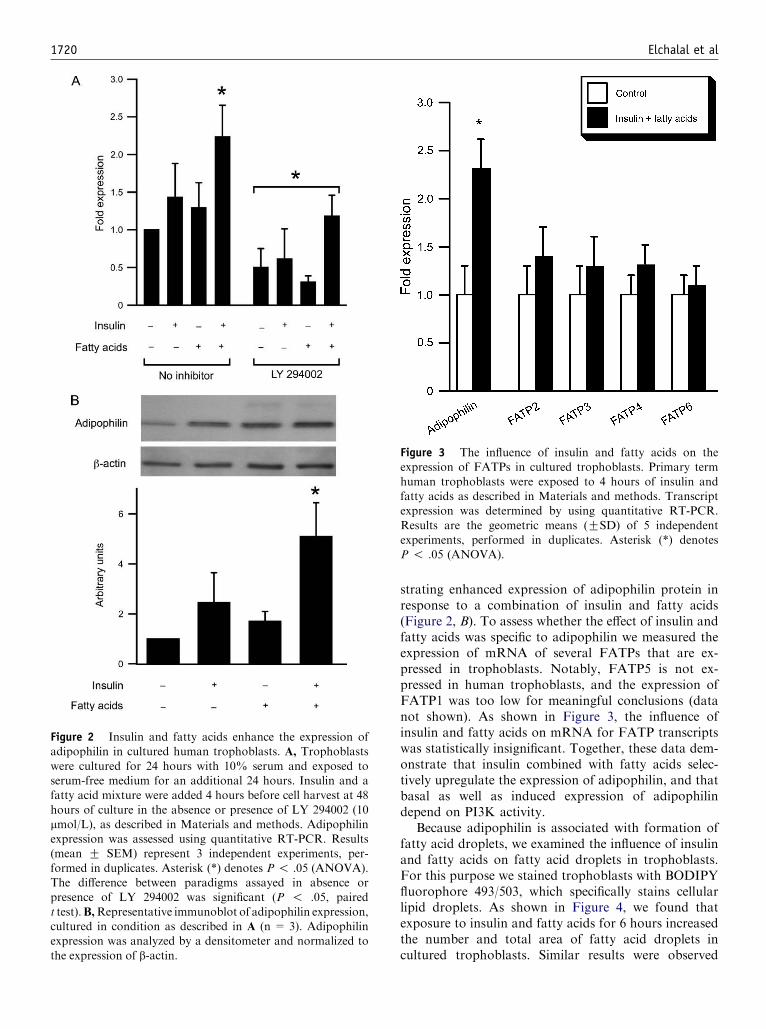
1720 Elchalal et al
Figure 2 Insulin and fatty acids enhance the expression ofadipophilin in cultured human trophoblasts. A, Trophoblasts
were cultured for 24 hours with 10% serum and exposed toserum-free medium for an additional 24 hours. Insulin and afatty acid mixture were added 4 hours before cell harvest at 48
hours of culture in the absence or presence of LY 294002 (10mmol/L), as described in Materials and methods. Adipophilinexpression was assessed using quantitative RT-PCR. Results(mean G SEM) represent 3 independent experiments, per-
formed in duplicates. Asterisk (*) denotes P ! .05 (ANOVA).The difference between paradigms assayed in absence orpresence of LY 294002 was significant (P ! .05, paired
t test).B,Representative immunoblot of adipophilin expression,cultured in condition as described in A (n = 3). Adipophilinexpression was analyzed by a densitometer and normalized to
the expression of b-actin.
strating enhanced expression of adipophilin protein inresponse to a combination of insulin and fatty acids(Figure 2, B). To assess whether the effect of insulin andfatty acids was specific to adipophilin we measured theexpression of mRNA of several FATPs that are ex-pressed in trophoblasts. Notably, FATP5 is not ex-pressed in human trophoblasts, and the expression ofFATP1 was too low for meaningful conclusions (datanot shown). As shown in Figure 3, the influence ofinsulin and fatty acids on mRNA for FATP transcriptswas statistically insignificant. Together, these data dem-onstrate that insulin combined with fatty acids selec-tively upregulate the expression of adipophilin, and thatbasal as well as induced expression of adipophilindepend on PI3K activity.
Because adipophilin is associated with formation offatty acid droplets, we examined the influence of insulinand fatty acids on fatty acid droplets in trophoblasts.For this purpose we stained trophoblasts with BODIPYfluorophore 493/503, which specifically stains cellularlipid droplets. As shown in Figure 4, we found thatexposure to insulin and fatty acids for 6 hours increasedthe number and total area of fatty acid droplets incultured trophoblasts. Similar results were observed
Figure 3 The influence of insulin and fatty acids on theexpression of FATPs in cultured trophoblasts. Primary term
human trophoblasts were exposed to 4 hours of insulin andfatty acids as described in Materials and methods. Transcriptexpression was determined by using quantitative RT-PCR.
Results are the geometric means (GSD) of 5 independentexperiments, performed in duplicates. Asterisk (*) denotesP ! .05 (ANOVA).

Elchalal et al 1721
Figure 4 The influence of insulin and fatty acids on the level of fatty acid droplets in cultured trophoblasts. A, Representativephotomicrographs of primary trophoblasts (n = 3). Cultures were exposed for 6 hours to insulin, fatty acids or both, until harvestedat 48 hours. Cells were stained by BODIPY fluorophore 493/503 for fat droplets (green) and TO-PRO (642/661) for nuclei (blue).Bar = 10 mm. B, Quantitative analysis of the number or area of fatty acids droplets in trophoblasts, performed as described in
Materials and methods. The influence of combined insulin and fatty acid supplementation was significant (P ! .05, ANOVA).Similar results were obtained when the cells were exposed to insulin and fatty acids for 24 hours (not shown).

1722 Elchalal et al
after 24 hours of exposure to insulin and fatty acids (notshown). To determine whether stimulation of fatty aciduptake by insulin underlies fatty acid accumulation introphoblasts we analyzed the influence of insulin, in theabsence or presence of fatty acids, on trophoblastuptake of tritiated oleic acid. As shown in Figure 5,we found that the uptake of oleic acid was notupregulated by supplementation of insulin. Althoughthe PI3K inhibitor LY 294002 weakly reduced the basaluptake of oleic acid (15%), it did not reduce the uptakeof oleic acid in the presence of insulin and fatty acids(Figure 5).
Comment
We found that a combination of insulin and the longchain fatty acids oleic and linoleic acid increase adipo-philin expression in cultured primary term humantrophoblasts. This effect was specific, and demonstratedusing analysis of both mRNA and protein. The timecourse and concentration dependence of insulin weresimilar to those we previously established.18 We notedthat either insulin or fatty acids alone exhibited a weakand statistically insignificant influence on adipophilinexpression. Because PI3K regulates pivotal pathways in
Figure 5 The effect of insulin and fatty acids on the uptake of
[3H]-oleic acid in cultured trophoblasts (n = 3), performed induplicates in the absence or presence of LY 294002 (10 mmol/L)as described in Materials and methods. Only the reduction inoleic acid uptake (15%) by LY 294002 in the absence of insulin
was significant (P ! .05, paired t test).
insulin signaling,19 we tested for the effect of LY 294002,a selective inhibitor of PI3K, on adipophilin expression.Interestingly, we found that LY 294002, inhibited bothbasal and induced expression of adipophilin, suggestinga role for PI3K in regulation of adipophilin expressionin trophoblasts. However, the fold induction of adipo-philin expression by insulin and fatty acids was un-changed, even in the presence of LY 294002. Consistentwith this finding, LY 294002 had a weak inhibitoryeffect on oleic acid uptake in the presence of fatty acids,but not insulin. In addition, we found that the enhance-ment of adipophilin expression by combined insulin andfatty acids was associated with fat accumulation introphoblasts but not with increased uptake of fatty acid.These data suggest that insulin and fatty acids enhancefat storage in trophoblasts, and that PI3K may play arole in adipophilin expression and fat accumulation inthese cells.
Fat readily diffuses through cell membranes, includ-ing placental trophoblasts. In addition, several types offatty acid transporters are expressed in the membraneand cytoplasm of diverse cell types, where they facilitatethe transfer and intracellular channeling of fatty acidsacross membranes.3 Insulin regulates the expression andsubcellular location of different fatty acid transporters inadipocytes, myocytes and cardiocytes, leading to en-hanced fatty acid uptake by these cells.20,21 Althoughadipophilin plays a role in cellular fat uptake, its regula-tion is incompletely understood. Oxidized low-densitylipoproteins (LDL) increase adipophilin expression inmacrophage foam cells and monocytes.22 Enhancedexpression of adipophilin in macrophages by acetylatedLDL promotes triglyceride and cholesterol storage andreduces cholesterol efflux.23 We recently demonstratedthat PPARg increases the level of adipophilin expressionin trophoblasts.7 Consistent with this notion, insulinstimulates the activity of PPARg in a ligand indepen-dent fashion.24 In addition, the expression of adipophi-lin, but not the lipid-associated proteins perilipin andS3-12 proteins, is increased in obese but not lean Zuckerrats.25 Together, these findings suggest that insulin andfatty acids influence adipophilin expression and fataccumulation in trophoblasts through regulation of thetranscriptional activity of PPARg.
We used a long chain fatty acid mix of oleic andlinoleic acids. These fatty acids represent only a fractionof fatty acid components in maternal blood. Other fattyacids, as well as cholesterol and triglycerides, may alsoplay an important role in total fat transport and uptakeby trophoblasts. Similarly, we did not analyze theinfluence of insulin and/or fatty acids on the expressionand function of other fat transporters. Furthermore, wedid not pursue the subcellular location of the trans-porters. These variables may be relevant to the activityof several transporters, such as CD36/fatty acid trans-locase. The regulation of fatty acid transporters in

Elchalal et al 1723
trophoblasts may have direct implications for preg-nancy-related hyperinsulinemia and its effect on placen-tal substrate availability.
Acknowledgments
We thank Elena Sadovsky and Lori Rideout for tech-nical assistance and manuscript preparation.
References
1. Haggarty P. Placental regulation of fatty acid delivery and its effect
on fetal growth: a review. Placenta 2002;23(Suppl A):S28-38.
2. Herrera E, Amusquivar E. Lipid metabolism in the fetus and the
newborn. Diabetes Metab Res Rev 2000;16:202-10.
3. Stahl A. A current review of fatty acid transport proteins (SLC27).
Pflugers Arch 2004;447:722-7.
4. Heid HW, Moll R, Schwetlick I, Rackwitz HR, Keenan TW.
Adipophilin is a specific marker of lipid accumulation in diverse
cell types and diseases. Cell Tissue Res 1998;294:309-21.
5. Gao J, Ye H, Serrero G. Stimulation of adipose differentiation
related protein (ADRP) expression in adipocyte precursors by
long-chain fatty acids. J Cell Physiol 2000;182:297-302.
6. Gao J, Serrero G. Adipose differentiation related protein (ADRP)
expressed in transfected COS-7 cells selectively stimulates long
chain fatty acid uptake. J Biol Chem 1999;274:16825-30.
7. Bildirici I, Roh CR, Schaiff WT, Lewkowski BM, Nelson DM,
Sadovsky Y. The lipid droplet-associated protein adipophilin is
expressed in human trophoblasts and is regulated by peroxisomal
proliferator-activated receptor-g/retinoid X receptor. J Clin En-
docrinol Metab 2003;88:6056-62.
8. Barak Y, Nelson MC, Ong ES, Jones YZ, Ruiz-Lozano P, Chien
KR, et al. PPARg is required for placental, cardiac, and adipose
tissue development. Mol Cell 1999;4:585-95.
9. Schaiff WT, Carlson MG, Smith SD, Levy R, Nelson DM,
Sadovsky Y. Peroxisome proliferator-activated receptor-g modu-
lates differentiation of human trophoblast in a ligand-specific
manner. J Clin Endocrinol Metab 2000;85:3874-81.
10. Tarrade A, Schoonjans K, Guibourdenche J, Bidart JM, Vidaud
M, Auwerx J, et al. PPARg/RXRa heterodimers are involved in
human CGb synthesis and human trophoblast differentiation.
Endocrinology 2001;142:4504-14.
11. Kjos SL, Buchanan TA. Gestational diabetes mellitus. N Engl J
Med 1999;341:1749-56.
12. Kliman HJ, Nestler JE, Sermasi E, Sanger JM, Strauss JM.
Purification, characterization and in vitro differentiation of cyto-
trophoblasts from human term placentae. Endocrinology 1986;
118:1567-82.
13. Guilbert LJ, Winkler-Lowen B, Sherburne R, Rote NS, Li H,
Morrish DW. Preparation and functional characterization of
villous cytotrophoblasts free of syncytial fragments. Placenta
2002;23:175-83.
14. Cortez-Pinto H, Zhi Lin H, Qi Yang S, Odwin Da Costa S, Diehl
AM. Lipids up-regulate uncoupling protein 2 expression in rat
hepatocytes. Gastroenterology 1999;116:1184-93.
15. Fungwe TV, Kudchodkar BJ, Lacko AG, Dory L. Fatty acids
modulate lecithin:cholesterol acyltransferase secretion indepen-
dently of effects on triglyceride secretion in primary rat hepato-
cytes. J Nutr 1998;128:1270-5.
16. Livak KJ, Schmittgen TD. Analysis of relative gene expression
data using real-time quantitative PCR and the 2-DD CT method.
Methods 2001;25:402-8.
17. Melki SA, Abumrad NA. Glycerolipid synthesis in isolated
adipocytes: substrate dependence and influence of norepinephrine.
J Lipid Res 1992;33:669-78.
18. Dorn C, Mouillet JF, Yan X, Ou Q, Sadovsky Y. Insulin enhances
the transcription of luteinizing hormone-b gene. Am J Obstet
Gynecol 2004;191:132-7.
19. Saltiel AR, Kahn CR. Insulin signalling and the regulation of
glucose and lipid metabolism. Nature 2001;414:799-806.
20. Stahl A, Evans JG, Pattel S, Hirsch D, Lodish HF. Insulin causes
fatty acid transport protein translocation and enhanced fatty acid
uptake in adipocytes. Dev Cell 2002;2:477-88.
21. Bonen A, Benton CR, Campbell SE, Chabowski A, Clarke DC,
Han XX, et al. Plasmalemmal fatty acid transport is regulated
in heart and skeletal muscle by contraction, insulin and leptin,
and in obesity and diabetes. Acta Physiol Scand 2003;178:
347-56.
22. Wang X, Reape TJ, Li X, Rayner K, Webb CL, Burnand KG,
et al. Induced expression of adipophilin mRNA in human macro-
phages stimulated with oxidized low-density lipoprotein and in
atherosclerotic lesions. FEBS Lett 1999;462:145-50.
23. Larigauderie G, Furman C, Jaye M, Lasselin C, Copin C, Fruchart
JC, et al. Adipophilin enhances lipid accumulation and prevents
lipid efflux from THP-1 macrophages: potential role in atherogen-
esis. Arterioscler Thromb Vasc Biol 2004;24:504-10.
24. Al-Rasheed NM, Chana RS, Baines RJ, Willars GB, Brunskill NJ.
Ligand independent activation of peroxisome proliferator-acti-
vated receptorg by insulin and C-peptide in kidney proximal
tubular cells: dependent on phosphatidylinositol 3-kinase activity.
J Biol Chem 2004;279:49747-54.
25. Dalen KT, Schoonjans K, Ulven SM, Weedon-Fekjaer MS,
Bentzen TG, Koutnikova H, et al. Adipose tissue expression of
the lipid droplet-associating proteins S3-12 and perilipin is con-
trolled by peroxisome proliferator-activated receptor-g. Diabetes
2004;53:1243-52.

American Journal of Obstetrics and Gynecology (2005) 193, 1724–32
www.ajog.org
Human preterm amnion cells cultured in 3-dimensionalcollagen I and fibrin matrices for tissueengineering purposes
Grozdana Bilic, PhD,a Heike Hall, PhD,b Anne Greet Bittermann, PhD,c
Prisca Zammeretti, PhD,b Tilo Burkhart, MD,a Nicole Ochsenbein-Kolble, MD,a
Roland Zimmermann, MDa,*
Obstetrics Research Unit, Department of Obstetrics, Zurich University Hospitala; Institute for BiomedicalEngineering, Department of Materials, Swiss Federal Institute of Technology (ETH) Zurich, and University ofZurichb; Electron Microscopic Central Laboratory, University of Zurich, Zurich, Switzerland c
Received for publication September 8, 2004; revised March 7, 2005; accepted April 1, 2005
KEY WORDSAmnion epithelial and
mesenchymal cells
Three-dimensionalcell-matrix system
Collagen IFibrin
MMPs
Objective: In this study, human preterm amnion cells were investigated in 3-dimensional (3D)
cell-matrix culture systems in an attempt to design therapeutic strategies for preterm prematurerupture of the membranes.Study design: Three-dimensional collagen I and fibrin cell–containing biomatrices were createdto mimic the architecture of native amnion. Amnion mesenchymal cells were embedded in 3D
matrices, and epithelial cells were placed on top of these matrices. Cell viability and morphologywere visualized by DiI-ac-LDL, F-actin, and nuclear staining. Proteolytic activity of matrixmetalloproteinases (MMPs) was investigated using gelatine zymography.
Results: Preterm amnion epithelial and mesenchymal cells cultured in collagen I and fibrinmatrices assume cell morphologies similar to those observed in vivo. Mesenchymal cells werecapable of remodelling collagen I, as seen by extensive volume contraction, by 40% at day 1 and
80% at day 5. Matrix contraction was independent of the presence of epithelial cells, and couldnot be inhibited by GM6001 and/or aprotinin. No contraction was observed in fibrin matricesover 8 days. The migratory response of mesenchymal cells cultured in 3D fibrin matricessupplemented with fibronectin was associated with specific activated MMP-9.
Conclusion: Three-dimensional fibrin matrices might be useful in amnion cell tissue engineering,including cell-matrix transplantation.� 2005 Mosby, Inc. All rights reserved.
Supported by EMDO Foundation Zurich, Switzerland.
* Reprint requests: Prof Dr Roland Zimmermann, Department of
Obstetrics, Zurich University Hospital, Frauenklinikstr. 10, CH–8091
Zurich, Switzerland.
E-mail: [email protected]
0002-9378/$ - see front matter � 2005 Mosby, Inc. All rights reserved.
doi:10.1016/j.ajog.2005.04.005
Pretermpremature ruptureof themembranes (PPROM)occurs in approximately 1% of all pregnancies, and isassociated with 30% to 40% of preterm deliveries. It is theleading identifiable cause of preterm delivery.1 BothPPROM and preterm birth carry a high risk of maternalmorbidity and neonatal morbidity and mortality.2 Severaltreatments for premature rupture of the membranes

Bilic et al 1725
(PROM) in human fetal membrane models have beenproposed. The application of maternal blood clot patcheshas been recognized as a potential option for therapy ofPPROM.3 However, the intra-amniotic deposition of amixture of platelets in fibrin cryoprecipitate, called ‘amnio-patch,’4-6 as well as a transvaginally applied intracervicalfibrin sealant,7 have been only partially successful in treat-ment of PPROM. A recent single case report for treatmentof PPROM used a collagen plug in combination withadditional fixationwithfibrin glue adhesives.8Although thesealing was initially effective, PPROMreoccurred after twoweeks.Despite their apparent potential, none of these treat-ments have been introduced into routine clinical use.Failure of those trials may have resulted from the lackof integration of the plugs at the site of regeneration, andthe lack of a subsequent healing response. Based on theliterature, the healing response after implantation of theplug strongly depends on recruitment of cells from adjacentnative tissue. In addition, the healing process may befacilitated and accelerated by tissue-specific cells providedwithin a biocompatible plug on which these cells canproliferate.9,10
The aim of this study was to investigate the behaviourof human amnion cells in 3-dimensional (3D) collagenI and fibrin matrices, with the ultimate objective todevelop a clinically relevant cell-matrix system fortreatment of PPROM.
Material and methods
Isolation and culture of human preterm amnionepithelial and mesenchymal cells
Preterm amnion epithelial and mesenhymal cells wereisolated and cultured as described previously.11 Amniontissues were collected immediately after elective cesareansection from 6 preterm (28–36 week) placentas withattached membranes from women without PROM, signsof infection, or chromosomal abnormalities. The mainindications for the elective cesarean sections were pree-clampsia, intrauterine growth retardation, and placentaprevia. Amnion and chorion leave tissues were separatedby blunt dissection,12,13 and the amnion was cut approx-imately 2 cm from the placental disc to avoid the ‘zone ofaltered morphology.’14,15 The samples were washed inphosphate-buffered saline (PBS, Sigma, Buchs, Switzer-land) to remove cellular debris and blood, minced, andtreated 4 times with 0.25% trypsin (Gibco BRL, Basel,Switzerland) for 15 minutes. The first digestion superna-tant, consisting primarily of red blood cells, was dis-carded. Epithelial cells obtained during subsequenttrypsin treatments were collected by centrifugation at1000 rpm for 5 minutes, and resuspended in a basalmedium comprising a 1:1 mixture of Ham’s F-12 andDulbecco’s modified Eagle’s medium (DMEM, Gibco)containing 10% heat-inactivated fetal bovine serum
(FBS, Gibco), 100 U/mL penicillin (Gibco), and 100 mg/mL streptomycin (Gibco). Mesenchymal cells were iso-lated from the remaining amnion tissue after complete(O98%) removal of epithelial cells (evaluatedmicroscop-ically). The tissue was minced and incubated with 2 mg/mL collagenase A (Roche Diagnostics, Rotkreuz, Swit-zerland) at 37(C for 120 minutes. Dispersed mesenchy-mal cells were collected by centrifugation at 1000 rpm for5 minutes. Epithelial and mesenchymal cell viability asassessed by Trypan blue dye exclusion was found to beover 97%. Both cell types were cultured further in basalmedium supplemented with 50 ng/mL epidermal growthfactor (EGF, Sigma), 2.5 mg/mL insulin (Sigma), 5 mg/mLtransferrin (Sigma), and 0.1 ng/mL triiodothyronine (T3,Sigma). This medium is further referred to as ‘supplementmedium.’ The cells were maintained in culture until con-fluence in a humidified atmosphere at 37(C and 5%CO2.
Coculture of preterm amnion epithelial andmesenchymal cells in 3D collagen I matrices
The 3D collagen I matrices used for coculture of embed-ded preterm amnion epithelial and mesenchymal cellswere used to create a cell-matrix system that mimics thestructure of native amnion (Figure 1). Preterm amnionmesenchymal cells (1 ! 104) were mixed with 100 mLneutralized collagen I solution (BDBiosciences, Bedford,MA). The final concentration of collagen I solution was 2mg/mL. Samples were placed in 96-well MicroWell�
plates (Nunclon delta, Nunc, Wiesbaden, Germany).Polymerization was allowed to proceed for 15 minutesat 37(C. Subsequently, 1! 104 pretermamnion epithelialcells were added on top of the collagen I matrices. Cell-matrix systems were cultured in ‘supplemented basalmedium’ at 37(C, 5% CO2, in a humidified atmosphere.
Assessment of global collagen Imatrix contraction
Eight experimental groups of collagen I matrices wereexamined as outlined in Table I. These 8 experimen-tal groups were performed using epithelial and mesen-chymal cells of 3 different preterm amnion membranes.Mesenchymal cells were either embedded into 3D col-lagen I matrices (Group I, III, V, VII), or matrices con-taining mesenchymal cells were covered by epithelialcells (Group II, IV, VI, VIII). In some indicated casesGM6001 (an inhibitor of MMP-1, MMP-2, MMP-3,MMP-8, and MMP-9, Chemicon, Juro Supply GmbH,Lucerne, Switzerland) and/or aprotinin (an inhibitor ofserine proteases, Sigma) were mixed into the collagen Isolution before polymerization at a final concentration of10 mmol/L and 50 mg/mL, respectively. Native cell-freecollagen I matrices were used as controls. The size of 3Dcollagen I matrices was analyzed by phase contrast andfluorescence microscopy using a Leica DMIL (Leica,Glattbrugg, Switzerland) equipped with a Leica DC 300F

1726 Bilic et al
Figure 1 Schematic illustration of the natural term amniotic membrane (left). The schematic representation on the right depictsour cell-culture model that aims to mimic the cellular and structural composition of natural amnion. Preterm amnion mesenchymal
cells were embedded in 3D matrices and epithelial cells were placed on top of these 3D matrices.
digital camera (Leica). Collagen I matrix contraction wasmonitored daily and processed using LeicaQ Win ImageAnalysis software (Leica Imaging System, Ltd, Cam-bridge, England, UK) and Photoshop� 7.0 (Adobe Sys-tems Inc, San Jose, CA). The size of 3D collagen Imatriceswere calculated by subtracting the matrix size in squaremicrometers from the size measured at t = 0 (0 hours).Values were expressed as a percentage of the size at t = 0(100%).
Preparation of fibrin matrices
Fibrin matrices were prepared according to standardprotocols bymixing the following components to the finalconcentrations: 2 mg/mL fibrinogen (Fluka AG, Buchs,Switzerland) in 10 mmol/L Tris-buffered saline, pH 7.4,0.5 U/mL factor XIII (kindly provided by Baxter AG,Vienna,Austria), 2NIHU/mLhuman thrombin (Sigma),and 2.5 mmol/L CaCl2. Collagen I, fibronectin (Bioreba,Basel, Switzerland), or laminin-1 (Sigma) were includedat 20 mg/mL gel before initiation of fibrin polymerizationby addition of thrombin (Sigma).
Coculture of preterm amnion epithelialand mesenchymal cells in fibrin-basedhydrogel matrices
Three-dimensional fibrin matrices were performed usingepithelial and mesenchymal cells from 3 preterm amnion
membranes. Preterm mesenchymal cells (2 ! 104) weresuspended in 200 mL fibrinogen solution containingdifferent extracellular matrix molecules at 20 mg/mLbefore initiation of fibrin polymerization. Samples wereplaced in 8-well glass culture dishes (Nunc Lab-TekTMIIChamber slide system,VWRInternationalAG,Dietikon,Switzerland). Polymerization induced by thrombin wasallowed to continue for 5 minutes at 37(C. Subsequently,the surface of 3D fibrin matrices were coated with eithercollagen I, fibronectin, or laminin at 20 mg/mL, andallowed to polymerize for another hour at 37(C. There-after, 2! 104 preterm amnion epithelial cells were seededon top of the 3Dmatrices (Figure 1). Cells-matrix systemswere cultured for 8 days as described above.
Cytochemical analysis
To detect the uptake of 1,10-dioctadecyl-3,3,3030-tetra-methylindocarbocyanine-labeled acetylated LDL (DiI-ac-LDL, BTI, Inc, Stoughton, Mass), preterm amnionepithelial and mesenchymal cells were incubated withDiI-ac-LDL (2.4 mg/mL) at 37(C for 1 hour. Cells werethen fixed with 2% paraformaldehyde for 10 minutes,washed 3! with PBS, permeabilized with 0.2% TritonX-100, and incubated with rhodamine-labelled phalloi-din (Molecular Probes, Eugene, Ore) in order to visualizeF-actin filaments. Finally, 4’6-diamidino-2-phenylindole,dihydrochloride (DAPI, Molecular Probes) was used tostain the nuclei of all cells. The fluorescently labelled cells

Bilic et al 1727
Table I Experimental groups of 3D collagen I matrices. Mesenchymal cells were embedded into collagen I matrices (Group I, III, V,VII) or mesenchymal cells were embedded and epithelial cells were seeded on top of these matrices (Group II, IV, VI, VIII). In someindicated cases the matrices were incubated with 10 mM GM6001 (Group III, IV, VII, VIII) and/or with 50 mg/ml aprotinin (Group V, VI,VII, VIII). Native cell-free collagen I matrices were used as control
Groups of 3D collagen I matricesMesenchymalcells
Epithelialcells
GM6001(10 mM)
Aprotinin(50 mg/ml)
I C � � �II C C � �III C � C �IV C C C �V C � � CVI C C � CVII C � C CVIII C C C CControl � � � �
were imaged with a confocal laser scanning microscopy(Leica SP2, Leica). In some indicated cases, the data werefurther processed using Imaris software (Bitplane AG,Zurich, Switzerland).
Gelatin zymography
Gelatin zymography was used to investigate the prote-olytic activities of MMPs expressed by preterm amnionmesenchymal cells embedded into 3D fibrin matrices.Collagen I, fibronectin, or laminin-1 was included intofibrin matrices before coagulation. Native fibrin matri-ces without additional extracellular matrix moleculeswere used as controls. The culture supernatants werecollected at day 1, 3, 5, and 7. The gelatinolytic activitiesof MMPs in cell-matrix culture supernatants wereassessed as previously described.16 Briefly, 10 mg of totalproteins were applied onto 10% SDS-polyacrylamidegels copolymerized with 1 mg/mL gelatin and electro-phoretically separated. Recombinant human MMP-2and MMP-9 (Chemicon International, Inc, Temecula,CA) were used as gelatine zymography standards. Thegels were subsequently washed twice for 20 minutes with2.5% Triton X-100 to remove SDS and to allow theMMPs to renature before incubating in zymographybuffer (20 mmol/L Tris, pH 8.0, 5 mmol/L calciumchloride, 0.02% sodium azide). The gels were incubatedfor 20 hours at 37(C with agitation. The zymographicgels were scanned using a Silverfast Expr1460 scannerequipped with SilverFast v5.52r09 software (Laser-Soft Imaging AG, Kiel, Germany) and compared witheach other.
Statistical analysis
Mean values and standard deviation (SD) are reported.Differences between 2 groups were analyzed using theMann-Whitney U test. The Kruskal-Wallis test was usedto test the differences between several groups. Signifi-cance level was set on P ! .05.
Results
Preterm amnion mesenchymal cells contractcollagen I matrices
The use of amnion cell cultures in collagen I matriceswas challenged by the extensive collagen I matrixcontraction mediated by amnion mesenchymal cells. Asignificant decrease in the size of anchored collagen Imatrices was detected over the course of 6 days (P !.001). In fact, the size of collagen I matrices was reducedby 40% at culture day 1 and 80% at culture day 5. Thiseffect was independent of the presence of coculturedpreterm amnion epithelial cells (Figure 2A). Further-more, collagen I contraction could not be blocked byGM6001 (10 mmol/L) or/and by aprotinin (50 mg/mL)(Table II). In the absence of amnion cells, no change incollagen I matrix size was detectable (Figure 2A).Additionally, migration of living preterm amnion me-senchymal cells out of zones of dense collagen I intozones of low collagen I concentrations was observed(Figure 2B).
In summary, collagen I matrices significantly contractwith increasing culture time, and preterm amnion me-senchymal cells are capable of remodelling and migrat-ing out of 3D collagen I matrices.
Coculture of preterm amnion epithelial andmesenchymal cells within native and modifiedfibrin hydrogel matrices
As illustrated in Figure 3A to C, fibrin matrix supple-mented with fibronectin maintained the overall morphol-ogy of cocultured amnion epithelial and mesenchymalcells. The overall cell morphology was visualized bycostaining for both F-actin and cell nuclei. At culture day2, amnion mesenchymal cells were sparsely distributedwithin the matrix and lacked prominent extensions (Fig-ure 3A). In contrast, at culture day 8, amnion mesenchy-mal cells displayed formation of the typically migratory

1728 Bilic et al
Figure 2 (A) Contraction of collagen I matrices mediated by preterm amnion mesenchymal cells. The size of collagen I matriceswas reduced by 40% at day 1 and 80% at day 5. This effect was independent of the presence of co-cultured preterm amnionepithelial cells. In the absence of amnion cells, no change in collagen I matrix size was observed. Matrix sizes are expressed as
percentages of initial matrix size at day 0 and are given as mean G SD of three experiments. * indicates statistical significance of thecondition ‘cell free-collagen I matrices’ vs condition ‘collagen I matrices containing mesenchymal and epithelial cells’ (p!0.001).Scale bar: 1mm. (B) Migration of living preterm amnion mesenchymal cells (arrowhead) from a zone of dense collagen I matrix
(arrow) into zones lacking collagen I after contraction of the matrix. The dashed line indicates, approximate, the border ofcontracted collagen I matrix. Living, migrating cells were stained with DiI-ac-LDL. Scale bar: 250 mm.

Bilic et al 1729
Table II Contraction of collagen I matrices mediated by preterm amnion mesenchymal cells (Group I and II) could not be blocked by10 mM GM6001 and/or 50 mg/ml aprotinin (Group III, IV, V, VI, VII and VIII). In the absence of amnion cells (Control), no change incollagen I matrix size was observed
Average of collagen I matrix size (%)
Groups of 3D collagen I matrices Day 0 Day 1 Day 6 Matrix contraction
I 99G1 58G5 14G1 yesII 99G1 57G11 15G1 yesIII 99G1 66G5 25G7 yesIV 99G1 65G7 26G9 yesV 99G1 60G6 29G6 yesVI 99G1 61G8 28G6 yesVII 99G1 63G8 27G7 yesVIII 99G1 64G6 26G6 yesControl* 99G1 99G1 99G1 no
* Indicates values that are significantly different from cell containing collagen I matrices. Matrix sizes are expressed as percentages of initial matrix
size at day 0, and are given as mean G SD of three experiments.
phenotype with long cellular extensions (Figures 3B–C).As judged byDAPI staining, the number ofmesenchymalcells was not significantly changed with increasing culturetime (P= .556). At culture days 2 and 8, the meannumber of mesenchymal cells was 135 G 7 and 140 G14 cells/0.5 mm2, respectively (Figure 3D). This findingindicates that preterm amnion mesenchymal cells in 3Dfibrin matrices undergo differentiation rather than pro-liferation. Furthermore, fibrin matrices supplementedwith fibronectin maintained the vitality of amnion cells.The vitality of amnion cells was not significantly changedover the time course of 8 days (P= .444). At culture day2, 87% G 9% of embedded mesenchymal cells (Figure3D) and 89% G 7% of epithelial cells were positive forDiI-ac-LDL uptake, determined as an indicator for cell-viability. Consistently at day 8, 84% G 11% of mesen-chymal cells (Figure 3D) and 86%G 5%of epithelial cellsdisplayed viability. The amnion epithelial cell layer wasformed on top of 3D fibrin matrices supplemented withfibronection. Amnion epithelial cells appeared similar tothose observed in vivo (data not shown). Notably, nosignificant decrease in the size of fibrin matrices wasdetected over the course of 8 days (P= .222) (Figure 3E).Additionally, the overall morphology of amnion epithe-lial and mesenchymal cells cultured in fibrin matricessupplemented either with collagen I or laminin-1 wassimilar as in fibrinmatrices supplementedwith fibronectin(data not shown).
Taken together, preterm amnion epithelial and me-senchymal cell morphologies in 3D fibrin matrices werefound to be similar as observed in vivo. Amnion cells arehighly viable, and no significant decrease in the size offibrin cell-matrix systems was observed.
Preterm amnion mesenchymal cell migration withinfibrin matrices supplemented with fibronectin is associ-ated with activation of MMP-9.
The migratory response of preterm amnion mesen-chymal cells cultured in fibrin matrices supplied with
fibronectin was associated with increased levels of acti-vated MMP-9. This activity was reduced after incuba-tion with 10 mmol/L GM6001. However, no activatedMMP-9 was detected in cultures of preterm amnionmesenchymal cells within native fibrin matrices or fibrinmatrices containing collagen I or laminin-1. The inactiveproforms of MMP-9 and MMP-2 could be detected inall cultures (Figure 4).
In summary, fibronectin-filled fibrin matrices wereassociated with up-regulated MMP-9 activation. Thisseems to be specific for fibronectin as fibrin matricessupplemented with laminin-1 or collagen I did not up-regulate MMP-9 activation.
Comment
Our study was performed to investigate the behavior ofhuman preterm amnion epithelial and mesenchymalcells in 3D collagen I and fibrin matrices, with theultimate objective to develop a potential option for thetreatment of PPROM patients based on tissue engineer-ing. Tissue-engineered transplants can be composed of 2components, tissue-specific cells placed into a biocom-patible scaffold on/in which these cells can proliferate.The scaffold material provides initial mechanical supportand a template for 3D organization.17 The approachpursued here relies on the design of a cell-containingmatrix that mimics the architecture of native amnion.The selection of cellular components of 3D matricesneeds to be tissue specific. In our previous study, wedemonstrated that amnion epithelial and mesenchymalcells differ in their repair potential. Mesenchymal cellsfrom preterm placenta healed more quickly than theirterm counterparts.11 Consequently, in this study, thecellular components of the here-developed cell-matrixtransplants were amnion epithelial and mesenchymalcells from preterm placenta. Besides the selection ofthe cellular components that ultimately determine the

1730 Bilic et al
Figure 3 Morphology of preterm amnion epithelial and mesenchymal cells in 3D fibrin matrices supplemented with fibronectin.Overall cell morphology at culture days 2 and 8 was visualized by staining for F-actin in combination with a DAPI nuclear stain.
The reconstruction of the 3D system was performed using a stacked series of optical slices obtained by confocal laser scanningmicroscopy. (A) Amnion cell morphology visualized at culture day 2. Amnion mesenchymal cells were sparsely distributed withoutprominent extensions (M; arrow; star). The position of amnion epithelial cells is indicated (E; arrowhead). (B) Amnion cell
morphology visualised at culture day 8. Amnion mesenchymal cells displayed a formation of typically migratory phenotype with cellextensions (M; arrow; star). The position of amnion epithelial cells is indicated (E; arrowhead). (C) Morphology of preterm amnionmesenchymal cells at higher magnification at culture day 8. Overall mesenchymal cell morphology was visualised by combining
staining for F-actin (arrow) and nuclei (arrowhead). (D) Amnion mesenchymal cells were highly viable as determined by Dil-ac-LDL uptake, and the cell number determined by DAPI stain was not significantly changed over the time course of 8 days (p=0.556).Data represent evaluation of three images per culture condition. (E) No significant decrease in the size of fibrin matrices wasdetected over the course 8 days (p=0.222). Scale bars: A, 500 mm; B, 500 mm; C, 40 mm.
function of such cell-matrix systems, an important fea-ture of these systems is the possibility of in situ applica-tion. Moreover, concerning the aim of this study, it is
important that such cell-matrix systems can be preciselyadministered via endoscopic methods to the rupturedsite of the amnion membrane. Trying to meet this

Bilic et al 1731
Figure 4 Comparison of gelatinolytic activity of MMP-9 in cultures of preterm amnion mesenchymal cells cultured within 3Dfibrin matrices. Fibrin matrices supplemented with fibronectin, collagen I or laminin-1 were assessed. Control cultures were preparedin native fibrin matrices. Increased levels of activated MMP-9 were detected in fibrin matrices containing fibronectin. Activity of
MMP-9 was reduced after treatment with 10 mM GM6001. The positions of pro- and active forms of MMP-2 and MMP-9 areindicated as assessed by comparison with a MMP-2 and MMP-9 marker.
requirement, we have used 3D fibrin matrices as ascaffold matrix for isolated preterm amnion epithelialand mesenchymal cells. As outlined in this study, 2interesting features of the 3D fibrin-matrix system werethe refinement by the addition of extracellular matrixmolecules that introduce additional adhesion sequencesinvolved in cell adhesion and/or migration, and specificmatrix degradation by cell-associated proteases such asMMPs or serine proteases, such as plasmin. We havedemonstrated that preterm amnion epithelial and mesen-chymal cells in modified 3D fibrin-based cell-matrixsystems acquire their natural morphologies and dis-played viability. In addition, in fibrin matrices supple-mented with fibronectin, preterm amnion mesenchymalcells activated proteolytic programs of tissue repair, asspecifically demonstrated by the activation of MMP-9.Moreover, fibronectin, here supplemented into nativefibrin matrices, is an intrinsic component of clinical‘fibrin glue’ formation. During clotting, plasma fibronec-tin incorporates into the fibrin network. Furthermore,the use of collagen I as a scaffold matrix was also investi-gated in this work. One limitation of this approach, asobserved in this study, is contraction and, consequently,
size reduction of collagen I matrices in vitro. In vivo, sucha size reduction would inevitably induce failure of theimplant followed by clinical consequences.
According to the literature, control of the contractionand organization of cells and matrix can be critical forsuccessfully creating tissue engineered grafts.18 Unfor-tunately, these aspect cannot be easily predicated at thedesign of cell-containing collagen I matrices. In contrastto the collagen I matrix-cell systems, no decrease in thesize of fibrin cell-matrix systems was observed over thetime course of one week. Related to this data, one mightspeculate that such fibrin based cell-matrix systems maybe useful in amnion cell transplantation. By virtue offibrin’s biological characteristics, such an implant couldseal the membrane leak instantly. Subsequently, thefibrin clot may be remodelled into native membranetissue by amnion cells provided in the matrix as well asby surrounding amnion cells. These fibrin based cell-matrix systems should induce only a low immunologi-cal response. First, the fibrin matrix is physiologicallyand therapeutically relevant. It is widely applied in sur-gery as sealant and adhesive, and when formed frompure fibrinogen, represents a highly defined substrate.

1732 Bilic et al
Second, human amnion epithelial cells do not expressany of the leukocyte antigens (HLA)-A, -B, -C or –DR,19 suggesting amnion tissue is a promising cellsource for a variety of tissue engineering applicationsincluding amnion cell transplantation. Recently, suc-cessful xenotransplatations have been reported withhuman amnion epithelial cells in the spinal cord ofmonkeys.20 Furthermore, different clinical studies con-firmed that amnion material effectively facilitates epi-thelialization, can maintain normal cellular phenotypes,and reduce inflammation and scarring. Many studies areongoing to investigate this unique feature of amnioticmembranes as a prerequisite for tissue engineering.21
Although the data presented here is promising, fur-ther studies must be undertaken to investigate theresponse of amnion cell-based tissue engineered graftsto more complex multifactorial in vivo environment.
Acknowledgment
We thank Michael Smith, PhD, from the Department ofMaterials, Swiss Federal Institute of Technology andAjit Sankar Mallik, MD, from the Zurich UniversityHospital for their critical review of the manuscript.
References
1. Parry S, Strauss JF 3rd. Premature rupture of the fetal membranes.
N Engl J Med 1998;338:663-70.
2. Alger LS, Pupkin MJ. Etiology of preterm premature rupture of
the membranes. Clin Obstet Gynecol 1986;29:758-70.
3. Sener T, Ozalp S, Hassa H, Yalcin OT, Polay S. Maternal blood
clot patch therapy: a model for postamniocentesis amniorrhea. Am
J Obstet Gynecol 1997;177:1535-6.
4. Quintero RA. New horizons in the treatment of preterm premature
rupture of membranes. Clin Perinatol 2001;28:861-75.
5. Quintero RA, Morales WJ, Allen M, Bornick PW, Arroyo J,
LeParc G. Treatment of iatrogenic previable premature rupture of
membranes with intra-amniotic injection of platelets and cryo-
precipitate (amniopatch): Preliminary experience. Am J Obstet
Gynecol 1999;181:744-9.
6. Quintero RA, Romero R, Dzieczkowski J, Mammen E, Evans MI.
Sealing of ruptured amniotic membranes with intra-amniotic
platelet-cryoprecipitate plug. Lancet 1996;347:1117.
7. Sciscione AC, Manley JS, Pollock M, Maas B, Shlossman PA,
Mulla W, et al. Intracervical fibrin sealants: a potential treatment
for early preterm premature rupture of the membranes. Am J
Obstet Gynecol 2001;184:368-73.
8. Quintero RA, Morales WJ, Bornick PW, Allen M, Garabelis N.
Surgical treatment of spontaneous rupture of membranes: the
amniograft–first experience. Am J Obstet Gynecol 2002;186:155-7.
9. Miller MJ, Patrick CW Jr. Tissue engineering. Clin Plast Surg
2003;30:91-103, vii.
10. Monaco JL, Lawrence WT. Acute wound healing an overview.
Clin Plast Surg 2003;30:1-12.
11. Bilic G, Ochsenbein-Kolble N, Hall H, Huch R, Zimmermann R.
In vitro lesion repair by human amnion epithelial and mesenchy-
mal cells. Am J Obstet Gynecol 2004;190:87-92.
12. Chang RS. Observations on the growth phases of human amnion
cell cultures. J Natl Cancer Inst 1968;40:491-503.
13. Okita JR, Sagawa N, Casey ML, Snyder JM. A comparison of
human amnion tissue and amnion cells in primary culture by
morphological and biochemical criteria. In Vitro 1983;19:117-26.
14. Malak TM, Bell SC. Structural characteristics of term human fetal
membranes: a novel zone of extreme morphological alteration
within the rupture site. Br J Obstet Gynaecol 1994;101:375-86.
15. McLaren J, Malak TM, Bell SC. Structural characteristics of term
human fetal membranes prior to labour: identification of an
area of altered morphology overlying the cervix. Hum Reprod
1999;14:237-41.
16. Janowska-Wieczorek A, Marquez LA, Nabholtz JM, Cabuhat
ML, Montano J, Chang H, et al. Growth factors and cytokines
upregulate gelatinase expression in bone marrow CD34(C) cells
and their transmigration through reconstituted basement mem-
brane. Blood 1999;93:3379-90.
17. Fauza DO. Tissue engineering: current state of clinical application.
Curr Opin Pediatr 2003;15:267-71.
18. Awad HA, Butler DL, Harris MT, Ibrahim RE,WuY, Young RG,
et al. In vitro characterization of mesenchymal stem cell-seeded
collagen scaffolds for tendon repair: effects of initial seeding density
on contraction kinetics. J Biomed Mater Res 2000;51:233-40.
19. Terada S, Matsuura K, Enosawa S, Miki M, Hoshika A, Suzuki S,
et al. Inducing proliferation of human amniotic epithelial (HAE)
cells for cell therapy. Cell Transplant 2000;9:701-4.
20. Sankar V, Muthusamy R. Role of human amniotic epithelial cell
transplantation in spinal cord injury repair research. Neuroscience
2003;118:11-7.
21. Tseng SC. Amniotic membrane transplantation for ocular surface
reconstruction. Biosci Rep 2001;21:481-9.
American Journal of Obstetrics and Gynecology (2005) 193, 1733–9
www.ajog.org
EDUCATION
The American Association of Obstetricians andGynecologists Foundation Scholars Program:Additional data on research-related outcomes
Georgine M. Pion, PhD,a Charles B. Hammond, MDb,*
Department of Psychology and Human Development, Vanderbilt University,a Nashville, TN;Department of Obstetrics and Gynecology, Duke University,b Durham, NC
Received for publication April 7, 2005; revised June 30, 2005; accepted July 13, 2005
KEY WORDSResearch
FellowAAOGF
Objective: The purpose of this study was to ascertain the progress of recipients of researchtraining in obstetrics and gynecology in establishing an active research career in academicmedicine.Study design: Existing data were used to examine the extent to which 41 individuals who had
received American Association of Obstetricians and Gynecologists Foundation (AAOGF)fellowships had achieved outcomes indicative of a career in academic medicine. Outcomesincluded employment as a full-time faculty member, receipt of NIH research funding, number of
publications, the types of journals in which these articles had appeared, and the type of research(eg, basic vs patient-oriented).Results: Among individuals who were awarded their fellowship between 1984 and 1997, 88%
held faculty appointments, and 40% of these positions were in institutions that were moreresearch-intensive that the medical degree–granting institutions of fellows. Slightly more than halfof former fellows had successfully competed for NIH research funding, with 22% being awarded
at least one R01 grant. Overall, fellows produced a total of 878 articles, one third of whichappeared in clinical journals, 18% were in basic biomedical research journals, and 48% were injournals that published both types of research.Conclusion: Previous AAOGF scholars have actively pursued research careers in academic
obstetrics and gynecology. Their performance compares favorably with those of individualsreceiving research training in other clinical specialties. A more complete understanding of theirperformance and the value added by the program would be possible if a core set of data on
outcomes were available from other types of training efforts in both obstetrics and gynecologyand other relevant disciplines.� 2005 Mosby, Inc. All rights reserved.
Funded by the Burroughs Wellcome Foundation, Research Trian-
gle Park, NC.
* Reprint requests: Charles B. Hammond, MD, Dept of Obstetrics
and Gynecology, Box 3853, Medical Center, Duke University, Dur-
ham, NC 27710.
E-mail: [email protected]
0002-9378/$ - see front matter � 2005 Mosby, Inc. All rights reserved.
doi:10.1016/j.ajog.2005.07.052
During the past 2 decades, continuing concern overthe dwindling supply of physician-scientists in obstetricsand gynecology has led to increased efforts by bothfederal and private foundation to support postdoctoralresearch training, which would facilitate the transition to

1734 Pion and Hammond
careers in academic medicine. In terms of NationalInstitutes of Health (NIH) initiatives, these include theReproductive Scientist Development Program (RSDP),the Women’s Reproductive Health Recent Career De-velopment Centers (WRHR), the Building Interdiscipli-nary Research in Women’s Health Program, and theContraception and Infertility Research Loan RepaymentProgram (CIR-LRP). With regard to private support,one of the earliest initiatives aimed at developing aca-demic leaders and strengthening research capability wasthe American Association of Obstetricians and Gyne-cologists Foundation (AAOGF) Scholars Program.Established in 1984, the annual number of fellowshipawards has grown from 1 to 4, partly as a result ofmembership donations, major legacies of past members,and support from the Burroughs Wellcome Fund. As of2004, a total of 57 fellowships had been awarded.
The early career outcomes of both AAOGF andRDSP fellows have been reported in 4 previous stud-ies.1-4 Most frequently examined have been outcomesassociated with the receipt of a faculty position andexternal research supportd2 traditional indicators of asuccessful career in academic medicine. Although thecohorts studied, timing of data collection, and variablesof interest differed, the results of these evaluations havebeen remarkably consistent. Between 90% and 95% offormer fellows were in faculty positions, with between58% and 80% also having attracted external researchsupport at some point during their career.
This paper updates the career progress of formerAAOGF scholars, focusing on those outcomes mostindicative of an independent research career. In additionto faculty employment and receipt of research funding,attention is targeted at examining fellows’ publicationtrack records. Not only are data presented on tradi-tional indicators used to measure research productivity(number of journal articles and the quality of thesepublications), but information also is presented as theresearch focus of this scholarship (ie, basic vs clinicalresearch). This latter indicator was chosen, given thegrowing concern over the inadequate supply of physi-cian investigators, particularly with regard to clinicaland patient-oriented research.
Material and methods
In order to allow sufficient time for the outcomes ofinterest to occur, data collection focused on the 41individuals whose first year of AAOGF funding beganbetween 1984 and 1997. Rather than surveys of pastfellows, primary reliance was placed on existing data-bases that contained information on all former fellows.Although this strategy limits the type of outcomes thatcan be examined, it imposes less respondent burden andis less vulnerable to any biases imposed by surveynonresponse. For background characteristics and aca-
demic employment, information was obtained from theAAOGF-maintained database on scholar recipients andsupplemented by Web searches. The NIH ComputerRetrieval of Information on Scientific Projects (CRISP)database was used for information on past and currentNIH support. Publication data for each scholar weregathered from the Institute for Scientific Information’s(ISI) Web of Knowledge; for each individual, 2 searcheswere performed on the last name, 1 adding the first andmiddle initials (where applicable) and another addingonly the initial of the first name. To avoid false positives,authorship of articles produced by these searches wasverified by obtaining copies of each article and checkingthe name and institutional affiliation. Vanderbilt Uni-versity’s Institutional Review Board approved the study.
To assist in interpreting the results, data from pub-lished outcome studies of other relevant research trainingprograms are reported where available. These include: 1)a survey of 146 postdoctoral trainees who graduatedfrom 1 of the National Research Service Award (NRSA)programs in primary care research between 1988 and19975; 2) data compiled on 89 individuals who were sup-ported between 1987 and 1996 by the Pediatric ScientistDevelopment Program (PDSP), an NICHD-funded re-search training initiative in pediatrics similar to theRSDP in obstetrics and gynecology6; and 3) 103 gradu-ates of a fellowship program at Harvard Medical Schoolaimed at training generalist researchers in internal med-icine.7 Outcomes of individuals who received predoctoraltraining support from the Medical Scientist TrainingProgram (MSTP) but who only earned the MD also arereported.8 Although these groups are far from idealcomparison groups for obvious reasons (eg, differentaptitudes, training experiences, specialties, and researchinterests), they are useful in providing a context forevaluating the career outcomes of AAOGF scholars.
Because of the small sample sizes involved andresulting loss of statistical power, statistical tests ofsignificance cannot serve as the sole criteria for identi-fying potentially meaningful subgroup differences. Thus,effect sizes were used to judge the magnitude of observeddifferences. The effect size is an index that is used toexpress, using standard deviation units, the differencebetween 2 groups on a particular measure. Acceptedcriteria exist for identifying the size of any groupdifference; an effect size between 0.20 and 0.49 isconsidered indicative of a ‘‘small’’ effect, an effect sizeof 0.50 to 0.79 is seen as a ‘‘moderate’’ effect, and onethat equals or exceeds 0.80 is viewed as ‘‘large.’’9
Results
Background characteristics
Women accounted for approximately 49% of all 1984 to1997 fellowship recipients; as might be expected from

Pion and Hammond 1735
trends in medical school enrollments over the past 20years, their representation was somewhat greater among1992 to 1997 scholars (52%) compared to the 1984 to1991 cohort (45%). Fellows typically earned their MDsfrom research-intensive institutions. Approximately20% graduated from US medical schools that wereranked in the top 10, with regard to total dollarsawarded by the NIH to domestic institutions for re-search grants in 2000; this percentage climbed to 46%when the top 25 institutions were considered. Fifteenpercent graduated from schools ranked between 26thand 50th in NIH research funds, and 27% graduatedfrom lower-ranked medical schools. The remainder(12%) received their MD degree from a non-US medicalschoolda group that is excluded in the NIH rankings.
On average, scholars began their AAOGF-supportedtraining about 7.5 years (SD 2.0) after completingmedical school. This period has lengthened somewhatfor more recent scholars. For individuals who begantheir postdoctoral study between 1984 and 1991, theaverage time elapsed between receipt of the MD andinitiation of the AAOGF fellowship was 6.9 years; thisfigure increased to 7.5 for the 1992 to 1997 cohort (ESZ0.31). Women more often began their fellowships earlierthan men (6.9 years vs 7.4 years, ES Z 0.26). However,the inching upwards of the elapsed time between medicalschool and initiation of the Scholars Award occurred forboth sexes. The average elapsed time for women was 6.6years for the 1984 to 1991 cohort and 7.2 years for 1992to 1997 cohort; the corresponding means for men were7.1 and 7.8, respectively.
Faculty positions
Consistent with previous studies,1-4 nearly all (88%) offormer fellows currently hold full-time faculty appoint-ments in obstetrics and gynecology departments, withthe remainder in clinical practice. This compares favor-ably to the available published data on graduates ofresearch fellowship programs in other specialties(Figure). It is slightly below the 93% reported forgraduates of the PSDP, but considerably higher thanboth the 64% reported for MSTP-supported traineeswho only completed the MD (but not the PhD) and the68% cited for former fellows of NRSA primary careresearch programs funded by the Health Resources andServices Administration.5,6,8 It also is comparable to the83% reported for MSTP graduates who had earned boththe MD and the PhD.8 Unlike former NRSA primarycare fellows in which 55% remained affiliated with theirfellowship training institutions, only one third ofAAOGF scholars held faculty positions at the institutionwhere they had completed their fellowship.5 However,1992 to 1997 scholars were more likely than their 1984 to1991 counterparts to remain at their postdoctoral train-ing institutions (38% vs 25%, respectively).
Not only did AAOGF fellows earn their degrees atdistinguished medical schools, but they also securedfaculty appointments at prestigious academic medicalcenters. Among all 1984 to 1997 scholars, nearly onethird (31%) were faculty in obstetrics and gynecologydepartments located in institutions ranked among thetop 10 institutions in terms of NIH research funding. Ifthe top 25 universities are included, the proportiongrows to 53%. Only 3% were in institutions that werenot among the top 50 in NIH research dollars.
A noticeable fraction of AAOGF fellows moved tofaculty positions in medical centers that were moreresearch-intensive than the ones in which they earnedtheir degree. Nearly two fifths (39%) held facultypositions in institutions that were more successful incompeting for NIH research monies than the institutionthat had awarded their MD; this includes the 8% whoseMDs and current faculty positions were both in the top10 cluster of universities. Unfortunately, similar infor-mation on other postdoctoral training programs withwhich to gauge this level of achievement is not available.
NIH funding
More than half (56%) of 1984 to 1997 AAOGF fellowshad been awarded 1 or more research grants by the NIH.This proportion is quite similar to that forMSTP traineeswho earned only the MD (see Figure). It also is appre-ciably higher than that reported for primary care fellows;however, this difference is more difficult to interpret,given that the percentage reported for this latter groupincludes all types of federal research awards that wereactive at the time of the survey. Former AAOGF scholarshave been less successful in securingNIH research awardsthan both pediatricians who graduated from the PSDPprogram and dual-degree holders who received MSTPsupportda finding that is consistent with national figures
Figure Acheivement of full-time faculty positions and receiptof peer-reviewed funding by fellows.

1736 Pion and Hammond
on the overall performance of obstetrics and gynecologydepartments in obtainingNIH research dollars relative totheir counterparts in pediatrics and the NIH success ratesfor MDs versus MD/PhDs.
Success in obtaining NIH support at some time intheir post-fellowship careers was somewhat more likelyfor individuals in the early cohort. Whereas nearly twothirds (65%) had obtained NIH funding, this was truefor 48% of the 1992 to 1997 cohort (ES Z 0.34).
Former fellows in pediatrics did outperform theirAAOGF counterparts in terms of having competedsuccessfully for an R01 grant. Whereas 22% of AAOGFrecipients had been awarded an R01 or R29 grant, thiswas true for 50% of PSDP fellows. Receipt of a careerdevelopment award (a K award), however, was moresimilar (17% of AAOGF fellows vs 12 percent of PSDPfellows).
Publication outcomes
During their fellowship, the large majority (85%) ofAAOGF scholars authored at least 1 publication. Thiswas true of both cohorts, although those in the latercohort were slightly more productive. Whereas individ-uals who began their fellowship between 1984 and 1991published, on average, 5.6 articles during their fellow-ship training, the corresponding figure was 6.5 for thosein the 1992 to 1997 cohorts.
Similar to previous studies on gender differences forscientists, the publication productivity of men was no-ticeably higher than that for women scholars. Across allcohorts, women published an average of 3.2 articlesduring their AAOGF-supported fellowship training,whereas the average for men was 8.8 articles. These dif-ferential publishing records characterized both cohorts.
Despite some occasional fluctuations, a majority ofscholars consistently had 1 or more papers published inany given year after their AAOGF-supported researchtraining. For example, in the first year after their awardhad ended, 73% had published at least 1 paper on whichthey were the sole or coauthor. The correspondingpercentages for years 3, 6, and 9 were 62%, 70%, and76%, respectively. What this suggests in that the ma-jority of scholars have continued to be active researchersthroughout the first decade of their post-fellowshipcareer.
Quality of published research
As shown in the Table, a noticeable proportion (33%) ofscholars’ articles appeared in 1 of the top 10 journals inobstetrics and gynecology, as judged by the impactfactor assigned to journals by the ISI (the frequencywith which the ‘‘average article’’ in a journal has beencited in a particular year). If one focuses on the top 25journals in biomedical and clinical research as judged byNIH intramural researchers,10 the percentage drops to
8%. The journals included in each of these 2 categoriesare listed in the Appendix.
It could be argued that the latter is a less appropriatemeasure for judging the quality of AAOGF fellows’scholarship, given that this group of prestigious journalsincludes several that are unlikely outlets for communi-cating research in reproductive science and health. Thus,it is worth noting that about 45% of AAOGF fellowspublished at least 1 article in this set of highly regardedjournals. This proportion is nearly identical to thatreported for MDs who are Burroughs Wellcome FundCareer Awardees, a group of highly talented basicbiomedical researchers who are awarded more support(up to 3 years of postdoctoral training and 3 years offaculty support).11
Types of journals in which publicationshave appeared
One strategy to examine the type of research conductedby fellows is to consider the types of journals in whichthis scholarship was performed. Using categorizationsassigned by the ISI, scholars’ articles were identified asappearing in: 1) a basic research journal if the journalprimarily published research in the life or other naturalsciences (eg, American Journal of Physiology, Endocri-nology, and Molecular and Cell Biology); 2) a clinicaljournal if published in journals that primarily focusedon clinical medicine (eg, Journal of Ultrasound in Med-icine); and 3) a ‘‘mixed’’ journal that published bothtypes of research (eg, JAMA and Obstetrics and Gyne-cology). This classification scheme had been used todescribe the publications of former MSTP trainees and,thus, allowed a comparison between the performance ofAAOGF scholars and MSTP trainees.8
The most common publication channel for recipientsof AAOGF training support consisted of journals thatpublish research in both the basic biomedical sciencesand clinical areas (see Table). About 88% of fellows hadpublished at least 1 article in these types of journals.Large majorities also had authored 1 or more articles injournals devoted to clinical medicine (73%) and journalsdevoted to communicating basic research (71%).
Looking at this in another way, AAOGF scholarspublished a total of 878 journal articles during the yearsfollowing their fellowship. Nearly half (48%) of thesearticles appeared in journals responsible for publishingboth basic and clinical research, 18% were in journalsthat focused on the basic biomedical sciences, and onethird (33%) were aimed at readers of primarily clinicalmedicine journals. These percentages are fairly similarto those for scholars’ publications during their fellow-ship training; of the 249 articles authored by theseindividuals, 15% appeared in basic science journals,29% in clinical medical journals, and 57% in articlesthat publish both types of research.

Pion and Hammond 1737
Table Papers published by AAOGF fellows
Outcome n Percent
Quality of publicationsPercent of AAOGF fellows with one or more articles in journals that were among the*Top 10 journals in obstetrics and gynecology 34 83Highly prestigious biomedical research journals 18 44
Percent of articles by AAOGF fellows in journals that were among they
Top 10 journals in obstetrics and gynecology 320 33Highly prestigious biomedical research journals 45 8
Types of published researchPercent of AAOGF fellows with one or more articles in journals devoted primarily to*Basic research 29 71Clinical medicine 30 73Both types of research 36 88
Percent of articles by AAOGF fellows in journals devoted primarily toy
Basic research 155 18Clinical medicine 296 33Both types of research 426 48
Recent involvement in clinical researchPercent of AAOGF fellows with one or more articles that represented*Any type of clinical research 17 42Patient-oriented research 8 20
Percent of articles by AAOGF fellows that werez
Patient-oriented researchMechanisms of human disease 6 8Clinical trials and interventions 4 5Development of new technologies 1 1Other types of clinical researchEpidemiologic research 3 4Health services and outcomes research 2 3Behavioral studies 2 3Use of human tissue 18 23
* Percentages are based on a total of 41 fellows.y Percentages are based on a total of 877 articles authored by fellows after completion of their fellowship.z Percentages are based on a total of 77 articles that were published in 2002.
The distribution of publications across various jour-nal types also was compared to that for the 1986 to 1990cohort of MSTP graduates and MSTP MD onlygraduatesdthe group that most closely matched theAAOGF scholars in terms of career stage. In general,AAOGF fellows were more likely to publish theirresearch in mixed and clinical medicine journals relativeto both of these groups. Whereas 33% of scholars’articles were in clinical publication outlets, this was trueof only 4% and 8% of the articles authored by MSTP-funded MD/PhDs and MDs (ES Z 0.82 and 0.65,respectively); these represent moderate to large differ-ences.8 Similarly, 48% of AAOGF fellows’ work ap-peared in mixed journals as compared to 13% and 28%of the MSTP groups (ES Z 0.79 and 0.42). Again, theseare sizable differences.
Involvement in clinical research
What the above analyses suggest is that the research ofinvestigators trained with AAOGF funds has tended to
lie closer to the clinical side of the basic-clinical researchcontinuumda pattern that is consistent with the argu-ments for the important role and contribution of MDsin conducting clinical research. However, the pictureremains somewhat unclear, given that the ‘‘mixed’’journals category is hard to interpret (ie, studies couldbe either basic or clinical research efforts). To explorethis further, all articles published by AAOGF fellows in2002, excluding reviews, letters, and editorial matter,were examined. Each of these 77 articles was classifiedwith regard to their use of human subjects; studies thatrequired IRB approval (and typically informed consent)were identified as human subjects research. For thiscluster of studies, each was then categorized using aclassification scheme developed by Kotchen et al in theirstudy of NIH peer review for clinical research.12 A studycould either be: 1) research that examined mechanismsof human disease; 2) clinical trials and other clinicalintervention studies; 3) research focusing on develop-ment of new technologies; 4) epidemiologic research;5) behavioral research; 6) health services and outcomes

1738 Pion and Hammond
research; 7) research that was based on using deidenti-fied human tissue; or 8) other. These 8 categories arecommonly regarded as clinical research. Categories1 through 3 are typically used to describe the basictypes of patient-oriented researchdnamely, researchconducted with human subjects in which the researcherdirect interacts with human subjects, as based on thedefinitions used by the NIH and Association of Patient-Oriented research.12
Overall, 58% of fellows’ published articles werelaboratory-based and did not involve human partici-pants, 2 characteristics that are more common of basicresearch. The remaining 42% of published studies didinvolve human participants and fell into 1 of the 8clinical research categories (see Table). Most commonwere studies that used deidentified human tissue (23%).The fractions that were epidemiologic, health services, orbehavioral research were small (3-4%). Approximately14% could be labeled as patient-oriented research,involving direct contact and interactions with patientsas part of the study; 8% investigated at understandingthe mechanisms of human disease; clinical trials andother intervention studies accounted for 5%; and 1%were focused on the development of new technologies.
Comment
Our examination of career outcomes corroborates theresults of earlier evaluations of fellowship programsaimed at training physician-scientists in obstetrics andgynecology.1-4 The overwhelming majority (88%) haveactive careers in academic medicine. Slightly more thanhalf have successfully competed for NIH funding atsome point in their post-fellowship careers. Althoughproportion of fellows with current NIH funding is muchlower (29%), it is most likely symptomatic of thedifficulty of maintaining a continuous NIH-funded re-search program in an increasingly competitive environ-ment.13 Furthermore, the pressures of clinical practiceand the financial status of academic departments makeit increasingly difficult for faculty to conduct significantresearch. Given the current environment, it is encour-aging that 22% of fellows have been awarded at least1 R01 grantda percentage that is no different than thatfor new R01 investigators overall.14
Approximately four fifths of all former fellows havehad at least 1 article published in a leading obstetricsand gynecology journal, and nearly half have had theirresearch published in 1 or more journals viewed as highquality by biomedical scientists. Compared to graduatesof MSTP programs (even those who earned only theMD), AAOGF scholars’ research efforts have beenmore directed at clinical research questions. A snapshotof fellows’ published research in 2002 revealed thatalmost half (46%) of the articles matched the definitionof clinical research developed by the NIH and others.
Represented is the full range of clinical research, but themost frequent are studies that used human tissue (23%)and patient-oriented research (14%). This level of in-volvement in clinical research not only supports the dis-tinct role of physician-scientists in reproductive healthbut also identifies the role that postdoctoral trainingprograms, even ones that are modest in size, can have onincreasing the pool of clinical investigators.
We believe the AAOGF scholars program has addedto the research capability of academic departments ofobstetrics and gynecology. However, in the face ofchanging times and options in funding, we believe thatit is important to continue assessment of the program ona regular and ongoing basis. Further, conducting suchassessments for other training initiatives in obstetricsand gynecology and in related clinical disciplines isworth considering for several reasons. Perhaps the mostimportant concerns the added knowledge about whatworks in training physician-scientists that would begained from having appropriate comparison groupsand employing similar measures. For example, althoughlittle benefit would be obtained from comparing a coreset of outcomes for MDs who have completed researchfellows with those of MDs with no fellowship training,much could be learned from comparing the same out-comes for programs that provide different trainingexperiences, levels of support, and so forth. We encour-age the American Gynecological and Obstetrical Soci-ety, its Foundation (AAOGF), and other sponsors ofresearch training in reproductive health to collaborate inthe design and execution of evaluation studies that canfurther identify the effectiveness of such scientific train-ing and how it applies to the long-term research goals ofthe discipline.
References
1. Longo LD, McClure ME, Jaffe RB. Reproductive physician-
scientists for the twenty-first century. Am J Obstet Gynecol
1999;181:934-9.
2. Creasy RK. Evaluation of postdoctoral-scientific training pro-
grams in obstetrics and gynecology. Am J Obstet Gynecol 1996;
175:494-5.
3. Dudley DJ. Optimism for perilous times: a survey of American
Association of Obstetricians and Gynecologists Foundation and
Reproductive Scientist Development Program postdoctoral re-
search fellows. Am J Obstet Gynecol 1997;176:814-8.
4. Leppert PC, Artal R. A survey of past obstetrics and gynecology
research fellows. J Soc Gynecol Investig 2002;9:372-8.
5. Steiner JF, Lamphear BP, Curtis P, Vu KO. The training and
career paths of fellows in the National Research Service Award
(NRSA) Program for Research in Primary Medical Care. Acad
Med 2002;17:712-8.
6. Hostetter MK. Career development for physician-scientists: the
model of the Pediatric Scientist Development Program. J Pediat-
rics 2002;140:143-4.
7. Simon SR, Shaneyfelt TM, Collins MM, Cook EF, Fletcher RH.
Faculty training in general internal medicine: a survey of graduates
from a research-intensive fellowship program. Acad Med 1999;
74:1253-5.

Pion and Hammond 1739
8. National Institute of General Medical Sciences. The careers and
professional activities of graduates of the NIGMS Medical Scien-
tist Training Program. Bethesda (MD); 1998.
9. Cohen J. Statistical power for the behavioral sciences, 2nd ed.
Hillsdale (NJ): Lawrence Erlbaum; 1988.
10. Hooper C. Chutes and ladders: NIH scientists discuss the art and
strategy of biomedical publishing. NIH Catalyst 1994;2:1, 8-10.
11. Pion GM, Ionescu-Pioggia M. Bridging postdoctoral training and
a faculty position: initial outcomes of the Burroughs Wellcome
Fund Career Awards in the Biomedical Sciences. Acad Med
2003;78:177-86.
12. The NIH Director’s Panel on Clinical Research report to the
Advisory Committee to the NIH Director, December 1997.
Available at: http://www.nih.gov/news/crp/97report/index.htm.
Accessed August 1, 2004.
13. Kotchen TA, Lindquist T, Malik K, Ehrenfeld E. NIH peer review
of grant applications for clinical research. JAMA 2004;291:836-43.
14. National Institutes of Health. Available at: http://grants1.nih.gov/
grants/awards/success/rpgbyacttype7004.xls. Accessed June 26,
2005.
Appendix
Top 10 journals in obstetrics and gynecology
The 10 journals that were classified as obstetrics andgynecology and that had the highest impact factors as
calculated by the Institute for Scientific Informationwere: Human Reproduction, Human Reproduction Up-date, Fertility and Sterility, Placenta, American Journalof Obstetrics and Gynecology, British Journal of Obstet-rics and Gynecology, Menopause, Journal of the Societyof Gynecological Investigation, Obstetrics and Gynecol-ogy, and Gynecologic Oncology.
Top 25 journals in biomedical research
Prestigious journals were defined as those identified byintramural NIH scientists as the 25 most prestigiousjournals in which to publish basic biomedical andclinical research papers (Hooper, 1994). These include:Annals of Internal Medicine, Biochemistry, Brain Re-search, British Medical Journal, Cancer Research, Cell,EMBO Journal, Genes and Development, JAMA, Journalof Biological Chemistry, Journal of Cell Biology, Journalof Clinical Investigation, Journal of Experimental Medi-cine, Journal of Immunology, Journal of Infectious Dis-eases, Journal of Neuroscience, Lancet, Molecular andCellular Biology, Nature, Neuron, NEJM, Proceedings ofthe National Academy of Sciences, and Science.

American Journal of Obstetrics and Gynecology (2005) 193, 1740–2
www.ajog.org
CASE REPORTS
Ischiorectal abscess after sacrospinous ligamentsuspension
Michael Hibner, MD,a Jeffrey L. Cornella, MD,a,* Javier F. Magrina, MD,a
Jacques P. Heppell, MDb
Department of Obstetrics and Gynecologya the Division of General Surgery,b Mayo Clinic, Scottsdale, AZ
Received for publication December 3, 2004; revised April 20, 2005; accepted May 12, 2005
KEY WORDSAbscessBladder diseasesProlapse
UterineRectoceleSacrospinous
suspensionStreptococcus viridans
An ischiorectal abscess in a 66-year-old patient was determined to be an uncommon complicationof sacrospinous fixation. The abscess was diagnosed 9 months after the patient had a sacr-
ospinous ligament suspension. She was treated successfully with perianal incision, drainage,and intravenous antibiotics.
� 2005 Mosby, Inc. All rights reserved.
The sacrospinous ligament suspension is a commonsurgical procedure for treatment of vaginal wall pro-lapse. Known complications of this procedure includehemorrhage, buttock pain, nerve injury, rectal injury,vaginal stenosis, and recurrent anterior vaginal wallprolapse.1-3
Case report
A 66-year-old woman presented to our institution with acomplaint of bloody vaginal discharge and pelvic pain.Nine months earlier, the patient had been diagnosedwith a grade 3 pelvic organ prolapse and severe stressurinary incontinence.
* Reprint requests: Jeffrey L. Cornella, MD, Department of Ob-
stetrics and Gynecology, Mayo Clinic, 13400 East Shea Boulevard,
Scottsdale, AZ 85259.
0002-9378/$ - see front matter � 2005 Mosby, Inc. All rights reserved.
doi:10.1016/j.ajog.2005.05.089
The patient had a Burch colposuspension with ante-rior and posterior colporrhaphy and a bilateral sacro-spinous ligament suspension. External operative notereview showed that the sacrospinous ligament suspen-sion had used 2 sutures of 0-polyester on each side.Sutures were placed using a suture-capturing device(Capio; Boston Scientific, Natick, MA). Further reviewof the operative report revealed that patient received 1 gof Cefotetan IV prior to the first incision. The vaginahad been prepped with Betadine.
Four months after the operation, the patient noticeda watery, pink vaginal discharge and had pain withdefecation. Three weeks before her presentation to ourinstitution, the patient experienced substantial worsen-ing of pain. She also noted a gush of malodorous,purulent material from the vagina. At the same time, shehad a noticeable decrease in appetite, increased consti-pation, general malaise, chills, and a fever of 100.5(F.
On physical examination, the patient was found tohave an extensive fullness in the left buttock. Skin over

Hibner et al 1741
the indurated area was red and tender. On vaginalexamination, the mass was noted to extend to the apexof the vagina. A nonabsorbable suture was found in theapex on the left side. Some drainage of pus was evidentbeneath the suture. A rectal examination showed thatthe mass extended approximately 7 cm above the analverge on the left side.
Computed tomography with intravenous contrastmedium showed a large abscess on the left side of therectum (Figure 1). The abscess seemed to have 3 locula-tions, with the lowest measuring 2.9 ! 2.6 cm and abut-ting the lateral rectal wall. Superficial to the levatormuscle was a larger collection measuring 7.1 ! 5.1cm. The most caudad collection measured 3.7 ! 2 cm.
The patient was taken immediately to the operatingroom, where a rigid proctoscopy was performed undergeneral anesthesia. The rectal mucosa was found to benormal with no inflammation or fistula detected.
A cruciate perianal incision was made, and a largeamount of foul-smelling purulent material was drained.All the septa in the abscess cavity were disrupted withcurved 15-cm Mayo-Stille scissors, and the remainder ofthe abscess was drained. The cavity was then irrigatedwith water and packed lightly with half-inch gauze. Thesacrospinous suture was removed through a vaginalapproach. The patient was given intravenous ciproflox-acin and metronidazole and was kept in the hospital 2days longer for pain control. The culture from the cavityof the abscess grew Streptococcus viridans.
Figure 1 Axial view of a contrast-enhanced computed tomo-gram of the pelvis shows abscess (double arrows) on patient’sleft side.
While the patient was in the hospital, her white bloodcell count decreased from 20.9! 109/L preoperatively to17.5! 109/L postoperatively. The patient was instructedto irrigate the abscess cavity with sterile water twice aday, using a 60-mL syringe and a short catheter. Twoweeks later, the abscess cavity had completely healed.
Comment
In 1951 Amreich4 introduced the procedure by which thevagina was suspended to the sacral tuberosity. In 1968Richter5 modified Amreich’s techniques, using the sacro-spinous ligament rather than the sacral tuberosity as thepoint of fixation. This procedure was popularized in theUnited States by Randall and Nichols,6 who reported a97% success rate with unilateral suspension.
Several early postoperative and long-term complica-tions are reported.1 Cuff cellulitis, cystitis, and postoper-ative voiding difficulties are common. Other earlycomplications include hemorrhage, nerve entrapment,and laceration of the rectum and bladder. Long-termcomplications include vaginal stenosis, stress urinaryincontinence, shortened vagina, recurrent prolapse,2 andmassive evisceration.3
This ischiorectal abscess is the first reported after asacrospinous ligament suspension. A Medline searchwas accomplished using the key words ischiorectal andabscess and sacrospinous. The time covered was from1966 to the fourth week of January 2005. There were noarticles describing formation of ischiorectal abscess aftersacrospinous ligament suspension published in thejournals covered by the Medline database.
Because S. viridans is usually not present in the rectum,the abscess may have been caused by contamination
Figure 2 Superior view of reconstructed image of contrast-
enhanced computed tomograms of the pelvis. Bones, yellow;sacrospinous ligament, blue; abscess, light blue; induration,pink; rectum, brown; and vagina, green.

1742 Hibner et al
from the skin of the buttocks or the mucosa of the vaginarather than fromaperforation of the rectum. Streptococciare rarely resistant to Cefazolin, andmost likely the causeof infection is inadequate vaginal prep or break in steriletechnique.
All surgical sutures can be divided into monofilamentand braided sutures. Braided sutures have capillarycharacteristics that allow absorption of water togetherwith potential pathogens. Those pathogens can easilyadhere to the large surface area of the multifilamentsuture. Because of all those characteristics of braidedsutures, monofilament sutures are much more suitable inthe areas in which the risk of infection is high.
To better visualize anatomical correlations of theabscess, vagina, and sacrospinous ligament, a 3-dimen-sional reconstruction was made (Figure 2). It clearlyshowed the abscess to be located between the left vaginalapex and the left sacrospinous ligament. This finding
confirmed our suspicion that the suture between those2 structures was the cause of the abscess.
References
1. Monk BJ, Ramp JL, Montz FJ, Lebherz TB. Sacrospinous liga-
ment fixation for vaginal vault prolapse: complications and results.
J Gynecol Surg 1991;7:87-92.
2. Holley RL, Varner RE, Gleason BP, Apffel LA, Scott S. Recurrent
pelvic support defects after sacrospinous ligament fixation for vag-
inal vault prolapse. J Am Coll Surg 1995;180:444-8.
3. Farrell SA, Scotti RJ, Ostergard DR, Bent AE. Massive eviscera-
tion: a complication following sacrospinous vaginal vault fixation.
Obstet Gynecol 1991;78:560-2.
4. Amreich J. Aetiologie und operation des scheidenstumpfprolapses.
Wien Klin Wochenschr 1951;63:74-7.
5. Richter K. The surgical anatomy of the vaginaefixatio sacrospinalis
vaginalis: a contribution to the surgical treatment of vaginal blind
pouch prolapse [German]. Geburtshilfe Frauenheilkd 1968;28:321-7.
6. Randall CL, Nichols DH. Surgical treatment of vaginal inversion.
Obstet Gynecol 1971;38:327-32.

American Journal of Obstetrics and Gynecology (2005) 193, 1743–5
www.ajog.org
Application of the three-dimensional maximum modein prenatal diagnosis of Apert syndrome
Tilman Esser, MD,a Patrick Rogalla, MD,b Christian Bamberg, MD,a
Karim D. Kalache, MDa
Departments of Obstetrics and Gynecologya and Radiology,b Charite University Hospital, Campus Mitte,Berlin, Germany
Received for publication February 11, 2005; revised June 6, 2005; accepted July 7, 2005
KEY WORDSApert SyndromeThree-dimensional
ultrasonographyPrenatal diagnosis
Apert syndrome is a rare disorder characterized by coronal craniosynostosis, syndactyly,
brachycephaly, midfacial hypoplasia, and central nervous system anomalies, among othermalformations. We present a case of Apert syndrome examined at 22 C 0 weeks’ gestation.Three-dimensional maximum mode was decisive for the correct prenatal diagnosis by demon-
strating the cranial deformities.� 2005 Mosby, Inc. All rights reserved.
Case report
A 29-year-old woman, gravida 1 para 0, with an unre-markable medical history was referred for an ultrasoundexamination in our department at 22C 0weeks’ gestationbecause the extremities were not clearly seen at thereferring institution. On 2-dimensional (2D) ultrasonog-raphy the second to fourth digit of both hands did notseem to move independently from each other, but theexaminationof the extremitieswas limited because of fetalposition. The facial profile was normal, as was the shapeof the head. There were no other obvious anomalies. Thediagnosis ofApert syndromewas suspectedbecause of theanomaly of the fingers, but could not be made withcertainty at this stage of the diagnostic workup. A 3-dimensional (3D) ultrasound examination (Voluson 730-Expert, GEHealthcare, Milwaukee, Wis) of the fetal facewas performed. The 3D data set was first manipulated inthe multiplanar mode and then in the maximum mode
Reprints not available from the author.
0002-9378/$ - see front matter � 2005 Mosby, Inc. All rights reserved.
doi:10.1016/j.ajog.2005.07.043
(also called skeletal mode). Multiplanar imaging con-firmed the lack of obvious abnormalities of the facialprofile, whereas the maximum mode revealed a widelyopenmetopic suture (Figure 1) with bilateral fusion of thecoronal sutures (Figure 2). This information led us torepeat the examination of the hands, and syndactyly wasconfirmed. The patient was counseled and decided toterminate her pregnancy. Karyotyping was performed onthe amniotic fluid as well as screening for the mostcommon mutations of Apert syndrome. The diagnosiswas confirmed by the identification of a heterozygotemutation Pro253Arg (C758G). Postmortem computertomography confirmed the prenatal findings (Figures 1,C, and 2, C).
Comment
The birth prevalence of Apert syndrome is 1:65,000.Characteristic features include coronal craniosynostosis,brachycephaly, midfacial hypoplasia, and symmetricalsyndactyly of the hands and feet. Approximately one half

1744 Esser et al
Figure 1 A, Maximum mode in a normal fetus at 21 weeks’ gestation. The metopic suture (arrow) is clearly visualized. B, Fetus
with Apert syndrome at 22 weeks’ gestation. Note that the metopic suture is widely open. C, Postmortem computer tomography ofthe same fetus confirming the prenatal finding.
Figure 2 A, Maximum mode in a normal fetus at 21 weeks’ gestation showing the coronal suture (arrow). B, Fetus with Apertsyndrome at 22 weeks’ gestation. Note the absent coronal suture. C, Postmortem computer tomography of the same fetusconfirming the prenatal finding.
of affected individuals are mentally retarded. An auto-somal dominant inheritance pattern has been observed,but the majority of cases are sporadic as a result of a denovo mutation. The known mutations that cause thesyndrome are 2 recurrent missense mutations of thefibroblast growth factor receptor 2 gene (FGFR2) involv-ing 2 adjacent amino acids: S252W and P253R. It isinteresting to note that the mutation in our case(Pro253arg) is associated with a better postsurgical out-come for craniofacial appearance.1 This could explainthat the shape of the head was normal on 2D ultrasound.
Several cases of prenatal diagnosis of Apert syn-drome that were based on the detection of abnormalitiesof the head or extremities using 2D ultrasonographyhave been reported. However, there is only 1 report ofthe use of 3D ultrasonography for diagnosis of Apertsyndrome.2 In this report, 3D ultrasonography was notcrucial for the diagnosis and only refined it by reveal-ing midfacial hypoplasia and downslanting palpebralfissures.
The ultrasonographic abnormalities for this syn-drome are easy to interpret in cases with a positive

Esser et al 1745
family history. However, prenatal diagnosis of sporadiccases can be very challenging. In fact, the vast majorityof sporadic cases have not been definitively diagnoseduntil the third trimester when the abnormalities of theskull shape related to craniosynostosis became obvious.As seen in our case, earlier in pregnancy the shape of thehead can be normal and syndactyly, which is almostalways present, may be missed.
Craniosynostosis is characterized by a prematurefusion of 1 or more cranial sutures. In Apert syndrome,there is a widened metopic suture with the sagittal sutureextending from the glabella to the posterior fontanellacaused by coronal craniosynostosis. Demonstration ofthe premature fusion of the coronal suture helps indifferentiating Apert syndrome from other forms ofcraniosynostosis. Acrocephaly in Crouzon syndromeand Carpenter syndrome results from synostosis ofcoronal, sagittal, and lambdoid sutures, whereas inPfeiffer syndrome, there is synostosis of the sagittaland coronal sutures. 3D maximum mode eliminates softtissue echoes and prominently emphasizes high echo-genic structures such as bones. We have shown that this
mode is able to reveal direct signs of pathognomonicchanges that occur in Apert syndrome that were notrecognized by 2D ultrasound.
Acknowledgment
We acknowledge Dr Roberto Romero, Chief of Intra-mural Research, Perinatology Research Branch of theNational Institutes of Health and Human Development,Bethesda, MD, for his valuable comments and sugges-tions.
References
1. von Gernet S, Golla A, Ehrenfels Y, Schuffenhauer S, Fairley JD.
Genotype-phenotype analysis in Apert syndrome suggests opposite
effects of the two recurrent mutations on syndactyly and outcome
of craniofacial surgery. Clin Genet 2000;57:137-9.
2. Boog G, Le Vaillant C, Winer N, David A, Quere MP, Nomballais
MF. Contribution of tridimensional sonography and magnetic
resonance imaging to prenatal diagnosis of Apert syndrome at
mid-trimester. Fetal Diagn Ther 1999;14:20-3.
American Journal of Obstetrics and Gynecology (2005) 193, 1746–7
www.ajog.org
Placement of a temporary vena cava filter during labor
Steven L. Clark, MD, Duane D. Blatter, MD, G. Marc Jackson, MD
St Marks Hospital, Hospital Corporation of America, LDS Hospital, and Department of Obstetrics and Gynecology,University of Utah School of Medicine, Salt Lake City, UT
Received for publication February 14, 2005; revised April 6, 2005; accepted April 16, 2005
KEY WORDSThromboembolism
PregnancyVena cava filter
Placement of a vena cava filter for the prevention of pulmonary thromboembolism in select
patients is a well established procedure in critical care medicine. We describe a case of placementand removal of a new removable vena cava filter in a pregnant patient, in this case during earlylabor. Vaginal delivery was accomplished without incident.
� 2005 Mosby, Inc. All rights reserved.
Case Report
A 21-year-old primigravid woman was admitted to thehospital at 38 weeks’ gestation with a diagnosis of ex-tensive unilateral ileofemoral deep venous thrombosis.There was no clinical evidence of pulmonary embolism.She received a 5-day course of full anticoagulation withintravenous unfractionated heparin . Thereafter, the pa-tient was discharged on a low-molecular-weight heparin,1 mg per kg subcutaneously every 12 hours. Three dayslater the patient was readmitted to the hospital in earlylabor, with a cervix 3 cm dilated.
We felt it desirable to withhold full anticoagulationduring labor; however, given the relatively recent natureof the deep vein thrombosis, we did not feel that suf-ficient time had passed to ensure full clot organizationand adherence to the vessel wall. Because the clot wasalso extensive, we felt the patient was at risk of massivepulmonary embolization, either during the process oflabor itself, or after sudden evacuation of the uterus atthe time of delivery. With these concerns in mind, thepatient was sent in the latent phase of labor to the
Reprints not available from the authors.
0002-9378/$ - see front matter � 2005 Mosby, Inc. All rights reserved.
doi:10.1016/j.ajog.2005.05.014
interventional radiology suite for placement of a remov-able Tulip vena cava filter (Cook Incorporated, Bloo-mington, IN) (Figure).
The right internal jugular vein was canalized and a 5French pigtail catheter advanced under ultrasound guid-ance into the caudal inferior vena cava. Initial inferiorvenacavogram obtained in the anterior-posterior (AP)projection demonstrated complete effacement of theinfrarenal inferior vena cava by the gravid uterus.Deployment of the filter into the vena cava under thisdegree of compression would have resulted in an unac-ceptable configuration with 2 attachment hooks engagedadjacent to each other in the posterior wall of the venacava. Such configuration would result in limited effec-tiveness in trapping potential thromboemboli. The pa-tient was placed in the left lateral decubitus position andrepeat inferior venacavogram confirmed decompressionof the vena cava; the filter was successfully deployed witha proper configuration in an infrarenal location. Shelabored in the left decubitus position, and underwent aspontaneous vaginal delivery of a viable male fetus whodid well.
Anticoagulation was withheld for 12 hours and thenreinstituted. Forty-eight hours after delivery, the inferiorvena cava filter was retrieved, once again using a right

Clark, Blatter, and Jackson 1747
jugular vein approach after residual clot behind the filterhad been ruled out by repeat venacavagram. Shereceived postpartum anticoagulation with coumadin.
Comment
Classic indications for placement of a vena cava filterinclude pulmonary embolism despite adequate antico-agulation and a contraindication to anticoagulation inthe presence of proximal deep vein thrombosis. Whilevena cava filters have occasionally been used in obstetrics
Figure Tulip filter with in an infrarenal position. The filter isabout to be removed.
for many years, clinicians have been reluctant to employthis device during pregnancy since the long-term seque-lae of a permanent vena cava filter inserted into a womanin her early 20s is not well studied; most permanent filtershave historically been placed in older individuals. How-ever, with the development of the newer temporary Tulipvena cava filter, such devices may be inserted for shortperiods of time and then removed when the risk ofembolization is less.1 This makes them ideal for tempo-rary use during circumstances in which full anticoagula-tion is relatively contraindicated, such as labor.
The unique features of this device include a hookwhich allows transvascular dislodgement, collapse, andretrieval. Retrieval is successful in over 95% of cases inwhich it is attempted. Most current studies suggest thesheath may be left in place for up to 14 days beforeremoval; when clinical circumstances dictate the needfor longer filter use, the device can be moved to a slightlydifferent location within the vena cava, thus extendingits use for an additional 14 days. The Tulip filter mayalso be left in place as a permanent filter, apparentlywithout ill effect.
This is the first reported case of the placement andremoval of a temporary vena cava filter in a pregnantwoman in the obstetric or American literature. A previ-ous case report from the UK described a similar womanwho underwent delivery by cesarean section.2 Our reportsuggests that such women may undergo uneventfulvaginal delivery shortly after filter placement withoutdislodgment or other adverse effects of the birthing pro-cess. In our patient, this was greatly facilitated by lateraldecubitus positioning of the patientdthis will probablybe of value to other clinicians encountering the needfor filter placement during late-term pregnancy.
References
1. Wicky S, Doenz F, Meuwly JY, Portier F, Schnyder P, Denys A.
Clinical experience with retrievable Gunther Tulip vena cava filters.
J Endovasc Ther 2003;10:994-1000.
2. Owen RJT, Krarup KC. Case report: the successful use and removal
of the Gunther-Tulip inferior vena cava filter in pregnancy. Clin
Radiol 1997;52:241-3.

American Journal of Obstetrics and Gynecology (2005) 193, 1748–9
www.ajog.org
Maternal death caused by midgut volvulus afterbariatric surgery
Paul V. Loar, III, MD,a,* Luis Sanchez-Ramos, MD,a Andrew M. Kaunitz, MD,a
Andrew J. Kerwin, MD,b Jesus Diaz, MDc
Departments of Obstetrics and Gynecology,a Surgery,b and Medicine,c The University of Florida Health ScienceCenter Jacksonville, FL
Received for publication January 23, 2005; revised March 31, 2005
KEY WORDSPregnancyGastric bypass
Maternal mortalityCounseling
A 31-year-old woman with a history of laparoscopic Roux-en-Y gastric bypass surgery presented
at 25 6/7 weeks’ gestation with complaints of abdominal pain. Maternal death followed midgutvolvulus, perforation, and septic shock.
� 2005 Mosby, Inc. All rights reserved.
More than 100,000 gastric bypass surgeries wereperformed in the United States in 2003 alone, with upto 73% involving women. As a result, pregnancy aftergastric bypass is becoming much more common.
Case report
A 31-year-old G4P2012 woman with a singleton preg-nancy at 25 6/7 weeks and a history of laparoscopicRoux-en-Y gastric bypass was transferred to the Uni-versity of Florida Health Science Center at ShandsJacksonville because of nonspecific abdominal painand possible small bowel obstruction.
On arrival, her examination was nonacute and shewas maintained on bowel rest with nasogastric decom-pression. A computed tomography (CT) found disten-sion of the entire small bowel with air fluid levels and adistended colon from cecum to sigmoid. Ultrasound
* Reprint requests: Paul V. Loar, III, MD, 655 W. 8th St,
Jacksonville, FL 32209.
E-mail: [email protected]
0002-9378/$ - see front matter � 2005 Mosby, Inc. All rights reserved.
doi:10.1016/j.ajog.2005.04.041
revealed a 784 g fetus in cephalic presentation andnormal amniotic fluid. After several days with someclinical improvement, she began to complain of dark,black emesis and copious melanotic diarrhea. Tests forClostridium difficile (C diff ) toxin were positive andtreatment was begun. At the recommendation of theconsultant general surgeon, flexible sigmoidoscopy wasperformed to exclude colon obstruction. There was noevidence of obstruction and the colon appeared normal.Symptom resolution after this procedure was attributedto therapeutic decompression. She continued to improvebut later at 26 5/7 weeks’ gestation experienced pretermpremature rupture of membranes with a prolapsedumbilical cord. An emergent cesarean delivery wasperformed via a Pfannensteil incision, during whichhealthy loops of small bowel were visualized with noevidence of intra-abdominal infection. The infant’sApgar scores were 1 at 1 minute, 6 at 5 minutes, and 8at transfer to the neonatal intensive care unit (NICU).
On postoperative day 3 the patient was noted to betachycardic, tachypneic, and hypotensive. She becameincreasingly unstable and was intubated after transfer to

Loar et al 1749
the medical ICU (MICU). Subsequently, septic shock,multiorgan failure, and abdominal compartment syn-drome were diagnosed. This latter entity refers tomassive intestinal edema associated with increased in-tra-abdominal pressures, which impair respiratory andrenal function. During exploratory laparotomy, per-formed by the general surgeon at bedside in the MICU,copious amounts of feculent fluid were noted within theabdominal cavity. She had frank intestinal necrosis ofthe entire small bowel and right colon suggesting avascular accident in the distribution of the superiormesenteric artery. The small bowel was twisted aroundthe root of the mesentery creating a midgut volvulus.The timing of the intestinal ischemia and necrosis isenigmatic because the patient was noted to have normalhealthy bowel at the time of her cesarean delivery. Aperforation was noted in the distal small bowel with nofocal point of obstruction. After consultation with herfamily, the patient was taken off of life support.
Autopsy findings included small bowel volvulus withassociated ischemic necrosis, dilatation, and perforation.Multiple adhesions were seen in the root of the mesen-tery causing midgut volvulus. The cause of these adhe-sions is speculative. Certainly, the patient’s previoussurgery is plausible. The effect of the pregnancies andmetabolic changes associated with her gastric bypass areunknown.
The infant, reported as depressed and floppy atdelivery, initially received blow-by, bag and mask ven-tilation, followed by endotracheal tube ventilation be-fore transfer to the NICU. Initially diagnosed withinfant respiratory distress syndrome and later withsepsis, hypotension, electrolyte abnormalities, seizures,hydrocephalus, anemia from chronic phlebotomy, and agrade IV intracranial hemorrhage, he received 2 doses ofsurfactant, fluid and pressor support, intravenous elec-trolyte repletion, anticonvulsants and antibiotics, seriallumbar punctures, and multiple blood transfusions. Theinfant was intubated shortly after delivery, followed byextubation to continuous pulmonary airway pressureand then to nasal cannula on postdelivery day 2. Theabove therapeutic interventions were continued atShands until he was transferred to a nearby children’shospital on day 33 with plans to follow-up with physicaland occupational therapy as well as pediatric neurosur-gery for shunt placement.
Comment
Publications that address pregnancy after gastric bypasssurgery largely focus on postoperative malabsorptivedisorders and describe decreases in obesity-related preg-nancy complications. Limited information exists, how-ever, with respect to adverse events after this procedure.1
In an autopsy study of 10 deaths after Roux-en-Ygastric bypass, half were due to technical complicationsdirectly related to the procedure. In addition, pulmo-nary embolism, both clinical and silent, was noted in80% of these cases. Other complications include gastro-intestinal hemorrhage, sepsis, abscess, obstruction, andsudden unexplained death. The International BariatricSurgery Registry reports a mortality rate of 0.3% in thefirst 6 months. We found only 1 publication thatreported maternal complications. This was a case reportstrikingly similar to ours, which described bowel infarc-tion from an internal hernia through the mesentery afterRoux-en-Y gastric bypass. The diagnosis was not madeuntil exploratory laparotomy revealed 61 cm of ischemicsmall bowel, which was resected. Despite this, bothmother and fetus died.2
Our case, along with the previously reported case,underscores the need for increased vigilance on the partof both general surgeons and obstetricians in evaluatingpregnant women with abdominal pain after bariatricsurgical procedures for morbid obesity. General sur-geons should have a high index of suspicion in thesepatients and a lower threshold for performing explora-tory laparotomy to avoid missing intra-abdominal dis-ease. Research assessing benefits and risks of bariatricsurgery in reproductive age women, including in thosewho later conceive, is needed. Further study comparingoutcomes among morbidly obese reproductive agewomen undergoing or not having bariatric surgerywould be particularly useful.
References
1. Wittgrove AC, Jester L, Wittgrove P, Clark GW. Pregnancy
following gastric bypass for morbid obesity. Obes Surg
1998;8:461-4, discussion 465-6.
2. Moore KA, Ouyang DW, Whang EE. Maternal and fetal deaths
after gastric bypass surgery for morbid obesity. N Engl J Med
2004;351:721-2.

American Journal of Obstetrics and Gynecology (2005) 193, 1750–2
www.ajog.org
Vulvar cellular angiofibroma: A case report
Ryan Kerkuta, MD*, Colleen M. Kennedy, MD, MS, Jo A. Benda, MD,Rudolph P. Galask, MD, MS
Departments of Obstetrics and Gynecology, and Pathology, Roy J. and Lucille A. Carver College of Medicine,University of Iowa Hospitals and Clinics, Iowa City, IA
Received for publication April 29, 2005; revised June 28, 2005; accepted August 8, 2005
KEY WORDSVulva
Cellular angiofibroma
Cellular angiofibroma is a benign growth initially described in 1997, with few reports to date. A31-year-old woman presented with a 3-year history of a small left labial mass, which had recentlyincreased in size to 5 cm, and was clinically thought to be a lipoma. A simple excision was per-
formed. Histologically, the mass was consistent with a cellular angiofibroma. Ten months later,the growth has not recurred. Cellular angiofibroma is a rare, benign mesenchymal lesion typicallyoccurring on the vulva, and should be considered in the differential diagnosis of a painless, soft,vulvar mass.
� 2005 Mosby, Inc. All rights reserved.
Cellular angiofibroma of the vulva is a rare benigngrowth of mesenchymal origin that was initially de-scribed by Nucci in 1997.1 There is little informationabout follow-up after excision. Previously describedas a lesion of middle-aged women, we describe a caseof a vulvar cellular angiofibroma that occurred in a31-year-old woman.
Case report
A 31-year-old parous, white woman was referred forfurther management of an enlarging, nontender vulvar
The study was funded in part by the NICHD 1K23 HD045769-01
as part of a K23 Career Development Award (C.M.K.). The sponsor
of the study had no role in study design, data collection, data analysis,
data interpretation, or in the writing of the report.
Reprints not available from the authors.
* Anticipated graduation from the University of Iowa, Roy J. and
Lucielle A. Carver College of Medicine in May 2006.
0002-9378/$ - see front matter � 2005 Mosby, Inc. All rights reserved.
doi:10.1016/j.ajog.2005.08.021
mass. The patient had first noticed a ‘‘walnut-sized’’ leftlabial growth about 3 years earlier and ultimately soughtmedical attention from her local physician when itenlarged to ‘‘lemon sized.’’ The physician referred thepatient to a tertiary care center after attempted drainageof the mass was unsuccessful.
The patient had genital warts excised 15 years earlierand had no other history of sexually transmitted dis-eases. She used low-dose oral contraceptive pills anddenied tobacco, alcohol, or illicit drug use. She had nopersonal or family history of breast or gynecologiccancer. Regular Papanicolaou test smears were allnormal.
Examination revealed a 5-cm, soft, mobile, nontendermass involving the left labia majora (Figure 1). Clini-cally, the mass resembled a lipoma. Small bowel wasnot palpable in the mass. Groin lymph nodes were notenlarged on palpation. The remainder of the pelvic ex-amination was otherwise unremarkable.
She underwent an uncomplicated simple resection ofthe mass in the operating room. Similar to lipoma
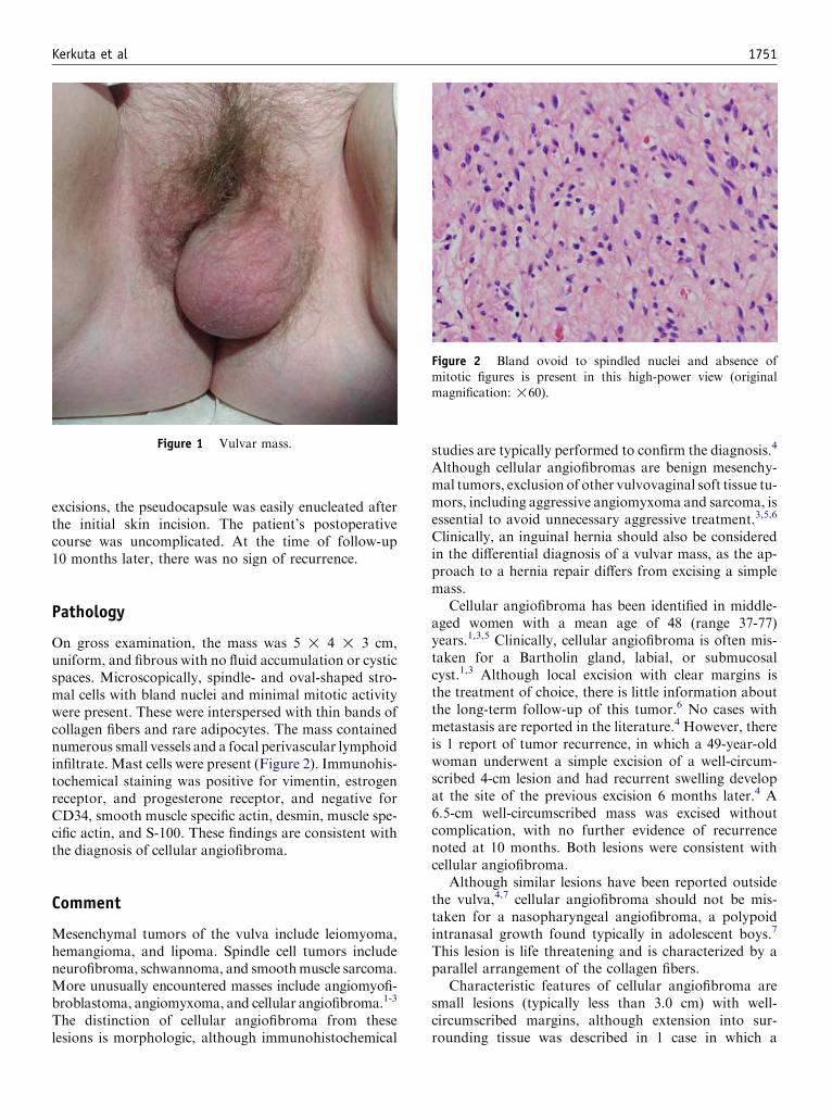
Kerkuta et al 1751
excisions, the pseudocapsule was easily enucleated afterthe initial skin incision. The patient’s postoperativecourse was uncomplicated. At the time of follow-up10 months later, there was no sign of recurrence.
Pathology
On gross examination, the mass was 5 ! 4 ! 3 cm,uniform, and fibrous with no fluid accumulation or cysticspaces. Microscopically, spindle- and oval-shaped stro-mal cells with bland nuclei and minimal mitotic activitywere present. These were interspersed with thin bands ofcollagen fibers and rare adipocytes. The mass containednumerous small vessels and a focal perivascular lymphoidinfiltrate. Mast cells were present (Figure 2). Immunohis-tochemical staining was positive for vimentin, estrogenreceptor, and progesterone receptor, and negative forCD34, smooth muscle specific actin, desmin, muscle spe-cific actin, and S-100. These findings are consistent withthe diagnosis of cellular angiofibroma.
Comment
Mesenchymal tumors of the vulva include leiomyoma,hemangioma, and lipoma. Spindle cell tumors includeneurofibroma, schwannoma, and smoothmuscle sarcoma.More unusually encountered masses include angiomyofi-broblastoma, angiomyxoma, and cellular angiofibroma.1-3
The distinction of cellular angiofibroma from theselesions is morphologic, although immunohistochemical
Figure 1 Vulvar mass.
studies are typically performed to confirm the diagnosis.4Although cellular angiofibromas are benign mesenchy-mal tumors, exclusion of other vulvovaginal soft tissue tu-mors, including aggressive angiomyxoma and sarcoma, isessential to avoid unnecessary aggressive treatment.3,5,6
Clinically, an inguinal hernia should also be consideredin the differential diagnosis of a vulvar mass, as the ap-proach to a hernia repair differs from excising a simplemass.
Cellular angiofibroma has been identified in middle-aged women with a mean age of 48 (range 37-77)years.1,3,5 Clinically, cellular angiofibroma is often mis-taken for a Bartholin gland, labial, or submucosalcyst.1,3 Although local excision with clear margins isthe treatment of choice, there is little information aboutthe long-term follow-up of this tumor.6 No cases withmetastasis are reported in the literature.4 However, thereis 1 report of tumor recurrence, in which a 49-year-oldwoman underwent a simple excision of a well-circum-scribed 4-cm lesion and had recurrent swelling developat the site of the previous excision 6 months later.4 A6.5-cm well-circumscribed mass was excised withoutcomplication, with no further evidence of recurrencenoted at 10 months. Both lesions were consistent withcellular angiofibroma.
Although similar lesions have been reported outsidethe vulva,4,7 cellular angiofibroma should not be mis-taken for a nasopharyngeal angiofibroma, a polypoidintranasal growth found typically in adolescent boys.7
This lesion is life threatening and is characterized by aparallel arrangement of the collagen fibers.
Characteristic features of cellular angiofibroma aresmall lesions (typically less than 3.0 cm) with well-circumscribed margins, although extension into sur-rounding tissue was described in 1 case in which a
Figure 2 Bland ovoid to spindled nuclei and absence ofmitotic figures is present in this high-power view (originalmagnification: !60).

1752 Kerkuta et al
46-year-old woman underwent initial enucleation, fol-lowed by 2 re-excisions and had no sign of recurrence at19 months.1 The cells are spindle shaped, lying betweenbands of collagen. Hyalinized vessels, abundant mastcells, and scant adipocytes may be present. Few mitoticfigures are identified. Cellular pleomorphism is rare,and tumor necrosis has not been described. Immunohis-tochemically, cellular angiofibromas are positive for vi-mentin and may express reactivity for CD34, whereasdesmin, actin, S-100 protein, keratin, and epithelialmembrane antigen are negative.4,6,8,9 In addition, the tu-mor has been found to be estrogen and progesteronereceptor positive.3-5 However, the significance of the pos-itive estrogen and progesterone receptors in angiofi-broma is unknown. These receptors are normal insubepithelial mesenchymal cells of the lower female gen-ital tract, so this may be a reflection of the cell of originrather than an alteration in a neoplasm.4 Alternatively, ithas been postulated that the expression of estrogen andprogesterone receptors suggests a role in the pathogene-sis of this tumor.3
The patient presented in this report was younger andher mass larger than most others reported in theliterature. Thus, cellular angiofibroma should be con-sidered in the differential diagnosis of a soft, painless,vulvar mass even in younger women.
Acknowledgment
We thank Ingrid E. Nygaard, MD, MS, for her editorialassistance with the manuscript.
References
1. Nucci MR, Granter SR, Fletcher CDM. Cellular angiofibroma:
a benign neoplasm distinct from angiomyofibroblastoma and spin-
dle cell lipoma. Am J Surg Pathol 1997;21:636-44.
2. Curry JL, Olejnik JL, Wojcik EM. Cellular angiofibroma of the
vulva withDNAploidy analysis. Int J Gynecol Pathol 2001;20:200-3.
3. Dargent J-L, de Saint Aubain N, GaldonMG, Valaeys V, Cornut P,
Noel J-C. Cellular angiofibroma of the vulva: a clinicopathological
study of two cases with documentation of some unusual features
and review of the literature. J Cutan Pathol 2003;30:405-11.
4. McCluggage WG, Perenyei M, Irwin ST. Recurrent cellular angio-
fibroma of the vulva. J Clin Pathol 2002;55:477-80.
5. Lane JE, Walker AN, Mullis EN, Etheridge JG. Cellular angiofi-
broma of the vulva. Gynecol Oncol 2001;81:326-9.
6. Nucci MR, Fletcher CDM. Vulvovaginal soft tissue tumours: up-
date and review. Histopathology 2000;36:97-108.
7. Garijo MF, Val-Bernal JF. Extravulvar subcutaneous cellular angi-
ofibroma. J Cutan Pathol 1998;25:327-32.
8. Dikmen Y, Yucebilgin MS, Kazandi M, Zekioglu O, Akalin T, Oz-
demir N. Cellular angiofibroma of the vulva: report of a case. Eur J
Gynaecol Oncol 2004;25:242-4.
9. Nielsen GP, Young RH. Mesenchymal tumors and tumor-like le-
sions of the female genital tract: a selective review with emphasis
on recently described entities. Int J Gynecol Pathol 2001;20:105-27.
American Journal of Obstetrics and Gynecology (2005) 193, 1753–5
www.ajog.org
A tale of 2 pedunculated myomas
Ihab M. Usta, MD, Elie M. Hobeika, MD, Anwar H. Nassar, MD
Department of Obstetrics and Gynecology, American University of Beirut Medical Center, Beirut, Lebanon
Received for publication July 13, 2005; revised July 21, 2005; accepted August 11, 2005
KEY WORDSSubmucous myoma
Pedunculated
We present 2 unusual cases of prolapsed pedunculated submucous myomas. In 1 patient, the pro-lapsed part measured 12 cm, with a 64-cm intrauterine part. The second patient had prolapsedpedunculated submucous myoma, which subsequently retracted into the uterus. Gynecologists
should be aware of unusual presentations of pedunculated submucous myoma to plan surgery.� 2005 Mosby, Inc. All rights reserved.
Uterine leiomyomas are the most common benigntumors of the female genital tract; 5% are submucosal,and 1.3% to 2.5% are pedunculated.1,2 Of the peduncu-lated submucous myomas (PSMs), 19.2% to 26.1%measure O3 to 5 cm.2 PSMs rarely may induce labor-like pain, eventually dilating the cervix and prolapsingthrough it with possible avulsion of the twisted necroticprolapsed portion.2
We present 2 cases of PSM; 1 case had an unusualappearance, and another case had unusual behavior.
Case 1
A 37-year-old woman (G3P3) had chronic foul-smellingdischarge, acute bleeding, and labor-like pain that wasfollowed by the protrusion of a mass on the perineum. A12-cm fleshy, dark-red mass was seen originating fromthe uterus and prolapsing through a dilated cervix(Figure 1,A). Ultrasound examination revealed a 17-cmuterus with a large myoma occupying the uterine cavity,which was continuous with the prolapsed part. Abdom-inal hysterectomy was performed. The myoma consistedof a 6-cm intramural round fundal fibroid tumor, with anelongated soft mid-portion coiled within the uterine
Reprints not available from the authors.
0002-9378/$ - see front matter � 2005 Mosby, Inc. All rights reserved.
doi:10.1016/j.ajog.2005.08.026
cavity and a distal portion that was prolapsed partiallythrough the cervix. The vaginal part was delivered vagi-nally; the other parts were removed abdominally. Thelength of the reconstituted myoma was 76 cm (Figure1,B).
Case 2
A 46-year-old grandmultiparous woman had a 4-monthhistory of menorrhagia. Ultrasound examination re-vealed an irregular uterus that measured 10.6 ! 6.8 !7.8 cm, with a posterior 6.0 ! 4.1 cm fibroid tumor.One week later, she experienced severe bleeding andlabor-like abdominal cramps. Speculum examinationshowed a 5-cm pedunculated violaceous fibroid tumorthat protruded through a 5-cm dilated cervix. Thebleeding decreased, and the pain subsided 1 hour afteradmission. In the operating room, 7 hours later, aspeculum examination by the same examiner revealeda closed cervix and retraction of the prolapsed myomainto the uterus. Intraoperatively, the myoma consistedof a whitish fundal lobe that was attached to theuterus O3.5-cm base and a violaceous lobe, which wasthe same part that had previously prolapsed through thecervix.

1754 Usta, Hobeika, and Nassar
Figure 1 Case 1. A, The prolapsed part of the myoma. B, The myoma on a 90-cm table. The thin arrow shows the intramural part;the thick arrow shows the prolapsed part.
Comment
Various imaging modalities can be used to diagnoseand characterize prolapsed PSMs and to obtain infor-mation that might aid in the choice of the surgicalapproach. In our cases, only ultrasound examination
Figure 2 Case 2. A, The myoma 1 week before prolapse. B,Pathology specimen. The thin arrows show the base of the my-oma; the thick arrows show the part that prolapsed and
retracted.
was used, because both patients had acute vaginalbleeding.
Vaginal myomectomy is an option for treating smallPSMs. Twisting, ligation and excision, and morcellationof larger tumors have been used.1,2 More recently, hys-teroscopically assisted techniques have been used.3
However, the size of the myoma in the first case was pro-hibitive for the attempt at such techniques. The longestmyoma ever reported measured 13 cm,3 which makesour case one of the largest and probably the longestmyoma reported in the literature.
The second case represents the first report of theregression of a myoma into the uterine cavity afterprolapse. Vaginal expulsion was not possible because nosuch expulsion was reported by either the nursing staffor the patient, and the pathologic specimen was theexact size and configuration of the image that was seenon ultrasound examination (Figure 2,A and B). Manualreplacement of prolapsed myomas was reported previ-ously in 3 patients before hysterectomy to prevent thecontamination of the abdominal cavity.1 In our case, re-placement during vaginal scrubbing was possible; how-ever, the cervix was closed, which suggested that themyoma had retracted earlier.
These cases illustrate the potential challenges gyne-cologists might face during the treatment of PSMs.Underestimation of the size of the myoma in the firstcase and attempts at vaginal myomectomy in similarsituations might have led to disastrous results. Further-more, a disappearing prolapsed myoma, such as in thesecond case, does not necessarily imply spontaneousexpulsion; retraction of the prolapsed part should bekept in mind.

Usta, Hobeika, and Nassar 1755
References
1. Brooks GG, Stage AH. The surgical management of prolapsed pe-
dunculated submucous leiomyomas. Surg Gynecol Obstet 1975;141:
397-8.
2. Ben-Baruch G, Schiff E, Menashe Y, Menczer J. Immediate
and late outcome of vaginal myomectomy for prolapsed
pedunculated submucous myoma. Obstet Gynecol 1988;72:
858-61.
3. Kanaoka Y, Hirai K, Ishiko O, Ogita S. An intranodal morcellation
technique employing loop electrosurgical excision procedure
for large prolapsed pedunculated myomas. Oncol Rep 2001;8:
1149-51.

American Journal of Obstetrics and Gynecology (2005) 193, 1756–8
www.ajog.org
Temporary balloon occlusion of the common iliac artery:New approach to bleeding control during cesareanhysterectomy for placenta percreta
Jin-Chung Shih, MD,a Kao-Lang Liu, MD,b Ming-Kwang Shyu, MDa
Department of Obstetrics and Gynecology,a and Department of Medical Imaging,b National Taiwan UniversityHospital and National Taiwan University College of Medicine, Taipei, Taiwan
Received for publication June 1, 2005; revised July 25, 2005; accepted August 12, 2005
KEY WORDSPlacenta percretaBalloon cathetersCommon iliac artery
A case of placenta percreta was referred at 31 weeks’ gestation. We performed a cesarean hyster-ectomy preceded by placement of occlusive balloon catheters at bilateral common iliac arteries at
34 weeks’ gestation. This simple and safe technique provides satisfactory efficacy for control ofprofuse bleeding during operation, with blood loss estimated at 800 mL.� 2005 Mosby, Inc. All rights reserved.
Placenta accreta is characterized by placental villiabnormally adherent to or invading the myometrium.Of the variants, the most severe form is placentapercreta, which indicates placenta penetrating throughthe entire uterine wall and even into the adjacent organs.Life-threatening hemorrhage often occurs in cases ofplacenta percreta during cesarean section, even withcorrect diagnosis and advanced preparation presurgery.The deaths were partly caused by inability to arrive atthe correct diagnosis before surgery and, more impor-tantly, catastrophic bleeding from abundant neovascu-larization and rich collaterals beyond the efficacy ofhemostasis available using current surgical techniques.
We report our successful experience in a case ofplacenta percreta, where preoperative placement ofocclusive balloon catheters in the bilateral commoniliac arteries minimized blood loss during cesareanhysterectomy.
Reprints not available from the authors.
0002-9378/$ - see front matter � 2005 Mosby, Inc. All rights reserved.
doi:10.1016/j.ajog.2005.08.033
Case report
A 34-year-old woman, gravida 4, para 1, presented atour clinic at 31 weeks’ gestation because of placentaprevia. She had given birth to 1 healthy term infant bycesarean section caused by prolonged labor. Other pasthistory included 2 uterine curettages for miscarriages.Our initial ultrasonography revealed a male fetusweighing approximately 2300 g and presenting in thetransverse position. Complete placenta previa with lossof the hypoechoic retroplacental zone led us to thediagnosis of placenta accreta. With weekly ultrasono-graphic investigation, progressive bulging of the pla-centa into the urinary bladder was observed andplacenta percreta with bladder invasion was stronglysuspected. Given the risk of spontaneous uterus ruptureand an estimated fetal weight of 2500 g, an electivecesarean hysterectomy was proposed at 34 weeks’gestation.
A multidisciplinary approach was scheduled at ad-mission. Because of the inherent anastomoses betweenthe internal and external iliac arteries, the radiologist

Shih, Liu, and Shyu 1757
elected to use temporary occlusive balloons in thebilateral common iliac arteries (CIA) during our cesar-ean hysterectomy. The patient and her family wereappropriately counseled with respect to the surgical andprocedural risks incurred. On the day of the operation,both femoral arteries were punctured using the standardSeldinger technique, with placement of 8-F sheathintroducers. The 5-F balloon catheters (maximal occlu-sive diameter 25 mm, Goodtec Co, Gifu, Japan) werethen inserted and placed in the CIA. The bilateral CIAlocations below the aortic bifurcation were easily iden-tified using the guide wire without contrast-enhancedangiography. The fluoroscopy time and the dosimetryof fetal exposure were only 2.4 minutes and 1.8 mGy,respectively. Under endotracheal general anesthesia,cesarean section was commenced after the establishmentof the arterial line and central venous route. Afterexploring the peritoneal cavity, tortuous cobweb-likevessels were identified covering almost the whole ante-rior uterine wall. The bulging placenta beneath theuterine serosa was readily identified. During applicationof the abdominal wall retractor, spontaneous uterinerupture and massive bleeding ensued at the implanta-tion site. A uterine incision was immediately made overthe higher uterine corpus to deliver the baby, withsimultaneous inflation of bilateral CIA balloon cathe-ters. The neonate weighed 2630 g and had Apgar scoresof 7/9 at 1/5 minutes, respectively. Blood loss wasdramatically decreased after balloon occlusion of thebilateral CIA. Subtotal hysterectomy was rapidly per-formed without removal of the placenta. Despite earlyinitiation of the cesarean hysterectomy at this gestation,however, it was found that the adhesive placenta hadinvaded to the urinary bladder, resulting in unavoidablebladder injury. The bladder repair was performedsmoothly by urologists after hysterectomy. The bilateralballoons were deflated before approximating the peri-toneal cavity to ensure adequate hemostasis. An oxim-etry monitor was placed on the patient’s lower limb andthe saturation maintained at around 85% to 92%during CIA occlusion. The arterial blood gas of lowerlimb was checked during this procedure, and the resultyielded mild metabolic acidosis with respiratory com-pensation (pH 7.37, pCO2 30.8 mm Hg, pO2 105.9 mmHg, HCO3 17.4 mEq/L, BE �6.6 mEq/L). The tempo-rary CIA occlusion sustained 53 minutes, with bloodloss estimated at 800 mL. The patient was transfusedwith 500 mL of packed red blood cells (RBC) only.The hemoglobin levels before and after the operationwere 10.2 and 9.7 g/dL, respectively. The markers of re-perfusion injury were checked on the following day andall demonstrated no elevation (CK 99 U/L, CK–MB 2.5U/L, LDH 490 U/L). Histologic examination of theuterus revealed that the chorionic villi had penetratedthe entire myometrium without intervening decidua,confirming the diagnosis of placenta percreta.
The operation was well tolerated by the patient,although recovery was complicated by temporary he-maturia and ileus. Eventual discharge was uneventful8 days after operation, with the Foley catheter removed10 days later at the urology clinic. At 6 months afterthe operation, the mother and baby remained healthy.
Comment
Currently, the management options for placenta accretainclude conservative and extirpative approaches. Theconservative strategy entails leaving the placenta in situduring cesarean section without further hysterectomy.However, risk of sepsis and delayed hemorrhage is alsoincurred. The extirpative approach consists of immediatecesarean hysterectomy, avoiding placental removal dur-ing operation. Nonetheless, extirpative management isassociated with significant risk of catastrophic bleeding.An adjuvant hemostatic technique is usually needed tominimize the risk of shock, multiple transfusion, andeven maternal death. Preoperative embolization of theinternal iliac artery (IIA) provides an excellent hemo-static effect in these cases; however, it is only suitable incesarean hysterectomy with previable fetuses. Sometimesthis procedure may elicit unexpected complications suchas pelvic pain, sexual dysfunction, or bladder ischemia.The alternative feasible technique is bilateral IIA ligationduring cesarean hysterectomy. Using this procedure, thepressures in the pelvic circulation are converted from ar-terial pressures to venous-like pressures, thus makingbranches in the circulation more amenable to hemostasisvia simple clot formation. However, the failure rate forthis may be as high as 60%.
A widely adopted alternative to IIA ligation ispreoperative placement of occlusive balloon cathetersin the bilateral IIA. This procedure appears to besuperior because the obstetrician does not need to befamiliar with the technique of IIA ligation, and canimmediately occlude the bilateral IIA after cord clamp-ing without the interference created by a small andbloody surgical field. However, the results of a numberof trials appear to indicate that the technique is not aseffective as expected, with 1 study even concluding nodifference in outcomes comparing patients who under-went this procedure and those who did not.1
Failure of IIA ligation or balloon occlusion can belargely explained by the extensive anastomoses in thepelvic vasculature. The obturator artery serves as animportant anastomosis bridging internal and externaliliac arteries.2 Additionally, angiographic studies of pa-tients undergoing IIA ligation have identified branchesof the lumbar, sacral, rectal, femoral, and even internalthoracic arteries as the origin of collateral circulation,preventing pelvic ischemia. Collectively, these collateralpelvic-vessel networks may substantially decrease theefficacy of IIA ligation or occlusion.

1758 Shih, Liu, and Shyu
To the best of our knowledge, the presented case isthe first report using temporary CIA occlusion formanagement of placenta percreta during cesarean hys-terectomy. Importantly, this approach appears to havesome apparent advantages compared with existing tech-niques. Unlike placement of IIA balloon catheters, theCIA balloon catheter is easier to lodge in the properlocation, especially for those cases with unusual vascularanatomy. The fluoroscopy exposure is decreased, andiodine-containing contrast medium is not required.Further, the procedure can be used to overcome collat-eral flow from the external iliac and femoral arteries,thus significantly decreasing hemorrhage during cesar-ean hysterectomy. However, prolonged occlusion of theCIA may be associated with reperfusion injury, throm-bosis, and/or embolism of the lower extremities. There-fore, the occlusion time should be as short as possible.As the duration of aortic cross-clamping can be at least1 hour in surgery for abdominal aortic aneurysm,3 our
procedural time is relatively safe, especially with thepresence of rich collaterals during pregnancy, and theblood flow cannot be totally occluded by balloon cathe-ter. Further evaluation and comparison of the efficacyand outcome of this procedure is needed relative tothe alternative techniques for management of advancedforms of placenta accreta.
References
1. Levine AB, Kuhlman K, Bonn J. Placenta accreta: comparison of
cases managed with and without pelvic artery balloon catheters.
J Matern Fetal Med 1999;8:173-6.
2. Dubreuil-Chambardel L. Traite des variations du system arteriel
variations des arteres du pelvis et du membre inferieur. Paris:
Masson et Cie; 1925.
3. Kurata S, Hongo H, Furutani A, Kaneda K, Sakai H, Kuroda Y,
et al. A new approach for abdominal aortic aneurysm by a ringed
Y graft. Vasc Surg 1992;26:47-52.

American Journal of Obstetrics and Gynecology (2005) 193, 1759–60
www.ajog.org
Episiotomy dehiscence that required intestinal diversion
Carl H. Rose, MD,a Kristi L. Blessitt, MD,a Farshid Araghizadeh, MD,b
John C. Morrison, MDa
Departments of Obstetrics and Gynecologya and Colon & Rectal Surgery,b University of Mississippi Medical Center,Jackson, MS
Received for publication May 12, 2005; revised August 4, 2005; accepted August 17, 2005
KEY WORDSEpisiotomy
DehiscenceInfectionIleostomy
Postpartum episiotomy dehiscence is a rare complication of vaginal delivery. Forceps-assistedvaginal delivery over mediolateral episiotomy was complicated by infection and dehiscence withrectal injury. A diverting ileostomy was used to permit healing. Episiotomy infection requires
early recognition and thorough evaluation to exclude occult rectal injury.� 2005 Mosby, Inc. All rights reserved.
A 40-year-old woman (G6P2032) was seen afterpremature rupture of membranes at 38 weeks of gesta-tion. Her medical history was significant for obesity(body mass index, 29 kg/m2), chronic hypertension, andrecurrent Grave’s disease that required radioactive io-dine ablation before conception. Her first pregnancy cul-minated in a spontaneous vaginal delivery (2466 g)followed by a cesarean delivery for dystocia in her sec-ond pregnancy (2722 g). She was counseled and stronglydesired vaginal birth after cesarean delivery for the cur-rent pregnancy. Oxytocin augmentation was begun, andlabor progressed satisfactorily over the next 10 hours.However, fetal bradycardia ensued late in the secondstage of labor; at C3 station, emergent outlet Simpsonforceps-assisted delivery over second-degree mediolat-eral episiotomy was performed for a healthy 2265-gmale infant with Apgar scores at 5 and 7 minutes of 9and 9, respectively. On further inspection, the episiot-omy was noted to extend into the rectal sphincter. Edgesof the sphincter muscle were identified and reapp-roximated with the use of 4 interrupted 0 polyglactinsutures; another continuous 1-0 suture was used to
Reprints not available from the authors.
0002-9378/$ - see front matter � 2005 Mosby, Inc. All rights reserved.
doi:10.1016/j.ajog.2005.08.037
rebuild the perineal body. The vaginal mucosa wasclosed with a smaller 2-0 suture that proceeded fromthe episiotomy apex to the hymenal ring. The bulbocav-ernosus stitches were pulled together, and the medial lat-eral skin incision was repaired with running 2-0 sutures.The patient’s postpartum course was complicated byrecurrent hemorrhage because of uterine atony 3 and 5hours after the delivery, which responded to 15-methylprostaglandin given intramuscularly and misoprostolplaced in the rectum. The episiotomy repair was notedto be intact during both episodes.
The next day she had a low-grade fever (99.8(F) andleukocytosis (white blood cell count 26,000/mm3);broad-spectrum antibiotics were begun. On the secondday after the delivery, she complained of pain at theepisiotomy site. Examination under anesthesia revealedpartial dehiscence of the episiotomy repair with purulentdrainage from an abscess cavity that communicated withthe left ischiorectal fossa. A full-thickness injury that in-volved the anal sphincter complex was noted thatextended inward 6 cm in depth and 1 to 2 cm in widthfrom the anal verge into the rectum at the 6 o’clockposition. The rectum mucosa was intact. The previousrepair was completely opened to drain the abscess, andthe devitalized tissue was debrided. The perineum and

1760 Rose et al
vaginal vault were irrigated, and the wound was packedwith wet-to-dry dressings. A rectal tube was inserted, andanaerobic antibiotic coverage was added. Sequentialpostoperative blood glucose values were normal, and an-tinuclear antibody testing was negative. She remainedafebrile throughout the remainder of her postoperativecourse.
Operative debridement of the perineal wound wasrepeated on postdelivery days 4 and 5 to ensure healthy,viable perineal tissues. Subsequently the colon and rectalsurgical service was consulted for evaluation and treat-ment; on postpartum day 5, the clinical decision wasmade to undertake repair of the injury. Because thetissues continued to require debridement despite inten-sive surgical and antibiotic therapy, the decision wasalso made to divert the fecal stream simultaneously.Therefore, on postdelivery day 6, the injury was repairedprimarily in 3 layers to restore annual rectal sphinctercomplex integrity. The subcutaneous wound above therepair, which extended into the ischiorectal fossa, wasleft open to heal by secondary intention. A divertinglaparoscopic loop ileostomy was also performed topromote perineal wound healing. Her subsequent post-partum course was unremarkable, and she was dis-charged home on postdelivery day 8 with colon andrectal surgery follow-up visits. The perineal woundhealed without event, as noted in the postpartumfollow-up visits at 2 and 6 weeks. Five months later,she underwent ileostomy closure and had return ofnormal gastrointestinal function and fecal continence.
Comment
Infection and episiotomy dehiscence that involves therectum is a catastrophic event. Permanent sphincterdysfunction with consequent fecal incontinence carriesenormous hygienic and psychosocial consequences. For-tunately, dehiscence occurs in only 0.2% to 2% of all ofepisiotomies. Specific guidance for the treatment ofconcurrent proximal rectal injury is virtually nonexistentin the obstetric literature. Traditional doctrine has heldthat the failure of initial repair mandates a 2- to 3-monthinterval to permit thorough tissue revascularization. Apreliminary report by Hauth et al1 described successfulrevision within 7 days of dehiscence in 8 patients, withonly a single failure. Ramin et al2 have published themost extensive series to date regarding early revision.Early closure was successful in all except 2 of 35 cases(6%); both failures had pre-existing risk factors forpoor tissue healing. All injuries were confined to therectal sphincter and/or the lower 5 cm of rectum.2 The
repairs were accomplished at a mean of 6.4 days (range,3-13 days).
In most cases, diverting loop ileostomy or colostomyis not performed, and most uncomplicated wounds healwell, as noted by the published reports. Failure usuallyoccurs when pre-existing risk factors such as diabetesmellitus, hypertension, obesity, or inflammatory diseasesare present. In this case, because the patient was obeseand because the abscess appeared early in the clinicalcourse, the clinicians on both services believed that thelikelihood of conservative treatment and delayed repairhad a high risk of failure. Because of the extension of theabscess into the ischiorectal fossa, a diverting ileostomywas performed to deflect the fecal stream at the sametime as the closure. Although immediate postpartumantibiotic therapy potentially could have changed theoutcome in this case (absent factors other than obesityand emergent forceps delivery), there was no clinicalindication to begin such treatment. Ileostomy wasselected because it can be performed laparoscopically,is less prone to fistulae than colostomy, and requiresmuch less patient care than traditional colostomy.
Infection appears to be the fundamental cause inmost repair failures. After dehiscence, the incidence offailed secondary repair does not appear to differ signif-icantly between midline (69%) versus mediolateral(86%) incisions. Other reported risk factors includechronic corticosteroid use, refractory cough, inflam-matory bowel conditions, immunologic deficiencies,obesity, or collagen-vascular disease. Recommendedlaboratory evaluation includes complete blood count,antinuclear antibody, human immunodeficiency virus,and hepatitis profile. Depending on individual history,further evaluation may include an upper gastrointestinalseries, barium enema, computed tomography scan, mag-netic resonance imaging, or colonoscopy.
To date, this is the only reported case of episiotomydehiscence that required temporary intestinal diversionthat we were able to find in the literature. A divertingloop ileostomy technique was selected because theeffluent quality and stomal care are reduced significantlycompared with conventional colostomy.
References
1. Hauth JC, Gilstrap LC III, Ward SC, Hankins GDV. Early repair
of an external sphincter ani muscle and rectal mucosal dehiscence.
Obstet Gynecol 1986;67:806-9.
2. Ramin SM, Ramus RM, Little BB, Gilstrap LC III. Early repair of
episiotomy associated with infection. Am J Obstet Gynecol 1992;
167:1104-7.

American Journal of Obstetrics and Gynecology (2005) 193, 1761–3
www.ajog.org
Endometriotic umbilical port site metastasis afterlaparoscopy
Umut Barbaros, MD,a,* Ahmet Cem Iyıbozkurt, MD,b Mıne Gulluoglu, MD,c
Merve Barbaros, MD,d Yesım Erbıl,a Vahıt Tunalı,e Selcuk Mercana
Departments of General Surgery,a Gynecology and Obstetrics,b and Pathology,c Istanbul Medical School, IstanbulUniversity, Istanbul, Turkey; Department of Gynecology and Obstetrics, Prıvate Hospital of Goksoy, Istanbul,Turkeyd; Istanbul Training and Research Hospital, Samatya, Istanbul, Turkeye
Received for publication November 17, 2004; revised May 12, 2005; accepted May 12, 2005
KEY WORDSEndometriosisLaparoscopy
Endometriotic implantUmbilical port site
metastasis
We present the case of a 40-year-old woman who had been previously operated for endometri-
oma 2 years ago by laparoscopy, with the complaint of an umbilical mass with cyclical painpattern. The dark-colored mass was excised and pathology report revealed it to be an endometri-otic implant.� 2005 Mosby, Inc. All rights reserved.
Endometriosis, the presence of functioning endome-trial tissue in anatomic locations other than the uterinecavity, still perplexes practitioners by its rare presentingforms. Endometriosis has a long history. It was de-scribed as adenomyomata of the rectovaginal septum inearly reports.1,2 This was similar to uterine adenomyo-mata of the uterus.1 Although extrapelvic endometriosismay be present in up to 12% of women with endometri-osis,3 umbilical endometriosis is uncommon, with anestimated incidence of 0.5% to 1.0% of all patientswith endometrial ectopia.4
The majority of cutaneous endometriosis develops onsurgical scars of the abdomen of young women in thereproductive period.5 Recently with more frequent useof a less invasive technique like laparoscopy for surgicaltreatment of endometriosis, more cases of endometriosis
* Reprint requests: Umut Barbaros, MD, Department of General
Surgery, Istanbul Medical School, Istanbul University, Istanbul,
Turkey 34390.
E-mail: [email protected]
0002-9378/$ - see front matter � 2005 Mosby, Inc. All rights reserved.
doi:10.1016/j.ajog.2005.05.088
at port sites are encountered. From 1976 to August2004, 6 cases of endometrioma of port sites after lapa-roscopy were published: The first report occurred aftera sterilization procedure, published in 1990.6 Two caseswere after laparoscopic-assisted subtotal hysterectomy,7
2 were after laparoscopy for endometriosis5,8 and 1 oc-curred after bilateral hernia treated by laparoscopythat was converted to laparotomy.9 In this report, wepresent a patient with endometriotic umbilical port sitemetastasis who previously had laparoscopic resectionof ovarian endometriotic cysts.
Case report
A 40-year-old woman presented with a recent history ofirregular umbilical mass and an associated severe cyclicpain during her menses. The mass was dark colored,hard, and measured 2 to 3 cm in diameter and waslocated on the previous laparoscopic umbilical port sitescar (Figure 1). Ultrasonography of the lesion revealed ahypoechoic 29 ! 21 ! 26 mm lobulated lesion located

1762 Barbaros et al
at the subcutaneous tissue just below the umbilicus withintense arteriolar network image seen during Doppler ul-trasound. The patient’s history was significant for laparo-scopic removal of a multilocular endometriotic cyst from
Figure 1 Umbilical mass in purple and gray color with cyclic
pattern.
Figure 2 Umbilical mass after complete excision.
the left ovary. During this laparoscopic procedure, thecyst wall had been removed without using a bag throughan inguinal port. The operative site had been irrigated us-ing around a liter of ringer’s lactate solution. The rest ofthe patient’s history was unremarkable.
During the operation, the hard mass located justbeneath the umbilicus above the rectus sheathwas excisedcompletely along with the adhered fascia (Figure 2). Theumbilicus appeared normal. The defect was primarilyclosed. The pathology report confirmed that the massexcised was an endometrioma (Figure 3). She had anuneventful postoperative course.
Comment
Although umbilical endometriosis is uncommonly re-ported, it is usually easily diagnosed because of thecyclic character of the pain it elucidates, as in oursubject. Simple excision of the umbilical endometriomais the treatment of choice.10
The histogenesis of endometriosis is still unknown,although there has been a lot of research as to its origin.In the case of an umbilical port site endometrioma, thelesion may have developed from peritoneal seeding ofcells because of pneumoperitoneum or from directcontact of the excised lesion with the abdominal wall.It is also proposed that cutaneous endometriosis mayarise from endometrial tissue that is transported vialymphatics or vascular channels.11 Because it is knownin the previous laparoscopy that the endometriotic le-sion was removed through the sheath of the instruments,our case lends support to the aerosolization theory, theconcept that pneumoperitoneum influences the implan-tation of free intraperitoneal endometriotic cells.
Figure 3 Histopathological view of the specimen.

Barbaros et al 1763
Routinely applying techniques like introduction andremoval of instruments and the excised lesions within thesheath to avoid contact with the abdominal wall, using abag during removal of endometriotic lesions and exsuf-flating the abdomen while the ports are in place may helpto prevent endometriotic implants. Carefully plannedcase-control studies may help to yield more informationabout the etiopathogenesis of umbilical endometriosis.
References
1. Cullen TS. The distribution of adenomyomata containing uterine
mucosa. Am J Obstet Gynecol 1919;80:130-8.
2. Lockyer C. Adenomyoma in the recto-uterine and recto-vaginal
septa. Proc Royal Soc Med (Obstet) 1913;6:112-20.
3. Franklin RR, Navarro C. Extragenital endometriosis. In: Chadha
DR, Buttran VC, editors. Current concepts in endometriosis. New
York: Alan R. Liss, Inc; 1990. p. 289.
4. Michowitz M, Baratz M, Stavorovsky M. Endometriosis of the
umbilicus. Dermatologica 1983;167:326-30.
5. Healy JT, Wilkinson NW, Sawyer M. Abdominal wall endometri-
oma in a laparoscopic trocar tract: a case report. Am Surg 1995;61:
962-3.
6. Denton GWL, Schofield JB, Gallagher P. Uncommon complica-
tions of laparoscopic sterilization. Ann R Coll Surg Engl 1990;
72:210-1.
7. Koninckx PR, Donders G, Vandecruys H. Umbilical endometrio-
sis after unprotected removal of uterine pieces through the umbil-
icus. J Am Assoc Gynecol Laparosc 2000;7:227-32.
8. Wakefield SE, Hellen EA. Endometrioma of the trocar site after
laparoscopy. Eur J Surg 1996;162:523-4.
9. Martinez-Serna T, Stalter KD, Filipi CJ, Tomonaga T. An un-
usual case of endometrial trocar site implantation. Surg Endosc
1998;12:992-4.
10. Purvis RS, Tyring SK. Cutaneous and subcutaneous endometrio-
sis. Surgical and hormonal therapy. J Dermatol Surg Oncol
1994;20:693-5.
11. Friedman PM, Rico MJ. Cutaneous endometriosis. Dermatol
Online J 2000;6:8.
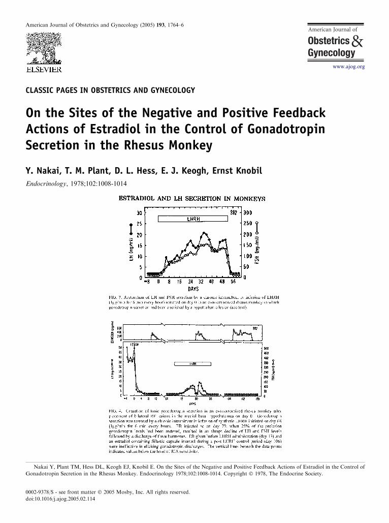
American Journal of Obstetrics and Gynecology (2005) 193, 1764–6
www.ajog.org
CLASSIC PAGES IN OBSTETRICS AND GYNECOLOGY
On the Sites of the Negative and Positive FeedbackActions of Estradiol in the Control of GonadotropinSecretion in the Rhesus Monkey
Y. Nakai, T. M. Plant, D. L. Hess, E. J. Keogh, Ernst Knobil
Endocrinology, 1978;102:1008-1014
Nakai Y, Plant TM, Hess DL, Keogh EJ, Knobil E. On the Sites of the Negative and Positive Feedback Actions of Estradiol in the Control of
Gonadotropin Secretion in the Rhesus Monkey. Endocrinology 1978;102:1008-1014. Copyright � 1978, The Endocrine Society.
0002-9378/$ - see front matter � 2005 Mosby, Inc. All rights reserved.
doi:10.1016/j.ajog.2005.02.114

Classic Pages 1765
Commentary by Lawrence D. Longo, MD
In the mid 1940s, Geoffrey W. Harris1 (1913-1971)posited the idea that hormones, as yet unidentified,regulated the function of the pituitary gland. Harris’group later produced ovulation in rabbits by intrapitui-tary infusion of an extract of bovine hypothalamus.2
Following considerable effort, Roger Guillemin andAndrew V. Schally independently demonstrated thathypothalamic peptides stimulated the release of follicularstimulating hormone (FSH) and luteinizing hormone(LH) both in vitro and in vivo, and in 1971, each reportedthe amino acid sequence of the decapeptide that becameknown as gonadotropin releasing hormone (GnRH).3,4
Subsequently, in a series of studies in Macaca mulata,Ernst Knobil (1926-2000) and colleagues demonstratedthat GnRH was released in a pulsatile manner and,carried to the adenohypophysis via a portal vascularnetwork, regulated the release of FSH, LH and, thus,ovarian function.5-8 Following bilateral radiofrequency(RF) lesions placed in the arcuate region of the medialbasal hypothalamus of an adult ovariectomized Rhesusmonkey, a continuous infusion of exogenous LHRHfailed to restore gonadotropin levels. In contrast, and asshown in Figure 3, pulsatile LHRH infusion (1 mg/minfor 6 minutes every hour) resulted in sustained restitutionof LHandFSH secretion. Figure 4 illustrates the decreasein blood plasma gonadotropin levels following the RFlesions, and the failure of estradiol benzoate (EB) ad-ministration (42 mg/kg body weight) to restore these.Again, gonadotropin secretion was restored in responseto chronic intermittent LHRH infusion.
0002-9378/$ - see front matter � 2005 Mosby, Inc. All rights reserved.
doi:10.1016/j.ajog.2005.06.056
Discovery of the hypothalamichormone pulse generator andsignificance*
Ernst Knobil, MD
Shortly after a specific radioimmune assay for monkeyluteinizing hormone (LH) became available in the late1960s, we began to investigate the dynamics of its
* The original version of this article, Remembrance: The discovery of the
physiological significance, appeared in Endocrinology 1992;131:1005-6. Copy
2000, Dr Knobil had promised such a report for the American Journal of O
0002-9378/$ - see front matter � 2005 Mosby, Inc. All rights reserved.
doi:10.1016/j.ajog.2005.06.025
At the University of Pittsburgh, Knobil establishedthe Center for Research in Human Reproduction, whereover several decades he made many seminal contribu-tions to understanding the function of the hypothala-mus, the pituitary gland, gonads, and the biology ofreproduction. These contributions to an understandingof ovulatory function have resulted in a significantimpact of health care. Knobil was elected to theNational Academy of Sciences, and the AmericanAcademy of Arts and Sciences, and received numerousinternational awards and honors.
References
1. Green JD, Harris GW. The neurovascular link between the neuro-
hypophysis and adenohypophysis. J Endocrinol 1947;5:136-46.
2. Harris GW. Humours and hormones. J Endocrinol 1972;53:2-23.
3. Burgus R, Butcher M, Ling N, Monahan M, Rivier J, Fellows R,
et al. Structure moleculaire du facteur hypothalmique (LRF)
d’origine ovine, controlant la secretion de l’hormone Gonadotrope
Hypophysaire de Luteinisation (LH). C R Acad Sci Hebd Seances
Acad Sci D 1971;273:1611-3.
4. Matsuo H, Baba Y, Nair RM, Arimura A, Schally AV. Structure of
the procine LH- and FSH- releasing hormone. I. The proposed
amino acid sequence. Biochem Biophys Res Commun 1971;43:
1334-9.
5. Knobil E. On the control of gonadotropin secretion in the Rhesus
monkey. Recent Prog Horm Res 1974;30:1-46.
6. Knobil E. Patterns of hypophysiotropic signals and gonadotropin
secretion in the Rhesus monkey. Biol Reprod 1981;24:44-9.
7. Knobil E. The neuroendocrine control of the menstrual cycle. Res
Progr Horm Res 1980;36:53-88.
8. Knobil E, Plant TM, Wildt L, Belchetz PE, Marshall G. Control of
the Rhesus monkey menstrual cycle: permissive role of hypotha-
lamic gonadotropin-releasing hormone. Science 1980;207:1371-3.
gonadotropin-releasingof its physiologic
secretion in a variety of physiologic and experimentalcircumstances. One such early study was designed todetermine whether a circadian rhythm in the plasma
hypothalamic gonadotropin-releasing hormone pulse generator and of its
right 1992, The Endocrine Society. Between that time and his death in
bstetrics and Gynecology.

SELECTED MANUSCRIPTS FROM THE 2005CREOG & APGO ANNUAL MEETING
The Council on Resident Education in Obstetrics and Gynecology (CREOG)The Association of Professors of Gynecology and Obstetrics (APGO)
2005 CREOG & APGO ANNUAL MEETINGMarch 2-5, 2005The Grand America Hotel555 South Main StreetSalt Lake City, UT 84111
APGO Web site: www.apgo.org
Patrick Duff, MD, CREOG Chair
Department of Ob-GynUniversity of FloridaP.O. Box 1002941600 SW Archer RoadGainesville, FL 32610Phone: (352) 392-3222Fax: (352) 392-2808
William N. P. Herbert, MD, CREOG Vice Chair
Department of Ob-GynUniversity of VirginiaBox 800712Hospital Drive, 3rd Floor Private ClinicsCharlottesville, VA 22908Phone: (804) 924-9937Fax: (804) 982-1840
Haywood L. Brown, MD, CREOG Program Co-chair
2005 CREOG & APGO Annual Meeting
Department of Ob-GynDuke UniversityDUMC 3084203 Baker HouseDurham, NC 27710Phone: (919) 668-3948Fax: (919) 668-5547
Dee E. Fenner, MD, APGO President
Department of Ob-GynUniversity of Michigan1500 E. Medical Center DriveL4100 Women’s HospitalBox 0276Ann Arbor, MI 48109Phone: (734) 763-6670Fax: (734) 647-9727
J. Mac Ernest, MD, APGO President-elect
Department of Ob-GynWake Forest UniversityMedical Center BoulevardWinston-Salem, NC 27157Phone: (336) 716-1025Fax: (336) 716-6937
J. Mac Ernest, MD, APGO Program Co-chair
2005 CREOG & APGO Annual Meeting
Department of Ob-GynWake Forest UniversityMedical Center BoulevardWinston-Salem, NC 27157Phone: (336) 716-1025Fax: (336) 716-6937

American Journal of Obstetrics and Gynecology (2005) 193, 1768–72
www.ajog.org
Steadfastly forward
Timothy R. B. Johnson, MD
Department of Obstetrics and Gynecology, University of Michigan Medical School, Ann Arbor, MI
Received for publication February 24, 2005; revised May 5, 2005; accepted August 1, 2005
‘‘Every time I look back, I meet the eyes of myforeparents looking steadfastly forward.’’
Professor Roger Shinn
Concerns abound with regard to the future of ourspecialty of obstetrics and gynecology. In his presiden-tial address to the American College of Obstetriciansand Gynecologists,1 Dr John Gibbons summarized theissues that dominate discussion: (1) recruitment ofmedical students; (2) change in residency programs;and (3) the new face of private or academic practice. Thefirst is highlighted by the decrease in the percentage ofgraduating US seniors choosing a career in obstetricsand gynecology and the associated decrease in residencyslots filled by US graduates. The marked decrease in thenumber of men entering the specialty is noted withconcern, and the shift of student interest to emergencymedicine, radiology, ophthalmology, anesthesiology,and dermatology noted with envy. It must be said thata decade ago both anesthesiology and radiologysounded their own alarms, so the astute observer mustsuppose that there are powerful cyclic forces operating.
The concern about residency training focuses on theever broadening scope of requirements, something thatas a member of the Residency Review Committee I am
Presented as the Wayne Johnson Memorial Lecture at the Annual
Meeting of the Association of Professors of Gynecology and Obstetrics
and the Council on Resident Education in Obstetrics and Gynecology,
March 2, 2005, Salt Lake City, UT.
Reprints not available from the author. Address correspondence to
Timothy R. B. Johnson, MD, Department of Obstetrics and Gyne-
cology, University of Michigan Medical School, 1500 East Medical
Center Drive, L4000 WH, Ann Arbor, MI 48109-0276.
E-mail: [email protected]
0002-9378/$ - see front matter � 2005 Mosby, Inc. All rights reserved.
doi:10.1016/j.ajog.2005.08.001
well aware of, but the breadth of medical knowledgepresents a challenge to all undergraduate and graduatemedical education programs. The 80-hour work weekhas surely placed new challenges on our residencyprograms. As to practice, increased debt burden, ad-ministrative burdens, liability concerns,2 and family andpersonal interests all contribute. Dr Gibbons speaksenthusiastically and hopefully about the potential to‘‘refocus our specialty.’’
One only need cast a brief glance at history to realizethat similar concerns are not new for our specialty. Twoof our most insightful and visionary specialty leaders,both my predecessors as chair at the University ofMichigan, were important observers of this phenomenon.
In a 1920 address to the American Medical Associ-ation,3 Dr Reuben Peterson, chair from 1901 to 1931,outlined ‘‘The Future of Obstetrics and Gynecology as aSpecialty.’’ This was a time when obstetrics and gyne-cology were considered widely by many as separatespecialties, and ‘‘gynecologists threw themselves intothe development of pelvic surgery to such an extentthat things outside the operative field failed to interestthem. . The result is that the ground work absolutelyessential to one aspiring to devote himself to onedivision of obstetrics and gynecology is lost sight ofand there is a wild scramble for a short cut to fame andfortune, usually through the dexterous handling of thescalpel.’’ He then argued for the unification of obstetricsand gynecology and for an emphasis on broad educationas the basis for both specialists and subspecialists.
Dr Peterson saw merit in a suggestion that onlymedical school gynecologists and psychiatrists shouldselect those most suited for gynecologic surgical careers,for ‘‘if by any chance (they) were to run across an
Johnson 1769
applicant who had ambition to become an expert ineverything, physical and functional, pertaining to thegenital tract of women, it would be comparatively easyto have such a person become either a temporary or apermanent occupant of the psychiatric clinic.’’
He envisioned strong hospital-based training pro-grams for both medical students and postgraduates,not yet widely called ‘‘residents.’’ His description of theideal department of obstetric and gynecology to offertraining for ‘‘young graduates’’ fits very well with theacademic obstetrics and gynecology residency programthat we would all recognize today. Peterson wrote that‘‘each man would work out the hardships of being up allnight with a confinement case and then be obliged to dodifficult hysterectomies the next day’’ and hoped thetrainee would ‘‘keep faith and find enough in his specialtyto interest him so he will do his regular work well andadd a little something to the sum of human knowledge.’’
In another major address, published in the AmericanJournal of Obstetrics and Gynecology as ‘‘Obstetrics-Gynecology: ATime for Change,’’4 Dr J. RobertWillson,my chairman who held his position from 1962 to 1979,raised not dissimilar issues: ‘‘changes in attitude andeducation of medical students; a proliferation of scien-tific and technical information to be incorporated intoclinical practice; increasingly burdensome controls byburgeoning institutional, professional, and governmen-tal bureaucracies; changing patient attitudes; changingphysician attitudes.’’ Dr Willson was an early acceptorand proponent of primary and preventive care5 and feltthat ‘‘too little emphasis is placed on preparing houseofficers for contemporary practice.’’ He was one of thefirst to understand that we must be responsive to societyand our patients as we develop the curricular contentof our educational programs. Dr Willson’s detailed cri-tique of residency training remains insightful and pro-vocative and resonates in the current discussions at theAccreditation Council for Graduate Medical Educa-tion (ACGME) and the Residency Review Committee(RRC).
As to medical students, he comments ‘‘that we nowhave as many or more applicants than first-year posi-tions is considered by some to be an indication ofsuccess . but are we being responsible? . How manyobstetrician-gynecologists trained in the present modelwill be content to spend almost all their time counselingadolescents, the elderly, and others and providing con-traceptive and other routine ambulatory services andhow well will they do these things?’’
Dr Willson then advocated changes and proposedeliminating ineffective programs, approving only pro-grams that provide comprehensive training, competency-based evaluations of trainees, regular program reviews,appropriate specialty examinations, and recertification.He saw the American Board of Obstetrics and Gynecol-ogy, American College of Obstetricians and Gynecolo-
gists, Association of Professors of Gynecology andObstetrics, the Council on Resident Education in Ob-stetrics and Gynecology, and the Residency ReviewCommittee all as important participants in this change,and it is remarkable that virtually all of his recommen-dations have been successfully implemented, if not asquickly as he would have liked or in time for him to see.
So if similar concerns have existed for more than acentury and previous leaders have predicted, advocated,and directed change, I believe we must follow their lead,face the challenges, and find the solutions that will chartthe course for the optimal wellness and health of ourpatients and our specialty.
I believe, as did Dr Willson, that change must comefrom our leaders and leadership organizations. I havedemonstrated that as a specialty we have faced similardremarkably similardchallengesbeforeandhave respondedconstructively.
Reuben Peterson and J. Robert Willson were rightthat our specialty will flourish if defined broadly: ob-stetrics and gynecology, surgery and primary care,specialty and subspecialty. We must address the con-cerns of those of who advocate for attention to surgicaltraining,6-8 but breadth and balance must be maintained.The role of primary and preventive care was emphasizedby Dr Willson, and its importance was recently reaf-firmed by specialty leaders9 in an ‘‘academic blueprint.’’
Dr Peterson also raised the issue of time, specificallynight call.10 We need to heed the lesson of the 80-hourwork week. After the ACGME mandate, the Universityof Michigan Medical School implemented an 80-hourwork week for medical students. It did not take long, norshould it have, for the faculty to ask, ‘‘How about us?’’Dr Gibbons is correct that we must address the chal-lenges not by focusing on residents and residency edu-cation or on medical students and medical education,but we must focus on practitioners and their practice:the very specialty of obstetrics and gynecology. Studentinterest groups will not change the reality of practice.Resident professionalized in an 80-hour work week willbring new expectations to their professional lives. Wemust refocus, redefine, if not reinvent our specialty.Already, as Dr Gibbons notes, there is movement towardlarger practice groups, groups that can absorb part-timedoctors and offer reasonable call schedules.
Other specialties have developed hospitalists, full-timehospital-based clinicians.11 Weinstein12 has proposedthe role of ‘‘laborist,’’ someone whose focus is manage-ment of laboring patients. One of the risks of some ofthese innovations is loss of continuity, but ultimatelywe must assure quality care and patient safety whilemaximizing satisfaction for all participants in the healthcare system. Recently our Maternal Fetal Medicinefaculty have experimented with a night float system,taking the lead from residents and medical students forwhom night float has become a national standard. In our

1770 Johnson
Table I Generations
Values Work Org. culture
Traditional generation, 1925-1940 Traditional obligation to conform Duty Command and controlBaby boomers, 1945-1963 Radicals Self-fulfillment Consensus
Individuals Opportunity for advancement ParticipationDual careers Work is leisure IdealistsSelf-gratification Self-directed
Generation Xers, 1965-1982 Latchkey kids Balance CompetenceExtended adolescence Learning opportunities Realists
Free agentsSelf-reliant
Org., Organizational.
department, despite occasional logistic challenges, third-and fourth-year residents and medical students have welldeveloped and enhanced night float systems. More juniorresidents are experimenting with options on how best toadapt it to their educational needs and work hourconstraints.
We need to develop programs for reentry andretraining because well-prepared colleagues who havetaken time out for family or life issues can be invaluablewhen they are ready to return to full or limited specialtypractice. Family medicine has recently reported a com-prehensive prescription for the successful future of thatspecialty.13 I believe strongly in a model in which theacademic training programs, both university and non-university based, serve as change agents. These depart-ments can be laboratories to test laborist, generalist,night float, part-time, reentry, and other options. Andthose models that are most successful will and mustserve as exemplars for others to adapt in their own local,historical, and cultural context.14,15 These departmentsand their leaders must take the risk and create theinnovation to be laboratories and exemplars of whatobstetrics and gynecology as a specialty can be, shouldbe, and will be.
There are several recognizable explanations for someof the current challenges, some already clearly describedDr Gibbons. To these, I want to highlight a few:
Generational issues
I do not believe we have paid adequate attention togenerational issues, which are becoming more relevantand important than they have ever been before. Ourbusiness school colleagues have come to accept thesefundamental differences among my generation, thebaby boomers, and generation X.16-20 And it looks likegeneration Y will be even more problematic for gener-ation X than generation X has been for the boomers.We need to understand each other’s values and worktogether better as we all move forward.
A series of authors have identified and classifiedgenerations as the traditionalist or silent generation(1925-1940), the baby boomers (1945-1963), the gener-ation Xers (1963-1982), and generation Y (1985-). Withrespect to health care and medicine, ‘‘the silent genera-tion’’ saw medicine as mysterious and doctors as god;‘‘baby boomers’’ craved convenience and control andsaw health problems as major life events; and generationX, Y, and onward are highly educated, highly unin-sured, and Internet savvy and think they can diagnosetheir own disease. For boomers, seminal events areVietnam, civil rights, birth control pills, and Woodstockand for generation Xers, personal computers, AIDS, theenergy crisis, and now 9/11.
These life experiences lead to different values (Table I)and different assets and liabilities (Table II). To simplify,boomers are workaholics and generation Xers strive to‘‘get a life.’’ These differences lead to different ap-proaches to work-life conflicts and low morale. Gener-ation Xers will be more direct and outspoken, giving theimpression that they are self-centered. In fact, expecta-tions about work hours, time spent at work, andproductivity as well as financial remuneration can bevery different. Mentors need to understand these gener-ational differences and use this knowledge to best adviseand assist students, residents, and young colleagues.
Table II Assets and liabilities
Baby boomer assets Generation Xer assets� Team player � Independent/individualistic� Driven � Adaptable� Service oriented � Techno-literate� Willing to go the extramile
� Creative� Unintimidated by authority
Baby boomer liabilities Generation Xer liabilities� Not budget minded � Impatient� Self-centered � Poor people skills� Judgmental � Inexperienced
� Cynical

Johnson 1771
Gender issues
We must also understand that in addition to genera-tional issues, gender inevitably will play a role in allmedical practice, not just obstetric and gynecologicpractice in the future.21 With more than 50% of medi-cal students now women, with more than 75% of allobstetrics and gynecology residents now women, ‘‘at-tention must be paid’’ to gender: women’s roles insociety, families, and professions are inevitably goingto influence changedpractice style and tenure, to men-tion just a fewdand this change will affect and benefitwomen and men in the years to come.
Differences that have been described, and I am grosslyoversimplifying, show that women and the groups theyform tend to use expressive skills; show compassion; andvalue relationships, interpersonal connections, and in-teractions as compared with traditional male individual-ism, separation, and independence. So if you add familyand personal concerns, the biomedical information ex-plosion, administrative burdens, debt and liability, andthe 80-hour work week to generational issues and gender,we have a perfect storm that for our specialty seemssometimes to rival the most destructive natural disaster.
So how do we move forward? I believe we need to bebold and aggressive. Peterson andWillson both were, butI am concerned that recently we have seen toomuch handwringing, toomuch backward looking and longing for thepast, too few innovative ideas and innovations, too littleunderstanding of issues, too few experiments, and toolittle execution. In one of my favorite recent books,Execution: The Discipline of Getting Things Done,22 2business leaders speak of the importance of execution:
‘‘Everybody talks about change. In recent years, a smallindustry of changemeisters has preached revolution,
reinvention, quantum change, breakthrough thinking,audacious goals, learning organizations, and the like. .But unless you translate big thoughts into concrete steps
for action, they’re pointless. Without execution, thebreakthrough thinking breaks down, learning adds novalue, people don’t meet their stretch goals, and the
revolution stops dead in its tracks. What you get ischange for the worse because failure drains the energyfrom your organization. Repeated failure destroys it.’’
So I advocate for action and execution in the clinicaltraining and academic laboratories that I describedabove. This active transformation of our specialty,which will quickly and inevitably spread, will serve asan important signal to our medical students and resi-dents that what we are doing has much to offer for them,for Bossidy and Charan also write:
‘‘The beliefs that influence specific behaviors . needchanging. These beliefs are conditioned by training,
experience, what people hear . about prospects andperceptions about what leaders are doing and saying.
People change them only when new evidence shows
them persuasively that they’re false. . We don’t thinkourselves into a new way of acting; we act ourselves intoa new way of thinking.’’
We must share in the criticism Bossidy and Charanlevel at those who have spent too much time on strategicplanning and managing operationsdclinical operationsfor many usdand not enough on execution. Gibbons,Gabbe, and others have shown us the challenges andsuggested solutions, American College of Obstetriciansand Gynecologists task forces are giving us plans andopportunities: We need to move forward. We need to trynew ways of doing things: laborists, generalists, com-puterized continuing medical education, simulated sur-gical training, night floats, shared practices, liabilityreform, preferably in departments that can share theirexamples of best practice with others. This is what ourpredecessors did. They are looking for us to do thesame.
In closing, I would like to adapt and invoke thechallenge and spiritual commitment of my favoritehymn: ‘‘Let there be thoughtful, progressive transfor-mation of the specialty of obstetrics-gynecology, and letit begin with us.’’
References
1. Gibbons JM. Presidential address: springtime for obstetrics and
gynecology: will the specialty continue to blossom? American
College of Obstetricians and Gynecologists. Obstet Gynecol 2003;
102:443-5.
2. Robinson P, Xu X, Keeton K, Fenner D, Johnson TRB, Ransom
S. The impact of medical legal risk on obstetrician-gynecologist
supply. Obstet Gynecol 2005;105:1296-302.
3. Peterson R. The future of obstetrics and gynecology as a specialty.
JAMA 1920;74:1361-4.
4. Willson JR. Obstetrics-gynecology: a time for change. Am J Obstet
Gynecol 1981;141:857-63.
5. Willson JR, Burkon DM. Obstetrician-gynecologists are primary
physicians to women. Education for a new role. Am J Obstet
Gynecol 1976;126:744-54.
6. Fenner DE. Training of a gynecologic surgeon. Obstet Gynecol
2005;105:193-6.
7. Rogers RM, Julian TM. Training the gynecologic surgeon. Obstet
Gynecol 2005;105:197-200.
8. Gurtcheff SE. Training the gynecologic surgeon: a (recently
graduated) resident’s perspective. Obstet Gynecol 2005;105:
2-3.
9. Gabbe SG, Mueller-Heubach E, Blechner JN, Pearse WH,
Depp R, Creasy RK. A blueprint for academic obstetrics and
gynecology. Obstet Gynecol 1998;92:1033-7.
10. Bettes BA, Chalas E, Coleman VH, Schulkin J. Heavier workload,
less personal control: impact of delivery on obstetrician/
xgynecologists’ career satisfaction. Am J Obstet Gynecol 2004;
190:851-7.
11. Wachter RM, Goldman L. The emerging role of ‘‘hospitalists’’
in the American health care system. N Engl J Med 1996;335:
514-7.
12. Weinstein L. The laborist: a new focus of practice for the
obstetrician. Am J Obstet Gynecol 2003;188:310-2.

1772 Johnson
13. Martin JC, Avant RF, Bowman MA, Bucholtz JR, Dickinson JR,
Evans KL, et al. The future of family medicine: a collaborative
project of the family medicine community. Ann Fam Med 2004;
2(Suppl 1):S3-32.
14. Berman DR, Johnson TRB, Apgar BS, Schwenk TL. A model of
family medicine and obstetrics/gynecology collaboration in
obstetrical care at the University of Michigan. Obstet Gynecol
2000;96:308-13.
15. AndersonGD, Nelson-Becker C, Hannigan EV, Berenson AB,Hankins
GDV. A patient-centered health care delivery system by a university
obstetrics and gynecology department. Obstet Gynecol 2005;105:205-10.
16. Zenke R, Raines C, Filipczak B. Generations at work. New York
(NY): Amacom; 2000.
17. Karp H, Fuller C, Sirias D. Bridging the boomer Xer gap.
Palo Alto (CA): Davies-Black; 2002.
18. Lancaster LC, Stillman. When generations collide. New York
(NY): Harper; 2002.
19. Bickel J, Brown AJ. Generation X: implications for faculty
recruitment and development in academic health centers. Acad
Med 2005;80:205-10.
20. Smith LG. Medical professionalism and the generation gap. Am J
Med 2005;118:439-42.
21. Moschos E, Beyer MJ. Resident attrition: is gender a factor?
Am J Obstet Gynecol 2004;191:387-91.
22. Bossidy L, Charan R. Execution: the discipline of getting things
done. New York (NY): Crown Business; 2002.

American Journal of Obstetrics and Gynecology (2005) 193, 1773–9
www.ajog.org
The essential elements of undergraduate medicaleducation in obstetrics and gynecology: A comparisonof the Association of Professors of Gynecology andObstetrics Medical Student Educational Objectivesand the National Board of Medical ExaminersSubject Examination
Maya M. Hammoud, MD,a Susan M. Cox, MD,b Barbara Goff, MD,c Alice Goepfert, MD,d
Aggie Butler, PhD,e David B. Swanson, PhD,e Kathleen Z. Holtzman, BS,e
Krista Allbee, BA,e Nadine T. Katz, MD,f Sonya S. Erickson, MDg
University of Michigan Medical School, Ann Arbor, MIa; University of Texas Southwestern Medical Center,Dallas, TXb; University of Washington, Seattle, WAc; University of Alabama at Birmingham, ALd; NationalBoard of Medical Examiners, Philadelphia, PAe; Albert Einstein College of Medicine, New York, NYf;University of Colorado Health Sciences Center, Denver, COg
Received for publication February 24, 2005; accepted August 8, 2005
KEY WORDSAssociation of
Professors of
Gynecology andObstetrics
National Board of
Medical ExaminersUndergraduate
medical educationEducational objectives
Obstetrics andgynecology subjectexamination
Objective: The objective of this study was to investigate whether the essential elements of the
Association of Professors of Gynecology and Obstetrics (APGO) Medical Student EducationalObjectives were adequately represented on the National Board of Medical Examiners (NBME)obstetrics and gynecology subject examination, and that the topics questioned on that examina-
tion were covered by the APGO objectives.Study design: The Undergraduate Medical Education Committee of APGO and the NBME staffseparately reviewed the same 2 NBME obstetrics and gynecology subject examinations. The ques-tions were mapped to the 15 essential elements of the APGO educational objectives and compar-
isons were made to check how well they matched.Results: All the essential elements of the educational objectives were covered by the NBME sub-ject examination. Of the questions on the examination, 99% were deemed appropriate for medical
students with 70% of the questions mapping to ‘‘Priority 1’’ objectives.Conclusion: The NBME examination provides an appropriate assessment of mastery ofwhat a medical student should learn, as represented by the APGO Medical Student Educational
Objectives.� 2005 Mosby, Inc. All rights reserved.
Reprints not available from the authors.
0002-9378/$ - see front matter � 2005 Mosby, Inc. All rights reserved.
doi:10.1016/j.ajog.2005.08.024

1774 Hammoud et al
Medical education is currently undergoing numerouschanges at both the graduate and undergraduate levels.With the potential effects on student teaching of resi-dents’ 80-hour work week restriction and increasingclinical demands on faculty, as well as the decentraliza-tion of students to multiple sites during the obstetricsand gynecology clerkship, it has become more challeng-ing than ever for clerkship directors to ensure a uniformquality educational experience for all their students.Therefore, clear learning objectives to guide faculty andstudents about what students should learn becomeincreasingly important, so that learning outcomes donot fluctuate widely as a function of the site and/orpreceptor.
For medical schools in the United States and Canada,a major source of curriculum guidance for the third-yearclerkship in Obstetrics and Gynecology (Ob-Gyn) is theAssociation of Professors in Gynecology and Obstetrics’(APGO) publicationMedical Student Educational Objec-tives.2 These objectives are a blueprint for design andcontent of the clerkship curriculum. Although in thepast the APGO educational objectives were presentedin a way that implied that they all might be consideredequally important, common sense and time restraintswithin the length of the clerkship dictate that prioritiesmust exist. In 2003 to 2004, the APGO UndergraduateMedical Education Committee (UMEC) revised theAPGO Medical Student Educational Objectives and de-fined 15 essential elements of knowledge and experienceto be covered in the obstetrics and gynecology clerkship.However, in students’ minds, the ‘‘essential elements’’ areoften, ‘‘What is on the test?’’
The majority of third-year Ob-Gyn clerkships use theNational Board of Medical Examiners’ (NBME) Ob-Gyn Subject (shelf) Examination as an objective assess-ment.3 According to the NBME, in 2004, the Ob-GynSubject Examination was used by 118 of the 125 USmedical schools accredited by the Liaison Committeeon Medical Education. Subject examinations are pri-marily designed to provide institutions with effectiveevaluation tools and useful student performance datathat can be compared with a large representative groupof students at the same stage of training. This examina-tion is generally given at the end of the rotation to testapplicants’ fund of knowledge in the field of Ob-Gyn.It typically accounts for about 30% of the final gradewith a range of 25% to 70%.1,4-7 The NBME SubjectExamination questions are written by experts in the fieldof Ob-Gyn and presumably represent what is importantin the subject for an undifferentiated medical student toknow. However, agreement between the publishedAPGO objectives and the test items on the NBME sub-ject examination has never been previously evaluated inany systematic fashion.
The primary objective of this study was to investigatewhether the essential elements of the recently revised
APGO Medical Student Educational Objectives, 8th edi-tion,8 were adequately represented on the Ob-Gyn Sub-ject Examination. Likewise, we wanted to validate thatthe topics questioned on the NBME Ob-Gyn SubjectExamination were covered by the APGO educationalobjectives.
Materials and methods
The APGO Medical Student EducationalObjectives, 8th edition
The UMEC of the APGO prepared the 8th edition ofthe Medical Student Educational Objectives in 2003 and2004. To develop this major revision, the UMEC editedthe previous edition of the educational objectives for themedical school clerkship to ensure coverage of signifi-cant additions and changes in the field and to betterdefine the essential knowledge and experience recom-mended for medical students in Ob-Gyn. In doing thework, UMEC used the process described by Roy M. Pit-kin, MD, in his presidential address to the AmericanGynecological and Obstetrical Society in September1996, titled ‘‘But Isn’t That Your Job, Son?’’9 DoctorPitkin broke down the then existent 60 APGO MedicalStudent Educational Objectives, 6th edition, into 3 cate-gories (priorities):
Priority 1: Objectives that he believed were of suchimportance that all medical students mustlearn and master them;
Priority 2: Objectives that he believed students shouldbe expected to learn; and
Priority 3: Objectives that he believed medical studentscan be expected to learn.
He described this nomenclature as, ‘‘Need to know,nice to know, and nuts to know.’’ In his analysis, DrPitkin grouped the ‘‘need to know’’ objectives into 7essential elements, which he proposed should be the‘‘essence of the medical student experience in Ob-Gyn.’’
The UMEC followed Dr Pitkin’s format and identi-fied 15 essential elements that encompass 37 of the 58educational objectives as shown in Table I. The APGOUMEC proposes that these 15 elements, to use Dr Pit-kin’s words, represent the current ‘‘essence of the medi-cal student experience in obstetrics and gynecology.’’
The NBME Ob-Gyn Subject Examination
Since 1989, the Ob-Gyn Subject Examination has been1 of the 7 clinical science discipline-based examinationsprovided through the NBME Subject ExaminationProgram. The examination is a 100-item multiple-choicequestion examination designed to be administered at theend of the clerkship or unit of instruction. The majority

Hammoud et al 1775
Table I The essential elements and their corresponding educational objectives
Essential element Related educational objective(s)
1. Clinical skills in history-taking and physical examination 1. History2. Examination4. Diagnosis and management plan5. Personal interaction and communication skills
2. Collect and interpret a cervical cytologydfirst line disposition,limitations of cervical cytology
3. Pap smear and cultures
3. Thorough grounding in modern contraceptive technology 33. Contraception and sterilization4. Differential diagnosis of the ‘‘acute abdomen’’dpelvic infection, ectopic
pregnancy, adnexal torsion, appendicitis, diverticulitis, renal calculi15. Ectopic pregnancy
36. STIs and UTIs5. Physiologic adjustments that accompany normal gestationdespecially
laboratory test results8. Maternal fetal physiology
10. Antepartum care6. Embryonic and fetal developmentdwhat does and does
not affect it, what is and is not teratogenic9. Preconception care
7. Health and well-being of populationsdsocial and health policy aspectof women’s health, ethical issues, sterilization, abortion, domesticviolence, adolescent pregnancy, access to health care
6. Legal and ethical issues
7. Preventive care and health maintenance16. Spontaneous abortion33. Contraception and sterilization57. Sexual assault58. Domestic violence34. Abortion
8. Menstrual cycle, including menopause 43. Amenorrhea45. Normal and abnormal uterine bleeding46. Dysmenorrhea47. Menopause
9. Infertility 38. Endometriosis48. Infertility
10. Intrapartum care 11. Intrapartum care11. Breast health, including breastfeeding 14. Lactation
40. Disorders of the breast12. Vaginal and vulvar disorders 35. Vulvar and vaginal disease13. Sexualitydpatient and physician 56. Sexuality and modes of sexual expression14. Common problems in obstetrics 17. Medical and surgical conditions in pregnancy
18. Preeclampsia/eclampsia23. Third-trimester bleeding30. Postterm pregnancy
15. Screening for reproductive cancers 51. Vulvar neoplasms52. Cervical disease and neoplasia53. Uterine leiomyomas54. Uterine carcinoma55. Ovarian neoplasms
Pap, Papacou.
of questions on the examination have been used previ-ously on the USMLE Step 2 Clinical Knowledge (CK)Examination. Before scored use on the USMLE (andsubsequently the subject examination), all questionsmust receive 2 separate approvals: first by contentexperts (Step 2 Obstetrics and Gynecology Test MaterialDevelopment Committee) and second by an Interdisci-plinary Committee composed of a surgeon, a psychia-trist, a pediatrician, an ambulatory medicine physician,an internist, and an obstetrician/gynecologist. The
purpose of the Ob-Gyn content on Step 2 CK is toassess knowledge of common Ob-Gyn problems that ageneralist would need to know, rather than focusing onwhat a specialist would need to know. For example,questions on gynecologic cancers deal with recognitionof the cancer rather than management.
NBME staff developed draft forms of the subjectexamination based on content and statistical specifica-tions. Each form must meet a specific statistical target toensure that the difficulty of new examinations remains

1776 Hammoud et al
Table II Content outline for NBME subject examination
Gynecology Range Obstetrics Range
Promoting/Maintaining Health 5%-10% 5%-10%Postmenarchal/reproductive Prenatal carePeri- and/or postmenopausal Assessing at-risk pregnancy
Intrapartum careContraception
Mechanisms of disease 15%-20% 10%-15%Infections Placental dysfunctionNeoplasms Pregnancy and laborBreast disease Postpartum disordersUrinary Fetus and newbornMenstrualEndocrineInfertility
Establish diagnosis 15%-20% 15%-20%Infections Normal pregnancyNeoplasms Nonobstetric complications of pregnancyBreast disease Obstetric complicationsUrinary Complications of puerperiumMenstrualEndocrineInfertility
Principles of management 5%-10% 10%-15%Pharmacologic treatmentTreatment/diagnosis stepsNonpharmacologic treatment
General principles d 1%-5%
constant across examination forms within and acrossyears. Table II shows the distribution of questions as as-signed by the NBME staff. Each draft examination formundergoes a stringent review by 4 to 5 Ob-Gyn clerkshipdirectors to ensure quality and appropriateness. Eachquestion is graded for appropriateness and contentand the entire examination is evaluated for deficienciesor redundancies of topics. Once the reviews are com-plete, NBME staff collates grades and comments foreach item for review by the chair of the Step 2 Ob-Gyn Test Material Development Committee. The chairreviews all comments and replaces questions deemedinappropriate or too difficult on the basis of reviewerinput. In general, only 4% to 8% of questions arereplaced.
Comparison of the APGO Objectives and theNBME Subject Examination
To investigate the congruence between the topics of therecently revised objectives and the NBME subjectexamination, in 2004 the APGO UMEC and theNBME staff separately reviewed the same 2 NBMEOb-Gyn shelf examinations. The UMEC mapped eachquestion to the specific learning objective(s) under 58
educational topics in the APGO Medical Student Educa-tional Objectives, 8th edition. Because the majority of theNBME test items involve some component of diagnosisand management, the UMEC chose not to code to thistopic (educational topic 4).
The NBME staff mapped the questions to the learn-ing objectives using the question’s diagnosis or diseaseas a guideline for choosing the appropriate learningcategory. The NBME staff considered this task to bechallenging because the learning objectives provided a1-dimensional way of coding items and NBME usesa multidimensional coding system that maps questionsbased on diagnosis and physician task (eg, health andhealth maintenance, mechanism of disease, diagnosis,and management) as outlined in Table II. The NBMEprocess is illustrated in the Figure. As shown, theNBME staff did map to ‘‘diagnosis and management’’for each of the test items that fit into this category.Both the UMEC and NBME mapped test items to mul-tiple educational topics when indicated and the resultswere compiled under each of the 15 essential elementsof the educational objectives.
This study did not involve experimentation on humanor nonhuman subjects and was exempt from Institu-tional Review Board approval.

Hammoud et al 1777
Figure Sample items from the NBME Subject Examination in Ob-Gyn showing NBME mapping to the APGO objectives.
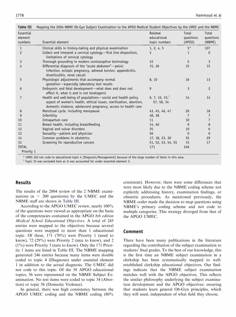
1778 Hammoud et al
Table III Mapping the 2004 NBME Ob-Gyn Subject Examination to the APGO Medical Student Objectives by the UMEC and the NBME
Essentialelementnumbers Essential element
Relatededucationaltopic numbers
Totalquestions(APGO)
Totalquestions(NBME)
1 Clinical skills in history-taking and physical examination 1, 2, 4, 5 5* 1072 Collect and interpret a cervical cytologydfirst line disposition,
limitations of cervical cytology3 1 0
3 Thorough grounding in modern contraceptive technology 33 5 34 Differential diagnosis of the ‘‘acute abdomen’’dpelvic
infection, ectopic pregnancy, adnexal torsion, appendicitis,diverticulitis, renal calculi
15, 36 15 15
5 Physiologic adjustments that accompany normalgestationdespecially laboratory test results
8, 10 16 13
6 Embryonic and fetal developmentdwhat does and does notaffect it, what is and is not teratogenic
9 3 2
7 Health and well-being of populationsdsocial and health policyaspect of women’s health, ethical issues, sterilization, abortion,domestic violence, adolescent pregnancy, access to health care
6, 7, 16, 33,y
57, 58, 3414 14
8 Menstrual cycle, including menopause 43, 45, 46, 47 26 189 Infertility 48, 38 7 710 Intrapartum care 11 10 711 Breast health, including breastfeeding 14, 40 9 812 Vaginal and vulvar disorders 35 10 613 Sexualitydpatient and physician 56 0 014 Common problems in obstetrics 17, 18, 23, 30 35 2915 Screening for reproductive cancers 51, 52, 53, 54, 55 15 17TOTALPriority 1
171 246
* UMEC did not code to educational topic 4 (Diagnosis/Management) because of the large number of items in this area.y Topic 33 was excluded here as it was accounted for under essential element 3.
Results
The results of the 2004 review of the 2 NBME exami-nations (n = 200 questions) by the UMEC and theNBME staff are shown in Table III.
According to the APGO UMEC review, nearly 100%of the questions were viewed as appropriate on the basisof the competencies contained in the APGO 8th editionMedical School Educational Objectives. A total of 245entries were mapped to the objectives because severalquestions were mapped to more than 1 educationaltopic. Of these, 171 (70%) were Priority 1 (need toknow), 72 (29%) were Priority 2 (nice to know), and 2(1%) were Priority 3 (nuts to know). Only the 171 Prior-ity 1 items are listed in Table III. The NBME mappinggenerated 246 entries because many items were doublecoded to topic 4 (Diagnosis) under essential element1 in addition to the actual diagnosis. The UMEC didnot code to this topic. Of the 58 APGO educationaltopics, 56 were represented on the NBME Subject Ex-amination. No test items were coded to topic 34 (Abor-tion) or topic 58 (Domestic Violence).
In general, there was high consistency between theAPGO UMEC coding and the NBME coding (80%
consistent). However, there were some differences thatwere most likely due to the NBME coding scheme notexplicitly addressing history, examination findings, orobstetric procedures. As mentioned previously, theNBME coder made the decision to map questions usingNBME’s primary coding scheme and not code tomultiple categories. This strategy diverged from that ofthe APGO UMEC.
Comment
There have been many publications in the literatureregarding the contribution of the subject examination tostudents’ final grades. To the best of our knowledge, thisis the first time an NBME subject examination in aclerkship has been systematically mapped to well-established clerkship educational objectives. Our find-ings indicate that the NBME subject examinationmatches well with the APGO objectives. This reflectsthe similar philosophy underlying the subject examina-tion development and the APGO objectives: ensuringthat students learn general Ob-Gyn principles, whichthey will need, independent of what field they choose.

Hammoud et al 1779
All medical students ask the question, ‘‘What do Ineed to know?’’ The APGO UMEC proposed theessential objectives to serve as a guide for curriculumdesign and content as well as a study guide to preparefor national licensure tests. Most medical schools acrossthe country use the NBME subject examination as anobjective assessment of performance of their medicalstudents. The use of a written examination in thisfashion presents some challenges as many believe thatthe examination begins to drive the curriculum. How-ever, the use of a standardized examination such as theNBME subject examination provides an easily obtain-able measure of knowledge and allows for a comparisonacross different groups, such as comparing groups atdifferent times of the year or comparing groups betweendifferent sites. In addition, it allows the schools toexamine how their students perform compared withthe national norm.
Our study findings should assure Obstetrics andGynecology Clerkship Directors that the NBME exam-ination provides an appropriate assessment of masteryof what a third-year medical student should learn asrepresented by the APGO Medical Student EducationalObjectives.
Acknowledgments
We gratefully acknowledge the significant contributionsof the Undergraduate Medical Education Committee(UMEC) of APGO in reviewing the NBME examina-tions: Jessica Bienstock, MD, Susan M. Cox, MD, Eve
Epsey, MD, Alice Goepfert, MD, Maya M. Hammoud,MD, Nadine T. Katz, MD, James J. Neutens, PhD,Edward Peskin, MD, Elizabeth Puscheck, MD, PaulKrueger, DO, Sonya S. Erickson, MD.
References
1. Goepfert AR, Phelan SP, Cliver SP, Herbert WNP. An evaluation
of trends in obstetrics and gynecology clerkship in the United States
and Canada. Am J Obstet Gynecol 2004;191:354-8.
2. Cox SM, editor. APGO medical student educational objectives.
8th ed. Crofton (MD): APGO Undergraduate Medical Education
Committee, Association of Professors of Gynecology and Obstet-
rics; 2004.
3. Barzansky B, Etzel SI. Educational programs in US medical schools
2002-2003. JAMA 2003;290:1190-6.
4. Manetta A, Manetta E, Emma D, Keegan KA, Williams JH. Effects
of rotation discipline on medical student grades in obstetrics and gy-
necology throughout the academic year. Am J Obstet Gynecol 1993;
169:1215-7.
5. Hampton HL, Collins BJ, Perry KG Jr, Meydrech EF, Wiser WL,
Morrison JC. Order of rotation in third-year clerkships: influence
on academic performance. J Reprod Med 1996;41:337-40.
6. Myles TD, Galvez-Myles R. Relationship between subjective
grade and examination score in obstetrics and gynecology clerk-
ships at community and university sites. J Reprod Med 2001;46:
729-34.
7. Edwards RK, Kellner KR, Sistrom CL, Magyari EJ. Medical stu-
dent self-assessment of performance on an obstetrics and gynecol-
ogy clerkship. Am J Obstet Gynecol 2003;188:1078-82.
8. Association of Professors of Gynecology and Obstetrics. The
APGO objectives web page. Available from http://www.apgo.org/
objectives/. Accessed on February 10, 2005.
9. Pitkin RM. But isn’t that your job, son? Am J Obstet Gynecol 1996;
174:1083-8.

American Journal of Obstetrics and Gynecology (2005) 193, 1780–4
www.ajog.org
Medical students self-reported work hours: Perceptionversus reality
Colleen Casey, MD,a Sangeeta Senapati, MD,a Casey B. White, PhD,b
Larry D. Gruppen, PhD,c Maya M. Hammoud, MDa
Department of Obstetrics and Gynecology,a Office of Medical Education,b Department of Medical Education,c
University of Michigan Medical School, Ann Arbor, MI
Received for publication February 21, 2005; revised July 3, 2005; accepted August 8, 2005
KEY WORDSQuality of clerkshipMedical student
Work hoursObstetrics and
gynecology
clerkship
Objective: The objective of this study was to compare the students’ actual work hours with their
self-reported work hours during the obstetrics and gynecology clerkship, and to determinewhether the number of hours worked correlate with the amount of ‘‘scut’’ reported or students’rating of the quality of the clerkship.
Study design: Students self-reported work hours were compared against their actual scheduledhours over 2 different academic years. Pearson’s correlation was performed to correlate the actualhours with the amount of reported ‘‘scut’’ work and the overall rating of the quality of the clerkship.
Results: The actual hours per week worked by students averaged 59 hours in 2003 and 48 hours in2004. Students overestimated their work hours both years. Students who worked more hours ratedthe clerkship lower and the quality of the clerkship significantly improved from 2003 to 2004 (4.2 vs3.8 P ! .03).
Conclusion: The majority of third-year students overestimate their work hours in obstetrics andgynecology. The rating of the overall quality of the clerkship increase significantly with fewer hoursworked, and it is not affected by the amount of ‘‘scut’’ work.
� 2005 Mosby, Inc. All rights reserved.
The adoption of the 80-hour work week has resultedin major changes in residency programs in the UnitedStates. Although the airline, trucking, and railwayindustries have a longstanding history of having workhours regulated by the government,1 until recently workhour restrictions in the medical field were not mandated.However, on June 11, 2002, the Accreditation Councilfor Graduate Medical Education (ACGME) passeduniversal standards limiting resident work hours to 80hours per week. These new regulations became effectiveJuly 1, 2003.
Reprints not available from the authors.
0002-9378/$ - see front matter � 2005 Mosby, Inc. All rights reserved.
doi:10.1016/j.ajog.2005.08.017
In February 2004, the Liaison Committee on MedicalEducation (LCME) officials added an annotation re-garding student work hour regulations to its accredita-tion standards.2 They recommended that in addition tomonitoring the amount of classroom time and exami-nation frequency, medical schools should pay attentionto the hours that medical students work during theclinical years and the educational value of their clinicalactivities. Student duty hours should be set taking intoaccount the effects of fatigue and sleep deprivation onlearning and patient care, and medical students shouldnot be required to work longer hours than residents.Soon after this decision by the LCME, the Medical

Casey et al 1781
Student Section (MSS) of the American Medical Asso-ciation (AMA) passed a resolution (MSS Resolution 16:Medical Student Clinical Training and Education Con-dition),3 asking the AMA to encourage the LCME tofollow the lead of the ACGME and include specificlimits on the workload of students in its accreditationstandards. Namely, the resolution for medical studenteducation called for an 80-hour work week, a 24-hourlimit on shifts, and on call no more than once every3 nights.
As early as 2001, 17 of 124 US medical schoolsreported having formal policies on medical studentshours4; however, little is known about the number ofactual or reported student work hours. Burke et al5
published self-reported students’ work hours duringtheir third-year required clerkships; in that study stu-dents indicated an average between 47 to 87 hours perweek depending on the clerkship, with 2 clerkshipsreaching more than 80 hours. A study by Chen et al6
at Harvard Medical School, which asked medical stu-dents to track their daily hours for 1 week, indicatedthat students spent a mean of 74 hours per week in thehospital, with some reporting as many as 106 hours perweek. Mean work hours were dramatically higher oninpatient rotations (90 hours per week) than on outpa-tient rotations (45 hours per week). Students on surgeryrotations worked the longest hours, with a mean ofmore than 94 hours per week spent in the hospitalduring an inpatient rotation.
At the University of Michigan Medical School, whenthe 80-hour work week regulation was adopted forresidents, medical students began to report anecdotallyan increase in their work hours. In addition, curriculumleaders considered adopting similar restrictions on med-ical students work hours, so starting in 2003, students onall required clerkships (internal medicine, surgery, ob-stetrics and gynecology, pediatrics, family medicine,neurology, psychiatry) were asked at the end of therotation to report how many hours they were requiredto be in the hospital per week. When we reviewed thenumber of hours students reported working during theirthird-year obstetric and gynecologic (Ob-Gyn) clerk-ship, we found a sizeable discrepancy between theirreports and the hours they were scheduled to work. Weundertook this study to compare the students’ scheduledor actual hours versus their perceived or self-reportedwork hours in 2003 and 2004. In addition, we wanted todetermine whether the number of work hours correlatedwith the amount of academically unproductive (‘‘scut’’)work they performed, as well as their overall rating ofthe quality of the clerkship.
Methods
At the beginning of the 6-week Ob-Gyn clerkship, allstudents rotating at the University Hospital were given a
work schedule. The schedule specified their daily assign-ments including clinics, inpatient services, and on-callresponsibilities. At the end of the clerkship, all studentscompleted a confidential online survey rating the qualityof their experiences on a 5-point Likert scale from 1 to 5(1 = poor; 5 = excellent). The questionnaire coveredseveral areas of their experiences including the overallquality of the clerkship. They were also asked toestimate the amount of time spent in academicallyunproductive work on a scale of 1 to 5 (1 = too little;3 = just right; 5 = too much), and the average numberof hours per week they were required to be in thehospital or clinics on a 4-point categorical scale asfollows: 1 = less than 60 hours; 2 = 61 to 70 hours; 3 =71 to 80 hours; 4 = more than 80 hours.
To reduce the number of medical student work hours,a student ‘‘night float’’ system was initiated in 2004,where students’ on-call responsibilities consisted of 3consecutive 12-hour night shifts and a 12-hour shift onthe weekend. This replaced the regular once-a-week callfrom the previous year. For the purposes of this study,we considered each year separately because of thecurriculum change. After institutional review boardexemption was obtained, all the students rotating atthe University Hospital (70 in 2003 and 65 in 2004) wereconsidered for the study. Although data were availableon the reported work hours for most of the studentsbecause more than 95% filled out a clerkship survey, wewere unable to retrieve all the actual work schedules thatresulted in available data for 35 students from 2003 and42 students from 2004. In calculating the actual hours,we added the number of hours scheduled each day tocompute the hours per week. The hours per week werethen combined and divided by 6 to calculate averagehours per week worked.
To compare students’ scheduled or actual work hourswith their reported or perceived work hours, the stu-dents were separated into 4 groups on the basis of theirreported work hours category 1 to 4 (1 = !60 hours;2 = 61-70 hours; 3 = 71-80 hours; 4 = 80 hours). Thesewere compared against the actual work hours we calcu-lated for them. In addition, Pearson’s correlation wasperformed to correlate the actual hours with the amountof reported scut work and the overall rating of thequality of the clerkship. The c2 analysis was performedto check if the amount of scut work was differentbetween 2003 and 2004, and an analysis of variancewas performed to compare the overall quality of theclerkship between the 2 years.
Results
The actual hours per week worked by students werehigher in 2003 (59 hours, range: 52-68) than in 2004(48 hours, range: 43-52). The estimated hours workedper week were also much higher in 2003 than 2004 as

1782 Casey et al
shown in Table I. At least 71% of the students over-estimated their work hours in 2003 compared with 53%of the students in 2004. More interestingly, although theactual hours worked was only about 10 more hours in2003 than it was in 2004, 71% of the students reportedworking more than 70 hours per week in 2003 and only12% reported working that many hours in 2004. Stu-dents reporting less than 60 hours per week were themost accurate, whereas those reporting more than 80hours were the least accurate (Figure).
The actual hours worked correlated negatively withthe overall rating of the quality of the clerkship for 2004and for both years combined. Students who workedmore hours rated the clerkship lower (Table II). Inaddition, the actual number of hours worked correlatedwith the amount of scut work for both years combined.This relationship did not hold true when consideringeach year separately. There was no significant correla-tion between the rating of the overall quality of theclerkship and the amount of scut work. There was nosignificant distribution difference in the amount of scutwork reported between 2003 and 2004; however, theoverall quality of the clerkship did significantly improvein 2004 (4.2 vs 3.8 P ! .03).
Comment
The reported hours and the actual hours worked bymedical students decreased in 2004, which reflected arestructuring of the clerkship. Students no longer hadregular on-call duties, which eliminated 24- to 36-hourshifts, so the actual number of hours worked per weekdecreased from 59 hours in 2003 to 48 hours in 2004.Although a large number of students were still over-estimating their hours in 2004, they represented asmaller percentage than the students who overestimatedtheir hours in 2003. These findings indicate that studentstend to overestimate their work hours more when theyare actually working more. In addition, the more hoursstudents worked, the more scut they reported. This
Table I The reported number of hours and the actual numberof hours worked by third-year medical students during theobstetrics and gynecology clerkship
YearNumber ofstudents
Studentpercent
Reportedhours
Actual hours(range)
2003 1 3 !60 602003 9 26 61-70 59 (53-68)2003 21 60 71-80 59 (52-64)2003 4 11 O80 59 (56-61)
2004 20 48 !60 47 (44-50)2004 17 41 61-70 49 (43-52)2004 5 12 71-80 49 (48-51)2004 0 0 O80 n/a
could indicate that with longer hours, students are eitherspending more time in academically unproductive activ-ities or the students potentially perceive less quality totheir learning experience because they are tired.
Our data show that decreasing the number of workhours and providing a structured schedule contributedto higher student ratings of the Ob/Gyn clerkship.Several studies support the notion that a structuredwork schedule results in increased student satisfactionwith the clerkship.7-10 Considering that the Ob/Gynclerkship has been the lowest rated clerkship by medicalstudents,11 these findings support the importance ofdeveloping a structured clerkship with reasonable hoursto improve the quality of the educational experience forthird-year medical students.
Figure Differences between perceived and actual hoursworked.
Table II Correlations between the overall quality of theclerkship, actual work hours, and amount of scut work
YearActualhours
Amount ofscut work
2003Overall quality of clerkship R = 0.0312* R = 0.0437
P = .859y P = .80Amount of scut work R = 0.2789
P = .105
2004Overall quality of clerkship R = �0.3452 R = �0.0313
P = .025 P = .844Amount of scut work R = 0.0993
P = .531
2003 and 2004Overall quality of clerkship R = �0.2685 R = �0.0488
P = .018 P = .67Amount of scut work R = 0.3004
P = .008
* R = Correlation coefficient.y P = P value.

Casey et al 1783
It is interesting that third-year medical studentsreported spending more hours at the hospital thanwhat they were scheduled to work. We thought oneexplanation for this might be that some students electedto stay and work beyond their scheduled hours. How-ever, a discussion with students and residents indicatedthis was not the case. Although there were occasionalinstances where a student stayed late for an interestingcase, students on gynecology call were also often senthome ahead of schedule. Students were also oftenexcused early and they were not expected to be presentfor gynecology afternoon rounds. In addition, when wecalculated their hours, we tried to err on the side ofoverestimating. For example, we added extra hours forstudents on gynecologic oncology, assuming they stayedlate every day although this only happened on average2 times a week. We also assumed students were at thehospital 1 hour before rounds, which is often not true.In any case, even if there were instances where a studentmight have worked more hours than scheduled, it wasimpossible to account for all of the extra reported hours,which in some cases reached more than 20 hours perweek.
A second possibility might be that students simply arenot able to estimate their work hours accurately. It hasbeen suggested that medical students’ estimations oftheir work hours may actually be a reflection of resi-dents’ work hours observed by the students.12 In addi-tion, previous studies in medical and higher educationhave shown that students are not very accurate in self-assessing their performance.13 When looking specificallyat medical student self-assessment, investigators havefound that the less experience a student has (ie,transitioning from the familiarity of the classroom tothe clinical clerkships), the more self-assessment accur-acy and performance suffers.14 Student work hours canpotentially fall into this category where third-year med-ical students are simply not experienced enough toaccurately assess their hours.
A third possibility for these findings might be thatstudents think they are working more hours becausethey are working hard or because of the many variationsin their daily schedule. The Ob-Gyn clerkship is achallenging clerkship. Students rotate through so manydifferent services including the operating room, laborand delivery, clinic, or night float. Some of these servicescan be very demanding and stressful and students mighttend to recall these experiences more than others. Thismakes it difficult for students to accurately recall thehours by simple reflection. It would be interesting toperform similar analyses for other clerkships, specifi-cally, some of the less demanding ones with moreoutpatient setting and no on-call duties to check if therewould be similar findings.
Whatever the causes are, this perception by medicalstudents that they are working longer hours than they
actually are can have serious implications. The perceivedwork hours required in surgical residencies play a part ina decreased interest in those specialties.12,15,16 Forexample, Miller et al17 surveyed 134 third- and fourth-year medical students to determine factors that influ-enced their decision in choosing a specialty. They foundthat 38% would not consider a residency program witha reputation for long work hours. In a separate study,Miller et al18 looked at how students entering surgicalfields developed their personal rank list. They found that40% of the applicants ranked higher those programsthat strictly adhered to work hour limitations. Thus, asstudents estimate long working long hours in obstetricsand gynecology they might also consider those hoursnegatively as they consider their residency choices. Thisis of special importance to the Ob-Gyn specialty becauseof the decreased number of students choosing to enterthe field.19
The adoption of the 80-hour work week regulationmandated by the ACGME has resulted in a restructur-ing of several residency programs nationwide. Muchdiscussion has been generated about its impact onresidents’ experience and job satisfaction.20,21 Whenconsidering restricted work hours for medical studentswe must remind ourselves of the role of the medicalstudent in the hospitaldmedical students are in thehospital to learn. It is our duty to create an idealenvironment for them to learn by maximizing goodeducational opportunities and minimizing ‘‘scut’’ work.Furthermore, we have to consider how those percep-tions of long hours might be impacting students’ careerchoices especially in obstetrics and gynecology.
References
1. Leach DC. Resident duty hours. The ACGME perspective.
Neurology 2004;62:E1-2.
2. Current LCME Accreditation Standards. Available at: http:
//www.lcme.org/standard.htm. Accessed on February 21, 2005.
3. Student Work Hours. Available at: http://www.ama-assn.org/
ama/pub/category/12141.html. Accessed on February 21, 2005.
4. Barzansky B, Etzel SI. Educational programs in US medical
schools, 2001-2002 [see comment] JAMA 2002;288:1067-72.
5. Burke W, Williams JA, Fenner DE, Hammoud MM. The obstet-
rics and gynecology clerkship: building a better model from past
experience. Am J Obstet Gynecol 2004;191:1772-6.
6. Chen J, Kamath R, Lam A, Litrovnik B, Leight K, Miller S, et al.
How will changes in resident work hours affect medical students?
Curr Surg 2003;60:268-70.
7. Ko CY, Escarce JJ, Baker L, Klein D, Guarino C. Predictors for
medical students entering a general surgery residency: national
survey results. Surgery 2004;136:567-72.
8. Sumit KD, Henke PK, Ailawadi G, Dimick JB, Colleti LM.
Attending, house officer, and medical student perceptions about
teaching in the third-year medical school general surgery clerkship.
J Am Coll Surg 2004;199:932-42.
9. Pogacnik M, Juznic P, Kosorok-Drobnic M, Pogacnik A, Cestnik
V, Kogovsek J, et al. An attempt to estimate student workload.
J Vet Med Educ 2004;31:255-60.

1784 Casey et al
10. Dunn TS, Wolf D, Beuler J, Coddington CC. Increasing recruit-
ment of quality students to obstetrics and gynecology: impact
of a structured clerkship. Am Coll Obstet Gynecol 2004;103:
339-41.
11. AAMC Medical School Graduation Questionnaire, 1978-2003.
Washington (DC): Association of American Medical Colleges;
2003.
12. O’Herrin JK, Lewis BJ, Rikkers LF, Chen H. Why do students
choose careers in surgery? J Surg Res 2004;119:124-9.
13. Ward M, Gruppen L, Regehr G. Measuring self-assessment:
current state of the art. Adv Health Sci Educ 2002;7:63-80.
14. Fitzgerald JT, White CB, Gruppen LD. A longitudinal study of
self-assessment accuracy. Med Educ 2003;37:645-9.
15. Azizzadeh H, McCollum CH, Miller CC 3rd, Holliday KM,
Shilstone HC, Lucci A Jr. Factors influencing career choice
among medical students interested in surgery. Curr Surg 2003;60:
210-3.
16. Gelfand DV, Podnos YD, Wilson SE, Cooke J, Williams RA.
Choosing general surgery: insights into career choices of current
medical students. Arch Surg 2002;137:941-7.
17. Miller G, Bamboat ZM, Allen F, Biernacki P, Hopkins MA,
Gouge TH, et al. Impact of mandatory resident work hour
limitations on medical students’ interest in surgery. J Am Coll
Surg 2004;199:615-9.
18. Miller G, Bamboat ZM, Allen F, Hopkins MA, Gouge TH, Riles
TS, et al. Attitudes of applicants for surgical residency toward
work hour limitations. Am J Surg 2004;188:131-5.
19. National Resident Matching Program Match Data. Advanced
data tables 2004. Available at: http://www.nrmp.org/res_match/
tables/table5_04.pdf. Accessed on February 21, 2005.
20. Killelea BK, Chao L, Scarpinato V. The 80 hour workweek. Surg
Clin N Am 2004;84:1557-72.
21. Lund KJ, Alvero R, Teal SB. Resident job satisfaction: will 80
hours make a difference? Am J Obstet Gynecol 2004;191:1805-10.

American Journal of Obstetrics and Gynecology (2005) 193, 1785–9
www.ajog.org
Development and assessment of a Web-based evaluationand management coding curriculum for residents
Paul M. Lemen, MD*
Department of Obstetrics and Gynecology, Medical College of Wisconsin, Milwaukee, WI
Received for publication March 2, 2005; revised June 8, 2005; accepted August 1, 2005
KEY WORDSEvaluation and
management
CodingSystems-based
practice
Objective: The purpose was to assess a Web-based learning module for residents on evaluation
and management coding, and to determine whether using a point system based on reimbursementis an effective way to measure improvement in performance.Study design: Nineteen residents at a university-based residency completed an online module on
evaluation and management coding. Pretest and posttest were administered consisting of 10actual patient notes from which to abstract the level of service. Tests were scored by percentcorrect as well as assigning points to correct responses equal to 2004 Medicare reimbursement forthat item’s level of service. Incorrect responses were debited points equal to the absolute
difference in reimbursement between the incorrect and correct evaluation and management level.Results: Average percent correct was 44.2 on the pretest and 45.3 on the posttest (P Z 1.0), withonly 47% of subjects improving their performance. The mean point score was 513.44 on the
pretest and 555.87 on the posttest (P Z .02), with 84% improving. Most subjects (84%) ratedthe module equal or superior to a didactic format and felt that their knowledge was improved bythe exercise.
Conclusion: An electronic learning module on evaluation and management coding for residents,using scoring based on reimbursement schedules, is effective for instruction and assessment andwell accepted by learners.
� 2005 Mosby, Inc. All rights reserved.
Evaluation and management (E&M) coding, part ofthe American Medical Association’s current proceduralterminology system,1 serves as the method by whichphysicians bill patients and third-party payers for manyclinical services. As such, understanding of the E&Mcoding system and the necessary documentation thataccompanies it are an important component to thecurrent practice of medicine in this country. Improper
* Reprint requests: Paul M. Lemen, MD, 9200 West Wisconsin,
Milwaukee, WI 53226.
E-mail: [email protected]
0002-9378/$ - see front matter � 2005 Mosby, Inc. All rights reserved.
doi:10.1016/j.ajog.2005.08.005
coding and documentation can lead to either overbillingfor services (which may constitute fraud) or under-compensation for services rendered. Despite this fact,there are numerous studies that indicate that physiciancompetence in the application of E&M coding is poor.2-4
To date, there are no studies that assess an educa-tional method for instructing residents in this importantsubject. At the Medical College of Wisconsin AffiliatedHospitals, obstetrics and gynecology residents havetraditionally received an annual 60-minute lecturefrom a departmental billing professional. This studywas designed to construct and assess an interactive Web-based electronic module to educate residents in E&M
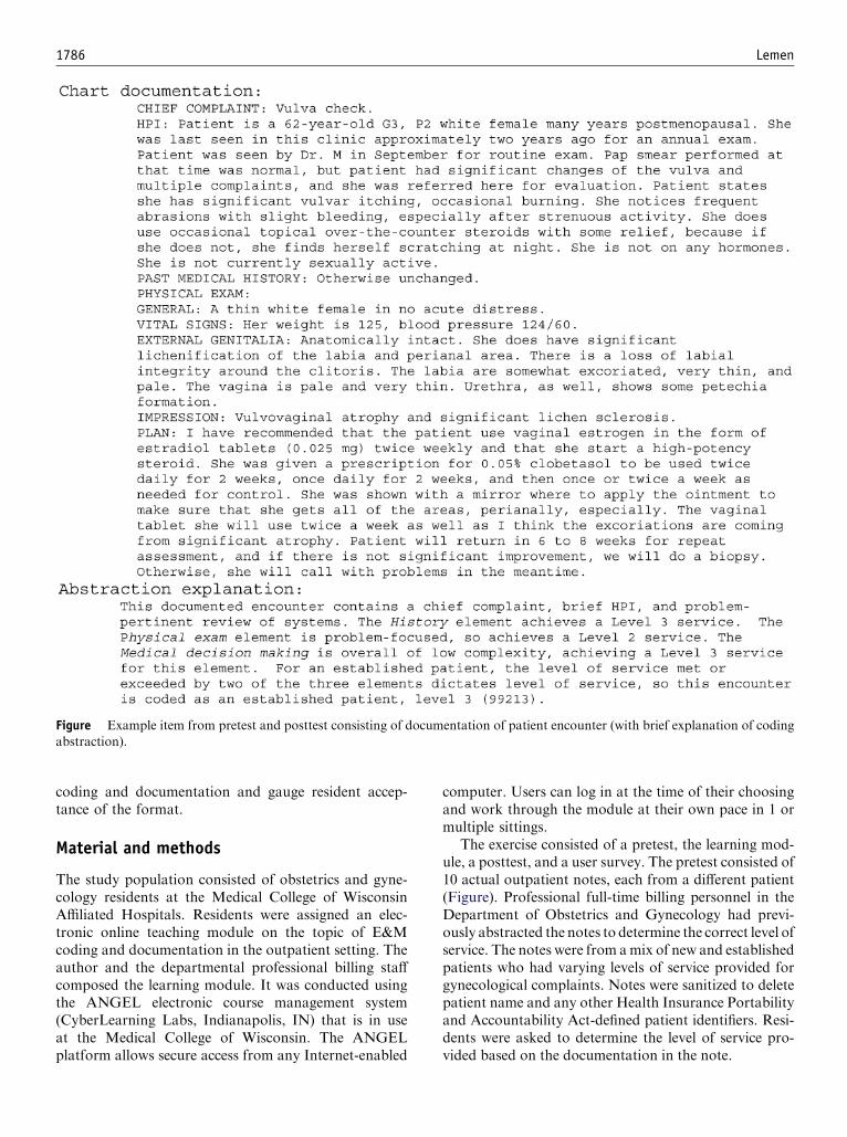
1786 Lemen
Figure Example item from pretest and posttest consisting of documentation of patient encounter (with brief explanation of coding
abstraction).
coding and documentation and gauge resident accep-tance of the format.
Material and methods
The study population consisted of obstetrics and gyne-cology residents at the Medical College of WisconsinAffiliated Hospitals. Residents were assigned an elec-tronic online teaching module on the topic of E&Mcoding and documentation in the outpatient setting. Theauthor and the departmental professional billing staffcomposed the learning module. It was conducted usingthe ANGEL electronic course management system(CyberLearning Labs, Indianapolis, IN) that is in useat the Medical College of Wisconsin. The ANGELplatform allows secure access from any Internet-enabled
computer. Users can log in at the time of their choosingand work through the module at their own pace in 1 ormultiple sittings.
The exercise consisted of a pretest, the learning mod-ule, a posttest, and a user survey. The pretest consisted of10 actual outpatient notes, each from a different patient(Figure). Professional full-time billing personnel in theDepartment of Obstetrics and Gynecology had previ-ously abstracted the notes to determine the correct level ofservice. The notes were from amix of new and establishedpatients who had varying levels of service provided forgynecological complaints. Notes were sanitized to deletepatient name and any other Health Insurance Portabilityand Accountability Act-defined patient identifiers. Resi-dents were asked to determine the level of service pro-vided based on the documentation in the note.

Lemen 1787
The pretest was scored in 2 ways. The first way con-sisted of a simple percent correct. The second consistedof a point system based on 2004 Medicare reimburse-ment schedules (Table I). Using this system, a correctanswer earned points equal to the dollar amount reim-bursed by Medicare for that level of service, for a maxi-mum score of 709.81 (Table II). An incorrect responsecaused a subtraction in points equal to the absolutedifference in the reimbursement between the correct leveland the incorrectly answered level. For example, if thedocumentation for the visit supported a new patient,level 3 code, but the resident responded new patient,level 2, their total score would be debited 38.47 points(absolute value of the difference in reimbursement,130.93 – 92.46). The rationale for derivation of thepoint system was based on the concept that with thissubject matter, simple scoring of items correct did notadequately assess either the module itself or the learner.
With E&M coding, not all errors are created equal. Ifa learner abstracted a chart note incorrectly and codedan ‘‘established patient, level 3’’ as an ‘‘establishedpatient, level 2’’ because they misunderstood whatconstitutes a complete review of systems, it representsa relatively minor error. However, if several errors weremade in recognizing the levels of history, examination,and medical decision-making components or did notrecognize that the patient was established, the errorscompound and should be graded more harshly. Usingthe point system based on reimbursement schedules,larger errors would be penalized more than small ones.Medicare reimbursement schedules were used because oftheir relatively universal use nationwide to aid in appli-cability and interpretation.
The learning module consisted of 7 short (1 to 3pages) sections of informational material that could notbe accessed until completion of the pretest. Mostsections contained 1 to 2 review questions with expla-nations of correct and incorrect answers. These ques-tions were used for review to highlight importantinformation and were ungraded. Primarily, however,they served as internal checkpoints for the ANGELplatform. Residents could not access a subsequentsection until the questions at the end of the currentsection were completed.
At the conclusion of the module, a posttest wasadministered identical in content to the pretest with the
Table I Medicare reimbursement schedule (2004)
New patient Established patient
Level 1 $34.79 $20.20Level 2 $62.20 $36.18Level 3 $92.46 $50.59Level 4 $130.93 $79.11Level 5 $166.65 $115.15
item order scrambled. The posttest could not beaccessed until all sections of the module were completed.The scoring of the posttest was likewise identical to thepretest. In addition, a short survey was conducted at theend of the exercise to obtain information on residentattitudes and opinions on the exercise. The effectivenessof the learning module was measured by assessing theimprovement (or difference) in resident performance onthe pretest versus the posttest.
This study was considered exempt from review by theMedical College of Wisconsin Institutional ReviewBoard. All residents in the program were asked tocomplete the module. However, data were analyzedonly for those who consented to participate in the study.
Statistical analysis
The difference in percent correct and points scored wasanalyzed using the sign test for median. This is anonparametric test, which makes no assumptions aboutthe data having a normal distribution. Analyses wereperformed with Minitab statistical software (Minitab,Inc, State College, PA).
Results
Twenty-one residents completed the online module. Ofthese, 19 consented to have their data used for analysis.The mean score on the pretest was 44.2% (range 10% to70%) using percent correct scoring and 513.44 (range274.00 to 620.89) using point scoring. The mean score onthe posttest was 45.3% using percent correct scoring and555.87 (range 409.74 to 633.36) using point scoring . Only47.4% of residents (9 of 19) improved their performanceafter completion of the module when assessed by percentcorrect. However, when evaluated using the point score,84.2%of residents (16 of 19) improved their performance.Using percent correct score, the mean score on theposttest improved 1.1% (P Z 1.0), whereas using thepoint score the mean score improved 42.43 (P Z .02).
Table II Point values for pretest and posttest
Level of service fromchart documentation
Points assignedfor correct answer
New patient, level 2 62.20New patient, level 3 92.46New patient, level 3 92.46New patient, level 4 130.93Established patient, level 2 36.18Established patient, level 2 36.18Established patient, level 3 50.59Established patient, level 3 50.59Established patient, level 4 79.11Established patient, level 4 79.11Total 709.81

1788 Lemen
With regard to the types of errors committed, therewere differences noted between the pretest and posttest(which resulted in the improved point score on theposttest). Among all learners, equal numbers of errors(20) were made on both the pretest and the posttest withregard to misclassifying a new patient as an establishedone or vice versa. Errors in the level of service coded weresignificantly different between pretest and posttest, how-ever. Of the 102 level-of-service coding errors on thepretest, 62 (61%) were overcoded. In contrast, of the 93level-of-service errors on the posttest, 61 (66%) wereundercoded. More importantly, 35 (34%) of these errorson thepretestweremiscodedby 2ormore levels of service.On the posttest, only 11 level-of-service errors (12%)weremiscoded by 2 levels and none by greater than 2.
A resident acceptance survey was conducted at thecompletion of the module. All 19 subjects completed thesurvey for a 100% response rate. Sixteen subjects (84%)felt that the module had met the objective of improvingtheir familiarity with E&M documentation and coding.Eleven (58%) felt the electronic format was superior to atraditional lecture, with an additional 5 (26%) indicatingthat the 2 formats were equivalent. Two respondents(11%) felt the lecture was superior, and 1 (5%) answered‘‘not sure.’’
With regard to the actual system used (the ANGELcourse management system), 15 (79%) thought theplatform was worthwhile as a learning tool, although 8of the 15 indicated that the subjects able to be taughtwith it were limited. Three respondents (16%) felt it was‘‘probably not’’ worthwhile, and 1 (5%) was ‘‘not sure.’’Nine of 19 (47%) felt that homework assignments tocomplete on their own time were appropriate in residenteducation, with only 2 indicating that they stronglyagreed with their use. Six of the 19 (32%) were neutral,whereas 3 (16%) disagreed with their use and 1 (5%)strongly disagreed. The total time reported as necessaryto complete the instructional module alone averaged32 minutes (range 15 to 65 minutes, median 35 minutes),whereas the entire exercise including pretest and posttestaveraged 56 minutes (range 15 to 120 minutes, median60 minutes).
Comment
Understanding proper coding and documentation helpsto develop practice habits that save time, ensure properreimbursement, and protect both from fraud allegationsand medical malpractice claims. To the practicing phy-sician, the importance of understanding E&M codingand documentation is obvious. Despite its importance,physicians commonly report insecurity in their owncompetence in this area. The literature bears this out,showing marked discrepancies between levels of servicecoded versus that documented.
The inherent difficulty in learning this system iscompounded when the learner is a resident. Not onlyis the system perceived to be confusing and complicated,but the relevance to the physician in training can bedifficult to grasp. Recently the Accreditation Council forGraduate Medical Education put in place competencyguidelines for the training of all residents.5 One of thecore areas that all physicians should be competent in issystems-based practice. This competency requires resi-dents to ‘‘demonstrate an awareness of and responsive-ness to the larger context and system of health care andthe ability to effectively call on system resources toprovide care that is of optimal value.’’ Understandingand applying E&M coding would be an importantcomponent of this competency area. Despite this man-date and the obvious importance in their future practiceof medicine, there is a dearth of literature about residentperformance, comprehension, or education concerningE&M coding and documentation. Ng and Lawless4
found that pediatric residents miscoded problem-oriented visits almost two thirds of the time, usuallyby undercoding. Interestingly, the year of training hadno impact on accuracy in coding.
Although our program has attempted to consistentlyinstill the importance of proper documentation and thefundamentals of proper coding, residents have univer-sally reported feeling less than competent in the area.We have traditionally taught this subject in didacticsessions, with learners, faculty, and billing instructors allunsatisfied at the result. It was felt that a self-pacedlearning module that demanded interaction and objec-tively graded individual performance would be a bettereducational experience in the teaching of this subject.Although its preparation is slightly more labor intensivethan preparation of a lecture, it was felt to be moreeffective than a lecture format and much more efficientthan multiple small group or individualized sessions forresidents with the billing staff. With recent limitationson duty hours and constant expansion of the skills andknowledge necessary for residents to learn in training,time is arguably the most valuable asset in medicaleducation. The ability to assign educational material tobe completed off duty is therefore also attractive.
The effectiveness of the exercise was to be judged byimprovement in learner performance on a set of ab-straction exercises. It was felt that a simple scoring ofright answer versus wrong answer was inadequate forthis subject. The scoring system detailed in Material andMethods was devised to overcome this problem. Becausenot all E&M coding errors are equivalent, the systemallowed greater penalty for answers that were farther offthe mark than others.
It was hypothesized that regardless of percent correcton posttest versus pretest, learner performance wouldnonetheless improve. Put another way, even if mistakeswere still made, they would be of smaller magnitude.

Lemen 1789
Obviously the goal is objectively correct coding withoutany errors at all, but small errors are more desirablethan large ones. The results of the data seem to confirmthis hypothesis. Although the overall change in percentcorrect was negligible, there was a significant improve-ment in point score on the posttest versus the pretest.When judged by their percent correct, approximatelyhalf (47%) of the subjects actually performed morepoorly on the posttest versus the pretest. However, twothirds of this cohort still improved their performanceoverall using the point scoring. Although residents stillanswered many of the items incorrectly, they answered‘‘smarter’’ and their mistakes were less significant. Thisis confirmed again in considering how many errors wereoff by only a single level of service on the posttest (88%)versus the pretest (66%).
The survey results indicated favorable acceptance ofthe electronic module as a teaching tool for the subjectmatter. The majority of subjects (84%) felt the learningobjectives were met, and an identical number felt theformat to be equal or superior to the lecture formatthat each had experienced several months earlier in theacademic year. The entire exercise took no longer (onaverage) than a traditional lecture, but this includedpretest and posttest activities that would not necessarilybe included in future applications of this exercise. Theactual learning module alone took slightly more thanhalf as long as a traditional lecture. A majority ofsubjects (79%) likewise felt that the ANGEL platformwould be worthwhile for other subjects. Interestingly, aminority of resident subjects (47%) indicated agree-ment with the concept of homework assignments ingraduate medical education, with only 2 (11%) agree-ing strongly.
There are a number of potential flaws and criticismsto this study. First and foremost is the actual method ofassigning points based on reimbursement schedules. Thenature of the reimbursement schedules is such that theinterval differences in dollars (or points) between con-secutive levels of service are not equal. If the correctanswer to a given question should have been ‘‘estab-lished patient, level 3,’’ then that answer would haveearned 50.59 points. Consider 2 subjects who makesimilar mistakes. The first answers ‘‘established patient,level 2,’’ but the second answers ‘‘established patient,level 4.’’ They have both made essentially identicalerrors in the spirit of this assessment: They are off bya single level of service and have correctly identified thepatient as established. However, because they are pe-nalized the difference in reimbursement, the first subject
is penalized only 14.41 points, whereas the second ispenalized 28.52 points.
Consideration was given to standardizing the penalty,such that each level of service had consistent differencebetween them or assigning a set penalty (eg, 50 points)for each level of service by which an answer wasmistaken. The point system remained as is, however,because it was felt that it would provide a more tangiblesense of the consequence of miscoding if it was tieddirectly to actual reimbursement schedules.
Comparing the performance of subjects after com-pleting the module with a control group that receives adidactic lecture was not feasible for this study because ofthe small overall sample size, but future studies shouldconsider this. It would be interesting to determinewhether resident level (postgraduate year) made a dif-ference in performance, but the sample sizes in this studywere again too small to make such comparisons.
Although I feel that the assessment using this pointsystem indicates that the learning module was effective,these results need to be validated. Validated assessmentwill come with determining whether coding and docu-mentation actually improve in the clinical setting afterparticipation in the exercise.
Acknowledgment
I thank Donna Kroening, CCS, for her invaluable helpin the preparation of the E&M coding module as well asWilliam Herbert, MD, and the rest of the faculty of theAssociation of Professors of Gynecology and Obstetrics/Solvay Educational Scholars Development Program forsubstantive guidance and advice.
References
1. American Medical Association. Current Procedural Terminology.
Washington, DC: American Medical Association; 2004.
2. Horner RD, Paris JA, Purvis JR, Lawler FH. Accuracy of patient
encounter and billing information in ambulatory care. J Fam Pract
1991;33:593-8.
3. Chao J, Gillanders WG, Flocke SA, Goodwin MA, Kikano GE,
Strange KC. Billing for physician services: a comparison of actual
billing with CPT codes assigned by direct observation. J Fam Pract
1998;47:28-32.
4. Ng M, Lawless ST. What if pediatric residents could bill for their
outpatient services? Pediatrics 2001;108:827-34.
5. The Accreditation Council for Graduate Medical Education.
ACGME Outcome Project General Competencies. Available at:
www.acgme.org/outcome/comp/compfull.asp.

American Journal of Obstetrics and Gynecology (2005) 193, 1790–3
www.ajog.org
Teaching residents coding and documentation:Effectiveness of a problem-oriented approach
Sawsan As-Sanie, MD, MPH,a,b Denniz Zolnoun, MD, MPH,a Mary Ellen Wechter, MD,a
Georgine Lamvu, MD, MPH,a Frank Tu, MD, MPH,a,c John Steege, MDa
Department of Obstetrics and Gynecology, Division of Advanced Laparoscopy and Gynecologic Surgery,University of North Carolina, Chapel Hill, NC a; Department of Obstetrics and Gynecology, The University ofMichigan, Ann Arbor, MI b; Department of Obstetrics and Gynecology, Northwestern University, Chicago, ILc
Received for publication March 10, 2005; revised May 29, 2005; accepted August 1, 2005
KEY WORDSResident educationCoding
DocumentationPractice management
Objective: We sought to assess the effectiveness of a problem-oriented approach to teaching
residents accurate coding and documentation of ambulatory gynecology visits.Study design: This was a pilot before-and-after study. Nine resident volunteers underwent 4individual instructional sessions on coding and documentation with a trained faculty member
over 6 weeks. Outcomes were assessed by comparing the appropriateness of procedure anddiagnostic codes billed in participant continuity clinic prior to and in the 6 to 9 months followingthe intervention.Results: Following the intervention, participants demonstrated an increase in the accuracy of
coding the correct category of the evaluation and management service, an increase in theappropriate use of modifiers, and a decline in undercoding errors.Conclusion: Problem-oriented interactive learning appears to be an effective method of teaching
residents proper coding and documentation.� 2005 Mosby, Inc. All rights reserved.
Accurate and consistent coding and documentationof outpatient office visits is the foundation of operatinga compliant and productive office practice. Unfortu-nately, many US residents in obstetrics and gynecology,as well as those in other subspecialties, graduate with alimited understanding of proper coding and documen-tation.1,2
To address this ubiquitous problem, the Accredita-tion Council for Graduate Medical Education includes
Presented at the Annual Meeting of Council on Resident Educa-
tion in Obstetrics and Gynecology and the Association of Professors of
Gynecology and Obstetrics, March 2-5, 2005, Salt Lake City, UT.
Reprints not available from the authors.
0002-9378/$ - see front matter � 2005 Mosby, Inc. All rights reserved.
doi:10.1016/j.ajog.2005.08.004
system-based education as 1 of 6 general competenciesrequired for resident development.3 System-based edu-cation expects that residents demonstrate ‘‘an awarenessof and responsiveness to the larger context and systemof health care.’’ As such, residents should be knowledge-able about coding, reimbursement, and the managementof a medical practice. Identification of these general com-petencies was the first step in the Accreditation Councilfor Graduate Medical Education’s long-term effort toemphasize outcome assessment in resident educationand the accreditation process.
Despite the obvious need for an effective educa-tion paradigm, there are no published guidelines onteaching or assessing the competency of coding and

As-Sanie et al 1791
documentation in an ambulatory gynecology setting.Thus, the objectives of this study were: (1) to develop aproblem-oriented approach to teaching proper codingand documentation of ambulatory gynecology visits;and (2) to assess the feasibility and effectiveness of suchan approach by evaluating changes in documentationcompetencies and billing patterns.
Material and methods
This was an institutional review board approved pilotbefore-and-after study conducted at the University ofNorth Carolina (UNC) Department of Obstetrics andGynecology between January 2003 and February 2004.Nine obstetrics and gynecology residents, postgraduateyears (PGY) 2 to 4, were asked and consented toparticipate in this study. Each resident attended 4individual instructional sessions with 1 of 4 trainedfaculty members over a 6-week period.
Each session was an individual 1-hour learner-basedmodule thatwas basedon a standard curriculum, drawingexamples of successes and errors from that individualresident’s dictations in the prior 3 months. The curricu-lum was designed to address the most common codingand documentation errors identified in a sample ofresident dictations audited prior to the educational inter-vention. Prior to meeting with the residents, each par-ticipating faculty member underwent the hospital’smandatory coding and documentation course and anadditional 1-hour training course, which reviewed thestudy’s curriculum.
Session 1 provided an overview of Medicare stan-dards for coding and documentation. This focused onthe usage of Current Procedural Terminology (CPT)Evaluation and Management (E/M) codes and theirlinkage to the International Classification of Diseases(9th revision) diagnostic codes. The medical history,physical exam requirements, and complexity of medicaldecision making for each level of service provided werereviewed. The appropriate use of modifiers-24, -25, and -51, which are used to assure proper reimbursement of apayable service provided, was highlighted in this session.For example, modifier-24 is used when providing anunrelated E/M service in the postoperative period of amajor surgical procedure. Modifier-25 is used when thesame physician performs a significant and separatelyidentifiable E/M service on the same day as a minorprocedure or a preventive health maintenance examina-tion. Modifier-51 is used when multiple but separateprocedures are performed on the same visit.
Session 2 focused on the differences between the 1995and 1997 Medicare guidelines for the documentation ofthe physical examination. During this encounter, thefaculty member reviewed the 1995 requirements of amultiorgan approach as well as the 1997 allowance for a
focused genitourinary examination. This session alsoprovided an overview of 2 methods of choosing the levelof service: billing according to evaluation and complex-ity versus billing for time-based counseling.
Session 3 reviewed the recommended components ofan annual health maintenance examination, based on theAmerican College of Obstetricians and Gynecologistsguidelines on preventive care.4 The emphasis was onwomen ages 18 to 39 years, the most prevalent age groupin the resident continuity clinic. A discussion of theNorthCarolina preventable causes of morbidity and mortalityserved as a reminder for residents to look beyond thebreast examination and Papanicolaou smear when pro-viding preventive care. Individual resident dictationswerethen reviewed on the appropriateness of their coding anddocumentation for an annual health maintenance visit.
Finally, session 4 was used as an opportunity toreview 5 audited resident dictations for errors defined inthe first 3 sessions. Residents were asked to providefeedback on their dictations and coding and receivedcase-specific guidance from the instructor. At the end ofthe session, a posttest was administered, followed byimmediate feedback on the answers.
Coding and documentation competencies were theprimary outcome evaluated. Four general categoriesof competencies were developed based on the UNCTeaching Physicians Oversight Committee guidelines forcomputing a compliance error rate: class 1 (overcoding)errors, class 2 (undercoding) errors, appropriate use ofmodifier-25, and correct coding of a preventive medicinevisit.
Class 1 errors are overcoding errors that cannot bebilled as reported by the physician. The most commonclass 1 errors are category errors in which the wrongcategory of visit is chosen and overcoding by 1 or morelevels of service. These errors are considered the mostegregious because they bill for a higher fee than thedocumentation supports. For example, billing CPT code99385 (preventive medicine, new patient 18-39 years old)when the appropriate code is CPT 99395 (preventivemedicine, established patient 18-39 years old) would beconsidered a class 1 error.
Class 2 errors are undercoding errors that may bebilled as coded but are also serious because they gener-ally undercode, causing lost revenues. For example,billing CPT code 99213 (expanded problem-focusedvisit, established patient) when the documentation sup-ports the level CPT 99214 (detailed problem-focusedvisit, established patient) is a class 2 error.
Coding competencies were assessed according to stan-dard Medicare guidelines. A UNC billing complianceauditor randomly selected 10 outpatient gynecologyvisits provided by study participants: 5 dictations in the3 months prior to the intervention and 5 dictationsbetween 6 and 9 months following the completion of theintervention. To assure adequate diversity in the selected

1792 As-Sanie et al
Table Frequency of coding errors among randomly sampled encounters by UNC obstetrics and gynecology residents (n = 9) 3 monthsprior and 6 to 8 months after an educational intervention
Preintervention Postintervention
Coding Error No. errors* % No. errors* % P valuey
Class 1 errors (unbillable errors)Overcoded due to wrong category billed 14/45 31.1 6/45 13.3 .05Overcoded by 2 or more levels of service 0/45 0 0/45 0 dOvercoded by 1 level of service 2/45 4.4 4/45 8.9 .16
Class 2 errors (billable errors)Consult opportunity missed 0/1 0 0/3 0 dUndercoded by 1 level of service 6/45 13.3 2/45 4.4 .16Undercoded by 2 or more levels of service 0/45 0 0/45 0 dProcedure not billed 2/4 50.0 3/8 37.5 .93Modifier-25 omitted 4/5 80.0 6/12 50.0 .39
* Total number of errors among all residents per opportunity to make error.y Wilcoxon signed rank test.
dictations, 2 of the 5 dictations in each time period wereselected from preventive medicine visits. The remaining3 were problem-oriented visits. Within these 2 categories,dictations were randomly selected from the appropriatetime period. Each dictation was audited to identify errorsin the coding competencies of interest. Also, a billing andcoding database was queried for all outpatient gynecol-ogy visits provided by study residents in the same period.Diversity in levels of service coded as well as appropriatecoding of annual health maintenance visits were exam-ined using this database. Descriptive and bivariate sta-tistics, including the Wilcoxon signed-rank test, wereperformed using STATA 8.0 (STATA Corp, CollegeStation, TX).
Results
Eleven residents volunteered to participate in this study;2 residents were excluded because they completed theeducational sessions within 1 month of graduation.Among the remaining 9 residents, 5 were PGY2 and 4were PGY3. The average postintervention examinationscore was 94% correct.
After the educational intervention, residents showeda decline in overcoding errors, primarily because of animprovement in assigning the correct category of servicerendered (Table). Nearly a third of audited dictationsprior to the intervention were coded in the wrongcategory of service; this declined to 13% following theintervention (P = .05). There was also a trend toward adecrease in coding errors that result in underbilling,including meaningful improvements in billing for pro-cedures provided and increases in the appropriate useof modifier-25. For example, 2 of 4 procedures (50%)performed in the preintervention period were not billed;this declined, although not statistically significant, to afailure to bill for 3 of 8 procedures (38%) in the post-intervention period.
Based on the preintervention audit of resident dicta-tions, the most common error when coding an annualhealth maintenance exam was linking the InternationalClassification of Diseases (9th revision) code V72.3(annual visit with Papanicolaou smear) to a problem-oriented CPT E/M code (99201-99205 or 99212-99214)rather than to a preventive medicine code (99385-99387,99395-99397). This coding error was billed in 54% of thepreventive medicine visits prior to the intervention anddeclined to 37% of preventive medicine visits in the 6 to9 months after the intervention (P = .32).
To assure adequate diversity in the types of visitsreviewed, 40% of audited dictations were preventivemedicine visits; the remainder was problem-orientedvisits. Among the problem-oriented visits, the mostcommon diagnoses were pelvic pain (30%), abnormaluterine bleeding (22%), uterine fibroids (13%), contra-ception (9%), and first-trimester abortion (9%). Therewas no relationship between the primary diagnosis andfrequency of coding error.
The diversity of E/M levels of service coded appearedto improve following the intervention. For example,among all outpatient problem-oriented gynecology visitsprovided by participating residents in the study periods,the use of upper-level E/M codes (99204, 99214, 99244)increased from 11.7% to 20.6% of visits (P= .05). High-level E/M codes (99205, 99215, 99245) also increased,from 0% to 4% (P = .03), whereas use of low-levelE/M codes (99201, 99202, 99211, 99212, 99241, 99242)declined from 27.9% to 15.3% (P = .21). Reflectingthis change in the diversity of E/M levels of service bil-led, the average charge billed per encounter rose from$109.48 to $120.29.
Comment
Despite the importance of mastering accurate coding anddocumentation, there are currently no accepted standards
As-Sanie et al 1793
for teaching these skills in residency programs. In fact,many residents in obstetrics and gynecology, as well asthose in other subspecialties, graduate with a limitedunderstanding of proper coding and documentation. In1 survey of obstetrics and gynecology residents attendinga regional research conference, only 29% of residentsfelt confident in coding problem-oriented visits and 43%felt confident in coding preventive care visits.2
Understanding how to correctly use modifiers wasequally lacking in this group of residents: only 29% feltconfident using modifier-25, 14% understood the useof modifier-24, and no residents were familiar withmodifier-51. This trend is not limited to residents inobstetrics and gynecology. In a large survey of emer-gency medicine residents, the majority of residents ratedtheir confidence in their ability to accurately code as‘‘minimal’’ (26%) or ‘‘not at all’’ (42%).1
To our knowledge, this is the first study that attemptsto address this deficit in resident education in obstetricsand gynecology. We found that after individual instruc-tion, university-based residents demonstrated meaning-ful improvement in the knowledge and application ofcoding principles. Residents tended to show declines inthe instances of noncompliant billing, both in assign-ment of E/M category and level of service provided.There was a trend toward a decline in undercodingerrors as well as an improvement in billing for proce-dures performed. The appropriate use of modifiers toaccurately bill for services provided seemed to increase,as did the appropriate coding of annual Papanicolaousmears during preventive medicine visits.
Residents in this pilot study also showed improve-ment in increasing the diversity of the E/M levels ofservice coded, which may be another marker for im-proved facility in coding for different levels of service.As a result, the average charges billed per patient seenalso increased. Assuming correct application and docu-mentation of the corresponding code for each visit, thissuggests that trained residents demonstrate improvedability to use their coding and documentation skills toreflect the complexity of services rendered. Althoughthis rise in average charge per patient seen was approx-imately $10.81, this modest amount could translate intosubstantial increases in billing for the same number ofservices provided. For example, in a residency programwith 24 residents, providing 10 outpatient gynecologyvisits per week for 47 weeks of the year, this increaseddiversity in coding could result in the capture of lostrevenues conservatively estimated at $120,000 per year.Indeed, this value underestimates the net benefit ofcoding education because it does not account for otherimprovements in coding knowledge, such as increaseduse of modifiers to appropriately avoid denials of claims.
It is important to note that this training programinvolved a small number of participating residents, and
the number of audited dictations in this study wasfew. Thus, we had limited ability to detect statisticallysignificant improvements. Also, the follow-up was rela-tively short and the 5 dictations may have been unchar-acteristic of the resident or patient population as awhole. In such a small study, results cannot necessarilybe generalized to other resident or patient populations.The participating residents were volunteers and mayhave represented a particularly motivated sample of phy-sicians committed to improving their coding knowledge.The long-term effectiveness of this training programshould be evaluated in future studies.
Although there was substantial time investment inface-to-face teaching sessions with faculty instructors on4 separate occasions, preliminary results of our previouswork teaching a 1-hour problem-oriented module sug-gest that this approach may be effective in improvingknowledge of coding and documentation.2 Thus, thisstudy, in addition to our previous work, is valuable inthat it demonstrates the feasibility of improving residentcoding skills. It also illustrates the potential valuablefinancial gains that improved coding could have for thedepartment and for the individual physician.
Today in a climate of escalating costs, falling reim-bursement, and legal scrutiny, skills for proper codingand documentation are vital to the financial enduranceof health care providers and institutions. Indeed, codingskills are becoming as essential to the success of thegraduating resident as any other skill acquired in resi-dency. With cooperation and communication amongteaching programs, successful training in coding skillsshould be rapidly developed and implemented in ob-stetrics and gynecology training programs.
Acknowledgment
The authors acknowledge Dr Robert Cefalo,MsHeatherScott, and Ms Christy Davis for their contribution tothe design and execution of this study.
References
1. Howell J, Chisholm C, Clark A, Spillane L. Emergency medicine
resident documentation: results of the 1999 American Board of
Emergency Medicine in-training examination survey. Acad Emerg
Med 2000;7:1135-8.
2. Zolnoun D, Tu F, Tresolini C, As-Sanie S, Lamvu G, Steege J.
Teaching residents coding and documentation: a problem-oriented
approach. Obstet Gynecol 2004;103(Suppl 4):15S.
3. ACGMEOutcomeProject. ACGMEGeneral Competencies, version
1.3. Available at: http://www.acgme.org/outcome/comp/compFull.
asp. Accessed February 18, 2005. Chicago (IL): Accreditation
Council for Graduate Medical Education; 1999.
4. ACOG Committee Opinion. Primary and preventive care: periodic
assessments. Obstet Gynecol 2003;102:1117-24.

American Journal of Obstetrics and Gynecology (2005) 193, 1794–7
www.ajog.org
Implementation and evaluation of a genetics curriculumto improve obstetrician-gynecologist residents’ knowledgeand skills in genetic diagnosis and counseling
Charles J. Macri, MD, Nancy D. Gaba, MD, Lauren M. Sitzer, MD, Lisa Freese, MS,Susanne L. Bathgate, MD, John W. Larsen Jr, MD
Department of Obstetrics and Gynecology and the Wilson Genetics Center, The George Washington UniversitySchool of Medicine and Health Sciences, Washington, DC
Received for publication February 25, 2005; revised June 29, 2005; accepted August 1, 2005
KEY WORDSGenetics curriculumAdult learnersStandardized patients
Obstetrician-gynecologistresident education
Web-based educationObjective Structured
Clinical
Examination
Objective: This study was undertaken to develop, implement, and evaluate a genetics curriculumfor obstetrician-gynecologist residents.
Study design: We prospectively evaluated the effect of a genetics curriculum on obstetrician-gynecologist residents’ knowledge and skills. Residents completed a needs assessment and pretest.Educational intervention included 2 3-hour didactic sessions with 1 hour of lecture followed by
case discussion and 1 3-hour session of experiential learning using standardized patients whoevaluated residents’ knowledge and skills in taking family history, drawing genetic pedigrees, andcounseling patients. Posttest scores were compared with pretest scores.
Results: Needs assessment was completed by all 40 obstetrics and gynecology residents andidentified limited and variable genetics education in medical school. Twenty-eight of 40 residentsattended the entire educational intervention and completed the pretest and posttest, and 25 of 28
showed improved test scores. Residents stated that they were more confident in their ability totake a family history, record a 3-generation pedigree, and counsel patients about geneticconditions after completion of the genetics curriculum.Conclusion: This multifaceted genetics curriculum improved residents’ knowledge of genetics as
well as their confidence in applying genetic concepts as assessed by the pretest and posttest and bytheir comments in the debrief session.� 2005 Mosby, Inc. All rights reserved.
With the completion of the human genome projectin 2001, the identification of disease-causing genes hasdramatically increased.1 Even prior to the completion ofthe human genome sequence, educators in genetics
0002-9378/$ - see front matter � 2005 Mosby, Inc. All rights reserved.
doi:10.1016/j.ajog.2005.08.003
recognized that practicing physicians had limited knowl-edge about genetic tests and the implications for medicalpractice.2 One group evaluating the genetics knowledgeof primary care providers found that ‘‘knowledge of
Presented at the Annual Meeting of Association of Professors of Gynecology and Obstetrics and the Council on Resident Education in Obstetrics
and Gynecology, March 2-5, 2005, Salt lake City, UT. Developed as part of the 2003-2005 Association of Professors of Gynecology and Obstetrics/
Solvay Pharmaceutical Educational Scholars Development Program.
Reprints not available from the authors.

Macri et al 1795
genetics and genetic tests was increasing among physi-cians, particularly among recent graduates and physi-cians who are exposed to patients with genetic problemsin their practices but that deficiencies still remain.’’3 Thisgroup stated that although ‘‘medical school courses ingenetics may improve knowledge, it was not sufficient.’’Rather, they determined that greater emphasis is neededat all levels of medical education.3
Obstetrician-gynecologist (OBGYN) residents areexpected to have a broad working knowledge of geneticsto provide patients with accurate and up-to-date care.This depends on an understanding of preconceptionand prenatal risks; family history and cancer risks;and disease risk for adult-onset disorders such as osteo-porosis, diabetes, hypertension, and stroke. OBGYNresidents are also expected to teach medical students,physician assistant students, and junior residents. Aresident spends 20% to 25% of the average week atwork supervising, evaluating, or teaching others.4
Most OBGYN residents have limited experience withgenetics because of fragmented medical school curricula,rapidly changing knowledge about genetics, and theprevious view of genetics as pertaining only to raredisorders.5,6 Curriculum change in OBGYN requiresevaluation of the learners, the teachers, and the programas noted by Metheny et al.7 Specifically, the ‘‘purpose ofthe evaluation is to determine the impact of the curric-ulum on and to evaluate the learner.’’ We have incor-porated several evaluation techniques in our geneticscurriculum for OBGYN residents including multiple-choice question examinations, direct observation withchecklists, and objective structured clinical examination(OSCE).7
Background
In 2001, a genetics curriculum for OBGYN residents waspresented by one of the authors (C.J.M.) as an Associa-tion of Professors of Gynecology and Obstetrics (APGO)workshop.8 This proposed curriculum included geneticcounseling, family history, 3-generation pedigrees,multifactorial inheritance, mendelian and nonmendelianinheritance, chromosome abnormalities, single-gene dis-orders, population genetics, screening for at-risk popu-lations, cancer genetics, molecular diagnostics, andprenatal screening and testing. Prior to the implementa-tion of this study, OBGYN residents at this institutionlearned genetics by individual study and periodic lecturesabout genetics topics and as part of clinical experience.
The purpose of this study was to test the effectivenessof a structured multifaceted curriculum on the test scoresof OBGYN residents at 1 large, urban, university-basedresidency program. We used the core competencies forgenetic knowledge, skills, and attitudes for all healthcare providers established by the National Coalition for
Health Professional Education in Genetics to developthe learning objectives.9 Although not every patient orfamily will require chromosome analysis or moleculartesting, it has been noted that important informationabout disease risk may be identified from the familyhistory, especially when recorded in the 3-generationpedigree format using standard pedigree nomenclature.10
Standardized patients
A standardized patient (SP) experience was used to helpteach and evaluate the OBGYN residents’ knowledgeand skills in taking a family history and drawing a3-generation pedigree. The use of SPs in medical educa-tion dates back to the 1960s.11 Standardized patients aretrained to portray a specific patient role and to reportobjectively on what takes place in the encounter.12,13
Furthermore, the use of SPs and simulations can providea means by which defined performance criteria canimmediately be evaluated by faculty and peers to permitfeedback and practice in a realistic but less threateningeducational environment.13-15 In 1982 Laube et al16
reported on the effectiveness of a clinical skills instruc-tion program for learning about the acute abdomen.
It has been demonstrated that OSCEs are psychomet-rically stable and valid as evaluation tools.17-19 Becauseof their validity and reliability, international and na-tional licensure boards have adopted OSCEs as a methodto evaluate clinical skills of candidates. In 2003 theNational Board of Medical Examiners initiated the useof SPs and a multiple OSCE as part of the step 2 clinicalskills examination. Because of the usefulness of thesemethods of instruction and evaluation in education ofmedical students and residents and the importance of thestep 2 clinical skills examination as part of the licensingexamination, a clinical learning and simulated skillscenter was established at the George Washington Uni-versity School of Medicine and Health Sciences.
Material and methods
This educational research project was approved by theInstitutional Review Board at the George WashingtonUniversity School of Medicine and Health Sciences(GWU). The curriculum presented at APGO was ad-justed to fit into the OBGYN competency skills programat GWU, and a needs assessment survey was conductedaddressing demographic information, prior educationalexperience in genetics, advanced degrees, and a Likertrating scale (ranging from 1 to 5) of residents’ knowl-edge and comfort level about genetic diagnoses andtesting.20 The faculty consisted of medical geneticists,a genetic counselor, maternal-fetal medicine specialists,and general obstetrician/gynecologists.
Following completion of the needs assessment, allresidents completed a 40-item, multiple choice question
1796 Macri et al
pretest with questions modified from standard reviewsources in OBGYN and medical genetics. These ques-tions assessed knowledge about genetic issues in pre-conception counseling, prenatal diagnosis, spontaneousabortion, and recurrent pregnancy loss as well as mo-lecular genetics and DNA diagnostic techniques. Pretestscores were recorded for each resident completing thisexercise.
Learning objectives were provided to each resident.The educational intervention included 2 3-hour didacticsessions and 1 3-hour session for history taking andpedigree drawing using SPs in the GWU clinical learningand simulated skills center. The content of the firstdidactic session included lecture-based topics (introduc-tion to mendelian inheritance [20 minutes], family his-tory and genetic counseling [20 minutes], and pedigreeanalysis and construction [20 minutes]).
This was followed by small-group, case-based ses-sions, in which the residents were provided cases illus-trating the concepts covered in the lectures, includingpedigree analysis for autosomal dominant disorders(breast cancer, BRCA1), autosomal recessive disorder(cystic fibrosis), and a population-based genetic riskassessment (sickle cell anemia). The small groups wereprovided with a written case, learning objectives, re-source materials, and a series of questions to answer.Each group had a faculty member who served as afacilitator. The group members selected a spokespersonto keep notes and present their groups’ questions andanswers to the entire group in the third hour. Residentswere encouraged to use online medical genetics resourceswhen needed. We created an informational Web site(www.gwumc.edu/edu/obgyn/genetics) at our institutionto facilitate standardized access to information by allresidents and students.21-23
The second session focused on advanced topics inmedical genetics including nonmendelian inheritance(mitochondrial inheritance, imprinting, uniparental di-somy, and mosaicism), chromosomal disorders (trisomy21, 18, and 13, monosomy X), and screening in at-riskpopulations (Tay Sachs disease, sickle cell anemia,thalassemias.) The cases in this second session includeda family history of mental retardation consistent withFragile X syndrome (triplet repeat disorder), a babyborn with Prader-Willi syndrome (imprinting and uni-parental disomy), and Turner mosaicism.
The third session included an experiential learningenvironment using SPs who presented cases in medicalgenetics to the residents. The residents were expected totake a family history, draw a pedigree, and counsel thepatient about the particular genetic condition. Teachingcases included patients with elevated maternal serumalpha fetoprotein level in the second trimester, breastcancer risk with 3 family members with premenopausalbreast cancer in an Ashkenazi Jewish family, and cysticfibrosis screening in a preconception counseling patient
visit. Both residents and faculty observed the residentscounseling the SPs and used a checklist to evaluate andprovide feedback about their history taking and coun-seling. The SP gave feedback to the residents about theirprofessionalism, history taking, and counseling.
This session was followed by a half-hour debriefsession about the genetics curriculum and the SP expe-rience. After the educational intervention, the residentscompleted the 40-question posttest. Posttest scores werecompared with pretest scores using paired Student t test.
Results
Forty residents completed the needs assessment and 28completed the pretest, educational intervention, post-test, and SP exercise.
Needs assessment
Forty residents completed the needs assessment survey.Prior to the educational intervention, 33 of 40 residentsstated they were ‘‘not comfortable’’ identifying medicalgenetics resources on the Internet and had limitedexperience using Web-based resources such as On-lineMendelian Inheritance in Man. Thirty-five of 40 statedthat they were ‘‘not comfortable’’ taking a detailedfamily history, recording a 3-generation pedigree, andcounseling patients about genetic conditions.
Exam scores
Of the 28 residents who completed all parts of theeducation intervention, 25 of the 28 residents improvedon the posttest. The pretest and the posttest included thesame 40 questions. Of the 28 residents, the pretest scoresranged from 20 of 40 to 32 of 40 correct answers, with amean of 26.89, and the posttest scores ranged from 23of 40 to 38 of 40 correct answers, with a mean of 32.00,which was statistically significant (P = .001).
Standardized patient session
Feedback was given informally from SPs and faculty toresidents. The evaluation aspect of this process is underdevelopment and will be included in future projects.
Debrief session
The evaluation of this education program in genetics bythe residents included both verbal feedback and writtencomments in the debrief session. Most residents ratedthe educational experience highly and stated that theyfelt more comfortable taking a detailed family history,recording a 3-generation pedigree, identifying Web-based resources for genetic conditions, and counselingpatients after completion of the program.
After talking with SPs, the residents expressed abetter understanding of what questions might arise with
Macri et al 1797
regard to family members and management decisions.Interacting with SPs allowed the residents to practiceformulating a mental script to use with actual patients inthe future.
In the needs assessment, some residents indicated thatrole playing and small group discussion would not behelpful to improve their knowledge and understandingof genetics topics; however, after participating in thecurriculum, they acknowledged the value of the smallgroup discussions and SPs.
Conclusions
The genetics curriculum improved residents’ knowledgeof genetics as assessed by the pretest and posttest.Residents expressed more confidence in their ability totake a detailed family history, record a 3-generationpedigree, useWeb-based resources for genetic conditions,and counsel patients after completion of the program.
Comment
The contemporary practice of medicine requires healthcare providers to have knowledge about genetics andpossess the skills necessary to obtain, interpret, andcommunicate information about genetics. The impor-tance of hereditary contribution to disease is oftenunderappreciated by the general public and many healthcare providers.
Through training, primary care providers includingobstetrician-gynecologists and OBGYN residents can beprepared to take a role in advocating for patient needsin the area of genetics. Current medical education pro-grams using lecture format may not be the most effectivemethod for providing health care personnel with therequisite knowledge and skills in genetics. Innovativeeducational modalities, experiential in nature, includingSPs, may better serve to educate adult learners.
Acknowledgment
For assistance with statistical analysis of pretest andposttest scores, you may contact Dr Quannetta T.Edwards, FNP, WHCNP, Chair, Graduate NursingProgram and Coordinator, Family Nurse PractitionerProgram, Howard University, College of Pharmacy,Nursing andAllied Health Sciences, Division of Nursing,Washington, DC.
References
1. Lander ES. Initial sequencing and analysis of the human genome.
Nature 2001;412:565.
2. Stephenson J. As discoveries unfold, a new urgency to bring
genetic literacy to physicians. JAMA 1997;278:1225-6.
3. Hofman KJ, Tambor ES, Chase GA, Geller G, Faden RR,
Holtman NA. Physicians’ knowledge of genetics and genetic tests.
Acad Med 1993;68:625-32.
4. Brown RS. House staff attitudes toward teaching. J Med Educ
1970;45:156-9.
5. Suther S. Barriers to the provision of genetic services by primary
care physicians: a systematic review of the literature. Genet Med
2003;5:70-6.
6. Korf BR. Integration of genetics into clinical teaching in medical
school education. Genet Med 2002;4(Suppl):333S-8S.
7. Metheny WP, Espy EL, Bienstock J, Cox SM, Erickson SS,
Gopefort AR, et al. To the point: medical education reviews
evaluation in context: assessing learners, teachers, and training
programs. Am J Obstet Gynecol 2005;192:34-7.
8. Cohn G, Macri CJ. APGO Annual Meeting, 2001. Workshop on
Genetics Curriculum. Available at: http://www.gwumc.edu/edu/
obgyn/genetics/docs/APGOGC3.doc. Accessed February 19, 2005.
9. National Coalition for Health Professional Education in Genetics
(NCHPEG). Core competencies in genetics: essential for all
health care professionals. Available at: http://www.nchpeg.org/
eduresources/core/core.asp. Accessed February 17, 2005.
10. Guttmacher AE, Collins FS, Carmona RH. The family history:
more important than ever. N Engl J Med 2004;351:2333-6.
11. Barrows HS, Abrahamson S. The programmed patient: a tech-
nique for appraising student performance in clinical neurology.
J Med Educ 1964;39:805.
12. Barrows HS. An overview of the uses of standardized patients
for teaching and evaluating clinical skills. Acad Med 1993;68:
443-52.
13. Zedek IJ, Hines JF, Thomas AR, Satin AJ, Armstrong AY,
Haffner WH. Development and use of military-unique standard-
ized gynecology patients for military residents in obstetrics and
gynecology training programs. Mil Med 1998;163:767-9.
14. Beullens J, Rethans JJ, Goedhuys J, Buntinx F. The use of
standardized patients in research in general practice. Fam Pract
1997;14:58-62.
15. Macedonia CR, Gherman RB, Satin AJ. Simulation laboratories
for training in obstetrics and gynecology. Obstet Gynecol 2003;
102:388-92.
16. Laube DW, Kretzschmar RM, Guenther SM, Lessner JE, Guthrie
D. A clinical skills instruction program: the acute abdomen.
J Med Educ 1982;57:726-8.
17. Schuwirth L, van der Vleuten C. The use of clinical simulations in
assessment. Med Educ 2003;37(Suppl 1):65-71.
18. Goff B, Nielsen P, Lentz G, Chow G, Chalmers R, Fenner D, et al.
Surgical skills assessment. A blinded examination of obstetrics and
gynecology residents. Am J Obstet Gynecol 2002;186:613-7.
19. Martin I, Jolly B. Predictive validity and estimated cut score of an
objective structured clinical examination (OSCE) used as assess-
ment of clinical skills at the end of the first clinical year. Med Educ
2002;26:418-25.
20. Sitzer L, Edwards Q, Freese L, Bathgate S, Larsen J, Macri CJ.
Genetics for healthcare providers: Web-based needs assessment,
pretest, posttest, lectures and standardized patients. Poster pre-
sentation at the Annual Meeting of the Association of Professors
in Obstetrics and Gynecology, March 2004.
21. Pagon RA, Pinsky L, Beahler CC. Online medical genetic re-
sources: a US perspective. BMJ 2001;322:1035-7.
22. Stewart A, Haites N, Rose P. Online medical genetics resources:
a UK perspective. BMJ 2001;322:1037-9.
23. The George Washington University School of Medicine and
Health Sciences, Department of Obstetrics and Gynecology,
Web-based resources. Available at: http://www.gwumc.edu/edu/
obgyn/genetics. Accessed February 19, 2005.

American Journal of Obstetrics and Gynecology (2005) 193, 1798–803
www.ajog.org
Obstetrics-gynecology resident satisfaction
Bridgette A. Blazek, MD,a Terrell W. Zollinger, DrPH,b Katherine Y. Look, MDa,*
Departments of Obstetrics and Gynecologya and Family Medicine,b Indiana University School of Medicine,Indianapolis, IN
Received for publication February 25, 2005; revised June 3, 2005; accepted August 1, 2005
KEY WORDSResidency education
Residency satisfaction
Objective: The objective of the study was to identify factors associated with satisfaction in the
Indiana University School of Medicine Obstetrics-Gynecology residency program and theresidents’ and faculty’s perception of whether these factors were extant in our program.Study design: Residents and faculty at the Indiana University School of Medicine Obstetrics-
Gynecology program were surveyed using an instrument based on prior primary care specialtyinvestigations. Multivariate regression evaluated the impact of various factors on residentsatisfaction.
Results: Seventy-seven percent (44 of 57) and 100% (35 of 35) of faculty and residents,respectively, completed the survey. Relevant training, collegiality, adequate resources, workload,care continuity, supportive coworkers, learning environment, autonomy, role ambiguity, and
supportive faculty were significantly associated with resident satisfaction. Care continuity, roleambiguity, and learning environment were the areas of largest faculty/resident disagreement.Conclusion: Relevant training and collegiality were most strongly linked to resident satisfaction.Three areas of dissatisfaction were identified, and we will seek to remedy these areas.
� 2005 Mosby, Inc. All rights reserved.
The percentage of US medical students choosingobstetrics and gynecology has decreased from its peakof 7.5% in 1993 to 5.5% in 2004.1,2 In addition, for2004, 16% of the 245 obstetrics and gynecology pro-grams went unfilled through the National ResidencyMatching Program.1 Student clerkship dissatisfaction,lifestyle issues, gender issues, and high professionalliability premiums have been cited as causes for thisdecline in student interest in obstetrics-gynecology.3,4
Residents play an active role in the education of medicalstudents during their obstetrics-gynecology clerkship.
* Reprint requests: Katherine Y. Look, MD, Indiana University
Department of Obstetrics and Gynecology, 535 Barnhill Drive Room
434, Indianapolis, IN 46202.
E-mail: [email protected]
0002-9378/$ - see front matter � 2005 Mosby, Inc. All rights reserved.
doi:10.1016/j.ajog.2005.08.002
One potential reason for student clerkship dissatisfac-tion resulting from resident-student interactions4 may bethat residents themselves are unhappy or dissatisfied.
Evaluations of residents’ satisfaction with their resi-dency program have been conducted in internal medi-cine and family practice.5-8 Factors associated withhigher satisfaction were those that enhanced learning,such as interaction with attending physicians and par-ticipating in patient rounds and seminars.6 Althoughresidents noted they received more of their learning fromother residents, the contact theyhadwithattending physi-cians was most predictive of satisfaction. Additionalfactors found to affect satisfaction included measuresof continuity of care, autonomy, collegiality, work thatencourages professional growth, work group loyalty,and role conflict.7

Blazek, Zollinger, and Look 1799
Our primary objective was to assess the impact of thesepreviously identified factors on obstetrics and gynecologyresidents’ satisfaction. Because contact with facultyplays a major role in resident satisfaction,7 we postulatedthat a disparity between residents’ and the faculty’sperceptions might serve as a guide to focusing residencyprogram improvement efforts. Hence, our secondaryobjective was to determine the degree of agreementbetween faculty and the residents as to whether thesatisfying factors were present within our program.
Materials and methods
Residents and faculty involved with the Indiana Uni-versity School of Medicine Obstetrics-Gynecology pro-gram were asked to complete a survey instrument on theANGEL Web-based system (http://www.angellearning.com; Indianapolis, IN). ANGEL is an interactive Inter-net curriculum tool available at the study institution.The resident survey instrument was adapted from in-struments developed from those used by other primarycare specialties.7,8 Items were added to address issuesspecific to the study residency program. Residents wereasked to respond on a Likert-type scale to such items as‘‘I influence things that affect me on the job.’’ Thefaculty instrument was created by modifying the itemsfrom the resident instrument such that they were askedto respond to a similar item such as ‘‘Residents influencethings that affect them on the job.’’ Residents andfaculty were also asked to respond to items to measuredemographic characteristics.
The survey items assessed several domains including:autonomy, work that encourages professional growth,continuity of care, work cohesion, workload, role ambi-guity, role conflict, adequate resources, loyalty to co-workers, loyalty to the program, supportive coworkers,supportive faculty, task significance, relevant training,collegiality, and learning environment. These domaincategories, with the exception of learning environment,were used in the previous studies and were found topossess psychometric reliability.7,8 We asked about sat-isfaction 11 different ways, 3 of which were used byRandall et al7 and considered as possible dependentvariables to be used in the regression analysis. The resultsof the 3 approaches were similar. The dependent variablechosen for the multiple regression analysis presented was‘‘I feel satisfied with this residency program.’’
Ten regression analyses were conducted using asindependent variables the residents responses to thequestions used in prior studies,7,8 with additional ques-tions that were found to be significantly related tosatisfaction in this study within each of the 10 domains.Table I lists the number of questionnaire items includedin the regression analyses for each of the 10 domains,the sources of the items and Cronbach’s alpha values forthe group of variables.
Institutional review board approval was obtainedprior to administration of the surveys. The instrumentwas available online for completion from January 5,2004, through February 16, 2004. The residents andfaculty were contacted via e-mail and fax with instruc-tions indicating where the questionnaire was available.A ‘‘whodunit’’ agent available on ANGEL was used toidentify those who had not completed the survey towhom reminder e-mails and faxes were sent.
A commonly used computer software package, SPSSversion 11.5.0 (http://www.spss.com Chicago, IL) wasused to conduct the data analyses. Multiple regressionanalysis was used to determine how strongly the factorswere associated with resident satisfaction. The depen-dent variable used for the multiple regression analyseswas the residents’ responses to the statement, ‘‘I feelsatisfied with this residency program,’’ coded from 1 to 5where 1 was ‘‘strongly disagree’’ and 5 was ‘‘stronglyagree.’’ The Pearson c2 test was used to compareresident and faculty responses to key items in each ofthe 10 factors that were found to be associated withresident satisfaction. To satisfy the cell size limitation ofthe c2 test, the ‘‘strongly agree’’ and ‘‘agree’’ responsesas well as the ‘‘disagree’’ and ‘‘strongly disagree’’ re-sponses were combined. The neutral responses werekept as a separate category. P values less than .05 wereconsidered statistically significant.
Results
Seventy-seven percent (44 of 57) and 100% (35 of 35) offaculty members and residents, respectively, completedthe survey. Females made up 80% of the residents and54.5% of the faculty respondents.
Satisfaction with the program was noted by 68.6%of the residents, which was not significantly higher than
Table I Reliability of items used to assess the impact ofdomains on resident satisfaction
FactorItems,no. Source(s)
Cronbach’salpha
Relevant training 3 Randall et al7 0.765Collegiality 3 Randall et al7 0.764Adequate resources 3 Randall et al7 0.637Workload 3 Randall et al7 0.746Continuity of care 2 Randall et al7 0.490Supportive coworkers 3 Randall et al7 0.748Learning environment 8 5 from Seelig,8
3 new0.907
Role ambiguity 3 Randall et al7 0.789Autonomy 3 Randall et al7 0.523Supportive faculty 5 3 from Randall
et al,7 2 new0.563

1800 Blazek, Zollinger, and Look
the 61.4% of the faculty who believed the residentswere satisfied with the program (P Z .394). Enthusiasmfor the program was reported by 60.0% of the residents,which was similar to the 56.8% of faculty memberswho thought the residents were enthusiastic about theprogram (P Z .869). Dissatisfaction with medical pro-fession in general was noted by 28.5% of the residentsand 21.0% of the faculty. As shown in Table II, relevanttraining, collegiality, adequate resources, workload,continuity of care, supportive coworkers, learningenvironment, role ambiguity, autonomy, and supportivefaculty were all significantly associated with residentsatisfaction.
The agreement between the responses of the facultyand residents on the satisfaction factors is shown inTable III. There was consistent agreement between thefaculty and residents that 4 of the 10 factors influencingresident satisfaction were present in our program: col-legiality, supportive coworkers, manageable workload,and relevant training. Residents and faculty agreed thatthere was collegiality as demonstrated by an excellentamount of interaction and ideas exchanged in the studydepartment. Supportive coworkers were thought extantbecause the majority of residents and faculty agreed thatthe residents listen to each other’s problems and helpeach other when needed.
One notable concern was the majority (88.6%) ofresidents professed that residents gossip about otherresidents; however, such behavior had been witnessedby only 54.5% of the faculty (P!.005). Although 88.2%and 85.7% of residents and faculty, respectively, thoughtthat residents received adequate training in proceduralskills; only 62.9% of residents stated that this programgave them the opportunity to learn the cognitive skillsneeded to successfully treat patients, whereas 88.1% offaculty believed that residents had opportunities to learnthese cognitive skills (P Z .015). A majority of bothresidents (85.7%) and faculty (68.2%) believed thatresidents had to work very fast to keep up with theirwork. Patient load was felt to be about right by 54.3% ofthe residents and 61.3% of the faculty.
Table II Impact of Specific Factors on Resident Satisfaction
Factor R2 P value
Relevant training 0.564 ! .0001Collegiality 0.447 ! .0001Adequate resources 0.410 .001Workload 0.403 .001Continuity of care 0.337 .001Supportive coworkers 0.301 .010Learning environment 0.498 .018Role ambiguity 0.280 .016Autonomy 0.264 .022Supportive faculty 0.149 .035
There was disagreement on the other 6 factorsincluding adequate resources, continuity of care, roleambiguity, autonomy, supportive faculty, and learningenvironment. However, adequate resources, continuityof care, and role ambiguity were areas in which theresidents felt the factors were present in the program toa greater degree than the faculty. In the clinics, 57.1% ofresidents felt they have enough support services, whereasonly 27.3% of faculty thought that the residents foundthe services to be adequate (P Z .010). Residents overallperceived a great deal of continuity of care in ourprogram, with 82.9% responding that they were satisfiedwith this aspect of their practice. The faculty may not becognizant of the amount of continuity of care built intothe program in that only 34.1% of the faculty membersthought the residents were satisfied with continuity ofcare (P ! .005). Nearly two thirds of residents (65.7%)stated they knew their work responsibilities, but only34.1% of the faculty thought they did (P Z .017).Nearly all residents (94.3%) believed they knew how toget their work done, whereas only 54.5% of the facultyagreed (P ! .005).
The other three factors (autonomy, supportive fac-ulty and learning environment) were perceived aslacking by the residents to a significantly greater degreethan the faculty, suggesting that resident needs may nothave been sufficiently met in these areas. Although themajority of residents (71.4%) reported that they caninfluence things that affect them on the job, nearly all(93.2%) of the faculty felt residents have this influence(P Z .011). Three quarters of the residents (77.1%)believed they had input in deciding how patient caredemands were shared, which was significantly lowerthan the 93.1% of the faculty who held this view (P Z.008). Commitment to teaching by faculty was reportedby 57.1% of residents, whereas 86.4% of facultybelieved this commitment existed (P Z .003). Almosthalf of residents (48.6%) noted that they had beenspoken to inappropriately or demeaned by a facultymember more than once, whereas only 18.2% of facultyadmitted to engaging in such behavior (P Z .016).More than half of residents (62.9%) agreed that there isa staff member they felt was easily approachable witha personal or work-related problem; 95.4% of thefaculty thought they could be entrusted with this role(P Z .001).
The area of most consistent disagreement between theresidents and the faculty was the learning environment.Only half of the residents (51.4%) felt that residenteducation is a high priority of this program, whereas90.9% of faculty felt this way (P ! .005). Althoughthree quarters of faculty (77.3%) felt they are teachingbased on current medical literature only half of residents(47.1%) felt this was the case (P ! .005). Appropriateand timely feedback from faculty was perceived to beavailable by 34.3% of residents, whereas 65.9% of
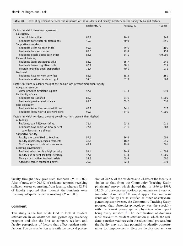
Blazek, Zollinger, and Look 1801
Table III Level of agreement between the responses of the residents and faculty members on the survey items and factors
Residents, % Faculty, % P value
Factors in which there was agreementCollegiality
A lot of interaction 85.7 70.5 .246Residents participate in discussions 40.0 40.9 .854
Supportive coworkersResidents listen to each other 94.3 79.5 .104Residents help each other 88.6 72.8 .138Residents gossip about each other 88.6 54.4 !0.005
Relevant trainingResidents learn procedural skills 88.2 85.7 .245Residents learns cognitive skills 62.9 88.1 .015Program provides good preparation 71.4 70.4 .911
WorkloadResidents have to work very fast 85.7 68.2 .164Residents workload is about right 54.3 61.3 .560
Factors in which residents thought the domain was present more than facultyAdequate resources
Clinic provides sufficient support 57.1 27.3 .010Continuity of care
Residents are satisfied 82.9 34.1 !.005Residents provide most of care 91.5 65.2 .010
Role ambiguityResidents know their responsibilities 65.7 34.1 .017Residents know how to get work done 94.3 54.5 !.005
Factors in which residents thought domain was less present than desiredAutonomy
Residents can influence things 71.4 93.2 .011Residents have input on how patient
care demands are shared77.1 93.1 .008
Supportive facultyFaculty are committed to teaching 57.1 86.4 .003Faculty repeatedly demean residents 48.6 18.2 .016Staff are approachable with concerns 62.9 95.4 .001
Learning environmentResident education is a high priority 51.4 90.9 !.005Faculty use current medical literature 47.1 77.3 !.005Timely constructive feedback exists 34.3 65.9 .002Adequate career counseling exists 20.5 52.3 .010
faculty thought they gave such feedback (P Z .002).Also of note, only 20.5% of residents reported receivingsufficient career counseling from faculty, whereas 52.3%of faculty reported they thought the residents werereceiving adequate career counseling (P Z .009).
Comment
This study is the first of its kind to look at residentsatisfaction in an obstetrics and gynecology residencyprogram and also the first to compare resident andfaculty perceptions of factors that affect resident satis-faction. The dissatisfaction rate with the medical profes-
sion of 28.5% of the residents and 21.0% of the faculty issimilar to that from the Community Tracking Studyphysicians’ survey, which showed that in 1996 to 1997,24.2% of obstetrics-gynecology physicians were very orsomewhat dissatisfied.9 It would appear that our resi-dents and faculty are as satisfied as other obstetrician-gynecologists; however, the Community Tracking Studyreported that obstetrics-gynecology was the specialtywith the lowest percentage of physicians who reportbeing ‘‘very satisfied.’’9 The identification of domainsmost relevant to resident satisfaction in which the resi-dents perceive weaknesses in the educational process, butthe faculty may not, has potential to identify opportu-nities for improvements. Because faculty contact can

1802 Blazek, Zollinger, and Look
positively affect resident satisfaction7 and residents andfaculty positively affect student interest,4 making theseimprovements in residency programs could positivelyaffect our specialty as a whole.
Ten factors were found to be associated with residentsatisfaction in our study, whereas Randall et al7 foundonly 6 factors to be correlated with resident satisfaction.Also, learning environment was added as a variable inthis study, which was not included in the earlier studyof primary care residents. The common factors betweentheir study and this one are continuity of care, auto-nomy, and collegiality. The study by Randall et al7 usedresponses from 119 internal medicine, family medicine,and pediatrics residents. The difference in factors iden-tified as important to satisfaction level may be due inpart to the differences between primary care residencyprograms and surgical residency programs.
Although the majority of the survey instrumentsitems were derived from those used in studies in whichinternal reliability was shown,7,8 1 limitation of thisstudy is that these instruments were not independentlyvalidated. It must also be acknowledged that this is asingle institution study and all the factors associatedwith satisfaction were not the same as those in the studyof primary care residents.7,8 Also, this study was basedon self-reported data and subject to the well-knownlimitations of such. Residents’ opinions often vary fromrotation to rotation, although at a given point in a year,this sample should reflect opinions from all rotations atour program. The faculty instrument asked the facultymembers to make judgments on residents’ opinions.This may be somewhat difficult, given varying residentcontact and familiarity from faculty member to facultymember. The faculty frequently chose the neutral re-sponse option on several questions, which illustrates thisconcern.
The 3 domains associated with resident satisfaction,including autonomy, supportive faculty, and learningenvironment, were not present to the extent desired andthis apparent deficit appeared to be underappreciatedby the faculty members. Although our residents perceiveadequate instruction in the how-to aspects of clinicaland surgical practice, their concerns about deficits incognitive skills teaching would imply that the facultyhas insufficiently emphasized the why of what we do forour patients. We believe learning environment might beconstrued as a sum of the other domains, making itperhaps the most important factor in this study. Facultyshould be instrumental in shaping the learning environ-ment, specifically giving more timely constructive feed-back, explicitly delineating expectations at the start ofeach rotation, emphasizing evidence-based medicine,and formalizing career counseling to improve residentsatisfaction.
The American College of Obstetricians and Gynecol-ogists Task Force on Residency Issues included many of
the domains associated with satisfaction in this study(John R. Musich, personal communication, February21, 2005). Similar recommendations were made morethan 15 years ago and illustrate the never-ending needfor educators to commit themselves to improving theresidency learning environment.10 New issues face to-day’s educators; however, the increased focus on pro-viding service to patients at multiple sites and producingclinical revenue, takes time away from teaching.11
Some of the recommendations have already beenimplemented on some services including appointment ofa resident advocate, an informal effort to match inter-ested resident mentees to faculty members with skill setsthat the resident hopes to acquire, resident-to-residentmentorship, and a midrotation feedback. However, forchange to be successful, there must be total commitmentto these changes by those directing the program alongwith the entire faculty.9 Change is necessary not only toimprove resident morale and learning environment andthus medical student recruitment but also to improvepatient outcomes and satisfaction because prior studieshave shown that physician satisfaction correlates withpatient compliance with treatment12 and patient satis-faction.13
Future studies involving other programs might de-lineate additional improvements that those programdirectors could share in the literature and at meetingsas part of best practices to enhance the learning envi-ronment for residents.
References
1. Advanced Data Tables for 2004 Residency Match. Available from:
www.nrmp.org.
2. Waldman RH, Randlett RR. Results of the National Resident
Matching Program for 2003. Acad Med 2003;68:502-4.
3. Schnuth RL, Vasilenko P, Mavis B, Marshall J. What influences
medical students to pursue careers in obstetrics and gynecology?
Am J Obstet Gynecol 2003;189:639-43.
4. Gariti DL, Zollinger TZ, Look KY. A study at Indiana University
School of Medicine to identify factors detracting students from
applying for an obstetrics and gynecology residency. Am J Obstet
Gynecol 2005;193:289-93.
5. Kendrick SB, Simmons JMP, Richards BF, Roberge LP. Resi-
dents’ perceptions of their teachers: facilitative behavior and the
learning value of rotations. Med Educ 1993;27:55-61.
6. Daugherty SR, Baldwin DC, Rowley BD. Learning, satisfaction,
and mistreatment during medical internship: a national survey of
working conditions. JAMA 1998;279:1194-9.
7. Randall CS, Bergus GR, Schlechte JA, McGuinness G, Mueller
CW. Factors associated with primary care residents’ satisfaction
with their training. Fam Med 1997;29:730-5.
8. Seelig CB. Quantitating qualitative issues in residency training:
development and testing of a scaled program evaluation question-
naire. J Gen Intern Med 1993;8:610-3.
9. Kravitz RL, Leigh JP, Samuels SJ, Schembri M, Gilbert WM.
Tracking career satisfaction and perceptions of quality among
US obstetricians and gynecologists. Obstet Gynecol 2003;102:
463-70.

Blazek, Zollinger, and Look 1803
10. Dilts PV, Dilts SL. Stress in residency: proposals for solution.
Am J Obstet Gynecol 1987;157:1093-6.
11. Gibbons JM. Springtime for obstetrics and gynecology: will the
specialty continue to blossom? Obstet Gynecol 2003;102:443-5.
12. DiMatteo MR, Sherbourne CD, Hays RD, Ordway L, Kravitz
RL, McGlynn EA, et al. Physicians’ characteristics influence
patients’ adherence to medical treatments: results from the Medical
Outcomes Study. Health Psychol 1993;12:93-102.
13. Haas JS, Cook EF, Ruopolo AL, Burnstin HR, Cleary PD,
Brennan TA. Is the professional satisfaction of general internists
associated with patient satisfaction? J Gen Intern Med 2001;15:
122-8.

American Journal of Obstetrics and Gynecology (2005) 193, 1804–6
www.ajog.org
Residency attrition rate in obstetrics and gynecology:Are we losing more postgraduates today?
Maria Manriquez Gilpin, MD*
Morgan Hill Women’s Health Care, Morgan Hill, CA
Received for publication February 24, 2005; revised June 4, 2005; accepted July 22, 2005
KEY WORDSAttrition rateResident
Objective: The purpose of this descriptive study was to determine the attrition rate in 2003 and to
establish where residents matriculate after leaving an obstetrics and gynecology residencyprogram.Study design: A questionnaire was sent by e-mail to all program directors in obstetrics and
gynecology residencies in the United States. The questionnaire asked for the number of residentswho had left a program, what year of training the resident was in, and whether the departure wasa transfer, withdrawal, or dismissal. It asked whether a transfer was to an obstetrics andgynecology residency program or to another specialty; if the resident transferred to another
specialty, which specialty did the resident choose.Results: Two hundred nineteen of 253 programs responded (86.5%). Of residents who leftprograms, 49% left in the first year of training; 34% left in the second year of training; 13% left in
the third year of training, and 4% left in the fourth year of training. The reason for attrition wasthat 75% of the residents transferred to another residency program; 16% of the residentswithdrew from training, and 8% of the residents were dismissed. Of the transferring residents,
60% remained in obstetrics and gynecology.Conclusion: Although resident attrition was higher than in 1992, more residents remained inobstetrics and gynecology.
� 2005 Mosby, Inc. All rights reserved.
Recruitment and retention of residents is fundamen-tally important to obstetrics and gynecology. Althoughthe former challenge has attracted considerable atten-tion,1 the latter has not.
In 1992, the attrition rate was determined to be3.47% or 299 residents over 2 years.2 In that study, most
* Reprint requests: Maria M. Gilpin, MD, Department Obstetrics
and Gynecology, Maricopa Integrated Health System, Medical Pro-
fessional Associates of Arizona, 2601 E Roosevelt St, Phoenix, AZ
85008.
E-mail addresses:[email protected] or mmgilpin@
yahoo.com
0002-9378/$ - see front matter � 2005 Mosby, Inc. All rights reserved.
doi:10.1016/j.ajog.2005.07.083
residents who left obstetrics and gynecology programsjoined other specialties. The data used to evaluate therate of attrition in obstetrics and gynecology in 2003 wasthe Accreditation Council for Graduate Medical Edu-cation (ACGME) Accreditation Data System (ADS).The ADS is a collection system that contains data for allACGME-sponsoring institutions and programs. Theattrition rate in obstetrics and gynecology for 2003was 4.5% or 210 of the 4665 residents. Of those 210residents, 9 residents were dismissed; 132 residents trans-ferred to other programs, and 69 residents withdrew.
To date, no study has examined the career path ofresidents who leave their obstetrics and gynecology

Gilpin 1805
residency. One study suggests that, when all residenciesare examined, residents who changed residencies rarelyleft a controlled living (CL) specialty (eg, anesthe-siology, dermatology, emergency medicine, pathology,ophthalmology, and radiology) for a non-CL (NCL)specialty (eg, family practice, internal medicine, pediat-rics, obstetrics/gynecology, and surgery).3 During thetime of that study, there was a net gain in CL specialtiesand a net loss in NCL specialties.
The objective of this study was to determine thesubsequent specialty training of the residents who lefttheir residencies in obstetrics and gynecology. It wasassumed that most residents who left an obstetrics and
Figure 1 Reason for attrition.
Figure 2 Attrition by postgraduate year.
gynecology residency would enter a controlled lifestylespecialty.
Material and methods
I obtained an e-mail list for all the United Statesobstetrics and gynecology residency programs and sentthe survey by e-mail to each of the 253 programdirectors or coordinators. A second and third requestwas sent from the ACGME Residency Review Com-mittee for Obstetrics and Gynecology.
The survey asked the for the following data on the re-sidents who had left their residency programs in the past3 years: the number of residents who left and the post-graduate year they were in when they left the residencyprogram. The survey also sought to identify whether thedeparture was a transfer, a withdrawal, or a dismissal.The survey inquired whether a transfer was to anotherobstetrics and gynecology program or another specialty;if the transfer was to another specialty, the surveywanted to know which one. This survey did not identifythe programs or residents that were involved, and thedata were collected without identifiers. I assured that noprogram information was duplicated by a check-off list.
Results
Of the 253 surveys that were sent, 219 responses (87%)were received from the program directors.
The transfer rate was 76%; the withdrawal rate was16%, and the dismissal rate was 8% (Figure 1). The yearin which residents were most vulnerable for attrition wasin the first year (49%), followed by the second year
Figure 3 Final specialty choice.

1806 Gilpin
(34%), the third year (13%) and finally the fourth year(4%; Figure 2). Collectively, 83% attrition occurs in thefirst 2 years.
Of the transferring residents, 60% remained in ob-stetrics and gynecology (Figure 3); the remaining 40%entered various specialties. Fewer than 20% of residentstransferred to CL specialties collectively. An equalpercentage of transferring residents went into specialtiesthat were considered to be NCL.
More residents (77.4%) in programs that had higherattrition rate (O3 residents in 3 years) chose to remainin obstetrics and gynecology, compared with programswith low attrition (0-3 residents; 55.8%; rate ratio, 1.75;95% CI, 1.32, 2.33).
Comment
Although resident attrition in obstetrics and gynecologyhas increased by 1% from 1992, these study data showthat more residents are remaining in obstetrics andgynecology training programs. With the use of the datafrom ACGME ADS for the year 2003,4 the attritionrate currently is 4.5%, with 62.9% of residents transfer-ring, 32.9% of residents withdrawing, and 4.2% of theresidents being dismissed. The study in 1992 indicatedthat more transferring residents chose to continuetraining in a specialty other than obstetrics and gyne-cology.2 These survey results indicate a reversal of thattrend, which may actually indicate a net of no realchange in resident attrition from obstetrics and gyne-cology. When programs with higher attrition rates arecompared with programs with low attrition rates, thedata suggest that there is a 1.75-fold increase of residentswho choose to remain in obstetrics and gynecology. It isinteresting to speculate the reason for this observation,although that reason is not available from this studiesinformation. Most of the residents who transferred didnot leave their programs to join less rigorous or CLspecialties.
In efforts to maintain brevity in the questionnaireinformation to stratify for postgraduate year, the num-ber of resident total per program or gender wasnot obtained. Earlier reports have identified importantgender differences in resident attrition.5
Why are residents choosing to leave their originalprograms, and how can these programs decrease their riskfor losing residents to other obstetrics and gynecologyresidency programs? The American College of Obstetri-cians and Gynecologists (ACOG) Presidential InitiativeTask Force on Residency Issues6 reported to the ACOGExecutive Board in January 2004 and addressed thesequestions and presented innovative ideas for the improve-ment of the content of residency curriculum, the conductof residency training, and the perception of post-resi-dency lifestyle. At the request of the task force, questionsregarding resident satisfaction were included in the 2004CREOGexamination survey. The executive committee atACOGmade the results known to me; however, the datahas not been published yet. Interestingly, inadequateeducation and difficult work relationships lead the rea-sons that residents would leave their residency programs.Excessive workload and another career option were lessimportant factors in attrition.
In conclusion, even though the residency programdirectors in obstetrics and gynecology put forth effortsin recruiting medical school candidates, they must alsobe attentive to the retention of residents early in theirtraining. Collectively, we in education must identifyfactors that place residents at risk and modalities todecrease that risk.
References
1. Bienstock JL, Laube DW. The recruitment phoenix: strategies for
attracting medical students into obstetrics and gynecology. Am J
Obstet Gynecol 2005;105:1125-7.
2. Seltzer VL, Messer RH, Nehra RD. Resident attrition in obstetrics
and gynecology. Am J Obstet Gynecol 1992;166:1315-7.
3. Schwartz RW, Simpson WG, Strodel WE, Jarecky RK, Griffen
WO, Young AB. Career change: in quest of a controllable lifestyle.
J Surg Res 1989;47:189-92.
4. ACGME Accreditation Data Systems Graduate Medical Education
Data: academic year 2002-2003. Last accessed: August 18, 2005.
Available at: http//www.acgme.org.
5. Moschos E, Beyer MJ. Resident attrition: Is gender a factor? Am J
Obstet Gynecol 2004;191:387-91.
6. American College of Obstetricians and Gynecologists. ACOG
presidential initiative task force on residency issues, report summary
2004. Paper presented at: American College of Obstetricians and
Gynecologists Executive Board Meeting; January 9-14, 2004;
Washington, D.C.
American Journal of Obstetrics and Gynecology (2005) 193, 1807–10
www.ajog.org
Personality type and clinical evaluations in an obstetrics/gynecology medical student clerkship
Katrina R. Davis, MD,* Joseph A. Banken, PhD
Department of Obstetrics and Gynecology, University of Arkansas for Medical Sciences, Little Rock, AR
Received for publication February 18, 2005; revised May 26, 2005; accepted July 22, 2005
KEY WORDSMyers-BriggsPersonality type
Medical studentClinical evaluation
Objective: The purpose of this study was to determine whether the Myers-Briggs type inventory
extraversion is associated with clinical evaluation ratings that students earn during their(obstetrics/gynecology) junior medical student clerkship.Study design: The Myers-Briggs type inventory was administered to medical students during
their obstetrics/gynec clerkship. Bivariate correlations between clinical evaluations, NationalBoard of Medical Examiners subject scores, and data from the Myers-Briggs type inventoryextraversion scale were analyzed.Results: Pearson product-moment correlation between clinical and National Board of Medical
Examiners subject scores was not significant (r = .25; P = .05). The National Board of MedicalExaminers did not show significant correlations with the Myers-Briggs type inventory extra-version data. The clinical evaluations showed a significant correlation (r = .35; P = .005) with
Myers-Briggs type inventory extraversion.Conclusion: Results show that Myers-Briggs type inventory extraversion is correlated positivelywith clinical evaluations. The National Board of Medical Examiners subject examination and
clinical evaluations were not correlated significantly. Findings question whether clinicalevaluation data should be included in the obstetrics/gynecology medical student evaluationprocess.
� 2005 Mosby, Inc. All rights reserved.
Medical student performance in obstetrics/gynecol-ogy clerkships is evaluated in different ways. One suchmode of evaluation is clinical. Previous investigatorshave reported on the methods of clinical evaluationsused in medical student clerkships in various specialties.These evaluations typically include an assessment ofpatient presentation skills, performance on wardrounds, and performance in outpatient clinics. Zahn
* Reprint requests: Katrina R. Davis, MD, Obstetrics and Gyne-
cology Department, University of Arkansas for Medical Sciences, 4301
West Markham Road, Slot 518, Little Rock, AR 72205.
E-mail: [email protected]
0002-9378/$ - see front matter � 2005 Mosby, Inc. All rights reserved.
doi:10.1016/j.ajog.2005.07.082
et al1 surveyed various obstetrics/gynecology clerkshipsacross the nation and found wide variation in clinicalassessment methods and the ways in which these eval-uations were incorporated into the final grades ofstudents. Awad et al2 reported on clinical evaluationsin a surgery clerkship and found poor correlations withmore objective modes of evaluation. In 2001, Go3
reported that the introduction of more objective perfor-mance criteria produced more reliable results in theclinical evaluation in an emergency medicine clerkship.Ginsburg4 in 1985 commented on the high rate ofvariation of clinical evaluation results of students byteachers in an internal medicine clerkship.

1808 Davis and Banken
At our study site, students are observed in variousclinical settings. Faculty members and residents ratestudents on their ability to perform physical examina-tions, present patient information, prepare write-ups offindings, develop assessments and plans, and interactwith patients and the rest of the clinical team. Theseobservations contribute to the clinical evaluation scorethat each student receives after the completion of theirobstetrics/gynecology clerkship. It is possible that per-sonality characteristics of the medical student influencethe clinical evaluation component. This important con-cern, although consistent with perceptions about clinicalevaluations, has not been investigated empirically.
The Myers-Briggs Type Indicator (MBTI) is a per-sonality inventory that has been used in behavioralresearch studies for many years. Past studies indicatedthat personality characteristics influence objective ex-amination scores,5 clinical performance,6 and academicachievement.7 It is possible that these same characteris-tics affect the opinions of observers when they evaluatethe clinical performance of medical students in obstet-rics/gynecology. The particular type of personality fea-tures that might influence clinical evaluation scores isnot known.
It is possible that students with similar clinical abil-ities are receiving different scores because of personalitycharacteristics of the student, rather than clinical knowl-edge and performance. In some situations, such person-ality characteristics might enhance or perhaps have adeleterious influence on evaluations of clinical abilities.
The purpose of this study was to evaluate whether theextraversion component of the MBTI personality in-ventory correlates with clinical evaluation scores, and ifclinical and National Board of Medical Examiners(NBME) subject scores are correlated.
A correlation between Myers-Briggs extraversion andclinical evaluations by evaluators would indicate thatthe current method of clinical evaluations is influencedby extra-test variables, such as personality characteris-tics of the medical student, and thus should be recon-sidered as part of the evaluation process of obstetrics/gynecology clerkships.
Material and methods
During the research period, 64 medical students matric-ulated through our obstetrics/gynecology clerkship. Thisincluded 3 clerkship periods, with each clerkship includ-ing from 14 to 20 students. The students were offeredparticipation in the study during orientation. Onceconsent was obtained, study participants were adminis-tered the MBTI (form M) by a faculty member who wasnot otherwise involved in the clerkship evaluation.
During the rotation, students were evaluated clinicallyby the residents and faculty and, at the end of their
clerkship, were administered the NBME subject exami-nation. Faculty members had no knowledge of the resultsof the MBTI. For the subjective evaluation, the clerkshipdirector integrated standardized evaluation feedbackfrom residents and faculty members in the form of ascanned document. Categories included clinical perfor-mance, interaction with patients, individual initiative,work ethic, fund of medical knowledge, and interactionwith residents/faculty. In each category, the studentswere given a score of 1 to 5 on a Likert-type scale, with1 reflecting 0 and 5 reflecting superior performance. Thisinformation was then summed to derive a numeric scorefor each student. Clinical and NBME subject examina-tion scores were then compared with the MBTI extra-version raw scores from the data of the MBTI.
The MBTI is a self-administered personality inven-tory that is based on the personality types as theorizedby Carl Jung.8,9 The Clerkship Director was not inattendance during the administration of the MBTI, wasgiven no information about the students during the datacollection period, and was not given any informationconcerning student participation in the research study.The research study was approved by the Human RightsAdvisory Committee at the University of Arkansas forMedical Sciences.
Results
The MBTI was administered in accordance with recom-mended assessment instructions. Sixty-three of 64 med-ical students (98%) who were solicited for participationagreed to participate in the study. All participantssuccessfully completed the MBTI and the obstetrics/gynecology clerkship rotation. NBME subject examina-tion data and subjective evaluations were retrieved forall participants. Data were analyzed in SPSS software(version 12.0; SPSS, Inc, Chicago, IL). There were nomissing data across all critical study variables for the63 students who participated in the study. All statisticaltests were 2-tailed, with a probability level of !.05 forsignificance.
There were 35 men (55%) and 28 women (44%) inthe study. One student chose not to indicate gender aspart of the study protocol. The average age of partic-ipants was 26 G 2.8 (SD) years (range, 22-39 years) forthe 63 participants. The t-test for independent meansindicated no significant difference in age of men orwomen (t = 0.90; degrees of freedom, 61; P O .05).
NBME subject component scores for all groupsranged from 50 to 89 (mean, 70.25 G 9.06). Clinicalevaluation scores ranged from 70 to 100 (mean, 89.21 G5.80), which reflects a more restricted range of evalua-tion dispersion. MBTI extraversion raw scores rangedfrom 0 to 21 (mean, 11.10 G 6.26). MBTI Introversionraw scores ranged from 0 to 21 (mean, 9.87 G 6.19).

Davis and Banken 1809
The Pearson product-moment correlations for theNBME subject examination and the clinical evaluationswere not statistically significant (r = .25; P = .05),which suggests relative independence of these evalua-tions. The correlation between the NBME and MBTIextraversion raw score was also not significant (r = .22;P O .05), which suggests no statistically significantassociation between these assessments.
The correlation between the MBTI extraversion rawdata and the clinical ratings was statistically significant(r= .35; P= .005) and reflective of moderate effect size.As support for construct validity of the MBTI, a signif-icant and negative correlation (r = �.38; P = .002)was shown for MBTI introversion and clinical ratings.In addition, the MBTI extraversion and introversionwere significantly and negatively correlated (r = �.97;P ! .001). Figures 1 and 2 are scatter plots andregression lines of these correlations. As shown, bothMBTI extraversion and introversion represent consid-erable dispersion.
Comment
Data indicate that, for medical students who rotatethrough our 6-week obstetrics/gynecology clerkship, theNBME subject scores do not have a significant statisti-cal correlation with clinical evaluations and have nosignificant correlation with MBTI extraversion. Thisechoes past reports regarding clinical evaluations and alack of meaningful correlations with more objectivemeasures of clinical ability.2,10
Clinical evaluations are significantly correlated withMBTI extraversion and introversion, which suggeststhat personality factors of the medical student may beassociated with evaluation scores. This questions
Figure 1 The x or horizontal axis represents ‘‘Level ofExtraversion’’ as scored on the MBTI; the y or vertical axisrepresents ‘‘% Score on Clinical Evaluation.’’
whether our clinical evaluation components of theobstetrics/gynecology clerkship should be included aspart of the evaluation process. Others have also foundpoor correlations of clinical evaluations with medicalstudent performance in other settings and questionedthe role of clinical evaluations in the role of medicalstudent evaluation.2 An alternative is to change thelabeling of the ‘‘clinical evaluation’’ to ‘‘interpersonalskills’’ because this may reflect more accurately what isbeing measured. The Accreditation Council for Gradu-ate Medical Education has recently recommended theinclusion of competencies in curricula that assess inter-viewing and interpersonal skills. Perhaps in our setting,clinical evaluations are the right tool being used in aninaccurate way.
It is also possible that clinical evaluations are influ-enced by personality characteristics of the evaluator.Further investigation of this possibility could involve anassessment of evaluators to consider the possibility ofinteractions between personality variables of the evalu-ator and student. Clerkships that use clinical evaluationsshould consider each of these possibilities in the evalu-ation of students.
Finally, the metrics of clinical evaluations need care-ful evaluation. In our setting, a Likert-type scale is beingconverted to a nominal scale, which introduces mathe-matical error because the 2 scales are incompatible.
With this consideration, it is important to keep thefindings of this preliminary study in mind when clinicalevaluations are incorporated into the formal clerkshipgrade. In addition, those persons who evaluate tran-scripts for residency applications are encouraged toconsider how a clinical grade is reported and how such‘‘grade’’ may be more influenced by personality ratherthan clinical skills.
Figure 2 The x or horizontal axis represents ‘‘Level ofIntroversion’’ as scored on the MBTI; the y or vertical axis
represents ‘‘% Score on Clinical Evaluation.’’

1810 Davis and Banken
It is important to note that these preliminary findingsshould be considered with some caution. It is alsorecommended that the findings of this study be repli-cated procedurally. Currently, we are in the process ofexpanding this trial over time with the use of our currentmethods to provide additional data regarding thesepreliminary findings. Repeating this investigation inother clerkships at The University of Arkansas forMedical Sciences and at other institutions with differentand similar clinical evaluation methods would assist in amore robust evaluation of the educational usefulness ofthe subjective evaluation process. Other researchersmight find it beneficial to assess similar personalitycharacteristics of those persons who conduct the clinicalevaluations. Furthermore, it is unclear whether findingsof this study would be generalizable to other medicalclerkship rotations. Further research is needed to ad-dress these concerns. As a final note, it is known thatclinical medicine and a physician’s success as a practi-tioner and colleague are often influenced by a reflectionof personality. The outgoing, extraverted physician islikely to interact and ‘‘connect’’ with others, includingpatients, in a different way than someone who is lowerin extraversion.
References
1. Zahn CM, Nalesnik SW, Armstrong AY, Satin AJ, Haffner WH.
Variation in medical student grading criteria: a survey of clerkships
in obstetrics and gynecology. Am J Obstet Gynecol 2004;190:
1388-93.
2. AwadSS, LiscumKR,AokiN,AwadSH,BergerDH.Does the sub-
jective evaluation of medical student surgical knowledge correlated
with written and oral exam performance? J Surg Res 2002;104:36-9.
3. Go S. A competency-based approach to clinical evaluation of
students in the emergency department. Acad Med 2001;76:538.
4. Ginsburg AD. Comparison of intraining evaluation with tests of
clinical ability in medical students. J Med Educ 1985;60:29-36.
5. Wild GC, Skipper BJ. Comparing examination scores and person-
ality types of students from three classes. Acad Med 1991;66:561-2.
6. Jones AC, Courts FJ, Sandow PL, Watson RE. Myers-Briggs type
indicator and dental school performance. J Dental Educ 1997;61:
928-33.
7. Tutton PJM. Psychometric test results associated with high
achievement in basic science components of a medical curriculum.
Acad Med 1996;71:181-6.
8. Jung CG. Psychological types (Baynes HG, translation). New
York: Pantheon Books; 1921.
9. Myers IB, Briggs KB. The Myers-Briggs type indicator. Palo Alto
(CA): Consulting Psychologists Press; 1962.
10. Kuo T, Chiu R. Do subjective evaluations of medical students
correlate with NBME medicine subject test scores? J Gen Intern
Med 2003;18(suppl):246.

American Journal of Obstetrics and Gynecology (2005) 193, 1811–6
www.ajog.org
Focused assessment of surgical performance: Difficultywith faculty compliance
Gabriella G. Gosman, MD,* Hyagriv N. Simhan, MD, MSCR, Richard S. Guido, MD,Ted T. M. Lee, MD, Suketu M. Mansuria, MD, Joseph S. Sanfilippo, MD, MBA
Department of Obstetrics, Gynecology and Reproductive Sciences, Minimally Invasive Gynecologic Surgery Program,University of Pittsburgh School of Medicine, Pittsburgh, PA
Received for publication February 21, 2005; revised June 1, 2005; accepted July 20, 2005
KEY WORDSEducationResidency
Training program
Objective: This study evaluated faculty compliance in the use of the global surgical rating scale of
the Objective Structured Assessment of Technical Skills to rate resident surgical performanceafter every endoscopic procedure.Study design: For this prospective cohort study, 4 faculty members in the Minimally Invasive
Gynecology Surgery Program were asked to rate resident surgical performance using theObjective Structured Assessment of Technical Skills instrument after every case. Facultycompliance was analyzed with respect to the influence of the resident or surgical case
characteristics. Faculty and residents completed surveys about the value of the case-by-caseratings.Results: Faculty members used the Objective Structured Assessment of Technical Skillsinstrument 36% of the time (range, 26%-60%). Faculty member compliance did not vary
according to resident or surgical case characteristics. Faculty members did not think the formshad much impact on whether they gave feedback. Residents thought the opportunity to read theirratings was helpful.
Conclusion: Faculty member compliance with case-by-case surgical performance evaluation ofthe residents was low.� 2005 Mosby, Inc. All rights reserved.
Residency training programs use the assessment ofresidents’ surgical performance to both formative andsummative ends for the residents and the educationalprogram. Resident surgical performance in our institu-tion currently is evaluated with a global performanceevaluation. Surgical skill is 1 of 9 categories that are
* Reprint requests: Gabriella G. Gosman, MD, 300 Halket St,
Room 2314, Pittsburgh, PA 15213.
E-mail: [email protected]
0002-9378/$ - see front matter � 2005 Mosby, Inc. All rights reserved.
doi:10.1016/j.ajog.2005.07.081
rated by attendings on this form. Faculty memberscomplete these forms at varying times after the end ofeach rotation. Such a single global rating of surgicalperformance may be very unreliable.1 To determine thefeasibility of a more informative and intensive approachfor the assessment of resident surgical performance, thisstudy pilots the use of a validated surgical performanceassessment instrument (the global rating scale of theObjective Structured Assessment of Technical Skills[OSATS])2 for real-world use in an academic obstetricsand gynecology residency.

1812 Gosman et al
Figure Global rating form of the Objective Structured Assessment of Technical Skills that has been adapted for everyday use in anobstetrics and gynecology residency program. The form was developed at the University of Toronto. (Modified from Martin JA,Regehr G, Reznick R, MacRae H, Murnaghan J, Hutchinson C, et al. Objective structured assessment of technical skills (OSATS)
for surgical residents. Br J Surg 1997;84:273-8. Copyright British Journal of Surgery Society Limited. Reproduced with permission.Permission is granted by John Wiley and Sons Ltd on behalf of the BJSS Limited).
The OSATS involves direct rater observation ofresidents performing technical or surgical tasks. Twoscoring systems are used for these tasks, a task-specificchecklist and an itemized global rating scale. Bothscoring systems have been shown to be reliable andvalid in formal multistation surgical testing environ-ments that involve live surgery and bench model surgicaltasks.2-4 Coleman and Mueller5 used the global ratingscale to score obstetrics/gynecology resident surgicalperformance of laparoscopic salpingectomies to assessthe effectiveness of a laparoscopic training intervention.We hypothesized that the global rating scale of theOSATS might be a means to improve significantly ourability to assess resident surgical performance on a case-by-case basis outside of the formal testing setting. Theglobal scale was selected because it can be used for avariety of procedures. This study focuses on the feasi-bility of completing a global rating scale after every casethat faculty members perform with a resident. Specifi-cally, we evaluated faculty members’ ability to complywith this case-by-case rating protocol. For this pilotstudy, we limited faculty participation to the 4 facultymembers of the Minimally Invasive Gynecology Surgery(MIS) Program in the Department of Obstetrics, Gyne-
cology and Reproductive Sciences at the University ofPittsburgh School of Medicine, Magee-Womens Hospi-tal. This group was selected because of the uniquetraining needs of residents who are learning endoscopicsurgery in an obstetrics/gynecology residency. We alsoevaluated faculty and resident attitudes toward thisnovel evaluation policy.
Material and methods
Study design and population
This is a prospective cohort study to evaluate thefeasibility of surgical performance evaluation of resi-dents after every case in which residents participated.The 4 faculty members of the MIS Program in theDepartment of Obstetrics, Gynecology and Reproduc-tive Sciences underwent an orientation to the globalrating scale portion of the OSATS, which is referred toas ‘‘rating form.’’ The Figure shows the adapted form.During the study period from February 9, 2004, throughJuly 8, 2004, the 4 faculty members agreed to complete arating form for resident surgical performance after everylaparoscopic, hysteroscopic, or combined laparoscopic/

Gosman et al 1813
hysteroscopic surgical procedure that they performedwith a resident or residents. Rating forms were printedon 5 ! 7–inch cards and were given to faculty members.Additional information that was collected on the ratingforms included resident level of participation, facultyinitials, resident name, date of the procedure, and theprocedure type. Faculty participants were oriented as tohow to use the form to evaluate a resident. However,faculty members were not specifically told where orwhen to complete the form. Completed cards werecollected by the primary investigator and kept in alocked cabinet until they were given to medical recordspersonnel, who served as honest brokers. Proceduresand resident participation were confirmed with de-iden-tified operative notes. Procedure times were obtainedwith the use of de-identified operating room records.
Before the study period, residents were informed ofthe study purposes and procedure and were oriented tothe rating form itself. At the end of the study period,rating forms and de-identified operative reports werecompiled for each rated resident. Each resident had theopportunity to review the ratings and the operativenotes of the cases in which they participated. Residentsthen completed a survey regarding their perceptions ofthe feedback value of the rating forms. At the end of thestudy period, faculty participants also completed asurvey regarding the value of the rating forms in termsof resident surgical performance evaluation and thefacilitation of feedback. The faculty participants metto discuss the strengths and weaknesses of the form.This protocol was reviewed by the Magee-WomensHospital Institutional Review Board and given exemptstatus.
Statistical analyses
Faculty percent compliance was defined as the totalnumber of completed rating cards divided by the totalnumber of surgical cases that were performed by facultywith residents during the study period. Compliance wascalculated for the faculty group overall and also wasstratified by individual faculty members. Differences incompliance by faculty member were analyzed using the
Table I Number of potential resident surgical performancerating encounters and compliance with completion of ratingform by attending
AttendingPotential ratingencounters (n)
Ratingscompleted (n)
1 81 41 (50.6%)2 138 36 (26.1%)3 51 18 (35.3%)4 5 3 (60%)TOTAL 275 98 (35.6%)
c2 test and logistic regression. Additionally, the influ-ence of a number of other covariates on compliance wasdetermined. Multivariable logistic regression was per-formed to identify factors that enhanced or detractedfrom compliance. These covariates included residentpostgraduate year (PGY), the type of surgical procedure(laparoscopy, hysteroscopy, or both), long operativetime, residents with a high number of cases performedwith the 4 faculty members during the study period, and2 resident participants in the case.
Faculty and resident survey data were handled sep-arately. For faculty and resident survey items that wereexpressed in Likert format, scores were summarizedwith median and range. Survey questions were adaptedfrom an obstetrics/gynecology resident feedback surveyof Farrugia and Shapiro.6 Responses to open-endedquestions were analyzed for content and thematictrends. All statistical analyses were performed withStata software (version 8.0 for Windows; Stata Corp,College Station, TX).
Results
The 4 faculty participants and 27 residents (PGY 1-4)performed 243 MIS cases. There were a total of 275potential surgical performance rating encounters be-tween faculty members and residents because 32 casesinvolved 2 residents operating together. Rating formswere completed for 98 cases (35.6%). Table I displaysthe number of cases that were performed, the number of
Table II Attending completion of surgical performancerating form based on resident and case characteristics
Characteristic nRated byattending P value*
PGY .071 4 02 42 18 (42.9%)3 156 61 (39.1%)4 73 19 (26.0%)
Procedure type .37Laparoscopy 166 59 (35.5%)Hysteroscopy 44 16 (36.4%)Laparoscopy and
hysteroscopy33 16 (48.5%)
Operation time greaterthan median forprocedure type
122 44 (36.1%) .66
Residents with O11cases with MIS facultyduring study
196 71 (36.2%) .75
Cases with 2 residentsoperating together
32 10 (31.3%) .44
* c2 test.
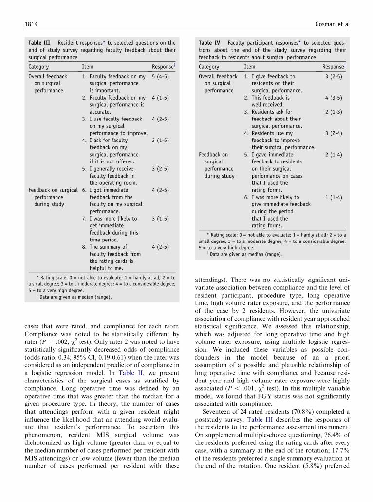
1814 Gosman et al
cases that were rated, and compliance for each rater.Compliance was noted to be statistically different byrater (P = .002, c2 test). Only rater 2 was noted to havestatistically significantly decreased odds of compliance(odds ratio, 0.34; 95% CI, 0.19-0.61) when the rater wasconsidered as an independent predictor of compliance ina logistic regression model. In Table II, we presentcharacteristics of the surgical cases as stratified bycompliance. Long operative time was defined by anoperative time that was greater than the median for agiven procedure type. In theory, the number of casesthat attendings perform with a given resident mightinfluence the likelihood that an attending would evalu-ate that resident’s performance. To ascertain thisphenomenon, resident MIS surgical volume wasdichotomized as high volume (greater than or equal tothe median number of cases performed per resident withMIS attendings) or low volume (fewer than the mediannumber of cases performed per resident with these
Table III Resident responses* to selected questions on theend of study survey regarding faculty feedback about theirsurgical performance
Category Item Responsey
Overall feedbackon surgicalperformance
1. Faculty feedback on mysurgical performanceis important.
5 (4-5)
2. Faculty feedback on mysurgical performance isaccurate.
4 (1-5)
3. I use faculty feedbackon my surgicalperformance to improve.
4 (2-5)
4. I ask for facultyfeedback on mysurgical performanceif it is not offered.
3 (1-5)
5. I generally receivefaculty feedback inthe operating room.
3 (2-5)
Feedback on surgicalperformanceduring study
6. I got immediatefeedback from thefaculty on my surgicalperformance.
4 (2-5)
7. I was more likely toget immediatefeedback during thistime period.
3 (1-5)
8. The summary offaculty feedback fromthe rating cards ishelpful to me.
4 (2-5)
* Rating scale: 0 = not able to evaluate; 1 = hardly at all; 2 = to
a small degree; 3 = to a moderate degree; 4 = to a considerable degree;
5 = to a very high degree.y Data are given as median (range).
attendings). There was no statistically significant uni-variate association between compliance and the level ofresident participant, procedure type, long operativetime, high volume rater exposure, and the performanceof the case by 2 residents. However, the univariateassociation of compliance with resident year approachedstatistical significance. We assessed this relationship,which was adjusted for long operative time and highvolume rater exposure, using multiple logistic regres-sion. We included these variables as possible con-founders in the model because of an a prioriassumption of a possible and plausible relationship oflong operative time with compliance and because resi-dent year and high volume rater exposure were highlyassociated (P ! .001, c2 test). In this multiple variablemodel, we found that PGY status was not significantlyassociated with compliance.
Seventeen of 24 rated residents (70.8%) completed apoststudy survey. Table III describes the responses ofthe residents to the performance assessment instrument.On supplemental multiple-choice questioning, 76.4% ofthe residents preferred using the rating cards after everycase, with a summary at the end of the rotation; 17.7%of the residents preferred a single summary evaluation atthe end of the rotation. One resident (5.8%) preferred
Table IV Faculty participant responses* to selected ques-tions about the end of the study survey regarding theirfeedback to residents about surgical performance
Category Item Responsey
Overall feedbackon surgicalperformance
1. I give feedback toresidents on theirsurgical performance.
3 (2-5)
2. This feedback iswell received.
4 (3-5)
3. Residents ask forfeedback about theirsurgical performance.
2 (1-3)
4. Residents use myfeedback to improvetheir surgical performance.
3 (2-4)
Feedback onsurgicalperformanceduring study
5. I gave immediatefeedback to residentson their surgicalperformance on casesthat I used therating forms.
2 (1-4)
6. I was more likely togive immediate feedbackduring the periodthat I used therating forms.
1 (1-4)
* Rating scale: 0 = not able to evaluate; 1 = hardly at all; 2 = to a
small degree; 3 = to a moderate degree; 4 = to a considerable degree;
5 = to a very high degree.y Data are given as median (range).

Gosman et al 1815
feedback during surgical cases exclusively, and 6 resi-dents (35.3%) desired verbal feedback during and/orimmediately after surgical cases in addition to the ratingcard and/or end-of-rotation feedback. One residentnoted that her intraoperative feedback was not consis-tent with what was recorded on the rating cards.
All 4 faculty members filled out a poststudy survey.Table IV contains these faculty member responses.Faculty members estimated that completion of therating card took 3.75 G 1.5 minutes. One facultyparticipant indicated that a distinct rating system wasneeded for residents who participated as assistants,rather than surgeons, on rated cases. The faculty groupsession revealed that all of the participants completedthe rating cards when they were back in their offices,rather than in the presence of the resident immediatelyafter the surgical procedure.
Comment
We found that real-world faculty compliance with theglobal rating scale of the OSATS as a performanceassessment tool was poor. Our single and multiplevariable analyses failed to identify specific barriers tocompliance with the OSATS rating forms, related toidentifiable traits of the resident or of the surgical caseitself. Faculty participants noted that a perceived barrierto compliance was that they held the onus of responsi-bility to initiate the evaluation process, rather than theresident. Some faculty participants felt that if residentswere responsible for presenting faculty with the ratingforms at the conclusion of each case, then the propor-tion of cases that were rated might improve. One facultymember thought that this particular instrument was notwell suited for evaluating residents who participateas surgical assistants (rather than primary surgeon);therefore, this individual was less likely to complete therating forms.
Technical skill assessment in obstetrics and gynecol-ogy resident education is essential for several reasons.Residency programs strive to graduate trainees who arecapable of independently performing all of the coreprocedures in the specialty.7 Assessment of residents’surgical performance is an important way to evaluatethe adequacy of the educational program. Assessment ofeach individual resident’s surgical skill must be a keycomponent of a program’s promotion and graduationpolicy. Furthermore, residents are adult learners forwhom feedback is a critical element of the educationalprocess.8 Surgical performance assessment obviouslyserves this purpose as well.
In terms of quality of feedback in our study, thefaculty group forum found that the forms did notfacilitate immediate feedback because faculty memberscompleted the forms when they returned to their offices
rather than at the conclusion of the surgical case in thepresence of the resident. This is reflected in the fact thatfaculty members felt that the rating cards did not makethem more likely to give immediate feedback (Table IV).Our survey demonstrated that residents strongly valuedfaculty feedback on their surgical performance and feltthat they would use this feedback to improve. Mostresidents who were surveyed perceived the rating cardsand summary that were derived from these assessmentsto be a valuable addition to currently used techniquesfor feedback. More than one third of residents who weresurveyed desired verbal feedback immediately on com-pletion of the surgical case. This immediacy of feedbackwas not facilitated by the rating form policy asimplemented in this study.
Our study is unique in that it evaluates facultycompliance with the OSATS global surgical rating scalein the real world of resident performance assessment.Studies by Fung et al9,10 using a similar but less widelyused instrument help to interpret our current findingsand guide further study in this area. This group used a3-item interactive voice response instrument to rateobstetrics/gynecology resident performance of laparo-scopic procedures. The instrument used a 5-point Likertscale to rate residents on (1) knowledge and handling ofinstruments, (2) ability to plan and perform operativemoves, and (3) knowledge of anatomy. A fourth ques-tion asked the rater to describe critical incidents thatoccurred during the case. Faculty members used thesystem to rate the resident participant. Residents usedthe system to rate themselves. Compliance for this studywas reported as percent of rated cases in which bothfaculty and resident used the system. They did notcalculate rated versus unrated cases. For feedbackpurposes, faculty members and residents could listento each other’s recorded comments. Similar to ourfindings, residents preferred the new system to thetraditional postrotation single-item evaluation of surgi-cal skill. A follow-up validation study of this instrumentshowed that it required a minimum of 12 ratings ofperformance during laparoscopic procedures to reach areliability of 0.80.10 The most practical application ofthese surgical performance ratings in obstetrics/gynecol-ogy residencies would be for assessment during a broadrange of surgical procedures. Such a context mightincrease the number of ratings that are required toattain reliability.
The use of the OSATS global rating scale or interac-tive voice response instrument facilitates a focusedassessment of resident surgical performance in a similarmanner to the clinical evaluation exercise (CEX) andmini-CEX. These techniques use direct faculty observa-tion of trainee-patient clinical encounters for formativeand summative assessment of trainee performance. TheAmerican Board of Internal Medicine strongly recom-mends the use of mini-CEX in the evaluation of

1816 Gosman et al
residents because this technique is efficient and effective.Consistent with the surgical performance assessmentinstrument, Norcini et al11 found that 12 to 14 mini-CEXencounters were needed to reach a reliability of 0.80for internal medicine residents. Patients in this studyhad a range of clinical settings with a wide variety ofclinical problems. It remains to be determined whetherthe OSATS global rating scale will function reliablywith approximately 12 ratings when applied to the fullrange of gynecologic endoscopic procedures or furtherto the full range of gynecologic surgical procedures.
Unlike the mini-CEX, the global rating scale of theOSATS has been validated only in formal testing envi-ronments rather than in routine clinical care. Our studyrepresents a necessary step towards the goal of validat-ing this widespread surgical evaluation tool in the dailysurgical practice of an obstetrics/gynecology residencytraining program. Given the compliance rate in ourstudy, very few residents had R12 rated cases. Ifcompliance cannot be improved substantially, it mayrequire 1 year of collecting these forms in our program(our study took place over 5 months) to achieve thisnumber of rated endoscopy cases for residents at eachlevel. The reliability and validity of the instrument thatwas used in the context of laparoscopy, hysteroscopy,and combined procedures could then be assessed.
In the future, we will implement faculty participantsuggestions to shift initiative for card completion to theresidents. They will give a card to the attending surgeonimmediately after the conclusion of the endoscopic case.We will explicitly request that faculty members completethe rating at that time. By modifying the protocol andmaking the timeframe for completion of the form moreexplicit, we hope to achieve 3 goals: (1) to improvefaculty compliance with completion of the form, (2) toencourage more timely feedback to residents by sum-marizing their performance during a rated case, and (3)
to test the reliability and validity of this instrument usedin this context.
References
1. Reznick RK. Teaching and testing technical skills. Am J Surg
1993;165:358-61.
2. Martin JA, Regehr G, Reznick R, MacRae H, Murnaghan J,
Hutchinson C, et al. Objective structured assessment of technical
skill (OSATS) for surgical residents. Br J Surg 1997;84:273-8.
3. Reznick R, Regehr G, MacRae H, Martin J, McCulloch W.
Testing technical skill via an innovative bench station examination.
Am J Surg 1997;173:226-30.
4. Lentz GM, Mandel LS, Lee D, Gardella C, Melville J, Goff BA.
Testing surgical skills of obstetric and gynecologic residents in a
bench laboratory setting: validity and reliability. Am J Obstet
Gynecol 2001;184:1462-70.
5. Coleman RL, Mueller CY. Effects of a laboratory-based skills
curriculum on laparoscopic proficiency: a randomized trial. Am J
Obstet Gynecol 2002;186:836-42.
6. Farrugia MM, Shapiro HM. Perceptions of resident feedback
in an obstetrics and gynecology residency program. CREOG
and APGO Annual Meeting. March 3-6, 2004, Lake Buena
Vista, FL.
7. Council on Resident Education in Obstetrics and Gynecology.
Educational objectives: core curriculum in obstetrics and gynecol-
ogy. Washington (DC): Council on Resident Education in Obstet-
rics and Gynecology; 2002.
8. Knowles M. The adult learner: a neglected species. Houston: Gulf
Publishing Co; 1990.
9. Fung Kee Fung MP, Parboosingh IJ, Temple LM, Guy d’Anjou
C, Haebe J, Lussier R. Development of a computerized tele-
communication system for in-training evaluation of residents in a
laparoscopic educational program. Obstet Gynecol 1997;90:
148-52.
10. Fung Kee Fung K, Fung Kee Fung M, Bordage G, Norman G.
Interactive voice response to assess residents’ laparoscopic skills:
an instrument validation study. Am J Obstet Gynecol 2003;189:
674-8.
11. Norcini JJ, Blank LL, Arnold GK, Kimball HR. The mini-CEX
(clinical evaluation exercise): a preliminary investigation. Ann
Intern Med 1995;123:795-9.
American Journal of Obstetrics and Gynecology (2005) 193, 1817–22
www.ajog.org
Self-assessment of resident surgical skills: Is it feasible?
Lynn S. Mandel, PhD,* Barbara A. Goff, MD, Gretchen M. Lentz, MD
Department of Obstetrics and Gynecology, University of Washington, Seattle, WA
Received for publication February 21, 2005; revised July 6, 2005; accepted July 20, 2005
KEY WORDSSelf-assessment
TrainingObjective Structured
Assessment of
Technical SkillsResidentEvaluation
Objective: The purpose of this study was to examine obstetrics and gynecology residents’ self-assessment of proficiency on a variety of surgical bench procedures and to compare their ratingswith those ratings of trained faculty observers who used instruments that have been shown to be
reliable and valid.Study design: As part of a 6-station Objective Structured Assessment of Technical Skills, 74residents at 5 institutions estimated their overall open and laparoscopic skill level beforethe testing. After completing each station, residents evaluated their overall and global skills
performance.Results: Residents rated their proficiency higher on open skills than on laparoscopic skills. Task-specific, overall, and global assessments were correlated significantly with the faculty ratings (P!.001). Residents tended to rate themselves lower than did faculty on almost all measures; eventhose residents with poor skills indicated that they were aware of their deficiencies. Overall andglobal self-assessments increased with each resident level, which indicated good construct validity.
Conclusion: Residents can rate their overall open and laparoscopic skills, task-specific perfor-mance, and global skills with good reliability and validity. Although they tended to scorethemselves lower than did faculty observers, the correlations are high (ie, residents who givethemselves a higher score tended to receive a higher score from faculty, and vice versa). One of the
concerns about self-assessment is that residents with poor skills might not be aware of theirdeficiencies. We did not find that to be the case. Therefore, when residents work on self-directedexercises, task-specific and global checklists can be used for both learning and self-assessment.
� 2005 Mosby, Inc. All rights reserved.
Supported in part by a grant from the United States Surgical
Corporation and by the Ortho McNeil Educational Grant from the
Association of Professors of Obstetrics and Gynecology and a grant
from the National Board of Medical Examiners (NBME) Medical
Education Research Fund Grant. The project does not necessarily
reflect NBME policy, and NBME support provides no official en-
dorsement.
* Reprint requests: Lynn Mandel, PhD, Department of Obstetrics
and Gynecology, University of Washington, 1959 NE Pacific, Room
BB 667A, Seattle, WA 98195-6460.
E-mail: [email protected]
0002-9378/$ - see front matter � 2005 Mosby, Inc. All rights reserved.
doi:10.1016/j.ajog.2005.07.080
With the current emphasis on self-directed and life-long learning, the ability to assess one’s own skills hasassumed increasing importance. In a process that isanalogous to medical diagnosis and treatment, learnerstypically must assess their knowledge or skills, deter-mine their learning needs, formulate learning goals,1
engage in a learning activity, and then reassess profi-ciency. This ability to assess one’s strengths and weak-nesses is essential to the process of life-long learning.2
Additionally, the development of high-fidelity laparo-scopic and surgical simulators and virtual reality

1818 Mandel, Goff, and Lentz
machines has added impetus to the value of being ableto self-assess, especially in simulation-based training.3
In a meta-analysis of 44 studies in higher education(including medicine), Falchikov and Boud4 found that,on average, the correlation between self-assessment andexternal measures was low. Gordon5 reviewed 18 studiesin the health professions that dealt with knowledge andclinical skills but not technical or surgical skills andfound that learners were not able to assess their profi-ciency accurately. Ward et al6 updated the analysis bycombining these studies with 20 newer studies and foundthat methods issues limited the applicability of theresults. Although O80% of these studies comparedratings of self-assessment with those of an externalevaluator, the reliability of these ‘‘gold standard’’ raterswas suspect, therefore calling into question both thereliability and validity of their findings.
More recent studies have focused on surgical skills andhave shown that trainees are able to assess their ownskills, especially after viewing a videotape of their perfor-mance.7 Surgical residents were asked to rate their skilllevel after performing a laparoscopic procedure in a pig,after viewing a videotape of their performance, and after areview of 4 ‘‘benchmark’’ performances that illustrated arange of proficiencies. The correlations between facultyand resident ratings increased significantly after viewingtheir own videotapes but not after reviewing the bench-mark tapes. It is not surprising that the correlation wouldbe higher after seeing their own performance becausefaculty were scoring them in real time during the test,whereas the residents had to attempt to recall at the endwhat they did during the entire procedure. With the videoto prompt them, they could review and reflect on theirperformance at each step.
Repetition of simulated surgical tasks has also shownthat medical student trainees can improve the accuracyof their self-assessment with practice.3 As the repetitionsincreased, the number of actual and estimated errorsdecreased. As the authors point out, the fact that theparticipants felt that they were improving can be apowerful motivational force during learning.
A study at Madigan Army Medical Center8 foundhigh reliability and validity when obstetrics and gyne-cology residents were asked to assess their skills inmidline episiotomy repair that was performed on a life-like model. Although residents rated their own profi-ciency lower than did faculty observers, the scores onthe component skills were similar.
Direct observation with specific criteria is consideredthe most reliable and valid method of assessing technicalskill during operative procedures.9 Pioneering work insurgical skills assessment by Reznick10 andMartin et al11
in Toronto has shown the Objective Structured Assess-ment of Technical Skills (OSATS) to have high reliabilityand validity. Because our previously published studieshave shown the same high reliability and validity,12-14
comparison of resident self-assessment scores with theOSATS results should yield reliable results with highconcurrent validity and avoid the pitfalls of compari-son with an unreliable gold standard. The purpose ofthis study was to examine resident assessments of theirown proficiency on a variety of surgical bench proce-dures and to compare their ratings with the ratings oftrained faculty observers who used instruments thathave been shown to be reliable and valid.
Material and methods
A total of 92 residents at 5 institutions were asked toestimate their overall open and laparoscopic skill levelson a scale of 1 (poor) to 5 (excellent) before starting a6-station OSATS that included both open and laparo-scopic skills that are performed on life-like benchmodels (Table I). The residents were assured thatperformance on the OSATS had no bearing on theirprogress in the residency program. In fact, the individualresident results were not shared with the residencyprogram directors.
Each examination had 3 open and 3 laparoscopicprocedures, with a balance of difficult and relatively easytasks.14 Immediately after each station, the residentsrated their overall performance on that skill (scale, 1-5)and completed the global skills checklist,11 which coversthe same 7 areas, regardless of task (maximum, 35 pointsfor each station). These global skills are applicable toalmost any surgical procedure (eg, respect for tissue,knowledge of the procedure, instrument handling,and time and motion). The overall ratings and the globalskills checklist had been validated previously whenused by faculty members to rate resident performance.15
Asking residents to complete these forms involved ap-proximately 5 minutes of explanation before the OSATS,and only a few minutes between stations to completethe evaluations. Physicians from the University ofWashington who were experienced in administeringthe OSATS acted as the faculty raters at all testing sites.
Resident and faculty ratings were compared with theuse of paired t-tests, which gave both the correlationsbetween the 2 scores and the statistical significance ofthe differences. Comparisons by resident level or testingsite were carried out with the use of one-way analysis ofvariance. Additional analyses used Pearson bivariatecorrelations. Data were analyzed with SPSS for Win-dows (version 12.0; SPSS, Inc, Chicago, IL).
Results
Complete sets of faculty and self-assessment data wereobtained for 74 residents. No resident refused to par-ticipate; however, some data were missing because of

Mandel, Goff, and Lentz 1819
logistic problems (eg, residents on call having to leavebefore the completion of testing). Self-assessments werecompared with the faculty overall and global ratings foreach individual skill and a combination of the 3 lapa-roscopic tasks, 3 open tasks, and the total overall andglobal scores that were a composite of the 6 tasks thateach resident had undertaken.
Overall assessments of open and laparoscopic skillson a scale of 1 (poor) to 5 (excellent) before the start oftesting revealed a mean of 3.12 for open and 2.65 forlaparoscopic skills (P ! 001). The 2 measures werehighly correlated (r Z .754; P ! .001), which indicatedthat residents who rated themselves high on open skillsalso rated themselves high on laparoscopic skills andvice versa.
After completing their overall and global assess-ments, residents rated their overall performance higheron the 3 open skills than on the 3 laparoscopic skills(10.2 vs 8.4; P ! .001). Identical trends were found onthe global assessments (74.0 vs 63.4; P ! .001). Ratingsfor the open and laparoscopic skills were highly corre-lated (P ! .001). Overall self-assessment and overall
Table I OSATS procedures
Open proceduresAbdominal closureBurchCystotomy repairRepair deserosalized bowelEpisiotomy repairLaparoscopic proceduresCystectomyOophorectomyIntracorporeal knotSalpingostomyDissection of retroperitoneal vessel
Table II Means and correlations for overall and globalratings
CompositeSelf-Assess-ment
FacultyRating
Correla-tion
P valuedifference
Open skillsOverall 8.35 10.22 0.743* !.001Global 63.64 74.41 0.753* !.001
Laparoscopy skillsOverall 7.50 8.57 0.665* !.001Global 58.56 64.47 0.679* .001
Total overallrating score(maximum, 30)
15.99 18.51 0.759* !.001
Total global score(maximum, 210)
125.13 136.92 0.664 .001
* P % .001.
global scores also were correlated significantly with thetotal overall and total global faculty ratings and theresidents’ estimate of their open and laparoscopic skillsbefore they started the OSATS (P ! .001). Results forthe overall assessments are shown in Table II.
Residents tended to rate themselves lower than didfaculty members on both the individual tasks andcomposite ratings and global skills (P ! .001). Forexample, with a maximum score of 5 on each task, themean overall ratings for residents and faculty wereabdominal closure (3.3 vs 3.9; P ! .001), Burch (2.1vs 2.8; P ! .001), bowel repair (2.9 vs 3.0; P Z .702),cystectomy (3.0 vs 3.4; P Z .014), and laparoscopicsalpingostomy (2.7 vs 3.2; P Z .007).
The mean global scores (maximum, 35) for residentsand faculty were abdominal closure (23.4 vs 28.3; P !.001), Burch (19.2 vs 22.6; P ! .001), bowel repair (22.6vs 24.3; P Z .193), and cystectomy (20.0 vs 23.3; P Z.001). Even though faculty ratings of overall perfor-mance were higher for laparoscopic salpingostomy, theresidents rated themselves slightly higher on the globalchecklist for this skill (22.1 vs 20.7; P Z .154). Resultsfor selected individual tasks are shown in Table III.There was also a high degree of correlation betweenresident and faculty ratings on specific tasks such asabdominal closure (r Z .60), Burch (r Z .77), andlaparoscopic salpingostomy (r Z .65; all P ! .001). Thesame trend was seen with the global skills checklists:abdominal closure (r Z .59), Burch (r Z .80), andlaparoscopic salpingostomy (r Z .65). Regardless of the
Table III Comparisons of means and correlations forselected skills
SkillSelf-assess-ment
Facultyrating
Correla-tion
P valuedifference
Abdominal closureOverall 3.28 3.92 0.596* !.001Global 23.39 28.34 0.587* !.001
BurchOverall 2.13 2.76 .768* !.001Global 19.24 22.64 .795 !.001
Lap salOverall 2.68 3.16 .591* .007Global 22.13 20.74 .646 .154
Laparoscopic oophorectomyOverall 2.70 3.24 0.576* .001Global 21.92 22.46 0.616* .647
CystectomyOverall 2.97 3.35 0.617* .014Global 20.04 23.33 0.708* .001
Bowel repairOverall 2.89 2.97 0.322 .052Global 22.60 24.31 0.410 .014
Maximum overall rating, 5; maximum global rating, 35.
* P % .001.

1820 Mandel, Goff, and Lentz
component skills on the OSATS, both faculty memberassessments and self-assessments were lower for laparo-scopic tasks than for open tasks. Faculty and residentratings were highly correlated, which means that thoseresidents who rated themselves highest were rated thehighest by the faculty. This is illustrated in Figures 1 and2, which show the total overall and total global ratingsfrom both groups.
Only 22% of the residents rated their overall perfor-mance higher than did the faculty; 6% of the residentshad the exact same rating, and 72% of the residentsrated their performance lower. For the global scores,30% of the residents rated their overall performance
Figure 1 Comparison of faculty ratings and resident self-
assessment of overall performance on 6 bench tasks.
Figure 2 Comparison of faculty and resident self-assessmentof total global skills ratings on 6 bench tasks.
higher; 1% of the residents rated their overall perfor-mance the same, and 69% of the residents rated theiroverall performance lower. Figure 3 shows the distribu-tion for the global scores; the negative numbers indicatethat the residents rated themselves higher than didfaculty members, and positive numbers show higherfaculty member ratings.
As has been reported previously,14 the OSATS taskshave shown high construct validity (ie, scores improvedas resident level increased, with senior residents scoringhigher than junior residents). For almost all individualskills, global self-assessments increased with each resi-dent level, which indicates good construct validity (mostprobability values !.001). The same was true for theresidents’ overall 1 to 5 rating on each task. The over-all assessments of open and laparoscopic skills beforeand after testing and the total global score after testingalso showed significant differences by level (P ! .001).As training increased, demonstrated proficiency in-creased (ie, postgraduate year 4 residents scoring higherthan postgraduate year 3 residents who scored higher thanpostgraduate year 2 residents who scored higher thanpostgraduate year 1 residents).
Figure 3 Differences between average faculty and residentratings on overall skills (maximum total, 30 points). Positive
values mean that the faculty rating was higher; negative valuesmean that the resident rating was higher.

Mandel, Goff, and Lentz 1821
One of the major concerns about self-assessment isthat residents with poor skills might not be aware oftheir deficiencies. In comparing overall faculty ratingsversus self-assessment for the lowest-scoring residents(R2 SDs below the mean), we found a significantcorrelation between the 2 assessments (P ! .05) andvery few instances in which the faculty rated the residentlow but the resident rated himself/herself high. Therewas no significant difference in the ability of residents toassess their own performance, regardless of whether theywere in a program that had a formal surgical skillscurriculum. There were also no consistent differences inthe correlations between resident and faculty ratingsbased on the difficulty of the task.
Comment
Residents can rate their overall open and laparoscopicskills, overall task-specific assessments, and global skillswith good reliability and validity. Although they tendedto score themselves lower than did faculty observers, thecorrelations were high (ie, residents who gave themselvesa higher score tended to receive a higher score fromfaculty members and vice versa). One of the concernsabout self-assessment is that residents with poor skillsmight not be aware of their deficiencies. However, wedid not find that to be the case. The process involvedonly an extra 5 minutes at the beginning of the OSATSto give the residents the forms and to explain thelogistics. Completion of the assessments after eachtask required only a few minutes while the residentswere waiting to proceed to the next station. The onlycosts were duplicating the checklists and a few minutesof faculty time.
Students and residents now can choose from a varietyof life-like models, many of which allow them to practiceon their own. However, as MacDonald et al3 pointedout, this type of training provides little feedback andrelies heavily on self-assessment. Learning psychomotorskills is enhanced when learners are given immediate,specific feedback. In the context of surgical skills learn-ing, this feedback can come from faculty members, asurgical simulator, and/or the learner’s own ability tomatch his/her performance with knowledge of the spe-cific procedure and general surgical principles. There-fore, when residents work on self-directed exercises,task-specific and global checklists can be used for bothlearning and self-assessment.
One only has to review Medline citations over thepast few years to see the growing trend to providelaboratory skills training that uses the many benchmodels and virtual reality simulators that currently areavailable. Therefore, the ability to assess one’s ownproficiency can complement faculty assessment in thesesimulations, help match the learner’s self-identified
needs with the activity, and help reduce the facultytime that is required for supervision and providingfeedback.
Ward et al7 reported that, when residents viewed avideotape of their performance, the accuracy of theirself-assessment was significantly closer to that of atrained faculty observer than was their initial assessmentimmediately after completing the skill. MacDonaldet al3 found that repetition improved the accuracy ofresidents’ self-assessment. Because the OSATS was atesting situation, it was not feasible to evaluate whetherthe ability to assess one’s own skills improved withrepetition. However, repeating a skill after receivingfeedback and/or reviewing a taped performance areoptions worth investigating.
Another potential research study would involve hav-ing both the resident and faculty evaluator completetask-specific and global checklists. Subsequently askingthe learner to describe how he/she has performed opensup a dialogue and makes it easier for the facultyevaluator to provide specific feedback. By reviewingthe checklists together, faculty members can also pointout possibly erroneous self-assessments, which couldlead to an improved ability to assess one’s own skills.
In summary, a reliable and valid self-assessment toolcan be used to document ongoing learning on specifictasks and provide feedback to the learner, the faculty,and the training program itself. The entire process tookonly a few minutes before the OSATS and between eachtesting station; the cost was almost nothing, and the taskwas easy for residents to complete.
References
1. Spencer JA, Jordan RK. Learner centered approaches in medical
education. BMJ 1999;318:1280-3.
2. Gordon MJ. Self-assessment programs and their implications for
health professions training. Acad Med 1992;67:762-9.
3. MacDonald J, Williams RG, Rogers DA. Self-assessment in
simulation-based surgical skills training. Am J Surg 2003;185:
319-22.
4. Falchikov N, Boud D. Student self-assessment in higher education:
a meta-analysis. Rev Educ Res 1989;59:395-430.
5. Gordon MJ. A review of the validity and accuracy of self-assess-
ments in health professions training. Acad Med 1991;66:762-9.
6. Ward M, Gruppen L, Regehr G. Measuring self-assessment:
current state of the art. Adv Health Sci Educ 2002;7:63-80.
7. Ward M, MacRae H, Schlachta C, Mamazza J, Poulin E, Reznick
R, et al. Resident self-assessment of operative performance. Am J
Surg 2003;185:521-4.
8. Goff BA, Nielsen PE, Lentz GM, Chow GE, Chalmers RW,
Fenner D, et al. Surgical skills assessment: a blinded examination
of obstetrics and gynecology residents. Am J Obstet Gynecol 2002;
186:613-7.
9. Eubanks TR, Clements RH, Pohl D, Williams N, Schaad DC,
Horgan S, et al. An objective scoring system for laparoscopic
cholecystectomy. J Am Coll Surg 1999;189:566-74.
10. Reznick RK. Teaching and testing technical skills. Am J Surg
1993;165:358-61.

1822 Mandel, Goff, and Lentz
11. Martin JA, Regehr G, Reznick R, MacRae H, Murnaghan J,
Hutchison C, et al. Objective structured assessment of technical
skill (OSATS) for surgical residents. Br J Surg 1997;84:273-8.
12. Goff BA, Lentz GM, Lee DL, Houmard B, Mandel LS. Develop-
ment of an objective structured assessment of technical skills
(OSATS) for obstetric and gynecology residents. Obstet Gynecol
2000;96:146-50.
13. Lentz GM, Mandel LS, Lee D, Gardella CM, Melville J, Goff BA.
Testing surgical skills of obstetrical and gynecologic residents in a
bench lab setting: validity and reliability. Am J Obstet Gynecol
2001;184:1462-8.
14. Goff B, Mandel L, Lentz G, VanBlaricom A, Oelschlager A, Lee
D, et al. Assessment of resident surgical skills: Is testing feasible?
Am J Obstet Gynecol 2005;192:1331-40.
15. Goff BA, Lentz GM, Lee D, Houmard B, Mandel LS. Develop-
ment of an objective structured assessment of technical skills for
obstetric and gynecology residents. Obstet Gynecol 2000;96:
146-50.

American Journal of Obstetrics and Gynecology (2005) 193, 1823–6
www.ajog.org
Resident job satisfaction: One year of duty hours
Kirsten J. Lund, MD,* Stephanie B. Teal, MD, MPH, Ruben Alvero, MD
Department of Obstetrics and Gynecology, University of Colorado Health Sciences Center, Denver, CO
Received for publication February 21, 2005; revised May 29, 2005; accepted July 20, 2005
KEY WORDSDuty hours
Job satisfactionQuality of lifeResidency
Objective: This study was undertaken to assess job satisfaction and quality of life aspects amongresidents in obstetrics/gynecology before and after the implementation of duty-hour require-ments.
Study design: We administered a survey to residents before and after duty-hour restrictions,addressing satisfaction with residency training, quality of life, and predictions/impressions of theeffect of reduced work hours.Results: Satisfaction with overall residency training as a discrete survey item did not change;
however, the composite score from all responses to specific items increased. Several specificclinical and academic items garnered higher satisfaction scores in 2004. Residents reported less-than-anticipated increases in healthiness of their lifestyle and a decrease in interest in teaching.
Conclusion: This is a prospective assessment of the effect of duty-hour requirements, improve-ments in residents’ perception of their time and ability to study and pursue research and in clinicalareas are encouraging. The perception that there is less interest in teaching is of concern.
� 2005 Mosby, Inc. All rights reserved.
The Accreditation Council for Graduate MedicalEducation instituted duty-hour requirements for allaccredited residency training programs as of July 1,2003. These requirements were put into effect in responseto many factors that included historic events in NewYork State, studies such as the 1999 Institute of Med-icine report on the number of medical errors and relatedpatient deaths each year, and proposed legislation thatwould have made duty-hour restrictions a matter offederal law.1-3 Recent studies have suggested that sleep-deprived residents are at increased risk of motor vehicleaccidents and that decreasing the length of call shifts
Presented at the 2005 Association of Professors of Gynecology and
Obstetrics and the Council on Resident Education in Obstetrics and
Gynecology Meeting, Salt Lake City, UT, March 2-5, 2005.
* Reprint requests: Kirsten J. Lund, MD, 4200 E Ninth Ave, Box
B-198, Denver, CO 80262.
E-mail: [email protected]
0002-9378/$ - see front matter � 2005 Mosby, Inc. All rights reserved.
doi:10.1016/j.ajog.2005.07.079
decreases the incidence of serious medical errors amonginterns.4,5 The overall intent of the duty-hour require-ments was to balance the needs of patient safety, residentwell-being, and academic and clinical education.6
At the same time duty-hour requirements wereimplemented for all medical specialties, obstetrics andgynecology training programs faced the problem ofdeclining interest in the specialty on the part of seniormedical students in the United States. In the 2004residency match, 65% of available training positions inobstetrics and gynecology were filled by US medicalschool graduates, which is a steady decrease from 75%in 2000.7 The rigors of residency training and the per-ceived lack of a good ‘‘quality of life’’ for obstetrician-gynecologists have been cited as key reasons for thisdeclining interest.8 Although the institution of duty-hourrequirements has been found to correlate positively withresidents’ perceived ‘‘quality of life,’’9,10 this phenome-non has not been studied extensively to identify those

1824 Lund, Teal, and Alvero
components of quality of life that are improved. Simi-larly, the relevance of physician job satisfaction to suchoutcomes as disability claims, job turnover, and alteredprescribing practices has been established among prac-ticing physicians but largely is unstudied among resi-dents in training.11-14 Finally, the effect of duty-hourrequirements on such factors as medical student educa-tion by residents is as yet unstudied.
In a previous study, we reported on components ofresident job satisfaction, quality of life, and predictionsas to the effect of reduced work hours before the actualimplementation of duty-hour requirements at our insti-tution. In this report, we again assess this cohort ofobstetrics and gynecology residents to evaluate whether1 year of duty-hour requirements had a significantimpact on resident job satisfaction and quality of life.
Material and methods
Survey description
A structured questionnaire that contained 59 items wasadministered to all residents in a university-based obstet-rics and gynecology residency program, immediatelybefore (June 2003) and 1 year after (June 2004) the duty-hour requirements went into effect. This survey consistedof 4 segments: Five demographic items, level of currentsatisfaction with aspects of the residency training experi-ence, comprising 38 items, 5 ‘‘current quality of life’’ indi-cators, and 11 questions as to the direct effect of the duty-hour requirements on residency training and quality oflife. For the latter segment, questions were worded as pre-dictions for change before the duty-hour requirements(2003 survey) and as assessments of change afterwards(2004 survey). Questions were answered on a 5-pointLikert scale, unless otherwise stated. Details on the devel-opment of this survey have been published previously bythe authors.15 The survey was designated as ‘‘exempt’’ bythe Colorado Multiple Institutional Review Board.
Survey administration
This survey was administered to all obstetrics and gyne-cology residents at the University of Colorado in June2003 and again in June 2004. Individual survey responseswere anonymous and identified by a code number.Surveys were returned by 33 of 35 residents (94%) in2003 and by 32 of 35 residents (91%) in 2004. Twenty-three residents completed both surveys. This included 9residents who were at the postgraduate year–1level in2003 (graduating class of 2006), 7 residents from the classof 2005, and 7 residents from the class of 2004.
Statistical analysis
Survey responses were entered into a spreadsheet andindependently hand-checked for accuracy by 2 of the
authors. Descriptive statistics, t-tests, and linear regres-sion analysis were calculated with the use of SPSS forWindows (version 12.0; SPSS, Inc, Chicago, IL). Thepaired t-test was used to evaluate change in meanresponses for identical survey items to control fornonindependence of samples.
Results
Population characteristics
The mean age of the residents was not significantlydifferent between years. Thirty-one of 33 residents in2003 were female; 29 of 32 residents in 2004 were female.Seventeen residents in 2003 were married, 2 of whomhad children, compared with 20 married residents in2004, 6 of whom had children.
Global satisfaction with residency
The response of residents to the question, ‘‘Please rateyour overall satisfaction with your obstetrics and gyne-cology residency program,’’ was analyzed controllingboth for year of training or among different groups oftrainees in comparable postgraduate training years andfor the effect of promotion or the results from the samegroup of residents over time. Although one comparison,residency satisfaction among postgraduate year 3 resi-dents, was significantly decreased among those postgrad-uate year 3 residents who completed the survey in 2004than among those who completed the survey in 2003;in general, there was no significant change in overallsatisfaction with residency between 2003 and 2004. Theaverage satisfaction rating for all residents who com-pleted the survey in 2003 and 2004 was also not different.
Satisfaction with specific aspects ofresidency training
Responses to 37 questions about current levels ofsatisfaction with specific aspects of training were ana-lyzed for change for those residents who completedboth surveys. These questions covered clinical training,academic environment, interpersonal relationships, andlifestyle issues. A full list of items that were queried hasbeen published previously.15 Of these 37 items, nonereceived significantly lower scores in 2004, and 9 re-ceived significantly higher satisfaction ratings (Table I).Of note, despite an overall 30% decrease in rotating in-house call taken by residents who were working tradi-tional day shifts, there was no significant increase insatisfaction with either the setup of night call or with theamount of night call taken.
In addition to analysis of responses to individualquestions, a composite score that comprised the averageof each individual’s response to all 38 questions in partII of our survey was generated. These composite scores

Lund, Teal, and Alvero 1825
were then analyzed for change over time and forcorrelation either with year of training or with previouscomposite score. The mean composite score for all23 residents increased from 3.60 in 2003 to 3.84 in2004 (P Z .005). Composite scores for each resident in2004 were not predicted by postgraduate year level or byprevious composite in 2003.
Quality-of-life indicators
Responses to quality-of-life indicators were analyzed forthe subgroup of 23 residents who completed both the2003 and 2004 surveys. The paired t-test was usedbecause of the relatively small numbers of respondentsand the likely significant effect on the variance ofresponses by the nonindependence of the samples.Weekly mean hours spent sleeping, days of exercise,minutes of exercise per session, and hours spent ineducational reading were highly predicted by the re-sponse the previous year, did not correlate with advanc-ing year of postgraduate training, and did not exhibitsignificant change overall. However, the reported hoursspent reading narrowly missed statistical significance(mean increase of 0.82 hours or 28% change; P Z .058).
Perceived effect of the work week reductionon aspects of training and lifestyle
The third part of the survey asked residents to predictchanges in training and lifestyle measures because ofduty-hour requirements in 2003 and to then assesschanges in the same 11 measures in 2004. A summaryof responses is given in Table II. Of the 11 measures, 2items received significantly lower scores than predictedafter the institution of the limited work week: the
Table I Significant changes in satisfaction with specificelements of residency training, 2003-2004*
Question 2003 2004 P value
Ability to pursueeducational reading
2.35 3.57 .000
Ability to research agiven medical topic
3.39 4.26 .001
Personal participationin research
3.04 3.48 .022
Gynecologic surgicalexperience
3.13 3.65 .011
Obstetric experience 4.35 4.70 .043Exposure to subspecialties 3.59 4.36 .002Attending supervision 3.73 4.18 .015Quality of hospitalnursing staff
3.43 3.74 .016
Amount of leisure time 2.43 3.00 .024
* Stem wording: ‘‘Please rate your satisfaction with the following
aspects of your program’’: Items were scored on a 5-point scale, 1 =
‘‘very dissatisfied,’’ 5 = ‘‘very satisfied.’’
assessment of the effect of reduced work hours onhealthiness of lifestyle and the effect on interest inteaching. The remainder of the responses did not differsignificantly between the 2003 and 2004 surveys.
Comment
Physician job satisfaction and quality of life are im-portant factors that affect clinical performance andrecruitment to and attrition from any specialty trainingprogram.8,11-14 Obstetrics and gynecology programs, inparticular, are faced with declining US medical studentinterest. Many educators are also concerned about theability to train competent physicians and surgeons withlimited time and resources. The imposition of restrictedwork hours has the potential to affect, positively ornegatively, the training and overall satisfaction ofobstetrics-gynecology residents.
In this study, we attempted to assess what impact,if any, the duty-hour requirements had on residents’perceptions of their training and quality of life. Wedid this in a prospective fashion by asking the samequestions both before and after 1 year of duty hours.Each segment of the survey was designed to evaluatesatisfaction from a slightly different perspective: Resi-dents were asked first simply to rate current satisfactionwith both overall residency experience and specificcomponents of that experience, then to quantify, beforeand after, certain indicators such as sleep and studytime, and finally to estimate the direct effect of the dutyhours themselves on key aspects of training and qualityof life.
Table II Effect of duty-hours restrictions on training andquality of life (n = 23)*
Parameter 2003 2004 P value
Will residency improve? 3.96 3.70 .342Happier in residency? 3.87 3.83 .883More free time? 4.17 3.87 .084Happier in personal life? 4.04 3.87 .162Additional time to study? 3.74 3.70 .857Healthier lifestyle? 4.22 3.35 .001Improved patient care? 3.83 3.48 .257More difficultcontinuity of care?
3.74 3.57 .567
Will be well-trained? 3.83 3.52 .110More time to teach? 3.39 3.39 1.0More interest in teaching? 3.65 2.78 .001
* Wording of these items changed from the 2003 to the 2004
surveys. In 2003 the wording was ‘‘After the institution of the 80-hour
work week, I will.’’ (Example, ‘‘I will have more interest in teaching’’;
1 = disagree, 5 = agree). In 2004 the wording was ‘‘Since the
institution of the 80-hour work week, please rate the following: .’’
(Example: ‘‘Interest that I have in teaching’’; 1 = worse/less, 5 = more/
better).

1826 Lund, Teal, and Alvero
We made several changes in the structure of theUniversity of Colorado residency in response to theduty-hour requirements. Although our average workweek was !80 hours before July 2003, some rotations,particularly in the first 2 years of training, were signif-icantly out of compliance. Frequency of in-house nightcall was reduced, both by the addition of another nightfloat resident in postgraduate year 1 and by the alloca-tion of 2 previous in-house positions in the postgraduate3 and 4 years to at-home call. The average work weekfor all residents, based on resident self-reporting,decreased from 73 to 66 hours.
The single outcome variable of ‘‘overall satisfactionwith residency’’ did not change after the institution ofduty-hour requirements. However, a composite averageof responses to all other more specific elements didincrease in a significant fashion. Nine elements inparticular increased significantly. Of these 9, 3 elementsrelated to academic issues (reading, reviewing the liter-ature, and doing research) and 3 elements related toclinical training (obstetric, gynecologic, and subspe-cialty exposure). In our program, the amount of clinicalexperience did not increase; in fact, it most likelydecreased. For example, many clinical services do nothave a resident 1 day per week because of post-callwork restrictions. Many gynecologic surgical proce-dures no longer have O1 resident participating, wherepreviously 2 residents (one primary surgeon and oneassistant) were able to take part in surgical procedures.Our conclusion is that, despite static or decliningvolume of clinical material, residents feel better ableto take advantage of their time with patients and withtheir supervising attending physicians. In addition,residents are very positive about their increased abilityto partake of educational components of residencyeducation (such as reading and research). This isreflected not only in increased ratings of satisfactionwith these components but also with actual reports ofincreased time spent reading, which narrowly missedstatistical significance.
One area of concern is certainly the decreased esti-mation by residents of their interest in teaching after theinstitution of duty-hour requirements. Again, our spe-cialty is faced with the challenge of improving studentrecruitment into obstetrics and gynecology, and resi-dents play a crucial role in teaching and mentoringstudents. Our residents report anecdotally that the needto cover clinical responsibilities and still meet the duty-hour requirements has made them less able to teach, andthis perception seems to be borne out by our study.Further investigation of this issue to optimize themedical student experience in obstetrics and gynecologyis certainly an important future goal.
This study has several important limitations. Al-though this residency program is larger than some, theabsolute numbers of residents who were studied is small,
which limits our ability to detect significant changes.However, expanding the survey to multiple residencyprograms would have introduced confounding factorssuch as different structures of residency programs andthe differing effects of what are necessarily uniqueresponses between different programs to the duty-hourchallenge. This study measured only residents’ percep-tions about the effect of the duty-hour requirements anddoes not measure absolute change in any of the areasthat were studied. Nonetheless, this prospective cohortstudy raises several important areas for future investi-gation as obstetrics and gynecology educators aim forcontinuous improvement in trainee satisfaction, clinicaland didactic education and skills, and medical studentrecruitment.
References
1. New York State Department of Health. Medical staff in New York
codes: rules and regulations, 10.405.4 (1998). Last accessed:
February 22, 2004. Available at http://www.health.state.ny.us/
nysdoh/search/index.htm.
2. Institute of Medicine. To err is human: building a safer health
system. Last accessed: February 20, 2004. Available at: http://
www.iom.edu/reports.asp.
3. Petition to the occupational safety and health administration
requesting that limits be placed on hours worked by medical
residents (HRG publication 1570). Last accessed: February 22,
2004. Available at: http://www.citizen.org/publications/release.
cfm?IDZ6771.
4. Barger LK, Cade BE, Ayas NT, Cronin JN, Rosner B, Speizer FE,
et al. Extended work shifts and the risk of motor vehicle crashes
among interns. N Engl J Med 2005;352:125-34.
5. Bernstein M, Etchells E. Does reducing interns’ work hours reduce
the rate of medical errors? CMAJ 2005;172:474.
6. ACGME common program requirements, revised July 1, 2003.
Last accessed: May 24, 2005. Available at: http://www.agme.
org/acWebsite/dutyhours/dh_Lang703.pdf.
7. National Resident Match Program. Results and Data 2005 Match.
Washington, DC: National Resident Matching Program, 2005;13.
8. Liaison Committee on Obstetrics and Gynecology. Enhancing
student interest in the specialty of obstetrics and gynecology.
Washington (DC): The College; 2003. p. 10-1.
9. Whang EE, Mello MM, Ashley SW, Zinner MJ. Implementing
resident work hour limitations: lessons from the New York state
experience. Ann Surg 2003;237:449-55.
10. Barden CB, Specht MC, McCarter MD, Daly JM, Fahey TJ 3d.
Effects of limited work hours on surgical training. J Am Coll Surg
2002;195:531-8.
11. Fraunfelder FT, Fraunfelder N. Managed care and physician
disability. Manag Care Interface 1999;12:57-9.
12. Ainge D. Increased physician disability claims causing ‘‘crisis’’:
high utilization of disability insurance may leave physician groups
fewer options in the future. Med Group Manage J 1995;42:80-4.
13. Pathman DE, Konrad TR, Williams ES, Scheckler WE, Linzer M,
Douglas J. Physician job satisfaction, dissatisfaction and turnover.
J Fam Pract 2002;51:593.
14. Watkins C, Harvey I, Carth P, Moore L, Robinson E, Brawn R.
Attitudes and behaviour of general practitioners and their pre-
scribing costs: a national cross-sectional survey. Qual Saf Health
Care 2003;12:29-34.
15. LundKJ,AlveroR,Teal SB.Resident job satisfaction:Will 80 hours
make a difference? Am J Obstet Gynecol 2004;191:1805-10.

American Journal of Obstetrics and Gynecology (2005) 193, 1827–30
www.ajog.org
The influence of an audience response systemon knowledge retention: An application toresident education
Archana Pradhan, MD, MPH,a,* Dina Sparano, BS,a Cande V. Ananth, PhD, MPHb
Department of Obstetrics, Gynecology, and Reproductive Sciences, Divisions of General Obstetrics and Gynecologya
Epidemiology and Biostatistics,b UMDNJ-Robert Wood Johnson Medical School, New Brunswick, NJ
Received for publication February 21, 2005; revised May 31, 2005; accepted July 19, 2005
KEY WORDSMedical educationContraceptionInteractive lecture
Audience responsesystem
Objective: The purpose of the study was to compare delivery methods of lecture materialregarding contraceptive options by either traditional or interactive lecture style with the use of an
audience response system with obstetrics and gynecology residents.Study design: A prospective, randomized controlled trial that included 17 obstetrics andgynecology residents was conducted. Group differences and comparison of pre/posttest scores toevaluate efficacy of lecture styles were performed with the Student t test. Each participant
completed an evaluation to assess usefulness of the audience response system.Results: Residents who received audience response system interactive lectures showed a 21%improvement between pretest and posttest scores; residents who received the standard lecture
demonstrated a 2% improvement (P Z .018). The evaluation survey showed that 82% ofresidents thought that the audience response system was a helpful learning aid.Conclusion: The results of this randomized controlled trial demonstrate the effectiveness of
audience response system for knowledge retention, which suggests that it may be an efficientteaching tool for residency education.� 2005 Mosby, Inc. All rights reserved.
Academic medical centers that are affiliated withmedical schools are charged with the task of providinga strong educational base for future physicians. This isno small feat because the material to teach is vast andthe time to teach is limited. Every medical specialty facesthe challenge of developing a complete curriculum that
* Reprint requests: Archana Pradhan, MD, MPH, Department of
Obstetrics, Gynecology, and Reproductive Sciences, UMDNJ-Robert
Wood Johnson Medical School, 125 Paterson St, New Brunswick, NJ
08901-1977.
E-mail: [email protected]
0002-9378/$ - see front matter � 2005 Mosby, Inc. All rights reserved.
doi:10.1016/j.ajog.2005.07.075
promotes the professional growth of the medical studentand the resident.
In the United States, most residency programs usea core curriculum of lectures to impart residents witha basic fund of knowledge. Education regarding con-traception options is one women’s health topic that isimportant to teach effectively. Any resident who haschosen to specialize in obstetrics and gynecology mustbe able to educate their female patients regardingcontraceptive options. Currently, there is neither astandardized curriculum for a core lecture on contra-ceptive choices nor an established method of delivering a

1828 Pradhan, Sparano, and Ananth
core lecture on contraceptive choices. Thus, there is botha need for a standardized curriculum regarding this issueand a need to evaluate whether the manner in which thisbody of knowledge imparted to residents is successful.
Alternative methods of delivering lectures are gainingground among those in the medical community who areinterested in improving the efficiency of programs be-cause retention rates of traditional didactic lectures oftenprove less than desirable. The audience response system(ARS) uses wireless keypads for audience participationduring lectures. Participants are able to respond to testquestions and receive immediate feedback while thecontent is still the current topic of discussion. Theinstructor is able to the see the responses immediatelyand can make changes to the instruction as needed toensure that the audience fully comprehends the content.
A study by Schackow et al1 was designed to measurethe effectiveness of an ARS to help lecture-attendeesretain information. The study at the Chicago-basedfamily practice residency program demonstrated a sig-nificant difference in posttest scores between residentswho received the lecture with the ARS versus the ‘‘tra-ditional’’ format lecture. With the active participationrequired by anARS and the straightforward presentationof key learning points that are typical of ARS lecturedelivery, the lecture material is given much more stayingpower than before, which proves the values of includingsuch technology in the medical education setting. TheARS maintains students’ anonymity, thus encouragingmore active participation and more truthful responsesthan might otherwise be possible.2 Data collection is alsomore accurate because inputs are collected electronicallyand can be managed automatically with the accompany-ing software. Human error in transcribing written an-swers, losing hard copies of answer forms, and other suchcauses of lost data are eliminated.
Although studies show that learners prefer lectureswhich use the ARS,2,3 there is little data on whether theARS can enhance knowledge retention in medicine. Thepurpose of this study was to compare delivery methodsof lecture material to obstetrics and gynecology resi-dents regarding contraceptive options by way of eithertraditional didactic lecture style or interactive lecturestyle with the use of an ARS. Specifically, the study wasdesigned to evaluate the effectiveness of the teachingmethod on the long-term retention of the lecture mate-rial and to determine whether the method of deliveryaffected the residents’ satisfaction of the lecture byquestionnaire.
Material and methods
A prospective, randomized trial that was designed tocompare the effect of 2 different teaching styles onknowledge retention was conducted from July throughAugust of 2004. Participants included 17 obstetrics and
gynecology residents at the UMDNJ-Robert WoodJohnson Medical School, NJ, who voluntarily consentedto participate in this study. Residents were assignedrandomly to attend either the contraception technologylecture or the interactive case-discussion class. Bothlectures were delivered by the same instructor.
The 8 residents in group 1 received the interactivelecture; the 9 residents in group 2 were taught the samematerial in a standard didactic lecture format. A15-minute pretest to establish resident baseline knowl-edge of contraceptive technology was administered to allresidents before the presentation of either lecture. TheARS was used for group 1. Group 2 received the samelecture material in traditional lecture format. Approxi-mately 6 weeks after the delivery of both lectures, allresidents completed the same posttest evaluation toassess their knowledge of the material that was coveredin the lectures. Also, each participant completed anevaluation form to assess his/her opinion regarding theusefulness of the ARS.
Each pre- and posttest consisted of 15 questions;1 to 2 questions pertaining to each of the followingareas were asked: epidemiology of contraception, basicscience of contraception, oral contraception, transder-mal contraception, contraception by intravaginal ring,injectable contraception, intrauterine devices, barriercontraception, emergency contraception, and naturalfamily planning.
Statistical analysis
The Student t test was used to compare pre- and posttestscores to evaluate efficacy of the intervention. No formalsample size analysis was performed because it wasdecided a priori to include all current obstetrics andgynecology residents on rotation. A probability value of!.05 was considered to denote statistical significance,and all statistical tests were 2-tailed.
Results
The 8 residents who received the ARS interactive lecturescored a mean G SD of 78 G 1.41 on the pretest and amean of 95G 1.60) on the post-test, which shows a 21%improvement between the 2 tests. The traditional didac-tic lecture group of 9 residents scored a mean of 80 G2.80 on the pretest and a mean of 82 G 2.32 on theposttest, which demonstrates a 2% improvement inscores. These data show that residents who receivedthe interactive lecture had significantly longer retentionof the lecture material compared with the residents whoreceived the traditional lecture (P Z .018; Table).
The results of the evaluation survey revealed thatonly one half of the students in the traditional group feltthey had a thorough understanding of contraceptive

Pradhan, Sparano, and Ananth 1829
technology after the lecture, although all of the studentsin the interactive group felt they had a thorough knowl-edge after the lecture. The evaluation survey also showedthat all 17 residents found the ARS easy to use and that14 of the residents thought that the ARS was a helpfullearning aid in the lecture (Table).
Comment
Previous studies consistently have illustrated failure inthe traditional lecture format as a means of effectivelyteaching material for sustained application. Relevantcase-based learning, on the other hand, has been cited asa preferred mode of education, with popularity rangingacross disciplines and cultures. According to Hommeet al,2 ‘‘Resident doctors are adult learners and interactivemethods of teaching are considered by many to besuperior when educating the adult learner.’’
Faculty members across the country are changingtheir methods of lecture delivery to accommodate thechanging needs of today’s adult learners. A study byCwiak et al4 compared 2 methods of lecture delivery tothird-year medical students. Students were given either adidactic lecture or an interactive lecture that incorpo-rated the tenets of problem-based learning. Findingsshowed an equivalent increase in posttest scores whetherstudents received the didactic lecture or the interactivelecture. Satisfaction scores were also consistently aboveaverage and equivalent for the 2 lecture styles. In asimilar study on diabetes mellitus and hypertension,although the medical students preferred the small groupdiscussions, tests scores that compared small groupdiscussions with traditional lectures were not signifi-cantly different between the 2 teaching methods.5
The significance of our study is that it showedincreased knowledge retention in the group that receivedthe interactive ARS lecture compared with the groupthat received the traditional lecture format. With thistechnology, the instructor would stop periodically to askthe students a question regarding the material that hadjust been delivered. The students responded to thesequestions using individual electronic keypads, and theinstructor was able to determine instantly the percentage
Table Summary of study results
Group
VariableInteractiveARS (n = 8)
Traditional(n = 9)
Pretest (%) 78 80Posttest (%) 95 82Strong knowledge before (n) 1 1Strong knowledge after (n) 7 3Easy to use (n) 8 9Answers helpful (n) 8 6
of the class that answered correctly with the use of thecorresponding computer program. The immediate feed-back allowed the instructor to measure the effectivenesswith which the material was being delivered and thusrephrase and repeat concepts when necessary. The ARS‘‘combines many of the key components important foradult learners: interactivity; commitment to an answerfrom the learner; timely feedback, and the highlightingof key concepts with ‘take home points’ by the facultyexpert.’’2 Perhaps, therein lies the explanation as to thereason that knowledge retention was affected in ourstudy; our interactive lecture included the ARS ascompared with previous studies that evaluated knowl-edge retention after interactive lecture alone. Our studyalso showed that residents find the ARS easy to use andconsider it helpful to their learning. These data supportthe findings of other studies that compared interactiveteaching with didactic teaching.4,5
These results demonstrate the effectiveness of the ARSon knowledge retention and further suggest that the ARSmay be a valuable and resourceful teaching tool forresidency education. However, it is possible that thenovelty of the experience may have lead to the increasedattention to the lecture, a Hawthorne effect of sorts. It ispossible that, if all lectures were given with the ARS, onewould not see the increased knowledge retention. Afuture study, which will compare pre- and posttest scoresin which the ARS is used for several different lecturesgiven on same day, may clarify this issue.
The ARS is an innovative and creative educationaltool that may help medical faculty become more efficientand effective educators. The ARS may not be widelyavailable for use in all lecture settings (ie, an institutionmay not have enough keypads for each audience mem-ber to receive his or her own.) In this situation, the ARScould be used initially when the teacher is developingthe lecture. The ARS could alert the lecturer to keypoints and concepts that require additional attentionfor retention by students.
With an ever-expanding body of material to learnand integrate into daily practice, all physicians are oftenchallenged to find, on their own, a means by which toefficiently acquire new skills. Because of its effectivenessin enhancing knowledge retention and its well-receivedusefulness, we conclude that the ARS is a valuableeducation tool and that it should be considered forimplementation into settings that span the educationalspectrum.
References
1. Schackow TE, Chavez M, Loya L, Friedman M. Audience response
system: effect on learning in family medicine residents. Fam Med
2004;36:496-504.
2. Homme J, Garth A, Morgenstern B. Utilization of an audience
response system. Med Educ 2004;38:575-9.

1830 Pradhan, Sparano, and Ananth
3. Uhari M, Renko M, Soini H. Experiences of using an interac-
tive audience response system in lectures. BMC Med Educ 2003;
3:12.
4. Cwiak CA, Edelman AB, Hatcher RA, Zieman MD, Nichols MD,
Jensen JT, et al. Teaching contraception: an interactive presenta-
tion using managing contraception. Am J Obstet Gynecol 2004;191:
1788-92.
5. Fischer RL, Jacobs SL, Herbert WN. Small-group discussion versus
lecture format for third-year students in obstetrics and gynecology.
Obstet Gynecol 2004;104:349-53.
American Journal of Obstetrics and Gynecology (2005) 193, 1831–4
www.ajog.org
Obstetrics and gynecology residents as teachers ofmedical students: Predictors of excellence
Joseph A. (Tony) Ogburn, MD,a,* Eve L. Espey, MD, MPH,a Maxine H. Dorin, MD,a
Chen Ming, MS,b William F. Rayburn, MDa
Departments of Obstetrics and Gynecologya and Mathematics and Statistics,b University of New Mexico,Albuquerque, NM
Received for publication February 21, 2005; revised May 19, 2005; accepted July 19, 2005
KEY WORDSMedical studentResident
TeachingExcellence
Objective: The purpose of this study was to assess variables that might predict which intern
candidates will become excellent teachers of medical students.Study design: This retrospective cohort study compared demographic characteristics, previouswork experience, United States Medical Licensing Examinations scores, honors on core
clerkships, membership in Alpha Omega Alpha, and match list ranking of 43 residents toidentify predictors of excellent teaching evaluations during residency.Results: Fifteen residents (35%) were identified as excellent teachers. They were more likely to
have had previous work experience, to be older, or to be male. They were not more likely to havehigher United States Medical Licensing Examinations test scores, more honors grades, AlphaOmega Alpha membership, or a higher rank list position.Conclusion: Work experience, age, and male gender are associated with increased likelihood of
being identified as an excellent teacher by medical students. Programs in which residents have asignificant role as teachers of students may consider these factors in the residency selectionprocess.
� 2005 Mosby, Inc. All rights reserved.
The significant role that is played by resident physi-cians in the education of medical students has beenrecognized for many years.1,2 The Liaison Committee for
Supported by The Seligman Educational Research Fund.
Presented as an oral presentation at the Association of Professors
of Gynecology and Obstetrics and the Council on Resident Education
in Obstetrics and Gynecology Annual Meeting, Salt Lake City, UT,
March 2-5, 2005.
* Reprint requests: Joseph A. (Tony) Ogburn, MD, Assistant
Professor, Residency Program Director, Department of Obstetrics
and Gynecology, University of New Mexico, MSC 10 55801 Univer-
sity of New Mexico, Albuquerque, NM 87131-0001.
E-mail: [email protected]
0002-9378/$ - see front matter � 2005 Mosby, Inc. All rights reserved.
doi:10.1016/j.ajog.2005.07.074
Medical Education has acknowledged the importance ofresident teaching by including a resident training require-ment as part of medical school accreditation standards.3
In its institutional requirements, the Accreditation Coun-cil on Graduate Medical Education (ACGME) specifiesthat residents participate fully in the educational andscholarly activities of the residency program, whichinclude a major responsibility for teaching and supervi-sion of medical students.4 The ACGME competency ofpractice-based learning and improvement requires thatresidents facilitate the learning of medical students.5
Almost 25% of resident activities are related tosupervising, instructing, and evaluating medical students

1832 Ogburn et al
and junior residents.1 Medical students estimate thatone third of their knowledge derives from residentteaching.6 On the most recent Association of AmericanMedical Colleges graduate exit questionnaire, which is anational survey of all graduating medical students abouttheir medical school experiences, students estimated thatmore than 50% of teaching was performed by residentsand fellows on their internal medicine (58%), generalsurgery (55%), and obstetrics and gynecology (50%)rotations.7
Despite the importance of resident teaching and thefocus on successful teaching by national committeessuch as the Liaison Committee for Medical Educationand ACGME, the ability to teach is typically not acriterion that is used to select residents. Instead, pro-gram directors focus on criteria such as United StatesMedical Licensing Examinations (USMLE) scores,grade point average, class rank, membership in AlphaOmega Alpha (AOA), and letters of recommendation.Because a major part of resident success depends onbeing an effective educator, we sought to identify appli-cant characteristics that would predict the ability toteach. The objective of this study was to determinewhether we could identify intern applicants who, afterbecoming residents, would more likely be rated asexcellent teachers by medical students and what specifictraits correlated with teaching excellence.
Material and methods
Subjects included all 43 obstetrics and gynecology res-idents between 1997 and 2004 who completed at least2 years of residency at the University of New Mexico.Before 1997, students uniformly did not evaluate resi-dent teaching; therefore, our sample was limited toresidents after that time. No structured training ofresidents to become more effective teachers was con-ducted during this time period. The University of NewMexico Human Research Review Committee reviewedand approved the study.
Demographic characteristics, measures of academicperformance, work history, and rank list position wererecorded for all subjects. All information came fromresident personnel files and was analyzed in the aggre-gate only. Work experience was defined as a period of atleast 1 year during which the subject was not enrolled inschool and was employed full-time. Type of workexperience was not considered. Demographic character-istics included age at onset of residency and gender.Measures of academic performance included USMLEscores, membership in AOA, and honors grades in third-year clinical clerkships.
Given our average medical school class of 74 grad-uating students, approximately 10 to 13 students rotatein each clerkship group. At the conclusion of each8-week clerkship, the medical students anonymously
evaluate all residents. The evaluation form directs thestudent to rate each resident on a scale of 1 (belowaverage) to 4 (excellent) for the following 4 items: (1)communicates clearly and effectively, (2) is an enthusi-astic and stimulating teacher, (3) models professionalbehavior, and (4) serves as a role model. Residents whoachieve an annual composite score of R3.5 are consid-ered excellent teachers. This score was chosen because itreflects a generally accepted standard of excellence. Wehave used it successfully for 7 years to select faculty andresidents for teaching excellence awards. Data wereentered and analyzed with SPSS software (version10.1; SPSS Inc, Chicago, IL). Logistic regression, Spear-man rank order correlation, and chi-squared test wereused to determine differences between excellent residentteachers and teachers who did not meet the criteria forexcellence. We reported 95% CIs and considered aprobability value of !.05 to be statistically different.
Results
We received approximately 200 applications for ourresidency program annually during the study period andinterviewed approximately 60 candidates from acrossthe United States. Few foreign-trained or osteopathicstudents were interviewed. The 6 interns who werechosen each year were typically non-Hispanic white orHispanic and ranged in age from 25 to 37 years.Approximately 75% of each intern class was female.
Information was available for all 43 residents. Fifteenof the residents (35%) met the criteria for being desig-nated excellent teachers. Comparisons of residents whowere excellent teachers with those residents who werenot excellent teachers in the areas of previous workexperience, gender, honors in obstetrics clerkship,honors in most clerkships, and membership in AOAare summarized in the Table. Applicants with previouswork experience were more likely than the others to beexcellent teachers (odds ratio, 8.4; 95% CI, 1.9- 37.6).Men were more likely than women to be identified asexcellent teachers (odds ratio, 5.6; 95% CI, 1.1-27.0).Older residents were more likely to be excellent teachers(P Z .04). There was no association between gender andprevious work experience (P Z .72) or gender and age(P Z .11). Previous work experience was found to be anindependent predictor of excellence after controlling forthe effect of age (P Z .01).
Higher USMLE step 1 score (P Z .35), USMLE step2 score (P Z .21), or higher rank list position (P Z .94)were not predictive of being rated an excellent teacher.
Only 1 of the 15 excellent teachers had previous workexperience in an actual teaching position. That individ-ual served as a teacher of English as a second language.The other excellent teachers had varied types of em-ployment, but most of the positions involved significantdirect contact with people.

Ogburn et al 1833
Comment
Our study indicates that residents who have previouswork experience and who are older are more likely to berated as excellent teachers by medical students. We wereunable to locate other investigations that examined anyassociation between work experience or age and therating of teaching. It appears that work experience is apredictor of teaching excellence that is independent ofthe effect of age. Although increasing age may beresponsible for some component of increased excellencein teaching, we believe it is likely that the life experiencethat is gained by working contributes to increasedmaturity and tolerance. These traits may improve anindividual’s effectiveness as a teacher.
We were interested to find that male gender wasassociated with a higher likelihood of being identified asan excellent teacher. Because of the small number ofmen in the sample, the actual significance of this findingis unclear. Male applicants who are attracted to obstet-rics and gynecology may possess traits, however, thatmake them more effective teachers. A larger study wouldbe necessary to confirm this finding.
Although academic performance has been evaluatedfor its ability to predict success in residency, it has notbeen evaluated for its role in the prediction of teachingexcellence. Although certain measures of academicachievement are associated with successful passage oflicensing examinations and specialty boards, we foundno association between any academic measure andteaching excellence.
Table Comparisons of characteristics of residents regardingexcellence in teaching
Characteristic
Proportion ofexcellentteachers (%)
Odds ratio forbeing excellentteachers(95% CI)
Pvalue
Work experience 8.4 (1.9, 37.6) .004Yes 57.1No 13.6
Gender 5.6 (1.1, 27.0) .046Male 66.7Female 26.5
Honors in obstetrics 0.9 (0.2, 3.6) .937Yes 34.5No 35.7
Honors in majorityclerkships
0.8 (0.2, 2.7) .686
Yes 31.6No 37.5
Membership in AOA 2.1 (0.4, 11.9) .402Yes 50.0No 32.4
Residents may enter their training programs asexcellent teachers or they may develop these skillsduring their training. A number of studies have assessedthe impact of a training program’s ability to improvethe teaching skills of residents with conflicting results.A study of obstetrics and gynecology residents foundthat a 1-time 4.5-hour teaching program improvedresidents’ self-assessment of teaching skills but did notaffect student evaluations of residents.8 A 6-hour teach-ing program in an internal medicine department resultedin a significant increase in resident teaching scores asreported by medical students.9 Overall, a significantimpact of training programs on the ability to teach hasnot been documented consistently.
Although trying to identify potential excellentteachers had not been part of our selection process,medical student education is emphasized in our resi-dency program. During residency interviews, applicantsare informed that teaching will be a major part of theirresponsibilities. Our intern orientation includes a 2-hourteaching session that provides an overview of the med-ical student curriculum, goals and objectives for thethird-year medical student clerkship, teaching tips, andan overview of the evaluation of medical students. Oureducational program includes 2 to 3 teaching workshopsthat are focused on student education and that areattended by all residents each year. Residents evaluatestudents at the mid point of each clerkship with adedicated 1-hour review session that uses the Reporter-Interpreter-Manager-Educator framework.10 These sessionsprovide not only an evaluation of student performancebut also develop resident skills in several aspects ofteaching, which includes evaluation and feedback.Finally, we recognize all residents who were identifiedas excellent teachers with an award at our annualresident banquet.
Our study has several potential limitations. We used a4-item questionnaire that can be completed quickly andeasily for students to rank all residents and facultymembers. More detailed questionnaires have been de-scribed.11,12 Although our questionnaire is not validated,it does include important domains that are covered inmore detailed questionnaires that include professional-ism, commitment to teaching, and communication skills.We also did not investigate some variables that may beassociated with excellence in teaching that include pre-vious research experience or subjective personality traits.Because of the small numbers of subjects and the widevariety of work experiences, we were not able to deter-mine whether any certain type of work experience wasmore predictive of being an excellent teacher. Addition-ally, our sample was limited to 43 subjects in obstetricsand gynecology. A larger, multicentered study is beingconsidered to confirm these findings.
Excellent resident teachers may improve the perfor-mance of learners.13 Subjectively, we believe that excellent

1834 Ogburn et al
resident teachers may improve medical student percep-tion of obstetrics and gynecology as a specialty and as acareer choice. Because fewer medical students currentlyare choosing our medical specialty as a career, it is criticalthat residents be viewed favorably and with respect.
The selection of residents who are likely to becomeexcellent teachers should be a goal of residency programdirectors. On the basis of the findings of this study, wehave incorporated the consideration of previous workexperience into our intern candidate selection process.Program directors should continue to recruit malestudents into the specialty.
Acknowledgments
We thank Cristina Beraun and Cindy Garcia for theirassistance in the collection of data.
References
1. Brown RS. House staff attitudes toward teaching. J Med Educ
1970;60:156-9.
2. Barrow MV. Medical students’ opinions of the house officer as a
medical educator. J Med Educ 1996;41:807-10.
3. Liaison Committee for Medical Education. Functions and struc-
ture of a medical school: Accreditation and the Liaison Committee
on Medical Education, Standards for the Accreditation of Medical
Education Programs Leading to the MD degree. Washington, DC,
and Chicago, IL: Association of American Medical Colleges and
the American Medical Association; 2000.
4. Accreditation Council on Graduate Medical Education. Institu-
tional requirements. Last accessed: February 2005. Available at:
www.acgme.org.
5. Accreditation Council on Graduate Medical Education. General
competencies. Last accessed: February 2005. Available at: www.
acgme.org.
6. Bing-You RG, Sproul MS. Medical students’ perceptions of
themselves and residents as teachers. Med Teach 1992;14:133-8.
7. Association of American Medical Colleges. 2004 Medical school
graduation questionnaire. Washington (DC): Association of
American Medical Colleges; 2004.
8. Frattarelli L, Kasuya R. Implementation and evaluation of a
training program to improve resident teaching skills. Am J Obstet
Gynecol 2003;189:670-3.
9. Wipf J, Orlander J, Anderson J. The effect of a teaching skills
course on interns’ and students’ evaluations of their resident-
teachers. Acad Med 1999;74:938-42.
10. Ogburn T, Espey E. The R-I-M-E method for evaluating medical
students on an obstetrics and gynecology clerkship. Am J Obstet
Gynecol 2003;189:666-9.
11. Litzelman MD, Stratos G, Marriott D, Skeff K. Factorial valida-
tion of a widely disseminated educational framework for evaluating
clinical teachers. Acad Med 1998;73:688-95.
12. Irby D. Evaluating clinical teaching in medicine. J Med Educ 1981;
56:181-6.
13. Griffith CH, Wilson JF, Haist SA, Ramsbottom-Lucier M. Do
students who work with better house staff in their medicine
clerkships learn more? Acad Med 1998;73:S57-9.

American Journal of Obstetrics and Gynecology (2005) 193, 1835–41
www.ajog.org
A multicenter study to determine motivating factorsfor residents pursuing obstetrics and gynecology
May Hsieh Blanchard, MD,a Amy M. Autry, MD,b Haywood L. Brown, MD,c JohnR. Musich, MD,d Leah Kaufman, MD,e Dylan R. Wells, MD,f Robert D. Stager, MD,g
Jennifer L. Swanson, MD,h Kirsten J. Lund, MD,i Donald W. Wiper III, MD,j
Jennifer L. Bailit, MD, MPHk
Case Western Reserve University-MetroHealth Medical Center/Cleveland Clinic Foundation,a Cleveland, OH;University of California,b San Francisco, CA; Duke University,c Durham, NC; William Beaumont Hospital,d RoyalOak, MI; Long Island Jewish Medical Center,e New Hyde Park, NY; University of Tennessee,f Memphis, TN;Medical College of Georgia,g Augusta, GA; Western Pennsylvania Hospital,h Pittsburgh, PA; University of ColoradoHealth Sciences Center,i Denver, CO; Maine Medical Center,j Portland, ME; Case Western Reserve University-MetroHealth Medical Center,k Cleveland, OH
Received for publication February 21, 2005; revised July 1, 2005; accepted July 19, 2005
KEY WORDSObstetrics and
gynecology
residencyStudent interestSpecialty choiceMotivators
Objective: This study was undertaken to determine why residents choose obstetrics and
gynecology.Study design: Applicants to obstetrics and gynecology residency programs were surveyed;a 5-point scale (5 = most important) was used to rate various aspects of the specialty. Univariatestatistics were performed. Bivariate analysis comparing results that were based on gender and
timing of decisions was completed with Student t test, c2, and Kruskal-Wallis tests.Results: A total of 153 applicants (42% response rate) from 10 programs participated; 85.3% ofrespondents were female. Surgical opportunities, variety of clinical experience, and fast-paced/
high-acuity experiences attract applicants to obstetrics and gynecology. When consideringprograms, resident camaraderie, gynecologic experience, and commitment to education weremost important. Over 70% of residents decided to pursue obstetrics and gynecology during or
after their third-year clerkship.Conclusion: Surgical opportunities and clinical variety appeal to applicants. The majority chooseobstetrics and gynecology during or after their core clerkship. In addition, program dynamics are
important when choosing a residency.� 2005 Mosby, Inc. All rights reserved.
Interest in obstetrics and gynecology, as evidenced bythe percentage of US medical school graduates enteringthe first year at a residency program (PGY-1), peaked
Reprints not available from the author.
0002-9378/$ - see front matter � 2005 Mosby, Inc. All rights reserved.
doi:10.1016/j.ajog.2005.07.069
in 1993 at 7.9%. Since that time, there has been a con-tinued decline in the number of US graduates pursuingtraining in obstetrics and gynecology.1 In 2004, 5.5%(743/13,572) of US seniors participating in the NationalResident Matching Program (NRMP) match suc-cessfully applied to obstetrics and gynecology PGY-1

1836 Blanchard et al
positions. However, only 65.1% of the 1,142 obstetricsand gynecology PGY-1 positions offered in the 2004match were filled by graduating US medical students.2
These statistics reflect an ongoing decline in interestamong graduating US medical students for the field ofobstetrics and gynecology. Proposed reasons for thisdecline have included the liability crisis, decreasedreimbursement, and unpredictability of lifestyle. Therehas been national attention drawn to this situation.Initiatives among obstetrics and gynecology educator(CREOG [The Council on Resident Education in Ob-stetrics and Gynecology]; APGO [The Association ofProfessors of Gynecology and Obstetrics]) and member-ship (ACOG [The American College of Obstetriciansand Gynecologists]) organizations have issued directivestoward focused recruitment into the specialty, promot-ing the positive aspects of the field.3
Despite the above-mentioned trends, there continueto be substantial numbers of medical students whopursue training in obstetrics and gynecology. Thesestudents can provide insight regarding opportunities tooptimize recruitment efforts.
In this study, we sampled applicants to obstetrics andgynecology residency programs from across the country.Our objective was to identify those students (ie, thosewho applied to residency programs in obstetrics andgynecology via the 2004 NRMP match) who were NOTdeterred by the negative aspects identified with the field ofobstetrics and gynecology, and to determine why theystill choose training in this specialty. Our secondary aimwas to evaluate for regional, as well as program size andaffiliation predilections among these applicants. Ulti-mately, we hoped to elucidate which aspects of thespecialty are appealing to current medical students,what helps applicants differentiate among the variousresidency programs, and how these findings are affectedby applicant gender or timing of decision to enter thefield.
Material and methods
In this multicenter survey, we evaluated motivatingfactors for choice to pursue residency training in ob-stetrics and gynecology, as well as differentiating factors(as perceived by applicants) among residency programs.Ten obstetrics and gynecology residency programs par-ticipated in this survey. Institutional Review Boardapproval was obtained at each site.
Surveys were sent to medical students applying for afirst-year residency position in each of the programs,based on the 2004 NRMP rank list for each program.Duplicate applicants among the 10 programs’ rank listswere identified. To ensure that each applicant was sur-veyed only once, those applicants were then assigned to1 of the programs that had the applicant on its rank list,but duplicates were preferentially distributed to the
programs that had lower volumes of surveys to begenerated (in attempts at equalizing the number ofsurveys dispersed by each study site). An applicant wasto receive the survey only from a program where he/shehad applied/interviewed. Each program sent 2 forms(e-mail, US mail) of the survey to each of the remainingapplicants on its list. Surveys were mailed to applicantsafter the 2004 NRMPmatch results were made available.Each survey was labeled with a unique code indicatingboth the program generating the survey and the recipient;this code was used for response tracking purposes only,and was discarded after the completed survey was re-ceived. The cover letter accompanying the survey wasgenerated and signed by the individual study site director,and included instructions indicating that participationwas voluntary, that the survey was part of a multicenterresearch study, that the identifier code was to be used fortracking response to request for participation, and thatonly 1 formof the survey should be returned.The trackingcode was also used to guard against duplicate responses.
Respondents were asked about their choice of obstet-rics and gynecology as a specialty. The survey listedvarious aspects of a career in obstetrics and gynecology(surgical/obstetric/research/primary care opportunities,patient type, variety of clinical experiences in day-to-daypractice, lifestyle factors) and asked respondents to rankeach using the 5-point scale (1 = low importance, 5 =high importance) as to which aspects were deemed to beappealing. Respondents were queried regarding the tim-ing of their decision to pursue obstetrics and gynecology,activities/experiences during medical school that contrib-uted to this choice, and other fields that were seriouslyconsidered other than obstetrics and gynecology.
The survey also queried respondents as to consider-ations in choosing an obstetrics and gynecology resi-dency program. Respondents were given a list of factorspertaining to residency programs (location; type ofprogram, ie, university-based vs community-based;educational experiences during residency/afforded bythe program; faculty size and dynamics; historic post-residency placement success; size of program; ‘‘fringe’’benefits), and asked to use a 5-point scale to rank theimportance of each factor in deciding which obstetricsand gynecology residency program to place on theirNRMP rank list. The respondent was then presentedwith a list of recruitment tools that are used by residencyprograms (eg, Web site, preinterview dinner/social,interview with faculty, interview with residents, follow-up visits) and asked to evaluate their usefulness inhelping the candidate to decide whether and how torank a particular obstetrics and gynecology program (5-point importance scale).
Survey participants were given the option to providedemographic information. In addition, applicants wereasked to provide information as to where on their ranklist they matched, their satisfaction with the outcome of
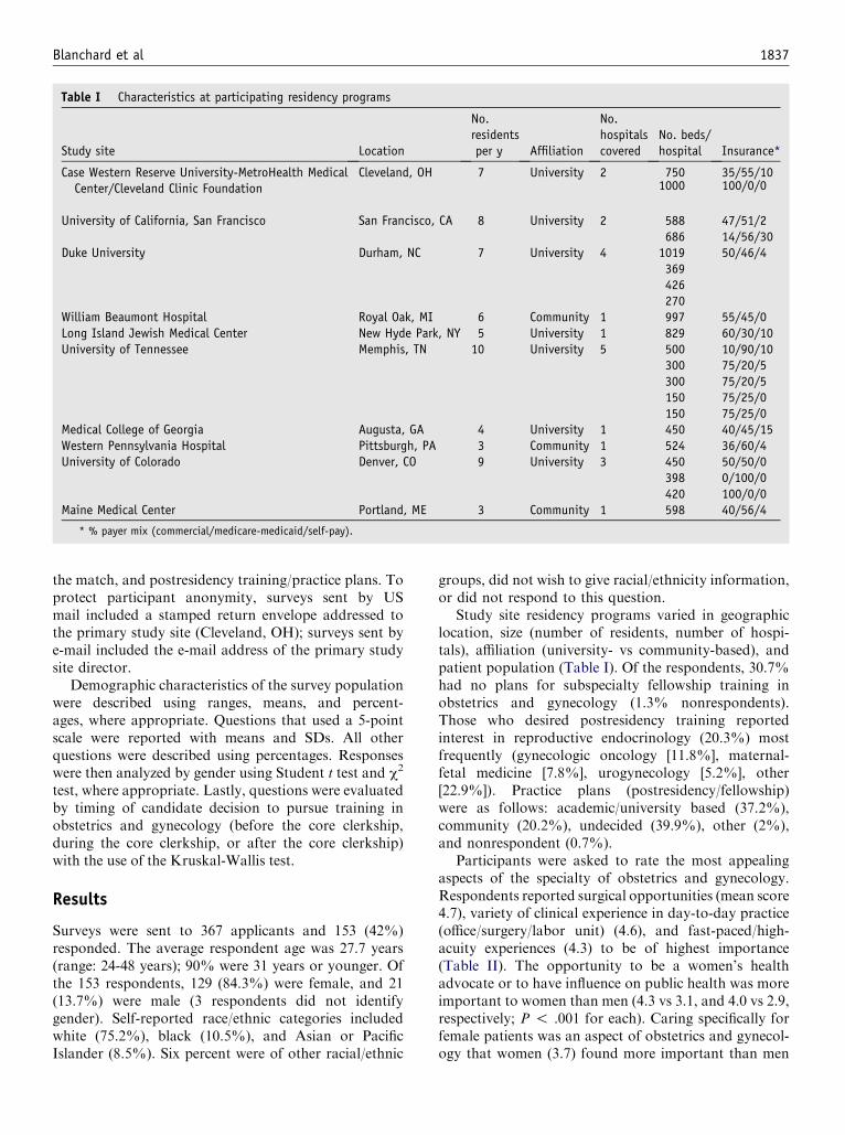
Blanchard et al 1837
Table I Characteristics at participating residency programs
Study site Location
No.residentsper y Affiliation
No.hospitalscovered
No. beds/hospital Insurance*
Case Western Reserve University-MetroHealth MedicalCenter/Cleveland Clinic Foundation
Cleveland, OH 7 University 2 750 35/55/101000 100/0/0
University of California, San Francisco San Francisco, CA 8 University 2 588 47/51/2686 14/56/30
Duke University Durham, NC 7 University 4 1019 50/46/4369426270
William Beaumont Hospital Royal Oak, MI 6 Community 1 997 55/45/0Long Island Jewish Medical Center New Hyde Park, NY 5 University 1 829 60/30/10University of Tennessee Memphis, TN 10 University 5 500 10/90/10
300 75/20/5300 75/20/5150 75/25/0150 75/25/0
Medical College of Georgia Augusta, GA 4 University 1 450 40/45/15Western Pennsylvania Hospital Pittsburgh, PA 3 Community 1 524 36/60/4University of Colorado Denver, CO 9 University 3 450 50/50/0
398 0/100/0420 100/0/0
Maine Medical Center Portland, ME 3 Community 1 598 40/56/4
* % payer mix (commercial/medicare-medicaid/self-pay).
the match, and postresidency training/practice plans. Toprotect participant anonymity, surveys sent by USmail included a stamped return envelope addressed tothe primary study site (Cleveland, OH); surveys sent bye-mail included the e-mail address of the primary studysite director.
Demographic characteristics of the survey populationwere described using ranges, means, and percent-ages, where appropriate. Questions that used a 5-pointscale were reported with means and SDs. All otherquestions were described using percentages. Responseswere then analyzed by gender using Student t test and c2
test, where appropriate. Lastly, questions were evaluatedby timing of candidate decision to pursue training inobstetrics and gynecology (before the core clerkship,during the core clerkship, or after the core clerkship)with the use of the Kruskal-Wallis test.
Results
Surveys were sent to 367 applicants and 153 (42%)responded. The average respondent age was 27.7 years(range: 24-48 years); 90% were 31 years or younger. Ofthe 153 respondents, 129 (84.3%) were female, and 21(13.7%) were male (3 respondents did not identifygender). Self-reported race/ethnic categories includedwhite (75.2%), black (10.5%), and Asian or PacificIslander (8.5%). Six percent were of other racial/ethnic
groups, did not wish to give racial/ethnicity information,or did not respond to this question.
Study site residency programs varied in geographiclocation, size (number of residents, number of hospi-tals), affiliation (university- vs community-based), andpatient population (Table I). Of the respondents, 30.7%had no plans for subspecialty fellowship training inobstetrics and gynecology (1.3% nonrespondents).Those who desired postresidency training reportedinterest in reproductive endocrinology (20.3%) mostfrequently (gynecologic oncology [11.8%], maternal-fetal medicine [7.8%], urogynecology [5.2%], other[22.9%]). Practice plans (postresidency/fellowship)were as follows: academic/university based (37.2%),community (20.2%), undecided (39.9%), other (2%),and nonrespondent (0.7%).
Participants were asked to rate the most appealingaspects of the specialty of obstetrics and gynecology.Respondents reported surgical opportunities (mean score4.7), variety of clinical experience in day-to-day practice(office/surgery/labor unit) (4.6), and fast-paced/high-acuity experiences (4.3) to be of highest importance(Table II). The opportunity to be a women’s healthadvocate or to have influence on public health was moreimportant to women than men (4.3 vs 3.1, and 4.0 vs 2.9,respectively; P ! .001 for each). Caring specifically forfemale patients was an aspect of obstetrics and gynecol-ogy that women (3.7) found more important than men
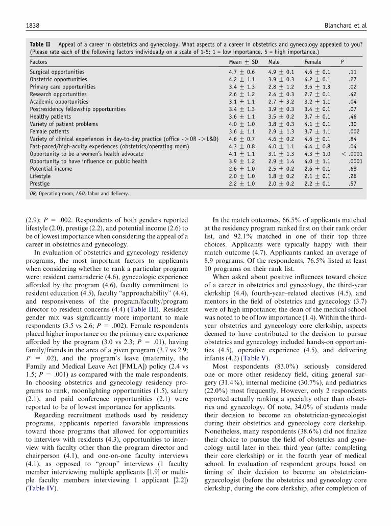
1838 Blanchard et al
Table II Appeal of a career in obstetrics and gynecology. What aspects of a career in obstetrics and gynecology appealed to you?(Please rate each of the following factors individually on a scale of 1-5; 1 = low importance, 5 = high importance.)
Factors Mean G SD Male Female P
Surgical opportunities 4.7 G 0.6 4.9 G 0.1 4.6 G 0.1 .11Obstetric opportunities 4.2 G 1.1 3.9 G 0.3 4.2 G 0.1 .27Primary care opportunities 3.4 G 1.3 2.8 G 1.2 3.5 G 1.3 .02Research opportunities 2.6 G 1.2 2.4 G 0.3 2.7 G 0.1 .42Academic opportunities 3.1 G 1.1 2.7 G 3.2 3.2 G 1.1 .04Postresidency fellowship opportunities 3.4 G 1.3 3.9 G 0.3 3.4 G 0.1 .07Healthy patients 3.6 G 1.1 3.5 G 0.2 3.7 G 0.1 .46Variety of patient problems 4.0 G 1.0 3.8 G 0.3 4.1 G 0.1 .30Female patients 3.6 G 1.1 2.9 G 1.3 3.7 G 1.1 .002Variety of clinical experiences in day-to-day practice (office -OOR -OL&D) 4.6 G 0.7 4.6 G 0.2 4.6 G 0.1 .84Fast-paced/high-acuity experiences (obstetrics/operating room) 4.3 G 0.8 4.0 G 1.1 4.4 G 0.8 .04Opportunity to be a women’s health advocate 4.1 G 1.1 3.1 G 1.3 4.3 G 1.0 ! .0001Opportunity to have influence on public health 3.9 G 1.2 2.9 G 1.4 4.0 G 1.1 .0001Potential income 2.6 G 1.0 2.5 G 0.2 2.6 G 0.1 .68Lifestyle 2.0 G 1.0 1.8 G 0.2 2.1 G 0.1 .26Prestige 2.2 G 1.0 2.0 G 0.2 2.2 G 0.1 .57
OR, Operating room; L&D, labor and delivery.
(2.9); P = .002. Respondents of both genders reportedlifestyle (2.0), prestige (2.2), and potential income (2.6) tobe of lowest importance when considering the appeal of acareer in obstetrics and gynecology.
In evaluation of obstetrics and gynecology residencyprograms, the most important factors to applicantswhen considering whether to rank a particular programwere: resident camaraderie (4.6), gynecologic experienceafforded by the program (4.6), faculty commitment toresident education (4.5), faculty ‘‘approachability’’ (4.4),and responsiveness of the program/faculty/programdirector to resident concerns (4.4) (Table III). Residentgender mix was significantly more important to malerespondents (3.5 vs 2.6; P = .002). Female respondentsplaced higher importance on the primary care experienceafforded by the program (3.0 vs 2.3; P = .01), havingfamily/friends in the area of a given program (3.7 vs 2.9;P = .02), and the program’s leave (maternity, theFamily and Medical Leave Act [FMLA]) policy (2.4 vs1.5; P = .001) as compared with the male respondents.In choosing obstetrics and gynecology residency pro-grams to rank, moonlighting opportunities (1.5), salary(2.1), and paid conference opportunities (2.1) werereported to be of lowest importance for applicants.
Regarding recruitment methods used by residencyprograms, applicants reported favorable impressionstoward those programs that allowed for opportunitiesto interview with residents (4.3), opportunities to inter-view with faculty other than the program director andchairperson (4.1), and one-on-one faculty interviews(4.1), as opposed to ‘‘group’’ interviews (1 facultymember interviewing multiple applicants [1.9] or multi-ple faculty members interviewing 1 applicant [2.2])(Table IV).
In the match outcomes, 66.5% of applicants matchedat the residency program ranked first on their rank orderlist, and 92.1% matched in one of their top threechoices. Applicants were typically happy with theirmatch outcome (4.7). Applicants ranked an average of8.9 programs. Of the respondents, 76.5% listed at least10 programs on their rank list.
When asked about positive influences toward choiceof a career in obstetrics and gynecology, the third-yearclerkship (4.4), fourth-year–related electives (4.5), andmentors in the field of obstetrics and gynecology (3.7)were of high importance; the dean of the medical schoolwas noted to be of low importance (1.4).Within the third-year obstetrics and gynecology core clerkship, aspectsdeemed to have contributed to the decision to pursueobstetrics and gynecology included hands-on opportuni-ties (4.5), operative experience (4.5), and deliveringinfants (4.2) (Table V).
Most respondents (83.0%) seriously consideredone or more other residency field, citing general sur-gery (31.4%), internal medicine (30.7%), and pediatrics(22.0%) most frequently. However, only 2 respondentsreported actually ranking a specialty other than obstet-rics and gynecology. Of note, 34.0% of students madetheir decision to become an obstetrician-gynecologistduring their obstetrics and gynecology core clerkship.Nonetheless, many respondents (38.6%) did not finalizetheir choice to pursue the field of obstetrics and gyne-cology until later in their third year (after completingtheir core clerkship) or in the fourth year of medicalschool. In evaluation of respondent groups based ontiming of their decision to become an obstetrician-gynecologist (before the obstetrics and gynecology coreclerkship, during the core clerkship, after completion of
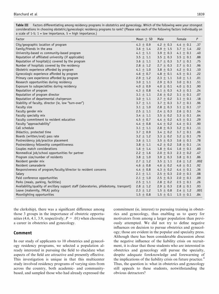
Blanchard et al 1839
Table III Factors differentiating among residency programs in obstetrics and gynecology. Which of the following were your strongestconsiderations in choosing obstetric/gynecologic residency programs to rank? (Please rate each of the following factors individually ona scale of 1-5; 1 = low importance, 5 = high importance.)
Factor Mean G SD Male Female P
City/geographic location of program 4.3 G 0.9 4.2 G 0.3 4.4 G 0.1 .37Family/friends in the area 3.6 G 1.4 2.9 G 1.5 3.7 G 1.4 .02University-based vs community-based program 4.1 G 1.1 3.9 G 0.3 4.1 G 0.1 .40Reputation of affiliated university (if applicable) 3.5 G 1.1 3.5 G 0.3 3.5 G 0.1 .88Reputation of hospital(s) covered by the program 3.6 G 1.1 3.7 G 0.3 3.7 G 0.1 .75Number of hospitals covered by the residency 2.8 G 1.2 2.7 G 0.3 2.7 G 0.1 .96Obstetric experience afforded by program 4.1 G 1.0 3.8 G 0.3 4.2 G 0.1 .10Gynecologic experience afforded by program 4.6 G 0.7 4.8 G 0.1 4.5 G 0.1 .22Primary care experience afforded by program 2.9 G 1.2 2.3 G 1.1 3.0 G 1.1 .01Research opportunities during residency 3.0 G 1.1 2.9 G 0.3 3.0 G 0.1 .51Exposure to subspecialties during residency 4.0 G 0.9 4.0 G 0.1 4.0 G 0.1 .90Reputation of program 4.3 G 0.8 4.1 G 0.3 4.3 G 0.1 .24Reputation of program director 3.1 G 1.1 2.6 G 0.2 3.1 G 0.1 .07Reputation of departmental chairperson 3.0 G 1.1 2.7 G 0.2 3.1 G 0.1 .18Stability of faculty, director (ie, low ‘‘turn-over’’) 3.7 G 1.1 3.7 G 0.3 3.7 G 0.1 .96Faculty size 3.1 G 1.0 2.8 G 0.3 3.1 G 0.1 .17Faculty gender mix 2.5 G 1.1 2.4 G 0.3 2.6 G 0.1 .54Faculty specialty mix 3.4 G 1.1 3.5 G 0.2 3.3 G 0.1 .64Faculty commitment to resident education 4.5 G 0.7 4.4 G 0.2 4.5 G 0.1 .29Faculty ‘‘approachability’’ 4.4 G 0.8 4.4 G 0.2 4.4 G 0.1 .90Call schedule 3.1 G 1.1 2.8 G 0.3 3.2 G 0.1 .11Didactics, protected time 3.7 G 0.9 3.4 G 0.2 3.7 G 0.1 .06Boards (written/oral) pass rate 3.2 G 1.2 3.3 G 0.2 3.2 G 0.1 .79Postresidency job/practice placement 3.6 G 1.1 3.5 G 0.3 3.6 G 0.1 .78Postresidency fellowship competitiveness 3.8 G 1.1 4.2 G 0.2 3.8 G 0.1 .14Couples match considerations 1.6 G 1.4 1.8 G 0.4 1.6 G 0.1 .60Nonmedical job/school opportunities for partner 2.2 G 1.6 2.0 G 0.3 2.3 G 0.2 .47Program size/number of residents 3.8 G 1.0 3.9 G 0.3 3.8 G 0.1 .86Resident gender mix 2.7 G 1.2 3.5 G 1.1 2.6 G 1.2 .002Resident camaraderie 4.6 G 0.8 4.6 G 0.2 4.6 G 0.1 .92Responsiveness of program/faculty/director to resident concerns 4.4 G 0.8 4.3 G 0.2 4.4 G 0.1 .36Salary 2.1 G 1.1 2.5 G 0.3 2.0 G 0.1 .08Paid conference opportunities 2.1 G 1.0 2.5 G 0.3 2.0 G 0.1 .09Perks (meals, parking, facilities) 2.5 G 1.1 2.8 G 0.2 2.5 G 0.1 .26Availability/quality of ancillary support staff (laboratories, phlebotomy, transport) 2.8 G 1.2 2.9 G 0.3 2.8 G 0.1 .93Leave (maternity, FMLA) policy 2.3 G 1.2 1.5 G 0.8 2.4 G 1.2 .001Moonlighting opportunities 1.5 G 0.8 1.5 G 0.1 1.5 G 0.1 .84
the clerkship), there was a significant difference amongthose 3 groups in the importance of obstetric opportu-nities (4.6, 4.1, 3.9, respectively; P= .01) when choosinga career in obstetrics and gynecology.
Comment
In our study of applicants to 10 obstetrics and gynecol-ogy residency programs, we selected a population al-ready interested in pursuing the field to elucidate whataspects of the field are attractive and presently effective.This investigation is unique in that this multicenterstudy involved residency programs of varying sizes fromacross the country, both academic- and community-based, and sampled those who had already expressed the
commitment (ie, interest) to pursuing training in obstet-rics and gynecology, thus enabling us to query formotivators from among a larger population than previ-ously attempted. We did not try to define negativeinfluences on decision to pursue obstetrics and gynecol-ogy; those are evident in the popular and specialty press.Although there has been considerable discussion aboutthe negative influence of the liability crisis on recruit-ment, it is clear that those students who are interested inobstetrics and gynecology still pursue the specialty,despite adequate foreknowledge and forewarning ofthe implications of the liability crisis on future practice.4
Thus, the question is: what in obstetrics and gynecologystill appeals to these students, notwithstanding theobvious detractors?

1840 Blanchard et al
When considering which aspects of the field of ob-stetrics and gynecology are most desirable to enteringresidents, it remains the surgical and procedural aspectsof the field. Furthermore, appeal of the variety of clinicalexperience in day-to-day practice, and the fast-paced/high-acuity experiences (obstetric, surgical) predominateacross both genders. Interestingly, the fast-paced/high-acuity experiences were significantly more important tothe female respondents as compared with the malerespondents, although both ranked it highly.
Respondents were interested in the gynecologic expe-rience afforded by a given residency program, whichspeaks to the attraction of the procedural aspects of thefield, as noted above. However, they also indicated theimportance of the ‘‘atmosphere’’ of the residency,namely, resident camaraderie, faculty ‘‘approachability,’’and the responsiveness of the program/faculty/programdirector to resident concerns. In addition, when in theprocess of interviewing, they highly valued the ability tomeet with residents and nonadministrative faculty, pre-sumably to get a better ‘‘feel’’ for what the residencyprogram is really like. The significance of this weightplaced on the less-tangible, less-quantitative aspects ofresidency may be its reflection of the changing values andperspectives among today’s future doctors when consid-ering what it means to be a resident and/or physician inmedicine (in general) and/or obstetrics and gynecology(in specific), as it pertains to views toward work, workethic, fiduciary responsibility, and a career in medicine.
Table IV Residency program recruitment techniques. Differentprograms use different recruitment methods. If you encounteredany of these techniques, please rate whether they influencedyour decision to rank a given program (scale of 1-5; 1 = influencedme to NOT RANK/rank lower a program, 5 = influencedme to RANKhigher a program)
Recruitment technique Mean G SD
Residency information on own website 3.6 G 0.9Communications before interview 3.4 G 1.0Opportunity to interview with residents 4.3 G 0.9Opportunity to interview with faculty members(other than director and chairperson)
4.1 G 0.9
Preinterview social (night previous) 4.0 G 1.1Other opportunity for informal interactions withresidents
3.8 G 1.1
Postinterview communications 3.9 G 1.1Follow-up (second-look) visit 3.2 G 1.6Solo interview day (you were the only applicantinterviewing that day)
1.9 G 1.2
Group interview day (there were other applicantsinterviewing that day)
3.5 G 1.0
One-on-one faculty interviews 4.1 G 0.9Multiple faculty members interviewing1 applicant (at once)
2.2 G 1.1
One faculty member interviewingmultiple applicants (at once)
1.9 G 1.2
The quest for effective recruitment into our specialtyhas been discussed since at least the early 1970s.5 In1991, Metheny et al6 surveyed senior students at 11medical schools to determine predictors of specialtychoice among the respondents who ranked obstetricsand gynecology as one of their top 4 specialty choices.They found that students attracted to the specialty weremore likely female, and gave higher ratings of impor-tance to obstetrics and gynecology procedures and risks,the opportunity to work with mostly healthy patients,patient contact, the opportunity to perform surgery,their beliefs about reproductive issues, and a perceivedneed for more physicians in the specialty. In our study,the survey recipients were actual applicants to obstetricsand gynecology residency programs, thus allowing fora larger sample size of those who have already beenidentified to have interest in the field. There were ahigher number of respondents who were female, reflect-ing the national status of applicants to residency inobstetrics and gynecology. However, although our re-spondents also found procedures and surgical opportu-nities to be most important, patient health status andadvocacy were of lesser importance, particularly for themale respondents.
In 2003, Fogarty et al4 investigated the same question,but asked it of the previous ten year’s graduates fromone Midwest medical school, and found that factorssignificant in influencing choice of obstetrics and gyne-cology as a specialty were gender, second-year rotation,obstetrics and gynecology staff, continuity of patientcare, primary care opportunities, surgical opportunities,female patients, healthy population, lifestyle opportuni-ties, and financial opportunities. Although our studyfound surgical opportunities and faculty/staff to beimportant factors for choice of residency, primary care,lifestyle, and income were of little import to applicants toobstetrics and gynecology residency. That same year,Schnuth et al7 surveyed current medical students, years
Table V Positive influences toward choosing obstetrics andgynecology. Which of the following positively influenced yourdecision to choose a career in obstetrics/gynecology? (Pleaserate each of the following factors individually on a scale of 1-5;1 = low importance, 5 = high importance.)
Mean G SD
Third-year obstetrics/gynecology coreclerkship
4.4 G 0.9
Interaction with residents 3.7 G 1.2Interaction with faculty 3.9 G 1.1Delivering infants 4.2 G 1.1OR experience 4.5 G 0.8Outpatient clinic experience 3.7 G 1.1Inpatient experience 3.8 G 0.9Didactics 3.3 G 1.1Hands-on opportunities 4.5 G 0.8

Blanchard et al 1841
1 through 4, in another Midwest medical school todetermine factors that influence selection of the specialtyof obstetrics and gynecology, but with the additional aimof evaluating gender differences. They found that femalestudents felt more strongly than male students thatinterest in women’s health, ability to improve the healthof women, emphasis on patient education and preven-tion, and intellectual content of obstetrics and gynecol-ogy were positive influences. In contrast, male studentsfelt more strongly than female students that opportuni-ties to perform surgery, gender, and working underpressure were influences. The proclivities expressed bythe female students were supported by our study. How-ever, it was the female respondents in our study whoplaced higher value on fast-paced/high-acuity experienceover the male respondents.
In a 2004 study, Vaidya et al8 evaluated the relation-ship between specialty choice and medical studenttemperament and character, using the Cloninger Tem-perament and Character Inventory (TCI; a 240-question,self-administered, true-false questionnaire). Cloningeret al identified 4 temperament dimensionsdnoveltyseeking (NS), harm avoidance (HA), reward dependence(RD), and persistence (P). The survey was administeredto third- and fourth-year medical students of a Midwestmedical school. Of those who chose obstetrics andgynecology as their specialty, they were noted to scorehigh in NS and RD. (NS is the tendency to explore, to becurious about novel stimuli, and to actively avoid pun-ishment and frustrating non-reward. High noveltyseekers are curious about new situations, and the riskierthe situation, the greater the ‘‘buzz’’ they experience. RDis the tendency to develop behaviors that lead to positivereinforcement and to maintain rewarded behaviors.Persons with high RD want to please others and socialinstitutions that were previously rewarding, and tend toconform to the reward system.) These findings supportour study findings, in that the surgical and proceduralaspects appealing to applicants to obstetrics and gyne-cology are within the category of ‘‘novel situations’’ withpotential for participation in risky situations. Further-more, the importance of the residency camaraderie andfaculty support speaks to the desire for rewarding socialinteractions.
Clearly, there are practical and professional crisesfacing practicing obstetrician-gynecologists. Theseneed continued attention toward resolution, both at anindividual and at an organized level. Nevertheless, instriving to promote the specialty of obstetrics andgynecology to potential residents, it is key to capitalizeon its strengths (procedural/surgical aspects, variety andnovelty in day-to-day practice). This study demonstratesthat these strengths are also what entering residents findmost appealing about our specialty. Behavioral researchsuggests that there are personality correlates in thosethat are attracted to the field of obstetrics and gynecol-ogy. Furthermore, stratification by gender did notdemonstrate any differences in the perceived importanceof these aspects of the specialty. Given that more than70% of our respondents did not choose to become anobstetrician-gynecologist until their third-year core ob-stetrics and gynecology clerkship or later, there areample opportunities to demonstrate and showcase theappealing aspects of the field.
References
1. Comprehensive Women’s Health Care: A Career in Obstetrics
and Gynecology. Association of Professors of Gynecology and
Obstetrics and Council on Resident Education in Obstetrics
and Gynecology, 1999. Available at: http://www.apgo.org/binary/
ResidencyProgram.htm. Accessed August 2004.
2. National Resident Matching Program 2004 Match Data, 2004.
Available at: http://www.nrmp.org. Accessed March 2004.
3. Gibbons JM Jr. Springtime for obstetrics and gynecology: will the
specialty continue to blossom? Obstet Gynecol 2003;102:443-5.
4. Fogarty CA, Bonebrake RG, Fleming AD, Haynatzki G. Obstetrics
and gynecologydto be or not to be? Factors influencing one’s
decision. Am J Obstet Gynecol 2003;189:652-4.
5. Willson JR. Recruitment into obstetrics-gynecology. Obstet Gyne-
col 1972;40:432-7.
6. Metheny WP, Blount H, Holzman GB. Considering obstetrics and
gynecology as a specialty: current attractors and detractors. Obstet
Gynecol 1991;78:308-12.
7. Schnuth RL, Vasilenko P, Mavis B, Marshall J. What influences
medical students to pursue careers in obstetrics and gynecology?
Am J Obstet Gynecol 2003;189:639-43.
8. Vaidya NA, Sierles FS, Raida MD, Fakhoury FJ, Przybeck TR,
Cloninger CR. Relationship between specialty choice and medical
student temperament and character assessed with Cloninger Inven-
tory. Teach Learn Med 2004;16:150-6.

American Journal of Obstetrics and Gynecology (2005) 193, 1842–7
www.ajog.org
Candid candidate comments: The relationship betweenresidency program selection factors and match listplacements from ranked applicants
Marilyn J. Raymond, PhD, PT,a,* Robert J. Sokol, MD,a Louis A. Vontver, MD, MEd,b
Kenneth A. Ginsburg, MDa
Obstetrics and Gynecology Department, Wayne State University School of Medicine,a Detroit, MI;Obstetrics and Gynecology Department, University of Washington,b Seattle, WA
Received for publication February 17, 2005; revised May 26, 2005; accepted July 18, 2005
KEY WORDSResident recruitmentResidency program
selectionResidency
applications
Objective: The purpose of this study was to explore the relationship between residency program
selection factors and placements we received on candidates’ match lists.Study design: An annual (2001-2004) telephone survey of applicants (n Z 140) ranked foracceptance to the department’s residency program addressed 16 program selection factorsimportant in choosing our site. Candidates’ comments about these program selection factors were
recorded. Logistic regression was conducted with program selection factors as independentvariables and program receipt of candidates’ top quartile match list rankings as the dependentvariable.
Results: The overall response rate was 68% (n Z 95). Applicants’ positive ratings on 3 programselection factors were related to our receipt of top quartile match list rankings: 1) our capacity tomeet personal career goals (odds ratio [OR] 4.59); 2) positive faculty-resident relationships (OR
1.97); and 3) suitable location (OR 1.96).Conclusion: The single most important factor contributing to receipt of candidates’ top quartilematch list placements was positive perceptions of our program’s abilities to meet their personal
career goal needs.� 2005 Mosby, Inc. All rights reserved.
Obstetrics and Gynecology National Resident MatchProgram (NRMP) results continue to show tight com-petition among programs to secure best applicants.1,2
These trends hit home when the Wayne State University
No financial support was received for this study.
* Reprint requests: Marilyn J. Raymond, PhD, Hutzel Women’s
Hospital, Obstetrics and Gynecology Medical Education Office, 3990
John R, 7 Brush North Room 7606, Mail Stop 165, Detroit, MI 48201.
E-mail: [email protected]
0002-9378/$ - see front matter � 2005 Mosby, Inc. All rights reserved.
doi:10.1016/j.ajog.2005.07.060
Obstetrics and Gynecology Residency Program in De-troit, Michigan experienced a 73% vacancy rate (8/11)in the 2001 match! This shocked us because we had astrong tradition of full matches for decades before thisincident. Though empty positions were filled quickly,program administrators placed a high priority uponaggressive identification and change of factors contrib-uting to this outcome. Part of our plan involvedexploring the relationship between factors consideredimportant to selecting our residency site by candidates

Raymond et al 1843
we ranked, and placements we received on match listsfrom these candidates.
An accepted, standardized tool to assess residencyprogram selection factors is not available. We reviewed16 previous studies spanning 2 decades on candidates’perceptions of factors important to their programselection. They revealed different target audiences, mea-surement tools, analytical methods, and numbers/kindsof program selection factors. Examples of programselection factors commonly used included resident sat-isfaction, program quality/reputation (eg, subspecialtyopportunities, diverse training, research), interpersonalrelationships, lifestyle issues, work environment (eg,salary, benefits, schedules), candidates’ intuitions, andinterviews.3-18 Candidates rate location most frequentlyas an important program selection factor.3,5,6,9,10-15
Debate continues on aspects of program quality/repu-tation, interpersonal relationships, resident satisfaction,the work environment, or lifestyle as the single mostimportant program selection factor.3-18
Studies on program selection factors typically useperspectives from graduating medical students, residentreflection, or specialties other than ob/gyn.3-16 We foundonly 1 study addressing program selection factors fromviewpoints of ranked candidates.6 Very little is knownabout ob/gyn candidates because previous investigationsincluded our specialty as a small part of aggregateresults in national studies.9,12 None of these studieslinked program selection factors to how candidatesranked a particular site on their match lists. To thisend, our purpose was to fill this gap by evaluating therelationship between program selection factors used tochoose our ob/gyn residency and placements we receivedon candidates’ NRMP match lists from applicantswe ranked.
Material and methods
After review of the literature, we developed a surveyinstrument on 16 factors considered important toselecting our residency program (Table I). Items wereadapted from the Council for Resident Education inObstetrics and Gynecology recommendations19 to ap-plicants on this topic. This document was chosen as thebasis for our items because it 1) reflected common itemsfound in previous studies, 2) was created and endorsedby ob/gyn educational scholars from a prestigiousprofessional organization, and 3) contained accessible,current information.
To measure participant responses to the importanceof program selection factors, a 5-point Likert scaleasked candidates to judge the quality of an item exam-ining its utility as a selection factor for choosing ourprogram (anchors: 1 Z ‘‘strongly disagree,’’ 2 Z ‘‘dis-agree,’’ 3 Z ‘‘neutral-neither agree or disagree,’’ 4 Z
‘‘agree,’’ and 5 Z ‘‘strongly agree’’). Some survey itemswere worded positively, and others negatively. Nega-tively worded items necessitated subsequent recoding ofcandidates’ responses in reverse order to correspond toratings given on positively worded ones. The higherrating an item received, the greater it was considered apositive program selection factor for our site.
We asked participants to tell us the number ofprograms to which they had applied. They were queriedabout where they placed us on their final match lists(anchors: 1 Z ‘‘did not rank our program,’’ 2 Z ‘‘firstquartile representing the lowest 25th percent or bottomquartile match list placement,’’ 3 Z ‘‘second quartilerepresenting between 26 to 50th percent position inmatch list placement,’’ 4 Z ‘‘third quartile representing51 to 75th percent position in match list placement,’’ and5 Z ‘‘fourth quartile representing 76th -100% positionin match list placement or top quartile’’). This gradingsystem was implemented because applicants applied todifferent numbers of programs. The candidates’ recall ofthe number of total programs they ranked and where wefell in this schema led to their placement of our site intoa particular quartile. These quartiles could be comparedwithout the mismatch of rankings that would haveoccurred if we had used a strict rank order rating scale.
We trained our Medical Education Office personnelto conduct annual telephone surveys from 2001 through2004. These surveys targeted 140 applicants for first-yearresidency positions who we ranked in the NRMPselection process. Interviews took place after the annualmatch results were official in mid-March. Each sessionlasted 20 minutes on average. Exempt institutionalreview board status was granted because of categoriza-tion of this study as an educational survey procedurewith subject confidentiality and no reasonable risks tosubjects upon disclosure of responses.
We used SPSS 12.0 (Chicago, IL) to conduct statisticalanalyses. We performed nonparametric testing becauseof the ordinal measurements used, and the lack ofnormality for some variables coupled with the smallsample size. Univariate analyses included descriptivestatistics: mean, mode, and median. Cross-tabulationsbetween match results and candidates’ final match listrankings were performed. Bivariate calculations utilizedthe Spearman rank-order test (rho) for correlation coef-ficients. Multivariate analysis with backward, stepwise(conditional) logistic regression was conducted.
Because of the small sample size (n Z 95), thenumber of independent variables in the logistic regres-sion model needed to be 9 or less. We decreased thenumber of program selection factors (n Z 16) used asindependent variables to meet this criterion by choosingonly those variables that had correlations significant atP ! .15 between the program selection factors and thedependent variable. The purpose of this logistic regres-sion was to determine which program selection factors

1844 Raymond et al
Table I Sixteen program selection factor survey items
Factor Survey item
Subspecialty opportunities The subspecialty education opportunities in the department(eg, reproductive endocrinology, maternal-fetal medicine, reproductive genetics,gynecologic-oncology) appeal to me.
OB surgical opportunities* There appears to be inadequate surgical training in obstetric procedures.Faculty impressions at interview Faculty left me with a positive impression about the program.Program meets personal career goals* The program does not match my future career goals with respect
to the kind of practice I hope to conduct.Curriculum The program appears committed to education, as evidenced by the implementation
of a structured 4-year curriculum.Lectures and conferences The program appears committed to education, as evidenced
by the number of formal teaching conferences.Salary and benefits I feel that this program’s salary and benefits packages are competitive
with the program I selected.Living location The Southeast Michigan location is a desirable living place for my personal
and family needs.Program leadership The program displays stable leadership, accreditation, and staffing patterns.Faculty-resident relationships* The quality of faculty and resident relationships appears poor.GYN surgical opportunities* There appears to be inadequate surgical training in gynecologic procedures.Resident impressions at interview Residents left me with a positive impression about the program.Interview structure/content* The interview process needs improvement.Work schedule This program’s work schedule is reasonable in comparison to the program I selected.Medical Center location The downtown Detroit Medical Center location is a desirable employment place.Resident-resident relationships The quality of the upper level/lower level resident relationships appears high.
* These items were worded negatively. Negatively worded items necessitated subsequent recoding of candidates’ responses in reverse order to
correspond to ratings given on positively worded items. Thus, the higher rating an item received, the greater it was considered a positive program
selection factor for our site.
worked together in relationship to our program receiv-ing candidates’ top quartile rankings. The dependentvariable measuring how candidates ranked our programwas recoded into 2 points: 0 Z ‘‘candidates did not rankus in the top quartile’’ from original scores ‘1 to 4,’ and1 Z ‘‘candidates ranked us in the top quartile’’ fromoriginal scores of ‘5’ only. The independent variablesconsisted of 9 program selection factors that correlatedsignificantly with the dependent variable of how candi-dates ranked our program (P ! .15).
The logistic regression analysis indicated that theodds-ratio (OR) for ‘‘program meets personal careergoals needs’’ was more than twice as high as the OR foreither of the other 2 significant factors. We thereforedecided to explore this factor further. The purpose ofthis additional logistic regression was to determinewhich program selection factors worked together inrelationship to determine the ‘‘meaning’’ of programmeets candidates’ personal career goal needs. Weperformed a logistic regression similar to the proceduredescribed previously. The dependent variable measuringprogram meets candidates’ personal career goals needswas recoded into 2 points: 0 Z ‘‘candidates did not feelwe met their personal career goals’’ from original scores‘1 to 4,’ and 1 Z ‘‘candidates did feel that we met theirpersonal career goals needs’’ from original scores of ‘5’
only. The independent variables consisted of 7 programselection factors that correlated significantly with thedependent variable of program meets personal careergoal needs (P ! .15). The 7 independent variables wereresident impressions at interview, gynecologic surgicalopportunities, faculty-resident relationships, resident-resident relationships, program leadership, specialtyopportunities, and curriculum.
To evaluate reliability of our findings, we performedleave-one-out cross-validation through discriminant anal-ysis to assess generalization error.20 This technique sortseach case into a group with respect to classificationfunctions calculated over the entire data set except theparticular case that is being classified. The purpose ofthis methodology is to decrease overly positive bias thatoccurs when you classify the same cases that are used inthe discriminant function coefficients. When similarcorrect classification occurs between the original casesand the cross-validated cases, the data can be general-ized. In addition to reliability, face validity was estab-lished by evaluation of survey items from 5 ob/gynmedical education faculty members.21
We recorded candidates’ comments about negativeand positive residency program selection factors forqualitative analyses. These remarks added breadth anddepth to the quantitative findings.
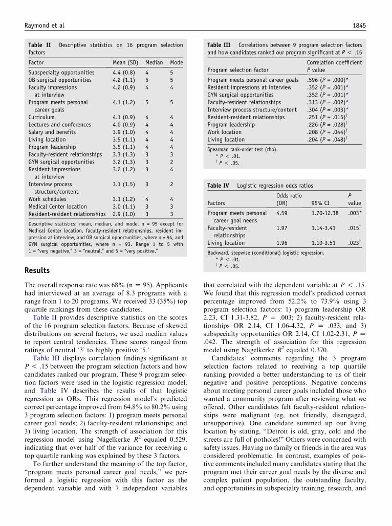
Raymond et al 1845
Results
The overall response rate was 68% (n Z 95). Applicantshad interviewed at an average of 8.3 programs with arange from 1 to 20 programs. We received 33 (35%) topquartile rankings from these candidates.
Table II provides descriptive statistics on the scoresof the 16 program selection factors. Because of skeweddistributions on several factors, we used median valuesto report central tendencies. These scores ranged fromratings of neutral ‘3’ to highly positive ‘5.’
Table III displays correlation findings significant atP ! .15 between the program selection factors and howcandidates ranked our program. These 9 program selec-tion factors were used in the logistic regression model,and Table IV describes the results of that logisticregression as ORs. This regression model’s predictedcorrect percentage improved from 64.8% to 80.2% using3 program selection factors: 1) program meets personalcareer goal needs; 2) faculty-resident relationships; and3) living location. The strength of association for thisregression model using Nagelkerke R2 equaled 0.529,indicating that over half of the variance for receiving atop quartile ranking was explained by these 3 factors.
To further understand the meaning of the top factor,‘‘program meets personal career goal needs,’’ we per-formed a logistic regression with this factor as thedependent variable and with 7 independent variables
Table II Descriptive statistics on 16 program selectionfactors
Factor Mean (SD) Median Mode
Subspecialty opportunities 4.4 (0.8) 4 5OB surgical opportunities 4.2 (1.1) 5 5Faculty impressionsat interview
4.2 (0.9) 4 4
Program meets personalcareer goals
4.1 (1.2) 5 5
Curriculum 4.1 (0.9) 4 4Lectures and conferences 4.0 (0.9) 4 4Salary and benefits 3.9 (1.0) 4 4Living location 3.5 (1.1) 4 4Program leadership 3.5 (1.1) 4 4Faculty-resident relationships 3.3 (1.3) 3 3GYN surgical opportunities 3.2 (1.3) 3 2Resident impressionsat interview
3.2 (1.2) 3 4
Interview processstructure/content
3.1 (1.5) 3 2
Work schedules 3.1 (1.2) 4 4Medical Center location 3.0 (1.1) 3 3Resident-resident relationships 2.9 (1.0) 3 3
Descriptive statistics: mean, median, and mode. n = 95 except for
Medical Center location, faculty-resident relationships, resident im-
pression at interview, and OB surgical opportunities, where n = 94, and
GYN surgical opportunities, where n = 93. Range 1 to 5 with
1 = ‘‘very negative,’’ 3 = ‘‘neutral,’’ and 5 = ‘‘very positive.’’
that correlated with the dependent variable at P ! .15.We found that this regression model’s predicted correctpercentage improved from 52.2% to 73.9% using 3program selection factors: 1) program leadership OR2.23, CI 1.31-3.82, P Z .003; 2) faculty-resident rela-tionships OR 2.14, CI 1.06-4.32, P Z .033; and 3)subspecialty opportunities OR 2.14, CI 1.02-2.31, P Z.042. The strength of association for this regressionmodel using Nagelkerke R2 equaled 0.370.
Candidates’ comments regarding the 3 programselection factors related to receiving a top quartileranking provided a better understanding to us of theirnegative and positive perceptions. Negative concernsabout meeting personal career goals included those whowanted a community program after reviewing what weoffered. Other candidates felt faculty-resident relation-ships were malignant (eg, not friendly, disengaged,unsupportive). One candidate summed up our livinglocation by stating, ‘‘Detroit is old, gray, cold and thestreets are full of potholes!’’ Others were concerned withsafety issues. Having no family or friends in the area wasconsidered problematic. In contrast, examples of posi-tive comments included many candidates stating that theprogram met their career goal needs by the diverse andcomplex patient population, the outstanding faculty,and opportunities in subspecialty training, research, and
Table III Correlations between 9 program selection factorsand how candidates ranked our program significant at P ! .15
Program selection factorCorrelation coefficientP value
Program meets personal career goals .596 (P = .000)*Resident impressions at interview .352 (P = .001)*GYN surgical opportunities .352 (P = .001)*Faculty-resident relationships .313 (P = .002)*Interview process structure/content .304 (P = .003)*Resident-resident relationships .251 (P = .015)y
Program leadership .226 (P = .028)y
Work location .208 (P = .044)y
Living location .204 (P = .048)y
Spearman rank-order test (rho).
* P ! .01.y P ! .05.
Table IV Logistic regression odds ratios
FactorsOdds ratio(OR) 95% CI
Pvalue
Program meets personalcareer goal needs
4.59 1.70-12.38 .003*
Faculty-residentrelationships
1.97 1.14-3.41 .015y
Living location 1.96 1.10-3.51 .023y
Backward, stepwise (conditional) logistic regression.
* P ! .01.y P ! .05.

1846 Raymond et al
the pursuit of fellowships. Some applicants felt faculty-resident relationships were good after conversing withthem during the interviews, discussing faculty relation-ships with current or past residents, or from theirpersonal clinical experiences affiliating with our pro-gram. Our living location was considered positively byindividuals who grew up in the area, or had relatives andfriends nearby. Some desired the area because of em-ployment opportunities for spouses, excellent publicschools, and competitive living costs compared withmany other large cities.
Our reliability and validity results supported the utilityof the measures selected. Leave-one-out cross-validationanalysis indicated that findings were likely to be gener-alizable. The technique showed similar results comparingoriginal grouped cases with cross-validated groupedcases as follows. Correct classification of the originalgroup was 72.8% in comparison to 70.7% for the cross-validated group. Five faculty reviewers unanimouslyagreed that the survey items represented residency pro-gram selection factors to confirm face validity.
Comment
Of the 16 program selection factors that we assessed,1 stood out as most important in relationship to receiptof candidates’ top quartile rankings. That programselection factor was how well the program met ourcandidates’ personal career goal needs. To explore thisconcept further we looked at our candidates’ remarks.These remarks revealed that a university setting, aprogram that cared for a diverse and complex patientpopulation, offered research and subspecialty training,and gave them skills to help pursue fellowships oftenexplained how their career goals were met by theprogram. Another logistic regression revealed that ‘‘out-standing faculty’’ members through strong programleadership and supportive faculty-resident relationshipswere key characteristics that helped define this factor. Inaddition, this logistic regression indicated that availabil-ity of subspecialty training was another critical compo-nent to meeting our candidates’ personal career goalneeds. This analysis also revealed to us that faculty-resident relationships were even more important thanthe initial findings showed. Faculty-resident relation-ships actually were intertwined with meeting candidate’spersonal career goal needs, as well as standing alone inrelationship to our receipt of top quartile rankings fromcandidates.
The high importance candidates placed on our pro-gram meeting their personal career goal needs indicatedthat candidates considered their professional aspirationsabove personal ones when selecting and ranking resi-dency programs. Nevertheless, the learning environmentand lifestyle issues also played into decisions to find the
right site. A remarkably strong association with receiv-ing a top quartile ranking (ie, Nagelkerke R2 Z 0.529)was obtained when a high rating on the factor ‘‘programmeets candidates’ personal career goal needs’’ combinedwith good ‘‘faculty-resident relationships’’ and a suitable‘‘living location.’’ In order to receive top quartile rank-ings from candidates, they needed to feel that our siteprovided major educational resources in an enhancedlearning environment, and in a location that supportstheir personal lifestyles.
When we looked at central tendency scores for eachprogram selection factor (Table II), the results showedus that we could work to improve candidates’ ratings onall program selection factors. Two factors on which wefocused most were highlighting the benefits of living inSoutheastern Michigan, and working internally to im-prove faculty-resident relationships.
Because in the real world we could not wait 4 years torespond to our recruitment needs, we made changesaccording to preliminary findings from annual surveys.One limitation of this study is that these actions mayhave colored the interpretation of the overall results.However, our work in using these surveys has met withsuccess indicating the study’s reliability and validity.After 3 years of post-match activities to handle vacantpositions, we filled all NRMP openings in 2004 and2005. Our interview numbers of candidates who meet orexceed our admittance standards have increased from 45in past years to 70 in 2005.
Other limitations of our study included small num-bers of participants. This necessitated the modificationof the logistic regression procedures previously de-scribed. In addition, we did not have the demographicinformation available to assess and compare nonre-sponders with responders in order to determine whetherresponse bias occurred. Finally, changes in the applicantpool, the resident pool, and the curriculum were notincorporated into the model.
An important difference in our study from others wasthat no other study used our survey item entitled‘‘program meets candidates’ personal career goalneeds.’’ Our qualitative and quantitative findings helpedus to interpret the meaning of this survey item to reflectother investigations’ descriptions of program quality/reputation (eg, outstanding faculty, opportunities insubspecialty training, diverse and complex patient pop-ulation, research, and pursuit of fellowships).3-5,9,11,14
Thus, we join those authors who have found programquality/reputation as a key aspect of resident recruit-ment. We add that a program’s quality/reputationincludes meeting candidates’ professional career goalneeds through maintaining outstanding faculty andproviding subspecialty opportunities.
While several investigators6,8,9,14 have found interper-sonal relationships were more important than programquality/reputation as the number 1 ranked program

Raymond et al 1847
selection factor, our study found interpersonal relation-ships to be significant, but of lesser importance thanprogram quality/reputation. Despite controversy overranked importance, the identification of interpersonalrelationships as a fundamental program selection factorhas been established by other studies.3-5,8,9,11,12 Further,our results confirm the findings of many others thatlocation remains an important program selection fac-tor.3,5,6,9-15 The identification of similar important pro-gram selection factors despite differences in study designssupports the validity of these results.
The information gained from these surveys is similarto market research. It has proven a rich and valuableresource in identifying program selection factors ourapplicants consider when ranking us on their match lists.Other programs can adapt this process to assess theirrecruitment needs or use our findings. These surveyshave provided us with timely feedback from the sourcethat matters to us mostdthe NRMP candidates wewanted to join our program. We believe their inputhelped us focus on many of the issues that led to ourprevious poor match results. The most valuable lessonlearned is that if we meet our candidates’ needs, we meetour program’s needs for recruiting best candidates.
Acknowledgments
Special thanks go to several people who helped with thisinvestigation. Alaina Bruce conducted the surveys from2002 to2004.MichaelKrugerassistedwith thedataanalysisand interpretations. Gloria Kuhn, MD, provided numer-ouswriting tips and editing suggestions. JohnM.Malone,Jr,MD, and Theodore B. Jones,MD, supplied support tokeep me on target and energized me to complete thisproject. The APGO Solvay Scholars program providedstructure and helpful information. Finally, appreciationis due to each of the 95 candidates who took the time toparticipate in this study and share their thoughts.
References
1. Lostumbo EM, Beran RL. Results of the National Resident
Matching Program for 2001. Acad Med 2001;76:665-8.
2. Signer MM, Beran RL. Results of the National Resident Matching
Program for 2004. Acad Med 2004;79:610-2.
3. Diebold SE, David CT, Ferraro CM. Factors influencing appli-
cants’ match rank order. Acad Emerg Med 1995;2:328-30.
4. DeLisa JA, Jain SS, Campagnolo D, McCutcheon PH. Selecting a
physical medicine and rehabilitation residency. Am J Phys Med
Rehabil 1992;71:72-6.
5. DiTomasso RA, DeLauro JP, Carter ST Jr. Factors influencing
program selection among family practice residents. J Med Educ
1983;58:527-33.
6. Flynn TC, Gerrity MS, Berkowitz LR. What do applicants look
for when selecting internal medicine residency programs? A com-
parison of rating scale and open-ended responses. J Gen Intern
Med 1993;8:249-54.
7. Hitchcock MA, Kreis SR, Foster BM. Factors influencing student
selection of family practice residency programs in Texas. FamMed
1989;21:122-6.
8. Laskin DM, Lesny RJ, Best AM. The residents’ viewpoint of the
matching process, factors influencing their program selection, and
satisfaction with the results. J Oral Maxillofac Surg 2003;61:
228-33.
9. Lebovits A, Cottrell JE, Capuano C. The selection of a residency
program: prospective anesthesiologists compared to others. Anesth
Analg 1993;77:313-7.
10. Marciani RD, Smith TA, Heaton LJ. Applicants’ opinions about
the selection process for oral and maxillofacial surgery programs.
J Oral Maxillofac Surg 2003;61:608-14.
11. Mayeaux EJ Jr, Arnold J. Why first-year family practice residents
choose their residency programs. Fam Med 1993;25:253-6.
12. Nuthalapaty FS, Jackson JR, Owen J. The influence of quality-of-
life, academic, and workplace factors on residency program
selection. Acad Med 2004;79:417-25.
13. Pretorius ES, Hrung J. Factors that affect National Resident
Matching Program rankings of medical students applying for
radiology residency. Acad Radiol 2002;9:75-81.
14. Simmonds AC, Robbins JM, Brinker MR, Rice JC, Kerstein MD.
Factors important to students in selecting a residency program.
Acad Med 1990;65:640-3.
15. Sledge WH, Leaf PJ, Sacks MH. Applicants’ choice of a residency
training program. Am J Psychiatry 1987;144:501-3.
16. Weissman SH, Bashook PG. The 1982 first-year resident in
psychiatry. Am J Psychiatry 1984;141:240-3.
17. Schwartz RW, Jarecky RK, Strodel WE, Haley JV, Young B,
Griffen WO, et al. Controllable lifestyle: a new factor in career
choice by medical students. Acad Med 1989;64:606-9.
18. Bland KI, Isaacs G. Contemporary trends in student selection of
medical specialties: the potential impact on general surgery. Arch
Surg 2002;137:259-67.
19. American College of Obstetrics and Gynecology. Guidelines for
pursuing a residency program. Available at: http://www.acog.org.
Accessed 2001.
20. Stone M. Asymptotics for and against cross-validation. Biometrica
1977;64:29-35.
21. Carmines EG, Zeller RA. Reliability and validity assessment.
Quantitative applications in the social sciences, Newbury Park,
CA: Sage University Press; 1979.
American Journal of Obstetrics and Gynecology (2005) 193, 1848–51
www.ajog.org
Educational games in an obstetrics and gynecologycore curriculum
Sharon O’Leary, MD,a,b,* Lisa Diepenhorst, MD,a Ruth Churley-Strom, MS, RN,a
Diane Magrane, MDc
Department of Obstetrics and Gynecology,a St Joseph Mercy Hospital, Ypsilanti, MI; The University of MichiganSchool of Medicineb; Faculty Development and Leadership Programs,c Association of American Medical Colleges,Ann Arbor, MI
Received for publication February 18, 2005; revised July 18, 2005; accepted July 18, 2005
KEY WORDSGame theory in
medical education
Interactive learning ofectopic pregnancy
Objective: The purpose of this study was to compare learning satisfaction and effectiveness using
traditional lecture or educational game in teaching medical students about ectopic pregnancy.Study design: Third-year medical students were randomized to instruction about ectopicpregnancy through either standard lecture or educational Jeopardy style game. Students in each
group completed a pretest, posttest, and satisfaction survey. Experts in ectopic pregnancyvalidated the pretest and posttest. The satisfaction survey was taken from published validatedtests. Paired samples t test was used to compare pretest and posttest scores. Independent samplest tests were used to compare test scores and satisfaction responses between groups. Chi-square
tested dichotomous satisfaction responses.Results: All 104 students in both groups showed significant improvement in learning about ectopicpregnancy (P! .001) on pre- and posttest comparison, with scores being almost identical. Students
in the group randomized to game format rated it higher in stimulating faculty/student interaction,helping retain information, and overall enjoyment than students participating in the lecturemethod(P ! .001). In addition, students in the game group responded positively that the format was
interactive, stimulated their interest, and kept them engaged in class content (P ! .001).Conclusion: The innovative educational game format was as effective as standard lecture ineducating students about ectopic pregnancy, while being more enjoyable and stimulating. Based onthese conclusions, we hope to motivate other teachers in obstetrics and gynecology to use
innovative teaching methods to provide a more enjoyable, stimulating, and active means ofeffective medical education.� 2005 Mosby, Inc. All rights reserved.
Creative teaching techniques engage students, stimu-late participation, and sustain their interest. Educationalgames meet these criteria and can serve as an additional
* Reprint requests: Sharon O’Leary, MD, Department of Obstet-
rics and Gynecology, St Joseph Mercy Hospital, Reichert Health
Center, 5333 McAuley Drive, R-2108, Ypsilanti, MI 48197.
E-mail: [email protected]
0002-9378/$ - see front matter � 2005 Mosby, Inc. All rights reserved.
doi:10.1016/j.ajog.2005.07.059
interactive model for enhancing the academic environ-ment. Games are effective, stimulating, and encourageactive participation.1 A variety of educational gameshave been used in medical education, with approachesincluding a card game to teach immunology,2 a ‘‘Survi-vor’’ game to review pulmonary physiology,3 and a‘‘Scruples’’ game to teach managed care ethics.4 The

O’Leary et al 1849
specific game format or the topic is not as important asthe novel approach to teaching that encourages activeparticipation, and offers a respite from the world of‘‘hum-drum’’ didactic lectures, which is the usual do-main of the medical students.
While educational games have been found to beeffective and enjoyable in medical education, none havebeen studied while teaching obstetrics and gynecologytopics to medical students. Currently, the standardmethod of instruction of third-year medical students isthe didactic lecture. The goal of this study was to evaluatestudent learning and student satisfaction with an inter-active game compared with a standard lecture format inteaching medical students about ectopic pregnancy.
Material and methods
This study was deemed exempt from Institutional Re-view Board review because it involved research con-ducted in established or commonly accepted educationalsettings dealing with normal educational practices, suchas research on instructional strategies and evaluation ofthe effectiveness of the strategies.
The University of Michigan academic schedulingsystem assigns 32 to 37 students to an 8-week clerkshipin obstetrics and gynecology. The first 5 days aredevoted to orientation activities and classes. On day 1,the investigators used a random numbers table to assignstudents to 1 of 2 methods for instruction on ectopicpregnancy. The principal investigator taught both ses-sions, the standard lecture with open questions andanswer period, and the game format. The class sessionsimmediately followed each other. Thus, for the openingweek of each ob-gyn clerkship, 16 to 18 studentsattended a lecture/discussion on ectopic pregnancy and16 to 18 students participated in a game organized toinstruct about ectopic pregnancy. The principal investi-gator prepared both lecture and game using Associationof Professors of Gynecology and Obstetrics (APGO)learning objectives, with careful cross-referencing ofteaching points to ensure consistency in content pre-sented. All students were encouraged to ask questions atthe end of each session.
Students completed a 20-item multiple-choice pretestto assess their general knowledge about ectopic preg-nancy. The test questions were developed by the prin-cipal investigator and included 2 questions extractedfrom the APGO test bank. Face and content validitywere judged by teaching faculty from the University ofMichigan and St Joseph Mercy Health System based onthe 5 APGO teaching objectives for ectopic pregnancy.
After students completed the pretest, those assignedto the lecture format received a standard 45-minutelecture with supporting slides by the principal investi-gator. Students assigned to the game format weredivided into 4 teams. The principal investigator pro-
vided the game rules, moderated the ‘‘Jeopardy’’ stylegame, and elaborated on correct answers. The gamecategories of 1) epidemiology and differential diagnosis,2) risk factors, 3) signs/symptoms, 4) diagnosis, and 5)treatment matched the APGO teaching objectives.Cards with increasing dollar values were displayed onthe game board below these categories. Teams wereawarded points based on the degree of difficulty andcomplexity of the question they selected. The team withthe most points received a high-energy edible reward.
Immediately following each class, students completedthe posttest and a satisfaction survey consisting of 4questions using a 5-point Likert scale and 3 true-falsequestions. Students were additionally queried about theirinterest in obstetrics and gynecology as a specialty andthe number of hours spent preparing for the class. Thesatisfaction survey was based on validated satisfactionsurveys,5-7 and judged to have face and content validityby an educational researcher and a faculty mentorinvolved in the APGO/Solvay Scholars program.
A power analysis was done to determine a sample sizerequired to achieve a minimum power of 0.80. Calcula-tions using nQuery Advisor Release 4.0: Study PlanningSoftware 1995-2000 (Statistical Solutions, Ltd, Cork,Ireland) identified that a sample size of 20 in each groupwould have 92% power to detect a difference in meansof 3 assuming that the common standard deviation is2.700 using a 2 group t test with a 0.050 2-sidedsignificance level.8
Pre- and posttest scores, as well as survey responsesrating increase in general knowledge, stimulation offaculty/student interaction, retention of information,and enjoyment of the format were compared betweenlecture and game methods by independent samples t test.Paired samples t test was used to compare studentpretest and posttest percent correct scores. A chi-squarestatistic was used to compare true or false responseswith questions about the interactive nature of thepresentation, the appropriateness of the method usedfor teaching medical facts about ectopic pregnancy, andstimulation of student interest and engagement in thelecture and game groups.
Results
During the study period, a total of 104 students wereassigned to the ob/gyn clerkship. All students whoarrived for the class agreed to take part in the study.Fifty-two students participated in the lecture and 52students in the game classes.
Mean pretest scores were 11.33 (56.6%) for studentsin the lecture group and 12.52 (62.6%) for students ingame format group. Although there was a significantdifference in pretest scores between groups (P Z .005),students in both groups demonstrated improvement inlearning with similar mean posttest scores of 16.98
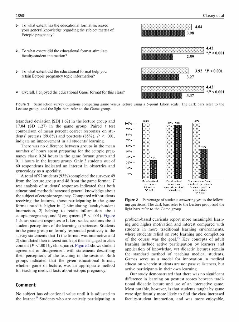
1850 O’Leary et al
(standard deviation [SD] 1.62) in the lecture group and17.04 (SD 1.27) in the game group. Paired t testcomparison of mean percent correct responses on stu-dents’ pretests (59.6%) and posttests (85%), P ! .001,indicate an improvement in all students’ learning.
There was no difference between groups in the meannumber of hours spent preparing for the ectopic preg-nancy class: 0.24 hours in the game format group and0.11 hours in the lecture group. Only 3 students out of60 respondents indicated an interest in obstetrics andgynecology as a specialty.
A total of 97 students (93%) completed the surveys: 49from the lecture group and 48 from the game format. Ttest analysis of students’ responses indicated that botheducational methods increased general knowledge aboutthe subject of ectopic pregnancy. Comparedwith studentsreceiving the lectures, those participating in the gameformat rated it higher in 1) stimulating faculty/studentinteraction, 2) helping to retain information aboutectopic pregnancy, and 3) enjoyment (P ! .001). Figure1 shows student responses to Likert-scale questions aboutstudent perceptions of the learning experiences. Studentsin the game group uniformly responded positively to thesurvey statements that 1) the format was interactive and2) stimulated their interest and kept them engaged in classcontent (P! .001 by chi-square). Figure 2 shows studentagreement or disagreement with statements describingtheir perceptions of the teaching in the sessions. Bothgroups indicated that the given educational format,whether game or lecture, was an appropriate methodfor teaching medical facts about ectopic pregnancy.
Comment
No subject has educational value until it is adjusted tothe learner.9 Students who are actively participating in
Figure 1 Satisfaction survey questions comparing game versus
Lecture group, and the light bars refer to the Game group.
problem-based curricula report more meaningful learn-ing and higher motivation and interest compared withstudents in more traditional learning environments,where students relied on rote learning and completionof the course was the goal.10 Key concepts of adultlearning include active participation by learners andapplication of knowledge, yet didactic lectures remainthe standard method of teaching medical students.Games serve as a model for innovation in medicaleducation wherein students are not passive listeners, butactive participants in their own learning.
Our study demonstrated that there was no significantdifference in learning on posttest scores between tradi-tional didactic lecture and use of an interactive game.Most notable, however, is that students taught by gamewere significantly more likely to find the class increasedfaculty-student interaction, and was more enjoyable,
lecture using a 5-point Likert scale. The dark bars refer to the
Figure 2 Percentage of students answering yes to the follow-
ing questions. The dark bars refer to the Lecture group and thelight bars refer to the Game group.

O’Leary et al 1851
stimulating, and interactive. Based on our findings wehope to motivate other teachers of obstetrics and gyne-cology to develop innovative strategies that provideenjoyable effective teaching methods.
This study focused on preexisting knowledge, learn-ing, and short-term retention through use of pre- andposttests. Future studies to test long-term recall ofcontent taught using an educational game format wouldadd evidence to the value of educational games.
References
1. Schwartzman RJ. Gaming serves as a model for improving
learning. Education 1997;118:9-18.
2. Steinman RA, Blastos MT. A trading-card game teaching about
host defense. Med Educ 2002;36:1201-8.
3. Howard MG, Collins HL, DiCarlo SE. ‘‘Survivor’’ torches ‘‘Who
Wants To Be A Physician?’’ in the educational games rating war.
Adv Physiol Educ 2002;26:30-6.
4. Baldor RA, Field TS, Gurwitz JH. Using the ‘‘Question of
Scruples’’ game to teach managed care ethics to students. Acad
Med 2001;76:510-1.
5. Mann BD, Eidelson BM, Fukuchi SG, Nissman SA, Robertson S,
Jardines L. The development of an interactive game-based tool for
learning surgical management algorithms via computer. Am J Surg
2002;183:305-8.
6. Copeland HL, Stoller JK, Hewson MG, Longworth DL. Making
the continuing medical education lecture effective. JCE Health
Professions 1998;18:227-34.
7. Edwards C, Fritz JH. Evaluation of three educational online
delivery approaches. Proceedings of the Second Annual Mid-
South Instructional Technology Conference; 1997 April 6-8;
Middle Tennessee State University. Available at: http://
www.mtus.edu/∼itconf/proceed97/online.3. Accessed August
11, 2005.
8. Elashoff JD. nQuery Advisor Release 4.0: study planning software,
Statistical Solutions, Ltd, Cork, Ireland; 1995-2000.
9. Dewey J. Experience and education. New York: Collier; 1938.
10. Newble DI, Clarke RM. The approaches to learning of students in
traditional and in an innovative problem-based medical school.
Med Educ 1986;20:267-73.

American Journal of Obstetrics and Gynecology (2005) 193, 1852–5
www.ajog.org
Medical student identification of domestic violenceas measured on an objective, standardized clinicalexamination
Susan E. Hoffstetter, PhD, WHNP, Robert J. Blaskiewicz, MD,Gail E. Furman, PhD, Jennifer A. McCabe, BS
Department of Obstetrics, Gynecology, and Women’s Health, Saint Louis University School of Medicine, St Louis, MO
Received for publication February 18, 2005; revised May 26, 2005; accepted July 18, 2005
KEY WORDSStandardized patientsDomestic violence
Medical studenteducation
Objective: This study examined the incidence and predictors of domestic violence screening by
third-year medical students at an end of clerkship Objective Standardized Clinical Examination.Study design: Two hundred and seventy-five third-year medical students completed an 8-stationend of clerkship Objective Standardized Clinical Examination as part of this retrospective
observational study, one with nonspecific abdominal pain and possible domestic violence.Checklists on history, physical, communication, and interpersonal skills were collected. Domesticviolence screening was analyzed by logistic regression and analysis of variance.Results: The incidence of domestic violence screening by history alone was 34% before the
physical. Interpersonal scores on the overall exam and domestic violence station, but not genderor rotation sequence, predicted domestic violence questioning.Conclusion: In this standardized patient study there was a low rate of domestic violence screening
by history.� 2005 Mosby, Inc. All rights reserved.
Domestic violence is a public health problem, a majorcause of morbidity and mortality in the United States,and a serious social problem impeding women’s partic-ipation and contributions to society.1 Domestic violenceis used to establish power and control over anotherperson through actual or threatened physical/sexualviolence or psychologic/emotional abuse and its effectscross ethnicity, race, age, national origin, sexual orien-tation, and religious and socioeconomic lines.2 Approx-
Presented at the Annual Meeting of the Association of Professors
of Gynecology and Obstetrics, and the Council on Resident Education
in Obstetrics and Gynecology, March 2-5, 2005, Salt Lake City, UT.
Reprints not available from the authors.
0002-9378/$ - see front matter � 2005 Mosby, Inc. All rights reserved.
doi:10.1016/j.ajog.2005.07.058
imately 1 to 4 million women a year in the United Statesare abused by an intimate partner.3,4 These womensuffer immediate and long-term physical and mentalhealth consequences, accruing an estimated cost of 4.1billion dollars for medical and mental health care.Women experiencing violence have 50% higher medicalcare costs compared with women not currently experi-encing violence.5
The need for violence education was first emphasizedin the 1985 Surgeon General’s Workshop on Violenceand Public Health. In 1995, the Association of AmericanMedical Colleges challenged medical schools to createthe domain of a family violence curriculum comparableto traditional subjects in whom students would gain the

Hoffstetter et al 1853
ability to screen, assess, and support victims of violence,as well as develop community support networks, re-sources, and information on health care professionalinvolvement.6
Medical schools have adopted varying formats fromlectures and workshops to training sessions to addressthe topic. There is little information on the incidence ofscreening for domestic violence by medical studentsin the clinical setting. Objective Standardized ClinicalExamination (OSCE) evaluations are now frequentlyused to measure clinical skills, and successful completionof a standardized patient examination is required forlicensure. The purpose of this study was to determinethe incidence and predictors of domestic violence screen-ing by third-year medical students at an end of clerkshipOSCE.
Material and methods
The study was a retrospective, observational studyapproved by the Institutional Review Board of SaintLouis University conducted for the academic years of2002 and 2003. All third-year medical students (n =276) completed an 8-station OSCE at the end of theobstetric and gynecology clerkship. One student wasexcluded from the analysis because of insufficient data,leaving 275 students in the study. The formal curriculumat Saint Louis University during the time of this studyincluded a preclinical presentation on domestic violenceand a didactic session during the ob/gyn clerkship.
One of the OSCE stations presented a patientcomplaining of nonspecific abdominal pain with anassociated abdominal bruise. Domestic violence was aconsideration in the differential diagnosis of this patient.The standardized patient was trained to offer informa-tion about her abusive relationship if the student askedspecifically about domestic violence. A simulated bruisewas placed on the patient’s abdomen that was clearlyvisible once the abdomen was properly exposed duringthe physical examination. If the student asked about thebruise the standardized patient was instructed to conveythe information about her domestic violence. In theabsence of a specific question about domestic violence ora question concerning the bruise the student did notreceive that information.
Data were collected on a subset of 236 consecutivestudents by timing of domestic violence inquiry (beforethe physical examination, during/after the exam, or notat all). The gender of the student was recorded. The timeof the students obstetric and gynecology rotation in theacademic year was noted. Rotations were grouped intoquarters of the academic year for analysis.
Data collected on all students for the domesticviolence station included a patient-completed historychecklist about the character of her abdominal pain andassociated symptoms, in addition to domestic violence
questions. The student was also assessed on their com-munication response to the patient’s domestic violencesituation, including the student’s ability to offer re-sources and reassurance, and identify patient safety. Thestudents’ interpersonal skills were assessed by the use ofa previously validated tool, Patient Perception Ques-tionnaire (PPQ).
Data were collected from the 8 stations across thedomains of history taking, physical examination skills,communication, diagnosis, treatment, and PPQ. OverallOSCE scores were calculated by both including andexcluding the domestic violence station. The incidenceof domestic violence screening was calculated by includ-ing the number of students asking about domesticviolence at any time during the patient encounter. Statis-tical analysis was completed using multivariate analysisof variance (MANOVA), univariate analysis (ANOVA),Bonferroni-adjusted pairwise mean comparisons, andsimultaneous-entry, multiple linear regression.
Results
Of the 275 students, 243 (88.4%) inquired about do-mestic violence with at least 1 question at some point inthe standardized patient encounter. In the subset of 236students for whom the timing of this inquiry was known,80 students (33.9%) asked about domestic violencebefore the physical exam, 133 students (56.4%) askedabout domestic violence during or after the physicalexam, and 23 students (9.7%) never asked. The percent-age of students in each of these 3 categories did not varysignificantly by student gender, c2(2) = 1.9, P = .38.Similarly, rotation by academic quarter was not signif-icantly associated with the timing of the domesticviolence questions, c2(6) = 8.6, P = .20.
Multivariate analysis of variance (MANOVA) wasused to determine whether timing of domestic violencequestions (before exam vs during/after exam vs never)was associated with other indicators of clinical perfor-mance. The MANOVA included the OSCE total scores(omitting the domestic violence station) for historytaking, physical examination, communication, PPQ, anddiagnosis/treatment. The PPQ score for the domesticviolence station was also included as a separate variable.The multivariate effect was significant across the 6dependent variables, Wilks’ lambda = 0.83, P ! .001.Follow-up univariate ANOVAs indicated that of the 6dependent variables, only the PPQ score for the domesticviolence station was significantly different as a functionof domestic violence question timing, F(2,233) = 18.7,P ! .001. Descriptive data are shown in Table I.Bonferroni-adjusted pairwise mean comparisons indi-cated that students who never asked about domestic vio-lence had significantly lower PPQ scores on the domesticviolence station than students who asked about domesticviolence before or during/after the exam (P ! .05).

1854 Hoffstetter et al
Simultaneous-entry, multiple linear regression wasused to predict, from the other indicators of clinicalperformance, the number of domestic violence questionsasked by the student (out of 3 questions). The meannumber of domestic violence questions asked was 2.4(SD 1.0; range 0-3). Neither student gender nor rotationwas significantly correlated with number of domesticviolence questions asked: gender, r = 0.03, P = .53;rotation, r = �0.03, P = .54. These variables were notincluded in the regression analysis. The number ofdomestic violence questions asked was predicted fromthe OSCE total scores for history taking, physicalexamination, communication, PPQ, and diagnosis/treat-ment (again, omitting the domestic violence station).The PPQ score for the domestic violence station wasagain included as a separate predictor. The overallregression equation with six predictors was significant,R = 0.41, F(6,268) = 9.0, P ! .001. Among the 6predictors, only the PPQ score for the domestic violencestation significantly predicted number of domestic vio-lence questions asked, b (standardized regression coef-ficient) = 0.39, P ! .001. Results are presented inTable II.
Overall performance on the domestic violence station(total score combining history taking, physical exam,communication, and PPQ) was also examined. Neitherstudent gender nor rotation was significantly correlatedwith overall performance on the domestic violencestation: gender, r = 0.11, P = .16; rotation, r =0.003, P = .96. Using multiple linear regression, do-mestic violence station performance was predicted fromthe OSCE total scores for history taking, physicalexamination, communication, PPQ, and diagnosis/treat-ment (again, omitting the domestic violence station).The overall regression equation with 5 predictors wassignificant, R = 0.22, F(5,269) = 3.1, P ! .05. Among
Table I Clinical performance as a function of domesticviolence question timing
DV question timing
Before exam(n = 80)Mean (SD)
During/afterexam(n = 133)Mean (SD)
Never(n = 23)Mean (SD)
History taking* 63.3 (10.0) 63.1 (7.9) 64.6 (8.8)Physical exam* 68.3 (7.6) 67.2 (7.4) 68.1 (6.7)Communication* 67.2 (13.0) 69.1 (14.0) 68.8 (17.6)PPQ* 75.9 (8.1) 76.1 (8.2) 74.7 (5.7)Diagnosis/treatment*
86.0 (14.2) 83.3 (15.2) 80.9 (14.1)
PPQ DV stationy 81.4 (13.3) 76.8 (14.9) 60.5 (16.1)
* OSCE scores; values do not include data from the domestic
violence station.y Bonferroni-adjusted pairwise comparisons: never ! before exam,
during/after exam (P ! .05).
the 5 predictors, only the PPQ score (across all otherstations besides the domestic violence station) signifi-cantly predicted overall domestic violence station per-formance, b (standardized regression coefficient) =0.18, P ! .01. Results are displayed in Table III.
Comment
This study found a low incidence of domestic violencequestioning before the physical exam in a standardizedpatient simulating an episode of nonspecific abdominalpain. Medical students were better at domestic violencescreening after identification of an abnormal physicalfinding. The timing of the domestic violence questioningwas not influenced by gender or time in the academicyear. Students who were rated higher on the PPQ scoreon the domestic violence station were more likely toinquire about domestic violence. This would suggestthat students who have better interpersonal skills arebetter at screening and identifying patients at risk for anabusive situation. Alternatively, the standardized pa-tient may have perceived a more empathetic student ifthey inquired about domestic violence.
The depth of domestic violence inquiry was notinfluenced by gender or amount of previous clinicalexperience. Possible explanations could relate to per-sonality traits, violence exposure, personal experiences,
Table II Prediction of number of domestic violence ques-tions asked from clinical performance indicators (predictorvariables)
Predictor variables b t P
History taking* �0.06 �0.90 .37Physical exam* 0.06 0.91 .36Communication* 0.06 0.96 .34PPQ* 0.003 0.04 .96Diagnosis/treatment* 0.03 0.55 .58PPQ DV station 0.39 6.6 ! .001
* OSCE scores; values do not include data from the domestic
violence station.
Table III Prediction of overall performance on domesticviolence station from clinical performance indicators (predic-tor variables)
Predictor variables* b t P
History taking �0.11 �1.5 .13Physical exam 0.11 1.5 .13Communication 0.09 1.4 .15PPQ 0.18 2.6 .009Diagnosis/treatment �0.002 �0.03 .97
* OSCE scores; values do not include data from the domestic
violence station.

Hoffstetter et al 1855
and value attributed to the social history by role models.In our study the overall OSCE PPQ score was predictiveof the student’s performance on the domestic violencestation. This performance was unrelated to other aspectsof history taking and physical examination and suggestsa correlation between student’s ability to relate to thepatient and their score. There may be a factor inherentin the student’s physician patient relationship that is apredictor of domestic violence screening.
Standardized patients are widely used in medicaleducation. Our study used this tool to measure thestudent’s behavior regarding a significant social problemin America. The OSCE exam can be used to measure theeffect of curricular change addressing domestic violence.
The manifestations of partner violence touch allmedical specialties, reinforcing the need for all physi-cians to be trained in screening, intervening, and advo-cating support for violence victims. Medical studentsneed a consistent approach in developing the clinicalskills to enable screening for domestic violence. Studentsneed to incorporate domestic violence screening inroutine assessments of all patients.
Acknowledgments
We thank John T. Chibnall, PhD, Saint Louis Univer-sity School of Medicine, Associate Professor of Psychi-atry, for his assistance with statistical analysis.
References
1. Dalton C, Schneider E. Battered women and the law. New York
(NY): Foundation Press; 2001.
2. Centers for Disease Control and Prevention. Intimate partner
violence. Atlanta (GA): National Center for Injury Prevention
and Control; 2000.
3. Violence by intimates: Analysis of data on crimes by current or
former spouses, boyfriends, and girlfriends. Washington, DC: US
Department of Justice; 1998.
4. The Commonwealth Fund, First Comprehensive National Health
Survey of American Women. New York: The Commonwealth
Fund; 1993.
5. Coker A, Reeder C, Fadden M, Smith P. Physical partner violence
and Medicaid utilization and expenditures. Public health Rep
2004;119:557-67.
6. Association of American Medical Colleges. Conference on family
violence. Division of educational research and assessment. Wash-
ington, DC. Acad Med 1995;70.

American Journal of Obstetrics and Gynecology (2005) 193, 1856–65
www.ajog.org
A new curriculum for hysteroscopy training asdemonstrated by an objective structuredassessment of technical skills (OSATS)
Amy L. VanBlaricom, MD,* Barbara A. Goff, MD, Michael Chinn, MD,Melodie M. Icasiano, MD, Peter Nielsen, MD, Lynn Mandel, PhD
Departments of Obstetrics and Gynecology, University of Washington, Madigan Army Medical Center, Seattle, WA
Received for publication February 18, 2005; revised May 16, 2005; accepted July 18, 2005
KEY WORDSCurriculumHysteroscopy training
Surgical skillsReliabilityValidity
Objective: The purpose of this study was to develop an effective curriculum to teach
hysteroscopy, as well as to develop an objective assessment instrument (OSATS).Study design: All 24 residents in our training program attended a 3-hour hysteroscopy curriculumin each of 3 years. Each year after the curriculum, an OSATS was administered consisting of an
assembly and operative task. Each taskwas evaluated with a task-specific checklist and a previouslyvalidated global assessment form. Twenty-four residents from an outside institution served ascontrols. All residents were examined by blinded and unblinded examiners.Results: The reliability coefficient was .7857 for the checklist and .9763 for the global scale.
Interrater reliability for the checklist was .7478 and ranged from .4222 to .9329 for the globalinstruments. Evaluation of construct validity revealed that the junior residents at both locationsdid significantly worse on the checklist and global scale than the senior residents for all 3 years of
the study (P ! .001). Those residents provided the curriculum did significantly better on both thechecklist and the global scale than the resident controls with a mean of 45% versus 63% for thechecklist and 18.3 versus 24.9 for the global score. (P = .001 checklist, P= .007 global scale).
Conclusion: This curriculum is an effective way to impart knowledge and skill in the assembly anduse of the operative hysteroscope. The checklist developed for this OSATS has excellent reliabilityand construct validity.� 2005 Mosby, Inc. All rights reserved.
Surgical training is an important part of any resi-dency program. Historically, surgical training occurredin the operating room where residents first observe anattending perform procedures, and then take on increas-ing roles in the surgical cases under direct supervision.
* Reprint requests: Amy VanBlaricom, MD, Box 356460, Univer-
sity of Washington Medical Center, 1959 Pacific, Seattle, WA 98195.
E-mail: [email protected]
0002-9378/$ - see front matter � 2005 Mosby, Inc. All rights reserved.
doi:10.1016/j.ajog.2005.07.057
This type of training results in variable acquisition ofsurgical skills, often caused by inherent differences inattending technique, as well as variation in how mucheach resident is allowed to do in a given procedure.These differences are sometimes based on perceived‘‘difficulties’’ in the case, including patient factors suchas age, weight, or associated medical conditions, whichcarry with them a need to complete the procedure in atimely manner. Other differences in resident experience

VanBlaricom et al 1857
Figure 1 Hysteroscopy model.
are based on attending personal preference as well astheir own comfort level with the procedure they areperforming.
In the last 10 years, advances in minimally invasivesurgical techniques have been astounding. Endoscopicsurgeries are rapidly becoming more complex and tech-nically difficult and in some cases replace historicallytried and true procedures, which require laparotomy. Inmany cases, faculty are finding themselves with inade-quate experience with new technologies and, therefore,residents are presented with fewer opportunities toperform these new techniques on their own even withdirect supervision. Additionally, with the advent ofwork hour restrictions there is now an increasing con-cern about decreased surgical experience in residenttraining.1-4 Because a significant depth and breadth ofsuch experience is required for surgical privileges and iseven being considered for standardization in NationalBoard Exams, it is incumbent upon us as academicsurgeons to expose residents to the experience they needto demonstrate adequacy once they leave our program.The responsibility for training competent surgeons lies
heavily upon us in academics. There are excellent datato support the effectiveness of formal, objective teachingof surgical skills outside of the operating room.5-15
Studies in the general surgery literature indicate thatthis may be a more efficient way of teaching surgicalskills.12,16-18
Despite its recognized importance in the age ofminimally invasive surgery, little work has been doneinvestigating teaching of hysteroscopic skills.19-24 Forthe most part, investigations in this area have been indevelopment of realistic models and simulators, withminimal work in curriculum development and appro-priate assessment of skills.25,26 Our goal was to developan effective method to teach and evaluate knowledgeand technical skill in hysteroscopy in obstetrics andgynecology residents.
Material and methods
A 3-hour curriculum was developed, consisting of a1-hour didactic session covering surgical indications,

1858 VanBlaricom et al
Figure 2 Hysteroscopy curriculum dry lab.
contraindications, anticipated complications, and basicsof equipment and media options, followed by a 2-hourhands-on lab where residents were given an opportunityto assemble the diagnostic and operative hysteroscopesand review troubleshooting points in their use, as well asthe use of other endoscopic equipment such as cameraand light source (Figures 1 and 2). Each resident wasthen given an opportunity to practice assembly andresection with the operative hysteroscope on an appro-priate inanimate model with one-on-one supervisionfrom a faculty expert. The didactics were given eachyear in 3 sessions of 8 residents per session.
All 24 residents in our training program were exposedto the hysteroscopy curriculum in 2001, 2002, and 2004.Each year, approximately 4 to 6 months after the curric-ulum, a posttest was administered consisting of an
OSATS (Objective Structured Assessment of TechnicalSkills) where residents would demonstrate assembly of anoperative hysteroscope and hysteroscopic resection ofpathology from a realistic inanimate model in front of anexaminer (Figures 3 and4). Specifically, the residentswereasked to resect a large polyp from an inanimate uterinemodel. Reusable models were purchased from Simulu-tions (formerly Limbs and Things, Bristol, England) andcost approximately $20 per resident. Twenty-four resi-dents from an outside institution who were not providedwith the curriculum were tested similarly. These residentsserved as the untrained cohort.
In any residency program there is concern about biasin evaluation as the examiners have often worked closelywith the residents they are evaluating. Thus, an impor-tant component to the project was testing residents at

VanBlaricom et al 1859
Figure 3 Hysteroscopy OSATS assembly task.
Madigan Army Medical Center, where examiners fromthe UW were blinded to level of training. In addition, wehad faculty members from Madigan evaluate our resi-dents in a completely blinded fashion. Faculty at eachinstitution evaluated their own residents as well. Blindedand unblinded assessments were then compared. Previ-ous evaluation of blinded examiners demonstrated ahigh interrater reliability.27,28 In doing so, we deter-mined the true objectivity of the OSATS.
For this project, a 2-station OSATS was designed toassess diagnostic and surgical hysteroscopy. A task-specific checklist was developed for each station inclu-sive of steps of the procedure deemed necessary andappropriate to safe and effective completion of the task.
Each of the tasks in the OSATS was timed and evalu-ated with a pass/fail judgment, the task-specific checklistevaluation, and a previously validated global assessmentform16-18 (Figures 5 and 6). Residents could fail a taskbased on time (longer than 10 minutes to completethe task) or technique. The time limit was chosen at10-minutes per task as it was used in previous endo-scopic OSATS. Previous research in surgical educationhas demonstrated that reliability and validity of anassessment tool are important.16-18
By adding outside blinded examiners for the OSATS,we attempted to demonstrate a high interrater reliabil-ity. In addition, by looking at all residents in theprogram we attempted to show a difference across years.

1860 VanBlaricom et al
Figure 4 Hysteroscopy OSATS resection task.
That is, a PGY4 (postgraduate year) would be moreknowledgeable and technically adept than a PGY1(construct validity).
The reliability of each of the instruments was assessedby Chronbach’s alpha. Interrater reliability was calcu-lated using intraclass correlation coefficients.
Construct validity for each instrument was evaluatedby comparing scores among the residency levels. Thiswas accomplished with a one-way analysis of variancewith resident year as the independent variable. Thisstudy was approved for human subjects’ exemptiongiven that it involved assessment of educational tools.
Results
At our institution, residents begin their exposure tohysteroscopy in their R1 year. The amount of the proce-dure they are allowed to perform is determined by theindividual attending based on the level of difficulty andsome personal comfort. As part of the preliminaryresearch for this project, a survey was performed of all24 residents in our department. This revealed that resi-dents felt that in most cases they were lacking in hyster-oscopy experience, including discomfort with operativeindications and contraindications, assembly of the com-
plex operative hysteroscopy equipment, operative tech-nique, and management of complications. Residentsreported a range of between 21 and 45 hysteroscopicprocedures by the completion of the chief year. In manycases they were only the secondary surgeon, with theattending performing much of the technically difficultportion of the case. Initial testing of the residents at alllevels of training proved that hysteroscopy skills wereinadequate,with only 1 of 6 second-year residents and 3of6 chief residents able to assemble a resectoscope in anappropriate time frame. Only 1 chief resident was able tocorrectly name the type and strength of media used at ourinstitution for operative hysteroscopy, and none coulddescribewhy this particularmediawas chosen. The resultsof this survey clearly underscore the need for additionaltraining in hysteroscopy.
The OSATS instruments developed to assess hystero-scopic skills were evaluated for reliability and validity.The reliability coefficient was .7857 for the checklist and.9763 for the global scale. Interrater reliability for thetask specific checklist was .7478 and ranged from .4222to .9329 for the global instruments.
Evaluation of construct validity revealed that thesenior residents (PGY3 and 4) at both locations didsignificantly better on the checklist and global scale than

VanBlaricom et al 1861
Figure 5 Hysteroscopy Testing.

1862 VanBlaricom et al
Figure 6 Hysteroscopy.
the junior residents (PGY1 and 2) for all 3 years of thestudy. The mean scores for the checklist were 43% (PGY1),53% (PGY2), 68% (PGY3), and 70% (PGY4) (P ! .001).
Mean raw scores for the global scale were 16.4(PGY1), 22.0 (PGY2), 26.0 (PGY3), and 26.0 (PGY4)(P ! .001) (Table I). Among the residents provided the

VanBlaricom et al 1863
curriculum, the senior residents (PGY3 and PGY4) didsignificantly better on both the task-specific checklistand global scale than the junior residents (PGY1 andPGY2) (P ! .001) (Table II).
The next step was to evaluate the effectiveness of thecurriculum. Those residents provided the curriculum didsignificantly better on both the task-specific checklistand the global scale than those residents not providedthe curriculum, with a mean score of 45% versus 63%for the checklist and 18.28 versus 24.88 for the globalscore (P = .001 checklist, P = .007 global scale) (TableIII). This difference was also found when evaluatingresidents by PGY year (Table IV). There were a total of5 trained residents who participated in all 3 years of thestudy. When their individual scores were reviewed, therewas a significant improvement in global score for eachresident for each year they received the curriculum (P =.012). The improvement in the task-specific checklist wasalso a pronounced trend that approached significance(P = .059) (both in Table V).
The curriculum cost involved price of the models anddedicated faculty time. The reusable hysteroscopy pelvictrainer was purchased from Limbs and Things for $407.The replaceable advanced resectable uteri were pur-chased for $61 each and were used for training andOSATS of 3 residents per uterus. The hysteroscopic
Table II Construct validity
n Checklist Global scale
Junior residents (PGY1 and 2) 36 50% 19.14Senior residents (PGY3 and 4) 36 76% 29.94
P ! .001 P ! .001
Residents provided the curriculum by PGY year.
Table III Curriculum effectiveness trained versus untrainedresidents
Global scale Checklist
n Mean n Average score
Trained residents 72 24.88 72 63%Untrained residents 18 18.28 24 45%
P = .007 P = .001
Table I Construct validity
PGY year n Global scale Checklist
1 21 16.43 43%2 27 22.08 53%3 23 26.00 68%4 25 26.00 70%
P ! .001 P ! .001
Baseline results on checklist and global scale by PGY year for all
residents in study.
instruments, camera, and video towers were all bor-rowed from the operating room of the hospital wherethe program was conducted.
The largest expense of the curriculum was committedfaculty time. One dedicated faculty member providedthe 3-hour curriculum on 3 occasions (for 8 residentseach occasion) for each of the 3 years of the study (atotal of 9 hours per year). At each OSATS session therewere 2 faculty evaluators (1 blinded and 1 unblinded).Time for OSATS was 30 minutes per resident for a totalof 96 faculty hours (32 hours per year).
Residents who received the curriculum rated it veryhighly. They were asked to rate the course in terms of
Table IV Trained versus untrained results broken down bypostgraduate year
Trained Untrained P value
PGY1Checklist 51% 22% .046Global 18.67 10.83 .002
PGY2Checklist 53% 51% .842Global 22.38 20.00 .620
Junior PGY (1 and 2)Checklist 52% 36.5% .030Global 20.83 13.89 .023
PGY3Checklist 73% 52% .018Global 27.23 19.00 .198
PGY4Checklist 75% 55% .053Global 30.42 24.50 .096
Senior PGY (3 and 4)Checklist 74% 54% .002Global 28.91 22.67 .055
Table V Individual improvement in residents participatingin all 3 years of the curriculum
Resident Year of curriculum Global score Checklist score
1 2001 17 .421 2002 25 .391 2004 34 1.02 2001 24 .622 2002 27 .682 2004 29 .653 2001 18 .543 2002 19 .583 2004 32 .524 2001 16 .314 2002 28 .524 2004 35 .875 2001 31 .855 2002 35 .485 2004 35 .96
P = .012 P = .059

1864 VanBlaricom et al
applicability to their education, effectiveness of impart-ing content, and perceived results (that is, were they ableto integrate knowledge gained into their clinical practicein a positive way). On a 5-point scale the results of thisrating were all 4.5 or higher. Notable comments in-cluded a significant improvement in comfort with theequipment and troubleshooting.
Comment
There is mounting evidence in the surgical literature tosupport the use of skills labs outside of the operatingroom for resident training.11-15 Cundiff found that acurriculum consisting of didactics as well as laparo-scopic skills training in a trainer and pig lab settingresulted in significant improvement in ob-gyn residents’performance in laparoscopic surgery.13 Similarly, Cole-man and Muller11 found a significant improvement intechnical skill and operative performance in a group ofupper level ob-gyn residents after a 4-week curriculumof intensive laparoscopic skills training. At the Univer-sity of Washington, Goff et al have demonstratedsimilar effectiveness with a comprehensive surgical skillscurriculum for ob-gyn residents.9,10,27,29
Procedures involving endoscopy lend themselves par-ticularly well to standardization via models. For exam-ple, the intrauterine cavity can be simulated on acomputer or in an inanimate model for resection withstriking similarity to human tissue. By using models forobjective surgical skills assessment, several advantagesare gained. First, residents can be allowed to operatecompletely independently. There is an opportunity tomake surgical decisions and demonstrate judgmentwithout encountering the obvious ethical dilemmas thesame would arouse on a human patient. Anotheradvantage is the ability to repeat the same procedureas many times as necessary with continuous feedbackuntil adequacy of skill and comfort with the equipmentare reached. There are economic advantages to thismethod, as well as the models are portable, it does notprolong operating room time, and the same equipmentcan be used repeatedly. Thus, an individual resident canactually perform as the primary surgeon many more of agiven procedure with this method. Competence is morequickly obtained while expending fewer resources.
Historically, evaluation of residents’ surgical skillshas been subjective evaluation by an individual attend-ing sporadically throughout the rotation or upon com-pletion of it. Often this assessment does not occur at thetime of a given surgery and therefore may lack effec-tiveness and objectivity caused by poor recollection ofindividual events by the attending. Because of variationsin attending personal style and individual patient fac-tors, subjective resident evaluation has been shown to beinconsistent and difficult to standardize, as well as
lacking reliability and validity.5 There are extensivedata in both the general surgery and gynecology liter-ature supporting objective evaluation of surgical skills asboth reliable and valid.5-10,14,16-18 Much of the researchin establishing OSATS for ob-gyn residents has beenaccomplished at our institution.9,27,29 In addition, thereliability and validity of this instrument has beenshown to be quite high, even when the providers ofthe exam were blinded to resident year of training andother personal details.27 Given this high reliability andvalidity, OSATS is applicable to other standardizedexam scenarios, including national board exams andother high stakes exams.
While the instruments used in this study had highreliability and validity, interrater reliability on the globalscale was significantly lower than seen previously. Theglobal scale used in this study has been previouslyvalidated extensively by Reznick,5 and UW stud-ies.9,14,27,29 Examiners from the institution whose resi-dents did not receive the curriculum were notconsistently the same examiners throughout the study,and they did not receive as much training in the use ofthe form as those at the University of Washington.Therefore, the low interrater reliability on the globalinstrument in this study could be caused by inadequatetraining in the use of the global form by new examiners.The task specific checklist developed for evaluation ofOSATS in this study showed high interrater reliabilitybetween blinded and unblinded examiners. Constructvalidity of the instrument used for the hysteroscopyOSATS was also good. We were able to show asignificant increase in baseline knowledge and technicalskill by advancing years in both trained and controlresidents.
The curriculum developed for this study was shownto be effective as, overall, those residents provided thecurriculum did significantly better on both the globalscale evaluation and the task-specific checklist at thetime of OSATS. When the individual PGY years werebroken down, there continued to be noteworthy trendsin improved acquisition of hysteroscopic knowledge andskill per postgraduate year, although this only becamestatistically significant when the junior residents andsenior residentswere compared as groups between trainedresidents and controls. Additional years of evaluationwillincrease the number of residents and, thus, the power ofthis study, which should improve our ability to showstatistical significance on an individual basis.
All residents were surveyed upon completion of eachyear of the study. There was an overwhelmingly positiveresponse to the curriculum across the years of the study.
Residents specifically noted an improved comfortwith hysteroscopic instrumentation as well as practicaluse. They added that this comfort and familiaritytranslated to increased independence in the operatingroom which increased with PGY year.

VanBlaricom et al 1865
The issue of feasibility is also important to consider inorder to use the curriculum and OSATS at otherinstitutions. The curriculum and OSATS outlined aboveare portable and, therefore, feasible to conduct not onlyin our institution but at others as well.
Instruments used for the curriculum are alreadyavailable at any institution providing hysteroscopicprocedures, and the models used in the OSATS areinexpensive, reusable, and replaceable. Thus, this cur-riculum and evaluation method is an excellent option fortraining in hysteroscopy, an important skill for anypracticing obstetrician gynecologist.
References
1. Blanchard MH, Amini SB, Frank TM. Impact of work hour
restrictions on resident case experience in an obstetrics and gyne-
cology residency program. Am J Obstet Gynecol 2004;191:1746-51.
2. Gurtcheff SE. Training the gynecologic surgeon: A (recently
graduated) resident’s perspective. Obstet Gynecol 2005;105:2-3.
3. Rogers RM, Julian TM. Training the gynecologic surgeon. Obstet
Gynecol 2005;105:197-200.
4. Fenner DE. Training of a gynecologic surgeon. Obstet Gynecol
2005;105:193-6.
5. Reznick RK. Teaching and testing technical skills. Am J Surg
1993;165:358-61.
6. Melvin WS, Johnson JA, Ellison EC. Laparoscopic skills enhance-
ment. Am J Surg 1996;172:377-9.
7. McMahon DJ, Chen S, MacLellan DG. Formal teaching of basic
surgical skills. Aust NZ J Surg 1995;65:607-9.
8. Lossing AG, Hatswell EM, Gilas T, Reznick RK, Smith LC. A
technical-skills course for 1st-year residents in general surgery:
a descriptive study. Can J Surg 1992;35:536-40.
9. Goff BA, Lentz GM, Lee D,Houmard B,Mandel LS. Development
of an objective structured assessment of technical skills for obstetric
and gynecology residents. Obstet Gynecol 2000;96:146-50.
10. Goff BA, Lentz GM, Lee DM, Mandel LS. Formal teaching of
surgical skills in an obstetric-gynecology residency. Obstet Gynecol
1999;93:785-90.
11. Coleman RL, Muller CY. Effects of a laboratory-based skills
curriculum on laparoscopic proficiency: a randomized trial. Am J
Obstet Gynecol 2002;186:836-42.
12. Wanzel KR, Matsumoto ED, Hamstra SJ, Anastakis DJ. Teaching
technical skills: training on a simple, inexpensive and portable
model. Plast Reconstr Surg 2002;109:258-63.
13. Cundiff GW. Analysis of the effectiveness of an endocscopy
education program in improving residents’ laparoscopic skills.
Obstet Gynecol 1997;90:854-9.
14. Lentz GM, Mandel LS, Lee D, Gardella C, Mellville J, Goff BA.
Testing surgical skills of obstetric and gynecologic residents in a
bench laboratory setting: validity and reliability. Am J Obstet
Gynecol 2001;184:1462-70.
15. Ault G, Reznick R, MacRae H, Leadbetter W, DaRosa D, Joehl
R, et al. Exporting a technical skills evaluation technology to other
sites. Am J Surg 2001;182:254-6.
16. Winckel CP, Reznick RK, Cohen R, Taylor B. Reliability and
construct validity of a structured technical skills assessment form.
Am J Surg 1994;167:423-7.
17. Martin JA, Regehr G, Reznick R, Macrae H, Murnaghan J,
Hutchison C, et al. Objective structured assessment of technical
skill (OSATS) for surgical residents. Br J Surg 1997;84:273-8.
18. Reznick R, Regehr G, MacRae H, Martin J, McCulloch W.
Testing technical skill via an innovative ‘‘bench station’’ examina-
tion. Am J Surg 1997;173:226-30.
19. Wallwiener D, Rimbach S, Bastert G. The hysterotrainer, a
simulator for diagnostic and operative hysteroscopy. J Am Assoc
Gynecol Laparosc 1994;2:61-3.
20. Loffer FD, Bradley LD, Brill AI, Brooks PG, Cooper JM.
Hysteroscopic training guidelines. The adhoc committee on
hysteroscopic training guidelines of the American Association of
Gynecologic Laparoscopists. J Am Assoc Gynecol Laparosc
2000;7:165.
21. Wolfe WM, Levine RL, Sanfilippo JS, Eggler S. A teaching model
for endoscopic surgery: hysteroscopy and pelviscopic surgery.
Fertile Steril 1988;50:662-4.
22. Mandel LP, Lentz GM, Goff BA. Teaching and evaluating surgical
skills. Obstet Gynecol 2000;95:783-5.
23. Kingston A, Abbott J, Lenart M, Vancaillie T. Hysteroscopic
training: the butternut pumpkin model. J Am Assoc Gynecol
Laparosc 2004;11:256-61.
24. Aydeniz B, Meyer A, Posten J, Konig M, Wallwiener D, Kurek R.
The ‘Hysterotrainer’dan in vitro simulator for hysteroscopy
and falloposcopy. Experimental and clinical background and
technical realization including the development of organ modules
for electro thermal treatment. Contrib Gynecol Obstet 2000;
20:171-81.
25. Loffer FD, Bradley LD, Brill AI, Brooks PG, Cooper JM.
Hysteroscopic training guidelines. J Am Assoc Gynecol Laparosc
2000;7:165.
26. Loffer FD, Bradley LD, Brill AI, Brooks PG, Cooper JM.
Hysteroscopic fluid monitoring guidelines. J Am Assoc Gynecol
Laparosc 2000;7:167.
27. Goff BA, Nielsen PE, Lentz GM, Chow GE, Chalmers RW,
Fenner D, et al. Surgical skills assessment: a blinded examination
of obstetrics and gynecology residents. Am J Obstet Gynecol
2002;186:613-7.
28. Vogt VY, Givens VM, Keathley CA, Lipscomb GH, Summitt RL.
Is a resident’s score on a videotaped objective structured assess-
ment of technical skills affected by revealing the resident’s identity?
Am J Obstet Gynecol 2003;189:688-91.
29. Goff BA, Lentz GM, Lee D, Fenner D, Morris J, Mandel LS.
Development of a bench station objective structured assessment of
technical skills. Obstet Gynecol 2001;98:412-6.

American Journal of Obstetrics and Gynecology (2005) 193, 1866–9
www.ajog.org
Measurement of endometrial stripe thicknessby obstetrics and gynecology residents
Daniel M. Breitkopf, MD,a,* Edward R. Smith, PhD,a William N. P. Herbert, MDb
Departments of Obstetrics and Gynecology, University of Texas Medical Branch, Galveston, TXa;University of Virginia, Charlottesville, VAb
Received for publication February 21, 2005; revised August 8, 2005; accepted August 11, 2005
KEY WORDSUltrasoundResident
Endometrium
Objective: How well do obstetrics and gynecology residents measure the endometrial stripe by
transvaginal sonography?Study design: Three obstetrics and gynecology residents at the University of Texas MedicalBranch from each year level were tested for their ability to perform endometrial stripe measure-ments. Measurements of endometrial stripe thickness within 1 mm of the reference value that was
obtained by an expert sonographer were deemed to be correct.Results: Residents correctly measured the endometrial stripe in 14 of 24 cases (58%). Postgrad-uate year–4 residents correctly measured endometrial stripe thickness in 5 of 6 cases (83%); lower
level residents were correct in 9 of 18 cases (50%). The most common error that was noted was anincorrect image plane in 25 of 47 attempts (53%). Fewer errors were committed by upper level ascompared with lower level residents (P ! .05).
Conclusion: By the end of residency, residents can accurately measure the endometrial stripe us-ing transvaginal sonography. Emphasis should be placed on the improvement of the measurementskills.
� 2005 Mosby, Inc. All rights reserved.
Competence in pelvic sonography is vital for practic-ing obstetrician/gynecologists. Many practicing obste-trician/gynecologists perform pelvic sonography in theiroffices for a variety of indications. Residency trainingprograms in obstetrics and gynecology are required todocument resident experience in pelvic sonography foraccreditation by the Accreditation Council for GraduateMedical Education. There is evidence that the numberof sonography examinations that are performed does
* Reprint requests: Daniel Breitkopf, MD, 301 University Blvd,
Route 0587, Galveston, TX 77555-0587.
E-mail: [email protected]
0002-9378/$ - see front matter � 2005 Mosby, Inc. All rights reserved.
doi:10.1016/j.ajog.2005.08.030
not correlate necessarily with competence. Hertzberget al1 evaluated 10 radiology residents after they hadperformed 200 sonography cases. Anatomic landmarkswere found correctly in only 56% of cases, althoughclinically significant errors were made in 1 of every 2cases. The experience of the resident did not correlatewith performance. On the basis of these findings, it is ap-parent that competency in sonography must be mea-sured specifically, instead of assumed, on the basis ofexperience alone.
The measurement of endometrial stripe thickness isused to determine whether an abnormality that requiresfurther evaluation is needed and to direct therapy forabnormal uterine bleeding. Inaccuracy in measurement

Breitkopf, Smith, and Herbert 1867
may lead to the performance of unnecessary proceduresor misdiagnosis. To date, no studies have examinedspecifically the performance of residents at measuringendometrial stripe thickness. This study seeks to fill thisgap in knowledge by demonstrating either the need forfurther education or that current residency training isadequate to learn this important skill. The objective ofthis investigation was to measure the performance ofobstetrics and gynecology residents in the sonographicmeasurement of endometrial stripe thickness.
Materials and methods
Using an institutional review board–approved protocol,3 residents from each year level in the obstetrics andgynecology program at the University of Texas MedicalBranch (postgraduate year [PGY] 1-4) were selectedrandomly. Residents were approached, and consent wasobtained by the co-investigator who is not involved intheir evaluation or promotion decisions (E.R.S.). Datafrom this study were not used in the evaluation orpromotion decisions regarding the resident physicianswho were involved.
The obstetrics and gynecology residency programconsists of 8 residents per year level who obtain the vastmajority of their clinical experience at a large tertiarycare academic medical center. Formal training in trans-vaginal sonography consists of didactic lectures given1 to 2 times each year and direct supervision in aspecialized pelvic sonography clinic. The clinic experi-ence is once each week for 2 6-week rotations in the first,third, and fourth years of training. Residents performpelvic sonography and sonohysterography with an ex-perienced faculty member in an American Institute ofUltrasound in Medicine–accredited sonography labora-tory. The mean number of vaginal sonograms that wereperformed by the 2004 class of residency graduates fromthe program was 136.
Each resident performed a transvaginal sonogram on2 normal volunteers, specifically obtaining endometrialstripe measurements. For the first sonogram, 1 subjectwas imaged by all residents. For the second sonogram,residents scanned 1 of 2 different subjects. For eachsubject, the measurement of endometrial stripe thicknesswas obtained on 2 separate images, and the values wereaveraged for analysis. The measurements that wereobtained were compared with the value that wasobtained by an expert sonographer. Measurements ofendometrial stripe thickness within 1 mm of the refer-ence value that was obtained by the expert sonographerwere deemed to be correct. Several published reportsconfirm the interobserver variability of endometrialstripe measurements to be within 1 mm.2-4
Subjects who were scanned were recruited from thestandardized patient program at the University of Texas
Medical Branch. These patients participate in the Gy-necologic Exam Teaching Associate program to instructsecond-year medical students in the performance ofpelvic examinations. After giving informed consent,subjects had a transvaginal sonogram by 4 differentexaminers in 1 day (3 resident examinations, 1 facultyexamination each day).
The time to perform the sonogram and still and videoimages of the endometrial stripe measurement that werecoded with subject and examiner numbers were recorded.Subjects rated their discomfort with the examination byusing a visual analogue scale. Residents’ perception oftheir own competence inmeasuring the endometrial stripewasmeasured by a visual analogue scale. The linear visualanalogue scales utilized for both the discomfort andcompetence measurements were 100 mm in length.
Still images that were obtained by the residents werereviewed by the expert sonographer to determine errorsthat were committed in the measurement of the endo-metrial stripe by the American Institute of Ultrasoundin Medicine Standards.5 Errors were classified in the fol-lowing manner: incorrect image plane, uterus not im-aged, cervical canal echo measured, and incorrectcaliper placement. Image plane was deemed correct ifthe endometrial echo could be seen in longitudinal/sagittal plane in its entirety as defined.
Data were analyzed with SigmaStat software (version3.0; SPSS, Inc, Chicago, IL). Mean differences betweenresident and expert measurements of endometrial stripethickness, time to perform the examinations, and painscale ratings were compared with the use of the t-test orMann-Whitney Rank Sum test, as appropriate. The pro-portion of correct measurements was compared with theuse of the chi-squared test. For the chi-squared test, cellswere combined when the proportion of expected cell fre-quencies !5 was O25%. One-way analysis of variancewas used to compare data across year levels. The Pearsonproduct moment correlation was calculated to examinethe relationship between the difference of endometrialstripe measurement from the reference value and numberof vaginal sonograms that were performed. A probabilityvalue of !.05 was considered significant.
Results
All residents completed the assigned endometrial stripemeasurements in August and September of 2004. OnePGY 1 resident did not take 2 endometrial stripemeasurements on 1 of the subjects. Three postmeno-pausal women from the standardized patient programwere scanned during the study. PGY 1 residents did nothave clinical or didactic experience in vaginal sonogra-phy at the time of the study. The mean number ofsonograms that were performed during residency for thePGY 2, 3, and 4 residents studied was 58.

1868 Breitkopf, Smith, and Herbert
Table I Performance data for residents by year level
Group
Measurementswithin 1 mmof reference value (n)
Endometrialstripemeasured (n)
Mean endometrialstripe differencefrom reference (mm)*
Mean timeto completeexamination (sec)*
Mean pain score*(mm)
PGY 1 6 (50%) 11 (64%) 0.08 G 0.07 206 G 79 3.3 G 1.6PGY 2 6 (67%) 12 (67%) 0.19 G 0.21 232 G 82 4.0 G 2.0PGY 3 6 (40%) 12 (83%) 0.15 G 0.06 198 G 110 3.5 G 1.0PGY 4 6 (83%) 12 (100%) 0.07 G 0.06 175 G 32 3.1 G 2.1All residents 24 (58%) 48 (77%) 0.12 G 0.12 204 G 79y 3.5 G 1.7Reference/expert N/A N/A N/A 85 G 53 3.3 G 0.8
N/A, Not applicable.
* Data are given as mean G SD.y Time to complete examination significantly longer than reference; P ! .001, Mann Whitney rank sum test.
Table II Classification of errors made in the measurement of endometrial stripe thickness*
PGY levelTotal measurementattempts (n)
No errorsmade (n)y
Error in imageplane (n)
Uterus and/orendometrial stripenot imaged (n)
Calipersplaced toowide (n)
Cervicalcanal echomeasured (n)
1 11 2 5 4 0 02 12 0 9 0 6 43 12 5 5 2 0 04 12 6 6 0 0 0
* The row totals are not equal to measurement attempts as multiple errors could be made in a single measurement attempt.y P ! .05 for lower level residents (PGYs 1 and 2) versus upper level residents (PGYs 3 and 4) by chi-squared test.
In 14 of 24 attempts, residents measured the endo-metrial stripe thickness within 1 mm of the expertlyobtained value. Table I compares the performance ofthe residents by PGY level. Differences between perfor-mance by year level were not statistically significant formean time to complete the examination, proportionwithin 1 mm of reference, mean visual analogue painscore, or mean difference in endometrial stripe fromstandard by analysis of variance. There was not a signif-icant correlation between the number of vaginal sono-grams that were performed and the difference ofendometrial stripe measurement from the referencevalue (r = �0.18; P = .58). The residents as a grouptook longer to complete the examination than did theexpert reference examiner (P ! .001, Mann-Whitneyrank sum test). The mean pain scores were not signifi-cantly different between residents and the expert.
Errors in imaging and measurement were classified bythe examination of the still images by the referencesonographer (D.M.B.) and are presented in Table II.Upper level residents were significantly less likely tomake errors in imaging and measurement than lowerlevel residents (P ! .05). Errors in obtaining the correctimaging plane were the most common and persistedthroughout the year levels. Errors in visualization ofthe uterus/endometrium, measurement of the cervix, orincorrect caliper placement became less common withincreasing resident experience.
Residents’ perception of their competence, as mea-sured by the visual analogue scale, in performing pelvicsonography increased from 23 mm for the first-year res-idents to 69 mm for the fourth-year residents, but thischange did not reach statistical significance.
Comment
Significant difficulties were encountered by residents inthe measurements of the endometrial stripe thickness.Most fourth-year residents were able to measure theendometrial stripe accurately and with fewer techniqueerrors than junior residents. Time to perform the exam-ination was longer for the residents; however, the subjectsdid not note more discomfort.
The present study examined a sample of residentsfrom 1 large university program. Although residencycurriculum and clinical rotations are set mainly bynational standards, interprogram differences in sonog-raphy education are likely to exist. Thus, the applica-bility to other residency programs may be limited. Thestudy used a limited number of normal subjects, whichshould reduce differences in the level of examinationdifficulty between residents. However, because onlysubjects with normal endometrial stripes were included,the potential for error in measurement was limited. Wedid not determine the ability of residents to distinguish

Breitkopf, Smith, and Herbert 1869
between normal and abnormal endometrial stripes. Theinclusion of a larger number of residents would improvethe statistical power and might lead to the ascertainmentof significant differences that were not seen with thepresent sample.
This is the first published investigation to examine theperformance of obstetrics and gynecology residents inthe measurement of endometrial stripe thickness. Asmentioned earlier, Hertzberg et al1 tested radiology res-idents in their sonographic abilities; however, skills thatare specific to pelvic sonography were not analyzed sep-arately. Few other reports of trainee performance in so-nography exist and are limited mostly to abdominalsonography in trauma by emergency medicine and gen-eral surgery.6,7 Parker et al8 examined the performanceof obstetrics and gynecology residents and nurse practi-tioners in the performance of sonohysterography withsurgical findings as a reference. No significant differ-ences were seen in true-positive rates between examina-tions that were performed by the nurse practitioners,second- or third-year residents, or fellows. The resultswere limited by the retrospective study design, by thefact that the examinations were not performed indepen-dently by the provider, and by the fact that the examina-tions with negative/normal findings were not included.
Although specific certification is not required forphysicians to perform sonographic examinations, sev-eral professional organizations offer voluntary accredi-tation of sonography practices. The accreditationprograms of the American College of Radiology andthe American Institute of Ultrasound in Medicinerequire physicians to have a specified number of sono-graphic examinations but do not require competencytesting. Our data indicate that, at least for graduatingresidents, significant difficulties exist in the measurementof the endometrial stripe and that competency specifictesting may be needed to assure accuracy.
An analysis of the errors that were committed by theresidents indicates a need for specific education inobtaining the correct image plane and identifying theuterus accurately. A checklist of specific requirements toobtain an accurate endometrial stripe may be helpful inaiding the proper measurement technique. It is encour-aging that, despite a significant number of errors, the
endometrial stripe was measured within 1 mm of thereference value in most cases. Obtaining the ‘‘rightanswer’’ by using the wrong method, however, mayincrease the likelihood of error in future attempts.
Our data, although specific to 1 residency trainingprogram, indicate the need for the specific assessment ofsonographic skills in the measurement of the endome-trial stripe. Residency programs should consider testingtheir trainees to determine the effectiveness of theircurriculum with regards to this vital skill for the prac-ticing obstetrician/gynecologist.
Acknowledgments
We thank Julie Griffice, RN, Anne Llana, RDMS, andBillie Rosenberger, RDMS, for their assistance in coor-dinating the data collection.
References
1. Hertzberg BS, Kliewer MA, Bowie JD, Carroll BA, DeLong DH,
Gray L, et al. Physician training requirements in sonography:
How many cases are needed for competence? AJR Am J Roengenol
2000;174:1221-7.
2. Warming L, Ravn P, Skouby S, Christiansen C. Measurement pre-
cision and normal range of endometrial thickness in a postmeno-
pausal population by transvaginal ultrasound. Ultrasound Obstet
Gynecol 2002;20:492-5.
3. Spandorfer SD, Arrendondo-Soberon F, Loret DM Jr, Feinberg
RF. Reliability of intraobserver and interobserver sonographic en-
dometrial stripe thickness measurements. Fertil Steril 1998;70:152-4.
4. Epstein E, Valentin L. Intraobserver and interobserver reproducibil-
ity of ultrasound measurements of endometrial thickness in post-
menopausal women. Ultrasound Obstet Gynecol 2002;20:486-91.
5. American Institute of Ultrasound in Medicine Education Commit-
tee. Guidelines for the performance of the ultrasound examination
of the female pelvis. Laurel (MD): American Institute of Ultra-
sound in Medicine; 1995.
6. Jang T, Sineff S, Naunheim R, Aubin C. Residents should not inde-
pendently perform focused abdominal sonography for trauma after
10 training examinations. J Ultrasound Med 2004;23:793-7.
7. Smith RS, Kern SJ, Fry WR, Helmer SD. Institutional learning
curve of surgeon-performed trauma ultrasound. Arch Surg 1998;
133:530-5.
8. Parker JD, Alvero RJ, Luterzo J, Segars JH, Armstrong AY. As-
sessment of resident competency in the performance of sonohyster-
ography: Does the level of training impact the accuracy? Am J
Obstet Gynecol 2004;191:582-6.
American Journal of Obstetrics and Gynecology (2005) 193, 1870–3
www.ajog.org
Impact of 1996 Residency Review Committeeobstetrics-gynecology primary care requirementson residency training and surgical procedures
Margaret W. Chu, MD,a,b Martha J. Rall, MS,a,c Linda M. Frazier, MD, MPH,a,c,*Douglas V. Horbelt, MD,a,b,c Travis W. Stembridge, MDa,b
Department of Obstetrics and Gynecology, University of Kansas School of Medicine–Wichitaa;Wesley Medical Centerb; Women’s Research Institute,c Wichita, KS
Received for publication February 22, 2005; revised August 10, 2005; accepted August 18, 2005
KEY WORDSPrimary careObstetrics-gynecologyResidency training
Attitudes
Objective: This study evaluated the impact of required primary care rotations in obstetrics-gynecology residency training after 1996.
Study design: A questionnaire was sent to the 1994 to 2003 graduates from 1 residency program,and records of surgical procedures completed during residency were analyzed.Results: Thirty-nine of 46 graduates participated in the study (response rate 85%). Required pri-
mary care training was associated with increased confidence in providing primary care (81.5%versus 54.5%, P = .12) but less agreement that obstetrics-gynecology is a primary care specialty(21.4% versus 45.5%, P = .23). Abdominal hysterectomies and vaginal hysterectomies per resi-
dent did not decrease (127 versus 113, P = 0.149, and 55 versus 48, P = .06, respectively). Ad-justed for temporal trends, cesarean sections per resident decreased (366 versus 321, P = .009).Conclusion: Residents maintained adequate rates of major inpatient surgical procedures after
implementation of required primary care training. There was a tendency for residents whograduated after 1996 to have less favorable attitudes about primary care.� 2005 Mosby, Inc. All rights reserved.
For many women of reproductive age, obstetrician-gynecologists serve as their principal source of primaryand preventive health care. Therefore, to educate resi-dents training in obstetrics and gynecology primaryhealth care conditions, the Residency Review Committeefor Obstetrics and Gynecology (OB/GYN) implementednew requirements for OB/GYN residency training
* Reprint requests: Linda M. Frazier, MD, MPH, University of
Kansas School of Medicine–Wichita, 1010 North Kansas, Wichita,
KS 67214.
E-mail: [email protected]
0002-9378/$ - see front matter � 2005 Mosby, Inc. All rights reserved.
doi:10.1016/j.ajog.2005.08.061
programs. The requirements, effective January 1, 1996,mandate a minimum of 6 months of primary caretraining, including rotations in emergency medicine,family practice, and/or internal medicine and geriatrics.The length of residency training remained 4 years.
In a 1995 baseline survey, 87% of OB/GYN residentsconsidered OB/GYN to be primary care and 85%planned to practice primary care after residency.1 An-other survey in 1998 showed that 93%of practicing obste-tricians and gynecologists routinely saw primary carepatients in their practice.2 A review of the literatureshowedno published studies assessing howwell graduates

Chu et al 1871
believe that the new primary care curriculum preparesthem for the role of primary care provider. No publishedstudies were located evaluating whether the primary caretraining requirements may have an adverse impact on thenumber of surgical procedures done during residency.
The goals of this study were to: (1) evaluate beliefsamong OB/GYN residency graduates about servingas a primary health care provider, (2) assess whetherobstetrician-gynecologists who participated in primarycare rotations during residency feel adequately preparedto provide primary care, and (3) determine whether theprimary care training requirement may affect surgicaltraining experience.
Material and methods
This study involved resident physicians who graduatedbetween 1994 and 2003 from the OB/GYN residencyprogram at theUniversity ofKansas School ofMedicine–Wichita.
Prior to 1996, residents in this program could choosefrom a list of electives, which included primary caretraining such as internal medicine, and non–primarycare training such as pathology, anesthesiology, sonog-raphy, and dermatology. After 1996, residents rotatedthrough 6 required months of family practice and/orinternal medicine, geriatrics, and emergency medicine.
The study was approved by the Institutional ReviewBoard of the Wichita Medical Research and EducationFoundation and the University of Kansas School ofMedicine, Wichita, KS. Each physician who completedall 4 years of training at this program was contacted bymail with a consent form explaining the study. Consent-ing former residents completed a questionnaire designedto assess how they manage primary care conditionsamong their patients, attitudes about primary care, andthe residency training they received. Primary care wasdefined as ‘‘general health care (ie, diseases other thanthose of the reproductive tract).’’3
Data from the department’s inpatient surgical proce-dure database were used to compute the total number ofselected procedures performed by each consenting grad-uate over the 4 years of residency training. Surgicalprocedures were captured in this database by an interfacewith the hospital’s computerized medical record systemand were validated monthly by each resident duringtraining. The 3most common surgeries performed duringOB/GYN residency, cesarean section, abdominal hyster-ectomy, and vaginal hysterectomy, were evaluated. Eli-gible surgical procedures were cases in which the residenthad ‘‘complete’’ involvement in the procedure as definedby the Obstetrics Gynecology Residency Review Board.
Statistical analysis
Data were analyzed using the Statistical Package for theSocial Sciences (version 11.5, SPSS Inc., Chicago, IL)
and SAS (version 8.01, SAS Institute, Cary, NC).Residents who graduated in 1994 to 1996 were com-pared with residents who graduated in 1997 to 2003.Because the incidence of cesarean sections changed overthe years during the time frame of the study,4,5 we ad-justed the number of cesarean sections performed byeach resident to the total number of cesarean sectionsperformed at the program’s teaching hospital as follows.The ratio of cesarean sections done by the resident to thetotal number of cesarean sections at the hospital was cal-culated for each half-year and then multiplied by the av-erage number of cesarean sections at the hospital during1994 to 2003. Means were compared using independent-samples t tests, and proportions were compared using c2
tests or Fisher exact tests. If data were skewed, mediansare presented and the appropriate nonparametric testswere used to assess statistical significance. All testswere 2 tailed using an alpha of 0.05.
Results
Thirteen residents graduated in 1994 to 1996 (earliergraduates), and 33 graduated in 1997 to 2003 (latergraduates). Eleven earlier graduates and 28 later gradu-ates responded (response rates 84.6% versus 84.5%, res-pectively, P = 1.0). Earlier graduates were older thanlater graduates (median 40 versus 34.5 years, respectively,P ! .001) and were less likely to have participatedin family practice or internal medicine rotations duringresidency (36.4% versus 96.4, respectively, P ! 0.001).When comparing earlier to later graduates, there wereno significant differences in gender (male, 54.5% versus46.4%, respectively, P = .73), practice location (smalltown 54.5% versus 32.1%, respectively; urban/military,18.2% versus 35.7%, respectively; suburban, 27.3% ver-sus 32.1%, respectively, P = .39), or portion of practicedevoted to obstetrics as compared with gynecology (me-dian 50%, range 30% to 65% versus median 60%, range15% to 75%, respectively, P = .24).
Provision of primary care services
All respondents reported being currently in general OB/GYN practice. Most respondents said they usually treatpatients younger than 18 years of age (71.1%), although7.9% treat these individuals only if pregnant, and 10.5%refer them to other physicians. Women in geriatric agegroups were treated by 61.1% of the respondents,whereas 27.8% said they refer these women. Whengraduates were asked to categorize how they typicallycare for various health problems (Table), urinary tractinfections, psychosocial problems, and tobacco usewere most commonly treated, whereas lipid disorders,cardiovascular disorders, and breast disorders weremost commonly referred. Several questions assessedwhether the respondents usually provide or order
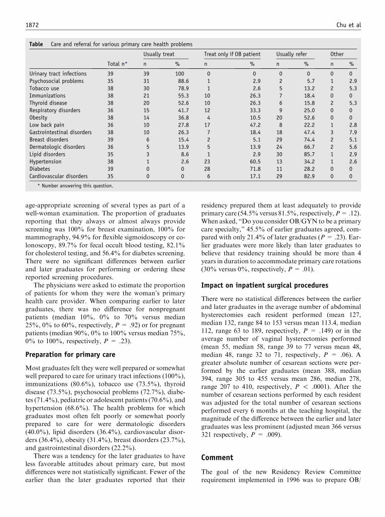
1872 Chu et al
Table Care and referral for various primary care health problems
Total n*
Usually treat Treat only if OB patient Usually refer Other
n % n % n % n %
Urinary tract infections 39 39 100 0 0 0 0 0 0Psychosocial problems 35 31 88.6 1 2.9 2 5.7 1 2.9Tobacco use 38 30 78.9 1 2.6 5 13.2 2 5.3Immunizations 38 21 55.3 10 26.3 7 18.4 0 0Thyroid disease 38 20 52.6 10 26.3 6 15.8 2 5.3Respiratory disorders 36 15 41.7 12 33.3 9 25.0 0 0Obesity 38 14 36.8 4 10.5 20 52.6 0 0Low back pain 36 10 27.8 17 47.2 8 22.2 1 2.8Gastrointestinal disorders 38 10 26.3 7 18.4 18 47.4 3 7.9Breast disorders 39 6 15.4 2 5.1 29 74.4 2 5.1Dermatologic disorders 36 5 13.9 5 13.9 24 66.7 2 5.6Lipid disorders 35 3 8.6 1 2.9 30 85.7 1 2.9Hypertension 38 1 2.6 23 60.5 13 34.2 1 2.6Diabetes 39 0 0 28 71.8 11 28.2 0 0Cardiovascular disorders 35 0 0 6 17.1 29 82.9 0 0
* Number answering this question.
age-appropriate screening of several types as part of awell-woman examination. The proportion of graduatesreporting that they always or almost always providescreening was 100% for breast examination, 100% formammography, 94.9% for flexible sigmoidoscopy or co-lonoscopy, 89.7% for fecal occult blood testing, 82.1%for cholesterol testing, and 56.4% for diabetes screening.There were no significant differences between earlierand later graduates for performing or ordering thesereported screening procedures.
The physicians were asked to estimate the proportionof patients for whom they were the woman’s primaryhealth care provider. When comparing earlier to latergraduates, there was no difference for nonpregnantpatients (median 10%, 0% to 70% versus median25%, 0% to 60%, respectively, P = .92) or for pregnantpatients (median 90%, 0% to 100% versus median 75%,0% to 100%, respectively, P = .23).
Preparation for primary care
Most graduates felt they were well prepared or somewhatwell prepared to care for urinary tract infections (100%),immunizations (80.6%), tobacco use (73.5%), thyroiddisease (73.5%), psychosocial problems (72.7%), diabe-tes (71.4%), pediatric or adolescent patients (70.6%), andhypertension (68.6%). The health problems for whichgraduates most often felt poorly or somewhat poorlyprepared to care for were dermatologic disorders(40.0%), lipid disorders (36.4%), cardiovascular disor-ders (36.4%), obesity (31.4%), breast disorders (23.7%),and gastrointestinal disorders (22.2%).
There was a tendency for the later graduates to haveless favorable attitudes about primary care, but mostdifferences were not statistically significant. Fewer of theearlier than the later graduates reported that their
residency prepared them at least adequately to provideprimary care (54.5%versus 81.5%, respectively,P=.12).When asked, ‘‘Do you consider OB/GYN to be a primarycare specialty,’’ 45.5% of earlier graduates agreed, com-pared with only 21.4% of later graduates (P= .23). Ear-lier graduates were more likely than later graduates tobelieve that residency training should be more than 4years in duration to accommodate primary care rotations(30% versus 0%, respectively, P = .01).
Impact on inpatient surgical procedures
There were no statistical differences between the earlierand later graduates in the average number of abdominalhysterectomies each resident performed (mean 127,median 132, range 84 to 153 versus mean 113.4, median112, range 63 to 189, respectively, P = .149) or in theaverage number of vaginal hysterectomies performed(mean 55, median 58, range 39 to 77 versus mean 48,median 48, range 32 to 71, respectively, P = .06). Agreater absolute number of cesarean sections were per-formed by the earlier graduates (mean 388, median394, range 305 to 455 versus mean 286, median 278,range 207 to 410, respectively, P ! .0001). After thenumber of cesarean sections performed by each residentwas adjusted for the total number of cesarean sectionsperformed every 6 months at the teaching hospital, themagnitude of the difference between the earlier and latergraduates was less prominent (adjusted mean 366 versus321 respectively, P = .009).
Comment
The goal of the new Residency Review Committeerequirement implemented in 1996 was to prepare OB/

Chu et al 1873
GYN residents adequately as primary care providers.This study suggests that residency graduates providescreening services for women presenting for well-womanexaminations, but they refer out certain complex pri-mary care disorders such as management of abnormallipid profiles. Obstetrician-gynecologists who graduatedafter 1996 provided about the same amount of primarycare to their patients as those who graduated in 1996 orearlier. Graduates after 1996 had a tendency to believethey were better prepared to provide primary care butnot to consider OB/GYN a primary care specialty,although these differences were not statistically signifi-cant. As some of our graduates wrote: ‘‘[I] don’t feel weshould be primary care; otherwise we are family practicewithout peds/males’’; or ‘‘[I’m] not interested inmanagingall health problems’’; and ‘‘[I] consider [my]self as spe-cialist and consultant, not PCP [primary care physician].’’
Despite the fact that 60.2% of residency programdirectors once believed that educational deficiencieswould develop as the result of the 6 months primarycare training,6 our study found only a small, nonstatisti-cally significant decrease in the number of abdominaland vaginal hysterectomies performed by earlier andlater graduates. Although the adjusted number of cesar-ean sections performed per resident decreased after1996, the number of procedures remained adequate fortraining purposes. The post-1996 graduates from thisprogram completed more of these procedures than thenational means for all residency programs reported byAccreditation Council for Graduate Medical Educationfor 1997 to 2002, which were 85 abdominal hysterec-tomies, 35 vaginal hysterectomies, and 175 cesareansections per resident.7
This study has several limitations. Our sample in-cluded only physicians who graduated from 1 residencyprogram, and this may limit generalizability. We hadlimited power to detect statistically significant differ-ences in attitudes toward primary care. If confirmed, ourfindings would suggest somewhat paradoxically thatmore primary care training is associated with lessfavorable attitudes about primary care. Because only abefore-after study design could be used (rather than arandomized trial), this difference in attitudes may reflecttemporal changes in the practice of OB/GYN nation-ally, rather than a direct effect of primary care training.Our small sample size also precluded analysis of all but
the 3 most common inpatient surgical procedures. Sur-gical procedures performed in office gynecology settingswere not captured by the hospital data used for thestudy. Thus, we were not able to assess whether some ofthe less common inpatient or outpatient surgical pro-cedures were sacrificed when residents had off-servicecommitments.
On the other hand, in the earlier and later periods, theresponse rates were essentially identical, and the careerpaths of graduates were very similar. This suggests thatresponse bias was unlikely. Our survey suggests severalhypotheses about the self-perceived competencies ofOB/GYN physicians in providing primary care services.Because 6 months of a 4-year residency is a substantiallength of time, larger studies may be worthwhile tofurther investigate these issues.
As OB/GYN physicians strive to fulfill their roles asprimary care givers, they must maintain and updatetheir knowledge about care of ambulatory health prob-lems. In an age of exponentially increasing medicalinformation, staying on top of both primary care issuesand obstetrical-gynecological issues is a challenge to abusy OB/GYN physician. As a graduate wrote: ‘‘Know-ing when to screen is one thing, but staying abreast ofthe various treatments is another.’’
References
1. Laube DW, Ling FW. Primary care in obstetrics and gynecology
resident education: a baseline survey of residents’ perceptions and
experiences. Obstet Gynecol 1999;94:632-6.
2. Morrison JC, Cowan BD, HamptonHL,MortonHH,Martin JN Jr,
McGehee RP. Medical practices of past graduates from one
obstetric/gynecologic trainingprogram.SouthMedJ 1998;91:227-30.
3. American College of Obstetricians and Gynecologists Executive
Board. Obstetrician-gynecologists: specialists in reproductive health
care and primary physicians for women. In: 2000 Compendium
of selected publications. Washington (DC): American College of
Obstetricians and Gynecologists; 2000. p. 1084-5.
4. Kozak LJ, Weeks JD. U.S. trends in obstetric procedures,
1990-2000. Birth 2002;29:157-61.
5. Harer WB Jr. Vaginal birth after cesarean delivery: current status.
JAMA 2002;287:2627-30.
6. Kuffel ME, Stovall DW, Kuffel TS, Zlatnik FJ. Reactions of resi-
dency directors to primary care requirements in obstetrics and gyne-
cology training. Obstet Gynecol 1998;91:145-8.
7. Lashbrook DL, Frazier LM, Horbelt DV, Stembridge TW, Rall
MJ. Pregnancy during obstetrics and gynecology residency: effect
on surgical experience. Am J Obstet Gynecol 2003;189:662-5.
American Journal of Obstetrics and Gynecology (2005) 193, 1874–8
www.ajog.org
Improved performance and student satisfaction afterimplementation of a problem-based preclinicalobstetrics and gynecology curriculum
Petra M. Casey, MD,a,* Diane Magrane, MD,b Timothy G. Lesnick, MSc
Department of Obstetrics and Gynecology, Mayo Clinic College of Medicinea; Faculty Development and LeadershipPrograms,b Association of American Medical Colleges; Department of Biostatistics, Mayo Clinic,c Rochester, MN
Received for publication February 17, 2005; revised May 20, 2005; accepted July 18, 2005
KEY WORDSProblem-based
learningCurriculum
developmentObstetrics and
gynecology course
Objective: This study was undertaken to assess student performance and satisfaction as apreclinical obstetrics and gynecology course changed from a didactic to a problem-based format.
Study design: We prospectively compared examination scores and course evaluations given to162 second-year medical students over 4 years: 2 years before and 2 years after the curricularchange, to assess student performance and satisfaction with learning. We used analysis of
variance for the analysis of student performance and likelihood-ratio test for analysis of studentsatisfaction.Results: Mean examination scores increased from 79.1% to 84% after implementation of theproblem-based curriculum (P ! .0001). The data showed statistically significant improvement in
satisfaction with course content, learning objectives, learning resources, instructional methods,and course examinations.Conclusion: Students’ satisfaction and performance in a preclinical obstetrics and gynecology
course improved significantly when problem-based learning methods were introduced.� 2005 Mosby, Inc. All rights reserved.
Problem-based learning (PBL) represents the evolu-tion from passive learning to an active, student-centeredlearning style.1-3 PBL involves a group of students(typically 6-10) and a faculty facilitator who guideslearning via discussion.1 The PBL approach has beenfound effective in improving performance in a variety of
Supported by a Faculty Development Grant from the Mayo Clinic
College of Medicine.
* Reprint requests: Petra M. Casey, MD, Department of Obstetrics
and Gynecology, Eisenberg 4A, Mayo Clinic, 200 First St SW,
Rochester, MN 55905.
E-mail: [email protected]
0002-9378/$ - see front matter � 2005 Mosby, Inc. All rights reserved.
doi:10.1016/j.ajog.2005.07.061
specialties.4-7 Surveyed students have indicated that,compared with didactic teaching, PBL promoted amore positive attitude toward learning and a morestimulating environment for clinical knowledge acquisi-tion.4,8,9 This educational method has been used inclinical and preclinical medical education7,10 as well asnonreproductive women’s health issues.11,12
The obstetrics and gynecology curriculum for studentsat Mayo Clinic College of Medicine includes a 26-sessionobstetrics and gynecology course in the second year,followed by a 6-week clinical clerkship in the third year.Second-year students have a dense curriculum of basicsciences and often perceive the introduction to obstetrics

Casey, Magrane, and Lesnick 1875
and gynecology as a distraction from learning as theyprepare for the United States Medical Licensing Exam-ination (USMLE).
The curricular change described in this study formal-ized an effort to engage the students in learning, topromote information acquisition, and to integrate basicscience with clinical medicine. The effect of PBL onexamination scores and satisfaction with various aspectsof introductory obstetrics and gynecology has not beendescribed in a prospective study in publication. Further,there are no studies investigating this effect over severalyears or in the setting where the curriculum includesnonreproductive women’s health issues.
Materials and methods
All second-year medical students (n = 162) enrolled inintroductory obstetrics and gynecology at Mayo ClinicCollege of Medicine during four academic years partici-pated in this study. There were 40 students during 2000-2001 (year 1), 42 during 2001-2002 (year 2), 41 during2002-2003 (year 3), and 39 during 2003-2004 (year 4).For PBL, students were divided into 4 groups of 10,whereas all students attended lectures as 1 group. Theproject was exempted by Mayo Clinic InstitutionalReview Board.
The course was taught by clinical faculty includinggeneral obstetrician-gynecologists, maternal-fetal medi-cine specialists, and gynecologic oncologists. A domesticviolence coordinator from Mayo Department of SocialServices facilitated the domestic violence unit. Thefaculty was allotted 3 hours of preparation per hour ofPBL discussion. All faculty completed a 4-hour facilita-tion workshop before course participation. This work-shop described PBL and allowed each faculty memberto facilitate a group discussion in a role-playing exercise.
The new curriculum was introduced over 4 academicyears. Traditional didactic curriculum was taught duringyear 1. Two PBL units were introduced during year 2,while other topics were taught with lectures. During year3, 66% of lectures were converted into PBL, followedby a complete problem-based curriculum during year 4.Domestic violence was added as a women’s health issuenot adequately taught elsewhere in the curriculum.
The units were modeled after Association of Profes-sors of Gynecology and Obstetrics (APGO) Guide toImplementing a Problem-based Learning Curriculum andTeaching Guide to Psychosocial Issues.13,14 Course con-tent was available to students and faculty on a Web siteand a printed syllabus. One author (P.M.C.) selected 26topics in obstetrics and gynecology, which followedsubject areas in the course textbook.15 Each PBL unitconsisted of APGO Medical Student Educational Objec-tives,16 textbook reading corresponding to the learningobjectives and additional content (American Collegeof Obstetricians and Gynecologists [ACOG] technical
bulletins, journal articles, prescribing information andPowerpoint presentations narrated using Camtasia[Camtasia Studio 2.1, TechSmith, Okemos, MI]). Mul-tiple-choice self-quizzes assessed subject mastery afterself-study. Students then participated in a clinical case-based discussion. Course evaluations communicatedstudent feedback. The time requirement for each unitdesign was about 15 hours.
Four examinations assessed knowledge acquisition.During year 1, examinations were administered viapaper and pencil as they had been before the study.To determine whether testing method would impactexamination scores, 1 examination during year 2 wasadministered with the use of WebCT software (WebCTCampus Edition 3.8, WebCT Inc, Lynnfield, MA),whereas the others were traditional paper-and-pencilexaminations. We tested for a difference between com-bined paper test scores and the WebCT test scores inyear 2 using paired t tests. In subsequent years, all 4examinations were Web based. To create questions ofconsistent quality and discriminatory value, we usedAPGO test bank’s multiple choice questions pertinentto the topics.17 The examination questions were inter-mixed, but otherwise kept consistent from year to year,allowing for meaningful comparison of scores.
We tested composite examination scores for normal-ity using the Kolmogorov-Smirnov goodness-of-fit test,and compared mean final grades using a 1-way analysisof variance (ANOVA). Pairwise differences between theyears were assessed using 3 post hoc tests (Tukey, leastsignificant difference, and Scheffe) to adjust for multiplecomparisons and cover the range from liberal (likely tofind differences) to conservative (unlikely to find differ-ences).
Student satisfaction was measured by Likert scaleresponses to 17 questions on a course evaluation. Thesemeasured change in satisfaction with scheduling, learn-ing objectives, course content, instructional methods,examinations, and integration of the course into thesecond year curriculum as related to the teachingmethod. Likert scale responses of 5, 6, or 7 indicatedagreement, 4 was neutral, and 1, 2, or 3 indicateddisagreement with each statement. The proportions ofresponders who agreed with each item were comparedbetween the years using likelihood-ratio tests for logistic
Table I Mean composite examination scores in each aca-demic year compared by ANOVA
YearNumber ofstudents % PBL
Final grades (%)
Mean SD Median Range
1 43 0 79.6 4.7 78.7 69.3-89.02 42 7.7 78.6 4.8 78.1 67.5-89.43 41 66.7 83.6 5.3 84.4 66.7-92.64 39 100 84.4 5.0 85.3 69.1-93.4

1876 Casey, Magrane, and Lesnick
Table II Student satisfaction with aspects of obstetrics and gynecology course
Year, N agree/N Total (%)
Question 2000-2001 2001-2002 2002-2003 2003-2004 P value*
Appropriate time for topicsin schedule
24/37 (64.9) 11/28 (39.3) 8/16 (50.0) 22/29 (75.9) .12
Manageable workload scheduled 28/37 (75.7) 17/28 (60.7) 14/17 (82.4) 25/29 (86.2) .07Important concepts clear inlearning objectives
7/36 (19.4) 3/27 (11.1) 9/17 (52.9) 17/28 (60.7) ! .0001
Appropriate detail clear inlearning objectives
7/36 (19.4) 3/27 (11.1) 8/17 (47.1) 15/28 (53.6) .0001
Content organized tofacilitate learning
7/36 (19.4) 7/28 (25.0) 5/17 (29.4) 14/28 (50.0) .01
Content related basic scienceto practice
15/35 (42.9) 9/28 (32.1) 9/16 (56.3) 18/28 (64.3) .02
Learning resources clearlyexplained concepts
4/29 (13.8) 6/24 (25.0) 2/14 (14.3) 12/28 (42.9) .04
Syllabus, handouts, Web siteof good quality
5/35 (14.3) 2/27 (7.4) 2/15 (13.3) 12/26 (46.2) .001
Text of good quality 20/36 (55.6) 12/27 (44.4) 7/16 (43.8) 16/27 (59.3) .66Class activities helpedunderstand concepts
9/31 (29.0) 6/28 (21.4) 4/17 (23.5) 14/28 (50.0) .05
Class activities related basicscience to practice
9/33 (27.3) 7/28 (25.0) 3/16 (18.8) 12/28 (42.9) .22
Course benefited fromsmall groups
15/31 (48.4) 12/28 (42.9) 8/15 (53.3) 14/28 (50.0) .66
Examinations assessedconceptual understanding
1/32 (3.1) 1/28 (3.6) 5/17 (29.4) 11/28 (39.3) ! .0001
Examinations assessed masteryof details
1/32 (3.1) 1/28 (3.6) 3/17 (17.7) 10/27 (37.0) ! .0001
Preferred WebCT examinations – 20/24 (83.3) 13/16 (81.3) 19/27 (70.4) .28Pelvic examination instructionvaluable
– 15/24 (62.5) 13/16 (81.3) 22/28 (78.6) .17
Course well integrated intocurriculum
4/33 (12.1) 4/25 (16.0) 3/14 (21.4) 8/26 (30.8) .07
* Obtained from a likelihood-ratio test for trend in logistical regression.
regression trend, in which the predictor variable was theproportion of PBL in the curriculum.
MedicalCollegeAdmissionTest (MCAT)andUSMLEscores for each class of students were tested usingregression analysis to uncover differences in students’learning abilities and test taking skills.
Results
Table I showed themean composite examination scores ineach academic year. The scores were normally distributedwith significant trends (ANOVA P ! .0001). Pair-wisecomparisons of the scores during the study period usingtests of mean difference showed significant differencesbetween years 1 and 3, 1 and 4, 2 and 3, and 2 and 4. Thesame method revealed no significant differences in themean scores between years 1 and 2 and between years 3and 4.
Table II represented students’ responses to 17 surveyquestions encompassing 7 general categories: course
organization/scheduling, learning objectives, coursecontent, learning resources, small group format, exam-inations, and integration into the rest of the curriculum.Varying numbers of students responded to each ques-tion each year. Significant improvement in satisfactionwas seen in five areas: learning objectives (2 items),course content (2 items), learning resources (2 of 3items), small group format (1 of 3 items), and examina-tions (2 of 4 items). There was no significant improve-ment in course integration and organization, or in theremaining items in the other categories.
To test whether the change from paper-and-pencil toWeb-based examinations had an effect on performance,we compared the aggregate scores of each of the 4classes by examination administration method. Theaverage score of the paper examinations during year 2was 78.5% G 5.5% and the mean score on the WebCTexamination was 78.6% G 6.5%. These means did notdiffer by regression analysis (P = .928). MCAT andUSMLE scores of students enrolled in the study were

Casey, Magrane, and Lesnick 1877
compared with investigate trends in students’ academicabilities reflected by standardized examinations. Nosignificant trend over the years was found on either 1of the examinations. Mean MCAT scores were 33.25 G0.50, P = .74 and mean USMLE scores were 232.525 G4.461, P = .85 by regression analysis.
Comment
Our study showed a consistent improvement in exami-nation scores and student satisfaction in preclinical ob-stetrics and gynecology after implementation of PBL.It supported PBL as a valuable tool for knowledgeacquisition in this specialty. Further, it controlled forbiases introduced by different examination methodsand student academic abilities. The latter was measuredby comparing MCAT and USMLE scores. Greateracademic strength resulting in higher examination scorescould have been associated with better satisfactionregardless of teaching method. We found no significanttrends in these parameters.
The robust improvement in written examinationscores after extensive implementation of PBL confirmedthat despite some difficulties with PBL teaching, ourstudents recalled more information with this method.Years 1 and 2 curricula represented primarily didacticteaching, whereas years 3 and 4 curricula utilized pri-marily PBL. As expected, the mean examination scoresfor year 2, when only 2 PBL units were presented, didnot differ from year 1. The improvement in examinationscores followed the adoption of PBL for the majority ofthe course and was sustained during the complete PBLimplementation. Thus, the observed change was notlikely because of enthusiasm for a new learning method.
The interpretation of student satisfaction with intro-ductory obstetrics and gynecology, was more complex.The students communicated that the didactic format didnot provide adequate learning and that they preferredthe PBL format taught in other courses. Once the PBLformat was implemented, the students indicated im-proved satisfaction with many aspects of the course.The most dramatic improvement was seen when years1 and 4 (all didactic vs all PBL) were compared.
Areas showing improved satisfaction included therelationship of learning objectives to concepts and ap-propriate level of detail and learning objective clarity, therelationship of basic science and clinical medicine appar-ent in well-organized course content, Web site and coursematerial quality, facilitation of conceptual understandingduring class activities and the testing of important con-cepts and appropriate detail. There were no deleteriouseffects of PBL on any parameters measured.
Interestingly, some satisfaction items contradictedeach other. For example, as a result of classroomactivities, students reported an improvement in under-
standing of important concepts, but not of the relation-ship between basic science and medical practice. On theother hand, they indicated that course content relatedbasic science to practice better with the PBL format. Thismay have been artifactual because of the sample size ofresponders or variable response rate.
The preference for small group instruction overlectures item required the most careful interpretation.How well prepared were the students to make thisdecision? Scores from year 3, when the students hada direct basis for comparison led to the most positiverating of the PBL curriculum. During years 1 and 4,all material was either lecture or PBL providing thestudents with only indirect basis to compare the 2methods via PBL teaching in other courses. Somestudents simply preferred lectures, shown to facilitateshort-term learning6 appropriate for multiple-choiceassessment. Thus, lectures may have represented anadvantage within the curricular density of the secondyear and a better match for the examination method.Interestingly, though the students did not track thenumber of hours needed for study, the workload wasnot perceptibly increased with PBL perhaps because wededicated 1 hour of study time per hour of discussion.
Some of the aspects showing significant changerepresented course organization rather than the teach-ing method. These included the scheduling and learningresources items.
Lastly, certain course evaluation items did not showimprovement. It was reassuring that the proportion ofpositive responses to the course schedule, textbook,gynecologic examination instruction, and examinationmethod, purposely kept constant during the study,remained stable. This validated our survey instrument.Similarly, the consistently low scores on the integrationitem suggested that the emphasis on USMLE over-shadowed clinical courses regardless of instructionmethod. Overall, the curricular change to PBL teachingled to a dramatically increased satisfaction with learningin obstetrics and gynecology.
This study also presented an alternative for curricu-lum development. Previously described strategies in-cluded PBL and didactic courses in parallel, abrupttransitions from one teaching method to another and apilot PBL course in the setting of didactics.10 We offereda model where gradual implementation of a curricularformat was undertaken over several years. The advan-tages of this model included continuous feedback fromstudents and faculty for identification of areas needingadjustment, increased faculty comfort level with the newteaching method on a smaller scale before full imple-mentation and the monitoring of scores and satisfactionparameters for unanticipated negative outcomes.
The largest challenge with implementing a PBL coursewithin our small obstetrics and gynecology departmentwas securing time for course design, faculty development/

1878 Casey, Magrane, and Lesnick
training and course participation without negativelyimpacting patient care. PBL format required 4 times thenumber of faculty hours to teach the same material. TheAPGO Guide to Implementing a Problem-based LearningCurriculum and Teaching Guide to Psychosocial Issues13,14
provided obstetric and gynecologic cases and a chapter on‘‘Violence against Women’’ with learning issues. The useof these resources considerably reduced time for PBLunit design. By using existing APGO cases enabled1 author (P.M.C.) to devote time to course content, exam-ination, and course evaluation development. Consistentdesign of the units allowed various faculty members toeffectively facilitate any PBL unit. However, multipleteaching sessions per subject and the occasional facultysubstitution introduced some content variability, a studylimitation. Although we made every effort to maximizestudent contact with a core of proven educators, through-out the study, there were occasional unavoidable changes.We received full support of department and medicalschool leadership for additional faculty time.
Finally, given our relatively low and variable re-sponse rates to the course evaluations, did the evalua-tions accurately reflect the class viewpoint? Further,because the evaluations followed the last examination,was the students’ satisfaction with the course altered bytheir impression of the examination? At the Mayo ClinicSchool of Medicine, students completed the courseevaluation as a prerequisite to the release of grades.They were not required to answer every item and couldsubmit verbal comments alone without the Likert scaleevaluation. This explains the lower response rates insome years. A collaborative multicenter study with alarger sample size could address this bias provided acomparable curriculum could be located.
In summary, the robust improvement in examinationscores and student satisfaction with implementation ofPBL in preclinical obstetrics and gynecology shouldencourage others to apply this method to their settings.Our study presented a gradual implementation of cur-ricular change, multiyear performance and satisfactiondata analysis and extensive use of APGO materials incourse design. These aspects of our study may be readilyadopted elsewhere.
Acknowledgment
We gratefully acknowledge the mentorship of DrAndrew E. Good, and a Faculty Development Grantfrom Mayo Clinic College of Medicine.
References
1. Albanese MA, Mitchell S. Problem-based learning: a review of
literature on its outcomes and implementation issues. Acad Med
1993;68:52-81.
2. Neufeld VR, Woodward CA, MacLeod SM. The McMaster MD
program: a case study of renewal in medical education. Acad Med
1989;64:423-32.
3. Wendelberger KJ, Simpson DE, Biernat KA. Problem-based
learning in a third year pediatric clerkship. Teach Learn Med
1996;8:28-32.
4. Farrell TA, Albanese MA, Pomrehn PR Jr. Problem-based learn-
ing in ophthalmology: a pilot program for curricular renewal.
Arch Ophthalmol 1999;117:1223-6.
5. Curtis JA, Indyk D, Taylor B. Successful use of problem-based
learning in a third year pediatric clerkship. Ambul Pediatr
2001;1:132-5.
6. Them C, Schulc E, Roner A, Behrens J. Comparison of frontal
teaching versus problem-oriented learning at the school of health-
care and nursing: nursing neurological patients. Int J Med Inform
2003;71:117-24.
7. Swagerty D, Studenski S, Laird R, Rigler S. A case-oriented
Web-based curriculum in geriatrics for third-year medical students.
J Am Geriatr Soc 2000;48:1507-12.
8. Kaufman DM, Mann KV. Comparing students’ attitudes in
problem-based and conventional curricula. Acad Med 1996;71:
1096-9.
9. Nalesnik SW, Heaton JO, Olsen CH, Haffner WH, Zahn CM.
Incorporating problem-based learning into an Ob Gyn clerkship:
impact on student satisfaction and grades. Am J Obstet Gynecol
2004;190:1375-81.
10. Vincelette J, Lalande R, Delorme P, Goudreau J, Lalonde V, Jean
P. A pilot course as model for implementing a PBL curriculum.
Acad Med 1997;72:698-701.
11. Robertson PA, Brown JS, Flanagan TA, Goldman ME, Learman
LA, Stevens AE, et al. The Women’s Health Curriculum by a
problem-based learning method for medical students at the Uni-
versity of California, San Francisco. Am J Obstet Gynecol
1997;176:1368-73.
12. Levison SP, Weiss LB, Puglia CD, Nieman LZ, Donoghue GD. A
model for intergrating women’s health issues in a problem-based
curriculum. J Womens Health 1998;7:1113-24.
13. Association of Professors of Gynecology and Obstetrics. Task
force on problem-based learning and undergraduate medical
education committee guide to implementing a problem-based
learning curriculum. Washington (DC): APGO; 1996.
14. Association of Professors of Gynecology and Obstetrics, Under-
graduate Medical Education Committee. Women’s health: a
teaching guide to psychosocial issues. Washington (DC): APGO;
2000.
15. Beckmann CR, Ling FW, Laube DW, Smith RP, Barzansky BM,
Herbert WP. Obstetrics and gynecology. 4th ed. Philadelphia:
Lippincott, Williams and Wilkins; 2002.
16. Association of Professors of Gynecology and Obstetrics. Under-
graduate Medical Education Committee Medical Student Edu-
cational Objectives. 7th ed. Washington (DC): APGO; 1997.
17. Association of Professors of Gynecology and Obstetrics. APGO
test bank. Washington (DC): APGO; 1996.

www.ajog.org
American Journal of Obstetrics and Gynecology (2005) 193, 1879–85
LETTERS TO THE EDITORS
Risk assessment for neonatal respiratory distress syndromewith FLM II combined with gestational age
To the Editors: The article by Parvin et al1 provides theclearest set of data to date that incorporate gestationalage into the evaluation of fetal lung maturity.
The authors of this study failed to mention our re-sponse in the letters to the editor section in regards todifferences in quantitative flourescence polarization-based fetal lung maturity assay (TDx-FLM) I versusTDx-FLM II.2 Despite a change in reagents, the actualnumeric values on paired specimens that were testedcontemporaneously were nearly identical (G4.0%).
Differences in the prevalence rate of respiratory dis-tress syndrome (RDS) have led to several different rec-ommended cutoff points, regardless of the version ofFLM test kits that were used.2,3 A large part of the dif-ference in the prevalence rate can be explained simply onthe basis of the differences in the gestational ages thatwere being tested for fetal lung maturity in the publishedstudies. Indeed, the prevalence of RDS changes dramat-ically between 30 and 36 weeks of gestation.4 On thebasis of gestational age alone, most fetuses !30 weeksof gestation will be at risk of RDS, whereas few fe-tuses after 36 weeks of gestation will experience RDS.Obtaining FLM results at 39C and !30 weeksof gestation is arguably not very useful.
Even within their study, the prevalence of RDS wasdifferent among the 3 groups. A selection bias towardincluding patients with RDS resulted from an increasednumber of RDS cases between 32 and 36 weeks of ges-tation in group 3. Bias towards including cases withRDS illustrates the potential problem with retrospectivestudies. Furthermore, the inclusion of patients at !30and O39 weeks of gestation is not useful and potentiallyskews the overall prevalence. Eliminating the cases !30weeks of gestation leaves approximately 50 cases ofRDS distributed over 10 weeks of gestation. There ap-pears to be only 1 case of RDS after 37 weeks of gestationof 131 patients who were tested. Notably, the number of
0002-9378/$ - see front matter � 2005 Mosby, Inc. All rights reserved.
doi:10.1016/j.ajog.2005.05.022
patients who were enrolled between 30 and 34 weeks ofgestation was small (2, 6, 14, 30, and 49, respectively).Owing to the higher incidence of RDS during those gesta-tional ages (55%, 40%, 35%, 25%, and 15%, respec-tively), a larger number of patients at those gestationalages may provide more meaningful data.
Although odds ratios are statistically more applica-ble, given the potential differences in prevalence betweenstudies, they are much more difficult to use conceptuallywhen risks of RDS are being discussed with patients andcolleagues.
The collection of a large number of gestational ageespecific data between 30 and 36 weeks with gestationalageespecific analysis would provide better risk estimates.
Michael G. Pinette, MD*Joseph Wax, MD
*Maine Medical CenterDepartment of Obstetrics and Gynecology
Division of Maternal-Fetal Medicine887 Congress St, Suite 200
Portland, ME 04102E-mail: [email protected]
References
1. Parvin CA, Kaplan LA, Chapman JF, McManamon TG,
Gronowski AM. Predicting respiratory distress syndrome using ges-
tational age and fetal lung maturity by fluorescent polarization. Am
J Obstet Gynecol 2005;192:199-207.
2. Pinette MG. Risk assessment calculation for respiratory distress [re-
ply]. Am J Obstet Gynecol 2005;189:1513-4.
3. Pinette M, Wax J. The usefulness of the TDX/TDXFLX fetal lung
maturity II assay [letter]. Am J Obstet Gynecol 2004;190:870.
4. Robertson PA, Sniderman SH, Laros RK, Cowan R, Heilbron D,
Goldenberg RL, et al. Neonatal morbidity according to gestational
age and birth weight from five tertiary care centers in the United
States, 1983 through 1986. Am J Obstet Gynecol 1992;166:1629-41.

1880 Letters to the Editors
Reply
To the Editors: We appreciate the acknowledgement byDrs Pinette and Wax that our article provides the clear-est set of data to date to incorporate gestational age intothe evaluation of fetal lung maturity. We agree thatoverall prevalence of RDS that is observed at a par-ticular institution will depend on the distribution ofthe gestational ages that are tested. We discussed thispoint in our article. We also agree that estimates of asingle TDx-FLM II cutoff for all gestational ages willbe influenced by the distribution of the gestationalages that are tested. The purpose of our study was notto estimate a single TDx-FLM II cutoff but to modelRDS risk as a function of TDx-FLM II ratio and gesta-tional age. Two of our study sites collected all availabledata for physician-ordered TDx-FLM II results; thethird site sought additional cases of RDS, independentlyof gestational age at the time of fetal lung maturity test-ing. As stated in our article, there was no statisticallysignificant difference among study sites in the rate ofchange of RDS risk as a function of gestational age,but we did find a significantly higher overall prevalenceof RDS at the third study site; both observations areconsistent with the third study site’s indentification ofadditional cases of RDS, independent of gestationalage. Data based on consecutive series of subjects allowunbiased estimation of risk. Therefore, in addition topresenting tables of odds ratios that reflect only therate of change of risk (but as Drs Pinette and Wax pointout are also more difficult conceptually), we also wereable to present tables of absolute risk by fitting a sepa-rate baseline prevalence term for the third study siteand adjusting for their ‘‘biased’’ overall prevalence.
Last, they state that they found only a 4% differencebetween the TDx-FLM I and II in a study of 47 samples,
0002-9378/$ - see front matter � 2005 Mosby, Inc. All rights reserved.
doi:10.1016/j.ajog.2005.05.023
Lymphatic mapping for endomhysteroscopic injection a safesentinel lymph node biopsy?
To the Editors: We read with interest the article byRaspagliesi et al1 demonstrating the relevance of intra-operative lymphatic mapping using hysteroscopic injec-tions of tracers in patients with endometrial cancer. In
which suggests that the 2 tests are ‘‘nearly identical.’’1
Two larger studies suggest that this is not the case.One of our laboratories (J.F.C.) participated in the mul-ticenter testing of TDx-FLM I versus FLM II (n=778patients).2 The following regression data were from thisstudy: slope, 0.742; Y intercept, 1.54 (r=0.962), whichresulted in FLM II ratios that were between 10% and25% lower than FLM I ratios. A study by Moxnesset al3 (n=63 patients) reported the following data:slope, 0.9; Y intercept, �0.9 (r=0.99), which resultedin FLM II ratios that were between 10% and 20% lowerthan FLM I ratios. Together these data suggest the 2assays are not ‘‘nearly identical’’ and the differencesshould not be ignored.
C. A. Parvin, PhDL. A. Kaplan, PhD
J. F. Chapman, DrPHT. G. McManamon, PhDA. M. Gronowski, PhD*
*Department of Pathology and ImmunologyDivision of Laboratory Medicine
Washington University School of Medicine660 S. Euclid, Box 8118
St. Louis, MO 63110E-mail: [email protected]
References
1. Pinette MG. Risk Assessment calculation for respiratory distress
[reply]. Am J Obstet Gynecol 2003;189:1513-4.
2. Abbott Laboratories. Technical Bulletin,March 16, 1995;XR95-002.
3. Moxness M, Lawson G, O’Brien J, Burritt M. Assessment of fetal
lung maturity by the Abbott FLM II assay and three other methods.
Clin Chem 1995;41:S95.
etrial cancer: Istechnique for
a series of 18 patients, they injected radiocolloid andblue dye subendometrially around the lesion duringhysteroscopy, and detected sentinel nodes (SN), by lap-arotomy, in all cases. One previous study has described

laparotomic identification of SNs under hysteroscopicguidance in patients with endometrial cancer.2 They in-jected radiocolloid under the endometrium during hys-teroscopy, and detected SNs, by laparotomy, in 82%of cases.2
This article raises several concerns. The first is thechoice of injection sites. Two injection sitesdsubserosalintraoperative myometrial and pericervicaldhave beenpreviously used to detect SN in women with endometrialcancer. The former approach, using blue dye, was ini-tially used because it reflected the anatomic drainageof the corpus uteri.3 However, this technique is contro-versial, as combined detection is not possible, and thenumber and location of myometrial injection siteshave not yet been standardized. The latter approach,which permits combined detection with patent blueand radiocolloid, is well standardized, simple and repro-ducible; it yields a higher SN detection rate, but doesnot reflect the anatomic drainage of the tumor.4 Hence,intrauterine injection, mimicking the natural lymphaticdrainage of endometrial cancer, is an attractive alter-native. Raspagliesi et al1 injected tracer under hystero-scopic guidance. In our experience, patent blueinjection under hysteroscopic guidance was difficult,as the endometrium was obscured by dye diffusion.Moreover, injection ‘‘under the endometrium’’ probablycorresponds in fact to injection in the superficialmyometrium.
The risk of cancer cell dissemination into the abdom-inal cavity must also be discussed. Despite the contro-versial prognostic significance of positive cytology,previous studies have shown a risk of tumor cell dissem-ination and metastasis after hysteroscopy. To avoid thisrisk, we injected patent blue under hysteroscopic guid-ance at the beginning of surgery, after coagulating theFallopian tubes.
Raspagliesi et al1 and Niikura et al2 confirmed therelevance of the SN biopsy in patients with endometrial
cancer, but did not obtain a higher SN identificationrate and bilateral SN detection rate than in previousstudies using dual labels injected pericervically.4
Finally, contrary to the laparoscopic SN approach,the procedure used by Raspagliesi et al,1 involving pre-operative hysteroscopy with uterine distension and lap-arotomy, is not compatible with the concept ofminimally invasive surgery, which is particularly benefi-cial for these often obese and elderly women with under-lying general health disorders.
Emmanuel Barranger, MD*Serge Uzan, MD
Emile Darai, MD, PhD*Department of Gynecology and Obstetrics
Hopital Tenon4 rue de la Chine
75020, AP-HPParis, France
E-mail: [email protected]
References
1. Raspagliesi F, Ditto A, Kusamura S, Fontanelli R, Vecchrone F,
Maccauro M, et al. Hysteroscopic injection of tracers in sentinel
node detection of endometrial cancer: a feasibility study. Am J Ob-
stet Gynecol 2004;191:435-9.
2. Niikura H, Okamura C, Utsunomiya H, Yoshinaga K, Akahira J,
Ito K, et al. Sentinel lymph node detection in patients with endome-
trial cancer. Gynecol Oncol 2004;9292:669-4.
3. Burke TW, Levenback C, Tornos C, Morris M, Wharton JT,
Gerhenson D. Intraabdominal lymphatic mapping to direct selec-
tive pelvic and paraaortic lymphadenectomy in patients with high
risk endometrial cancer: results of a pilot study. Gynecol Oncol
1996;62:169-73.
4. Barranger E, Cortez A, Grahek D, Callard P, Uzan S, Darai E.
Laparoscopic sentinel node procedure using a combination of pa-
tent blue and radiocolloid in women with endometrial cancer.
Ann Surg Oncol 2004;1:344-9.
0002-9378/$ - see front matter � 2005 Mosby, Inc. All rights reserved.
doi:10.1016/j.ajog.2005.04.042
Letters to the Editors 1881
Reply
To the Editors:We appreciated very much the commentsof Dr Baranger et al regarding our article entitled ‘‘Hys-teroscopic injection of tracers in sentinel node detectionof endometrial cancer: A feasibility study.’’
Regarding the site of injection, we named it the sub-endometrial layer because the patient is awake duringthe hysteroscopic injection. We avoided the myometrialinjection because it elicits pain caused by the presence
of neural fibres in this stratum. Moreover, the myome-trial injection increases significantly the rate of intravas-cular leakage that reduces the nodal uptake of thetracers.
Visual impairment during hysteroscopic injection iscommon during blue dye (BD) injection because of itsleakage from the injection site. However, it is limitedto a few seconds when a continuous flow hysteroscope

1882 Letters to the Editors
is used, which enables a prompt washout of the leakedBD. The paucity of BD uptake by the lymph nodes(30% in our series) constitutes one of its major draw-backs and we do not use this tracer any longer in futurestudies.
Several studies correlated the potential risks of peri-toneal dissemination of endometrial cancer cells duringhysteroscopy. Published studies on this issue are limited,as they did not assess the intrauterine pressure (IUP) orthey adopted an IUP greater than 100 mm Hg. It is welldemonstrated by studies on hydrotubation that usingIUP less than 50 mm Hg does not create hydrotubation.Furthermore, in our study that used radiocolloid, in nocases was the peritoneum found to be radioactive, con-firming the absence of tubal leakage.
This study was conducted not to increase the SN de-tection rate reported by previous published techniques,but to evaluate the feasibility of an injection techniquemore similar to that used in other cancers, injectingthe tracers around the lesion or, when the mucosa istotally involved by the tumor, in all the uterine walls.
The suggestion to perform the whole surgical phase ofSN detection and/or of the staging by laparoscopy,
0002-9378/$ - see front matter � 2005 Mosby, Inc. All rights reserved.
doi:10.1016/j.ajog.2005.04.043
Evidence does not support cer
To the Editors: Just when we thought the case for cervicalpreservationduring hysterectomyhasbeen settled after re-cently published studies, we were surprised to see a reviewarticle defending supracervical hysterectomy.1 We believethe fundamental controversy regarding laparoscopicsupracervical hysterectomy is not whether it is superiorto laparoscopic-assisted vaginal hysterectomy or endome-trial resection. The ultimate issue with any supracervicalhysterectomy technique is whether preservation of thecervixhasanyadvantageornot.Because all the theoreticalbenefits of laparoscopic supracervical hysterectomy areassociated with saving the cervix, level I or II evidencefrom 4 recent well-designed studies comparing total eitherabdominal hysterectomy or vaginal hysterectomy withsubtotal abdominal hysterectomy should suffice to answerthis question for laparoscopic supracervical hysterectomy,as well. Briefly, all of these studies found no statisticallyor clinically significant advantage to cervical preserva-tion.2-5 Moreover, Okaro et al suggested that it mighteven have long-term disadvantages. They reported that16 of 70 (22%) women who were followed for a mean of
pursuing the conceptofminimally invasivedisease is ofmajorconcern. However, we preferred to investigate the issuestep by step, initially assessing the feasibility of the intra-uterine injection of tracers by hysteroscopy. This studywas conducted by laparotomy because at that time weused the same probe used for breast andmelanoma cancerthat can be used only by laparotomy. Further studies areongoing to evaluate the reliability of the SNas a surrogatemarker of the actual retroperitoneal node status the feasi-bility of this technique by laparoscopy.
Francesco Raspagliesi, MDAntonino Ditto, MD
Shigeki Kusamura, MDMaria L. Carcangiu, MDFrancesca Vecchione, MD
Marco Maccauro, MDEugenio Solima, MD
Department of Gynecologic OncologyIstituto Nazionale Tumori
Via Venezian 1Milan, MI 20133 Italy
E-mail: [email protected]
vical preservation
66 months after laparoscopic supracervical hysterectomyrequired trachelectomy.6
Finally, in any area of medicine, it is the surgeon’sgoal to develop the least invasive, the fastest, the leastcomplicated, and the most effective operative techniqueswith the shortest hospital stay and lowest cost.7 In thecase of hysterectomy, it is hard to beat the vaginal routefrom all of these perspectives.
Oz Harmanli, MDStephen A. Metz, MD
Department of Obstetrics and GynecologyBaystate Medical Center
Springfield, MAE-mail: [email protected]
References
1. Jenkins TR. Laparoscopic supracervical hysterectomy. Am J Obstet
Gynecol 2004;19:1875-84.
2. Roovers JP, van der Bom JG, van der Vaart CH, Heintz AP. Hys-
terectomy and sexual wellbeing: prospective observational study of

vaginal hysterectomy, subtotal abdominal hysterectomy, and total
abdominal hysterectomy. BMJ 2003;327:774-8.
3. Thakar R, Ayers S, Clarkson P, Stanton S, Manyonda I. Outcomes
after total versus subtotal abdominal hysterectomy. N Engl J Med
2002;347:1318-25.
4. Learman LA, Summitt RL, Varner RE, McNeeley SG,
Goodman-Gruen D, Richter HE, et al. A randomized comparison
of total or supracervical hysterectomy: surgical complications and
clinical outcomes. Obstet Gynecol 2003;102:453-62.
5. Gimbel H, Zobbe V, Andersen BM, Filtenborg T, Gluud C,
Tabor A. Randomised controlled trial of total compared with sub-
total hysterectomy with one-year follow up results. BJOG 2003;110:
1088-98.
6. Okaro EO, Jones KD, Sutton C. Long term outcome following lap-
aroscopic supracervical hysterectomy. BJOG 2001;108:1017-20.
7. Harmanli OH, Gentzler CK, Byun S, Dandolu V, Grody MHT.
A comparison of abdominal and vaginal hysterectomy for the large
uterus. Int J Gynecol Obstet 2004;87:19-23.
0002-9378/$ - see front matter � 2005 Mosby, Inc. All rights reserved.
doi:10.1016/j.ajog.2005.04.016
Letters to the Editors 1883
Reply
To the Editors: I appreciate Drs Harmanli and Metz’sresponse to my review of laparoscopic supracervicalhysterectomy (LSH).1 The article was a review of theexisting LSH literature and was not intended as a‘‘defense’’ of supracervical hysterectomy nor as a com-prehensive comparison of hysterectomy techniques. Iagree that vaginal hysterectomy is an effective hyster-ectomy technique with minimal morbidity. However,some patients either for surgical reasons or personal pre-ferences are not candidates for a vaginal hysterectomy.Therefore, it is imperative that we investigate lessmorbid techniques than abdominal hysterectomy.
I disagree with the assertion that ‘‘all the theoreticalbenefits of LSH are associated with saving the cervix.’’After an LSH, most patients return to normal workand personal activities within 2 weeks of the procedure(Tables I, II, and III),1 as opposed to the standard 6weeks after total abdominal or vaginal hysterectomy.The earlier return-to-function results in significant finan-cial savings related to lost time at work or costs of child-care during recovery. Furthermore, although the recenttrials referenced in your letter have advanced our knowl-edge of abdominal supracervical hysterectomy versusabdominal hysterectomy, these procedures involve anabdominal incision that results in significant postopera-tive pain and infectious risk. LSH eliminates the needfor an abdominal incision and therefore may lowerthis morbidity.
Finally, the trials referenced in your letter showedno clinical or statistical difference between supracervi-cal and total hysterectomy; however, they found nostrong disadvantages to cervical preservation. The 22%
0002-9378/$ - see front matter � 2005 Mosby, Inc. All rights reserved.
doi:10.1016/j.ajog.2005.04.061
subsequent trachelectomy rate reported by Okaro et al2
is the highest published in the literature to date. LSHtypically has a 5% postoperative complication rate (Ta-bles I-III),1 which compares favorably with vaginal hys-terectomy, and most patients undergoing LSH do notrequire further therapy. As stated in the review, all pa-tients considering a LSH should be aware of the poten-tial for postoperative vaginal bleeding, cervicaldysplasia, or the rare need for trachelectomy.
In conclusion, I concur that vaginal hysterectomyshould be our goal when it is acceptable to the patientand is technically feasible. However, for those patientswho are not candidates, I submit that LSH is a promis-ing technique given its previously enumerated benefits.LSH still requires further study to determine its appro-priate role in our surgical armamentarium.
Todd R. Jenkins, MDCarolinas Medical CenterObstetric and Gynecology
1000 Blythe BlvdPO Box 32861
Charlotte, NC 28232E-mail: [email protected]
References
1. Jenkins TR. Laparoscopic supracervical hysterectomy. Am J Obstet
Gynecol 2004;19:1875-84.
2. Okaro EO, Jones KD, Sutton C. Long term outcome following lap-
aroscopic supracervical hysterectomy. BJOG 2001;108:1017-20.

1884 Letters to the Editors
Fetal monitoring with the ST analyser: Need for along-term follow-up of the infants
To the Editors: The recent publication of Devaitis et al1
showed that when attending physicians were unaware tothe ST analysis information, only using available cardio-tocography (CTG) as per current practice, the clinicalSTAN guidelines had a sensitivity of less than 50% formetabolic acidemia at birth (BD O12 mmol/L). Similarresults were reported by other perinatalogists with thecombination of CTG and STAN: sensitivity of 30%for Haberstich et al2 and 46% for Kwee et al3 for thesame degree of metabolic acidosis.
Two questions have to be answered before introduc-ing this technology for routine use in the labor wards,whereas its cost is twice higher than conventional CTG:
Does the STANCCTG lower operative deliveries incomparison with CTG only? In the Swedish multicentricstudy, it is obvious that ST analysis plus CTG lowers therate of operative deliveries for fetal distress (relative risk0.83 [95% CI 0.69-0.99]).4
Is the neonatal outcome better with the STAN? In theCTG-ST group of the Swedish trial versus CTG alone,metabolic acidosis was effectively lowered (relative risk0.47 [0.25-0.86]) when defining fetal asphyxia from apH !7.05 and a BDecf O12 mmol/L, but no significantdifferences between the groups were found regardingApgar scores, admissions to neonatal intensive care, orneonatal encephalopathy.4
In fetal lambs, the rising T/QRS reflects a catechol-amine surge, activation of beta adrenoreceptors, myo-cardial glycogenolysis and metabolic acidosis, whereasthe appearance of biphasic and negative waveformsare late markers for severe decompensation.5
From a complementary analysis of the Swedish trialconducted by Noren et al,6 it is also obvious that theSTAN events are associated with a marked metabolicacidosis (mean pH at 7.07 and BDecf at 10.2 mmol/Lfor biphasic STCT/QRS rise and mean pH at 7.04and BDecf at 10.2 mmol/L for T/QRS rise only).
The real issue is: must we wait for this degree of met-abolic acidosis before intervening? In a recent paper byVictory et al7 mean arterial pH in the umbilical cordat birth was 7.24 G 0.07 and BE was at �5.6 G 3.0mmol/L. The values of pH (7.05) and of BEecf (�12mmol/L) selected by Amer-Wahlin et al4 are lowerthan 2 SD, as defined by Victory et al,7 who claimedthat with range values of umbilical cord blood gases at
0002-9378/$ - see front matter � 2005 Mosby, Inc. All rights reserved.
doi:10.1016/j.ajog.2005.04.011
3 SD to 2 SD below the mean there was a substantial in-crease in the incidence of Apgar scores less than 7 at 5minutes (!10), for NICU admissions (!4), and for as-sisted ventilation (!3).
So, further long-term neurodevelopmental studies ofthe infants monitored by STANCCTG are needed be-fore attesting that fetal brain cells are not more sensitiveto metabolic acidosis than the fetal myocardium andbefore validating the STAN technology for standardintrapartum fetal surveillance.
Georges J. J. Boog, MD**Department of OBGYN
University of NantesRegional Perinatal Center
38, Avenue Jean Monnet44093 Nantes cedex 1
FranceE-mail: [email protected]
References
1. Devaitis KL, Poole M, Schmid G, Penava D, Natale R, Gagnon R.
ST segment analysis of the fetal electrocardiogram plus electronic
fetal heart rate monitoring in labor and its relationship to umbil-
ical cord arterial blood gases. Am J Obstet Gynecol 2004;191:
879-84.
2. Haberstich R, Vayssieres C, David E, Sebahoun V, Schmitt B,
Langer B, et al. Routine use of ST-segment of fetal electrocardio-
gram for monitoring labor. Gynecol Obstet Fertil 2003;31:820-6.
3. KweeA, vanderHoorn-vandenBeldCW,Veerman J,DekkersAHS,
VisserGHA. STANS21 fetal heartmonitor for fetal surveillance dur-
ing labor: an observational study in 637 patients. J Matern Fetal Ne-
onatal Med 2004;15:400-7.
4. Amer-Wahlin I, Hellsten C, Noren H, Hagberg H, Herbst A,
Kjellmer I, et al. Cardiotocography only versus cardiotocography
plus ST analysis of fetal electrocardiogram for intrapartum fetal
monitoring: a Swedish randomised controlled trial. Lancet 2001;
358:534-8.
5. Westgate JA, Bennet L, Brabyn C, Williams CE, Gunn AJ. ST
waveform changes during repeated umbilical cord occlusions in
near-term fetal sheep. Am J Obstet Gynecol 2001;184:743-51.
6. Noren H, Amer-Wahlin I, Hagberg H, Herbst A, Kjellmer I,
Marsal K, et al. Fetal electrocardiography in labor and neonatal
outcome: data from the Swedish randomized controlled trial on in-
trapartum fetal monitoring. Am J Obstet Gynecol 2003;188:183-92.
7. Victory R, Penava D, da Silva O, Natale R, Richardson B. Umbil-
ical cord pH and base excess values in relation to adverse outcome
events for infants delivering at term. Am J Obstet Gynecol 2004;191:
2021-8.

Letters to the Editors 1885
Reply
To the Editor: Dr Boog has made important commentsrelated to our article reporting the first North Americanclinical study on the ability of the STAN clinicalguidelines to predict metabolic acidemia during labor.We also appreciate bringing to our attention the articlefrom Kwee et al,1 which was not published at the time ofsubmission. We reported an incidence of poor ECGquality of 11% of the tracing time that is almost identi-cal to the incidence of 10% of technical failure reportedby Kwee et al,1 which further supports 1 of our conclu-sions that these technical issues need to be addressed ifthis new technology is to have clinical application.
The ultimate goal of fetal health surveillance is tominimize the risk of intrapartum asphyxia with its po-tential neurologic sequelae and to keep perinatal mor-bidity and mortality to a minimum. The Canadianguidelines indicate a threshold for intervention duringlabor should be an umbilical artery pH falling below7.15 with a base deficit less than �12 mmol/L. Thesevalues are close to �2 SD from population-based umbil-ical artery cord gases we previously published (pH!7.13 and base excess ! �10.3 mmol/L, respectively).2
Above these thresholds, perinatal morbidity and mortal-ity in a normally formed neonate should therefore beminimized but not completely eliminated. It is also im-portant that a fall in pH below these thresholds can oc-cur fairly rapidly because pH is a nonlinear logarithmicmeasure of the hydrogen ions concentration. Victoryet al3 also demonstrated an exponential rapid increasein neonatal morbidity when the umbilical artery pHfell from �2 SD to �3 SD, which further supports thefact that an intervention threshold for umbilical arterypH of 7.15 is a prudent choice as a primary outcomevariable as used in our study. The Swedish randomizedtrial evaluating the STAN monitor used a relatively low
0002-9378/$ - see front matter � 2005 Mosby, Inc. All rights reserved.
doi:10.1016/j.ajog.2005.04.010
cutoff for umbilical artery pH of 7.05 with a base excessin the extracellular fluid of less than �12 mmol/L. Be-cause no follow-up data on long-term neurologic out-come from the Swedish randomized trial are available,until such information is published, a higher more pru-dent threshold for an umbilical artery pH as we electedto choose for our validation trial should be used. Fi-nally, it is increasingly evident that the STAN technol-ogy within its current state cannot be applied yet forstandard intrapartum fetal health surveillance becauseboth our validation trial and the European validationtrial1 demonstrated a sensitivity below 50% for the de-tection of clinically relevant onsetting fetal metabolic ac-idosis during labor with a technical failure rate of 10%.
Robert Gagnon, MDKristina L. Dervaitis
Department of Obstetrics and GynecologyUniversity of Western Ontario
St. Joseph’s Health Care London268 Grosvenor Street, London, ON, N6A 4V2
CanadaE-mail: [email protected]
References
1. KweeA, van derHoorn-van denBeldCW,Veerman J,DekkersAHS,
VisserGHA. STANS21 fetal heartmonitor for fetal surveillance dur-
ing labor: an observational study in 637 patients. J Matern Fetal Ne-
onatal Med 2004;15:400-7.
2. Lackman F, Capewell V, Gagnon R, Richardson B. Fetal umbilical
cord oxygen values and birth to placental weight ratio in relation to
size at birth. Am J Obstet Gynecol 2001;185:674-82.
3. Victory R, Penava D, da Silva O, Natale R, Richardson B. Umbil-
ical cord pH and base excess values in relation to adverse outcome
events for infants delivering at term. Am J Obstet Gynecol 2004;191:
2021-8.

InformationFOR READERS
CommunicationsCommunications regarding editorial management should be
addressed to the appropriate Editor. For mailing address, tele-phone and fax numbers, e-mail address, and detailed instructionson manuscript submission, see Information for Authors in the firstissue of each volume and on our Web site at www.ajog.org.Address business communications to Periodical Publisher,
Mosby, Inc, 11830 Westline Industrial Dr., St. Louis, MO63146-3318.
Customer service (orders, claims, online, change of address):Elsevier Periodicals Customer Service, 6277 Sea Harbor Drive,Orlando, FL 32887-4800. Tel: (800) 654-2452 (U.S. andCanada); (407) 345-4000 (outside U.S. and Canada). Fax: (800)225-4030 (U.S. and Canada); (407) 363-9661 (outside U.S. andCanada). E-mail: [email protected] changes must be submitted four weeks in advance.Yearly subscription rates: United States and possessions: Indi-
vidual $212; Institution $470; Student/Resident: $106. All othercountries (prices include airspeed delivery): Individual $260; Insti-tution $522; Student/Resident $130. Single Issues $44. To receivestudent/resident rate, orders must be accompanied by name ofaffiliated institution, date of term and the signature of program/residency coordinator on institution letterhead. Orders will bebilled at the individual rate until proof of status is received.Current prices are in effect for back volumes and back issues.Further information on this journal is available from the Pub-
lisher or from this journal’s website (http://www.ajog.org). Infor-mation on other Elsevier products is available through Elsevier’swebsite (http://www.elsevier.com).Advertising information. Advertising orders and inquiries can
be sent to: USA, Canada, and South America, Elsevier Inc, JohnManey, 360 Park Avenue S, New York, NY 10021; phone: 212-633-3855; fax: 212-633-3820; e-mail: [email protected]. Clas-sified advertising orders and inquiries can be sent to Traci Peppers,Elsevier, 360 Park Avenue South, New York, NY 10010; phone(212)633-3766. Europe and the rest of the world, Julie Toop;phone +44 (0) 1865 843016; fax +44 (0) 1865 843976; [email protected].
Author inquiriesFor inquiries relating to accepted articles (including electronic
submission where available), please visit Elsevier’s Author Gate-way at http://authors.elsevier.com. The Author Gateway also pro-vides the facility to track accepted articles and set up e-mail alertsto inform you of when an article’s status has changed, as well asdetailed artwork guidelines, copyright information, frequentlyasked questions, and more. Please see Information for Authorsfor individual journal. Contact details for questions arising afteracceptance of an article, especially those relating to proofs, areprovided after registration of an article for publication.
�1 The paper used in this publication meets the requirements ofANSI/NISO Z39.48-1992 (Permanence of Paper).
Reprints. For queries about author offprints, e-mail [email protected]. To order 100 or more reprints for educa-tional, commercial, or promotional use, contact the CommercialReprints Department, Elsevier Inc., 360 Park Avenue South, NewYork, NY 10010-1710. Fax: (212) 462-1935; e-mail reprints@
elsevier.com. Reprints of single articles available online may beobtained by purchasing Pay-Per-View access for $30 per articleon the journal website, www.ajog.org.Copyright 2005 Mosby, Inc. All rights reserved.Electronic availability: Online from Elsevier Inc., at http://
www.ajog.org and from Ovid Technologies, Inc, at (800)950-2035.Microform edition available from ProQuest Information and
Learning, 300 N Zeeb Rd, Ann Arbor, MI 48106-1346; tele-phone (734)761-4700 or (800)521-0600.Photocopying: Single photocopies of single articles may be
made for personal use as allowed by national copyright laws. Per-mission of the Publisher and payment of a fee is required for allother photocopying, including multiple or systematic copying,copying for advertising or promotional purposes, resale, and allforms of document delivery. Special rates are available for educa-tional institutions that wish to make photocopies for non-profiteducational classroom use.Permissions may be sought directly from Elsevier’s Rights De-
partment in Philadelphia, PA, USA: phone (+1) 215 239-3804,fax (+1) 215 239-3805, e-mail [email protected] may also be completed on-line via the Elsevier homepage(http://www.elsevier.com/locate/permissions).In the USA, users may clear permissions and make payments
through the Copyright Clearance Center, Inc., 222 Rose-wood Drive, Danvers, MA 01923, USA; phone: (+1) (978)7508400, fax: (+1) (978) 7504744, and in the UK through theCopyright Licensing Agency Rapid Clearance Service (CLARCS),90 Tottenham Court Road, London W1P 0LP, UK; phone: (+44)20 7631 5555; fax: (+44) 20 7631 5500. Other countries mayhave a local reprographic rights agency for payments.Derivative works: Subscribers may reproduce tables of contents
or prepare lists of articles including abstracts for internal circula-tion within their institutions. Permission of the Publisher is re-quired for resale or distribution outside the institution.Permission of the Publisher is required for all other derivative
works, including compilations and translations.Electronic storage or usage: Permission of the Publisher is re-
quired to store or use electronically any material contained inthis journal, including any article or part of an article.Except as outlined above, no part of this publication may be re-
produced, stored in a retrieval system or transmitted in any formor by any means, electronic, mechanical, photocopying, recordingor otherwise, without prior written permission of the Publisher.Address permissions requests to: Elsevier Rights Department, at
the fax and e-mail addresses noted above.Notice: No responsibility is assumed by the Publisher for any
injury and/or damage to persons or property as a matter of prod-ucts liability, negligence or otherwise, or from any use or opera-tion of any methods, products, instructions or ideas containedin the material herein. Because of rapid advances in the medicalsciences, in particular, independent verification of diagnoses anddrug dosages should be made.Although all advertising material is expected to conform to
ethical (medical) standards, inclusion in this publication doesnot constitute a guarantee or endorsement of the quality orvalue of such product or of the claims made of it by its manu-facturer.
November 2005 17A
Copyright © 2022 FDOKUMEN