full_contents_pdf-63-4.pdf - Obstetrics & Gynecology Science
176
Korean Society of Obstetrics and Gynecology Korean Society of Maternal Fetal Medicine Korean Society of Gynecologic Endocrinology Korean Society of Gynecologic Endoscopy and Minimally Invasive Surgery Korean Society of Ultrasound in Obstetrics and Gynecology Korean Society of Contraception and Reproductive Health Korean Urogynecologic Society Korean Society of Endometriosis
-
Upload
khangminh22 -
Category
Documents
-
view
5 -
download
0
Transcript of full_contents_pdf-63-4.pdf - Obstetrics & Gynecology Science

Ko
rea
n S
ocie
ty of O
bste
trics an
d G
yn
eco
log
yO
bste
trics
&G
yne
co
log
y Sc
ien
ce
Vo
lum
e 6
3· N
um
be
r 4 J
uly 2
02
0 p
ag
es 3
95
-551
Korean Society of Obstetrics and GynecologyKorean Society of Maternal Fetal MedicineKorean Society of Gynecologic EndocrinologyKorean Society of Gynecologic Endoscopy and Minimally Invasive SurgeryKorean Society of Ultrasound in Obstetrics and GynecologyKorean Society of Contraception and Reproductive HealthKorean Urogynecologic SocietyKorean Society of Endometriosis

Congress Events
2020
2020.07.26 The 13th Good Clinical Practice for Breast and Thyroid Disease for Gynecologists
AI Hall, 12F, Daeyang AI Center, Sejong University
2020.08.23 The 20th Annual Meeting of Korean Society of Gynecologic Endoscopy and Minimally Invasive Surgery
Grand Auditorium, 6F, East Building, Asan Medical Center, Seoul, Korea
2020.09.05 The 13th Symposium of Korean Society of Ultrasound in Obstetrics and Gynecology
Grand Auditorium, B1F, The Catholic University of Korea Seoul St. Mary’s Hospital, Seoul, Korea
2020.09.13 The 25th Annual Congress of Korean Society of Gynecologic Endocrinology
Convention Center, SETEC, Seoul, Korea
2020.09.25-26 The 106th Annual Congress of Korean Society of Obstetrics and Gynecology
Lotte Hotel Seoul, Seoul, Korea
2020.10.16-17 The 63rd Annual Spring Meeting of Korean Society of Obstetrics and Gynecology
Changwon Exhibition Convention Center, Changwon, Korea
2020.11.29 The 30th Annual Congress of Korean Society of Gynecologic Endoscopy and Minimally Invasive Surgery
Grand Hall and Theater, Grand Walkerhill, Seoul, Korea


Aims and ScopeObstetrics & Gynecology Science (NLM title: Obstet Gynecol Sci) is an international peer-review journal that published basic, translational, clinical research, and clinical practice guideline to promote women’s health and prevent obstetric and gynecologic disorders. The journal has an international editorial board and is published in English on the 15th day of every other month. Submitted manuscripts should not contain previously published material and should not be under consideration for publication elsewhere.
The journal has been publishing articles since 1958. The aim of the journal is to publish original articles, reviews, case reports, short communications, letters to the editor, and video articles that have the potential to change the practices in women's health care.
The journal’s main focus is the diagnosis, treatment, prediction, and prevention of obstetric and gynecologic disorders. Because the life expectancy of Korean and Asian women is increasing, the journal's editors are particularly interested in the health of elderly women in these population groups. The journal also publishes articles about reproductive biology, stem cell research, and artificial intelligence research for women; additionally, it provides insights into the physiology and mechanisms of obstetric and gynecologic diseases.
Obstetrics & Gynecology Science is the official journal of the following academic societies in Korea:- Korean Society of Obstetrics and Gynecology - Korean Society of Maternal Fetal Medicine- Korean Society of Gynecologic Endocrinology - Korean Society of Gynecologic Endoscopy and Minimally Invasive Surgery - Korean Society of Ultrasound in Obstetrics and Gynecology - Korean Society of Contraception and Reproductive Health- Korean Urogynecologic Society- Korean Society of Endometriosis
Abstracted/Indexed inScopus, PubMed, PubMed Central, KoreaMed, KoreaMed Synapse, Korea Citation Index, DOI/Crossref, DOAJ
BackgroundObstetrics & Gynecology Science continues in 2013 Korean Journal of Obstetrics & Gynecology (pISSN:2233-5188, eISSN: 2233-5196), which was first published in 1958.
Open AccessArticles published in Obstet Gynecol Sci are open-access, distributed under the terms of the Creative Commons Attribution Non-Commercial License (http://creativecommons.org/licenses/by-nc/3.0/) which permits unrestricted non-commercial use, distribution, and reproduction in any medium, provided the original work is properly cited.
Subscription InformationKorean Society of Obstetrics and Gynecology will send Obstet Gynecol Sci for free to some relevant individuals and institutions. Full text PDF files are also available at the official website (http://www.ogscience.org). To order a subscription to Obstet Gynecol Sci, please contact our editorial office.
Publisher Pil Ryang Lee, MD, PhD (Chairman of the Board, Korean Society of Obstetrics and Gynecology)
Editor-in-ChiefYoung Ju Kim, MD, PhD (Ewha Womans University, Seoul, Korea)
Editorial OfficeKorean Society of Obstetrics and Gynecology4th Floor, 36 Gangnam-daero 132-gil, Gangnam-gu, Seoul 06044, KoreaTel: +82-2-3445-2262 Fax: +82-2-3445-2440Homepage: www.ogscience.org E-mail: [email protected]
Printed by Jin Publishing & Printing Co.49-2 Chungmu-ro, Jung-gu, Seoul 04550, KoreaTel: +82-2-2271-6789 Fax: +82-2-2277-5194
This journal was supported by the Korean Federation of Science and Technology Societies Grant funded by the Korean Government.
This paper meets the requirements of KS X ISO 9706, ISO 9706-1994 and ANSI/NISO Z.39.48-1992 (Permanence of Paper).
Copyright ⓒ 2020 Korean Society of Obstetrics and Gynecology. All rights reserved.
∞
pISSN 2287-8572eISSN 2287-8580www.ogscience.org
Vol. 63 • No. 4 • July 2020Published on 15 July

Vol. 63 • No. 4 • July 2020
Editorial Board
EDITOR-IN-CHIEFYoung Ju Kim
Ewha Womans University, Korea
DEPUTY EDITORKyung Ju Lee
Korea University, Korea
EDITORIAL BOARD
ASSOCIATE EDITORS
Osman AktasAtatürk University, Turkey
Edward Araujo JúniorEPM-UNIFESP, Brazil
Aydin AriciYale University, USA
Sanjoy Kumar BhattacharyyaRG Kar Medical College, India
Dittakarn BoriboonhirunsarnMahidol University, Thailand
Dong Hyun ChaCHA University, Korea
Ravi ChandranGleneagles Hospital Kuala Lumpur, Malaysia
Chie-Pein ChenMackay Memorial Hospital, Taiwan
Gian Carlo Di RenzoUniversity of Perugia, Italy
Murat GultekinHacettepe University, Turkey
Rohana HaththotuwaNinewells Care Mother & Baby Hospital , Sri Lanka
Jay D. IamsThe Ohio State University, USA
Jae-Weon KimSeoul National University, Korea
Mee-Ran KimThe Catholic University of Korea, Korea
Yoon Ha KimChonnam National University, Korea
Young Tae KimYonsei University, Korea
Asim KurjakUniversity of Zagreb, Croatia
Mark LawsonUniversity of California, USA
Pil Ryang LeeUniversity of Ulsan, Korea
Pisake LumbiganonKhon Kaen University, Thailand
Ramkumar MenonUniversity of Texas Medical Branch, USA
Farr R. NezhatWeill Cornell Medical College, USA
Min-Jeong OhKorea University, Korea
David OlsonUniversity of Alberta, Canada
Kyo Hoon ParkSeoul National University, Korea
Dimitrios PapoutsisShrewsbury and Telford Hospital, UK, University of Western Macedonia, Greece
Cheong Rae RohSungkyunkwan University, Korea
Michael G. RossUniversity of California, USA
Robert S. Schenken UT Health San Antonia, USA
Jin-Chung ShihNational Taiwan University, Taiwan
In Sook SohnKonkuk University, Korea
Yong Sang SongSeoul National University, Korea
Yukio SonodaMemorial Sloan-Kettering Cancer Center, USA
Walfrido W SumpaicoMCU-FDT Medical Foundation, Philippines
John TaitRANZCOG, New Zealand
Jun Takeda Juntendo University, Japan
Mamoru Tanaka Keio University, Japan
Siriwan TangjitgamolNavamindradhiraj University, Thailand
Ming-Song TsaiFu Jen Catholic University, Taiwan
Togas TulandiMcGill University, Canada
Chih-Feng YenChang Gung University, Taiwan
Eun-Hee YooKyung Hee University, Korea
Nanbert ZhongPeking University , China
MANUSCRIPT EDITORXMLink
Korea
Hyun Hee ChoThe Catholic University, Korea
Jeong Kyu HohHanyang University, Korea
Han Sung HwangKonkuk University, Korea
Soo Rim KimCatholic Kwandong University, Korea
Ji Won KimCHA University, Korea
Yong Jin KimKorea University, Korea
Young-Han KimYonsei University, Korea
Hyun Sun KoThe Catholic University of Korea, Korea
Eun-Ju LeeChung-Ang University, Korea
Jung Ryeol LeeSeoul National University, Korea
Jung-Yun LeeYonsei University, Korea
Keun Ho LeeThe Catholic University, Korea
Taek Sang LeeSeoul National University, Korea
Ju-Won RohDongguk University, Korea
Seok Kyo SeoYonsei University, Korea

pISSN 2287-8572eISSN 2287-8580
Vol. 63 • No. 4 • July 2020
Copyright © 2020 Korean Society of Obstetrics and Gynecology
Review ArticlesMaternal-Fetal Medicine
395 The effect of antenatal magnesium sulfate on intraventricular hemorrhage in premature infants: a systematic review and meta-analysis
Yousef Moradi, Rozhin Khateri, Ladan Haghighi, Shoaib Dehghani, Shiva Mansouri Hanis, Mehrdad Valipour, Zahra Najmi, Zahra Fathollahy, Meisam Allahmoradi, Kamyar Mansori
407 Role of interleukin-6 (IL-6) in predicting gestational diabetes mellitus Azam Amirian, Mahin Balouchi Mahani, Fatemeh Abdi
Gynecologic Oncology417 Fertility-sparing treatment in early endometrial cancer: current state and future strategies Andreas Obermair, Eva Baxter, Donal J. Brennan, Jessica N. McAlpine, Jennifer J. Mueller, Frédéric Amant, Mignon D. J. M. van Gent,
Robert L. Coleman, Shannon N. Westin, MPH, Melinda S. Yates, Camilla Krakstad, Monika Janda
Original ArticlesMaternal-Fetal Medicine
432 Can vitamin C and interleukin 6 levels predict preterm premature rupture of membranes: evaluating possibilities in North Indian population
Sumedha Gupta, Harsha S. Gaikwad, Banashree Nath, Achla Batra
440 Secular trends in cesarean sections and risk factors in South Korea (2006–2015) Ho Yeon Kim, Dokyum Lee, Jinsil Kim, Eunjin Noh, Bachelor, Ki-Hoon Ahn, Soon-Cheol Hong, Hai-Joong Kim, Min-Jeong Oh, Geum Joon Cho
448 Evaluation of maternal rhesus blood type as a risk factor in adverse pregnancy outcomes in Korea: a nationwide health insurance database study
Yihua Jin, Meari Dong, Seung Woo Yang, Kyu-Min Lee, Sung Won Han, Shin Hee Seo, Ajin Lee, In Sook Sohn, Han Sung Kwon, Geum Joon Cho, Han Sung Hwang
455 Cervicovaginal fluid cytokines as predictive markers of preterm birth in symptomatic women Sunwha Park, Young-Ah You, Hayoung Yun, Suk-Joo Choi, Han-Sung Hwang, Sae-Kyung Choi, Seung Mi Lee, Young Ju Kim
Gynecologic Oncology464 Prognostic impact of p16 and p53 gene expressions in stage 1a epithelial ovarian cancer Emre Günakan, Yusuf Aytaç Tohma, Latife Atasoy Karakaş, Hüseyin Akıllı, Asuman Nihan Haberal, Ali Ayhan
470 Effect of gynecological cancer and its treatment on bone mineral density and the risk of osteoporosis and osteoporotic fracture
Jeong Eun Lee, Che Yon Park, Eunhyun Lee, Yong Il Ji
Reproductive Endocrinology480 Knowledge and awareness about fertility preservation among female patients with cancer :
a cross-sectional study Reeta Mahey, Shobha Kandpal, Monica Gupta, Perumal Vanamail, Neerja Bhatla, Neena Malhotra

pISSN 2287-8572eISSN 2287-8580
Vol. 63 • No. 4 • July 2020
Copyright © 2020 Korean Society of Obstetrics and Gynecology
General Gynecology490 Is placental KISS-1 expression associated with first trimester abortion spontaneous? Eser Colak, Emel Ebru Ozcimen, Ozgur Hilal Erinanç, Yusuf Aytac Tohma, Mehmet Ufuk Ceran
497 Prevalence of iron deficiency anemia in Asian female population and human development index (HDI): an ecological study
Elham Goodarzi, Reza Beiranvand, Hasan Naemi, Isan Darvishi, Zaher Khazaei
506 Comparison of pain and proper sample status according to usage of tenaculum and analgesia: a randomized clinical trial
Cihan Comba, Gökhan Demirayak, Sakir Volkan Erdogan, Ibrahim Karaca, Omer Demir, Oguz Guler, Isa Aykut Ozdemir
514 Intravaginal isonicotinic acid hydrazide (INH) versus misoprostol for cervical ripening prior to hysteroscopy Ladan Haghighi, Zahra Najmi, Samaneh Rokhgireh, Yousef Moradi
521 Serum vitamin D levels and ovarian reserve markers in secondary amenorrhea patients: Is there a link? Gyung-Mee Kim, Gyun-Ho Jeon
Case ReportsMaternal-Fetal Medicine
529 Postnatally diagnosed coexisting congenital diaphragmatic hernia with pulmonary sequestration: a report of two cases
Hyun Mi Kim, Ja Hyun Hwang, Mi Ju Kim, Hyun-Hwa Cha, Won Joon Seong
Gynecologic Oncology534 Retroperitoneal Erdheim-Chester disease without skeletal bone involvement mimicking uterine sarcoma with
multiple organ involvement Hae Min Kim, Gun Oh Chong, Min Ju Kim, Ji Young Park, Yoon Hee Lee
538 Pelvic malakoplakia presenting as endometrial cancer: a case report Jeong Soo Cho, Hye In Kim, Jung Yun Lee, Eun Ji Nam, Sunghoon Kim, Young Tae Kim, Sang Wun Kim
General Gynecology543 Granulomatous peritonitis caused by iatrogenic spillage of ovarian dermoid cystectomy: a case report and
literature review Hyo-Eun Kim, Minji Seo, Jae Young Kwack, Yong-Soon Kwon
Video ArticleGeneral Gynecology
548 Use of a ring retractor to facilitate specimen removal in laparoscopic surgery Deok Ho Hong, Miseon Kim, Kidong Kim, Dong Hoon Suh, Jae Hong No, Yong Beom Kim
550 Erratum
551 Erratum

www.ogscience.org 395
Review ArticleObstet Gynecol Sci 2020;63(4):395-406https://doi.org/10.5468/ogs.19210pISSN 2287-8572 · eISSN 2287-8580
The effect of antenatal magnesium sulfate on intraventricular hemorrhage in premature infants: a systematic review and meta-analysis Yousef Moradi, PhD1, Rozhin Khateri, MSc2, Ladan Haghighi, MD3, Shoaib Dehghani, MSc2, Shiva Mansouri Hanis, MSc4, Mehrdad Valipour, PhD5, Zahra Najmi, MD6, Zahra Fathollahy, PhD7, Meisam Allahmoradi, PhD8, Kamyar Mansori, PhD9
1Student Research Committee, Iran University of Medical Sciences, Tehran; 2Student Research Committee, Kurdistan University of Medical Sciences, Sanandaj; 3Department of Obstetrics and Gynecology, Iran University of Medical Sciences, Tehran; 4School of Public Health, Dezful University of Medical Sciences, Dezful; 5Department of Epidemiology, School of Public Health, Iran University of Medical Sciences, Tehran; 6Department of Obstetrics and Gynecology, Fellowship of Minimally Invasive Surgery, Zanjan University of Medical Sciences, Zanjan; 7Sama Technical and Vocational Training College Tehran Branch (Tehransar), Islamic Azad University, Tehran; 8Department of Physical Education and Sport Sciences, Baneh Branch, Islamic Azad University, Baneh; 9Department of Biostatistics and Epidemiology, School of Medicine, Zanjan University of Medical Sciences, Zanjan, Iran
ObjectiveThe aim of this systematic review and meta-analysis study was to determine the pooled estimate of the effect of antenatal magnesium sulfate (MgSO4) on intraventricular hemorrhage (IVH) in premature infants.
MethodsTwo review authors independently searched all randomized clinical trials from international databases, including Medline (PubMed), Web of Sciences, Scopus, Cochrane Central Register of Controlled Trials (CENTRAL), and Research Registers of ongoing trials (ClinicalTrials.gov), from January 1989 to August 2017. Two independent review authors were responsible for data collection. After extracting the necessary information from the evaluated articles, meta-analysis of the data was performed using Stata version 14. Also, sources of heterogeneity among studies were determined by Meta regression.
ResultsIn this study, among 126 articles that were extracted from primary studies, 7 papers that evaluated the effect of MgSO4 on IVH were eligible for inclusion in the meta-analysis. The results of the meta-analysis showed that pooled relative risk (95% confidence interval [CI]) was 0.80 (95% CI, 0.63 to 1.03) for the effect of MgSO4 on IVH.
ConclusionResults of this study showed that although MgSO4 had a protective effect on IVH in premature infants, this effect was not statistically significant. Further studies are needed to determine the best dosage, timing, and gestational age to achieve the optimum effect of MgSO4 on IVH.
Systematic Review RegistrationInternational Prospective Register of Systematic Reviews (PROSPERO) Identifier: CRD42019119610
Keywords: Magnesium Sulfate; IVH; Premature infants; Systematic review; Meta-analysis
Received: 2019.11.08. Revised: 2020.03.04. Accepted: 2020.03.08.Corresponding author: Kamyar Mansori, PhDDepartment of Biostatistics and Epidemiology, School of Medicine, Zanjan University of Medical Sciences, Karmandan Town, Haj Ahmad Mahdavi Street, Zanjan 4513983153, IranE-mail: [email protected] https://orcid.org/0000-0003-3527-4741
Articles published in Obstet Gynecol Sci are open-access, distributed under the terms of the Creative Commons Attribution Non-Commercial License (http://creativecommons.org/licenses/by-nc/3.0/) which permits unrestricted non-commercial use, distribution, and reproduction in any medium, provided the original work is properly cited.
Copyright © 2020 Korean Society of Obstetrics and Gynecology

www.ogscience.org396
Vol. 63, No. 4, 2020
Introduction
Intraventricular hemorrhage (IVH) consists of bleeding inside or around the ventricles, which are the areas in the brain that contain the cerebral spinal fluid. IVH is a common problem in premature infants, especially in very low birth weight infants (<1,500 g) [1]. Ancel et al. [2] reported that 56 children with normal ultrasound findings accounted for 35% of children with cerebral palsy. In the same study, children with isolated IVH, and those with white matter disease, accounted for 14% and 52% of cerebral palsy cases, respectively [2].
Many factors are involved in the incidence of IVH, the most important of which include respiratory distress, hypoxia-induced damage, ischemia, high or low blood pressure, increased venous blood pressure, pneumothorax, and hy-povolemia [3]. Symptoms of IVH are nonspecific and differ according to the severity of the disease [4]. In severe and acute IVH cases, symptoms such as pale skin, acute anemia, respiratory dysfunction, and fontanel bulge can occur. Cur-rently, cerebral ultrasound or magnetic resonance imaging is performed in the first 3 days of life and is repeated two or three times in suspected cases of IVH to assess its severity [5,6]. Bleeding from the germinal matrix around the brain ventricle based on IVH extension in brain ultrasonography is divided into four categories: grade 1 for limited bleeding to germinal matrix; grade 2 for IVH; grade 3 for bleeding with ventricular extension; and grade 4 for extension of bleed-ing to brain parenchyma. Grades 1 and 2 are automatically removed without any consequence; however, grades 3 and 4 are associated with severe consequences [7]. Given that preterm birth is a major cause of IVH, various methods have been proposed for its prevention [8]. Tocolytic treatments are among the conventional methods; however, there is dis-agreement about the best treatment method [9]. Magnesium sulfate, prostaglandin inhibitors, calcium channel blockers, and nitric oxide releasing drugs are some of the therapeutic methods in which positive effects have been reported [10,11]. Currently, magnesium sulfate is one of the most common methods used to prevent preterm delivery. Various studies have suggested that magnesium sulfate reduces the risk of brain injury in preterm infants [12-14]. In contrast, there are some studies that do not confirm the effect of magne-sium sulphate on risk reduction of IVH, cerebral palsy, and perinatal mortality [15-17]. Therefore, given the controversy in the results of various studies in this field, the aim of this
systematic review and meta-analysis study was to determine the effect of antenatal magnesium sulfate (MgSO4) on IVH in premature infants.
Materials and methods
This systematic review was performed according to the Pre-ferred Reporting Items for Systematic Reviews and Meta-Analyses (PRISMA) [18]. The protocol of this study was regis-tered in the International Prospective Register of Systematic Reviews (PROSPERO) (CRD42019119610).
1. Search strategyTwo review authors (YM and KM) independently searched all randomized clinical trials from international databases, including Medline (PubMed), Web of Sciences, Scopus, Co-chrane Central Register of Controlled Trials (CENTRAL), and Research Registers of ongoing trials (ClinicalTrials.gov), from January 1989 to August 2017. The search was performed based on 11 English phrases and keywords, including “Mag-nesium Sulfate (MgSO4)”, “Heptahydrate Magnesium Sul-fate”, “Tocolysis Preterm”, “Newborn”, “Newborn Infant”, “Newborn Infants”, “Fetal”, “Neonatal”, “Preterm Prelabor Rupture of Membranes (PPROM)”, “Prelabor Rupture of Membranes (PROM)”, “Preterm Birth”, “Premature Births”, “Intraventricular hemorrhage (IVH)”, “Cerebral Intraven-tricular Hemorrhages”, “Neuroprotection”, “Neuronal Pro-tection”, “Dystonic-Rigid Cerebral Palsy”, “Mixed Cerebral Palsy”, “Rolandic Type Cerebral Palsy”, “Congenital Cerebral Palsy”, “Spastic Diplegia”, “Monoplegic Cerebral Palsy”, “Athetoid Cerebral Palsy”, “Dyskinetic Cerebral Palsy”, “Atonic Cerebral Palsy”, “Hypotonic Cerebral Palsy”, “Diple-gic Infantile Cerebral Palsy”, “Spastic Cerebral Palsy”, and “Cerebral Palsy (CP)”. We exported the search results to the End-Note software version 9. The duplicated primary stud-ies were deleted. The primary search results were reviewed based on the inclusion and exclusion criteria, and some of the articles were eliminated after reviewing their title and ab-stract. Subsequently, we investigated the search results and excluded some studies after full text review (Fig. 1).
2. Inclusion and exclusion criteriaWe included all randomized controlled trials (RCTs) which as-sessed or reported the effect of antenatal magnesium sulfate

www.ogscience.org 397
Yousef Moradi, et al. Antenatal magnesium sulfate on intraventricular hemorrhage
on IVH in premature infants. Therefore, only articles in which the primary outcome was IVH in premature infants, and ges-tational age between 24 to 37 weeks, were included in this review. Also, we considered primary studies in which IVH was diagnosed after birth by cranial ultrasound. We excluded du-plicate citations, non- peer reviewed, cross-sectional studies, case control studies, review papers, book chapters, confer-ence proceedings, and studies with other primary outcomes.
3. Data extraction and quality assessment After assessment of the titles, abstracts, and texts, the full text of each selected article was retrieved for detailed analy-sis. Data were extracted using a data collection form with the name of the first author, date of publication, study title, study design, geographical setting, sample size, type of comorbidities (IVH-related), and main outcome. The entire process, from systematic search to final data extraction, was performed independently by two research experts (Kappa statistic for agreement for quality assessment; 0.75). Two reviewers (KM and ZN) independently evaluated the articles. Any disagreement was assessed by both reviewers, and if a consensus was not reached, a third author (YM) would evaluate the study. Moreover, quality assessment (using CONSORT) was determined by the same data extractor for
each study. Risk of bias in the included studies was assessed using the Cochrane Risk of Bias Tool [19]. The bias domains that were assessed included sequence generation, allocation concealment, blinding, outcome data, and outcome report-ing. Trials were rated as high risk of bias when the method-ological flaw was likely to have affected the true outcome, low risk of bias if the flaw was deemed inconsequential to the true outcome, and unclear risk of bias when insufficient information was provided to permit judgment.
4. Statistical analysisIn this meta-analysis, we used two measures of association measurement: odds ratio (OR) and relative risk (RR). When the frequency of outcome (IVH in premature infants) is rela-tively low, OR and risk ratio provide similar estimates of RR [20]. We used logarithm and standard error logarithm RR for meta-analysis. The pooled RR with 95% confidence in-terval (CI) was derived through the DerSimonian and Laird method using random and fixed models [21]. Finally, for the estimated RR, we used the random effects model, since the test for heterogeneity was statistically significant in some analyses. In the present study, we used Cochran’s Q test and I2 statistic, with a significance level set at P-value <0.10 for evaluating statistical heterogeneity between the studies [22].
Records identified through database searching
(n=116)
Records after duplicates removed (n=109)
Records screened (n=109)
Irrelevant study (n=40)
Full-text articles excluded, with reasons (n=62):
Non available full text (n=28)Multiple reports on the same
data (n=2)Other design study (n=27)Not available data (n=5)
Full-text articles assessed for eligibility (n=69)
Studies included in quantitative synthesis (meta-analysis)
(n=7)
Iden
tific
atio
nEl
igib
ility
Incl
uded
Scre
enin
g
Additional records identified through other sources
(n=10)
Fig. 1. Flow diagram of the literature search and study selection.

www.ogscience.org398
Vol. 63, No. 4, 2020Ta
ble
1. C
hara
cter
istic
s of
stu
dies
incl
uded
by
prin
cipa
l out
com
e ev
alua
ted
Au
tho
rs
Dat
e o
f p
ub
li -ca
tio
n
Stu
dy
nam
eSt
ud
y d
e-si
gn
Geo
-g
rap
hi-
cal
sett
ing
Sam
ple
siz
eC
om
or-
bid
itie
s(I
VH
)
Ag
e o
f p
reg
-n
ancy
(w
eeks
)
Cas
es
Do
ses
of
inte
r-ve
nti
on
R
R
(95%
CI)
OR
(9
5% C
I)
Hirt
z et
al.
[24]
2015
Ant
enat
al m
agne
sium
and
ce
rebr
al p
alsy
in p
rete
rm
infa
nts
RCT
US
Inte
rven
tion
grou
p: 9
53Pl
aceb
o gr
oup:
1,
026
Cra
nial
ul
tras
ound
s 24
–37
PPRO
M/P
TL6
g lo
adin
g an
d
2 g/
hr in
fusio
n -
0.57
(0
.37–
0.87
)
Mirz
amor
adi
et a
l. [2
7]20
14D
oes
mag
nesiu
m s
ulfa
te d
elay
th
e ac
tive
phas
e of
labo
r in
wom
en w
ith p
rem
atur
e ru
ptur
e of
mem
bran
es
RCT
Iran
Inte
rven
tion
grou
p: 4
6Pl
aceb
o gr
oup:
46
Cra
nial
ul
tras
ound
s<3
4PP
ROM
4 g
load
ing
and
2
g/hr
infu
sion
-3.
2
(0.4
–4.8
)
Cro
wth
er e
t al
. [10
]20
03Ef
fect
of m
agne
sium
sul
fate
gi
ven
for n
euro
prot
ectio
n be
fore
pre
term
birt
h
RCT
Aus
tral
ia
Inte
rven
tion
grou
p: 5
35Pl
aceb
o gr
oup:
52
7
Cra
nial
ul
tras
ound
s<3
0Pl
anne
d an
d ex
pect
ed
with
in 2
4 hr
4 g
load
ing
and
2
g/hr
infu
sion
1.10
(0
.90–
1.33
)-
Mitt
endo
rf e
t al
. [30
]20
02A
ssoc
iatio
n be
twee
n m
ater
nal
seru
m io
nize
d m
agne
sium
le
vels
at d
eliv
ery
and
neon
atal
intr
aven
tric
ular
he
mor
rhag
e
RCT
US
Inte
rven
tion
grou
p: 7
2Pl
aceb
o gr
oup:
72
Cra
nial
ul
tras
ound
s<3
4PT
LBe
fore
act
ive
phas
e (4
g lo
adin
g an
d
2 g/
hr in
fusio
n)A
fter
act
ive
phas
e (4
g s
ingl
e do
se)
1.11
(0
.53–
2.34
)-
Hor
ton
et a
l. [2
5]20
15Th
e ef
fect
of m
agne
sium
su
lfate
adm
inist
ratio
n fo
r ne
urop
rote
ctio
n on
late
ncy
in w
omen
with
pre
term
pr
emat
ure
rupt
ure
of
mem
bran
es
RCT
US
Inte
rven
tion
grou
p: 6
21Pl
aceb
o gr
oup:
63
8
Cra
nial
ul
tras
ound
s24
–32
PPRO
M6
g lo
adin
g an
d
2 g/
hr in
fusio
n-
0.31
(0
.10–
0.96
)
Rous
e et
al.
[29]
2008
Mag
nesiu
m s
ulfa
te fo
r the
pr
even
tion
of c
ereb
ral p
alsy
RCT
US
Inte
rven
tion
grou
p: 1
,096
Plac
ebo
grou
p:
1,14
5
Cra
nial
ul
tras
ound
s24
–31
PPRO
M/P
TL6
g lo
adin
g an
d
2 g/
hr in
fusio
n0.
91
(0.7
8–1.
08)
-
Mar
ret e
t al.
[26]
2006
Effe
ct o
f mag
nesiu
m s
ulph
ate
on m
orta
lity
and
neur
olog
ic
mor
bidi
ty o
f the
ver
y-pr
eter
m n
ewbo
rn (o
f les
s th
an 3
3 w
eeks
) with
two-
year
neu
rolo
gica
l out
com
e:
resu
lts o
f the
pro
spec
tive
PREM
AG
tria
l
RCT
Fren
chIn
terv
entio
n gr
oup:
286
Plac
ebo
grou
p:
278
Cra
nial
ul
tras
ound
s<3
3Pl
anne
d an
d ex
pect
ed
unde
r 30
hr
4 g
singl
e do
se0.
83
(0.6
2–1.
09)
-
IVH
, int
rave
ntric
ular
hem
orrh
age;
RR,
rela
tive
risk;
CI,
conf
iden
ce in
terv
al; O
R, o
dds
ratio
; RC
T, ra
ndom
ized
con
trol
led
tria
l; PP
ROM
, Pre
term
Pre
labo
r Rup
ture
of M
embr
anes
; PTL
, pre
-te
rm la
bor.

www.ogscience.org 399
Yousef Moradi, et al. Antenatal magnesium sulfate on intraventricular hemorrhage
Table 2. Quality assessment of included studies according to the CONSORT checklist
Item No.Hirtz et al.
[24]Mirzamoradi
et al. [27]Crowther et
al. [10]Mittendorf et
al. [30]Horton et al.
[25]Rouse et al.
[29]Marret et al.
[26]
1a No Yes Yes No No Yes Yes
1b Yes Yes Yes Yes Yes Yes Yes
2a Yes Yes Yes No Yes Yes No
2b Yes Yes No No Yes No Yes
3a Yes Yes Yes Yes Yes Yes Yes
3b Yes Yes Yes No Yes Yes Yes
4a Yes Yes Yes Yes Yes Yes Yes
4b Yes Yes No Yes Yes Yes Yes
5 Yes Yes Yes Yes Yes Yes Yes
6a Yes Yes Yes Yes Yes No Yes
6b No No No No No No No
7a No Yes No Yes No Yes Yes
7b No No Yes No No Yes Yes
8a Yes Yes Yes Yes Yes Yes Yes
8b No No Yes No No No Yes
9 No No Yes No No No Yes
10 No No Yes No No No Yes
11a Yes Yes Yes Yes Yes Yes Yes
11b No No No No No Yes Yes
12a Yes Yes Yes Yes Yes Yes Yes
12b No No No No No Yes No
13a Yes Yes Yes Yes Yes Yes Yes
13b Yes Yes No No No No No
14a Yes Yes Yes Yes Yes Yes Yes
14b Yes Yes Yes Yes Yes Yes Yes
15 No Yes Yes Yes Yes Yes Yes
16 Yes Yes Yes Yes Yes Yes Yes
17a Yes Yes Yes Yes Yes Yes Yes
17b No No No No No No No
18 Yes Yes No Yes No No No
19 No No Yes No No No No
20 Yes No Yes Yes Yes Yes No
21 No No No No No No No
22 Yes Yes Yes Yes Yes Yes Yes
23 Yes No No No No No Yes
24 No No No No No Yes No
25 No No No No No No No
Total 22 23 24 19 20 24 26

www.ogscience.org400
Vol. 63, No. 4, 2020
An I2 <50%, I2 ≥50%, and I2 ≥75% were considered to be evidence of “moderate”, “substantial”, and “considerable” heterogeneity, respectively [19]. In addition, to assess the source of heterogeneity between the studies, the authors conducted a meta regression and subgroup analysis. Publica-tion bias was assessed by funnel plot, Egger and Begg’s test, with a significance level set at P-value<0.10 [23]. The statis-tical analysis was performed using Stata 14.0 (Stata Corp, College Station, TX, USA) and Review Manager (RevMan), version 5.2 (The Nordic Cochrane Centre, The Cochrane Col-laboration, Copenhagen, Denmark).
Results
At the end of the database search, we obtained 126 articles. Among these articles, 17 were removed due to duplication. After reviewing the titles and abstracts of the articles, 40 were excluded due to non-relevance, resulting in 69 articles. After reviewing the full text of these 69 articles, only 7 were eligible for entry in the meta-analysis (Fig. 1).
The total sample size of the included studies in meta-analysis was 8,578 cases [10,24-29], in which 4 studies re-ported RR with a total sample size of 4,135 [10,26,28,29], and 3 studies reported OR with a total sample size of 4,443 [24,25,27] (Table 1). Table 1 shows the characteristics and results of the studies included in the meta-analysis.
As previously described in the methodology section, when the frequency of outcome (IVH in premature infants) is rela-tively low, OR and risk ratio provide similar estimates of RR
[20]. Therefore, the OR and RR were combined, and the pooled RR was extracted with 95% CI.
Fig. 2. Risk of bias summary: review authors’ judgments about each risk of bias item for each included study.
Random sequence generation (selection bias)
Allocation concealment (selection bias)
Blinding of participants and personnel (performance bias)
Blinding of outcome assessment (detection bias)
Incomplete outcome data (attrition bias)
Selective reporting (reporting bias)
Other bias
0% 25% 50% 75% 100%
Low risk of bias Unclear risk of bias High risk of bias
Rand
om s
eque
nce
gene
ratio
n (s
elec
tion
bias
)
Allo
catio
n co
ncea
lmen
t (se
lect
ion
bias
)
Blin
ding
of p
artic
ipan
ts a
nd p
erso
nnel
(per
form
ance
bia
s)
Blin
ding
of o
utco
me
asse
ssm
ent (
dete
ctio
n bi
as)
Inco
mpl
ete
outc
ome
data
(att
ritio
n bi
as)
Sele
ctiv
e re
port
ing
(repo
rtin
g bi
as)
Oth
er b
ias
Fig. 3. Risk of bias summary (review authors’ judgments about each risk of bias item for each included study).
Amanda L. Horton, et al
Caroline A. Crowther, et al
Deborah G. Hirtz, et al
Dwight J. Rouse
Marret S. et al
Masoumeh Mizamoradi, et al
Robert Mittendorf, et al

www.ogscience.org 401
Yousef Moradi, et al. Antenatal magnesium sulfate on intraventricular hemorrhage
1. Quality assessment and risk of bias The quality assessment of the studies was performed by CONSORT checklist. Table 2 shows quality assessment of included studies according to the CONSORT checklist. As can be seen, the quality of all studies was high based on this checklist, except the study by Mittendorf et al. [30] (Table 2). The risk of bias was also performed for the articles included in the analysis. We considered the probability of the risk of bias according to sequence generation, allocation sequence concealment, blinding of participants and personnel, blind-ing of outcome assessment, incomplete outcome data, selective outcome reporting, and other potential sources of bias. The results showed that most studies were low risk in terms of random sequence generation (selection bias) and allocation sequence concealment (selection bias). Additional information is provided in Fig. 2. Also, the study by Crowther et al. [10] presented the lowest risk among the studies Fig. 3. The studies did not provide adequate description of their methods, including randomization and blinding, which made it difficult for the researchers to make judgments about the risk of bias among the included studies. Also, outcome data was incomplete in all the included studies, so the risk of se-lective reporting was high or unclear (Fig. 2).
2. Meta-analysis The results showed that pooled RR was 0.80 (95% CI, 0.63 to 1.03; I2=63.0%; P=0.013), although MgSO4 had a protec-tive effect on IVH; however, this effect was not statistically significant (Fig. 4). The results also indicated the heteroge-neity of the studies; however, bias in the publication of the results was not statistically significant (χ2=3.25; P=0.352; I2=8.2%); however, the CI of the test includes zero (Begg’s test: Z=0.34; P=0.978) (Egger’s test: t=0.10; P=0.930 95% CI, −0.743 to 0.779).
3. Subgroup analysisTable 3 shows subgroup analysis by gestational age and MgSO4. The RR for 6 g loading and 2 g/hr regimen of MgSO4 on IVH in premature infants was 0.84 (95% CI, 0.72 to 0.98; I2=42.5%; P=0.345). Also, RR for 4 g loading and 2 g/hr infusion MgSO4 was 1.13 (95% CI, 0.94 to 1.36; I2=27.8%; P=0.250); however, 4-g single dose MgSO4 had a protective effect on IVH in premature infants (RR, 0.86; 95% CI, 0.66 to 1.12; I2=0.0%; P=0.473) (Table 3).
The results of the subgroup analysis based on gestational age showed that the effect of MgSO4 on IVH in premature infants between 24–37 weeks and <34 weeks were 0.93
Fig. 4. Frost plot for relative risk (RR) and 95% confidence interval (CI) of magnesium sulfate (MgSO4) on intraventricular hemorrhage (IVH).
Authors RR (95% CI) Events, Treatment
Events, Control
Weigh
Robert Mittendorf, et al 1.12 (0.54, 2.33) 13/74 11/70 10.34
Caroline A. Crowther, et al 0.97 (0.67, 1.42) 49/620 50/615 32.15
Marret S, et al 0.99 (0.57, 1.72) 24/341 23/324 17.28
Dwight J. Rouse, et al 0.64 (0.39, 1.07) 23/1112 38/1184 19.69
Masoumeh. M, et al 0.67 (0.12, 3.81) 2/46 3/46 1.96
Deborah G. H, et al 0.60 (0.32, 1.12) 15/777 27/836 13.90
Amanda L. H, et al 0.31 (0.10, 0.96) 4/589 13/602 4.68
Overall (I-squared = 10.2%, p = 0.351) 0.80 (0.63, 1.03) 130/3559 165/3677 100.00
0.103 9.7

www.ogscience.org402
Vol. 63, No. 4, 2020
(95% CI, 0.83 to 1.05; I2=74.1%; P=0.009) and 0.92 (95% CI, 0.70 to 1.18; I2=56.7%; P=0.099), respectively (Table 3).
4. Meta regression The results of the meta regression analysis, used to explore the sources of interstudy heterogeneity according to ges-tational age, indicated that the effect of MgSO4 on risk of IVH is unrelated to gestational age (Q test=2.43, df=2, P-value=0.467).
Discussion
IVH is one of the most common complications in premature infants, and can cause long-term disability, cerebral palsy, mental retardation, seizures, behavioral and cognitive impair-ment, and death [31,32]. Studies have shown that the im-mature antioxidant system of the preterm infant can cause damage to the endothelial cells and alter brain hemostasis, can increase the susceptibility to reactive oxygen species, and, finally, increase the risk for IVH [33-35], Furthermore, studies have shown that approximately one third of cerebral palsy cases and IVH occur in premature infants [36]. There-fore, the present systematic review and meta-analysis study was designed to investigate the effect of antenatal MgSO4 on IVH in premature infants.
The results of our study indicate that although MgSO4 had a protective effect on IVH, this effect is not statistically sig-nificant (pooled RR, 0.80; 95% CI, 0.63 to 1.03). Although studies have shown MgSO4 being used for the first time in
obstetric practice, there is a controversy about its effect on the outcomes of premature infants [37,38]. Indeed, some studies have concluded that MgSO4 is harmful due to in-creased risk of death and neurological problems for neonates [39-41]. In contrast, other studies have shown that MgSO4 has a protective and beneficial effect on low birth weight infants [42-44]. For example, a study by Nelson et al. showed that the use of MgSO4 can reduce the IVH incidence (OR, 0.14; 95% CI, 0.05 to 0.51) [42]. In a systematic review study conducted by Doyle et al. [45] with the aim of study-ing the effect of antenatal MgSO4 on neurologic outcomes in preterm infants, the results showed that use of MgSO4 dramatically reduced the risk of cerebral palsy in the children of women at risk of preterm birth (RR, 0.69; 95% CI, 0.54 to 0.87); also, a significant decrease was observed in the rate of substantial gross motor dysfunction (RR, 0.61; 95% CI, 0.44 to 0.85) [45]. In general, there are many reports that show that MgSO4 increases the antioxidant properties of the brain, protects the brain cells against hypoxia and apoptosis, and normalizes platelet aggregation [46-50]. In other words, the MgSO4 is a tocolytic method for preventing preterm labor. Epidemiological studies have shown that MgSO4 in mothers leads to myocardial stability and blood supply in placenta and fetal brain, as well as reduction of the ischemic region and antioxidant effects, with decreased platelet adhesion in the fetus [51-53].
In contrast, some studies concluded that use of MgSO4 had no effect, or had a minor and insignificant effect, on pre-mature infants [16,17].The large RCTs that were conducted using different doses of MgSO4 have shown an insignificant
Table 3. Summary relative risk (RR) Estimates (95% confidence intervals [CIs]) for randomized controlled trial studies conducted on the effect of antenatal magnesium sulfate (MgSO4) on intraventricular hemorrhage in premature infants by gestational age, and MgSO4 regimen
SubgroupNumber of
studies (Sample size)
Summery RR (95% CI)
Between studies Between subgroups
I2 P heterogeneity Q Q P heterogeneity
MgSO4 regimen
6 g loading and 2 g/h infusion 3 (6,592)a) 0.84 (0.72–0.98) 42.5% 0.345 7.07 6.07 0.048
4 g loading and 2 g/h infusion 3 (1,298)b) 1.13 (0.94–1.36) 27.8% 0.250 2.77
4 g single dose 2 (832)c) 0.86 (0.66–1.12) 0.0% 0.473 0.51
Gestational age (wk)
24 to 37 4 (7,654) 0.93 (0.83–1.05) 74.1% 0.009 11.57 16.21 0.013
<34 3 (1,842) 0.91 (0.70–1.18) 56.7% 0.099 4.62a)Hirtz et al. [24], Horton et al. [25], and Rouse et al. [29]; b)Mirzamoradi et al. [27], Crowther et al. [10], and Mittendorf et al. [30]; c)Marret et al. [26] and Mittendorf et al. [30].

www.ogscience.org 403
Yousef Moradi, et al. Antenatal magnesium sulfate on intraventricular hemorrhage
reduction in the combined death, cerebral palsy, or gross motor dysfunction among premature infants [10,26,29]. For example, the results of a clinical trial study by Marret et al. [26] showed that although MgSO4 has a protective effect on IVH, this effect was not statistically significant (OR, 0.83; 95% CI, 0.55 to 1.32). Meanwhile, the study by Crowther et al. [10] showed that magnesium sulfate had a protective effect on the risk of IVH; however, this effect was not statistically sig-nificant (OR, 0.83; 95% CI, 0.64 to 1.32). Also, a systematic review by Doyle et al. [45] that studied the effect of antena-tal magnesium sulfate on neurologic outcome in preterm in-fants indicated that antenatal magnesium sulfate therapy has no statistically significant effect on pediatric mortality, or on other neurologic impairments or disabilities, in the early years of life of children (RR, 1.01; 95% CI, 0.82 to 1.23). A study by Petrova and Mehta [54] in 2012 revealed that there was no significant association between the use of magnesium sulfate, IVH, and parenchyma injury. Evidently, the results of these studies are consistent with our results. However, the results among various studies in this field are inconsistent, which may be due to differences in study design, study pop-ulation or sample size, gestational age, follow-up patterns, and/or different doses of the MgSO4.
There several limitations of this study that should be con-sidered when interpreting the results. First, potential publica-tion bias may exist in the observed results, since only some established electronic literature databases were searched. Second, language bias may threaten the results, given that only published articles in English were reviewed. Third, due to lack of detailed information, the quality assessment of the eligible studies may have been influenced by personal judgment. Finally, the last limitation is heterogeneity among the studies. Indeed, the included studies were not directly comparable with each other due to different methods for outcome assessment and experimental variation.
In conclusion, the results of this review showed that al-though MgSO4 had a protective effect on IVH in neonates, this effect is not statistically significant. However, based on the heterogeneity in study population, sample sizes, gesta-tional age, magnesium sulfate dosage, and follow-up pat-terns among the included studies, further investigation is needed to evaluate the best dosage, timing and gestational age for the optimum effect of magnesium sulfate on IVH.
Conflict of interest
No potential conflict of interest relevant to this article was reported.
Acknowledgements
This study was financially supported by the Deputy of Re-search and Technology of Iran University of Medical Sciences, Tehran, Iran (Grant No. 13281).
Ethical approval
The study is applicable to Institutional Review Board (IRB: IR.IUMS.REC.1397.830).
Patient consent
There is no need for patient consent in this review article.
References
1. Delbos F, Bertrand G, Croisille L, Ansart-Pirenne H, Bier-ling P, Kaplan C. Fetal and neonatal alloimmune throm-bocytopenia: predictive factors of intracranial hemor-rhage. Transfusion 2016;56:59-66.
2. Ancel PY, Livinec F, Larroque B, Marret S, Arnaud C, Pier-rat V, et al.; EPIPAGE Study Group. Cerebral palsy among very preterm children in relation to gestational age and neonatal ultrasound abnormalities: the EPIPAGE cohort study. Pediatrics 2006;117:828-35.
3. Ibishi VA, Isjanovska RD. Prelabour rupture of mem-branes: mode of delivery and outcome. Open Access Maced J Med Sci 2015;3:237-40.
4. Perlman JM, Goodman S, Kreusser KL, Volpe JJ. Reduc-tion in intraventricular hemorrhage by elimination of fluctuating cerebral blood-flow velocity in preterm in-fants with respiratory distress syndrome. N Engl J Med 1985;312:1353-7.
5. Ahmann PA, Lazzara A, Dykes FD, Brann AW Jr, Schwartz JF. Intraventricular hemorrhage in the high-
www.ogscience.org404
Vol. 63, No. 4, 2020
risk preterm infant: incidence and outcome. Ann Neurol 1980;7:118-24.
6. Bada HS, Korones SB, Perry EH, Arheart KL, Ray JD, Pourcyrous M, et al. Mean arterial blood pressure changes in premature infants and those at risk for intra-ventricular hemorrhage. J Pediatr 1990;117:607-14.
7. Papile LA, Munsick-Bruno G, Schaefer A. Relationship of cerebral intraventricular hemorrhage and early child-hood neurologic handicaps. J Pediatr 1983;103:273-7.
8. Papile LA, Burstein J, Burstein R, Koffler H. Incidence and evolution of subependymal and intraventricular hemor-rhage: a study of infants with birth weights less than 1,500 gm. J Pediatr 1978;92:529-34.
9. Stark MJ, Hodyl NA, Andersen CC. Effects of antena-tal magnesium sulfate treatment for neonatal neuro-protection on cerebral oxygen kinetics. Pediatr Res 2015;78:310-4.
10. Crowther CA, Hiller JE, Doyle LW, Haslam RR; Austral-asian Collaborative Trial of Magnesium Sulphate (AC-TOMg SO4) Collaborative Group. Effect of magnesium sulfate given for neuroprotection before preterm birth: a randomized controlled trial. JAMA 2003;290:2669-76.
11. Conde-Agudelo A, Romero R. Antenatal magnesium sulfate for the prevention of cerebral palsy in preterm in-fants less than 34 weeks' gestation: a systematic review and metaanalysis. Am J Obstet Gynecol 2009;200:595-609.
12. Killion MM. Magnesium sulfate for neuroprotection. MCN Am J Matern Child Nurs 2015;40:394.
13. Bozkurt O, Eras Z, Canpolat FE, Oguz SS, Uras N, Dilmen U. Antenatal magnesium sulfate and neurodevelopmen-tal outcome of preterm infants born to preeclamptic mothers. J Matern Fetal Neonatal Med 2016;29:1101-4.
14. Bano S, Chaudhary V, Garga UC, Yadav S, Singh SK. Chapter 1. Intracranial hemorrhage in the newborn. In: Chaudhary V, editor. Intracerebral Hemorrhage. London: IntechOpen Limited; 2014.
15. Kimberlin DF, Hauth JC, Goldenberg RL, Bottoms SF, Iams JD, Mercer B, et al. The effect of maternal magne-sium sulfate treatment on neonatal morbidity in < or = 1000-gram infants. Am J Perinatol 1998;15:635-41.
16. Paneth N, Jetton J, Pinto-Martin J, Susser M; The Neo-natal Brain Hemorrhage Study Analysis Group. Magne-sium sulfate in labor and risk of neonatal brain lesions and cerebral palsy in low birth weight infants. Pediatrics
1997;99:E1.17. Grether JK, Hoogstrate J, Walsh-Greene E, Nelson KB.
Magnesium sulfate for tocolysis and risk of spastic cere-bral palsy in premature children born to women without preeclampsia. Am J Obstet Gynecol 2000;183:717-25.
18. Liberati A, Altman DG, Tetzlaff J, Mulrow C, Gøtzsche PC, Ioannidis JP, et al. The PRISMA statement for report-ing systematic reviews and meta-analyses of studies that evaluate health care interventions: explanation and elaboration. PLoS Med 2009;6:e1000100.
19. Higgins JP, Thomas J, Chandler J, Cumpston M, Li T, Page MJ, et al. Cochrane handbook for systematic re-views of interventions. Version 5.0.1. London: The Co-chrane Collaboration; 2008.
20. Greenland S. Quantitative methods in the review of epi-demiologic literature. Epidemiol Rev 1987;9:1-30.
21. DerSimonian R, Laird N. Meta-analysis in clinical trials. Control Clin Trials 1986;7:177-88.
22. Higgins JP, Thompson SG. Quantifying heterogeneity in a meta-analysis. Stat Med 2002;21:1539-58.
23. Egger M, Davey Smith G, Schneider M, Minder C. Bias in meta-analysis detected by a simple, graphical test. BMJ 1997;315:629-34.
24. Hirtz DG, Weiner SJ, Bulas D, DiPietro M, Seibert J, Rouse DJ, et al. Antenatal MAGNESIUM AND CEREBRAL PALSY IN PRETERM INFANTS. J Pediatr 2015;167:834-839.e3.
25. Horton AL, Lai Y, Rouse DJ, Spong CY, Leveno KJ, Varner MW, et al. Effect of magnesium sulfate administration for neuroprotection on latency in women with pre-term premature rupture of membranes. Am J Perinatol 2015;32:387-92.
26. Marret S, Marpeau L, Follet-Bouhamed C, Cambonie G, Astruc D, Delaporte B, et al. Effect of magnesium sulphate on mortality and neurologic morbidity of the very-preterm newborn (of less than 33 weeks) with two-year neurological outcome: results of the prospective PREMAG trial. Gynecol Obstet Fertil 2008;36:278-88.
27. Mirzamoradi M, Behnam M, Jahed T, Saleh-Gargari S, Bakhtiyari M. Does magnesium sulfate delay the active phase of labor in women with premature rupture of membranes? A randomized controlled trial. Taiwan J Obstet Gynecol 2014;53:309-12.
28. Mittendorf R, Dambrosia J, Dammann O, Pryde PG, Lee KS, Ben-Ami TE, et al. Association between maternal se-

www.ogscience.org 405
Yousef Moradi, et al. Antenatal magnesium sulfate on intraventricular hemorrhage
rum ionized magnesium levels at delivery and neonatal intraventricular hemorrhage. J Pediatr 2002;140:540-6.
29. Rouse DJ, Hirtz DG, Thom E, Varner MW, Spong CY, Mercer BM, et al. A randomized, controlled trial of mag-nesium sulfate for the prevention of cerebral palsy. N Engl J Med 2008;359:895-905.
30. Mittendorf R, Dammann O, Lee KS. Brain lesions in new-borns exposed to high-dose magnesium sulfate during preterm labor. J Perinatol 2006;26:57-63.
31. Pinto Cardoso G, Houivet E, Marchand-Martin L, Kayem G, Sentilhes L, Ancel PY, et al. Association of intraven-tricular hemorrhage and death with tocolytic exposure in preterm infants. JAMA Netw Open 2018;1:e182355-182355.
32. Braun A, Xu H, Hu F, Kocherlakota P, Siegel D, Chander P, et al. Paucity of pericytes in germinal matrix vasculature of premature infants. J Neurosci 2007;27:12012-24.
33. McCrea HJ, Ment LR. The diagnosis, management, and postnatal prevention of intraventricular hemorrhage in the preterm neonate. Clin Perinatol 2008;35:777-92.
34. Folkerth RD, Haynes RL, Borenstein NS, Belliveau RA, Trachtenberg F, Rosenberg PA, et al. Developmental lag in superoxide dismutases relative to other antioxidant enzymes in premyelinated human telencephalic white matter. J Neuropathol Exp Neurol 2004;63:990-9.
35. Ma S, Li X, Fang Q, Ross MG, Chao CR. Influence of fe-tal to neonatal transition on nitric oxide synthase expres-sion in the nucleus tractus solitarius in sheep. Brain Res Dev Brain Res 1999;118:119-27.
36. Winter S, Autry A, Boyle C, Yeargin-Allsopp M. Trends in the prevalence of cerebral palsy in a population-based study. Pediatrics 2002;110:1220-5.
37. Fox NS, Gelber SE, Kalish RB, Chasen ST. Contemporary practice patterns and beliefs regarding tocolysis among U.S. maternal-fetal medicine specialists. Obstet Gynecol 2008;112:42-7.
38. Lewis DF. Magnesium sulfate: the first-line tocolytic. Ob-stet Gynecol Clin North Am 2005;32:485-500.
39. Mittendorf R, Pryde PG. A review of the role for magne-sium sulphate in preterm labour. BJOG 2005;112 Suppl 1:84-8.
40. Li Q, Guan X, Wu P, Wang X, Zhou L, Tong Y, et al. Early transmission dynamics in Wuhan, China, of novel coronavirus–infected pneumonia. N Engl J Med 2020;382:1199-207.
41. Mittendorf R, Besinger R, Santillan M, Gianopoulos J. When used in the circumstance of preterm labor, is there a paradoxical effect of varying exposures to magnesium sulfate (MgSO4) on the developing human brain? Am J Obstet Gynecol 2005;193:S65.
42. Nelson KB, Grether JK. Can magnesium sulfate reduce the risk of cerebral palsy in very low birthweight infants? Pediatrics 1995;95:263-9.
43. You C, Deng Y, Hu W, Sun J, Lin Q, Zhou F, et al., Es-timation of the time-varying reproduction number of COVID-19 outbreak in China. Lancet 2020 Feb 20 [Epub]. https://doi.org/10.2139/ssrn.3539694.
44. Schendel DE, Berg CJ, Yeargin-Allsopp M, Boyle CA, Decoufle P. Prenatal magnesium sulfate exposure and the risk for cerebral palsy or mental retardation among very low-birth-weight children aged 3 to 5 years. JAMA 1996;276:1805-10.
45. Doyle LW, Crowther CA, Middleton P, Marret S. Ante-natal magnesium sulfate and neurologic outcome in preterm infants: a systematic review. Obstet Gynecol 2009;113:1327-33.
46. Golan H, Kashtuzki I, Hallak M, Sorokin Y, Huleihel M. Maternal hypoxia during pregnancy induces fetal neu-rodevelopmental brain damage: partial protection by magnesium sulfate. J Neurosci Res 2004;78:430-41.
47. Gulczynska E, Gadzinowski J, Wilczynski J, Zylinska L. Prenatal MgSO4 treatment modifies the eryth-rocyte band 3 in preterm neonates. Pharmacol Res 2006;53:347-52.
48. Sameshima H, Ikenoue T. Long-term magnesium sul-fate treatment as protection against hypoxic-ischemic brain injury in seven-day-old rats. Am J Obstet Gynecol 2001;184:185-90.
49. Thordstein M, Bågenholm R, Thiringer K, Kjellmer I. Scavengers of free oxygen radicals in combination with magnesium ameliorate perinatal hypoxic-ischemic brain damage in the rat. Pediatr Res 1993;34:23-6.
50. Türkyilmaz C, Türkyilmaz Z, Atalay Y, Söylemezoglu F, Celasun B. Magnesium pre-treatment reduces neuronal apoptosis in newborn rats in hypoxia-ischemia. Brain Res 2002;955:133-7.
51. Mittendorf R, Pryde PG. An overview of the possible re-lationship between antenatal pharmacologic magnesium and cerebral palsy. J Perinat Med 2000;28:286-93.
52. Garnier Y, Middelanis J, Jensen A, Berger R. Neuropro-

www.ogscience.org406
Vol. 63, No. 4, 2020
tective effects of magnesium on metabolic disturbances in fetal hippocampal slices after oxygen-glucose depri-vation: mediation by nitric oxide system. J Soc Gynecol Investig 2002;9:86-92.
53. Elimian A, Verma R, Ogburn P, Wiencek V, Spitzer A, Quirk JG. Magnesium sulfate and neonatal outcomes
of preterm neonates. J Matern Fetal Neonatal Med 2002;12:118-22.
54. Petrova A, Mehta R. Magnesium sulfate tocolysis and in-traventricular hemorrhage in very preterm infants. Indian J Pediatr 2012;79:43-7.

www.ogscience.org 407
Review ArticleObstet Gynecol Sci 2020;63(4):407-416https://doi.org/10.5468/ogs.20020pISSN 2287-8572 · eISSN 2287-8580
Introduction
Diabetes is a metabolic disorder with steadily increasing prevalence. By 2014, 422 million adults were reported to have diabetes and at least 629 million people will be affected by 2045 if appropriate measures are not taken to reduce it. It has also been reported that high blood sugar causes 4 mil-lion deaths each year. The effects of diabetes go beyond the individual level, as it also affects the family and society and have wide-ranging socio-economic consequences [1]. Ges-tational diabetes mellitus (GDM) is a type of Diabetes, which is defined as diabetes diagnosed during the second or third trimester of pregnancy without prior detection [2]. GDM is a heterogeneous disorder resulting from the interactions between environmental and genetic factors [3]. Obesity and advanced maternal age are associated with the increasing prevalence of GDM worldwide. GDM heightens the potential risk of type 2 diabetes onset in the mother and her offspring [4]. The prevalence of GDM worldwide is estimated to be 17%, varying across different regions, with an estimate of
10% in North America and 25% in Southeast Asia [5]. GDM is associated with adverse pregnancy outcomes,
including preeclampsia, polyhydramnios, fetal macrosomia, stillbirth, and neonatal complications such as hypoglycemia, hyperbilirubinemia, hypocalcemia, polycythemia, and respi-ratory distress [5]. Consequences of GDM extend beyond infancy and pregnancy, increasing the risks of metabolic
Role of interleukin-6 (IL-6) in predicting gestational diabetes mellitusAzam Amirian, MSc1, Mahin Balouchi Mahani, MSc1, Fatemeh Abdi, PhD2
1Department of Midwifery, School of Nursing and Midwifery, Jiroft University of Medical Sciences, Jiroft; 2Social Determinants of Health Research Center, Alborz University of Medical Sciences, Karaj, Iran
Gestational diabetes mellitus (GDM) is the most common pregnancy-associated metabolic disorder that is steadily increasing worldwide. Early diagnosis of pregnant women susceptible to GDM is the first step for deploying effective preventive treatment to reduce maternal, fetal, and neonatal complications. The diagnostic process of GDM is still controversial and interleukin-6 (IL-6) is one of the most recent markers used for the diagnosis of GDM. In this study, we aimed to systematically review the role of IL-6 in the diagnosis of GDM. In this systematic review, Google Scholar, Scopus, PubMed, ISI Web of Science, ProQuest, and MEDLINE databases were searched using the following keywords: GDM, screening, and IL-6, with the time interval 2009–2020. The quality of articles was assessed using the Strengthening the Reporting of Observational Studies in Epidemiology checklist. Twenty-four articles with desired quality that met the inclusion criteria were selected and reviewed further. Sixteen studies showed a statistically significant association, while 8 studies did not report any relationship between IL-6 levels and GDM. Based on the results of these studies, assessing the serum IL-6 levels can be investigated a newly established diagnostic biomarker for GDM. Therefore, through early diagnosis of susceptible women, effective measures can be implemented to reduce its complications.
Keywords: Diabetes, gestational; Screening; IL-6
Received: 2020.01.11. Revised: 2020.03.13. Accepted: 2020.04.01.Corresponding author: Fatemeh Abdi, PhD Social Determinants of Health Research Center, Alborz University of Medical Sciences, Taleghani Boulevard, Taleghani Square, Karaj 3149779453, IranE-mail: [email protected]://orcid.org/0000-0001-8338-166X
Articles published in Obstet Gynecol Sci are open-access, distributed under the terms of the Creative Commons Attribution Non-Commercial License (http://creativecommons.org/licenses/by-nc/3.0/) which permits unrestricted non-commercial use, distribution, and reproduction in any medium, provided the original work is properly cited.
Copyright © 2020 Korean Society of Obstetrics and Gynecology

www.ogscience.org408
Vol. 63, No. 4, 2020
syndrome, impaired glucose tolerance, and obesity in the offspring of affected mothers; it is a robust marker for the diagnosis of type 2 diabetes and diabetes-associated vascular diseases for the mother [6]. Various studies have shown that the lifestyle changes during pregnancy, especially in the early stages of pregnancy, can help in reducing the risk of GDM and also improve the adverse consequences associated with it [7].
Pregnancy represents a complex metabolic and physiologi-cal state in women. Insulin resistance plays a crucial role in the pathophysiology of GDM; in normal pregnancy, it can occur due to the increased secretion of diabetogenic placen-tal hormones [8]. Despite over five decades of research, a common consensus on an internationally accepted screen-ing method for GDM is yet to be achieved. Disagreements include the optimal time for screening, appropriate screen-ing test, and general or selective screening methods [9]. According to World Health Organization (WHO), GDM can now be diagnosed with gestational glucose tolerance test using 75 grams of glucose at 24–28 gestational weeks [10]. Oral glucose tolerance test (OGTT) is an unpleasant test re-quiring consumption of 75 grams of glucose and delaying gastric discharge, which can cause nausea and vomiting. It is also a time-consuming method that requires overnight fasting before collection of 3 blood samples [11]. Moreover, an increase in the maternal blood glucose and fetal growth pathway occurs before 24 weeks of gestation, which has not been diagnosed in GDM. Early diagnosis of pregnant women with GDM and timely treatment can reduce the short- and long-term complications associated with it [12]. Researchers are currently investigating various markers to diagnose GDM, including interleukin-6 (IL-6) [13]. IL-6 is a cytokine produced by immune, adipose, and endothelial cells, and can have significant effects on glucose metabolism. IL-6 also affects pancreatic islet beta cells and enhances insulin secretion [14]. Additionally, inflammatory markers such as IL-6 have also been implicated in the pathogenesis of type 2 diabetes [15].
Numerous studies have been conducted on the association of IL-6 with GDM. Some studies have shown a statistically significant association between elevated IL-6 levels and GDM [4,13,16], while others did not report such relationships [17,18]. To this end, considering the contradictory results regarding the association between IL-6 and GDM, and based on the literature, there is no systematic review discussing the association between IL-6 and GDM. Therefore, in this
systematic review, we aimed to investigate the association between IL-6 and GDM.
Methods
1. Search strategyThis study was conducted based on the guidelines of Pre-ferred Reporting Items for Systematic Reviews and Meta-Analyses. In order to collect data in a systematic manner, reliable databases such as MEDLINE, ISI Web of Science, PubMed, Scopus, Google Scholar, and ProQuest were used (Table 1).
2. Inclusion and exclusion criteriaInclusion criteria included all the observational articles pub-lished in English and Persian from January, 2009 to February, 2020, in which the healthy pregnant women were in the age group of 18–40 years and screened for gestational diabetes during 24–28 gestational weeks.
Lack of access to the full text of articles, protocol studies, case studies, brief reports, all non-Persian and non-English articles, as well as studies on high-risk pregnant women (over 40 years of age, with body mass index (BMI) over 30, family history of type 2 diabetes, history of polycystic ovary syndrome, thyroid problems, hypertension, diabetes, and tobacco use, and any other disorders affecting the maternal and neonatal health) were excluded.
3. Study selectionIn the initial search, 2,578 articles were fetched. Two differ-
Table 1. Search strategy
No. Search term
#1 ‘Gestational diabetes’ [tiab], OR ‘GD’ [tiab], OR ‘Gestational Diabetes Mellitus’ [tiab], OR ‘GDM’ [tiab], OR ‘Diabetes, Pregnancy-Induced’ [tiab], OR ‘Pregnancy-Induced Diabetes ‘ [tiab]
#2 ‘Screening’ [tiab], OR ‘Predicting’[tiab], OR ‘Diagnosis’[tiab]
#3 ‘ Interleukin-6’ [tiab], OR ‘IL-6’ [tiab]
#1 AND #2
#1 AND #3
#1 AND #2 AND #3

www.ogscience.org 409
Azam Amirian, et al. Role of IL-6 in predicting GDM
ent researchers reviewed these articles and disagreements were resolved by a third one. Subsequently, 1,478 dupli-cated articles were removed. After reviewing the titles and abstracts, 900 more articles were excluded. After reviewing the full text of the articles in the next step, 100 articles were removed due to incompetence. Finally, 24 articles were con-sidered sufficiently qualified and eligible for further reviewing (Fig. 1).
4. Quality assessmentStrengthening the Reporting of Observational Studies in Epidemiology (STROBE) statements were used to assess the quality of the studies. The STROBE statement as a valid tool consists of a checklist of 22 items to assess the quality of dif-ferent parts of the observational studies [19,20].
5. Data extractionInitially, the selection and evaluation of studies were per-formed independently by the 2 researchers, and the dis-agreements were resolved by a third one. Information on the first author’s name, year of publication, geographic region, study design, participants, BMI, sample, test time, test analy-sis method, diagnostic criteria of GDM, and levels of IL-6 were extracted and considered for the analysis.
Results
According to the process of search for articles, flowchart of which is presented in Fig. 1, 24 relevant high quality articles were selected and considered in this study after thoroughly reviewing the selected articles. Quality of the selected articles was assessed on the basis of the STROBE checklist (Fig. 2).
Fig. 1. Flow diagram for searching the articles.
Records identified through database searching or article’s references (n=2,578)
Duplicate records removed (n=1,100)
Records removed after reviewing the titles and abstracts (n=200)
Records removed based on evaluation of the full texts (n=100)
Studies included in quality appraisal (n=50)
Final articles included in the review(n=24)
Record excluded (n=900)
Records excluded (n=100)
- Review articles (n=10)- Letters and comments (n=4)- Not full text (n=3)- Other reasons (n=9)
Fig. 2. Strengthening the Reporting of Observational Studies in Epidemiology (STROBE) score of different studies.
Perc
ent
40.0%
30.0%
20.0%
10.0%
0.0% 18.00 19.00 20.00 21.00
STROBE score

www.ogscience.org410
Vol. 63, No. 4, 2020
These studies included articles published from 2009 to 2020, which were mainly case-control (n=17), cross-sectional (n=2), observational (n=2), cohort (n=2), and longitudinal (n=1) studies. A total of 2,806 pregnant women participated in these studies and blood samples were collected for measur-ing IL-6 levels in 3 studies during the first trimester (12%), in 18 studies during the second trimester (75%), and in 3 stud-ies during the third trimester (12%) (Fig. 3).
The characteristics of the selected studies are listed in Table 2. These studies were conducted in different countries, includ-ing China (6), Turkey (4), US (2), Poland (2), India (2), Aus-tralia (1), Brazil (1), Prague (1), Tunisia (1), Saudi Arabia (1), Canada (1), Finland (1), and Greece (1). Moreover, for the di-agnosis of GDM, 5 studies used the guidelines of Carpenter and Coustan, 1 study used the Indian criteria, 2 studies used the guidelines of International Association of Diabetes and Pregnancy Study Groups (IADPSG), 5 studies used the Ameri-can Diabetes Association (ADA) guidelines, 4 studies used the National Diabetes Data Group guidelines, 2 studies used the China Diabetes Association Diabetes Branch guidelines, 1 study used the Australasian Diabetes in Pregnancy Soci-ety guidelines, 1 study used the Polish Diabetes Association guidelines, 1 study used the American College of Obstetri-cians and Gynecologists (ACOG) guidelines, 1 study used the Canadian Diabetes Association guidelines, 1 study used the WHO guidelines, 1 study used the Endocrine Society Clini-cal Practice Guideline/ACOG guidelines, 1 study used the ACOG/ADA guidelines, and 1 study used the Carpenter and Coustan/ADA guidelines. The majority of the studies mea-
sured IL-6 levels using the enzyme-linked immunosorbent as-say (18), while the others employed multiplex immunoassay (4) or chemiluminescent immunoassay (2).
Fig. 4 shows the significance level of the selected studies. As shown in this diagram, 16 studies exhibited a significant relationship between the serum level of IL-6 and GDM. Therefore, we can conclude that the serum IL-6 level may act as a suitable diagnostic marker for GDM.
Discussion
In the present systematic review, 24 articles were reviewed, most of which demonstrated a significant relationship be-tween IL-6 levels and GDM. Thus, we concluded that IL-6 can be used as a marker to predict the occurrence of gesta-tional diabetes.
Accurate and early diagnosis of women at a high risk of developing GDM provides an opportunity to manage the prenatal care models and apply future interventions to re-duce the progression of gestational diabetes, and thereby its associated health care expenses and side effects [40]. However, the diagnostic criteria for GDM is still debatable. A previous study reported a linear relationship between ma-ternal blood glucose levels and adverse perinatal outcomes [41]. The IADPSG recommends that the studies aimed to diagnose gestational diabetes must develop simpler, more cost-effective methods that do not require OGTT [42]. In recent years, there has been a great interest in identifying the role of inflammation during the development of GDM. Inflammatory factors can act as insulin antagonists and cause insulin resistance [43]. IL-6, as a cytokine, plays a crucial role in the pathophysiology of glucose intolerance and serves as a potential serum marker for early screening of glucose intoler-ance [43].
In type 2 diabetes, inflammatory cytokines may induce insulin resistance by suppressing multiple pathways in target tissues that are responsible for proper insulin signaling [44]. Insulin resistance is associated with abnormal secretion of proinflammatory cytokines, such as IL-6 [4]. In non-pregnant women, BMI and high body fat mass have been found to be associated with elevated levels of serum IL-6 [13]. IL-6 is also secreted by the placenta during pregnancy, which can lead to a chronic inflammatory process in adipose tissue and further assist the development of pregnancy-induced insulin Fig. 3. Trimester in which the interleukin-6 test was performed.
12.00% 12.00%
75.00%
Trimester t1 t2 t3

www.ogscience.org 411
Azam Amirian, et al. Role of IL-6 in predicting GDMTa
ble
2. R
esul
ts fr
om a
sys
tem
atic
revi
ew o
f stu
dies
Au
tho
rG
eo-
gra
ph
ic
reg
ion
Stu
dy
d
esig
n
GD
M g
rou
pC
on
tro
l gro
up
Sam
ple
Tim
e o
f co
llect
te
st (
wk)
Met
ho
d o
f an
alys
is t
est
Dia
gn
os -
tic
crit
eria
o
f G
DM
GD
M g
rou
pC
on
tro
l g
rou
pR
esu
lts
(P
-val
ue)
Part
ici-
pan
tsB
MI
Part
ici-
pan
tsB
MI
IL-6
IL-6
Wan
g et
al.
[21]
Chi
naC
ase-
cont
rol
60N
R50
NR
Seru
m24
–28
ELIS
AA
DA
96.6
6 (8
8.33
–10
6.66
)a)
83.3
2 (7
8.34
–88
.33)
a)
Sign
ifica
nt
(P<0
.001
)
Brag
a et
al.
[17]
Braz
ilO
bser
vatio
nal
7827
.8 (2
3.6–
32.1
)a)98
22.8
(20.
9–27
.3)a)
Seru
m24
–28
Mul
tiple
x im
mun
oass
ayC
arpe
nter
an
d C
oust
an
0.17
5 (0
.120
–0.
298)
a)
0.15
5 (0
.100
–0.
325)
a)
Not
sig
nific
ant
(P=0
.77)
Sudh
arsh
ana
Mur
thy
et a
l. [4
]
Indi
aO
bser
vatio
nal
3025
.7b)
3025
b)Se
rum
24–2
8EL
ISA
In
dia
crite
ria2.
96±1
.37c)
2.88
±1.2
1c)Si
gnifi
cant
(N
R)
Sidd
iqui
et a
l. [1
3]In
dia
Cas
e-co
ntro
l53
25.3
±4.0
8c)50
23.2
8±2.
95c)
Seru
m24
–31
ELIS
A (d
isclo
se)
AD
A0.
72±0
.67d)
0.39
±0.7
0d)Si
gnifi
cant
(P
=0.0
01)
Šim
ják
et a
l. [1
8]Pr
ague
Cas
e-co
ntro
l12
V1: 2
6.04
±3.7
5d)
V2: 2
7.59
±3.5
4d)
12V1
: 27.
63±4
.59d)
V2: 2
8.74
±4.6
3d)
Seru
mV1
: 28–
32V2
: 36–
38M
ultip
lex
imm
unoa
ssay
IA
DPS
GV1
:4.3
3±0.
81d)
V2: 5
.02±
1.13
d)
V1: 2
.78±
0.37
d)
V2: 3
.69±
0.29
d)
Not
sig
nific
ant
(NR)
Yang
et a
l. [1
6]C
hina
Coh
ort
2131
.60±
3.70
c)34
31.7
0±3.
10c)
Seru
m<1
3EL
ISA
Car
pent
er
and
Cou
stan
17/5
(12–
23)a)
11 (5
–7)a)
Sign
ifica
nt
(P=0
.001
)
Yu e
t al.
[22]
Chi
naC
ase-
cont
rol
80N
R60
NR
Seru
m24
–29
ELIS
AC
DA
DB
59.4
4±3.
9522
.81±
1.54
Sign
ifica
nt
(P=0
.021
)
Zhao
et a
l. [2
3]C
hina
Cro
ss
sect
iona
l29
28.1
0±3.
20c)
3224
.10±
3.00
c)Se
rum
24–2
8EL
ISA
Car
pent
er
and
Cou
stan
5.10
±1.2
0c)2.
96±0
.70c)
Sign
ifica
nt
(P<0
.001
)
Abe
ll et
al.
[24]
Aus
tral
iaC
ohor
t25
28.0
(26.
0–31
.0)a)
7829
.5 (2
5.5–
34.0
)a)Se
rum
12–1
5EL
ISA
A
DIP
S2.
03 (1
.67–
2.90
)a)
1.92
(1.5
7–2.
56)a)
Not
sig
nific
ant
(P=0
.428
)
Zhan
g et
al.
[25]
Chi
naC
ase-
cont
rol
6038
.68±
9.50
c)60
28.7
4±7.
26c)
Seru
m24
–28
Mul
tiple
x im
mun
oass
ayES
CPG
/A
CO
G5.
85±1
.41c)
3.91
±1.6
6c)Si
gnifi
cant
(P
=0.0
02)
Boss
ick
et a
l. [2
6]U
SAC
ase-
cont
rol
1837
.10±
7.70
c)16
728
.40±
7.10
c)Se
rum
13–2
8M
ultip
lex
imm
unoa
ssay
AC
OG
/A
DA
5.40
±2.0
0c)3.
90±2
.10c)
Sign
ifica
nt
(P=0
.002
)
Ozt
op e
t al.
[27]
Turk
eyC
ase-
cont
rol
3026
.4 (1
7.3–
44.1
)a)20
24 (2
1–26
)a)Se
rum
24–2
6EL
ISA
AD
A3.
10±0
.90c)
2.70
±0.7
0c)Si
gnifi
cant
(P
=0.0
4)
Qu
et a
l. [2
8]C
hina
Cas
e-co
ntro
l53
32.1
7±4.
3657
23.6
2±3.
51Se
rum
22–3
0EL
ISA
Car
pent
er
and
Cou
stan
4.43
±0.4
5c)2.
57±0
.19c)
Sign
ifica
nt
(P=0
.000
4)
Güm
ü ş e
t al.
[29]
Turk
eyC
ase-
cont
rol
101
27.0
0±5.
00c)
6622
.00±
3.00
c)Se
rum
24–2
8EL
ISA
A
DA
19.2
1±29
.00c)
15.4
9±19
.13c)
Not
sig
nific
ant
(P=0
.933
)

www.ogscience.org412
Vol. 63, No. 4, 2020
Au
tho
rG
eo-
gra
ph
ic
reg
ion
Stu
dy
d
esig
n
GD
M g
rou
pC
on
tro
l gro
up
Sam
ple
Tim
e o
f co
llect
te
st (
wk)
Met
ho
d o
f an
alys
is t
est
Dia
gn
os -
tic
crit
eria
o
f G
DM
GD
M g
rou
pC
on
tro
l g
rou
pR
esu
lts
(P
-val
ue)
Part
ici-
pan
tsB
MI
Part
ici-
pan
tsB
MI
IL-6
IL-6
Has
siako
s et
al
. [30
]G
reec
eC
ase-
cont
rol
4025
.36e)
9423
.16e)
Seru
m11
–14
Che
milu
min
esce
nt
imm
unoa
ssay
IAD
PSG
2.0
(1.6
0)f)
1.5
(1.0
1)f)
Sign
ifica
nt
(P=0
.001
)
Kim
et a
l. [3
1]U
SAC
ase-
cont
rol
3224
.1±0
.9d)
227
24.9
±0.6
d)Se
rum
24–2
8EL
ISA
C
arpe
nter
an
d C
oust
an
2.6±
0.2d)
2.9±
0.9d)
Not
sig
nific
ant
(NR)
Kuźm
icki
et a
l. [3
2]Po
land
Cas
e-co
ntro
l46
V1: 2
5.0
(24.
2–28
.0)a)
V2: 2
5.0
(24.
2–28
.0)a)
45V1
: 23.
0 (2
0.4–
26.2
)a)
V2: 2
3.0
(20.
4–26
.2)a)
Seru
mV1
: 24–
28V2
: 29–
32EL
ISA
PDA
V1: 0
.99
(0.8
6–1.
27)a)
V2: 1
.13
(0.9
8–1.
57)a)
V1: 0
.89
(0.7
5–1.
07)a)
V2: 1
.02
(0.8
3–1.
22)a)
Sign
ifica
nt
(P<0
.05)
Ner
giz
et a
l. [3
3]Tu
rkey
Cas
e-co
ntro
l30
30.2
0±5.
60c)
3029
.10±
30.0
0c)Se
rum
>24
ELIS
A
AC
OG
32.5
(24.
8–48
)a)7.
1 (4
.2–9
.8)a)
Sign
ifica
nt
(P=0
.001
)
Özy
er e
t al.
[34]
Turk
eyC
ase-
cont
rol
3530
.40±
4.30
c)70
25.3
0±3.
80c)
Seru
m24
–28
Che
milu
min
esce
nt
sequ
entia
l im
mun
omet
ric
assa
y
Car
pent
er
and
Cou
stan
/A
DA
2.0
(2.0
–2.0
0)a)
2.0
(2.0
–2.5
1)a)
Not
sig
nific
ant
(P=0
.08)
Mriz
ak e
t al.
[35]
Tuni
siaC
ase-
cont
rol
2828
.50±
1.80
c)24
29.7
±2.2
3c)Se
rum
24–2
6EL
ISA
ND
DG
77.8
4±10
.98c)
53.1
7±1.
45c)
Sign
ifica
nt
(P=0
.002
)
Abd
el G
ader
et
al.
[36]
Saud
i A
rabi
aC
ross
-se
ctio
nal
150
34.4
0±5.
90c)
100
30.6
0±4.
60c)
Seru
m35
–40
ELIS
A
ND
DG
13.7
0+2.
50c)
13.9
0+15
.30c)
Not
sig
nific
ant
(NR)
Mor
isset
et a
l. [3
7]C
anad
aLo
ngitu
dina
l20
28.2
0±7.
5027
24.2
0±4.
30Se
rum
26.1
±3.7
ELIS
A
CD
A1.
47±0
.72
0.90
±0.3
2Si
gnifi
cant
(P
≤0.0
5)
Pöyh
önen
-A
lho
et a
l. [3
8]
Finl
and
Cas
e-co
ntro
l38
30.6
2126
.9Pl
asm
a24
–28
ELIS
A
AD
A4.
9±2.
21c)
4.91
±2.8
2c)N
ot
signi
fican
t (P
=0.5
8)
Kuzm
icki
et a
l. [3
9]Po
land
Cas
e-co
ntro
l81
22.1
(20.
5–24
.9)a)
8223
.1 (2
0.3–
25.0
)a)Se
rum
24–3
1EL
ISA
WH
O1.
0 (0
.7–1
.5)a)
0.8
(0.5
–1.1
)a)Si
gnifi
cant
(P
=0.0
06)
GD
M, g
esta
tiona
l dia
bete
s m
ellit
us; B
MI,
body
mas
s in
dex;
IL, i
nter
leuk
in; V
1, v
isit
1; V
2, v
isit
2; E
LISA
, enz
yme-
linke
d im
mun
osor
bent
ass
ay; A
DA
, Am
eric
an D
iabe
tes
Ass
ocia
tion;
IA
DPS
G, I
nter
natio
nal A
ssoc
iatio
n of
Dia
bete
s an
d Pr
egna
ncy
Stud
y G
roup
s; C
DA
DB,
Chi
na D
iabe
tes
Ass
ocia
tion
Dia
bete
s Br
anch
; AD
IPS,
Aus
tral
asia
n D
iabe
tes
in P
regn
ancy
Soc
iety
; ES
CPG
, End
ocrin
e So
ciet
y C
linic
al P
ract
ice
Gui
delin
e; A
CO
G, A
mer
ican
Col
lege
of
Obs
tetr
icia
ns a
nd G
ynec
olog
ists;
PD
A, P
olish
Dia
bete
s A
ssoc
iatio
n; N
DD
G, N
atio
nal D
iabe
tes
Dat
a G
roup
; CD
A, C
anad
ian
Dia
bete
s A
ssoc
iatio
n; W
HO
, Wor
ld H
ealth
Org
aniz
atio
n; N
R, n
ot re
port
ed; S
D, s
tand
ard
devi
atio
n.a)M
edia
n (in
terq
uart
ile ra
nge)
; b)M
ean;
c)M
ean±
SD; d)
Mea
ns±s
tand
ard
erro
r of t
he m
ean;
e)M
edia
n; f) M
edia
n (S
D).
Tab
le 2
. Con
tinue
d

www.ogscience.org 413
Azam Amirian, et al. Role of IL-6 in predicting GDM
resistance [45]. In GDM, similar to type 2 diabetes, insulin resistance is implicated in the pathophysiology [46]. Type 2 diabetes is considered a chronic inflammatory disease and IL-6 is a risk factor for the development of type 2 diabetes. Therefore, due to similar mechanisms underlying the onset of GDM and type 2 diabetes, IL-6 might act as an effective marker in predicting GDM.
In this systematic review, quantitative analysis of avail-able data on the relationship between serum IL-6 levels and GDM was performed based on 24 selected articles, of which 16 articles showed an association between elevated serum IL-6 levels in women and GDM. For example, the results of a case-control study conducted by Siddiqui et al. [13] that aimed to investigate the association of IL-6 and C-reactive protein with GDM in Indian women demonstrated that the serum levels of IL-6 in women with GDM was significantly higher than in control women. Moreover, IL-6 levels were also associated with pre-pregnancy BMI, fasting blood sugar, and postprandial blood sugar . A study conducted by Yu et al. [22], which aimed at investigating the changes in gut flora and various inflammatory factors in patients with GDM, showed that the serum levels of inflammatory factors, including IL-6, were significantly higher in the case group than that in the control group; moreover, patients with GDM were highly susceptible to intestinal flora imbalances with elevated inflammatory factors, which affected the immune function in these patients and may play an important role in the development of diabetes. Furthermore, the findings of
the study conducted by Zhao et al. [23], which aimed to ex-amine the possible association of inflammatory markers with glucose intolerance and GDM in Chinese women, indicated that IL-6 levels were significantly higher in pregnant women with GDM or glucose intolerance compared to those in the healthy control group, and that there was a positive relation-ship between inflammatory cytokines, BMI, and HbA1c . In the study conducted by Zhang et al. [25], aimed at investi-gating the association between inflammatory and metabolic biomarkers in women with GDM in Mongolia, showed that the levels of inflammatory and placental biomarkers, includ-ing IL-6, in both serum and placenta showed a significant dif-ference between women with GDM and those with healthy pregnancies.
The present review also included studies that did not show a correlation between the serum levels of IL-6 and GDM, such as the study conducted by Abell et al. [24], which aimed at evaluating the relationship between GDM risk using fast-ing glucose and serum biomarkers at the early pregnancy, and indicated that the serum IL-6 levels did not improve the ability to predict the risk of GDM. The results of another study conducted by Gümüş et al. [29] aimed to evaluate the possible association between clinical and biochemical param-eters with GDM and gingivitis; this study showed that there was no association between the serum IL-6 levels and GDM. Similar results were obtained in an another study conducted by Özyer et al. [34] that aimed at investigating the associa-tion of inflammatory mediators, including IL-6, with glycemic status in pregnancy; the results of this study indicated that the maternal serum levels of inflammatory mediators are not relevant for assessing GDM during the late second or early trimester. The reason for such differences in the results of these studies can be attributed to the use of different methods and kits for measuring IL-6 levels and dissimilar di-agnostic criteria for GDM. Also, the effects of confounding variables on serum IL-6 levels and GDM were not considered in all of the selected articles.
Increased IL-6 secretion during pregnancy has been associ-ated with GDM in several studies. Conversely, OGTT testing with 75 grams of oral glucose, which is currently the gold standard test, is performed almost late in pregnancy and re-quires overnight fasting. Moreover, consumption of glucose is not very pleasant for a pregnant woman and it requires the collection of blood samples 3 times during the process. However, assessing IL-6 levels does not have the above-
Fig. 4. Assessing the significance level of different studies.
Cou
nt
20
15
10
5
0Significant
SignificanceNot significant

www.ogscience.org414
Vol. 63, No. 4, 2020
mentioned challenges and it is easy, affordable, and toler-able for a pregnant woman. Therefore, IL-6 can be used as a marker for assessing the risk of GDM in pregnant women. Considering the increasing prevalence of gestational diabetes worldwide and the need for timely diagnosis and treatment of GDM to reduce adverse maternal and fetal complications, an acceptable marker such as IL-6 is urgently needed.
Conclusion
The results of this study indicate that serum IL-6 levels are significantly higher in pregnant women with GDM than in healthy pregnant women. Therefore, the evaluation of this marker as an acceptable, inexpensive, and readily available diagnostic criterion for assessing the risk of GDM can be in-vestigated.
One of the limitations of this systematic review is the het-erogeneity of the diagnostic criteria for GDM. Different com-mercial kits and assays have been employed to measure the serum IL-6 levels. Conversely, based on the extensive research and use of multiple studies with different ethnicities, it is dif-ficult to control the effect of this variable. Additionally, all the previous studies that were considered did not offer adequate clinical information for performing a robust meta-analysis.
Acknowledgements
We would like to thank Alborz University of Medical Sci-ences.
Conflict of interest
No potential conflict of interest relevant to this article was reported.
Ethical approval
This study has a code of ethics No. IR.ABZUMS.REC.1398.218 approved by Alborz University of Medical Sciences.
Patient consent
There is no need for patient consent in this review article.
References
1. World Health Organization. Classification of diabetes mellitus. Geneva: World Health Organization; 2019.
2. American Diabetes Association. Classification and diag-nosis of diabetes: standards of medical care in diabe-tes-2019. Diabetes Care 2019;42:S13-28.
3. Koivusalo SB, Rönö K, Klemetti MM, Roine RP, Lindström J, Erkkola M, et al. Gestational diabetes mellitus can be prevented by lifestyle intervention: the Finnish Gesta-tional Diabetes Prevention Study (RADIEL): a randomized controlled trial. Diabetes Care 2016;39:24-30.
4. Sudharshana Murthy KA, Bhandiwada A, Chandan SL, Gowda SL, Sindhusree G. Evaluation of oxidative stress and proinflammatory cytokines in gestational diabetes mellitus and their correlation with pregnancy outcome. Indian J Endocrinol Metab 2018;22:79-84.
5. Durnwald C. Diabetes mellitus in pregnancy: screening and diagnosis [Internet]. Waltham (MA): UpToDate, Inc; c2019 [cited 2019 Nov 25]. Available from: https://www.uptodate.com/contents/diabetes-mellitus-in-pregnancy-screening-and-diagnosis.
6. Caughey AB. Gestational diabetes mellitus: obstetric issues and management [Internet]. Waltham (MA): UpToDate, Inc; c2019 [cited 2019 Nov 30]. Available from: https://www.uptodate.com/contents/gestational-diabetes-mellitus-obstetric-issues-and-management.
7. Song C, Li J, Leng J, Ma RC, Yang X. Lifestyle inter-vention can reduce the risk of gestational diabetes: a meta-analysis of randomized controlled trials. Obes Rev 2016;17:960-9.
8. Mirghani Dirar A, Doupis J. Gestational diabetes from A to Z. World J Diabetes 2017;8:489-511.
9. Cunningham FG, Leveno KJ, Bloom SL, Dashe JS, Hoff-man BL, Casey BM, et al. Williams obstetrics. 25th ed. New York (NY): Mcgraw-hill; 2018.
10. World Health Organization. Diagnostic criteria and clas-sification of hyperglycaemia first detected in pregnancy. Geneva: World Health Organization; 2013.
11. Amreen S, Suneel A, Shetty A, Vasudeva A, Kumar P.
www.ogscience.org 415
Azam Amirian, et al. Role of IL-6 in predicting GDM
Use of glycosylated HbA1c and random blood sugar as a screening tool for gestational diabetes mellitus in first trimester. Int J Reprod Contracept Obstet Gynecol 2018;7:524-8.
12. Huhn EA, Rossi SW, Hoesli I, Göbl CS. Controversies in screening and diagnostic criteria for gestational diabetes in early and late pregnancy. Front Endocrinol (Lausanne) 2018;9:696.
13. Siddiqui S, Waghdhare S, Goel C, Panda M, Soneja H, Sundar J, et al. Augmentation of IL-6 production con-tributes to development of gestational diabetes mellitus: an Indian study. Diabetes Metab Syndr 2019;13:895-9.
14. Suzuki T, Imai J, Yamada T, Ishigaki Y, Kaneko K, Uno K, et al. Interleukin-6 enhances glucose-stimulated insulin secretion from pancreatic β-cells: potential in-volvement of the PLC-IP3-dependent pathway. Diabetes 2011;60:537-47.
15. Lainampetch J, Panprathip P, Phosat C, Chumpathat N, Prangthip P, Soonthornworasiri N, et al. Association of tumor necrosis factor alpha, interleukin 6, and C-reac-tive protein with the risk of developing type 2 diabetes: a retrospective cohort study of rural thais. J Diabetes Res 2019;2019:9051929.
16. Yang Y, Liu L, Liu B, Li Q, Wang Z, Fan S, et al. Func-tional defects of regulatory T cell through interleukin 10 mediated mechanism in the induction of gestational diabetes mellitus. DNA Cell Biol 2018;37:278-85.
17. Braga FO, Negrato CA, Matta MFBD, Carneiro JR, Gomes MB. Relationship between inflammatory mark-ers, glycated hemoglobin and placental weight on fetal outcomes in women with gestational diabetes. Arch En-docrinol Metab 2019;63:22-9.
18. Šimják P, Cinkajzlová A, Anderlová K, Kloučková J, Kra-tochvílová H, Lacinová Z, et al. Changes in plasma con-centrations and mRNA expression of hepatokines fetuin A, fetuin B and FGF21 in physiological pregnancy and gestational diabetes mellitus. Physiol Res 2018;67:S531-42.
19. Abdi F, Ozgoli G, Rahnemaie FS. A systematic review of the role of vitamin D and calcium in premenstrual syn-drome. Obstet Gynecol Sci 2019;62:73-86.
20. Rahnemaie FS, Zare E, Zaheri F, Abdi F. Effects of com-plementary medicine on successful breastfeeding and its associated issues in the postpartum period. Iran J Pediatr 2019;29:e80180.
21. Wang X, Liu J, Wang D, Zhu H, Kang L, Jiang J. Expres-sion and correlation of Chemerin and FABP4 in periph-eral blood of gestational diabetes mellitus patients. Exp Ther Med 2020;19:710-6.
22. Yu H, Liu Z, Dong S. Changes in intestinal flora, TNF-α, L-17, and IL-6 levels in patients with gestational diabetes mellitus. Eur J Inflamm. Forthcoming 2018.
23. Zhao X, Liu J, Shen L, Wang A, Wang R. Correlation between inflammatory markers (hs-CRP, TNF-α, IL-1β, IL-6, IL-18), glucose intolerance, and gestational dia-betes mellitus in pregnant women. Int J Clin Exp Med 2018;11:8310-6.
24. Abell SK, Shorakae S, Harrison CL, Hiam D, Moreno-Asso A, Stepto NK, et al. The association between dys-regulated adipocytokines in early pregnancy and devel-opment of gestational diabetes. Diabetes Metab Res Rev 2017;33:e2926.
25. Zhang J, Chi H, Xiao H, Tian X, Wang Y, Yun X, et al. Interleukin 6 (IL-6) and tumor necrosis factor α (TNF-α) single nucleotide polymorphisms (SNPs), inflammation and metabolism in gestational diabetes mellitus in Inner Mongolia. Med Sci Monit 2017;23:4149-57.
26. Bossick AS, Peters RM, Burmeister C, Kakumanu N, Shill JE, Cassidy-Bushrow AE. Antenatal inflammation and gestational diabetes mellitus risk among pregnant Afri-can-American women. J Reprod Immunol 2016;115:1-5.
27. Oztop N, Kusku-Kiraz Z, Dervisoglu E, Dinccag N, Genc S. The association of glycemic markers with plasma adi-pocytokine levels in women with gestational diabetes. J Diabetes Metab 2016;7:1000699.
28. Qu X, Yu H, Jia B, Yu X, Cui Q, Liu Z, et al. Association of downregulated HDAC 2 with the impaired mitochon-drial function and cytokine secretion in the monocytes/macrophages from gestational diabetes mellitus pa-tients. Cell Biol Int 2016;40:642-51.
29. Gümüş P, Özçaka Ö, Ceyhan-Öztürk B, Akcali A, Lappin DF, Buduneli N. Evaluation of biochemical parameters and local and systemic levels of osteoactive and B-cell stimulatory factors in gestational diabetes in the pres-ence or absence of gingivitis. J Periodontol 2015;86:387-97.
30. Hassiakos D, Eleftheriades M, Papastefanou I, Lam-brinoudaki I, Kappou D, Lavranos D, et al. Increased maternal serum interleukin-6 concentrations at 11 to 14 weeks of gestation in low risk pregnancies complicated

www.ogscience.org416
Vol. 63, No. 4, 2020
with gestational diabetes mellitus: development of a prediction model. Horm Metab Res 2016;48:35-41.
31. Kim SY, Sy V, Araki T, Babushkin N, Huang D, Tan D, et al. Total adiponectin, but not inflammatory markers C-reactive protein, tumor necrosis factor-α, interluekin-6 and monocyte chemoattractant protein-1, correlates with increasing glucose intolerance in pregnant Chinese-Americans. J Diabetes 2014;6:360-8.
32. Kuźmicki M, Telejko B, Lipińska D, Pliszka J, Wilk J, Wawrusiewicz-Kurylonek N, et al. The IL-6/IL-6R/sgp130 system and Th17 associated cytokines in patients with gestational diabetes. Endokrynol Pol 2014;65:169-75.
33. Nergiz S, Altınkaya ÖS, Küçük M, Yüksel H, Sezer SD, Kurt Ömürlü İ, et al. Circulating galanin and IL-6 concen-trations in gestational diabetes mellitus. Gynecol Endo-crinol 2014;30:236-40.
34. Özyer Ş, Engin-Üstün Y, Uzunlar Ö, Katar C, Danışman N. Inflammation and glycemic tolerance status in preg-nancy: the role of maternal adiposity. Gynecol Obstet Invest 2014;78:53-8.
35. Mrizak I, Arfa A, Fekih M, Debbabi H, Bouslema A, Bou-maiza I, et al. Inflammation and impaired endothelium-dependant vasodilatation in non obese women with gestational diabetes mellitus: preliminary results. Lipids Health Dis 2013;12:93.
36. Abdel Gader AG, Khashoggi TY, Habib F, Awadallah SB. Haemostatic and cytokine changes in gestational diabe-tes mellitus. Gynecol Endocrinol 2011;27:356-60.
37. Morisset AS, Dubé MC, Côté JA, Robitaille J, Weisnagel SJ, Tchernof A. Circulating interleukin‐6 concentrations during and after gestational diabetes mellitus. Acta Ob-stet Gynecol Scand 2011;90:524-30.
38. Pöyhönen-Alho M, Ebeling P, Saarinen A, Kaaja R. De-creased variation of inflammatory markers in gestational diabetes. Diabetes Metab Res Rev 2011;27:269-76.
39. Kuzmicki M, Telejko B, Szamatowicz J, Zonenberg A, Nikolajuk A, Kretowski A, et al. High resistin and inter-leukin-6 levels are associated with gestational diabetes mellitus. Gynecol Endocrinol 2009;25:258-63.
40. Harrison CL, Lombard CB, East C, Boyle J, Teede HJ. Risk stratification in early pregnancy for women at increased risk of gestational diabetes. Diabetes Res Clin Pract 2015;107:61-8.
41. Farrar D, Duley L, Dowswell T, Lawlor DA. Different strategies for diagnosing gestational diabetes to improve maternal and infant health. Cochrane Database Syst Rev 2017;8:CD007122.
42. Weinert LS. International Association of Diabetes and Pregnancy Study Groups recommendations on the diag-nosis and classification of hyperglycemia in pregnancy: comment to the International Association of Diabetes and Pregnancy Study Groups consensus panel. Diabetes Care 2010;33:e97.
43. Kleiblova P, Dostalova I, Bartlova M, Lacinova Z, Ticha I, Krejci V, et al. Expression of adipokines and estrogen receptors in adipose tissue and placenta of patients with gestational diabetes mellitus. Mol Cell Endocrinol 2010;314:150-6.
44. Khodabandehloo H, Gorgani-Firuzjaee S, Panahi G, Meshkani R. Molecular and cellular mechanisms linking inflammation to insulin resistance and β-cell dysfunction. Transl Res 2016;167:228-56.
45. Sun D, Li F, Zhang Y, Xu X. Associations of the pre-pregnancy BMI and gestational BMI gain with pregnancy outcomes in Chinese women with gestational diabetes mellitus. Int J Clin Exp Med 2014;7:5784-9.
46. Vejrazkova D, Vcelak J, Vankova M, Lukasova P, Brad-nova O, Halkova T, et al. Steroids and insulin resistance in pregnancy. J Steroid Biochem Mol Biol 2014;139:122-9.

www.ogscience.org 417
Review ArticleObstet Gynecol Sci 2020;63(4):417-431https://doi.org/10.5468/ogs.19169pISSN 2287-8572 · eISSN 2287-8580
Introduction
Endometrial cancer (EC) is the fifth most common gyne-cological cancer in developed countries, with an estimated worldwide incidence of 382,069 new cases per year [1,2]. The highest age-standardized incidence rates in 2018 were estimated to be 19.1 and 22.2 per 100,000 women in North America and Europe, respectively [2,3], attributable to the greater overall prevalence of obesity and metabolic syndromes [4]. Although the age-standardized rates were
Fertility-sparing treatment in early endometrial cancer: current state and future strategiesAndreas Obermair, MD, FRANZCOG, CGO1, Eva Baxter, PhD1, Donal J. Brennan, MD, PhD2,3, Jessica N. McAlpine, MD4,5, Jennifer J. Mueller, MD6,7, Frédéric Amant, MD, PhD8,9, Mignon D. J. M. van Gent, MD, PhD9, Robert L. Coleman, MD10, Shannon N. Westin, MD, MPH10, Melinda S. Yates, PhD10, Camilla Krakstad, PhD11,12, Monika Janda, PhD13
1Queensland Centre for Gynaecological Cancer Research, The University of Queensland, Brisbane, Australia; 2Department of Gynaecological Oncology, UCD School of Medicine, Catherine McAuley Research Centre, Mater Misericordiae University Hospital; 3Systems Biology Ireland, University College Dublin, Dublin, Ireland; 4Division of Gynecologic Oncology, Department of Gynecology and Obstetrics, The University of British Columbia; 5BC Cancer Agency, Vancouver, Canada; 6Department of Surgery, Memorial Sloan Kettering Cancer Center; 7Department of Obstetrics & Gynecology, Weill Cornell Medical College, New York, NY, USA; 8Department of Oncology, KU Leuven, Leuven, Belgium; 9Center for Gynecologic Oncology Amsterdam, AVL-NKI and Amsterdam University Medical Center; 10Department of Gynecologic Oncology and Reproductive Medicine, The University of Texas M. D. Anderson Cancer Center, Houston, TX, USA; 11Centre for Cancer Biomarkers CCBIO, Department of Clinical Science, University of Bergen; 12Department of Obstetrics and Gynecology, Haukeland University Hospital, Bergen, Norway; 13Centre for Health Services Research, The University of Queensland, Brisbane, Australia
Endometrial cancer (EC) is the fifth most common cancer in women worldwide. Global estimates show rising incidence rates in both developed and developing countries. Most women are diagnosed postmenopausal, but 14–25% of patients are premenopausal and 5% are under 40 years of age. Established risk factors include age and hyperestrogenic status associated with nulliparity, obesity, and metabolic syndrome. Standard treatment for EC, which involves total hysterectomy and bilateral salpingo-oophorectomy, has excellent survival outcomes, particularly for low-grade endometrioid tumors. However, it leads to permanent loss of fertility among women who wish to preserve their reproductive potential. With current trends of reproductive-age women delaying childbearing, rising EC incidence rates, and a growing epidemic of obesity, particularly in developed countries, research on conservative non-surgical treatment approaches remains a top priority. Fertility-sparing treatment predominantly involves the use of oral progestins and levonorgestrel-releasing intrauterine devices, which have been shown to be feasible and safe in women with early stage EC and minimal or no myometrial invasion. However, data on the efficacy and safety of conservative management strategies are primarily based on retrospective studies. Randomized clinical trials in younger women and high-risk obese patients are currently underway. Here, we have presented a comprehensive review of the current literature on conservative, fertility-sparing approaches, defining the optimal candidates and evaluating tumor characteristics, reproductive and oncologic outcomes, and ongoing clinical trials. We have also summarized current guidelines and recommendations based on the published literature.
Keywords: Endometrial cancer; Fertility preservation; Conservative treatment; Progestin
Received: 2019.09.15. Revised: 2020.03.16. Accepted: 2020.04.03.Corresponding author: Andreas Obermair, MD, FRANZCOG, CGOQueensland Centre for Gynaecological Cancer Research, The University of Queensland, St Lucia, Brisbane QLD 4072, AustraliaE-mail: [email protected]://orcid.org/0000-0003-2199-1117
Articles published in Obstet Gynecol Sci are open-access, distributed under the terms of the Creative Commons Attribution Non-Commercial License (http://creativecommons.org/licenses/by-nc/3.0/) which permits unrestricted non-commercial use, distribution, and reproduction in any medium, provided the original work is properly cited.
Copyright © 2020 Korean Society of Obstetrics and Gynecology

www.ogscience.org418
Vol. 63, No. 4, 2020
the lowest in middle-income countries, such as South Af-rica, Japan, and Brazil, these countries had the most rapid rates of increase in the past 10 years, at 11.3%, 5.9%, and 5% annually, respectively. Similar trends were observed for premenopausal women and an increased risk of EC over successive generations was more pronounced in some Asian countries, Costa Rica, and New Zealand [5].
Although EC is typically considered a postmenopausal cancer, 14%–25% of patients are premenopausal and 5% are younger than 40 years; majority of the patients tend to present with low-grade early stage tumors of the endome-trioid subtype that are confined to the endometrium [6-8]. Historically, EC has been categorized into 2 clinicopathologi-cal types—type 1 EC, the estrogen-dependent endometrioid type associated with obesity that accounts for up to 85% of ECs, and type 2, the non-endometrioid subtypes that include serous, clear-cell, undifferentiated carcinomas and malignant-mixed Mullerian tumors and are typically not associated with obesity [9,10]. Tumor grade and myometrial invasion increase with age, accounting in part for the considerably worse prognosis of older patients [11,12].
Established risk factors for EC include exposure to exog-enous estrogens or endogenous hyperestrogenic status as-sociated with nulliparity, early age at menarche, late-onset menopause, age, and obesity [13]. Women with greater weight gain after 20 years of age had a 3-fold increased risk of EC of any subtype and a 6-fold increased risk of the endo-metrioid subtype [14]. A higher fasting insulin level has been linked to EC risk, regardless of the menopausal status [15]. It has also been causally linked to EC risk independently of body mass index (BMI) [16].
The survival outcomes following current standard surgical treatment for EC are good, ranging from 74% to 91%, par-ticularly for women diagnosed with low-grade endometrioid tumors without lymph node involvement [17]. A Danish na-tionwide survey of EC patients with stage I tumors reported a 5- and 10-year disease-specific survival of 99% and 98%, respectively [18]. Younger women of reproductive age have significantly better disease-specific survival than older wom-en independent of other prognostic indicators [19]. However, medical comorbidities can negatively impact survival out-comes. Diabetes and obesity are negative predictors of all-cause and disease-specific mortality, regardless of the tumor type, and the presence of more than 2 major comorbidities other than diabetes significantly predicts cancer-specific mor-
tality [20]. Cardiovascular disease is also a frequent cause of death among women who survive EC beyond 5 years [21].
The standard treatment for EC involving total hysterec-tomy and bilateral salpingo-oophorectomy (THBSO) with or without lymphadenectomy is effective [22]. Fertility-sparing treatment approaches for patients who wish to preserve childbearing involve endocrine treatments with oral pro-gestins, medroxyprogesterone acetate (MPA) or megestrol acetate (MA), or gonadotropin-releasing hormone (GnRH). More recently, levonorgestrel-releasing intrauterine devices (LNG-IUDs) have been used. However, there are inherent risks involved in conservative approaches, including the risk of the treatment being ineffective, the risk of relapse, and missing a diagnosis of ovarian or lymph node involvement (advanced stage disease) or synchronous ovarian cancer.
From this point forward, the term “fertility preservation” will refer to the preservation of the uterus. The role of ovar-ian preservation as part of the surgical management of EC is outside the scope of this review.
Given the current trends of women of reproductive-age delaying child-bearing and the increasing incidence of EC amongst nulliparous women [23,24], high-level evidence on alternatives to THBSO are urgently required. Generating evidence on conservative treatments for low-risk EC has be-come a top priority [25].
Defining candidates for fertility-sparing treatment
1. Young women desiring childbearing capacityWomen of child-bearing age with an EC diagnosis frequently have early-stage, low-grade disease [7,19,26] and although the standard treatment of THBSO is effective, they should be considered for non-surgical fertility-sparing treatment. The risks include missing occult lymph nodes or ovarian metas-tases. One possible solution could be a surgical exploration (including surgical node assessment) prior to fertility-sparing treatment. Rigorous follow-up is imperative. Estimates of age at first birth in the US show that 1 of 12 first births in 2006 were to women aged 35 and older, compared to 1 of 100 in 1970 [27].
2. Women with significant medical co-morbiditiesFor elderly women, substituting major surgery with effective

www.ogscience.org 419
Andreas Obermair, et al. Fertility-sparing for endometrial cancer
conservative alternatives may prevent major surgical compli-cations. Patients with multiple medical comorbidities have a significantly higher risk of intra- and post-operative compli-cations, including a prolonged hospital stay [28,29]. THBSO performed by laparoscopy in patients with early-stage EC was associated with less blood loss and pain, faster recovery, and a shorter hospital stay [30]. A recent meta-analysis of both the LACE [31] and the LAP2 [32] trials reported com-parable survival outcomes for patients who had minimally invasive and those who had open surgery for EC [33]. Our group found that older patients with multiple pre-existing comorbidities, increased complexity of surgical procedures, higher American Society of Anesthesiologists (ASA) scores ≥2 (the ASA scoring system classifies patients’ preoperative physiological status and operative risk), and high BMI were independent predictors of any adverse event [34]. A predic-tion model that quantifies the risks of adverse events in pa-tients undergoing surgical procedures for EC has also been outlined [35].
3. Obese women in whom surgery is technically infeasible
Perioperative risks and length of hospital stay are higher in obese women [35]. The rate of conversion from laparoscopy to laparotomy increases with higher BMI, with 57% of pa-tients with BMI ≥40 kg/m2 requiring conversion [32]. Adverse events significantly increase healthcare costs [36]. The effi-cacy of treatment alternatives that are safe and less invasive needs to be addressed in this growing population of patients with obesity and EC, and the feMMe trial currently underway aims to evaluate fertility-sparing non-surgical treatment op-tions in obese patients [37].
4. Tumor characteristicsTraditionally, the most suitable candidates for fertility-sparing treatment of EC have been patients with grade 1 tumors of the endometrioid subtype, with or without superficial inva-sion into the myometrium. These patients are more likely to present with estrogen- and progesterone-receptor posi-tive tumors and progestin treatment results in encouraging outcomes. Patients with poorly differentiated tumors and deeply invasive disease (>50%) are considered ineligible for fertility-sparing treatment by most investigators. Some guidelines even suggest that any myometrial invasion should be considered a contraindication to fertility preservation
[38]. Leitao et al. [39] compared grade 1 tumors diagnosed preoperatively with dilatation and curettage (D&C) or pipelle biopsy and found that significantly fewer tumors diagnosed by D&C were upgraded in the final hysterectomy specimen than those diagnosed by pipelle biopsy (8.7% vs. 17.4%; P=0.007). Given reported disparities between pathologists in differentiating between complex atypical endometrial hy-perplasia and grade 1 EC [40,41], the accuracy of the final histological diagnosis for fertility-sparing treatment may be improved by a pathological review.
Patients with low-risk pathological features can expect a 5-year overall survival rate ranging from 80% to 90%. The accuracy of various methods to assess myometrial invasion have been widely reported in the literature. A meta-analysis of 9 studies found that both dynamic contrast-enhanced and diffusion-weighted magnetic resonance imaging (MRI) had high diagnostic accuracy in detecting deep myometrial inva-sion (86% pooled sensitivity for both methods) in women undergoing preoperative staging [42]. An earlier study sug-gested a high negative predictive value for deep myometrial invasion [43]. The implementation of both MRI and transvag-inal ultrasonography (TVUS) appears to be highly effective in assessing myometrial invasion [44,45]. Similarly, comparisons of computed tomography, TVUS and MRI showed similar performance overall, but significantly better assessment of myometrial invasion with contrast-enhanced MRI [46]. TVUS performed by experts specializing in gynecological oncol-ogy had diagnostic accuracy comparable to MRI and greater inter-observer reproducibility for evaluating deep myometrial and cervical stromal invasion than that performed by general gynecologists [45,47].
5. Synchronous or metastatic ovarian cancerThe reported rate of synchronous ovarian cancer in women with EC varies from 5% to 29% [26,48,49]. Although EC patients aged ≤45 years are 5 times more likely to have synchronous ovarian tumors than women aged >45 years, nulliparity and not age is an independent risk factor for syn-chronous ovarian cancer [26]. A large multi-center analysis by the Korean Society of Gynecologic Oncology found that while the incidence of synchronous ovarian cancer in young women with EC was quite low (4.5%), synchronous ovarian cancer was not identified in women with low-risk EC [50]. A retrospective chart review of women aged 24–45 years found that 88% of coexisting ovarian tumors identified at

www.ogscience.org420
Vol. 63, No. 4, 2020
hysterectomy were synchronous primaries and 69% occurred in women with grade 1 EC [51]. There is evidence from the Surveillance, Epidemiology, and End Results database that the overall rate of synchronous ovarian cancer in EC patients has declined in the past 2 decades, but that rates peak be-tween 29 and 47 years of age, followed by a significant de-cline [52].
Synchronous endometrial and ovarian carcinomas are seemingly independent co-occurrences of tumors at both the ovary and endometrium and are seen in 5% of EC patients and 10% of ovarian cancer patients.Two groups have dem-onstrated a clonal relationship between each site [53,54], disproving the concurrent theory of independent develop-ment of both tumors and suggesting that these cancers arise at one anatomic location and undergo restricted metastasis (“pseudometastases”) to the second site. Failure to recog-nize a synchronous endometrial and ovarian carcinoma could allow the ovarian mass to progress and further metastasize, thereby changing a surgically curable disease into one re-quiring adjuvant chemotherapy with or without radiation, with an increased risk of recurrence and death from disease [55,56].
6. Family cancer history and genetic syndromesPatients presenting with EC at a young age may have a ge-netic etiology. The risks associated with family history of EC have been well documented in meta-analyses and popula-tion-based studies [57,58] and may encompass a range of factors including shared environmental and lifestyle choices that may be influenced by genetic predisposition. The life-time risk of EC in women with Lynch syndrome is estimated to be 40% to 60% and is primarily associated with germ-line mutations in one of the mismatch repair genes (MMR), MLH1, MSH2, MSH6, or PMS2 [59]. Analysis of a large series of Lynch families showed that the cumulative risk of EC by 70 years of age was substantially higher among MLH1 mu-tation carriers (54%), but the risks did not exceed 2% in women under age 40 [60]. Population-based studies show that MMR defects underlie <5% of EC at the population level, and the risk depends on which MMR gene is disrupted [61]. Current National Comprehensive Cancer Network (USA) guidelines for hereditary cancers recommend that all women with EC undergo genetic testing for MMR mutations, par-ticularly women under 50 years of age and those from Lynch families [62]. Germline loss-of-function variants in the tu-
mor suppressor gene PTEN have also been associated with increased risk of EC [63]. A comprehensive review of the evidence for genes implicated in hereditary EC and the avail-able commercial testing panels have been presented in detail elsewhere [64].
At an increasing number of gynecological cancer centers, all patients diagnosed with EC are evaluated for Lynch syn-drome by immunohistochemical staining of tumor histo-pathology specimens for MLH1, MSH2, PMS2, and MSH6 proteins, which are surrogate markers for Lynch syndrome. A diagnosis of Lynch syndrome and the associated risks for EC and ovarian cancer might impact a patient’s decision regard-ing conservative, fertility-sparing treatment for EC.
Fertility-sparing management
1. Hormonal therapyFertility-sparing management of EC typically involves the use of the oral progestins MPA or MA, or more recently the LNG-IUD. The effectiveness of oral progestins remains un-proven through randomized controlled clinical trials and the optimal dose and duration of progestin use have not been established. Current recommendations are MPA at a dose of 400–600 mg/day or MA at a dose of 160–320 mg/day for a minimum of 6 months, with follow-up assessment of treat-ment response using D&C and imaging [38,65]. The LNG-IUD releases 52 mg of intrauterine progestin at a consistent rate for up to 5 years but declines thereafter.
Progestin use in women undergoing fertility-sparing treat-ment for atypical hyperplasia and EC have consistently shown good overall response rates (76–81%) [66-68]. MA has been linked to higher remission probabilities compared to MPA and other hormonal treatments [66], which may be due to the relatively higher bioavailability of MA compared to MPA following oral administration [69]. A study of Korean women aged ≤40 years with grade 1 endometrioid EC treated with daily oral MPA or MA had similar rates of complete response (77.7%) after a median follow-up of 66 months; however, MPA was associated with a lower risk of recurrence com-pared to MA and BMI ≥25 kg/m2 was a significant factor in failure to achieve a complete response [70]. Weight changes during treatment with progestin therapy were also shown to have a minimal effect on response rates, and pre-treatment and post-treatment BMI ≥25 kg/m2 were significantly associ-

www.ogscience.org 421
Andreas Obermair, et al. Fertility-sparing for endometrial cancer
ated with a higher rate of recurrence, highlighting the impor-tance of maintaining normal BMI during progestin treatment [71]. Obese patients are hyperestrogenic and suppressing the production of endogenous estrogen may be an effective alternative. A recent pilot study of a GnRH agonist combined with an aromatase inhibitor in fertility-sparing treatment of 6 obese women with grade 1 EC reported a complete response in all women at 3–6 months, and a pregnancy and live birth rate of 50% and 75% respectively [72]. A prospective ob-servation study of LNG-IUD for 1 year plus a GnRH analogue for 6 months in patients aged <40 years with stage 1A EC showed results comparable to studies using MPA and MA, with a complete remission rate of 57% and a recurrence rate of 25% [73]. Although most studies report no significant toxicity among patients treated with high-dose oral proges-tins, adverse effects include thrombophlebitis, pulmonary emboli, weight gain, hypertension, and headaches, which may lead to low patient compliance [40,74,75].
The LNG-IUD circumvents the issues with patient non-compliance that accompany oral medication as well as the possible side-effects associated with high-dose oral proges-tins. A recent meta-analysis of 5 randomized controlled trials comparing the LNG-IUD to oral cyclic MPA treatment found that although regression rates for both were similar overall, regression rates were higher for non-atypical endometrial hyperplasia and mixed (atypical and non-atypical) endome-trial hyperplasia therapy among non-obese women [76]. A prospective multi-center study to determine the efficacy of the LNG-IUD in combination with oral MPA at 500 mg/day in early-stage EC patients aged <40 years has been undertaken by the Korean Gynecologic Oncologic Group [77]. Several other randomized controlled trials are currently underway to evaluate the efficacy of LNG-IUDs with or without oral pro-gestins or metformin in younger women with low-grade EC (Table 1).
2. Hysteroscopic tumor resectionHysteroscopy as part of conservative management is contro-versial. Proponents claim it allows a more targeted removal of the primary site of disease plus adjacent margins and myometrium and permits a more accurate assessment of tumor characteristics through adequate biopsy sampling. Others find it challenging to distinguish the cancer tissue from surrounding hyperplasia. Studies reporting outcomes related to the use of hysteroscopy in conservative manage-
ment are limited to case studies and case series. Alonso et al. [23] reviewed studies published between 1975 and 2014 of EC patients aged <40 years treated with initial hysteroscopic resection followed by hormone therapy for fertility-sparing and found that the complete response rate for patients with stage 1A grade 1 EC was 88.9%. Pregnancy rates in these studies among those who attempted conception, including through assisted reproductive technology (ART), ranged from 25% to 66%, suggesting the potential for superior outcomes compared to hormone treatment alone. Recent studies have confirmed these findings, reporting complete response rates in women with stage 1A EC of 89% to 97% following hys-teroscopic fertility-sparing treatment, and pregnancy rates of over 45% among women who attempted conception [78,79]. Concerns have been raised about possible negative consequences of hysteroscopy due to EC cells spreading into the peritoneal cavity and the likelihood of adverse pregnancy outcomes due to mechanical damage to the basal layer of the endometrium causing Asherman’s syndrome and a risk of placenta accreta [80].
3. Follow-up examinationsIt is critically important that patients who are considered for fertility-sparing treatment understand and accept the need for close follow-up and endometrial sampling to evaluate the response to treatment. The optimal duration of progestin treatment that would achieve a complete response has been established, but there is little evidence to suggest added ben-efit beyond 6 months. A meta-analysis of 24 studies involving 370 patients undergoing fertility-sparing treatment showed that the remission probability after 6 months of treatment was 72% compared to 78% after 12 months of treatment, suggesting marginal benefit beyond 6 months [66]. There is variation across gynecologic oncology societies regarding the frequency of follow-up and method of endometrial sam-pling, primarily because of differences in treatment protocols, but current recommendations are for histological evaluation at 3–6 months by endometrial biopsy through D&C rather than aspiration biopsy [65,81]. Establishment of a complete response, defined as no evidence of progressive disease, is a good indicator that pregnancy may be pursued, with ongo-ing close follow-up for early detection of successful pregnan-cy or signs of recurrence. Patients who partially respond to treatment at 6 months may be advised to continue progestin treatment for an additional 3–6 months, and non-responders

www.ogscience.org422
Vol. 63, No. 4, 2020Ta
ble
1. C
urre
nt o
r ong
oing
clin
ical
tria
ls o
f con
serv
ativ
e tr
eatm
ent f
or e
ndom
etria
l can
cer (
EC)
Clin
ical
Tria
ls.
go
v ID
Star
t d
ate
Stu
dy
titl
eA
ims
(pri
mar
y o
bje
ctiv
e)D
esig
n/i
nte
rven
tio
nIn
stit
uti
on
Part
icip
ants
NC
T020
3578
7M
arch
20
14M
etfo
rmin
with
the
levo
norg
estr
el-r
elea
sing
intr
aute
rine
devi
ce fo
r the
tre
atm
ent o
f com
plex
aty
pi-
cal h
yper
plas
ia (C
AH
) and
en
dom
etria
l can
cer (
EC) i
n no
n-su
rgic
al p
atie
nts
To c
ompa
re th
e ra
te o
f CR
at 6
mon
ths
in n
on-s
urgi
cal g
rade
1 E
C a
nd C
AH
pa
tient
s re
ceiv
ing
met
form
in +
LN
G-
IUS
to 5
0%
Ope
n la
bel/m
etfo
rmin
85
0 m
g tw
ice
daily
ad
ded
to s
tand
ard
non-
surg
ical
trea
tmen
t w
ith L
NG
-IUS
Cha
pel H
ill, N
C,
USA
15 w
omen
18
year
s or
old
er w
ith
biop
sy-p
rove
n co
mpl
ex a
typi
cal
hype
rpla
sia (n
=15)
or g
rade
1
EC
NC
T015
9487
9Ja
nuar
y 20
12Tr
eatm
ent w
ith m
edro
xy-
prog
este
rone
ace
tate
plu
s LN
G-IU
S in
you
ng w
omen
w
ith e
arly
sta
ge e
ndom
e -tr
ial c
ance
r
To in
vest
igat
e th
e tr
eatm
ent e
ffic
acy
of
LNG
-IUS
plus
MPA
in y
oung
wom
en
with
ear
ly s
tage
EC
Phas
e 2
open
labe
l tria
l/M
irena
® (L
NG
-IUD
), or
al M
PA o
ral p
roge
s -tin
+ L
NG
-IUS
Kore
an G
ynec
o -lo
gic
Onc
olog
y G
roup
Stu
dy
39 p
atie
nts
<40
year
s of
age
w
ith h
istol
ogic
ally
con
firm
ed a
s EC
gra
de 1
that
is c
onfin
ed to
th
e en
dom
etriu
m b
ased
on
the
MRI
eva
luat
ion
NC
T007
8867
1N
ovem
ber
2008
Levo
norg
estr
el in
trau
ter -
ine
devi
ce (I
UD
) to
trea
t co
mpl
ex a
typi
cal h
yper
-pl
asia
(CA
H) a
nd g
rade
1
endo
met
rioid
end
omet
rial
carc
inom
a (G
1EEC
)
To le
arn
the
leve
l of e
ffec
tiven
ess
of
the
Mire
na® L
NG
-IUD
in tr
eatin
g hy
perp
lasia
of t
he li
ning
of t
he u
teru
s an
d/or
ear
ly-s
tage
can
cer o
f the
lin -
ing
of th
e ut
erus
Phas
e 2
open
labe
l tria
l/le
vono
rges
trel
IUD
M.D
. And
erso
n C
ance
r Cen
-te
r, TX
USA
50 p
atie
nts
≥18
with
a d
iagn
osis
of c
ompl
ex a
typi
cal h
yper
plas
ia
or e
ndom
etria
l bio
psy
with
in
3 m
onth
s of
stu
dy e
nrol
men
t O
R pa
tient
s w
ith a
dia
gnos
is of
gr
ade
1 en
dom
etrio
id e
ndom
e -tr
ial c
arci
nom
a
NC
T032
4191
4Ju
ly 2
017
Meg
estr
ol a
ceta
te p
lus
LNG
-IU
S in
you
ng w
omen
with
ea
rly e
ndom
etria
l can
cer
To s
ee if
MA
plu
s LN
G-IU
S w
ill n
ot b
e in
ferio
r to
retu
rnin
g th
e en
dom
etria
l tis
sue
to a
nor
mal
sta
te th
an M
A
alon
e in
pat
ient
s w
ith e
arly
EC
Phas
e 2
& 3
rand
omiz
ed
tria
l/ora
l MA
160
mg/
day
vs. o
ral M
A 1
60
mg/
day
+ LN
G IU
S fo
r at
leas
t 3 m
onth
s
Fuda
n U
nive
r -sit
y, Sh
angh
ai40
pat
ient
s 18
-45
year
s of
age
; co
nfirm
ed d
iagn
osis
of e
n-do
met
rioid
EC
bas
ed u
pon
hyst
eros
copy
; no
myo
met
rial
inva
sion
conf
irmed
, ext
ensio
n be
yond
cor
pus,
or e
nlar
ged
lym
ph n
odes
con
firm
ed b
y M
RI
NC
T029
9072
8M
arch
20
16M
irena
® ±
met
form
in a
s fe
rtili
ty-p
rese
rvin
g tr
eat -
men
t for
you
ng A
sian
wom
en w
ith e
arly
end
ome -
tria
l can
cer
To e
valu
ate
the
effic
acie
s of
LN
G-IU
S,
Mire
na®, w
ith o
r with
out m
etfo
rmin
ov
er ~
6 m
onth
Phas
e 2
rand
omiz
ed
open
labe
l tria
l/Mi -
rena
® L
NG
-IUD
vs.
M
irena
® L
NG
-IUD
+
Met
form
in
Cha
ng G
ung
Mem
oria
l H
ospi
tal,
Tai -
wan
120
patie
nts
≤40
year
s ol
d w
ith
hist
olog
ic c
onfir
med
gra
de 1
en
dom
etrio
id E
C; t
umor
con
-fin
ed to
the
endo
met
rial c
avity
w
ith n
o ev
iden
ce o
f met
asta
sis
on M
RI a
nd/o
r TVU
S
NC
T023
9708
3Se
ptem
ber
2015
Levo
norg
estr
el-r
elea
sing
intr
aute
rine
syst
em w
ith
or w
ithou
t eve
rolim
us in
tr
eatin
g pa
tient
s w
ith a
typi
-ca
l hyp
erpl
asia
or s
tage
IA
grad
e 1
endo
met
rial c
ance
r
To e
stim
ate
the
effic
acy
of th
e LN
G-IU
S de
vice
alo
ne a
nd in
com
bina
tion
with
ev
erol
imus
to tr
eat c
ompl
ex a
typi
-ca
l hyp
erpl
asia
or s
tage
Ia g
rade
1
endo
met
rioid
EC
with
resp
onse
rate
Phas
e 2
rand
omiz
ed
open
labe
l tria
l/LN
G
IUD
vs.
LN
G IU
S +
ever
olim
us
M.D
. And
erso
n C
ance
r Cen
-te
r, TX
USA
270
patie
nts
≥ 18
yea
rs o
f age
w
ith a
dia
gnos
is of
CA
H o
r gr
ade1
end
omet
rioid
or f
ocal
gr
ade
2 ad
enoc
arci
nom
a w
ith
pred
omin
antly
gra
de 1
EC

www.ogscience.org 423
Andreas Obermair, et al. Fertility-sparing for endometrial cancerC
linic
alTr
ials
.g
ov
IDSt
art
dat
eSt
ud
y ti
tle
Aim
s (p
rim
ary
ob
ject
ive)
Des
ign
/in
terv
enti
on
Inst
itu
tio
nPa
rtic
ipan
ts
NC
T034
6325
2A
pril
2018
Valu
e of
LN
G-IU
S as
fert
ility
-pr
eser
ving
trea
tmen
t of
EAH
and
EC
To a
naly
ze th
e ef
fect
iven
ess
of L
NG
-IU
S (M
irena
®) i
n th
e fe
rtili
ty-s
parin
g tr
eatm
ent o
f aty
pica
l end
omet
rial
hype
rpla
sia a
nd e
arly
EC
, inc
ludi
ng
path
olog
y re
spon
se a
nd p
regn
ancy
ou
tcom
e
Phas
e 2
& 3
rand
om-
ized
ope
n la
bel t
rial/
MPA
250
–500
mg/
day
vs. M
PA+L
NG
-IUS
vs.
LNG
-IUS
only
; pat
ient
s w
ith c
ontr
aind
icat
ion
to o
ral M
PA is
allo
-ca
ted
to G
nRH
-ago
nist
Wes
t Chi
na S
ec-
ond
Uni
vers
ity
Hos
pita
l
224
patie
nts
≤40
year
s ol
d w
ith
a gr
ade
1 en
dom
etrio
id E
C
limite
d to
the
endo
met
rium
on
MRI
; and
a s
tron
g de
sire
for
fert
ility
pre
serv
atio
n
NC
T030
7769
8Ju
ne 2
017
An
endo
met
rial c
ance
r stu
dy
for w
omen
with
recu
rren
t or
per
siste
nt e
ndom
etria
l ca
ncer
To in
vest
igat
e th
e ef
ficac
y of
sod
ium
cr
idan
imod
in c
onju
nctio
n w
ith
prog
estin
ther
apy
in p
atie
nts
with
re
curr
ent o
r per
siste
nt E
C, w
ho h
ave
faile
d pr
oges
tin m
onot
hera
py o
r who
ha
ve b
een
iden
tifie
d as
PrR
neg
ativ
e
Phas
e 2
open
labe
l m
ulti-
cent
er tr
ial/
sodi
um c
ridan
imod
in
conj
unct
ion
with
MA
th
erap
y
Xene
tic B
iosc
i -en
ces,
Inc
72 p
atie
nts
≥18
year
s of
age
w
ith h
istol
ogic
ally
con
firm
ed
sero
us c
arci
nom
a or
end
ome -
trio
id ty
pe o
f EC
; rec
urre
nt o
r pe
rsist
ent p
rogr
essiv
e di
seas
e th
at is
refr
acto
ry to
est
ablis
hed
trea
tmen
ts
NC
T035
3870
4Ja
nuar
y 20
15Ef
fect
of f
ertil
ity-s
parin
g th
erap
y of
ear
ly e
ndom
e -tr
ial c
ance
r
The
purp
ose
of th
is st
udy
is to
inve
s-tig
ate
the
effe
ct o
f Fer
tility
-spa
ring
Ther
apy
of E
arly
End
omet
rial C
ance
r
Rand
omiz
ed o
pen
labe
l tr
ial/B
MI ≥
25 k
g/m
2 gi
ven
MPA
250
mg/
day
plus
Met
form
in
1–1.
5 g/
day
vs. M
PA
250
mg/
day
only
Peki
ng U
nive
r -sit
y Pe
ople
’s H
ospi
tal
80 p
atie
nts
18 to
40
year
s ol
d w
ith F
IGO
sta
ge 1
a gr
ade
1 w
ith le
sion
conf
ined
to th
e en
dom
etriu
m; s
tron
g de
sire
to
pres
erve
fert
ility
NC
T019
6831
7O
ctob
er
2013
Meg
estr
ol a
ceta
te p
lus
met
-fo
rmin
to m
eges
trol a
ceta
te
in p
atie
nts
with
end
ome -
tria
l aty
pica
l hyp
erpl
asia
or
ear
ly s
tage
end
omet
rial
aden
ocar
cino
ma
To e
valu
ate
effic
acy
of M
A p
lus
met
-fo
rmin
in re
turn
ing
the
endo
met
rial
tissu
e to
a n
orm
al s
tate
than
MA
al
one
in p
atie
nts
with
end
omet
rial
atyp
ical
hyp
erpl
asia
or e
arly
sta
ge E
C
Phas
e 2
rand
omiz
ed
open
labe
l/ora
l met
-fo
rmin
500
mg
third
da
ily +
MA
160
mg/
day
vs. M
A 1
60 m
g/da
y fo
r 3 m
onth
s
Fuda
n U
nive
r -sit
y, Sh
angh
ai15
0 pa
tient
s 18
–45
year
s ol
d w
ith a
con
firm
ed d
iagn
osis
of
endo
met
rial a
typi
cal h
yper
pla -
sia o
r low
gra
de E
C; d
esire
for
repr
oduc
tive
func
tion
or u
teru
s
NC
T040
0856
3A
ugus
t 20
19Ba
riatr
ic s
urge
ry fo
r fer
tility
-sp
arin
g tr
eatm
ent o
f aty
pi-
cal h
yper
plas
ia a
nd g
rade
1
canc
er o
f the
end
omet
rium
(B
-FiE
RCE)
To d
eter
min
e th
e pr
opor
tion
of e
ligib
le
wom
en w
ho a
gree
to p
artic
ipat
e in
the
stud
y (re
crui
tmen
t rat
e). T
he
prim
ary
outc
ome
will
be
met
and
a
full
scal
e ra
ndom
ized
con
trol
led
tria
l to
ass
ess
effic
acy
will
be
cond
ucte
d if
≥40%
recr
uitm
ent r
ate
is ac
hiev
ed
patie
nts’
reas
ons
for p
artic
ipat
ion
or
non-
part
icip
atio
n w
ill b
e re
cord
ed
A p
ilot r
ando
miz
ed
cont
rolle
d tr
ial t
o as
-se
ss th
e fe
asib
ility
of a
fu
ll-sc
ale
rand
omiz
ed
tria
l. pa
rtic
ipan
ts w
ill
be ra
ndom
ized
1:1
to
baria
tric
sur
gery
plu
s pI
UD
or s
tand
ard
pIU
D
alon
e
Uni
vers
ity
Hea
lth N
et-
wor
k, T
oron
to
36 p
atie
nts
18–4
1 ye
ars
old
with
gr
ade
1 en
dom
etrio
id E
C o
r co
mpl
ex a
typi
cal h
yper
plas
ia,
and
BMI ≥
35 k
g/m
2 , no
evi-
denc
e of
met
asta
tic d
iseas
e, a
de
sire
for f
ertil
ity p
rese
rvat
ion
and
no c
ontr
aind
icat
ions
to
pIU
D
BMI,
body
mas
s in
dex;
CR,
com
plet
e re
spon
se; E
C, e
ndom
etria
l can
cer;
EAH
, aty
pica
l end
omet
rial h
yper
plas
ia; F
IGO
, Int
erna
tiona
l Fed
erat
ion
of G
ynec
olog
y an
d O
bste
tric
s; C
AH
, com
-pl
ex a
typi
cal h
yper
plas
ia; G
nRH
, gon
adot
ropi
n-re
leas
ing
horm
one;
PrR
, pro
gest
eron
e re
cept
or; L
NG
-IUS,
levo
norg
estre
l-rel
easin
g in
trau
terin
e sy
stem
; LN
G-IU
D, l
evon
orge
stre
l-rel
easin
g in
trau
terin
e de
vice
; MA
, meg
estro
l ace
tate
; MPA
, med
roxy
prog
este
rone
ace
tate
; MRI
, mag
netic
reso
nanc
e im
agin
g; IU
D, i
ntra
uter
ine
devi
ce; p
IUD
, pro
gest
in in
trau
terin
e de
vice
.
Tab
le 1
. Con
tinue
d
www.ogscience.org424
Vol. 63, No. 4, 2020
at the 6-month follow-up with persistent disease confirmed by D&C should be counselled to undergo hysterectomy.
Reproductive and oncologic outcomes
Reproductive outcomes in EC patients who undergo fertility-sparing treatment are promising, although estimates vary according to patient characteristics and whether ART is used. A multi-center study of 141 Korean women aged <40 years who were diagnosed with grade 1 endometrioid EC and treated with oral progestins showed that 73% of women who attempted conception, including a subset receiving fertility drugs, were successful, and that 66% had at least 1 live birth. Although the incidence of infertility in this co-hort was higher than in the general population, those who received fertility treatments had similar 5-year disease-free survival rates compared to those who did not, and women who had at least 1 pregnancy had better disease-free survival rates compared to those who did not [82]. A more recent analysis of 118 Korean women with stage 1A grade 1-2 en-dometrioid EC treated with MPA and the LNG-IUD reported a live birth rate of 67% among those who tried to become pregnant and had full-term pregnancies. In this cohort, the median disease-free survival was 26 months among those who became pregnant, compared to 12 months among those who did not, suggesting that successful pregnancy may improve disease-free survival [83]. The benefits of preg-nancy on recurrence were also documented by Ichinose et al. [84], who found that all women in their study receiving ART had a live birth and a lower risk of recurrence compared to those who did not achieve pregnancy. However, obesity negatively impacts pregnancy rates and lowers the probabil-ity of long-term treatment success among women receiving fertility-sparing treatment [85,86]. This remains an ongoing challenge to conservative management of EC, and high-quality data from randomized controlled trials are needed to successfully address this issue [87].
Future strategies and recommendations
The incidence of EC is rising, particularly in countries with rapid socioeconomic growth. Increases in obesity in these countries and a global decline in reproductive rates will
increase the need for fertility-sparing approaches to treat-ment [5]. EC is among the 10 most frequent cancers in Latin American and Caribbean countries (7.7 per 100,000 women) and may reflect changes in reproductive patterns in conjunc-tion with rising rates of obesity in these countries [88]. In high-income countries, the proportion of women with high BMI (≥25 kg/m2) rose at a faster rate than the global aver-age between 1980 and 2013, with the exception of Japan, where despite lower rates of obesity, EC rates continued to rise, highlighting the involvement of other risk factors [5]. The global prevalence of type 2 diabetes, which is known to be associated with EC and correlates with high BMI, has ris-en from 4.7% in 1980 to 9.0% in 2014 in adults aged >18 years [89]. Further discussion is warranted on the optimal ap-proach to follow-up, including the frequency of endometrial re-evaluation in patients desiring fertility-sparing treatment, without compromising a patient’s quality of life or increasing the risks associated with recurrence or survival.
Overall, mortality associated with conservative treatment of EC is low, and women aged <45 years with low-grade EC and minimal myometrial invasion who are treated with progestins seem to have good clinical outcomes comparable to those receiving primary hysterectomy [90]. However, there are reports in the published literature of fatalities following pregnancy in patients treated conservatively for well differen-tiated and non-invasive EC [91,92]. The importance of proper informed consent and strict follow-up procedures for pa-tients opting for fertility-sparing treatment of EC cannot be overemphasized. Additional factors such as synchronous or metastatic ovarian cancer, or cancers associated with genetic predisposition need to be factored into current mortality esti-mates. Patients requesting fertility-sparing treatment should undergo testing for Lynch syndrome and be counselled prior to undertaking treatment. Additionally, patients with prior conditions that predispose to infertility, including obesity, polycystic ovarian syndrome, and anovulation, should be en-couraged to seek ART for conception as soon as a complete response is achieved.
Feasibility studies for weight loss approaches with a view to improving EC treatment outcomes are critical to reducing the burden of this disease on healthcare resources. Signifi-cant reductions in excess body weight and improvements in biomarkers for insulin resistance and reproductive hormones among obese women undergoing bariatric surgery have been reported [93]. Bariatric surgery was shown to signifi-

www.ogscience.org 425
Andreas Obermair, et al. Fertility-sparing for endometrial cancer
cantly reduce the risk of EC among obese women [94]. These studies show promise for improving treatment in obese women, but further studies are necessary to determine the range of benefits on general health and the chance of fertil-ity subsequent to fertility-sparing treatment for EC.
The importance of progesterone receptor status has been discussed with regard to predicting treatment response. While the Gynecologic Oncology Branch of the Chinese Medical Association requires progesterone receptor status in patients undergoing fertility-sparing treatment, the European Society for Medical Oncology (ESMO) does not currently rec-ommend routine testing because patients who test negative for progesterone status still respond to treatment, although their chance of a response is significantly lower [65]. The inclusion of immunohistochemical markers [95] and serum levels of cancer antigen CA125 [96] to predict response to treatment will require further studies. Risk classification of tumors in a fertility-sparing setting using molecular mark-ers for p53 and MMR abnormalities has been trialed with promising results [97]. Endometrial biomarkers to predict re-sistance to therapy would prove beneficial and are currently being evaluated (NCT00788671). The feMME trial (ANZGOG NCT01686126) is a randomized clinical trial that investigates the effectiveness of LNG-IUD in women with early stage EC and a BMI >30 kg/m2. It is a 3-arm randomized multicenter phase 2 study of LNG-IUD ± metformin ± weight loss inter-vention in 165 women [37]. Metformin is the most widely used anti-diabetic drug, and there is evidence from in vitro studies to suggest that it also has anti-cancer properties [98]. Results are expected to become available in 2020. Addition-ally, 4 Chinese studies are currently evaluating fertility-sparing
treatments for early-stage EC: MPA 250 mg/day ± metformin 1–1.5 g/day (NCT03538704); MPA 250–500 mg/day vs MPA + LNG-IUD vs LNG-IUD (NCT03463252); oral MA 160 mg/day ± LNG-IUD (NCT03241914); and MA 160 mg/day ± met-formin (NCT01968317). A Taiwanese study is also currently evaluating LNG-IUD ± metformin in young Asian women un-der the age of 40 years with early stage EC (NCT02990728), and the Korean Gynecologic Oncology Group is evaluating LNG-IUD + MPA in young women with grade 1 EC confined to the endometrium (NCT01594879). With the exception of a Korean study, which recently published a 6-month update [99], these and other trials outlined in Table 1 have not been published at the time of writing this review.
Guidelines and recommendations
Comprehensive evidence-based guidelines and recommen-dations for EC management were developed by the ESMO, the European Society of Gynaecological Oncology, and the European Society for Radiotherapy & Oncology and pub-lished in 2015 [38]. The following summarizes their current recommendations for fertility-sparing treatment, including an update published in 2017 [100]:
• Conservative management should be considered in se-lected patients with grade 1 EC or premalignant disease; patients must be referred to specialized centers, un-dergo histological diagnosis using D&C with or without hysteroscopy, and have their diagnosis confirmed by a specialist gynecopathologist (Table 2). Overt myometrial invasion and adnexal involvement should be excluded
Table 2. Optimal candidates for non-surgical fertility-sparing treatment
Terms of candidates
1. Young women of child-bearing age (preferably women under age 40) diagnosed with EC
2. Definitive diagnosis of grade 1 tumor of the endometrioid subytpe with or without superficial invasion into the myometrium
3. Well differentiated tumors with ≤50% myometrial invasion assessesd by TVUS or MRI where TVUS is insufficient to assess
4. No evidence of synchronous or metachronous ovarian tumors evaluated by MRI or TVUS
5. No family history or hereditary cancer syndromes as evidenced by mutation testing primarily for Lynch Syndrome by immunohistochemical staining of tumor specimens for MMR proteins
6. No contraindications for hormonal treatment
7. Patients with comorbidities that preclude them from surgical treatment
8. Full acceptance and understanding that fertility-sparing treatment is not standard, carries a risk of recurrence, and requires close follow-up
EC, endometrial cancer; TVUS, transvaginal ultrasonography; MRI, magnetic resonance imaging; MMR, mismatch repair genes.

www.ogscience.org426
Vol. 63, No. 4, 2020
using pelvic MRI or expert TVUS.• Current recommendations for fertility-sparing therapy
involve the use of oral progestins, MPA (400–600 mg/day), MA (160–320 mg/day), or the LNG-IUD and assess-ment of patient responses by D&C, hysteroscopy, and imaging at 6 months. Minor variations of fertility-sparing treatment by other gynecologic oncology societies are primarily in regards to follow-up biopsy; the Japan Soci-ety of Gynecologic Oncology recommends endometrial biopsy at 3 months following progestin treatment [101], and the Korean Society of Gynecologic Oncology rec-ommends endometrial biopsy at 3–6 month for fertility-sparing treatment of grade 1 endometrioid tumors confined to the endometrium [102]. In addition, the Gynecologic Oncology Branch of the Chinese Medical Association requires patients to be under the age of 40 and progesterone-receptor positive to be considered for fertility-sparing therapy [103].
• Patients who wish to conceive following a complete response should be advised to pursue pregnancy earlier and consider ART to improve success rates. Those not ready to conceive immediately should be offered low-dose progestin or the LNG-IUD. After completion of childbearing, THBSO should be considered, although preservation of the ovaries would depend on age and genetic risk factors.
• Patients should be apprised of the pros and cons of fer-tility-sparing treatment and that there are inherent risks associated with this approach, including the likelihood of future hysterectomy if there is disease recurrence. They should also be willing to accept close follow-up during and after treatment at least bi-annually, which may include D&C and imaging to evaluate recurrence.
Conclusion
There is consensus across various leading gynecologic oncol-ogy societies that fertility-sparing treatment of EC must be considered a research priority. Research to estimate the ef-fectiveness of fertility-sparing treatment of EC is underway. Research is also required to understand the information needs of patients considering fertility-sparing treatment of EC. Finally, further research is urgently required to determine the predictors of response to hormonal treatment.
Acknowledgements
The authors thank Dr Sharon Johnatty of SugarApple Com-munications for providing medical writing support, which was funded by the Queensland Centre for Gynecological Cancer, in accordance with Good Publication Practice (GPP3) guidelines (https://www.ismpp.org/gpp3). All authors criti-cally reviewed the manuscript, and all authors approved the final submitted version.
Conflict of interest
No potential conflict of interest relevant to this article was reported.
Ethical approval
The study is not applicable to Institutional Review Board.
Patient consent
There is no need for patient consent in this review article.
References
1. Bray F, Ferlay J, Soerjomataram I, Siegel RL, Torre LA, Jemal A. Global cancer statistics 2018: GLOBO-CAN estimates of incidence and mortality worldwide for 36 cancers in 185 countries. CA Cancer J Clin 2018;68:394-424.
2. Ferlay J, Colombet M, Soerjomataram I, Dyba T, Randi G, Bettio M, et al. Cancer incidence and mortality pat-terns in Europe: estimates for 40 countries and 25 ma-jor cancers in 2018. Eur J Cancer 2018;103:356-87.
3. Siegel RL, Miller KD, Jemal A. Cancer statistics, 2018. CA Cancer J Clin 2018;68:7-30.
4. Trabert B, Wentzensen N, Felix AS, Yang HP, Sherman ME, Brinton LA. Metabolic syndrome and risk of en-dometrial cancer in the united states: a study in the SEER-medicare linked database. Cancer Epidemiol Bio-markers Prev 2015;24:261-7.

www.ogscience.org 427
Andreas Obermair, et al. Fertility-sparing for endometrial cancer
5. Lortet-Tieulent J, Ferlay J, Bray F, Jemal A. International patterns and trends in endometrial cancer incidence, 1978–2013. J Natl Cancer Inst 2018;110:354-61.
6. Ota T, Yoshida M, Kimura M, Kinoshita K. Clinico-pathologic study of uterine endometrial carcinoma in young women aged 40 years and younger. Int J Gyne-col Cancer 2005;15:657-62.
7. Duska LR, Garrett A, Rueda BR, Haas J, Chang Y, Fuller AF. Endometrial cancer in women 40 years old or younger. Gynecol Oncol 2001;83:388-93.
8. Siegel RL, Miller KD, Jemal A. Cancer statistics, 2019. CA Cancer J Clin 2019;69:7-34.
9. Bokhman JV. Two pathogenetic types of endometrial carcinoma. Gynecol Oncol 1983;15:10-7.
10. Setiawan VW, Yang HP, Pike MC, McCann SE, Yu H, Xiang YB, et al. Type I and II endometrial cancers: have they different risk factors? J Clin Oncol 2013;31:2607-18.
11. Creasman WT, Odicino F, Maisonneuve P, Quinn MA, Beller U, Benedet JL, et al. Carcinoma of the corpus uteri. FIGO 26th annual report on the results of treat-ment in gynecological cancer. Int J Gynaecol Obstet 2006;95 Suppl 1:S105-43.
12. Amant F, Moerman P, Neven P, Timmerman D, Van Limbergen E, Vergote I. Endometrial cancer. Lancet 2005;366:491-505.
13. Webb PM. Environmental (nongenetic) factors in gy-necological cancers: update and future perspectives. Future Oncol 2015;11:295-307.
14. Nagle CM, Marquart L, Bain CJ, O'Brien S, Lahmann PH, Quinn M, et al. Impact of weight change and weight cycling on risk of different subtypes of endo-metrial cancer. Eur J Cancer 2013;49:2717-26.
15. Hernandez AV, Pasupuleti V, Benites-Zapata VA, Thota P, Deshpande A, Perez-Lopez FR. Insulin resistance and endometrial cancer risk: a systematic review and meta-analysis. Eur J Cancer 2015;51:2747-58.
16. Nead KT, Sharp SJ, Thompson DJ, Painter JN, Savage DB, Semple RK, et al. Evidence of a causal associa-tion between insulinemia and endometrial cancer: a mendelian randomization analysis. J Natl Cancer Inst 2015;107:djv178.
17. Morice P, Leary A, Creutzberg C, Abu-Rustum N, Darai E. Endometrial cancer. Lancet 2016;387:1094-108.
18. Lajer H, Elnegaard S, Christensen RD, Ortoft G, Schle-
dermann DE, Mogensen O. Survival after stage IA endometrial cancer; can follow-up be altered? A pro-spective nationwide Danish survey. Acta Obstet Gyne-col Scand 2012;91:976-82.
19. Lee NK, Cheung MK, Shin JY, Husain A, Teng NN, Berek JS, et al. Prognostic factors for uterine can-cer in reproductive-aged women. Obstet Gynecol 2007;109:655-62.
20. Nagle CM, Crosbie EJ, Brand A, Obermair A, Oehler MK, Quinn M, et al. The association between diabe-tes, comorbidities, body mass index and all-cause and cause-specific mortality among women with endome-trial cancer. Gynecol Oncol 2018;150:99-105.
21. Ward KK, Shah NR, Saenz CC, McHale MT, Alvarez EA, Plaxe SC. Cardiovascular disease is the leading cause of death among endometrial cancer patients. Gynecol Oncol 2012;126:176-9.
22. American Cancer Society. Surgery for endometrial can-cer [Internet]. Atlanta (GA): American Cancer Society; c2017 [cited 2019 Jul 3]. Available from: https://www.cancer.org/cancer/endometrial-cancer/treating/surgery.html.
23. Alonso S, Castellanos T, Lapuente F, Chiva L. Hystero-scopic surgery for conservative management in endo-metrial cancer: a review of the literature. Ecancermedi-calscience 2015;9:505.
24. Navarria I, Usel M, Rapiti E, Neyroud-Caspar I, Pelte MF, Bouchardy C, et al. Young patients with endome-trial cancer: how many could be eligible for fertility-sparing treatment? Gynecol Oncol 2009;114:448-51.
25. Creutzberg CL, Kitchener HC, Birrer MJ, Landoni F, Lu KH, Powell M, et al. Gynecologic Cancer InterGroup (GCIG) endometrial cancer clinical trials planning meeting: taking endometrial cancer trials into the translational era. Int J Gynecol Cancer 2013;23:1528-34.
26. Evans-Metcalf ER, Brooks SE, Reale FR, Baker SP. Profile of women 45 years of age and younger with endome-trial cancer. Obstet Gynecol 1998;91:349-54.
27. Matthews TJ, Hamilton BE. Delayed childbearing: more women are having their first child later in life. NCHS Data Brief 2009:1-8.
28. Gunderson CC, Java J, Moore KN, Walker JL. The impact of obesity on surgical staging, complications, and survival with uterine cancer: a Gynecologic Oncol-ogy Group LAP2 ancillary data study. Gynecol Oncol
www.ogscience.org428
Vol. 63, No. 4, 2020
2014;133:23-7.29. Obermair A, Brennan DJ, Baxter E, Armes JE, Geb-
ski V, Janda M. Surgical safety and personal costs in morbidly obese, multimorbid patients diagnosed with early-stage endometrial cancer having a hysterectomy. Gynecol Oncol Res Pract 2016;3:1.
30. Mourits MJ, Bijen CB, Arts HJ, ter Brugge HG, van der Sijde R, Paulsen L, et al. Safety of laparoscopy versus laparotomy in early-stage endometrial cancer: a ran-domised trial. Lancet Oncol 2010;11:763-71.
31. Janda M, Gebski V, Davies LC, Forder P, Brand A, Hogg R, et al. Effect of total laparoscopic hysterectomy vs total abdominal hysterectomy on disease-free survival among women with stage I endometrial cancer: a ran-domized clinical trial. JAMA 2017;317:1224-33.
32. Walker JL, Piedmonte MR, Spirtos NM, Eisenkop SM, Schlaerth JB, Mannel RS, et al. Laparoscopy compared with laparotomy for comprehensive surgical staging of uterine cancer: Gynecologic Oncology Group Study LAP2. J Clin Oncol 2009;27:5331-6.
33. Asher R, Obermair A, Janda M, Gebski V. Disease-free and survival outcomes for total laparoscopic hysterec-tomy compared with total abdominal hysterectomy in early-stage endometrial carcinoma: a meta-analysis. Int J Gynecol Cancer 2018;28:529-38.
34. Kondalsamy-Chennakesavan S, Bouman C, De Jong S, Sanday K, Nicklin J, Land R, et al. Clinical audit in gy-necological cancer surgery: development of a risk scor-ing system to predict adverse events. Gynecol Oncol 2009;115:329-33.
35. Kondalsamy-Chennakesavan S, Janda M, Gebski V, Baker J, Brand A, Hogg R, et al. Risk factors to predict the incidence of surgical adverse events following open or laparoscopic surgery for apparent early stage endometrial cancer: results from a randomised con-trolled trial. Eur J Cancer 2012;48:2155-62.
36. Kondalsamy-Chennakesavan S, Gordon LG, Sanday K, Bouman C, De Jong S, Nicklin J, et al. Hospital costs associated with adverse events in gynecological oncol-ogy. Gynecol Oncol 2011;121:70-5.
37. Hawkes AL, Quinn M, Gebski V, Armes J, Brennan D, Janda M, et al. Improving treatment for obese women with early stage cancer of the uterus: rationale and design of the levonorgestrel intrauterine device ± met-formin ± weight loss in endometrial cancer (feMME)
trial. Contemp Clin Trials 2014;39:14-21.38. Colombo N, Creutzberg C, Amant F, Bosse T,
González-Martín A, Ledermann J, et al. ESMO-ESGO-ESTRO consensus conference on endometrial can-cer: diagnosis, treatment and follow-up. Ann Oncol 2016;27:16-41.
39. Leitao MM Jr, Kehoe S, Barakat RR, Alektiar K, Gat-toc LP, Rabbitt C, et al. Comparison of D&C and office endometrial biopsy accuracy in patients with FIGO grade 1 endometrial adenocarcinoma. Gynecol Oncol 2009;113:105-8.
40. Kaku T, Yoshikawa H, Tsuda H, Sakamoto A, Fuku-naga M, Kuwabara Y, et al. Conservative therapy for adenocarcinoma and atypical endometrial hyperplasia of the endometrium in young women: central patho-logic review and treatment outcome. Cancer Lett 2001;167:39-48.
41. Zaino RJ, Kauderer J, Trimble CL, Silverberg SG, Curtin JP, Lim PC, et al. Reproducibility of the diagnosis of atypical endometrial hyperplasia: a Gynecologic Oncol-ogy Group study. Cancer 2006;106:804-11.
42. Andreano A, Rechichi G, Rebora P, Sironi S, Valsecchi MG, Galimberti S. MR diffusion imaging for preopera-tive staging of myometrial invasion in patients with endometrial cancer: a systematic review and meta-analysis. Eur Radiol 2014;24:1327-38.
43. Wu LM, Xu JR, Gu HY, Hua J, Haacke EM, Hu J. Predic-tive value of T2-weighted imaging and contrast-en-hanced MR imaging in assessing myometrial invasion in endometrial cancer: a pooled analysis of prospective studies. Eur Radiol 2013;23:435-49.
44. Rodríguez-Trujillo A, Martínez-Serrano MJ, Martínez-Román S, Martí C, Buñesch L, Nicolau C, et al. Preop-erative assessment of myometrial invasion in endome-trial cancer by 3D ultrasound and diffusion-weighted magnetic resonance imaging: a comparative study. Int J Gynecol Cancer 2016;26:1105-10.
45. Savelli L, Ceccarini M, Ludovisi M, Fruscella E, De Iaco PA, Salizzoni E, et al. Preoperative local staging of endometrial cancer: transvaginal sonography vs. mag-netic resonance imaging. Ultrasound Obstet Gynecol 2008;31:560-6.
46. Kinkel K, Kaji Y, Yu KK, Segal MR, Lu Y, Powell CB, et al. Radiologic staging in patients with endometrial cancer: a meta-analysis. Radiology 1999;212:711-8.

www.ogscience.org 429
Andreas Obermair, et al. Fertility-sparing for endometrial cancer
47. Eriksson LS, Lindqvist PG, Flöter Rådestad A, Due-holm M, Fischerova D, Franchi D, et al. Transvaginal ultrasound assessment of myometrial and cervical stromal invasion in women with endometrial cancer: interobserver reproducibility among ultrasound ex-perts and gynecologists. Ultrasound Obstet Gynecol 2015;45:476-82.
48. Gallos ID, Yap J, Rajkhowa M, Luesley DM, Cooma-rasamy A, Gupta JK. Regression, relapse, and live birth rates with fertility-sparing therapy for endometrial cancer and atypical complex endometrial hyperplasia: a systematic review and metaanalysis. Am J Obstet Gy-necol 2012;207:266.e1-266.e12.
49. Gitsch G, Hanzal E, Jensen D, Hacker NF. Endometrial cancer in premenopausal women 45 years and young-er. Obstet Gynecol 1995;85:504-8.
50. Song T, Seong SJ, Bae DS, Suh DH, Kim DY, Lee KH, et al. Synchronous primary cancers of the endometrium and ovary in young women: a Korean Gynecologic Oncology Group Study. Gynecol Oncol 2013;131:624-8.
51. Walsh C, Holschneider C, Hoang Y, Tieu K, Karlan B, Cass I. Coexisting ovarian malignancy in young women with endometrial cancer. Obstet Gynecol 2005;106:693-9.
52. Matsuo K, Machida H, Blake EA, Holman LL, Rimel BJ, Roman LD, et al. Trends and outcomes of women with synchronous endometrial and ovarian cancer. Oncotar-get 2018;9:28757-71.
53. Anglesio MS, Wang YK, Maassen M, Horlings HM, Bashashati A, Senz J, et al. Synchronous endometrial and ovarian carcinomas: evidence of clonality. J Natl Cancer Inst 2016;108:djv428.
54. Schultheis AM, Ng CK, De Filippo MR, Piscuoglio S, Macedo GS, Gatius S, et al. Massively parallel sequenc-ing-based clonality analysis of synchronous endometri-oid endometrial and ovarian carcinomas. J Natl Cancer Inst 2016;108:djv427.
55. Li J, Zhu Q, Yang B, Ning C, Liu X, Luo X, et al. Risk factors for ovarian involvement in young and premeno-pausal endometrioid endometrial cancer patients. Eur J Obstet Gynecol Reprod Biol 2018;222:151-4.
56. Soliman PT, Slomovitz BM, Broaddus RR, Sun CC, Oh JC, Eifel PJ, et al. Synchronous primary cancers of the endometrium and ovary: a single institution review of 84 cases. Gynecol Oncol 2004;94:456-62.
57. Win AK, Reece JC, Ryan S. Family history and risk of endometrial cancer: a systematic review and meta-analysis. Obstet Gynecol 2015;125:89-98.
58. Johnatty SE, Tan YY, Buchanan DD, Bowman M, Wal-ters RJ, Obermair A, et al. Family history of cancer pre-dicts endometrial cancer risk independently of Lynch syndrome: implications for genetic counselling. Gyne-col Oncol 2017;147:381-7.
59. Sehgal R, Sheahan K, O'Connell PR, Hanly AM, Martin ST, Winter DC. Lynch syndrome: an updated review. Genes (Basel) 2014;5:497-507.
60. Bonadona V, Bonaïti B, Olschwang S, Grandjouan S, Huiart L, Longy M, et al. Cancer risks associated with germline mutations in MLH1, MSH2, and MSH6 genes in Lynch syndrome. JAMA 2011;305:2304-10.
61. Buchanan DD, Tan YY, Walsh MD, Clendenning M, Metcalf AM, Ferguson K, et al. Reply to J. Moline et al. J Clin Oncol 2014;32:2278-9.
62. National Comprehensive Cancer Network. NCCN guidelines for patients: uterine neoplasms. Ver 1 [Inter-net]. Plymouth Meeting (PA): National Comprehensive Cancer Network; c2019 [cited 2019 Jul 29]. Available from: https://www.nccn.org/patients/guidelines/uter-ine/.
63. Tan MH, Mester JL, Ngeow J, Rybicki LA, Orloff MS, Eng C. Lifetime cancer risks in individuals with germ-line PTEN mutations. Clin Cancer Res 2012;18:400-7.
64. Spurdle AB, Bowman MA, Shamsani J, Kirk J. Endome-trial cancer gene panels: clinical diagnostic vs research germline DNA testing. Mod Pathol 2017;30:1048-68.
65. Rodolakis A, Biliatis I, Morice P, Reed N, Mangler M, Kesic V, et al. European Society of Gynecological Oncology task force for fertility preservation: clinical recommendations for fertility-sparing management in young endometrial cancer patients. Int J Gynecol Can-cer 2015;25:1258-65.
66. Koskas M, Uzan J, Luton D, Rouzier R, Daraï E. Prog-nostic factors of oncologic and reproductive outcomes in fertility-sparing management of endometrial atypi-cal hyperplasia and adenocarcinoma: systematic review and meta-analysis. Fertil Steril 2014;101:785-94.
67. Gunderson CC, Fader AN, Carson KA, Bristow RE. Oncologic and reproductive outcomes with progestin therapy in women with endometrial hyperplasia and grade 1 adenocarcinoma: a systematic review. Gynecol

www.ogscience.org430
Vol. 63, No. 4, 2020
Oncol 2012;125:477-82.68. Ramirez PT, Frumovitz M, Bodurka DC, Sun CC, Lev-
enback C. Hormonal therapy for the management of grade 1 endometrial adenocarcinoma: a literature re-view. Gynecol Oncol 2004;95:133-8.
69. Adlercreutz H, Eriksen PB, Christensen MS. Plasma concentrations of megestrol acetate and medroxypro-gesterone acetate after single oral administration to healthy subjects. J Pharm Biomed Anal 1983;1:153-62.
70. Park JY, Kim DY, Kim JH, Kim YM, Kim KR, Kim YT, et al. Long-term oncologic outcomes after fertility-sparing management using oral progestin for young women with endometrial cancer (KGOG 2002). Eur J Cancer 2013;49:868-74.
71. Park JY, Seong SJ, Kim TJ, Kim JW, Bae DS, Nam JH. Significance of body weight change during fertility-sparing progestin therapy in young women with early endometrial cancer. Gynecol Oncol 2017;146:39-43.
72. Zhang Z, Huang H, Feng F, Wang J, Cheng N. A pilot study of gonadotropin-releasing hormone agonist combined with aromatase inhibitor as fertility-sparing treatment in obese patients with endometrial cancer. J Gynecol Oncol 2019;30:e61.
73. Minig L, Franchi D, Boveri S, Casadio C, Bocciolone L, Sideri M. Progestin intrauterine device and GnRH analogue for uterus-sparing treatment of endometrial precancers and well-differentiated early endometrial carcinoma in young women. Ann Oncol 2011;22:643-9.
74. Thigpen JT, Brady MF, Alvarez RD, Adelson MD, Homesley HD, Manetta A, et al. Oral medroxyproges-terone acetate in the treatment of advanced or recur-rent endometrial carcinoma: a dose-response study by the Gynecologic Oncology Group. J Clin Oncol 1999;17:1736-44.
75. VAN Heertum K, Liu J. Differential effects of proges-togens used for menopausal hormone therapy. Clin Obstet Gynecol 2018;61:454-62.
76. Yuk JS, Song JY, Lee JH, Park WI, Ahn HS, Kim HJ. Le-vonorgestrel-releasing intrauterine systems versus oral cyclic medroxyprogesterone acetate in endometrial hyperplasia therapy: a meta-analysis. Ann Surg Oncol 2017;24:1322-9.
77. Kim MK, Seong SJ, Lee TS, Kim JW, Nam BH, Hong SR, et al. Treatment with medroxyprogesterone ac-
etate plus levonorgestrel-releasing intrauterine system for early-stage endometrial cancer in young women: single-arm, prospective multicenter study: Korean gy-necologic oncology group study (KGOG2009). Jpn J Clin Oncol 2012;42:1215-8.
78. Yang B, Xu Y, Zhu Q, Xie L, Shan W, Ning C, et al. Treatment efficiency of comprehensive hysteroscopic evaluation and lesion resection combined with proges-tin therapy in young women with endometrial atypical hyperplasia and endometrial cancer. Gynecol Oncol 2019;153:55-62.
79. Falcone F, Laurelli G, Losito S, Di Napoli M, Granata V, Greggi S. Fertility preserving treatment with hys-teroscopic resection followed by progestin therapy in young women with early endometrial cancer. J Gyne-col Oncol 2017;28:e2.
80. Park JY. Hysteroscopy in fertility-sparing management for early endometrial cancer: a double-edged sword. J Gynecol Oncol 2017;28:e16.
81. Kim MK, Seong SJ, Song T, Kim ML, Yoon BS, Jun HS, et al. Comparison of dilatation & curettage and endo-metrial aspiration biopsy accuracy in patients treated with high-dose oral progestin plus levonorgestrel in-trauterine system for early-stage endometrial cancer. Gynecol Oncol 2013;130:470-3.
82. Park JY, Seong SJ, Kim TJ, Kim JW, Kim SM, Bae DS, et al. Pregnancy outcomes after fertility-sparing manage-ment in young women with early endometrial cancer. Obstet Gynecol 2013;121:136-42.
83. Chae SH, Shim SH, Lee SJ, Lee JY, Kim SN, Kang SB. Pregnancy and oncologic outcomes after fertility-sparing management for early stage endometrioid en-dometrial cancer. Int J Gynecol Cancer 2019;29:77-85.
84. Ichinose M, Fujimoto A, Osuga Y, Minaguchi T, Kawana K, Yano T, et al. The influence of infertility treatment on the prognosis of endometrial cancer and atypical complex endometrial hyperplasia. Int J Gyne-col Cancer 2013;23:288-93.
85. Gonthier C, Walker F, Luton D, Yazbeck C, Madel-enat P, Koskas M. Impact of obesity on the results of fertility-sparing management for atypical hyperpla-sia and grade 1 endometrial cancer. Gynecol Oncol 2014;133:33-7.
86. Chen M, Jin Y, Li Y, Bi Y, Shan Y, Pan L. Oncologic and reproductive outcomes after fertility-sparing manage-

www.ogscience.org 431
Andreas Obermair, et al. Fertility-sparing for endometrial cancer
ment with oral progestin for women with complex endometrial hyperplasia and endometrial cancer. Int J Gynaecol Obstet 2016;132:34-8.
87. Janda M, McGrath S, Obermair A. Challenges and controversies in the conservative management of uterine and ovarian cancer. Best Pract Res Clin Obstet Gynaecol 2019;55:93-108.
88. WHO International Agency for Research on Cancer. Population fact sheet: Latin American and the Carib-bean 2018 [Internet]. Lyon: WHO International Agency for Research on Cancer; c2018 [cited 2019 Aug 6]. Available from: http://gco.iarc.fr/today/data/factsheets/populations/904-latin-america-and-the-caribbean-fact-sheets.pdf.
89. World Health Organization. Global status report on noncommunicable diseases 2014 [Internet]. Geneva: World Health Organization; c2014 [cited 2019 Jul 29]. Available from: https://www.who.int/nmh/publications/ncd-status-report-2014/en/.
90. Greenwald ZR, Huang LN, Wissing MD, Franco EL, Gotlieb WH. Does hormonal therapy for fertility pres-ervation affect the survival of young women with ear-ly-stage endometrial cancer? Cancer 2017;123:1545-54.
91. Kothari R, Seamon L, Cohn D, Fowler J, O'Malley DM. Stage IV endometrial cancer after failed conser-vative management: a case report. Gynecol Oncol 2008;111:579-82.
92. Ferrandina G, Zannoni GF, Gallotta V, Foti E, Mancuso S, Scambia G. Progression of conservatively treated en-dometrial carcinoma after full term pregnancy: a case report. Gynecol Oncol 2005;99:215-7.
93. MacKintosh ML, Derbyshire AE, McVey RJ, Bolton J, Nickkho-Amiry M, Higgins CL, et al. The impact of obesity and bariatric surgery on circulating and tissue biomarkers of endometrial cancer risk. Int J Cancer 2019;144:641-50.
94. Anveden Å, Taube M, Peltonen M, Jacobson P, Ander-sson-Assarsson JC, Sjöholm K, et al. Long-term inci-dence of female-specific cancer after bariatric surgery or usual care in the Swedish obese subjects study. Gy-necol Oncol 2017;145:224-9.
95. Weinberger V, Bednarikova M, Hausnerova J, Ovesna
P, Vinklerova P, Minar L, et al. a novel approach to pre-operative risk stratification in endometrial cancer: the added value of immunohistochemical markers. Front Oncol 2019;9:265.
96. Reijnen C, IntHout J, Massuger LF, Strobbe F, Küsters-Vandevelde HV, Haldorsen IS, et al. Diagnostic accu-racy of clinical biomarkers for preoperative prediction of lymph node metastasis in endometrial carcinoma: a systematic review and meta-analysis. Oncologist 2019;24:e880-90.
97. Falcone F, Normanno N, Losito NS, Scognamiglio G, Esposito Abate R, Chicchinelli N, et al. Application of the Proactive Molecular Risk Classifier for Endometrial Cancer (ProMisE) to patients conservatively treated: outcomes from an institutional series. Eur J Obstet Gy-necol Reprod Biol 2019;240:220-5.
98. Lee TY, Martinez-Outschoorn UE, Schilder RJ, Kim CH, Richard SD, Rosenblum NG, et al. Metformin as a therapeutic target in endometrial cancers. Front Oncol 2018;8:341.
99. Kim MK, Seong SJ, Kang SB, Bae DS, Kim JW, Nam JH, et al. Six months response rate of combined oral me-droxyprogesterone/levonorgestrel-intrauterine system for early-stage endometrial cancer in young women: a Korean Gynecologic-Oncology Group Study. J Gynecol Oncol 2019;30:e47.
100. Colombo N, Creutzberg C, Querleu D, Barahona M, Sessa C; ESMO Guidelines Committee. Appendix 5: endometrial cancer: eUpdate published online 8 June 2017 (www.esmo.org/Guidelines/Gynaecological-Cancers). Ann Oncol 2017;28:iv153-6.
101. Yamagami W, Mikami M, Nagase S, Tabata T, Kobayas-hi Y, Kaneuchi M, et al. Japan Society of Gynecologic Oncology 2018 guidelines for treatment of uterine body neoplasms. J Gynecol Oncol 2020;31:e18.
102. Lee SW, Lee TS, Hong DG, No JH, Park DC, Bae JM, et al. Practice guidelines for management of uterine corpus cancer in Korea: a Korean Society of Gyneco-logic Oncology consensus statement. J Gynecol Oncol 2017;28:e12.
103. Kong TW, Ryu HS, Kim SC, Enomoto T, Li J, Kim KH, et al. Asian Society of Gynecologic Oncology Interna-tional Workshop 2018. J Gynecol Oncol 2019;30:e39.

www.ogscience.org432
Original ArticleObstet Gynecol Sci 2020;63(4):432-439https://doi.org/10.5468/ogs.19188pISSN 2287-8572 · eISSN 2287-8580
Introduction
Preterm premature rupture of membranes (PPROM) is the rupture of the chorion-amniotic membrane and leakage of amniotic fluid before onset of labor pains, prior to 37th week of pregnancy. Although PPROM involves 1–5% of pregnan-cies, it is responsible for 20–55% preterm deliveries [1]. PPROM is an important contributor to maternal and perinatal morbidity and mortality. The major cause of perinatal mor-bidity and mortality associated with PPROM is prematurity. Maternal sepsis is a rare but life-threatening complication reported in nearly 1% of cases [2].
It is postulated that mediators, released either from stretch-
Can vitamin C and interleukin 6 levels predict preterm premature rupture of membranes: evaluating possibilities in North Indian populationSumedha Gupta, MS1, Harsha S. Gaikwad, MD1, Banashree Nath, MS2, Achla Batra, DGO, DNB1
Department of Obstetrics and Gynaecology, 1VMMC and Safdarjung Hospital, New Delhi, 2All India Institute of Medical Sciences, Raebareli, India
ObjectiveWe aimed to evaluate the relation between plasma ascorbic acid levels and the occurrence of preterm premature rupture of membranes (PPROM) and whether patients with ascorbic acid deficiency have predisposition to microbial invasion, as revealed by serum interleukin-6 (IL-6) levels and confirmed by placental culture sensitivity evaluation.
MethodsThis controlled study involved 50 women with PPROM at gestational age between 28 and 36.6 weeks. Fifty patients with uncomplicated pregnancy and intact membranes were observed as control group. Serum ascorbic acid and IL-6 levels were evaluated for PPROM group subjects and controls, matched by their gestational age and body mass index. Repeat samples for IL-6 were taken after 48 hours of admission in PROM group subjects and at the onset of labor in controls. Placental membranes were sent for culture sensitivity evaluation after delivery.
ResultsThe mean plasma level of ascorbic acid was 0.60±0.35 mg/dL and 1.18±0.43 mg/dL in the PPROM and control groups, respectively, showing significant difference. Plasma IL-6 level rose from 18.18±5.94 pg/mL to 34.32±7.31 pg/mL after 48 hours of admission in the PPROM group (P=0.000). Plasma IL-6 level in controls rose from 5.59±4.36 pg/mL at admission to 7.36±3.67 pg/mL at the onset of labor. Membrane culture test results were positive in a total of 40 samples, of which 37 samples were from the PPROM group subjects.
ConclusionVitamin C deficiency, when complemented with elevated serum IL-6 level, can predict susceptibility to PPROM in the third trimester; the risk increases in women with prior history of similar pathology.
Keywords: Preterm premature rupture of fetal membranes; Ascorbic acid; Interleukin-6; Complications; Pregnancy
Received: 2019.10.01. Revised: 2020.02.13. Accepted: 2020.02.25.Corresponding author: Banashree Nath, MSDepartment of Obstetrics and Gynaecology, All India Institute of Medical Sciences, Dalmau Rd, Munshiganj, Raebareli 229405, IndiaE-mail: [email protected]://orcid.org/0000-0001-5708-8769
Articles published in Obstet Gynecol Sci are open-access, distributed under the terms of the Creative Commons Attribution Non-Commercial License (http://creativecommons.org/licenses/by-nc/3.0/) which permits unrestricted non-commercial use, distribution, and reproduction in any medium, provided the original work is properly cited.
Copyright © 2020 Korean Society of Obstetrics and Gynecology

www.ogscience.org 433
Sumedha Gupta, et al. Predictability of vit C and IL 6 levels for PPROM
ing of membranes or infection, lead to activation of destruc-tive enzymes in the matrix, thus causing rupture of amniotic membranes. Reactive oxygen species (ROS) is one of the factors involved in membrane destruction. Micronutrients stabilize the ROS, and hence, its deficiency leads to abnor-mal collagen structure synthesis that has been associated with increased risk of PPROM. The micronutrient Ascorbic acid (vitamin C) is an effective water-soluble antioxidant that scavenges several ROS, thus reducing oxidative stress [3]. It participates in the equilibrium between synthesis and deg-radation of collagen and this may be critical in reducing the occurrence of PPROM as well. Interleukin-6 (IL-6) is one of the inflammatory cytokines produced by macrophages, am-nion, chorion, and decidual cells that is elevated in maternal serum and amniotic fluid in patients with PPROM with mi-crobial invasion, even before the development of signs and symptoms of chorioamnionitis. A meta-analysis showed that IL-6 might be a sensitive and specific diagnostic marker for the early diagnosis of neonatal sepsis with premature rupture of membranes (PROM) [4]. Hence measuring IL-6 cytokine in maternal serum is a simpler and less invasive method of pre-dicting impending infection in PPROM [5].
The present case-control study was conducted to investi-gate whether there is a relation of plasma ascorbic acid levels to the occurrence of PPROM and if those with deficiency have a significant predisposition to microbial invasion of amniotic membranes, as revealed by elevated IL-6 levels. Our aim was also to determine fetal morbidity and mortality in patients with PPROM and co-relate the outcome with serum vitamin C and IL-6 levels.
Materials and methods
This was a case control study conducted in a tertiary level hospital in Northern India for a period of 18 months. The study group subjects were hospitalized patients with PPROM. Control group subjects were patients from pre-natal clinics, identified during routine checkup.
Osaikhuwuomwan et al. [6] reported that 33 subjects in a study group and 21 in a control group had deficient serum vitamin C levels. Taking these as reference values and assum-ing 80% power and 5% type 1 error with equal proportion, sample size was calculated to be 38. Assuming 10% loss to follow-up, sample size was rounded off to 50 subjects.
The first 50 cases with PPROM between 28 and 36.6 weeks gestation, who fulfilled eligibility criteria, formed the study group. Control group was comprised of 50 patients with matching gestational age and body mass index, who were having uncomplicated pregnancies and intact membranes.
1. Inclusion criteriaWomen, from 18 to 35 years of age, with singleton preg-nancy between 28 and 36.6 weeks’ gestational age, were included in the study.
2. Exclusion criteriaWomen with anemia, diabetes mellitus, urinary tract infec-tion, respiratory tract infection, vaginitis, polyhydramnios, history of smoking, and intake of micronutrients were ex-cluded from the study.
Blood samples were collected initially from subjects in both groups, to establish the serum ascorbic acid and IL-6 levels, before the administration of any medication. Repeat samples for IL-6 were taken after 48 hours of admission in the study group and at the onset of labor in the control group. Specu-lum examination was done in cases with PPROM to confirm the presence of amniotic fluid and inspect its color and odor. Fetal well-being was evaluated by ultrasound. After routine workup the study group subjects were managed as per hos-pital protocol.
1) Serum IL-6Maternal 2 mL venous blood was drawn into sterile silicone-coated tubes, refrigerated at 5°C, and centrifuged. Serum IL-6 was measured in batches by a single investigator, using an enzyme-linked immune-sorbent assay kit (R&D Systems, Minneapolis, MN, USA). This assay did not cross-react with other cytokines and had an inter- and intra-assay variation less than 5%.
2) Plasma ascorbic acidMaternal 2 mL venous blood samples were collected in hepa-rinized vial to establish ascorbic acid baseline. Serum ascorbic acid levels were evaluated using spectrophotometry.
Placental membranes of both groups were sent after de-livery for aerobic culture sensitivity testing. Samples were collected in a universal sterile container without any preser-vative or additive and sent immediately after collection. They were refrigerated at 4–8°C, if there was a delay in sending

www.ogscience.org434
Vol. 63, No. 4, 2020
them to the microbiology laboratory. Samples were immedi-ately processed in the microbiology laboratory. The placental membrane was well homogenized on a sterile Petri dish, using a bard-parker blade. The homogenized tissue was in-oculated into 5% sheep blood agar (McConkey agar plates and brain heart infusion [BHI] enrichment media). Gram stain was also done from the tissue. Inoculated plates were incubated for 48 hours at 37°C and inspected for zones of growth. If no growth was detected then subculture was pre-pared, using BHI media. The report was considered negative only if there is no growth after 72 hours. Any growth in any of the culture plates were further evaluated for identification and sensitivity testing. Manual identification was done by biochemical tests and supported in a few cases by Vitek 2.0.
Sensitivity testing of the isolates was done by disk diffusion test method following CLSI 2018.
Categorical variables are presented in number and percent-age (%) and continuous variables as mean±standard devia-tion and median. Comparison of quantitative variables was performed by unpaired t-test/Mann-Whitney test. Qualitative variables were compared using χ2 test /Fisher’s exact test. P-value of <0.05 was considered statistically significant. The data was entered in MS Excel spreadsheet and analysis was done using SPSS version 25.0 (IBM Corp., Armonk, NY, USA). The accuracy of the multivariate predictors of gene muta-tions was measured using the area under the receiver oper-ating characteristic (ROC) curve.
Table 1. Descriptive characteristics for preterm premature rupture of membranes (PPROM) and control groups
Parameters PPROM group Control group P-value Adjusted OR (95% CI) P-value
Maternal age (yr) 24.36±3.45 24.08±3.33 0.639a) - -
Gravidity (number) 2.72±1.01 2.24±1.04 0.021a) Gravidity≥3, 1.79 (0.73–4.37) 0.190
Presence of preterm birth history 18 (36) 5 (10) 0.002b) 4.36 (1.39–13.71) 0.010
Low socio-economic status 34 (68) 17 (34) 0.003b) 3.56 (1.48–8.59) 0.050
Smoking 3 (6) 2 (4) 0.646b) - -
Caesarean section 14 (28) 10 (20) 0.349b) - -
Gestational age at birth (wk) 34.00±1.54 39.30±1.76 0.000a) - -
Birth weight (kg) 1.70±0.20 3.10±0.41 0.000a) - -
Neonatal morbidity and mortality 37 (74) 12 (24) 0.000b) - -
Data are shown as mean±standard deviation or number (%).OR, odds ratio; CI, confidence interval.a)Independent t-test; b)Pearson χ2 test.
Table 2. Plasma ascorbic acid and interleukin (IL)-6 levels between preterm premature rupture of membranes (PPROM) group and con-trol group
Parameters PPROM group (n=50) Control group (n=50) P-value
Plasma ascorbic acid levels (mg/dL) at the time of enrolment 0.60±0.35 1.18±0.43 0.000a)
Plasma IL-6 levels (pg/mL) at the time of enrolment 18.18±5.94 5.59±4.36 0.000a)
Repeat plasma IL-6 levels (pg/mL) 34.32±7.31 (48 hr after admission)
7.36±3.67 (at the onset of labour)
-
No. of patients with low plasma ascorbic acidb) 33 (66) 6 (12) 0.000c)
No. of patients with high IL-6b) 39 (78) 3 (6) 0.000c)
No. of placental membrane culture (+) 37 (74) 3 (0.06) 0.000c)
Data are shown as mean±standard deviation or number (%).a)Independent t-test; b)Cut off levels for ascorbic acid was taken as ≤0.6 mg/dL [7] while cut off levels of IL-6 was taken as ≥15 pg/mL since this value predicted microbial invasion (confirmed on membrane culture sensitivity) of amniotic membranes in PPROM with high sensitivity and specificity; c)Pearson χ2 test.
www.ogscience.org 435
Sumedha Gupta, et al. Predictability of vit C and IL 6 levels for PPROM
Results
Descriptive characteristics of PPROM and control group are summarized in Table 1.
Univariate analysis showed patients, belonging to lower so-cioeconomic status and of higher gravida, were more prone to develop PPROM. Patients with a history of preterm birth were having five times higher risk of PPROM (odds ratio [OR], 5.06; 95% confidence interval [CI], 1.70–15.05) in the pres-ent pregnancy. The mean value of plasma ascorbic acid was 0.60±0.35 mg/dL in PPROM group and 1.18±0.43 mg/dL in the control group at the time of enrollment at the same gestational age. Patients with low ascorbic acid levels were having 14 times higher risk of PPROM (OR, 14.23; 95% CI, 5.06–40.04). Mean levels of plasma IL-6 at the time of en-rollment in patients of PPROM group was higher (18.18±5.94
pg/mL) than control group (5.59±4.36 pg/mL). The differ-ence was statistically significant (P-value=0.000) (Table 2). Its levels were directly proportional to the duration of leak-ing at admission; however, there was weak correlation (r2 value=0.16).
In our study, the cutoff level for ascorbic acid was ≤0.6 mg/dL [7]; hence, predicting the chances of PPROM with sensitiv-ity of 66% and specificity of 88% and ROC analysis showed an area under the curve (AUC) of 0.841 (95% CI, 0.78–0.93). The cutoff level of IL-6 was ≥15 pg/mL, and this value was found to predict microbial invasion (confirmed on membrane culture sensitivity tests) of amniotic membranes in PPROM, with a sensitivity of 85% and specificity of 86.67%, and ROC analysis showed an AUC of 0.915 (95% CI, 0.85–0.97) (Table 3 and Fig. 1).
After 48 hours of admission, repeat plasma IL-6 level rose
Table 3. The relationship between microbial invasion and interleukin (IL)-6 level
Laboratory test
Microbial invasion (+) (n=40)
Microbial invasion (−) (n=60)
Sensitivity (%)
Specificity (%)
PPV (%) NPV (%)
IL-6a) 34b) 6c) 8d) 52e) 85 86.67 80.95 89.65
n, number of subjects; PPV, positive predictive value; NPV, negative predictive value.a)IL-6: No. of patients with high IL-6 level (≥15 pg/mL) (n=42); b)True positive; c)False negative; d)False positive; e)True negative.
Fig. 1. (A) Receiver operating characteristic (ROC) curve for serum vitamin C as predictor of preterm premature rupture of membranes. (B) ROC curve for serum interleukin-6 as predictor of microbial invasion of placental membranes. AUC, area under the curve; CI, confidence interval.
A BROC curve ROC curve
AUC 0.841 (95% CI 0.763-0.920) AUC 0.919 (95% CI 0.861-0.976)
Sens
itivi
ty
Sens
itivi
ty
1.0
0.8
0.6
0.4
0.2
0.0
1.0
0.8
0.6
0.4
0.2
0.00.0 0.2 0.4 0.6 0.8 1.0
1 - SpecificityDiagonal segments are produced by ties.
0.0 0.2 0.4 0.6 0.8 1.01 - Specificity
Diagonal segments are produced by ties.
www.ogscience.org436
Vol. 63, No. 4, 2020
from 18.18±5.94 to 34.32±7.31 pg/mL, the difference being statistically significant (P<0.000). However, the mean plasma level of IL-6 in controls rose from 5.59±4.36 at admission to 7.36±3.67 at the onset of labor. Membrane culture tested positive, suggesting microbial invasion in a total of 40 cases, of which 37 cases presented PPROM. Only 3 cases without PPROM had evidence of microbial invasion.
There was no difference in the mode of delivery in both the groups but statistically significant difference in birthweight of newborns existed. Neonatal morbidity developed in higher proportion in the PPROM group. Septicemia and conjunctivitis developed in 28% and 6% of babies in the PPROM group, while conjunctivitis developed in 4% in the control group. Respiratory distress syndrome was present in 24% of pa-tients in the PPROM group, while it was present in only 10% of patients in the control group. Jaundice contributed equally (10%) to fetal morbidity in both groups. This proportional difference in the neonatal morbidity showed high statistical significance (P-value=0.001). Neonatal mortality was present in 6% of the patients from the PPROM group while none in the control group. Among women with low plasma vitamin C levels, 90.9% of neonates had one or other morbidity.
Discussion
Primary outcome (Table 2) revealed mean plasma level of ascorbic acid was significantly low while IL-6 level was high at the time of enrollment in the PPROM group. There was significant difference in the number of women with low serum ascorbic acid and high IL-6 level at the time of en-rollment among PPROM and control group. IL-6 levels rose significantly high after 48 hours of admission among cases
while the rise was moderately high among controls, compar-ing values during enrollment and at onset of labor. Secondary analysis revealed there was a significant difference between PROM group subjects and controls in terms of socioeconomic status, gravidity, history of preterm birth, birthweight, and perinatal morbidity (Table 1). However, multi-logistic analysis (Table 4) revealed that women having a history of preterm delivery and those with high serum IL-6 level at the time of enrollment were only at a significant high-risk of PPROM.
Vitamin C is a micronutrient that directly stimulates col-lagen synthesis by activating multiple genes. It is considered that it maintains the equilibrium between synthesis, stabil-ity, and degradation of collagen, which in turn may reduce the incidence of PPROM [8]. It also acts as an antioxidant and this role of vitamin C is most evident during periods of elevated oxidative stress. Phagocytes are activated because of many infections and they release ROS, which play a role in elimination of virus and bacteria [9]. These ROS are, how-ever, not without side effects and may be harmful to the host cells, thus, playing a probable role in the pathogenesis of microbial invasion [10]. Ascorbic acid is assumed to play a role in protecting host cells against the detrimental effects of ROS, released by phagocytes, and hence, its levels are de-creased during infections [11]. Therefore, ascorbic acid levels may be decreased in most infections, occurring after PPROM, thus acting as a cause as well as effect. This was evident in our study too. Ascorbic acid levels were significantly lower in the PPROM group, which might have triggered the occur-rence of PPROM. Otherwise primary microbial invasion of placental membranes, as evident from the high IL-6 levels at the time of enrollment, was the cause for the decreas-ing level of ascorbic acid in PPROM group. Hence, IL-6 and ascorbic acid both can be utilized as marker for PPROM and
Table 4. Significant predictive factors for preterm premature rupture of membranes based on logistic regression test results
Factors P-value Adjusted OR (95% CI)
Low socioeconomic status 0.87 1.12 (0.25–4.84)
Presence of history of preterm birth 0.01a) 7.93 (1.51–41.72)
Plasma ascorbic acid at the time of enrolment 0.53 2.13 (0.19–23.48)
Plasma IL-6 at the time of enrolment 0.00a) 1.41 (1.11–1.80)
No. of patients with low serum ascorbic acid at the time of enrolment 0.50 0.38 (0.03–6.68)
No. of patients with high IL-6 at the time of enrolment 0.59 2.45 (0.07–50.63)
IL, interleukin; OR, odds ratio; CI, confidence interval.a)Significant P-value.

www.ogscience.org 437
Sumedha Gupta, et al. Predictability of vit C and IL 6 levels for PPROM
microbial invasion. Siega-Riz et al. [12] found a relation-ship between low vitamin C intake and an increased risk of PPROM (adjusted OR, 1.03; 95% CI, 0.90–1.17). Sharma and Mehta [3] observed low vitamin C concentration in pa-tients with PPROM and an inverse relationship between the duration of the rupture of membranes and vitamin C levels with a significant increase in maternal morbidity, neonatal morbidity, and mortality. This inverse relationship evidenced in study by Sharma and Mehta [3] reinforces our findings though they did not measure any inflammatory marker in se-rum. However, the results of studies, defining the therapeutic role of the micronutrient are not encouraging. Steyn et al. [13] observed no significant difference in the occurrence of preterm labor with ascorbic acid supplementation (250 mg/day) and concluded that the therapeutic effects of vitamin C may be evident only in patients with deficiency of the micronutrient. Spinnato et al. [14] observed that high-dose vitamin C (1,000 mg/day) caused an increase in the incidence of PROM in a randomized double-blind placebo-control trial and warned against the empirical use of the micronutrient. Mercer et al. [15] observed that at high doses, vitamin C may cause collagen degradation and apoptosis, thus questioning the beneficial role of vitamin C in the prevention of PPROM. They concluded that vitamin C has no role in the remodeling process of normal fetal membrane, which may lead to weak-ening and rupture at term. However, a randomized double-blind placebo-controlled trial proved the effectiveness of vitamin C at a dose of 100 mg/day in preventing PPROM [16]. Hence, the therapeutic role of the micronutrient needs to be elucidated in deficient and sufficient subjects to evaluate bona fide effects. The dose has to be optimized after adjust-ing to plasma levels and duration of gestation, so that the inadvertent harmful effects, if any, are avoided.
Our body cannot synthesize vitamin C and diet is the main source of this micronutrient [13]. Inadequate intake is hence the possible cause for the prevalence of deficiency of vita-min C found in our study population. An imprudent attitude and casual approach in terms of clinical visits, drug intake, physical labor and diet among Indian women towards higher order pregnancies, could be a result from the limitations of social framework, prevalent in the country. This may also ex-plain the observation made by us and in other studies [17,18] of higher association of PPROM with low socioeconomic sta-tus. A history of prior preterm delivery or PPROM was seen in a greater proportion in the study group, which is consistent
with results of other studies [19,20]. Negara et al. [21] ob-served that there was stronger p53 expression and stronger apoptotic index in the PROM group than in the PROM-free group. This genetic link may explain the occurrence of re-peated events of rupture of membranes in a woman. Our study found that there was no difference in the mode of delivery in the study and the control group, like Sharma and Mehta [3] did, while other studies [22,23] reported higher rate of caesarean deliveries among women with PPROM. Poor perinatal outcomes were observed in our study group, consistent with results of another study [24].
In conclusion our study demonstrates that low serum vi-tamin C levels alone can poorly predict the occurrence of PPROM. However, when this is complemented with elevated IL-6 level after 28 weeks, susceptibility to PPROM increases, given the subject has had prior history of such complication.
Measuring serum levels of IL-6 and vitamin C will assist in counselling and prognosticating patients with anticipated rupture of membranes. Optimizing the serum levels of ascor-bic acid before conception and during pregnancy, particularly in those with micronutrient deficiency or a history of PPROM, can be a rewarding therapeutic intervention for the preven-tion of this obstetric complication.
Acknowledgements
We thank Dr. Anita Rani, Professor, Department of Biochem-istry and Dr. Annapurna Parida, Senior Resident, Department of Microbiology, VMMC and Safdarjung Hospital, for helping us conduct this study.
Conflict of interest
No potential conflict of interest relevant to this article was reported.
Ethical approval
This study was approved by the Institute Ethics Committee (S.No.IEC/VMMC/SJH/Thesis/October-2015). All tests were done free of cost in our hospital and were provided by the Government of India.

www.ogscience.org438
Vol. 63, No. 4, 2020
Patient consent
Informed written consent was taken from all subjects before enrolling them in the study.
References
1. Hartling L, Chari R, Friesen C, Vandermeer B, Lacaze-Masmonteil T. A systematic review of intentional delivery in women with preterm prelabor rupture of membranes. J Matern Fetal Neonatal Med 2006;19:177-87.
2. ACOG Committee on Practice Bulletins-Obstetrics. ACOG practice bulletin No. 80: premature rupture of membranes. Clinical management guidelines for obste-trician-gynecologists. Obstet Gynecol 2007;109:1007-19.
3. Sharma R, Mehta S. Ascorbic acid concentration and preterm premature rupture of membranes. J Obstet Gynaecol India 2014;64:417-20.
4. Qiu X, Zhang L, Tong Y, Qu Y, Wang H, Mu D. Interleu-kin-6 for early diagnosis of neonatal sepsis with prema-ture rupture of the membranes: a meta-analysis. Medi-cine (Baltimore) 2018;97:e13146.
5. Gulati S, Bhatnagar S, Raghunandan C, Bhattacharjee J. Interleukin-6 as a predictor of subclinical chorioamnio-nitis in preterm premature rupture of membranes. Am J Reprod Immunol 2012;67:235-40.
6. Osaikhuwuomwan JA, Okpere EE, Okonkwo CA, Ande AB, Idogun ES. Plasma vitamin C levels and risk of pre-term prelabour rupture of membranes. Arch Gynecol Obstet 2011;284:593-7.
7. Mayo Medical Laboratories. Ascorbic acid (vitamin C), plasma [Internet]. Rochester (MN): Mayo Medical Laboratories; c2020 [cited 2020 Jun 4]. Available from: https://www.mayomedicallaboratories.com/test-catalog/Overview/42362.
8. Osaikhuwuomwan JA. Preterm rupture of mem-branes: the vitamin C factor. Benign J Postgrad Med 2010;12:60-8.
9. Harpsøe MC, Nielsen NM, Friis-Møller N, Andersson M, Wohlfahrt J, Linneberg A, et al. Body mass index and risk of infections among women in the Danish national birth cohort. Am J Epidemiol 2016;183:1008-17.
10. Akaike T. Role of free radicals in viral pathogenesis and
mutation. Rev Med Virol 2001;11:87-101.11. Hemilä H. Vitamin C and infect ions. Nutr ients
2017;9:339.12. Siega-Riz AM, Promislow JH, Savitz DA, Thorp JM Jr,
McDonald T. Vitamin C intake and the risk of preterm delivery. Am J Obstet Gynecol 2003;189:519-25.
13. Steyn PS, Odendaal HJ, Schoeman J, Stander C, Fanie N, Grové D. A randomised, double-blind placebo-controlled trial of ascorbic acid supplementation for the prevention of preterm labour. J Obstet Gynaecol 2003;23:150-5.
14. Spinnato JA 2nd, Freire S, Pinto e Silva JL, Rudge MVC, Martins-Costa S, Koch MA, et al. Antioxidant supple-mentation and premature rupture of the membranes: a planned secondary analysis. Am J Obstet Gynecol 2008;199:433.e1-433.e8.
15. Mercer BM, Abdelrahim A, Moore RM, Novak J, Ku-mar D, Mansour JM, et al. The impact of vitamin C supplementation in pregnancy and in vitro upon fe-tal membrane strength and remodeling. Reprod Sci 2010;17:685-95.
16. Casanueva E, Ripoll C, Tolentino M, Morales RM, Pfeffer F, Vilchis P, et al. Vitamin C supplementation to prevent premature rupture of the chorioamniotic membranes: a randomized trial. Am J Clin Nutr 2005;81:859-63.
17. Ferguson SE, Smith GN, Salenieks ME, Windrim R, Walker MC. Preterm premature rupture of membranes. Nutritional and socioeconomic factors. Obstet Gynecol 2002;100:1250-6.
18. Hackenhaar AA, Albernaz EP, da Fonseca TMV. Preterm premature rupture of the fetal membranes: association with sociodemographic factors and maternal genitouri-nary infections. J Pediatr (Rio J) 2014;90:197-202.
19. Shen TT, DeFranco EA, Stamilio DM, Chang JJ, Muglia LJ. A population-based study of race-specific risk for preterm premature rupture of membranes. Am J Obstet Gynecol 2008;199:373.e1-373.e7.
20. Mercer BM, Goldenberg RL, Meis PJ, Moawad AH, Shellhaas C, Das A, et al. The preterm prediction study: prediction of preterm premature rupture of membranes through clinical findings and ancillary testing. The Na-tional Institute of Child Health and Human Development Maternal-Fetal Medicine Units Network. Am J Obstet Gynecol 2000;183:738-45.
21. Negara KS, Prajawati NL, Surya GP, Suhendro S, Arijana K, Tunas K. Protein 53 (P53) expressions and apoptotic

www.ogscience.org 439
Sumedha Gupta, et al. Predictability of vit C and IL 6 levels for PPROM
index of amniotic membrane cells in the premature rupture of membranes. Open Access Maced J Med Sci 2018;6:1986-92.
22. Tavassoli F, Ghasemi M, Mohamadzade A, Sharifian J. Survey of pregnancy outcome in preterm premature rupture of membranes with amniotic fluid index <5 and ≥5. Oman Med J 2010;25:118-23.
23. Kayiga H, Lester F, Amuge PM, Byamugisha J, Autry AM.
Impact of mode of delivery on pregnancy outcomes in women with premature rupture of membranes after 28 weeks of gestation in a low-resource setting: a prospec-tive cohort study. PLoS One 2018;13:e0190388.
24. Elimian A, Verma U, Beneck D, Cipriano R, Visintainer P, Tejani N. Histologic chorioamnionitis, antenatal steroids, and perinatal outcomes. Obstet Gynecol 2000;96:333-6.

www.ogscience.org440
Original ArticleObstet Gynecol Sci 2020;63(4):440-447https://doi.org/10.5468/ogs.19212pISSN 2287-8572 · eISSN 2287-8580
Introduction
Cesarean sections (CSs) are the most common major surgi-cal procedure for women. There is no doubt that CS is a life-saving procedure for mothers and babies. This procedure can prevent adverse pregnancy outcomes such as perinatal asphyxia, stillbirth, uterine rupture, or obstetric fistula [1]. However, an increase in the use of CS is worrisome in many ways. The World Health Organization (WHO) reported that the rate of CS is increasing and nearly doubled between 2000 (12.1%) and 2015 (21.1%) [2,3]. CSs are associated with not only higher economic burden than those by vaginal births but also with health burden such as the risk of uterine rupture, abnormal placentation, ectopic pregnancy, stillbirth,
Secular trends in cesarean sections and risk factors in South Korea (2006–2015)Ho Yeon Kim, PhD1, Dokyum Lee, MD1, Jinsil Kim, MD2, Eunjin Noh, Bachelor2, Ki-Hoon Ahn, PhD1, Soon-Cheol Hong, PhD1, Hai-Joong Kim, PhD1, Min-Jeong Oh, PhD1, Geum Joon Cho, MD, PhD1
1Department of Obstetrics and Gynecology, Korea University College of Medicine, 2Korea University Guro Hospital Smart Healthcare Center, Seoul, Korea
ObjectiveThis study aimed to investigate trends in the rate of cesarean sections (CSs) in South Korea from 2006 to 2015 and identify the risk factors associated with these changes.
MethodsUsing the National Health Insurance Corporation dataset, all women who gave birth between 2006 and 2015 were included in the study. We investigated 1) the mode of delivery, 2) the complication rates during pregnancy (i.e., preeclampsia and placenta previa), and 3) pre-pregnancy factors (body mass index, hypertension [HTN], diabetes mellitus [DM], and other pre-existing medical conditions), and their trends during the study period.
ResultsOver 10 years, the rate of CS increased from 36.3% in 2006 to 40.6% in 2015 (P<0.01). The rate of CS increased in primiparous women, women with multiple pregnancy, and women with preeclampsia. Maternal age and the incidence of placenta previa also increased. In contrast, the rate of vacuum deliveries and vaginal birth after CS decreased during the study period. The rate of women with pre-pregnancy obesity and DM increased, but the rate of women with pre-pregnancy HTN decreased.
ConclusionThe rate of CS in South Korea increased from 2006 to 2015. This trend may reflect changes in the rate of different risk factors. Identifying the causes of the increasing CS trend observed in this study will allow clinicians to monitor these factors and possibly reduce the rate of CS.
Keywords: Cesarean section; Trends; Risk factors
Received: 2019.11.13. Revised: 2020.02.28. Accepted: 2020.03.02.Corresponding author: Geum Joon Cho, MD, PhDDepartment of Obstetrics and Gynecology, Korea University College of Medicine, 148 Gurodong-ro, Guro-gu, Seoul 08308, KoreaE-mail: [email protected]://orcid.org/0000-0001-6761-0944
Articles published in Obstet Gynecol Sci are open-access, distributed under the terms of the Creative Commons Attribution Non-Commercial License (http://creativecommons.org/licenses/by-nc/3.0/) which permits unrestricted non-commercial use, distribution, and reproduction in any medium, provided the original work is properly cited.
Copyright © 2020 Korean Society of Obstetrics and Gynecology

www.ogscience.org 441
Ho Yeon Kim, et al. Cesarean section in South Korea
preterm birth, and altered immune systems and obesity in children born through a CS [4].
The CS rate in South Korea ranks fifth among 24 countries in the Organization for Economic Cooperation and Develop-ment [5]. The rate of CS in other East Asian countries is less by 10%, although women in these countries share similar pelvic cavity characteristics. There is no definite consensus on the optimal rate of CS. However, the WHO recommends a rate of CS of approximately 10–15% because the mater-nal and neonatal health risks outweigh the benefits beyond 15% [6].
Understanding the trends in the factors affecting cesarean deliveries may benefit from assessing the effects of both ma-ternal and healthcare provider characteristics on the change in CS rates and assist in designing effective interventions to optimize the rate of CS. Therefore, we examined recent trends in CS and the related factors that influence its rate.
Materials and methods
1. Study dataStudy data were collected by merging the Korea National Health Insurance (KNHI) claims database with the National Health Screening Examination (NHSE) data.
In South Korea, 97% of the population is required to enroll in the KNHI program. The remaining 3% of the population is covered by the medical aid program. Thus, using the KNHI claims dataset, which contains information on all claims, nearly all information about diseases and their treatment can be identified from this centralized database, with the excep-tion of procedures not covered by insurance.
As part of the KNHI healthcare system, all insurance subscribers and dependents are invited to participate in a biannual NHSE free of charge. The NHSE consists of 2 com-ponents: a health interview and a health examination. There-fore, pre-pregnancy information contained in the NHSE was also used in this study.
2. Study populationBased on the KNHI claims dataset, we identified all women who delivered between January 1, 2006 and December 31, 2015 (dataset 1). Using dataset 1, the delivery mode, preg-nancy outcomes, and pre-pregnancy diseases were identified based on the International Classification of Diseases, 10th
Revision (ICD-10) codes.Women were included in the analysis if they had an NHSE
1 to 2 years before their delivery (dataset 2). Pre-pregnancy characteristics were evaluated in dataset 2.
3. Measurements of outcomes and pre-pregnancy and pregnancy characteristics
The mode of delivery was identified using electronic data interchange (EDI) codes in the KNHI claims database using dataset 1.
Pregnancy characteristics and pre-pregnancy diseases were evaluated using dataset 1. Information on pregnancy charac-teristics including primiparity, multiple pregnancy, instrumen-tal delivery, vaginal birth after CS (VBAC), placenta previa, and preeclampsia was collected using EDI codes and ICD-10 codes. Information on pre-pregnancy diseases including dia-betes mellitus (DM; E10–E14), hypertension (HTN; I10–I15), renal diseases (N10–N08, N10–N16), and cardiovascular–pulmonary diseases (I20–I28, I60–I69), hypothyroidism (E02–E03), hyperthyroidism (E05), and systemic diseases (M30–M36) was retrieved based on the ICD-10 codes.
Information on pre-pregnancy characteristics including smoking status, obesity, fasting glucose, liver function tests (LFTs), and total cholesterol (TC) levels was collected us-ing dataset 2. Smoking status was categorized into current smokers, past smokers, and never smokers. Health examina-tions included the measurement of body mass index (BMI) in kilograms per square meter. Obesity was defined as BMI ≥25 kg/m2 [7]. All blood samples were obtained after a minimum fasting period of 8 hours. The levels of fasting glucose, aspar-tate aminotransferase (AST), alanine aminotransferase (ALT), and cholesterol were measured using enzymatic methods. A high fasting glucose level was defined as glucose ≥126 mg/L [8]. High LFTs were defined as AST and ALT levels >31 U/L [9]. High cholesterol was defined as cholesterol ≥200 mg/L [10].
4. Statistical analysesThe data are expressed as mean±standard deviation for con-tinuous variables and as percentage for categorical variables.
Clinical and biochemical characteristics by delivery years were compared using analysis of variance (ANOVA) for dif-ferences in continuous variables and using the χ2 test for categorical variables. Secular trends in the continuous and categorical variables were determined and compared across the years using ANOVA polynomial regression tests and chi-
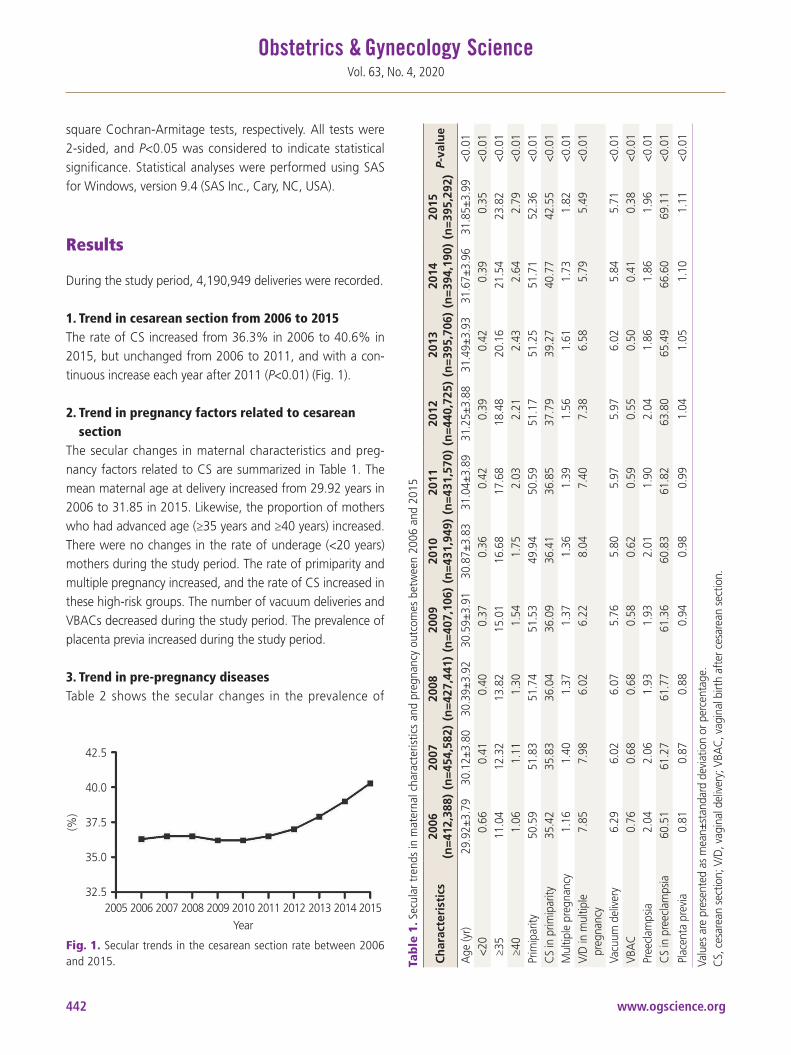
www.ogscience.org442
Vol. 63, No. 4, 2020
square Cochran-Armitage tests, respectively. All tests were 2-sided, and P<0.05 was considered to indicate statistical significance. Statistical analyses were performed using SAS for Windows, version 9.4 (SAS Inc., Cary, NC, USA).
Results
During the study period, 4,190,949 deliveries were recorded.
1. Trend in cesarean section from 2006 to 2015The rate of CS increased from 36.3% in 2006 to 40.6% in 2015, but unchanged from 2006 to 2011, and with a con-tinuous increase each year after 2011 (P<0.01) (Fig. 1).
2. Trend in pregnancy factors related to cesarean section
The secular changes in maternal characteristics and preg-nancy factors related to CS are summarized in Table 1. The mean maternal age at delivery increased from 29.92 years in 2006 to 31.85 in 2015. Likewise, the proportion of mothers who had advanced age (≥35 years and ≥40 years) increased. There were no changes in the rate of underage (<20 years) mothers during the study period. The rate of primiparity and multiple pregnancy increased, and the rate of CS increased in these high-risk groups. The number of vacuum deliveries and VBACs decreased during the study period. The prevalence of placenta previa increased during the study period.
3. Trend in pre-pregnancy diseasesTable 2 shows the secular changes in the prevalence of
Fig. 1. Secular trends in the cesarean section rate between 2006 and 2015.
(%)
42.5
40.0
37.5
35.0
32.5 2005 2006 2007 2008 2009 2010 2011 2012 2013 2014 2015
Year
Tab
le 1
. Sec
ular
tren
ds in
mat
erna
l cha
ract
eris
tics
and
preg
nanc
y ou
tcom
es b
etw
een
2006
and
201
5
Ch
arac
teri
stic
s20
06(n
=412
,388
)20
07(n
=454
,582
)20
08(n
=427
,441
)20
09(n
=407
,106
)20
10(n
=431
,949
)20
11(n
=431
,570
)20
12(n
=440
,725
)20
13(n
=395
,706
)20
14(n
=394
,190
)20
15(n
=395
,292
)P-
valu
e
Age
(yr)
29.9
2±3.
7930
.12±
3.80
30.3
9±3.
9230
.59±
3.91
30.8
7±3.
8331
.04±
3.89
31.2
5±3.
8831
.49±
3.93
31.6
7±3.
9631
.85±
3.99
<0.0
1<2
0 0.
660.
410.
400.
370.
360.
420.
390.
420.
390.
35<0
.01
≥35
11.0
412
.32
13.8
215
.01
16.6
817
.68
18.4
820
.16
21.5
423
.82
<0.0
1≥4
01.
061.
111.
301.
541.
752.
032.
212.
432.
642.
79<0
.01
Prim
ipar
ity
50.5
951
.83
51.7
451
.53
49.9
450
.59
51.1
751
.25
51.7
152
.36
<0.0
1C
S in
prim
ipar
ity
35.4
235
.83
36.0
436
.09
36.4
136
.85
37.7
939
.27
40.7
742
.55
<0.0
1M
ultip
le p
regn
ancy
1.16
1.40
1.37
1.37
1.36
1.39
1.56
1.61
1.73
1.82
<0.0
1V/
D in
mul
tiple
pr
egna
ncy
7.85
7.98
6.02
6.22
8.04
7.40
7.38
6.58
5.79
5.49
<0.0
1
Vacu
um d
eliv
ery
6.29
6.02
6.07
5.76
5.80
5.97
5.97
6.02
5.84
5.71
<0.0
1VB
AC
0.
760.
680.
680.
580.
620.
590.
550.
500.
410.
38<0
.01
Pree
clam
psia
2.
042.
061.
931.
932.
011.
902.
041.
861.
861.
96<0
.01
CS
in p
reec
lam
psia
60
.51
61.2
761
.77
61.3
660
.83
61.8
263
.80
65.4
966
.60
69.1
1<0
.01
Plac
enta
pre
via
0.81
0.87
0.88
0.94
0.98
0.99
1.04
1.05
1.10
1.11
<0.0
1
Valu
es a
re p
rese
nted
as
mea
n±st
anda
rd d
evia
tion
or p
erce
ntag
e.C
S, c
esar
ean
sect
ion;
V/D
, vag
inal
del
iver
y; V
BAC
, vag
inal
birt
h af
ter c
esar
ean
sect
ion.

www.ogscience.org 443
Ho Yeon Kim, et al. Cesarean section in South Korea
maternal pre-pregnancy diseases. The prevalence of pre-pregnancy DM, cardiovascular-pulmonary disease, hyperthy-roidism, and systemic disease was unchanged, but that of HTN and renal disease decreased during the study period. The prevalence of hypothyroidism increased.
4. Trend in pre-pregnancy factors related to cesarean section
The secular changes in pre-pregnancy risk factors related to CS were identified, as summarized in Table 3. The prevalence of pre-pregnancy obesity increased from 7.13% in 2006 to 10.67% in 2015. The prevalence of pre-pregnancy abnormal LFTs, high fasting glucose, and high TC also increased during the study period. The prevalence of pre-pregnancy smokers also increased.
Discussion
In this study, we evaluated secular trends in the rates of CS deliveries from 2006 to 2015 and their increase from 36.5% in 2007 to 40.3% in 2015. A previous study on changes in CS rate South Korea from 1982 to 2012 reported that the rate remained at 36% for the most recent 6 years of the study [11]. Our study evaluated 3 later years than that study and found that the rate increased significantly up to 40%. Increased maternal age, primiparity, pre-pregnancy obesity, multiple pregnancy, and placenta previa were contributing factors to the increase, while the rate of vacuum deliveries and VBACs decreased during the study period.
The rise in the rate of CS is a worldwide phenomenon. A multinational survey of the CS rates by the WHO reported an overall mean of 26.4% [12], and a recent global analysis of 150 countries reported a 12.4% increase in the rate of CS, with an average annual rate of increase of 4.4% over the past decades [13]. The trend in CS in Hong Kong, whose pop-ulation shares similar pelvic sizes as that in our population, demonstrated a 10% increase during a 20-year study period, resulting in CS rates up to 24–25% [14]. European coun-tries including Norway and England also showed moderate increases of 15.4% and 24.1%, respectively [15]. Our study found a significantly increasing rate over the study period, almost 3 times higher than that in 2015 when compared to the WHO-recommended CS rate. Herein, we explored the factors contributing to the increase in the rates of CS.Ta
ble
2. S
ecul
ar tr
ends
in th
e pr
eval
ence
of m
ater
nal p
re-p
regn
ancy
com
orbi
ditie
s be
twee
n 20
06 a
nd 2
015
Ch
arac
teri
stic
s20
06(n
=412
,388
)20
07(n
=454
,582
)20
08(n
=427
,441
)20
09(n
=407
,106
)20
10(n
=431
,949
)20
11(n
=431
,570
)20
12(n
=440
,725
)20
13(n
=395
,706
)20
14(n
=394
,190
)20
15(n
=395
,292
)P-
valu
e
DM
1.12
1.05
0.99
0.94
0.98
1.03
1.06
1.09
1.09
1.11
<0.0
1
HTN
0.79
0.80
0.81
0.80
0.83
0.75
0.56
0.53
0.51
0.48
<0.0
1
Rena
l dise
ase
1.98
1.92
1.91
1.83
1.88
1.81
1.76
1.77
1.57
1.60
<0.0
1
Car
dio-
vasc
ular
and
pu
lmon
ary
dise
ase
0.53
0.56
0.57
0.54
0.58
0.58
0.56
0.55
0.53
0.52
<0.0
1
Hyp
othy
roid
ism2.
282.
482.
762.
913.
073.
023.
133.
153.
043.
22<0
.01
Hyp
erth
yroi
dism
1.62
1.69
1.69
1.64
1.73
1.71
1.70
1.55
1.52
1.46
<0.0
1
Syst
emic
dise
ase
0.81
0.87
0.88
0.94
0.98
0.99
1.04
1.05
1.10
1.11
<0.0
1
Valu
es a
re p
rese
nted
as
perc
enta
ge. W
omen
wer
e cl
assif
ied
as h
avin
g pr
e-pr
egna
ncy
dise
ases
if t
hey
wer
e di
agno
sed
with
DM
(E10
–E14
), H
TN (I
10–I
15),
rena
l dise
ases
(N10
–N08
, N
10–N
16),
card
iova
scul
ar a
nd p
ulm
onar
y di
seas
es (I
20–I
28, I
60–I
69),
and
syst
emic
dise
ases
(M30
–M36
), ba
sed
on th
e IC
D-1
0 co
des.
DM
, dia
bete
s m
ellit
us; H
TN, h
yper
tens
ion;
ICD
-10,
Inte
rnat
iona
l Cla
ssifi
catio
n of
Dise
ases
, 10t
h Re
visio
n.
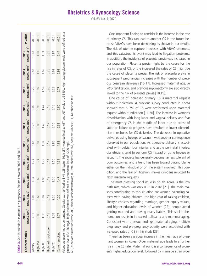
www.ogscience.org444
Vol. 63, No. 4, 2020
One important finding to consider is the increase in the rate of primary CS. This can lead to another CS in the future be-cause VBACs have been decreasing as shown in our results. The risk of uterine rupture increases with VBAC attempts, and this catastrophic event may lead to litigation problems. In addition, the incidence of placenta previa was increased in our population. Placenta previa might be the cause for the rise in rates of CS, or the increased the rates of CS might be the cause of placenta previa. The risk of placenta previa in subsequent pregnancies increases with the number of previ-ous cesarean deliveries [16,17]. Increased maternal age, in vitro fertilization, and previous myomectomy are also directly linked to the risk of placenta previa [18,19].
One cause of increased primary CS is maternal request without indication. A previous survey conducted in Korea showed that 6–7% of CS were preformed upon maternal request without indication [11,20]. The increase in women’s dissatisfaction with long labor and vaginal delivery and fear of emergency CS in the middle of labor due to arrest of labor or failure to progress have resulted in lower obstetri-cian thresholds for CS deliveries. The decrease in operative deliveries using forceps or vacuum was another consequence observed in our population. As operative delivery is associ-ated with pelvic floor injuries and acute perinatal injuries, obstetricians tend to perform CS instead of using forceps or vacuum. The society has generally become far less tolerant of poor outcomes, and a trend has been toward placing blame either on the individual or on the system involved. This con-dition, and the fear of litigation, makes clinicians reluctant to resist maternal requests.
The most pressing social issue in South Korea is the low birth rate, which was only 0.98 in 2018 [21]. The main rea-sons contributing to this situation are women balancing ca-reers with having children, the high cost of raising children, lifestyle choices regarding marriage, gender equity values, and higher education levels of women [22]; people avoid getting married and having many babies. This social phe-nomenon results in increased nulliparity and maternal aging. Consistent with previous findings, maternal aging, multiple pregnancy, and pre-pregnancy obesity were associated with increased rates of CS in this study [23].
There has been a gradual increase in the mean age of preg-nant women in Korea. Older maternal age leads to a further rise in the CS rate. Maternal aging is a consequence of wom-en’s higher education level, followed by marriage at an older Ta
ble
3. S
ecul
ar tr
ends
in m
ater
nal p
re-p
regn
ancy
fact
ors
betw
een
2006
and
201
5
Ch
arac
teri
stic
s20
06(n
=57,
690)
2007
(n=7
3,82
2)20
08(n
=72,
884)
2009
(n=7
8,46
8)20
10(n
=90,
100)
2011
(n=9
2,80
2)20
12(n
=99,
639)
2013
(n=8
9,09
3)20
14(n
=90,
094)
2015
(n=9
3,31
5)P-
valu
e
Obe
sity
7.13
7.03
7.34
7.74
8.42
8.76
9.09
9.29
9.87
10.6
7<0
.01
Hig
h A
ST
0.80
0.68
0.66
0.74
0.87
0.88
0.93
0.97
1.03
1.07
<0.0
1
Hig
h A
LT
1.30
0.97
1.00
1.22
1.37
1.45
1.47
1.52
1.69
1.71
<0.0
1
Hig
h fa
stin
g gl
ucos
e 0.
130.
120.
120.
140.
170.
170.
180.
200.
250.
23<0
.01
Hig
h TC
2.
332.
262.
362.
502.
863.
103.
153.
233.
623.
84<0
.01
Cur
rent
sm
okin
g 3.
774.
204.
474.
314.
885.
385.
805.
775.
935.
44<0
.01
Valu
es a
re p
rese
nted
as
perc
enta
ge. O
besit
y w
as d
efin
ed a
s BM
I ≥25
kg/
m2 . H
igh
LFTs
wer
e de
fined
as
AST
and
ALT
leve
ls of
>31
U/L
. Hig
h fa
stin
g gl
ucos
e le
vels
wer
e de
fined
as
a gl
ucos
e le
vel o
f ≥12
6 m
g/L.
Hig
h ch
oles
tero
l was
def
ined
as
a ch
oles
tero
l lev
el o
f ≥20
0 m
g/L.
AST
, asp
arta
te a
min
otra
nsfe
rase
; ALT
, ala
nine
am
inot
rans
fera
se; T
C, t
otal
cho
lest
erol
; BM
I, bo
dy m
ass
inde
x; L
FT, l
iver
func
tion
test
.

www.ogscience.org 445
Ho Yeon Kim, et al. Cesarean section in South Korea
age. Increased maternal age poses higher obstetric complica-tions because these women have more chronic diseases such as diabetes and HTN before and during pregnancy. The rate of preeclampsia was slightly decreased during 10 years, but a higher rate of CS in preeclamptic patients was observed. One possible explanation for the decrease in preeclampsia is the introduction of voucher programs for maternal health care covered by KNHI since 2008, and this program has allowed easier access to facility antenatal care and reduced the finan-cial burden of low-income pregnant women [24]. Maternal aging also results in an increased failure to progress due to a contracted pelvis [25]. Women with contracted pelvis are likely to pass their pelvic dimensions to their own offspring. This phenomenon implies that the application of life-saving CS can evolutionarily increase the rates of contracted pelvises [26]. These factors related to aging lead to increased rates of CS. Several previous statistics in many countries showed the link between older maternal age and the increased rates of CS [27].
Our results showed a 50% increase in the frequency in multiple pregnancy and decreased numbers of vaginal de-liveries in multiple pregnancy. The evolution of infertility therapy is one of the main reasons for the increase in mul-tiple pregnancy. In Korea, the Ministry of Health and Wel-fare has established regulations on the number of embryo transfers regarding maternal age and status of embryos such that no more than 3 cleavage-stage embryos or 2 blastocysts can be transferred even after the age of 35. This has had a direct influence on adverse obstetrical outcomes including increase in the rate of CS deliveries. There are more chances of malpresentation in multiple pregnancy and even without malpresentation, and doctors tend to perform CS instead of vaginal delivery.
Several pre-pregnancy factors were associated with in-creased rates of CS deliveries. Pre-pregnancy obesity was one important factor that increased the rate of CS. There has been a rapid rise in maternal pregnancy BMIs in the past decades, and the prevalence of adult obesity in South Korea increased from 29.7% in 2009 to 32.4% in 2015 due to in-crease in childhood obesity and human microbiome issues, in agreement with our results [28,29]. Obesity is a well-known factor that contributes to adverse maternal and neonatal outcomes [30]. Associated factors that lead to CS are chronic diseases of the mother, macrosomia due to uncontrolled blood glucose levels, excess intra-abdominal adipose tissue
causing obstructed labor, and induction agent intolerance due to large body volumes [31-33].
Despite the decreasing trend in smoking among people worldwide, our results showed that smoking prevalence among Korean women increased [34]. Changes in gender role, higher education, and social norms are contributing factors for the increase in smoking trend among women. Other than this factor, abnormal liver function and higher fasting glucose levels appear to be associated with the risk of CS. These factors may increase the rate of CS because they are strongly associated with comorbidities. However, the relationship between these factors and CS should be investi-gated in the future.
Several interventions have been implemented to reduce unnecessary CS, but their effectiveness was limited. How-ever, counseling low-risk primipara mothers who favor CS on the risk of CS and educating them on optimal gestational weight gain and the importance of controlling body weight before pregnancy may help to prevent unnecessary CS. In low-risk pregnancies, women who want a primary CS should be counseled on the increased risks of perinatal mortality and morbidity resulting from CS. They include higher blood loss, infection rates, thromboembolism, and adjacent organ injuries in mothers and respiratory problems and mortality in babies [35,36]. One previous study conducted in China re-ported that attendance in a prenatal education course signif-icantly reduced the rate of maternal CS requests [37]. Educa-tion on labor and normal delivery, the benefit of babies born vaginally, the perinatal risk of CS, pain management during labor, and the supporting roles of the husband or parents by skilled obstetricians, and maternal unit nurses or midwives may help to lower the number of CS as noted in this study.
This population-based cohort study reports trends in the rate of CS deliveries in South Korea over 10 recent years. However, the study had several limitations. First, social fac-tors such as educational background, socioeconomic sta-tus, labor force participation, and publicity of the hospital, which are known to be associated with CS rates, were not considered. Second, the results of our study are not com-pletely generalizable because our health insurance system is different from those in other countries and the population enrolled was East Asian. Third, the indications for primary CS were not described and may not be accurate because insur-ance does not cover the CS on maternal request.
We observed increasing trends in the rates of CS over the

www.ogscience.org446
Vol. 63, No. 4, 2020
recent decade. Maternal aging, increased primiparity, in-creased pre-pregnancy obesity, increased placenta previa, in-creased multiple pregnancy, and decreased vacuum deliveries and VBACs were major factors associated with the increased rates of CS. CS should be undertaken when medically neces-sary, and informed consent should include short-term and long-term effects of CS for the sake of the mother and the baby. Efforts should focus on providing CS to the appropriate women at the right time. In addition, public health programs to lower the rate of CS should be established, and multidisci-plinary discussion should be performed in the future, wheth-er national health insurance will cover “CS upon maternal request.”
Conflict of interest
No potential conflict of interest relevant to this article was reported.
Ethical approval
This study was approved by the Institutional Review Board (IRB) of the Korea University Medical Center (IRB No. 2019GR0256).
Patient consent
All the information that was provided for the study had been de-identified; therefore, we were unable to obtain informed consents from the participants.
References
1. Ahmed S, Tunçalp Ö. Burden of obstetric fistula: from measurement to action. Lancet Glob Health 2015;3:e243-4.
2. Boerma T, Ronsmans C, Melesse DY, Barros AJ, Barros FC, Juan L, et al. Global epidemiology of use of and dis-parities in caesarean sections. Lancet 2018;392:1341-8.
3. Betrán AP, Temmerman M, Kingdon C, Mohiddin A, Opiyo N, Torloni MR, et al. Interventions to reduce un-
necessary caesarean sections in healthy women and ba-bies. Lancet 2018;392:1358-68.
4. Sandall J, Tribe RM, Avery L, Mola G, Visser GH, Homer CS, et al. Short-term and long-term effects of caesarean section on the health of women and children. Lancet 2018;392:1349-57.
5. OECD. Health at a glance 2013: OECD indicators [In-ternet]. Paris: OECD Publishing; 2013. Available from: https://www.oecd.org/els/health-systems/Health-at-a-Glance-2013.pdf.
6. Appropriate technology for birth. Lancet 1985;2:436-7. 7. Finucane MM, Stevens GA, Cowan MJ, Danaei G, Lin JK,
Paciorek CJ, et al. National, regional, and global trends in body-mass index since 1980: systematic analysis of health examination surveys and epidemiological studies with 960 country-years and 9·1 million participants. Lan-cet 2011;377:557-67.
8. Danaei G, Finucane MM, Lu Y, Singh GM, Cowan MJ, Paciorek CJ, et al. National, regional, and global trends in fasting plasma glucose and diabetes prevalence since 1980: systematic analysis of health examination surveys and epidemiological studies with 370 country-years and 2·7 million participants. Lancet 2011;378:31-40.
9. Clark JM, Brancati FL, Diehl AM. The prevalence and eti-ology of elevated aminotransferase levels in the United States. Am J Gastroenterol 2003;98:960-7.
10. Farzadfar F, Finucane MM, Danaei G, Pelizzari PM, Cow-an MJ, Paciorek CJ, et al. National, regional, and global trends in serum total cholesterol since 1980: systematic analysis of health examination surveys and epidemio-logical studies with 321 country-years and 3·0 million participants. Lancet 2011;377:578-86.
11. Chung SH, Seol HJ, Choi YS, Oh SY, Kim A, Bae CW. Changes in the cesarean section rate in Korea (1982–2012) and a review of the associated factors. J Korean Med Sci 2014;29:1341-52.
12. Vogel JP, Betrán AP, Vindevoghel N, Souza JP, Torloni MR, Zhang J, et al. Use of the Robson classification to assess caesarean section trends in 21 countries: a secondary analysis of two WHO multicountry surveys. Lancet Glob Health 2015;3:e260-70.
13. Betrán AP, Ye J, Moller AB, Zhang J, Gülmezoglu AM, Torloni MR. The increasing trend in caesarean section rates: global, regional and national estimates: 1990–2014. PLoS One 2016;11:e0148343.
www.ogscience.org 447
Ho Yeon Kim, et al. Cesarean section in South Korea
14. Chung WH, Kong CW, To WW. Secular trends in cae-sarean section rates over 20 years in a regional obstetric unit in Hong Kong. Hong Kong Med J 2017;23:340-8.
15. Tollånes MC, Thompson JM, Daltveit AK, Irgens LM. Ce-sarean section and maternal education; secular trends in Norway, 1967-2004. Acta Obstet Gynecol Scand 2007;86:840-8.
16. Thurn L, Lindqvist PG, Jakobsson M, Colmorn LB, Klung-soyr K, Bjarnadóttir RI, et al. Abnormally invasive pla-centa-prevalence, risk factors and antenatal suspicion: results from a large population-based pregnancy cohort study in the Nordic countries. BJOG 2016;123:1348-55.
17. Marshall NE, Fu R, Guise JM. Impact of multiple cesare-an deliveries on maternal morbidity: a systematic review. Am J Obstet Gynecol 2011;205:262.e1-8.
18. Jauniaux E, Grønbeck L, Bunce C, Langhoff-Roos J, Collins SL. Epidemiology of placenta previa accreta: a systematic review and meta-analysis. BMJ Open 2019;9:e031193.
19. Tanimura K, Morizane M, Deguchi M, Ebina Y, Tanaka U, Ueno Y, et al. A novel scoring system for predicting adherent placenta in women with placenta previa. Pla-centa 2018;64:27-33.
20. Kim HK. Impact factors of Korean women’s cesarean section according to ecological approach. Korean J Women Health Nurs 2011;17:109-17.
21. Statistic Korea. Statistics of birth and death, 2018. Dae-jeon: Statistic Korea; 2019.
22. Song JE, Ahn JA, Lee SK, Roh EH. Factors related to low birth rate among married women in Korea. PLoS One 2018;13:e0194597.
23. Nkoka O, Ntenda PA, Senghore T, Bass P. Maternal over-weight and obesity and the risk of caesarean birth in Malawi. Reprod Health 2019;16:40.
24. Choe SA, Min HS, Cho SI. Decreased risk of preeclamp-sia after the introduction of universal voucher scheme for antenatal care and birth services in the Republic of Korea. Matern Child Health J 2017;21:222-7.
25. Kim SY, Park JY, Bak SE, Jang YR, Wie JH, Ko HS, et al. Effect of maternal age on emergency cesarean section. J Matern Fetal Neonatal Med 2019:1-8.
26. Pavličev M, Romero R, Mitteroecker P. Evolution of the human pelvis and obstructed labor: new explanations
of an old obstetrical dilemma. Am J Obstet Gynecol 2020;222:3-16.
27. Bayrampour H, Heaman M. Advanced maternal age and the risk of cesarean birth: a systematic review. Birth 2010;37:219-26.
28. Ng M, Fleming T, Robinson M, Thomson B, Graetz N, Margono C, et al. Global, regional, and national preva-lence of overweight and obesity in children and adults during 1980–2013: a systematic analysis for the Global Burden of Disease Study 2013. Lancet 2014;384:766-81.
29. Lee M. Research trends in obesity & obesogenic environ-ments in Korea. Nutr Res Pract 2019;13:461-72.
30. Ovesen P, Rasmussen S, Kesmodel U. Effect of prepreg-nancy maternal overweight and obesity on pregnancy outcome. Obstet Gynecol 2011;118:305-12.
31. Arrowsmith S, Wray S, Quenby S. Maternal obesity and labour complications following induction of labour in prolonged pregnancy. BJOG 2011;118:578-88.
32. Al-Kubaisy W, Al-Rubaey M, Al-Naggar RA, Karim B, Mohd Noor NA. Maternal obesity and its relation with the cesarean section: a hospital based cross sectional study in Iraq. BMC Pregnancy Childbirth 2014;14:235.
33. Gaudet L, Wen SW, Walker M. The combined effect of maternal obesity and fetal macrosomia on pregnancy outcomes. J Obstet Gynaecol Can 2014;36:776-84.
34. Chang Y, Kang HY, Lim D, Cho HJ, Khang YH. Long-term trends in smoking prevalence and its socioeconom-ic inequalities in Korea, 1992–2016. Int J Equity Health 2019;18:148.
35. Malloy MH. Impact of cesarean section on intermediate and late preterm births: United States, 2000–2003. Birth 2009;36:26-33.
36. Saeed KB, Greene RA, Corcoran P, O’Neill SM. Incidence of surgical site infection following caesarean section: a systematic review and meta-analysis protocol. BMJ Open 2017;7:e013037.
37. Gao Y, Tang Y, Tong M, Du Y, Chen Q. Does attendance of a prenatal education course reduce rates of caesarean section on maternal request? A questionnaire study in a tertiary women hospital in Shanghai, China. BMJ Open 2019;9:e029437.

www.ogscience.org448
Original ArticleObstet Gynecol Sci 2020;63(4):448-454https://doi.org/10.5468/ogs.20004pISSN 2287-8572 · eISSN 2287-8580
Evaluation of maternal rhesus blood type as a risk factor in adverse pregnancy outcomes in Korea: a nationwide health insurance database studyYihua Jin, MD1, Meari Dong, MD1, Seung Woo Yang, MD, PhD1, Kyu-Min Lee, BS2, Sung Won Han, PhD2, Shin Hee Seo, MD1, Ajin Lee, MD1, In Sook Sohn, MD, PhD1, Han Sung Kwon, MD, PhD1, Geum Joon Cho, MD, PhD3, Han Sung Hwang, MD, PhD1
1Division of Maternal and Fetal Medicine, Department of Obstetrics and Gynecology, Research Institute of Medical Science, Konkuk University School of Medicine; 2School of Industrial Management Engineering, Korea University; 3Department of Obstetrics and Gynecology, Korea University College of Medicine, Seoul, Korea
ObjectivesThe current study aimed to investigate whether pregnancy outcomes are affected by maternal rhesus (Rh) status by comparing the primigravida pregnancy outcomes of Rh-negative women with those of Rh-positive women.
MethodsThe study data were collected from the Korea National Health Insurance Claims Database and the National Health Screening Program for Infants and Children. In total, 1,664,882 primigravida women who gave birth between January 1, 2007 and December 31, 2014, were enrolled in this study. As the risk and severity of sensitization response increases with each subsequent pregnancy, only primigravida women were enrolled. The patients were divided into 2 groups according to Rh status, and the pregnancy outcomes were compared.
ResultsIn total, 1,661,320 women in the Rh-positive group and 3,290 in the Rh-negative group were assessed. With regard to adverse pregnancy outcomes, there was no statistically significant difference between the 2 groups in terms of the prevalence of preeclampsia, postpartum hemorrhage, abruptio placenta, placenta previa, and uterine artery embolization. A univariate analysis revealed that none of the adverse pregnancy outcomes were significantly correlated to Rh status (preeclampsia: odds ratio [OR], 1.00, 95% confidence interval [CI], 0.81–1.23; postpartum hemorrhage: OR, 1.10, 95% CI, 0.98–1.24; abruptio placenta: OR, 0.80, 95% CI, 0.46–1.37; and placenta previa: OR, 1.08, 95% CI, 0.78–1.42). The adjusted ORs of postpartum hemorrhage and preterm birth did not significantly differ.
ConclusionMaternal Rh status is not associated with adverse outcomes in primigravida women.
Keywords: Rhesus blood-group system; Pregnancy outcomes; Gravidity; National health programs
Received: 2019.12.28. Revised: 2020.02.14. Accepted: 2020.03.04.Corresponding author: Han-Sung Hwang, MD, PhDDepartment of Obstetrics and Gynecology, Konkuk University School of Medicine, 120 Neungdong-ro, Gwangjin-gu, Seoul 05029, KoreaE-mail: [email protected]://orcid.org/0000-0003-0622-0701
Geum Joon Cho, MD, PhDDepartment of Obstetrics and Gynecology, Korea University College of Medicine, 148 Gurodong-ro, Guro-gu, Seoul 08308, KoreaE-mail: [email protected]://orcid.org/0000-0001-6761-0944
Articles published in Obstet Gynecol Sci are open-access, distributed under the terms of the Creative Commons Attribution Non-Commercial License (http://creativecommons.org/licenses/by-nc/3.0/) which permits unrestricted non-commercial use, distribution, and reproduction in any medium, provided the original work is properly cited.
Copyright © 2020 Korean Society of Obstetrics and Gynecology

www.ogscience.org 449
Yihua Jin, et al. Rhesus blood type and pregnancy outcome
Introduction
A person’s blood type is determined according to the specific antigen types on the erythrocyte membranes of red blood cells (RBCs). The 2 main factors that determine the blood type are ABO (A, B, AB, and O) and rhesus (Rh) (positive or negative). Karl Landsteiner first discovered the ABO blood group system in 1900 when he was investigating the causes of some fatal transfusions [1]. The ABO blood group system was named according to the different agglutinins, or blood group antigens, including A, B, and H (or O) antigens, on the surface of human RBCs [1]. Based on the presence or ab-sence of the Rh factor, the blood group system is called the Rh blood group system. Rh represents the first 2 letters of the name Macacus Rhesus. In 1940, during animal experiments, Landsteiner and other scientists discovered that rhesus mon-keys and most human RBCs have antigenic Rh blood types, and this was used in naming the system [2]. With continuous studies of the Rh blood groups, the Rh blood group system was found to be the most complex system in the RBCs [3]. The discovery of the Rh blood type has played an important role not only in guiding blood transfusions more scientifically but also in improving experimental diagnoses and clinical im-munotherapy.
The blood type may affect human health and diseases with a wide range of expression in human cells and tissues, in-cluding platelets, epithelium, and vascular endothelium [4,5]. Therefore, several studies showed the clinical significance of the biological characteristics of the ABO blood system, par-ticularly with regard to cancer, cardiovascular disease, and pregnancy-related disease [6-8]. Numerous reports revealed that the ABO blood group may be associated with some risk factors for unfavorable pregnancy outcomes [9].
However, data about whether maternal Rh blood type alone, without consideration of alloimmune sensitization, is associated with the development of pregnancy-related dis-eases are limited. Moreover, the Rh-negative population is extremely small. Thus, a large population must be evaluated to assess the role of Rh blood type in identifying women at risk of developing pregnancy-related complications.
Thus, this study aimed to investigate whether pregnancy outcomes are affected by maternal Rh status by comparing the primigravida pregnancy outcomes of Rh-negative women with those of Rh-positive women in a nationwide population study.
Materials and methods
1. Health care in KoreaApproximately 97% of the Korean population is enrolled in the Korea National Health Insurance (KNHI) program. All claims data are stored in the KNHI claims database. As part of the KNHI system, a National Health Screening Program for Infants and Children (NHSP-IC) was started in 2007, and it includes information about physical examination findings, anthropometric measurements, and developmental screen-ing results after birth. This study used information from the KNHI claims database to identify all women who gave birth between January 1, 2007 and December 31, 2014. More-over, whether these women were Rh-positive or Rh-negative based on the applicable codes from the International Clas-sification of Disease, 10th Revision was assessed. The data of women who met the inclusion criteria were linked to those of their offspring in the NHSP-IC database.
2. Dataset and outcomesFig. 1 shows the flowchart of participant enrollment. Us-ing the KNHI claims data, we identified all women who gave birth between January 1, 2007 and December 31, 2014 (n=3,383,282). The maternal datasets were merged with those in the NHSP-IC database. Multigravida women (n=1,650,500), those whose offspring had not undergone
Fig. 1. Flowchart of participant enrollment. NHSP-IC, National Health Screening Program for Infants and Children.
Deliveries between January 1, 2007and December 31, 2014
(n=3,383,282)
Excluded n=67,900• No NHSP-IC n=66,839• Women with missing data n=1,061
Initially included in study(n=1,732,782)
Excluded multi-gravida(n=1,650,500)
Final women available for analysis(n=1,664,880)

www.ogscience.org450
Vol. 63, No. 4, 2020
NHSP-IC health examinations (n=66,839), and those with missing data (n=1,061) were excluded from the study. Data regarding maternal and offspring outcomes were extracted. The information included pregnancy outcomes, such as parity, type of delivery, pulmonary embolism, postpartum hemorrhage, abruptio placenta, placenta previa, and uterine artery embolization. Furthermore, the characteristics of the neonates, such as sex, whether delivered preterm, and birth weight, were evaluated. Preterm birth was defined as birth at a gestational age <37 weeks, low birth weight (LBW) as birth weight <2.5 kg, and large for gestational age (LGA) as birthweight >4.0 kg.
3. Statistical analysisContinuous and categorical variables were expressed as mean±standard deviation and number (percentage), respec-tively. The clinical characteristics of the participants were compared using t-test for continuous variables and the χ2
test for categorical variables. The risks of Rh-negative blood type were evaluated via multiple regression analyses. The risk of postpartum hemorrhage and preterm birth was adjusted for each related risk factor (postpartum hemorrhage: age, multiple pregnancy, cesarean section, preterm birth, LBW, LGA, preeclampsia, abruptio placenta, and placenta previa; preterm birth: age, multiple pregnancy, and preeclampsia). All tests were 2-sided, and a P-value <0.05 was considered statistically significant. Statistical analyses were performed using SAS for Windows (version 9.4; SAS, Cary, NC, USA).
Results
1. Baseline characteristics of the study populationThe clinical characteristics of the participants are shown in Table 1. There were no significant differences in terms of age at delivery and multiple pregnancy and cesarean sec-
Table 1. Clinical characteristics of the participants
Characteristics Rh-positive (n=1,661,320) Rh-negative (n=3,290) P-value
Age (yr) 29.8±3.7 29.7±3.5 0.444
Age >35 yr at delivery 161,506 (9.7) 317 (8.8) 0.058
Multiple gestation 33,479 (2.0) 82 (2.3) 0.270
Cesarean section 617,044 (37.1) 1,311 (36.4) 0.323
Preeclampsia 40,998 (2.5) 89 (2.5) 0.998
Postpartum hemorrhage 130,422 (7.9) 308 (8.5) 0.124
Placenta abruption 7,528 (0.5) 13 (0.4) 0.407
Placenta previa 18,899 (1.1) 43 (1.2) 0.757
Uterine artery embolization 2,101 (0.1) 6 (0.2) 0.501
Data were represented as mean±standard deviation and number (%).Rh, rhesus.
Table 2. Neonatal weight differences between the rhesus (Rh)-positive and Rh-negative groups
Characteristics Rh-positive (n=1,661,320) Rh-negative (n=3,290) P-value
Male sex 853,655 (51.38) 1,883 (52.20) 0.325
Preterm birth 57,406 (3.46) 129 (3.58) 0.691
Neonatal weight (g) 3.18±0.49 3.17±0.52 0.301
Normal weight 1,520,122 (91.50) 3,316 (91.93) 0.273
Low birth weight 82,309 (4.95) 181 (5.02) 0.221
Large birth weight 58,889 (3.54) 110 (3.05) 0.313
Data were presented as mean±standard deviation and number (%). Low birth weight: <2,500 g, large birth weight: >4,000 g, and preterm delivery: <37 gestational weeks.

www.ogscience.org 451
Yihua Jin, et al. Rhesus blood type and pregnancy outcome
tion rates between the Rh-positive group (n=1,661,320) and Rh-negative group (n=3,290). Moreover, adverse delivery outcomes did not significantly differ between the 2 groups. Moreover, preterm birth rates and birth weight (corrected for gestational age) did not significantly differ between the Rh-positive and Rh-negative groups (Table 2).
2. Univariate regression analysis of Rh blood type as a risk factor for pregnancy outcomes
Univariate regression analyses were performed to evaluate the associations between Rh blood type and various clinical outcomes (Table 3). The Rh blood type was not a risk factor for preterm birth (odds ratio [OR], 1.13, 95% confidence in-terval [CI], 0.91–1.41), LBW (OR, 1.01, 95% CI, 0.87–1.18), and LGA (OR, 0.86, 95% CI, 0.71–1.04). Rh blood type was not a risk factor for adverse pregnancy outcomes (preeclamp-sia: OR, 1.00, 95% CI, 0.81–1.23; postpartum hemorrhage: OR, 1.10, 95% CI, 0.98–1.24; abruptio placenta: OR, 0.80, 95% CI, 0.46–1.37; and placenta previa: OR, 1.08, 95% CI, 0.78–1.42). Moreover, age-adjusted univariate regression analyses revealed no significant relationships between the Rh blood type and these factors.
3. Multiple regression analysis of rhesus blood type as a risk factor for postpartum hemorrhage and preterm birth
Based on the univariate analysis, the adjusted ORs for post-partum hemorrhage and preterm birth were analyzed be-
cause there was no significant relationship between Rh blood type and clinical outcomes (Table 4). The risk factors for post-partum hemorrhage were age, multiple pregnancy, cesarean section, preterm birth, LBW, LGA, preeclampsia, abruptio placenta, and placenta previa. Preterm birth was adjusted for age, multiple pregnancy, and preeclampsia. After adjust-ing for the factors, no relationships were found between Rh blood type and postpartum hemorrhage (OR, 1.10, 95% CI, 0.98–1.23) and preterm birth (OR, 0.02, 95% CI, 0.85–1.23).
Discussion
The proportions of blood type differ according to race, re-gion, and ethnicity. In general, Africa, the Middle East, Eu-rope, India, and Central Asia have higher Rh-negative blood rates than other regions. In Korea, Rh-negative is considered a rare blood type according to the Korean Red Cross stan-dards, with an incidence of 0.4%; however, pregnancy can lead to several complications among Rh-negative women [10]. Currently, a non-O blood group may affect hemostatic balance disturbances and lead to an increased risk of embo-lization compared to an O blood group [11]. By contrast, an-other study showed a weak relationship between ABO blood type and gestational hypertension [12,13]. However, whether Rh blood type (negative or positive) is correlated to pregnan-cy outcomes is not known. Therefore, this study focused on the association between Rh-positive and Rh-negative blood groups and maternal pregnancy outcomes in 1,664,882 primigravida women in a large population study. The clinical characteristics did not differ significantly between Rh-positive (n=1,661,320) and Rh-negative women (n=3,290).
Several studies showed that the Rh blood type plays an im-portant role in neonatal alloimmune disorders. The immune
Table 3. Univariate regression analysis of rhesus blood type as a risk factor of pregnancy outcomes
Characteristics OR (95% CI)Age-adjusted OR (95% CI)
Multiple pregnancy 1.13 (0.91–1.41) 1.15 (0.92–1.43)
Cesarean section 0.97 (0.90–1.03) 0.97 (0.91–1.04)
Preterm birth 1.04 (0.87–1.24) 1.04 (0.87–1.24)
LBW 1.01 (0.87–1.18) 1.02 (0.88–1.18)
LGA 0.86 (0.71–1.04) 0.86 (0.71–1.04)
Preeclampsia 1.00 (0.81–1.23) 1.00 (0.81–1.24)
Postpartum hemorrhage 1.10 (0.98–1.24) 1.10 (0.98–1.23)
Placental abruption 0.80 (0.46–1.37) 0.80 (0.46–1.38)
Placental previa 1.08 (0.78–1.42) 1.07 (0.79–1.44)
OR, odds ratio; CI, confidence interval; LBW, low birth weight; LGA, large for gestational age.
Table 4. Multiple regression analysis of rhesus blood type as a risk factor of postpartum hemorrhage and preterm birth
Characteristics Risk factor: adjusted OR (95% CI)
Postpartum hemorrhagea) 1.10 (0.98–1.23)
Preterm birthb) 0.02 (0.85–1.23)
OR, odds ratio; CI, confidence interval.a)Postpartum hemorrhage: adjusted for age, multiple pregnancy, cesarean section, preterm birth, low birth weight, large birth weight, preeclampsia, abruptio placenta, and placenta previa; b)Preterm birth: adjusted for age, multiple pregnancy, and preeclampsia.

www.ogscience.org452
Vol. 63, No. 4, 2020
system of a Rh-negative mother considers Rh-positive fetal cells as foreign substances. Then, the mother’s body produc-es antibodies against fetal blood cells. Antibodies, such as IgG1 and IgG3, cross back through the placenta to the baby [14,15]. Then, they destroy the baby’s circulating RBCs, and this eventually develops into complications, such as preterm delivery, LBW, hydrops fetalis, hyperbilirubinemia, anemia, and bilirubin-induced neurological dysfunction [16-20].
In this study, there was no significant association between Rh blood type and neonatal outcomes, including preterm birth, LBW, and LGA. As previously mentioned, as the risk and severity of sensitization response increases with each subsequent pregnancy, we only included primigravida women in this analysis to reduce selection bias. During the first pregnancy, the initial exposure to fetal RBCs results in the formation of IgM antibodies, and these do not cross the placental barrier. Thus, no differences were observed in the first pregnancies of Rh-negative women [21]. Based on our results, the relationship between fetal gross and Rh blood type without an alloimmune effect is weak.
The World Health Organization reports that the major con-tributors to maternal death or maternal mortality are post-partum hemorrhage and hypertensive disorders [22]. Various studies have investigated whether there is an association be-tween maternal ABO blood type and maternal adverse preg-nancy outcomes [8,12,23-25]. In brief, maternal ABO blood type was correlated to the risk of preeclampsia. However, its association with GDM, preterm delivery, LBW, and SGA re-mains controversial.
Compared to the ABO types, studies about Rh blood type are limited. Thus, this study evaluated the differences in the rate of cesarean section, preeclampsia, postpartum hemor-rhage, abruptio placenta, placenta previa, and uterine artery embolization among pregnant women with different Rh blood types. However, there was no significant association between Rh blood type and pregnancy outcomes, even pre-eclampsia, in primiparous women. Lee et al. [24] showed that Rh D-positive mothers had a slightly increased risk of preeclampsia in a large cohort study in Sweden (OR, 1.07, 95% CI, 1.03–1.10). However, in this study, multiparous women were enrolled, and only the Rh D antigen was evalu-ated. In a previous research, more than 50 Rh antigens have been identified at the serological level encoding 2 genes (RHD and RHCE). Among these antigens, Rh D, C, c, E, and e were considered the most clinically significant, and Rh D has the
strongest antigenicity [26]. In our study, different findings were obtained, which might be attributed to the fact that we did not assess the differences in these antigens.
However, we did not find any associations between Rh blood type and adverse pregnancy outcomes. This result might be explained partly by the limitations of our research. First, this study only included Rh blood type classifications. However, the differences between the specific Rh blood type or the ABO classification were not assessed because the ABO blood group is not considered a disease category in the KNHI program. Second, as described above, the exclu-sion of multigravida pregnant women might have influenced the relatively varying results of the rates of preeclampsia compared to other previous studies. Third, with regard to neonatal outcomes, attention is now shifting towards short- and long-term morbidity [27]; however, in this study, only neonatal weight was included, and more detailed data are required to evaluate neonatal outcomes. To the best of our knowledge, this is the first nationwide retrospective research about the association between Rh blood type and adverse pregnancy outcomes in Korea. Hence, further studies must be conducted for a more detailed evaluation of the Rh blood type among pregnant women.
In conclusion, there were no significant differences in the risk of adverse pregnancy outcomes between Rh-positive and Rh-negative primigravida women in Korea.
Acknowledgments
This research was supported by the Basic Science Research Program through the National Research Foundation of Korea (NRF) funded by the Ministry of Science, ICT & Future Plan-ning (NRF-2017R1C1B2010487). This research was support-ed by a grant from the Korea Health Technology R&D Project through the Korea Health Industry Development Institute (KHIDI) funded by the Ministry of Health & Welfare, Republic of Korea (grant number: HI17C1713).
Conflict of interest
No potential conflict of interest relevant to this article was reported.

www.ogscience.org 453
Yihua Jin, et al. Rhesus blood type and pregnancy outcome
Ethical approval
The study protocol was approved by the institutional review board of Korea University Medical Center (IRB number: 2019GR0256).
Patient consent
This paper is a retrospective study and does not require writ-ten informed consent.
References
1. Landsteiner K, Levine P. On individual differences in hu-man blood. J Exp Med 1928;47:757-75.
2. Wiener AS. History of the rhesus blood types. J Hist Med Allied Sci 1952;7:369-83.
3. Anstee DJ, Tanner MJ. Biochemical aspects of the blood group Rh (rhesus) antigens. Baillieres Clin Haematol 1993;6:401-22.
4. Liumbruno GM, Franchini M. Beyond immunohaema-tology: the role of the ABO blood group in human dis-eases. Blood Transfus 2013;11:491-9.
5. Franchini M, Mengoli C, Lippi G. Relationship between ABO blood group and pregnancy complications: a sys-tematic literature analysis. Blood Transfus 2016;14:441-8.
6. Yu H, Xu N, Li ZK, Xia H, Ren HT, Li N, et al. Association of ABO blood groups and risk of gastric cancer. Scand J Surg 2019:1457496919863886.
7. Paquette M, Dufour R, Baass A. ABO blood group is a cardiovascular risk factor in patients with familial hyper-cholesterolemia. J Clin Lipidol 2018;12:383-389.e1.
8. Witsenburg CP, Rosendaal FR, Middeldorp JM, Van der Meer FJ, Scherjon SA. Factor VIII levels and the risk of pre-eclampsia, HELLP syndrome, pregnancy related hy-pertension and severe intrauterine growth retardation. Thromb Res 2005;115:387-92.
9. Seyfizadeh N, Seyfizadeh N, Yousefi B, Borzoueisileh S, Majidinia M, Shanehbandi D, et al. Is there association between ABO blood group and the risk factors of unfa-vorable outcomes of pregnancy? J Matern Fetal Neona-tal Med 2015;28:578-82.
10. Duerbeck NB, Seeds JW. Rhesus immunization in preg-nancy: a review. Obstet Gynecol Surv 1993;48:801-10.
11. Alpoim PN, de Barros Pinheiro M, Junqueira DR, Freitas LG, das Graças Carvalho M, Fernandes AP, et al. Pre-eclampsia and ABO blood groups: a systematic review and meta-analysis. Mol Biol Rep 2013;40:2253-61.
12. Phaloprakarn C, Tangjitgamol S. Maternal ABO blood group and adverse pregnancy outcomes. J Perinatol 2013;33:107-11.
13. Scott JR, Beer AA. Immunologic aspects of pre-eclamp-sia. Am J Obstet Gynecol 1976;125:418-27.
14. Finning K, Martin P, Summers J, Daniels G. Fetal geno-typing for the K (Kell) and Rh C, c, and E blood groups on cell-free fetal DNA in maternal plasma. Transfusion 2007;47:2126-33.
15. Andersen AS, Praetorius L, Jørgensen HL, Lylloff K, Larsen KT. Prognostic value of screening for irregular antibodies late in pregnancy in rhesus positive women. Acta Obstet Gynecol Scand 2002;81:407-11.
16. Thompson PJ, Greenough A, Brooker R, Nicolaides KH, Gamsu HR. Antenatal diagnosis and outcome in hydrops fetalis. J Perinat Med 1993;21:63-7.
17. Kobets TV, Usachenko IE. Risk factors of complicated postnatal period in low birth weight infants. Tsitol Genet 2001;35:49-53.
18. Fan J, Lee BK, Wikman AT, Johansson S, Reilly M. As-sociations of rhesus and non-rhesus maternal red blood cell alloimmunization with stillbirth and preterm birth. Int J Epidemiol 2014;43:1123-31.
19. Zipursky A, Bhutani VK, Odame I. Rhesus disease: a global prevention strategy. Lancet Child Adolesc Health 2018;2:536-42.
20. Bel Hadj I, Boukhris R, Khalsi F, Namouchi M, Bougmiza I, Tinsa F, et al. ABO hemolytic disease of newborn : does newborn’s blood group a risk factor? Tunis Med 2019;97:455-60.
21. Fyfe TM, Ritchey MJ, Taruc C, Crompton D, Galliford B, Perrin R. Appropriate provision of anti-D prophylaxis to RhD negative pregnant women: a scoping review. BMC Pregnancy Childbirth 2014;14:411.
22. Khan KS, Wojdyla D, Say L, Gülmezoglu AM, Van Look PF. WHO analysis of causes of maternal death: a system-atic review. Lancet 2006;367:1066-74.
23. May D. Letter: Maternal blood group A and pre-eclamp-sia. BMJ 1973;4:738.

www.ogscience.org454
Vol. 63, No. 4, 2020
24. Lee BK, Zhang Z, Wikman A, Lindqvist PG, Reilly M. ABO and RhD blood groups and gestational hyperten-sive disorders: a population-based cohort study. BJOG 2012;119:1232-7.
25. Chen Y, Chen C, Ke X, Xiong L, Shi Y, Li J, et al. Analysis of circulating cholesterol levels as a mediator of an as-sociation between ABO blood group and coronary heart disease. Circ Cardiovasc Genet 2014;7:43-8.
26. Chou ST, Westhoff CM. Molecular biology of the Rh system: clinical considerations for transfusion in sickle cell disease. Hematology Am Soc Hematol Educ Program 2009:178-84.
27. Smits-Wintjens VE, Walther FJ, Lopriore E. Rhesus hae-molytic disease of the newborn: postnatal management, associated morbidity and long-term outcome. Semin Fetal Neonatal Med 2008;13:265-71.

www.ogscience.org 455
Original ArticleObstet Gynecol Sci 2020;63(4):455-463https://doi.org/10.5468/ogs.19131pISSN 2287-8572 · eISSN 2287-8580
Introduction
Preterm birth (PTB), defined as birth before 37 weeks of ges-tation, is a major cause of neonatal morbidity and mortality [1,2]. Over the past decade, the prevalence of preterm births has steadily increased worldwide. Morbidity and mortality of infants born before 34 weeks of gestation are more than those of infants born after 34 weeks. Therefore, antenatal steroid administration is recommended for patients with preterm labor (PTL) and preterm premature rupture of mem-branes (pPROM). However, prediction methods and therapies
Cervicovaginal fluid cytokines as predictive markers of preterm birth in symptomatic womenSunwha Park, MD1, Young-Ah You, PhD2, Hayoung Yun, MD1, Suk-Joo Choi, MD, PhD3, Han-Sung Hwang, MD, PhD4, Sae-Kyung Choi, MD, PhD5, Seung Mi Lee, MD, PhD6, Young Ju Kim, MD, PhD1,2
1Department of Obstetrics and Gynecology, Ewha Womans University Mokdong Hospital; 2Ewha Medical Research Institute, College of Medicine, Ewha Womans University; Department of Obstetrics and Gynecology, 3Samsung Medical Center, Sungkyunkwan University School of Medicine, 4Konkuk University Medical Center, Konkuk University School of Medicine, 5College of Medicine, The Catholic University of Korea, 6Seoul National University College of Medicine, Seoul, Korea
ObjectiveHere, we investigated whether cytokines in the cervicovaginal fluid (CVF) can be predictive markers of preterm birth (PTB).
MethodsA multi-center prospective cohort study was conducted on 59 singleton pregnant women hospitalized for preterm labor (PTL) and/or preterm premature rupture of membranes (pPROM) between 22 weeks and 36 weeks 6 days of gestation from 2014 to 2015. The levels of 13 inflammatory cytokines (macrophage inflammatory protein [MIP]-1α, MIP-1β, tumor necrosis factor [TNF]-α, interleukin [IL]-1β, IL-6, IL-8, IL-17α, granulocyte colony stimulating factor [G-CSF], IL-7, IL-4, IL-5, IL-10, and IL-13) were measured using a multiplex bead-based immunoassay and that of fetal fibronectin (fFN) was measured using enzyme-linked immunosorbent assay (ELISA). Statistical analyses were performed using Student’s t-test, Mann-Whitney U test, Pearson’s correlation, and receiver operating characteristic (ROC) curve analysis in SPSS version 20.0.
ResultsAmong the 13 cytokines assessed, the levels of 3 cytokines (MIP-1α, IL-6, and IL-7) were negatively correlated with gestational age at delivery (P=0.028, P=0.002, and P=0.018, respectively). Sensitivities of MIP-1α, IL-6, and IL-17α were 70%, 80%, and 75%, respectively, and their specificities were 57%, 65%, and 69%, respectively. The sensitivity and specificity of fFN were 33% and 95%, respectively.
ConclusionIn symptomatic women diagnosed with PTL and/or pPROM, cytokines from cervicovaginal fluid, especially IL-6 and IL-17α, could be better predictive markers of PTB than fFN.
Keywords: Cytokines; Interleukin-6; Interleukin-17A; Biomarkers; Preterm birth
Received: 2019.07.26. Revised: 2020.03.22. Accepted: 2020.04.15.Corresponding author: Young Ju Kim, MD, PhDDepartment of Obstetrics and Gynecology and Ewha Medical Research Institute, College of Medicine, Ewha Womans University, 1071 Anyangcheon-ro, Yangcheon-gu, Seoul 07985, KoreaE-mail: [email protected]://orcid.org/0000-0002-3153-3008
Articles published in Obstet Gynecol Sci are open-access, distributed under the terms of the Creative Commons Attribution Non-Commercial License (http://creativecommons.org/licenses/by-nc/3.0/) which permits unrestricted non-commercial use, distribution, and reproduction in any medium, provided the original work is properly cited.
Copyright © 2020 Korean Society of Obstetrics and Gynecology

www.ogscience.org456
Vol. 63, No. 4, 2020
for the same are not well studied. PTB can be classified into two categories: spontaneous and
iatrogenic. Spontaneous PTB accounts for up to 75% of all PTBs [3]. About 45% of PTBs are caused by PTL with intact membranes, while 30% are caused by the pre-labor rupture of fetal membranes. Causes of spontaneous PTB include activation of inflammatory reactions in the gestational tis-sues and secretion of inflammatory cytokines as an immune response to ascending infection of the genital tract and ab-normal vaginal microorganisms [4-7].
Many studies aimed at predicting PTB have been con-ducted. Commonly used screening tests include risk scoring, cervical length measurement, and detection of biochemical markers. Risk scoring involves quantification of risk factors for spontaneous PTB and/or pPROM, including PTB history, cervical conization history, low socioeconomic status, low body mass index, smoking, drug use, anxiety, multifetal ges-tation, gestational diabetes, and gestational hypertension [6-9]. Tests for fetal fibronectin (fFN) and phosphorylated insulin-like growth factor binding protein-1 (IGFBP1) are now commercially available [10,11]. The level of fFN in cervico-vaginal fluid (CVF), leaked due to the disruption of maternal choriodecidual tissues and through matrix remodeling, was used as a diagnostic marker for predicting PTB using a non-invasive method. Lockwood et al. described the clinical utility of fFN to predict spontaneous PTB in symptomatic women with high sensitivity (81.7%) and specificity (82.5%), based on a fFN threshold concentration of ≥50 ng/mL [3,8]. How-ever, its specificity and sensitivity were confounded by un-protected vaginal intercourse, digital examination, bleeding, or contamination with amniotic fluid [1,12]. Cervical length measurement using transvaginal sonograms in patients with PTL and pPROM have also been used to predict PTB; how-ever, it requires a skilled ultrasonographer and shows limited correlation with CVF [13-15].
Increased levels of inflammatory cytokines in CVF signify intra-amniotic infection that causes PTB [16]. Several inflam-matory cytokines, including interleukin (IL)-1, -2, -4, -6, -8, -10, -12 and -17, tumor necrosis factor-alpha (TNF-α), inter-feron gamma (INF-γ), regulated on activation, normal T cell expressed and secreted (RANTES), and C-reactive protein (CRP), have been detected in the CVF, amniotic fluid, and blood of asymptomatic and symptomatic pregnant women [3,4,17,18]. IL-6 and matrix metalloproteinase-8 (MMP-8) levels in the amniotic fluid were significantly correlated with
pPROM [19,20]. We also found that the levels of cytokines such as IL-1b, IL-6, IL-7, IL-7a, and TNF-α were significantly increased in the amniotic fluid of patients with cervical insuf-ficiency [16]. Thereafter, the objective of our study was to de-termine whether inflammatory cytokines could be measured using non-invasive methods. Recent studies have shown that IL-6, IL-8, macrophage inflammatory protein (MIP)-1α, and MIP-1β levels in the CVF of patients with pPROM were correlated with those in the amniotic fluid [21]. IL-1β, IL-6, IL-8, and TNF-α levels were also elevated in patients with intact membranes in PTL [22]. There have also been studies in which IL-6 was shown to be a useful marker for predicting preterm delivery.
In the preset study, we expected that the levels of inflam-matory cytokines in the CVF would be elevated in patients with PTL and pPROM. We investigated whether the levels of cytokines in the CVF could be a better predictive marker of PTB than that of fFN in CVF.
Materials and methods
1. Study populationA multi-center (Ewha Womans University Mokdong Hospital, Samsung Medical Center, Konkuk University Hospital, Seoul St. Mary’s Hospital, and Seoul Metropolitan Government-Seoul National University Boramae Medical Center) pro-spective cohort study was conducted in singleton pregnant women suffering from complications of PTL and/or pPROM between 22 weeks and 36 weeks 6 days of gestation, from 2014 to 2015. The characteristics (age, parity, and body mass index at admission) of the mother were analyzed. To diagnose pPROM, sterile speculum exam was conducted for detecting amniotic fluid pooling in vaginal cavity and nitra-zine test was done. Uterine activity was assessed by cardioto-cography. PTL was diagnosed in patients with regular uterine contraction and 4 or more contractions in 20 minutes, or 8 or more in 60 minutes as detected by cardiotocography. A total of 65 women with singleton pregnancies were included in this study. Patients with preeclampsia, hemolysis, elevated liver enzymes, a low platelet count (HELLP) syndrome, incom-petent internal os of cervix (IIOC), and placenta previa were excluded. Out of the 59 participants, 19 were diagnosed with pPROM, and the rest with PTL (Fig. 1); no patient was suspected of clinical chorioamnionitis, defined according to

www.ogscience.org 457
Sunwha Park, et al. CVF cytokine for prediction of PTB
the criteria of Gibbs et al. [23], which includes the presence of fever (>37.8°C) and two or more of the following associ-ated clinical findings: uterine tenderness, malodorous vaginal discharge, maternal leukocytosis, maternal tachycardia, and fetal tachycardia. Gestational age was estimated by fetal bi-ometry, assessed using a sonogram during the first trimester. Cervical length was measured at the time of admission by transvaginal ultrasound. Vaginal transducers were placed in the anterior fornix to examine the endocervical canal. Cali-pers were used to measure the length of the cervical canal from internal to external os. Women with pPROM at less than 35 weeks of gestation were treated with corticoste-roids, tocolytics, and antibiotics. After 34 weeks, all patients with pPROM underwent induced delivery, when there was no labor pain, or cesarean section, if it was indicated. Those with PTL were treated with corticosteroids and tocolytics. We checked pregnancy outcome in terms of gestational age at delivery, birth weight, and Apgar score of the newborn at 1 and 5 minutes.
2. Cervicovaginal fluid collection and laboratory study
CVF was collected from the posterior vaginal fornix with a sterile cotton swab and stored at −80°C within 30 minutes of collection, until further analysis. The levels of 13 inflam-
matory cytokines (MIP-1α, MIP-1β, TNF-α, IL-1β, IL-6, IL-8, IL-17α, granulocyte-colony stimulating factor [G-CSF], IL-7, IL-4, IL-5, IL-10, and IL-13) in the CVF were measured using the Human Cytokine/Chemokine Magnetic Bead Panel (Merck Millipore, Burlington, MA, USA). The intra-assay and inter-assay variations of all analyses were less than 10% and 12%, respectively. fFN in CVF was also sampled similarly and mea-sured using a human fFN enzyme-linked immunosorbent as-say (ELISA) kit (CUSABIO, Houston, TX, USA).
3. Statistical analysisStatistical analyses were performed using the Statistical Pack-age for Social Sciences (version 20; SPSS Inc., Chicago, IL, USA). Clinical characteristics were compared using Student’s t-test for continuous variables and χ2 test for categorical variables. P<0.05 was considered statistically significant. The relationship between the level of cytokines and gestational age at delivery was analyzed using Pearson’s correlation. The levels of cytokines were analyzed by the Mann-Whitney U test and compared between preterm delivery group (delivery at <34 weeks) and normal controls (delivery at ≥34 weeks). Receiver operating characteristic (ROC) curves of cytokine levels in early preterm birth were generated and the area un-der the curve (AUC) was determined.
Results
1. Characteristics of the study populationThe demographic and clinical data of the study participants are presented in Table 1. The study group consisted of 23 pregnant women who delivered before 34 weeks of gesta-tion and the controls (n=36) delivered after 34 weeks of gestation. There was no significant difference in age, par-ity, body mass index, and cervical length between the two groups (P=0.519, P=0.730, P=0.487, and P=0.381, respec-tively). fFN in the CVF showed a significant difference be-tween the two groups (P=0.017). The study group was divid-ed into PTL- and pPROM- subgroups. Frequency analysis was performed at the time of admission using the χ2 test for the diagnosis. There was no statistical difference in the distribu-tion between the two groups, with a P-value of 0.77. Gesta-tional age at the time of diagnosis was significantly different for the two groups (P=0.002). The white blood cell count, birth weight, and Apgar score of the newborns showed sig-
Fig. 1. Flow chart of participants in the cohort study. HELLP, he-molysis, elevated liver enzymes, and a low platelet count; IIOC, incompetent internal os of cervix.
65 singleton pregnancies between 22.0 and 36.6 wk of gestation
59 women were includedPreterm labor : 40
pPROM : 19
Delivery at <34 wk gourp : 23Delivery at ≥34 wk gourp : 36
6 women were excludedPreecalmpsia : 3
HELLP syndrome : 1IIOC : 1
Placenta previa : 1

www.ogscience.org458
Vol. 63, No. 4, 2020
nificant differences between the two groups (P<0.001).
2. Cytokine levels in the cervicovaginal fluid in the study and control groups
Among the 13 cytokines considered for the study, 9 (MIP-1β, MIP-1α, TNF-α, IL-1β, IL-6, IL-8, IL-17α, G-CSF, IL-7) were detected in the CVF of the 2 groups. Fig. 2 graphically shows the correlation of these 9 cytokines with the gestational age at delivery; 4 of these (MIP-1α, MIP-1β, IL-6, and IL-7) showed a significant negative correlation (P=0.003, P=0.010, P=0.035, and P=0.041, respectively). The levels of these 9 cytokines were compared between the study group and con-trol group and are shown in Table 2. The levels of 3 cytokines (MIP-1α, IL-6, and IL-17α) were significantly higher in patients with PTL and/or pPROM who delivered before 34 weeks of gestation, compared to controls (P=0.028, P=0.002, and P=0.018, respectively). ROC curves were constructed for the 5 cytokines (MIP-1α, MIP-1β, IL-6, IL-7, and IL-17α) showing significant differences between the two groups and AUC values and optimal concentration thresholds for predictive utility were determined. Based on these thresholds, the sen-sitivities and specificities for the prediction of PTB were deter-
mined (Fig. 3 and Table 3). Sensitivities of MIP-1β, MIP-1α, IL-6, IL-17α, and IL-7 were 47%, 70%, 80%, 75%, and 35%, respectively, and their specificities of were 82%, 57%, 65%, 69%, and 94%, respectively; the areas under the ROC curves of these cytokines were 0.676, 0.677, 0.788, 0.715, and 0.643, respectively. The sensitivity and specificity of CVF fFN were 33% and 95%, respectively. Forty patients who were diagnosed with PTL at the time of admission were analyzed. The levels of IL-6 and IL-7 showed significant increase in the study group with P-values of 0.023 and 0.040, respectively (Supplementary Table 1). The ROC curves for these 2 cyto-kines were constructed and AUC values and optimal con-centration thresholds for predictive utility were determined (Supplementary Table 2). Sensitivities of IL-6 and IL-7 were 83% and 83%, respectively, and their specificities were 76% and 64%, respectively. The sensitivity and specificity of CVF fFN were 33% and 70%, respectively. Subgroup analysis of 19 patients who were diagnosed with pPROM at the time of admission compared the cytokines of PTB and control group. IL-1β showed a significant difference with P-value 0.043 (Supplementary Table 3).
Table 1. Clinical characteristics of the study population (n=59)
Characteristics Delivery at <34 wk (n=23) Delivery at ≥34 wk (n=36) P-valuea)
Maternal age (yr) 31 (23–38) 32 (19–46) 0.519
Nulliparity 6 (16.7) 3 (13.0) 0.730d)
BMI (kg/m2) 24 (20–33) 25 (18–32) 0.487
Fetal fibronectin (ng/mL) 133 (11–699) 40 (11–142) 0.017b)
WBC (cell/mL) 13,596 (8,130–24,280) 10,063 (6,440–16,640) <0.001c)
Cervical length (mm) 20 (0–44) 23 (2–69) 0.381
<25 mm group 12 (57.1) 18 (54.5) >0.990d)
≥25 mm group 9 (42.9) 15 (45.5)
Diagnosis at admission
PTL group 12 (52.2) 28 (77.8) 0.077d)
pPROM group 11 (47.8) 8 (22.2)
Gestational age at admission (wk) 28 (22–33) 31 (20–36) 0.002b)
Gestational age at delivery (wk) 29 (23–33) 36 (34–41) <0.001c)
Birth weight (g) 1,323 (570–2,320) 2,640 (780–3,740) <0.001c)
Apgar score at 1 min 6 (0–10) 8 (4–10) <0.001c)
Apgar score at 5 min 7 (0–10) 9 (8–10) <0.001c)
Data are expressed as median (range) for continuous variables and number (%) for categorical variables.BMI, body mass index; WBC, white blood cell; PTL, preterm labor; pPROM, preterm premature rupture of membranes.a)Continuous variable were analyzed by Student’s t-test; b)Significant difference between 2 group (P<0.05); c)Significant difference between 2 group (P<0.001); d)Categorical variable was analyzed by χ2 test.

www.ogscience.org 459
Sunwha Park, et al. CVF cytokine for prediction of PTB
Fig. 2. The relationship between cytokine level and gestational age at delivery. Using Pearson correlation. MIP, macrophage inflammatory protein; TNF, tumor necrosis factor; IL, interleukin; G-CSF, granulocyte colony stimulating factor. a)Significantly negative correlated with the gestational age at delivery (P<0.05).
MIP
-1α
(ng/
mL)
IL-1
7 α (
ng/m
L)IL
-1β
(ng
/mL)
IL-7
(ng/
mL)
TNF α
(ng/
mL)
GC
SF (n
g/m
L)
MIP
-1β
(ng/
mL)
IL-6
(ng/
mL)
IL-8
(ng/
mL)
300.00
200.00
100.00
0.00
15.00
10.00
5.00
0.00
3,000.00
2,000.00
1,000.00
0.00
15.00
10.00
5.00
0.00
150.00
100.00
50.00
0.00
1,000.00
800.00
600.00
400.00
200.00
0.00
300.00
200.00
100.00
0.00
1,500.00
1,000.00
500.00
0.00
1,0000.00
8,000.00
6,000.00
4,000.00
2,000.00
0.00
r=-0.393P-value 0.003a)
r=-0.231P-value 0.083
r=-0.047P-value 0.730
r=-0.225P-value 0.041a)
r=-0.232P-value 0.085
r=-0.076P-value 0.581
r=-0.338P-value 0.010a)
r=-0.283P-value 0.035a)
r=-0.138P-value 0.310
20 25 30 35 40 45
Gestational age at delivery weeks
20 25 30 35 40 45
Gestational age at delivery weeks
20 25 30 35 40 45
Gestational age at delivery weeks
20 25 30 35 40 45
Gestational age at delivery weeks
20 25 30 35 40 45
Gestational age at delivery weeks
20 25 30 35 40 45
Gestational age at delivery weeks
20 25 30 35 40 45
Gestational age at delivery weeks
20 25 30 35 40 45
Gestational age at delivery weeks
20 25 30 35 40 45
Gestational age at delivery weeks
Table 2. Cervicovaginal fluid cytokine levels in patients in the study group and control group
Cytokine Delivery at <34 wk (n=23) Delivery at ≥34 wk (n=36) P-valueb)
MIP-1β 18.56 (0–375.00) 2.89 (0–301.56) 0.067
MIP-1α 11.62 (0–309.47) 5.73 (0–130.48) 0.028a)
TNF-α 0 (0–169.67) 0 (0–130.66) 0.661
IL-1β 65.35 (0–3,321.48) 67.08 (0–2,990.27) 0.738
IL-6 83.27 (0–1,712.41) 1.44 (0–956.15) 0.002a)
IL-8 3,663.22 (234.78–10,911.85) 3,007.72 (154.79–10,365.90) 0.472
IL-17α 6.54 (1.51–17.97) 4.17 (0–11.69) 0.018a)
G-CSF 38.80 (0–1,097.48) 38.74 (0–818.30) 0.225
IL-7 2.81 (0–18.83) 0 (0–19.55) 0.064
Data are expressed as the median (range) (pg/mL).MIP, macrophage inflammatory protein; TNF, tumor necrosis factor; IL, interleukin; G-CSF, granulocyte colony stimulating factor.a)Significant difference between 2 groups (P<0.05); b)Using Mann-Whitney U test.
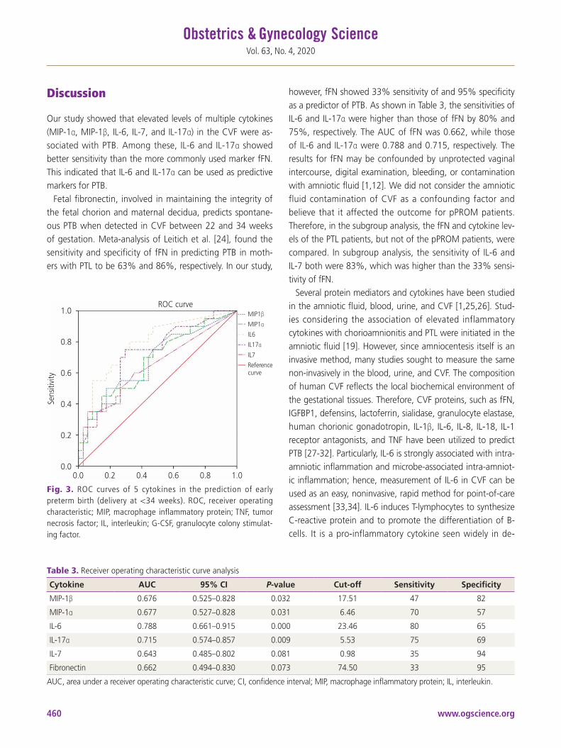
www.ogscience.org460
Vol. 63, No. 4, 2020
Discussion
Our study showed that elevated levels of multiple cytokines (MIP-1α, MIP-1β, IL-6, IL-7, and IL-17α) in the CVF were as-sociated with PTB. Among these, IL-6 and IL-17α showed better sensitivity than the more commonly used marker fFN. This indicated that IL-6 and IL-17α can be used as predictive markers for PTB.
Fetal fibronectin, involved in maintaining the integrity of the fetal chorion and maternal decidua, predicts spontane-ous PTB when detected in CVF between 22 and 34 weeks of gestation. Meta-analysis of Leitich et al. [24], found the sensitivity and specificity of fFN in predicting PTB in moth-ers with PTL to be 63% and 86%, respectively. In our study,
however, fFN showed 33% sensitivity of and 95% specificity as a predictor of PTB. As shown in Table 3, the sensitivities of IL-6 and IL-17α were higher than those of fFN by 80% and 75%, respectively. The AUC of fFN was 0.662, while those of IL-6 and IL-17α were 0.788 and 0.715, respectively. The results for fFN may be confounded by unprotected vaginal intercourse, digital examination, bleeding, or contamination with amniotic fluid [1,12]. We did not consider the amniotic fluid contamination of CVF as a confounding factor and believe that it affected the outcome for pPROM patients. Therefore, in the subgroup analysis, the fFN and cytokine lev-els of the PTL patients, but not of the pPROM patients, were compared. In subgroup analysis, the sensitivity of IL-6 and IL-7 both were 83%, which was higher than the 33% sensi-tivity of fFN.
Several protein mediators and cytokines have been studied in the amniotic fluid, blood, urine, and CVF [1,25,26]. Stud-ies considering the association of elevated inflammatory cytokines with chorioamnionitis and PTL were initiated in the amniotic fluid [19]. However, since amniocentesis itself is an invasive method, many studies sought to measure the same non-invasively in the blood, urine, and CVF. The composition of human CVF reflects the local biochemical environment of the gestational tissues. Therefore, CVF proteins, such as fFN, IGFBP1, defensins, lactoferrin, sialidase, granulocyte elastase, human chorionic gonadotropin, IL-1β, IL-6, IL-8, IL-18, IL-1 receptor antagonists, and TNF have been utilized to predict PTB [27-32]. Particularly, IL-6 is strongly associated with intra-amniotic inflammation and microbe-associated intra-amniot-ic inflammation; hence, measurement of IL-6 in CVF can be used as an easy, noninvasive, rapid method for point-of-care assessment [33,34]. IL-6 induces T-lymphocytes to synthesize C-reactive protein and to promote the differentiation of B-cells. It is a pro-inflammatory cytokine seen widely in de-
Table 3. Receiver operating characteristic curve analysis
Cytokine AUC 95% CI P-value Cut-off Sensitivity Specificity
MIP-1β 0.676 0.525–0.828 0.032 17.51 47 82
MIP-1α 0.677 0.527–0.828 0.031 6.46 70 57
IL-6 0.788 0.661–0.915 0.000 23.46 80 65
IL-17α 0.715 0.574–0.857 0.009 5.53 75 69
IL-7 0.643 0.485–0.802 0.081 0.98 35 94
Fibronectin 0.662 0.494–0.830 0.073 74.50 33 95
AUC, area under a receiver operating characteristic curve; CI, confidence interval; MIP, macrophage inflammatory protein; IL, interleukin.
Fig. 3. ROC curves of 5 cytokines in the prediction of early preterm birth (delivery at <34 weeks). ROC, receiver operating characteristic; MIP, macrophage inflammatory protein; TNF, tumor necrosis factor; IL, interleukin; G-CSF, granulocyte colony stimulat-ing factor.
ROC curve MIP1β
MIP1α
IL6
IL17α
IL7
Reference curve
Sens
itivi
ty
1.0
0.8
0.6
0.4
0.2
0.0 0.0 0.2 0.4 0.6 0.8 1.0

www.ogscience.org 461
Sunwha Park, et al. CVF cytokine for prediction of PTB
cidual tissue, placenta, fetal membrane, and amniotic fluid, and regulates immune adaptation to allow the pregnancy to progress. Preterm labor and pPROM are thought be because of the inflammatory condition caused by infection. Our study has showed that IL-6 is superior to conventional fFN assays. IL-17α as a protective cytokine in host defense against bacte-rial and fungal infections, was shown to be associated with intra-amniotic inflammation [35]. No study has analyzed IL-17 in the CVF; however, IL-17α was significantly elevated in the amniotic fluid of patients with cervical insufficiency [16]. MIP-1α and MIP-1β are produced in the macrophages in response to infection and inflammation. They subsequently activate human granulocytes and the level of MIP-1α in the CVF is associated with amniotic fluid infection [21,36,37]. MIP-1α has been studied in the CVF of patients with pPROM, but not PTL. In this study, we found that elevation of the MIP-1α level in the CVF was associated with PTL. IL-7 is a cy-tokine that can stimulate IL-17 production by cells involved in both innate and adaptive immunity. Elevated serum IL-7 is also associated with PTL [38].
Although our sample size was small, the strength of this study is that it was conducted as a multi-center prospective study in 5 centers in Korea. This study directly compared cy-tokines and fFN in the CVF from the same cohort of patients. When selecting a patient group, the ones with multiple ges-tation, gestational diabetes, and gestational hypertension were excluded. However, factors such as cervical conization history, socio-economic status, pre-pregnancy body mass index, smoking, drug use, and anxiety that could be con-founding factors could not be excluded. The gestational age at diagnosis is also a confounding factor that may affect the gestational age at delivery, but no analysis has been done to correct this.
In conclusion, CVF cytokines, especially IL-6, IL-17α, could be useful predictive markers of PTB in patients with PTL and/or pPROM; however, studies with larger cohort will be re-quired for validation of these findings.
Acknowledgements
This research was supported by the Ministry of Health and Welfare of the Republic of Korea (Grant Number: HI18C0378) through the Korea Health Industry Development Institute.
We would like to thank Editage (www.editage.co.kr) for English language editing.
Conflict of interest
No potential conflict of interest relevant to this article was reported.
Ethical approval
This study was approved by the Institutional Review Board Committee of Ewha Womans University Mokdong Hospital (certificate No. EUMC 2014-06-010).
Patient consent
The patients provided written informed consent for the pub-lication and the use of their images.
Supplementary materials
Supplementary Tables associated with this article can be found online at https://doi.org/10.5468/ogs.19131.
References
1. Heng YJ, Liong S, Permezel M, Rice GE, Di Quinzio MK, Georgiou HM. Human cervicovaginal fluid biomark-ers to predict term and preterm labor. Front Physiol 2015;6:151.
2. Yoo JY, You YA, Kwon EJ, Park MH, Shim S, Kim YJ. Dif-ferential expression and methylation of integrin subunit alpha 11 and thrombospondin in the amnion of preterm birth. Obstet Gynecol Sci 2018;61:565-74.
3. Chan RL. Biochemical markers of spontaneous pre-term birth in asymptomatic women. BioMed Res Int 2014;2014:164081.
4. Amabebe E, Chapman DR, Stern VL, Stafford G, Anum-ba DO. Mid-gestational changes in cervicovaginal fluid cytokine levels in asymptomatic pregnant women are

www.ogscience.org462
Vol. 63, No. 4, 2020
predictive markers of inflammation-associated spontane-ous preterm birth. J Reprod Immunol 2018;126:1-10.
5. Jefferson KK. The bacterial etiology of preterm birth. Adv Appl Microbiol 2012;80:1-22.
6. Dekker GA, Lee SY, North RA, McCowan LM, Simpson NA, Roberts CT. Risk factors for preterm birth in an in-ternational prospective cohort of nulliparous women. PLoS One 2012;7:e39154.
7. Son KA, Kim M, Kim YM, Kim SH, Choi SJ, Oh SY, et al. Prevalence of vaginal microorganisms among pregnant women according to trimester and association with pre-term birth. Obstet Gynecol Sci 2018;61:38-47.
8. Kim YJ, Lee BE, Park HS, Kang JG, Kim JO, Ha EH. Risk factors for preterm birth in Korea: a multicenter pro-spective study. Gynecol Obstet Invest 2005;60:206-12.
9. Baer RJ, McLemore MR, Adler N, Oltman SP, Chambers BD, Kuppermann M, et al. Pre-pregnancy or first-trimes-ter risk scoring to identify women at high risk of preterm birth. Eur J Obstet Gynecol Reprod Biol 2018;231:235-40.
10. Conde-Agudelo A, Papageorghiou AT, Kennedy SH, Vil-lar J. Novel biomarkers for the prediction of the sponta-neous preterm birth phenotype: a systematic review and meta-analysis. BJOG 2011;118:1042-54.
11. Paternoster DM, Muresan D, Vitulo A, Serena A, Batta-gliarin G, Dell’avanzo M, et al. Cervical phIGFBP-1 in the evaluation of the risk of preterm delivery. Acta Obstet Gynecol Scand 2007;86:151-5.
12. Liong S, Di Quinzio MK, Fleming G, Permezel M, Rice GE, Georgiou HM. New biomarkers for the prediction of spontaneous preterm labour in symptomatic preg-nant women: a comparison with fetal fibronectin. BJOG 2015;122:370-9.
13. Son M, Miller ES. Predicting preterm birth: cervi-cal length and fetal fibronectin. Semin Perinatol 2017;41:445-51.
14. Chandiramani M, Seed PT, Orsi NM, Ekbote UV, Bennett PR, Shennan AH, et al. Limited relationship between cervico-vaginal fluid cytokine profiles and cervical short-ening in women at high risk of spontaneous preterm birth. PLoS One 2012;7:e52412.
15. DeFranco EA, Lewis DF, Odibo AO. Improving the screening accuracy for preterm labor: is the combination of fetal fibronectin and cervical length in symptomatic patients a useful predictor of preterm birth? A system-
atic review. Am J Obstet Gynecol 2013;208:233.e1-6.16. Son GH, You YA, Kwon EJ, Lee KY, Kim YJ. Compara-
tive analysis of midtrimester amniotic fluid cytokine levels to predict spontaneous very pre-term birth in pa-tients with cervical insufficiency. Am J Reprod Immunol 2016;75:155-61.
17. Gulati S, Agrawal S, Raghunandan C, Bhattacharya J, Saili A, Agarwal S, et al. Maternal serum interleukin-6 and its association with clinicopathological infectious morbidity in preterm premature rupture of membranes: a prospective cohort study. J Matern Fetal Neonatal Med 2012;25:1428-32.
18. Curry AE, Vogel I, Skogstrand K, Drews C, Schendel DE, Flanders WD, et al. Maternal plasma cytokines in early- and mid-gestation of normal human pregnancy and their association with maternal factors. J Reprod Immu-nol 2008;77:152-60.
19. Chaemsaithong P, Romero R, Docheva N, Chaiyasit N, Bhatti G, Pacora P, et al. Comparison of rapid MMP-8 and interleukin-6 point-of-care tests to identify intra-amniotic inflammation/infection and impending preterm delivery in patients with preterm labor and intact mem-branes. J Matern Fetal Neonatal Med 2018;31:228-44.
20. Musilova I, Bestvina T, Hudeckova M, Michalec I, Cobo T, Jacobsson B, et al. Vaginal fluid IL-6 concentrations as a point-of-care test is of value in women with preterm PROM. Am J Obstet Gynecol. Forthcoming 2016.
21. Lee SM, Park KH, Jung EY, Kook SY, Park H, Jeon SJ. In-flammatory proteins in maternal plasma, cervicovaginal and amniotic fluids as predictors of intra-amniotic infec-tion in preterm premature rupture of membranes. PLoS One 2018;13:e0200311.
22. Coleman MA, Keelan JA, McCowan LM, Townend KM, Mitchell MD. Predicting preterm delivery: comparison of cervicovaginal interleukin (IL)-1beta, IL-6 and IL-8 with fetal fibronectin and cervical dilatation. Eur J Obstet Gy-necol Reprod Biol 2001;95:154-8.
23. Gibbs RS, Blanco JD, St Clair PJ, Castaneda YS. Quan-titative bacteriology of amniotic fluid from women with clinical intraamniotic infection at term. J Infect Dis 1982;145:1-8.
24. Leitich H, Kaider A. Fetal fibronectin--how useful is it in the prediction of preterm birth? BJOG 2003;110 Suppl 20:66-70.
25. Goldenberg RL, Iams JD, Mercer BM, Meis PJ, Moawad

www.ogscience.org 463
Sunwha Park, et al. CVF cytokine for prediction of PTB
A, Das A, et al. The Preterm Prediction Study: toward a multiple-marker test for spontaneous preterm birth. Am J Obstet Gynecol 2001;185:643-51.
26. Heng YJ, Di Quinzio MK, Permezel M, Rice GE, Georgiou HM. Interleukin-1 receptor antagonist in human cervico-vaginal fluid in term pregnancy and labor. Am J Obstet Gynecol 2008;199:656.e1-7.
27. Kekki M, Kurki T, Kärkkäinen T, Hiilesmaa V, Paavonen J, Rutanen EM. Insulin-like growth factor-binding protein-1 in cervical secretion as a predictor of preterm delivery. Acta Obstet Gynecol Scand 2001;80:546-51.
28. Lockwood CJ, Senyei AE, Dische MR, Casal D, Shah KD, Thung SN, et al. Fetal fibronectin in cervical and vaginal secretions as a predictor of preterm delivery. N Engl J Med 1991;325:669-74.
29. Rizzo G, Capponi A, Rinaldo D, Tedeschi D, Arduini D, Romanini C. Interleukin-6 concentrations in cervical se-cretions identify microbial invasion of the amniotic cavity in patients with preterm labor and intact membranes. Am J Obstet Gynecol 1996;175:812-7.
30. Jacobsson B, Holst RM, Mattsby-Baltzer I, Nikolaitchouk N, Wennerholm UB, Hagberg H. Interleukin-18 in cervi-cal mucus and amniotic fluid: relationship to microbial invasion of the amniotic fluid, intra-amniotic inflamma-tion and preterm delivery. BJOG 2003;110:598-603.
31. Tanaka Y, Narahara H, Takai N, Yoshimatsu J, Anai T, Miyakawa I. Interleukin-1β and interleukin-8 in cervi-covaginal fluid during pregnancy. Am J Obstet Gynecol 1998;179:644-9.
32. Sanchez-Ramos L, Mentel C, Bertholf R, Kaunitz AM,
Delke I, Loge C. Human chorionic gonadotropin in cer-vicovaginal secretions as a predictor of preterm delivery. Int J Gynaecol Obstet 2003;83:151-7.
33. Kacerovsky M, Musilova I, Jacobsson B, Drahosova M, Hornychova H, Janku P, et al. Cervical fluid IL-6 and IL-8 levels in pregnancies complicated by preterm prelabor rupture of membranes. J Matern Fetal Neonatal Med 2015;28:134-40.
34. Ashford K, Chavan NR, Wiggins AT, Sayre MM, McCub-bin A, Critchfield AS, et al. Comparison of serum and cervical cytokine levels throughout pregnancy between preterm and term births. AJP Rep 2018;8:e113-20.
35. Kiefer DG, Peltier MR, Keeler SM, Rust O, Ananth CV, Vintzileos AM, et al. Efficacy of midtrimester short cervix interventions is conditional on intraamniotic inflamma-tion. Am J Obstet Gynecol 2016;214:276.e1-6.
36. Romero R, Gomez R, Galasso M, Munoz H, Acosta L, Yoon BH, et al. Macrophage inflammatory protein-1 al-pha in term and preterm parturition: effect of microbial invasion of the amniotic cavity. Am J Reprod Immunol 1994;32:108-13.
37. Lee SM, Park KH, Jung EY, Cho SH, Ryu A. Prediction of spontaneous preterm birth in women with cervical insuf-ficiency: comprehensive analysis of multiple proteins in amniotic fluid. J Obstet Gynaecol Res 2016;42:776-83.
38. Herrera-Muñoz A, Fernández-Alonso AM, Fischer-Suárez N, Chedraui P, Pérez-López FR. Maternal serum cytokine levels in pregnancies complicated with threatened pre-term labour. Gynecol Endocrinol 2017;33:408-12.
www.ogscience.org464
Original ArticleObstet Gynecol Sci 2020;63(4):464-469https://doi.org/10.5468/ogs.19204pISSN 2287-8572 · eISSN 2287-8580
Introduction
Ovarian cancer is usually diagnosed at advanced stages. It is the most lethal gynecological cancer and accounts for 4.3% of all cancer-related deaths [1]. Advanced-stage epithelial ovarian cancer (EOC) is the leading subject of research in literature compared to early-stage disease. However, most early-stage disease-related studies evaluate the patients with stage 1 or 2 together, even though stage 1a/1b disease has a better prognosis than stage 1c or 2 [2].
Stage 1a EOC constitutes a minor group (up to 10%) of patients, with a 5-year survival rate of over 90% [3]. There are several prognostic factors associated with stage 1a
Prognostic impact of p16 and p53 gene expressions in stage 1a epithelial ovarian cancerEmre Günakan, MD1, Yusuf Aytaç Tohma, MD1, Latife Atasoy Karakaş, MD1, Hüseyin Akıllı, MD2, Asuman Nihan Haberal, MD2, Ali Ayhan, MD1
Departments of 1Obstetrics and Gynecology, 2Pathology, School of Medicine, Başkent University, Ankara, Turkey
ObjectiveEpithelial ovarian cancer (EOC) is rarely detected at stage 1a. Most of the patients have a good prognosis and there are limited factors that affect their survival. In the present study, we evaluated the p16 and p53 gene expressions of stage 1a EOC patients. Prognostic effects of these gene expressions, as well as those of other factors on short term survival were analyzed.
MethodsOur study included 29 patients. The specimens of the ovary with cancer were stained for p16 and p53. Gene expressions and other prognostic factors were evaluated.
ResultsThe median age of the patients was 51 years (27–84). The mean numbers of dissected pelvic and paraaortic lymph nodes were 27 and 12, respectively. The mean follow-up time was 33.7±18.9 months. During this period, recurrence occurred in two patients. One of the patients had grade 2 mucinous carcinoma and died of the disease at month 12 after the recurrence occurred at month 7. The second patient had clear cell carcinoma and recurrence occurred at month 34. p16 and p53 gene expressions or other factors were not associated with overall survival (OS) or disease-free survival in the short term. The lower p16 positivity rate in the non-clear cell group was found to be statistically significant (P=0.003). Both p53 and p16 positivity rates were higher in the high-grade carcinoma.
ConclusionThe levels of none of the common prognostic factors, including those of p16 and p53 gene expression, were associated with the progression-free survival or OS of stage 1a in the short term. Appropriate surgical staging and non-omission of subclinical metastases seem to be of central importance.
Keywords: Disease-free survival; Genes, p16; Genes, p53; Progression-free survival
Received: 2019.10.26. Revised: 2020.02.10. Accepted: 2020.02.17.Corresponding author: Emre Günakan, MDDepartment of Obstetrics and Gynecology, School of Medicine, Başkent University, Fevzi Çakmak Cd. 10. Sk. No:45 Bahçelievler, Ankara, TurkeyE-mail: [email protected]://orcid.org/0000-0001-8854-8190
Articles published in Obstet Gynecol Sci are open-access, distributed under the terms of the Creative Commons Attribution Non-Commercial License (http://creativecommons.org/licenses/by-nc/3.0/) which permits unrestricted non-commercial use, distribution, and reproduction in any medium, provided the original work is properly cited.
Copyright © 2020 Korean Society of Obstetrics and Gynecology

www.ogscience.org 465
Emre Günakan, et al. p16, p53 genes in stage1a ovarian cancer
disease, including age, histological factors (e.g., type and grade), and molecular factors such as abnormal oncogene expression (e.g., p53, p16, Her2/neu, PTEN, p21, and ki67).
p16 is a cyclin-dependent kinase (CDK) inhibitor that inhib-its retinoblastoma protein levels by inhibiting CDK phosphor-ylation during cell cycle progression. Another mode of its action involves the inhibition of cyclin D1, which inactivates pRb through CDK4. Both p16 and pRb are important tumor suppressors in cell cycle regulation. p16 is a potent inhibi-tor of CDK4 and CDK6. p16, pRb, and cyclin are involved in the transition from the G1 to S phase, which is the most important control point of the D1 cell cycle. The p53 protein is localized in the nucleus and plays a role in the mechanisms underlying DNA damage repair. p53 controls the transition from the G1 to S phase. It is the most common site of ge-netic change in human tumors [4]. Approximately, 50% of human tumors contain a p53 gene mutation. Additionally, several studies have reported about the high p53 mutation rate in EOC [5-7]. Recently, mutation or promoter methyla-tion of the p16 gene has also been detected in ovarian se-rous adenocarcinoma [8]. In the current study, we aimed to evaluate the effect of p16 and p53 expressions and conven-tional prognostic factors on stage 1a EOC.
Materials and methods
Our study included 29 patients who underwent staging sur-
gery for EOC at the Department of Gynecologic Oncology, School of Medicine, Başkent University and were diagnosed with stage 1a disease. The study was conducted in accordance with the Declaration of Helsinki. Standard staging surgery in-cluded hysterectomy, bilateral salphingo-oophorectomy, bilat-eral pelvic and paraaortic lymph node dissection, omentecto-my, and appendectomy. Uterus and one ovary were preserved and unilateral salphingo-oophorectomy was performed in fertility-sparing surgery. The International Federation of Gyne-cologists and Obstetricians (FIGO) system was used for disease staging [9]. Patients were evaluated in groups based on the histological type (clear and non-clear cell carcinoma) and the presence or absence of p16 and p53 gene expressions. p16 and p53 expressions were evaluated immunohistochemically in paraffin blocs. Nuclear staining in the epithelial cells was evaluated under a light microscope. Lack of staining, slight staining, or staining of less than 10% of the tumor cells was defined as negative, while others were defined as positive (Fig. 1). Data were analyzed using SPSS for Windows v.15.0 (SPSS Inc., Chicago, IL, USA). Descriptive and frequency analyses were performed. Categorical variables were compared using the chi-square test or Fisher’s exact test, as appropriate. The level of statistical significance was set at P<0.05.
Results
Our study included 29 patients. The median age of the pa-
Fig. 1. (A) Weak staining pattern with p16 marker (×10 high-power field [HPF]); (B) Moderate staining pattern with p16 marker (×20 HPF); (C) Strong staining pattern with p16 marker (×10 HPF).
A B C
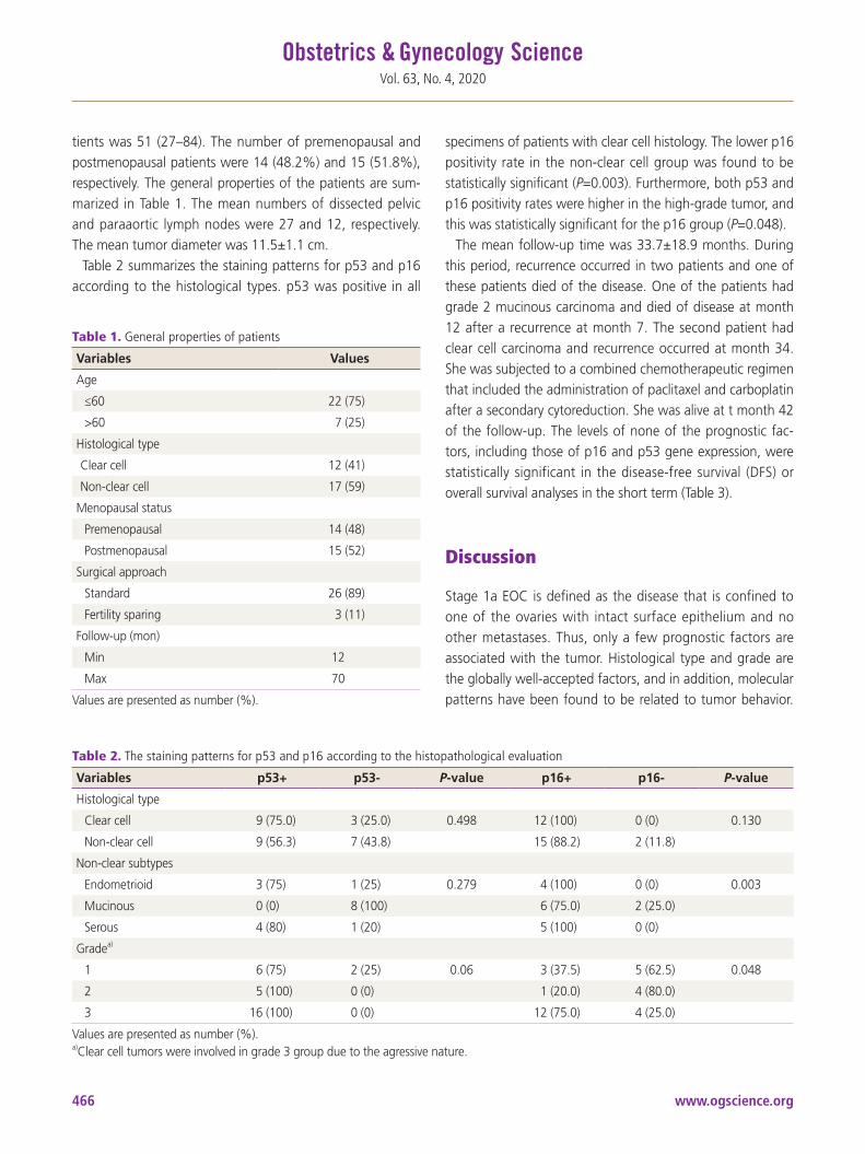
www.ogscience.org466
Vol. 63, No. 4, 2020
tients was 51 (27–84). The number of premenopausal and postmenopausal patients were 14 (48.2%) and 15 (51.8%), respectively. The general properties of the patients are sum-marized in Table 1. The mean numbers of dissected pelvic and paraaortic lymph nodes were 27 and 12, respectively. The mean tumor diameter was 11.5±1.1 cm.
Table 2 summarizes the staining patterns for p53 and p16 according to the histological types. p53 was positive in all
specimens of patients with clear cell histology. The lower p16 positivity rate in the non-clear cell group was found to be statistically significant (P=0.003). Furthermore, both p53 and p16 positivity rates were higher in the high-grade tumor, and this was statistically significant for the p16 group (P=0.048).
The mean follow-up time was 33.7±18.9 months. During this period, recurrence occurred in two patients and one of these patients died of the disease. One of the patients had grade 2 mucinous carcinoma and died of disease at month 12 after a recurrence at month 7. The second patient had clear cell carcinoma and recurrence occurred at month 34. She was subjected to a combined chemotherapeutic regimen that included the administration of paclitaxel and carboplatin after a secondary cytoreduction. She was alive at t month 42 of the follow-up. The levels of none of the prognostic fac-tors, including those of p16 and p53 gene expression, were statistically significant in the disease-free survival (DFS) or overall survival analyses in the short term (Table 3).
Discussion
Stage 1a EOC is defined as the disease that is confined to one of the ovaries with intact surface epithelium and no other metastases. Thus, only a few prognostic factors are associated with the tumor. Histological type and grade are the globally well-accepted factors, and in addition, molecular patterns have been found to be related to tumor behavior.
Table 1. General properties of patients
Variables Values
Age
≤60 22 (75)
>60 7 (25)
Histological type
Clear cell 12 (41)
Non-clear cell 17 (59)
Menopausal status
Premenopausal 14 (48)
Postmenopausal 15 (52)
Surgical approach
Standard 26 (89)
Fertility sparing 3 (11)
Follow-up (mon)
Min 12
Max 70
Values are presented as number (%).
Table 2. The staining patterns for p53 and p16 according to the histopathological evaluation
Variables p53+ p53- P-value p16+ p16- P-value
Histological type
Clear cell 9 (75.0) 3 (25.0) 0.498 12 (100) 0 (0) 0.130
Non-clear cell 9 (56.3) 7 (43.8) 15 (88.2) 2 (11.8)
Non-clear subtypes
Endometrioid 3 (75) 1 (25) 0.279 4 (100) 0 (0) 0.003
Mucinous 0 (0) 8 (100) 6 (75.0) 2 (25.0)
Serous 4 (80) 1 (20) 5 (100) 0 (0)
Gradea)
1 6 (75) 2 (25) 0.06 3 (37.5) 5 (62.5) 0.048
2 5 (100) 0 (0) 1 (20.0) 4 (80.0)
3 16 (100) 0 (0) 12 (75.0) 4 (25.0)
Values are presented as number (%).a)Clear cell tumors were involved in grade 3 group due to the agressive nature.

www.ogscience.org 467
Emre Günakan, et al. p16, p53 genes in stage1a ovarian cancer
The aim of our study was to investigate whether p53 and p16 expressions affect survival in stage 1a EOC. In the cur-rent analysis, we did not find a statistically significant asso-ciation between survival and the levels of any of the factors, including those of p53 and p16 expression, in the short term.
The initial approach in early-stage ovarian cancer treat-ment is surgical staging. These results constitute the basis of adjuvant treatment and prediction of prognosis [10,11]. Ad-ditionally, there is no difference in 5-year survival expectancy between fertility-sparing or radical surgery at stage 1 [12,13]. In stage 1a disease, classic staging procedure is sufficient and curative in a substantial proportion of patients. Observa-tional studies have shown that 30% of patients, who were thought to have stage I or II disease during the initial surgery, were diagnosed with a more advanced stage disease after more comprehensive restaging laparotomy [14]. In a study comprising 138 patients, the recurrence rates of complete and incomplete staging were 10% and 28%, respectively [15]. At this point, the main targets of the surgery in patients who are thought to have early-stage disease should be ap-propriate staging procedure and the exclusion of occult and subclinical metastases [16]. In our study, all patients were subjected to a standard staging surgery with an adequate
number of lymph nodes in both the pelvic and paraaortic re-gions.
Endometrioid histology is associated with a better progno-sis and an earlier stage at the time of diagnosis [17]. Con-trastingly, clear cell histology is associated with poor prog-nosis [3]. In addition, clear cell histology has an aggressive nature and can be considered as a high-grade tumor. Even stage 1a patients with clear cell carcinoma undergo adjuvant therapies. Clear cell histology was the most frequent histo-logical type in our study and was evaluated as a separate group. All specimens were p53-positive and this was a con-spicuous point.
In the cell cycle, DNA damage is controlled at the G1/S control point and DNA repair mechanisms are activated. Mu-tation of the p53 tumor suppressor gene results in a DNA re-pair failure leading to neoplastic development [18]. Mutation of p53 is frequently detected in high grade serous ovarian cancers, and this frequency may be up to 80% in advanced stages [19]. Over-expression of p53 is associated with poor prognosis [20-25] and high tumor grade [24]. Additionally, p53 was associated with lower DFS and poor prognosis in stages 1 and 2 [21,22]. Association of p16 and EOC is a rel-atively less evaluated issue compared to p53. In contrast, p16
Table 3. Survival analysis of prognostic factors
Variables DFS P-value OS P-value
Age 0.224 0.226
<60 22 (35.8±20.7) 22 (36.0±20.4)
>60 7 (25.6±10.0) 7 (26.7±11.5)
Histological type 0.182 0.161
Clear cell 12 (39±18.2) 12 (39.67±18.2)
Non-clear cell 17 (29.3±19.1) 17 (29.6±18.8)
Grade 0.536 0.489
1 8 (26.9±14.0) 8 (26.9±14.0)
2 5 (34.0±22.3) 8 (35.0±20.9)
3 16 (36.3±20.5) 16 (36.8±20.6)
p16 0.078 0.103
Positive 16 (38.9±19.9) 16 (38.94±19.9)
Negative 13 (26.3±16.0) 13 (27.38±16.1)
p53 0.297 0.277
Positive 27 (34.3±19.3) 27 (34.8±19.1)
Negative 2 (19.5±9.1) 2 (19.1±9.1)
Values are presented as number (mean±standard deviation).DFS, disease-free survival; OS, overall survival.

www.ogscience.org468
Vol. 63, No. 4, 2020
gene mutations or promoter methylation has been detected in serous ovarian cancer in recent years [8]. Moreover, p16 over-expression is associated with high differentiation and tu-mor grade [26], advanced-stage [8], and poor prognosis [27]. Similar previously published studies have commonly focused on advanced stage disease or stage 1 and 2 disease together. In the current study, we investigated the effect of these mo-lecular changes in stage 1a differently from other studies. We found some significant histopathological findings, whereas its effect on survival could not be well-established due to the short follow-up time.
Stage 1a EOC patients were included in the current study to minimize heterogeneity. Prognostic factors, including p53 and p16 expressions, were evaluated and initial reports of short-term results are reported herein. Standard surgical and medical management of a single-center and uniform patient selection should be considered the strengths of this study. However, the short follow-up time and a limited number of patients should be considered the limitations. The long-term results will be also presented in the future.
Conflict of interest
No potential conflict of interest relevant to this article was reported.
Ethical approval
The study was approved by the review board of the Univer-sity of Medical Sciences.
References
1. Ferlay J, Soerjomataram I, Dikshit R, Eser S, Mathers C, Rebelo M, et al. Cancer incidence and mortality world-wide: sources, methods and major patterns in GLOBO-CAN 2012. Int J Cancer 2015;136:E359-86.
2. Chan JK, Tian C, Monk BJ, Herzog T, Kapp TS, Bell J, et al. Prognostic factors for high-risk early-stage epithelial ovarian cancer. Cancer 2008;112:2202-10.
3. Ayhan A, Reed N, Gultekin M, Dursun P. Early stage epi-thelial ovarian cancer. In: Ayhan A, Reed N, Gultekin M,
Dursun P, editors. Textbook of gynaecolgical oncology. 2nd ed. Ankara: Güneş Tıp Kitapevleri; 2011. p.429-31.
4. Vousden KH, Lane DP. p53 in health and disease. Nat Rev Mol Cell Biol 2007;8:275-83.
5. Ayhan A, Reed N, Gultekin M, Dursun P. prognostic factors for gynecologic cancers. In: Ayhan A, Reed N, Gultekin M, Dursun P, editors. Textbook of gynaecolgical oncology. 2nd ed, Ankara: Güneş Tıp Kitapevleri; 2011. p.1056-60.
6. McMenamin ME, O’Neill AJ, Gaffney EF. Extent of apop-tosis in ovarian serous carcinoma: relation to mitotic and proliferative indices, p53 expression, and survival. Mol Pathol 1997;50:242-6.
7. Singer G, Kurman RJ, Chang HW, Cho SK, Shih IM. Diverse tumorigenic pathways in ovarian serous carci-noma. Am J Pathol 2002;160:1223-8.
8. Katsaros D, Cho W, Singal R, Fracchioli S, Rigault De La Longrais IA, Arisio R, et al. Methylation of tumor sup-pressor gene p16 and prognosis of epithelial ovarian cancer. Gynecol Oncol 2004;94:685-92.
9. Rodriguez M, Nguyen HN, Averette HE, Steren AJ, Penalver MA, Harrison T, et al. National survey of ovar-ian carcinoma XII. Epithelial ovarian malignancies in women less than or equal to 25 years of age. Cancer 1994;73:1245-50.
10. Schildkraut JM, Halabi S, Bastos E, Marchbanks PA, Mc-Donald JA, Berchuck A. Prognostic factors in early-onset epithelial ovarian cancer: a population-based study. Ob-stet Gynecol 2000;95:119-27.
11. Zanetta G, Rota S, Chiari S, Bonazzi C, Bratina G, Torri V, et al. The accuracy of staging: an important prognostic determinator in stage I ovarian carcinoma. A multivari-ate analysis. Ann Oncol 1998;9:1097-101.
12. Ayhan A, Celik H, Taskiran C, Bozdag G, Aksu T. Oncologic and reproductive outcome after fertility-saving surgery in ovarian cancer. Eur J Gynaecol Oncol 2003;24:223-32.
13. Colombo N, Chiari S, Maggioni A, Bocciolone L, Torri V, Mangioni C. Controversial issues in the management of early epithelial ovarian cancer: conservative surgery and role of adjuvant therapy. Gynecol Oncol 1994;55:S47-51.
14. Mutch DG. Ovarian and tubal cancers. In: Gibbs RS, Karlan BY, Haney AF, Nygaard IE, editors. Danforth’s ob-stetrics and gynecology. 10th ed. Philadelphia (PA): Lip-

www.ogscience.org 469
Emre Günakan, et al. p16, p53 genes in stage1a ovarian cancer
pincott Williams & Wilkins; 2008. p.1023-60.15. Le T, Adolph A, Krepart GV, Lotocki R, Heywood MS.
The benefits of comprehensive surgical staging in the management of early-stage epithelial ovarian carcinoma. Gynecol Oncol 2002;85:351-5.
16. Chan J, Fuh K, Shin J, Cheung M, Powell C, Chen LM, et al. The treatment and outcomes of early-stage epithelial ovarian cancer: have we made any progress? Br J Cancer 2008;98:1191-6.
17. Seidman JD, Kurman RJ. Pathology of ovarian carcino-ma. Hematol Oncol Clin North Am 2003;17:909-25.
18. Ellenson LH, Pirog EC. The female genital tract. In: Ku-mar V, Abbas AK, Fausto N, editors. Robbins and Cotran pathologic basis of disease. 7th ed. Philadelphia (PA): Elsevier Saunders; 2005. p.1092-104.
19. Kurman RJ, Shih IM. Pathogenesis of ovarian cancer: les-sons from morphology and molecular biology and their clinical implications. Int J Gynecol Pathol 2008;27:151-60.
20. Darcy KM, Brady WE, McBroom JW, Bell JG, Young RC, McGuire WP, et al. Associations between p53 overex-pression and multiple measures of clinical outcome in high-risk, early stage or suboptimally-resected, advanced stage epithelial ovarian cancers A Gynecologic Oncology Group study. Gynecol Oncol 2008;111:487-95.
21. Skírnisdóttir IA, Sorbe B, Lindborg K, Seidal T. Prognostic impact of p53, p27, and C-MYC on clinicopathological
features and outcome in early-stage (FIGO I-II) epithelial ovarian cancer. Int J Gynecol Cancer 2011;21:236-44.
22. Skírnisdóttir I, Seidal T. Prognostic impact of concomi-tant p53 and PTEN on outcome in early stage (FIGO I-II) epithelial ovarian cancer. Int J Gynecol Cancer 2011;21:1024-31.
23. de Graeff P, Crijns AP, de Jong S, Boezen M, Post WJ, de Vries EG, et al. Modest effect of p53, EGFR and HER-2/neu on prognosis in epithelial ovarian cancer: a meta-analysis. Br J Cancer 2009;101:149-59.
24. García-Velasco A, Mendiola C, Sánchez-Muñoz A, Ball-estín C, Colomer R, Cortés-Funes H. Prognostic value of hormonal receptors, p53, ki67 and HER2/neu expres-sion in epithelial ovarian carcinoma. Clin Transl Oncol 2008;10:367-71.
25. Lee YK, Park NH. Prognostic value and clinicopathologi-cal significance of p53 and PTEN in epithelial ovarian cancers. Gynecol Oncol 2009;112:475-80.
26. Khouja MH, Baekelandt M, Nesland JM, Holm R. The clinical importance of Ki-67, p16, p14, and p57 expres-sion in patients with advanced ovarian carcinoma. Int J Gynecol Pathol 2007;26:418-25.
27. Surowiak P, Materna V, Maciejczyk A, Pudelko M, Suchocki S, Kedzia W, et al. Decreased expression of p16 in ovarian cancers represents an unfavourable prog-nostic factor. Histol Histopathol 2008;23:531-8.

www.ogscience.org470
Original ArticleObstet Gynecol Sci 2020;63(4):470-479https://doi.org/10.5468/ogs.20012pISSN 2287-8572 · eISSN 2287-8580
Introduction
Osteoporosis is the most common metabolic bone disease caused by reduced bone mineral density (BMD) and is associ-ated with high risk of bone fractures. Fractures due to osteo-porosis are closely related to the mortality rate of the elderly population, and there is an increased prevalence of osteopo-rosis due to increased life expectancy. Therefore, social and medical interest in the risk factors, diagnosis, and treatment methods of osteoporosis is increasing. Several systemic con-ditions and diseases may affect bone mineral density, includ-
Effect of gynecological cancer and its treatment on bone mineral density and the risk of osteoporosis and osteoporotic fracture Jeong Eun Lee, MM, Che Yon Park, MM, Eunhyun Lee, MD, Yong Il Ji, MD, PhDDepartment of Obstetrics and Gynecology, Inje University, Haeundae Paik Hospital, Busan, Korea
ObjectiveThe purpose of this study was to evaluate the risk of osteopenia and osteoporosis by examining the bone mineral density (BMD) of the lumbar spine and femur in patients with gynecological cancer without bone metastasis and to evaluate the impact of treatment for different cancers on BMD.
MethodsThis study retrospectively reviewed the medical records of 243 women with gynecological cancer and 240 controls between March 2010 and December 2016. Patients with cervical cancer (n=105), endometrial cancer (n=63), and ovarian cancer (n=75) were treated with total hysterectomy including bilateral salpingo-oophorectomy and/or chemotherapy and/or radiotherapy. For the control group, healthy post-menopausal women without gynecologic cancer were selected.
ResultsBefore anticancer treatment, the BMD of patients with cervical cancer and ovarian cancer was significantly lower than that of the controls, and the BMD of patients with endometrial cancer was not significantly different from that of the controls. However, the BMD of endometrial cancer significantly decreased after treatment. According to the treatment methods, there were significant differences in the BMD of L3, L4, and the femur neck. Changes in the BMD were lowest in patients who underwent surgical treatment only, and the highest bone loss was found in patients who underwent postoperative concurrent chemoradiotherapy.
ConclusionPatients with cervical and ovarian cancer had lower BMD than those in the control group before treatment, and patients with endometrial cancer had decreased bone density after treatment. Therefore, during the treatment of gynecological cancer, strategies should be implemented to mitigate these risks.
Keywords: Bone density; Treatment-associated cancer; Osteoporosis
Received: 2020.01.07. Revised: 2020.03.03. Accepted: 2020.03.19.Corresponding author: Yong Il Ji, MD, PhDDepartment of Obstetrics and Gynecology, Inje University, Haeundae Paik Hospital, 875 Haeun-daero, Haeundae-gu, Busan 48108, KoreaE-mail: [email protected]://orcid.org/0000-0002-1030-6105
Articles published in Obstet Gynecol Sci are open-access, distributed under the terms of the Creative Commons Attribution Non-Commercial License (http://creativecommons.org/licenses/by-nc/3.0/) which permits unrestricted non-commercial use, distribution, and reproduction in any medium, provided the original work is properly cited.
Copyright © 2020 Korean Society of Obstetrics and Gynecology

www.ogscience.org 471
Jeong Eun Lee, et al. BMD of gynecological cancer patients
ing insufficient calcium intake, excessive sodium intake, lack of exercise, chronic kidney disease, parathyroid disease, hy-perthyroidism, and gastrointestinal absorption disorder [1].
The mean BMD of women is lower than that of men, and bone mass accumulates between 20 and 30 years of age. The loss of bone density is most rapid after the age of 50, particularly 5 years after menopause in women [2,3]. A de-crease in estrogen in postmenopausal women affects bone and lipid metabolism [4]. Estrogen plays an important role in the osteoclast–osteoblast balance related to maintenance of the BMD. It is known that BMD decreases when estrogen concentration decreases [5].
Previous studies have reported low BMD and high serum calcium levels in various types of cancer including prostate and breast cancer without bone metastasis [6-11]. The de-crease in the BMD associated with cancer may be linked to the osteoclastic effect of the malignant cell itself as well as the cancer treatment-induced bone loss (CTIBL) caused by surgery, chemotherapy, and radiation therapy [12-14]. Go-nadal dysfunction due to cancer treatment reduces hormone secretion, leading to decreased BMD.
The purpose of this study was to evaluate the risk of osteo-penia and osteoporosis in patients with gynecological cancer according to the type of cancer and to analyze the BMD of the lumbar spine and femoral neck after various cancer treatments including surgery, chemotherapy, and radiation therapy.
Materials and methods
Between March 2010 and December 2016, a retrospec-tive study was conducted by analyzing the medical records of women who were treated at Haeundae Paik Hospital Obstetrics and Gynecology. All subjects underwent physical examination, blood tests, ultrasonography, and bone mineral density measurements.
1. Study participantsA total 243 patients with gynecological cancer were se-lected for this study. Patients with cervical cancer (n=105), endometrial cancer (n=63), and ovarian cancer (n=75) were treated with total hysterectomy including bilateral salpingo-oophorectomy and/or chemotherapy and/or radiotherapy. Six patients with bone metastasis or those who did not undergo
surgery due to an advanced stage of cancer were excluded. For the control group, 240 healthy post-menopausal women who did not have menstruation for 1 year were selected. The BMD was measured before and 1 year after cancer treat-ment. In the control group, the BMD was measured again 1 year after the initial test.
Patients with thyroid disease, parathyroid disease, kidney stones, hyperprolactinemia, malabsorptive disorder, or previ-ous pathologic fractures that may affect bone metabolism were excluded from the study. Patients who were prescribed medication that may affect the study outcomes, such as hor-mones, bisphosphonates, calcitonin, vitamin D, or calcium, were also excluded.
2. Methods
1) Bone mineral density measurementThe BMD of the 1st–4th lumbar spine and femoral neck was determined by measurement with dual-energy X-ray absorp-tiometry (DXA, Lunar Radiation Corp., Madison, WI, USA). According to the criteria of the World Health Organization, the diagnosis of osteopenia was made with a T-score in the range of −1.0 and −2.5 and that of osteoporosis with a T-score of −2.5 or below [15,16].
2) Cancer treatmentGynecological cancer patients were treated with total hys-terectomy including bilateral salpingo-oophorectomy and/or chemotherapy and/or radiotherapy. Eighty-five patients re-ceived only surgical treatment, and 92 patients were treated with surgery and an adjuvant or palliative chemotherapy. Eight patients received surgery and radiotherapy, and 58 patients received surgical treatment with concurrent chemo-radiation therapy (CCRT). Forty-four cervical cancer patients underwent only total hysterectomy with bilateral salpingo-oophorectomy, and 48 patients underwent postoperative CCRT. Thirty-eight endometrial cancer patients (60.3%) underwent surgical treatment only. Most ovarian cancer pa-tients (90.7%) were treated with chemotherapy after surgery (Table 1).
3) Research variable factorsAge, height, weight, body mass index (BMI), and obstetric history were included as common demographic character-istics. In gynecological cancer patients, the cancer stage ac-
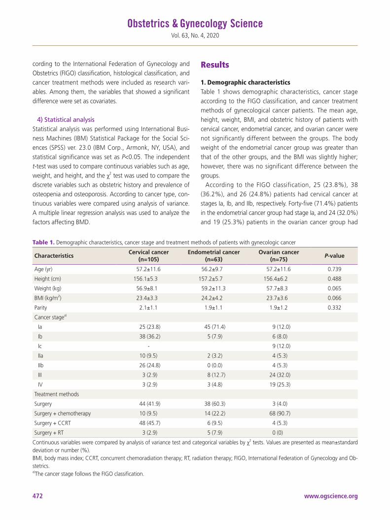
www.ogscience.org472
Vol. 63, No. 4, 2020
cording to the International Federation of Gynecology and Obstetrics (FIGO) classification, histological classification, and cancer treatment methods were included as research vari-ables. Among them, the variables that showed a significant difference were set as covariates.
4) Statistical analysisStatistical analysis was performed using International Busi-ness Machines (IBM) Statistical Package for the Social Sci-ences (SPSS) ver. 23.0 (IBM Corp., Armonk, NY, USA), and statistical significance was set as P<0.05. The independent t-test was used to compare continuous variables such as age, weight, and height, and the χ2 test was used to compare the discrete variables such as obstetric history and prevalence of osteopenia and osteoporosis. According to cancer type, con-tinuous variables were compared using analysis of variance. A multiple linear regression analysis was used to analyze the factors affecting BMD.
Results
1. Demographic characteristicsTable 1 shows demographic characteristics, cancer stage according to the FIGO classification, and cancer treatment methods of gynecological cancer patients. The mean age, height, weight, BMI, and obstetric history of patients with cervical cancer, endometrial cancer, and ovarian cancer were not significantly different between the groups. The body weight of the endometrial cancer group was greater than that of the other groups, and the BMI was slightly higher; however, there was no significant difference between the groups.
According to the FIGO classification, 25 (23.8%), 38 (36.2%), and 26 (24.8%) patients had cervical cancer at stages Ia, Ib, and IIb, respectively. Forty-five (71.4%) patients in the endometrial cancer group had stage Ia, and 24 (32.0%) and 19 (25.3%) patients in the ovarian cancer group had
Table 1. Demographic characteristics, cancer stage and treatment methods of patients with gynecologic cancer
CharacteristicsCervical cancer
(n=105)Endometrial cancer
(n=63)Ovarian cancer
(n=75)P-value
Age (yr) 57.2±11.6 56.2±9.7 57.2±11.6 0.739
Height (cm) 156.1±5.3 157.2±5.7 156.4±6.2 0.488
Weight (kg) 56.9±8.1 59.2±11.3 57.7±8.3 0.065
BMI (kg/m2) 23.4±3.3 24.2±4.2 23.7±3.6 0.066
Parity 2.1±1.1 1.9±1.1 1.9±1.2 0.332
Cancer stagea)
Ia 25 (23.8) 45 (71.4) 9 (12.0)
Ib 38 (36.2) 5 (7.9) 6 (8.0)
Ic - 9 (12.0)
IIa 10 (9.5) 2 (3.2) 4 (5.3)
IIb 26 (24.8) 0 (0.0) 4 (5.3)
III 3 (2.9) 8 (12.7) 24 (32.0)
IV 3 (2.9) 3 (4.8) 19 (25.3)
Treatment methods
Surgery 44 (41.9) 38 (60.3) 3 (4.0)
Surgery + chemotherapy 10 (9.5) 14 (22.2) 68 (90.7)
Surgery + CCRT 48 (45.7) 6 (9.5) 4 (5.3)
Surgery + RT 3 (2.9) 5 (7.9) 0 (0)
Continuous variables were compared by analysis of variance test and categorical variables by χ2 tests. Values are presented as mean±standard deviation or number (%).BMI, body mass index; CCRT, concurrent chemoradiation therapy; RT, radiation therapy; FIGO, International Federation of Gynecology and Ob-stetrics.a)The cancer stage follows the FIGO classification.
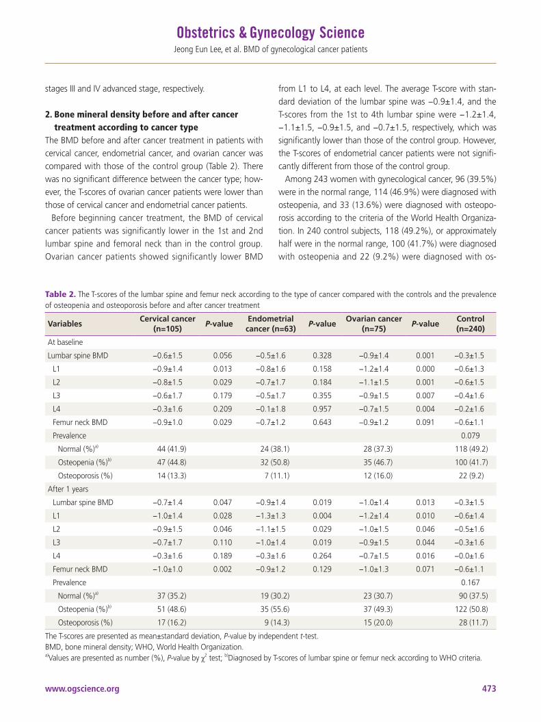
www.ogscience.org 473
Jeong Eun Lee, et al. BMD of gynecological cancer patients
stages III and IV advanced stage, respectively.
2. Bone mineral density before and after cancer treatment according to cancer type
The BMD before and after cancer treatment in patients with cervical cancer, endometrial cancer, and ovarian cancer was compared with those of the control group (Table 2). There was no significant difference between the cancer type; how-ever, the T-scores of ovarian cancer patients were lower than those of cervical cancer and endometrial cancer patients.
Before beginning cancer treatment, the BMD of cervical cancer patients was significantly lower in the 1st and 2nd lumbar spine and femoral neck than in the control group. Ovarian cancer patients showed significantly lower BMD
from L1 to L4, at each level. The average T-score with stan-dard deviation of the lumbar spine was −0.9±1.4, and the T-scores from the 1st to 4th lumbar spine were −1.2±1.4, −1.1±1.5, −0.9±1.5, and −0.7±1.5, respectively, which was significantly lower than those of the control group. However, the T-scores of endometrial cancer patients were not signifi-cantly different from those of the control group.
Among 243 women with gynecological cancer, 96 (39.5%) were in the normal range, 114 (46.9%) were diagnosed with osteopenia, and 33 (13.6%) were diagnosed with osteopo-rosis according to the criteria of the World Health Organiza-tion. In 240 control subjects, 118 (49.2%), or approximately half were in the normal range, 100 (41.7%) were diagnosed with osteopenia and 22 (9.2%) were diagnosed with os-
Table 2. The T-scores of the lumbar spine and femur neck according to the type of cancer compared with the controls and the prevalence of osteopenia and osteoporosis before and after cancer treatment
VariablesCervical cancer
(n=105)P-value
Endometrial cancer (n=63)
P-valueOvarian cancer
(n=75)P-value
Control (n=240)
At baseline
Lumbar spine BMD −0.6±1.5 0.056 −0.5±1.6 0.328 −0.9±1.4 0.001 −0.3±1.5
L1 −0.9±1.4 0.013 −0.8±1.6 0.158 −1.2±1.4 0.000 −0.6±1.3
L2 −0.8±1.5 0.029 −0.7±1.7 0.184 −1.1±1.5 0.001 −0.6±1.5
L3 −0.6±1.7 0.179 −0.5±1.7 0.355 −0.9±1.5 0.007 −0.4±1.6
L4 −0.3±1.6 0.209 −0.1±1.8 0.957 −0.7±1.5 0.004 −0.2±1.6
Femur neck BMD −0.9±1.0 0.029 −0.7±1.2 0.643 −0.9±1.2 0.091 −0.6±1.1
Prevalence 0.079
Normal (%)a) 44 (41.9) 24 (38.1) 28 (37.3) 118 (49.2)
Osteopenia (%)b) 47 (44.8) 32 (50.8) 35 (46.7) 100 (41.7)
Osteoporosis (%) 14 (13.3) 7 (11.1) 12 (16.0) 22 (9.2)
After 1 years
Lumbar spine BMD −0.7±1.4 0.047 −0.9±1.4 0.019 −1.0±1.4 0.013 −0.3±1.5
L1 −1.0±1.4 0.028 −1.3±1.3 0.004 −1.2±1.4 0.010 −0.6±1.4
L2 −0.9±1.5 0.046 −1.1±1.5 0.029 −1.0±1.5 0.046 −0.5±1.6
L3 −0.7±1.7 0.110 −1.0±1.4 0.019 −0.9±1.5 0.044 −0.3±1.6
L4 −0.3±1.6 0.189 −0.3±1.6 0.264 −0.7±1.5 0.016 −0.0±1.6
Femur neck BMD −1.0±1.0 0.002 −0.9±1.2 0.129 −1.0±1.3 0.071 −0.6±1.1
Prevalence 0.167
Normal (%)a) 37 (35.2) 19 (30.2) 23 (30.7) 90 (37.5)
Osteopenia (%)b) 51 (48.6) 35 (55.6) 37 (49.3) 122 (50.8)
Osteoporosis (%) 17 (16.2) 9 (14.3) 15 (20.0) 28 (11.7)
The T-scores are presented as mean±standard deviation, P-value by independent t-test.BMD, bone mineral density; WHO, World Health Organization.a)Values are presented as number (%), P-value by χ2 test; b)Diagnosed by T-scores of lumbar spine or femur neck according to WHO criteria.

www.ogscience.org474
Vol. 63, No. 4, 2020
teoporosis. There was no significant difference between the groups (P=0.079); however, the rates of osteopenia and osteoporosis were 5.2% and 4.4% higher in cancer patients than in the control group, respectively.
After 1 year of cancer treatment, the average T-score of the lumbar spine, the T-score of the 1st to 2nd lumbar spine, and femoral neck in cervical cancer patients was significantly lower than those in the control group. In the ovarian cancer group, the average T-scores of L1-L4 and the 1st to 4th level of the lumbar spine were significantly lower than those of the control group. In patients with endometrial cancer, the T-score of the 1st to 3rd level of the lumbar spine and the av-erage T-score of L1-L4 was significantly lower than those of the control group.
Changes in the BMD before and after cancer treatment according to the type of gynecological cancer are shown in Fig. 1. In endometrial cancer patients, the BMD was signifi-cantly lower than that in cervical cancer and ovarian cancer
patients, and there was a significant difference in the lumbar spine (P=0.024).
3. Changes in bone mineral density according to treatment method
In 243 patients with gynecological cancer, changes in the BMD before and after cancer treatment were compared in relation to the different treatment methods (Table 3). The T-scores of the 3rd and 4th lumbar spine and the femoral neck were significantly different according to the treatment methods. The smallest change in the BMD was observed in 85 patients who had undergone only surgical treatment, including bilateral salpingo-oophorectomy. Conversely, the T-scores of the 58 patients who received surgical treatment with CCRT significantly decreased after treatment.
4. Factors associated with bone lossThe factors affecting the BMD in gynecological cancer pa-
Fig. 1. The T-scores of the lumbar spine and femur neck in patients with gynecologic cancer before and after treatment. BMD, bone min-eral density.
-0.4
-0.5
-0.6
-0.7
-0.8
-0.9
-1.0
-1.1
-0.4
-0.5
-0.6
-0.7
-0.8
-0.9
-1.0
-1.1
-1.2
Cervical cancer
Endometrial cancer
Ovarian cancer
Cervical cancer
Endometrial cancer
Ovarian cancer
Baseline BaselineAfter treatment After treatment
Table 3. The changes of bone mineral density in patients with gynecologic cancer according to treatment methods
VariablesSurgery only
(n=85)Surgery + chemotherapy
(n=92)Surgery + RT
(n=8)Surgery + CCRT
(n=58)P-value
Lumbar spine BMD (g/cm2)a) 0.02±0.47 −0.18±0.63 −0.09±0.56 −0.30±0.47 0.051
L1 −0.01±0.71 −0.12±0.95 −0.67±0.51 −0.36±0.58 0.052
L2 0.01±0.56 −0.16±0.80 −0.21±0.53 −0.28±0.51 0.228
L3 −0.02±0.54 −0.18±0.76 0.20±0.72 −0.34±0.57 0.043
L4 0.07±0.71 −0.17±0.74 0.37±1.33 −0.23±0.62 0.022
Femur neck BMD (g/cm2) −0.04±0.24 −0.03±0.26 −0.19±0.26 −0.27±0.30 0.026
Values are presented as mean±standard deviation, P-value by analysis of variance. BMD, bone mineral density; CCRT, concurrent chemoradiation therapy; RT, radiation therapya)Mean change of T-scores.

www.ogscience.org 475
Jeong Eun Lee, et al. BMD of gynecological cancer patients
tients were analyzed by multiple linear regression analysis (Table 4). Age and BMI were found to affect the BMD in the lumbar spine and femur neck, and there was no significant relationship between height, weight, and obstetric history and the BMD. There was a negative correlation between age and the BMD, whereas there was a positive correlation between BMI and the BMD. According to the type of cancer, endometrial cancer showed a negative correlation with the BMD, but it was not a significant variable in other types of cancer. There was no significant difference when analyzing the influence of treatment methods and the stages of cancer.
Discussion
The life expectancy of gynecological cancer patients has improved due to the advances in medical technology for diagnosing and treating gynecological cancer. Quality of life is also important for patients with longer life expectancy, and osteoporosis can have a significant impact on quality of
life. Previous studies have reported a decrease in bone den-sity and hypercalcemia in patients with other malignancies without bone metastasis [6-11]. Studies have shown that malignant tumor cells secrete serum growth factors that pro-mote bone resorption and bone destruction. Prostaglandins [17,18], tumor growth factor [19], osteoclast activator [11], and parathyroid hormone analogue [20,21] are known be involved in osteoclast activation [15].
The purpose of this study was to evaluate the risk of os-teopenia and osteoporosis in patients diagnosed with gyne-cological cancer and to investigate the differences according to the type of cancer. Furthermore, we tried to identify the changes after cancer treatment. When the BMD before can-cer treatment was compared according to the type of cancer, the BMD of cervical cancer patients was significantly lower in the 1st and 2nd lumbar spine and the femoral neck than that in the control group. Previous studies have reported a lower BMD of the lumbar spine in cervical cancer patients before cancer treatment [22-24]. Hung et al. [24] reported that premenopausal patients with cervical cancer had signifi-
Table 4. The factors associated with bone loss by a multiple linear regression analysis
VariablesLumbar spine T-score Femur neck T-score
βStandardized
errort P-value β
Standardized error
t P-value
Age (yr) −0.050 0.003 2.055 0.041 −0.042 0.002 −1.904 0.058
Height (cm) −0.006 0.031 0.194 0.846 0.036 0.027 1.347 0.179
Weight (kg) 0.013 0.041 −0.309 0.758 −0.054 0.036 −1.504 0.133
BMI (kg/m2) 0.135 0.104 0.338 0.046 0.129 0.090 1.432 0.153
Parity −0.160 0.085 −1.883 0.060 −0.058 0.073 −0.787 0.432
Cancer type
Cervical cancer 0.088 0.062 1.406 0.160 0.024 0.054 0.436 0.663
Endometrial cancer −0.311 0.078 −3.989 0.000 −0.104 0.067 −1.568 0.118
Ovarian cancer −0.010 0.074 −0.139 0.889 −0.118 0.064 −1.837 0.067
Treatment methods
Surgery −0.134 0.067 −1.992 0.470 −0.076 0.057 −1.328 0.185
Surgery + Chemotherapy 0.237 0.100 2.375 0.180 0.148 0.087 1.694 0.091
Surgery + CCRT −0.056 0.118 −0.476 0.063 −0.070 0.103 −0.679 0.059
Surgery + RT 0.204 0.189 1.077 0.282 0.067 0.165 0.408 0.684
Cancer stage
Stage I −0.069 0.051 −1.341 0.181 −0.072 0.049 −1.480 0.140
Stage II, III, IV −0.152 0.087 −1.742 0.082 −0.041 0.056 −0.732 0.465
β and standardized error are unstandardized coefficients.BMI, body mass index; CCRT, concurrent chemoradiation therapy; RT, radiation therapy.
www.ogscience.org476
Vol. 63, No. 4, 2020
cantly lower BMD in L2-L4 spines than in the controls. Lee et al. [22] suggested that postmenopausal patients with cervical cancer had a lower BMD and higher risk of osteoporosis in the lumbar spine before cancer treatment than patients with endometrial cancer. Therefore, this study supports the results of previous reports.
In the present study, ovarian cancer patients had a lower BMD in the 1st to 4th lumbar spine and a lower average T score of L1–L4 than those of the control group before treatment. More than half (57.3%) of the ovarian cancer patients were in advanced stages of the disease, with stages III and IV according to the FIGO classification. The long pe-riod of exposure to estrogen deprivation due to ovarian fail-ure presumably influenced the low BMD before treatment. However, when analyzing the stage of cancer as a variable through multiple linear regression analysis, no significant re-lationship was found.
The study analyzed bone loss after one year of cancer treatment. The BMD of cervical cancer patients was lower in the L1, L2 spines, for average T-scores of L1–L4, and in the femoral neck than those in the control group. Ovarian cancer patients showed a significantly lower BMD from L1 to L4, and the average T score of L1–L4, than the controls. Howev-er, as bone density was already lower in cervical and ovarian cancer patients than in controls before cancer treatment, it cannot be concluded that treatment had an effect on the re-duction of BMD. Alternatively, the BMD of endometrial can-cer patients was not significantly different when compared with the control group before treatment. The BMD of the 1st to 3rd lumbar spine, the average T-score of the lumbar spine, and the femoral neck were decreased after 1 year of treatment compared with that in the control group. Further, in multiple linear regression analysis, only endometrial cancer showed a negative correlation to bone loss when the type of cancer was set to variable, and other gynecological cancers showed no significant correlation.
In a previous study, endometrial cancer patients who had undergone bilateral oophorectomy, followed by chemothera-py or radiation therapy, showed significantly lower BMD than other gynecological cancer groups [25]. In estrogen-depen-dent endometrial cancer, high levels of estrogen protect bone mass from osteoclasts and stimulate osteoblasts to maintain bone density [26,27]. Similarly, in the present study, the BMD of endometrial cancer patients before treatment was higher than those of cervical cancer and ovarian cancer patients.
However, after cancer treatment, hypogonadism may cause a rapid decrease in estrogen levels, leading to abrupt changes in BMD in endometrial cancer patients. Previous studies have reported that cancer treatment-induced bone loss is associ-ated with hypogonadism [12,13]. Conversely, persistent ex-posure to high levels of estrogen increases the accumulation of bone mass and lowers the risk of fractures. Previous stud-ies verified low risk of endometrial cancer in patients with osteoporosis or osteoporotic fractures [28-30].
Several anticancer drugs used in chemotherapy have di-rect toxic effects on osteoblasts or can indirectly affect bone metabolism by inducing hypogonadism. In addition, immu-nosuppressive agents such as steroids and cyclosporine, used in combination with anticancer drugs, may increase the risk of osteoporosis [13,31]. When comparing the changes in the BMD according to the methods of cancer treatment, there was a significant difference in the BMD before and after treatment in the 3rd and 4th lumbar spine and the femoral neck (Table 3). The change in the BMD was lower in patients who underwent only surgical treatment including bilateral salpingo-oophorectomy, and the greatest decrease in the BMD was observed in patients who had undergone surgical treatment with CCRT. However, in the multiple linear regres-sion analysis, no significant influence was found according to the treatment methods.
Previous studies have identified a decrease in bone density and an increase in bone turnover markers in cervical cancer patients treated with chemoradiotherapy [22,32-34]. In ad-dition, cervical cancer has been found to reduce the BMD after radiotherapy and increase the incidence of osteoporotic fractures [35,36]. Radiation therapy leads directly to bone atrophy and indirectly to vascular changes of the bone, lead-ing to destruction [13]. Radiotherapy of the pelvis is known as a risk factor for sacral fractures, and it has been reported that fractures of the femoral neck occur in approximately 2% of cervical cancer patients who underwent radiotherapy [14,35,36].
Hwang et al. [32] reported that the 4th lumbar spine and femoral bone density decreased in patients with cervical cancer after chemoradiotherapy. The 4th lumbar spine is an area affected by irradiation of the pelvis; thus, this area may experience a reduction in the BMD after irradiation. In the present study, patients who underwent chemotherapy with concurrent chemoradiation after surgical treatment had significant changes in their BMD, which is in line with the
www.ogscience.org 477
Jeong Eun Lee, et al. BMD of gynecological cancer patients
results of previous studies. However, as a limitation of the study, the number of patients who underwent radiotherapy after surgery was small; therefore, this study cohort may not be truly representative of the population.
This study was a retrospective comparative analysis, with data extracted from patients’ medical records. Through multiple linear regression analysis, the average age and BMI were found to affect the BMD in gynecological cancer pa-tients. However, it was difficult to control all the factors that could affect the patients’ bone metabolism, such as smok-ing, alcohol intake, nutrition, and physical exercise [1]. The study has limitations owing to the retrospective observational nature. Many types of covariates are expected to affect the dependent variable. Attempts to control these covari-ates would result in a reduced and insufficient number of subjects to represent the specific populations. Retrospective observational studies require a large population for report-ing rare outcomes. In addition to the difficulty in controlling various factors, there are limitations related to various biases such as selection bias and information bias. Furthermore, we compared the changes in BMD before and after cancer treat-ment for 1 year in the study. However, the scale for detecting changes in the BMD was too small; therefore, the interpreta-tion of the data was limited.
Nevertheless, this study has a large number of gynecologi-cal cancer patients and various analyses based on the type of cancer, cancer staging, and treatment methods. We tried to investigate the changes in the BMD before and after treat-ment and identify the risk of osteoporosis among gyneco-logical cancer patients. In conclusion, the BMD of cervical and ovarian cancer patients before cancer treatment was significantly lower than that in the control group, and the BMD after cancer treatment was remarkably decreased in endometrial cancer patients.
The early diagnosis and treatment of cancer have improved the treatment rate and survival rate of gynecological cancer patients; however, osteoporotic fractures not only reduce the quality of life of long-lived patients but also increase their mortality. This study suggests that the risk of bone density loss and osteoporosis in gynecological cancer should be rec-ognized, prevented, and diagnosed early to reduce the inci-dence of osteoporotic fractures. Proper calcium and vitamin intake, outdoor activity recommendations, and regular BMD measurements may improve the quality of life of gyneco-logical cancer patients. In addition, larger scale prospective
studies should be carried out to investigate the factors that affect the long-term, as well as the short-term side effects of cancer treatment in gynecological cancer patients.
Conflict of interest
No potential conflict of interest relevant to this article was reported.
Ethical approval
The study was approved by the Institutional Review Board of Haeundae Paik Hospital, Inje University (IRB No. 2018-04-019) and performed in accordance with the principles of the Declaration of Helsinki. Written informed consents were ob-tained.
Patient consent
The patients signed written informed consent for the publi-cation and the use of their images.
References
1. Hannan MT, Felson DT, Dawson-Hughes B, Tucker KL, Cupples LA, Wilson PW, et al. Risk factors for longitudi-nal bone loss in elderly men and women: the Framing-ham Osteoporosis Study. J Bone Miner Res 2000;15:710-20.
2. Gallagher JC. Effect of early menopause on bone min-eral density and fractures. Menopause 2007;14:567-71.
3. Walsh BW, Li H, Sacks FM. Effects of postmenopausal hormone replacement with oral and transdermal estro-gen on high density lipoprotein metabolism. J Lipid Res 1994;35:2083-93.
4. Nguyen TV, Jones G, Sambrook PN, White CP, Kelly PJ, Eisman JA. Effects of estrogen exposure and reproduc-tive factors on bone mineral density and osteoporotic fractures. J Clin Endocrinol Metab 1995;80:2709-14.
5. Daniell HW, Dunn SR, Ferguson DW, Lomas G, Niazi Z, Stratte PT. Progressive osteoporosis during andro-
www.ogscience.org478
Vol. 63, No. 4, 2020
gen deprivation therapy for prostate cancer. J Urol 2000;163:181-6.
6. Berruti A, Dogliotti L, Terrone C, Cerutti S, Isaia G, Tarabuzzi R, et al. Changes in bone mineral density, lean body mass and fat content as measured by dual energy X-ray absorptiometry in patients with prostate cancer without apparent bone metastases given androgen de-privation therapy. J Urol 2002;167:2361-7.
7. Wei JT, Gross M, Jaffe CA, Gravlin K, Lahaie M, Faerber GJ, et al. Androgen deprivation therapy for prostate cancer results in significant loss of bone density. Urology 1999;54:607-11.
8. Cauley JA, Lucas FL, Kuller LH, Vogt MT, Browner WS, Cummings SR. Bone mineral density and risk of breast cancer in older women: the study of osteoporotic frac-tures. Study of Osteoporotic Fractures Research Group. JAMA 1996;276:1404-8.
9. Atkins CD. Bone mass and the risk of breast cancer. N Engl J Med 1997;337:199-200.
10. Zhang Y, Kiel DP, Kreger BE, Cupples LA, Ellison RC, Dorgan JF, et al. Bone mass and the risk of breast can-cer among postmenopausal women. N Engl J Med 1997;336:611-7.
11. Mundy G, Eilon G, Orr W, Spiro T, Yoneda T. Osteoclast activating factor: its role in myeloma and other types of hypercalcaemia of malignancy. Metab Bone Dis Relat Res 1980;2:173-6.
12. Michaud LB, Goodin S. Cancer-treatment-induced bone loss, part 1. Am J Health Syst Pharm 2006;63:419-30.
13. Pfeilschifter J, Diel IJ. Osteoporosis due to cancer treat-ment: pathogenesis and management. J Clin Oncol 2000;18:1570-93.
14. Bonfiglio M. The pathology of fracture of the femoral neck following irradiation. Am J Roentgenol Radium Ther Nucl Med 1953;70:449-59.
15. Kanis JA, Melton LJ 3rd, Christiansen C, Johnston CC, Khaltaev N. The diagnosis of osteoporosis. J Bone Miner Res 1994;9:1137-41.
16. Kanis JA. Diagnosis of osteoporosis and assessment of fracture risk. Lancet 2002;359:1929-36.
17. Seyberth HW, Segre GV, Morgan JL, Sweetman BJ, Potts JT Jr, Oates JA. Prostaglandins as mediators of hypercal-cemia associated with certain types of cancer. N Engl J Med 1975;293:1278-83.
18. Martin T, Partridge N. Prostaglandins, cancer and bone:
pharmacological considerations. Metab Bone Dis Relat Res 1980;2:167-71.
19. Sherwin SA, Twardzik DR, Bohn WH, Cockley KD, To-daro GJ. High-molecular-weight transforming growth factor activity in the urine of patients with disseminated cancer. Cancer Res 1983;43:403-7.
20. Suva LJ, Winslow GA, Wettenhall RE, Hammonds RG, Moseley JM, Diefenbach-Jagger H, et al. A para-thyroid hormone-related protein implicated in malig-nant hypercalcemia: cloning and expression. Science 1987;237:893-6.
21. Strewler GJ, Stern PH, Jacobs JW, Eveloff J, Klein RF, Leung SC, et al. Parathyroid hormonelike protein from human renal carcinoma cells. Structural and functional homology with parathyroid hormone. J Clin Invest 1987;80:1803-7.
22. Lee Y, Kim A, Kim HY, Eo WK, Lee ES, Chun S. Bone density in patients with cervical cancer or endometrial cancer in comparison with healthy control; according to the stages. J Cancer 2015;6:686-93.
23. Cho SH, Cho SH, Lee JA, Moon H, Kim DS. Reduced spinal bone mass in patients with uterine cervical cancer. Obstet Gynecol 1991;78:689-92.
24. Hung YC, Yeh LS, Chang WC, Lin CC, Kao CH. Prospec-tive study of decreased bone mineral density in patients with cervical cancer without bone metastases: a prelimi-nary report. Jpn J Clin Oncol 2002;32:422-4.
25. Lee SW, Yeo SG, Oh IH, Yeo JH, Park DC. Bone mineral density in women treated for various types of gyneco-logical cancer. Asia Pac J Clin Oncol 2016;12:e398-404.
26. Douchi T, Yamamoto S, Nakamura S, Oki T, Maruta K, Nagata Y. Bone mineral density in postmeno-pausal women with endometrial cancer. Maturitas 1999;31:165-70.
27. Möllerström G, Lagrelius A, Carlström K, Sjöberg HE. Denser trabecular bone in women with endometrial car-cinoma. Int J Gynecol Cancer 1994;4:161-8.
28. Persson I, Naessén T, Adami HO, Bergström R, Lagrelius A, Möllerström G, et al. Reduced risk of hip fracture in women with endometrial cancer. Int J Epidemiol 1992;21:636-42.
29. Persson I, Adami HO, McLaughlin JK, Naessén T, Frau-meni JF Jr. Reduced risk of breast and endometrial can-cer among women with hip fractures (Sweden). Cancer Causes Control 1994;5:523-8.
www.ogscience.org 479
Jeong Eun Lee, et al. BMD of gynecological cancer patients
30. McGlynn KA, Gridley G, Mellemkjaer L, Brinton LA, An-derson KC, Caporaso NE, et al. Risks of cancer among a cohort of 23,935 men and women with osteoporosis. Int J Cancer 2008;122:1879-84.
31. Mundy GR, Ibbotson KJ, D’Souza SM, Simpson EL, Ja-cobs JW, Martin TJ. The hypercalcemia of cancer. Clinical implications and pathogenic mechanisms. N Engl J Med 1984;310:1718-27.
32. Hwang JH, Song SH, Lee JK, Lee NW, Lee KW. Bone min-eral density after concurrent chemoradiation in patients with uterine cervical cancer. Menopause 2010;17:416-20.
33. Nishio K, Tanabe A, Maruoka R, Nakamura K, Takai M,
Sekijima T, et al. Bone mineral loss induced by anticancer treatment for gynecological malignancies in premeno-pausal women. Endocr Connect 2012;2:11-7.
34. Lee SH, Ku CH, Shin JW, Park JM, Park CY. Bone mineral density in patients with endometrial cancer. J Korean Soc Menopause 2009;15:35-40.
35. Chen HH, Lee BF, Guo HR, Su WR, Chiu NT. Changes in bone mineral density of lumbar spine after pelvic radio-therapy. Radiother Oncol 2002;62:239-42.
36. Oh D, Huh SJ, Nam H, Park W, Han Y, Lim DH, et al. Pelvic insufficiency fracture after pelvic radiotherapy for cervical cancer: analysis of risk factors. Int J Radiat Oncol Biol Phys 2008;70:1183-8.

www.ogscience.org480
Original ArticleObstet Gynecol Sci 2020;63(4):480-489https://doi.org/10.5468/ogs.20003pISSN 2287-8572 · eISSN 2287-8580
Introduction
The global cancer burden is estimated to have increased to 18.1 million new cases and 9.6 million deaths in 2018 [1]. One in 5 men and 1 in 6 women worldwide develop cancer during their lifetime and 1 in 8 men and 1 in 11 women die from the disease [1]. With the advances in cancer treat-ment, including surgery, chemotherapy, and radiotherapy, the overall survival rate of patients with cancer has improved, especially in the last 2–3 decades. Survival rates are the high-est in patients with cancer in the 15–44-year age group,
Knowledge and awareness about fertility preservation among female patients with cancer : a cross-sectional studyReeta Mahey, MD, DNB, Shobha Kandpal, MSS, Monica Gupta, MD, DNB, MRCOG, Perumal Vanamail, PhD Biostatistics, Neerja Bhatla, MD, FICOG, Neena Malhotra, MD, DNB, FRCOGDepartment of Obstetrics & Gynecology, All India Institute of Medical Sciences, New Delhi, India
ObjectiveThe incidence of women in the reproductive age group diagnosed with cancer has recently increased. However, very few patients opt for or are offered fertility preservation (FP) strategies because of a significant lack in awareness. The present study was conducted to evaluate the knowledge of the effect of cancer treatment on fertility and available options for FP.
MethodsThis was a cross-sectional study conducted at a tertiary care center from March 2019 through August 2019. One hundred female patients with gynecological or nongynecological cancer and 18-40 years of age were interviewed. The participants were categorized on the basis of the modified Kuppuswamy socioeconomic status (SES) scale and the responses of the patients in the different categories were compared.
ResultsMore than half the patients (63%) were in the 20–35-year age group. Most of the patients (71%) were married, and of them, 28 (39.4%) desired to have children. Only 32% of the patients were aware of the detrimental effect of cancer and its treatment on future fertility, and of them, only 28% could specify the gonadotoxic effect of chemotherapy. Knowledge was significantly higher in the upper and middle SES levels than it was in the lower SES level (P<0.001). More than half of the patients (68%) were not aware of the existing FP options, whereas one-third of the patients (32%) were given information about FP by their physicians.
ConclusionThe overall awareness of the gonadotoxic effect of cancer therapy and available FP options in the present study was poor. Awareness of FP among both patients and clinicians needs to be increased.
Keywords: Cancer; Fertility preservation; Knowledge; Awareness; Chemotherapy
Received: 2019.12.27. Revised: 2020.04.22. Accepted: 2020.05.05.Corresponding author: Neena Malhotra, MD, DNB, FRCOGDepartment of Obstetrics & Gynecology, All India Institute of Medical Sciences, Ansari Nagar-10029, New Delhi, IndiaE-mail: [email protected]://orcid.org/0000-0001-7837-5361
Articles published in Obstet Gynecol Sci are open-access, distributed under the terms of the Creative Commons Attribution Non-Commercial License (http://creativecommons.org/licenses/by-nc/3.0/) which permits unrestricted non-commercial use, distribution, and reproduction in any medium, provided the original work is properly cited.
Copyright © 2020 Korean Society of Obstetrics and Gynecology

www.ogscience.org 481
Reeta Mahey, et al. Oncofertility awareness among females
with a 5-year survival rate ranging between 60% and 82% [2-5]. However, cancer treatment has many undesirable side effects, including impaired fertility, which occurs in approxi-mately 80% of survivors and becomes an important quality-of-life issue in these patients [6,7]. Cancer itself affects fertility when it involves reproductive organs that need to be surgically removed, and when treatment involves gonado-toxic chemotherapy or radiotherapy in the pelvic field or the hypothalamus-pituitary area of the brain. Because of late marriages and delayed childbearing, the proportion of young patients with cancer who have yet to plan for their first preg-nancy is increasing. Sometimes the only fertility option left for these cancer survivors is the use of donated gametes.
Oncofertility, a term coined by Woodruff in 2006, combines cancer treatment with fertility preservation (FP) for young women with cancer who desire future fertility [8]. Various FP methods have evolved over the last 2 decades. Among them, embryo and oocyte cryopreservation are established methods of fertility preservation with possibility of having own genetic offspring in future. Translocation of the ovaries to above the pelvic brim protects the ovaries from pelvic ra-diotherapy. However, translocation is not always successful because of radiation scatter and remigration of the ovaries and is of no benefit when treatment involves chemotherapy or whole-body irradiation. Ovarian tissue cryopreservation is still in the experimental stage and suppression of the ovaries with gonadotropin-releasing hormone agonist (GnRHa) has limited benefit for FP [9-11].
The American Society of Clinical Oncology (ASCO) and the American Society of Reproductive Medicine (ASRM) recom-mend that all possible FP options should be discussed with women diagnosed with cancer before starting any gonado-toxic therapy [12].
Despite the recommendations of international societies, more than half of patients with cancer are not given all the information they need about FP options and very few patients actually undergo any FP procedure [13-16]. The pri-mary reason behind these facts might be a lack of awareness among patients and treating physicians regarding possible FP strategies [17]. In addition, sociodemographic dispari-ties have been shown to affect access to FP services [18,19]. These disparities may be due to disparities in education status and the inability to afford FP. Other reasons why few patients undergo an FP procedure include time constraints, fear about the safety of the procedure, fear of the risk of cancer recurrence from a future pregnancy, and fear of the transmission of cancer to offspring.
The present observational study was conducted to evaluate the knowledge and awareness of female patients with can-cer attending the OBGYN outpatient clinic and the infertility clinic at our institute regarding the effect of cancer treatment on future fertility and available methods of FP. We also cat-egorized the participants according to their socioeconomic status (SES) using the recent classification system applicable to the Indian population and assessed the difference in knowledge among the SES groups.
Fig. 1. Modified Kuppuswamy socioeconomic status scale.

www.ogscience.org482
Vol. 63, No. 4, 2020
Materials and methods
This was a cross-sectional study conducted at a tertiary care referral center from March 2019 through August 2019. Ethical approval was obtained from the Ethics Committee of the All India Institute of Medical Sciences (Ref. No. IEC-9/04.01.2019, RP-50/2019) and informed consent was ob-tained from all the participants. Inclusion criteria were age from 18–40 years old, diagnosis of cancer, and attendance at the infertility clinic. The women who visited the infertility clinic were referred either by the OBGYN clinic (gynecological cancer) or by other specialties for FP. A total of 100 women participated in the study. Participation was completely vol-untary, and a fertility counselor interviewed each participant. The questionnaire (Appendix 1) elicited information about the sociodemographic profile of the participants and con-tained questions to determine knowledge regarding the effect of cancer treatment on future fertility and the gonado-toxic effect of chemotherapy and radiotherapy, awareness of FP, whether information regarding the effect of cancer on fertility and FP was provided by the primary physician, and the reasons for not proceeding with FP. The study population was stratified into 5 SES categories according to the modi-fied Kuppuswamy scale (Fig. 1) [20]. Because the number of patients in each group was small, the upper-middle and lower-middle SES categories were combined into a single middle SES category and the upper-lower and lower SES cat-egories were combined into a single lower SES category. The knowledge levels of the three SES groups (upper, middle, and lower) were compared.
Statistical analysisAll statistical analyses were performed using IBM SPSS Sta-tistics version 19 (IBM Corp., Armonk, NY, USA). Descrip-tive statistics were used for quantitative variables and are expressed as mean, standard deviation (SD) or frequency, and percentage. The responses of participants was stratified according to the SES scale. To compare the categorical data, Pearson’s chi-square test or a Fisher exact test was performed as appropriate. A post hoc pairwise comparison was also performed using Bonferroni’s correction. For all statistical tests, P<0.05 was considered to be statistically significant.
Results
A total of 100 women participated in the study. The sociode-mographic characteristics of the study population are pre-sented in Table 1. The mean (±SD) age of the study cohort was 28.8±7.36 years. Forty-eight percent (48%) of the pa-tients came from the OBGYN clinic and 52% were referred by other departments in our hospital or by other hospitals.
Table 1. Baseline demographic profile of the study subjects (n=100)
Characteristics Values
Age (yr)
<20 16 (16)
20–35 63 (63)
>35 21 (21)
Type of family
Nuclear 56 (56)
Joint 44 (44)
Marital status
Married 71 (71)
Single 29 (29)
Education
Illiterate 19 (19)
Primary 10 (10)
Secondary 26 (26)
Graduate and above 45 (45)
Socioeconomic status
Lower 51 (51)
Middle 44 (44)
Upper 5 (5)
Fertility at the time of diagnosis
Living child 43 (43)
No living child 28 (28)
Unmarried 29 (29)
Type of cancer
Ca ovary 30 (30)
Ca breast 25 (25)
Ca cervix 11 (11)
Lymphoma 17 (17)
Ca endometrium 7 (7)
Others 10 (10)
Values are presented as number (%).

www.ogscience.org 483
Reeta Mahey, et al. Oncofertility awareness among females
Seventy-one percent (71%) of the participants were mar-ried at the time of the cancer diagnosis and 29% were un-married. When asked about the desire for bearing a child in the future, 57 patients (28 of whom were married) indicated they wanted children in the future, 6 patients had not yet decided, and the remaining 37 patients had at least one liv-ing child and were not interested in FP. Comparing the wish for future fertility among the SES classes, significantly more women in the upper SES class than in the lower SES class wished to have future fertility. This may be due to the in-creasing trend of delayed childbearing by the upper SES class (60% vs. 11.8%) (P=0.017).
When asked about their knowledge of the effect of cancer and its treatment on future fertility, only 32% of the women were aware that cancer treatment might impair future fertil-ity. When specifically asked about the gonadotoxic effect of chemotherapy and radiotherapy, only 28% of the women had an awareness of the effect. As shown in Fig. 2A, signifi-cantly more women in the upper SES class were aware of the harmful effect of cancer therapy on future fertility and of the gonadotoxic effect of chemotherapy and radiotherapy. The pairwise comparison with Bonferroni correction showed that there was a significant difference in awareness of the harm-ful effect of cancer therapy between the upper and lower SES classes (P=0.003) and between the middle and lower SES classes (P=0.001). Similarly, with respect to awareness of the gonadotoxic effect, there was significant difference between the upper and lower SES classes (P=0.017) and between the middle and lower SES classes (P=0.001). However, there was no significant difference (P>0.05) between the upper and
middle groups for the two parameters. Less than one-third (32%) of the women were aware of FP, and of them, 28% were aware of oocyte and embryo cryopreservation. This awareness was significantly higher in the upper and middle SES classes (Fig. 2A). We found that none of the patients in the lower education groups (illiterate and primary education) had knowledge regarding the effect of cancer and gonado-toxic effect of chemotherapy on fertility, whereas significantly more women in the higher education group (graduate and above) had more knowledge in this regard (Fig. 2B). Also, awareness of FP was significantly greater in the higher edu-cation group (Fig. 2B). More than half of the women (68%) were not given any information by their primary physician re-garding the effect of cancer treatment on future fertility and about FP. Patients who desired future fertility cited lack of information, financial constraints, lack of family support, and fear of delay in cancer treatment as reasons for not pursuing treatment. The most common reason among 28.6% of the patients was lack of proper information from their physi-cians. Only 11 patients underwent the FP procedure and 5 patients had to undergo surgical removal of the uterus and ovaries because of cancer. Follow-up found that 2 patients who underwent FP died.
Discussion
The present cross-sectional study evaluated the knowledge and awareness of female patients with cancer regarding the gonadotoxic effect of cancer therapy and FP options.
Fig. 2. Oncofertility awareness according to (A) SES and (B) education of the patient. SES, socioeconomic status.
Perc
enta
ge
Perc
enta
ge
9080706050403020100
60
50
40
30
20
10
0
Percent awareness according to SES
Cancer effect onfertility Effect of cancer
Treatment on fertilityGonadotoxic effect ofcheno/radiotherapy
Awareness of fertilitypreservation
Gonadotoxic effect Aware of FP
Upper SES Middle SES Lower SES
Illiterate Primary Secondary Graduate and above
4/5
(80%
)
0 0 0
3/ 5
(60%
)
2/5
(40%
)
22/4
4 (5
0%)
0 0
10
20/4
4 (4
5.5%
)
20/4
4 (4
5.5%
)
6/51
(11.
8%)
30.8
19.2
30.8
53.3
51.1
51.1
5/51
(9.8
%)
10/5
1 (1
9.6%
)
P<0.001 P<0.001P<0.001 P<0.001P<0.001
P<0.025
Awareness according to educationA B

www.ogscience.org484
Vol. 63, No. 4, 2020
The study was conducted at the infertility clinic of a tertiary care referral government institute so most of the women belonged the to lower and middle SES levels. The study concluded that overall knowledge and awareness about the harmful effect of cancer therapies and about FP before initi-ating gonadotoxic treatment was poor. Women in the lower SES class had significantly less knowledge than did women in the upper SES class. Similarly, knowledge was significantly low in the lower literacy groups.
About 10% of cancer patients are <45 years of age at the time of diagnosis [21]. Advances in cancer therapy have led to a remarkable improvement in the survival rates of patients with cancer. The overall cancer death rates have fallen by more than 1.6% per year between 2004 and 2008 [22]. Be-cause of the trend of delayed childbearing, more women are diagnosed with cancer before they have planned their first pregnancy. This has led to the need for FP for which various options are available in accordance with the age of the pa-tient, the type of cancer and prognosis, the type and dose of chemotherapy, marital status, and existing ovarian reserve.
Both radiotherapy and chemotherapy can impair fertility. Chemotherapy affects fertility via gonadotoxicity, and ra-diotherapy, if it involves the pelvis, affects the reproductive organs (ovary and uterus) [23]. Studies have shown that the rate of fertility and pregnancy of patients after cancer treat-ment is 38% lower than that of women in the general popu-lation [24].
Considering the improved survival rates among young patients with cancer and impaired fertility after cancer treat-ment, it is important to counsel these patients about the harmful effects of cancer treatment on future fertility and the available FP options. Lack of knowledge about FP among physicians and patients can lead to financial, legal, and psy-chological consequences.
The most common nongynecological cancers in our patient cohort were breast cancer and lymphomas. One of the main treatment modalities for these cancers is chemotherapy with alkylating agents, which are highly gonadotoxic because they are not cell cycle specific and affect other cells in the ovaries. If these cancers are detected at an early stage, patients have good overall survival, and with FP, they have a better chance of achieving pregnancy in the future. In our study, the aware-ness of FP did not differ much among patients with different types of cancer because overall knowledge was poor, but patients with breast cancer or lymphoma who have a good
prognosis will benefit greatly from FP.Our study found that approximately two-thirds of the pa-
tients were not aware of the effect of cancer treatment on fertility and of the gonadotoxic effect of chemotherapy and radiotherapy. A study conducted in Pakistan demonstrated a lack of awareness in more than 90% of young female pa-tients with cancer about cancer treatment causing infertility [25]. Other studies have also demonstrated poor knowledge of the effect of cancer treatment on fertility [26,27].
Because knowledge about oncofertility among patients and physicians is poor, patients do not get the right informa-tion about the methods available to preserve their fertility and about the possibility of having their own genetic children in the future once cancer treatment is completed. In a study by Chin et al. [28], only 60% of women with cancer received fertility counseling before starting cancer therapy and only 13% of those women were referred to a fertility specialist. Our study showed comparable results whereby 68% of the study population was not counseled by their primary physi-cian about the gonadotoxic effect of cancer therapy on fu-ture fertility.
This lack of knowledge about the harmful effect of cancer therapy may be due to low SES and low education level. The Chin et al. [28] study showed that women who had less education, were unmarried, or already had a child were less likely to receive fertility counseling because fertility is not considered a primary concern for these women. Our study also showed that women of low SES and those with a low education level had significantly less knowledge about the harmful effect of cancer therapy compared to that of their respective counterparts.
ASCO and ASRM guidelines recommend that patients with cancer should be given sufficient information about cancer treatment-related infertility and available fertility options [29]. However, studies have shown that physicians and patients have insufficient knowledge about FP [30,31]. Chehin et al. [32] assessed patients with cancer for their knowledge and awareness about FP and found that only 34% of the women were aware that cancer therapy could lead to infertility and only 22% were aware of FP options. Higher education was associated with more knowledge about the effect of cancer therapy on fertility and available FP options. Chehin et al. [32] concluded that patients with cancer had poor overall knowledge about FP. We found similar results in our study with only one-third of the women aware of the gonadotoxic

www.ogscience.org 485
Reeta Mahey, et al. Oncofertility awareness among females
effect of cancer treatment and less than one-third (28%) having knowledge about FP. In addition, those with a higher education had better knowledge.
More than 90% of the patients in a study by Osmani et al. [25] wanted to discuss fertility issues with their primary oncologist before initiation of treatment, but other studies have found that less than half of the participants were of-fered a detailed consultation [21,17]. In our study, only 32% of the patients were informed about FP by their primary physician. Similarly, Ameri et al. [33] found that only 22.7% of their study patients received information regarding cancer treatment and infertility. However, they also found that sig-nificantly more patients with gynecological cancer received information compared to those with nongynecological cancer [33]. In contrast, studies conducted in the US and Switzerland found that more than 70% of the participants were informed by their healthcare professionals about fertil-ity impairment after cancer treatment [31,34]. This regional difference in physician counseling may be due to a greater adherence to guidelines in countries like US and Switzerland in comparison to India, Brazil and Iran [32,33]. Reports have shown that only British and American physicians discuss fer-tility issues with their patients [30,35].
Some studies have shown that disparities in providing in-formation depended on the gender of the patient [34,36]. In a study by Armuand et al. [36], only 48% of the women compared to 80% of the men received information about the risk of infertility with cancer treatment. The reasons be-hind this difference might be that cryopreservation of sperm is a relatively simple procedure and less costly compared to female FP methods, and for men there is no delay in start-ing cancer treatment. Female FP methods have many barri-ers including the time needed for hormonal medication to stimulate the ovaries, delayed cancer treatment, financial constraints, and the fear of risks associated with ovarian stimulation. To overcome these barriers, the oncologist and the reproductive endocrinologist could collaborate to prevent delay in referral, financial support arranged from govern-ment and nongovernment organization to cover expenses for FP, and ovarian stimulation protocols could be modified for minimal risk and overall less time required to complete the procedure.
Kim et al. [37] conducted a prospective study to evaluate the efficacy of the FP consultation process and predictors of decisional conflict among patients in need of FP. More than
half (52%) of the participants requested their consultation, all participants answered that consultation was a helpful resource for information, and 73% of the participants made their decision about treatment after consultation. The consul-tation helped the patients formulate questions related to FP. Although consultation appeared to play a critical role in the decision-making of the patients about FP, the referral rate for consultation by oncologists was still poor [37].
Various reasons for not opting for FP may be the cost, a lack of proper information among both patients and treating physicians, fear of delaying cancer treatment and fear of risk of transmission of cancer to future offspring among patients with cancer.
To increase the knowledge of oncologists about FP, a Can-cer and Fertility Platform was established in 2012 to provide a website with up-to-date information, communication tools, and ready-to-use script for fertility counseling [26]. After 3 years of use of the Cancer and Fertility Platform, the knowledge and practices of physicians were evaluated. The survey showed that referrals for FP by the cancer physicians was significantly higher after using the Cancer and Fertility Platform [38]. According to a recent systematic review of factors to improve oncofertility care, referrals for FP are very low worldwide, even after the publication of international guidelines on FP. International oncofertility models of care (MOC) are needed to train cancer and noncancer healthcare professionals to improve their knowledge about FP so they can convey that knowledge to patients requiring a referral [39]. A multidisciplinary team that includes an oncologist, a fertility specialist, a nurse counsellor, and a mental health professional is needed to provide high-quality cancer care to these patients.
One limitation of our study was its small sample size as a result of few patients being referred to us for FP. We assessed the knowledge of only female patients with cancer and not that of physicians. Another limitation was that the partici-pants were patients with cancer who were consulting at a tertiary referral center. Knowledge and awareness of FP are expected to be poor among the general population. To our knowledge, this was the first study of Indian female patients with cancer.
Our study showed that the overall knowledge of young female patients with cancer regarding fertility impairment following cancer treatment and available FP options is poor. Because life expectancy after cancer treatment is increasing,
www.ogscience.org486
Vol. 63, No. 4, 2020
the option of FP should be a topic of interest among all on-cologists. It is important to provide standardized up-to-date information and systematically offer oncofertility counsel-ing to patients of reproductive age who are scheduled for cancer treatment. This can be accomplished by adopting a structured program that includes having a detailed face-to-face discussion with patients, providing them with written information about the various aspects of oncofertility, and organizing education programs for patients and concerned physicians.
Acknowledgements
We thank the study participants for their patience during the interview and the oncology residents for referring patients to us.
Conflict of interest
No potential conflict of interest relevant to this article was reported.
Ethical approval
Ethical approval was obtained from the Ethics Committee of the All India Institute of Medical Sciences (Ref. No. IEC-9/04.01.2019, RP-50/2019).
Patient consent
Informed consent was obtained from all the participants.
References
1. Ferlay J, Colombet M, Soerjomataram I, Mathers C, Par-kin DM, Piñeros M, et al. Estimating the global cancer incidence and mortality in 2018: GLOBOCAN sources and methods. Int J Cancer 2019;144:1941-53.
2. van Dijk M, van den Berg MH, Overbeek A, Lambalk CB, van den Heuvel-Eibrink MM, Tissing WJ, et al. Repro-ductive intentions and use of reproductive health care
among female survivors of childhood cancer. Hum Re-prod 2018;33:1167-74.
3. Ward E, DeSantis C, Robbins A, Kohler B, Jemal A. Childhood and adolescent cancer statistics, 2014. CA Cancer J Clin 2014;64:83-103.
4. Coleman MP, Quaresma M, Berrino F, Lutz JM, De An-gelis R, Capocaccia R, et al. Cancer survival in five conti-nents: a worldwide population-based study (CONCORD). Lancet Oncol 2008;9:730-56.
5. Haggar FA, Pereira G, Preen DD, Holman CD, Einars-dottir K. Cancer survival and excess mortality estimates among adolescents and young adults in Western Aus-tralia, 1982–2004: a population-based study. PLoS One 2013;8:e55630.
6. Campo-Engelstein L. Consistency in insurance cover-age for iatrogenic conditions resulting from cancer treatment including fertility preservation. J Clin Oncol 2010;28:1284-6.
7. Peate M, Meiser B, Hickey M, Friedlander M. The fertil-ity-related concerns, needs and preferences of younger women with breast cancer: a systematic review. Breast Cancer Res Treat 2009;116:215-23.
8. Woodruff TK. The Oncofertility Consortium--addressing fertility in young people with cancer. Nat Rev Clin Oncol 2010;7:466-75.
9. Irene Su H, Lee YT, Barr R. Oncofertility: meeting the fer-tility goals of adolescents and young adults with cancer. Cancer J 2018;24:328-35.
10. Loren AW, Mangu PB, Beck LN, Brennan L, Magda-linski AJ, Partridge AH, et al. Fertility preservation for patients with cancer: American Society of Clinical On-cology clinical practice guideline update. J Clin Oncol 2013;31:2500-10.
11. Oktay K, Harvey BE, Partridge AH, Quinn GP, Reinecke J, Taylor HS, et al. Fertility preservation in patients with cancer: ASCO clinical practice guideline update. J Clin Oncol 2018;36:1994-2001.
12. Ethics Committee of the American Society for Reproduc-tive Medicine. Fertility preservation and reproduction in cancer patients. Fertil Steril 2005;83:1622-8.
13. Corney RH, Swinglehurst AJ. Young childless women with breast cancer in the UK: a qualitative study of their fertility-related experiences, options, and the informa-tion given by health professionals. Psychooncology 2014;23:20-6.

www.ogscience.org 487
Reeta Mahey, et al. Oncofertility awareness among females
14. Geue K, Richter D, Schmidt R, Sender A, Siedentopf F, Brähler E, et al. The desire for children and fertility is-sues among young German cancer survivors. J Adolesc Health 2014;54:527-35.
15. Yee S, Abrol K, McDonald M, Tonelli M, Liu KE. Address-ing oncofertility needs: views of female cancer patients in fertility preservation. J Psychosoc Oncol 2012;30:331-46.
16. Niemasik EE, Letourneau J, Dohan D, Katz A, Melisko M, Rugo H, et al. Patient perceptions of reproductive health counseling at the time of cancer diagnosis: a qualitative study of female California cancer survivors. J Cancer Surviv 2012;6:324-32.
17. Sallem A, Shore J, Ray-Coquard I, Ferreux L, Bourdon M, Maignien C, et al. Fertility preservation in women with cancer: a national study about French oncologists awareness, experience, and feelings. J Assist Reprod Genet 2018;35:1843-50.
18. Letourneau JM, Smith JF, Ebbel EE, Craig A, Katz PP, Ce-dars MI, et al. Racial, socioeconomic, and demographic disparities in access to fertility preservation in young women diagnosed with cancer. Cancer 2012;118:4579-88.
19. Goodman LR, Balthazar U, Kim J, Mersereau JE. Trends of socioeconomic disparities in referral patterns for fertility preservation consultation. Hum Reprod 2012;27:2076-81.
20. Singh T, Sharma S, Nagesh S. Socio-economic status scales updated for 2017. Int J Res Med Sci 2017;5:3264-7.
21. Howlader N, Noone A, Krapcho M, Miller D, Brest A, Yu M, et al. SEER cancer statistics review, 1975–2016. Bethesda (MD): National Cancer Institute; 2020 [cited 2019 Apr 7]. Available from: https://seer.cancer.gov/csr/1975_2016.
22. Siegel R, Naishadham D, Jemal A. Cancer statistics, 2012. CA Cancer J Clin 2012;62:10-29.
23. Biedka M, Kuźba-Kryszak T, Nowikiewicz T, Żyromska A. Fertility impairment in radiotherapy. Contemp Oncol (Pozn) 2016;20:199-204.
24. Stensheim H, Cvancarova M, Møller B, Fosså SD. Pregnancy after adolescent and adult cancer: a population-based matched cohort study. Int J Cancer 2011;129:1225-36.
25. Osmani AH, Haider G, Ali S, Ali F, Irfan M, Fatima DE.
Knowledge and perceptions about cancer treatment-associated infertility among young patients at a ter-tiary care hospital in Pakistan. Asian Pac J Cancer Prev 2017;18:3261-5.
26. Kim J, Mersereau JE. A pilot study about female ado-lescent/young childhood cancer survivors’ knowledge about reproductive health and their views about con-sultation with a fertility specialist. Palliat Support Care 2015;13:1251-60.
27. Duffy CM, Allen SM, Clark MA. Discussions regarding re-productive health for young women with breast cancer undergoing chemotherapy. J Clin Oncol 2005;23:766-73.
28. Chin HB, Howards PP, Kramer MR, Mertens AC, Spencer JB. Which female cancer patients fail to receive fertility counseling before treatment in the state of Georgia? Fertil Steril 2016;106:1763-1771.e1.
29. Peccatori FA, Azim HA Jr, Orecchia R, Hoekstra HJ, Pav-lidis N, Kesic V, et al. Cancer, pregnancy and fertility: ESMO Clinical Practice Guidelines for diagnosis, treat-ment and follow-up. Ann Oncol 2013;24 Suppl 6:vi160-70.
30. Adams E, Hill E, Watson E. Fertility preservation in cancer survivors: a national survey of oncologists’ cur-rent knowledge, practice and attitudes. Br J Cancer 2013;108:1602-15.
31. Urech C, Ehrbar V, Boivin J, Müller M, Alder J, Zanetti Dällenbach R, et al. Knowledge about and attitude to-wards fertility preservation in young female cancer pa-tients: a cross-sectional online survey. Hum Fertil (Camb) 2018;21:45-51.
32. Chehin MB, Bonetti TC, Serafini PC, Motta E. Knowl-edge regarding fertility preservation in cancer patients: a population-based survey among Brazilian people during the Pink October awareness event. JBRA Assist Reprod 2017;21:84-8.
33. Ameri A, Novin K, Sourati A, Rashidi P. Awareness of fe-male cancer patients about the risk of impaired fertility. J Adolesc Young Adult Oncol 2019;8:342-8.
34. Shnorhavorian M, Harlan LC, Smith AW, Keegan TH, Lynch CF, Prasad PK, et al. Fertility preservation knowl-edge, counseling, and actions among adolescent and young adult patients with cancer: a population-based study. Cancer 2015;121:3499-506.
35. Forman EJ, Anders CK, Behera MA. A nationwide sur-
www.ogscience.org488
Vol. 63, No. 4, 2020
vey of oncologists regarding treatment-related infertility and fertility preservation in female cancer patients. Fertil Steril 2010;94:1652-6.
36. Armuand GM, Rodriguez-Wallberg KA, Wettergren L, Ahlgren J, Enblad G, Höglund M, et al. Sex differences in fertility-related information received by young adult cancer survivors. J Clin Oncol 2012;30:2147-53.
37. Kim J, Deal AM, Balthazar U, Kondapalli LA, Gracia C, Mersereau JE. Fertility preservation consultation for women with cancer: are we helping patients make high-quality decisions? Reprod Biomed Online 2013;27:96-103.
38. Réseau Régional de Cancérologie OncoPaca-Corse. La Plateforme Régionale Cancer & Fertilité [Internet]. Marseille: Réseau Régional de Cancérologie OncoPaca-Corse; 2020 [cited 2019 Apr 7]. Available from: http://www.oncopaca.org/fr/page/la-plateforme-regionale-cancer-fertilite-oncofertilite.
39. Anazodo A, Laws P, Logan S, Saunders C, Travaglia J, Gerstl B, et al. How can we improve oncofertility care for patients? A systematic scoping review of current international practice and models of care. Hum Reprod Update 2019;25:159-79.

www.ogscience.org 489
Reeta Mahey, et al. Oncofertility awareness among females
Appendix 1. Questionnaire
Name Age UHID No.
Phone number: Family size
Referred from
Marital status at the time of diagnosis: married/unmarried
Type of cancer:
Fertility status at the time of diagnosis (No. of abortions/living children):
Education: Income:
Profession: Socioeconomic status:
Q1. What was your age at the time of your cancer diagnosis?
Q2. Do you intend to have more children? Yes/no/not yet decided
Q3. Do you have any idea that cancer will affect your future fertility? Yes/no
Q4. Of these, what type of treatment have you received/are planning to receive for cancer? a. Surgery b. Chemotherapy c. Radiotherapy d. Combination
Q5. Are you aware of the gonadotoxic effect of chemotherapy/radiotherapy on future fertility and ovarian function? Yes/no/don’t know
Q6. Were you given proper FP options information before going for gonadotoxic treatment? Yes/no/don’t know
Q7. What is/was the reason for not opting for FP before treatment?
Did not have information Financial reason Lack of family support Was not informed by physician
Did not want to delay my cancer treatment
Risk of transmission of cancer to the future child
Already had desired children at that time
Any other
Q8. Are you aware of available FP options for you? Yes/no
Oocyte cryopreservation Embryo cryopreservation Ovarian cortex preservation
Shield from radiation field GnRHa suppression Ovarian transposition

www.ogscience.org490
Original ArticleObstet Gynecol Sci 2020;63(4):490-496https://doi.org/10.5468/ogs.19242pISSN 2287-8572 · eISSN 2287-8580
Introduction
The first trimester of pregnancy is a critical period for the continuity and progression of a healthy pregnancy. Approxi-mately 15% of clinically confirmed pregnancies result in spontaneous abortion, and 80% of those occur during the first trimester of pregnancy [1]. The major etiological factors for spontaneous abortions during the first trimester of preg-nancy are genetic, endocrine, anatomical, and immunologi-cal; however, the etiology of 50% of such abortions remains unknown. Regulation of the finely controlled process of trophoblast invasion plays a major role in preventing compli-cations of pregnancy, including early spontaneous abortion [2,3]. The ability of markers such as mean arterial pressure,
Is placental KISS-1 expression associated with first trimester abortion spontaneous?Eser Colak, MD1, Emel Ebru Ozcimen, MD1, Ozgur Hilal Erinanç, MD2, Yusuf Aytac Tohma, MD3, Mehmet Ufuk Ceran, MD1
Departments of 1Gynecology and Obstetrics, 2Pathology, Konya Medical and Research Center, Baskent University School of Medicine, Konya; 3Department of Gynecology and Obstetrics, Baskent University School of Medicine, Ankara, Turkey
ObjectiveSeveral studies have examined biological markers during the first trimester to predict the maintenance of a healthy pregnancy. One such marker is kisspeptin, which is encoded by the KISS-1 gene. We aimed to determine whether first-trimester pregnancy losses were associated with levels of placental KISS-1 expression.
MethodsThis prospective case control study was conducted at a tertiary center. The study group included 27 and 24 patients who underwent dilation and curettage at <10 weeks of gestation, due to first trimester spontaneous pregnancy loss and for elective termination (control), respectively. Placental and decidual tissues from all patients were sectioned and immunohistochemically analyzed for kisspeptin.
ResultsAge, gravida status, parity number, gestational week, and number of previous abortions did not significantly differ between the groups. KISS-1 expression levels were significantly lower in the group with spontaneous abortion compared with the group with elective termination. The median staining intensity of KISS-1 expression in the elective and spontaneous termination groups were 3 (strong) and 2 (moderate), respectively (P=0.004). KISS-1 expression levels were significantly lower among patients with previous abortions in the elective termination group (P=0.002).
ConclusionKISS-1 expression levels were found to be significantly reduced in patients with spontaneous pregnancy loss; KISS-1 plays an important role in the implantation and continuation of pregnancy.
Keywords: Genes, tumor suppressor; Abortion, spontaneous; Pregnancy, unwanted; Placenta
Received: 2019.12.17. Revised: 2020.01.21. Accepted: 2020.02.04.Corresponding author: Eser Colak, MDDepartment of Gynecology and Obstetrics, Konya Medical and Research Center, Baskent University School of Medicine, Hocacihan mah. Saray cad. No:1, Selcuklu, Konya 42030, TurkeyE-mail: [email protected]://orcid.org/0000-0002-8184-7531
Articles published in Obstet Gynecol Sci are open-access, distributed under the terms of the Creative Commons Attribution Non-Commercial License (http://creativecommons.org/licenses/by-nc/3.0/) which permits unrestricted non-commercial use, distribution, and reproduction in any medium, provided the original work is properly cited.
Copyright © 2020 Korean Society of Obstetrics and Gynecology

www.ogscience.org 491
Eser Colak, et al. Placental KISS-1 in first trimester
uterine artery pulsatility index, placental growth factor, pregnancy-associated plasma protein A, maternal serum alpha-fetoprotein, and free beta-HCG, in predicting healthy maintenance of pregnancy, have been examined during the first trimester [4,5]. However, studies of markers that could indicate the well-being of pregnancy during the first trimes-ter, are under way.
Recent studies have focused on molecular mechanisms like those of tumor cells because trophoblast migration and inva-sion occur via such molecular mechanisms [6,7]. One such mechanism is that of kisspeptin.
Kisspeptin constitutes a group of peptide fragments encod-ed by the KISS-1 gene (a tumor metastasis suppressor) in hu-mans and each fragment acts on various tissues via numer-ous mechanisms [8]. Kisspeptin-10 (KP-10) might play a role in the regulation of trophoblast invasion, puberty onset, and in the development of the hypothalamic–pituitary–gonadal axis [9,10]. Kisspeptins, especially KP-10, reportedly play im-portant roles in trophoblast invasion through the regulation of placental angiogenesis. Trophoblast invasiveness reduced by KP-10 might induce the downregulation of matrix metal-lopeptidase activity [11]. Kisspeptin expression in syncytio-trophoblasts and cytotrophoblasts is reduced in women who experience recurrent spontaneous abortion compared with controls [12]. Sullivan-Pyke et al. [13] found lower serum kis-speptin levels in cases experiencing spontaneous abortions. Thus, kisspeptin might serve as a new serum biomarker to discriminate the likelihood of spontaneous abortion from vi-able intrauterine pregnancy [13].
The present study compared the expression of placental KISS-1 that encodes kisspeptin, between first-trimester preg-nancy losses and elective pregnancy terminations to deter-mine the differences at the tissue level.
Materials and methods
1. Study populationThis prospective, case control study was conducted at the Department of Gynecology and Obstetrics of a tertiary cen-ter.
The sample size of the study was calculated using the G*Power (G*Power Ver. 3.1.9.2, Franz Faul, Üniversität zu Kiel, Kiel, Germany) statistical package. The required sample sizes for α=0.05 and Zα=1.96, and β=0.10 and Zβ=−1.28
was calculated for each group. The study and control groups included 27 and 24 consecutive patients who underwent dilation and curettage at <10 weeks of gestation due to first trimester spontaneous pregnancy loss and for elective termi-nation, respectively, between July 2017 and January 2018. Elective pregnancy termination is possible before gestational week 10 in Turkey. Therefore, we included first-trimester pregnancy losses only before gestational week 10.
Spontaneous pregnancy loss was diagnosed via transvagi-nal ultrasound and clinical assessment. We excluded patients with a retroplacental hematoma, active maternal infec-tion, systemic lupus erythematosus, hypertension, diabetes, chronic renal insufficiency, alcohol consumption, a smoking habit (cigarettes), or a history of ≥2 spontaneous abortions. According to the guidelines of the Royal College of Obstetri-cians and Gynaecologists, recurrent pregnancy loss is defined as a total of ≥3 abortions [14]. We analyzed the karyotypes of all patients, and excluded those with abnormalities to un-mask and avoid possible effects of chromosomal abnormali-ties on kisspeptin expression between the groups. Patients with vaginal bleeding and hematomas were not included due to low risk. As much as possible, we aimed to select only patients with healthy pregnancies for the control group.
Curettage specimens from 27 spontaneous pregnancy loss-es and 24 elective pregnancy terminations histopathologically diagnosed as “decidua showing the Arias-Stella reaction and chorionic villi” at the Faculty of Medicine, Department of Pa-thology between 2001 and 2003 were collected and archival hematoxylin and eosin stained sections were reevaluated. Kisspeptin was immunohistochemically assessed in tissue samples containing decidua and chorionic villi without areas of hemorrhage and necrosis.
2. ImmunohistochemistrySections were incubated at 56°C for 24 hours, deparaffinized in xylene, then rehydrated using a descending alcohol series (100–70%). Endogenous peroxidase activity was blocked by immersing the sections for 5 minutes in methanol contain-ing 0.5% hydrogen peroxide. Antigens were retrieved by microwave-heating the sections in trisodium citrate buffer (10 mM sodium citrate, pH 6.0) for 10 minutes. The sections were rinsed 3 times in de-ionized distilled water, immersed in 0.3% hydrogen peroxidase for 30 minutes to block en-dogenous peroxidase activity, then washed in PBS for 2–3 minutes. Non-specific background staining was minimized

www.ogscience.org492
Vol. 63, No. 4, 2020
using Ultra V Block (Thermo Fisher Scientific Inc., Waltham, MA, USA) for 5 minutes. The sections were then incubated for 1 hour at room temperature with the mouse monoclonal primary antibody to KISS-1, 24-Q (Santa Cruz Biotechnology Inc., Dallas, TX, USA) diluted 1:100. This antibody was raised against a partial recombinant protein mapped within amino acids 46–145 of KISS-1 of human origin. After a wash with PBS for 5 minutes, Goat Anti-Polyvalent (Lab Vision Corp., Fremont, CA, USA) was applied, then the sections were washed in PBS. The sections were incubated for 15 minutes at room temperature with streptavidin peroxidase (Lab Vi-sion Corp.). Immunoreactivity was visualized using the chro-mogen, DAB (Thermo Fisher Scientific Inc.). The sections were counterstained with Mayer’s hematoxylin solution and mounted. Staining specificity was confirmed by comparison with a positive control.
3. Analysis of immunohistochemical expressionTwo pathologists who were blinded to the pregnancy stage and outcomes analyzed all sections. Immunohistochemical staining was evaluated by scanning whole sections for anti-bodies using a BX51 light microscope (Olympus Optical Co., Ltd., Tokyo, Japan) at 4×, 10×, 20×, and 40× magnification. KISS-1 proteins were mainly located in the glandular epithe-lium, decidualized stromal cells, cytotrophoblasts, and syncy-tiotrophoblasts.
The results are expressed as ratios (%) of positivity based on staining intensity scored as 0 (negative), 1+ (low), 2+ (moderate), and 3+ (strong). Ratios (%) of stained cells were: 0 (<10%), 1 (10–25%), 2 (10–50%), 3 (51–80%), and 4
(>80%). The final score was calculated as the product of the ratio of stained cells and staining intensity, resulting in weak (0–2), moderate (3–6), and strong (8) KiSS-1 expression [15].
4. Statistical analysisVariables were analyzed using SPSS 22.0 (IBM Corp., Ar-monk, NY, USA) and MedCalc 14 (MedCalc Software Ltd., Ostend, Belgium) software. The normality of data distribution was evaluated using Shapiro-Wilk tests. The homogeneity of variances was assessed using Levene tests. Pairs of inde-pendent groups were compared using independent samples t-tests with Bootstrap results, and Mann-Whitney U-tests with Monte Carlo results. Categorical variables were com-pared using Pearson χ2 tests with exact results. Correlations between KISS-1 and age, number of births, parity, and abor-tus were assessed using Spearman rho tests. The sensitivity and specificity of relationships between classifications based on cutoff values calculated according to the number of abor-tions and KISS-1 variables for the 2 groups of patients, and the actual classification were analyzed using receiver operat-ing characteristics curves. Diagnostic-dependent variables with explanatory variables were estimated using logistic regression analysis, K-Nearest Neighbor, and Decision Tree. However, the results were not significant and are not shown. Quantitative variables are expressed as mean±standard devia-tion, range (maximum–minimum), and medians. Categorical variables are expressed as number (%). The variables were analyzed at 95% confidence intervals, and P<0.05 was con-sidered statistically significant.
Table 1. Comparison of demographic data and KISS-1 levels between the 2 groups
Characteristics Optional abortion (n=24) Abortus (n=27) Total (n=51) P-value
Age 31.54±4.95 (41–24) 31.89±7.37 (43–19) 31.73±6.29 (43–19) 0.839
Parity 2 (8–1) 2 (7–1) 2 (8–1) 0.893
KISS-1 3 (3–2) 2 (3–2) 3 (3–2) 0.004
Gravida
Primigravid 8 (33.3) 8 (29.6) 16 (31.4) 1
Multigravid 16 (66.7) 19 (70.4) 35 (68.6)
Previous abortion
None 21 (87.5) 19 (70.4) 40 (78.4) 0.182
1 3 (12.5) 8 (29.6) 11 (21.6)
Values are presented as mean±standard deviation, median (maximum–minimum), or number (%). Data were compared using independent samples t-tests (Bootstrap), Mann-Whitney U-tests (Monte Carlo), and Pearson χ2 tests (exact).

www.ogscience.org 493
Eser Colak, et al. Placental KISS-1 in first trimester
Results
Table 1 shows the demographic characteristics of the pa-tients. Mean age (control vs. study group: 31.54±4.95 vs. 31.89±7.37 years), gravida status, parity number, previous abortions and gestational weeks (P=0.072) did not signifi-
cantly differ between the 2 groups. These finding confirmed the homogeneous distribution of demographic characteris-tics within each group.
Median values of placental KISS-1 expression significantly differed between the two groups. Median values for stain-ing intensity indicating KISS-1 expression in the control and study groups were 3 (strong) and 2 (moderate), respectively (P=0.004; Table 1). Fig. 1 shows a loss of KISS-1 in expression in the glandular epithelium and decidualized stromal cells. Fig. 2 shows strong KISS-1 staining intensity in the glandular epithelium and decidualized stromal cells.
The effects of previous abortions on placental KISS-1 ex-pression were significantly lower in the group with elective termination (P=0.002), but did not significantly differ in the group with spontaneous abortion (Table 2).
We determined a cutoff value of 2 for KISS-1 expression based on the areas under receiver operator characteris-tics curves. The sensitivity and specificity were 59.3% and 83.3%, respectively (Table 3, Fig. 3).
The expression of KISS-1 was significantly lower in the patients with, than without a history of abortion (P<0.001; Table 4).
Fig. 1. Weak (+) staining intensity.
Fig. 2. Strong (+++) intensity of staining.
Table 2. Investigation of the relationship between KISS-1 levels and previous abortion in both groups
Previous abortion No. KISS-1 P-value
Optional abortion
No 21 3 (3–2) 0.002
Yes 3 2 (2–2)
Abortus
No 19 2 (3–2) 1
Yes 8 2 (3–2)
Values are presented as median (maximum–minimum). Data were compared using Mann-Whitney U-tests (Monte Carlo).
Table 3. Determination of cut-off, sensitivity and specificity values for KISS-1
Cut-off Optional abortion (n=24) Abortus (n=27) AUC (95% CI) P-value
KISS-1
>2 20 (83.3)a) 11 (40.7) 0.713 (0.856–0.570) 0.004
<2 4 (16.7) 16 (59.3)b)
Values are presented as number (%). Data were analyzed using ROC (Honley & Mc Nell - Youden index J).AUC, area under the ROC curve; CI, confidence interval; ROC, receiver operating curve.a)Sensitivity; b)Specificity.

www.ogscience.org494
Vol. 63, No. 4, 2020
Discussion
This study investigated whether placental KISS-1 expression differed between patients with pregnancy losses and those who had elective pregnancy terminations within ≤10 weeks of the first trimester.
Serum kisspeptin values during early gestation are found to be associated with abortive imminence, poor obstetric outcomes such as preterm birth, preeclampsia, and intrauter-ine growth retardation [16]. The same study also associated kisspeptin with recurrent pregnancy losses, indicating that serum kisspeptin could serve as a marker of placental func-tion [12]. However, comparative information about KISS-1 expression between healthy pregnancies and non-recurrent abortion is scarce.
Our results showed significantly lower placental KISS-1 ex-pression in patients with first-trimester pregnancy losses than in those requesting elective terminations. As far as we are aware, this is the first comparison of KISS-1 in materials de-
rived from non-recurrent spontaneous abortions and elective pregnancy terminations.
Kisspeptin has many effects on the physiology of puberty, the neuroendocrine system, reproductive
physiology, and it plays a regulatory role in syncytiotropho-blasts [9,10]. Kisspeptin and its receptor system are expressed during implantation and placentation in the endometrial and placental tissues of various species, including humans other. A study of hysterectomy materials obtained from non-pregnant female dogs and dogs at different stages of pregnancy found a significant increase in KP-10 levels after implantation and around the middle of pregnancy [17]. An-other study found higher levels of KISS-1R expression in the early placenta, where the invasive capacity of trophoblasts needs to be tightly controlled compared with term placenta in humans [18]. These studies suggested a potential role of the kisspeptin/KISS-1R system in the invasive and migratory properties of trophoblasts.
Levels of kisspeptin and its receptor GPR54 were also as-sociated with spontaneous abortions due to their actions on trophoblast invasion. Wu et al. [12] compared the im-munohistochemical expression of kisspeptin in decidua and trophoblasts between women with first-trimester pregnancy loss and those who requested legal termination. Levels of kisspeptin were significantly lower in decidual tissue and trophoblasts from women experiencing first-trimester preg-nancy loss than in those requesting legal elective termina-tion. We compared KISS-1 expression in decidual cells and trophoblasts between women with first-trimester pregnancy loss and those with elective pregnancy termination. Levels of KISS-1 expression were significantly lower in decidua and tro-phoblasts from patients with first-trimester pregnancy losses compared with elective termination. We found significantly lower KISS-1 expression in the group with spontaneous abor-tion, which supports published findings [12,19]. However, unlike the patients in those studies, none of our patients had anamnesis of recurrent pregnancy losses. Fig. 3. Sensitivity and specificity for KISS-1.
Kisspeptin
Sensitivity: 59,3Specificity: 83,3Criterion: ≤2Se
nsiti
vity
100
80
60
40
20
0
0 20 40 60 80 100100-specificity
Table 4. Correlation analysis for KISS-1
KISS-1Optional abortion Abortus Total
r P-value r P-value r P-value
Age −0.243 0.252 0.170 0.397 0.005 0.97
Previous abortion −0.845 <0.001 −0.043 0.832 −0.544 <0.001
Data assessed using Spearman’s ρ test.

www.ogscience.org 495
Eser Colak, et al. Placental KISS-1 in first trimester
In contrast, we identified a negative correlation between KISS-1 expression and abortion in our cohort. The expres-sion of KISS-1 did not significantly differ between patients with spontaneous abortion with and without a history of abortion. However, KISS-1 expression was significantly lower among the women with, than without a history of abortion who requested legal termination. We speculate that previ-ous abortions did not change the KISS-1 expression value in the group with spontaneous abortion because they already had low expression, whereas previous abortions significantly changed the normal level of KISS-1 expression in women with unwanted pregnancies. This result suggests an associa-tion between reduced KISS-1 expression and recurrent abor-tions. Others have also found higher KISS-1 expression in preterm, than in term pregnancy, indicating that KISS-1 ex-pression gradually decreases as the placenta matures [20,21]. Together with these findings, the present results suggested that KISS-1 is mainly expressed in the placenta during early pregnancy.
Kisspeptin concentrations have been determined plasma or serum, but few studies have immunohistochemically as-sessed kisspeptin expression in tissues. Our results showed that KISS-1 is mainly expressed in villous trophoblasts, and that reduced KISS-1 expression is associated with early preg-nancy outcomes.
The limitations of our study are that KISS-1 expression was examined only in trophoblastic tissue, and plasma levels were not simultaneously measured. Future studies should inves-tigate correlations between serum kisspeptin levels and tro-phoblastic KISS-1 expression. Consequently, whether serum kisspeptin could serve as a predictive or diagnostic marker of miscarriage should be investigated. Low expression of KISS-1 might be the result of abortions. Therefore, KISS-I expres-sion should be analyzed before and after elective abortion to clarify a cause-and-effect relationship. In addition, detecting KISS-1 mRNA using RT-PCR might be useful to precisely de-termine whether the KISS-1 gene is affected. Our study did not include patients with recurrent pregnancy loss, which dif-fered from previous similar studies. The reason is that many etiologies can cause pregnancy loss, and a kisspeptin defi-ciency might function as a cofactor instead of being a direct cause. Thus, we avoided reducing heterogeneity.
In conclusion, we assessed the expression of KISS-1, which is the gene that produces the protein kisspeptin in abortus tissues, and found that KISS-1 plays an important role in the
implantation and continuation of pregnancy.
Acknowledgements
This article was supported by a grant from Baskent University (KA16/132).
Conflict of interest
No potential conflict of interest relevant to this article was reported.
Ethical approval
The study protocol was approved by the Institutional Review Board of Baskent University (KA16/132).
Patient consent
All patients provided written informed consent to participate in the present study.
References
1. Liu Y, Liu Y, Zhang S, Chen H, Liu M, Zhang J. Etiology of spontaneous abortion before and after the demon-stration of embryonic cardiac activity in women with recurrent spontaneous abortion. Int J Gynaecol Obstet 2015;129:128-32.
2. Shih IM, Kurman RJ. New concepts in trophoblastic growth and differentiation with practical application for the diagnosis of gestational trophoblastic disease. Verh Dtsch Ges Pathol 1997;81:266-72.
3. Norwitz ER, Schust DJ, F isher SJ. Implantat ion and the survival of early pregnancy. N Engl J Med 2001;345:1400-8.
4. Cignini P, Maggio Savasta L, Gulino FA, Vitale SG, Man-giafico L, Mesoraca A, et al. Predictive value of pregnan-cy-associated plasma protein-A (PAPP-A) and free beta-hCG on fetal growth restriction: results of a prospective

www.ogscience.org496
Vol. 63, No. 4, 2020
study. Arch Gynecol Obstet 2016;293:1227-33. 5. Sonek J, Krantz D, Carmichael J, Downing C, Jessup K,
Haidar Z, et al. First-trimester screening for early and late preeclampsia using maternal characteristics, biomarkers, and estimated placental volume. Am J Obstet Gynecol 2018;218:126.e1-126.e13.
6. Murray MJ, Lessey BA. Embryo implantation and tumor metastasis: common pathways of invasion and angio-genesis. Semin Reprod Endocrinol 1999;17:275-90.
7. Soundararajan R, Rao AJ. Trophoblast ‘pseudo-tumori-genesis’: significance and contributory factors. Reprod Biol Endocrinol 2004;2:15.
8. Bilban M, Ghaffari-Tabrizi N, Hintermann E, Bauer S, Molzer S, Zoratti C, et al. Kisspeptin-10, a KiSS-1/metastin-derived decapeptide, is a physiological inva-sion inhibitor of primary human trophoblasts. J Cell Sci 2004;117:1319-28.
9. Plant TM. The role of KiSS-1 in the regulation of puberty in higher primates. Eur J Endocrinol 2006;155 Suppl 1:S11-6.
10. Roa J, Tena-Sempere M. KiSS-1 system and reproduc-tion: comparative aspects and roles in the control of female gonadotropic axis in mammals. Gen Comp En-docrinol 2007;153:132-40.
11. Hiden U, Bilban M, Knöfler M, Desoye G. Kisspeptins and the placenta: regulation of trophoblast invasion. Rev Endocr Metab Disord 2007;8:31-9.
12. Wu S, Zhang H, Tian J, Liu L, Dong Y, Mao T. Expression of kisspeptin/GPR54 and PIBF/PR in the first trimester trophoblast and decidua of women with recurrent spon-taneous abortion. Pathol Res Pract 2014;210:47-54.
13. Sullivan-Pyke C, Haisenleder DJ, Senapati S, Nicolais O, Eisenberg E, Sammel MD, et al. Kisspeptin as a new se-rum biomarker to discriminate miscarriage from viable intrauterine pregnancy. Fertil Steril 2018;109:137-141.e2.
14. Royal College of Obstetricians and Gynaecologists. The investigation and treatment of couples with reccurent first-trimester and second-trimester miscarriage: green-top guideline No. 17. London: Royal College of Obstetri-cians and Gynaecologists; 2011.
15. Li L, Tian J, Zhou L, Wu S, Zhang S, Qi L, et al. Role of kisspeptin/GPR54 in the first trimester trophoblast of women with a history of recurrent spontaneous abor-tion. Int J Clin Exp Pathol 2017;10:8161-73.
16. Kavvasoglu S, Ozkan ZS, Kumbak B, Sımsek M, Ilhan N. Association of kisspeptin-10 levels with abortus imminens: a preliminary study. Arch Gynecol Obstet 2012;285:649-53.
17. Schäfer-Somi S, Ay SS, Kaya D, Sözmen M, Beceriklisoy HB, Ağaoğlu AR, et al. Kisspeptin-10 and the G protein-coupled receptor 54 are differentially expressed in the canine pregnant uterus and trophoblast cells. Reprod Domest Anim 2017;52 Suppl 2:123-9.
18. Janneau JL, Maldonado-Estrada J, Tachdjian G, Miran I, Motté N, Saulnier P, et al. Transcriptional expression of genes involved in cell invasion and migration by normal and tumoral trophoblast cells. J Clin Endocrinol Metab 2002;87:5336-9.
19. Park DW, Lee SK, Hong SR, Han AR, Kwak-Kim J, Yang KM. Expression of Kisspeptin and its receptor GPR54 in the first trimester trophoblast of women with recurrent pregnancy loss. Am J Reprod Immunol 2012;67:132-9.
20. Torricelli M, Galleri L, Voltolini C, Biliotti G, Florio P, De Bonis M, et al. Changes of placental Kiss-1 mRNA ex-pression and maternal/cord kisspeptin levels at preterm delivery. Reprod Sci 2008;15:779-84.
21. Terao Y, Kumano S, Takatsu Y, Hattori M, Nishimura A, Ohtaki T, et al. Expression of KiSS-1, a metastasis sup-pressor gene, in trophoblast giant cells of the rat pla-centa. Biochim Biophys Acta 2004;1678:102-10.

www.ogscience.org 497
Original ArticleObstet Gynecol Sci 2020;63(4):497-505https://doi.org/10.5468/ogs.19196pISSN 2287-8572 · eISSN 2287-8580
Introduction
Anemia represents a global problem that affects people from all walks of life in all societies. Around a quarter of the world’s population suffers from this disease. The problem is not constrained to the medical sector, and its roots can be traced to education, demography, and nutrition. Iron defi-ciency accounts for about half of the world’s anemia. Anemia has caused 19.7 million years of life adapted with disability. The average annual economic loss incurred as a result of iron deficiency anemia in 10 developing countries is estimated at approximately $16.78 per capita or 4% of gross domestic
Prevalence of iron deficiency anemia in Asian female population and human development index (HDI): an ecological studyElham Goodarzi, MSc1, Reza Beiranvand, MSc2, Hasan Naemi, MSc3, Isan Darvishi, MSc4, Zaher Khazaei, MSc5
1Social Determinants of Health Research Center, Lorestan University of Medical Sciences, Khorramabad; 2Student Research Committee, Dezful University of Medical Sciences, Dezful; 3Iranian Research Center on Healthy Aging, Sabzevar University of Medical Sciences, Sabzevar; 4Department of Operating Room, School of Nursing and Midwifery, Shahrekord University of Medical Science, Shahrekord; 5Department of Epidemiology, School of Public Health, Ilam University of Medical Sciences, Ilam, Iran
ObjectiveAnemia is a major problem in developing countries, the purpose of the present study was to investigate the relationship between the human development index (HDI) and the prevalence of anemia among Asian women.
MethodsThis was a descriptive-analytic ecological study. The study analyzed data including HDI and prevalence of anemia extracted from the World Bank. The correlation coefficient and analysis of variance were used for data analysis. The significance level was considered to be less than 0.05. Analyses were performed using Stata-14 software.
ResultsThe results revealed that the prevalence of anemia in women of reproductive age (R=−0.626, P<0.001), pregnant women (R=−0.576, P<0.001) and non-pregnant women (R=−0.623, P<0.0001) was significantly negatively correlated with HDI index. In the analysis of HDI components and the prevalence of anemia, a significant negative correlation was observed between the prevalence of anemia and the mean years of schooling, life expectancy at birth, and expected years of schooling indices in all 3 groups of women (non-pregnant, pregnant, and of reproductive age) (P<0.05).
ConclusionSince there was a negative correlation between the prevalence of anemia in women and HDI, paying greater attention to factors which influence anemia may contribute to the prevention of anemia in developing countries.
Keywords: Prevalence; Anemia; Asia
Received: 2019.10.08. Revised: 2020.02.09. Accepted: 2020.02.10.Corresponding author: Zaher Khazaei, MScDepartment of Epidemiology, School of Public Health, Ilam University of Medical Sciences, Ilam 7419654459, IranE-mail: [email protected]://orcid.org/0000-0001-8619-3664
Articles published in Obstet Gynecol Sci are open-access, distributed under the terms of the Creative Commons Attribution Non-Commercial License (http://creativecommons.org/licenses/by-nc/3.0/) which permits unrestricted non-commercial use, distribution, and reproduction in any medium, provided the original work is properly cited.
Copyright © 2020 Korean Society of Obstetrics and Gynecology

www.ogscience.org498
Vol. 63, No. 4, 2020
product (GDP) [1].Although adults only need 1 mg of absorbable iron daily,
iron deficiency anemia remains a widespread issue. Women of reproductive age who have poor diets and suffer from malnutrition are at a greater risk for anemia [2,3]. The preva-lence of iron deficiency anemia in women is higher than that of men. Despite the economic-scientific advances in recent years, anemia is still prevalent especially in pregnant women and children before school age [4].
The World Health Organization (WHO) estimates that iron deficiency anemia took the lives of 273,000 people in 2004. Of this figure, 45% were Southeast Asian, 31% African, 9% Eastern Mediterranean, 4% Oceania, and 3% Euro-pean. Of all anemia cases, 97% are found in middle-income countries [5]. Anemia is particularly prevalent in women of reproductive age and children under the age of 5. Studies in developed countries such as Japan, Sweden, and the United States indicate the rising incidence of iron deficiency anemia in affluent societies. It has also posed a major problem in de-veloping countries such as India over the last 50 years [6].
The results of a study in 185 countries from 1990 to 2011 exhibited that anemia was the most prevalent in Southeast Asia. Extreme cases of iron deficiency anemia can escalate to maternal mortality and preterm labor [3,7]. Several factors contribute to the widespread prevalence of micronutrient de-ficiencies in South Asia including iron deficiency in the diet, poor health, genetic causes, chronic infections, malaria and schistosomiasis [8,9]. The prevalence of anemia varies widely in different parts of the world and it is more salient in lower socioeconomic classes [10].
Moreover, as far as the relationship between anemia and the human development index (HDI) is concerned, in areas with a high HDI, anemia-related problems are less noticeable. HDI represents the essence of human development mea-sures. This index measures the average success of a country in 3 main dimensions of human development, i.e. a long and healthy life, education and appropriate living standards [11].
Given the prevalence of anemia in Asian countries, it is necessary to raise awareness about its contributing factors in order to plan and manage the financial and human resources required to curb its incidence. It is essential for countries to introduce the necessary interventions by comparing their data with those of other countries with regard to the effect of socioeconomic status on the root causes of anemia [11].
The purpose of this study was to explore the impact of so-
cioeconomic development (based on HDI) on the prevalence of anemia among Asian women based on data retrieved from the World Bank in 2018.
Materials and methods
The present research was a descriptive-analytic ecological study that investigated the relationship between anemia prevalence in 3 groups of women and its connection with HDI in Asian countries. The study data included HDI and the prevalence of anemia obtained from the World Bank [12].
1. Definition of anemiaPercentage of women aged 15−49 years with a hemoglobin concentration less than 120 g/L for non-pregnant women and lactating women and less than 110 g/L for pregnant women, adjusted for altitude and smoking [13,14].
2. Human development indexThe HDI provided by the World Bank delivers the latest data on global development, which includes national, regional and global estimates. According to the Human Development Report, countries are divided into 4 groups: very high human development, high human development, medium human development, and low human development based on the level of the HDI. The numerical value of the HDI is between 0 and 1. It measures efforts taken by each country to reach the highest possible HDI value, and also allows comparisons between countries. HDI represents the essence of human development measures. It measures the mean success of a country in 3 main dimensions of human development: a long and healthy life, access to education and proper living stan-dards [15].
3. Statistical analysisIn this study, data analysis was performed using correlation coefficients and an analysis of variance (ANOVA) to investi-gate the relationship between the prevalence of anemia and HDI. A P-value of less than 0.05 was considered significant. Analyses were performed using the Stata-14 software (Stata-Corp, College Station, TX, USA).

www.ogscience.org 499
Elham Goodarzi, et al. Relationship anemia and HDI
Results
According to the results reported in 2016, the global preva-lence of anemia is 32.5% in non-pregnant women, 40.1% in pregnant women, and 32.8% in women of reproductive age. The highest prevalence of anemia in all 3 groups of women (non-pregnant, pregnant, and of reproductive age) was observed in South-East Asia and the lowest in America (Table 1). The analysis of trends in anemia prevalence in 3 groups of women between 2000 and 2016 demonstrates that the prevalence of anemia in all 3 groups was the highest in the South-East Asia region, which was above the global average during these years (Fig. 1).
According to the analysis of the prevalence of anemia, the highest prevalence of anemia in non-pregnant women was recorded in Yemen (70.2%), Pakistan (52.2%), and In-dia (51.5%); the highest prevalence of anemia in pregnant women was recorded in Yemen (63%), Cambodia (55.8%), and Myanmar (53.8%); and the highest prevalence of ane-mia in women of reproductive age was recorded in Yemen (69.6%), Pakistan (52.1%), and India (51.4%) (Table 2).
The results revealed a significant negative correlation be-tween the prevalence of anemia in women of reproductive age (R=−0.626, P<0.001), pregnant women (R=−0.576, P<0.001) and non-pregnant women (R=−0.623, P<0.001) and HDI index (Fig. 2).
In the analysis of HDI components and the prevalence of anemia in 3 groups of women, a significant negative cor-relation was observed between the prevalence of anemia in non-pregnant women and mean years of schooling (MYS) (r=−0.564, P<0.001), life expectancy at birth (LEB) (r=−0.552, P<0.001), and expected years of schooling (EYS) (r=−0.564, P<0.001). There was a significant negative correlation be-tween anemia prevalence in pregnant women and MYS (r=−0.563, P<0.001), LEB (r=−0.439, P<0.001), and EYS (r =−0.534, P<0.01). Moreover, there was also a significant negative correlation between the prevalence of anemia in women of reproductive age and MYS (r=−0.567, P<0.001), LEB (r=−0.553, P<0.001), and EYS (r=−0.566, P<0.01). No significant correlation was observed between the prevalence of anemia and GDP in all 3 groups of women (P>0.05) (Table 3).
The results of the ANOVA suggested that in non-pregnant women, the highest mean prevalence (48.7±19.1) belonged to low human development and the lowest mean prevalence (27.4±9.2) to very high human development, and this differ- Ta
ble
1. P
reva
lenc
e of
ane
mia
in w
omen
est
imat
es b
y W
orld
Hea
lth O
rgan
izat
ion
(WH
O) r
egio
n
WH
O r
egio
nPr
eval
ence
of
anem
ia in
n
on
-pre
gn
ant
wo
man
(%
)Pr
eval
ence
of
anem
ia in
p
reg
nan
t w
om
an (
%)
Prev
alen
ce o
f an
emia
in
wo
man
rep
rod
uct
ive
age
(%)
2000
2005
2010
2015
2016
2000
2005
2010
2015
2016
2000
2005
2010
2015
2016
Afr
ica
44.8
(3
9.8–
49.0
)42
.6
(38.
2–46
.5)
39.1
(3
4.7–
43.2
)38
.0
(32.
0–44
.1)
38.2
(3
1.7–
45.0
)52
.1
(48.
9–54
.9)
50.5
(4
7.6–
52.9
)48
.1
(44.
8–50
.7)
46.3
(4
1.0–
50.7
)46
.2
(40.
3–51
.3)
45.6
(4
1.0–
49.5
)43
.4
(39.
3–47
.0)
40.1
(3
6.0–
43.9
)38
.8
(33.
1–44
.5)
39.0
(3
2.7–
45.4
)
Am
eric
a19
.8
(16.
5–23
.7)
18.7
(1
6.1–
21.5
)17
.5
(14.
5–21
.1)
18.2
(1
3.5–
24.7
)18
.8
(13.
5–26
.4)
28.9
(2
2.9–
34.5
)26
.8
(21.
7–31
.7)
25.4
(2
0.5–
30.4
)25
.3
(19.
5–32
.3)
25.5
(1
9.2–
32.8
)20
.2
(17.
0–24
.0)
19.1
(1
6.5–
21.7
)17
.8
(14.
9–21
.3)
18.4
(1
3.9–
24.8
)19
.1
(13.
9–26
.5)
Sout
h-Ea
st A
sia47
.4
(42.
3–52
.2)
46.5
(4
1.1–
51.2
)44
.9
(39.
4–50
.2)
45.2
(3
9.0–
50.2
)45
.6
(38.
8–51
.2)
50.3
(4
5.6–
55.1
)49
.2
(43.
8–53
.6)
48.0
(4
2.5–
52.8
)47
.9
(42.
3–53
.1)
48.1
(4
2.1–
53.7
)47
.6
(42.
7–52
.2)
46.7
(4
1.4–
51.2
)45
.0
(39.
7–50
.2)
45.3
(3
9.3–
50.2
)45
.8
(39.
1–51
.2)
Euro
pe20
.2
(16.
1–25
.5)
19.8
(1
6.0–
25.0
)19
.5
(15.
7–24
.5)
21.7
(1
6.2–
28.8
)22
.5
(16.
4–30
.7)
27.5
(2
2.0–
33.6
)26
.0
(21.
2–31
.5)
25.3
(2
0.6–
31.1
)26
.1
(20.
3–33
.9)
26.5
(2
0.3–
35.0
)20
.4
(16.
3–25
.6)
20.0
(1
6.2–
25.1
)19
.7
(15.
9–24
.5)
21.8
(1
6.4–
28.7
)22
.7
(16.
6–30
.7)
East
ern
Med
iterr
anea
n39
.3
(34.
7–43
.6)
38.4
(3
4.0–
42.8
)37
.4
(32.
9–41
.8)
39.0
(3
2.2–
45.6
)39
.8
(32.
1–47
.5)
41.1
(3
6.4–
45.7
)40
.5
(36.
6–44
.5)
40.1
(3
6.1–
43.8
)40
.6
(34.
9–46
.5)
40.9
(3
4.5–
47.6
)39
.5
(35.
0–43
.7)
38.6
(3
4.4–
42.8
)37
.6
(33.
3–41
.8)
39.1
(3
2.6–
45.5
)39
.8
(32.
4–47
.4)
Wes
tern
Pac
ific
21.8
(1
5.7–
30.7
)19
.4
(13.
8–28
.3)
19.2
(1
2.5–
29.4
)23
.7
(12.
5–38
.8)
25.1
(1
2.5–
41.8
)28
.9
(23.
4–36
.6)
27.7
(2
2.1–
35.4
)28
.3
(20.
9–39
.1)
32.0
(2
0.5–
49.3
)33
.0
(20.
5–51
.1)
22.0
(1
6.0–
30.8
)19
.6
(14.
2–28
.4)
19.5
(1
2.8–
29.6
)23
.9
(12.
9–38
.6)
25.3
(1
3.0–
41.8
)
Wor
ld31
.1
(28.
5–34
.2)
30.1
(2
7.6–
33.1
)29
.4
(26.
6–32
.9)
31.6
(2
7.4–
36.4
)32
.5
(27.
8–37
.8)
41.6
(3
9.1–
44.0
)40
.4
(38.
1–42
.8)
39.4
(3
6.9–
42.1
)39
.8
(36.
4–43
.8)
40.1
(3
6.4–
44.6
)31
.6
(29.
1–34
.7)
30.6
(2
8.2–
33.5
)29
.9
(27.
2–33
.3)
32.0
(2
7.9–
36.6
)32
.8
(28.
3–38
.0)
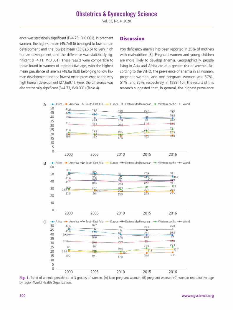
www.ogscience.org500
Vol. 63, No. 4, 2020
ence was statistically significant (F=4.73, P<0.001). In pregnant women, the highest mean (45.7±8.6) belonged to low human development and the lowest mean (33.8±6.6) to very high human development, and the difference was statistically sig-nificant (F=4.11, P<0.001). These results were comparable to those found in women of reproductive age, with the highest mean prevalence of anemia (48.8±18.8) belonging to low hu-man development and the lowest mean prevalence to the very high human development (27.6±9.1). Here, the difference was also statistically significant (F=4.73, P<0.001) (Table 4).
Discussion
Iron deficiency anemia has been reported in 25% of mothers with malnutrition [3]. Pregnant women and young children are more likely to develop anemia. Geographically, people living in Asia and Africa are at a greater risk of anemia. Ac-cording to the WHO, the prevalence of anemia in all women, pregnant women, and non-pregnant women was 37%, 51%, and 35%, respectively, in 1988 [16]. The results of this research suggested that, in general, the highest prevalence
Fig. 1. Trend of anemia prevalence in 3 groups of women. (A) Non-pregnant woman, (B) pregnant woman, (C) woman reproductive age by region World Health Organization.
A
B
C
50454035302520151050
Africa America South-East Asia Europe Eastern Mediterranean Western pacific World
2000 2005 2010 2015 2016
50454035302520151050
2000 2005 2010 2015 2016
60
50
40
30
20
10
0 2000 2005 2010 2015 2016
Africa America South-East Asia Europe Eastern Mediterranean Western pacific World
Africa America South-East Asia Europe Eastern Mediterranean Western pacific World
47.444.839.3
31.1
21.620.219.8
46.5
42.6
38.4
30.1
19.819.418.7
44.9
39.137.4
29.4
19.519.217.5
45.2
3938
31.6
23.721.718.2
45.6
39.8
38.232.5
25.122.518.8
47.6
45.639.5
31.6
22
20.4
20.2
46.7
43.4
38.6
30.620
19.6
19.1
45
40.1
37.6
29.9
19.519.7
17.8
45.3
39.138.8
32
23.9
21.8
18.4
45.8
39.839
32.8
25.3
22.7
19.21
50.3
52.141.641.1
26.928.9
27.5
50.5
49.240.540.4
27.726.8
26
48.1
4640.139.4
28.325.425.3
47.946.3
40.639.4
3226.125.3
48.1
46.240.940.1
3326.525.5

www.ogscience.org 501
Elham Goodarzi, et al. Relationship anemia and HDI
Table 2. Prevalence of anemia in Asian women (estimates by country in 2016)
CountryPrevalence of anemia in
non-pregnant woman (%)Prevalence of anemia in
pregnant woman (%)Prevalence of anemia in
woman reproductive age (%)
Afghanistan 42.4 (27.7–55.8) 38.2 (24.7–53.3) 42.0 (28.2–54.9)
Armenia 29.2 (12.2–56.3) 35.5 (17.5–63.3) 29.4 (12.6–56.3)
Azerbaijan 38.4 (25.3–51.8) 40.1 (26.9–54.7) 38.5 (25.7–51.6)
Bahrain 41.9 (19.0–67.2) 42.8 (20.2–64.8) 42.0 (19.7–66.7)
Bangladesh 39.6 (21.1–55.5) 45.7 (28.9–60.1) 39.9 (22.0–55.5)
Bhutan 35.7 (24.8–47.8) 32.8 (21.3–47.9) 35.6 (25.0–47.3)
Cambodia 46.3 (36.3–54.2) 55.8 (46.0–63.3) 46.8 (37.2–54.6)
China 26.2 (9.8–47.7) 32.4 (15.6–57.8) 26.4 (10.2–47.5)
Cyprus 25.1 (9.6–50.6) 29.0 (15.2–57.0) 25.2 (9.9–50.2)
Georgia 27.4 (12.9–45.5) 30.1 (17.5–50.3) 27.5 (13.2–45.3)
India 51.5 (41.5–58.7) 50.1 (42.0–57.2) 51.4 (41.8–58.5)
Indonesia 28.2 (18.7–38.8) 42.0 (30.4–53.7) 28.8 (19.4–39.0)
Iran 30.3 (12.7–52.8) 34.1 (17.5–57.6) 30.5 (13.2–52.4)
Iraq 28.7 (17.2–44.3) 33.5 (19.3–51.6) 29.1 (18.0–44.1)
Israel 15.2 (7.0–38.0) 23.8 (12.7–49.9) 15.7 (7.5–37.3)
Japan 21.2 (14.4–31.0) 34.1 (18.2–57.6) 21.5 (14.8–31.0)
Jordan 34.5 (20.8–50.0) 37.1 (22.9–51.6) 34.7 (21.4–49.7)
Kazakhstan 30.7 (13.0–52.2) 30.4 (15.7–54.3) 30.7 (13.4–51.2)
Kuwait 23.5 (9.8–46.3) 31.2 (14.8–57.3) 23.8 (10.2–46.2)
Kyrgyzstan 35.9 (21.3–51.5) 39.8 (25.1–55.6) 36.2 (22.0–51.0)
Lao People’s Democratic Republic 39.3 (19.5–60.7) 45.7 (23.8–65.0) 39.7 (20.7–60.7)
Lebanon 31.1 (13.2–55.0) 35.2 (17.8–60.1) 31.2 (13.6–54.8)
Malaysia 24.4 (9.4–49.8) 37.1 (15.8–63.0) 24.9 (10.0–49.7)
Maldives 42.4 (21.2–60.9) 46.9 (18.6–67.5) 42.6 (21.9–61.0)
Mongolia 18.9 (7.1–38.8) 30.3 (18.2–49.5) 19.5 (8.0–38.4)
Myanmar 46.0 (34.2–54.7) 53.8 (41.5–62.4) 46.3 (35.0–54.9)
Nepal 34.9 (19.2–50.9) 40.0 (22.9–57.7) 35.1 (19.9–50.9)
Oman 38.0 (17.4–62.2) 41.8 (20.8–62.5) 38.2 (18.1–62.1)
Pakistan 52.2 (35.3–67.4) 51.3 (36.4–62.8) 52.1 (35.7–67.0)
Philippines 14.9 (9.7–25.6) 30.3 (19.2–45.2) 15.7 (10.5–26.1)
Qatar 27.5 (10.2–52.8) 33.4 (15.2–58.5) 27.7 (10.6–52.9)
Saudi Arabia 42.8 (18.0–70.9) 45.5 (20.7–65.6) 42.9 (18.8–70.5)
Singapore 22.0 (8.5–50.3) 31.8 (15.2–59.6) 22.2 (8.7–50.3)
Sri Lanka 32.5 (14.1–51.3) 35.4 (19.2–55.6) 32.6 (14.7–51.1)
Syrian Arab Republic 33.5 (13.8–56.5) 36.1 (17.2–60.2) 33.6 (14.6–56.3)
Tajikistan 30.3 (14.0–49.3) 33.5 (17.3–57.4) 30.5 (14.9–48.5)
Thailand 31.6 (11.2–70.2) 40.2 (15.9–67.6) 31.8 (11.7–70.2)
Timor-Leste 41.2 (25.0–55.9) 42.6 (24.5–60.7) 41.3 (26.2–55.9)
Turkey 30.7 (12.0–56.7) 34.4 (16.5–60.2) 30.9 (12.5–56.6)
Turkmenistan 32.6 (13.3–54.9) 33.1 (16.7–58.1) 32.6 (13.8–54.4)
United Arab Emirates 27.6 (10.8–52.7) 33.2 (15.7–59.6) 27.8 (11.1–52.6)
Uzbekistan 36.8 (16.2–52.6) 24.9 (14.6–48.5) 36.2 (16.6–51.6)
Viet Nam 23.7 (10.4–43.7) 37.3 (19.3–58.5) 24.2 (11.0–43.8)
Yemen 70.2 (54.7–79.7) 63.0 (56.3–66.7) 69.6 (55.0–78.5)

www.ogscience.org502
Vol. 63, No. 4, 2020
Table 3. Pearson correlation between human development index (HDI) component and prevalence of anemia in Asian women in 2016
HDI component
Prevalence of anemia in non-pregnant woman
(%)
Prevalence of anemia in pregnant woman (%)
Prevalence of anemia in woman reproductive
age (%)
ra) P-value ra) P-value ra) P-value
Gross national income per 1,000 capita −0.284 >0.05 −0.225 >0.05 −0.286 >0.05
Mean years of schooling −0.564 <0.001 −0.563 <0.001 −0.567 <0.001
Life expectancy at birth −0.552 <0.001 −0.439 <0.001 −0.553 <0.001
Expected years of schooling −0.564 <0.001 −0.534 <0.001 −0.566 <0.001a)Statistical method used: Pearson correlation coefficient.
Fig. 2. Correlationa) between the human development index (HDI) and prevalence of anemia in Asian women. (A) Women reproductive age, (B) pregnant woman, (C) non-pregnant woman in 2016. a)Statistical method used: correlation coefficient.
A
C
B
Prev
alen
ce (%
)
Prev
alen
ce (%
)
Prev
alen
ce (%
)
70
60
50
40
30
20
80
60
40
20
70
60
50
40
30
20
0.5 0.6 0.7 0.8 0.9
HDI 0.5 0.6 0.7 0.8 0.9
HDI
0.5 0.6 0.7 0.8 0.9
HDI
Prevalence Fitted values Prevalence Fitted values
Prevalence Fitted values
R Sq. linear=-0.626, P<0.0001 R Sq. linear=-0.576, P<0.0001
R Sq. linear=-0.623, P<0.0001
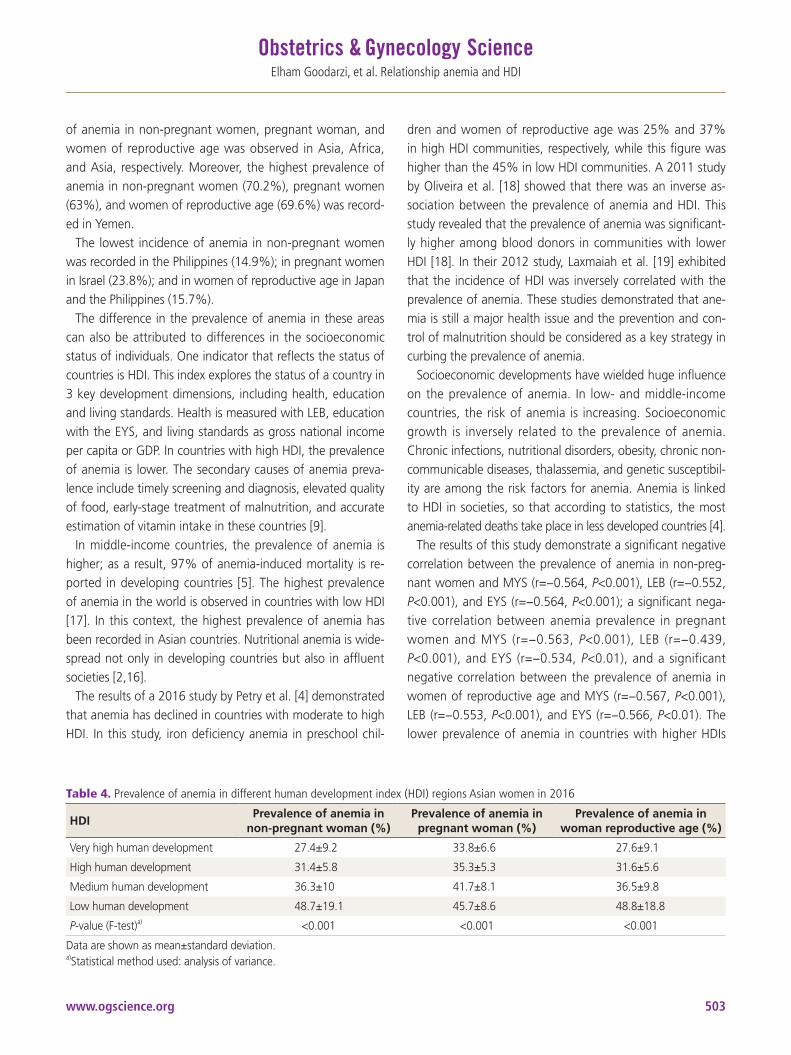
www.ogscience.org 503
Elham Goodarzi, et al. Relationship anemia and HDI
of anemia in non-pregnant women, pregnant woman, and women of reproductive age was observed in Asia, Africa, and Asia, respectively. Moreover, the highest prevalence of anemia in non-pregnant women (70.2%), pregnant women (63%), and women of reproductive age (69.6%) was record-ed in Yemen.
The lowest incidence of anemia in non-pregnant women was recorded in the Philippines (14.9%); in pregnant women in Israel (23.8%); and in women of reproductive age in Japan and the Philippines (15.7%).
The difference in the prevalence of anemia in these areas can also be attributed to differences in the socioeconomic status of individuals. One indicator that reflects the status of countries is HDI. This index explores the status of a country in 3 key development dimensions, including health, education and living standards. Health is measured with LEB, education with the EYS, and living standards as gross national income per capita or GDP. In countries with high HDI, the prevalence of anemia is lower. The secondary causes of anemia preva-lence include timely screening and diagnosis, elevated quality of food, early-stage treatment of malnutrition, and accurate estimation of vitamin intake in these countries [9].
In middle-income countries, the prevalence of anemia is higher; as a result, 97% of anemia-induced mortality is re-ported in developing countries [5]. The highest prevalence of anemia in the world is observed in countries with low HDI [17]. In this context, the highest prevalence of anemia has been recorded in Asian countries. Nutritional anemia is wide-spread not only in developing countries but also in affluent societies [2,16].
The results of a 2016 study by Petry et al. [4] demonstrated that anemia has declined in countries with moderate to high HDI. In this study, iron deficiency anemia in preschool chil-
dren and women of reproductive age was 25% and 37% in high HDI communities, respectively, while this figure was higher than the 45% in low HDI communities. A 2011 study by Oliveira et al. [18] showed that there was an inverse as-sociation between the prevalence of anemia and HDI. This study revealed that the prevalence of anemia was significant-ly higher among blood donors in communities with lower HDI [18]. In their 2012 study, Laxmaiah et al. [19] exhibited that the incidence of HDI was inversely correlated with the prevalence of anemia. These studies demonstrated that ane-mia is still a major health issue and the prevention and con-trol of malnutrition should be considered as a key strategy in curbing the prevalence of anemia.
Socioeconomic developments have wielded huge influence on the prevalence of anemia. In low- and middle-income countries, the risk of anemia is increasing. Socioeconomic growth is inversely related to the prevalence of anemia. Chronic infections, nutritional disorders, obesity, chronic non-communicable diseases, thalassemia, and genetic susceptibil-ity are among the risk factors for anemia. Anemia is linked to HDI in societies, so that according to statistics, the most anemia-related deaths take place in less developed countries [4].
The results of this study demonstrate a significant negative correlation between the prevalence of anemia in non-preg-nant women and MYS (r=−0.564, P<0.001), LEB (r=−0.552, P<0.001), and EYS (r=−0.564, P<0.001); a significant nega-tive correlation between anemia prevalence in pregnant women and MYS (r=−0.563, P<0.001), LEB (r=−0.439, P<0.001), and EYS (r=−0.534, P<0.01), and a significant negative correlation between the prevalence of anemia in women of reproductive age and MYS (r=−0.567, P<0.001), LEB (r=−0.553, P<0.001), and EYS (r=−0.566, P<0.01). The lower prevalence of anemia in countries with higher HDIs
Table 4. Prevalence of anemia in different human development index (HDI) regions Asian women in 2016
HDIPrevalence of anemia in
non-pregnant woman (%)Prevalence of anemia in
pregnant woman (%)Prevalence of anemia in
woman reproductive age (%)
Very high human development 27.4±9.2 33.8±6.6 27.6±9.1
High human development 31.4±5.8 35.3±5.3 31.6±5.6
Medium human development 36.3±10 41.7±8.1 36.5±9.8
Low human development 48.7±19.1 45.7±8.6 48.8±18.8
P-value (F-test)a) <0.001 <0.001 <0.001
Data are shown as mean±standard deviation.a)Statistical method used: analysis of variance.
www.ogscience.org504
Vol. 63, No. 4, 2020
could be attributed to factors such as the existence of micro-nutrients in foods, more accurate diagnostic methods and enhanced access to primary health care.
The major factors contributing to the high prevalence of anemia in most countries are low income levels and malnu-trition. Hence, one effective step to mitigate the prevalence of anemia is to provide primary anemia prevention strategies such as increasing household per capita and providing food packages for families. Despite the interrelation of these fac-tors, caution should be practiced when interpreting such studies because, in addition to the environmental factors of anemia prevalence, the limitations of descriptive studies should also be taken into consideration. Early prevention and epidemiological studies, especially for people in less devel-oped countries, can help reduce the burden of disease and improve health status in these countries.
In women from different south-east Asian regions iron de-ficiency anemia should ideally be addressed through dietary diversification and improved access to foods that have high levels of bioavailable iron, including animal products. Daily or intermittent iron supplementation, alone or together with folic acid and other micronutrients, can be used for high-risk groups (children, pregnant women and women of reproduc-tive age), to improve iron intake. These strategies should be tailored to local conditions, taking into account the specific etiology and prevalence of anemia in a given setting and population group, and should be built into the primary health-care system and existing programs.
In conclusion, the results of the present study suggest a negative correlation between the prevalence of anemia in women and HDI. Moreover, the prevalence of anemia in these countries was negatively correlated with LEB and edu-cation. Hence, a study of factors that raise the prevalence of anemia in countries with low HDI can go a long way in reducing its incidence rate. Further, raising the level of a mother’s education and her awareness can also contribute to a reduced incidence.
Acknowledgements
This work was supported by Kerman University of Medical Sciences, Kerman, Iran.
Conflict of interest
No potential conflict of interest relevant to this article was reported.
Ethical approval
The study was approved by Ethic Committee (IR.KMU.REC.1398.505).
Patient consent
The patients provided written informed consent for the pub-lication and the use of their images.
References
1. World Health Organization. Iron deficiency anemia. Assessment, prevention, and control. A guide for pro-gramme managers. Geneva: World Health Organization; 2001.
2. Saxena Y, Shrivastava A, Saxena V. Effect of gender on correlation of anaemia with body mass index in medical students. Indian J Physiol Pharmacol 2011;55:364-9.
3. Seshadri S. Prevalence of micronutrient deficiency par-ticularly of iron, zinc and folic acid in pregnant women in South East Asia. Br J Nutr 2001;85 Suppl 2:S87-92.
4. Petry N, Olofin I, Hurrell RF, Boy E, Wirth JP, Moursi M, et al. The proportion of anemia associated with iron de-ficiency in low, medium, and high human development index countries: a systematic analysis of national surveys. Nutrients 2016;8:693.
5. World Health Organization. Global health risks: mortal-ity and burden of disease attributable to selected major risks. Geneva: World Health Organization; 2009.
6. Hallberg L, Hultén L, Lindstedt G, Lundberg PA, Mark A, Purens J, et al. Prevalence of iron deficiency in Swedish adolescents. Pediatr Res 1993;34:680-7.
7. World Health Organization. The global prevalence of anaemia in 2011. Geneva: World Health Organization; 2015.
8. Roy CN. Anemia of inflammation. Hematology Am Soc

www.ogscience.org 505
Elham Goodarzi, et al. Relationship anemia and HDI
Hematol Educ Program 2010;2010:276-80. 9. Mockenhaupt FP, Rong B, Günther M, Beck S, Till H,
Kohne E, et al. Anaemia in pregnant Ghanaian women: importance of malaria, iron deficiency, and haemoglo-binopathies. Trans R Soc Trop Med Hyg 2000;94:477-83.
10. Khambalia AZ, Aimone AM, Zlotkin SH. Burden of anemia among indigenous populations. Nutr Rev 2011;69:693-719.
11. Shih YC, Chien CR, Xu Y, Pan IW, Smith GL, Buchholz TA. Economic burden of renal cell carcinoma in the US: part II--an updated analysis. Pharmacoeconomics 2011;29:331-41.
12. Gordon EJ, Leon JB, Sehgal AR, Hoffart N. Why are hemodialysis treatments shortened and skipped? Devel-opment of a taxonomy and relationship to patient sub-groups. Nephrol Nurs J 2003;30:209-17.
13. World Health Organization. Haemoglobin concentra-tions for the diagnosis of anaemia and assessment of severity. Geneva: World Health Organization; 2011.
14. World Health Organization. Prevalence of anaemia in pregnant women (%) [Internet]. Geneva: World Health Organization; c2016 [cited 2019 Jan 17]. Available
from: https://www.who.int/data/gho/data/indicators/indicator-details/GHO/prevalence-of-anaemia-in-preg-nant-women-(-)?bookmarkId=56cd5b17-0a02-480e-8205-abc912b8e333.
15. Anand S, Sen AK. Human development index: method-ology and measurement. New York (NY): United Nations Development Programme; 1994.
16. McLean E, Cogswell M, Egli I, Wojdyla D, de Benoist B. Worldwide prevalence of anaemia, WHO vitamin and mineral nutrition information system, 1993–2005. Public Health Nutr 2009;12:444-54.
17. Barkley JS, Wheeler KS, Pachón H. Anaemia prevalence may be reduced among countries that fortify flour. Br J Nutr 2015;114:265-73.
18. Oliveira CD, Martins G, Custer B, Proietti FA, Carneiro-Proietti AB, César CC. Hierarchical analysis of anaemia deferral in blood donor candidates: the individual in the population perspective. Transfus Med 2011;21:371-7.
19. Laxmaiah A, Arlappa N, Balakrishna N, Mallikarjuna Rao K, Galreddy C, Kumar S, et al. Prevalence and determi-nants of micronutrient deficiencies among rural children of eight states in India. Ann Nutr Metab 2013;62:231-41.

www.ogscience.org506
Original ArticleObstet Gynecol Sci 2020;63(4):506-513https://doi.org/10.5468/ogs.19185pISSN 2287-8572 · eISSN 2287-8580
Comparison of pain and proper sample status according to usage of tenaculum and analgesia: a randomized clinical trialCihan Comba, MD1, Gökhan Demirayak, MD2, Sakir Volkan Erdogan, MD3, Ibrahim Karaca, MD4, Omer Demir, MD5, Oguz Guler, MD6, Isa Aykut Ozdemir, MD7
Division of Gynecologic Oncology, Department of Gynecology and Obstetrics, 1University of Health Sciences, Sultangazi Haseki Training and Research Hospital, Istanbul, 2University of Health Sciences, Bakirkoy Dr. Sadi Konuk Training and Research Hospital, Istanbul; Department of Gynecology and Obstetrics, 3University of Health Sciences, Bagcilar Training and Research Hospital, Istanbul, 4University of Health Sciences, Tepecik Training and Research Hospital, Izmir, 5Karadeniz Technical University, Trabzon, 6Kucukkoy Bilge Hospital, Istanbul; 7Division of Gynecologic Oncology, Department of Gynecology and Obstetrics, Medipol University Mega Hospital, Istanbul, Turkey
ObjectiveColposcopic biopsy is a discomfortable procedure. Additionally, it creates negative influence on sexuality. This study aimed to investigate the relationships among tenaculum, pain perception, and biopsy size during colposcopy.
MethodsIn total, 228 patients who underwent colposcopy-directed biopsy were included, and randomized into 4 groups based on whether analgesic and tenaculum were used and replaced (tenaculum with n=58/without analgesic n=56, no tenaculum replacement with n=57/without analgesic n=57). Lidocaine hydrochloride (40 mg) plus adrenaline (0.025 mg) was administered in the analgesic groups. The pain was assessed using a linear visual analog scale. The biopsy specimen size was measured in millimeters.
ResultsThe mean age of the patients was 42.85±8.88 years. The most frequent colposcopy indications were atypical squamous cells of undetermined significance and human papilloma virus-positive results on cervical cytology (30.2%; n=69). Low- and high-grade intraepithelial lesions were noted in 14.91% (n=34) and 10.96% (n=25) women through colposcopy-directed biopsy results, respectively. Tenaculum replacement increased pain perception in the without analgesic group; however, no statistically significant differences were noted between of the groups with and without tenaculum replacement with analgesic. The size and number of biopsy specimens were not associated with tenaculum replacement and analgesic use.
ConclusionAdministration of analgesics decreased discomfort and pain in patients. Tenaculum replacement aided colposcopists in manipulating the cervix. Additionally, administration of analgesics relieved pain in the tenaculum replacement group.
Trial RegistrationClinicalTrials.gov Identifier: NCT03279666
Keywords: Cervix uteri; Biopsy; Pain perception; Human papilloma virus
Received: 2019.09.27. Revised: 2020.02.17. Accepted: 2020.02.18.Corresponding author: Cihan Comba, MDDivision of Gynecologic Oncology, Department of Gynecology and Obstetrics, University of Health Sciences, Sultangazi Haseki Training and Research Hospital, Uğur Mumcu, Hastane Cad No:1 A D:1B, 34265 Sultangazi/Istanbul, TurkeyE-mail: [email protected]://orcid.org/0000-0002-3161-2689
Articles published in Obstet Gynecol Sci are open-access, distributed under the terms of the Creative Commons Attribution Non-Commercial License (http://creativecommons.org/licenses/by-nc/3.0/) which permits unrestricted non-commercial use, distribution, and reproduction in any medium, provided the original work is properly cited.
Copyright © 2020 Korean Society of Obstetrics and Gynecology

www.ogscience.org 507
Cihan Comba, et al. Suited technique for colposcopic biopsy
Introduction
Colposcopy is the cornerstone of cervical precancerous lesion evaluation [1]. It has been the standard of care for evaluating abnormal cervical cytology since the 1970s [2].
In conjunction with screening and treatment of precancer-ous lesions, colposcopy has played a crucial role in reducing the incidence and mortality due to cervical cancer for the past 50 years [1]. The current guidelines for initial manage-ment of high-grade cervical cytological abnormalities recom-mend an immediate diagnostic excisional procedure during the initial colposcopy that is critical to a “see-and-treat” ap-proach in selected patients. In addition, it allows recognition of a subset of women with cervical disease and safe observa-tion of the disease over time. Further, it plays an important role in reducing the over-treatment of low-grade lesions [3].
Procedure risks of colposcopy, such as significant bleeding, infection, and long-term morbidity, are low. Colposcopy-directed punch biopsies are associated with discomfort, cramping, and pain. Furthermore, some women reported a negative influence on sexuality [4]. A traumatic colposcopy experience may lead to some women refraining from obtain-ing adequate cervical screening in the future [5]. Both abnor-mal cervical cytology results and colposcopy experience can induce anxiety in women [6]. A study reported that women experienced worry and anxiety in the interim period—the time between being notified of an abnormal screening result and colposcopy appointment [7]. However, the procedure can usually be performed on an outpatient basis without anes-thesia [8]. Researchers have studied the use of local anesthe-sia, topical spray, and forced cough method to reduce pain due to colposcopy-directed punch biopsy [8-10], and most of these studies indicated that these methods were effective.
Most gynecologic oncology centers worldwide use the International Federation of Gynecology and Obstetrics stag-ing system for gynecological cancers such as cervical cancer. The clinical stage is used for cervical cancer staging; hence, it is based on the results of the physical examination by doc-tor, biopsies, imaging tests, and a few other tests such as magnetic resonance imaging, cystoscopy, and proctoscopy
[11]. Stage 1A (microinvasive) cervical cancer can only be detected through microscopic examination of removed tis-sue, preferably a cone; however, several punch biopsies may be required for diagnosis. Stage 1A cervical cancer was de-fined as invasion limited to measured stromal invasion with
a maximum depth of 5 mm and stage 1A1 was defined as measured stromal invasion with a maximum depth of 3 mm
[12]. Hence, punch biopsy size is crucial.This study aimed to investigate the relationship among te-
naculum placement, pain perception, and biopsy size during colposcopy.
Materials and methods
Bakirkoy Dr. Sadi Konuk Training and Research Hospital (BTRH), University of Health Sciences, was the reference cen-ter in the cervical smear and human papilloma virus (HPV) screening program for 30–65-year-old women. The screen-ing had been performed by primary level health staff (family physicians and screening center staff) under the Republic of Turkey, Ministry of Health, since 2014. These centers referred women with high-risk (HR) HPV positive results to specific hospitals. HR-HPV detection was performed using the com-mercially available Hybrid Capture 2 assay® (HC2; Digene Corp., Gaithersburg, MD, USA). The results were assessed in BTRH, and colposcopy was performed by gynecologic on-cology faculty members or gynecologic oncology fellows in patients with indications for colposcopy.
This study was conducted on patients with indications for colposcopy and within the scope of the Republic of Turkey, Ministry of Health Cervical Cancer Screening pro-gram between June 15, 2017 and December 31, 2017. The study was approved by the ethical board of BTRH (number: 2017/47) and was registered in ClinicalTrials.gov (number: NCT03279666). Informed consent was obtained from all the patients before the procedure. Patients were randomized using a computer program. A statistical program (GPower version 3, Heinrich Heine University, Düsseldorf, Germany) was used to estimate the sample size through a 1-tailed hy-pothesis using an independent sample t-test with an α error of 0.05 and a power of 0.90. The total sample size required for a moderate effect size (d=0.50) was calculated to be 236. In the present study, 58 women were in group A, 56 women were in group B, 57 women were in group C, and 57 women were in group D. Post hoc analysis for moderate effect size indicated that the power of the test was reduced to 0.80. In addition to undergoing colposcopy, the inclusion criteria for the study were as follows:
• Being older than 21 years old

www.ogscience.org508
Vol. 63, No. 4, 2020
• No history of receiving colposcopy, loop electrosurgical excision procedure (LEEP), conization, or cryotherapy
• Did not use analgesics up to 6 hours before the procedure• Did not experience chronic pain or did not use analgesics
for a long-term• Did not have advanced atrophic or hypertrophic cervix• Did not have visible cervical mass (polyp, myoma, or lo-
cally advanced cervical cancer)The exclusion criteria were as follows:• Did not provide consent for participation in the study• Had advanced orthopedic problem• Had allergy to analgesics• Were pregnantThis study enrolled 228 women. Patients were randomly
assigned to 4 groups (Fig. 1).• Group 1: patients with tenaculum replacement without
analgesic (n=58)• Group 2: patients without tenaculum replacement with-
out analgesic (n=56)• Group 3: patients with tenaculum replacement with anal-
gesic (n=57)• Group 4: patients without tenaculum replacement with
analgesic (n=57)The number of patients who underwent cervical biopsy as
well as endocervical curettage (ECC), only cervical biopsy, and only ECC were 182, 42, and 4, respectively.
The patients were placed in the lithotomy position in a quiet colposcopy room and using a single-use vaginal specu-lum colposcopy was performed by the gynecologic oncology team, comprising a doctor and nurse. Colposcopy was start-ed by cleaning the cervix with 5-mL saline. Cotton soaked in freshly prepared 5% acetic acid solution was applied on the cervix for 3 minutes, and subsequently, colposcopic exami-nation was performed. Then, 2-mL lidocaine hydrochloride (40 mg) plus adrenaline (0.025 mg) was injected at the 8 and 4 o’clock positions of the cervix using a 22-gauge needle to
Fig. 1. Flow diagram of the study.
Included (n=228)Group 1 (n=58) Group 2 (n=56) Group 3 (n=57) Group 4 (n=57)
Schedule subjects(n=240)
Excluded (n=12)Not tolerated
procedure (n=9) Need grasping for
biopsy (n=3)
Table 1. General data of all groups
Variables Group 1 (n=58) Group 2 (n=56) Group 3 (n=57) Group 4 (n=57) P-valuea)
Gravidy 4.57±4.9/3 (0–34) 3.2±1.9/3 (0–9) 3.1±1.9/3 (0–9) 3.6±2.6/3 (0–14) 0.327
Parity 3.02±2.06/2 (0–12) 2.7±1.8/2 (0–8) 2.3±1.4/2 (0–7) 2.4±1.5/2 (0–7) 0.297
SVD 2.6±2.2/2 (0–12) 2.1±1.9/2 (0–7) 1.9±1.6/2 (0–7) 2.02±1.7/2 (0–7) 0.540
SCA 0.5±0.9/0 (0–4) 0.46±0.8/0 (0–3) 0.35±0.7/0 (0–3) 0.3±0.6/0 (0–3) 0.681
Abortus 0.69±1.4/0 (0–9) 0.38±0.6/0 (0–2) 0.54±0.7/0 (0–3) 0.89±1.2/0 (0–5) 0.099
UC 0.88±3.9/0 (0–30) 0.13±0.5/0 (0–2) 0.21±0.8/0 (0–5) 0.28±0.9/0 (0–5) 0.112
Age 43.4±10.03/42 (28–72) 43.1±8.5/42.5 (29–64) 41.8±7.9/41 (27–60) 43.1±9.1/44 (25–63) 0.809
Number of biopsy 2.9±1.04/3 (1–5) 3.3±1.2/3 (1–5) 3.6±0.9/3 (1–6) 3.7±0.9/4 (2–6) 0.001b)
Pain level 3.9±3.1/3 (0–10) 4±3.3/3 (0–10) 2.2±2.7/1 (0–10) 2.6±3.02/1 (0–10) <0.001b)
Size of biopsy_median 3.4±1.3/3.3 (1–8) 3.8±1.8/3.5 (1–8.3) 3.6±1.2/3.3 (1.7–7.5) 3.7±1.3/3.7 (1–7) 0.492
Values are presented as mean±standard deviation/median (minimum–maximum). Group 1, patients with tenaculum replacement without an-algesic; Group 2, patients without tenaculum replacement without analgesic; Group 3, patients with tenaculum replacement with analgesic; Group 4, patients without tenaculum replacement with analgesic.SVD, spontaneous vaginal delivery; SCA, sectio caserean abdominalis; UC, uterine curuttage for unwanted pregnancy.a)Kruskal-Wallis test; b)P<0.01.

www.ogscience.org 509
Cihan Comba, et al. Suited technique for colposcopic biopsy
those in the groups 3 and 4. The doctor waited for 2 minutes for the analgesic to take effect before obtaining biopsy speci-mens. Biopsy specimens were obtained from necessary sites by using a biopsy equipment (Schumacher biopsy forceps 1/1 made in Germany), with or without tenaculum replacement on the anterior part of the cervix transversely in accordance with the group. ECC was performed using a Recamier uter-ine curette (width 3 mm made in Germany) if required. Sub-sequently, the linear visual analog scale was used to assess pain within 2 minutes after colposcopic examination.
1. Statistical analysisDescriptive statistics were used to define continuous vari-ables (mean; standard deviation; and minimum, median, and maximum). The Kruskal-Wallis test was used to analyze the association between more than 2 continuous variables that were independent and not normally distributed. The Mann-Whitney U test was used to analyze the association between 2 continuous variables that were independent and not normally distributed. The correlation between 2 continu-ous variables without normal distribution was analyzed with Spearman’s rho correlation analysis. Chi-square (or Fisher exact test as applicable) was used to analyze the association between categorical variables. Statistical significance was set at P=0.05. The analyses were conducted using MedCalc Statistical Software version 12.7.7 (MedCalc Software bvba, Ostend, Belgium; http://www.medcalc.org; 2013).
Results
A total of 228 of 240 patients were analyzed. Ten patients
were excluded because they did not provide consent to participate and other 2 patients were excluded because their biopsy results indicated cervical cancer. The mean age of included patients was 42.85±8.88 years (range, 25–72 years; Table 1). The most frequent colposcopy indications were atypical squamous cells of undetermined significance and HPV positive results on cervical cytology (n=69; 30.2%). The most common HR-HPV type was HPV 16 (n=68; 29.8%). HPV 51 was the most frequently detected non-HPV 16-18 of the high-risk HPV types (Table 2). Normal cytology, low-grade intraepithelial lesions and high-grade intraepithelial lesions
Table 2. Human papilloma virus status
HPV type Group 1 (n=58) Group 2 (n=56) Group 3 (n=57) Group 4 (n=57) P-valuea)
Negative 16 (27.6) 15 (27.3) 24 (42.1) 24 (42.1) 0.044
HPV type 16 13 (22.4) 10 (18.2) 14 (24.6) 8 (14.0)
HPV type 18 2 (3.4) 2 (3.6) 3 (5.3) 2 (3.5)
HPV type 16+18 6 (10.3) 8 (14.5) 3 (5.3) 2 (3.5)
HPV type non 16-18 15 (25.9) 19 (34.5) 8 (14.0) 10 (17.5)
HPV type 16 or 18+HPV-N 6 (10.3) 1 (1.8) 5 (8.8) 11 (19.3)
Values are presented as number (%). Hybrid capture 2 technology was used for detection of 13 high-risk HPV types.HPV, human papilloma virus; HPV-N, non 16-18 high-risk HPV types. a)Ki-Kare test.
Table 3. Relationship between pathological results and pain per-ception
Pathological result Pain perception
Pathological result of ECC
Inadequate (n=11) 3.4±2.6/2 (1–9)
Normal (n=162) 2.9±3.1/1 (0–10)
LSIL (n=6) 4.7±3.7/4 (1–10)
HSIL (n=7) 4±3.4/2 (0–9)
P-valuea) 0.277
Pathological result of biopsy
Normal (n=164) 3.1±3.1/2 (0–10)
LSIL (n=34) 3.7±3.5/2.5 (0–10)
HSIL (n=25) 3.1±3.03/2 (0–10)
Cancer (n=1) -
P-valuea) 0.653
Values are presented as mean±standard deviation/median (mini-mum–maximum).ECC, endo cervical curettage; LSIL, low grade intraepithelial lesion; HSIL, high grade intraepithelial lesion.a)Kruskal-Wallis test.
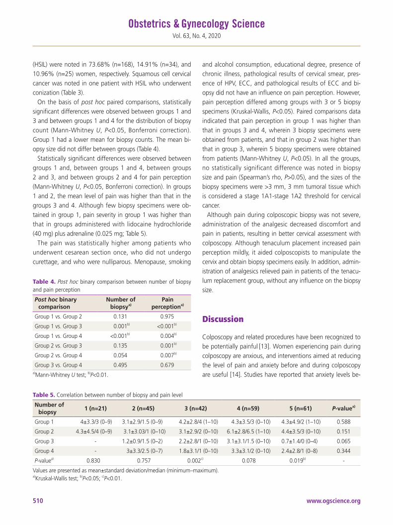
www.ogscience.org510
Vol. 63, No. 4, 2020
(HSIL) were noted in 73.68% (n=168), 14.91% (n=34), and 10.96% (n=25) women, respectively. Squamous cell cervical cancer was noted in one patient with HSIL who underwent conization (Table 3).
On the basis of post hoc paired comparisons, statistically significant differences were observed between groups 1 and 3 and between groups 1 and 4 for the distribution of biopsy count (Mann-Whitney U, P<0.05, Bonferroni correction). Group 1 had a lower mean for biopsy counts. The mean bi-opsy size did not differ between groups (Table 4).
Statistically significant differences were observed between groups 1 and, between groups 1 and 4, between groups 2 and 3, and between groups 2 and 4 for pain perception (Mann-Whitney U, P<0.05, Bonferroni correction). In groups 1 and 2, the mean level of pain was higher than that in the groups 3 and 4. Although few biopsy specimens were ob-tained in group 1, pain severity in group 1 was higher than that in groups administered with lidocaine hydrochloride (40 mg) plus adrenaline (0.025 mg; Table 5).
The pain was statistically higher among patients who underwent cesarean section once, who did not undergo curettage, and who were nulliparous. Menopause, smoking
and alcohol consumption, educational degree, presence of chronic illness, pathological results of cervical smear, pres-ence of HPV, ECC, and pathological results of ECC and bi-opsy did not have an influence on pain perception. However, pain perception differed among groups with 3 or 5 biopsy specimens (Kruskal-Wallis, P<0.05). Paired comparisons data indicated that pain perception in group 1 was higher than that in groups 3 and 4, wherein 3 biopsy specimens were obtained from patients, and that in group 2 was higher than that in group 3, wherein 5 biopsy specimens were obtained from patients (Mann-Whitney U, P<0.05). In all the groups, no statistically significant difference was noted in biopsy size and pain (Spearman’s rho, P>0.05), and the sizes of the biopsy specimens were >3 mm, 3 mm tumoral tissue which is considered a stage 1A1-stage 1A2 threshold for cervical cancer.
Although pain during colposcopic biopsy was not severe, administration of the analgesic decreased discomfort and pain in patients, resulting in better cervical assessment with colposcopy. Although tenaculum placement increased pain perception mildly, it aided colposcopists to manipulate the cervix and obtain biopsy specimens easily. In addition, admin-istration of analgesics relieved pain in patients of the tenacu-lum replacement group, without any influence on the biopsy size.
Discussion
Colposcopy and related procedures have been recognized to be potentially painful [13]. Women experiencing pain during colposcopy are anxious, and interventions aimed at reducing the level of pain and anxiety before and during colposcopy are useful [14]. Studies have reported that anxiety levels be-
Table 4. Post hoc binary comparison between number of biopsy and pain perception
Post hoc binary comparison
Number of biopsya)
Pain perceptiona)
Group 1 vs. Group 2 0.131 0.975
Group 1 vs. Group 3 0.001b) <0.001b)
Group 1 vs. Group 4 <0.001b) 0.004b)
Group 2 vs. Group 3 0.135 0.001b)
Group 2 vs. Group 4 0.054 0.007b)
Group 3 vs. Group 4 0.495 0.679a)Mann-Whitney U test; b)P<0.01.
Table 5. Correlation between number of biopsy and pain level
Number of biopsy
1 (n=21) 2 (n=45) 3 (n=42) 4 (n=59) 5 (n=61) P-valuea)
Group 1 4±3.3/3 (0–9) 3.1±2.9/1.5 (0–9) 4.2±2.8/4 (1–10) 4.3±3.5/3 (0–10) 4.3±4.9/2 (1–10) 0.588
Group 2 4.3±4.5/4 (0–9) 3.1±3.03/1 (0–10) 3.1±2.9/2 (0–10) 6.1±2.8/6.5 (1–10) 4.4±3.5/3 (0–10) 0.151
Group 3 - 1.2±0.9/1.5 (0–2) 2.2±2.8/1 (0–10) 3.1±3.1/1.5 (0–10) 0.7±1.4/0 (0–4) 0.065
Group 4 - 3±3.3/2.5 (0–7) 1.8±3.1/1 (0–10) 3.3±3.1/2 (0–10) 2.4±2.8/1 (0–8) 0.344
P-valuea) 0.830 0.757 0.002c) 0.078 0.019b) -
Values are presented as mean±standard deviation/median (minimum–maximum).a)Kruskal-Wallis test; b)P<0.05; c)P<0.01.

www.ogscience.org 511
Cihan Comba, et al. Suited technique for colposcopic biopsy
fore and during the procedure are elevated in women. Partic-ularly, anxiety can be attributed to the uncertainty regarding what the colposcopic examination entails, events during the procedure, and fear or experience of pain during the pro-cedure [15,16]. Humor, imagination of pleasant scenes and experiences, music, and forced coughing have been reported to reduce pain through the gate control theory of pain based on partial inhibition of painful sensations traveling along slower nerve fibers by overriding stimuli that travel along fast nerve fibers [17,18]. However, to the best of our knowledge, no study has reported the relationship among the use of te-naculum, pain perception, and biopsy size to date.
In 2011, Naki et al. [10] found that local anesthesia was more effective than forced coughing for relieving pain during cervical biopsy, in contrast to Schmid et al. [9] in 2008, but the duration of the entire procedure was significantly longer in patients in the local anesthetic group [10].
Bogani et al. [19] observed that nulliparity, number of previ-ous vaginal deliveries, and pain expectancy were associated with pain perception during colposcopic biopsy, and only pain expectancy independently correlated with operative pain levels. Factors such as educational level, obstetric his-tory, and chronic pelvic pain may affect the anticipated pain score. Öz et al. [20] reported a significantly inverse relation-ship between patients’ educational level and reported pain scores after both cervical biopsy and ECC procedures. On the contrary, in the present study, we found that women who were nulliparous, who underwent cesarean section abdomi-nalis, and who did not undergo dilatation and curettage perceived a higher level of pain. However, education, meno-pause, smoking, history of ECC, and chronic diseases were found to be not associated with pain score.
The first study by Rabin et al. [21] investigated the use of local anesthesia for gynecological procedures by comparing 20% benzocaine gel with placebo for its ability to reduce pain associated with cervical biopsy, intrauterine device in-sertion, pain associated with injection of anesthetic drug, tenaculum placement, and ECC, and revealed that 20% ben-zocaine gel significantly reduced pain associated with these procedures. In the current study, it was found that local an-esthesia reduced pain perception as a significant difference in pain perception was noted between the groups without tenaculum replacement and with or without local anesthesia.
Several scientific studies have indicated that women sub-jected to a passive, pleasing and simple visual distraction dur-
ing colposcopy experienced less post procedural pain than those who were not subjected to visual distraction [22,23]. In the present study, the overall pain level in group 3 patients was low (2.2±2.7).
The procedure type may affect patients’ pain perception. Kola-Palmer et al. [24] reported that a significant difference in sensory pain existed between treatment groups of women undergoing see-and-treat large loop excision of the trans-formation zone treatment and those undergoing colposcopy plus punch biopsy who reported significantly higher levels of pain perception than women who underwent diagnostic colposcopy.
The tenaculum is used to stabilize and supply counter trac-tion on the cervix and uterus [25]. A study by Goldthwaite et al. [26] compared pain scores with tenaculum placement after injecting intracervical lidocaine and topically applying lidocaine gel. Lidocaine injection was more effective than lidocaine gel application in the study. Robinson et al. [27] indicated that a significant difference in pain perception ex-isted after comparing intracervical lidocaine injection (saline injection), when traction was placed with tenaculum during hysterosalpingography, with no injection. Oyama et al. [28] revealed that lidocaine injection reduced pain score during cervical biopsies and endocervical curettage, and overall pro-cedure scores. Additionally, Kiviharju et al. [29] found that local anesthesia decreased the pain sensation. In the present study, we found that intracervical lidocaine injection reduced pain during colposcopic biopsy.
Colposcopists speculate that the most suitable colposcopic assessment and biopsy method is the one that involves ad-ministration of analgesics and tenaculum use (group 3 in this study). This method can aid in reducing pain, and the physi-cian can manipulate the cervix and obtain biopsy specimens easily during the procedure. Invasive cancer can be diag-nosed with a single biopsy. However, generally, biopsy instru-ments are not sufficiently large to diagnose invasive cancer. In addition, further studies with more participants should be conducted to investigate whether the size of the biopsy specimen has an influence.
Conflict of interest
No potential conflict of interest relevant to this article was reported.
www.ogscience.org512
Vol. 63, No. 4, 2020
Ethical approval
All the procedures performed in the study were in accor-dance with the ethical standards of the institutional and/or national research committee and with the 1964 Helsinki declaration and its later amendments or comparable ethical standards.
Patient consent
Informed consent was obtained from all the study partici-pants.
References
1. King A, Clay K, Felmar E, Heustis DG, Karns RM, Krahl P, et al. The Papanicolaou smear. West J Med 1992;156:202-4.
2. Singer A, Walker P, Tay SK, Dyson J. Impact of introduc-tion of colposcopy to a district general hospital. Br Med J (Clin Res Ed) 1984;289:1049-51.
3. Massad LS, Einstein MH, Huh WK, Katki HA, Kinney WK, Schiffman M, et al. 2012 updated consensus guide-lines for the management of abnormal cervical cancer screening tests and cancer precursors. Obstet Gynecol 2013;121:829-46.
4. Campion MJ, Brown JR, McCance DJ, Atia W, Edwards R, Cuzick J, et al. Psychosexual trauma of an abnormal cervical smear. Br J Obstet Gynaecol 1988;95:175-81.
5. Ideström M, Milsom I, Andersson-Ellström A. Women’s experience of coping with a positive Pap smear: a reg-ister-based study of women with two consecutive Pap smears reported as CIN 1. Acta Obstet Gynecol Scand 2003;82:756-61.
6. Kola-Palmer S, Walsh JC. Correlates of psychological dis-tress immediately following colposcopy. Psychooncology 2015;24:819-24.
7. Waller J, McCaffery K, Kitchener H, Nazroo J, Wardle J. Women’s experiences of repeated HPV testing in the context of cervical cancer screening: a qualitative study. Psychooncology 2007;16:196-204.
8. Clifton PA, Shaughnessy AF, Andrews S. Ineffectiveness of topical benzocaine spray during colposcopy. J Fam
Pract 1998;46:242-6. 9. Schmid BC, Pils S, Heinze G, Hefler L, Reinthaller A,
Speiser P. Forced coughing versus local anesthesia and pain associated with cervical biopsy: a randomized trial. Am J Obstet Gynecol 2008;199:641.e1-3.
10. Naki MM, Api O, Acioglu HC, Uzun MG, Kars B, Unal O. Analgesic efficacy of forced coughing versus local anesthesia during cervical punch biopsy. Gynecol Obstet Invest 2011;72:5-9.
11. Freeman SJ, Aly AM, Kataoka MY, Addley HC, Reinhold C, Sala E. The revised FIGO staging system for uterine malignancies: implications for MR imaging. Radiograph-ics 2012;32:1805-27.
12. Bhatla N, Aoki D, Sharma DN, Sankaranarayanan R. Cancer of the cervix uteri. Int J Gynaecol Obstet 2018;143 Suppl 2:22-36.
13. Gajjar K, Martin-Hirsch PP, Bryant A. Pain relief for women with cervical intraepithelial neoplasia undergo-ing colposcopy treatment. Cochrane Database Syst Rev 2012;10:CD006120.
14. Hilal Z, Alici F, Tempfer CB, Seebacher V, Rezniczek GA. Video colposcopy for reducing patient anxiety during colposcopy a randomized controlled trial. Obstet Gyne-col 2017;130:411-9.
15. Galaal K, Bryant A, Deane KH, Al-Khaduri M, Lopes AD. Interventions for reducing anxiety in women un-dergoing colposcopy. Cochrane Database Syst Rev 2011;7:CD006013.
16. Bosgraaf RP, de Jager WC, Servaes P, Prins JB, Massuger LF, Bekkers RL. Qualitative insights into the psychologi-cal stress before and during colposcopy: a focus group study. J Psychosom Obstet Gynaecol 2013;34:150-6.
17. Tsao JC, Fanurik D, Zeltzer LK. Long-term effects of a brief distraction intervention on children’s laboratory pain reactivity. Behav Modif 2003;27:217-32.
18. Dickenson AH. Gate control theory of pain stands the test of time. Br J Anaesth 2002;88:755-7.
19. Bogani G, Serati M, Cromi A, Di Naro E, Casarin J, Pi-nelli C, et al. Local anesthetic versus forced coughing at colposcopic-guided biopsy: a prospective study. Eur J Obstet Gynecol Reprod Biol 2014;181:15-9.
20. Öz M, Korkmaz E, Cetinkaya N, Baş S, Özdal B, Meydanl MM, et al. Comparison of topical lidocaine spray with placebo for pain relief in colposcopic procedures: a ran-domized, placebo-controlled, double-blind study. J Low

www.ogscience.org 513
Cihan Comba, et al. Suited technique for colposcopic biopsy
Genit Tract Dis 2015;19:212-4.21. Rabin JM, Spitzer M, Dwyer AT, Kaiser IH. Topical an-
esthesia for gynecologic procedures. Obstet Gynecol 1989;73:1040-4.
22. Chan YM, Lee PW, Ng TY, Ngan HY, Wong LC. The use of music to reduce anxiety for patients undergo-ing colposcopy: a randomized trial. Gynecol Oncol 2003;91:213-7.
23. Walsh JC, Curtis R, Mylotte M. Anxiety levels in women attending a colposcopy clinic: a randomised trial of an educational intervention using video colposcopy. Patient Educ Couns 2004;55:247-51.
24. Kola-Palmer S, Walsh JC, Rogers M. Patients’ percep-tions of colposcopy pain. Eur J Cancer Care (Engl) 2016;25:49-56.
25. Johnson N, Bromham DR. Effect of cervical traction with a tenaculum on the uterocervical angle. Br J Obstet Gyn-
aecol 1991;98:309-12.26. Goldthwaite LM, Baldwin MK, Page J, Micks EA, Nichols
MD, Edelman AB, et al. Comparison of interventions for pain control with tenaculum placement: a randomized clinical trial. Contraception 2014;89:229-33.
27. Robinson RD, Casablanca Y, Pagano KE, Arthur NA, Bates GW, Propst AM. Intracervical block and pain perception during the performance of a hysterosalpin-gogram: a randomized controlled trial. Obstet Gynecol 2007;109:89-93.
28. Oyama IA, Wakabayashi MT, Frattarelli LC, Kessel B. Lo-cal anesthetic reduces the pain of colposcopic biopsies: a randomized trial. Am J Obstet Gynecol 2003;188:1164-5.
29. Kiviharju M, Kalliala I, Nieminen P, Dyba T, Riska A, Jako-bsson M. Pain sensation during colposcopy and cervical biopsy, with or without local anesthesia: a randomized trial. J Low Genit Tract Dis 2017;21:102-7.

www.ogscience.org514
Original ArticleObstet Gynecol Sci 2020;63(4):514-520https://doi.org/10.5468/ogs.19170pISSN 2287-8572 · eISSN 2287-8580
Introduction
Since the advent of hysteroscopic surgery, anomalies of the uterine cavity such as endometrial polyps, sub-mucosal myo-mas, adhesions, and uterine septums are being effectively treated [1]. Diagnostic hysteroscopy is a standard method of diagnosing intrauterine lesions, which it makes possible to carry out biopsy and intraoperative lesion removal. The sen-sitivity of this method has been reported to range between 84% and 97%, and its specificity is between 88% and 93% [2-4]. Furthermore, hysteroscopic surgery reduces the need
Intravaginal isonicotinic acid hydrazide (INH) versus misoprostol for cervical ripening prior to hysteroscopyLadan Haghighi, MD1, Zahra Najmi, MD2, Samaneh Rokhgireh, MD3, Yousef Moradi, PhD Candidate4
Department of Obstetrics and Gynecology, 1Iran University of Medical Sciences, Tehran, 2Zanjan University of Medical Sciences, Zanjan; 3Endometriosis Research Center,, Iran University of Medical Sciences, Tehran; 4Department of Epidemiology, School of Public Health, Iran University of Medical Sciences, Tehran, Iran
ObjectiveThe aim of the present study was to compare the efficacy of vaginal isoniazid (isonicotinic acid hydrazide [INH]) and vaginal misoprostol in cervical ripening before hysteroscopic surgery.
MethodsThis randomized controlled trial included patients scheduled for hysteroscopic surgery during April 2016 and June 2017. The inclusion criteria were as follows: postmenopausal women or those at premenopausal age who had not had a vaginal delivery and candidate for diagnostic or operative hysteroscopy with closed cervix before intervention. The INH group (intervention group) received 900 mg of vaginal isoniazid (three 300-mg pills) 6–8 hours before hysteroscopic surgery. The misoprostol group (control group) received 400 micrograms of vaginal misoprostol 6–8 hours before hysteroscopic surgery. Finally, the efficacy of the 2 agents was comparatively analyzed.
ResultsBaseline characteristics were comparable between the groups. In 67 cases in the INH group (95%) and 45 in the misoprostol group (50%), hysteroscopic entry was successful without additional mechanical dilation, and this difference was statistically significant (P=0.001). The odds ratio (OR) obtained in this study was 0.57 for both INH and misoprostol groups (OR, 0.57; 95% confidence interval, 0.43–0.75). Further, 19 cases in the INH group vs. 45 cases in the misoprostol group did not respond to the intervention, indicating statistically significance (P=0.001).
ConclusionVaginal INH is more effective than misoprostol in cervical ripening before hysteroscopic surgery and can be a good alternative to misoprostol.
Trial RegistrationIranian Registry Clinical Trial (IRCT) Identifier: IRCT2015112821506N4
Keywords: Isonicotinic acid hydrazide; Misoprostol; Cervical ripening
Received: 2019.09.13. Revised: 2020.02.01. Accepted: 2020.03.16.Corresponding author: Yousef Moradi, PhD Candidate Department of Epidemiology, School of Public Health, Iran University of Medical Sciences, Shahid Hemmat Highway, Tehran 1449614535, IranE-mail: [email protected]://orcid.org/0000-0002-2936-5930
Articles published in Obstet Gynecol Sci are open-access, distributed under the terms of the Creative Commons Attribution Non-Commercial License (http://creativecommons.org/licenses/by-nc/3.0/) which permits unrestricted non-commercial use, distribution, and reproduction in any medium, provided the original work is properly cited.
Copyright © 2020 Korean Society of Obstetrics and Gynecology

www.ogscience.org 515
Ladan Haghighi, et al. INH for cervical ripening prior to hysteroscopy
for major and unnecessary surgeries [5].One of the major problems of hysteroscopic surgery is the
difficulty in inserting a hysteroscope (especially a resecto-scope) into the cervical canal [5]. Nearly half of the complica-tions of hysteroscopic surgeries occur during entry into the cervix, such as cervical rupture, false path, and perforation of the uterus [6].
In the past, the role of laminaria and prostaglandins of the E-series in cervical dilatation and reducing intraoperative complications of cervical dilatation have been proven [7]. Misoprostol is a synthetic analogue of prostaglandin E1 used to treat and prevent gastric ulcers, which is now widely used in obstetrics and gynecology. Its applications in obstetrics and gynecology include medical abortion in the first and sec-ond trimesters of pregnancy, preparation of the cervix before vacuum aspiration or dilatation and curettage, labor induc-tion, and prevention and treatment of postpartum hemor-rhage [8-10]. Although misoprostol was manufactured for oral administration, numerous pharmacokinetic studies have shown that its active metabolite concentration is retained for longer in vaginal administration than oral [11]. The advan-tage of misoprostol to other prostaglandin analogues is that it is cheap, stable at room temperature, and available in the form of oral pills [4,12]. However, in our country, misoprostol is not always available due to its abortifacient action, which makes it the only agent for illegal medical abortion, prohib-ited by the government of Iran. Besides, its administration is restricted to inpatients. Therefore, finding an effective agent that can be used in the outpatient setting is seen as a priority in our country.
Isonicotinic acid hydrazide (INH) is an anti-tuberculosis agent, recently introduced for cervical ripening. It has been studied in term pregnancies, where it was as effective as misoprostol [13]. There is some evidence that the effect of INH on cervical ripening may be partially due to nitric oxide (NO) production in the cervix. It has previously been shown that injection of INH causes a significant increase in the NO levels in the red blood cells (RBCs) of rats, and it has been suggested that NO plays an important role in the pathogen-esis of INH-induced oxidative stress in RBCs [14].
As the knowledge and data on the efficacy of this new agent on cervical ripening is still limited, especially regarding some adverse effects such as abdominal cramps, nausea, and vomiting, and because of the feasibility of using INH in the outpatient setting, we conducted the present study to assess
the effect of vaginal INH on cervical ripening before hystero-scopic surgery and to compare the efficacy of this agent with that of misoprostol.
Materials and methods
This study was a prospective randomized controlled trial that included patients with intrauterine pathology referred to Hazrat e Rasoul e Akram Hospital, Tehran, who were candi-dates for hysteroscopic surgery between April 2016 and June 2017. Their medical, gynecologic, and obstetric histories were obtained one day before surgery.
The inclusion criteria were as follows: postmenopausal women or those at premenopausal age who had not had a vaginal delivery and candidate for diagnostic or operative hysteroscopy with closed internal os before intervention (no entrance of Hegar size 2). Patients were excluded if contrain-dications for receiving prostaglandins, such as severe asthma, glaucoma, heart disease, hypertension, or renal failure were present; severe utero-vaginal prolapse that interfered with the insertion of pills in the posterior vaginal fornix; history of cervical surgery or cervical insufficiency; simultaneous lapa-roscopy; and cervical myomas.
The participants were randomly assigned to 2 groups by means of computer-generated randomization, and sealed opaque medication packets were used consecutively. In order to prevent information bias, the patients and the surgeons who performed the hysteroscopic procedure were blinded to the treatment and the intervention.
The misoprostol group (n=90) received 400 micrograms of vaginal misoprostol (two 200-mg pills) (Cytotec, Searle Pharmaceuticals, Sutton, UK) 6–8 hours before hysteroscopy. The INH group (n=86) received 900 mg of vaginal INH (three 300-mg pills) (Daru Pakhsh, Tehran, Iran) 6–8 hours before hysteroscopy. The tablets were administered by one of the research team members after complete vaginal examination. Prior to general anesthesia, the patients were asked about adverse drug reactions such as nausea and vomiting, vaginal bleeding, diarrhea, and uterine cramps, and a questionnaire was filled out by the surgeon.
The surgeries were carried out on all the patients by one surgeon. The hysteroscopic surgeries were performed using a BETTOCCHI Hysteroscope (5 mm) based on a 2.9-mm rod-lens telescope with a 30-degree lens angle from KARL STORZ
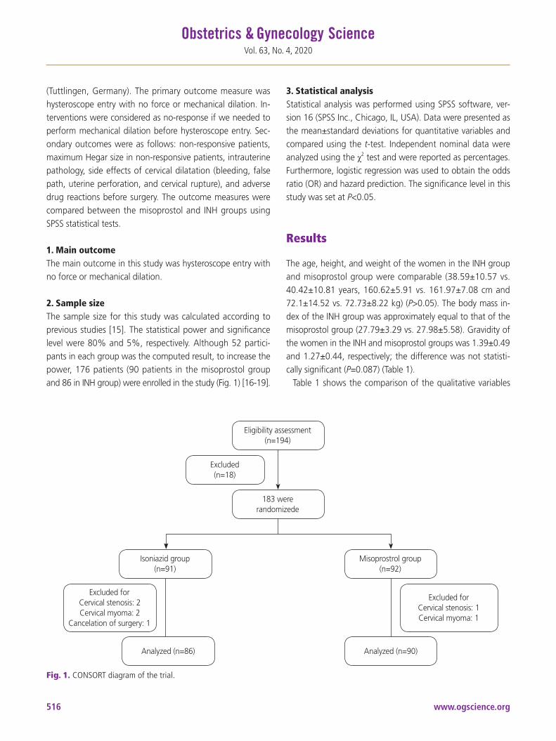
www.ogscience.org516
Vol. 63, No. 4, 2020
(Tuttlingen, Germany). The primary outcome measure was hysteroscope entry with no force or mechanical dilation. In-terventions were considered as no-response if we needed to perform mechanical dilation before hysteroscope entry. Sec-ondary outcomes were as follows: non-responsive patients, maximum Hegar size in non-responsive patients, intrauterine pathology, side effects of cervical dilatation (bleeding, false path, uterine perforation, and cervical rupture), and adverse drug reactions before surgery. The outcome measures were compared between the misoprostol and INH groups using SPSS statistical tests.
1. Main outcomeThe main outcome in this study was hysteroscope entry with no force or mechanical dilation.
2. Sample sizeThe sample size for this study was calculated according to previous studies [15]. The statistical power and significance level were 80% and 5%, respectively. Although 52 partici-pants in each group was the computed result, to increase the power, 176 patients (90 patients in the misoprostol group and 86 in INH group) were enrolled in the study (Fig. 1) [16-19].
3. Statistical analysisStatistical analysis was performed using SPSS software, ver-sion 16 (SPSS Inc., Chicago, IL, USA). Data were presented as the mean±standard deviations for quantitative variables and compared using the t-test. Independent nominal data were analyzed using the χ2 test and were reported as percentages. Furthermore, logistic regression was used to obtain the odds ratio (OR) and hazard prediction. The significance level in this study was set at P<0.05.
Results
The age, height, and weight of the women in the INH group and misoprostol group were comparable (38.59±10.57 vs. 40.42±10.81 years, 160.62±5.91 vs. 161.97±7.08 cm and 72.1±14.52 vs. 72.73±8.22 kg) (P>0.05). The body mass in-dex of the INH group was approximately equal to that of the misoprostol group (27.79±3.29 vs. 27.98±5.58). Gravidity of the women in the INH and misoprostol groups was 1.39±0.49 and 1.27±0.44, respectively; the difference was not statisti-cally significant (P=0.087) (Table 1).
Table 1 shows the comparison of the qualitative variables
Fig. 1. CONSORT diagram of the trial.
Eligibility assessment(n=194)
183 wererandomizede
Isoniazid group(n=91)
Analyzed (n=86)
Misoprostrol group(n=92)
Analyzed (n=90)
Excluded forCervical stenosis: 2Cervical myoma: 2
Cancelation of surgery: 1
Excluded forCervical stenosis: 1Cervical myoma: 1
Excluded(n=18)

www.ogscience.org 517
Ladan Haghighi, et al. INH for cervical ripening prior to hysteroscopy
between the misoprostol and INH groups. As can be seen, in 45 patients in the misoprostol group (50%) and 67 in the INH group (95%), hysteroscope entry was successful without additional mechanical dilation, and this difference was statis-tically significant (P=0.001) (Table 2).
The OR obtained in this study was 0.57 for both misopros-tol and INH groups, indicating that the likelihood of hystero-scope entry without additional mechanical dilation was 43% lower in the misoprostol group than in the INH group (OR, 0.57; 95% confidence interval [CI], 0.43–0.75) (Table 2 and Fig. 2).
Further, the OR of non-response in the misoprostol group was 1.75 times that in the INH recipients, meaning 75% higher (OR, 1.75; 95% CI, 1.32–2.30) (Table 2). Comparison of intrauterine pathology between the 2 groups showed that the numbers of women with Asherman’s syndrome in the misoprostol group and INH group were 4 (4.4%) and 0 (0%), respectively, while 12 women (13.3%) in the misopro-stol group and 19 (22.1%) in the INH group had no notable
intrauterine pathology, and this difference was statistically significant (P=0.001).
We did not encounter any adverse drug reactions in either group. However, we had one case of false path in INH group and one case of uterine perforation during hysteroscope en-try in a very anteverted uterus in the misoprostol group; both were diagnosed and managed properly.
Discussion
The results of the present study show that INH is more effec-tive than misoprostol in cervical ripening before hysteroscopic surgery. The aim of this randomized clinical trial was to com-pare the efficacy of vaginal misoprostol and INH on cervical ripening before hysteroscopy. There have been numerous studies on the effect of misoprostol on cervical preparation before hysteroscopy, but none have compared it with INH.
For example, in a study by Batukan et al. [11], the effect of
Table 1. Basic demographic factors in 2 groups
Variables INH (n=86) Misoprostol (n=90) P-valueOR (95% CI)
(misoprostol/INH)
Age 38.59±10.57 40.42±10.81 0.258 -
BMI 27.98±5.58 27.79±3.29 0.647 -
Gravidity 1.27±0.44 1.39±0.49 0.087 -
Parity
No delivery 6 (31.6) 10 (22.2) 0.786 1.00
1 delivery 3 (15.8) 13 (28.9) 0.143 0.53 (0.23–1.23)
2 delivery 7 (36.8) 9 (20.0) 0.582 0.78 (0.33–1.83)
3 and more delivery 3 (15.8) 13 (28.9) 0.195 0.54 (0.22–1.27)
Menshis
Regular 25 (29.1) 20 (22.2) 0.251 1.00
Menorrhagia 3 (3.5) 5 (5.6) 0.288 0.48 (0.10–2.25)
Metrorrhagia 19 (22.1) 22 (24.4) 0.262 0.69 (0.29–1.61)
Menometrorrhagia 14 (16.3) 20 (22.2) 0.150 0.56 (0.22–1.37)
Oligomenorrhea 3 (3.5) 1 (1.1) 0.421 2.40 (0.23–24.87)
Amenorrhea 1 (1.2) 1 (1.1) 0.699 0.80 (0.04–13.06)
Irregular 21 (24.4) 21 (23.3) 0.381 0.80 (0.34–1.86)
Menopause
Yes 12 (14) 8 (8.9) - 1.00
No 74 (86) 82 (91.1) 0.613 1.05 (0.94–1.17)
Values are presented as mean±standard deviation or number (%).BMI, body mass index; CI, confidence interval; OR, odds ratio; INH, isonicotinic acid hydrazide.

www.ogscience.org518
Vol. 63, No. 4, 2020
vaginal and oral misoprostol on cervical preparation before hysteroscopy was evaluated, and the results showed that oral administration of misoprostol was preferable to vaginal administration.
In the present study, the hysteroscope entered the uterus
with no additional mechanical dilatation in 50% and 77.9% of the women in the misoprostol group and INH group, respectively. Moreover, the likelihood of hysteroscope entry was 43% lower in the misoprostol group than in the INH group. On the other hand, the OR of non-response in the misoprostol group was 75% higher than that in the INH group. The ORs show that the incidence of Asherman’s syn-drome in the misoprostol group was 0.94 times that in the vaginal INH group.
INH is a recently introduced agent for cervical ripening. The results of a study, conducted by Haghighi and Mohabatian [13] showed that vaginal INH is an effective agent for cervi-cal ripening prior to labor induction in term pregnancies. The mechanism of action of INH on cervical dilatation is similar to that of NO donors. The results of some studies showed that INH affects cervical dilatation via production of NO [14]. El-Khayat et al. [20] showed that primary cervical dilatation and the duration of dilatation were higher with vaginal misopro-stol than with Foley catheter plus vaginal isosorbide mono-nitrate, but there was no significant difference between the
Table 2. Outcome variables in 2 groups and predict of variables by logistic regression
Variables INH (n=86) Misoprostol (n=90) P-valueOR (95% CI)
(misoprostol/INH)
Primary outcomes
Hysteroscopy entrance
Yes 67 (77.9) 45 (50.0) 1.000 1.00
No 19 (22.1) 45 (50.0) 0.0001 0.57 (0.43–0.75)
Secondary outcomes
Non responses
Yes 19 (22.1) 45 (50.0) 1.000 1.00
No 67 (77.9) 45 (50.0) 0.0001 1.75 (1.32–2.30)
Intra uterine pathology
Normal 19 (22.1) 12 (13.3) - 1.00
Polyp 31 (36.0) 42 (46.7) 0.090 0.71 (0.26–1.95)
Myoma 17 (19.8) 15 (16.7) 0.513 0.75 (0.56–0.99)
Asherman’s syndrome 0 (0) 4 (4.4) 0.035 0.94 (0.22–0.99)
Septum 6 (7.0) 4 (4.4) 0.612 0.63 (0.07–5.10)
Atrophy 2 (2.0) 2 (2.2) 0.530 0.90 (0.27–3.01)
Focal hyperplasia 10 (11.6) 7 (7.8) 0.554 1.05 (0.95–1.16)
Product of conception 1 (1.2) 4 (4.4) 0.035 0.75 (0.56–0.99)
Max Hegar size in nonresponses 3.73±1.28 3.84±1.04 0.726 -
Values are presented as number (%) or mean±standard deviation.CI, confidence interval; OR, odds ratio; INH, isonicotinic acid hydrazide.
Fig. 2. The compare of frequency of hysteroscopy entry without mechanical dilation in 2 group. INH, isonicotinic acid hydrazide.
9080706050403020100
Hysteroscopy entry
Yes
50 50
77.9
22.1
No
Misoprostol INH

www.ogscience.org 519
Ladan Haghighi, et al. INH for cervical ripening prior to hysteroscopy
2 groups in terms of the duration of surgery or difficult in dilatation. The lack of a statistically significant difference in their study might be due to different sample sizes or meth-ods from those used in the present study. On the other hand, Waddell et al. [21] found that the use of vaginal misoprostol before hysteroscopy reduced the pain during and pressure required for cervical dilatation.
The results of numerous studies on the effect of miso-prostol on cervical dilatation have shown that using vaginal misoprostol had a more pronounced effect on cervical dilata-tion than its sublingual form. Bisharah et al. [22] randomly assigned 40 women to an intervention group (100 mg of sublingual misoprostol) and a placebo group and found no significant difference between the 2 groups in terms of uterine dilatation. The results of a study by Mulayim et al. [23] showed that cervical dilatation time was longer in the control (placebo) group than in the intervention (sublingual misoprostol use) group; thus, cervical tearing was greater in the control group. In a study conducted by Preutthipan and Herabutya [24], vaginal misoprostol was found to reduce dilatation time in premenopausal women. Another study by Sordia-Hernández et al. [25] showed that the time of hysteroscopic surgery in the vaginal misoprostol group was significantly shorter than that in the placebo and sublingual misoprostol groups.
The strength of this study is that it compares the effect of vaginal misoprostol with that of other vaginal medica-tions. In many studies, vaginal misoprostol groups have been compared with placebo groups or rectal, oral, or sublingual misoprostol administration. This method made it possible to compare the effect of 2 different drugs on vaginal dilatation.
A study conducted by Inal et al. [26] showed that dinopros-tone had a greater effect on cervical dilatation than misopro-stol. In the present study, the OR obtained for hysteroscopic entrance in the misoprostol and INH groups was 0.57 (0.43–0.75), which indicated that the likelihood of hysteroscope entry into the uterus in the misoprostol group was 43% lower than that of the INH group. This means that cervical dilatation in the women receiving misoprostol was less than in the INH recipients.
INH can be simply used as a ripening agent before hyster-oscopy. It is easily available and does not seem to have any complications. However, its underlying mechanism of cervi-cal ripening is unknown. Our study was limited by its small sample size. Further research is needed to determine the
mechanisms of action and clinical efficacy of INH against that of other ripening agents to find the best dosage and timing of INH for ripening of cervix before hysteroscopy.
In conclusion, vaginal INH seems to be more effective than misoprostol in cervical ripening before hysteroscopic surgery and can be a good alternative to misoprostol.
Conflict of interest
No potential conflict of interest relevant to this article was reported.
Acknowledgements
This paper is derived from a medical doctorate thesis of Iran Medical Sciences University.
Ethical approval
The study was approved by the Local Ethics Committee of Iran University of Medical Sciences, Tehran, Iran and regis-tered in the Iranian Registry of Clinical Trials (IRCT.ir); ID num-ber: IRCT2015112821506N4.
Patient consent
In this study, informed consent was received from all the par-ticipants and their partners.
References
1. Dogru A, Sargin F, Celik M, Sagiroglu AE, Goksel MM, Sayhan H. The rate of device-associated nosocomial infections in a medical surgical intensive care unit of a training and research hospital in Turkey: one-year out-comes. Jpn J Infect Dis 2010;63:95-8.
2. Eggimann P, Sax H, Pittet D. Catheter-related infections. Microbes Infect 2004;6:1033-42.
3. Randolph JF Jr, Ying YK, Maier DB, Schmidt CL, Riddick DH. Comparison of real-time ultrasonography, hystero-

www.ogscience.org520
Vol. 63, No. 4, 2020
salpingography, and laparoscopy/hysteroscopy in the evaluation of uterine abnormalities and tubal patency. Fertil Steril 1986;46:828-32.
4. Wolman I, Jaffa AJ, Hartoov J, Bar-Am A, David MP. Sensitivity and specificity of sonohysterography for the evaluation of the uterine cavity in perimenopausal pa-tients. J Ultrasound Med 1996;15:285-8.
5. Munro MG, Jensen J, Purdon TF. Narrow caliber office hysteroscope. J Am Assoc Gynecol Laparosc 1994;1:S24.
6. Campo R, Van Belle Y, Rombauts L, Brosens I, Gordts S. Office mini-hysteroscopy. Hum Reprod Update 1999;5:73-81.
7. Dingfelder JR, Brenner WE, Hendricks CH, Staurovsky LG. Reduction of cervical resistance by prostaglandin suppositories prior to dilatation for induced abortion. Am J Obstet Gynecol 1975;122:25-30.
8. Christensen NJ, Bygdeman M, Gréen K. Comparison of different prostaglandin analogues and laminaria for preoperative dilatation of the cervix in late first trimester abortion. Contraception 1983;27:51-61.
9. Jain JK, Mishell DR Jr. A comparison of misoprostol with and without laminaria tents for induction of second-trimester abortion. Am J Obstet Gynecol 1996;175:173-7.
10. Ngai SW, Yeung KC, Lao T, Ho PC. Oral misoprostol versus vaginal gemeprost for cervical dilatation prior to vacuum aspiration in women in the sixth to twelfth week of gestation. Contraception 1995;51:347-50.
11. Batukan C, Ozgun MT, Ozcelik B, Aygen E, Sahin Y, Turkyilmaz C. Cervical ripening before operative hys-teroscopy in premenopausal women: a randomized, double-blind, placebo-controlled comparison of vaginal and oral misoprostol. Fertil Steril 2008;89:966-73.
12. Siddique S, Howlader MJ, Saha J, Begum KS. A compari-son of orally administered misoprostol with vaginally ad-ministered misoprostol for cervical ripening and labour induction. KYAMC Journal 2017;7:668-72.
13. Haghighi L, Mohabatian B. Isonicotinic acid hydrazide (INH): a new agent for cervical ripening at term. J Obstet Gynaecol 2015;35:251-4.
14. Yilmaz HR, Uz E, Gökalp O, Ozçelik N, Ciçek E, Özer MK. Protective role of caffeic acid phenethyl ester and erdosteine on activities of purine-catabolizing enzymes and level of nitric oxide in red blood cells of isoniazid-administered rats. Toxicol Ind Health 2008;24:519-24.
15. Haghighi L, Homam H, Raoofi Z, Najmi Z. Intravaginal isosorbide dinitrate or misoprostol for cervical ripening prior to induction of labour: a randomised controlled trial. J Obstet Gynaecol 2013;33:272-6.
16. Machin D, Campbell MJ, Tan SB, Tan SH. Sample size tables for clinical studies. Hoboken (NJ): John Wiley & Sons; 2011.
17. Sokal RR, Jamesrohlf F. Introduction to biostatistics. 2nd ed. New York (NY): W. H. Freeman and Company; 2009.
18. Zar JH. Biostatistical analysis. Delhi: Pearson Education, India; 1999.
19. Abotorabi S, Mohammadi M, Bagherivand S, Oveisi S. A pilot randomized controlled trial to evaluate isosorbide mononitrate (IMN) efficiency for cervical ripening prior to labor induction in Iranian pregnant women. Iran J Pharm Res 2019;18:988-94.
20. El-Khayat W, Alelaiw H, El-kateb A, Elsemary A. Com-paring vaginal misoprostol versus Foley catheter plus vaginal isosorbide mononitrate for labor induction. J Matern Fetal Neonatal Med 2016;29:487-92.
21. Waddell G, Desindes S, Takser L, Beauchemin MC, Bessette P. Cervical ripening using vaginal misoprostol before hysteroscopy: a double-blind randomized trial. J Minim Invasive Gynecol 2008;15:739-44.
22. Bisharah M, Al-Fozan H, Tulandi T. A randomized trial of sublingual misoprostol for cervical priming before hys-teroscopy. J Am Assoc Gynecol Laparosc 2003;10:390-1.
23. Mulayim B, Celik NY, Onalan G, Bagis T, Zeyneloglu HB. Sublingual misoprostol for cervical ripening before diagnostic hysteroscopy in premenopausal women: a randomized, double blind, placebo-controlled trial. Fertil Steril 2010;93:2400-4.
24. Preutthipan S, Herabutya Y. Vaginal misoprostol for cer-vical priming before operative hysteroscopy: a random-ized controlled trial. Obstet Gynecol 2000;96:890-4.
25. Sordia-Hernández LH, Rosales-Tristan E, Vazquez-Men-dez J, Merino M, Iglesias JL, Garza-Leal JG, et al. Effec-tiveness of misoprostol for office hysteroscopy without anesthesia in infertile patients. Fertil Steril 2011;95:759-61.
26. Inal HA, Ozturk Inal ZH, Tonguc E, Var T. Comparison of vaginal misoprostol and dinoprostone for cervical ripening before diagnostic hysteroscopy in nulliparous women. Fertil Steril 2015;103:1326-31.

www.ogscience.org 521
Original ArticleObstet Gynecol Sci 2020;63(4):521-528https://doi.org/10.5468/ogs.20071pISSN 2287-8572 · eISSN 2287-8580
Introduction
Vitamin D is known to have a major influence on bone health through regulation of calcium phosphorus homeo-stasis. Recently, the female reproductive system has been defined as one of the non-classical target organs of vitamin D. Vitamin D receptor expression was identified in ovarian gran-ulosa cells as well as other female reproductive organs, in-cluding endometrium and the uterus. These findings suggest that vitamin D may have a potential role in female reproduc-tion [1-3]. Several studies have demonstrated a direct effect of vitamin D on ovarian folliculogenesis and steroidogenesis in animal and human cell-line studies; vitamin D receptor null mutant mice have impaired folliculogenesis and vitamin D
Serum vitamin D levels and ovarian reserve markers in secondary amenorrhea patients: Is there a link?Gyung-Mee Kim, MD, PhD1, Gyun-Ho Jeon MD, PhD2
Departments of 1Psychiatry, 2Obstetrics and Gynecology, Inje University Haeundae Paik Hospital, Inje University College of Medicine, Busan, Korea
ObjectiveTo investigate whether serum 25-hydroxyvitamin D [25(OH)D] level is associated with ovarian reserve markers in secondary amenorrhea (SA) patients.
MethodsSixty-three women diagnosed with SA were recruited during 12 months from the initiation of this prospective observational study. Serum 25(OH)D levels, serum anti-Müllerian hormone (AMH) levels and antral follicle count (AFC) were estimated in study participants and ovarian reserve markers were compared between participants with vitamin D deficiency and those with normal vitamin D levels.
ResultsOf the 63 participants, 27 (42.9%) were vitamin D deficient (<20 ng/mL) and 36 (57.1%) had normal vitamin D levels. The mean AMH levels and AFC were 10.86±8.94 µ/L and 15.23±7.65 in the vitamin D deficient group, and 7.24±5.62 µ/L and 12.30±6.95 in the normal vitamin D group. Univariate and multivariate linear regression analysis of log10 transformed AMH and AFC with serum 25(OH)D adjusted for age and body mass index confirmed no association between vitamin D levels and AMH levels or AFC. There was also no correlation between serum 25(OH)D and AMH levels or AFC in all participants. However, participants with vitamin D deficiency had an increased chance of having polycystic ovarian syndrome (PCOS) as cause of SA than those with normal vitamin D levels (adjusted odds ratio, 7.559; 95% confidence interval, 1.28–44.65; P=0.026) after adjustment for clinical factors by logistic regression model.
ConclusionThere was no correlation between serum 25(OH)D levels and ovarian reserve markers in SA patients, but vitamin D deficiency may be linked to PCOS patients.
Keywords: Anti-Mullerian hormone; Amenorrhea; Ovarian reserve; 25-hydroxyvitamin D; Polycystic ovarian syndrome
Received: 2020.03.19. Revised: 2020.05.13. Accepted: 2020.06.01.Corresponding author: Gyun-Ho Jeon, MD, PhDDepartment of Obstetrics and Gynecology, Inje University Haeundae Paik Hospital, Inje University College of Medicine, 875 Haeun-daero, Haeundae-gu,Busan 48108, KoreaE-mail: [email protected]://orcid.org/0000-0003-1128-1656
Articles published in Obstet Gynecol Sci are open-access, distributed under the terms of the Creative Commons Attribution Non-Commercial License (http://creativecommons.org/licenses/by-nc/3.0/) which permits unrestricted non-commercial use, distribution, and reproduction in any medium, provided the original work is properly cited.
Copyright © 2020 Korean Society of Obstetrics and Gynecology
www.ogscience.org522
Vol. 63, No. 4, 2020
stimulated steroidogenesis in human ovarian cells [4,5].Anti-Müllerian hormone (AMH) is produced by granulosa
cells of small antral and pre-antral follicles, and along with antral follicle counts (AFCs), is a well-known representa-tive marker for ovarian reserve. AMH plays a crucial role in folliculogenesis and is not affected by the menstrual cycle, making it the most widely used ovarian marker. Although it is still unclear how vitamin D affects female reproductive function, interest in a potential relationship between vitamin D and ovarian reserve markers, particularly AMH, has in-creased since reports on the effect of vitamin D on follicular development have accumulated. Moreover, the presence of a functional vitamin D response element in the AMH gene pro-motor region was demonstrated [6] and experimental results in animal and human granulosa cells have also reported that vitamin D affects AMH signaling [7,8]. These basic research studies have indicated that vitamin D deficiency may alter go-nadal function through abnormal AMH signaling. However, discrepancy exists in clinical study findings under various set-tings, where some suggest a significant association between vitamin D levels and ovarian reserve, while others have not found any correlation between vitamin D and ovarian reserve markers [9,10]. Considering these contradictory results, we performed this prospective observational study to examine serum 25-hydroxyvitamin D [25(OH)D] levels in patients with secondary amenorrhea (SA) and investigate the relationship between serum 25(OH)D levels and ovarian reserve markers (i.e., serum AMH and AFC) in these women.
Materials and methods
1. Study design and participantsThis study was conducted as a prospective cohort study for 12 months from March 2018 to February 2019. The study population was comprised of patients evaluated by a single reproductive endocrinologist for symptoms of SA. A total of 78 participants were initially recruited to the study. Three participants refused to participate in the study and 12 partici-pants were excluded due to the study’s exclusion criteria. The exclusion criteria of this study were as follows: 1) women who had taken hormonal medication including oral contraceptives or vitamin D supplements within the previous 6 months; 2) women who had ovarian surgery, chemotherapy or radio-therapy; 3) women who took medication and/or were diag-
nosed with a systemic disease that can affect menstruation (e.g., psychoaffective medicine, thyroid hormone, diabetes, hyperprolactinemia); 4) women who refused to participate this study. Patients’ information including age, parity, height, body weight, cause of SA, season of blood sampling (i.e., to account for seasonal changes in vitamin D), current medica-tions and other past medical history were recorded. Gyneco-logic ultrasonography was also performed on their first study visit. To determine cause of SA, polycystic ovarian syndrome (PCOS) was diagnosed on the basis of Rotterdam criteria [11], and primary ovarian insufficiency (POI) was regarded as 2 recordings of serum follicular stimulating hormone (FSH) levels of more than 40 IU/L at least one month apart in a woman aged under 40 years (i.e., >2 standard deviation [SD] less than the mean menopausal age) [12]. All patients whose etiology of SA was not determined, excluding women with POI and PCOS, were deemed unexplained chronic anovula-tion [13]. PCOS in adolescents was only diagnosed in women with at least 2 years after menarche and who had SA, hyper-androgenemia, and increased ovarian volume on ultrasound [14].
Venous blood samples of all participants were taken for the measurement of serum 25(OH)D and AMH on the same day, prior to treatment for SA. Vitamin D deficiency was defined as serum 25(OH)D levels <20 ng/mL based on the Endocrine Society clinical practice guidelines [15].
2. Serum vitamin D, hormones and ovarian reserve markers measurements
1) Serum 25-hydroxyvitamin DEach participant’s venous sample was drawn into a serum sep-aration tube (SST) and serum 25(OH)D was measured using a DIA source 25 OH Vitamin D3 radioimmunoassay kit (DIA-source ImmunoAssays S.A., Louvain-La-Neuve, Belgium) with immunoassay device (gamma 5) and presented in ng/mL. The total imprecision coefficient of variance was 5.38% at a concentration level of 6.35 ng/mL and 4.96% at 37.71 ng/mL.
2) Serum follicular stimulating hormoneEach participant’s venous sample was drawn into an SST and serum FSH was measured using an Elecsys FSH electroche-miluminescence immunoassay kit (Roche Diagnostics GmbH, Mannheim, Germany) with immunoassay device (Cobas e 801) and presented in mIU/mL. The total imprecision coef-

www.ogscience.org 523
Gyung-Mee Kim, et al. Vitamin D and ovarian reserve markers
ficient of variance was 3.1% at a concentration level of 48.8 mIU/mL and 2.3% at 17.4 mIU/mL.
3) Anti-Müllerian hormone and antral follicle countsBlood drawn into the SST was centrifuged within 1 hour (3,000 rpm for 10 minutes). The separated serum was mea-sured for AMH level by ECLIA method with Elecsys AMH kit (Roche Diagnostics GmbH) on a Cobas e 601 immunoas-say analyzer and presented in ng/mL. The total imprecision coefficient of variance was 3.5% at a concentration level of 0.042 ng/mL and 3.4% at 0.20 ng/mL. An AFC was mea-sured by one gynecologist on the participant’s first visit in all individuals with gynecologic ultrasonography. AFC repre-sented the total number of all antral follicles, from 2 mm to 10 mm in size, in both ovaries.
3. Statistical analysisCategorical variable data are presented as a frequency with percentage and continuous variables are presented as a group mean±SD. Differences in study participants’ charac-teristics were compared across subgroups with a χ2 test or
Fisher’s exact test for categorical variables and independent t-test or Mann-Whitney’s U test for continuous variables, as appropriate. An analysis of variance with Duncan’s post hoc test or Kruskal-Wallis test with Dunn’s post hoc test were also employed, as appropriate. To check if data were normally distributed, we used a Shapiro-Wilk test. Partial cor-relation coefficients controlling for age and body mass index (BMI) were estimated to investigate the linear relationship between 2 continuous variables. Univariate linear regression analysis between each variable (age, BMI, vitamin D, season), and multivariate linear regression analysis with all covari-ates, were performed to identify the regression coefficients for factors related to the log10 AMH, AFC, PCOS and POI. For data visualization, box plot and scatter plot were also displayed. Finally, logistic regression analysis was performed to identify whether vitamin D deficiency is associated with PCOS as a cause of SA. All statistical analyses were carried out using SPSS 24.0 (SPSS Statistics for Windows 24.0; IBM Corp., Armonk, NY, USA) statistical software and P-values less than 0.05 was considered statistically significant.
Table 1. Patients’ demographic variables
VariableVitamin D (ng/mL)
P-value <20 ≥20
Age (yr) 22.54±5.33 27.81±8.65 0.014a)
BMI (kg/m2) 22.85±5.17 22.56±4.39 0.823a)
Amenorrhea cause
PCOS 21 (80.8) 19 (51.4) 0.454b)
Unexplained chronic anovulation 2 (7.7) 12 (32.4)
POI 3 (11.5) 6 (16.2)
AFC 15.23±7.65 12.30±6.95 0.119c)
AMH 10.86±8.94 7.24±5.62 0.130a)
FSH 6.30±4.28 8.25±9.39 0.392a)
Season of blood sample 0.128b)
Autumn 6 (23.1) 2 (5.4)
Spring 9 (34.6) 11 (29.7)
Summer 6 (23.1) 16 (43.2)
Winter 5 (19.2) 8 (21.6)
Values are presented as mean±standard deviation or number (%).BMI, body mass index; PCOS, polycystic ovarian syndrome; POI, primary ovarian insufficiency; AFC, antral follicle count; AMH, anti-Müllerian hormone; FSH, follicular stimulating hormone.a)P-values were derived from Mann-Whitney U test; b)P-values were derived from Fisher’s exact test; c)P-values were derived from independent t-test.
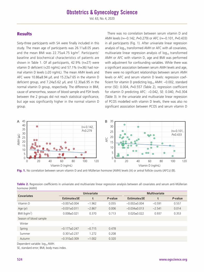
www.ogscience.org524
Vol. 63, No. 4, 2020
Results
Sixty-three participants with SA were finally included in this study. The mean age of participants was 26.11±8.05 years and the mean BMI was 22.75±4.75 kg/m2. Participants’ baseline and biochemical characteristics of patients are shown in Table 1. Of all participants, 42.9% (n=27) were vitamin D deficient (<20 ng/mL) and 57.1% (n=36) had nor-mal vitamin D levels (≥20 ng/mL). The mean AMH levels and AFC were 10.86±8.94 µ/L and 15.23±7.65 in the vitamin D deficient group, and 7.24±5.62 µ/L and 12.30±6.95 in the normal vitamin D group, respectively. The difference in BMI, cause of amenorrhea, season of blood sample and FSH levels between the 2 groups did not reach statistical significance, but age was significantly higher in the normal vitamin D group.
There was no correlation between serum vitamin D and AMH levels (r=−0.142, P=0.279) or AFC (r=−0.101, P=0.433) in all participants (Fig. 1). After univariate linear regression analysis of log10 transformed AMH or AFC with all covariates, multivariate linear regression analysis of log10 transformed AMH or AFC with vitamin D, age and BMI was performed with adjustment for confounding variables. While there was a significant association between serum AMH levels and age, there were no significant relationships between serum AMH levels or AFC and serum vitamin D levels: regression coef-ficient for vitamin D predicting log10 AMH: −0.002, standard error (SE): 0.004, P=0.557 (Table 2), regression coefficient for vitamin D predicting AFC: −0.042, SE: 0.040, P=0.304 (Table 3). In the univariate and multivariate linear regression of PCOS modelled with vitamin D levels, there was also no significant association between PCOS and serum vitamin D
AM
H ( μ
L)
AFC
454035302520151050
35
30
25
20
15
10
5
0 0 20 40 60 80 100 120
Vitamin D (ng/mL) 0 20 40 60 80 100 120
Vitamin D (ng/mL)
(r=-0.142,P=0.279 (r=-0.101,
P=0.433
A B
Fig. 1. No correlation between serum vitamin D and anti-Müllerian hormone (AMH) levels (A) or antral follicle counts (AFCs) (B).
Table 2. Regression coefficients in univariate and multivariate linear regression analysis between all covariates and serum anti-Müllerian hormone (AMH)
CovariatesUnivariate Multivariate
Estimate±SE t P-value Estimate±SE t P-value
Vitamin D −0.007±0.004 −1.962 0.055 −0.002±0.004 −0.591 0.557
Age (yr) −0.031±0.011 −2.867 0.006 −0.034±0.013 −2.541 0.014
BMI (kg/m2) 0.008±0.021 0.370 0.713 0.020±0.022 0.937 0.353
Season of blood sample
Winter - -
Spring −0.177±0.247 −0.715 0.478
Summer 0.301±0.237 1.272 0.208
Autumn −0.310±0.309 −1.002 0.320
Dependent variable: log10 AMH.SE, standard error; BMI, body mass index.
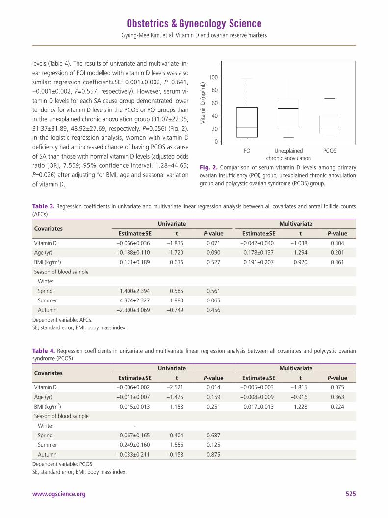
www.ogscience.org 525
Gyung-Mee Kim, et al. Vitamin D and ovarian reserve markers
levels (Table 4). The results of univariate and multivariate lin-ear regression of POI modelled with vitamin D levels was also similar: regression coefficient±SE: 0.001±0.002, P=0.641, −0.001±0.002, P=0.557, respectively). However, serum vi-tamin D levels for each SA cause group demonstrated lower tendency for vitamin D levels in the PCOS or POI groups than in the unexplained chronic anovulation group (31.07±22.05, 31.37±31.89, 48.92±27.69, respectively, P=0.056) (Fig. 2). In the logistic regression analysis, women with vitamin D deficiency had an increased chance of having PCOS as cause of SA than those with normal vitamin D levels (adjusted odds ratio [OR], 7.559; 95% confidence interval, 1.28–44.65; P=0.026) after adjusting for BMI, age and seasonal variation of vitamin D.
Table 3. Regression coefficients in univariate and multivariate linear regression analysis between all covariates and antral follicle counts (AFCs)
CovariatesUnivariate Multivariate
Estimate±SE t P-value Estimate±SE t P-value
Vitamin D −0.066±0.036 −1.836 0.071 −0.042±0.040 −1.038 0.304
Age (yr) −0.188±0.110 −1.720 0.090 −0.178±0.137 −1.294 0.201
BMI (kg/m2) 0.121±0.189 0.636 0.527 0.191±0.207 0.920 0.361
Season of blood sample
Winter
Spring 1.400±2.394 0.585 0.561
Summer 4.374±2.327 1.880 0.065
Autumn −2.300±3.069 −0.749 0.456
Dependent variable: AFCs.SE, standard error; BMI, body mass index.
Table 4. Regression coefficients in univariate and multivariate linear regression analysis between all covariates and polycystic ovarian syndrome (PCOS)
CovariatesUnivariate Multivariate
Estimate±SE t P-value Estimate±SE t P-value
Vitamin D −0.006±0.002 −2.521 0.014 −0.005±0.003 −1.815 0.075
Age (yr) −0.011±0.007 −1.425 0.159 −0.008±0.009 −0.916 0.363
BMI (kg/m2) 0.015±0.013 1.158 0.251 0.017±0.013 1.228 0.224
Season of blood sample
Winter -
Spring 0.067±0.165 0.404 0.687
Summer 0.249±0.160 1.556 0.125
Autumn −0.033±0.211 −0.158 0.875
Dependent variable: PCOS.SE, standard error; BMI, body mass index.
Fig. 2. Comparison of serum vitamin D levels among primary ovarian insufficiency (POI) group, unexplained chronic anovulation group and polycystic ovarian syndrome (PCOS) group.
Vita
min
D (n
g/m
L)
100
80
60
40
20
0
Unexplained chronic anovulation
PCOSPOI

www.ogscience.org526
Vol. 63, No. 4, 2020
Discussion
Our study demonstrated that serum 25(OH)D levels were not related with ovarian reserve markers among SA patients, even after adjustment for age, BMI, cause of amenorrhea, and seasonal variation of vitamin D. However, mean serum vitamin D levels were different depending on the cause of amenorrhea, however the difference did not reach statisti-cal significance (Fig. 2). In particular, vitamin D levels in the participants with PCOS or POI demonstrated a tendency to-wards lower serum vitamin D levels than those in participants with unexplained chronic anovulation. In addition, women with vitamin D deficiency had an increased chance of having PCOS than those with normal vitamin D levels, after adjust-ment for clinical factors by logistic regression model. We did not calculate the adjusted OR for POI since participants with POI were a very small portion of the study participants. The lack of correlation between serum vitamin D levels and ovarian reserve markers across all participants in our study is in agreement with several recent studies that also demon-strated no correlation. However, the result of lower vitamin D levels in participants with PCOS in this study needs to be taken with caution, considering the heterogeneous results of previous studies [16,17]. To date, the role of low vitamin D levels on AMH regulation or the pathophysiology of PCOS is unclear, with some previous reports suggesting that this vitamin D deficiency is likely due to obesity or a metabolic syndrome that is frequently associated with PCOS [18,19].
While basic research has reported that vitamin D affects ovarian folliculogenesis and steroidogenesis, there is limited clinical information on how vitamin D affects ovarian mark-ers such as AMH. Wojtusik et al. [9] have reported a dose-dependent decrease in AMH mRNA levels in granulosa cells after treatment with vitamin D. In another recent study, hu-man granulosa cells treated with vitamin D exhibited altered AMH signaling and they demonstrated an inverse correlation between vitamin D status in follicular fluid and AMH recep-tor-II (AMHT-II) mRNA gene expression [8]. Binding AMH to AMH receptor-II (AMHR-II) is known to suppress follicular maturation, by inhibiting primordial follicle recruitment into the growing follicle pool, and by decreasing the sensitivity of follicles to FSH [10]. Since vitamin D had an effect on down-regulation of AMHR-II gene expression, the phosphorylation process, and nuclear localization after AMH-AMHR-II binding [8], it may be involved in promoting follicle development by
altering AMH production pattern and FSH sensitivity in ovar-ian granulosa cell [20,21].
Previous studies on the relationship between vitamin D and ovarian reserve markers, particularly AMH, have had inconsistent results. For instance, Merhi et al. [22] found a positive relationship between serum vitamin D and AMH lev-els and suggested vitamin D deficiency might be associated with lower ovarian reserve in late reproductive aged women. Dennis et al. [23] also suggested that vitamin D might have a positive effect on AMH production in adults, and the extent of seasonal variation in women’s AMH levels correlated with the extent of their variation in vitamin D levels. In another study of 1,430 premenopausal women, a negative correla-tion was found between serum vitamin D levels and urinary FSH levels, suggesting that low vitamin D levels may influ-ence ovarian reserve and earlier menopause [24]. Meanwhile, a recent cross-sectional study that included 283 infertile women, revealed no significant association between vitamin D and ovarian reserve markers (AMH, AFC) [25], which is in agreement with a prospective study that failed to demon-strate any beneficial effect of vitamin D supplementation on normalization of serum AMH levels [26].
In this study, generally healthy women should be included as controls. However, it was difficult to recruit healthy wom-en to volunteer to have their AMH and vitamin D levels mea-sured, so we conducted this study in patients with SA who required AMH measurements for diagnosis. These SA pa-tients included women with various levels of ovarian reserve, and it was assumed that unexplained chronic anovulatory patients were similar to healthy women with normal ovarian reserve. Indeed, participants with unexplained chronic anovu-latory in our study demonstrated normal ovarian reserve (data not shown). In addition, cholecalciferol, the main natural source of vitamin D, is affected by sun exposure and outdoor activity, so participants’ duration of outdoor activity should also be measured. However, we did not conduct a survey of patients’ outdoor activity as subjective factors could have been reflected in this evaluation. Although the validity of AMH measurements in those younger than 20 years of age has not been established, we included participants in this age group and their AMH measurement, as the small number of girls in their late teens in this study were considered to have reached sufficient maturity.
This study is the first report on the relationship between se-rum vitamin D levels and ovarian reserve in Korean women.

www.ogscience.org 527
Gyung-Mee Kim, et al. Vitamin D and ovarian reserve markers
This study was possible because it was conducted in patients with SA, which included women with various levels of ovar-ian reserves. Furthermore, this study is strengthened by its prospective design, which has a low risk of confounding bias, the fact that both AFC and AMH were measured as a marker of ovarian reserve, and finally AMH was measured on the same day as 25(OH)D.
However, this study has several limitations. Firstly, this study has small study population to then analyze by subgroup(s) of amenorrhea causes, which increases the risk of selection bias or type II error. Secondly, healthy women with normal men-strual cycles, who were not SA patients, were not included as controls. Thirdly, this study was observational in nature, so we cannot interpret these results in terms of causality. Despite of these limitations, this study is the first prospec-tive observational study of the relationship between vitamin D and ovarian reserve in Korean women with SA, suggest-ing no association between them. In this regard, it is not necessary to routinely measure serum vitamin D levels in SA patients with abnormal ovarian reserve. However, it would be meaningful to provide vitamin D supplementation, after measuring serum vitamin D concentration in PCOS patients, considering the low vitamin D levels in PCOS patients in this study. Nevertheless, given the uncertainty of how vitamin D affects female reproduction and the limitations of this study, no clear conclusion can be drawn. Further study is required on the relationship between vitamin D levels and obesity or metabolic abnormalities in patients with PCOS.
In conclusion, this observational study demonstrated that there was no correlation between serum vitamin D levels and ovarian reserve markers in SA patients, but that vitamin D deficiency may be linked to PCOS patients. However, given the limitations of this observational study, further prospective research with a larger population and age-matched control group should be investigated to draw more definite conclu-sions on the relationship between vitamin D levels and ovar-ian reserve markers.
Acknowledgements
This work was supported by the 2018 Inje University research grant (grant number 20180150).
Conflict of interest
No potential conflict of interest relevant to this article was reported.
Ethical approval
This study was approved by the Institutional Review Board of Inje University Haeundae Paik Hospital, Busan, Korea (ap-proval number: 2017-01-018).
Patient consent
All participants provided written informed consent.
References
1. Fabris A, Pacheco A, Cruz M, Puente JM, Fatemi H, Gar-cia-Velasco JA. Impact of circulating levels of total and bioavailable serum vitamin D on pregnancy rate in egg donation recipients. Fertil Steril 2014;102:1608-12.
2. van de Vijver A, Drakopoulos P, Van Landuyt L, Vaiarelli A, Blockeel C, Santos-Ribeiro S, et al. Vitamin D deficiency and pregnancy rates following frozen-thawed em-bryo transfer: a prospective cohort study. Hum Reprod 2016;31:1749-54.
3. Franasiak JM, Molinaro TA, Dubell EK, Scott KL, Ruiz AR, Forman EJ, et al. Vitamin D levels do not affect IVF out-comes following the transfer of euploid blastocysts. Am J Obstet Gynecol 2015;212:315.e1-6.
4. Yoshizawa T, Handa Y, Uematsu Y, Takeda S, Sekine K, Yoshihara Y, et al. Mice lacking the vitamin D recep-tor exhibit impaired bone formation, uterine hypopla-sia and growth retardation after weaning. Nat Genet 1997;16:391-6.
5. Parikh G, Varadinova M, Suwandhi P, Araki T, Rosenwaks Z, Poretsky L, et al. Vitamin D regulates steroidogenesis and insulin-like growth factor binding protein-1 (IGFBP-1) production in human ovarian cells. Horm Metab Res 2010;42:754-7.
6. Malloy PJ, Peng L, Wang J, Feldman D. Interaction of the vitamin D receptor with a vitamin D response element in

www.ogscience.org528
Vol. 63, No. 4, 2020
the Mullerian-inhibiting substance (MIS) promoter: regu-lation of MIS expression by calcitriol in prostate cancer cells. Endocrinology 2009;150:1580-7.
7. Dicken CL, Israel DD, Davis JB, Sun Y, Shu J, Hardin J, et al. Peripubertal vitamin D3 deficiency delays puberty and disrupts the estrous cycle in adult female mice. Biol Reprod 2012;87:51.
8. Merhi Z, Doswell A, Krebs K, Cipolla M. Vitamin D alters genes involved in follicular development and steroido-genesis in human cumulus granulosa cells. J Clin Endo-crinol Metab 2014;99:E1137-45.
9. Wojtusik J, Johnson PA. Vitamin D regulates anti-Mulle-rian hormone expression in granulosa cells of the hen. Biol Reprod 2012;86:91.
10. Durlinger AL, Visser JA, Themmen AP. Regulation of ovarian function: the role of anti-Müllerian hormone. Reproduction 2002;124:601-9.
11. Rotterdam ESHRE/ASRM-Sponsored PCOS Consensus Workshop Group. Revised 2003 consensus on diagnostic criteria and long-term health risks related to polycystic ovary syndrome. Fertil Steril 2004;81:19-25.
12. De Vos M, Devroey P, Fauser BC. Primary ovarian insuf-ficiency. Lancet 2010;376:911-21.
13. Chandeying P, Pantasri T. Prevalence of conditions caus-ing chronic anovulation and the proposed algorithm for anovulation evaluation. J Obstet Gynaecol Res 2015;41:1074-9.
14. Fauser BC, Tarlatzis BC, Rebar RW, Legro RS, Balen AH, Lobo R, et al. Consensus on women’s health aspects of polycystic ovary syndrome (PCOS): the Amsterdam ES-HRE/ASRM-Sponsored 3rd PCOS Consensus Workshop Group. Fertil Steril 2012;97:28-38.e25.
15. Holick MF, Binkley NC, Bischoff-Ferrari HA, Gordon CM, Hanley DA, Heaney RP, et al. Evaluation, treatment, and prevention of vitamin D deficiency: an Endocrine Soci-ety clinical practice guideline. J Clin Endocrinol Metab 2011;96:1911-30.
16. Wong HY, Li HW, Lam KS, Tam S, Shek CC, Lee CY, et al. Independent association of serum vitamin D with anti-Mullerian hormone levels in women with polycystic ovary syndrome. Clin Endocrinol (Oxf) 2018;89:634-41.
17. Cappy H, Giacobini P, Pigny P, Bruyneel A, Leroy-Billiard M, Dewailly D, et al. Low vitamin D3 and high anti-Müllerian hormone serum levels in the polycystic ovary syndrome (PCOS): is there a link? Ann Endocrinol (Paris) 2016;77:593-9.
18. Hahn S, Haselhorst U, Tan S, Quadbeck B, Schmidt M, Roesler S, et al. Low serum 25-hydroxyvitamin D con-centrations are associated with insulin resistance and obesity in women with polycystic ovary syndrome. Exp Clin Endocrinol Diabetes 2006;114:577-83.
19. Krul-Poel YH, Snackey C, Louwers Y, Lips P, Lambalk CB, Laven JS, et al. The role of vitamin D in metabolic disturbances in polycystic ovary syndrome: a systematic review. Eur J Endocrinol 2013;169:853-65.
20. Visser J. Role of anti-Müllerian hormone in follicle re-cruitment and maturation. J Gynecol Obstet Biol Reprod (Paris) 2006;35:2S30-4.
21. Irani M, Merhi Z. Role of vitamin D in ovarian physiology and its implication in reproduction: a systematic review. Fertil Steril 2014;102:460-468.e3.
22. Merhi ZO, Seifer DB, Weedon J, Adeyemi O, Holman S, Anastos K, et al. Circulating vitamin D correlates with serum antimüllerian hormone levels in late-reproductive-aged women: Women’s Interagency HIV Study. Fertil Steril 2012;98:228-34.
23. Dennis NA, Houghton LA, Jones GT, van Rij AM, Mor-gan K, McLennan IS. The level of serum anti-Müllerian hormone correlates with vitamin D status in men and women but not in boys. J Clin Endocrinol Metab 2012;97:2450-5.
24. Jukic AM, Steiner AZ, Baird DD. Association between serum 25-hydroxyvitamin D and ovarian reserve in pre-menopausal women. Menopause 2015;22:312-6.
25. Drakopoulos P, van de Vijver A, Schutyser V, Milatovic S, Anckaert E, Schiettecatte J, et al. The effect of serum vi-tamin D levels on ovarian reserve markers: a prospective cross-sectional study. Hum Reprod 2017;32:208-14.
26. Irani M, Minkoff H, Seifer DB, Merhi Z. Vitamin D increases serum levels of the soluble receptor for ad-vanced glycation end products in women with PCOS. J Clin Endocrinol Metab 2014;99:E886-90.
www.ogscience.org 529
Case ReportObstet Gynecol Sci 2020;63(4):529-533https://doi.org/10.5468/ogs.20052pISSN 2287-8572 · eISSN 2287-8580
Introduction
Pulmonary sequestration (PS) is a nonfunctioning mass of lung parenchyma that lacks communication with the normal tracheobronchial tree. It has an aberrant systemic arterial blood supply, usually from the thoracic or abdominal aorta [1]. Congenital diaphragmatic hernia (CDH) affects 1 in 2,500 newborns and is associated with high rates of morbidity and mortality [2]. While the association of PS with CDH is as high as 30%–40%, the prognosis of their coexistence is unknown [3-5]. We present 2 cases of coexistent PS with CDH. In both cases, only the isolated lung mass was diagnosed prenatally; the coexisting CDH was diagnosed only after birth.
Case report
1. Case 1A 31-year-old multigravida was referred to our hospital for a fetal lung mass at 27.5 weeks of gestation. On detailed ultrasonography examination, a hyperechoic, homogeneous,
well-defined mass measuring 2.5×1.8 cm was found posteri-or to the right lower lobe of the fetal lung (Fig. 1A). Because no aberrant systemic arterial blood supply was observed, the initial diagnosis was congenital pulmonary airway malforma-tion (CPAM). The fetal right kidney was slightly elevated, raising suspicion of an ectopic kidney (Fig. 1B). Although the diaphragm deviated upward on the right, it was well
Postnatally diagnosed coexisting congenital diaphragmatic hernia with pulmonary sequestration: a report of two casesHyun Mi Kim, MD1,2*, Ja Hyun Hwang, MD1,2*, Mi Ju Kim, MD1,3, Hyun-Hwa Cha, MD, PhD1,2, Won Joon Seong, MD, PhD1,2
Department of Obstetrics and Gynecology, 1School of Medicine, Kyungpook National University, 2Kyungpook National University Chilgok Hospital, 3Kyungpook National University Hospital, Daegu, Korea
While the associations between pulmonary sequestration (PS) and congenital diaphragmatic hernia (CDH) are known, CDH may be obscured by PS and thus, overlooked on prenatal ultrasonography when coexisting with PS. We present 2 cases of postnatally diagnosed CDH combined with PS. In both cases, PS was prenatally diagnosed as an isolated lung mass, while CDH was confirmed only after birth. Both newborns were sufficiently stable that management was not required immediately after birth. PS may function as an “anatomical barrier” to prevent herniation of the abdominal contents into the chest, thus acting as a “protector” providing normal lung maturation throughout pregnancy. If PS is suspected prenatally, coexisting CDH may be obscured; thus, close prenatal care and counseling of the parents regarding the possibility of CDH are essential. These infants should be delivered at a tertiary center, and imaging should be performed to exclude coexisting CDH.
Keywords: Pulmonary sequestration; Congenital diaphragmatic hernias; Prenatal ultrasonography
Received: 2020.02.25. Revised: 2020.04.21. Accepted: 2020.05.05.Corresponding author: Won Joon Seong, MD, PhD Department of Obstetrics and Gynecology, School of Medicine, Kyungpook National University, 130 Dongdeok-ro, Jung-gu, Daegu 41944, KoreaE-mail: [email protected]://orcid.org/0000-0002-8088-2554
*These authors contributed equally to this work.
Articles published in Obstet Gynecol Sci are open-access, distributed under the terms of the Creative Commons Attribution Non-Commercial License (http://creativecommons.org/licenses/by-nc/3.0/) which permits unrestricted non-commercial use, distribution, and reproduction in any medium, provided the original work is properly cited.
Copyright © 2020 Korean Society of Obstetrics and Gynecology

www.ogscience.org530
Vol. 63, No. 4, 2020
maintained despite the right ectopic kidney. In the follow-up ultrasonography performed at 34.4 weeks of gestation, persistent left superior vena cava (PLSVC) was noted, but there were no changes in the size of the lung mass and no evidence of CDH.
The female neonate was delivered vaginally at 39.5 weeks of gestation and weighing 2,720 g. Apgar scores were 8 at 1 minute and 9 at 5 minutes. During workup in the neo-natal intensive care unit (NICU), her vital signs were stable and oxygen saturation was maintained above 95% in room air. However, the right lung aeration was slightly decreased upon physical examination. Chest radiography demonstrated decreased lung volume and diffuse haziness in both lung fields. One day after birth, right CDH was identified on chest computed tomography (CT) conducted due to the upper deviation of the liver and right kidney seen on an abdominal sonogram. The liver, right kidney, and some bowel loops were located in the right lung field (Fig. 1C). Lung hypopla-sia at the right lower lobe and PLSVC were observed. There
was no evidence of CPAM on the postnatal chest sonogram and CT. The CDH was repaired 3 days after birth. A 5-cm Bochdalek’s hernia with a sac was present on the posterior side of the right diaphragm. The herniated organs were ana-tomically reduced into the abdomen. The pediatric surgeon attempted to determine the lung mass based on the prenatal history even though there was no evidence of lung mass on the chest CT. A 5×4-cm lung mass was observed in the up-per right diaphragm and the area adjacent to the right of the aorta, and excision of the mass was performed. The origin of the lung mass was unclear, as was its association with other organs. Histopathological examination showed mature and immature lung tissues, which was consistent with extralobar pulmonary sequestration (ELS; Fig. 1D). Two months after the surgery, the patient had no postoperative complications and had appropriate growth for her age.
2. Case 2A 29-year-old primigravida was referred to our hospital for
Fig. 1. Prenatal and postnatal findings in Case 1. Prenatal ultrasonographic image showing (A) a hyperechoic mass (asterisk) on the pos-terior right lower lung on the transverse view and (B) elevated right kidney (arrowheads) beneath the lung mass on the coronal view. (C) Postnatal computed tomography image showing the liver, right kidney, and some bowel loops in the right lung field as well as right lung hypoplasia. (D) Postoperative histopathological findings of the extralobar lung sequestration showing immature pulmonary parenchyma with bronchiole-like structures with pseudocystic dilatation.
A B C D
Fig. 2. Prenatal and postnatal findings in Case 2. Prenatal ultrasonographic image showing (A) a hyperechoic mass (asterisk) in the left upper abdominal cavity in the sagittal view and (B) posterior to the stomach in the transverse view. (C) Color Doppler ultrasonographic image showing that the lung mass received blood supply from the descending aorta. (D) Computed tomography image after birth showing the stomach fundus and presumed extralobar lung sequestration positioned below the diaphragm herniated into the left thorax.
A B C D

www.ogscience.org 531
Hyun Mi Kim, et al. CDH with PS
a suspected fetal lung mass at 22.2 weeks of gestation. De-tailed ultrasonography examination showed a hyperechoic, homogeneous, well-defined mass measuring 2.0×1.8 cm in the left upper abdominal cavity, posterior to the stomach (Fig. 2A and B). The lung mass was thought to be PS, as its blood supply was from the descending aorta (Fig. 2C). Al-though the stomach was slightly above its normal position, the diaphragm looked intact. On follow-up ultrasonography performed at 35.3 weeks of gestation, the lung mass was unobservable.
The male neonate was delivered through a cesarean sec-tion on maternal request at 37.3 weeks of gestation and weighed 2,580 g. Apgar scores were 8 at 1 minute and 9 at 5 minutes. After birth, mild chest retraction was observed; thus, a high-flow nasal cannula (HFNC) was applied. Oxygen saturation was maintained above 95%. Initial chest radiogra-phy did not show any abnormal findings. A 2.4×0.5×2.5-cm well-defined, wedge-shaped mass was observed within the left lower thorax on chest CT performed 8 days after birth. The mass was thought to be an ELS because of its arterial supply from the aorta and drainage into the portal vein. A hiatal hernia was suspected because the stomach fundus and presumed ELS (positioned below the diaphragm) herniated into the left thorax (Fig. 2D). Because HFNC was needed for only 2 days after birth and the infant’s general condition had improved after ventilatory support, the pediatricians decided to avoid immediate surgery and discharged the infant to the outpatient department. The infant did not undergo the surgery, and no acute illnesses related to the diagnosis was found several months after birth.
Discussion
PS is a discrete mass of nonfunctioning lung parenchyma that does not communicate with the tracheobronchial tree and has an aberrant systemic arterial blood supply, usu-ally from the thoracic or abdominal aorta [1]. Based on the pleural covering, it is classified as intralobar pulmonary se-questration (ILS) or ELS. While ELS is uncommon (15–25% of cases) compared to ILS (75–85% of cases), most prenatally diagnosed cases are ELS, above the diaphragm, and most frequently occur in the lower left side of the chest [6,7]. ELS lesions also occur below the diaphragm and can be confused with neuroblastoma or adrenal hemorrhage [8,9].
ELS is more frequently associated with other anomalies than ILS, including CDH, cardiac abnormalities, pulmonary hypoplasia, and foregut anomalies [1,10]. Among these ab-normalities, CDH is the most common coexisting anomaly, occurring in 30–40% of cases [3-5]. CDH is characterized by incomplete diaphragm formation and is complicated by pulmonary hypoplasia and pulmonary hypertension, which may require assisted ventilation or extracorporeal membrane oxygenation (ECMO) in severe cases [11].
The relationship between PS and CDH may be explained by the embryogenetic concept that PS develops at 4–5 weeks of gestation. This occurrence can interfere mechanically with diaphragm fusion and pleuroperitoneal canal closure occur-ring at 10 weeks of gestation, resulting in CDH [12]. ELS or CDH, when present alone, is often diagnosed prenatally by ultrasonography. However, when both conditions are pres-ent, CDH may be missed in prenatal diagnosis. In such cases, an accurate diagnosis may not be possible because the mass effect of PS is speculated to cause an “anatomical barrier” in concomitant CDH, blocking the herniation of the abdominal contents; this may serve as a “protector,” allowing normal development of both lungs throughout the pregnancy [4,13]. CDH may be missed until delivery and then appear dramati-cally after delivery, depending on the size of the defect in the diaphragm, pressure in the abdominal cavity after labor, delivery mode, respiration of the infant after delivery, resus-citation of the infant, and/or positive pressure ventilation [13]. PS often decreases in size before birth and may not need treatment unless complications such as fetal hydrops or pleural effusion are present after birth [14,15]. The prognosis is generally good if there are no complications; however, the prognosis for cases with coexisting CDH is not established [13].
There were several reasons to explain why CDH was overlooked in our patients. In Case 1, the defect was large enough that the right kidney could herniate into the right lung field. However, the PS was also large enough to prevent herniation of the bowel contents through the diaphragmatic defect. Although the right kidney appeared to be somewhat elevated on prenatal ultrasonography, no evidence of hernia-tion was found. After spontaneous vaginal delivery, CDH was immediately discovered as the anatomical barrier function of the PS was lost; the intra-thoracic pressure was believed to have decreased as the neonate cried, while the increasing intra-abdominal pressure pushed the abdominal contents

www.ogscience.org532
Vol. 63, No. 4, 2020
into the thoracic cavity. The respiratory function of the infant appeared to be stable considering the degree of herniation. This may be because the protector function of the PS allowed lung maturation. In Case 2, CDH was confirmed later when compared to Case 1 because the infant had no obvious respiratory difficulty and there were no specific findings on initial chest radiography. Chest CT was performed based on the prenatal history of PS; it unexpectedly confirmed accom-panying CDH. On prenatal examination, only the stomach appeared to be slightly elevated above the normal position. Considering the PS, we should have suspected the presence of CDH. Because the condition of the infant was stable and he did not show any symptoms of CDH, careful monitoring was performed without immediate surgery.
If a lung mass, especially PS, is suspected on prenatal ultra-sonography, coexisting CDH may be obscured. Thus, careful prenatal care and counseling of parents regarding the pos-sibility of postnatal CDH are necessary. Postnatally, imaging should be performed immediately to determine whether sur-gical treatment is necessary, even if the respiratory function of the infant is stable. Although the natural regression of PS is expected, these patients should be delivered at a tertiary center and coexisting CDH should be excluded.
Conflict of interest
No potential conflict of interest relevant to this article was reported.
Ethical approval
The study was approved by the Institutional Review Board of Kyungpook National University Chilgok Hospital (IRB No. KNUCH 2020-06-026) and performed in accordance with the principles of the Declaration of Helsinki. Written informed consents were obtained.
Patient consent
The patients provided written informed consent for the pub-lication and the use of their images.
References
1. Gerle RD, Jaretzki A 3rd, Ashley CA, Berne AS. Congeni-tal bronchopulmonary-foregut malformation. Pulmonary sequestration communicating with the gastrointestinal tract. N Engl J Med 1968;278:1413-9.
2. Langham MR Jr, Kays DW, Ledbetter DJ, Frentzen B, San-ford LL, Richards DS. Congenital diaphragmatic hernia. Epidemiology and outcome. Clin Perinatol 1996;23:671-88.
3. Corbett HJ, Humphrey GM. Pulmonary sequestration. Paediatr Respir Rev 2004;5:59-68.
4. Grethel EJ, Farrell J, Ball RH, Keller RL, Goldstein RB, Lee H, et al. Does congenital diaphragmatic hernia associ-ated with bronchopulmonary sequestration portend a better prognosis? Fetal Diagn Ther 2008;23:250-3.
5. Harris K. Extralobar sequestration with congenital dia-phragmatic hernia: a complicated case study. Neonatal Netw 2004;23:7-24.
6. Frazier AA, Rosado de Christenson ML, Stocker JT, Tem-pleton PA. Intralobar sequestration: radiologic-patholog-ic correlation. Radiographics 1997;17:725-45.
7. Biyyam DR, Chapman T, Ferguson MR, Deutsch G, Dighe MK. Congenital lung abnormalities: embryologic features, prenatal diagnosis, and postnatal radiologic-pathologic correlation. Radiographics 2010;30:1721-38.
8. Kalenahalli KV, Garg N, Goolahally LN, Reddy SP, Iyengar J. Infradiaphragmatic extralobar pulmonary sequestra-tion: masquerading as suprarenal mass. J Clin Neonatol 2013;2:146-8.
9. Yang HJ, Lee SW, Lee HJ, Lee JH, Jeon YS. Extralobar pulmonary sequestration mimicking an adrenal tumor. JSLS 2012;16:671-4.
10. Stocker JT. Sequestrations of the lung. Semin Diagn Pathol 1986;3:106-21.
11. Hubbard AM, Adzick NS, Crombleholme TM, Haselgrove JC. Left-sided congenital diaphragmatic hernia: value of prenatal MR imaging in preparation for fetal surgery. Radiology 1997;203:636-40.
12. Luet’ic T, Crombleholme TM, Semple JP, D’Alton M. Early prenatal diagnosis of bronchopulmonary sequestration with associated diaphragmatic hernia. J Ultrasound Med 1995;14:533-5.
13. Lee MY, Won HS, Shim JY, Lee PR, Lee BS, Kim EA, et al. Protective effect of fetal pulmonary sequestration in two

www.ogscience.org 533
Hyun Mi Kim, et al. CDH with PS
cases of postnatal manifestation of congenital diaphrag-matic hernia. Ultrasound Obstet Gynecol 2012;39:719-22.
14. Adzick NS, Harrison MR, Crombleholme TM, Flake AW, Howell LJ. Fetal lung lesions: management and out-
come. Am J Obstet Gynecol 1998;179:884-9.15. Cavoretto P, Molina F, Poggi S, Davenport M, Nicolaides
KH. Prenatal diagnosis and outcome of echogenic fetal lung lesions. Ultrasound Obstet Gynecol 2008;32:769-83.

www.ogscience.org534
Case ReportObstet Gynecol Sci 2020;63(4):534-537https://doi.org/10.5468/ogs.19232pISSN 2287-8572 · eISSN 2287-8580
Introduction
Histiocytosis is a complex and heterogeneous group of disor-ders of unknown etiology. Moreover, differentiating the types of histiocytosis can be quite challenging and requires a com-bination of clinical, radiological, and immunohistochemical features [1]. Erdheim-Chester disease (ECD) is characterized by the accumulation of foamy macrophages, chronic inflam-mation, fibrosis, and organ failure [2]. ECD tumors contain lipid-laden macrophages that are CD68-positive and CD1a-/S100-negative [3]. ECD primarily affects the bones and may involve virtually every organ and tissue with protean clinical manifestations, ranging from asymptomatic bone involve-ment to multisystemic, life-threatening forms [2].
We describe a unique case of multi-retroperitoneal organ involvement, mainly the ureter, kidney, rectum, bladder, and uterus, with no skeletal bone involvement.
Case report
A 73-year-old woman visited our hospital with vaginal spot-
ting for a month. She had also been experiencing abdominal pain along with pus-like urine. Transvaginal ultrasonography revealed a 5-cm mass on the anterior uterus. The mass was suspected to have invaded the bladder and rectum. Labo-ratory studies revealed an elevated white blood cell count of 12.5 103/μL and a C-reactive protein level of 23.17 mg/dL. However, other tumor markers (CA125, CA19-9, carci-noembryonic antigen) were within the normal range. Mag-netic resonance imaging also revealed a 5.2×5.8×6.8-cm3
Retroperitoneal Erdheim-Chester disease without skeletal bone involvement mimicking uterine sarcoma with multiple organ involvementHae Min Kim, MD1,2, Gun Oh Chong, MD, PhD1,2, Min Ju Kim, MD1,2, Ji Young Park, MD, PhD3, Yoon Hee Lee, MD1,2
Department of Obstetrics and Gynecology, 1School of Medicine, Kyungpook National University, 2Kyungpook National University Chilgok Hospital; 3Department of Pathology, School of Medicine, Kyungpook National University, Daegu, Korea
Erdheim-Chester disease (ECD) is a rare type of non-Langerhans cell histiocytosis and is characterized by the diffuse histiocytic infiltration of multiple organs. Retroperitoneal ECD, especially with uterine involvement, is extremely rare. We report about a 73-year-old woman who presented with vaginal spotting for a month and experienced abdominal pain along with pus-like urine. Computed tomography revealed an irregular mass-like lesion in the uterus, possibly a uterine sarcoma, invading the ureter, rectosigmoid, and bladder. A tissue biopsy of the retroperitoneal mass revealed typical morphological and immunohistochemical features of ECD. However, clinical features, especially long bone involvement, did not coincide with ECD, and BRAF V600E gene mutation was not detected. We made a diagnosis of atypical retroperitoneal ECD mimicking uterine sarcoma with multiple organ involvement.
Keywords: Erdheim-Chester disease; Uterus; Ureter
Received: 2019.12.09. Accepted: 2020.02.27Corresponding author: Gun Oh Chong, MD, PhDDepartment of Obstetrics and Gynecology, Kyungpook National University Chilgok Hospital, 807 Hoguk-ro, Buk-gu, Daegu 41404, KoreaE-mail: [email protected]://orcid.org/0000-0003-4887-4017
Articles published in Obstet Gynecol Sci are open-access, distributed under the terms of the Creative Commons Attribution Non-Commercial License (http://creativecommons.org/licenses/by-nc/3.0/) which permits unrestricted non-commercial use, distribution, and reproduction in any medium, provided the original work is properly cited.
Copyright © 2020 Korean Society of Obstetrics and Gynecology

www.ogscience.org 535
Hae Min Kim, et al. Retroperitoneal histiocytosis
irregular mass lesion in the uterus that invaded the right pelvic sidewall, bladder, rectosigmoid colon, and right ureter (Fig. 1A and B).
A computed tomography urography scan of her abdomen also revealed a lobulated soft tissue mass in the uterus that invaded the right mid ureter, severe hydronephrosis, and diffuse wall thickening at the rectosigmoid junction of the colon (Fig. 1C).
Explorative laparotomy was performed based on the sus-picion of a uterine sarcoma that invaded the right ureter, rectum, and bladder. A lobulated uterine mass approximately
7 cm in size was agglomerated with the rectum and bladder and invaded the right ureter and pelvic wall. The right ureter was filled with pus-like fluid. Total abdominal hysterectomy, bilateral salpingo-oophorectomy, low anterior resection and small bowel resection, appendectomy, right nephrectomy, and partial cystectomy were performed. Frozen sections of the uterus, kidney, and rectum showed diffuse histiocytic in-filtration. The final histopathology showed diffuse histiocytic infiltration within the entire myometrium, rectum, right ure-ter, right kidney, and bladder. Tumor cells had large amounts of cytoplasm with a foamy appearance; however, there were
Fig. 1. (A) Magnetic resonance image showing a lobulated irregular mass (white arrow) at the left side of the uterus (U: uterus). (B) Mag-netic resonance image showing bladder invasion (white arrow) and rectosigmoid invasion (yellow arrow). (C) Computed tomographic im-age showing severe hydronephrosis and hydroureter (white arrow).
A B C
Fig. 2. (A) Image of histopathologic features of the uterus demonstrating clear and foamy histiocytes (hematoxylin and eosin stain, ×200). (B) Immunohistochemical staining image demonstrating the strong positive reactivity of histiocytic markers (CD68, ×40).
A B
www.ogscience.org536
Vol. 63, No. 4, 2020
no atypia or mitosis (Fig. 2A). For differential diagnosis of histiocytic proliferation in the female genital tract, further diagnostic workup including immunohistochemistry (CD68, S100, and cytokeratin) and molecular pathology (BRAF V600E gene) was performed. Immunohistochemical staining was positive for CD68 (Fig. 2B) and negative for S100 and cytokeratin. BRAF V600E gene mutation was absent in real-time PCR. The bone scan revealed no uptake in long bones.
The patient was regularly followed up at 3-month intervals with no medical therapy, and she remained without evidence of recurrence 8 months after surgery.
Discussion
ECD is an extremely rare type of non-Langerhans cell his-tiocytosis involving multiple organs including the bones, central nervous system, heart, lung, retroperitoneum, and skin. Skeletal involvement is the most frequent manifesta-tion, with 96% of patients displaying the characteristic ra-diological finding of bilateral symmetric long bone sclerosis [4]. However, ECD has heterogeneous and diverse clinical manifestations. Atypical presentations can be challenging for the diagnosis of ECD. ECD with no skeletal involvement is extremely rare, and only a few cases have been reported [5]. Our patient had no skeletal involvement.
Histologically, there is proliferation of foamy lipid-laden histiocytes surrounded by Touton giant cells. Due to the lack of cytological atypia in the cells, the neoplastic nature of the proliferation is often missed. Immunohistochemical staining for CD68, CD163, and FACTOR XIII is positive, with CD1a, Langerin, and S100 being negative. The BRAF V600E muta-tion is present in more than 50% of ECD cases, with other mutations such as N/KRAS and PIK3CA described in BRAF-negative ECD [6].
In a prospective investigation of 60 adults with ECD con-ducted at the National Institutes of Health (NIH) clinical cen-ter, one-third of patients had a history of renal impairment before their NIH visit [7]. A total of 65% of patients exhibited encasement of the kidney and involvement of the retroperi-toneal space, causing renal artery stenosis in 54% of cases and ureteropelvic junction obstruction in 51% of cases [7]. However, a study investigating abdominal involvement dur-ing ECD reported that there was no uterine involvement [8].
ECD has no standard therapy, although consensus guide-
lines for clinical management were recently reported [9]. Empiric treatments include anti-inflammatory, immunosup-pressive, and chemotherapeutic agents, and recent reports of BRAF V600E and MAPK pathway gene mutations in ECD have suggested treatment with BRAK and MEK inhibitors [7].
Our patient had multiple retroperitoneal organ involvement without skeletal bone involvement. Tissue biopsy and immu-nohistochemical studies suggested ECD diagnosis. However, the BRAF V600E gene mutation was not found. To the best of our knowledge, uterine involvement in ECD has not been reported. Differential diagnosis of ECD in the female genital tract includes a variety of infectious, inflammatory, and neo-plastic disorders. Xanthogranulomatous inflammation involv-ing the uterus is typically centered in the endometrium and involves a combination of neutrophils, plasma cells, lympho-cytes, and hemosiderin-laden histiocytes [10]. Unlike ECD, uterine Rosai-Dorfman disease involves lymphocytophagocy-tosis or emperipolesis [11].
Our patient presented atypical ECD with pathologic and immunohistochemical characteristics that coincided with ECD and clinical characteristics such as skeletal bone involve-ment and BRAF V600E gene mutation that did not coincide with ECD. To the best of our knowledge, this is the first case report of retroperitoneal ECD with mainly uterine involve-ment. To correctly diagnose atypical ECD, communication among clinicians, radiologists, and pathologists is vital.
Conflict of interest
No potential conflict of interest relevant to this article was reported.
Ethical approval
The study was performed in accordance with the principles of the Declaration of Helsinki. Written informed consents were obtained.
Patient consent
The patients provided written informed consent for the pub-lication and the use of their images.

www.ogscience.org 537
Hae Min Kim, et al. Retroperitoneal histiocytosis
References
1. Favara BE, Feller AC, Pauli M, Jaffe ES, Weiss LM, Arico M, et al. Contemporary classification of histiocytic disor-ders. Med Pediatr Oncol 1997;29:157-66.
2. Campochiaro C, Tomelleri A, Cavalli G, Berti A, Dagna L. Erdheim-Chester disease. Eur J Intern Med 2015;26:223-9.
3. Haroche J, Arnaud L, Amoura Z. Erdheim-Chester dis-ease. Curr Opin Rheumatol 2012;24:53-9.
4. Mazor RD, Manevich-Mazor M, Shoenfeld Y. Erdheim-Chester disease: a comprehensive review of the litera-ture. Orphanet J Rare Dis 2013;8:137.
5. Salama H, Kojan S, Abdulrahman S, Azzumeea F, Alhe-jazi A. Erdheim-Chester disease with no skeletal bone involvement and massive weight loss. Case Rep Hematol 2017;2017:3862052.
6. Emile JF, Diamond EL, Hélias-Rodzewicz Z, Cohen-Au-bart F, Charlotte F, Hyman DM, et al. Recurrent RAS and PIK3CA mutations in Erdheim-Chester disease. Blood
2014;124:3016-9. 7. Estrada-Veras JI, O’Brien KJ, Boyd LC, Dave RH, Durham
B, Xi L, et al. The clinical spectrum of Erdheim-Chester disease: an observational cohort study. Blood Adv 2017;1:357-66.
8. Nikpanah M, Kim L, Mirmomen SM, Symons R, Papa-georgiou I, Gahl WA, et al. Abdominal involvement in Erdheim-Chester disease (ECD): MRI and CT imaging findings and their association with BRAFV600E mutation. Eur Radiol 2018;28:3751-9.
9. Diamond EL, Dagna L, Hyman DM, Cavalli G, Janku F, Estrada-Veras J, et al. Consensus guidelines for the di-agnosis and clinical management of Erdheim-Chester disease. Blood 2014;124:483-92.
10. Blanco C, Fernández F, Buelta L, Garijo F, Val-Bernal JF, Sánchez S. Xanthomatous endometritis. Appl Pathol 1989;7:273-6.
11. Pan LY, Offman SL, Warnke RA, Longacre TA. Uterine Rosai-Dorfman disease (sinus histiocytosis with massive lymphadenopathy). Int J Gynecol Pathol 2014;33:432-6.

www.ogscience.org538
Case reportObstet Gynecol Sci 2020;63(4):538-542https://doi.org/10.5468/ogs.19245pISSN 2287-8572 · eISSN 2287-8580
Introduction
Malakoplakia was first announced by Michaelis and Gut-mann in 1902 and named the lesion malakoplakia from Greek malakos (soft), and plaka (plaque) by Vom Hansemann in 1903. Malakoplakia is a rare non-malignant inflamma-tory granulomatous disease that most commonly affects the genitourinary tract, but the involvement of other organs has also been reported [1]. Even though the pathogenesis is not completely known, a number of cases accompany bacterial infection in the pelvic and as a result, is diagnosed as tubo-ovarian abscess (TOA) at first. The theories about etiology include bacterial infection and abnormal immune response such as abnormal function of macrophage. The immunologic theory also explains the fact that the most commonly af-fected patients are the immune-suppressed ones [2,3].
The treatment of malakoplakia includes medical treatment such as antibiotics, surgical treatment, and sometimes with-drawal of immunosuppressive therapy [3]. Prognosis depends on not only the severity, location and extent of the disease but also the general health of the patients before the diag-nosis.
We present a case which was first suspected as a severe infection involving left ovary and salpinx and as endometrial cancer later. After coming to our hospital, debulking surgery was done and the pathologist diagnosed her as malako-plakia. With conservative management including proper antibiotics after the surgery, she was fully recovered and dis-charged without her chronic pelvic pain.
Pelvic malakoplakia presenting as endometrial cancer: a case reportJeong Soo Cho, MD, Hye In Kim, MD, Jung Yun Lee, MD, Eun Ji Nam, MD, Sunghoon Kim, MD, Young Tae Kim, MD, Sang Wun Kim, MD, PhDDepartment of Obstetrics and Gynecology, Institute of Women’s Life Medical Science, Yonsei University College of Medicine, Seoul, Korea
Malakoplakia is a rare granulomatous, inflammatory disease generally manifesting as ulcers of the urogenital tract, especially in the bladder, but it can occur in any part of the body. Because of its varied clinical presentations, malakoplakia is considered for differential diagnosis upon suspicion. The final diagnosis is confirmed by the presence of Michaelis-Gutmann bodies. We report a case of pelvic malakoplakia accompanied by left lower quadrant pain that was misdiagnosed as endometrial cancer with pelvic mass based on imaging studies. The patient underwent dilatation and curettage, and the pathology report revealed no malignancy. Because of persistent pain and septic shock, she underwent a debulking operation to remove the mass. Histopathologic examination revealed malakoplakia. For postoperative management, she received broad-spectrum antibiotics, but abdominal pelvic computerized tomography performed on postoperative day 9 revealed pelvic mass recurrence. To the best of our knowledge, this is the only rare case report of pelvic malakoplakia mimicking endometrial cancer.
Keywords: Malakoplakia; Endometrial cancer; Pelvic inflammatory disease; Menopause
Received: 2019.12.20. Revised: 2020.01.29. Accepted: 2020.03.19.Corresponding author: Sang Wun Kim, MD, PhDDepartment of Obstetrics and Gynecology, Institute of Women’s Life Medical Science, Yonsei University College of Medicine, 50-1 Yonsei-ro, Seodaemun-gu, Seoul 03722, KoreaE-mail: [email protected]://orcid.org/0000-0002-8342-8701
Articles published in Obstet Gynecol Sci are open-access, distributed under the terms of the Creative Commons Attribution Non-Commercial License (http://creativecommons.org/licenses/by-nc/3.0/) which permits unrestricted non-commercial use, distribution, and reproduction in any medium, provided the original work is properly cited.
Copyright © 2020 Korean Society of Obstetrics and Gynecology

www.ogscience.org 539
Jeong Soo Cho, et al. Malakoplakia case mimicking EM cancer
Case report
A 52-year-old woman presented with intermittent left lower quadrant pain that had started in 2017. She was diagnosed with systemic lupus erythematosus 19 years ago and had been taking oral corticosteroids since then. Her past medical history also showed steroid-induced diabetes mellitus, hyper-tension, and hypothyroidism, which was treated with levo-thyroxine. Menopause occurred when she was in her thirties.
On her first visit to the emergency department in another tertiary hospital in May 2018, she underwent abdominal pel-vic computerized tomography (APCT), which showed a mul-tiloculated lesion on the left adnexa, with left ovary involve-ment and hydrosalpinx or pyosalpinx. Under the impression of a TOA, she was admitted and received conservative care including intravenous (IV) antibiotics for 15 days. However, the pain in her left lower quadrant persisted. She visited the emergency department for the second time in July 2018. APCT was performed again, which revealed a slightly in-creased multiloculated, rim-enhancing lesion compared with the previous APCT findings. Again, the impression was TOA; however, APCT showed a newly developed wall enhance-ment of the left ureter due to either secondary change or combined ureteritis. Because of persistent pain, she agreed to undergo surgery. On August 3, 2018, the patient under-went laparoscope-guided bilateral salpingo-oophorectomy. The pathology report showed chronic active inflammation with foam cell collection in her left ovary and both salpinges.
Because the surgery did not alleviate her pain, she un-derwent both APCT and magnetic resonance imaging in April 2019, which revealed a 7-cm irregular mass in her left adnexa and an infiltrative lesion in her uterus, suggestive of endometrial cancer. Imaging results also showed left ureter
invasion by the left adnexal tumor. As prophylactic manage-ment, a left ureter stent was inserted. Furthermore, she un-derwent dilatation and curettage for diagnosis. Pathological examination showed chronic active inflammation with foam cell collection, consistent with the finding in the previous laparoscopic surgery.
The patient visited the emergency department of our hospital on May 20, 2019. She had sepsis and a body tem-perature of >39°C; blood culture revealed the presence of extended-spectrum beta-lactamase–producing Escherichia coli. The cervix culture taken at the emergency department also showed the presence of the same organism. Her initial blood urea nitrogen level was 45.6 mg/dL, and her creatinine level was 2.75 mg/dL, which implied septic postrenal acute kidney injury. For septic shock evaluation, she underwent APCT without contrast, which revealed newly developed mild hydronephrosis of the left kidney. The left adnexal mass remained the same as that seen in April 2019 (Fig. 1). We started initial shock management in the emergency depart-ment and conservative care including an IV antibiotic (me-ropenem) for bacteremia and left percutaneous nephrostomy (PCN) for left hydronephrosis and acute kidney injury.
After her recovery from septic shock, the patient under-went debulking surgery on June 4, 2019. During the surgery, severe adhesion in the pelvis involving the peritoneum, rec-tum, uterus, and left ureter was noted. Multiple hard and catheter-shaped foreign body–like materials were observed in the left parametrium and left pararectal space and near the left internal iliac artery. Caseous necrotic materials had spread deep into the left pelvic and paravaginal space. To-tal hysterectomy and debulking operation of the left pelvic mass, including left pelvic lymph nodes, were performed.
The final pathology showed foamy histiocyte aggregation
Fig. 1. (A) The patient’s preoperative abdominal pelvic computerized tomography (APCT) depicting the pelvic mass (coronal view and horizontal view). (B) Postoperative day #9 APCT depicting the recurred pelvic mass (coronal view and horizontal view).
A B

www.ogscience.org540
Vol. 63, No. 4, 2020
and acute and chronic inflammation with numerous Michae-lis-Gutmann bodies, consistent with malakoplakia (Fig. 2). According to the recommended treatment for malakoplakia, we continued IV administration of tazoperan after the sur-gery. After the culture from peritoneal fluid cytology showed ceftazidime-sensitive E. coli, we changed the antibiotic to ceftazidime 1 g twice a day.
However, her creatinine level increased gradually to 4.48 mg/dL after the surgery. A retrograde pyelogram showed patent left ureter and dye spillage from the right ureter. Right PCN was performed. A computerized tomography scan performed on postoperative day 9 showed recurrence of a 9-cm mass in her left pelvis. Hence, we again administered tazoperan. On postoperative day 21, the patient was referred to a general hospital for further conservative care with IV an-tibiotics.
On July 24, approximately 1.5 months after surgery, she underwent left PCN removal and right PCN clamping. On August 2, the patient returned to the emergency depart-ment with complaints of fever and right flank pain that had started on the previous day. She presented with right cos-tovertebral angle tenderness and pyuria and was admitted to the urology department with a diagnosis of right acute pyelonephritis. On August 9, the anterograde pyelogram showed leakage at the right ureter, which was similar to the previous retrograde pyelogram finding. Right PCN exchange was performed. After conservative care with IV antibiotics, the patient was discharged on August 12. On September 10, direct ureteroneocystostomy was attempted; however, only right PCN exchange was performed because the ureter was short.
The patient has been closely followed up through outpa-tient visits to both urology and obstetrics and gynecology departments.
Discussion
Malakoplakia is a rare chronic inflammatory disease that usually affects the genitourinary tract [1], but it can affect any part of the body. However, its pathogenesis remains unknown. The disease usually occurs in immunosuppressed patients such as those with diabetes mellitus or those taking oral corticosteroids for rheumatic disease or immunosuppres-sants due to organ transplantation and autoimmune disease. Because of its characteristics, malakoplakia is assumed to be related to a defect in macrophage function against bacterial infection [2,3].
In this case, malakoplakia resulted from bacterial infection in the pelvis that was first diagnosed as TOA and immuno-suppression due to oral steroids and diabetes mellitus. Mala-koplakia can affect various organs including the skin, lungs, and bones [4,5]. A literature review showed several reports of malakoplakia occurring in female pelvic organs involving the ovary and fallopian tube [6,7]; however, this is a very rare case of pelvic malakoplakia in the uterus without adnexa due to previous surgery and involving the full depth of the uterus, thus mimicking endometrial cancer as detected in imaging studies.
The diagnosis of malakoplakia is confirmed histologically after biopsy or surgery based on its pathognomonic features of von Hansemann histiocytes and Michaelis-Gutmann bod-ies, which are monocytes or macrophages containing undi-gested bacterial components including calcium [8].
The bladder is one of the organs most commonly affected with malakoplakia. Immunosuppressed patients such as those who underwent renal transplantation initially show hematuria or irritative symptoms [9]. However, malakoplakia affecting the female pelvis may not show disease-specific symptoms but rather general symptoms such as abdominal
Fig. 2. (A, B) Foamy epithelioid histiocytes and some lymphocytes in endomyometrium and pelvic cavity (A: hematoxylin and eosin [H&E] stain, ×100, B: H&E stain, ×200). (C) Histiocytes with basophilic bodies containing calcium (H&E stain, ×400). (D) Michaelis-Gutmann bodies noted (Von Kossa stain, ×400).
A CB D

www.ogscience.org 541
Jeong Soo Cho, et al. Malakoplakia case mimicking EM cancer
pain and mild fever. Without clinical suspicion in immuno-suppressed patients, diagnosis can be delayed, thus increas-ing morbidity, as noted in this case.
The treatment of choice for malakoplakia is conservative care with IV antibiotics as soon as possible to prevent disease progression. Reduction of immunosuppression in addition to IV antibiotics should be considered if possible. The treatment period or regimen is not yet established. However, recent case studies have reported the use of IV broad-spectrum antibiotics for longer than 2 months [9-11]. If the lesion is localized to one organ, medical treatment is recommended; however, surgical resection followed by IV antibiotics should be considered if the disease forms a mass.
Because malakoplakia is a rare disease, physicians should have clinical suspicion for precise diagnosis. Without such awareness, examinations and imaging studies may lead to an incorrect diagnosis, as in this case. In this case, the patient showed only low abdominal pain, and she was initially diag-nosed with TOA. Her pathologic report from the first surgery showed only chronic inflammation, and she was treated with IV antibiotics for only 2 weeks. The short treatment period allowed disease progression. Therefore, careful history review and physical examination are necessary. Malakoplakia pres-ents as a hard and solid mass containing nodules, which is similar to malignant tumors in imaging studies. However, it can be differentiated by pathologic examination.
This report illustrates a case that was assumed to be endo-metrial cancer based on imaging studies but was diagnosed as pelvic malakoplakia after surgical resection. Because com-plications and morbidity increase with incorrect treatment, malakoplakia should be carefully diagnosed and treated. Malakoplakia should be suspected in immunosuppressed patients showing symptoms of infection, and differential di-agnosis is therefore required.
Conflict of interest
No potential conflict of interest relevant to this article was reported.
Ethical approval
This study was approved by the Institutional Review Board of
Severance Hospital (4-2019-0988).
Patient consent
The Institutional Review Board approved the exemption of patients consent under the restriction that no identifiable personal information is revealed in the process.
References
1. Stanton MJ, Maxted W. Malacoplakia: a study of the lit-erature and current concepts of pathogenesis, diagnosis and treatment. J Urol 1981;125:139-46.
2. Biggar WD, Crawford L, Cardella C, Bear RA, Gladman D, Reynolds WJ. Malakoplakia and immunosuppressive therapy. Reversal of clinical and leukocyte abnormalities after withdrawal of prednisone and azathioprine. Am J Pathol 1985;119:5-11.
3. Lewin KJ, Fair WR, Steigbigel RT, Winberg CD, Droller MJ. Clinical and laboratory studies into the pathogenesis of malacoplakia. J Clin Pathol 1976;29:354-63.
4. Bansal SS, Jokhi VH, Ponde SV, Kaushik N, Sonawane C. Malakoplakia of proximal tibia- A case report. J Orthop Case Rep 2014;4:78-80.
5. Dias PH, Slongo LE, Romero FR, Paques GR, Gomes RP, Rocha LC. Retroperitoneal sarcoma-like malakoplakia. Rev Assoc Med Bras (1992) 2011;57:615-6.
6. Chou SC, Wang JS, Tseng HH. Malacoplakia of the ovary, fallopian tube and uterus: a case associated with diabetes mellitus. Pathol Int 2002;52:789-93.
7. Aikat BK, Radhakrishnan VV, Rao MS. Malakoplakia--a report of two cases with review of the literature. Indian J Pathol Bacteriol 1973;16:64-70.
8. Lou TY, Teplitz C. Malakoplakia: pathogenesis and ultrastructural morphogenesis. A problem of altered macrophage (phagolysosomal) response. Hum Pathol 1974;5:191-207.
9. Hina S, Hasan A, Iqbal N, Shabbir MU, Sheikh AA. Mala-koplakia of the urinary bladder and unilateral ureter. J Coll Physicians Surg Pak 2019;29:582-4.
10. Mitchell A, Dugas A. Malakoplakia of the colon follow-ing renal transplantation in a 73 year old woman: report of a case presenting as intestinal perforation. Diagn

www.ogscience.org542
Vol. 63, No. 4, 2020
Pathol 2019;14:22.11. Dong H, Dawes S, Philip J, Chaudhri S, Subramonian
K. Malakoplakia of the urogenital tract. Urol Case Rep 2014;3:6-8.

www.ogscience.org 543
Case ReportObstet Gynecol Sci 2020;63(4):543-547https://doi.org/10.5468/ogs.19189pISSN 2287-8572 · eISSN 2287-8580
Introduction
Ovarian dermoid cyst (ODC) is one of the most common benign ovarian neoplasms [1]. Ovarian cystectomy or oopho-rectomy is usually performed to treat associated pain symp-toms to exclude malignancy and avoid potential complica-tions such as torsion and spontaneous rupture. Laparoscopic ovarian cystectomy has become a routine procedure and has replaced traditional laparotomic approaches [2]. However, despite the advantages of laparoscopic treatment, there is an increased risk for intraperitoneal spillage of cyst contents that can potentially result in chemical peritonitis. Prolonged chemical peritonitis rarely results in the formation of granu-loma in the peritoneum, otherwise known as granulomatous peritonitis (GP). To our knowledge, there has been only one reported case of prolonged GP, which was treated with re-peated laparotomic surgeries [3]. The case described herein would be the second of iatrogenic GP due to spillage of ODC contents. However, it is the first to describe successful treatment of iatrogenic GP using conservative laparoscopic surgery.
Case report
A 39-year-old nulliparous woman visited the department of infection with symptoms of daily night fever and abdominal pain. She had a history of laparoscopic ovarian cystectomy, performed 3 months previously in a local hospital, due to an ODC. She also complained of other gastrointestinal symp-toms including nausea, dyspepsia, intermittent constipation, and abdominal discomfort; however, she did not experience pelvic pain. Physical examination revealed mild tenderness
Granulomatous peritonitis caused by iatrogenic spillage of ovarian dermoid cystectomy: a case report and literature reviewHyo-Eun Kim, MD1, Minji Seo, MD2, Jae Young Kwack, MD2, Yong-Soon Kwon, MD, PhD2
Department of Obstetrics and Gynecology, 1Ulsan University Hospital, University of Ulsan College of Medicine, Ulsan, 2Nowon Eulji Medical Center, Eulji University, Seoul, Korea
A 39-year-old nulliparous woman experienced continuous mild fever and abdominal pain since undergoing laparoscopic ovarian dermoid cystectomy 3 months previously in a local hospital. Abdominal computed tomography revealed diffuse heterogeneous fat infiltrations with numerous micronodules in the greater and lesser omentum, combined with ascites with thickening of the parietal peritoneum. The patient underwent exploratory laparoscopy, which included partial pelvic peritonectomy, excision of granulomas, and adhesiolysis with massive irrigation. The patient was treated successfully with laparoscopic surgery and all reproductive structures were spared without operative complications. To avoid peritonitis, complete removal of cyst contents and massive irrigation should be performed during ovarian dermoid cystectomy. Conservative surgical treatment may be a good choice for treating granulomatous peritonitis induced by iatrogenic rupture.
Keywords: Iatrogenic disease; Teratoma, ovarian; Peritonitis
Received: 2019.10.05. Revised: 2019.11.12. Accepted: 2019.12.18.Corresponding author: Jae Young Kwack, MDDepartment of Obstetrics and Gynecology, Nowon Eulji Medical Center, Eulji University, 68 Hangeulbiseok-ro, Nowon-gu, Seoul 01830, Korea E-mail: [email protected]://orcid.org/0000-0001-6685-5894
Articles published in Obstet Gynecol Sci are open-access, distributed under the terms of the Creative Commons Attribution Non-Commercial License (http://creativecommons.org/licenses/by-nc/3.0/) which permits unrestricted non-commercial use, distribution, and reproduction in any medium, provided the original work is properly cited.
Copyright © 2020 Korean Society of Obstetrics and Gynecology

www.ogscience.org544
Vol. 63, No. 4, 2020
noted throughout the abdomen. Laboratory investigations revealed increased levels of inflammatory markers, including the tumor marker cancer antigen 125 (CA-125), which was elevated to 189.8 U/mL.
Abdominal computed tomography (CT) revealed diffuse heterogeneous, smudged, fat infiltrations with numerous micronodules in the greater and lesser omentum, along with ascites with thickening of the parietal peritoneum, and there was tubular enhancing granulation tissue in the right lower quadrant abdominal wall (Fig. 1A). The differential diagno-sis included tuberculous peritonitis, malignant peritonitis, and GP, which in all cases involve elevated levels of CA-125. Consequently, the patient was referred to the department of general surgery and gynecology for simultaneous evaluation of the peritoneal and pelvic cavities, including biopsies of the nodules revealed on abdominal CT.
The first laparoscopic surgery was performed in the depart-
ment of general surgery. The operation revealed the pres-ence of multiple nodular lesions, 1–3 mm in size, in the peri-toneum and omentum. Laparoscopic findings revealed pelvic adhesion and approximately 500 mL of serous ascites (Fig. 1B). In addition to the peritoneum and omentum excision biopsies, ascites fluid was sent for cytological examination.
The patient experienced persistent fever, even after the first laparoscopic surgery, and was transferred to the depart-ment of gynecology for exploration of the pelvic cavity and subsequent gynecological surgical management. The second laparoscopic surgery was performed 2 weeks after the first operation. Operative findings included severe adhesions in the omentum, bowel to the peritoneum, uterus, and tube to ovary, with multiple granulomatous nodules in the lesser and greater omentum (Fig. 1C). Peritoneal and pelvic adhe-siolysis was performed, in which prominent granulomas were removed, and the peritoneal and pelvic cavities were copi-
Fig. 1. Preoperative abdominopelvic computed tomography (CT) findings and intraoperative findings of granulomatous peritonitis. (A) Preoperative CT revealing diffuse moderate heterogeneous, smudged fat infiltrations with numerous micronodules in the greater and lesser omentum, combined with diffuse mild ascites with diffuse thickening of the parietal peritoneum, with tubular enhancing granulation tissue in the right lower quadrant abdominal wall. (B) Laparoscopic view revealing profuse sebaceous material in the entire pelvic cavity. (C) Laparoscopic view revealing multiple nodular lesions in the greater and lesser omentum.
A B
C

www.ogscience.org 545
Hyo-Eun Kim, et al. Peritonitis after surgery of ovarian teratoma
ously irrigated. Frozen biopsy of the omental tissue revealed granulomatous inflammatory processes consistent with foreign-body reaction. However, no spillage of ODC materi-als trapped within the operative field was suspected.
Laboratory investigations revealed no evidence of malig-nancy or signs of tuberculosis in the ascites. The surgical specimens exhibited multifocal fibrotic changes with inflam-matory cell infiltration (Fig. 2A), foreign-body type granulo-mas encompassing hair-like foreign materials (Fig. 2B and C), multinucleated giant cells within the granulomas (Fig. 2C), and non-caseating granulomas with chronic inflammatory cells (Fig. 2D). Biopsies of prominent nodules collected from the right pelvis and right para-colic gutter demonstrated a microscopic appearance similar to the surgical specimens. In-terestingly, any suspected spillage of ODC contents was not observed either in laparoscopic exploration or in the irrigated ascites fluids. However, microscopic examination revealed hair-like materials in the resected granulomas.
The patient was started on a soft diet on postoperative day 3 and experienced significantly decreased gastrointestinal symptoms. The level of C-reactive protein increased to 7.77 mg/dL on postoperative day 1 but returned to the normal range (0.98 mg/dL) 1 week later. The patient was discharged on postoperative day 4 of the second laparoscopic surgery. She remained free of symptoms at the 3-month follow-up, with no additional postoperative management.
Discussion
ODC is one of the most common benign ovarian neoplasms
present during childbearing years [1]. Such cysts are usually benign teratomas that often contain diverse types of tissue including hair, teeth, thyroid parenchyma, and cystic com-ponents. Ovarian cystectomy is used to treat pain and avoid complications such as torsion and spontaneous rupture. Spontaneous rupture and rupture during cystectomy can lead to chemical peritonitis, as observed in the present case.
Laparoscopy is regarded to be a safe and efficient method for the diagnosis and treatment of ODCs by proficient lapa-roscopic surgeons. This procedure offers many advantages including less postoperative pain, lower risk for wound com-plication, earlier ambulation, and more rapid convalescence. The risk for adhesion formation is also reduced using this procedure. However, in the case of an ODC, the laparo-scopic approach could result in chemical peritonitis caused by spilled contents of a ruptured dermoid cyst. Gynecologi-cal laparoscopy is generally performed with the patient in the Trendelenburg position, and there can be difficulty in suctioning contents after rupture, which can be hidden in the upper abdominal space [4]. If spillage of ODC contents is suspected, irrigation and changing patient position to the reverse Trendelenburg will help spilled contents flow down-ward from the upper abdomen.
Leakage of sebaceous material and hair follicles from the intraperitoneal rupture of mature cystic teratoma causes an aseptic inflammatory peritoneal reaction, specifically, chemi-cal peritonitis. Although we could not find materials that induced GP on gross inspection in the second operation, microscopic examination revealed hair-like materials, from which we inferred the possibility of a small amount of spill-age of ODC contents during laparoscopic ovarian cystectomy.
Fig. 2. Histological findings of peritoneal nodules. (A) Chronic inflammation and fibrosis of the omentum (hematoxylin and eosin [H&E] staining, original magnification ×40). (B) Hair follicle-like structure in the omentum (H&E staining, original magnification ×200). (C) Dam-aged hair follicle-like structure with multinucleated giant cells (H&E staining, original magnification ×200). (D) Foreign-body type granu-loma (H&E staining, original magnification ×200).
A CB D

www.ogscience.org546
Vol. 63, No. 4, 2020
Prolonged chemical peritonitis leads to granuloma forma-tion in the peritoneum [4]. Chronic GP, however, is one of the catastrophic complications caused by intraoperative leaking of dermoid cyst contents. Moreover, the condition often appears along with severe adhesions, and ascites that resemble tuberculosis peritonitis or malignancy [5].
The occurrence of GP is very rare. Shawki et al. [6] reviewed 496 laparoscopic dermoid cystectomies in which spillage oc-curred in 324 (65.3%) cases. Despite the high incidence of spillage, however, chronic GP developed in only 1 (0.2%) patient. There are a few reports describing chronic GP due to spontaneous rupture of ODC [7,8]. To manage chronic GP due to spontaneous dermoid cyst rupture, hysterectomy with salpingo-oophorectomy or omentectomy using a laparotomy approach have been performed due to a clinical pattern that mimics advanced ovarian malignancy [7,8].
To the best of our knowledge, the first case of iatrogenic GP caused by the spillage of ODC contents during laparo-scopic surgery was reported by Huss et al. [3]. They treated the patient with 2 laparotomies: one trans-rectal and the other via xipho-pubic access.
The signs and symptoms of GP may be subtle and marginal in the early period of chemical peritonitis; however, the pa-tient would complain of progressive abdominal distention, lower abdominal pain, and gastrointestinal disturbances such as anorexia, nausea, vomiting, and diarrhea [7].
Chronic GP usually exhibits unusual radiological findings such as ascites and omental infiltration and thickening of parietal peritoneum on CT scan. These findings may be con-fused with the peritoneal spread of advanced malignancy and tuberculosis [5]. Thus, ascites fluid analysis includes acid-fast bacillus culture and staining, and/or cytology to rule out the presence of other malignancies; in addition, bacterial or fungal culture tests should also be evaluated. Significant elevation in serum CA-125 levels occurs in patients with tu-berculous peritonitis and aseptic GP [9].
In the present case, conservative laparoscopic surgery—sparing the uterus and both ovaries by only removing the prominent inflammatory tissues and copious lavage—proved to be sufficient treatment for prolonged chemical peritonitis induced by dermoid cyst spillage. Granulomas encompassing foreign materials are relatively large and the surface mucosa is contagious and should be removed. If a patient experi-ences persistent gastrointestinal or pelvic symptoms and has suspicious findings of aggravated peritonitis on ultrasonog-
raphy, repeated laparoscopic surgeries should be performed. Ruptured teratomas with peritonitis may be difficult to discriminate from malignancies [10]. In case of malignancy, thorough rinsing can reduce the possibility of direct seeding of tumor cells. As such, massive irrigation is essential when malignant tumor cannot be absolutely excluded [11]. In ad-dition, copious saline lavage has been shown to decrease the risk for adhesion and inflammation [12]. Another technical point to prevent GP is careful dissection of the cyst and use of an endoscopic retrieval bag to remove cysts via a port site to prevent spillage [13].
In conclusion, surgeons should perform the technique in a less traumatic manner and be cautious about cystic rupture to prevent spillage. If, however, spillage occurs, complete removal of cyst contents and massive irrigation should be considered key measures to avoid peritonitis. It is apparent that an early conservative surgical approach with laparos-copy, involving removal of peritoneal granuloma and copious lavage, can result in successful treatment of advanced chemi-cal peritonitis.
Conflict of interest
No potential conflict of interest relevant to this article was reported.
Ethical approval
This study was approved by Institutional Review Board of Ul-san University Hospital (approval number 2018-03-035).
Patient consent
Written informed consent was obtained from each partici-pant.
References
1. Comerci JT Jr, Licciardi F, Bergh PA, Gregori C, Breen JL. Mature cystic teratoma: a clinicopathologic evaluation of 517 cases and review of the literature. Obstet Gynecol

www.ogscience.org 547
Hyo-Eun Kim, et al. Peritonitis after surgery of ovarian teratoma
1994;84:22-8. 2. Shamshirsaz AA, Shamshirsaz AA, Vibhakar JL,
Broadwell C, Van Voorhis BJ. Laparoscopic management of chemical peritonitis caused by dermoid cyst spillage. JSLS 2011;15:403-5.
3. Huss M, Lafay-Pillet MC, Lecuru F, Ruscillo MM, Che-valier JM, Vildé F, et al. Granulomatous peritonitis after laparoscopic surgery of an ovarian dermoid cyst. Diag-nosis, management, prevention, a case report. J Gynecol Obstet Biol Reprod (Paris) 1996;25:365-72.
4. Clément D, Barranger E, Benchimol Y, Uzan S. Chemical peritonitis: a rare complication of an iatrogenic ovarian dermoid cyst rupture. Surg Endosc 2003;17:658.
5. Kaya M, Kaplan MA, Isikdogan A, Celik Y. Differentia-tion of tuberculous peritonitis from peritonitis carcino-matosa without surgical intervention. Saudi J Gastroen-terol 2011;17:312-7.
6. Shawki O, Ramadan A, Askalany A, Bahnassi A. Laparo-scopic management of ovarian dermoid cysts: potential fear of dermoid spill, myths and facts. Gynecol Surg 2007;4:255-60.
7. Phupong V, Sueblinvong T, Triratanachat S. Ovarian teratoma with diffused peritoneal reactions mimick-ing advanced ovarian malignancy. Arch Gynecol Obstet 2004;270:189-91.
8. Suprasert P, Khunamornpong S, Siriaunkgul S, Phong-narisorn C, Siriaree S. Ruptured mature cystic teratomas mimicking advanced stage ovarian cancer: a report of 2 cases study. J Med Assoc Thai 2004;87:1522-5.
9. Mas MR, Cömert B, Sağlamkaya U, Yamanel L, Kuzhan O, Ateşkan U, et al. CA-125; a new marker for diagnosis and follow-up of patients with tuberculous peritonitis. Dig Liver Dis 2000;32:595-7.
10. Stuart GC, Smith JP. Ruptured benign cystic teratomas mimicking gynecologic malignancy. Gynecol Oncol 1983;16:139-43.
11. Royal College of Obstetricians and Gynaecologists (UK). Management of adnexal masses in premenopausal women. Green-top guidelines No. 62. London: Royal College of Obstetricians and Gynaecologists; 2011.
12. Fielder EP, Guzick DS, Guido R, Kanbour-Shakir A, Kras-now JS. Adhesion formation from release of dermoid contents in the peritoneal cavity and effect of copious lavage: a prospective, randomized, blinded, controlled study in a rabbit model. Fertil Steril 1996;65:852-9.
13. Kondo W, Bourdel N, Cotte B, Tran X, Botchorishvili R, Jardon K, et al. Does prevention of intraperitoneal spill-age when removing a dermoid cyst prevent granuloma-tous peritonitis? BJOG 2010;117:1027-30.

www.ogscience.org548
Video ArticleObstet Gynecol Sci 2020;63(4):548-549https://doi.org/10.5468/ogs.20025pISSN 2287-8572 · eISSN 2287-8580
Laparoscopic surgery for benign gynecologic diseases con-sists of trocar insertion, tissue excision/repair, and specimen extraction. Specimen extraction can be challenging if the specimens are large or solid. For example, in a study of lapa-roscopic myomectomy, the mean specimen extraction time was 28 minutes, despite the use of a power morcellator [1]. During specimen extraction, the use of a specimen bag is recommended to prevent spillage.
Solo surgery is defined as an operation performed by the surgeon alone, without assistants. Lack of experienced assis-tants and advances in technology have promoted the adop-tion of solo surgery, and reports of its use in various fields have shown it to be feasible [2-5]. For example, in a study that included 25 solo surgeries for hernia repair, none of the procedures required an assistant [6]. In addition, in solo lapa-roscopic surgeries for benign gynecologic diseases, the tissue excision/repair step can be performed using a robotic uterine
manipulator and a robotic laparoscope holder [5].However, without assistants, a key issue is whether speci-
men extraction is possible. When we extract specimens, as-sistants pull the specimen bag and facilitate the extraction by straightening the bag; this helps in preventing spillage. If
Use of a ring retractor to facilitate specimen removal in laparoscopic surgeryDeok Ho Hong, MD1, Miseon Kim, MD2, Kidong Kim, MD, PhD1, Dong Hoon Suh, MD1, Jae Hong No, MD1, Yong Beom Kim, MD1
Department of Obstetrics and Gynecology, 1Seoul National University Bundang Hospital, Seongnam, 2CHA Gangnam Medical Center, CHA University, Seoul, Korea
ObjectiveTo introduce a new surgical technique for specimen removal during laparoscopic surgery.
MethodsThe surgical technique was described, pictured, and recorded. The surgery was performed in a tertiary hospital.
ResultsDuring laparoscopic surgery, the specimen is resected from the surrounding tissues and contained in a specimen bag to prevent spillage. The edges of the bag are then pulled through the trocar site, and the specimen—protected by the bag—is removed. To facilitate the removal process and to prevent spillage, assistants will normally hold the edges of the bag during the process. To mitigate the need for assistants to hold the edges of the bag, we wrapped the pulled edges of the bag around a ring retractor, which is a plastic ring, to straighten the bag. This technique enabled the operator to remove the specimen without needing an assistant.
ConclusionThe technique we describe here, using a ring retractor for specimen removal, is useful when assistants are unavailable to help during laparoscopic surgery.
Keywords: Laparoscopy; Specimen handling; Operative surgical procedure; Endoscopy
Received: 2020.01.16. Revised: 2020.03.17. Accepted: 2020.03.30.Corresponding author: Kidong Kim, MD, PhDDepartment of Obstetrics and Gynecology, Seoul National University Bundang Hospital, 82 Gumi-ro 173-beon-gil, Bundang-gu, Seongnam 13620, KoreaE-mail: [email protected]://orcid.org/0000-0001-9254-6024
Articles published in Obstet Gynecol Sci are open-access, distributed under the terms of the Creative Commons Attribution Non-Commercial License (http://creativecommons.org/licenses/by-nc/3.0/) which permits unrestricted non-commercial use, distribution, and reproduction in any medium, provided the original work is properly cited.
Copyright © 2020 Korean Society of Obstetrics and Gynecology

www.ogscience.org 549
Deok Ho Hong, et al. Specimen removal using a ring retractor
we can extract specimens without assistants, then the sur-gery can be performed completely solo from start to finish. In addition, we can reduce injuries associated with specimen extraction. For example, knife morcellation may injure assis-tants who are holding the specimen bag.
Herein, we report a technique that enables specimen ex-traction without the need for assistants. After the specimen is placed in the specimen bag, the edges of the bag are pulled through the trocar wound. The pulled edges of the bag are wrapped around a ring retractor, which is a plastic ring, to straighten the bag. By repeatedly turning the retrac-tor inside out, the bag is straightened by the friction be-tween the bag and retractor. Once the bag is straightened, the operator can extract the specimen alone. When the bag is loosened, the operator can additionally turn the retractor to re-straighten the bag.
By adopting this technique, specimens can be extracted alone, without assistants. One practical tip is to use a bag one size larger than one would normally use because extra length is required for wrapping around the retractor.
Acknowledgements
We would like to thank Editage (www.editage.co.kr) for Eng-lish language editing.
Conflict of interest
No potential conflict of interest relevant to this article was reported.
Ethical approval
This study was approved by the Institutional Review Board (IRB) of Seoul National University Bundang Hospital (IRB No. B-1906-547-303).
Patient consent
The need for informed consent was waived.
Video clip
Video can be found with this article online at https://doi.org/10.5468/ogs.20025.
References
1. Amemiya K, Adachi K, Sasamoto N, Yamamoto Y. Tran-sumbilical extraction of 151-300-g myomas without morcellator versus conventional laparoscopic myomec-tomy with power morcellator. Gynecol Minim Invasive Ther 2017;6:162-6.
2. Lee B, Suh SW, Choi Y, Han HS, Yoon YS, Cho JY, et al. Solo single incision laparoscopic cholecystectomy us-ing the parallel method; Surgical technique reducing a steep learning curve. Ann Hepatobiliary Pancreat Surg 2019;23:344-52.
3. Kang SH, Lee Y, Park YS, Ahn SH, Park DJ, Kim HH. Solo single-incision laparoscopic resectional Roux-en-Y gastric bypass for morbid obesity with metabolic syndrome. Obes Surg 2017;27:3314-9.
4. Choi BJ, Jeong WJ, Kim SJ, Lee SC. Solo-surgeon single-port laparoscopic anterior resection for sigmoid colon cancer: comparative study. J Laparoendosc Adv Surg Tech A 2018;28:330-6.
5. Maheshwari M, Ind T. Concurrent use of a robotic uter-ine manipulator and a robotic laparoscope holder to achieve assistant-less solo laparoscopy: the double ViKY. J Robot Surg 2015;9:211-3.
6. Takahashi M, Takahashi M, Nishinari N, Matsuya H, To-sha T, Minagawa Y, et al. Clinical evaluation of complete solo surgery with the “ViKY®” robotic laparoscope ma-nipulator. Surg Endosc 2017;31:981-6.

www.ogscience.org550
ErratumObstet Gynecol Sci 2020;63(4):550-550https://doi.org/10.5468/ogs.19168.e1pISSN 2287-8572 · eISSN 2287-8580
Correction of Phrase: Medically unfit women with early-stage endometrial cancer treated with the levonorgestrel intrauterine systemManolis Nikolopoulos, MBBS, Michelle A.L. Godfrey, MBBS, MRCOG, Rekha Wuntakal, MBBS, MRCOG
In the published article, “Manolis Nikolopoulos, Michelle A.L. Godfrey, Rekha Wuntakal. Medically unfit women with early-stage endometrial cancer treated with the levonorgestrel intrauterine system. Obstet Gynecol Sci 2020;63(3):337-345. https://doi.org/10.5468/ogs.2020.63.3.337,” we found an error in the above published article, and correct the error as below. The authors apologize for any inconvenience that it may have caused.
Old phrase on the page 339:Change of the LNG-IUS was advised no later than thrice a year.
Corrected phrase:Change of the LNG-IUS was advised every three years.
www.ogscience.org 551
ErratumObstet Gynecol Sci 2020;63(4):551-551https://doi.org/10.5468/ogs.17099.e1pISSN 2287-8572 · eISSN 2287-8580
Correction of the name of author: Pre-implantation genetic diagnosis and pre-implantation genetic screening: two years experience at a single centerSeyeon Won, Hannah Kim, Woo Sik Lee, Ji Won Kim, Sung Han Shim
In the published article, “Se Yeon Won, Hannah Kim, Woo Sik Lee, Ji Won Kim, and Sung Han Shim. Pre-implantation genetic diagnosis and pre-implantation genetic screening: two years experience at a single center. Obstet Gynecol Sci 2018;61(1):95-101. https://doi.org/10.5468/ogs.2018.61.1.95,” the name of author was published incorrectly. The authors apologize for any inconvenience that it may have caused.
Se Yeon Won, Hannah Kim, Woo Sik Lee, Ji Won Kim, Sung Han Shim
The name of author should be corrected as follows. Changes are marked by underlines:
Seyeon Won, Hannah Kim, Woo Sik Lee, Ji Won Kim, Sung Han Shim

Aims and ScopeObstetrics & Gynecology Science (NLM title: Obstet Gynecol Sci) is an international peer-review journal that published basic, translational, clinical research, and clinical practice guideline to promote women’s health and prevent obstetric and gynecologic disorders. The journal has an international editorial board and is published in English on the 15th day of every other month. Submitted manuscripts should not contain previously published material and should not be under consideration for publication elsewhere.The journal has been publishing articles since 1958. The aim of the journal is to publish original articles, reviews, case reports, short communications, letters to the editor, and video articles that have the potential to change the practices in women’s health care.The journal’s main focus is the diagnosis, treatment, prediction, and prevention of obstetric and gynecologic disorders. Because the life expectancy of Korean and Asian women is increasing, the journal’s editors are particularly interested in the health of elderly women in these population groups. The journal also publishes articles about reproductive biology, stem cell research, and artificial intelligence research for women; additionally, it provides insights into the physiology and mechanisms of obstetric and gynecologic diseases.Obstetrics & Gynecology Science is the official journal of the following academic societies in Korea:- Korean Society of Obstetrics and Gynecology- Korean Society of Maternal Fetal Medicine- Korean Society of Gynecologic Endocrinology- Korean Society of Gynecologic Endoscopy and Minimal Invasive
Surgery- Korean Society of Ultrasound in Obstetrics and Gynecology- Korean Society of Contraception and Reproductive Health- Korean Urogynecologic Society- Korean Society of Endometriosis
Open AccessObstetrics & Gynecology Science (OGS) is an open access journal. Articles are distributed under the terms of the Creative Commons Attribution License, which permits unrestricted non-commercial
use, distribution, and reproduction in any medium, provided the original work is properly cited. Permission must be requested from the Editorial Office of OGS to use tables or figures appearing in the journal in other periodicals, books, or media for scholarly and educational purpose. This procedure is in accordance with the Budapest Open Access Initiative definition of open access.The journal also follows the open access policy of PubMed Central at United States National Library of Medicine (http://www.ncbi.nlm.nih.gov/pmc/).All contents of the journal are available immediately upon publication without embargo period.
Archiving policyThe full text of OGS has been archived in PubMed Central (https://www.ncbi.nlm.nih.gov/pmc/journals/2215/) from the volume 56, 2013. According to the deposit policy (self-archiving policy) of Sherpa/Romeo (http://www.sherpa.ac.uk/), authors cannot archive pre-print (i.e., pre-refereed) versions, but they can archive post-print (i.e., final draft, post-refereed) versions. Authors can archive the publisher’s version/PDF. OGS provides the electronic backup and preservation of access to the journal content in the event the journal is no longer published by archiving in PubMed Central.
ReadershipIt is primarily for obstetricians & gynecologists. They will be able to obtain tailored information to adopt the information for their patients care. Its readership can be expanded to other positions:• Researchers can get the cases for research projects and rationale
of their researches;• Clinicians in the other fields can get the recent progress of
obstetrics and gynecology so that they can refer their patients for more specific consultation to obstetricians & gynecologists.
• Administrators of the hospital or health center can access recent info and adopt a variety of data in the management of the institutes.
• Medical health students can understand the recent innovation and trends of obstetrics and gynecology so that they are able to learn those information during their study.
• Policy makers may be able to reflect the results of the articles to
Instructions for Authors
Enacted in January 1958 Revised in December 2019

the health policies especially for maternal health.• The public will be able to read the advancement in the obstetrics
and gynecology fields that they have a confidence in visiting obstetricians & gynecologists to consult their health problem.
Editorial OfficeKorean Society of Obstetrics and Gynecology4th Floor, 36 Gangnam-daero 132-gil,Gangnam-gu, Seoul 06044, Korea.Tel: +82-2-3445-2382 Fax: +82-2-3445-2440Homepage: www.ogscience.org , E-mail: [email protected]
Ethical Considerations
1. Research EthicsAll of the manuscripts should be prepared based on strict observation of research and publication ethics guidelines recommended by the Council of Science Editors (http://www.councilscienceeditors.org/), International Committee of Medical Journal Editors (ICMJE, http://www.icmje.org/), World Association of Medical Editors (WAME, http://www.wame.org/), and the Korean Association of Medical Journal Editors (KAMJE, http://www.kamje.or.kr/intro.php?body=eng_index). All studies involving human subjects or human data must be reviewed and approved by a responsible Institutional Review Board (IRB). Please refer to the principles embodied in the Declaration of Helsinki (https://www.wma.net/policies-post/wma-declaration-of-helsinki-ethical-principles-for-medical-research-involving-human-subjects/) for all investigations involving human materials. Animal experiments also should be reviewed by an appropriate committee (IACUC) for the care and use of animals. Also studies with pathogens requiring a high degree of biosafety should pass review of a relevant committee (IBC). The approval should be described in the Methods section. For studies of humans including case reports, state whether informed consents were obtained from the study participants. The editor of OGS may request submission of copies of informed consents from human subjects in clinical studies or IRB approval documents. The OGS will follow the guidelines by the Committee on Publication Ethics (COPE, http://publicationethics.org/) for settlement of any misconduct.
2. Conflict of InterestThe corresponding author of an article is asked to inform the Editor of the authors’ potential conflicts of interest possibly influencing the research or interpretation of data. A potential
conflict of interest should be disclosed in the cover letter even when the authors are confident that their judgments have not been influenced in preparing the manuscript. Such conflicts may include financial support or private connections to pharmaceutical companies, political pressure from interest groups, or academic problems. Disclosure form shall be same with ICMJE Uniform Disclosure Form for Potential Conflicts of Interest (http://www.icmje.org/coi_disclosure.pdf). The Editor will decide whether the information on the conflict should be included in the published paper. In particular, all sources of funding for a study should be explicitly stated. The OGS asks referees to let its Editor know of any conflict of interest before reviewing a particular manuscript.
Statement of Informed ConsentCopies of written informed consent and institutional review board (IRB) approval for clinical research should be retained for reference as necessary. Please insert a sentence in the Materials and Methods section stating that the study was approved or exempt from approval and include the name of the IRB.
Statement of Human and Animal RightsAll human investigations must be conducted according to the principles expressed in the Declaration of Helsinki. All studies involving animals must state that guidelines of the authors’ institution, or any applicable national law, regarding the use and care of laboratory animals were followed.
Selection and Description of ParticipantsClearly describe the selection of observational or experimental participants (healthy individuals or patients, including controls), including eligibility and exclusion criteria and a description of the source population. Because the relevance of such variables as age, sex, or ethnicity is not always known at the time of study design, researchers should aim for inclusion of representative populations into all study types and at a minimum provide descriptive data for these and other relevant demographic variables. Ensure correct use of the terms sex (when reporting biological factors) and gender (identity, psychosocial or cultural factors), and, unless inappropriate, report the sex and/or gender of study participants, the sex of animals or cells, and describe the methods used to determine sex and gender. If the study was done involving an exclusive population, for example in only one sex, authors should justify why, except in obvious cases, (e.g., prostate cancer).” Authors should define how they determined race or ethnicity and justify their relevance.

Originality and Duplicate PublicationAll submitted manuscripts should be original; further, they should not be under consideration for publication by other scientific journals. Any part of the accepted manuscript may not be duplicated in any other scientific journal without the permission of the editorial board. If duplicate publication related to a paper in this journal is detected, the author(s) will be named in the journal, and the respective institute(s) of affiliation will be informed; additionally, there will be penalties for the author(s).
Secondary PublicationIt is possible to republish manuscripts if they satisfy the conditions of secondary publication in the “Uniform Requirements for Manuscripts Submitted to Biomedical Journals.”
Data sharing statementOGS accepts the ICMJE Recommendations for data sharing statement policy (http://icmje.org/icmje-recommendations.pdf). All manuscripts reporting clinical trial results should submit a data sharing statement following the ICMJE guidelines from 1 Jan 2020.
Evidence-Based MedicineFor the specific study design, such as randomized control studies, studies of diagnostic accuracy, meta-analyses, observational studies and non-randomized studies, it is recommended that the authors follow the reporting guidelines listed in the following table.
Initiative Type of study Source
CONSORT Randomized controlled trials
http://www.consort-statement.org
STARD Studies of diagnostic accuracy
http://www.stard-statement.org
PRISMA Preferred reporting items of systematic reviews and meta-analyses
http://www.prisma-statement.org
STROBE Observational studies in epidemiology
http://www.strobe-statement.org
MOOSE Meta-analyses of observational studies in epidemiology
http://www.consort-statement.org/resources/downloads/otherinstruments/moose-statement-2000pdf
Registration of Clinical Trial ResearchIt is recommended that research dealing with a clinical trial be registered with a primary national clinical trial registration site such as https://cris.nih.go.kr/cris, or other sites accredited by the World Health Organization (WHO) or the International Committee
of Medical Journal Editors.
Copyright and LicensingAll published papers become the permanent property of the Korean Society of Obstetrics and Gynecology. A copyright transfer form should be submitted to the editorial office by fax, regular mail, or e-mail upon acceptance. If excerpts from other copyrighted works are included, the author(s) must obtain written permission from the copyright owners and credit the source(s) in the article. It is the responsibility of the author(s) to request permission from the publisher for any material that is being reproduced. This requirement applies to text, illustrations, and tables.Obstetrics & Gynecology Science (OGS) is an open access journal. Articles are distributed under the terms of the Creative Commons Attribution Non-Commercial License, which permits unrestricted non-commercial use, distribution, and reproduction in any medium, provided the original work is properly cited. Permission must be requested from the Editorial Office of OGS to use tables or figures appearing in the journal in other periodicals, books, or media for scholarly and educational purpose. This procedure is in accordance with the Budapest Open Access Initiative definition of open access.The journal also follows the open access policy of PubMed Central at United States National Library of Medicine (http://www.ncbi.nlm.nih.gov/pmc/).All contents of the journal are available immediately upon publication without embargo period.
SUBMISSION PROCEDURES FOR PEER REVIEW
Manuscripts SubmissionManuscripts should be submitted online at (https://www.editorialmanager.com/ogs). Submission instructions are available on the website. All articles submitted to the journal must comply with these instructions. Failure to do so will result in return of the manuscript and a possible delay in publication.
Screening Before ReviewIf the manuscript does not fit the aims and scope of the journal or does not adhere to the Instructions to Authors, it may be returned to the author immediately after receipt and without a review. Before reviewing, all submitted manuscripts are inspected by Similarity Check powered by iThenticate (https://www.crossref.
org/services/similarity-check/), a plagiarism-screening tool. If the similarity score is too high, the editorial board will conduct a more profound content screening. If the similarity rate is 15% or more, further screening is usually performed; furthermore, every manuscript may be checked for excessive similarity in specific sentences. The settings for Similarity Check screening are such that the following are excluded: quotes, bibliography, small matches (e.g., six words), small sources (1%), and the Methods section.
Peer Review ProcessThe editor selects peer referees by recommendation of the editorial board members or from the specialist database owned by the editorial board. Acceptance of the manuscript is decided based on the quality and originality of research and its clinical and scientific significance by the referees. This journal uses a double-blind review, which means that identities of both the reviewer and author are concealed from the reviewers, and vice versa, throughout the review process. A referee’s decision is given as “accept,” “minor revision,” “major revision,” and “reject.” If there is a marked discrepancy in the decisions between two referees or in opinions between the author and referee(s), the editor may send the manuscript to another referee for additional comments and a recommended decision. An initial decision will normally be made within four weeks of receipt of a manuscript, and the reviewers’ comments will be sent to the corresponding authors by e-mail. Revised manuscripts must be submitted online by the corresponding author, who must indicate the alterations that have been made in response to the referees’ comments item by item. Failure to resubmit the revised manuscript within eight weeks of the editorial decision is regarded as a withdrawal.
Article Processing ChargesThere is no page charges for submission or publication.
MANUSCRIPT CATEGORIES
The journal focuses on clinical and experimental studies, reviews, case reports, and short communications. Any physician or researcher throughout the world can submit a manuscript if the scope of the manuscript is appropriate. However, manuscripts should be submitted in English.
[1] Original articlesOriginal articles are reports of basic or clinical investigations.
The maximum length of a manuscript is 3,500 words of body text, excluding the abstract, references, figures, and tables. These articles are limited to 40 references. The manuscript should be organized in the following sequence: title page, the abstract and keywords, introduction, materials and methods, results, discussion, acknowledgments, references, tables, and figures with their legends.
[2] ReviewsReviews are invited by the editor and should be comprehensive analyses of specific topics. Authors who wish to submit unsolicited reviews should contact the editor-in-chief to determine appropriateness of reviews for publication in OGS. These articles are organized as follows: title page, the abstract and keywords, introduction, body text, conclusion, acknowledgments, references, tables, and figures with their legends. The maximum word count is 4,500 words of body text, excluding the abstract, references, tables, and figures. The editors also suggest a limit of 150 references.
[3] Case reportsCase reports will be published only in exceptional circumstances (i.e., when they illustrate rare occurrences of clinical importance). Up to three cases may be described, and case reports should be organized in the following sequence: title page, the abstract and keywords, introduction, case report, discussion, acknowledgments, references, tables, and figures with their legends. The abstract should be unstructured, and its length should not exceed 150 words. The manuscript should not exceed 1,500 words; further, no more than two figures (including tables) should be included, and the number of references is limited to 15.
[4] Short communicationsA short communication is a definitive report of highly significant findings in the field; it receives a very rapid review and, if accepted, is published within an average of 12 weeks from receipt. A manuscript should not exceed 1,500 words and must contain an unstructured abstract of approximately 150 words, a one-paragraph introduction, an abbreviated materials and methods section, a results section, and a concise discussion section. There should be no more than 20 references and no more than two tables (including figures).
[5] Letter to the EditorA letter to the editor provides brief comments in response to a

specific published article in OGS. A letter addressing an article published in one of the three previous issues will be considered. The editor-in chief may invite the author(s) of the published article to reply in writing. A published letter is accompanied by either a reply from the original author(s) or the statement, “Reply declined.” A letter must include a title page (including your affiliation, full address, and e-mail address), conflict of interest disclosure, and a Statement of Authorship signed by all authors. A letter can be signed by no more than four authors and must not exceed 1,000 words (excluding references); only one table or figure may be included (if essential). Additionally, no more than five references are allowed. Letters to the editor should deal with short clinical cases of medical interest or innovation. All letters should be recommended by the journal’s editors. Please do not upload your case report as a letter on the submission website. No abstract or keywords are required, and text should be formatted in one continuous section.
[6] Video articlesVideo articles are published in full online and include the abstract, video file, and still image. Video authors have the ability to present their scientific findings through visual media. All submitted files should be properly labeled so that they directly relate to the video articles’ content. The maximum file size is 350MB (after conversion to MP4) and the video should not exceed 10 minutes. Formats accepted for conversion include MPG, AVI, MOV, WMA, WMV, SWF, RM, and FLA. An audio narration in English must accompany the video without music soundtracks. Please provide a video still image file as well. It can be any frame from the video or may be a separate. There should be a manuscript submitted with the video that includes a title page, structured abstract, body of text, and disclosures, as well as references (if needed). The abstract should not exceed 250 words and must describe concisely, in a paragraph, the following: Objective, Methods, Results, and Conclusion. The body text should not exceed 1,000 words, and there should be no more than 20 references. A video file should be submitted by using a URL/URI/External Resource.
MANUSCRIPT PREPARATION
Manuscripts for submission to OGS should be prepared according to the following instructions. The journal follows the “Uniform Requirements for Manuscripts Submitted to Biomedical Journals: Writing and Editing for Biomedical Publication” (http://www.icmje.
org/recommendations/), commonly known as “the Vancouver style,” if not otherwise described below.
General GuidelinesAfter entering information about the authors, the manuscript title, abstract, keywords, and other details, you will be prompted to upload your files. The main document with manuscript text and tables should be prepared with in Microsoft Word.
• The main document should be organized in the following order: title page, the abstract and keywords, introduction, materials and methods, results, discussion, acknowledgments, references, tables, and figures with their legends.
• The manuscript should be written in 10-point font with double spacing on A4-sized paper (21.0×29.7 cm) with 2.5 cm margins (top, bottom, right, and left).
• Manuscript pages are to be numbered consecutively, centered at the bottom of each page and beginning without the title page as page 1.
• The use of acronyms and abbreviations is discouraged and should be kept to a minimum. Acronyms and abbreviations cannot be used in the title. When used, they are to be defined where first used, followed by the acronym or abbreviation in parentheses.
• Drug and chemical names should be stated in standard chemical or generic nomenclature. Units of measure should be presented according to the International System (SI) of units.
1) Title pageInclude the following items on the title page: title of the article, full names of authors, academic degrees, and institutional affiliations of all authors. A short running head must also be provided, consisting of fewer than 40 characters including spaces. When addresses of authors differ, begin with the name of the organization where the primary research was conducted and follow with the names of the other organizations along with the authors’ names, listed in numerical order. At the bottom of the title page, identify the corresponding author and include his/her postal address and e-mail address.
2) Abstract and KeywordsThe abstract should not exceed 250 words and describe concisely, in a paragraph, the following: Objective, Methods, Results, and Conclusion. Up to five keywords should be listed below the abstract as index terms. For the selection of keywords, refer to Medical Subject Headings (MeSH, http://www.ncbi.nlm.nih.gov/

mesh) in Medline.
3) IntroductionBriefly describe the purpose of the investigation, including relevant background information.
4) Materials and MethodsDescribe the research plan, materials (or subjects), and methods used, in that order. Explain in detail how the disease was confirmed and how subjectivity in observations was controlled. When experimental methodology is the main issue of the paper, describe the process in detail so as to recreate the experiment as closely as possible. The sources of the apparatus or reagents used should be given along with the source location (name of company, city, state, and country). Information regarding institutional review board/ethics committee approval or waiver and informed consent should be stated. Methodology for statistical analyses and criteria for statistical significance should be described.
5) ResultsResults should be presented in a logical sequence in the text, tables/figures , and illustrations. Do not repeat in the text all data that appear in the tables or figures; you may, however, describe important points and trends.
6) DiscussionObservations pertaining to the results of research and other related materials should be interpreted for your readers. Emphasize new and important observations; do not merely repeat the contents of the introduction or results. Explain the meaning of observed opinions along with their limits; within the limits of the research results, connect the conclusion to the purpose of the research.
7) AcknowledgmentsIf necessary, persons who have made substantial contributions but who have not met the criteria for authorship are acknowledged here. All sources of funding applicable to the study, disclosure of potential conflicts of interest (including financial interests, activities, relationships, and affiliations), information on previous presentations, and any important disclaimers should be stated explicitly here.
8) Ethical approvalClinical studies or experiments using laboratory animals or
pathogens should mention approval of the studies by relevant committees in this section. The sources of special chemicals or preparations should be given along with their location (name of company, city and state, and country). Method of statistical analyses and the criteria for determining significance levels should be described. An ethics statement should be placed here when the studies are performed using clinical samples or data, and animals.
9) Patient consentAll authors are required to follow the ICMJE requirements (http://www.icmje.org/recommendations/browse/roles-and-responsibilities/protection-of-research-participants.html) on privacy and informed consent from patients and study participants. Confirm that any patient, service user, or participant in any research, experiment, or clinical trial described in the paper has given written consent to the inclusion of material pertaining to themselves; and that authors have fully anonymized them.
10) ReferencesIn the text, references should be cited with Arabic numerals in brackets in the order cited. In the References section, the references should be numbered in order of appearance in the text (in English). List all authors if there are less than or equal to six authors. List the first six authors followed by “et al.” if there are more than six authors. If an article has been published online but has not yet been given an issue or pages, the digital object identifier (DOI) should be supplied. Journal titles should be abbreviated in the style used in Medline. If the reference is in Korean, then provide the English version in the references list. Other types of references not described below should follow Citing Medicine: The NLM Style Guide for Authors, Editors, and Publishers (http://www.nlm.nih.gov/citingmedicine).
• Journal articles:1. Park JH, Chung D, Cho HY, Kim YH, Son GH, Park YW, et
al. Random urine protein/creatinine ratio readily predicts proteinuria in preeclampsia. Obstet Gynecol Sci 2013;56:8-14.
2. Reed SD, Newton KM, Garcia RL, Allison KH, Voigt LF, Jordan CD, et al. Complex hyperplasia with and without atypia: clinical outcomes and implications of progestin therapy. Obstet Gynecol 2010;116:365-73.
• Entire book:3. Korean Society of Obstetrics and Gynecology. Gynecology. 4th
ed. Seoul: Korean Medical Book Publisher; 2007.

• Part of a book:4. Holschneider CH, Berek JS. Valvar cancer. In: Berek JS, Novak E,
editors. Berek & Novak’s gynecology. 14th ed. Philadelphia (PA): Lippincott Williams & Wilkins; 2007. p.1549-80.
• Conference paper:5. Rice AS, Brooks JW. Canabinoids and pain. In: Dostorovsky JO,
Carr DB, editors. Proceedings of the 10th World Congress on Pain; 2002 Aug 17-22; San Diego, CA. Seattle (WA): IASP Press; 2003. p.437-68.
• Online publication:6. Dieci MV, Barbieri E, Piacentini F, Ficarra G, Bettelli S, Dominici
M, et al. Discordance in receptor status between primary and recurrent breast cancer has a prognostic impact: a single-Institution analysis. Ann Oncol 2012 Sep 20 [Epub]. https://doi.org/10.1093/annonc/mds248.
• Online sources:7. American Cancer Society. Cancer reference information
[Internet]. Atlanta (GA): American Cancer Society; c2012 [cited 2012 Oct 20]. Available from: http://www.cancer.org/docroot/CRI/CRI_0.asp.
8. National Cancer Information Center. Cancer incidence [Internet]. Goyang (KR): National Cancer Information Center; c2012 [cited 2012 Oct 20]. Available from: http://www.cancer.go.kr/cms/statics.
11) Tables• Each table should have a title, begin on a new page, and be
numbered with an Arabic numeral in the order in which it is
cited in the text.• The title and contents of a table should be written in concise
and clear English so that the reader can understand the table without referring to the text.
• The total number of tables shall not exceed five.• Within a table, if a non-standard abbreviation or description is
necessary, elaborate with an annotation below the table. Insert lower case, superscript letters a), b), c), etc., to the right of terms that need explanation. The annotation (preceded by the respective lower case letter) should appear below the table.
• Statistical measures, such as SD or SEM, should be identified.• Vertical or horizontal lines between entries should be omitted.
12) Figures• Upload each figure as a separate image file.• The figure images should be provided in EPS or TIF format—
although the JPEG format is allowed for color figures—in high resolution (preferably 300 dpi for figures and 600 dpi for line art and graphs).
• If figures are not original, the author(s) must contact each publisher to request permission to reprint; include information regarding permission to reprint in a footnote below the figure.
• Figures should be numbered, using Arabic numerals, in the order in which they are cited in the text.
• In the case of multiple images within the same figure, use capital letters after the numeral to indicate the correct order (e.g., Fig. 1A, Fig. 1B).
• The total number of figures shall not exceed five.• A figure legend should be a one-sentence description (rather
than a phrase or a paragraph) in English.

The following list will be useful during the final checking of an article prior to sending it to the journal for review.
Please consult this Instructions for Authors for further details of any item.
Ensure that the following items are present:
One author has been designated as the corresponding author with contact details:
• E-mail address
• Full postal address
All necessary files have been uploaded, and contain:
• Abstract, keywords
• All figure captions
• All tables (including title, description, footnotes)
Further considerations
• Manuscript has been ‘spell-checked’ and ‘grammar-checked’.
• References are in the correct format for this journal.
• All references mentioned in the Reference list are cited in the text, and vice versa.
• Permission has been obtained for use of copyrighted material from other sources (including the Web).
• Color figures are clearly marked as being intended for color reproduction on the Web (free of charge) and in print, or to be
reproduced in color on the Web (free of charge) and in black-and-white in print.
• If only color on the Web is required, black-and-white versions of the figures are also supplied for printing purposes.
Submission Checklist

Corresponding Author Name
Manuscript ID No.
Manuscript Title
As an integral part of the online submission process, the corresponding authors are required to confirm whether they or
their co-authors have any conflicts of interest to declare, and to provide details of these. At the point of submission, Korean
Society of Obstetrics & Gynecology policy requires that each author discloses any financial interests or connections, direct
or indirect, or other situations that might raise the question of bias in the work reported or the conclusions, implications, or
opinions stated.
Author Signed Declarations
Use more forms if needed to add names.
By signing this conflict of interest form, each and every undersigned author agrees as follows: To the best of my knowledge,
I have no relevant financial relationships except as follows (please list any possible exceptions below the author name).
Author Name Signed Date
Author Name Signed Date
Author Name Signed Date
Author Name Signed Date
Author Name Signed Date
Author Name Signed Date
Conflict of Interest Form

Manuscript ID No.
Manuscript Title
Corresponding Author Name
Fax
The authors of the article hereby agree that the Korean Society of Obstetrics & Gynecology holds the copyright on all
materials and the right to publish, transmit, sell, and distribute them in the journal or other media.
Signature
The authors sign for and accept responsibility for releasing this material and transferring copyright.
Author Name Signed Date
Author Name Signed Date
Author Name Signed Date
Author Name Signed Date
Author Name Signed Date
Author Name Signed Date
Author Name Signed Date
Author Name Signed Date
Author Name Signed Date
Copyright Transfer Agreement

Volume 63 · Number 4 · July 2020
Korean Society of Obstetrics and GynecologyKorean Society of Maternal Fetal MedicineKorean Society of Gynecologic EndocrinologyKorean Society of Gynecologic Endoscopy and Minimally Invasive SurgeryKorean Society of Ultrasound in Obstetrics and GynecologyKorean Society of Contraception and Reproductive HealthKorean Urogynecologic SocietyKorean Society of Endometriosis
Ko
rea
n S
ocie
ty of O
bste
trics an
d G
yn
eco
log
y
www.ogscience.org
Obstetrics & Gynecology Science
Ob
stetric
s&
Gyn
ec
olo
gy S
cie
nc
e
pISSN 2287-8572eISSN 2287-8580
Vo
lum
e 6
3· N
um
be
r 4 J
uly 2
02
0 p
ag
es 3
95
-551