Operative gynecology
638
M mm: i m
-
Upload
khangminh22 -
Category
Documents
-
view
0 -
download
0
Transcript of Operative gynecology

Mmm:
im


OPERATIVE GYNECOLOGY
VOLUME I

OPERATIVE GYNECOLOGY
BY
HOWARD A. KELLY, A. B., M. D.
FELLOW OF THE AMERICAN GYNECOLOGICAL SOCIETY;
PROFESSOR OF GYNECOLOGY AND OBSTETRICS IN THE JOHNS HOPKINS UNIVERSITY,
AND GYNECOLOGIST AND OBSTETRICIAN TO THE JOHNS HOPKINS HOSPITAL, BALTIMORE;
FORMERLY ASSOCIATE PROFESSOR OF OBSTETRICS IN THE UNIVERSITY OF PENNSYLVANI/^
CORRESPONDING MEMBER OF THE SOCI^Tfi OBStStRICALE ET GYNficOLOGIQUE DE PARIS,
AND OF THE GESELLSCHAFT fOR GEBURTSHULFE ZU LEIPZIG
WITH TWENTY-FOUR PLATES AND OVERFIVE HUNDRED AND FIFTY ORIGINAL ILLUSTRATIONS
VOL. I
NEWAPPLETON
YORKAND COMPANY

Copyright, 1898,
Br D. APPLETON AND COMPANY.
TO
ROBERT P. HARRIS, M. D.,
WHOSE KINDLY SYMPATHY AND GOOD ADVICE
HAVE AIDED ME FROM THE FIRST,
I DEDICATE THIS BOOK.

And this is the reason why the cure of many diseases is unknown
to the physicians of Hellas, because they are ignorant of the whole,
which ought to be studied also ; for the part can never be well
unless the whole is well."
Socrates in the Charmides of Plato.
Translated by B. Jowett, vol. i, p. 11.

rREFACE.
My aim in writing this book has been to place in the hands of the manyfriends who have from time to time visited me and followed my work, a con-
venient summary of the various gynecological operations I have found best in
my own practice. It is far from my purpose to present a digest of the litera-
ture of the subject, or even to describe all the important operations ; if I had
set out to do this, the book would never have been written in the midst of the
pressing practical duties of my work.
Gynecology is so young a science, and many of its surgical procedures are
as yet so incompletely developed, that I think the best service a gynecologist
can render his specialty is to record accurately his own experiences. Scientific
accuracy is especially necessary in gynecology, in w^hich the discovery of
anesthesia and the perfection of an aseptic technique have rendered operations
safe which a few years ago would have been necessarily fatal. It is compara-
tively easy now to open the abdomen ; it is no easier than it ever was to combat
the causes of disease. This fact is emphasized not only by the number and
variety of operations proposed, but also by a healthy tendency toward con-
servatism. Although I have spent several years in the preparation of mybook, so rapid have been the changes in the gynecological field that I have
found it necessary to rewrite some of the chapters two and even three times.
I have few claims to originality to urge, and these are, I think, clearly set
forth in the text. I should further explain that I have taken the liberty
afforded by the more general scope of the work of often omitting references
where it would have consumed time to search for them. My own special re-
searches are connected with the operation for suspension of the uterus, and with
the investigation of vesical and ureteral diseases. In the classification of tumors
of the bladder, I have largely used the work of Clado.
I have many acknowledgments to make and many kind friends to thank
for their aid throughout.
First of all, I want to express my indebtedness to Dr. Mary Augusta Scott,
to whose constant kindly stimulus and friendly help more than to any one else
the work owes its existence. Dr. Scott has arranged, revised, and edited the
book.
I am glad of this opportunity to thank my colleague, Prof. William H.
Welch, for suggestions as to Chapter I. I have also to tliank Dr. B. Meade
Bolton for Chapter III, and Dr. L. F. Barker for Chapter XXXYIII ; and also

Dr. J. M. T. Finney. Di'. S. Flexner has kindly read over the section on
peritonitis in Chapter XXII, and Dr. J. Wliitridge AVilliams has reviewed
the iirst part of Chapter XXXIY for me. Dr. W. W. Kussell assisted in the
preparation of Chapter XXX. Dr. Thomas S. Cullen has been a valuable
helper throughout, furnishing pathological reports and identifying cases.
I am under especial obligation to Dr. John G. Clark for furnishing ma-
terial and for criticising the work while in progress in places too numerous
to mention. Dr. Otto Ramsay has carefully reviewed several of the chapters,
especially Chapters XII and XIII, on the bladder and ureters, where his special
studies have been of service in rendering the discussion of the subject more
accurate. Dr. J, E. Stokes helped to identify cases from our histories, and read
over C'hapter II in the light of his experience in assisting me in operations
in private. I must also thank Dr. J. II. Durkee, Dr. G. W. Dobbin, and Dr.
B. B. Lanier.
The illustrations have all been made by Mr. Max Brodel and Mr. II. Becker.
I am particularly indebted to Mr. Brodel for liis unflagging interest and for the
great zeal with which he lias thrown himself into the work from the beginning.
His pictures speak for themselves. Mr. A. S. Murray has been associated with
my work for the past five years and has furnished me with over sixteen hun-
dred photographs. The illustrations have been drawn partly from these pho-
tographs, and partly from my own sketches made on the spot, at operations
or immediately afterwards. Mr. Murray has also devised various original
ways of photographing patients on the operating table, among them vertical
photography.
Finally, many thanks are due to Miss Jennie Gill, my efficient secretary,
for setting up the manuscript.
Howard A. Kelly.
Baltimore, July 3, 1S97.

CONTENTS
CHAPTER PAGE
I. Sepsis, asepsis, and antisepsis in hospitals .1II. Antisepsis and asepsis in private practice « 23
III. Bacteriology 32
IV. Topographical anatomy 42
V. The gynecological examination . . .80VI. Gynecological instruments and dressings 138
VII. Anesthesia 145
VIII. General principles involved in plastic operations 159
IX. Diseases of the external genitals 168
X. Rupture of the recto-vaginal septum and relaxed vaginal outlet . . 204
XI. Operations on the vagina 230
XII. Affections of the urethra and bladder 266
XIII. Affections of the ureters 396
XIV. Operations upon the cervix of the uterus, including dilatation and
curettage , . . . 478
XV. Prolapse of the uterus 499
XV^I. Vaginal hysterectomy , . . . 514
XVII. Inversion of the uterus 531
XVIII. Vaginal extirpation of submucous myomata and polypi 538
XIX. The uterus as a retention cyst 549


LIST OF ILLUSTEATION^S.
FIR. PAGE
1. Steam sterilizer for dressings and dishes, the door partly open 5
2. Sectional view of sterilizer for dressings and dishes, with steam in central chamber
under pressure 6
3. Instrument sterilizer 7
4. Hand basins set on pivots for removal and sterilization 9
5. Operating table, with stout brass legs and frame and heavy glass top .... 10
6. Tanks for storage of hot and cold water 11
7. Three sizes of silk 12
8. Rolls of sterilized silk threads on glass bobbins 13
9. Skeins of catgut sterilized with cumol 14
10. Cumol sterilizer 16
11. McKelway portable frame 28
12. Edebohls portable table 29
13. Sagittal section of child's pelvis 43
14. Sagittal section of pelvis of adult woman 44
15. Superficial layers of abdominal muscles 45
16. Deep layers of abdominal muscles 46
17. Transverse section through the abdominal wall above the semilunar fold of Douglas . 47
18. Transverse section through the abdominal wall below the semilunar fold of Douglas . 47
19. The celiotomy veins 48
20. Mesentery of small intestine, the intestine removed 50
21. Groups of small intestine 51
23. Position of abdominal wall and intestines in emaciated patient (front view) ... 52
23. Position of abdominal wall and intestines in emaciated patient (sagittal section) . . 53
24. Topography of appendix vermiformis and termination of ileum 54
25. Pelvic viscera in normal position 55
26. The utero-sacral ligaments and Douglas's cul-de-sac 56
27. Vascular trunks of lower abdomen 57
28. Vascular trunks of lower abdomen, showing usual origin of ovarian arteries ... 58
29. Relation of the ureter to the uterine vessels in situ 59
30. Vascular supply of uterus, ovary, and tube .... 60
31. Arterial blood supply of ovary 61
32. Parovarium 61
33. Lymphatic system of pelvic organs 62
34. Vascularization of vault of bladder 63
35. Vascularization of vesical mucosa 64
36. Areas of vascularization of vesical mucosa 65
37. Topography of fixed part of bladder 66
38. Blood supply of lower sigmoid and rectum 67
39. Sagittal section through the pelvis, showing vessels and nerves posteriorly ... 68
40. Same after removal of the viscera 69
41. Round ligament, inguinal and femoral rings, as seen from within 70
42. Topography of round ligament , . 71
xi

XU LIST OF ILLUSTRATIONS.
FIG. PACE
43. The pelvis, after removal of the viscera, seen through the superior strait .... 72
44. Course of the internal putlic artery from its origin to its termination .... 73
45. Arterial vascularization of the perineum and pelvic floor from below .... 74
46. Muscles and nerves of the perineum and pelvic floor, from below .... 75
47. Origin and insertion of the fibers of the levator ani muscles 76
48. Sagittal section showing the mechanism of the levator ani muscles 77
49. Blending of the levator ani muscle with the muscle of the rectum 78
50. Coronal section of the pelvis, showing its posterior half and the relations of the levator
ani muscles to the rectum 79
51. Sagittal section through normal adult body 82
52. Enormous ovarian cystoma 83
53. Characteristic outline of a large ovarian cyst, from below 84
54. Abdomen distended by a large parovarian cyst 85
55. Form of abdomen characteristic of a large globular myomatous uterus .... 86
56. Abdomen distended by a large cystic myoma 87
57. Abdomen distended by a large multinodular myoma 88
58. Flaccid abdomen, with ascites 88
59. Section through normal abdomen 89
60. Section through ascitic abdomen 89
61. Cylindrical flattened abdomen characteristic of ascites 90
62. Ovarian tumor, with ascites 91
63. Sims's posture 92
64. Knee-chest posture 93
65. Bimanual examination of the pelvic viscera (left view) 95
66. Bimanual examination of the pelvic viscera (right view) 96
67. Bimanual examination, showing deep invagination of the pelvic floor .... 97
68. Palpating the roots of the sciatic nerve by the rectum 99
69. Bimanual examination, with the uterus in artificial descensus 101
70. External direct method of measuring the conjugata vera (first step) 105
71. External direct method of measuring the conjugata vera (second step) .... 106
72. Differentiation between a myoma in the anterior uterine wall and an enlarged uterus in
anteflexion 109
73. Lateral displacement of the uterus by an ovarian cyst 110
74. Same at a later stage Ill
75. Deviation of the sigmoid flexure 116
76. Deviation of the sigmoid flexure 117
77. Deviation of the sigmoid flexure 118
78. Deviation of the sigmoid flexure 119
79. Patient in position for a rectal examination 120
80. Examination of the rectum by reflected light 121
81. The four cardinal projections of the abdomen and pelvis 128
82. Diagram showing how to use the projections of Fig. 81 in the case of a pelvic tumor,
accurately locating it and registering its form 130
83. Diagrams showing the directions of development of abdominal tumors . . . .13184. Tumor in transverse colon 132
85. Emmet's left-curved scissors 134
86. Tenacula of various kinds 135
87. Tenaculum forceps 136
88. Three-pronged tenaculum forceps 136
89. Long rat-toothed forceps 137
90. Hemostatic forceps 137
91. Miller's sponge forceps 137
92. Placenta and polyp forceps 138
93. 94, 95, 96. Rapid method of tying the square knot (in four steps) .... V->'.), 140
97. Curved needles 141
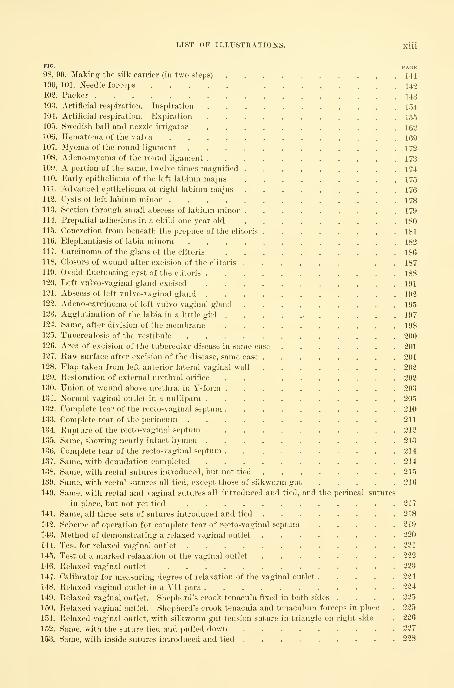
LIST OF ILLUSTKATIONS. xiii
FIG. PAGE98, 99. Making the silk carrier (in two steps) . . . . , . , . , .141100, 101. Needle forceps 142
102. Packer 143
103. Artificial respiration. Inspiration 154104. Artificial respiration. Expiration 155
105. Swedish ball and nozzle irrigator 162
106. Hematoma of the vulva 169
107. Myoma of the round ligament 172
108. Adeno-myoma of the round ligament 173
109. A portion of the same, twelve times magnified 174110. Early epithelioma of the left labium majus . . . 175
111. Advanced epithelioma of right labium majus 176
112. Cysts of left labium minor 178
113. Section through small abscess of labium minor 179
114. Preputial adhesions in a child one year old 180
115. Concretion from beneath the prepuce of the clitoris 181
116. Elephantiasis of labia minora 182
117. Carcinoma of the glans of the clitoris 186
118. Closure of wound after excision of the clitoris 187
119. Ovoid fluctuating cyst of tlie clitoris 188
120. Left vulvo-vaginal gland excised 191
121. Abscess of left vulvo-vaginal gland 192
122. Adeno-carcinoma of left vulvo-vaginal gland 195
123. Agglutination of the labia in a little girl 197
124. Same, after division of the membrane 198
125. Tuberculosis of the vestibule 200
126. Area of excision of the tubercular disease in same case 201
127. Raw surface after excision of the disease, same case 201
128. Flap taken from left anterior lateral vaginal wall 202
129. Restoration of external urethi'al orifice . . 202
130. Union of wound above urethra, in Y-form 203
131. Normal vaginal outlet in a nullipara 205
132. Complete tear of the recto-vaginal septum 210
133. Complete tear of the perineum 211
134. Rupture of the recto-vaginal septum 212
135. Same, showing nearly intact hymen 218
136. Complete tear of the recto-vaginal septum 214
137. Same, with denudation completed 214
138. Same, with rectal sutures introduced, but not tied 215
139. Same, with rectal sutures all tied, except those of silkworm gut 216
140. Same, with rectal and vaginal sutures all introduced and tied, and the perineal sutures
in place, but not yet tied 217
141. Same, all three sets of sutures introduced and tied 218
142. Scheme of operation for complete tear of recto-vaginal septum 219
143. jMethod of demonstrating a relaxed vaginal outlet 220
144. Test for relaxed vaginal outlet 221
145. Test of a marked relaxation of the vaginal outlet 223
146. Relaxed vaginal outlet 223
147. Calibrator for measuring degree of relaxation of the vaginal outlet 224
148. Relaxed vaginal outlet in a Vll-para 224
149. Relaxed vaginal outlet. Shepherd's crook tenacula fixed in both sides .... 225
150. Relaxed vaginal outlet. Shepherd's crook tenacula and tenaculum forceps in place . 225
151. Relaxed vaginal outlet, with silkworm gut tension suture in triangle on right side . 226
152. Same, with the suture tied and pulled down 227
153. Same, with inside sutures introduced and tied 228
Xiv LIST OF ILLUSTRATIONS.
FIG. P-^^fSE
154. Same, showing the gathering suture 229
155. Same, operation completed 229
156. Entire absence of vagina 233
157. Relations of rudimentary uterus, ovaries, and tubes in the case of absence of vagina . 234
158. Normal left tube and ovary, with uterine nodule 235
159. Normal right tube and ovary, with uterine nodule 236
160. Intact hymen after nine years of marriage 237
161. Traumatic atresia of the vagina 238
162. Double vagina with thick septum 239
163. Double vagina with double cervix 240
164. Atresia of vagina due to cup and stem pessary 241
165. Cyst of right vaginal wall 245
166. Cyst of anterior vaginal wall in pregnancy 246
167. Abscess of the recto-vaginal septum 247
168. Abscess of recto-vaginal septum from rectal fistula 248
169. Section of wall of cyst from anterior vaginal wall 248
170. Outline of cyst protruding from the vagina 249
171. Section of wall of cyst from posterior vaginal wall 250
172. Cross-section through wall of vaginal cyst 251
173. Large, thick-walled cyst of posterior vaginal wall 253
174. Primary carcinoma of posterior vaginal wall 256
175. Primary vaginal carcinoma 257
176. Atresia of the vagina 258
177. Same, showing operation 259
178. Atresia of vagina, in sagittal section 260
179. Same, '^howing operation 261
180. Atresia of the vagina in a negress 262
181. Coronal section of an old atresia of vagina 263
182. Same, after operation, with sutures in place 264
183. Instrument for measuring calibers and diameters of specula 276
184. Cystoscope and obturator 277
185. Urethral calibrator and dilator 277
186. Delicate mouse-toothed forceps 278
187. Searcher for locating urethral orifice 279
188. Examination of bladder in the dorsal position 279
189. Vesical speculum introduced, knee-chest position 280
190. Patient in a harness, knee-chest position, for cystoscopic examination .... 281
191. Holding the vesical speculum ready for introduction 282
192. Examination of bladder, knee-chest position 283
193. Cystoscope with oblique end and obturator 285
194. Instrument for internal vesical measurements 285
195. Hypertrophy of urethral mucosa 290
196. Ilypertrophied external urethral orifice 291
197. Operation for hypertrophied urethral mucosa 293
198. 199. Urethro-vaginal and vesico-vaginal fistula in the same patient .... 297
200. Same, showing method of introducing sutures ......... 298
201. ("oncealed abscess of Skene's gland 301
202. Large suburethral abscess 304
203. Urethral caruncle 307
204. Exstrophy of bladder become cancerous 320
205. Hairpin calculus 328
206. Section of a vesical calculus 329
207. V. Dittel's operation for vesico-uterinc (istuhi 330
208. Same, operation completed 331
209. Scissors for paring edges of vesico-vaginul fistula 337

LIST OF ILLUSTKATIOXS. XV
^O. PAGE210. Classical operation for vesico-vaginal fistula ...,..,.. 338
211. Scheme of same , , . . 339
212. Vesico-vaginal fistula closed with buried catgut suture 341
213. Dudley's operation for large vesico-vaginal fistula 342
214. Same, showing smaller bladder 343
215. 216. Vesico-vaginal fistula occupying entire base of bladder 344
217, 218. Vesico-utero-vaginal fistula 345
219. Suprapubic operation for vesico-vaginal fistula (Trendelenburg) 349
220. Vesico-utero-vaginal and vesico-uterine fistula in the same patient 350
221. Vesico-uterine fistula, showing treatment 351
222. Vesico-uterine fistula, sutures in place 352
223. Vesico-vaginal fistula caused by pessary 353
224. Hypertrophy of anterior vaginal wall due to cystitis 354
225. Pyuria due to suppurating dermoid cyst opening into bladder 355
226. Pronged instrument for tying knot inside the bladder . . . . • . . . 358
227. Linear ulcer of posterior wall of bladder 365
228. Ulcer of the trigonum of the bladder 366
229. Tubercular cystitis 368
230. Two-way catheter 371
231. Rubber balloon for treatment of cystitis 372
232. Rubber balloon rolled and grasped in the forceps 373
233. Bladder inflated by the vesical balloon 373
234. Long metal ureteral catheter 402
235. End of elastic bougie tipped with wax 403
236. Sounding the left ureter with the searcher 404
237. Using the goniometer 405
238 Passing a metal ureteral catheter into the left ureteral orifice 406
239. Washing out the pelvis of the kidney 407
240. Catheterizing both ureters 409
241. Sieve and graduate for filtering urine 411
242. Instrument for collecting urine without catheterizing the ureter 411
243. Composite temperature and pulse chart of ureteral fever 416
244. Demonstration of stricture of the ureter and of hydroureter 428
245. Ends of dilating metal catheters 436
246. Washing out the right kidney 438
247. Washing out the kidney and ureter 439
248. Diagnosis of abscess of kidney by the renal catheter 443
249. Hydroureter of both sides, with double ureter on the left side 446
250. Hydroureter and hydronephrosis 447
251. Syringe and aspirator 449
252. A ureteral calculus 449
253. End of a wax-tipped catheter 450
254. A calculus of the pelvis of the kidney 451
255. Stone caught in the eye of a renal catheter 451
256. Removal of the kidney and ureter without opening the peritoneum 453
257. Prolapse of the ureteral and vesical mucous membrane 455
258. Switching the ureter into the bladder by means of an artificial vesico-vaginal fistula . 458
259. Uretero-vaginal fistula 459
260. Right uretero-cystostomy for uretero-vaginal fistula 460
261. Uretero-cystostomy 461
262. Uretero-ureteral anastomosis, showing the ureter divided and the lower end tied and
split on one side 466
263. Uretero-ureteral anastomosis, showing the ureter held in place by the traction ligatures . 467
264. Experimental uretero-ureteral anastomosis in a dog 468
265. Showing the lines of incision in two cases of nephro-ureterectomy 469

XVI LIST OF ILLUSTRATIONS.
PIG. PAGE
2G6. Total extirpation of a tuberculous left kidney with its ureter 470
267. Removal of a tubercular kidney and ureter 471
268. Removal of the kidney and ureter, showing the facility with which the ureter can be
palpated all the way down to the common iliac artery 473
269. Showing the method of removing the lower end of the ureter througli the vaginal vault 473
270. Removal of the kidney with the ureter 475
271. Removal of kidney and entire ureter, nephro-ureterectomy 476
272. Ends of three sizes of the EUinger, and Goodell-Ellinger dilators 479
273. Goodell-Ellinger dilator with spring between the handles 480
274. Criminal abortion, with separated elm tent in situ 481
275. Uterus perforated by a tupelo tent 482
276. Sharp curette for removing the uterine mucosa 485
277. Section of a glandular uterine polyp 487
278. The spoon of the long, sharp curette 492
279. Knife-blade tenaculum for depleting the cervix 494
280. So-called " erosion " of the cervix uteri 495
281. Bilateral laceration of the cervix 496
282. Incision into the angles of the laceration 496
283. Denudation of both lips for plastic union 496
284. The cervix after all the sutures are tied on both sides 497
285. Glass irrigator 497
286. Complete prolapse of the uterus and vagina 500
287. Complete prolapse of the uterus and vagina, with retroflexion 501
288. Prolapse of the uterus, showing stages of descent 502
289. Partial prolapse, with eversion of the vaginal walls 503
290. Complete prolapse of the vagina and uterus, with retroflexion 504
291. Partial prolapse of the uterus and.vagina, with elongate lacerated cervix . . . 505
292. Partial prolapse of the uterus, with eversion of vaginal walls 506
293. Partial prolapse of the uterus, with elongate hypertrophied cervix 510
294. Operation for prolapse of the uterus by amputation 511
295. Prolapse of the uterus, vagina, and rectum, with complete rupture of the recto-vaginal
septum 513
296. Vegetating epithelioma of the cervix 514
297. Epithelioma of the cervix without vegetation 515
298. Vaginal hysterectomy for cancer of the uterus; utei-us and cervix curetted and the cer-
vix sewed up 516
299. Vaginal hysterectomy ; cutting the cervix loose from the vaginal vault, under irrigation 517
300. Vaginal hysterectomy : detaching the bladder from the cervix 518
301. Aneurismal needle 519
303. Vaginal hysterectomy ; exposing and tying ofl' the left broad ligament .... 520
303. End of stout blunt tenaculum 520
304. Vaginal hysterectomy ; freeing the right broad ligament 521
305. Vaginal hysterectomy ; applying the last ligature 523
306. Vaginal hysterectomy ; the uterus brought outside 523
307. Vaginal hysterectomy ; the uterus removed 535
308. Inversion of the uterus 533
309. Inversion due to sarcoma 536
310. Pediculated submucous myoma 540
311. Pediculated submucous myoma, with partial inversion 541
313. Large external pediculated submucous myoma 543
313. Pediculated submucous myoma attached to the fundus posteriui-ly 544
314. Sickle-shaped stout knife 545
315. Pyo-physometra due to occlusion of the cancerous cervix 554

LIST OF PLATES.
PLATE FACING PAGEI. Various kinds of bacteria 32
II. Diagnosis of abdominal tumor, showing also respiratory motion 80
III. Pruritus vulva? 198
IV. Caruncle of urethra 307
V. Loculate bladder 318
Fig. 1. The loculi surrounded by contracted muscular bands.
Fig. 2. The same loculi with the muscular bands relaxed.
VI. Trigonum of bladder before and after treatment with vesical balloon (Figs. 1 and 2) . 375
VII. Fig. 1. Normal bladder 387Fig. 2. Carcinoma of the bladder 387
VIII. Tuberculosis of the endometrium 489
IX. Fig. 1. Adeno-carcinoma of the body of the uterus 491
Fig. 2. Epithelioma of the cervix 491
X. Epithelioma of the cervix uteri (colored) 493
xvii


OPERATIVE GYNECOLOGY,
CHAPTER I.
SEPSIS, ASEPSIS, AND ANTISEPSIS IN HOSPITALS.
1. Sepsis, definition of.
2. Asepsis.
S. Antisepsis. Soap and water. Dry heat. Dry-air oven. Steam oven or steam cylinder. Steam.Boiling soda solntion. Chemical antiseptics.
4. Operating room. Table. Sterilized water. Sterilization and preservation of instruments.Sterilization and preservation of sutures and ligatures. Silkworm gut. Catgut. Gauzeand cotton. Iodoform gauze. Sponges. Di-ainage cushions. Ovariotomy pad. Perineal
pad. Vessels.
5. Preparation of surgeon, assistants, and nurses. Operating suit. Brushes. Cleansing and disin-
fecting the hands and forearms.
SEPSIS.
Surgical sepsis arises from tlie invasion of a wound by pathogenic micro-
organisms which find in the tissues suitable conditions for their development
and growth.
The micro-organisms most frequently concerned in traumatic infections are
the pyogenic bacteria, of which the most important representatives are the pyo-
genic staphylococci and streptococci, although under special conditions manyother bacterial species may cause suppurative inflammation. Tlie simple con-
ception which once prevailed that a wound becomes infected, in much the same
way as an artificial culture medium, by the mere entrance of pathogenic bac-
teria, has been greatly modified by bacteriological studies of the conditions
underlying the infection of wounds. There are various circumstances besides
the mere presence of bacteria which determine the occurrence and the character
of traumatic infections.
A fresh wound in healthy tissues, while it resembles an artificial culture
medium in offering suitable food for the development of many kinds of bac-
teria, differs from such a medium in the presence of various properties of cells,
tissues, and fluids which are hostile to the life and growth of many bacteria. In
the study of the causation of traumatic infectious it is important to consider not
only the invading micro-organisms, but also the germicidal powers of the cells
and fluids of the body. Experiments of Dr. W. II. Welch and others have
demonstrated that even the most careful antiseptic or aseptic surgical technique
often fails to exclude the entrance of bacteria, including sometimes even the
ubiquitous pyogenic cocci, into wounds which heal without infectious inflamma-
tion. Under these circumstances the antibacterial properties of the living cells

2 SEPSIS, ASEPSIS, AND ANTISEPSIS IN HOSPITALS.
and of the fluids in the wounded area suffice to inliibit the growth or the
pathogenic manifestations of tlie invading bacteria. It is largely to these
natural inhibitive forces of the living tissues that we must ascribe the good
results obtained in many surgical operations conducted even under a bad
technique.
It would, however, be a serious error to rely exclusively in surgical tech-
nique upon the germ-destroying powers of the living tissues and fluids of the
body, great as these undoubtedly are and important as it is not to interfere
with these natural germicidal agencies. In a large proportion of the cases in
which bacteria have been found in so-called aseptic wounds the bacteria have
been either non-pathogenic or possessed of little virulence. It is exceptional to
find virulent pyogenic bacteria in wounds without any manifestations of their
pathogenic activity.
The most common invader of wounds of the skui is a variety of the staphy-
lococcus pyogenes albus called by Welch ( Conditions underlying the
Infection of Wounds. Trans, of the Congress of American Physicians and
Stirgeons, vol. ii)the staphylococcus epidermidis albus, as it is a
regular inhabitant of the epidermis and hair follicles. The investigations of
Drs. II. Robb and A. A. Ghriskey {Johns Hopkins Hospital Bulletin^ vol. iii,
p. 37, 1892) have shown that most wounds through the skin sooner or later
become contaminated with this organism, and yet its presence may not interfere
with primary union. An important point relating to the presence of the
staphylococcus epidermidis albus in the healthy skin is that it
lies so deeply in the epidermis or hair follicles that chemical disinfection of the
superficial layers of the skin does not destroy it, as may be demonstrated by
the following experiment : After thorough disinfection of the skin by perman-
ganate of potash and oxalic acid, in the way subsequently described, cultures
made from scrapings of the surface usually show no growth. If, now, ster-
ilized silk sutures be passed one or more times through the skin in the disin-
fected area, and a tube of nutrient agar-agar be inoculated with the sutures,
the presence of the white staphylococcus, often in pure culture, can
be demonstrated in parts of the epidermis deeper than those acted upon by
any chemical methods of disinfection of the surface of the integument.
Welch believes that the staphylococcus epidermidis albus is
but rarely pyogenic, and that its pathogenic activity depends largely upon de-
creased resistance in the germicidal forces of the wound area.
The most recent bacteriological and practical experiments on infection of
wounds point conclusively to the fact that the skin is a common habitat for
various organisms, and that this must be taken into careful consideration in
the preliminary disinfection of all operative fields. As already stated, in a large
proportion of cases these organisms are non-pathogenic, and a fresh wound
containing them may, from a surgical standpoint, be regarded as aseptic when
the process of healing is in no way interfered with.
Cultures taken from beneath the most carefully applied surgical dressings
very frequently show growths which can be accounted for only on the supposi-

ASEPSIS. 3
tion that bacteria were present before the operation, or were deposited in the
wound during the progress of the operation, or gained access later from the
adjacent skin. Suppuration occurs when the organism is virulent, the con-
dition of the wound favorable for growth, and the normal inhibitory activity
of the tissues is reduced.
In the following quotation from Dr. Welch's paper he summarizes the con-
ditions underlying wound infection :" The effects produced in the animal
body by the pyogenic cocci are determined by many factors relating to the
infectious agent and to the individual exposed to infection. There are differ-
ences in these effects, depending upon the species of animal ; upon the tissues
and parts of the body infected ; upon the readiness of absorption from the
affected parts ; upon the source, the number, and the virulence of the organ-
isms ; upon the nature and amount of toxic substances accompanying and pro-
duced by the bacteria ; upon general predisposing conditions of the body ; and
upon local conditions in a wound, such as the presence of foreign bodies, of
pathological products, of dead spaces, of bruised, necrotic, and strangulated
tissues."
]^otwithstanding the constancy of micro-organisms in the air and on all
objects with which we come in contact, we are usually able, by carrying out a
rigid technique, to prevent the invasion of a wound by virulent pyogenic organ-
isms in sufficient number to produce harm. The realization of the difficulty of
obtaining a germ-free w^ound should stimulate surgeons to observe the most
painstaking care in the preliminary preparation in order to reduce the amount
of contamination to a minimum.
ASEPSIS.
In a surgical sense asepsis is the absence of septic germs; an aseptic
wound is one which remains free from invasion by these germs in sufficient
number to disturb the healing process.
The common means for the introduction of the germs are the hands of the
surgeon or of his assistants, the instruments, or the surgical accessories.
The surface of the body, the digestive canal, and the female genital tract
up to the internal os uteri are normally the habitat of many species of micro-
organisms. As it is not practicable to differentiate beforehand the specific
character of the various germs which are present, especially as to their pyo-
genic properties and virulence, modern surgery first proceeds upon the as-
sumption that the skin of the patient, of the surgeon, and of the assistants,
the instruments, the dressings, etc., are in an infected state until rendered
aseptic by the use of antiseptic measures ; and second, it endeavors to
maintain the aseptic condition thus established throughout and after an op-
eration.
The surgeon must also be constantly alive to the fact that his work and
that of his assistants and the nurses may bring them into daily contact with
septic matter, and that extraordinary precautions are necessary to avoid con-
veying such infected material from case to case. There is a well-recognized

4 SEPSIS, ASEPSIS, AND ANTISEPSIS IN HOSPITALS.
liability of septic cases to occur in groups in hospital practice. As an example
of this in my own practice, in 1892 I ruptured a large streptococcus abscess
in removing it, and the patient died shortly afterward. Three cases immedi-
ately following this had an erysipelatous inflammation of the wound and nar-
rowly escaped with their lives.
ANTISEPSIS.
Antisepsis is a term used to designate any active means whatever by which
septic germs are removed, destroyed, or rendered inactive.
The antiseptic principle may be worked out in a variety of ways. The
demonstration, however, of the value of any antiseptic procedure must come
through the more rigid scientific methods of the bacteriologist, and in all cases
of innovation as to ways and means his experiments must be recognized as the
authoritative tests.
The mechanical removal of germs by scrubbing with soap and water, and
their destruction by steam or boiling solutions, are the best antiseptic agents
which we possess. It is a noteworthy fact that the housewife's simple reme-
dies against dirt and against fermentation, as in preserving fruits, appear to be
the final outcome in this direction of the surgical activity of the last half of
this century.
The usual methods of applying heat as a germicide are the hot dry-air oven,
the steam oven, or steam cylinder, and boiling soda solution.
Hot-air disinfection requires too high a temperature—176'6° C'. (350° F.)—to
be satisfactory for most purposes, and is injurious also to sharp instruments. I
have for this reason abandoned it in favor of steam disinfection.
Steam disinfection in an oven, jacketed to prevent the steam from condens-
ing, destroys the most resistant organisms.
In order to destroy all germs with their spores, stei-ilization by live steam
must be repeated for two or more successive days, an hour the first time and
half an hour on each subsequent occasion. The spores of pathogenic bacteria
are less resistant than those of some saprophytic bacteria, such as the bacilluss u b t i 1 i s , and the former are destroyed by exposure for half an hour to the
temperature of live steam. Steam under pressure of ten or fifteen pounds
destroys even the most resistant spores by a single exposure for twenty minutes
to half an hour.
The Arnold or E. Boeckmann steam sterilizer, or some sterilizer similarly
constructed, is cheap and effective. The steam is generated rapidly in a small,
hollow plate by a Bunsen flame, and then passes through a slioi-t shaft into
a jacketed cylinder containing the articles to be sterilized, (circulating from
this under an outside co])per jacket which covers the whole, it is recondensed
and drips into a pan, from which it runs through small holes into the hollow
plate, and begins to travel the circuit again.
Institutions supplied with steam heat may convey the live steam directly into
the sterilizers—a practical, effective, and rapid means of sterilization. An appa-
ratus long in use in the gynecological operating room of the Johns Hopkins

STEAM DISINFECTION.
Hospital, connected in tins way with the general steam-heating system, has
proved most satisfactory.
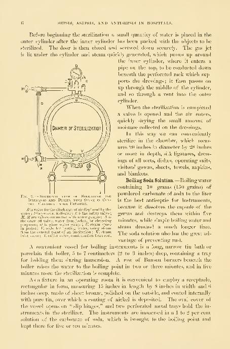
Two sterilizers are employed—one for water, the other for dressings, etc.
The sterilizer for dressings consists of a cylindrical copper reservoir contain-
ing a steam coil which enters from above, and has its exit from below. Thebottom slopes toward the
center, forming a shallow
funnel with a drainage-
tube for the escape of the
condensed steam. A wire
netting is placed two
inches from the bottom,
upon which the objects to
be sterilized are deposited.
The circulation is so ar-
ranged that when active
sterilization is required
live steam can be turned
into the cylinder, pene-
trating the linen envel-
opes of the dressings and
the cotton plugs of the
flasks and tubes.
When the sterilization
is completed the live steam
is turned from the reser-
voir into the coil by sim-
ple gate-valves, and so
quickly dries the dressings
before they are removed
from the sterilizer. In
order that the drying pro-
cess may be facilitated,
the cover should be lifted
and air allowed to enter.
Steam sterilization un-
der pressure is more rapid and more effective than that conducted without it.
One of the latest and best sterilizers on this plan is the Sprague, manufactured
by Richard Kny & Company, constructed on tlie principle of the autoclave
used in the bacteriological laboratory.
The apparatus consists of an inner and an outer cylinder, the outer serving
as a jacket for the inner one. The sterilizing chamber is barrel-shaped, and
is closed by a secure door, which makes it steam-tight. A steam gauge indi-
cates the pressure, which may be carried up to thirty pounds, and a safety
valve is security against explosion.
Fig. 1.
—
Steam Sterilizer for Dressings and Dishes, the doorpartly open.
When the dressings are put in and the door closed a slight turn
fixes the projecting lugs in under the rim ; the ring in the center of
the door is then revolved until the door is jammed down and a steam-
tight joint secured. The steam is generated in the jacket by a long
gas jet or steam pipes underneath. The amount of water in the jacket
is indicated by the gauge at the side. The steam guage on top regis-
ters the pressure inside the chamber. After heating and exhausting
the air in the sterilization chamber the steam is let in by a screw andthe sterilization begins. At the completion of sterilization the steam
is turned off and the dressings in the boiler thoroughly dried before
removal.
;EPSIS, ASKl'SIS, AND ANTISEPSIS IN HOSPITALS.
Before beginning the sterilization a small quantity of water is placed in the
outer cylinder after the inner cylinder has been packed with the objects to be
sterilized. The door is then closed and screwed down securely. The gas jet
is lit under the cylinder and steam quickly generated, which passes up around
the inner cylinder, where it enters a
pipe on the top, to be conducted downbeneath the perforated rack which sup-
ports the dressings ; it then passes on
up through the middle of the cylinder,
and so through a vent into the outer
cylinder.
When the sterilization is completed
a valve is opened and the air enters,
quickly drying the small amount of
moisture collected on the dressings.
In this way we can conveniently
sterilize in the chamber, which meas-
ures 20 inches in diameter by 28 inches
or more in depth, silk ligatures, dress-
ings of all sorts, dishes, operating suits,
visitors' gowns, sheets, towels, napkins,
and blankets.
Boiling Soda Solution.—Boiling water
containing 10 grams (150 grains) of
powdered carbonate of soda to the liter
is the best antiseptic for instruments,
because it dissolves the capsule of the
germs and destroys them within five
minutes, while simple boiling water and
steam demand a much longer time.
The soda solution also has the gi-eat ad-
vantage of preventing rust.
A convenient vessel for boiling instruments is a long, narrow tin bath or
porcelain fish boiler, 5 to 7 centimeters (2 to 3 inches) deep, containing a tray
for holding them during immersion. A row of Bunsen burners beneath the
boiler raises the water to the boiling point in two or three minutes, and in five
minutes more the sterilization is complete.
As a fixture in an operating room it is convenient to employ a receptacle,
rectangular in form, measuring 1 5 inches in length by 8 inches in width and (\
inches deep, made of slieet bronze, polished on the outside, and coated internally
with pure tin, over which a coating of nickel is deposited. The oval cover of
the vessel o])ens (m " slip hinges," and two perforated metal trays hold the in-
struments in the sterilizer. The instruments are immersed in a 1 to 2 per cent
solution of the carbonate of soda, which is l)rought to the boiling point and
kept there for five or ten minutes.
Fig. 2. — Sectional view of Steeilizeb forDressings and Dishes, with Steam in Cen-tral Chamber under Pressure.
B is valve for the discharsre of air displaced by thewater : D is pressure indicator ; £ is the safety valve
;
M, M are valves connected with water gauges ; N is
the draw-oft' valve, water from jacket, for cleansing
purposes ; is glass water gauge ; tf^ steam space
in jacket; F, coils for heating water, using steamfrom the general plant of an institution ; W, steaminlet valve ; X, outlet valve, condensation from coil.

CHEMICAL ANTISEPTICS. 7
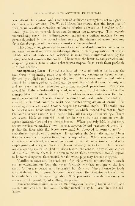
The boiler is arraiioed for lieating either by gas or by steam.
Chemical Antiseptics.—As far as possible, it is safer to depend upon steam or
heat sterilization rather than upon chemicals.
Experiments have shown that the solution of bichloride of mercury, fre-
quently employed in surgical work, does not under all conditions manifest its
germicidal powers. It often merely inhibits germ growth, but to what extent
this inhibition is valuable is as yet unknown. The inefficiency of bichloride of
mercury as a cutaneous germicide can be tested for practical purposes by im-
mersing the hands for ten minutes in a 1-500 aqueous solution, and then in a
sterilized ammonium sulphide solution to precipitate the mercury. After this,
by scraping the epithelium, cultures can usually be obtained which will grow in
ordinary media.
If dishes and porcelain ware are to be efficiently sterilized by this means,
they must be kept in a strong solution of corrosive sublimate (1-500) for fif-
teen minutes after they have been thoroughly scrubbed with soap and water
;
the sublimate kills most of the bacteria and renders the rest inactive.
In the experiments on skin disinfection we have a factor to consider which
we do not meet with in the sterilization of the dishes. The albuminate of mer-
cury which is formed in the tissues, when brought in contact with corrosive
sublimate solutions, may encapsulate the organisms, and so render them incapable
of growth. When dishes, on the other hand, are submerged in the disinfectant
Fig. S.—Instrument Sterilizer.
solution, the organisms are at once brought in contact with the bichloride of
mercury without the formation of this albuminate, and the sterilization is more
effective.
The use of chemical solutions, such as carbolic acid and corrosive sublimate,
for disinfection of wounds is objectionable, because their value depends upon the
8 SEP8IS, ASEPSIS, AND ANTISEPSIS IN HOSPITALS.
strength of the sohition, and a sohition of sufhcient strength to act as a germi-
cide acts as an irritant. Dr. W. S. Halsted has shown that the irrigation of
fresh wounds with a corrosive sublimate solution as weak as 1-10,000 is fol-
lowed by a distinct necrosis demonstrable under the microscope. This necrotic
material may retard the healing process and act as a culture medium for any
germs deposited in the wound subsequently ; the danger of acute poisoning
from the absorption of the mercury must also be considered.
I have long since given up the use of carbolic acid solutions for instruments,
and only use sterilized water to submerge them in during operation. The ger-
micidal effect of carbolic acid solutions is more than counterbalanced by the
injury which it causes to the hands. I have seen the hands so badly cracked and
chapped by the carbolic solutions that it was impossible to scrub them perfectly
with nail brushes.
The Operating Eoom.—For private hospitals or small public institutions the
best form of operating room is a simple, spacious, rectangular structure well
lighted by skylight and northern windows. The various architectural details
should be so arranged as to facilitate the work for which the room is designed,
and to carry out the principles governing surgical procedures. The doors
should be of the noiseless sliding kind, so as to offer no obstruction to the easy
transportation of patients to and fro. Any elaborate ornamentation of the room
must be eschewed. The walls must be smooth, of hard finish, or coated with
enamel water-proof paint, to resist the disintegrating action of steam. The
cleansing of the walls and floors is helped by rounded angles. The walls may
be paneled with broad slabs of African marble, which extend five feet up from
the floor as a wainscot, or, as in some clinics, all the way to the ceiling. There
are several kinds of material useful for flooring ; the most common are the
square encaustic tiles and the mosaic blocks. When properly laid, so that there
are no crevices or cracks, either makes a serviceable and ornamental floor. In
paving the floor with the blocks care must be observed to secure a uniform
smoothness over the entire surface. By mopping the floor daily and scrubbing
it twice a week with sapolio its surface is kept clean. Where economy in con-
struction is considered, a cement pavement or bolted boiler iron covered with
ship's paint makes a good floor, which can be easily kept clean. The floors of
some operating rooms are laid to slope toward the center or toward one corner
of the room, where thei'e is a drainage vent; this convenience would appear
to be more dangerous than useful, for the waste pipe may become clogged.
Ventilation must also be considered, for, while we do not attribute so muchrisk to contamination from the air as formerly, we dare not ignore the fact
that infection may occasionally be carried in this way. The entrance for fresh
air and the exit for impure air should l)e so placed that the circulation will not
be conducted over the operating table. This precaution is further necessary on
account of the possibility of chilling the patient.
The ventilators should be so set that they can be easily taken Dut of their
sockets and cleansed, and some filtering material may be placed in the venti-
lators.

THE OPERATING ROOM. 9
A sloping skylight, looking to the north, gives an evenly distributed light,
which is never glaring.
The equipment of the operating room must be simple.
A prime requisite is a row of large, oval marble basins plentifully supplied
with hot and cold water. To facilitate the most perfect details of the aseptic
principle, the taps may be connected with a pedal attachment like that devised
by Dr. H, Robb, which permits the water to be turned on or off by the foot.
The most glaring inconsistency in the aseptic arrangement of most operating
rooms is the impossibility of thoroughly sterilizing the hand basins, which are
Fig. 4.
—
Hand Basins set on Pivots fob Eemov.
Tlie liot and cold water are mixed in a rose jet a foot above the basin,
controlled by the foot taps on the floor.
I) Sterilization.
The flow of Jiot or cold water is
contaminated at every washing and are lial:)le to hold grease. This may be
avoided by using movable metal basins made of plated copper or solid nickel,
and swung over a porcelain hop])er or sink, as shown in the figure.
A large sink for the immersion of dishes, etc., and a hopper for waste water,
should be in a convenient location. The traps in all the pipes must be in-
spected and disinfected frequently.
The room should be fitted with electric-light and gas fixtures, and an electric-
light bracket should be placed near the operating table, so that a portable light
with reflector may be attached easily. A group of four incandescent lights
with reflectors should be suspended over the table.
Tlie other furnishings of the operating room should be as few as possible
;
all apparatus—such as dressings, sterilizers, water-boilers, etc.—should be placed

10 SEPSIS, ASEPSIS, AND ANTISEPSIS IN HOSPITALS.
in an adjoining room. The instrument case should be conveniently located,
either near the operating- table or in an adjoining room, so that at any time an
instrument may be quickly obtained if required in the midst of an operation.
Glassware for instruments and solutions, and jars for sterilized ligatures,
gauze, cotton, and towels, are kept in a room especially set aside for storage.
The sterilization of instruments, dressings, etc., should not be done in the
operating room, as the combustion products vitiate the atmosphere, and during
the summer months the temperature of the room becomes excessive with the
additional heat.
The anesthesia room should be conveniently placed, but great care must be
observed to have it so planned that noises from the operating room will not
be heard by a waiting patient.
Operating Table.—The gynecological operating table should be of metal with
a moval)le glass top, which can be raised or lowered as required.
The Kelly table shown in the figure is arranged with a support for the
Operating Table, with stout Brass Legs and Frame and heavy Glass Top.
Simple attachment with ratchet for the elevation of the pelvis. This is lifted off during vacrinal opera-
tions, and the seat under the table drawn around for the anesthetizer to sit on, while the operator occupies a
.stool at the opposite end of the table.
patient's feet below the top. A simple lattice of interwoven metal slats, with a
ratchet and crossbar, gives the needed elevation of the pelvis.
The height of the table is 78-5 centimeters (JM inches); width, HB".") centi-
meters (21 inches) ; and length, 113 centimeters (44: inches).

STERILIZED WATER. 11
Edebohls's table, one of tlie simplest and best constructed, and the Boldt
table, which inclines the whole body, are both well arranged for self-drainage
and easy adjustment.
Sterilized "Water.—An abundant supply of sterilized water should always be
on hand in the operating room. Water drawn from the tap can be sterilized
by boiling it for half an hour. If it is
allowed to stand covered for several
hours after boiling, the organic matter
settles to the bottom, and the clear water
above this can be drawn off by a spigt)t
placed in the vessel about 10 centimeters
(4 inches) from the bottom. A ready
method of sterilizing water in a chnic is
l)y means of a copper reservoir lined with
a steam coil. To use this, fill the reser-
voir with water, and then open a valve
in the coil, letting in the steam, when the
water is quickly brought to a boiling
point. Another way of getting sterile
water is by distillation ; water can l)e
distilled in quantity, from SO to 120 liters
(20 to 30 gallons) daily, by means of a
gas flame, running water, and a small
copper still, hung on a bracket against
the wall. The cold-water faucet taking
its supply from the street is connected
with the still by a rubber tube and a slow
flow started ; a Bunsen burner beneath
the still condenses a small portion of the water passing through it, and in this
way 6 or 8 gallons or more can be secured every twenty-four hours. Thedistilled water is conveniently stored in large agate-ware pails and boiled as re-
quired for use.
In a large clinic the quantity of sterilized w^ater, both hot and cold, which is
needed for daily use is so great that an apparatus such as that shown in Fig. 6
is a great convenience.
The water, entering from the house tap, is first filtered in the narrow cylinder
l)etween the two large ones, to remove all visible impurities. It is then boiled,
either by a gas engine, below in the center, or by steam coils, and stored in the
large reservoirs seen at the sides, holding from 60 to Y5 gallons or more, so
arranged that one holds hot and the other cold water. Gauges show the amountof water in the tanks, and thermometers register the temperature. The water
is drawn mixed at the desired temperature. Air-filtering vacuum valves above
the cylinders provide for the entrance of pure air as the water is withdrawn.
Sterilization and Preservation of Instruments.—It is but a few years since the
care of the instruments amounted to nothing more than washing them, often
Fig.

12 SEPSIS, ASEPSIS, AND ANTISEPSIS IN HOSPITALS.
Imrriedly, with soap and warm water, and putting them away in a velvet-hned
case, ready for use at the next operation. A close observer could then fre-
quently detect dried blood clinging to the joints of forceps and scissors, and
dirt lodged in the eyes of the needles.
No part of the gynecological technique is to-day considered more important
than the sterilization of the instruments. To facilitate cleansing, a preference
must always be given to the simplest forms of instruments ;joints, corrugations,
and rough surfaces on the handles must be avoided whenever possible. In the
locks of scissors and forceps the screw joint must be rejected, and in its place
the French lock, or one similar to a device of my own, are recommended.
After an operation the instruments are gathered together, the paired instru-
ments, such as forceps and scissors, separated, and knives and needles laid apart.
They are then placed with handles together in a large dish and washed with
soap and hot water. If tarnished, they may be polished with the best grade of
sapolio. The first assistant lifts up one instrument after another, rinsing it
and wiping it clean ; he hands it to the second assistant, who dries it, inspect-
ing carefully all its parts before placing it on a clean dry towel spread on a
table. When all the instruments have been cleaned, they are classified and
put away in the instrument case on glass shelves to await the next operation.
After septic operations, dealing with purulent peritonitis, abscesses, sloughs,
etc., the instruments, in addition to being washed, must be sterilized before
being returned to the case. By using water not far from the boiling point
in cleansing them, the instruments become so hot that they dry much more
rapidly.
Before every operation the proper instruments are selected and placed in a
bag, or wrapped in a towel, and laid on a tray for sterilization, for five minutes
in a 1 per cent bicarbonate of soda solution, as devised by C. Schimmelbusch,
When lifted out of the solution they are placed in glass
dishes on a table close to the operating table, where they
are classified by an assistant whose hands have been ster-
ilized ; they are then covered with hot water. One of the
great advantages of the soda solution is that it does not
tarnish and dull the edges of the instruments as steam
sterilization does. Such glaring inconsistencies as drying
the instruments with a soiled towel or taking them up
with unclean hands must be avoided. Instruments taken
out of the case for inspection by visitors must be laid
^"*'Silk '"usEi.^— f''in°e
asidc for Sterilization before being returned.
Intekmediati:, and Only the sterilized hands of the operator and his as-
sistants should come m contact with the instruments used
during the operation. An instrument which falls to the table or fioor, or touches
garments or face, is sc))tic until restcrilized.
Sterilization and Preservation of Ligatures and Sutures. Silk and silkworm
gut are sterilized by the fractional method.
The best quality of surgeon's twisted silk must be secured in three sizes
:

13
fine (No. 2), intermediate (Xo. 3), and heavy (J^o. 4). The fine silk is used to
make the carrier loops in the needles and for intestinal suture. The inter-
mediate silk is used in general to tie vessels and to bring-
together wound surfaces, and often to tie small pedicles.
The stout ligature is only used in tying a large quantity
of tissue in a pedicle.
The following method of sterilizing silk we owe to
Dr. W. S. Ilalsted, of the Johns Hopkins Hospital : Theskeins of silk are opened and cut in lengths of 40 centi-
meters (11 inches) for carriers, and 24 to 30 centimeters
(9 to 12 inches) for ligatures and sutures. Ten of these
are wound on a glass reel, and several such reels of
one size, or of assorted sizes, are dropped into a stout
glass ignition tube devised for this purpose; several of
these tubes, plugged loosely with cotton, are put in a
steam sterilizer for an hour the first day, and on the two
following days for half an hour each time. The steam
passes through the cotton without restraint, and acts uponthe silk as eavsily as if it lay loose in the sterilizer. Onremoving the tubes, the cotton in the mouth is pushed
tightly in and they are stored away in glass jars until
wanted. Silk which remains over after an operation
may be resterilized in the same way, but it is ajDt to be
weakened after the second sterilization.
If it is necessary to take but one reel of silk out of a
tul)e, it may be done without contaminating the rest by
carefully removing the cotton stopper between the third
and fourth fingers, taking care that the surface of the cot-
ton which comes in contact with the tube does not touch
anything else, while holding the tube obliquely to facilitate
removing the reel with a pair of sterilized forceps.
Silkworm Gut.—To sterilize silkworm gut, a dozen
pieces or more are loosely twisted together, doul)led, and
put into an ignition tube or a piece of ignition glass tub-
ing plugged at both ends, and sterilized in the same wayas the silk.
Catgut.—The employment of catgut sterilized by de-
fective methods has, in at least three recorded instances
in my own practice, been productive of serious outbreaks
of infection. That the majority of methods are unsafe
is shown by the great number proposed. From 1890 to
1894 I used catgut prej^ared by soaking in ether and then
boiling in alcohol under pressure. The results fromits use were good until the beginning of 1894, when an outbreak of sepsis
occurred which caused four deaths, and while we had no direct bacteriolog-
FiG. 8.—EoLLS OF Steril-ized Silk Threads onGlass Bobbins Pre-served IN STOUT GlassIgnition Tubes. %Ordinary size.
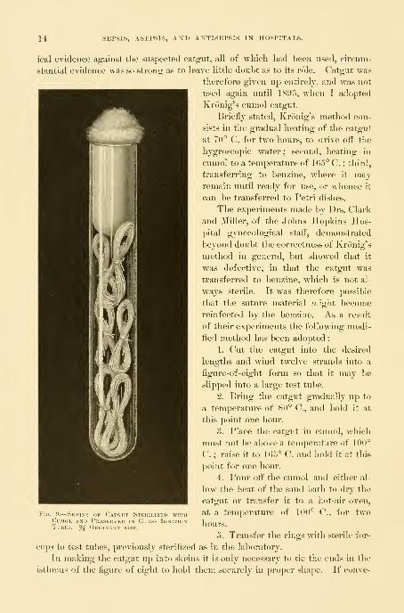
14 SEPSIS, ASEPSIS, AXD ANTISEPSIS IN HOSPITALS.
ical evidence against the suspected catgut, all of which had been used, circum-
stantial evidence was so strong as to leave little doubt as to its rcMe. Catgut was
therefore given up entirely, and was not
used again until 1895, when I adopted
Kronig's cumol catgut.
Briefly stated, Kronig's method con-
sists in the gradual heating of the catgut
at 70° C. for two hours, to drive off the
hygroscopic water; second, heating in
cumol to a temperature of 165° C. ; third,
transferring to benzine, where it mayremain until ready for use, or whence it
can be transferred to Petri dishes.
The experiments made by Drs. Clark
and Miller, of the Johns Hopkins Hos-
pital gynecological staff, demonstrated
beyond doubt the correctness of Kronig's
method in general, but showed that it
was defective, in that the catgut was
transferred to benzine, which is not al-
ways sterile. It was therefore possible
that the suture material might become
reinfected by the benzine. As a result
of their experiments the following modi-
lied method has been adopted :
1. Cut the catgut into the desired
lengths and wind twelve strands into a
figure-of-eight form so that it may be
slipped into a large test tube.
2. Bring the catgut gradually up to
a temperature of 80° C, and hold it at
this point one hour.
8. Place the catgut in cumol, which
must not l)e above a temperature of 100°
C. ; raise it to 1G5° C. and hold it at this
point for one hour.
4. Pour off the cumol aiid either al-
low the heat of the sand bath to dry the
catgut or transfer it to a hot-air oven,
at a temperature of 100° C, for two
hours.
5. Transfer the rings with sterile for-
ceps to test tubes, previously sterilized as in the laboratory.
In making the catgut up into skeins it is only necessary to tie the ends in the
isthmus of the figure of eight to liold them securely in proper shape. If conve-
j. y.
—
Skeins of Catout Stekilizku withCumol and Prksekvkd in Glass IgnitionTubes. % Okdinauy size.

CATGUT. 15
nient, it is better to use the hot-air oven for the drying process, "but this is not
absohitely essential, as a sand bath can be improvised, as suggested by Kronig,
to serve this purpose. A beaker glass of at least a half-liter capacity is im-
bedded three fourths of its height in a tin or agate-ware vessel of sufficient
capacity to permit three fourths of an inch of sand to be packed about the
sides and beneath the glass.
In drying or boiling, the catgut should not come in contact with the bottom
or sides of the vessel, but should be suspended on slender wire supports or
placed upon cotton loosely packed in the bottom. During the drying process
the beaker glass is covered with a sheet of pasteboard, through which a centi-
grade thermometer is thrust, so that the mercury bulb may be suspended about
midway in the vessel. In this way the temperature can be regulated perfectly.
A Bunsen burner is placed under the sand bath and the temperature in the
beaker glass is slowly brought up to 80° C, where it is held for one hour to dry
the catgut. A higher temperature than 100° C, before the catgut is thoroughly
dry, renders it brittle ; this step m the method must be carried out most care-
fully. When the drying process is completed the cumol is poured into the
beaker glass and brought up to a temperature of 165° C, a little short of the
boiling point, with two Bunsen burners. A copper-wire netting should be
placed over the beaker glass to prevent the ignition of the cumol. This tem-
perature is more than sufficient to kill all micro-organisms, and it is not neces-
sary to allow the cumol to boil, which causes unnecessary evaporation. Thecatgut is left for one hour at this temperature, when the cumol is poured off
for subsequent use.
Cumol, which is of a clear limpid or slightly yellowish appearance when pro-
cured from the chemist, is changed to a brownish color by boiling.
The catgut is allowed to remain in the sand bath until the excess of cumol is
driven off and it appears entirely free from any oily matter. A period of one
to two hours is usually sufficient to dry it thoroughly.
From the sand bath or hot-air oven it is transferred with sterile forceps to
sterile test tubes, such as are used for culture media, in which it is preserved
from contamination until ready for use. Small quantities should be placed in
each tube, to obviate the necessity of opening them too frequently.
In conclusion, it is well to bear in mind that while cumol is not explosive it
is very inflammable, and great care should be observed in lifting the wire screen
from the beaker glass to prevent drops of the cumol from falling into the flame
or on the heated piece of metal on which the sand bath rests, as it will take fire,
flare up, and ignite the fluid in the beaker glass. Such an accident has occurred
three times in our experience.
Catgut may be sterilized with perfect safety and with certainty by using the
following apparatus constructed by Dr. J. G. Clark with the aid of Mr. A. V. M.
Sprague : The materials are brass and copper, brass for the cast parts and coj)-
per for the cylinders. A cylindrical vessel of copper 6 inches in diameter and
8 inches high is fixed within a similar larger cylinder, so as to leave a space of
one inch on all sides and at the bottom between the two. This space is com-
16 SEPSIS, ASEPSIS, AXD ANTISEPSIS IN HOSPITALS.
pactly filled witli dry sand. Tlie apparatus is sup^sorted on legs raising it G
inches above the tray on which it rests. The upper end terminates in a bronze
metal flanged top, upon which rests a dome head of cast bronze. The head is
bolted tightly to the body of the apparatus, Init may be quickly removed so as
to reach the interior. Thesterilizer is provided with
a glass gauge to show the
quantity of cumol in the
cylinder, and a thermometer
registers the temperature of
the fluid ; there is an attach-
ment for a hose to carry oft"
the vapor as it is generated.
The sand between the cylin-
ders is heated by a Bunsen
gas burner, which stands on
the tray ; a uniform heat is
easily generated, raising the
temperature of the cumol
quickly to C. (331° F.),
necessary for the steriliza-
tion.
Gauze, or cheese cloth, is
used in large quantities dur-
ing operations and for the
dressings afterward, and is
bought to advantage in bales
of one hundred yards each.
It forms the best covering
for parts of the body around
the field of operation, and is
a good absorbent and pro-
tective when laid as a dress-
ing, six to eight folds thick,
on wounds. It is also valu-
able for making pads to be
used in the abdomen during an operation, and for small gauze sponges.
Absorbent cotton, which is common cotton cleansed and deprived of its oil
in oi'der to render it absorbent, is the most efficient dressing we possess for
taking up discharges, whether applied to the vulva or over an abdominal wound,
either directly or on top of a gauze pad. It is also used in padding the in-
equalities of the abdomen after an abdominal operation before ap])lying a
bandage.
Cotton bolsters covered with gauze are needed to hold back the obtruding
coils of intestines in abdominal operations. They are made of non-absorbent
Fig. 10.
—
Cumol Sterilizer.
_E, tap for removing cumol from cylinder; F, funnel throughwhich cumol is poured into cylinder ; G, glass tube connectedabove and below with cylinder to .show the amount of cumol; S,sand between outer and inner vessels ; V, vent.

SPOXGES. 17
cotton, wliicli does not take up moisture, and so preserves its elasticity. Thecotton is prepared in rolls 4 to 6 centimeters (1|- to 2|- inches) in diameter,
Tvhich are then cut in lengths of 12 centimeters (5 inches) and covered with
gauze.
Gauze, cotton, towels, and bandages nuist be sterilized fractionally by placing
them in the steam sterilizer for an hour, then taking them out and again steril-
izing them for half an hour at a time on two successiye days. After steriliza-
tion they should be preserved in large glass jars. It is easier to take what is
wanted from the stock without contaminating the rest if, instead of keeping it
in bulk, it is broken up into smaller packages before sterilization and rolled in
towels or gauze. These small rolls should be kept unopened until needed.
When called for, the nurse lifts one of the rolls from the jar, and, unpinning it
without touching its contents, lets the ends fall back and holds it to the oper-
ator or dresser, who then takes what he wants. Dressings sterilized for imme-
diate use may be used with perfect safety, the fractional sterilization only being
necessary when they are to be stored for future use.
Where enormous quantities of gauze are used the expense may be diminished
one half by sterilizing and using it over again, as suggested by Dr. J. C. Blood-
good, of the Johns Hopkins Hospital, where the gauze, after using it once,
unless the case is known to have been a streptococcus infection, is washed out
in cold water and then soaked in a strong solution of bicarbonate of soda to
cleanse and remove the blood ; it is then taken to the laundry, boiled and dried,
and sent back. The patients now smooth it out and roll it up, after which it is
sterilized in a steam sterilizer for a half an hour, and used in the ward for
various dressings. But a layer of new gauze is always put next to a recent
wound.
Iodoform gauze is prepared with aseptic hands by rolling plain sterilized
gauze in 3-meter (about 3-yard) lengths, and then cutting up the roll into dif-
ferent lengths and breadths to meet the yarious requirements.
Before dividing the large roll into these smaller pieces it is saturated with
the following iodoform mixture : To 180 cubic centimeters (6 ounces) of warmwater, made into a good suds with Castile soap, add 45 cubic centimeters (an
ounce and a half) of powdered iodoform, and mix it well in a clean basin with
a glass rod. Then immerse the roll of gauze in the liquid, and work it with the
hands until the iodoform has been completely taken up into the meshes of the
roll. This is now sterilized three times in the steam sterilizer.
Sponges.—S p o n g e s are difficult to sterilize, and for this reason were for
some time largely abandoned, but at present they are again used more freely in
abdominal surgery. When suitably sterilized, no other substitute possesses the
same degree of elasticity and absorptive power. But the responsibility of ster-
ilizing sponges is so great that it must never be left to druggists or instrument
makers.
Steps in the preparation of sponges.1. Lay them in a stout cloth and pound sufficiently to break up grit and lime.
2. Rinse with warm water ten or more times until it remains clear.

18 SEPSIS, ASEPSIS, AND ANTISEPSIS IN HOSPITALS.
3. Immerse in a muriatic acid solution, 15 cubic centimeters to 1 liter (3ij
to ()j), for twentj-four hours.
4. Immerse in saturated warm permanganate of potash solution.
5. Decolorize in a hot saturated oxalic acid solution.
6. Pass through limewater to take out all the oxalic acid.
7. Hinse thoroughly in plain sterilized water.
8. Immerse in a 1-1,000 solution of bichloride of mercury for twenty-four
hours.
9. Preserve, until used, in a 3 per cent carbolic acid solution.
The hands manipulating the sponges during these preparations, fromstep four on, must be sterile, and much of the manipulation may be done
with instruments.
When wanted for use, the sponges are lifted out with a long pair of sterilized
forceps and rinsed in sterilized water. I never use the same sponge twice,
although this may be safely done after aseptic operations.
The best substitute for a sponge is Berlin wool made into a small ball and
covered with gauze, which can be sterilized in the ordinary way in the steam
sterilizer. Another good substitute for sponges are small gauze mops, made by
cutting gauze into convenient strips and rolling them into small balls ; a suffi-
cient quantity of these sponges can be prepared before operation by the nurse
and stored in linen bags and sterilized by the fractional method.
In operations in private houses, where the water supply is questionable, the
so-called dry technique, in which dry gauze and sponges are used instead
of water, is decidedly safer.
Rubber drainage pads are especially valuable in permitting an abundant use
of water without wetting the patient's clothes or the floor. The largest size,
devised for drainage in ovariotomy and abdominal surgery in general, is a circu-
lar sheet of rubber 62 centimeters (25 inches) in diameter, with a rim 10 cen-
timeters (4 inches) in diameter, which is inflated when in use. An apron 61
centimeters (24 inches) long, extending over the edge of the table down into a
bucket, carries away the waste. The patient i-ests with her buttocks at about
the center of the cushion, and her clothes drawn well above it ; all water poured
on the abdomen runs over the sides or between the thighs down on to the
rubber, where it is diverted by the inflated rim toward the apron, and so carried
over the edge of the table into the bucket.
A rectangular perineal pad is needed in vaginal operations, facilitathig the
abundant use of water by protecting the back and sides, and diverting the
water by its inflated rim and apron over the side of the table into a recep-
tacle. Its measurements are: AVidth, 34 centimeters (14 inches); length of
apron, 54 centnneters (22 inches) ; and size of inflated rim, 9 centimeters (4
inches).
These pads are cleansed by scrubbing after each operation with soap andwater. If they are discolored they are sponged off with a saturated oxahc acid
solution. If infected, they may be rinsed with a 1-500 bichloride solution
and hung in a sunny place to dry.

PREPARATION OF THE OPERATOR, ASSISTANTS, AND NURSES. 19
-Glass, liard-rubber, poreelain-lined, or agate-ware vessels liold tlie
instruments, immersed in hot water, during the operation. The smooth, hard
surface does not readily lodge septic material, and is easily cleansed after an
operation. Rubber trays are useful in private practice, on account of lightness
in transportation, and the fact that a number can be nested without chipping.
Glass vessels are the most satisfactory for clinics and operating rooms ; clear
glass readily reveals spots and gives the appearance of cleanliness in harmony
with the surroundings.
In clinics where the steam sterilizer is large enough the best way to sterilize
the dishes is to put them in the steam bath along with the dressings.
PREPARATION OF THE OPERATOR, ASSISTANTS, AND NURSES.
Personal cleanliness must be observed by frequent bathing, changes of
underclothing and of linen, and by wearing clean, well-brushed clothes. Aman who is dirty in his general habits is unfit to practice surgery. The obliga-
tion to keep clean begins long before entering the operating room for the
purpose of " washing up "; it is a duty devolving upon surgeon, assistants,
and nurses, at all times, to avoid direct contact with septic materials when-
ever it is possible, and to scrub the hands thoroughly and, in many cases, to
sterilize them carefully immediately after any such contact. Both surgeon and
assistants should acquire a feeling of instinctive aversion to touching ain'thing
septic.
Septic cases must be relegated to the end of the operating list. On con-
cluding a septic operation the conscientious operator will at once think of his
next work, though it may be several days off, and he will immediately proceed,
while his hands are still moist, to secure a thorough mechanical disinfection with
soap and water. He will also do well to repeat this several times in the interval,
at home or in the clinic.
The preparation for an operation begins, therefore, at the preceding opera-
tion ; it may be days beforehand.
An assistant wliose ward work brings him into direct contact with abscesses
and slonghing carcinomatous cases, and, above all, one who has charge of or
has examined a case of puerperal fever, must be debarred from helping at
operations.
Operating Suit.—Preparatory to operation, the coat, vest, shirt, and trousers
nuist be removed and a sterilized linen suit put on ; the jacket is made with
short sleeves, for tlie upper arm only, and buttoned up the back ;the trousers,
if made of a separate piece, have a draw-string at the waist, and are made with-
out buttons or buckles. A sterilized linen cap and white canvas shoes com-
plete a costume fulfilling the requirements of an aseptic technique. Just be-
fore each operation the nurse takes a sterilized apron out of her stock of supplies
and puts it on the operator, covering that part of his suit which necessarily
becomes contaminated in moving about the room before and between oper-
ations.
20 SEPSIS, ASEPSIS, AND ANTISEPSIS IN HOSPITALS.
Brushes.—The brushes for seru))V)ino^ the hands and nails must be made of
stiff bristles, or, better, of a vegetable fiber, such as the Mexican Tampico grass,
of durable quality ; they are sterilized after every use in the steam sterilizer,
and kept in a wire basket. The brush should have a strong wooden back, to
afford a good grasp, and should not measure less than 12 by 4*5 centimeters.
The fiber brushes stand repeated sterilizations. As soon as the fiber gets soft
the brush must be thrown away. The same brush must never be used by two
different persons, or twice by the same person without resterilization.
As I visit various clinics I often see no more serious defect in tlie technique
than the miserable, insignificant, flabby nail brushes often used by the surgeon
and all his assistants in common, without any or with but one sterilization.
Scrubbing the hands, and particularly the nails, with such brushes becomes a
farce.
Disinfection of Hands and Forearms.—The first duty of the operator, assist-
ants, and nurses upon entering the operating room is to remove from hands and
forearms all contamination from the thousand contacts of daily life, as Avell as
to destroy those germs which have their habitat in the superficial parts of the
skin and under the nails.
Many methods of hand disinfection have been proposed. Among these,
Fiirbringer's is perhaps the most commonly known and generally used. To
carry out this method the hands are actually scrubbed for a minute with soap
and water as hot as can be borne ; they are then rubbed for a minute with
80 per cent alcohol, and finally washed with a ^ per cent sublimate solution.
This method yields fair results, but it is not absolutely certain, as shown by
my own experiments ; for even after the most careful use of the agent, if the
mercury is precipitated by a sulj)hide of ammonium solution, cultures can often
be obtained from the scrapings from the skin.
Welch says in this connection :" It may be urged that it is not necessary
actually to kill the bacteria upon the skin ; it is sufficient if they are rendered
incapable of growth ; and as most of those which are not killed by the sublimate
do not grow upon our ordinary nutrient media, it is reasonable to infer that
they will not grow upon wounds. This line of argument certainly deserves
consideration. Nevertheless, there is no jiositive proof that these bacteria will
not grow in wounds under some conditions, and surely we shall feel safer with
a method of disinfection which actually kills the bacteria."
I adopted, in 1889, the permanganate of potash and oxalic acid method of
disinfection of hands, which had been used by Prof. F. Schatz, of Rostock,
for the purpose of prolonging the act of washing the hands for greater security,
but not with any germicidal intent. In 1891 my assistants, Drs. Ghriskey and
Robb, carried out a series of bacteriological experiments to test the efficacy of
this method, and these were embodied in an article written for the Amer. Jour,
of Ohst., vol. xxiv.
From these studies I arrived at the following conclusions, Mhich have stood
the test of time :
1. Staphylococci are present on the hands of all persons.

DISIXFECTIOISr OF IIAXDS AXD FOREAR.MS. 21
2. It is impossible to get rid of these organisms even by scrubbing the bands
and nails from ten to twenty-five minutes with a sterilized brush, soap, and water
at a temperature of 40° C.
3. The bichloride of mercury solutions as used, up to 1 to 50(>, are not as
germicidal as supposed, but they are inhibitory, as demonstrated by cultures
growing after the precipitation of the bichloride with annnonium sulphide
(Geppert).
At the time these experiments were conducted it was believed that the per-
manganate of potassium was the active germicidal agent, the oxalic acid l)eing
used simply to neutralize and decolorize the permanganate of potassium.
A series of experiments by Dr. Mary Sherwood, conducted in 1893, at myrequest, to determine the relative part played by these two chemicals in the
process of disinfection, however, led to the conclusion that both the perman-
ganate of potassium and oxalic acid were germicides, but that the oxalic acid, at
a temperature of aljout 40° C, is a much more powerful germicide than per-
manganate of potassium. (See Johns Hopkins IIospHdl liejxrrts, vol. iii, p.
359.)
The strong evidence furnished by these two series of experiments as to the
eliicacy of the permanganate and oxalic acid as disinfectants is further sustained
by an extended practical experience.
In my clinic the cleansing and disinfection of the hands and forearms is
accomplished in four steps
:
1. The hands and forearms are first vigorously scrubbed for ten minutes
with a Ijrush, using common brown kitchen soap or green soap and hot water.
Particular attention must be given to scrubbing the surfaces between the
fingers, and to the nails, which must not be more than a millimeter in length.
The most vigorous efforts in washing must be devoted to the spaces beneath
and about the nails. The water should be as warm as can be comfortably
borne, and either constantly changed w^ith fresh water running in, or poured
out and changed completely four or five times. The duration of this important
step must not be measured by guessing ; a clock must stand directly over the
wash-basins, and assistants and nurses for the first three months should be
required to spend never less than ten minutes in cleansing their hands. After
the experience in washing thus gained, the time may be reduced to five minutes.
Although the hands and arms now appear clean, they are not aseptic, for cul-
tures taken from beneath the nails and from the skin will develop colonies of
micrococci, often in large number, in spite of any washing, however j^rolonged
and thorough.
2. The hands, thus mechanically cleaned and softened, are next immersed in
a hot saturated solution of permanganate of potash until stained a deep mahog-
any color.
3. They are then immersed at once in a saturated solution of oxalic acid,
wliich decolorizes and completely sterilizes them. The oxalic acid solution
should be as warm as can conveniently be borne.
4. The oxalic acid may be removed by rinsing the hands in warm water, but
23 SEPSIS, ASEPSIS, AND ANTISEPSIS IN HOSPITALS.
it is better for tins purpose to keep a dish of sterilized limewater on hand,
which at once precipitates the oxalate of lime.
After such a thorough preliminary disinfection it will be necessary to return
to the wash-basins frequently during the preparations and during the operation
to remove the contamination of various necessary contacts with substances not
sterilized—such as the body of the patient, the outer surfaces of dishes, lids, etc.
By turning again to the wash-basins and vigorously scrubbing for ten or
fifteen seconds with a fresh brush, the danger of contamination is removed.
Pads of sterilized gauze 15 centimeters (6 inches) square are useful in en-
abling assistants and nurses to touch handles and lids of jars, etc., without con-
tamination.
With the completion of these antiseptic preparations the operator and his
assistants are in a position to go on with their work dominated by a different
impulse; for the efiicient employment of antisepsis before the opera-
tion has secured a condition of asepsis which it will henceforth be the con-
stant effort of surgeon, assistants, and nurses to maintain throughout and after
the operation.
Although the methods just detailed are indispensable in the preparations
for an operation, it is still more important that the surgeon, assistants,
and nurses should live under such a keen realization of the vital relations
of sepsis, antisepsis, and asepsis to their work that they shall always feel an in-
stinctive repugnance to contact with any septic material. This sensibility
must be especially alert in relation to intestinal and vaginal examinations,
treating abscesses, handling sloughs, or touching pathological matter at auto25-
sies, etc. The occasions are rare which justify a surgeon in engaging directly
in a post-mortem examination or in handling septic specimens at all. After
any such necessary exposure the operator should scrub his hands and forearms
thoroughly with soap and warm water, and finally sterilize them with the hot
saturated permanganate of potash and oxalic acid solutions.
Surgical assistants and nurses are at all times disqualified by their occupation
from taking any part in a post-mortem examination. This instinctive shrinking
from infection, keeping always on guard against sepsis, may well be termed" the antiseptic conscience."

CHAPTEK II.
ANTISEPSIS AND ASEPSIS IN PRIVATE PRACTICE.
1. Difference between private and liospital surc:ery.
2. Thi'ee plans for preparing sterilized kit : In a public hospital. Room prepared in surgeon's
own house. By associate in private hospital.
3. Equipment of sterilizing room at surgeon's house.
4. Sterilization of instruments, dressings, and ligatures.
5. Instrument bags. Contents. Plastic operations. Abdominal operations.
6. Preparation of room for operation.
Difference between Private and Hospital Surgery.—Antisepsis and asepsis can
only he attained and carried out in private practice with a greater expenditure
of time and trouble, in marked contrast to the facilities of the operating room
in the modern hospital. With due care, however, and constant painstaking
attention to details, a room in a private house may be so prepared that the prin-
ciples already laid down need not be violated.
The chief difficulties encountered are the thorough sterilization and the
preservation of the instruments and dressings in an aseptic state, and the proper
preparation of vessels, towels, and sheets at the patient's home. The surgeon
is sometimes compelled to intrust these matters to unskilled assistants, or, in an
emergency, even to the family servants. Another reason why work in private
houses is less satisfactory must not be overlooked ; it is the embarrassment of
the new surroundings to the surgeon himself. The immber and disposition of
assistants, the source of light, the slight delays on the part of the nurses in
attending to their duties, as well as the many minor questions as to the
bacteriological condition of this or that article, all contribute to emphasize
the diiference between routine and emergency work. Not the least distress-
ing feature of surgical w^ork in private practice is the liability to forget im-
portant instruments in packing the kit, or the awkwardness of a makeshift
when an unexpected need has arisen which can not be supplied from the arma-
mentarium at hand.
But, in spite of all the objections which may he raised, a large amount of
gynecological work will continue to be done in private houses. Such are the
emergency cases which dare not travel, and the j)atient8 of the surgeon without
satisfactory clinical conveniences or whose practice lies largely in country dis-
tricts, where a repugnance to a hospital still lingers.
In spite of all precautions and jDreparations, I confess to a feeling of anxiety
after important operations in private, which is only relieved when the patient
is convalescent. The first difficulty to be met is the need of suitable assist-
ance. Every operator with a large practice must have a trained assistant to
23

24 ANTISEPSIS AND ASEPSIS IN PRIVATE PRACTICE.
lielp him in his private operations, to care for the instruments, and to make
the necessary preKminarj preparations for opei-ation at tlie house of tlie pa-
tient. Sucli an assistant must be a man with a broad hospital training. The
second point of importance is the sterihzation of the instruments and dress-
ings. The instruments may be sterihzed either before going to the patient's
house or upon arriving there ; the dressings must always be sterilized before-
hand.
My own method has been to sterilize and pack away all instruments and
dressings immediately after returning from an operation, so as to be ready for a
call at any moment, I keep prepared hi this way three bags of instruments
and dressings : one, for an ordinary gynecological examination and dilatation
and curettage; another, for plastic operations; and the third, for abdominal
surgery.
I have tried three plans in the preparation of an instrument kit : first, to
have it prepared by my resident at a public hospital ; second, to fit up a steriliz-
ing room in my own house ; and, third—my present plan—to place all prepara-
tions in the hands of my associate in my private hospital, who supervises the
work of the operating-room nurse in putting them in order. The last plan
is the most satisfactory, but, for the sake of the great number of surgeons who
must prepare at home, I descril)e the
Equipment of a Sterilizing Room at the Surgeon's Home.—When possible, a
special room should be set apart for this purpose. It need not be larger than
8 by 10 feet—big enough to hold the instrument case and receptacles for
dressings, a sterilizer, and a washstand. It should be well lighted, with walls
coated with a light enamel paint. If the floor is not close jointed, linoleum or
oilcloth, turned up against the washboard at the edges, gives a clean surface,
which may be frequently mopped.
A glass instrument case with a metal frame is the l)est for purposes of clean-
liness, but one of hard wood, preferably oak, will answer. The shelves upon
which the instruments lie should be of glass or covered with glass plates.
A steam sterilizer of the Arnold pattern occupies one corner of the room,
with a large Bunsen burner beneath it. A sink, 2 by 4 feet, supplied with
hot and cold water, is an important luit not an essential convenience.
A wooden table with a glass or paraffined top, a bench, glass jars for dress-
ings, sponges, and ligatures, 3 enameled tin basins, 1 enameled dipper of a liter
capacity, 2 agate-ware reservoirs holding 10 gallons each, and a large shallow
I)oiler on a gas burner for the instruments, complete the furnitui'e of the
room.
Sterilization of Instruments, Dressings, and Ligatures.—A large quantity of
sterilized dressings, sponges, and ligatures ought always to be ready, so that they
may be taken out of the supply jars at a moment's notice, without waiting to
sterilize more ; but the dressings should not l)e kept over a month without re-
sterilizing them.
The instruments must be sterilized immediately after returning from an
operation. To do this, they are first scrubbed with a brush with soap and warm

STERILIZATION OF INSTRUMENTS, DRESSINGS, AND LIGATURES. 25
water, taking especial care to remove all visible traces of dirt from joints andcorrugations. They are then wrapped in a towel and put on a rack in the long
shallow boiler and boiled for five minutes in a 1 per cent solution of carl)o-
nate of soda. The knives must be wrapjjed separately in absorbent cotton to
protect the edges.
Before beginning the preparations, the floor is mopped up and the table andbench washed off with hot water and soap to remove the dust. All the agate-
ware vessels are scrubbed with soap and water and scalded out with boiling
water, and the two large reservoirs are filled two thirds full with water boiled for
half an hour, and one of them set aside to cool. A half liter of a saturated solu-
tion of oxalic acid and a half liter of a saturated potassium permanganate solu-
tion are prepared in two of the agate basins, while a third basin is left for the
hands.
The hands are now scrubbed and disinfected by the permanganate of potash
and oxalic acid solution, as described in Chapter I ; or a pair of sterilized rubber
gloves may be worn, and the thorough sterilization of the hands left until all the
preparations have been made. The instruments are lifted out of the boiler andrinsed with plain boiling water taken with a sterilized cup from the agate-ware
reservoir. They are laid on one of the sterilized towels and at once wiped per-
fectly dry with another towel. If the water used is hot, they will dry muchmore rapidly. They are next assorted, the knives put in a special sterilized
metal box by themselves, and placed in a sterilized bag of butcher's linen. Ster-
ilized instruments thus put away in a bag and stored in the kit will remain sterile
until the bag is again opened.
Dressings, ligatures, and sponges are best sterilized in bulk beforehand, whenthe following preparations are necessary
:
Silk and silkworm gut are cut the desired lengths and placed in stout igni-
tion tubes, plugged with cotton, and put in a wire basket. A dozen assorted
needles are threaded with carriers and stuck in a large gauze pad, like a needle-
book, so as to be rolled up.
Towels are made up into bundles of two sizes, one containing 4 and the other
10 towels, and inclosed in a linen bag.
Cotton is cut in strips 30 centimeters {12 inches) in length, and made up into
convenient-sized packages and rolled in towels securely pinned.
The gauze is doubled and cut in pieces one meter (40 inches) square and
one half meter (20 inches) square, and into pads 15 by 20 centimeters (6 by 8
inches) in size and several folds thick. The large sizes are used to cover the
abdomen in abdominal operations, and to cover the buttocks in vaginal opera-
tions ; the medium sizes to lay in the abdominal cavity over the intestines dur-
ing an operation ; and the smaller pieces to protect the hands in grasping con-
taminated objects, such as cautery handle, etc. For abdominal cases four large,
four medium, and four small pieces should be put up into one package, while
for plastic cases only two of the large and two of the small pieces are required.
Each package should be wrapped in a towel and then inclosed in an outer
protector and sterilized. This enables the assistant to open the covering be-

26 ANTISEPSIS AXD ASEPSIS IN PRIVATE PRACTICE.
fore sterilizing his hands, and so does away with the necessity of having some
one else open the packages for him later on when his hands are sterilized.
Protective stockings for perineal operations and T and abdominal bandages are
laid in bags to be sterilized. Every bag before sterilization must bear a label
stating its contents ; this may be done by writing on the bag in large letters
with indelible ink. When these packages are all ready they are put, loosely
packed, together with the wire basket full of ignition tubes, into the sterilizer
and steamed for one hour. Dressings thus sterilized only once should not be
used except in cases of emergency ; if there is time for deliberate preparation,
the fractional method must be followed by sterilizing for half an hour upon each
of the two following days. While the dressings are being sterilized the glass
jars in which they are to be stored must be thoroughly washed with soap and
water, and rinsed out with a 1-500 bichloride solution, followed by warm ster-
ilized water. After the first sterilization the wire basket containing the liga-
tures is lifted out and set aside, preferably in a sunny place, until the next day.
The dressings, protected by a towel, are left to dry in the sterilizer with the
top off.
The following day the wire basket is again placed in the sterilizer with the
dressings and steamed for half an hour, and after twenty-four hours the process
is repeated, completing the fractional sterilization and destroying spores and
germs absolutely.
The ignition tubes containing ligatures are now marked with a label stating
the size of the ligatures and the date of sterilization, after which they are stored
away in glass jars, ready for use at any time, safe from the invasion of micro-
organisms, which will not penetrate the cotton plugs or the linen envelopes.
The linen bags are made up, in various sizes, of heavy butcher's linen, closed
with a draw-string. The bag should be enough longer than the instruments for
the top to fold well over before tying. I use bags of the following dimensions
:
The larger size, 38 by 20 centimeters (15 by 8 inches), for instruments and
dressings; the smaller sizes, 30 by 15 centimeters (11 by 6 inches), and 12 by
8 centimeters (5 by 3 inches), for the rubber tubing, needles, etc.
Kubber cloths and pads should be disinfected by scrubbing with soap and
water and rinsing with boiling water, and finally sponging with a 1-1,000
bi(;hloride of mercury solution, which is washed off, and they are dried in the
sunlight and inclosed in linen bags. The agate instrument trays should be
rinsed with boiling water and set aside, filled with a 1-1,000 solution of bichloride
for an hour ; they are then rinsed off and enclosed in linen bags. From 50 to
100 sponges should be sterilized at one time, according to the method described
in the preceding chapter. They are preserved in a carbolic acid solution (3 per
cent), which must be changed at least once in ten days. All the dressings, in-
struments, sponges, and accessories having been prepared, the operating bag maynow be packed.

instrument bags. 27
Instrument Bags.
Canvas telescopic bags make a satisfactory operating kit. The most useful
sizes are 38 by 21 centimeters (15 by 8 inclies), 60 by 30 centimeters (24 by 12
inches), and 56 by 30 centimeters (22 by 12 inches). The largest size is for the
abdominal instruments and accessories, the intermediate for plastic operations,
and the smallest for making examinations, removal of sutures, dilatation, andcurettage.
To pack the bag, a sterilized linen cloth is first laid in it, hanging well out
over the edges ; then the instruments are put in, and finally a complete list, dis-
tinctly written on a card, is placed conspicuously on the inside cover. By con-
sulting this list at any subsequent time the surgeon knows at once what articles
the bag contains without opening the packages, and can add any special instru-
ments needed for particular cases. As the nurse packs the kit, she keeps the
appropriate list, for plastic or abdominal operations, before her, checking the ar-
ticles as they are put in. The glass must be stored in the center to prevent
breakage. When the bag is full a towel is laid over its contents, and the linen
cover is brought together and pinned over all. The instruments to be taken in
plastic cases will be found enumerated in Chapter YI.
Kit containing instruments and accessories for abdominal oj^erations in pri-
vate practice
:
Four nail brushes, sterilized and wrapped in gauze.
Soap in metal box.
Tablets of bichloride of mercury, 5 grains each.
Tablets of sodium chloride.
Two ounces of oxalic acid in a bottle.
Two ounces of permanganate of potassium in a bottle.
Brandy, 8 ounces ; alcohol, 8 ounces.
Iodoform and boric-acid powder (1 to 7).
Razor in case.
Ether and cone, chloroform and mask.
Hypodermic needle, with hypodermic tablets of strychnine, grain ^^g-, and
atropine, grainy-J q^.
Gauze (2 large, 2 medium, and 4 small pieces in j)ackage).
12 sterilized towels in bag.
Seven sponges in 2 jars.
Iodoform gauze.
One package of absorbent cotton (6 pieces).
Irrigation bag with tube and glass nozzle.
Ovariotomy pad.
Abdominal bandage.
Storage battery and headlight.
2 porcelain-lined hand basins.
Rubber gloves, sterilized in the soda solution with the instruments and put
in a linen bas:.

28 ANTISEPSIS AND ASEPSIS IN PRIVATE PEACTICE.
2 rubber sheets.
Duck suits aud canvas shoes for surij,eon and assistants.
Safety pins.
If the operation is to be an abdominal one, it is essential to send with the
kit a portable Trendelenburg table. One of the best I know of is that of Dr.
Fig. 11.
—
McKelway's Portable Frame for Elevation of the Pelvis.
The frame is made of tous^h wood, with a water-proof support for the body buttoned fast to it. It is keptin position by a simple ratchet, and it can be attached to any ordinary table by two clamps.
Ct. I. McKelway, of Philadelphia, made with a light wooden frame, with imper-
meable cover, weighing altogether 18 pounds. It is clamped on an ordinary
kitchen table when used. Dr Gr. M. Edebohls, of New York, has devised a
beautiful light metal table swinging on its support at any angle desired ; it
weighs 36 pounds in its case, ready for shipping, and 34 pounds without the
case.
Preparation of the Room for Operation.
When possible, the surgeon, or his assistant, or a trained nurse, should go to
the house of tlie patient the day before the operation to select a suitable roomand to give directions how to prepare it, getting ready towels, bed linen, water,
and vessels. It is my custom to forward these directions :
" Arrange, if you can, a room on the second floor, with good light and ven-
tilation. Remove carpets, curtains, upholstery, and any unnecessary articles of
furniture, such as sofas, rocking chairs, fancy tables, brackets, pictures, etc.
Have the mattress thoroughly aired and the bed cleaned and made up with a
fresh draw sheet with a rubber sheet beneath. I prefer a single bed. Sci-ub
the floor thoroughly, wipe off the walls, and particularly tops of doors and
windows, removing every particle of dust, and on the morning of operation goover all again with a wet cloth. Do not use a dry duster in the room.
" Provide these articles : 4 chairs with wood or cane seats : a table 4 feet

PREPAKATIOX OF THE ROOM FOR OPERATIOX. 29
long, 2 feet wide, and 30 inches high (common kitchen tahle) ; '2 small square
tables (I can use a bureau or marble-top washstand if necessary) ; 2 clean
buckets, a foot-bath tub, 3 china pitchers and basins, a dozen clean towels
(not new), 2 sheets, 2 blankets, a new wash boiler, 8 bottles with corks for
hot water, 2 pounds of absorbent cotton, a rubber sheet, and 1 bedjjan."
I often add to the list a small tin sterilizer and a gas stove.
The wash boiler must be thoroughly scrubbed and rinsed with boiling water
on the morning of the operation and filled with water, distilled if obtainable,
and put on to boil for an hour, and set aside on the stove, well covered, keeping
it at about 5(>° C. (12U° F.) when desired for use. The three china pitchers
must be scrubbed and scalded out and filled with water from the boiler, which
has become cold, and then covered with towels. It is safer to have the dishes
scalded once more just before use. Wash-basins must be scrubbed with soap
and water, scalded, and turned upside down on clean towels so as not to catch
the dust of the room.
When the surgeon arrives he should see for himself that his instructions have
been fully carried out.
If it has been impos-
sible to give full instruc-
tions beforehand re-
garding the selection
and preparation of the
room, the assistants and
nurse must go to work
at once on their arrival,
and do the best they can
under the circumstances
in the time at their dis-
posal. It is better not
to take up the carpets
on the morning of an
operation, but unneces-
sary furniture should
be removed, and a drug-
get or dampened sheet
spread on the floor.
To the operating
tal)le is clamped the
portable Trendelenburg
table covered with a
folded blanket, pro-
tected by a sheet. Achair is placed at the end of the table, covered with a blanket and sheet, to serve
as a rest for the patient's feet during a celiotomy.
The ovariotomy pad is placed on the table so that the patient's buttocks will
Fig. 12.
—
Edeboiils' Light Portable Table.
Tlie table wei^-hs 34 pounds without the case, and 3*5 pounds cased ; it
holds the heaviest patieuts without i-ocking, and is easily changed to anydegree of elevation.
30 ANTISEPSIS AND ASEPSIS IN PRIVATE PRACTICE.
lie squarely upon it, while its apron hans^s over the edge, on the operator's side,
into the foot bath or bucket. Two smaller tables are covered with sterilized
towels, and are used by the assistants ; upon one of them the dressings are
placed, and upon the other the instruments, still in the bags.
The oxalic acid and permanganate of potash solutions are now prepared in
two basins, and two other basins for the hands are filled, one with a bichloride
solution (1-1,000), and the other with pure warm water, and placed on the
chairs. The basin of bichloride must stand farthest from the operator, in order
that he may be less likely to put his hands into it unintentionally during the
operation.
The nail brushes, resting in the gauze they were wrapped in, and the soap, are
laid by the basins. The rubber bag is filled with warm water and hung about
three feet above the table. Basins for sponges and gauze are filled with boiled
water, and the razor, soap, and solutions for cleansing the abdomen or peri-
neum laid on a towel within easy reach.
The assistant now sterilizes his hands and forearms, scrubl)ing them with
soap and water and disinfecting them with the permanganate and oxalic acid
solutions, as described in Chapter I, and following the rules as to touching un-
sterilized objects in force in the operating room at the hospital. Whenpackages are to be opened, pitchers to be picked up, etc., the nurse must be
called upon to do it.
The instruments are arranged in one of the trays, and preferably covered
with boiled water, although some surgeons like to use them dry. The needles,
threaded with carriers, together with the suture materials, are placed in another
tray. Antiseptic chemical solutions poured on the instruments and ligatures
only injure them, and are dangerous to the patient, besides not helping the
technique.
Three free sponges and four sponges on holders, in a basin near by, are
suflicient for the average abdominal operation. With instruments and sponges
arranged, the assistant turns his attention to the dressings, which are ready to be
opened and handed to him by the nurse.
A sterilized bag of gauze sponges should be carried, so that in case it is im-
possible to obtain water which is unquestionably safe, the dry gauze may be
used instead of the regular sponges.
When all these arrangements are completed, the assistant makes a final
examination, inspecting the preparations and noting where the various articles
required during the operation are to be found.
The patient, who has been anesthetized in the adjoining room, is now carried
in and placed on the table. In helping to arrange the patient, shaving, and
washing the abdomen, the assistant puts on the sterilized rubber gloves, which
perfectly protect his sterilized hands from contamination during the various
manipulations.
As soon as these preparations are completed the nurse draws the gloves off
his hands ; and he arranges the stei-ilized towels and gauze about the field of
operation, and then takes his place opposite the operator.

PREPARATION OF THE ROOM FOR OPERATION. 31
A slit is made in the gauze sheet over the site of the incision, and the opera-
tor, who has also disinfected his hands, begins his work.
The small pads of sterilized gauze nuist always be used to protect the hands
in taking hold of anything not sterilized, such as a cautery handle, a basin, or a
pitcher.
The surgeon and his assistants must constantly be on the watch to see not
only that they themselves commit no errors in technique, but also that the
nurses, who are more easily embarrassed by their new surroundings, do not
infringe on these rules as the operation progresses.
The after-care of the patient will prove easy or difficult, according as her
surroundings have been altered to the simple arrangement of a hospital room.
Plain, bare furnishings will also materially relieve the nurse in maintaining
strict cleanliness. The high narrow hospital bed is convenient, because it facili-
tates dressing the wound and feeding and caring for the patient.
A small sterilizer on an alcohol or gas lamp, or even the kitchen range, will
serve to sterilize the cotton, bandages, towels, and the instruments used in re-
moving dressings each time just before use, so that this part of the technique
need in no respect be inferior to that of the hospital.

CHAPTER III.
BACTERIOLOGY.
1. Two views of the normal vaginal organisms. Dcklerlein's : as long as the vaginal secretion
remains acid and contains the Doderlein bacillus there is no danger of infection.
Kronig's : that the normal vaginal secretion contains a number of different bacteria, which
can be cultivated under anaerobic conditions only.
2. Natural safeguards against infection. Vagina normally closed. The vaginal secretion is germi-
cidal. Law of Wyssakovitsch : that the cells covering any part of the body, so long as they
preserve their integrity, protect the underlying tissues. Law of Metschnikoff : wherever the
body is attacked by bacteria the polynuclear leucocytes and the large mononuclear leuco-
cytes quickly come to the rescue as phagocytes.
3. The {5robabilitie"s of autoinfection.
4. Infection by way of the bladder.
5. Special consideration of the different forms of bacteria. Gonococcus. Syphilis, no micro-or-
ganism yet known as cause. Tubercle bacillus.
6. Suppuration. Streptococcus pyogenes. Staphylococcus pyogenes aureus. Staphylococcus pyo-
genes albus. Staphylococcus pyogenes citreus.
7. Bacillus aerogenes capsulatus.
Apart from the relation wliieli bacteria bear to general surgery, tliey also
play an important and peculiar role in gynecological and obstetrical practice.
Since the vagina forms one of the portals of entry for bacteria, and since the
bacteria may thence find their way to all parts of the genital tract, it is essen-
tial at the outset to understand the conditions favoring their entrance into the
vagina and their further progress, as well as the natural and artificial means for
guarding against infection by this avenue. Unfortunately, it has not been pos-
sible to come to a decision in regard to some of the most important points at
issue, and the results of the observation and experiments of equally trustworthy
authorities are still at variance with one another. It may be broadly stated
that these results fall into two categories—viz., one going to show that as long
as the secretion of the vagina remains acid, as it normally is, and contains a
peculiar bacillus, first described by A. Doderlein and called the Doderleinbacillus, there is no danger of infection. If, however, the secretion loses
its acid reaction—as, for example, during the lochial discharge—this safeguard
against infection is overcome. The normal vaginal bacillus present during
pregnancy makes way for cocci in the lochia, but reappears under ordinary cir-
cumstances at the end of the puerperium. Doderlein therefore recommends the
use of douches of lactic acid during the lochial discharge, in order to prevent the
action of the normal secretion from being overcome. His object is to keep the
discharges acid in order to furnish the conditions most favorable for the growth
of the normal bacillus, and to prevent the growth of pathogenic micro-organ-
isms, most of which, as is well known, prefer alkaline media.
Opposed to these results of Doderlein are those obtained by B. Knhiig and
others, which 2:0 to show that the normal secretions contain a number of difi'er-

.1 aTAJs: 'lo ;ioiTiiao8aa
isdio ai ,3wcn sl^nia ni -gaixl Rsofilq smog fli ,i»oooo;tq3iJa Ito anxBifO—.1 .Olf
.<%-T',<>V5vn » wot'^l. .awoi ni boquorg aaoelq
-ifirio liarfi sioPl .allao awq add ni Bhb trq ca:o?|| iooooonox)—.S .oia
.oqeda ixiroaid oiis'neioi.
baB ,b9TTi!Jo 10 .illiofid. aloidcTuT—.8.01agsaaern aulcf 9fIT ;fiii ©dJ aao -garri-g ,^hB{ji^9iii nrfije
• .nyfoq xRoidD .alleD lio islowa sdi bib
bebiiuot evad ^iiodg .iwmmoo iloo 8xjIfioB3— .i^ .aia
.io30o io\ najiiiJflivi n, , ..i.., /-a.. . ..>ii« ^tov nadw ; qxiiijlq led^Jei 9xb bnB ,8bn9
^Ilcqioaiiq luooo loooo sriT .auaioja gsa^^^o^q guooooolYdqjsiS—.8 .oia
ba£ ,xl-gaiz bauoi oaljs oxe ^ei^ .zeqavg lo*-^'^'^""' '>hrfdniQe9'i tedwemoa ,89aafim xxi
.anxjsdo J'loiia ni n99a &d ^fitn
aulliofid A .flljiiixxM bnfi doIsW) atrd^Ixxg'^ ^oioe aixUiojea— .9 .oia
xii awoda ajB ,9lx;aqao "weio a x<i J^baixoiiixa bna aixaq at -saiTinooo ,d^n9l gldaiiav \o
.sialq ddi

DESCRIPTION OF PLATE I.
Fig. 1.—Chains of streptococci, in some places lying in single rows, in other
places grouped in rows. From a culture.
Fig. 2.—Gonococci from pus, lying free and in the pus cells. Note their char-
acteristic biscuit shape.
Fig. 3.—Tubercle bacilli. They are long and slender, straight or curved, andstain irregularly, giving one the impression that they contain spores. The blue masses
are the nuclei of cells. Chiefly polymorpho-nuclear leucocytes.
Fig. 4.—Bacillus coli communis. These bacilli are short, have rounded
ends, and are rather plump ; when very short they may be mistaken for cocci.
Fig. 5.—Staphylococcus pyogenes aureus. The cocci occur principally
in masses, somewhat resembling bunches of grapes. They are also found singly, andmay be seen in short chains.
Fig. 6.—Bacillus aerogenes capsulatus (Welch and Nuttall). A bacillus
of variable length, occurring in pairs and surrounded by a clear capsule, as shown in
the plate.

PLATE
b'lAl-
'%
\X
Fig2
if


LAWS OF WISSAKOVITSCII AND MP:TSCHNIK0FF. 33
ent bacteria wliicli can be cultivated, bowever, under anaerol)ic conditions only.
Kronig bas described two of tbese, botli of tbem anaerobic, non-pathogenic
streptococci. It is claimed tbat but few aerobic and facultative aerobic organ-
isms are to be found, and tbat the latter prefer acid media, and, furthermore,
that the vaginal secretion, whether normal or not, always destroys pyogenicmicro-organisms introduced into the vagina. It is not yet known what elementin the secretion acts as a germicide
; but whatever it is, it is claimed by Kronigto be more active and efficient than any antiseptic applied in the form of
douches.
It is still impossible to reconcile these contradictory views. It seems, how-ever, well established that the portal of entry afforded by the female genital
tract for the invasion of bacteria is provided with its own safeguards of defense.
It has been pointed out that the vagina is normally closed and is opened only to
allow the escape of the uterine contents during menstruation and parturition, andduring coitus and vaginal examinations. Except under the last two conditions
the natural tendency is rather to remove any micro-organisms already present
than to admit them from the outside. The fact that the vagina is usually
closed probably offers an obstacle to the invasion of the bacteria, but this wouldhardly be sufficient unless the secretion which glues the walls together is itself
antagonistic to the bacteria, for the closure can hardly be so perfect that bacte-
ria would meet with an efficient mechanical obstruction. Still this must beregarded as one of the safeguards, imperfect as it is. Another safeguard is
undoubtedly the vaginal secretion, which, as has just been said, according to
some authorities, acts only when it is normal, but according to others is equally
effective even when it is pathological. AVhatever the germicidal power of the
vaginal secretion may be due to—whether to its acid reaction, or to a special
l)acterium, or to several bacteria wliicli lind nutrition peculiarly suitable to their
growth in the secretion, or whether its action is purely mechanical in coating
over the mucous membrane, in transporting the bacteria outward and hermet-
ically sealing the vagina—it is agreed on all sides that the secretion does act as a
protection against the invasion of the pathogenic bacteria. M. Walthard has
shown that this germicidal power is not due to the mucin present in the
secretion.
Another safeguard to be considered is the law of Wissakovitsch, according
to which the cells covering any part of the body protect the underlying tissue as
long as they preserve their integrity. If, for example, the outer cells of the
mucous membrane of the vagina are removed mechanically or by erosion an
important safeguard is destroyed ; this destruction may take place by the intro-
duction of the linger or of instruments in examinations and in giving douches.
In any enumeration of the means of defense against bacterial invasion in any
part of the body the law of Metschnikoff should always be included, and no
exception in this respect is found in the female genital tract. According to
E. Metschnikoff, wherever the body is attacked by bacteria the polymorpho-nu-
clear leucocytes and the large mononuclear leucocytes quickly come to tlie
rescue and act as phagocytes.

34 BACTERIOLOGY,
K. Menge sums up the means of defense and the circumstances which
weaken these and make infection possible as follows
:
" The normal conditions warding off the invasion of pathogenic bacteria are
the various harmless bacteria and their products, the acid reaction, the secretion
from the tissues, the leucocytes, and the insufficiency of oxygen. These safe-
guards are diminished in the newborn, and in the adult during menstruation,
also where there is a superabundant secretion from the cervix and body of the
uterus, or from the cervix alone, and at the climacteric. Infection is also apt to
take place where the vulva gapes wide open and the vagina is everted."
If the safeguards are overcome in any way and infection takes place, the
question arises whether this is due necessarily to bacteria introduced from with-
out, or whether the bacteria have been lying in wait in the genital tract for an
opportunity to attack the tissues. In regard to this important point opinion
seems also to be divided. G. AVinter finds micro-organisms, which may be
pathogenic, constantly present in the lower part of the cervix in pregnant as
well as in non-pregnant women. The wider the opening (»f the os, the farther
up the organisms are found. The upper part of the cervix is free from micro-
organisms. M. Walthard finds that the genital canal of unexamined pregnant
women may be divided in this respect into the portions lying externally and
more or less in communication with the outside, on the one hand, and those
portions which are better protected, on the other. Bacteria are constantly
present all the way from the vestibule to the upper part of the cervical canal,
the uterus and tubes being free. He thinks the uterine cavity is protected by
the mucus in the cervix. In the portions of the canal where the bacteria are
normally found the number is small at the beginning of labor and larger at
the close ; and in pregnancy, parturition, and the puerperium the streptococcus,
staphylococcus, gonococcus, and colon bacillus are often present. In twenty-
seven cases out of a hundred, streptococci were found which were nonpatho-
genic, it is true, but which he thinks might have become virulent.
Fr. Yahle finds pathogenic l)acteria present oftener than the staphy-
lococcus aureus or alb us. The number and virulence of the organ-
isms are variable. It seems therefore probable that pathogenic bacteria are
sometimes present in the genital tract, and under these conditions it requires
only a transitory weakening of the normal safeguards to bring about an infec-
tion. It does not seem impossible, therefore, that autoinfection may take place
;
but where infection occurs it is most likely that the bacteria are introduced by
manipulation of some kind shortly before the symptoms appear ; for, after all,
the pathogenic bacteria probably do not lie dormant in the genital tract for
any great length of time.
The significance of the presence of pathogenic bacteria varies according to
the species found, for this fact determines whether in case of invasion the
process will remain purely local and insignificant, or will spread to other parts
and so cause a general infection. If the staphylococci alone are present,
especially the comparatively harmless citreus, albus, or epidermidis
albus, the danger to the health of the patient is much less than in the case

PATHOGEXIC BACTERIA MET WITH IX GYNECOLOGICAL PRACTICE. 35
of the streptococcus which tends to produce a general infection. If g o n o -
cocci are found in the vagina the danger of an infection of the Fallopian tubes
becomes imminent. In view of the proximity of the anus, the colon bacillusis frequently found, but its presence has little significance. The finding of anorganism which retains its stain by Gabbett's method should not lead to a
diagnosis of tuberculosis without other tests, for the smegma bacillusalso holds this stain, and is in so far indistinguishable from the tubercleb a c i 1 1 u s . Wertheim considers that a cover-glass preparation made andexamined during the course of an abdominal operation is suflicient for the
pi'ognosis, and enables one to determine whether drainage should be used or
not. He recommends drainage on the basis of such an examination only wherestreptococci or the staphylococci are found. Processes caused by the gonococ-
cus, on the other hand, do not require drainage, since this organism causes at
most only a local peritonitis, and never general sepsis.
In my own clinic drainage is rarely ever employed, and its use is uninflu-
enced by the character of the organisms found during the operation.
Besides the easy means of ingress formed by the vagina for micro-organisms,
the female bladder is an easier avenue of entrance than the male bladder onaccount of its shorter urethra. Infection of the bladder usually takes place
through the introduction of the bacteria upon unsterilized instruments or uponinstruments contaminated during their introduction into the bladder by the
bacteria at the vaginal outlet. The micro-organisms may find their way fromthe bladder up the ureters to the kidneys, or they may be conveyed to the
kidneys and other parts of the body by the blood current, leaving the ureters
unaffected. In the latter mode of spreading the smallest lesion or erosion of
the wall of the bladder may afford the opportunity for the bacterial invasion.
It is not always apparent why infection sometimes becomes general and some-
times remains local. The bacteria may attack the walls of the bladder imme-diately, or they may first cause an ammoniacal fermentation of the urine.
According to Noel Halle, the organisms most often concerned are the
bacillus coli communis, the urobacillus liquefaciens sep-tic u s , the tubercle bacillus, and certain other bacilli and cocci.
The pus cocci are also found, but not as frequently as other organisms.
The colon bacillus attacks the walls of the bladder immediately, without
first causing fermentation of the urine. The colon bacillus, the pus cocci, andthe urobacillus liquefaciens are not as prone to travel up the ureters as theyare to be taken up by the blood current and form embohc foci in the kidneys
and other organs.
Pathogenic Bacteria met with in Gynecological Practice.
Gonorrhea,—According to E. Wertheim, gonorrhea is the most frequent
cause of suppuration met with in gynecological practice. It is caused by a
specific organism, and hence can be contracted only by direct or indirect con-
tact with a gonorrheal discharge. According to Cahen-Brach, in children indi-

36 BACTERIOLOGY.
rect infection is more frequent than in grown persons. In children infection
usually starts at the vulva, whence it spreads to the urethra and vagina, and
seldom to the cervix and corpus uteri and tubes; in children also joint
metastases are rare. In women, according to J. Veit, the first attack of gonor-
rhea usually disappears spontaneously, and the tubes become involved in the
first attack only in the rare cases of infection shortly before or shortly after
delivery. During childbed gonorrhea may cause a special form of peritonitis
characterized by an explosion beginning in the latter days of confinement;
repeated attacks only are to be regarded as dangerous.
Kapytowsky finds that ten per cent of prostitutes still have gonococci in the
vaginal secretions after they have been discharged from the hospital as cured
of gonorrhea. He finds that seven per cent of prostitutes admitted to the hos-
pital for diseases other than gonorrhea have gonococci in the secretion, and that
eight per cent of all healthy prostitutes harbor the gonococcus, Klein has
found that in chronic gonorrhea the individual may become accustomed to the
presence of the gonococci, but the micro-organisms from such cases may cause
the virulent disease in other persons, and can then cause reinfection of the
original person. Furthermore, that immunity after recovery does not seem
to take place.
The micrococcus gonorrhoese, or gonococcus, was first observed
by Neisser in gonorrheal pus, and was subsequently cultivated by E. Bumni
upon artificial media, from which the cultures were successfully inoculated upon
human beings. The gonococcus is found in the gonorrheal discharge lodged
within the pus cells, and this is its characteristic feature. In gonorrheal pus
numerous gonococci are also found lying free between the pus cells ; fre-
quently there are clumps of the cocci about the size and shape of a pus cell,
evidently resulting from the destruction of the cell by the growth of the cocci.
The cocci occur in pairs, occasionally as tetrads. Their opposing surfaces are
flat or slightly concave.
The gonococcus is colored readily by the ordinary aniline stains, but does not
retain the stain by Gram's method. In stained preparations the band between
the cocci remains clear. The morphology and staining properties do not suftice
alone to distinguish the gonococcus from other similar micro-organisms, but its
peculiar grouping within the pus cells is quite characteristic. The gonococcus
does not grow upon the usual culture media employed for other bacteria. It
was first cultivated upon human blood serum, upon which medium it grows
in the form of a thin layer, scarcely visil)le to the naked eye. The surface is
smooth and glistening ; l)y reflected light the color is grayish yellow. The
growth is weak at best and ceases in two or three days ; the organism often
stops growing for no apparent reason.
E. Wertheim made an important advance in the study of the gonococcus by
discovering that the micro-organism grows much better upon blood serum mixed
with nutrient agar. The best plan is to use human blood serum, though cattle
serum will also give some growth. The gonorrheal pus should be mixed with
the uncoagulated serum, and the mixture added to one or two parts of melted
SYPHILIS. 37
agar at about 40° or 45° C. This mixture is then allowed to solidify in anoblique position in the tube. The growth is particularly abundant in the absence
of oxygen, as in Biichner's pyrogallic acid and potassium hydrate method.Superficial colonies are described as having a compact center with a very deli-
cate, transparent, finely granular zone with projections, like peninsulas on a
map. Deeper colonies have a solid, clumpy appearance, but with a sharp, regu-
lar contour.
Wertheim, contrary to the experience of others, succeeded in getting a scanty
growth of the gonococcus upon ordinary agar and upon glycerin agar ; he also
succeeded in getting the organism to grow and produce inflammation in the
peritoneum of animals; white mice were found to be especially adapted to
this experiment. The process always remains local, and does not lead to
general peritonitis ; it goes on, in other words, just as it does in the humanperitoneum.
E. Wertheim's method as above described is the one most usually employed,
but various other special media have been recommended. Abel recommendssmearing the surface of an oblique agar tube with blood serum in the manneremployed by PfeifEer for cultivating the influenza bacillus. Ghon and Schlagen-
haufer also obtained results in this way, and by the use of one part of urine
to two of agar. Blood serum and urine in the proportions of t^^ o to one have
also been employed, and urine in various proportions to agar. Dr. Simon Flex-
ner has cultivated the organism upon a medium prepared from the embryos of
hogs.
Probably the most satisfactory medium is the one recently reconnnended hj
Young and Hagner. It is as follows :
Collect acid urine containing 0-5 per cent or more of albumin, allowing it to
decompose. Boil the urine until a large albuminous precipitate falls, then filter.
The filtered urine should be clear.
Boil the urine again, and add 1*8 per cent agar, 0'3 per cent beef extract, 0*5
per cent sodium chloride, and 1 per cent peptone ; render neutral or slightly
acid, and after cooling to 60° C. (14(J° F.), clear up with one or two eggs. In
short, adopt the same procedure as in making simple agar, merely substituting
the boiled and filtered albuminous urine for water. When the medium is
ready for use, it is clear, neutral, or slightly alkaline, and may be treated as
ordinary agar, being subsequently slanted or plated. On this albumen-urine-
agar the gonococcus appears as small, round, elevated, grayish-white, semi-
translucent colonies visible in from thirty-six to forty-eight hours. The
virtue of this medium is probably due to albumin which is not coagulated by
heat.
Syphilis.—-No micro-organism has as yet been shown to be the cause of syph-
ilis. A bacillus described by Lustgarten is in all probability not the cause. The
disease is specific and infectious, and is conveyed, like gonorrhea, by impure
coitus and by contact with articles that have been contaminated with the virus
of a syphilitic person. Owing to the obscurity of the etiology, the subject
hardly belongs as yet to the domain of bacteriology.

38 I?ACTKK10L0(iY.
Tuberculosis.—Prinmry tuberculosis of the kidueys, according to Dr. William
Osier, is not rare, but is more frequent in men than in women, and the infection
usually takes place through the blood ; one or both kidneys may be involved,
usually one kidney only, and the presence of tubercle bacilli may be demon-
strated in the urine. Primary tuberculosis of the tube is not uncommon, but
tuberculosis of the uterus is rare. The detection of the tul)ercle bacillus is usu-
ally a matter of little difficulty; it must be borne in mind, however, that the
smegma bacillus, a normal inhabitant of the prepuce, may lead to error owing
to its many points of similarity to the tubercle bacillus. The most probable
source of infection lies in the dust that has become contaminated with sputum
from a tuberculous individual. The portal of entry into the geni to-urinary
tract is not always apparent. The micro-organism which is the cause, and the
only cause, of the disease, is the same as that which causes tuberculosis of the
lungs, scrofula, lupus, and other tubercular processes.
The tubercle bacillus is a slender stave with rounded ends. It measures
from a sixth to a half as long as the diameter of a red blood-corpuscle. In
stained preparations portions of the rods frequently remain unstained, making
it appear as if the rods were broken up into fragments. These fragments
are often nearly or quite spherical, closely resembling streptococci, but they
could never be mistaken for these, owing to the peculiar staining properties of
the tubercle bacilli. Sometimes the unstained portions of the rods are more or
less spherical, and resemble endogenous spores. It is probable, however, that
the tubercle bacillus does not form spores.
The character which distinguishes this bacillus from nearly all others is its
peculiar behavior toward staining dyes. The bacteria in general are readily
stained with ordinary aqueous solutions of the aniline dyes, and are completely
decolorized by treatment for a few minutes or seconds by dilute mineral acids.
There have been a great many methods devised for differential staining of the
tubercle bacillus. The formulae for three of these methods are given below.
The Koch-Erlich Method for staining Tubercle Bacilli.
—The solution consists of a saturated aqueous solution of aniline oil to which is
added enough of a saturated alcoholic solution of fuchsin, or gentian violet, or
methyl violet, to give a deep stain.
The Ehrlich-Weigert solution is practically the same as the Koch-
Ehrlich, and is made by mixing 11 cubic centimeters of the saturated alcoholic
solution of the dye, 10 cubic centimeters of absolute alcohol, and 100 cubic
centimeters of the saturated aqueous solution of aniline oil. The saturated
aqueous solution of aniline oil is prepared by shaking up thoroughly 6 or 7
cubic centimeters of aniline oil in 100 cubic centimeters of water, and tilter-
iug. The solution will be of about 5 per cent strength. The Koch-Ehrlich
or the Ehrlich-AVeigert stains should be allowed to act upon the material
to be stained for al)Out twenty-four hours in the cold, or fifteen to twenty
minutes if heated. The material should be spread out thin over the cover glass
or slide, allowed to dry in the air, and then fixed upon the glass by passing a few
times through the Bunsen fiame. Sections of tissue are simply left in the stain-

SUPPURATION. 39
ing solution for fifteen to twenty minutes, when the sohition is heated, or left for
twentj-four hours in the cold. Decolorization is effected bj immersion in a
33 per cent nitric acid solution, or more gradually in 3 to 5 per cent of hydro-
chloric acid in alcohol. With either agent the preparation is left in until there
is little or no stain visible to the naked eye, when it will be found with the
microscope that only the tubercle bacilli, if any are present, will be stained
;
some of the tissue nuclei may retain some stain, but none of the bacteria will
retain it. The bacillus of leprosy is the only other organism known to hold
its stain by this method of decolorization.
The Z i e h 1 - ^N" e e 1 s e n method of staining tubercle bacilli consists in
using a solution of one gram of powdered fuchsin to 100 cubic centimeters
of a 5 per cent solution of carbolic acid. This solution stains the tubercle
bacilli in a few minutes ; the decolorization of the rest of the preparation maybe effected as above mentioned, or, according to Gabbett's method, with 25 per
cent of sulphuric acid containing 2 per cent of powdered methylene blue. This
not only takes the fuchsin out of the background, but stains the latter blue at
the same time. Leprosy bacilli and the bacilli constantly present in the smegma
of untidy persons retain the stain by this method as well as tubercle bacilli.
According to Gretlie, the best ready method of differentiating between tuber-
cle bacilli and smegma l)acilli is Weichselbaum's method for staining tubercle
bacilli—namely, by staining the preparation with carbolic acid fuchsin and coun-
terstaining with concentrated alcoholic solution of methylene blue. Tubercle
bacilli remain stained, but smegma l)acilli become decolorized.
The tubercle bacillus is not only peculiar in its l^ehavior toward staining
dyes, but it is also peculiar in its requirements for cultivation upon artificial
culture media. It will not grow upon most of the ordinary media used, and
requires a temperature of about 35° to 39° C. on special media. Roux and E.
Nocard state that the best temperature is 39° C. Many special media have been
recommended, of which the most commonly used are beef-blood serum coagu-
lated in oblique test tubes and sterilized, boiled potatoes in test tubes, and Roux-
Nocard's glycerin agar, which consists of ordinary nutrient agar with the
addition of 6 or 7 per cent of glycerin. The growth is slow, becoming appreci-
able to the naked eye in from four to six weeks.
Suppuration.
Although many different micro-organisms have been found as the active
causes of the formation of pus, it is usual to restrict the term " micro-organisms
of suppuration " to the streptococcus pyogenes and the staphylo-
coccus pyogenes aureus, staphylococcus pyogenes albus,
and staphylococcus pyogenes cit re us. The gonococcus is a pyo-
genic organism, and the typhoid fever bacillus, the bacillus coli communis,
and others have also l)een found as the cause of suppuration, but the organisms
most usually encountered and referred to in this connection are the pus cocci
already named.
40 bactp:riology.
The streptococcus pyogenes resembles closely, if it is not identical
with, the streptococcus of erysipelas. It causes local suppuration in any part
of the genital tract, and is prone to spread and cause peritonitis and general sep-
ticemia. Most cases of puerperal septicemia are probably due to this micro-
organism. It is apt to cause a mixed infection, following in the wake of tuber-
culosis ; its virulence is variable.
The individual streptococci are larger than staphylococci, and are usually
made up of two symmetrical hemispheres. Frequently some of the cells are
much larger and stain more deeply than the others. These are supposed to be
arthrospores. It is one of the characteristic features of these cells that they
hang together in longer or shorter chains. They stain readily with any of the
ordinary aqueous staining solutions. They grow in the form of small, discrete,
white colonies upon all the usual solid media, l)ut most luxuriantly upon alkaline
media, though they are said to retain their virulence l)est when gradually accus-
tomed to acid media. In liquid media the chains are usually longer than on
solid media. The growth in all cases is delicate.
The staphylococci are found in various pathogenic processes, either
alone or in association with the streptococcus. They tend to remain local,
but the staphylococcus pyogenes aureus may cause extensive
lesions, or even general septicemia. The aureus is the most virulent of
the three, and the alb us next, though the virulence of all of them is
variable. They resemble one another closely under the microscope, and also
macroscopically, in cultures, except for the difference in color, which makes
its appearance in the culture of the a u r e u s and c i t r e u s , as the names of
these imply. For a day or more before the color develops they are indis-
tinguishable. The growth on all media is nmch more vigorous than that
of the streptococcus, forming dense masses. The cells are spherical and
clump together in irregular masses, though sometimes there is a tendency to
form short chains.
Bacillus Aerogenes Capsulatus.—There is another organism, which, although
not pyogenic, is not infrequently the cause of death. This is the bacillusaerogenes capsulatus, iirst described by Welch and Nuttall in 1892
{Johns Ilophiiis Hospital Bulletin, July-August, 1892, p. 81). This bacillus
has been found in the blood vessels during autopsies, in the wombs of womendying of septicemia after confinement, especially after abortion cases, and in
emphysematous gangrenes.
It is a large, straight bacillus, with rounded ends averaging 3 to <> milli-
meters in length, and about three times as long as broad ; it usually grows
singly, but may occur in chains of three or four, but without the chain tendency,
as seen in anthrax. One of its chief characteristics is a distinct capsule. Thebacillus is not motile and only forms spores on blood serum; it is strictly anae-
robic. It stains well with all the aniline dyes, and fairly well with Gram's solu-
tion. Capsules can sometimes be seen when ordinarily stained, but they are
nmch more distinct if treated with acid and gentian violet, as advised by AVelcli.
It is an obligate anaerobe, growing only when oxygen is entirely excluded, and
BACILLUS AEROGENES CAPSULATUS. 41
best at a temperature of 85° to 37° C. (95° to 99° F.) ; but it will grow at the
ordinary temperature of a room.
On agar slants it appears at the end of twenty-four hours as a pale, whitish
moist growth ; sometimes a few gas bubbles are seen in the substance of the
agar. In tubes containing one per cent of glucose or lactose agar, which have
been melted and inoculated after cooling to 40° C, there is an abundant growth
at the end of twenty-four hours, forming numerous fine white colonies, and the
media are split up by an abundant formation of gas. Bouillon cultures show
clouding at the end of twenty-four hours and a few gas bubbles on the surface,
but after forty-eight hours the liquid clears and the growth sinks to the bottom
;
if sugar be added to the bouillon, the gas production is much more marked.
Litnms milk is coagulated and acidified after forty-eight hours. In gelatin
there is softening along the line of puncture after from five to six days, and the
growth sinks to the bottom, but there is no general liquefaction of the media.
On potato there is a faint white growth after forty-eight hours. Blood serum
is not liquefied, but spores are found after twenty-four hours.
If inoculated subcutaneously it is pathogenic for mice, guinea-pigs, rabbits,
and pigeons, the animal dying in from twenty-four to forty-eight hours with an
enormous development of gas around the site of inoculation. If it is injected
into the circulation it is rarely fatal ; if, however, the animal is killed soon after
receiving the injection and left in a warm place for ten or twelve hours, there
will be an enormous development of gas throughout the tissues, which burns
when brought into contact with a light.
Dr. B. B. Lanier, in 1897, described a gas bacillus very like this one, agree-
ing with it in every particular, except growth in the presence of oxygen ; it
is found in the same class of cases. He calls the new organism the bacillusaerogenes capsulatus II.

CHAPTER IV.
TOPOGRAPHICAL ANATOMY.
1. Difference between infantile and adult uterus and adnexa (Fijjs. 13 and 14).
2. Superficial and deep layers of abdominal muscles (Fi«:s. 15 and 16).
3. Scheme of relations of "the muscles and fascia of the abdominal walls in transverse section (Figs.
17 and 18).
4. The "celiotomy veins" (Fig. 19).
5. The topographical anatomy of the small intestines (Figs. 20 and 21). (Grouping of intestines
(Fig. 21). Relations of "folds of mesentery (Fig. 20).)
6. Position of anterior abdominal wall and intestines in an emaciated body (Figs. 22 and 23).
7. Topography of appendix vermiformis and termination of ilium (Fig. 24).
8. The pelvic viscera in normal position (Fig. 25).
9. The utero-sacral ligaments and Douglas's cul-do-mr (Fig. 26).
10. Vascular trunks of lower abdomen and pelvis ( l<"i<;s. 27 and 28).
11. Relation of the ureter to the uterine vessels in '<i/ii (Fig. 29).
12. Vascular supply of uterus, ovary, and tube (Fig. 30).
13. Arterial blood supply of ovary (Fig. 31).
14. Parovarium (Fig. 32).
15. Lymphatic system of the pelvic organs (Fig. 33).
16. Vascularization of the vault of the bladder (Fig. 34).
17. Vascularization of the vesical mucosa (Fig. 35).
18. Areas of vascularization of the vesical mucosa (Fig. 3(i).
19. Topographv of fi-xed part of bladder (Fig. 37).
20. Blood supp'lv of lower sigmoid and rectum (Fig. 38).
21. Sagittal section through the pelvis showing vessels and nerves posteriorly (Fig. 39).
22. Same, after removal of the viscera (Fig. 40).
23. Round ligament, inguinal and femoral rings as seen from within (Fig. 41).
24. Topography of round ligament (Fig. 42).
25. The pelvis "after removal of the viscera, seen through the superior strait (Fig. 43).
26. Course of the internal pudic artery from its origin ro its termination (Fig. 44).
27. Arterial vascularization of the perineum and pelvic floor from below (Fig. 45).
28. Muscles and nerves of the perineum and pelvic floor from below (Fig. 46).
29. Origin and insertion of the fibers of the levator ani muscles (Fig. 47).
30. Sagittal section showing the mechanism of the levator ani muscles (Fig. 48).
31. Blending of the levator ani muscle with the muscle of the rectum (Fig. 49).
32. Coronal section of the pelvis, showing its posterior half and the relations of the levator ani
muscles to the rectum (Fig. 50).
A KNOWLEDGE of anatoiTiy and pliysiology is just as essential to the gjne-
coloi^ist as a familiarity with the general ]->rineiples of surgery ; indeed, the very
foundation stones of successful work are laid in envisaging the relations of the
parts to be dealt with so clearly that the operator divides layer from layer
almost as if the coverings of the body were transparent. Without this accurate
knowledge of the component parts of the pehas and abdomen and their mutual
relations, to be gained only by actual dissections, surgery is not an art, but at
best a haphazard procedure guided by luck ; without a knowledge of physi-
ology an operator will often ruthlessly sacrifice organs or parts of organs whose
functional activity is essential to the happiness and well-being of the patient.
I wish to emphasize these facts because so many men enter the ranks of
gynecology from general practice with only such medical training as is given in
the schools—insufficient to make them safe operators.
42

INFANTILE PELVIS. NATURAL SIZI 43
I shall not attempt in this chapter to describe the pelvic anatomy as it is laid
down in the various accessible manuals for dissectors, but shall rather take uj)
the anatomy of the abdominal pelvic viscera, first, as they are approached in an
operation from above, and then from below, purely from the practical stand-
point. Descriptions of the relations of organs are so lifeless without satisfactory
pictures that I have confined the text for the most part to the description of
topographical drawings. These are all from original dissections except two.
From birth down to the period of full sexual maturity of women there is a
gradual progressive change in the position and relations of the pelvic viscera—in
fact, in the infant the bladder and the uterus can be named " pelvic viscera " only
by reference to what they are to become, for at this early period both organs lie
Sagittal Sieotion
above the superior strait among the other abdominal organs, as shown in the
figure drawn from a frozen section of a mature newborn child.
The comparison between the infantile and the adult pelvis is well shown by
placing beside a child's pelvis of natural size that of a fully developed woman,
reduced to correspond. (Figs. 13 and 14.)
One of the most striking differences to be noted is the alteration in the direc-
tion of the axis of the pelvis ; in the child this is a simple straight prolongation
of the abdominal cavity, in the woman the pelvic axis is set at a marked angle.
While the uterus in the adult is seen lying in anteflexion wholly within the pel-
44 TOPOGRAPHICAL ANATOMY.
vis, at an acute ani>-le with the vagina, with well-developed corpus and small cer-
vix, in the infantile pelvis the uterus lies almost wholly within the abdomen, as a
rule compressed between the rectum and bladder in an upright position, without
any angle of flexion. In the example figured the uterus lies on the left side of the
median line and is cut through close to the cervix; the fundus rests on the last
lumbar vertebra, and the mesentery of the small intestine is situated in front of
it between the uterus and bladder. The cervix is large as compared with the
fundus;the long rugose vagina lies just anterior to the axis of the pelvis, follow-
ing its curve, and without the sigmoid curve, which is so characteristic in the adult.
Fig. 14.—Fei >F Adult Woman IS.\oiTTAL Section, Keduced to thePelvis foh Comparison.
lME size as
The thick-walled bladder lies in the anterior part of the pelvic cavity just
above the symphysis. The almost straight rectum is divided into three cavities
by two valve-like folds of mucosa, the lower one situated just above the middle
of the vagina, on a line drawn from the lower border of the symphysis to the
coccyx, the upper one opposite the vaginal vault. The umlnlicus, as in the adult,
is opposite the second lumbar vertebra.
Fig. 15 shows the oval contour of the abdominal cavity, covered by the exter-
nal and internal oblicjue muscles ; the right and left recti muscles form strong
bands, uniting symphysis pubis to sternum ; they are bound together in the
ceuter by the linea alba and bordered on their outer margins by the semilunar

SUPERFICIAL LAYERS OF ABDOMINAL MUSCLES. 45
lines. The sheath of the right rectus is opened below, showing the right
pyramidahs muscle, which arises by a narrow base from the symphysis pubis
and extends upward one third the way to the umbilicus, overlying the rectus.
The semilunar line on the left is seen about halfway out between the
Round lig.
median line and the left lumbar region, looking at the body from the
front. The external oblique muscle is well shown on this side with its
fibers radiating from the costal margin and the left lumbar region out toward

46 TOPOGRAPHICAL AXATOMY,
the rectus of the same side. Below, just above Poupart's liijanient, tlie parting
of the libers is seen at the external ring out of whicli the round ligament emerges.
Sheath of rectus(auterior lamella).
Pyramidalis
The position of Poupart's hgament l)etween tlie spine of tlie pubis and the
anterior-superior spine of the ilium is clearly indicated by the white hue.
On the right side the external oblique nniscle has been dissected olf and
reflected upward, exposing the internal oblique nniscle ;the tendinous aponeuro-
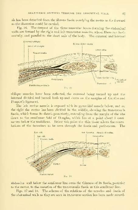
TKANSVEKSE SECTIOXS THROUGH THE ABDOMINAL WALL. 47
sis has been detached from the fibrous fascia overljiug the rectus as far forwardas the dissection could be carried.
Fig. 16. The deepest of the three muscular layers forming the abdominalwalls are formed by the right and left transverse muscles, whose fibers run hori-
zontally, and parallel to the short axis of the body. The external and internal
External oblique
; Internal obliqueRectus abdominalis
Transversalis
Fascia transversalis
Fio. 17.
oblique muscles have been reflected, the external being turned up and the
internal divided and turned botli up and down on the margins of the ribs andPoupart's ligament.
The left rectus nuiscle is exposed with its pyramidal muscle below, and on
the right the rectus has been divided in the middle, showing the transversalis
fascia, which forms its sheatli posteriorly, extending from the margin of the ribs
down to the semilunar fold of Douglas, which lies at a point about 3 centi-
meters below the umbilicus. Below this point the thin tissue allows the convo-
lutions of the intestines to Ije seen through the fascia and ])eritoneum. The
Int. obi.
: Transversalis
Ant. lamella. Sheath of rectus.
Linea alba
Fascia transv.
abdominal wall below the semilunar line owes the thiimess of its fascia, posterior
to the rectus, to tlie cessation of the transversalis fascia at this semilunar line.
Figs. 17 and 18. The scheme of the relations of the muscles and fascia of
the abdominal walls as they are seen in transverse section has been made accord-

48 TOPOGRAPHICAL ANATOMY.
ing to Branne. Fig. 17 shows the section of the walls above the semilunar folds
of Douglas, and Fig. 18 sliows the section below the folds of Douglas. Both
pictures exhibit the relations
of the oval recti to the trans-
verse and internal and exter-
nal oblique muscles.
In the section above the
folds of Douglas, Fig. 17, it
is important to notice the re-
lation of the aponeurosis, in-
dicated by the white spaces
between the muscles. Thedivision of the fascia of the
internal oblique muscle is
seen at the rectus, one lamella
passing in front to unite vnth
the fascia of the external ob-
lique, the other lamella pass-
ing posteriorly to join the
transversalis tendon, and so
to continue as a conjoined
tendon until it unites with
its fellow of the opposite
side.
In Fig. 18 quite a diiier-
ent arrangement of the mus-
cles is seen ; the external ob-
lique muscle remains about
the same, while the internal
oblique advances much closer
to the rectus, and the trans-
versalis, instead of passing
behind the rectus, as in Fig.
17, lies farther back toward
the lumbar region than either
of the others. In Fig. 18 the
fascia of the external and in-
ternal oblique muscles blend
at a point nearer the median
line, as shown also in Fig. IT.
The internal oblique also fails
to split, sending its entire tendinous aponeurosis in front of the rectus along
with that of the transversalis nuiscle.
Fig. 19 shows what I have for some years been in the habit of calling " the
celiotomy veins." In opening the abdomen in the linea alba in its lower third,

THE TOPOGRAPHICAL ANATOMY OF THE SMALL INTESTINES. 40
tliese veins are almost invariably found l}ang just over the peritoneum. Some-times there is but one large vein 1^ or 2 miUimeters in diameter, but usually
there are two of them from 1 to 1^ millimeter in diameter, separated by aninterval of 3 or 4 millimeters ; they follow a slightly winding, but in gen-
eral straight, course down to the symphysis pubis, over which they pass to
the neck of the bladder, where they empty into the large vesical plexus of veins.
I have not seen any arteries accompanying these veins. They are usually large
enough to give rise to some persistent oozing, if injured, and for this reason
should be carefully observed in every case, in order to cut between them, or to
one side when there is but one vein. In a series of twenty abdominal sections,
taken consecutively, the celiotomy vein was seen sixteen times ; in most cases it
ascended straight up the median line just beneath the peritoneum, a little to the
right or to the left ; six times it was seen bifurcating. In four cases the diameter
was at least 2 millimeters, in all the rest it was less than 2 millimeters.
In the subcutaneous fat the position of a transverse vessel is indicated on
both sides of the incision at a point about 2 centimeters (| of an inch) above the
symphysis pubis. This vessel is quite constant, and when divided spouts out a
little stream of arterial blood on one side and venous blood on the other ; some-
times there is arterial bleeding from both sides, showing a free anastomosis.
The umbilicus in the figure is shown displaced to the left.
THE TOPOGRAPHICAL ANATOMY OF THE SMALL INTESTINES.
Figs. 20 and 21 have been di-awn directly from the subject to demonstrate
important points in the topographical anatomy of the small intestines. This
subject has been carefully worked up by D. Sernoff {Inteimat. Monats.f. Aimt.
u. Phi/s., Bd. xi, 1894) and others, and elaborated with important additions by
Dr. F. P. Mall, whose demonstrations have been followed in preparing the
figures.
In order to expose the intestines in their normal positions, the abdominal
cavity has been opened by a crucial incision, and each of the four flaps reflected
outward. Letters have been placed upon the small intestines, associating themin groups. Each one of tliese groups is so attached to a series of lamellae
of the mesentery that by picking up one of the grouj)s of lamellae at its base
near the vertel)ral colunm the entire bunch of small intestines attached to it is
also lifted up.
In descril)ing the groups I begin at the duodenum. Fig. 20, and note the
lamellae under the left splenic flexure of the colon included in the letters A and
B ; from this group the mesentery crosses the vertebral column to the right side,
where it forms a series of folds under the right hepatic flexure of the colon ; this
group is included between the letters B and C ; crossing the vertebral column
once more to the left, the next group is found lying in the left iliac fossa,
included between the letters C and D ; the fourth and last group of lamellae,
between D and E, fills the lower abdomen and right iliac fossa, and it is par-
ticularly important to note the straight line of the terminal portion ascending
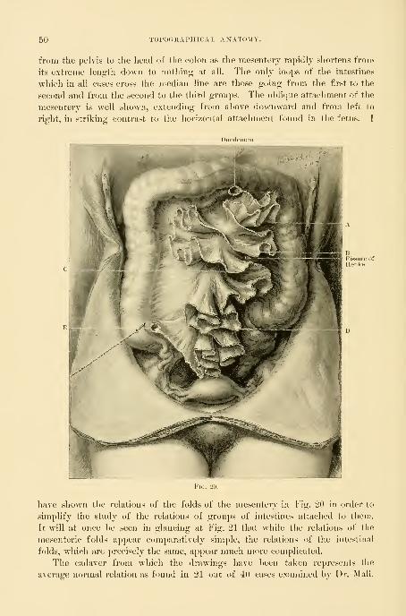
50 TOPOGRAPHICAL ANATOMY.
from the pelvis to the head of the colon as the mesentery rapidly shortens from
its extreme length down to nothing at all. The only loops of the intestines
which in all cases cross the median line are those going from the first to the
second and from the second to the third groups. The oblique attachment of the
mesentery is well shown, extending from above downward and from left to
right, in striking contrast to the horizontal attachment found in the fetus. I
have shown the relations of the folds of the mesentery in Fig. 20 in order to
simplify the study of the relations of groups of intestines attached to them.
It will at once be seen in glancing at Fig. 21 that while the relations of the
mesenteric folds appear comparatively simple, the relations of the intestinal
folds, which are precisely the same, appear much more complicated.
The cadaver from which the drawings have been taken represents the
average normal relation as found in 21 out of 40 cases examined by Dr. Mall.

GROUPS OF THE SMALL INTESTINES. 51
In order to facilitate tlie study of the groups of intestines, of which I have just
descrihed four, the same letters are used, marking the same divisions seen in the
preceding figure. The figures accompanying the letters in Fig. 21 indicate the
superficial direction of the bowel. Sernoff found that the exposed or periph-

52 TOPOGRAPHICAL AXATOMY.
eral part of the intestines constituted only about one sixth of the entire lengthof the canal from duodenum to cecum; the average length of tlie small inres-
¥in. 22.
tine, according to Sernoff, is 537 centimeters (214-8 inches). The distancefrom B, to B, and from B, to B3, etc, by no means represents a uniform space.

ANTERIOR ABDOMINAL WALL AND INTESTINES IN AN EMACIATED BODY. 53
Group B (Fig. 21) of the bowels lies under the left splenic flexure of the
colon, and, by passing the hand down to the mesentery at this point, can be
picked up en 7nasse. Group C, under the hepatic flexure of the colon, can
be picked up by carrying the hand down to the mesentery between the colon
and small intestines ; by throwing this group over to the left side, the whole of
the right renal region is exposed. Below B and above C there is a natural
fissure or separation between the bowels (fissure of Henke) which goes all the
way back to the psoas muscle.
The following variations in relation to these groups are often found
:
Variation 1 : Group C is displaced from its position under the right hepatic
flexure over into the left flank.
Variation 2 : Group B crosses the median line and occupies the position of
Group C under the hepatic flexure, while Group C goes to the left.
Variation 3 : Group C goes down to the left, and Groups B and D go across
and ascend on the riglit to occupy its place.
Fig. 22. Almost all anatomical drawings of the abdominal cavity fall into
the error of placing the anterior abdominal wall at too great a distance from the
lumbar vertebrae ; the separation between the two will vary according to the dis-
tention of the intestines, which float up and push the wall forward, and so
lift it 2, 3, 5, or more centimeters from the promontory of the sacrum ; as
the intestines contract again they retire to the upper part of the abdomen, to
the right and left flanks, and to the pelvis.
Fig. 23. In emaciated patients the anterior abdominal wall may not infre-
quently be found so closely applied over the lower part of the vertel)ral column
that its rounded form is plainly seen. In one of my abdominal operations, remov-
ing an extensively adherent parovarian cyst, the collapsed walls actually became

54 TOPOGRAPHICAL ANATOMY.
adherent to the vertebral cohimn, causing the patient great discomfort in her
convalescence. Figs. 22 and 23 show an extreme case in which the abdom-
inal walls were not more than 3 millimeters thick. Here the groups of in-

PELVIC VISCERA IX XORMAL POSITION. 55
testines are all displaced toward the periplierj, and the bowel crosses fromright to left opposite the second instead of opposite the fourth and fifth lumbarvertebrae. Groups B and C are crowded up under the vault of the thorax,
Group D lies in the left flank, while Group E has dropped into the pelvis,
which it fills. The abdominal wall rests directly upon the aorta and the venacava and the mesentery with its vessels. It is interesting to note the plastic flat-
tening and the ridges on the body of the uterus due to post-mortem compres-sion by the intestines, seen in both Figs. 22 and 23.
Fig. 2-1:. It is particularly important to the gynecologist to be familiar with
-c\>
.^^^.-^r
V
the anatomy of the terminal portion of the ileum, that part of the intestine
which is most liable to drop into the pelvis and to l)e involved in the various
inflammatory gynecological processes.

56 TOPOGRAPHICAL ANATOMY.
The fig;ure shows the cecum in the riglit iliac fossa with its longitudinal
fibers leading down to the vermiform appendix, which lies coiled above the
common iliac artery on the psoas muscle. The straight ascent of the ileum out
of the pelvis and over the sacro-iliac junction to its point of exit in the cecum
is especially noteworthy. This arrangement appears to be a mechanical neces-
sity due to the triangular form of the end of the mesentery which terminates at
the cecum in a point.
Fig. 25 shows tlie mutual relations of the pelvic viscera as seen upon
opening the abdomen through the superior strait. The drawing is after nature
exactly, and, although presenting some slight individual peculiarities, does not
deviate in any important particular from the average case.
The bladder in front is moderately distended, somewhat gibbous in form,
and fullest on the right side. The rectum passes down into the pelvis to the
right of the promontory of the sacrum, necessitating a slight left lateral dis-
placement of the uterus, which lies between the rectum and the bladder ; this
has the effect of lengthening the right and shortening the left round ligament,
and of causing their curves to differ. The tubes and ovaries on each side fill
up the triangular spaces left between the rectum and uterus and the pelvic
walls.
Fig. 26 shows the utero-sacral ligaments coursing from the cervix on the
right and left in a curved line around the rectum. Below and between the
ligaments in front and the rectum lies Douglas's cul-de-sac.
The next dissection (Fig. 27) exposes the great vascular trunks of the lower
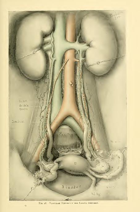
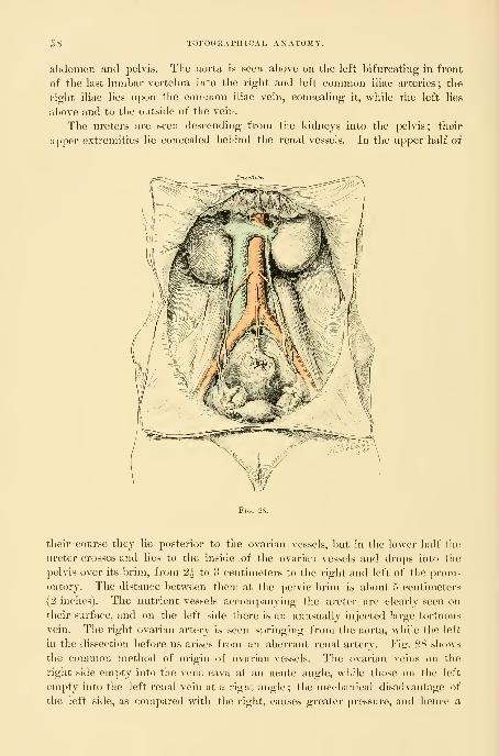
58 TOPOGRAPHICAL ANATOMY.
abdomen and pelvis. The aorta is seen above on the left bifurcating in front
of the last lumbar vertebra into the right and left common iliac arteries ; tlie
right iliac lies upon the connnon iliac vein, concealing it, while the left lies
above and to the outside of the vein.
The ureters are seen descending from the kidneys into the pelvis; their
upper extremities lie concealed behind the renal vessels. In the upper half of
their course they lie posterior to the ovarian vessels, but in the lower half the
lu'eter crosses and lies to the inside of the ovarian vessels and drops into the
pelvis over its brim, from 2^ to 3 centimeters to the right and left of the prom-
ontory. The distance between them at the pelvic brim is about 5 centimeters
(2 inches). The nutrient vessels accompanying the ureter are clearly seen on
their surface, and on the left side there is an unusually injected large tortuous
vein. The right ovarian artery is seen springing from the aorta, while the left
in the dissection before us arises from an aberrant renal artery. Fig. 28 shows
the connnon method of origin of ovarian vessels. The ovarian veins on the
right side empty into the vena cava at an acute angle, while those on the left
empty into the left renal vein at a right angle ; the mechanical disadvantage of
the left side, as compared with the right, causes greater pressure, and hence a
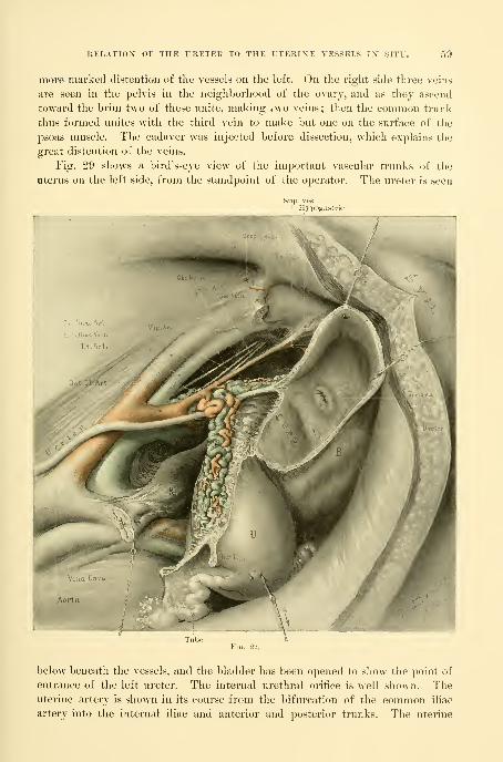
RELATIO>r OF THE URETER TO THE UTERINE VESSELS IX SITU. 59
more marked distention of the vessels on the left. On the right side three veins
are seen in the pelvis in the neighborhood of the ovarj, and as tliej ascend
toward tlie brim two of these unite, making two veins ; then the common trunk
thus formed unites with the third vein to make but one on the surface of the
psoas muscle. The cadaver was injected before dissection, which explains the
great distention of the veins.
Fig. 29 shows a bird's-eye view of the important vascular trunks of the
uterus on the left side, from the standpoint of the operator. The ureter is seen
below beneath the vessels, and the bladder has been opened to show the point of
entrance of the left ureter. The internal urethral orifice is well shown. Theuterine artery is shown in its course from the bifurcation of the common iliac
artery into the internal iliac and anterior and posterior trunks. The uterine

GO TOPOGHAI'rriCAL ANATOMY.
artery arises from the anterior trunk in common with the hypogastric artery.
Tlie origin of the vaginal artery is well shown. The ureter lies closer to the
cervix uteri on the left side.
Fig. 80 is taken from an injected pelvis of a fully developed nuiltipara, and
shows the entire vascular relations of tlie uterus, ovary, and Fallopi.iu tul>c, as
seen from the front. The anterior leaf of jxTitoneum has been removed, leav-
ing the vessels in situ, and held in place by the ])<)sterior leaf.

ARTERIAL BLOOD SUPPLY OP OVARY. 61
The relations of the uterine vessels to the ureter, the cervix, and the vaginal
vault should be carefully noted. The ureter lies below the uterine artery and
two of its veins, and above a large vaginal and uterine vein. The uterine artery
ascends beside the uterus from 1 to 2 or 3 millimeters away from it, tortuous
and interwoven with its veins. At the neck of the uterus, opposite the internal
OS, it gives off a large artery which penetrates the uterine body ; all the other
branches which go to the uterus are small. Up near the cornu uteri the termi-
nal l)ranch of the uterine artery anastomoses with a branch of the ovarian artery.
The ovarian artery enters the pelvis in the suspensory ligament of the ovary
(infundibulo-pelvic ligament) ; it divides just before it reaches the hilum of the
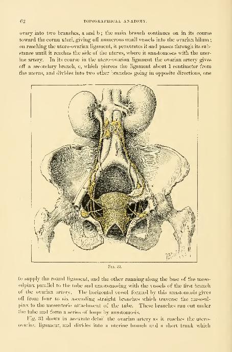
62 TOPOGRAPHICAL ANATOMY.
ovary into two branches, a and b ; the main branch continues on in its course
toward the cornu uteri, giving oQ numerous small vessels into the ovarian hilum;
on reaching the utero-ovarian ligament, it jjenetrates it and passes through its sub-
stance until it reaches the side of the uterus, where it anastomoses with the uter-
ine arterv. In its course in the utero-ovarian ligament the ovarian artery gives
off a secondary branch, c, which pierces the ligament about 1 centimeter from
the uterus, and divides into two other branches going in opposite directions, one
to supply the round ligament, and tlie other running along the base of the meso-
salpinx parallel to the tube and anastomosing with the vessels of the first branch
of the ovarian artery. The horizontal vessel formed by this anastomosis gives
off from four to six ascending straight branches which traverse the mesosal-
pinx to the mesenteric attachment of the tube. These brandies run out underthe tube and form a series of loops by anastomosis.
Fig. 81 shows in acicurate detail the ovarian artery as it reaches the utero-
ovarian ligament, and divides into a uterine branch and a short trunk which
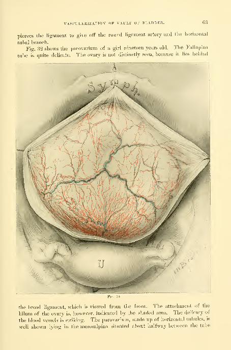
VASCULARIZATION OF VAULT OF BLADDER. 63
pierces the ligament to give off the round ligament artery and the horizontal
tubal branch.
Fig. 32 shows the parovarium of a girl nineteen years old. The Fallopian
tube is quite dehcate. The ovarv is not distinctly seen, because it lies behind
the broad ligament, which is viewed from the front. The attachment of the
hilum of the ovary is, however, indicated by the shaded area. The delicacy of
the blood vessels is striking. The parovarium, made up of horizontal tubules, is
well shown lying in the mesosalpinx situated about halfway between the tube

64 TOPOORAPHICAL ANATOMY.
and the ovary, running parallel to the tube and giving off about fifteen vertical
tubules converging toward the hilum of the ovary. Some of the outer tubules
are beautifully convoluted. The outer extremity of the horizontal branch ter-
FiG. 35.
minates in two so-called hydatids, hanging free l)y a little ]>edicle from the front
of the broad ligament across the tubo-ovarian fimbria.
Fig. 33. The lymphatic system of the pelvic organs. The uterus and its
appendages and the vagina are everywhere covered by a rich network of lym-
phatic vessels with whose anatomical arrangement we have become acquainted
through the observations of Mascagni and Poirier. This vascular network sur-
rounds the uterus and vagina like the finest lace. Upon leaving the uterus, the
smaller vessels collect into larger trunks, which then discharge into the various
neighboring glands. From the upper part of the vagina and lower cervix the
lymph vessels collect to enter the glands on the pelvic floor and accompany the
uterine and internal iliac vessels, to the next system of glands, in the bifurcation
of the common iliac arteries (a a'). From this point the lymph channel leads

VASCULARIZATION OF THE VESICAL MUCOSA. 65
over the artery to a gland often found on its upper side well above the bifurca-
tion, and so on up to the lumbar glands (b b'). The lymph vessels of the bodyof the uterus either pass out through the mesosalpinx near the ovarian attach-
ment, and on up the suspensory ligament of the ovary to the lumbar glands
(b b'), or take quite another direction and course down the round ligaments to
the deep inguinal glands' (c c'). The lowest part of the vagina and external
genitals are richly supplied with lymphatics, which communicate with the super-
ficial and deep inguinal glands, and through these with the glands lying uponthe external iliac arteries.
Fig. 3-i gives a good idea of the vascularization of the vault of the bladder
—that part of the bladder which is in relation to tlie peritoneum. The perito-
neum has been dissected off to sliovv the circulation. The veins are seen anasto-
mosing across from one side to the other, and terminating below in the urethro-
vesical plexus at the neck of the urethra. The superior vesical arteries are also
seen. It is important to note the unusual injection in the neighborhood of the
cervix uteri.
Fig. 35 shows the vasculai-ization of the vesical mucosa, and exhibits beauti-
fully the dendritic arrangement of the little branches of the superior, middle,
and inferior vesical vessels as they plunge through the coats of the bladder and
come to view on the mucous surface, l)ranching out into small vessels and capil-
laries. It will be seen that certain definite ai-eas of the bladder are constantly
vascularized by the same groups of vessels.
The first great group is at the trigonal area where the vessels branch out into
the bladder from the internal urethral orifice like a fan, appearing at the upper
edges of the papillae and coursing toward the
ureteral orifices ; they then continue parallel to
the ureteral folds, and so reach the side walls of
the bladder.
This group of vessels anastomoses with the
next, which is seen just below the edges of the
cut ; the second group is derived from the supe-
rior vesical vessels, and several vascular trees are
seen coming through to the surface of the mu-
cosa and distributing themselves over it in fine
branches. In the posterior part of the bladder
the middle vesical vessels occupy the area in the
neighborhood of tlie cervix uteri ; vascular trees
from this source on the right and on the left side
are quite constantly found, and form a character-
istic landmark in the examination of the living
subject through the speculum.
Fig. 37 shows the topography of the fixed part
of the bladder—that part which is attached to the symphysis pul)is, vagina, and
cervix uteri, as contrasted with the upper movable peritoneal portion. The
first striking feature is the hexagonal form, which is caused by the attachment
riG. .11). \ AScn.AIilZATIOX OF THEVesical Mucosa by the RightAND Left Superior, Middle,AND Inferior Vesical Arteries.
The superior vesical arteries (a)
are distributed over the superior andlateral regions of the bladder. Themiddle vesical arteries (b) are distiib-
uted over the posterior portion whichlies in relation to the uterus and up-per vagina. The inferior vesical ar-
teries (c) are distributed to the tri-
sronuin and the middle part of the

66 TOPOCxRAPHICAL ANATOMY.
of the bladder to the symphysis and its angular reflection out over the lat-
eral pubic rami, P>om the posterior point of attachment to the pul)ic rami
it is reflected again at an angle to its cervical attachuient, which it also meets
at an angle, completing the hexagon. The trigomim is well shown and the in-
ternal urethral oriflce lies a little in advance of the center, forming a transverse
fold with a sharp ridge above it, and with numerous flne radiating folds en-

BLOOD SUPPLY OF LOWER SIGMOID AXD RECTUM. 67
tering the opening from below. Tlie ureteral orifices are about 2^ centime-
ters apart, and the same distance from the urethra ; each orifice is situated on
a little mons ureteris. The inter-ureteric ligament is evident bj a slight eleva-
rete r
2..:=^ o hemorrl\,
IAHery
]]v.^-
tion. Posterior to the inter-ureteric ligament is that part of the base of the
bladder which lies in relation to the upper vagina.
Fig. 38 shows the blood supply of the lower sigmoid and rectum. In order
to expose its vessels, the rectum has been thrown over to the right, uncovering
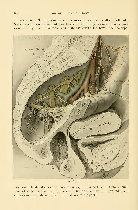
08 TOPOGRAPHICAL ANATOMY.
the left ureter. The inferior mesenteric artery is seen giving o£E the left colic
hranclies and then its sigmoid branches, and terminating in the superior hemor-
rhoidal artei-y. All these branches radiate out toward the bowel, and the siipe-
Sacral Gang"!
rior hemorrhoidal divides into two branches, one on each side of the rectum,
lying close to the bowel in the pelvis. The large superior hemorrhoidal vein
empties into the inferior mesenteric, and so into the portal.

SAGITTAL SECTION THKOUGH THE PELVIS. 69
Fi^. 39 is a sagittal section of the pelvis, showing the rectum drawn away
from the sacrum, in order to demonstrate the arteries, veins, and nerves of the
sacral and lateral pelvic regions. The distribution of the superior hemor-
rhoidal vessels is the same as that shown in Fig. 38. The sacral plexus of
nerves is seen to emerge from the sacral foramina, forming the lumbo-sacral
Deep epigastric
Obtur. nerveObtur. art. '
Femoral ring
Obtur. foramen
Lumb saer. cord 1 sacr
cord, and the iirst, second, third, fourth, and fifth sacral cords, which converge
toward the great sacro-sciatic foramen, to unite in the sciatic nerve. The sacral
ganglia of the sympathetic nerve are seen lying upon these nerves as they
emerge from the foramen. Observe tlie nerves going from the fourth sacral
cord to the lower part of the rectum and the coccygeus muscle.
Fig. 40 shows the muscles of the pelvis in sagittal section with arteries and
nerves, after removal of the viscera. The psoas muscle is seen overhanging
the brim of tlie pelvis and narrowing its superior strait ; upon the psoas lie the
common and external iliac arteries, and it is crossed by the internal iliac artery.
The obturator muscle covers the obturator foramen, and its fibers converge

YO TOPOORAPHICAL ANATOMY.
to its tendon, which passes out of the pelvis through the lesser sciatic notch.
At the lower margin of the ohturator muscle is the white line of fascia
which marks the upper border of the levator ani muscle. The levator ani seen
arising from the fibrous line will be described more particularly in connection
with Figs. 46 to 50. The coccygeus muscle borders the posterior margin of the
fienitocruralnerve
Ovarian vessils
Ext. iliac art.
Ext. iliac vein
levator ani, is fan-shaped, and is attached by its base to the side of the lower
sacrum and coccyx, and by its apex to the spine of the ischium. The pyri-
formis muscle pads the posterior part of the pelvis, rising in muscular bundles
from the front of the sacrum and gradually converging and passing out of the
j^elvis through the great sacro-sciatic foramen, posterior to the sciatic nerve.
The sacral plexus forming the sciatic nerve is seen as in Fig. 39. The obturator
nerve courses around the pelvic wall parallel to aiul below the brim of the
pelvis to the obturator foramen, where it leaves the pelvis. The vesical ]>rancli
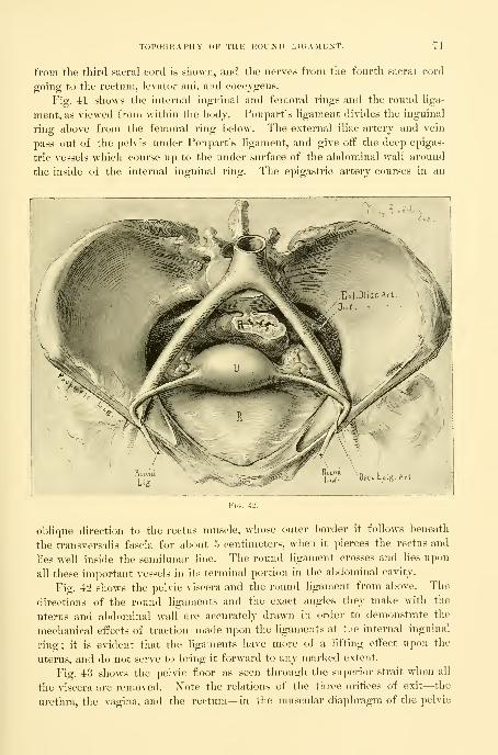
TOPOGRAPHY OF THE ROUND LIGAMENT. n
from the third sacral cord is shown, and tlie nerv-es from the fourth sacral cord
going to the rectum, levator ani, and coccjgeus.
Fig. 41 shows the internal inguinal and femoral rings and the round liga-
ment, as viewed from within the body. Poupart's ligament divides the inguinal
ring above from the femoral ring below. The external iliac artery and vein
pass out of the pelvis under Poupart's ligament, and give off the deep epigas-
tric vessels which course up to the under surface of the abdominal wall around
the inside of the internal inguinal ring. The epigastric artery courses in an
_1
oblique direction to the rectus muscle, whose outer border it follows beneath
the transvcrsalis fascia for about 5 centimeters, when it pierces the rectus and
Hes well inside the semilunar line. The round ligament crosses and lies upon
all these important vessels in its terminal portion in the abdominal cavity.
Fig. 42 show^s the pelvic viscera and the round ligament from above. The
directions of the round ligaments and the exact angles they make with the
uterus and abdominal wall are accurately drawn in order to demonstrate the
mechanical effects of traction made upon the ligaments at the internal inguinal
ring; it is evident that the ligaments have more of a lifting effect upon the
uterus, and do not serve to bring it forward to any marked extent.
Fig. 43 shows the pelvic floor as seen through the superior strait when all
the viscera are removed. ]S"ote the relations of the three orifices of exit—the
urethra, the vagina, and the rectum—in the muscular diaphragm of the pelvic

TOFOGRAPHK'AL ANATOMY.
floor, and the relation of these to their surrounding bony supports. The pelvis
is funnel-shaped and the orifices disposed in the anterior portion ; the urethra
appears as a small slit surrounded by thick walls just under the pubic arch.
Border of grt-at sciatit' fonuuen
Obturator nerve
The vagina has the characteristic shape of the letter II lying on its side, and
appears embraced by the muscular fibers of the levator ani, which hold the
lower part of the rectum forward. The puckered rectal opening is grasped in
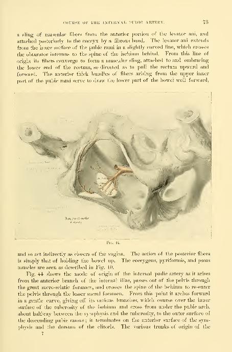
(•OUIISK OF THE INTERNAL PUDIC ARTERY. 73
a sling of niusfular libers from the anterior portion of tlie levator ani, and
attached posteriorly to the coccyx by a fibrous band. The levator ani extends
from the inner surface of the pubic rami in a slightly curved line, which crosses
the obturator internus to the spine of the ischium behind. From this line of
origin its fibers converge to form a muscular sling, attached to and embracing
the lower end of the rectum, so directed as to pull the rectum upward and
forward. The anterior thick bundles of fibers arising from the upper inner
part of the pubic rami serve to draw the lower part of the bowel well forward,
and so act indirectly as closers of the vagina. The action of tlie posterior fibers
is simply that of holding the bowel up. The coccygeus, pyriformis, and psoas
muscles are seen as described in Fig. 40.
Fig. 44 shows the mode of origin of the internal pudic artery as it arises
from the anterior branch of the internal iliac, passes out of the pelvis through
the great sacro-sciatic foramen, and crosses the spine of the ischium to re-enter
the pelvis through the lesser sacral foramen. From this point it arches forward
in a gentle curve, giving oif its various branches, which course over the inner
surface of the tuberosity of the ischium and cross from under the pubic arch,
about halfway between the symphysis and the tuberosity, to the outer surface of
the descending pubic ramus ; it terminates on the anterior surface of the sym-
physis and the dorsum of the clitoris. The various trunks of origin of the
7

74 TOPOGRAPHICAL AXATOMY.
inferior hemorrhoidal, superficial perineal artery, artery of the bulb, and corpus
cavernosum are all shown.
Fig. 45 shows the arterial vascularization of the floor of the pelvis as seen
from without. The various arterial branches drawn are the derivatives of the
internal pudics already indicated in their origin in Fig. 44.
Within the bony framework of the pelvic outlet, as formed laterally and pos-
teriorly by the great sacro-sciatic hgaments, are seen the three pelvic outlets—the
Infer, lieiiion-}'!. art.
urethral, vaginal, and anal—corresponding to the same outlets seen from within in
the complementary picture (Fig. 43). It is important again to notice the posi-
tion of the urethra high up under the pubic arch, with the vagina immediately
beneath it ; the anal orifice is at about the center of the figure, halfway between
the pubic arch and coccyx and the tuberosities of the ischium. A striking
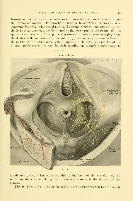
MUSCLES AND NERVES OF THE PELVIC FLOOR. 75
feature in the picture is the ischio-rectal fossae between each tul)ero8ity and
the levator-ani muscle. Posteriorly the inferior hemorrhoidal arteries are seen
emerging from the ischio-rectal fossae and curving forward, and branching over
the levator ani muscle, to be distributed to the lower part of the rectum and the
sphincter ani muscle. The superficial perineal arteries are seen emerging from
the depths of the ischio-rectal fossae anteriorly, and coursing forward in front of
the rectum over the transverse perineal muscles. The terminal branches of the
internal pudic artery are seen in their distribution, a small branch going to
Muse bulb rn\
m

76 TOPOG KA P 1 1 1CA L ANATO M Y
.
and rectal openings, together with the distribution of the terminal branches of
the nerves. Posterior to a line drawn between the anterior margins of the
tuberosities of the ischium are seen the following muscles. The transverse
perineal muscles take their origin beneath the tuberosities of the ischium and
cross the perineal body horizontally between the vaginal outlet and the anal
orifice, each one fusing with its fellow on the opposite side. A number of the
muscular bundles diverge from the horizontal fibers anteriorly and posteriorly
1 ,[ ro^ M
Cocci
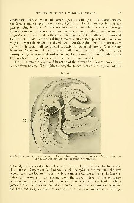
MECHANISM OF THE LEVATOR AKI MUSCLES. 77
coTitinuation of tlie levator ani posteriorly, is seen tilling out the space between
the levator and the great sacro-sciatic ligaments. In the anterior half of the
picture, lying in front of the transverse perineal muscles, are shown the con-
strictor vagina? made up of a few delicate nmscular fibers, embracing the
vaginal outlet. External to the constrictor vaginas lie the bulbo-cavernosus and
the erector clitoris muscles, arising from the pubic arch posteriorly, and con-
verging toward the dorsum of the clitoris. On the right side of the j^icture are
shown the internal pudic nerve and the inferior pudendal nerve. The various
branches of the intei-nal pudic nerve, similar in name and distribution to the
corresponding arteries as described in Fig. 45, are seen in their distribution to
the nniscles of the pelvic fioor, perineum, and vaginal outlet.
Fig. 47 shows the origin and insertion of the fibers of the levator ani muscle,
as seen from below. The sphincter ani, the lower part of the vagina, and the
Sph.ani.
Fig. 48.
—
Sagittal Section of Pp;lvis in Fk;. 47, Demonstrating in a Schematic Way the Actionp
OF THE Levator Ani and the Sphincter Ani Muscles.
extremity of the urethra have lieen cut off on a level with the attachments of
this muscle. Important landmarks are the symphysis, coccyx, and the left
tuberosity of the ischium. Just inside the tuber ischii the fibers of the internal
obturator muscle are seen arising from the inner surface of the obturator
foramen and the adjacent pubic ramus and converging to the tendon, which
passes out of the lesser sacro-sciatic foramen. The great sacro-sciatic ligament
has been cut away in order to expose the levator ani muscle in its entirety.
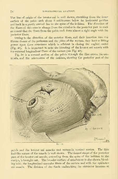
78 TOPOGRAPHICAL ANATOMY.
The line of origin of the levator ani is well shown, stretchinir from the inner
surface of the pubic arch about ?i millimeters below its horizontal portion
and back in a gently curved line to the spine of the ischium. The direction of
the fibers of this muscle change from the anterior to the posterior part to such
an extent that the fibers from the pubic arch form almost a right angle with the
posterior fibers.
Owing to the direction of the anterior fibers, and their insertion into the
fibrous tissues of the perineum and the ^sides of the rectum, they have a lifting
power upon these structures which is efticient in closing the vaginal outlet
(Fig. 48). It is important to note the blending of the levator ani muscle with
the external longitudinal fibers of the rectum (see Fig. 49).
Fig. 50 is a coronal section of the pelvis through the iliac crests, the ace-
tabula, and the tuberosities of the ischium, showing the posterior part of the
pelvis and the levator ani muscles and rectum in vertical section. The thin
leaf-like nature of the muscle is well shown. The funnel shape of the posterior
part of the levator ani muscle, extending from the spine of the ischium to the
coccyx, is brought out. The broader surface of attachment is also shown blend-
ing with the longitudinal nmscular fibers of the rectum and with the sphincter
ani muscle. The division of the fascia ensheathing the obturator internus at

CORONAL SECTION OF THE I'ELVIS. 79
the point called " the white line " is shown. The obturator internus appears in
section between the levator ani and the ischium, and in the depths of the ischio-
rectal fossa below the levator appear the pudic vessels and nerves lying close to
the tuberosity of the ischium. The coccygeus and pyriformis muscles appear
nerveInt. pudic art.
vein
j Isohio-rectal fossafilled with fat
as continuations of the levator, parallel to its upper fibers and clothing the
posterior pelvic walls on both sides of the sacrum. The sacral plexus is seen
overlying the pyriformis muscle on the left.

CTIAPTEK V.
THE GYNECOLOGICAI. EXAMINATION.
1. The gynecological examination. Inspection. Measurements. Photography. The normalabdomen. Abdomen of ovarian cyst. Abdomen of fibroid tumors of the uterus. Ascitic
abdomen. The use of specula in inspection. Percussion. Auscultation. Palpation.
Postures employed. Standing. Squatting or crouching. Sitting and bending forward.
The left lateral, or Sims's posture. The knee-chest. The dorsal. Examination of the
pelvic organs in the dorsal position. Simple examination with one hand in vagina or
rectum. The bimanual examination :—With the organs in situ. Invagination of the
pelvic floor. Examination of the uterus. Examination of the ovaries. Examination of
the Fallopian tubes. Bimanual examination by the rectum and abdominal walls. Bi-
manual examination by rectum and abdomen after atmospheric distention of the rectum.
Bimanual examination in the dorsal position with elevated pelvis; the same with the uterus
in artificial retroposition. The bimanual examination with the uterus drawn down to the
vaginal outlet. Examination of the anterior surface of the uterus through the rectum.
Examination in pelvic disease. Pelvimetry :—Four ways of measuring the true con-
jugate diameter. Anesthesia. Rules for use of anesthesia. Preparation of patient. Theexamination. Displacements affecting position. Descensus. Anteflexion. Retroposi-
tions. Ascensus uteri. Fixations and adhesions affecting mobility. Inflammation andtumors aiiecting size and form. Peculiarities of sensitiveness. Peculiarities of con-
sistency. Information derived from curettage of the uterus. Microscopic examinationof a piece of tissue excised. Microscopic and bacteriological examination of uterine,
vaginal, and other discharges. Examination of the rectum. Examination of the vermi-form appendix.
3. The general examination. Age. Heredity. Temperament. Habit. Color. Diseases to look
out for. Lung disease. Affections of the heart and arteries. Affections of the alimentarycanal. Diseases of the liver. Diseases of the kidney. Taking the history. Skeleton out-
lines of history and treatment. Diagrams of pelvic lesions. Minuter examinations of gyne-cological cases.
The recent progress in gynecology is chiefly due to the new and better
methods of examining patients, which constitute a fundamental difference be-
tween the gynecology of to-day and that of our immediate predecessors.
The gynecological examination includes both an investigation of any exist-
ing pelvic disorders and a careful inquiry into the patient's general condition.
The natural order of inquiry is first to take the history, then to examine the
pelvic organs, and finally to make the general examination.
GYNECOLOGICAL EXAMINATION.
The examination proceeds by making an orderly investigation of the pelvic
and abdominal organs by means of inspection, palpation, percussion, and auscul-
tation.
Inspection.—Inspection is Hmited to the surface of the abdomen, the external
genitals, and those parts of the rectum, vagina, and cervix which can be exposed
to view, either directly or by instrumental aid. In doubtful cases inspection
may even go so far as to make a direct examination of the uterus, ovaries, and
tubes through an exploratory incision in the abdominal wall.
80

iqaen edi ewoda
DESCRIPTION OF PLATE II.
Diagnosis of abdominal tumor by inspection ; tlie lower abdominal walls are splinted
and held immovable by the tnmor behind them. The hazy line above the umbilicus
shows the respiratory motion.

PLATE
Keiiotype Pnati-ag Co., BoBtoii


PHOTOGRAPHY. 81
The general condition of the body—whether fat, well nourished, or emaci-
ated— is naturally the tirst point to attract attention. Inspection also notes
peculiarities of color affecting the skin and the mucous membranes. The
greenish-yeilow hue of the chlorotic woman will often at once explain an
amenorrhea ; the cachexia of a cancerous patient is characteristic and easy to
remember when once seen ; the ovarian facies bespeaks malnutrition, and the
pallor of hemorrhage in myomata or extra-uterine pregnancy is a diagnostic
factor of the highest importance. The septic patient has a peculiar sallow,
anemic appearance. Inspection also notes the face indicative of hysteria. The
careful slow gait of the patient wnth pelvic peritonitis and any peculiarities in
the way of protecting tender parts from touch or shock by pressure with the
hands are also to be noted.
Inspection of the abdomen is of the greatest value when the eye is trained to
know its various contours in health. Variations between the normal and abnor-
mal contour of the abdomen produced by the growth of tumors, or by ascitic
effusions, or by gas, can be readily seen. In abdominal tumors the inspection
is limited to outlines, and is only one diagnostic measure, which, in association
with other aids, enables us to arrive at a correct estimate of the character of the
disease beneath.
While a simple inspection is sufficient for the innnediate purposes of the
diagnosis, careful m e a s ur e m e n t s of abdominal enlargementsshould always be made and recorded. By means of measure-
ments at different times changes in the size of a distended al)domen are made
evident and imperceptible differences of a few centimetres can be accurately
determined. Besides, we also do away with such vague terms as " a small tu-
mor," or " a large " or " enormous one,"' eliminating the large personal equation
lurking in these statements.
The following are the usual measurements made :
Circumference of the abdomen at the umbilicus.
Circumference halfway below the umbilicus.
Elevation of highest point of abdominal wall above the plane of the anterior-
superior spines.
Distance from sternal notch to symphysis pubis.
Distance from umbilicus to pubis.
Distance from umbilicus to right and left anterior-superior spines respec-
tively.
By such measurements the degree and form of abdominal enlargements
are determined, whether more in the lower or upper abdomen, or in one flank,
and whether symmetrical or asymmetrical.
Photography is a valuable adjunct to descriptive records; the photo-
graph gives an instantaneous idea of form, often better than an elaborate
description. The photograph with the patient lying on the table can be taken
with advantage from three points of view : A protile from the side, showing
the general enlargement of the abdomen : a profile from below, showing sym-
metry or asymmetry ; while a quartering view halfway between these two posi-
8

82 THE GYNECOLOGICAL EXAMINATION".
tions and looking rather down on to tlie abdomen gives a general view of the
relations of the parts of the tumor to the abdominal landmarks. When possi-
cTioN Tiiijoiiiii Adilt Body, .showing tii?: Normal PositionTHE Utekus, Blauuek, Hectum, anu Abdominal Walls.
Kklatioss of
TIk! intestines arc not shown, and the dotted line represents the outline of the pelvic bones. It is impor-tant to note the proximity of the anterior ahdoniinal wall to the saeral promontory.
ble, the nmbilicns should be included in the picture, as the most im])ortant
landmark. A profile view, with the patient erect, shows the anterior displace-
ment of a large tumor.

PHOTOGRAPHY. 83
A beautiful graphic record demonstrating the presence of a tumor within
the abdomen is furnished by the profile photograph shown in Plate 11. The
diagnostic sign rests upon the hazy contour of the upper half of the abdomen,
beginning at the umbilicus ; at first sight, the indistinct line looks like a fault
Fig. 52.—Enormous Ovarian Cysto.ma, with Globular PENDCLors Abdomen, and the Ciiaractekiptic
Emaciation.
in the picture, but this is due to the fact that it registers the natural movements
of the abdominal wall during expiration and inspiration which are cut short be-
low by the tumor splinting the lower abdomen so that it can not move, as it
would do if no tumor were present.
Marked departures from the normal may occur within the limits of health,
of which the most frequent are distention from tympany or the

84 Tin-: (iYKEC'OLOGICAL EXAMIXATIOX.
accumulation of fat in the omentum and al)dominaI walls. Tympanyproduces a symmetrical form, by the uniform exj^ansion of the intestines in
all directions, the greatest prominence being around the umbilicus. The gen-
eral appearance of such an enlargement may not differ at all from that of an
encysted tumor. In a fatty abdomen, if the fat is in the walls, it is often
lRACTERISTIC of
characterized by the presence of creases from side to side ; if it is inside the
cavity, on the omentum and mesentery, in the nullipara, the rotundity is simply
increased ; but in the muciparous woman the walls appear flabby and the abdo-
men flat and distended in the flanks. These changes occur commonly after the
menopause. If an abdominal tumor is present under these circumstances, it
often becomes a ditflcult task to make a diagnosis, and the physician may easily
be misled into concluding that there is no tumor within.
Fig. 51. The importance of knowing the normal abdomen and its
variations within tlie limits of health becomes evident as we study the changes
in form brought about by tumors in the peritoneal cavity. Such a pathological
enlargement is either uniform over the whole abdomen or localized in some
special area. The enlargement itself may present a uniformly convex surface,

FORM OF LAK(iK OVARIAN CYSTS. 8,5
or it may he marked by bosses and grooves. A uniform itierease in tlie size of
the whole abdomen is only produced by tumors of the largest size and by
ascites. Such a case is shown in the figure of Mrs. T)., who had an ovarian cyst
weighing 100 pounds, which I removed in Philadelphia, in May, 1887. (Fig. 52.)
A symmetrical convex surface over an abdominal tumor indicates a corre-
sponding symmetry of surface of the tumor within. The contrast afforded in
this way with a bossed surface serves to distinguish certain groups of tumors.
In gastric, splenic, or hepatic tumors of the upper abdomen the swelling is
more above, while in pelvic tumors the enlargement is mostly below.
The pregnant uterus may be taken as the type of symmetrical lower al)-
dominal and pelvic tumors ; hei'e the chief distention is below the umbilicus,
and in the first pregnancy up to the eighth month the prominence in the
median line is like that of an ovarian cyst or a myomatous uterus of the same
size.
Fig. 53. The form characteristic of large ovarian cysts is
an ovoid distention of a part or of the whole al)domen, with more or less smooth
outlines. Such tumors at first involve the lower or infra-umbilical part of the
abdomen greatly in excess of the upper part, and if the tumor is of enormous

86 THE GYXECOLOGICAL EXAMIXATIOX.
an ovarian tumor made up of a number of loeuli of about the same size, or
when adhesions prevent the tumor from rotating and accommodating itself.
Figs. 55 and 56. Two foi'ms of enlarged ahdomen are characteristic of
fibroid ttimors of the uterus : one in which the distention is spherical, looking
as if the cavity contained a cannon ball, while the drop from the top of the
tumor to the normal level of the abdominal wall, as the patient lies on her back,
is often almost vertical. This is rarely seen in ovarian tumors. The other
form has an appearance of irregular nodular masses distributed in tlie lower
abdomen, Fig. 57.
Figs. 58, 59, and 60. Enlargement of the abdomen frequently arises from
ascitic accumulations, which tend to take the form of a flattened
Fig. 55.
—
Form of Abdomen Characteristic of a Large Globul.vr Myomatous Uterus
Note particularly tlie abrupt lines of elevation, especially from epigastrium to umbilicnis.
ovoid, the regions of greatest prominence being in the lianks, whither the liuid
gravitates. While the flattening is an important differential point between an
ascitic accumulation and circumscribed encysted fluids, yet occasionally in a
nullipara ascites may present the domelike prominence of a cyst, and the differ-
ence can only be detected after palpation and ])ercussion in various positions.
See Figs. 61 and 62.
Inspection is the essential factor in the diagnosis of diseases of the external
genitalia, vagina, and vaginal cervix. The vagina and cervix can be exposed to
view by the aid of instruments. Relaxed outlet, rui)ture of the outlet, prolap-
sus, and affections of Bartholin's glands, such as c}sts and abscesses, etc., are
diagnosed at once by simple inspection.
Insj^ection of the vagina and vaginal cervix is effected

PERCUSSION". sr
by means of specula. The best are Fergusson's tubular, Sinis's duck-
bill, GoodelPs bivalve, and Xelson's trivalve speculum, and Kelly's small cylin-
drical specula. The valvular
specula are introduced to their
full extent closed, and then
opened, when the cervix is
brought plainly into view.
Upon withdrawing the sjjecu-
lum the vaginal walls are ex-
amined as they slowly roll over
the end. Ordinary specula,
however, must not be used in
examining unmarried women,
for they destroy the hymen and
produce a dilatation of the vag-
inal outlet. I have often seen
a distention from specular ex-
aminations great enough to ad-
mit four lingers. Small cylin-
drical specula, 9 centimetres (3^
inches) long and 8, lU, 12, 14,
and 16 millimetres in diam-
eter, must be used in examining
and treating the vagina and
cervix in unmarried women and
girls. The patient is put in
the knee-breast position, and
the speculum with an obturator
introduced without injuring the
hymen ; as soon as the obtura-
tor is withdrawn the vagina fills
with air, and eveiy part of it,
with the cervix, is plainly ex-
posed to view by a light reflected
from a head mirror.
Percussion.— Percussion is a
valuable adjunct to ins^^ection
and palpation in the differential
diagnosis of abdominal tumors.
There are in general three kinds
of percussion notes—flat, tym-
panitic, and dull. The flat note,
drawn from the most prominent
part of an ovarian or uterine
tumor, is in striking contrast to the high-pitched tympany of the intestines sur
Fig. 56.
—
Abdomen Distended by a Large Cystic Myoma,Abdominal Wall simply pushed out without sagging.
Note dilated superficial epigastric vein and edematous legs.
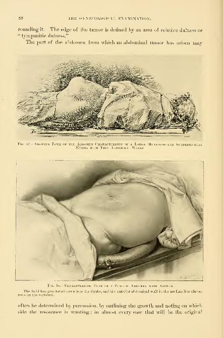
THE GYNECOLOGICAL EXAMINATION,
rounding it. The edge of the tumor is defined hj an area of relative duhiess or" tvnipanitic dulness."
The part of the a])d<)men from which an a])dominal tumor has arisen may
Fig. 57.
—
Showing Foum of tiik Ardomex Characteristic of a Large Mcltinodular SibperitonealMyoma with Thin Ai'.i>ominai, Walls.
Fig. 58.
—
Chakacteristic Form of a Flaccid Abdomen with A.scitks.
The fluid has gravitated down into the flanks, and tlii' anterior abdominal wall in tlie ineditui line almostrests on the vertebra>.
often be determined hy percussion, by outlining the growth and noting on which
side the resonance is wanting; in almost every case that will be the original

PERCUSSIOIS'. 80
habitat of the tumor from wliich it has developed out toward the middle of theabdominal cavity, the direction of least resistance.
The outlines of most pelvic tumors are more or less erescentic and surroundedby an area of resonance called the corona, c o r o n a o v a r i a n a , or c o r o n au t e r i n a
.
Percussion is of the greatest service in differentiating cystic and solid tumorsfrom tympany and ascites. The tympanitic abdomen is resonant all over ; theascitic al domen yields a dull note in the flanks and tympany above, from the
Characteristic form of section tlirougli normal abdomen (Fig. 59") compared witli ascitic abdomen (Fig. 60").
Section made through umbilicus and fourth lumbar vertebra."
gravitation of the fluid and the floating up of the intestines. When the accunm-lation is extreme, however, the distention of the abdomen may be so great as to
lift the walls farther from the back than the mesentery can reach ; in such cases
the intestines are everywhere covered with fluid and percussion yields a flat or
dull tympanitic note in all directions. Moderate accumulations may be made to
gravitate from one side to the other, or into the lower or upper abdomen, bychanging the position of the patient; and tlie dull and tympanitic areas will
change with each alteration of posture.
The most important use of percussion in diagnosis is the recognition of an
area of tympany overlying retroperitoneal tumors, usually renal, which lift the
colon forwai-d as they advanc-e towai-d the anterior abdominal wall ; in this

90 THE GYNECOLOGICAL EXAMINATION.
way tlie error of mistaking an enlarged kidney for an ovarian tumor is always
to be avoided.
Auscultation.—Auscultation is lindted to tlie surface of the abdomen and is
eliieily valuable in discriminating abdominal tumors from pregnancy, where the
sound of the fetal heart-beats is the distinctive sign. In fibroid tumors a loud
bruit is often heard, caused by the free circulation of the blood in the great vascu-
lar channels ; this must not be mistaken for the bruit of pregnancy. The only
way to make the distinction clear is by discovering other signs of pregnancy.
Palpation.—After inspection and percussion we proceed to examine by touch.
No other diagnostic procedure is equally satisfactory and so certain in its results
as the sense of touch applied to the various organs through the abdominal.
%^^
Fig. 01.
—
Cylindrical Flattened Abdomen CiiABACTEPasTic of Ascites.
vaginal, or rectal walls. It is not unusual for the l)eginner to feel discouraged
with the results of palpation, which at first are crude and indefinite, but by per-
sistent practice the tactile sense becomes acute, and the consistence as well as the
minuter outlines and relations of the various organs in health and disease are
easily appreciated.
By palpation we outline structures normal and abnormal, and determine the
relative position, consistence, mobility, and sensitiveness of the parts under in-
vestigation. In this way cystic tumors are at once differentiated from hard
ones, and masses are easily detected in the lower abdomen, where they are hidden
behind the symphysis, or in the flanks. Again, the degree of relaxation of the
outlet and the condition of the vaginal walls and of the cervix are at once
determined by digital palpation. Palpation is greatly facilitated by the use of
certain postures, which are so important that I shall describe them in detail.
The various useful postures are the standing, squatting,sitting, and bending forward, the left lateral or Sinis's,
the k n e e - b r e a s t , and the dorsal or lithotomy.

POSTUKES. 91
Standing Posture .—In this postnre the patient stands with one foot
on the floor and the other resting on a stool six or eight inches high, while the
physician stoops before her and, resting the elbow of his examining hand on his
knee, proceeds to make a digital examination of the vaginal outlet, the vagina,
Fio. 62.
—
Ovarian Tcmob with Ascites.
The upper picture shows the form of abdomen us seen from below; the lower picturethe side. Note protrusion at the umbilicus. M. E., op., Dec. 23, 1895.
the form seen from
and the other pelvic organs. The hand can be placed at rest and the armlengthened or shortened at will by supporting the leg on the ball of the foot,

02 THE GYNECOLOGICAL EXAMINATION.
keeping the heel off the floor. This gives a springy support and takes away
the natural tendency to stiffen the arm in pushing the hand high up into the
vagina. Relaxation of the vaginal outlet and descensus uteri are most easily
recognised in this way. While standing, also, if tliere is a movable kidney, it
drops forward and is readily grasped between the hands.
Squatting or Crouching Posture.—The patient takes the same
posture as in defecation, and by a slight straining effort is able to demonstrate
to the examiner behind her the least tendency to prolapse and eversion of the
vaginal walls. The full effect of a relaxed outlet may he brought out in this
way better than by any other means.
Sitting and Bending Forward.—The patient leans forward, rest-
ing the weight of her body on the shoulder of a nurse, and so thoronghly re-
laxes the abdominal muscles ; the examiner then sits before her or at her side,
and makes counter-pressure with one hand over the back while with the other
he palpates deeply through the lax abdominal walls.
Fig. 63. The Left Lateral or Sims's Posture .—Li this position
the patient lies on her left side with her left arm behind her back and both
legs flexed upon the abdomen, the right drawn up aljove the left, and the pelvis
Fig. 63.—Sims's Posti-re.
Sliowin},' the position of the Iciis and chest, and espeeially tlie inidinatioii oftlie pelvis, as seen in outline l)el(nv.
tilted decidedly over toward the table, so as to facilitate the gravitation of its
contents through the superior strait in the direction of the anterior abdominal
wall ; this causes the vagina to balloon out with air as soon as the posterior
wall is retracted. The distention will not take place, however, unless the pelvis
is sufficiently tilted, so that if the patient persists in lying with her right hip
vertically above the left, the difficulty nuist be overcome by requesting her to
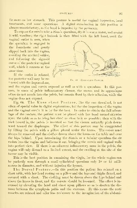
POSTURES. 93
lie more on her stomach. This posture is useful for vaginal inspection, local
treatments, and some operations. A digital examination in this position is
always unsatisfactory, as the hand is impeded by the perineum.
To expose the cervix take a Sims's speculum, dip it in warm water, and anoint
it with vaseline ; the right buttock is then lifted with the left hand, until thevaginal outlet is seen, whenthe speculum is engaged in
the fourchette and gently
slipped back into the vagina,
avoiding the urethral orifice,
and following the sigmoid
curve of the posterior vaginal
wall, which it retracts at the
same time.
If the outlet is relaxed,
the posterior wall may be re-
tracted with the fingers alone,
and the vagina and cervix exposed as well as with a speculum. In this pos-
ture, in cases of pelvic inflammatory disease, the uterus and its appendages
often do not recede into the pelvis, but remain fixed by their adhesion, while the
vagina does not expand.
Fig. 6-i. The K n e e - c h e s t Posture, like the one described, is not
often of special value in digital explorations, but for the inspection of the vagina
and the vaginal cervix it is by far the best. In order to obtain the full advan-
tage of the posture, the ])atient must be placed with her head turned sidewise
upon the table so as to bring her chest as close to it as possible ; then with the
back bowed in, the pelvis is inverted so that the viscera naturally pitch down-
ward toward the diaphragm. The effect of this posture may be exaggerated
by lifting the pelvis with a pillow placed under the knees. The corset must
always be removed and the clothes drawn above the knees on the table and over
the hips behind. Upon introducing the Sims's or a tubular speculum the air
rushes into the vagina and balloons it out, bringing the cervix and vaginal walls
into perfect view. If there is an adherent inflammatory mass in the pelvis, the
vagina will only distend to a limited extent, and the swelling at the site of the
tumor may be visible.
This is the best position in examining the virgin, for the whole vagina can
be perfectly seen through a small cylindrical speculum only 10 oi- 12 milli-
metres in diameter, and without injuring the hymen.
The Dorsal Posture .—In this position the patient lies relaxed on a
short table, with her head resting on a pillow and the legs and thighs flexed, and
covered with a sheet. The clothing must be drawn above the hips behind and
above the knees in front, and the corsets should be loosened. The eifect is in-
creased by elevating the head and chest upon pillows so as to shorten the dis-
tance between the symphysis pubis and the sternum. By this means the recti
muscles are relaxed and offer less resistance to the invagination of the abdomi-
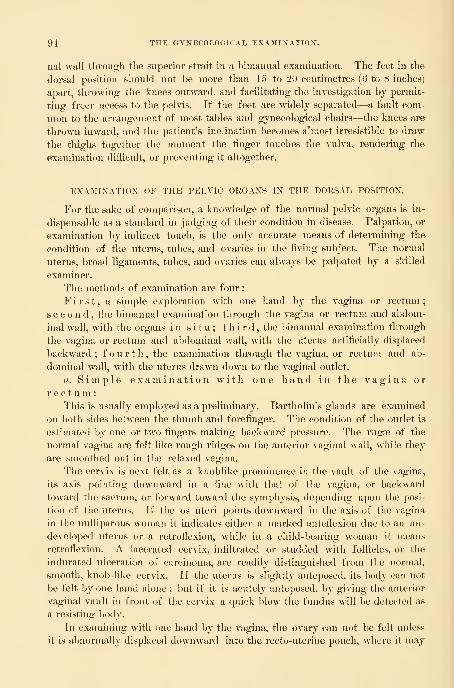
94 THE GYNECOLOGICAL EXAMINATION".
nal wall through the superior strait in a bimanual examination. The feet in the
dorsal position should not be more than 15 to 20 centimetres (0 to 8 inches)
apart, throwing the knees outward, and facilitating the investigation by permit-
ting freer access to the pelvis. If the feet are widely separated—a fault com-
mon to the arrangement of most tables and gynecological chairs—the knees are
thrown inward, and the patient's inclination becomes almost irresistible to draw
the thighs together the moment the finger touches the vulva, rendering the
examination difiicult, or preventing it altogether.
EXAMINATION OP THE PELVIC ORGANS IN THE DORSAL POSITION.
For the sake of comparison, a knowledge of the normal pelvic organs is in-
dispensal)le as a standard in judging of their condition in disease. Palpation, or
examination by indirect touch, is the only accurate means of determining the
condition of the uterus, tubes, and ovaries in the living subject. The normal
uterus, broad ligaments, tubes, and ovaries can always be palpated by a skilled
examiner.
The methods of examination are four
:
First, a simple exploration with one hand by the vagina or rectum
;
second, the bimanual examination through the vagina or rectum and abdom-
inal wall, with the organs in situ; third, the 1 )imanual examination through
the vagina or rectum and abdominal wall, with the uterus artificially displaced
backward ; fourth, the examination through the vagina, or rectum and ab-
dominal wall, with the uterus drawn down to the vaginal outlet.
a. Simple examination with one hand in the vagina or
rectum:This is usually employed as a preliminary. Bartholin's glands are examined
on both sides between the thumb and forefinger. The condition of the outlet is
estimated by one or two fingers making backward pressure. The rugae of the
normal vagina are felt like rough ridges on the anterior vaginal wall, while they
are smoothed out in the relaxed vagina.
The cervix is next felt as a knoblike prominence in the vault of the vagina,
its axis pointing downward in a line with that of the vagina, or backward
toward the sacrum, or forward toward the symphysis, depending upon the posi-
tion of the uterus. If the os uteri points downward in the axis of the vagina
in the nulliparous woman it indicates either a marked antefiexion due to an un-
developed uterus or a retroflexion, while in a child-bearing woman it means
retroflexion. A lacerated cervix, infiltrated or studded with follicles, or the
indurated ulceration of carcinoma, are readily distinguished from the normal,
smooth, knob-like cervix. If the uterus is slightly anteposed, its body can not
be felt by one hand alone ; but if it is acutely anteposed, by giving the anterior
vaginal vault in front of the cervix a <|uick l)low the fundus will be detected as
a resisting l)ody.
In examining with one hand by the vagina, the ovary can not be felt unless
it is abnormally displaced downward into the recto-uterine pouch, where it may

EXAMIXATIOX OF THE PELVIC ORGAKS IN THE DORSAL POSITION. 95
be discovered by pressure beliind the cervix uteri a little to the right or left. It
feels like a rounded, somewhat elastic body, slipping up and away under the
pressure. Any attempt with one hand to feel the ovary not displaced fails, or
gives at the utmost but an uncertain idea of its presence ; because as soon as it is
touched it yields to the pressure, and is displaced upward and out of reach. Anexamination of the deeper pelvic structures with one hand is therefore incomplete.
5. The bimanual method of examination is conductedeither with the organs in situ or with the uterus in artifi-
cial displacement.The bimanual examination with the organs in situ dej^ends for its success
upon the invagination of the abdominal wall just above the symphysis pubis,
Fig. 65.
—
Bimanual Examination of the Pelvic Viscera.
Same as before, but with the third and fourth fingers flexed upon tlie pahn and the pelvic floor invagi-nated, adding an incli or more to tlie lengtli of the fingers. Left view.
through the superior strait, with one hand, wliile with the other hand the examina-
tion is made through the inferior strait. The index finger, or both index and
middle fingers, if the vagina is sufficiently lax, is introduced as far as
the cervix. The palmar surface of the last joint of the finger must always be
used in palpating ; it is a beginner's error to use the radial side of the finger.

<x^ THE GYNECOLOGICAL EXAMINATIOX.
There are two ways of holding the rest of the hand which is outside during the
examination, either with the lingers strongly flexed in the palm, Fig. 65, or
with the thumb and fingers widely separated, the thumb resting upon the sym-
physis and the unemployed fingers on the perineum, Fig. 6(1. The first posi-
FiG. (j6.
—
Bimanual Examination of the Pelvic Visce
The upper, abdominal hand, pushes the abdominal walls in behind the uterus, wliile tlio linvcr, vairinal
hand, catches the cervix. Note the position of the third and fourth fingers extended in tlif i,dute:il cleft.
Ri^ht view.
tion is best when tlie examination can 1)e conducted without bending the flexed
fingers beyond a right angle with, tlie examining finger ; otherwise, the second
method is preferable.
Simultaneously with tlie introduction of the finger into the vagina slight
pressure is made over the middle of the superior strait, wnth the tips of the fin-
gers of the other hand resting upon the abdomen above the symphysis.
In most cases only slight pressure is re(|uired throughout to make a complete
examination of the pelvic organs ; in other cases it is necessary to overcome re-
sistance by making a gradually increased pressure downward until the structures
are felt. As a rule, the outlines of the pelvic organs are not minutely examined
by the abdominal hand, which serves more as a plane of resistance to prevent
the upward displacement and gliding away of uterus and ovaries when touched

EXAMIXATIOX OF THE PELVIC ORGANS IX THF: DORSAL POSITIOX. 97
bj the finger within the vagina. When the abdominal walls are thin and lax
the outer hand may also be employed in studying the outlines of the organs.
Fig. 67. Invagination of the Pelvic Floor .—In spite of the
assistance given by the external hand, the bimanual examination would often
prove unsatisfactory if the vaginal hand were limited by the length of the index
and middle lingers. Invagination of the pelvic floor is therefore a necessary
¥iG. 67.—Bimanual Examixation, showixg the Dekp Invagination- of the Pklvk' Fi.ixir, AddingSeveral Centimeters to the Length of the Fingers.
The left forearm and hand are placed at rest by making the pressure from the hip in tlie direction ofthe arrow.
aid, as by this means the examining finger is practically lengthened from 4 to B
centimetres (1^ to 2| inches). This is accomplished by pressing the perineumup into the pelvis in the axis of the inferior strait. The pubic arch and the

98 THE GYNFX'OLOGICAL EXAMINATION.
tuberosities of the ischium are obstacles to invagination, but a skilful examiner
may ovei'come them by cramping the fingers a little more closely together or by
making pressure farther back. Another difficulty in the way of securing the
fullest advantages from this method of examining is an involuntary stiffening of
the wrist and finger muscles. This may be overcome by pushing from the
elbow, while the hand remains perfectly flexible, in order not to interfere
with the delicacy of its tactile sense. Where the resistance is unusually great
or the act proves tiresome, the examiner will help himself materially by sup-
porting the elbow on his pelvis and pushing from his hip, relieving the arm
entirely.
The examination of the uterus is begun by the vaginal hand
giving the cervix a slight blow, which sends it upward at the moment the ab-
dominal hand bears down upon the same spot. Several such movements rapidly
repeated in front of and behind the cervix at once decide whether the body of
the womb lies in anteposition or retroposition. When the fundus lies in advance
of the cervix, by sliding the vaginal finger forward and bringing the abdominal
hand a little closer to the symphysis and pressing downward, a plane of resist-
ance is furnished upon which the vaginal finger rolls and palpates the whole
organ, while the hand above also appreciates every movement given, and so by
their combined action a judgment is almost instinctively formed.
Examination of the Ovaries .—By carrying the vaginal finger far
up into the lateral fornix posterior to the cervix, and then pushing out toward
the lateral wall of the pelvis, while deep pressure is made with the abdominal
hand in the same direction through the corresponding semilunar line, the ovary
can usually be caught and palpated. It is not sufficient simply to touch the
ovary, but it must be caught repeatedly and allowed to slip between the fingers
in various directions until it has been thoroughly examined on both surfaces and
its free border. The ovary feels like a firm body about as big as the end of the
thumb, with a rounded border and convex surfaces, slightly irregular. It is
freely movable in all directions.
Examination of the Fallopian Tubes.—These structures in
their normal condition are not often easily felt with certainty through the va-
gina. If they are thickened by disease, the uterine end may be rolled between
the fingers like a stout cord and traced outward toward the pelvic walls.
Bimanual Examination by the Rectum and AbdominalWalls .—A retroflexed fundus is felt and outlined with marvellous distinct-
ness when held down upon the rectal finger by the abdominal hand pressing in
through the superior strait. The crucial point in this examination is the recog-
nition of the angle between the cervix and fundus, associated with the absence
of the fundus in front. The ovaries are felt by making combined pressure in
the same direction as in the examination through the vagina. If the ovary is
not at once found, the surest guide is the utero-ovarian ligament, recognised as
a prominent fold on the posterior surface of the broad ligament just below the
cornu uteri ; by following this out with the finger for 2 to 2^ centimetres (| to
1 inch) the inner border of the ovary is felt.

:XAMlXATIO]Sr OF THE PELVIC ORGANS IN THE DORSAL POSITION. 99
Bimanual Examination b y the Rectum an d A 1) d o m e n
after Atmospheric Distention of the Rectum.—^When the or-
dinary recto-abdominal bimanual examination is impeded by coils of small intes-
tines filling the posterior pelvis and interfering with the fingers in their efforts
to search out and palpate the ovaries and tubes, this difficulty may be removed
by the following expedient : The rectum and bladder are first evacuated and the
patient is put in the knee-chest posture and a speculum introduced into the
rectum. This lets in a large amount of air, and the bowel balloons out and ap-
plies itself broadly over the sacral hollow and the posterior surfaces of the
uterus and left broad ligament, and at the same time the small intestines fall
away into the upper abdomen after a minute or two. The patient must then
be turned on to her back, taking care to keej) the pelvis constantly higher than
the rest of the abdomen, so as not to let the intestines gravitate again into the
pelvic cavity.
On making the bimanual examination the pelvic viscera are felt with start-
ling distinctness, the rectal finger enters a large air cavity no longer impeded by
Fig. 68.—Pa Li>AT IN i Np;rve by the Kf.ctim.
the mucous folds, the opening from the ampulla into the upper bowel is readily
found, and the posterior surface of the uterus and the ovaries and tubes feels as
if skeletonized in the pelvis. They lie so clearly exposed to touch that their
minuter surface peculiarities, fissures and elevations, and variations in consistence
can be detected.
The roots of the sciatic nerve may also be palpated V)y the rectum, as shown
in Fig. 68 ; such an examination will sometimes reveal the source of an obscure
intra-pelvic pain which has previously been attributed to an ovarian or a uterine
origin. The patient nnist be conscious, and as the fingers are draAvn over the
tender cord a cry of pain will be elicited.

100 THE GYNECOLOGICAL EXAMIXATION".
r. Bimanual examination in the dorsal position withelevated pelvis; the same with the uterus in artificial
retroposition :
If the pelvis of a patient in the dorsal position and with strongly Hexed
knees and thighs is elevated high above the level of the body on the table the
intestines will gravitate upward and the lower abdominal walls fall in toward the
pelvic viscera, which are now conveniently disposed for a searching deep bimanual
examination through vagina or i-ectum and abdominal walls. The examiner
now stands on one side of the patient and proceeds with the investigation with
unusual ease, the'^curves of the flexed hands naturally followhig the hollow of
the sacrum and the curved pelvic canal.
Not infrequently the posterior surfaces of the uterus, ovaries, tubes, and
l)road ligaments can not be distinctly palpated by any of these methods, either
because the fundus lies too far forward, with the cervix too far back, or because
the patient is so stout that the finger can not reach far enough. A satisfactory
examination of the surface of the uterus and its adnexa can often be made under
these circumstances by forcing the organ back into retroposition and pushing it
down on the floor of the pelvis, where it is easily palpated, together with its ad-
nexa, by the vaginal or rectal finger. In order to produce this artificial retro-
displacement it is generally necessary to have the patient under the influence of
an anesthetic, so that she will be completely relaxed. The examination pro-
ceeds as follows : The abdominal hand is first pressed down behind the symphy-
sis pubis to catch the fundus, while the other hand lifts it through the anterior
vaginal wall. The fundus brought within the grasp of the external hand by
this means is caught and pushed backward in the direction of the sacral hollow.
The backward displacement is finally completed by continuing the pressure on
the anterior face of the uterus with the abdominal hand, while the vaginal finger
hooked behind the cervix rotates it forward and upward. Each of these three
movements forms a step in the backward rotation of the uterus upon its trans-
verse axis through the junction of the cervix with the body.
While the abdominal hand keeps up the pressure and so holds the uterus in
its abnormal position, the vaginal finger is withdrawn and inserted in the rectum,
up beyond the ampulla, through the sphincter-like orifice between the utero-
saeral ligaments, where the whole posterior surface of the uterus and the broad
ligaments, including ovaries and tubes, can be minutely palpated. The utero-
ovarian ligaments stand out as sharply defined folds on either side of the uterus
just below its cornua, and form the best guides in locating the ovaries whenthey are difficult to find.
(J. The bimanual examination with the uterus di-awn downto the vaginal outlet:
The advantages of this mode of examination is that the uterus is acted uj)on
in three difi^erent dire(;tions at once. It depends for its success upon the great
natural mobility of the organ, which allows it not only to be forced back into
retr()])Osition, but tolerates a marked artificial ^h-i^ccD.^xs. The normal uterus
may be displaced without injury downward in the vagimi until the cervix ap-
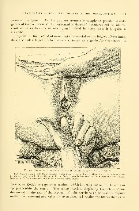
EXAMINATIOX OF THE PELVIC OR(iANS I\ THE DORSAL POSITION. 101
pears at the liynien. In this way we secure the completest possible investi-
gation of the condition of the pei'itoneal surfaces of the uterus and its adnexa,short of an exploratory celiotomy, and indeed in many cases it is quite as
accurate.
Fig. ()i>. This method of examination is carried out as follows : First intro-
duce tlie index linger up to the cervix, to act as a guide for the tenaculum
Fig. iji) —BiM\M vl h\\MiN
The cervix is caught with the corrugated tenaculum and drawn down to tlie outlet : then the tenaculumis held against the ball of the thumb while the index finger is inserted into the rectum and u.sed, in con-junction with the abdominal hand, to examine the pelvic organs.
forceps, or Kelly's corrugated tenaculum, which is firmly hooked in the anterior
lip just within the canal. Then make traction, displacing the whole uterus
downward in the axis of the vagina, until the cervix is at or near the vaginal
outlet. An assistant now takes the tenaculum and retains the uterus there, and
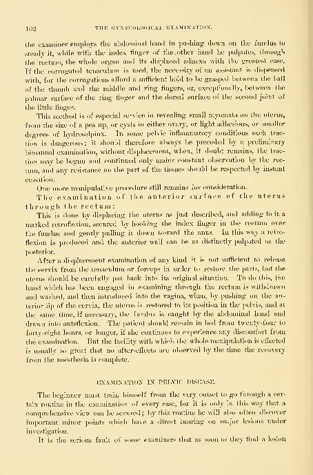
IQ^ THE GYXECOLOaiCAL EXAMIXATION.
the examiner employs tlie abdominal hand in pushing down on the fundus to
steady it, while with the index linger of the,other hand he palpates, through
the rectum, the whole organ and its displaced adnexa with the greatest ease.
If the corrugated tenaculum is used, the necessity of an assistant is dispensed
with, for the corrugations afford a sufficient hold to be grasped between the ball
of the thumb and the middle and ring fingers, or, exceptionally, between the
pilmar surface of the ring finger and the dorsal surface of the second joint of
the little finger.
This method is of especial service in revealing small myomata on the uterus,
from the size of a pea up, or cysts in either ovary, or light adhesions, or smaller
degrees of hydrosalpinx. In some pelvic inflammatory conditions such trac-
tion is dangerous; it should therefore always be preceded by a preliminary
bimanual examination, without displacement, when, if doubt remains, the trac-
tion may be begun and continued only under constant observation by the rec-
tum, and any resistance on the part of the tissues should be respected by instant
cessation.
One more manipulative procedure still remains for consideration.
The examination of the anterior surface of the uterus
through the r e c t u m :
This is done by displacing the uterus as just described, and adding to it a
marked retroflexion, secured by hooking the index finger in the rectum over
the fundus and gently pulling it down toward the anus. In this way a retro-
flexion is produced and the anterior wall can be as distinctly palpated as the
posterior.
After a displacement examination of any kind it is not sufficient to release
the cervix from the tenaculum or forceps in order to restore the parts, but the
uterus should be carefully put back into its original situation. To do this, the
hand which has been engaged in examining through the rectum is withdrawn
and washed, and then introduced into the vagina, when, by pushing on the an-
terior lip of the cervix, the uterus is restored to its position in the pelvis, and at
the same time, if necessary, the fundus is caught by the abdominal hand and
drawn into anteflexion. The patient should remain in bed from twenty-four to
forty-eight hours, or longer, if she continues to experience any discomfort from
the examination. But the facility with which the whole manipulation is efi'ected
is usually so great that no after-effects are observed by the time the recovery
from the anesthesia is complete.
EXAMINATION IN PELVIC DISEASE.
Tlie beginner must train himself from the very outset to go through a cer-
tain routine in the examination of every case, for it is only in this way that a
comprehensive view can be secured ; by this routine he will also often discover
important minor points which have a direct bearing on major lesions under
investigation.
It is the serious fault of some examiners that as soon as they find a lesion

EXAMIXATIOX IX PELVIC DISEASE. l03
anywhere in the genital tract which may account for some of the symptomsthey at once concentrate their entire attention and treatment upon that point,
forgetting the fact that the patient may liave other lesions as well. This is best
illustrated by the numerous cases of lacerated cervix and " ulcers of the mouth of
the womb " persistently treated where the serious disease lies in the inflamed
Fallopian tul)es.
To avoid this superficial treatment the examination begins by noting all the
peculiarities of the external genitalia. The orifices of Bartholin's glands mustbe looked at for the taehes significant of infection, and the glands themselves
should be felt to see if they are enlarged or pus can be squeezed out of the
duct. The state of tlie hymen must be noted—whether intact, dilated, or torn.
The uretliral orifice, by a pufl:y reddened condition, often gives evidence of a
gonorrheal infection, and a little pus can be milked down the urethra by the
finger stroking the anterior vaginal wall. By firmer pressure of the outer part
of the urethra against the pubic arch, Skene's ducts are emptied of any accumu-lated pus. Scars at the vaginal outlet and the relaxed condition following child-
birth or the use of large instruments are also to be noted, as well as the func-
tional activity of the anterior fibres of the levator ani muscle.
The vagina is noted as short or long, and rugose or smoothed out, and espe-
cial note is taken of any cysts in it. The rectum can he palpated through the
posterior vaginal wall, feeling like a stringy collapsed tube easily moved fromside to side ; if it contains any fecal masses this is evident to touch. Anteriorly
the base of the bladder, and antero-laterally the ureters, can be felt through the
vagina, and if they are inflamed, touch will always elicit complaint. The cervix
is the most prominent feature in the vault of the vagina ; its direction is im-
portant, whether lying in or across the vaginal axis, and its form, whether conical
with a small os, or split and everted and containing distended follicles. At the
vault of the vagina, in front of, behind, or at the sides of the cervix, hard masses
may be felt which will require a careful bimanual examination to determine their
identity. The condition of the rectum should be carefully inquired into in every
gynecological case, and any symptoms pointing in that direction should be in-
vestigated with care. The gynecologist will in this way take particular note of
hemorrhoids, fissures, fistula, proctitis, and especially of strictures.
The examination of the urethra, bladder, ureters, and kidneys are described
in Chapters XII and XIII.
The bimanual examination by one of the methods described follows next;
the position of the uterus is ol)served, together with its size, surfaces, mo-bility, and sensitiveness. Finally, the tubes, ovaries, and broad ligaments are
palpated.
When such a routine is regularly followed, instead of merely noting one
lesion, the observer will often find several, either independent or in conjunction,
in the same patient ; for example, a deep laceration or a complete tear through
the septum at the vaginal outlet is often found associated vnt\\ extensive scar
tissue in the vagina, a lacerated cervix, and a retroflexed uterus ; or, on the
other hand, the external tear, which is the sign of a ditiicult forceps labor, is
104 THE GYNECOLOGICAL EXAMINATION.
associated witti a pelvic tubal abscess, the sequela of a puerperal infection.
One of the most striking complications I have seen was that of a patient with
a gonorrhea! urethritis. Pressure on Bartholin's glands squeezed out a little
pus and showed she had also Bartholinitis. Pus taken from the vagina con-
tained numerous gonococci ; an endocervicitis and an endometritis were also
o-onorrheal. When the abdomen was opened, pus oozing from the tubes was
found to contain the same organisms, and, lastly, they were found abundantly in
pus taken from the peritoneal cavity.
Pelvimetry.—Pelvimetry is of the utmost service to scientific gynecology,
and sh(»uld be constantly practiced in all clinics, as the gynecological lesions
found are often explainable by the discovery of .a deformed pelvis. The various
external measurements should be made as described in the obstetrical text-books
—viz., the distance l)etween the anterior-superior iliac spines, between the iliac
crests, Baudelocque's diameter, and the intertrochanteric diameter.
There are four ways of measuring the true conjugate diameter of the superior
strait, which is the most important single measurement : In the first place, it
may be estimated, as usual, from the diagonal conjugate through the vagina
;
this, however, is often impracticable in gynecology, first, either because the
vaginal canal is too short and rigid, or because of scar tissue at the vaginal vault,
or of masses in the pelvis above the vault which prevent the necessary displace-
ment of the vagina up to the sacral promontory. In the second place, a diagonal
conjugate may sometimes be obtained under these circumstances by pressing the
finger up through the rectum until it touches the promontory, and so measuring
the distance to the under surface of the pubic arch. In the third place, I have
found the following procedure, which I call "the external direct method of
measuring the true conjugate," to be most [generally useful
:
The patient lies on the back, with slightly flexed thighs and knees, and the
head and chest elevated, so as to relax the abdominal muscles perfectly. The
examiner then stands on her right or left side, according as he intends to use his
right or his left hand with the palmar surface down. Then with gently in-
creasing pressure he makes deep palpation above the symphysis pul)is backward
toward the vertebral column, feeling for the promontory of the sacrum with the
tips of the fingers, sweeping from the abdominal cavity down into the pelvis,
deeper and deeper each time until the characteristic median projection of the
promontory is recognized. As soon as the promontory is felt he sweeps the
fingers of the open flat hand several times down over it into the pelvis, gaining
a distinct impression as to its exact position ; then the fingers are allowed to
rest vertically above the promontory ; in this way the posterior point of the con-
jugate diameter is fixed. The free hand now determines the anterior point, by
pressing the middle finger down behind the symphysis pubis, until the most
prominent point on its posterior border is distinctly felt. Directly over this an
indentation is made with the finger nail on the outstretched hand. Fig. 70. The
hand is then raised from the abdomen, keeping the fingers rigidly in the same
position, and the distance from the tip of the finger to the mark made on the
palm will be the true conjugate diameter, Fig. 71.

PELVIMETRY. 105
Tlie eliief sources of error arise eitlier from measuring directly over the
summit of the symphysis or from pressing the finger tips against the prom-
ontory instead of over it, thus interposing the thickness of the abdominal wall.
Judgment necessary to tell when the lingers are vertically above the prom-
iintory throuo;h abdominal walls of varying thickness is the chief factor in
Fig. 70.— FutsT Ste
The tip of tlie middle finder of the left hand rests just above the promontory of the sacrum, while the
middle fincrer of the ri^ht hand indents the pahn at a point just above the inner face of the symphysis. Thedistance marked otf iu this way is the true eonjugate.
making the measurement. Experience will gradually eliminate grosser errors
and l)ring a sufficient degree of certainty for practical purposes. The more con-
tracted the pelvis the less is the liability to error.
In illustration I will cite the following case (see George W. Dobbin, Amer.
Jour. Oh.9t., vol. xxxii, ^o. 2, 1S95): Mrs. H., admitted to the Johns Hopkins
Hospital, January 3, 1895, had had two severe instrumental lal)ors within two
years, both children dying during labor. Since the second labor she had had
no control of loose bowels and there was a constant dribbling of urine.

106 THE GYNECOLOGICAL EXAMINATION.
Tlie examination revealed an extensive dermatitis witli edema of the external
genitals. The recto-vaginal septum was torn through and the sphincter pits
separated 3 centimeters (1^ inch), although in spite of this the vaginal outlet was
well lifted np. The vagina was smooth throughout; at the vault there was a
sharp falciform scar at the junction of the right lateral and anterior vaginal
walls. The cervix was stellately lacerated and divided into one posterior and
two anterior portions. A sulcus of scar tissue between the two anterior por-
5 f 32 1109 8765* 32 1 Q CV\
.
^0
Takii
tions ended at a vesico-vaginal fistula 3 millimeters in diameter. The uterus lay
in retroposition reclining in the sacral hollow. The tubes and ovaries were
normal. On account of the dense unyielding scar tissue in
the vaginal vault, it was impossible to measure the obli(|ue
conjugate either by the vagina or by the rectum. By the ex-
ternal direct method the true conjugate was found to be only 7 centimeters

EXAMINATION UNDER ANESTHESIA. 107
(3 inches). The patient had a flat pelvis of liiii^h grade, the obstetrical dilB-
culties were fully explained, and the gynecological condition etiologically
accounted for.
In the fourth place, the most accurate method of all is the direct measure-
ment of the conjugate from sacral promontory to posterior surface of the sym-
physis pubis, through the abdominal incision in the course of a celiotomy ; this
is easily obtained by guiding the tip of a graduated sound to the promontory
by the index finger, and then feeling for the posterior surface of the symphysis
with the other index finger, and estimating the corresponding point on the
sound, which is now taken out and the marking read off.
This is particularly useful in pelvic inflammatory cases where the disease has
come from a difficult labor and the abdomen has to be opened to remove it.
EXAMINATION UNDER ANESTHESIA.
I feel that I can not emphasize too much the extreme importance of a rou-
tine use of ether or chloroform anesthesia to the point of complete relaxation
in investigating intrapelvic diseases. Weeks, months, or even years of useless
palliative measures will be saved in many cases if the patient is anesthetized and
examined before beginning treatment. The purpose of the anesthesia is to do
away with all resistance on th.e part of the patient, relaxing the abdominal mus-
cles completely and preventing the possibility of unexpected resistance when
tender points are touched. The examination with the anesthetic can be con-
ducted with a thoroughness which is impossible without it, the uterus can be
drawn down, adhesions pulled upon, the perineum deeply invaginated, and in-
flamed tubes and ovaries handled in a way which is impossible as long as the
patient remains conscious.
I may add also that it is a definite advantage to the operator to be able to
devote his concentrated attention to the examination and not to be distracted by
his anxiety as to how much pain he is giving his patient.
Rules for the Use of Anesthesia.—I recommend, therefore, the following
rules : LTse an anesthetic in all cases
(a) Where doubt exists after an ordinary bimanual examination.
(b) Where a patient comes to the specialist after having had treatment for a
long time at other hands without improvement.
(c) In all cases of pelvic peritonitis involving one or both ovaries or tubes,
without producing any gross tumor, when the use of the anesthetic is to find
out the extent of the disease.
(d) Always in unmarried W(^men.
Preparation of the Patient for Anesthesia.—The lower bowel must be emptied
by taking a purgative the night before, and if this does not act freely enough,
an enema in the morning. The bladder should be emptied by catheter at the
last moment. It is best to examine early in the morning and about an hour
after a light breakfast, such as a cup of tea and a piece of toast. The early hour
has the advantage of relieving the patient from a day of anxious expectation.
108 THE GYNKCOLfXilCAL EXAMINATION.
After completely anesthetizing her, if it is done in her room, she should he
brought across the bed, with her hips projecting a little over the edge and legs
and thighs held well flexed by assistants or a leg holder. It must be the aim in
the examination to keep her unconscious as short a time as is consistent with a
thorough investigation, in order to diminish the after-discomforts with the lia-
bility to distressing nausea. Patients who have come into the house from the
outdoor department simply for the ether examination usually go home late in
the afternoon of the same day, but it is better, as a rule, to keep them quietly
in bed for a full day afterward.
Exposure of any part of the body during the examination must lie guarded
against as much as possible, no matter what the patient's station in life may be,
partly on the ground of a proper respect for the sex in general, partly because
of the sacred obligation to treat the patient in her helpless condition with that
deference which is her due in return for her contidence in putting herself in tliis
way into her physician's hands, and partly because of the inevitable demoralizing
effect on nurses, the doctor, and assistants that comes from a careless indifference
to the dictates of a proper modesty. While the patient is being examined no
persons not directly interested, professionally or otherwise, should be present.
Classes of students must be admitted to the examining room in limited num-
bers only ; in no case should more than three or four students examine the
same patient, and the physician who is responsible for her must exercise a con-
stant watchful care to keep any student from examining too long and from using
unnecessary force.
The hands must be specially prepared for the examination by a thorough
scrubbing with soap and warm water, and by cutting the nails short, so that tliey
will not bruise the skin in making the bimanual examination. After each ex-
amination the hands must be washed afresh to prevent carrying contamination
from one case to another. An examination should never be made with a sore
hand. Vaseline is a good lubricant for the vaginal and rectal Angers. Whenthe hymen is intact the examination should be made, as stated in the rules,
under an anesthetic ; in tliis condition it is sometimes possible to introduce
a small finger into the vagina without making any rupture, but it is better to
omit the digital examination by the vagina entirely, and to conduct it wholly
through the rectum and abdominal walls. If it is necessary to catch the cervix
and draw the uterus down to get at the tubes and ovaries, this may be done by
introducing into the vagina the bullet forceps closed, and then, guided by the
rectal finger, which feels the cervix distinctly, to open the forceps and grasp the
cervix.
A perfectly satisfactory inspection of the vagina, and witliout injury to the
hymen, may be made l)y using a small cylindrical speculum in the knee-breast
position.
The Examination.—The various methods of handling the normal pelvic
organs and the diifcrent avenues of access to them are also used in investigating
the diseases of these organs ; and the knowledge acquired in gaining a thorough
familiarity with the condition and relations of the healthy organs is indispensable

POINTS TO J?E COXSIDERKI) IX EXAMIXATION, 109
in estimating the presence and extent of disease, for the normal condition is the
only standard of comparison. Owing to a want of familiarity with the normalstructures, operators have frequently made the frightful mistake of removingsound organs.
Almost all morbid changes advanced enough to call for operative interfer-
-DlFFEREXTIATIi BETWEEN" A MyomUtebls
IN THE AnTEIMi>K UtEKINE WaLLN Anteflexion.
By ;i bimanual examination, while the vacrinal lincrers, resting upon tlie cervix, liold tlie mass up, theahdominal fingers are able to discover the little sulcus between the funtlus and the tumor, and to appreciatethe slight but <.listiiict mobility of the myoma as separate from the fundus. H. op. Oct. 2-4, 'yO.
ence produce alterations of the normal structures which can be recognized by
an investigation which considers the following points :
(a) Displacements affecting position.
(b) Fixation and adhesions affecting mobility.
(c) Inflammation and tumors affecting size and form.
(d) Any abnormal sensitiveness.
(e) Peculiarities of consistence.
(f) Information derived from curettage of the uterus.
(g) The microscopic examination of a piece of tissue excised.
(h) The microscopic and bacteriological examination of uterine, vaginal, and
other discharges.

110 THE GYNECOLOGICAL EXAMINATION.
The skilled examiner never makes his diagnosis by taking these questions up
and applying' them one after another, seriatim, to a ease in hand ; he proceeds,
on the contrary, to make the investigation with a trained touch which at once
recognizes any abnormalities, and instinctively selects the essential points for
Fig. THE Left Lateral Displacement of the Uterus, which is Pished to the Opposite
IN THE Direction of the Arrows, bv an Ovarian Cyst on the Kiuht.
more special attention. Such a plan is not without the risk of occasionally over-
looking some point of importance, particularly if the examination is a hurried one.
The beginner will always iind it better to take the questions up and apply
them categorically, at least until the routine becomes so fixed in his mind that
its application is afterward more or less instinctive. This, too, is the only satis-
factory plan for a text-book.
(a) Displacements affecting the Position of the Pelvic Organs.—D e s c e n s u s
.
—The index finger carried up the vagina notes whether the cervix is well back
in the pelvis or lies low down on the pelvic fioor in the axis of the vagina, indi-
cating a descent of the uterus.
Anteflexion .—A little sudden pressure against the anterior vaginal wall
will often encounter a resisting body, which at once recedes by using the other
hand to make counter-pressure through the lower abdominal wall ; if the re-
cession is prevented and the body, palpated carefully, is found to be movable,
ovoid in form, and by carrying the bimanual palpation a little farther back,
organically connected with the vaginal cervix, the uterus is in normal anteflex-
ion. When the cervix lies in the axis of the vagina, and the body of the womblies against the anterior vaginal wall parallel to it, the angle between the two is
very acute and a pathological antefiexion exists. Fig. T2.
R e t r o p o s i t i o n s ,—The two forms of retroposition are retroversion and
retroflexion, and a diagnosis is made by demonstrating bimanually (1) the ab-

FIXATION AND ADHESIONS AFFECTING MOBILITY. Ill
sence of the fundus from its normal position in the front part of the pelvis,
and (2) its presence somewhere in the back part of the pelvis, behind the cer-
vix, bj feeling it there with the vaginal finger as a round, resisting mass, and
tracing its direct connection with the cervix. If the bridge of tissue joining
the vaginal cervix to the body supposed to be the fundus can not be satisfac-
torily palpated, it will be felt more clearly if the cervix is caught with a tenacu-
lum forceps and pulled down. The bimanual palpation sometimes shows that
the fundus lies to the right or left in lateral ilexion ; this is due to adhesions
drawing it in the direction of the ilexion, or to a tumor filling the opposite side
and pushing it over; or again to a large ovarian tumor of the side to which
the uterus incHnes, which pulls on the broad ligament as the tumor escapes into
the abdomen. Figs. 73 and 7-1.
Ascensus Uteri.—In ascension the cervix is lifted up above its normal
position in the pelvis, and in an extreme form the whole womb may be displaced
out of the pelvis into the abdomen. This may happen in the case of a broad-
ligament tumor, or of an ovarian tumor adherent to its posterior surface and
drawing it into the abdomen.
(1)) Fixation and Adhesions affecting Mobility.—If the uterus is found fixed
in a certain position, and does not move easily upward in making slight pres-
Fio. 74.—Showing the Displacememt of the Uterus toward the Side from which the TumorGrows, Due to the Enlargement of the Cyst which now Fills the Pelvis and, by Traction
ON its Pedicle, Draws the Uterus in the Opposite Direction.
sure on the cervix, its condition is abnormal, and the cause must be sought for.
I know of but one apparent exception to this rule, and that is where the uterus
responds but slightly to pressure on account of a stout tense abdomen and
increased intra-abdominal pressure. When the cervix is occupied by a cancer
which has extended out into one or both broad ligaments the whole organ feels

112 THE GYNECOLOGICAL EXAMIXATIOX.
as if lield in a vise by the hard masses extending out to the pelvic walls. Adhe-
sions of the posterior uterine surface to the pelvic Hoor restrict its mol)ility,
forming an adherent retroilexed uterus. This is tested by pulling down the
cervix and trying to raise the fundus, when the adhesions are put on the stretch
and felt. I would caution the beginner here not to conclude that a retroilexed
fundus is adherent because be can not push it up through the vagina. The
normal mobility is also greatly restricted in almost all cases of inllanmiation of
the tubes and the pelvic peritoneum, which result in masses behind the broad
ligaments. Ovarian and tubal adhesions are best felt bimanually with one or
two iingers in the rectum. The adhesions, whether light and velamentous, like
a web, or short and firm, binding the ovary to the posterior surface of the broad
ligament, are easily felt upon attempting to handle the ovary in order to exam-
ine both its surfaces, as described in the examination of the normal ovary. Anadherent tube is almost always involved with the ovary in pelvic inflammatory
disease, and is also usually enlarged.
(c) Inflammation and Tumors affecting Size and Form.—Only the trained
fingers familiar with the normal organs will recognize at once all deviations in
size. Botli the enlai-ged infiltrated and the cancerous cervix are characteris-
tically different from the normal, and lacerated everted cervical lips can be
recognized at once.
The trained clinician, knowing how large the normal nulliparous uterus
ought to be, and what is the size of the average parous utei'us, will have little
difiiculty in deciding whether the uterus of a young woman is undersized—that
is, puerile or infantile ; or in the case of a woman of advanced yeai-s, whether it
is senile. The large body of a subinvoluted uterus differs as much from the
normal to the touch as a hydrocephalic head does from a sound fetal head.
The myomatous uterus, from the small nodules just projecting from the
serous surface, often not as big as a pea, all the way to the vast masses filling
the abdomen, presents unmistakable characteristics in the enlargements and
irregular bizarre shapes assumed. Often the only suggestion of a cancer of the
body ()f the uterus found at a first examination is the increased size and the
globular form of the uterine body. In pregnancy we trace a uniform develo])-
ment in the size of the uterine l)ody from the fourth week on to the end. The
most sensitive touch will be the quickest to appreciate this change at its earliest
stage, from the fourth to the sixth week.
The Fallopian tubes are changed in size and form by all infiammatory dis-
eases, more particularly in those in which the outer extremities are occluded and
the secretions retained, called sactosalpinx. With the thickening of its coats
and the distention of its lumen the tube becomes harder and larger, and so is
the more easily palpated. The inflamed tube assumes a sausage shape with two
or three convolutions. •
Alterations in the size and form of the ovaries may affect a part or the
whole of the organ. A little hard mass projecting from its surface is most
likely a corpu^s Jihrosnm ; a nodular swelling jirojecting from one part of the
periphery and not more than 2 or 8 centimeters (f to 1^ inch) in diame-

PECULIARITIES OF CONSISTENCE. 113
ter is a cystic Graafian follicle, or the last menstrual corpus nigrum. A larger
cystic tumor with a smooth surface, from 4 to 6 or even 10 centimeters (1^
to 2|^ or 5 inches) in diameter, is a Graafian cyst, or a cystic corpus luteurn.
Small dermoid cysts may also present similar characteristics. An ovarian ab-
scess is usually distinguished by the dense surrounding adhesions, but a suppu-
rating dermoid will also present these signs. The larger ovarian tumors are
usually associated with a complete disappearance of the ovary and its replace-
ment by a smooth or irregular mass, according as there is one or a number of
cysts.
(d). Peculiarities of Sensitiveness.—Normally the pelvic organs are not at all
sensitive to the ordinary bimanual manipulation. The ovaries alone are jDainful
if a decided pressure is made upon them. Frequently, however, patients comefor examination in whom the only discoverable difficulty is an abdominal sensi-
tiveness, and the most painstaking investigation fails to show any other trouble.
This hyperesthesia is often confined to one ovary, generally the left, which the
patient can not bear to have touched ; in other cases both ovaries are sensitive,
and there may be a perfectly normal uterus, so tender that not even the lightest
pressure can be borne. In extreme cases the whole pelvic and even lower ab-
dominal peritoneum shows the same sensitiveness. I know of no cause for this
;
it is often associated with other disturbances which are presumably circulatory.
It is important that every practitioner should recognize this ailment, so as to
avoid the common mistake of estimating the amount of disease present by the
tenderness complained of as soon as pressure is made on the pelvic viscera.
Ovaries and tubes have been removed repeatedly w^here the only demonstrable
difficulty was a persistent sense of discomfort and sensitiveness to pressure, only
to discover that the mutilation has not in the least relieved the difficulty.
Pelvic sensitiveness is also peculiarly the mark of the hysterical patient
whose attention has become fixed on these organs. All inflammatory affections
are characterized not only by pain during the exacerbations, but by a persistent
sensitiveness of the inflamed structures, which makes it difficult to handle and
outline them. Under such circumstances it is necessary to put the patient under
anesthesia to make a thorough investigation.
(e) Peculiarities of Consistence.—In addition to peculiarities in position, in
mobility, in form, and in size, the pelvic organs in disease also exhibit marked
peculiarities in consistence. Each organ has its own individual standard, differ-
ing from every other organ in this respect. For example, the consistence of the
cervix is one thing, that of the uterus and ovary another. The most striking-
example is the change in the vaginal portion of the cervix in j)regnancy from a
firm, hard, resistant tissue to a soft and even flabby condition. The cheesy fri-
ability of a cancerous cervix also differs from any other state. There is a putty-
like condition of the subinvoluted uterus, which indents on pressure, and which
ought to be a warning against the use of the sound. I have seen the sound go
through the uterine wall in these cases with as much ease as if it were a piece
of blotting paper. Again, the soft semifluctuation of the pregnant uterus from
the third to the fifth months is different from the hard fibroid uterus. Occa-
10
114 THE GYNECOLOGICAL EXAMINATION.
sionally a vascular fibroid will simulate pregnancy. In diseases of the tubes and
ovaries there is no more important distinction to be made than the changes in
consistence. In infected cases a dense hardness, which replaces the soft pliability
of the pelvic floor, is characteristic, and is only imitated l)y adherent cancerous
ovaries. The consistence of the enlarged Graafian follicle is also characteristic
in the paper-like thinness of its shell, which is easily recognized by the finger,
I have twice recognized a rupture in an ovarian cyst made up of a mass of little
cysts by putting the finger through the hole which hajjpened to be on the pelvic
floor and feeling the little cysts within. The consistence of an al)dominal ascites,
and of a parovarian cyst or a raultilocular ovarian cyst, differs in each case, and
is often the most characteristic diagnostic feature.
(f) Information Derived from Curettage of the Uterus.—By curettage of the
uterus and a microscopical examination of the scrapings we determine the dif-
ference between glandular hyperplasia, endometritis, carcinoma, sarcoma, the re-
mains of an abortion, and tuberculosis of the endometrium (for details, see Chap-
ter XIV).
(g) The Tissues.—In the same way, l)y making a microscopic examination
of a piece of tissue excised from the cervix, a differential diagnosis is established
between inflammatory conditions and carcinoma (see Chapter XVI).
(h) Secretions.—The examination of the secretions, commonly called leu-
corrheal, from the uterus, cervix, vagina, and vulva, often gives important
information, and either throws light upon the cause of an existing disease
or shows the presence of elements in these secretions liable under favorable
conditions to endanger health and life. The purpose of this examination
is to discover the presence of one or other of the commoner pyogenic
organisms—the streptococcus, the staphylococcus, the gonococcus, and per-
haps the tubercle bacillus and the colon bacillus. The examination includes
observations as to the presence of any secretion, its location, quantity, ap-
pearance, consistence, chemical reaction, bacteriological character, and any local
reaction.
For accuracy of investigation, the following regions should be examined
:
The ducts of Bartholin's glands.
The vulvar commissure.
The urethral orifice and Skene's glands.
The lower vagina.
The vaginal vault.
The cervical canal.
The uterine body.
The normal secretions which contain nnmerous micro-organisms but none of
the pyogenic bacteria form the standard of comparison. No bacteria of any
kind are found in the cavity of the utei-us.
In young women and in those not infected the secretions within the vulva
and at the vaginal outlet are those which have escaped from the vagina above.
Tlie natural appearance of the secretion is scanty, milky-Mdiite ; it consists of
desquamated vaginal epithelial cells, with nmcus and a few leucocytes, and its

EXAMINATIOX OF THE RECTUM. 115
chemical reaction is acid. The normal cervical secretion is a clear tenacious
mucus. The secretion from Bartholin's glands is small in quantity and thin and
clear. There is no secretion about the urethra.
In disease there is an entire change in the character of these secretions,
which become abundant, and change to a mueo-purulent character. By squeez-
ing Bartholin's ducts a drop or two of pus is made to exude at the orifice,
and on separating the labia the discharge may be taken up from the com-
missure. To get secretion from the vaginal vault and the cervix without
contamination, the patient may be put in the knee-breast position and a small
cylindrical speculum inserted, which admits air and does not touch the upper
part of the vagina. In the infected cases the cervix is often puffy, and its
everted mucosa weeps an abundant stringy muco-purulent discharge from its
surface. The most striking example of the utility of the examination of these
secretions is that of the puerperal infections, where the exact nature of the
infection may be determined. The prol)able nature of a pelvic abscess maybe traced to a gcmorrheal infection, if the gonococcus is found to be a resident
in the lower genital tract. The commonest points in which a latent gonorrhea
may lurk are the cervix uteri, the ducts of Bartholin's glands, and Skene's
glands.
The technique of the examination for these bacteria and the methods of
cultivating them are described in Chapter III.
EXAMINATION OF THE RECTUM.
The close relationship between the rectum and the other pelvic organs in-
volves both in many common affections, liable to affect any portion of the bowel,
from anal orifice to the brim of the pelvis. The commonest of these diseases
may be arranged under three heads : (a) Fistulae and rupture of the recto-vaginal
septum;
(b) the extension of a malignant growth from one organ to the other
;
(c) compression of the bowel either by increase in volume of uterus or ovaries,
or by inflammatory products which constrict its lumen.
Exammation of the rectum is also frequently called for on account of the
liability of the patient to refer disorders of the bowel to the uterus and its
adnexa. In this way a fissure of the rectum may be overlooked and a long and
useless course of treatment undertaken to relieve a pain from a source felt
higher up in the pelvis ; the congestion of hemorrhoids often produces a sense
of weight and bearing down in the pelvis, easily mistaken for the symptoms of
prolapse of the uterus.
For all these reasons it is important to make some statement about the con-
dition of the rectum in every gynecological record. Sometimes it is well to
make the examination without special preparation for it on the part of the
patient, when the physician may better judge of the habitual state of the bowel.
For a thorough examination the lower bowel must be completely emptied.
Anesthesia is not necessary as a rule. There are two methods of examining
—
(a) by palpation, and (b) by inspection.

116 THE GYNECOLOGICAL EXAMIXATION.
Palpation.—The finger introduced into the vagina easily feels the lower part
of the rectum from the cervix down through the posterior vaginal wall, and by
pressing upon it and rolling it from side to side, its size, mobility, and sensitive-
ness may be estimated. The normal rectal tube feels like a flat band with longi-
tudinal striae, which, under pressure, slips freely from side to side and without
pain. Any fecal accumulation presses forward into the vagina and gives the
bowel a more cylindrical form. The presence of feces can be recognized by
indenting the putty-Kke mass with the Angers.
The upper part of the rectum behind the cervix is often markedly sensitive,
and becomes more so when it is distended. This must never be forgotten when
a sensitive spot is found behind the cervix. I have seen an erroneous diagnosis
made of tumor behind the uterus and " inflamed ovary " when there was really
nothing the matter. A loaded upper rectum crowds out behind both broad liga-
ments, filling the pelvis with fecal masses readily confused with ovarian and
:.)
Fig. 75.—Deviation of the SiiiMoii) 1<i,k.\i're.
The l)ow(!l crosses the promontory of the sueruiii on the right side, und then returns to the left pelvic
l)riin and droi)s into the pelvis just behind the uterus.
tul)al tumors. In a more moderate distention the mass may lie on the pelvic
floor behind the left broad ligament. By palpation a distended uj)per rectum
and sigmoid flexure can be easily mapped out through the abdominal wall, if it
is not too thick and rigid. The diflerential diagnosis between these fecal tumors
EXAMINATION OF THK RECTUM BY PALPATION. iir
and true pelvic tumors is so impoi'tant that I present several diao-rams, tracings
from actual cases taken out of a large numl)er which have come under my obser-
vation. (Figs. 75, Y6, Y7, aud 78.)
In order to make the examination, the patient lies on her back in a position
of relaxation, with her shoulders shghtly raised and knees drawn up. Theexaminer then stands on her left side, and gradually makes deeper and deeper
/
-Devi OF THE Sigmoid Flexure.
The bowel skirts tlie anterior pelvic brim from left to right, and drops down into the pelvis on theriglit side.
pressure through the lower abdominal wall in the left semilunar line, until he
reaches the pelvic brim, without giving any discomfort to the patient to cause
her to resist. By gently drawing the fingers forward along the superior strait,
the empty sigmoid is felt slipping beneath them like a large, flat cord. If it is
distended it becomes still more distinct. In marked distention the bowel fol-
lows in general one of three directions : In the first, the bowel describes a sig-
moid curve behind both broad ligaments. Fig. 75 ; second, it curves out into the
anterior part of the pelvis over the bladder and then back to the sacrum. Fig.
76 ; third, the distention is upward into the abdomen and then down into the
pelvis from the right side, Fig. 77 ; and, fourth, the bowel passes in front of the
bladder almost to the region of the right round ligament, where it is bent on
itself ; when it reaches the promontory of the sacrum it makes a plunge downinto the pelvis. Fig. 78.
118 THE GYNECOLOGICAL EXAMINATION.
These fecal tumors are diagnosed bimanually by being continuous with afecal mass behind the vagina or behind the uterus low down, about the nature
of which there is no doubt. They occup)' peculiar positions in the upper pelvis,
and are elongate, and markedly movalde on account of a long meso-sigmoid\
Fig. 77.
—
Deviation of the Siomoid Flexuke.
The bowel extends down to Poupart's ligament on the left side, and thdrops into the pelvis on the right side.
dvirts the left pelvic brim and
they are often made up of a number of scyl)alous nodules. The customary
sensitiveness must not mislead. Any doubt remaining after such an examina-
tion can be cleared up by a purgative or enema, and by an inspection with a
sigmoidoscope.
A digital examination of the anus shows the existence of abnormalities, and
when the finger is carried up into the ampulla and tlie rectum innnediately
above that it demonstrates the presence of any marked changes, more especially
constriction by inflammatory masses ; when the finger passes between the utero-
sacral ligaments it seems to be entering a long, rigid tube with smooth walls.
Amid the redundant folds of the ampulla it is sometimes hard to find the en-
trance into the bowel above. The proper point is best located by taking the
cervix as a guide and seeking the opening right behind it.
Inspection.— Under inspection the whole nnu'ous surface of the lower bowel
is exposed to view, from its external orifice up to the sigmoid fiexure and colon.

EXAMIXATIOX OF THE RECTUM BY IXSPECTION. 119
and any alterations in color or nnevenness of surface and deposits, together with
changes in caliber and points of tixation, are at once evident, Fig. 79. To makethe best possible examination in this way, the bowel must be emptied of feces
and the patient placed in the knee-chest posture ; the thighs should be vertical,
the back well curved in, and the chest as close as possible to the table ; the
patient should wear no corsets or any constricting garments on the upper abdo-
men. A cylindrical speculum of suitable length and caliber is now introduced
and the bowel examined by a light reflected from a head mirror.
The following specula are useful : A short and a long proctoscope, a sig-
moidoscope, and a sphincteroscope.
The cylinder of the short proctoscope is l-t centimeters (5^ inches) long and
22 millimeters in diameter ; the long proctoscope is 20 centimeters (8 inches)
long and 22 millimeters in diameter ; and the sigmoidoscope is 35 centimeters
(14 inches) long and 22 millimeters in diameter. At the outer end of the cylin-
i^
,Fi<i 7S.
—
Deviation of the Sigmoid Flexure.
The bowel forms a sharp angle just behind the symphysis pubis, and then crosses the left broac
to tlie promontory of the sacrum and descends into the pelvis in the median line.
drical tul)e is a funnel-shaped rim to which the stout handle, big enough to be
grasjDed in the whole fist, is attached. Each speculum has an oljturator, blunt at
the end and provided wdth a strong stem and handle.
The sphincteroscope is short and slightly conical, the diameter at the lower
end of the tube being 2*5 centimeters (1 inch) and at the upper 3 centimeters

120 THE GYNECOLOGICAL EXAMINATION.
(l^ inch), while the outer rim of the funnel-shaped flange is 5 centimeters (2
inches) in diameter. The strong handle is set on this. The obturator is like
that in the other specula.
To make the examniation, the speculum is coated with vaseline and the but-
tocks are drawn apart, exposing the anus. The round end of the obturator is
laid upon the orifice, and, grasping the speculum in the list so that the pahn
Fig. 79.
—
Patient Supported by Uprights and Straps in Position for a Keotal Examination.
The instruments—conical sphincter dihitor, proctoscope, sigmoidoscope, applicator with cotton pledget
—
are reduced in proportion to tlie size of the patient.
keeps pushing the ol)turator in, it is carried into the bowel in a direction at first
downward and forward, and then upward toward the sacral hollow. Sometimes
the end catches in the gi'oove between extei-nal and internal s])hincters; if this
happens, it nuist be withdrawn and pushed in again in a slightly diiferent direc-

EXAMINATION OF THE RECTUM BY INSPECTION. 121
tion. As soon as it has fairly entered, the obturator is jjiilled out and the air
rushes audibly in, widely distending the bowel. The examination is now made
by reflecting the rays of light from an electric droplight, or a lamp, or good
daylight, by a head mirror, down the tube into the bowel, which is so well illu-
minated that the smallest points on its surface become visible, Fig. 80. It is
possible to detect differences not larger than the pores on the palm of the hand.
It will often be found that the speculum is turned too much downward, and
that it is necessary to drop the handle to bring an extensive area of bowel into
view. By turning the tube a little from side to side the whole dilated ampulla
Fjg. 80.
—
Examination of the Kectum bt Eeflected Light.
The instrument seen above to the right, drawn on the same scale of reduction as the body, is introduced
to its full length. The electric light, held by an assistant close to the sacrum, is reflected down the tube bya head-mirror. This picture is drawn fi'om a photograph.
is inspected in a few moments. The ground color of this and other parts of the
bowel is a pale red with large vessels like veins dividing up the surface at wide
intervals. The normal bowel is never intensely red and injected in this posture,
neither does it have a hazy appearance.
After studying the ampulla, upon looking up the lumen of the bowel a series
of half valves are seen cutting into its lumen on the right and on the left. The
tube passes easily through each of these, displacing first one and then another,
without perceptible resistance, exposing to view successive lengths of the bowel
hugging the sacral hollow. The promontory often appears characteristically
projecting as a rounded smooth eminence on the upper surface ; its bony nature
is evident on touching it with the end of the speculum.

122 THE GYNECOLOCxICAL EXAMINATIOISr.
The bowel distends so widely in this position that any fecal masses lie loose
in the lumen, and the speculum may often be carried beyond without removing
them and without their choking its lumen. Sometimes, on introducing the specu-
lum, the bowel will be found in the act of gradually opening up, expanding fold
by fold. The dilatation ceases in some part of the sigmoid flexure, where the
mucous surfaces suddenly come together, but a little pushing with the end of
the speculum, or by observing the mucous folds as they part in the escape of
flatus, the lumen is found, and the speculum can be pushed still farther uj),
although the view is no longer so pei"fect as in the lower atmospherically dis-
tended bowel.
To continue the inspection beyond the rectum on up into the sigmoid flex-
ure, the longer reach of the sigmoidoscope is necessary. The direction of the
instrument is no longer confined to the median line, but by degrees turns more
and more to the patient's left. I have introduced this speculum as far as 30
centimeters (12 inches) beyond the anus.
The purpose of the inspection is to note all alterations from the normal
appearance of the bowel, such as patches of congestion, mucus lying on the sur-
face, ulceration, and polyps. Strictures are found most commonly, in connec-
tion with jDelvic tumors, when there is a sudden narrowing of the lumen, beyond
which the speculum can not pass, and the bowel seems rigidly fixed.
Pelvic peritonitis, especially that form due to abscesses in the ovaries and
tubes, is particularly liable to produce stricture of the rectum at any point from
the brim of the pelvis down to the ampulla. In one of my cases the rectum
was choked by a large tubal abscess on the right side ; above the constriction,
which extended from the ampulla to the upper part of the pelvis, the bowel was
greatly distended and there was an opening between the sac and the rectum.
In another case, in which dense inflammatory masses with abscesses on both
sides were taken out together with the uterus, a tight stricture of the bowel was
found just below the promontory of the sacrum. This was about 4 centimeters
(1^ inch) long, and not more than 1^ centimeter (^ inch) in diameter, measured
on the outside.
A fistulous orifice seen foreshortened in the side of the bowel is easily passed
over, and must be carefully sought for by pressing so as to flatten the mucosa
out on the end of the speculum.
The sphincteroscope is used by pushing it into the ampulla, withdrawing the
obturator, and then drawing the speculum out a little until the inner sphincter
circle begins to close over it, and then pushing it back in. In doing this it does
not re-enter the portion of the bowel just left, but simply spreads the area within
view over the end, giving a flat field for inspection. In this way, step by step,
the whole sphincter area is exposed, and any abnormalities easily detected. For
children and nervous patients who are diftieult to examine, a speculum 12 centi-
meters (5 inches) long and oidy 18 to 2<» millimeters in tliameter is most useful.
I

EXAMINATION^ OF THE VERMIFORM APPENDIX. 123
EXAMINATION OP THE VERMIFORM APPENDIX.
The gynecologist must be familiar with the position and methods of palpat-
ing tlie normal and diseased vermiform appendix, in order not to confuse its
affections with those of the right tube and ovary near by. We owe the discov-
ery of this valuable means of diagnosis to Dr. George M. Edebohls, of NewYork (see Xew York 'four, of Gijn. and Obst., Feb., ISOi, and Amer. Jour, of
the Med. Sci, May, 1894).
Under ordinary circumstances the normal vermiform appendix can be felt
through the abdominal wall against its underlying iliac muscle as a short dis-
tinct cord, moderately or not at all sensitive, extending from its base at a point
in a line between the umbilicus and the right anterior-superior iliac spine, down-
ward and inward to the pelvic brim. To find it the patient lies with the
abdomen bare and knees and thighs flexed without effort, and the examiner,
standing at the patient's right side, makes pressure inward in the right semihmar
line just below McBurney's point. He increases the pressure gradually until
the posterior abdominal wall is reached. This may be felt, if desired, to make
certain of the position.
The fingers, keeping up the pressure, then glide in a direction downward
and outward toward Poupart's ligament, until a delicate, cord-like structure is
felt to slip beneath them. The maneuver is then rej)eated a little higher up,
and then a little lower down, changing the position until the length and direc-
tion of the appendix are ascertained. The upper end disappears at McBurney's
point, and the lower end at the brim of the pelvis as a rule. A loop of intes-
tine or muscular fibers in the abdominal wall may be mistaken for the appendix,
but any overlying small intestine may be disposed of by placing the patient
for a few minutes in the knee-breast posture, and by careful attention the more
superficial position of the muscular strands will be recognized. A diseased
appendix is often still more easily recognized from its extreme sensitiveness and
its increase in size, making it feel like a big hard cord, more or less fixed.
If there is an inflammatory exudate about the appendix the organ can not
as a rule be felt, but the positioM and distribution of the mass are both charac-
teristic of appendical as contrasted with tubal and ovarian inflammatory disease.
An exception to the general principles here laid down are those cases in
which the inflamed end of tlie appendix lies in the pelvis involved with the
right tube and ovary.
INVESTIGATION OP THE GENERAL CONDITION OF THE PATIENT.
Upon completing the gynecological examination, the specialist must turn
his attention to the condition of all the other vital organs in the body, associ-
ating the results with the facts elicited by the examination of the pelvic organs
;
he is then in a position to estimate the relative importance of any gynecolog-
ical ailment.
This examination is valuable in several ways : It often happens that the pel-

124 THE GYNECOLOaiCAL EXAMINATION.
vie disease is but a part of a general morbid condition, or is dependent npon
disease elsewhere for its continuance ; this is the case when pulmonary phthisis
is associated with the tubercular tubes and ovaries, and tubercular peritonitis, or
when a disposition to uterine hemorrhage is but one of the manipulations of a
crippled heart or a cirrhotic liver. On the other hand, a pyonephrosis may be
due primarily to a pelvic abscess blocking the ureter and furnishing the source
of infection.
It often happens, too, that there may be some grave organic disease of one
of the other organs which is simply an accidental complication, but neverthe-
less forbids the performance of any serious gynecological operation.
The inquiry will be commenced by asking about any strong family tendency
to hereditary diseases which may bear an etiological relation to any local affec-
tion, or so complicate the local conditions that they must be taken into consid-
eration in the prognosis and treatment. The risks attending a plastic operation
upon the cervix or vagina, for example, are of no moment in properly selected
cases, but they may be followed by disastrous results if certain constitutional
diseases, such as advanced nephritis, tuberculosis of the lungs, diabetes, etc., are
overlooked.
The main points of the general gynecological examination are age, heredit}-,
temperament, habit, color, and the following diseases : tuberculosis, pneumonia,
pleurisy, hydrotliorax, heart disease, affections of the alimentary ti-act, diseases
of the liver, spleen, and kidneys.
Age.—Other conditions being equal, women between the ages of twenty and
forty withstand the effects of operation best. But, with Dr. Mary Sherwood,
I have collected recently statistics in 100 cases which show that ovariotomy in
women between the ages of seventy and eighty-two is attended with a mortality
only slightly greater than in women of younger years {Johns Hopkins Hospital
Reports. Gynecologieal Fasciculus, ISTo. II, p. 509) ; in 115 cases in the hands
of QC) operators, only 12 per cent died. (See Chapter XXI.)Between twenty and forty women are in the prime of life, and resist the
effects of shock, hemorrhage, and infection better than those whose vital forces
are impaired by advancing years. In the aged the minor gynecological ailments,
such as retroflexion, lacerated cervix, relaxed outlet, and often even the markedforms of prolapse of the uterus, should not be treated by operation unless the
patient experiences serious discomfort. The old are much more easily depressed
by the loss of blood, and recover more slowly from shock. Conv^aleseence is
longer with the aged, because the recuperative powers are feebler. Carcinomaof the uterus in its early stage and diseases of the appendages, which, if not
interfered with, will destroy health or terminate life, sliould be submitted to
operation regardless of age, if this is the only contra-indication.
Heredity.—The hereditary predisposition to certain diseases must be care-
fully inquired into, because any marked family tendency may have an impor-
tant bearing on the etiology. When there is a hereditary tendency to insanity,
especially in patients inclined to be morbid or melancholy, any operation is
attended with risk of precipitating an attack of insanity, as a post-operative

TEMPEKAMENT AND COLOll. 125
complication. In neurotic faTnilies the results of surgical work are always less
satisfactory. A family tendency to excessive menstrual flow may account for
wliat would in other cases be significant of disease. A strong tendency to can-
cer in the family will arouse suspicion as to cervical erosions or persisting uterine
hemorrhages.
A family tendency to tuberculosis of the lungs in a patient who has the gen-
eral appearance of being tubercular, without signs of the disease, must put the
operator on his guard, as the convalescence is apt to be slow, and the patient is
often a long time in acquiring any vigorous health.
Temperament.—The temperament of the patient exercises more or less influ-
ence on the results of operation, and it is a good thing for the surgeon to study
the character and disposition of his patient beforehand. Bright, cheerful womenapproach an operation with more composure and recover from its effects more
rapidly than the despondent. A buoyant disposition is especially helpful in
shortening the convalescence.
Hysteria and various neurotic ailments often accompany pelvic diseases in
women, and the effects of their presence nmst be noted and weighed well before
operation. On the other hand, certain classes of nervous patients need a strong
mental impression made upon them, and are greatly benefited by even trivial
operations. In hysterical women the convalescence is often marked by nerve
storms which are difiicult to control.
I have seen patients so discouraged by the naturally depressing effects of the
disease, superadded to a despondent temperament, that they refuse to acknowl-
edge they were any better after complete relief of their ailment.
Bodily Habit.—The better the general health of the patient, the better able is
she to withstand the effects of operation. It is, however, a constant matter of
surprise to note the rapid recovery of comparatively feeble and delicate women
from the effects of a severe operation. A rol)ust appearance is not always the
best indication that the convalescence will be short. The imponderable factor
of vitality has everything to do with it.
Color.—Contrary to the common dictum, I find the negress less demonstra-
tive after operation than white women. She frequently approaches the opera-
tion with greater fear, but her naturally buoyant, forgetful nature gains the
ascendency soon afterward, and she makes a rapid recovery. The mulatto, on
account of her mixed strain, may show the same characteristics as the negress,
or she may partake of the higher nervous development of the white race.
In making an examination it is helpful to know of any special racial tend-
encies. I find that out of 100 operations for pelvic diseases in colored women,
32 per cent were for myomata ; 50 cases were of pelvic inflammatory disease
;
there were 3 cases of extra-uterine pregnancy, and 1 ovarian cystoma ; there
were also 3 dermoid cysts, 1 papilloma, and 1 sarcoma. The marked pre-
ponderance of the fibroid tumors and inflammatory diseases and the conspicuous
absence of the glandular ovarian cystomata are striking features in the resume
of cases.
After such a general consideration of the status of the patient following the

120 THE GYNECOLOGICAL EXAMINATION.
pelvic examination, all the important organs of the body should be examined
seriatim. It is often most convenient to proceed directly from the pelvic
examination to that of the abdominal viscera. When the history pcjints
to some chest complications tlie heart and lungs will naturally be examined
first.
Lung Diseases.—Tuberculosis of the lungs must be sought for and its extent
carefully noted. Even a pneumonia might be overlooked without making a
routine examination and the dyspnea present attributed to the pressure of a
large tumor. Pleurisy and effusions in the chest are by no means rare compli-
cations. Bronchitis is often made worse by the administration of an anesthetic,
and may even cause death.
Emphysema and asthma should also be considered, as the eml)arra8sed breath-
ing, coughing, and deficient oxygenation render both operation and after-treat-
ment difficult.
Heart Disease and Arterio-sclerosis.—In vah^ilar diseases of the heart, the
question to be decided in operative cases is whether the compensation will be
sulficient to stand the strain upon it. So long as the function of the heart is
well maintained, as indicated by the general health, minor degrees of valvular
disease are of no particular moment. Failure in compensation, as shown by the
impaired circulation in the extremities, difficulty in breathing on exertion, and
attacks of dyspnea, must be carefully noted. 1 lost one case in this way ; the
patient was extremely cyanosed and suffered from a great dyspnea throughout
the operation, from which she never recovered.
In all cases of painful menstruation and menorrhagia the heart must be
carefully examined, as these disabilities may be associated with valvular dis-
eases and venous stasis. One of the most important and serious affections is
disease of the coronary arteries, liable to cause sudden death in the midst of
an apparently perfect convalescence. Arterio-sclerosis, with its weakened vas-
cular system, must be noted as it holds a definite relation to the repair of
wounds, making vessels difficult to control and. increasing the risks of secondary
hemorrhage.
Affections of the Alimentary Canal.—Dyspepsia must be looked for together
with its associated ailments, headache, depression, and nausea. Graver affections
of the alimentary tract, such as aggravated forms of dyspepsia, gastric ulcer, and
dysentery, are associated witli depraved nutrition and demand close attention.
A possible cancer of the stomach needs consideration. I have several times
had such patients come to me for gynecological treatment.
Constipation is perhaps the commonest ailment associated with these affec-
tions ; it is important to note its degree and the means habitually adopted by the
patient to relieve it.
Diseases of the Liver.—In examining the right hypochondriuni, cirrhosis,
cancer, and abscess of the liver must be borne in mind. The ])alpating fingers
should also always try to touch the gall bladder, I have several times found
this enlarged. In one case of large pajulloma of the ovary the gall bladder was
as big as the fist, distended with a cement-like substance. In another, with a
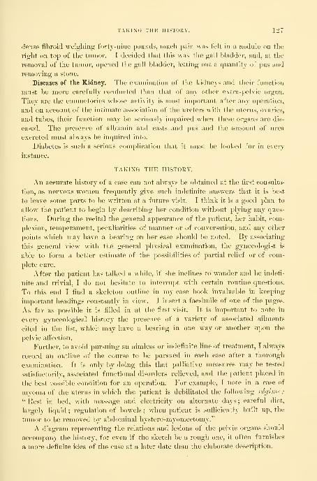
TAKING THE HISTORY. 127
dense fibroid weighing forty-nine pounds, mneh pain was felt in a nodule on tlie
right on top of the tumor. I decided that this was the gall bladder, and, at the
removal of the tumor, opened the gall bladder, letting out a quantity of pus and
removing a stone.
Diseases of the Kidney.—The examination of the kidneys and their function
must be more carefully conducted than that of any other extra-pelvic organ.
They are the emunctories whose activity is most important after any operation,
and on account of the intimate association of the ureters with the uterus, ovaries,
and tubes, their function may be seriously impaired when these organs are dis-
eased. The presence of albumin and casts and pus and the amount of urea
excreted must always be inquired into.
Diabetes is such a serious complication that it must be looked for in every
instance.
TAKING THE HISTORY.
An accurate history of a case can not always be obtained at the first consulta-
tion, as nervous women frequently give such indefinite ansv/ers that it is best
to leave some parts to be written at a future visit. I think it is a good plan to
allow the patient to begin by describing her condition without plying any ques-
tions. During the recital the general appearance of the patient, her habit, com-
plexion, temperament, peculiarities of manner or of conversation, and any other
points which may have a bearing on her case should be noted. By associating
this general view with the general physical examination, the gynecologist is
able to form a better estimate of the possibilities of partial relief or of com-
plete cure.
After the patient has talked a while, if she inclines to wander and be indefi-
nite and trivial, I do not hesitate to interrupt with certain routine questions.
To this end I find a skeleton outline in my case book invaluable in keeping
important headings constantly in view. I insert a facsimile of one of the pages.
As far as possible it is filled in at the first visit. It is important to note in
every gynecological history the presence of a variety of associated ailments
cited in the list, which may have a bearing in one way or another upon the
pelvic affection.
Further, to avoid pursuing an aimless or indefinite line of treatment, I always
record an outline of the course to be pursued in each case after a thorough
examination. It is only by doing this that palliative measures may be tested
satisfactorily, associated functional disorders relieved, and the patient placed in
the best possible condition for an operation. For example, I note in a case of
myoma of the uterus in which the patient is debilitated the following regime
:
" Rest in bed, with massage and electricity on alternate days ; careful diet,
largely liquid ; regulation of bowels ; when patient is sufiiciently built up, the
tumor to be removed by abdominal hystero-myomectomy."
A diagram representing the relations and lesions of the pelvic organs should
accompany the history, for even if the sketch be a rough one, it often furnishes
a more definite idea of the case at a later date than the elaborate description.

128
Date
THE GY>^^ECOLOGICAL EXAMINATION.
Diagnosis
Name s. W. M. Age
Occupation Par Miscarriages
Instr. deliv.
Mcnstr. hist.
Complains now of tlie following s!/mptori
Resides
Patient of Dr.
fever
Gen. appearance
Weight
Headaches
Sleep
Appetite
Digestion
Bowels
Float, kid.
Urination
History of development of present conditi
General previous hist. rheumatism fevers, etc.
Family history
Physical exam, of Pelvis and Abdomen
Vug. outlet
Vagina
Cervix
Uterus
Urinary analysis
Sketch
Fallopian tubes and ovaries
Ontline of treatment to be followed

Fio. 81.—The Four Cardinal Projections of the Abdomen and Pelvis, reduced on the Same Scale.
can be located by following the parallel lines in the three projeeticins. 'I'lic: lower diagrams are viewed perpendicularly to the plane of the


TAKING THE IIISTOUY. 129
There are four outlines necessary to illustrate properly the pelvis and its
contents. Prof. Schultze, of Jena, and Dr. R. L. Dickinson, of this country,
liave devised rubber stamps by means of which a diagrammatic view of several
aspects of the pelvis can be reproduced in a case book, or upon a history blank.
I prefer in some instances, however, to make a free-hand drawing, because indi-
vidual peculiarities may be best brought out in this way. A sagittal section is
used to indicate uterine displacements and the position of tumors in front of or
behind the uterus,
A coronal section through the crests of the ilia, the acetabula, and the tuber-
osities of the ischium is necessary to demonstrate lateral displacements of the
uterus and the location of inflammatory masses on the right and on the left. If
the examination be unsatisfactory, and there is doubt concerning the existence
of disease on either side, an interrogation mark indicates that the question is
unsettled, and leaves it open for future determination in an examination under
anesthesia.
Lesions lateral or posterior to the uterus, in order to be properly indicated
diagrammatically, i*equire an outline of the pelvis looking in from above. Such a
diagram is especially valuable for filling in after operation, because by it the exact
position and relationship of inflannnatory masses to the pelvic organs can be graph-
ically sho^vn. Adhesions are conveniently indicated by zigzag or straight lines.
Areas of resistance in the vault of the vagina not clearly outlined bimanu-
ally are best registered on a diagram of the inferior strait seen from below.
Fig. 81 shows the three geometrical projections of the normal body : first, a
sagital section, viewed perpendicularly to the cut surface ; second, a front view
of the body, seen perpendicularly to its long axis ; and third, a view of the pel-
vis from above and perpendicular to the superior strait. These diagrams have
been drawn on the same scale and are covered by a double system of parallel
lines, thus dividing each of the three projections into a certain number of
squares, which have their corresponding fellows on the other projections. In
other words, the body has been imagined divided into a system of cubes, the
projections of which we see in the three planes as a square network. Thefourth diagram, in the left lower corner of the plate, is a view of the inferior
strait seen from below. It is evident that by following this system every
given point, or a tumor, in the body can be registered with great accuracy, as
illustrated in Fig. 82, where the position of an ovarian cyst has been located in
its three dimensions.
The location of abdominal tumors and dull and tympanitic areas may be in-
dicated on a large diagrammatic outline of the abdomen. In Fig. 83, on the
left, the abdomen is shown in outline with its contained viscera, and those or-
gans from which abdominal tumors most frequently develop are seen distributed
around the periphery ; in Fig 83, on the right, is a diagrammatic illustration
of the directions taken by the various abdominal tumors in the course of their
development, as indicated by the arrows. These directions, as will be seen, are
centripetal—that is to say, from the more resisting periphery to the more yield-
ing center.
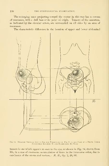
130 THE GYNECOLOGICAL EXAMINATION.
The enlarging mass projecting toward the center in this way has a corona
of resonance, with a dull base at its point of origin. Tumors of the omentum,
as indicated by the circular arrow, are surrounded on all sides by an area of
resonance.
The characteristic difference in the location of upper and lower abdominal
Fio. 82.—DiA(;i£AM SiiowiNCJ How to Use the Projections of Fir. 81 in the Case of a Pelvh' TimorACCUKATELY LoCATINO It ANI) RK(iISTKKIN(i ItS Foi'.M.
tumors is one which appeals at once to the eye, as shown in Fig. 84, drawn from
life, in a case of enormous accumulation of feces in the transverse colon, due to
carcinoma of the uterus and rectum.
—
M. B., Oj). 7, 29, 96,

TAKING THE HISTORY. 131
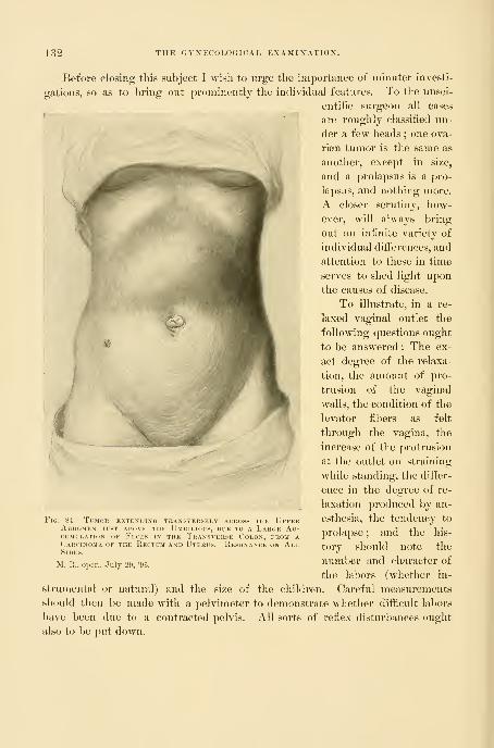
132 THE GYNECOLOGIflAL EXAMINATION".
Before closing tliis subject I wish to urge the importance of minuter investi-
gations, so as to bring out prominently the individual features. To the unsci-
entific surgeon all cases
are roughly classiiied un-
der a few heads ; one ova-
rian tumor is the same as
another, except in size,
and a prolapsus is a pro-
lapsus, and nothing more.
A closer scrutiny, how-
ever, will always bring
out an infinite variety of
individual differences, and
attention to these in time
serves to shed light upon
the causes of disease.
To illustrate, in a re-
laxed vaginal outlet the
following questions ought
to be answered : The ex-
act degree of the relaxa-
tion, the amount of pro-
trusion of the vaginal
walls, the condition of the
levator fibers as felt
through the vagina, the
increase of the protrusion
at the outlet on straining
while standing, the diifer-
ence in the degree of re-
laxation produced by an-
esthesia, the tendency to
prolapse ; and the his-
tory should note the
number and character of
the labors (whether in-
strumental or natural) and the size of the children. Careful measurements
should then be made with a pelvimeter to demonstrate whether diflicult labors
have been due to a contracted pelvis. All sorts of refiex disturbances ought
also to be ])ut down.
Fig. 84.
—
Ttmor extending transvep.sely ackoss the UpperAbdomen just above the Umbilicus, due to a Large Ac-cumulation OF Feces in the Transverse Colon, from aCarcinoma of the Kectum and Uterus. Resonance on AllSides.
M. R., oper., July 211, '9

CHAPTER YI.
GYNECOLOGICAL INSTRUMENTS AND DRESSINGS.
1. Introduction.
2. Illumination.
3. Specula : Nelson's trivalve speculum. Goodell-Baer bivalve speculum. Kelly's small cylin-
drical specula for virgins. Sims's speculum. Simon's speculum.4. Retractors, vaginal and abdominal.5. Knives : Ordinary scalpel. Broad-bladed scalpel, with a large handle.
6. Scissors : Straight, sharp-pointed, and blunt. Emmet's left-curved scissors,
7. Tenacula : Straight. Curved. Corrugated. Shepherd's crook.
8. Forceps: Tenaculum forceps. Long straight dressing forceps. Long rat-toothed forceps.
Rat- toothed dissecting forceps. Hemostatic forceps. Sponge forceps. Polyp forceps.
9. Ligature and suture materials : Silk in three sizes—fine, medium, and stout. Silkworm gut.
Catgut. Kangaroo tendon. Silver wire. Tying knots with silk and catgut.
10. Needles : Curved and straight. Carrier. Needle holder. Transfixion needles.
11. Packer.
12. Glass catheter.
13. Large glass trocars for tapping cysts, curved and pointed./
14. Leg holder.
15. Aspirator : Dieulafoy-Potain aspirator. Syringe aspirator.
16. Cautery : Paquelin's therrao-cautery. Electro-cautery.
GYNECOLOGICAL INSTRUMENTS.
A LARGE, carefully selected armamentarinm is essential to the gynecologist.
He needs instruments of three sorts :
(1) Instruments for exposing the field of operation in vagina or al)domen.
(2) Instruments for special operations.
(3) Instruments and accessories for closing the wound.
The field of the operation is often remote from the surface, either deep down
on the pelvic floor or at the vaginal vault, and necessitates the use of specula
and retractors to make it accessible. But specula are useless without a good
light well directed upon the field ; for this reason I will consider first the prime
requisite—illumination.
The Illumination.—For the illumination of the field of operation, a good
diffused sunlight is Lest of all. This is attained in the operating room by plenty
of windows and a large skylight, and by walls painted with a light color. The
direct rays of the sun are embarrassing, and so a north exposure is best. JSTo
operator, however, can afford to depend on this source of light alone, on account
of the uncertainties of the weather and the frequent call for an intense ihumina-
tion localized at one point.
In an emergency in private practice a common candle with a tin reflector,
or a mirror held so as to direct its rays, may be used. In the clinic the electric
133

134 GYNECOLOGICAL INSTRUMENTS AND DRESSINGS.
light is the l)est artificial ilhmiinaiit. The current may he conducted from a
wall hracket hy a long insulated fiexihle wire to the 16-candle-power lamp,
with a tin reflector enameled white inside and attached to a handle, as shown in
the figure in Chapter XX. This can be held by an assistant so as effectively
to illuminate the wound area. A simple extemporized reflector may be made
by enclosing the electric light in a cone of white paper covered with black cloth.
Where an electric street current is not available, a storage battery is a satis-
factory substitute, running a 6- or 8-candle-power lamp.
Specula.—For inspection of the vaginal vault the best specula are Nelson's
trivalve speculum, Goodell's bivalve speculum, modified by B. F. Baer, and
Kelly's small cylindrical specula, Nos. 12-15 of the cystoscopic set, for use in
the virgin. The utility of these instruments is limited to an examination for
diagnostic purposes, to treatments applied to the vaginal vault, and to the appli-
cation of packings. Sims's and Simon's specula are useful both in making an
examination and in exposing the field during an operation at the vaginal vault,
serving the double purpose of specula and retractors. The Sims's speculum is
most used by the New York school of gynecologists, and is more serviceable
with the patient in the left lateral position. Several sizes
are needed, differing in length and breadth, for narrow and
relaxed and for long and short vaginas. The Simon specula
are purchased in sets, and consist of two handles with ad-
justable blades of varying lengths and breadths, for both
the anterior and the posterior vaginal walls. They are used
in the dorsal position.
Vaginal retractors, with long light handles, are used to
hold back the lateral and upper walls of the vagina, and to
keep the field of operation free. The blades of these re-
tractors should be of two sizes—2 by 7 centimeters and 3 l)y
7 centimeters.
Abdominal retractors serve to lift up or to draw aside
one of the walls on either side of an abdominal incision to
enable the operator to inspect the pelvic viscera. The best
patterns are Ilalsted's, with concave blades^ 4 Uy 7 and 6 by
7 centimeters in size.
Knives.—The knives used in gynecological surgery are
the ordinary scalpels, made of solid metal, with handles
smooth or grooved to afford a better grasp. For opening
the abdomen, I like a broad-bladed scalpel with a large han-
dle, and for marking areas of denudation in the vagina or
on the cervix, or for delicate dissections in the pelvis, I pre-
fer a knife with a slender blade and a sharp point.
In transporting or sterilizing knives the blades must be wra])ped in cotton,
or they must be fastened in a rack in a metal box to protect them.
Scissors.—Scissors are among the most important of all gynecological in-
struments, and, through the inventive genius and teaching of Dr. T. A. Emmet,
Fig. 85.—Emmet's Lbft-CL'RVED Scissors fokPlastic OpekationsAT THE Vaginal Out-let.
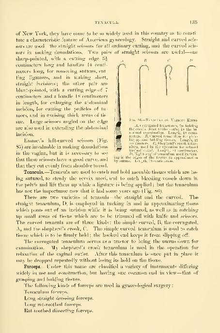
TENACULA. 135
Fig. Tenacula of Various Ki
hyin tl
>iitur
A, Corrugated tenaculum, for holdingthe cervix down to the outlet in the bi-manual examination. Length, 18 centi-meters. £, Curved tenaculum for pick-ing up and holding tissues. Length, 22centimeters. C, Shepherd's crook tenac-ulum, used in the operation for relaxedvaginal outlet. Length, 21 centimeters./>, Right-antrled tenaculum used in turn-e edyes of the tissues in approximationi. Length, 19 centimeters.
of ISTew York, thej have come to be so widely used in this country as to consti-
tute a characteristic feature of American gynecology. Straight and curved scis-
sors are used—the straight scissors for all ordinary cutting, and the curved scis-
sors in making denudations. Two pairs of straight scissors are useful—one
sharp-pointed, with a cutting edge 5^^ j, ^, p
centimeters long and handles 14 centi-
meters long, for removing sutures, cut-
ting ligatures, and in making short,
straight incisions ; the other pair are
blunt-pointed, with a cutting edge of 7
centimeters and a handle 18 centimeters
in length, for enlarging the abdominal
incision, for cutting the pedicles of tu-
mors, and in excising thick areas of tis-
sue. Large scissors angled on the edge
are also used in extending the abdominal
incision.
Emmet's left-curved scissors (Fig.
85) are invaluable in making denudations
in the vagina, but it is necessary to see
that these scissors have a good curve, and
that they cut evenly from shoulder to end.
Tenacula.—Tenacula are used to catch and hold movable tissues which are be-
ing sutured, to steady the cervix uteri, and to catch bleeding vessels down in
the pelvis and lift them up while a ligature is being applied ; but the tenaculum
has not the importance now that it had some years ago (Fig. 80).
There are two varieties of tenacula—the straight and the curved. Thestraight tenaculum, D, is employed in tucking in and in approximating tissue
which pouts out of an incision while it is being sutured, as well as in catching
up small areas of tissue which are to be trimmed off with knife and scissors.
The curved tenacula are of three kinds : the simple curved, B, the corrugated,
A, and the shepherd's crook, C. The simple curved tenaculum is used to catch
tissue which is to be firmly held ; the hooked end keeps it from slipping off.
The corrugated tenaculum serves as a tractor to bring the uterus down for
examination. My shepherd's crook tenaculum is used in the operation for
relaxation of the vaginal outlet. After this tenaculum is once put in place it
may be dropped repeatedly without losing its hold on the tissue.
Forceps.—Under this name are classiiied a variety of instruments differing
widely in use and construction, but having one common end in view— that of
grasping and holding tissues.
The following kinds of forceps are used in gynecological surgery
:
Tenaculum forceps.
Long straight dressing forceps.
Long rat-roothed forceps.
Rat-toothed dissecting forceps.

136 GYNECOLOGICAL INSTRUMENTS AND DRESSINGS.
Hemostatic forceps.
Sponge forceps.
Polyp forceps.
Tenaculum forceps, or doiil)le tenaculum forceps, resemble two tenacula fast-
ened so as to work together in opposite directions. They are used to grasp and
draw the cervix down, to steady it while the uterine dilator
is introduced, and to catch and hold a bleeding pedicle
which has dropped back into the abdomen. They ought
to be made strong enough to resist feathering, and the ends
must be slightly curved at right angles to the sliaft and
tapered, as shown in the figure, to prevent tearing tlie tis-
sues. The iigure (Fig. 87) shows a small tenaculum for-
ceps which I have found especially useful. If the tenacu-
lum tears out, a three-pronged tenaculum may be used to
advantage (Fig. 88).
Long straight dressing forceps are constantly used in re-
moving and applying dressings, in carrying pledgets of cot-
ton into the vagina to cleanse it, and in making applica-
tions.
Long Rat-toothed Forceps.—I find a pair of long rat-
toothed forceps, like those figured in the text (Fig. 89),
one of my most useful instruments in abdominal surgery,
effectually taking the place of a hand deep down in the
pelvis.
Rat-toothed dissecting forceps are needed in picking upthe layers of tissue, in making the abdominal incision, and
in catching the tissue in vaginal and cervical denudations.
Hemostatic Forceps. At least two dozen artery forceps
should be included in a set of abdominal instruments, but
only four sets are required for most vaginal operations.
The original forceps were devised by Koeberle, of Strass-
burg, and are excellent for the compression of vessels
lying in soft tissues, as in the abdominal walls and on the
floor of the vagina. For general use the forceps figured
in the text and in use in the Johns Hopkins Hospital are the most satisfactory
(Fig. 90). They are 15 centimeters
in length, and have a curved biting
surface 4 centimeters long; the lock
shown in the figure is an improvement
on my own lock. The especial points
of value in these forceps are (1) that
the jaws are longer than usual and
gently curved, and (2) that the tips
of the jaws grasp the tissue before the first shoulder is reached. This ])er-
mits a small bit of tissue or an artery to be clamped by the points if the
Fig. 87. — TENAniAFOKOEPS, WITH CaT(AND Open Lock.
;
Okdinap.y Size.
FlO. 88.—Cl'LLKN's

SPONGE FORCEPS OR HOLDERS. 137
forceps are only closed one or two notches, while a large area may be
clamped if they are closed completely.
Sponge Forceps or Holders.—Sponges
in abdominal surgery are chiefly of serv-
ice in cleansing the pelvic cavity, in tak-
ing up pus rapidly, and in holding back
the intestines. The best sponge holder
is my own with a lock devised by Dr. G.
B. Miller, of the gynecological staff of the
Johns Hopkins Hospital, and shown in
the accompanying figures (Fig. 91). Theessential features of these forceps are the
blunt teeth at the lower end which hold
the sponge, and the clasp which slides
freely under one handle until it is slipped over the neck of the other handle
and pushed down, fixing the sponge. The entire length of the forceps is 22-5
centimeters, and the whole separates into three pieces for cleansing.
sL> OF Large Kat-toothed Forceps,FOR l\SE DEEP IN THE PeLVIS. WhOLE LeNGTH,3 Centimeters.
Fig. 90.—Hemostatic ForcepsWITH Open Lock.
Miller's Sponge Forceps.
The hook shown above, in outline, hangs loose on the roundhandle until the forceps are locked, as seen in the right-hand figure.

138 GYNECOLOGICAL INSTRUMENTS AND DRESSINGS.
Polyp Forceps.—The best foi'ceps for grasping small polyps or for removing
a small ovum or pieces of placenta are those shown in the text (Fig. 92). Theform of the blade is shown in the figure, and the handles are 2T'5 centimeters
long and provided with a catch.
The ligature and suture materials used in gynecology are silk,
silkworm gut, catgut, and silver wire, which are conveniently abbreviated in
clinical records by using the initial letters only before the word" suture," as s., s. w. g., c. g., s. w. sutures.
Silk.—Pure Chinese silk is used in thi-ee sizes—fine, me-
dium, and coarse.
Fine silk is best adapted for the ligation of small ves-
sels, for suturing the intestines, for approximating peritoneal
surfaces, and for bringing wound surfaces into aj^position when
there is no tension.
Medium s i 1 k is used in ligating large vessels and in
tying o& the ovarian vessels in a bunch. This size should
always be used in preference to heavier silk in all cases where
it can stand the strain.
Coarse silk ligatures should only be used in vagi-
nal hysterectomy in tying off the broad ligaments. Coarse silk
ligatures are also used as tractors to pull the uterus down in
vaginal hysterectomy.
Silkworm gut is one of the best plastic suture materials wehave, and once introduced and tied or clamped with shot, pre-
serves a well-rounded, elastic loop indefinitely or until it is re-
moved. The fact that it possesses no meshes gives it a great
advantage over silk, which in time forms a seton, furnishing
a highway of communication for germs from the surface into
the deeper tissues. Silkworm gut is rarely used as a buried su-
ture, either in the abdominal cavity or in the vagina. It is
used by many surgeons in closing the abdominal wound after
celiotomy, in cervical operations, and as a tension suture in
the operation for relaxed vaginal outlet or lacerated perineum.
Catgut, 2)roperly sterilized, is valuable as ligature and suture
material, because it is absorbed by the tissues and does not re-
<|uire removal. The chief objections to catgut are the difii-
culty of sterilizing it, its too rapid absorption, and the fact
that it may come untied. Only intermediate and heavy-sized
catgut should be used, as finer strands are too weak. AVater
swells and softens catgut so quickly that it must be kept im-
mersed in alcohol until it is used. The too rapid absorption of catgut is pre-
vented by the pi'eparation in cumol (see (^hajiter I). In vaginal operations cat-
gut is chiefiy used as an accurate approximation suture ; if it holds but four
days, the tissues, as a rule, are sufKciently united, so that sutures are no longer
necessary. Its greatest advantage here is that the removal of sutures is avoided.
/J.
Fig. 92.— PlacentaAND Polyp For-ceps. Length,30-5 Centime-TEIIS.

SILVER WIRE. 139
Kangaroo tendon, the split sinews of the kangaroo's tail, introduced by Dr.
Henry O. Marcy, of Boston, has the advantage of being absorbed much more
Fig. 93.
—
Rapid Method of tying the Square Knot.
First step : the tirst knot is tied and the ligature grasped as shown In the figure.
slowly than catgut. It is useful in all forms of suturing and ligating, and Dr.
Marcy advocates it especially for radical hernia operations.
Silver Wire.—Stout silver wire has been introduced by Dr. W. S. Halsted
as a buried suture. Its chief use as a permanent suture is in holding together
Fig. 94.—Second step : the end b is passed over a to make the second knot.
the fasciae of the abdominal incision when it is closed, and in uniting the
muscles and fasciae in the radical cure for hernia. The wire is best intro-
duced as a mattress suture, the ends twisted four times at an obtuse angle,
cut off, and turned down at one side of the incision. These sutures re-

140 GYNECOLOGICAL INSTRUMENTS AND DRESSINGS.
main indefinitely in place, and rarely have to be taken out, like bnried sutures
of silkworm gut.
Tying Knots with Silk and Catgut.—Much time may be lost by tying knots
clumsily, and the surgeon will be a gainer all his life long if he will learn at the
outset a definite rapid method of tying both silk and catgut. I always tie in
the following manner : the first knot is
tied with the inner strand in the right
hand, thrown over and then under
the outer strand held in the left
,i#fc.
. 95.—Tlind strp: the end h is lidd m the kft liandwhile a \h taken in tlie rit'lit hand and thb knotdrawn home.
hand, and drawn tight down on the vessel.
Then, by following the four steps wdiich are
seen in the diagrams better than can be described,
the second knot is quickly drawn down on the first
and tightened by supj^orting the thumbs against each
other as shown, to avoid tugging on the tissue (Figs. 93,
94, 95, and 90).
Silkworm gut is
best tied in a square knot, and
after immersion in warm water.
Catgut is best tied dry. If a
third knot is added, either to
the silkworm gut or the cat-
gut, as suggested by Dr. C. P.
Noble, the ends may then be
safely cut off close to the liga-
ture; the use of a third knot
leaves less foreign material be-
hind, and the knot is less liable
to shp, and also, in the case of
silkworm gut, the little ends
which are liable to irritate the
tissue arc removed.
Needles. — Curved needles
(Fig. 97) are the best for almost
all gynecological purposes ; they
should be of three sizes, as shown in figures, and must answer the following
requirements : A good temper, a good open eye, a short straight shank just
Fig. OG.—Final step: tying the ligature tight without roding the tissue by buttressing' the thumbs against eai
other. The dotted line sliow.s the overlappinLr of tl
left thumb bv tlic right.

THE SUTURE CARRIER. 141
telow tlie eye for the grasp of the needle holder, and a cutting surface not
wider than the body of the needle ; the point must follow the curve of the
needle, and must not be bent inward. One of
the commonest faults is a kink or a curve just
below the eye, making the needle liable to break
in the grasp of the holder. Simple straight cam-
bric needles, with a round sharp point and with-
out any cutting edge, are the best for intestinal
suturing ; they are held in the fingers so that the
sense of resistance at the point may enable the
operator to recognize the position of the subnm-
cous fibrous coat, and so to pick it up.
The Suture Carrier.—Tlie suture carrier is a
silk loop tied to the eye of a needle for the pur-
pose of pulling interrupted sutures through in
rapid succession. It is tied by taking a long
piece of medium silk 52 centimeters (21 inches)
Fig. 97, Needles,
Used in plastic work and all kinds ofsuturing, except suture of the intestines.
StraijSfht straw needle, used in intestinal
suturing, on the right.
Fig. 98. Fig. 99.
First and .second steps in making the silk carrier. Both ends of the thread are put through the eye of theneedle in the same direction, and a loop is formed passing over tlie needle in the direction of the arrow,as shown in Fig. 08 ; the loop is then brought down below the eye and drawn tight, fixing the carrier,
as shown in Fig. 99.

142 GYNECOLOGICAL INSTRUMENTS AND DRESSINGS.
lon^, putting both ends together through the eye of a needle, and then mak-
ing a loop on one of the ends, slipping it over the needle beyond the eye,
and pulling it tight (Figs. 9S and 99). The length of the carrier loop made
in this way is 20 centimeters (8
inches) long. In using the car-
rier the needle is passed through
Fig. 100.
—
Needle Foeceps forCurved Needles. % Ordi-nary Size.
Fig. lOL
—
Needle Forceps showing the ShapeOF THE Bite and the Manner of graspingTHE Needle on the Flat Part just belowthe Eye.
the tissue, and when the loop, threaded by the assistant, is drawn through,
the suture is in place. Sutures may be placed more rapidly in this way than
by any other means. The intestinal needles are each armed with a single
thread of fine black silk. The carrier is never used here to avoid making any
larger hole than is absolutely necessary.
Needle Holder.—The most satisfactory needle holder for curved needles is
the one figured here (Figs. 100 and 101).
It is important that the handles should be large enough to afi'ord a good
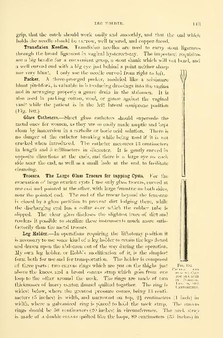
LEG HOLDEK. 143
grip, that the catch should work easily and smoothly, and that the end whichholds tlie needle should be narrow, well tapered, and cojDper-faced.
Transfixion Needles.—Transfixion needles are used to carry stout ligatures
through the l)road ligament in vaginal hysterectomy. The important requisites
are a big handle for a convenient grasp, a stout shank which will not bend, anda well curved end with a big eye just behind a point neither sharp
nor very blunt. I only use the needle curved from right to left.
Packer.—A three-pronged packer, modeled like a miniature
blunt pitchfork, is valuable in introducing dressings into the vagina
and in arranging properly a gauze drain in the abdomen. It is
also used in packing cotton, wool, or gauze against the vaginal
vault while the patient is in the left lateral semiprone position
(Fig. 102.)
Glass Catheters.—Short glass catheters should supersede the
metal ones for women, as they are so easily made aseptic and kept
clean by immersion in a carbolic or boric acid solution. There is
no danger of the catheter breaking while being used if it is not
cracked wdien introduced. The catheter measures 13 centimeters
in length and 5 millimeters in diameter. . It is gently curved in
opposite directions at the ends, and thei-e is a large eye on each
side near the end, as well as a small hole at the end, to facilitate
cleansing.
Trocars. The Large Glass Trocars for tapping Cysts.—For the
evacuation of large ovarian cysts I use only glass trocars, curved at
one end and pointed at the other, with large fenestrae on both sides,
near the pointed end. The end of the trocar beyond the fenestrae
is closed by a glass partition to prevent dirt lodging there, while
the discharging end has a collar over which the rubber tube is
slipped. The clear glass discloses the slightest trace of dirt and
renders it possible to sterilize these instruments much more satis-
factorily than the metal trocars.
Leg Holder.—In operations requiring the lithotomy position it
is necessary to use some kind of a leg holder to retain the legs flexed
and drawn upon the abdomen out of the way during the operation.
My own leg holder, or Robb's modification of it, is the simplest
form both for use and for transportation. The holder is composed
of three parts : two canvas rings which are put on the thighs just
above the knees, and a broad canvas strap wdiich goes from one
loop to the other around the neck. The rings are made of two
thicknesses of heavy canton flannel quilted together. The ring is
widest below, where the greatest pressure comes, being 13 centi-
meters (5 inches) in width, and narrowest on top, 2^ centimeters (1 inch) in
width, where a galvanized ring is placed to hold the neck strap. The canvas
rings should be 50 centimeters (20 inches) in circumference. The neck strap
is made of a double canvas quilted like the loops, 89 centimeters (35 inches) in
Fig. 10-2.
Packj:r forPLACING Cot-ton OR GauzeIN Position.Length, 20-5
Centimeters.

144 GYNECOLOCxICAL INSTRUMENTS AND DRESSINGS.
length, and 6| centimeters (2^ inches) wider in the middle, gradually tapering
to the ends. Harness straps at the ends and three metal rings about 15 centi-
meters apart make the leg holder adjustable.
Aspirator.—The aspirator, at one time largely given up, has in recent years
again become an indisjjensable instrument. The Dieulafoy-Potain aspirator is
one of the best, and is so well known as to need no description. During the
operation the aspirator should be in the hands of a competent assistant, who
should be sure that the bottle is well exhausted and the suction channel unob-
structed before use. Immediately after using the instrument the suction tube
should be cleansed by creating a vacuum in the bottle and immersing the point
in warm water. The tubing, after being washed out in this way, should be
placed in a bichloride solution (1-1,000) for at least an hour, after which it is
dried and put away in the case. The needles and trocars should be sterilized
after every operation by boiling in a carbonate of sodium solution (1 per cent)
for five minutes, and dried in a Bunsen or an alcohol flame. The receiving
bottle should be sterile, as it is often desirable to make cultures from its con-
tents.
Syringe Aspirator.—My own aspirator is like a large glass syringe, a pint in
capacity, with a metal ftoint to w^hich a piece of rubber tubing, with a needle,
is attached. The piston must fit perfectly to keep the air from entering. Aswitch and an opening on the side provide for the discharge of the contents of
the barrel without withdrawing the trocar.
Cautery.—The term cautery is used in contrast to chemical cauterization pro-
duced by nitric acid, chloride of zinc, nitrate of silver and caustic potash, etc.
The irons of ancient surgery have been replaced in modern times by
Paquelin's thermo-cautery or one of its modifications, too familiar to need de-
scription. One of the best forms is that in which the tube passes directly
through the middle of a small bottle holding the benzine.
The Electro-cautery.—In the clinic room an electro-cautery is often moreconvenient than the thermo-cautery. The electricity is supplied from a storage
battery, or, better still, from a street current which is cut down. I use in myclinic an alternating current controller, in which induction is used for resistance,
in place of a sectional coil and point switch. A 52-volt alternating current is
employed and reduced by the controller from to 5 volts with an amperage of
from 1 to 35.
CHAPTER VII.
ANESTHESIA.
1. Local and general anesthesia.
2. Local : Cold. Cocain. Endermic injections.
3. General anesthesia : Introductory. Anesthetizer. Signs of complete rehixation. Dangersymptoms. Oxygen after anesthesia. Anesthesia slip.
4. Chloroform.5. General rules for administering any anesthetic.
6. Rules for administering chloroform.
7. Resuscitation of the asphyxiated.
8. Ether : Operation under the primary effect of ether.
9. Death from anesthesia.
The choice of the best anesthetic and the safest method of administerinp; it
are questions of the utmost importance, for its improper use often mocks at skill
and converts one of the greatest surgical blessings into an agent of death.
There are two forms of anesthesia employed in gynecology—local, in which
only a small area of the body is anesthetized, and general, where the anesthetic
is inhaled and induces a state of unconsciousness, during which the most exten-
sive and prolonged operation can be done without pain.
LOCAL ANESTHESIA.
Local anesthesia is best adapted to those cases in which the operation is con-
fined to a small exposed area, whether on the surface of the body or in the vagina,
where the operation is of a minor character and of short duration. Local anes-
thesia is induced either l)y applying cold or a solution of cocain, or by injecting
normal salt solution into the deeper layers of the skin, or by constriction.
Cold.—Cold anesthetizes the surface by reducing the temperature close to the
freezing point, paralyzing the nerves of sensation. The application of cold for
anesthetic purposes about the pelvis is restricted to a narrow field. It may thus
be employed in " freezing " the skin over a labial abscess, which can then be
quickly opened, or in benumbing the skin on the lower part of the abdomen for
the purpose of making a small incision through which a trocar is to be thrust to
tap an ascites or an ovarian cyst.
Anesthesia by cold may be produced either by the application of ice or by
directing a fine ether spray against the part for about five minutes. If a lump
of ice is used it should be sprinkled with salt, wrapped in a thin cloth, and the
salted side applied to the spot for about five minutes, when the blanched surface
will show the effect of the agent. The refrigeration of the surface l\y ethyl
chloride is perhaps the best way to apply cold over a small area, and I know of
12 145

146 ANESTHESIA.
no better arrangement than the ethyl chloride (Bengue) supplied to the trade in
glass vials with brass tops perforated by a capillary opening and closed by a
screw cap. Each tube contains 30 grams of ether, and is sufficient for from ten
to fifteen minor operations.
The efficacy of the ethyl chloride depends upon its low boiling point, which
is 12"5° C. Ethyl chloride anesthesia will be found valuable in such minor
operations as evacuating abscesses about the vulva, opening stitch-hole abscesses,
incising a suppurating pile, etc. The anesthetic effect is obtained by holding
the nozzle from six to eight inches away from the skin while the fine spray
plays upon it. The color at once changes, and in less than half a minute a
white parchment-like appearance is produced, with an anesthesia which lasts
about two minutes. The freezing is more rapid in summer.
Cocain.—Cocain hydrochlorate may be used for short operations on the skin
or the mucous membrane, either by local application or by injection under the
surface of the skin. Operations to which cocain is best adapted are the removal
of pediculated tumors, the incision of a suppurating gland or a vaginal cyst, or
in the preparation of the surface of the rectal mucosa to render painless the
injection of hemorrhoids with carbolic acid, or in allaying the sensitiveness of
the urethra before introducing a speculum. It takes about five minutes to pro-
duce local anesthesia by this means. Solutions of cocain should never l)e in-
jected into the urethra or rectum, as the drug is quickly absorbed, and in a
certain percentage of cases its use is followed by collapse.
I did a celiotomy in 1888 under local anesthesia produced by injecting 10
or 12 minims of a 2 per cent solution of this drug at several points in the line
of incision. The patient experienced no pain until the peritoneal cavity was
opened and the pelvic organs were pulled upon. The incision was sewed up
while the tissues were yet under the effects of the cocain, and the patient was
put to bed, having been conscious of every step of the operation, with only
slight pain.
For operations in the vagina requiring local anesthesia a pledget of absor-
bent cotton is saturated with a 5 to 10 per cent solution and applied to the part
for five minutes. The application may be repeated from time to time during
the operation, although a free flow of blood seriously interferes with the effect-
iveness of subsequent applications by washing away the solution as soon as it
comes in contact with the tissues.
Cocain may sometimes be used to enable the surgeon to operate upon the
perineum without resorting to a general anesthesia. In this case a few minims
of a 2 per cent solution should be injected by multiple punctures quite super-
ficially along the line of incision or denudation. Such an operation can only
be done on a patient who has excellent control of her nerves. It is well to
begin the operation about three minutes after the injection and before the fluid
is absorbed ; the denudation in the anesthetized tissues then permits the injected
fluid to escape over the wound and keeps up an anesthetic effect. Such an
operation must be performed rapidly, all materials must be at hand, and assist-
ance must be prompt. The concluding steps are sometimes painful, and are

GENERAL ANESTHESIA. 147
completed satisfactorily only by exercising a great deal of moral suasion over
the patient. The great difficulty in local anesthesia by injecting cocain is that
it is often impossible to tell beforehand how long an operation will last, and in
long operations cocain can not be depended upon.
Endermic Injections.—This method has superseded in many clinics the use of
cocain, proving more efficient and less dangerous. The procedure is simple
and rapid, and for minor operations on skin surfaces is the best method devised.
It consists of injecting by the hypodermic needle some innocuous fluid into the
deep layers of the skin, producing thereby a small area of localized edema.
The principle is the same as that of inducing anesthesia by constriction, the dis-
tention being sufficient to stop the circulation and paralyze the terminal nerve
endings. Sterilized normal salt solution is the fluid commonly used.
The technique of the method is as follows
:
The field of operation nmst be thoroughly cleansed and the procedure con-
ducted throughout with tlie usual antiseptic precautions. The tilled hypodermic
needle is then introduced as nearly parallel to the surface as possible until the
deep layers of the skin are reached. The fluid is then slowly forced in until a
wheal from 1 to 3 centimeters in size is raised. This becomes blanched and
sharply defined from the surrounding skin. By successive injections into the
periphery of the wheals an area of desirable size can be anesthetized without
further pain. The effect disappears as the artificial edema is absorbed, but the
period is of sufficient length to enable one to open al)scesses, remove sutures, or
excise small tumors.
Schleich advocates injecting in the same manner a weak solution of mor-
phine and cocain, but it does not seem to improve the efficiency of the method,
the quantity of the drugs being too small to produce a decided physiological
action.
GENERAL ANESTHESIA.
General anesthesia suspends consciousness, relaxes the whole body, and puts
the patient for the time completely at the disposal of the operator. It is there-
fore suitable for prolonged, difficult, and painful operations.
The Anesthetizer.—The office of the anesthetizer is scarcely secondary in im-
portance to that of the operator ; it is one of the most serious errors to hold
that this responsible position may be delegated to an inexperienced person or a
mere student, for tinndity or bad judgment on the pai-t of the anesthetizer
may result in an imperfect anesthesia which interrupts and harasses the operator,
while too profound an anesthesia may kill the patient on the table or by an
excessive use of the drug produce a bronchitis or pneumonia which may prove
fatal.
An unskillful anesthetizer is also prone to forget his office and become ab-
sorbed in the operation with imminent risk to his charge. As the surgeon's
attention must be engrossed by the operation, it is highly essential that the
assistant who gives the anesthetic should be thoroughly reliable, because to him
must be intrusted the administration of stimulants if danger symptoms arise ; if

148 ANESTHESIA.
the operator has to direct the anesthetizer, it is confusinpj to both, and the anes-
thetic is likely to be improperly administered. If it is necessary to call upon an
inexperienced person to administer the anesthetic, he should be fully instructed
beforehand, and the operator should be constantly on the watch.
Ether and chloroform are the only anesthetics universally used, and each of
them has its marked peculiarities. The employment of the one or the other is
for the most part determined rather by national and geographical boundaries
than by the special adaptability to the particular case. Chloroform, for exam-
ple, is used almost universally in England and on the continent of Europe,
although ether has been recently making its way more and more into the Ger-
man clinics. Ether is par excellence the anesthetic of the United States, but
this is not without the notable exception of nearly all the Southern States,
where chloroform is used almost exclusively.
The anesthetic of the future will certainly be given in an atmosjjhere defi-
nitely diluted, Spenzer ( Western Reserve Medical Journal^ November, 1894)
has recently definitely shown that ether in a 3'5 per cent solution can be given to
dogs for hours without ill effects, while 6 per cent will prove fatal in a short time.
In prolonged operations or operations upon debihtated patients, the patient
should be kept thoroughly warm, to counteract the depressing drop of tempera-
ture of the anesthesia ; this is best accomplished by hot-water bags placed near
different parts of the body.
Signs of Complete Relaxation.—-1. Loss of conjunctival reflex. The commonpractice of testing the eye reflex by touching the conjunctiva with the finger tip
is to be severely condemned, for not a few patients have developed a severe con-
junctivitis from such treatment.
2. Fixed, contracted pupils.
3. Slow, regular, and deep inspiration.
4. Complete loss of general reflexes and resistance.
I have never had occasion to pass a ligature through the tongue to pull it
forward. This can only be necessary during operation on the face or throat,
where it is impossible to pull the jaw forward and throw the head backward,
which if skilfully done will open the upper air passages. In hundreds of cases
I have never been compelled to use swabs to clear the throat and mouth of
mucus. If the position of the head and jaw is correct, the collection of mucuswill work itself into the mouth, where it can be gently removed by a soft towel
or a piece of gauze.
I have found the greatest difficulty in anesthetizing patients addicted to the
use of morphine and alcohol. It is sometimes almost impossible to ol)tain com-
plete relaxation, and the breathing throughout is stertorous, and interferes
seriously with the proper exposure in abdominal operations by constantly forc-
ing the intestines into the fiekl of operation. Stertorous breathing, if pro-
longed, is an indication of asphyxia, and is usually (piickly relieved by allowing
the patient more air, or by throwing the head backward and the jaw forward,
or by clearing the mouth of mucus.
The diflerence in color of the face between simple mechanical asphyxia and

OXYGEN AFTER ANESTHESIA. 149
that produced physiologically by the drug is worthy of attention ; in the former
the face becomes blue, the eyes protrude, and the features swell, while in the
latter the change is more gradual, the face does not swell, it becomes livid, and
changes slowdy into a grayish pallor. This pallor is often the first signal of
danger, as the respirations may become shallower and shallower imperceptibly
without mechanical signs of interference, and the anesthetizer may be unawareof the change.
The danger symptoms are :
1. Cessation of respiration.
2. Stoppage of the pulse.
3. Sudden pallor.
-i. Dilated, fixed pupils.
5. Dark-hued blood replacing bright arterial blood.
6. Sudden cessation of bleeding in the course of operation. The anesthe-
tizer will naturally notice the first four points, and the operator the other two,
and sometimes the first.
Pneumonia may follow the administration of the anesthetic, whether ether
or chloroform is given. Out of 1,800 administrations I have seen this complica-
tion eight times. The liability to pneumonia is increased if the patient has a
slight bronchitis or a coryza beforehand.
Oxygen after Anesthesia.—The administration of oxygen gas to the patient
coming out of the anesthetic is at present being extensively employed, both to
hasten the complete recovery of consciousness and to lessen the nausea. It is
particularly reconmiended for old and feeble patients, and for those who have a
tendency to bronchorrhea, and after prolonged, exhausting operations. The
oxygen, stored in a small cylinder, is passed through a bottle containing water,
by which the rate of flow can be estimated ; it is then given diluted with the air
by holding the end of a tube near the face. Chloroform may be administered
in the same way by allowing the oxygen to pass through a bottle containing
chloroform instead of water.
Oxygen is also often given during the anesthesia in the same manner by
conducting a rubber tube connected with the cylinder of compressed oxygen
under one side of the ether cone or through its point, or indeed by passing a
small rubber tube into one nostril even. The gas liberated by the removal of
the pressure slowly bubbles through the water bottle and enters the air passages
along with the anesthetic with every breath.
Although many surgeons express great satisfaction with this adjuvanl^ its
real value has not yet been determined and awaits careful investigation.
The following slip is kept and filled out by the anesthetizer in my clinic :
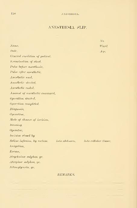
150 AXESTHESIA.
ANESTHESIA SLIP
Name,
Date,
General condition of patient.
Examination of chest.
Pulse before anesthesia.
Pulse after anesthetic.
Anesthetic used.
Anesthetic started.
Anesthetic ended.
Amount of anesthetic consumed,
Operation started.
Operation completed.
Diagnosis,
Operation,
Mode of closure of incision.
Dressing,
Operator,
Incision closed by
Saline infusion, by rectum.
Irrigation,
Enema,
Strychnince stdphas, gr.
Atropinm sulphas, gr.
Nitro-glycerin, gr.
No.
Ward,
Age,
into cd)domen. into cellular tissue.
REMARKS.

CHLOROFORM. 151
CHLOROFORM.
Only chloroform manufactured by perfectly responsible chemists should l)e
used, owing to the increased dangers from adulteration.
The advantages of chloroform are in some respects greater than those of any
other anesthetic. By its means we are able to bring the patient more quickly
and more comfortably into a state of unconsciousness. She remains more quietly
under its influence, and as a rule nausea is not so extreme as after taking ether.
In my experience the percentage of cases entirely free from nausea is as great
after ether as after chloroform anesthesia. Out of 50 cases of chloroform
anesthesia, 6 were free from nausea afterward ; and out of 200 cases of ether
anesthesia, 28 were free. These advantages are more than counterbalanced,
however, by the greater risk to life in using chloroform. The mortality from
chloroform is about one case in 3,000. Chloroform is contra-indicated on ac-
count of its increased danger in a weak heart or in an overtaxed right heart.
It is not contra-indicated in valvular disease with good compensation or in any
particular form of abdominal disease. It is also probably better in nephritis,
but, as Hare says, quantity for quantity, compared with ether, it is more irri-
tating in this condition. Its administration is preferable in old people with
atheromatous vessels, in children, or in patients who struggle violently.
Operations must not be performed under chloroform during the stage of
primary anesthesia, so frequently utihzed for short operations under ether.
Deaths have occurred in this way which were apparently due to sudden inhibi-
tion of the heart from shock not felt in complete anesthesia. The quiet even
anesthesia produced by chloroform is such an advantage in abdominal surgery
that it would be indicated in all cases were it not for these dangers.
It kills by paralyzing either the heart or, more frequently, the respiratory
centers, according to the report of the Hyderabad Chloroform Commission,
which is supported by the investigations of Dr. H. A. Hare. The first symp-
tom of danger is either a complete relaxation of the pupils, a sudden pallor of
the face, or a weakening of the respiratory movements, which become feeble and
intermittent, accompanied or followed by sudden or gradual failure in the pulse.
It has been my experience in two cases to see the respirations fail first, while in
at least two other cases there was an alarming failure in the heart's action, the
respiratory movements being still good. The first warning may be the sudden
ashy pallor, which should call for immediate resuscitative measures, as it is al-
ways a precursor of graver danger. The respiration should be as closely observed
as the pulse, and any change in its depth or rhythm should be carefully noted.
Before giving any anesthetic at all the character of the respiration must be
noted, and the heart must be carefully examined.
The nature and action of the anesthetic should be explained to the patient,
and it is always best to tell her that she may hear peculiar sounds or that she
may have the sensation of falling, etc. ; otherwise the occurrence of these phe-
nomena in the first stage of anesthesia may frighten her, causing her to struggle
violently. The face should be lightly anointed with vaseline and the eyes and

152 ANESTHESIA.
inoiitli covered with soft towels. I have seen the whole side of the face badly
burned by chloroform, due to the neglect of this precaution. The room must
be perfectly quiet, and no talking should be permitted to excite the patient and
retard the progress of the anesthesia.
It is best for the anesthetizer to accustom himself to taking the pulse in the
temporal or facial artery. It is much more convenient than the radial pulse.
The following rules regarding the preparation for anesthesia apply to the
administration of both ether and chloroform :
1. The diet should be carefully regulated, if possible, several days before ad-
ministering an anesthetic, only easily digested foods being given. On the day
preceding, liquid or soft diet should be insisted upon. During this period the
bowels must be freely moved each day, either by an enema or a mild laxative.
Nausea and vomiting will be much less if the patient fast at least six hours l)e-
fore taking the anesthetic.
2. In very nervous patients a small dose of morphine, given about half an
hour before anesthetizing, renders them more tractable. Atropine, in doses of
TTo *o j^Q of a grain, is said to lessen the bronchial secretion and to act as a
mild respiratory stinuilant, but in my experience it has not proved of great
value.
3. False teeth and all foreign bodies should be removed from the mouth.
4. Bands which tend to constrict the neck or waist nuist be loosened.
Rules for Administering Chloroform.—The following rules are to be observed
in the administration of chloroform :
(a) An examination of the patient beforehand as to the condition of her
vascular system, lungs, and kidneys. A weak or a laboring, dilated heart are
contra-indications prohibiting the use of chloroform.
(b) The assistant who gives the chloroform must be accustomed to its use,
and must realize keenly that there is danger in every case.
(c) It is never right to assign the administration of chloroform to one whohas been accustomed to administer ether only, and in no case should anesthesia
by chloroform be intrusted to a nurse, unless a responsible physician keeps the
patient constantly under his supervision during its use.
(d) Chloroform should never be given, except in obstetrics, without abun-
dant help close at hand to resuscitate in case of asphyxia.
(e) The patient must not be disturbed in the early stages of anesthesia by
slamming doors, loud walking, or talking. I have seen a patient jump up and
refuse to take more when frightened in this way.
(f) Chloroform must be given from a graduated bottle containing a definite
quantity, a few drops at a time on the inhaler, with an abundant admixture
of air.
(g) The anesthesia nnist never be hurried, and, above all, the person giving
it must not use the connnon exhortation, "Breathe deeply."
(h) The head must never be raised higher than the body, to avoid sudden
anemia of the brain.
(i) If the patient vomits, the chloi'oform should be put aside and the jaw
RULES FOR ADMINISTERING CHLOROFORM. 153
drawn forward, bj hooking the fingers behind tlie angle, and the face turned to
the side, until she is quiet again, when the anesthetic may be resumed.
(k) If tlie anesthetizer notes any alarming change in the patient's pulse,respiration, color, or pupils, he must at once suspend the anesthesia,
and, if the condition persists, proceed to resuscitate.
(1) If the respiration becomes unequal or stormy the chloroform nnist be
immediately withdrawn, as there is no way of judging how much more of the
drug is being absorbed than under ordinary conditions of breathing.
The patient must be carefully and continuously watched after the anesthesia
is over until she becomes conscious, as she may die in this post-operative stage.
When a patient does not rally well and promptly she should be watched with
increased care, and stimulants in the form of external heat, stimulating rectal
enemata, and hypodermics of brandy, digitalis, and strychnine nnist be given.
In such cases death has occurred several hours or longer after the operation.
A satisfactory way of using chloroform is completely to anesthetize the
patient with it, and then to continue the anesthesia throughout the operation
with ether. Chloroform may be given at the start by a physician, and the ether
anesthesia kept up by an experienced nurse.
The best method of giving chloroform is with the Esmarch inhaler. A few
drops—not more than four or five—are poured on the flannel hood covering the
little rounded wire frame, wdiich is held at least five inches from the face. The
patient should be slowly and gently brought under its influence by adding a few
drops from time to time, and gradually bringing the inhaler closer to the face.
In case of diflicult breathing arising from the root of the tongue dropping
back in the fauces, the lower jaw^ must be seized behind the angles and pulled
forward, producing subluxation, and the head at the same time extended, so as
to bring the upper air passages and the trachea into line. (H. A. Hare's method,
Johns Hopkins Hosp. Bull., January, 1895.) The practice of using much force
in pulling the jaw forward is reprehensible;patients frequently complain for
days of soreness at the angles of the jaw, and I have seen parotitis occur as a
result of the traumatism. If moderate force is not suflicient to draw the jaw
forward it should be protected with pads of cotton or gauze, or the mouth
should be opened and the tongue pulled forward with a tongue clamp ; but it
is rarely necessary to resort to this measure.
In giving cldoroform the anesthesia must never be hastened ; in this respect
the rule is diametrically opposite to that for the use of sulphuric ether.
Resuscitation of the Asphyxiated.—As soon as a pallid face, dilated pupils, a
fee])le pulse, and cessation of respiration are noticed, no time must be lost in
proceeding at once to resuscitate the patient.
The operation must be instantly suspended, arteries in the field of operation
whose lumina can be seen (for they will have ceased to bleed) must be tempora-
rily clamped, and the wound hastily protected with sterilized gauze, while an
assistant jumps upon the table, grasjjs the patient's legs beneath the knees,
and lifts the body up to an angle of 40 or 45 degrees, until it rests on the
shoulders. In this way the blood gravitates down into the head and heart.
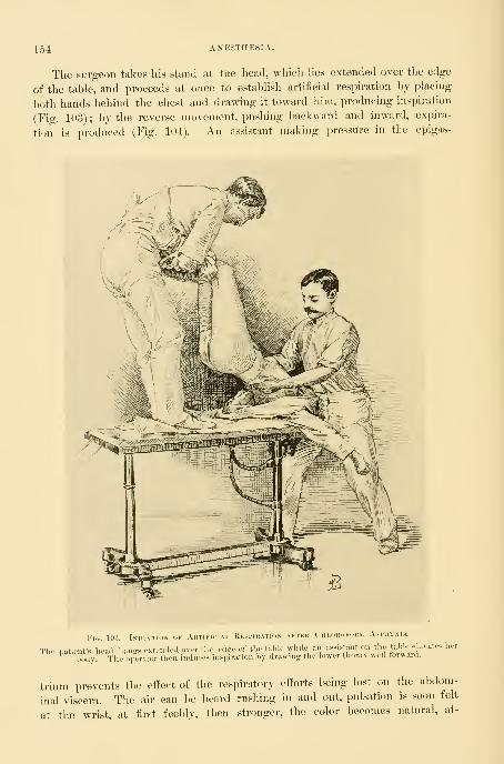
154: AXESTHESIA.
The surgeon takes liis stand at the head, which Hes extended over the edge
of the table, and proceeds at once to estabhsh artificial respiration by placing
both hands behind the chest and drawing it toward him, producing inspiration
(Fig. 103); by the reverse movement, pushing backward and inward, exinra-
tion is produced (Fig. 104). An assistant making pressure in the epigas-
Fui. 103.— Ini)1(;tion of Aktiki Kesi rlON AKTEU CllLOlil'Fl
The patient's head l.angs extended over tl.o edjre of the table while an assistant on the table elevates her
body. The operator then induces inspiration by drawing the lower thorax well forwaid.
trium prevents the eftect of tlie respiratory efforts being lost on the abdom-
inal viscera. The air can be heard rushing in and out, pulsation is soon felt
at the wrist, at first feebly, then stronger, the color becomes natural, at-

RESUSCITATION OF THE ASPHYXIATED. 155
tempts are made to respire, and in a short time the danger is past, when
the operation may be resumed. If the pulse can not be felt at the wrist it
may be found by feeling the abdominal aorta through the incision. If it is not
Fio. 104.
—
The Induction of an 1n.->imration is followed by the Compression ok the Lower Chest,PRODUCING A Forced Expiration.
felt there the hand may press up through the diaphragm and feel the heart
directly.
Where the lower chest is contracted by the wearing of corsets and when the
costal cartilages are calcified this manipulation will not produce respiration, and
it is necessary to force air in and out of the chest by placing one hand on the
middle or lower thoracic spine and the other on the sternum. Then, com-

150 ANESTHESIA.
}3ressing the chest, air is forced out, and hy relaxing the pressure it rushes in
again. In this way a regular respiration may be maintained until it is es-
tablished voluntarily. The larynx can be kept open by hooking an index finger
into it.
If it is necessary to resort to artificial respiration when there is not a suffi-
cient number of assistants present to carry out the method just described, as mayoccur in private practice, the plan formulated by Dr. Sylvester should be fol-
lowed. By this method the patient is quickly placed in position across the bed
with a pillow or roll of clothing beneath the thorax. The surgeon, standing at
the head of the patient, grasps her arms at the elbows and draws them upward
and outward, describing a circle, until they meet above the head. This move-
ment induces inspiration by expanding the chest through the agency of the
pectoral muscles. After a pause of two or three seconds the arms are swept
downward and outward to the sides of the chest, against which they are firmly
pressed. This maneuver induces expiration by diminishing the capacity of the
chest. The two movements should average eighteen to the minute.
ETHER.
Under this title, in America, washed sulphuric ether is universally under-
stood. Only that brand known to be the purest in the market should be used.
Ether is contra-indicated in bronchial catarrh, or whei-e its use excites bron-
chorrhea, or constant coughing with asphyxia due to irritation of the air passages,
or violent continuous nausea. Chloroform must then be used. It is not contra-
indicated in kidney disease, nor in any other disease, except where the act of
straining may prove injurious, or where the patient is so weak that any little
additional exertion may prove fatal. I have lost but one case on the table from
ether anesthesia, and the autopsy showed atheromatous arteries and chronic
myocarditis.
Various styles of ether inhalers have been devised, but I consider the towel
cone, stiilened with paper, as after all the most satisfactory ; it is easily made,
and a fresh one can be prepared for each patient. A stiff piece of blotting-
paper or moderately heavy manilla paper, 15 X 10 inches in diameter, should be
covered with oiled muslin, and this in turn with a clean towel. The oiled nnislin
may be dispensed with if not at hand. This pad is then twisted into the shape
of a cone and then pinned together. A moistened sponge or piece of cotton is
lightly packed into the apex, and upon this the ether is poured. It is always
best to give ether gently, soothing the patient and letting her grow gradually
accustomed to the vapor as it is brought nearer and nearer to the face. A few
minutes spent in this way will obviate entirely the necessity of forcing the
patient down on the table and strangling her with the drug, a procedure never
to be forgotten.
In the early part of the anesthesia only small quantities of ether should lie
used, and no attempt should be made to force the patient to take it ra])idly. If
the patient is told to breathe deeply the respirations will continue full and regu-

ETHER. 157
lar, according to the suggestion of tlie anestlietizer, until lier volition is over-
come, when there is a cessation in the breathing which prevents the even
administration of the ether. For this reason I think it is best to instruct the
patient to breathe naturally, and only to command her to breathe deeply when
she persists in holding her breath. By gradually bringing the inhaler nearer
the face and allowing at short intervals a breath or two of fresh air the dis-
agreeable strangling sensation is avoided. As soon as she loses consciousness it
is an error to remove the ether whenever there is a disposition to vomit ; this
is best overcome by increasing the quantity of ether and getting her more com-
pletely anesthetized. A timid anestlietizer, by hesitating at this point, can
harass an operator throughout a long operation.
Operation under the Primary Effect of Ether.—At an early stage of the anes-
thesia, just as consciousness is lost, there comes a short period of relaxation and
insensibility, which can be utilized for such short operations as paracentesis,
dilating a sphincter, opening an abscess, excising a small tumor on the surface,
or passing two or three sutures.
In two or three minutes this stage is passed and a noisy, excited stage mayfollow, which lasts ten or fifteen minutes or longer, finally jDassing into the stage
of profound anesthesia. When this final stage is reached it is important to give
just enough ether to keep the patient completely relaxed and unconscious, and
not a bit more. While coming out of ether anesthesia the patient must be
watched, and assisted when she vomits by turning the head and body to one
side, cleansing the mouth, and keeping her face clean and her pillow protected.
Care must be exercised to keep her fauces clear and to prevent her from inspir-
ing regurgitated food.
The duration of the period of unconsciousness depends greatly upon idio-
syncrasy and upon the amount of the anesthetic taken ; while one patient may
come to in a half hour, another will lie sleeping or in a dazed state for four or
five hours. It is generally safe in private practice for the physician to leave her
in the care of the nurse after she has spoken.
The liability of patients coughing and straining excessively as they are com-
ing out of the anesthesia must be borne in mind, and stitches and ligatures must
always be put in so that they can not possiljly tear out or give way from any
such cause.
Death from Anesthesia.—In about 8,500 administrations of ether I have lost
two patients from the anesthetic.
One of these, a woman of forty, died after the removal of an adherent ovarian
cyst, presenting no unusual ditficulties and not involving the loss of much blood.
She died as the wound was being closed, after an operation lasting forty min-
utes ; the first danger sign was a deep congestion of the intestines followed by
cyanosis of the face, bulging eyes with widely dilated pupils, and an impercep-
tible pulse. Respiration became shallower and shallower, and there was no
response to any form of stimulation or to all efforts to induce respiration. Xocause for the death could be discovered.
The second death after ether occurred shortly afterward in the case of a

158 ANESTHESIA.
woman sixty-four years old—L. T. N., 4232, March 18, 1896—after an abdominal
hysterectomy for an adeno-carcinoraa of the uterus, lasting two hours. The
patient was obese, weighing 235 pounds, and took the ether badly from the
start. The pulse, which had become steadily more rapid and small, ceased first,
while the respirations, labored throughout, became more labored and gasping,
and the face livid. Artificial respiration could not be carried out on account of
the unwieldy form of the patient.
One death has occurred in about 1,500 chloroform anesthesias. This was
the case of a colored woman of forty-seven
—
B. B., 3257, January 1, 1895—whodied during the early stages of the anesthesia.
She had taken chloroform on one occasion before, and objected so strenu-
ously to ether that chloroform was again used. The first part of the anesthesia
passed off quietly, but when placed for operation she became so rigid that the
Esmarch inhaler was brought closer to the face, at no time nearer than two
inches. This did not help the rigidity, and the respirations grew shallow^er.
The chloroform was taken away at once and ether sent for ; but the temporal
and radial pulses had become imperceptible, and then respiration ceased. Efforts
at artificial respiration were utterly ineffectual, owing to the fact that she had a
rigid chicken-breasted chest with calcified cartilages. The autopsy revealed also
an adherent left lung, abdominal viscera everywhere mutually adherent and
attached to the diaphragm, and atheromatous vessels (see Johns IIojjMns Hasp.Bull., vol. vi, May-June, 1895).

CIIAPTEK YIII.
GENERAL PRINCIPLES INVOLVED IN PLASTIC OPERATIONS.
1. Preparation. Rest. Bowels. Urine. Dress for operation.
2. Operation. Position of patient. Washing of genitalia. Assistants. Irrigation. The opera-
tion. Dressings after operation.
3. Care during convalescence. Position of patient in bed. Douching, if necessary. Catheteri-
zation. Care of bowels. Diet. Care of wound. Removal of sutures. Rest and tonic
treatment. Hemorrhage following operation. Infection.
All plastic operations about the vulva, vaginal outlet, vagina, and cervix
have certain common details, which may be considered in the following order
:
1. Preparation for operation.
2. The operation.
3. Care during convalescence.
PREPARATION FOR OPERATION.
Every patient should be subjected to a thorough general physical exami-
nation before the performance of any gynecological operation, in order to ex-
clude the possible presence of any obscure disease that might account for the
condition of ill health. If the examination shows that an operation is necessary,
and the general health of the patient is much impaired, a rest of a week or
more in bed will hasten the convalescence, toning up the system and quiet-
ing the mind. Such a preparation is especially valuable in the case of nervous
women. Constipation, which is obstinate in many uterine aifections, should be
overcome by a purgative, the continued use of mild laxatives, and a light but
nourishing diet should be given. If there is loss of appetite, a simple tonic,
such as tincture of nux vomica, calumbo, or gentian, is often helpful. AVomen
with marked debility will be benefited by massage, cold baths, and electricity.
When, however, the general health of the patient is good and she is clearly
suffering from purely local symptoms, the preliminary period of rest and tonic
treatment may be dispensed with, and the operation may be done with ])ut one
or two days' preparatory treatment.
The older gynecologists invariably put their patients under a protracted
course of preparation for an operation, while the present rule is to operate im-
mediately and to build up the patient during her convalescence.
Immediately preceding the operation the bowels should be carefully evacu-
ated, so as to avoid disturl>ing them for at least two days afterward. To insure
thorough purgation, 3 ij of licorice powder, or a similar amount of magnesium
sulphate, should be administered (both morning and evening of the day befoi-e),
159

160 GENERAL PRINCIPLES INVOLVED IN PLASTIC OPERATIONS.
followed tlie next morning at six o'clock hy a warm enema of a pint of soap
and water. If the patient is delicate a milder purgative, such as a pill of aloes,
strychnine, and belladonna, or the solution of citrate of magnesium, 3 viij, may
be given with good effec*. The enema should be given quite three hours before
the operation, regardless of the effect of the purgative, as it is essential to have
the rectum and sigmoid flexure clear of feces. The action of an enema given
later than the time specified is often delayed until the operation is under way,
when the surgeon may be annoyed by the constant ejection of semi-fluid feces
over the gauze diaphragm in front of the buttocks.
The urine must always be carefully examined both chemically and mi-
croscopically before operation. Diabetes is a contra-indication to any surgical
operation in most cases. Nephritis in its early stages does not materially de-
crease the patient's chances of recovery. If, however, the constitutional and
local symptoms indicate advanced nephritis, the operation should in no case
be performed.
The early morning is the best time to operate, when the surgeon feels fresh
for his duty and his hands are free from the contamination of his daily work;
the patient should also have a good night's rest, insured if need be by a mild
sedative. The evennig before operation the patient should take a hot bath, and
immediately go to Ijed. The following morning, after the enema, the vagina
should be thoroughly cleansed with a douche of carbolic acid solution (2 per
cent), or boric acid (32 grains to the liter), at a temperature of 110 F. As a
rule, no food of any kind is given on the morning of the operation. If, how-
ever, the patient is weak or feels faint, a glass of warm milk, or a cup of tea
diluted with milk, may be given.
The patient's dress for operation consists of an undervest of warm flannel in
winter, or of gauze in summer, a nightgown open up the back, and a pair of
long woolen stockings. The hair is most conveniently dressed by plaiting it in
two braids.
If the operation is to be performed in a private house, the patient should be
anesthetized in a room adjoining the one selected for the operation ; in a
hospital the anesthetizing room is always separate from the operating room.
THE OPERATION.
The Position of the Patient.—The operating table is covered with a blanket
protected by a sterilized sheet, and upon this, at the end of the table upon
which the buttocks are to rest, is placed a rubber perineal drainage cushion.
The buttocks rest squarely upon the cushion, projecting slightly over the edge
of the table, and the legs and thighs are held flexed upon the abdomen by a
leg holder. To apply Kelly's or Robb's leg holder, buckle one end of it around
the thigh just above the popliteal space, taking care to keep the band smooth, so
that it does not bind the leg too tight. The other end is then carried up under
the shoulder, around the neck and down to the opposite side, where it is simi-
larly buckled above the popliteal space. When the patient is thoroughly under
IllRIGATIOX. IGl
the anesthetic this leg holder simply detains the legs, without cramping them,
and the knees fall apart naturally without the assistance of the leather crutches
and bar so much used in the past. The arms of the patient should be folded
across her breast and retained in this position by drawing the skirt of the under-
vest well up over the elbows. The nightgown should be pushed up under the
small of the back above the drainage cushion.
The external genitals are thoroughly soaped and shaved up to the ni o n s
veneris. Kobb's razor, with a short fixed metal handle devised for this pur-
pose, is useful. After shaving, the genitals should again be thoroughly washed
with soap and water. Green soap or soft soap serves admirably for the pur-
pose; it can be thoroughly rul)bed into the skin, cleansing better than hard
soap. Be careful to cleanse all furrows between the labia and about the
clitoris.
After the external parts have been cleansed, the assistant takes a pledget of
cotton covered with soap, and introducing it into the vagina with long forceps,
under a stream of water from the irrigator, smooths out all fun*ows and scrubs
thoroughly all accessible parts, so as to remove the discharges and accumulated
epithelial debris. Then the vagina is douched with a 10 per cent creolin
solution, which sterilizes and acts as an efficient detergent. This solution is
followed by a bichloride of mercury solution (1-2,000), and this again by
warm water. A thorough vaginal cleansing will require from three to five
minutes.
Long sterilized canton fiannel stockings are now drawn over the patient's
legs and fastened above the knees with a draw string. A protector 1 meter
square (1 yard), composed of two thicknesses of gauze, is spread between the
thighs, covering all the exposed parts, and hanging well down over the but-
tocks onto the cushion ; as the surgeon takes his seat he cuts a small open-
ing in the protector corresponding to the vulva so as to expose the field of
operation.
Assistants.—For convenience of rapid work the surgeon will do best with
four assistants—one to give the anesthetic, two standing on either side of the
patient to help the operator, while the fourth hands the instruments and liga-
tures as wanted. In operations conducted in a private house or private hos-
pital the operator can make shift with two assistants—one to give the anesthetic
and one to assist him directly.
Irrigation.—Irrigation by a continuous stream of warm water directed over
the field of operation is the best means of removing the blood, leaving the line
of incision and denudation constantly clear. Sponging is not so good, only im-
perfectly removing the blood, which remains to coagulate about the ligatures and
to cling to the hands of the surgeon, rendering them sticky and slippery. Aglass reservoir holding several gallons of water should be placed on a shelf
at an elevation of five feet above the operating table. The rubber tubing
from the reservoir, when not in use, should be coiled and kept immersed
in a 5 per cent carbolic solution. It is best to regulate the flow by a glass
douche nozzle, an Esmarch's hard rubber stopcock, or an efficient ball-and-
13

102 GENERAL PRINCIPLES INVOLVED IN PLASTIC OPERATIONS.
socket nozzle, like the one here figured, because either can be easily detached
for sterilization.
The assistant on the right hand of the patient takes charge of the irrigation,
keeping the area upon which the surgeon is working free from blood.
In vaginal lijsterectomj a normal salt solution {-^-^ of 1 per
cent) should be used; it is not irritating and does no harmeven though it enters the peritoneal cavity.
The Operation.—Just as the artist, with a few rapid strokes,
sketches in the outline of his picture, so the surgeon vnW first
outline his field of a plastic operation by incisions, marking its
outer limits. This will enable him to judge more deliberately
as to the amount of tissue to be removed ; it is better, of
course, to err on the side of a small outline than a large one,
because a small outline may be enlarged so as to include more
tissue, if found necessary. Outlining with the knife is espe-
cially important where scissors are to be used ; the knife cuts a
sharp line and the mucous surfaces then pull apart, permitting
a rapid denudation with the scissors and subsequent accurate
coaptation of the edges.
Bleeding is rarely active in plastic operations, the vessels
being of smaller caliber. If, however, there is enough bleed-
ing to annoy the operator, the vessels may be temporarily
caught with artery forceps until the sutures are introduced.
A large vessel which persists in bleeding after the forceps are
taken off may be controlled by introducing one of the sutures
approximating the parts, so as to grasp the vessel in its loop
;
this suture should be tied tighter than an ordinary approxi-
mating suture, so as to check the bleeding, and thus it serves
the purpose of both suture and ligature.
The sutures are of three kinds—silkworm gut, silk, aud catgut. Silkworm-
gut sutures best bear the tension in bringing together widely separated areas.
Silk and catgut sutures are used for accurate approximation, either to supple-
ment the silkworm-gut sutures, or alone, where there is but slight tension in
bringing the wounded surfaces together. Catgut is ill adapted for use, if there
is any outward traction of the wound. The best suture for close approximation
is fine silk, which offers the least possible opportunity for the entrance of septic
matter. Silver wire is now rarely used, and there are no circumstances under
which it is better tlian silkworm gut.
Dressings after the Operation.—At the end of the oj^eration the vagina and
external genitals are dried l)y pledgets of sterilized cotton. A strip of iodoform
gauze may then be inserted into the vagina with the three-pronged packer as
far up as the cervix, loosely filling the upper vagina and just appearing at the
outlet ; this should be taken out in five or six days and the vagina douched daily
afterward.
It is my practice at present to use no vaginal dressing at all, but simply to
Fig. 10,").—SwedishHard RubberBall and Noz-zle Irrigator.
By bending^ thenozzle in the ball at
an ancfle the Howis controlled or
stopped altogether.
CARE DURIXG CONVALESCENCE. 163
protect the vulva by a sterilized gauze pad held in place by a T-bandage. The
pad is changed several times daily, and if there are any offensive discharges the
vagina is douched out with a warm boric or carbolic solution once or twice a
day. I have found a powder composed of boric acid, 3 ounces ; alum, 1 ounce;
carbolic acid, ^ ounce ; and oil of peppermint, 1^ drachm, very satisfactory in
relieving the odor and irritation which are sometimes distressing during the con-
valescence from a plastic operation.
Before removing the patient from the table draw the urine with a glass
catheter, loosen the leg holder, and raise the buttocks by carrying the feet of
the patient toward her head ; dry tlie genitals, buttocks, and back with a towel,
and remove the drainage pad.
The external genitals should be powdered with iodoform and boric acid
(1-7), and then covered with a loose pad of sterilized cotton, held in place by a
T-bandage.
CARE DUKING CONVALESCENCE.
A nurse or doctor should remain with every patient, controlling anyviolent movements until she has fully recovered consciousness. In rec-
tal and perineal operations it is not necessary to follow the old practice of re-
stricting the movements of the legs with a binder after she becomes conscious.
She may also be turned on her side if she wishes.
In perineal operations the bedpan must be used for two weeks and straining
avoided. After cervical operations this restriction is not necessary, and cervi-
cal cases are required to stay in bed from seven to ten days only. If the patient
can pass her urine voluntarily from the first she should be permitted to do so.
The vaginal pack is removed when a discharge appears externally, and when
the discharge continues a douche is necessary ; it should be given with the
greatest care, to avoid pressure of the nozzle on the wound, once or twice daily.
A trained nurse, or the physician himself, should attend to this duty, for it has
not infrequently happened that an unskilled nurse or an ignorant attendant has
thrust the point of the svringe through a recently repaired pei-ineum.
The nurse should be instructed how to separate the labia and expose the
outlet with one hand by pushing downward and backward without making
traction on any sutures. Secretions are now removed with pledgets of cotton
held in the dressing forceps, and the blunt glass douche nozzle, gently poised
between the thumb and index finger, is introduced in a direction backward and
inward. Be careful to expel the air from the douche nozzle before it is intro-
duced into the vagina. After the douche is given the genitals are dried with
pledgets of sterilized cotton dusted with iodoform and boric powder (1-7), and
covered again with a sterilized cotton vulvar pad. Under no circumstances
should the hands come in contact with the field of operation. Except in cases
of infection, douches should not be given earlier than the seventh day.
Catheterization.—A serious complication to be guarded against in all plastic
operations is a cystitis caused by catheterization, and for this reason I wish to
speak with special emphasis about catheterizing and the care of the catheter.

104 gexp:ral principles ixvolved ix plastic operations.
In skilled Iiaiids the glass catheter is best. Iiumediately after use it should be
rinsed in warm water and boiled lor live minutes in a soda solution, and pre-
served aseptically, wrapped in sterilized gauze, or inunersed in a bottle of car-
bolic solution (5 per cent); or it may be stored in a glass ignition tube, resting
on cotton and plugged with sterilized cotton. Metal catheters should not be
used. Rubber catheters are safest in untrained hands, and are sterilized by
keeping them in a carbolic solution (2i) per cent) ; the catheter should be rinsed
in boiling water before using.
Catheterization must be performed in the following manner : The vulva is
exposed under a good light, so as to bring the urethral orilice into full view
upon separation of the labia with the thumb and forelinger of the left hand.
Then, with the dressing forceps in the right hand, the parts immediately
surrounding the urethra may be cleansed with pledgets of sterilized cotton
saturated with boric acid solution ; now take the catheter from its receptacle,
without touching its vesical end, and introduce it gently into the bladder, not
attempting in any way to control its direction : it will follow naturally the
course of the urethra. Before withdrawing the catheter, stop up its outer end
by the linger to prevent the urine from dribbling over the parts. Finally, dust
the vulva with iodoform powder, and replace the sterilized T-bandage.
Care of the Bowels.—On tiie second evening following the operation a pill
of aloin, strychiiine, and belladonna, or two drachms of licorice powder, or a
half drachm of cascara, is given, followed the next morning, if necessary, by a
soap and water enema. The custom of confining the bowels for eight or ten
days is repreliensil)le. There is no danger of fecal matter gaining access to the
wound, even where sutures have been j^assed on the rectal surface if they have
been properly placed and properly tied. There is likewise no danger of disturb-
ing united wound surfaces by the downward displacement of the pelvic fioor
during defecation on the third or fourth day following operation, if the feces
are soft or fluid. When the bowels are confined for a longer period, there
is often great diificulty in securing a movement, and the effort to pass the
scybalous masses is now attended with real danger, because the sutures have
become loosened and the union of parts is not sufiiciently firm to withstand the
pressure.
Only a trained nurse or the surgeon himself should give the enema. I have
known an inexperienced person to push the nozzle of the syringe through the
coats of the bowel and force the injection into the pelvic cellular tissue. In
one case I knew a nurse to push the end of the syringe through the stitches
of a ruptured perineum and inject into the vagina. The most convenient
])Osition for giving the enema is with the patient lying on the left side. If a
scyl>alous mass blocks the rectum the surgeon nmst himself introduce his index
finger, break it up, and hook it out, making pressure in a direction away fromthe wound. When the bowels are once thoroughly (»peiied, they should be
kept open by a mild laxative, or an enema given every other day.
Diet.—No food is given until the patient has recovered from the nausea
following the anesthetic. After from twelve to twenty-eight hours the stomach

CARE OF THE WOUXD. 165
is usually sufficiently settled to permit the retention of small amounts of liquid
nutriment. It is l)est to comtnenee with from 30-60 cubic centimeters (1-2
ounces) of milk every two or three hours, followed in a day or two by li^ht
broths of chicken, beef, or mutton. If nausea is persistent, a nutrient enemashould be given to sustain strength, consisting of 60 cubic centimeters of milk
and the yolks of two eggs, with enough water to make 120 cubic centimeters (4
ounces). Tea well diluted with milk, hot beef tea with the yolk of a raw egg
stirred in, rice soup, kumiss, are usually well borne.
From the third to the seventh or tenth days soft diet is best— soft boiled
eggs, sweetbreads, oysters, white meat of chicken, milk toast, rice, bread, mush,
baked apples, and baked potatoes. After the seventh day full diet may be
gradually resumed.
Care of the Wound.—Where the wound is entirely or partly on the surface,
the chief point in its care is to keep all objects which might convey infection
from coming in contact with it ; for this reason neither the surgeon nor the
nurse should touch the wound with the hands in the subsequent dressings.
The removal of discharge and arrangement of the gauze or cotton dressings
should be effected with sterilized forceps.
If there is free discharge, it should he removed once or twice daily with
pledgets of cotton, followed by a light dusting of the surface with the iodoform
and boric powder mixture.
The length of time during which the sutures should be allowed to remain
varies both with their position and with the results aimed at. If the healing is
uninterrupted, the skin sutures may l)e removed with safety on the eighth day.
Those within the vagina should not be removed Ijefore the twelfth day, or even
later, on account of the danger of separating surfaces not yet firmly united.
Cervical suture;^ of silkworm gut may remain in place almost indefinitely,
and, if the operation has been one of combined cervical and perineal repair,
their removal should never be aitempted until the perineum is quite firm and
sound again, in from four to six weeks.
In order to remove the sutures, the patient is brought across the bed, or,
better still, placed on a tal)le, with the buttocks toward a good light, and the
legs flexed upon the abdomen. The dressings and any incrusted powder are
removed by sopping the parts with a warm boric acid solution ; if the field of
operation is within the vagina it is exposed with specula or retractors. In re-
moving cervical sutures a Sims's speculum is inserted and the posterior vaginal
wall retracted, while the anterior wall is elevated by a narrow flat retractor.
The first suture seen is caught with forceps and pulled upon until its loop
comes into view, which is then cut and the suture withdrawn. The remaining
sutures are found by displacing the cervix first to one side and then to the
other. Sutures upon the floor of the vagina can not always be readily ex-
posed, and are often best located by touch and then grasped with forceps and
removed.
Stitches on the rectal side are readily removed by drawing them through
the fenestrum of a rectal speculum which is pushed into the bowel, exposing

166 genp:ral prixciples ixvolved ix plastic operations.
the line of union, as practiced by Dr. Cx. M. Tuttle, of New York. Care must
be taken in cutting the loop not to cut off both sides at once, as a loop thus
left in the tissues will invariably cause persistent irritation and discharge, and
must be removed sooner or later.
Rest and Tonic Treatment.—A patient upon whom a minor plastic operation
has been performed should remain in bed for two weeks or longer, both for the
purpose of securing firm union of the tissues, as well as for the equally important
purpose of building up the nervous system and recruiting the general health.
Advantage should l)e taken of the opportunity to keep neurasthenic patients in
bed eight weeks or longer, giving them at the same time the benefit of a rest
cure. From the fourteenth to the eighteenth day, depending upon the general
improvement in symptoms, the patient may be permitted to put on a light
wrapper and sit in a reclining chair or lie on a couch in sunny parts of the
room. By the twentieth day she may resume her lighter duties, gradually in-
creasing them during the succeeding days, imtil she has returned to her customary
routine of work. The tendency of our hospitals is to make the stay of poor
patients too short and to hurry them home.
It is a serious error to consider the function of the surgeon at an end when
the wound is well healed and the operation in a technical sense successful.
Patients who have long been in bad health before operation should be kept
under observation for months afterwards, for the purpose of directing exercise,
diet, and tonic treatment. Suitable exercise should be regularly and persistently
taken, short daily walks in the open air, and rubbing down with alcohol or
cacao butter on going to bed. Morning and afternoon the patient should rest
for an hour on the back. Gymnastic exercises are not necessary, and exhausting
exertion, such as shopping and dress fitting, must be avoided. Such tonics as
tincture of nux vomica and the preparations of hypophosphites combined with
cinchona often encourage a poor appetite. Koumiss, malt extract, or malt and
milk, are valuable aids to the ordinary diet. One of the best therapeutic
agents is a complete change of air for two or three months—in winter to a
warmer climate, in early spring to the seashore, or in summer to the mountains.
In all of these cases it is absolutely essential to a perfect recovery to relieve
the patient's mind of anxiety ; for this reason the burdens of her regular duties,
whether social or domestic, must be cast off or lightened as much as possible.
As a general rule, the sexual relation should be prohibited for three months
after plastic operations involving the vagina, and should then not be permitted
oftener than once a week.
Hemorrhage following Operation.—An active hemorrhage is occasionally seen
after a vaginal operation ; it usually arises within the first week and persists for
twelve, twenty-four, or forty-eight hours, or even longer, if unchecked. Such
bleeding, while rarely threatening life, is always an annoying complication on
account of the difficulty of access to the bleeding point ; it also renders the pa-
tient profoundly weak and anemic, and prolongs convalescence. To control
the hemorrhage, bring the patient across the bed or on a table in the Hthotomy
position, with a good light on the parts. Withdraw the vaginal pack if there is
IXFECTION". 167
one, and wash the vagina free of all elot.s ; after the douche, elevate the anterior
vaginal wall with a small speculum and expose the whole wound area, cleansing
it with small pledgets of cotton until the bleeding point is found. A curved
needle carrying a small silk suture is then passed deeply beneath the point andthe suture tied, controlling the flow. When the circumstances are not favor-
able for such a prompt and direct treatment, a tampon of sterilized non-absorb-
ent cotton must be applied in such a manner as to make pressure upon the
whole wound area. After twenty-four or thirty-six hours the pack is removed,
but if the oozing persists it must be again applied. A tight jDack skilfully ap-
plied does not often interfere with the union of the parts.
Infection.—The symptoms of infection following plastic operations are simi-
lar to those which may arise from an infected wound anywhere in the body.
Ordinarily they do not show themselves before the third day, and it may beeven longer than that before the surgeon is able to differentiate clearly betweenthe usual sequelae of operation and an infected process. Fever is usually pres-
ent, and the pain of beginning sepsis is lancinating in character, and extends
from the labia down the inner thigh. When the patient locates pain in this
region no time should be lost in discovering the seat of infection. If it is about
one of the sutures, as is most likely to be the case, the suture should be removedat once, and if a pus cavity of considerable size is found, it must be freely
drained. In the early stages, whei-e the symptoms are suspicious but the seat of
infection can not be accurately determined, the application of hot poultices will
not only relieve the pain, but so hasten the inflammatory process that a definite
diagnosis can be made.
Hot injections of a solution of bichloride of mercury (1-5,000) every four or
five hours also ogives relief. But this treatment should not l)e continued lono;er
than forty-eight hours, on account of the danger of mercurial poisoning. If the
whole wound looks red and angry, all the sutures nuist be taken out and the
wound allowed to heal by granulation. Sometimes an abscess of considerable
size forms laterally, near one or the other of Bartholin's glands ; in this case an
incision should be made dii-ectly into the cavity, as far as possible away from the
seat of operation, so that the ultimate results of the operation may not suffer
from the infection.
Very often, by taking out a single stitch, a small stitch-hole abscess will dis-
charge and the trouble be over. The outcome of a bad infection, extensive in
area, may sometimes be surprisingly good ; I have seen a complete rupture of
the perineum granulate down to perfect control over the sphincter ani.
CHAPTER IX.
DISEASES OF THE EXTERNAL GENITALS.
1. Advantages of superficial position for operation : a. Ileinorrliage easily controlled, h. Defectseasily covered, c. Asepsis, d. Sutures.
2. Diseases of labia niajora : a. Lipoma, b. Hydrocele, c. Tnguino-labial abscess, d. Pseudo-myxoma of the canal of Nuck. e. Hernia. /. Myoma of round ligament, g. Condyloma.h. Carcinoma.
3. Diseases of labia minora : Cysts.
4. Diseases of clitoris : a. Adhesions and concretions, h. Elephantiasis, c. Sarcoma, d. Car-
cinoma.5. Diseases of vulvo-vaginal glands: a. Cyst; h. Abscess; c. Adeno-earcinouia ; d. Myxo-fibro-
sarcoma.6. Affections of the vulvar mucosa : a. Cohesion, h. Pruritus.
In considering the surgical diseases of the external genitals, we take up the
affections of some five different structures—namely, the labia niajora, the labia
minora, the clitoris, the mucous membrane about the vaginal outlet, and the
vulvo-vaginal glands.
There is no common principle other than contiguity uniting these diverse
organs in their pathological affections, the list of which is but short, including
neoplasms, elephantiasis, cysts, abscesses, and pruritus.
In spite of the situation of these organs upon the exterior of the body, they
are so well protected by the thighs that they are but rarely subjected to violence,
I have seen a case in which a hematoma lias been produced by the kick of a
brutal husband ; a girl of twelve was brought into the ward of the Johns Hop-kins Hospital suffering from a severe hemorrhage, with a large hematoma of
the perineum and left labium, the result of a fall astride a fence rail on which
she had been standing. I know of instances in which the external genitals have
been injured in young girls by sliding down balusters and striking a low newel
post. In one case, in the care of Dr. Jacob Price, of West Chester, Pa., a vul-
var laceration was produced by the horn thrust of an angry cow.
Operations upon the external genitals are among the simplest and least dan-
gerous gynecological procedures, on account of the superficial accessible position
of the organs.
Hemorrhage, although often free, particularly in o})erations involving the
clitoris, is always readily controlled. Deep sutures uniting the edges of the
wound are usually sufficient to control the bleeding without the aid of buried
ligatures. The free anastomosis of numerous smaller vessels is the means of
effecting a rapid union of wound surfaces. It is also easy to cover up large de-
fects created by the extirpation of tumors and neoplasms with the lax movable
adjacent skin. Situated on the surface of the body, the wound is readily pro-

LABIA MAJOllA. 169
tected, and its asejDtic condition easily preserved after operation ; for this reason
suppuration does not often occur.
For suture material I prefer silkworm gut as a tension suture, and fine silk
or catgut for accurate approximation. The need for an absorbable material is
not so great where the sutures can be so readily removed, but for greater con-
venience the subcuticular catgut suture is jjerhaps the best.
LABIA MAJORA.
Lipoma.—Lipoma, or fatty tumor, is one of the rarest gynecological affections;
no writer has as yet recorded more tlian a single instance in his own practice.
In the Johns Hopkins llos-
-'^^^pital Reports, vol. iii, page
821, 1 collected all the cases
I could find in the scattered
literature, numbering only
twenty.
Lipomatous tumors are
usually easily recognizable,
as they possess the same
characteristics as lipomata
elsewhere. The labium it-
self is enlarged when the
tumor is attached to it by
a broad base, or the tumor
may hang by a pedicle more
or less attenuated. In a
case which I saw in the
Episcopal Hospital, Phila-
delphia, an ovoid tumor, 8
centimeters (3 inches) long,
hung from the middle of
the right lalnum majus by
a slender pedicle 5 centi-
meters (2 inches) in length
and not more than 3 milli-
meters in thickness. The
pedicle of a large lipoma,
on the other hand, may ex-
tend up into the inguinal
canal, in which case the
tumor simulates a hernia.
The base of the growth has
also been found extending back on to the perineum, or even up into the vagina.
The length of the pendulous growth in one case was 55 centimeters (22 inches).
10(5.
—
Hematoma of tiil \ i i
MaJVS and extending DOW, nil. Lkkt LabiumTHE Perineum.
The vaginal outlet is discolored and all of the surrounding parts-
distorted and infiltrated with blnod. Below is an abrasion of the
skin. The patient fell astraddle a chair.

170 DISEASES OF THE EXTERXAL GENITALS.
Dr. William Goodell, of Philadelphia, observed a case attached by a broad
pedicle hanging down to the knees. Balls-IIeadley, of Melbourne, removed a
tumor which weighed 24: pounds.
The lipoma feels hard or soft, according as the fibrous septa or the fat pre-
dominates. When there is an excess of fat, the sense of fluctuation is so dis-
tinct that the inference that the tumor is cystic is almost irresistible. I7nder
tins impression Goodell inserted an exjjloring needle into his case.
It is easy to mistake such a mass for a hernia, where the pedicle is l)road
and extends up into the inguinal canal, and where there is impulse on coughing,
together with some appai'ent reduction on manipulation and upon lying down,
as has been recorded.
Age is not an important factor; the youngest patient I have found noted was
eighteen, and the oldest, operated upon by Dr. A. II. Deekens, in Philadelphia,
sixty-one.
Tiie larger growths take years to develop. One woman carried her burden
seventeen years.
A large tumor hanging l)etween the thighs is apt to become ulcerated from
attrition, and an extensive hemorrhage may arise from such an area.
The chief distress comes from interference with walking, with the sexual
function, and in one case, obstruction of the vaginal outlet during labor. Both
of these difficulties existed in one of my own cases, in which a large globular
fatty tumor hung from the left groin close to the labium majus ; nevertheless,
the patient carried the growth thirteen years until it alarmed her by becoming
ulcerated.
The diagnosis rests upon the following factors : The tumor is a well defined
ovoid or round, softish, fluctuating or hard, generally pediculated, often covered
with wrinkled or lobulated skin, not reducil)le, is slightly hardened by the appli-
cation of cold, and the septa may be faintly outlined on the surface, and is pain-
less unless ulcerated.
The treatment is extirpation ; if the ])edicle is long and thin, it may be sim-
ply constricted firndy or slightly abraded and surrounded for ten minutes by a
pledget of cotton wet with a saturated solution of cocaine, and then cut off at a
distance of 1 or 2 centimeters (f to f inch) from its attachment. The growth is
often supplied by a single artery in the center, which should be tied with catgut.
The wound is closed by catgut sutures, and the dressing applied. Where the
pedicle is not so well defined, one may often be formed by grasping the tumor
and drawing it out from the body. The incision must here be made well out
from the body on the under side of the tumor, otherwise there will be a largo
defect in the skin when the tumor is taken away. There is no objection to
utilizing a part of the skin covering of the gi'owth in this way, as it is in all re-
spects normal, and there is no danger of the tumor recurring.
Large sessile growths extending up into the inguinal canal, or into the vagina,
or out on to the perineum, must be removed by making an oval incision through
the skin over the growth, and shelling out the fatty mass, ligating bleedi>ig ves-
sels, and then approximating the skin with sutures. Injections witl'i alcohol or

IXGUIXO-LABIAL ABSCESS. 171
removal l>y l)urning tlirougli tlie pedicle with tlie cautery, or ligation of the
])e(licle, leaving it to slough oli', as practised in the past, ought to be abandoned.
Hydrocele is an affection of the persistent canal of Xuck, characterized by
an accunuilation of fluid within it ; it is exceedingly rare, owing to the fact that
the canal is normally completely obliterated in the adult.
The hydrocele presents the appearance of a rounded elongate or moniliform
cord extending like a string of beans from the region of the external inguinal
ring down into the labium majus.
When the distended sac shows the constrictions, they appear in a succession
of little swellings ; at other times there is only a single elastic enlargement at
the upper and outer angle of the vulva.
The diagnosis is established by noting the location of the affection, its direc-
tion upward toward the inguinal canal, and the fact that it does not gi\^e rise to
any characteristic symptoms.
If the canal is patulous above, the fluid may be forced back into the ab-
domen.
The absence of any intestine from the canal may be ascertained both by per-
cussion and by placing a finger over the ring and pa^'tially closing it, while the
fluid is forced back into the abdomen by pressure made upon the tumor with
the other hand ; the sensation communicated is that peculiar to fluids alone.
When a hydrocele can not be differentiated from a small solid tumor in the
canal, the use of the aspirator will determine the diagnosis in a simple and safe
way.
Encysted hydrocele should first be treated by aspiration, after cleansing and
shaving the part inunediately over the swelling ; if the tumor returns, the sac
should be exposed and dissected out of the labium, all hemorrhage stopped, and
the wound closed with catgut.
A sort of false hydrocele of the upper part of the canal is often associated
with the presence of a large amount of ascitic fluid in the abdomen, and depends
upon the increased intra-abdominal pressure as its cause. The treatment then
is that of the intra-abdominal condition producing the ascites, after which an
operation may be called for to close the neck of the sac at the inguinal ring to
prevent the occurrence of an inguino-labial hernia.
Inguino-labial Abscess.—I have found this condition in the left inguinal canal
of a nndatto ; it occupied the upper outer part of the labium, and was about 3
centimeters in length by 2-5 centimeters in width. The abscess was hard but
slightly irregular, moval)le, very painful on pi-essure, and associated with a rise
of temperature.
After due preparation an oval incision was made over the enlargement and
the entire honeycombed abscess was enucleated from the inguinal canal down to
the pubic ramus, which was laid bare. The chief difficulty in the extirpation
arose from the extreme vascularity of all the surrounding adherent tissues.
Hemorrhage was controlled by numerous ligatures passed deeply under the
tissues and tied tight. A thin strip of iodoform gauze made an efiicient drain
for such oozing as could not be checked inunediately. This was removed in

DISEASES OF THE EXTERNAL (iENITAL
two days, and the wound healed thronghout ; the skin sntures were removed in
a week.
Pseudo-myxoma of the Canal of Nuck.—I have ohserved this condition in a ease
of pseudo-myxoma of the peritoneum due to a ruptured ovarian cyst, in wliicli
FT.
nl.lig^
Fig. 107.
—
Myoma of the Kounu Ligament within the Inguinal Canal.
The tumor consists of two masses, of which the upper has pushed its way down into the pelvis hcliind tlie
peritoneum, while the lartfe mass filled the canal. Tiie iliairram shows its relations to the left iduiid liira-
tiient; the little lohulatcdinasscs at eacli end are fat. ( )ct. 1-2, is;);!. Natural size.
the encysted mass below the inguinal canal was about 3 l)y 2 centimeters, and
shut ojff from the peritoneal cavity. The vermiform appendix was glassy and
distended with the myxomatous material to tliree to four times its normal
diameter.
Hernia appears in the form of h c i-n i a in gu i n o -labialis. The hernial
sac forms a pouchlike prolongation of the abdominal parietal peritoneum, and
extends down into the labium majus, which may be greatly eidargetl. displacing
the vulvar oriiice to the opposite side.

FIBROMA AND MYOMA OF THE ROUND LIGAMENT. 173
The sac presents a distinct swelling IVoni its exit at tlie inguinal ring above
down to its lower margin lieside the vaginal outlet, and contains eitlier omentum,
or omentum and serum, or omentum and intestines.
The diagnosis is readily made upon observing that the tumor extends up into
the abdomen, and that it is tympanitic, gurgles on pressure, and can be replaced
by putting the patient on her back with elevated chest and flexed thighs in a
position of relaxation ; on standing and straining it descends again into the
labium.
Tumors dull on percussion and irreplaceable are formed by a part of the
omentum adhering to the neck of the sac, together with serous fluid transuded
and incarcerated in the sac. The use of the fine needle of the aspirator will
here settle the doubt. For description of the operation see Chapter XXXY.
Fibroma and Myoma of the Round Ligament.^The most common new growths
of the round ligament are fibroma and myoma. Both of these tumors appear as
small unilateral growths which gradually enlarge, giving, however, little or no
pain.
The differential diagnosis between fibrous tumors of the round ligament and
other affections of the inguinal canal is not difiicult, depending upon the location,
fixation, hardness, and painless character of the growth.
The following ease of fibroma of the round ligament presented a tyj^ical
history (C. H., October 13, 1893, Path. No. 65). The patient first noticed a
small mass the size of a pea in the inguinal canal above the spine of the pubes,
which grew gradually and never gave rise to any pain. On entering the hospital
the tumor was about the size of an egg, slightly movable, painless on pressure,
and irreducible.
The operation consisted in an incision along the course of the canal, exposure
and ligation of the round ligament at the points of entrance and exit from the
tumor, removal of the tumor, and
closure of the canal.
Pathological Report.—Tumor, 8 by 6 by 5 centimeters,
ovoid in shape, with a smaller mass
springing from one side. The sur-
face of the tumor is shaggy and in
places masses of adipose tissue are
seen. At the junction of the larger
and the smaller mass is a cord, 5
millimeters in diameter, which runs
directly into the mass. On section
its fibers merge into those of the
tumor. The tumor is dense, elastic
on section, and of a uniform grayish
color.
Microscopical examination : Tumor consists of fibrous tissues rich in nuclei;
the fibers are more or less concentrically arranged around a central portion,
^^..
Fig. lOS.—AriExo-MTOMA or the Kound Ligament.
Natural size in lonofitudinal section. The skin is
above, beneath the skin is a eoarselj' reticulated fat with
septa radiatincr from the adeno-myoma in the lower half
of tlie specimen, and surroundiner' it. Several dark areas
of hemon-hawic infarct seen in the fat.
174 DISEASES OF THE EXTEKXAL GENITALS.
^V
wliieli consists of non-striated muscle fibers with bands of fibrous tissue ruimin_<^
between the stride. This center is definitely outlined from the surrounding
fibrous tissues and is evidently the round ligament. The specimen shows every-
where groups of small canals lined
by one layer of cells ; these are
probably lymph spaces. The spec-
-y-^^ " .- ^ imen is poor in blood vessels.
I i ^ r.'^'^-^^ Diagnosis : Fibroma of round
'\^ \ '""^ f- ' _':'. ligament.
I have operated upon one case
of adeno-myoma of the round liga-
ment, one of the rarest of the tu-
mors of this region. The growth
is benign, and its clinical features
in no way differ from fibroma.
The tumor possesses considerable
pathological intei-est conforming to
the type described in Chapter
XXXI, under the title of adeno-
myoma of the uterus.
Condyloma.—Small condylomata
are common in connection with
gonorrhcea. I have seen but one
case of extensive condylomata situ-
ated upon the lower left labium
majus as large as a man's fist, in
the practice of Dr. B. F. Baer, of
Philadelphia. The 23atient waspregnant at the time, and the tumor hung from the vulva attached by a broad
base to the sound skin, presenting a typical vegetating warty appearance, and
was continually moistened with secretions.
The operation was by excision with the cautery knife. The better plan, ac-
cording to present methods, is by excision with the scalpel and suture.
Carcinoma.—Carcinoma of the external genitals is commonest between the
ages of forty-five and sixty. The disease is recognized in its earlier stages as a
well-defined, hard, nodular mass, with everted margins, infiltrating the skin, and
broken down and ulcerating in the flattened central portion. In the moreadvanced stages the numerous secondary nodules with the brawny skin and
enlarged inguinal lymphatics can not be mistaken.
The tendency of the disease is to extend continuously up to the vaginal out-
let, but not beyond it, and then across to the op})osite side, or down over the
perineum ; if not checked, the growth always extends u]) into the groin. AVlien
the inguinal glands are infected, the labium also presents a choked, irregular,
knotted appearance, with ulceration in the older portions of the disease. Thereis, in spite of these changes, always a markerl tendency to preserve the general
Fig. 109.—A Portion of the Adeno-myoma, 12 timesMAGNIFIED.
The specimen consists chiefly of non-striped musclefibers. In the right lower corner are masses of fat cells.
In the vicinity of the left upper corner is a pseudo-glo-merulus, composed of stroma, scattered throuLrliout whichare cross sections of several glands. The surface of theglomerulus is covered by one layer of cylindrical epithe-lium, and its capsule is composed of one layir of cells
which in places are cuboidal or alnmst flat. The cells ofthe capsule have practically no uiidcilx iiic; sii-oina. l>ut lie
direi'tly on the muscle fibers. The s[iace lietwei'U thepseudo-glomerulus and the capsule, on tracing it to theright, is seen to be continuous with a gland cavity, and is
nothing more than a dilated portion of the gland." Aboveand to the right of the pseudo-glornerulus are cross sec-
tions of two glands ; below it are several h>ni:itudinal sec-
tions, one showing dichotomous iaancliiim-. All of theglands are surrounded by stroma, which separates themfrom the muscle.

CARCIXOMA OF THE LABIA MAJORA. 175
contour of the lal)ium, giving the cancerous mass a pyramidal form, with its base
above. Its wet fissured surface secretes a fetid, watery fluid, and foci of sup-
puration are not uncommon.
In one case, in wliich the disease lay partly on the mucous surface, the corre-
FiG. 110.—Early Ki-niihi-i
ox Top,\. OF THE LkFT LaI'.IU.M Ma.IFS. FOR.MIXd A IIa
)VEItEI) WITH A TlIIX EpITHELUM AND ERODEIXG NoDFLE, Flat
No return two years after removal. The vaginal outlet is relaxed, there is a iibroma on th
the lower angle of the labium, and there are extensive external hemorrhoids. Path. Xo. 44:i.
rht side at
spending surface on the opposite side had a thick, white, macerated appearance,
but did not appear to be affected with the disease. After a thorougli extirpa-
176 DISEASES OF THE EXTERNAL GENITALS.
tion of the riglit labium, the woman returned fifteen months later for operation
upon an extensive involvement of the left labium. There was a brawny indura-
tion of the labium above the disease with deep pigmentation of the surrounding
111.— AnvANcKi) Epithelioma of the Kight Labium Majus.
skin ; the infiltrated skin bordering on the tumor was of a dark- violet color,
separated from the more prominent ulcerating mass by a sulcus.
The earliest case I have seen (Mrs. J. B. R., 'No. 8018, September 8, 1894),
if I except the contact inoculation above mentioned, was a diseased area 2"5
centimeters (1 inch) in diameter, smooth, hai-d, white and bright red in places,
circular, slightly elevated, painless, and situated on the lower part of the left
labium (see Fig. llUj. This was removed by a wide, deep excision, and in three

LABIA MINORA. 177
jears tliere lias been no evidence of a recurrence. The microscopic examination
showed that it was an epithehonia.
The patient with carcinoma complains of itchinor, burning, shooting, and
stabbing pains. Bleeding is not a prominent symptom.
Excision is the proper treatment. The use of the cautery or destruction
with caustic is no longer admissible. The operation should be performed under
continuous irrigation. It is important to give the disease a wide berth by mak-ing the incision around it at least 2 centimeters (1 inch) distant on all sides,
except the vaginal. It is not necessary to carry the incision inside the vagina,
unless the disease extends up to the hymen. The whole labium is usually ex-
cised with as much of the surrounding skin as is necessary. In all cases the
inguinal glands of the side on which the disease occurs must also be dissected out
unless there is an extensive infiltration of these glands, which forms a contra-
indication to any operative interference.
To remove the cancerous growth the oval incision around it is carried through
the skin, and the mass covered with iodoform gauze, grasped, and drawn out
from the body, while the scalpel cuts beneath it and rapidly dissects off the en-
tire labium with its underlying fat down to the deep fascia. Two or three
arteries, large enough to be troublesome, may need clamping, and afterward a
fine ligature. It is best to free the inner side first by dissecting from within
outward, to avoid buttonholing the mucous surface, which one is liable to do in
dissecting in the opposite direction.
The large defect left by the removal is covered by drawing the outer
margin to the inner with deep interrupted catgut sutm-es, making the line
of union in the long axis of the labium removed ; the skin is united with
subcuticular catgut sutures, or interrupted silkworm gut with catgut be-
tween.
Where much tissue has been removed, the tension of brino-ino^ the edo-es of
the wound together will distort the neighboring soft parts and drag the urethra
toward the affected side. This distortion will necessitate careful attention in
keeping the parts clean and dry during the convalescence, because the pa-
tient will not be able to urinate without wetting the wound. In such a case
I leave a soft catheter in the bladder for the first forty-eight hours, and
after that require the patient to be catheterized three times a day by a care-
ful nurse who has had pointed out to her the new position of the orifice and
the altered direction of the urethral canal.
LABIA MINORA.
Diseases affecting the labia minora alone are rare. These structures are
more liable to be involved in processes starting in and implicating the neighbor-
ing organs at the same time ; thus they are affected in carcinoma of the external
genitals, in elephantiasis, and in pruritus. Under these circumstances and in
inflammation the labia do not appear as distinct organs, but merely as coarse
reduplications on the mucous surfaces of the labia majora.
178 DISEASES OF THE EXTERNAL (iEMTALS.
In my private practice I have had one case of nniltiple cvsts of the lal)ia
minora from a half to two centimeters in diameter (Fig. 112).
Small sebaceous cysts are sometimes met with, occasioning a slight irritation
and necessitating an incision to evacuate the contents.
I have had occasion once to remove a small lenticular inflammatory mass
from the upper part of the right nymplia, brought about by the contusion of a
bicycle seat (see Fig. 113). The little flat nodule, which occasioned the patient
much discomfort, was excised under cocain. The microscopic examination
showed (Clyn. Path. No. 1776) that it consisted of a small firm nodule 1*6 by
0'8 centimeters, covered on its free surface by smooth skin. The center of the
nodule was occupied by an inflammatory focus consisting of a dense mass of
polymorphonuclear leukocytes, toward the periphery giving place to strands of
Fi(i. 112.—Cy
swollen connective-tissue cells and young blood vessels. The skin covering be-
yond a moderate leukocytic invasion was unaltered.
I also removed on one occasion a small fibroma of the labium minus (Path.
No. 1470) which occasioned no symptoms at all. The tumor, a round hard
nodule, 8 millemeters in diameter, was excised from the right labium iiiiiuis,
nearly in the median line, where it joins its fellow.
Histologically the surface was found to consist of several layers of stratified
epithelium ; the stroma was made up of a loose fibrillated tissue consisting of

ruTOiirs. 179
spindle cells with spindle-shaped and oval nuclei. The protoplasm took but a
faint nuclear stain. The superficial portion of the nodule was infiltrated with
small round cells and a few polymorphonuclear leukocytes.
The treatment of fi^rowths of the lal)ia minora is simple, consisting in the
luflaj
Fui. 113.
—
Section through Small Abscess of the Labium Minus following a Bicycle Trauma.
excision of the affected labium or such portion of it as is involved in the disease,
followed by interrupted or continuous subcutaneous catgut sutures.
CLITORIS.
AVith a single exception, diseases affecting the clitoris alone are exceedingly
rare. In elephantiasis of the external genitals the clitoris is prone to be the
organ most extensively involved.
Adhesions and Concretions.—The commonest affection of the clitoris is adhe-
sions between the glans and the hood covering it. These adhesions are almost
universally found, and never give rise to trouble unless an accumulation of
retained smegma causes increased vascularity and irritation. In children these
changes are apt to be followed by constant handling and friction.

180 DISEASES OF THE EXTERNAL GENITALS.
Ill any irritation or ill-defined discomfort, or tendency to handle or rnb the
genitals, the clitoris should always be carefully inspected. This rule is the
more stringent in the case of little girls, who can not locate the source of the
discomfort (see Fig. 114).
An inspection of the glans of the clitoris should form a part of every gyne-
cological examination which proceeds in a routine manner to investigate the
condition of the sexual organs.
The glans is exposed by grasping the fold of mucous membrane covering it
between the thumb and forefinger and drawing it upward, at the same time
pushing in toward the symphysis and causing the glans to become extruded.
The adhesions will usually at once be seen in semicircular form on tlie con-
vex surface of the glans back in advance of ttie corona.
The largest concretion 1 have seen I removed from the dorsal surface of the
clitoris of an unmarried woman of twenty-five, who was hysterical and showed
In the first picture the glans is completely covered iu by the adherent prepuce and the diminutive lahia
minora. In the second picture the adhesions have been severed and the glans is exposed encircled by little
accumulations of smegma.
signs of mental aberration (see Fig. 115). The mass was 1*2 by 1 centimeter,
and beveled off at its discolored anterior extremity, which could be seen pro-
jecting from under the prepuce over the glans. The concealed portion was
perfectly white. After releasing a few adhesions at the sides below it was
easily lifted out of its bed.
The adhesions exposed by drawing back the prepuce should be freed with a
small, blunt probe. If the patient is not too nervous the anesthetic action of a

ELEPHANTIASIS. 181
strong solution of cocain (20 per cent) applied for ten minutes will be sufficient
to benumb the sensibility. The exposed adherent surface, although denuded of
its epithelium, bleeds but slightly. Here and there little white concretions of
smegma, varying in size from a mere point to a mass a centimeter in diameter,
come into view, as the adhesions yield to the strokes of the probe point.
The corona as well as the whole convex surface must be freed. The separa-
tion is completed when the sulcus back of the corona is exposed. The raw sur-
faces are now covered with vaseline. The patient should lie
abed as long as walking produces discomfort.
The prepuce should be fully drawn back every day for
two weeks and vaseline applied to prevent the adhesions from
formino; again.mi • 1 • 1 T 1 1 • 1 T 1
riG. 115.
—
ConcretionThis IS best done with a little narrow spoon which I have removed from be-
deWsed for this purpose. The bowl of the spoon is filled puce of the Cli-
with vaseline, and then placed under the prepuce, pushed'^'^^^^'
.1 1, !/• 'ix'Ti •J.^ The shaded part was
gently up and turned from side to side, keeping the concav- exposed and stained
ity over the glans which fits in it.
'
S; No^Sr^'''"
Elephantiasis.—ElejDhantiasis is a name given to an affec-
tion which must not be confounded with the elephantiasis of tropical countries,
a parasitic disease affecting principally the lower extremities ; this affection is
rarely seen in this latitude, while elej)hantiasis of the genitals is by no means
rare.
The resemblance between tlie two diseases is superficial only ; in both, the
organs affected exhibit a coarse hypertrophy with brawny indurations.
The negro race seems to be peculiarly susceptible ; all but one of the cases I
have seen have been negresses.
The organs affected in order of susceptibility are first the clitoris and lal)ia
minora, and then the labia majora. The perineum may he involved, but be-
yond this the disease does not invade surrounding tissues.
My own observations lead me to conclude that it owes its origin to a chronic
inflammation, associated with an obstruction of the lymph channols draining the
external genitals. Syphilis is one of the commonest exciting causes.
I have seen eight well-defined cases, and in most of them areas of ulceration
were to be found about the vulva with cicatrices in the inguinal region.
The enlargement may be more or less symmetrical when the clitoris is the
chief organ involved, but when a labium majus is greatly enlarged, its fellow is
usually but slightly or not at all affected. One or l)otli labia minora may be
affected.
The disease is of rapid growth, enlarging to a mass the size of the fist in
the course of one or two years. It is usually attended with severe pain in
the genitals, often worse at night. Cramps are also felt in the legs. One
patient was bedridden on account of her sufferings. One under observation
desires the removal of hypertrophied right nyrapha on account of the pain.
Syphilis as a rule will account for cases associated with headache and noc-
turnal pain.

182 DISEASES OF IllE KXTERXAL (JENTITALS.
Painful micturition and even incontinence are coniinon symptoms, due to
areas of ulceration and the involvement of the urethra. Leucorrhea ahnost
always exists, and is often pi-ofuse.
The diagnosis is not ditiicult. Elephantiasis is separated from the other
tumors by not possessing sucli sharply defincfl limits of growth, (ylose inves-
FlO. lli;.— El.Kl'H
tigation always shows it to he a more or less gi-otes(iue hypertrophy of nor-
mal structures. The brawny feeling and the lohulated iissured surfaces are
also important clinical characteristics.
Elephantiasis of the Clitoris.—A good illustration of the dis-
ease was afforded by the following case. She was a poorly nourished negress,
thirty-one years old. Her menstruation, at tirst regular and moderate, had be-

ELEPHANTIASIS. 183
come irregular and profuse, and she had a constant free leucorrheal discharge.
She complained of pains in the small of the back and in the abdomen and of
cramps in the legs, together with frequent painful urination, worse at night.
Upon examining her I found the vulvar cleft occupied by a large, pendu-
lous, irregular tumor mass, attached at the anterior connnissure and hanging-
down over the vaginal outlet. The vaginal outlet beneath this was found re-
laxed, the cervix stellately torn, and the uterus reclining in the sacral hollow.
The tumor was shown by its relations to be an enormous clitoris, 10 centi-
meters long, 5 centimeters broad, and 4'5 centimeters (4 by 2 by If inches) in
antero-posterior thickness. Its lower rounded end was free and slightly notched
beneath, having exactly the form of a large penis with a retracted prepuce.
Back of the corona was a well-delined sulcus. Thickened ])reputial folds encir-
cled the glans. At the sides lay the enlarged nymphaj. It had a broad base of
attachment at the symphysis. The urethra lay intact beneath the clitoris, but
the vaginal outlet was thickened and corrugated, and showed several superficial
areas of ulceration from ^ to 1 centimeter in breadth. A fetid leucorrheal dis-
charge issued from the vagina. On the dorsal surface of the tumor was an
irregular white patch 2 by 1 centimeters {^ by f inch), probably representing
an old area of ulceration in marked contrast to the surrounding deeply pigmented
structures. Two little pediculated tumors, the size of a pea and a hazelnut re-
spectively, hung from the junction of the right nympha with the clitoris. Scars
in the left groin and in the supraclavicular region were evidences of old syphi-
litic disease.
The following case presents a picture of the disease when limited to the lal)ia
minora : The patient was twenty-eight years old, and had passed through three
childbirths, all instrumental, the last premature at six and a half months, four
years before. She had lived a loose life, separated from her husband, and had
contracted an ulcer upon the external genitals two years before, where I found
upon examination a cicatrix 1 by 1 centimeter, just within the posterior commis-
sure. The urethral orifice was ulcerated and an ulcer lay on the anterior vaginal
wall. The labia minora appeared as a lobulated tumor 9 centimeters long by ;^
centimeters (3| by 1^ inches), projecting 4 centimeters {1^ inch) beyond the
normal labia majora, and below the clitoris. There was a deep sulcus between
each labium and the frenulum of the clitoris. The outer surfaces of the labia
were divided by shallow sulci, the inner surface being smooth and glistening
(see Fig. 116).
The treatment of elephantiasis is by excision. "Where
syphilis is evident and still active, antisyphilitic treatment should be started at
once, and by frequent bathing and enveloping the parts in boric acid solution,
vaginal douches, and touching ulcerated patches with a five per cent nitrate of
silver solution, the parts are brought into a suitable condition for operation.
After placing the patient under anesthesia and suitably exposing the genitals,
the hypertrophied mass is enveloped in iodoform gauze, or gauze saturated with
a bichloride solution, grasped with the left hand and drawn out from the body,
to form a distinct pedicle when none exists naturally.

184 diseasp:s of the external genitals.
An incision is now made into the ])edicle just a])Ove its base, leaving enough
tissue to make flaps which can l)e easily brought together to cover the wound
area. There is no danger of a recurrence of the disease from leaving a portion
of the pedicle behind in this way.
The better plan is to amputate from above downward. If the bleeding is
excessive, vessels may be clamped, or, better still, the wound surfaces may be im-
mediately drawn together by deep silkworm-gut sutures, closing the upper
portion of the wound and stopping the flow. The amputation is then continued
on down, more sutures are applied, and so on until the whole mass is removed,
and the wound completely closed.
Where the clitoris is removed it will usually be necessary to ligate a few
large blood vessels, particularly those on the dorsum, with flne catgut. Whenclitoris and labia minora are removed together, the wound presents the appear-
ance of an inverted Y (X) ; an inverted V-shaped wound (A) is left after remov-
ing both labia minora.
In the case figured (page 182) I adopted a slightly different procedure by
pulling out the mass to form a pedicle, whose upper part was transfixed by three
stout silk sutures, one below the other, and the corresponding part of the tumor
severed from above downward, leaving a slightly cupped raw surface. The
three sutures were then tied, firmly enough to serve the double purpose of
approximating the opposite edges of the incision, and controlling the hemor-
rhage. By a succession of similar steps, first introducing the sutures, then sev-
ering that part of the pedicle overlying them and then tying the sutures at once,
the largo tumor was quickly removed with trifling hemorrhage.
Sarcoma.—One instance of this rare disease has come under my notice. Thepatient, a Pole, twenty-six years old, came to my service in the dispensary of
the Johns Hopkins Hospital, complaining of constant pain in the genitals, in-
creased by coitus.
A tumor, 4| by ^^ centimeters (2 by .1 inches) in size, was found over-
lying the descending pubic ramus in the position of the left eras of the cli-
toris. It was pointed at both ends, above and below, hard, movable on its
base, and slightly lobulated.
A wide incision should always be made ; in this case the mass was removed
by Dr. H. Robb, under cocain anesthesia, by an incision in its long axis, split-
ting a fibrous capsule in which it lay. Excessive venous oozing followed the
enucleation, and was controlled with difficulty by pressure and the application
of tannin.
She was discharged from the hospital in a week Avith a small linear, Tion-
suppurating wound, and has not been heard of since.
After hardening in Miiller's fluid, sections were made showing two kinds of
tissues, cells in groups or l<mg rows, with a homogeneous substance between
them. In some ])ortions instead of cell groups there were single cells with long,
irregular processes, connnunicating with each other, imbedded in the liomogene-
ous material filHng the interspaces. The cells in groups and rows were gener-
ally spindle-shaped, with long imclei, some of which resembled closely non-

CAKCIXOMA OF THE CLITORIS. 1S5
striated muscular fibers. All the cell groups and bands communicated so as to
make the homogeneous matei-ial appear as islets Ijetween them.
In some portions of the tumor the cell groups made up the greater parts of
the tissue ; but every gradation existed between the groups and the single cells.
In other places the intercellular substance predominated, and there were only
scattered nuclei in the homogeneous material, with but little cell substance around
them. In sections made after freezing, the homogeneous material swelled up
and became transparent on the addition of acetic acid ; in the hardened sections,
it was in places slightly granular, and stained faintly with eosin.
Blood vessels were few, and were always found in the homogeneous material
into which their walls gradually passed.
In sections stained with picro-carmine, the long bundles of cells where the
nuclei were longest stained bright yellow, like muscle fibers. Nothing like this
could be discovered in other parts.
The examination thus showed that the tumor was a sarcoma whose homo-
geneous intercellular substance was formed by a myxoid degeneration of the
tissue ; it was therefore a myxosarcoma.
Carcinoma.—Cancerous disease of the clitoris is rare. Two cases only have
come into my hands for treatment. The first was a married woman, E. McD.,
^o. 179, fifty years old, who had had three children, the last twenty-three years
back.
She had ceased to menstruate four years previously, and since that time had
suffered from severe itching of the external genitals, for wdiich she had received
local treatment without relief.
Two months before, she had noticed a spot of what she t(jok to he proud
flesh in the cleft of the vulva anteriorly ; this grew rapidly until it reached the
size of the end of the thumb. It was not painful.
I found on the dorsum of the clitoris an enlarged hard area, 1 by 8 centi-
meters in diameter, and its surface pouting, sharply defined, granular, warty,
hard, and yellowish, and slightly reddened, not adherent to structures beneath.
To the right of the glans was a small patch similar to the first, 3 millimeters
in diameter. The labia minora were contracted down to short, thick rudi-
mentary folds. These, together with the white patches, bore evidence to the
changes induced by pruritus and scratching.
She was operated upon April 22, 1890. The whole body of the clitoris down
to the crura, with both labia minora, were excised, making a wound the shape
of an arrow head, whose edges were approximated by sutures passed transversely.
Twelve days later a recurrent nodule was found in the left labium majus,
about 1 centimeter from the scar. The whole upper portion of the scar and
the adjacent tissue were excised down to the symphysis pubis and closed with
six silkworm-gut sutures. The wound healed, and there was no recurrence at a
later date.
The second case (C. L., Xo. 2465, December 23, 1893) was thirty -eight years
old, the mother of four children, the youngest seven years old ; her mother died
of cancer of the lip.

186 DISEASES OF THE EXTERNAL GEXITALS.
For eight years she had noticed a reddened area gradually extending Ijetween
the lahia anteriorly, and for six months past growing rapidly. She had no pain
Fig. 117.—Caki;in"m a .i
The dotted line indioates the area exei^sed. Dec. 2.3, 1893.
(,l.A\^ OF THE ClIToUIS, WITH AN AkEA OF IMPLANTATION BY CoNTACT OK
THE Left Lahh'M Maji's.
in it until within a few days ; there was a inue
the tumor.
oid discharge from the surface of

CARCIXOMA OF TUE CLITORIS. 187
I found upon examination a large rose-red glans clitoris, protruding ante-
riorly between the labia nmjora, 3 centimeters (Ij inch) long by 3*3 centimeters
(1^ inch) in breadth, ovoid in form, slightly indented on its under surface. Onthe convex surface to the right there was a pit 1'2 centimeter deep by 1'8 centi-
meter long, and on the right dorsum of the corona an irregular tongue of un-
affected tissue 1"2 centi-
meter by 0-3 to 0-S cen-
timeter l)road. There
was an area of infiltra-
tion of the mucous sur-
face of the left labium
majus, 1 by 8 centime-
ters, where it lay in con-
tact with the diseased
glans (see Fig. 117).
Both labia majora
were deeply pigmented
from scratching, and the
labia minora were with-
ered, insignificant struc-
tures from old-standing
pruritus.
The disease was ex-
tirpated by an oval ex-
cision 12 by 8 centime-
ters (5 l)y 3^- inches),
extending from the mons
veneris to the urethra.
Numerous actively
bleeding vessels were
clamped, and six of them
wereligated. The wound
was closed by bringing
the edges of the incision
together from side to side by interrupted sutures
and the stitches removed on the seventh day.
The pathological examination slunved that the specimen consisted of the
clitoris wath the surrounding skin and mucous membrane. The clitoris was
converted into a mass 2^ by 2 centimeters, in whose center was an ulcerated
cavity 1 centimeter deep, with necrotic grayish edges ; the remainder of the
mass was firm, of a grayish-pink color, and circumscribed in its growth.
Microscopically the tumor was made up principally of squamous epithelial
cells, in part arranged in nests, some of them forming the typical pearly bodies,
and in pai-t growing free in tissue. The stroma was fibrous tissue and existed
only in small quantities. Everywhere there were numbers of lymphoid cells.
F THE (.1.1-
SlBCUTlr-Fio. 118.
—
Closire of the Woini) m.\
TORI.S, BY M.\TTRASS 8^Tl"KE^^ OF I!
ILAK CONTINUOL'S SuTlRE.
Tlie vascular area in the deeper part of tlie wound is controlled bythe inattra.-is sutures.
Primary union was secured.
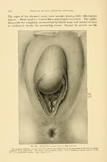
188 DISEASES OF THE EXTERXAL GENITALS.
The edges of the ulcerated cavity were necrotic, showing Httle inflammatory
reaction. Great numbers of nerve fibers were found everywhere. The epithe-
lial e:rowth Avas completely circumscribed by fibroid tissue and seemed to have
no tendency to invade the surrounding tissues. Beyond the growth was the
The prepuce encircles the convexity of the tumor, extending from one labium minus t
a shallow sickle-siiaped fold. The dorsal veins are shown above. A little lenticular va;
other, formingcvst lies at the
.ascc.fthe luft lal Oper. June 21, IS'J

VULVO-VAGIXAL GLAXDS. 189
normal mucous membrane, and beneath it the fat and fibrous tissue. Tlie exami-nation sliowed that the tumor was an epithelioma of the clitoris.
Cyst of the Clitoris.—I had occasion to remove (June 21, 1897) a large cystic
clitoris, shown in Fig. 119. The following is the report of the pathological
examination
:
Mrs. S. C. (Path. No. 1795). The fiuid is thick, sel)aceous-like, and of a
light brownish-jellow color. Microscopically it is found to contain numerouscholesterin and other irregular crystals. It also contains granular epithelial
cells. On histological examination the outer surface of the cyst is seen to becovered by squamous epithelium ; the walls are composed of wavy connective-
tissue cells running mostly parallel to the surface. Near both the outer andinner surfaces are localized areas of small round-cell and polymorphonuclearinfiltration. In one or two sections sebaceous glands can be seen. The inner
surface of the cyst shows considerable variation in its epithelial lining, someportions being covered by three or four layers of squamous epithelium, tlie deep-
est layer of which is cuboidal, and others by one layer of cuboidal cells. In
some portions where the epithelium is one layer in thickness the cells are
columnar. The cyst has evidently arisen from the clitoris.
VULVO-VAGIXAL CxLAX DS.
Cyst of the Vulvo-vaginal Gland.—Two kinds of cysts of the vulvo-vaginal
gland are met with—simple and suppurating. Both forms are among the com-
monest of the vulvar diseases, while other atfeetions of these glands are ex-
tremely rare.
The s i mJ)
1 e cyst is the result of an inflammatory occlusion of the duct
of the glands, followed by a retention of the secretions and the formation of a
tumor varying in size from that of a bean to that of a hen's egg. Inflammatory
cysts are oftenest due to gonorrheal infection, and the tendency just now is to
attribute all of these cases to this source.
I have known instauces, however, of small cystic accumulations in which
gonorrhea was probably not present. One of my patients for some time com-
plained of a pruritus for which she had had much treatment without Ijeneflt,
after which the cyst developed.
I have seen but one case in which both sides were involved.
The enlarged gland forms a projection more or less marked, according to its
size, to the right or left of the outlet, in a position corresponding to that of the
gland. The observer inspecting the tumor from the front is most struck by the
marked deviation of the cleft of the vulva forming a curved line directed toward
the sound side. The small cysts are located low down in the labium, in the
position of the gland ; but as they become larger they extend upward in the
direction of least resistance, and moi-e especially inward toward the mucous sur-
face of the labium, where they become quite superficial ; their mucous surface
appears smooth and shining, and sometimes almost transparent. The fluid con-
tained in the cysts is clear, yellowish or turbid, and generally of a gelatinous
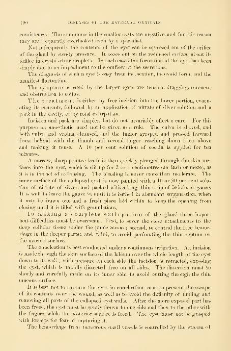
190 DISEASES OF THE EXTERNAL GENITALS.
consistence. The symptoms in the smaller cysts are negative, and for this reason
they are frequently overlooked even by a specialist.
Not infrequently the contents of the cyst can be squeezed out of the orifice
of the gland by steady pressure. It oozes out on the reddened surface about its
orifice in crystal-clear droplets. In such cases the formation of the cyst has been
sinqjly due to an impediment to the outflow of the secretions.
The diagnosis of such a cyst is easy from its location, its ovoid foi-m, and the
manifest fluctuation.
The symptoms created by the larger cysts are tension, dragging, soreness,
and obstruction to coitus.
The treatment is either by fi"ee incision into the lower portion, evacu-
ating its contents, followed by an application of nitrate of silver solution and a
pack in the cavity, or by total extirpation.
Incision and pack are simpler, but do not invariably efliect a cure. For this
purpose an anaesthetic need not be given as a rule. The vulva is shaved, and
both vulva and vagina cleansed, and the tumor grasped and pressed forward
from behind with the thumb and second finger reaching down from above
and making it tense. A 10 per cent solution of cocain is applied for ten
minutes,
A narrow, sharp-pointed knife is then quickly plunged through the skin sur-
faces into the cyst, which is slit up for 3 or 4 centimetres (an inch or more), as
it is in the act of collapsing. The bleeding is never more than moderate. Theinner surface of the collapsed cyst is now painted with a 10 or 20 per cent solu-
tion of nitrate of silver, and packed with a long, thin strip of iodoform gauze.
It is well to leave the gauze in until it is bathed in abundant suppuration, whenit may be drawn out and a fresh piece laid within to keep the opening fromclosing until it is filled with gi-anulations.
In making a complete extirpation of the gland three impor-
tant difiiculties nmst be overcome : First, to sever the close attachments to the
deep cellular tissue under the pul)ic ramus ; second, to control the. free hemor-
rhage in the deeper parts ; and third, to avoid perforating the thin septum on
the mucous surface.
The enucleation is best conducted under a continuous irrigation. An incision
is made through the skin surface of the labium over the whole length of the cyst
down to its wall ; with pressure on each side the incision is retracted, exposing
the cyst, which is rapidly dissected free on all sides. The dissection must be
slowly and carefully made on its inner side, to avoid cutting through the thin
mucous surface.
It is best not to rupture the cyst in enucleation, so as to prevent the escape
of its contents over the wound, as well as to avoid the difticulty of finding and
removing all parts of the collapsed cyst walls. After the more exposed part has
been freed, the cyst must l)e gently drawn to one side and then to the other with
the fingers, while the postericjr surface is freed. The cyst nuist not be grasped
with forceps for fear of rupturing it.
The hemorrhage from numerous small vessels is controlled by the stream of

ABSCESS OF THE V LLVO-VAGIX AL GLAXD. 191
water constantly running over the field. When the vulvo-vaginal duct is cut
the contents of the tumor often begin to exude by the fine orifice.
The removal of the cyst leaves a deep bleeding cavity in the labium. All
persistently bleeding vessels are caught and tied with fine catgut. Xeglect of
this precaution will result in the formation of a blood tumor of considerable
Fig. 120.—Left V'llvo-vaginal Gl.a EXCISED AND THE WoiND CLOSED WITH FiVE InTERIGUT SlTlRES.
size ; I have seen one such hematoma containing 90 cubic centimeters (8 ounces)
of blood, and the overflow into the patient's bed was estimated at a liter more.
The pulse, which was normal, went up to ISO. A profound anemia resulted.
After checking the bleeding, interrupted silkworm-gut or silk sutures are
passed on the skin surface, the loop of each suture reaching to the bottom of
the wound and bringing the surfaces together, leaving no pockets for the ac-
cumulation of blood. The usual dry dressing is placed on the surface, and the
sutures removed in a week.
Abscess of the Vulvo-vaginal Gland.—The vulvo-vaginal glands are especially
liable to become the seat of abscesses forming distinct tumors, in the lower part
of one or the other lal)ium, encroaching upon the vaginal outlet. These ab-
scesses have the same topographical relations as the simple cysts just described.
The overlying skin may appear normal or dark red, and injected in color.
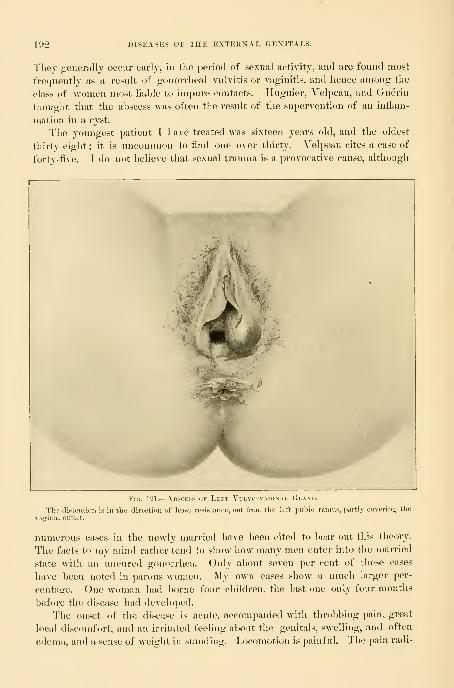
192 J)ISEASES OF THE EXTERXAL GEXITALS.
They generally occur early, in the period of sexual activity, and are found most
frequently as a result of gonorrheal vulvitis or vaginitis, and hence among the
class of women most liable to impure contacts. Iluguier, Yelpeau, and (xuerin
thought that the abscess was often the result of the supervention of an inflam-
mation in a cyst.
The youngest patient I have treated was sixteen years old, and the oldest
thirty-eight : it is uncommon to And one over thirty. Velpeau cites a ease of
fortv-five. I do not believe that sexual trauma is a provocative cause, although
Fig. 1-il.— AiiSPEss ok T.eft Vulvo-vagixai. (!i,am).
The distention is in the direction of least resistance, out from the left jmhio ramus, partly eoverin>r the
vaginal outlet.
numerous cases in the newly married have l>een cited to bear out this theory.
The facts to my mind rather tend to show how many men enter into the married
state with an uncured gonorrhea. Only about seven per cent of these cases
have been noted in parous women. My own cases show a much larger per-
centage. One woman had borne four children, the last one only four months
before the disease had developed.
The onset of the disease is acute, accompanied with throbl)ing pain, great
local discomfort, and an irritated feeling about the genitals, swelling, and often
edema, and a sense of weight in standing. Locomotion is painful. The pain radi-
ABSCESS OF THE YULVO-VAGIXAL GLAXl). 193
ates down the thigh, and there is often inabihty to sit down withont increasing
the pain, together with a sense of pressure in the rectum. The gait is sk)W and
straddling and the body bent forward. There are often cliills and fever.
The general condition of the patient is one of extreme malaise. Oftentimes
there is a history of leucorrhea, offensive or irritating, M'ith painful mictu-
rition.
One of my cases had an ulcer 1 centimeter in diameter at the fourchette and
another in the inferior wall of the urethra, near its internal orifice, 6 millimeters
by 10 millimeters in diameter. In this case the pus removed from the left vulvo-
vaginal gland contained numerous typical colonies of gonococci within the cells.
In most cases, however, the microscopic examination of cover slips has proved
entirely negative.
The affection usually reaches its height in a few days ; it may, however, be
several weeks in developing ; it tends toward spontaneous recovery by rupture
on the mucous surface of the labium, discharging from 15 to 100 cubic centi-
meters {^ to 3 ounces) of l)lood, grumous pus, or pure yellow pus. The exit
may be by one or several small openings, which tend to close rapidly. But the
disease is prone to relapse (" relapsing abscess "') in such cases. I have had a
patient who presented a history of repeated suppurations extending over many
months. Other rare cases give a history of alternation (alternating abscesses) of
the affection from one side to the other. Abscesses of the vulvo-vaginal gland
is especially liable to recur, because the ultimate ducts become infected, and so
the suppurative process may go on indefinitely, until the entire gland is either
destroyed or removed by operation.
Occasionally there is no enlargement of the gland visible on inspection. In
spite of the fact that there is no evident tumor, there may be a more or less
constant escape of pus, serving to keep up an infection of the rest of the genital
tract.
The diagnosis, as a rule, is easily made upon associating the symptoms de-
scribed with the discovery of a painful fluctuating tumor in the lower part of
one of the labia. The cases most liable to cause a mistake in diagnosis are those
in which the pus sac has thick walls and feels like a small, hard body, the size
of a bean, deep in the labium, without fluctuation. I have seen several of these
cases in which no diagnosis was made until the pus was let out. In one case
even the little nodule was thought to be malignant.
A simple abscess must not be confounded with stercoro-vulvar abscess, due
to a rectal fistula extending forward and discharging through a labium. I had
one such case treated by one of my assistants in which the rectal connnunication
was not recognized until the abscess was opened. This disease ought to be
diagnosed beforehand by the brawny induration, extending back on to the
perineum, and by the fistulous orifice which can be felt just inside the sphincter.
The history also often shows that the distress was first felt in the rectum.
The proper treatment of abscess of the vulvo-vaginal gland is by
free incision and packing. After suitably closing, shaving, and cleansing the
parts, the abscess is made tense by pi'essure from behind on both sides, when it

lO-t DISEASES OF THE EXTERXAL GENITALS.
is freely opened from below up. The incision is begun low down to give good
drainage in the most dependent position.
After evacuation the lips of the incision are separated, the sac wiped clean,
and its whole inner surface touched with pure carbolic acid on absorbent cotton.
The incision must be kept from closing until the cavitj is obliterated ; to do
this, the cavity is loosely j)acked with a thin strip of iodoform gauze, which
should be replaced every two or three days. It will be noticed that it shrinks
with surprising rapidity, holding less gauze each time the pack is removed.
After healing, the tine linear cicatrix will be found with difficulty. Sutures and
ligatures need not be used.
I object to making an incision on the mucous surface, unless the abscess is at
the point of rupturing there, on account of the possibility of a tender cicatrix at
this point. When the incision is made here, the anesthetic action of a twenty
per cent solution of cocain or ethyl chloride will suffice to blunt the sensibility.
In more than one instance I have known an abscess to rupture spontaneously
on the night before the time fixed for operation. The opening thus made by na-
ture in each of these cases was badly placed for the drainage, and too small to
allow a pack to be inserted. It was therefore necessary to make it larger by
incising it downward.
Where the abscess consists of a small indurated mass with a little pus in the
center, the better mode of treatment is by complete extirj)ation of the gland, if
possible without opening it. In one of these cases in my clinic it could only be
extirpated piecemeal, on account of the hard, infiltrated surrounding tissue. The
treatment of the cavity thus made is to close it completely, as described in the
case of cyst.
After the incision, evacuation, and packing, the woman must lie abed for sev-
eral davs or a week or more. She may go about as soon as she has recovered
sufficient strength, and the act of walking is not painful.
Adeno-carcinoma.— I report here in full a case of adeno-carcinoma of the
vulvo-vaginal gland upon which I operated in December, 1891. (R. S., Xo.
113(5, December 18, 1891.)
The writings of Gottschalk, of Berlin, Worth, of Kiel, and Koppe, of Mos-
cow, have thrown a new light upon cysts of the upper portion of the labia
majora, which can no longer be looked upon as connected with the vulvo-vagi-
nal gland. It has been shown that adenoid elements can occur in this region
probably due to the misplacement of epithelial elements in early embryonic life
;
it is possible that the following (^ase belongs to this group
:
My patient was an American, fifty-five years old. She had had ten children
and one miscarriage. The family history was negative. The menses had ceased
seven months before she entered the hospital.
At Christmas time, 1890, she noticed a swelling in the left labium majus,
which grew slowly for six months, and then for two iiiontlis it grow rapidly.
She had no pain, other than a dragging sensation.
Dr. II. Robb examined her upon entering the Johns Hopkins Hospital dur-
ing my absence in the summer of 1891, and found a hemispherical enlargement

ADEXO-CARCIXOMA OF THE VULVO-VAGIXAL GLAND. 195
in the left labium iiiajus, irreo-ular and lobulated, as big as an orange, dark red
on its surface, and fluctuating. It appeared to have bands of tissue running
ai'ouud it, and in two places the wall was thickened. There was well-marked
venous congestion of its surface.
Under the impression that the contents were purulent, it w^as incised, and a
large quantity of whitish and pinkish cheesy blood-stained dehris escaped with
clots. The cyst wall was lined with similar dehris and shreds of connective
tissue. It was evident that the sac could not be enucleated without great difii-
culty, and it was therefore cauterized and packed v.ith iodoform gauze, and an
k \
J#^"
Fig. 12'2.—Adeno-carcino.m.v of the Left Vllvo-vagixal Gland.
The skin is thin, the pores coarse and widely separated, and a few hairs are seen seattei-t-d over the sur->e. A LirL,'o vein courses over the riu'lit iiiidcr surface of the tumor, which contains a bloody tiuid.
external dressing applied. After eighteen days pure cai'bolic acid was ap])lied
to the cavity, which showed no tendency to close. After tw^enty-four days infll-
tration and thickening were still noticeable, bat not so marked as at first. Alittle whitish exudate was squeezed out.
She went home to return in four months, when I found a prominent tumor
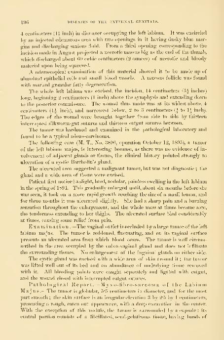
196 DISEASES OF THE EXTERNAL GENITALS.
4 centimeters (U iiicli) in diameter occnpjing tlie left labinin. It was encircled
by an injected edematous area with two openings in it having dusky blue mar-
gins and discharging sanious fluid. From a third opening corresponding to the
incision made in August projected a necrotic mass as big as the end of the thumb,
which discharged about (>0 cubic centimeters (2 ounces) of necrotic and bloody
material upon being squeezed,
A microscopical examination of this material showed it to be made up of
abundant epithelial cells and small blood vessels. A mucous follicle was found
with marked granular fatty degeneration.
The whole left labium was excised, the incision, 14 centimeters (5^ inches)
long, beginning 2 centimeters (1 inch) above the symphysis and extending down
to the posterior commissure. The wound thus made was at its widest above, 4
centimeters (H inch), and narrowest below, 2 to 3 centimeters (| to 1^ inch).
The edges of the wound were brought together from side to side by thirteen
interrupted silkworm-gut sutures and thirteen catgut sutures between.
The tumor was hardened and examined in the pathological laboratory and
found to be a typical adeno-carcinoma.
The following case (M. T., No. 3896, operation October 12, 1895), a tumor
of the left labium majus, is interesting because, as there was no evidence of in-
volvement of adjacent glands or tissues, the clinical history pointed strongly to
ulceration of a cystic Bartholin's gland.
The ulcerated area suggested a malignant tumor, but was not diagnostic ; the
gland and a wide area of tissue were excised.
Patient first noticed a slight, hard, nodular, painless swelling in the left labium
in the spring of 1894. This gradually enlarged until, about six months before she
was seen, it took on a more rapid growth I'eaching the size of a small lemon, and
for three months it was ulcerated slightly. She had a sharp pain and a burning
sensation throughout the enlargement, and the whole mass at times became sore,
the tenderness extending to her thighs. The ulcerated surface bled consideraljly
at times, causing some relief from pain.
Examination .—The vaginal outlet is occluded by a large tumor of the left
labium majus. The tumor is reddened, fluctuating, and on its vaginal surface
presents an ulcerated area from which blood oozes. The tumor is well circum-
scribed in the area occupied by the vulvo-vaginal gland and does not infiltrate
the surrounding tissues. No enlargement of the inguinal glands on either side.
The cystic gland was excised with a wide area of skin around it ; the tumor
was lifted well out of its bed and an abundance of underlying tissue removed
with it. All bleeding points were caught separately and ligated with catgut,
and the wound closed with interrupted catgut sutures.
Pathological Report.—My xo-fibro-sarcoma of the LabiumMa j u s .- The tumor is globular, 5*5 centimeters in diameter, and for the most
part smooth ; the skin surface is an irregular elevation 3 l)y 2*5 hy 1 centimeters,
presenting a rough, eaten-out appearance, with a deep excavation in the center.
With the exception of this nodule, the tumor is surrounded by a capsule ; its
central portion consists of a fibrillated, semi-gelatinous tissue, having bands of
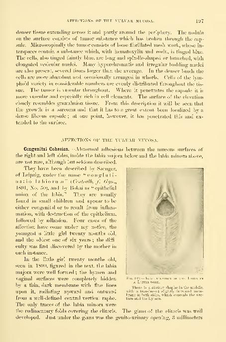
AFFECTIOXS OF THE VULVAR MUCOSA. 197
denser tissue extendino:; across it and partly around the periphery. The nodule
on the surface consists of tumor substance which has broken through the cap-
sule. Microscopically the tumor consists of loose fil>rillated mesh-work, whose in-
terspaces contain a substance which, with hematoxylin and eosin, is tinged blue.
The cells, also tinged faintly blue, are long and spindle-shaped or branched, with
elongated vesicular nuclei. Many hyperchromatic and irregular budding nuclei
are also present, several times larger than the average. In the denser bands the
cells are more abundant and occasionally arranged in whorls. Cells of the lym-
phoid variety in considerable numbers are evenly distributed tliroughout the tis-
sue. The tumor is vascular throughout. Where it penetrates the capsule it is
more vascular and especially rich in cell elements. The surface of the elevation
closely resembles granulation tissue. From this description it will be seen that
the growth is a sarcoma and that it has to a great extent been localized by a
dense fibrous capsule; at one point, however, it has penetrated this and ex-
tended to the surface.
AFFECTIOXS OF THE VULVAR MUCOSA.
Congenital Cohesion.—Aluiormal adhesions between the mucous surfaces of
the right and left sides, inside the laljia majonare not rare, although but seldom descril)ed.
They have been described by Saengei*,
of Leipzig, under the name " c o n g 1 u t i -
natio labiorum" ( Centralb. f. Gijn .,
1891, No. 50), and by Bokai as "epithelial
union of the lal)ia." They are usually
found in small children and appear to be
either congenital or to result from inflam-
mation, with destruction of the epithelium,
followed by adhesion. Four cases of the
affection have come under my notice, the
youngest a little girl twenty months old,
and the oldest one of six years ; the difti-
culty was first discovered by the mother in
each instance.
In the little girl twenty months old,
seen in 1890, figured in the text, the labia
majora were well formed ; the hymen and
vaginal surfaces were completely hidden
l)y a thin, dark membrane with fine lines
upon it, radiating upward and outward
from a well-defined central vertical raphe.
The only traces of the labia minora were
the rudimentary folds covering the clitoris.
below and the lal)ia minora abov
Fig. 128.—Ac. I i iin\iio\ of the Labia inA LiTTl.K ( . I l:l..
Tlicre is a distinct rliaphe in the middle.with a translucent slislitly furrowed nieiii-
birane in both sides, wliich conceals the ure-
thra and tlie hvmen.
The fflans of the clitoris was well
developed. Just under the glans was the geuito-urinary opening, 3 millimeters

198 DISEASES OF THE EXTERNAL GENITALS.
Fig. 124.
—
Agohtinatiox dk tup; L.m'.ia.
The same case after divi.sion of tlie membrane ; urethra and hymen exposed.
ill diameter, the sole outlet for urine and vaginal secretions. A probe intro-
duced through the opening and l)eliin<l this membrane showed the depth of
the anomalous interlabial septum to be 15 millimeters. On pushing the septum
forward the raphe appeared white. The
vagina was 5 '5 centimeters (2J inches) deep.
I look upon this case as simply an ab-
normally long fourchette, as there was no
history of any iniiammatory aiiection, and
especially because of the well-formed raphe,
which would not have been found on any
adventitious membrane. In two other cases
the membrane was similarly developed and
appeared to be congenital.
T r e a t m e 11 1.—The membrane was cut
down to its base, exposing a normal urethra
and hymen. The incision left a linear
Y-shaped wound on the mucosa. One su-
ture was required to control l)leeding.
Inflammatory Cohesion of the Mucous
Surfaces.—A little girl of six presented her-
f^elf at the clinic with an occlusion of the
outlet, first detected when she was a year
old. On inspection, a line of granular ero-
sion was found in the middle of the vulva posteriorly, and the labia were exten-
sively united on their mucous surfaces, concealing the site of the hymen and
urethra and the whole clitoris, but leaving a minute orifice just o\er the site of
the clitoris. The vulvar mucous surface throughout its entire length was ad-
herent. Under the influence of chloroform the adherent surfaces were stripped
apart with a probe, exposing a vaginal orifice 10 by 5 millimeters, the urethra,
and the clitoris. Lateral adhesions of the hood to the glans of the clitoris were
also freed. Sometimes the adherent surfaces may be separated by using cocain
instead of a general anesthetic.
Pruritus, or Vulvitis Pruriginosa.—Pruritus is especially a disease of the old,
and is one of the most distressing of all the gynecological afi^ections not en-
dangering life. It consists in a subacute inflammation of some portion or of
all the external genitals, involving the deeper layers of the skin and the nerve
endings ; it is therefore a dermato-neuritis. I have adopted the term vulvitispruriginosa, suggested by Sanger, of Leipzig (v. Gei^. f. (rehur/.s/i.,
Leipzig, Oct. 16, 1893), as more correctly descrilnng the morl)id process. The
common name "pruritus" means simply "itching" and nothing more, and
merely describes a symptom common to many afl'ections.
While the whole vulva may be involved, the disease is oftenest localized in
the free portion of the clitoris with its coverings, the neighboring surfaces,
and the labia minora. With these structures, the whole inner surface of the
vulva may be involved, the hymen forming the limit of its extension in-

) HOiT<iiaoaaa
.fiidal 9xlj .'1 ia6?A)iqoi aeaiB dgc^irfw erfT .^bsib ri«iwoI
lo ixi9jcniB9i>t «viu . .
. , „ n£ gxW aJiT989iq9*r eaii beiiob siiT— .S .-Oil
i-jBlq ni «9T0iii8—bgiamrnoo rrorJirinqO — .<'-; Vii'^l
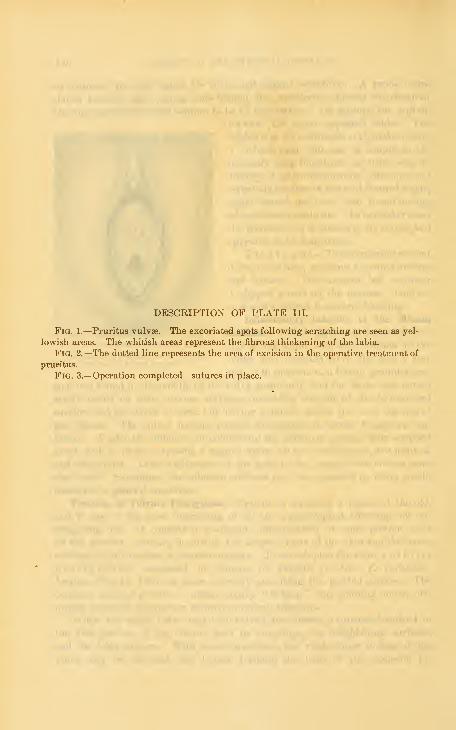
DESCRIPTION OF PLATE III.
Fig. 1.—Pruritus vulvae. The excoriated spots following scratching are seen as yel-
lowish areas. The whitish areas represent the fibrous thickening of the labia.
Fig. 2.—The dotted line represents the area of excision in the operative treatment of
pruritus.
Fig. 3.— Operation completed—sutures in place.

PLATE
FiAl.
Liifi .LPtanJ iCo. Bos}on,U,5.A
.


PRURITUS, OR VULVITIS PRURIGIXOSA. 199
ward. The skin surfaces of the ]al>ia majora also become involved in ajjgra-
vated cases.
1 have seen the disease localized to small areas about the clitoris and four-
chette ; in another case the most marked alterations were in the labio-femoral
folds.
The changes induced are a thickening due to an inflammation of the connec-
tive tissue in the corium. The mucous surfaces have a thick, dead-white, with-
ered appearance. The glans clitoris often completely disappears, lea\dng in its
place beneath the thick white preputial folds a little pit. These white surfaces,
as well as the labia on their outer surfaces, are streaked with fissures which are due
to scratching ; these are pink at the bottom and generally arranged vertically.
The real causes of pruritus are not known, although a number of provoca-
tive causes and conditions are well recognized.
In every case of intractable pruritus the urine should l)e examined for sugar,
as some of these cases are diabetic in origin ; this is due to the fermenta-
tion of the urine, which then acts as an irritant upon all the tissues with which
it comes in contact.
A sero-purulent discharge from a myomatous uterus proved to lie the excit-
ing cause in one instance : all attempts to relieve the pruritus failed, until
finally the patient was so harassed that she consented to operation. The uterus
was extirpated for the myoma and the jiruritus ceased. .
Fissure in ano may be accompanied by pruritus of the vulva, which will
be cured l)y the healing of the fissure.
An attempt has been made, without success, to demonstrate a bacterial origin.
The initial stages may often be attributed to irritative vulvar and vaginal secre-
tions, after which the more aggravated form of the disease develops from the
repeated mechanical insults in rubbing and scratching tlie parts.
The proper treatment of the severer cases of chronic pruritus with the
changes described is, as advised by Sanger, by excision of the diseased area. Thefree mobility of the external genitals and adjacent parts allows almost any defect
created by an excision to be readily covered.
The following case will serve as an illustration of the operation where the
disease involves all the external genitals except the skin surfaces of the labia
majora
:
The patient was a married woman, fifty-seven years old, and a nullipara;
she had had one miscarriage twenty-five years ago ; menopause sixteen months
before operation. When younger she had had a milky leucorrhea, but this
had ceased for several years. She had suffered from itching in the genitals
for twenty years, at first always connected with the menstrual period, begin-
ning a day 1 )efore and lasting twelve days ; for three yeai-s past thei-e had been
a constant intolerable itching and burning, with burning micturition, keeping
her awake almost every night, and nearly driving her insane. During this
period she had noticed the formation of little blisters between the labia, which
would break, leaving raw surfaces, discharging pus. These surfaces i-arely ap-
peared to heal.

200 DISEASES OF THE EXTERNAL GEXITAI>
I found the inner surfaces of tlie la])ia niajora covered with irregular wliite
patches of thickened epidermis, more al)undant above, extending from the upper
commissure down to the lower part of the vaginal outlet ; below this the surface
V
/
>^^i
'
'^^
Fui. 125.- Tl liKltCL LUSiS OF TllJi \'eST1BILE.
The flut pinkish luaiuinillated disoasetl area oecufiies the entire vestibule, enuroachiny on tlic iipinT i
tin of the urethra and extending sliffhtlv into the anterior vajrinal sulei. Tliere is no thickeninir or inti
tion of the edges, which are raised about t\v(
and the adjacent parts red and swolleiabove the level of the tliseascd
rnar-
tiltra-
toris
was covered with a reddish glaze. At the angle between the inner nnicous and
external skin surfaces of the labia—that is, on the margin of the disease—was
a line of whitish scales with slightly elevated edges. A few small superficial
ulcers were scattered over the wliite area. The labia minora were M-ithered
down to insignificant rudimentary folds. The clitoris was completely concealed
beneath the thickened diseased tissue, and a little hole only showed where the
glans is usually found. The hymen was entirely absent ; the disease was limited
by a line encircling the vaginal outlet and including the urethra, which was not
involved.

PRURITUS, OR VULVITUS PRURIGINOSA. 201
The wliole of this diseased surface was excised under anesthesia by an opera-
tion lasting thirteen minutes. After a prolonged painstaking scrubbing an
incision was made, outlining the area to be removed, beginning at the commis-
sure above and extending down on either side along the angle between the
outer and inner surfaces of the laliia, to the level of the vaginal floor. From
-Fig. 126.— Ai:i.A -i- K\<im..n ,,k Tuber-cular l)isp;.\SE SHOWN ny the DottedLine: this included the Clitoris,BOTH Labia Minora, and the EntireVestibule, with the Anterior 1'artOF the Urethra.
Fig. 127.
—
Kaw .Surface created by the Re-moval of the Diseased Area, showingFour Catgut Sutures uniting the Pos-terior Margin of the Urethra to theVaginal Mucosa.
this point the incisions were carried up to the vaginal outlet and around it,
meeting over the urethra.
The area excised, roughly described, resembled a sj^earhead pointing up-
ward with a deep notch at its base. The whole tliickness of the skin thus
outlined was rapidly dissected away, removing with it the labia minora and the
body of the clitoris. The dissection was made from above downward by catch-
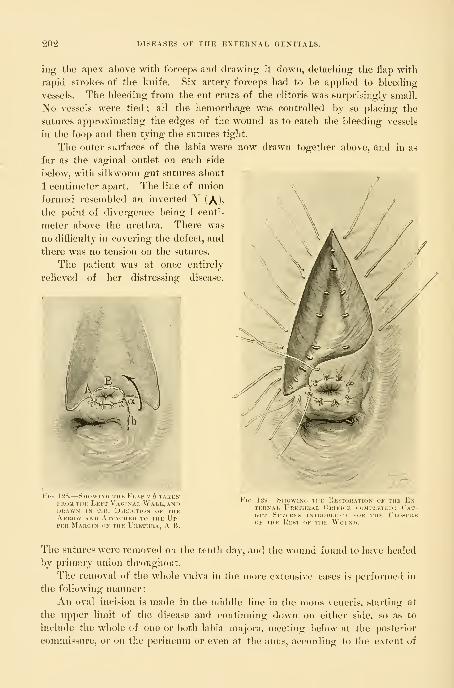
202 DISEASES OF THE EXTERNAL GENITALS.
ing the apex above with forceps and drawing it down, detaching the flap with
rapid strokes of the knife. Six artery forceps had to be apphed to bleeding
vessels. The bleeding from the cut crura of the clitoris was surprisingly small.
No vessels were tied ; all the hemorrhage was controlled by so placing the
sutures approximating the edges of the wound as to catch the bleeding vessels
in the loop and then tying the sutures tight.
The outer surfaces of the labia were now drawn together al)Ove, and in as
far as the vaginal outlet on each side
below, with silkworm-gut sutures about
1 centimeter apart. The line of union
formed resembled an inverted Y (\),
the point of divergence being 1 centi-
meter above the urethra. There was
no difficulty in covering the defect, and
there was no tension on the sutures.
The patient was at once entirely
relieved of her distressing disease.
O^!|ii^i?^ jb ^'^SiWui^
. 128.
—
SiiowiN(i TiiK Flap 'J b takenFROM THE Left Vaginal Wall, anddrawn in the direction of theArrow and Attached to the Up-per Margin of the Urethra, A B.
Fig. 129 —SiiovviMi iiil 1;l>iui;aiiun of the E.\-
ternal Urethral Orifice completed; Cat-GL'T Sutures introduced for the Closureof the Rest of the Wound.
The sutures were removed on the tenth day, and the wound found to have healed
by primary union throughout.
The removal of the whole vulva in the more extensive cases is perforuicil in
the following manner:
An oval incision is made in the middle line in the inons veneris, starting at
the upper limit of the disease and continuing down on either side, so as to
include the whole of one or both labia majora, meeting below at the posterior
commissure, or on the perineum or even at the anus, according to the extent of

TUBERCULOSIS OF THE VESTIBULE. 203
tlie disease downward. Another incision encircles the vaginal outlet in the
position of the hymen, beginning above the urethra. The whole area between
these two incisions is rapidly excised from above downward, and bleeding ves-
sels caught with forceps. The edges of the
upper part of the wound are brought together
from side to side with silkworm-gut sutures,
down as far as the level of the urethra. Below
this point they are drawn in on either side and
attached to the vaginal outlet, covering the
whole defect.
Tuberculosis of the Vestibule.—Tubercular
disease of the external genitals is extremely
rare, not more than three or four cases having
been recorded. This disease is usually associ-
ated with pulmonary phthisis.
I have seen one case of tuberculosis of the
vestibule. The patient, Mrs. S., a widow, aged
lifty-five, complained chiefly of stinging pain
on urination, caused by the urine flowing over
the ulcerated area. A small ulcer first ap-
peared one year before coming to me ; this
increased steadily in size until it occupied an
area as seen in Fig. 125, Mrs. S. (Gryn. Path.
IS^o. 1756). The specimen removed consists of
a triangular piece of tissue, the margins of
which are covered by mucous membrane. The
central portion presents an eaten-out appear-
ance, and the deeper tissues are infiltrated,
though not markedly indurated. Situated in
this ulcerated area is the urethral orifice surrounded by a narrow band of
smooth nmcous membrane. Histologically, the surface of the ulcer is made
up of the characteristic tubercular granulations, while typical tubercles are
scattered throughout the deeper tissues, some being found innnediately beneath
the urethral mucous membrane. Tubercle l)acilli are demonstrable in small
numbers.
Diagnosi s.—Tuberculosis of clitoris and vestibule.
The description of the operation is given in Figs. 126-130.
Fig. 130.
—
Showinc. i
KejsT of the W(h m) ahove theUrethra in the Foum of an In-verted Y.

CHAPTEK X.
RUPTURE OF THE RECTO-VAGINAL SEPTUM AND RELAXEDVAGINAL OUTLET,
1. I'hysiological support of the vaginal outlet.
2. Operations for recent injuries to the vaginal outlet: External tear; internal, and combinedexternal and internal tear; complete tear of recto-vaginal septum.
3. The intermediate oi)eration for injuries to the outlet.
4. Old complete rupture of recto-vaginal septum : Operation for the same.
5. Relaxed outlet: Operation for the same.
The Physiological Support of the Vaginal Outlet.—Tlie " vaginal outlet," called
"outlet" with reference to its parturient function, or "introitus," "inlet," or
" vaginal entrance," from its sexual function, forms the inferior extremity of the
vaginal canal connnunicating with the external genitalia beneath the pubic arch.
While the vaginal canal above and within the pelvis is broader and more capa-
cious, inferiorly it suddenly narrows down to an outlet which is a canal 4 to 5
centimeters (2 inches) long.
The posterior wall of the vagina in sagittal section of the body forms a flat-
tened sigmoid with the convexity of its lower curve directed forward behind the
jnibic arch. The peculiar funnel shape of the vagina—broad above and con-
tracted below—appears most distinctly upon placing a woman who has never
borne children in the knee-l)reast position and letting in air, when the intra-
pelvic portion of the vagina will balloon out, while the outlet or introitus re-
mains tightly contracted, closely hugging the pubic arch.
The mechanical theory advocated by some writers, that the closure of the
outlet and its snug position beneath the pubic arch are dependent upon a thick
wedge of tissue, a so-called " perineal body," acting like a cork plugging a Ijottle,
is erroneous. In consequence of this false conception absurd and unnecessary
operations have been devised and extensively employed for injuries in this
situation.
The error of this prevalent notion is evident upon examining the virginal
outlet, where the vaginal otitlet and anns are both seen lifted well up under the
pubic arch, and upon introducing the index finger into the vagina the pubic
arch is felt in front, while posteriorly a broad, rounded, resilient band of mus-
cular tissue, the levator ani, stretches behind the outlet from the right to
left pubic ramus.
This examination readily demonstrates the important fact that the vaginal
introitus is but a narrow chink between this posterior nniscular hand and the
pubic arch. By making backward pressure upon the posterior wall of the
304

THE PHYSIOLOGICAL SUPPORT OF THE VAGIXAL OUTLET. 205
vagina within the introitus the band yields, to return to its former position as
soon as the pressure is withdrawn.
The fourchette and the supposed " perineal body," lying between the four-
chette and rectum, should now be closely examined by placing the index finger
of one hand just within the vagina and the other in the rectum and palpating
so as to measure the size and thickness of these structures. It will be found
that they are but slight tissues incapable of giving any support to the super-
jacent organs.
The real supporting mechanism of the outlet is not the perineal body, but
the anterior portion of the levator ani muscle. This broad, rounded muscle
arises on either side of the inner surface of the pubic ramus and passes back
around the lateral vaginal wall to unite w^ith its fellow behind the rectum, its
fibers beino- intimatelv interwoven with the lateral walls of the rectum. These
Vol.—Normal Varinal Outlet in
The vagina is completely hidden and there is no fattening across the anus and the gluteal furrow as in
the case of a relaxed outlet. The little concentric furrows surrounding the fourchette below are not foundin the virgin.
important anatomical relations may readily be detected in the living subject by
making pressure in each lateral sulcus of the vagina while one finger lies within
the rectum.
From what has just been said, it is apparent tliat the vaginal outlet has no

206 RUPTURE OF THE RECTO-VAGINAL SEPTUM.
direct means of closiii-e such as would be afforded by a powerful sphincter mus-
cle, but depends for its support upon the indirect action of the levator muscle.
For by the contraction of this nmscle the lower end of the rectum is tightly
lifted up under the pubic arch and the vagina flattened out and held up between
the two. It is further important to notice that the position of the plane of the
pubic arch, in front of the plane of the levator fibers, renders the closure more
efficient, like a " cut off." It is this arrangement which gives the sigmoid curve
to the lower extremity of the virginal vagina.
With rare exceptions, the important injuries to the vaginal outlet affect its
caliber alone, and arise during parturition. It is not difficult to appreciate the
rationale of this when we recall the fact that during the passage of the child's
head the outlet, normally from 2 to 3 centimeters (^ to 1 inch) in diameter, is
dilated until it forms a ring 28 centimeters (1() or 12 inches) in circumference.
In numerous instances, instead of the gradual and all-round dilatation of the
outlet produced by repeated impacts of the advancing and retiring fetal head,
the yielding is sudden and in one place, with rupture of the muscular fibers hi
consequence. The parturient canal represents a funnel within a funnel, the
uterus and cervix representing the upper funnel, set within the upper vagina
and outlet as the lower funnel. In consideration of this fact it is surprising tliat
both the contracted outlets, cervical and lower vaginal, are not more frequently
damaged during the passage of the large fetal ovoid. Injuries to the outlet
similar in character often result from the removal of large submucous myomata
lying within the vagina. The vaginal outlet may be injured from without by a
vai-iety of accidental causes, such as falling upon a chair post, or being gored by
cattle, or in a child from sliding down a haymow on to a jjitchfork handle, or
sliding down a balustrade on to a low newel post.
The operation in all cases of injured outlet should be performed as soon as
possible after the injury ; all other operations at a later date act only as more
or less efficient suljstitutes.
Recent obstetrical injuries at the vaginal outlet may, for practical purposes,
be classified under three heads
:
1. External superficial tear.
2. Internal, and combined externaband internal tear.
3. Complete tear of the recto-vaginal septum.
Recent External Superficial Tear.—The simplest form of tear begins at the
fourchette, extends backward through the skin in the median line, and involves
the superficial wedge of lax tissue between the fourchette and the rectum ; it mayextend up into the vagina as far as the posterior column. This form of injury
is the commonest and relatively the least important, and does n(»t in any case
affect the supporting structures at the vaginal outlet.
The only purpose of an operation for its relief is to avoid healing by granu-
lation and the possible formation of a tender scar.
Operation.—In its slightest forms the external tear needs no further at-
tention than strict cleanliness throughout the convalescence.
A deeper injury, with a base 2 to 3 centimeters (f to 1^ inch) in length,

RECENT INTERNAL TEAR. 207
may be sutured immediately after deliv^ery, or on the following day, when
the j)atient should be brought across the bed under a good light, with the legs
flexed on the abdomen and held by an assistant or by a legholder.
The necessary instruments are a needle holder, medium-sized curved needles,
and catgut and silkworm-gut sutures. These instruments should be close at hand
on a sterilized towel. The hands of the operator should be carefully washed im-
mediately before operating. The labia are now held apart with the first and
second fingers of the left hand, exposing a torn triangulai- surface on either
side posteriorly. A needle provided with a carrier threaded with a catgut
suture is introduced in the sound tissue near the upper angle of the tear from
a half to three quarters of a centimeter from its margin, brought out at the base
of the wound, and re-entered, to emerge on the mucous surface opposite the
point of entrance. A similar suture is placed about a centimeter below this.
When both these sutures are tied the wound is closed down to a shallow pit on
the skin surface, where two or three superficial sutures may be needed to com-
plete the approximation.
The wound should be protected afterward with iodoform and boric-acid
]iowder.
Recent Internal Tear and Combined External and Internal Tear.—Another
conmion form of injury sustained in parturition is a slit in the nmcosa, which
may extend from the fourchette or from the hymen for 4 or 5 centimeters up
into the vagina into one of its sulci. In another form the tear is forked and
extends into both sulci. This injury is often caused by the head of the child
starting within the vagina a tear, which is enlarged by the shoulder following,
plowing its way down between the levator fibers and their rectal attachments
on one or both sides. If this tear happens to be continued forward, it becomes
associated with the external tear and forms a combined external and internal
tear.
Neglect of this injury results at a later date in the serious disability which I
describe as a relaxed vaginal outlet. The fact that this lesion within the vagina
was not looked for by our older practitioners has induced many of them to
assert that lacerations never occurred in their obstetrical practice. Teachers of
obstetrics can not lay too much stress upon the necessity of a proper examina-
tion after labor and proper attention to this neglected form of primary injury.
Lnmediately after the birth, if the labia are separated and the posterior
vaginal wall inspected under a good light, the ragged, l)leeding surface of the
tear stands out in marked contrast with the smooth vaginal wall, although both
alike are uniformly deeply congested.
The Operatio n.—The lacerated surfaces must be repaired at once or on
the day following delivery, for a few sutures skillfully applied at this time will
accomplish the work of many more at a later date.
The patient should be placed as described in the operation for superficial ex-
ternal tear, resting upon a pei'ineal drainage cushion. The use of an anesthetic
advisable if she is nervous, can usually be dispensed with if the operator is deft
and can work quickly.
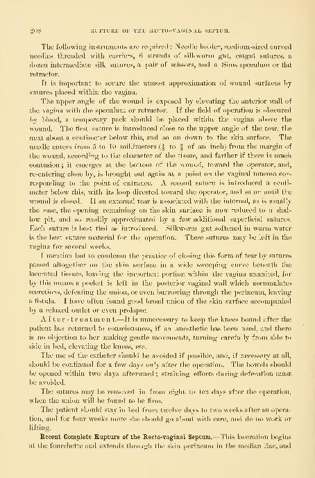
208 RUPTURE OF THE RECTO-VAGINAL SEPTUM.
The following instruments are required : Needle holder, medium-sized curved
needles threaded with carriers, 6 strands of silkworm gut, catgut sutures, a
dozen intermediate silk sutures, a pair of scissors, and a Sims speculum or flat
retractor.
It is imjiortant to secure the utmost approximation of wound surfaces hy
sutures placed within the vagina.
The upper angle of the wound is exposed by elevating the anterior wall of
the vagina wnth the speculum or retractor. If the fleld of operation is ol^scured
by blood, a temporary pack should be placed within the vagina above the
wound. The first suture is introduced close to the up])er angle of the tear, the
next about a centimeter below this, and so on down to tlie skin surface. The
needle enters from 5 to 10 millimeters {^ to f of an inch) from the margin of
the wound, according to the character of the tissue, and farther if there is muchcontusion ; it emerges at the bottom of the wound, toward the operator, and,
re-entering close by, is brought out again at a point on the vaginal mucosa cor-
responding to the point of entrance. A second suture is introduced a centi-
meter below this, with its loop directed toward the operator, and so on until the
wound is closed. If an external tear is associated with the internal, as is usually
the case, the opening remaining on the skin surface is now reduced to a shal-
low pit, and so readily approximated by a few additional superficial sutures.
Each suture is best tied as introduced. Silkworm gut softened in warm water
is the best suture material for the operation. These sutures may be left in the
vagina for several weeks.
I mention but to condemn the practice of closing this form of tear by sutures
passed altogether on the skin surface in a wide sweeping curve beneath the
lacerated tissues, leaving the important portion within the vagina ununited, for
by this means a pocket is left in the posterior vaginal wall which accumulates
secretions, defeating the union, or even burrowing through the pei-ineum, leaving
a fistula. I have often found good broad union of the skin surface accompanied
by a relaxed outlet or even prolapse.
A f t e r - 1 r e a tm e n t.—It is unnecessary to keep the knees bound after the
patient has returned to consciousness, if an anesthetic has been used, and there
is no objection to her making gentle movements, turning carefully from side to
side in bed, elevating the knees, etc.
The use of the catheter should be avoided if possible, and, if necessary at all,
should be continued for a few days only after the operation. The bowels should
be opened within two days afterward ; straining efforts during defecation must
be avoided.
The sutures may be removed in from eight to ten days after the operation,
when the union will be found to be firm.
The patient should stay in bed from twelve days to two wrecks after an opera-
tion, and for four weeks more she should go about with care, and do no work or
lifting.
Recent Complete Rupture of the Recto-vaginal Septum.—This laceration begins
at the fourchette and extends through the skin perineum in the median line, and

THE INTERMEDIATE OPEKATIOX FOR IXJURIES TO THE OUTLET. 209
through the sphincter ani for a variable distance up tlie recto-vaginal sep-
tum. The tear into the rectum forms a serious complication, destroying the
function of the sphincter muscle and causing incontinence of feces and flatus.
By this accident a sensitive patient is cut off from the comjsany of her nearest
friends, and compelled to live in a state of isolation. Strange, however, as it
may seem, if the operation is not performed at once, the patient may carry her
malady for years without seeking the relief so readily afforded.
O p e r a t i o n.—An innnediate operation is imperative. The parts should
1)6 suitably exposed, as described for the last operation, and under anesthesia,
if the patient can not be perfectly controlled without it. The instruments
required are a needle holder, scissors, curved needles, and catgut and silk-
worm-gut sutures. The first step in the restoration is the closure of the rent
in the bowel, which is effected by interrupted catgut sutures on the rectal sur-
face at the upper end of tlie tear. Each sutui-e pierces the margin of the
mucosa and apjDears on the septum 4 or 5 millimeters (^ to ^ inch) from the
edge, to enter the septum on the opposite side, coming out again on the
mucosa. The remaining sutures are passed in like manner, radiating out on
to the skin surface and embracing the ruptured ends. Great care must be
taken in bringing the sphincter ends into accurate approximation. The lower
sutures alone are not sufficient to insure the sphincter union without the addi-
tion of a silkworm-gut suture entering on the skin surface and emerging well
behind the ends of the ruptured muscle and traversing the septum. The tear
now presents the appearance of the simpler form just described, which is closed
by interrupted silkworm-gut sutures, for the most part placed within the
vagina. Each suture is tied as passed, and a few superficial catgut sutures
are passed i)etween them, to insure perfect approximation. This operation
skillfully performed is always successful if a puerperal sepsis does not inter-
fere.
The bowels should be moved on the third day, and opened every second
day afterward. Under no circumstances should they be allowed to become
constipated.
It is important that the patient should remain at least two weeks in bed.
The external sutures should be removed on the eighth day, and the internal a
week or two later.
The Intermediate Operation for Injuries to the Outlet.—The intermediate period
begins from five or six days and extends to two or three weeks after labor, while
the unrepaired perineal wound is undergoing granulation and cicatrization. Theparts at the bottom of the wound, naturally in close juxtaposition, often unite by
first intention, while the remaining area is engaged in throwing off sloughing
particles, granulating, and cicatrizing. In a few days small pink granulations
are visible over the wound area, while the marginal epithelium as a fine white
line invades it on all sides, contracting the wound from day to day. The granu-
lating surface and the adjacent area is rigid and flushed by the new vasculariza-
tion. The intermediate period, although not often selected as a time for opera-
tive interference, on account of prolonging the detention in bed, is not altogether

210 RUPTURE OF THE RECTO-VAGINAL SEPTUM.
unsatisfactory, for a well-performed operation will be almost surely followed by
a good result.
The wound is best exposed on a table, with flexed thighs, under a good hglit.
Local anesthesia by means of cocain will, as a rule, be sutticient. This is secured
either by saturating a pledget of absorbent cotton with a 4 per cent solution,
applying it for ten minutes to the wound and surrounding tissue, or by injecting
a few minims around the margin of the wound. With a sharp scalpel or spoon
curette, the operator vigorously scrapes off the granulations, using also scissors
Fig. VM.—CoMi'LETi'FullM.
The opening is filled up with the anterior vaginal wall. At each end of th
gon below slight depressions, indicating the sphincter pits, are seen.
pen til
and forceps as needed to effect the denudation. The peculiarity of the tis-
sue will be found to be its friability, which makes it difficult to denude evenly
in the usual way with scissors and forceps. The denudation must everywhere
extend down into the sound tissue below. If some time has elapsed since the
injury, it will be necessary in denuding to allow for consideral)le contraction of
the wound. In this case strips of adjacent mucous membrane nnist also be
removed.
The sutures should tlien be passed as described in the repair of recent inju-
ries, according to the character of the tear, whether external superflcial, internal,
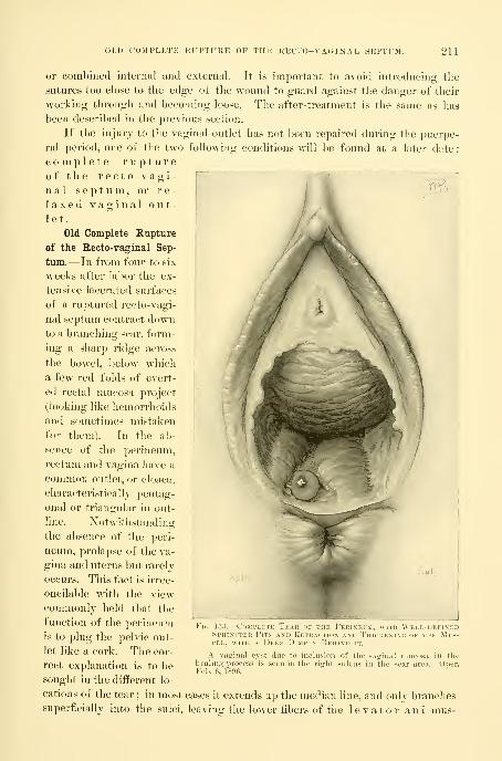
'OMPLETE RUPTURE OF THE RECTO-VAGINAL SEPTUM. 211
or combined internal and external. It is important to avoid introducing the
sutures too close to the edge of the wound to guard against the danger of their
working through and becomhig loose. The after-treatment is the same as has
been described in the previous section.
If the injury to the vaginal outlet has not been repaired during the puerpe-
ral period, one of the two following conditions will be found at a later date
:
complete ruptureof the recto-vagi- "
nal septum, or re-
laxed vaginal out-let.
Old Complete Rupture
of the Recto-vaginal Sep-
tum.—In from four to six
weeks after labor the ex-
tensive lacerated surfaces
of a ruptured recto-vagi-
nal septum contract downto a branching scar, form-
ing a sharp ridge across
the bowel, below which
a few red folds of evert-
ed rectal mucosa project
(looking like hemorrhoids
and sometimes mistaken
for them). In the alj-
sence of the perineum,
rectum and vagina have a
common outlet, or cloaca,
characteristically pentag-
onal or triangular in out-
line. ]^otwithstanding
the absence of the peri-
neum, prolapse of the va-
gina and uterus but rarely
occurs. This fact is irrec-
oncilable with the view
conraionly held that the
function of the perineum
is to plug the pelvic out-
let like a cork. The cor-
rect explanation is to l)e
sought in the different lo-
cations of the tear ; in most cases it extends up the median line, and only branchessuperficially into the sulci, leaving the lower fibers of the levator ani mus-
FlG. \Py4.—L'ns
SpilINrTKtCLE, WITH
\n OF THE FerINP i \l. u 111
II'- \.M) Ki.TR\CTION AM) IhICKENIDeep Dimple Behind it.
A vaginal cyst due to inclusion of tlie vaginal mucosalioaling process is seen in the right sulcus in the scar area.Feb. 6, 1896.
.Mi-s-
n theOper.

212 RUPTURE OF THE RECTO-VAGINAL SEPTUM,
cle uninjured. When, in rare instances, the i-upture both passes through the
periueum centrally and extends deeply into one or both sulci, prolapse may
occur.
The sphincter a n i muscle in cases of complete tear will vary in form, mJitfcrent cases, from a simple broken circle, with its ends still bound together, all
;i^.#J. >vall
;^,.-
VV.. 1:U.~RUPT.:UE OK THE ReCTO- V AO ,NA ,, Sk.T.M, U.HM^A.., V KXTKNmNO HUH,,P A'-^'« "^^
P^^"^
K.on Vagina., Wall, iu;t now pwllk.. down bv the ( oNTKA.nu.Ns of t.. S.MnNCTi.,K ink.
Shallow Ako with Extreme Separation of the SpiiiNrrER Ends. .Itne 21, ls,»7.
the way to a shallow arc, in wliich case the nmscle is short and thick with a deep
(Hinple^n the skin behind it. A smooth glazed depression, at times puckered or
pitted, at the lower angle of the perineal scar, frequently serves as the sphincter

OLD COMPLETE KUPTl'KE OF THE RECTO-VAGINAL SEPTUAL 2\?,
landmark. A straight sphincter is the result of frequent contractions pnlliny
down the angle of the tear so that ultimately a deep tear comes to look like a
superficial one. Thus the extent of separation of the ends of the nmscle becomes
a measure of the depth of the original tear. It is sometimes difficult to identify
the sphincter ends upon simple inspection, but by pulling on or pinching the
Case sekn in Fi
On bringing the two sides together the hymen is found to he intact except posteriorly. The yielding ofthe vaginal orifice in labor has" therefore been sudden, and all in one place posteriorly, instead of a slow,equable distention producing multiple tears in the hymen. June 21, 1807.
muscle so as to stimulate a contraction, the position of the ends may always be
discovered. It is important not to be misled, by the ability of the patient to
retain feces, into the error of thinking the tear can not be complete, for
where the original rent is shallow and the cicatrix in the angle binds the

214 RUPTURE OF THE RECTO-VAGINAL SEPTUM.
ends firmly too-etlier, the sphincter will often contract efhcientlj up to this
point. A similar result is, in fact, all we can hope to attain by the best plastic
operation.
Operation.—Women with complete rupture of the septum sometimes
have a chronic diarrhea. Here the only preparation necessary is an injection
high into the bowel a few hours before
the operation, clearing out all fecal
matter. In other cases a free purga-
tion must be secured the night before
operation, followed l)y an enema in
the morning. Carbolized and mercu-
rial solutions, if used at all in other
cases, must never be used here for
irrigation, on account of the dan-
ger of fatal poisoning from absorption
t
Fig. i; -Cojii Kk
The sphincter pits are seen below on bothsides of the rectal orifice, the shortened sphinc-
ter muscle is much thickened, and there is acharacteristic pit just below it. The red line
encloses the area to be denuded ; it must not be
forgotten that the triangles seen extending upinto the vagina are srreatly fore- shortened.
through the bowel. To prevent dis-
charges from contaminating the field,
one or two pledgets of iodoform
gauze wrung out in warm water are
pushed up into the lower bowel, to
be removed when the operation is
completed. The instruments neces-
sary are scalpel, dissecting forceps,
Emmet's left-curved scissors, needle holder, curved needles, and catgut and silk-
worm-gut sutures. The area to be denuded must be outlined with the scalpel,
Fig. 137.—Complete Tear of the Recto-vagi-
nal Septum. Denudation completed.

OLD COMPLETE RUPTURE OF THE RECTO-VAGIN'AL SEPTUM. 215
which follows the direction of the scar tissue in a general way, greatly exagger-
ating its outlines ; the cardinal principle in the denudation is to reproduce as
nearly as possible the original injury.
The lirst incision splits the septum and includes the sphincter ends, from
which a line is continued up under the pubic arch on either side ; thence it goes
down into each vaginal sulens and back again, meeting in front of the posterior
column, 1 to 2 centimeters (f to -| inch) above the first incision in the septum.
All of the tissue included within the out-
line is now removed. Begin at one of "
the sphincter ends, catching it up with
tissue forceps and cutting it free with
curved scissors. Continue the denuda-
tion around the sharp edge of the septum
to the opposite end of the sphincter,
which is denuded in the same way, tak-
ing care to remove all scar tissue. Asecond strip above and parallel to this is
next cut off, a third, and so on, continu-
ing the denudation up into the vagina
until the whole area within the outline
has been removed. It is important to
bear in mind that the denudation within
the vagina must extend a centimeter or
more (^ inch or so) above the angle of
the tear in order to avoid the tendency
to form a recto-vaginal fistula at this
point. Silkworm-gut and catgut sutures
are best adapted to the approximation of
the denuded surfaces. Half-deep su-
tures of catgut are preferable for closing
the rectal side of the tear, and for se-
curing accurate approximation between
the silkworm-gut sutures, which are used
at wider intervals. The complication of
the torn bowel is first disposed of by a
series of interrupted rectal sutures, com-
mencing at the upper angle of the tear,
entering each suture at the margin of the rectal nnicosa, and emerging on the
wound surface 4 to 5 millimeters {-^ to | inch) distant, re-entering on the opposite
side and coming out again on the margin of the mucosa at a point corresponding
to that of entrance. This suture may be tied at once and dropped into the rec-
tum, and a little less than a half centimeter {^ inch) below this another suture
passed in like manner, tied, and dropped, and so on until the whole of the rectal
rent has been obliterated down to the sphincter. One of the most imj)ortant
points in the operation now is to secure an accurate approximation of the
Fig. 138.
—
Complete Tear of the Kecto-vagi-NAL Septum.
Rectal sutures introduced, but not tied. Notethe position of the silkworin-trut tension suture
introduced well behind the sphincter ends andpassing up through the septum.

216 KUPTUKE OF THE KECTO-VA(;iNAL SEI'TUM.
sphincter ends l)y two or three sutures radiating from the rectal out on to the
skin surface. The conti-actions of the sphincter render it necessary to assist
these sutui-es with one of silkworm gut introduced well behind to the denudedends and passing up through the septum. When this has been done the rectal
Fig. 13it.
—
Complete Teak of the Kecto-vaginal Se
The rectal sutures all tied except the silkwonu-^ut tension suture. The sutures iire introduced butnot tied in the right vaginal sulcus, one of silkworm gut and two of catgut above it.
rent is repaired, the wound is reduced from a complicated one involving three
surfaces—rectum, skin, and vagina—to a simpler wound involving vagina and
skin perineum.
The next step is the repair of the vaginal wound by a silkworm-gut suture
in either sulcus, reaching doM^n to the series of rectal sutures, at the bottom of
the wound. The loop of the suture should he in a plane nearer to the oi)erator
than its points of exit and entrance, so as to lift up the tissues at the bottom of
the wound when it is tied. Superficial and half-deep catgut sutures com])lete
the union within.
There still remains an opening on the skin surface, which is i-eadily I)i-ought

THE RELAXED VAGINAL OUTLPyr. 217
together by a silkworm-gut suture, aided by a few superficial or half-deep cat-
gut sutures.
The Relaxed Vaginal Outlet.—The name " relaxed outlet " describes a loose,
gaping introitus, a condition which is more frequently observed after multiple
childbirth, each successive delivery distending the orifice, until it appears like
the mouth of a bag without its draw string, as Dr. T. A. Emmet has long been
in the habit of describing it. Although a frequent ailment, it is rarely recog-
nized except under the title of some one of its attendant and accidental fea-
Fici. 140.
—
Complete Tear of the Recto-vaginal Septi m.
Rectal and vaginal sutures all introduced and tied, and the perineal sutures in place, but not yet tied.
tures, such as " rectocele," " cystocele," or " rectocele and cystocele,'"" or '' lacera-
tion of the perineum" in varying degrees.
Clinical Appearance.—Upon inspection of such a patient on the
back, with the legs flexed, the cleft of the buttocks appears flattened and broad
;
the anus is often wide, somewhat everted, and displaced backward ;the sphincter
ring is clearly seen. The skin perineum is often preternaturally deep and the
fourchette intact. In other cases the skin surface of the perineum is torn as

218 RUPTURE OF THE RECTO-VAGINAL SEPTUM.
far back as the sphincter a n i. The intact deep perineum has long been
a gynecological stumbHng block, on account of the inveterate habit of physi-
cians of estimating tlie functional activity and efficiency of the vaginal outlet by
its depth on the skin surface ; a " good perineum," signifying that the distance
from fourehette to anus measures 2i centimeters (1 inch) or more, whence the
faulty conclusion is drawn that the support at the vaginal outlet nmst likewise
Fig. 141.
—
Complete Tear of the Kecto-vaginal Septum.
All three sets of sutures introduced and tied, the cati^ut suture cut off and the silkworm gut left long.
The outlet is pulled open a little in order to show the inside suture.
be "good." The fact is that in many of the worst forms of relax-
ation the perineum is deeper on the skin surface than be-
fore childbirth, a condition due to the overstretching of the external skin
at the time the outlet was broken down.
On separating the labia in a case of relaxed outlet the vaginal walls appear
more or less pouting, and either the anterior or posterior walls may protrude
to a marked degree. In rarer cases lateral, anterior, and posterior walls all
protrude.
The relaxed condition of the vaginal outlet may be demonstrated in a \ ariety
of ways. Upon instructing the patient to bear down, both anterior and posterior

THE RELAXED VAGINAL OUTLET. 219
walls roll out, bringing into view a considerable portion of the lower vagina.
We are thus enabled to estimate the effects of lifting, walking, or straining at
stool u23on such a patient. If a finger is placed upon the cervix uteri,
during the act of straining it will be felt descending in the axis of the vagina
toward the outlet. The descent is especially marked if the patient is examined
in the erect posture, when the surgeon will also be still better able to judge
the effect of exercise upon her pelvic organs.
Upon placing the patient in the left lateral position and elevating the upper
right buttock, air rushes audibly into the vagina and the posterior vaginal wall
drops away fi-oni the anterior, leaving the gaping outlet as a large hole in the
pelvic floor {vide. Fig. l-i-i).
Palpation in the dorsal position reveals other important deviations from the
normal outlet. The perineum is often but a lax, thin partition which may easily
Fig. 142.
—
Scheme of the Operation for Complete Tear of the Recto-vaginal Septum laid on aFlat Surface.
The torn sphincter muscle is indicated by dotted red lines eross-liatclied at each end. The deep inden-tation on the under side of the figure represents the rectal side of the tear, and the two red triangles above,one on each side, represent the denudations extending up into the vaginal suleis. The sutures are passedfirst on the rectal side. A, radiating out into the perineum, then in the vaginal sulci, B, and finally on theperineal side, C. The cross-marked sutures are of catgut and the plain ones of silkworm gut. JNote espe-
cially the silkworm-gut sutures passed in behind the sphincter ends and up into the septum.
be gathered up between thumb and forefingers of both hands and lifted up
over the urethra and the clitoris. Many physicians are misled by the fact that,
when the patient is lying in the dorsal position, the lax anterior and j^osterior
vaginal walls apparently fill out the deficiency. Touch, however, ought to
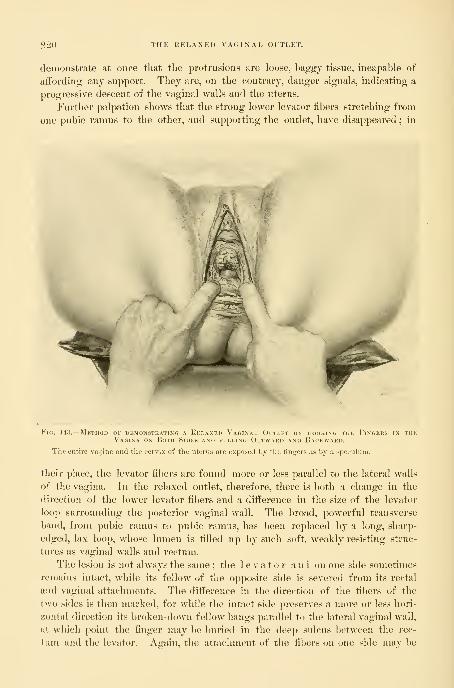
220 THE HKLAXKl) VAGINAL OITLET.
demonstrate at once tliat the protrusions are loose, bagf^y tissue, incapable of
affording any support. They are, on the contrary, danger signals, indicating a
progressive descent of the vaginal walls and the uterus.
Further palpation shows that the strong lower levator fibers stretching from
one pubic ramus to the other, and supporting the outlet, have disappeared ; in
-^
ti. 143.
—
Method of demonstrating a Kei.axed Vaginal Outlet bv hooking the Fingers in thVagina on Both Sides and pulling Outwakd and Backward.
Tlie entire vagina and the cervix of the uterus are exposed by the fingers as by a speeulvun.
their place, the levator fil)ers are found more or less parallel to the lateral walls
of the vagina. In the relaxed outlet, therefore, there is both a change in the
direction of the lower levator fibers and a difference in the size of the levator
loop surrounding the posterior vaginal wall. The broad, powerful transverse
band, from pubic ramus to pubic ramus, has been replaced by a long, sharp-
edged, lax loop, whose lumen is filled up by such soft, weakly resisting struc-
tures as vaginal walls and rectum.
The lesion is not always the same ; the lev at o r a n i on one side sometimes
remains intact, while its fellow of the opposite side is severed from its rectal
and vaginal attachments. The difference in the direction of the fibers of the
two sides is then marked, for while the intact side pi-eserves a more or less hori-
zontal direction its broken-down fellow hangs parallel to the lateral vaginal wall,
at which point the finger may be buried in the deep sulcus between the rec-
tum and the levator. Again, the attachment of the fibers on one side may be

TESTS FOR TIIK RELAXKD VACxINAL OUTLET. 221
nearer the outlet than the fibers of the opposite side, which lie in a different
plane.
While the eversion of the relaxed outlet is often evident upon simple inspec-
tion, it may be most characteristically demonstrated by placing the thumbs on
either side of the outlet behind and pushing outward and upward.
In many cases of reflex disturbances a relaxed outlet can only be detected
Fio. 144.
—
Test for the Kelaxed Vaginal Outlet, showing how the Po.stekiob Vaginal Wall dkop.'s
WELL AWAY FROM THE ANTERIOR WaLL BY SIMPLY PLACING THE PaTIENT IN SiMs's ToSTURE.
'I'lie external skin perineiun is well preserved, but in spite of this there is a large lax, cjai)ing orifice.
by examination under an anesthetic, for during a conscious examination the
weakened levator is nnder tonic contraction and more or less efficiently closes
the outlet, and the examiner may be so far deceived as to estimate a marked
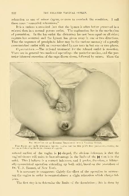
222 THE RELAXED VAGINAL OUTLET.
relaxation as one of minor degree, or even to overlook the condition. I call
these cases " concealed relaxations."
It is a curious anatomical fact that the hymen is often better preserved in a
relaxed than in a normal parous outlet. The explanation lies in the mechanism
of parturition. In the lax outlet the distention has not been equal on all sides
;
rupture has occurred and the hymen has given away in one or two directions.
Thus the sequence of precipitate labor may be the curious anomaly of a greatly
overstretched outlet with an overstretched hymen torn in but one or two places.
Operation.—The rational treatment for the relaxed outlet is resection.
There are in general two modes of operating—the posterior median, and the pos-
terior bilateral exsection of the superlluous tissue, followed by suture. Since the
Fig. 145.
—
Test of an Ex
Four fingers are easily introduced into the vaiiina and tlie thin pelvic floor pushed out, evcrtinjj therectum, and showing the entire absence of support.
natural outline of the vagina is H -shaped, the obvious inference is that the
vaginal tissues will unite to best advantage in the limbs of the H that is in the
sulci. Tins I believe is a correct inference, and I prefer, therefore, a bilater-
ally symmetrical operation extending up both sulci, based on the procedure of
Dr. T. A. Emmet, of New York.
It is necessary to exaggerate slightly the effect of the operation in narrow-
ing the vagina in order to counterbalance a slight relaxation which always fol-
lows.
The first step is to determine the limits of the denudation ; this is done by

OPERATION FOR THE RELAXED VAGINAL OUTLET. 223
means of two tenacula shaped like a sbeplierd's crook, fixed on either side at the
junction of the hymenal ring, or its remains, leaving sufficient tissue across the
anterior vaginal wall between the tenacula to make a small outlet when the
tenacula are brought together. These points mark the upper lateral limits of
^L2^ -L.Fio. 14<i.— Kelaxeu Vaginal Ottlet.
i fingers are demonstrating the position and the direction of the lower fibers of the levator-ani mus-lich is grasped between them. The almost vertical direction of the levator loop is especially note-,' as the most characteristic feature.
The„cle, which .^ f^.^.^^^^. .^^^..^^,^ „,.v,....
worthy as the most characteristic feature.
the resection. If thej are fixed too near the urethra too much tissue will be
removed and the new outlet will be too contracted ; on the other hand, if they
are fixed too low down the new outlet will continue to be too large, notwith-
standing the operation. The correct pattern to have in mind in resecting is the
nulliparous outlet.
A third tenaculum is now fixed in the vagina in the median line posteriorly,
on the crest of the vaulted prominence of the rectocele, or posterior column (wV7^
Fig. 150).

224 THE RELAXED VAGINAL OUTLET.
With these three points fixed, the area of denudation must now Ije out-
lined with a sharp scalpeL The bloodj outhne obviates the hal)iHty to error
in a free-hand denudation. No one
pattern will fit all cases ; as an ex-
cessive relaxation requires a more
extensive resection than one of
moderate degree.
Fig. 147.— Calibrator for measuuino tiik
De(;ree of Rela.xation of the VaginalOutlet ; Graduation^ in Centimeters.
The blades are closed and introduced just
within the outlet and then opened as far iis
they will separate easily. The tio^ures on thescale measure the decree of the relaxation.
The surface to be denuded is
irregular in outline and occupies
several planes, making it difficult
to represent it adequately in a pic-
ture. In making the outline the
central tenaculum and one of the
lateral tenacula are drawn widely
apart, downward, and outward, ex-
posing one of the vaginal sulci. If
there be a moderate degree of re-
Fio. 148.
—
Relaxed Vaginal Oitlet in a VII-I'ARA, WITH rEUKECT I'rKSERV ATION OF THEHymen, except in the Median Line Po.s-
teriorly.
Forccp.s were not uswOperation, June 1, IH'JT.
any of tiic lal)ors.
lax ation the apex of the triangle outHned in eacli sulcus is situated o centimeters
(1^ inch) within the outlet. By dej)ressing the convex posterior vaginal wall a
distinct line will be seen at the juncture of the anterior and lateral walls. An

OPERATION FOR THE RELAXED YAGIXAL OUTLET. 225
incision should he made down to the lateral tenaculum through the vagina, par-
allel to and just below the anterior wall. From the same point within, the sec-
ond side of the triangle is made by an incision down to the tenaculum at the
crest of the rectocele. A narrow triangular undenuded area remains between
the two triangles thus formed
in the sulci (m<;/6? Fig. 151). The
outline is now comjileted by a /,,
semicircular incision extending
around the posterior wall^ keep- /^ ,| \-
ing within the hymen above, -i^m ill// \but embracing any scar tissue
seen below. The center of this
line falls 3 to 4 centimeters (1

226 THE RELAXED VAGINAL OUTLET.
to tlie apex of one of the triangles ; tlien it continues back, and is carried to andfro across the front and up into the other triangle
; frequently the whole out-
lined area can be removed in a single strip. The dissection is often facili-
FlG. 151.— KhLAM.1) VaMNAL OlTLET.
The silkworm-frut tension suture is placed in the triansjle on the right side. Tlie dotted lines representthe part of the suture which lies concealed under the surface. The short piece of the suture visil)lo as awhite line at the bottom of the denudation is the part which is exposed by bringing the needle out at thebottom of the wound and re-entering it close by.
tited l»j running the ends of the scissors beneath the lax tissue on the floor of
the vagina. Arterial and venous hemorrliage from cut vessels is sometimes
free, but the venous flow lasts only a short time and ceases spontaneously. Anactively spouting artery should first be clamped for a time in the artery forcei)s,
and if it persists in bleeding after a few moments it may be tied with catgut.
By judicious application of the deep tension and the approximation sutures,
much hemorrhage can be checked without the use of buried sutures at all.
The large wound area is nowac(;urately npproxiniated by means of from three
to four silkworm-gut sutures, and from eight to twelve half-dee]) and superficial
catgut sutures. But one silkworm-gut suture is placed witliin the vagina, in
cither sulcus. An assistant exposes one of the triangular areas by drawing the
tenacula at its base downward and outward ; a carrier is entered upon the

OPERATION FOR THE RELAXED VAGINAL OLTLET. 227
mucosa on the lateral vaginal wall near tlie incision, a little below the middle of
the triangle, and carried under the tissue toward the operator, appearing at the
bottom of the sulcus, considerably below the point of entrance ; it is re-entered
close bj and carried in the reverse direction, finally emerging on the mucosa of
the opposite side of the triangle (and opposite the point of entrance). A stout
silkw^orm-gut suture sharply bent upon itself, 2 centimeters (f inch) from the
end, is hooked into the loop of the carrier and drawn through, then pulled u])
and tied in a square knot, care being taken to adjust accurately the edges of the
wound before tying. The suture thus placed draws together a large area of
tissue. To close the wound accurately above the suture its ends are grasped
between the third and fourth fingers, and by traction the upper part of the tri-
is exposed, as a narrow ellipse, with loosely approximated sides. Perfect
The silkworm-gut s^uture is tied and pulled down, exposini; the cati^ut sutures in place and ready to
be tied, closing accurately the upper ]iart of the denudation, already broutrht loosely together by the silk-
worm gut. These catgut sutures must pass deep into tJie tissues so as not to leave a pocket in the woun<lbelow them.
union is secured here by fine catgut sutures, carried deeply from side to side.
The first is placed but a short distance above the one of silkworm gut, tied here,
and used in its turn as a tractor, exposing the wound immediately beyond ;then
the next suture is passed and tied and so on, until the upper part of the triangle
is closed and all bleeding has stopped. The opposite sulcus is closed in the same

228 THE RELAXED VAGINAL Ol'TLET.
way with a single suture of silkworm gut and several of fine catgut. These sutures
should check all hemorrhage, but if there is persistent oozing it must be controlled
by additional sutures tied tightly at the bleeding point. In this way a large par tof
the resected area within the vagina has been approximated, and the vaginal canal
markedly narrowed within the pelvis. When the triangular areas in the sulci arelarge, a half-deep catgut suture should bo added below the one of silkworm gut.
Most of the remaining area may be brought together by a sino'le o-atherino-
suture of silkworm gut, embracing the upper angles on the sides and transfixing
the rectocele {vide Fig. 15;^)).
An additional silkworm-gut suture may sometimes be necessary on the skin
The inside sutures are now int
introduee'l aliovc across the an;do\v this is also left untied.
Ihe -ratlierinir suture of silkworm ^nit
V suture introduced to close the wound
surface extending through to the bottom of the wound. Half-deep and super-
ficial sutures will complete the union.
The duration of the operation is from fifteen to thirty minutes. The out-
lining takes about one minute and a half, the denudation three or four minutes,
and passing the sutures ten minutes longer; various minor matters may lengthen
the time to half an hour.

RESULT OF OPERATIOX FOR THE RELAXED VAGINAL OUTLET. 229
The result of the operation is now evident in the change in the position, size,
and direction of the vaginal outlet. It has been lifted and restored to its posi-
tion well beneath the pubic arch. Its size has been reduced from 5 or 6 to 1^
or 2 centimeters (2^ to f inches) in diameter. The examining finger no longer
enters in the direction of the promontory of the sacrum, but goes backward
Fk,. l.j-l.—itJiLAXtD V Alji.SAL (^L ll-l:.!.
ShowiiiiT how the gathering suture above draws to-
gether the tissues.
L OlTLET.
Operation completed. The suture.s
with lonjrer ends, two inside and twooutside, are ot'silkworm gut ; the others
are all of catgut.
toward the coccyx. This change in direction and position of the outlet removes
it from the line of intra-abdominal pressure in which it has lain. Instead, there-
fore, of the constant tendency to eversion of the vaginal wall through a wide
opening, the pressure is spent in forcing the anterior vaginal wall down upon
the posterior one, and both upon the restored pehdc floor.
The external sutures should he removed from the eighth to the tenth day.
Those in the inside may remain several weeks.
The immediate result of this operation is a complete restoration, and even
the hymen is often restored. Subsequent labors will not destroy the eifects of
the operation, unless unskillfully conducted or attended by complications.
CHAPTER XT.
OPERATIONS ON THE VAGINA.
1. Introductory: a. Anatomy, b. Intrinsic affi'ctions, few. f. 'rnuinuitic affections, largest gronp.
d. Diseases from extension.
2. General surgical principles.
8. Congenital affections: a. Aljseiice of the vagina, b. Iraperfnrate hynien. r. Atresia of oneside of the uterus, d. Vaginal septa, e. Double vagina associated with uterus bicornis
duplex.
4. Foreign bodies.
5. Vaginitis.
6. Vaginal cysts.
7. Abscess of Gartner's canal.
8. Neoplasms: a. Benign, b. Sarcoma and carcinoma.
'J. Traumatic affections : of the vault ; of the orifice ; of the canal. Strictures—falciform andannular. Atresia.
10. Recto- vaginal fistula.
The vao:;ina is a simple iniisculo-membi'anous canal, lined by stratilied epi-
tlieliinn. It is distinctly fnnnel-shaped, its lower extremity being contracted
and lying beneath the pubic arch, while its expanded upper end, flattened antero-
posteriorly, rests upon the pelvic floor and receives the cervix of the uterus.
Tlie list of its surgical affections is a short one. The most frequent and im-
portant are the ruptures at either extremity occurring during parturition ; those
al)Ove are produced by the extension of a tear beginning in the cervix and con-
tinuing out into the vaginal vault on one or both sides, while those below are
oftenest found in association with a lacerated outlet. Other vaginal affections,
such as malformations, cysts, and neoplasms, are rare.
Uterine tumors, such as a polypus or an inverted uterus fllliug the lumen of
the vagina, are not reckoned among vaginal diseases.
The vagina being in the form of a sac, all of its surgical diseases, except fis-
tulae, in one way or another affect the size of the canal ; foreign bodies, cysts,
abscesses, neoplasms, cicatrices, and atresije encroach upon the lumen and dimin-
ish the caliber to a varying degree. The injury at the outlet alone has the efl'ect
of enlarging the canal at this point. Destructive diseases, such as sloughs, ulcers,
and carcinoma, perforate the walls, establishing fistulous communication witli the
bladder in front, the rectum behind, or even the small intestines above.
GENERAL SlMiGU'AL PinNClPIiKS.
The surgical ])nn('iples involved in tliu treatment of vaginal diseases re-
late to
:
1. The thorough cleansing of the field.
2. The proper exposure for operation.
230

GENEUAL SURGICAL PRINCIPLES. 231
3. The careful removal of existing disease, avoiding injury to important
neighboring viscera.
4. The control of hemorrhage.
5. The closure of the wound by suture.
The vagina must be thoroughly cleansed before everyoperation. After bringing the buttocks to the edge of the table on a drahi-
age pad, as in all plastic operations, the assistant retracts the posterior commis-
sure with two fingers, or in a virgin with a narrow speculum, and introduces into
the vagina a ball of cotton al)out 3 centimeters (1-2 inch) in diameter, coated
with soft soap, grasped in a pair of foi-ceps ; warm water is then poured in from
a vessel above, and the upper vaginal tract thoroughly cleansed by vigorously
scrubbing for several minutes in all directions, taking care to distend and cleanse
between the folds. All parts of the vault will be better reached if two fingers
are introduced, the middle finger pushing the vaginal tissue down or to one side,
while the index finger pushes the cervix in the opposite direction. Bj repeated
washings with fresh pledgets of cotton and soap, followed by douching with
warm water, all debn'-s' and loose epithelium are gradually softened and removed.
The lower vaginal tract is cleansed in like manner. Where discharges are
escaping out of the cervix there is a constant liability to reinfection. It is best
in such a case to dilate and curette the uterus, and then to pack the vault of the
vagina with iodoform gauze during the performance of an operation.
Exposure of the field .—In women who have borne children there is
usually no difiiculty in opening the vaginal outlet wide enough to expose and
permit easy access to all pai-ts of the canal by means of retractors in fi-ont and
behind. The upper retractor fitting under the symphysis pubis should be nar-
row, with its blade spreading outside to keep the labia minora from dropping
over the outlet and so obstructing the view ; the posterior retractoi- must be
broad enough and. long enough to stretch the vagina and afford a good view of
its walls, including the cervix. I found it necessary in one case, in order to reach
a malignant disease of the vault of the vagina, to split the pelvic floor from the
fourchette around the anus to the sacro-coccygeal joint, and dissect down l)eside
the rectum, turning it to one side.
In the removal of diseased tissue it is im])ortant to
bear in mind the topographical relations of the jjarts. In
tlie first place, there are no organs except the ureters in the vicinity whose integ-
rity is essential to life ; thus, if uterus, bladder, or rectum ai-e involved together
with the vagina, portions of these structures may be sacrificed in removing the
diseased tissue.
Even a considerable segment of tlie rectum may 1)e removed and the upper
and lower ends brought together. The exsection of poi-tions of the bladder
requires careful attention to avoid injuring the ureters, whose location is indi-
cated by the ureteral folds in the anterior vaginal wall.
The h e m o r r h a g e encountered in vaginal operations is never alarming, and
is readily controlled by forceps and ligatures. Bleeding from the vaginal walls
may always be controlled by the sutures approximating the edges of the wound.
232 OPERATIOXS ON THE VAGINA.
Silkworm gut is the best suture material where thei-e is tension, but
silk and catgut may both be used.
Cleanliness is maintained after the operation by keeping
the vaginal outlet dry and well protected. As soon as there is any discharge from
the vagina the pack should be removed, and if the discharge is sweet-smelling the
further care should consist in applying iodoform and boric acid powder (1 to 7)
and absorbent cotton over the outlet. If the discharge is ill-smelling at any
time, the vaginal douches must be given once or twice daily. I lind the pleas-
antest and most efficient douche to be two drops of menthol and bicarbonate of
soda and borax, a teaspoonful each, dissolved in half a liter of hot water and
used warm.
The various surgical aifections of the vagina may be considered under the
following heads
:
1. Congenital affections.
2. Foreign bodies.
3. Cysts.
4. Neoplasms,
5. Traumatic affections and atresiae.
6. Recto-vaginal fistulie.
CONGENITAL AFFECTIONS.
Congenital affections are imperforate hymen, absence of the vagina, atresia
of the upper part of the vagina with bicornute uterus, double vagina, and
vaginal septa.
Absence of the Vagina.—When the vagina is absent, the uterus, ovaries, and
tubes are also usually either absent or rudimentary. The exact condition of the
organs higher up must be determined by a bimanual examination under anes-
thesia through the emptied rectum and the abdomen ; the examination may also
sometimes be made by a finger in the rectum and a sound or a finger in the
bladder. An operation attempting to establish a connection between rudimen-
tary organs and the vulva can not be serviceable, and is therefore unjustifiable.
It is also useless to attempt to form a deep pocket between the rectum and blad-
der simply for sexual purposes, as such an opening can not be maintained.
Transplantation for Atresia of the Vagina .—An absent
vagina may be replaced by the transplantation of new vaginal tissue from a case
of prolapse, when the uterus, tubes, and ovaries are present, or when there is a
unicorn uterus with hematometra and hematosalpinx. As W, Nagel has pointed
out, in many apparently congenital cases the atresia of the vagina is really due
to an unnoticed local inflammation in early childhood.
The formation of a new vagina has been twice successfully done l)v A.
Mackenrodt {Cetdralh.f. Gyn., 1896, No. 21, p. 540) as follows: A transverse
incision is made in the septum between the urethra and the rectum, and the
vesical and the rectal sides of the septum are separated fmni cu-h other by a
blunt dissection, with finger and instruments ; on reaching the cervix both index

CONGEXITAL AFFECTIONS. 233
lingers are inserted and tlie future vagina widened. The wound surface is nowpacked firmly with an iodoform-gauze tampon, whicli is changed every few days,
until the whole surface becomes covered with healthy granulations. At tliis
juncture the transplantation is done either at one or at several sittings, accord-
ing to the amount of the tissue available to make the new vagina. Unusualcare must be taken in preparing the flaps which must be carefully cleansed first,
and then cut out without crushing or bruising, and with as little as possible of
the underlying connective tissue. The flaps are then put aside, wound surface
to wound surface, and kept warm and covered in a sterile dish until the pro-
lapse operation has been completed, when they are laid with great care on the
dried aseptic wound surface and pressed down, until they adhere as if by suc-
tion;they are then fixed in place by an iodoform-gauze tampon, which remains
undisturbed for about ten days, while the patient is kept absolutely quiet in bed.
The replacement of the whole vagina at one sitting is more diflScult; the flaps
formed as described are spread out lengthwise on a Cusco's speculum, with the
epithelial surfaces turned inward ; they are then sewed loosely together so as
to allow any secretions to escape between them. At the inner end of the specu-
lum they are connected by a few threads which form a loose pocket; then the
upper end of the speculum is filled with iodoform gauze and inserted into the
wound, and as the speculum is withdrawn more gauze is packed in, all in onepiece, until the tamponade is completed. Especial care must be o-iven to keo])-
ing the tampon from being wet with urine. In
two weeks the parts appear normal.
When both the vagina and the uterus are a1)-
sent and the ovaries are present and functionally
active, the recurring monthly paroxysms of pain,
associated at times with a vicarious menstruation,
may necessitate celiotomy for the removal of the
ovaries, as in the following case :
B. M. (No. 2190, Sept. 9, 1893), an anemic
woman twenty-eight years old ; at the age of
twelve began to have periodical monthly severe
headaches, accompanied by dizziness and flushes
of heat over the entire body ; her face flushed
readily, and she was annoyed by frequent blush-
ing; she also had sharp, cutting pains in there- , _ ,^.^_ _^ j
fifion of the left ovary; between the attacks she Figi 56.—Entire Absence of the"^,
.\ AGINA, "WITH Indication of
felt well. In her twentieth year she began to Double Hv.men-. The Exter-1 , . "i T n • NAL Genitals Normal. Sept.nave convulsions, as many as four and five in a 9, i893.
day. The first hemorrhagic discharge from the
rectum took place in her fourteentli year at one of the periods. The flow was
dark brown in color, clotted, and not offensive, and continued one day ; after
this there was no discharge for three years, but the feeling of fullness and pain
in the abdomen still recurred every month. In her seventeenth year she had
a second hemorrhage, which continued for six weeks.
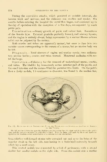
234 OPERATIONS ON THE VAGINA.
During the convulsive attacks, which persisted at variable intervals, she
became weak and nervous, and the abdomen was swollen and tender. Six
months before entering the hospital the rectal flow began, and continued up to
the day of operation with the exception of a few days, accompanied by nmch
pelvic pain.
Examination.—Scanty growth of pul)ie and vulvar hair. Escutcheon
of the female type. External genitalia perfectly formed, rudimentary hymen,
and the vagina is entirely absent, being represented by a narrow fibrous column
which can be palpated by the rectum.
Both ovaries and tubes are apparently normal, but seem to fuse into two
nodular masses corresponding to the cornua of a uterus, but no uterine l)ody can
be felt.
Diagnosis.—Total absence of vagina and uterine cervix; two rndinien-
tary uterine bodies ; ovaries and tubes normal. Menstrual molimina with rec-
tal discharge.
Operation.—Celiotomy for the removal of undeveloped uterus, ovaries,
and tubes. The bladder lay transversely across anterior part of the pelvis, and
the small intestine and the rectum filled the posterior two thirds. On the pelvic
floor a fleshy nodule, IS centimeter in diameter, was found in the median line,
U^
Fis. 157.
—
Relations ok Tiiii Ridimi;: I II i:i, nil I I lu X,
ul- 1 III, \' \(,1NA.
• \ \i:iEs IN THE Case of Ausem-k
,e lies upon the l)i,t(l(lei- lu-iir tlic ineaiiiii luu-, tlie riirlit nodulf is cinsi' to tlie pi'lvu-
hriin, and the two are connected by a thin He-li\ liiind. Note tlic lar^e oval made by the round liiraineuts.
Operation for vicarious rectal menstruation \Mtli extreme nervous diseonitorts.
from which a well-formed uterine tube extended out and ii]) to the lirim of the
su])erior strait on the left side, terminating in a fimbriated extremity beneath
which lay a small ovary.
This central nodule was connected by a fold of peritoneum with a second
similar but smaller nodule on the right side. From this nodule also a uterine

IMPERFORATE HYMEX. 235
tube extended out to an ovary. Both tubes were patulous down to the fleshy
nodules, from which well-fonned, round ligaments extended out to each inguinal
canal.
A fibrous band about 4 centimeters long, about 5 millimeters broad, and 2
to 8 millimeters thick, extended down under the bladder, representing the va-
Fiu. 158.^Lf:FT TruE
The inembrane below, with iiurallel tblds, occupies the position of the uterine body and the upper vagina.Sept. '.•, 1893, Xo. 5-1. Natural size.
gina. The rudimentary uterine cornua tubes and ovaries were removed and the
patient made a good recovery.
Microscopical Examination.— Tubes normal, well developed,
nothing atypical in their histology. They end in two euls-de-sae in fleshy
nodules slightly larger than the tubes and containing some normal utei-ine mu-cosa and glands.
The ovaries contain Graafian follicles in various stages of development. ( 'or-
pora lutea and corpora fibrosa are present.
Diagnosis.—Rudimentary bicornute uterus, al)sencG of cervix and fun-
dus of uterus, normal ovaries and tubes.
Imperforate Hymen.—The simplest form of vaginal atresia is that of its
lower extremity at the hymen. The closure here affects the vaginal mucous
meml)rane alone and does not consist, as in atresise above, of an absence of the
entire wall with a replacement by fibrous tissue, forming the sejitura. The hy-
men in these cases usually forms a thick, tough, resisting membrane.
The upper vagina, uterus, ovaries, and tul)es are, as a rule, well formed and
functionally active in such cases.

230 opp:rations on the vagina.
This condition of the hjnien is rarelj recognized until puberty, when the
failure of the appearance of the menstrual secretions is the occasion of an exami-
nation, which at once reveals the anomaly. It may, however, be discovered in
quite young children by the accumulation of mucus within, causing the hymento pout out, forming a whitish sac between the labia beneath the urethra, which
becomes more prominent when the child cries. If this sac is cut open with a
pair of scissors a little mucus is discharged and no further difficulty is ex-
perienced.
After puberty, as each menstrual period pours its secretions into the uterus
and vagina, the more lluid parts are absorbed and leave behind a thick tarry sul)-
stance. In the course of time, between the ages of seventeen and twenty, the
vagina may become distended into a sac big enough to till the pelvis. The upper
end of the sac is formed by the expanded uterus, often with dilated tubes at
/^*--^/
Fio. 159.—Right Tube, Ovary, NiiRMAL Size of the Ti'be axi>
either horn, and a greatly distended cervix. The distinction between uterine
cavity proper and cervical canal is marked by the internal os uteri, ^^hi(•ll pre-
serves its identity although much dilateJ. The lower uterine segment opens up
so as to appear like a continuation of the vagina ; indeed, the external os is often
difficult to lind. One of the most important complications is the distention of
the uterine tubes by the backing up of the retained menstrual secretions.
The changes at the lower end of the vagina are quite characteristic and afford
valuable diagnostic points, as they are readily accessible to inspection and touch.
There is a marked bulging convex tumor protruding between the lal)ia, which
fluctuates distinctly upon touch;posterioi'ly it is limited by the perineum, later-
ally by the inner surfaces of the labia, and anteriorly it reaches the posterior
margin of the urethra. If the tumor is large enough to fill the lower abdomen,
rising as it may as high as the umbilicus, the wave of fluctuation is i-cadily traus-
mitted from above downward as far as the tumor at the vulva. The rectal ex-
amination reveals an elongate sac filled with fluid occupying the position of the
uterus and conforming in its general direction to the axis of the pelvis.
Treatment.—Lives have been repeatedly lost from sepsis coming on rap-

IMPERFORATE HYMEN. 237
idly ai'ter opening these aceunuilations, especially where the tubes have been
dilated. The blood adhering to the sac and the thin walls, together with the
sudden change in the pressure upon the blood \essels, affords nutrient material
for sepsis and a ready avenue for its entrance into the neighboring peritoneal
cavity through necrosis of the thin walls. This danger will be avoided by a
thorough cleansing of the field, by talcing care n<jt to infect the tract while oper-
ating, and by a careful jjack-
ing with iodoform gauze so
as to protect the field for
some days after operation.
After cleansing the ex- ^' "^
ternal genitals the bulging » \ >
membrane is opened by a cru- |p*
cial incision, dividing it into W'- {:
four triangular flaps. The f
thick tarry fluid is allowed
to escape slowly ; the canal
above it is washed out from
five to ten minutes with a
saturated boric acid solution
introduced through a long
curved glass douche nozzle.
Pains must be taken to empty
the whole vaginal and uterine
cavities of all the blood. Anabundance of iodoform and
boric acid powder is dusted
into the vagina and iodoform
gauze loosely packed in from
the vaginal vault to the out-
let. The urine is drawn, the
powder sprinkled over the
outside, and a pad of sterilized
cotton laid on, held in place
by a T-bandage. These dress-
ings may be left in four or
five days or even longer, pro-
vided all is going well and they do not become saturated earlier. At any time
as soon as they are wet with secretions the dressings must be changed l)y bring-
ing the patient conveniently to the edge of the talde or bed under a good light
and withdraw^ing the pack with forcejis and reinserting it with a packer, using
every care to avoid contact of the gauze with fingers, buttocks, etc. By this
mode of treatment sepsis will he kept out and the one great danger eliminated.
In some rare cases a thick, tough hymen, almost imperforate, forms an insu-
perable barrier in married life. Such a malformation is generally soon discov-
FiG. IGO.—11^ MEN In'tact M-rBu Nine \ eai;^ of Maruied Life.
The niciubrane was thick and tough and perforated by twosmall orifices. Oper. June 5, 1897.

238 Ol'KHATIOXS ON TlIK VAGINA.
ered and easily relieved by a simple incision with the sur<!:eon's knife. In the
figure (160) I sliow a case, however, in which this barrier still existed after nine
years of mari'ied life.
Atresia of One Side of the Uterus.—Another form of congenital atresia affects
but one horn of a bicor-
nute uterus. The n)en-
strual secretions are here
retained in tlie one side,
and as the accumulation
increases a pelvic tumor
is formed, bulging into
the vault of the vagina
on the defective side,
where there is a distinct
ovoid swelling, more or
less tense and fluctuat-
ing to touch, displacing
the uterus toward the op-
posite side.
The treat m e n t
of this form of gynatre-
sia is by making a cru-
ciform incision and evac-
uation and thorough irri-
gation, followed by the
most rigid aseptic precau-
tions during the conva-
lescence.
Vaginal Septa.— Sejita
when congenital are usu-
ally found in the upper
])art of the vagina. They
appear as falciform pro-
cesses, involving only the
mucous membrane and
encroaching upon the In-
men of the canal. The
cervix may be entirely
hidden and the avemie of
conununication from the
vagina below to the vault
above may be but a small
orifice placed at one side.
Sometimes the sep-
tum extends transverse-
I fFig ir.l.—Th\ Atkksia
Mknstki
A median iin'ision allowed the thick, tarry blood to es
accumulate in tiie speculum as shown. The membraneexcised along the dotted line. Dec. 5, 1896.

DOUBLE VAGINA. 239
ly across the entire vagina, forming an atresia, as in the case shown in
Fig. 161.
Such septa may mechanically cause sterility. They may also act as pockets in
which irritating secretions are retained. The treatment is simply to divide theseptum with a scalpel in one or two directions down to its base without an anes-thetic. If the septum is thin and membranous no sutures will be needed ; if
ileshy and bleeding at the base, two or three fine silk sutures at this point will
draw the upper and lower surfaces together and stop the flow. These septamust not be confused with the acquired cicatricial septa, which need a different
treatment.
Double Vagina associated with a Septate Uterus.—When there
is a fusion of the Miillerian ducts without the absorption of the septum, uterus
septus and double vagina are formed. These cases are not rare, and often pre-
sent a history like the following : M. C. (JVo. 4887, December 28, 1890), agedforty-one, married twenty-six years ; iii-para ; labors dithcult, but not instru-
mental ;three miscarriages.
Menstruation began at fifteen, i-egular, moderate, lasting two to three days,
always with much pain.
Examination .—External genitals normal ; hymen beginning on left side
below urethra around to posterior margin is intact, beyond this on tlie right side
broken in three places, leaving carunculse between.
A prominent bridge of tissue extends from a point 1 centimetre l)elow the
everted urethra to the posterior margin of the hymen, ^hei-e it curves upwardand joins the hymen on the left side.
This bridge between the anterior and the
posterior walls is thick and fleshy, looking
like normal vaginal tissue. It begins on
the anterior wall 1*5 centimeter broad, is
about 5 millimeters broad in the middle,
and 2 to 3 millimeters in width where it
joins the left side of hymen. Two vagi-
nal orifices are formed in this way, the
left crescentic with the lower sharp horn
of crescent encroaching on the left side
;
the right opening is oval, 3 by 2 centi-
meters in size when held slightly apart.
The redundant vaginal walls pout into
both of these orifices.
The vaginal introitus looks as if the
right side had been bi-oken down and its
folds smoothed out by labor, while the
left side remained intact. This doubling
of the vagina, apparent at the orifice, is continued all the way up to the flattened
double uterine cervix, and upon introducing a bivalve speculum, two little
cervices appear at the vaginal vault, one in each half of the vagina, with both
Fig. ^l.J2.— J)..i .,, ,. .,
Fleshy .S?;pTrM. J'iik I.kfi' immfhi: i
Oval, while the Kigeit is CrescextkDec. -28, 1896.

240 OPEKATIOXS OX THE WVfilNA.
openings turned toward tlie septiun. The septum is not cpiite so thick ahove as
below, and it measures about 3 centimeters from anterior to posterior walh
A diagnosis of pelvic abscess was made in this case, and in order to evacuate
it by the vagina and to secure good drainage in a dependent position it was
necessary to excise the entire septum, and so unite the right and left vaginae into
one. This was done by cut-
ting it away with scissors at a
W '
? little distance from the vagi-
- ' nal walls. The bleeding was
moderate and easily controlled
by a continuous catgut suture
along the anterior and poste-
rior vaginal walls.
An exploratory abdominal
incision was then made and
the vagina opened and drained
in the vault behind the cer-
vices under guidance of the
fingers in the abdomen, and
the patient recovered.
Foreign Bodies.—The one
foreign body found in the
vagina with any degree of
frequency is a pessary, intro-
duced for therapeutic pur-
poses. The pessary becomes
injurious when it is too large,
or when, owing to its com-
position, it gives rise to foul
secretions, or when left in too long. It was more a fault of our predecessors than
of present-day practitioners that they tried to effect by the size of the pessary
what they could not attain by its skillful adjustment. I once removed a Hodgepessary large enough for a mare, which had been thrust into the vagina years
before to relieve a simple flexion. In another case I found an old woman in a
low typhoid condition which was inexplicable, until a fetid leucorrheal discharge
was discovered ; then on vaginal examination a large, thick ring pessary was felt
choking the vagina. On removal, it was found to l)e made of cloth covei-ed
with an impermeable paint and stuffed with fiber. It had been introduced in
Germany fifteen years previously, and had produced such extensive ulceration of
the vagina with absorption from the i-aw surfaces as to bring on the typhoid
condition in which the patient died, in spite of the removal of the cause and
repeated careful cleansing of the w^ound surfaces.
In another instance a stem pessary, which h:id been introduced by the
family physician, was thought to have been lost, as examination failed to reveal
its presence in the vagina. Later, the patient began to suffer intensely, and
— 1) )l 1 I I \ \( IN \ \M) 1)(>M I 1 ( 1 la 1\, W 1111 \ 1>I MlOt A Bl\ \L\E hi'K I I I \I INriiODK H) INTO E\CII IMDI <(> \
TO SHOW BOIH CutMChS AND TIIE StlTLM IN Tlli MlDDIl

FOKEIGX BODIES. 241
came to the hospital in a septic condition, and on examination, the pessary was
found lying transversely across the vagina, one end having buried itself in the
perineum, where it was felt as a hard body, while the cup had ulcerated its wayinto the bladder, producing a vesico-vaginal fistula. It was removed by break-
ing it to pieces with the bone forceps. The patient did not rally from the sep-
tic condition, and died in a few days.
Soft-rubber ring pessaries commonly produce a free leucorrhea, often with
intense itching of the external genitals, and for this reason I have abandoned
them. The effect of a hard-rubber pessary, which is too large, is to imbed
itself in the posterior vaginal wall. On removing such a pessary a deep semi-
circular sulcus may be seen back of the cervix and extending out on the lateral
wails. When the granulations meet over the posterior bar, this part of the
pessary is completely buried, and must be cut out to be removed.
In more extreme cases the ulceration extends through into the rectum behind,
and the anterior bar pushes through the vesico-vaginal wall into the bladder in
front, forming recto-vaginal and vesico-vaginal fistulas. Instances of ulceration
even into the peritoneum are also recorded.
The treatment is the removal of the foreign body and keeping the
wound clean by repeated irrigations until it is healed, and then closing the fis-
tula. Careful note must be taken of the fact that where the foreign body has
been in place for years the vaginal orifice
often becomes so contracted as to prevent its
withdrawal by simple traction through the
outlet without laceration. The efli'ort may be
first made to remove it by traction in the di-
rection of least resistance, after cleansing the
vagina thoroughly and injecting into it a large
quantity of vaseline. If the pessary can not
be extracted in this way without injury, either
on account of its size or because of the in-
crustations covering it, the operator must re-
move it in pieces. To do this he will have
better command of the field and be less liable
to injure the vagina with the patient in the
knee-breast position and the posterior vaginal
wall well retracted l)y a broad Sims specu^
lum. The pessary is then easily seen and
caught with a pair of stout forceps, which fix
it firmly, while with bone forcejDS it is cut
into pieces small enough to be readily re-
moved. If the forceps can not break it, a
metacarpal saw may be used.
Ulcerated areas should be freely penciled with a 5 per cent nitrate of
silver solution every four or five days, and warm boric acid douches used twice
daily until the wound has healed.
19
164.
—
Atresia of the Vagina dueTO A Cup and Stem Pessauy.
Above the atretic area the vagina is
filled with pus. The stem of the pessary
i.s outlined where it lay buried in the tis-
sues to the riorht.

242 OPERATIONS ON THE VAGINA.
Vaginitis.—Vaginitis, or, more correctly, colpitis, is an inflammatory affec-
tion of the vaginal mucosa usually due to a bacterial infection.
Vaginitis may also be the result of the application of an irritant to the vagina,
but the disease does not persist unless the inflammation is kept alive by the
invasion of one or another of the pathogenic organisms. A vaginitis is also
often produced by the irritant effect of a foreign body in the vagina, such as a
pessary, particularly if the pessary has been transferred from another person
without l)eing sterilized.
Although a variety of micro-organisms form the real basis of the different
forms of inflammation of the vagina, but few of these have as yet been identi-
fied, and it is often impossible in a given case to name the true cause. The
best known forms of bacteria liable to produce a vaginitis are the gonococcus,
the tubercle bacillus, an organism resembling the oidium albicans, and a gas
bacillus.
Gonorrheal vaginitis is rare, and so experienced an observer as
Bumm (Veit's Handh.^ Bd. i, p. 474) declares that he has only seen five cases
all told.
Too great care, therefore, can not be taken in establishing such a diagnosis.
Bumm considers that the cases commonly considered as gonorrheal vaginitis ai-e
merely the result of the secondary irritation proceeding from the stagnation of
purulent discharges from an infected cervix. He quotes further experiments
made in v. Rinecker's clinic in Wiirzburg, in 1880, where the vagina was allowed
to remain for twelve hours in contact with a gonorrheal secretion without any
manifest result.
Veit, on the other hand, is inclined to lay much stress on the gonococcus as
an etiological factor.
The exemption of adult women is apparently due to the thick impervious
vaginal epithelium ; in young children, on the other hand, and even in young
women, where the vaginal epithelium is tender and succulent, an infection of
this sort is far more liable to occur.
Pregnancy, by the increased blood supply to the genital organs, with
the attendant softening of the tissues, increased moisture, and loss of the super-
ficial layers of tlie vaginal epithelium, predispc^ses to inflammatory processes,
and a slight trauma may suflfice to bring about an acute colpitis. At this time
we observe the two following forms, which are almost unknown at any other
period.
The aphthous vaginitis is due to the presence of a vegetable ])arasite,
resembling closely the oidium albicans, which appears in white raised
])atches on the reddened vaginal wall, and in tlie shape of small white flakes in
the vaginal discharges.
The other form of vaginitis is known as c o 1 p o - h y p e r p 1 a s i a c y s t i c a
,
and is characterized by the presence of gas cysts in the vaginal mucosa, the
formation of gas being due to a bacillus which has been isolated by several
observers. The gas is found in cavities in the interstices of the comiective
tissues.

VAGINITIS. 24:0
A vaginitis may also be due to contact with the irritating discharges of a
carcinomatous cervix.Senile vaginitis is peculiar to old age, when the vagina is atrophic and
has a poor blood supply ; if at this time an inflammation is set up by an irritat-
ing uterine or cervical discharge, or if a trauma occurs from coitus or other
cause, the result is an ulcerated area which is slow to heal, or adhesions mayform chiefly at the vaginal vault, giving rise to the adhesive vaginitis of
old age.
Finally, there is found at times, accompanying febrile or exantliematous dis-
eases, a severe form of vaginitis, which may present the picture of diphtheritic
vaginitis, so called from the false membrane formed, either in localized areas or
almost entirely covering the vagina. The inflammation may even be severe
enough to terminate in gangrene and sloughing of the vagina.
These cases are usually masked by the acute general symptoms of the dis-
ease, and in the worst forms are almost always fatal. The results of the less
severe forms are seen afterward in atresia or stenosis of the vagina.
A chronic form of vaginitis is also seen, characterized by thicken-
ing and prominence of the papillae of the mucous membrane, by a thin
purulent discharge, and sometimes by ulcerated areas in various parts of the
vaginal wall.
The s y m p t o m s of a vaginitis vary gi-eatly from the acute form, where
the patient is unable to move on account of the severe pain, with its attendant
vesical and rectal tenesmus, to those cases where the only complaint is of irrita-
tion of the external genitals, with some vaginal discharge.
On examination in an acute case, the labia and surrounding skin show
signs of inflammation, being reddened, thickened, and smeared with a whitish
purulent discharge. On separating the labia the discharge will be seen issu-
ing from the vaginal orifice, which may be pouting and of a deep red color.
On introducing the finger the vagina will be found hot and swollen and
sensitive, the tissues feel softer than normal, and on withdrawing the finger
it may show a little streak of blood, [inder the speculum the vagina is of a
deep red color, the mucosa looks thicker than normal, and in places a velvety
appearance, from the prominent papillae, is noted. Small blood-red areas
may also be seen which bleed on touch ; these are points wdiere the mucosa
is thinned out and the blood vessels show more distinctly. This latter appear-
ance was the origin of the name of " c o 1 ]) i t i s granulosa" for one variety
of vaginitis.
The symptoms in the senile vaginitis are usually but slightly
marked, and the condition is often discovered accidentally. There is usually a
little purulent discharge, the vagina is smooth, injected uniformly or spotted
with red, diminished in caliber and length and coitus is interfered with.
In vaginitis accompanying pregnancy the principal complaint is the almost
unbearable itching, which at times prevents the patient from sleeping.
The diagnosis of vaginitis can only be made by a thorough examina-
tion of the parts. The presence of a profuse curdy secretion covering the walls

244 OPERATIONS ON THE VAGINA.
should not suffice, but this must be wiped off and the color and amount of swell-
ing or thickening noted. The aphthous vaginitis may be distinguished by the
small white flecks on the surface, which can not easily be wiped off, and by a
microscopical examination of the discharge. The colpo- hyperplasiacystica is recognized by the presence of the cysts, which, on opening, are
found filled with gas.
The diagnosis of gonorrheal vaginitis must always be carefully made, and if
there is any doubt a gonorrheal source should not be suggested to the patient.
The acute history, with the onset following six or eight hours after coitus, the
profuse yellowish discharge with a slight pungent odor, the involvement of the
urethra, cervix, and perhaps one of the vulvo-vaginal glands, will all help in
the diagnosis, which must also be strengthened by a microscopical examination of
slides, stained by the Gram and other methods.
The treatment of vaginitis depends to a large extent on the cause of
the inflammation. The proi)er plan is to discover the cause when possible and
to remove it, and then to treat the remaining disease.
For example, a foreign body must be immediately removed, and in many
cases this will be the ordy treatment necessary. The irritating discharges from
a carcinomatous cervix or from a sloughing uterine myoma must first be checked
by the removal of the growth and by vaginal douches.
The local treatment may be carried out by using medicinal substances in a
watery solution, introduced through a syringe into the vagina, or by medicinal
substances applied directly in viscous solutions or as dry powders.
Irrigation of the vagina is best practiced with a fountain syringe with a
glass nozzle, which can be easily and thoroughly cleansed by boiling. Thereservoir of the syringe should only be raised a foot or eighteen inches above
the patient, and the injection should always be taken in the reclining posi-
tion, allowing the fluid to enter the vagina slowly, the nozzle not being intro-
duced over two inches. The patient should also lie quietly for a time, allowing
the last of the irrigation to remain in contact with the vaginal mucosa for a
short time.
Yarious substances are used in this way, either for their antiseptic or astrin-
gent action, and combinations may be made in which both actions may be taken
advantage of. The watery solutions which are most frequently used are bichlo-
ride of mercury, in strengths of from 1-40,000 to 1-10,000 : carbolic acid, 20
or 30-1,000; potassium permanganate, 5-1,000; boric acid, 30 or 40-1,000;
also, as astringents, tannin from 10 to 30-1,000; acetate of lead, 1 to 5-1,000;
alum, 10 to 25-1,000.
When the discharge is acid, it will often prove of distinct advantage to use
alkaline douches, such as lime water, soda water, etc.
The use of cotton tampons soaked in viscous solutions of various agents is
also a good method of treating vaginitis, with, liowev(>r, the drawback that
the patient must see her physician every time it is necessary to renew the
tampon, for it is usually impossible for her to introduce it herself. Thetampons may either be introduced in the kncc-breast or in the left lateral

VAGINAL CYSTS. 245
position, or they may be introduced in the dorsal position, using a bivalve
specuhini. The tampon should either be soaked in the fluid or the fibers can
be separated enough to form a cavity in the center, in which the fluid may be
poured. Each one nuist also have a cord attached firmly to it to facihtate the
removal.
The substance generally used for the vehicle is glycerin, as this has itself a
Vac.i
The cyst is somewhat acuininute and the base is much smaller than the greatest cireuinferenoe. Path.No. 428.
certain amount of action on the vagina. In this may be dissolved alum, 5-100,
boric acid, 10 to 20-100, or ichthyol of 5 or 10 per cent strength.
Dry powders may be applied to the vaginal surface through a powder blower

240 OPERATIONS ON THE VAGINA.
or by means of a brusli, or they may be inclosed in a wad of wide-meshed p^aiize
and introduced
Nitrate of silver, in 5 or K) per cent solutions, may be applied locally to
ulcerated areas by an applicator wound with cotton.
The prognosis of most cases of acute vaginitis is good if the cause can be
removed. Gonorrheal vaginitis usually heals quickly, though there is always a
chance of reinfection from
the cervix or urethra, un-
less these also receive
careful and prolonged
treatment. The chronic
and senile forms are hard
to heal on account of the
marked changes which
the tissues have under-
gone, rendering rejuve-
nation impossible ; the
prognosis as to complete
cure in these cases nuist
therefore be guarded.
Vaginal Cysts.—Cysts
in the vaginal wall are not
so rare as is commonly
supposed, for if all cases
were examined carefully
enough small cysts would
be frequentlyfoundwhich
are ordinarily overlooked
;
cysts, however, as big as a
hen's egg or even larger,
are uncommon. Cysts
may spring from any por-
tion of the vaginal walls,
and are usually hemi-
spherical or ovoid, round-
ed or flattened on top,
shining and translucent
when the vaginal mucosa
is thinned out over them,
and projecting into the
vaginal lumen, which may
I saw one clear thin-walled cyst lying behind the
cervix, reniform, concave anteriorly, extending transversely across the vaginal
vault, about 8 centimeters (1-2 inch) long by 1 centimeter in breadth. Several
small cysts are occasionally found in a group.
Fi(i. 106.—(Jyst of the Anterior \'ai
The entire cyst is translucent, witover the surface. Note the well-definedDee. 0, 1895.
be seriously encroached upon.
liti.sh bands iiiterlii
d vessels. Natural >ize.

VAGINAL CYSTS. 247
The cyst contents are thin and watery, gluey, opalescent, or even purulent.
I have seen purulent vaginal cysts live times, three of the anterior wall and two
of the posterior, all of them extremely painful and sensitive to the slightest
touch and associated with fever. Two of those on the anterior wall, however,
were not true vaginal cysts, but were suburethral abscesses, discharging into
the urethra ; another, a true vaginal cyst with a thin wall, was seen with Dr.
A. K. Minicli, of Philadelphia, and was situated within the vagina on its ante-
rior wall, ovoid, and as big as a hen's egg ; it was cured by a simple incision,
evacuating the pus. The fourth was 4 centimeters (1"6 inch) in diameter and
Fig. lt)T.— Abscess of the Re<t<>-vagixal Septcm (Ai ix the Upper Part of the Vagina.
3 centimeters (1-2 inch) thick, situated in the posterior vaginal wall, high up,
just below the vault and seemed fixed to a firm base. It caused paroxysms
of agonizing rectal pain, during which the patient would stand grasping a chair
and screaming. On incising it, thick, yellow, odorless pus escaped ; the walls
of the cavity were smooth, rigid, and irregular above. There was no communi-
cation with the rectum in this case, as noted in an abscess of the recto-vaginal
septum reported by Heydrich {Centmlh.f. Gyn., 1891,^0. 21). The remaining
case involved the posterior vaginal wall, low down, and was caused by a rectal
fistula, so that out of the five four were pseudo- and but one was a true vaginal
cyst.
Etiology.—The current belief that the true vaginal cysts are commonly
formed in Gartner's ducts is erroneous, for two reasons—the superficial site of

248 OPERATIOXS ON THE VAGINA.
PusSac
Fig. 168.
—
Abscess of the Eecto-vaginal Septum from a Eectai.Fistula, distending the Perineum and the Posterior Vagi-nal Wall.
tliese cysts, and their indifferent positions on the anterior, lateral, or posterior
walls. The case mentioned above is also qnite conclusive evidence against this
theory, inasmuch as the
cyst lay quite superficial,
and crossed the vaginal
vault from side to side with
its long axis horizontally.
Another objection is the
fact that a group of small
cysts may be found irregu-
larly distributed on one side
of the vagina, and not ar-
ranged in a curved or in a
straight line, as would be
the case if they originated
in Gartner's duct. Thesame objections can not be
urged against the cases cited
by Kiwisch and Veit, in which a row of cysts were distributed in line along the
anterior vaginal wall on either side. Nor can any objection be urged against
the supposition that cysts at
the vaginal vault, lateral to ^—_ *b.
the cervix and extending upinto the parametrium above
the vault, have developed in
the remains of the duct.
I would divide vaginal
cysts, according to their ori-
gin, into those arising
—
1. From the vaginal
glands.
2. From epithelial nests
included in the scar tissue
following a trauma.
3. From Gartner's ducts.
The vaginal glandsare sparse and are lined with
cylindrical epithelium ; whenthe duct becomes choked the
accumulating secretion with-
in pushes out into the va2:ina
in the direction of least re-
sistance and a cyst is forined. ti
The evidence we have of this
mode of origin of some vaginal
tSd
ifeiwfg||e|^||P||l£»gH,?!^
. IBO.— Section of the Wall of i
RioK Wall of the Vacjina, jistterior to the Urethral Orifice.
Cyst fkoii the Ante-10 the Kui'HT AM) I'OS-
riio cyst walls were smooth, tliin, and transi>arciit ; the cavity
, except for a few clumps of round cells here and there.
, Stiatiii.^l . uithelium ; 6, a vaginal gland lined with ciliated
ylindii.al ipitiiclium situated in the connective tissue in thelull of the cyst; c. blood lying free in the stroma; d, large
pindle-shaped connective-tissue cells; <;, cylindrical ciliated epi-
lielium lining the cyst cavity. Path. No.' 1502. Magnified 300
was
cysts de})euds u})on the researches of F. von
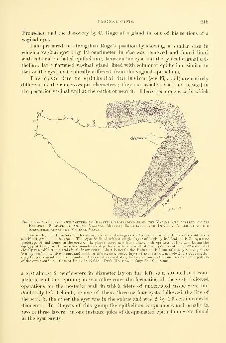
VAGIlSrAL CYSTS. 249
Preuselien and the discovery by C. Huge of a gland in one of his sections of a
vaginal cyst.
I am prepared to strengthen Ruge's position by showing a similar case in
which a vaginal cyst 1 by 1*5 centimeter in size was removed and found lined
with columnar ciliated epithelium ; between the cyst and the typical vaginal epi-
thelium lay a flattened vaginal gland lined with columnar epithelium similar to
that of the cyst, and radically different from the vaginal epithelium.
The cysts due to epithelial inclusion (see Fig. ITl) are entirely
diffei-ent in their microscopic characters ; they are usually small and located in
the posterior vaginal wall at the outlet or near it. I have seen one case in which
Fig. 170.
—
Cyst 8 by 5 Centimeters in Diamethj ii( ii i in i i m nii V\( i\v ami r< \ kred on its
External Surface by Smooth Vaginal Mi((.^\ 1 ( -ii\in \M) l)hNvn\ \i)iii rent to theStructures above the Vaginal Vault.
The walls, 8 niillimeters in thickness, are of a dark-grayish opaque color, and the cavity contains asemifluid, greeni.-li Milistancc. The cyst is lined with a single layer of 1i1l:1i cyliiKlrical cpitlicruuii, a true
prototype of that fniiiid in the cervix. In places there are islets lined with c|iitlii lium like that lining thesurface of the cy^t: thisc islets sometimes dip down into the wall of Ihr .'n st a cciitinRtcr or more, andclosely resemble triir -lands in their structure. Just beneath the lining c]'ithfliuiii of tlie cyst cavity there
is a layer of connective tissue, and next to this came a broad layer of non-stri]ie<_l muscle fibers cut longitu-
dinally, transverselx . and oMiquely. A layer of normal stratitie"d squamous eiiithelium lies over one portion
of the'outcr surface'. Case of Dr. C. P. Noble. Path. No. 1775. Magnified four times.
a cyst almost 2 centimeters in diameter lay on the left side, situated in a com-
plete tear of the septum ; in two other cases the formation of the cysts followed
operations on the posterior wall in which islets of undenuded tissue were un-
doubtedly left behind ; in one of them three or four cysts followed the line of
the scar, in the other the cyst was in the sulcus and was 2 by 1'5 centimeters in
diameter. In all cysts of this group the epithelium is squamous, and usually in
two or three layers ; in one instance piles of desquamated epithelium wei-e found
in the cyst cavity.

250 OPERATIONS OX THK VAGIXA.
s^f
Cysts developing from Gartner's ducts are found in rare instances at the
vaginal vault extending up between the folds of the broad ligament.
Cysts of the vaginal vault must be dis-
tinguished from the atresia of a rudimen-
tary horn of the uterus, which forms a
prominent fluctuating tumor at the vault
or extending from the vault down the lat-
eral wall. In these cases there is a history
of pain associated with the retention, and
a bimanual examination through rectum
and abdomen will show that the tumor ex-
'^'^~..';:'y^i'^-:X\ i-'^'^/'^' :y-/'.^ tends well up into the pelvis. A subure-
r. ... ,•^A-,^,
.^-. -
-/-•.«'•-. -.."- .
tliral abscess is peculiar in its position be-
neath the urethra, the thick vaginal wall
covering it, its extreme tenderness, and
in that it discharges its contents through
the urethra on pressure.
A cystic dilatation of a blind ureter
beneath the urethra may easily be con-
fused with a simj)le vaginal cyst. Such
was the case of E. G. Orthmann {Ce?i-
traJh. f. Gyn., 1893, ^o. T). The cyst
occupied the lower two thirds of the va-
gina and grew year by year. The diag-
nosis of a vaginal cyst was made, but in
dissecting it out it was found that the con-
tents had disappeared, and on reaching its
long pedicle above, a correct diagnosis of a
forked ureter with a blind end dilated into
a cyst was made.
The small cysts occasion no symptoms
whatever. The chief clinical symptoms
of the larger ones are obstructions to mari-
tal intercourse and to labor ; often the pa-
tient's first intimation that there is any-
thing wrong is when a part of the cyst
protrudes at the vulva. The suppurating
cyst alone is painful.
The treatment is simple and free
from risk. Small cysts may be excised
without opening them and the wound
closed with catgut sutures ; lai'ger cvsts
1^;
Fig. 171.
—
Section from the Wall of a Cy.st,
2 BY 2-5 Centimeters in Diameter, takenFROM THE Posterior W.\ll of the Vagina.
The walls are thin and transparent, and the
cyst cavity is nearly tilled with large cells,
a, normal stratified squamous epithelium of thevairinal mucosa; J, connective tissue showing anumber of small blood vessels ; c, a layer of mu.s-
ele fibers cut loiirritudinally and transversely ; (/,
two, ami m plaees three, layers of rather flat epi-
thelial cells lining the cyst cavity. Other por-
tions of the cyst are lined with several layers
of typical squamous epithelium, e, large cells
lying free in the cyst cavity, probably descjua-
niated epithelium. Path. No. 1411. Magnitied50 times.
brane dissected out, and the wound then closed by suture. In suj^purating cases
after proper cleansing, a large segment of the whole thickness of the cyst wall is
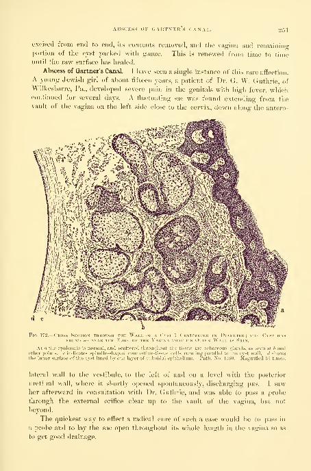
AISSCESS 0I-' GARTXERS CAXAE. 551
excised from end to end, its contents removed, and the vagina and remainino-
portion of the cyst packed with gauze. This is renewed from time to timeimtil the raw surface has healed.
Abscess of Gartner's Canal.—I have seen a single instance of this rare affection.
A joung Jewish girl of about fifteen years, a patient of Dr. G. W. Guthrie, ofWilkesbarre, Pa., developed severe pain in the genitals with high fever, whichcontinued for several days. A fluctuating sac was found extending from thevault of the vagina on the left side close to the cervix, down along the antero-
Fig. 172.
—
Cross Section through the Wall of a Cvst 1 Centimeter in Diameter; the Cyst wasFOIJNb so NEAR THE EdGE OF THE VaGIXA THAT ITS OuTEK VVaLL IS SkIN.
At a tlie epidermis is normal, and scattered throughout the tissue are sebaceous glands, as seen at b andother points, c indicates spindle-shaped connective-tissue cells running parallel to the cyst wall, d showsthe inner surface of the cyst lined hyone layer of cuboidal epithelium. Path. No. 1390. Magnified 36 times.
lateral wall to the vestibule, to the left of and on a level with the posterior
urethral wall, where it shortly opened spontaneously, discharging pus. I saw
her afterward in consultation with Dr. Guthrie, and was able to pass a probe
through the external orifice clear up to the vault of the vagina, but not
beyond.
The quickest way to effect a radical cure of such a case would be to pass in
a probe and to lay the sac open throughout its whole length in the vagina so as
to get good drainage.

252 OPERATIONS ON THE VAGINA.
NEOPLASMS.
Benign neoplasms originating in the vagina are extremely rare, and the only
forms found are the myomata.
The etiology of these tumors, as in those occurring in tlie uterus, is still un-
known, though Yeit, following Recklinghausen {Hand. d. Gyndhol.^ Bd. i, p.
348), speaks of the possibility of their originating in Gartner's ducts.
Myomata appear in the vagina either as polypoid growths with long pedi-

SARCOMA OF THE VAGIXA. 253
the vagina and pelvis. The patient may complain of a sense of weight in the
pelvis, tenesmus of the bladder, and even partial retention may occur, as direct
effects of the pressure. There may also be constipation and rectal tenesmus.
Pain in coitus has been noted, and the tumor may at times attain a sufficient
size to interfere with delivery.
With necrosis and gangrene of the tumor we find the added symptoms of
profuse ill-smelling vaginal discharge, with an accompanying irritation of the
surrounding parts.
The treatment is removal of the growth. Pediculated tumors are easily am-
putated, the vessels controlled, and the base sutured, bringing the tissues evenly
together. In the case of a larger tumor with a wide base it may be necessary
to enlarge the vaginal opening by making lateral incisions before the growth
can be satisfactorily reached and removed. The way to remove sessile tumors
is to make a linear incision through the overlying vaginal wall and then to enu-
cleate the growth. The cavity remaining after such an enucleation may be
closed completely l)y suturing the vagina after checking all hemorrhage. Theopposed sides are kept together by packing the vagina with gauze, which is al-
lowed to remain undisturbed for from four to seven days.
Malignant neoplasms are represented by sarcomata and by carcinomata.
Sarcoma appearing in the vagina, according to Steinthal( Virch. Ardi.^ Bd.
cxl, p. 4:49) and Kolisko ( Wien. Min. Wochenschr.^ 1889), may be sharply di-
vided into two classes. In young children they are usually polypoid in form
and situated on the anterior vaginal wall, while in adults the sarcoma is usually
a diffuse growth found in any portion of the vaginal canal.
When secondary to sarcoma of the uterus it appears first in the vaginal
vault as a dark bluish knobbed or polypoid outgrowth.
In children the first symptom noticed is a rounded or irregular berrylike
tumor appearing in the vulvar cleft and accompanied by pain. Pain on mictu-
rition and constipation may be present. In adult patients a vaginal discharge
usually makes its appeai'ance early in the course of the disease, and there are at
times slight hemorrhages from the surface of the growth.
There is a great tendency in all of these tumors to undergo necrosis, and this,
together with the foul discharges, opens up an avenue for the entrance of an in-
fection, which in the end often causes death. Cystitis and pyelonephrosis also
often accompany the growth, as well as pyometra and purulent peritonitis.
The diagnosis in all of these cases must be made by a microscopical exami-
nation of the tumor, but in case of sarcoma it must be remembered that delay
makes the prognosis more serious.
The treatment is by early and radical removal of the growth.
Four cases have been reported—two in children and two in adults—in which
permanent recovery has followed removal.
The various methods of operating are similar to those described under the
treatment of carcinoma.
Carciaoma of the Vagina.—Primary cancer of the vagina is rarely seen.
Hecht {Inaug. Dls. Munclien,^ 1891) found that a little over one per cent of

254 OPKRATIOXS ox THE YAGIXA.
cases of cancer in women were vaginal. In the majority of these cases tlie pos-
terior wall is affected iirst. Out of eighteen cases cited by Olshausen of Berlin,
thirteen involved the posterior wall. The disease usually ap])ear8 in the form
of a fungating mass of tissue, easily breaking down and bleeding. Another
form appears as a granular ulcerated area with hard infiltrated margins. In a
still rarer form of carcinomatous infiltration the vaginal walls simply become
rigid and contracted.
While primary cancer is rare, a secondary involvement from extensicju to the
vaginal vault from a cancerous cervix is quite common—so common, indeed, that
it is often necessary to remove a considerable part of the u])per vagina with the
uterus in hysterectomy for uterine cancer.
Nothing is known as to the cause of this affection. Heredity and trauma
during chil.dl)irtli have not l)een shown to be active, as in the case of cancer of
the cervix. The tendency of the disease located in the upper part of the va-
gina is to extend over onto the posterior cervical lip, rendering it impossible at
times to determine whether the cervix or the vagina was the original starting
point. Where there is a large cancerous area at the vault of the vagina with an
involvement of the outer surface of the cervix, which is continuous with it, the
disease may without hesitation he stated to be vaginal in its origin instead of
cervical.
Hemorrhages, vaginal discharge, dull aching pain, and difliculty in defecation
and micturition are common symptoms. As the disease extends rapidly into
the neighboring lymph channels cachexia becomes more and more marked, until
the patient dies of exhaustion.
The treatment is extirpation in all cases in which there is no involve-
ment of the connective tissue laterally. Such an implication nnist be discov-
ered by estimating the mobility of the diseased area by pressing directly upon
it, and by palpating around its margins through the rectum.
There are in general four ways of operating upon a cancerous vagina :
First, a simple excision of the cancerous area through the vaginal outlet.
Second, a circular incision of the vagina below the diseased area, followed bya stripping off of the whole circumference of that portion which is to be extii--
pated ; after this an abdominal incision, freeing the uterus and removing it with
the upper part of the vagina.
Third, a transverse incision through the perineum and extending ou upthrough the recto-vaginal septum to the diseased area, which is then removed
through the incision.
Fourth, a posterior incision from sacrum to fourchette beside the rectum,
splitting the vagina up to the diseased area.
First, if the disease is discovered when it is still (piite superficial and
limited in its area, it may be extirpated with knife, or scissors and forceps,
operating through the vaginal orifice, cutting around it on all sides at a distance
of 1-5 or 2 centimeters and loosening it up from its base with the fingers and
removing it, and finally bringing the margins of the wound together by catgut
sutures.
CARCINOMA OF THE VAGINA. 255
The second method consists in a circular incision of the vagina well below
the disease ; afterward it is stripped up to the vault with the fingers and detached
on all sides. The abdomen is then opened from above and the uterus and de-
tached portion of the vagina removed. It is especially important here to deter-
mine that the vaginal infiltration does not extend into the surrounding tissues
;
careful examination must also be made by the rectum to discover any infiltration
of the broad ligaments ; should this be found a radical operation is contra-in-
dicated. The operation may sometimes be concluded after stripping the vagina
loose posteriorly and at the sides, and in front as far as the vault, by catching
and drawing dowm the cervix and ajjplying ligatures to the broad ligaments,
and removing the uterus, as in vaginal hysterectomy. Sometimes the whole up-
per third of the vagina may be removed in this way.
The third method, proposed and used by Prof. Olshausen {CentraM.f. Gijn.^
1895, No. 1), consists in the removal of the cancerous vagina through a trans-
verse incision in the perineum, and a separation of rectum and vagina up to
Douglas's cul-de-sac. If the uterus is to be removed, the peritoneum is opened
and the uterus inverted and freed by tying off the broad ligaments from above
downward toward the cervix. When this separation is partly efi^ected, the
loosened vagina is cut through with scissors and the carcinoma detached, and
finally, after releasing the bladder, the cervix uteri is tied off. If the uterus
is not to be removed, the separation of vagina and rectum is carried up to the
cervix uteri, and the vagina in the neighborhood of the disease is freed on all
sides from the subjacent tissue. An incision is then made into the vagina at a
suitable point and the diseased portion excised with scissors. In a contracted
vagina the last part of the excision is facilitated by splitting its posterior wall
from the frenulum up, giving a broad view of the field.
I prefer the fourth plan to this, as less awkward and as enabling me to reach
the parts more directly, the extirpation by an incision beside the rectum, adopted
in the following case.
The cancerous patch was situated at the vault of the vagina posteriorh% and
was 3 by 4 centimeters (1*2 by 1'4 inch) in diameter, and involved as well the
outer surface of the cervix. It could not be drawn down, so as to attack it
from below, so the patient was placed upon her left side and an incision madefrom the end of the sacrum, beside the coccyx, and continued in a slightly
curved line down beside the rectum and around the right margin of the anus,
through the perineum to the fourchette. By carrying the incision deep enough
the rectum was exposed and easily drawn toward the left with retractors, in
this way exposing the vagina.
The posterior vaginal wall was then split from the fourchette to the cervix
and the diseased portion freed at the sides with the fingers and removed. It
included the posterior two thirds of the upper portion of the vagina and the
posterior lip of the cervix above the vault. I now brought the uterus down in
retro position, and united its posterior surface to the vaginal wall, where it was
cut off, filling in the gap left, leaving a shortened but entire vaginal canal. The
posterior wound was closed with interrupted sutures. The patient recovered

250 OPERATIONS ON THE VAGINA.
and the uterus united in its new position, but the disease, wliieh had extended
beyond the limits of the field of operation, continued to advance, and she died at
her home some months later.
If the uterus is to be removed as well, the peritoneum may be opened at
Douglas's cul-de-sac and the uterus drawn through the incision and its Ijroad
Fio. 174.—A Cam of Adkno-c akcinoma of the Tosihitiou Vac.iwl Waii \^\ Imi'lantation fisom anUNSUSPECTED AdENO-CARCINOMA OF THE BoDY OF THE UtEKU.S.
ligaments tied off ; then, using it as a tractor by pulling it well out of the
wound, the vagina is made tense and easily outlined while the finger is engaged
in freeing it from the tissues at the sides and from the bladder in front. Asmuch of the vagina as is necessary may be removed with the uterus. Care
must be taken not to wound the ureters by placing bougies in them before be-
ginning the operation. If the uterus is not to be removed the ])eritoneum must
be pushed up without opening it, and the vagina freed on all sides in the neigh-

TRAUMATIC AFFECTIONS. 257
borliood of tlie diseased portion, and then opened and the diseased area ex-
cised.
Traumatic Affections.—Traumatic affections are frequently found in the vagina,
the result of injuries incurred in labor. They are usually located either in the
vault or near the outlet, and involve one or both sides, extending down or upin the axis of the vagina. Any marked narrowing of the vaginal canal due to
cicatricial contraction between the vault or the outlet is unusual.
Extensive sloughing in the middle of the vagina may produce a concentric
contraction, narrowing the caliber even down to complete closure (atresiaV a g i n ae), and followed by retention of the menses (hematocolpos andh e ni a t o m e t r a). The cicatricial bands radiating out from the sides of a vesi-
co-vaginal fistula and narrowing the lumen of the vagina serve to illustrate an-
other mode of the production of a vaginal stenosis.
The vaginal cicatrices may be classified under two general heads, correspond-
ing also to the difference in etiology, viz., cicatrices whose direction is in the
axis of the vagina, and cicatrices whose direction
is transverse to the axis of the vagina.
Cicatrices of the first class in the axis are
usually found at either extremity of the canal,
and are associated, as stated, with a lacerated
cervix or a ruptured outlet.
Cicatrices of the second class crossing the axis
are the result of pressure and sloughs during ])ar-
turition or of syphilitic sores.
At the vagina] outlet the principal scars ex-
tend from the posterior median line up the right
or left sulcus on one or both sides of the pos-
terior column. The scar tissue at this point must
be regarded as a conservative effort of l^ature in
her endeavor to fill out and draw together the
rents produced in childbirth. The symptoms
produced by such scars vary from the slightest
all the way to a severe neurosis. Rarely a scar is
so tender as to require excision. A scar at the
vault may be the cause of a lateral displacement
of the uterus. Cicatricial contraction of the canal
will interfere with all its functions, even render-
ing the escape of the menses impossible or endangering life in case of pregnancy.
Too much stress, however, nnist not be laid on this last point, as labor has pro-
gressed normally in such cases in which the outlook seemed almost hopeless at
the start.
The treatment differs according to the form and extent of the disease.
The best method, in general, is a complete excision of the scar extending well
into the subjacent tissue, supplying the defect created by sliding over it the sound
tissue from above and below.
20
175.
—
Secondary Vaoinal Car-cinoma.
Sagittal section of the cervix (C)and vagina, sliowing the isolated carci-
nomatous nodules on the posterior wall.
Case same as Fig. 174. J. S., Nov.14, 1S05.

258 OPERATIOXS OX THE VAGINA.
Small falciform cicatrices at the vault of the vagina on the right or left side
may be treated by drawing the cervix in the opposite direction with a tenacu-
lum, making the scar tense, and cutting across it in several places down to its
base. This may be done under cocain, and if the cutting is repeated several
times, the cervix will finally be freed. If the cervix is torn deeply, as is usually
the case, the cervical tear may be repaired and the scar excised at the same
time. Care must be taken to avoid the mistake of simply removing that por-
tion of the scar which projects into the vagina. The dissection must be carried
well below the vault, removing all of the scar tissue. The chief dangers are of
injury to a ureter which has been drawn out of its normal position, and perhaps
caught in the scar tissue, and of cutting a uterine artery, which is also brought
nearer the vault than normal.
To avoid injuring the ureter, a bougie should be placed in it before begin-
ning any extensive operation at the vault. The ureter can then be readily felt
_ „ - from time to time, and if involved will be
easily dissected out and restored to its proper
place without injury.
The artery will be avoided by a slow, care-
ful dissection, palpating the structures often
as it progresses. The wound thus created is
closed by interrupted sutures, and a dry dress-
ing applied. Cicatrices in the lower part of
the vagina are always removed in the course
of the operation for relaxed vaginal outlet.
Atresia of the vagina following
labor may involve any portion of the canal.
The closure is due to sloughing fi'om pressure
during labor, followed by a cicatricial contrac-
tion. The area cut off may be from one to
several centimeters in diameter. Oftentimes
the atresia is incomplete, when a superficial
examination would lead the observer to assert
that it was complete. One or two minute
orifices may be detected in the transverse scar
at the bottom of the vaginal cid-de-sae, and
pressure made above may cause a little dark
fiuid to exude.
The severest s y m p t o m s arise in atresia
from the apparent amenorrhea, which might l)e called an amenorrheaparodoxica, as the menstrual function continues normally, while the secre-
tions accumulate above the stricture. The pain at the menstrual period is often
agonizing. With the increasing accumulation the vagina expands, and the cer-
vix and uterus, and sometimes the uterine tubes, dilate, until the pelvis is
choked, and a large mass may be felt projecting into the abdomen.
Atresia may arise from extensive ulceration in the vagina, but the one coni-
FiG. 176.
—
Atresia of the Vagina, show-ing THE Scar at the Point at whichTHE Anterior and Posterior VaginalWalls come together. A. M., Jan.18, 1896.

TRAUMATIC AFFECTIONS. 259
mon cause is the traumatism of a delayed labor in a contracted pelvis. Fromthis cause one of my patients recovered, not only with an atresia, but with a
vesico-vaginal iistula, and a recto-vaginal fistula as well.
The diagnosis is made by the history of a severe labor, by the subse-
quent amenorrhea with severe menstrual colic, and by the impediment to sexual
intercourse. An examination reveals the na-
ture of the affection, as the finger enters a short
sac, or if the urethra is dilated, as is often the
case, the finger may enter the bladder without
difticulty, greatly puzzling the physician for a
time.
An examination jx?)' rectum shows the pres-
ence of a fluctuating sac al)ove the atresia, and
above this, it may be, one or two other sacs,
separated by one or two well-defined transverse
constrictions.
The treatment is to remove the scar
tissue which closes the vagina and establish a
permanent free communication between the
separated parts of the vagina by a plastic oper-
ation ; this is completed in three steps : first,
opening up the channel and allowing the accu-
nmlated fluids to escape ; second, removing the
scar tissue ; third, uniting the sound upper and
lower portions of the vagina over the defect.
Before, throughout, and after the operation
the most painstaking antiseptic precautions
nnist be taken, as the accumulated fluid is
peculiarly liable to undergo rapid decomposition, and the walls of vagina, uterus,
and tubes are in a state of extraordinary susceptibility to infective processes.
Death from infection has so often followed the sini])le evacuation of the fluid
that many surgeons dread the operation.
I can not illustrate the further steps of the operation better than l)y describ-
ing one of my cases {Johns ILqtl'. IIosp. Rep.^ vol. iii, p. 429).
The patient was a negress about twenty-two years old. She had had a still-
born child after a prolonged instrumental labor eight years before I saw her,
and had suffered ever since the birth. Her periods had always been regular
before, but she had never menstruated since, although suffering greatly with the
monthly molimina and from backache and pains in the lower abdomen. Six
months before I saw her she had been kept in bed eight weeks by an attack of
peritonitis.
On making a vaginal examination under a bed cover, the finger entered a
large, smooth -walled cavity in which none of the expected landmarks could be
recognized. This was found to be the bladder, with the urethra so widely
dilated that two fingers could be introduced without pain ; the external urethral
177.
—
Operation for Atresia ofTHE Vagina caused by an AdhesionOF THE Anterior and PosteriorWalls in the Form of a Septi m.irsT above the Hymen.
Tlie cruciform incision is made tirst to
permit the thorough washing out of thesac ; the dotted line indicates tlie area ex-cised in restoring the caliber of the va-gina.

OPEKATIOXS ON THE VAGIXA.
orifice Ining patulous and everted under the pubic arch. All that was left of
the vagina on the vulvar side was a little pit of firm scar tissue 1 centimeter in
depth just beliind the urethra.
Upon making a bimanual examination a chain of tumors was found filling the
pelvis, lying one above another ; at first thej felt like a group of myomata, but
Fig. 178.
—
Atresia of tiik Vagina seen in Sagittal Section; the Vagixa above the Atresia is dis-
tended WITH the ActaMi'LATED Mknstrial Fliid. There is no Distention of the Uterus.
a closer examination showed that they fluctuated and formed a continuous cav-
ity, with two shallow sulci between. The obliterated portion of the \'agina was
4 centimeters long.
The operation was performed in this way : The left index finger was intro-
duced into the bladder and the thund) into the rectum until they touched the
sac of fluid and held the atresic area between their palmar surfaces, (luided by
these fingers to avoid injuring the rectum or the bladder, a large trocar was
entered in the pit beneath the urethra and pushed up through the oblitei-ated
septum until it penetrated the sac. Upon withdrawing the trocar 5(m> cubic
centimeters (about IH ounces) of tarry blood escaped and the three sacs collapsed.
By means of a uterine dilator the trocar puncture track was now enlarged until
it reached from one pubic ramus to the other. Abundant room was thus secured
to catch the margin of the vagina just above the stricture and dissect it loose on
all sides for a distance of a centimeter. This loosened collar was then pulled
down over the dilated atresia and attached by a series of interru])ted sutures to
the margin of the vagina just below it. By this means, by sliding the normal
vaginal tissue dow^n over the cicatricial ai'ea, the canal was restored without leav-

RECTO-VAGIXAL FISTULA. 201
ing an exposed raw area to uiulero-o subsequent contraction. The calil)er of the
new vagina was now normal, and a month later, when the patient was discharged,
it even appeared normal in length.
If resisting scar tissue is felt after opening up the canal, it must be dissected
out. Where the urethra is not dilated, the linger in the rectum alone will serve
as a guide for the trocar.
Recto-vaginal Fistula.—Kecto-vaginal fistulte are abnormal channels of com-
munication between the rectum and the vagina, generally situated at one end or
the other of the vaginal canal.
The cause of the iistula in the upper vagina is commonly an extension of a
cancerous disease from the cervix on to the vagina and through the recto-
vaginal septum ; in the lower vagina the commonest cause is a failure in the
attempt to restore a complete rupture of the recto-vaginal septum. After this
operation, if the suturing is imperfect, fecal matter is apt to be forced into the
upper part of the wound upon the denuded surfaces, producing suppuration and
failure of union, and leaving a fistulous ori-
fice opening either on to the vulva or into
the vagina.
Other causes, such as syphilis and stric-
ture of the rectum, produce fistula in some
cases. I have seen but one case whei*e it
was due to labor. I have also seen one case
where it followed the excision of hemor-
rhoids, and another where a necrosis had
been produced by the jjressure of a badly
fitting pessary.
The s y m p t o m s arise from the escape
of fecal material into the vagina, or, if the
orifice is minute, from the escape of gases
by this avenue.
The cancerous fistulie are particularly
distressing from the disgusting condition in
wdiich the patient is apt to be kept by the
constant emission of feces from the vulva.
Fistulfe low down in the vagina are often
so small that they are detected with diffi-
culty, and yet the inability to control the gases, which escape audildy, keep the
patient in a constant state of nervous apprehension.
The diagnosis is made either by simple inspection, or by passing a probe
into any suspicious pits and thus tracing the connection with the bowel, or by
introducing the finger into the rectum and palpating its anterior surface from
the sphincter up. The rectal end of the fistula is marked by a distinct depres-
sion easily felt ; this can be pushed forward and the vaginal opening madevisible.
A further demonstration may be made by injecting milk into the rectum
Fig. 179.
—
Operation for Atkesia of theVagina seen in Sagittal Section, show-ing THE Continuous Suture applied in
THE Eight Half of the Vagina, bring-ing the Vaginal Mucosa above downto the Vaginal Mucosa below the Atre-sia, IN THIS WAY bridging IT OVER WITHMucous Membrane.

26-2 opp:rations ox the vagixa.
and watching for its avenue of escape by the vagina. The patient herself often
calls attention to her inability to retain rectal eneniata, which escape by the
vagina.
The proper treatment of a recto-vaginal fistula depends upon various
associated conditions.
/^
The index finjjor
reetuiii clearly detini
is visible between tl
Fi(i. 180.— Atkesia of thk Vac.ina in a Negress.
is intrmlueed tlirouirl) the dilated urethra into the bladder, and witli the thumb in thes the position an<l tliiekness of the reeto-vesieal septum. Only a shallow vai,'inal pocket
Fistula from the extension of cervical cancer is not, as a rule, ameiuible to
treatment. The utmost that can be done is to keep the parts as clean as jiossible
by securing a daily free evacuation of the bowel, avoiding the constant leakage,
and by the frequent use of vaginal douches.
In event of a slow progressing cancer at the vault, it would be quite right to
try to give some relief by making the fistulous opening large enough to establish
a free communication with the bowel, and then to denude a circular 8tri]> on the
vaginal wall below this and to close the vagina with interrupted silkworm-gut
sutures (partial colpocleisis).

TREATMEJfT OF KECTO-VAGIXAL FISTULA. 263
There are three ways of closing a recto-vaginal fistula l)y suture :
First, by a funnel-shaped denudation of the edges on the vaginal side fol-
lowed by suture, after the pattern of the vesico-vaginal fistula operation.
Second, by splitting the perineum and recto-vaginal septum and completely
separating the rectal from the vaginal portion of the fistula, followed by a sepa-
rate suture of the rectum.
Third, by splitting the recto-vaginal septum vertically as far as the fistula,
which is then denuded and the recto-vaginal septum closed as in a case of com-
plete tear.
Before operation the intestinal tract must be thoroughly emptied, the parts
cleansed, the sphincter
ani stretched so as to
paralyze it, and a loose
iodoform-gauze pack
put well up in the rec-
tum to keep its dis-
charges out of the va-
gina and off from the
wound during the su-
turing.
In determining
wliat form of opera-
tion will be best in a
given ease, the posi-
tion of the fistula, the
condition of the sur-
rounding parts, and
the ])resence or ab-
sence of scar tissue
must be considered.
Any complication on
the rectal side, such as
a deep pit or an ulcer or granulation tissue, must also be taken into consider-
ation, as these conditions almost necessarily defeat union.
First. The simplest form of ojjeration, a funnel-shaped denudation and
suture, will be selected when the opening lies above the sphincter and levator
ani area, and when it is free from bands of scar tissue, and the rectal surface is
healthy and does not present a deep pit. After suitable exposure the parts are
benumbed with cocain applied for ten minutes. The area to be excised is then
outlined with the point of a sharp knife about 1 centimeter away from the edge
of the opening on all sides. This is now denuded by catching the edge with
forceps or a tenaculum, cutting away strip after strip with a pair of delicate
scissors until the whole is bared and bleeding down to the rectal mucosa, which
now lies at the bottom of a wide-mouthed funnel opening on the vaginal sur-
face. The wound is now closed by a series of interrupted silkworm-gut sutures,
Fio. 181.
—
Coronal Sectiox of an Old Atresia of the Vagina withDistention of the Vagina, Cervix, and Uterine Cavity withMenstrual Fluid. The Atresia occupies the Lower Third ofTHE Vagina.

26i OPERATIONS ON THE VAGINA.
three to tlic centimeter, passed in tlie direction of least resistance, generally fromside to side, and tied iirmly. Superficial catgut sutures are used between the
silkworm gut if the line of aj^proximation is not perfectly accurate. The gauze
pack is now taken ont of the rectum and the vagina washed out and a 2)iece of
iodoform gauze inserted loosely. The after-treatment consists in a restricted
Fig. 182.
—
Old Atresia of thk Vagina opened and evacuated : Interrupted Sutures in Place toDRAW THE Vaginal Mucosa down to the Mucosa at the Vaginal Outlet, bridging over theDenuded Area in the Connective Tissue.
diet and regular daily movements of the l)Owels, preferably secured by medicine
taken by the mouth. In eight days the stitches are removed, and the patient
may go about.
In one of my cases there was a small opening 3 millimeters in diameter just
beyond the internal sphincters, and the surrounding tissue was soft and natural
and free from scars. I applied cocain, and denuded and closed it on the vaginal
surface, as described, and allowed the patient to rise at once from the ta])le and
go home to continue her usual occupations without interruption. In eight days
she returned, and I removed the silk sutures and found that perfect union had
taken place. Such treatment as this will only succeed in the most favorable
cases, and ought rarely to be tried. The proper after-treatment of a sim])le tis-
tula is to keep the patient quiet in bed for a week.
Second. When the fistula is up above the sphincter area and is sur-
rounded by scar tissue, the best plan is to dissect the rectum free from the
vagina, either by splitting the perineum from side to side and working up to the
fistula between rectum and vagina, or by raising a fla]) of vaginal tissue below
the opening and dissecting it up to the tistula, which is then freed from its vag-

TREATMENT OF RECTO-VAGIXAL FISTULA. 265
inal attaeliments on all sides. Interrupted sntnres are now passed throui^li themuscular coats of the denuded l)Owel, avoiding the mucosa and closing the
rectal opening. Thev are left long and brought out through the fistulous vag-
inal opening, which need not be closed. The incision in the recto-vaginal sep-
tum, through which the separation was made and the sutures passed, is finally
closed, and a dry dressing placed in the vagina.
The success of this procedure depends upon the invariable soft, yielding
condition of the bowel, which is well adapted for plastic union when detached
from the rigid cicatricial vaginal tissne, which prevents the sides of the woundfrom coming together without undue traction.
I succeeded in one case (K. S., Xo. 2916, Sept. 5, 1894) in doing this wherethe rectal fistula was 2 centimeters long at the top of an obliterated upper va-
gina, with a vesico-vaginal fistula directly opposite. There was a large amountof scar tissue on all sides, rendering union by suture after denudation a hopeless
undertaking. I therefore made a transverse perineal incision 4 centimeters long
and dissected between the vagina and rectum up to the fistula, which was then
split, making two fistulous orifices out of one, the posterior opening leading into
the rectum and the anterior into the vagina. The rectal opening M^as then
closed separately by interrupted fine silk sutures brought out through the vag-
inal opening. The perineal wound was closed, and the result was immediate
union throughout.
Third. When the fistula is low down, close to the vaginal outlet in the
sphincter area, the better plan is to cut entirely through the septum, repro-
ducing the complete rupture, and then to denude the margins of the fistula andfor some distance aljove it, and close the whole as in a case of complete tear.
This is better than an attempt to effect closure by a denudation on the vaginal
side alone, for three reasons : In the first jilace, the position of the opening
on the rectal side is unfavorably situated, as in any bowel movement unusual
pressure is brought to bear on the anterior wall of the rectum at this point bythe fecal masses, and is so great that some particles of fecal matter are inevi-
tably forced into the wound, preventing union. In the second place, there is
always a pit on the rectal side in these sphincter fistulas which catches fecal mat-
ter. In the third place, the bridge of tissue below the fistula which the opera-
tor endeavors to save by simple denudation and approximation is often insig-
nificant ; this is especially apparent after it has been cut through.
I would repeat the caution not to forget to carry the denudation on the
vaginal surface well above the fistula. The further denudation and suture must
be made as fully described in the treatment of comjDlete rupture of the recto-
vaginal septum in Chapter X.
CHAPTER xri.
AFFECTIONS OF THE URETHRA AND BLADDER.
1. Brief historical sketch. Celsus : calculus. Fatio : vesico-vaginal tistula. Simon : dilatation of
the urethra. Griiufeld: introduction of light into bladder. Rutenberg's air-pumpingspeculum. Nitze's cystoscope for examining the male bladder modified for the female.
Pawlik's method of catheterizing the ureter free-handed. Sanger : palpation of the ureters.
Kelly : examination under atmospheric distention induced by posture. Bibliography.
2. Topography of the bladder in women. Natural landmarks within the bladder. The relations
of the bladder to surrounding structures. Artificial division into hemispheres and quad-rants.
3. Examination of the urethra and bladder: Urinalysis and examination of discharges; percus-
sion; palpation; inspection without instruments; urethroscopy; cystoscopy: Its funda-mental principles ; instruments used—the light, the reflector, vesical specula with obtu-
rators, dilator, evacuator, ureteral searcher. The cystoscopic examination : asepsis; prepa-ration of patient ; anesthesia ; postures, dorsal and knee-breast ; calibrating and dilating the
urethral orifice ; the lubricant ; introducing the speculum ; inspection.
4. Diseases of the urethra. 1. Malformations: hypospadias; epispadias; atresia of the urethra;totally deficient urethra. 2. Displacements: prolapse of the mucosa. 3. Dilatation. 4.
Stricture. 5. Ischuria. 6. Fistula. 7. Foreign bodies. 8. Urethritis: acute; chronic;
(1) diffuse chronic urethritis; (2) circumscribed chronic urethritis. 9. Suburethral abscess.
10. New growths : caruncle ; fibroma ; cancer ; sarcoma.
5. Diseases of the bladder. 1. Introductory. 2. Diagnosis : by history and symptoms ; by urin-
alysis ; by palpation ; by inspection under illumination. 3. Methods of treatment in gen-eral. 4. Classification of diseases : Congenital defects : (1) double bladder ; (2) loeulate blad-
der; (3) exstrophy. Displacements: (1) lateral; (2) upward; (3) downward; (4) eversion.
Foreign bodies in the bladder: (1) calculi, pessaries, catheters, etc.; (2) removal—throughurethra; through vaginal incision; through suprapuluc incision. Vesical fistuhe : (1) his-
torical sketch: Luiz de Mercado, Felix Plater, Ilendrick Koonhuyzen. J. Fatio, A. J. Jobert,
G. Simon, J. Marion Sims, T. A. Emmet, Nathan Bozeman, A. '^Martin, L. von Dittel, A.Mackenrodt, W. A. Freund, E. C. Dudley, II. A. Kelly ; (2) causes
; (3) symptoms ; (4) diag-nosis. 5. Treatment, (a) general, (b) of fistula of large size: A. Martin—closure by turningup vaginal flaps to form the new base of the bladder; F. Trendelenberg—closure by trans-
planting a flap from the posterior vaginal wall ; E. C. Dudley—closure by suturing the de-
nuded vesical mucosa to its anterior margin ; A. Mackenrodt—closure by detaching thebladder from the vagina and suturing it independently; H. A. Kelly—closure by detach-ing the bladder posteriorly and suturing it to the denuded vaginal wall anteriorly; W. A.Freund—closure by suturing the body of the uterus into the defect. Of vesico-utero-vaginalfistula : H. C. Coe, Otto v. Herff, H. A. Kelly, F. Trendelenberg. Of vesico-uterine fistula : F.
H. Champneys, H. A. Kelly. Of other vesical fistuhe : G. C. Blackman. Hemorrhoids. Hy-peremia. Cystitis : (1) bacteriology
; (2) acute ; (3) chronic ; (4) treatment of chronic cystitis
—medication, irrigation or instillation, direct topical treatment, surgical treatment ; (5)
tubercular cystitis; (6) exfoliative cystitis.
6. Tumors of the bladder: 1. Classification. 2. Benign tumors: papilloma; fibroma; adeno-ma; myoma; cystic follicles; dermoid cysts. 3. Malignant tumors: epithelioma; myxoma;sarcoma. 4. Clinical history of vesical tumors. 5. Diagnosis. 6. Operative treatment
:
by a dilated urethra; by vaginal incision; by suprapubic incision; by symphyseotomy;cystectomy ; K. Pawlik's case.
Previous to the latter half of the century just closing but little was knownabout diseases of the urinary apparatus in women.
And while the relatively more urgent and dangerous diseases of the male
organs had exacted the closest attention, the modesty of women, as well as the
inaccessible nature of the affections, all conspired to hinder an earlier scientiiic
investigation of the female organs.
266
BRIEF HISTORICAL SKETCH. 267
The shortness of the female urethra was known and special suitable metal
catheters devised and used before the Christian era, and Celsus in the first half
of the first century carefully describes an operation for stone in the bladder in
women—cutting for the stone from the outside through the vestibule into the
neck of the bladder, cautioning the operator to insert the finger, as a control,
into the vagina in a married woman, but into the rectum in a virgin. Calculus
in the female therefore attracted attention at an early date, on account of the
surprising observation that enormous stones could safely pass through the short
and more easily relaxed female urethra {qmti et ht'evior quam in marihus et
laxior est.—Celsus).
Johannes Fatio, of Basel, late in the seventeenth century recognized, treated,
and cured cases of vesico-vaginal fistula by denuding the margins with scissors
and drawing the edges together with a sharpened quill wrapped with thread.
A new interest was aroused in diseases of the bladder by the labors of G.
Simon, of Rostock, who carefully determined the extreme degree of safe dilata-
bility of the urethra for digital palpation of the bladder, using a series of conical
dilating specula with obturators, with diameters increasing up to 20 millimeters.
Simon was also able in some cases to feel a ureteral orifice, and, under the
guidance of touch, to slip in a ureteral catheter ; he did this seventeen times in
eleven cases, but never made any practical use of it. Indifferent as was Simon's
success, this was the starting point of all recent important work in connection
with the diagnosis of affections of the female urinary organs.
Josef Griinfeld, of Vienna, in 1874, filled the bladder with water and then
examined it through a short, straight speculum, with a piece of glass set obliquely
in its tube, so as to prevent the escape of the fiuid and at the same time to per-
mit the direct passage of light without I'eflection.
Rutenberg devised a speculum, aljout 20 millimeters in diameter, with a glass
partition and a tube attached for injecting air into the bladder, while a mirror
placed inside the bladder reflected various parts of its walls. It was necessary
to anesthetize the patient to relieve the pain produced by this examination, and
Rutenberg never succeeded in finding the ureteral orifices.
Max Kitze, of Dresden, with real genius, constructed a cystoscopic apparatus
for the male bladder, consisting of a long tubeiike catheter with a short beak
carrying a small electric lamp at the tip and a prism at the eye through which
the light, reflected from the walls of the bladder distended with water, is di-
rected into the tul)e, which further contains a telescopic arrangement of lenses
so as to give a wide fleld of vision to the observer at the outer end. A larger,
shorter and straighter tube has been made after the same plan for use in women.
Through this elaborate and delicate but most useful instrument Nitze and his
followers, who are now to be found among the genito-urinary specialists in all
the larger cities, are able not only to examine the bladder but also the ureteral
orifices, and even to catheterize the ureters with a flexible catheter, introduced
in a small tube beside the speculum. Nitze himself is also able even to operate
successfully upon small tumors within the bladder.
K. Pawlik, of Prague, made one of the most important additions to this
268 AFFECTIONS OF THE URETHRA AND BLADDER.
subject wlieii he improved Siinoirs ureteral catheter for women, and demon-
strated the feasibihty of introducing it free-hand into the ureter through the
urethra and l.)ladder. This is done by retracting the posterior vaginal wall while
the bladder is moderately distended with water (150 to 200 cubic centimeters),
when the two " ureteral " folds come into view on the anterior vaginal wall,
sweeping backward from the neck of the bladder toward the cervix and mark-
ing the site of the ureters just above them. These folds determine the direction
of the tip of the catheter in the bladder as it is made to ghde along its base while
seeking the ureteral orifices. The finger at once recognizes the fact that the
catheter has become engaged in the ureter by its assuming a certain fixed direc-
tion.
M. Sanger, of Leipzig, added another fact of the highest importance when
he pointed out the ease with which the low^er ends of the normal ureters could
be felt through the vaginal walls, and the increased distinctness of enlarged dis-
eased ureters.
My own method was first published in the Johns Ilophlns Hospital Bulletin
for November, 1893, and in a longer and more fully illustrated article in the
American Journal of Obstetrics for January, 1894. Its essential features are
:
1. An atmospheric dilatation of the bladder induced by posture.
2. The introduction of a simple straight speculum as a rule of small size and
without fenestra.
3. The examination of the mucous surface of the bladder and urethra by
means of a reflected light or an electric headlight.
I have further demonstrated the ease with which flexible catheters can be in-
troduced into the ureters in this position, and by their use I have been able to
reach the pelvis of the kidney, and to diagnose accurately such renal diseases as
hydronephrosis, pyelitis, calculus, etc.
The whole field of investigation of bladder, ureteral, and renal aft'ections is
in this way thrown open to easy investigation.
Since the publication of my method K. Pawlik has described a modification
of it consisting in a dilatation of the urethra under anesthesia sufficient to admit
a large open speculum, the atmospheric distention of the bladder, and its exam-
ination by means of a little electric light introduced into the bladder through the
speculum {Central, f. Gyn.., May 5, 1894).
But few treatises devoted to urinary diseases in women have as yet appeared.
Scanzoni's, in 1854, was the first. A. J. C. Skene's book on Diseases of the
Bladder and Urethra in Women was published in NeAV York in 1882 ; F.
Winckel's elaborate and valuable contribution appeared in Billroth and Luecke's
Ilandlmch, Stuttgart, second edition, 1886 ; II. A. Kelly's Diseases of the Fe-
male Bladder and Urethra^ Twentieth Century Practice., William "Wood & Co.,
was issued in January, 1895 ; and finally we have I)ie Kranl'heiten der weihlichen
Blase^ by II. Fritsch in Yeit's Ifandhxch der Gijnal'oliKjie^ 1897.

TOPOGRAPHY OF THE BLADDER. 2C9
TOPOGRAPHY OF THE BLADDER.
The fact that we are now in possession of a simple means of observing all
sorts of bladder aifections characterized by changes of form or color, bringing
under observation and within the reach of local treatment even minute lesions,
emphasizes the need for other and more accurate ways of describing the location,
form, and extent of such diseases in their relationship to the bladder wall, as
well as of registering such changes as may be found from examination to exami-
nation. It is also important, if we would convey any accurate impression of
our observations to others, to use a more precise phraseology than such vague
generic anatomical terms as " vertex " and " base," and to substitute in their
place an accurate schema of the interior of the bladder with suitable divisions
and subdivisions.
I will therefore consider the topography of the bladder from three stand-
points, each of which will be useful to the specialist in describing what he sees
through the speculum
:
1. The natural landmarks within the bladder.
2. The relations of the bladder to surrounding structures.
3. An artiiicial division into hemispheres and quadrants.
1. The Natural Landmarks in the Bladder Itselt—T he internal o r i fi c e
of the urethra, which begins as a narrow margin to shut in over the end
of the speculum, and continues to increase in breadth as the speculum is slowly
withdrawn from the l)ladder, forms one of the most important points of depar-
ture in the descrij)tion of lesions which may extend from the bladder into the
urethra, or vice versa, or in the location of lesions limited to the area adjacent to
the urethra ; we have in this way a periurethral area circumscribed by a
circle of say about 4 centimeters in diameter around the internal urethral orifice.
"Opposite the urethral orifice" is also sometimes a convenient
expression to designate the location of an affection involving the small area of
the posterior vesical wall, the part first seen on withdrawing the obturator.
The ureteral orifices are the most important of all the natural land-
marks in the bladder, and any account of a lesion in their innnediate neighbor-
hood is readily described as located either between them (interureteric) or pos-
terior, anterior, or lateral, to one or the other.
In the knee-breast posture the ends of the ureters often stand out promi-
nently, forming a truncate cone from 5 to 8 millimeters in diameter at the base,
and from 3 to -t millimeters high, with the orifices at the top or a little to the
anterior inner side ; I have named this eminence the mons ureteris; it is
a valuable lanchnark in accurately locating minuter lesions directly about the
orifices.
I have given the name "ureteral folds" to marked rounded elevations
in the vesical mucosa, about 2 centimeters (f inch) long, sometimes seen in the
knee-breast position, stretching from each ureteral orifice backward and outward
toward the pelvic walls. These folds manifestly correspond to the terminal
270 AFFECTIOXS OF THE URETHRA AXD KLADDER.
portions of the ureters which pass through the bladder wall. When the rest of
the bladder expands with air the resistance of the firmer tissue of the ureters
causes the elevation.
The vesical triangle, or trigonum, with its three apices at the
ureters and the internal urethral orifice, defines an area about 2|^ centimeters (1
inch) wide at the base and 2 centimeters (f inch) long on the sides, easily dis-
tinguished by its deeper injection from the rest of the bladder mucosa. This is
one of the most important landmarks, and as an area it is peculiarly suscepti-
ble to certain affections rarely found elsewhere.
The interureteric ligament, connecting the ureteral orifices, is some-
times seen as a distinct fold elevated above the the level of the bladder be-
hind it ; it is usually marked as a line separating the deeper injection of the
trigonum from the paler mucosa of the posterior part of the bladder.
A shallow depression 2 or 3 centimeters (about 1 inch) broad is
sometimes seen posterior to the interureteric line, and is formed by the bladder
ballooning out in the direction of the vagina, while the less yielding trigonum
resists the expansion.
Important points of reference also are those relating to the fixed and
the movable portions of the bladder. As the bladder is emptied, the upper,
more movable portion, covered with peritoneum, settles down into the lower
and relatively more fixed portion, which lies in close relation to the vagina, until
it comes to lie within it as one saucer rests in another. During respiration the
free upper half may often be seen moving on the lower half, as if hinged, and
the line of demarcation between them may be distinctly made out. This diffei--
ence between mobility and relative immobility seems t<^ determine to some ex-
tent the localization of the inflammatory affections.
At the edges where the two saucers meet, three folds are formedwhich can be most easily seen by examining a patient in the dorsal position,
even without any, or with but little, elevation of the pelvis. I call these folds
the plicae vesicales right, left, and posterior. The posterior foldstretches from side to side in front of the uterus ; it is gently convex forward,
and ends in front of each broad ligament, where each lateral fold begins,
and extends horizontally around toward the urethra. These folds represent
the physiological hinges on which the bladder moves in expanding and collaps-
ing. I have called the apices where the posterior fold meets the lateral folds in
front of the i)road ligaments the right and the left vesical c o r n u a .
2. Relations of the Bladder to Surrounding Structures.—To the specialist a
familiarity wuth the exact relationships existing between the hips and bladder
and its enveloping structures is of the highest importance, on account of the
liability of the bladder to be affected by or to partici])ate in the diseases of these
structures.
The upper half of the bladder is covered with peritoneum, and may be called
the subperitoneal area. This does not include an area above the ure-
thral orifice, in relation to the space of Rotzius and the symphysis pul)is—the
K y m p h y 8 e a 1 area.
ARTIFICIAL DIVISIOX OF THE BLADDER INTO HEMISPHERES. 271
The trigonum and a broad strijD of tissue extending back from it lies in close
relation to the anterior vaginal wall—the vaginal area of the bladder. Just
above this vaginal area is a narrow strip in close relation to the anterior por-
tion of the supravaginal cervix uteri, as far up as the internal os, t h e uter-ine area. Laterally the two broad ligaments lie in contact with the right andleft cornua.
3. Artificial Division of the Bladder into Hemispheres and Quadrants.—Wlient!ie bladder is distended with air it forms a hollow sphere, flattened antero-pos-
teriorlj, and the observer, looking through the speculum, simply peeps through
a hole in its wall, and by turning the speculum brings all parts, even those
nearest the speculum, into view.
From the observer's standpoint it is easy to consider the bladder simply as a
mathematical figure, a sphere divided into hemispheres. For example, the sagit-
tal plane of the body which divides the pelvis into right and left halves, also
cuts the bladder into right and left hemispheres by an imaginaryline easily and accurately followed by simply elevating and depressing the
handle of the speculum.
A point opposite the end of the speculum in the fully distended bladder, in
the knee-breast position, in the center of the posterior hemisphere, may be taken
as a posterior pole, corresponding to the internal urethral orifice, the
anterior pole.
The position of the postei'ior pole determined in this way is not always in
the same horizontal meridian, even in the same patient at different examina-
tions; it is, however, always in the same vertical plane, and near enough the
same horizontal position for practical purposes, so that, after assuming a certain
point, as the posterior -pole, and describing any lesions near by in relation to it,
the same point is easily located at a later date for further comparison.
"With a fixed posterior and an anterior pole, we may then consider the blad-
der as ifiirther di\nded by a horizontal plane passing through these poles. The
sagittal and the horizontal planes, intersecting at the poles, furthei* subdivide the
bladder into quadrants.Lesions at and around the posterior central point we may speak of as p o 1 a r
and circumpolar; lesions above or below the horizontal plane and to the
right of the sagittal plane are described as located in the right upperor lower quadrants, and on the left side as in the left upper or
lower quadrants.By using this simple but purely artificial scheme an irregular patch of dis-
ease can be accurately mapped out on a diagram, and any alterations in its fonn
easily noted from time to time.
The chief use of tliis system of division is to locate lesions in the posterior
part of the bladder, where there are no natural landmarks which are readily
available.

272 AFFECTIOXS OF THE URETHRA AXD BLADDER.
EXAMINATION OF THE URETHRA AND BLADDER.
Tliere are, in general, four ways of making a physical examination of affec-
tions of the urethra and bladder, namely, by
1. Urinalysis.
2. Percussion.
3. Palpation.
4. Inspection, urethroscopy, cystoscopy.
1. Urinalysis.—The fullest physical, chemical, microscopic, and bacteriologic
examination of the urine should be made in evei-y case where any morljid
changes ai"e found.
The color, odor, and specific gravity must be noted, together with the degree
of the alkalinity or of the acidity, and the presence of albumin, sugar, pus, blood,
mucus, or fragments of stone ; minute stones may be seen under a weak lens
and tested micro-chemically ; the microscope may reveal pus corpuscles and
blood, even in minute quantities, as well as casts and various crystalline sub-
stances. Bacteria must be noted and identified as far as possible by the vari-
ous staining and culture methods. Bits of tissue and ej)itlielial cells may also be
discovered. The bacteriologic examination of an uncontaminated specimen of
urine will sometimes reveal at once the true cause of disease, such, for example,
as the tubercle bacillus, gonococcus, or colon bacillus.
As a rule, in inflammatory diseases of the urethra, by stroking it from above
downward on its vaginal surface, sufiicient secretion may be brought to the
meatus for a cover-slip examination. After exposing the inner surface of the
bladder to view in the manner to be described, secretions clinging to the bladder
wall, or issuing out of a sinus, or from a ureteral orifice may be taken up on a
platinum looj) for further examination.
2. Percussion is of use in outlining a bladder full of urine or containing
ail-. If percussion over the lower part of the abdomen above the symphysis
yields everywdiere a tympanitic note, it is certain that the bladder can not be
more than moderately distended with urine. When there is a decided fluctuat-
ing swelling just above the symphysis, and extending even as high as the um-bilicus, a flat percussion note all over the enlargement, with a corona of reso-
nance above and at the sides and a broad dull base below, almost surely indicates
an overdistended bladder.
After examining the bladder under atmospheric distention, upon withdraw-
ing the speculum, the overlying abdominal wall yields a high-pitched, tympanitic
note on percussion until the air is discharged.
3. Palpation.—Valuable information can often l)e gained in urethral and
vesical diseases by the sense of touch alone. Changes noted in tliis way relate
to sensitiveness and to variations in form or consistency. The urethra is ])al-
pated indirectly thi'ough the anterior vaginal wall by rolling the index finger
over it from side to side, pressing upward, and using the under and the posterior
surfaces of the symphysis pubis as a point of counter-pressure. An inflamed

INSPECTION. 273
urethra feels tense and swollen and elicits a cry of pain ; a suburethral abscess
feels like an elastic round lump projecting into the vagina ; a cancerous urethra
is hard and iixed like a rigid cord, and often nodular.
The external urethral orifice is best felt with the index finger pressing up
onto the symphysis just over the vaginal outlet. An infiamed orifice or a sen-
sitive caruncle makes pressure intolerable ; a cancerous orifice is hard and rag-
ged. If the urethra is excessively dilated, as from coitus in atresia of the va-
gina, the finger may go into the bladder so easily as to produce the impression
that it has entered a capacious vagina.
By palpating the empty bladder bimanually between two fingers in the
vagina and a hand pressing down over the symphysis, the fingers can be
brought close together, with only the abdominal wall, vagina, and upper and
lower walls of the bladder intervening ; by carrying the fingers back in the
direction of the cervix, the posterior part of the bladder, where it is reflected
on itself, is often distinctly felt as it slips from under the touch. In cystitis pain
is felt on making this pressure ; in advanced tubercular cystitis the thickening
in the bladder walls is easily appreciated. In one of my tubercular cases the
bladder was felt firmly contracted down behind the symphysis, and big and
hard like a hen's egg.
A stone or a foreign body may be caught between the fingers and outHned,
and a diagnosis made in this way.
A still better way to ])alpate the bladder bimanually is by putting the patient
in the knee-chest position and letting air into the vagina, when the fingers of
both hands can be brought close together and the whole organ felt with won-
derful distinctness. The time has forever gone by for dilating the urethra to
admit the index finger for the purpose of palpating the inner surface of the
bladder. !No useful information can be gained by this crude procedure which
can not be better secured, and without pain and risk of incontinence, by the sim-
ple method of inspection
4. Inspection.—In almost all affections of the urethra and bladder dii-eet in-
spection yields the most positive results in the diagnosis of disease. An in-
spection without the use of any instrument may afford much valuable informa-
tion. Almost all the afi^ections of the urethral orifice may be diagnosed by an
inspection, in which nothing more is done than to separate the labia minora mdeenougli to expose it. By placing a finger close to each side of the urethra and
drawing its lips apart, the lower end of the canal is exposed to view, including
the orifices of Skene's ducts posteriorly and just within them. By retracting
the vaginal outlet, the vaginal surface of the urethra and of the fioor of the
bladder are seen, and a tumor of the urethra projecting into the vagina, or a
displacement of the bladder, or a vesico- vaginal fistula may be diagnosed. Such
marked displacements as a cystocele or a prolapse of the vagina and bladder are
best observed when the patient stands erect.
When the abdomen is opened the peritoneal surface of the bladder is exposed
to view, and anything affecting it, such as adhesions, or tumors pressing on it, or
a hypertrophy, are easily seen.
21

274 AFFECTIONS OF THE URf:TnUA AND BLADDER.
Urethroscopy.—When a urethral or a vesical disease is far enough advanced
to call for an investigation, there will almost always be found morbid changes
distinct enough to be recognized by a cystoscopic or a urethroscopic examination.
The urethra is examined by introducing into the bladder a speculum 8 or K
)
millimeters in diameter and withdrawing it gradually, all the while studying the
urethral mucosa as each successive part of the canal passes over the ejid of the
speculum from above downward. At first the end of the speculum coming out
of the bladder just clears the rim of tlie internal urethral orifice, then on continu-
ing the withdrawal the rim approaches the center, forms the central figure, and
then disappears from \dew as the lower walls successively come to occupy the
field.
The endoscopic picture resembles a flat funnel, and, as suggested by
Griinfeld, is so named ; the portion in the middle where the urethral walls
meet is called the central figure, and the portion of the urethra exposed
to view between the central figure and the rim of the speculum is the f u n n e 1
wall.
The central figure forms a large free opening only at the internal urethral
orifice, where, surrounded by a narrow margin of mucosa, it is at fii-st almost as
large as the speculum ; it decreases in size as the speculum is withdrawn, until
the walls approach on all sides and form a small quadrilateral or oval figure,
finally closing altogether ; lower down in the urethra the central figure forms a
transverse line, which finally assumes a vertical direction at the external urethral
orifice.
The funnel walls are made up of numerous folds which radiate out from
the central figure to the margin of the speculum. From eight to twelve of these
may be seen at once. The posterior fold in the upper part of the urethra is the
largest and is a continuation of a triangular elevation on the trigonum in the
bladder, named by J. C. L. Barkow colliculus cervicalis. Numerous
delicate vessels are plainly visible on the urethral walls, one or two on each fold,
running longitudinally with it.
In the lower part of the urethra, near the external orifice, the longitudinal
folds are crossed by a transverse fold, which subdivides the urethral nnicosa into
a kind of lattice work with shallow pits between.
The orifices of the urethral glands, Morgagni's crypts and Littre's acinous
glands, appear as fine points, often in groups disposed longitudinally, or as larger
yellowish spots ; they can be better seen by changing the position of the specu-
lum so as to displace the central figure and bring one side of the urethral wall
flat against its end.
Cystoscopy.—The fundamental principles of a cystosco])ic examination are :
1. The introduction of a simple cylindrical s])cculum into the bladder.
2. The atmospheric distention of the bladder induced solely by posture.
3. The ilhimination and inspection of the vesical mucosa, either by means of
a direct light, such as a little electric lamp attached to the forehead or the mouth
of the speculum, or by means of a strong light reflected by a head mirror.
The view of the bladder obtained in this way is a direct one ; and the open

CYSTOSCOPY. 275
speculum allows the operator to touch any part of the bladder with a sound, and
to introduce various instruments with ease.
T li e I n s t r u m e n t s Used ,—The necessary instruments are the follow-
ing : A strong light, a head mirroi-, vesical specula with obturators, a urethral
calibrator and dilator, an evacuator for removing urine, long mouth-toothed for-
ceps, and a ureteral searcher.
In case of emergency the instruments absolutely necessary for an examina-
tion are but few and simple. The light is always easily obtained, and every
physician owns a throat mirror. An evacuator can be made by attaching a piece
of rubber tubing to the end of a syringe ; and the dilator, forceps, and searcher
can be dispensed with, so that the vesical speculum is really the only novel indis-
pensable instrument, and even that could be extemporized from a piece of tin or
a bit of cardboard.
The Light .—The best iliuminant is the strong wliite electric hght. I com-monly use a sixteen -candle-230wer droplight set in a socket on a short woodenhandle, with a simple oval tin reflector, evenly coated with white enamel paint
on the inside, covering half of it ; the current is conveyed from the wall bycords, and the connection with the wall is made by means of a movable socket
;
it is ready for use at any moment, and can be carried from room to room.
When there is no electric light available, I take witli me a small storage bat-
tery weighing ten pounds and measuring lU by 6^ by 4^ inches, which runs a
six-volt four-candle-power mignon lamp for fifteen hours. The little light is
attached to the head band and inclosed in a short metal cylinder with a reflector
behind it. A tube carrying a convex lens fits over the cylinder, covering in the
light, and moves on a ratchet, affording an adjustment which concentrates the
illumination on a small circle at the desired point. If a direct electric current
is available, the battery can be recharged without sending it away, by connecting
it with the wires from the street, with a current adapter interposed.
Strong daylight or sunlight gives a good illumination, and although at times
invaluable, it is uncertain, and awkward to direct to all parts of the bladder,
necessitating moving the patient about instead of the mirror.
A short candle may be used, but its light is too feeble for a minute exami-
nation. Lamps and gas burners are the most unsatisfactory light, because they
can not be held close enough to the patient, and they give out enough heat to
make the examiner uncomfortable.
The head mirror is a simple concave i-eflector with about 30 centi-
meters (12 inches) focal length. The large circle of light wdiich is thrown by
this mirror around the oriflce of the speculum is a necessity, for, if the circle
were a small one, the slightest movement of the head would darken tlie field in
the bladder, while the larger circle allows considerable latitude of movement.
I like the mirror attached to a flexible steel band crossing the top of the
head better than the elastic bands encircling it ; the steel band is more quickly
put on and removed without disarranging the hair. A steel segmented band
covers and protects the mirror when out of use, and is the safest and most con-
venient device for transportation.
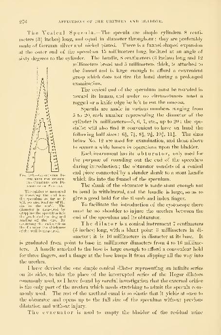
276 AFFKCTIOXS OK THE UHETHKA AND HLADDKK.
The Vesical Specula .—The specula are simple cylinders 8 centi-
meters (3-|- inches) long, and equal in diameter throughout ; they are preferably
made of German silv^er and nickel plated. There is a funnel-shaped expansion
at the outer end of the speculum 15 millimeters long, hiclined at an angle of
sixty degrees to the cyhnder. The handle, 8 centimeters (3 inches) long and 12
millimeters broad and 5 millimeters thick, is attached to
iiij the funnel and is large enough to afford a convenient
grasp which does not tire the hand during a prohjnged
examination.
The vesical end of the speculum must be rounded in
toward its lumen, and under no circumstances nmst a
ragged or a knife edge be left to cut the mucosa.
Specula are made in various numbers ranging from
5 to 20, each number representing the diameter of the
cylinder in millimeters—5, 0, 7, etc., up to 20 ; the spe-
cialist will also find it convenient to have on hand the
following half sizes: (5^, 7^, 8i, 9^, 10^, 11^. The sizes
below No. 12 are used for examination, and those above
to secure a wide lumen in operations upon the bladder.
Each instrument has its obturator, only used for
the pui'pose of rounding out the end of the speculum
during introduction ; the ol)turator consists of a conical
end piece connected' l)y a slender shank to a stout handle
which fits into the funnel of the speculum.
The shank of the obturator is made stout enough not
to bend in withdrawal, and the handle is large, so as to
give a good hold for the thumb and index finger.
To facilitate the introduction of the cystoscope there
must be no shoulder to injure the urethra between the
end of the speculum and its obturator.
The dilator is a conical instrument 7 centimeters
(8 inches) long, with a blunt point 3 millimeters in di-
ameter ; it is 16 millimeters in diameter at its base. It
is graduated from point to base in millimeter diameters from 4 to 16 millime-
ters. A handle attached to the base is large enough to afford a convenient hold
for three fingers, and a flange at the base keeps it from slipping all the way into
the urethra.
I have devised the one simple conical dilator representing an infinite series
on its sides, to take the place of the interrupted series of the Ilegar dilators
commonly used, as I have found by careful investigation that the external orifice
is the only part of the urethra which needs stretching to admit the specula com-
monly used. The rest of the urethral canal is so elastic that it yields at once to
the obturator and opens up to the full size of the s|)eculum without pi-evious
dilatation and without injury.
The e vacua tor is used to empty the bladder of the residual urine
Fig. 183.—(iR.\i)UATEi) In-
strument FOR MHASIK-iNG Calibers and ])i-
A.METERS OF ISPECCLA
The caliber is measuredby insertino: the end into
the speculum as far as it
will go and reading ott'the
size on the scale. The(.liainctiT is nicasun-il by
the di
of the
uatr.l ,.|».|,m- ;
off the size,
il tiie caliber fr
etcr tlie thicknill is measured.

CYSTOSCOPY. 277
wliicli the patient often can not expel, and which can not be removed by
a catheter, amounting to 4 to 10 cubic centimeters. It must also be used
from time to time to remove the urine accunuilating during a prolonged exam-
ination. The evac-
uator is a small
hollow, perforated
metal ball, con-
nected by iine rub-
ber tubing, about
35 centimeters (14
Fig. 184:.
—
Cystoscope axd (jBTiK\roR.
€0=
The cystoscope is 7 iiiilUiiietcrs in diaiiKtcvith an inner lumen of (5 millimeters Tlong handle attached to the funnel atfordsouvenient grasp in introducing and holdintrn any position. % ordinary size.
inches) long, with
a rubl)er exhaust-
ing bulb. The
rubber tube is cut
about 5 centime-
ters (2 inches) from the ball, and a piece of glass tubing
inserted which serves both as a telltale to show whenthe urine is flowing in the tube,
as well as to give rigidity to the
tube when it is picked up for in-
troduction into the bladder.
If the patient lies on her back
during the cystoscopic examina-
tion, the evacuator must be used
much oftener, as a small quantity of urine easily obscures
the held of view in this posture. In the knee-breast posi-
tion, on the other hand, a little clear urine in a pool in
the inverted vault of the bladder in no way interferes
with a thorough inspection of all parts.
The evacuator is used in the following manner: The
assistant, grasping the rubber bulb, pushes its base in with
his thumb and forces out all the air ; while the examiner,
holding the other end, drops the little j)erforated ball
through the speculum into the pool of urine, when the
assistant removes his thumb, and the bulb slowly ex-
pands, sucking up the urine. The evacuation will be
more rapid if the suction bulb is held well below the
level of the bladder. If there is only a little urine to
be taken up, it will escape faster l)y withdrawing the
l)all a little occasionally so as to suck up some air with the
urine.
Dr. G. E. Shoemaker, of Philadelphia, has devised a
simple evacuator consisting in a little tube with perfora-
tions and slightly bent at both ends, and connected with
an exhaust bottle emptied by a syringe (see Annals of Surgery, November,
Fig. 185.—Urethral Cal-ibrator AND Dilator.
The numbers indicate
the diameters in millime-ters.

278 AFFECTIONS OF THE URETHRA AND IJLADDER.
1895). Dr. W. L. Burrage, of Boston, lias also made an attaeliinent for the cys-
toscope to effect the same end.
The long mouse-toothed forceps are a hght for-
ceps 24 centimeters (9 inches) long, with long slender arms lo-5
centimeters (about 4 inches) long, and at the ends delicate slightly
recurved mouse teeth. The handles are fenestrated for lightness.
They are useful in cleansing the lumen of the speculum of drops
of urine, or in taking up a little urine out of the bladder with
small pledgets of cotton, or in wiping oif small areas of the vesical
mucosa.
The ureteral searcher is a small rod 18 centimeters (7
inches) long with a little bulbous end 3 millimeters by 1"5 milli-
meter, and a handle f> centimeters (2^ inches) long set at an angle
of 120 degrees. It is used in touching any part of the bladder
wall, in exploring a sinus, and particularly in locating the ureteral
orifices in doubtful cases.
Applicator.—Any piece of flexible wire about 15 centi-
meters long will do as an applicator to carry medicated cotton to
all points on the bladder or tlie urethra.
Other useful instruments are a speculum graduated in centi-
meters for measuring the distance between points on the bladder
wall, the external or internal urethral orifices, and a flattened
searcher, likewise graduated in centimeters and half centimeters.
The Technique of the Examination.—A sepsis .—Asepsis must
be maintained throughout every examination by handling only
aseptic instruments, introduced by clean hands,through a cleansed urethral orifice.
All the instruments used must have been sterilized and be laid
in a clean tray on a sterilized towel. The external urethral orifice
must be wiped clean with a boric acid solution before introducing
the speculum to remove any leucorrheal or other discharges often
contaminating its lips.
The hands must be scrubbed clean, and as far as possible the
utmost precaution must be taken to avoid touching any part of
the instruments but the handles. If this were always done, no
infection could occur even with infected hands. Every
instrument should be constantly inspected to detect any
rough or scaling surface liable to cut the nuicons mem-brane.
Preparation of the Patient.—The patient
UFio. 186.—Delicatk Moisk
TOOTHED FoUCKPS FO|{ CONVEYiNQ Pledgets ok CotTON INTO THE BlADDEH. should come to the examining table with the lower
True It
<ll()Uh lorc bowel emptied. I find that in many cases it makes a
decided difiici-ence if she has just eaten a meal, when
the bladder does not always expand so well. Immediately before the examina-
tion she must empty the bladder in a sitting or standing posture. If the nurse

TECHXIQUE OF THE EXAMIXATIOX. 279
draws tlie urine witli a catheter, or if slie passes it on the tahle, tlie evacuation
will not be nearly so complete.
Anesthesia .—A general anesthetic is only needed for a nervous woman.
Local anesthesia by means of a 10 per cent solution of cocain applied on a
pledget of cotton wound on a metal rod and introduced just within the external
Fig. 187.
—
Searcher for locating the Ureteral Orifice.
urethral orifice for five minutes beforehand, is suflicient to benumbtlie sensations so entirely that any required dilatation may be madeand the speculum introduced without much discomfort.
Posture of the Patient .—Two postures are available, an elevated
dorsal and a knee-breast. The dorsal position is the most convenient to use
and the least tiring to the patient, but it is only of service in thin patients, and
the atmospheric expansion is not so good ; the bladder of a fat woman wi
rarely distend at all in this posture. The head and thorax rest on the table,
while the pelvis is raised by putting one or two bran cushions under the but-
FiG. 188.
—
Examination of the Bladder in the Dorsal Position, with Elevated Pelvis.
The electric light held close to the symphysis is reflected by the head mirror into the bladder. Theangle of reflection must he as small as possible, so as to avoid constantly displacing the pencil of light witli
slight movements of the head.
tocks, so as to elevate them 20 or 30 centimeters (S or 12 inches) or more
above the table level. This gives a pitch to the pelvic and lower abdominal vis-
cera which makes them gravitate toward the diaphragm, and as soon as a specu-
lum is introduced the bladder sucks in air enough to distend it.
When the bladder does not expand, and yet it is particularly desirable to

280 AFFECTIONS OF TIIK IRKTHRA AND BLADDER.
use the dorsal position on account of the inabihty of the patient to stand the
inconvenience and fatigue of the knee-breast position, the bladder may be dis-
tended and the pelvis relieved of the small intestines by first placing her in the
knee-breast jDosition for a minute and letting in air with a catheter; she is then
turned on her back with hips elevated on the cushions, taking care to keep the
pelvis all the time well above
the level of the abdomen.
The speculum may now be
introduced and a satisfactory
examination made. A blad-
der distended in this way will
often remain well distended
until the hips are let downagain to the table level.
The knee -breast
]) o s i t i o n is the one posi-
tion most satisfactory and
applicable in all cases. Thepatient kneels with her knees
separated 10 or 12 inches,
close to the end of the table,
and, keeping the buttocks as
high as possible, lets the l)ack
curve in, and brings the side
of the face down on the
table. If she squats a little,
drooping the buttocks slight-
ly toward her feet, she will
be more conveniently disposed for the examination. Sometimes, to get a good
expansion, it is necessary to push the thighs in the opposite direction beyond the
vertical. If she is under an anesthetic, the best way to hold her in the knee-
breast position is for two assistants to stand, one on each side, close up to the
body to prevent it from falling sidewise, each grasping the body with one arm
thrown over the back, and holding the leg in the crotch of the knee with the
other hand to keep it from slipping up or down.
An apparatus like that shown in the text (Fig. liM)) and devised by Dr. G.
1>. Miller is useful where assistants are scarce, but the thigli bands imist not be
allowed to cut into the femoral fold.
Calibrating and dilating the Urethral Orifice.— Before
dilating the urethra and introducing a speculum it is well to calibrate it, that
is, to measure its diameter in millimeters as a guide to the amount of dilatation
needed to admit a speculum ; for example, if the urethral orifice has a diameter
of 6 or 7 millimeters only, it can not be dilated up to 10 or 12 millimeters with-
out a slight rupture of its margins ; calibration in this case would induce one to
use a speculum a size or two smaller than usual. Again, the calibration often
-Vesical Speculum introduced -witii the PatientTHE Knee-breast Posture.
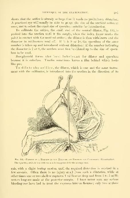
tectixiqup: of the examixatiox. 281
sliows that the oriiice i.s already so large that it needs no preliminary dilatation.
A practiced eye will usually be able to gauge the size of the urethral oriiice at
once, and to select the exact size of speculum suitable for introduction.
To calibrate the orifice, the small end of the conical dilator, Fig. 18;"), is
pushed into the urethra until it fits snugly, when the index finger marks the
point in contact with the urethral orifice ; the dilator is then withdrawn and the
diameter in millimeters read off. If it is 9 or 10, the speculum of the same
number is taken up and introduced without dilatation; if the number indicating
the diameter is 7 or 8, the urethra must first be dilated up to the size of specu-
lum to be used.
Boroglycerid forms the best lubricant for dilator and speculum
l)ecause it is colorless. Yaselin sometimes leaves a film behind which looks
like pus.
To dilate the orifice, the dilator, which is one and the same instru-
ment with the calibrator, is introduced into the urethra in the direction of its
Fig. 190.
—
Patient in a Harness in the Knee-breast Position fou Cystoscofic Examination.
The squatting attitude is a little too much exaggerated for the average case.
axis, with a slight boring motion, until the required distention is reached in a
few seconds. Often there is no injury at all from such a dilatation, while at
other times one or two shallow ruptures 1 millimeter deep and from 3 to 5 milli-
meters long are made at the posterior margin. I have never seen any serious
bleeding nor have had to treat the ruptures later as fissures ; only two or three
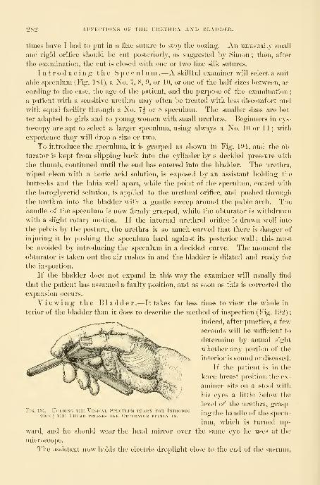
28i AFFECTIONS OF THE URETHKA AND JiLADDER.
times have I liad to put in a fine suture to stop the oozing. An unusually small
and rigid orifice should be cut posteriorly, as suggested by Simon ; then, after
the examination, the cut is closed with one or two fine silk sutures.
Introducing the Speculum .—A skillful exammer will select a suit-
able speculum (Fig. 184), a No. 7, S, 9, or lU, or one of the half sizes between, ac-
cording to the case, the age of the patient, and the purpose of the examination;
a patient with a sensitive urethra may often be treated w^itli less discomfort and
with equal facility through a No. 7^ or 8 speculum. The smaller sizes are bet-
ter adapted to girls and to young women with small urethrse. Beginners in cys-
toscopy are apt to select a larger speculum, using always a No. lo or 11 ; with
exjierience they will drop a size or two.
To introduce the speculum, it is grasped as shown in Fig. 191, and the ob-
turator is kept from slipping back into the cylinder l>y a decided pressure with
the thumb, continued until the end has entered into the bladder. The urethra,
wiped clean with a boric acid solution, is exposed by an assistant holding the
buttocks and the labia well apart, while the point of the speculum, coated with
the boroglycerid solution, is applied to tlie urethral orifice, and pushed through
the urethra into the bladder with a gentle sweep around the pubic arch. Thehandle of the speculum is now firmly grasped, while the obturator is withdrawn
with a slight rotary motion. If the internal urethral orifice is drawn well into
the pelvis by the posture, the urethra is so much curved that there is danger of
injuring it by pushing the speculum hard against its posterior wall ; this must
be avoided by introducing the speculum in a decided curve. The moment the
obturator is taken out the air rushes in and the 1)ladder is dilated and ready fur
the inspection.
If the bladder does not expand in this way the examiner will usually find
that the patient has assumed a faulty position, and as soon as this is corrected the
expansion occurs.
Yiewing the Bladder.—It takes far less time to view the whole in-
terior of the bladder than it does to describe the method of inspection (Fig. 192) ;
indeed, after practice, a few
seconds will be sufficient to
determine by actual sight
whether any portion of the
interior is sound or diseased.
If the patient is in the
knee-breast position the ex-
aminer sits on a stool with
his eyes a little below the
level of the urethra, grasp-:Vksicai, Si.k.thm ke.u.y fou [ntuoduc-
i,.o.|l,p Landle of the siiecii-! i'i:i>sEs THE OiiTi KAToi: FiKMi.v IN.
lUg 1116 ildnaie OT lllL SpCClI-
lum, which is turned u})-
mld wear the head mirror over the same eve he uses at the
Fio. loi.-ir.
ward, aiK
microscope
1 he sh
The assistant now hokls the electric droplight close to the end of the sacrum.

TECIIXIQUE OF THE EXAMIXATIOX. 283
which is protected from the heat \>y one or two towels, and the lower margin of
the head mirror is drawn away from the face and turned until the reflected light
spot falls w^ithin the bladder. Men accustomed to throat and eye work will find no
difRcultj in putting a good illumination at once just where they want it, while to
the inexperienced man the apparent waywardness of the light will he his chief
ti-ouble throughout. The direct ray of the little electric headlight makes the
illumination of the field an easier task.
Fig. 192.
—
Examination of the BLADHEn with the Patient in THE knee-breast Position.
The inspection of the bladder naturally begins with the posterior liemis])here
about the j)Osterior pole, opposite the internal urethral orifice, from 3 to 5 centi-
meters distant from the anterior wall, but not more than 2 or 3 centimeters from
the end of the speculum, which is pushed well into the bladder.
The whole posterior hemisphere is first examined as the end of the instru-
ment is directed to the right and to the left, l)y alternately raising and dropping
the handle so that every part of the mucosa is passed in review at least twice.
The normal background of the inflated bladder seen in this
way is a dull white, with here and there large vessels l)ranching and anastomos-
ing over it in an irregular manner. The fine rosy capillary injection seen in a
contracted bladder is not visible when it is distended with air, for the minuter
vessels are emptied, both by the expansion and by the posture of the patient.
At a point 1 or 2 centimeters above the posterior pole a rounded red spot of
capillary injection is often seen, which may easily be mistaken for a localized in-
flammation, but wliicli is mei-ely a suction hyperemia induced at this point by
contact with the end of the speculum during the withdrawal of the obturator.

284 AFFECTIONS OF THE URETHRA AND BLADDER.
The larger blood-vessels spring out of the subnmcosa, where tliej are first
seen in a hazy way, becoming clearer and with sharply defined outlines on the
surface, where they divide and subdivide into numerous branches. Occasionally
an artery is seen pulsating, and a large dark vein may sometimes be seen gradu-
ally disappearing from view as it penetrates the walls obliquely. The mucous
surface on the right and the left of the posterior hemisphere is often divided up
by shallow interlacing ridges, or again a sharp ridge 2 to 3 centimeters long is
seen to cross the field oljliquely ; these ridges are formed by the inner muscular
bundles irregularly arranged. Numerous little glistening points are due to
moisture on slight ine(pialities of surface which catch and reflect the light.
By dropping the handle of the speculum decidedly, its inner end is raised
and the vault or summit of the bladder is brought into view, and every part
of the organ inspected by moving the end from side to side. By elevating the
handle decidedly, the floor of the bladder is examined in the same way, and
then by moving it to the left and to the right the right and left walls come into
view.
The only parts which remain unexamined are those contiguous to the in-
ternal urethral orifice, and these are now seen by a still more decided elevation
and depression of the handle. With a marked depression of the speculum the
vesical triangle conies into view, always a little more injected than the rest of
the bladder, due to the fact that the mucosa and the underlying tissues are inti-
mately connected, which prevents this part from expanding and becoming ane-
mic like the rest of the bladder.
Turning the speculum from fifteen to twenty degrees—generally the latter
—to the right or to the left a little pinkish prominence is seen—the m o n s
ureteris which marks the position of the ureteral orifice; this usu-
ally looks like a fine transverse line about 2 millimeters long on the side of
the mons. It is sometimes a faint streak, like a little water line on paper.
At other times the orifice appears as a little pit or a mere point. Immediately
around the ureteral orifice is a paler area about 1 millimeter broad, and sur-
rounding this a rosy area 3 or 4 millimeters broad. I have several times seen
a blood vessel emerging out of it on to the vesical mucosa. If a V with its angle
at thirty degrees is marked on the cylinder of the speculum, near the handle,
by bringing one of the arms of the V parallel to the axis of the urethra the other
arm will then point toward one of the ui-eteral orifices, which may now be found
at once on looking through the speculum.
If the ureteral orifice is watched for half a minute or so a little clear urine
will be seen to spout out from the surface, forming a jet which lasts two or
three seconds, to be repeated again in the course of a minute.
Sometimes the urine spurts up free from the surface of the bladder and shoots
into the lumen of the speculum and trickles down to the outer edge. By hold-
ing the end of the speculum close up under the ureter, or by using the oblique
speculum adapted specially to this purpose. Fig. 103, enough urine can be caught
up with pledgets of cotton or in a small graduate to answer the purposes of a
physical, chemical, and microscopic examination. When the bladder is inflamed

TECHNIQUE OK THE EXAMINATION. 285
or ulcerated, it is sometimes of great advantage to get a little urine from one or
both sides in this waj, because it avoids the risk of a possible infection of a
ureter by putting in a catheter.
The interureteric line is often distinctly seen, either from its having a little
deeper color than the bladder behind it, or from a slight elevation.
In the process of the examination of the entire bladder conducted in
this way the
Held of vision
has changed
from the poste-
rior wall pei-jjen-
dicular to the
plane of vision
to the triangulai-
area which lies
almost parallel
to it ; at right
angles diiierences in color are best seen, while in the plane
of vision outlines which cross it come out more distinctly.
T li e r e t r o 8 y m p h y s e a 1 area comes into view on
elevating the handle of the speculum so as to direct the
inner end toward the symphysis pubis.
Occasionally a bladder will be found which does not re-
main ballooned out with air, l)ut undergoes ])eriods of more
or less rhythmic contraction, each of which lasts half a minute or moi'e. Withthe contraction there is an influx of blood into the capillaries, and the mucousmembrane assumes a rosv hue, becomino; more intense as the contraction in-
Obtlkator ixsekted.
Wheu the patient is in the knee-breast posture andthe oblique end is held under the ureteral orifice theurine is caught as it spurts out of the ureter, and runsdown the speculum to tlie outside, where it is collectedfor examination. By securiniT urine in this way thecatlietc-rization of the ureter is avoided. The lenirthlit' the spLMulum alone to the end of the lip is 9-5 cen-timeters, and its diameter is 10 millimeters. % ordi-nary size.
Fig. 194.—IXSTKIMEXT FOR MEASLRINtt THE Dl.STANCE BeTWEEX THE InTEKXAL OrIFXOE OF THE UrETHRAAXD V.^Rious Points on the Vesical Walls.
The distance in millimeters is registered on a graduated arm on the outside. One arm slides along theother and the lower one is provided with a little hook on its inner end to hold it against tlic inner endof the speculum. Knee-breast posture.
creases, until the whole organ is thrown into small folds like a labyrinth of cere-
bral convolutions. With the conti-action the aii* is audibly expelled and often
286 AFFECTIOXS OF THE IKETIIRA AND BLADDER.
urine comes sputtering out with it. After waiting from lialf a minute to a
minute the contraction relaxes and the bladder expands, and the examination
can be continued. The color and appearance of the w^alls and of the vessels
of a normal bladder must be well fixed in the mind by numerous examinations,
because the normal conditions are the standards of comparison in determining
the presence of areas of congestion, inflammation, or other diseases.
InsuflScient expansion of the bladder will be noticed in advanced pregnancy,
or in the case of a tumor blocking the pelvis, or in ascites. It may also be due
to the fact that the patient in taking the knee-breast posture arches her back,
and raises her chest too high from the table, and so interferes with the action
of gravity on the intestines. Often, too, a little time must be allowed for the
viscera to gravitate slowly tow^ard the diaphragm, and so create the necessary
suction for the distention of the l)ladder.
Too great an expansion of the bladder may also be troublesome. The diffi-
culty is that the trigonum and the ureteral orifices are then lifted up so high
that the examiner has to bring his head so far under the patient that his posi-
tion is extremely awkward and he does not get enough light for the inspection.
This may be remedied in several ways
:
a. Before introducing the cystoscope a speculum is always put into the
vagina, which then balloons out with air and lets its anterior wall with the floor
of the bladder drop in the direction of the symphysis ; then when the vesical
speculum is introduced the available expansion space of the pelvis, already partly
occupied by the distended vagina, is so diminished that the floor of the bladder
remains more nearly in the plane of vision. In parous women the atmospheric
expansion of the vagina is usually spontaneous. Distention of the rectum ^-itli
air will sometimes produce the same eftect.
b. By putting a cotton pack in the vagina or by depressing its anterior wall
with a spatula, any particular portion of the base of the bladder can be held down
in view.
e. Cases where there is a tendency to an excessive expansion may, as a rule,
be easily examined in the dorsal posture, wdien it is naturally not so great.
The presence of air in the bladder is rarely painful so long as the urethra is
open and the air enters and escapes freely with each respiratory movement.
But not infrequently as soon as the speculum is taken out the patient feels
a cramping pain, which is not relieved until she has been able to seat her-
self on a vessel to expel the air. To avoid this after-pain, the examiner mayleave the speculum in place, or slip a catheter in, and then lower the ])atient
gently from the knee-breast posture on to her side, so as to let the air out
gradually.
It is not necessary to take any special precaution after a vesical examination,
unless it has been prolonged enough to weary the patient, or unless she is feeble
or nervous ; under these circumstances rest for an hour or two with a lialf tea-
spoonful of aromatic spirits of ammonia may be prescril)ed.
The field of usefulness of the cystoscopic method just described is a large
one, commensurate with the entire field of vesical diseases, and the practitioner

AFFECTIONS OF THE URETHRA. 287
who uses it liberally will be rewarded by constantly discovering tliat affections
hitherto described as merely functional have definite local lesions as their basis,
and are often speedily amenable to simple methods of treatment.
I wish further to insist that a cystoscopic examination should be made in
every case where a vesical affection is more than transient and the diagnosis
is not absolutely clear without it, and that every part of the bladder should
then be thoroughly inspected.
AFFECTIONS OF THE URETHRA.
Short as the urethral canal is in women, it is liable to a variety of diseases,
some of which are peculiar to the sex. These affections are chiefly those which
either interfere with function or affect the caliber of the urethra. As the final
avenue of egress of the U2'ine the urethra holds a position analogous to the short
anal canal in its relation to the rectal ampulla and the intestines above. Owingto its relation to the external genitals, which are infested with micro-organisms,
the urethral orifice is constantly exposed to the risk of infection from without.
Its position under the resisting pubic arch renders the urethra liable also to
damage from j^rolonged pressure during labor, or to compression by a tumor
which chokes the pelvis. It is protected from external injuries by its concealed
position between the thighs and labia.
Affections of the urethra may conveniently be considered under the follow-
ing heads : Malformations, displacements, dilatation, stricture, ischuria, fistula,
foreign body, hyperemia, urethritis, new growths.
Congenital malformations of the urethra are among the rarest
gynecological affections. The commonest is a distinct lateral displacement of
the external orifice, generally about 2 millimeters, to one or the other side. Ashallow vertical fissure corresponding to the urethral orifice may be found on
the opposite side with a sharp ridge between the two ; this gives tlie appear-
ance of a double urethral orifice.
Most malformations of practical importance are due to a deficiency of the de-
velopment of some part of the urethral canal.
These may be classified as : (a) hypospadias, (b) epispadias, (c) imperforate
urethra, (d) totally deficient urethra.
Hypospadias.—In hypospadias part of the inferior wall of the urethra is want-
ing and the external urethral orifice opens at some point in the anterior vaginal
wall. One of the best described cases is that of A. Lebedeff {Arch.f. Gyn.y
vol. xvi, p. 290). The patient was a married woman, twenty-three years old, a
nullipara. She had always been well in every respect until five years married,
when she began to experience a pressure on the bladder and to suffer from
an involuntary escape of urine, at first at night after coitus, but soon becoming
constant. An examination showed normal labia, nymphse, and clitoris. But in
the vestibule, instead of a urethral canal, there was a furrow, lined with a deli-
cate mucous membrane, and leading back over the anterior vaginal wall, be-
tween vaginal folds so closely applied as to form a distinct ridge ; this furrow

288 AFFECTIONS OF THE UKETlllJA AM) 1!LA DDEIt.
ended in a canal 2 centimeters long situated 2 centimeters within tlie introitus,
which admitted two fingers directly into the bladder. The upper wall of thivS
furrow, seen on drawing aside the protecting vaginal folds, was covered through-
out with a bright-pink mucous membrane crossed bj a fine network of vessels.
The fact that the patient had never borne a child, and the straight course of the
canal, associated wnth the entire absence of any cicatricial tissue, showed that
the condition was a congenital defect of the inferior wall of the urethra extend-
ing as far up as the neck of the bladder. The incontinence had been brought
on mechanically by coitus.
A case of my owm, more properly classified, as F. Winckel has pointed out,
as a persistent urogenital sinus, was a nullipara forty-six years old;
the external genitals were normal as far as the introitus of the vagina, where the
only opening between clitoris and rectum w^as found. There was no hymen,
and the smooth orifice beneath the pubic arch had the form of a transverse slit.
If the finger was pushed in, it invariably entered a short muscular canal, which
was the shortened urethra, and so passed directly into the bladder. The ure-
thral orifice was in this w^ay situated about 1 centimeter behind the pubic arch,
and the urethral canal was only 1 centimeter long. While the inferior wall of
the urethra was absent, the anterior wall continued on out as far as the vesti-
bule, but was not of normal length. The upper M'all of the introitus was vas-
cular and of a deep-red color, and presented numerous longitudinal mucous
folds. There was a gaping slit in the anterior vaginal wall 1 centimeter long,
wdiich shortened the caliber of the urethra by so much. If now the point of the
sound introduced into the vagina was turned sharply down over the perineum,
it would then enter one or the other of two orifices lying side by side, and sepa-
rated by a fleshy septum ; this was a double vagina about 8 centimeters (5
inches) deep, with a small cervnx in the vault of each half. The incontinence
and distress the patient had complained of in coitus was relieved, and the chan-
nels returned to their normal usage by a plastic operation, freshening and
uniting the edges of the flaps, and converting the two vaginji? into one by
removing the septum.
Similar to this was the case of a short dilated urethra reported Ity Dr. A\". II.
Baker, of Boston [New Yoi'h Jour, of Gyn. and Ohstet.., Oct., 189?)); here there
was no trace of an upper urethral wall, and there was therefore a comjilete al)-
sence of the external part of the urethra, defectus itrethnu exterinv.
Epispadias.—In epispadias there is a defect of the upper wall of tlie urethra
associated with a separation of the labia minora and division of the clitoi'is. In
its extreme form the symphysis gapes, the anterior wall of the bladder is defi-
cient, and the bladder becomes everted (exstrophy).
Atresia.—Congenital atresia of the urethra may be due either to defective
intra-uteriue develo])ment, when it is associated with other anomalies, or it maybe ac(piired late in intra-uterine life by an agglutination of a portion of the ure-
thral canal. The latter condition was clearly the one obtaining in the case of a
child two days old (Mandl, AYhn. l-Vm. Wochenschr., 1891, p. 515), which
vomited and had general convulsions until the atresia was broken down by
DISPLACEMEXT OF THE URETHRA. 289
a sound, when turbid concentrated urine escaped and the disturbances ceased.
There was no marked distention of tlie bladder or evidence of hydronephrosis.
When the atresia is due to defectiv^e development a number of other coex-
isting defects are usually found, as in the instance reported h\ F. Schatz
{Arehiv f. Gyn., i, p. 12), where there was a double uterus, double vagina, and
d o u b 1 e bladder. As there was no urethra, each of these bladders opened
by an orifice in its base into the corresponding vagina.
If the atresia is a complete one, in order that the child so affected shall live,
nature must have provided some other channel for the escape of the urine, such
as an opening into the bladder through the symphysis, or a patulous urachus,
which discharges at the navel. If there is no avenue of escape for the urine,
this will, even in the intra-uterine life, accumulate in such quantity as to pro-
duce an enormous distention of the bladder, ureters, and kidneys, with ascites.
Under these circumstances the distended abdomen forms a serious hindrance to
the I)irtli.
Coagenital Absence of Urethra.—In these cases all trace of the urethra is
wanting, both external and internal orifices, and upper and lower walls, and the
base of the bladder o])ens directly into the vagina, ^vith which it forms one
common canal.
The urethra is liable to d is p lace m e n t s of two kinds : (1) Those affect-
ing the entire urethra with the adjacent tissues; (2) those affecting the nmcous
meml)rane of the urethra alone.
Displacement of the Entire Urethra.—The anatomical and topographical rela-
tions of the urethra are such that it is most favorably situated to prevent dis-
placement. Its shortness, its position directly under the jDubic arch, and the
dense fibrous connection with the adjacent parts all resist any ordinary efforts
to force it out of its natural position. The commonest change in position is
noticed when the vaginal outlet is relaxed and gaping, and the base of the blad-
der descends with the anterior vaginal wall to till up the gap. Careful inspec-
tion and the use of a sound then show that the urethra has rotated outward and
forward around the symphysis as its axis ; the external orifice lies farther for-
ward and its direction more upward than normal, while the internal oi'ifice has
sunk with the bladder. In prolapse (^f the elongated cervix uteri with a vesical
diverticulum in the pouch the urethra often undergoes a still more markedchange in its direction, gradually yielding to the traction of the prolapsed sac,
first at its internal orifice, and then the lower portions, until the whole urethra
finally lies outside the body. The canal sometimes assumes a sigmoid curve,
which makes it difficult to pass a catheter.
The symptoms of this condition are those referred to the prolaj^sus and
those which arise from difficulty in emptying the bladder. Unless the patient is
in the habit of pushing up the sac for this purpose, the evacuation is often in-
complete and decomposition of retained urine may set in with all its untoward
sequelae.
li p w a r d displacement of the urethra occurs during labor and whenthe urethra is drawn up by a full bladder. It also occurs in the case of large
22
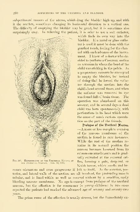
290 AFFECTIONS OF THE URETHRA AXD BLADDER.
subperitoneal tumors of the uterus, wliicli drag the bladder high up, and with
it the urethra, sometimes changing its horizontal direction to a vertical one.
The difficulty of emptying the bladder may l)e great, but it is sometimes also
surprisingly easy. In relieving the patient, it is safer to use a soft catheter,
M'liich finds its own way into the
bladder. If a metal or glass cathe-
ter is used it must l)e done with the
gentlest touch, feeling for the chan-
nel with each advance of the instru-
ment. I know of a doctor who de-
cided to perform a Caesarean section
on a woman in whom the head of the
child was sticking in the pelvis. Asa preparatory measure he attempted
to empty the bladder, but instead
of doing that he forced the cathe-
ter through the urethra into the
child's head several times, and when
the catheter was removed its eye
was found full of brain tissue. The
operation was abandoned on this
account, and in several days a dead
child was born spontaneously, with
perforations in its head which were
the cause of much curious specula-
tion on the part of the friends.
Prolapse of the Urethral Mucosa.
—A more or less complete eversion
of the mucous membrane of the
urethra is found in rare instances.
While the rest of the urethra re-
mains in its normal position the
mucosa becomes loc»sened from its
submucous attachments and is grad-
ually extruded at the external ori-
fice, forming a pale, deep-red, or
bluish tumor, which swells and be-
left to itself. As the anterior, pos-
all involved, the protruding mass is
Fig. 195.
—
Hypertrophy of the Urethral Mucosa atTHE External Orifhe. Apr. 10, 1896.
comes edematous and even gangrenous if
terior, and lateral walls of the urethra are
tubular, and is lined within as well as covered without by a sensitive, easily
bleeding mucous membrane. No age is exempt from prolapse of the urethral
mucosa, but the affection is far commoner in young children ; in two cases
reported the patients had reached the advanced age of seventy and seventy-two
years.
The prime cause of the affection is usually struma, but the immetliately ex-

DILATATIOX OF THE URETHRA. 291
citing cause may be a blow, straining, or coughing, or rape. Inflanniiation of
the mucosa also occasionally produces a prolapse, which is as a rule only partial.
The diagnosis will be made upon separating the labia and observing at
the site of the urethra and choking its orifice a vascular tumor with a slit in the
center of it opening into the bladder. This condition must be distinguished from
caruncle of the orifice or a hemorrhoidal pouting of the mucosa at the orifice, as
well as from eversion of the mucosa of the bladder or of the ureter.
The seat of a caruncle is usually on one side of the external urethral orifice.
An eversion of the mucosa of tlie orifice forms but a shallow protrusion not more
than 5 or 6 millimeters long.
In everted bladder the base of the tumor is found by a sound to be within
and attached to the bladder, instead of at the external orifice of the urethra. Thebladder tumor also lacks a canal. An everted ureter is attached to the bladder
wall and a fine sound or catheter 1^ millimeter in diameter passes through the
tumor on up over the pelvic brim into tlie kidney.
The treatment will vary with the conditions,
ought to be given up entirely for cleaner
surgical methods. It is also unadvisable
to transfix and ligate the protruding mass
and allow it to slough or to cut it away
beyond the ligatures, as has been done.
The first and simplest plan to l)e tried
in a recent case is i-eposition, after getting
rid of the sensitiveness, either by means
of a weak solution of cocain or by anes-
thesia. By compressing the tumor on all
sides and at the same time pushing it back
into the urethra a replacement may be
effected which will prove permanent ; the
patient should be kept in bed afterward,
and a vulvar compress applied, and small
doses of belladonna should be given as a
sedative by rectal suppository.
If the tumor can not be replaced or if it
escapes again directly after replacement,an
operation will be necessaiy, and the best is
the excision of the protruding portion with
knife or scissors, followed by a carefully
applied continuous suture of fine catgut,
uniting the cut edges and checking the
hemorrhage. It is important to catch both edges as they are cut to prevent
an inversion with excessive hemorrhage.
Dilatation of the Urethra.—Variations in the caliber of the urethra, both dila-
tation and stricture, are of rare occurrence. Dilatation, however, a disease never
found in men, is far connnoner than stricture, a disease so often found in them.
The cautery so often used
Ftg. 196.
—
Hypebtrophiei) P^xternal OrificeOF THE UrETHKA .SHOWN IX SaiHTTAL SeC-TIOX.
the svniphvsis; B, the hhuUler : thebelow.
ivm.
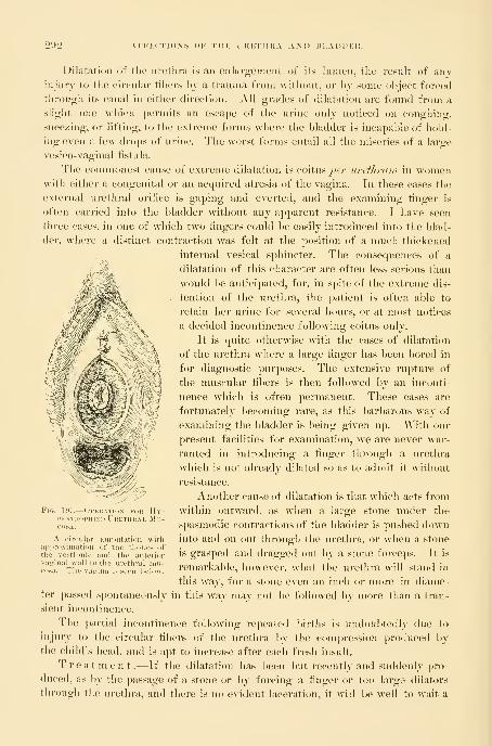
on-> AFFECTION'S OF TIIK IKFTHltA AN
Dilatation of tlie iii-etlira is an enlai-genient of its lumen, tliu result of anyinjury to the circular fibers by a trauma from without, or l)y some object forced
through its canal in either directi<.)n. All grades of dilatation are found from a
slight one which permits an escape of the urine only noticed on coughing,
sneezing, or lifting, to the extreme forms where the bladder is incapable of hold-
ing even a few drojjs of urine. The worst forms entail all the miseries of a large
vesico-yaginal fistnla.
The connnonest cause of extreme dilatation is coitus 7>i?/' uretltfam in womenwith either a congenital or an acquired atresia of the vagina. In these cases the
external urethral oritice is gaping and everted, and the examining finger is
often carried into the bladder without any apparent resistance. I have seen
three cases, in one of which two fingers could be easily introduced into the blad-
der, where a distinct contraction was felt at the position of a nmch thickened
internal vesical sphincter. The consequences of a
dilatation of this character are often less serious than
would be anticipated, for, in spite of the extreme dis-
tention of the urethra, the patient is often able to
retain her urine for several hours, or at most notices
a decided incontinence following coitus only.
It is quite otherwise with the cases of dilatation
of the urethra where a large finger has been bored in
for diagnostic purposes. The extensive rupture of
the muscular fibers is then followed by an inconti-
nence which is often permanent. These cases ai-e
fortunately becoming rare, as this barbarous way of
examining the bladder is being given up. With our
present facilities for examination, we are never war-
ranted in introducing a finger through a urethra
which is not already dilated so as to admit it without
resistance.
Another cause of dilatation is that which acts from
within outward, as when a large stone under the
spasmodic contractions of the bladder is pushed downinto and on out through the urethra, or when a stone
is grasped and dragged out by a stone forcejis. It is
remarkable, however, what the urethra will stand in
this way, for a stone even an inch or more in diame-
ter passed spontaneously in this way may not be followed by more than a tran-
sient incontinence.
The partial incontinence following repeated births is undoul)todly due to
injury to the circular fibers of the urethra by the compression produced bythe child's head, and is apt to increase after each fresh insult.
Treatment.—If the dilatation has been but recently and suddeidy \>yo-
duced, as by the passage of a stone or by forcing a finger or too large dilators
through the urethra, and there is no evident laceration, it will be well to wait a
. 1!»7.— (>l

DILATATION OF TllK IKETI1I{A. 29^:5
few days, simply keeping the parts clean and free from contamination by vagi-
nal douches and local application of a boric acid solution ; and if there is any
definite imj)rovement it will be well to wait as long as it continues. If there
is any evident laceration at the external meatus, this should be sutured under
cocain, first trimming off all uuevenness and then uniting the parts from side
to side with fine interrupted silk sutures, using a small fine needle. When the
relaxation is due to coitus and the abnormal channel has been created in
place of an atresic vagina, nothing can l)e done until the vagina is restored to its
integrity.
The condition of a patient with an incontinent urethra is so pitiable that as
ennnent an authority as ilutenberg ( ]]le/i. iiied. Woch., 1875, No. 87) proposed
to cure it by closing the urethra entirely and making a suprapubic fistula, which
was to be controlled by the pressure of a pledget.
In general four plans have Ijeen tried with var^-ing success in attempting to
overcome incontinence. These are :
(a) A vaginal pessary.
(b) A longitudinal resection of the vaginal wall, with or without a piece of
the urethra.
(c) An operation to fiatten out and compress the external orifice.
(d) Twisting the urethra spirally so as to narrow^ its caliber.
It is not possible in the absence of a larger experience to speak with decision
as to the comparative merits of the three operativ^e procedures proposed, but I
would prefer as a first resort to resect after the plan proposed in (b).
If the incontinent urethra was near the normal caliber I should use Pawlik's
plan (c).
If the incontinence is due to the traction or scar tissue in the anterior
vaginal wall on the neck of the bladder, this nmst first be freely incised to
do away with the traction. Permanent relief has even been obtained in this
way.
(a) F. Schatz {^[/•c/i. f. Gynal'ol.^ vol. xi) has been able to give relief bymeans of his funnel-shaped pessary, which presses directly upon the urethra.
Similar results have occasionally been secured by the use of a l)al] pessary large
enough to press the urethra against the symphysis and hold the urine back
until the intravesical pressure has increased to a certain degree. An intact
levator ani is necessary for the successful use of a pessary, which can not give
relief in a relaxed vaginal outlet.
(b) An excision of the superfluous relaxed portions reconnnends itself as the
most rational plan, and it has yielded excellent results in the hands of Frank
and Engstrom.
Frank's procedure {Centndhl. f. Gyiidhjl.^ 1882, ]^o. 9) is to lay a small
catheter in the urethra and then to excise a wedge-shaped piece from the pos-
terior urethral wall, including the vaginal as well as the urethral mucosa, and
extending from the external urethral orifice to within about one centimeter of
the internal orifice. The incision is now continued in an elliptical form on the
vaginal wall beyond the neck of the bladdei-. By a transverse row of interrupted
294 AFFECTIONS OF THE URETHRA AND I5LADDER.
sutures the wliole wound surface is now accurately approximated. Tlie lower an-
terior part of the incision underlying two thirds of the urethra simply resects its
relaxed canal, while the width of the elliptical portion of the denudation has
been calculated so as to form a sort of buttress behind the neck of the bladder
like the third lobe of a prostate in the male.
Engstrum {Bed. klin. WoeL, 1887, p. 744), in an anemic, badly nourished
woman, fearing a failure of union on the side of the urethral nuicosa, carried his
excision on the vaginal septum down to the urethral mucosa, but did not include
it. The wound suppurated and healed by granulation with the formation of
scar tissue, and as a result the patient was able to hold her urine four hours by
day, and all night.
(c) By flattening the outer end of the urethra and bending it at the same time,
Pawlik ( Wien. mecl. Wochenschr., 1883, Nos. 25 and 26) relieved several pa-
tients of incontinence. His plan is to draw the orifice of the urethra well for-
ward toward the clitoris and sharply to one side ; then, marking the point on
the side to which it could be drawn without excessive traction, a long, narrow
denudation al)out 2 centimeters long (| inch) is made in the sulcus and sutures
passed to hold the urethra in that position. After a week, when the sutures are
removed, the other side of the urethra is drawn upward and outward in the same
manner, and the sulcus on that side denuded and sutured. By this means the
ui'ethra receives a sharp bend forward and the posterior wall is strongly flattened
against the anterior by traction on both sides.
(d) Torsion of the urethra is a plan proposed by R. Gersuny {Centralh. f.
Cliir., 1889, p. 433). The whole urethral canal is dissected out from the sur-
rounding structures as far as the neck of the bladder, and the urethra is then
twisted on itself, so as to form a series of spiral folds, when it is sutured so as to
be held in this position. Gersuny relieved his patient after twisting the urethra
one and a quarter times on itself—that is, through an arc of 450 degrees.
Desnos (Ann. des mal. des org. gen.-urin.^ 1890, p. 344) partially relieved
the patient by ligating the urethra. He first introduced a catheter into the blad-
der, and then cut through the vaginal mucosa so as to expose the upper two
thirds of the urethra ; this portion was then isolated by a catgut ligature placed
2 or 3 millimeters from the neck of the bladder and tied so tight that the cath-
eter could just be moved. The vaginal incision was then closed with silkworm
gut. A small wedge was also taken out of the gaping external orifice. The
result was an immediate power of retention for three hours, afterward reduced
to one hour and a half.
Stricture of the Urethra.—A stricture of the urethral canal so large as to
interfere with the free exit of the urine from the Ijladder is rare. Stric-
tures of large caliber in which there is no evident impediment to urination
or to the passage of an ordinary catheter have been described and their inipoi"-
tance insisted upon by Dr. Ely Van de Warker {Medical News., Philadeljihia,
1887, p. 59). They are to be recognized by using olive-pointed bougies, which
catch and trip in the stricture as they are withdi'awn. Dr. Van de "Warker
finds that the evil results of a neglected stricture of large caliber in women

STRICTURE OF THE URETHRA. 295
are similar to those in men ; hut confirmation of these important conclusions
are still wanted from other clinicians.
A variety of causes may operate to produce a stricture, some of which are
:
1. A localized thickening produced by a chronic gonorrheal urethritis.
2. A cicatricial contraction in the anterior wall of the vagina following a
slough produced by labor.
3. The cicatrization of a chancre, whether in the vagina or in the urethra.
4. Carcinoma of the urethra.
5. Extreme contraction of the external meatus without assignable cause.
Gonorrheal stricture is the commonest of all forms, although it has as yet re-
ceived but little attention in women. Its history is often difficult to obtain,
owing to the slightness of the symptoms produced by a chi-onic gonorrheal
urethritis. The slough which follows labor is more apt to i-esult in a urethro-
vaginal fistula than in a stricture.
The symptoms of stricture are difficulty and pain in micturition, the
urine being expelled in drops or in a fine stream with considerable straining.
These difficulties increase as the caliber of the stricture lessens, although occa-
sionally even an extreme contraction of the urethra may elicit no complaint. I
remember well my first case, a German woman of about fifty-two, from whom1 removed 252 gallstones. After the operation she could not urinate lying on
her back, nor could the nurse catheterize her. I found just within the external
orifice a cicatricial narrowing of the urethra, which only allowed a fine catheter
2 millimeters in diameter to pass with difficulty. The patient was not conscious
of there being anything wrong, nor was I able to get any history or to determine
the cause of the stricture.
The diagnosis will be made readily if every case complaining of any
urinary disturbances is examined locally. The effort to catheterize or to pass a
vesical speculum will at once tell whether an obstruction exists or not, and if so,
the urethra may be calibrated with bougies and the stricture studied urethro-
scopically, determining its exact size, position, extent, appearance, and consistence.
The treatment will depend upon the form of the stricture and its
cause. In secondary cancerous disease which can not be eradicated, in the
earher stages the bladder should simply be catheterized regularly ; later, when
the obstruction increases, a vesico-vaginal fistula may be made, or, if necessary,
the ureters may be set free and turned into the vaginal vault. In one of mycases of syphilitic thickening the urethra was reduced to a rigid canal, with ex-
tensive ulcerations at the external orifice ; the patient had beside this a universal
cystitis and hypertrophy of the bladder walls.
In cicatricial contraction of the anterior vaginal wall compressing the urethra,
if the cicatrix is narrow, the plan of making multiple incisions into it under
cocain, may be tried. If this does not succeed, the cicatrix should be dissected
out, even going so far, if necessary, as to resect the lower walls of the urethra
with it ; then, after an exact closure of the wound with fine interrupted sutures
passed close together, a catheter should be left in the bladder for four or five
days to relieve the canal of any strain.
296 AFFECTIONS OF THE URETHRA AND BLADDER.
When the stricture is narrow and more or less circular, as in the gonorrheal
stricture, the lumen should be enlarged by incising or dilating it.
A stricture which allows a bougie 2 or 2^ millimeters in diameter to pass
may be dilated by passing the bougies daily, until a No. 4 or 5 is passed. After
three or four days a No. 5, 5^, and may be passed, and so on gradually up to
No. 10, the maximum. A smaller stricture, admitting only a No. 1 or 1^
bougie, may be exposed through the urethroscope, its edges incised slightly, and
then dilated up to No. 3 or 4, gradually followed by the larger dilators until the
lumen is restored to the normal size.
Care must be exercised to keep the field free from contamination, and not to
transport the germs on the external urethral orifice into the canal every time the
dilators are used. If the stricture is limited in its extent, and an examination
shows that there will be but little risk of an extensive rupture, a rapid dilatation
may be practiced, as recommended by E, Hermann {Trans, of the Ohst. Soc. of
London, for 1887, xxix, p. 27), restoring the urethra to a normal caliber at the
first sitting.
All cases treated by dilatnig must be kept under observation for a long time,
as a good percentage show a decided tendency to relapse, when the dilatation
nnist be repeated. I gave one of my patients, who was an intelligent nurse, a
glass catheter, with the instruction to use it at intervals to discover whether
the stricture was recurring and to keep it open, and she did this with good
effect.
A stricture confined to the external meatus is easy to treat by benumbing the
part with a strong solution of cocain and using the conical urethral dilator, or. if
it is very tight and the scar tissue extends deep, an incision 5 millimeters deep
may be made through the posterior margin and the urethral and vaginal mucosae
sewed together.
Ischuria.—Ischuria is an affection in which the patient, often without a de-
monstrable mechanical cause, is unable to void the urine which is then retained
in the bladder. It is sometimes seen in hysterical girls, and often in the puer-
peral state, where it is prol)ably due to pressure on the neck of the bladder by
the head of the child during its descent, benumbing the nerves and so destroying
for a time the reflex sensibility. That this is probably the correct explanation
is borne out by the fact that it oftenest follows forceps labors.
The diagnosis is usually easy to make by the discovery of a distinct tumor
just above the symphysis pubis ; on introducing the catheter the urine escapes
and the tumor at once disappears. I had one case in a young woman with
anorexia nervosa, in whom I palpated and percussed the flat lower abdo-
men and concluded that there was no urine in the bladder, but on putting in a
catheter 500 cubic centimeters of urine escaped; the bladder had distended
laterally.
The best way to treat puerperal cases is first to try letting the patient urinate
l)y sitting erect on the vessel, and if this does not succeed to practice a rapid
dilatation of the urethra under cocain. The external genitals are carefully
cleansed and the caliber of the urethral orifice taken. Cocain (10 per cent solu-

URETIIKAL FISTULA. 297
tion) is then applied for live minutes in the canal and the first dilator used,
followed immediately bj a size a half millimeter larger, and this by the next
size, and so on up to No. 12 or 14 millimeters in diameter. Often the pa-
tient will 1)6 permanently relieved at once, or at most the passage of the
same numbers once more after an interval of a day or two will relieve the
ischuria.
In an anemic, hysterical patient the condition of the blood, and the nervous
symptoms, the bowels, and the digestion should receive especial attention.
Strychnin is one of the best systemic remedies given in full doses.
Urethral Fistula.—A urethro-vaginal fistula following labor is a rare occur-
rence. When the urethra is involved the lesion is located in the upper part,
oftenest at the neck of the bladder, and is frequently found in association with
an extensive injury to the base of the bladder, forming a vesico- urethro-vaginal fistula. ]S^ot so rare, however, is a fistula artificially created to
draw out a redundant urethral mucosa and relieve dysuria (Emmet's buttonhole
operation).
Urethral fistulae usually involve the lower wall only, and appear either as
elliptical openings from 1 to 1-5 centimeter long or as a fine circular opening
not larger than a pin head. If the fistula is close to the neck of the bladder
there may be a frequent involuntary escape of urine. If it is farther down in
Figs. 198, 199.
—
Urethu<)-vac;in.\l and Vesico-v, ;ai. Fisii 1 \ IN THE Same Patient.
A small bridge of tissue (c), including the neck of the bladder remained intact. The upper border of the
vesico-vaginal fistula (a) and the lower border of the urethro-vagiual fistula i6) were denuded and united
without sacrificing the neck of the bladder (c). See also Fig. 200.
the canal there may l>e no syjiiptoms at all pointing to its existence, and under
these circumstances there is no reason why the fistula accidentally discovered
should be operated upon.
I have seen but two cases, both resulting from the traumatisms of labor.
In one there was an elliptical opening in the floor of the urethra at about the
middle, I'T) centimeter long by 3 millimeters in width, and the other a round
opening about i millimeters in diameter, just in front of the neck of the
298 AFFECTIONS OF THE URETHRA AXD BLADDER.
bladder, while just behind the neck there was a vesico-vaginal fistula a little
larger in diameter.
The treatment of a simple fistula which does not involve more than
one third of the lumen of the urethra is like that of vesico-vaginal fistula, by a
fnnnel-shaped denudation of its margins, broad on the vaginal surface, and
reaching up to but not including the urethral mucosa. Fine silkworm-gut su-
tures are then passed transversely, and tied so
as to bring the edges into exact apposition. It
is better to leave a catheter in the bladder for
five days. The stitches should be removed in
fi-om seven to ten days.
In a case in which a urethral fistula just be-
low the sphincter was complicated by a vesical
fistula just above it, leaving intact the vesical
sphincter ring at the internal urethral orifice,
the problem was to save this important bridge
of tissue with the hope of retaining its sphinc-
ter action. The bridge was so narrow that
both sides of it could not be denuded and
sutures passed, so the plan was adopted of de-
nuding the margins of both vesical and ure-
thral fistulse, treating them as if they consti-
tuted one large fistula instead of two small
ones ; the urethral sphincter lying between
them was not touched. The denudation was
made down to the vesical and urethral mu-
cosae, and extended out broadly on to the vagi-
nal mucosa, and interrupted sutures of silk-
worm gut were passed in an antero-posterior
direction, so as to make the line of union a transverse one. The union was
complete and the patient had entire control of her urine, in spite of the fact
that a short circuit w^as made in this way from the bladder under the sphincter
portion into the urethra.
Foreign Bodies in the Urethra.—Foreign bodies are but seldom found in the
urethra. They arise either from a calculus escaping from the bladder and
caught in the urethra, or are introduced from without through the external
urethral orifice, or they are formed within the urethra itself.
In case the foreign body forms in the urethra or is lodged thei-e from the
bladder, it is quite sure to be a phosphatic calculus.
When the foreign body is introduced from without, if it remains long
enough, it becomes incrusted with phosphates, and so forms a calculus. I have
the specimen of a peculiar form of urethral calculus in a case in which the
bladder was choked with a large ovoid stone, from one end of which a mass
about 3 centimeters long and 2 centimeters in diameter pi-ojected into the
urethra. The outer end is pointed, while a constriction at the upper end indi-
^iH> —The Mjtikii) m imkoihcinoTHE, SlTURFS IN THE LA'sE Oi VksK O-
VAGINAL FlSTUL\ (B), AND URETHRO-VAGINAL Fistula (U).
The bridore of tissue between U and Bthe neck of the bladder.

FOREIGN BODIES IX THE URETHRA. 299
cates tlie position of the neek of the bladder. These calculi closely resemble
the calculi filling the pelvis of the kidney, and projecting into the ureter.
The symptoms of a urethral calculus are frequent and difticult micturition,
with alkaline urine containing mucus, pus, or blood.
The examination by the vagina reveals an enlargement in the anterior vaginal
wall, somewhat movable and tender on pressure and densely hard, feeling
through the thick mucous covering like cartilage. On attempting to introduce
a catheter into the bladder, the point strikes against the hard substance and the
diagnosis is clear.
Treatment.—The best mode of treatment, when the stone is not too
large, is to extract it by the meatus in the manner proposed and practiced by
Prof. A. J. C. Skene {Diseases of the Bladder and Urethra in Women, JSTew
York, 1878, p. 345), In one of Dr. Skene's cases the stone was lodged near the
meatus ; the forefinger of the left hand was introduced into the vagina and
pressed above the calculus to steady it. A wire curette was passed through
the meatus beyond the stone, when by traction with the curette and pressure
with the finger the stone was extracted. This is not unlike the classical method
of treating vesical calculi in women. In another case of a stone higher up in
the urethra Prof. Skene was able to fix it firmly by pressure through the vagina
so as to grasp and extract it by forceps.
In a case of Prof. F. Scliatz {Yerhand. d. deutsch. Gesell.f. Gyndkol., II
Cong., Leipzig, 1888, p. 115) a urethral stone weighing 100 grains formed
around a hairpin which had escaped into the urethra in masturbation three
quarters of a year before. The stone was 8 centimeters (3 inches) long, and
projected well back into the bladder. The patient passed this stone spontane-
ously with severe straining and bleeding for t^vo hours ; she afterward suf-
fered from incontinence. Similar to this was the case of A. Mazario {Siebold''s
Jour.^ f. Geh. and Frauemim. und Kinderkranlxlt .^ ]S^o. 7, p. 794), The
patient had thrust a long sewing needle into the meatus, which penetrated
the urethro-vaginal septum and was lost. A calculus formed in the wall be-
tween the vagina and urethra, which was removed by cutting down through
the meatus half an inch on both sides of the tumor and pressing it out by a
finger in the vagina. After removing the stone, the finger could easily be
introduced through the dilated urethra into the bladder. The stone was three
inches long and four inches and a quarter in greatest circumference. The
patient recovered.
When the calculus is small enough to pass without injury, or when it is
lodged behind the contracted external meatus, and is cylindrical or narrow and
fusiform, it should be removed by simple traction and pressure, or, if necessary,
by dilating and incising the meatus. Soft phosphatic calculi may be broken by
crushing with forceps, and so removed piecemeal. A large stone projecting into
the urethra from the bladder should be removed from the bladder by a vaginal
or a suprapubic incision. In other cases it is better to extract the stone by
making a longitudinal incision through the vagina into the urethra or into the
urethral sac in which the stone lies.

300 AFFECTIONS OF THE URETHRA AND BLADDER.
Tliis was (lone in a case of Seriioti reported l>y J^iaseski {Nouv. arch.
(ToUtet. et de gi/mkol., 1892, p. 23(5) .The patient, sixty -live years old, began
to experience discomfort twenty-live years before, inunediately after her last
confinement. For three years she had suffered intensely with painful niictui-i-
tion, passing her urine as often as ten times daily and almost as often at night.
Upon examination, 1^ centimeter from the meatus a densely hard, incompressible
angular mass was felt in the anterior vaginal wall, about as big as a nut, painful
on pressure, and movable. The urine contained a mueo-purulent sediment.
The stone lying in a pocket with a small orifice of communication with the
urethra was not touched by the first sounding efforts.
An incision was made through the anterior vaginal wall 2^ centimeters (1
inch) long over the calculus, and it was extracted and the wound closed with a
continuous silk suture. The calculus was pipe- shaped, tlie size of three little
hazelnuts superimposed, and was made up of earthy phosphate. A complete
recovery followed.
Urethritis.—Urethritis in woman is a disease quite common, but rarely noted,
owing to the infrequent use of the endoscope by gynecologists. Moreover,
many of the cases of urethritis are diagnosed symptomatically as " cystitis " or
"irritation of the bladder." Inflammation of the urethra in the absence of such
a local cause as a foreign body is usually due to the gonococcus, which lin-
gers in the urethra as its seat of preference long after all traces of infec-
tion have disappeared from every other part of the genito-urinary tract. Some-
times the patient presents a history of an acute inflammation, but oftener
there is no definite history of such an attack or some slight disturbance only is
recalled.
Vaginitis, endocervicitis, and inflammati(jn of the vulvo-vaginal ducts may
be found coexistent with an old urethritis.
B. Tarnovski ( Vortrdge iiber venerische Krankheiten^ Berlin, 1872) in 750
cases of gonorrhea found acute or chronic urethritis in 286, or 38 per cent.
Steinschneider {Bed. Idin. WocL, 1887, No. 17, p. 301), in a study as to the
localization of the gonorrheal hifection in 84- fresh cases, found gonococci in the
urethra in all of them.
The secretion may be discovered bathing the urethral orifice, or on separat-
ing the little urethral labia, or by milking the urethra from above downward,
wlien a little purulent or brownish or bloody fluid will exude from the external
orifice. This should be done before urinating, so that the secretion will not have
been washed away.
The disease is particularly apt to linger in a chronic form in Skene's glands,
which can be milked out by making the pressure from above downward, first on
one side of the urethra and then on the other. One or two drops of thick
])us will often exude from the orifice of the duct just inside the urethi-a, gi\-
ing evidence of its source by adhering more to the side from which it was
squeezed. Long after a gonorrhea is a])])arently well a fresh attack may start
up by auto-infection from a chronic gonorrhea which has lingered in these
glands.

URETHRITIS. 301
•JOl. — ConcealedAiisfEss i)¥ Skene'sGland.
A drop of thick pusl]us been squeezed out ofthe right gland and lies
upon the right labiumuretlirse. The oritice oftlie left gland isseenjustinside the left labiumurethriv.
In acute gonorrheal urethritis the symptoms are a persistent intense burn-
ing, frequent urination with pain, and sometimes a discharge of blood. Vulvitis
and vagniitis may be associated with them. In the subacute form the discom-
forts may be transitory and not serious.
It is important in all cases to e.Kamine the urethral secretion microscopically
for gonococci, and confirmatory evidence will be gained if the presence of
gonococci in the cervical secretions can be demonstrated.
Should they be found in the cervix and not in the urethra
the evidence would still be in favor of a gonorrheal ure-
thritis.
The urethroscopic examination must be made in every
(rase where the purpose of the examiner is not only to knowthe nature but its grade and its extent as well. The dis-
eased conditions are found almost exclusively in the mu-cous and submucous tissues, and are more apt to be localized
in the anterior or posterior portions of the urethra than in
the middle.
In making a direct examination several precautions must
be taken :
1. A small-sized speculum nmst be used (say a No. S) in
acute cases in order to do as little harm as possible to the
mucous membrane.
2. The external meatus must be well cleansed to avoid pushing any pus on
the surface up into the urethra and bladder on the end of the obturator.
3. The manipulations must all be conducted with extreme gentleness and
delicacy so as to avoid producing lesions wdiicli might open up an avenue for
septic invasion of the submucosa.
Acute Urethritis .—In fiorid gonorrhea with a pouting swollen meatus
secreting abundant pus the examination may be foregone with advantage to the
patient until the swelling of the mucous membrane has somewhat subsided.
If the examination is made, a strong solution of cocain should first be used to
diminish the extreme sensitiveness of the mucosa, especially at the external ori-
fice, which is swollen, red, and everted. Often here the little dilated orifices of
a few glands can be seen exuding minute drops of pus. This condition is shown
by the urethroscope to extend a short distance back, to be less intense about the
middle, and often to assume a marked intensity again near the internal orifice.
The use of the speculum always does some injury, making small fissures and pro-
ducing slight hemorrhages.
Linear ulcers from 2 to 4 millimeters long and 1 millimeter broad are not
rare on the inferior wall ; they are painful and exhibit a yellowish area of necro-
sis in the center with an injected margin. The whole mucous membrane is
deeply injected, and so swollen that it looks edematous, pouting into the lumenof the speculum and obliterating any distinct funnel form. Pus is seen abun-
dantly between the mucous folds (See, v. Janovsky, Arch.f. Dermat. %ind Si/ph.,
1891, p. 925).

302 AFFECTIOXS OF THE URETHRA AND BLADDER.
Under the name urethritis externa Guerin has described a localiza-
tion of the gonorrheal process which Dr. E. Finger {Die Blenorrhoe des Sexual-
organe imd ihre CompUcationeii^ Leipzig and Wien, 1893, p. 300) speaks of
as follows :" The gonorrheal infiammation of the follicles at the orifice is either
chronic, when there are no symptoms and a small amount of pus, or acute and
relapsing. One or the other follicle swells, giving the urethral orifice an asym-
metrical appearance, and the mucous membrane over the follicle is reddened.
Soon a little point of pus appears. An abscess has formed in the follicle, and
speedily opens, the pus escapes, and the follicle closes. In a short time the same
thing occurs again in the same or another follicle, and so it continues for a long
time.
'' The only symptom of this unappreciated condition is some pain on touch-
ing the orifice. By a rupture of the abscess into the urethra and vagina simul-
taneously, a fistula is formed.'"
Chronic urethritis, the commonest form seen by the gynecologist,
presents characteristic lesions easily noted through the urethroscope.
That the chronic form is a common sequel of the acute has been shown by
the investigations of Finger and Janovsky {ut supra).
It exists in two forms :
1. The diffuse chronic urethritis is especially apt to follow on
the acute form when located in the anterior part of the urethra. It is marked by
small abscesses, especially involving Skene's glands, and by a diffuse chronic
swelling in the anterior urethra. The funnel wall in these cases is thickened
and pouts into the speculum, and the central figure may be displaced laterally.
The vessels are deeply injected, giving the nnicosa a livid color. The mucosa
in older cases presents grayish or slate-colored patches, 2 or 3 millimeters in
diameter. The disease is commonest in prostitutes.
Janovsky states that diffuse hyperplastic processes extend out on to the sub-
mucosa from the diseased Skene's glands.
2. Circumscribed chronic urethritis .—The subjective symp-
toms of circumscribed urethritis are mostly slight, often amounting to nothing
more than an itching or burning sensation. The discharge is thin and contains
but few gonococci ; when the disease is localized in the glands it is known as
glandular urethritis (Oberliinder). Patches of deeply reddened mucosa
are seen for the most part up near the internal and down near the external
orifice. In these, particularly along the posterior wall, groups of yellow spots
about half a millimeter in diameter are seen surrounded by a reddened area. In
a more advanced stage anemic streaks of scar tissue may be seen and the tissue
resists the passage of the speculum, even tearing when more pressure is made.
Treatm ent.—JSTo active local treatment should be undertaken during an
acute urethritis. The patient must rest in bed and receive frequent hot vaginal
douches ; she must bathe the parts externally with lead water and laudanum and
receive a belladonna suppository (0'03 grain), or if the pain is too great an opium
suppository. As soon as the acute stage has subsided, an iodoform siqipository
may be inserted once daily into the urethra with benefit.

SUBURETHRAL ABSCESS. 303
The eliroinc form must be treated by exposing the affected areas and makingapphcations of a 3 to 5 per cent solution of nitrate of silver at intervals of
from three to five days. Skene's glands should be emptied daily by pressure
from above downward on each side of the urethra. If there is a chronic diffuse
infiammation about these tubules they should be laid open in the direction of
the vagina, and their lining mucosa burned with a silver stick.
Ichthyol (the ichthyo-sulphate of ammonia), first employed therapeutically byUnna, in 1883, in cutaneous diseases, is now widely used as a gonococcocide,
and Jadassohn vaunts its germicidal powers in a 1 per cent solution as moreefficient than resorcin or permanganate of potash, already much used. It has
no toxic or irritant effect, and is best used in solutions of from 1 to 10 per cent
strength. Jullieu {Internat. Cong., Eome, 189-1:) uses ichthyol with remarkable
effect in urethritis in the following manner : A delicate piece of metal rough-
ened for about 8 centimeters (3 inches) of its length is wrapped in absorbent
cotton, which is then soaked w^ith an ichthyol and glycerin solution (1 to 10, or
1 to 5), and introduced into the urethra ; by making pressure in various direc-
tions the folds of the urethra are effaced, the glands pressed upon, and the
solution squeezed out of the cotton and lirought into contact with all parts
of the mucous membrane. At the same time the urethritis is under treat-
ment gonorrhea of the vagina and cervix nnist be actively treated by vaginal
tampons.
Suburethral Abscess.—There is a peculiar affection of the urethra about whoseetiology we are still in the dark ; it has been variously called " suburethral ab-
scess," " abscess of the urethro-vaginal septum," " chronic abscess of the female
urethra," "urethral urinary pocket," "urethral diverticulum," and "urethro-
cele." The essential features of the disease are an abscess cavity in the urethro-
vaginal septum communicating with the inferior wall of the urethra. The dis-
ease presents itself as a symmetrical rounded swelling of the anterior vaginal
wall beneath the urethi'a. varying in size from two to three centimeters in
diameter. It is sometimes ovoid and as big as a hen's egg, with its longest
diameter in the axis of the vagina.
The enlargement begins one or two centimeters behind the external urethral
orifice, and may extend back to the base of the bladder ; it is sliarply circum-
scribed, and can, as a rule, he seen at once, filling the vaginal outlet, on separat-
ing the labia. In one case wdiich I have seen it was situated farther back toward
the neck of the bladder, and was first detected by the finger recognizing a pecul-
iar cushiony resistance at this point. The surface of the tumor is smooth,
sometimes tense, and elastic or yielding to touch. If firm pressure is madeupon the tumor it diminishes in volume as the contained j3us flows out of the
urethra. It is extremely painful to handle. A urethroscopic examination shows
a deeply congested mucosa, and on withdrawing the speculum a few drops of
pus suddenly gush into its lumen as it passes a certain point, and on moving it
to and fro until the exact place is fixed, and elevating the handle a little to
bring the floor of the urethra into better view, a small longitudinal fissure maybe seen about the middle or a little behind the middle of the urethra, A

304 AFFECTIONS OF THE URETHRA AND UI.ADDEH.
probe passed tlirouoli the speculum into this opening is felt per vaginam in
the sac.
When the cases come into the gynecologist's hands the patients have, as a
rule, been suffering for some years, and have often been treated for a long time
Fio. 202.—LarokINTO THE I'li
.Ian. 17, 18l»4.
.\ntki;i()1{ Vaginal Wall and discharging PusTHE Inteknal Ukethual Ouifice. Operation,
for an irritable bladder. They are usually married women in the thirties, and
complain of painful micturition, excessive pain in coitus, and a sense of discom-
fort and bearing down as if a foreign body were in the vagina. The patient her-
self often notices a discharge of pus from the urethra, sometimes fetid. In

SUBURETHRAL ABSCESS. 305
urinating, Ilugiiier noticed iirst an escape of pus, then pus and urine, and
finally clear urine {Me)n. de la soc. de chir. de Paris^ 1847).
Huguier is supposed to have been the first to describe this disease, but curi-
ously enough I have found the first real description in William Hey's Practical
Observations in Surgery, published in Philadelphia in 1805, p. 304. Hey gives
a typical history as follows: ''In 1786 Anne Miller came under my care as
an out patient of the General Infirmary at Leeds for a node on the tibia, which
I suspected to have liad a venereal origin. When she was about to l)e dis-
charged cured, she informed me that she had been troubled for fifteen or six
teen years with sudden and irregular discharges of purulent matter from the
vagina. These discharges, she said, were frequent, and sometimes considerable,
yet she never perceived any matter to be mixed with her urine.
" Upon examination, I found a roundish tumor at the os externum, appearing
to be formed by an enlargement of the bulbous part of the urethra. Wlien the
tumor was compressed pure pus issued from the urethra, yet her urine, whendrawn off with a catheter, did not contain the least mixture of purulent matter.
Upon introducing a bent probe into the urethra, I could easily push it to the
most depending part of the tumor, and I could feel the probe distinctly l)y a
finger introduced within the vagina.
" I divided the tumor longitudinally at a time wdien it was distended with
matter. That part of the vagina which I cut through was not thinned by the
distention, but was rather tough. The cavity of the cyst was smooth. As the
opening w^hich I had made was depending, and as the removal of any part of
the cyst would have been attended with difiiculty, I only filled the cavity with
lint. A small artery was opened by dividing the cyst, but the hemorrhage did
not continue long. This patient recovered speedily, and got quite free from the
complaint."
The microscopical examination of the sac in one of my cases (L. J. P., 3095,
i, 17, 1894), a nulliparous colored woman, thirty-one years old, showed on the
outer vaginal surface a typical mucous membrane beneath which was connective
tissue rich in oval and spindle cells, with numerous dilated blood vessels. The
inner lining of the sac consisted in mucous membrane eroded in places, and
beneath this were irregular aggregations of polynuclear leucocytes, and the sur-
face was rough, with many elevations and depressions. In some of the depres-
sions irregular oval cells with small oval nuclei were found, either in short rows
or scattered without order, appearing identical with urethral epithelium.
The clinical history would appear to indicate that the sac was a urethral
diverticulum probably starting in an abscess formed in one of the crypts on the
floor of the urethra.
A wide distinction must be drawni betw^een these sacs, with a small orifice of
communication wdth the urethra, and cases of urethrocele, in which there is a
bellying out of the entire posterior wall of the urethra, and vaginal cysts occu-
pying the same position, but not sensitive, incompressible, and containing a
viscid fluid. The vaginal wall is generally thinned over a vaginal cyst.
Tliey must be distinguished, too, from a small abscess in one of the lacunae
23

306 AFFECTIONS OF THE URETHRA AND BLADDER.
of Morgagni which is not large enough to produce any swelling in tlie vagina,
or a calculus, arrested or forming in the urethra and carried in a diverticulum,
recognized by its density and the sensation of a stone communicated to the
probe.
Trauma due to an injury in labor, where there is an abrasion of the mucous
membrane, followed by the formation of a little urinary pocket, with decom-
position of the urine and inflammation, may also be mistaken for a suburethral
abscess. (S. Duplay, Poehes urineuses, Archiv. gen. de med., Ko. 140, 1880,
p. 12.)
Treatment .—Four plans have been followed :
a. Dilating the urethra sufficient to introduce the finger and enlarge the
fistula by forcing it into the sac, and so giving free exit to the accumulations.
b. A simple longitudinal, vaginal incision into the sac with a knife or
cautery.
c. Exsection of an elliptical piece of the urethro-vaginal septum, including
part of the sac wall, with or without suture.
d. Exsection of the entire sac and closure of the wound.
Winckel (Billroth and Luecke, Ilandb. d. Frauenkranlh., Stuttgart, 1880,
iii, p. 361) had a case which, he says, took care of itself, the patient emptying
the sac frequently and using lead-water applications.
The best and simplest plan to bring inmiediate relief is the old one of
William Hey—a longitudinal incision into the sac. After benumbing the vagi-
nal mucosa with a 10 per cent solution of cocain, the sac is split open fromend to end with a knife. The sac wall may then be painted with a strong
tincture of iodine and packed with lint. The excision of an elliptical piece
prevents the edges coming together and gives freer drainage. In one of mvcases I split the vagina and dissected out the urethral sac with great difficulty,
on account of its intimate relations with all the surrounding parts and the free
bleeding throughout. I then closed the longitudinal wound under the pos-
terior urethral wall with a series of interrupted silkworm-gut sutures. Thepatient recovered completely. I was not so fortunate in a second case in whichunion was delayed, leaving a urethro-vaginal fistula, which had to be closed bya subsequent plastic operation.
If the simple incision and drainage is not sufiicient, the contracted sac
can be just as well dissected out at a later date, removing a small oval piece
of the vaginal wall, but taking care to leave enough tissue to close the defect
left by cutting out the sac.
NEW GROWTHS FROM THE URETHRA.
The following forms of new growths have been observed in the urethra :
1. Caruncle.
2. Fibroma.
3. Carcinoma.
4. Sarcoma.


PLATE IV
^^ jhI^^^^^^^^^^I

DESCEIPTION OF PLATE IV.
Caruncle of urethra. The caruncle is seen as a bright red growth like a cockscombattached to the lower margin of the urethral orifice. Note the flattening of the tumordue to the constant lateral pressure between the labia.

dmooasfoon '' eloiiu-iijO
>V7j'>J MTU lib

307
All of these affections are rare. Caruncle is the commonest form, cancer
comes next, and fibroma and sarcoma are found with extreme rarity.
Caruncle.—Urethral caruncle, or vascular tumor of the meatus, was first
described by Samuel Sharp in 1 750(Critical Enquiry into the Present State of
Surgery, 1750, p. 168). He says :" Small excrescences may occasion violent
disorders in so tender an organ as the urethra. I have seen a notal)le instance
in the urethra of a vir-
gin, where they grew in
small quantity upon the
orifice of the meatus
urinarius, and for manymonths had produced
the most excruciating
torment, which contin-
ued until I had totally
extirpated them."
In the same year G.
B. Morgagni described
a case in a post mortem
examination upon a girl
fifteen years old. " Exurethrce osguIo corpun-
culum prominebat ru-
hellum'''' {De Sed. et
Causis Morhornm, Lib.
iv, de morh. Chir., Ep.
50, 51, first edition,
Venice, 1751).
Since this time Eng-
lish writers in particular
have devoted much at-
tention to this affection.
The growth is usually seated upon the external orifice of the urethra some-
where on the lower half ; it is of a florid or a diisky-red color, and is attached to
the margin of the urethra by a pedicle or by a broad base, which sometimes
extends up into the urethral canal. The appearance varies greatly. Sometimes
it is flat and rugose and but slightly elevated, and looks much like a raspberry
;
at other times nodose, or, as in the accompanying plate, the tumor is narrow,
with a pedicle and a sharp, crenated edge, and stands out from the urethi-a ^vith
its long axis vertical, compressed by the labia on the sides.
Histologically the tumor is made up of connective tissue and hypertrophied
papillte, with numerous dilated vessels. It is covered with pavement epithelium.
The presence of any unusual number of nerve fibers or any unusual arrange-
ment of the nerve endings has not yet been satisfactorily demonstrated, although
this statement of Sir J. Y, Simpson {Clin. Led. on Bis. of Women, Phila., 1863,
£..ji?
AND LaFkunt
Fig. 203.
—
Urethral Carunci.k occcpvitvi; the I'os
EKAL Margins of the Urethra ( J'v) and i.vi
THE Vaginal Outlet ( Va).
The growth is crescentic, and coucave on its inner surface, and hasa broad base with a narrow outer margin. It is smooth and glistening,slightly papillarv, pink at its base, and deep red at tlie outer margin.Pa'th. No. 1350. "Oct. 24, 1896.
308 AFFECTIOXS OF THE URETHRA AND BLADDER.
p. 137) is still largely quoted :" The late Dr. John Reid once examined for me
most carefully with the microscope a very sensitive and painful caruncle which
I had removed from a patient, and he came to the conclusion that there was a
very rich distribution of nervous filaments in it."
The clinical history of a urethral caruncle is a striking one. "While some of
them are painless, the majority cause exquisite pain during urination. One of
Simpson's patients suffered so that she was in the habit of going some distance
from the house to urinate, so that her moans and screaming might not be heard.
Another patient, a young girl at puberty, would hold her water for twelve
hours at a time to escape the pain of passing it, looking forward with horror to
the time when the bladder must be emptied. In married women the sexual
relation is often intolerable. From the site of the growth the pains radiate up
through the pelvis into the bladder, vagina, and uterus, and down the thighs.
The wear and tear of the extreme suffering on the nervous system is so great
that the health may be completely wrecked, and the patient does little else than
nurse her misery.
William Goodell {Lessonti in Gyn€<x)logy^ Phila., 1879, p. 26) presents a typ-
ical picture of an extreme case—that of " a young married lady who was broken
down in mind and body by her sufferings. She was peevish, morose, and melan-
cholic, and had dysmenorrhea and every imaginable ache. Coitus had not been
indulged in for months, and she had taken to her bed. Neither hei- medical
attendant nor myself could believe that the presence of a urethral caruncle satis-
factorily accounted for pale lips, hollow cheeks, sunken eyes, and for her grave
mental and physical manifestations. . . . Yet after we removed the caruncle she
became another woman. As if by magic, all her panis and aches, even her
dysmenorrhea, left her."
When the growth is unusually vascular and its dilated vessels lie near the
surface, hemorrhages are frequent and may become alarming.
The diagnosis is readily made upon separating the labia and inspecting the
external genitalia, when the striking red excrescence at the urethral orifice is at
once noted. If the patient is examined first by touch, the finger may reveal
the seat of the suffering ; but as a rule she will shrink so from the examina-
tion that the examiner will be unable to bring the finger into contact with
the parts, and will be apt to be misled into concluding it is a case of vaginis-
nuis. Dysmenorrhea and ovarian disease are among the commonest mistakes
made when the diagnosis is based on the patient's description of sufferings
which she may be unable to locate precisely. Cystitis is also often erroneously
thought to be present after the loose fashion of diagnosing diseases of the bladder
in women.
The treatment must look to the complete extirpation of the growth.
Anything short of its entire removal will almost certainly be followed by a i-eturn
after a few months or longer.
Galvano-puncture has been used with eminent success by Dr. L. ]\[. Sweet-
nam, of Toronto, Ontario. The growth is covered with a 10 per cent solution
of cocain for five minutes, and then the red-hot galvanic needle is plunged into

FIBROMA OF THE UKETHKA. 309
its most prominent part down to the base from five to ten times, according to
its size. This has the immediate effect of blanching it and causing it to diminish
in size. The treatment is painless, and may be repeated one or more times until
the entire mass has disappeared.
The use of caustics, which has been advocated in the past, is to be entirely
rejected on account of the subsequent dangerous cicatricial contraction of the
urethral orilice.
The removal with the knife, followed by suture, is the usual plan of treat-
ment, but to be successful this must be thoroughly done. For a small or a
pediculated tumor anesthesia is not necessary, as the parts can be sufficiently
benumbed with cocain ; but if the growth has a broad l)ase or extends up into
the urethra, the operation must be done more delil)erately and anesthesia used.
The growth is clasped in a pair of small fenestrated forceps, drawn forward, andan incision made on all sides, one or two millimeters from the base of the pedi-
cle ; then the pedicle is cut through, step by step, and the tissues approximated
in the direction of least resistance with a fine contiimous catgut suture, covering
in the raw surface as the growth is cut away. Any large actively bleeding vessels
must be tied separately with fine catgut.
Fibroma of the Urethra.—Connective-tissue growths in the urethra are rare.
Judging by the few cases described they would appear to occur with greater
frequency in little girls.
C. Hennig {Jahrh.f. Kinderheilh., N. F., 1S08, Bd. i, p. lol) notes a case in
which he was called to remove a growth from the genitals of a recently born
girl. It consisted of a fleshy, soft, pendulous tumor about the size and form of
a lupine seed, with a pedicle 3 centimeters (1^ inch) long and 2 to 3 millimeters
thick, and was attached to the posterior margin of the urethra ; it was visible as
soon as the legs were separated. The little growth was removed with scissors,
with very slight bleeding.
Another case observed by the same author was that of a prematurely born
girl 45 centimeters (18 inches) long. A soft rose-colored mass, 4 millimeters
long and 5 by 3 millimeters thick, with a pedicle 3 millimeters in length,
hung down from the right inferior margin of the urethra. This little poly-
poid tumor was tied with a string and cut oil on the following day between
the child and the ligature ; there was considerable bleeding. Microscopic ex-
amination showed that the growth consisted of a whitish connective tissue in
almost parallel layers frequently running into one another, so as to form nu-
merous meshes, and provided with numerous long nuclei characteristic of con-
nective tissue.
C. Mettenheimer {Jahrh. f. Kinderheill'., X. F., Bd. vi, 1873, p. 323)
reports a case similar to that of Hennig in which the little girl was six years
old. lie found on examination a soft, elongate, red body, compressed by the
labia of both sides, secreting mucus and hanging down over the frenulum. Thebase of this tumor was attached to the inferior margin of the urethra and some-
what crenated. At each side of the base were two little wartlike outgrowths
connected with the larger mass. The growth became markedly sensitive during

310 AFFKCTIONS OF TIIK URETHRA AXD liLADDER.
the use of local applications, and was removed with difficulty on account of the
resistance of the child.
Microscopic examination showed on the surfaces several layers of pavement
epithelium with markedly granular cells. The stroma of the tumor consisted of
a thick connective tissue, whose librillse were densely interwoven. Between the
libers were numerous fine granules. At a later date the remainder of the tumor,
which was imperfectly removed at the first operation, was extirpated under chlo-
roform narcosis.
Dr. H. Hoening, of Breslau {Bed. kliii. Wochenschr., 18G9), reports a case
of a large fibroid polyp attached to the inferior margin of the urethra, choking
the vagina and projecting out beyond the vulva. I shall refer but briefiy to this
case, as it belongs to a group of tumors of the urethro-vaginal septum, included
under urethral diseases with doubtful propriety.
The patient had noticed a year before a painless elastic swelling projecting
out of the vaginal orifice, looking like a bladder, and producing a sensation of
tension and occasional retention of urine. This grew rapidly and she was finally
obliged to be catheterized regularly. According to her statement, a physician
cut off a mass, as large as a child's head and weighing two pounds, fourteen days
before she entered the gynecological clinic at Bonn.
Upon examination, a mass was found projecting from the genitals about
the size of the fist, ulcerating and breaking down, and extending into and chok-
ing the vagina. It was the shape of a dumb-bell with the marked constriction
under the pubic arch. At the operation the vaginal tumor was drawn outside
by strong traction, when it was found attached to the anterior vaginal wall
under the urethra by a short pedicle about as thick as the finger. This was cut
through with scissors and the tumor removed. Some free hemorrhage was
checked by ligatures. The vagina had been converted by the tumor into a large
sac in its highest part, and was extensively ulcerated by pressure. The mass
weighed nearly three pounds, and was 20 centimeters (8 inches) long by 9 centi-
meters {Si; inches) in breadth at the thickest place.
The microscopic examination was made by Prof. E. Kindfleisch, who re-
ported that the tumor was an edematous soft fibroid without any admixture of
suspicious elements.
Myoma of the Urethra.—Biittner describes {Zeit-sch. f. Gel). uikJ Gyn., vol.
xxviii. Part I, p. 136) a case of myoma of the urethra observed at F. Ahlfeld's
clinic in Marburg in September, 1893.
The patient, forty years old, had had a sensation of pressure in the i-e-
gion of the urethra for a year back ; four weeks before, she noticed a small
tumor at the vulva, which apparently grew rapidly. There was no other
disturbance j)roduce(l by its presence than the frequent evacuations of the blad-
der.
The examination revealed an idcerated tumor the size of a hen's egg pro-
truding from the genitals, which was separated from the clitoris by a bi-oad area
of sound tissue; the orifice of the urethra was converted into a crescentic slit
4 to 5 centimeters wide, encircling the tumor on its under side. The anterior
CANCER OF THE URETHRA. 311
part of the urethra could not be distinguislied, as the tumor was attached at that
point. The base of the tumor was apparently covered with a thin connective-
tissue layer, and numerous reddish fibers from the sphincter muscle of the
urethra. The tumor was only moderately sensitive to touch. The firm circum-
scribed tumor of considerable size, distinctly attached to one part of the urethra,
bleeding but slightly and not breaking down, or bleeding under handling, differs
in these important characteristics from a carcinoma or a sarcoma of the urethra.
The extirpation w^as made without any difiiculty by catching the projecting
mass with forcejDS and pulling it forward, and cutting around it so as to split the
capsule, which was then easily pushed back much as a utei'ine myoma may often
be shelled out of its capsule. There was scarcely any hemorrhage. The finger
could be introduced into the pit in the anterior wall of the urethra at its ex-
ternal orifice, and back of this the finger could feel the firmly closed normal
nretlira. The patient made a rapid and complete recovery.
The microscopic examination showed that the tumor was made up almost
entirely of the smooth nuiscle fil)ers of the urethra, with a minimal admixture of
fibrous tissue.
Cancer of the Urethra.—Cancer of the urethra belongs to the rarer diseases
and appears in two forms, either as a primary cancer, affecting, as a rule, at the
outset the mucous surface of the urethra, or as a peri-urethral cancer.
In two cases of cancer of the epithelial surface of the urethra published by
P. Reichel {Pkys.-3le(L Ges., Wiirzburg, 1891, p. 48) the patients were botli
sixty years old, and the extensive carcinomatous affection of the entire urethra
seemed to have taken its starting point at the external orifice, where the disease
was most advanced.
Dr. T. G. Thomas (Ame/'. Jour, of Obstetrics, 1877, p. 114) exhibited a
cancer of the urethra of a patient, twenty-nine years of age, who two months
previously had noticed a pinkish discharge from the vagina, increasing until it
amounted almost to hemorrhage. Upon finding a growth at the orifice of the
vagina she consulted a physician, who discovered a tumor as large as an Eng-
lish walnut projecting from the urethra. The tumor was removed, together
with the entire urethra up to the neck of the bladder, and the specimen exam-
ined by Dr. Francis Delafield was pronounced to be carcinoma. The patient
recovered, and had complete control of her bladder function.
Winckel (Billroth and Luecke's Ilandhuch, 2d ed., Bd. iii, p. 381) describes
two cases of primary urethral cancer. In one he was able to extirpate the iso-
lated urethral tumor, which was 3 by 1 centimeters in size. In its center was
the urethra with its mucous surface broken down and ulcerated. Close to the
external orifice the vaginal mucosa bordered directly upon the whitish-gray
crumbling tumor mass filled with yellowish spots. Plugs of pavement epitheli-
nm were separated from each other by bundles of muscular tissue. The tumor
was separated from the vaginal epithelium by the normal vaginal mucosa con-
taining an unusual number of leukocytes. In his second case, figured in his
book (page 382), the patient had a carcinomatous urethru-vaginal fistula and a
secondary cancer of the bladder.

312 AFFECTIONS OF THE URETHRA AND BLADDER.
In October, 1891, J. Schramm {Centh. f. Gyn, 1892, p. 23(5) exhilnted, at the
Gynecological Society of Dresden, a primary pe ri -n rethral cancer
removed from a patient fifty -six years old. The tumor was larger than a wal-
nut, and caused incontinence. It was removed by scraping, and the surfa( e
treated with the Paquelin cautery.
In 1869 Melchiori and Kiberi described five eases of peri-urethral cancer
{SchmMsJahrl., Bd. cxlvi, p. Sl-t). They found that the peri-urethral cancer
started in the vestibule close to the urethra and then developed in the cellular
tissue inside the urethra, without affecting the urethral walls or mucosa. The
nodules were hard and showed no signs of ulcerating at the l)egiiHnng, but occa-
sioned lancinating pain. In some cases they were ulcerated and bleeding when
first discovered at a later stage of the growth.
I have myself seen two cases of secondary peri-urethral cancer. In l)oth the
urethra was converted into a small rigid tube, easily bleeding upon introducing
a glass catheter into the bladder, and the patient suffered from extreme diffi-
culty in emptying the bladder. In one of these cases the disease extended from
a cancer of the labium majus down over the vestibule around the urethra ; in
the other, a small-celled cancer extended from the vault of the vagnia down
around the urethra, after an extirpation of the uterus and the upper vagina for
cancer of the cervix, with metastases in the vault. The patient came back six
months later, with a nodular infiltration of the rest of the vagina and an infil-
tration underlying the whole urethral tract, converting fhe urethra into a i-igid
tube.
Treatment .—The treatment of carcinoma of the urethra is by extirpation
in all cases where the disease has not progressed so far as to make a radical pro-
cedure absolutely hopeless. The removal of the disease in its earher stages,
when it is confined to the neighborhood of the external orifice, is easy. This
should be done with a knife, and the carcinomatous mass should be given a
wdde berth, cutting as high up in the vagina as it may be necessary. The vag-
inal mucosa can afterward be approximated, and the vaginal and urethral
mucosa sutured together to preserve, as far as possil)le, the normal caliber and
direction of the urethra.
Thomas's case cited above shows that \dth destruction of the urethra, even
down to the neck of the bladder, continence may still remain.
In the case operated upon by A. F. McGill {Lancet, 1890, p. 900) the cancer
involved two thirds of the urethra and the lower part of the bladder. It was
treated as follows : The pelvis was elevated and the abdominal walls opened a
half incih above the pubis by a transverse incision three inches long ; a transverse
incision into the bladder under this was fixed to the skin by sutures to keep the
l)ladder from dropping away. Then putting the patient in the lithotomy posi-
tion, the entire cancerous mass was removed with knife and scissors, an assistant
pressing it down into the vaginal opening from above. A vaginal opening,
made in the l)ladder by this excision large enough to admit two fingers, was closed
by live sutures. On putting the patient again in the Trendelenburg position,
the suspending sutures were cut and the suprapubic incision closed down to a.
SARCOMA OF URETHRA. 313
small orifice left for drainage. The wound in the vagina broke down, leaving a
vesico-vaginal fistula ; but this healed spontaneonsly in thirty-seven dajs and the
patient went home wearing a urinal.
Sarcoma of Urethra.—But four cases of sarcoma of the urethra have been
described, affecting the external orifice.
H. Beigel {Die Kixnikh. des iveihUcheii GescJilechtes, Bd. ii, Stuttgart, 1875,
p. 654) cites the case in a patient, fifty years old, who suffered from pain and
hemorrhages. The examination revealed a tumor made up of three vertical
folds occupying the position of the urethra and projecting out so as to separate
tlie labia majora, the whole mass being about the size of a walnut. On the
2'Jth of Kov., 1873, the tumor was removed with scissors. The operation only
occasioned a moderate amount of bleeding, which was checked by the appli-
cation of chloride of iron. Beigel gives a picture of the tumor in situ, together
with two pictures of the microscopic sections, showing that the tumor was a
sarcoma.
E. Ehrendorfer describes a second case {Centralh. f. Gyn., 1892, ^o. 17,
p. 321) very like Beigel 's. The patient was fifty-two years old and past the
climacteric. For eighteen mouths she had noticed an enlargement in the neigh-
borhood of the urethral orifice, but it gave no trouble until shortly before the
examination and treatment. Her attention was first directed to the swelling by
the discharge of a bloody watery fluid without any bad odor. She also suffered,
as in Beigel's case, from bleeding at coitus. An examination showed the tumor
projecting out over the vulva, pushing aside the labia majora and minora. The
mass was made up of several deep-red, injected, rounded, and cockscombhke
protuberances, divided, in general, by three deep sagittal fissures. In places
there was a loss of the superficial epithelium, and a discharge of bloody fluid.
Several small areas appeared edematous. The length of fold of the right side
was 3 centimeters (1^ inch) ; of the left and middle folds, -1 centimeters (14^ inch)
;
the thickness varied from |- to 2 centimeters {\ to | inch) ; and it projected from
3 to 3^ centimeters (1^ to 1^ inch). These masses were attached to the inferior
lateral margin of the extei-nal urethral orifice, and connected with some smaller
masses surrounding the upper margin, so that the orifice was completely encir-
cled and formed a distinct pedicle for the tumor. Between the larger masses
hanging down from the inferior orifice and the smaller masses above, the ure-
thral opening was easily found. There was no infiltration, and no nodules were
found in the surrounding tissue. The growth was moderately resisting and
elastic.
The tumor was removed, at the request of the patient without anesthesia, by
grasping it and drawing it forward moderately and incising the mucous mem-brane just behind the pedicle on all sides with a knife. Keeping up the mod-
erate traction, the urethral mucosa was also cut through, and the whole mass
completely removed. There was a moderate amount of parenchymatous bleed-
ing, and only one vessel was tied. The urethral and vaginal mucosa were united
with catgut sutures, and a dry iodoform dressing applied. The wound did not
heal by first intention, but in four weeks the patient was discharged cured.
314 AFFECTIONS OF THE URETHRA AND BLADDER.
Microscopic examination showed that the smaller tumors on section ap-
peared like lymphatic glands made of numerous small cells poor in protoplasm.
Between the crowded cells in the thin j)laces was a fine reticulated intercellular
substance, more fibrous in some places than in others. Toward the periphery
appeared scattered or grouped smaller round cells (small-celled infiltration).
The tissue was vascular, and a few of the larger veins were choked with blood.
The vessel walls were thin without endothelium. The outer covering in the
smaller tumors was made up of pavement epithelium, wanting in places, where
it was replaced by flat granulations. The larger masses consist in their deep
portions of the same crowded round cells found in the small tumors. Moi-e su-
perficially, however, there was a firmei', large-meshed stroma poor in round cells.
In places the pavement epithelium was made up of many layers, and showed no
atypical penetration into the depths of the mass. At no place was there any
gland or glandlike outgrowth. With reference to the lymphoid cells poor in
protoplasm, disposed partly in a network and partly in bands between intercel-
lular substance without epithelioid character, and without alveolar arrangement,
it is evident that the tumor was a small, round-celled sarcoma, closely resem-
bling fresh granulation tissue. Free pigment found in places was evidently due
to interstitial hemorrhages or blood-corpuscle columns.
Galabin {Trans. London OlM. Soc, vol. xxxviii) also rej^orts a case of
"myxosarcoma of the urethra in a child."The patient, a little girl three years old, was first admitted to the medical
ward of the hospital, l)ut, on account of hematuria, was transferred to the gyne-
cological division. On examination a- tumor was found between the labia ex-
tending from a dilated urethra. This tumor measured about three inches in
both principal diameters, and the surface was bright red and lobulated. Thegrowth was removed with the galvano-cautery, and after removal the urethra
was found dilated enough to allow a finger to be introduced. The child died
soon after leaving the hospital. Microscopical examination showed the tumor
to be a round-celled sarcoma, myxomatous in places.
A case of m e 1 a n o - s a r c o m a of the urethra occurring in a single
woman, aged sixty-four, is reported by Dr. C. A. L. Keed, of Cincinnati {Amer.
Jour, of bhs., Dec, 1890, p. 8(34).
The patient discovered the tumor herself some months previous to the ex-
amination upon suffering pain and noticing blood in the urine. After this there
was a more or less constant pinkish discharge. At the examination a black,
lobulated, eroded mass about 3 centimeters in diameter was found separating the
labia, with the urethra in its center.
Almost the entire urethra was removed with the growth, in spite of which
the patient was able to retain her urine, and made an excellent recovery as far
as the local condition was concerned. She died six months and a half later with
a large nodular tumor filling the abdomen above the navel.
Microscopical examination of the urethral tumor showed it to be a typical
melano-sarcoma.

AFFECTIONS OF THE BLADDEK. 315
AFFECTIONS OF THE BLADDER.
Affections of the ]>ladder may hi general be classified as
:
1. Those originating in some part of the bladder wall itself.
2. Those connected with its functional activity.
3. Those due to the extension of disease from some other organ.
The bladder is a thin-walled musculo-membranous sac, imbedded in connect-
ive tissue, and partly covered by peritoneum, and any disease originating in it
must first involve one of the component layers of its walls, either the mucous,the nmscular, the fibrous, or the peritoneal coats. The list of such purely local
affections is short ; we may have, for example, an inflammation of the mucosa,
cancer of the mucosa, or myoma and fibroma of the muscular and fibrous layers.
No disease limited to the small area of its peritoneal covering has as yet beenobserved. The physiological activity of the bladder as a recipient of the urine,
as a reservoir, and as a detrusor u r i n se , render it liable to certain diseases
depending upon pathological conditions of the urine.
Stones are formed in the bladder from nuclei which may be either trans-
ferred from the kidney or may originate in the bladder de novo. The bladder
is also often inoculated by bacilli brought down to it from a tuberculous kidney.
When there is an obstruction to the outflow of the urine the bladder walls be-
come either abnormally tluTi or hypertrophied.
The topographical relations of the bladder, its continuity and contiguity with
neighboring structures, are a fruitful source of secondary affections. A con-
spicuous example of this sort is the cystitis following upon a gonorrheal ure-
thritis. The peritoneal covering is also often involved in any extensive pelvic
peritonitis, and the bladder then foi*nis adhesions to the uterus, to ovarian and
tubal tumors, and even to the rectum. I have often seen these adhesions be-
tween the bowel and bladder so extensive as to l)ury the uterus completely out
of sight.
When we come to look over the list of the diseases which are due to conti-
guity of tissue we find the bladder liable to participate in a vai-iety of vaginal,
uterine, tubal, and ovarian affections. As each of these organs exhibits a well-
defined tendency toward certain peculiar affections, and only a limited portion
of the bladder lies in contact with it, certain areas of the organ are also in this
way rendered more susceptible to particular affections, which are distinctly
regional in character. A conspicuous example is the fistulous communication
between the vagina and the base of the bladder. Again, that portion of the
bladder which touches the cervix is apt to be invaded by a cancerous disease
extending from the uterus ; ovarian and tubal abscesses may break through the
broad ligament into the l)ladder posteriorly in the neighborhood of the vesical
cornua.
The diagnosis of diseases of the l)ladder is made
—
First, by careful study of the history and the sympt(^matology.
Second, by urinalysis.

310 AFFECTIONS OF THE URETHRA AXD BLADDER.
Third, by a direct examination, by palpation, and inspection of every part of
the illuminated inner surface under simple atmospheric distention in the waydescribed.
With the new and easy methods of diagnosis which at once separate the
simpler from the graver cases, and the localized from the diifuse affections,
rational plans of treatment may now readily be adopted, superseding
the older ways.
First, topical applications can be made over small areas under direct inspec-
tion, even using strong caustic solutions, which would be dangerous if applied
to the whole interior of the bladder.
Second, irrigation with medicated solutions is valual)le in extensive affections
involving almost its entire inner surface.
Third, ointments can be applied to the mucosa l)y inflating a rubber balloon.
Fourth, the snare and other instruments can be used to i-emove pediculated
growths.
Fifth, diseased areas can be excised, and sound tissues brought together by
sutures.
Classification of Diseases .—It is important in taking up diseases
of the bladder in women to avoid the old error of transferring to this fleld the
clinical observations gathered in the study of the vesical diseases of men, for
both the symptomatology and the frequency of the various diseases differ vastly
in the two sexes. Many of the vesical diseases of women are entirely different
from those in men, and the modes of treatment should be different also, on ac-
count of the different anatomical relations.
Diseases of the bladder in women may be classified as
—
1. Congenital defects.
2. Displacements, with alterations in form and capacity.
3. Neuroses.
4. Foreign bodies.
5. Traumatic affections.
6. Inflammatory affections.
7. Neoplasms, benign and malignant.
Congenital defects of the bladder are but rarely seen. They are
(a) double bladder, (b) loculate bladder, (c) exstrophy.
Double Bladder.—This anomaly is due to the want of fusion between the right
and left parts of the allantois in early fetal life. Only a few cases are known to
have occurred ; the first is the observation of Gerard Blasius {Observ. Mediroi
Rariores^ Amsterdam, 1700, p. 59), in which a complete double bladder \vas
found in an adult ; his account of it occurs in his nineteenth observ^ation, entitled
"Another Example of Double Bladder." "At the post mortem of a man whodied of phthisis in 1(357 the outside of the bladder had a longitudinal depression
extending throughout its length, and wdien the bladder was laid open a thick
membranous septum was found ccmipletely dividing it into two cavities and
extending down to the orifice of the single urethra, into which each cavity
opened. Each of these cavities had but one uretei-."
loculatp: bladder. 317
A similar case occurred in the practice of Dr. Alan P. Smith, of Baltimore,
to whom the patient came for a stone in the bladder (see Trans. Med. and Chit'.
Faculty.^ State of Maryland.^ 1878, p. 91).
The patient was a middle-aged man with a double penis, separated by a deep
sulcus above, below closely miited ; on the right side there was a normal urethra
beginning at the extremity of the glans ; on the left side the urethral orifice was
found just in advance of the scrotum ; in front of this the organ was perfectly
solid. On the surface of the organ and midway between the umbilicus and the
pubis was an irregular smooth patch with a slightly concave surface not covered
by true skin, formed by the wall of a partly extroverted bladder. The scrotum
w^as normal and contained two normal testes. The patient urinated at -v^nll fi-om
the right or the left ureter, and doing this in presence of the doctor, he first
discharged a quantity of clear amber-colored, healthy urine from the right side,
and then innnediately afterward emptied the left side into a separate vessel, dis-
charging ammoniacal urine, turbid with mucus and pus. The left urethral orifice
was dilated and the stone removed from the bladder, after which the patient
recovered.
Several similar cases have been observed in young children, respectively
:fifteen days, two months, and twelve hours old, by I. Cattier. S. T. von Soem-mering (v. Winckel, Billroth and Luecke's Ilandhueh., iii, p. 1()7), and F. Schatz
{xirchlv f. Gyn.^ No. 1).
Cattier's case is clearly told (see Petri Borelli, etc., Centurim iv. Accesse-
Tunt D. I. Cattier Ohs. Med. rarm^ Parisiis, 1657, Obs. xx, p. 76). "An infant
monstrosity with double urinary bladder and misplaced rectum and uterus. Theaforesaid D. Rousseau related to me that he was called to open a cadaver of a
child fifteen days old, in which he noted many abnormal things ; for example,
there were two bladders in the hypogastrium, separated by the breadth of a
finger, into each of which one ureter passed directly."
Dr. Futh, of Metz, describes a case of double bladder {Central, f. Gyn.^ 1894,
No. 14) in a boy of four months. The bladder was divided by a septum into
right and left halves communicating by means of a small opening 5 millimeters
in diameter at the apex of the trigonum. A single ureter opened into each half.
The urine of the right side was obliged to pass through the opening in the sep-
tum in order to escape. There was also a separation of the symphysis, ventral
hei-nia, and adhesions between the bladder and the rectum, as well as a length-
ened meso-sigmoid.
Partial division of the bladder by septa extending a short distance into its
lumen in the median line are not so rare.
Loculate Bladder.—Congenital loculi or diverticula forming smaller or larger
pockets projecting like bosses on the outer surface of the bladder are not so
rai-e. They are undoubtedly due to a defective development of the muscular
wall of the bladder, allowing a part of the mucosa to be forced out between the
bundles of muscles during the contraction. These anomalies are liable to be
confounded with similar pockets which are the result of inflammatory diseases,
and which not infrequently lodge calculi. They were also mistaken by the

318 AFFECTIONS OF THE UKETHKA AND BLADDER.
earlier observers for supernumerary bladders. A. Molinetti, for example, de-
scribes a woman with live urinary bladders {Dls.seHatiinies Anatomiaj-Patlw-
logic(v).
Blasius, too, just quoted, mistook sucli a case for a double bladder. In
1670 lie examined a man of thirty, whose bladder just back of the urethra
corresponded in all respects to a natural organ, but in its upper part, com-
municating by an extremely fine opening, was found a second bladder of less
capacity. The relation of the two parts is clearly shown in his work on
Plate 0, Fig. 11.
I have found two cases of loculate bladder in the course of my cystoscopic
examinations in women. In the first there was an opening in the right wall
of the bladder 1 centimeter in diameter, leading into a basin-shaped cavity
a centimeter in depth, situated above and posterior to the ureteral orifice,
and near enough to it to be mistaken at first sight for a large ureteral open-
ing. While under observation the bladder contracted rhythnncally, throwing
the mucosa into numerous folds. With each contraction the oval opening into
the diverticulum closed down smaller and narrower, until nothing was left of
it except a fine line, with finer lines radiating out from it into the surrounding
mucosa.
In another case a number of these loculi were seen in the posterior wall
of the bladder in front of the broad ligaments, where its walls were almost
cribriform. The pits appeared to be formed by long muscular bundles ele-
vated 2 or 3 nfillimeters above the surrounding surface, and crossing one
another in various directions. The mucous lining of the bladder, passing
over and dipping down between these bundles, formed a number of narrow
oval pits from 3 to 8 to 10 millimeters in diameter. The larger of these
pits varied in size and form according as the muscular fibers were con-
tracted or relaxed. This condition requires no treatment, but demands recog-
nition on account of the liability of small stones to lodge in the pits and
the possibility of mistaking it for the result of an inflammatory process.
Loculate bladder may be readily distinguished from the pits left by inflam-
mation by the absence of whitish scar tissue, which differs both in appear-
ance and in touch, as tested by the end of the searcher, from the normal
mucous surface. Scar tissue is firm and resisting, while the mucosa is soft
and yielding.
Exstrophy of the Bladder.—Exstrophy, or eversion of the bladder, from a fis-
sure or defect in its anterior wall, is much commoner in the male than in the
female. This defect is due to a failure of the abdominal lamime to unite in
early fetal life, and is analogous to a harelip.
Less degrees of the same defect are more frequently found, such as a super-
ficial furrow in the abdominal wall, dividing the clitoris into right and left
halves and separating the labia. A narrow furrow over the symphysis, extend-
ing up over tlie anterior abdominal wall to the umbilicus, is also an indication of
a fissure just avoided. The failure of the urachus to close high up leaves a
vesico-umbilical fistula, through which the urine escapes ; again, instead of a
V 3^AJ<T
:ruooJ— .1 .Oil!
f- 1- '—rag ilcv/

DESCRIPTION OF PLATE V.
Fig. 1.—Loculate bladder. The loculi are seen as deep depressions in the bladder
wall surrounded by muscular bands in a state of contraction. The bladder mucosa is
apparently normal.
Fig. 2.—Shows the same loculi with the muscular bands relaxed.

PLATE V.
Fi^i.
F^S-
Liih.LPrans &Co.Bo3fnn,U5.A
.


EXSTROPHY OF THE BLADDER. 319
fistula, we may have a fissure into the upper j^art of the bladder, exposing
its mucous surface ; and when the defect is still more extensive the fissure
is lower down, and, in extreme cases, the whole anterior bladder wall is want-
ing. When the fissure involves the whole anterior wall of the bladder the
symphysis pubis is invariably wanting too, and the right and left pubic rami
are simply connected by a fibrous band from 1 to 8 centimeters (f to 3 inches)
long.
An admirable description of the appearance of the parts is given by Dr. J. J.
Schneider (Siebold's Journal f. Geburtsh., etc., Bd. xii, 1832, p. 279). Thenavel is displaced downward, and sometimes all evidence of its presence is
wanting. The recti muscles are widely separated, and a thin membrane be-
tween them covers in the abdominal cavity. Low down in the pubic region
a rounded mass appears just above the position of the vaginal orifice, the size
of a nut or a fist, with its transverse diameter greater than the vertical; its
color varies from pale rose to dark red and liverlike. The surface is irregular
and wrinkled, or granular and indurated in patches ; in fact, the whole external
bladder looks like a spongy mass of excoriated fiesh. The parts are covered
with sUme and constantly wet with odorous urine. In children the sensitive-
ness is generally extreme. The ureters are seen each opening upon the surface
of this mass, sometimes between mucous folds, sometimes on the apex of a
marked elevation. The orifices discharge jets of urine at intervals, often pro-
jecting it a foot from the body. A sound carried into the ureter passes up
to the kidney ; and frequently a catheter may show a marked dilatation of
the ureter above its vesical orifice. In girls the urethra is generally wanting.
There may also be an atresia of the vagina and incomplete development of
the uterus. Many of these cases are in other respects so malformed and
puny that they are born only to die in early childhood : still a number have
lived to old age, and several cases of pregnancy under these conditions have
been observed.
C. C. T. Litzmann {Archiv f. Gyn., Bd. v) and A. Gusserow {Bed. Jdin.
Wochenschr., 1879, No 2) have studied with especial care the abnormal mechan-
ism of labor, with separated symphysis, and without abdominal pressure. In
Gusserow's case a dead child in foot presentation had to be extracted on account
of the inability of the mother to complete the labor without the assistance of the
abdominal muscles.
In a case of a girl of fifteen (G. T., No. 3869, October 11, 1895) the pubic
bones were separated 4 centimeters with a thin, sharp-edged fibrous band be-
tween them ; above this there had been a total defect of the anterior bladder
wall, covered in by inverted flaps of skin taken from the sides, and so adapted
as to leave only a small orifice open just above the fibrous band, through which
all the urine escaped. By a rectal examination I found an infantile uterus and
small ovaries, and on jnaking a cystoscopic examination through the orifice left
between the flaps two little oval openings representing a double hymen were
discovered on the posterior wall of the bladder ; a sound passed through them
led up to the cervix uteri.
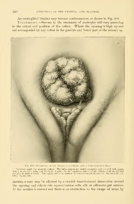
320 AFFECTIONS OF THE URETHRA AND BLADDER.
An exstropliied bladder may become carcinomatous, as shown in Fig. 204.
Treatment.—Success in the treatment of exstrophy will vary accordino;
to the extent and position of the defect. Where the opening is high up and
not accompanied l)y any defect in the genitals and lower part of the urinary ap-
Fir,. 204.
—
Exstrophy of the Bladder convekted into a Carcinomatous Mass.
Catheters mark the ureteral oritices. The labia majora are widely separated and covered witli sjuirse
hairs; between the labia and below the bladder lie the separated halves of the clitoris, with tlie dividednymphic to right and left. The vaginal orifice is marked by a transverse slit between the halves of the cli-
toris. Ye nat. size.
])aratu8, a cure may be effected by a careful fumK'l-sha])ed denudation around
the opening and side-to-side approximation with silk or silkworm-gut sutures.
If the urethra is normal and there is no obstruction to the escape of urine by

EXSTROPHY OF THE BLADDER. 321
this avenue, tliis simple plastic operation, analogous to that done for vesico-
vaginal fistula, ought always to succeed. Where the defect is extensive and a
urethra is absent a complete cure is unattainable. The best that can be done is
to cover and protect the raw mucous surfaces with flaps from the neighbor-
ing skin, reducing at the same time the size of the orifice through which the
urine discharges. The operator would better avoid turning the skin surface in,
on account of the urinary incrustations which are likely to form on the hairs
and keep up a constant irritation. Where a urethra is wanting, no satisfactory
substitute for its function can be formed.
For closing in the defect in the abdominal wall the following plans have been
successfully tried
:
First, by taking three skin flaps from the sides of the opening, one above,
and one from each side, leaving them attached by a broad pedicle ; the flaps
must be large enough to allow for a decided subsequent contraction. Theyare brought across the orifice and sewed together accurately, closing the defect.
Billroth's plan of treating exstrophy is to loosen up two broad lateral flaps
left attached both above and below. These flaps are dissected loose by cut-
ting down to the fi])rous aponeurosis overlying the recti, so as to be sure to
have enough thickness of tissue to preserve their vitality ; then, in about twoweeks, when the under surface is freely granulating, they are drawn together
and united in the median line over the bladder. If the flaps are made broad
enough, it is, as a rule, not necessary to close the openings left at the side, for in
five or six weeks they will close of themselves. No attempt is made to close
the fistula left above at the navel until after the artificial urethra has been made
;
then the umbilical fistula is closed by denudation and suture.
Displacements and Alterations in Form and Capacityof the Bladder .—The bladder in women is liable to a remarkable series
of peculiar displacements and alterations of fonn in its effort to carry out the
function of a urinary reservoir in spite of a variety of hindrances.
In determining the existence and extent of such abnormalities, the normal
conditions must be borne in mind as the sole standard of comparison, and it
must not be forgotten that while the male bladder is more or less spherical and
has its greatest diameter in the antero-posterior direction, the greatest diameter
in the female bladder in moderate distention is transverse, owing to the in-
creased resistance to its expansion backward furnished by the uterus and broad
ligaments.
The uterus lying in normal anteposition forms an indentation in the median
line of the distended bladder, which can be touched and recognized by a sound
introduced through the urethra. The physiological peculiarity in the form of
the female bladder disappears after the removal of the uterus, and the male
type is assumed with its greatest expansion from before backward.
In marked distention the female bladder rises into the abdomen and has its
greatest diameter vertically, when the vault of the bladder may even reach the
uml)ilicus and the distended organ appear like a large monocystic tumor spring-
ing from the pelvas. A case of this kind was brought several hundred miles to
24

322 AFFECTIONS OF THE UKETHHA AND BLADDER.
Bee me, expecting an operation for an abdominal tumor. The tnmor collapsed
as soon as a catheter was introduced, and a large basin of ammoniacal urine was
withdrawn.
In another case, by palpation, percussion, and bimanual examination, a mono-
cystic pelvic tumor, rising well up into the abdomen, had been demonstrated,
and the patient was brought under anesthesia for operation. Upon passing a
catheter, a large amount of limpid urine was discharged and the tumor immedi-
ately collapsed.
The distention of the bladder may also take place markedly to the right or
to the left side, giving it a gibbous form and making it more liable to be
mistaken for a fluctuating tumor connected with the broad ligament. This ob-
liquity of form can be easily demonstrated by passing a sound, which goes in 10
or 11 centimeters outward and backward on one side and but 6 or 7 centi-
meters on the other. These lateral obliquities are produced by any obstacle
to expansion, such as an inflammatory mass or a tumor fixing one broad
ligament.
An upward displacement of the bladder not associated with distention is
noted in numerous cases in which a large uterus fills the pelvis and the lower
abdomen. The most frequent cause of this form of displacement is a fibroid
uterus in which both the cervical and fundal ends are involved ; the top of the
bladder may even come to lie on a level with the umbilicus flattened out on the
anterior face of the tumor mass. The simple choking of the pelvis by a mass
is sufficient to force the distending bladder up into the abdomen. Among a
large number of such cases of upward distention I have seen but one where
there was a great hypertrophy of the bladder walls. In cases of upward dis-
placement a moderate amount of fluid in the bladder is often visible to the eye,
formino- a cushiony prominence on the tumor above the symphysis, fluctuating
on palpation.
Downward displacement of the bladder is found in cases with a weak jjelvic
floor, with relaxed outlet, or where the intra-abdominal pressure is excessive.
This displacement is also found in extreme prolapse of the rectum, drawing
the posterior vaginal wall well into the sac, and dragging the uterus and the
anterior vaginal wall down witb it. Where there is a gaping vaginal outlet,
the base of the bladder pouts into it as a soft, round, ovoid prominence, yield-
ing to touch, and easily displaced by pressure ; the swelling diminishes when
the bladder is emptied and returns again as soon as it is distended with urine,
or when the patient stands on her feet and the intra-abdominal pressure is.
exerted. This form of displacement is appropriately called " a cystocele."
Pari jxissu with the displacement of a prolapsed uterus, that part of the
bladder wliich is attached to the anterior vaginal wall and the lower part of the
uterus is likewise displaced, escaping with the vagina and the uterus outside of
the pelvis (see Chapter XV). A part of the bladder remains within the abdo-
men and a part in the prolapsed sac ; in this way the organ assumes the form
of an hourglass. The entire bladder is rarely found within the prolapsed sac,
and even then any marked degree of distention nnist take place into the pelvis.

p:x.stkophy of the bladder. 323
In rare cases the bladder becomes completely detached from the vaginal wall
and uterus in prolapsus and remains entirely inside the pelvis.
I reported a case of this kind {Johns Hopk. Hasp. Rep. on Gijnecologij, vol.
ii, 1892, p. 311) where a large prolapsed sac lay between the thighs, and in front
of and behind the uterus were masses of intestmes (anterior and posteriorenterocele), while the bladder, completely detached from its vaginal anduterine detachments, lay within the pelvis.
E version of the bladder through a dilated urethra is the mostunusual form of displacement. In eversion both mucous and muscular layers
are involved, and the tumor appears between the labia as an ovoid red masscovered with furrows. A careful examination of the under surface may showthe presence of the ureteral orifices. The causes of the eversion are an in-
creased intra-abdominal pressure, associated sinmltaneously with a relaxation of
the bladder wall and dilatation of the internal urethral orifice. That part of
the bladder which hes o])posite to the internal urethral orifice, the posterior
pole, is first engaged, and, under the influence of straining efforts, forced downthrough the urethra, dragging more and more of the viscus with it until the
vvdiole organ is turned inside out. Eversion is observed oftener in young chil-
dren and in the aged.
The bladder may also be displaced by being drawn into the inguinaland femoral canals, and even through the foramen ovale with herniee.
Diagnosis .—The diagnosis of the form of displacement of the bladder in
any given case is not difficult. After distention with fluid a bimanual palpa-
tion will outline the different parts, and by means of a graduated sound intro-
duced per urethram, measurements made in various directions will determine
the exact form.
The diagnosis of an eversion of the vesical nmcosa must be made under
anesthesia by carefully examining the tumor protruding from the dilated
urethra, and if a sound is passed through the urethral canal, the bladder cavity
is found to be absent and the pedicle of the tumor at the internal urethral
orifice. On pushing back this mass, the bladder cavity is restored, and, if the
urethra is sufliciently dilated, by introducing a finger, the absence of any tumor
is demonstrated.
Treatment.—The treatment of the various displacements of the bladder
often resolves itself into the treatment of the associated conditions which have
caused the displacement. By removing ovarian tumors choking the pelvis, and
inflammatory masses lateral to the uterus, the free distention of the bladder
within the pelvis once more becomes possible. By removing a fibroid uterus
the bladder is let down to its normal pelvic position.
Other displacements are treated by repairing the relaxed vaginal outlet so
as to lift up the pelvic floor and give an adequate support to the anterior
vaginal wall ; I rarely find it necessary, as was the custom ten years ago, to
operate upon the cystocele itself. In prolapse of the uterus the bladder is
restored to its normal position by the operations upon the uterus and the pelvic
floor, retaining the uterus in its normal position.

324: AFFECTIONS OF THE URETHRA AND BLADDER.
In treating eversion we must, in tlie first place, put the bladder back into its
normal place bj elevating the pelvis in the knee-breast posture ; with gentle
compression and manipulation the tumor may now be forced back into the pel-
vis. The patient should then be kept in bed, with the foot of the bed ele-
vated, to reduce the pressure on the pelvic viscera. If the displacement per-
sists in returning, a plastic operation may be performed, narrowing the
urethra by placing a catheter in its canal as a guide for the size of a new ure-
thra to be formed, and then excising a wedge-shaped piece with its base on
the vaginal surface extending through to the urethral mucosa. The denuded
surfaces are then brought together by interrupted sutures passed from side
to side.
Foreign Bodies in the Bladder. — A variety of foreign bodies are found in the
bladder. They either form in the bladder itself, as in the case of vesical calculi,
or they may reach the bladder from the exterior, either by perforating its walls,
or by descending a ureter into the bladder, or by being introduced through the
urethra.
The commonest foreign bodies are calculi, formed of incrustations of phos-
phates and urates. Small oxalic acid and uric acid calculi may descend from the
kidney and lodge in the bladder, and grow there to a lai-ge size by the accretion
of phosphates and urates.
Foreign bodies may also enter the bladder from the side of the peritoneum,
the tubes, or the ovaries, as well as from the vagina. In this way echinococci
have ruptured into its cavity ; silk ligatures about the jDedicle of an ovarian
tumor have ulcerated through its walls ; dermoid cysts have opened and dis-
charged quantities of hair by the bladder ; and the bones of an extra-uterine
fetus have also found an exit in the same way. The commonest foreign l)ody
which makes its way into the bladder from the vagina is a pessary, usually of
large size, which has ulcerated through the vesico- vaginal septum.
By the urethra a large variety of foreign bodies have been introduced ; these
are usually several inches long, and of a caliber somewhat smaller than the
urethra. The commonest object is a bit of a catheter broken off or an entire
catheter which has slipped out of the fingers and so entered. Other articles
which have been found have been introduced by the patient herself, such as
hairpins, toothpicks, crochet needles, etc.
The symptoms produced are at first those of irritation of the l)ladder, fol-
lowed later by inflammation.
The patient complains of a suprapul)ic pain and a frequent desire to urinate;
the urine becomes cloudy, and pus soon appears. In a few weeks the foreign
body l)ecomes incrusted with urine salts, and the symptoms of cystitis become
more urgent and the distress increases.
A small foreign body, such as a renal calculus, may only lodge tenqiorarily,
and with its spontaneous escape by the urethra the symptoms cease.
Bodies of an elongate form will, if large enough to put the bladder walls on
the stretch, ulcerate throygh either into the vagina or into the })eritonenm, in
the latter case producing a rapidly fatal peritonitis.
FOIiEIGN BODIES IN THE BLADDER. 325
Diagnosis.—The presence of a foreign body in the bladder may be de-
termined either by touch or by inspection. If the body is long, or is of large
size, it may often be easily felt bimanually by palpating the emptied bladder
between two fingers in the vagina and the hand pressing down over the sym-physis. Upon passing a sound into the bladder the presence of the foreign bodymay also be demonstrated by the sense of contact with a hard body, as well as
by the audible click produced upon striking it.
The simplest and surest way to make a diagnosis is by inspection. The pa-
tient is put in the knee-breast position and the vesical speculum introduced, andthe bladder, distended with air, is then easily examined in all its parts as already
described. If there is any foreign body present which is not wedged in betweenthe bladder walls it will drop into the most dependent part, where it is mosteasily seen. By means of inspection the diagnosis of the absence or presence
of a foreign body can be made with certainty; by this means also its form and
position are noted, together with any alterations produced in the bladder walls
by its presence.
Treatment .—The treatment in every case is directed to the speedy re-
moval of the foreign body. There are three ways of doing this : (1) Through the
intact urethra, (2) through an incision in the bladder walls made through the
vagina, and (3) through a suprapubic incision.
1. A small foreign body not more tlian 10 or 15 millimeters in diameter
and a long narrow body, such as a needle or a glass catheter, may be removedthrough the vesical speculum. The bladder being empty, the speculum is in-
troduced in the knee-breast position, and the object exposed. If it is a small
round object it may be picked up by the mouse-toothed forceps and simply
lifted out through the speculum, or it may be caught in a scoop and held against
the end of the speculum, and withdrawn together with the speculum.
A long body like a glass catheter may be removed by introducing a
straight instrument, such as a searcher, into its open end, and then manipulating
the end of the speculum until the catheter slips into it ; then by pushing the
speculum well down on the catheter engaged in this way, its end can be easily
caught and withdrawn.
The older writers were wont to try to deliver a calculus through the urethra
by catching it between the fingers of one hand in the vagina, or in a virgin in
the rectum, and the other hand pressing down above the symphysis, and so
forcing it into the urethra and on out.
I succeeded in July, 1895, in removing a glass catheter in this way. Thepatient was a young woman, about twenty-three years old, with a spherical myo-matous uterus filling the pelvis and reaching up to the umbilicus. She hadbeen suffering from retention of urine, and as her physician introduced a glass
catheter 13 centimeters (5 inches) long into the overdistended bladder, it slipped
out of his fingers and was lost in the bladder. When I examined her I foundthe myoma and a long rigid body in front of it, with its blunt end projecting
into the anterior vaginal wall to the right and its upper rounded end pressing
upward directly under the anterior abdominal wall, 4 centimeters below the um-
,32f) AFFECTIONS OF THP] IKKTHRA AND JiLADDKH.
LilicLis. The hymen was relaxed, so that by careful inanij)ulation with two
fingers in the vagina I was al)le to push up the lower end of the catheter while
pushing the upper end to the right ; by doing this I brought tlie end into the
urethra, when it descended at once, and escaped with a quantity of bloody urine.
The patient suffered no further inconvenience from its twenty-four hours' stay
in her bladder.
Bodies from 10 to 20 millimeters in diameter maj be removed through the
urethra after dilating it. Simon has shown that even after a dilatation of 20
centimeters incontinence does not occur if it is carefully done. Two postero-
lateral incisions, 2 or 3 millimeters deep, must be made into the external urethral
orifice to avoid tearing it when the dilatation is carried up to 20 millimetei-s or
near it ; the rest of the urethra, which is more elastic, is then enlarged by a
series of successive dilators up to the required size, and the foreign object is
either removed through one of the larger specula or grasped by a pair of small
stone forceps introduced through the urethra and so withdrawn. The lateral
incisions in the urethral orifice are then closed with fine catgut sutures.
Calculi, hke other foreign bodies, may be removed either (1) by the urethra,
(2) by vaginal incision, (3) by suprapubic incision, or (4) by crushing with the
lithotrite.
Stones from 2 to 3 centimeters in diameter should be crushed by means of
an instrument introduced through the urethra. If the bladder is first moder-
ately distended with water and a lithotrite inserted, the stone is readily caught
in the open beak of the instrument and l)roken up, and the pieces afterward re-
moved through a speculum from the bladder distended with air.
Dr. H. J. Bigelow's apparatus for litholapaxy, which has served so Avell in
men to reduce the number of cutting operations, by both crushing and washing
out the bits of stone, is also available and even easier of application in women,
although it has never been widely used on account of the great simplicity of the
older operation through the short urethra.
Dr. E. T. Caswell, of Providence, K. I., reported a case (Merl. iWw.s*,
Aug. 2B, 1882) in which he crushed a phosphatic stone weighing, when dried,
100 grains. He used a modified Thompson's fenestrated lithotrite, and washed
the fragments out through a straight tube (28 French).
Dr. D. F. Keegan {Zajicet, Jan. 9, 1897) in an instructive article on
Litholapaxy in Girls and Women, reports eighteen cases of calculi occurring
in women, where he used the lithotrite with success. The calculi were of dif-
ferent varieties—namely, phosphatic, oxalate of lime, and uric acid ; the smallest
weighed 72 grains and the largest 702 grains. The average length of stay in
the hospital was only 5' 3 days.
Although nature and art have succeeded in removing stones of large size by
the urethra without any diminution of their volume, this is a hazardous pro-
cedure, and ought not to be imitated, on account of the imminent j-isk of a
permanent incontinence following.
An extreme case of this sort is reported by Dr. Alex. Duidap, of Sju-ingfield,
Ohio {Ainer. Jour, of Obst., vol. xiv, p. 853) ; upon examining the patient, who

FOREIGX BODIES IX THE BLADDER. 327
was twentj-eiglit years old, he found a large stone 2 by 2^ by 1^ inches in diam-
eter in the bladder ; she wonld not allow any cutting operation to be done, so
he caught the stone with a pair of forceps introduced through the urethra,
and delivered it slowly by traction. The external meatus was the most re-
sistant portion, but he succeeded in working it through in about three quarters
of an hour, by pressing back the tissues over the stone much as an obstetrician
may try to help the perineum back over the advancing head of the child. Thestone was rough on one side and tore the mucous membrane of the canal
considerably ; this produced a sharp venous hemorrhage of short duration. Inspite of this enormous dilatation, she suffered no serious inconvenience, although
unable to hold her water as long as before.
2. The vaginal incision is to be preferred for stones which are so large that
they can not safely be removed through the urethra, and is adapted to all but
the largest calculi.
The operation may best be conducted with the patient lying in the left semi-
prone position, with the posterior vaginal wall well retracted, so as to expose
clearly the entire extent of the anterior wall from cervix to urethra. A blunt
instrument like a male sound is now introduced through the urethra into the
bladder, and the vaginal wall is pushed forward in the median line and cut
through, opening the bladder ; the incision is now extended by drawing apart
the edges of the wound and cutting back toward the cervix and forward toward
the neck of the bladder until it is large enough to permit the introduction of
the blades of a pair of stone forceps, which are used to grasp the stone byits smallest diameter and draw it out through the wound endwise, without lacer-
ation of the tissues.
The incision must then be accurately closed with interrupted sutures either
of fine silk or silkworm gut. If the bladder is then drained for five or six days
the clean-cut vaginal wound ought to heal promptly, leaving no fistula behind.
It is best to close the wound at once in this way, although even large woundsmay heal spontaneously. Such a case is the one of Dr. F. R. Eccles, of Lon-
don, Ontario, figured in the text. The patient introduced a hairpin into the
urethra and it escaped into the bladder. She married soon after, and at her
confinement a large foreign body was felt in the way of the head as it descended;
it was pushed up, however, and the labor proceeded normally. Dr. Eccles was
called in later and removed the hairpin, incrusted with large, fused twin calculi
through an incision in the anterior vaginal wall. No sutures were used to close
the wound, which healed spontaneously within five weeks.
3. The suprapubic operation for the removal of calculi {sectio alta) is best
adapted to those of the largest size, filling the bladder. It is especially suitable
for children, where the vaginal route is not available.
After distending the bladder with water, a vertical incision 6 to 8 centi-
meters long is made in the middle line just above the symphysis, separating the
recti and the pyramidales muscles, and pushing aside the fat underlying them,
but taking care not to cut the peritoneum. In this way the bladder is exposed
and its wall cut through vertically and the stone extracted. The incision in the

528 AFFECTIONS OF THE URETHKA AND BLADDER.
bladder wall is now closed with fine interrupted catgut sutures placed close
together and embracing the entire thickness of the wall down to but not in-
cluding the mucosa. The incision in the abdominal wall is then closed with
buried silver wire for the nmscles and
fascia and catgut for the skin. Then, if
the bladder is kept well drained for a
week, the wound in its vault will heal by
first intention.
The stone which is shown in the figure
was removed from a little girl only eight
years old by Dr. F. R. Eccles by the su-
23rapubic operation ; the wound, which was
not closed completely on account of the
unhealthy condition of the vesical mucosa,
healed spontaneously in four weeks.
Vesical Fistulae.—Vesical fistulse are ab-
normal channels of communication between
the bladder and contiguous or adjacent or-
gans ; they are found, for example, (1) be-
tween the bladder and the vagina, (2) l)e-
tvveen the l)ladder and the uterus, and (8)
between the bladder and some portion of
the intestinal tract.
History.— It is a remarkable fact
that no clear refei'ences to these commonand distressing disorders are found in the
earliest writers preceding the Christian
era, and for nearly sixteen hundred years
afterward. Toward the end of the six-
teenth century and early in the seven-
teenth several clear descriptions appear almost simultaneously in the writings of
Luiz de Mercado, a Spanish physician (1520-1600). Fehx Plater (Basel, 1536-
1611), and Severin Pineau (born in Chartres in the middle of the sixteenth
century).
Plater (see I. Spach, Gijixvc. etc., Argent., 1597) gives two clear descriptions
of fistulse following diflicult childbirths ; the second case is appropriately entitled
vesiecB cervicis alia ritptiira in partu. " As a serpience of a diflicult first
labor, a young country girl had the opening of the bladder rent to such a degree
that there was a long gaping furrow in its place, and the open bladder could
be seen. I have twice inspected it myself, and discovered that it was so byusing a probe. On account of this injury there is a constant involuntary dis-
charge of urine, and the surrounding parts become excoriated and inflamed."
H. van Poonhuysen (16()3) first proposed the closure of such a fistula by
suture.
J. Fatio ( Wehe-mutter\ Basel, 1752, p. 284) gives an admirable description
Fig. 205.
—
Phosphatio Calculus formed uponA IlAUtPIN IN THE RlADDER.

VESICAL FISTUL.^. 329
of the way in wliieh lie carried this proposal out in 1675 and 108-1 by placing
bis patients in tbe litbotomy position and exposing tbe fistula witb a speculum
;
be then freshened its margins witb a delicate pair of scissors, and brought tbe
edges together by passing a sharpened quill through them and winding a
thread over the ends of tbe quill to keep it from coming out. Both cases
recovered.
A. J. Jobert de Lamballe {Cwmjjtes rend, de VAcad, des sci.. 1850, and
Traits des jistules, Paris, 1852) was the first operator who systematically took
hold of this perplexing question and treated a large number of cases, many of
them successfully. His plan of treatment for the simpler cases was to bring
the cervix of the uterus down by traction with forceps so as to expose the
fistula, followed by a broad denudation of the edges of the fistula and their
exact approximation by interrupted sutures. When the fistula was a large one,
and the approximation difficult on account of the tension, he made incisions in
tbe lateral vaginal walls parallel to the edges of the fistula, so as to permit the
tissues to be drawn together {"^jjar glisseuient^''). An incision through the
vaginal vault detaching the cervix for this purpose has since been known as the
incision of Jobert.
G. Simon(JJeher die Heilung der Blasenscheiderifisteln, Giessen, 1854), did
away with these lateral incisions, and substituted in their place a method with
which his name is still connected, the use of a double set of sutures, one intro-
duced at a distance from the wound for the relief of tension (sutures of deten-
tion), the other to secure accurate approximation (sutures of reunion).
J. Marion Sims {On the Treatment of Yedco-vaginal Fistula, Amer. Jour.
of Med. Sci., 1852, vol. xxiii, p. 59), working independently in America,
accomplished three things : (1) He devised the duck-bill speculum for the ex-
posure of the fistula witb the patient lying in the
left semi-prone position; (2) he clearly described the
best method of denuding the margins in a funnel
form and down to but not including the vesical
mucosa ; and (3) he sutured the edges of the wound
accurately together with the non irritating (antisep-
tic ?) silver wire, and by this means, coupled with
his great skill as an operator, he attained a degree of
success in the treatment of these cases never before
reached.
T. A. Emmet ( Vesico-vaqinal Fistula, etc., New _ „^,. ^ „^
, ,
"^.
' , Fig. 206.—Sectiox of a ^ esical
York, 1868, and Principles and Practice of Gyne- Calculus of the Bladder,
7 -Dl -1 1D^A\ J 1VT xl -D t rrr NATURAL SiZE, REMOVED FROMCology, rblla., 1879) and JNathan J3ozeman {Ihe a Girl Eight Years Old.
Gradual Prejxiratory Treatment of the Complica-
tions of Urinary and Fecal Fistulce in Women, New York Jour. Med. Sciences,
October 1, 1887) developed the method of treating large and complicated fis-
tulce by gradual preparatory treatment, incising the bands of scar tissue, and
softening them by pressure so as to make the vaginal walls supple enough to be
drawn together.

330 AFFECTIOXS OF THE URETHRA AXD BLADDER.
In spite of the advances made by these great surgeons, a large num])er of
intractable cases remained.
Colpocleisis, or a surgical closure of the vagina so as to make a common pouch
out of the vagina and the bladder, was performed in such cases by Simon in
1855, and has been practiced more or less ever since. The great advances whicli
have been made recently can best be signalized by citing the eight indications
for colpocleisis accepted by Simon, with the remark that not one of them holds
good to-day. They were :
(a) An extensive loss of tissue, rendering it impossible to approximate the
margins of the fistula.
(b) Inaccessible fistula.
(c) Destruction of the uterine cervix, bringing the peritoneum dangerously
near the seat of operation.
(d) Severe hemorrhage into the bladder after an operation.
(e) Incarceration of the cervix uteri in the bladder.
(f) Atresia of the vagina above the fistula.
(g) Atresia of the urethra, with a fistula above and below it.
(h) Uretero-vaginal and uretero-utero-vaginal fistula.
The first active steps taken in an entirely new direction, with the object in
view of relieving these cases without resorting to a procedure involving so muchmutilation as does colpoclei-
sis, were those of Rydygier
{Bed. I'lin. Wochenschr.,
1887, Xo. 31) and of A. Mar-
tin, of Berlin {Zeit. f. Gyu.
nnd Geh.^ 1891), who planned
to cover in the defect with
large flaps dissected up from
the contiguous vaginal walls.
L. von Dittel {Ahdom.
Blasenscheidentfisteln Oper-
ation, Wien. med. Woch.,
1893, N"o. 25) made a radical
departure from all precedent
by opening the abdomen and
detaching the bladder from
the uterus, and so exposing
the fistula, which was then
sewed up; the sutures only
included the bladder walls,
and after closing the opening in this way, the vesieo-uterine })eritoneum wasagain united and the abdomen closed.
A. Mackenrodt, of Berlin {Centralh. f. Gyn., 1894, No. 8), adopted a some-what similar plan, but one making a definite advan(;e on the preceding, in that
he operates through the vagina and detaches the bladder on all sides from the
;. 207.—Treatment of Vesico-uterine Fistl'la byPUBIC Incision, (v. Dittel. i
The ve.sico-uterine peritoneum (P-P) is divided, tlic listul
)iu the uterus, and its margins united by suturu.<1 tv

VESICAL FISTUL/E. 331
fistula, and then sews the hladder np independently and closes the opening in
the vagina bj drawing its sides together, if possible ; if he can not do this, he
uses the anterior face of the uterus to fill out the defect. With the exception of
this last step, the important outhnes of this operation were already defined bySanger ( Volk. Samm. Uiu. Vort., No. 301) and Walcher {Cenfr.f. Gyn., 1894,
P-1)-AV. A. Freund {Eine yieue Oper. z. schliessung gewisser Harnfistehi heirrh
Weihe., Samm. klin. Vo/i., N. F., 1895, No. 118) again operated in a radically
different direction when he
used the body of the in-
verted uterus, 1)ringing it
into the vagina through the
])osterior fornix, to close a
large defect in the vesico-
vaginal septum.
Dr. E. C. Dudley, of
Chicago, succeeded in clos-
ing a large intractable fistula
by making a semicircular
denudation inside the blad-
der on its mucous surface,
extending from one margin
of the fistula around to the
other ; he then sutui-ed this
denuded surface to the an-
terior part of the fistula,
and so obtained a closure.
My own plan {Johns
Hopk. Hosj). Bull., Feb., 189G) is to split the margin of the large fistula pos-
teriorly, separating the bladder wall from the vagina, and then to denude its
anterior margin on the vaginal surface, and to suture the movable posterior
bladder wall to the fixed anterior vaginal wall.
Causes .—Vesico-vaginal fistulse are commonly caused by the traumatism
of a difiicult labor, resulting from the impaction of the child's head in a narrow
])elvis. In consequence of the prolonged pressure, the vitality of the vesico-
\'aginal septum is destroyed at the point at which it is compressed between the
head and the symphysis pubis ; in a few days a slough has formed, and the piece
of tissue drops out, leaving an opening between the bladder and the vagina. I
have rejjeatedly made pelvic measurements in these cases, and rai*ely found a
fistula following parturition which was not in a contracted pelvis. (See Dr. G.
W. Dobbin, The Use of Pelmmetry in Gynecology, Amer. Jour. Ohst., August,
1895, p. 201.)
The impression which has prevailed in the profession that these fistulas are
often due to the use of the oljstetric forcejDS is erroneous, for they are undoubt-
edly due not to the use of the forceps, but to too long a delay in using them.
Fig. '208.
—
Supkafubk: Operation for Vesico-lterine Fistcla.{\. DiTTEL.)
The operation completed, the bladder and the peritoneum sutured.
332 AFFECTIONS OF THE URETHRA AND BLADDER.
This point was insisted npon by W. T. Schmidt in 1828 (v. Siebold's Jour. f.
Gel), and Fniueaz. and KinderkranhheUeti, Bd. vii, p. 330), and in unr own
day by T. A. Emmet, of New York.
Other causes are foreign bodies, such as stem pessaries, working their way
from the vagina into the bladder, or 'vioe versa (Figs. 168 and ITO) ; syphilis
;
a cancer extending from the cervix uteri will often destroy the anterior
vaginal wall and so create a iistula ; the perforation produced by a pistol ball
(Emmet); hematoma of the septum from coitus, followed by sloughing; the
wounds of a vaginal hysterectomy are also to-day a frequent source of vesico-
vaginal fistula.
The course of a fistula uninterfered with is toward closure,
either by primary union or by granulation, cicatrization, and contraction of its
edges. In this way, by cicatrization, a small fistula will usually close entirely in
a few weeks' time, and large ones will be reduced to one half or one third their
original size. A clean-cut opening, such as that made for the extraction of
a stone, may possibly close of itself without any suture, even if it is a large
one. In time the margins of a large fistula grow sharp and hard with cica-
tricial tissue, and in bad cases the cicatrices radiate out over the vaginal walls
or pin the fistula down to a pubic ramus. The posterior walls of the vagina
may also be involved so as to close the vagina so tight that it is difficult to see
the fistula.
Although the tendency of the smaller fistulie is always toward a spontaneous
cure, in some instances a minute opening may persist for many years. I oper-
ated, for example, upon a patient who had had a fistula for twenty-three years,
and the opening was not much larger than a hair, and yet large enough to per-
mit the constant escape of urine into the vagina with all its disagreeable conse-
quences.
Symptoms.—The symptoms produced by vesico-vaginal fistulge are quite
characteristic. Soon after the confinement which causes it there may be bloody
urine, difliculty in urination with symptoms of cystitis, and marked febrile
disturbances, followed in a week or more by the escape of a slough, after
which the urine, instead of accumulating in the bladder, escapes at once
through the opening into the vagina, and so out over the vulva, perineum,
and adjacent parts, which are constantly kept wet. The effect of this upon
the skin is to produce a painful dermatitis and excoriations, and the vulvar
hairs often become incrusted with the urine salts. Areas of excoriation are
also found within the vagina, often coated with sabulous material and incrusta-
tions. The parts involved may be so exquisitely tender that the slightest mo\'e-
ment is painful, and anything like a thorough examination is often impossible
without anesthesia.
If the fistula is a small one, the patient may in certiiiu positions retain a con-
siderable amount of her urine and void it naturally. If the vaginal oiitlet is
not broken down, considerable urine may accumulate within the vagina in the
recumbent posture to escape on rising ; this often leads the ])atient into the
erroneous idea that she holds the urine in the bladder while lying and passes it

VESICAL FISTUL.E. 333
naturally afterwards ; one of my patients was al)le to hold even as much as 3U0
cubic centimeters of urine in this way.
The effect of a fistula on the patient's general health is often most marked
;
the local discomforts compel her to remain pretty constantly in oue place and in
one position, preventing her from getting exercise and fresh air ; nutrition fails,
she becomes emaciated, excessively constipated, depressed, and peevish, and has
a cachectic appearance.
In spite of the obstacles rendering conception rare, it has occurred. In oue
of my own cases, the patient, having a fistula 1-5 centimeter in diameter just
back of the neck of the bladder, conceived, and passed through a natural labor
and a normal puerperum, after which the fistula was operated upon and cured.
In a case in the hands of L. Winckel {llandb. der Frauenl'rank., vol. iii, p.
441) a patient with a fistula became pregnant and passed through her confiue-
ment at tei'm, after which the fistula actually healed spontaneously.
Diagnosis .—In making a diagnosis of a vesical fistula, the examiner
must investigate all the associated conditions which tend to complicate the case.
In doing this he will not only note the size, the form, and the exact site of the
fistula, but will also carefully inquire into the condition of the surrounding vag-
inal walls : whether soft and yielding or fixed by scar tissue, whether the an-
terior lip of the cervix is involved in the fistula (cervico-vesico-vaginal), whether
the neck of the bladder is included (vesico-urethro-vaginal), and whether the
fistula is fixed to one or the other pubic ramus. It is most important also to
note the position of the ureteral orifices in their relation to the edges of the
fistula. Other complications which may occur are the existence of two vesico-
vaginal fistulse, or of a vesico-vaginal fistula and a urethro-vaginal or a vesico-
uterine fistula existing together. I have seen one case of vesico-vaginal fistula
following a severe labor complicated by a recto-vaginal fistula, and an atresia
of the upper vagina with hematometra. In another case with a vesico-vaginal
fistula there was also a complete rupture of the recto-vaginal septum (see F.
Plater's first case in I. Spach's Gynm'. Lihri, Argent., 1597, p. 23, index). In
still another instance in my hands a large vesico-vaginal fistula, adhering to the
pubic ramus, was associated with a wide separation of the symphysis pubis rup-
tured in a badly managed forceps labor.
The diagnosis of a vesical fistula is made by a consideration of the history,
by touch, and by inspection.
The patient gives a history of a constant discharge of urine over her person,
dating usually from a severe confinement or from a hysterectomy ; if, in spite
of the fact that she has this constant flow, she also passes water at regular in-
tervals, the probable diagnosis will then be one of ureteral and not of vesical
fistula.
By touch the examiner will often feel more or less scar tissue in the vagina
and a large hole in the anterior vaginal wall, which may be filled with the soft
prolapsing mucous membrane of tlie bladder, and the finger can be introduced
through this hole into the bladder and carried forward so as to feel the internal
orifice of the urethra.

334 AFFECTIONS OF THE UKETHRA AND BLADDER.
Inspection affords the fullest information about the fistula and the associated
conditions. To make a vaginal inspection the posterior vaginal wall must be
drawn back and the anterior wall exposed. A large fistula is seen as soon as
the accumulated urine is dried out of the vagina ; to find a smaller one it
may be necessary to hunt among the vaginal folds, when it will often be found
near the vault and to one side of the cervix or the other. A vesico-uterine
fistula gives evidence of its presence by the urine whicli escapes from the cervix
uteri.
When the fistula can not be found in this way it will usually be detected by
injecting the bladder with an aniline solution or with sterilized milk, and then
watching to see at what point the colored fluid runs out. If a fine sound is
carried into the bladder through the urethra its end can usually be brought out
through the smallest fistula.
The cystoscope may also be used to examine the fistula from the vesical
side, but this is not so easy as the vaginal examination, because the floor of the
bladder lies almost in the plane of vision, and the hole in it with its inverted
mucous membrane is seen so foreshortened that it may easily escape notice
altogether. It is therefore necessary, in order to get a good view of it, to
lift the floor up on the end of the speculum, so as to bring it across the plane
of vision.
Treatment.—In describing the various modes of treatment, I shall con-
sider vesical fistulse under the following heads :
1. Vesico-vaginal fistula.
2. Vesico-utero-vaginal fistula.
3. Vesico-uterine fistula.
4. Entero-vesical fistula.
When the injury occurs to which the fistula owes its origin, the physician is
not as a rule aware of the nature of the accident until the slough comes away
;
then the constant involuntary escape of the urine signalizes what has happened.
It may be, however, that the urine will begin to escape some days before the
sloughing is complete, when the examining finger detects a soft, crackling mass
in front of the cervix ; it is important at this time to begin at once the use of
mildly antiseptic vaginal douches several times daily, to prevent the accumula-
tion of fetid discharges in the vagina, and to keep the wound as clean as possil)le.
The convalescence will be hastened if the slough is exposed and caught with
forceps and the dead tissue cut away ; small particles which still adhere to the
edges of the wound will then soon detach themselves and leave a clean, granu-
lating surface.
Mild boric acid or mild carbolic acid douches should now be kept uj) until
the fistula is healed either spontaneously or by operation. A s p o n t a n e o n s
cure may reasonably be expected only in the case of small fistuUi? 1 or 2
centimeters in diameter and may be awaited as long as the wound shows signs
of contracting ; such a closure may take place in from two to four months, and
in exceptional cases after six or eight months.
The efforts made by our predecessors to bring about a cure by simple [)os-

VESICAL FISTUL.E. 335
ture, or bv putting a catheter in the bladder through the urethra, or by placing
pledgets of cotton in the vagina, can not be recommended with any assurance of
their utility ; such measures belong rather to the days when local treatment in
gynecology was universal.
Cauterization was at one time extensively employed, and many cures
were made in the case of smaller fistulse. The edges of the sound were treated
with the nitrate of silver stick, Vienna paste, caustic potash, tincture of cantliar-
ides, or the hot iron, and later with the Paquelin cautery.
The cautery is applied to the edges of the fistula on the vaginal surface so as
to destroy the superficial tissue and provoke active granulations, which, meeting
across the opening and uniting, close it at once ; or in the case of a large opening
the further cicatricial contraction is brought about. It is, as a rule, necessary to
keep this treatment up at intervals of a week or ten days for two or three
months. Such plans of treatment have to-day almost entirely passed out of
vogue, and will only be resorted to in the early stages of the affection when it is
too soon to operate, or when for some other reason the operation can not be per-
formed.
It is a significant fact that the best results by this plan of treatment have
been reached during the early stages of the disease, at a time when the spontane-
ous cure takes place if it is going to take place at all.
() peration .—The operative treatment is as a rule the only form of treat-
ment to be considered, for in the simpler cases it is invariably successful, and in
the more complicated cases nothing short of operation will bring relief.
Preparatory treatment is necessary in most cases where the vagina
contains sloughing necrotic tissue and incrusted urine salts, and where the
contiguous parts are raw and granulating; these complications ^vill l)e re-
moved by prolonged repeated warm boric acid vaginal douches, a repeated
painstaking cleansing of vagina and vulva, using forceps and cotton to re-
move and wi]3e off sloughs and debris, followed by occasional applications of
weak solutions of the nitrate of silver to the raw surfaces. At the same time
bands of scar tissue may be incised so as to diminish the tension on the woundedges.
If a recto-vaginal fistula exists also, in order to avoid infection of the wound,
this must either be closed and healed before operating upon the vesico-vaginal
fistula, or both closed at the same time. It will be safer in most cases to close the
rectal opening first, because there is always a greater risk of this breaking down,
in which case the vesical wound would almost certainly be infected and give
way too.
The operator need not be embarrassed by finding a marked stenosis of the
vaginal orifice at the time of operation, for this may be at once extensively
divided with the knife by an incision down beside the rectum, giving all the
room necessary to get at the fistula ; and after the vesical operation is completed
the incised edges may be accurately united again.
With more recent advances made in operating on bad fistulae, we are able
to dispense with some of the elaborate time-consuming preparatory treat-

330 AFFECTIONS OF THE URETHRA AND BLADDER.
meut ill the way of incisions and vaginal dilators used to get rid of the scar
tissue.
The best time to operate is within six or eight weeks after labor,
while the tissues are soft and yielding, vascular, and free from the fixation and
the atrophy caused by scar tissue. If the fistula is a small one and its edges can
be easily drawn together with tenacula, with a little freshening of its margins
and several sutures to unite them, primary union is easily secured ; large andirregular fistulse are far more difiicult to unite. The operation becomes most
difficult after the formation of the scar tissue distorting and fixing the edges.
Even pregnancy forms no contraindication to operation, as shown by the success-
ful work of Schlesinger and others.
Instruments needed for the operation are : Speculum, lateral retractors,
tissue forceps, fistula knife, fistula scissors, tenaculum, needles, silkworm gut,
catgut, and silk.
There are, in general, as briefly indicated above, seven different ways of
closing vesico-vaginal fistula3
:
1. The classical method of denuding the margins on the vaginal surface and
uniting them by suture (Roonhuysen, Jobert, Sims, Simon).
2. Covering in the defect by flaps transplanted from the contiguous vaginal
walls (Rydygier, Martin, Trendelenburg).
3. Opening the abdomen and cutting through the vesico-uterine peritoneum,
and so detaching the bladder from the fistula, sewing up the bladder wound, and
then reuniting the peritoneum and closing the abdomen (von Dittel).
4. Denudation on the vesical mucosa from one side of the fistula around to
the other, and uniting this surface to the freshened anterior part of the fistula
(Dudley).
5. Dissecting the bladder loose from the vagina and sewing up the vesical
wound separately (Sanger, Walcher, von Winkel). The anterior face of the
uterus is used to close the vaginal defect (Mackenrodt).
6. Freeing the bladder around the posterior two thirds of the fistula, and
bringing it forward and uniting it to the anterior third, which is freshened on
its vaginal surface (Kelly).
7. The posterior fornix is opened and the body of the uterus brought
through it inverted and attached to the edges of the fistula on all sides, so closing
it (Freund).
Curing a vesico-vaginal fistula by denudation of its
margins and approximation of its edges by suture. This
is the simplest mode of treatment, and is adapted to all fistulie in which the
edges can be drawn together without much tension ; if this can be done with
tenacula beforehand, the operator may feel reasonably sure of a successful
result. The easiest fistula to* close in this way are all the small ones, and the
larger ones which are situated in the upper part of the vagina near the cervix,
where the vaginal tissue is more lax and abundant ; and the easiest large
fistulse are the transverse ones.
When the edges can not be brought easily togetlier by traction with the

VESICAL FISTUL.E. 337
tenaciila in any direction, the denudation and approximation by sutni-e maystill be tried if the operator has had such experience in plastic work that he is
able to form a good judgment as to the extent to which he will be able to
relieve the tension by lateral incisions through the scar tissue, which fixes the
edges of the wound. It is worse than useless to denude the edges of a large
fistula without having any definite idea as to what can be accomplished until
the stitches are put in and pulled upon. It would be far better to let the
patient entirely alone, and to confess honestly an inability to relieve her, than
to go on cutting away valuable tissues and increasing tlie size of the fistula
every time, with a vague idea that by some chance the
operation will succeed. I have seen several womenwho have been operated upon as many as five and six
times in this way who were nothing better, but far worse
for it.
The patient is put on the table, either in the left lat-
eral or in the lithotomy position, or with elevated hips,
after Simon, in whichever way the fistula can be exposed
best; I prefer myself, in almost all cases, to put the pa-
tient on her back.
The posterior vaginal wall is drawn strongy backward,
and lateral retractors are used on one or both sides, to give
a perfect exposure to the field of operation.
The steps of the operation are : (1) Paring the edges
of the fistula; (2) passing and tying the sutures.
The edges of the fistula should be paredon the vaginal surface entirely ; this creates a freshened
area from 5 to 6 or 8 millimeters in breadth, extending
down to but not including the mucous membrane of the
bladder.
Either a knife or scissors may be used to remove
the tissue ; a knife is necessary where there is much friable
tissue, but in most cases I prefer a delicate pair of scissors
whicli I have had made for this purpose, and I denude in
the following manner
:
With a knife I first outline the limit of the denudation
all around the fistula, and when this is done I take tlie
rat-toothed forceps and catch a piece of the tissue to be
removed, and begin cutting it off with the scissors. This
can be done rapidly, as the outer limit is marked out
by the incision with the knife, and the operator does
not have to pause to exercise his judgment about it ; in addition to this, the
sharp straight cut of the knife is better than the jagged edges made by the
scissors.
The freshening must be carried down into sound tissue, avoiding the error
of simply paring off the surface. Every particle of the tissue within the limits
25
'209. — Scissors fokPARING THE EdGES OFTHE VeSICO-VAGINALFistula.
The shanks are madelonjj and slender and theblades are delicate andcurved on the flat. % or-
dinary size.

338 AFFECTIONS OF THE UKETHRA AND BLADDER.
defined must be removed, or union will not take place ; and to make sure that
this has been done, little islets of undenuded tissue must be carefully sought out
and picked up with a tenaculum and snipped off.
Constant irrigation with a fine stream of water is the best way to
keep the field clear of blood during the cutting, but a little piece of sponge
grasped in a pair of forceps
will often be needed to makefii'm pressure on some spot
which is obscured by the free
oozing ; this blanches the tis-
sue for a second or two, and
as the bleeding begins again
the operator can see whether
there are any little unde-
nuded areas which do not
bleed. The entire wound
now has a fresh edge gently
beveled on to the vaginal
surface.
Passing and tyingthe sutures is the next
step. To do this I use the
ordinary needle holder and
small curved needles armed
with a carrier made of fine
silk.
A tenaculum is often
needed to steady the tissues
while passing the needle
through them, and to catch
and hold the point of the
needle as soon as it emerges, until it can be grasped by the needle holder
again and drawn completely through.
By simply following the direction of least resistance in passing the sutures,
in a variety of cases the resulting wound will assume the form of a JJ, V? "^j —
>
I,or \. When possible it is best to avoid bringing three points together as in an
H a Y or a)[.
As a suture material I prefer to use a fine, flexible silkworm gut, often using
catgut between them. Before passing the first silkworm-gut suture, the oper-
ator nnist determine in which directi(m the edges of the wound will come together
with the least traction ; he then passes the suture which is to lie in the middle
of the wound when it is closed. To do this the needle must pierce the
vaginal mucosa about 3 millimeters from its edge, and appear just under the
mucous membrane of the bladder; it then enters the opposite side at the
OpEKATION, SlllRhs INSLRltU TbANS-,V INhTEAD OF VkRTK'AI.LY.

VESICAL FISTUL.^. 330
border of the freshened surface and the bladder mucosa, and finally emerges on
the vaginal surface 3 millimeters away from the edge of the wound, correspond-
ing to the point of entrance.
Other sutures are similarly introduced on both sides of this first one, about
half a centimeter apart, until there are enough sutures laid to close the woundfrom side to side. Especial care must be taken to secure an accurate apposition
of the wound at its angle. The suture first introduced is then tied, and after
that those at the sides. The amount of tension made in tying them must be
just enough to bring the tissues snugly together ; constriction of the tissues
within the grasp of the suture loop must be avoided. As a rule there is a
little pouting between each of these silkworm-gut sutures, and this is best
corrected, after tying them, by passing a sufficient number of fine catgut or
even fine silk sutures with a small needle penetrating only al)out halfway
through the septum.
If the fistula lies near the neck of the bladder, the operator can not be too
careful to avoid including one or both ureteral orifices in his sutures. This has
often been done, and the patient has as a consequence either lost her life, or the
intense renal colic brought on has compelled the operator to remove his sutures
soon after the operation. This accident
will only be avoided by (1) examining the
edges of the opening beforehand and mak-
ing sure that the little ureteral orifices are
not situated there, and (2) by taking care
not to pass the sutures so deeply that a
ureter which opens somewhere near the
wound will be caught in its loop.
If the ureteral orifice is found in the
margin of the fistula it nnist be put out of
harm's way either by introducing a cathe-
ter into it and dissecting it up for a short
distance and turning it into the bladder,
and then completing the denudation and
suture of the fistula, or else by denuding
farther out onto the vagina, and so secur-
ing a wide enough surface for the closure
of the fistula, without coming into contact
with the ureteral orifice. This has the
effect of turning the ureter up into the
bladder without disturbing it.
Where the fistula is pinned down at one
of its angles to one of the pubic bones, a plan which I adopted in one of mycases may sometimes be put into successful practice. I introduced a long deli-
cate tenotomy knife on the vulvar surface about 3 centimeters from the fixed
point, and carrying it under the mucous membrane as far as the fixed point,
cut it loose from the bone without puncture. The hemorrhage was but slight.
Firt. 211.
—
Ol v-^icvL OrKPvATioN FOR Vesico-\Aol^AL Fistula.
The parallel lines show the tissue removedill denuding the edges of the fistula, and the
suture is placed ready to tie. The entire thick-
ness of the vesico-vagjnal wall, excepting the
bladder mucosa, is included in the suture.

340 AFFECTIONS OF THE URETHRA AND BLADDKU.
I was then able to bring the tissues together witliout undue traction, and a per-
fect union resulted.
Aside from the danger of including a ureteral orifice in a suture, there is
one other risk connected with the operation, and that is hemorrhage into
the bladder from the edges of the incision. This occurred in a case operated
upon by one of my assistants, and the wound had to be reopened five hours
after the operation, when TOO cubic centimeters of blood clots were re-
moved. The stitches were put in again, this time including the mucosa, and
the patient then recovered with perfect union. In a case operated upon by
J. Backer {Cent. f. Gyn., 1893, No. 38) the bladder became distended with
blood almost to the navel and ruptured into the peritoneal cavity under vio-
lent straining efforts, and the patient died forty-live hours after the operation of
acute sepsis.
A f t e r - T r e a t m e n t .—The care of the patient after the operation con-
sists in keeping the bladder empty for five or six days and requiring her to re-
main in bed. It is a good practice to put a soft gauze pack in the vagina to give
gentle support to its walls ; a soft-rubber catheter is put in the bladder and re-
tained there for from four to seven days, according to the size of the fistula. I
find it tends to relieve the irritation often produced by taking this out for an
hour every morning and evenhig. The vaginal pack should be replaced when it
becomes soiled.
In small fistulse, or larger ones, where the approximation has been easily made,
I often do not leave a catheter in at all, but order the patient to empty the blad-
der herself or have it emptied every three or four hours for four days, when the
interval may be lengthened.
The bowels should be opened on the thirdday by giving a purgative followed
by an enema.
The silkworm-gut stitches may be removed in twelve or fifteen days.
The ability to retain the urine always increases as the bladder grows accus-
tomed to the resumption of its normal function.
In addition to the classical method of closing an ordinary vesico- vaginal fis-
tula which I have just described, two other essentially different plans have been
successfully carried out by F. Vulliet and A. F. McGill.
Vulliet's procedure {Wouv. arch. (Tobst. et <le gyn., 1887, p. 512)
consists briefly in the union of the tissues denuded on the vaginal surface by
two layers of sutures, one buried and one superficial.
The patient had had a fistula for seven years, and had already been operated
upon three times, with the effect of filling in the deficiency, about 2 centimeters
in diameter, with a thin layer of scar tissue perforated in three places like a sieve.
This tissue, not available for plastic purposes, was sacrificed, and a denudation 8
millimeters in diameter made around its border in the sound tissue. The ante-
rior and posterior extremities of the fistula were then caught with forceps and
pulled in opposite directions until the edges came into contact. The first suture
was then introduced, threaded directly in a small needle. The suture, made of
silk and permeated with iodol and glycerin, was passed continuously along the

VESICO-VAGINAL FISTULA OF LARGE SIZE. 341
margin of the fistula, entering and re-entering at points close together. By this
means alone the closure was so effective that no more fluid escaped from the blad-
der in spite of the coughing and straining under anesthesia. Another layer of
sutures was then applied below this one, completely closing the wound. Seven
days later the superficial sutures were taken out and the union found perfect.
An interestmg new method is that of A. F. McGill, of Leeds {Lancet^
November 8, 1890, p. 967), entitled An Operation for Vesico-vaginal Fis-
tula through a Suprapubic Opening in the Bladder. The patient, seventeen
years old, had an opening in the vesico-
vaginal septum just in front of the os uteri
large enough to admit the tip of the index
finger.
The operation was performed January 11,
1890. The pelvis was elevated and the blad-
der opened above the symphysis by a trans-
verse incision, and fixed to the abdominal
wall.
The fistula was then pushed up within
reach by an assistant with two fingers in the
vagina, its edges freshened, and then com-
pletely closed by four chromicised catgut su-
tures passing through the vesical mucosa only.
She was then placed in the lithotomy
position and the wound closed on the vagi-
nal surface with four silk sutures, includ-
ing all the layers but the vesical mucosa.
The suprapubic wound was now closed in three layers—bladder, abdominal
muscles, and skin—leaving an opening for a drainage tube, which was removed
on the fifth day. On the eighth day she passed urine by the urethra, in less
than a month the suprapubic wound closed, and on February 13th she returned
home well.
A method recommended by Sanger and von AValcher involves the separate
suture of the mucosa after freeing it from the margin of the fistula. The cat-
gut which unites the mucosa is then buried by a separate layer uniting the vagi-
nal opening (see Fig. 212).
Vesico-vaginal Fistulae of Large Size.—The type of a simple fistula of small or
medium size, in which the edges can be brought together after denudation with-
out undue traction, has just been described. When, however, the defect in the
floor of the bladder is large, and thei'e is a great deal of scar tissue m the vaginal
walls, it may be difiicult or even quite impossible to draw the edges together.
Even when the operator succeeds in doing this the sutures are sure to cut through
before union has taken place. Among these cases must also be placed a little
group in which a fistula of medium size is converted into a large one by the suc-
cessive parings of unsuccessful operations.
Cases classified under this group have in the past either been cured only
Fig. 212.
—
Vesico-vaginal Fistula closedBY USING A Buried Suture of CatgutIN THE Bladder Wall and then unit-ing THE Vagina over this.

342 AFFECTIONS OF THE URETHRA AND BLADDER.
after months of preparations and repeated operations, or tliey have heen aban-
doned as incurable, and colpocleisis has been done as a last resort.
Within the past few years a number of operative procedures have been de-
vised which now enable us to cope with even this hitherto hopeless class.
I will now describe some of these various methods in detail dwelling particu-
larly on the fact that the most important principle is the fact that we are able
to detach the flexible bladder from the rigid vaginal wall, and to draw it together
and sew it up independently.
Closure of a Fistula by turning up Vaginal Flaps to formthe Base of the Bladder.—One of the first efforts in a new direction in
the treatment of fistulre was that of A; Martin, of Berlin {Zeitschr. f. Geb. rtnd
Gyn., No, 19, p. 394), in the case of a large fistula of fifteen years' standing,
comprising the entire vesico-vaginal septum, and ah-eady operated u2)on twice
unsuccessfully.
To get the tissue to form a new base for the bladder incisions were made
through the vaginal wall at some distance from the fistula and parallel to its
edges. The vaginal tissue thus outlined was then loosened up in the direction
of the fistula, and the edges of the flaps made in this way were drawn together
and sewed as in a cleft palate operation ; by doing this, that part of the vaginal
mucosa which lay be-
H. -Wf'iWH tween the incision and
the edge of the fistula
was turned upward so
as to form a new floor
for the bladder, leaving
the raw surface exposed
on the anterior vaginal
wall. This raw surface
was then closed in by
using a continuous su-
ture to draw it together
as in an anterior colpor-
rliaphy.
The wound healed
in spite of a catarrh
of the bladder downto an opening at the
cervix.
This method is anal-
ogous to Yolkmann's
operation for ectopia of
the bladder, in which
form the u})i)er wall of
Fig. 213.—Dudley's Operation for ]. \r(,i Vi.si,
WITH Rigid Mahmns,.
The diagram shows the left half of the bladder. The denudationincludes tlie anterior jjart of the fistula //from d to df. and then ex-tends up over the vesical mucosa as shown. Three of the sutures arerepresented in place, but not tied.
the skin surface of the abdomen is turned inward t(
the bladder.
Somewhat analogous to this is the ])lan of F. Trendelenburg {Samm. Uin.
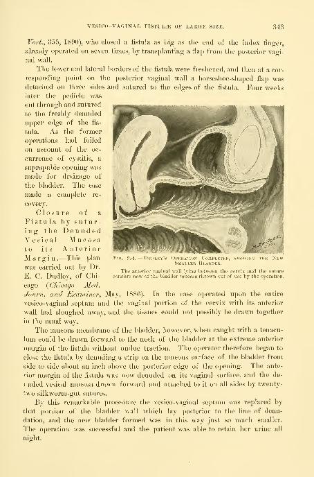
VESICO-VAGINAL FISTULA OF LAKGE SIZE. 343
Vort., 355, 1890), who closed a fistula as big as the end of the index finger,
already operated on seven times, by transplanting a flap from the posterior vagi-
nal wall.
The lower and lateral borders of the fistula were freshened, and then at a cor-
responding point on the posterior vaginal wall a horseshoe-shaped flap wasdetached on three sides and sutured to the edges of the fistula. Four weekslater the pedicle was
cut through and sutured
to the freshly denuded
upper edge of the fis-
tula. As the former
operations had failed
on account of the oc-
currence of cystitis, a
suprapubic opening was
made for drainage of
the bladder. The case
made a complete re-
covery.
Closure of a
Fistula by sutur-ing the DenudedVesical Mucosato its AnteriorMargin .—This plan
was carried out by Dr.
E. C. Dudley, of Chi-
cago {Chicago Med.
Journ. and Examiner., May, 1886). In the case operated upon the entire
vesico-vaginal septum and the vaginal portion of the cervix with its anterior
wall had sloughed away, and the tissues could not possibly be drawn together
in the usual way.
The mucous membrane of the bladder, however, when caught with a tenacu-
lum could be drawn forward to the neck of the bladder at the extreme anterior
margin of the fistula without undue traction. The operator therefoi-e began to
close the fistula by denuding a strip on the mucous surface of the bladder from
side to side about an inch above the posterior edge of the opening. The ante-
rior margin of the fistula was now denuded on its vaginal surface, and the de-
nuded vesical mucosa drawn forward and attached to it on all sides by twenty-
two silkworm-gut sutures.
By this remarkable procedure the vesico-vaginal septum was replaced by
that portion of the bladder wall which lay posterior to the line of denu-
dation, and the new bladder formed was in this way just so much smaller.
The operation was successful and the patient was able to retain her urine all
night.
Fig. 214. — Dudi.ky's ()im,i:\i ids ('(jmpi.ki H), .-^iiowlno iiit .Ni.w
Smaller Bladder.
The anterior vaginal wall lying between the cervix and the sutureconsists now of thebladder mucosa thrown out of use by the operation.

344 AFFECTIONS OF THE UKETIIRA AND BLADDER.
(closure of the Vesico-vaginal Fistula by detaching the-
Bladder from the Vagina and suturing it Independently(Figs. 217, 218).—A. Mackenrodt, of Berlin {Centmlblattf. Gijn., No. 8, 1894),
made a remarkable step in advance when he devised the following plan :
a. The fistula is exposed, and the cervix at one end and the urethral promi-
nence at the other, each caught with a pair of tenaculum forceps, and the tissues
between made tense by traction in opposite directions.
}). An incision is next made in the median hne extending across the fistula
215, 216.
—
Vesico-uteko-vaginal Fistula occupying the Entire Base ok the Bladder withDense Cicatricial Edges and with the Ureteral Openings in the Upper Margin.
Tlie bladder was dissected loose from the uterus and from the lateral vaginal walls around the upper half
of the tist\ihi. The lower half of the tistula was then denuded on its vaginal surface and the raw surface of
the loosened bladder united to the vaginal denudation.
and through the vaginal walls and down to the bladder, so as to expose the
entire base of the bladder.
e. The edges of the fistula are then split so as to separate the bladder from
the vagina, and the separation is carried out widely on all sides, extending
uj)ward, if need be, as far as the vesico-uterine peritoneum.
d. The movable elastic bladder is now closed by demuling its edges and
drawing them together by fine silkworm-gut sutures. Beneath these a second
and even a third row of sutures may be placed.
e. After closing the bladder in this way the vaginal wound is approximated
as far as the tissues will permit by denuding its borders and drawing the corpus
uteri forward and passing sutures from side to side so as to bring the margins
together and at the same time hold the uterus in anteficxion. If the vagina

VESICO-VAGINAL FISTULA OF LARGE SIZE. 345
will not come together the uterus is used to fill in the gap, making a firm base
in place of the fistulous opening.
Closure of the Fistula by detaching the Bladder Pos-teriorly and suturing it to the Denuded Vaginal WallAnteriorly.—My own plan {Johns Ilophiris Tlospital Bulletiii^Yehrxiaxy^
1896) for the treatment of large fistalse, inoperable by the classical method, is
one which was carried out in a case already operated upon five times, with the
consequent loss of the entire base of the bladder, including the internal orifice
of the urethra and the anterior lip of the cervix.
The patient (M. Y., 3811, Sept. 25, 1895) was forty years old and had had
five children ; the fistula dated from the third labor eight years ago, and each
one of the five efforts made to close it had only served to increase the defect
without uniting any part of the tissue.
The opening was 4 by 3 centimeters in size ; the ureteral orifices were seen
on its posterior border after replacing the congested bladder, M'hich was inverted
through it into the vagina ; the edges of the fistula were fixed by scar tissue
and by scars radiating out over the vaginal walls.
The steps of the operation were the following :
a. A crescentic incision was made around the posterior two thirds of the
fistula, separating by a blunt dissection the bladder with its muscular and
mucous coats from the vagina and the cervix laterally, and all the way up to
the peritoneum.
h. The remaining anterior third of the fistula was then pared on its vaginal
Figs. 217, 218.
—
Vesico-utero-vaginal Fistula, showing the Way ix which the Bladder was de-
tached FROM THE Uterus Above from a to a; and brought Down and attached to the DenudedVagina at b.
surface, extending the denudation down to but not including the vesical and
urethral mucosse.
c. The ureters were marked out and protected during the next step by pass-
ing two flexible ureteral catheters 2^ millimeters in diameter through the
urethra, one into each ureter.
d. The detached part of the bladder behind was now easily drawn forward
and accurately united by interrupted fine silkworm-gut sutures to the immov-

346 AFFECTIONS OF THE URETHRA AND BLADDER.
able anterior third of the fistula on its vaginal surface ; each suture caught the
under surface of the muscular wall of the bladder so as to turn its edge up into
the newly formed bladder. The ureteral orifices were in this way directed
upward, and tliey escaped compression and transfixion through the presence of
the catheters. The vaginal opening was not closed.
I left the ureteral catheters in place three days to drain each kidney through
tlie urethra and put the bladder entirely at rest thus avoiding any strain on the
healing tissues.
The wound healed throughout, except at the left upper angle, where a fistu-
lous sinus 1 millimeter in diameter was left ; through this a little urine escaped
occasionally. On leaving the ward the patient could hold 100 cubic centi-
meters, and did not have to empty the bladder oftener than once in three hours.
The raw surface on the anterior vaginal wall was replaced by a firm contracting
cicatrix.
Closure of an Extensive Yesi co-vaginal Fistula bysuturing the Body of the Uterus into the Defect.—This opera-
tion was devised and practiced with success in two cases by W. A. Freund {Sam.m.
Mill. Yort., No. 118, 1895). The first patient was forty years old, and had lost
the posterior wall of the urethra and a considerable part of the sphincter area
at the neck of the bladder. The opening into the bladder easily admitted the
index finger ; the tissues around the fistula were bound to the pelvic bone by
extensive radiating scars, and the cervix was hidden in a mass of scar tissue at
the vault of the vagina.
Douglas's pouch was opened and the retroflexed uterus drawn out into the
vagina and scraped on both sides in front of the broad ligaments until it bled, it
was then sutured to the freshened edges of the opening in the bladder and the
posterior half of the urethra. The fundus uteri was then removed so as to
expose its cavity, and the edges of the wedge-shaped excision were united, pro-
viding an exit for the menstrual discharges. After a protracted convalescence,
marked by attacks of fever and the discharge of sutures and a varying degree of
continence, the patient was able to retain the urine, so that five months later she
only passed it twice in three and a quarter hours, and could void it voluntarily
as soon as there was any accumulation in the bladder.
Four months after the first operation the defect of the anterior part of tlie
ui-etlira was made up by drawing over it the contiguous vaginal walls and sutur-
ing them together. Menstruation took place from the new cervix formed at
the open fundus.
The second case was that of a young woman twenty years old, in whom the
entire base of the bladder had been lost. The perineum was torn back into the
lax sphincter, and there was a recto-vaginal fistula high up. The urethral ori-
fice formed a slit opening into a urethra 1^ centimeter long. The edges of
the fistula were surrounded with extensive scar tissue, and the cervix was con-
cealed in a mass of scars at the vault.
At the operation Douglas's ctol-de-sac was ojiened ; then the recto-vaginal
septum was split from the incision in the vaginal vault down through the recto-

VESICO-UTERO-VAGIXAL FISTULA (FISTULA LAQUEATICA). 347
vaginal fistula and on downward through the anus. The edges of the large
defect in the anterior vaginal wall were now encircled by a broad area of
denudation, and the retroverted uterus drawn out through Douglas's pouch and
freshened by scraping its borders in front of the broad ligaments. The utei-us
was now attached to the edges of the fistula on all sides by silk sutures, which
had been previously laid through the margins of the fistula.
The scar tissue and edges of the fistula in the recto-vaginal septum were nowremoved and closed on the rectal side with catgut, and on the vaginal side with
silk sutures. The body of the uterus was next united to the urethra, and the fun-
dus uteri removed in a wedge-shaped excision and sutured on all sides. Finally
the perineal wound was closed with wire sutures uniting the sphincter and about
1 centimeter of the tissue above it. The bladder was drained with a catheter and
the wound healed throughout, except for the formation of a small recto-vaginal
fistula, which closed sj)ontaneously after the escape of some silk threads.
Three months after the operation there was no incontinence of urine, and
the weak sphincter had recovered its power under the use of hypodermics of
strychnin in its immediate neighborhood. All lateral expansion of the bladder
had disappeared, and instead of this there was a distention of 18 centimeters
upward.
Vesico-utero-vaginal Fistula (Fistula Laqueatica).—These fistulas are situated
at the vault of the vagina close against the cervix, which is frequently involved
by the destruction of a portion or all of its anterior lip.
They occur either from extensive sloughing in this region, or more frequently
from a laceration of this part of the cervix, often due to the obstetric forceps,
extending into the vault of the vagina and on into the bladder. This mode of
origin is well shown in a case reported by Dr. H. C. Coe {Amer. Jour. Med. Sci.,
1890, p. 487), in which he did a successful suprapubic amputation of the uterus
for rupture during labor. Before Dr. Coe operated on the woman, however,
an attempt had been made to introduce the forceps through the undilated cer-
vix, which resulted in a rupture of the cervix extending into the bladder and
leaving a cervico-vaginal fistula behind.
A similar case of double fistula is also figured by Otto v. Ilerff {Zeit.f. Gel).
und Gyn.., vol. xxii, 1891, p. 10).
A case of my own, which I saw April 1, 1885, also goes to prove that these
fistulas are often the result of a tear rather than a sloughing. The patient had
had a severe instrumental labor with a stillbirth six weeks before ; she came to
me with a small fistula just at the vault of the vagina on the right side against
the cervix. This fistula opened two ways, from the bladder backward into the
cervical canal, and downward into the vaginal vault. See Fig. 220. There is,
I think, no other conceivable way in which such fistulae, lying so close to-
gether in this position, could have been brought about, except by a tear ex-
tending through the cervix and forward into the bladder, followed by a healing
of the vaginal cervix between.
The fistula following a tear is apt to be small, while that following a slough
may take in the whole anterior part of the vaginal vault.

348 AFFECTIONS OF THP: URETHRA AND BLADDER.
Treatment .—A small fistula of recent origin may get well spontaneously,
or, as in my own case just cited, may recover after stimulating applications ; I
used the nitrate of silver stick several times wdtli improvement, and after the pa-
tient went home she recovered entirely.
The essential difference hetween the treatment of these and the vesico- vaginal
fistulae lies in the close proximity of the rigid cervix whose tissues can not be
draw^n together like that in the flexible vaginal walls. In addition to this, the
fistula may form a sinus longer than the ordinary vesico-vaginal fistula, and the
denudation and approximation may effect only the closure of the bottom of this
sinus, which for this reason refuses to heal.
A variety of operations have been proposed, among them the following
are of practical value
:
A simple closure may be effected in the al)sence of any scar tissue sur-
rounding a small fistula by making a deep funnel-shaped denudation on the vagi-
nal surface, and then passing several silkworm-gut sutures from before backward
through both edges of the fistula and the anterior cervical lip as well. This will
not succeed if there is much destruction of the cervix with scar tissue.
The anterior lip of the cervix may be effectively utilized to
close a larger defect in the vault by paring its edges and attaching them by
sutures directly to the sides and edges of the fistula pared on its vaginal surface.
If there is too much tension created in pulling the cervical lip down and attach-
ing it to the fistula in this way, this may be relieved by splitting the cervix
bilaterally up to or above the vaginal vault, in this way elongating the anterior
lip, which is then easily pulled out so as to cover in the defect.
The posterior lip of the cervix is in some cases easier to ap-
proximate to the anterior edge of the fistula than the anterior lip. When this is
denuded and attached so as to fill in the defect, the cervical canal is turned into
the bladder, and menstruation henceforth takes place through this viscus.
Dr. N. Bozeman has advocated the preparation of those cases where there is
scar tissue and fixation by catching the uterus with forceps and dragging it downdaily for some weeks beforehand, so as to gradually overcome the resistance.
Detacliing the Uterus from the Bladder and then sutur-ing the Fistula .—The best plan of all, and one doing away with the difii-
cult dealing with the scar tissue, is the following (see A. AVolfler in v. IIerff"'s
paper, Zeit. f. Geb. tind Gyn.^ 1891, p. 5)
:
a. The cervix is caught and drawn down and backward, and separated from
the vaginal vault in front. This separation is continued well above the fistula
by detaching a part of the bladder from the supravaginal cervix.
h. The edges of the fistula are then pared down to the vesical mucosa, taking
care to get rid of all scar tissue.
e. The fistula may then be closed by interrupted fine silkworm-gut sutures,
or by buried continuous catgut sutures in two or three layers.
d. After this the cervix may be attached again to the vaginal vault l)y means
of several silkworm-gut sutures.
It is important, for five or six days after the operation, to keep the bladder

VESICO-UTERO-VAGINAL FISTULA (FISTULA LAQUEATICA). 349
empty, and to avoid an infection at the vault by keeping a clean, loose iodoform
pack in tlie vagina.
Trendelenburg ( YoTkmann^s Sancm. Min. Yort., 355) recommends tlie fol-
lomng plan of dealing with vesico-vaginal fistulse when they can not be satis-
factorily exposed on the vaginal side ; also for fistulse in the immediate neigh-
borhood of the ureter, for vesico-uterine, uretero-cervical, and uretero-vaginal
iistulae
:
The patient is placed on the table with the pelvis well elevated at an axis of
not less than forty-live degrees to the horizontal ; by this posture, when the
Fig. 219.
—
Svprapibic Upekation for Vesico-vaginal Fistula ( Tkkndelenbukg).Suprapubic Incision seen Above.
Sagittal Section.
bladder is incised, it at once fills with air, and its entii-e interior is well exposed
to view.
The bladder is opened by making, a transverse incision 10 centimeters long
across the upper border of the symphysis, separating the attachments of both
recti muscles, and exposing the prevesical space ; a transverse opening in the
bladder is then made 5 to centimeters long. The edges of the fistula now ex-
posed are denuded in the form of a shallow funnel in such a way as to remove
a broad band of tissue from the bladder mucosa, and a narrow one from the
vagina and cervix. The edges are brought together with silkworm-gut sutures
;
in the first cases these were tied in the bladder, but later two needles were
threaded on one suture, and both ends were passed through into the vagina,
where they were tied.
The incision into the bladder is now closed down to an opening left for a T-
drain. The patient is compelled to lie in Sims's position until the fifth day,
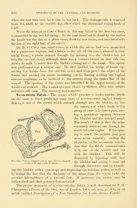
350 AFFECTIONS OF THE URETHRA AND BLADDER.
when slie may turn over for a time on her hack. The drainage tuhe is removed
from tlie ninth to tlie twelfth day, after wliich tlie abdominal wound heals of
itself.
While the attempt to close a fistula in this way failed in the first two cases,
it succeeded in the two following. In the case described in detail by the author
the fistula was the size of a plum stone, fixed by scar tissue, and associated with
the loss of the right half of the cervix.
Dr. II. C. Coe's case, cited above, in which the uterus had been amputated
for a parturient rupture, had a fistula to the left of the cervix situated in cica-
tricial tissue ; a probe entered through the opening into the bladder but not
into the cervical canal, although there was a communication on that side too,
shown by milk injected into the bladder coming out of the canal. The opera-
tion performed was a unique one. He first divided the bridge of tissue sep-
arating the fistula from the cervical canal, and then pared the edges of the
fistula and excised the entire remaining cervix, leaving nothing but vaginal
mucous membrane to be included in the sutures along the entire line of the
wound. The opening of the ureter was identified in the upper margin of the
fistula and avoided. The wound was now closed by thirteen silver wire sutures
and three silk ones. The recovery was complete.
Vesico-uterine Fistula.— The causes which produce a vesico-uterine fistijla
are the same as those producing some cases of cervico-vesico-vaginal fistula
—
that is, a tear of the cervix which extends through into the bladder, up into
the uterus, and which heals in this
group of cases in its lower part, leav-
ing a persistent opening between
the bladder and the cervical canal.
The result of this is that the urine
constantly dribbles out through the
cervix into the vagina. If the open-
ing is small the j)atient may pass
some urine naturally, leading the
physician to the erroneous conclu-
sion that the fistula communicates
with one of the ureters and not
with the bladder. This will be
disproved by injecting milk into
the bladder and seeing it ooze out
through the cervix, and by examin-
ing the bladder with a cystoscope and inspecting the fistulous orifice, as well as
by noting the fact that the discharge of the urine from the cervix lacks the
peculiar intermittcncy of a ureteral flow. If necessary the ureters may be
catheterized and their patency demonstrated.
The proper treatment of a vesico-uterine fistula is well described by F. IF.
Champneys {Trun.s: of the Ohst. Soo. of London, 1888, vol. xxx, p. 348), in an
article entitled Description of a New Operation for Yesico-titerine Fistula.
Fio. 220.
—
Vesico-utero-vaginal and YesFistula in the Same Patient,

VESICO-UTEKINE FISTULA. 351
The procedure is as follows : The patient, thirty-eight years old, had had
four sev^ere labors, the last two instrumental. Her pelvis was generally con-
tracted and flattened, and the last labor continued four days and was terminated
by the forceps ; on the same day the urine began to flow by the vagina, and con-
tinued to do so up to the day of operation.
On examining per vaginam, the cervix was found rather large and flabby and
the canal big enough to admit the index finger for an inch. On injecting the
bladder a large stream escaped from the cervix, and a bent probe introduced
through the urethra could be passed on directly into the cervical canal.
Operation May 12, 18S7. a. After passing a probe through the fistula as just
described, and bringing it out at
the cervix, the latter was steadied
with a volsella forceps.
h. A transverse incision 3|^
centimeters long was madethrough the anterior fornix from
the vagina and the bladder dis-
sected up from the cervix with
scissors and fingers as in a vaginal
extirpation of the uterus. The
dissection was carried well above
the fistula, dividing it into two
parts, one opening into the bladder
and the other into the cervix, each
admitting the index finger easily.
c. No freshening was required,
as the whole surface was raw.
Seven fine silver sutures were
passed from side to side to close
the opening in the bladder, each
one beina: entered an eig-hth of an inch from the hole and brought out on its
edge, avoiding the mucosa. Four similar sutures closed the cervix. These
sutures were all cut short.
d. The vaginal wall was then united to the cervix by four long silkworm-
gut sutures, subsequently removed.
A self-retaining catheter was left in the bladder and a gauze pack put in the
vagina. The result was a perfect recovery of function.
I operated January 12, 1893, on a somewhat similar case. The patient (F.
H., 1750), forty-six years old, had had twelve children, the last two born instru-
mentally. At the last confinement she was four weeks abed instead of four days,
as usual, and from the fourth day on she suffered from incontinence of urine.
Both vaginal w^alls were found lax and pouting, and the anterior lip of the
cervix was completely destroyed. At a point well above the vaginal vault a fis-
tula 1*5 centimeter in diameter opened into the anterior cervical wall. This
was treated by exposing the cervix with a speculum and drawing it down, and
Fig. 221.
—
Vesico-uterine Fistula treated by dissect-ing THE Bladder Free from the Uterus and sewingUP the Fistula.
The long arrow shows tlie position of the fistula, the
short arrow points to the line of incision in front of the cer-
vix separating the cervix (>) from the vagina {d) ; the edgesof the fistula (a b) are then approximated and (c d) unitedagain.

352 AFFECTIONS OF THE URETHRA AND BLADDER.
then cutting across tlie vault of the vagina and detacliiiig the cervix from the
bladder and so separating the fistula for 1"5 centimeter on all sides from the
vagina and uterus, laying bare a hole in the bladder wall 8 millimeters in diame-
ter. This was closed by four silkworm-gnt sutures introduced from side to side,
Fistula, Sutiu!Es
with fine catgut between for accurate approximation ; the cervical part of the
fistula was left open, and the vaginal vault was not closed. See Fig. 222. Avaginal pack was then put in and the bladder drained by a catheter.
In two weeks the silkworm-gut sutures were removed and the union found
perfect throughout.
Other Vesical Fistulae.—Aside from the genital fistuljie which have just been
discussed, fistulous communications with other organs are but rarely observed.
This immunity is due to the fact that some unusual accident is necessary to

OTHER VESICAL FISTUL.^. 353
establish a communication between the bladder and any of the other abdominal
or pelvic viscera.
A communication may be formed in this way between the bladder and a
tube or an ovary and between the bladder and the small or large intestine.
When the bladder communicates with a tube or an ovary this is brought
about in one of two ways : either (1) an abscess of the tube or ovary perforates
the broad ligament at its base and so finds its way into the bladder, or (2) an
ovarian tumor forms adhesions with the peritoneal portion of the bladder ; the
septum becomes thinned out and finally breaks, and the contents of the cyst
escape by this avenue.
An abscess j)erforating the Ijroad ligament commonly finds its way into the
bladder in the neighborhood of the right or left cornu—that is, at either end of
the posterior fold. I have seen a case of a tubercular abscess of the tube on
the right side of the pelvis discharging in this way.
A suppurating dermoid cyst may break through into the bladder
and the nature of the abscess be determined by the escape of hair (pilimiction) or
bones discharged per
uretliram^ or even by
a tooth found as the
nucleus of a vesical
calculus. A case of this
sort is well described
by Dr. G. C. Black-
man {Avier. Jour, ofthe Med. Sciences, Jan-
uary, 1869, p. 49).
The patient, thirty-six
years old, first noticed
air escaping from the
bladder, then urine
passed by the rectum,
and she suffered from
a cystitis. A calculus
was found and re-
moved. This con-
tained a tooth, and in
the course of seven
years four similar cal-
culi containing teeth
as nuclei were extracted. Some months after the last one was removed she began
to pass hairs incrusted with phosphatic deposits. Dr. Blackraan also gives a
careful review of the literature of the subject.
Fig. 225 shows thfe condition found in a patient of Dr. Henry Eisner, of
Syracuse, IST. Y., in a case of pyuria due to a dermoid cyst. The patient, forty
years old, had known of the existence of the tumor for over twenty years.
26
Fig. 223.
—
Vesico-vaginal Fistula, cai'sep ry a Pessary seen in Sa-gittal Section.
The shank of the pessary lies buried in the recto- vaginal septum. Thevagina i.s atretic and the pus cavity in its upper portion discharges into the
bladder, which lies contracted behind the symphysis, by a fistulous open-ing caused by the cup of tlie pessary. The hypertropliy'of the vesico- andurethro-vaginaL septum is also shown, as also in Fig. 224.

354 AFFECTIONS OF THE URETHRA AND BLADDER.
Three years before the operation the previously inovahle tumor became fixed
above the symphysis, and for the same length of time she suifered from a pyuria.
The tumor was found at the operation to be a right dermoid cyst densely
adherent to and discharging its contents into the bladder ; after freeing numerous
surrounding adhesions, the dense, fibrous sinus, 3 centimeters in diameter, was
dissected out down to the
bladder just above the
symphysis pubis and cut
olf, exposing a lumen of
about 3 millimeters.
This was closed by six
interrupted buried catgut
sutures and the vesical
peritoneum was then
drawn over it and united
by six moi*e catgut su-
tures, leaving a longitudi-
nal linear wound at the
site of the attachment.
No drain was used. The
pus disappeared at once
and a perfect recovery
followed. When drainage
is necessary it is easy to
make the wound entirely
extraperitoneal by uniting
the peritoneum from the
bladder up on to the ab-
dominal wall, so as to leave
whatever space is desired
between the peritoneum
and the symphysis.
An extra-uterine sac
may also suppurate and open into the bladder, and the nature of the affection
first be made clear by the escape of one of the bones through the urethra.
Therig {Centralbl. f. innere Med., .Bd. xv, p. 97) has observed in women twocases of paratyphlitic (vermiform appendix ?) abscesses breaking into the blad-
der ; both recovered under irrigation.
A case of colo-vesical fistula is reported by R. Harrison {Tioentieth Century
Practice, New York, 1895, vol. i, p. 222). Air bubbles escaped through the
urethra and granular cells and spiral vessels were found in the urine, and after
death the colon was found adherent to the bladder and a cherry-stone lying in a
diverticulum among the adherent intestines. The dehru' in the urine evidently
came from the disintegrating kernel of the stone.
The s y m p t o m s produced by the communication of any of these extra-
FiG. 224.—Enormous Hypertrophy and Edema of the AnteriorVaginal Wall simulating Cystocele, due to Cystitis. Nov.27, 189-).
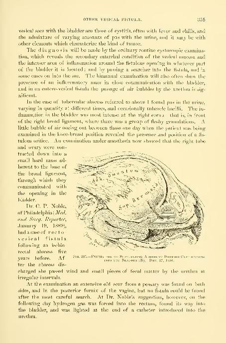
OTHER VESICAL FISTUL.E. 355
vesica] sacs with the bladder are tliose of cystitis, often with fever and chills, and
the admixture of varying amounts of pus with the urine, and it may be with
other elements which characterize the kind of tumor.
The diagnosis will be made by the ordinary routine cystoscopic examina-
tion, which reveals the secondary catarrhal condition of the vesical mucosa andthe intenser area of inflammation around the fistulous opening in whatever part
of the bladder it is located ; and by passing a searcher into the fistula, and in
some cases on into the sac. The bimanual examination will also often show the
presence of an inflammatory mass in close communication with the bladder,
and in an entero-vesical fistula the passage of air bubbles by the urethra is sig-
nificant.
In the case of tubercular abscess referred to above I found pus in the urine,
varying in quantity at diiferent times, and occasionally tubercle bacilli. The in-
flammation in the bladder was most intense at the right cornu—that is, in front
of the right broad ligament, where there was a group of fleshy granulations. Alittle bubble of air oozing out between these one day when the patient was being
examined in the knee-breast position revealed the presence and position of a fis-
tulous oi-ifice. An examination under anesthesia now showed that the right tube
and ovary were con-
tracted down into a
small hard mass ad-
herent to the base of
the broad ligament,
through which they
connnunicated with
the opening in the
bladder.
Dr. C. P. Noble,
of Philadelphia (J/^r/.
and Surg. Reporter,
January 19, 1889),
had a case of recto-
vesical fistulafollowing an ischio-
rectal abscess five
years before. Af-
ter the abscess dis-
charged she passed wind and small pieces of fecal matter by the urethra at
irregular intervals.
At the examination an extensive old scar from a pessary was found on both
sides, and in the posterior fornix of the vagina, but no fistula could be found
after the most careful search. At Dr. Noble's suggestion, however, on the
following day hydrogen gas was forced into the rectum, found its way into
the bladder, and was lighted at the end of a catheter introduced into the
urethra.
Fig. 225.
—
Pyuria due to Suppurating Adherent Dermoid Cyst openingINTO THE Bladder (B). Dec. 17, ISOti.
356 AFFECTIONS OF THE URETHRA AND BLADDER.
Treatment .—The proper line of treatment must depend on the individ-
ual case ; no general rules can he laid down. The local affection of the hladder
tends to a spontaneous recovery when the cause is removed. If the patient's
health will permit it, a sac which opens into the bladder should therefore either
be enucleated and the source of the discharge stopped, or evacuated and drained
in some other direction—into the vagina, or by the abdominal wall—so as to
give the bladder a chance to recover.
In one of the worst cases of pyuria I have ever seen a cure was effected by
opening and draining the abscess, which lay in front of the uterus, through the
anterior fornix hi the vagina. In another case a pelvic abscess on the right side,
discharging through the bladder, was relieved by enucleating both tubes and
ovaries with the uterus, leaving the vaginal portion of the cervix, which was
sewed over the fistulous orifice at the base of the broad ligament, so as to divert
any discharges into the vagina. The fistula could not be closed by direct suture
on account of the friable inflammatory tissue composing its walls.
Hemorrhoids.—Yaricose Bladder.—A hemorrhoidal condition of the
bladder or vesical v a r i x is a rare affection, in spite of the fact that all
the favoring conditions for its frequent occurrence seem to be supplied in the
venous stasis so often found in the pelvis, in the enormous congestion of hemor-
rhoidal venous sinuses, as well as in the congestive disturbances found in associ-
ation wath retroflexion of the uterus. The frequency of inflammatory disturb-
ances in the neighboring genital organs, and the great development of crural and
labial varices in pregnancy, as well as the increased amount of blood observed in
the bladder in that condition, Avould also lead one naturally to expect to meet
with a varicose condition of the bladder as a common occurrence under these
circumstances. When found in men vesical varix is apt to be associated with
rectal hemorrhoids. The cause of vesical varix has been shown in them to
be due to an insufficient size of the venous channels above the communication of
the hemorrhoid and the vesical plexuses, so that a lower rectal congestion brings
with it at the same time a vesical stasis. In 1854, Guyon exhibited a case at the
Anatomical Society of Paris in which the neck of the bladder was surrounded
by a varicose venous circle with diverging dilated submucous ramiflcations.
II. Picard {Traite des mal. de la vessie, Paris, 1878, p. 284) found in an
autopsy on a man who died of this disease that the large venous plexuses sur-
rounding the prostate communicated with the hemorrhoidal plexus. The mucosa
of a large part of the bladder itself bristled with a great number of varices, form-
ing little bluish tits about its neck, on the base, and extending high up on the
lateral walls. Each little titlike projection was the elbow of a vein projecting
from 1 to 2 millimeters beyond the surface of the bladder. Some of the vessels
near the neck showed abrasions, and others were perforated, demonstrating the
source of hemorrhage, and probably of the infection of which the patient died.
In the clinical history the one characteristic symptom is the re-
peated hemorrhages. In men retention of the urine has also been noted as
a common synq^tom.
The diagnosis between this condition and papilloma and cancer in its

HEMOERHOIDS. 357
early stages, by symptoms and an external examination, is only made with dif-
ficulty. If the hemorrhages come on with an attack of the piles, vesical varix
may be suspected, especially if difficulty in urination occurs at the same time.
Vesical varix is also found when there is a periodical bleeding alternating be-
tween the rectum and the bladder.
All doubt may be easily cleared up in women by making a direct cystoscopic
examination of the mucous surface of the bladder, when the blue congested ves-
sels may be easily inspected and their number, size, and distribution deter-
mined. In such a case it is better to examine first in the dorsal position
under a moderate degree of elevation, to avoid the tendency of the knee-
breast position to produce an artificial anemia, temporarily relieving the very
condition one wants to see. The inspection should be carefully extended over
the whole circumurethral area, and from thence down the urethra as the specu-
lum is withdrawn.
Arbuthuot Lane, at a meeting of the Clinical Society of London {Lancet^
March 18, 1895, vol. ii, p. 1252), reported a rare condition under the title of
A Nmvoid Growth of the Mucous Membrane of the Bladder.
A child, aged three years and a half, had been passing bloody urine for twoyears, the blood at times coming away in large clots. When seen by Lane the
hemorrhage was so severe and had been so long continued as to endanger life.
On examining the patient, several nevoid patches were seen around the anus
and on the buttocks. By abdominal palpation the bladder could be distinctly
felt above the pubes. When the bladder was opened above the symphysis pubis,
large nevoid masses, some as large as grapes, protruded through the wound
;
most of these were soft and bled easily ; a few were hard and apparently cystic.
Almost the whole of the mucous surface of the bladder was affected.
As an operation seemed to be practically out of the question, the incision
was closed, and afterward the hemorrhage practically ceased, the urine being
only a little blood tinged at times.
Treatment .—If the discovery of the varicose condition is made acci-
dentally, and there are no urgent symptoms, nothing should be done, but in a
persistently bleeding case in a woman, after diagnosing the cause of the bleeding
and locatnig its position in the bladder, one of several plans may be followed
;
in a mild case the galvano-cautery introduced through the speculum may be
used over small areas at several sittings.
If the urethra is dilated to admit a No. 15 to 18 speculum, one or more
ligatures even may then easily be thrown about several of the larger venous
trunks by means of a fine curved needle on a fixed handle carrying fine silk,
which can then be tied by using a little instrument pronged like a pitchfork to
afford a point of counter pressure within the bladder. A more active and
direct interference may be made by means of an incision through the anterior
vaginal wall, everting and exposing the veins at the neck of the bladder. Sev-
eral of the larger trunks may then be tied with fine silk and the vaginal incision
closed again.
Dr. W. Ryan, of Springfield, 111., had a case of varix in a woman which he

358 AFFECTIONS OF THE URETHRA AND HLADDER.
suecessfullj treated by a suprapubic incision. The patient was thirty-
one years of age and married, and for six months had passed large quantities of
blood with her urine, in clots ; whenever the bladder became distended with the
clots her suffer-
ing was extreme.
Fig. 226.—Pronged Instkumknt with Ci.eat at tiik Handle She WaS SO ane-JolNT FOR TYING .A KnOT InSIDE THE Bl,A])DEU. \4 <*RI>I- • i • 1
NARY Size. mic and m such
a generally de-
pressed state of health that several of her physicians thought she
was sulfering from a serious renal aifection. There were no rectal
hemorrhoids at all. Washing out the bladder and the use of injections only
made her worse.
On December 19, 1890, Dr. Ryan opened the bladder above the symphysis
pubis and found an extensive dilatation of the veins aljout the neck of the blad-
der, at the base, and running up on the sides just under the intact mucosa, about
2 millimeters in diameter. In opening the bladder, some veins about the neck
were cut, and continued to bleed moderately through a drainage tube which was
left in for five days. After this simple treatment, incision, inspection, and drain-
age, she made a perfect recovery.
Hyperemia.—By hyperemia or a congestion of the vascular system of the
bladder is meant either a local or a general flushing of the vesical capillaries, pro-
ducing an increased redness of the surface, in contradistinction to a hemor-
rhoidal condition, where the venous trunks are involved and the capillary veins
are greatly distended. A physiological hyperemia of the bladder may be ob-
served when nature determines any large amount of l)lood to a neighboring
organ, as, for example, in pregnancy. A localized hyperemia of the vesical
mucosa is often found also associated with inflammatory disease in the imme-
diate neighborhood. The withdrawal of the obturator from the end of the
vesical speculum always produces a little patch of hyperemia on the posterior
vesical wall by acting for a moment as a piston and sucking the vesical mucosa
into the end of the speculum.
Hyperemia of the T r i g o n um .—This is a common condition local-
ized in the trigonum, and rarely extends beyond its limits, except into the
urethra.
The entire surface of the trigonum may be of a deep rosy red, the injection
extending around both ureteral oriflces ; the border of the injected area becomes
gradually merged into the surrounding sound tissue. The surface of the mucosa
sometimes has a slightly puffy or edematous appearance. The injection may
be of a patchy nature only surrounding the ureteral oriflces, or it may even
be limited to the neighborhood of one ureteral oriflce.
The margins of the injection wdien not limited by the interureteric line are
irregular in outline. The most intense injec'tion is often in the area closest
to the urethra, which is also deeply injected in its upper part, and character-
ized by prominent lacunae.
The symptoms produced by a hyperemia are characteristic. The patient
HYPEREMIA. 359
suffers from a desire to empty the bladder at frequent intervals, either by day
or by night, or both ; she often complains of a burning or bearing-down sensa-
tion, or of a feeling of fullness about the neck of the bladder. Tlie act of urina-
tion is sometimes painful, but not always ; very often after urinating there is
a distressed feeling about the parts, which persists for some minutes or even an
hour or more, leaving her much depressed. Other patients may not feel the
desire to urinate more frequently than ordinary, but the distress is experienced
afterward. The whole area is extremely tender to the touch either by the end
of the speculum or a probe.
The causes of this disease are sometimes difficult to determine. I find it in all
those cases which have hitherto been diagnosed as " irritable bladder," or " teasing
of the neck of the bladder from retroflexion of the uterus," or " pressure of an
anteflexed uterus on the bladder," or " neuralgia of the bladder "; it is also the
only lesion existing in many of the cases under treatment for a supposed cystitis.
It is frequently observed after abdominal pelvic operations, and appears to
be due in these cases to the irritation of the vesical mucosa by the highly con-
centrated urine discharged during the first few days. 1 have for the past two
years almost eliminated it from my wards by giving every such patient a large
rectal enema of normal salt solution before she leaves the operating table ; the
salt solution dilutes and greatly increases the flow of urine.
Another fruitful cause of hyperemia of the trigonum is the slight mechanical
insult of catheterization. It may be that a mild infection lies at the bottom of
some of these cases, and that the affection is in reality a form of trigonitis,but this remains to be proved.
Diagnosis.—The diagnosis will not be diflicult if all cases of '"' vesical
irritability " are examined by the direct method.
The marked redness in the trigonum is at once apparent, and tlie evidence is
still more decided when there is a patch of it on one side while the other remains
clear. Often the patient complains bitterly when the end of the speculum
touches this spot, and if it is touched with a searcher she will declare at once that
the seat of her discomforts is located there.
The examiner must guard against two errors in making the diagnosis : First,
he must not mistake the physiologically greater injection of the trigonum over
that of the rest of the bladder for a hyperemia ; he will avoid this by familiariz-
ing himself with the appearance of the normal trigonum, and allowing for the
slight difference in color which always exists.
In the second place, he must not mistake the hyperemic blush about a ure-
thral orifice, which is so commonly met with as a sign of disease of the kidney
or ureter of that side, for a simple hyperemia. A few leucocytes are sometimes
found in the urine when the hyperemia is intense.
Treatment .—The treatment should be directed to the cause when it can
be discovered. If it follows an operation, recovery is usually spontaneous within
two or three weeks. If the urine is highly charged with urea, diluents should
be given ; water and flaxseed tea, and citrate of potash and lithia in large doses
are all useful. I have found the most relief from half-teaspoonful doses of

360 AFFECTIONS OF THE URETHKA ANJ) J5LADDER.
sweet spirits of niter repeated every two hours; fluid extract of zea mais and
triticum repens in half-teaspoonful doses are valuable, particularly tlie
first remedy. Any articles of diet, such as tomatoes, fruits, or acids, should be
avoided when the patient finds that they aggravate her condition. The bowels
must be kept well opened all the time. A prolonged hot vaginal douche often
gives great relief.
When these means fail, direct topical treatment should be begun l)y placing
the patient in the knee-breast position and exposing the affected area, and apply-
ino- a 3 to 5 per cent solution of nitrate of silver to the affected area alone.
It is easy to do this with a little absorbent cotton twisted on a wire applicator.
These applications may be repeated every three to five days as long as the affec-
tion continues to improve. If the convalescence comes to a standstill I then
inject 4 to 6 cubic centimeters of a 2 to 3 per cent solution of ichthyol in
glycerin into the empty bladder, and insert Clark's rubber balloon as described
in the next section on cystitis, inflate it, and leave it in for from five to ten
minutes.
Cystitis.
—
Bacteriology: Cystitis is a disease much less frequent in
women than in men, and exceedingly rare in children. An infection is the true
cause of every case of cystitis, and the continuance of the disease depends upon
the continued action of one or other of the various pathogenic micro-organisms.
The term cystitis is therefore a collective name for a variety of inflammatory
affections having certain symptoms in common in their early stages, but often
differing widely in their final forms.
The commonest avenue of infection is through the urethra, in which numer-
ous organisms are constantly found normally ; these organisms, together with
organisms from the vulva lodged on the external urethral orifice, may be carried
into the bladder by the catheter, sound, or other aseptic instrument, or they may
be introduced on unclean instruments, and the infection started up in this way.
It is also necessary to allow for a few cases in which the organisms enter the
bladder from the urethra without instrumentation, especially where tlie urethra
is dilated and patulous, as in women who have borne many children.
The bladder may also be infected from the kidney either when the kidney
or its pelvis is diseased, as in pyelitis or pyelonephrosis, or, as has been shown,
even when the kidney itself is healthy, the organisms may be eliminated from
the body through it, and so may infect the bladder. A claim has also been made
by Wreden {Arch, des Sciences Biologiqnes, St. Petersburg, Bd. ii, 5, 1894) that
a direct infection of the bladder may take place from the intestine under certain
conditions ; in support of this are the experiments upon animals, occluding both
rectum and urethra, with the invariable result of occasioning a true cystitis, in
which usually pure cultures of the organism used in the experiments may l)e
isolated from the bladder.
C. Posner and II. Lewin report a series of experiments {Centrdlhl . f. Ildrn
unci Sexual- Organe, Bd. vii. Heft T, 1896) which throw much light upon this
question of a direct infection ; they found, after closure of both the rectum and
the urethra, that while they were always able to get pure cultures from the blad-

EXPERIMENTAL CYSTITIS. 361
der of either the colon bacillus or the special organism used, these organ-
isms were always present in the blood, and in the substance of the
kidneys as well, so that although the result of the investigation does not
entirely preclude the possibility of a direct passage of the intestinal bacteria into
the bladder, it renders it less probable, while the chances are that the infection
travels through the blood into the kidneys and so enters the bladder. In several
cases in which they injected coloring matter into the rectum, in no instance did
it appear in the bladder or bladder wall. The entrance of the organisms into the
blood is explained by the fact that there was always some wound of the intes-
tine or rectum, from the clamp or a ligature, opening up an avenue for their
direct passage into the finer blood vessels or into the lymphatics.
It is also possible that as a result of the ligation changing the circulatory
conditions from the normal the organisms may have penetrated the unwoundedintestinal wall, and so have entered the lymphatic circulation. That this some-
times occurs in the human being is beyond question, for we know that while the
bacteria are unaljle to pass through the normal intestinal mucosa, they do pene-
trate the mucosa and enter the peritoneal cavity when the vital activity of the
intestine is lowered or dead, as, for instance, where there is a strangulation of
the intestine, or where the blood supply is cut off. The colon bacillusis not infrequently found free in the peritoneal cavity under such circumstances.
On the other hand, Reymond {Ant), des mal. des organes gen. nrhi., April,
1893) has proved beyond any question that bacteria can enter the bladder
directly from inflammatory areas in the neighboring organs. He was struck by
the frequent occurrence of cystitis in women suffering with inflanmiation of the
uterus or of the Fallopian tubes, and in most cases proved by culture that the
organism was the same in both organs ; as an additional proof, he found in a
case of salpingitis in one tube a localized cystitis on the same side in the blad-
der. To complete his chain of evidence it was necessary to prove that the
organisms could pass directly through the walls of the bladder from the neigh-
boring inflamed area. To do this he laparotomized dogs and injected 2, or 3
centimeters of a culture of the uro-bacillus liquifaciens of Krogius,
which he had isolated from a case of salpingitis and cystitis, under the peri-
toneum covering the bladder.
Ten hours latter, on removing the ligature from the penis, he found a
cystitis present, and was able to obtain pure cultures of the uro-bacillus from
the bladder, cultures from the blood and kidneys remaining sterile. He also
demonstrated the organisms in the bladder walls under the spot where the in-
oculation was made, and at this place the cystitis was most marked, showing in
one case an ulcerated area. By further experiments he proved that the organ-
ism was able to penetrate the peritoneum.
The clinical cases, together with the experiments on animals, prove his con-
clusion that cystitis may arise from the passage of organisms directly from a
diseased tube or ovary, the blood, kidneys, and other organs remaining sterile,
when the predisposing condition is present in the form of a congestion of the
bladder caused by the neighboring infected area.
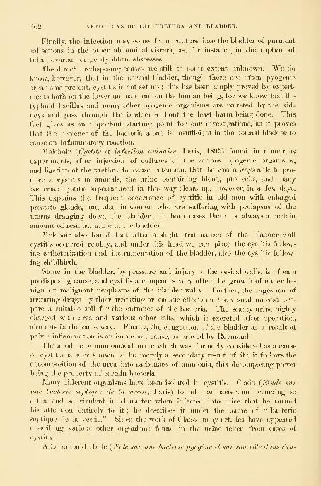
362 affp:ctions of the urethra and bladder.
Finally, the infection may come from rupture into the bladder of purulent
collections in the other abdominal viscera, as, for instance, in the rupture of
tubal, ovarian, or perityphlitic abscesses.
The direct predisposing causes are still to some extent unknown. We do
know, however, that in the normal bladder, though there are often pyogenic
organisms present, cystitis is not set up ; this has been amply proved by experi-
ments both on the lower animals and on the human being, for we know that the
typhoid bacillus and many other pyogenic organisms are excreted by the kid-
neys and pass through the bladder without the least harm being done. This
fact gives us an important starting point for our investigations, as it proves
that the presence of the bacteria alone is insufficient in the normal bladder to
cause an inflammatory reaction.
Melchoir {Cystite et infection urinaire, Paris, 1895) found in numerous
experiments, after injection of cultures of the various pyogenic organisms,
and ligation of the urethra to cause retention, that he was always able to pro-
duce a cystitis in animals, the urine containing blood, pus cells, and many
l)acteria ; cystitis superinduced in this way clears up, however, in a few days.
This explains the frequent occurrence of cystitis in old men with enlarged
prostate glands, and also in women who are suffering with prolapsus of the
uterus dragging down the bladder ; in both cases there is always a certain
amount of residual urine in the bladder.
Melchoir also found that after a slight traumatism of the bladder wall
cystitis occurred readily, and under this head we can place the cystitis follow-
ing catheterization and instrumentation of the bladder, also the cystitis follow-
ing childbirth.
Stone in the bladder, by pressure and injury to the vesical walls, is often a
predisposing cause, and cystitis accompanies very often the growth of either be-
nign or malignant neoplasms of the bladder walls. Further, the ingestion of
irritating drugs by their irritating or caustic effects on the vesical mncosa pre-
pare a suitable soil for the entrance of the bacteria. The scanty urine highly
charged with urea and various other salts, which is excreted after operation,
also acts in the same way. Finally, the congestion of the bladder as a result of
pelvic inflammation is an important cause, as proved by Reymond.The alkaline or ammoniacal urine which was formerly considered as a cause
of cystitis is now known to be merely a secondary result of it ; it follows the
decomposition of the urea into carbonate of ammonia, this decomposing power
being the projjerty of certain bacteria.
Many different organisms have been isolated in cystitis. Clado {Etude sur
une hacterie septique de la vessie, Paris) found one bacterium occurring so
often and so virulent in character when injected into mice that he turned
his attention entirely to it ; he describes it under the name of " Bacterie
septique de la vcssie." Since the work of Clado many articles have appeared
describing various other organisms found in the urine taken from cases of
cystitis.
Albarran nud Ilalle {Note sur une hacterie pyogen.e et sur son role dans Vin-

BACTERIA FOUND IN CYSTITIS. 363
fection urinaipe, Bull, de Vacad. de med., 1888) describe an organism found
by tliem in forty-seven out of fifty cases of infection of the urinary tract, the
organism being present in pure culture in fifteen out of the forty-seven cases;
this was named by them " b a c t e r i e p y o g e n e."
Doyen {Journal des connaissances mcdicales^ 1888, p. 236) studied the or-
ganisms present in cases of ascending pyelonephritis, and found three varieties
of the p r o t e u s
.
Rovsing {Blasen-Entsundungen, Berlin, 1890) studied the urine from thirty
cases of cystitis, all but three of which were ammoniacal, and found in five of
the thirty the tubercle bacillus, in eiglit the staphylococcus pyo-genes aureus, and in three the staphylococcus pyogenes albusand c i t r e u s ; he also found in the other cases various undescril)ed organisms
—the streptococcus pyogenes urese, the diplococcus uresepyogenes, in two cases the cocco- bacillus urese pyogenes, and
in one case micrococcus u r e ge f 1 a v u s pyogenes.Krogius, the author of several articles on the bacteriology of cystitis and other
urinary infections, has come to the conclusion that the organism, a short bacillus
commonl}^ found by him, was in fact an intestinal bacillus, probably the colonbacillus.
Melchoir published in 1895 the results of the bacteriological examinations in
thirty-five cases of cystitis, and found that among these the colon bacilluswas present in pure culture seventeen times, and was present altogether twenty-
four times. The streptococcus pyogenes was found four times, the
proteus of Hauser four times, the tubercle bacillus three times,
and the gonococcus and typhoid bacillus each once ; the remaining
organisms were undescribed until he isolated them.
Melchoir also thinks that the organisms described by Clado, as well as those
described by Albarran and Halle and Morelli, are in fact only the colon bacillus
which he found so many times, and he compares the modes of growth, size, and
general morphology in a convincing manner.
Besides the above, Heyse {Zeitschr.f. Min. Med., Bd. xxiv, 189-1, p. 130) has
described an interesting case of cystitis from infection by the bacillus lactis
a e r o g e n e s , with the formation of gas in the bladder. He traced the infection
from the intestines, where the bacillus was present in large numbers, to the va-
gina, where it had also evolved gas, from whence it had evidently been carried
into the bladder by catheterization. Heyse also cites a case of p n e u m a t u r i a
described by Senator, who found the t o r u 1 a c e r v i s i se to be the cause of
the gas formation in a diabetic patient.
The bacillus aerogenes capsulatus of Welch has also been iso-
lated from several cases of pyelonephritis, and in one case reported by Goebel,
from an autopsy performed at the Hamburg General Hospital, on an old manwho had an enlarged prostate, the bladder was found filled with gas, and there
were numerous gas blebs beneath the mucous membrane.
Fr. Yahle {Inaug. Diss., Marburg, 1895) describes a case of exfoliative cys-
titis in a woman suffering with a myoma incarcerated in the pelvis, in which the

304 AFFECTIONS OF THE URETHRA AND BLADDER.
organism present was the streptococcus pyogenes; tlie patient died
later of a septic peritonitis.
As in the bacterial infections in other parts of the body, we are likely to find
in cystitis two or more varieties of organisms present at the same time, or, in other
words, a mixed infection. This is especially likely to be the case in the
more chronic forms of cystitis.
For example, Melchoir found in one case of cystitis accompanying carcinoma
of the bladder the bacillus coli communis and the proteus of
Ilauser, in another case of cystitis of long duration, following a urethral
stricture, the bacillus coli communis and the streptococcus pyo-
genes, and in still another case of long-continued cystitis the p r o t e u s of
H a u s e r and the streptococcus pyogenes.The pathogenic bacteria which have been most connnonly isolated from the
inflamed bladder may be summarized as follows :
The bacillus coli communis, the streptococcus pyogenes,
the staphylococcus pyogenes alb us, citreus, and aureus, the
bacillus 1 a c t i 8 a e r o g e n e s , u r o b a c i 1 1 u s 1 i q u i f a c i e n s,
the
gonococcus Neisser, the typhoid bacillus, the tubercle ba-
cillus, and several varieties of the proteus.One can well see from this list that almost any pyogenic organism, entering
the bladder under favorable conditions, may set up an inflammatory action.
Certain of these micro-organisms seem to follow some definite route of entry
to the bladder ; the gonococcus, for example, always travels up the urethra, and
the cystitis which develops from it belongs to the group of ascending infections
;
the colon bacillus may take the same route, or it may penetrate the tissues and
pass more directly from the bowel into the bladder. This is especially likely to
happen if the bowel is adherent to the bladder or opens into it.
The bacillus tuberculosis is often a descending infection, finding its first
habitat in the kidney and then traveling down the ureter to the bladder.
Finally a condition has been described by H. Krogius {Annales des maladies
des organes ge7iito-t^rinaires, 189-1, p. 96, 210), B. Goldberg {Cent/'alhl. f. Tlarn
und Sexual- Organe, Bd. vi, 1895, p. 352), and others under the name of "bac -
teri u rie," in which, with no sign of cystitis except the presence of a few pus
cells, the urine simply swarms with bacteria when voided, and has a peculiar
fetid odor.
To summarize, we find the following facts
:
1. That cystitis is always caused by the presence of bacteria.
2. Tliat the mere presence of bacteria is insuflicient to cause a cystitis, a fui"-
ther predisposing cause is necessary.
3. That there are various modes of entrance for bacteria—through the
urethra, through the ureter from the kidney directly, from inflammatory areas
in the uterus or Fallopian tul)es, and probably from the rectum under like con-
ditions ; still another probable avenue of entrance is through the blood.
4. That under favorable conditions any pathogenic organism may give rise
to cystitis.

CYSTITIS. 3(55
Distribution of the Inflammatory Area. — From a purely
clinical standpoint cases of cystitis may also be classified, according to the loca-
tion and distribution of the inflamed area, as
—
Diffuse cystitis (cystitis diffusa), involving the entire mucosa of the
bladder
;
Circumscribed cystitis (cystitis circumscripta), where the disease is
confined to a patch ; or
Scattered cystitis (cystitis d i s p e r s a), where the disease is distributed
in patches over the surface.
Such a division of the forms serves to direct the attention to a fact of the
utmost importance hitherto overlooked in the treatment of these cases—that the
cystitis is not always a disease of the entire mucosa of the bladder, but is far
oftener found in patches with sound areas between, the sound portion usually
preponderating. The practical corollary from this is, that it is irrational to treat
the whole inner surface of the bladder by the injection of a strong solution
which may seriously harm the sound nnicosa.
Fig. 227.
—
Linear Ulcer of the Bladder on the Posterior Wall, appearing as a Yellowish-whiteSurface surrounded by an Area of Intense Injection, about 3 Millimeters Broad ; this againIS surrounded by an Area of Injected Large Vessels, which appear to center in the Ulcer.
The location of the ulcer is shown by the ureteral orifices. Two other small round ulcers in the samebladder, shown in their relative positions near by. Patient of Dr. P. Harris. Exam. Jan. 19, 1897.
According to the location of a circumscribed cystitis, it may be designated as
trigonal, periurethral, fundal, apical posterior, right or left lateral.
If we add to this description of the form and location of the disease the name
of the pathogenic organism, the outlined description becomes fairly complete;
for example, we often have a cystitis trigonalis (gonococcus), a cys-titis posterior (tuberculosa), or a cystitis universalis (bacillus coli
communis).
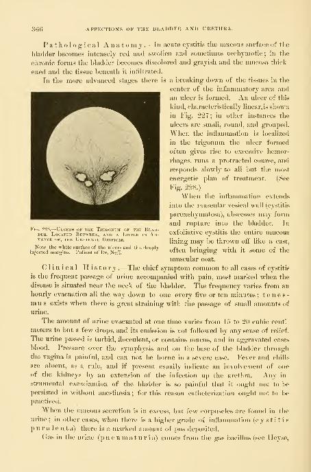
366 AFFECTIONS OF THE BLADDER AND URETHRA.
Pathological Anatomy .—In acute cystitis the mucous surface of the
bladder becomes intensely red and swollen and sometimes ecchymotic ; in the
chronic forms the bladder becomes discolored and grayish and the nmcosa thick-
ened and the tissue beneath it infiltrated.
In the more advanced stages there is a breaking down of the tissues in the
center of the inflammatory area and
an ulcer is formed. An ulcer of this
kind, characteristically linear, is shown
in Fig. 227; in other instances the
ulcers are small, round, and grouped.
When the inflammation is localized
in the trigonum the ulcer formed
often gives rise to excessive hemor-
rhages, runs a protracted course, and
responds slowly to all but the most
energetic plan of treatment. (See
Fig. 228.)
When the inflammation extends
into the muscular vesical wall (cystitis
parenchymatosa), abscesses may form
and rupture into the bladder. In
exfoliative cystitis the entire mucous
lining may be thrown otf like a cast,
often bringing with it some of the
muscular coat.
Clinical History .—The chief symptom common to all cases of cystitis
is the frequent passage of urine accompanied with pain, most marked when the
disease is situated near the neck of the bladder. The frequency varies from an
hourly evacuation all the way down to one every five or ten minutes ; tenes-mus exists when there is great straining with the passage of small amounts of
urine.
The amount of urine evacuated at one time varies from 15 to 20 cubic centi-
meters to but a few drops, and its emission is not followed by any sense of relief.
The urine passed is turbid, flocculent, or contains nmcus, and in aggravated cases
blood. Pressure over the symphysis and on the base of the bladder through
the vagina is painful, and can not be borne in a severe case. Fever and chills
are absent, as a rule, and if present usually indicate an involvement of one
of the kidneys by an extension of the infection up the urethra. Any in-
strumental examination of the bladder is so painful that it ought not to be
persisted in without anesthesia ; for this reason catheterization ought not to be
pra(;ticed.
When the mucous secretion is in excess, but few corpuscles are found in the
urine; in other cases, when there is a higher grade of inflammation (cystitis
p u r u 1 e n t a) there is a marked amount of piis deposited.
Gas in the urine (pn eu maturia) comes from the gas bacillus (see Ileyse,
Fig. 228.^Ulceks of the Tkigonum of the Blad-der, Located Between, and a Little in Ad-vance OF, THE Ureteral Orifices.
Note the white surface of the ulcers and the deeplyinjected margins. Patient of Dr. Netf.

CYSTITIS. 367
Centr. f. liiti. Med., Bd. xxiv), or from the decomposition of diabetic urine
;
gas is also observed when there is an entero-vesical iistula.
The duration of a cystitis varies from a short-lived affection to one of years'
standing ; the gonorrheal cases in women are most apt to recover quickly and
spontaneously. The catarrh of the bladder which is found associated with cal-
culus recovers when the cause is removed. The most protracted cases are those
of tubercular origin and those following labor. When cystitis is associated
with a diphtheritis or gangrene of the ' bladder the termination is speedily
fatal. In cases of old standing the muscular walls may become so hyj^ertrophied
as to form a hard mass like a tumor behind the symphysis.
Diagnosis .—It is always easy to diagnose a case of cystitis if the proper
examination is made ; this includes (a) a history of the illness, (b) examinations
of the urine, and (c) an examination of the mucous surface affected.
Many women actually under treatment for cystitis only suffer from hyperemia
of the trigonura, or mild inflammation of the upper urethra ; with a careful ex-
amination of the urine and inspection of the bladder such a mistake in diagnosis
could not occur. The history of the case includes the characteristic symptoms
jusf referred to, either coming on gradually or dating from some particular occa-
sion ; strangury and tenesmus are the most important symptoms. The examina-
tion of the urine reveals the presence of bacteria, and when they are found in
pure culture they are usually the cause of the cystitis.
In tubercular cystitis the discovery of only a few of the characteristic tuber-
cle bacilli, made after repeated searches, will be sufficient to make the diagnosis
clear. In one of my patients, my assistant. Dr. J. G. Clark, exposed an ulcer
in the bladder distended Avith air, and curetted off a little portion, in which
numerous tubercle bacilli were found. When the colon bacillus is the infecting
organism it is often found in the urine in pure culture.
The direct examination of the inflamed bladder gives the surest information
as to the existence of the disease, its grade, and its extent.
The knee-breast position is the most convenient one for seeing all parts
of the organ, but in milder grades of cystitis the artificial anemia induced
by the posture and expansion tends to obliterate the characteristic signs; it
is better, therefore, in these cases to examine in the dorsal position with a
slight elevation. The patient should be put under anesthesia for the first
investigation, so that it may be thoroughly made. The inspection begins at
the posterior wall, and extends in an orderly manner over the whole organ, as
described.
In this class of cases one is apt to find at the posterior pole a superficial layer
of blood on the mucosa, which has come from the trauma of the end of the
specula impinging on the inflamed delicate mucosa. The difference between
this and the submucous hemorrhage can be detected by wiping the surface with
a pledget of cotton, when the blood comes off. The affected areas are rendered
strikingly apparent by contrasting the normal whitish background and the deeply
injected patches of inflamed tissue. In the sound parts the capillaries are rarely
seen, and the pale mucosa is mapped out by larger branching vessels, but the dis-
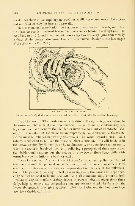
368 AFFECTIONS OF THE URETHRA AND BLADDER.
eased areas show a fine capillary network, or capillaries so numerous that a gen-
eral red color of varying intensity prevails.
By the bimanual examination the bladder is found tender to touch, and when
the muscular coat is thickened it may feel like a tumor behind the symphysis. In
one of my cases I found a hard ovoid mass as big as a hen's egg lyhig transversely
in front of the uterus ; this proved to be a tuberculous bladder in the last stages
of the disease. (Fig. 229.)
Fig. 229.—Tubercular Cystitis.
Shows the markedly thickened vesical walls and the method of palpating the bladder bimanually.
Treatment .—The treatment of a cystitis will vary widely, according to
the cause and character of the inflammation. When there is a continuously act-
ing cause, such as a stone in the bladder, or urine pouring out of an infected kid-
ney, or a stagnation of the urine in an imperfectly emptied bladder, these con-
ditions must be relieved before any progress can be made toward a cure. As a
rule, it is sufficient to remove the cause to effect a cure, and this will be done in
the instances cited by lithotomy, or by nephrectomy, or by nephro-ureterectomy,
when the ureter is involved too, or by relieving a prolapsus of these uterus and
the bladder, and washing out the stagnant urine two or three times daily with
warm boric acid solutions (2 to 3 per cent).
Treatment of Acute Cystitis.—An expectant palliative plan of
treatment should be pursued in acute cases ; under these circumstances local
treatment or interference of any sort aggravates the intensity of the inflamma-
tion. The patient must stay in bed in a warm room, the bowels be kept open,
and the diet reduced to licpiids and soft food ; all stiiruilants nnist be prohibited.
Prolonged vaginal douches, lasting fifteen to twenty minutes, given three times
daily, help to relieve the congestion ; hot applications should be kept on the
lower abdomen, if they give comfort. IJot sitz baths and dry hot bran bags
are also valuable adjuvants.

CYSTITIS. 369
While the pain is excessive and persistent it is necessary to give the patient
as much rehef as possible, and to insure some hours of rest and sleep every day.
Morphin meets this indication better than any other drug, and the best way to
use it is to give enough of it hypodermically in two or three doses to secure
about eight hours of continuous rest. During the remaining sixteen hours
codeia may be tried, and hyoscyamus and belladonna should be given in supposi-
tories, or an occasional rectal enema of 30 drops of deodorized tincture of opium
in 100 to 120 cubic centimeters of warm starch water.
As soon as the inflammation begins to subside, as shown by the lessened pain
and frequency of micturition, the convalescence will be greatly promoted l)y
washing out the bladder two or three times daily with lukewarm water contain-
ing 2 per cent of boric acid or 2 per cent of ichthyol.
Treatment of Chronic Cystitis .—The treatment of chronic cys-
titis must always be one of active interference. Four plans of attack are avail-
able : (a) medication, (b) irrigation or instillation, (c) direct topical treatment, (d)
surgical treatment.
Medication.—A great variety of drugs have been recommended as
benelicial in curing or relieving chronic cystitis ; there is a large amount of
trustworthy testimony in favor of salol in doses of from 3 to 5 grams
daily in divided doses ; under its use the symptoms abate, and the bacteria in
the urine diminish in numbers. Quinin, which is largely eliminated by the
kidneys, has a sedative eifect on the urinary organs, and is said to act effectively
in sterilizing the urine. Salicylate of soda in a 5 to 10 gram dose is also used
in some cases with good effect.
In the rarer gonorrheal cases oil of sandalwood or oil of copaiba in capsules,
5 to 10 minims each, give excellent results. Eucalyptus oil has also been found
useful in 10 minim doses every two hours. Fluid extract of zea ma is
corn-silk), in half teaspoonful doses, is the best drug I know to allay the irri-
tability of the bladder ; fluid extract of t r i t i c u m r e p e n s is used in the
same way.
A milk diet is of the best service in many cases.
Irrigation .—Irrigation, or washing the bladder out, is a necessary adju-
vant to other means of treatment; by this means the bladder is thoroughly
cleansed, and enormous numbers of bacteria removed with mucus and other
debris^ often imperfectly discharged in micturition, and for a time at least the
bladder walls are relieved from the constant contact with toxic products.
The irrigation may be carried out in two ways : either by letting small quanti-
ties of the fluid run in and directly out again, or by injecting larger quantities,
so as to distend the bladder sensibly, and then, after an interval of from a few
seconds to a minute, letting it flow out again. The latter plan has the advan-
tage of distending the bladder so that its entire mucous surface is cleansed by
coming into contact with the solution. The amount of discomfort experienced by
the patient should serve as a guide as to the amount of distention to be practiced
at each sitting, and with re])eated trials it will be found that the bladder grows
more tolerant. The irrigating solution is apt to give pain if its specific gravity
370 AFFECTIONS OF THE UKETHRA AXD BLADDER.
is much below that of the urine ; for this reason plain warm water, although
useful mechanically, is not well tolerated.
A satisfactory solution (see A. S. Lobingier, Medical Hews, Philadelphia,
Oct. 15, 1892, p. 425) is made with a powder of boric acid, borax, and chloride
of soda in the respective proportions of 4, 2, and 1 grams dissolved in half a
liter of hot water, and used warm.
Solutions of the bichloride of mercury have a bactericidal effect and are of
the utmost service in most cases of chronic cystitis. It is best to begin using
weak solutions of 1 to 100,000 in water in which a little common salt has been
added ("6 per cent), and to increase the strength each time until they are used
as strong as 1 to 10,000 or 1 to 5,000. It is well to vary these irrigations with
the milder boric acid solutions, using them on alternate days, or one in the morn-
ing and the other in the evening.
Weak solutions of carbolic acid, not stronger than 1^ per cent, often do
good service. When the distressing symptoms have cleared up and the urine
still remains purulent, Mr. Nunn {Lancet, Feb. 23, 18Y8) recommends the use
of a quinin wash, beginning with one grain of the neutral sulphate to the ounce
of water, with one drop of muriatic acid.
When the urine remains alkaline and there is a tendency to throw down
phosphates, Mr. R. Harrison {Twentieth Century Practice, vol. i, p. 230) recom-
mends irrigation with 5 to 10 grains of citric acid dissolved in a pint of warmwater.
The technique of the irrigation is as follows : The patient is put on a table,
or if she is too weak she is brought to the edge of tlie bed with the thighs
flexed and the buttocks resting on a perineal drainage pad ; the parts are then
freely washed with a weak boric acid solution, taking particular care to remove
all visible foreign material from the urethral orifice.
As an irrigating apparatus I use a simple glass funnel connected with a
glass catheter by a piece of rubber tubing four feet long. A clip or a pair of
forceps on the tubing controls the flow of the fluid. Unless the person giving
the injection can be relied upon to do it skillfully, it is better to use a rubber
catheter in place of a glass one, which may bruise the tissues.
The solution is now poured into the funnel, and allowed to run down and
fill the tube and catheter ; the injection of any air must be avoided, because it is
painful. The catheter is then introduced into the l)ladder and the funnel
held high enough to force the fluid slowly into the bladder ; after waiting a
while the funnel is now dropped below the level of the table and the fluid flows
back again. If the solution is comparatively clear the maenuver may be re-
peated, but as often as it becomes turbid it should be renewed. Irrigation may
also be practiced through a two-way catheter, such as that shown in the text
;
the fluid runs in the upper arm in the direction of the arrow and returns by the
lower.
Instillation.—Instillation differs from irrigation in that the medicated
solution is injected in smaller quantities and is left in the bladder for a time
in order to secure a more protracted action on the bladder walls. In this way

CYSTITIS. 371
solutions of nitrate of silver (1 to 2 per cent), sublimate solutions, and iodoformemulsions (5 to lU per cent) have been used.
Instillations have been systematically used with excellent results l)y M.Guyon, of Paris. Small quantities of fluid have been used, in accordance with
Guyou's dictum that the bladder does not possess an anatomical but a physio-
logical capacity, and
a painful bladder ^must not be dis-
tended. Solutions of
the bichloride of mer-
cury are used, in a
strength varying from
1-4000 to 1-500, be-
ginning with the
weaker and gradu-
ally mcreasmg up to je= Y\a. S.SO.—Two-way Catheter for Washing out the Bladder.
the stronger Solu- f TIk- tluid t-ntors and returns in the direetion of the arrows.
tions, and injecting
at first every other day and then every day as the patient's tolerance is tested.
A syringe is used holding about 5 grams (1^ drachms), and from 2 to 5 grams
are injected slowly into the bladder and allowed to remain there from fifteen
minutes to half an hour. The treatments must be continued for a period vary-
ing from some days to several weeks. Out of 34 cases so treated, 12 were cured,
9 were very greatly improved, 9 improved, and 4 unimproved.
Direct Topical Treatment .—By far the most efiicient way of treat-
ing chronic cystitis is by direct topical applications, which, however, should not
entirely supersede the use of internal medication or of irrigations or instillations.
Tliere are two ways of treating the affected areas directly : either l)y expos-
ing them to view and then applying a medicated solution, or by using a rubber
balloon, which is inflated in the bladder so as to distend its walls and bring
every point of its mucosa into contact with a medicated substance.
The first plan of exposing and treating the diseased patches is best in old
chronic cases where the patches are few in number and do not cover much sur-
face. This is done in the same way and with the same ease with which the
bladder is inspected. The patient is put in the knee-breast position and the
vesical speculum introduced, the air-distended bladder inspected, and the extent
of the disease accurately determined and mapped out on a diagram for future
comparison. The application is then made under direct inspection by means of
a pledget of cotton twisted on a wire applicator, taking care to touch nothing
but the diseased spots. This is easy if the parts are kept under view, and if
the cotton is not too wet with the solution. The amount of surface treated at
one time must be regulated by the kind of application made and by the extent
of the disease. It is also well to proceed cautiously at first by trying any of the
stronger stimulating drugs on a limited area and watching the effect. I often
use at first a 5 per cent solution of nitrate of silver, following it up by a 3 per
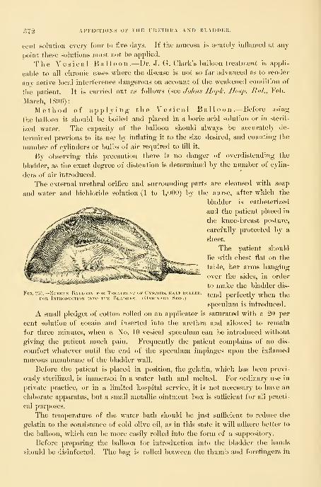
372 AFFECTIONTS OF THE URETHK.V AND HLADDKU.
cent solution everj four to live days. If the nmeosa is acutely inflamed at any
point these solutions must not l)e applied.
The Vesical Balloon.—Dr. J. G. Clark's balloon treatment is appli-
cable to all chronic cases where the disease is not so far advanced as to render
any active local interference dangerous on account of the weakened condition of
the patient. It is carried out as follows (see Johns Iloph. IIosj). BuL, Feb.-
March, 1896):
Method of applying the Vesical Ball o o n .—Before using
the balloon it should be boiled and placed in a boric acid solution or in steril-
ized water. The capacity of the balloon should always be accurately de-
termined previous to its use by inflating it to the size desired, and counting the
number of cylinders or bulbs of air required to All it.
By observing this precaution there is no danger of overdistending the
bladder, as the exact degree of distention is determined by the number of cylin-
ders of air introduced.
The external urethral orifice and surrounding parts are cleansed with soap
and water and bichloride solution (1 to 1,000) by the nurse, after whicli the
bladder is catheterized
and the patient placed in
the knee-breast posture,
carefully protected by a
sheet.
The patient should
lie with chest flat on the
table, her arms hanging
over the sides, in order
to make the bladder dis-
tend perfectly when the
speculum is introduced.
A small pledget of cotton rolled on an applicator is saturated with a 20 per
cent solution of cocain and inserted into the urethra and allowed to remain
for three minutes, when a No. 10 vesical speculum can be introduced without
giving the patient much pain. Frequently the patient complains of no dis-
comfort whatever until the end of the speculum impinges upon the inflamed
mucous membrane of the bladder wall.
Before the patient is placed in position, the gelatin, which has been previ-
ously sterilized, is inmiersed in a water bath and melted. For ordinary use in
private practice, or in a limited hospital service, it is not necessary to have an
elaborate apparatus, hut a small metallic ointment box is sufiicient for all practi-
cal purposes.
The tem])erature of the water bath should be just sufficient to reduce the
gelatin to the consistence of cold olive oil, as in this state it will adhere better to
tlie balloon, which can be more easily rolled into the form of a suppository.
Before preparing the balloon for introduction into the bladder the hands
should be disinfected. The bag is rolled between the thumb and forefingers in
¥iQ. 231.
—
Rubber Balloon for Treatment ok CvsrFOR Introduction into the Bladder. ( Okdi
I^, IIAI 1 ROLI.l 1
iR\ Size. I
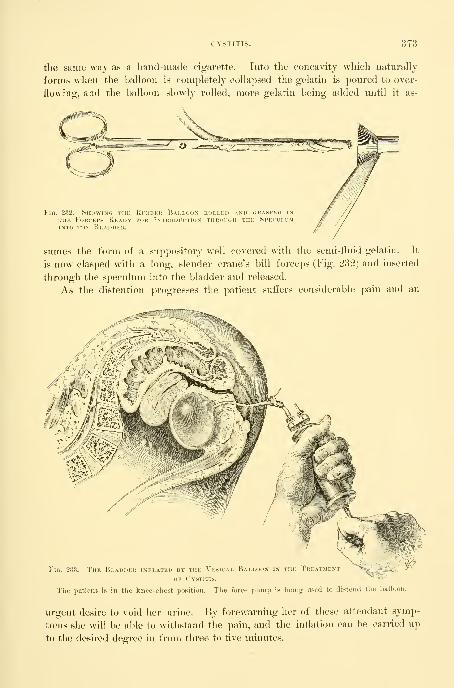
CYSTITIS. 373
the same way as a hand-made cigarette. Into tlie concavity wliicli naturally
forms when the balloon is completely collapsed the gelatin is poured to over-
flowing, and the balloon slowly rolled, more gelatin being added until it as-
FiG. 232.
—
Showino the Kcihseu Ballodx kolleu and (jrasped in
THE Forceps Readv for Intkodiction throihh the SpeculumINTO the Bladder.
sumes the form of a suppository well covered with the semi-fluid gelatin. It
is now clasped with a long, slender crane's bill forceps (Fig. 232) and inserted
through the speculum into the bladder and released.
As the distention progresses the patient suflers considerable pain and an
Fig. 2.33.—The B the TreatmentDER inflated BY THE Ve!<ICAL BaLLOON
of Cystitis.
The patient is in the knee-chest position. The force pump is being used to distend the balloon.
urgent desire to \oid her urine. By forewarning her of these attendant symp-
toms she will be able to withstand the pain, and the inflation can be carried up
to the desired degree in from three to five minutes.

374 AFFECTIONS OF THE URETHRA AND BLADDER.
The pain in chronic cystitis is usually severe during the first two or three ap-
plications, but the jDatient as a rule exiDcriences so much relief su])sequently that
she is willing to persevere in the treatment.
A rectal suppository of 1 grain of opium, introduced innnediately after the
treatment, is of great service in alleviating the subsequent suifering. Having
inflated the bag up to the required size, the clip on the rubber tube is closed
to prevent the escape of the air, and the patient assumes the dorsal or lateral
posture.
It is best to leave the balloon in place for fifteen or twenty minutes ; to
remove it the clip is released, when all but a small amount of air escapes and
the rest is aspirated with the air j)ump, when the collapsed rubber bag is easily
pulled out through the urethra.
Another way of using the balloon is the following : The patient empties her
bladder and then lies in the right or left semi-prone position, while the urethral
orifice is exposed and cleansed. From 5 to 8 centimeters of a 2 per cent solu-
tion of ichthyol in glycerin is then injected into the bladder by means of a deli-
cate long-nozzled syringe. The balloon, completely exhausted of all air, is nowtaken up with aseptic hands, stretched out a little, and rolled together in small
compass around a metal staff, so that the rubber projects a little beyond its end.
A clasp on the tube prevents air from entering too soon. The size and shape
of the balloon rolled up in this wsij is much like that of a catheter, and the staff
gives the stiffness necessary for introduction. The balloon, coated with the
ichthyol glycerin, is now grasped so as to prevent its unrolling and pushed
through the urethra into the bladder and the staff drawn out. The clasp is then
taken off and the balloon inflated, either by a rubber ball insufiiator, such as is
used in throat work, or by the force pump of an aspirator. The inflation should
be great enough to l)e felt decidedly, but not to cause much pain, when the rub-
ber tube is clamped to prevent the escape of the air. The inflated balloon dis-
tends the bladder and brings every point of its mucosa into contact with the
ichthyol already injected. The amount of the distention can also be gauged
by introducing a finger into the vagina and palpating the l)ase of the bladder,
or by examining bimanually. The bladder should not be larger than a goose
egg.
The balloon is left in place from ten to twenty minutes, according as the
[)atient can bear it ; it comes out collapsed npon letting out the air and pulling
on the rubber tube.
A history of a case, of a severe type of chronic cystitis of thirteen months'
standing, well represents the efficiency of the vesical balloon.
M. J., admitted October 21, 1895, colored, aged thirty-five years, had been
married ten years, with no children, and no miscarriages. She suffered from
frequent and painful micturition and hematuria.
About thirteen months ago she began to have slight ]);iin on urinaticm,
which grew rapidly worse, and for the last five nioutlis blood has frecpieiitly ap-
peared in the urine.
The frequency of urination is mucli greater at niglit, whcMi she is (•onq)eIled

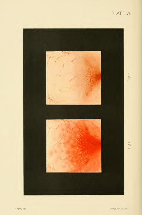
PLATE VI.
Mhrodel.fec. Lilh.LPrang&Co.Bosion.U.5.A

DESCEIPTION OF PLATE VI.
Fig. 1.—Appearance of the trigonum and the base of the bladder before treatment.
Fig. 2.—Appearance of the same part of the bladder after treatment with the ves-
ical balloon.
PioiT*ii5ioa3a
m

CYSTITIS. 375
to get lip eight to ten times ; a week ago she had agonizing pain and several
blood clots were passed. There is now a constant dull pain over the bladder,
which becomes sharp and cutting during micturition. When the paroxysms
come on the patient has an expression of intense suffering.
Upon making a direct examination of bladder the urethra was found con-
gested, the vesical trigonnm intensely reddened, the rugce standing out prominent-
ly, and over the surface of the bladder flakes of pus and small blood clots. The
intensest inflammation was in the inter-ureteric area gradually shading off
toward the fundus of the bladder. Where the inflammation was greatest the
mucous membrane was of an angry red color and bled when touched lightly
with the ureteral searcher. The capillaries were indistinguishable in the in-
flamed areas, and a careful search of the bladdei- failed to reveal the ureteral
orifices. In the less congested areas above the trigonum the capillaries were
prominent, and at various points small, intensely red congeries of minute vessels
were seen. The anterior wall of the bladder in isolated places appeared normal.
The treatment by an application of 10 per cent ichthyol gelatin by
means of vesical balloon gave great pain at the time of the application.
October 22 : Greatly relieved two hours after treatment, and still feels nmcli
better than before the treatment.
October 23 : Balloon again applied, still very painful ; the bladder appears
less congested and the ureteral orifices are faintly visible. Marked improvement
in symptoms ; urination much less painful. She rose only three times last night.
A colored drawing of the bladder as it now appears is shown in Plate VI.
November 10 : The bladder has been treated every third day since the last
note was made, and now appears almost entirely well. The patient no longer
experiences any pain between the treatments and thinks she is entirely well.
Advised to remain one week longer.
November 19 : Patient discharged to-day. The mucous membrane has as-
sumed a perfectly healthy hue, except a slightly increased reddening around the
ureteral orifices. No treatment since the last note. The pain is entirely re-
lieved, and the patient got up but once last night to urinate. The second colored
drawing (on Plate YI) shows the present condition of the bladder.
Applications may be made in this way every day or every second day ;the
improvement is usually marked from day to day, and old cases are sometimes
relieved in less than a dozen treatments. Glycerin must be used as the vehicle
for the drug injected into the bladder, and to coat the balloon when the bladder
is simply to be distended, as vaselin and oils ruin the rubber bags.
Surgical Treatment of Chronic Cystitis .—In obstinate cases
associated with great pain relief has often been given by making an opening in
the base of the bladder so as to let the urine escape into the vagina, keeping the
bladder empty and giving it a complete physiological rest for a period of several
months or longer until the cystitis is cured. This procedure has had a warm
advocate in Dr. T. A. Emmet, who has repeatedly employed it with success.
But as the constant dribbling of the urine through such an artificial fistula
entails all the distressing disagreeable consequences of a fistula from any other

376 AFFECTIONS OF THE URETHRA AND BLADDER.
cause ; it is to be expected that the field for this operation will l)e limited to the
cases not relieved by the direct plans of treatment just described.
The operation is done in the following manner : The patient is put in the
dorsal position with flexed thighs, a sound is introduced into the bladder dis-
tended with water, and the base of the bladder well behind the urethra is pushed
forward into the vagina into the median line on the end of the sound. The an-
terior vaginal wall being well exposed by retractors, the operator cuts through
the vesico-vaginal septum onto the sound with a narrow-bladed knife ; a gush of
water shows that the bladder is opened and the sound passes through into the
vagina. As the water escapes, the hole is quickly enlarged backward until it is
at least 2 centimeters long. The sound is then taken out and the vesical mu-
cosa drawn over the intervening cut surface of the septum and attached to the
vaginal mucosa on all sides by a continuous catgut suture ; this prevents the
fistula from closing spontaneously, as it would do if raw surfaces were left ex-
posed and in contact. The vagina should be irrigated daily, and the extei-nal
parts protected by a stiff zinc oxide ointment. Clean gauze pads must be kept
under the patient at night and fresh ones applied often by day. When the in-
flammation has subsided, in the course of several months, then the edges of the
fistula should be pared and the opening closed.
Tubercular Cystitis.—Tubercular disease of the bladder in women is observed
with a frequency which increases just in proportion as careful direct examina-
tions and bacteriological investigations of the urine are made. It is either pri-
mary in the bladder or descending from the kidney to the bladder, or agahi
a part of a general tuberculosis.
R. Ultzmann {Die Krank. der HanMase, Stuttgart, 1890, p. 151) states that
tubercular cystitis often complicates other inflammatory processes, and more espe-
cially those due to gonorrhea, when the gonococci may be found in close asso-
ciation with tubercle bacilli.
T. Kovsing {Die Blasenentzmidungen^ etc., Berlin, 1890) declares that
tubercle bacilli can not engender a tubercular cystitis in a sound bladder, not
even when there is a retention of urine, but that a direct inoculation into the
mucosa or a preliminary suppurative cystitis are necessary factors.
The disease is not often seen in its initial stages, when there is simply an in-
tense catarrhal condition of the bladder. In cases of infection from a tubercular
kidney or a tubercular abscess behind the broad ligament discharging into the
bladder, the infection is most marked in a path in front of the ureteral orifice of
that side, or in front of the sinus opening into the abscess where infected urine
meets the mucosa before dilution. In addition to the catarrhal cystitis found
here, there are often numerous little scattered whitish nodules having the ap-
pearance of tubercles. Sooner or later caseation occurs and the tubercles break
down, leaving a deep ragged-edged ulcer; the urine then contains pus and
mucus and blood.
The tubercular ulcer or ulcers may advance but slowly ; in the worst cases
the entire bladder is involved, and the bloody urine is constantly expelled in
small quantities with great sutfering. The trigomim, the base, and the posterior

TUBERCULAR CYSTITIS. 377
walls are oftenest affected. If the disease is left to run its natural course it is
always chronic, extending over a period of many years with slow changes. In a
more intense form, when the entire bladder participates in the disease, it mayterminate fatally in a few months or a year.
The family history may give the right clew to an obscure obstinate form of
cystitis, as in the following case occurring in the practice of Dr. L. M. Sweet-
nam, of Toronto
:
The patient, a young woman twenty-three years old, began to have severe
pain in the urethra, and died eleven months after the onset of an extensive tuber-
culosis involving the bladder, the urethra, and the right ureter and kidney ; the
temperature at one time reached 108° F. In the same family twin sisters died
of pulmonary tuberculosis ; another sister had tubercular glands in the neck
four months after the extraction of some teeth ; a fourth sister lost one eye and
a nasal bone, and had five or six tubercular skin lesions, a spina ventosa affecting
three metacarpal bones, and a tubercular tarsus. A brother, twelve years old,
and both parents were healthy. In four cases of tuberculosis of the bladder
reported by Dr. Edward Keynolds {American Medico - Surgical Bulletin,
April 4, 1896) the family histoi-y was negative in two and distinctly tuberculous
in two.
Diagnosis .—A cystitis occurring in a phthisical patient not exposed to
gonorrhea is most likely tubercular too. All cases of cystitis in young persons
are open to this suspicion if they are clearly not gonorrheal. S. Bontor {Brit-
ish Med. Jour., 1893, vol. i, p. 1058) reports a case of a tubercular ulcer over
the left ureteral orifice in the bladder of a child five years old. Chronic ulcers
found in the bladder are usually tubercular.
The diagnosis is made certain by finding the tubercle bacilli either in the urine
or in the tissues themselves. When the disease is advanced the bacilli can usually
be found in large numbers without any trouble, but in other cases repeated ex-
aminations nnist be made to find even a few of them. The surest way to find
the bacilli and so clear up the diagnosis is to expose the diseased area and curette
off a little piece of the tissue from the margin of the ulcer ; the microscopic ex-
amination of this may show the presence of the bacilli when they have been
sought in vain in the urine.
In Reynolds's four cases there were a few isolated papules discovered in each
case. They were about the size of a grain of rice, slightly oblong, glistening,
rounded, and round above the surface of the mucous meml)rane. Tliese papules
were carefully watched, and were found to break down and form tuberculous
ulcers.
The tubercular ulcer is characterized by a granulating base, sharp, irregular
edges, \ni\\ small hemorrhagic foci surrounding it. In the most advanced cases
the contracted bladder simply appears as a mass of ulcerations with irregular
surfaces filled with pus and blood and mucus. When the disease descends from
the kidney, the part first affected is that about the ureteral orifice of the affected
side,
E. H. Fenwick {Lancet, 1891, p. 935) injected tuberculin in a patient with

37S AFFECTIOXS OF THE URETHRA AND BLADDER.
a tubercular ulcer witli the result of l^ringing on a violent attack of hematuria
;
others have used the injections with negative results.
Treatment .—The outlook in a case of tuberculosis of the bladder from a
therapeutic standpoint is no longer so hopeless as it was before the use of the
endoscope. AVe are able with our present diagnostic methods not only to deter-
mine the specific nature of this disease, but to discriminate between the exten-
sive cases and those which remain localized and are more amenable to treatment.
In all these cases the general health demands the most painstaking-
attention ; associated wnth rest, abundant nutritious food, suitable exercise in the
fresh air, and it may be change of climate and scene, we must also depend
largely upon such medical agents as cod-liver oil, iron preparations, and other
tonics which have been found useful in combating tubercular ])rocesses of all
kinds.
When tlie bladder disease forms Init a part of a more general infection, or is
associated with an extensive lung affection, but little will be accomplished locally
in staying the pr-ogress of the affection. When the disease is in some neighbor-
ing organ, as in a kidney or a uterine tube, and the bladder is only secondarily
mvolved, the original focus must be removed before any results
may be expected from the treatment of the bladder, and if the disease is ])ut
limited, it may clear up without further assistance.
The direct treatment of a tubercular bladder is either by injection, by topical
applications, or by surgery.
As injections, iodoform ennilsions (5 to 10 per cent), solutions of corro-
sive sublimate (from 1 to 5,000 to 1 to 500), nitrate of silver (1|^ to 2 per cent),
and lactic acid (5 per cent solution with cocain) have been used;good results
are to be looked for from the use of iodoform made into an emulsion (5 to 1(>
per cent) with glycerin, gum acacia, and water. This should be injected into
the bladder and applied evenly to the whole surface under moderate pressure
by using Clark's balloon as described in the last section. Such treatments con-
tinued every two or three days are calculated to yield the maximum effect of
local treatment. By the instillation methods above described Guyon has been
able out of sixteen cases of tubercular cystitis to cure three, to improve six
greatly, and to improve five.
A case of vesical tuberculosis re])orted by Dr. ,\. O. Polak, of l]rooklyn
{Ainer. (ri/zt. and Ohs. Jour.^ January, 189T, p. -il), is a model of clear descrip-
tion and effective treatment.
The patient, a single Swedish girl, twenty years old, during an attack of
anemia three years ago began to suffer sharp lancinating pains on urination ; she
lost weight and had frequent hematuria. In the latter part of April, 1896, she
had an unusual vesical hemorrhage with inability to void the urine and a con-
stant intense tenesmus ; several ounces of bloody urine were drawn by catheter
drop by drop. The temperature was 102° and the pulse 120. On palpation,
there was exquisite tenderness all over the region of the bladder and blood
dripped from the urethra.
By means of a large cystoscope the l)ladder was found filled with clots of

TUBERCULAR CYSTITIS. 379
blood ; after washing and sponging them out, an ulcerated area was detected on
the right side near the neck of the bladder, about 3 centimeters in diameter, with
raised irregular edges and studded with tubercles ; the remaining mucosa was
normal. This patch was thoroughly curetted through a cystoscope, and vesical
drainage established by a coil of gauze enclosed in gutta-percha tissue.
Three days later the urine still contained pus, blood, and tubercle bacilli.
Daily vesical irrigations were used, and a 50 per cent emulsion of iodoform in-
jected after each one.
May 15 : A direct examination revealed an area of ulceration with pale, flabby
granulations, and appearing as though varnished with a thin coat of serum. Asolution of nitrate of silver (SO grains to the ounce) was carefully applied to the
dried surface of the ulcer, followed by a daily irrigation with a solution of sali-
cylate of soda. Prompt improvement followed, the bacilli steadily diminished
in numbers, and by June 1st the urine was normal.
June 10 : By direct cystoscopy a normal mucous membrane was seen, and the
place of the ulceration was occupied by a pale cicatrix. After this the patient
gained twenty-five pounds in weight.
October 15 : !N"o recurrence of symptoms.
Petit {Lijec. contre la tuherculose de la vessie, Semaine med.^ 1892, t. xii,
p. 42 [annexes]) uses an emulsion consisting of iodoform, 20 grams, glycerin,
10 grams, and water, 6 grams, with gum tragacanth, 0-25 grams. A tea-
spoonful of this mixture is added to 150 grams of warm water with 10 drops
of laudanum, and the whole is injected slowly into the bladder; half the quan-
tity is allowed to run out in two minutes and the rest to remain as long as pos-
sible. This same formula may also be used with the rubber balloon.
Topical Applications .—The means we now have of exposing the
tubercular ulcer and of making direct applications to its surface opens up an
entirely new field for therapeutic research, since we shall be able to apply con-
centrated solutions to tlie diseased spots without risk of injuring the sound
mucosa elsewhere. After making such an application the bladder may be filled
with water and washed out repeatedly. The application in this way of varying
strengths of silver nitrate from a 20 per cent aqueous solution to the solid stick
have proved of great value in Reynolds's hands.
Surgical Treatment.—The surgical treatment of such an obstinate
affection will prove the most satisfactory way of dealing with it in selected cases.
The various surgical methods are curettage, c a u t e r y, and excision,
Chirettage of the B 1 add e r.—Curettage has been successfully prac-
tised by Guyon and others in cases of rebellious cystitis and for tuberculous
cystitis. Curettage is an eminently rational plan of treatment for two reasons
:
in the first place the lesions of l)oth forms of cystitis are as a rule localized in
the more superficial parts of the bladder in the mucosa, and hence easily re-
moved without risk, and in the second place the regeneration of the mucosa,
even after an extensive destruction, takes jDlace readily.
The procedure is conducted in this way : A sharp curette is used, the patient
placed in the lithotomy position, and the bladder thoroughly washed out with a

380 AFFECTIONS OF THE URETHRA AND BLADDER,
mild antiseptic solution and emptied. Tlie tina^er is then inserted into the
vagina and the curette directed through the viretln-a into the bladder, where it is
first employed in curetting the base of the bladder, using the finger as a point
of counter pressure ; at the same time the amount of force employed can be
readily estimated ; then the curette is next used over the vesical surface behind
the symphysis pubis. After cleansing out the bladder the curette is again intro-
duced, and the posterior lateral and superior portions are next attacked. Such
is the procedure in the rare cases of aggravated cystitis which has gone so far as
to involve the entire vesical mucosa ; by making a careful cystoscopic examina-
tion l)eforehand those cases in which the lesions are localized in certain areas of
the bladder will also be recognized, and the curettage will then of course only
be directed against the diseased portions, sparing the sound tissues any unneces-
sary insult.
It will now be easy to use the galvano-cautery through the cystoseope in the
air-distended bladder, and to continue its use from time to time so as to test the
efficiency of this mode of treatment.
The ideal mode of treatment of a tuberculous ulcer, which is not relieved l)y
the simpler plans just detailed, is its exposure and excision, followed
by suture. This is easily practicable through a longitudinal suprapubic incision
without opening the peritoneum ; the bladder is incised parallel to the abdominal
wound, and its edges temporarily stitched to the skin surface to keep it from
dropping out of easy reach during the operation ; then in the more su])erficial
cases of the disease the mucosa may be extensively excised, being cut through
a short distance from the affected edges and detached by a blunt dissec-
tion. Any little patches of sound mucosa nnist be carefully left, as they will
materially aid in the rejuvenation of the nmcosa, acting like grafts on the skin
surface.
R. Bardenheuer {CentraM. f. Gijn.^ 1894, Bd. xviii, p. .38()) has even excised
the entire nmcosa without interfering seriously with the bladder function. It
is a question whether in such cases the new mucosa grows out from the urethral
and ureteral orifices, or is reproduced from minute portions left in numerous
little depressions. When the disease extends deeply into the vesical walls, the
tuberculous area must be cut out and the edges then In-ought together by inter-
rupted catgut sutures as in the treatment of a vesico-vaginal fistula.
When the tuberculous area is about one of the ureteral orifices, the operator
must determine whether the upper urinary tract of that side is affected ; if it is
not, the area may be excised and the ureter turned into the bladdei- at a jxtint
farther back.
It will be best in these cases not to leave a suprapubic drainage opening.
The incision into the bladder through wdiich the oj)eration has been conducted
should be sewed up independently of the abdominal incision by using a series of
interrupted catgut sutures catching only the nmscular walls. The abdominal
incision should be closed by interrupted silkworm gut or silver wire sutures,
and the bladder kept drained through the urethra for five or six days.
If the sutures in the bladder do not come away in twelve or fourteen days,

EXFOLIATIVE CYSTITIS. 381
and tliere is any evident ii-ritation from their presence, they may be exposed and
cut and taken out through a No. 12 or l-t vesical speculum.
Exfoliative Cystitis.—The expulsion of a part of the whole of the mucousmembrane of the l)ladder sometimes occurs as a form of cystitis (cystitis
exfoliativa). Tliis is a disease to which women are peculiarly liable,
although it is also rarely seen in men.
There is a clear and excellent account of a case in Nicholas Tulp's Observa-
tlones rnedicce (Amst., 1672, lib. 2, cap. xlviii), under the title membranalapidescens (membrane turning into stone). Gerbreghta Rotaria, thought
to have a calculus, finally passed a large membrane covered with little stones
and with a perforation in the middle, so that the urine could escape through it.
But some pieces of the membrane remained behind, and until Nature freed her
from them she suffered excessively. The fibers of the bladder being relaxed,
there was an involuntary escape of the urine, which was finally relieved bytissue-strengthening medicines.
The causes of such a detachment of the mucous membrane are various,
the commonest being retroflexion of the gravid uterus, which is found in 50 per
cent or more of the cases ; other causes are protracted birth (25 to 30 per cent),
the pressure made by a myoma choking the pelvis (see A. Gottberg, Inaug.
Dis., Kiel, 1892, and Fr. Yahle, Inaug. Dis., Marburg, -1895), retention of urine
and in one case the injection of a strong saline solution into the bladder (Be-
gouin, Jour, de ined de Bordeaux.^ t. xxii, p. 158).
The common factor underlying all the various causes is probably the
ischemia produced by pressure, for the cutting off or lessening of the vesical
blood supply results in an ischemic necrosis with or without an infection.
The detached membrane may be extruded either entire or in small pieces ;
the mucous surface may be so altered by the necrotic changes that it is recog-
nizable with difficulty, and its surface is often studded with uric acid crystals.
Adhering to it are often found more or less extensive portions of the muscular
coat, and in the worst cases the peritoneal coat is also involved ; in this waythree grades of the affection are established (H. Boldt, Suppurative Exfoliative
Cystitis, Amer. Jour, of Ohstet., vol. xxi, 1888, p. 301).
H. S. Cocram reports a case four months pregnant {Medical News, Phila.,
vol, Ixiii, p. 633) in which the bladder was distended to within an inch of the
ensiform cartilage, the entire mucosa became detached and was expelled, and
the openings for the ureteral orifices could be seen in it. The patient was able
to leave the hospital in twenty days, and recovered completely.
The clinical history shows frequent micturition and overdistention of the
])ladder, often with dribbling, associated with a retroflexion of the gravid uterus,
or a pelvic tumor, or following a difiicult childbirtli. Schabert reports a case
{St. Petersburg, med. Woch., Bd. xix, p. 373) following the birth of twins.
The bladder becomes exceedingly tender, and there is an acceleration of the
pulse with fever, and all the disturbances usually associated with a severe septic
process.
In a case of my own, where an overdistention of the bladder followed ovari-

382 AFFECTIONS OF THE UKETHRA AXD BLADDER.
otomy, portions of the mucosa were cast off, and the patient had a higli fever
and became insane ; she was put into an asylum, where she died more than a
year later from pulmonary phthisis.
The urine, at first turbid, becomes fetid, and in a few days pieces of the
membrane are expelled ; when the membrane becomes detached and endeavors
to escape entire, it may block the internal urethral orifice for a time, causing
a renewed retention of the urine. Its expulsion may be brought about with
great pain and straining, and after this there is a more or less permanent drib-
blino- of the urine. In some cases, in time the bladder regains its function to
an unexpected degree ; in others it is never able to hold more than a little urine
at a time.
Death may occur from sepsis, or from one of the complicating conditions,
such as peritonitis, or pyelitis from an upward extension of the infection, or later
from uremia.
The diagnosis is made by recalling the clinical history of the case asso-
ciated with a cystitis and discharge of the vesical tissue. On examining the base
of the bladder by the vagina, it is found thickened and tender, and in cases
where the loosened tissue obstructs the urethra the catheter may perforate this
and let out a lot of foul urine from behind it. The difference between these
septic cases with such a history, and tlie prolapse of the vesical mucosa occur-
ring mostly in children, is so marked as to need reference only.
The treatment must first be directed to the condition which causes the
retention, if it is still active, and, secondly, to the condition of the bladder itself.
If the uterus is retroflexed and incarcerated, the patient must be put under an
anesthetic, if necessary, in order to reduce the flexion, when it may be kept in
place by an appropriate vaginal pack.
If the pelvis is choked by a myoma, an effort should l)e made to dislodge it
into the abdomen. If this can not be done, it will scarcely be advisable to open
the abdomen and remove the tumor until the vesical symptoms have subsided.
The bladder itself must be carefully watched to prevent any large accumu-
lation of urine in it, and when the membrane is in the process of detachment
and expulsion it is best to assist nature by gentle traction, and cutting off any
protruding portions. If the membrane chokes the urethra and prevents the
escape of urine, the accumulation will be voided by passing a glass catheter
through it.
After the early acute symptoms have passed off, the patient will l)e greatly
benefited by washing out the bladder two or three times daily, using a warmboric acid solution, and the irrigation nnist be kept up as long as there are pus
and bacteria in the urine.
TUMORS OF TIIP] BLADDER.
A variety of tumors are found in the bladder in women, but not so frequent-
ly as in men, the proportion being about one to three or four. They may be
grouped according to their clinical significance—that is, their tendency to remain

TUMORS OF THE BLADDER. 383
localized or to invade the surrounding tissues, as benign and malignant (see G.
Clado, Traite des tu7neurs de la vessie^ Paris, 1895, p. (i3).
I. The benign tumors are papilloma, fibroma, adenoma, myoma, dermoid cysts.
II. The malignant tumors ai-e carcinoma, sarcoma.
The malignant group further includes all forms of tumors of a mixed nature
except fibro-myomata and all forms of degenerated tumors.
If we bear in mind the several component tissues of the bladder walls—the
mucosa and subnmcosa and the muscular layer—and examine the various neo-
plasms found there from the standpoint of origin, they may be classified as fol-
lows (Kiister, Volhm. Sammhing Min. Vort., 1880)
:
I. Connective tissue tumors of the mucosa and of the submucosa : Papillo-
mata and fibroid jsolyps, mucous polyps, sarcomata.
II. Tumors of the muscular tissue : Myomata.
III. Tumors of the glandular tissue and epithelium : Adenomata, epithelio-
mata.
Tumoi's are furthermore primary when they originate in the bladder, and
secondary when they extend to the bladder from other organs. The secondary
tumors are naturally of the malignant type ; the connnonest form is carcinoma
of the cervix uteri which advances to the bladder.
Nothing whatever is known as to the etiology of primary vesical tumors.
J. Albarran (Zes tumeurs de la vessie, Paris, 1892) is of the opinion that the
chronic irritation produced by the presence of micro-organisms is an efiicient
cause.
The villous outgrowths covering the interior of the bladder (vesica v i 1 -
losa), in some cases associated with calculus, are undoubtedly the product of
mechanical irritation, but these can scarcely be called neoj^lasms in the strict
sense.
Clado has shown that the normal l)ladder contains villi, which throws light on
the frequent association of villous outgroM'ths with all variety of bladder tumors.
Secondary tumors of the bladder are not usually metastatic in the ordinary
sense, but invade it by contiguity of tissue. In men, most of these tumors are
furnished by the prostate and the rectum, and in women the enormous fre-
quency of cancer of the cervix uteri is the occasion of the frequent involvement
of the base of the bladder. I have seen the most extensive sarcoma of the
genital system from the vagina through the uterus and out onto the peritoneum
without any bladder affection.
The seat of vesical tumors is found more frequently in certain areas than in
others. The places of predilection are exhil)ited in Fere's table, where, out of
107 cases, there were in the base of the bladder alone, 25 ; attached to both base
and the walls together, 13 ; on the posterior wall, 17 ; close to the right ureter,
5 ; close to the left ureter, 8 ; anterior wall, 2 ; anterior and superior wall, 1
;
right or left lateral walls, 4; multiple tumors, 12; diffuse tumors, 8, etc.
Out of 634 cases of polypi, Fenwick (British Medical Journal, 1888, vol. ii,
p. QQQ) found that the tumors were single in 60 per cent and multiple in the
remaining 40 per cent.
384 AFFECTIONS OF THE UHE:TIIRA AND BLADDER.
Albarran {>it supra, page 01), in an analysis of 82 cases, found that tlie
tumors were single in (51 and multiple in 21 of the cases, a proportion of about
76 per cent.
One of the most important statistical subdivisions of these tumors, ironi a
clinical and a therapeutic standpoint, is that which is based on the nature of the
attachment of the neoplasm to the vesical walls. Albarran {ut supra^ page 58)
found in seventy-eight personal observations of tumors of the epithelial type (in
which are included all the commonest forms—the papillomata, cysts, adenomata,
and epitheliomata) that they were attached as follows : Pediculated, 28 ; sessile,
9; encephaloid (infiltrating), 31; cancroid (mammilated, bossed, ulcerated), 1<>.
No two writers are precisely agreed in their classification of these tumors,
and in many of the instances reported the diagnosis has been made purely from
the macroscopic appearances, and this accounts for the enormous preponderance
of tumors described as "papillomata," which really include fibroid, adenoid,
and malignant epithelial growths. Although it is true that a benign growth
may exist in the same bladder with a malignant one, or that a benign growth
may become malignant, this transition would not be noted so frequently in the
literatui'e if careful microscopic examinations were made in all cases.
Papilloma.—As J. Orth {Lehrh. d. Speciel, Pathol. Anat., Berlin, 1880, Bd.
ii, p. 214) very properly says, the general name papilloma may be given to the
group of pediculated tufted tumors as long as we do not know to which special
class the growth belongs ; it may be either a benign papillary fibromaor a malignant papillary cancer. We see from this that the term papil-
loma is often employed simply to describe the form and general appearance of
the tumor, without conveying any information as to its real character.
If, on the other hand, we limit the term papilloma to the group of benign
tumors, we are met with the further difiiculty as to the propriety of the name
according as we consider the tumor primarily an outgrowth from the epithelial
or from the connective tissue.
If it is a tumor of the submucous connective tissue, covered by the mucosa
and pushing out into the cavity of the bladder as it grows, then the proper name
is papillary fibroma (Virchow, 1885); this view makes the papillomata
one of the group of fibromata which differ among themselves in possessing more
or less connective tissue.
Clado, on the other hand, considers papillomata as epithelial growths of the
nmcosa of an exogenous type—that is, one in which the epithelium is confined
to the exterior. This classification groups them with the adenomata and estab-
lishes also a certain relationship between them and the epitheliomata, which are
of the endogenous (ingrowing) type.
The benign papillomata are made up of a framework of connective tissue,
more or less abundant, richly supplied with blood vessels, and covered every-
where with the vesical epithelium. They usually have a tufted, villous, branch-
ing appearance, and are so vascular that the name " villous anginoma" has been
given to them. Sometimes the interspaces between the prolongations are filled
with detritus, when the fungating appearance is lost.

ADEXOMA. 385
Tliey occur at any age—from six and nine months (Stein) to seventv-seven
(Gaillard)—and may be either single or multiple, and they frequently
complicate other tumors.
Clado distinguishes three varieties—the villous, the pediculated, and the coro-
noid. The villous j)apilloma appears in the form of filaments growing from the
surface of the mucosa, and they are more or less grouped. When the wholebladder is covered by them the name vesica villosa (Kiister) has been given
it. These filaments assume a shape like that of a finger or ribbon, cylindrical
or conical, and often subdivide once or twice. The pediculated polyps, consti-
tuting the commonest form, are grouped on a cylindrical pedicle which may be
several centimeters long. In the coronoid form the aifected portion of the blad-
der has the appearance of a number of crests closely applied and looking indi-
vidually like a cock's comb.
In all benign papillomata the pedicle never passes beyond the limits of the
mucosa, however thickened or infiltrated this may become by inflammation,
although the base of the growth may sometimes contain muscular tissue. Thesize of a papilloma varies from that of a pea to a walnut ; they are rarely as
large as a hen's egg.
Fibroma.—The fibromata ()r fibroid polyps form a group of benign tumors
in which the connective tissue elements are in excess. They are less frequent
than the papillomata. which have Imt a scanty fibrous framewoi'k and appear to
occur oftener in men than in women.
The tumor is usually pediculated and its surface is smooth or slightly lobu-
lated, and the pedicle is usually a delicate one. When the tumor is sessile and
situated within the bladder wall, its connections with surrounding tissue are such
that it can be enucleated. Although the pedicle and the mucous surface of the
tumor are vascular, the interior is but poorly supplied with blood vessels. The
fibromata often enter the group of mixed tumors by undergoing a myxomatous
degeneration (see F. Schatz, Fibromyxoma telecutgiectodes vesictp, etc., Ardnv.
f^Gijn., 1876, Bd. x, p. 356).
Adenoma.—The adenoma is a benign epithelial tumor of the glandular type
rarely met with ; it is sessile or pediculated, and has a smooth, lobulated, or papil-
lary surface. When sessile, the tumor can be easily enucleated with the finger
without hemorrhage.
It is difiicult, in the light of our knowledge of the histology of the bladder,
an organ which is remarkably deficient in glandular elements, to account for the
origin of these tumors ; for this reason Klebs and others have insisted that these
growths must take their origin in the prostate gland in the male. R. Kaltenbach
(Langenbeck's vlrcAfyy?7/'Z^/«7^. (?//«•., 1884, xxx, p. 659), however, has described
a papillary adenoma which he removed by a vesico-vaginal incision from a womanforty-four years old, the origin of which Prof. Bostrom traced to the mucous
crypts of the bladder. Yon Fritsch has also described a fibro-adenoma of the
bladder in a girl three years old ; it was covered with a calculous deposit and
filled the whole bladder. These cases, of course, show that such tumors do
occur in the bladder independently of the prostate.
28

386 AFFECTIONS OF THE TRETHRA AND BLADDER.
The adenoma may be either sessile or pediculated, and its surface smooth^
lobulated, or villous.
Clado cites exceptional cages where "adenomata" (cylindrical-celled epitheli-
omata) have relapsed after extirpation, and infiltrated the bladder walls like
ordinary epitheliomata. This rare occurrence mnst be distinguished from the
tendency to relapse in, situ after incomplete extirpation which the adenomata
share in common with the simple papillomata.
Myoma.—Myoma is one of the rarer vesical tumors, first described by Vir-
chow {Die Krankhafte)i Geschwulste, Bd. iii, p. 121, myocarcinoma). It takes
its orio-in in the muscular coat of the l)ladder, and is therefore made up of
smooth muscular fibers with more or less connective tissue, and grouped or
interlacing as in uterine myomata. The tumor either develops out into the
bladder cavity upon a thick pedicle or it remains sessile.
W. T. Belfield (Wien. med. Woch., 1881, No. 12, p. 329) has described a
new variety of external vesical myoma occurring in a woman fifty years old. It
was ovoid in form, 2 by 1 by 2 centimeters, and attached to the outside of the
muscularis by four strands made up of blood vessels and muscular tissue.
J. Verhoogen {Cent. f. Ham und Sexual- Organe^ 1895, p. 132) describes a
like case, occurring in a man aged twenty-three years. The tumor at the oper-
ation was found to be about the size of a child's head. It arose from a pedicle,
just above the prostate gland, and extended backward and upward, almost
filling the pelvis. Microscopically it was found to be a filn-o-myoma.
Cases are also described by Felix Terrier and Henri Ilartmann in the Revue
de chir., Paris, 1895, p. 181.
The mucous covering of the vesical myomata is intensely congested, and the
remaining muscular coat of the bladder hypertrophied. Ulceration of the sur-
face is rare.
Cystic Follicles.—Small cysts are sometimes found on the inner surface of
the bladder due to an occlusion of the mucous follicles ; they appear scat-
tered or in groups, forming little translucent elevations from 2 or 3 milli-
meters in size up to the size of a split pea. I have observed these in a case of
chronic cystitis ; on touching a cyst with the point of a knife the contents im-
mediately escape, and the only trace which remains is a slight hemorrhage from
the base. This affection has been called vesical herpes. Malignant tu-
mors also often undergo cystic degeneration.
Dermoid Cysts.—Dermoid cysts of the bladder are so rare that Orth {ut suj}n/)
says that only one well substantiated observation exists, that of Sir James Paget
{Surg. I\(th., 1853). Albarran cites a case of Boucher {Soe. anatomique., 1840)
somewhat doubtfully, stating that there was a cyst containing a fatty liquid at
the top of the bladder, and communicating with it by a narrow opening.
Outside of tliese rare observations, cases have been recorded in which der-
moid cysts outside of the l)ladder (sec Sanger, Archivf. Ggn., 1879), or ovarian
dermoids, have discharged their contents into this organ, and hairs have escaped
by the urethra (pilimiction).
In the group of malignant tumors we find two types of tumors repre-


Heliotypp'ftui.titi^ ''''.'^"'^'

DESCRIPTION OF PLATE VII.
Fig. 1.—Shows the normal bladder laid open by an incision through the anterior
wall. The ureteral orifices are seen as narrow slits at the two posterior angles of the
trigonum ; the third angle is at the internal urethral orifice. The trigonum is char-
acterized by its increased vascularity between these three points. The longitudinal
vesical folds entering the urethral orifice are well shown.
Fig. 2.—Secondary carcinoma of the bladder following carcinoma of the cervix.
The carcinoma appears in the form of rounded nodules in the bladder wall, mainly
in the vicinity of the cervix. One small nodule is seen in the trigonal area. Note
also the thickened walls of the bladder.
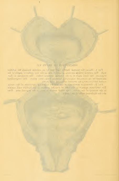
lohaiaB 9ffi d-guo'idi noiaioni fi ul 'labbjiW iiinriorr edi ewoilS—.1 .oi'i
exii io 89l^ftB lohaJaoq owi 9rii : un afi neaa 9iii geoftho lenaJeiir eriT .IIbv/
-•ifirfo ai inxiao^ki exiT .aoftho ihiLhtMi ifitnaJai 9rfJ is ai sI'gnB biixf;t arii ; nrx/no-ghJ
[eaibiiii-gaoi eriT .ainioq 99iri;t oaarii xiggV/jyd -^iiiBliroaBV b98B9ioai aJi -^d b9sh9ioB
.ifworia [f - '^'io Iii'iifi9iu edi •gcihaiae ahlo^ Ifioiagv
.xivi9o 9rii \o BmoxiioiBO^ctiwoilol ''to Boioiiio'iBO ^iBbaoogg— .S .Oi'i
^jrlniBra ,IIb7^ i9bbBl(:{ ^''^ '^' -,,ir,i,..,, ^nfol arft ni 8'iB9qqB BmonbiBD 9iIT
9io>T .B91B Ifiao'gi*) - eaO .xiyiso sriJ ^o "^iinioiv 9if>t ni
fcbijIJ 9'dJ ^C) ?.[[siw hBdfiihidi sdi okU

SARCOMA. 387
gented—the epithelial type, the carcinomata, and the connective tissue type, the
sarcomata. The most frequent are the carcinomata, and these are commonest in
men as primary tumors of the bladder, while in women they invade the bladder
as secondary tumors extending from the uterus.
Carcinoma.—The carcinomata consist of two kinds of tumors, the first made
up of tlie squamous and the second of the cylindrical celled epitlielium ; thej are
characterized by a tendency to infiltrate the bladder walls and invade all sur-
rounding tissues. The tumors thus formed are usually multiple, and project into
the lumen of the bladder, where they are covered with villosities ( " villous can-
cer"); again others are mulberrylike in appearance. There are often several
larger masses with broad pedicles, and a number of small tumors near by.
The infiltrating form of epithelioma without villosities is rarer than the
vegetating villous form ; the friability of these tumors is especially marked. In
some cases the epithelioma can only be shown to have infiltrated the tissues by a
microscopic examination (the larvaceous or masked form of Guyon). The sur-
face is less frequently ulcerated in the epithelioma than in carcinoma.
The carcinoma appears in the forms commonly known as encephaloid, scir-
rhus, or colloid cancer. The ulcerations most commonly^ observed in the infil-
trating form arise either from fatty degeneration or interstitial hemorrhage or
gangrene.
The walls of the bladder not involved in the new growth are hypertrophied,
partly from the thickening of the muscular coat, and partly from an interstitial
muscular sclerosis, the product of irritation (Clado).
Cancer of the bladder exhibits the same tendency as does cancer of the body
of the womb to remain localized for a long time in its own viseus, an important
fact bearing upon the operative treatment.
These cancers are liable to undergo certain changes ; inflannnation easily
supervenes in the exposed lowly organized tissues, especially after instrumental
interference ; cystic degeneration is common on the surface or in the walls of
the growth, and gangrene may follow interstitial hemorrhages in infected cases.
Sarcoma.—The vesical sarcomata form a group of rare tumors of the con-
nective tissue type, malignant in character.
The first description was given by Guersant {Arc/i. gen. de med., 1853,
second series, p. 311), and since that time but few cases have been added to the
literature. Albarran and Clado out of a large experience have only ol)served
three instances.
McAYeeney {British Med. Jovr., vol. i, 1893, p. 647), under the title of
" Spindle-celled Sarcoma of the Urethra," reports a case which from the de-
scription that follows was apparently a pediculated sarcoma of the bladder.
The patient, a woman aged thirty-two years, had suffered from pain on mic-
turitioTi for some months, and was admitted to the Mater Misericordiae Hospital
under the care of Dr. Madden and Mr. Hayes, who found a soft vascular tumor
projecting from the urethral orifice, and traceable along the roof of the urethra
up into the bladder. On removal it proved to be a typical spindle-celled
sarcoma.

388 AFFECTIONS OF THE IKETIIKA AND BLADDER.
Sarcoma appears to occur about one third ofteuer in women than in men, at
ahnost any period of Hfe from early cliildhood up to lifty-nine years of age.
The tissue in which the neoplasm takes its origin is probably the stroma of the
mucosa, wdiich ordinarily contains round, embryonic cells.
The tumors are usually multiple and almost always sessile, varying greatly in
size and having, as a rule, a smooth surface ; the color is red, violaceous, or even
blackish. The parts of the bladder adjacent to the base are usually infiltrated.
In women the sarcoma is especially prone to extend out through the urethra,
ap})earing at the external oriiice.
Myxoma.—Myxoma is a form of degeneration grafted upon one of the pri-
mary forms of tumors; it is always found, therefore, in a mixed form. The
commonest are the myxo-fibromata and the myxo-sarcomata.
Such myxomata are usually pediculated and occur in groups;they are com-
monly found in early life, grow on the floor of the bladder near its neck, and
present much the appearance of nasal polyj)s, but they are more vascular, and
are firmer. Owing to their situation, one of the tumors may easily escape from
the urethra and appear at the vulva.
The tissue of the myxomatous tumor is made up of embryonic cells and
myxomatous cells with anastomosing prolongations ; the capillaries are numer-
ous, and elastic fibers are found abundantly. They show a remarkable ten-
dency to return rapidly after removal (see Schatz, Arehivf. Gyn., Bd. x, 18T<),
on Fibromyxoma).
Clinical History of Vesical Tumors.—In their earlier stages the
symptoms of tumors, both benign and malignant, are nnich alike. As a malig-
nant tumor progresses, the emaciation and constitutional symptoms become pro-
nounced features in the case, but these features may be simulated to some extent
by a benign tumor associated with cystitis and hemorrhages.
The earliest and the commonest of all symptoms characteristic of vesical
tumors of every kind is a decided tendency to bleeding from the bladder.
Clado has demonstrated by an analysis of a series of cases that a cystitis is
the first symptom in 8 per cent of the papillomata, in 20 per cent of the sarco-
mata and myxomata, and in 25 per cent of the carcinomata and epitheliomata.
In a few cases retention or incontinence of urine are the first indications of
the growth.
Hemorrhages from the bladder may appear in the form of urine more
or less deeply discolored by blood, or in the form of clots, or it may be discov-
ered only upon making a microscopic examination of the urine (colorless hemor-
rhage, glol)ulinuria). They appear, persist, and disappear, to reappear without
any apparent cause wdiatever ; sometimes the urine remains bloody for years; in
other cases a hemorrhage comes on after exercise, or some violent motion, as
horseback riding. Little decolorized filaments and irregular clots of blood col-
lecting on the interstices of the tumor and finally washed away by the urine are
characteristic of villous tumors. Epithelial cells and fragments of the tumor
may be found adherent to such clots.
When a large hemorrhage takes place, and the blood accumulates in the

MYXOMA. 389
bladder, the distention may be so great as to reach even as high as the um-bilicus.
Frequent micturition is another common symptom due to the pres-
ence of a tumor ; the increase at first may scarcely be noticed, but later the
bladder may require to be emptied every few minutes. When cystitis is super-
added, this of course of itself induces both pain and frequency.
Suppression o f t h e urine is brouglit about mechanically by a pedicu-
lated tumor which lies near enough the urethral orifice to cover it and interfere
with the flow, or when the tumor is attached farther away from the orifice but
has a long pedicle. A tumor so i3laced as to press habitually upon one of the
ureteral orifices will in time cause a hydro-ureter and hydronephrosis of that
side. A retention of the urine may be caused by a distention of the bladder
with l)lood clots, and if the pressure from this source continues to increase, the
urine may be even prevented from entering the bladder (anuria) ; the patient
under these circumstances sufiiers from great pain and straining.
Pain is not a common symptom except when clots accumulate in the blad-
der, or when there is a coincident cystitis, which is one of the commonest com-plications liable to arise at any time, and exceedingly obstinate, rarely disappear-
ing so long as the tumor remains.
Prognosis.—The ultimate outcome varies, of course, with the nature of
the tumor; the benign cases run on for years and do harm by the severe hemor-
rhages, by the diminished capacity of the bladder, due to the presence of the
tumors, or by the cystitis, which may be intense in its character and may travel
upward, producing a pyelonephritis.
The malignant neoplasms destroy life in the course of a few years or sooner,
according to the rapidity of their growth and to the hemorrhages, cystitis,
ulcerations, or gangrene associated with renal infection, pyemia, or peritonitis.
Diagnosis.—The diagnosis of a vesical tumor will be made l)y (a) a
study of the history; (b) examination of the urine;(c) palpation of the bladder;
and (d) a direct cystoseopic inspection.
Although the direct examination gives at once a positive diagnostic answer,
the remaining means of investigation should not be neglected.
The history is as a rule in no respect characteristic.
The examination of the urine shows the presence of blood ; and if there is
cystitis, pus and micro-organisms and various crystals, and rarely ])its of the
tumor. Earlier writers laid great stress on finding these pieces of the neoplasms,
and judged from them the character of the disease. The opinion now held is
that even when the presence of a tumor may be inferred in this way, no definite
conclusions can be drawn as to its nature, not even as to whether it is benign or
malignant.
One of the rarest symptoms, only observed in connection with vesical
tumors, is the spontaneous coagulation of the urine (f i b r i n u r i a) after escape
from the body, due to an excess of fibrin discharged with the blood into the
bladder.
The use of a catheter to bring away a piece of a neoplasm in its eye is too

390 AFFF.CTION.S OF THE URETHRA AXD BLADDER.
uncertain, and the use of a curette, guided solely l)y touch, is too dangerous to
be practiced, now that other simple and safe diagnostic measures are always
av^ailable.
Palpation of a bladder emptied of its urine may give interesting informa-
tion by revealing a localized thickening of the tissue when a tumor of a size and
consistency sufficient to be felt bimanually is present. Touch is especially valu-
able in the case of malignant tumors in determining whether the disease has
already passed beyond the limits of the bladder. In such a ease the examina-
tion must be made with especial care through the lateral vaginal fornices to
find any fixation on the side of the pelvic walls, and through the rectum to find
any enlarged glands on the pelvic walls or between the external and internal
iliac arteries, or even up on the common iliac artery.
Direct Inspection .—This mode of investigation at once gives a positive
answer to several important queries in the diagnosis : (a) Whether or not a
tumor is present;(b) whether the tumor is single or multiple
;(c) the seat of the
tumor;(d) its size, form, and color
;(e) the kind of pedicle
;(f) any compli-
cating conditions, such as calcareous incrustations, cystitis, and ulceration.
If examination hurts the patient, it will be best to anesthetize her, and then,
after emptying the bladder, to put her in the knee-breast position and intro-
duce a No. 9 or 10 speculum. The examiner, looking into the air-distended
bladder in this position, will see pendant any tumors springing from the trigo-
num or the base. By a minute investigation he wall be al)le to determine in
most cases the chances of a successful operation, although it may not always be
possible to distinguish with certainty between malignant and non-malignant
growths.
In general it will do to recall the fact that the simple papillomata have small
pedicles, while the pedicle of a simple epithelioma is much stouter, and in a sar-
coma it is quite broad. Any nodules about the base of the tumor will also be
detected and regarded with suspicion.
The presence of a cystitis or of any ulceration of the surface of the bladder
complicates an operation. When the tumors are found clustered around the
neck of the bladder they may be looked upon as certainly malignant.
Operative Treatment .—There is but one way of treating vesical
tumors, and that is by eradicating them by operation whenever possible. Apalliative plan must be adopted only when the condition of the patient or the
extent of malignant disease forbids operation.
The conditions most favoral)le for operation are general good health, urine
clear from infection, albumin, and casts, and a single tumor with a pedicle.
In old age and in childhood under five years the tumor is almost certaiidy
malignant and inoperable.
Before proceeding with the ojieration it is important to gain an exact estimate
of the patient's general condition, and to have made one or more thorough
cystoscopic examinations for the purj)ose of studying the peculiarities of the
tumor.
The avenues of extirpation are : (a) I>y the dilated urethra ; (})) by a vaginal

MYXOMA. 391
incision;
(c) bv suprapiilnc incision ; (d) bj symphyseotomy;
(e) the i-emoval of
tlie entire bladder (cystectomy).
The choice of tlie mode of operation will depend on the size of the tumor
and its pedicle, on its seat, and on the presence of such complications as multiple
tumors, infection, anemia, and extreme jjrostration.
The least dangerous ways of operating are hj the dilated urethra and through
a vaginal incision.
The suprapubic incision is more formidable on account of the risk of open-
ing the peritoneum and urinary mfiltration or infection of the loose cellular
tissue, and the symphyseotomy is the most formidable of all.
The operation in a particular case may be no more than the severance of a
delicate pedicle setting free a tumor, or it may involve the resection of a portion
of the mucosa, or a portion of the entire bladder wall, or in extreme cases the
sacrifice of the whole l)ladder.
(a) By the Urethra.—Simon {Samm. Uin. Vort, No. 88, 1875, p. 8) has
shown that the urethra may be safely dilated to a diameter of 2 centimeters
after making two lateral incisions in the posterior margin of the external
urethral orifice to keep it from tearing at this its narrowest part. A series of
dilators differing 1 millimeter in diameter is then passed in, beginning with a
No. 8 or 10 and ending with No. 20. After this the largest speculum (No. 20j
may be inserted and the tumor exposed for operation. Through a speculum of
this large size we may safely remove most of the pediculated tumors of the
bladder, either by the galvano-caustic loop, or by using a delicate, properly
bent cautery knife.
In using either of these means to extirpate the growth, it is possil)le under
the control of the sight to adjust the loop or to use the knife so as to effect an
amputation close to the bladder ; this avoids leaving any of the pedicle behind,
and produces also a slight destruction of tissue on the bladder wall itself, suffi-
cient to prevent recurrence of a benign growth.
A tumor removed in this way must be carefully examined microscopically,
and if it is found to be malignant the operator must be prepared to resect a por-
tion of the bladder wall through a vaginal or a suprapubic incision as soon as
upon inspection there is any evidence of return. Sessile tumors and infilti-ating
growths can not be treated in this way.
(b) The vaginal route (colpocystotomy) is best when a limited portion
of the bladder wall has to be excised with the tumor. It is easier to operate
in this way upon the upper portion of the bladder, and when the vaginal outlet
is relaxed and the anterior wall naturally tends to drop down ; it is awkward and
difficult with a tight vaginal outlet.
To make the vaginal incision the perineum is retracted and the cervix fixed
with tenaculum forceps ; the base of the bladder is then cut through onto a sound
introduced through the urethra, and the incision enlarged, if need be, forward to
the internal orifice and back to the cervix. The edges of the incision are nowdra^\Ti apart and the neoplasm, already located cystoscopically, is drawn through
the opening into the vagina, everting ^vith it the contiguous portion of the blad-
392 AFFECTIONS OF THE URETHRA AND BLADDER.
der wall. If it occupies but a small area it may now be excised piecemeal, su-
turino; step by step ; and if the bleeding is free, tying tlie sutures as they are
passed. If the area of excision is a larger one, and if the cut goes deeply into
or through the bladder wall, it will be best to transfix the wall in several places
at a distance from the field of operation to hold it in place while the extirpation
and suturing are going on ; by doing this the great risk of hemorrhage and
delay from the open wound pulling back into the bladder will be avoided.
If the field of extirpation lies in the neighborhood of the intravesical
portion of a ureter, it will be safer to insert a bougie beforehand so as to
protect it.
(c) Suprapubic Incision (hypogastric route).—By this avenue tumors
of larger size may be safely extirpated and the operation safely controlled
throughout when it is necessary to extirpate any considerable portion of the
bladder with the tumor.
The important practical questions whether the bladder may be safely sutured
so as to avoid the risk of peritonitis after cutting into it on the peritoneal side,
and whether any considerable part of the bladder may be removed and the de-
fect made good by proper suturing, can be readily answered by some of the
accidents met with in removing lai-ge myomatous uteri. I have several times
cut into the bladder and closed the incision with interrupted sutures without
any ill consequences. In one case I cut off a piece of the bladder as big as the
palm of my hand, and closed the defect by interrupted sutures in the muscular
coat without any after-effect.
The bladder may be exposed either by a transverse incision, which gives
more room, or by a vertical incision in the median line ; the disadvantage of
the transverse incision is the severance of the recti muscles and the liability of
the wound to gap open during the healing. If it is possible to avoid it the
peritoneum ought not to be opened on account of the increased dangers of in-
fection, which are greatly multiplied when cystitis is a complication.
The incision begins just above the symphysis pubis and is made 6 or 8 centi-
meters (2^ to 3 inches) long. The prevesical space is exposed, and the peri-
toneum is pushed up off the anterior abdominal wall, so as to expose the vault
of the bladder ; if the pelvis is a deep one and the abdominal walls thick, it is
well to fill the bladder beforehand with water so as to bring it within easy reach
just under the incision. In a thin patient there is no difficulty in picking it up
and opening it. A vertical incision is now made through the muscular and
mucous coats of the bladder long enough to give plenty of room to get at the
tumor and handle it easily. When the tumor lies deep down in the pelvis and
is hard to reach, the steps of the operation will be greatly facilitated by tempo-
rarily attaching the sides of the incision in the bladder to the skin of the
abdominal incision, so as to hold the whole bladder well up in view and within
easy reach.
Superficial tumors covering a wide area may be extirpiitcd by incising the
mucosa on all sides and dissecting it up so as to remove it with the tumors.
Almost the whole of the vesical mucosa may be taken away and yet it will re-

MYXOMA. 393
generate, but wherever little islets or strips of sound mucosa can be left this
should be done, as the new mucous membrane starts to grow from these centers.
As nnich of the defect as possible should be covered in by drawing together the
remaining mucous membrane with a continuous catgut suture.
When the disease goes deeper than the mucosa it is safe to excise even the
entire thickness of the bladder wall, if necessary trenching on its peritoneal por-
tion too. In this way a large part of the bladder, a half or even two thirds of
it, may be resected, and the portion remaining will be able in great measure to
maintain its function. After cutting through the walls, the rest of the bladder
can be loosened from its attachments by a blunt dissection with lingers or a knife
handle. On the vaginal side, although adhering more closely, the bladder can
be detached in the same way without opening the vagina. All bleeding vessels
should be tied at once with catgut.
After resection, the wound must be accurately closed, when possible, by in-
terrupted catgut sutures applied close together, after which the bladder is kept
empty from six to eight days by drainage through the urethra. The way in
which the sutures are tied will depend on the position of the tumor ; when this
is situated on the base or on either side they may all be tied in the bladder, but
at the vault they should be tied on the outside of the bladder. Each suture
tied on the outside should grasp the muscular surface alone ; those on the in-
side should include the mucosa too. When the peritoneum is cut, this should
be drawn over the line of sutures to form an additional protection to the
abdominal cavity.
If the tumor occupies the site of one of the ureteral orifices it will be easy
to extirpate it, first cutting off the end of the ureter, if necessary, and transplant-
ing it into a part of the bladder posterior to the wound. I should do this by
puncturing the bladder wall on that side where I wished to introduce it, loosen-
ing the ureter and bringing it through the opening. The new ureteral orifice
should be cut obliquely and attached to the bladder, in its new position, by four
or five fine catgut sutures. The wound made by taking out the tumor may then
be closed by interrupted catgut sutures, taking in all layers. After closing the
bladder wound perfectly the abdominal wound should be brought together by
silkworm-gut sutures through the fascia and catgut through the fat and the
skin.
If the wound in the bladder can not be perfectly closed, it is necessary to
use a gauze drain above the pubis as well as by the urethra, so as to avoid
urinary infiltration of the tissues ; when the wound begins to close down to a
small opening a rubber tube may replace the gauze.
Sonnenburg {Berl. Idin. Woch.^ 1884, ISTo. 52) describes a resection of the
bladder in a woman for a fibro-sarcoma. The tumor on the anterior wall of the
bladder was 3 or 4 centimeters in diameter, and had an ulcerated surface. Asuprapubic incision was made and the bladder freed and the tumor excised, leav-
ing only a part of the posterior wall and the base of the bladder %vith the ureters.
The peritoneum, which had been opened, was brought together by suture and
the bladder drained, both by the urethra and through the abdominal wound,

394 AFFECTIONS OF THE URETHRA AND BLADDER,
wliich was left to close by granulation. The patient survived the operation five
weeks.
(d) Symphyseotomy is used to secure a large fiehl of operation where
the tumor is situated at the neck of the bladder, but the bladder in women is
so accessible by the suprapubic route that it can hardly be necessary in any case
to resort to so serious a procedure.
(e) Cystectomy ,—The removal of the entire bladder is required when
its entire wall is occupied by a malignant growth, but it rarely happens that a
patient with such an extensive disease will be in condition to stand such an
operation, even if the disease has not extended beyond the bladder.
The following case of cystectomy admirably devised and successfully prac-
ticed by K. Pawlik {Cen. f. Gyn. Beilage^ 1890, p. 113) deserves careful
study as a model on which to base any similar attempts in the future ; it
was conducted by the following steps : Transplantation of the ureters into the
vagina; extirpation of the bladder; construction of a new bladder out of the
vagina.
The patient had complained of painful micturition and bloody urine, and the
removal of a vesical polyp was followed by relief for a time ; but later, papillary
growths of the bladder, accompanied with hematuria, gave so much distress that
Dr. Pawlik determined to remove the entire bladder.
On August 3, 1889, he performed a preliminary operation—the establishment
of uretero-vesical fistulse. Having introduced a Simon speculum into the vagina
and sounds into the ureters to mark them out, he dissected them free from the
bladder by a vaginal incision 2 centimeters long, tied silk ligatures around them
on the vesical side, split them open longitudinally, and then sutured the openings
with fine silk sutures into the upper part of the vagina ; he then cut ofE each
ureter below the ligature. The discharge from the bladder no longer receiving
any urine, was at first a thick, brownish liquid, but later it contained nothing
but some mucus.
Three weeks later the bladder was extirpated ; after suitably preparing the
field of operation he filled the bladder with an iodoform emulsion, and intro-
duced into the ureters elastic sounds with mandarins. He then made an incision
10 centimeters (4 inches) long in the linea alba, extending down to the symphysis
pubis, and, without cutting into the peritoneum, he detached the distended blad-
der easily on all sides, except at the artificial ureteral openings into the vagina.
Here there was considerable hemorrhage, which was controlled by tampons.
Having dissected the entire bladder free down to the urethra, he finished the
operation of removing it by the vaginal route. A transverse incision was madein the anterior vaginal wall just above the urethra, and the emptied bladder
was drawn through this opening and severed in the ])lane of the internal ure-
thral orifice. As the papillomata grew thick about the orifice, he dissected away
the mucosa widely around this point.
The urethra was finally fitted into the vagina by suturing its anterior wall to
this transverse vaginal incision, and attaching the remaining portion to the lateral
and posterior walls of the vagina, which was now denuded around its entire cir-

MYXOMA. 395
ciimference. Tliis had tlie effect of converting the vagina into an artificial blad-der, and of retaining the entire urethra as its outlet.
The abdominal wound was drained. The suprapubic fistula was long inclosing, as was also a fistula behind the urethra, and there was at one time anobstruction of the right ureter. This was, however, relieved, and a quantity ofurine escaped. The patient recovered and with a small fistula had good controlof the new vaginal bladder, which had a capacity of 400 cubic centimeters.
CIIAPTEE XIIT.
AFFECTIONS OF THE URETERS.
1. Anatomy. 1. The abdominal portion of tlie ureter. 2. The pelvic portion of the ureter.
2. Physiology.
3. Methods of examining the ureters. 1. Inspection. 2. Palpation. 3. Catheterization : (o) in-
struments used ; (b) introduction of speculum and location of ureteral orifices ; (c) introduc-
tion of the flexible silk catheter;(d) how to secui'e urine from both ureters at the same
time; (e) how to obtain uncontaminated urine; (/) how to secure urine directly from the
ureter without catheterizing ; (g) catheterization of the ureters without elevation of the
pelvis and without atmospheric distention of the bladder;{h) points to be observed in se-
curing separated urines; (i) analysis of separated urines. 4. Sounding the ureters. 5.
Catheterizing the pelvis of the kidney : (o) flexible silk catheter;
{b) introduction of cathe-
ter ; (c) asepsis. 6. Ureteral fever.
4. Congenital aflfections of the ureters. 1. Ectopic ureteral orifice. 2. Cystic dilatation of an
occluded ureter. 3. Congenital flexure of the ureter.
5. Ureteritis and periureteritis. 1. Causes. 3. Symptoms. 3. Prognosis. 4. Diagnosis. 5.
Treatment.6. Tubercular ureteritis. 1. Symptoms. 2. Diagnosis. 3. Operative treatment.
7. Obstruction of the ureter. 1. Causes. 2. Clinical symptoms. 3. Diagnosis : (a) palpation :
(b) catheterization. 4. Operative treatment.
8. Stricture of the ureter. 1. Gonorrheal stricture of the vesical end. 2. Atresia of the ureter :
(a) of lower end after extirpation of kidney and upper part; {b) of lower end after cathe-
terization; (r) of renal end. 3. Traumatic "strictures". 4. Hydroureter.
9. Pyoureter.
10. Ureteral calculus. 1. Form and situation. 2. Symptoms. 3. Diagnosis. 4. Operative treat-
ment.11. Prolapse of the ureter.
12. Ureteral fistula. 1. Causes. 2. Diagnosis. 3. Treatment : (a) fistula in the lateral wall of
the ureter ; (b) ureteral fistula at the base of the bladder ; (c) ureteral fistula at the vaginalvault; {d) uretero-cystostomy performed seven weeks after vaginal hysterectomy: (e) extra-
peritoneal uretero-cystostorny; (/) ureterostomy; (g) ureterotomy; (/() uretero-ureterostomy;(i) nephro-ureterectomy.
Anatomy.—The ureters (see Figs. 2Y-29, Chap. IV) are t^vo symmetrically
disposed, flattened, whitish cords lying in the loose connective tissue behind the
abdominal and pelvic peritoneum, from 25 to 30 centimeters (10 to 12 inches")
long. The left ureter is longer than the right because of the higher position of
the left kidney. Each ureter begins funnel-shaped at the renal pelvis, follows
an irregularly curved course, and terminates at a little eminence (in the knee-
breast posture) in the bladder at the end of the inter-ureteric fold. The diame-
ter of its lumen is about 8 millimeters, and is uniform throughout except at each
extremity, where there is a slight narrowing. The abdominal portion is fr^Mn
2 to 3 centimeters (f to 1^ inch) longer than the pelvic portion.
The course of the abdominal ])(>rtion of the ureter, from renal
])elvis to pelvic brim, starts out from the kidney 4 centimeters (1^- inch) from
the median line, curves forward over the psoas nniscle and then inward until it
reaches a point, at about the middle of its length, from 2"5 to 3 centimeters (1
390

PHYSIOLOGY. 39T
to 1^ inch) distant from the median line ; liere it diverges sHghtly outward and
crosses the pelvic brim 3 centimeters (1^ inch) from the median Hue.
Throughout the larger part of its abdominal course it lies upon the great
psoas muscle, which it crosses obliquely. It holds no important relationship to
any other vessels until joined at about the middle by the ovarian veins and
artery. On the right side, above the brim of the pelvis, it lies behind the
caput coli and the ascending colon ; on the left side it lies behind the sigmoid
flexure at the brim, and above this behind the descending colon.
The whole of the abdominal portion of either ureter can be exposed through
a lateral incision without injuring any important structure or ligating any ves-
sels, and without opening the peritoneum, by simply lifting np the ascending or
descending colon and drawing the bowel toward the median line.
At the brim of the pelvis each ureter lies upon the common iliac artei-y,
crossing it at about 3 centimeters (1^ inch) from the middle of the sacral prom-
ontory;just below this it crosses the common iliac vein as it drops into the
pelvis beside the internal iliac artery, and usually behind it.
The ovarian vessels cross the ureter, and leave it at the brim as they enter
the top of the broad ligament.
Within the pelvis the ureter pursues a sigmoid course, running at
first behind the peritoneum of the posterior lateral pelvic wall, close to the
internal iliac artery, and then turning forward and crossing under the uterine
artery, aud passing through a sort of membranous foramen at the base of the
broad ligament halfway between the cervix and the pelvic wall, nearer to the
cervix on the left side. Beyond the cervix it runs at iirst parallel to the upper
anterior vaginal wall, which it crosses, to pierce the bladder wall obliquely for-
ward and inward, ending at the ureteral oritice at the t r i g o n u m v e s i c ge .
The landmark for the first part of the pelvic portion of the ureter is the
internal iliac artery. The ureter can be found on a rectal examination lying just
behind the artery, which it sometimes crosses so as to lie in front of it.
In its relation to the vaginal walls the lower ends of the ureters may
be located by the " ureteral folds " seen on the anterior vaginal wall.
Physiology.—The function of the ureters is simply to transmit the urine from
the pelvis of tlie kidney to the bladder. This function is an active and nut a
passive one. The urine first accumulates in the renal pelvis and enters the
nreter intermittently, where it is caught and carried down by a peristaltic wave
al)out 2-5 centimeters long, which travels the length of the ureter every ten to
twenty or thirty seconds. As the wave passes, there is a distinct vermicular
movement, at first a contraction then a lengthening of the ureter, which moves
forward under its peritoneal cover. I have seen this phenomenon repeatedly
in the course of operations. I have also excited the wave movement by light
tapping or by lifting the ureter up, pinching, and dropping it ; this act may
also excite a reverse peristalsis. Each ureteral contraction is signalized at the
orifice of the ureteral catheter by the sudden expulsion of a few drops of urine,
or if the caliber of the catheter is quite small, by a jet lasting two or three sec-
onds. An observer watching the vesical orifice of the ureter with the patient

398 AFFECTIONS OF THE URETERS.
in tlie knee-breast position sees little jets of urine spurting out every few sec-
onds. The inner coat of the normal ureter is not sensitive to the contact of the
flexible silk ureteral catheter as it is introduced.
METHODS OF EXAMINING THE URETERS.
The ureters can be examined by inspection, palpation, catheterization, and
sounding.
Inspection.—But one portion of the ureters, the vesical orifices, can be
seen by a cystoscopic examination without a preliminary operation. When the
patient is in the knee-breast position a distinct ridge is often seen on the vesical
mucosa extending from each ureteral orifice out to the pelvic wall, which corre-
sponds to the lower extremity of the ureters. I have exposed and examined the
vaginal portion of the ureter by an incision extending from the vault halfway
down through the antero-lateral vaginal wall. By separating the edges of the
incision, the ureter will be found in the loose cellular tissue just above the vagina,
close to the pelvic wall. It can be located with greater ease if a bougie has been
placed in it beforehand, converting it into a hard cord easily distinguished.
The posteri(n" pelvic and lower abdominal portions on either side can readily
be inspected, when the abdomen is opened, by drawing the sigmoid flexure
toward the right side to expose the left ureter, and by lifting up the caputc o 1 i and drawing it also to the right to expose the right ureter. The uretei's
appear as whitish, flat cords, often with a little tortuous artery coursing down
them, beneath the peritoneum, and lying close to the inner side of the ovarian
vessels at the brim of the pelvis. If not seen, the ureter can be found by pick-
ing it up just above the brim of the pelvis with the ovarian vessels and the
adjacent cellular tissue ; the ovarian veins collapse at once on pressure, and the
artery is small, but the ureter forms a distinct flat cord readily recognized by
touch. This cord is easily followed by touch and sight down over the pelvic
brim, and then, by holding it out from the pelvic wall and floor, a sort of meso-
ureter is formed, and it is traceable as far forward as the uterine artery.
If there is much fat in the abdomen it is sometimes hard to find the ureter.
In such cases I pick up a fold of peritoneum overlying the connnon iliac artery
near its bifurcation and incise it for 2 or 3 centimeters (f to 1 inch). By draw-
ing apart the edges of this incision and getting rid of the fat and then looking
closely, the ureter will be found beneath. If necessary to trace it farther it may
be held up and the peritoneum split up or down, laying it bare.
The abdominal portions of the ureters can be laid bare for inspection by
incising the peritoneum reflected over the ascending and descending colon on
the outer side, where there are no vessels; then, by displacing the colon toward
the median line, the ureter is exposed on the psoas muscle.
I have also inspected the entire abdominal portion of the ureter tln-ough an
incision beginning in the flank in front of the quadratus nuiscle and extending
down just above and parallel to the brim of the pelvis as far as the anterior
superior spine. This can be done most conveniently when the kidney is removed

METHODS OF EXAMINING THE URETERS. 399
and detached from everything but the ureter. By pulHng the ureter so as to
make it tense, its course is easily followed by the finger down in the loose cellu-
lar tissue. It will be important to do this in tubercular disease of the kidney to
see if the ureter is involved too, or in the case of a suspected stone in the ureter.
Palpation.—The whole pelvic portion of the ureter is accessible to palpation
in two ways—either by the vagina or by the rectum.
By the vagina the ureters are most accessible to palpation at their lower
extremities, from the bases of the broad ligaments beside the cervix down to the
terminus in the bladder. To palpate the ureter the bladder and rectum should
be empty, and the patient lying on her back mth flexed thighs. The index
finger is now carried high up into one of the vaginal fornices, pushing it upward
and outward toward the pelvic wall, which is then gently stroked downward and
backward. The ureter feels to the finger tip like a flat cord which is constantly
slipping away. The cord is palpated again and again, each time bringing the
finger nearer the outlet, and so tracing the course of the ureter down the pelvic
wall to the point at which it passes between the anterior vaginal wall and the
bladder.
Sometimes the ureter will be found lying close to the pelvic wall, and at
others in the loose cellular tissue several millimeters distant. When the ureter
is out of easy reach it can be better felt by a bimanual examination, the upper
hand pressing down through the abdominal wall. By this manoeuvre the ab-
dominal hand displaces the organ slightly, and at the same time offers a plane of
resistance against which the ureter can be readily palpated by the vaginal finger.
In advanced pregnancy, where the head is low in the pelvis, the ureters are
markedly displaced and can be felt with extraordinary distinctness against the
child's head.
In palpating its lower extremity the ureter is distinguished by its direction,
its size, its consistency, and its mobility. It may be confused with an obturator
artery pursuing a course parallel to the vagina, but the artery is small and round,
and it will be felt to pulsate. The obturator nerve also lies parallel to the course
of the ureter above, but it may be traced down to the obturator foramen, and
produces pain in the leg on pulling it. The sharp tendinous arch of the levator
muscle may also be mistaken for the ureter, but a closer palpation will correct
this source of error, as well as the impression at first produced by strands of the
internal obturator muscle.
The ureter lies loosely in its cellular bed, and so can be sometimes displaced
downward 1 or 2 centimeters, and if a hand rests over the abdominal portion at
the pelvic brim in a thin patient, when a finger draws down the vaginal end and
lets it snap back, like a cord of a bow, the impulse may sometimes be felt at
the brim of the pelvis.
The normal ureter can only be palpated -with certainty through intact ab-
dominal walls at the pelvic brim when the walls are extremely thin. I have
felt them distinctly through the lax umbilical ring immediately after childbirth.
A diseased ureter, usually extremely sensitive, can be readily located by the pain
on pressure at its point of transit from the abdomen into the pelvis.

400 AFFECTIONS OF THE URETERS.
To make tliis examination the patient lies on her back with shoulders raised
on a pillow and thighs moderately drawn up, and the large bowel and bladder
must be empty. The examiner stands on the side he wishes to palpate and
begins by making a gradually increasing deep pressure through the abdominal
walls until the promontory of the sacrum is found ; 3 centimeters (1^ inch) to
the right or left side of this point and a little below it is the point at which the
ureter crosses the pelvic brim. By making deep pressure through the semi-
lunar line over the brim at this point in an oblique direction from above down-
ward, and sliding the fingers up and down, the patient will at once complain of
pain and possibly of a desire to urinate if the ureter is inflamed. A large dis-
eased ureter—tuberculous, for example—will feel through a thin abdominal wall
like a stout cord rolling under the fingers.
The abdominal portion of an inflamed ureter above these points may be
traced by following the line of tenderness developed on making deep pressure.
By the rectum the ureter can be felt from the pelvic brim to the pelvic
floor through the empty bowel ; the left ureter is the most accessible. The pel-
vic floor is invaginated by strong pressure and the finger carried up to the
bifurcation of the common iliac artery, from which j)oint down the internal
ihac artery is easily followed. Guided by these landmarks, the finger palpates
carefully behind and close to the internal iliac artery until a flat yielding cord
(the ureter) is detected, which can be traced at first downward and then forward.
A ureter whose walls are thickened can be still more readily found and palpated.
If the ureter is not found in this way, it can be palpated with perfect ease
throughout its whole pelvic course by first placing a hard-rubber bougie or
a catheter within it.
In abdominal operations, when the broad ligament is opened, if the ureter is
not marked out by a catheter lying in its lumen, it may be found by touch alone
by separating the anterior from the posterior layer of peritoneum and carrying
the thumb and forefinger deep down to the pelvic floor, and gathering up the
cellular tissue and letting it slip out between the flngers ; after a few efforts the
ureter will be distinctly recognized, and then easily traced in its course into the
anterior part of the pelvis.
Catheterization.—The most important means of investigation at our com-
mand is catheterization, by which we may establish the existence of a stricture,
a hydroureter, a pyoureter, or a calculus of the ureter, or secure evidence of dis-
ease of the kidney above, or settle the question as to whether one or both ureters
are involved ; catheterization also gives precise information as to the extent and
location of the disease.
We are able by catheterization to receive directly from the ureter the ui-ine
discharged from the kidney without contamination with the surface of the blad-
der and urethra and before mixture with the urine from the opposite kidney.
By catheterizing both ureters and leaving the catheters in, the urine from both
kidneys may be collected separately, throwing the bladder for a time entirely
out of use. Catheters may even be left in ])lac(; for several hours, or even in
exceptional instances, as suggested by Dr. F. llenrotin, for three or four days.

CATHETERIZATION". 401
The utmost pains must be taken tliroughout to avoid the introduction of septic
matter into the ureter by the catheter.
The best way to catheterize the ureters in women is under an atmospheric
distention of the bladder, secured by posture, and a direct inspection of the
ureteral orifices through a cystoscope.
The following instruments are required :
A conical urethral dilator ; several specula with obturators, Nos. 8, S^, 9,
9^, 10 ; a light ; a head mirror ; an evacuator ; long recurved mouse-toothed
forceps : a ureteral searcher ; flexible ureteral and renal catheters ; a metal
ureteral catheter ; hard-rubber bougies ; and a series of dilating catheters.
A description of the urethral dilator, various specula, light, mirror, evacuator,
forceps, and searcher, used also in examination of the bladder, has been given in
Chapter XII.
Flexible C a t h e t e r s .—Flexible catheters which readily follow the
curves of the ureters and do not injure them during introduction are used to
drain the urine from the ureters ; they can easily be carried beyond the pelvis
into the abdominal portions even as far as the kidneys ; with their use also there
is no liability of hurting the patient or of the catheter shpping out during the
subsequent manipulations necessary to put the patient in a satisfactory position
in the bed, when the catheter is to be left in for any length of time.
Two kinds of flexible catheters are made, ureteral and renal, differing oidy
in length, the former 30 centimeters (12 inches) and the latter 50 centimeters
(20 inches) long. These catheters are made of woven silk, many times coated
with varnish and rubbed down until they have a highly polished surface. Theend of the catheter is blunt, conical, with a large oval eye 2 centimeters (f
inch) from the tip. Both kinds are made in diameters which run from If
millimeter to 3 millimeters. The following sizes are furnished : If, 2, 2^, 2^,
2|, 3. The name of the size specifies the diameter in millimeters.
It is possible in almost all eases to introduce a catheter into the ureter
through one of the plain cylindrical cystoscopes without anesthesia and without
any or but slight dilatation of the urethra. The bladder should be distended
with air by the knee-chest or elevated -dorsal posture and illuminated by a sim-
ple reflected light (see Chapter XII).
A wire stylet is necessary to give the catheter the needed stiifness during its
introduction into the ureter. The catheters should be kept dry and straight.
If they are bent they tend to crack and blister and little scales rise which cut
like a knife. After use each catheter should be thoroughly cleansed by forcing
through it with a syringe a warm bichloride-of-mercury solution (1 to 1,000)
followed by warm water. After septic cases it must be sterilized by boiling two
minutes in pure water, after which it should be laid away in sterilized towels or
cloth in a warm place and kept quite straight four or five days until thoroughly
dried, when it may be put away in a case until wanted for further use. It is
especially important to make sure that no particles of dirt are left in the lumenof the catheter ; macroscopic particles can be detected by the interference with
the flow of water from end to end, as well as by holding the catheter up to the
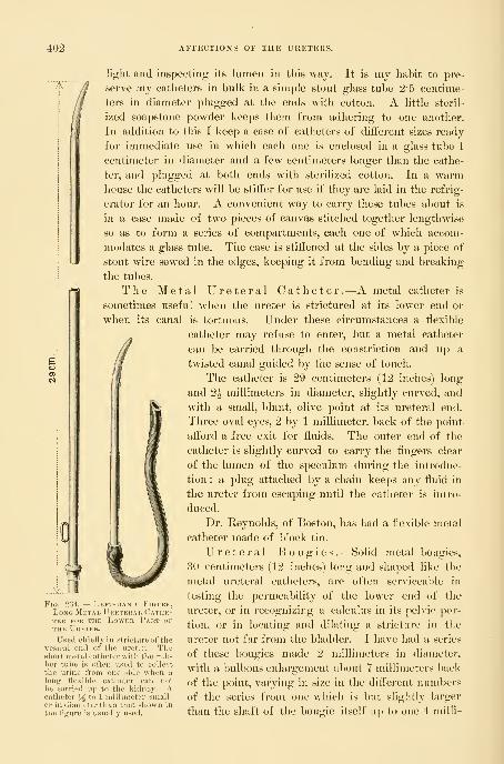
402 AFFECTIONS OF THE URETERS.
1/
light and inspecting its lumen in this way. It is my habit to pre-
serve my catheters in bulk in a simple stout glass tube 2'5 centime-
ters in diameter plugged at the ends with cotton. A little steril-
ized soapstone powder keeps them from adhering to one another.
In addition to this I keep a case of catheters of diiierent sizes ready
for immediate use in which each one is enclosed in a glass tube 1
centimeter in diameter and a few centimeters longer than the cathe-
ter, and plugged at both ends with sterilized cotton. In a warmhouse the catheters will be stiifer for use if they are laid in the refrig-
erator for an hour. A convenient way to carry these tubes about is
in a case made of two pieces of canvas stitched together lengthwise
so as to form a series of compartments, each one of which accom-
modates a glass tube. The case is stiffened at the sides by a piece of
stout wire sewed in the edges, keeping it from bending and breaking
the tubes.
The Metal Ureteral Catheter.—A metal catheter is
sometimes useful when the ureter is strictured at its lower end or
when its canal is tortuous. Under these circumstances a flexible
catheter may refuse to enter, but a metal catheter
can be carried through the constriction and up a
twisted canal guided by the sense of touch.
The catheter is 29 centimeters (12 inches) long
and 2| millimeters in diameter, slightly curved, and
with a small, blunt, olive point at its ureteral end.
Three oval eyes, 2 by 1 millimeter, back of the point
afford a free exit for fluids. The outer end of the
catheter is slightly curved to carry the fingers clear
of the lumen of the speculum during the introduc-
tion ; a plug attached by a chain keeps any fluid in
the ureter from escaping until the catheter is intro-
duced.
Dr. Reynolds, of Boston, has had a flexible metal
catheter made of block tin.
Ureteral Bougies .—Solid metal bougies,
80 centimeters (12 inches) long and shaped like the
metal ureteral catheters, are often serviceable in
testing the permeability of the lower end of the
ureter, or in recognizing a calculus in its pehdc por-
tion, or in locating and dilating a stricture in the
ureter not far from the bladder. I have had a series
of these bougies made 2 millimeters in diameter,
with a Imlbous enlargement about 7 millimeters back
of the point, varying in size in the different numbers
of the scries from one which is but slightly larger
than the shaft of the bougie itself up to one 4 milli-
....V.
Fig. 234. — Left-hand Figure,Long Metal Uketerai, ('athe-TEB FOR the LoWER PaKT OFTHE Ureter.
Used chiefly hi stricture of thevesical end of the ureter. Theshort metal catheter with the rub-ber tube is often used to collect
the urine from one side when a
long flexible catheter can notbe carried up to the kidney. Acatheter J^ to 1 iiiiHiineter small-er in diameter than that shown in
the figure is usually used.
CATHETERIZATION. 403
meters in diameter. I have tested whalebone and found that, on account of its
elasticity, it does not make a good ureteral bougie. The best bougies are madeof hard rubber 2 millimeters in diameter and 50 centimeters (20 inches) in total
length. There is a slight narrowing below the end which is rounded off into a
point shaped like an olive. The handle, large enough to be taken conveni-
ently between the thumb and forelinger, is 6 centimeters (2^ inches) in length
and passes easily through the No. 8 vesical speculum. This bougie easily
adapts itself to the curves of the ureter and can be pushed on up into the
pelvis of the kidney without danger. It becomes more flexible when warmed.The ureteral and renal catheters are also made without any
eye for use as flexible bougies; these are the safest in perform-
ing hysterectomy, on account of the liability of the hard rubber
to break when bent suddenly and sharply.
I have also had a long hard-rubber bougie made with a httle
notch running lengthwise at the tip on two sides, intended to
catch and hold the dental wax with which the end is coated whenthe bougie is used as a searcher for a renal calculus. If a calcu-
lus is present and the bougie comes in contact with it, the shining
impressionable surface of the wax is scratched, and the scratch
marks can be seen under the lens of a low magnifying power. Asilk renal catheter tipped with wax detects the stone equally well,
and so serves the double purpose of bougie and catheter.
Dilating Catheters .—The dilating catheters are used
to dilate ureteral strictures near the bladder. They are nickel-
plated metal tubes, 25 centimeters (10 inches) in length, slightly
curved at the tapering conical point, which is well rounded and
blunt so as not to hurt the ureteral wall. The slight curve, which
is shown in the picture, facilitates the introduction through a
stricture. There are four eyes, arranged in pairs, one eye l)elow
the other on opposite sides of the catheter, and located witliin 2
centimeters of the end. The outer end of the catheter is curved
in an opposite direction from the curve of the point, so as to
keep the Angers out of the way during introduction. At the
outer end there is a little bulbous enlargement to hold rul)ber
tubing slipped over it ; a plug and chain are attached to keep the catheter closed
until introduced. The sizes vary in diameter from 2^ to 6 millimeters, the
difference between the sizes being half a millimeter.
Introduction of the Speculum and Location of theUreteral Orifices .—The bladder is first emptied of its urine by voiding
it in a sitting or a standing posture. The evacuation is more complete when it
is so voided than when drawn by catheter in the dorsal posture. The patient is
then placed on a table in the knee-breast or elevated-dorsal position, the labia
se])arated, and the urethral orifice exposed and cleansed with a boric acid solu-
tion to avoid carrying surface contamination into the bladder on introducing the
speculum.
Fig. 235.—ShowsEnd of ElasticBougie tippedWITH Wax.
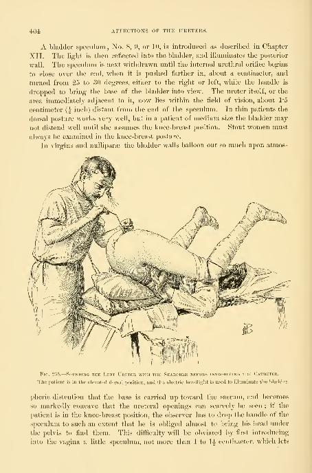
404 AFFECTIOXS OF THE URETEKS.
A bladder speculum , No. 8, 9, or 10, is introduced as described in Chapter
XII. The light is then reflected into the l)ladder, and illuminates the posterior
wall. The speculum is next withdrawn until the internal urethral orifice begins
to close over the end, when it is pushed farther in, about a centimeter, and
turned from 25 to 30 degrees, either to the right or left, while the handle is
dropped to bring the base of the bladder into view. The ureter itself, or the
area immediately adjacent to it, now lies within the field of vision, about 1*5
centimeter (^ inch) distant from the end of the speculum. In thin patients the
dorsal posture works very well, but in a patient of medium size the bladder may
not distend well until she assumes the knee-breast position. Stout women must
always be examined in the knee-breast posture.
In virgins and nulliparas the bladder walls balloon out so much upon atmos-
FiG. 236.
—
Sounding the Left Ureter with the Searcher bekore intuoducing the Catheter.
Tlie patient is in tlie elevated dor.sal position, and tiie electric headlight is used to illuminate the bladder.
pheric distention that the base is carried up toward the sacrum, and becomes
so markedly concave that the ureteral openings can scarcely be seen ; if the
patient is in the knee-breast position, the observer has to drop the handle of the
speculum to such an extent that he is obliged almost to bring his head under
the pelvis to find them. This difficulty will be obviated by first introducing
into the vagina a little speculum, not more than 1 to 1^ centimeter, which lets

CATHETERIZATION. 40i
in the air and causes the anterior vaginal wall to drop down, bringing the base
of the bladder into the plane of vision. If the distention is still too great after
this manoeuvre, the difficulty may then be overcome by introducing within the
vagina a cotton pack large enough to hold the
anterior wall down, or a small inflatable rub-
ber bag, or an instrument shaped like a spatula
with a strongly curved handle to make pres-
sure on the vaginal wall and bring the ureteral
oritice into view.
While the ureter is generally found at an
angle of from twenty-five to thirty degrees
with the urethra, it may be eithei- more or
less. I have often seen it upon simply carry-
ing the speculum straight into the bladder
without deviating more than from three to
five degrees to the right or left. I use a sim-
ple device, figured in the text as a goniome-ter, to measure the angle between a line con-
necting the ureteral orifice with the internal
orifice of the urethra and the axis of the ure-
thra. The zero line of the goniometer is held
in the line of the urethra while the long arm
points to the ureteral orifice, when the angle
can be read off on the graduated arc. In in-
flammatory cases the ureter is often drawn
markedly to one side.
If the abdomen is filled with ascitic fluid,
or if there is a tumor wedged in the pelvis, or
if inflammatory disease is present, the bladder
may not distend enough to allow the ureteral oriflces to be seen. In such cases
the orifice may be sought in the dorsal position without elevation.
The ureteral orifice is recognized as soon as it comes into the field of the
speculum as a fine transverse slit, 2 to 3 millimeters long, like a little dark line
on the bladder wall, not unlike a water mark in paper. At times it appears
more distinct, owing to a slight injection of its borders. Rarely it looks like a
fine dark point or a distinct hole. In the knee-breast position a decided emi-
nence, having the form of a truncated cone, marks its site, and the opening
is situated on top or on the anterior urethral side of this. I have called this
elevation the m o n s u r e t e r i s . In one of my cases there appeared to be
two left ureteral orifices, parallel and exactly alike, about 2 millimeters apart,
but on passing the catheter into the lower one on the edge of the mons instead
of going up a ureter, it reappeared in the bladder and the false orifice was
found to be a little bridle of mucous tissue, 2 or 3 millimeters long and about
as wide.
When the ureteral orifice is not seen at all after a careful search it may be
Fig. 237,
MINI
— Using the Goniometer to detee-THE Angle made ry the Axis of
THE Urethra, with a Line drawn fromthe Internal Urethral Orifice to theU keter.
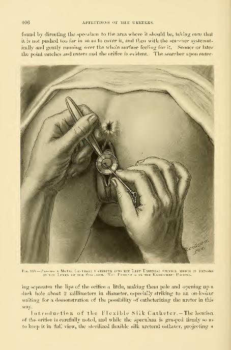
406 AFFECTIONS OF THE URETERS.
found by directing the speculum to the area wliere it should be, taking care that
it is not pushed too far in so as to cover it, and then with the searcher systemat-
ically and gently running over the whole surface feeling for it. Sooner or later
the point catches and enters and the orifice is evident. The searcher upon enter-
FiG. 238.
—
Passing a Metal Ureteral Catiietek into the Left Ureteral Orifice, which is exposedIN THE Lumen of the Speculum. The Patient is in the Knee-chest Postihe.
ing separates the lips of the orifice a little, making them pale and opening uji a
dark hole about 2 millimeters in diameter, especially striking to an on-looker
waiting for a demonstration of the possibility of catheterizing the ureter in this
way.
Introduction of the Flexible Silk Catheter.— The location
of the orifice is carefully noted, aiid while the speculum is grasped firmly so as
to keep it in full view, the sterilized flexible silk ureteral catheter, projecting a

CATHETERIZATIO^r. 407
little from its glass tube, is held over the
shoulder—in the left hand for the right
ureter and in the right hand for the left
ureter—^and slowly guided up the tube,
where its point is engaged in the orifice
and pushed on from 10 to 15 centimeters
(4 to 6 inches).
The end of the catheter must be lubri-
cated by dipping it in a boro-glyceride solu-
tion before introduction, and before taking
Fig. 23'J.—Washing Out the Pelvis of the Kidney byContinuous Irrigation.
Tlie renal catheter has been inserted up to the kidney(outlined through the body) and then connected with a
funnel containing the irrigating solution. An oi-dinary glass
catheter inserted into the bladder drains it by a short rub-ber tube into a bowl. Upon elevating the funnel, the solu-
tion runs out of the end of the renal catlietL-r and'circulates
in the pelvis of the kidney, whiL-h it di.-^teiids, and thenescapes freely down the ureter, beside the catheter, into
the bladder and so out through the catheter into the bowl.
The arrows indicate the course of the tluid.
.J-'"'"

408 AFFECTIONS OF THE URETERS.
liold of the end of the catheter sterilized ruljber linger stalls must be drawn
over the thumb and forefinger, to avoid direct contact with the fingers, and so
a possible infection of the ureter. If there is any inflammatory process in the
bladder, the ureteral orifice must be cleansed with a pledget of cotton held by
the mouse-toothed forceps, and the lumen of the speculum must be cleansed in
the same way.
In introducing the long renal catheter when the glass tube is not used, the
handhng of its upper part, which is to lie inside the body, may be avoided by
first locating the ureteral orifice and then asking for the catheter. The assistant
takes it up from the sterilized towel in which it rests by the outer end and
hands it to the operator, who likewise receives it by this end, and slowly guides
the swinging tip into the speculum and so on up into the ureteral orifice, when
it is then easily run off from its stylet and on up the ureter.
When the catheter is in place the speculum is withdrawn, while the operator
holds on to the catheter to keep it from being pulled out too. If the catheter is
to remain in but a short time the patient may stay in the same position ; other-
wise she should be carefully turned over on her back or side, avoiding any pull
on the catheter.
In many cases when a catheter not larger than 2 or 2*25 millimeters is used
to wash out the pelvis of the kidney the fluid only distends the pelvis a little
and then begins to run down the ureter and back into the bladder outside the
catheter. Owing to this circumstance, in cases of catarrhal pyelitis and of pyelo-
nephrosis it is often possil)le to wash the kidney out thoroughly by keeping up
a continuous irrigation for from ten to thirty minutes or longer.
After the renal catheter is inserted the patient lies on the opposite side and
a glass catheter is inserted into the bladder. It is well to color the irrigating
solution with aniline dye to demonstrate its return to the eye. On raising the
funnel and letting the fluid run into the kidney the return flow into and out
of the bladder is noticed at an interval of from fifteen to seventy-five seconds.
The end of the catheter is placed in a sterilized test tube to collect the escaping
urine. If the tube is left in after the patient is put to bed it is best held in a
block of wood in an auger hole bored at an angle as shown in the figure.
When the short metal catheter with a piece of rubber tubing on the end is
inserted into the ureter for the purpose of collecting the urine of one side, it is
best to drop a small quantity of a concentrated aniline solution into the bladder
so as to have positive evidence that the clear fluid escaping by the catheter is
not contaminated by the fluid in the bladder and that the catheter remains in
place in the ureter.
In making a thorough examination of urine collected directly from the ureter
five things must be inquired into :
1. The amount of fluid escaping at once upon the introduction of the
catheter.
2. The rate of flow during catheterization.
3. Physical properties, specific gravity.
4. (yhemical properties.

CATHETERIZATION. 409
5. Bacteriological condition.
The watch is taken out and the time of introduction noted, so that the rate of
secretion may be determined by measuring the amount collected within a
definite time.
When both ureters are to be catheterized the speculum is withdrawn and
re-inserted beside the first catheter, and the other orifice found and catheterized
in like manner.
Fia. 240.
—
Catiieterizing both Ureters; the Separated Urines are being collected in Test TubesPlugged with Cotton and held in a Block.
Another way of securing separated urines from both
ureters at the same time is to place one of the larger ureteral catheters
in one ureter and then carefully remove all fluid from the bladder with the suc-
tion apparatus and pledgets of cotton. The patient lies on her back and the
urine drains, say for an hour, through the ureteral catheter into a vessel in the bed.
The urine which collects in the bladder during this hour may be assumed to come
from the other kidney if it presents different chemical and microscopic char-

410 AFFECTIONS OF THE UKETERS.
acteristics ; it is removed either by an ordinary vesical catheter, before taking
out tlie ureteral catheter, or by introducing a speculum and using the suction
apparatus. This plan needs further trial and is not available when there is
inflammation of the bladder which contaminates the urine accumulating in it.
A method of separating the urines from the right and left ureters without
catheterizing the ureters has been devised by Dr. Neumann {Deutsche med.
Woch., No. 43, 1897). The patient is seated on the very edge of a table, with
her feet on the floor or a stool. An instrument is then inserted which is intended
to divide the bladder for a time into right and left halves, and at the same time
to provide a free exit for each half, in this way separating the urines. The
instrument is constructed like a catheter, 4 centimeters long and 1 centimeter in
diameter, with a vertical partition down tiie middle ; this partition is continued
4 centimeters beyond the body of the instrument, and ends in a blunt rounded
point, connected with the end by two fine wires on each side. The distal end
of the catheter ends in two little tubes, one for each side ; on these little gradu-
ates are hung to collect the urine. The whole instrument has a gentle curve,
like Ilegar's cervical dilators.
The urines are separated by first washing the bladder out from one tube
through the other, and then introducing the index finger into the vagina and
pressing the base of the bladder firmly up against the instrument, which nowfits snugly behind the symphysis pubis. The urine escaping from the ureters
now flows down the tubes on the right and left sides completely separated.
How to obtain II n contaminated Urine. — Sterilizedurine, or urine free from any contamination from external sources, may be
obtained by covering 3 or 4 centimeters (1^ to 1^ inch) of the outer end of
the sterilized ureteral catheter with a protecting rubber sleeve, and then intro-
ducing the catheter as described ; the sleeve is then removed and another piece
of longer sterilized tubing slipped over the end and used to convey the urine
into a suitable sterilized glass tube plugged with cotton, resting in a block. Toavoid contaminating the end of the catheter by contact with the sides of the
speculum, it may be introduced into the bladder loosely covered with a steril-
ized rubber sleeve, which is pulled off as soon as it is well in ; but I prefer sim-
ply cleansing the inside of the speculum with a boric acid solution.
It is also possible to obtain uncontaminated urine after introducing the
catheter in the ordinary way by boiling the first drop of urine appearing at the
end with an alcohol flame held under it ; the urine which follows this is then in
no danger of contamination by picking up germs at this point.
For a bacteriological examination and cultures it is sufficient in this way to
let a few drops fall directly from the end of the catheter on to the slide or into
the culture tube. It is always well to test alkalinity or acidity as the urine
escapes.
How to secure Urine from the Ureter without using a
Ureteral Catheter .—Sometimes there are serious objections to passing a
catheter into a sound ureter ; when, for example, the bladder is extensively
inflamed the examiner will hesitate, on account of the risk of opening up the
CATHETERIZATION. 411
ureter and of the dangers attendant upon tlie slight trauma under such circum-
stances. But it is almost always possible to get enough urine for a microscopic
and chemical examination without even touching the ureteral orifice. This is
done by putting the patient in the knee-breast position,
exposing a ureteral orifice, wiping it off, and then holding
the end of the speculum close up under it until a jet of
urine escapes ; the drop is caught in the lumen of the
speculum and runs down its side on to the outer lip, where
it may be taken at once on to a slide and examined or col-
lected in a minim graduate. The microscopic examination
in this way of a drop or two may be just as satisfactory as
a large quantity secured by the ureteral catheter. (See
Twentieth Cent. Prac. Med., vol. i, 1895, p. 690.)
I have had a speculum made for this special purpose
(see Fig. 193) with the end cut off obliquely to fit in better
under the orifice in the knee-breast position.
Catheterization of the Ureterswithout Elevation of the Pelvis and^\•ithout Atmospheric Distention of
the Bladder .—Under certain circumstances,
when it is awkward or when it consumes too nmchtime to place the patient in the knee-breast j)Osi-
tion and to elevate the hips on cushions, I am in
Fig. 241.—Sieve aku Gradua.te forfilterixg and collecting aFew Drops of Ukine caught ipON Cotton from the UketebalOrifice, through the Speci-Luii, without catheterizingthe Ureter.
the habit of introducing the catheter in the follow-
ing simple manner without elevation or atmospheric distention of the bladder :
The patient lies on her back on a flat table, with thighs well drawn up on
the body, and the bladder is emptied. The No. 9 or 10 cystoscope is now intro-
duced, its outer end strongly elevated, and the inner end turned toward the right
or left side of the base of the bladder. The head mirror is now turned so as to
^"T
Fig. 242.
—
Instrument fob collecting Urine as it runs out of the Ureter, in the Knee-breast Posture, without catheterizing the Ureter.
The No. 10 speculum is first introduced up under the ureteral orifice, then the collecting tubeis held up to the orifice, and any drops of urine escaping are caught and discharged into a minimgraduate in the direction of the arrow.
illuminate tlie portion of the wall of the bladder at the end of the speculum.
The speculum is now withdrawn as far as the urethral orifice, to locate its posi-
tion, and then pushed in again and turned to one side with the idea of bringing
the ureteral orifice at once within the lumen of the speculum. Sometimes it can
be seen immediately, even througli a little layer of clear urine ; at otlier times it
is necessary to keep the speculum against the bladder w^all, and then, after drying
out the few drops of urine in it, to find the ureteral orifice by gliding the instru-

412 AFFECTIONS OF THE URETERS.
ment over the vesical mucosa. Any depression reseinhling tlie mouth of the
ureter is first tested with the searcher, and then if it is found the catheter is
puslied in. By introducing the flexible catheters in this way just before a vagi-
nal or abdominal hysterectomy the ureter is converted into a cord easily felt
throughout the operation.
It not infrequently happens that the patient, with carcinoma of the cervix,
is either so heavy that she can not be put into the knee-chest posture with the
limited assistance at the command of the operator, or that she is so feeble that
the operator feels unwilling to lose the amount of time necessary to change her
position in order to find the ureteral orifices for catheterization. Under these
circumstances I have often resorted to the method just described with perfect
satisfaction. It will, however, hardly be possible for one not thoroughly used
to the simpler way of catheterizing the ureters in the knee-breast posture, and
so familiar with the exact location of their vesical orifices, to find them in the
dorsal position .with a collapsed bladder.
Points to be observed in securing Separated Urines.—I use the plural urines advisedly to make a distinction, hitherto impossible, be-
tween the mixed urine in the bladder from both sides and that from each kidney
separately before mixing.
The purpose of the examination is to estimate correctly the status of each
kidney by determining (1) its working coefficient as estimated by the amount of
urea being secreted; (2) the existence of various morbid f)roducts, such as casts,
albumin, pus, and bacteria.
To reach accurate conclusions, the following points should be observed in
catheterizing both ureters
:
1. The exact time of introduction of each catheter is noted. It is well to
attach the note to the catheter on a card.
2. The time of withdrawal is noted and also written on the card, giving the
exact duration of the flow.
3. The exact amount of secretion collected in the test tube is noted.
4. It is well to compare the rate of secretion, determined by noting the
amount of flow in a given unit of time, say from five to fifteen minutes or
longer, with the entire amount passed in the twelve hours during which the
examination is made. If the amount secured is too small or too large the error
may be rectified in this way. A nervous patient, for example, will sometimes
pass an excessive amount through the catheter.
5. An analysis of each urine is made investigating its physical, chemical,
microscopical, and bacteriological characters. Especial attention must be paid
to the urea as the most important representative of the physiological activity of
the kidney. It is better to keep a book of charts for recording each analysis
under some such plan as the following
:

CATHETERIZATION. 413
ANALYSIS OF SEPARATED URINES.
Name, Date,
Diagnosis,
Time of insertion of catheter,
Right or left ureter catheterized.
Size of catheter used,
Time of withdrawal of catheter.
Amount of urine secured,
Average amount in twenty-four hours.
Appearance of urine on withdrawal,
Sediment,
Specific gravity.
Reaction,
Albumin,
Urea,
Microscopic examination.
Bacteria, cover-slip, and cultures.
Sounding the Ureters.— Sounds are introduced into the ureters to
find a stricture or an obstruction, to dilate a stricture, and to convert the soft
ureter into a firm resisting cord easily found and kept under the fingers during
a pelvic operation.
In most instances the catheters serve the purpose of bougies as well or
better than a solid instrument. The catheter, for example, gives evidence of
the passage of a stricture by the difficulty of entrance, by the bite of the stric-
ture, as well as by an immediate gush of urine, and the long, flexible silk cathe-
ters serve just as well to splint the ureter and mark out is course to prevent
injury during an operation. The catheter, however, can not so well detect and
estimate the character of resistance, and the force used in overcoming it can not
be so well gauged. For these purposes I use hard-rubber bougies 30 centi-

414 AFFECTIONS OF THE URETERS.
meters (12 inches) long and 2 millimeters in diameter for the pelvic portion,
and 50 centimeters (20 inches) long for the entire ureter and pelvis of the
kidney.
These bougies are smooth and flexible, and easily follow the course of the
ureter. The ureteral orifice is exposed as for catheterization, and the point of
the bougie engaged. By pressing on one side or the other of the speculum the
end may be brought to bear directly upon the ureteral orifice and slipped in.
By a gentle forward movement it is carried on and upward toward the
kidney, easily guided by the ureter and taking all its curves.
Catheterizing the Pelvis of the Kidney.—The pelvis of the
kidney can be catheterized by means of long, flexible silk catheters.To introduce the renal catheter, the hands are carefully washed
and sterilized, the ureteral orifice exposed, and the catheter, taken from the
refrigerator or stiffened with a stylet, is coated with boro-glyceride at the end and
slipped through the speculum and pushed on until its point is engaged in the
ureter. The long outer end of the catheter, wrapped in a piece of sterilized
gauze, or still lying in the sterilized towel, out of which it is drawn as it is intro-
duced, must hang over the shoulder. The rate of introduction should be slow,
2 or 3 centimeters at a time, and the examiner should take care to keep the end
of the speculum close to the ureteral orifice, and watch to see that the catheter
does not kink in the bladder or speculum. The patient may have no sensation
at all as the catheter goes in, or may be only conscious as the end touches the
upper margin of the pelvis of the kidney.
When from 32 to 37 centimeters (13 to 15 inches) have been pushed in be-
yond the external urethral orifice the end will lie in the upper part of the renal
pelvis.
If the catheter is soft and has to be braced by a stylet, this nmst not reach
quite to the end, and as soon as a few centimeters of the catheter are engaged in
the ureter the stylet is pulled out for the same distance, after which the cathe-
ter is stripped off from the stylet and pushed on up the ureter into the kidney.
Normally there is but little urine collected in the pelvis of the kidney, and
it is necessary to wait a while for the catheter to fill and begin to discharge the
droplets. The respiratory movements may be seen in the play of the drop to
and fro as it hangs from the end of the catheter. When there is a stricture in
the ureter or at the pelvis of the kidney there is an accumulation of urine oi-
pus within the pelvis. The renal catheter relieves this retention by drawing
off the fluid and discovers lesser grades of hydronephrosis and pyonephrosis
which have hitherto escaped attention. I estimate the degree of hydronephro-
sis by measuring the exact amount of fluid discharged in a steady stream or by
continuous dropping with the catheter held down so as to act as a siphon. If
the catheter is a small one and it takes a long time to empty the sac, I then
allow half a cubic centimeter per minute for the activity of the kidney during
the time of evacuation. As soon as the flow begins to come at intervals it is
evident that this is (le])endent on the present secretory activity of the kidney,
and the accumulated urine has been exhausted.

URETERAL FEVER. 415
Asepsis.—The whole technique of the examination and exploration of the
ureters must be asepticallj conducted. The danger from introducing septic
material directly into a ureter is sufficiently obvious, and is illustrated by nu-
merous examples of a fatal infection ascending from the urethra up to the kid-
ney. If an infection of the urethra or bladder can spread in this way, it goes
without saying that an infection introduced in the ureter will also spread.
The various manipulations ought to be conducted with a care in the aseptic
technique equal to that of any surgical procedure, so that there is therefore noexcuse for any ill sequel from a simple examination in a healthy case.
The aseptic technique is divided into two stages : (1) The care of the instru-
ments; (2) care during their introduction.
The silk catheters must be sterilized before using by boiling two minutes in
plain water, and washed immediately after every use with hot water, boiled for
two minutes in pure water, and laid away on a sterilized towel until perfectlv
dry inside and out. They may also be washed out with a saturated solution of
oxalic acid. Tliey are then put singly or several together in a long sterile glass
tube plugged at each end with cotton. The metal catheters are sterilized in the
same way as other metal instruments, by boiling five minutes in the soda solution
(see Chapter I).
In introducing the catheters the greatest care is necessary at every step to
avoid contamination by the assistant, the speculum, the bladder wall, or the fin-
gers of the examiner.
The lumen of the speculum and the ureteral orifice are cleansed with a ])ledget
of cotton saturated with a boric acid solution held by the mouse-toothed forceps.
The metal catheter is held by its outer end, avoiding at all times touching
the end that is to go into the ureter ; it is then guided up the speculum and in-
troduced. To introduce the flexible ureteral and renal catheters, the end is
pushed a little way beyond the glass tube and dipped in boro-glyceride, and
then the glass tul)e is rested on the examiner's shoulder. He now takes hold of
it with thuml) and forefinger covered with sterilized rubber finger stalls, draws
it out of the tube, and guides it on into the ureter. By using these sinq)le pre-
cautions all risk is avoided.
Ureteral Fever.—I have seen ureteral fever following the introduction of
the ureteral catheter in four cases. In each of these cases the upper urinary
tract was already infected, and the urine contained pus coming from the pelvis
of the kidney.
One patient had a stricture at the vesical end of the ureter and a dilated pvo-
ureter and pyelitis. On two occasions, while washing out the ureter and kidney,
in a case under treatment for some time without any change in the technique,
the introduction of the catheter was followed by a chill and fever, with an eleva-
tion of the temperature as high as 104° F. The pulse increased in rapidity in pro-
portion to the fever, the face became flushed, and the patient was restless and had
severe headache. Fi-om the second day on to the fourth or fifth day the tempera-
ture declined to normal. During the attack chilly sensations were repeated daily,
and there was a decided tenderness over the course of the catheterized ureter.

416 AFFECTIONS OF THE URETERS.
In another case tlie attack began with a decided chill on the evening of the
day of irrigation, which was practiced through a metal catheter injecting a bi-
chloride of mercury solution (1 to 50,000). The patient complained of pain
along the course of the ureter and in the back, and had a hot flushed face with
headache and malaise. The temperature was highest on the second day, reach-
ing 103°, and declined to normal on the fourth day. In a second attack, follow-
DAY

CONGENITAL AFFECTIONS OF THE UKETER. 417
other hand, be due to a sjjht wliich begins at any point below the normal renal
pelvis, and the ureter continues double all the way to the l)ladder ; or again the
two canals may fuse at any point on the way down.
An interesting case of complete duplication of the left ureter occurred in one
of my patients and is reported by Dr. Otto Ramsay {Johns Hopkina Hospital
Bulletin, E'ovember-December, 1896). The patient (A. W., 4154), forty-five
years of age, was admitted to the ward with an inoperable cancer of the cervix
and died soon after admission. Autopsy (!N^o. 813, June 22, 1896).
Anatomical diagnosis : Sloughing carcinoma of the uterus, perforation into
the rectum. Involvement of the ureters, with hydroureter and slight hydro-
nephrosis.
The two left ureters begin at the hilum of the kidney in two separate pelves
and run down into the pelvis side by side, closely bound together, but separate,
to the bladder, which they enter by distinct orifices 1*5 centimeter apart. Theyare dilated from the point where they ai-e involved in the cancerous growth near
the cervix, all the way up to the kidney. Each one is about the size of the
little finger and has clear contents. They sllo^v^ a marked contraction where
they pass through the growth. The drawing on page 446 well illustrates the
condition.
Such forms of duplicature have no pathological significance. Two other
forms of malformation, however, are of the highest importance ; these are the
ectopic ureteral orifice and the dilatation of an occluded ureter.
Ectopic Ureteral Orifice.—An ectopic ureteral orifice is one located either in
the vagina, or in the urethra at the external urethral meatus, or under the hood
of the clitoris.
The one common symptom observed in such cases is a persistent leakage of
urine, noted from childhood up, but varying in amount at ditferent times as
measured by the napkins worn. In spite of this constant discharge, the patient
empties her bladder at regular intervals, and the abnormal flow continues as
active after micturition as before it. The abnormally displaced orifice may be
either the only one connected with the kidney, or a supernumerary orifice, the
other opening normally into the bladder. The practical importance of distin-
guishing between these two allied and, to a superficial examination, similar con-
ditions is evident.
Diagnosis .^In incontinence of urine not due to gross and easily demon-strable lesion, such as a vesico-vaginal or uretero-vaginal fistula, the inquiry into
the cause must be made in a careful and orderly manner in order to discover
cases of this kind.
The first question to be answered is whether the involuntary discharge of
urine comes through the urethra, and if it does whether it is simj)ly due to a
breaking down of the sphincter fibers at the neck of the bladder, or whether
there is some extraordinary channel of communication between the ureters or
bladder and the genital tract.
If the patient is a virgin or has never borne children, and her bladder has
not been subjected to any manipulative interference, and if the disease has
418 AFFECTIONS OF THE URETERS.
existed from earliest childliood, the presumption is at once in favor of a con-
genital malformation.
If, on the other hand, after lying down for a while a certain quantity of
urine is found accumulated within the vagina, a vaginal orifice of discharge mayhe looked for. By drying out the vagina and placing in it dry pledgets of
absorbent cotton, and at the same time filling the vulvar cleft with cotton, and
waiting a few minutes, it will be easy to determine the fact of a leakage, by
noticing the spots on the cotton, where a little urine has accumulated, and this
will also approximately fix the position of the opening. By injecting the blad-
der with an aniline or sterilized milk solution, its independence of this viscus
will be demonstrated. A prolonged careful inspection of the area indicated by
the spot of urine on the cotton will reveal the minute orifice through which
intermittent discharges of urine escape. If the discharge comes from the ure-
thra, a careful urethroscopic examination must be made of the entire tract from
the internal sphincter down to its external orifice by withdrawing the urethro-
scope millimeter by millimeter, constantly watching the funnel-shaped figure of
the urethra at the end of the speculum for any small opening or jet of urine.
On finding the orifice the questions now to be answ^ered are these :
1. Is the abnormal opening a ureteral orifice ?
2. On which side is it located—that is to say, to w^hich kidney does it
belong ?
3. Is it a single or a double ureter ? If double, has it also a normal opening
into the bladder ?
4. If double, does it continue so all the way up to the kidney, or does it unite
with its fellow at some ^joint above the bladder ?
5. If double all the way up, do both ureters enter a connnon pelvis, or have
they separate pelves ?
First, it is a ureteral orifice if, by injecting the bladder with a colored solu-
tion, none of the fluid escapes by the opening, demonstrating its independence,
and if, in spite of the passage of urine at regular intervals from the bladder, little
jets of urine are seen coming from the orifice, with intermissions not longer than
a few seconds.
Second, the assurance that the opening is ureteral, and the answer to the
question on which side it is located, to which kidney it belongs, is given by pass-
ing a long renal bougie through the opening up into the pelvis of the kidney,
from 25 to 30 centimeters or more, and then by observing to wdiicli side of the
cervix uteri the bougie turns, and by palpating the l)OUgie through the vagina
and rectum.
The third question, whether it is a single or double ureter, is answered by
placing the patient in the knee-breast position and introducing a No. 10 vesical
speculum and looking for the ureteral orifices within the bladder. If the oi-ifices
are found on both sides in normal j^osition, it is evident that the ureter is super-
numerary or split.
The fourth question, whether a double ureter continues so all the way U]) to
the kidney, may, I think, be answered in the following way : I would pass a

CONGENITAL AFFECTIONS OF THE URETEK. 419
catheter, 2*5 millimeters in diameter, large enough to fill the lumen of the abnor-
mal ureter, all the way up to the pelvis of the kidney. I would then introduce
a renal catheter into the normal vesical orifice of the same side and push it up.
If the two ureters unite into one a short distance above the bladder, or at somepoint in the abdomen below the kidney, I should expect the second catheter to
be stopped short in its course upon striking the first. The catheters could nowbe withdrawn after carefully noting the exact distance to which each had been
introduced, and by laying them together on a sheet of white paper in a similar
position a tracing of the form of the ureter could be made. To make sure of the
diagnosis it would then be well to reverse the procedure by introducing a long
catheter up into the kidney through the vesical orifice of the ureter, and then
pushing up another catheter through the abnormal orifice, until it is stopped bythe first catheter. Again noting the exact distances to which the catheters have
been introduced, and withdrawing them, and reconstructing the ureteral situa-
tion on paper, the diagnosis will be confirmed if the two drawings correspond.
To determine whether the ureter is double throughout, and if double, whether
the kidney has one or two pelves, the following plan will be sufficient : A renal
catheter is passed through each ureter up to the renal pelvis and a sterilized ani-
line solution is injected into one, when, if there is a communication between the
pelves, the colored fluid will immediately flow from the other ; if there is nocommunication, it will simply be returned, while the other side discharges clear
urine.
These various points in diagnosis are of practical importance in determining
the nature of the operation to be performed.
When the opening is at the urethral orifice, the anterior wall of the vagina
may present a characteristic prominent curved ridge, which covers the ureter,
as in the case of Dr. F. II, Davenport, of Boston {Trans. Amer. Gyn. Soc,
1890, p. 343), in which the orifice was in the posterior wall of the external
urethral orifice. W. H. Baker's case opened similarly about two lines to the
left of and below the urethra {Boston Med. and Sui'g. Jour., Dec, 1878). It
was not determined in either of these cases whether or not the ureter was
double, which could now be done either by examining the orifices in the
bladder cystoscopically or by passing a renal catheter up to the kidney pelvis
and injecting an aniline solution, and noting whether the urine in the bladder
is colored.
Erlach reported a case before the Vienna gynecological society, December
4, 1888, in which he found post mortem a right ureter double throughout, each
ureter starting in a separate pelvis above. One of the ureters opened naturally
into the bladder and the other into the urethra just below the internal orifice.
In spite of this, there was no history of incontinence.
A case of congenital anomaly of the ureter has been observed by Baum in
which the supernumerary opening was close to the external urethral orifice.
The normal orifice was seen upon opening the bladder from above.
In a case of Massari ( Wiener m-ed. Wochen., 1879, No. 33) a child four
years old, with a preternatural vaginal anus, suffered from constant leakage of

420 AFFECTIONS OF THE URETERS.
urine, the cause of wliieli was only explained post mortem, when the kidneys
were found to be fused across the vertebral column. The right ureter was
normal throughout, but the left one had no vesical orifice ; instead, it passed the
bladder in its course and discharged by a minute orilice just under the prepuce
of the clitoris. The vagina was double and the uterus normal.
Treatment.—The object of the treatment is to get rid of the constant
leakage by turning the urine into the bladder.
Two plans in general have been tried to effect this. First, by dissecting out
the extremity of the ureter from its bed and turning it into the bladder, and,
second, by a suprapubic incision into the bladder and then opening the ureter
beneath the base of the bladder and establishing a communication between the
two, after which the distal end of the ureter beyond the opening may be ligated.
The suprapubic incision is closed at the end of the operation.
In case the ureter is double with a single renal pelvis above, it would be
proper to try the plan of ligating the abnormal ureter at any point in its course
where it could most conveniently be laid bare ; the operator might introduce a
flexible renal catheter, and, using this as a guide, incise the vaginal wall some-
where from 2 to 3 centimeters beyond the abnormal orifice, laying bare the
ureter in its course. It should be then carefully dissected out from the sur-
rounding tissues and freed on all sides sufiiciently to allow a ligature to be
placed about it. It may then be ligated with silkworm gut or fine silk, dropped,
and the vaginal incision closed over it.
By the plans pursued by Baker and Davenport, the ureter is dissected out
from its external orifice back to the base of the bladder by splitting the vaginal
wall, exposing the abnormal canal, and carefully freeing it on all sides from the
cellular attachments. When it has been freed up to a point under the base of
the bladder corresponding in position to that of the normal ureteral orifice, an
incision is made through the vesico-vaginal septum into the bladder a little less
than a centimeter in length. The end of the ureter which has been dissected
out is now cut off and the new orifice slit up for about 6 millimeters to make a
larger opening. The end is then turned into the bladder through the opening,
which is closed by two or three silkworm-gut sutures extending through from
the vaginal surface to the vesical mucosa. The uppermost suture is made to
include the muscular coats of the ureter at a point about on a level with its
opening into the bladder. Another fine suture below this penetrates the vesico-
vaginal septum for a short distance and catches the ureteral coats again. The
ureter being fixed by the two sutures, the remainder of the vaginal incision from
which the ureter has been dissected is closed by interrupted sutures.
This was done by Dr.W. H. Maxson, of St. Helena, Cal. {Med. JSfeios, March
21, 1896, p. 323), who operated upon a young woman twenty-two years old for
an incontinence of urine dating from her earliest recollection. He found the
orifice of the left ureter about a <piarter of an inch within the external urethral
orifice. At the operation the ui-eter was dissected out through the vagina for
three inches and a half and drawn through a small opening made in the base of
the bladder close by, after cutting off" an inch and a half of the lower end. The

CYSTIC DILATATION" OF AN OCCLUDED UEETEK. 421
ureter was then stitched to the bladder wall with catgut and the vaginal incision
closed. A complete recovery of function ensued.
It is important, as a preliminary precaution, to determine by a cystoscopic
examination whether the ureter is single or double. If it is double, a bougie
must be placed in the normal ureter opening into the bladder, so that in trans-
j^lanting the abnormal orifice the normal one will not be cut or included in the
sutures.
The plan of establishing a connnunication between the abnormal ureter and
the bladder by a suprapubic incision hi the bladder was adopted in Baum'scase to avoid dilating the vaginal orifice in a girl eighteen years of age. Theright ureter, discharging close to the urethral orifice by a fine opening, was
greatly dilated in the neighborhood of the bladder. The base of the bladder was
incised through a suprapubic incision and the dilated ureteral sac opened from
above. A piece of the sac about a centimeter in diameter was now excised and
the edges of the incision stitched together. The part of the ureter beyond this
new opening was now ligated. A portion of the abdominal incision was closed
with suture, and the prevesical space drained with iodoform gauze. After this
operation urine passed naturally. The patient ^^assed a urinary concretion five
months later and suffered from a hernia.
Cystic Dilatation of an Occluded Ureter.—A rare but practically important
anomaly of the ureter is that in which the lower end has failed to communicate
either with the bladder or with any part of the genital tract, and remains
occluded. If this forms the only avenue of discharge for the corresponding
kidney or part of the kidney, complete atrophy of the organ depending upon it
is a necessary consequence.
Where the terminus of the ureter is under the base of the bladder, and the
lower end is dilated into a spherical or ovoid cyst, this has been seen projecting
into the bladder, forming a prominent rounded tumor, occupying one side of the
base, as in F. Tangl's case, where the patient was a woman sixty-seven years of
age and the left ureter was affected, ending in a saclike projection into the blad-
der. The kidney of the same side was extremely atrophic and displaced down-
ward. The right kidney was in a state of chronic interstitial nephritis. The
tendency of such anomalies to be associated with other malformations was shown
by the fact that the patient had a uterus bilocularis unicollis.
A similar case to this was that of Kolisko, where the right ureter was double
throughout. The abnormal ureter began in a separate pelvis in the u])per part of
the kidney which was atrophied, and in its passage downward crossed its fellow
and ended below the orifice of the normal ureter in a sac-like dilatation, which
projected into the lumen of the bladder and extended down into the urethra.
This tract had thick nmscular walls. It was quite evident in this case that the
kidney was a fused one, and that the maldeveloped ureter belonged to the upper
kidney.
One of the most remarkable cases of cystic dilatation of the lower end of the
ureter is that of Dr. E. G. Orthmann, of Diisseldorf, in which the cyst presented
the characteristics of a vaginal cyst. The patient was twenty-seven years old,

422 AFFECTIONS OF THE URETERS.
and presented a circumscribed cystic tumor of the anterior vaginal wall which
she thought was a prolapse of the uterus. This gradually kept increasing in size
and was associated with drawing pains in the left side, extending around into the
small of the back. The tumor was elastic and circumscribed and occupied the
lower third of the vagina down to within a finger's breadth of the external ure-
thral orifice. It could be pushed back, but returned on the least straining.
Careful examination showed that it had no connection with the urethra or blad-
der. At the operation the thick walls of the tumor were dissected out up to a
long pedicle on the left side which was bared from 8 to 10 centimeters (3 to 4
inches), when it became evident, from the way in which the tumor emptied
itself upward, that there was a communication with the ureter above. The
j)edicle was tied and cut and retracted into the cellular tissue out of sight. The
wound was closed with catgut and the convalescence was undisturbed.
In another group of cases of ureteral anomalies the lower end of the ureter
may end in a blind pit without any dilatation. In these cases the kidney of that
side is entirely absent or atrophic. When the ureter comes from a separate por-
tion of the kidney by a pelvis of its own the atrophy may be limited to this part.
Such is the case reported by F. Tangl(Virchow^s Are/dv, Bd. cviii, p. 414),
in a patient sixty-five years old, with extreme atrophy of the left kidney and a
double right ureter which united below and ended in a blind canal, recognized
as Gartner's duct, in the anterior vaginal wall. The right kidney was affected
with chronic interstitial nephritis. There was a bilocular uterus M'ith one
cervix.
Haller (Deut. Arch.f'dr Min. Med.^ Bd. v. Heft 2) and Weigert( YlrehoiV'.s
Archiv, No. 70, p. 490) report cases where, with com])lete duplication of the
ureter and pelvis, one of the ureters ended blindly in the bladder wall, causing
in this way a partial hydronephrosis.
Congenital Flexure of the Ureter.—A case of congenital flexure of the right
ureter with extreme hydronephrosis is described by AYeigert. The large kidney
extended 3 centimeters (1^ inch) beyond the middle line. The right ureter pur-
sued a normal course from the bladder to a point 21 centimeters (S^ inches)
above it, where it reached the lower border of the tumor. At this point it bent
suddenly to the left, forming a sharp kink, beyond which there was considerable
enlargement of the lumen. It extended from this point to the left border of the
tumor into which it merged. The lower margin of the dilated ureter curved
around on to its left side, while at the upper margin there was a second sharp
flexure. There was no thickening or evidence of inflammation, and after releas-
ing the flexure the fluid escaped easily.
Ureteritis and Periureteritis.—Ureteritis and periureteritis are found asso-
ciated with a variety of urinary diseases. In ureteritis there is an inflanunation
and thickening of the coats of the ureter, the disease beginning with the mucous
coat which is most exposed.
Periureteritis, on the other hand, is an inflammatory afl'ection involving the
cellular tissue in which the ureter lies throughout its whole course, from renal
pelvis to bladder. The peritoneum overlying the ureter may also be involved

URETERITIS AND PERIURETERITIS. 423
by contiguity. Periureteritis often arises in an affection of the cellular tissue,
extending upward from the vaginal vault. I have seen two such cases follow-
ing the division of the ureter in a vaginal hysterectomy, leaving a fistulous
opening at the vault of the vagina. In one case I opened the abdomen someweeks after the operation to transplant the fistulous orifice into the bladder,
and found the cellular tissue surrounding the ureter on that side dense and
rigid from the vaginal vault up to the brim of the pelvis. The structures were
so hard and immobile that any attempt to dissect the ureter out of its bed and
lift it up could not even be considered. A periureteritis due to an inflamma-
tion extending from the interior of the ureter outward is rare ; I have not yet
encountered it in any case.
Tlie causes of ureteritis are threefold : (1) by extension of the dis-
ease upward from the bladder; (2) by extension of renal disease downward into
the ureter ; or (3) the inflammation may originate from some cause located in
the ureter itself, such as a calculus. The first and second causes are the most
frequent.
In ureteritis due to calculus the evidences of the disease are found in a
thickening and contraction of all its coats, foi-ming a stricture below the stone
lodged in it. When several stones are lodged in one ureter, a series of sti'ictures
are found with dilatation above each.
The most rational classification of the various forms of ureteritis induced by
vesical or renal disease is that which depends upon the special exciting cause.
We may thus distinguish a ureteritis due to streptococcus or staphylococcus in-
fection, ureteritis due to gonorrheal infection, and finally a tubercular ureteritis.
Of these forms, the first three commonly originate in the lower urinary tract,
infecting first the urethra and bladder, and then the ureter through the conti-
nuity of mucous surfaces, while the tubercular disease more commonly starts in
the kidney and affects the ureter from above downward.
It is remarkable how frequently extensive inflammatory lesions are found,
either in the bladder or in the kidney, without any marked participation of the
ureter directly continuous as it is with the bladder below", and constantly bathed
with the infectious renal discharges from above.
Another and rare form of inflammation affecting the ureter and pelvis of
the kidney is cystic ureteritis and pyelitis, characterized by the forma-
tion of little cysts projecting from the mucous surface the size of a hempseed, or smaller, containing a thin watery or tenacious fluid. These cysts are
more abundant in the upper part of the ureter, and probably originate in the
sparse glands or crypts in the mucosa.
The symptoms common to the various forms of ure-teritis arise either from the inflamed ureter itself, or from the interference
with function resulting from the inflammation. Owing to the fact that the
ureteritis is always secondary and is usually simply an extension of grave renal
or cystic disease, its own peculiar symptoms are often masked. This is particu-
larly the case in acute forms resulting from a severe infectious process extend-
ing rapidly from the bladder up to the kidney.
424 AFFECTIONS OF THE URETERS.
In chronic ureteritis the most marked symptoms are the pain locahzed on
one side extending up into the flank, with frequent and painful micturition.
Pus is always found in the urine, and sometimes blood.
The prognosis in the acute forms, while depending somewhat upon
the involvement of the ureter, will be better guided by the condition of the
bladder or kidney which can be investigated and estimated, while that of the
ureter can only be surmised. In the chronic forms the prognosis depends en-
tirely upon the cause ; the gonorrheal ureteritis tends to form stricture just as in
the urethra. When not actively treated, the tendency is to run a long course,
often extending through a series of years. It is important to note that, in spite
of extensive involvement of the ureter, the function of the kidney is rarely
entirely lost.
The diagnosis of ureteritis is not difficult to make with the direct
means of investigation at our command. The subjective symptoms often so
closely simulate cystitis that a differentiation can not be made by symptoms
alone, the most characteristic of which is pain along the ureter.
Infallible diagnostic points are found l)y making a digital examination.
After emptying the bladder and rectum, upon introducing the finger into the
vagina, and palpating the antero-lateral vaginal wall in its upper part, a large,
thick, exquisitely tender cord is found sweeping upward to the vaginal vault
and disappearing at the side of the cervix under the base of the broad ligament.
It is often nodular, and when felt for the first time in the vaginal vault it inva-
riably creates the impression that it is an adherent ovary or tube. I have knowninflamed ureters to be mistaken for ovaries in this way. The pain provoked by
the examination is usually so great that an anesthetic is necessary to outline
thoroughly the structures. The thickened ureter is often movable in the cellu-
lar tissue ; by introducing the finger into the rectum it may be traced over the
sciatic notch and on up toward the brim of the pelvis, where it is found lying
close to the internal iliac artery. Upon palpating through the abdominal wall,
down upon the pelvic brim, at a point 3 centimeters (1^ inch) to the right or
left of the promontory of the sacrum and a little below it, the patient will com-
plain of pain, and, if the abdominal walls are unusually thin, the thickened
ureter may be felt rolling under the fingers. If the umbilical ring is relaxed
the ureter can be felt through it with the Titmost distinctness. Upon contin-
uing the palpation upward in the course of the ureter, it can be traced by the
pain elicited when the pressure is made directly over it. Through an open
abdominal incision, by preference in the semilunar line, the thickened left
ureter may readily be found at its point of transit from the abdomen into the
pelvis, by lifting up the sigmoid flexure to the right angl exposing the uretei*
just beneath the peritoneum, crossing the common iliac artery beside the ovarian
vessels. On the right side the ureter will be exposed by lifting up and drawing
the head of the colon to the right.
By making a cystoscopic examination with the bladder distended with air,
the ureteral orifice of the affected side will often be found the center of an area
of intense injection, situated on a truncate cone, sometimes surrounded by papil-
TUBERCULAR URETERITIS. 425
larj eminences, and not infrequently markedly everted. If the orifice is watched
for a time, turbulent or purulent urine may be seen escaping.
By means of a catheter, urine may be collected from an infected ureter, and
if the catheter is not contaminated in the introduction, a bacteriological exami-
nation of the urine obtained will often reveal the cause of the inflammatory
trouble, whether due to tubercle bacillus, gonococcus, or streptococcus.
As we can do nothing directly to the ureter in an acute inflammatory condi-
tion which will be beneficial, the treatment of this form of ureteritis is j)urely
expectant, and devoted to the associated disease in the bladder or kidney.
In its chronic form the treatment must vary according to the extent of the
disease, and to the changes it has produced in the kidney. If the result of the
ureteritis has been simply to thicken the coats of the ureter, forming an obstacle
to the downward passage of urine, the urinary channel above such an obstacle
will be dilated with urine or pus, and in all cases, before any more radical meas-
ures to relieve the obstruction are adopted, a renal catheter must be passed in
order to determine the degree of stricture estimated from the bite on the cathe-
ter and from the amount of urine behind it, and the character of the infection
by the pus secured. If pus is present, an effort should be made to sterilize the
upper urinary tract by emptying it and washing it out with weak bichloride
solution (from 1-100,000 to 1-10,000) every two or three days. For treatment
see under Stricture of the Ureter.
Tubercular Ureteritis.— One of the most frequent forms of ureteritis produ-
cing profound alterations in the coats of the ureter is due to tubercular infection.
This commonly involves its entire length, and arises secondary to a tubercular
kidney. The thickening of the ureteral coats converts the organ into a rigid
tube, irregular on its outer surface, and presenting marked irregularities in its
lumen.
The rare cases which present themselves for treatment, if of long standing,
are one-sided.
The lining membrane of the ureter is ulcerated and the pelvis of the kidney
filled with pus. When more advanced still, the bladder mucosa is affected,
varying from some disseminated tubercles sowed about the ureteral orifice, all
the way to an extensive diffuse infection with areas of ulceration.
The chief clinical symptom is the frequent painful urination, the patient
being obliged to sit on the vessel every few minutes, and rarely being able to
wait half an hour or longer.
Blood is only found in the urine in advanced cases, when it generally comes
from the bladder. Pus secreted by the ureter is always present in varying
quantities. Large amounts may be passed at intervals, being held back by the
constriction of the lumen of the ureter, until sufficient pressure is developed in
the upper ureter and renal pelvis to break through the obstruction. In this
way we have intermittent pyuria, accompanied by a constant pyuria of lesser
degree.
Fever is a symptom of cases in which the pus is held back in the kidney in
quantity.
420 AFFECTIONS OF THE URETERS.
The diagnosis is, as a rule, not difficult to make when the various means
of investigation at our command are employed. These consist in palpation by
the vagina, rectum, and abdomen, a cystoscopic examination of the ureteral ori-
fice of the bladder, rarely in a direct inspection through an abdominal incision,
and in catheterization with the isolation of urine from the infected area, and
above all in the demonstration of the tubercle bacillus in the urine.
It is often necessary to make repeated examinations before the tubercle bacil-
lus can be found. A source of error here is the smegma bacillus which
is found ai'ound the genitals of both sexes and which has the same staining
qualities and the same size and form as the tubercle bacillus.
Griinbaum {Lancet^ January 9, 1897), who has studied the question experi-
mentally, comes to the conclusion that, " as a rule, careful catheterization elim-
inates all sources of diagnostic error." These conclusions he drew from the
examination of the urine from 17 persons—10 male and 37 female. The urine
in all cases was centrifugahzed and stained in the ordinary manner for the
tubercle bacillus. He could not find the smegma bacillus in any of the ten
specimens voided by the men, though its presence in the male urethra has several
times been demonstrated.
Of the thirty-seven specimens from the women, eleven were obtained by the
catheter, and in none of them was the smegma bacillus found, while, on the other
hand, in twenty-nine voided specimens the smegma bacillus was found seventeen
times.
Another method of deciding definitely whether the tubercle bacilli are pres-
ent is by the inoculation of the suspected urine into animals. I have operated
on a case where the presence of a tuberculous kidney was first diagnosed in this
way by Dr. T. K. Holmes and Dr. A. McPhedran, of Canada. They made in-
oculations both into the eye of a rabbit and into the peritoneal cavity of a guinea
pig, and in both places the tubercular lesions were readily demonstrated.
The method of inoculating the peritoneal cavity is easy, a few centimeters
(2 or 3) of the suspected urine being injected into the unopened peritoneal
cavity of the guinea pig with a clean hypodermic syringe. The animal dies, as
a rule, in three or four weeks.
Strong presumption of a tubercular ureteritis exists if the vaginal examina-
tion reveals a ureter greatly enlarged, thick, hard, exquisitely sensitive, and moi-e
or less nodular, lumpy, or uneven, and traceable under anesthesia by the rectum
up to tlie pelvic brim. The characteristic tender spot will always be found at
the brim upon palpating through the abdominal wall.
By cystoscopic inspection of the bladder we may infer the specific nature of
the ureteral inflammation, either by the areas of extensive ulceration in the ad-
vanced cases, or by the scattered tubercles in the milder ones, located mostly at
the base of the bladder and about the orifice of the affected ureter. An intense
injection about the ureteral orifice may be the only vesical sign pointing to the
afl:"ectcd side.
By catheterization of the ureter unmixed urine is obtained, which, in the
earliest stages of the disease, may exhibit no changes at all ; later, sparse tu-

TUBERCULAR URETERITIS. 427
berele bacilli may be found after repeated use of the centrifuge. When the
tubercular disease is in an advanced stage, urine is obtained which is milky or
thick with pus, peculiarly pale in color, and sometimes alkaline, and contain-
ing a markedly diminished amount of urea. The tubercle bacilli are found in
the flocculent sediment, which begins to fall as soon as the urine stands for a
short time. It may take five or six examinations to find the bacilli, when tliev
will often appear in great abundance.
The prognosis of the affection is years of invalidism, and life is finally
destroyed when the kidney is full of pus, the ureter choked, and the l)ladder
infiltrated and converted into a mass of ulcerations. Oftentimes, also, there is
extensive tubercular disease of other organs.
The treatment is either jtalliative or radical, and the palliative course is
only resorted to in order to improve the i3atient's condition for an operation if
possible. Where the ureter is obstructed, and pus is dammed up above the
stricture and in the kidney, a catheter may be passed, the pus drawn off, andthe dilated portion washed out with a bichloride solution, beginning with 1 to
20,000. This may be repeated every few days until the general condition is so
improved that the disease may be safely extirpated with the knife. JSTepliro-
ureterectomy, or extirpation of the infected kidney and ureter, is the only pos-
sible means of cure. I have done this in three instances, and will describe
the proper method of operating by giving the details of one case, which I oweto the courtesy of Dr. M. D. Mann, of Buffalo.
C. K. (No. 1836), aged thirty-one, began to suffer at the age of fifteen with
an " irritable bladder " at the menstrual periods, the difficulty extending into the
intermenstrual period as well after six months. Pain in the bladder soon be-
came constant, and to this w^as added in another year pain iji the left loin anddown the thigh. In about five years she was pretty constantly confined to her
room. JSTo treatment produced more than temporary relief from the agonizing
pain and spasms accompanying the act of micturition every few minutes by day
and night, when her screaming could be heard at a long distance.
Upon examination, the left ureter was found thick, hard, and nodular, as if
slightly constricted at irregular intervals, and in the left foniix it felt like a dis-
tinct mass in the l)road ligament. The slightest pressure on it produced exqui-
site pain and a desire to urinate. On catheterizing both ureters at the sametime, several cubic centimeters of amber-colored urine collected from the right
side, while none at all escaped on the left, but on pushing the catheter farther
in, behind the broad hgament, there was then a sudden escape of pale lemon-
colored urine flowing in a steady stream until the beaker was filled (see Fig.
244). The urine on the right side was acid, while that on the left was alkaline,
and also contained abundant pus and tubercle bacilli.
On trying to withdraw the left catheter, it was found firmly held in the Ijite
of the strictured ureter. The pi-esence of the stricture was further demonstrated
by passing into the ureter a small bulbous bougie, which entered the enlarged
portion and came out over the stricture witli a decided jump. The rate of dis-
charge from the left side further demonstrated the existence of a large p y o-
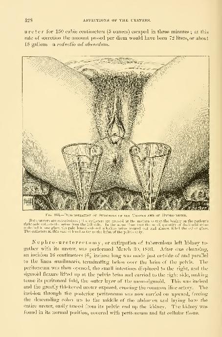
428 AFFECTIO]SrS OF THE URETERS.
ureter for 150 cubic centimeters (5 ounces) escaped in three minutes ; at this
rate of secretion the amount passed per diem would have been 72 hters, or about
18 gallons—a reductio ad absurduTn.
Fig. 244.
—
Demonstration of Stricture of the Ureter and of Hydroureter.
Both ureters are catheterized ; the catheters are crossed in the uretlira so that the beaker on the patient'srifrht side collects the urine from the left side. In the same time that the small quantity of dark acid urinecollected in one glass, the pale lemon-colored alkaline urine poured out and almost tilled the other glass.The catheters in this case entered as far as the brim of the pelvis only.
]Sr e p h r o - u r e t e r e c t o m y , or extirpation of tuberculous left kidney to-
gether with its ureter, was performed March 30, 1893. After due cleansiiig,
an incision 16 centimeters (6J inches) long was made just outside of and parallel
to the linea semilunaris, terminating below over the brim of the pelvis. Theperitoneum was then opened, the small intestines displaced to the right, and the
sigmoid flexure lifted up at the pelvic brim and carried to the right side, makingtense its peritoneal fold, the outer layer of the meso-sigmoid. This was incised
and the greatly thickened ureter exposed, crosaing the common iliac artery. Theincision through the posterior peritoneum was now carried on upward, freeing
the descending colon up to the middle of the abdomen and laying bare the
entire ureter, easily traced from its pelvic end up the kidney. The kidney wasfound in its normal position, covered with peritoneum and fat cellular tissue.

TUBERCULAR URETERITIS. 429
Tlie peritoneum was next incised over the kidnej and the process of enuclea-
tion begun. The separation was effected with ditficultj, owing to the dense
adherent fibrous tissue interpenetrating the fat and sticking tight to the kidnej,
especially about the hilum. Slowlj, and with much care, the large vessels werefreed and tied with four fine silk hgatures, and the kidney severed from all its
attachments, except the ureter. Kow, taking the kidney in hand, it was pulled
downward and the ureter gradually dissected out of its bed of cellular tissue all
the way to the pelvic floor. The ovarian vessels were tied in the abdomen at
about the middle of the ureter.
The ureter was then ligated at the pelvic floor, and cut off wedge shaped 1
centimeter above the ligature, forming flaps for easy closure. Care was taken
throughout not to contaminate the peritoneum with the infected end, which wasfinally burned out with a Paquelin cautery down to the ligature, and the flaps
united with fine silk sutures. The left flank was then pushed out by two fingers
and pierced with a knife, making a hole 3 centimeters (1^ inch) long in the
line of the iliac crest, just in advance of the spinous muscles. A gauze drain
reaching the ureteral stump below was put in here, 14 centimeters (5^ inches)
long by 3 centimeters (IJ inch) wide, and the anterior incision was closed.
The colon fell into its natural position vtdthout suture.
On the first day there was a free bloody serous discharge, which gradually
decreased, and the drain was taken out on the fifth day. The patient made a
quick, undisturbed recovery, and is still living, three years later, remarkably im-
proved. For the removal of the entire ureter with the kidney by an extra-
peritoneal operation, see Nephro-uretectomy.
The following is the pathological report
:
Kidney Macroscojjically .—The mass representing the kidney is
made up of four large lobules, separated from one another by shallow sulci. Theupper third of the organ is least affected, though this is deeply pitted in every
direction, and contains a number of small cysts, which become distended on in-
jecting the pelvis of the kidney, proving direct connection. This portion meas-
ures by -i-S centimeters (2| by If inches), while the whole kidney mass measures
11 by 6 by 3*5 centimeters (l^^ by 2^ by 1^ inches). Below the mass just de-
scribed on the anterior face is a cyst measuring 5-5 by 5 centimeters (2^ by 2
inches), and below this two yellowish masses, 7 by 4-5 centimeters (3 by If
inches), independent of one another but connected by a shallow sulcus. All
the cysts are covered by the capsule. The weight of the whole organ with
the attached ureter is 100 grams.
The capsule of the kidney is thickened and intimately adherent in places.
On section, about 65 cubic centimeters (2 ounces) of fluid escapes from the cysts;
in one it is white and flaky, consisting almost entirely of fatty debris ;in another,
the fluid resembles blood-stained urine. These cysts are all found to communi-
cate with the pelvis of the kidney and represent dilated calices. The parenchyma
of the kidney is largely destroyed. In one place an area of cortex is found 7
millimeters in depth, but elsewhere the kidney substance is represented by lay-
ers averaging 1-3 millimeter in thickness sjjread out over the dilated calices.

430 AFFECTIONS OF THE URETERS.
Frozen sections of the kidney show a diffuse infiltration with fatty
granulation tissue, with here and there areas of complete necrosis, with frag-
mentation of nuclei. Numerous definite tubercle nodules, containing giant and
epithelioid cells, can be made out. The tuberculous process goes gradually over
into the more healthy kidney substance, and here and there a single tubule or
glomerulus can be seen in the diffuse tuberculous tissue.
Ureter.—The ureter presents two points of constriction, repectively, 3
and 8 centimeters (1^ and 3^ inches) from the kidney. The hilum of the kid-
ney is filled with dense adherent fat, preventing dissection of the stricture with-
out tearing it.
The ureter is much dilated, more at some points than at others, the caliber of
its lumen varying from 1*7 to 3 centimeters (^ to 1^ inch). Its wall is much
thickened, measuring in places from 5 to 6 millimeters. The mucous membrane
is of an opaque buff color, and at one spot, near the pelvis, there is a superficial
area of calcification 5 millimeters in diameter.
Frozen section of the ureter shows that the epithelium is entirely ab-
sent from the surface, and that the mucous membrane is converted into a mass
of diffuse tuberculous tissue, in which here and there definite tubercular nodules
can be made out. The surface is not infrequently quite necrotic, and the cells
near it have undergone fatty degeneration. The muscular layer has been in-
volved, and there are many aggregations of small round and epithelioid cells
there. In some places there is cell proliferation in the fibrous layer of the
ureter. The connective tissue is from three to four times thicker than normal.
This ureter had been catheterized previous to the operation, and numbers of
tubercle bacilli demonstrated in the pus which was present in the urine thus ob-
tained. Cultures made from the pelvis of the kidney and from the kidney sub-
stance on ordinary agar-agar remained sterile.
Diagnosis .—Tuberculosis of kidney, pelvis of kidney, and ureter.
Although in the case just cited a transperitoneal route was followed, the best
routine way to reach the ureter is entirely extraperitoneal (see Johns Hopkins
Hosp. BuL, Feb, and March, 1896), by means of a long incision beginning
back in the loin in front of the quadratus muscle, halfway between ribs and
ilium, and continued in an oblique direction downward and forward, skirting
the anterior superior spine within 4 centimeters (1^ inch) of it, and ending in
the semilunar line over the top of the broad ligament. The skin, fat, muscles,
and fascia are divided down to the peritoneum, which is then dissected up by the
fingers, being lifted toward, the opposite side; the nreter is found, after raising
the colon, crossing the belly of the psoas muscle with the ovarian vessels, and if
not seen at once, it may be traced from the pelvis of the kidney down. It mayfurther be recognized by tapping it sharply, or by watching a peristaltic wave
pass downward. The peritoneum need not be opened at any point. After free-
ing the kidney by ligating its vessels and detaching the abdominal poi-tion of the
ureter as described, the pelvic portion is then freed by following the upper por-
tion as a guide, while the fingers readily lift the pelvic peritoneum from the ves-
sels which drop with the ureter over tlie brim. By pulling it out the ureter

OBSTKUCTION OF THE URETER. 431
may be freed not only down to the floor of tlie pelvis, but well forward. Tocomplete the enucleation as far as the vesical attachment, the uterine artery andveins must be tied and divided.
Obstruction of the Ureter.—Obstruction of the ureter, diminishing or obliter-
ating its lumen, will be more frequently diagnosed when the opportunities of
examining the ureters afforded by abdominal surgery and catheterization are
more generally embraced. The importance of recognizing the existence of a
ureteral obstruction can not be overestimated, on account of its damaging effect
upon the kidney, diminishing or even suppressing its excretory power ; the dan-
ger of an obstruction is vastly greater when both ureters are involved.
The immediate effect of obstruction is to back up the urine above it in
the pelvis and calices of the kidney, producing hydroureter and hydronephro-
sis, varying in their clinical appearance according as the obstruction is produced
gradually or suddenly, is partial or complete. If the hydroureter and hydro-
nephrosis become infected, we have then to deal with a pyoureter and pyelo-
nephrosis above the obstruction.
Causes .—Ureteral obstruction may be produced in a variety of ways, and
is far more common in women than in men, being frequently associated with
diseases of the uterus and ovaries. They may be classified in general as
—
First, causes acting from without and occluding the ureter by pressing upon
it or overstretching it ; such are
—
1. Ovarian tumors,
2. Uterine tumors.
3. Cancerous infiltration of the broad ligaments.
4. Cancer of the cecum.
5. Retroperitoneal pelvic sarcoma.
6. Aneurism of the iliac artery.
7. Scar tissue in the broad ligament.
8. Periureteritis.
9. An omental adhesion to the pelvic brim.
10. Thickened bladder walls.
11. Sarcoma of the bladder.
12. Pediculated tumor of the bladder.
Second, foreign bodies lodged in the ureteral canal
:
1. Calculus.
2. Blood clot.
3. Echinococcus cyst.
Third, affections of the ureteral walls themselves
:
1. Ureteritis bacilli coli communis.2. Ureteritis g o n o r r h o i c a
.
3. Ureteritis tuberculosa.4. Valve formation in the ureteral wall.
5. Gumma in the wall.
6. Cancer of the ureter.
7. Psorospermial cysts.

432 AFFECTIONS OF THE UEETERS.
Some of the twenty-two causes of ol)struction just cited act unilaterally,
while others are more apt to act on both ureters at once ; it therefore becomes
important from a practical standpoint to divide them further into groups ac-
cording to this tendency.
Both ureters are apt to be obstructed by cancer of the cervix uteri extend-
ing out into the broad ligaments, by thickened bladder walls, by some large sub-
peritoneal fibroid tumors, and in rare instances by calculi.
But one ureter is apt to be involved in parametritis, small pelvic tumors and
inflammatory masses posterior to the broad ligament, gonorrheal stricture, and
tuberculosis.
The location of the obstruction in almost all cases is in the pelvic portion of
the ureter, at some point between the brim of the pelvis and the vesical end.
The reasons for this predilection lie, on the one hand, in the proximity to the
ureters of the uterus, tubes, and ovaries, and their liability to inflammatory
affections or new growths, and, on the other hand, to the fact that the ureters
are enclosed with these organs in the unyielding bony pelvic canal, which affords
a point of resistance against which pressure can be made. Next in frequency
to the pelvic extremity is the involvement of the upper end near the pelvis of
the kidney.
The clinical symptoms of obstruction are variable, depending on the
cause and the completeness of the occlusion, as well as the rapidity with which it
is produced. In the milder grades, where the distention is not great, there may be
no symptoms at all. I have a patient whose right ureter and renal pelvis are
dilated by a stricture at the vesical end of the ureter until they hold 100 cubic
centimeters of urine without producing any subjective sensations wdiatever.
Extreme dilatation may be produced without pain if the cause acts slowly. I
had one case, a little girl about three years old, in whom the right ureter was
lifted out of the pelvis and dilated to a diameter of 1^ centimeter by a retro-
peritoneal sarcoma.
"Where the obstruction depends upon inflammatory disease the chief symp-
tom is frequent painful urination ; in cases of tumors and pelvic inflammatory
masses the ureteral symptoms are often masked by the associated complaint.
The sudden closure of one ureter, as by a ligature, produces violent pain in its
course, extending into the kidney, associated with restlessness, a hot, dry skin,
and fever and diminished urine. If both ureters are obstructed, uremia devel-
ops soon after the pressure in the sac of urine formed is equal to that in the
blood vessels, checking further secretion.
To make a diagnosis three questions must be answered :
First, Is the ureter obstructed at all ?
Second, What is the nature of the obstruction ?
Third, What is its degree ?
A- diagnosis from symptoms alone can only be made in that small percentage
of cases in which the occlusion has taken place suddenly, as in the case of a cal-
culus or clot descending from the kidney and lodging in the ureter and block-
ing its flow, or when in an operation the ureter is tied and the persistent agoniz-
OBSTRUCTIOX OF THE URETER. 433
ing ureteral and renal colic definitely located in its course leave no doubt as to
the nature of the difficulty.
There are no reliable symptoms of a dilatation of an aseptic ureter that has
developed slowly. A diagnosis of obstruction with dilatation may be made with
assurance whenever uremic symptoms are noted in the course of a cancer of the
cervix.
While diagnostic means heretofore liave been indirect and unsatisfactorv, en-
abling us only to infer the existence of obstruction, and that in a small percent-
age of cases, the means of direct exploration of the whole ureteral tract now at
our command leave but little to be desired in the way of accuracy. These are
the inspection of the ureteral orifices and the catheterization and sounding of
the ureters.
Before describing the actual use of these methods it will be im])ortant to
consider first in what class of cases it is desirable to try to ascertain whether
there is or is not an obstruction. I should always make an examination for ob-
struction where there is persistent pain in the course of a ureter ; where the
patient is distressed by frequent urination, for which a sufficient cause does not
exist in the bladder or urethra ; where there is pus in the urine in cases of pelvic
inflammatory diseases ; and where pelvic tumors might be supposed to makepressure on a ureter.
In investigating an obstructed ureter we wish to determine
—
1. Whether there are any abdominal or pelvic tumors or masses which could
])ress upon a ureter.
2. A¥liether any form of ureteritis exists.
8. Whether the ureter is blocked by a stone or clot.
4. Whether two of these conditions do not act in combination.
The presence of an abdominal or pelvic tumor pressing on a ureter can be
determined by a bimanual and rectal examination and deep abdominal palpation
under anesthesia. Sufficient cause for an obstruction exists when a band of scar
tissue, following injury in childbirth, is felt in the parameti'ium, dragging the
uterus to one side, or when an inflammatory mass is felt fixed to the pelvic wall
and floor, or when there is a uterine or ovarian tumor choking the pelvis, or
some other tumor filling the lower abdomen.
Palpation of the ureter through the vaginal walls shows whether it is thick-
ened or not, and so demonstrates the presence or absence of a ureteritis.
The blocking of the ureter by a stone or clot can only be demonstrated bythe passage of a sound or catheter, and this brings us to the method of demon-strating with absolute certainty the existence of an obstruction. In examining
any given case the investigation must not cease when one cause sufficient to
explain an obstruction is found ; other associated causes must always be sought
for, and their absence definitely proved, before the one cause found is finally
accepted. For example, an inflammatory obstructive ureteritis may be found
associated with a pelvic abscess of the same side.
By the direct examination the existence of an obstruction is proved either
when (1) a ureteral catheter or sound j^asses freely up the ureter until it is sud-
434 AFFECTIONS OF THE URETERS.
deuly checked, or (2) when each time after passing a certain point in the ureter
there is an inmiediate continuous How of urine of from several up to a hundred
or more cubic centimeters in quantity. Sufficient time must be allowed to elapse
for more urine to accumulate before repeating the examination. The demon-
stration is still more complete in a case of this kind if the instrument is distinctly
grasped in the bite of the stricture and resists withdrawal.
In sounding and eatheterizing a ureter for obstruction, the metal catheter is
only of use for the lower part of its course, from the vesical orifice to the pos-
terior pelvic wall. With gentle tact the metal catheter may sometimes be
coaxed through a tight stricture impassable to the yielding silk catheters. As a
rule, it can not be pushed in more than from 4 to 6 centimeters {1^ to 2^ inches)
under inspection with the patient in the knee-breast position, and it is bettei"
after introducing it to turn her over to the dorsal position, or to put it in, in the
first place, in the dorsal position, and to let the air out of the bladder with a
catheter, and then to guide the further progress of the ureteral catheter with a
finger at first in the vagina and then in tlie rectum. It is needful to empty out
the air, as the distended bladder splints the catheter and impedes its onward
movement.
An ordinary solid sound has no advantage over a catheter, which does
equally well as sound and catheter. The only sound I have ever used with
advantage is one made like a catheter, but solid, and with a slight bulbous
enlargement, 1 centimeter back of the point, which trips in passing any narrow
place in the lumen.
The short flexible ureteral catheter is only valuable in locating strictures in
the lower ureter, but is easy to introduce and is safer in experienced hands.
The long flexible catheters, 50 centimeters (20 inches) in length, are used to
locate strictures in the upper ureter all the way uj) to the pelvis of the kidney.
It is always important in searching for a stricture to pass the catheter up slowly,
so that the flow of urine will make it evident as soon as the stricture is passed.
If the catheter is pushed up rapidly, the end may be several centimeters or more
beyond the stricture before the flow begins, and the stricture in this way esti-
mated to l)e higher up than it really is. A good telltale is made by dipping
the finger in water and touching it to the end of the catheter, which is then
closed by a thin film of water until the urine begins to flow, forcing the air out
and pushing the water oif from the end of the catheter in the form of a little
bubble, in this way announcing the coming of the urine some seconds in ad-
vance of its actual appearance. The escape of urine backed up behind an ob-
struction is different from the intermittent normal flow drop by drop. The
urine which has been held back often pours out of the catheter in a steady stream
until it has almost all escaped, when it continues to drop steadily for a while
longer, and so the sac is emptied. The fact that the urine has been backed up,
and that it is not simply an abnormally rapid secretion, can be proved by a sim-
ple calculation. For example, if the normal secretion is alxnit 1'5 liter per
diem (3 pints), this makes about 1 cubic centimeter per minute from both sides
combined, or half a cubic centimeter from one side. If now I collect 90 cubic

OBSTRUCTIOK OF THE URETER. 435
centimeters in three minutes after putting one catheter in, that equals 3() cubic
centimeters in one minute, or sixty times tlie normal amount, 90 liters (180
pints) a day, manifestly impossible.
An important part of the investigation is to decide exactly where the stric-
ture ends and the dilated portion of the ureter begins. This is done by with-
drawing the catheter slowly during the escape of the urine, and noting the
moment the flow is checked ; the length of the catheter inside, of course, then
measures the distance of the upper end of the stricture from the meatus. Todetermine the distance from the vesical end of the ureter, the distance of the
external meatus to the ureteral orifice is measured with the searcher and de-
ducted.
The treatment of ureteral obstruction depends upon the cause; in some
cases it can be easily removed, in others it is irremediable. An obstruction
should never be allowed to persist if it can be removed without undue risk to
life. The danger of surgical interference is greater where both ureters are
involved, and is greatest of all if infection of one or both sides is superadded.
Where both sides are occluded by a cancer in the broad ligaments, the plan
proposed of prolonging life by severing the ureters just under the kidneys, and
turning the ends out to discharge the urine in the flanks is rarely applicable on
account of the condition of the patient by the time the disease has advanced so
far. In case uremia is threatened from occlusion of both ureteral orifices bythickened, inflamed bladder walls, it will be proper to save the kidneys and pre-
serve life by opening each antero-lateral vaginal wall, isolating the ureters, and
making a longitudinal incision in them 1 centimeter long, so as to suture them
to the vaginal wall (kolpo-ureterostomy). In case of recovery of the bladder,
the ureteral fistulse could afterward be closed.
AVhere the ureteral dilatation comes from the pressure of a pelvic tumor, it
is treated by taking away the tumor and removing the pressure. Indeed, this
is often done in removing pelvic tumors without the operator knowing all that
he has accomplished. In all cases of pelvic tumors both ureters should be in-
spected before removing the growth, for dilatation in varying degrees (hydro-
ureter) will be discovered with surprising frequency. If to the dilatation an
infection has been added, this will need treatment later, either by washing out
the pelvis of the kidney and the ureteral tract, or by opening the pelvis in the
loin. I have twice seen pelvic abscess in the left side associated with pyo-
ureter and pyelonephritis. In another case a densely adherent ovarian cyst on
the left side blocked the ureter. The upper ureter and pelvis of the kidney
were filled with pus, and the enlarged kidney contained multiple abscesses. I
removed the tumor and the kidney, and the woman regained complete liealth
and is still living three years later.
Ureteritis as a cause of obstruction is quite common. The ureteritis itself
can not be directly treated unless it has formed a stricture and the ureter is
dilated above it ; the treatment is then directed to the dilatation of the stricture
and the removal of the infection. Where it is due to stone, relief will be ob-
tained only by uretero-lithotomy. I have seen two cases of colon bacillus infec-
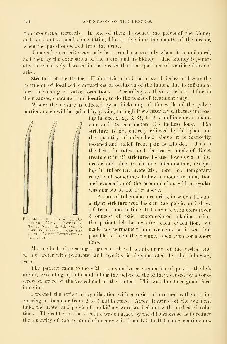
-1:3 ) AFFECTIOXS OF THE URETERS.
tion ])rodueiiig- ureteritis. In one of tliein I opened the pelvis of the kidney
and took out a small stone litting like a valve into the mouth of the ureter,
when the pus disappeared from the urine.
Tubercular ureteritis can only be treated successfully when it is unilateral,
;ind then by the extirpation of the ureter and its kidney. The kidney is gener-
ally so extensively diseased in these eases that the question of sacrifice does not
arise.
Stricture of the Ureter.—Under stricture of the ureter I desire to discuss the
treatment of localized contractions or occlusion of the lumen, due tu infiannna-
tory thickening or valve formations. According as these strictures differ in
their causes, character, and location, so do the plans of treatment vary.
Where the closure is effected by a thickening of the walls of the pelvic
portion, much will be gained by passing through it successively catheters increas-
ing in size, 2, 2^, 3, 3|, 4, ^, 5 millimeters in diam-
//^ /I eter and 28 centimeters (13 inches) long. The
// /l stricture is not entirely relieved by this plan, but
rI
fi the quantity of urine held above it is markedly' * ''' lessened and relief from pain is afforded. This is
the best, the safest, and the easiest mode of direct
treatment in all strictures located low down in the
ureter and due to chronic inflammation, except-
ing in tubercular ureteritis ; here, too, temporary
relief will sometimes follow a moderate dilatation
and evacuation of the accumulation, with a regular
washing out of the tract above.
I IA case of tubercular ureteritis, in which I found
I Ia tight stricture well back in the pelvis, and drew
jl 11I
off" from time to time 100 cubic centimeters (over"^ ^3 ounces) of pale lemon -colored alkaline urine,
the patient felt better after each evacuation, but
made no permanent improvement, as it was im-
possible to keep the channel open even for a short
time.
My method of treating a gonorrheal stricture of the vesical end
of the ureter with pyoureter and pyelitis is demonstrated by the following
case :
The ])atient came to me with an extensive accumulation of pus in the left
ureter, extending up into and filling the pelvis of the kidney, caused by a cork-
screw stricture of the vesical end of the ureter. This was due to a gonorrheal
infection.
I treated the stricture by dilatation with a series of ureteral catheters, in-
creasing in diameter from 2 to 5 millimeters. After drawing off" the ])urulent
fluid, the ureter and pelvis of the kidney were washed out with medicated solu-
tions. The caliber of the stricture was enlarged by the dilatations so as to reduce
the quantity of the accumulation above it from 150 to 100 cubic centimeters.
Fi(i. 24").
—
The Ends of the Di-i.ATiNc; Metal Catheters,Three Sizes (3, 3.5, and 4),
USED IN DILATING StrICTIREOF THE Lower Extremity ofTHE Ureter.

STRICTURE OF THE URETER. 437
The purulent character of the secretion was removed and all trace of gonococci,
at lirst abundant, disappeared.
My patient (E. S., San. Ofi) was a married woman, thirty-one years of age,
of slight build and haggard-looking. She had had one child four years be-
fore without special ditiiculty, the only pregnancy in six years of married life.
The menses were regular and without pain. Headaches were rare ; the appe-
tite was good and the bowels regular. Slie had no chills.
She had been feeling depressed for some months and had lost weight, and com-
plained of severe pain on urinating, wdiich persisted for a half hour or longer.
There was a sense of pressure in the bladder, and she was obhged to urinate
every two or three hours by day and oftener by night. There was no acute
pain, but aching in the limbs and discomfort of the lower abdomen. She
noticed that the appearance of the urine varied greatly, being clear at times,
and at other times containing much yellow sediment.
My examination showed that the vaginal outlet was torn superficially back
almost to the anus ; the cervix was in the axis of the vagina, somewhat low down,
showing a slight tear, and the uterus was in retroflexion ; the left ovary was
displaced downward, not adherent, and tender on pressure. ( )n examining the
anterior wall of the vagina, no special tenderness was developed on palpating
the bladder.
The ureters were then pal])ated by the vagina. The left felt distinctly harder
than normal and somewhat thickened, but was without marked tenderness ; it
was also displaced toward the pelvic floor.
The l)ladder was then examined nnder atmospheric dilatation, with the patient
in the knee-chest position, tlirongli the No. 10 speculum. There was evidence
of a patchy, mild cystitis. The fleld opposite the ureter, the posterior pole, and
its surrounding area were mottled, red, and injected, the vessels being obscured';
the injection increased toward the vault, which was covered over an area -1- by 5
centimeters by fine granules, averaging one or two to the square millimetei-,
most marked on the right side. The tips of each of these granules i-eflected
the light and gave the snrface a bright studded appearance. On the left
side in places the surface presented a superficial worm-eaten appearance. On
the right lateral wall, 2^ centimeters behind the ureteral orifice, was a ridge 2
millimeters in height, extending downward to the base of the l)ladder. Near
the right ureteral orifice was an area of intense congestion, presenting an
edematous appearance, surrounding the ureter, whose orifice could only l)e
located by a little pallor in the form of a crescent. Posterior to the right ureter
was a superficial ulcer 2 by 3 millimeters, with a narrow red border and a yel-
low center.
The left ureteral orifice was situated on a truncate cone, about 6 milli-
meters in diameter at its base and 2 millimeters at the top. It was slightly
edematous, and on the urethral side broken up by a number of irregular papil-
lary eminences. The sight of the ureteral orifice at the first examination was
marked by a yellow spot of pus. On introducing a searcher into the opening of
the orifice, a thin stream of pus escaped and ran down on the bladder wall.
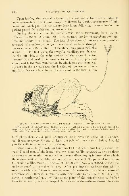
43 S AFFECTION'S OF THE URETERS.
Upon leaving the ureteral catheter in the left nreter for three minutes, 11
cu])ic centimeters of dark Huid escaped, followed by 6 cubic centimeters of tluid
containing nmch pus. In the twenty-four liours following the examination the
patient passed TOO cubic centimeters of urine.
Durino- the whole time the patient w^as under treatment, from the 2d
of March to the 2d of June, 1894, I catheterized her left ureter about one hun-
dred and twenty times in all. The first three weeks of her stay were passed in
rej^eated vain endeavors to get the ureteral catheter through
the stricture into the ureter. Three difficulties prevented this
at first. In the first place, the irregular papillary prominences
on the left side, in the neighborhood of the ureteral orifice,
obscured it, and made it impossible to locate it with precision
subsequent to the first examination, in which pus was seen ooz-
ing out ; in the second place, the location of the ureteral mons
and its orifice were in extreme displacement to the left ; in the
Fig. 246.
—
Washing Out the Eight Kidney foe Gonorrheal Ureteritis and Pyelitis.
A flexible catheter is usually used. In this case a metal catheter was passed through a stricture at thelower end of the ureter and the solution carried up to the kidney by gravity by means of a funnel and longrul)l)er tube. The patient is in the knee-chest position to aid gravity.
third place, there was a spiral stricture of the intravesical portion of the ureter,
and it was necessary for me to learn the twist of the stricture before I could
])ass the catheter at once at every sitting.
After almost daily effoi'ts for three weeks the stricture was finally cleai-ed by
an accidental turn of the hand ; this was more readily repeated on two or three
occasions subsecpiently, but not without many discouraging failures, after which
the ureteral orifice was definitely located on the side of the pyramid in i-clation
to certain papilhie, and the direction of the stricture was ascertained, so that the
catheter could be passed w^ith ease. After pushing the catheter through the
stricture with a half turn it entered about S centimeters; a distinct sense of
resistance was felt in attempting to withdraw it, due to the bite of tlie stricture,
about 1^ centimeter long. So long as the point of the catheter went no farther
than the stricture, no urine escaped ; but as soon as the catheter cleared the stric-
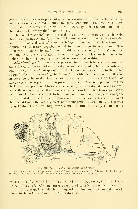
STRICTURE OF THE URETER. 439
tiire, pale urine began to pour out in a steady stream, continuing until 150 cubic
centimeters were collected in three minutes. Sometimes the iirst urine drawn
off would be of a reddish-brown color, followed by a whitish sediment, and at
the last a thick, creamy fluid like pure pus.
The fact that so much urine escaped in so short a time proved conclusively
that there was an extreme dilatation of the left urinary channels above the stric-
ture, for the normal rate of secretion being at the most 1 cubic centimeter a
minute for both ureters together, or 1^ in three minutes for one ureter. The
discharge of 150 cubic centimeters would be twenty-nine times the normal
amount, or at the rate of about twenty-two gallons a day for both sides to-
gether, proving that there was a dilated pyoureter and pyelitis.
After drawing off all the fluid, a piece of tine rubber tubing with a funnel at
the end was connected with the catheter, and a saturated boric-acid solution,
equal to two thirds of the quantity of fluid taken out, w^as run into the ureter
by gravity by simply elevating the funnel filled with the fluid from 40 to 60 cen-
timeters above the level of the bladder. Care was taken to have the tubes full of
fluid, so as not to inject air. The patient, during all these manipulations, was in
the knee-breast position. She took no anesthetic, as the treatment was not painful.
After the catheter was in the ureter she raised herself on her hands and knees
to dispose the fluid to run out faster. When the injection was given she again
let her chest down to the table, and rose again when it was to flow out. I found
that I could wash the urinary tract repeatedly with the same fluid, if I desired
it, by holding the funnel high for the fluid to run in, and by holding it an
^y^\iFig. 247.
—
Washing Out the Kidney and Ureter.
Letting the fluid run back a.crain into the funnel from the kidney and ureter. The funnel is held low and
the patient rises on the hands and knees to facilitate the outward flow. San., Mrs. S.
equal distance below the level of the table for it to run out again, often bring-
ing with it a considerable amount of shreddy white debris from the ureter.
A small Y-shaped switch with a stopcock in the angle was used at times to
facilitate the inflow and outflow of the solutions.

440 AFFECTIOXS OF THE URETERS.
After the iirst few treatments slie began to experience relief from pain and
was less frequently disturbed at night.
Examination of the urine l)y Dr. Barker in the pathological laljoratory of
the Johns Hopkins Hospital showed that it was straw-colored, neutral in reac-
tion, and contained abundant muco-purulent, stringy, tenacious sediment. There
was a small amount of albumin, but no sugar and no casts. The specific gravity
was 1-032. There were many polynuclear leucocytes, crowds of pus cells, and
many diplococci, nearly all of which were within the protoplasm of the leuco-
cytes. Octahedra of calcium oxalate were found, and a few cylindroids. There
were no tubercle bacilli, and no other bacteria than diplococci, which were of
the typical appearance of gonococci, and much smaller than staphylococci or
streptococci.
The bladder walls were treated by occasional applications of a 5 per cent
solution of nitrate of silver, applied directly to the affected areas on absorbent
cotton with an apjilicator, and by daily irrigations of a bichloride solution (1-
150,000).
My first effort in the treatment of the case was to secure a continuous drain-
age of the ureter, avoiding all accumulation al)Ove the stricture, hoping by this
plan to induce a contraction of the ureteral walls. To do this I made a short
ureteral catheter 2 millimeters in diameter and 5 centimeters long, with a little
shoulder about 2 centimeters back of the inner end to keep it from slipping out
of the ureter after introduction, and with a fiange 6 millimeters in diameter at
the lower end to keep it from slipping altogether into the ureter. I placed this
in the ureter by means of a searcher used as a mandarin to conduct it through
the stricture. I found, however, that its presence gave so much pain and in-
creased the irritation of the bladder, after being in place for twelve hours, that
I was obliged to abandon its further use, although it acted well mechanically.
My next plan, which was successful in curing the case, was to have ureteral
catheters made in four sizes, increasing from the smallest, 2 millimeters, to the
largest, which was 6 milHmeters in diameter. The j)oints of the catheters M^ere
blunt and straighter than the ureteral catheters ordinarily used, on one side
almost on a line with the. shaft.
In the course of two months of such treatment the ureter was dilated sulfi-
ciently to permit the introduction of the largest catheter, from the end of which
the accumulated urine would drop in a large free stream. "With the catheters I
began systematically to wash out the ureter and kidney with a bichloride of mer-
cury solution (1-150,000), constantly increasing the strength until 1-16,000 was
used. The treatment with the bichloride was interrupted several times for the
injection of a 1 per cent nitrate of silver solution, and once for a weak iodine
solution. Toward the end, while using the larger catheters, I was obliged some
six times t(j suspend the treatment for from two to three days, on account of a
chill followed by elevation of temperature from 102° to 104° F., with a (piick-
ened pulse (120), headache, nausea, and pain in the left inguinal region and
legs. The patient was fiushed and restless, and sufiered from sleeplessness at
these times.

STRICTURE OF THE URETER. 441
The result of the hichloride washings was a conijjlete disappearance of pus
cells, leucocytes, and gonococci from the urine, and the reduction of the size of
the distended ureteral tract from one holding regularly from 140 to 150 cubic
centimeters down to 90 or 100 cubic centimeters. The bladder assumed a nor-
mal appearance and she became able to sleep through the night without rising
once. She gained 20 pounds in weight and resumed the rosy appearance of per-
fect health, with a corresponding remarkable improvement in spirits.
The treatments were discontinued August 8, 1894, and I saw her again in
January, 1895, and then on two occasions catheterized the ureter, drawing oft"
only 90 and 100 cubic centimeters of clear urine from the left ureter with-
out a trace of pus or cocci. She has therefore recovered from the infection,
but still has a stricture of the ureter of larger caliber with a lax distended ureter
above it.
I made several attempts to empty the ureter by massage, with consideral)le
success at first, but the procedure became so painful that it had to be stopped.
Just before the massage the bladder was emptied by catheter, and immediately
after treatment as much as 90 cubic centimeters of urine were secured.
I demonstrated the success of the massage and mapped out the exact posi-
tions in which to make pressure by placing the patient in the dorsal position and
then introducing a catheter with a rubber tube attached to its outer end, when a
straight glass tube, 50 centimeters long, attached at the other end of the rublier
tubing, filled at once with urine to the level of the ureter and acted as a manom-eter. Kespiratory movements were traced by its rhythmical ascent and fall.
( )n making pressure over the ui'eter through the abdominal wall the column
ascended in the vertical glass, and, by inci-easing the pressure, could be forced
out over the top. If the pressure was made to one side of the ureter there was
only a slight effect or none at all. By marking all the points of effective pres-
sure on the skin, and afterward connecting the markings, the course of the ureter
was accurately mapped out.
The following important points are demonstrated l)y this case :
1. Stricture of the lower extremity of the ureter can be diagnosed without
any operation by using the cystoscope with the bladder dilated wdtli air by
posture.
2. Stricture of the ureter can be improved by gradual dilatation by a series
of hollow bougies (catheters) and without a koljio-ureterotomy. (See Johns,
Hop. Gijn. Rep., No. 1.)
3. A stricture through which a No. 5 (5 millimeters in diameter) bougie is
passed every day for several weeks will still hold back the urine if the wails of
the ureter above have lost their contractility.
4. Pyoureter and hydroureter can be diagnosed by drawing off" in a few
minutes such a quantity of fluid as it is manifestly impossible for the kidney to
secrete in that amount of time.
5. Pyoureter and pyelitis can be cured by washing out the ureter and pelvis
of the kidney without any preliminary cutting operation to disclose the ureteral
orifice (as in kolpo-uretero-cystotomy, Bozeman).
442 AFFECTIOXS OF THE URETERS.
6. Variations in pressure intlie eohinui of liuid in a distended ureter can be
demonstrated bj a manometer attached to the ureteral catheter.
7. In this way the course of the ureter can be mapped out.
There are several sorts of stricture in the upper part of the ureter at or near
the junction with the pelvis of the kidnev.
In one important group of cases the closure is intermittent, or de])ends on a
ureteritis without a definitely localized stricture.
Complete closure may be congenital or it may follow an injury or inflamma-
tory disease. The treatment will depend upon the length of time which has
elapsed since the occlusion was eifected as well as upon the result of the occlu-
sion. Where the kidney has undergone atrophy nothing need be done. If it
has become greatly distended, temporary relief may be afforded by tapping. If
the occlusion is but recently acquired, the effort should be made by operation to
make the passage pervious.
I have examined the first patient on whom I performed a nephro-ureterec-
tomy, leaving 6 or 8 centimeters of the lower end of the tuberculous ureter, and
found the position of the ureteral orifice in the bladder marked by a shallow pit
impervious to the sound.
In another case of an intermittent hydronephrosis of long standing, due to a
stricture of the ureter just below the pehds of the kidney, in attempting to
locate the stricture by means of a catheter the vesical opening was lacerated ; a
few days later the kidney was ex])osed and the ureteral stricture divided and
sutured so as to make a free opening from the kidney into the ureter. Thepelvis of the kidney was drained through an incision in the dorsum so as to
allow the repaired stricture to heal at rest. During this time the bruised lower
end of the ureter, no longer kej^t open by the passage of the nrine, closed com-
pletely, as I found by a cystoscopic examination several weeks later, when no
efforts made to pass the sounds succeeded.
The following case of complete occlusion of the renal end of the
ureter came under my care in May, 1894.
C. F. (San. 109, May, 12, 1894), twenty-three years old, was suffering from an
old fistula discharging pus beneath the left anterior superior spine. Her trouble
began ten years before with " awful spells of pain " in the left side, extending
around into the small of the back. These came about twice yearly and lasted
two weeks at a time, when she had to stay abed on account of the pain and fever
attending them. In the intervals she suffered from constant soreness in the left
side. In 1890 she had a spell lasting a year, treated as "typhoid fever," and
terminating in April, 1891, in an abscess in the left inguinal region, which was
opened and 2 liters of pus and blood evacuated. The abscess continued dis-
charging until October, 1892, when it closed. In June, 1898, she got worse
again and the abscess reformed ; it was opened in August, and since then the
discharge had been constant. Several gritty particles escaped recently from the
wound. Seven months before coming to me she had been in a sanitarium, where
she was treated for spinal caries. Each time the sinus closed and pus accumu-
lated she had a chill with pain and fever until it was reopened.

strictl'rp: of the ureter. 443
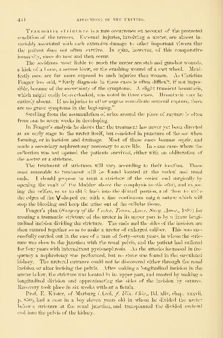
4-i-i AFFECTIOXS OF THE URETERS.
Traumatic stricture is a rare occurrence on account of the protected
condition of the ureters. External injuries, involving a ureter, are almost in-
variably associated with such extensive damage to other important viscera that
the patient does not often survive. In spite, however, of this comparative
immunity, cases do now and then occur.
The accidents most liable to reach the ureter are stab and gunshot wounds,
a kick of a horse, a severe blow, or the crushing wound of a cart wheel. Mani-
festly men are far more exposed to such injuries than women. As Christian
Fenger has said, " Early diagnosis in these cases is often difficult, if not impos-
sible, because of the uncertainty of the symptoms. A slight transient hematuria,
which might easily be overlooked, was noted in three cases. Hematuria may be
entirely absent. If no injuries to other organs complicate ureteral rupture, there
are no grave symptoms in the beginning."
Swelling from the accumulation of urine around the place of rupture is often
from one to seven weeks in developing.
In Fenger's analysis he shows that the treatment has never yet been directed
at an early stage to the ureter itself, but consisted in puncture of the sac whenforming, or in incision and drainage. Most of these cases l)ecame septic, and
made a secondary nephrectomy necessary to save life. In some cases where the
collection was not opened the patients survived, either with an o1)literation of
the ureter or a stricture.
The treatment of strictures will vary according to their location. Those
most amenable to treatment will be found located at the vesical and renal
ends. I should propose to treat a stricture of the vesical end surgically l)y
opening the vault of the bladder above the symphysis {sectio alta), and expos-
ing the orifice, so as to slit it back into the dilated portion, and then to unire
the edges of the V-shaped cut with a fine continuous catgut suture wliich will
stop the bleeding and keep the urine out of the cellular tissue.
Fenger's plan [Surgery of the Ureter^ Trans. Amer. Surg. Assoe.., 1S!»4) for
treating a traumatic stricture of the ureter in its upper part is by a linear longi-
tudinal incision dividing the strictui'e. The ends and the sides of the incision are
then sutured together so as to make a ureter of enlarged caliber. This was suc-
cessfully carried out in the case of a man of forty-seven years, in whom the stric-
ture was close to the junction with the renal pelvis, and the patient had suffered
for four years with intermittent jjyelonephrosis. As the attacks increased m fre-
<piency a nephrotomy was performed, but no stone was found in the sacculated
kidney. The ureteral entrance could not be discovered either through the renal
incision or after incising the pelvis. After making a longitudinal incision in the
ureter below, the stricture was located in its upper part, and treated by making a
longitudinal division and approximating the sides of the incision by sutures.
Ilecovery took place in six weeks without a fistula.
Prof. E, Kiister, of Marburg {Arch. f. I'Un. CJiir., lid. xliv, chap, wxvii,
p. 850), had a case in a boy eleven years old in whom he divided the uivlt'r
below a stricture at the renal junction, and transplanted the divided ureteral
end into the pelvis of the kidney.

STRICTUKE OF THE UKETER. 445
Two years before, the boy had had a left hydronephrosis for which a lum-
bar incision was made, resulting in a fistula ; from this time on little or no
urine came from the bladdei*, showing that the right kidney was either absent
or inactive. In order to make the ureter patent, Kiister made a lumbar inci-
sion and opened the pelvis of the kidney, exposing the ureteral orifice. Oncutting this down to make it patulous, he discovered a stricture 2 centime-
ters (3 inches) below the kidney, necessitating cutting off the ureter transversely
below the stricture. The lower end of the ureter was now enlarged by splitting
it longitudinally, and then suturing it into the incision in the renal pelvis. The
rest of the wound in the renal pelvis was closed with catgut.
The result was that four months later the patient was able to pass 100 cen-
timeters ( 3| ounces) of urine by the bladder in twenty-four hours, while the rest
escaped by a fistula in the loin. This was closed by curetting and dilating the
sinus and using buried sutures, when the recovery was complete, with a lum-
bar hernia.
Stricture is also formed by an abnormal entrance of the ureter at an acute
angle into the renal pelvis, replacing the normal funnel-shaped opening. The
orifice is situated high up in the side of the renal pelvis, and is valve-shaped
or minute and punctate. The nature of the obstruction is easily demonstrated
by injecting fluid in both directions. It will be found to pass readily upward
into the pelvis, but to escape from the pelvis into the ureter with difficulty.
This condition of the ureter has especially been studied in its relation to hydro-
nephrosis, which in some cases is caused by it, while in others it would seem that
the twisting of the distended kidney ])roduced the occlusion of the lower border
of the ureter.
In the treatment of these cases two things must be done : First, the accumu-
lation must be relieved if it is distressing ; and, second, the passage must be
made patulous. The proposition to extirpate such a kidney should no longer be
seriously considered, for even after a l)lockade of weeks or months the kidney
retains to a remarkable degree its excretory power. In a case under my care I
evacuated a hydronephrosis of six years' standing, withdrawing 155 cubic centi-
meters of urine, in which I found 0-039 gram of urea to the cubic centimeter.
This was done by placing the patient in the knee-T)reast position, and with the
speculum and head mirror exposing the right ureteral orifice, when a delicate
renal catheter was passed up into the ureter and the sac evacuated. If the
stricture proves impassable, then the second procedure alone remains to be car-
ried out—that is, the exposure and plastic repair of the contracted portion.
Fenger's plan of treatment of the valvular orifice is the best. He has car-
ried it out successfully in the following way :
The patient was a woman, twenty-eight years old, having intermittent
hydronephrosis with severe pain. The lower third of the kidney was drawn
forward into the wound in the left loin, and the posterior surface of its pelvis
exposed and freed of the enveloping fat. An incision was then made, about 2
centimeters (f inch) long, through the thickened pelvis. On holding this open
with forceps, a small semicircular opening was seen at the lower portion of the

44G AFFECTIONS OF THE URETERS.
inner wall, with its posterior border convex and the anterior straight, forming a
valvelike fold over the entrance likely to close the ureter when the pelvis became
moderately distended.
This stricture was overcome by making an incision through the mucosa and
Fi(}. 249.
—
Hydroureter ok Both Sides- due to Strictures produced by a Cancer of tiik Uteris;Double Ureter on the Left Side from Kidney to Bladder.
Note the separate vesical orifices on the left, and tlie position of the right orifice on top of a cushioned
eminence. V. nat. size. June 22, 1896.
the umscular wall of the renal jielvis and ureter without cutting through into
the surrounding cellular tissue. The lower ends of the cut in the ureter and in
the renal pelvis were now simply united by a single very line silk suture, chang-
ing the vertical incision into a horizontal line, giving a wide exit into the ureter.
In place of the No. 5 French bougie introduced at the outset, a No. 11 could
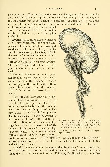
HYDROUKETER. 447
now be passed. This was left in the ureter and brought out of a wound in the
dorsum of the kidnej to keep the ureter open while healing. The opening into
the renal pelvis was closed by ten fine interrupted silk sutures, not piercing the
mucosa. The wound was partially closed with extensive drainage. The bougie
in the ureter was taken out on the second
day. The woman recovered without a
fistula, and had no return of the hydro-
nephrosis.
Hydroureter, or an abnormal distention
of the ureter with urine, is but the com-
plement, of stricture which we have just
considered. The cause of the hydroureter
and hydronephrosis, which is genetically
the same and always associated with it, is
invariably due to an obstruction to the
outflow of the secretion Avithout infection.
The various causes, therefore, are those
just enumerated, which need not be cited
again.
Bilateral hydroureter and hydro-
nephrosis may arise from an obstruction
as low down as the urethra, or from a
hypertrophy of the bladder walls. It has
been noticed arising from the compres-
sion of the orifices in exstrophy of the
bladder.
Pelvic tumors, neoplasms, and inflam-
matory diseases act on one or both ureters
according to their disposition. The hydro-
ureter always extends from the point of
constriction up into the pelvis of the kid-
ney, which it involves (hydronephrosis).
The tract included is therefore greater or
less, according to the location of the ob-
struction. It is greatest when the stop-
page is at the extreme lower end of the
ureter, as in the case of a calculus plug-
ging its orifice. One of the commonest
forms, generally of lesser degree, is that
produced by the pressure of large uterine or ovarian tumors, which is almost
always greatest just at the pelvic brim, so that the hydroureter alfects the
abdominal portion only.
A marked case is shown in the figure taken from one of my patients (S. A.
H., 4039. Dec. 30, 1895), who died with an enormous carcinoma of the cecum
filling the whole abdomen and pelvis. Following the distention the ureter be-
FiG. 250.
—
Hydroureter and Hydronephrosis,SHOWING Kink in Ureter due to Band okAdhesion stretching from the Lower PartOF the Pelvis of the Kidney almost downto the Pelvic Brim, '/s Nat. Size.
Sarcoma of peritoneum.April 4, 1895.
11. B. Autopsy,

448 AFFECTIONS OF THE URETERS.
came kinked, and adhesions formed which bound the kinks together and would
tend to keep up the distention even if the cause were removed.
In one instance iigured in the text (Fig. 250) the ureter was obstructed by
the pressure of a sarcomatous growth of the peritoneum;as the ureter distended
it kinked and became further obstructed by a band of adhesion uniting it to the
})elvis of the kidney.
A sharp line of distinction between hydroureter and pyoureter can not be
drawn ; in many cases of hydroureter pus is found in small quantities. In some
instances this increases while under observation until it is so abundant that there
can be no hesitation in calling it pyoureter; in other cases a pyoureter will
rapidly improve and the pus diminish from day to day, ultimately leaving behind
a simple hydroureter without the observer being able to decide just when the
transformation took place.
Pyoureter.—Pyoureter is an accumulation of pus in the ureter. In order to
bring this about, two things are necessary : first, an obstruction, and, second, an
infection, or the infection may take place first and the obstruction develop after-
ward. A common example of the first class is a hydroureter which becomes
infected, while the second class is typified by the case cited under gonorrheal
stricture of the ureter. Properly speaking, many of these cases should be classi-
fied under ureteritis and its sequelae. The quantity of pus found varies from a
large deposit falling as a sediment in the urine as soon as it is withdrawn, or a
thick and creamy pus only brought out of the catheter by suction, all the way
down to a small quantity of pus just sufiicient to give the urine a turbid ov
milky appearance, or the j)us may even not appear at all until the urine is cen-
trifugalized and put under the microscope. The same causes may act to produce
pyoureter as hydroureter, of which the former may sometimes be considered an
advanced stage.
Fever is, as a rule, only an occasional symptom. I have seen several cases
where an intense intermittent colic was the most pronounced symptom, and
where no calculus was present.
The diagnosis is made by the ureteral and renal catheters. Upon introduc-
ing the catheter and clearing the obstruction the pus or purulent urine will begin
to fiow. It must be borne in mind that urine thickened by pus will escape muchmore slowly than nonnal urine ; if necessary, the discharge at the end of the
catheter may be hastened by applying suction with an air-tight syringe.
Whenever the pus is inspissated or too thick to flow readily through the
small catheter, it is best to dilute it by injecting some warm boric acid solution
and allowing it to mix well with the pus before escaping again ; by repeating
this maneuver an accumulation may be evacuated in a few minutes which could
not otherwise escape in the course of several hours. When the thick pus is in
the pelvis of a large kidney the dilution may be aided after injecting the sohi-
tion by manipulating the kidney freely between two hands.
I have met a number of cases due to tubercular ureteritis with stricture, and
the one case cited of gonorrheal infection.
The prognosis depends upon the cause. In tubercular cases the disease is

URETERAL CALCULUS. 449
Fig. 251.— Sykinge and AspiuatokWITH Cock tor injecting andWASHING OUT THE PeLVIS OF THEKidney THROUGH the Kexal Cath-eter; OR TO inject Fluid .-^o as toDILUTE a Collection of Pus toothick to run out THROUGH THECatheter.
progressive until removed. In other cases the
infection involves not only the ureter but the
pelvis of the kidney, and the kidney substance
too, impairing the secreting function.
The treatment will also depend on the
cause, and the possibility of completely re-
moving any obstruction to a free outflow.
Disinfection of the whole urinary tract upinto the kidney may be carried out as already
detailed.
Ureteral Calculus.—A calculus lodged some-
where in the course of the ureter is far morerarely found than in either the renal pelvis or
in the l)ladder.
The chemical characters of such calculi are
the same as those found in the kidney ; the
form, however, of a calculus which has lodged
in the ureter for some time is peculiar, being
elongate, from four to six or more times its
diameter, which averages about 5 millimeters,
and pointed at both ends. The forms of these
calculi and the appearance of the layers showthat they gain by accretion at the ends and
lose by attrition at the side. A ureteral calcu-
lus has been observed 12-5 centimeters (5 in-
ches) long. Small calculi may be round or
even horseshoe shaped, as in Dr. R. B. Hall's
case (AVw York Medical Record^ Oct. 18,
1S90). The pelvis of the
kidney is the source of
these calculi, which drop
down into the ureter or
are slowly forced downuntil tliey lodge some-
where in its course.
I had one case of a
ureteral calculus forming
upon a silk thread used to
suture the opened ureter
to the vaginal vault (kolpo-
ureterostomy) for the purpose of treating a
stricture of the ureter. The patient suffered
intense pain from vesical spasms until I dis-
covered the stone and took it away. It was
about 6 millimeters in diameter.
Fig. 252.— A UreteralCalculus which waspassed spontaneously,SHOWING the Charac-teristic Ovoid Form.Actual Size.

450 AFFECTIOXS OF THE URETERS.
Ureteral calculi lodge by preference within certain well-
deiined limits—for example, just below the renal pelvis, about the flexure at
the pelvic brim, and the pelvic floor, are decidedly points of predilection.
The symptoms produced by a stone lodged in the ureter are attacks
of severe pain extending from the kidney down the course of the ureter, and
sometimes accompanied by rigors. The pulse is ele-
I - ' ^ vated and there is fever. The point of location of
the stone is tender on deep pressure. These attacks
are intermittent, and recur at variable intervals as
long as the stone remains. With the attacks mayoften be noticed the formation of a tumor in the loin
of that side. Where the stone is not lodged, but is
gradually descending toward the bladder, its advance
can be traced by the patient, and is often marked by
bloody urine. Mechanically the ol)struction produces
a hydroureter and hydronephrosis, varying in grade
according to the completeness of the obstruction ; if
there is infection, pyoureter and pyonephrosis mayarise. In time the function of the kidney becomes
greatly impaired, but it is remarkable how persist-
ently it continues to excrete a diminished percentage
of urea after months and years of such interference.
The valve action of a stone in plugging the ureter
and then permitting the daramed-up contents to escape
suddenly is well shown by the history of Dr. Hall's
case cited above, in which he found a renal tumor the
size of a pint cup, which was not present the day
before.
A presumptive diagnosis will 1 )e madew^hen all the symptoms above described are found.
It nnist be remembered, however, that the passage of
a blood clot, or the temporary closure of the ureter
by an inflamed thick mucosa, may give rise to similar
symptoms. The most certain of all means of diag-
nosis is the direct examination by vagina, by rectum,
or by cystoscope and catheterization of the ureter, orw
Fig. 253.—End ok a Wa.\TIPPED Catheter.
Diagnosis of ronril calfiil
by means of a wax coal mm t
catheter. Tlie scratch luarwere made by th(- caK'iil
sliown in Fif^. 254. Four limenlarged.
by an abdominal incision.
in the ureter in advance of the broad ligament can be
felt through the antero-lateral wall of the vagina and
rolled under the finger. Back of this ])oint the noi-mal
ureter is easily accessible per rect>n//, all the wny up
to the pelvic brim, by following the landmarks <le-
scribed, and the palpation is all the easier if the ureter contains a foreign body.
A cystoscopic examination may be so fortunate as to disclose a stone project-
ing partly into the bladder. By using the metal catheter with a diaphragm on
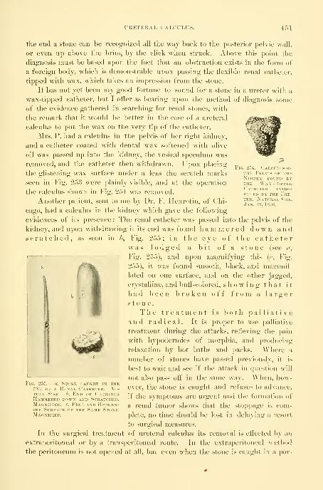
URETERAL CALCL'LUS. 451
Fig. '254.—Calcuhs ofTHE Pelvis of theKidney found byTHE Wax - tippedCatheter intro-duced BY' the LTre-
ter. Natural Size.
.Ian. 29, 1S90.
the end a stone can be recognized all the way back to the posterior pelvic wall,
or even up above the brim, bj the click when struck. Above this point the
diagnosis must be based upon the fact that an obstruction exists in the form of
a foreign body, which is demonstrable upon passing the flexible renal catheter,
tipped with wax, which takes an impression from the stone.
It has not yet been my good fortune to sound for a stone in a ureter with a
wax-tipped catheter, but I offer as bearing upon the method of diagnosis some
of the evidence gathered in searching for renal stones, with
the remark that it would be better in the case of a ureteral
calculus to put the wax on the very tip of the catheter.
Mrs. P. had a calculus in the pelvis of her right kidney,
and a catheter coated with dental wax softened with olive
oil was passed up into the kidney, the vesical speculum was
removed, and the catheter then withdrawn. Upon placing
the glistening wax surface under a lens the scratch marks
seen in Fig. 253 were plainly visible, and at the operation
the calculus shown in Fig. 254 was removed.
Another patient, sent to me by Dr. F. Henrotin, of Chi-
cago, had a calculus in the kidney which gave the following
evidences of its presence : The renal catheter was passed into the pelvis of the
kidney, and upon withdrawing it its end was found li a m m e r e d do w n a n d
scratched, as seen in ?>, Fig. 255; in the eye of the catheterwas lodged a bit of a stone (see c/,
Fig. 255), and upon magnifying this (f, Fig.
255), it was found smooth, black, and mammil-lated on one surface, and on the other jagged,
crystalline, and buff-colored, showing that it
had been broken off from a largerstone.
The treatment is both palliativea n d radical. It is proper to use palliative
treatment during the attacks, relieving the pain
with hypodermics of morphia, and producing
relaxation by hot l)aths and packs. AYhere a
number of stones have passed previously, it is
best to wait and see if the attack in question will
not also pass off in the same way. When, how-
ever, the stone is caught and refuses to advance,
if the symptoms are urgent and the formation of
a renal tumor shows that the stoppage is com-
plete, no time should l)e lost in delaying a resort
to surgical measures.
In the surgical treatment of ureteral calculus its removal is effected by an
extraperitoneal or by a transperitoneal route. In the extraperitoneal method
the peritoneum is not opened at all, but even when the stone is caught in a por-
FiG. 255.— a, Stone caught in theEY'E of a Kenal Catheter. Ac-tual Size, h. End of CatheterHammered down and Scratched.Magnified, e. Free and Beoken-off Surface of the Same Stone.Magnified.
452 AFFECTIONS OF THE UUETEK.S.
tion of the ureter lyino; Ijeiieatli the peritoneum, the latter i8 loosened and turned
to one side and the ureter ari'ived at in this way from behind. In the transperi-
toneal method the anterior abdominal wall is opened, preferably in the semilunar
line, and the ureter exposed and incised, making in this way two incisions through
the peritoneum on opposite sides of the abdomen.
The extraperitoneal route is always to be preferred, on account of the danger
of peritonitis, and on account of the risk of a urinary fistula, M-hich will be safer
behind than across the peritoneum.
In two positions the stone must always be taken out by the extraperitoneal
i-oute : first, when lodged anywhere between the kidney and the superior strait
;
second, when lodged in the anterior part of the pelvis, under or in front of the
broad ligament. The ureter is more easily accessible from the brim of the pelvis
down to the broad ligament after opening the abdomen, but even here it is bet-
ter to make a long lateral incision and peel up the peritoneam so as to get at the
stone in this way.
The presence or absence of infection also infiuences the choice of route.
When the urine is discharging pus from the affected side, the extraperitoneal
route must always be followed, on account of the enormously increased risks of
infection if the peritoneum is opened. As stated in discussing the diagnosis of
stone in the ureter, it may be proper in doubtful cases to make a preliminary
abdominal incision in the semilunar line in order to locate the stone, and then to
remove it by a lateral incision.
The various operations for ureteral calculus are performed as follows : Whenthe stone is found lodged in the lower vaginal part of the ureter, if its end can
be seen through the cystoscope projecting into the bladder, the effort should be
made to grasp it with a pair of ordinary forceps, or alligator forceps, with a
hook, or in a noose, and by traction, aided by pushing from behind with one
finger in the vagina, to draw it out of its bed and through the urethra.
If this fails, and whenever the stone is behind the vesical orifice of the ureter,
it must be reached by a vaginal incision.
After definitely locating by touch the part of the vagina nearest the stone,
the patient is put on her back, or in the lateral posture, and the posterior vaginal
wall retracted, exposing the anterior and the lateral walls.
The bladder is emptied and a longitudinal incision is made through the
vaginal wall about 3 centimeters (1^ inch) long;pulling apart the borders of
the incision and dissecting down into the cellular tissue, the enlarged ureter is
exposed and caught by j^assing blunt hooks under it, above and below the stone.
A longitudinal incision is then made in the ureter over the end toward the blad-
der, just large enough to push the stone through endwise without any tearing.
After this is accomplished a bougie is run up the ureter to make sure there are
no more stones above, and the ureteral incision is sewed up with five or six fine
interrupted catgut sutures, introduced with a delicate curved intestinal needle,
each one end)racing the outer coats and avoiding the mucosa. If a good closure
is secured and thei-e is no infection, the vaginal wound may be closed too, but
if there is much pus in the urine it is safer to drain the vaginal incision.
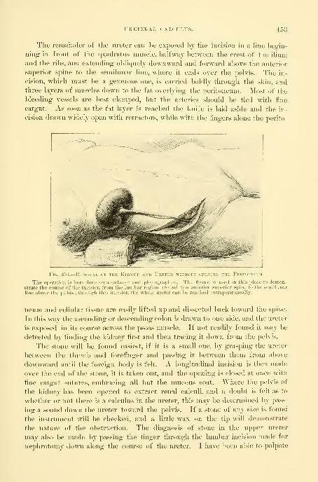
r R HT E 1{ A L CA LC U LUS. 453
The remainder of the ureter can he exposed by tlie incision in a line Ijegin-
ning in front of the quadratiis muscle, halfwaj between the crest of the ilium
and the ribs, and extending ol)liquely downward and forward above the anterior
superior spine to the semilunar line, where it ends over the pelvis. The in-
cision, which must be a generous one, is carried boldly through the skin, andthree layers of muscles down to the fat overlying the peritoneum. Most of the
bleeding vessels are best clamped, but the arteries should be tied with fine
catgut. As soon as the fat layer is reached the knife is laid aside and the in-
cision drawn widely open with retractors, while with the fingers alone the perito-
FfG. 256.
—
Removal of the Kidney and Ureter without opening the Peritoneitm.
The operation is here done on a cadaver and photographed. The figure is used in this place to demon-strate the course of the incision from the lumbar region around the anterior superior spine to the semilunarline above the pubis ; through this incision the whole ureter can be reached e.Ntraperitoneally.
neum and cellular tissue are easily lifted up and dissected back toward the spine.
In this way the ascending or descending colon is drawn to one side, and the ureter
is exposed in its course across the psoas muscle. If not readily found it may be
detected by finding the kidney first and then tracing it down from the pelvis.
The stone will be found easiest, if it is a small one, by grasping the ureter
between the thumb and forefinger and passing it between them from above
downward until the foreign body is felt. A longitudinal incision is then made
over the end of the stone, it is taken out, and the opening is closed at once with
fine catgut sutures, embracing all but the mucous coat. Where the pelvis of
the kidney has been opened to extract renal calculi, and a doubt is felt as to
whether or not there is a calculus in the ureter, this may be determined by pass-
ing a sound down the ureter toward the pehas. If a stone of any size is found
the instrument will be checked, and a little wax on the tip will demonstrate
the nature of the obstruction. The diagnosis of stone in the upper ureter
may also l)e made by passing the finger through the lumbar incision made for
nephrotomy down along the course of the ureter. I have been able to palpate

454 AFFECTIOXS OF THE URETERS.
the ureter in this way all the way to the pelvic brim over the common iliac
artery without makino; a Ioniser incision than necessary to deal with the renal
stone.
The transperitoneal plan of removing calculi is safe and proper when
there is no infection, and the stone is fixed in the ureter at a point near the
pelvic brim. In this position the ureter is easily found and exposed, and may be
treated through an incision in the semilunar line with the pelvis well elevated.
The stone is then removed by a longitudinal incision closed by catgut mattress
sutures embracing peritoneum and muscular coats. If the suturing is accurate,
and there is no stricture below to dam back the urine, there is no need of a
drain, and the abdominal wound should be completely closed.
W. A. Lane, of Guy's Hospital, operated on a calculus impacted in the
ureter for twenty years (see the Lancet^ Nov. <S, 1890, p. 9f>T).
The patient was twenty-three years old and began having violent abdominal
pains when she was three years old ; she had hematuria when eight years old.
Attacks of violent pain were referred to the left side low down and were accom-
panied by great irritation in the urethra. After each attack abundant pus
appeared in the urine.
At a first operation a lumbar incision was made and the kidney ex-
])l(jred ; a " kink " in tlie ureter was found and corrected, but the pain soon
recurred.
On July 5, 1890, an abdominal incision was made in the left linea semi-
lunaris and the ureter palpated about the pelvic brim, where a stone was felt
and pushed up ; a longitudinal incision was made in the side of the ureter and
a small oval stone three quarters of an inch long was removed, and the opening
closed with a fine continuous silk suture. The wound healed without leakage
and the patient recovered. The calculus was made up of alternating layers of
uric acid and urates.
Prolapse.—An eversion or prolapse of the ureteral mucosa is one of the
rarest of the ureteral affections. It is commonest in female children and often-
est congenital. The prolapse usually depends for its origin upon a narrowing
or stricture at the ureteral orifice, the pressure of the urine filling the pelvis of
the kidney and the ureter behind this is then sufficient to cause the ureteral
mucosa to pout out into the bladder in the form of a cystic tumor with the ob-
structed ureteral orifice at some point of its periphery. In the child the pro-
lapsed ureter may even escape through the urethra and appear at the vulva,
where it may be mistaken for an everted urethra. An example of an a('<juired
prolapse in a man following an acute cystitis five years before death is slunvn in
Fig. 257, examined and reported by Dr. George Blumer {Johux Ilopl'mx llnx-
pital Bulletin, Sept.-Oct., 1896, p. 174). The patient died of an extensive uri-
nary infection, to which this class of cases is peculiarly liable. The enormously
hypertrophied bladder, in one place 8 centimeters thick, was corrugated and
covered with di])htlieritic patches. On the right side there was a hydroureter
and a cushiony protrusion into the bladder as big as the end of the thumb.
On the left side a pyramidal sac occupied the position of the ureteral orifice.

URETERAL FISTULA. 45 (
The sae was fluctuating, 8 centimeters long, 3 centimeters in diameter at the
base, and 9 centimeters in diameter near its extremity. High up on one side
the minute ureteral orifice was found (see Fig. 257 a) as big as a pin point and
Fig. 257.
—
Prolapse of thk Uretp:kal and Vesical Mucous Membrane, most Marked on* the LeftSide, Slight on the Eight. Diphtheritic Inflamm.vtion of the Bladder and Left Ureter.
Note the position and narrowness of the ureteral orifices at a and J, and the large pyoureter abovethe bladder on the left and the smaller liydroureter on the risrht side. % natural size.
situated in the center of a small area of dense fibrous tissue. On opening the
thin-walled sac, the finger could be carried directly up into the ureter.
Ureteral Fistula.—A ureteral fistula is an abnormal opening through which
the urine is discharged directly from a ureter on to the surface of the body or
into some part of the genital or alimentary tract, and in so far as the affected
ureter is concerned the bladder is throwm out of use.

450 AFFECTIONS OF THE URETERS.
Ureteral fistulse are the result of a trauma, or of ulcer-
ation, or they are congenital, and by far the commonest are those
involving the anterior part of the pelvic portion of the ureter.
A uretero-vesical fistula is produced by a ureteral stone ulcerat-
ing through the ureteral walls into the bladder, or by the artificial making of
a connnunication between the bladder and the ureter behind a strictured orifice.
Such a fistula requires no treatment, and only needs mention.
The commonest causes of ureteral fistulge were formerly the traumatisms of
labor, tearing open the uterus, vagina, bladder, and ureters, and leaving a
permanent communication between the ureter and uterus or ureter and vagina,
forming a uretero-uterine or a uretero-vaginal fistula. Other causes acting but
rarely were the ulceration through the vagina into the ureter, produced by a
large pessary, or the cutting of a ureter on opening a pelvic abscess ^^^r vagi-
nam. To-day the commonest cause of ureteral fistulas is to be found in the fre-
quently performed vaginal and abdominal hysterectomies in which the ureter
is accidentally injured. In vaginal hysterectomy for cancer the wonder is that
the ureter is not more frequently tied or cut, as it lies so close to the field of
operation and is often so intimately involved in the disease. Indeed, it seems
quite certain that this accident must occur with far greater fre(|uency than is
apparent from the reported cases. In rare cases both ureters are injured, form-
ing a double ureteral fistula.
The diagnosis of ureteral fistula is not diflicult. The traumatic forms
must first be distinguished from the congenital. Congenital ureteral fistulas
almost always open at a point below the neck of the bladder, even as low downas the external genitals, while the traumatic forms open either at the l)ase
of the bladder, or, more commonly still, at the vault of the vagina, or into the
uterus. An additional distinguishing feature is the fact that the congenital
fistulas are known to have existed from childhood, while the traumatic take
their origin from some definite period in adult life.
The next step of importance is to distinguish ureteral from vesico-uterine
and vesico-vaginal fistulas. A fistula involving one ureter discharges constantly,
while the patient is also emptying the bladder at regular intervals to discharge
the urine received from the sound ureter. Care must also be taken to distin-
guish cases of small vesico-vaginal fistulas situated high up in which the bladder
still retains some of its functional activity, discharging urine per iwethfam, in
spite of more or less leakage.
By injecting the bladder by an aniline solution, or with sterilized milk, if
the case is one of vesico-vaginal fistula, the colored fiuid w^ill escape by the
vagina ; if, on the other hand, the fistula is ureteral, the flow from the vagina
will continue clear.
Berard's method of diagnosing between a ureteral and a vesical fistula is to
empty the bladder and then cause the patient to sit on a vessel for two hours,
when, if the fistula is a ureteral one, the amount of urine collected ought to
approximate that drawn by a catheter at the end of this time.
Upon exposing the anterior vaginal wall and the vaginal vault the ureteral

URETERAL FISTULA. 457
fistula is found usually near the cervix, imbedded in sear tissue. Upon intro-
ducing a flexible catheter or bougie into a ureteral fistula the instrument can be
pushed up beyond the pelvis into the abdomen as far as the kidney, and if left
in place the urine is seen dropping at intervals from the end of the catheter.
In every case of vesico-vaginal fistula a careful search must be made to be
sure that the ureteral orifice does not open on the margin of the fistula.
AVinckel has reported a case in which a minute fistula communicated with the
bladder and the ureter at the same time.
A sure sign of a ureteral fistula is obtained by exposing the ureteral orifice
and passing into it a sound or a catheter. It will be found that the sound will
only enter a short distance, not more than 3 or 4 centimeters, on the injured
side, where it is stopped, and it" the orfice is watched no urine will be seen to
escape. On the sound side the metal catheter or sound can be carried back
to the posterior pelvis and a flexible catheter can be pushed up to the kidney,
and, if left in, the urine is discharged through it.
Treatment.—Various plans have been devised for the purpose of divert-
ing back into the bladder the urine which discharges through the fistula. The
following is an outline of some of these methods
:
1. By buttonholing the bladder and making an artificial vesico-vaginal fis-
tula close to the ureteral fistula, and, after this has healed, bridging over a chan-
nel between the two fistulous orifices by drawing together the sound vaginal tis-
sues at the sides.
2. By making an artificial vesico-vaginal fistula close to the ureteral fistula
and then encircling both orifices in a ring of vaginal denudation, which is folded
on itself so that the urine flows first from the ureter into a little vaginal pocket
and then into the bladder by way of the vesico-vaginal fistula.
3. By dissecting out the end of the urethra and, after splitting it to prevent
contraction of the orifice, to turn the end into an opening made into the base of
the bladder.
4. By making a big vesico-vaginal fistula in the vaginal vault near the ure-
teral fistula and closing the upper part of the vagina (partial colpocleisis).
5. AVhen the ureter is not completely severed and the fistula simply involves
its lateral wall, by closure of the fistula, by means of denudation and suture of
its margins.
0. By opening the al)domen to release the end of the ureter and turning it
into the bladder (abdominal uretero-cystostomy).
7. By total occlusion of the vagina after making a large vesico-vaginal fistula
(total colpocleisis).
8. Removal of the kidney corresponding to the fistulous ureter (nephrec-
tomy).
Of these various plans of treatment, no one is adapted to all cases, but that
plan is to be selected which best meets the individual requirements of the par-
ticular case. In general the simplest plan must be followed, involving least risk
to life and avoiding if possible any extensive mutilation or the sacrifice of such
an important organ as the kidney; the last thing to be thought

458 AFFKCTIOXS OF THE URETERS.
of is e o 1 p o c 1 e i 8 i 8 or nephrectomy, and the abdomen must not be
opened if a simple anastomosis can be effected per vaginam.
The condition of the patient must be carefully considered. I have had
patients referred to me for the treatment of a ureteral fistula following vaginal
hysterectomy, who were suffering from pelvic cellulitis and periureteritis, and
80 prostrated that I was unable to perform an operation. In one case the patient
was emaciated, had a rapid pulse, and a constantly elevated temperature.
When the fistula is traumatic the best time to operate is some months after
the receipt of the injury, because for several weeks after its formation the mass
of fresh young scar tissue forming in the vaginal vault is unfavorable for any
kind of plastic operation.
Fistula in the Lateral Wall of the Ureter .—-A fistula in the
lateral wall of the ureter is easily closed by making a circular denudation in the
vaginal wall around the opening about 4
millimeters in breadth, similar to the denu-
dation for a vesico-vaginal fistula. The sides
of the denudation are then brought together
by means of a series of interrupted silk or
fine catgut sutures approximating the tissue
in the direction of least resistance. I have
had one operation of this sort to perform
upon a patient upon whom I had previously
opened the lateral wall of the ureter near the
vaginal vault and sutured it to the vagina in
order to get at and dilate a ureteral stricture
in the back part of the pelvis. The denuda-
tion was made and the sutures were applied
as just described, and the wound healed }'*'>'
pi'imam.
Ureteral Fistula at the Baseof the Bladder .—A ureteral fistula situ-
is best treated by dissecting up the ureter to
the extent of 1 or 2 centimeters, and then perforating the base of the bladder
and turning the end of the ureter into the bladder. The vaginal part of the
incision is closed by interrupted sutures, taking care to catch the outer coats
of the ureter in one or two of the upper sutures so as to hold it fixed in the
incision and so prevent retraction as descril)ed in the treatment of ectopic
ureteral orifice.
Ureteral Fistula at the Vaginal Vault.—When the fistula
lies in the vault of the vagina and there is enough loose vaginal tissue around it,
the best plan of treatment is the formation of a vesico-vaginal fistula near by,
not less than a centimeter in diameter, and then making a circular denudation,
including both fistuUe, as shown in Figs. 258 and 259. The sides of the denuda-
tion on the N-aginal surface are brought together by interrupted silk or catgut
sutures. The difficulty in this operation is the tendency of the vesico-vaginal
Fig. 258. — Switching the Ukkter {I'r)
INTO THE BlADDEK BY MEANS OF AN Ak-TiKiciAL Vesico-vaginal Fistula {B).
The area of undenuded vaginal mucosais seen between Ur and B. The five su-tures bring the denuded vaginal mucosatogether over this.
ated at the base of the bladder

URKTKHAL FISTULA. 4:59
iistula to contract and close, and for this reason the opening must be made
sufficiently large and the nnicous surfaces of the vagina and bladder accurately
approximated.
I^ r e t e r o - c y s t o s t o m y .—The abdomen should be opened and the end
of the ureter freed and turned into the bladder in those cases where a sound
kidney is discharging its urine into the vaginal vault, and where, at the same
time, on account of the scar tissue or its retracted position, a vaginal operation
establishing the connection between the ureter and the bladder is impossible.
Contraindications to abdominal uretero-cystostomy are an enfeebled condi-
tion of the patient, fresh scar tissue in the vaginal vault and pelvic floor, and
an extensive periureteritis. I have twice been ol^liged to abandon the operation
on account of periureteritis. This can be detected by first recognizing the mass
of resistant tissue in the vaginal vault, and then passing a catheter up the ureter
Fig. 259.
—
Uretero-vagin.\l Fistula, switching the Ureter into the Bladder through a Vesico-
vaginal Fistula, a b. Areas or Denudation.
The union of the edges of the fistula is shown in the second picture. March 8, 1896.
and examining by the rectum, when the ureter will be found to be no longer
free and movable, but is distinguished with difficulty imbedded in a mass of
hard tissue, extending a variable distance up toward the superior strait.
Similar operations have been successfully performed by Drs. C. B. Penrose,
of Philadelphia(Univ. Mag., April, 1894), and Florian Krug, of New York.
(See J. M. Baldy, Amer. Gijn. and Ohs. Jour., Nov., 1S9-1-, and H. J. Boldt,
personal communication).
It is important to note that the operations of uretero-ureteral anastomosis
and uretero-cystostomy must not be looked upon as rivals in the same field.
Where the ureter is cut far enough back from the bladder to permit an anasto-
mosis of the upper into the lower end, the distance between the upper end and
the bladder is too great to allow a uretero-cystostomy to be considered. Where,
on the other hand, the ureter is cut near enough to the bladder to allow the
upper end to be turned into the bladder, it will be found that the lower end is
so short and so awkwardly placed that a uretero-ureteral anastomosis is not to be
thought of.

460 AFFECTIONS OF THE URETERS.
There is but one class of cases in whicli the procedure is elective ; that is,
when the ureter has become lengthened and dilated by displacement upward
over a uterine myoma. I would in this case elect to do a uretero-ureteral
anastomosis if the ureter were dilated, or a uretero-cystostomy if it were of nor-
mal caliber.
The method of performing uretero-cystostomy is described in the following
case, operated on seven weeks after vaginal hysterectomy : The patient (B. Z.,
2990) entered the hosjjital in August, 1894, with an extensive carcinoma of the
cervix, for which Dr. W, R. Russell, then the resident gynecologist, performed
vaginal hysterectomy. The disease had extended so far out into the broad
ligaments that he 'was obliged to place the ligatures at a greater distance from
the cervix than usual. She recovered rapidly from the hysterectomy, but re-
tained as a sequel a ureteral fistula in the vault of the vagina near the middle of
iG Hysterectomy forFig. 260.
—
Right Uretero-cy-.stostomy' for Uretero-vaginal Fistula followiCancer of the Uterus.
The ureter has been dissected out of its bed and cut off close to the base of the right broad ligament ; it
was too short to reach to the bladder, so the bladder was loosened from its attachments to the anterior pelvic
wall and thrown back 3 centimeters to meet the ureter.
the cicatrix. From this there was the usual constant leakage of urine, although
she regularly passed the urine accumulating in the bladder from the other kid-
ney. From a simple vaginal inspection it was impossible to say whether the
flow from the cicatrix came from the right side or the left. It clearly did not
come from the bladder, for it remained unchanged by the injection of a steril-
ized solution of milk into that viscus.
To decide which was the severed ureter I placed the patient in the kuce-
breast position and introduced my No. 10 cystoscope, when the bladder tilled
with air and I was able to inspect the ureteral orifices. By introducing a

URETERAL FISTULA. 461
searcher into the left ureteral orifice I found that this ureter was intact as far as
the posterior wall of the pelvis. Upon introducing the searcher into the riglit
ureteral orifice it could not be carried in more than 2 centimeters, on accountof meeting an impassable obstruction. The urine was seen flowing from the
left ureteral orifice while nothing escaped from the riglit side. The demonstra-tion was thus complete that it was the right ureter which was injured and the
left was intact.
Having cleared up the diagnosis in this way, I proceeded to operate to re-
lieve the condition, in October, 1894, seven weeks after the original operation.
Operation.— The patient was
placed in the Trendelenburg position
and an incision 12 centimeters long
made through abdominal walls loaded
with fat. Every step throughout the
operation was embarrassed l)y the
obesity of the patient. After opening
the abdomen, the large fat omentumand intestines were dislodged from the
lower abdomen and pelvis with great
ditficulty, and held away by means of
cotton gauze pads.
The end of the ureter could not be
found on the pelvic floor on account
of the rigidity and inflammation sui--
rounding the line of scar tissue between
the rectum and bladder. The riglit
ovary and tube, which had been left,
were also pinned down to this scar tis-
sue by numerous vascular adhesions.
The attempt to reach the ureter at this
point was therefore aliandoned and it
was sought out at the pelvic brim,
where it was readily found after lifting
up the caput coli and incising the peri-
toneum and pushing aside the fat. It
was then traced from the point where
it crosses the common iliac artery downto the pelvic floor, exposing the whole
length of the pelvic portion by splitting the peritoneum over its upper surface.
The anterior portion was involved in the inflammatory material surrounding
the scar, which bled so freely that no attempt was made to dissect it out.
Four centimeters of the lower end of the ureter lying directly behind the scar
tissue were dissected loose and the ureter lifted up from its bed and divided
close to the scar, sacrificing as little as possible of its length.
I now found that althougli I had cut the ureter to the best advantage possible
UuETERO-CypTOSTOMY.
ShowiiiiT ill
ter drawn tlimi
der by a pair <>
The middle tiij
duced, holdinff tli
]icr tiLfure the end of the ure-|,i iiiiii,' made into the blad-
i> ]'a-si .1 through the urethra."\\s Mil,- of the sutures intro-
ureter in place. The lowerflgure shows the ureter secured in the bladder bysutures, deep and superficial on all sides.

462 AFFECTIONS OF THE URETERS.
under the circumstances, I could not do more than merely Ijring it into contact
with the bladder by pulling on it. It was evident that if I were to suture it to
the bladder, exercising this degree of traction, it would pull loose soon after the
operation, leaving a uretero-abdominal instead of a uretero-vaginal fistula to
deal with.
1 was able to cope successfully with this formidable difficulty in the follow-
ino- manner : The bladder was dissected free from its attacliments to the hori-
zontal rami of the pubis on both sides, with scissors and fingers, and dropped
down into the pelvis so as to extend it and carry it more into the back part of
the pelvis, gaining at least 3 centimeters in this way. By this means the ureter
and the bladder w^ere now easily approximated without strain. I then made a
small incision through the bladder wall, w^hieh w^as covered with fat at least a
centimeter thick, at the point on the right side nearest the ureteral end drawn
straight across the pelvis. This incision passed through the peritoneum and was
not more than 3 or 4 millimeters in length, and just large enough to receive the
ureter snugly.
I then slit up the under surface of the ureter for about 4 millimeters, en-
larging the caliber of its orifice to avoid a stricture, and with a pair of long
delicate forceps introduced through the urethra, the l)ladder, and through the
incision, I caught the ureteral end and drew it into the bladder and held it there
while it was being attached to the bladder wall by about six fine interrupted silk
sutures passed through the nmscular tissue of the bladder and the peritoneal
and muscular coats of the ureter on all sides, beginning with the under side.
The ureter thus dissected out of its bed, and attached to the bladder, was
stretched like a lax cord from the posterior part of the pelvis to the bladder,
which lay gibbous and flattened out on the pelvic floor.
The abdominal incision was closed down to its lower angle, where a narrow
gauze drain was inserted for fear of leakage. Care was taken in closing the
incision not to draw together the peritoneum underlying its lower end, to avoid
raising the bladder and indirectly pulling upon the ureter. No leakage occurred
and the drain was removed, and the wound healed without suppuration. Iler
urinary difficulties were immediately and completely relieved with the perfect
restoration of continence.
At a subsequent cystoscopic examination I discovered the abnormally placed
ureteral orifice opening into the posterior hemisphere of the bladder into which
it freely discharged its urine.
This case is one of especial interest for the following reasons
:
I was able to determine on which side the injury had been sustained by
sounding the ureter in the knee-breast position with the bladder distended
\vith air. 1 was enabled, by a simple but delicate plastic procedure, to secure at
once a perfect restoration of function without sacrificing any such important
structure as a kidney. (See -/. //. IT. Bull.^ February, 1895.)
The only case I know of in which a double ureter has been accidentally di-
vided in the course of an operation, and then anastomosed into the bladder, was
reported to me by Dr. Anna M. Fullerton.

URETERAL FISTULA. 463
" On March 10, ls97, the patient, R. L., thirtv-six years old, the mother of
six children, entered the Woman's Hospital, of Philadelphia, for a double pjo-
salpinx with ovarian abscesses. She had been ill and confined to bed for three
months before admission to the hospital.
"At the operation the uterine apiDcndages alone were removed; the uterus,
not being especially enlarged, was allowed to remain, because the patient was not
in fit condition for a^ prolonged operation. Dense adhesions existed ; on the right
side the adhesions were so firm that some of them i-equired to be cut. Enuclea-
tion of the appendages was very diflicult ; a band passing across the pelvis a
little below the l)rim was firmly adherent to the broad ligament a little below
the uterine tube and at the junction of its middle and outer third. Not think-
ing of its being the ureter in that location, I severed it with scissors close to its
attachment to the broad ligament. Upon doing this I found I had severed twocanals covered with peritoneum and lying side by side imbedded in a connnonsheath of connective tissue. Each was the size of a normal ureter. Xo blood
or fluid apjDeared to escape from the canals at any time. A sound was passed
down through each canal to the bladder, and struck upon a cathetei* placed in the
bladder. Similarly a long sound was passed upward through the superior pair
of orifices, and passed several inches toward the kidney, proving the condition
to be one of double ureter traversing the pelvis at a nmch higher point than
normal, and thence passing between the folds of the broad ligament to the point
of attachment to the bladder. The vesical ends of the ureters being ligated, the
portion communicating with the kidneys was drawn down, and the two orifices
introduced into the bladder by a conmion opening made in the superior portion
of the organ a little to the right. The patient made a perfect convalescence,
and was discharged, April 18th, in good health."
Extraperitoneal Uretero-cystostomy .—O. AVitzel, of Bonn{Centralh. f. Gyn., 1896, No. 11), has devised a plan for the anastomosis of the
ureter into the bladder by bringing the ureter under the peritoneum in a newdirection, so as to shorten its course. In addition to this, the bladder was
detached and drawn out in the manner just described. The patient had uretero-
vaginal fistula operated on in vain by the vagina. The abdomen was opened,
and the thickened ureter found at the pelvic brim and traced downward, and
divided at about the middle of the broad ligament. The lower end was closed
by sutures and dropped, while the upper end was brought to the upper part of
the incision at the brim of the pelvis, and drawn down beneath the peritoneum
above the innominate line by a pair of long forceps started upward under the
peritoneum to the right of the bladder.
The incisions in the pelvic peritoneum and the peritoneum in the median
line of the abdomen were now closed, and the remainder of the operation con-
ducted extraperitoneal ly.
The bladder was now pulled up on the right side until it reached more than
4 centimeters {\\ inch) beyond the end of the ureter, where it was attached by
some stout catgut sutures.
The ureter was then transplanted into the bladder by forming an oblique

404 AFI-ECTIOXS OF THE UUETEKS.
channel ; the end of the ureter was cut off ol)hquely, and its nuicous coat at-
tached by fine catgut to the mucosa of the bladder, exposed through a small
opening made over the end of a pair of forceps introduced through the urethra.
Another row of catgut sutures outside of this attached the ureteral walls firndy
to the vesical walls.
The oblique channel was then formed by uniting the bladder walls over the
ureter on both sides. A drain was put in through a separate opening in the
bladder, and the bladder was drained for four days.
The patient made a perfect recovery.
Nephrectomy, removing the kidney corresponding to the fistulous
ureter, must be performed when the kidney is extensively diseased and the
seat of suppurative septic affection. An attempt should be made, however, to
save the kidney by washing the renal pelvis to cure any existing pyelitis, after
the manner described in the treatment of pyelonephrosis.
G. Simon, of Heidelberg {Chir. der Nieren^ 1871), first extirpated the
kidney for uretero-abdominal and uretero-vaginal fistulse.
Schede {Munch, med. Wochenschr., 1888, p. 512) extirpated the kidney in a
case of uretero-uterine fistula after several plastic operations had failed.
Ureterostomy .—When the ureter is cut off in the course of an abdominal
operation, and the upper end can not be grafted into the lower (uretero-ureteros-
tomy), the only alternatives left are either to bring the ureter out onto tlie skin
surface and to let it discharge there, or to extirpate the kidney of that side.
The plan usually adopted has been to bring the ureter out onto the surface
of the abdomen in the incision in the median line. I have the rejjort of such a
case furnished by Dr. C. P. Noble, of Philadelphia.
The patient, thirty years old, had an extra-uterine pregnancy, requiring the
removal of both tubes and ovaries together with the uterus. She had a pulse
rate of 160, and was so prostrated toward the end of the operation, when the
right ureter was found cut off above the brim of the pelvis, that the only thing
to be done was to get through as soon as possible by closing the incision and
sewing the ureter into it. The patient has recovered wdth a urinary fistula.
Although ureteral catheters were kept in this ureter for more than six days,
there was no infection, or fever, or chill.
I am indebted to Dr. Noble also for a remarkable ease in which he assisted
at the operation.
The patient, a German woman fifty-eight years old, had a papillary tumor
of the ovary, forming a large mass filling the pelvis and the lower abdomen.
At the operation the abdomen was opened and 2,500 cubic centimeters of fluid
withdrawn from a cyst, whose wall was found densely adherent and continnous,
with an extensive cancerous involvement of all the pelvic organs. The cyst was
peeled out of the left broad ligament and tied off and removed. The left ureter
was then found divided, but the patient was in such a bad condition that it was
deemed inadvisable to prolong the operation, and the end of the ureter was
brought out in the abdomiiuxl incision, which was closed. A fiexible catheter
was put into the ureter to conduct the urine away from the wound, but no
URETERAL FISTULA. 465
urine ever flowed from tliat side, showing that the kidney was completely
atrophied.
Ureterotomy .—Ureterotomy, or incision into the ureter (see Johns
Hopkins Hospital Bulletin, Dec, 1894, p. 137), is practiced either for the re-
moval of a foreign body from the ureter, or in order to pass a bougie into its
lumen, with a view to ascertaining whether or not it is patulous.
The alternative of a ureterotomy is a cystotomy or incision opening the
bladder and exposing the ureteral orifice, which can then l)e catheterized.
The method of performing ureterotomy is to expose the ureter by making an
incision into the peritoneum 3 centimeters long, preferably near the pelvic brim,
where it is easiest to pick up and to handle the ureter, and then lifting it up a
little out of its bed, to incise it longitudinally, cutting through its muscular coat
and exposing and cutting the mucosa also, taking care not to injure its opposite
wall. The delicate tortuous ureteral artery must also be carefully avoided.
The incision should not be longer than 5 to 6 millimeters.
It is closed with three or four interrupted sutures of fine silk passed with a
delicate needle, including the muscular coats and leaving out the mucosa.
After neatly approximating the edges in this way, the ureter should be
watched until two or three peristaltic waves of urine have passed down, to makesure that there is no leakage.
I have performed ureterotomy in one case to remove a foreign body
(J. D. S., 4038, Dec. 27, 1895) . It was an abdominal hysterectomy for cancer
of the cervix, and hard-rubber bougies had been introduced into both ureters
before the operation, in order to keep them perfectly distinct throughout. Theresult of the manipulation during the enucleation was that the right bougie
broke off about 10 centimeters (4 inches) behind its vesical orifice. I could not
work the upper part of the bougie down into the bladder without injuring the
mucosa with its sharp edge, so I lifted the ureter up and made the upper end
prominent, and cut a hole in it just large enough to draw the broken bougie
through. The little opening, 3 millimeters long, was closed with two inter-
rupted silk sutures penetrating the museularis, and it healed without a fistula
remaining.
I have also cut into the ureter four times, at a point varying from 3 to 4
centimeters below the brim of the pelvis, for diagnostic purjjoses. The incision
was made in each case with a view of determining whether the ureter was in-
cluded in a ligature in the broad ligament.
In none of these cases had a bougie been placed in the ureter before the
operation, so that the exact relation of the ureter to the cervix was a matter
of doubt. After placing numerous ligatures close to the cervix to control
oozing veins, the ureter was traced into close proximity to the ligated masses,
in a case of hystero-myomectomy, one of hystero-salpingo-oophorectomy for
pelvic inflammatory disease with dense adhesions, and in two cases of pan-
hysterectomy for carcinoma uteri.
In two cases after opening the ureter the bougie stopped short at the liga-
tured area, and the ureter had to be freed by cutting the ligatures. Although

466 AFFECTIONS OF THE URETERS.
the ligatures were tightly tied, the ureter appeared to have suffered no harmfrom its brief constriction.
The little longitudinal incision in the ureter was closed with tine silk mat-
tress sutures in two cases, and with interrupted sutures in the other two cases,
each suture including the muscular coat. If mattress sutures are used, it is im-
portant to make the loop a narrow one, so as not to pucker the delicate ureter.
Fig. 262.
—
Uretero-i i;i i i i; \ i
Showing the ureter divided and the lower end tied and split
which is drawn down into it by two traction ligatures. Thedivision of the ureter in a hystero-myomectomy. Recovery.
on one side, ready to receive the upper endoperation was done on the right side after
Uretero-ureterostom y.— Uretero-ureterostomy is the anastomosis of
the upper end into the lower end of a divided ureter as a means of re-establishing
its lumen.
The plan of implanting the upper end of a cut ureter into the side of the
lower end was devised and successfully practiced on the dog by Weller VanHook, of Chicago {Jour, of the Amer. Med. ^.s'.sy>6'., vol. xxx, March 4, 1893), and
utilized by me in the human being May 1, 1893 (Annals of Surger//., Jan.,
1894, p. io).
The patient was a negress (F. M., 1946) with a large myomatous uterus tilling
the lower two thirds of the abdomen, and lifting the right ureter high out of
the pelvis.

URETERAL FISTUL.^ 46 T
The ureter, exposed for 7 centimeters of its length on the anterior surface of
an intraliganientary myoma 18 centimeters (7 inches) in diameter, looked like
a large wliitish, flat vein ; it disappeared from sight at the cornu uteri among a
number of other vessels, I took it for a vein, and doubly ligated and cut it in
two, but a sound passed down into the bladder and another up to the kidney,
after cutting the ligatures, at once demonstrated the error. After removing the
Showinsr the ureter lielJ in i>laee by tlie traetion liiriitures
sutures unite the entering ureter to tlie eut edges of the intubsubeipient ureter.
I'lie five untied
uterus down to the vaginal cervix, and closing the cervical stump, I anastomosed
the upper end of the ureter into the lower in the following manner :
The lower end was tied with a silk ligature close to its cut extremity, and
then a slit about 1 centimeter long was made lengthwise in the ureter just below
this. The upper end was cut obliquely to avoid too great a contraction of its
orifice, and was drawn down by means of a fine silk traction suture snugly into
the slit, so as to project into the lower end, where it was held by fine silk inter-
rupted sutures, each one of which grasped the edge of the cut and the wall of
the intussuscepted end as shown in the figures.
The peritoneum should be drawn over the whole area of bared pelvic con-
nective tissue, and the abdomen closed without a drain, such as was used in this
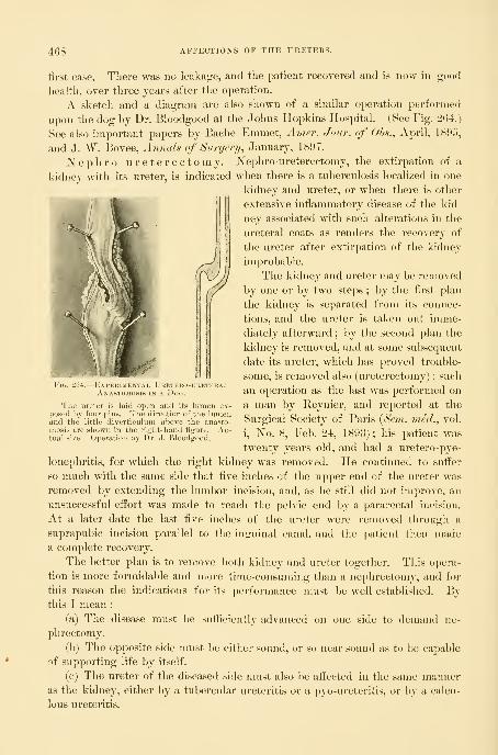
468 AFFECTIONS OF THE URETERS.
first case. There was no leakage, and the patient recovered and is now in good
health, over three years after the operation.
A sketch and a diagram are also shown of a similar operation performed
upon the dog by Dr. Bloodgood at the Johns Hopkins Hospital. (See Fig. 204.)
See also important papers by Bache Emmet, Amer. Jour, of Ob^., April, 1895,
and J. W. Bovee, Annals of Surgenj^ January, 1897.
Nephro -ureterectomy.—Nephro -ureterectomy, the extirjDation of a
kidney with its ureter, is indicated when there is a tuberculosis localized in one
kidney and ureter, or when there is other
extensive inflammatory disease of the kid-
ney associated with such alterations in the
ureteral coats as renders the recovery of
the ureter after extirpation of the kidney
improbable.
The kidney and ureter may be removed
by one or by two stej^s ; by the first plan
the kidney is separated from its connec-
tions, and the ureter is taken out imme-
diately afterward ; by the second plan the
kidney is removed, and at some subserpient
date its ureter, which has proved trouble-
some, is removed also (ureterectomy) ; such
an operation as the last was j)erformed on
a man by Reynier, and reported at the
Surgical Society of Paris {Sein. med., vol.
i, No. 8, Feb. 24, 1893) ; his patient was
twenty years old, and had a uretero-pye-
lonepliritis, for which the right kidney was removed. He continued to snfi'er
so much with the same side that five inches of the upper end of the ureter was
removed by extending the lumbar incision, and, as he still did not improve, an
unsuccessful eifort was made to reach the pelvic end by a pararectal incision.
At a later date the last five inches of the ureter were removed through a
suprapubic incision parallel to the inguinal canal, and the patient then madea complete recovery.
The better plan is to remove both kichiey and ureter together. This opera-
tion is more formidable and more time-consuming than a nephrectomy, and for
this reason the indications for its performance must be well established. Bythis I mean :
(a) The disease must 1)0 sufficiently advanced on one side to demand ne-
phrectomy.
(b) The opposite side must l)e eithei- sound, or so near sound as to be capable
of supporting life by itself.
(c) The ureter of the diseased side must also be affected in the same manneras the kidney, either Ijy a tubercular ureteritis or a pyo-ureteritis, or by a calcu-
lous ureteritis.
Fig. 264.
—
Expekimental Uretero-ubeteralAnastomosis in a Dog.
Tlie ureter is laid open and its lumen ex-posed by four pins. The direction of the lumenand the little diverticulum above the anasto-mosis are shown in the right-hand figure. Ac-tual size. Operation by Dr. J. Bloodgood.
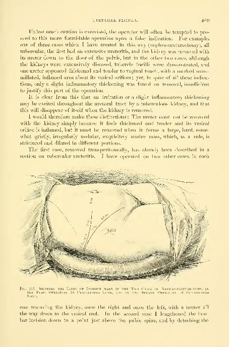
URETERAL FISTULA, 469
Unless much caution is exercised, tlie operator will often be tempted to pro-
ceed to this more formidable 0])eration upon a false indication. For example,
out of three cases which I have treated in this way (nephro-ureterectomy), all
tubercular, the first had an extensive ureteritis, and the kidney was removed with
its ureter down to the floor of the pelvis, but in the other two cases, although
the kidneys were extensively diseased, tubercle bacilli were demonstrated, and
one ureter appeared thickened and tender to vaginal touch, with a marked mam-millated, inflamed area about its vesical orifices
;yet, in spite of all these indica-
tions, only a slight inflammatory thickening was found on removal, insufficient
to justify this part of the operation.
It is clear from this that an irritation or a slight inflammatory thickening
may be excited throughout the ureteral tract by a tuberculous kidney, and that
this will disappear of itself when the kidney is removed.
I would therefore make these distinctions : The ureter must not be removed
with the kidney simply l)ecause it feels thickened and tender and its vesical
orifice is inflamed, but it must be removed when it forms a large, hard, some-
what gristly, irregularly nodular, exquisitely tender mass, which, as a rule, is
strictured and dilated in different portions.
The first case, removed transperitoneally, has already been described in a
section on tubercular ureteritis. I have operated on two other cases, in each
Fig. 265.—Showing the Lines of Incusk.n .maue in the Two Cases of Nephro-uretekectomy, inTHE First Operation 28 Centimeters Long, and in the Second Operation 16 Centimeters
one removing the kidney, once the right and once tlie left, with a ureter all
the way down to the vesical end. In the second case I lengthened the lum-
bar incision down to a point just above the pubic spine, and by detaching the
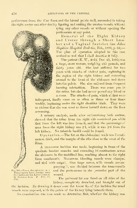
470 AFFECTIONS OF THE URETERS.
peritoneum from the iliac fossa and the lateral pelvic wall, succeeded in taking
the right ureter out after doubly ligating and cutting the uterine vessels, without
tying any other vessels or without opening the
peritoneum at any point.
Removal of the Right Kidneyand Ureter through a Short Lum-bar and a Vaginal Incision (see Johns
Ilojykins Hospital Bulletin^ Feb., 1896, p. 34).
—
The plan of operation adopted in this case
w^orked so well that I shall describe it fully.
The patient (K. W., 4012, Dec. 21, 1895) was
large, stout woman, w^eighin^ pounds, and
tliirty years old. She had suffered for two
years with attacks of violent pain, beginning in
the region of the right kidney and extending
around to the front of the abdomen and down
into the pelvis. She also suffered from frequent
l)urning micturition. There was some pus in
the urine, but she had never passed any blood or
a stone. The attacks of pain, which at first were
infrequent, finally came on as often as three or four times
weekly, beginning under the right shoulder blade. They w^ere
so violent that she was wont to throw herself down on the fioor
screaming.
A urinary analysis, made after catheterizing both ureters,
showed that the urine from the right side contained pus wdiile
that from the left was free from it, and that the percentage of
urea from the right kidney was 2-1, while it was 2-6 from the
left kidney. No tubercle bacilli could be found.
Operation .—The fat on the al)dominal walls was 7 centi-
meters thick, and the margin of the ribs close to the crest of the
ilium.
A transverse incision was made, beginning in front of the
quadrate lumbar muscles and extending 16 centimeters across
the abdomen in the umbilical line, reaching almost to the right
linea semilunaris. Numerous bleeding vessels were clamped
and tied with catgut. One large nerve, wdth vessels accom-
panying it, was divided between the transversalis
and the peritoneum in the posterior part of the
wound.
The perirenal fat was freed on all sides of the
kidney, completely detached, and brought out of
the incision. By drawing it down over the lower lip of the incision the renal
vessels were exposed, with the pelvis of the kidney lying beneath them.
An examination was now made to determine, first, whether the kidney was
Fig. 266.—Total Extirpation <>f a
Tuberculous Left Kidney withITS Ureter by the Long Incision.
X Natural Size. P. Dec. 13,
1895.
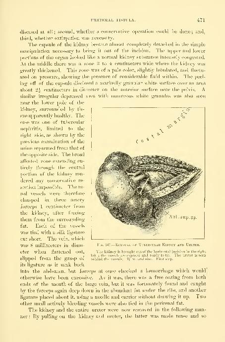
URETERAL FISTULA. 4Y1
diseased at all ; second, whether a conservative operation could be done ; and,
third, whether extirpation was necessary.
The capsule of the kidney became almost completely detached in the simple
manipulation necessary to bring it out of the incision. The upper and lower
jiortions of the organ looked like a normal kidney substance intensely congested.
At the middle there was a zone 3 to 4 centimeters wide where the kidney was
greatly thickened. This zone was of a pale color, slightly lobulated, and fluctu-
ated on pressure, showing the presence of considerable fluid within. The peel-
ing off of the capsule disclosed a markedly granular white surface over an area
about 2^ centimeters in diameter on the anterior surface near the pelvis. Asimilar irregular depressed area with numerous white granules was also seen
near the lower pole of the
kidney, surrounded by tis-
sue apparently healthy. The
case was one of tubercular
nephritis, limited to the
right side, as shown by the
previous examination of the
urine separated from that of
the opjiosite side. The broad
afi^ected zone extending en-
tirely through the central
portion of the kidney ren-
dered any conservative re-
section impossible. The re-
nal vessels were therefore
clamped in three artery
forceps 1 centimeter from
the kidney, after freeing
them from the surrounding
fat. Each of the vessels
was tied with a silk ligature
cut short. The vein, which
was 8 millimeters in diam-
eter when flattened out,
slipped from the grasp of
its ligature as it sank back
into the abdomen, but forceps at once checked a hemorrhage which would
otherwise have been excessive. As it was, there was a free oozing from both
ends of the mouth of the large vein, but it was fortunately found and caught
by the forceps again deep down in the abundant fat under the ribs, and another
ligature placed about it, using a needle and carrier without drawing it up. Twoother small actively bleeding vessels were also tied in the perirenal fat.
The kidney and the entire ureter were now removed in the following man-
ner : By pulling on the kidney and ureter, the latter was made tense and so
Fig. 267.
—
Removal of Tubercular Kidney and Ureter.
The kidney is brought out of the horizontal incision in the right
loin ; the vessels are exposed and ready to tie. The ureter is seen'
' '"
' % natural size. First step.behind the vessels.

472 AFFECTIONS OF TPIE URETERS.
easily followed and dissected out of its cellular bed, with the index and middle
fingers pushing the peritoneum, the ascending colon, and the caput coli to
one side, and stripping off the loose cellular tissue surrounding the ureter. This
dissection was carried down to the brim of the pelvis, and the common iliac
artery could be felt with the tips of the fingers over its entire length, with the
thumb resting on the surface of the abdomen, the end of the thumb reaching
the anterior superior spine.
I now freed the ureter down to its vaginal portion by introducing the en-
tire hand and part of the forearm into the cellular tissue, at first between the
Fig. 268.^Eemoval of the Kidney and Ureter, showing the Facility with which the Ureter canBE palpated and FREED ALL THE WAY DOWN TO THE CoMMON IlIAC AuTEKY WITHOUT INTRODICINGTHE Entire Hand into the Transverse Incision. Second step.
peritoneum and the abdominal wall, then under the peritoneum of the false })el-
vis, and finally between the peritoneum and the walls of the true pelvis. This
blunt dissection with the fingers was facilitated by pulling on the kidney and
making the ureter tense. In this way I freed it and followed it forward to
the broad ligament. At this point considerable resistance was felt, and the ure-

URETERAL FISTULA. 473
ter appeared to the touch to pass throuo;h a hole with a sharp border in its
upper part. Above this I distinctly felt the uterine artery pulsating.
N^ik.^'
Fig. 209.
—
Showixg the Methuu of removing the Lower End of the L^reter through theV^AGiNAL Vault.
The upper hand introduced through the horizontal incision holds the ureter and lifts up the uterine
iirterv. while the vatrinal vault is opened by a pair of sharp-pointed scissors to allow the ureter to be drawnthrough. Third step.
At this juncture the ureter broke, about 6 centimeters from the kidney ; the
lower end was at once caught in forceps and held, while by dint of pushing and
474 AFFECTIONS OF THE URETERS.
working in my finger I succeeded in freeing about two centimeters inore of the
ureter. Before doing this, however, I put a stout silk ligature over the abdom-
inal end of the ureter, and by means of one hand in the pelvis and the otlier
liolding the long outside end of the ureter I succeeded in tying a knot about it,
just behind the broad ligament ; then with a long pair of scissors introduced
through the al)dominal incision and controlled by the hand introduced into the
pelvis in the same way, the ureter was cut off half a centimeter above the
ligature, after taking care to milk back any of its contents and to keep the
upper end tight squeezed until it was removed.
The vagina was now thoroughly disinfected, and, with the patient still lying
on her left side, I passed two fingers of my right hand up to the vaginal vault,
and with my left hand introduced into the pelvis through the abdominal incision,
I brought both hands together with nothing but the vaginal tissue between
them. I now made an opening in the vaginal vault and brought the end of the
ureter through it and clamped it in a pair of forceps, until the abdominal wound
was closed, when the vaginal end was removed also.
This opening was made in the following manner : I passed my entire left
hand through the abdominal wound down into the pelvis and pressed the index
and middle fingers against the right vaginal fornix, at the same time lifting up
the uterine artery on the index finger so as to avoid any danger of cutting it
;
the end of the ureter lay between these fingers. The index and middle fingers
of the right hand were now introduced into the vagina (the patient was lying in
the left lateral posture) and pressed up against the fingers of the left hand in
the abdomen, the palmar surfaces of both hands being turned upward. The
opening in the vault necessary to draw the end of the ureter into the vagina was
now made by the assistant, who introduced a pair of sharp-pointed scissors
along my fingers up to the vaginal vault and pushed them through the thin
septum, guided by my instructions ; he then spread the blades of the scissors
and withdrew them, in this way enlarging the hole in the vault to about 2 centi-
meters. The opening was situated about 2 centimeters to the right of the cer-
vix. The bleeding from this torn wound was venous and slight. With a pair
of forceps pushed through this vaginal opening, the ligature attached to the
end of the ureter was now caught, and the ureter drawn through the vagina and
held there by forceps while the abdominal wound was being closed.
Closure of the Abdominal Incision .—The whole wound tract
was first irrigated with normal salt solution. Although the bleeding was slight,
a drain was put in on account of extensive separation of the cellular tissues and
the fear of the accumulation of the products of a serous weeping. The fascia
and muscles were brought together by interrupted silver-wire sutures, with a
gauze drain in the middle, and the fat and skin were closed by buried and sub-
cuticular cat-gut sutures.
The condition of the patient was excellent, and the pulse as quiet as if no
operation had been performed at all. I therefore did not hesitate to put her at
once in the lithotomy position and proceed with the extirpation of the remain-
der of the ureter per vaginam. The end of the ureter and the hole in the vault

URETERAL FISTULA. 4Y5
were exposed by using retractors and catching the right side of the cervix with
a bullet forceps and drawing it strongly to the left. By pulling on the forceps
holding the ureter it was made tense, while I cut down through the vaginal
wall, at first at the side between the anterior and the lateral walls, and then
curving the incision forward under the base of the bladder to a point within 1^
centimeter of the end of the ureter in the bladder. The ureter broke off 3 centi-
FiG. 270.
—
Kemoval of the Kidney with the Ureter.
The last step, the removal of the lower end of the ureter through the vagina. The ureter has beendrawn through the opening made in the vaginal vault. The dotted line indicates the direction of the in-cision to expose the ureter"down to its vesical extremity. Fourth step.
meters below the vaginal vault, and I had some difficulty in finding the short
end in the tissue by the sense of touch and in grasping it with the forceps.
There was a free venous oozing from the cut vagina below the vault. The ure-
ter broke once more, and this time at its vesical extremity, and as I could not
find the end again I closed the wound and stopped the bleeding by introducing

47( AFFECTIONS OF THE URETERS.
about six catgut sutures, tied tightly. The hole in the vault communicating
with extensive cellular area above was left open for an inferior drain, which
was now inserted, pushing a piece of iodoform gauze well up into the cavity
and leaving its end hanging down in the vagina.
The recovery of this patient proceeded without a single unfavorable symp-
tom, and she is well two years later.
Pathological Keport .—The specimen consists of the left kidney and
ureter. The lower half of the kidney is 6*5 centimeters long, 4 broad, and 5
thick. It is for the most part of a dark-red
color, but on its anterior surface presents three
pale, slightly elevated areas composed of ag-
gregations of minute yellow tubercles. Theremaining portion of the kidney presents a
lobulated appearance, and is 6 by 4*5 centime-
ters in its various diameters. This portion of
the kidney is soft and yielding, and on section
is found to consist of three or four large ca-
seous al)scesses containing thick, creamy, odor-
less fluid. The lower half of the organ is in
most parts normal in appearance, but at one
point contains a caseous nodule 1 centimeter
ih '^^^^^^^i^^y in diameter. The pelvis of the kidney is
II ^^^^^^^^ smooth and glistening. The ureter is 19 centi-
meters in length ; in the vicinity of the kidney
it is 5 millimeters in diameter, at its vesical end
9 millimeters ; it is firm and somewhat rigid.
The walls of the abscesses are composed
of typical tuberculous granulation tissue, lined
l)y caseous detritus. Tlie tissue in the vicinity
of the tuberculous abscesses is greatly altered.
Many of the glomeruli are completely hyaline, others are compressed
by the greatly thickened capsule. The connective tissue is markedly
increased, and scattered here and there throughout it are young tubercu-
lous nodules. The pelvis of the kidney has an intact surface epithelium
slightly infiltrated with small round cells. The stroma beneath, however,
shows marked small round-celled infiltration. Sections from the upper
and middle portions of the ureter are also slightly infiltrated by small
round cells. The ureter in the vicinity of the bladder, although dilated,
is little altered. The ureter throughout its course shows no trace of the
tuberculous process. Tubercle bacilli were found in the wall of the caseous
areas in the kidney.
Diagnosis .—Tuberculosis of the kidney.
The diagnoses were made in this and in the second case referred to by
symptoms, by palpation, by inspection, and by the analyses of the se})arated
urines.
Fig. 271.— Removal of KidneyAND Entire Ureter (Ne-phro-ureterectomy).
Tubercular zone on surface ofkidney. The two lower piecesof the ureter below the ligature
were removed through the vagi-nal vault. }4 natural size. W.Op. Dec. -21, 189.0.

URETERAL FISTULA. 477
The patients—all tliree—presented a history of pain in the side^ extending
down the course of the ureter and accompanied by frequent painful micturition.
In the first case the renal symptoms were masked by the strangury in the
bladder, due to cystitis and some tubercle nodules.
In the second case the intense pain in the left side, and in the third case in
the right side, accompanied in both cases by attacks of intense renal colic,
pointed toward the chief focus of the disease.
By palpation in all cases the pelvic portion of the ureter was found to be
enlarged and thickened, but only in the first case did it show any nodular
enlargement. There was also in each case a point of tenderness at the place
where the ureter crosses the pelvic brim. It was also shown by palpation that
the ureter of the opposite side was normal.
By inspection the bladder was shown to be normal excepting around the
orifice of the ureter on the diseased side, where there was a reddened granular,
mammillated aj)pearance.
The separated urines showed that the abnormal constituents of the urine
came entirely from the side indicated by this appearance in the bladder, and
that the opposite side was sound.
Tubercle bacilli were found in the first case after a jmtient search ; in the
second case bacilli, undoubtedly tubercle bacilli, were found which had someof the characteristics of the smegma bacillus. In the third case no bacilli w^ere
found, and the diagnosis depended upon the history and the physical examina-
tion. (See J. H. II. Bull., Feb. and March, 1896.)

CHAPTER XIV.
OPERATIONS UPON THE CERVIX OF THE UTERUS, INCLUDINGDILATATION AND CURETTAGE.
1. Dilatation.
2. Curettage : 1. Preparation and examination of uterine scrapings. 2. Normal uterine mucosa.3. Acute endometritis. 4. Chronic endometritis. 5. Decidual endometritis. 6. Mucouspolypus. 7. Remnants of abortion. 8. Tuberculosis of the endometrium. 9. Cancer of
the body of the uterus. 10. Adeno-carcinoma of the body of the uterus. 11. Sarcoma of
the uterus. 12. Curettage for cancer of the cervix : o. Epithelioma of the cervix. J. Adeno-carcinoma of the cervix.
3. Repair of the lacerated cervix.
DILATATION.
The cervical canal is dilated for tlie relief of dysmenorrhea, and for the pur-,
pose of removing portions of the endometrium, or the remains of an incomplete
abortion, and to overcome sterility.
For dysmenorrhea, the operation of dilating the cervix does not yet stand
upon a scientific basis, as its mode of action is not clear and the results are far
from uniform. While a small percentage of cases are cured, and a larger per-
centage are relieved, still a considerable number are not in the slightest degree
benefited. A common cause of failure is a want of care in selecting suitable
cases. The general practitioner, and even many specialists, fall into a commoneiTor of beginning the treatment of all cases of dysmenorrhea by a dilatation,
without a proper preliminary search for other causes of pain, such as tubal and
ovarian disease, pelvic peritonitis, and the presence of small fibroids in the uterine
walls. It must not be forgotten that dysmenorrhea is but a symptom conmion
to a variety of diseases, and to make clear its relation to a variety of pelvic affec-
tions which are most apt to escape detection upon a superficial examination, I
have analyzed 255 of my cases of pelvic peritonitis with adherent ovaries and
tubes, tubercular peritonitis, hydrosalpinx, pyosalpinx, and catarrhal salpingitis,
taken consecutively. Of these 255 cases, 185 suffered from dysmenorrhea, and
it was absent in but 70 cases ; therefore, from this analysis it would appear that
72 per cent of pelvic inflammatory cases present dysmenorrhea merely as a
complication.
In spite of failure, even in many of the well-selected cases, the relief and the
occasional cures effected make dilatation one of the most important, and often
one of the most satisfactory, of all the minor gynecological procedures.
The most suitable cases for dilatation are those in which the pain is spas-
modic, begins with the fiow, and is most intense during the first day or two.
Operation.—Dilators of the (Toodell-P^lliuger pattern, of three sizes, are
needed ; the smallest, having smooth blades, is 4 millimeters in diameter, and the
478
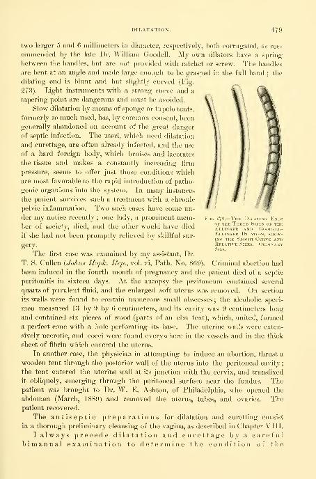
DILATATION. 479
two larger 5 and 6 millimeters in diameter, respectively, both corrugated, as rec-
ommended by the late Dr. William Goodell. My own dilators have a spring
between the handles, but are not provided with ratchet or screw. The handles
are bent at an angle and made large enough to be grasped in the full hand; the
dilating end is blunt and but slightly curved (Fig.
273). Light instruments with a strong curve and a
tapering point are dangerous and must be avoided.
Slow dilatation by means of sponge or tupelo tents,
formerly so much used, has, by common consent, been
generally abandoned on account of the great danger
of septic infection. The uteri, which need dilatation
and curettage, are often already infected, and the use
of a hard foreign body, which bruises and lacerates
the tissue and makes a constantly increasing firm
pressure, seems to offer just those conditions which
are most favorable to the rapid introduction of patho-
genic organisms into the system. In many instances
the patient survives such a treatment with a chronic
pelvic inflammation. Two such cases have come un-
der my notice recently ; one lady, a prominent mem-ber of society, died, and the other would have died
if she had not been promptly relieved liy skillful sur-
gery.
The first case was examined by my assistant. Dr.
T. S. Cullen {Johns Ilopk. Rep., vol. vi, Path. No. 869),
been induced in the fourth month of pregnancy and the patient died of a septic
peritonitis in sixteen days. At the autopsy the peritoneum contained several
quarts of purulent fluid, and the enlarged soft uterus was removed. On section
its walls were found to contain numerous small abscesses ; the alcoholic speci-
men measured 13 by 9 by 6 centimeters, and its cavity was 9 centimeters long
and contained six pieces of wood (parts of an elm tent), which, united, formed
a perfect cone with a hole perforating its base. The uterine walls were exten-
sively necrotic, and cocci were found everywhere in the vessels and in the thick
sheet of fibrin which covered the uterus.
In another case, the physician in attempting to induce an abortion, thrust a
wooden tent through the posterior wall of the uterus into the peritoneal cavity
;
the tent entered the uterine wall at its junction with the cervix, and transfixed
it obliquely, emerging through the peritoneal surface near the fundus. Thepatient was brought to Dr. W. E. Ashton, of Philadel23hia, who opened the
abdomen (March, 1889) and removed the uterus, tubes, and ovaries. Thepatient recovered.
The antiseptic preparations for dilatation and curetting consist
in a thorough preliminary cleansing of the vagina, as described in Chapter VIII.
I always precede dilatation and curettage by a carefulbimanual examination to determine the condition of the
Fig. 272.
—
The Dilating EndsOF THE Three Sizes of theEllinger and Goodell-Ellinger Dilators, show-ing the Slight Curve andRelative Sizes. OrdinarySize.
Criminal abortion had

480 OPERATIONS UPON THE CERVIX OF THE UTERUS.
organs and the exact position of the uterus. If the direc-
tion of the uterine canal is known it aids greatly the introduction of the uterine
dilator.
In the virgin the index finger must be introduced into the vagina slowly
and gently, to avoid injuring the hymen. When the finger touches the cervix
a pair of tenaculum forceps is introduced and the cervix firmly grasped by its
anterior lip. The finger is now withdrawn and traction
made with the forceps until the o s uteri is seen at
the vaginal outlet.
When the orifice is small, or the examining finger
large, in order to avoid injuring the hymen the position
of the cervix must be determined, witliuut vaginal exam-
ination, by a careful rectal palpation ; the tenaculum
forceps are now introduced into the vagina, and, under
the guidance of the rectal finger, the anterior lip of
the cervix is cautiously caught and drawn down to the
outlet.
In married women and those who have borne children
the posterior vaginal wall may readily be retracted by a
Sims or Simon speculum, or indeed with two fingers, ex-
posing the cervix, which is grasped with the tenaculum
forceps and drawn down.
The smallest dilator is now taken up, poised delicately
between the fingers just like a pen, and gently introduced
within the external os, and pushed up the canal to the
internal os. The dilator must never be grasped with
handles braced against the palm of the hand and forced
through obstructions. When resistance is encountered,
as it commonly is, in passing from the internal os into
the uterine cavity, the dilator must be withdrawn a little
and gently coaxed up in a slightly different direction,
until by repeated efforts, without force, it finally passes
the obstruction and slips in.
I have seen a death result from neglect of this pre-
caution and the use of a sharp dilator (see Amer. Jour.
OKs., Jan., 1891). The surgeon pierced the posterior
wall of the anteflexed uterus at its cervical junction, and
tore a wide hole into the peritoneum. He then inserted
a coarse sponge tent into the cervix, Avhich projected
partly within the peritoneal cavity. The patient died in
a few days of peritonitis, in spite of an effort which I
made to save her by opening and draining the abdomen.
The risk of perforating an antefiexed uterus in this way
is so manifest that I can not escape the conviction that
such an accident has happened more frequently.
Fig. 273.—Goodell-Ellin-GEB DiLATOK, WITHSpring between theHandles, but witholtA Ratchet.
The corrugations on theblades prevent slipping dur-ing the dilatation. J^ ordi-nary size.

DILATATION. 481
With the l)lades of the instrument well introduced, I dilate the canal lirst in
one direction, then relaxing the pressure, the blades close and I rotate the dilator
a little, gently dilating another portion, and so on, continuing all around the
circle back to the first point. The cervix, yielding to these repeated gentle
impacts from within on all sides, gradually and equably dilates to the necessary
degree without laceration. In this way in a minute or two the canal opens
Fig. 274.—Criminal Abortion, with Separated Elm Tent in situ partially perforating the UterineWall. Septicemia and Death. Specimen removed at Autopsy.
up enough to admit a larger corrugated dilator, with which the dilatation is
continued in like manner from side to side, antero-posteriorly and at all points
between. This extent of dilatation, large enough to allow tlie introduction of a
hougie 1 centimeter in diameter, is usually sufficient for the relief of dysmen-
orrhea or for curettage. A somewhat greater dilatation may be secured by
using the largest-sized dilator, but not without risk of too great injury to the
cervix. It is unjustifiable to attempt to dilate a cervical canal sufficiently to
permit the introduction of the index finger into the uterine cavity, for such a
degree of dilatation can only be effected by extensive rupture of the cervix.
Such a method of dilating, by repeated impacts on the cervical canal from
all directions, is far better than the common method of opening a dilator con-
trolled by a ratchet or screw, and expending all the force in one direction, until
the cervical fibers split and a tear is produced. The objections to this method
are the damage done the cervix, the greater danger of septic infection, and the
scar left when the rent heals, with the possibility of a carcinoma.
While the operation of dilatation and curettage is usually considered a safe
procedure, and is followed by little or no mortality, it may have decided dan-
gers which must be considered.
34
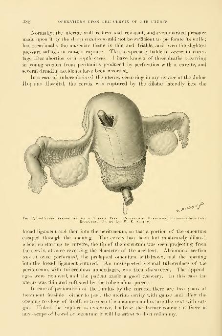
482 Ol'KHATIONS UPOX TlfE CERVIX OF THE UTERI'S.
Normally, the uterine wall is firm and resistant, and even marked pressure
made upon it by the sharp curette would not be sufficient to perforate its walls
;
but occasionally the nmscular tissue is thin and friable, and even the sliglitest
pressure suffices to cause a rupture. This is especially liable to occur in curet-
tage after al)ortion or in septic cases. I have known of three deaths occurring
ill young women from peritonitis produced by perforation with a curette, and
several dreadful accidents have been recorded.
In a case of tuberculosis of the uterus, occurring in my service at the Johns
Hopkins Hospital, the cervix was ruptured by the dilator laterally into the
ecl^^"fFig. 275.
—
Uteru PEUFdRATED BY A TuPELO TeNT. PeRITONITIRecovery. Op. by Ur. W. IC. K^w
Iystero-sai.po
broad ligament and then into the peritoneum, so that a portion of the umentuni
escaped through the opening. The cervix had been but moderately dilated,
when, on starting to curette, the tip of the omentum was seen projecting from
the cervix, at once revealing the character of the accident. Abdominal section
was at once performed, the prolapsed omentum withdraAvn, and the opening
into the broad ligament sutured. An unsuspected general tul)ei"culosis of the
peritoneum, with tuberculous appendages, was then discovered. The a])pend-
ages were removed, and the patient made a good recovery. In this case the
uterus was thin and softened by the tuberculous process.
In case of perforation of the fundus by the curette, there are two ])l;nis of
treatment feasible— either to pack the uterine cavity with gauze and nllow the
opening to close of itself, or to open the abdomen and suture the rent with cat-
gut. Unless the rupture is extensive, I advise the foi-nier course : if there is
any escape of bowel or omentum it will be safest to do a celiotomy.

CUIIETTAGE. 4S3
Perforation of tlie fundus with a uterine sound has occurred six times in mypersonal experience without anj serious trouble following the accident, but
death from peritonitis followed in a case in the hands of one of mj assistants.
In a case seen by Dr. M. D. Mann, of Buffalo (A/ner. Joh/: of Oh.s., 1895,
p. 603), a young practitioner forcibly dilated the cervix in order to remove the
ovum in an early abortion which the patient had induced by means of a cathe-
ter. In using a sharp curette and his finger, after clearing out the ovum, he
caught hold of and tore a loop of the intestine. Dr. Mann was called in within
an hour and a half, opened the abdomen, and found a hole in the center of the
fundus of the uterus large enough to admit the finger; the ileum was divided
close to the ileo-cecal valve, and was separated from its mesentery fully six
inches ; the head of the colon was bruised and infiltrated, and the abdomen con-
tained some bl(Jod and feces. The j^atient recovered after the closure of the
hole in the uterus, and the inversion of the head of the colon, followed by the
removal of the detached Ixjwel and the making of a new ileo-colic anastomosis
with a Murphy button.
Dr. J. B. Harvie, of Troy, X. Y., had personal cognizance of a case in
which, after dilating the uterus, a young practitioner passed in a pair of forceps
to catch the ovum and drew out and cut off six feet of bowel (!) without realizing
what he had done.
In a similar case of extensive intestinal injury following the perforation of
the uterus with a curette. Dr. C. P. Noble, of Philadelphia, opened the abdomen
and resected three feet of the small intestine successfullv.
CURETTAGE.
Curettes are used to remove the superficial portions of the uterine mucosa
in endometritis ; to secure bits of tissue for diagnostic purposes in suspected
cancer of the body ; to remove portions of an ovum incompletely cast off ; and
to clean out the broken-down tissue of a cancerous cervix where the disease has
progressed beyond hope of a radical cure. Sharp curettes, handled with extreme
deUcacy, are most serviceable ; the blunt curettes often advocated are but in-
sufiicient substitutes.
A careful microscopical study of the tissue should follow the removal l)y the
curette in every case, and the following conditions should be looked for
:
Normal uterine mucosa.
Acute endometritis.
Chronic endometritis.
Endometritis decidualis.
Mucous polypi.
Remnants of abortion.
Tuberculosis of the endometrium.
Carcinoma of the body of the uterus.
Sarcoma of the uterus.
Cancer of the cervix.

484 OPERATIONS UPON THE CERVIX OF THE TTTETITTS.
Preparation and Examination of Uterine Scrapings.—In examining portions of the endometrium I use the formaHn method intro-
duced bj my assistant. Dr. T. S. Cullen {Johns Ilopk. llonp. Bull., April, 1895),
which obviates the tedious delays of ten days or two weeks incident to older
methods of preparation and permits a diagnosis to be made within lifteen minutes
—that is to say, while the ftatient is still under anesthesia ; if necessary, a radical
operation may then be performed at once.
The procedure is the following :
(a) Place frozen sections of the fresh tissue in a 5 per cent aqueous solution
of formalin for from three to five minutes.
(b) Iinmerse in a 50 per cent alcohol solution for three minutes.
(c) Place in absolute alcohol one minute.
(d) Wash in water.
(e) Stain in hematoxylin for two minutes.
(f) Decolorize in acid alcohol.
(g) Kinse in water, to which Dr. T. Brown has recently recommended the
addition of two or three drops of ammonia, which rapidly brings back the char-
acteristic hematoxylin color.
(h) Stain with eosin.
(i) Transfer to 95 per cent alcohol.
(k) Pass through absolute alcohol, creosote, or oil of cloves, and mount in
Canada balsam.
A cylinder of condensed carbonic-acid gas is kept in a room adjoining the
operating-room, in order to facilitate the iimnediate making of the frozen sec-
tions, to be passed at once through the routine described.
By securing an early diagnosis in this way the patient is often relieved of
the necessity of taking an anesthetic twice, and cases arriving from a distance
save from ten days to two weeks of their time in the hospital.
It is easier to cut fine sections after the tissue has been first hardened in the
formahn, according to the second plan also recommended by Dr. Cullen, as
follows
:
The scrapings are placed immediately in a 10 per cent formalin solution,
kept in small specimen bottles, always at hand. In two or three hours after
tliey are sufficiently hardened to cut readily, frozen sections are made and left
in a 50 per cent alcohol solution three minutes, when the succeeding steps are
as described above.
The curetted specimens should be placed in a bottle by themselves and
labeled at once, and when the sections are cut no similar opendishes containing sections should be lying about, nor should
they be passed through the fluids together with other sections, in order to avoid
the terril)le mistake of confusing two cases, and so drawing erroneous conclu-
sions. In all my experience of many hundreds of examinations this accident
has happened once.
The patient had a uterus of normal size, and was nearly exsanguinated by
protracted excessive hemorrhages. I operated upon her for a cancer of the

ENDOMETRITIS. 485
body of the uterus, diagnosed from curettings. Upon opening the abdomen I
found not a cancer, but a small pediculated fibroid tumor, lying in the cervix,
which had not been felt during the curettage upon which the diagnosis had
been made. It was afterward discovered that the scrapings had been mixed
with those from another patient.
Normal Uterine Mucosa.—The standard of comparison for all curetted speci-
mens is the normal uterine mucosa; this presents, microscopically, an even sur-
face covered by a single layer of cylindrical ciliated epithelium. The glands
are round or oval on cross section, and in a few places may be seen opening on
the surface.
They are usually equidistant, and are lined with one layer of cylindrical ciliated
epitlieUura ; hence they appear as reduplications of the surface epithelium. Anoccasional bifurcation is seen in the deeper portion of the gland. In the floor of
the gland there is not infrequently a small titlike ingrowth. Lying between the
glands is found the stroma of the mucosa or so-called lymphoid tissue. The
cells, however, are much larger, and on close examination bear no resemblance
to lymphoid tissue ; the nuclei of the stroma cells are oval, vesicular, and appear
to best advantage in specimens hardened in Miiller's fluid. The arteries of the
stroma are usually found in small bunches ; the veins are large and single and
thin-walled.
The blood in the veins is separated from the stroma cells by but one layer of
endothelium. The line of demarcation between the mucosa and the muscle is
usually well defined ; occasionally, however, a gland pene-
trates the muscle for some depth, when it is invariably
accompanied by a considerable amount of stroma. This
dipping of a gland into the muscularis must not be mis-
taken for a pathological condition.
Endometritis.—Curettage for endometritisfollows innnediately upon dilatation, and is performed in
this way : The sharp perforated spoon curette, poised
between thumb and first and second finger, is
easily introduced through the dilated canal. The whole
inner surface of the uterus over the fundus and from 'FORREiTovlNGraE^UTER^
fundus to cervix is now carefully scraped, completely re-'s^;^^^^''"°'^-
^^"^•^«^'
moving the superficial portion of its lining membrane in
strips and short pieces. The sound basis is recognized by its greater resistance,
and a slight grating sensation communicated to the fingers. The separated
lining membrane is expelled through the cervix by a series of intermittent
uterine contractions ; its discharge may also be assisted l)y using the curette to
scoop it out. The hemorrhage after this operation is never serious enougli to
call for measures to control it. The patient should be kept abed from three
days to a week ; it has never been my practice to introduce gauze into the
uterine cavity.
Acute endometritis is generally found in acute septic processes in-
volving the entire genital tract, but on account of the predominating symptoms
f

486 OPERATIONS UPOX THE CERVrX OF THE UTERUS.
of the other orgaiit*, it is usuall}^ overlooked. Under any eircninstanees it is a
rare affection.
The surface epitlielial cells are swollen often as nuich as two or three
times their normal size, wdiile the adjacent cells may be compressed. There is
also a tendency to cell proliferation, and between the epitlielial cells are many
polynuclear leucocytes and some small round cells. The glands in the su])er-
ficial portions show similar changes, a swollen epithelium with some tendency
toward proliferation, and a small round-celled and polymorphonuclear intiltra-
tion ; leucocytes are found partially filling some of the gland lumina. The
deeper portions of the glands near the nmscle are often normal.
The stroma shows superficially much infiltration, with polymorphonuclear
leucocytes and small round cells, the infiltration diminishing toward the muscle.
Alterations are rarely made out in the muscular tissue beneath.
Chronic endometritis is also rather rare. The prevailing habit of
describing all scrapings, particularly because of their abundance in some cases,
as examples of endometritis, is greatly to be deplored. It interferes with our
getting any satisfactory idea as to the frequency of the real aifection, and tends
to encourage unnecessary operating. The so-called '^ fungoid endometritis " is
not a pathological entity at all, and the name ought to be expunged from gyne-
cological works.
Chronic endometritis is oftenest associated with old cases of pyosalpinx ; it
is rarely ever found in the ordinary scrapings. The slight Hability of the
uterine mucosa to this affection may be ascril)ed to two factors : In the first
place, the tendency of pus-containing tubes is to complete closure at the
uterine end, and so shutting oft" one avenue of infection, and, in the second
place, the form and position of the uterine canal is such as to afford good
drainage.
The surface of the mucosa may be rather uneven, and the epithelium stunted,
low, cylindrical, or cuboidal. The glands are in places diminished in number,
and vary much in size; some of them are narrow superficially and distended be-
low. The epithelium of the dilated glands is somewhat fiattened.
The stroma is denser than normal, especially in the superficial jjortions, its
nuclei tend to become spindle-shaped, and there is much small round-cell in-
filtration. There are practically no polymorphonuclear leucocytes to be seen.
The stroma in its deeper portions is often normal, and there are no changes in
the muscle.
Decidual Endometritis .- -This is always found after an al)ortion in
the early months, and is often probably the cause of the abortion.
The decidua shows marked polymorphonuclear infiltration, especially in its
superficial portions ; the leucocytes are so abundant that the individual decidual
cells are separated from one another. Some small round cells usually accom-
pany the infiltration. The deeper portions of the decidua are usually unaltered.
Mucous Polypi.—A mucous polyp is a localized outgrowth of the uterine
mucosa forming one or more small tumors within its cavity. The tumors do
not often attain a size greater than 2 by 3 centimeters. They occur in a variety

MUCOUS POLYPI. 487
of foi-ins, either iingerlike, round and pediculated, pear-sliaped, oi- like a cock's
comb with a broad base.
In one of my cases a flattened ovoid polyp 1'5 centimeter long was found
lying in the cervical canal and attached to the fundns by a threadlike pedicle 1
millimeter in diameter and between 4 and 5 centimeters long. The velvety ap-
pearance of the polyp, with its slight indentations, resembles that of the uterine
nuicosa ; often small cysts, formed by distended glands, can be seen on the
surface.
Fig. 277.
—
Seotion of a Glandtlar Utkrine Polyp.
The small %ure below shows a section of the polyp mairnified three tinies ; the larirer section above is
inagnifled seventy times. The dilated glandular spaces are seen lying in tlie connective-tissue stroma.Specimen 682.
They sometimes give rise to proti-acted hemorrhages, but as a rule they pi'o-
duce no symptoms at all.
Histologically, the epithelium is the same in character as that lining the
uterine cavity with wliich it is directly continuous.
The entire polyp is made up of uterine mucosa, e})ithelium, glands, and
stroma. The glands are mostly normal, Imt where they are dilated and form
small cysts, tlie epithelium becomes cul)oidal and tlie cavities contain some des-
quamated epithelial cells. The stroma, especially near the tips, often shows
hemorrhage and edema.
In sharp contrast to the usual isolated mucous polyps just described is the
rare general excessive hypertrophy of the mucosa, of which a single instance has
come under my notice (A. L., 3470, May 7, 1895).
The entire uterine mucosa was thickened to about three times the normal
diameter, and appeared everywhere in the form of flattened domelike elevations,
488 OPERATIONS UPON THE CERVIX OF THE UTERUS.
separated from each other by shallow furrows 2 or 3 niilliineters in depth ; the
microscopic appearance was strikingly like that of malignancy. Microscopically,
the excessive growth was limited to the glands, which, although normal in num-
ber, were increased in size and markedly convoluted. The stroma was normal.
Remnants of Abortion.—In curetting to remove a dead ovum or an incom-
plete miscarriage, the chief danger lies in the readiness with which sepsis may
invade the upper genital tract. In cases w^hich are already septic, the avoidance
of a general infection and the safety of the patient depend upon the complete
removal of the ovum, and the efficient drainage established through the dilated
cervix. There is no way by which we can thoroughly disinfect a septic uterus.
Ckses which are not septic will not become so if the operation is aseptically per-
formed, and the aseptic conditions are maintained afterward. When the flow
does not begin to diminish within two days after an abortion, or wlien the tem-
perature rises three or four degrees, I at once advise curettage. The cervix in
these cases is usually soft, and dilatation more easily effected in consequence, and
not infrequently the cervix is so open as to need no dilatation at all.
The anterior cervical lip is caught with tenaculum forceps, and a blunt
spoon curette introduced and used with gentle force over the whole inner
surface of the uterus, loosening and bringing down the membranes which begin
to pour out of the os. Undue force must not be used lest the curette perforate
the softened uterine wall and pass into the abdominal cavity, exijosing the patient
to the imminent risk of a septic peritonitis. After loosening the membranes
with the curette, a pair of fenestrated placental forceps (see Chap. VI, Fig. 92)
is inserted, which brings away the placenta, decidua, and fetus, if not previously
expelled, whole or in pieces.
When the canal is large enough, as is usually the case in a miscarriage after
the third month of pregnancy, the index finger well sterilized should be intro-
duced and the whole interior of the womb palpated.
Unsuspected pieces of tissue will often be found clinging especially to the
placental area. These can be freed by the palmar surface of the finger, assisted
l)y the external hand acting through the abdominal walls, affording a point of
resistance. The uterine wall thus bared in places feels almost as thin as paper,
and must be gently handled. Where the curetting is difficult and uncertain the
entire separation of the remains of the ovum may be thus effected by the finger
alone, assisted by the hand making counter pressure through the abdominal
walls.
The finger nails m u s t never be use d to scrape tissue off from
the uterine walls, as such a practice would often introduce sepsis, and if the case
was already septic the operator would then be sure to cany the infection away
with him to inoculate other patients.
Irrigation of the uterus after curetting is not necessary, unless the contents
are septic, when the cavity must be repeatedly washed out with a warm boric-
acid solution introduced by means of a curved glass douche nozzle, using the
blunt end of the nozzle over the uterine surface to aid in detaching clots and
small particles of debris. The uterus may be drained for forty-eight hours bj


PLATE Vlll.
4x^
I
'J
t*:-^.-*"^
X 70
H.Becker, fee.Liih.LPrang iA.o.boaor,;; ^ ,-

DESCRIPTION OF PLATE VIII.
Tuberculosis of the endometrium. This is a typical picture of early tuberculosis of
the endometrium. Above is the normal uterine muscle, on the left side is a dilated
gland lined with flattened epithelium, while in the middle of the section and at the
lower margin two practically normal glands are seen. Scattered throughout the
stroma of the mucosa are typical tubercles, most of which show giant cells. The inter-
vening stroma is the seat of marked small-celled infiltration. There are no caseous
areas present.

7 aTAjq "KO pioiTgmoaaa
\o giaoIwoiQdiij / i -^ u> jiujiq fBoiq-^t £ 8i airIT .mi/i'fismobns gr^J lo zkoluo'ieduT
fasiBlib B er obia iial arfi xro .eloguiii Qflhaitr Ismiois edi ^i 9VodA .mjji'i;t9inoba9 edi
sdi in bnfi ttoiJoi^g &di \o eihbim edi at elxrfw ,mis'dedilqe bsiiaJtelt riiiw bsnil baB['g
edi iuod-gfsoiili bertoiie.oS .aaes bib zbttBl'g fBrnion: ^llfioidojriq ov/i xiigTora lawol
-•i9ifii 9xIT .e
TUBERCULOSIS OF THE ENDOMETRIUM. 489
packing its cavity looselj with gauze, the ends of which are allowed to hang out
of the cervix into the vagina ; my own practice, however, is simply to place a
loose gauze pack in the vagina, which is renewed every twenty-four hours.
Patients should be kept in bed after curetting for abortion for two weeks or
longer, to allow involution of the uterus to take place ; care of the patient is just
as important at this time as in the puerperium after a normal labor.
Microscopic Examination for the Remnants of an Abor-tion .—We usually have in these cases the clinical history of a recent miscar-
riage, and the amount of material removed by curettage is often abundant. As
a rule, there is no suggestion as to their source in the macroscopic appearance of
the tissues ; occasionally little villous threads can be seen.
Histologically, the appearance of glandular hypertrophy predominates ; the
glands are dilated, convoluted, and show little titlike processes springing into
their lumina ; the epithelium is a little flattened and the stroma of the mucosa
shows marked swelling of its cells in the superficial portion, forming typical
decidual cells which persist for several weeks after the abortion.
These appearances are suggestive of pregnancy, but a positive diagnosis must
rest upon the discovery of vilh ; these in the early months still show two layers
of epithelial covering, the inner of which is made up of cuboidal cells ; the outer
syncytial layer appears as a ribbon of protoplasm with nuclei distributed through
it ; this outer layer sends out protoplasmic buds which form the new villi, and
in the centers of these buds are found from five to forty nuclei, forming the so-
called placental giant cells, because when cut across they present the appearance
of a typical giant cell. The interior of a villus is composed of mucoid tissue
rich in blood vessels.
In one obscure case nothing was found in the curettings but some glandular
hypertrophy, ill-defined decidual cells, and a single free giant cell ; this latter
structure led to a further searching investigation, which was rewarded by the
discovery of villi, confirming the diagnosis of pregnancy.
Tuberculosis of the Endometrium.—In the early stages the epithelium of the
surface is intact, the glands normal, and the tubercles are found scattered
throughout the superficial portions of the stroma, consisting of aggregations of
epithelioid cells ; later they are surrounded by small round cells, and at a still
later date giant cells are found in the center.
The surface epithehum over a superficial nodule is often somewhat flattened
and pale. In a marked case the glands are encroached upon, and it is at times
almost impossible to distinguish some of the epithelioid cells from the gland epi-
thelium ; in other glands, tul)ercles are seen partly projecting into and obliter-
ating the cavity ; again the gland may be filled with caseous material.
In the most advanced cases where the cavity of the uterus is lined by caseous
material, the surface is covered by a necrotic material devoid of nuclei, below
which lies a zone of typical tuberculous tissue, consisting of epithehoid cells and
tul)ercles ; in the deeper portions a stray gland may survive ; where the process
has gone deep enough to involve the muscle, the glands are often entirely absent.
Bacilli are found with varying frequency, sometimes sparse, sometimes
490 OPERATIONS UPON THE CERVIX OF THE UTERUS.
abundant, and most numerous in the advanced cases with marked caseation ; in
my experience thej are much more readily found than in tuljerculosis of the
tubes.
In the early stages of the disease the tubercular process may be entirely un-
suspected, and the curettings may look like normal uterine mucosa ; but where
the disease is advanced, the presence of soft cheesy masses will at once arouse
suspicion. Necrotic carcinomatous tissue may present a somewhat similar ap-
pearance, but the characteristic branching is found here and does not occur in
tuberculosis. In advanced cases the diagnosis may also be reached from an
examination of the uterine discharge which contains tubercle bacilli.
On histological examination, the diagnosis of tuberculosis is readily estab-
lished, as the tissues j)resent the usual tubercular picture. Dr. T. S. Cullen
{Johns Hopkins Reports, vol. iv, p. 91) reports several cases of tuberculosis of
the endometrium occurring in my wards.
It has happened several times in my experience that the tuberculosis has been
found in a purely accidental way, as it were, while submitting the uterine scrap-
ings to the routine examination. Again, I have found a tubercular endometrium
on curetting the uterus immediately after removing tubercular tubes and ovaries.
Tubercular affections of the endometrium are either miliary, or part of a
general tubercular process, or of the chronic diffuse form.
The chronic diffuse tuberculosis is that form with which we have to do ; it
begins, as a rule, near the fundus secondary to a tubercular tube. The first visi-
ble alterations are little yellowish-white nodules under the surface, 1 to 2 milli-
meters in diameter, which may increase in size and numbers, and then coalesce
and break down, forming an ulcer with undermined edges. The disease extends
from the endometrium down into the uterine muscle.
Cancer of the Body of the Uterus.—The curette is used in these cases for two
purposes : first, to remove some of the lining membrane of the uterus for diag-
nosis, and, second, to remove as much of the diseased tissue as possible, so as to
check hemorrhage and to clean out septic deht'is, in order to give the patient a
chance to recruit before undertaking the total extirpation. In both instances
the use of the curette is simply preparatory to hysterectomy.
The cancerous tissue breaks down readily under the curette, which nuist be
used with unusual care and with gentler force than in endometritis, to avoid per-
forating the uterine wall in the more extensively infiltrated areas.
As a rule, the whole endometrium is affected, but where the disease is still
localized its position may be recognized by the distinct difference in the sense of
touch, communicated through the instrument, between the diseased tissue as it
breaks down, and the soft sound mucosa with its firm substratum of normal
muscle.
Even the macroscopic appearance of the curettings in carcinoma of the body
is quite characteristic, and much valuable information may be gleaned from
a careful inspection ; while the normal uterine mucosa has a comparatively
smooth surface and is usually from 1 to 2 millimeters thick, in the carcinoma
the surface, if still intact, has a branching or treelike appearance. This may

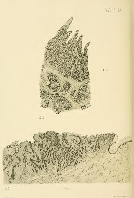
PLATE IX.
Fig.l.
X8 Fig.2
Lith,LPraivg4Co.Bo5tonr.sA

DESCRIPTION OF PLATE IX.
Fig. 1.—Adeno-carcinoma of the body of the uterus ( x 12). The section is taken at
a right angle to the surface of the uterine mucosa, and the upper border corresponds to
the uterine cavity. The thickening of the mucosa is due to the teatlike outgrowths
;
at the same time there is a growth into the muscle represented by the groups of
glands seen in the lower part of the picture. This is an instructive picture, as it showsthe early changes, before any necrosis has occurred. Path. No. 559.
Fig. 2.—Epithelioma of the cervix. The normal mucosa, composed of several layers
of squamous epithelium, is seen to the right. This ends abruptly and is replaced bymasses of epithelium, which penetrate the tissue in all directions. Note the transition
of the normal epithelium into that of the new growth, the continuity between the
superficial and the deeper portions of the tissue, and the deep stain taken by the carci-
nomatous cells. The uneven upper surface is due to loss of tissue. Path. No. 169.

)rmHnm'i
no:Asii 81 noiiosa erlT .(21 x ) -iu-ioiu "idi l-t ibod -..i
:
.>! «i)aoq«9Tioo isbiod -laqqu 9ilJ bus BSoouot anhtjdi/ 'm; i
; aiiiv/o'igijjo 9v[ili^a;t edi oj euh ai Grtoo/nu ariJlo ^nirr:./
lo aqijoig 9ifJ Y<^ Jjoiitsae-iqei .3l')^jjifl 9ifi'oJ«i dJwr •>
awoda ji ?,b ,e'iuioiq aviiou'itaai ab pi ghfT .artrJolq e;!
.965
2'I9^{;bI [B79V9a ^o b98oqm
Xd baojslqai ai huB ^liq
noJiiaiiiJTiJ 9di 9ioM .axi
orft xi99wi9d ^JitJiiiinoo
.r:.'4
9f)A— .1 .oil
oJ al-gflB ixf-gh B
ivBo 9flh9ijj sdi
di ems3 edi iB
[i ni n99a zbnai-g
<'.-'<• '". , J^HBrfo ^IlB9 9/1*
lOibdJiqa— .S .Ol%
'nibdiLqe auoniBupa \o
euaaij 9X1! envjjgnsq iijni//'. ^mwilgriiiqg ^o 898aBm
7/9a 9i{.t 3o JBii) ojni aiudediiqe iBmioxi 9rli lo
n;a oili -^cf najlBJ iriBi« qyafc 9iiJ Lab ,9B88ii 9fli ^o fiaoiiioq i9q99b 9xi;t bns iBiofligqua
.69 1 .oVL .diti*i .9jj8ai:t ^o aaol oi eub gi 90Bliua •i9qqu n9y9xiij 9riT .allao auoteraon

SARCOMA OF THE UTERUS. 491
not at first sight be detected, as the mucus tends to ghie the Httle projections
together. Nearly all of these little stems or branches have delicate capillaries
in their centers. One of the most striking points is the large amount of the
tissue removed, together with the size of the individual pieces. Normally only
a drachm or slightly more can be removed ; in carcinoma, however, from 4 to 8
drachms may come away, and the individual pieces may reach 1 centimeter or
more in thickness and are very friable.
In curetting to check hemorrhage the whole endometrium should be scraped
as rapidly as possible down to the firm muscular tissue, as by this means the
hemorrhage is less than if the instrument is used slowly and timidly. The use
of the curette under these circumstances is followed by a firm vaginal pack of
iodoform gauze. Hemorrhage is caused in these cases by the superficial necrosis
which opens up the vessels or permits them to rupture easily ; it is therefore
necessary to get well below this tissue when the vessels cease to bleed by their
normal contraction.
Cancer of the body of the uterus is found in two forms—epithelioma, madeup of squamous epithelial cells, and adeno-carcinoma ; not more than eight cases
of the former have been observed.
Adeno-carcinoma of the Body of the Uterus.—As stated,
abundant scrapings are usually furnished for examination—an amount never
found normally.
These often have quite a charactei'istic appearance even on a microscopic
examination ; they appear as short, broken, irregular, friable bits of tissue,
whitish and waxy in places, with little knoblike projections with coagula be-
tween them. Microscopically, the surface has usually disappeared, and the
remaining tissue is made up of groups of small and large glands, varying muchin appearance ; some are lined by one layer of cylindrical epithelium, others by
two or three layers, and still others are choked with cells. Large areas of
epithehal cells are often found in which the glandular form has almost disap-
peared, due to an excessive overgrowth of the epithelium in which numerous
glands are crowded together.
The stroma between the glands is composed of spindle cells, and shows
much small round-celled infiltration.
It is not necessary, as commonly held, to demonstrate a penetration of the
muscular layer by the glands in order to make the diagnosis of carcinoma. The
diagnosis must rest upon the characteristic appearances above detailed as found
in the mucosa alone, and this is fortunate, for the curette rarely penetrates as
deep as the muscularis.
Sarcoma of the Uterus.—This rare disease is not often found in the uterine
scrapings.
In my experience, the round or the round and spindle-celled sarcomata have
been found most commonly.
The macroscopic appearances are not characteristic, but microscopically
large areas are found composed of round or of spindle cells, many of them
containing nuclear figures and an increased amount of chromatin, as evidenced

492 OPERATIONS UPON THE CERVIX OF THE UTERUS.
by the intense staining of the nucleus ; there is also an absence of the uterine
glands in these areas. Such a picture is strongly suggestive of sarcoma.
Where the superficial tissue is broken down and the muscularis is invaded
by the characteristic cells the diagnosis is more certain.
I have operated in a single instance upon a case of sarcoma of the uterus in
which the diagnosis had been made by curettage. No enlargement of the uterus
could be detected bimanually, but, relying entirely upon the microscopic exami-
nation, I performed vaginal hysterectomy, and found a sarcomatous nodule 1
centimeter (0*4 inch) in diameter in the left horn of the fundus projecting into
the uterine cavity. The patient recovered, and has had no return of the disease
in over four years.
Cancer of the Cervix.—Curettage for cancer of the cervix is employed for two
purposes : First, to remove the septic, breaking down cancerous material, and
leave a clean field for hysterectomy ; second, to remove as much of the disease
as possible where it has advanced too far for complete extirpation.
Sometimes the uterus is more or less anchored at the vaginal vault by the
extension of the disease into one or both broad ligaments. The amount of this
infiltration can be better estimated by a rectal than by a vaginal examination.
Such cases of cancer of the cervix in whicli a broad, hard mass is detected on
either side, extending out to the pelvic wall, interfering
fwith the mol)ility of the uterus, are unsuitable for hysterec-
tomy, and are best treated by thorough curettage. Even
those advanced cases with marked cachexia and foul dis-
charges, bedridden and suffering from nausea, will often be
much benefited by thorough curettage, which i-emoves the
friable, sloughing masses, and leaves in their place a clean,
cone-shaped excavation. I have found that the severe pain
so often noted in these advanced cases is due to a choked
cervix with retention of the discharges, forming a pyometra.
Complete relief follows the evacuation of this fluid if the
canal is kept open.
The two most efiicient forms of curettage are the fingers
Fig. 278.—The Spoon of ^"^ ^ ^^"» scoop curettc ou a stout handle. The friable,
THE Long Sharp Cu- redundant portions of the disease are best brouo-ht out byRETTE FOR REMOVING
_
"^_
,
THE Cancerous Tissue vigorously usiug the end of the index and middle fingers as
BINARY Size.
'
"
a curctte. It is astonishing how much of the affected tis-
sue can be removed in this way. The scoop curette follows
the fingers and is lield firmly, and used boldly and rapidly, bi-eaking down the
diseased tissue under the guidance of the index finger, which locates the points
to be curetted, and prevents the instrument from advancing too far in the
directtion of bladder, rectum, or pei"itoneal cavity.
The limit of the diseased tissue which can be removed in this way is recog-
nized by the scraping sound and sensation, indicating that a hard base has been
reached. Less blood is lost by working rapidly down to the healthier tissue than
by a slower procedure, which allows the rigid diseased vessels time to bleed.
/;


X50
HBecker^fec.
PLATE X
Li!h.LPran|&CaBosion,U5,/

DESCRIPTION OF PLATE X.
Epithelioma of the cervix uteri. This specimen was obtained by curettage. It,
shows a central branching portion, consisting of a stroma with a marked round-celled
infiltration, and enclosed in this stroma are epithelial nests composed of groups of
polygonal cells. A few dark dots which may be seen in the center of some of these
nests are polymorpho-nuclear leucocytes. The central portion of the two upper nests
are filled with these leucocytes.
.Z 3TAJ^ 'in Vi0lT^lSlD83<J
ai?.9a leqqjj owi arfl lo noi^ioq ImJaap ari'.'
ihoq •goMouB'iJ I-uiJiT9a a gv/oria
ilJ fli baaobu'* baa ,noUc'!l[fliu
-f tmiouaddq'iotaxioq s'Ui ^i^ea

CANCER OF THE CERVIX. 493
When the disease has extended so far that tlie operator feels uncertain
whether the next effort will invade bladder, rectum, or peritoneum, it is im-
portant to advance more slowly, controlling the curettage by repeated examina-
tions. A finger in the rectum or a sound in the bladder will assist in deter-
mining the thickness of the septum. If the peritoneal cavity is accidentally
opened, an iodoform gauze tampon should at once be closely packed within the
rent and the operation continued until all septic and sloughing masses have been
removed down to a clean wound surface. The vagina is now cleansed, the
gauze removed, and a fresh pack inserted, projecting a short distance into the
pelvic cavity ; this is allowed to remain in place for three or four days, when it
is removed and a fresh pack inserted, not quite so far up. The excavated area
and the vagina must also be loosely filled with an iodoform gauze pack and
protected by the vulvar occlusive dressing.
Two forms of cancerous disease are found in the cervix uteri—e p i t h e 1 i -
o m a and a d e n o - c a r c i n o m a
.
To make a diagnosis in the early stages of carcinoma of the cervix, it is
necessary for the clinician to send the pathologist a wedge of the suspicious
portion, which should be at least 1 centimeter in depth ; this may readily be
removed without pain after injecting a few minims of a 4 per cent solution
of cocain deep into the cervical tissue, when two or three catgut sutures may
be passed to close in the wound. Where the cervical disease is far advanced
the ordinary curettings will be sufficient for the diagnosis.
Epithelioma .—The surface of the cervix is covered by several layers of
squamous epithelium, which, however, can be seen penetrating the stroma in
the form of fingerlike or branching masses of cells ; many of these branches
when cut transversely or obliquely appear as round, oval, or irregular groups of
cells lying deep in the stroma. On other parts of the surface of the tissue slight
elevations are found which consist of a central blood vessel surrounded by little
or no stroma and covered externally by numerous layers of squamous epithe-
lium, indicative of an outgrowth of blood vessels and stroma with a dispropor-
tionate increase in the epithelial layers.
The cervical glands are usually normal, with the exception that in some
cases the squamous epithelium may l)e found projecting into the lumen of the
gland and partly occluding it ; in more advanced cases the glands are completely
obliterated.
Such a macroscopic appearance affords conclusive evidence of the existence
of epithelioma of the cervix.
Ade no-carcinoma .—The disease here first manifests itself inside of the
cervical canal, and is often invisible upon inspection and beyond the reach of
touch even when it has extended out as far as the broad ligament.
The examination by curettage reveals in most cases an absence of the sur-
face epithelium. When the epithelium is intact, there is sometimes a marked
proliferation of the cells which form titlike outgrowths, which, developing in
excess, form new glands.
The cervical glands are in some places normal, while in others there is an

494 OPERATIONS UPON THE CERVIX OF THE UTERUS.
increase in the epithelium, and tlie gland, ordinarily lined by one layer, nowshows two or three layers of epithelium. Other glands show titlike epithelial
excrescences projecting into their cavities which choke the lumen in the older
portions of the disease. The glands appear t(^ run riot in the tissue, interpene-
trating the cervix in all directions.
REPAIR OF THE LACERATED CERVIX,
Almost all cervices in parous women show distinct evidence of injuries,
which take the form of single, bilateral, or stellate lacerations. These lacera-
tions vary in extent all the way from a slight indentation
to a deep rent, completely separating anterior and posterior
lips and extending far out into the vaginal vault.
The mere fact of the existence of a tear, however deep,
by no means constitutes an indication for operation. I
constantly receive patients who have been sent long dis-
tances for the surgical treatment of harmless injuries of
this kind.
Cases suitable for operation are those only in which the
lips are infiltrated, congested, and pouting, oftentimes with
choked glands, pouring out a tenacious mucous secretion.
A potent i-eason for operating upon these diseased cervices
is the remarkable frequency with which they are found
associated witli cancer.
The patient, when possible, should be prepared for the
operation by rest, hot vaginal douches once or twice daily,
and by keeping the bowels regular. Every four or five days
the physician should treat the cervix by puncturing any
dilated follicles, and relieving the congestion by a scarifica-
tion, drawing off from 15 to 30 cubic centimeters {^ to 1
ounce) of blood each time.
To deplete the cervix 1 use an instrument with
a short knife blade bent at right angles to the handle to
prevent it from penetrating too deep into the tissues. Todo this the cervix is exposed with a speculum and caught
with a tenaculum forceps, and the knife-blade tenaculum
plunged rapidly and deeply into the congested extremity
and vaginal surfaces, four or five times in either lip. I
have never seen any alarming hemorrhage follow this treat-
ment. Should the oozing at any point prove too persistent,
it may be controlled by a suture. After each treatment a
pledget of cotton is laid in the vagina saturated with boro-
glyceride, supported by a wool pack l)elow, and left in
place for twelve hours.
With such preparatory treatment carried out every five
Fig. 279.
—
Knife-bladeTenaculum for ije-
PLETING THE CeKVIX.
The blade, set at ananj^le on the shaft, is
prevented from pene-trating too deeply. %ordinary size.

REPAIR OF THE LACERATED CERVIX. 495
or six days an infiltrated everted cervix, so rigid that the lips can not be drawn
together, will soften sufficiently for operation in the course of two or three
weeks.
There is a condition which is commonly known by the erroneous title of
erosion of the cervix, which must be carefully distinguished from laceration.
Fid. '280.—So-called "Erosion" of the Cervix Uteri.
There is no laceration, but an infection of the cervical c^lands which has caused the mucosa to swell upand roll out into the vagina, partially everting the cervix. Age 20. Gyn. No. 4865. Dec. 12, 1896.
The OS forms a wide transverse slit, and the surfaces of both lips are covered
with an angry red, glistening, fissured surface, upon which a closer examination
reveals the orifices of numerous glands. This is due to an infection of the cer-
vical glands and a swelling of its mucosa, which, having no room inside, is com-
pelled to roll out on to the vaginal surface ; it is therefore an eversion of the
cervical mucosa.
Operation .—The plastic operation for the repair of a lacerated cervix was
devised by Dr. T. A. Emmet {Principles and Practice of Gynecology, Phila-
delphia, 1884, p. 466).
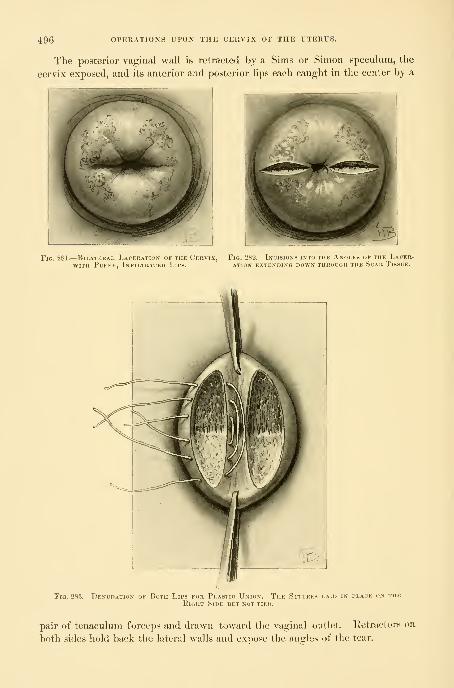
496 OPERATIONS UPON THE CERVIX OF THE UTERUS.
The posterior vaginal wall is retracted by a Sims or Simon speculum, the
cervix exposed, and its anterior and posterior lips each caught in the center by a
Fig. 281.— Bn.ATEitAi, 1..\ierati"\ ..f thk (.'krvix, Fio. 'JM', I\. i-k.ns into the Angles of the Lacer-WITH PUFEY, InF[LTKATEU LiPS. ATION EXTENDING DOWN THROUGH THE ScAK TiSSUE.
Fig. 283.— Denudation of Both Lips for Plastic Union. The Sutures laid in place on theEight Side but not tied.
pair of tenaculum forceps and drawn toward the vaginal outlet, lietractors on
both sides hold back the lateral walls and expose the angles of the tear.

KEPAIR OF THE LACERATED CERVIX. 497
First, the denudation of the hp
into one of the
sound tissue
the (lenu-
area of
ante-
There are two steps in the operation
second, the approximation by suture.
Denudation .—The accumulation of scar tissue in the angles of the rentbetween the lips must always be removed; serious disturbances have arisenfrom forcibly uniting ^.^-^ *^® lips over such a rigid fibrous plug. Tomake sure of this,
^ .
'' ^. I commence the denudation by an incision
^ J'^ angles entirely through the scar until the
:* below is reached. This limits the depth of<lati()n in the angles of the tear. I next outline the
denudation with a sharp knife, by deep incisions on l)oth
rior and posterior lips.
The outline extends fromeach end of the incision in
the angle out to the end of
the hps. If the tear is bilateral
a similar incision is made in the
angle of the opposite side, and the
area to be removed is similarly out-
lined on the lips of that side. Astrip of mucosa \ centimeter wide
must 1)6 left between the lines of
incision for the cervical canal. Theoutlined area is now denuded by
catching the tongue of tissue be-
tween the incisions with rat-toothed forceps and removing it completely
with knife or scissors. A serious and not uncommon error is to denude
by shaving off the surface of the cervix on its vaginal side. The lips
of a cervix so held together present a good external appearance, but a
sound introduced within the canal demonstrates at once that the appar-
ently normal cervix is but a jjouch with a thin septum of vaginal tissue
on each side, and the cervical tear is no more repaired than it would be
if a suture were simply passed through the ends of both lips and drawn
up and tied.
The fear of wounding a circular artery in denuding the cer-
vix is groundless. Any vessel which may Ije cut will readily be
controlled by bringing the lips firmly together.
Sutures.— The proper sutures are silkworm gut or catgut.
The sutures are introduced by means of a stout medium-sized
needle and a carrier. As a rule, two or three silkworm - gut
sutures on each side are enough ; fine superficial catgut sutures
are used between them for accurate union.
The first silkworm-gut suture is introduced up at the angle,
entering upon the vaginal surface and coming out on the uterine
surface of one lip, and reentering on the uterine surface and coming out at the
corresponding point of the opposite lip. All the sutures are best introduced
Fia. 284.
—
The Ceuvix after all theSutures are tied on Both Sides.
Fig. 285.—GlassIrrigator FORWASHING OUTTHE VaginaAND THE UtE-RCS.
The irrigator
is sterilized byboiling and keptstanding in a car-bolic-acid solu-
tion. 1^ ordina-ry size.

498 OPERATIONS UPON THE CERVIX OF THE UTERUS.
first Oil botli sides, and then tied successively from above downward. In case
tlie vaginal outlet is operated upon at the same time, the assistant will find it
easier to locate the loop for removal of the sutures if tliej are clamped with
perforated shot instead of being tied. Fine superficial catgut sutures are used
to secure accurate approximation between the silkworm-gut sutures. All the
silkworm-gut sutures are now cut about 2^ centimeters (1 inch) long, to facili-
tate their removal later.
A loose gauze pack is placed in tlie vagina to absorb the discharges for the
first two or three days, after which it is removed and the outlet simply protected
by boric-acid powder and a vulvar pad. If there is any discharge after this the
vagina may be douched out daily witli a weak menthol, soda, and borax solution.
It will not l)e necessary to catheterize as a rule.
Where no operation has been performed at the vaginal outlet, the cervical
sutures may be removed in ten days or two weeks. When the outlet has been
repaired, the cervical sutures need not be touched for four weeks or longer.
The sutures are most readily exposed and removed with the patient in the knee-
breast or in the left lateral posture.

CHAPTEE XV.
PROLAPSE OF THE UTERUS.
1. Definition.
2. Forms of prolapse : a. Entire uterus. S. Cervix, c. Vesical diverticulum. (?. Prolapse withoutvesical diverticulum, e. Rectal diverticulum. /. Enterocele. g. Prolapse with completetear.
3. Accurate description of cases necessary.
4. Causes of prolapse : a. Congenital, b'. Strain, c. Childbirth.5. Symptoms and complications.6. Operative treatment : a. Simple prolapse : 1. Supravaginal amputation of cervix. 2. Resec-
tion of lax outlet. 3. Anterior colporrhaphy. 4. Suspension of the uterus. J. Comphcatedprolapse : 1. Complete tear of septum. 2. Prolapse of rectum.
7. After-treatment.
Definition .—Prolapse of the uterus and falling of the womb are terms
applied to a hernia, sometimes appropriately called " sacro-pubic hernia," occur-
ring at the vaginal outlet, in which the uterus lies within the hernial sac.
Although the term " falling of the womb " is sanctioned bj long usage, it
is seriously misleading, inasmuch as it implies nothing more than a simple dis-
placement of the uterus, which in fact never occurs alone, but is always associ-
ated with eversion of other important structures, usually the vaginal walls and a
part of the bladder, these organs hanging together out of the vulvar cleft below
the pubic arch, like a large mucous pouch.
An illustration of an extreme form of prolapse, representing the most ad-
vanced degree attainable, is furnished by one of my patients, a woman twenty-
two years old, with complete eversion of both vaginal walls, and complete pro-
lapse of the I'etroflexed uterus as well. The sketch shows the relations of the
sac as viewed from the side.
Forms of Prolapse .—Under the comprehensive title " prolapse " are
gathered a variety of interesting forms which may, however, be arranged under
two cardinal divisions—prolapse of the entire uterus, and prolapse of the cervix
only.
In the first case the uterus descends as a whole, the fundus sinking pari
passu with the cervix, following it out as it passes beyond the vaginal outlet ; in
its descent the uterus occupies an infinite number of positions between the nor-
mal anteflexion, and a condition of complete extrusion.In the second case the fundus of the uterus descends but slightly in the pel-
vis, while the cervix, advancing more rapidly, escapes at the outlet, with the
everting vaginal walls ; this form of prolapse involves only the lower extremity
of the uterus and is therefore incomplete.

500 PROLAPSE OF THE UTERUS.
A good illustration of the elongation of the cervix just above
the va^nal vault is afforded by the relations of the parts m one of my pat.ents.
Fm. 2Si'>.—Complete Fuolapsu
The piitient, a ncgress, is soon
IE Utekus and Vaoina, forming
Khodei) in Dark Areas.
Ill beliiiKl, in onlcr to expose the sae better.
A Large Micois Poicii
Here there was a little atrophic cervix at the end of a prolapsus haiiging Y
timeters (3 inches) below the vulva. The sac was but 10 centimeters (4 inches)

FOKMS OF PROLAPSE. 501
in circumference. The length of the uterine canal was 11 centimeters (4^inches), and the body of the uterus lay entirely within the pelvis. A divertic-
ulum of the bladder entered 3^ centimeters m inch) into the sac, while the
rest of the bladder extended 9 centimeters (S^ inches) up into the pelvis. Onreturning the sac, nothing was apparent but a relaxed outlet and a cystocele of
moderate size.
This remarkable displacement owes its occurrence to a ductile condition of
the supravaginal portion of the cervix, where it joins the uterine body, allow-
ing it to be drawn out from 3 to 6 centimeters (1 to ^ inches) longer thannormal.
..%̂^
Fig. 287.—Complete Prolapse of the Vagina and Uterus, with Eetroflexion.
Note the narrow neck at tlie junction witli the body and the prominence posteriorly. M. K., 653.
Variations of these two cardinal divisions of prolapse are formed by the
presence of a longer or shorter bladder diverticulum, or even by the absence of
any portion of the bladder within the sac.
A rectal diverticulum may be found in the prolapsed posterior vagi-
nal wall, but is one of the rarer complications. The presence of small intestines
in the sac in front of or behind the uterus (anterior or posterior e n -
t e r o c e 1 e) is an unusual complication ; it is most rarely found in front, as this
space is usually filled by the bladder.
Prolapse of the uterus with complete rupture of the recto-vaginalseptum is also rare. The rarest of all forms is that of complete prolapse with
rupture of the septum and prolapse of tlie rectum.
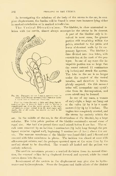
502 PROLAPSE OF THE UTERUS.
In investigating the relations of the body of the uterus to the sac, in com-
plete displacement, the fundus will be found in some rare instances lying either
in marked anteflexion or in marked retroflexion.
The Vesical Diverticulum .—The bladder, in close anatomical re-
lation with the cervix, almost always accompanies the uterus in its descent.
A part of the bladder only is in-
volved in most cases, the greater
portion still remaining within the
pelvis, attached to the pubis and
lower abdominal walls by its sus-
pensory ligament. The bladder is
thus divided into two lobes, with
constriction at the neck of the pro-
lapse. In one of my cases the in-
trapelvic portion was so large that
the sound entered 11 centimeters
(4^ inches) and struck the sacrum.
The lobe in the sac is no longer
under the control of the vesical
muscles, and therefore is incom-
pletely emptied. On this account
urine will accumulate and cystitis
arise from its decomposition, and
even calculi may be formed.
In one of my cases, a womanof sixty-eight, a large sac hung out
at the vulva 12 by 9 by 8 centi-
meters (5 by 3|- by 3 inches). Thevagina was completely everted, and
the uterus lay entirely within the
sac. In the middle of the sac, in the diverticulum of the bladder, lay a large
calculus. The intra-pelvic portion of the bladder contained a second stone of
equal size. She had also passed three small calculi before I saw her. The cal-
culi were removed by an incision 4 centimeters (1^ inch) long through the pro-
lapsed anterior vaginal wall, beginning 3 centimeters (1 inch) above the cer-
vix. The mucous membrane of the bladder was found thick and inflamed and
covered with false membrane in places. The incision was closed at once with
silkworm-gut sutures, and the prolapse operated upon at the same time by the
method about to be described. The wounds all healed and the patient was
entirely relieved.
The urethra sometimes presents a marked deviation from its normal direc-
tion, the external oriflce being displaced forward and upward, while its canal
curves down into the sac.
Involvement of the ureters in the displacement may give rise to hydro-
ureter and hydronephrosis. From the frequent and futile eiforts of the bladder
Fig. 288.
—
Prolapse of the Utehus, showing the In-
termediate Stages between the Uterus in Ante-flexion AND IN Complete Prolapse.
First the uterus descends a little and drops into re-
troflexion ; then it descends farther and the flexion is
straightened out ; then the cervix appears at the vagi-
nal outlet, to escape beyond it in the next stage ; and,
finally, the whole uterus lies outside enclosed in thevaginal sac.

PROLAPSE WITHOUT VESICAL DIVERTICULUM, 503
to expel the residual urine, its walls may become enormously hypertropliied; in
other cases they may stretch and become thin.
Prolapsewithout Vesical Diverticulum .—Although rare, pro-
lapse without vesical diverticulum is occasionally found, the bladder remainingentirely within the pelvis, l)eing separated from its uterine connections. An
Fig. 2s'J.
—
l'\i;ii\i, rj:<>i.APf*rs ; Kversion of the Anterior, Posterior, am> Latkhal \ ai.i_\ai. Walls.
The vaginal canal is seen in the middle, and the cervix is still invisible within the pelvis.
interesting case of this character entered my clinic in April, 1891 {see JohnsHopkins Hospital Report in Gynecology^ vol. iii, p. 311). The accompanyingfigure represents the appearance at the time of the first operation. The patient
(M K., 653, April 4, 1891) was thirty-two years old, of slight build, weighing less

504 PROLAPSE OF THE UTERUS.
than one Imndred pounds, married ten years, and the mother of three children
—nine, seven, and three and a half years old. Both cervix and perineum were
torn in an unassisted first labor, and a protrusion at the vulva was noticed then.
Fig. 290.
—
Complete 1'rolapse of the Vagina and Uteiuis, with Retroflexion of the Pkolapsei)Uterus, as is evident from the Domelike Prominence on the Under Surface of the Sac.
Tlie lacerated everted cervix and areas of ulceration are plainly seen.
which increased after the birth of a second large child ; after this she suffered
from excessive constipation, frequent micturition, and dragging pains, and the
effect of the third labor was a prolapse which hung 10 centimeters (4- inches)
below the vulva.
I found at my first examination a large sac between the thighs dependent
from the vulvar orifice, and the anterior wall of the vagina everted from cervix
to urethral orifice, the vagina posteriorly, on the contrary, presenting a depth of
about Y centimeters (8 inches) within the pelvis. The uterine cavity measured
7 centimeters (3 inches) in length. The body of the uterus was still within the
pelvis in a direct line with the axis of the sac. There were no apparent elonga-

PROLAPSE WITHOUT VESICAL DIVERTICULUM. 505
tion or thinning of the supravaginal cervix felt through the sac wall. The an-
terior and posterior part of the prolapse were distended with soft, irregular
masses, gurgling on pressure, and tympanitic on percussion. These masses were
easily reducible and were evidently coils of intestines. The urethra lay just
beneath the pubic arch. Upon introducing a sound within the bladder, it en-
tered the pelvis 8 centimeters (3 inches) in the median line, and 9 centimeters
(3f inches) on either side, but no part of the bladder entered into the sac.
I should explain the absence of vesical diverticulum in this way. The ten-
dency of the bladder when markedly distended is to assume the spherical or
ovoid form, which accommodates the largest amount of fluid in the smallest space.
In a prolapse, in the process of formation, the upper lobe of the bladder in ex-
panding constantly exerts traction upon the lower lobe and tends to draw it up
out of the sac into the pelvis, by wdiich means the cellular attachments between
iTii Eloxgate Lacekatei) Cervix.
The sound is introduced into the bladder to show the altered direction of the urethra and the vesical
diverticulum in the sac. The light spot plainly shows the position of the end of the sound in the bladder.
uterus and bladder are stretched and yield more and more, until the separation
is complete and the whole bladder comes to lie free in the pelvis. This sepa-
ration of the uterus from the bladder may go on progressively with the descent
3G

506 PROLAPSE OF THE UTERUS,
until the prolapse is complete. With the bladder thus lying in the pelvic
cavity, while the uterus is prolapsed, the vesico-uterine has become transformed
into a' utero-vaginal pouch in front, analogous to the recto-uterine one behind.
In but one case have I seen the bladder lying entirely within the prolapse,
without any portion in the pelvis or attached to the symphysis. At a subse-
quent examination, whenthe bladder contained more
urine, it was found ex-
tending back toward the
sacral hollow.
In addition to the form
of enterocele thus de-
scribed, another form in
which the intestines crowd
into the sac posterior to
the uterus is more fre-
quently found.
A rectal diverticulum
is rarely found in the pro-
lapse, although it may hap-
pen, and a considerable
fecal stasis be discovered
at this point. The rela-
tion of the anterior wall of
the rectum to the sac maybe readily ascertained by
introducing the finger within the bowel. These various possible complications
must all be noted before operation on account of the danger of opening the
peritoneum or the bowel.
Complete tear of the recto-vaginal septum is not often found with prolapse;
this is due to the fact that the direction of the tear for the most part is central,
and so does not involve the levator ani muscle to any great extent, leaving
the outlet well supported. In a small percentage of cases, however, the associa-
tion is observed.
An unusual complication is prolapse associated with v e s i c o - c e r \- i c o -
vaginal fistula. I operated on such a case April 9, 1892. The patient
(K. W., 1320) was fifty-three years old, and had a large prolapsus sac with an
irregularly torn posterior cervical lip ; the anterior lip was gone, and in the
midst of a mass of scar tissue in its place was a fistula 2 millimeters in diameter.
The cervix was elongated, the fundus uteri remaining in place in the pelvis.
She had been operated upon unsuccessfully twenty-one years before by Prof.
Nathan Smith, of Baltimore, and had become a confirmed morphine eater. I
made the classical oval denudation on the vaginal mucosa 3 centimeters (1 inch)
long, funnel-shaped, with the bladder muc(»sa at the apex, and united the edges
of the wound from side to side with interrupted silkworm-gut sutures. The
Fig. 292.
—
Pautial Prolapse of the Uterus, with Partial Ever-sion OF the Posterior Vaginal Wall and Complete Ever-sioN of the Anterior Vaginal Wall.
The cervix is elongate. The point of special importance is thecomplete detachment of the bladder from its uterine and vaginal at-
tachments, with the presence of the intestines in the sac bothi anteriorand posterior to the uterus (vaginal enterocele).

CAUSES OF PROLAPSE. 507
whole operation was peculiarly easy with the parts Ijino; displaced exteriorly.
She was carefully watched during her convalescence, and l)Oth the morphine
habit broken and the tistula cured.
One of the rarest complications is that presented by a fifty-year-old patient,
who had a prolapse with hypertrophic elongation of an infiltrated lacerated
cervix, and just below the cervico-uterine junction a complete atresia of
the uterine canal.
Prolapse cases differ so widely in their individual features that all operators
should make note of the following characteristics
:
Dimensions of the prolapsed sac.
Appearance and position of the cervix.
Presence of ulcerated areas.
Complete or incomplete eversion of both anterior and posterior vaginal walls.
Length of uterine canal, noting whether the cervical portion is drawn out.
Exact position of the fundus uteri—in the pelvis or in the sac.
Relations of the bladder to the sac.
Relation of the rectum to the sac.
Position of the peritoneal pouch, posterior to the sac.
Presence of intestines in the sac.
Appearance of the outlet when the sac is returned.
Pelvic measurements, to explain if possible the cause of a difficult labor.
Causes of Prolapse .—Congenital defects in the vaginal outlet and
pelvic floor may supply the factors necessary for the formation of a prolapse
which may be found at birth. Protrusion of the pelvic viscera has been ob-
served from the strain of a fall.
But the conditions essential to the production of a prolapse are most fre-
quently found after nuiltiple pregnancy. The large, heavy uterus following a
puerperal infection, l)y its weight alone predisposes to prolapse where the woman
has gone to work too soon. The direct causal relationship between labor and
prolapse is shown by thirty-five of my cases in which there was but one who
had had no pregnancy, and here the prolapse was but partial.
Twenty-seven women of whom I have accurate notes had had an average of
3-8 children. Nine of these women had borne children after the appearance of
the prolapse.
A tight obstetric binder, by throwing the uterus into retroposition, also
favors prolapse. If the binder is used, it must under no circumtances be applied
tightly within the first ten days after labor. To aid the uterus in regaining its
normal size the patient should remain in bed for two weeks, during which time
the physician should from time to time assure himself of its position by palpa-
tion through the abdominal walls, drawing the fundus forward if he finds a
tendency toward retroflexion. It is best for her not to He much in the dorsal
position, but to turn in bed as often as she wishes, assuming any comfortable
posture. Cases of prolapse will also be avoided if retroflexion and relaxed out-
let receive timelv treatment.

508 PROLAPSE OF THE UTERUS.
Prolapse owes its origin therefore to an insufficiency of the intrapelvic
uterine supports associated with a weakness of the pelvic floor.
A tight, well-closed vaginal outlet, depending upon the integrity of the an-
terior part of the levator ani muscle, is the most important factor in retaining
the uterus within the pelvis. This muscle controls the outlet and prevents pro-
lapse in three ways : (1) It retains the normal outlet in its position forward under
the pubic arch, out of the line of abdominal pressure; (2) it gives to the outlet
the size and form of a narrow slit, preventing the protrusion of the pelvic vis-
cera; (8) it directs the axis of the vaginal canal forward instead of directly
downward, so that the intra-abdominal pressure strikes the pelvic floor at a
right angle.If these functions of the levator ani are impaired or destroyed by extreme
dilatation or laceration of its fibers, the vaginal outlet is no longer supported,
but drops open and falls back toward the sacrum, and the canal assumes a
direction more or less in a direct line with the abdominal pressure, the first
effects of which are to crowd the adjacent anterior and posterior vaginal walls
down into the outlet, still further distending it.
If the body of the uterus is retained within the pelvis by its broad ligament
attachments, as the cervix descends, the portion between the cervix and the
body becomes drawn out and thin, a condition readily diagnosed by squeezing
the sac between the fingers, when the upper cervix is felt in the middle like a
long, thin cord.
The essential intrapelvic supports of the uterus are those which tend to keep
its upper pole (the fundus) in front, and its lower pole (the cervix) in the back
part of the pelvis. The attachment of the vesico-uterine peritoneal folds high up
on the anterior face of the uterus serves to hold the fundus behind the symphysis,
while the utero-sacral muscles at the opposite pole serve to hold the cervix back.
So long as these supporting structures remain intact, displacement can not occur.
If, however, the utero-sacral folds relax, the cervix drops away fi-om the sacrum
in the only possible direction, down the vagina. The body of the uterus then
remains no longer cushioned upon the upper surface of the bladder, but at once
begins to hang as a dead weight, and is forced step by step down upon the pel-
vic floor by the force of gravity, combined with the intra-abdominal pressure.
In the descent the cervix is involved first and the fundus next, and it is only a
question of time when the prolapse will be complete.
Retroflexion of the uterus is often but an initial step in the formation of
prolapse, which will occur wlien the retroflexion is associated with a relaxation
at the vaginal outlet.
Symptoms and Complications.— I find that in ?>r> of my eases
the average age was forty-two years, and that only 4 were below thirty, 2 being
but nineteen years old ; 10 were between thirty and forty, 8 between fifty and
sixty, and 13 between forty and fifty.
The most distressing symptoms of prolapse are backache and a dragging
sensation in the pelvis and lower abdomen, i^roducing a general feeling of weak-
ness ; locomotion is often painful—as one patient expressed it, she always '" felt

AMPUTATIOX OF THE CERVIX. 509
as if she could go no farther." Sitting is sometimes painful. Frequent urina-
tion is common ; the bowels are often constipated and the appetite poor.
From stagnation of urine in the vesical diverticulum intense cystitis is some-
times found. The bladder walls first become thickened and the ureters are
compressed. The infection often travels up to the pelvis of the kidnevproducing pjelonephrosis and death. In rare cases calculi are found in the
pouch.
In one of my cases there was a complete prolapse with retroflexion and a
myoma on the posterior wall of the fundus. This was about 3 centimeters in
diameter and made a distinct elevation on the everted vaginal surface.
The vaginal mucous membrane covering the sac loses its rugose appearance,
becomes smooth, hypertrophied, and callous. Kubbing on the thighs and gar-
ments often causes ulcers with deep, sharply defined l)orders.
Operative Treatment.—The normal supports of the uterus and
vaginal outlet can never be perfectly restored. Therefore that operation is
best which offers the most efiicient substitute. I prefer the following proced-
ure : A resection of the relaxed vaginal outlet, restoring its caliber, changing
the direction of its axis, and changing its position, associated with a supra-
vaginal amputation of the cervix ; in bad cases the abdomen is opened and the
uterus suspended to the anterior abdominal wall.
The resection of the vaginal outlet alone is not sutficient when there is a
ductile cervix, which will afterward worm its way out of the smallest canal
;
amputation of the cervix therefore deprives the uterus of its leader, as it were,
and is always necessary except when it is unusually small or senile.
Hysterectomy is not necessary to cure prolapse, and if the operations upon
the outlet and cer\dx are skillfully j)erformed, it will not often be necessar}^ to
suture the uterus to the anterior abdominal wall. "Wliere there is extreme re-
laxation, and the outlet has not been satisfactorily lifted up by the resection,
a suspension of the uterus to the anterior abdominal wall is then advisable (see
Chapter XXY). This, however, will never be sufiicient by itself.
Amputation of the Cervix .—To amputate the cervix both anterior
and posterior lips are caught with tenaculum forceps and pulled well out of the
body, or instead of tenaculum forceps, two long, stout silk sutures may be passed
through both lips of the cervix and used as tractors. A circular incision is madeimmediately above the cer\dx through the vaginal wall and the uterus pulled
downward, while the vaginal vault is stripped off with the thumb and first and
second fingers pressing against the cervix, and rubbing the vagina up in front
and behind. The separation is always incomplete at the sides where the vessels
enter the uterus. Injury to the bladder will be avoided by directing the force
of the separation movement toward the cervix, and by occasionally inserting a
sound into the bladder in case of doubt as to its exact i*elations.
The amount of c e r v i x bared is 5 to 6 centimeters (2 to 2^ inches). Theuterine vessels on each side of the cervix are now tied as high up as possible by
a catgut ligature passed close to the side of the cervix. This materially lessens
the hemorrhage in the subsequent steps. It is not necessary to expose the

510 PROLAPSE OF THE UTERUS.
peritoneum either in front or l)eliind, altbougli no harm will be done if it is
opened.
The c e r V i X is now amputated; it is first split from its external orilice
to the upper limit of the denudation, after which the lips are drawn apart and a
stout curved needle, carrying a catgut suture, is entered at the anterior vaginal
?^tP\ "v;,i
\\\%\\yv\V
Fig. 293.
—
Pautial Pkolapse of the Uterus, with Elongate Hypertrophied Cervix.
The cervix is drawn down and held for tlie circular amputation, the first step in the operation.
wall close to the incision, carried under any oozing points in the loose cellular
tissue in its track, and made to emerge on the mucosa in the cervical canal -t or 5
millimeters below the angle of the slit. A similar suture is passed through the
vaginal wall and the posterior cervical lip. After this several other sutures are
passed on either side of these, and the cervical lips are then amputated in such
a manner as to leave the canal the most prominent portion on the stump. The
vaginal mucosa is then drawn in to the cervical mucosa by means of these sutures,
which are now tied.
The elliptical openings in the vaginal vault to the right and left of the cer-
vical canal are closed by three or four catgut sutures, bringing vagiiuil nnicosa to
vaginal nmcosa, and passing under the deep parts of the wound, so as to include
all bleeding vessels in their embrace. These sutures nuist be so applied as not

ANTERIOR COLPORRHAPHY. 511
to leave any pockets beneath the surface, as Ijlood is certain to accumulate anddistend such spaces, causing infection, which will work its waj to the surface
at a later date in the form of an abscess.
The only large abscess I have ever seen in the cellular tissue anterior to the
uterus was in a patient who had been operated on for prolapse at another clinic,
and came to mine with high fever and severe pain, when I discovered andopened an abscess between the uterus and the bladder containing about 150 cubic
centimeters (5 ounces) of pus.
Accurate superficial approximation of the wound at the vault is secured byline catgut sutures between the deep ones.
This first step in the operation does not occupy longer than ten or fifteen
minutes.
Kesection of the Vaginal Outlet .—The resection of the vaginal
outlet in these cases is in general similar to that described in Chapter X. Theonly important difference lies in the greater length and breadth of the triangles
of denudation extendmg up into the vaginal sulci, thus resecting a larger area of
the vagina. It is both more ditficult and awkward to outhne the area to be
exsected within the vagina, on account of the laxity of the tissues and the fact
that the well-defined posterior column has disappeared, and in its place is a
thick, wrinkled, redundant mass. The
line between anterior and lateral walls is
fortunately distinct, affording a guide
for the outer border of each triangle,
just under and parallel to it. The un-
denuded tongue in the middle, which
is left to form the floor of the newvagina, must be made narrow as well
as long.
Anterior C o 1 p o r r h a p h y .
—
While in a large percentage of cases
the anterior vaginal wall is well sup-
ported by the resection of the posterior
vaginal wall, occasionally the " cysto-
cele," or prolapsed bladder, persists in
pouting out at the vaginal orifice, which
in course of time it dilates, and so de-
stroys the effect of the operation.
An anterior colporrhaphy for the
relief of the cystocele is indicated only
in cases of extreme relaxation.
The essential step in the opera-tion for cystocele is the re-
moval of an oval piece of tissue large
support the base of the bladder without encroaching upon the proposed field
of operation upon the posterior wall.
Fig. i'J-i.—Operation for Prolapse of the Ute-rus BY Amputation
AmputatJou of the cervix at 1, oval resection ofthe anterior vaginal wall at 2, restoration of the vagi-nal outlet at .3, and suspension of the uterus at 4.
enouffh to reduce the hernia and to

»12 PROLAPSE OF THE UTEIUS.
The operation for cystocele slioiild follow immediately upon the amputation
of the cervix, and so forms tlie second step in tlie train of three operations—namely, amputation of the cervix and closure of the vaginal vault, resection of
the anterior vaginal wall for cystocele, and resection of the relaxed vaginal
outlet.
The cervical and the urethral extremities of the cystocele are grasped with
tenaculum forceps, pulling in opposite directions and drawTi down into the
vaginal outlet, while the lateral walls of the vagina are held away by flat
retractors.
An oval incision, 4 to 6 centimeters long and 2^ to 3 centimeters broad,
through the entire thickness of the vaginal mucosa, outlines the area to be de-
nuded. If the denudation is made too broad the suturing of the posterior
vaginal wall in the next step w^ill be difficult.
The separation of the flap which has been outlined may be effected by loos-
ening one of its ends and then completing the detachment by a blunt dissection
Flu. 2'j5.— PiioLAi'SE OF THE Utkki;s, V.vdiNA, AND Keottm,Eecto-vaoinal Septum. M. \V., Phi
ri-UE OF THE
with the fingers ; constant care nnist be taken not to tear the bladder wall which
is exposed. The wound made in this way does not usually l)leed much, and any
hemorrhage may be controlled by the interrupted silkworm-gut or catgut sutures,
introduced from side to side across the axis of the oval denudation, about a centi-

SUSPENSION OF THE UTERUS. 513
meter apart. The sutures iinist not penetrate the bladder or catch the ureters.
On tying the sutures only a linear wound is left. They may he removed in
about two weeks.
Suspension of the Uterus .—In those cases in which the vagina
has been completely everted the lax outlet is resected with difficulty, and there
still exists a marked tendency of the uterus and the upper part of the vagina to
bear down upon the repaired outlet. A decided mechanical advantage will be
secured by making a small abdominal incision just above the symphysis pubis,
and attaching the posterior surface of the uterus to the anterior abdominal wall
by three permanent sutures, in the manner descril)ed in Chapter XXIV. Themanifest advantages of this step have been insisted upon first by Dr. G. M. Ede-
bohls and then O. Kiistner.
Prolapse of the Uterus with Complete Rupture of theR e c t o - V a g i n a 1 Septum .—Where the laceration is comjjlete, tear of the
recto-vaginal septum deviates markedly to one side or the othei-, the lateral
fibers of the levator ani muscle may be ruptured as well, and prolapse
occur. If the tear has extended above the sphincter area, there may be also a
prolapse of the rectum. The treatment of this complex condition is similar to
that just described, with the exception of the restoration of a ruptured instead
of a relaxed vaginal septum in the manner described in Chapter X.
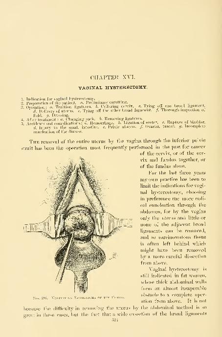
CHAPTER XVI.
VAGINAL HYSTERECTOMY.
Indication for vaginal hysterectomy.
Preparation of the patient, a. Preliminary curetting., ^ ,.
Operation : a. Traction ligatures, b. Collaring cervix, c. Tying oif one broad ligament.
d. Delivery of uterus, e. Tying off the other broad ligament. /. Thorough inspection of
field, g. Dressing.
After-treatment :«. Changing pack. 6. Removing hgatures.» . , , i
Accidents and complications : a. Hemorrhage, h. Ligation of ureter, c. Rupture of bladder.
d. Injury to the small intestine, e. Pelvic abscess. /. Ovarian tumor, g. Incomplete
enucleation of the disease.
The removal of the entire uterus by the vagina through the inferior pelvic
strait has been the operation most frequently performed in the past for cancer
of the cervix, or of the cer-
vix and fundus together, or
of the fundus alone.
For the last three years
my own practice has been to
limit the indications for vagi-
nal hysterectomy, choosing
in preference the more radi-.
cal enucleation through the
abdomen, for by the vagina
only the uterus and little or
none of the adjacent broad
ligaments can be removed,
and so carcinomatous tissue
is often left behind which
might have been removed
by a more careful dissection
from al)ove.
A^aginal hysterectomy is
still indicated in fat women,
whose thick abdominal walls
form an almost insuperable
obstacle to a comj^lete oper-
ation from above. It is not
because the difficulty in removing the uterus by the abdominal method is so
great in these cases, but the fact that a wide exsection of the broad ligaments
514
Vui. 200. VKtt?;TATIN

PREPAllATION OF THE PATIEXT. 515
and removal of the pelvic glands is almost impossible, on account of the mechan-
ical hindrances offered hy tlie thick walls and the deep pelvis.
Fig. 297.
—
Epithelioma ok tiik ('lii\i\ without Vegetation.
The vaginal vault is converted into a flat granulating .surface. The cervical opening i.s seen in the center.
Preparation of the Patient .—The patient must be duly prepared
by rest, baths, vaginal douches, and above all by a thorough evacuation of the
bowels.
If the cancerous disease has advanced to the sloughing stage, or so far as to
cause any odorous discharges, or to form a mass protruding into the upper
vagina, the danger of sepsis will be greatly increased unless the field is first

516 VAOINAL HYSTERECTOMY.
cleansed by a thorough curettage, removing as much of the diseased tissue as
can be scraped away with a sharp curette (see Chapter XIV). If the vagina is
packed every two days after doing this, in a week or ten days the patient will
be ready for the radical operation. In an urgent case the curettage may be
done just before the uterus is removed.
Operation.—The patient is brought to the edge of the table hi the
lithotomy position, with the limbs well flexed and the buttocks resting on the
perineal pad. The assistant then shaves the external genitals, washing them
well, together with the vagina, and curettes away all redundant cancerous
masses with fingers and scoop unless this has been already done.
The posterior vaginal wall is then retracted with a large Sims or Simon
speculum, exposing the vault of the vagina and the cervix. If the vaginal out-
let is narrow, hindering a view of the cervix, a greater degree of dilatation may
The uterus and ecrvix curetted thoroughly and the cervix sewed up with
traction. The field of operation exposed by retractors.
be secured by boring in the hand with the tips of the fingers held together,
making a conical dilator, or, better still, by using a conical rectal dilator,
centimeters in diameter at the base. If the rigidity can not be overcome in this
way, one or two deep lateral incisions through the posterior connnissure and

OPERATION. 517
extendin^^ around one or hotli sides of the rectinn and np into the vaginal sulci,
will give the necessary enlargement. The hemorrhage from the surfaces thus
incised is rarely great enough to call for the use of forceps or ligatures.
I'll,. li'J'J.
—
Vaginal IIysteiiectojiv.
Beginning the operation by cutting the cervix loose from tlie vaginal vault, under continuous irrigation.
Continuous irrigation is used to keep the tield clear of blood during the lirst
part of the operation, until the peritoneum is opened.
The vault of the vagina and the cervix being exposed as descril)ed, the an-
terior lip of the cervix is caught by tenaculum forceps and drawn down ; the
posterior lip is also caught, and a stout needle is passed through the anterior and
]iosterior lips, carrying a heavy silk suture about 40 centimeters (16 inches) long.
Thi-ee or four of these ligatures are passed, and each one tied tightly, drawing
the lips firmly together and completely closing the cervical canal and covering
in the diseased area, to prevent the escape of any of the intra-uterine contents
over tlie wound area during the enucleation. The ligatures ai*e left long to
serve as tractors, and enable the operator to hold the uterus down near the
outlet, and to keep pulling it farther and farther down, delivering it gradually
as its attachments are severed.
The cervix is first drawn down toward the vaginal outlet as far as it will

518 VAGINAL HYSTERECTOMY,
come, often outside, and an assistant on either side holds back the lateral vaginal
walls with a retractor so as to prevent them from hiding the field of operation.
The operator, pulling on the traction ligatures, now makes a circular incision
around the cervix and through the entire thickness of the vaginal vault, not less
than 2 centimeters (| inch) distant from the margin of the disease. The Paque-
lin or galvano-cautery may be employed in this stage of the operation, instead
of a knife, to check the oozing. Moderate bleeding from its margins may be
disregarded until the close of the operation. More profuse bleeding should be
controlled at once by ligatures passed through the vaginal walls, so as to grasp
the vessels.
The knife or scissors are now laid aside, and the operator begins to push up
and peel back the cellular tissue from its cervical attachments in front and be-
Tlie linger is engaged in pushing up tlie bladder, detaching it from tiie eervix.
hind the cervix with index and middle finger. There is usually no resistance
and rarely any serious hemorrhage, for the important blood vessels lie in the
broad ligaments at the sides. Care must be taken in pushing back the tissues
and freeing the cervix to keep the ball of the finger always directed toward the
cervix. If the separation is carelessly performed and this precaution neglected.

OPEKATIOX. 519
there is danger of perfoi'ating tlie bladder or unexpectedly entering the peri-
toneum, especially if the disease has extended in either of these directions. Assoon as the peritoneum behind the uterus is reached, the
fact is readily recognized by the fluctuation of a little fluid
in Douglas's cul-de-sac, or by the smooth anterior and poste-
rior surfaces gliding over each other. It is opened by catch-
ing a fold of it with forceps and making a small cut into it
with scissors; one index finger is then thrust in, enlarging
the opening, and then the other index finger is introduced,
tearing the incision as wide as possible from side to side, well
out to the bases of the broad ligaments.
As soon as the peritoneum is laid open the irrigation
must cease. K sterilized sponge, or pledget of gauze with
string attached, is now pushed into Douglas's cul-de-sac to
prevent the entrance of fluids or the escape of dehris from the "^ x^'i°^;T'^!!?i^'^'T^'*'^-1- -L lir-JbDLr., >\ ITH J^EFT
field of operation up into the peritoneum. A pair of artery Cueve for passing^ ^
1 .. 1 . 1 .LlGATlRES THROUGH
forceps, clamped at the end of the string, distinguishes it at the Broad^ Liga-
onee from the ligatures applied to the broad ligaments. liYSTERECTOMY.
The anterior vesico-uterine fold of peritoneum is next
reached in like manner after completely detaching the bladder from its uterine
connections. It is also recognized by the gliding of its peritoneal surfaces over
each other. An opening is made by pushing in a pair of sharp-pointed scissors
under the guidance of the index finger, spreading the handles and withdrawing
them. The index fingers are then introduced as just described and the hole
enlarged out to the broad ligaments on either side. This leaves the uterus hang-
ing in the pelvis attached by the broad ligaments alone.
The anterior peritoneal fold may sometimes be more readily reached after
the lower parts of the broad ligaments have been ligated and severed from the
cervix, permitting a greater downward displacement of the uterus, and making
this part of the peritoneum more accessible.
The accident of pushing the finger through the bladder will be avoided by
frequently introducing a sound into the bladder as the operation progresses, to
determine its exact position, and the thickness of the intervening wall. An acci-
dental rent in the bladder at once shows itself by a sudden gush of urine into
the vagina. This is most apt to occur wdien the disease has progressed through
the cervix into the bladder wall. A fistula made in this way should be closed,
after paring the edges to remove the disease, with interrupted silk sutures, when
the enucleation of the uterus is completed. If this operation is performed im-
mediately, it is rarely unsuccessful.
The next step in the enucleation is to tie off the broad ligaments with stout
silk ligatures, introduced by a strongly curved blunt aneurismal needle. In
introducing the first ligature—say on the right side—the cervix is drawn strongly
to the left, and the right vaginal wall held out of the way by a retractor. The
left index finger is placed beside the cervix, behind the broad ligament, and the
aneurismal needle, armed with a ligature, is passed through, from before back-

520 VAGINAL HYSTERECTOMY,
After freeing the bladder in front and opening the cul-de-sac behind, the base of the left broad ligament
needle.
IS exposed by pulling the cervix to the right, and tied off by passing a stout silk ligature in an aneurism
ward, onto the tip of the finger. This hgatiire includes a bunch of tissue about
a centhneter in diameter, and is entered about a centimeter distant from the
cervix, out towai-d the pelvic wall. The loop of the ligature is caught with a
stout, blunt tenaculum, pulled out, and the needle is withdrawn. The ligature
is tied at once, as tightly as possible, and drawn aside, and the broad ligament
divided between the ligature and uterus, nearer the latter. All
cutting is done with scissors, carefully snipping the tissues of
the broad ligaments as they are drawn forward on the index
finger. As soon as a little oozing of blood is seen to follow
a cut, it means that an area of tissue is uncontrolled and
another ligature nnist be inserted in a similar manner just
above the last. The assistant must use the retractor with
care, so as not to pull on the ligatures already tied. After
^t^^h^^sT^Jt^ ^bTintintroducing two or three ligatures in this way, one above
Tenaculum uskd the other, the cervix is drawn to the opposite wall, and theFOK CATCHING THE
,. 1 i /• i it t i i 1 • T1Ligature AND DRAW- basc of the left broad ligament ligatcd and severed m likeING IT DOWN IN VaO-iNAL Hysterectomy. manner.

OPERATIOX. 521
An extensively infiltrated cervix occupies a considerable space in the naiTow
pelvis, and in consequence lies in close contact with the ureters. In such cases,
as advised by Pawlik, a bougie or a catheter should be placed in each ureter
before the operation. The exact position of the ureter is thus constantly evident
to the fingers throughout the enucleation, and injury to the organ is avoided
with ease and certainty. I have in this way several times avoided this accident.
In one patient, after removing a large cancerous uterus, I found a separate mass
in the left broad ligament, winch I proceeded to enucleate with forceps and scis-
sors, actually dissecting it off from the ureter, which was bared for 7*5 centi-
meters (3 inches), without injury, it being plainly defined the whole time by a
bougie 2*5 millimeters (0-1 inch) in diameter.
L liY.STtKECJO.MV.
Freeing the rkht broad ligament from the uteru.s. The cervix lies to the lett; the tirst ligature to the
base of the broad ligament has been tied and hangs loose; the second ligature, mcluding the uterine artery
has been tied just above the first as shown, and the scissors are just about to divide the broad ligament
between the ligature and the right border of the uterus.
Moderate traction may be made upon the ligature when tied, assisting in ex-
posing the area to be divided by the scissors; but as soon as the division is

122 VAGINAL HYSTERECTOMY
effected all traction must cease at once, lest the ligatures be pulled off. Theuterine artery should be included in the second or third ligature applied. Assoon as the peritoneum is opened the index linger will find the artery pulsating
Tlie cervix and uterus pulled well over to the left. The right broad ligament has been tied all the wayup to the top, which will be tied next. The index linger is hooked behind the top of the broad ligament,
pulling it down into view for the application of the last ligature. The round ligament and the tube are seen
at their uterine ends.
beside the cervix near the internal os. Its exact position once fixed serves as a
guide in estimating the amount of tissue to be included in the ligatures in order
to catch it in the second or third. The artery, when laid bare, is easily distin-
guished as a large, white, tortuous trunk with a lumen 2 or 3 milHmeters in
diameter, strongly pulsating on its proximal side. As soon as the uterine arteries
of one side are secured and severed from the uterus the operator continues to
tie off the remainder of the broad ligaments on that side up to the top. "When
near the top of the broad ligament the finger is passed over the tul)e close to the
uterine cornu, which is hooked down into view and tied.
If the cancer affects the body of the uterus, or there is a pyosalpinx or other
pelvic inflammatory disease complicating it, the ligatures should be placed on the
outer pelvic side of the ovaries and tubes which are removed together with the
uterus. It is more difficult to complete the operation in this way with the

OrERATIOX. 523
removal of the ovaries and tubes, because the ligatures placed nearer to the pelvic
wall are not so easily tied, and are more apt to slip oS. The ligature at the top
of the broad ligament nnist he tied with especial care, and the tissue cut at a
distance from it, to avoid the risk of its slipping off. As soon as the whole of
the right side of tlie uterus is freed, two fingers are inserted, the fundus cauo-ht
from behind, and the body of the uterus slowly and carefully delivered sidewdse
through the opening m the vaginal vault, down through vagina and out into
the vulvar cleft, where it hangs attached by the npper part of the left broad Hga-
ment. The enucleation is now completed by tying off the opposite side fromabove downward with two or more ligatures.
It is possible by removing the uterus in this way, from above downward,to apply the ligatures at a safe distance from the side which is most diseased
;
for this reason, in cases of advanced disease, it is best not to apj^ly more than a
single ligature below on the worst side.
Fig. 30(5.
—
Vaginal IIystekectomy.
The uterus entirely freed on tlie ris^ht side and brought out onto the vulv;
broad ligament is now tied from above downward.The remainder of the left
As soon as the uterus is removed, the operator takes the sponge from the
pelvis and separates the ligatures into right and left groups, holding them with-
out traction, while an assistant pours a hot sterilized normal salt solution, 4:^-3° C.

524- VAGINAL IIYSTEUECTOMY.
(110° v.), into the vagina, and cleanses the lower pelvis with a sponge held
in the forceps. The irrigation is continued until the water returns i)erfectly
clear.
If omentum, or a loop of intestine, falls into the vagina, it must be carefully
pushed back. AVhile washing out the lower pelvis he must inspect the perito-
neum as well, in order to discover and remove any coagula lodging there.
The next step is the investigation of the condition of all the ligatures and
the search for bleeding points. A slight traction of the upper ligatures, aided
by a sponge pushed into the peritoneum, and then partially withdrawn, brings
the broad ligament forward, and exposes first one part of it and then another.
Any ligature which seems loose nnist at once be replaced by transfixing the
broad ligament at that point. A few fine silk ligatures are usually required to
control small vessels under the first ligatures, just below the base of both broad
lio-aments. A persistent flow from a hemorrhoidal vessel in the recto-vaginal
septum must also be controlled by a ligature placed beneath the bleeding point.
The wound area underlying the base of the bladder but rarely bleeds.
The ligatures on either side are now tied in two bunches, and the ends cut
off just within the vagina.
As a final step, the pelvis is dried with a small sponge, and a dressing applied
of strips of iodoform or sterilized gauze, 45 centimeters (18 inches) long by 5
centimeters (2 inches) in breadth. To insert the gauze, the operator, taking the
packer, pushes a strip in between the ligatures until the whole space between
the broad ligaments is loosely filled out. The vagina below is also packed with
gauze somewhat firmly to prevent the intestines from escaping in this direction.
This packing of gauze supports the small intestines, omentum, rectum, and blad-
der, and drains off any fluid int(^ the vagina. Too tight a pack does not drain
well. A loose pack, on the contrary, favors prolapse of the bowel into the
vagina. I lost one patient through a loose pack, from peritonitis, due to the
infection of a knuckle of intestine forced down beside the pack. It is a good
plan to unite the peritoneum in the middle by one or two sutures, leaving a little
opening on either side for drainage, supporting the intestines and lessening the
liability to prolapse. Iodoform and boric-acid powder is dusted freely into the
vaginal outlet as the speculum is withdrawn.
The urine is now drawn, and, if clear, conveys the assurance that the bladder
has not been injured. Bloody urine may indicate an injury to the bladder or
ureter. A roll of sterilized absorbent cotton is applied over the vulva, held in
place by a T-bandage.
After-treatment.—AVhen the effects of the anesthesia have worn off,
it is not necessary to keep the patient on her back. She will be greatly relieved
from time to time by being gently turned over on one side or the other ; after a
few days she may turn on her face and urinate in this posture. At first the
catheter should be used three or four times daily. The bowels should be moved
on the third day by a laxative pill, followed by a warm enema of oil and soajv
suds, or of glycerin and oil, 180 centimeters (6 ounces). During the evacuation
she must avoid straining. If the fecal matter does not easily pass out, the nurse
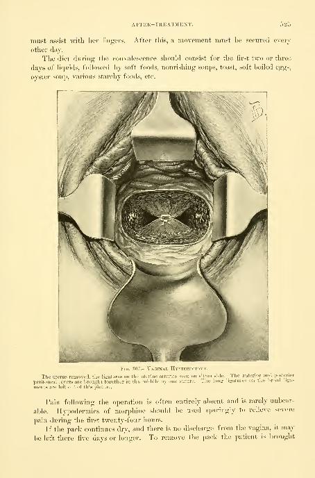
AFTER-TREATMENT. 52;>
must assist "with her fingers. After this, a movement must be secured every
other day.
The diet during the convalescence should consist for the first two or three
days of liquids, followed by soft foods, nourishing soups, toast, soft boiled eggs,
oyster soup, various starchy foods, etc.
Fig. 307.—Vaginal Hysterectomy.
The uterus removed, the liiratures on the uterine arteries seen on either side. Tlie anterior and posterior
peritoneal layers are l)rou2ht tOi^ether in tlie middle by one suture. The long ligatures on the broad liga-
ments are left out of this picture.
Pain following the operation is often entirely absent and is rarely unbear-
able. Hypodermics of morphine should be used sparingly to relieve severe
pain during the first twenty-four hours.
If the pack continues dry, and there is no discharge from the vagina, it may
be left there five days or longer. To remove the pack the patient is brought

526 VAGIXAL HYSTERECTOMY.
with the buttocks to the edge of the bed with the thighs flexed. The operator
shps a narrow Sims speculum into the vagina, retracting the posterior wall, and
with dressing forceps draws the strips of gauze out from between the ligatures.
As soon as the strips are removed the vaginal vault must be cleansed with
pledgets of absorbent cotton, and a fresh pack inserted.
Ko vaginal douches of any kind should be used until three weeks have
passed, when a 3 per cent warm carbolized douche or boric-acid douche may
be given once or twice daily, using a short nozzle and taking great care not to
push it too far in. When silk ligatures are used the discharge is sure to become
odorous sooner or later, and the vagina must be cleansed more frequently. The
ligatures loosen and come away with a little traction, in bunches, in from four to
six weeks. It is a good plan not to wait for them to become detached, but in
the course of three weeks to expose and remove them with forceps and scissors.
These sutures can be removed most easily with the patient in the knee-breast
or Sims posture.
In eighteen days the patient may sit in a reclining chair a little while each
day, and after this gradually increase her movements, until after four weeks,
when she is able to be wp all day. At this time an examination will show that
the vaginal vault is closed, and the wound area has contracted down to a
transverse granulating linear scar, with the granulations more abundant at each
end. After six or eight months this whole line has contracted still more, until
it is a thin white cicatrix, closing the vault.
After a hysterectomy the patient should avoid hard work, heavy lifting, and
prolonged exertion for several months. Recovery of health is usually rapid;
within a few months a pale, emaciated woman often regains all her lost vigor.
But the surgeon still has a duty to perform in continuing to watch these cases,
examining them at first at intervals of two or three months, and later every six
months, in order to detect at once any recurrence of the disease. It will occa-
sionally be necessary to cut out a small area of recrudescence in the vaginal
vault, which will be detected at an early stage by this careful inspection.
In two instances in which the uterus was enucleated without removing the
uterine tubes or the ovaries I found at a later date the vault of the vagina occu-
pied by dark red, funguslike masses, which at first sight suggested a rapid return
of the disease; on removing these, however, they proved to be the uterinetubes inverted through the incision at the angles, exposing to view their
inner mucous lining.
The accidents and complications that arise during vaginal hys-
terectomy are :
1. Hemorrhage.
2. Ligation of one or both ureters.
3. Rupture of the bladder.
4. Injury to the small intestine.
5. Pelvic abscess.
6. Ovarian tumor.
7. Incomplete enucleation of the disease.

HEMORRHAGE. 527
Hemorrhage .—The various sources of liemorrhage are the hemorrhoidal,
uterine, ovarian, and vesical arteries and veins. Slight hemorrhage from small
arteries is usually easily controlled by clamping them for a time with forceps ; if
they continue to bleed they must be ligated with line silk or catgut. Hemor-rhage from such large vessels as the uterine and ovarian arteries may prove em-barrassing from the constant flow of blood which obscures the field ; the diffi-
culty of managing it increases the greater the distance of the bleeding point
within. Such a hemorrhage is most apt to arise from cutting too close to one of
the ligatures, so that the short bunch of tissue in its grasp slips from under the
constricting loop. The worst form of hemorrhage is seen where the effort has
been made to grasp the entire broad ligament in a clamp. This form of hemor-rhage is treacherous, because it is hable to occur some hours after the operation.
The following instruments and accessories should always be within easy
reach to meet such an emergency : A Sims speculum, two long flat retractors,
dressing forceps, three sponges in holders, six artery forceps, pei-ineal pad, iodo-
form gauze, transfusion apparatus, and a liter of normal salt solution. A hypo-
dermic injection of strychnin (3L- grain) should be given at once.
To control the hemorrhage the patient is put under chloroform and brought
to the edge of the bed under a good light, or, better still, placed on a table. Assoon as she is sufficiently relaxed by the anesthetic, the operator must proceed
ra]">idly, as prolonged anesthesia is especially dangerous in the shock following
an extensive hemorrhage.
The saturated gauze pack is removed, and with fingers and sponges on holders
the vagina and pelvis are rapidly cleared of the large clots which sometimes extend
high up into the abdomen as far as the umbilicus. By gentle traction upon the
bunches of ligatures, the broad ligament of one side and then that of the other
is drawn into view and the loose ligature loop found and the broad ligament
clamped. If active hemorrhage is going on, the quickest way to detect its
source is to introduce a clean sponge on a holder between the broad ligaments
and leave it there for a few seconds. The deep blood stain in one spot reveals
the corresponding position of the hemorrhage. If, however, the precise area
can not be determined by inspection, the broad ligament on the bleeding side
should be caught by traction forceps, introduced under the guidance of the index
finger, and drawn down step by step until the bleeding point is seen. As soon
as discovered, a pair of artery forceps is applied, or several forceps in rapid suc-
cession if needed, until the whole area is under control. If the patient is much
shocked by loss of blood, the quickest and most satisfactory plan is to leave the
forceps in place for from thirty-six to forty-eight hours, without attempting to
apply a ligature. When a large area of the broad ligament has slipped up into
the pelvis and the bleeding is active, and proper assistance is wanting, a bold and
successful method of finding the bleeding vessels is to take a pair of bullet for-
ceps and carry them up into the pelvis, guided by the touch ; the broad ligament
is then seized and drawn down into view, and so clamped from end to end with
artery forceps. The operator must constantly l)ear in mind that an active hemor-
rhage almost invariably arises from one of four vessels, the two uterine and the
528 VAGINAL HYSTERECTOMY.
two ovarian arteries ; each one of these must be inspected in searching for tlie
source. A persistent exhausting hemorrhage may also arise from patulous
atheromatous vessels in the septum between bladder and cervix or between cer-
vix and rectum. One of my patients nearly lost her life from a slow continu-
ous oozing from a small vessel of this sort.
Where the mucous surfaces are blanched by the hemorrhage, the respiration
quickened, and precordial distress felt, and there is a rapid, thready, scarcely
perceptible pulse, or even where there is only well-defined shock, infusion
of a liter of salt solution under the breasts should be resorted to during the
operation.
Ligation of one of the Ureter s.—This is an accident to which a
beginner is peculiarly liable, and comes from passing the first ligatures too far
out from the cervix toward the pelvic wall.
The most skilled operator may make this mistake, when the cervix is un-
usually enlarged by cancerous infiltration, diminishing the distance to the pelvic
wall, and bringing the cervix and the ureters into an abnormally close relation-
ship. In such cases the only absolute assurance of safety lies in a preliminary
sounding of the ureters, by placing a flexible bougie in each one, where it re-
mains until the operation is over (see Chapter XIII). By this means, as soon
as the peritoneum is opened, the ureter can be felt at once against the side
of the pelvis like a hard, firm cord, which the bougies keep splinted out upon
the pelvic wall at the greatest possible distance from the cervix. The extreme
importance of placing a sound in the ureters has been repeatedly illustrated in
my cases where the ureter has been bared for one or more inches by a careful
dissection.
Rupture of the Base of the Bladder.—This accident will not
occur in an ordinary vaginal hysterectomy if the uterus is freed from its vesical
attachments by constantly directing the end of the finger toward the cervix.
Occasionally the operator will be surprised by discovering that the disease has
extended beyond the cervix and involved the base of the bladder, which breaks
down, allowing the finger to enter the bladder in the enucleation, in spite of
every precaution. This accident is signalized by a gush of urine mixed with
blood. The exact location of the tear should at once be determined, either
with the finger, or by introducing a sound into the bladder through the urethra,
and bringing the point out through the hole.
Care must be taken during the further steps of the operation not to convert
a small rent into a large ragged opening. If the danger of further rupture is
imminent, while using the finger, the remainder of the bladder may be dissected
off from the cervix with forceps and scissors. As soon as the extirpation of
tlie uterus is complete, the rent in the bladder should be exposed by a little
traction on its peritoneal fold, and if there is any suspicion of infiltration, the
margins of the rent should be liberally excised and the fistula closed by inter-
rupted silk sutures, not including the mucosa, and the vesical peritoneum drawn
down over the closed fistula and attached to the anterior vaginal wall, thus bury-
ing the fistula and relieving it of any tension as the bladder distends with urine.

INCOMPLETE ENUCLEATION. 529
After such a complication, the urine should be drawn every four hours for
two or three days. If tlie approximation is good, this operation is ahnost in-
variably successful, and a vesico-vaginal fistula will not complicate the con-
valescence.
Injury to the Small Intestine.—The small intestine may be
injured where adhesions to the uterus have been formed, in consequence of
pelvic peritonitis of tubal origin, or from extension of cancer of the body of the
uterus to the contiguous viscera.
By palpation on opening the peritoneum and a careful sejDaration of ad-
hesions, any serious injury to the bowel will usually be avoided, and its lumenwill not be opened unless invaded by the disease ; in such a case the best plan is
to draw the affected loop of bowel down out of the pelvis, cut out the diseased
area, and close the opening by sutures. Such cases as these ought not to be
operated on through the vagina if the extent of the disease is suspected before-
hand ; an abdominal incision reveals the exact condition, which can then l)e
dealt with under actual inspection.
Pelvic Abscess complicating Yaginal Hysterectomy.—Vaginal hysterectomy, where there is an abscess in one or both tubes and
ovaries, is made more difficult by the fixation of the uterus through the in-
flamed adherent lateral masses, which interfere with the downward displace-
ment. The danger of general peritonitis is also enhanced by the contamination
of the pelvic peritoneum by the escaping pus. The best plan of procedure is
to go on as far as possible with the enucleation of the lower part of the uterus
without rupturing the abscess, and then to protect the peritoneum thoroughly
with a gauze pack while evacuating the pus with an aspirator.
When the peritoneum is opened, gauze should be packed on all sides in pro-
tecting the abdominal cavity.
If the abscess involves but one side, the unaffected side should be freed first
and the uterus brought out, when the affected tube and ovary may be removed
with the uterus by introducing two fingers and separating the adhesions and
dra^ving the organs outside, and then ligating and cutting, first the ovarian
vessels near the line of the pelvic brim and continuing on down the broad liga-
ment until the whole mass is freed.
If the abscess has ruptured and its contents escaped over the peritoneum and
the wound, some of the pus should at once be examined under the microscope
for organisms, and if any of the various pyogenic cocci are found in abundance,
the sponging out should be more thorough, washing the whole jjelvis with ex-
treme care, and a more abundant gauze drain should be placed higher up within
the pelvis at the end of the operation.
If the enucleation is impeded by an ovarian tumor, this should be
freely opened and evacuated and drawn out in a collapsed condition and the
ligatures applied to the broad ligaments as usual.
Incomplete Enucleation .—An incomplete enucleation is the result
of an operation undertaken by mistake when the disease is too far advanced for
radical treatment, and I know of nothing more unsatisfactory than the discovery,

530 VAGINAL HYSTERECTOMY.
after the operation is well under way, that a portion of tlie disease has advanced
beyond the possibihty of removal. One may find a nodular mass adhering by a
broad base to the pelvic wall, or an infiltration of the upper part of the broad
ligament through which it is necessary to cut to free the uterus ; or, again, the
uterus may be so friable as to break down as soon as it is handled, leaving a
ragged infiltrated area extending into the broad ligament.
Separate masses plastered on the pelvic wall must be let alone, for complete
extirpation is here impossible, and the attempt would excite a hemorrhage which
might easily become uncontrollable.
Where the broad ligament is found widely infiltrated after the operation lias
advanced too far to be abandoned, the uterus nnist be removed as nearly as possi-
ble in one piece and any remaining cancerous areas with friable tissue energetic-
ally curetted, the hemorrhage checked with forceps left on from twenty-four to
forty-eight hours, the pelvis washed out with the utmost care, and an extensive
gauze pack inserted, completely enveloping the infiltrated area on all sides.

CHAPTER XYII.
INVERSION OF THE UTERUS.
1. Definition and description.
2. Various forms : a. Acute, b. Chronic, c. With fibroid tumor.3. Diagnosis.
4. Prognosis.
.5 . Treatment—reposition : a. By manual efforts, h. Vaginal amputation of the uterus, c. Pan-hysterectomy, d. Kiistner's method—opening Douglas's pouch and incising the sac andreinverting.
6. Inversion due to malignant disease.
The uterus in inversion is turned inside out, so as to form a hollow tumor
projecting into the vagina ; its walls are externally the uterine mucosa, internally
the peritoneum, and between the two lie the muscular coats of the uterus. The
uterine end of the tubes and the utero-ovarian ligaments enter into the pouch
formed by the inverted peritoneum, and the ovaries and outer ends of the tubes
lie just above it. The sac within is not more than 2-2.5 centimeters (f to 1 inch)
deep, and its orifice forms a narrow slit or a puckered orifice opening into the
peritoneum.
Various Forms.—Various forms of inversion exist which it is impor-
tant to recognize, as the mode of treatment differs in each.
Acute inversion is the form found immediately after labor ; it is often due
to unskilled efforts in delivering the placenta by traction upon the cord. This
is oftener seen by the obstetrician than by the gynecologist.
The chronic form is either simply a survival of the acute form or is slowly
produced in a non-puerperal uterus along with the expulsion of a tumor attached
to its walls ; this is the form which is most frequently seen in our gynecological
clinics.
The commonest cause of inversion is a submucous fibroid tumor attached to
the fundus uteri. The mechanism of its formation under these circumstances
is the relaxation of the uterine cavity below the tumor, produced by expulsive
efforts like those of labor, by which the tumor is finally forced into the vagina,
dragging with it the attached portion of the uterine wall.
If the tumor is submucous and becomes pediculated, the peritoneal surface
of the uterine wall undergoes no displacement and there is no inversion;
if, on
the other hand, the tumor remains sessile, as it descends the whole thickness of
the uterine walls and the peritoneum may follow, creating on the peritoneal sur-
face an indentation, at first slight but becoming more and more deeply depressed
until complete inversion is brought al»out by the escape of the tumor into the
531
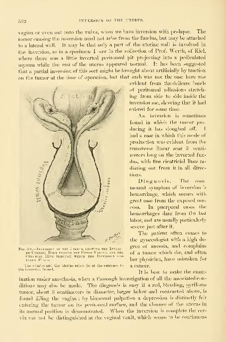
632 INVERSION OF THE UTERUS.
vagina or even out onto the vulva, when we have inversion witli prolapse. The
tumor causing the inversion need not arise from the fundus, but may be attached
to a lateral wall. It may be that only a part of the uterine wall is involved in
the inversion, as in a specimen I saw in the collection of Prof. Werth, of Kiel,
where there was a little inverted peritoneal pit projecting into a pediculated
myoma while the rest of the uterus appeared normal. It has been suggested
that a partial inversion of this sort might be brought about artificially by traction
on the tumor at the time of operation, but that such was not the case here was
evident from the delicate bands
of peritoneal adhesions stretch-
ing from side to side inside the
inversion sac, showing that it had
existed for some time.
An inversion is sometimes
found in which the tumor pro-
ducing it has sloughed otf. I
had a case in which this mode of
production was evident from the
transverse linear scar 3 centi-
meters long on the inverted fun-
dus, with fine cicatricial lines ra-
diating out from it in all dii'ec-
tions.
Diagnosis. — The com-
monest symptom of inversion is
hemorrhage, which occurs with
great ease from the exposed mu-
cosa. In puerperal cases the
hemorrhages date from the last
labor, and are usually particularly
severe just after it.
The patient often comes to
the gynecologist with a high de-
gree of anemia, and complains
of a tumor wdiich she, and often
her physician, have mistaken for
a cancer.
It is best to make the exam-
ination under anesthesia, when a thorough investigation of all the associated con-
ditions may also be made. The diagnosis is easy if a red, bleeding, pyriform
tumor, about 3 centimeters in diaineter, larger below and contracted above, is
found filling the vagina ; by bimanual palpation a depression is distinctly felt
entering the tumor on its peritoneal surface, and the absence of the uterus in
its normal position is demonstrated. When the inversion is complete the cer-
vix can not be distinguished at the vaginal vault, which seems to be continuous
Fig. 308.
—
Inversion ..i iiit L iekls, showing the iNVEiiT-
ED Uterine Body filling the Upper Vagina, and theCervical Ring through which the Inversion hastaken Place.
The ovaries and tlie uterine tul
the inversion funnel.
It th

TREATMENT—REPOSITION. 533
with the tumor. If the inversion is incomplete the cervix remains as an en-
larged ring, and a sound may he pushed into it for a short distance up to the
neck of the sac.
In the case of a tumor projecting into the vagina with a partially inverted
uterus, the uterine attachment of the tumor presents a depression which mayhe felt through the rectum. AVhen the inversion is partial any undertaking to
enucleate the tumor in ignorance of this complicaticjn is most hazardous ; the
peritoneum has been opened under such circumstances and the life of the pa-
tient lost, through the tendency of the uterine wall thus cut through to retract
out of sight and to bleed into the peritoneal cavity. The difficulty of getting
hold of the edges of the wound, together with the injury and exposure of the
peritoneum, made such an accident one of the gravest misliaps which formerly
could befall a gynecologist. The proper plan now in event of such an accident
would l)e to open the abdomen immediately and deal with the bleeding area
directly by suture.
The rule, therefore, in every case of a submucous myoma, is to assume that
an inversion does exist until the contrary is proved by a careful rectal and bi-
maimal examination—palpating the peritoneal surfaces of the uterus over the
point of attachment of the tumor.
The prognosis of an inversion left to itself is unfavorable. But few cases
undergo spontaneous reposition, the hemorrhages endanger life, and ulceration
of the exposed mucous surface may give rise to sepsis. In one instance recorded
there was a spontaneous amputation of the body of the uterus. (R. M. Murray,
Edinburgh Medical Journal, vol. xxviii, p. 901.)
T r e a t m e n t—E e p o s i ti o n .—The most satisfactory plan of treatment is
by a reposition of the displacement. This • is usually easy in a puerperal case
seen soon after its occurrence, when with the thumbs or fist an indentation is
started at the most prominent part on the wall of the inverted sac, and by con-
tinuing to push up in the axis of the pelvis the depression is made deejDer and
deeper, until reinverted tissue, acting as a wedge, enters and dilates the cervix
and passes into the pelvis, when the replacement is shortly completed. The dif-
ficulty of keeping the uterus up in its place is often greater than that of return-
ing it. To hold it in place, an iodoform gauze pack may be introduced within
the cavity, filling it, and supported by a vaginal pack, which is changed from
day to day, until by contraction and recovery of tone the danger of recurrence
is past.
AVhen the inversion is caused by a tumor attached to the fundus, it will usu-
ally be sufficient to take away the tumor to bring about an immediate return of
the uterus to its normal shape. On account of the facility with which reposition
occurs under these circumstances, it is important to pass all the sutures neces-
sary to close the wound and stop bleeding before detaching the tumor ; other-
wise the bleeding surface once reinverted can not be reached.
In chronic cases one of the following three plans of treatment may be
adopted :
First, by manual eiforts.

534 IXVEUSIOX OF THE UTERUS.
Second, by vaginal amputation.
Third, by opening Douglas's poueli and incising the sac from fundus to cer-
vix and reinverting.
Manual efforts at reposition should be made first. To do this, the
tumor is squeezed with one hand to make it longer and smaller, and then pushed
with the other hand, like a wedge, up into the cervix, through which it gradually
returns in the reverse order of its formation, if the effort is destined to succeed.
Another way is to grasp the tumor in the full hand, slipping the extended index
and middle fingers of the same hand inside the cervical ring to dilate it, at the
same time attempting to push the uterus up through the dilated ring. The
other hand makes counter-pressure simultaneously through the abdominal wall,
over the ring, helping to roll tlie cervical tissues back over the neck of the
uterine tumor.
The diificulties in the way of a manual reposition are usually insuperable
;
they arise from the altered rigid fibrous character of the uterine tissue, with
vascular engorgement and edema, as well as from the fact that the rigid neck
of the inverted peritoneal sac is so much smaller than the body of the uterus
which is to pass through it.
The surgeon is not warranted in making prolonged forcible attempts at
manual reposition on account of the inevitable bruising of the tissues and the
danger of laceration at the cervix. In a case of marked fatty degeneration of
the uterine walls of six and a half years' standing. Dr. A. Martin, of Berlin,
perforated them in attempting to efliect a manual reduction ; the patient died in
collapse a few days later {Path, und Therap. der Fmuenl'i'anl'heiten^ 3(1 ed.,
Wien und Leipzig, 1893, p. 158).
A most natural suggestion to the surgical mind, in the present stage of ab-
dominal surgery, upon the failure of manual efforts, would be to open the abdo-
men, to dilate the contracted canal from vdthin by fingers and dilators, and then
to push the uterine body up from the vaginal side through the enlarged canal
into its normal position ; this has been tried, but has not proved very successful.
I note a failure of my own in a case of long-standing inversion with pro-
lapse, in Philadelphia, eight years ago. The proposed plan was to open the ab-
domen and expose the neck of the inverted sac, and then to stretch this with
strong dilators, and with the help of an assistant to force the body of the uterus
up through the enlarged neck of the sac, producing reposition. I had further
intended to prevent the recurrence of the inversion and prolapse l)y stitching
the fundus to the anterior al)dominal wall (Suspension of the Uterus, Chapter
XXIV).I opened the alxlomen and exposed the narrow slit-like orifice at the site of
the inversion, but my utmost efforts to make any impression upon the opening
with fingers or dilators were unavailing, and I was obliged to abandon the at-
tempt and relieve the patient by am])utating the uterus through the vagina.
On the other hand, a successful operation of this kind was performed by Dr.
T. Ct. Thomas, of New York, in September, 1809 {Atner. Jour, of (>h-4., vol. ii).
The patient was put under ether, when an assistant so forcibly lifted the uterus

TREATMENT—REPOSITION. 535
up against the abdominal wall tliat the intestines were displaced and the cervical
ring conld be felt, A small incision was then made in the median line of tlie
abdominal wall down on to the ring, opening the peritoneum. The operator
now grasped the inverted uterus in the vagina, and at the same time introduced
a powerful steel dilator into the neck of the sac on the peritoneal surface. The
dilatation proceeded easily and rapidly, and the uterus was reinverted and re-
stored to its normal form in twenty-seven minutes. The patient recovered and
left her bed on the eighth day.
The second method, vaginal amputation of the uterus, remains
a satisfactory alternative in case of failure of the preceding means.
After a thorough cleansing of the field, the uterus is enveloped in sterilized
gauze, grasped, and drawn down and exposed by pulling back the posterior
vaginal wall with a speculum, so that the neck of the sac at the vaginal vault is
accessible. The amputation is now begun by cutting two flaps at the neck of
the inverted uterus, one anterior and one posterior, just below the vault of the
vagina. Before the peritoneum is opened, three or four stout silk ligatures are
passed with a large curved needle completely through the uterine stump in an
antero-posterior direction.
The peritoneum is now cut through in front, and by continuing the incision
cautiously out to the sides, the uterine arteries and veins are found, clamped,
and tied as high up as possible with fine silk. An assistant keeps up a strong
traction on the ligatures to keep the stump from inverting into the peritoneum.
The operator now takes the ligatures one at a time and ties them tightly, bring-
ing the lips of the stump firmly together. Additional deep sutures must be
passed, if necessary, to cheek bleeding and secure accurate approximation. The
stump closed in this way soon slips through the cervix and a partial reinversion
is established.
The most important point to bear in mind throughout, is that the ligatures
passing through the stump must keep the lips of the wedge-shaped incision
firmly approximated even after reinversion has occurred. A dry dressing should
be kept in the vagina ; the sutures may be removed in ten days or two weeks.
Complete vaginal hysterectomy (panhysterectomy) may be performed by
opening Douglas's pouch from side to side and the vesico-uterine pouch in front,
and then hooking the index finger around one side of the cervix and passing a
succession of ligatures through the tissue intervening between the finger and the
vault, tying each ligature, and cutting between it and the cervix, taking care not
to cut too near the ligature. The amount of tissue severed is small and the
uterine artery is soon ligated ; an additional ligature must be applied to its
free end.
The opposite side is ligated in like manner, and the uterus freed. The blad-
der does not enter the inversion sac. If there is no bleeding, the peritoneal sur-
faces of the wound may now be drawn together with a running suture, the lig-
atures arranged on the right and the left side, and the vault of the vagina
packed with iodoform gauze.
Prof, O, K ii s t n e r's method of reposition in chronic cases resisting

53() INVERSIOX OF THE UTERUS.
simpler methods of reduction {G'ntralh.f. Gyn.^ 1893, No. 41) is in entire accord
with the recent developments of gynecological surgery, and promises success in
cases which it has hitherto been found impossil)le to treat in a conservative
manner. I have not yet had a case upon which I could try it, l)ut, in view of
the appai'ent feasibiHty of the plan, I give the details of the operation. It is
briefly this : The peritoneum is opened posterior to the uterus and the neck of
the sac is incised, relieving the constriction and making it large enough to push
the fundus through. The,—../ steps are conducted in the
/ V •";^ followino; manner :
/ ' U T- • -1
I'lrst, a wide transverse
,v incision in Douglas's cul-de-
::--'' .' .vac^ opening the peritoneum.
,^-'-'' ^- Second, the introduction
, - /, \ ^, ,,-- of the index linger through
I
r-, -^ ij /; ;; ,
-'this opening into the inver-
sion funnel of the uterus, and
separation of any adhesions
found.
Third, a longitudinal in-
cision through the posterior
uterine wall, as nearly as pos-
sil)le in the median line. This
begins about 2 centimeters
l)elow the inverted fundus
and ends about 2 centimeters
above the os externum, and
extends all the way down to
the peritoneum.
Fourth, reinversion of the
uterus by fixing the funnel
with the index finger in
Douglas's pouch, and press-
ing in the fundus with the
thumb of the same hand,
and superficial sutures passed
Fig. 30y.
—
Inversion ok the Uteki.s due to Wakcoma.
Hysterectomy, recovery. San. Jan. lit, 1897.
Fifth, suture of the uterine incision by deep
on the peritoneal surface.
Sixth, closure of Douglas's eul-de-mc with sutures.
Inversion due to Malignant Disease. — Inversion due to a
malignant tumor of the fundus is rare, and I have seen but one case. Thepatient (J. II. ?>., No. 410, San., Jan. 11), 1897) presented herself on account
of a fetid, watery, blood-tinged discharge which had continued for about a year
with hemorrhages at intervals.
I found the whole vagina above the levator ani filled with an ovoid mass
about 8 by 6 by 4 centimeters, fiattened antero-posteriorly, and attached at the

INVERSION DUE TO MALIGNANT DISEASE. 537
cervix by a pedicle 2*5 centimeters in diameter. The external os formed, a sharp
rim around the pedicle, and the depth of the canal was from 2 to 2*5 centimeters.
Bimanually the uterine body was found, absent, and in its place was a pit
which entered the cervical ring, close to which both ovaries could be felt, the
left one entering it for a short distance. The ovoid body filling the vagina was
made up of the inverted uterine body and a sessile tumor of a light grayish color
covered with little tags of tissue.
In grasping this mass it broke down, and was so friable that the entire enu-
cleation had to be done with the fingers. There was no capsule at all, and no
line of demarcation between it and the uterine tissue at the base, covering an area
of 2' 5 by 2 centimeters, where the uterus appeared white and non-vascular. The
fundus went ujd through the cervix after seventy-two hours, upon releasing a
pair of forceps detaining it, so as to obviate the risk of a hemorrhage, which
could not be controlled by suture in the friable tissue.
The tumor removed was broken up into a number of irregular pieces, and
was easily penetrated by the fingers in all directions, without any hard or resist-
ing nodules ; the tissue toward its base had an edematous appearance and was
longitudinally striate and tore in shreds, like the meat of a crab's claw.
A microscopic examination showed that the tumor was a spindle-celled sar-
coma. A complete abdominal hysterectomy was therefore done a week later,
after which the patient recovered.

CHAPTEK XYIII.
VAGINAL EXTIRPATION OF SUBMUCOUS MYOMATA AND POLYPI.
1. Description and position of myomata.2. Symptoms : a. Hemorrhage, b. Pain. c. Suppuration.S. Diagnosis : a. History, b. Palpation, c. Difference between fibroid polypi and cancer.
4. Treatment—extirpation : a. Selection of cases, b. Cutting through the pedicle, c. Removalof sessile submucous fibroids: (1) Piecemeal, with foi'ceps and scissors; (2) by splitting the
capsule; (3) by both these methods combined ; (4) by celiotomy.
5. Complications : a. Hemorrhage, b. Sepsis, c. Rupture of uterus, d. Death from exhaustion.
6. Polyps.
Description and Position of Submucous Myomata.—Quite often a myomatous tumor, originating in tlie submucosa or interstitially
in the uterus, is carried, in the course of its development, down into the uterine
cavity, where it is found attached by a broad base or by a pedicle of varying
length. The tendency to develop in this way is greatest where there is a single
tumor, which may varj^ from the size of an egg to that of a mass big enough to
choke the pelvis. Small submucous myomata are also occasionally found asso-
ciated with large interstitial and subserous tumors. The tumor, which lies at
first concealed in the uterine cavity, may, in the course of time, be extruded
through the plastic cervix until it comes to lie partly or wholly within the vagina,
or even outside the vulva, where it may be found associated with an inverted
uterus or with a long, slender pedicle. Large sessile tumors are sometimes
grasped so firmly by the cervix, when only partially delivered out of the cavity
of the uterine body or into the vagina, as to show a deep encircling furrow at
the point of constriction at the internal or external os. The pedicle of such a
tumor may be attached to any part of the uterus from fundus to cervix. The
most usual position is an attachment to the body just above the cervix.
The submucous myoma in the early stages is covered by tlie mucosa, which
gradually becomes thinner, and not infrequently entirely disappears in places.
This atrophy is due to pressure and attrition. The uterine mucosa between two
adjacent submucous myomata is frequently thickened because it is protected
from pressure. Apart from these inequalities in the thickness of the mucosa it
is usually unaltered in other respects. Occasionally, however, the mouths of the
glands become occluded, and small cysts develop ; in one of my cases the utei'ine
cavity was studded with clear lenticular cysts, and some of them Avere 8 milli-
meters in diameter. The presence of submucous myomata does not preclude the
possibility of endometritis or tuberculosis, or of other changes in the nmcosa,
538

DESCRIPTION AND POSITION OF SUBMUCOUS MYOMATA. 539
such as carcinoma and sarcoma. Cysts found in the middle of such tumors hnedwith cylindrical ciliated epithelium have been explained as originating in a por-
tion of the glandular tissue nipped off early in the development of the tumor.
Symptoms .—The most characteristic symptoms are hemorrhageand pain. The hemorrhages are often excessive, and make the woman ex-
tremely anemic. They are worse at the menstrual periods, but may last for
weeks or months together. The pains arise from the expulsive efforts of the
uterus trying to push the foreign body without the cervix; they are severe,
intermittent, and expulsive in character, like those of labor ; they often continue
for years. Occasionally cases are observed where the pain has been slight, or
absent altogether, and the only symptom is hemorrhage. A thin serous oozing
from the mucous surface, resembling that of early cancer, may be the first symp-
tom to call the woman's attention to her condition.
Most myomata are interstitial m their beginning, and only become subserous
or submucous as they grow, depending upon their situation and the resistance
encountered.
The myomatous tumor takes its origin in a little muscular whorl which is
poorly vascularized, and secures its blood supply from the capillaries of its
periphery, which send their branches into the tumor along its connective-tissue
septa. As the tumor increases in size the vessels are thickly crowded together
around its periphery, and as it becomes submucous it pushes down toward the
uterine cavity and begins to infringe upon the mucosa. It crowds this against
the opposite wall, and thins it out until it loses its normal characteristics and the
vessels are exposed ; then hemorrhage occurs. At first there is but slight in-
crease in the length and quantity of the menstrual flow from the erosion of the
mucosa. A real hemorrhage first takes place when the mucosa is so thin that
the underlying leashes of vessels which belong to the tumor itself are infringed
u])on. Then a large vessel may rupture, or many smaller ones, simultaneously,
during the menstrual congestion. The vessels form a deeply injected corona
around the jDrojecting myoma at its periphery, and the hemorrhage is largely from
this area, after the most prominent portion of the tumor has been thinned down
to its non-vascular area. In the advanced stages of the submucous tumors the
freest hemorrhage is therefore on the border line between the thicker mucosa
and the attenuated envelope of the tumor. As a rule, these subinucous tumors
produce a corresponding cup-shaped depression on the opposite uterine w\all, and
in the depression formed by the projecting tumor the same vascular phenomena
are found. I have made these deductions from a careful study of this subject
by Dr. J. G. Clark.
Often, when a total extirpation of the uterus has been made for multiple
myomata, all the symptoms have really been due to a small submucous tumor
projecting into the uterine cavity and causing the hemorrhage. If such a tumor
is removed by a thorough curettage, little or no further trouble may be experi-
enced unless another tumor pushes down under the mucosa.
A small pediculated tumor may descend into the vagina with each menstrual
period and return into the uterus afterward, in this way appearing intermittently.

540 VACxINAL EXTIRPATION OF SUBMUCOUS MYOMATA AND POLYPI.
A large tuinor clioking the pelvis often gives rise to serious pressure symptoms
on the part of the bladder and rectum. The pedicles of small tumors may in
time become so attenuated as to break, allowing the tumor to escape spontane-
ously. Larger masses sometimes become necrotic, and break down into a fetid,
gangrenous, suppurating mass, which extends up into the center of the tumor
like a wedge, producing fever and cachexia with profound exhaustion. Several
of the worst septic cases I have ever seen have been of this kind. In one the
tumor had disappeared, and left behind only its muscular and mucous covering,
which hung limp out of the cervix.
Injection of the blood vessels of myomata of the uterus clearly explains why
sloughing and necrosis of the submucous tumors occur, for their internal blood
supply is almost invariably poor, and frequently even before they reach the
-'Sssiy^^j^PfSFr'';
Fig. 310.
—
Pediculated Submucous Myoma isuoken down and collapsed.
Tlie sloughing mass of tissue hanging out of the vulva is soft and flaccid, like a wet rag.
mucous or serous surfaces a necrotic or calcareous core is found. If the tumor
becomes submucous, and the wreath of vessels and the mucosa are eroded away,
either sloughing from a necrobiosis or suppuration from infection may occur.
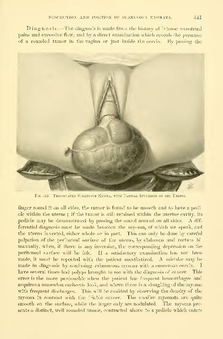
DESCRIPTION AND POSITION OF SUBMUCOUS MYOIIATA. 541
Diagnosis.—The diagnosis is made from the history of intense menstrual
pains and excessive flow, and by a direct examination which reveals the presence
of a rounded tumor in the vagina or just inside the cervix. By passing the
Fig. 311.
—
Peuiculateu Submu OK THE Uterus
finger round it on all sides, the tumor is found to be smooth and to have a pedi-
cle within the uterus ; if the tumor is still retained within the uterine cavity, its
pedicle may be demonstrated by passing the sound around on all sides. A dif-
ferential diagnosis must be made between the myoma, of which we speak, and
the uterus inverted, either whole or in part. This can only be done by careful
palpation of the peritoneal surface of the uterus, by abdomen and rectum bi-
manually, when, if there is any inversion, the corresponding depression on the
peritoneal surface will be felt. If a satisfactory examination has not been
made, it must be repeated with the patient anesthetized. A mistake may be
made in diagnosis by confusing submucous myoma with a cancerous cervix. I
have several times had polyps brought to me with the diagnosis of cancer. This
error is the more pardonable when the patient has -frequent hemorrhages and
acquires a somewhat cachectic look, and where there is a sloughing of the myomawith frequent discharges. This will be avoided by observing the density of the
myoma in contrast with the friable cancer. The smaller myomata are quite
smooth on the surface, while the larger only are nodulated. The myoma pre-
sents a distinct, well-rounded tumor, contracted above to a pedicle which enters
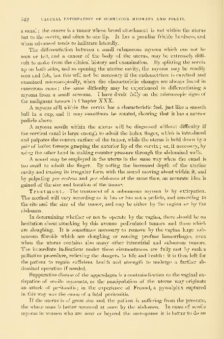
542 VAGINAL EXTIRPATION OF SUBMUCOUS MYOMATA AND POLYPI.
a canal ; the cancer is a tumor whose broad attachment is not witliin the uterus
but to the cervix, and often to one hp. It has a pecuUar friable hardness, and
when advanced tends to infiltrate laterally.
The differentiation between a small submucous myoma which can not be
seen or felt, and a cancer of the body of the uterus, may be extremely diffi-
cult to make from the clinical history and examination. By splitting the cervix
up on both sides, and so opening the uterine cavity, the myoma may be readily
seen and felt, but this will not be necessary if the endometrium is curetted and
examined microscopically, when the characteristic changes are always found in
cancerous cases ; the same difficulty may be experienced in differentiating a
myoma from a small sarcoma. I have dwelt fully on the microscopic signs of
the malignant tumors in Chapter XXX.A myoma still within the cervix has a characteristic feel, just like a smooth
ball in a cup, and it may sometimes be rotated, showing that it has a narrow
pedicle above.
A myoma sessile within the uterus will be diagnosed without difficulty if
the cervical canal is large enough to admit the index finger, which is introduced
and palpates the convex surface of the tumor, while the uterus is held down by a
pair of bullet forceps grasping the anterior lip of the cervix ; or, if necessary, by
using the other hand in making counter pressure through the abdominal walls.
A sound may be employed in the uterus in the same way when the canal is
too small to admit the finger. By noting the increased depth of the uterine
cavity and tracing its irregular form with the sound moving about within it, and
by palpating 'per rectum and j^er ahdomen at the same time, an accurate idea is
gained of the size and location of the tumor.
Treatment.—The treatment of a submucous myoma is by extirpation.
The method will vary according as it has or has not a pedicle, and according to
the site and the size of the tumor, and may be either by the vagina or by the
abdomen.
In determining whether or not to operate by the vagina, there slioukl be no
hesitation about attacking by this avenue pediculated tumors and those which
are slougliing. It is sometimes necessary to remove by the vagina lai-ge sub-
mucous fibroids which are sloughing or causing profuse hemorrhages, even
when the uterus contains also many other interstitial and subserous tumors.
The immediate indications under these circumstances are fully met by such a
palliative procedure, relieving the dangers to life and health ; it is then left for
the patient to regain sufficient health and strength to undergo a further ab-
dominal operation if needed.
Suppurative disease of the appendages is a contraindication to the vaginal ex-
tirpation of sessile myomata, as the manipulation of the uterus may originate
an attack of peritonitis ; in the experience of Freund, a pyosalj)inx rui^tured
in this way was the cause of a fatal peritonitis.
If the uterus is of great size and the patient is sufTering from the pressure,
the whole mass is better removed at once by the abdomen. In cases of sessile
myoma in women who are near or beyond the menopause it is better to do an

DESCRIPTION" AND POSITION" OF SUBMUCOUS MYOMATA. 543
abdominal hysterectomy than to risk a ditiicult vaginal enucleation of the tumoralone.
When the pedicle is long and attenuated, from 1 to |^ a centimeter,
and can be easily reached in the vagina, it may be simply ligated 2 or 3 centi-
meters from the tumor and divided with scalpel or scissors close to the tumor, and
a dry dressing placed in the vagina. An attenuated pedicle which can just be
reached within the uterus may be safely treated by torsion, grasping the tumor
with museau forceps and slowly turning it until the pedicle breaks off. I have
also followed the plan of clamping the pedicle within the uterus with an ordi-
nary pair of artery forceps and then cutting the tumor aw^ay and leaving the for-
ceps in situ in the midst of a gauze pack for forty -eight houi-s ; after this there
is no more danger from hemorrhage, and they are removed.
Fig. 312.—Large Pedicul.vtkd ^ .Myo.ma hanging Outside the Vulv.
A stout pedicle, 2 centimeters or more in diameter, may be grasped
with bullet forceps, or held by passing two or three sutures through it near the
tumor, to prevent retraction when the tumor is removed, and then cut through in
such a way as to make anterior and posterior flaps, taking care to do the cutting
on the convex surface of the growth. After removal of the tumor the sutures
are tied, bringing the flaps together to control bleeding. By observing the prin-
ciple of effecting the separation on the surface of the tumor, instead of following
the natural inclination, which is to amputate the pedicle as high up as it can be
reached, the danger of cutting the uterine wall and opening a partially inverted
peritoneum is obviated. "When the tumor is so large as to fill the vagina, pre-
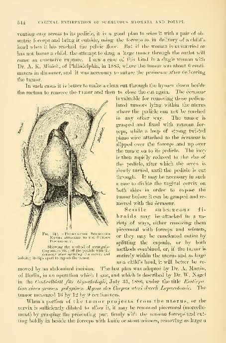
544 VAGIIfAL EXTIKPATION OF SUBMUCOUS MYOMATA AND POLYPI.
venting easy access to its pedicle, it is a good plan to seize it with a pair of ob-
stetric forceps and bring it outside, using the forceps as in dehvery of a child's
head when it has reached the pelvic floor. But if the w^oman is unmarried or
has not borue a child, the attempt to drag a large tumor through the outlet will
cause an extensive rupture. I saw a case of this kind in a single woman with
Dr. A. K. Minich, of Philadelphia, in 1883, where the tumor was about 6 centi-
meters in diameter, and it was necessary to suture the perineum after delivering
the tumor.
In such cases it is better to make a clean cut through the hymen down beside
the rectum to remove the tumor and then to close the cut again. The ecraseur
is valuable for removing those pedicu-
lated tumors lying within the uterus
where the pedicle can not be reached
in any other way. The tumor is
grasped and fixed with museau for-
ceps, while a loop of strong twisted
piano wire attached to the ecraseur is
slipped over the forceps and up over
the tumor on to its pedicle. The loop
is then rapidly reduced to the size of
the pedicle, after which the screw is
slowly turned, until the pedicle is cut
through. It may be necessary in such
a case to divide the vaginal cervix on
both sides in order to expose the
tumor before it can be grasped and re-
moved with the ecraseur.
Sessile submucous fi-
broids may be attacked in a va-
riety of ways, either removing them
piecemeal with forceps and scissors,
or they may be enucleated entire by
splitting the capsule, or by both
methods combined, or, if the tumor is
entirely within the uterus and as large
as a child's head, it will better be re-
moved by an abdominal incision. The last plan was adopted by Dr. A. Martin,
of Berlin, in an operation which I saw, and which is described by Dr. W. Nagel
in the Centralhlatt fur Gyndkologie, 3\\\j 31, 1880, under the title Exstirpa-
tion eines grossen polyposen Myom des Corpus uteri (lurch Laparotomle. The
tumor measured 16 by 12 by 9 centimeters.
When a portion of the tumor projects from the uterus, or the
cervix is sufficiently dilated to allow it, it may be removed piecemeal (morcelle-
ment) by grasping the presenting part firmly with the museau forceps and cut-
ting boldly in beside the forceps with knife or stout scissors, removing as large a
ilG 313 PeDICILATED SlBMlfOlSM\OMA ATTAl HED TO THt 1'U>D19PosrtKIORLl
Showing the method of strangula-
ting and cutting otf the pedicle with theecraseur after splitting the cervix and
holding its lips apart to expose the tumor.

SESSILE SUBMUCOUS FIBROIDS. 545
1^
wedge-shaped piece as possible ; the part adjacent to this is then caught and
pulled more into view and attacked in the same manner. After several such
wedges have been removed, the cutting may be continued
more deeply into the tumor, when the sides will collapse,
allowing the remainder to be easily shelled out or cut away.
If the tumor is covered by a capsule, it is best to incise
this broadly and strip it back before attacking the tumor
itself. The latter part of the enucleation may often be
easily completed by the fingers, but it is safest to stick to
the instruments and to strip the tumor out of its base with
blunt scissors, sound, or spatula, while keeping up the trac-
tion with the forceps.
When the tumor is so dense that the scissors make but
little impression on it, the enucleation may be more rapidly
effected by cutting out wedges with a sharp sickle-shaped
knife, like that shown in the figure ; this has the advantage
of burying itself deeply in the tissues and cutting as it is
drawn toward the operator.
In order to expose a tumor which cannotbe reached through an undilated cervix the vaginal
vault is exposed by retractors, and the anterior and poste-
rior cervical lips caught and held apart with tenaculum for-
ceps, while a deep incision is made on each side, splitting
the entire cervix up into the uterine cavity. This lays bare
the tumor, which is carefully explored with the finger or
a sound to determine its size or location. If it is inter-
stitial the capsule is split from end to end and worked back
with a blunt instrument on all sides, so as to expose as
much as possible of the fibroid mass beneath. The most
accessible portion is now grasped with stout museau for-
ceps and forcibly drawn down, while a wedge-shaped seg-
ment is removed with knife or scissors. In this way piece
after piece is extracted, until the tumor has been sufficient-
ly diminished in size for the rest to be withdrawn through
the cervix.
Hemorrhage is, as a rule, only moderate, and occurs at
once after the extirpation. The reason why profuse hemor-
rhage rarely occurs after these operations can be well demonstrated by the
injected specimens, which show that there is no large artery present ; as soon
also as the tumor is removed the contraction of the uterine muscle acts as an
efiicient hemostatic.
At the completion of the operation the uterus is thoroughly washed out
with warm water, the incisions in the cervical lips closed with silkworm-gut
sutures, and the vagina packed with iodoform gauze.
If a sloughing fibroid has been extracted, it is best not to try to unite the
Fig. 314.
—
Sickle-shapedStout Knife used inExtirpating LargeSubmucous Myomata.

546 VAGINAL EXTIRPATION OF SUBMUCOUS MYOMATA AND POLYPI.
cervical lips, but to pack the uterus with gauze, to be removed in two or three
days and followed by daily irrigation.
The plan of splitting the capsule and enucleating the tumor with a blunt
instrument is also well adapted to large sessile cervical fibroids.
In a case (C. V. B., 3295) upon which I operated Jan. 30, 1895, the anterior
cervical lip was occupied by a large dense fibroid tumor choking tlie entire
vagina; the posterior lip was high up in the pelvis and intact. Tlie tumor
was sessile, with a base of attachment extending from a point halfway down
the anterior vaginal wall to a point high up on the uterus.
A sagittal incision centimeters (2^ inches) long was made over the most
prominent portion, through the vaginal capsule, which was half a centimeter
thick ; the sides of this incision were then peeled back to right and left and the
exposed white nodular fibrous surface grasped with forceps and pulled upon,
while the enucleation was continued with a blunt instrument, peeling the tumor
out of its fibrous investment, which extended above the vault of the vagina up
under the bladder as high as the internal os uteri. There was a little bleeding
from the bottom of the large hole made in the vaginal vault, which was easily
controlled by a continuous buried catgut suture, approximating the sides and
diminishing the size of the cavity. The external incision appeared collapsed
and irregular and could not be neatly approximated in one line, so I brought it
together by a single silkworm-gut, purse-string suture. A dry dressing was
applied and an uneventful recovery ensued without suppuration.
Complications .—The following complications may occur in consequence
of these operations : Hemorrhage, sepsis, rupture of the uterus, and death from
exhaustion.
Hemorrhage is usually moderate, and if the bleeding area can not be
seen and controlled by ligature, the flow may ])e checked by a firm pack of wool
or sterilized non -absorbent cotton left in the uterus from twenty-four to thirty-
six hours.
I have in four instances encountered a hemorrhage after the removal of a
fibroid tumor attached to the fundus which was persistent in spite of the adop-
tion of all ordinary means to control it. The first case was in Philadelphia,
Feb. 3, 1889. The patient (M. E.), already almost exsanguine from the constant
flow from the pediculated fibroid, bled so fast after the removal of the tumor
that I feared she would die at once. I therefore packed the uterus with gauze
and completely closed the vaginal cervix with silkworm-gut mattress sutures,
passed through both lips and tied tightly. This stopped the flow, and in forty-
eight hours I cut the sutures and removed the pack.
I have done the same thing since then in three similar cases in the Johns
Hopkins Hospital with a like result. There were no symptoms after this clos-
ure pointing either to the damming up of blood in the uterus or to its escape
out through the uterine tubes onto the peritoneum in any of the cases.
Sepsis may readily arise after the extirpation of a sessile tumor if the toch-
tiicpie is imperfect, and it is most liable to occur when the tumor is deep-seated
and diflicult of access. I lost under these conditions an elderly woman with a
COMPLICATIONS. 547
tumor 3 centimeters (1|- inch) in diameter at the fundus, firmly fixed and hardto get at. The tumor was much torn and the removal was incomplete, and, in
spite of careful cleansing and an intra-uterine pack of iodoform gauze, she died
within a week of sepsis.
Rupture of the Uterus.—The gravest accident liable to occur in
removing these growths, wdien they are sessile and intimately connected with the
uterine muscle, is rupture or puncture of the uterine wall. This is most serious
when the tumor is out of sight in the uterine cavity, because the injury maytake place without its becoming evident. Such an accident, fortunately nowquite rare, was much commoner when the spoon saw was used to detach the
tumor from its bed.
Eupture has occurred once in my experience, with a fatal result (S. L.,
1441, June 21, 1892). The tumor, which was about the size of an apple, wassituated in the right lateral wall of the uterus, and was removed with great
difficulty by torsion and morcellation. The temperature reached 103° the dayimmediately following the operation, but from that time gradually subsided
until it became about normal on the seventh day. There was a persistent bloody
oozing and several hemorrhages, the largest being about three ounces. Thepatient then became irritable and complained of sharp pain in the lower ab-
domen ; up to the eleventh day^ the pulse and temperature continued about
normal, when the pulse quickly became very rapid and small and the tempera-
ture fell to 96°, and death ensued within a few hours. The autopsy showed a
ragged hole through the uterine wall between the leaves of the broad ligament.
Cultures from this area and all the organs were negative.
Anatomical diagnosis of this case : Sapremia ; sloughing mass of tissue in
the uterus ; perforation of uterus into bi'oad ligament ; mucous polypus of
uterus ; submucous myoma ; fatty degeneration of heart, liver, and kidneys
;
general marked anemia.
Death may occur shortly after the operation if the patient is already in a
state of profound exhaustion when seen by the surgeon. I lost a feeble old
woman, already greatly reduced by hemorrhages, in this way ; she had a simple
pediculated fibroid as large as two fists, and its removal was rapidly accom-
plished without difficulty and without any hemorrhage, but she simply died of
exhaustion ^vithin twenty-four hours, in spite of all sorts of stimulation.
Polypi .—Polypi are soft growths produced by a hypertrophy of the
uterine mucosa, often associated with an endometritis, and frequently found in
fibroid uteri. Their histological peculiarities are described in Chapter XIY.The size varies from that of a pea to that of a walnut, and rarely they are
larger. Those within the uterine cavity are usually found near the tubal os-
tia and are more commonly sessile.
Cervical polypi are most frequently pediculated, and protrude from the ex-
ternal OS. Upon pressure they often recede into the uterus.
S y m p t o m s .—Hemorrhages are the only clinical sign of polypi. These
are rarely severe, and the patients are most often brought to the consultant
through the fear of a tumor.
5-18 VAGINAL EXTIRPATION OF SUBMUCOUS MYOMATA AND POLYPI.
These growths should never be neglected, on account of a liability to malig-
nant changes. Extirpation and a careful microscopic examination is always
indicated. If the growth is readily accessible the pedicle should be ligated with
a strand of catgut and excised. Those higher in the uterine cavity can be more
easily treated by dilating the cervical canal and removing, either by torsion or
by ecraseur.
CHAPTER XIX.
THE UTERUS AS A RETENTION CYST.
1. Definition.
2. Causes.
3. Symptoms.4. Diagnosis.
5. Treatment : a. Hematometra. h. Pyometra. c. Physometra.
Definition .—The conver.sion of the nterus into a sac containing flnid
or gas is caused by the occlusion of the lower genital tract at any point
from the cervix down. When the uterus alone forms the sac, it is mostlikely to contain blood during the early childbearing period of life ; or pus,
and rarely pus with gas, during the later childbearing period and after the
menopause.
The sac so formed is made up of the more or less thinned-out uterine walls,
and is lined everywhere by the uterine mucosa. The formation of such a sac is
brought about by the closure of the three avenues by which the uterus com-
municates with its neighboring cavities—namely, the uterine openings into the
uterine tubes and the cervical canal. When the occlusions are found in the
vagina and at the outer extremities of the uterine tubes, the uterine retention
cyst then forms but one part of a large irregular sac, the rest of which is made
up of the vaginal and tubal cavities -svdth free comnnmications from one to
the other.
The names of these conditions, applied from the nature of their contents
—
" hematometra," " pyometra," " hydrometra," and " physometra "—are really mis-
nomers, and will be misleading unless it is expressly borne in mind that the
terms are used for clinical convenience, merely to designate a prominent feature
of an aifection of the cervix or of the vagina. The real disease, on the other
hand, is that which eifects the closure, and causes the accidental sequel® of
accumulation and distention above it.
Tlie terms pyuria and pyosalpinx, equally unscientific, are used in exactly tlie
same way, purely for clinical convenience.
Causes .—The causes of retention cysts are various. They may be due to
congenital malformation of the vagina, or to the occlusion of one half of a
bicornute uterus, or to operative interference with the cervix by knife, cautery,
or ecraseur, or to an extensive traumatism of the vagina during labor, or to an
549

550 THE UTERUS AS A RETENTION CYST.
endocervicitis, or to cancer of the vaginal cervix blocking the canal and prevent-
ing the escape of the secretions from the npper part of the cancerous area.
I have found a pyonietra above a cervical cancer so often that I always have
it in mind, and if a patient complains much of pain in the lower abdomen I ex-
amine for it carefully. It is evident, as the history of the cases shows, that the
accumulation takes place gradually from week to week, or with each menstrual
period. In cases of vaginal and cervical atresia following the menopause there
is no accumulation above, and no symptoms arise unless there is a senile endo-
metritis. I have seen cervical atresia of this kind in a prolapsed uterus and in
a large fibroid uterus. Ilennig reckoned that three per cent of all women over
the climacteric had this acquired atresia.
The size of the sac will dej^end upon the activity of the secretion and the
length of time the obstruction has lasted. It may be a small one, containing
but a few cubic centimeters of fluid, incaj)able of producing any symptoms, or
it may attain a great size, even filling the lower abdomen.
Syra ptoms .—The symptoms produced arise both from the degree of the
distention and the nature of the contents. When the sac is tense, constant pain
is felt in the lower abdomen, which is too sore to bear pressui*e, and with this
are apt to be associated urinary and rectal disturbances, together with a variety
of nervous phenomena common to many pelvic diseases. The j)ain itself is apt
to vary in intensity, and at the menstrual period is paroxysmal, each attack being
accompanied by the most intense suffering.
If the cervical or vaginal passage is not perfectly closed there will be a little
dribbling leakage of pus or blood, evident on inspection. When pus is retained
there may be a moderate fever.
Diagnosis .—Upon making a digital examination, the diagnosis is usually
easy if there is a considerable accumulation distending the uterus and it feels
like a tense bag; it is more diflacult if there is much disease at the point of
occlusion and but little accumulation above it, and in this case, as I shall point
out, the symptoms deserve a most careful consideration.
To make a thorough examination it is best to put the patient completely
under the influence of an anesthetic. The bowels should be well emptied and
the bladder catheterized. It also aids greatly in the palpation of a uterine cyst,
wdiich may be flaccid and not clear in its outlines, if the patient is first put in
the knee-chest position for about three minutes to dislodge the small intestines,
skeletonizing the pelvic viscera, as it were. The examination is then continued
in the dorsal position.
By the finger inserted in the vagina the point of the occlusion below is
fixed as vaginal or cervical, and whether fibrous or cancerous. A vaginal inspec-
tion may reveal one or two minute orifices, showing that the closure is not
complete.
The extent of the occluded area, whether due to a contraction or a thicken-
ing, and the position, size, and relations of the sac itself, must be studied by the
l)imanual, rectal, and abdominal examination.
With a fino;er in the vae-ina and a fino-er in the rectum, the lower limit ofO O c? ^

IIEMATOMETRA. 551
the atresia at which the rectal palpation is to begin is fixed. Then placnig the
hand on the abdomen, both to furnish a plane of counter pressure and to aid in
palpating, the rectal finger investigates the atresic area above its lower vaginal
limit and palpates the uterine body on all sides. A uterus even moderately
distended, so as to hold from 50 to 100 cubic centimeters of fluid, lies more or
less median and assumes a globular form. Its walls feel tense, rounded, andelastic, often conveying at once the impression of extreme distention.
The dangers of this condition, if left undisturbed, are rupture and gen-
eral infection ; rupture has occurred into the peritoneal cavity, bowel, andbladder. Occasionally such sacs will open spontaneously through the cervical
canal.
Treatment.—The treatment in all cases is operative, and holds two ob-
jects in view :
First, to evacuate the contents of the sac, and
Second, to keep a channel of normal calil)er open into the vagina to prevent
a reaccumulation. (For atresia of the vagina and congenital forms see Chap-
ter XL)The evacuation is always easy, l)ut it is often most diflicult to keep the
channel patulous in the cervical region.
Hematometra.—Hematometra is an accumulation of blood within the uterine
cavity, brought about by a congenital or an acquii-ed occlusion of the cervix,
vagina, or hymen.
In operations to remove the cervical stricture the vagina must first l)e care-
fully disinfected, and extreme care taken throughout not to convey the slightest
infection into the uterine cavity ; then, if a small opening exists, a uterine sound
is passed in and, assisted by a finger in the rectum, carried up into the sac. If
this succeeds, a small-sized dilator is next introduced and the opening enlarged
so as to let the fluid out ; this is followed by a larger dilator, until the opening
is quite patulous ; the evacuation of the fluid may be hastened by mopping out
the uterine cavity with iodoform gauze. It is not necessary to wash the uterus
out—the less done to its cavity the better. An iodoform-gauze pack is put into
the vagina and changed every two to four days. After a week or ten days the
vault of the vagina is exposed, and a dilator of the Hegar pattern passed up
into the uterus. This is repeated every two or three days for some weeks to
insure the canal's remaining open.
If the closure is complete, then the sac must be opened by pushing a large
trocar and canula up through the atresia as near as possible in the position of
the cervical canal, under tlie guidance of a finger in the rectum resting on the
lower part of tlie sac. As soon as the trocar is taken out the fluid escapes, and
on withdrawing the canula the dilators of different sizes, from small to large,
may be used, so as to open the cervix from side to side well up into the uterine
sac. The anterior and posterior lips of the fibrous cervix are then excised and
interrupted sutures passed through the part of the uterus just above this, draw-
ing it down and attaching it to the anterior and posterior vaginal walls. A pack
is then put into the vagina and the wound allowed to heal.

552 THE UTERUS AS A RETENTION CYST.
Pyometra.—Pyometra is usually due to a cancer of the cervix blocking up
the canal, or to an endocervicitis causing adhesions between the cervical sur-
faces. Then pyogenic organisms gain access to the retained fluid, and an ac-
cumulation of pus results. I have also seen a large fibroid utems with an
intact nmcous surface filled w^ith a pale, thin pus, which poured out on ampu-
tating the cervix. Pus in considerable quantities is also often discharged from
a large uterus containing a sloughing fibroid tumor. It is my intention here,
however, to dwell only upon the cases in which the quantity is sufficient to
distend the cavity, forming a retention cyst. The ages of five of my cases of
pyometra were fifty-seven, sixty, sixty-one, sixty-two, and sixty-seven years,
respectively.
The treatment is first to open up the canal and let out the pus, then wash
out thoroughly, drain, and keep the passage open. Where the pyometra is due
to a senile endocervicitis, a part of the cervix, or a pit at the vaginal vault, repre-
senting the cervical canal, can usually be distinguished in the midst of a
granular area. Through this the uterine sound may be passed with slight
force, followed by dilators, letting out the pus. The uterine cavity is then
washed out with a warm saturated boric-acid solution ; after this an iodoform-
gauze drain should be placed in the uterus, to be removed the next day, after
which the cavity is kept open and disinfected by washing it out daily with a
1-10,000 bichloride of mercury solution, using a long, curved glass tube for the
douche nozzle. Nitrate of silver solutions of 10 or 5 per cent strength should
be applied to any granulating areas about once in five days.
It is not enough simply to dilate the canal and let out the pus, for these
cases show a strong tendency to relapse, and must be kept under observation
for a long time. The following was a typical case : Mrs. S., sixty-seven years
old, had ceased to menstruate at fifty, and had remained perfectly well until
within a year, when she was taken with violent pains in the lower part of the
abdomen, which she thought were neuralgic. She had fever at the same time,
and was so prostrated that she had to go to bed for five weeks, when a slight
fetid discharge began to issue from the vagina, and she felt better. This came
only when she was up, and was greatest in the morning.
I found a smooth senile vagina and a diminutive cervix with an obliterated
canal. The uterus, felt per rectum, was tense and globular in form, and as
large as a two and a half months' pregnancy. A dilator was easily passed
through the closed cervix and 180 cubic centimeters (6 ounces) of thick, fetid
green pus escaped. The canal was well dilated and kept open, and she was
at once free of all pain and began to improve. She left me, returning several
weeks later with some discomfort, when I found the canal contracted, and let
out 30 cubic centimeters (1 ounce) of thick pus mixed with blood. After this
the canal was kept open by passing dilators at intervals.
When cancer of the cervix is the cause of the occlusion and pyometra, the
treatment must be different, and is directed both to the cancerous condition as
well as to the pyometra. If the disease has not spread too far beyond the uterus
a total extirpation must be performed (see Chapter XXX), in this way curing
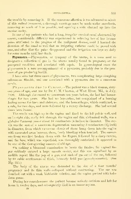
PHYSOMETRA. 553
the trouble by removing it. If the cancerous aiiection is too advanced to admit
of this radical treatment, a thorough curettage must be made under anesthesia,
removing as much of it as possible, and opening a wide channel up into the
uterine cavity.
In one of my patients vrho had a long, irregular cervical canal obstructed by
cancer, considerable difficulty was experienced in relieviug her of her intense
pains attributed to the progress of the malignant disease, until I learned the
direction of the canal so well that an irrigating catheter could be passed with
ease, and after that the pains disappeared and the irrigation was kept up daily
for some months until her death.
Physometra.—Physometra, or tympany of the uterus, is a term used to
designate a collection of gas in the uterus usually found in pregnancy or the
puerperal condition and associated with sepsis. In gynecological cases the
physometra is a rare accompaniment of a pyometra, probably due to the pres-
ence of gas-producing bacilli.
I have seen but three cases of physometra, two complicating large sloughing
submucous fibroids, and one associated with a pyometra due to a cancerous
cervix.
P h y s o m e t r a due to Can c e r .—The patient was a black woman, sixty-
one years of age, sent me by Dr. C. M. Cheston, of AVest River, Md., in July,
1890. Although she ceased to menstruate ten years before, she had had liemor-
rhages for two years. She had no leucorrhea, but complained of a burning
feeling across the back and a])donien, and the hemorrhages, which continued, as
a rule, for two days, and were followed by a w^atery discharge. She had several
times been insane.
The cervix was high up in the vagina and fixed to the left pelvic wall, and
on its right side, easily felt through the vagina and thin abdominal walls, was a
globular fluctuant tumor about 10 centimeters (4 inches) in diameter. The cer-
vix was the seat of a cancerous degeneration measuring 3 centimeters (IJ inch)
in diameter, from wdiich numerous shreds of tissue hung down into the vagina
with excavated areas between them, freely bleeding when touched. The cancer-
ous tissue was first broken down with the fingers, followed by a sharp curette,
and the base, which was superficial, was thoroughly cauterized ; it appeared to
be one of the slow-growing cancers of old age.
On making a bimanual examination to locate the fundus, the vaginal fin-
ger suddenly entered a large smooth cavity, and this was signalized by an
audible report like the pop of a gun, and rush of gas out of the sac, followed
by 90 cubic centimeters of thick, intensely fetid pus (pyo-physometra). (See
Fig. 315.)
The body of the uterus was distended to the size of a four months'
pregnancy and its thin walls collapsed without contracting. The sac was
douched out with a weak bichloride solution, and the vagina packed with iodo-
form gauze.
During her convalescence the patient became actively delirious and left for
home in twelve days, and subsequently died in an insane asylum.
39

554 THE UTERUS AS A RETENTION CYST.
Physometra due to Sloughing Fibroid Tumors .—In Janu-
ary, 1887, I saw a colored woman, a patient of Dr. II. Williams, of Phila-
delphia, who was extremely emaciated and hectic, with a dry tongue and a
pulse of 140. The abdomen was as large as that of a woman eight months
pregnant, tense and tender. Palpation showed that the uterus was converted
into a mass of fibroid tumors, but the percussion note over the mass was
Fig. 315.
—
Pyo-physometra due to Occlusion of the Cancerous Cervix.
Note the thin distended uterine wall, containing pus in the lower part of its cavity, with a large gas spaceabove. An explosion of gas took place as soon as the instrument broke through the barrier at the cervix.
tympanitic. At the operation I introduced my hand into the uterine cavity
and easily removed a sloughing fibroid which would fill a one and a half
liter measure, and then broke through the thin septum of a second large
sessile fibroid tumor, when two liters of intensely fetid pus escaped, with large
quantities of gas. The pulse after the operation was 184; the patient was fi'ee
from pain and lost no blood, but she died a week later worn out by the j)ro-
longed suppuration.

PHYSOMETKA. 555
Pliysonietra in Pregnancy .—The commonest of all forms of pliyso-
metra is that met with in pregnancy or in the puerperium. It is oftenest ob-
served in women with narrow pelves, where labor is protracted and wheremannal or other operative interference has been found necessary, and is always
due to an infection by a gas-producing bacillus.
It is still the common impression that the gas in the uterus is due to the en-
trance or introduction of air from without, or to the formation of gas blebs in a
dead fetus macerated in a moist medium. For example (C. Bamberg, Inaug.
Dissert., Halle, 1877), in the case of a primipara with prolapse of the cord, the
patient was put in the knee-elbow posture and the cord replaced. The next daythe patient had a chill, the pulse rose to 124, and the temperature was 4:1-4:° C,and percussion over the uterus yielded perfect tympanitic resonance. The child
was perforated and delivered, and after the escape of the head a quantity of ex-
tremely foul gases poured out of the vagina with the fetid waters.
The child is always dead, and the waters, as a rule, ruptured when the tym-
panites is found.
The true cause of the tympany is the bacillus a e r o g e n e s c a p s u -
1 a t u s , and this is well shown by a case investigated by Dr. George W. Dolp-
hin, the first case in which the bacillus has been demonstrated ante-mortem
{Puerperal /Sepsis due to Infection with the Bacillus Aerogenes Caj}sulatus,
Johns Hopkins Hospital Bulletin, Feb., 1897).
The patient, a Polish woman, had been in labor for two da^^s, attended by a
midwife. She was found by Dr. Dobbin in a state of extreme exhaustion, with
a pulse of 130 to 140, and with a large dead fetus impacted in a pelvis, with a
true conjugate measuring approximately 8 centimeters (3^ inches). There were
no regular labor pains, and there was a continuous escape of gas and frothy fluid
from the vagina. A sweetish offensive odor was noticeable about the bed, and
a distinct bubbling, crackling sound could be heard. The child was delivered by
a craniotomy, and immediately following the delivery a large amount of offen-
sive gas escaped from the uterine cavity. (Ireat numbers of the bacillus aero-
genes capsulatus were demonstrated in the fetus, placenta, and uterine lochia,
and a fatal prognosis was given.
Death occurred on the third day after delivery. No autopsy was allowed.
Dr. S. Flexner saw the patient six to eight hours before death, and endeavored
in vain to find any evidences of the formation of gas in the tissues remote from
the genitalia. But six or seven hours after death the appearances presented
were those of extensive gas formation everywhere m the soft tissues and serous
cavities, and from the nose and mouth frothy bloody serum exuded, which in
cover-slip preparations showed the characteristic bacilli.
This is a confirmation of the prediction made by Welch and ]^uttall that
many of the cases of supposed entrance of air into the uterine sinuses would l^e
found to be due to infection with a gas-producing micro-organism.
The diagnosis is made by percussing the tympanitic, distended uterus,
sometimes helped by noticing the escape of fetid waters mingled with little gas
bubbles. The uterus may contain a small quantity of gas, which always assumes

556 THE UTERUS AS A RETENTION CYST.
the highest position in turning the patient ; in other cases the uterus is so dis-
tended that the danger of rupture seems innninent, and breathing is greatly em-
barrassed by pressure on the diaphragm.
The treatment is to empty the uterus as soon as possible, without refer-
ence to the child, which is already dead, and then to wash out the uterine cavity
with an antiseptic douche, such as carbolic acid, 3 per cent, and to repeat the
douches frequently enough to sterihze and keep sterile the genital tract.

INDEX OF CASES FROM THE JOHNS HOPKINS HOSPITAL.
Adeno-carcinoma of Bartholin's gland
Carcinoma of clitoris .
Carcinoma of clitoris .
Carcinoma of external genitals .
Death from anesthesia
Death from anesthesia
Double vagina ....Double ureter ....Exstrophy of bladder
Fibroma of round ligament
Gonorrheal stricture of ureter .
Hydroureter ....Hypertrophy of uterine mucosa.
Inversion due to malignant disease
Myxo-fibro-sarcoma of labium majus
Prolapse with vesico-cervico-vaginal fistula
Prolapse without vesical diverticulum
Recto-vaginal fistula ....Removal of kidney and ureter .
Rupture of uterus ....Stricture of ureter ....Submucous myoma ....Suburethral abscess ....Tubercular ureteritis ....Undeveloped uterus, ovaries, and tubes
Ureterotomy
Uretero-cystostomy ....Uretero-ureterostomy....Vesico-uterine fistula....Vesico-vaginal fistula....
R. S., 1136, December 18, 1891
E. McD., 179, April 23, 1890
C. L., 2465, December 23, 189^
J. B. R., 3013, September 8, 1894
.
L. T. N., 4233, March 18, 1896
B. B., 3357, January 1, 1895
.
M. C, 4887, December 28, 1896
A. W., 4154, February 17, 1896 (admission).
G. T., 3869, October 14, 1895
C. H., Path. No. 65, October 13, 1893
E. S., San. 96, March 2 to June 2, 1894
S. A. H., 4039, December 30, 1895
A. L., 3476, May 7, 1895
J. H. B., San. 410, January 19, 1897
M. T., 3896, October 12, 1895
K. W., 1320, April 9, 1892 .
M. K., 653, April 4, 1891 .
K. S., 3916, September 5, 1894
K. W., 4012, December 21, 1895
S. L., 1441, June 21, 1892 .
C. F., San. 109, May 12, 1894
C. V. B., 3295, January 30, 1895
L. J. P., 3095, January 17, 1894
C. R., 1836, March 30, 1893 .
B. I\r., 2190, September 9, 1893
J. D. S., 4038, December 37, 1895.
B. Z., 2990, October 6, 1894 .
F. M., 1946, May 1, 1893 .
F. H., 1750, January 12, 1893
M. Y., 3811, September 25, 1895
INDEX OF CASES FROM THE LITERATURE.
Atresia of urethra
Atresia of urethra
Calculus impacted in ureter
Cancer of urethi'a
Cancer of virethra
Cancer of urethra
Maxdl .
F. SCHATZ .
W. A. Lane .
A. F. McGiLLT. G. ThomasWiNCKEL
557

558 INDEX OF CASES FROM THE LITERATURE.
Colo-vesical fistula
Congenital anomaly of ureter
Congenital anomaly of ureter
Congenital flexure of ureter
Cystic dilatation of ureter .
Cystic dilatation of ureter .
Dermoid cyst suppurating into bladder
Double bladder .
Double bladder .
Double bladder .
Exfoliative cystitis
Extirpation of bladder
Extraperitoneal uretero-cystostomy
Fibroid polyp
Fibroma of urethia
Fibroma of urethra
Foreign bodies in bladder
Foreign bodies in urethra
Foreign bodies in urethra
Foreign bodies in urethra
Foreign bodies in urethra
Hypospadias
Melano-sarcoma of urethra
Myoma of urethra
Myxo-sarcoma of urethra
Nevoid growth in bladder
Nephro-ureterectomy .
Rupture of uterus by curettage
Rupture of uterus by curettage
Rupture of uterus by a wooden
Sarcoma of bladder
Sarcoma of urethra
Sarcoma of urethra
Stricture of ureter
Stricture of ureter
Suburethral abscess
Traumatic stricture of ureter
Ureterostomy
Uretero-ureterostouiy .
Urethral caruncle
Vesico-uterine fistula .
Vesico-vaginal fistula .
Vesico-vaginal fistula .
Vesical tuberculosis .
R. HarrisonMassari
W. H. MaxsonWeigertE. G. OrthmannF. TanglG. C. BlackmanI. Cattier .
FUTH .
A. p. Smith .
Nicholas Tulp
K. Pawlik .
C. WiTZEL .
H. Hoenig .
C. Hennig .
C. Mettenheimer
A. DUNLAP .
A. Mazario .
PlASESKI
F. Schatz .
A. J. C. Skene
A. Lebedeff
C. A. L. ReedButtnerGalabin
Arbuthnot LaneReynier
M. D. Mann .
J. B. Harvie
w. e. ashton
McWeeney .
H. Beigel .
E. Ehrendorfer ,
C. Fenger .
E. KCster .
William HeyC. Fenger .
C. P. Noble .
A. M. FULLERTON
William Goodell
F. H. ClIAMPNEYS .
A. F. McGillW. A. FreundJ. 0. POLAK .

I K D E X
Abdomen, myoma of, 86-88.
ovarian cyst of, 83-85.
Abortion, curettage for remnants of.
Abscess of Gartner's canal, 251.
of the vulvo-vaginal gland, 191.
suburethral, 303.
pelvic, 529.
Absorbent cotton, 16.
Adeno-carcinoina of cervix, 493.
of the vulvo-vaginal gland, 194.
of body of uterus, 491.
Adenoma of the bladder, 385.
Affections of the bladder, 315.
of the vulvar mucosa, 197.
of the ureters, 396.
Analysis of separated urines, 413.
Anatomy, pelvic, 42.
of ureters, 396.
Anesthesia, 145, 279.
general, 147.
local, 145.
slip, 150.
Anesthetics, 148.
Anesthetizer, 147.
Antisepsis, 4.
Antiseptic conscience, 22.
methods, 4.
Applicator, 278.
Ascitic abdomen, 88, 89.
Asepsis, 3, 278, 415.
Aspirators, 144.
Assistants in plastic operations, 161.
Atresia of one side of uterus, 238.
of vagina, 258.
of urethra, 288.
Auscultation, 90.
Auto-infection of genital tract, 34.
Bacillus aerogenes capsulatus, 40, 41.
Bacillus coli communis, 35, 39.
Bacteria, 34, 35.
Bacteriology, 32.
Bimanual examination, 95-101.
Bladder, artificial division of, into hemispheres
and quadrants, 271.
calculi of, 324.
classification of diseases of, 316.
congenital defects of, 316.
displacements and alterations in form and
capacity of, 321.
eversion of, 323.
exstrophy of, 318.
foreign bodies in, 324.
natural landmarks of, 269.
relations of, to surrounding structures, 270.
rupture of, 528.
Boiling soda solution, 6.
Bougie coated with veax, 403.
Brushes, 20.
Biichner's pyrogallic acid and potassium hy-
drate method, 37.
Calibrating and dilating the urethral orifice, 280.
Calibrator, 277.
Carcinoma of bladder, 387.
of body of uterus, 490, 514.
of cervix, 492. 514.
of clitoris, 185.
of labia majora. 174.
of urethra, 311.
of vagina, 253.
Care of bowels after plastic operation, 164.
during convalescence, 163.
of wound, 165.
Carrier, suture, 141.
Caruncle of urethra, 307.
Catgut, 13, 138.
Catheterization, 163.
of pelvis of the kidney, 414.
of the ureters, 400.
of the ureters without elevation of the pelvis
and without atmospheric distention of the
bladder, 411.
Catheters, dilating, 403.
flexible ureteral, 401.
flexible renal, 401.
559
560
Catheters, glass, 143.
metal ureteral, 402.
Cauteries, 144.
Celiotomy veins, 48.
Central figure of bladder, 274.
Cervix, lacerations of, 494.
operations on, 495.
amputation of, 509.
Chemical antisepsis, 7.
Child killed in utero by catheter, 290.
Chloroform, 151.
rules for administering, 152.
Clitoris, diseases of, 179.
Cocain, 146.
Colpocleisis, 330.
Colpocystotomy, 391.
Colporrhaphy, anterior, 511.
Condyloma of labia majora, 174.
Corn silk, 360, 369.
Cultures, 36, 37.
Cumol, 13.
Curettage, 483.
for cancer of cervix, 492.
of bladder, 379.
Cyst of the clitoris, 189.
of the vulvo-vaginal gland, 189.
Cystectomy, 394.
Cystic follicles, 386.
Cystitis, 360.
acute, 368.
chronic, 369.
exfoliative, 381.
tubercular, 376.
Cystoscope, 277.
Cystoscopy, 277.
Death from anesthesia, 158.
Dermoid cyst suppurating into bladder, 353.
cyst of bladder, 386.
Diagrams of pelvic lesions, 130, 131.
Diet, 164.
Difiference between private and hospital surgery,
23.
Dilatation, 478.
Dilator, 276.
Diseases of external genitals, 168.
Disinfection of hands and forearms, 20.
Diiderlein's bacillus, 32.
Double bladder, 316.
ureter, 416.
vagina, 239.
Douching, 163.
Drainage pads, 18.
Dress for plastic operations, 160.
Dressing forceps, 130.
Dressings after operation, 162.
Ectopic ureteral orifice, 417.
Edebohls portable table, 29.
Ehrlich-Weigert solution, 38.
Electro-cautery, 144.
Elephantiasis, 181.
Ellinger dilators, 478.
Emmet's left-curved scissors, 135.
Endermic injections, 147.
Endometritis, acute, 485.
chronic, 486.
decidual, 486.
Enema, 160.
Enterocele, 501.
Epispadias, 288.
Epithelioma of bladder, 387.
of cervix, 493.
Equipment of sterilizing room at surgeon's
home, 24.
Ether, 156.
Ethyl chloride, 146.
Evacuator, 276.
Examination of pelvic organs in the dorsal po-
sition, 94.
in pelvic disease, 102.
under anesthesia, 107.
of urethra and bladder, 272.
of vermiform appendix, 123.
External direct method of measuring the conju-
gate vera, 104.
Facsimile of page from case-book, 128.
Fallopian tubes, examination of, 98.
Fere's table, 383.
Fibi'oma and myoma of round ligament, 173.
of bladder, 385.
of urethra, 309.
Fissure of Ilenke, 50, 53.
Fistula in lateral wall of ureter, 458.
of ureter at base of bladder, 458.
of ureter at vaginal vault, 458.
Fistulc'e, colo-vesical, 354.
recto-vaginal, 261.
recto-vesical, 355.
ureteral, 455.
urethro-vaginal, 297.
vesical, 328, 352.
vesico-vaginal, 341.
vesico-uterine, 350.
vesico-utcro-vaginal, 347.
Flexible catheters, 401.
Forceps, 135.
hemostatic, 136.
long mouse-toothed, 278.
long rat-toothed, 136.
long straight dressing, 136.
polyp, 138.

561
Forceps, rat-toothed dissecting, 13G.
sponge, 137.
tenaculum, 136.
Formalin, 484.
Gabbett's method of staining, 35.
Gauze, 16, 25.
General anesthesia, 147.
Goniometer, 405.
Gonocoecus, 35.
Gonorrhea, 35, 39.
Gonorrheal stricture of ureter, 436.
Gram's method of staining, 36.
Gynecological examination, 80.
Hand basins, 9.
Head mirror, 275.
Hematokolpos, 257.
Ilematometra, 551.
Hemorrhage, 166. 527.
sources of, 546.
Hemorrhoids, 356.
Hemostatic forceps, 136.
Hernia, 172.
Hot-air disinfection, 4.
Hyderabad Chloroform Commission, 151.
Hydrocele, 171.
Hydroureter, 447.
Hymen, imperforate, 235.
Hyperemia of bladder, 358.
of trigonum, 358.
Hypospadias, 287.
Ichthyol treatment for urethritis, 303.
Ignition tubes, 13, 14.
Illumination, 133, 225.
Imperforate hymen, 235.
Incomplete enucleation in vaginal hysterectomy,
529.
Infection following plastic operations, 167.
Inguino-labial abscess, 171.
Injury to small intestine, 529.
Inspection, 80-86, 87.
of bladder, 273.
of rectum, 118-121.
of the ureters, 398.
Instillation of bladder, 370.
Instrument bags, 27.
sterilizer, 7.
Intestine, injury to small, 529.
Introduction of flexible silk catheter, 406, 414.
of speculum and location of the ureteral ori-
fices, 403.
Invagination of the pelvic floor, 97.
Inversion of the uterus, 531.
Investigation of general condition of patient, 123.
Iodoform gauze, 17.
Irrigation of the field of operation, 161, 338.
of bladder, 369.
Ischuria, 296.
tubercle ba-
Kangaroo tendon, 139.
Knives, 134.
Koch-Ehrlich method for stainiuf
cilli, 38.
Labia majora, 169.
minora, 177.
Law of Metschnikoff, 33,
of Wissakovitsch, 33.
Leg holder, 143, 160.
Ligatures, 138.
Lipoma of labia majora, 169.
Litholopaxy, 326.
Local anesthesia, 145.
Location of ureteral orifices, 403.
Loculate bladder, 317.
Lubricant, 281.
Malformations of urethra, 287.
Measurements of abdominal enlargements, 81.
Melano-sarcoma of urethra, 314.
Metal ureteral catheter, 402.
Methods of examining ureters, 398.
Micrococcus gonorrhoea, 36.
Mailer's fluid, 184.
Myoma of bladder, 386.
of urethra, 310.
Myxoma of bladder, 388.
Myxo-fibro-sarcoma of labium majus, 196.
Myxo-sarcoma of urethra, 314.
Xeedle-holder, 142.
Needles, 140.
Nephrectomy, 464.
Nephro-ureterectomy, 428.
Nevoid growth of bladder, 357.
Normal abdomen, 84.
Normal uterine mucosa, 485.
Operating room, 8.
suit, 19.
table. 10.
Outlet, resection of, 511.
Ovaries, examination of, 98.
Oxygen after anesthesia, 149.
Pack, changing it, in vaginal hysterectomy, 525.
Packer, 143.
Palpation, 90.
of bladder, 272.
of the ureters, 399.
Papillary fibroma, 384.
602
Papilloma of bladder, 384.
Pathogenic bacteria met with in gynecological
practice, 'So.
Pedicle, ligation of, 543.
Pelvic abscess, 529.
Pelvimetry, 104.
Percussion, 87.
of bladder, 272.
Perineal pad, 18, 160.
Periureteritis, 422.
Pessaries, 240.
Photography, 81.
Physometra, 553.
Pneumaturia, 366.
Pneumonia after anesthesia, 149.
Polyps, 486, 547.
forceps, 138.
Position of patient for plastic operation, 160.
Posture of patient for examination of bladder,
299.
Postures, 90.
Preparation of patient for examination of blad-
der, 279.
of patient for anesthesia, 107.
of patient for plastic operation, 159.
of room in private house, 28.
of surgeon, assistants, and nurses, 19.
Prolapse of ureteral mucosa, 454.
of urethral mucosa, 290.
of uterus, 499.
without vesical diverticulum, 503.
Pruritis, 198.
Pseudo-myxoma of the canal of Nuck, 172.
Pyometra, 552.
Pyoureter, 448.
Rat-toothed forceps, 136.
dissecting forceps, 136.
Rectal diverticulum, 501.
Recto-vaginal septum, operations on, 208.
Rectum, examination of, 98, 99, 115.
prolapse of, 513.
Relaxed vaginal outlet, operation for, 217.
Rest and tonic treatment, 166.
Resuscitation of the asphyxiated, 154-156.
Retention cysts, 549.
Retractors, 134.
Roux-Nocard's glycerin agar, 39.
Rubber drainage pads, 18.
Rules for administering any anesthetic, 152.
for administering chloroform, 152.
for use of anesthesia in examinations, 107.
Rupture of uterus, 547.
Sarcoma of bladder, 387.
of body of uterus, 491.
Sarcoma of clitoris, 184.
of urethra, 313.
of vagina, 253.
Scalpels, 134.
Scissors, 134.
straight scissors, 135.
Emmet's left-curved scissors, 135.
Searcher, ureteral, 278.
Sepsis, 1.
Septum, complete tear of, 208, 211, 513.
jSigns of complete relaxation in anesthesia,
148.
Silk, fine, medium, and heavy, 13, 138.
Silkworm gut, 13, 138.
Silver wire, 139.
Smegma bacillus, 35.
Specula, use of, in inspection, 87.
Speculum, Goodell-Baer bivalve speculum, 134.
introduction of, into bladder, 282.
Kelly's small cylindrical speculum for girls,
134.
Nelson's trivalve speculum, 134.
Simon's speculum, 134.
Siras's speculum. 134.
Sponge forceps, 137.
Sponges, 17.
Staphylococcus aureus and albus, 34, 39, 40.
citreus, albus, 34, 39, 40.
epidermidis albus, 2, 34.
Steam disinfection, 4.
sterilizer, 5, 6.
Sterilization and preservation of instruments,
11.
of catgut with cumol, 14.
of gauze, cotton, towels, and bandages, 12.
of instruments, dressings, and ligatures, 24.
of sutures and ligatures, 12.
Sterilized rubber gloves, 25.
water, 11.
Sterilizers, 5-7.
Streptococcus pyogenes, 39, 40.
Stricture of ureter, 436.
of vagina, 257.
ti'aumatic, of ureter, 444.
Submucous myoraata, 538.
Suburethral abscess, 303.
Suppuration, 39.
Suspension of the uterus, 513.
Suture materials, 138.
Sutures, removal of, 165.
Swedish ball irrigator, 162.
Symphyseotomy for vesical tumors, 394.
Syphilis, 39.
Taking the history, 127.
Tanks for storage of hot and cold water, 11.

563
Tenacula, 135.
Tenaculum forceps, 136.
Topographical anatomy, 43.
anatomy of small intestines, 49.
Topography of the bladder, 269.
Transfixion needles, 143.
Trocars, 143.
Tubercle bacillus, 35, 38, 39,
Tubercular cystitis, 376.
ureteritis, 425.
Tuberculosis, 38.
of endometrium, 489.
of genital tract, 38.
of the vestibule, 203.
Tumors of the bladder, 382.
Tying knots with silk and catgut, 140.
off one broad ligament in vaginal hysterecto-
my, 519.
Typhoid fever bacillus, 39.
Ureter, anatomy of, 396.
catheterization of, 400.
congenital affections of, 416.
congenital flexure of, 422.
cystic dilatation of occluded ureter, 421.
introduction of flexible silk catheter into, 406.
ligation of, 528.
location of ureteral orifices, 403.
methods of examination, 398.
obstruction of, 431.
physiology of, 397.
sounding, 413.
stricture of, 436.
Ureteral bougies, 452.
calculus, 449.
fever, 415.
fistula, 455.
folds, 269.
searcher, 278.
Ureteritis, 422.
tubercular, 425.
Uretero-cystostomy, 459.
Ureterostomy, 464.
Ureterotomy, 465.
Uretero-ureterostomy, 466.
Urethra, affections of, 287.
atresia of, 288.
cancer of, 311.
caruncle of, 307.
congenital absence of, 289.
dilatation of, 280.
displacements of, 289.
fibroma of, 309.
Urethra, fistula of, 297.
foreign bodies in, 298.
myoma of, 310.
new growths of, 306.
sarcoma of, 313.
stricture of, 294.
Urethral dilator. 277.
Urethritis, 300.
Urethroscopy, 274.
Urinalysis, 272,
Urine, analysis of separated urines, 413.
how to obtain uncontaminated urine, 410.
how to secure urine from both sides at the
same time, 409.
how to secure urine directly from the ureter
without catheterization, 410.
points to be observed in securing separated
urines, 412.
Uterine scrapings, 484.
Uterus, examination of, 98.
Vagina, absence of, 232.
anatomy of, 230.
congenital affections of, 232.
double, 239.
foreign bodies in, 240.
neoplasms of, 252.
traumatic affections of, 257.
Vaginal cysts, 246.
hysterectomy, 514.
Vaginal outlet, obstetrical injuries of, 206.
physiological support of, 204.
resection of, 511.
septa, 238.
Vaginitis, 242.
Varicose bladder, 356.
Vesical bacteria, 35, 304.
balloon, 372.
diverticulum, 502.
herpes, 386.
specula, 276.
Vulvo-vaginal glands, 189.
Washing of genitalia, 161.
Weichselbaum's method of staining tubercle ba-
cilli, 39.
Wertheim's method of staining the gonoeoccus,
36.
Young and Hagner medium, 37.
Ziehl-Neelsen method of staining tubercle ba-
cilli, 39.
END OF VOLUME I.