Differential Interventions: Images as Operative Tools (2016)
15
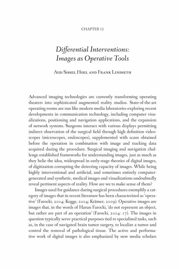
CHAPTER 12 Differential Interventions: Images as Operative Tools Aud Sissel Hoel and Frank Lindseth Advanced imaging technologies are currently transforming operating theaters into sophisticated augmented reality studios. State-of-the-art operating rooms are run like modern media laboratories exploring recent developments in communication technology, including computer visu- alizations, positioning and navigation applications, and the expansion of network systems. Surgeons interact with various displays permitting indirect observation of the surgical field through high definition video- scopes (microscopes, endoscopes), supplemented with scans obtained before the operation in combination with image and tracking data acquired during the procedure. Surgical imaging and navigation chal- lenge established frameworks for understanding images, just as much as they belie the idea, widespread in early-stage theories of digital images, of digitization corrupting the detecting capacity of images. While being highly interventional and artificial, and sometimes entirely computer- generated and synthetic, medical images and visualizations undoubtedly reveal pertinent aspects of reality. How are we to make sense of them? Images used for guidance during surgical procedures exemplify a cat- egory of images that in recent literature has been characterized as ‘opera- tive’ (Farocki, 2004; Kogge, 2004; Krämer, 2009). Operative images are images that, in the words of Harun Farocki, ‘do not represent an object, but rather are part of an operation’ (Farocki, 2004: 17). The images in question typically serve practical purposes tied to specialized tasks, such as, in the case of navigated brain tumor surgery, to localize a tumor and control the removal of pathological tissue. The active and performa- tive work of digital images is also emphasized by new media scholars
Transcript of Differential Interventions: Images as Operative Tools (2016)
CHAPTER 12
Differential Interventions:
Images as Operative Tools
Aud Sissel Hoel and Frank Lindseth
Advanced imaging technologies are currently transforming operating
theaters into sophisticated augmented reality studios. State-of-the-art
operating rooms are run like modern media laboratories exploring recent
developments in communication technology, including computer visu-
alizations, positioning and navigation applications, and the expansion
of network systems. Surgeons interact with various displays permitting
indirect observation of the surgical field through high definition video-
scopes (microscopes, endoscopes), supplemented with scans obtained
before the operation in combination with image and tracking data
acquired during the procedure. Surgical imaging and navigation chal-
lenge established frameworks for understanding images, just as much as
they belie the idea, widespread in early-stage theories of digital images,
of digitization corrupting the detecting capacity of images. While being
highly interventional and artificial, and sometimes entirely computer-
generated and synthetic, medical images and visualizations undoubtedly
reveal pertinent aspects of reality. How are we to make sense of them?
Images used for guidance during surgical procedures exemplify a cat-
egory of images that in recent literature has been characterized as ‘opera-
tive’ (Farocki, 2004; Kogge, 2004; Krämer, 2009). Operative images are
images that, in the words of Harun Farocki, ‘do not represent an object,
but rather are part of an operation’ (Farocki, 2004: 17). The images in
question typically serve practical purposes tied to specialized tasks, such
as, in the case of navigated brain tumor surgery, to localize a tumor and
control the removal of pathological tissue. The active and performa-
tive work of digital images is also emphasized by new media scholars
178 Aud Sissel Hoel and Frank Lindseth
investigating digital image applications such as Photosynth, Augmented
Reality, and Google Street View (Uricchio, 2011; Verhoeff, 2012; Hoelzl
and Marie, forthcoming). As pointed out by William Uricchio, algorith-
mic intermediation reconfigures the relation between the viewing subject
and the object viewed in a way that ‘ultimately determines what we see,
and even how we see it’ (Uricchio, 2011: 33). Algorithmically enabled
image applications do not simply reproduce pre-given realities but exer-
cise transformative powers on both ends of the subject-object relationship
(Carusi, 2012). This is why established representational approaches fall
short of accounting for the active roles of digital image applications, and
this is why new theorizations of images are needed.
Operational approaches provide promising possibilities for rethinking
images for at least three reasons: first, they offer dynamic approaches that
analyze phenomena into doings and happenings rather than into things
and static entities; second, they offer relational approaches that conceive
identity in terms of open-ended processes of becoming; and third, by so
doing, they allow us to ascribe agency to images, and crucially, to conceive
agency as distributed across interconnected assemblages of people, prac-
tices, and mediating artifacts. In the following, we contribute to the ongo-
ing efforts to rethink images in dynamic terms by probing more closely
into neurosurgical imaging and navigation practices where images are
literally used as operative tools. By zooming in on critical moments of the
image-guided neurosurgical process, we draw out key features of an oper-
ational understanding of images, with the aim of developing it further as a
‘differential’ theory of images (Hoel, 2011a and 2011b).
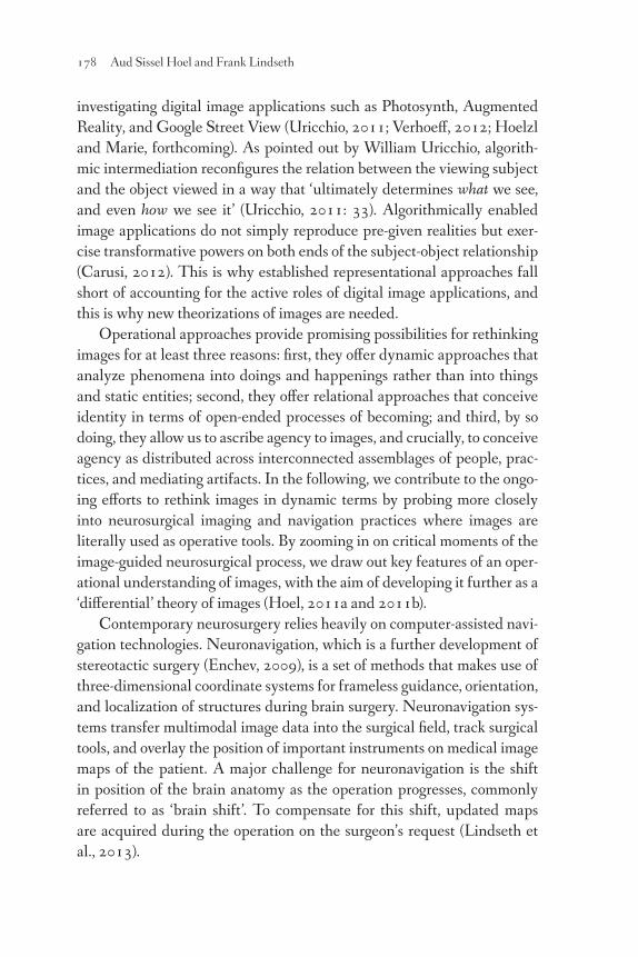
Contemporary neurosurgery relies heavily on computer-assisted navi-
gation technologies. Neuronavigation, which is a further development of
stereotactic surgery (Enchev, 2009), is a set of methods that makes use of
three-dimensional coordinate systems for frameless guidance, orientation,
and localization of structures during brain surgery. Neuronavigation sys-
tems transfer multimodal image data into the surgical field, track surgical
tools, and overlay the position of important instruments on medical image
maps of the patient. A major challenge for neuronavigation is the shift
in position of the brain anatomy as the operation progresses, commonly
referred to as ‘brain shift’. To compensate for this shift, updated maps
are acquired during the operation on the surgeon’s request (Lindseth et
al., 2013).
Differential Interventions: Images as Operative Tools 179
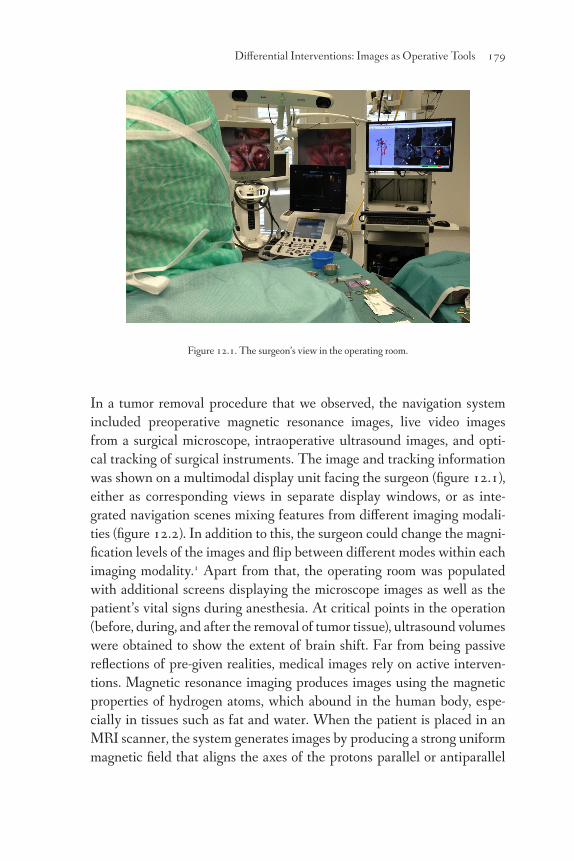
Figure 12.1. The surgeon’s view in the operating room.
In a tumor removal procedure that we observed, the navigation system
included preoperative magnetic resonance images, live video images
from a surgical microscope, intraoperative ultrasound images, and opti-
cal tracking of surgical instruments. The image and tracking information
was shown on a multimodal display unit facing the surgeon (figure 12.1),
either as corresponding views in separate display windows, or as inte-
grated navigation scenes mixing features from different imaging modali-
ties (figure 12.2). In addition to this, the surgeon could change the magni-
fication levels of the images and flip between different modes within each
imaging modality.1 Apart from that, the operating room was populated
with additional screens displaying the microscope images as well as the
patient’s vital signs during anesthesia. At critical points in the operation
(before, during, and after the removal of tumor tissue), ultrasound volumes
were obtained to show the extent of brain shift. Far from being passive
reflections of pre-given realities, medical images rely on active interven-
tions. Magnetic resonance imaging produces images using the magnetic
properties of hydrogen atoms, which abound in the human body, espe-
cially in tissues such as fat and water. When the patient is placed in an
MRI scanner, the system generates images by producing a strong uniform
magnetic field that aligns the axes of the protons parallel or antiparallel
180 Aud Sissel Hoel and Frank Lindseth
to the field, emitting a radiofrequency pulse at the right frequency and
duration, and altering the magnetic field on a local level using gradient
magnets to determine the location of the image ‘slices’.
When the radio-wave transmitter is turned off, the protons start to
‘relax’, producing radio wave signals as they release their energy and
return to their equilibrium state. The signals are picked up by the sys-
tem’s receiver coils and transformed into gray level intensities for each
pixel in a cross-sectional image. Since protons in different tissues have dif-
ferent relaxation times, various scanning sequences can be used to distin-
guish between different types of tissue, say, fat and water, or pathological
tissue and normal tissue. Conventional 2D ultrasound also builds cross-
sectional grayscale images of the anatomy at hand, this time by a trans-
ducer probe emitting a high frequency sound pulse into the patient. As
the sound waves travel into the body, they hit boundaries and interfaces
between various tissue types and some of the sound waves are reflected
back to the probe. In ultrasonic imaging, each sound pulse is followed by
‘listening’ for these sound echoes. By measuring the time taken for echoes
to return, the system calculates the distance to anatomical structures and
displays the strength of the echoes as a grayscale image.
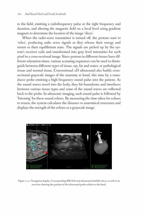
Figure 12.2. Navigation display. Corresponding MR (left) and ultrasound (middle) slices, as well as an
overview showing the position of the ultrasound probe relative to the head.

Differential Interventions: Images as Operative Tools 181
We have zeroed in on technical details of medical image generation
in order to show that MR and ultrasound imaging are based on con-
trasts. The patterns shown are not simply found; they stand out only to
the extent that the areas of interest are subjected to targeted excitations,
tissues and organs being provoked to answer back along the lines speci-
fied by the parametric setup. Each imaging method has a highly selective
range, disregarding anatomical or functional features that fall outside its
scope. The point we want to make, however, goes beyond the familiar one
concerning the selective nature of imaging methods. Rather, the point is
that these methods stand in a generative relation to the imaged features:
each imaging method differentially intervenes into the phenomena under
examination, delineating and sustaining a characteristic pattern or struc-
ture not detectable in the same way by other methods.
In order for images to support navigation, the image data have to be
registered to the patient coordinate system. The registration process
makes the coordinates of anatomical features in physical space and the
various image spaces coincide. During surgery, 3D ultrasound can also
be used to update the preoperative image maps, shifting the positions of
imaged features by means of advanced algorithms (Lindseth et al., 2013).
In the observed operation, fiducial markers were placed on the patient’s
skull before entering the MRI scanner. When the patient was immobi-
lized on the operating table, the markers, visible in the MR images, were
used by the surgeon to identify the corresponding points on the patient’s
head by means of a tracked pointer. As the procedure progressed, ultra-
sound was used for direct guidance (figure 12.3).
Figure 12.3. Ultrasound-guided surgery. Preoperative planning using a tracked pointer (left),
acquisition of a new ultrasound volume using a tracked ultrasound probe (middle), and removal
of tumor tissue using a tracked resection instrument guided by updated ultrasound images.

182 Aud Sissel Hoel and Frank Lindseth
Images used for neuronavigation are clearly ‘part of an operation’;
they are active, transformative, and most definitely reconfigure the sub-
ject-object relationship. For all that, even if they are considered here as
‘agents’, they are not understood to operate on their own. Further, even
if they are made to be processed by computers, the images are ultimately
aimed at human eyes. First, when it comes to objects, it is important to
note that the imaged features are relational and dynamic entities. The
objects revealed by one method are not strictly speaking the same as the
objects revealed by another method. Each method establishes, so to speak,
a ‘working object’ (Daston and Galison, 2007), natural objects being too
plentiful and unrefined to usefully cooperate in systematic comparisons.
Second, when it comes to subjects, imaging methods enact a productive
displacement of the human sensorium, bringing information that is nat-
urally beyond us into the purview of the human senses. By enhancing
human sensitivity, they expand the human action range. In neuronaviga-
tion, agency is distributed as humans, apparatuses, and tissues form an
integrated system.
Thus, if we replace the framework of representation with a dynamic
and relational framework, the ‘operational’ understanding of images
can be further specified as ‘differential’: images serve to discern differ-
ences; differential intervention is their mode of operation. However, if
we truly endorse a dynamic and relational framework, we soon come to
realize that in fact all images, even the pre-digital ones, are operative and
differential tools.
Original source and licence
The New Everyday: A MediaCommons Project, ‘The Operative Image’
cluster curated by Ingrid Hoelzl, http://mediacommons.futureofthe-
book.org/tne/cluster/operative-image January 24, 2014. Licence:
CC-BY ND SA.
Differential Interventions: Images as Operative Tools 183
References
Carusi, A. (2012) ‘Making the Visual Visible in the Philosophy of Science,’
Spontaneous Generations, 6/1: 106-114, DOI 10.4245/sponge.v6i1.16141.
Daston, L. & Galison, P. (2007) Objectivity. New York: Zone Books.
Enchev, Y. (2009) ‘Neuronavigation: Geneology, Reality, and Prospects’,
Neurosurgical Focus 27/3: 1-18.
Farocki, H. (2004) ‘Phantom Images’, Public, 29 (2004): 12-24.
Hoel, A. S. (2011a) ‘Differential Images’, in J. Elkins & M. Naef (eds), What Is an
Image? University Park: Penn State University Press, 152-4.
Hoel, A. S. (2011b) ‘Thinking “Difference” Differently: Cassirer versus Derrida
on Symbolic Mediation’, Synthese, 179/1: 75-91.
Hoelzl, I. & Marie, R. (forthcoming), ‘Google Street View: Navigating the
Operative Image,’ Visual Studies.
Kogge, W. (2004) ‘Lev Manovich: Society of the Screen’, in D. Lauer & A.
Lagaay (eds), Medientheorien: Eine philosophische Einführung. Frankfurt:
Campus Verlag, 297-315.
Krämer, S. (2009) ‘Operative Bildlichkeit: Von der “Grammatologie” zu einer
“Diagrammatologie”? Reflexionen über erkennendes “Sehen”’, in M. Hessler
& D. Mersch (eds), Logik des Bildlichen: Zur Kritik der ikonischen Vernunft.
Bielefeld: Transcript, 94-123.
Lindseth, F. et al. (2013) ‘Ultrasound-Based Guidance and Therapy’, in Gunti
Gunarathne (ed.), Advancements and Breakthroughs in Ultrasound Imaging
(InTech), DOI: 10.5772/55884.
Uricchio, W. (2011) ‘The Algorithmic Turn: Photosynth, Augmented Reality and
the Changing Implications of the Image’, Visual Studies, 26/1: 25-34.
Verhoeff, N. (2012) Mobile Screens: The Visual Regime of Navigation.
Amsterdam: Amsterdam University Press.
Notes
1 T1, T2, FLAIR, MR angiography, fMRI, and DTI tracts for the magnetic
resonance images, and B-mode and Doppler for the ultrasound images.

PHOTOMEDIATIONS: A READER
edited by Kamila Kuc and Joanna Zylinska
Photomediations: A Reader Kuc &
Zylinska
Photography / Media Studies / Media Arts
Photomediations: A Reader offers a radically different way of understanding photography. The concept that unites the twenty scholarly and curatorial essays collected here cuts across the traditional classification of photography as suspended between art and social practice to capture the dynamism of the photographic medium today. It also explores photography’s kinship with other media - and with us, humans, as media.
The term ‘photomediations’ brings together the hybridontology of ‘photomedia’ and the fluid dynamism of ‘mediation’. The framework of photomediations adopts a processual, and time-based, approach to images by tracing the technological, biological, cultural, social and political flows of data that produce photographic objects.
Contributors:
Kimberly K. Arcand : David Bate : Rob Coley : Charlotte Cotton : Joseph DePasquale : Alexander García Düttmann : Mika Elo : Paul Frosh : Ya’ara Gil-Glazer : Lee Grieveson : Aud Sissel Hoel : Kamila Kuc : Zoltan G. Levay : Frank Lindseth : Debbie Lisle : Rafe McGregor : Rosa Menkman : Melissa Miles : Raúl Rodríguez-Ferrándiz : Travis Rector : Mette Sandbye : Jonathan Shaw : Ajay Sinha :Katrina Sluis : Olivia Smarr : Megan Watzke : Joanna Zylinska
Cover image: Bill Domonkos, George, 2014 CC BY-SA


Photomediations: A Reader

Photomediations: A Reader
arose out of the Open and Hybrid Publishing pilot,
which was part of Europeana Space,
a project funded by the European Union’s ICT Policy
Support Programme under GA n° 621037.
See http://photomediationsopenbook.net
Photomediations: A Reader
Edited by Kamila Kuc and Joanna Zylinska
In association with Jonathan Shaw,
Ross Varney and Michael Wamposzyc
London, 2016
OPEN HUMANITIES PRESS
First edition published by Open Humanities Press 2016
© Details of copyright for each chapter are included within
This is an open access book, licensed under Creative Commons By Attribution Share
Alike license. Under this license, authors allow anyone to download, reuse, reprint,
modify, distribute, and/or copy their work so long as the authors and source are cited
and resulting derivative works are licensed under the same or similar license.
No permission is required from the authors or the publisher. Statutory fair use and
other rights are in no way affected by the above.
Read more about the license at http://creativecommons.org/licenses/by-sa/4.0
Figures and other media included with this book may have different copyright
restrictions.
Cover and design: Michael Wamposzyc, 2016 CC BY-SA
Cover image: Bill Domonkos, George, 2014 CC BY-SA
Typeset in Fanwood Text, a free OpenType font by Barry Schwartz.
More at: http://www.identifont.com/show?2DMD
ISBN: 978-1-78542-020-7 PDF
ISBN: 978-1-78542-002-3 Print
Open Humanities Press is an international, scholar-led open access publishing
collective whose mission is to make leading works of contemporary critical thought
freely available worldwide. More at: http://openhumanitiespress.org

Contents
Photomediations: An Introduction 7
Joanna Zylinska
I Photography, Optics and Light
1. A New Kind of History?
The Challenges of Contemporary Histories of Photography 21
Ya’ara Gil Glazer
2. Painting with Light: Beyond the Limits of the Photograph 48
Melissa Miles
3. Why Burn a Photograph? A Film by Hollis Frampton 64
Alexander García Düttmann
4. Processing Color in Astronomical Imagery 75
Kimberly K. Arcand, Megan Watzke, Travis Rector, Zoltan G. Levay,
Joseph DePasquale and Olivia Smarr
II The Image in Motion
5. It Has Not Been – It Is.
The Signaletic Transformation of Photography 95
Mette Sandbye
6. The ‘Potential Mobilities’ of Photography 109
Debbie Lisle
7. The Cinematograph As an Agent of History 120
Kamila Kuc
8. What is the Value of a Technological History of Cinema? 133
Lee Grievson
9. A New/Old Ontology of Film 143
Rafe McGregor

III Hybrid Photomediations
10. Notes on a Painting of a Painted Photograph 163
Ajay Sinha
11. Boredom and Baroque Space 172
David Bate
12. Differential Interventions: Images as Operative Tools 177
Aud Sissel Hoel and Frank Lindseth
13. Glitch Studies Manifesto 184
Rosa Menkman
14. NewFotoScapes: An Interview with Charlotte Cotton 186
Jonathan Shaw
15. The Creative Power of Nonhuman Photography 201
Joanna Zylinska
IV The Networked Image
16. Benjamin, BitTorrent, Bootlegs: Auratic Piracy Cultures? 227
Raúl Rodríguez-Ferrándiz
17. The Gestural Image: The Selfie, Photography Theory and
Kinaesthetic Sociability 251
Paul Frosh
18. The New Technological Environment of Photography
and Shifting Conditions of Embodiment 268
Mika Elo
19. Authorship, Collaboration, Computation?
Into the Realm of Similar Images 283
Katrina Sluis
20. The Horrors of Visuality 290
Rob Coley
Notes on Contributors 313