objective structured clinical examination (osce) as a - ERIC
Upload
khangminh22Category
view
2download
0

PRACTICE OSCEs IN
obstetrics and gynaecology
A guide for the medical student and MRANZCOG exams
Gareth Weston
Beverley Vollenhoven
Jane McNeilage
Sydney Edinburgh London New York Philadelphia St Louis Toronto

Churchill Livingstoneis an imprint of Elsevier
Elsevier Australia. ACN 001 002 357(a division of Reed International Books Australia Pty Ltd)Tower 1, 475 Victoria Avenue, Chatswood, NSW 2067
© 2009 Elsevier Australia
This publication is copyright. Except as expressly provided in the Copyright Act 1968 and the Copyright Amendment (Digital Agenda) Act 2000, no part of this publication may be reproduced, stored in any retrieval system or transmitted by any means (including electronic, mechanical, microcopying, photocopying, recording or otherwise) without prior written permission from the publisher.
Every attempt has been made to trace and acknowledge copyright, but in some cases this may not have been possible. The publisher apologises for any accidental infringement and would welcome any information to redress the situation.
This publication has been carefully reviewed and checked to ensure that the content is as accurate and current as possible at time of publication. We would recommend, however, that the reader verify any procedures, treatments, drug dosages or legal content described in this book. Neither the author, the contributors, nor the publisher assume any liability for injury and/or damage to persons or property arising from any error in or omission from this publication.
National Library of Australia Cataloguing-in-Publication Data_______________________________________________________
Weston, Gareth.
Practice OSCEs in obstetrics and gynaecology : a guide for the medical student and MRANZCOG exams / Gareth Weston, Beverley Vollenhoven, Jane McNeilage.
ISBN: 978 0 7295 3867 1 (pbk.)
Practice OSCEs Series.
Includes index.Bibliography.
Royal Australian and New Zealand College of Obstetricians and Gynaecologists--Examinations, questions, etc. Obstetrics--Examinations, questions, etc. Gynaecology--Examinations, questions, etc.
Vollenhoven, Beverley.McNeilage, Jane.
618.076_______________________________________________________
Publisher: Sophie KalinieckiDevelopmental Editor: Sunalie SilvaPublishing Services Manager: Helena Klijn Editorial Coordinator: Lauren AllsopEdited by Margaret Trudgeon Proofread by Sarah Newton-JohnInternal and cover designs by Toni Darben Index by Forsyth Publishing Services Typeset by TnQ Books and Journals Pvt LtdPrinted in Australia by Ligare

Foreword
Perhaps the only constant in the many recent changes in medical undergraduate and postgraduate education is the continuing demand for assessment. Undergraduate curricula in obstetrics and gynecology, now often broadened into Women’s Health, typically only occupy 8 to 10 weeks in a four- or five-year medical course. Compressing principles and basic knowledge into this time is challenging for all universities. It has led some to develop National Collaborative Core Curricula to emphasise wellness and maintenance of health, while allowing for regional differences in health care needs (www.whccc.org/toc.cfm). Despite such learning challenges, all universities and colleges increasingly demand assessment that can withstand scrutiny.
Objective Structured Clinical Examinations (OSCEs) have become widely used since their introduction as an assessment tool about 30 years ago. Dr Gareth Weston and his colleagues are among the first generation of doctors who have grown up with OSCEs as medical students, and then as postgraduates, and now as examiners themselves in Obstetrics and Gynecology. They are ideally placed to write this invaluable resource in Obstetrics and Gynecology learning and assessment.
For candidates at any OSCE please understand that the OSCE is only less subjective than long cases and other traditional assessment tests. Some subjectivity remains. Although the candidate does not need to wear Dior or Armani, personal hygiene, neatness, appropriate dress and behaviour can only help a candidate’s cause to be a future doctor or specialist.
For the undergraduate there is a vital need to become case-hardened with OSCE practice. This book will greatly help. Chapter 1 is most important: many medical students seem unaware how to pace themselves over a 6-minute OSCE station. Time is a fact of life for any modern doctor, whether year 1 or year 25. Please practise your timing and become fluent.
Postgraduates at examinations such as the MRANZCOG or MRCOG must not only be fluent but also clinically mature. History and examination should flow with a rhythm and a cadence rather that like of a concert pianist, over 5 minutes or so. Test yourself, as suggested by Dr Weston and his colleagues, on three or four OSCEs at a time to help become case-hardened and to overcome the ‘difficult’ station or your odd bad performance. Where you can, try to relate your medical logic and thought processes to the examiner. In postgraduate examinations there may be even more than one OSCE examiner – some colleges have internal checks and audits to root out examiner bias.
�i

Foreword n �ii
An advantage of the OSCE is its easy adaption to many clinical situations – the ward, the operating theatre, the birthing unit or the labour ward. This means examination questions may not be theory from any textbook, but simple things that only a clinical attachment can teach. Writing an operation report or post-operative orders are examples. For many universities this helps focus the medical student on clinical matters in those precious 8 to 10 weeks of their obstetrics and gynecology/women’s health course.
My final word of advice is to peak at the right time. All medical students and young doctors are highly intelligent individuals. That is not enough. Athletes for the Olympic Games not only exercise hard, but also eat the world’s best food, get proper rest and have focused sports counselling. Medical students and postgraduates should also exercise their bodies as well as their brains, eat well, sleep well and focus to perform at their best. Exhaustion from a part-time job out-of-hours is no preparation for an OSCE.
Professor David HealyHead, Monash UniversityDepartment of Obstetrics and Gynecology

Preface
I hope that medical students and training registrars alike find the practice OSCE exam questions contained in this book valuable in preparing for their respective exams. The questions are designed so that they are undertaken in pairs, with one student acting as the examiner and the other as the exam candidate. There are four sets of eight OSCEs pitched at a medical student level, and a further four sets of eight OSCEs at a MRANZCOG level. My co-authors and I have tried to present questions on a broad range of topics. When attempting to cover the entire specialty it is impossible not to be impressed by the expanse of knowledge contained within obstetrics and gynaecology.
I would like to thank the many teachers who have inspired and taught me much of the art of obstetrics and gynaecology: Professor Ian Pettigrew, Dr John Campbell, Professor David Healy and Dr John Bowditch, to name but a few. To Professor Peter Rogers and Dr Caroline Gargett, a big thank you for teaching me much of the science of obstetrics and gynaecology.
For those of you reading this book and preparing for your exam – good luck!!
Dr Gareth WestonSeptember 2008
�iii

Abbreviations
ACIS Adenocarcinoma in situACL AnticardiolipinAFI Amniotic fluid indexAFP Alpha-fetoproteinAIS Androgen insensitivity
syndromeANA Antinuclear antigenAPH Antepartum haemorrhageAPTT Activated partial
thromboplastin timeARM Artificial rupture of
membranesASD Atrial septal defectASRM American Society for
Reproductive MedicineAv AntevertedAXR Abdominal X-rayBMI Body mass indexBP Blood pressureBPM Beats per minuteBRCA Breast cancer geneBSL Blood sugar levelBSO Bilateral salpingo-
oophorectomyCAH Congenital adrenal
hyperplasiaCEA Carcinoembryonic antigenCF Cystic fibrosisCIN Cervical intraepithelial
neoplasiaCMV CytomegalovirusCOCP Combined oral
contraceptive pillCOGU Certificate in Obstetrics and
Gynaecology UltrasoundCRP C-reactive proteinC/S Caesarean sectionCSE Combined spinal/epiduralCT Computerised tomographyCTG CardiotocographCVS Cardiovascular system;
chorionic villous samplingCXR Chest X-ray
D&C Dilatation and currettageD&E Dilatation and evacuationDHEAS Dehydroepiandrosterone
sulphateDIC Disseminated intravascular
coagulationDKA Diabetic ketoacidosisDM Diabetes mellitusDNA Deoxyribose nucleic acidDVT Deep venous thrombosisECG ElectrocardiographEFW Estimated fetal weightEx ExaminationFAI Free androgen indexFBE Full blood examinationFDIU Fetal death in uteroFH(R) Fetal heart (rate)FISH Fluorescent in situ
hybridisationFSH Follicle stimulating
hormoneFVL Factor V LeidenFWT Full ward testGBS Group B streptococcusGCT Glucose challenge testGDM Gestational diabetesGH Growth hormoneGTN Glyceryl trinitrateGTT Glucose tolerance testHBV Hepatitis B virusHCG Human chorionic
gonadotrophinHDL High density lipoproteinHELLP Haemolysis, elevated liver
enzymes, lowered plateletsHIV Human immunodeficiency
virusHNPCC Hereditary non-polyposis
colon cancerHPV Human papillomavirusHR Heart rateHSG HysterosalpingogramHSV Herpes simplex virus
ix

x n Abbreviations
HT Hormone therapyHVS High vaginal swabHx HistoryIBT Immunobead testICSI Intracytoplasmic sperm
injectionICU Intensive care unitIDC Indwelling catheterIDDM Insulin dependent diabetes
mellitusIGF Insulin-like growth factorIGT Impaired glucose toleranceIM IntramuscularIMB Intermenstrual bleedINR International normalised
ratioIUD Intrauterine deviceIUGR Intrauterine growth
retardationIV IntravenousIVDU IV drug use(r)IVF In vitro fertilisationJVP Jugular venous pressureLAC Lupus anticoagulantLBW Low birth weightLDH Lactate dehydrogenaseLDL Low density lipoproteinLFT Liver function testLH Luteinising hormoneLLETZ Large loop excision of the
transformation zoneLMP Last menstrual periodLMW Low molecular weightLUSCS Lower uterine segment
caesarean sectionLV Left ventricleMCV Mean cell volumeMRI Magnetic resonance
imagingMRKH Mayer-Rokitansky-
Kuster-HauserMSU Mid-stream urineNHMRC National Health and
Medical Research CouncilNICU Neonatal intensive care unitNIH National Institutes of
HealthNSAIDs Non-steroidal anti-
inflammatory drugs
NKA No known allergiesNVD Normal vaginal deliveryOA Occiput anteriorOHSS Ovarian hyperstimulation
syndromeOI Ovulation inductionOT Operating theatrePAI Plasminogen activation
inhibitorPAP Test Papanicolaou smearPAPP Pregnancy associated
plasma proteinPCA Patient-controlled analgesiaPCB Postcoital bleedPCOS Polycystic ovarian
syndromePCR Polymerase chain reactionPE Pulmonary embolusPEEP Positive end expiratory
pressurePET Pre-eclamptic toxaemiaPHx Past historyPG ProstaglandinPGD Pre-implantation genetic
diagnosisPID Pelvic inflammatory diseasePO Per oralPOC Products of conceptionPOD Pouch of DouglasPOF Premature ovarian failurePPH Postpartum haemorrhagePPROM Preterm premature rupture
of membranesPR Pulse rate; per rectumPV Per vaginaRh RhesusRIF Right iliac fossaROM Rupture of membranesRPR Rapid plasma reaginRR Relative risk; respiratory
rateRUQ Right upper quadrantSA Semen analysisSCC Squamous cell carcinomaSFH Symphyseal-fundal heightSHBG Sex-hormone binding
globulinSIDS Sudden infant death
syndrome

Abbreviations n xi
SLE Systemic lupus erythematosus
STD Sexually transmitted diseaseSTI Sexually transmitted
infectionT TemperatureT21 Trisomy 21TENS Transcutaneous electrical
nerve stimulationTFT Thyroid function testTOP Termination of pregnancyTORCH Common transplacental
infective organismsTPHA Treponema pallidum
haemagglutination testTSH Thyroid stimulating
hormone
TTTS Twin–twin transfusion syndrome
TVT Tension free vaginal tapeU+E+Cr Urea and electrolytes and
creatinineU/S UltrasoundUTI Urinary tract infectionVDRL Venereal disease reference
laboratoryVE Vaginal examinationV/Q scan Ventilation perfusion scanVSD Ventricular septal defectVTE Venous thromboembolismWBC White blood cellsWCC White cell countWHI Women’s health initiativeWHO World Health Organization
How to approach an OSCE: clinical stations
1
IntroductIonAn ‘Objective Structured Clinical Examination’ (OSCE) is a short, simulated clinical scenario designed to assess the clinical skills of the examination candidate. This method of examination was first proposed in 1975 by R.M. Harden as one way of providing ‘a more objective approach to the assessment of clinical competence’.
In an OSCE examination candidates move through a number of short clinical scenarios which are designed to focus on a range of topics and specific clinical skills. This can be contrasted with the traditional clinical examination – the ‘long case’ – where the candidate would take a history and examine a patient in private, before presenting examiners with the findings, proposed diagnoses, required investigations and treatment. In his original article, Harden found that the OSCE results had a far better correlation with the written results of the students than the traditional approach as the patient (usually simulated) was the same for all students, while the examiners had a standard marking sheet, making their assessment both clear and reproducible.
Since its introduction the OSCE has become a widely used examination tool for both undergraduate medical student and postgraduate specialist examinations. It is currently a key component of the examination process in Obstetrics and Gynaecology at our institution. While it does not replace the need for written examinations to test purely factual knowledge, it does assess a different range of skills that are of a more practical nature.
Aspects of clinical practice that can be assessed in an OSCE range from taking a patient’s general history and asking questions appropriate to the presenting complaint to taking a focused history on a particular problem (such as a menstrual history or a sexual history), explaining investigation results in terms that a patient can understand (e.g. an abnormal fetal ultrasound result or an abnormal Pap smear), performing a specific clinical examination (e.g. a routine newborn examination) or acting out a clinical ‘action’ such as taking a Pap smear, performing neonatal resuscitation or dealing with a shoulder dystocia in labour.
�

� n How to approach an OSCE: clinical stations
Medical studentsThere is often a lot of concern surrounding the OSCEs by medical students as they are less familiar with the format than they are with written examinations, a familiar format first experienced at secondary school. The techniques of history-taking, examination, and counselling and talking to patients are relatively new to them. Nonetheless, these skills are just as important to their future success as doctors as the factual knowledge they gain from reading the textbooks.
MrAnZcoG candidatesCandidates for the specialist entry MRANZCOG (Membership of the Royal Australian and New Zealand College of Obstetrics and Gynacology) exam may be more comfortable with history-taking, examining and counselling patients, but less familiar with the OSCE examination process. While the exams aim to mimic clinical practice, there is a certain ‘knack’ to passing them which requires an understanding of their format and how they are assessed.
the importance of practiceThe old saying that ‘repetition is the mother of learning’ is no less true of OSCEs than it is of any other examination type. There is no doubt that the more practice cases and scenarios candidates experience, the more likely they are to pass the exam. This textbook has been written with the aim of providing a significant number of practice cases, together with a detailed marking scheme, so that exam candidates working in pairs will be able to assess each other objectively and improve their performance by reviewing ideal answers. Not all examination candidates manage to obtain enough practice before the OSCE exam, and the preparation of cases by ‘practice’ examiners is time-consuming, meaning that busy doctors are often unable to provide adequate time to go through practice cases. We hope that this book will allow candidates for an OSCE in Obstetrics and Gynaecology, either at undergraduate or postgraduate level, to gain sufficient practice before the exam to maximise their chances of a pass.
BAsIc oscE structurEMedical studentsThe basic structure of OSCEs for medical students may vary from institution to institution, and you should check with your faculty to see what your institution expects. At our university OSCEs are usually made up of 1 minute reading time, followed by 6 minutes with the examiner, often with an actor playing the role of the patient. After the first 5 minutes (i.e. after 6 minutes of the 7-minute station), the examiner is required to give an indication to the candidate that only 1 minute remains before the end

CHAPTER 1 How to approach an OSCE: clinical stations n �
of the OSCE station. At the end of the 7 minutes a bell is sounded and the examination candidate must move on to the next station.
MrAnZcoG candidatesThe MRANZCOG OSCEs have a uniform format for all candidates. At present the OSCEs are 20 minutes in total, with 5 minutes reading time at the start of the station, followed by 15 minutes with the examiner. Obviously these OSCEs are more complex than the cases for the medical students, and the candidates often have three to five scenarios to go through, which test a wide range of both obstetric and gynaecological knowledge before they can proceed to the next station. Generally at the first station the candidate takes a detailed history from the simulated patient (either an actor or the examiner playing the role of the patient and answering questions).
MarkingThe OSCE examiner usually has a marking sheet with a set of marks assigned to key clinical points – either specific questions relating to history, examinations performed, differential diagnoses, or information on prognosis or implications for the patient’s health imparted to the patient. This rather rigid marking scheme means that marks can be gained only for the specific points indicated. However, it does ensure a uniform marking scheme for all examination candidates, allowing for the marks of two candidates to be directly compared.
One problem that arises with this marking scheme is that some candidates demonstrate more orderly and logical thought processes in the way that they direct history-taking, examination and investigation of the patient than others. Therefore, some OSCEs will have a proportion of marks assigned to ‘clinical competency’ (e.g. 5 out of 20 marks), so that well-organised candidates have the opportunity to distinguish themselves.
reading timeReading time is an integral part of the OSCE, and it is very important to use this time wisely. It is even more important in the MRANZCOG OSCE, as there are 5 minutes assigned, rather than the 1 minute allocated for the medical student OSCE. The amount to be ‘read’ may only amount to one or two sentences, but there is important information in those few short lines. The introductory information may be presented, for example, as a letter from a referring general practitioner, or as a short clinical description.
Extracting maximum information from the introductionEXAMPLE: Mrs Bloggs is a 41- year-old G3 P2 at 8/40 gestation presenting for her first antenatal visit.This introduction has already given us a number of pieces of important information. First, the patient’s age – she is 41 years old and of advanced maternal age. She will need to be counselled about the increased risk of

� n How to approach an OSCE: clinical stations
miscarriage (due to aneuploidy), gestational diabetes in pregnancy (she will need a glucose tolerance test rather than just a glucose challenge test at 28/40), pre-eclampsia and Down syndrome (one in 100 risk – need to discuss screening/amniocentesis/chorionic villous sampling).
Second, she has had two previous deliveries of greater than 20 weeks gestation. We will need to ask about the mode of delivery, the gestation at delivery and any previous pregnancy, delivery (e.g. shoulder dystocia), or post-delivery problems (e.g. post-partum haemorrhage, breastfeeding problems, Group-B streptococcal infections in the neonates). All of these pieces of information may impact on our management of the pregnancy.
Third, the patient is at 8 weeks gestation, so issues to be encountered are likely to occur, at least initially, in the first trimester. We will need to ask about early pregnancy problems (e.g. bleeding, pain, hyperemesis gravidarum, urinary problems). Finally, we are told that the patient is presenting for her first antenatal visit, so we will need to order and explain all of the routine antenatal screening (FBE, blood group and antibodies, rubella IgG levels, hepatitis B serology, RPR or other test for syphilis, offer HIV testing, midstream urine for culture, and possibly a first trimester ultrasound for dating the pregnancy).
Most of the introductory scenarios will similarly have information to be gleaned to a greater or lesser degree (see the boxed text below for common clinical points from the introductory statement). Use the reading time to jot down as much of the information or relevant history and examination details as you can, so that you remember it. It is easy to forget a seemingly minor detail that becomes very important to the scenario later on.
Common points of information in the introductory statement
• Age of patient:Young patients (<��)• Social/financial difficulties• Recreational drugs• Increased risk of STDs (esp chlamydia), consider screening if
appropriate• Contraception, Pap smearsOlder women (>�8)• Increased risk of Down syndrome in pregnancies• Reduced fertility with increasing age, increased miscarriage risk• Increased risk of gestational diabetes and pre-eclampsia (in
pregnancies)Post-menopausal• Increased risk of osteoporosis, menopausal symptoms, cancers,
prolapse, urinary incontinence and sexual problems (vaginal atrophy, reduced libido); ask regarding mammograms

CHAPTER 1 How to approach an OSCE: clinical stations n �
BMI (>�0)• Counsel regarding diet/exercise• General health issues (heart disease, arthritis, sleep apnoea, blood
cholesterol, etc)• Specific gynae and obstetric issues (increased risk of gestational
diabetes, miscarriage, PCOS, operative risks, etc)BMI (<�8)• Hypothalamic/pituitary anovulation if eating disorder• Ask regarding diet (adequate?) and exercise (excessive?)Presenting clinical problem• Need to focus on this and address first in historyGestation• Consider questions/conditions relevant to gestationParity• Consider effects of parity on current gynae/obstetric problemAboriginal• Social/financial difficulties; domestic violence?• Alcohol and drug-related issues• Cultural sensitivity/Aboriginal liaison officer to be involvedIntravenous drug-user• HepB, HepC, HIV screen; liver function if HBV or HCV +ve• Effects of drugs on pregnancies (e.g. IUGR, neonatal dependence,
risk of abruption or FDIU)• Social/financial issuesSex worker• Social/financial issues• Recreational drugs• Sexually transmitted diseases/PID
Be organisedAdopting an organised approach to extracting clinical information from the ‘patient’, and addressing the clinical problems presented are crucial to your success in the OSCE. In the MRANZCOG (and in most medical student) exams, candidates are allowed to write notes on a blank sheet of paper, both during the reading time and during the examination time with the examiner. The candidates at our institution have an extremely high success rate, in part due to adopting a systematic approach to note-making. While there are any number of ways of organising your notes, below is one effective suggestion.
Divide the paper into three columns (see Figure 1.1). In the first column list in logical order aspects of the history you wish to ask the ‘patient’. In the second column list aspects of the examination you wish to perform. In the third column list investigations and management, which will usually

� n How to approach an OSCE: clinical stations
be dictated by information in the history or examination. For example, if the patient is a smoker you would list this in the history column and then draw a line across to management and ask for a CXR/lung function test (if working up for a gynaecological operation), counsel about the dangers of smoking to the woman’s health (including fertility), as well as that of her unborn baby (if pregnant or trying to get pregnant), or of her child (e.g. risk of SIDS if she has an infant at home).
At the bottom of the page an ‘Issues list’ can be drawn up, so that issues identified during the reading time or the history-taking are not forgotten later on when counselling the patient. Remember, the examiner has a tick list of clinical information to be gleaned from the patient when assigning your marks, and you want to get as many of these as possible. Good techniques at this stage will maximise your OSCE marks, helping you through the stage when you will probably be feeling at your most nervous and flustered.
An ordered approach to history-taking and examination pointsAn organised candidate for the OSCE exam will use an ordered approach when taking a gynaecological and obstetric history, as well as when asking for points on examination. You will not usually be expected to actually perform an examination (it would be impossible for a simulated patient to endure 20 to 30 pelvic examinations), but you will be expected to know precisely what examinations you would like to perform.
The candidate then asks for the results of their examination. If you fail to ask for particular examinations to be performed the examiner will not reveal the examination findings, which may be crucial to the management of the patient. Thus, it is important to make sure that you approach the
HISTORY EXAMINATION Investigations /Management
ISSUES LIST
Figure �.� An effective method for note-taking

CHAPTER 1 How to approach an OSCE: clinical stations n �
history and examination in a thorough manner. Practice OSCEs help to get candidates into a routine where they are more likely to remember the key points of history and examination they should be asking for during the actual OSCE examination, when stress levels are at their highest.
Two important points to note about taking the initial history examination are: first, be thorough but efficient; and second, adjust the type and order of questions to address the particular presenting problem.
Be thorough but efficientMedical studentsIn a medical student exam, time management is critical. There will often be several marks assigned to extra questions at the end of the OSCE case. Often candidates spend far too long on the initial history and examination phase. While they may get the majority of the marks for the first station of the OSCE, they will get an overall low mark because they have not been able to attempt the marks assigned to the later questions.
MRANZCOG candidatesThere are likely to be up to five to six scenarios to pass through in a single MRANZCOG OSCE station. Therefore, it is important to leave enough time to actually get through all of the scenarios.
Possible approaches – all candidatesSplit questions on history or examination into groups (no more than three at a time), and ask for two to three aspects of history or examination at once to save time. Be careful not to lump too many questions together, especially if they are potentially complex, to avoid confusing the examiner/simulated patient. Speak quickly, but not so fast as to render your questions unintelligible to the examiner. If they cannot understand what you have said they might miss it altogether and you will not get marks for the knowledge you have displayed. This last point is especially important for exam candidates from non-English-speaking backgrounds – speak clearly and practise with native English speakers if possible to ensure you can be understood on exam day. Don’t ask too many extraneous questions, such as aspects of history already given in the introduction, and keep questions designed to look for specific rare diagnoses to a minimum. While demonstrating your knowledge of relevant differential diagnoses, an exhaustive number of questions on rare conditions will eat into your examination time and are less likely to have marks assigned to them.
Addressing the presenting problemIt displays poor clinical acumen if the candidate blindly recites a long list of questions on history and examination without making any reference to the particular problem. The examiner is looking for a future clinician, not a robot. If the scenario is a gynaecological one, start with the gynae history first, and then ask for the past obstetric history, and vice versa. Take

8 n How to approach an OSCE: clinical stations
a more detailed history regarding the presenting complaint, but less detail with regard to aspects of gynae/obstetric history that is not relevant to the particular scenario presented.
Think carefully when asking for aspects of examination. If the patient is a 17-year-old girl who has not had sexual intercourse, do not blunder into asking for a vaginal examination. If the patient is a 50-year-old woman who has a past history of a hysterectomy, do not ask for a Pap smear! The effective candidate not only has a rough outline or approach to their history and examination, but can think on their feet so that the condition of the actual patient represented in the scenario is considered at all times. After all, this is exactly what happens in real-life clinical practice.
Suggested templates for gynae and obstetric initial encounters are presented over the next three pages.
History/examination of the gynae patient
(Questions that should be grouped together are listed on the same line.)History • Patient name and age• Presenting complaint: what is the primary reason the patient
presents today?• Menstrual history:
– Age at menarche/menopause – How many days since last menstrual period? – Are cycles regular? How many days does she bleed? Average
length of cycles• Are periods heavy? (if so, how long has it been present? Clots/
flooding? How many pads used per day? Has she had any previous treatments? If so, how effective were they? Side effects?)
• Are periods painful? (if so, when during cycle? How does it affect daily functioning – e.g. number of days off work? Any treatments? Effective? Side effects?)
• Any intermenstrual bleeding? Post-coital bleeding? Post-menopausal bleeding?
• Sexually active at present? Any problems with intercourse? Dyspareunia? (If so, timecourse, and whether superficial or deep, or related to cycles)
• Contraception – types tried, failures/unwanted pregnancies, side effects
• Vaginal discharge (If present: colour, odour, itch, irritation?); past history; sexually transmitted diseases? (If so, treated? Contact tracing? Checked for other STDs?)
• Last Pap smear? Does she have regular Pap smears? (What frequency?) Normal? Ever abnormal? (If so, what treatment?)
• Last mammogram/breast ultrasound?• Menopausal symptoms (if age-appropriate, or amenorrhoea)

CHAPTER 1 How to approach an OSCE: clinical stations n �
• Urinary incontinence/symptoms; prolapse/lump in vagina; bowel symptoms
• Pelvic pain (not associated with menses or intercourse)• Past gynaecological history: past diagnoses (and basis for
diagnosis), past operations• Past obstetric history (see obstetric history-taking for details)• Past medical history; past psychiatric history• Past surgical history• Family history (of cancers, or medical and genetic conditions)• Social history: home, relationships, work, financial/social stresses• Smoking history; alcohol intake; other recreational drugs• Medications; allergiesExamination• General appearance (colour, secondary sexual characteristics)• Vitals (temperature, blood pressure, pulse rate, respiratory rate);
body mass index; full ward test (urine pregnancy test if appropriate)– (remember: inspection, palpation, percussion, auscultation)
• Thyroid, cardio-respiratory and breast examination• Abdominal examination• Inspection of external genitalia (lumps, skin conditions, ulcers,
discolouration, atrophy), including urethral meatus• Bimanual examination: Uterine size and shape; anteverted/
retroverted; tenderness; mobility; adnexal masses• Joint vaginal and rectal examination (if appropriate – for Pouch of
Douglas nodules/tenderness)Speculum examination• Bi-valve to inspect vaginal walls and cervix (take Pap smears and
high vaginal/cervical swabs if appropriate)• Sims speculum to examine for prolapse (systematically examine
anterior and posterior vaginal wall then vault) and urinary incontinence (loss of urine with cough).
History/examination of the obstetric patient: Antenatal history
• Patient name and ageCurrent pregnancy• Spontaneous or assisted conception (IVF/ovulation induction;
reason for infertility)• Planned pregnancy? Wanted pregnancy?• Gestation: last menstrual period (gestation by dates); if by
ultrasound, when performed and findings (nuchal translucency, singleton/twins, placenta, other findings, e.g. fibroid, ovarian cyst)

�0 n How to approach an OSCE: clinical stations
• On folate or multivitamins prior to conception? Rubella/parvovirus/varicella checked prior to conception?
• Current pregnancy symptoms (ask appropriate to gestation: first trimester – hyperemesis, breast tenderness, urinary Sx; third trimester – backache, gastro-oesophageal reflux)
• Any screening Ix performed to date? What results?Past obstetric history• Pregnancies in order with their outcomes• Early pregnancy losses: miscarriages (gestation, treatment,
complications); terminations (gestation, mode of TOP, complications); ectopics (type, gestation, treatment)
• Pregnancies > 20/40 (gestation at delivery; medical complications of previous pregnancies; mode of delivery; delivery complications – post-partum haemorrhage, shoulder dystocia; puerperal complications – infections, breast-feeding issues, postnatal depression)
• Gynaecological and general history as above, but with less comprehensive questioning of gynaecological history
Examination• General history is as above for gynaecological history, until the
candidate reaches the abdominal examination• N.B. Check urine for protein and glucose on dipstick• Abdominal and vaginal examination depending on gestationFirst trimester• Abdominal/vaginal Ex: Is uterus palpable abdominally? If not, what
size uterus on vaginal examination? Speculum for Pap smear if dueSecond trimester/third trimester• Abdominal Ex: symphyseal-fundal height (SFH); lie and
presentation of fetus; single or multiple pregnancy; doppler of fetal heart (present? rate?); miscellaneous findings (fibroid, uterine tenderness)
• Vaginal Ex (only if appropriate) – Cervical length, dilatation, consistency, position, station of
presenting part
History/examination of the obstetric patient: Intrapartum history
• Patient name and age• Parity• Single or multiple pregnancy• Mode of previous deliveries; prior delivery complications• Brief medical/surgical history• Medications (including syntocinon), allergies
CHAPTER 1 How to approach an OSCE: clinical stations n ��
• Presenting complaint (often called by midwife/junior doctor)• Progress of labour (contractions, vaginal assessments)• Status of membranes, colour of liquor• Use of analgesia (pethidine? How long ago? epidural?)• Assessment of fetal wellbeing (fetal heart rate, CTG)
Examination • General: BP, full ward test of urine, pulse rate, temperature• Abdominal Ex: Lie, presentation, SFH, fetal heart, contractions• Vaginal Ex: Presentation, station, position, moulding, caput; cervix–
dilatation, length, position; assessment of pelvisN.B. This is likely to be a more fast-paced encounter focusing on
management of emergencies and the history needs to be abbreviated to focus on the crucial issues that pose a risk to the mother and fetus(es). For example, make sure that this is not a trial of scar, a placenta praevia, multiple pregnancy or a breech presentation. Check for gestational diabetes, hypertension, anaemia or concerns regarding IUGR. Exclude significant maternal illnesses such as type 1 diabetes, asthma, epilepsy, stroke or cardiac disease.
WrAppInG It upAfter taking a detailed but appropriate history and examination, there will usually be a number of investigations and/or aspects of management or treatment to be initiated. The better candidates will end the first scenario with a list of problems or issues. Rather than just listing a lot of investigations and treatments, they will be linked to the issues identified. Never simply state that you wish an investigation to be performed, but state why the investigation is needed (i.e. what you are looking for). This lets the examiner know that you are thinking about the clinical case, and that you understand why you need to perform the investigations.
If you are recommending invasive investigations and/or treatments, you should be prepared to immediately mention possible risks of surgery or side effects of medications. There are significant time pressures in an OSCE, so you should interact with the examiner or simulated patient by asking them if they would like you to go into more detail. A statement such as: “There are potential complications of this surgery. Would you like me to discuss these in detail with you?”, is a good start. If it is not important to the particular scenario or there are no marks attached to it in the marking scheme the examiner may indicate that it is unnecessary to avoiding time-wasting.
Medical studentsFor medical student OSCEs there is generally only one scenario, unless your institution has longer OSCE times – 6 minutes is simply not long enough to allow for multiple complex encounters, but it is enough to allow for a series of further related questions.

�� n How to approach an OSCE: clinical stations
To wrap up the scenario for a medical student we would suggest aiming to spend 2 minutes on history-taking and 1 minute on the examination.
MrAnZcoG candidatesAs the first scenario is the first of several for a MRANZCOG OSCE, wrapping it up in a timely fashion is critical to having enough time to secure the marks assigned to the later scenarios. We would suggest trying to aim for 3 minutes for the history-taking, with a further 2 minutes for the examination in a MRANZCOG OSCE. Obviously there will be some stations where this will not be possible, so this is only a rough guide.
MovInG on: furthEr EncountErs of thE oscE kIndMrAnZcoG candidatesIn the MRANZCOG OSCEs there will usually be further clinical encounters. For example, you may have further encounters with an obstetric patient at different stages of gestation. A gynae patient may become pregnant for a later encounter. You may encounter the patient before, during and after surgery, with a different set of clinical problems to be identified and managed at each scenario.
It is important not to assume that the clinical issues in the first encounter are to be the only ones for the entire OSCE. The issues can suddenly change from one encounter to the next. Always start each new encounter within a single OSCE station with an open mind and ask for a brief, fresh history and examination. If you forget to check on changes to history and examination in the same patient at different encounters you may miss important changes to the patient’s condition. Also be careful not to forget aspects of history or examination from the first encounter which may not become important until the third or fourth encounter. For example, there may have been a family history of breast cancer discovered in the first encounter which only becomes important when discussing the possible use of hormone therapy in the final encounter. Alternatively, a patient’s past history of a midline laparotomy from a burst appendix may only become important when considering performing a laparoscopy in the final encounter, necessitating a Hassan entry rather than a Veress needle, or a Palmers point entry rather than umbilical. Being able to refer to an issues list jotted down from the reading time and first encounter will minimise your chance of forgetting these key aspects of history and examination points in the later encounters.
Remember that it is not necessary to be brilliant in each encounter during an OSCE station in order to pass the station. It is possible that you may mess up one encounter. Try not to let that affect your subsequent encounters. Keep moving through the encounters trying to extract as many marks as possible from each. Almost all of the candidates for the OSCE

CHAPTER 1 How to approach an OSCE: clinical stations n ��
exam will do badly in some of their encounters – you will not be the only one. It is also possible that you may run out of time before completing all of the encounters or questions for the station. Again, this does not mean you have failed the station overall. Put it behind you and move on to the next station with a clear and calm head.
There are a number of particular types of questions in further encounters of the OSCEs that you can have prepared answers for. Many past MRANZCOG OSCEs have had questions asking candidates to describe how to perform a particular procedure (e.g. a forceps delivery, a gynaecological operation, and so on). Memorise a logical description of all common gynaecology and obstetric procedures, including positioning of the patient, prepping and draping, type of incision, short description of the surgical steps of the procedure, risks and steps to minimise the risks. Do not be exhaustive in your description, but emphasise key points. There are only likely to be a small number of marks assigned to the encounter. Examples of other questions you can have a prepared answer for include: describe types of electrosurgical injuries; describe how to locate and ligate the internal iliac arteries; describe how to perform an external cephalic version, or contraindications for vaginal birth after caesarean section.
It is important to practise discussing consent for medical treatments, procedures and/or surgery – particularly risks and possible complications. For example, if a candidate prescribes clomiphene citrate for anovulatory infertility but does not mention side effects or the risk of multiple pregnancy (and why multiple pregnancy is an undesirable outcome!), they have not fully informed the patient and may miss marks assigned to these points in the encounter.
GEnErAl prEpArAtIon tIps for An oscEAll candidatesThis text aims to provide OSCE examination candidates with a number of practice scenarios. Practise as many as you can prior to the examination. Try to cover all aspects of the gynaecology/obstetrics syllabus. Also try to cover a large number of possible different formats, such as phone-call advice, emergency management, regular clinical encounters in outpatient settings, post-natal and post-operative complications, grief counselling, and dealing with angry or emotional patients. This will minimise your chance of being ‘thrown’ by an encounter format on the day of the examination. If there are published past examination questions for your OSCE, then practise those as well. Contact your mentors for face-to-face practice cases. It is a good idea to perform three or four practice OSCEs per week leading up to your examination. If you can form a study group with fellow candidates this will improve your practice as you will be able to observe other people practising OSCEs and thereby get tips of both what to do and what not to do.

�� n How to approach an OSCE: clinical stations
Examiners are trained not to give any clues to the candidate as to how they are going in the exam. Do not expect encouragement from the examiner and do not feel you are doing badly if they do not show any sign that you are doing well. If the examiner signals that you should move on to other issues in your wrapping up of the first encounter, don’t keep reciting your knowledge. It is likely that they are trying to time-manage the OSCE to give you enough time for the further stations. Some candidates get frustrated that they have been unable to demonstrate all of their knowledge, but it is no use demonstrating knowledge for which there are no marks assigned in the station – better to move on to further encounters in the station with marks assigned to them.
In some OSCE stations you will finish with time to spare. This does not necessarily mean that you have missed information – some of the OSCEs may be shorter than others. It can be unnerving to have to sit there in silence for a minute in an OSCE. Try to think if there are any issues you have missed during the station, and discuss them. There may still be marks to be had. During a MRANZCOG OSCE, however, you may only gain marks by discussing matters relevant to the last encounter of the OSCE. The rules regarding medical student OSCEs will vary from institution to institution.
You must remember to list even the most simple or obvious things that may appear to be second nature to you; for example, stating the regular antenatal visits to an obstetric patient and the health checks performed at each antenatal visit. It is easy to forget to state the obvious (e.g. ‘monochorionic diamniotic twin pregnancy is a high-risk pregnancy’).
It is also easy to forget to state exactly what you want done for a patient admitted to the ward. Many candidates fall into the trap of assuming that management decisions will be made by others, as is common in a team approach to public patients. This is not the case in the OSCE. You cannot assume that the patient’s blood pressure and temperature will be checked or that the drain-tube output will be measured, unless you specifically ask for it. You should also set limits at which you wish to be contacted (such as an upper limit of blood pressure in a pre-eclamptic patient, an upper limit of blood sugar level in a diabetic patient or a lower limit of oxygen saturation in a patient with ovarian hyperstimulation syndrome, in case pleural effusions develop). You must treat your description of management for a patient as though you are instructing staff who have never seen the particular condition before. After all, in the examination it is important to demonstrate that you know how to instruct regarding regular observations for your patients.
When considering management approaches for a patient in a clinical scenario, always think in a conservative fashion. Choose the safest, most careful course of action. If in doubt, admit the patient for observation and investigation. It is better to be safe and cautious than take risks with the patient’s care.

CHAPTER 1 How to approach an OSCE: clinical stations n ��
Try to be engaging with the simulated patient, and appropriate (i.e. serious when discussing complications or grief-counselling, bright and cheerful when taking a history). Do not crack jokes with the patient or examiner – this will not make you look like a competent clinician and may offend the patient. Ask for permission before performing invasive examinations, such as vaginal or rectal examinations. You would not perform these without permission in real life, and should mimic this in your OSCE.
Finally, we wish you good luck, and hope that the practice exam questions contained in this text help you to successfully prepare for your OSCE examination.

2How to approach an OSCE: counselling stations
IntroductIonSome OSCEs are designed to concentrate more on candidates’ counselling skills than on their display of medical and technical knowledge. In our interactions with patients in real life, counselling skills are often just as important – and sometimes more important – than our ability to recite medical facts and figures as we tell them what treatment they might need.
The counselling stations allow the examiner to get a sense of how you speak and communicate with patients. Some examples of situations when counselling skills are tested include: the delivery of bad news; dealing with an angry or upset patient; and explaining a complex clinical problem, and/or treatment, to a patient in terms they can understand.
delIvery of bad news‘Bad news’ can be defined as any information which radically and negatively alters the patient’s perception about their own future. While it is most commonly associated with the diagnosis of a terminal illness, such as ovarian cancer, in the context of obstetrics and gynaecology it may be the loss of a baby (e.g. fetal death in utero diagnosed on a scan), or the loss of reproductive potential (e.g. a hysterectomy performed to control bleeding in a young woman, or the discovery that a young woman has Turner’s syndrome and has gone through premature menopause, so will be unable to have children naturally). The patient experiences a perceived loss (i.e. of life-span, of a baby, or of reproductive potential), and will go through a variable grief reaction.
Kubler-Ross identified the five stages of the grief reaction to death and dying (denial, anger, bargaining, depression, acceptance), which can easily be applied to any grief reaction by a patient. While the short duration of an OSCE does not fully reflect the reality of dealing with bad news (it will usually take much longer, and multiple consultations with the patient), it is worth remembering some basic counselling skills which may help in an OSCE where the delivery of bad news is assessed.
When the candidate knows that bad news must be delivered it is important to try to prepare the patient before delivering it. Identify yourself
16

CHAPTER 2 How to approach an OSCE: counselling stations n 17
to the patient, and to any other family members present. Offer to have family members present, if appropriate (e.g. a husband or partner if a fetal death in utero has occurred; a mother or father if a young girl has been diagnosed with a serious congenital reproductive problem).
The patient should be warned that bad news is about to be delivered, so that they can prepare themselves; for example, “I am sorry to say that I have some bad news from the results of your tests”, or “I am afraid that the results of the ultrasound scan are not good”. It is important that you are empathic in the manner in which you deliver the bad news, and that you indicate from your manner that you care about the patient’s reaction to the news and take the situation very seriously.
The bad news must be delivered clearly and slowly, avoiding the use of technical medical terms. You will need to pause at times to ask the patient if they have understood what you have just explained, and be prepared to ask them if they have any questions about the information you have imparted. Offer to provide written material, if necessary. Also consider the emotional impact of bad news and offer professional counselling, further visits to re-explain things, and patient support groups and resources if appropriate and available. Colleagues should never be criticised when delivering bad news, even when it is perceived that the negative outcome is related to their action or inaction, as this will not help the patient in their immediate situation.
Once the bad news has been imparted in a clear and sensitive manner, it is important to explain what medical intervention is needed to deal with the problem (e.g. possible induction of labour for a fetal death in utero; an operation for ovarian cancer). Before moving on to explaining treatment and prognosis, ask the patient if they are ready for you to continue. Again, explain your possible management strategies in a clear and sensitive manner, and give realistic information about their prognosis.
Finally, it is not uncommon for patients to be dealing with feelings of guilt surrounding negative clinical outcomes, particularly where miscarriage or death of a fetus is concerned. It is important to acknowledge this, and, where possible, to anticipate these feelings by addressing them directly. For example, when counselling a woman with a miscarriage, as part of a routine explanation make a point of informing the patient that the miscarriage has not been caused by her working during early pregnancy or by sexual activity. The patient often harbours feelings of guilt that these actions may have played a role in causing the miscarriage.
dealIng wIth an angry or upset patIentDuring your OSCE there will often be times when the patient may be distressed or angry about the clinical situation they find themselves in. One common scenario will be a patient with a post-operative complication. There are many such examples in the OSCEs in this text.

18 n How to approach an OSCE: counselling stations
In real-life practice, patients should be clearly informed before a procedure of any possible complications, and the chances that they may occur. Without this, informed consent has not been obtained. If a patient is warned about possible complications beforehand they are far less likely to be angry with you about them afterwards, and are far less likely to initiate litigation proceedings. Similarly, in your OSCE, when discussing possible treatments it should be routine to offer to discuss potential complications if they proceed with treatment.
If a negative outcome occurs during treatment of a patient in an OSCE you must not be evasive about the problem. Explain clearly what has happened and how it has happened. It is important to express regret that the complication has occurred and good practice to say ‘sorry’ to the patient.
It is vital to have a clear plan of how to deal with the problem after you have identified it and explained it to the patient. The patient might display considerable anger. Do not talk over them; allow them to express their anger, acknowledge their feelings. For example, you could say: “I can see that you are angry about the situation, and that is perfectly understandable.”
Once the situation has been explained, and the patient’s feelings acknowledged, the candidate should outline a clear plan of action. The patient will regain confidence in a clinician who can demonstrate that they not only know what has gone wrong but what can be done about it. Thisknow what has gone wrong but what can be done about it. This ends the consultation on a positive note, for example: “It is regrettable that this has happened, but now we know what is wrong with you, and this is what we can do to fix it”, or “It is unfortunate that this has happened this time, but now this is what we can do to prevent this happening again”.
explaInIng complex clInIcal problems and treatmentsSome OSCEs will involve the diagnosis of a condition with wide-ranging and complex clinical consequences for the patient. For example, polycystic ovarian syndrome has a range of implications for the patient: reproductive potential (anovulation), hyperandrogenism (acne and hisutism), and long-term clinical consequences (obesity, hypercholesterolaemia, diabetes, endometrial cancer risk). It is easy to lose the patient in the details unless the issues are dealt with in an orderly fashion.
Divide the issues up and deal with them one at a time. You may choose to deal with the most serious issues first or with the issue that has caused their presentation to you. With each clinical problem, outline in lay terms what the clinical problems are, how they can be monitored or investigated, and any available treatments for them. Many conditions have both short-term and long-term associated problems. The patient will usually want to know about the short-term implications of their condition first, followed by the long-term problems.

CHAPTER 2 How to approach an OSCE: counselling stations n 19
Some scenarios will present a clinical problem and expect you to counsel the patient about treatment options. Again, it is important to strike a balance between imparting information (you want to demonstrate to the examiner that you have the medical knowledge), and making it understandable to the patient.
When describing an array of medical treatments it is important to have an organised framework. Not only does this make it clearer to the patient, but it makes it less likely that you will forget any available interventions. Below is a list of types of treatment, starting from less invasive to more invasive:• General measures• Exercises/physiotherapy• Medical treatments
– Tablets– Injections– Medicated devices
• Surgical treatments– Minor surgery– Major surgeryNot all categories will be appropriate for all situations, but if you run
through these headings mentally during the delivery of your answer you will be unlikely to forget any treatment options. For example, when counselling a patient regarding treatment options for menorrhagia:• General measures: iron tablets/vitamin C tablets• Exercises/physiotherapy: not appropriate• Medical treatments
– Tablets: tranexamic acid; oral contraceptive pill; NSAIDs; progestagen
– Injections: Depo Provera– Medicated devices: Mirena IUD
• Surgical treatments– Minor surgery: endometrial ablation/resection– Major surgery: hysterectomyRemember not only to describe the treatments simply and clearly, but
any side effects and complications that could arise from various medications or surgery. Also, explain why some treatments may not be appropriate for the particular patient presented in the OSCE (e.g. surgical options for menorrhagia inappropriate for a 16-year-old girl; the oral contraceptive pill not appropriate for a woman with a past history of several episodes of deep venous thrombosis).
conclusIonIn an OSCE where counselling is a major feature it is likely that there will be fewer scenarios, as counselling requires more time than simply outlining medical management. Even in an OSCE where counselling is not a major

20 n How to approach an OSCE: counselling stations
feature, the good candidate will display some of the above principles in their interaction with the role-playing patient.
There are as many types of possible OSCE counselling scenarios as there are possible clinical interactions with patients in real-life practice. The best preparation for counselling in an OSCE is undoubtedly honing clinical counselling skills in clinics and hospital wards while dealing with real patients.

3Practice OSCE questions for
medical student exams
Student OSCe AQ1Mrs Brown is a 24-year-old nullipara with a 20-year history of type 1 diabetes (insulin-dependent diabetes mellitus). She wishes to have a baby. Please take a history and counsel.
Q2Please counsel Mrs Brown with respect to pregnancy planning and care based on the history you have obtained.
Q1You are a rural GP obstetrician called by the midwives. “Mrs Wyatt delivered her baby 15 minutes ago, and is now bleeding very heavily. Could you please come to the labour ward as soon as possible.”
Describe your assessment and management of the patient.
Q2What are the risk factors for a post-partum haemorrhage? (In general – not specific to this patient.)
Q3Would this episode change your management of Mrs Wyatt’s labour for her next baby? If so, how?
Q1You are attached to the Gynaecology Oncology Unit and have just assisted at a laparotomy on a 54-year-old woman with advanced ovarian cancer. She has had a total abdominal hysterectomy, bilateral salpingo-oophorectomy and omentectomy. Blood loss was estimated at 300 mL and 5 L of ascitic fluid was drained at the time of the surgery. Fluid replacement was 2.5 L crystalloid and urine output was 250 mL.
Please describe your post-operative orders for the first 24 hours. The patient is stable; there are no drains in situ. The patient has an indwelling urinary catheter and has patient-controlled analgesia (PCA) with morphine.
A1
A2
A3
21

22 n Practice OSCE questions for medical student exams
Q2It is day 3 post-op. The patient has been stable and was commenced on clear fluids yesterday. You are asked to see her as she has vomited a large volume of bile-stained fluid and is complaining of abdominal pain.
Please assess the patient.
Q3What investigations would you like to order?
Q4The results of the investigations are as follows: • CXR: mild atelectasis only • AXR: air-fluid levels and distended loops of small bowel • FBE: Hb 10.6 g/dl, WCC 18.0 • Urea/electrolytes: potassium 3.0, otherwise normal
What is your diagnosis and management?
Mr and Mrs F are unable to become pregnant after trying for 3 years. Please advise.
Q1Please take a history about the presenting problem.
Q2Please ask for relevant examination findings.
Q3What are the key investigations to be performed?
Q1You are seeing Ms Veronica Hilton, a 23-year-old woman who wishes to try to have a baby in the next few months. She smokes 20 cigarettes a day and drinks 2–3 glasses of wine a night.
You are speaking to the patient. Please counsel her about the risks of smoking and alcohol, with particular reference to obstetric and gynaecological problems.
Q2Ms Hilton is now 12 weeks pregnant, and has stopped drinking alcohol. However, she still smokes 10 cigarettes a day.
“Can I use nicotine patches to help me stop smoking, doctor?”
Q3Ms Hilton has a friend who doesn’t smoke or drink, but uses cocaine at parties and is 20 weeks pregnant.
“Can cocaine harm her pregnancy, doctor?”
A4
A5

CHAPTER 3 Practice OSCE questions for medical student exams n 23
Q1You are seeing Mrs Prouse, a 70-year-old woman with three episodes of vaginal bleeding over the last 6 weeks.
Please take a history from Mrs Prouse.
Q2What examination findings would you like to know?
Q3What are the most appropriate investigations?
Q4What features are you looking for in the ultrasound scan?
Q5The curettings show a grade 1 endometrioid adenocarcinoma. What is the most appropriate treatment for Mrs Prouse’s cancer?
Q1Miss Bertram is a 23-year-old woman who has come to see you following her routine Pap smear. The Pap smear has shown low-grade cervical dysplasia – CIN1/HPV.
Please explain this to her, as well as your management plan.
Q2Is there a role for the HPV vaccine in this woman?
Q3Describe the HPV vaccines currently available.
Q4Miss Bertram returns in 12 months for her repeat Pap smear, which still shows CIN1/HPV. What is the next course of action?
Q5What does a colposcopy involve?
Q6Miss Bertram has a biopsy that shows CIN3/HPV. The transformation zone is in full view, and the changes do not extend up the endocervical canal. What are the treatment options?
Q1Ms H, a 24-year-old woman, presents to you complaining of pelvic pain. Please manage her problem.
A6
A7
A8

24 n Practice OSCE questions for medical student exams
Q2Ms H’s abdominal and pelvic examination found a normal sized uterus which was fixed in a retroverted position. There was the impression of a mass on the right side of the pelvis.
Ms H has a laparoscopy at which endometriosis is seen and the areas of endometriosis are removed. Please explain endometriosis to Ms H and the management of her condition.
Student OSCe BQ1Ms V is presenting for the ‘morning-after pill’. She had unprotected sex last night with a long-standing male partner.
Please take a history from Ms V.
Q2Please inform Ms V on the different types of emergency contraception currently available in Australia, their effectiveness and side effects.
Q3What should be advised for Ms V for the future?
Q1You are the obstetrics registrar, called to see Mrs Joan Grey in the labour ward. The midwife on duty tells you over the phone: “Mrs Grey has been pushing for two hours, and is getting tired. Please come and assess her.”
Q2What are the conditions that must be satisfied for you to safely perform a forceps delivery?
Q3What are the potential complications of a forceps delivery?
You are seeing Ms T, who presents with irregular menstrual periods 6 to 12 weeks apart, and excessive hair growth. Please assess her and manage her problem.
Q1Please take a history of the presenting complaint from Ms T.
Q2Please ask for relevant examination findings.
Q3Are there any tests that you would order?
B3B3
B1
B2

CHAPTER 3 Practice OSCE questions for medical student exams n 25
Q4What are the treatment options for this woman?
Q1Mrs Ward has presented to the labour ward at 39 weeks gestation with a history of fluid loss from the vagina.
Please assess and advise on management.
The examiner will play the role of the patient.
Q2Mrs Ward goes home to await the onset of normal labour and agrees to your management plan. She returns the next day with a yellow-green colour to her liquor. Please advise on your management.
Q3Another woman is on the ward with ruptured membranes at 30 weeks gestation. What features would suggest chorioamnionitis?
Q1A 49-year-old woman, Mrs Broadbent, presents to you with an 8-week history of increasing abdominal girth, urinary frequency and a palpable abdominal mass.
Please take a history from the patient.
Q2Please ask for relevant examination findings.
Q3What investigations would you like to order?
Q4The results of your investigations show: • FBE, U+E+Cr, LFTs: normal • CA125: 1205 U/mL (<45 U/mL) • CA15.3: 11 U/mL (<30 U/mL) • CA19.9: 45 U/mL (<39 U/mL) • CEA 6.4 μg/L (non-smokers <3.5; smokers < 6.5 μg/L) • LDH 225 U/L (100-230) • CXR: small pleural effusion in R lung field • Pelvic ultrasound: Bilateral solid and cystic ovarian masses with low
resistance blood flow. Small uterus, endometrial thickness 3 mm, ascites, normal kidneys
• CT scan: Large solid, cystic mass arising from pelvis, ascites, omental thickening, no lymphadenopathy
“Doctor, what do my results show, and what do we do now?”
B4
B5

26 n Practice OSCE questions for medical student exams
Q1You are in antenatal clinic. Susan Thompson is a 30-year-old primigravida at 38 weeks gestation. You have just examined her and noted her BP to be 160/100 with a FWT 2+ protein.
“Is there something wrong, doctor?”
Q2“What happens now, doctor?”
Please advise on management of this patient.
Q1Mrs V presents with hot flushes and night sweats. She has not had a period for 12 months. Please manage her problem.
Q2Please advise Mrs V on the use of hormone therapy (HT).
Q3Please advise Mrs V on the benefits and risks of using hormone therapy.
Q1Mrs Tran has been referred to you by her GP with the following letter:
Dear Dr,I am referring Mrs Tran to you, who is 10 weeks pregnant with recent onset of jaundice. Please manage her pregnancy.RegardsDr LMO
You are the doctor. The examiner is playing the role of Mrs Tran.
Q2Results show: • Blood group A+, no antibodies • FBE Hb 130 WCC 6.0 Plts 200 • Rubella immune, TPHA negative, MSU negative, HIV negative,
declines Down syndrome screening test • LFT: AST 250, ALT 500, ALP 50, bilirubin 180, albumin 35 • Hep A negative • Hep B +ve (HBcAg +ve, anti-HBcIgM +ve, HBsAg +ve, anti-HBsIg –ve,
HBe Ag –ve) • Hep C negative • Upper abdominal ultrasound: no gallstones detected
Please explain to Mrs Tran how her diagnosis alters your management of her pregnancy.
B6
B7
B8

CHAPTER 3 Practice OSCE questions for medical student exams n 27
Student OSCe CQ1Mrs Gore is a 27-year-old woman presenting to your clinic with a monochorionic diamniotic twin pregnancy. Counsel her as to the increased risks compared with a singleton pregnancy.
Q2“I have been reading in the paper over the weekend about twin–twin transfusion syndrome. How will we tell if I am developing this condition?”
Q3Mrs Gore does not develop twin–twin transfusion syndrome during her pregnancy. What conditions and preparations would need to be in place for you to be happy to deliver Mrs Gore’s twins vaginally, rather than by caesarean section?
Q1You are the casualty medical officer seeing Mrs Rachel Turner, a 24-year-old primigravida, at roughly 9 weeks gestation, in the Emergency Department. She has presented with PV bleeding. Take a history, examine and initiate investigation of the patient.
Q2What are the differential diagnoses of the cause of the PV bleeding?
Q3 • Ultrasound report: 6/40 sized intra-uterine gestational sac. No fetal
pole or fetal heart visible. Cervical os open • βHCG = 8000 IU/mL • Haemoglobin 130 g/dl
What is your management?
Q4“Tell me what went wrong, doctor.” Counsel the patient.
Q1You are called to the labour ward to see Mr and Mrs Harlow, who presented in their first pregnancy at 38 weeks gestation with a 3-day history of reduced fetal movements. They have had a fetal death in utero confirmed on ultrasound, and are understandably distraught.Take a focused history and advise on appropriate investigations.
Q2Advise and counsel the couple on subsequent management.
C1
C2
C3

28 n Practice OSCE questions for medical student exams
Q1Ms B presents for an abortion. She is 10 weeks pregnant based on an ultrasound. Please take an appropriate history.
Q2Please inform Ms B on the process of pregnancy termination and the risks involved.
Q3What should be advised for Ms B for the future?
Q1You are attending the delivery of a term infant in the labour ward. At delivery the baby has two loops of cord wrapped tightly around the neck. The baby is blue in colour and not breathing at birth. The liquor was clear at delivery.
Please demonstrate how you would manage the resuscitation of the baby on the doll provided. Explain to the examiner as you go what steps you are taking.
Q2Explain to the examiner how you would determine the Apgar score for an infant 5 minutes after birth, and why the Apgar score is performed.
Q1Ms B presents to have a routine Pap smear performed. She last had a normal smear 3 years ago and her smears have always been normal.
Please explain why Pap smears are performed and how often they should be done.
Q2Please perform a Pap smear on the mannequin. Describe the steps of performing the Pap smear and explain what you are doing to the examiner.
Q1You are in an antenatal clinic. Mrs Joanne Thompson, a 30-year-old primigravida at 34 weeks gestation is enquiring about pain relief options in labour.“Hello, doctor. I am a bit worried about how I will cope in labour. What can I do for pain relief?”
Q2“Can you tell me any advantages or disadvantages between the various options?”
C4
C5
C6
C7

CHAPTER 3 Practice OSCE questions for medical student exams n 29
Encounter 1Mrs W, a 62-year-old woman, comes to see you for some advice regarding her family history of cancer.
Please take an appropriate history from Mrs W.
Encounter 2What advice would you give Mrs W regarding her family history?
Encounter 3What is the likely hereditary cancer syndrome in this family? What are the other main gynaecological family cancer syndromes, and what are the genes that are mutated?
Encounter 4What screening tests, and what prophylactic surgery, are available for: a HNPCC? b BRCA 1/2?
Student OSCe dAnnabelle Gordon is a 35-year-old primigravida at 34 weeks gestation, who presents via ambulance with an antepartum haemorrhage at 0100 hours. The bleeding is fresh and at least 2 cups in volume, with ongoing bleeding continuing. She has been a regular attendee at antenatal care, with an uneventful pregnancy to date. The 20-week scan showed an ante-rior, not low placenta. You are attending her.
Please take any further relevant history and say what examination you would conduct, as well as management, based on the history above and your findings.
The examiner will play the role of the patient.
Q1Please take a brief and directed history.
Q2Please tell the examiner what physical examination findings you would like to know.
Q3Please indicate your initial management steps.
Q4What is your provisional diagnosis?
Encounter 1Mrs D, a 62-year-old woman, is referred to you with the sensation of a lump in her vagina, as well as urinary incontinence.
Please take an appropriate history.
C8
d1
d2

30 n Practice OSCE questions for medical student exams
Encounter 2Please ask for relevant examination findings.
Encounter 3Please explain to Mrs D what investigations you would like to perform, and what management options are available to her. Outline any relevant health issues you have identified.
Q1You are the obstetrics resident, called to see Mrs Walker, a 25-year-old primigravida, in labour. Please take an appropriate history, examine the patient, and advise on your management.
Q2The midwife calls you 2 hours later to say that she has heard a deceleration during the last contraction, and that she has put on a CTG monitor. She asks you to come and review the fetal heart trace. What are the features of the trace that you would look for? What features indicate a reassuring CTG?
Q3Mrs Walker is reassessed by vaginal examination 4 hours after your initial examination, and is found to have a cervical dilatation of 5 cm. Outline your assessment and management.
Q1Ms C, a 24-year-old woman, presents requesting a script for the combined oral contraceptive pill. Please counsel her.
Q2Please inform Ms C on how to start the COCP and the side effects.
Q3Please advise Ms C on the benefits and risks of using the COCP.
Q1Mrs Drake is 40 years old, and presents at 9 weeks gestation in her third pregnancy. She wishes to discuss the risk of Down syndrome. Please take a brief history and counsel her.
Q2“Will an ultrasound pick up Down syndrome, doctor? I would not want to have a Down syndrome child.”
Q3Mrs Drake has a high-risk screening test for Down syndrome (1 in 40). Explain this to her and counsel appropriately.
“What do I need to do now, doctor?”
d3
d4
d5

CHAPTER 3 Practice OSCE questions for medical student exams n 31
Q1A 20-year-old woman, Anna, presents for her first Pap smear. She has had an episode of post-coital bleeding in the last week.
Please take a history of the presenting complaint from Anna.
Q2What investigations would you like to perform at the time of your examination?
Q3The results of your investigations are: • Pap smear: normal • Colposcopy: normal • HVS:
– Wet prep – trichomonas absent – Gram stain – leukocytes ++, normal vaginal flora – Culture – heavy growth of normal vaginal flora
• Chlamydia DNA – positive • Gonorrhoea DNA – negative
What is your management now?
Q1Mrs Hassim is a 28-year-old recent migrant from Afghanistan presenting to the public antenatal clinic at 8 weeks gestation for her first antenatal visit. She is fully veiled.
Please advise her on the antenatal tests required, and explain to her in lay terms what the tests are for.
Q2You are seeing her two weeks later. The results of the tests include: • Blood group: AB +ve, no antibodies • Full blood examination: Hb 120 (110–140), MCV 69 (80–100), WCC
8.0 (4–11), platelets 280 (150–450) • RPR: negative • Hepatitis B serology: negative • Rubella serology: immune • HIV serology: negative • MSU: negative • Vitamin D: 20 (75–250)
Please explain the significance of any abnormal tests, and what, if anything, needs to be done about them.
Q1Mrs Meadows, a 46-year-old woman, presents with heavy, irregular periods.
Please take a history relevant to the presenting complaint.
d6
d7
d8

32 n Practice OSCE questions for medical student exams
Q2Please ask for relevant examination findings.
Q3What investigations would you like to order?
Q4The results of your investigations are as follows: • Pap smear: normal • Haemoglobin: 9.7 g/dl • Iron studies: consistent with Fe-deficiency anaemia • Normal thyroid, renal and liver function • Pelvic ultrasound:
– Endometrium 20 mm thick – Uterus 10 cm long, 4.8 cm antero-posterior diameter, 6 cm wide – Normal uterus, ovaries and tubes – Corpus luteum in left ovary – No free fluid in pelvis
• Pipelle (if asked for): endometrial hyperplasia without atypia– (If pipelle not asked for) “What would you advise next?”
• Hysteroscopy/D+C findings: endometrial hyperplasia without atypia
How would you manage this patient’s problem, and what are the side effects of treatment?

4MedicalstudentOSCE
answersanddiscussion
33
Student OSCe AQ1History(Only give history if specifically requested)• �Age�at�diagnosis:�4�–�presented�in�DKA
½ mark• �Insulin�dosing�regime:�3x�daily�actrapid,�nocte�protophane
½ markCompliance• �Sometimes�skips�doses�if�busy• �HBA1c�average�around�10• �Says�her�diabetes�is�‘brittle�and�difficult�to�control’• �Sees�a�diabetes�specialist�once�a�year�and�her�GP�intermittently• �No�eye�review�for�more�than�a�year• �No�regular�podiatry�review• �Unaware�of�any�renal�disease• �No�hospitalisations�for�diabetes�for�last�6�years
Score any 4 4 marksGynecological history• �Regular�cycles�5/28• �No�problems�with�menorrhagia,�dysmenorrhoea,�dyspareunia• �Last�Pap�smear�–�possibly�more�than�2�years�ago• �Regular�coitus
2 marksSurgical history• �Nil• �Medications:�insulin�as�above,�nil�else
½ mark• �Allergies:�penicillin�–�rash
½ markSocial history• �Schoolteacher• �Married�2�years
A1A1

34 n Medical student OSCE answers and discussion
• �Stable�home�life�and�financesAny 2 scores 1 mark
• �Cigarettes/alcohol,�recreational�or�illicit�drugs–�Non-smoker–�Alcohol�✕�2�glasses�wine�per�week–�No�recreational�drugs
1 mark
Q2Pre-pregnancy• �Importance�of�diabetes�control:�fetal�abnormalities�and�later�pregnancy�
complications�related�to�HbA1c�level• �Should�not�get�pregnant�until�glucose�control�optimal• �Needs�preconceptual�diabetitian�visit• �Prevention�of�spina�bifida:�needs�5�mg�of�folate�rather�than�0.5�mg• �Team�management�required:�physician,�diabetes�educator• �Ancillary�services:�referral�for�podiatry,�ophthalmology• �Baseline�investigations• �Diabetes:�HbA1c,�24-hour�urine�collection,�screen�for�thyroid�disease• �Pregnancy:�rubella�serology 5 marks (any 5 score 1 mark each)Antenatal management• �Specialist/obstetric�unit�experienced�in�management�of�diabetes• �Experienced�ultrasound�required• �Tight�glucose�control�required�compared�to�non-pregnant�state• �More�frequent�review�compared�to�general�population• �Need�for�fetal�surveillance 3 marks (any 3 score 1 point each)Labour and delivery• �Timing�and�mode�of�delivery�will�depend�upon�fetal�assessment• �Keep�glucose�control�optimal�during�labour�too• �Vaginal�delivery�is�possible,�risk�of�shoulder�dystocia 1 markPostnatal care• �Baby�to�the�nursery 1 mark
20 marks total
DiscussionDiabetes�affects�7.5%�of�the�Australian�adult�population.�Type�I�diabetes�accounts�for�10–15%�of�diabetes�cases,�with�the�rest�being�type�II.�These�pregnancies�are�considered�high�risk�due�to�the�potential�for�complications�for�the�mother�and�the�fetus.�A�thorough�knowledge�of�the�risks�(outlined�in�the�ideal�answers�above)�is�expected,�given�the�common�nature�of�the�
condition.
n
CHAPTER 4 Medical student OSCE answers and discussion 35Q1HistoryN.B. Should be brief (an obstetric emergency)• �32-year-old�primigravida�with�a�singleton�pregnancy• �No�other�medical�or�antepartum�problems• �Induction�of�labour�16�hours�ago�with�ARM�and�IV�Syntocinon�at��
40�weeks�gestation• �Contracting�4:10�strong�for�last�6�hours.�Adequate�progress�on�serial�VEs• �Liquor�clear.�CTG�normal�(apart�from�no�reactivity)�throughout�
labour• �Used�1�dose�of�pethidine�100�mg�2�hours�ago• �Normal�cephalic�delivery.�No�episiotomy�cut.�Duration�of�second�stage�
90�minutes• �A�baby�boy,�with�1�and�5�minute�Apgars�of�8�and�9
½–1½ marks, depending on degree of detail about labour obtained• �Placenta�appears�whole.�Third�stage�lasted�10�minutes
½ mark• �Bleeding�heavily�after�delivery�of�placenta,�fresh�blood�plus�clots.�
Estimated�blood�loss�1�L½ mark
• �Medications:�Syntocinon�(10�units�in�1�L�N/saline,�running�at�120�mL/hour)
Examination/management(Obstetric emergency, so carry on at same time)• �Call�for�more�help�(extra�midwife/midwives,�other�doctors�if�available)
½ mark• �Assess�patient’s�conscious�state�–�patient�drowsy,�but�conscious�and�
orientatedAssess haemodynamic state• �BP�100/60,�PR�100,�still�active�fresh�bleeding,�peripheries�cool�and�
sweaty½ mark
Initiate resuscitation• �2�wide�bore�(16�gauge)�IV�lines
–�Send�off�blood�for�group�and�cross-match,�FBE,�clotting�profile�(APTT/INR/DIC�screen),�Haemacue�(if�available)
–�Bed�to�be�moved�into�head-down�position–�Give�O2�by�mask,�and�use�O2�saturation�monitor�if�available–�Give�colloid�agent�IV�(Gelafusion)–�Cross-match�4�units�of�blood,�but�use�O�negative�blood�if�necessary–�Observations�every�10–15�minutes�by�midwife�to�be�recorded
½–1 mark depending on completeness of resuscitation Mx
A2A2

36 n Medical student OSCE answers and discussion
Establish the cause of the PPH�1� �Uterine�atony�(most�likely)
Abdominal�Ex�shows�‘boggy’,�non-contracted,�enlarged�uterus�with�fundus�above�the�umbilicus
1 mark�2� �Retained�placenta/products
Placenta�and�membranes�complete;�no�expulsion�of�further�products�on�application�of�fundal�pressure
1 mark�3� �Vaginal/perineal�lacerations
No�vaginal�lacerations�on�Sims�speculum�Ex½ mark
�4� �Cervical�lacerationNo�cervical�lacerations�with�careful�‘walking’�around�the�cervical�circumference�with�sponge�forceps�during�speculum�Ex
½ mark�5� �Haemostatic�disorders • �Await�results�of�clotting�profileMedical management of uterine atonyN.B. Continue to prompt candidate to run through further stages of management if necessary, by pointing out that the patient is continuing to bleed/deteriorate.• �Bimanual�uterine�fundal�massage
½ mark• �10�unit�bolus�of�Syntocinon�to�be�given�IV• �40�units�of�Syntocinon�into�1�L�of�Hartmanns,�running�over�4�hours
1 mark, ½ without correct dose• �Ergometrine�500�mcg,�with�half�IV�and�half�IM
1 mark, ½ without correct dose• �Insert�indwelling�urinary�catheter 1 mark• �Misoprostol�tablets�(200�mcg�per�tablet),�2–4�tablets�per�vagina/rectum
“The�patient� is�still�bleeding�–�now�a�total�of�2.5� litres�according�to�the�midwives.”Surgical management of uterine atony• �Call�for�help�(experienced�obstetrician�if�available),�and�take�to�
operating�theatre• �General�anaesthetic�(consider�central�venous�line�with�anaesthetist)• �Examine�in�theatre:�no�lacerations�or�retained�products,�but�continued�
bleeding• �Give�prostaglandin�F2α�(0.25�mg�injected�via�abdomen,�by�steadying�
fundus�with�non-injecting�hand,�into�4�quadrants�of�the�uterus:�maximum�dose�1�mg)
1 mark, ½ without correct dose

CHAPTER 4 Medical student OSCE answers and discussion n 37
Move�to�laparotomy�(midline)• �Uterine�artery�ligation�bilaterally• �Internal�iliac�artery�ligation�(bilateral)• �Consider�B-Lynch�suture• �Uterine�packing�or�intrauterine�tamponade�balloon�may�be�considered
½ mark for any of the above 4 options, to maximum 1½ marks• �If�all�else�fails�–�hysterectomy�(total�or�subtotal)
½ markN.B. An experienced obstetrician is able to attend. A hysterectomy, while considered, is not necessary to control the patient’s bleeding. 14 marks total for Q1
Q2• �Past�history�of�post-partum�haemorrhage• �Prolonged�labour• �Precipitate�labour• �Prolonged�use�of�IV�syntocinon• �Ante-partum�haemorrhage/abruption�(‘Couvelaire�uterus’)• �Grand-multiparity�(reduced�uterine�tone)• �Uterine�overdistension�(multiple�pregnancy,�polyhydramnios,�fetal�
macrosomia)• �Instrumental/operative�delivery• �Uterine�fibroids• �Chorioamnionitis• �Uterine�trauma�(rupture/inversion)• �Disorders�of�haemostasis�(inherited�–�Von�willebrands,�haemophilia�
carriers;�acquired�–�thrombocytopaenias,�disseminated�intravascular�coagulation)
½ mark for each mentioned risk factor to maximum of 3 marks
Q3In�the�next�labour�the�patient�is�at�increased�risk�of�postpartum�haemorrhage,�and�should�be�managed�as�a�high-risk�patient. 1 markPrecautions include:• �IV�access�in�next�labour�(½ mark)• �Send�off�FBE�and�group�and�hold�at�start�of�labour�(½ mark)• �Experienced�accoucher�to�be�present�at�the�delivery�(½ mark)• �Ergometrine�and�misoprostol�to�be�readily�available�(½ mark)• �Ensure�delivery�suite�has�prostaglandin�F2α�in�stock
3 marks total for Q3
DiscussionPost-partum� haemorrhage� (PPH)� is� defined� as� the� loss� of� greater� then�500� mL� of� blood� after� delivery.� It� can� be� divided� into� primary� (within��
38 n Medical student OSCE answers and discussion
24�hours�of�delivery)�and�secondary�(fresh�bleeding�after�24�hours�but�before�6�weeks�post-delivery),�which�have�different�differential�causes.�The�causes�of� secondary�PPH� include� retained�products� (placenta�or�membranes),�infection�(chorioamnionitis/endometritis),�subinvolution�of�the�placental�site,�and�rare�causes�such�as�aretrio-venous�malformations.�Our�discussion�will�be�limited�to�primary�PPH,�presented�in�this�OSCE�case.
worldwide,� PPH� remains� a� major� cause� of� maternal� mortality� and�morbidity.� The� western� world� has� experienced� a� significant� decline� in�maternal�deaths�from�PPH�due�to�preventative�management�(i.e.�expectant�management�of�the�third�stage�of�labour),�as�well�as�the�ready�availability�of�blood�transfusions�and�oxytocic�therapy.�Nonetheless,�deaths,�as�well�as�serious�morbidity,�due�to�post-partum�haemorrhage�still�occur�in�Australia.�In�Victoria,�for�the�years�1997–2003,�there�were�13�direct�maternal�deaths,�of�which�five�(38%)�were�attributed�to�PPH�(two�from�amniotic�fluid�embolus�and�subsequent�DIC,�one�from�abruption�causing�uterine�atony,�one�from�uterine�fibroids,�and�one�from�a�retained�placenta).
The�incidence�of�primary�PPH�is�estimated�to�be�around�5%�in�most�Australian� obstetric� units.� It� is� a� common� obstetric� complication,� with�potentially� very� serious� consequences� and,� as� such,� students� would� be�expected� to� complete� their�obstetrics� rotation�with� a� clear� approach� to�management,�and�do�well�on�this�OSCE�station.
The�most�common�cause�of�primary�PPH�is�uterine�atony.�The�next�two�most�common�causes�are�retained�products�of�conception�and�genital�tract�laceration�(of�the�vagina�or�cervix).�A�rare�cause�of�PPH�is�coagulopathy.��A�list�of�the�risk�factors�for�PPH�are�found�in�the�answer�to�Q2.
The�management�of�primary�PPH�is�clearly�laid�out�in�the�worked�answer.�It�is�important�in�this�OSCE�to�state�the�obvious�steps�that�would�be�taken�in�such�an�obstetric�emergency,�such�as�calling�for�help�and�giving�oxygen�by�mask.�If�you�do�not�say�that�you�would�take�those�steps�the�examiner�cannot�assume�that�you�would�automatically�take�them!�In�real�life,�as�a�resident�or�registrar�in�a�tertiary�hospital,�there�are�well-trained�staff�who�would�do�this�automatically,�but�in�the�exam�situation�you�are�on�your�own�and�must�demonstrate�that�you�know�how�to�handle�an�emergency.
A�key�approach�to�answering�the�management�of�a�PPH�is�to�divide�it�into�clear�sections,�as�laid�out�in�the�examiner’s�ideal�answer:�1� �Call�for�help.�2� �Initial�assessment.�3� �Initiate�resuscitation.�4� �Establish�the�most�likely�cause�of�PPH�on�history,�examination�and�
investigation.�5� �Specific�treatment�of�the�cause�of�PPH.
If�cause�of�PPH�is�vaginal/cervical�laceration,�then�obviously�suturing�of�the� laceration�should�control�the�bleeding,�and�may�need�to�be�done�in�theatre�(allows�for�both�better�visualisation,�trained�theatre�nurse�for�assistance,�and�expert�anaesthetic�staff�for�ongoing�resuscitation�effort�while�bleeding�brought�under�control).

CHAPTER 4 Medical student OSCE answers and discussion n 39
If� the�PPH� is�due� to�a� retained�placenta�or�products,� then�a�manual�removal�of�placental�tissue�under�adequate�analgesia�is�required.�In�this�case,�premature�administration�of�oxytocics�may�cause�the�cervix�to�clamp�shut�and�make�the�procedure�much�more�difficult.�Conversely,�a�general�anaesthetic�may�relax�the�uterine�muscle,�making�it�easier�for�the�uterus�to�admit�the�hand�for�separation�of�the�placenta�from�its�attachment�to�the�uterine�wall.
Any�identified�coagulopathy�should�be�corrected.�Thrombocytopaenias�will�be�corrected�with�platelet�transfusion,�while�disseminated�intravascular�coagulation�(DIC)�will�be�corrected�with�fresh�frozen�plasma/cryoprecipitate�and�platelets.�with�coagulopathies,�a�haematologist�(if�available)�should�be�consulted�to�help�guide�the�appropriate�administration�of�blood�products.
Remember�that�the�uterine�atony�is�overwhelmingly�the�most�common�cause�of�primary�PPH.�It�is�the�first,�second�and�third�cause�on�any�list,�to�emphasise�its�importance.�Management�should�start�with�medical�treatment,�and�students�are�expected�to�know�correct�dosages.�In�an�emergency�there�will�often�not�be�time�to�look�up�doses,�so�they�must�be�memorised.�Some�of�the�potential�side�effects�of�the�various�oxytocics�(not�mentioned�in�the�ideal�answer�as�they�do�not�relate�to�the�presented�case)�are:• �Syntocinon�–�hypotension�(especially�as�a�large�IV�bolus),�
hyponatremia�(due�to�antidiuretic�effect),�can�cause�ECG�changes�with�IV�boluses
• �Ergometrine�–�nausea�and�vomiting,�hypertension• �Misoprostol�–�bronchoconstriction�(beware�with�asthma�sufferers),�
fever,�nausea• �Prostaglandin�F2α�–�bronchoconstriction,�nausea,�vomiting,�diarrhoea,�
fever,�hypertensionAll�of�the�above�oxytocics�can�cause�cramping�uterine�pains�as�the�uterus�
contracts.Severe�PPH�can�result�in�hypovolaemic�shock,�DIC�and�(rarely)�Sheehan’s�
syndrome.� Sheehan’s� syndrome� is� avascular� necrosis� of� the� pituitary�secondary� to� severe� obstetric� haemorrhage,� and� occurs� in� 1� in� 10,000�deliveries.�The�patient�has�subsequent�deficiency�of�the�anterior�pituitary�hormones,�manifested�as�failed�lactation,�amenorrhoea,�hypothyroidism,�and�adrenocorticoid�deficiency.
Primary�prevention�of�PPH�is�extremely�important�to�reduce�the�incidence�of�this�obstetric�complication.�‘Active�management’�of�the�third�stage�of�labour�reduces�the�risk�of�PPH�by�40%.�It�includes�the�administration�of�an�oxytocic�agent�(usually�Syntocinon�10�IU)�prophylactically�at�delivery,�after�the�delivery�of�the�anterior�shoulder�of�the�baby,�as�well�as�early�cord�clamping�(hastens�placental� separation),� and� delivery� of� the� placenta� with� controlled� cord�traction.�Retained�placenta�is�a�major�cause�of�PPH,�and�if�retained�longer�than�30�minutes,�a�manual�removal�should�be�performed.�The�Brandt-Andrews�method�(controlled�cord�traction�with�suprapubic�pressure�on�the�uterus�to�prevent�uterine�inversion,�after�positive�signs�of�placental�separation�have�been�witnessed)�helps�to�reduce�the�time�to�delivery�of�the�placenta.
40 n Medical student OSCE answers and discussion
Secondary�prevention� is�where�a�patient�has�experienced�a�PPH�in�a�previous� delivery.� Care� should� be� taken� to� optimise� haemoglobin� with�haematinics�prior�to�the�onset�of�labour.�The�other�key�points�are�listed�in�the�answers�to�Q3.
Q1• �Vital�signs�to�be�checked�on�ward:�(pulse�rate,�respiratory�rate,�blood�
pressure,�oxygen�saturation,�temperature)–�Every�15�minutes�for�first�hour–�Every�30�minutes�for�the�next�3�hours–�Every�hour�for�the�following�6�hours–�4-hourly�thereafter
1 mark• �Urine�output�to�be�measured�hourly:�notify�if�<30�mL�per�hour• �Start�a�strict�fluid�balance�chart 1 mark• �Administer�intravenous�fluids
–�1�L�normal�saline�at�8/24ly�rate�(125�mL/hour),�followed�by–�1�L�normal�saline�at�8/24ly�rate,�followed�by–�5%�dextrose�at�8/24ly�rate
1 mark• �DVT�(deep�venous�thrombosis)�prophylaxis
–�Low�molecular�weight�or�sodium�heparin–�Graduated�compression�stockings–�+/–�intermittent�pneumatic�compression
2 marks• �Analgesia
–�PCA–�Paracetamol�1�g�6/24ly�either�PR�or�IV
1 mark• �Nil�by�mouth�(ice�chips�for�comfort) 1 mark• �Antibiotics�–�not�indicated�unless�evidence�of�infection�(prophylactic�
antibiotics�given�at�time�of�operation)• �Blood�tests�to�be�performed�the�next�morning:
–�FBE–�Urea,�electrolytes,�creatinine–�Albumin�(may�lose�large�amounts�in�malignant�ascites)
1 mark
Q2History• �well�until�this�morning• �Vomited�twice• �Generalised�abdominal�pain�and�distension• �Burping�frequently
A3A3

CHAPTER 4 Medical student OSCE answers and discussion n 41
• �Has�not�passed�flatus 2 marksExamination• �BP�120/80,�PR�90�bpm,�afebrile,�O2�saturation�96%�on�room�air,�RR�16• �Urine�output�35�mL/hour• �Cardiovascular/respiratory�Ex:
–�JVP�+1–�Dual�heart�sounds,�nil�added�sounds–�Dull�to�percussion�at�lung�bases;�no�crepitations
• �Abdominal�Ex:–�Distended�abdomen–�wound�clean�and�dry–�No�bowel�sounds�to�auscultation
4 marks
Q3• �Chest�X-ray• �Abdominal�X-ray�(supine�and�erect)• �FBE,�urea�+�electrolytes,�creatinine 3 marks
Q4• �Diagnosis:�post-operative�bowel�ileusManagement• �IV�fluids• �Naso-gastric�tube�if�vomiting�persists• �Antiemetics• �Review�analgesia�(if�can�reduce�opioids�by�using�NSAIDs,�may�benefit�
bowel�function)• �Nil�by�mouth�until�bowel�sounds�present/passing�flatus 3 marks 20 marks total
DiscussionThis�scenario�tests�the�ability�of�the�student�to�manage�a�patient�having�a�major�abdominal�procedure�in�the�immediate�post-operative�period.�The�second�part�of�the�station�requires�the�student�to�recognise�and�manage�a�common�post-operative�problem�(post-operative�bowel�ileus).
Q1Mr�and�Mrs�F�have�been�married�for�5�years�and�Mrs�F�ceased�using�the�combined�oral�contraceptive�pill�3�years�ago�in�order�to�become�pregnant.�She�is�37�years�old�and�has�never�been�pregnant�in�the�past.
She�has�28–30-day�cycles�and�the�periods�last�4�days.�She�has�never�had�a�problem�with�her�periods.
A4A4

42 n Medical student OSCE answers and discussion
• �No�past�history�of�STDs/PID• �Last�Pap�smear�was�3�years�ago• �Mr�F�is�40�years�old�and�has�never�fathered�any�children• �No�past�history�of�surgery�or�injury�to�testes,�or�of�mumps�orchitis• �They�are�both�healthy�with�no�other�personal�history�of�note• �They�have�sex�2–3�times�a�week• �They�have�no�family�history�of�note• �They�are�non-smokers�and�occasionally�drink�alcohol.�She�is�taking�
folic�acid�supplementation�at�a�dose�of�1�mg/day• �She�is�a�primary�school�teacher�and�he�is�a�secondary�school�teacher• �They�are�both�very�frustrated�about�their�inability�to�become�pregnant 8 marks total (depending on completeness of history)
Q2Relevant�examination�findings�for�Mrs�F�are:• �Normal�BP�and�BMI�22• �Vaginal�examination�shows�a�normal�size�anteverted�uterus�which�is�
mobile�and�non-tender�and�there�are�no�masses�palpable.�A�Pap�smear�is�performed.�If�other�examination�asked�for,�it�is�normal.
4 marks totalRelevant�examination�findings�for�Mr�F:• �Mr�F�has�a�normal�pattern�of�body�hair�and�a�male�habitus.�BMI�is�24• �He�has�15�mL�testicles�bilaterally.�There�are�no�other�findings. 3 marks total(NB: Common error is to forget to examine the male partner)
Q3• �Female�hormonal�profile�including�testing�for�ovarian�reserve�(days�
2–4�FSH,�LH,�E2),�mid�luteal�progesterone�and�TSH 2 marks• �Rubella�and�varicella�immunity 1 mark• �Testing�of�tubal�patency�with�laparoscopy,�hysteroscopy,�dye�studies,�
D&C�or�hysterosalpingogram1 mark
• �SA�and�IBT�(semen�analysis�and�immunobead�test�for�anti-sperm�antibodies)
1 mark
20 marks total
DiscussionThe�definition�of�infertility�is�one�year�of�unprotected�intercourse�without�conception.�Fifteen�per�cent�of�couples�are�infertile�(sterile�and�subfertile);��3�per�cent�of�couples�are�sterile�due�to�azospermia,�absent�ovulation�or�female�genital�tract�obstruction.�One�in�7�couples�are�infertile�at�age�30–34�years,��1�in�5�at�age�35–39�years�and�1�in�4�at�age�40–44�years.

CHAPTER 4 Medical student OSCE answers and discussion n 43
Infertility�is�a�growing�problem�and�may�be�due�to�a�number�of�factors:�1)� postponement� of� childbearing;� 2)� increasing� and� effective� use� of�contraception;�3)�greater�accessibility�to�abortion;�4)�rising�incidence�of�sexually�transmitted�infections�(STI),�especially�in�developing�countries;�and� 5)� possible� environmental� toxins.� Smokers� take� longer� to� become�pregnant�and�are�also�at�greater�risk�of�spontaneous�abortion.
In�each�ovulatory�cycle�a� couple�has�a�20–25%�chance�of�pregnancy�(monthly�probability�of�conception�or�fecundability).�After�3�months�of�exposure�65%�of�couples�are�pregnant,�after�6�months�75%�are�pregnant,�after�1�year�85%�are�pregnant�and�after�2�years�95%�are�pregnant.�Most�infertility�investigations�are�carried�out�after�one�year�of�infertility,�except�in�a�woman�over�the�age�of�35�years,�when�investigation�should�be�begun�after�6�months�of�infertility.
Infertility� is�classified�as�primary�(never�pregnant)�or�secondary�(has�been�pregnant,�no�matter�the�outcome).
It� is� important� that�a�history�and�examination�are�performed�on�the�couple.�Key�points� to�note� from�each�are� their�age,� the�duration�of� the�infertility�and�whether�it�is�primary�or�secondary,�and�if�pregnancies�have�occurred� outside� the� current� partnership.� A� sexual� history� should� be�obtained�taking�note�of�the�frequency�of�sexual�intercourse�and�whether�it�is�timed�to�ovulation.
For�the�woman�the�history�should�involve�the�frequency�of�the�menstrual�cycle,�its�duration�and�other�associated�factors�such�as�heavy�bleeding�or�pain.�Prior�obstetric�and�gynaecological�history,� such�as� the�occurrence�of�an�STI,�as�well�as�medical�and�surgical�history�and�family�history,�are�important.�There�are�some�medical�conditions,�such�as�pulmonary�artery�hypertension,�that�preclude�pregnancy.�The�use�of�medications�(prescribed�and�over-the-counter),�as�well�as�the�use�of�cigarettes,�alcohol�and�illicit�drugs�should�be�noted.�A�general�and�gynaecological�examination�should�be�performed,�taking�note�of�the�size�of�the�uterus,�its�position,�shape�and�mobility,�as�well�as�the�size�of�the�ovaries,�if�able�to�be�palpated.�Tenderness�on�examination�should�be�noted.�A�Pap�smear�must�be�done�if�due.
For� the� man� the� past� and� current� history� is� important,� such� as� the�occurrence�of�an�STI�or�a�mumps�infection�as�an�adult.�Prior�surgical�history�is�also�important�to�note,�such�as�an�orchidopexy.�The�use�of�medications�(prescribed�and�over-the-counter),�as�well�as�the�use�of�cigarettes,�alcohol�and�illicit�drugs,�should�be�noted.�A�general�and�scrotal�examination�should�be�performed,�taking�note�of�the�size,�shape�and�consistency�of�the�testes,�as�well�as�the�presence�of�the�vas�deferens,�varicocoele,�hydrocoele�or�hernia.
The�aetiology�of�infertility�is�varied�and�often�more�than�one�problem�may� be� present� in� each� couple.� The� following� are� possible� causes� of�infertility:• �Male• �Ovulatory• �Tubal• �Age
44 n Medical student OSCE answers and discussion
• �Endometriosis• �Coital• �Cervical�and�uterine• �Unexplained.
The�more�severe�the�diagnosed�cause�of�infertility,�the�better�the�prognosis�for� conception�after� treatment,�provided� that� the� treatment�completely�corrects�the�problem.�Minor�abnormalities�alone�may�not�have�an�impact,�but�when�they�occur�in�combination�the�probability�of�pregnancy�may�be�very�low.�The�duration�of�infertility�and�the�time�remaining�for�conception�may�be�more�important�than�the�cause�of�infertility�itself.Male infertilityMost�causes�of�male�infertility�are�not�amenable�to�treatment.�The�most�common�problem�is�a�decrease�in�sperm�production�(hypospermatogenesis)�as�a�result�of�a�testicular�problem.�This�may�be�acquired�as�a�result�of�mumps�infection�or�it�may�be�congenital�as�a�result�of�a�chromosomal�problem�such�as�Klinefelter’s�syndrome.�Six�per�cent�of�men�with�severe�oligospermia�(concentration�<10�✕�106/mL)�have�a�karyotypic�abnormality�and�some�of�these�men�may�also�have�a�genetic�problem�with�sperm�production�which�involves�a�deletion�of�a�part�of�their�Y�chromosome�that�is�important�in�sperm�production.
Males�may�also�have�an�obstructive�cause�for�their�infertility.�This�may�be�acquired�due�to�epididymo-orchitis�secondary�to�an�STI�or�as�a�result�of�vasectomy,�or�it�could�be�congenital,�such�as�due�to�congenital�absence�of� the� vas� deferens.� In� most� obstructive� causes� the� male� presents� with�azospermia.
Rarely,�hypospermatogenesis�may�be�due�to�a�hypothalamic�problem�such�as�congenital�Kallman’s�syndrome�or�a�pituitary�problem�such�as�a�prolactinoma.�In�these�cases�there�is�a�lack�of�stimulation�of�the�testis�by�FSH�and�LH.�These�causes�are�amenable�to�treatment.
The�testes�also�produce�testosterone�and�some�men�with�severe�male�infertility�may�need�to�consider�testosterone�replacement�therapy�once�their�infertility�treatment�is�completed.Ovulatory disordersThe�most�common�disorder�of�ovulation�that�affects�fertility�is�polycystic�ovarian�syndrome�(PCOS).�women�with�this�disorder�have�oligo-ovulation�and�are�often�hirsute�and�obese.
Other� causes� for� ovulatory� infertility� may� be� due� to� hypothalamic�problems�such�as�weight�loss�or�a�pituitary�disorder�such�as�prolactinoma.�In�these�cases�there�is�no�stimulation�of�the�ovary�because�of�a�disorder�of�production�of�FSH�and�LH.
The�ovary�may�not�be�responsive�to�stimulation.�This�is�termed�premature�ovarian�failure�and�is�not�amenable�to�infertility�therapy.Tubal factorTubal�factor�infertility�is�secondary�to�pelvic�inflammatory�disease�(PID).�The� increasing� incidence� of� STIs,� which� cause� PID� and� tubal� damage,�

CHAPTER 4 Medical student OSCE answers and discussion n 45
is� responsible� for� the� increasing� incidence� of� infertility,� particularly� in�developing�countries.�The�most�common�organisms� that�cause�PID�are�Chlamydia trachomatis�and�Neisseria gonorrhoea.�The�problem�with�both�these� STIs� is� that� they� are� often� asymptomatic� at� the� time� of� infection�and�therefore�go�untreated;�a�tubal�factor�is�diagnosed�later�at�the�time�of�infertility�investigation�when�pelvic�adhesions�and�tubal�damage�are�found,�such�as�a�hydrosalpinx.�Other�causes�for�tubal�and�pelvic�disease�are�related�to�the�presence�of�pelvic�adhesions�due�to�previous�surgery�(e.g.�ruptured�appendix),�as�well�as�inflammatory�bowel�disease�(e.g.�Crohn’s�disease).AgeThere�is�a�definite�age-related�decline�in�fertility.�In�a�study�of�women�in�a�donor� insemination�program� it�was� shown�that� there� is�a�progressive�decline�in�the�pregnancy�rate�after�the�age�of�30�years.�One-third�of�women�who�defer�pregnancy�until�over�the�age�of�35�years�will�have�an�infertility�problem.�For�the�40–44-year-old�age�group�the�monthly�fecundity�rate�is�about�5%,�while�it�is�negligible�in�women�45–49�years�old,�when�only�2–5%�will�have�a�viable�pregnancy.
Age-related�decline�in�infertility� is�due�to�the�quantity�and�quality�of�oocytes� still�present,�as�well�as� the�greater� likelihood�of� the�presence�of�conditions� such�as� endometriosis.�There� is� also�a�greater� likelihood� for�spontaneous�abortion�in�older�women,�reaching�a�rate�of�almost�50%�in�a�woman�older�than�40�years.�This�is�mostly�due�to�autosomal�trisomies.EndometriosisEndometriosis�is�the�presence�of�ectopic�endometrial�tissue�in�the�pelvis.�with� every� menstrual� cycle� this� tissue� bleeds� and� is� irritating� to� the�peritoneum.�It�is�the�reason�for�the�dysmenorrhoea�that�is�experienced�by�women�with�this�disease.�The�areas�heal�by�scarring�and�can�therefore�cause�adhesions�and�a�distortion�of�the�pelvic�anatomy.�This�distortion�is�the�most�likely�reason�for�the�infertility�experienced�by�women�with�endometriosis.Coital problemsThese� problems� may� be� related� to� either� physical� (diabetes,� alcohol,�medications�–�e.g.�anti-androgens,�antihypertensives)�and/or�psychological�causes�of�impotence�in�the�male.�There�may�also�be�an�inability�to�ejaculate�into�the�vagina�due�to�hypospadius.�Female�problems�include�psychological�factors�causing�vaginismus�as�well�as�physical�factors�related�to�problems�in�the�vagina�such�as�occur�after�radiotherapy.Cervical and uterine factorsThe�only�definite�and�rare� reasons� for�cervical� factor� infertility�are�due�to�conisation�of�the�cervix�by�whatever�method�and�cervical�amputation�(Manchester�repair).�These�procedures�may�cause�cervical�stenosis�and�lack�of�mucus�production.�The�cervix�and�cervical�mucus�are�important�because�they:�1)�aid�sperm�receptivity;�2)�protect�the�sperm�from�the�hostile�acidic�and� phagocytic� vaginal� environment;� 3)� supplement� energy� for� sperm�metabolism;�4)�filter�abnormal�and�nonmotile�sperm;�and�5)�act�as�a�sperm�reservoir.�Also�the�cervix�is�a�possible�site�for�sperm�capacitation.

46 n Medical student OSCE answers and discussion
Uterine�factors�include�uterine�adhesions,�which�are�often�acquired�as�a�result�of�surgery�or�infection.�Uterine�leiomyomas�and�polyps�as�a�cause�of�infertility�remains�controversial.�In�general,�these�factors�are�more�likely�related�to�abortion�rather�than�infertility.Unexplained infertilityCouples�with�idiopathic�infertility�may�have�subtle�ovulatory,�tubal�and/or��sperm� functional� problems� that� are� unable� to� be� diagnosed� by� current�technology.�Couples�with�idiopathic�infertility�have�a�35–50%�cumulative�pregnancy�rate�after�2�years�of�follow-up�and�60–70%�rate�after�3�years�of�follow-up.�However,�if�they�are�infertile�for�4�or�more�years�they�have�a�very�poor�prognosis.�The�rate�of�spontaneous�pregnancies�in�these�couples�also�decreases�with�age�of�the�female�partner.
It�is�mandatory�that�both�partners�be�investigated.�Investigations�are�those�that�will�alter�the�management�plan.�The�basic�infertility�investigations�include:� 1)� semen� analysis� and� sperm� antibody� assessment� (SA+IBT);�2)� tubal�assessment;�3)�hormonal�assessment,� including�tests�of�ovarian�reserve�and�ovulation�and�thyroid�function.�Rubella�and�varicella�immunity�should�also�be�performed�and�the�female�immunised�if�she�is�not�immune.�Other�hormonal�tests�will�need�to�be�considered�if�an�ovulatory�disorder�is�suspected.
Semen analysis and sperm antibody assessment�is�based�on�strict�world�Health�Organization�(wHO)�criteria.�A�normal�SA�is�a:• �volume�of�2–5�mL• �pH�7.2–7.8• �concentration�of�greater�than�20�✕�106/mL• �greater�than�50%�motility,�further�defined�depending�on�the�type�of�
movement�and�its�speed• �greater�than�15%�normal�forms• �less�than�1�x�106/mL�wBC.
The�viscosity�of�the�semen�is�noted�and�liquefaction�should�take�place�within�30�min.�Fructose�content�(seminal�vesicle)�and�acid�phosphatase�(prostate)�can�also�be�assessed.�The�period�of�abstinence�prior�to�a�semen�analysis� should� be� 2–3� days� and� the� specimen� should� be� collected� by�masturbation.�If�not�performed�in�the�office,�it�should�be�examined�within�1�h�of�production.�If�1�SA�is�abnormal�it�should�be�repeated�at�least�twice,��one�month�apart,�for�a�male�factor�problem�to�be�diagnosed.
Sperm�antibodies�should�be�assessed�routinely�as�6%�of�men�have�these�antibodies�for�no�apparent�reason.�The�antibodies�can�be�localised�to�the�head,�midpiece�or�tail�and�can�affect�sperm�movement�or�fertilisation�by�preventing�the�sperm�from�recognising�the�egg.�About�50%�of�men�who�have�had�a�vasectomy�reversal�develop�sperm�antibodies�and�about�50%�of�such�men�father�children.
The� most� comprehensive� assessment of the pelvis,� tubal� normality�and�patency�and�endometrial�normality�is�performed�with�a�laparoscopy�and�hysteroscopy.�A�hysterosalpingogram�(HSG)�can�also�be�performed,�

CHAPTER 4 Medical student OSCE answers and discussion n 47
although�it�only�gives�information�on�tubal�patency�and�normality.�It�is�best� considered� if� a�woman�has� a� contraindication�or� is� at�high� risk�of�complication�at�laparoscopic�surgery.�Both�tests�for�tubal�patency�may�be�therapeutic.
All�women�should�be�assessed�for�ovarian reserve.�At�Monash�IVF,�an�FSH�greater�than�10�IU/L�and�an�E2�greater�than�200�pmol/mL�is�considered�abnormal�and�guarded�prognosis�given�to�the�woman�in�terms�of�treatment�success.�An�FSH�greater�than�20�IU/L�is�an�indication�for�the�use�of�donor�oocytes.�A�high�E2�(>200�pmol/mL)�with�a�normal�FSH�may�indicate�earlier�follicular�recruitment�which�will�mask�an�otherwise�high�FSH�and�also�indicates�a�poor�prognosis.�A�basal�FSH�concentration�may�be�a�better�predictor�of�fertility�outcome�than�age.�women�who�smoke�have�a�higher�FSH�level�as�do�women�with�one�ovary.�A�mid luteal progesterone concentration (MLP)�can�also�be�useful.�A�serum�P�concentration�less�than�5�nmol/mL�is�diagnostic�of�anovulation�if�performed�at�the�correct�time.�The�ideal�concentration�on�day�21�of�a�28-day�cycle�should�be�greater�than�30�nmol/mL.�with�differing�cycle�lengths�the�time�that�the�MLP�is�assessed�will�change.
Thyroid assessment,�such�as�a�TSH,�should�be�routinely�performed�in�all�women�with�infertility.�Hypothyroidism�in�particular�is�often�silent�and�can�contribute�to�infertility,�particularly�by�affecting�the�menstrual�cycle.�It�is�easily�treated.�Hypothyroidism�in�the�mother�can�cause�hypothyroidism�in�the�fetus,�which�can�cause�slow�brain�development.
The� treatment� of� the� couple� is� geared� towards� the� cause� for� their�infertility�and�the�number�of� factors� that�are�present.�At�all�points� they�should�be�involved�in�treatment�decisions.�If�no�cause�for�the�infertility�is�diagnosed�this�does�not�mean�that�there�is�no�treatment�and�once�again�they�should�be�allowed�to�choose�what�to�do,�particularly�the�no-treatment��or�conservative�option.
If�a�male�factor�is�diagnosed�and�is�amenable�to�treatment�this�should�be�undertaken.�However,�most�males�cannot�be�treated�and�their�infertility�is�bypassed�with� the�use�of� in�vitro� fertilisation� (IVF),�most�often�with�microinjection�(IVF�and�ICSI),�where�each�of�the�female’s�eggs�that�are�retrieved�are�injected�with�a�motile�sperm.
Tubal� factor� infertility� is� also� often� bypassed� with� the� use� of� IVF.�Endometriosis-related�infertility�can�be�treated�surgically�to�normalise�the�anatomy,�but�can�also�be�treated�with�IVF�if�severe.
Ovulatory� disorders� are� often� treated� medically� and� if� they� are� the�only�cause�present,�successful� induction�of�ovulation�will�often�give�the�couple�the�same�chance�of�pregnancy�as�those�where�the�female�ovulates�regularly.
The�use�of�0.4�mg�supplemental�folic�acid�is�important�and�should�be�begun�at�least�3�months�before�the�couple�try�for�a�pregnancy.�If�not�started�then�they�should�be�begun�when�they�present.�This�dose�has�been�shown�to�decrease�the�risk�of�neural�tube�defects.
The�three�most�significant�prognostic�factors�for�infertile�couples�are�the�period�of�infertility,�female�age�and�the�type�of�infertility.�with�proper�

48 n Medical student OSCE answers and discussion
evaluation�and�treatment�at�least�50%�of�couples�who�attend�an�infertility�clinic�will�become�pregnant.�when�couples�become�pregnant�there�is�no�difference�in�obstetric�outcome�than�there�is�for�a�fertile�couple,�taking�into�account�their�age,�parity�and�the�presence�of�a�multiple�pregnancy.
Some�couples�will�never�become�pregnant�with�or�without�treatment�and�they�may�choose�to�remain�childless�or�to�adopt.�In�Australia�adoption�is�difficult�because�there�are�few�babies�offered�for�adoption.�In�addition,�all�adoptions�in�Victoria�are�open.�International�adoption�is�often�easier�but�can�be�expensive.�In�general,�adoption�is�a�positive�alternative�for�those�who�want�mostly�to�be�parents.�Most�adoptions�are�successful�and�most�adoptees�are�emotionally�healthy�and�consider�it�to�be�a�positive�experience.
The�major�shift�in�the�emotional�aspect�of�infertility�is�that�emotional�stress�is�viewed�as�a�result�of�infertility�rather�than�as�a�cause�of�infertility.�Infertility�and�its�treatment�often�cause�psychological,�social,�physical�and�financial� stresses�on�a�couple�and� it�has�been�described�as�a� ‘life�crisis’.�Because�infertility�is�viewed�as�a�medical�disease�that�can�be�treated,�couples�often�delay�crucial�decisions�about�career�and�life�plans,�adoption�and�the�possibility�of�childlessness.�For�women�the�experience�can�be�devastating�as�infertility�becomes�the�focus�of�their�existence�and�affects�nearly�all�aspects�of�their�lives,�including�their�job�security.�women�often�feel�guilty�and�this�guilt�may�be�focused�on�sexual�behaviours�as�well�as�feelings�of�anger�and�sadness.�For�men�the�infertility�work-up�and�treatment�may�also�be�stressful�as�it�is�seen�to�be�based�on�performance�appraisal�and�expectation.�Sexual�dysfunction�and�impotence�may�result.
Most�infertile�couples�feel�socially�isolated�as�infertility�is�still�viewed�as�socially�unacceptable.�The�couple�will�evolve�psychologically�through�a�number�of�phases�including�surprise,�denial,�anger,�isolation,�guilt,�grief�and�finally�resolution.�They�often�experience�the�sense�of� losing�control�over�their�lives.
All� couples� experiencing� infertility� would� probably� benefit� from�psychological� counselling.� The� most� benefit� is� probably� gained� at� the�beginning�of� the�work-up,�when�a�psychiatric� indication� is�present�and�at�the�termination�of�unsuccessful�treatment,�which�represents�a�crisis�for�most�couples.�Group�therapy�is�often�beneficial.
ReferencesAmerican�Fertility�Society�Guideline�For�Practice�1995�Age�related�infertility.Bostofte�E,�Bagger�P,�Michael�A��et�al�1993�Fertility�prognosis�for�infertile�couples.�
Fertility�and�Sterility�59:102Cameron�I�T,�O’Shea�F�C,�Rolland�J�M�et�al�1987�Occult�ovarian�failure:�A�syndrome�
of�infertility,�regular�menses�and�elevated�FSH�concentrations.�Journal�of�Clinical�Endocrinology�and�Metabolism�67:1190
Cooper�G�S,�Baird�D�P,�Hulka�B�S�et�al�1995�Follicle�stimulating�hormone�concentration��in�relation�to�active�and�passive�smoking.�Obstetrics�and�Gynecology�85:407

CHAPTER 4 Medical student OSCE answers and discussion n 49
AA
Cramer�D�w,�Barbieri�R�J,�Xu�H��et�al�1994�Determinants�of�basal�follicle-stimulating�hormone�levels�in�premenopausal�women.�Journal�of�Clinical�Endocrinology�and�Metabolism�79:1105
Edelmann�R�J,�Connolly�K�J�1986�Psychological�aspects�of�infertility.�British�Journal�of�Medical�Psychology�59:9
Kennedy�S,�Robinson�J,�Hallam�N�1993�LLETZ�and�infertilty.�British�Journal�of�Obstetric�Gynaecology�100:965
Keye�w�R�1994�Unexplained�infertility.�Endocrine�and�Fertility�Forum�15:1Khalif�A�E,�Toner�J�P,�Muasher�S�J�et�al�1992�Significance�of�basal�follicle-stimulating�
hormone�levels�in�women�with�one�ovary�in�a�program�of�in�vitro�fertilization.�Fertility�and�Sterility�57:835
Li�T�C,�Macleod�I,�Singhal�V�et�al�1991�The�obstetric�and�neonatal�outcome�of�pregnancy�in�women�with�a�previous�history�of�infertility:�a�prospective�study.�British�Journal�of�Obstetric�Gynaecology�98:1087
Shalev�E,�Eliyahu�S,�Ziv�M�et�al�1994�Routine�thyroid�function�tests�in�infertile�women:�are�they�necessary?�American�Journal�of�Obstetric�Gynaecology�171:1191
Speroff� L,� Glass� R� H,� Kase� N� G� 1989� Clinical� gynecologic� endocrinology� and�infertility,�4th�edn.�williams�&�wilkins,�Baltimore
Toner�J�P,�Philput�C�B,�Jones�G�S�et�al�1991�Basal�follicle�stimulating�hormone�level�is�a�better�predictor�of�in�vitro�fertilization�performance�than�age.�Fertility�and�Sterility�55:784
Van�Noord-Zaadstra�B�M,�Looman�C�w�N,�Alsbach�H��et�al�1991�Delayed�child�bearing:�effect�of�age�on�fecundity�and�outcome�of�pregnancy.�British�Medical�Journal�302:1361
wood� C,� Calderon� I,� Crombie� A� 1992� Age� and� fertility:� Results� of� assisted�reproductive� technology� in� women� over� 40� years.� Journal� of� Assisted�Reproduction�and�Genetics�9:482
world�Health�Organization�1999�Laboratory�manual�for�examination�of�human�semen�and�sperm-cervical�mucous�interaction,�4th�edn.�Cambridge�University�Press,�Cambridge,�UK
Q1Smoking and pregnancyIt�is�extremely�important�to�try�to�give�up�smoking�prior�to�attempting�to�get�pregnant,�if�possible.�Smoking�has�important�consequences�for�fertility�and�for�the�pregnancy.
The� patient� should� be� referred� to� QUIT� line� and� encouraged� to� use�therapies�to�aid�smoking�cessation,�such�as�nicotine�replacement�therapy�(gum�or�patches)�or�hypnotherapy,�combined�with�counselling. 2 marksAs�the�patient�is�trying�to�conceive,�she�should�be�encouraged�to�be�on�folate�supplements�to�reduce�the�risk�of�neural�tube�defects,�have�her�Pap�smears�up�to�date,�and�her�rubella�serology�checked. 1 markThe�patient�needs�to�be�counselled�in�a�non-adversarial�manner�about�the�following�effects�on�her�reproductive�health:
55

50 n Medical student OSCE answers and discussion
• �Smoking�increases�the�risk�of�infertility�(by�54%)�and�increases�delay�of�conception.�This�effect�is�seen�with�smoking�by�either�partner,�and�even�from�exposure�to�passive�smoke
• �Smoking�causes�accelerated�ovarian�follicle�depletion.�Menopause�occurs�1–4�years�earlier�in�smokers�than�in�non-smokers
• �Increased�risk�of�spontaneous�miscarriage�in�smokers• �Increased�risk�of�ectopic�pregnancy�in�smokers�(but�difficult�to�control�
for�confounding�associated�lifestyle�factors)• �In�IVF,�smoking�is�associated�with�increased�gonadotrophin�
requirements,�reduced�estradiol�levels,�increased�number�of�cancelled�cycles,�reduced�numbers�of�oocytes�collected,�reduced�fertilisation�and�implantation�rates.�It�increases�failure�at�every�stage�of�treatment
• �It�is�inadvisable�for�smokers�over�the�age�of�35�to�be�an�estrogen-�containing�oral�contraceptive�pill,�due�to�the�increased�risk�of�cardio-�vascular�disease�(although�not�an�issue�for�this�woman�in�the�short�term)
2 marks for appropriate manner and lay terminology while counselling patient
½ mark for each effect (max 3 marks)The�patient�needs�to�be�counselled�regarding�the�risks�to�her�baby�from�smoking�during�pregnancy.Antenatal risks• �Placental�abruption• �Premature�delivery�and�premature�rupture�of�membranes• �Placenta�praevia• �Lower�birth�weight�(an�average�of�200�g)Post-partum risks• �Increased�risk�of�brain�tumours�in�infant• �Increased�perinatal�mortality�rate• �Increased�risk�of�sudden�infant�death�syndrome�(SIDS):�doubled�risk�
of�smoking�during�pregnancy,�tripled�if�smoking�post-natally• �Increased�risk�of�respiratory�illness�in�infants 1 mark for each antenatal/postpartum risk (max 6 marks)Alcohol and pregnancyThe� patient� must� be� advised� that� there� is� no� safe� level� of� alcohol�consumption�in�pregnancy.�Even�‘social’�drinking�cannot�be�deemed�to�be�safe�in�pregnancy,�and�the�patient�should�stop�drinking�while�attempting�to�conceive�and�during�pregnancy. 1 mark‘Heavy’�drinking�is�associated�with:• �fetal�alcohol�syndrome• �growth�restriction• �intellectual�impairment�and�behavioural�disturbance• �congenital�cardiac,�brain�and�spine�defects• �characteristic�facial�features�(low-set�ears,�small�head,�hypoplastic�
philtrum,�elongated�flat�mid-face,�short�palpebral�fissures)

CHAPTER 4 Medical student OSCE answers and discussion n 51
• �intellectual�impairment,�even�in�the�absence�of�the�full�fetal�alcohol�syndrome.
2 marks (1 for each)
Q2Nicotine�replacement�therapy�contains�less�nicotine�than�cigarette�smoke,�with�none�of�the�other�toxic�substances�in�cigarettes.�Use�in�pregnancy�is�safer�than�smoking,�and�may�improve�birthweight�if�it�leads�to�less�smoking.• �Yes,�the�patient�may�use�nicotine�patches�or�gum�to�aid�smoking�
cessation�in�pregnancy. 2 marks
Q3• �Cocaine�use�in�pregnancy�is�particularly�associated�with�risk�of�
placental�abruption. 1 mark
DiscussionThe well-documented effects of smoking on cardiovascular disease, cancers and lung disease are not the subject of this OSCE, and have no marks assigned to them. The candidate has been asked to concentrate on reproductive and pregnancy complications.About�25%�of�reproductive-age�women�smoke,�with�30%�of�them�quitting�or�significantly�reducing�their�habit�prior�to�or�soon�after�conceiving�a�child.�The�risk�of�smoking�in�pregnancy�is�higher�among�young�women,�women�from� low� socio-economic� backgrounds,� and� women� who� use� drugs� of�abuse.�Smoking�in�pregnancy�is�a�major�public�health�issue,�and�as�such,�medical�students�would�be�expected�to�have�a�thorough�knowledge�of�the�risks�to�a�pregnancy.�The�risks�to�female�reproductive�and�obstetric�health�are�clearly�outlined�in�the�model�answer�above.
Alcohol�use�or�abuse�in�pregnancy�is�inadvisable,�due�to�the�outlined�risks.�while�clear�evidence�of�fetal�effects�has�only�been�demonstrated�for�‘heavy’�alcohol�intake,�just�what�‘heavy’�intake�is�defined�as�is�controversial,�and�there�are�likely�to�be�more�subtle�effects�on�intellectual�and�behavioural�functioning,�even�with�low�levels�of�‘social’�alcohol�intake�in�pregnancy.
This�is�a�counselling�station,�so�there�is�minimal�or�no�history-taking,�and�the�examinee�should�be�directed�by�the�examiner�to�counsel�the�patient.�The�better�candidates�will�provide� information� in�clear� layman’s� terms.�Complex�medical�terminology�must�be�presented�in�a�manner�that�a�non-medical�patient�can�understand.
while� the� patient� has� specifically� asked� about� nicotine� replacement�therapy,�another�option�is�the�use�of�bupropion�(Zyban).�Bupropion�is�an�effective�antidepressant,�but�may�also�aid�in�smoking�cessation�programs.�There� are� no� known� harmful� effects� of� Zyban� in� pregnancy,� although��patients� should� be� encouraged� to� quit� before� pregnancy� rather� than�during.

52 n Medical student OSCE answers and discussion
Q1History• �70-year-old�woman• �Three�episodes�of�bright�PV�bleeding�in�last�6�weeks• �Last�episode�yesterday,�with�bright�red�blood�and�a�small�blood�clot.�
Still�has�some�dark�discharge�today• �Menopausal�since�age�52• �was�on�hormone�therapy�(oestrogen�and�progestin�tablets)�for��
5–6�years,�then�stopped�age�57• �Has�not�had�a�Pap�smear�in�>10�years;�never�had�a�mammogram• �Para�3�(all�NVDs),�5�grandchildren• �Past�medical/surgical�Hx:
–��cholecystectomy,�appendicectomy,�R�total�knee�replacement,�tubal�ligation
–��type�2�diabetes�(on�metformin�500�mg�BD;�BSLs�>�8.0�three�times/week)
• �NKA• �Non-smoker,�occasional�wine�with�dinner• �Family�Hx:�bowel�cancer�(father�and�1�brother);�has�colonoscopy�every�
2�years• �Social�Hx:�retired�teacher,�lives�with�husband,�active�(gardening�and�
looking�after�grandchildren) 10 marks, depending on completeness of history
Q2Examination• �BP�170/95,�BMI�35• �Breast�Ex:�normal• �Chest�clear• �Abdominal�Ex:�obese,�large�pannus,�open�cholecystectomy�scar,�no�
palpable�masses• �Dark�blood�on�pad• �Vulva/vagina:�normal�appearance• �Speculum:�cervix�normal;�some�dark�blood�coming�through�the�os;�
Pap�smear�taken• �Bimanual�pelvic�Ex:�unhelpful�due�to�body�habitus
5 marks, depending on completeness
Q3• �FBE,�urea�+�electrolytes,�liver�function�tests• �CXR,�ECG• �Transvaginal�pelvic�ultrasound• �Hysteroscopy/dilatation�+�curettage• �Mammogram 2 marks
A6A6

CHAPTER 4 Medical student OSCE answers and discussion n 53
A7A7
Q4• �Ultrasound�features:
–��Endometrial�thickness�(should�be�<�4�mm�in�post-menopausal�woman)
–�Morphology�of�the�ovaries–�Uterine�size/dimensions
2 marks
Q5Surgical�staging,�including:• �total�abdominal�hysterectomy• �bilateral�salpingo-oophorectomy• �+/–�pelvic�lymph�node�sampling 1 mark
20 marks total
DiscussionThe�patient�depicted�in�this�OSCE�is�at�risk�for�endometrial�cancer�because�of�her�obesity,�her�age�and�her�diabetes.�In�this�patient’s�case,�given�that�the� PV� bleeding� is� recurring,� a� hysteroscopy� and� curettage� would� be�indicated�even�with�an�endometrial� lining�on�ultrasound�within�normal�limits.�However,� some�gynaecologists� faced�with�a�patient�with�a� single�episode�of�post-menopausal�bleeding�would�be�happy�not�to�proceed�with�a�hysteroscopy,�as�long�as�the�ultrasound�did�not�show�a�thickening�of�the�endometrium.
while� surgical� staging� for� endometrial� cancer� has� traditionally� been�performed� via� a� laparotomy,� some� gynaecological� oncologists� opt� to�perform�the�operation�laparoscopically�(a�total�laparoscopic�hysterectomy,�bilateral�salpingo-oophorectomy,�and�pelvic�lymph�node�dissection).�There�is�currently�a�trial�(LACE)�looking�at�the�use�of�laparoscopic�surgery�rather�than�laparotomy�for�endometrial�cancer.
Q1If the candidate starts to take a history or examine the patient, direct them to obey the instructions – counsel the patient and explain the management.
PROMPT:�“Doctor,�does�this�mean�that�I�have�cancer?”
First,�point�out�to�Miss�Bertram�that�this�is�NOT�cancer.½ mark
Explain�low-grade�changes:• �The�changes�are�caused�by�infection�with�certain�strains�of�the�human�
papilloma-virus�(HPV).• �HPV�is�very�common�in�sexually�active�males�and�females.

54 n Medical student OSCE answers and discussion
• �It�causes�two�types�of�dysplastic�(pre-cancerous)�changes�on�the�cervix�–��low-grade�changes�(CIN1)�and�high-grade�changes�(CIN2�and�CIN3).
• �Low-grade�changes�have�a�high�chance�of�spontaneously�resolving�themselves�(average�time�is�11�months,�but�can�take�up�to�3�years).�Two-thirds�will�resolve�within�a�year.
3 marksAppropriate�management�for�low-grade�abnormality�is�a�repeat�Pap�smear�in�12�months�if�patient�is�under�30�years�old.�If�the�patient�is�over�30�years�old� offer� a� repeat� Pap� smear� in� 6� months� or� an� immediate� colposcopy�(Australian�NHMRC�Guidelines). 2 marks• �If�the�patient�is�a�smoker�it�will�increase�the�time�to�resolution,�so�they�
should�be�encouraged�to�quit.½ mark
Q2The�essential�point� is� that� there� is�NO�role� for� the�HPV�vaccine� in� this�woman’s�current�dysplasia�as�it�is�a�prophylactic,�not�a�therapeutic�vaccine.�However,�she�is�still�a�candidate�for�the�vaccine�as�it�will�protect�her�against�future�infection. 2 marks
Q3Gardasil• �Protects�against�HPV�strains�6�and�11�(cause�genital�warts),�16�and�18�
(high-risk�HPV�types�that�together�account�for�up�to�70%�of�cervix�cancer)
Cervarix• �Protects�against�HPV�strains�16�and�18• �(Extra mark)�For�an�extra�mark�the�student�will�know�that�there�is�now�
evidence�of�cross-protection�with�some�of�the�other�high-risk�strains. 2 marks (plus 1 bonus mark)
Q4• �Referral�to�a�gynaecologist�for�a�colposcopy 1 mark
Q5Examination�of�the�transformation�zone�of�the�cervix�under�magnification�(the�vagina�and�vulva�should�also�be�examined).�The�transformation�zone�is�the�area�in�which�the�dysplastic�changes�occur.�Five�per�cent�acetic�acid�is�used�to�identify�dysplastic�changes�(dysplastic�cells�stain�white�with�acetic�acid�due�to�the�different�glycogen�content).�Abnormal�areas�are�biopsied�to�confirm�dysplasia.�Lugols�iodine�can�also�be�used�to�identify�dysplastic�areas�and�should�be�used�to�examine�the�vagina�for�evidence�of�dysplasia. 5 marks

CHAPTER 4 Medical student OSCE answers and discussion n 55
AA
Q6• �LLETZ�or�laser�ablationN.B. A cone biopsy is not indicated as there is no evidence of invasion on histology, and the changes do not extend up into the endocervical canal. 4 marks 20 marks total
DiscussionThe�student�is�expected�to�show�a�basic�understanding�of�the�diagnosis�and�management�of�cervical�dysplasia,�as�well�as�knowledge�of�recent�advances�in�prophylactic�vaccines�for�HPV�infection�and�cervical�dysplasia.
The�ability�of�the�student�to�counsel�a�patient�effectively�regarding�an�abnormal�Pap�smear�is�tested�in�the�first�encounter.
The�student�is�expected�to�take�an�adequate�history�and�discuss�management�of�endometriosis.
Q1History• �Regular�periods�every�28–30�days�with�5�days�bleeding.�Not�heavy.�Last�
menstrual�period�2�weeks�ago.�No�inter-menstrual�bleeding�or�bleeding�after�sexual�intercourse
• �Pain�begins�2�days�before�onset�of�period�and�lasts�2�days�into�period.�Started�2�years�ago.�No�pain�at�other�times,�but�occasional�pain�with�intercourse.�Pain�can�sometimes�be�so�severe�that�she�has�to�go�home�from�her�job�as�a�secretary
• �Pain�relieved�by�Ponstan;�occasionally�needs�Panadeine�forte• �Sexually�active�–�using�condoms�for�contraception,�one�sexual�partner• �No�urinary�or�bowel�symptoms.�No�abnormal�vaginal�discharge• �No�past�medical�or�surgical�history• �Last�Pap�smear�12�months�ago�and�normal 10 marks
Q2• �Chronic�condition,�classified�as�minimal,�mild,�moderate�or�severe• �Often�occurrence�of�pain�bears�little�correlation�to�severity�of�disease• �Management�can�be�surgical�and/or�medical• �Surgery�involves�removal�of�deposits�and�normalising�the�anatomy• �Medical�involves�keeping�endometriosis�under�control�hormonally.�
Long-term�use�of�COCP�or�Depo-Provera,�short-term�use�of�GnRH�analogues
• �If�wants�to�be�pregnant�and�infertile�no�evidence�that�hormonal�treatment�helps.�Surgery�of�benefit�particularly�to�normalise�the�anatomy.�May�need�IVF�if�anatomy�distorted�or�other�factors�present
10 marks 20 marks total
88

56 n Medical student OSCE answers and discussion
DiscussionEndometriosis�is�the�presence�of�ectopic�endometrial�glands�and�stroma,�and�is�one�of�the�most�common�gynaecological�problems.�It�is�estimated�that�7–10%�of�women�have�this�condition�and�that�it�is�present�in�25–35%�of�infertile�women.�It�usually�presents�in�women�between�the�ages�of�30�and�45�years.�whether�it�presents�as�infertility,�a�pelvic�mass,�dysmenorrhoea,�dyspareunia�or�non-cyclical�pelvic�pain,�it�can�be�an�extremely�debilitating�disease.
The� diagnosis� should� be� suspected� in� a� woman� who� presents� with�infertility,�especially�if�she�also�complains�of�dysmenorrhoea�that�is�getting�worse�with�age�and�dyspareunia.�It�must�be�remembered�that�there�is�no�correlation�between�the�degree�of�pain�and�the�severity�of�the�endometriosis.�There�is�no�increase�in�the�incidence�of�menstrual�or�ovulatory�dysfunction,�although�there�is�some�evidence�that�premenstrual�spotting�may�occur�in�50%�of�women�with�endometriosis.
On�examination,�the�uterus�may�be�retroverted�and�fixed�and�there�may�be�tender�nodules�in�the�Pouch�of�Douglas�(POD)�and�on�the�uterosacral�ligaments.
The�diagnosis�of�endometriosis�must�be�made�by�direct visualisation�of�the�lesions�and/or�their�biopsy.�Typical�lesions�are�of�blue/black�powder-burn�type,�but�they�may�also�be�red,�vesicular,�polypoid�and�non-pigmented.�Adhesions�and�peritoneal�windows�may�also�be�seen.�The�most�common�sites�affected�by�endometriosis�are�the�peritoneum�of�the�uterosacrals,�of�the�ovarian�fossae�and�the�POD,�the�ovaries,�the�bladder�serosa,�the�sigmoid�colon�and�the�rectovaginal�septum.�Rarer�locations�are�the�vaginal�mucosa,�bladder�wall,�other�abdominal�and�visceral�peritoneum,�abdominal�scars,�pleural�cavity�and�lung�parenchyma.�Superficial�endometriosis�is�more�likely�to�be�associated�with�infertility�and�deep�endometriosis�to�be�associated�with�pain.
The� American� Society� for� Reproductive� Medicine� (ASRM)� scoring�system�is�useful�as�an�objective�measure�of�disease�severity�and�also� for�scoring�treatment�success,�although�its�use�is�no�longer�widespread.
The�rationale�behind�the�usage�of�hormones or medical therapy�in�the�management�of�endometriosis�is�that�steroid�hormones,�particularly�E,�are�major�regulators�of�growth�and�function�of�endometriotic�tissue.�A�lack�of�E,�testosterone�(T)�or�high�dose�P�will�produce�atrophy.
One� of� the� major� treatment� options� is� the� COCP� administered�continuously.� Pain� will� usually� resolve� within� 3–6� months� when� total�amenorrhoea� occurs.� Progestogens� in� high� doses� cause� atrophy� of�endometriotic�deposits.�Depo-Provera�is�a�good�treatment�choice.�Danazol�has�been�one�of�the�most�widely�used�therapies�for�endometriosis�and�is�generally�used�for�6�months�due�to�its�hyper-androgenic�side�effects.�It�has�been�shown�to�cause�an�approximate�50%�decrease�in�the�endometriosis�score�after�6�months�of�treatment.�This�is�mainly�due�to�a�decrease�in�the�size�and�number�of�the�implants.�Surgical castration�is�the�most�effective�

CHAPTER 4 Medical student OSCE answers and discussion n 57
treatment� for�endometriosis.�On�this� is�based� the�medical�management�of� endometriosis� using� GnRH analogues� (GnRHa),� as� these� drugs� will�cause�a�reversible�medical�castration.�All�studies�have�universally�shown�an� improvement� in� the� endometriosis� score� by� approximately� 50%� in�approximately�80%�of�patients.�The�side�effects�of�treatment�are�due�to�the�hypo-oestrogenic�state�and�are�the�same�as�those�experienced�by�menopausal�women:�hot�flushes,�vaginal�dryness,�loss�of�libido�and�mood�swings.�The�drugs�are�administered�for�6�months�due�to�the�side�effects�as�well�as�the�negative�impact�on�bone.
Surgery�should�attempt�to�normalise�the�anatomy�and�destroy�or�remove�as� much� disease� as� possible.� Care� must� be� taken� to� prevent� adhesions�due�to�extensive�surgery.�Surgery�may�benefit�minimal�and�mild�disease�if�the�patient�is�infertile,�though�this�remains�controversial.�Normalising�the�anatomy�will�aid�pregnancy.� If� fertility� is�no� longer�an� issue�radical�surgery�is�the�option.�This�involves�hysterectomy�and�bilateral�salpingo-oophorectomy.
The� pain� associated� with� endometriosis� is� effectively� treated� using�medical�and/or�surgical�management.
The�recurrence rate�of�endometriosis�is�5–20%�per�year�with�a�reported�cumulative�rate�of�40%�after�5�years.
Student OSCe BThe� student� is� expected� to� take� an� adequate� history� of� the� presenting�problem� and� advise� regarding� emergency� contraception,� as� well� as� the�merits�of�using�effective�contraception.
Q1History• �27�years�old• �Sexually�active�with�1�partner.�Same�partner�for�the�last�4�years• �Usually�use�condoms�but�not�always�compliant.�No�other�
contraception�used�in�past.�The�sex�was�consensual/unforced• �Periods�generally�occur�every�30�days,�lasting�5�days�with�no�problems.�
LMP�2�weeks�ago.�Never�used�any�hormones• �No�past�history�–�medical�or�surgical• �No�family�history• �No�allergies,�no�medications,�non-smoker,�occasional�alcohol• �Pap�smear�normal�12�months�ago 8 marks total depending on completeness of history
Q2‘Postinor’–�Laevonorgestrel�(LNG)�method.�Must�be�used�within�72�hours.�0.75�mg�✕�2�immediately�or�can�be�used�as�1�tablet�12�hours�apart.�The�former�has�better�compliance.�No�prescription�required.�1–2%�failure.�No�
B1B1

58 n Medical student OSCE answers and discussion
side�effects�as�can�occur�with�the�‘Yuzpe�method’.�Must�do�pregnancy�test�if�period�delayed�by�1�week. 3 marks totalThe�‘Yuzpe�method’�–�100�μg�EE�and�0.5�mg�LNG�administered�12�hours�apart�with�an�antiemetic�due�to�vomiting�that�can�occur�with�high�dose�of�E.��Must� be� within� 72� hours.� Failure� rate� 2–3%.� Need� to� see� a� doctor� for�treatment.�Must�do�pregnancy�test�if�period�delayed�by�1�week. 3 marks totalInsertion� of� a� copper� IUD� up� to� 5� days� after� unprotected� intercourse.�Failure�rate�is�2–3%.�Should�not�be�used�in�those�at�risk�for�PID�or�STIs.�Can�be�used�for�ongoing�contraception.�Must�do�pregnancy�test�if�period�delayed�by�1�week. 3 marks total• �Should�discuss�termination�of�pregnancy�if�a�method�fails. 1 mark
Q3• �Need�to�have�adequate�ongoing�contraception�so�that�not�using�
emergency�contraception�as�a�method�of�contraception. 2 marks 20 marks total
DiscussionEmergency�contraception�is�used�if�unprotected�intercourse�occurs�or�it�is�thought�that�another�method�has�failed.
Currently�the�method�of�choice�is�‘Postinor’.�It�can�be�obtained�over�the�counter�at�the�chemist.�It�needs�to�be�used�within�72�h�of�the�event�of�unprotected�intercourse.�It�is�presented�as�2�✕�0.75�mg�tablets.�Compliance�is�better�if�both�are�taken�immediately�rather�than�12�h�apart.�The�failure�rate�is�1–2%.
The�‘Yuzpe method’�is�100�μg�ethinyl�oestradiol�(EE)�and�0.5�mg�LNG�administered� 12� h� apart� with� an� antiemetic.� The� failure� rate� is� 2–3%�if� treatment� is�administered�within�72�h.� Its�possible�methods�of�action�include�inhibition�of�ovulation,�effect�on�the�luteal�phase,�effect�on�tubal�transit� time,� embryotoxicity,� induction� of� abortion� and� prevention� of�implantation�by�endometrial�changes,�the�last�being�the�most�likely�reason�for�success.�Theoretically,�the�medication�may�be�used�up�to�the�fourth�or�fifth�day�after�unprotected�intercourse.
The�Task�Force�on�Postovulatory�Methods�of�Fertility�Regulation,�in�a�randomised�controlled�trial,�compared�LNG�(0.75�mg�12�h�apart)�(Postinor-2)��with� the�Yuzpe�method�started�within�72�h�of�unprotected� intercourse.�They�reported�that�the�former�method�was�more�successful�(1.1%�vs�3.2%)�and�better�tolerated.
An�alternative�method�is�the�insertion�of�a�copper�IUD�up�to�5�days�after�unprotected�intercourse.�The�failure�rate�is�similarly�2–3%.�This�method�

CHAPTER 4 Medical student OSCE answers and discussion n 59
prevents�implantation.�It�should�not�be�used�in�those�at�risk�for�PID�or�STIs.�Its�advantage�is�that�it�can�be�used�for�ongoing�contraception.
Mifepristone� (RU� 486)� (10� mg)� is� a� most� effective� postcoital� cont-raceptive�agent.�It�is�significantly�more�effective�than�the�Yuzpe�method�(0%�vs�2.62%)�with� fewer� side�effects.� It� can�also�be�used�up� to�5�days�after�unprotected�intercourse.�when�compared�with�the�Yuzpe�method�it�causes�cycle�prolongation.�A�Cochrane�Review�on�emergency�contraception�reported�that�LNG�0.75�mg�and�RU486�offered�the�highest�efficacy�with�acceptable� side� effects.� However,� RU486� caused� a� delay� in� the� onset� of�menstruation,�which�caused�anxiety�in�the�women�using�this�method.�The�wHO� Research� Group� in� Fertility� Regulation� published� a� randomised�double-blind�trial�comparing�the�LNG�method�(1.5�mg�as�a�single�dose�and�0.75�mg�12�h�apart)�with�the�use�of�RU486�at�a�single�dose�of�10�mg�given�within�5�days�of�intercourse.�They�showed�no�difference�in�pregnancy�rates�between�groups,�all�being�equally�effective,�and�a�low�incidence�of�side�effects.�In�the�RU486�group�there�was�a�delay�in�the�onset�of�menses.�Given�that�there�is�no�difference�in�efficacy�between�Postinor�in�divided�doses�versus�1�dose,�the�single�dose�regimen�is�preferable.�A�pregnancy�test�should�be�performed�if�the�expected�period�does�not�occur�within�3–4�weeks�after�taking�emergency�contraception.�There�is�no�evidence�that�failure�of�the�LNG�method�is�associated�with�an�increase�in�congenital�malformations�or�adverse�pregnancy�outcomes.
The�woman�should�be�offered�an�abortion�if�the�method�fails.It�is�important�that�all�women,�especially�teenagers,�are�well�informed�
on�the�availability�of�emergency�contraception.�In�Australia,�Postinor�is�available�over�the�counter,�making�it�much�more�accessible,�and�ultimately�this�accessibility�may�decrease�the�number�of�unwanted�pregnancies.�Ready�availability�of�emergency�contraception�does�not�lead�to�an�increase�in�its�use,�an�increase�in�unprotected�sex�or�a�decrease�in�the�use�of�more�reliable�contraceptive�methods�(Marston�et�al�2005).
If�appropriate,�a�patient�requiring�emergency�contraception�should�be�counselled�regarding�safe�sex,�and�offered�screening�for�sexually�transmitted�diseases.� This� is� not� appropriate� in� this� case,� as� she� is� in� a� long-term�monogamous�relationship.�It�must�always�be�considered�a�possibility�that�the�unprotected� sex�was�non-consensual� (i.e.� a� rape),� and� this�must�be�specifically�asked�for,�as�it�may�not�be�volunteered�by�the�patient.�Again,�this�is�not�the�case�with�this�patient.�Finally,�Pap�smears�must�be�discussed�(should�occur�within�one�year�of�the�first�act�of�sexual�intercourse,�once�over�the�age�of�18�years),�although�our�patient�in�this�scenario�has�up-to-date,�normal�Pap�smears.
ReferencesCheng�L,�Gulmezoglu�A�M,�Van�Oel�C�J�et�al�2004�Interventions�for�emergency�
contraception�(Cochrane�Review).�The�Cochrane�Library,�Issue�3.�John�wiley,�Chichester

60 n Medical student OSCE answers and discussion
Grou� F,� Rodrigues� I� 1994� The� morning-after� pill� –� How� long� after?� American�Journal�of�Obstetrics�and�Gynecology�171:1529
Marston�C,�Meltzer�H,�Majeed�A��2005�Impact�on�contraceptive�practice�of�making�emergency�hormonal�contraception�available�over�the�counter�in�Great�Britain:�repeated�cross�sectional�surveys.�British�Medical�Journal�331:271
Speroff�L,�Darney�P�1992�A�clinical�guide�for�contraception.�williams�&�wilkins,�Baltimore
Task�Force�on�Postovulatory�Methods�of�Fertility�Regulation�1998�Randomised�controlled�trial�of�levonorgestrel�versus�the�Yuzpe�regimen�of�combined�oral�contraceptives�for�emergency�contraception.�Lancet�352:428
who�Research�Group�In�Fertility�Regulation�2002�Low�dose�Mifepristone�and�2�regimens�of�levonorgestrel�for�emergency�contraception.�Lancet�360:1803
Yuzpe�A�A,�Thurlow�H�J,�Ramzy�I��et�al�1974�Post�coital�contraception:�a�pilot�study.�Journal�of�Reproductive�Medicine�13:53
Q1The�student�is�expected�to�take�a�history�and�examine�the�patient,�and�then�decide�on�a�course�of�management.History• �25-year-old�term�(39�weeks�gestation)�primigravida• �Singleton�pregnancy• �Antenatal�testing�all�OK,�apart�from�blood�group�B�negative�and�
group-B�streptococcus�+ve�on�high�vaginal�swab�at�36�weeks• �No�medical/surgical�history• �No�allergies• �Has�been�having�3�strong�contractions�in�10�minutes�for�last�2�hours• �Progress�of�labour�in�first�stage�adequate�on�vaginal�assessments• �Liquor�is�clear,�membranes�ruptured�3�hours�ago• �Has�had�one�dose�of�pethidine�4�hours�ago,�and�used�nitrous�gas�up�
until�started�pushing• �Intermittent�doppler�of�fetal�heart�shows�fetal�heart�rate�130�bpm
6 marks, depending on completeness of historyExamination• �Clearly�tired�and�distressed�patient,�lying�supine• �PR�100,�BP�130/85,�temp�37.5˚C• �Abdominal�Ex:�term�fundal�height,�contractions�strong,�longitudinal�lie,�
no�fetal�head�palpable�above�the�pelvic�brim,�fetal�heart�rate�130�bpm• �Vaginal�Ex:�fully�dilated�cervix,�cephalic,�station�+2�cm�from�ischial�
spines,�position�direct�occiput-anterior,�+�caput,�+�moulding,�liquor�not�seen,�pelvis�appears�adequate
4 marks totalManagement• �Diagnosis�is�prolonged�second�stage�of�labour 1 mark• �Patient�needs�forceps�or�ventouse�delivery 1 mark
B2B2
CHAPTER 4 Medical student OSCE answers and discussion n 61
• �CTG�monitor�to�be�applied• �Call�paediatrician�to�be�present�at�delivery
Q2• �Fully�dilated�cervix• �Cephalic�presentation�(can�use�in�breech�for�aftercoming�head,�or�in�
face�mento-anterior)• �Station�below�ischial�spines�(high�forceps,�with�station�above�spines,�
should�never�be�attempted)• �No�part�of�the�fetal�head�palpable�above�the�symphysis�pubis• �Must�be�sure�of�correct�position�of�the�head• �No�obvious�cephalo-pelvic�disproportion• �Empty�bladder• �Adequate�analgesia�(in�this�situation,�either�pudendal�nerve�block,�or�
epidural/spinal)• �Requires�an�episiotomy
½ mark for each (maximum 4 marks)
Q3Maternal complications• �Short�term
–�vaginal�lacerations–�perineal�injuries/anal�sphincter�tears–�extension�of�episiotomy–�urinary�retention–�PPH
• �Long�term–�incontinence�of�flatus/faeces�(if�damage�to�anal�sphincter)–�dyspareunia�(if�perineum�scarred/injured)
½ mark for each (maximum of 2 marks)Fetal complications• �Scalp/face�lacerations/bruising• �Cephalhaematoma• �Subconjunctival�haemorrhage• �Facial�nerve�injuries• �Skull�fracture
½ mark for each (maximum of 2 marks)
DiscussionAn�OSCE�station�on�forceps�is�a�good�way�of�determining�if�medical�students�have�been�spending�time�on�the�labour�ward.�It�is�usually�poorly�answered,�despite�its�importance�in�obstetrics.
Instrumental�vaginal�delivery�occurs� in�roughly�10–15%�of�all�births�in�Victoria,�with�an�even� split�between� forceps�and�ventouse� (vacuum)�

62 n Medical student OSCE answers and discussion
deliveries.�Over� the� last�20�years� the�number�of� instrumental�deliveries�overall�has�declined,�and�the�proportion�of�vacuum�deliveries�compared�with� forceps� has� increased.� Presumably� at� least� part� of� the� decline� in�instrumental� vaginal� delivery� can� be� accounted� for� by� the� steady� and�continuing�rise�in�caesarean�section�rates�in�Victoria�(from�15%�in�1985�to�29.5%�in�2004).
Types�of�forceps�deliveries�can�be�categorised�according�to�the�station�of�the�presenting�part�(the�fetal�skull):• �Outlet�forceps�–�fetal�head�is�at�or�on�the�perineum,�and�visible�without�
parting�the�labia.• �Low�forceps�–�station�+2�cm�or�more�(below�ischial�spines).• �Mid-cavity�forceps�–�station�0�cm�(head�‘engaged’)�to�+2�cm).• �High�forceps�–�no�longer�performed;�fetal�head�above�ischial�spines.
The�operator�must�be�aware�of�a�large�caput�(swelling�of�the�fetal�scalp)�giving�a�falsely�lower�station�than�the�fetal�skull.
There�are�three�types�of�forceps�in�use�in�Australia:�the�Neville-Barnes�forceps�(only�to�be�used�when�in�OA�position;�traction�forceps�only),�the�Kjelland�forceps�(rotational�forceps),�and�the�wrigleys�forceps�(for�outlet�forceps�and�delivery�of�head�at�caesarean�section�only,�as�allows�less�traction�than� Neville-Barnes).� At� our� university� we� have� had� OSCEs� where� the�medical�students�were�expected�to�identify�the�type�of�forceps�as�part�of�the�station�(see�Figure�4.1�opposite),�and�point�out�both�the�parts�of�the�forceps�(blade,�shank,�lock,�and�handle),�as�well�as�the�pelvic�and�cephalic�curves.
Instrumental�vaginal�delivery�is�performed�to�shorten�the�second�stage�of�labour�and�has�a�number�of�indications:• �Maternal:�medical�conditions�benefiting�from�a�short�second�stage��
(e.g.�some�cardiac�disease),�maternal�exhaustion,�prolonged�second�stage�(greater�than�2�hours�in�a�nulliparous,�and�1�hour�in�a�parous�woman;�some�operators�allow�an�extra�hour�if�an�epidural�is�in�use).
• �Fetal:�fetal�distress�(abnormal�CTG,�vaginal�bleeding�suspicious�of�an�abruption),�cord�prolapse.In� the� Neville-Barnes� forceps� delivery� depicted� in� this� station,� the�
necessary�pre-conditions�are� listed�in�the�answer�to�question�2.�The�left�blade�of�the�forceps�(to�the�maternal�left�pelvic�side)�should�be�inserted�first,�followed�by�the�right�blade.�No�more�than�three�pulls�should�occur,�each�with�an�adequate�uterine�contraction,�and�coordinated�with�maternal�pushing.�If�there�is�the�suspicion�that�the�forceps�may�be�difficult,�a�trial�of� forceps� in� theatre,� so� that� conversion� to� a� caesarean� section� can� be�performed�rapidly,�is�appropriate.
In�listing�the�possible�complications�of�the�forceps�delivery,�the�better�answers�will�be�divided�into�maternal�and�fetal�categories,�as�well�as�short-�and�long-term�complications.

CHAPTER 4 Medical student OSCE answers and discussion n 63
Kjelland forceps
Cephalic
curve
Slight pelvic curve
Lock
Direction knob
Wrigley’s forceps
Blade Shank Handle
Lock Sho
ulde
r
Cephalic
curve
Pelvic curve
Neville-Barnes
Figure 4.1 Differenttypesandfeaturesofforceps(obstetricforcepsfromPairmanet al. 2006 Midwifery:preparationforpractice,Elsevier,Sydney.BasedonJOats&SAbraham2004,Llewellyn-JonesFundamentalsofobstetricsandgynaecology,8thedn,Elsevier,Sydney)

64 n Medical student OSCE answers and discussion
The�student�is�expected�to�take�an�adequate�history�of�the�presenting�problem�and�ask�relevant�questions�about�key�examination�findings.�They�are�also�expected�to�ask�for�appropriate�diagnostic�tests�and�counsel�the�patient�regarding�treatment�options�for�the�presenting�complaint�(the�dual�problems�of�irregular�cycles�and�hirsutism/excessive�hair�growth).
Q1• �27�years�old• �Menarche�12�years• �Periods�have�always�been�about�3–6�monthly.�Lasting�5�days.�No�
problems.�LMP�2�weeks�ago.�Never�used�any�hormones• �Never�sexually�active• �Acne�as�a�teenager• �Hirsutism�on�chin�(plucks�hair),�around�nipples�(no�treatment),�on�
abdomen�(waxes)�and�waxes�legs�every�3–4�weeks.�Problem�since�menarche
• �If�asks�for�symptoms�of�virilism�such�as�voice�deepening,�change�to�body�shape,�clitoral�enlargement,�balding�–�not�present
• �Size�16–18;�has�always�struggled�with�weight• �No�past�history�–�medical�or�surgical• �Mother�has�type�II�diabetes,�father�has�hypertension• �No�allergies,�no�medications,�non-smoker,�occasional�alcohol 10 marks total, depending on completeness of history
Q2• �BMI�32• �BP�110/75• �Terminal�hair�on�upper�lip,�chin,�around�nipples�and�thick�hair�over�
lower�abdomen.�If�asks�for�Ferriman-Gallwey�score�18• �Acanthosis�nigricans�on�nape�of�neck• �No�pink�abdominal�striae• �Other�examination�normal 5 marks total (1 mark for each point of examination asked for)
Q3• �FSH,�LH,�prolactin,�testosterone,�DHEAS,�TSH• �Pelvic�ultrasound• �Glucose�tolerance�test,�lipid�profile 3 marks total (½ mark for each of the above ordered)
Q4weight�loss�and�exercise�are�extremely�important,�both�for�resolution�of��immediate�symptoms�and�reduction�of�the�risk�of�diabetes�and�cardiovascular�disease�later�in�life.
To�control�hirsutism�and�regulate�cycle:
B3B3

CHAPTER 4 Medical student OSCE answers and discussion n 65
• �Cosmetic�measures,�such�as�shaving,�depilatory�creams,�electrolysis 1 mark• �Hormonal�treatments,�such�as�OCP�(Yasmin�or�Diane�35)�±�
antiandrogen�such�as�Aldactone 1 mark 20 marks total
DiscussionPolycystic�ovarian�syndrome�(PCOS)�is�the�most�common�reproductive�endocrine�disorder�in�women�of�reproductive�age,�with�a�prevalence�ranging�from�5–10%.�It�is�the�most�common�cause�of�anovulatory�infertility.�It�is�a�heterogenous�condition�with�various�combinations�of�symptoms�and�signs,�such�as:• �menstrual�irregularities,�including�oligomennorrhoea�or�amenorrhoea• �infertility• �androgen�excess• �obesity,�although�10%�of�women�with�PCOS�are�of�normal�weight• �insulin�resistance�with�compensatory�hyperinsulinaemia.
The�diagnosis�of�PCOS�can�be�difficult.�The�Rotterdam�Consensus�on�Diagnostic�Criteria�for�PCOS�(2003)�states�that�a�diagnosis�of�PCOS�must�include�two�out�of�three�of�the�following:• �Oligo-�and/or�amenorrhoea�(<8�uterine�bleedings/year).• �Clinical�and/or�biochemical�signs�of�hyperandrogenism.• �Polycystic�ovaries�on�ultrasound.
Other� aetiologies� (e.g.� congenital� adrenal� hyperplasia,� androgen-secreting�tumours,�Cushing’s�syndrome)�must�be�excluded.
The�older�NIH�criteria� (1990)� stated� that�a�diagnosis�was�made� in�a�woman�with:• �irregular�periods• �clinical�or�biochemical�hyperandrogenism• �exclusion�of�other�causes.
Each� of� these� criteria� has� problems.� The� Rotterdam� criteria� may�overdiagnose�the�condition�and�the�NIH�criteria�may�underdiagnose�the�condition.�The�diagnosis�is�still�a�work�in�progress.
Polycystic�ovaries�on�ultrasound�can�be�diagnosed�in�up�to�20%�of�all�women,�although�with�the�recent�change�to�the�number�of�follicles�counted�in�each�ovary�up�to�50%�may�have�PCO.�The�ultrasound�diagnosis�of�PCO�is�defined�as�the�presence�of�12�or�more�follicles�of�2–9�mm�diameter�in�at�least�one�ovary�with�or�without�increased�ovarian�stromal�volume�(>10�mL).Clinical signs of hyperandrogenismClinical�signs�of�hyperandrogenism�include:• �acne• �hirsutism:�excessive�growth�of�hair,�or�growth�of�hair�in�atypical�areas,�
e.g.�face,�breast,�abdomen

66 n Medical student OSCE answers and discussion
• �Ferriman-Gallwey�Scoring�System�11�areas�with�maximal�score�of�4�in�each�area
• �male-pattern�baldness• �virilisation:�deepening�of�voice• �clitoral�hypertrophy• �breast�atrophy• �increased�muscle�bulk.
The�aetiology�of�PCOS�is�unknown.�It�has�a�familial�tendency,�suggesting�a�genetic�basis�for�its�inheritance.�A�woman�has�a�50%�chance�of�developing�PCOS� if� she� has� an� affected� sister.� while� this� could� reflect� autosomal�dominant� inheritance,� it� is� more� likely� that� there� is� an� interaction� of�environmental� factors�with�a�small�number�of�causative�genes.�There� is�some�evidence�that�PCOS�may�manifest�itself�as�early�as�in�utero,�with�an�increased�risk�of�IUGR�and�post-term�birth.�In�utero�androgen�exposure�has�also�been�associated�with�masculine�finger�length�patterns,�as�well�as�childhood� hyperinsulinaemia� and� premature� pubarche.� PCOS� is� often,�but�not�always,�triggered�by�weight�gain,�implicating�insulin�resistance�and�hyperinsulinaemia�as�causal�factors.�Similarly,�by�losing�weight/reducing�fat�mass,�overweight�PCOS�patients�can�increase�their�tissue�insulin�sensitivity�and�restore�menstrual�cyclicity.�Hyperinsulinaemia�and�insulin�resistance�may�act�in�genetically�predisposed�women�to�unmask�latent�abnormalities�in�steroid�production.�In�vitro,�insulin�also�stimulates�androgen�synthesis�in�theca�cells�and�decreases�SHBG,�increasing�free�androgen�availability.
The�hyperandrogenic�milieu�of�PCOS�results�in�an�increased�pituitary�sensitivity�to�GnRH�stimulation.�This�results�in�higher�LH�secretory�episodes�in�response�to�each�GnRH�secretory�burst.�LH�luteinises�theca�cells,�thus�producing� further� testosterone� and� androstenedione.� These� androgens�are�aromatised�to�oestrogens,�and�also�inhibit�the�production�of�SHBG,�resulting�in�a�reduction�of�bound�oestrogen�and�testosterone.Long-term health risksThere�are�several�long-term�health�risks�associated�with�PCOS:Insulin resistance• �Up�to�50%�with�PCOS• �women�with�PCOS�have�a�3–7✕�risk�of�developing�type�II�diabetes• �Occurs�in�up�to�75%�of�obese�and�30%�of�non-obese�PCOS�women• �Risk�factors�for�type�II�diabetes�in�PCOS�patients�include:
–�obesity–�family�history�of�type�II�diabetes–�anovulation
• �Progression�of�impaired�glucose�tolerance�(IGT)�can�be�delayed�by�lifestyle�and�pharmacological�intervention.
Cardiovascular diseasePCOS�patients�have�increased�risk�factors�for�cardiac�disease:• �Obesity• �IGT/type�II�diabetes

CHAPTER 4 Medical student OSCE answers and discussion n 67
• �Markers�of�abnormal�vascular�function�–�increased�CRP,�t-PAI• �Dyslipidaemia�–�increased�triglycerides,�reduced�HDL• �Hypertension�–�3✕�risk
However,�limited�epidemiological�studies�have�shown�no�direct�evidence�of�an�increased�incidence�of�mortality�from�coronary�heart�disease,�although�the�incidence�of�stroke�is�slightly�increased.Endometrial hyperplasia and cancerPCOS� patients� are� at� increased� risk� of� endometrial� hyperplasia� as� a�result� of� chronic� anovulation� and� unopposed� oestrogen� exposure.�Hyperandrogenaemia� and� elevated� IGF� (insulin-like� growth� factor-1)�concentrations�are�also�implicated�as�risk�factors�for�the�development�of�endometrial�cancer�in�PCOS�patients.
There�is�a�theoretical�increased�risk�(up�to�4✕)�of�endometrial�carcinoma�in�PCOS�patients.�However,�while�PCOS�is�associated�with�risk�factors�for�endometrial�cancer,�it�does�not�necessarily�follow�that�there�is�an�increased�incidence�or�mortality�from�endometrial�cancer.�Nevertheless,�women�with�longstanding�anovulation�(<1/4–6�months)�and�a�thickened�endometrium�on�ultrasound�should�have�an�endometrial�biopsy�to�exclude�malignancy.Pregnancy risksSome�studies� suggest�PCOS�patients�are�at� increased� risk�of�gestational�diabetes,�recurrent�miscarriage�and�IUGR.�However,�other�studies�suggest�that�the�increased�weight/BMI�is�the�main�risk�factor,�rather�than�PCOS�per�se.OtherIncreased�risk�of:• �sleep�apnoea• �endometriosis• �breast�cancerTaking a historywhen� seeing� a� woman� with� PCOS� these� are� the� key� points� in� taking� a�history:• �Menstrual�pattern�–�from�menarche�to�present• �Hyperandrogenism�–�acne,�hirsutism,�alopecia
N.B. rapid onset/progression – suggestive of androgen secreting tumour• �Fertility/infertility• �Lifestyle�–�diet�and�exercise• �Recent�weight�gain• �Family�history• �Medications�–�androgens,�phenytoin,�diazoxide�as�cause�of�hirsutism• �Smoker�–�CVS�risk�factorExaminationThe�key�examination�points�include:• �general�–�BP,�BMI• �thyroid�–�goitre• �CVS

68 n Medical student OSCE answers and discussion
• �visual�fields�–�pituitary�adenoma• �hirsutism�and�male�pattern�baldness�–�Ferriman-Gallwey�scale�normal�
<�8�for�caucasian�women• �acne• �virilisation�–�deep�voice,�clitoromegaly,�android�body�shape,�increased�
muscle�mass,�breast�atrophy• �Cushing’s�syndrome�–�striae,�central�obesity,�moon-like�facies,�buffalo�
hump,�hypertension• �Acanthosis nigricans�–�associated�with�severe�insulin�resistance• �abdomen�–�masses,�e.g.�androgen-secreting�tumours• �pelvic,�Pap�smear�if�requiredInvestigations• �PCOS�patients�with�oligo/anovulation�are�typically�normo-
gonadotropic�and�normo-oestrogenic,�although�LH�concentrations�are�frequently�elevated�(>95th�centile�in�~60%�of�PCOS�patients)�due�to�increased�amplitude�and�frequency�of�LH�pulses.�An�LH:FSH�ratio�>2�can�aid�in�the�diagnosis,�but�a�normal�ratio�does�not�exclude�the�condition.�By�the�same�token,�an�LH�<3�in�a�woman�with�amenorrhoea�makes�the�diagnosis�of�hypogonadism.
• �Total�testosterone:�may�be�elevated�(>1.5�nmol/L)• �SHBG:�may�be�reduced• �Free�androgen�index:�ratio�of�testosterone:�SHBG�often�increased�in�
PCOS• �DHEAS:�can�have�mild�isolated�increase�in�PCOS,�often�greatly�
elevated�(>2x�normal)�with�androgen-secreting�tumours�from�the�adrenal
• �Prolactin:�hyperprolactinaemia�may�be�associated�with�oligo/anovulation.�Hyperandrogenism�may�be�associated�with�mildly�elevated�prolactin
• �TFTs:�thyroid�disease�may�be�associated�with�weight�gain�and�menstrual�irregularities,�and�is�not�uncommon�in�females�of�reproductive�age
• �17-OHP:�to�exclude�21-hydroxylase�deficient�non-classical�CAH.�This�may�be�evident�clinically�by�rapid�onset�of�hirsutism�and�virilism�at�puberty
• �GTT:�Measurements�of�fasting�insulin�and�insulin�tolerance�tests�are�not�recommended
• �Fasting�lipid�profileManagement of PCOSNon-pharmacological methodsweight�reduction�and�other�lifestyle�measures�(e.g.�diet�and�exercise)
–�This�is�the�most�important�intervention�in�an�overweight�or�obese�woman�with�PCOS.�Some�studies�have�shown�that�a�5%�loss�of�weight�can�restore�ovulation�in�up�to�90%�of�PCOS�patients,�with�60%�becoming�pregnant�within�18�months.

CHAPTER 4 Medical student OSCE answers and discussion n 69
–�This�can�be�facilitated�by�enrolling�patients�into�a�lifestyle�program.• �Hair�removal
–�Cosmetic�measures�such�as�bleaching,�waxing,�shaving,�depilatory�cream,�electrolysis�and�laser
Medical methodsCombined oral contraceptive pill (COCP)If�a�PCOS�patient�does�not�want�pregnancy,�administration�of�a�COCP�may�have�the�following�benefits:• �Control�of�endometrial�growth�with�prevention�of�endometrial�
hyperplasia�and�carcinoma.• �Inhibition�of�excess�ovarian�androgen�production,�with�reduction�in�
hirsutism,�acne,�and�so�on.• �Improvement�in�HDL:LDL�with�low-androgenic�gestagens��
(N.B. Triglycerides typically increase with COCP use.)• �COCPs�inhibit�folliculogenesis,�as�a�result�of�suppression�of�pituitary�
gonadotrophin�production.• �COCPs�also�reduce�free�circulating�androgens�by:
–�stimulating�the�synthesis�of�sex-hormone�binding�globulin�(oestrogen�effect)
–�inhibiting�adrenal�steroidogenesis�(DHEAS)�(progesterone�effect).A�daily�dose�of�30–35�ug�of�Ethinyl�Estradiol�(EE)�guarantees�sufficient�
suppression�of�ovarian�follicular�activity,�as�well�as�effective�stimulation�of�SHBG�production.�The�first�choice�of�gestagen�should�be�one�with�low�androgenic�potential�(e.g.�gestodene,�desogestrel,�norgestrel).�Cyproterone�acetate�and�drosperinone�are�specific�antiandrogens�and�COCPs�containing�these�as�the�progestogen�components�should�be�the�first�choice�if�possible.�Yasmin,�which�contains�drosperinone,�may�be�a�better�choice�than�Diane�35��(which� contains� cyproterone� acetate),� as� the� latter� can� often� cause�bloating.
N.B. Caution should be taken using COCPs in patients who are very obese, smokers, hypertensive or hypercholesterolaemic. If a woman is >35 years old and smokes, the COCP is an absolute contraindication.Progestogens• �Main�clinical�benefit�is�to�ensure�a�regular�withdrawal�bleed�when�
taken�cyclically�(e.g.�12�days�per�month)• �Progestogens�inhibit:�gonadotropin�stimulation�of�ovarian�androgen�
production,�5α-reductase�activity�in�the�skin�and�adrenal�androgen�production.
• �All�of�the�above,�however,�only�occur�at�higher�doses�than�are�given�for�inducing�a�withdrawal�bleed�(e.g.�Provera�10�mg).�The�Mirena�IUD�also�provides�excellent�endometrial�protection�and�can�be�used�as�a�treatment�in�the�PCOS�patient�with�endometrial�hyperplasia.

70 n Medical student OSCE answers and discussion
Anti-androgensThere�is�some�evidence�showing�a�more�pronounced�and�faster�improvement�of�skin�androgenic�symptoms,�especially�hirsutism,�using�a�combination�of�COCP�with�anti-androgens.�Need�a�trial�for�at�least�6�months�to�see�an�effect.
As� their� mechanism� of� action� (blocking� the� androgen� receptor)� is�different�from�COCP,�they�can�act�synergistically�with�COCPs.�They�are�also�prescribed�with�COCP�for�contraception,�as�anti-androgens�have�a�teratogenic�effect�on�male�fetuses.�The�two�most�commonly�used�drugs�are�aldactone�and�cyproterone�acetate.Management of infertilityOvulation induction (OI)A�Cochrane�review�concludes�that�either�Metformin�or�clomiphene�citrate�can�be�used�as�first-line�agents�in�ovulation�induction,�and�combination�therapy� is� more� effective� than� single-agent� therapy.� However,� this� is� a�subject�that�continues�to�be�researched�and�these�recommendations�may�change.• �OI�with�gonadotrophins• �Requires�daily�injections�of�recombinant�FSH,�with�incremental�
increases�in�dose�until�there�is�a�follicular�response.
ReferencesCattrall� F,� Vollenhoven� B,� weston� G� 2005� Anatomical� evidence� for� in� utero�
androgen�exposure�in�women�with�polycystic�ovarian�syndrome.�Fertility�and�Sterility�84(6):1689–1692
Harborne�L,�Fleming�R,�Lyall�H,�Norman�J,�Sattar�N��2003�Descriptive�review�of�the�evidence�for�the�use�of�Metformin�in�polycystic�ovarian�syndrome.�The�Lancet�361(9372):1894–1901
Kjotrod�S,�von�During�V,�Carlsen�S�2004�Metformin�treatment�before�IVF/ICSI�in�women�with�PCOS;�a�prospective,�randomised,�double-blinded�study.�Human�Reproduction�19(6):1315–1322
Kovacs�G��2005�Polycystic�ovaries�and�polycystic�ovarian�syndrome.�Obstetrics�and�Gynecology�7(4):41–43
Pillay�O,�Te�Fong�L,�Crow�J�et�al�2006�The�association�between�polycystic�ovaries�and�endometrial�cancer.�Human�Reproduction�21(4):924–929
Speroff�L,�Glass�R,�Kase�N��1999�Clinical�gynaecologic�endocrinology�and�infertility,�6th�edn.�williams�&�wilkins,��Lippincott�
Q1History• �29-year-old�G3P1�at�39/40�gestation• �woke�up�6�hours�ago�with�a�gush�of�fluid�into�the�bed.�Soaked�the�
bedsheets.• �No�contractions• �Fetal�movements�felt�by�patient• �Past�obstetric�history:�normal�vaginal�delivery�at�term�3�years�
ago,�uncomplicated�pregnancy;�1�miscarriage�at�8�weeks�last�year,�uncomplicated,�treated�with�a�suction�curette
B4B4

CHAPTER 4 Medical student OSCE answers and discussion n 71
• �Current�pregnancy�uneventful�–�no�hypertension�or�gestational�diabetes
• �Blood�group�A�+ve;�GBS�negative�(tested�at�36/40)• �No�medical�problems• �Past�Hx:�suction�curette�last�year,�nil�else• �No�family�Hx�of�note• �No�medications,�NKA• �Lives�with�husband�and�3�year-old�daughter,�full-time�mother,�non-
smoker,�non-drinker 4 marksExamination• �BP�130/80,�PR�80,�RR�16,�T�37• �Abdominal�Ex:�non-tender�uterus,�longitudinal�lie,�cephalic�
presentation,�head�3/5�above�the�pelvic�brim,�fetal�movements�felt,�fetal�heart�+ve�with�Doppler
• �Sterile�speculum�Ex:�clear�liquor�draining�from�cervical�os,�no�cord�present,�cervix�multi-os�and�central,�Amnicator�+ve�if�tested�for
4 marksSubtract 1 mark if performs vaginal examination
Investigation/management• �CTG�–�reactive�normal�heart�tracePROMPTS�by�examiner:�“Can�I�go�home,�doctor?�If�so,�is�there�anything��I�have�to�watch�out�for?”Management options�1� �Induction�of�labour,�either�immediately�or�the�next�day�–�need�to�
confirm�forewaters�with�forceps,�and�start�IV�Syntocinon,�with�continuous�CTG�monitoring;�once�prolonged�rupture�of�membranes�(>18�hours)�will�need�IV�antibiotics�(penicillin)
�2� �Conservative�management�–�await�onset�of�normal�labourFor�conservative�management�(can�wait�up�to�96�hours�for�onset�of�labour),�the�following�are�needed:• �Twice-daily�temperature�measurement�by�patient• �Adequate�fetal�movements�(>10�per�day)• �Daily�normal�CTG�until�in�labour• �No�change�to�colour�of�liquor�(no�meconium)• �No�fever�or�abdominal�pain• �Oral�antibiotics�(augmentin�or�amoxicillin�+�potassium�clavulanate)• �will�need�IV�antibiotics�in�labour�(penicillin)�due�to�prolonged�
ruptured�membranes• �After�96�hours,�recommend�induction�of�labour�even�if�the�clinical�
situation�is�unchanged 2 marks for describing options
½ mark for each condition necessary for conservative Mx (2 marks maximum)

72 n Medical student OSCE answers and discussion
Q2• �Likely�meconium�in�the�liquor• �Needs�to�have�a�CTG�–�if�suitable/normal,�needs�to�have�induction�of�
labour• �IV�Syntocinon�after�confirmation�of�ruptured�forewaters• �Continuous�CTG�monitoring• �IV�penicillin�to�mother�(prolonged�ruptured�membranes)• �Suctioning�of�the�airway/trachea�of�the�fetus�after�delivery�of�the�head
1mark for each (4 marks maximum)
Q3• �Fever• �Tender�abdomen/uterus• �Change�in�colour�of�liquor�from�clear�(can�check�pads)• �Raised�C-reactive�protein• �Rise�in�white�cell�count• �CTG�with�fetal�tachycardia�(>160�bpm) 1 mark for each (4 marks maximum)
20 marks total
DiscussionBetween�6%�and�12%�of�pregnant�women�experience�membrane�rupture�prior� to� the�onset�of� labour.� In�only�2–3%�of�pregnant�women�will� the�rupture�of�membranes�occur�prior�to�37�weeks�gestation.
The�management�must�be�negotiated�with�the�patient�in�this�instance,�but�once�cord�presentation/prolapse,�chorioamnionitis�and�fetal�distress�are�excluded�it�is�reasonable�to�institute�conservative�management,�as�the�patient�wishes,�so�long�as�the�woman�adheres�to�a�strict�monitoring�for�signs�of�infection�or�fetal�compromise.
In�the�third�question,�the�student�is�expected�to�list�the�signs,�symptoms�and�investigation�findings�in�a�preterm�pregnancy�with�chorioamnionitis.
Q1History• �Mrs�Broadbent,�a�49-year-old�woman• �“My�clothes�have�been�getting�tighter�over�the�past�2–3�months.”• �“I�can�feel�a�lump�in�the�lower�abdomen.”• �No�nausea�or�vomiting• �Sensation�of�early�satiety• �Recent�reflux• �Urinary�frequency�and�the�sensation�that�the�bladder�is�always�full• �Recent�episodes�of�diarrhoea• �Para�3�(3�NVDs,�children�12,�14�and�18�years�old)• �Periods�stopped�2�years�ago;�no�hormone�therapy• �Last�Pap�smear�12/12�ago�(always�normal)• �Laparoscopic�tubal�ligation�at�42�years�old
B5B5

CHAPTER 4 Medical student OSCE answers and discussion n 73
• �NKA�Nil�medications• �Family�Hx:�adopted,�doesn’t�know�family�history• �Lives�with�husband�and�3�children;�works�in�family�hardware�store;�
smokes�15�cigarettes�per�day,�occasional�alcohol 5 marks, depending on completeness of history
Q2Examination• �BMI�24• �Head�and�neck�normal;�no�lymph�nodes�palpable• �Cardiorespiratory�Ex:�dual�heart�sounds,�nil�added;�dull�to�percussion�
in�R�lung�base• �Breast�Ex:�normal• �Abdominal�Ex:�distended,�with�clinical�ascites;�firm�mass�above�
symphysis;�no�groin�nodes• �Vulva/vagina:�normal• �Speculum:�normal�parous�cervix• �PV/PR�Ex:�firm�irregular�mass,�separate�to�uterus;�some�mobility 4 marks, depending on completeness
Q3Investigations• �FBE,�urea�+�electrolytes�+�creatinine,�liver�function�tests• �Tumour�markers:�CA125,�CA15.3,�CA19.9,�LDH,�CEA• �CXR• �Pelvic�ultrasound�scan• �CT�scan�chest,�abdomen,�pelvis 3 marks (½ mark for each up to 3 marks)
Q4Examiner plays the role of the patientNeed� to� explain� to� the� patient� clearly� and� simply� that� the� scan� shows�tumours� on� both� ovaries,� with� high� probability� of� cancer,� as� they� are�complex�masses,�tumour�marker�CA125�is�markedly�elevated,�and�there�is�ascites.
Acknowledge�that�this�is�a�shock.The�first�step�in�treatment�is�referral�to�a�gynaecological�oncologist�for�
surgery.
PROMPT:�“Has�the�cancer�spread,�Doctor?”�“will�I�die�from�this?”
Need�to�be�honest�and�straightforward�with�the�patient.“The� CT� scan� does� suggest� that� the� cancer� has� spread� beyond� the�
ovaries,�but�this�is�the�case�for�the�majority�of�women�in�your�situation.�The�important�thing�is�that�we�get�on�with�your�treatment�promptly.”

74 n Medical student OSCE answers and discussion
will� need� surgery� for� cytoreduction� of� tumour� load,� followed� by�chemotherapy. 4 marks for correct information
3 marks for appropriate sensitivity in counselling patient
20 marks total
DiscussionIn�this�OSCE,�not�only�is�the�student�expected�to�arrive�at�the�likely�diagnosis�by�information�gained�from�the�history,�examination�and�investigations,�but� they�are�also�expected� to�deal� sensitively�with�how� they� impart� the�bad�news�to�the�patient.�Marks�are�assigned�to�the�appropriate�manner�in�dealing�with�the�patient’s�reaction�to�the�likely�diagnosis.
Q1• �Identify�that�the�BP�is�higher�than�acceptable• �Identify�that�2+�protein�is�not�normal• �Diagnosis�may�be�consistent�with�pre-eclampsia
1 mark each (2 marks maximum)Proceed to take a historyHistoryMedical history• �Booking�blood�pressure�(120/70)• �Any�prior�diagnosis�of�essential�or�secondary�hypertension�(nil)• �Any�anti-hypertensives�prescribed�whether�prior�to�or�during�
pregnancy�(nil)• �Family�history�of�pre-eclampsia�(yes:�sister�and�mother)• �Any�medical�illnesses�known�to�predispose�to�pre-eclampsia:�
connective�tissue�disease,�IDDM�(no)1 mark each (3 marks maximum)
Current symptoms• �Headache• �Visual�disturbance• �Epigastric�pain• �Edema• �Fetal�wellbeing:�Presence�or�absence�of�fetal�movements
½ mark each (2 marks maximum)Examination• �Repeat�BP�(160/100)• �Assessment�of�symphysio-fundal�height�(38�cm)• �Fetal�lie/presentation:�longitudinal�lie,�cephalic�presentation• �Presence�of�fetal�heart�(present,�140�bpm)• �Vaginal�assessment�of�cervix:�Bishop�score�(unfavourable�–�would�need�
Prostin�for�induction�of�labour)½ mark each (2 marks maximum)
B6B6

CHAPTER 4 Medical student OSCE answers and discussion n 75
Q2• �Identify�need�for�admission• �Identify�need�for�investigations�to�assess�severity�of�PET• �Identify�need�to�assess�the�fetus�for�compromise�due�to�PET• �Identify�need�for�delivery�if�PET�confirmed• �Identify�possible�need�for�anti-hypertensives 1 mark each (4 marks maximum)Possible investigationsMaternal• �FBE• �U/E/Cr• �LFTs• �Coag�screen• �Uric�acid• �24-hour�urine�collection�OR�spot�protein/creatinine�ratio 1 mark each (3 marks maximum)Fetal• �CTG• �Ultrasound�for�growth• �Assessment�of�liquour�volume• �Doppler�of�the�umbilical�artery
½ mark each (2 marks maximum)Possible treatment• �Anti-hypertensives:�Aldomet/Labetalol/Nifedipine
1 markPossible further management• �Induction�of�labour 1 mark 20 marks total
DiscussionPre-eclampsia� is�a�common�condition�of�pregnancy�affecting�5–10%�of�primigravidas.�It�is�defined�as:• �blood�pressure�>140/90�mmHg,�in�a�woman�with�normal�BP�prior�to�
pregnancy• �proteinuria�>�0.3�g�per�24�hours• �peripheral�oedema.
Given�the�common�nature�of�the�condition,�as�well�as�the�potentially�disastrous�consequences� for�mother�and�fetus� if� the�diagnosis� is�missed�or�delayed,� students�are�expected� to�have�a� thorough�knowledge�of� the�condition�and�its�investigation�and�treatment.�Refer�to�standard�obstetrics�textbooks�for�more�information.

76 n Medical student OSCE answers and discussion
The�student�is�expected�to�take�an�adequate�history�and�advise�regarding�the�use�of�hormone�therapy,�its�benefits�and�risks.
Q1History• �54�years�old• �3�pregnancies�and�3�children�born�by�vaginal�delivery;�healthy• �No�periods�for�12�months;�prior�to�that�about�every�1–3�months�for��
2�years�and�prior�to�that�regular• �Hot�flushes�and�night�sweats�for�the�past�18�months;�getting�worse;�
difficulty�sleeping,�tired,�irritable;�vaginal�dryness�and�pain�with�intercourse
• �Only�had�1�partner;�used�condoms�in�the�past• �No�past�history�–�medical�or�surgical• �Family�history:�mother�developed�breast�cancer�at�age�of�54�years• �No�allergies,�no�medications,�non-smoker,�occasional�alcohol• �Pap�smear�6�months�ago;�normal�mammogram�9�months�ago• �works�as�a�secretary 8 marks
Q2• �HT�given�as�a�combined�preparation�with�both�E�and�P• �Can�be�given�continuously�as�she�has�had�no�period�for�12�months• �Advantage�of�continuous�use�is�that�no�return�of�bleeding,�but�may�get�
irregular�bleeding/spotting• �Early�side�effects�include�breast�tenderness,�nausea�and�headaches 4 marks
Q3Benefits• �Effective�relief�of�symptoms,�including�vaginal�symptoms• �Bone�protection�and�treatment�for�osteopaenia�and�osteoporosis• �May�protect�against�bowel�cancerRisks• �Deep�venous�thrombosis• �After�5�years�slight�increase�in�breast�cancer,�heart�attack�and�stroke
8 marks 20 marks total
DiscussionMenopause� is�defined�as� the�cessation�of�menses� for�at� least�1�year�and�usually�occurs�between�the�ages�of�48�and�55�years.�Over�the�centuries�there�has�been�no�change�in�the�time�of�menopause.�As�the�population�ages�and�lifespans�increase,�most�women�in�developed�countries�can�expect�to�spend�at�least�one-third�of�their�lives�in�menopause.
B7B7

CHAPTER 4 Medical student OSCE answers and discussion n 77
The�most�common�symptoms�are�hot�flushes�and�night�sweats;�these�occur�in�over�85%�of�menopausal�women.�They�are�more�likely�to�occur�at�night�and�may�precede�the�menopause�by�a�number�of�years.�They�may�last�5�years�after�the�final�cessation�of�periods,�although�in�10%�of�women�these�symptoms�persist.�Other�symptoms�include�vaginal�dryness,�skin�changes�such�as�drying�and�thinning,�a�variety�of�psychological�symptoms�including�loss�of�memory,�anxiety,�mood�swings�and�irritability,�a�decrease�in�libido�and� a� lack� of� energy.� Other� common� complaints� include� headaches,�dizziness,�palpitations,�insomnia�and�myalgias.�Depression�is�not�caused�by� menopause,� but� transitory� depression� may� occur� when� a� woman�experiences�a�prolonged�menopausal�transition�(at�least�27�months).�This�is�thought�to�be�due�to�the�presence�of�symptoms.
while�some�women�experience�no�symptoms�at�all,�50–60%�of�women�seek�help�in�dealing�with�the�problems�of�menopause.Risks of HTMost�women�use�hormone�therapy�(HT)�to�relieve�symptoms.�Oestrogen�can�also�be�used�for�both�the�prevention�and�treatment�of�osteoporosis�and�its�use�is�based�on�data�from�randomised�controlled�trials.�Oestrogen�therapy� reduces� the� risk� of� osteoporosis� and� increases� bone� density� by�approximately�5–10%.�The�risk�of�hip�and�arm�fractures�is�reduced�by�50–60%�and�the�risk�of�vertebral�deformation�by�90%,�based�on�epidemiological�data.�It�has�also�been�shown�to�be�protective�against�colorectal�cancer.�This�is�based�on�both�observational�studies,�as�well�as�the�wHI�trial.
One�of�the�risks�of�HT�is�breast�cancer.�Overall,�the�administration�of�HT�is�associated�with�a�very�small�risk�of�breast�cancer�after�5�years�of�use�based�on�epidemiological�data�as�well�as�the�wHI�trial.�In�this�trial� the�excess�number�of�breast�cancers�that�developed�was�8�in�the�treated�group�(10,000�women)�with�no� increase� in�mortality.�There�was�no� increase� in�breast�cancer�in�the�women�on�ET�alone.�The�risk�of�endometrial�hyperplasia�and�cancer�is�associated�with�the�administration�of�E�without�P�in�women�with�a�uterus.�The�risk�increases�substantially�with�prolonged�duration�of�use�(more�than�tenfold�after�more�than�10�years)�and�the�risk�persists�for�several�years�after�discontinuation�(over�5�years).
women�using�HT�are�also�at�risk�of�thromboembolic�disease.�This�is�in�the�order�of�two�to�three�times�the�background�risk.�The�wHI�trial�has�also�shown�that�HT�should�not�be�used�for�primary�prevention�of�heart�attack,�and�previous�studies�have�shown�that�HT�cannot�be�used�for�secondary�prevention�of�heart�attack.�The�wHI�trial�demonstrated�that�after�5�years�of�use�of�HT� there�were�8� extra� cases�of�heart� attack� in�10,000�women�compared�with�women�using�a�placebo.
The�regimen�of�HT�depends�on�the�presence�or�absence�of�a�uterus.�If�the�uterus�is�absent�only�E�need�be�given.�However,�if�the�uterus�is�present�then�P�therapy�needs�to�be�administered�for�at�least�10�days�of�each�month.�The�disadvantage�of�this�mode�of�treatment�is�that�a�withdrawal�bleed�will�occur�on�cessation�of�the�P�treatment.�To�prevent�this�both�E�and�P�can�be�administered�daily.

78 n Medical student OSCE answers and discussion
Q1History• �Mrs�Tran�is�a�33-year-old�Vietnamese�woman• �Unplanned�singleton�pregnancy,�but�wants�to�keep�the�baby• �10�weeks�gestation�by�an�ultrasound�1�week�ago,�as�well�as�by�dates• �G2P1�–�had�a�normal�vaginal�delivery�of�a�boy�3�years�ago�(he�is�well)• �Not�on�folate/multivitamins,�and�not�aware�of�rubella�status• �Mild�nausea,�no�other�pregnancy�symptoms• �No�screening�tests�yet• �No�relevant�gynaecological�history.�Never�had�a�Pap�smear• �No�past�medical/surgical�Hx• �No�relevant�family�Hx• �Non-smoker,�non-drinker,�married�and�lives�with�husband�and�son;�
both�she�and�her�husband�are�currently�looking�for�work• �No�allergies�or�medications• �Jaundice�started�3�days�ago,�noticed�by�GP�when�she�went�for�a�
pregnancy�test�for�her�delayed�period;�no�previous�episodes�of�jaundice• �Associated�mild,�right�upper�quadrant�discomfort• �Urine�has�been�darker�of�late,�and�some�pale�diarrhoea• �No�known�history�of�gallstones�or�hepatitis.�No�past�IVDU 6 marksExamination• �Vitals:�PR�80,�BP�110/70,�T�37॰C• �Mild�jaundice�(skin/sclera)• �FwT:�no�protein/glucose• �Cardiorespiratory�Ex:�normal• �Abdominal�Ex:�slight�RUQ�tenderness,�no�masses• �VE�–�10/40�uterus,�anteverted,�no�adnexal�masses• �Speculum:�normal�cervix�(can�take�Pap�smear�at�this�stage) 2 marks
PROMPT:�“Do�you�need�to�organise�any�tests,�doctor?”Investigations�1� �Routine�antenatal�investigations
Blood�group�and�antibodies,�FBE,�rubella�serology,�TPHA,�MSU(M/C/S),�HIV�serologyConsider/offer�Down�syndrome�screening
�2� �LFTs�3� �Hep�A,�B,�C�serology�4� �Upper�abdominal�ultrasound�looking�for�gallstones 5 marks
Q2• �Mrs�Tran�has�evidence�of�acute�Hepatitis�B�infection• �Should�involve�a�gastroenterologist/infectious�disease�physician�in�
management
B8B8

CHAPTER 4 Medical student OSCE answers and discussion n 79
• �Notifiable�disease�to�Health�Department• �Should�test�for�Hep�B�and�vaccinate�husband�and�son�against�Hepatitis�
B�if�they�are�negative.�warn�against�sexual�intercourse�and�sharing�of�toothbrushes�until�husband�is�vaccinated
2 marksManagement of pregnancy• �No�risk�of�fetal�abnormality�from�infection• �Need�to�consider�health�of�both�mother�and�babyMother• �Check�LFTs�regularly�throughout�pregnancy�(at�least�weekly�until�
clinically�improved,�and�then�monthly)• �Closer�to�delivery,�if�LFTs�still�abnormal,�check�INR�(risk�of�bleeding�if�
clotting�factors�from�liver�reduced) 2 marksPROMPT:�“How�do�I�stop�my�baby�getting�Hepatitis�B,�doctor?�would�a�caesarean�section�help?”Baby• �Staff�to�exercise�universal�precautions�at�delivery.�Avoid�fetal�scalp�
blood�sampling�and�instrumental�delivery�if�possible• �Needs�to�have�Hep�B�passive�and�active�immunisation�with�Hep�
B�immunoglobulins�and�Hep�B�vaccine�respectively,�after�birth�to�prevent�transmission�of�Hep�B�virus
• �Further�vaccinations�at�2,�4�and�12�months�after�birth• �Need�serological�testing�at�12�months�with�paediatricianThere� is� no� evidence� that� caesarean� section� lowers� the� risk� of� HBV�transmission�compared�with�vaginal�delivery. 3 marks 20 marks total
DiscussionIn�this�OSCE�the�student�must�deal�with�a�jaundiced�patient�in�the�first�trimester�of�pregnancy.�The�main�differential�diagnosis�in�such�a�patient�includes:• �alcohol�and�drug-induced�hepatitis�(ruled�out�on�history�in�this�
patient)• �gallstones/obstructive�jaundice�(ruled�out�with�abdominal�ultrasound)• �viral�hepatitis�–�Hepatitis�A,�B�or�C�(assessed�by�serology).
There�are�other�rarer�causes,�but�there�is�nothing�in�the�patient’s�history�to�suggest�these.
This�is�a�very�different�scenario�to�jaundice�presenting�in�late�pregnancy,�when�cholestasis�of�pregnancy,�acute�fatty�liver�of�pregnancy,�and�HELLP�syndrome� complicating� pre-eclampsia� would� be� important� differential�diagnoses.� This� case� is� of� early� pregnancy,� and� these� diagnoses� are� not�relevant�here.
It�is�important�to�remember�to�perform�the�routine�antenatal�testing,�as�well�as�testing�for�a�cause�of�jaundice.
80 n Medical student OSCE answers and discussion
In� this�case�of�acute�(or�acute�on�chronic)�Hepatitis�B� infection,� the�management�should�include�general�measures,�as�well�as�consideration�of�maternal�health�and�prevention�of�vertical�transmission�to�the�baby.�Marks�are�allocated�for�stating�relevant�important�negatives,�such�as�the�fact�that�there�is�no�risk�of�fetal�abnormalities�and�that�a�caesarean�section�will�not�prevent�vertical�transmission.
Student OSCe CQ1The student is not expected to take a history or examine the patient. They should be directed to counsel the patient directly about risks if they start to take a history.Antenatal – maternal• �Miscarriage�rate�increased• �Hyperemesis�gravidarum�(both�increased�rate�and�severity)• �Iron-deficiency�anaemia• �Exaggerated�general�symptoms�of�pregnancy�(e.g.�backache�and�gastro-
oesophageal�reflux)• �Antepartum�haemorrhage• �Gestational�hypertension• �Pre-eclampsia• �Gestational�diabetes• �Premature�rupture�of�membranes• �Premature�delivery�(both�iatrogenic�due�to�fetal�or�maternal�condition,�
as�well�as�due�to�increased�uterine�distension)Antenatal – fetal• �Congenital�anomalies• �Intrauterine�growth�retardation�(IUGR)• �Perinatal�mortality�rate• �Growth�discordance�(not�associated�with�twin–twin�transfusion�
syndrome)Intrapartum• �Increased�need�for�operative�delivery�(forceps,�breech�extraction�of�
second�twin,�caesarean�section),�with�increased�risk�of�trauma�to�mother�and�baby
Postpartum• �Post-partum�haemorrhage�(due�to�uterine�distension)• �Breastfeeding�difficulties�(need�supply�for�two�babies)• �Financial�difficulties• �Postnatal�depression�(partly�due�to�increased�stressors�and�increased�
intervention�needed�at�birth)
C1C1

CHAPTER 4 Medical student OSCE answers and discussion n 81
Problems unique to monochorionic twins• �Twin–twin�transfusion�syndrome�(TTTS)�(15–20%�of�monochorionic�
twins) 12 marks (depending on completeness)
Q2• �TTTS�can�only�occur�with�monochorionic�twins• �Clinical�signs�of�TTTS:�rapid�abdominal/uterine�distension,�reduced�
ability�to�palpate�fetal�parts�(massive�polyhydramnios)• �Ultrasound�screening�every�2–4�weeks�from�24–28�weeks�gestation�
is�indicated�to�look�for�signs�of�TTTS.�Signs�of�TTTS�on�ultrasound�include:–�growth�discordance�between�the�twins–��polyhydramnios�(recipient�twin),�and�oligohydramnios/
anhydramnios�(donor�twin) 4 marks (depending on completeness)
Q3• �Twins�should�have�no�fetal�compromise/distress• �Should�be�near�term• �First�(lead)�twin�must�be�cephalic�(presentation�of�second�twin�is�
not�important,�as�can�perform�internal�cephalic�version�and�breech�extraction�of�second�twin)
• �Delivery�should�be�in�a�hospital,�with�an�experienced�obstetrician,�paediatrician�and�anaesthetist�available
• �Epidural/spinal• �Intravenous�access• �Continuous�monitoring�of�both�twins�with�cardiotocograph 4 marks (depending on completeness) 20 marks total
DiscussionThe�key�to�the�first,�and�largest,�part�of�this�OSCE�is�to�have�an�organised�approach� to� dividing� up� the� risks,� so� that� you� are� less� likely� to� forget�problems�that�are�increased�or�unique�to�twins.�A�suggestion�is�to�divide�the�problems�into�antenatal�(both�maternal�and�fetal�problems�to�be�considered�separately),� intrapartum� and� postpartum� problems,� with� an� additional�category�of�problems�unique�to�this�twin�pregnancy�(i.e.�TTTS).
About�3.5%�of�all�births�in�the�state�of�Victoria�are�twins,�with�about�one-third� of� them� resulting� from� reproductive� treatment� (ovulation�induction�and�IVF).�Twins�are�risky�pregnancies.�Twin�babies�have�roughly�a�four-fold�risk�of�perinatal�mortality�(much�of�this�due�to�the�increased�risk�of�prematurity:�only�6%�of�singleton�pregnancies�are�less�than�37�weeks�gestation�at�birth,�compared�with�over�50%�of�twins).�Twin�pregnancies�are�more�than�twice�as�likely�to�be�delivered�by�caesarean�section�than�a�

82 n Medical student OSCE answers and discussion
singleton�pregnancy.�The�many�increased�risks�are�listed�in�the�answer�to�question�1.�The�best�students�will�demonstrate� lateral�thinking�and�also�consider�the�social�and�psychological�problems�increased�in�parents�of�twin�pregnancies�(see�answer�to�question�2).
It� is� extremely� important� to� determine� chorionicity� (number� of�placentas)�of�a�twin�pregnancy,�which�can�only�be�accurately�determined�by�a�first�trimester�ultrasound�examination�of�the�placental�mass(es).�After�the�first�trimester�the�placental�masses�–�even�of�a�dichorionic�pregnancy�–�fuse,�making�the�differentiation�of�a�monochorionic�from�a�dichorionic�pregnancy�difficult.�The�shared�vasculature�of�twins�with�a�single�placenta�can� result� in� imbalances� of� flow,� with� the� twin� that� receives� a� greater�proportion�of�blood�flow�gradually�growing�relatively�larger,�and�with�a�greater�amount�of�surrounding�amniotic�liquor.�TTTS�(which�occurs�in�about�15%�of�monochorionic� twin�pregnancies)� should�be�managed� in�a� specialist� unit,� usually� of� a� tertiary� hospital,� where� close� monitoring,�treatment�and�delivery�options�can�be�decided.
while�some�obstetricians�will�perform�a�caesarean�section�on�all�twin�pregnancies,� the�prevailing�view�in�Australia� in�2007�is�still� that�vaginal�birth�of�twins�is�acceptable�if�the�criteria�listed�in�the�answer�to�question�3�are�met.�At�present,� it� is�unacceptable�to�deliver�twins�vaginally� if� the�presentation�of�the�first�twin�is�not�cephalic,�or�head�first.�This�view�may�change�with�the�result�of�an�ongoing�large�randomised�trial�of�vaginal�birth�versus�caesarean�section�for�twins.
Q1History• �24-year-old�woman�in�first�pregnancy• �Planned,�spontaneous�pregnancy.�Roughly�9�weeks�since�last�menstrual�
period• �was�not�on�folate,�and�rubella�status�unknown.�No�antenatal�
investigations�to�date�(has�an�appointment�at�a�public�antenatal�clinic�for�next�week)
1 mark current pregnancy Hx• �Started�bleeding�4�hours�ago.�Began�with�spotting,�but�bleeding�
increasing�to�a�steady�trickle.�No�clots�or�products�passed.�Bleeding�spontaneous,�not�post-coital
1 mark Hx of bleeding• �Lower�abdominal�cramps�of�increasing�intensity�started�2�hours�ago 1 mark pain Hx• �Did�have�hyperemesis�gravidarum�up�until�2�weeks�ago.�Minimal�
symptoms�now 1 mark current pregnancy Sx• �Last�had�food/drink�6�hours�ago• �Periods�regular�(bleeds�3�days,�28�days�apart),�not�heavy�or�painful,�no�
post-coital�or�intermenstrual�bleeding.�No�vaginal�discharge• �was�on�oral�contraceptive�pill�until�4�months�ago.�Never�had�an�STD
C2C2

CHAPTER 4 Medical student OSCE answers and discussion n 83
• �No�past�gynae�history�of�note1 mark Gynae Hx
• �Last�Pap�smear�8�months�ago,�normal½ mark Pap smear Hx
• �No�medical�problems• �No�past�psych�history• �Past�surgical�history:�appendicectomy�(open,�uncomplicated)�at�17�
years�old• �Lives�with�husband�(good�relationship);�works�in�marketing;�smokes�
10�cigarettes/day;�no�alcohol�or�drugs½ mark smoking/alcohol/drug Hx
• �No�allergies.�Nil�medicationsExamination• �Visibly�distressed�patient• �Vitals:�BP�120/60,�PR�84,�Temp�37˚C,�RR�12
1 mark for vitals• �Abdomen:�old�appendicectomy�scar,�no�masses,�generalised�tenderness�
over�lower�abdomen• �Speculum:�cervical�os�slightly�open,�fresh�blood�coming�from�the�os,�
mild�ectropion• �Bimanual�Ex:�6/40�sized�anteverted�uterus,�no�palpable�adnexal�masses,�
mild�generalised�tenderness 1 mark for speculum/PV Ex of uterine size, os, adnexaeInvestigations• �Pelvic�ultrasound�(vaginal)• �Blood�group�and�antibodies• �FBE• �Quantitative�βHCG 1 mark for U/S, ½ mark for blood group, ½ mark for βHCG
Q2• �Miscarriage�(inevitable,�incomplete�or�complete)• �Ectopic�pregnancy• �Hydatidiform�molar�pregnancy�(rare�and�unlikely)• �Bleeding�from�cervix�or�vagina,�e.g.�cervical�ectropion�(unlikely�given�
examination�findings�of�bleeding�from�os) 1 mark ‘miscarriage’ (only ½ if not specific type(s))
½ mark for each further diagnosis (to max of 1 mark)
Q3• �Need�to�check�blood�group.�If�rhesus�negative�and�no�antibodies�give�
anti-D�250�IU. 1 mark• �IV�access�(group�and�hold�optional,�depending�on�bleeding�–�not�
needed�in�this�case)

84 n Medical student OSCE answers and discussion
• �Given�ultrasound�and�βHCG�results,�patient�has�a�non-viable�intrauterine�pregnancy
1 markOptions for management�1� �Suction�curettage�under�general�anaesthetic�(counsel�regarding�risks:�
anaesthetic�risks,�retained�products�1–2%�requiring�further�treatment,�infection,�perforation�of�uterus�<0.5%).
�2� �Medical�treatment�on�ward�with�misoprostol�400�mcg�PV�4/24ly�until�delivery�of�products.
�3� �Expectant�management�(given�time,�patient�will�likely�pass�pregnancy�products�on�her�own),�with�suction�curette�in�case�of�excessive�bleeding.
2 marks for all 3 options, 1 mark for 1–2 options
Q4Counsel patientCandidates�should�demonstrate�appropriate�sensitivity�to�patient’s�feelings�by�expressing�some�recognition�of�loss. 1 mark• �15%�of�all�pregnancies�result�in�a�miscarriage• �25–50%�of�women�will�have�at�least�one�miscarriage�in�their�lifetime• �50–60%�of�all�miscarriages�due�to�sporadic�chromosomal�defects
½ mark• �No�increased�risk�of�miscarriage�in�the�next�pregnancy
½ mark• �Slight�increased�risk�of�miscarriage�with�smoking,�can�reduce�risk�by�
quitting½ mark
• �Needs�folate�0.5�mg/day,�and�rubella�status�checked�prior�to�next�pregnancy
½ mark• �No�increased�risk�of�miscarriage�risk�from�working,�normal�activity,�
sexual�intercourse½ mark
• �No�need�for�investigation�for�causes�of�miscarriage�unless�three�consecutive�miscarriages
• �Refer�to�counselling�services/support�groups�if�necessary½ mark
20 marks total
Discussion‘Miscarriage’,� or� early� pregnancy� loss,� is� defined� slightly� differently� in�different�countries.�In�Australia�it�is�the�loss�of�an�intrauterine�pregnancy�at�under�20�weeks�gestation,�or�of�a�fetus�weighing�under�400�g�where�the�gestation�is�not�accurately�known.�In�the�UK�it�is�under�24�weeks�gestation,�and�the�wHO�defines�a�miscarriage�as�under�22�weeks,�or�under�500�g.
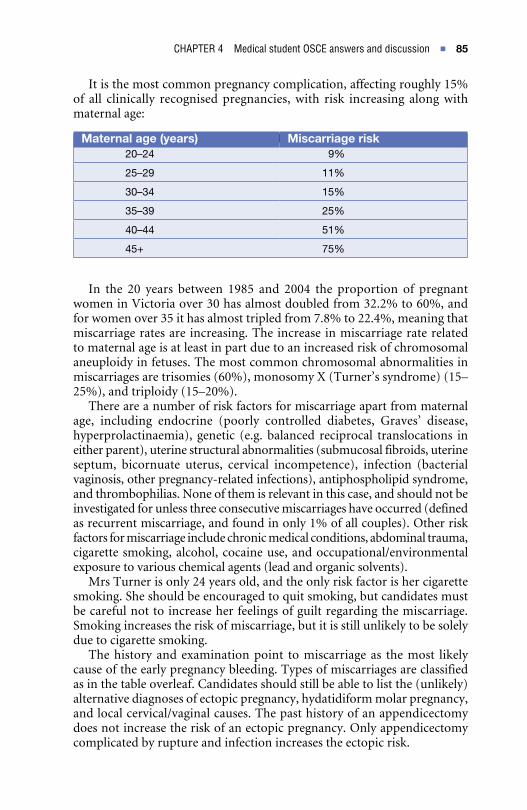
CHAPTER 4 Medical student OSCE answers and discussion n 85
It�is�the�most�common�pregnancy�complication,�affecting�roughly�15%�of� all� clinically� recognised� pregnancies,� with� risk� increasing� along� with�maternal�age:
In� the� 20� years� between� 1985� and� 2004� the� proportion� of� pregnant�women�in�Victoria�over�30�has�almost�doubled�from�32.2%�to�60%,�and�for�women�over�35�it�has�almost�tripled�from�7.8%�to�22.4%,�meaning�that�miscarriage�rates�are�increasing.�The�increase�in�miscarriage�rate�related�to�maternal�age�is�at�least�in�part�due�to�an�increased�risk�of�chromosomal�aneuploidy�in�fetuses.�The�most�common�chromosomal�abnormalities�in�miscarriages�are�trisomies�(60%),�monosomy�X�(Turner’s�syndrome)�(15–25%),�and�triploidy�(15–20%).
There�are�a�number�of�risk�factors�for�miscarriage�apart�from�maternal�age,� including� endocrine� (poorly� controlled� diabetes,� Graves’� disease,�hyperprolactinaemia),� genetic� (e.g.� balanced� reciprocal� translocations� in�either�parent),�uterine�structural�abnormalities�(submucosal�fibroids,�uterine�septum,� bicornuate� uterus,� cervical� incompetence),� infection� (bacterial�vaginosis,�other�pregnancy-related�infections),�antiphospholipid�syndrome,�and�thrombophilias.�None�of�them�is�relevant�in�this�case,�and�should�not�be�investigated�for�unless�three�consecutive�miscarriages�have�occurred�(defined�as�recurrent�miscarriage,�and�found�in�only�1%�of�all�couples).�Other�risk�factors�for�miscarriage�include�chronic�medical�conditions,�abdominal�trauma,�cigarette�smoking,�alcohol,�cocaine�use,�and�occupational/environmental�exposure�to�various�chemical�agents�(lead�and�organic�solvents).
Mrs�Turner�is�only�24�years�old,�and�the�only�risk�factor�is�her�cigarette�smoking.�She�should�be�encouraged�to�quit�smoking,�but�candidates�must�be�careful�not�to�increase�her�feelings�of�guilt�regarding�the�miscarriage.�Smoking�increases�the�risk�of�miscarriage,�but�it�is�still�unlikely�to�be�solely�due�to�cigarette�smoking.
The�history�and�examination�point� to�miscarriage�as� the�most� likely�cause�of�the�early�pregnancy�bleeding.�Types�of�miscarriages�are�classified�as�in�the�table�overleaf.�Candidates�should�still�be�able�to�list�the�(unlikely)�alternative�diagnoses�of�ectopic�pregnancy,�hydatidiform�molar�pregnancy,�and�local�cervical/vaginal�causes.�The�past�history�of�an�appendicectomy�does�not�increase�the�risk�of�an�ectopic�pregnancy.�Only�appendicectomy�complicated�by�rupture�and�infection�increases�the�ectopic�risk.
Maternal age (years) Miscarriage risk20–24 9%
25–29 11%
30–34 15%
35–39 25%
40–44 51%
45+ 75%

86 n Medical student OSCE answers and discussion
Symptoms Cervical os UltrasoundThreatenedmiscarriage
SpottingMinimalpain
Closed FH+ve
Inevitablemiscarriage
IncreasedbleedingCrampingpains
Open FH+ve
Incompletemiscarriage
Bleeding Open FH-ve;some(?POCseen)
Painvariable POCstillinuterus
Completemiscarriage
Reducingbleeding Closed Uterusempty
Painvariable
Missedmiscarriage
Nilorspotting Closed FH-ve
Septicmiscarriage(canaffectanyoftheabovetypes)
Temperature Variable Variable
Uterinetenderness
In� relation� to� investigation�and�management,� the� blood� group� must�be�checked�and�anti-D�immunoglobulin�given�to�rhesus�negative�women,�regardless�of�the�cause�of�bleeding,�to�prevent�isoimmunisation.�The�fetus�has�established�a�circulation�and�is�producing�red�blood�cells�by�about�5.5�weeks�gestation.�However,�only�the�reduced�dose�of�250�IU�is�required�for�pregnancies�under�14�weeks�gestation.�A�pelvic�ultrasound�(with�vaginal�rather�than�abdominal�scanning�to�provide�more�accurate�information)�is�essential,�both�to�pick�up�ectopic�pregnancies�(empty�uterus�or�pseudosac,�adnexal�masses,�free�fluid�in�pelvis),�hydatidiform�molar�pregnancies�(‘snow-storm’�appearance�of�hydropic�villi),� as�well� as� to�aid� in�distinguishing�between� types� of� miscarriages,� as� seen� above.� If� the� pregnancy� is� non-viable,�plans�should�be�made�to�evacuate�the�uterus�of�pregnancy�products�by�surgical,�medical�or�expectant�means.
The�choice�of�evacuation�method�will�be�determined�by�a�combination�of�the�clinical�condition�of�the�patient,�as�well�as�by�patient�choice.�For�example,� if� haemodynamically� compromised,� a� surgical� option,� being�fastest,�will�be�most�appropriate.�Be�wary�of�using�misoprostol�for�medical�evacuation�of�a�pregnancy�in�asthma�sufferers,�as�misoprostol�may�cause�bronchospasm.�This�is�not�an�issue�in�the�current�patient,�and�any�of�the�three�options�would�be�reasonable�at�the�patient’s�discretion,�given�that�she�is�medically�stable.

CHAPTER 4 Medical student OSCE answers and discussion n 87
CC
The�importance�of�a�quantitative�βHCG�is�in�helping�to�interpret�the�ultrasound�result.�A� fetal�heart� should�be�visible�via�vaginal�ultrasound�once� the� βHCG� reaches� 1500–2000� IU/mL;� by� abdominal� ultrasound�once�it�reaches�5000�IU/mL.�During�the�first�10�weeks�gestation�in�a�viable�pregnancy�it�should�rise�by�at�least�66%�every�48�hours.�In�this�case,�with�8000�IU/mL,�the�pregnancy�cannot�be�viable�as�a�fetal�heart�is�not�visible�by�ultrasound.�Another�clue�that�the�pregnancy�has�failed�is�the�loss�of�early�pregnancy�symptoms�(hyperemesis�gravidarum)�in�the�history.
when�taking�a�history�from�the�patient,�focus�on�the�presenting�problem,�but�do�not�forget�your�general�pregnancy�management�(e.g.�ensuring�the�patient�is�on�folate�while�trying�to�conceive�to�reduce�the�risk�of�neural�tube�defects,�and�checking�rubella�status).
In�counselling�the�patient,�the�candidate�should�demonstrate�sensitivity�as� a� miscarriage� is� a� distressing� event.� The� risk� of� a� miscarriage� in� a�subsequent�pregnancy�increases�with�number�of�consecutive�pregnancies�roughly�as�follows:
It� is� important� to�point�out� to� the�patient,� even�without�her�asking,�that�normal�work,�housework�and�sexual�activity�are�not�responsible�for�sporadic�miscarriages.�She�may�be�feeling�guilt�about�some�of�these,�and�she�should�be�reassured.�A�miscarriage�can�cause�a�severe�grief�reaction�similar�to�a�stillbirth�in�some�women,�and�they�should�be�offered�counselling�and�follow-up�if�necessary.�There�may�be�support�groups�available�depending�on�the�patient’s�location.�Otherwise,�her�local�doctor�is�a�good�start.
Q1History• �30-year-old�primigravida�38/40,�singleton�pregnancy• �No�antenatal�complications�(no�gestational�diabetes�or�hypertension)• �Antenatal�investigations�including�20/40�ultrasound�normal• �No�history�of�fever/rash/illness• �No�medical�problems�or�past�surgery• �NKA,�nil�medications,�no�alcohol/cigarettes/no�illicit�drug�use• �was�working�as�office�manager,�and�still�working�at�diagnosis�of�FDIU• �Supportive,�loving�relationship�with�husband• �No�fetal�movements�for�3�days�prior�to�presentation 2 marks (depending on completeness)
33
After1miscarriage: 15%(unaltered)
2miscarriages: 20%(slightlyincreased)
3miscarriages: 30%
4miscarriages: 40%
5miscarriages: 50%

88 n Medical student OSCE answers and discussion
InvestigationsExplain�to�couple:�Investigations�are�to�look�for�a�cause�for�the�FDIU,�so�that�the�likelihood�of�recurrence�and�need�for�preventive�measures�in�next�pregnancy�can�be�ascertained.
½ markMaternal investigations• �Blood�group�and�antibodies�(in�case�of�isoimmunisation)• �FBE�(if�platelets�low,�may�be�disseminated�intravascular�coagulation�
(DIC))• �Fibrinogen• �APTT/INR• �Kleihauer�test�(if�positive,�may�be�concealed�abruption)• �Serology�screening�for�possible�infections�(toxoplasma,�listeria,�
cytomegalovirus,�rubella�serology)• �HbA1C/glycosylated�haemoglobin• �Thyroid�function�tests• �Antiphospholipid/anticardiolipin�antibodies/lupus�anticoagulant• �Antinuclear�antigen�(ANA)
2½ marks (½ for each point to maximum 2 ½)Placenta/cord• �Send�for�histology�after�delivery• �Surface�swabs�for�culture• �Examine�cord�for�knots
1½ marks (½ for each point)Fetal investigations (to be performed after delivery)• �Photographs�for�morphology�assessment• �Karyotype�(by�skin�biopsy)• �Surface�swabs�plus�gastric�aspirate�for�culture• �X-ray�(particularly�important�if�not�consenting�to�autopsy)• �Autopsy:�need�to�approach�this�with�sensitivity�and�explain�the�need�
for�an�autopsy�to�determine�if�there�are�any�issues�found�to�explain�the�FDIU�which�may�impact�on�future�pregnancies.�Must�explain�that�it�will�only�be�performed�with�the�permission�of�the�parents,�but�that�it�is�advisable�to�have�one.
1 mark for discussion on autopsy
1½ marks (½ for each point to maximum of 1½ for the other points)
Q2Some�acknowledgement�of�the�loss�to�the�couple�is�appropriate,�such�as,�“I�am�very�sorry�for�the�tragic�loss�of�your�baby.” 2 marks for comment and appropriate manner“There�does�not�appear�to�be�any�cause�based�on�the�history,�but�we�will�need�to�wait�for�the�results�of�the�investigations�we�have�ordered,�and�tests�on�the�fetus�after�delivery.”�(or�similar�explanation)
½ mark

CHAPTER 4 Medical student OSCE answers and discussion n 89
The�fact�that�Mrs�Harlow�was�working�at�the�time�had�nothing�to�do�with�the�stillbirth.
½ markNeed�to�consider�the�options�for�delivery:�1� �would�not�advise�a�caesarean�section,�as�this�would�create�
complications�for�future�pregnancies/deliveries.�2� �May�opt�for�expectant�management,�as�80%�of�FDIUs�will�deliver�
spontaneously�within�2�weeks.�There�is�a�risk�of�DIC,�with�slow�consumption�of�fibrinogen�over�time,�but�the�risk�is�low�unless�the�FDIU�remains�in�utero�for�>4�weeks.
�3� �Other�option�is�for�an�induction�of�labour�and�vaginal�delivery,�using�prostaglandins�(low-dose�misoprostol,�or�Prostin�–�prostaglandins�E�and�F).
4 marks (1 for each point, and 1 for risk of DIC)Before�delivery,�the�couple�needs�to�decide�if�they�wish�to�see�the�stillborn�baby�or�not.�There�is�some�evidence�that�seeing�the�baby�after�birth�helps�some�couples�to�deal�with�the�grieving�process.�Naming�the�baby�is�important�to�many�couples,�as�well�as�having�photos�and�neonatal�footprints/handprints. 2 markswill�need�to�consider�options�for�burial�of�the�stillborn�baby�at�some�stage.
½ markwill�need�to�be�offered�counselling,�and�referral�to�support�groups,�such�as�SANDS�(Stillbirth�and�Neonatal�Death�Support). 1 markShould�see�a�senior�consultant�within�2�weeks�after�delivery�of�the�stillbirth�to�go�through�the�results�of�the�investigations�and�autopsy,�and�plan�for�any�special�measures�needed�in�subsequent�pregnancies.
½ mark 20 marks maximum
DiscussionTragically,�up�to�half�of�all�fetal�deaths�in�utero�occur�at�term.�when�they�occur�the�couple�require�intense�counselling�and�support.�However,�the�practitioner�must�not� lose�sight�of� trying�to�find�the�cause�of� the�FDIU�so�that�the�risk�to�subsequent�pregnancies,�if�any,�can�be�determined,�and�efforts�can�be�made�to�prevent�a�recurrence.�Unfortunately,�many�FDIUs�at�term�remain�unexplained,�which�can�make�it�difficult�for�the�couple�to�come�to�terms�with�the�loss.
In�subsequent�pregnancies,�with�an�unexplained�term�FDIU,�the�couple�may� require� greater� support� and� more� frequent� antenatal� visits.� while�excessive�fetal�monitoring�should�not�be�encouraged�for�these�patients,�they�may�need�reassurance�that�their�baby�is�well,�particularly�when�nearing�the�gestation�at�which�they�lost�their�previous�baby.�Delivery�may�need�to�be�arranged�by�induction�of�labour�or�caesarean�section�(as�the�patient�directs)�at�a�gestation�just�prior�to�the�previous�loss,�and�if�in�labour,�continuous�fetal�monitoring�may�be�offered�for�parental�reassurance�in�some�centres.

90 n Medical student OSCE answers and discussion
All�maternity�units�should�have�a�protocol�for�managing�and�investigating�FDIUs,�so�that�staff�do�not�fail�to�obtain�potentially�valuable�information�for�the�unfortunate�parents.
The� student� is� expected� to� take� an� adequate� history� of� the� presenting�problem�and�advise�regarding�surgical�termination�of�pregnancy�and�post-procedure�contraception.
Q1History• �24�years�old• �First�pregnancy• �Currently�sexually�active�with�1�partner.�Same�partner�for�the�last��
2�years�but�relationship�broke�up�when�the�pregnancy�was�diagnosed• �Usually�uses�condoms�but�not�always�compliant.�Has�used�the�
combined�oral�contraceptive�pill�(COCP)�in�past�but�has�had�problems�remembering�to�take�it
• �Periods�generally�occur�every�28�days,�lasting�5�days�with�no�problems.�LMP�10�weeks�ago.�Pregnancy�dating�confirmed�on�ultrasound�as�being�10�weeks�gestation
• �No�past�history�–�medical�or�surgical• �No�family�history• �No�allergies,�no�medications,�non-smoker,�occasional�alcohol• �Pap�smear�normal�12�months�ago• �Requesting�a�TOP�as�feels�unable�to�cope�psychologically�with�a�
pregnancy�on�her�own.�Her�parents�live�interstate.�Has�thought�about�the�implications�for�her�of�having�a�TOP.�was�initially�shocked�that�she�was�pregnant�but�now�resigned�to�the�TOP�as�she�feels�it�is�the�best�decision�for�her
• �works�as�a�secretary 8 marks maximum, depending on completeness of history
Q2• �would�be�a�surgical�TOP�due�to�gestation• �Suction�currette�under�general�anaesthetic�or�sedation.�Short�
procedure�with�an�oxytocic�used�to�contract�the�uterus�during�the�procedure
• �Cervical�preparation�using�400�μg�misoprostol�vaginally�at�least�3�hours�beforehand
• �Doxycycline�100�mg�orally�6�hours�prior�to�procedure�and�200�mg�orally�2�hours�after�the�procedure
• �Must�know�blood�group�if�have�to�administer�Anti�D 4 marksRisks• �Perforation,�which�is�minimised�by�the�use�of�misoprostol,�allowing�
gentle�cervical�dilatation
C4C4

CHAPTER 4 Medical student OSCE answers and discussion n 91
• �Haemorrhage,�which�is�minimised�by�the�use�of�misoprostol�and�an�oxytocic�which�contract�the�uterus
• �Infection,�which�is�minimised�by�the�use�of�antibiotics• �No�long-term�sequelae 4 marks (1 for each point)
Q3• �Need�to�have�adequate�ongoing�contraception�so�that�not�faced�with�an�
unplanned�pregnancy�again• �Reliable�options�include�hormonal�contraception�such�as�Implanon,�
Depo-Provera,�Mirena�IUD�or�non-hormonal�such�as�copper�IUD.�These�forms�of�contraception�can�be�administered�at�the�time�of�a�TOP
• �All�forms�must�be�combined�with�the�use�of�condoms�with�casual�sexual�partners
4 marks 20 marks total
Background information on termination of pregnancyAbortion�is�not�unlawful�in�the�state�of�Victoria,�provided�that�the�medical�practitioner�can�prove�the�health�(mental�or�physical)�of�the�mother�would�be� endangered� by� continuing� with� the� pregnancy.� Abortion� laws� vary�between�states�in�Australia.�In�Victoria�one�abortion�is�performed�for�every�three�live�births.
The�annual�worldwide�abortion�rate�varies�between�32�and�46�per�1,000�women�aged�15–44�years,�with�a�peak�rate�at�the�age�of�20�years.�In�most�developed�countries�the�abortion�rate�varies�between�10�and�30�per�1,000�women.�Some�24%�of� the�world’s�population�reside� in�countries�where�abortion�is�not�legally�permitted.�worldwide�about�2�out�of�5�abortions�are�thought�to�be�illegal,�leading�to�between�50,000�to�100,000�preventable�deaths�each�year,�nearly�all�(99%)�in�poor�countries.�Each�hour�8�women�die�from�unsafe�abortions.
There� is� evidence� that� improving� sex� education� and� particularly�promoting�access�to�safe�and�effective�contraception�decreases�the�number�of�abortions�performed�in�any�country.�Abortion�is�ultimately�a�choice�that�the�individual�woman�makes�with�or�without�consultation�with�the�father,�members�of�her�family,�a�religious�advisor�or�her�doctor.
The�number�of�abortions�performed� in�order� to�protect� the�physical�health�of� the�mother� is� few.�Termination�of�pregnancy�is�also� indicated�if� the� mother� has� ingested� a� potential� teratogen� during� the� period� of�organogenesis,�or�if�the�fetus�is�known�to�have�an�abnormality.�Less�than�2%�of�abortions�are�for�fetal�abnormality.
Psychological�or�social�indications�comprise�the�major�reason�for�the�performance�of�pregnancy�termination�in�this�country.�Most�abortions�are�induced�because�the�pregnancy�is�unintended�and�unwanted.�A�minority�of�women�request�abortion�because�of�a�psychiatric�illness�severe�enough�to�

92 n Medical student OSCE answers and discussion
require�intervention.�However,�an�unwanted�or�accidental�pregnancy�can�cause�severe�psychological�stress.
Counselling�is�a�most�important�part�of�the�management�of�a�woman�who�requests�pregnancy�termination.�The�major�objectives�of�counselling�are:• �to�determine�the�reason�for�pregnancy�termination• �to�discuss�the�implications�of�the�request• �to�offer�emotional�support• �to�discuss�future�contraceptive�options.First trimester abortionThe� risk� of� surgical abortion� is� reduced� if� it� is� performed� in� the� first�trimester.� This� procedure� ranks� among� the� safest� and� easiest� surgical�procedures�with�a�mortality�rate�of�1/100,000�procedures.� In�developed�countries�the�mortality�associated�with�childbirth�is�11�times�higher�than�that�associated�with�safely�performed�surgical�abortion�and�30�times�higher�than� for�abortions�performed�up� to�8�weeks�gestation.�More� than�90%�of� terminations� are� performed� in� the� first� trimester� of� pregnancy.� The�most�common�surgical�procedure�is�vacuum�aspiration�(98%),�which�can�take�place�until�14�weeks�gestation.�All�procedures�are�performed�under�local� (paracervical� block)� or� general� anaesthesia.� Pre-operative� cervical�preparation�should�be�undertaken.�The�optimal�drug�is�misoprostol�and�the�optimal�dosage�and�time�interval�of�misoprostol�appears�to�be�400�μg�vaginally� 3� h� prior� to� the� termination� (grade� A� evidence).� The� cervix�should�be�dilated�as�atraumatically�as�possible�and�as�little�as�is�necessary�to�perform�the�operation�safely.�After�cervical�dilatation,�an�oxytocic�drug��(0.5� mg� ergometrine� intravenously� [IV]� or� 10IU� syntocinon� IV)� is�administered�and�the�suction�currettage�performed�under�a�vacuum�pressure�of��800�mmHg.�If�there�is�doubt�as�to�whether�the�uterus�is�empty,�the�cavity�can�be�explored�using�a�sharp�currette.�An�empty�uterus�usually�does�not�bleed.�Surgical�termination�should�not�be�performed�at�≤6�weeks�gestation�due�to�failure�to�remove�the�gestational�sac,�but�medical�abortion�(see�below)�is�most�effective�at�≤7�weeks�gestation.�After�the�procedure�the�products�of�conception�are�routinely�sent�for�histological�assessment.
Medical abortion�is�the�method�of�choice�at�≤7�weeks�and�should�be�offered�at�7–9�weeks�gestation.�It� is�a�safe�and�effective�procedure.�This�method� of� pregnancy� termination� can� be� accomplished� using� 200� mg�RU486�with�800�μg�Misoprostol�administered�vaginally�48�h�later�(grade�A�evidence).�The�addition�of�the�latter�reduces�the�dose�of�each�individual�medication�and�reduces�the�side�effects,�which�are�mainly�gastrointestinal.�when�the�drugs�are�given�in�combination,�5%�require�a�currettage,�1%�will�bleed�heavily�enough�to�require�a�transfusion�and�the�method�will�fail�in�1%.�The�abortion�usually�occurs�within�6�h�of�the�administration�of�the�PG�analogue�in�90%�and�within�8�h�in�96%.
Routine�follow-up�is�mandatory�if�this�method�of�pregnancy�termination�is�used.�The�success�rate�depends�on�the� length�of� the�pregnancy�and�if�

CHAPTER 4 Medical student OSCE answers and discussion n 93
performed�early�has�a�success�rate�similar�to�vacuum�aspiration.�A�Cohrane�review�has�reported�that�medical�methods�of�abortion�in�early�pregnancy�are�safe�and�effective,�with�the�best�evidence�for�the�combination�of�RU486�and� misprostol.� As� yet� there� are� no� currently� available� studies� directly�comparing� surgical� termination�of�pregnancy�with� this� combination�of�medications.Second trimester abortionIn�the�second trimester surgical abortion�involves�dilatation�and�evacuation�(D&E)�with�misoprostol�400�μg�3-hourly�for�3�doses�administered�vaginally�beforehand.�This�method�of�surgical�abortion� is�cheaper�and�safer� than�medical� methods.� It� is� performed� under� intubational� anaesthesia� after��14–16� weeks� gestation� and� volatile� anaesthetic� gases� must� not� be� used.�Serious�complications�occur�in�1%�and�include�perforation,�cervical�damage�and�retained�products.
A� medical termination in the second trimester� can� be� carried� out�using�vaginal�(PGE1�or�PGE2),�intramniotic�or�intramuscular�PGs.�Extra�or�intraamniotic�PGE2�compares�well�in�terms�of�efficacy�with�D&E.�PGE1�pessaries,�such�as�misoprostol�400�μg�can�also�be�placed�in�the�posterior�fornix� of� the� vagina� every� 3–4� h,� with� 5–6� being� required� to� cause� an�abortion.
The�most�important�determinants�of�abortion�mortality�are�gestation�at� the� time� of� the� procedure� and� the� type� of� anaesthesia� used.� Early complications� of� first� trimester� surgical� abortion� include� uterine�perforation,� haemorrhage,� cervical� lacerations,� failure� to� complete� the�procedure� due� to� performance� too� early� in� gestation� and� anaesthetic�mishaps.� Late complications� include� retained� products� (0.2–0.6%),�anaemia,�thromboembolism�and�cervical�stenosis�(0.02%).�The�mortality�rate� is� 1:100,000� from� infection� (23%),� thromboembolism� (23%),�haemorrhage�(20%)�and�anaesthesia�(16%).
Pelvic� infection� may� complicate� up� to� 12%� of� surgically� induced�abortions.�A�meta-analysis�of�randomised�trials�showed�that�prophylaxis�with�antibiotics�effective�against�either�Chlamydia trachomatis�or�bacterial vaginosis� reduced� the� post-abortion� infective� morbidity� by� half.� The�screen� and� treat� strategy� appears� to� be� less� cost-effective� than� routine�prophylaxis.
The�perforation�rate� in� first� trimester� surgical� terminations� is�0.05%�and� in� second� trimester�procedures� is�0.32%.�Perforation�may�occur�at�the� cervical-uterine� junction� during� dilatation� or� during� the� curettage.�The�suction�curette�may�then�be�placed�behind�the�bladder�or�in�front�of�the�rectum.�During�the�procedure�most�perforations�occur�at�the�fundus�and�usually�go�unrecognised.� If�a�perforation� is� suspected� the�extent�of�the�damage�must�be�assessed.�If�there�is�no�active�bleeding,�no�action�is�necessary.�However,�if�the�defect�is�large,�there�is�active�bleeding�or�there�is�a�suspicion�of�bowel�injury,�laparoscopy�+/–�laparotomy�must�be�performed.�If� a� perforation� does� occur� the� procedure� may� be� completed� under�

94 n Medical student OSCE answers and discussion
laparoscopic�or�ultrasound�guidance,�or�alternatively�may�be�performed,��2� weeks� later.� Risk� factors� for� perforation� are� previous� gynaecological�surgery� including� previous� abortion,� lower� uterine� segment� caesarian�section�and�large�loop�excision�of�the�transformation�zone�of�the�cervix.�Atraumatic�pre-operative�dilation�of�the�cervix�using�PGs�may�prevent�this�important�complication.
Vacuum� aspiration� in� the� first� trimester� is� not� associated� with� later�infertility,� increased� risk� of� ectopic� pregnancy� or� subsequent� non-viable�outcome.�First�trimester�abortion�is�also�not�associated�with�later�spontaneous�miscarriage.�There�is�also�no�association�with�an�increased�risk�of�breast�cancer.�It�is�uncertain�if�second�trimester�abortions�ultimately�affect�fertility.
Studies� have� shown� that� the� psychological sequelae� of� abortion� are�minimal� for� the� majority� of� women� and� that� choosing� to� terminate� a�pregnancy�rather�than�deliver�an�unwanted�first�pregnancy�does�not�place�women�at�higher�risk�of�depression.�Indeed,�the�prevention�of�abortion�may�be�psychologically�more�devastating�for�the�woman�and�her�child.�After�pregnancy�termination�most�women�express�relief.�Feelings�of�guilt�and�loss�are�usually�self-limiting.�The�women�who�may�have�difficulty�coping�with�an�abortion�are�those�who�are:• �dependent�on�alcohol�or�drugs�or�who�live�in�an�abusive�relationship�
and�have�poor�social�supports• �victims�of�rape�or�incest• �aborting�because�of�fetal�abnormality�or�at�an�advanced�gestational�age�
at�the�time�of�the�termination• �suffering�from�a�pre-existing�psychiatric�illness• �unsure�about�going�ahead�with�an�abortion.
In�Rh�negative�women,�anti-D�prophylaxis�must�not�be�forgotten.
Q1N.B. This OSCE station is best examined with a doll for the candidate to demonstrate the resuscitation steps on. A stethoscope and neonatal resuscitation mask are helpful, if available, but not essential (the candidate can indicateare helpful, if available, but not essential (the candidate can indicateavailable, but not essential (the candidate can indicate that they use them whether or not they are present by miming the necessary actions).At�birth,�should�immediately�assess�if�baby�is�breathing,�crying,�and�whether�or�not�there�is�good�tone. 1 markEXAMINER:�“The�baby�is�not�breathing,�and�has�no�muscular�tone.”• �Should�dry�and�provide�tactile�stimulation�to�the�baby�(e.g.�with�towel/
cloth/rubbing�of�skin)• �Provide�warmth�to�the�baby�(radiant�heat�source)• �Position�the�airway�–�head�should�be�in�neutral�or�slightly�extended�
position�(if�overextended,�may�block�airway)• �Check�breathing,�and�check�heart�rate�with�stethoscope 4 marks (1 mark for each)
C5

CHAPTER 4 Medical student OSCE answers and discussion n 95
EXAMINER:�“The�baby�is�still�not�breathing�and�has�a�heart�rate�of�about�80�beats/minute.”• �Need�to�initiate�positive�pressure�ventilation,�using�air�or�oxygen,�either�
with�self-inflating�bag�or�a�T-piece�resuscitation�device�(e.g.�Neopuff).�The�latter�provides�PEEP�(positive�end�expiratory�pressure)�and�ventilations�should�be�administered�at�a�rate�of�60�times�per�minute.
2 marks• �Reassess�every�minute�or�so�for�attempts�by�infant�at�respiration,�as�
well�as�heart�rate�auscultation. 1 markEXAMINER:�“The�baby�is�still�not�breathing,�and�the�heart�rate�has�dropped�to�40�beats/minute.”• �Commence�chest�compressions:�Place�hands�around�the�chest�with�two�
thumbs�over�the�lower�half�of�the�baby’s�sternum�(1�finger’s�breadth�below�a�line�drawn�between�the�nipples),�compressing�the�thumbs�down�on�the�sternum�so�that�one-third�of�the�chest�anterior-posterior�diameter�is�depressed.
• �Should�perform�90�chest�compressions�and�30�ventilations�in�1�minute,�with�a�ratio�of�3:1.
3 marks• �Reassess�the�respiratory�efforts,�heart�rate,�colour�and�tone�of�the�baby�
regularly. 1 markEXAMINER:�“The�baby�starts�to�cry�after�3�minutes�of�resuscitation,�the�heart�rate�has�increased�to�120�beats/minute,�and�the�baby�is�now�pink�with�good�tone.”• �Cease�resuscitation,�keep�baby�warm. 1 mark
Q2The�Apgar�score�is� from�0�to�10,�and�is�used�to�identify� infants� in�need�of�resuscitation.�Apgar�scores�are�routinely�determined�and�recorded�at��1�minute�and�5�minutes�after�birth.�They�are�determined�by�a�score�between�0�and�2�for�each�of�the�following�five�signs:
Sign Score = 0 Score = 1 Score = 2Heartrate Absent <100bpm >100bpm
Respiration Absent Weak/irregular Regular/crying
Colour White/blue PaleextremitiesBodypink
Completelypink
Tone Flaccid Reduced Wellflexed
Irritability/Reflexes
None Reduced Cough/cry

96 n Medical student OSCE answers and discussion
C6
2 marks for why Apgars performed
5 marks for each criterion for Apgar (1 mark each)
20 marks total
DiscussionA�knowledge�of�basic�neonatal�resuscitation�is�expected�to�be�demonstrated�by�the�examination�candidate�in�this�station.�The�resuscitation�described�is�based�on�the�guidelines�of�the�Australian�Resuscitation�Council.
The�flowchart�on�the�opposite�page�can�be�obtained�from�the�website:�http://www.resus.org.au/arc_neonatal_flowchart.pdf
In� the� OSCE� exam,� there� would� be� a� mannequin� for� the� student� to�demonstrate�the�performance�of�a�Pap�smear�on.�In�this�practice�OSCE�the�exam�candidate�should�mime�the�actions�of�the�Pap�smear,�or�describe�the�steps�verbally.
The�student�is�expected�to�go�through�the�process�of�performing�a�Pap�smear�on�the�mannequin.�They�are�expected�to�understand�the�different�steps�in�the�performance�of�a�Pap�smear�and�to�understand�why�these�tests�are�performed�and�how�frequently�they�should�occur.Equipment required• �Mannequin• �Bivalve�speculum• �Cytobrush�or�cervex�brush• �wooden�spatula• �Fixative• �Lubricant�or�water• �Glass�slides• �Slide�covers• �Pencil• �Adequate�lighting

CHAPTER 4 Medical student OSCE answers and discussion n 97
February 2006
AUSTRALIAN RESUSCITATION COUNCIL
NEONATAL FLOWCHART
Birth
Breathing or crying?
Good muscle tone?
Routine care:
Dry the baby
Provide warmth
Clear the airway only if needed
Assess breathing, colour and heart rate
Dry and stimulate Position the head and neck to open the airway Provide warmth
If heart rate <100/min or inadequate breathing
Give positive pressure ventilation until heart rate >100 and infant breathing*
Inadequate breathing and heart rate <60/min*
Assess adequacy of ventilation and improve if possibleIf heart rate does not increase >60/min give chest compressionswith positive pressure ventilation at 3:1
If heart rate still does not increase > 60/min reassess ventilation technique* give adrenaline May also need to give IV fluids
Yes
No
* Endotracheal intubation may be considered at several stages
Assess breathing and heart rate *
If baby is breathing, heartrate is >100/min andbeginning to look pink,then give routine careand observationsappropriate for gestation
•••
•
•
•
•
Figure 4.2 Theassessmentforresuscitationandsubsequentmanagementofanewbornbabyintheabsenceofmeconium

n
98 Medical student OSCE answers and discussionQ1The�student�is�expected�to�state�that�Pap�smears�are�performed�to�detect�pre-cancerous�cervical�changes�or�cervical�dysplasia.�It�is�a�screening�test�that�shows�there�may�be�changes,�but�does�not�give�a�definite�diagnosis�of�what�the�changes�are.�If�there�is�an�abnormal�result�the�patient�should�be� referred� for� a� colposcopy� (examination� of� the� cervix� with� a� bifocal�microscope�by�a�gynaecologist).�Pap�smears�are�done�every�2�years�in�the�absence�of�problems. 4 marks total, depending on completeness of the answer
Q2The�student�is�expected�to�go�through�the�process�of:�1� �writing�detail�on�the�slide�2� �getting�the�equipment�necessary,�such�as�speculum,�brush�and�spatula,�
fixative�and�slide�cover�3� �warming�the�speculum�with�water�4� �inserting�the�speculum�into�the�vagina�and�displaying�the�cervix�and�
fixing�the�speculum�5� �with�the�spatula,�rotating�the�pointy�end�360॰�in�the�internal�os�to�
sample�the�transformation�zone�6� �with�the�cytobrush�or�cervex�brush,�rotating�it�360॰�in�the�internal�os�to�
sample�the�transformation�zone�7� �fixation�of�the�slide�and�placing�in�the�slide�holder�8� �removal�of�the�speculum. 16 marks (2 marks for each point)
DiscussionPap� smears� (named� after� Papanicolaou,� the� inventor� of� the� test)� are�performed�as�a�screening�test�to�identify�cervical�dysplasia�before�it�progresses�to�cervical�carcinoma.�The�Pap�smear�is�an�example�of�‘exfoliative�cytology’.�It�is�not�a�diagnostic�test.�The�smear�samples�cells�from�the�‘transformation�zone’�of�the�cervix.�The�transformation�zone�is�the�point�of�transition�from�columnar�to�squamous�epithelium�and�is�the�site�at�which�squamous�cell�cervical�carcinomas�are�most�likely�to�occur.�The�cells�are�preserved�on�a�slide�for�examination.�The�proportion�of�cells�on�the�slide�with�abnormal�characteristics,�such�as�poor�differentiation,�increased�nuclear-cytoplasm�ratio,�hyperchromasia,�and�nuclear�pleomorphism,�determines�whether�the�smear�is�described�as�CIN�I-III�or�mild�to�severe�dysplasia.�Because�the�Pap�smear�is�a�screen,�in�the�case�of�abnormality�all�women�with�an�abnormal�Pap� smear� should� have� a� colposcopy.� A� colposcopy� enables� a� targeted�biopsy�of� the�abnormal�area� to�occur,� thus�giving�a�definitive�diagnosis�of�the�degree�of�the�abnormality.�The�woman�can�then�be�appropriately�treated�prior�to�the�development�of�a�cancer.
In�Australia�it�is�recommended�for�women�every�2�years�until�70�years�of�age,�starting�one�year�after�the�first�episode�of�sexual�intercourse.�The�reason�that�the�incidence�of�cervical�cancer�has�decreased�is�due�to�the�performance�

CHAPTER 4 Medical student OSCE answers and discussion n 99
of�Pap�smears.�In�Australia,�women�who�present�with�cervical�cancers�tend�to�be�those�who�have�never�had�a�Pap�smear.
Pre-cancerous�lesions�of�the�cervix�are�associated�with�infection�with�oncogenic�strains�of�the�human�papilloma�virus�(HPV).�A�prophylactic�HPV�vaccine�is�now�available�in�Australia�and�New�Zealand�to�prevent�infection�with�some�of�the�more�common�oncogenic�strains�of�HPV.�RANZCOG�recommends�the�vaccination�of�all�females�aged�9�to�26�years�against�HPV.�It� is�expected�that� this�will� reduce� the� incidence�of�cervical�dysplasia� in�the�future.�However,�despite�vaccination,�current�recommended�screening�protocols�should�continue�to�be�adhered�to.
Q1History• �Take�a�history�for�any�medical�conditions,�allergies�and�medications�to�
ensure�there�are�no�contra-indications�to�any�methods�of�analgesia• �Medical�Hx:�childhood�asthma�now�resolved• �Surgical�Hx:�appendicectomy• �Family�Hx:�nil�significant• �Medications:�Blackmores�multivitamins�for�pregnancy�and�
breastfeeding• �Allergies:�penicillin�–�rash
½ mark each (maximum 1 mark)Discuss�the�patient’s�options,�commencing�with�drug-free�methods,�then�
moving�on�to�prescribed�analgesia�and�then�anaesthesia.�1� �Drug-free�methods:�mobilising�–�walking,�rocking;�position�change�–�bean�
bag,�floor�mat,�mediball;�hot�packs;�massage;�water-based�shower,�bath½ mark each (maximum 1 mark)
�2� �Patient-controlled�analgesia:�nitrous�oxide,�TENS½ mark each (maximum 1 mark)
�3� �Prescribed�analgesia:�oral,�IM,�IV:�Panadol/Panadeine/Panadeine�forte,�IM�pethidine�(uncommon:�IV�pethidine)
½ mark each (maximum 1 mark)�4� �Anaesthesia:�epidural�block,�spinal�block,�combined�epidural/spinal
½ mark each (maximum 1 mark) 5 marks total
Q21 Drug-free methodsAdvantages• �No�medications• �No�needles�or�injections• �No�risk�of�fetal�or�maternal�sedation• �Multiple�options• �Patient�remains�in�control• �Partner�can�be�involved 1 mark each (3 marks maximum)
C7C7

100 n Medical student OSCE answers and discussion
Disadvantages• �May�not�be�adequate�for�some�women’s�analgesia�needs• �water-based�therapy�cannot�be�used�with�continuous�CTG��
monitoring• �Movement�options�may�be�limited�if�continuous�monitoring�is�
required½ mark for any one of the above
2 Patient-controlled analgesiaNitrous oxideAdvantages• �Rapid�onset/rapid�offset• �Can�be�used�for�many�hours• �Patient-controlled• �No�overdose/sedation�risk• �Provides�a�reasonable�level�of�analgesia 1 mark each (3 marks maximum)Disadvantages• �Nausea• �Dry�mouth• �Dizziness• �Mask�can�be�claustrophobic• �May�not�be�adequate�for�some�women’s�needs
½ mark each (1 mark maximum)TENSAdvantages• �No�sedation• �No�nausea
½ mark for any one of the aboveDisadvantages• �Limited�analgesic�benefit• �Cannot�use�water-based�therapy
½ mark for any one of the above3 Prescribed analgesiaAdvantages• �Provides�reasonable�analgesia• �Routinely�available• �Does�not�require�IV�access
½ mark each (1 mark maximum)Disadvantages• �Nausea/vomiting• �Maternal:�altered�conscious�state/sedation• �Fetal�sedation/�respiratory�compromise• �local�pain/hematoma�at�injection�site• �Rare�risk�of�nerve�damage�(sciatic�nerve)• �Intermittent�administration�is�required

CHAPTER 4 Medical student OSCE answers and discussion n 101
• �Not�appropriate�analgesia�for�heroin-using�clients 1 mark each (2 marks maximum)4 AnaesthesiaAdvantages• �Usually�provides�excellent�pain�relief
½ markDisadvantages• �Requires�specialised�operator�(anaesthetist)• �Rare�risk�of�complications:�abscess,�hematoma,�nerve�injury• �Risk�of�failure,�incomplete�block• �Requires�IV�access,�CTG�monitoring,�in-dwelling�catheter• �Loss�of�mobility�due�to�motor�blockade• �Possible�maternal�hypotension• �Post-dural�puncture�headache• �Increased�requirement�for�Syntocinon�augmentation• �May�be�contra-indicated�for�some�congenital�or�acquired�spinal�disease�
and�some�medical�illnesses�e.g.�lumbar�disc�disease,�multiple�sclerosis½ mark each (3 marks maximum)
15 marks total (Q2)
20 marks total
DiscussionOverviewAs�with�any�clinical�encounter,�the�student�is�expected�to�briefly�review�the�relevant�clinical�history�at�the�commencement�of�the�OSCE.�Although�it�would�be�uncommon�to�find�according�to�the�antenatal�history�that�any�particular�method�of�pain�relief�is�contra-indicated,�this�still�demonstrates�that�the�candidate�shows�a�logical�and�clinically�oriented�approach�to�the�problem.�The�candidate�is�then�expected�to�show�a�sensible�progression�to�the�answer,�beginning�with�widely�available,�low-tech,�low-risk�pain�relief�options�and�working�through�to�the�most�effective�but�least�available�and�most�interventional�methods�of�pain�relief.DiscussionThe� experience� and� implications� of� labour� pain� varies� widely� between�women�and�may�even�vary�between�labours�for�the�same�woman.�Pain�is�commonly�experienced�as�lower�abdominal�pain�during�contractions�that�can� extend� to� intermittent� or� continuous� lumbo-sacral� back� pain,� and�gluteal�or�thigh�pain.�Pain�has�been�variably�reported�as�up�to�horrible�or�excruciating.�On�average,�labour�pain�increases�with�progressive�cervical�dilatation� and� depends� upon� the� intensity,� duration� and� frequency� of�uterine� contractions.� As� normal� labour� still� represents� a� physiological�phenomenon,�researchers�are�still�divided�as�to�whether�labour�pain�has�significant�detrimental�effects.�The�potential�negative�physiological�effects�of�labour�pain�include�increased�cardiac�output,�increased�blood�pressure�and�heart�rate�and�hyperventilation.

102 n Medical student OSCE answers and discussion
Intra-partum�pain� relief�options�vary�widely�depending�upon�ethnic�origin,� cultural�practices,� location�of�birth�and� individual� expectations.�Continuous�trained�labour�support,�including�both�emotional�and�physical�assistance,� has� been� demonstrated� to� reduce� the� usage� of� intra-partum�analgesia,�increase�the�likelihood�of�a�spontaneous�vaginal�birth,�increase�the�woman’s�satisfaction�with�the�birth�experience,�and�has�no�known�risks.�This�is�of�greatest�benefit�when�begun�early�in�labour.
Bathing�during�labour�is�safe�and�appears�to�be�most�effective�when�used�beyond�5�cm�dilatation�for�limited�periods.�Bath�use�may�be�of�benefit�by�increasing�maternal�relaxation�and�decreasing�catecholamine�production.�Bathing� temperature� should� be� kept� to� 36–37॰C� to� prevent� maternal�hyperthermia�and�potential�neonatal�morbidity.�Studies�are�divided�upon�the�overall�effectiveness�of�bathing�during�labour�for�pain�relief.
Studies�of�the�benefits�of�positioning�in�labour�for�pain�relief�are�limited�and�difficult�to�conduct.�However,�the�freedom�to�adopt�the�position�of�choice� considered� most� beneficial� by� the� mother� for� reduction� of� pain�should�be�encouraged.
Nitrous oxide�inhaled�via�an�on-demand�valve�is�widely�used�in�Australia.�It�can�be�used�at�any�stage�of�labour,�for�any�duration�of�time�and�is�widely�accepted�due�to�its�ease�of�use,�minimal�side-effects,�and�absence�of�effect�on�uterine�activity.�Due�to�the�intermittent�nature�of�uterine�contraction-related�pain�and�the�time�lag�between�the�commencement�of�inhalation�and�the�onset�of�analgesic�effect,�nitrous�oxide�is�most�effective�if�inhaled�before�the�commencement�of�the�contraction,�but�in�practice�this�can�be�difficult�to�achieve.�Quantitative�effects�of�nitrous�oxide�on�pain�scores�have�been�difficult�to�validate�scientifically;�however,�in�many�studies,�labouring�women�describe�significant�analgesic�effect�with�nitrous�oxide�use.
A� TENS� (transcutaneous� electrical� nerve� stimulation)� consists� of� a�stimulator� and� a� pair� of� electrodes.� The� stimulator� contains� a� pulse�generator�and�two�controls�for�amplitude,�for�amplitude�and�frequency.�It�is�attached�to�the�spinal�processes�of�the�patient,�and�impulses�cause�muscle�fasciculation�to�alter�the�sensation�of�pain�during�uterine�contractions.�It�is�a�non-invasive�option�of�pain�relief,�but�while�effective�for�some�women,�does�not�have�as�strong�an�analgesic�effect�as�more�invasive�options�discussed�below.
Epidural anaesthesia�is�a�nerve�block�technique�using�local�anaesthetic�and/or�opioid�agents�that�requires�the�skills�of�a�specialist�anaesthetist.�Side�effects�such�as�pruritis,�nausea,�vomiting,�inability�to�void,�impaired�motor�blockade,�an�increased�likelihood�of�instrumental�delivery,�a�greater�need�for�oxytocin�augmentation,�and�risks�of�maternal�hypotension�and�fever�are�all�well� recognised.�A� lumbar�epidural� technique� is� the�most�widely�used,�but�combined�spinal/epidural�(CSE)�approaches�are�also�possible.�A�CSE�offers�the�spinal�advantages�of�faster�onset�of�analgesic�effect,�with�the�epidural�advantages�of�continuous�infusion�and�ongoing�analgesia.�In�general,�an�epidural�anaesthesia�is�more�effective�for�labour�pain�relief�than�

CHAPTER 4 Medical student OSCE answers and discussion n 103
CC
non-epidural�methods�but�it�has�more�side�effects�and�complications.�Minor�side�effects�such�as�pruritis�and�nausea�depend�upon�the�inclusion�of�opioid�in�the�epidural�agents.�Pruritis�rates�for�local�anaesthetic�agents�alone�range�from�0–4%,�but�up�to�an�average�of�62%�in�the�presence�of�narcotic�agents.�Nausea�and�vomiting�rates�overall�are�low,�on�average�<5%.�At�least�60%�of�women�require�an�indwelling�catheter�for�an�inability�to�void�spontaneously�and�in�many�centres�the�use�of�an�indwelling�catheter�is�routine�with�epidural�usage�to�avoid�the�risks�of�urinary�retention�and�bladder�overdistension.�Hypotension�rates�are�variable�and�can�occur�in�up�to�50%�of�labouring�women� following� epidural� insertion.� Prophylactic� intravenous� fluid�administration�may�be�of�benefit.�According�to�the�Cochrane�collaboration,�epidural�anaesthesia�increases�the�risk�of�instrumental�delivery�(RR1.38),�maternal�fever�>38�॰C�(RR�3.6)�with�no�demonstrated�effect�upon�caesarean�section�rates,�neonatal�Apgar�scores�or�long-term�backache.
Encounter 1History• �Mrs�w,�62�years�old• �Currently�fit�and�well.�Had�surgery�3�months�ago�for�endometrial�
cancer�(surgical�cure,�with�no�further�treatment�required)• �G6P6�(3�daughters,�3�sons,�all�NVDs)• �Past�Hx:�R�Breast�cancer�(aged�52;�treated�with�wide�local�excision�
and�axillary�clearance.�Had�tamoxifen�for�2�years,�then�arimidex,�as�developed�an�endometrial�polyp�with�tamoxifen)
• �Non-drinker,�non-smoker• �NKA�meds�–�Arimidex• �Family�Hx:
–�Daughter�died�of�bowel�cancer�aged�30–�All�5�other�children�have�had�colonic�polyps�removed,�and�have�regular�colonoscopies�for�screening–�Father�died�of�bowel�cancer�aged�76–�Mother�alive�and�well–�No�other�siblings–�History�for�grandparents�unknown
• �Lives�in�rural�area;�runs�a�dairy�farm�with�her�husband,�one�son,�and�his�family;�other�children�live�in�the�nearest�city;�6�grandchildren
9 marks
Encounter 2• �Strong�family�history�of�bowel�cancer�(2�first-degree�relatives:�father�
and�daughter).�Also�personal�history�of�breast�and�endometrial�cancer• �Recommend�review�at�a�familial�cancer�clinic�for�consideration�of�
genetic�testing 3 marks
88

104 n Medical student OSCE answers and discussion
Encounter 3• �Hereditary�non-polyposis�colorectal�cancer�(HNPCC)• �Hereditary�breast�and�ovarian�cancer�(BRCA1�and�BRCA2)
2 marks
Encounter 4• �HNPCC�–�colonoscopy�for�bowel�cancer/no�screening�test�for�
endometrial�cancer–�colectomy
• �BRCA�1/2�–�mammography�(breast),�CA125�+�pelvic�U/S�(ovary)–�prophylactic�mastectomies�(breast),�prophylactic�BSO�(ovary)
6 marks
20 marks total
DiscussionThe�medical�student�is�expected�to�display�a�basic�understanding�of�some�common�hereditary� cancer� syndromes.�The�patient�has�a� strong� family�history� of� bowel� cancer,� as� well� as� a� personal� history� of� endometrial�and� breast� cancer.� Hereditary� non-polyposis� colorectal� cancer� should�be� suspected,� and� the�patient� referred� to�an�appropriate�genetic� cancer�counselling� service.�while� the� students�are�not� expected� to�counsel� the�patient�as�to�options�for�prophylactic�treatment�(i.e.�whether�or�not�to�have�a�prophylactic�colectomy),� they�are�expected� to�be�aware�of� the�known�options�for�screening�or�prophylactic�surgery�in�these�cases.
Student OSCe dQ1N.B. obstetric emergency, so need to keep history-taking brief• �Medical�history?�Nil• �Surgical�history?�Nil• �Family�history?�Nil• �Medications?�Nil• �Allergies?�Nil• �Pap�smear:�at�first�visit,�normal�result�obtained• �Progress�of�the�pregnancy�to�date:�any�complications�such�as�
gestational�hypertension,�gestational�diabetes?�NoPresenting complaint• �Bleeding:�approx�0100.�woken�from�sleep;�found�self�lying�in�a�pool�of�
blood�on�bed.�went�to�toilet,�more�fresh�bleeding�into�the�bowl.�Called�the�ambulance.
• �Amount:�at�least�2�cups• �Site:�vaginal• �Appearance:�fresh,�bright,�like�blood�from�a�cut�fingerAssociated features• �Pain:�nil�initially,�now�some�crampy�period�pains
D1D1

CHAPTER 4 Medical student OSCE answers and discussion n 105
• �ROM:�not�noted• �Uterine�activity�period�pains�as�noted• �Fetal�movements�present
No ROMPain
Fetal movementsNo significant medical surgical history
No allergies
3 marks (1 mark each, any 3 of above)
Q2• �BP�115/70• �HR�100• �Periphery�warm�and�well�perfused
BPHR
Peripheries
2 marks (1 point each any 2)Abdo:• �symphysiofundal�height�=�34�weeks• �uterine�tenderness�=�moderate• �lie:�longitudinal• �presentation:�cephalic,�mobile• �presence�of�fetal�heart:�detected• �Timing-tightenings,�2�in�10• �Bleeding:�fresh�blood�on�pads,�soaking�approx�1�pad�every�20�mins
TendernessUterine activity
Fetal lie/presentationFetal heart
4 marks (1 each)• �Speculum�Ex:�Clot�present• �Total�measured�loss�from�clots�=�100�mL• �Active�fresh�bleeding�seen�through�os• �Exclude�local�lower�genital�tract�causes�of�bleeding–�nil�sites�seen
BleedingOngoing loss
Speculum examination: Exclude lower causesIdentify blood through os
3 marks
Q3• �IV�bung

106 n Medical student OSCE answers and discussion
• �IV�fluids–�colloid/crystalloid�to�replace�loss�at�least�600�mLResuscitation/maternal stabilization
IV bungIV colloid/crystalloid
3 marks• �X-�match�blood�(2�units)• �Kleihauer• �APPT/INR�or�clotting�studies
2 marks (any 2 of above: 1 mark each)• �Continuous�CTG�monitoring�while�bleeding
1mark• �Fast�while�actively�bleeding• �Notify�registrar/specialist/senior�member�of�staff• �Prepare�for�delivery�if�fetal�distress�or�ongoing�haemorrhage• �Urgent�ultrasound�for�placental�site
1 mark (½ mark each for any of the above)
Q4• �Placental�abruption 1 mark
20 marks total
DiscussionThis�is�a�difficult�station�for�the�medical�student�as�it�requires�the�student�to� simultaneously� consider� directed� history-taking,� specific� points� on�examination,�as�well�as�basic�steps�of�resuscitation�and�management�in�a�time-pressured�setting.�As�such,�it�helps�to�differentiate�the�student�who�has�spent�time�in�the�delivery�suite�and�seen�how�such�cases�are�managed�in�this�way�in�a�real-life�setting,�where�history,�examination�and�management�are�frequently�combined�in�similar�urgent�scenarios.�This�ability�to�manage�multiple�threads�of�information�only�comes�with�practice.�In�this�scenario,�designed� to� simulate� a� major,� evolving� abruptio� placenta,� the� history�required� is�brief�and�directed�towards�major� life-threatening� issues�that�would�interfere�with�surgery�or�an�anaesthetic.
Basic�minimums�that�must�be�obtained�in�such�a�scenario�include:�any�prior� operations� or� anaesthetics?� Any� major� medical� illnesses/chronic�conditions,�e.g.�asthma,�diabetes,�hypertension?�Any�medications�or�drugs�of�dependence?�Any�allergies?�Every�doctor�should�have�a�standard�list�of�major�questions�to�ask�in�any�emergency�setting�and�this�is�a�good�time�to�practise�using�it�even�if�the�introduction�has�stated�that�the�patient�was�previously�well.�In�this�setting,�asking�for�a�handheld�record�from�the�patient�can�be�of�immense�benefit,�but�may�not�be�available.
Once�the�basic�history�has�been�obtained�the�examination�needs�to�be�directed�towards�the�immediate�wellbeing�of�the�mother�and�the�fetus,�as�well�as�the�cause�of�the�bleeding,�with�the�background�plan�of�whether�the�fetus�requires�immediate�or�semi-urgent�delivery�within�the�next�24�hours.�

CHAPTER 4 Medical student OSCE answers and discussion n 107
It�is�easy�to�forget�that�a�young,�fit�woman�may�have�lost�significant�blood�volume�before�there�are�any�physiological�signs�of�compromise;�conversely,�anxiety�may�induce�a�tachycardia�that�is�out�of�sync�with�the�other�physical�signs.�Typically,�a�blood�loss�of�15%�of�total�circulating�volume�will�have�no�effect�on�measured�heart�rate�or�blood�pressure,�and�the�patient�will�still�have�warm,�well-perfused�peripheries.�Signs�of�mild�shock�may�not�be�evident�until�a�blood�loss�of�20–25%,�progressing�to�profound�hypotension�and�collapse�once�the�blood�loss�reaches�40%�of�circulating�volume.�Even�if�a�non-invasive�monitor�is�used�to�regularly�record�the�blood�pressure�and�heart�rate,�laying�hands�on�the�patient’s�pulse�still�provides�invaluable�information�about�the�quality�of�the�pulse�and�the�peripheries,�and�is�to�be�encouraged�in�all�emergency�situations.
The� examination� then� progresses� to� an� assessment� of� the� abdomen�and�the�gravid�uterus,�looking�both�for�clues�as�to�the�cause�of�bleeding�and�clinical� information�that�may� impact�upon�the�management�of� the�pregnancy.�The�two�most�common�causes�of�APH�are�abruptio�and�placenta�praevia,�and�the�history�has�already�indicated�that�the�20-week�ultrasound�showed�a�non-praevia�placenta.�This�is�reassuring�at�approximately�99%�accuracy,�so�it�is�rare�but�not�impossible�to�find�a�missed�placenta�praevia�or�succenturiate�lobe�not�recognised�at�the�20-week�scan.
Encounter 1History• �Mrs�D,�62�years�old• �Has�had�a�‘lump’�in�the�vagina�for�several�years.�Associated�with�a�
dragging�sensation.�Become�much�worse�over�the�last�3–4�months• �Sometimes�has�to�push�the�lump�back�in�order�to�empty�her�bowels• �Urinary�incontinence�slowly�worsening�over�the�last�few�years• �Loses�urine�when�laughs,�coughs,�sneezes,�jumps,�losing�small�amounts�
of�urine;�needs�to�wear�an�incontinence�pad�all�the�time�during�the�day�‘just�in�case’;�has�3–4�episodes�of�incontinence�per�week
• �Goes�to�the�toilet�8�times�per�day�(“to�keep�my�bladder�empty�and�prevent�accidents”),�and�gets�up�once�in�the�night
• �Occasional�urgency,�but�not�the�major�feature�of�the�incontinence.�No�double-voiding
• �No�incontinence�of�flatus�or�faeces.�No�constipation;�normal�bowel�habit
• �Not�sexually�active�(husband�has�Alzheimer’s)• �Drinks�4�cups�of�coffee�per�day,�and�has�a�couple�of�glasses�of�wine�in�
the�evenings• �Has�had�6�normal�vaginal�deliveries�of�7�children�(one�set�of�twins�born�
vaginally);�had�‘quick�labours’• �Menopausal�at�50�years�old,�no�post-menopausal�bleeding;�used�
hormone�therapy�for�5�years�for�hot�flushes,�then�stopped�due�to�concern�regarding�her�breast�cancer�risk
• �Last�Pap�smear�15�years�ago�(normal)
D2D2

108 n Medical student OSCE answers and discussion
• �Never�had�a�mammogram• �Past�medical�Hx:�hypertension�(Rx�atenolol;�well-controlled)• �Past�surgery:�open�cholecystectomy�(10�years�ago);�bilateral�total�knee�
joint�replacement�(5�years�ago);�appendicectomy�aged�10�y.o.• �No�allergies;�meds:�atenolol• �Lives�with�husband�(Alzheimer’s),�and�also�looks�after�her�severely�
disabled�grand-daughter�(cerebral�palsy)�5�days�a�week�while�her�daughter�works.�Her�daughter�is�reliant�on�this�help
• �Ex-smoker�(quit�12�years�ago);�drinks�wine�(2�glasses)�in�the�evening
Encounter 2Examination• �Obese�woman,�BMI�38• �BP�135/90,�PR�72,�T�37�C,�RR�16• �Chest�clear,�No�breast�lumps• �Abdomen:�moderate�pannus,�no�masses• �Vulva:�normal• �Sims�speculum�Ex:�Grade�4�uterine�descent�(procidentia),�Grade�2�
rectocele,�Grade�2�cystocele;�cervix�has�some�ulceration,�with�a�dry�and�wrinkled�appearance�(can�perform�Pap�smear);�Stress�incontinence�demonstrated�after�reduction�of�the�uterus
Encounter 3Issues�include:�1� �the�severe�utero-vaginal�prolapse�2� �the�urinary�incontinence�3� �general�health�issues.Investigations�required�include:• �mid-stream�urine�for�culture,�to�rule�out�a�bladder�infection• �urodynamics�studies�to�determine�if�the�major�cause�of�urinary�
incontinence�is�stress�or�urge�(N.B.�will�need�to�perform�studies�with�uterus�replaced�into�the�pelvis,�or�the�procidentia�may�mask�more�severe�stress�incontinence)
• �ECG,�CXR�(hypertension�and�risk�of�heart�disease�from�obesity),�to�determine�suitability/risk�of�surgery.
Prolapse� quite� severe,� and� pelvic� floor� exercises� are� unlikely� to� be� of�assistance.
Options� for� treatment� include� insertion� of� a� ring� pessary� or� surgery�(vaginal�hysterectomy,�anterior�and�posterior�vaginal�repair�combined�with�a�type�of�vault�suspension�procedure).�If�opting�for�surgery,�will�need�to�arrange�assistance�for�care�of�her�husband�and�grand-daughter,�and�will�need�medical�review.
If,�as�appears�likely,�the�urinary�incontinence�is�mainly�stress�incontinence,�surgery�for�prolapse�can�be�combined�with�a�sling/mesh�procedure�such�as�a�TVT�(tension-free�vaginal�tape)�or�Monarc�sling.

CHAPTER 4 Medical student OSCE answers and discussion n 109
In�general,�she�should�reduce�her�coffee�and�alcohol�intake�to�improve�her�bladder�control.
Obesity�is�a�general�health�issue:�should�be�encouraged�to�lose�weight�(diet/exercise).
She�needs�to�have�a�mammogram�every�2�years�for�screening.She�should�probably�should�have�a�diabetes�screen�(glucose�tolerance�
test)�and�fasting�cholesterol/triglycerides�(at�risk�due�to�obesity).
(Mrs�D’s�urodynamics�confirms�severe�stress�incontinence,�and�she�decides�on�a�ring�pessary�as�well�as�a�TVT�procedure.)
DiscussionIn�this�case,�the�student�is�expected�to�demonstrate�the�ability�to�take�an�appropriate�history�and�perform�the�appropriate�examination�for�urinary�incontinence.�As�in�this�case,�there�is�often�a�range�of�health�issues�facing�such�patients,�and�it�is�important�to�deal�with�all�of�the�appropriate�health�issues�–�not�just�the�prolapse�and�incontinence.
when�deciding�on�treatment�options,�the�candidate�must�consider�the�social�context.�In�this�case,�a�ring�pessary,�combined�with�a�day�procedure�such�as�a�TVT�sling�procedure�may�be�more�appealing�to�the�patient�than�a�major�operation,�given�the�dependence�of�her�husband�and�grand-daughter.�A�good�candidate�will�consider�the�social�situation�in�their�counselling�of�the�patient.� It�must�be�noted� that� a� ring�pessary� is� inappropriate� if� the�patient�is�sexually�active.
A�urine�culture�is�mandatory�before�considering�treatment�for�urinary�incontinence,� as� a� urine� infection� may� masquerade� as� stress� or� urge�incontinence,�triggering�unnecessary�surgery�or�medical�treatment.
Q1History• �Mrs�walker,�25-year-old�G1P0�39/40�gestation• �Singleton�pregnancy• �No�antenatal�problems�(no�gestational�diabetes�or�hypertension)• �Blood�group�A�positive• �No�medical�problems;�no�past�Hx�surgery• �No�allergies.�Nil�medications• �Had�a�show�8�hours�ago;�membranes�intact• �Started�contracting�6�hours�ago,�with�frequency�of�contractions�
increasing�from�10-minutely�to�5-minutely�contractions��3�hours�ago
• �Fetal�movements�felt�today;�fetal�heart�heard�with�intermittent�auscultation�(fetal�heart�rate�140–150�bpm)
• �Using�nitrous�oxide�gas�for�analgesia,�and�coping�well4 marks, depending on completeness
D3D3

110 n Medical student OSCE answers and discussion
Examination• �BP�120/80,�PR�92,�T�37॰C• �Abdominal�Ex:�fundus�=�dates�(term),�longitudinal�lie,�cephalic�
presentation,�4/5�of�head�palpable�above�the�pelvic�brim,�fetal�heart�rate�150�bpm�with�doppler
• �2�in�10�moderate�contractions 2 marks• �Vaginal�Ex�(with�patient’s�permission):
–�Cervix�4�cm�dilated,�1�cm�long,�and�in�mid-position–�Membranes�intact–�Cephalic�presentation,�left�occipito-transverse�position–�Station:�1�cm–�Pelvis�feels�adequate
2 marks (½ mark for each point asked for)Management• �Patient�is�in�the�active�stage�of�labour• �Normal�progress�up�until�now 1 mark• �Continue�with�intermittent�fetal�auscultation• �(No�need�for�IV�access,�urinary�catheter,�or�continuous�CTG�
monitoring�–�normal�labour)• �Reassess�with�vaginal�examination�in�4�hours 1 mark
Q2• �First,�examine�the�tocograph�for�the�frequency�of�contractions�(cannot�
tell�the�strength�of�contractions�well�from�the�CTG,�but�can�assess�their�frequency)
• �Baseline�fetal�heart�rate�(assessed�between�contractions):�100–160�bpm�is�reassuring
• �Variability�(variation�in�fetal�heart�rate�around�baseline):�5–10�bpm�is�reassuring
• �Reactivity�(accelerations�in�FHR�over�baseline):�2�accelerations�of�at�least�15�bpm�for�at�least�15�seconds�each�over�a�20-minute�period�is�reassuring
• �Decelerations:�absence�of�decelerations�is�reassuring½ mark for each point (2½ marks total)
Q3• �Slow�progress�in�the�active�phase�of�labour 1 markAssess the three ‘Ps’
Powers:�contracting�2�in�10�mildPassenger:� abdominal�Ex� (fetus�does�not� feel� large);� vaginal�Ex�–�no�
caput�or�moulding

CHAPTER 4 Medical student OSCE answers and discussion n 111
Pelvis:�vaginal�Ex�–�pelvis�feels�adequate 3 marks (1 mark for each point)• �The�slow�progress�is�most�likely�due�to�poor�contractions,�based�on�the�
above�assessment 1 mark• �Augment�labour�with�an�artificial�rupture�of�membranes�(ARM)�+/–�
IV�Syntocinon,�and�reassess�vaginally�in�4�hours.�If�no�or�little�progress�at�that�stage,�may�consider�a�caesarean�section
2 marks• �Reassess�need�for�pain�relief.�If�inadequate,�options�include�IM�
pethidine�or�an�epidural½ mark
20 marks total
DiscussionIn�this�scenario�the�medical�student�is�expected�to�demonstrate�the�ability�to�assess�a�normal�labour,�and�then�to�manage�slow�progress�in�the�active�phase�of�labour.
The�first�stage�of�labour�is�from�the�onset�of�labour�until�full�dilatation�of�the�cervix.�The�second�stage�of�labour�is�from�full�dilatation�of�the�cervix�until�the�delivery�of�the�baby,�and�the�third�stage�is�from�delivery�of�the�baby�until�delivery�of�the�placenta.
The�first�stage�of�labour�is�divided�into�the�latent�phase�(from�start�of�labour�until�cervix�reaches�dilatation�of�4�cm)�and�active�phase�(from�4�cm�until�fully�dilated).�In�the�active�phase�of�labour�the�normal�rate�of�progress�is�roughly�1cm/hour�in�a�primigravida.
The�first�scenario�is�of�a�woman�at�the�start�of�a�normal�active�phase�of�labour.�In�the�third�scenario,�however,�the�labour�is�showing�slow�progress.�Slow�progress�in�the�active�phase�of�the�first�stage�of� labour�requires�an�assessment� of� the� three� Ps:� Powers� (adequacy� of� uterine� contractions),�Passenger�(size�of�fetus,�signs�of�obstructed�labour),�and�Pelvis�(signs�of�a�small/contracted�pelvis�on�examination).�If�inadequate�contractions,�as�in�this�case,�augmentation�of�labour�is�indicated�in�the�absence�of�other�contraindications�(e.g.�fetal�distress).�If�the�‘passenger’�is�too�large,�or�the�‘pelvis’�is�too�small,�then�delivery�by�caesarean�section�is�indicated.
Q1The�student�is�expected�to�take�an�adequate�history�and�advise�regarding�how�to�use�the�combined�oral�contraceptive�pill�(COCP),�and�its�benefits�and�risks.History• �24�years�old• �Never�pregnant• �Currently�sexually�active�with�one�partner.�Same�partner�for�the�last�3�
months.�Never�been�sexually�active�before.�Using�condoms�but�wants�a�method�of�contraception�that�is�more�certain
D4D4

112 n Medical student OSCE answers and discussion
• �Periods�generally�occur�every�28�days,�lasting�5�days�with�no�problems;�LMP�2�weeks�ago
• �No�past�history�–�medical�or�surgical.�No�contraindication�to�using�the�COCP
• �Family�history�–�mother�developed�breast�cancer�at�age�54�years• �No�allergies,�no�medications,�non-smoker,�occasional�alcohol• �Never�had�a�Pap�smear• �works�as�a�secretary 5 marks
Q2• �Begin�with�a�period• �Can�start�active�pill�on�day�1�of�cycle,�which�ensures�immediate�
contraceptive�protection• �If�start�non-active�pill�then�need�7�active�pills�before�safe• �COCP�at�same�time�each�day�to�get�into�habit�of�using�the�COCP• �Side�effects�when�starting�include�nausea,�headaches,�breast�tenderness�
and�breakthrough�bleeding,�which�disappear�within�the�first�3�months�of�starting
• �7-day�rule�for�missed�pills�also�applies�if�patient�has�vomiting,�diarrhoea�or�is�on�antibiotics
• �Must�continue�to�use�condoms�to�protect�against�viral�sexually�transmitted�illnesses�(STIs)
8 marks
Q3Benefits• �Effective�contraception• �Periods�lighter�and�less�painful.�Less�likely�to�become�anaemic• �Less�ovarian�functional�cysts• �Decreased�risk�of�ovarian�cancer• �Decreased�risk�of�endometrial�cancer• �Decreased�incidence�of�bacterial�STIs• �Fewer�fibroids�and�endometriosisRisks• �Deep�venous�thrombosis• �Increased�gall�bladder�disease• �Probably�no�increase�in�breast�cancer• �Small�increase�in�cancer�of�the�cervix 7 marks 20 marks total
DiscussionThe� COCP� has� two� components� –� oestrogen� (E)� and� progestagen� (P).�Ethinyl�Oestradiol�(EE)�is�a�potent�form�of�E�and�is�present�in�every�COCP.�There�are�four�different�Ps�used�in�the�COCP.�These�are�norethisterone�

CHAPTER 4 Medical student OSCE answers and discussion n 113
(NET)� (first� generation),� laevonorgestrel� (LNG)� (second� generation),�norgestimate,� desogestrel,� and� gestodene� (all� third� generation)� and�cyproterone�acetate�(CPA)�and�drospirenone�(both�fourth�generation).
The�types�of�preparation�include�monophasics,�biphasics�and�triphasics,�depending�on�whether�the�dose�of�E�and/or�P�remains�constant�throughout�the�cycle.
COCPs�are�further�classified�as�high�dose�(≥50�μg�EE),�low�dose�(<50�μg�EE)�or�ultra�low�dose�(20�μg�EE).
The�COCP�is�administered�for�3�out�of�4�weeks.�It�prevents�ovulation�by�inhibiting�gonadotrophin�secretion�via�an�effect�on�the�hypothalamus�and�the�pituitary.�The�P�component�suppresses�LH�and�prevents�ovulation�and�the�E�component�suppresses�FSH�and�prevents�selection�of�a�dominant�follicle.� The� E� provides� stability� to� the� endometrium� and� potentiates�the� action� of� the� P� component.� The� P� component� causes� an� atrophic�endometrium,�thick�cervical�mucus�and�decreases�tubal�motility.�There�is�no�evidence�for�a�delay�to�return�of�fertility�when�the�COCP�is�ceased.
with�motivated�subjects�the�annual�failure�rate�with�the�COCP�is�0.1%.�Pregnancies�usually�occur�when�the�week�of�placebo�pills�is�prolonged�with�escape�from�ovarian�suppression.
There� are� complications� associated� with� the� COCP.� These� include�venous thrombosis.� This� is� a� dose-related� effect� of� E� with� the� highest�incidence�in�the�first�year�of�use�and�the�risk�being�limited�to�current�users�only,�with�a�disappearance�of�risk�6�weeks�after�cessation.�Varicose�veins�do�not�influence�the�risk�of�VTE�development.�The�absolute�risk�of�VTE�is�1/10,000�women-years�and�is�increased�to�3–4/10,000�in�a�woman�on�the�OCP.�The�relative�risk�of� fatal�pulmonary�embolism�in�current�users�of�the�OCP�is�9.6�with�the�absolute�risk�being�10.5/million�women-years.�It�must�be�remembered�that�the�risk�of�VTE�in�a�pregnant�woman�is�30�times�greater�than�in�a�non-pregnant�woman.
The�risk�of�myocardial infarction�is�increased�in�women�over�35�years�who�also�smoke.�In�these�women�the�risk�of�death�from�the�COCP�is�greater�than�the�risk�of�death�from�pregnancy.�The�COCP�acts�synergistically�with�other�risk�factors�for�cardiovascular�disease.�For�women�over�35�years�who�do�not�smoke�the�benefits�of�the�COCP�far�outweigh�the�risks.�women�under�35�years,�regardless�of�smoking�status,�are�at�no�or�only�slightly�increased�risk�of�myocardial�infaction.�The�risk�of�myocardial�infarction�is�thought�to�be�related�to�thromboembolic�events�rather�than�to�an�atherosclerotic�event.
The�overall�risk�of�stroke�in�women�of�reproductive�age�is�5.5/100,000.�There� is� an� EE� dose-dependent� risk� for� the� development� of� cerebral�thromboembolic�events,�however,�the�low�dose�EE�COCPs�(<50�μg)�do�not�increase�this�risk.
An�increased�incidence�of�clinically�significant�hypertension�on�the�low�dose�pills�has�not�been�reported.�If�a�woman�has�hypertension�and�it�is�well�controlled�on�medication,�the�low�dose�COCP�is�not�contraindicated.

114 n Medical student OSCE answers and discussion
Carbohydrate metabolism� is� affected� mainly� by� the� P� component,�but�may�also�be�partly�affected�by�the�EE�influence�on�lipid�metabolism,�hepatic�enzymes�and�the�elevation�of�unbound�cortisol.�On�the�low�dose�formulations�the�changes�are�so�mild�as�to�be�of�no�clinical�significance.�The�COCP�does�not�cause�an�increase�in�the�incidence�of�diabetes.
Other metabolic effects�include�nausea,�breast�discomfort�and�bloating,�as�well�as�a�possible�decrease�in�libido,�all�of�which�are�less�evident�with�the�low�dose�pills.�Chloasma�occurs�in�5%�and�is�becoming�rarer�with�the�lower�doses�of�EE.�But�once�it�occurs�it�may�never�completely�disappear.
The�use�of�the�COCP�is�protective�for�endometrial cancer.�Use�for�at�least�12�months�reduces�the�risk�by�50%�with�the�greatest�protection�after�3�years�of�use�and�protection�persists�for�15�years�after�cessation.�The�risk�of�ovarian cancer� is�reduced�by�40%�and�increases�with�the�duration�of�use�of� the�COCP�(80%�after�10�years)�and�continues� for�at� least�10–15�years�after�discontinuation.�There�is�a�significant�trend�for�the�development�of�invasive�cervical cancer�with�increasing�duration�of�use�of�the�COCP�(RR�1.77�after�12�years�of�use).�The�risk�appears�to�decline�with�increasing�time�after�COCP�discontinuation.�The�increased�risk�may�occur�because�the�COCP�gives�no�protection�against�the�human�papilloma�virus�(HPV),�but�it�has�also�been�reported�that�in�women�with�HPV�there�was�a�fourfold�increase�in�the�risk�of�cervical�cancer�compared�with�women�not�on�the�COCP�who�had�HPV.�women�on�the�COCP,�like�all�women,�should�have�regular�cervical�screening�and�should�consider�the�HPV�vaccine.�women�who�are�currently�using�the�COCP�or�have�used�it�in�the�past�10�years�are�at�a�slightly�increased�risk�of�breast�cancer�diagnosis�(RR�1.24�[1.15–1.33]�current�users;�1.07�[1.12–1.13]�after�10�years).�After�10�years�of�cessation�of�use�there�is�no�increase�in�the�risk.�More�recently,�no�difference�in�breast�cancer�risk�has�been�reported�among�current�or�past�users�of�the�COCP,�no�matter�what�age�they�began�taking�it,�the�dose�of�EE�or�the�duration�of�use.
The�risk�of�bacterial STIs�is�reduced�by�50–60%�in�current�users,�but�at�least�12�months�of�use�is�necessary.�This�may�be�due�to�the�alteration�in�the�cervical�mucus�as�well�as�to�the�reduction�in�menstrual�blood�loss.�There�is�no�protection�against�viral�infections.
The� absolute contraindications� to� the� COCP� include� suspected�pregnancy,� thromboembolic� disorders,� cerebrovascular� or� coronary�artery�disease,�markedly�impaired�liver�function,�a�history�of�cholestatic�jaundice,�suspected�breast�cancer�or�other�E-dependent�neoplasia,�herpes�gestationis,� a� history� of� otosclerosis� with� known� deterioration� during�pregnancy,� undiagnosed� abnormal� vaginal� bleeding,� smokers� over� 35�years.� A� contraindication� to� the� OCP� is� usually� a� contraindication� to�pregnancy.
Relative contraindications� include� migraines,� uncontrolled�hypertension,�epilepsy�(depending�on�the�drugs�used),�sickle�cell�disease�

CHAPTER 4 Medical student OSCE answers and discussion n 115
and�active�gall�bladder�disease.�Provided�there�are�no�contraindications,�a�woman�may�use�a�low�dose�COCP�until�she�reaches�menopause.
If�active�pills�are�started�on�day�1�of�the�cycle�there�is�immediate�protection.�If�not�started�immediately�then�an�additional�form�of�contraception�needs�to�be�used�for�7�active�pill�days.�If�a�pill�is�missed�(<12�h�late)�it�should�be�taken�as�soon�as�remembered�and�the�next�taken�as�usual�with�no�back-up�required.�If�2�are�missed�or�greater�than�12�h�have�elapsed,�the�forgotten�pills�should�be�taken�and�another�form�of�contraception�used�for�the�next�7�active�pill�days.�The�most�significant�pills�to�miss�are�those�that�increase�the�length�of�the�pill-free�period.�If�this�has�occurred,�the�woman�must�miss�the�pill-free�days�and�start�a�new�pack�immediately.�If�a�woman�has�vomiting�or�diarrhoea�or�is�taking�antibiotics�the�COCP�may�not�be�absorbed�adequately.�She�should�be�counselled�on�the�7-day�rule.
For� women� who� develop� breakthrough bleeding� or� premenstrual�symptoms�on�the�COCP,�reducing�the�pill-free�interval�to�3�days�will�often�alleviate�these�symptoms.�There�is�no�reason�that�the�COCP�cannot�be�used�continuously� such� that�no�withdrawal�bleed�occurs.�The�only�problem�may�be�breakthrough�bleeding�which�usually�disappears�after�one�year�of�continuous�use.�This�type�of�regimen�is� ideal� in�women�with�menstrual�migraine�or�dysmennorrhoea.
Drugs�may�decrease the plasma concentration of the COCP�by:• �inducing�hepatic�microsomal�enzymes�(rifampicin,�some�
anticonvulsants)• �interfering�with�the�enterohepatic�recirculation�of�E�(broad�spectrum�
antibiotics),�or�interfering�with�absorption�(antacids�and�purgatives).Drugs�that�increase COCP concentration�include�high�dose�vitamin�C.�
This�is�only�of�clinical�significance�if�the�vitamin�C�is�stopped�suddenly�as�there�may�be�a�rapid�drop�in�COCP�concentrations.
Q1History• �40-year-old�woman• �Planned,�spontaneous�pregnancy• �was�on�folate,�and�rubella�immunity�has�been�checked�(immune)• �2�previous�normal�vaginal�deliveries�of�term�healthy�babies�(2�children,�
7�years�old�and�5�years�old)• �Blood�group�B�negative,�other�antenatal�screening�tests�by�GP�normal• �No�gynae,�medical�or�surgical�history�of�note• �No�family�history�of�genetic�problems• �Meds:�folate;�NKA• �Lives�with�husband;�non-smoker,�non-drinker;�housewife 5 marks total, depending on completeness of history
(If asks for examination, direct to counsel regarding Down’s risk: “What is my risk of Down syndrome, and how can we check for it?”)
D5

116 n Medical student OSCE answers and discussion
CounsellingDown� syndrome� is� the� most� common� chromosomal� abnormality,�resulting�in�intellectual�disability,�characterised�by�trisomy�21�(3�copies�of�chromosome�21).
Increasing�maternal�age�is�a�major�risk�factor�(roughly�1�in�1,500�at�20�y.o.;�1�in�300�at�36�y.o.;�1�in�100�at�40�y.o.;�1�in�40�at�44�y.o.).
Ninety-five� per� cent� are� sporadic� mutations� (non-dysjunction� of�maternal�chromosomes�during�meiosis),�but�up�to�5%�are�due�to�unbalanced�translocations�or�mosaicism.
1½ marks
Q2• �An�ultrasound�has�a�low�sensitivity�for�picking�up�Down�syndrome. 1 markThis�patient�should�be�offered�non-invasive�screening�tests�to�determine�the�individual�risk�to�her�pregnancy�of�having�Down�syndrome.�The�options�include:�1� �nuchal�translucency�on�ultrasound�(11–13�weeks)�–�70%�sensitivity 2 marks�2� �combined�first�trimester�screening�(nuchal�translucency�plus�blood�test�
for�biochemical�screening�of�βHCG�and�PAPP-A)�–�80%�sensitivity,�5%�false�positive
1½ marks�3� �second�trimester�maternal�serum�screening�(quadruple�test)�� �alphafetoprotein�(↓�in�T21),�unconjugated�estriol�(↓�in�T21),�βHCG�(↑�
in�T21),�and�Inhibin�A�(↑�in�T21)85%�sensitivity�(80%�sensitivity�if�triple�test�without�Inhibin�A),�5%�false�positives
2 marksScreening�tests�give�an�individual�risk�by�adjusting�age-related�risk.�They�do�not�rule�out�Down�syndrome�completely,�but�allow�patients�to�decide�on�whether�they�need�an�invasive�diagnostic�test�or�not. 2 marks
Q3“You�have�a�high-risk�screening�test�for�Down�syndrome�(anything�more�than�1�in�300�is�considered�high�risk).�This�does�not�mean�that�the�baby�has�Down�syndrome.�It�does�mean�that�invasive�diagnostic�tests�are�required�to�determine�the�exact�chromosomes/karyotype�of�the�baby.”�(or�similar�explanation) 1 markOptions�include:�1� �chorionic�villous�sampling�(CVS)�� �Can�be�performed�11–14�weeks�(allows�termination�of�pregnancy�at�
earlier�gestation),�but�1%�risk�of�miscarriage1½ marks

CHAPTER 4 Medical student OSCE answers and discussion n 117
�2� �amniocentesis15–18�weeks,�0.5%�risk�of�miscarriage
1½ marksN.B. For invasive tests this patient needs Anti-D immunoglobulin cover 1 mark
20 marks total
DiscussionIn�the�past,�Down�syndrome�testing�with�amniocentesis�was�only�offered�to�women�of�advanced�maternal�age.�However,�most�Down�syndrome babiessyndrome babies�babies�are�born�to�women�under�35�years�old,�as�they�are�the�women�having�the�majority� of� the� babies.� The� advent� of� non-invasive� screening� tests� has�revolutionised�antenatal�testing�for�Down�syndrome.�In�the�past,�when�only�invasive�diagnostic�tests�were�available,�their�relatively�high�miscarriage�risk�limited�their�use�to�women�at�increased�risk�of�Down�syndrome�pregnancies�(i.e.� maternal� age� and� ultrasound� markers).� with� the� increasing� age� of�women�attempting� to�achieve�a�pregnancy�at�present,�Down�syndrome�screening�is�a�major�obstetric�issue�for�our�patients.
A�Down�syndrome�child�has�characteristic�features:• �Slanted�palpebral�fissures;�flat�face;�protruding�tongue�(macroglossia);�
hypotonia;�loose�skin�on�back�of�neck;�single�palmar�crease;�small�general�size
Down�syndrome�results�in�the�following�problems:• �Intellectual�disability�(mild�to�severe�mental�retardation,�with�no�way�
of�predicting�the�severity�of�retardation�antenatally)• �Behavioural�disturbances�(especially�at�onset�of�puberty)• �Congenital�malformations:
–�Heart�malformations�(30–40%)�–�ASD,�VSD–�Conductive�deafness�(60–80%)–�Visual�impairments:�cataracts–�Duodenal�atresia
• �Problems�in�later�life:–�Hypothyroidism:�auto-immune�thyroiditis–�Susceptibility�to�infection–�Leukemia�(20-fold�risk)–�Early-onset�Alzheimer’s.All�women�should�be�offered�antenatal�screening�tests�early�in�pregnancy,�
regardless�of�age.�The�woman�must�have�a�careful�explanation�as�to�what�the�test�is�for.�The�test�should�not�be�given�to�a�woman�who�would�carry�on�with�the�pregnancy�regardless�of�the�result�(i.e.�with�moral�or�ethical�objections�to�termination�of�pregnancy)�as�it�will�only�cause�anxiety,�with�no�benefit�to�her�or�her�baby.
In�the�OSCE�case�given,� it�would�be�acceptable�to�discuss�the�option�of�skipping�the�screening�test�altogether�and�going�straight�to�a�CVS�or�amniocentesis,�given�her�age-related�risk.�However,�she�must�be�offered�the�

118 n Medical student OSCE answers and discussion
choice�of�a�non-invasive�screening�test�first,�in�case�she�wants�to�avoid�the�miscarriage�risk�associated�with�invasive�needle�tests.
While the better students will be able to name the hormones tested in the serum screening tests, as well as sensitivity and false positive rates, it is not necessary in order to obtain the marks allocated in this OSCE.
Q1History• �20-year-old�nulliparous�woman• �Regular�menstrual�cycle�on�combined�oral�contraceptive�pill• �LMP�1/52�ago• �Single�episode�of�post-coital�bleeding:�bright�loss�immediately�after�
intercourse;�some�lower�abdominal�discomfort�associated�with�the�bleeding
• �No�previous�episodes�of�PCB;�no�intermenstrual�bleeding�(IMB)• �No�dysuria,�dyspareunia• �Some�occasional�vaginal�discharge�(non-offensive)�in�last�couple�of�
weeks• �Sexually�active�since�age�17�(3�partners,�current�boyfriend�of�1�month)• �No�history�of�genital�warts/herpes• �Never�had�a�Pap�smear• �No�significant�past�medical/surgical�history• �NKA;�medications�(COCP)• �University�student�(arts/law);�smokes�10�cigarettes�per�day;�alcohol�on�
weekends;�no�recreational�drugs 7 marks, depending on completeness of history
Q2• �Pap�smear• �Post-coital�bleeding,�therefore�needs�a�colposcopic�examination�as�well�
as�a�Pap�smear• �High�vaginal�swab�for�M/C/S• �Endocervical�swab�for�chlamydia/gonorrhoea• �First�catch�urine�for�chlamydia/gonorrhoea�PCR�(test�for�DNA�of�the�
organisms) 3 marks total (1 mark for 1 of above, 2 marks for 3 of above,
2½ marks for 4 of above, 3 marks for all 5)
Q3• �Chlamydia�treatment�options:
–�Azithromycin�1g�PO�statOR–�Doxycycline�100mg�BD�PO�7–10�days
2 marks• �Counsel�regarding�side�effects�of�treatment�(gastrointestinal�symptoms,�
doxycycline�best�taken�with�food)½ mark
D6

CHAPTER 4 Medical student OSCE answers and discussion n 119
• �Notifiable�disease:�must�complete�relevant�paperwork�to�DHS½ mark
• �Partner�must�be�treated�regardless�of�absence�of�symptoms�to�prevent�reinfection
• �Options�for�telling�partner:–�Patient�may�advise�partner�herself–�Doctor�may�tell�partner�directly�with�patient’s�consent–�Partner�may�be�contacted�by�DHS�partner�notifying�officer�with�consent�of�the�patient�(patient�identity�not�disclosed)
2 marksEducation• �Patient�should�be�given�information�on�sexually�transmitted�infections�
(STIs)�and�their�transmission• �Encourage�reduced�risk�by�using�condoms• �Advise�to�refrain�from�sexual�intercourse�for�7�days�after�single�dose�
therapy,�or�until�completion�of�a�7-day�regimen�and�until�all�their�sexual�partners�have�been�treated
• �Should�be�advised�of�heath�risks�from�smoking,�and�encouraged�to�quit2½ marks
Follow-up:• �Re-evaluate�for�signs�and�symptoms• �Treat�reinfection�if�it�occurs• �Reinforce�safe�sex�practice• �Recommend�serology�for�HIV,�hepatitis�B,�and�syphilis�(now,�and�in�
3/12�time)• �Test�of�cure�is�only�needed�if�pregnant�(dependent�on�compliance)• �Re-screen�in�3–4�months
2½ marks 20 marks total
DiscussionThe� student� is� expected� to� take� a� relevant� sexual� history,� perform� the�appropriate�investigations,�and�counsel�the�patient�as�outlined�in�the�ideal�answer.�The�patient�presents�with�vaginal�discharge�and�an�episode�of�post-coital�bleeding.�Post-coital�bleeding�mandates�a�colposcopic�examination�of�the�cervix,�not�just�a�Pap�smear�(which�is�a�screening�test�only,�not�a�diagnostic�test).�The�history�of�unprotected�sexual�intercourse�with�a�new�boyfriend,�as�well�as�recent�onset�of�vaginal�discharge,�should�prompt�the�student�to�screen�for�STDs.
The�treatment�of�a�chlamydial�infection�is�outlined�in�the�ideal�answer�above.
120 n Medical student OSCE answers and discussion
Q1Antenatal testing required and rationaleBlood group and antibodies• �If�Rhesus�negative,�need�repeated�testing�for�development�of�antibodies�
against�Rhesus�+ve�babies,�which�can�harm�pregnancy,�and�anti-D�immunoglobulin�administration�antenatally�and�postnatally�to�prevent�isoimmunisation,�which�could�harm�future�pregnancies
• �Presence�of�other�antibodies�needs�increased�monitoring�in�case�it�could�affect�the�pregnancy
Full blood examination• �Pregnant�women�at�risk�of�iron�deficiency�anaemia�(would�need�iron�
supplementation).�Other�haemoglobinopathies�need�to�be�detected�as�may�have�implications�for�maternal�and�fetal�health
Syphilis testing (e.g. Rapid Plasma Reagin test)• �If�positive,�need�treatment�with�penicillin�to�prevent�congenital�syphilis�
in�the�fetusHepatitis B serology• �If�hep�B�+ve,�need�active�and�passive�immunisation�of�the�newborn�
(hep�B�vaccination�and�immunoglobulins)�to�prevent�hepatitis�B�of�the�newborn
Rubella serology• �If�the�mother�contracts�rubella�(German�measles)�during�pregnancy,�
can�have�severe�congenital�defects�of�the�baby.�Low�immune�or�non-immune�women�need�to�have�a�vaccination�post-partum�(cannot�give�vaccine�when�pregnant)
HIV serology• �Should�be�offered�antenatally�to�all�women.�If�HIV�+ve,�steps�can�
be�taken�to�reduce�risk�of�vertical�transmission�from�30%�to�2%�(zidovudine�or�AZT�to�mother�antenatally�and�to�infant�post-partum,�elective�caesarean�section�rather�than�vaginal�delivery,�and�no�breastfeeding)
MSU• �Pregnant�women�at�increased�risk�of�urinary�tract�infections,�and�
often�asymptomatic.�Early�detection�and�treatment�reduces�risk�of�miscarriage�and�PPROM,�as�well�as�pyelonephritis�in�the�mother
Vitamin D/calcium• �Fully�veiled�women,�particularly�from�Muslim�migrant�backgrounds,�
can�be�at�risk�of�vitamin�D�deficiency�due�to�lack�of�sun�exposure.�Needs�to�have�vitamin�D�supplementation�if�deficient
Down syndrome screening• �Should�be�offered�to�all�pregnant�women�after�discussion�about�what�
they�would�do�if�they�had�a�positive�screen�(Mrs�Hassim�does�not�want�it) 9 marks (½ mark for listing each test, further ½ for each correct
explanation) 2 marks general competency for counselling in lay terms
D7

CHAPTER 4 Medical student OSCE answers and discussion n 121
Q2The�exam�candidate�should�recognise�that�the�results�are�all�normal,�apart�from�the�low�mean�cell�volume�(MCV)�on�the�full�blood�examination,�and�the�low�vitamin�D�levels. 2 marksIt�should�be�explained�to�the�patient�that�the�low�MCV�could�mean�either:• �iron�deficiency�–�need�iron�studies�to�confirm;�If�low�iron,�need�iron�
supplementation�and�rechecked�FBE�at�least�once�per�trimester 2 marks
or• �haemoglobinopathy�(e.g.�thalassaemia)�–�need�to�be�checked�with�
haemoglobin�electrophoresis�(for�beta�thalassaemia)�and�possibly�DNA�testing�with�PCR�(alpha�thalassaemia).
2 marks• �Need�partner�testing�with�FBE�–�if�normal,�no�risk�of�major�
haemoglobinopathies�in�the�fetus;�if�abnormal,�need�to�refer�for�genetic�counselling�regarding�risks�of�the�fetus�inheriting�a�major�haemoglobinopathy
1 mark• �The�low�vitamin�D�levels�mean�that�Mrs�Hassim�needs�vitamin�D�
replacement�(Ostelin�tablets�2�per�day),�and�needs�to�have�her�vitamin�D�levels�rechecked�in�3�months.
2 marks
20 marks total
DiscussionThis�station�tests�the�ability�of�the�student�not�only�to�know�what�routine�antenatal�tests�are�performed�at�the�first�antenatal�visit,�but�why�they�are�performed,�as�well�as�the�ability�to�explain�this�to�the�patient�in�lay�terms�she�can�understand.�This�is�an�important�skill�in�clinical�practice.�In�general,�if� a� patient� understands� why� she� is� being� tested� and� the� benefit� to� her�pregnancy,�she�is�more�likely�to�comply�with�testing.�The�doctor�must�have�a�basic�understanding�of�the�implications�of�abnormal�results,�and�how�they�change�the�management�of�a�normal�pregnancy.
The�second�part�of�the�OSCE�tests�the�student’s�knowledge�of�causes�for�a�low�mean�cell�volume�of�red�blood�cells�on�full�blood�examination.�The�possibility�of�a�haemoglobinopathy,�as�well�as�the�more�common�iron�deficiency�anaemia,�needs�to�be�considered,�especially�in�view�of�the�higher�risk�of�this�due�to�the�presented�patient’s�migrant�background.
It� has� been� shown� that� women� from� cultural� backgrounds� which�advocate� extensively� covering� their� bodies,� such� as� those� from� veiled�Muslim�cultures,�especially�dark-skinned�women,�are�at�risk�of�vitamin�D�deficiency.� In�a�study�of� these�women�attending�a�Melbourne�antenatal�clinic�it�was�shown�that�80%�had�a�vitamin�D�level�below�the�test�reference�range,�putting�them�at�risk�of�bone�disease�and�of�neonatal�hypoglycaemia�in�their�children�(Grover�&�Mori�2001).�This�needs�to�be�considered�in�the�patient�in�this�OSCE,�as�well�as�the�routine�tests�listed.

122 n Medical student OSCE answers and discussion
Long-term�vitamin�D�deficiency� is� associated�with�heart�disease�and�breast�cancer,�as�well�as�increased�risk�of�intrauterine�growth�restriction�and�premature�labour�in�pregnant�women.�If�a�child�is�born�into�a�vitamin�D�deficient�household,�there�is�the�later�risk�of�rickets�and�osteomalacia.
ReferenceGrover�S,�Morely�R�2001�Vitamin�D�deficiency�in�veiled�or�dark-skinned�pregnant�
women.�Medical�Journal�of�Australia�175:251–252
Q1History• �46-year-old�woman• �Bleeding�every�2�weeks�for�the�past�6�months,�lasting�4–5�days• �Periods�regular�prior�to�this• �Soaks�through�tampons�and�heavy�pads,�needing�to�change�pads�every�
2�hours�on�days�1–2• �Dysmenorrhoea,�especially�in�the�first�2�days• �Tired,�decreased�exercise�tolerance• �G4P3�–�three�normal�vaginal�deliveries�(children�aged�5,�8�and�10),�one�
8/40�miscarriage• �Laparoscopic�tubal�ligation�aged�40�y.o.• �Last�Pap�smear�4�years�ago�(normal;�no�abnormal�smears�in�past)Medical problems• �Hypertension�(Rx�with�ACE�inhibitor)• �Hypercholesterolaemia�(Rx�lipitor)Past surgery• �Laparoscopic�cholecystectomy• �Appendicectomy�(18�years�old)• �Family�Hx:�mother�alive�and�well�(83�years�old);�Father�died�of�prostate�
cancer�72�y.o.• �Allergic�to�penicillin�(anaphylactic�reaction�as�a�child);�Meds�as�above• �Housewife/mother;�non-smoker;�drinks�a�glass�of�wine/night 6 marks, depending on completeness of history
Q2Examination• �Signs�of�anaemia�–�conjunctiva,�palmar�creases�(both�pale)• �BP�120/70,�pulse�rate�92�bpm• �BMI�27• �Thyroid/breast�Ex:�normal• �Cardiac�Ex:�dual�heart�sounds,�nil�added• �Abdominal�Ex:�normal• �Vulva:�normal�in�appearance• �Speculum�Ex:�cervix�normal,�Pap�smear�taken;�can�take�Pipelle�sample�
of�endometrium
D8

CHAPTER 4 Medical student OSCE answers and discussion n 123
• �Bimanual�pelvic�Ex:�bulky,�anteverted�uterus,�no�adnexal�masses 5 marks depending on completeness (1 mark for suggesting a pipelle
sample of endometrium)
Q3• �Full�blood�examination• �Iron�studies• �Urea,�electrolytes�and�creatinine• �Liver�function�tests• �Thyroid�function�tests• �Pelvic�ultrasound�scan 3 marks (½ mark for each investigation)
Q4• �If�no�pipelle�sample�taken,�next�step�given�the�thickened�endometrial�
lining�and�the�history�is�a�hysteroscopy,�D+C 1 markManagement• �Medroxyprogesterone�acetate:�10�mg/day�for�14�days�per�month
OR• �Mirena�IUD�insertionSide effects of medroxyprogesterone acetate:• �Headaches• �Breast�tenderness/pain• �Mood�swings/irritability• �Abdominal�bloating• �Irregular/breakthrough�bleedingSide effects of Mirena:• �Irregular�bleeding�for�first�few�months 5 marks
20 marks total
DiscussionThe� student� is� expected� to� be� able� to� adequately� assess� the� presenting�problem,� order� the� appropriate� investigations� and� discuss� treatment�options.�The�patient�presented�has�endometrial�hyperplasia.�Her�risk�factors�include�her�age�and�the�fact�that�she�is�overweight�(BMI�=�27).�Her�adipose�tissue�acts�as�an�extra�source�of�estrogen,�increasing�her�risk�of�endometrial�hyperplasia�and�cancer.�The�endometrial�lining�is�abnormally�thickened�on�ultrasound�investigation�at�20�mm,�which�should�prompt�the�student�to�perform�an�endometrial�sampling,�either�by�pipelle�or�by�a�curettage�at�the�time�of�a�hysteroscopy.�The�lack�of�atypical�cells�of�the�histopathology�of�her�endometrium�makes�conservative�medical�treatment�the�best�option.

5SpecialistlevelpracticeOSCEquestions
MRANZCOG OSCE EQ1LetterDear Doctor,I am referring Mrs Harvey, a 26-year-old woman with a kidney transplant, to you for advice, as she wishes to start her family. Please advise and manage her.RegardsDr N Ephron
Q2Mrs Harvey returns for the results of her tests.Investigation results • U+E+Cr normal, apart from mildly elevated creatinine (130 μmol/L)• MSU neg• 24-hour urine creatinine – normal• 24-hour urine protein – 0.12 g• Rubella immune• Pap smear normal• Antihypertensive changed to Aldomet (methyldopa) by renal physician
“Can I start trying for a pregnancy, doctor?”
Q3How will your management of her pregnancy differ from a normal pregnancy?
Q4Mrs Harvey is 23+3 weeks pregnant, and was admitted to the ward with pre-eclampsia 2 weeks ago. You are the resident on night duty and have been called by the midwives to see her as she is feeling unwell with abdominal pain. Please manage.
E1
124

CHAPTER 5 Specialist level practice OSCE questions n 125
Encounter 1Mrs M presents with a 3-month history of post-menopausal bleeding.
Please take an appropriate history from Mrs M.
Encounter 2Please ask for relevant examination findings. What investigations would you order?
Encounter 3The patient presents a week later for the results of her investigations.Investigation results• FBE: Haemoglobin 110 g/dl, normal white cell count and platelets• U+E+Cr: normal, creatinine 105• LFTs: normal• HbA1C: 10%• Pelvic ultrasound: Anteverted uterus measuring 80 mm length ×
45 mm A–P × 50 mm width. The cervix is normal in appearance. The× 50 mm width. The cervix is normal in appearance. The 50 mm width. The cervix is normal in appearance. The uterus is mobile and non-tender. The endometrium is thickened, measuring 15 mm, with increased vascularity. The right ovary measures 17 × 16 × 1�� mm, is mobile and non-tender. The left ovary× 16 × 1�� mm, is mobile and non-tender. The left ovary 16 × 1�� mm, is mobile and non-tender. The left ovary× 1�� mm, is mobile and non-tender. The left ovary 1�� mm, is mobile and non-tender. The left ovary could not be identified. No adnexal mass is seen. There is no pelvic free fluid. Conclusion: thickened endometrium.
“What do we do now, doctor?”
Encounter 4You are now seeing Mrs M following her hysteroscopy/D+C.• Histopathology of uterine curettings: Grade 2 endometrial
adenocarcinoma
“Doctor, what are the results from my test? Do I have cancer? Am I going to die?”
Encounter 1You are the obstetric registrar on call for a tertiary level labour ward. A midwife calls you for a Mrs CK, who has been pushing for 2 hours in second stage, and now has an abnormal CTG. Please manage.
Encounter 2With the third pull of the forceps the head of the baby is delivered, but the shoulders are stuck. How do you proceed?
Encounter 3Following delivery of the baby, Mrs CK has had a post-partum haemorrhage, which is being managed with oxytocics, as well as IV resuscitation with whole blood. Half an hour later the bleeding has been controlled and the midwife calls you back to the labour ward: “Mrs CK is distressed with fever, chills, and chest discomfort with wheezing!” Please advise on your management.
E3
E2

126 n Specialist level practice OSCE questions
Encounter 4Mrs CK’s baby was successfully delivered, with a birthweight of 3.7 kg, but her baby appears to have a brachial plexus palsy. You are now seeing her on the postnatal ward. Please counsel both with regards to her baby and any implications for her next pregnancy.
Encounter 1LetterMr and Mrs F are unable to become pregnant after trying for 5 years. Could you please advise them regarding their options.
Encounter 2Mr and Mrs F return 6 weeks later to discuss the results of their tests.
Test results• Days 2–4 FSH 4IU/L
– LH 4IU/L – Oestradiol 80 pmol/L
• TSH 1.45• Mid luteal progesterone 40 nmol/L• Rubella immune, varicella immune• SA+IBT: 45 million/mL, 50% forward motility, 80% abnormal forms,
negative IBT
“Is there anything else to be done, doctor?”
Encounter 3A diagnostic laparoscopy is performed and the patient is discovered to have severe endometriosis with a 4 cm left endometrioma. The anatomy is distorted with both ovaries adherent in the POD with bowel overlying, the fimbrial ends of both tubes are not present within the adhesions. The tubes are patent, the hysteroscopy is normal.
“What do you advise, doctor?”
Encounter 4The operation proceeded well, and you advised the couple to try for a pregnancy for 6 months before coming to see you again.
The couple re-present as pregnancy has not occurred after 6 months of regular sexual intercourse.
“Is there anything we can do to start our family, doctor?”
Encounter 1You have been asked to do the elective caesarean section list at a tertiary hospital for a colleague who is ill. The first case is a 32-year-old woman, Mrs M, gravida 4 para 3, with a suspected placenta percreta.
The patient, Mrs M, is in the theatre reception area. Please take a brief history from her.
E4
E5

CHAPTER 5 Specialist level practice OSCE questions n 127
Encounter 2For which investigations would you like to see the results? What should have been done prior to delivery?
Encounter 3Investigation results• Haemoglobin ��8 g/dl• U+E+creatinine, coagulation profile normal• Urinalysis: no blood• Ultrasound: shows anterior placenta invading the full thickness of the
myometrium with a suggestion of invasion into the bladder• MRI: shows possible bladder invasion
What are the alternative options for management of the placenta in this case?
Encounter 4You proceed to surgery. To attempt to decrease blood loss you decide to ligate the internal iliac arteries. Describe how you would go about this.
Encounter 5What are the branches of the internal iliac artery, and what are the potential complications of dividing the posterior branch?
Encounter 1Mrs B, a fit and sprightly 80-year-old woman, is referred to you by her GP after he has tried unsuccessfully to treat her vulval itch.
Please take an appropriate history from Mrs B.
Encounter 2Please ask for relevant examination findings.
Encounter 3What investigations would you like to perform?
Encounter 4The vulval biopsy shows squamous cell carcinoma of the vulva (SCC). The other investigations are normal. There is no evidence of groin node enlargement on the CT scan.
What is the most appropriate management?
Encounter 5You have referred Mrs B to a gynaecological oncologist, but she wants to know why, what will they do next, and are there any risks involved?
Encounter 1LetterThank you for seeing Miss B, a 25-year-old woman who presents with a
E6
E7
2-year history of irregular periods. Please manage her problem.

128 n Specialist level practice OSCE questions
E
Encounter 2The patient returns 4 weeks later for the test results. She still has not had a period.Test results • FSH 4 IU/L• LH 16 IU/L• Prolactin 400 mIU/mL (0–600 mIU/L)• Testosterone 3.5 nmol/L (0.2–2.8 nmol/L)• DHEAS 13 μmol/L (2–11μmol/L)• TSH 2.5 (0.4–5.0)• FAI 12 (<5)• SHBG 10 (20–200)Ultrasound • The uterus is not enlarged. The endometrium has a non-specific
appearance and measures 8 mm• Each ovary contains between 40–50 follicles. There are no cysts in
either of the ovaries.
“What do the results show, doctor?” Please discuss results and any further tests required.
“Are there any long-term health risks associated with my condition, doctor?”
Encounter 3Further test results. GTT: Fasting 4 mmol/L – 1 hour: 8 mmol/L – 2 hour: 6 mmol/L• Cholesterol 6.6 mmol/L (<5.5 mmol/L)• Triglyceride 1.5 mmol/L (<2.5 mmol/L)• HDL 1.2 mmol/L (>2.5 mmol/L)• LDL 3.8 mmol/L (<3.5 mmol/L)
Discuss the results of the further tests with Miss B.
“Is there anything that can be done to reduce the hair growth, doctor?”
“Are there any other treatments necessary for my condition?”
Q1You are seeing Mrs Bertram in antenatal clinic for her first antenatal visit, with a twin pregnancy from IVF (in-vitro fertilisation).
Please take a history and advise on management.
Q2Mrs Bertram is returning to the clinic for the results of her Down syndrome screening test.
8

CHAPTER 5 Specialist level practice OSCE questions n 129
• Blood group: A neg, no antibodies• FBE: Hb 130 MCV 86 platelets 1��7• HepBsAg: neg• Rubella: immune• TPHA: negative• HIV: negative• MSU: no growth on culture• Down syndrome screening by nuchal translucency for twins:
– Normal nuchal translucency for twin 1 – Increased nuchal translucency for twin 2
• First trimester maternal serum screen: increased T21 risk• Combined screening risk result: 1 in 80
“What do we do now, doctor?”
Q3The result from the amniocentesis shows trisomy 21 in the second twin, and 46XY normal in the first twin.
Please counsel Mrs Bertram regarding her options.
MRANZCOG OSCE FEncounter 1A 28-year-old woman, Mrs K, is referred to you with a history of right iliac fossa pain for 6 months. A pelvic ultrasound scan has shown a 14 cm solid mass arising from the right ovary.
Please take an appropriate history from Mrs K.
Encounter 2Please ask for relevant examination findings.
Encounter 3What investigations would you like to order?
Encounter 4Mrs K cancelled her follow-up visit to discuss her results, as she had an important meeting to attend interstate. On her return, she was driving back from the airport when she developed acute abdominal pain. Her husband drove her to the Emergency Department and you have been asked to attend.
What would be your management on arrival?
Encounter 5At laparotomy, a ruptured haemorrhagic right ovarian mass was removed. The mass appeared to be predominantly blood clot. The left tube, ovary and the uterus looked normal. The abdomen was irrigated with warm saline thoroughly to remove the blood. All peritoneal surfaces were inspected, as
F1

130 n Specialist level practice OSCE questions
well as the bowel, liver, spleen and stomach. Lymph nodes were checked (none enlarged). The incision was closed.Final histology: Granulosa cell tumourHad the patient not had an emergency operation for an acute abdomen, and had you known that the initial blood results had shown a raised Inhibin B (24��2 pg/mL), what would have been the appropriate surgical management for this woman, who wishes to preserve her fertility if possible?
Encounter 6If the patient had atypical endometrial hyperplasia and Stage 1A germ-cell tumour, what would be your future management?
Encounter 1LetterThank you for seeing Miss A, a 16-year-old who presents with primary amenorrhoea. Please manage her problem.
Encounter 2The patient returns 4 weeks later for the test results. She still has not had a period.
Test results• FSH: 4 IU/L• LH: 4 IU/L• Estradiol 400 pmol/L• Progesterone 50 nmol/L• Prolactin 400 mIU/mL (0–600 mIU/L)Ultrasound A transabdominal scan was performed. The uterus is not present and there is no vaginal stripe. Both ovaries are seen and there are 10 follicles in each ovary.
She and her mother ask: “What is the diagnosis?”
“Is there any treatment necessary?”
“What are the options for future child-bearing?”
Encounter 1You are called down to the Emergency Department to see Ms Thorburn, a 24-year-old primigravida at 22 weeks gestation. The resident informs you of a 1-day history of severe central chest pain associated with shortness of breath. Please assess and manage. The examiner will play the role of the patient.
What is your differential diagnosis? What investigations will you order?
F2
F3

CHAPTER 5 Specialist level practice OSCE questions n 131
Encounter 2The emergency resident calls you back down to casualty 2 hours later. The results of your tests are available.Test results • Arterial blood gases: PH 7.40 pCO2 22 PO2 68• CXR normal• VQ high probability pulmonary embolus• ECG Sinus tachycardia nil else• Spiral CT saddle pulmonary embolus• Blood for procoagulants: FVL, Pr C/PrS, ATIII, LAC, ACL,
Prothrombin gene mutation (pending) – may be ordered after diagnosis of pulmonary embolus
Outline your management.
Encounter 3The treatment of the pulmonary embolus is successful. Mrs Thorburn is still on Clexane treatment and is now seeing you for management in the antenatal clinic at 38 weeks gestation.
Encounter 4You are in the recovery room of the operating theatre, having performed a LUSCS for obstructed labour 30 minutes ago. The recovery nurse approaches you saying that she is worried about the blood loss, which has been 100 mL since the return from theatre.
In the recovery room (you are in attendance) the examiner plays the role of the recovery nurse.
Q1You are the O&G specialist on duty, and you are called to see Miss Bennett, a 22-year-old sex worker, who has presented to the Emergency Department with a vulval ulceration.
Please assess and manage.
Q2The results of your tests and investigations are as follows:• B-HCG 3��,000• Pelvic ultrasound: singleton intrauterine pregnancy 15/40 gestation,
placenta not low, liquor normal, ovaries normal• TPHA +ve• VDRL/RPR +ve titre 1:128• HVS normal vaginal flora• Culture/immunofluorescence from ulcer –ve• Dark-field microscopy/silver staining from ulcer: spirochaetes seen• HSV IgG +ve, HSV IgM –ve• Cervical/urethral/anal swabs –ve for gonorrhoea/chlamydia• Hepatitis B serology –ve
F4

132 n Specialist level practice OSCE questions
• Hepatitis C serology +ve• HIV serology –ve• Pap smear normal
Outline your management.
Q3Miss Bennett is on a stable dose of methadone and has stopped using heroin. Her VDRL/RPR levels have dropped to titres of 1:8 with treatment.
She presents to the labour ward at term in early labour with ruptured membranes 3 hours ago. Last night she had an eruption of painful genital ulcers.
What is your management?
Encounter 1Letter: Mr and Mrs E are unable to become pregnant after trying for 3 years and they are very anxious about this. Could you please advise them regarding their options.
Encounter 2Mr and Mrs E return 6 weeks later to discuss the results of your investigations.
Test results • Days 2–4 FSH 4 IU/L
– LH 4 IU/L – Oestradiol 80 pmol/L
• TSH 1.45• Mid luteal progesterone 40 nmol/L• Rubella immune• Varicella immune• SA+IBT azospermia
Encounter 3Mr E returns to see you with his wife for the results of your further investigations.
Test results • Male FSH 5 IU/l• LH 4 IU/L• Prolactin 400 mIU/mL• Testosterone 20 nmol/L• Karyotype 46 XY• Cystic fibrosis carrier for Delta F 508 gene mutation• Cystic fibrosis testing for female shows carrier Delta F 508 gene mutation.
“Is there anything we can do to have a baby, doctor?”
F5

CHAPTER 5 Specialist level practice OSCE questions n 133
Encounter 1You receive a letter regarding a patient presenting to your rooms:
Dear doctor,Could you please perform a Pap smear for Georgia Melton, a 22-year-old woman, who is due for her first Pap smear. I was unable to perform a Pap smear in the rooms, as she found it too painful. Thank you for your assistance.RegardsDr LMO
The patient has presented with her husband.
Encounter 2a “What’s wrong with me doctor? I know that this isn’t normal. Can anything be done about it? What sort of operation will help me?”
Encounter 2b “How are we going to have babies if we can’t have sex?”
Q1You are seeing Mrs Brock for her first antenatal visit. You have the following letter:
Dear Dr,Would you please take over the management of Mrs Brock, a 25-year-old woman who is 8 weeks by dates. She has a history of heart valve disease due to rheumatic fever in childhood.RegardsDr LMO
Q2Mrs Brock goes into spontaneous labour at 37/40. She is transferred to the cardiac ward for cardiac monitoring with a midwife in attendance. Her labour has progressed well, and she has just started pushing. You are now called to the labour ward to see her by the midwife, as Mrs Brock has become acutely unwell with difficulty breathing associated with drowsiness. At the same time the CTG is now showing late decelerations.
Q3Post-partum, Mrs Brock has a 2-week stay in the intensive care unit. She required a blood transfusion while in the ICU. After her discharge she was discovered to have developed anti-Kell antibodies.
Counsel her about the risk these pose, if any, to her subsequent pregnancies.
Q1You have been referred a 16-year-old girl, Miss T, who presents with her mother, by her local doctor, with the following letter:
F7
F8
F6

134 n Specialist level practice OSCE questions
Dear DoctorThank you for seeing Miss T a 16-year-old who presents with primary amenorrhoea. Please manage her problem.RegardsDr LMO
Q2Miss T is now returning to see you 4 weeks later for her test results. She still has not had a period.• FSH: 4 IU/L• LH: 20 IU/L• Prolactin: 400 mIU/mL (0–600 mIU/L)• Testosterone: 10 nmol/L• Estradiol: 400 pmol/L• DHEAS: 10 nmol/L• Progesterone: 2 nmol/LUltrasound • A transabdominal scan was performed. The uterus is not present and
there is no vaginal stripe. Neither ovaries are seen.• Karyotype 46XY
“What do my test results show, doctor?”
MRANZCOG OSCE GQ1You are the obstetrics registrar on duty in the evening at a tertiary hospital. Mrs White has been sent in to the casualty by a nearby imaging centre with an ultrasound reporting the following:• A 18/40 intrauterine singleton pregnancy with signs of moderate fetal
hydrops. Placenta is clear of the os. Normal ovaries.
Please assess the patient and outline your management.
Q2You are a consultant in the materno-fetal medicine unit, and are seeing Mrs White with the results of her investigations. They are as follows:• Hb electrophoresis: no abnormal peaks, normal• HbA1C: 4.7• Liver function tests: normal• Kleihauer test: negative• Blood group and antibodies: O neg, no antibodies• Parvovirus IgM +ve IgG +ve• Rubella IgM –ve IgG –ve• CMV IgM –ve IgG –ve• HepBsAg –ve• Toxoplasma IgM –ve IgG –ve
G1

CHAPTER 5 Specialist level practice OSCE questions n 135
• Repeat tertiary unit ultrasound: no fetal anomalies seen, fetal hydrops again noted
• Amniocentesis: 46XX on FISH analysis, karyotype still pending, no growth on culture
You are counselling the patient regarding the results.
Q3Unfortunately, Mrs White’s fetus is found to have no fetal cardiac activity at the time of the intra-uterine transfusion. She has elected to have a dilatation and evacuation of the dead fetus, which you are performing.
At surgery, a major perforation of the uterine fundus is suspected.What is your management?
Q4A large fundal perforation is found at laparoscopy, but no damage to bowel or other intraperitoneal organs. She requires a laparotomy to suture the fundus.
You are seeing Mrs White post-operatively.
Encounter 1You are the gynaecologist on call for a country hospital and you are called by the Emergency Department regarding Miss M, a 15-year-old girl, who has presented to the department with pain and an obvious abdominal mass.
What aspects of the history and examination would you like to ask over the phone to the emergency doctor ?
Encounter 2For which investigations would you like the results?
Encounter 3Investigation results• FBE Hb ��0 g/dl, WCC 14.0• U+E+Cr, LFT, coagulation profile: all normal• CXR: bilateral atelectasis• CT scan: cystic mass arising from pelvis separate from uterus, likely
ovarian in origin; some septations seen• Beta-HCG negative; LDH normal• Other tumour markers not yet available
You have attended the emergency and have reviewed Miss M. She is there with her mother. Both are anxious. What is your management?
Encounter 4The final histopathology has shown a low-grade borderline serous tumour of the right ovary. A successful ovarian cystectomy was performed, with the cyst removed intact.
What do you recommend next and why?
G2

136 n Specialist level practice OSCE questions
Encounter 5Miss M returns for a routine follow-up. She has declined to take the oral contraceptive pill and has been having 6-monthly ultrasound scans for 2 years, but then does not attend for 18 months. Her GP has arranged a pelvic ultrasound scan, which has shown a 5 cm cyst on the right ovary.What is your management?
Encounter 1LetterThank you for seeing Miss M, a 6-year-old who presents with Turner’s syndrome. She will be starting on growth hormone shortly. Her mother is interested in the way in which secondary sexual characteristics will be developed and wants to know what long-term investigations will need to be performed. She also wants to know about her daughter’s options for future fertility.
The examiner will play the role of the patient’s mother. No further history needs to be taken.
The mother asks, “My daughter will be treated with growth hormone to increase her height. How will breast development occur?”
Encounter 2The mother asks, “What are the health implications for my daughter, and what tests need to be done in the future?”
Encounter 3The mother asks, “What are the options for fertility for my daughter? If she gets pregnant are there any risks to the pregnancy?”
Encounter 4(Direct question from examiner to candidate)“Doctor, I have been reading about Turner’s syndrome on the internet, and am curious about how the clinical presentation and/or your management would differ if my daughter was:• a 45X/46XX mosaic?• a 45X/46XY mosaic?”
Encounter 1You are the on-call obstetrician at a large, isolated provincial hospital, and have just been rung by a general practitioner. A woman at 26 weeks gestation with a blood pressure of 1��0/110 has been referred to the midwifery unit for admission. The prior blood pressures have always been <140/��0, and the blood pressure at the first visit was 110/60.
You are in the unit and meet the patient, Mrs Cook. Take a history and outline your management.
G3
G4

CHAPTER 5 Specialist level practice OSCE questions n 137
Encounter 2You have arranged to transfer Mrs Cook to a level 3 hospital by air ambulance, but the weather has closed the airport and it is unlikely to open until the following day. You get a phone call at 2300 hrs from the midwifery ward to say that the fetal heart is only 80 bpm.
Outline your management.
Encounter 3The patient has been transferred to ICU, intubated and started on a nitroprusside infusion. The fetus dies and the ICU staff are having trouble controlling her blood pressure and weaning her off the ventilator.
Outline your plan of management.
Encounter 4On vaginal examination the cervix is long, hard and closed. Prostaglandin pessaries are used over a 4-day period, with no change in the cervix and no uterine activity. The patient’s blood pressure is still difficult to control, and she is still being ventilated.
Outline your management.
Encounter 5You perform a LUSCS and deliver a still-born baby. The post-operative course is uneventful. On the 6th day post-op she is ready to be discharged when you are called to her room and find her lying on the floor of her room, cyanosed, with a BP of 60/20.
Outline your management.
Encounter 1Mrs D presents with hot flushes and night sweats and wants to discuss HT with you. She has not had a period for 12 months. Please manage her problem.
Encounter 2Mrs D returns to see you 6 weeks later. “What do my test results show, doctor? Do I need to do anything about them?”
Test results• Bone density: T score spine –1.75, T score hip –1.5• Vitamin D 30 (75–250)
Encounter 3“Is there any treatment for my hot flushes?”
Encounter 1Mrs KL presents to your clinic with the following letter:
G5
G6

138 n Specialist level practice OSCE questions
Dear Dr,Mrs KL is a 32-year-old woman with regular painful menstrual bleeding. Could you please manage her problem?Yours sincerelyDr GP
Encounter 2The decision is made to proceed with a diagnostic laparoscopy and hysteroscopy. Please describe your technique for commencing the surgery.
Encounter 3At surgery, a normal uterine cavity and lining are found at hysteroscopy, with normal endometrium on histopathology. At laparoscopy, endometriosis is diagnosed, with bilateral pelvic side wall endometriosis as well as a large rectovaginal nodule, obliterating the Pouch of Douglas.
You are seeing Mrs KL 2 weeks later. Advise her on management.
Encounter 4You are operating with your registrar, who asks you, “What are the types of electrosurgical injuries possible during laproscopic surgery?”
Encounter 5Laproscopic surgery to excise the rectovaginal nodule is successful, with no complications. Mrs KL recovers well and is discharged from hospital the next day.
It is now 6 months later, and you are seeing Mrs KL in your rooms for a follow-up appointment. She is complaining of left-sided pelvic pain. Please advise on your management.
Encounter 1Ms P, a 26-year-old woman, presents to the Emergency Department with vaginal bleeding at 14 weeks gestation.
Please take an appropriate history from Ms P.
Encounter 2You have examined the patient. She has a mild systolic murmur, is tachycardic, but her blood pressure is within normal limits. Her mucous membranes are dry and her uterus is 16 weeks size. No fetal heart is heard with doppler. What investigations would you like to order?
Encounter 3• FBE and U+E+creatinine suggest the patient is dehydrated. The
ultrasound scan is reported as showing areas of relative hypoechogenicity and hyperechogenicity. No fetus or gestational sac are seen.
• Beta-HCG is 500,000 IU/L
What do you tell the patient, and what is your management?
G7

CHAPTER 5 Specialist level practice OSCE questions n 139
Encounter 4The histopathology from your suction curettage shows a complete hydatidiform molar pregnancy.
Please explain the features of a complete versus a partial mole, including the genetic features.
Encounter 5What is your management following curettage?
Encounter 6The beta-HCG levels are as follows:• Week 1 post curettage: 10,000 IU/L• Week 2: 5,000 IU/L• Week 3: 1,000 IU/L• Week 4: 4,250 IU/L
What is your management?
Encounter 7There is no evidence of metastatic disease. What is your management?
Q1You are doing a private obstetric locum in a small private hospital and are called by the midwives to see Mrs Beresford, a 31-year-old woman with twins conceived after IVF treatment. She has been complaining of slight, fresh PV bleeding since this morning.
Please assess and manage.
Q2The ambulance arrives to take Mrs Beresford to the nearest tertiary obstetric hospital, with you as escort. While in transit, in the back of the ambulance, the membranes rupture and a loop of cord appears at the introitus.
What is your management?
Q3You arrive at the tertiary hospital 15 minutes later and rush to theatre. Describe how you would perform the classical caesarean section.
Q4It is now 12 weeks since the dramatic delivery of the twins, and Mrs Beresford is seeing you in your rooms with her husband. He tells you that she has been tearful and moody of late, and is having trouble managing the twins.
Please assess and manage.
G8

140 n Specialist level practice OSCE questions
MRANZCOG OSCE HEncounter 1Mrs Brown presents to your surgery with the following letter:
Dear Dr,Please take care of Mrs Brown, a 50-year-old woman with worsening urinary incontinence. I would like you to take over the management of this problem.RegardsDr LMO
Encounter 2The results of your tests come back as follows:• Mid-stream urine culture – negative• Urodynamics – severe urodynamic stress incontinence with a mild
degree of detrusor overactivity/urge incontinence
“What can we do to fix the problem, doctor?”
Encounter 3It is now 6 weeks after your operation. Mrs Brown has come to your rooms for a follow-up visit, complaining of vaginal discharge.
Encounter 4At a follow-up visit 1 year later Mrs Brown presents complaining of an itchy, sore vulva. Please assess and manage.
Q1You are the obstetric registrar on call at a tertiary obstetric hospital. You have been called in to see Mrs Bryant, a 24-year-old woman at 10 weeks gestation with severe hyperemesis gravidarum of pregnancy. She has presented to the Emergency Department.
Q2You have admitted Mrs Bryant for rehydration and investigation. The results of the tests you have ordered are as follows:• TSH: very low• Free T4: markedly elevated• Thyroid receptor autoantibodies: +ve
Please advise Mrs Bryant as to your management and how the diagnosis will impact on the management of the pregnancy.
Q3The obstetric resident calls you from the antenatal clinic to see Mrs Bryant at 36/40. Her hyperthyroidism is stable and the pregnancy has otherwise progressed well. He has just confirmed with ultrasound that the baby is a breech presentation. Please counsel the patient.
H1
H2
CHAPTER 5 Specialist level practice OSCE questions n 141
Q4Your external cephalic version is successful, and she goes into spontaneous labour at 3��/40, delivering a healthy baby girl. The midwife calls you immediately after a difficult delivery of the placenta, as Mrs Bryant has started to bleed heavily, and a lump has appeared at the introitus.
Please outline your management.
Encounter 1A 25-year-old woman, Miss P, has been referred to you for consultation. Her mother has just been diagnosed with ovarian cancer and she wants to know what her own risk is, and if anything needs to be done about it if she has increased risk.
Please take a history from Miss P.
Encounter 2Physical examination of Miss P is unremarkable. What will you advise her regarding her risk of ovarian cancer?
Encounter 3If the patient belongs to a hereditary breast/ovarian cancer family, what are the possible genes involved, and what is her possible lifetime risk of ovarian cancer?
Encounter 4“What do you advise as the next step, doctor?”
Encounter 5Miss P tests positive for BRCA1. She wants to have a family one day. What do you advise her?
Encounter 1Mrs Rajendran is a 30-year-old woman, recently migrated from India, who presents to see you in your rooms with a history of heavy menstrual bleeding. Please take a history and advise on management.
Encounter 2The hysteroscopic resection of the submucosal fibroid proceeds without any operative complications. Mrs R is discharged from hospital the day after the operation. She is now presenting to your rooms 3 months later.
Encounter 3Mrs R has become pregnant and has a high risk for placenta accreta on ultrasound and MRI. She is planned for an elective caesarean section tomorrow. What preparations do you instruct the obstetric resident to make?
H4
H3

142 n Specialist level practice OSCE questions
Encounter 4At the caesarean section a live healthy term baby is delivered. A placenta accreta is confirmed, and a caesarean-hysterectomy is performed. You are called to the post-natal ward by the midwives, who are reporting a copious watery discharge from the midline wound.
Q1Louise Waters is a 25-year-old nulliparous type I diabetic who has been referred to you for pre-pregnancy counselling.
Q2Louise now presents to you 6 months later at 1��/40 gestation. The pregnancy has been progressing well, and her BSLs are well managed in conjunction with her endocrinologist.She has just had her 1��/40 ultrasound, and the report reads:
Singleton intrauterine pregnancy with gestation of 1��/40. Normal liquor volume. No fetal anomalies identified. Placenta is anterior. Cervical os 2–3cm open, with beaking on applying pressure.
“Is my baby OK, doctor? I don’t want to lose my baby!”
Q3Describe how you would perform a McDonald cervical cerclage.
Q4Louise is discharged after a week, without complications, with a McDonald’s suture in situ. At 26/40, she presents to the Emergency Department with a watery vaginal discharge.
Encounter 1Mrs P is referred to you with a Pap smear showing a possible high-grade intraepithelial lesion. Marked inflammatory changes are also noted on the smear and organisms consistent with Candida.
Please take an appropriate history from Mrs P.
Encounter 2Please describe your examination of Mrs P.
Encounter 3You are seeing Mrs P a week later with the results. Here are the results:• Pap smear: high-grade squamous intraepithelial lesions and
adenocarcinoma in situ (ACIS)• HVS: normal vaginal flora, moderate growth• Targeted biopsy: CIN3/HPV + ACIS
Please explain the results to Mrs P.
What is your planned management?
H5
H6

CHAPTER 5 Specialist level practice OSCE questions n 143
Encounter 4What are the potential risks of a cone biopsy?
Encounter 5Mrs P has her cone biopsy with no complications during or immediately after the procedure. The histopathology shows CIN3/ACIS, both with clear surgical margins.
You are called to the Emergency Department as Mrs P has presented with heavy PV bleeding 12 days after her cone biopsy. The bleeding started an hour ago and she has soaked through 4 pads and is passing clots. She is feeling dizzy.
What is your management?
Encounter 6Mrs P remains hypotensive and tachycardic and is continuing to bleed profusely, soaking through the pack you have attempted to insert in the Emergency Department.
What do you do now?
Encounter 1LetterThank you for seeing Miss C, a 28-year-old woman who presents with no periods for the past 2 years. She is recently married and wants to have a baby. Please manage her problem.
Encounter 2The patient returns 4 weeks later for the test results. She still has not had a period and there have been no clinical changes. She is anxious for the results.Test results • FSH 50 IU/L & 30 IU/L one month apart• LH 40 IU/L & 45 IU/L one month apart• Prolactin 100 mIU/mL (0–600 mIU/L)• TSH 2.5 (0.4–5.0)
“What do my test results show, doctor? Does anything further need to be done?”
Encounter 3Further test results • Karyotype 46XX• Bone Density: T score spine –1.52, T score hip –1.6• Vitamin D 40 (75–250)
“What do the extra tests you have ordered show?”
“Can anything be done for my hot flushes?”
“Can I have a baby in the future?”
H7

144 n Specialist level practice OSCE questions
Q1You are seeing Mrs Turnbull, a 2��-year-old woman with systemic lupus erythematosus. She has been sent in by her rheumatologist for pre-pregnancy counselling as she wishes to start her family.
Please take a history and advise on your management.
Q2Mrs Turnbull shows you the results of recent tests by her rheumatologist that you have requested:• FBE: Hb 12��, WCC ��.0, platelets 234• Glucose tolerance test: 0 hr 4.8, 1 hr 5.7, 2 hr 5.2• Urea and electrolytes: normal• 24-hour urine protein/creatinine clearance: normal• ANA: +ve• C3/C4: normal• Anti ds-DNA: +ve• Anti Ro: +ve; Anti La: –ve• Anticardiolipin antibodies: +ve
Q3You are seeing Mrs Turnbull at 30 weeks gestation. She has just had an ultrasound and you are reading the report:• 30/40 singleton intrauterine pregnancy• Estimated fetal weight: 1180 g• Moderate to severe fetal hydrops present, fetal heart rate 55 bpm• Placenta fundal
“Is something wrong with my baby, doctor?”
H8

6SpecialistlevelOSCE
answersanddiscussion
MRANZCOG OSCE EQ1History• �26-year-old�woman,�nulliparous• �End-stage�renal�failure�at�18�years�old�secondary�to�reflux�nephropathy• �Was�on�haemodialysis�until�23�years�of�age,�when�she�received�a�kidney�
transplant�from�her�brother• �Transplant�has�been�reasonably�successful�over�the�last�2�years��
(1�episode�of�attempted�rejection�averted�3�years�ago)• �Serum�creatinine�stable�at�130�μmol/L• �Current�immunosuppressive�regime:�Prednisolone,�Azathioprine�
(ceased�mycophenelate�18�months�ago)• �Mild�hypertension,�controlled�with�atenolol• �No�urinary�tract�infections�last�12�months• �Periods�28–32�days�apart,�not�heavy�or�painful;�was�amenorrhoeic�until�
6�months�after�transplant• �No�past�gynaecological�history�of�note;�uses�condoms�for�contraception• �Last�Pap�smear�3�years�ago,�normal• �Not�on�folate/Elevit;�has�not�had�rubella�checked• �Medical�history:�nil�apart�from�above• �Surgical�history:�Re-implantation�of�ureters�at�6�years�old�(only�
partially�successful)• �Renal�transplant�surgery�3�years�ago• �No�family�history�of�note• �Social�Hx:�Married�2�years�ago,�good�relationship,�wish�to�try�for�a�
baby�soon• �Non-smoker,�non-drinker,�works�in�retail
5 marksExamination• �Mild�cushingoid�appearance,�BMI�30• �BP�130/80,�PR�84,�RR�12,�T�37̊ C,�urinalysis�normal• �Cardiorespiratory,�breast,�thyroid�Ex:�normal• �Abdominal�Ex:�Scar�from�previous�operations,�otherwise�normal
E1E1
145

146 n Specialist level OSCE answers and discussion
• �Pelvic�Ex�(with�permission):�Normal-sized�anteverted�uterus,�no�adnexal�masses,�cervix�normal�on�speculum�Ex�(can�take�Pap�smear)
2 marksPre-pregnancy advice/investigations• �Pregnancy�in�renal�transplant�patients�is�not�advisable�until�2�years�
after�transplant,�due�to�risk�of�rejection,�and�only�in�transplant�patients�with�–� �relatively�normal�renal�function�–� �no�proteinuria�–� �no/well-controlled�arterial�hypertension�–� �no�evidence�of�rejection
• �Mrs�Harvey�should�not�attempt�a�pregnancy�until�she�has�had:�–� �baseline�U+E+creatinine�–� �MSU�–�culture�–� �24-hour�urinary�protein�and�creatinine�checked�–� �renal/transplant�physician�review�and�involvement
2½ marksShe�will�need�atenolol�changed�to�metoprolol�or�methyl-dopa�(Aldomet),�as�atenolol�associated�with�IUGR�and�premature�delivery.�Not�to�attempt�pregnancy�until�hypertension�well�controlled.
½ markRisks of the pregnancy, if she proceedsMaternal• �Deterioration�of�renal�function,�graft�rejection�(rare�–�2–5%),�
hypertension�of�pregnancy�(30%),�proteinuria,�UTI,�anaemiaFetal• �IUGR�(1/3),�premature�delivery�(50%)
1.5 marksMrs�Harvey�should�continue�her�immunosuppressive�medications�before�and�during�the�pregnancy.�Normally�should�not� take�mycophenelate� in�pregnancy�(teratogenic�risk),�but�has�already�ceased�it.
½ mark• �Needs�rubella�immunity�checked• �Due�for�Pap�smear�(increased�risk�of�cervical�dysplasia�with�
immunosuppressives)• �Needs�to�start�Elevit/folate�prior�to�conception
1 mark
Q2• �No�change�to�the�history• �On�examination,�BP�120/85,�otherwise�unchanged• �As�long�as�she�understands�the�potential�risks�to�herself�and�her�baby,�
she�may�start�trying1 mark

CHAPTER 6 Specialist level OSCE answers and discussion n 147
Q3• �Monthly�MSU�to�look�for�bacteremia• �More�frequent�visits�(2�weekly)�to�check�BP�and�urinalysis�–�risk�of�
pre-eclampsia• �Check�renal�function�(U+E+Cr)�monthly• �24-hourly�urine�protein/creatinine�once�per�trimester• �Needs�glucose�tolerance�test�(increased�risk�GDM�on�prednisolone)• �4-weekly�ultrasound�for�fetal�biometry/growth�–�risk�of�IUGR• �Multidisciplinary�management�with�renal�transplant�physician• �Should�aim�for�vaginal�delivery�if�possible�(less�risk�to�transplanted�
kidney)1 mark for each 3 points (maximum 2 marks)
Q4History• �23�weeks�and�3�days�pregnant• �Headache�for�last�4�hours,�not�responding�to�Panadol• �Associated�with�blurry�vision,�and�RUQ�pain• �On�Aldomet�500�mg�QID,�Labetolol�300�mg�QID• �Last�24-hour�urine�protein�2�days�ago�=�1.0�g• �Renal�function�has�been�stable• �Has�not�had�betamethasone�(planned�for�24�weeks)Examination• �Drowsy,�BP�170/110,�urinalysis�++++protein• �Abdominal�Ex:�SFH=19�cm;�fetal�heart�heard�(150�bpm)• �Vaginal�Ex:�closed,�long�cervix
1 markInvestigation/management• �Transfer�to�labour�ward�for�hydralazine�to�control�BP,�and�close�
observation• �IV�access,�and�send�off�bloods:
�–� �FBE�Hb�75,�WCC�12.0,�platelets�38�–� �U+E+Cr�creatinine�260�μmol/L�–� �LFTs�ALT�460,�AST�320,�ALP�50,�albumin�30
• �Patient�is�developing�HELLP�syndrome�complicating�severe�pre-eclampsia
• �She�requires�immediate�delivery�by�caesarean�section�(likely�to�need�classical�C/S:�will�necessitate�elective�LUSCS�for�any�future�pregnancies)
• �Contact�haematologist�–�need�platelets�and�4–6�units�of�blood• �Will�need�general�anaesthetic�(not�safe�for�spinal�anaesthetic,�due�to�
thrombocytopaenia)• �Fetus�is�unlikely�to�survive�given�severe�IUGR�and�severe�prematurity�–��
must�counsel�patient�with�sensitivity�regarding�this

148 n Specialist level OSCE answers and discussion
• �Needs�hydrocortisone/steroid�cover�during�caesarean�section�(long-term�prednisolone)
• �Will�need�close�monitoring�of�renal�function�after�delivery,�in�case�of�further�decline�(involve�renal�team)
3 marks20 marks total
DiscussionThis� case� requires� the� candidate� to� display� a� knowledge� of� appropriate�pre-pregnancy� counselling� for� a� renal� transplant� patient,� as� well� as� the�management�of�pregnancy�in�such�a�patient.�Two�of�the�most�common�complications� in� renal� transplant� patients� are� pre-eclampsia� (more�common�in�transplant�patients�where�there�is�renal� impairment�or�pre-existing�hypertension,�of�which�this�patient�has�both),�and�intra-uterine�growth�retardation.
The�candidate� should�be�able� to�assess� the�patient� in�Q4� to�discover�severe�pre-eclampsia,�complicated�by�HELLP�syndrome.�The�fetus�must�be�delivered�due�to�overwhelming�risk�to�the�mother,�despite�the�likelihood�of�fetal�demise.�This�is�an�emergency�situation,�but�must�still�be�dealt�with�tactfully/sensitively�when�counselling�the�mother.
Encounter 1History• �Mrs�M,�62�years�old• �Periods�ceased�at�54�years�old,�never�used�hormone�therapy• �Has�had�several�episodes�of�irregular�vaginal�bleeding�over�the�last�
3�months• �G3P3,�3�NVDs,�6�grandchildren• �Last�Pap�smear�last�year,�no�history�of�abnormal�Pap�smears;�
mammogram�last�year�normal�(has�them�at�around�same�time�as�Pap�smears)
• �Hypertension,�takes�2�antihypertensives�(unsure�what�tablets�she�takes)• �Type�2�diabetes,�treated�with�metformin,�BSLs�not�well�controlled.�Her�
GP�is�considering�changing�her�to�insulin• �Past�surgery:�laparoscopic�cholecystectomy,�appendicectomy,�right�
total�knee�joint�replacement• �Strong�family�history�of�cardiovascular�disease�and�diabetes• �Non-smoker,�non-drinker;�lives�with�her�husband�and�loves�to�look�
after�her�grandchildren• �NKA�medications:�antihypertensives�and�metformin
5 marks
Encounter 2Examination• �Obese,�but�otherwise�well-looking• �BMI�45
E2E2

CHAPTER 6 Specialist level OSCE answers and discussion n 149
• �PR�96,�BP�145/90• �Large�abdominal�pannus,�skin�under�pannus�reddened• �Vulva�and�vagina�normal• �Speculum:�blood�coming�through�the�cervical�os,�otherwise�normal�
parous�cervix,�can�take�Pap�smearInvestigations• �FBE• �Urea�+�electrolyes�+�creatinine• �Liver�function�tests• �HbA1C�(glycosylated�haemoglobin)• �ECG• �CXR• �Pelvic�ultrasound�(transvaginal)
5 marks
Encounter 3There� is� an� abnormally� thickened� endometrium� for� a� postmenopausal�woman�(it�should�be�</=�5�mm).�This�means�that�we�need�to�perform�a�hysteroscopy�and�endometrial�curettage�for�pathological�diagnosis.• �Endometrial�hyperplasia/carcinoma�needs�to�be�ruled�out.• �The�hysteroscopy�uses�either�fluid�or�CO2�gas�to�distend�the�potential�
endometrial�cavity.�The�cavity�is�examined�for�irregular�features�or�a�polyp.�Curettings�are�taken�for�histopathology�and�any�polyps�are�removed.
• �Risks�of�surgery:�anaesthetic�risks,�infection,�bleeding,�perforation�of�the�uterus�(0.5%),�gas�embolism�(rare,�and�only�if�CO2�gas�used).
Prior� to� surgery,� will� need� a� physician� review� with� regards� to� the�cardiovascular�risk�and�diabetes.�Needs�a�cholesterol/triglyceride�check.
HbA1C�is�elevated�at�10%.�This�means�that�for�the�previous�10�weeks�the�average�glucose�level�was�13�mmol/L.�The�blood�sugar�levels�need�to�be�reviewed�by�a�physician�with�a�view�to�starting�the�patient�on�insulin�therapy.
Serum�creatinine�is�elevated,�and�need�to�consider�diabetic�renal�damage�(check�24-hour�creatinine�clearance�and�24-hour�protein�excretion).
The�patient�is�obese,�and�weight�control�by�diet�and�exercise�needs�to�be�considered�to�decrease�the�associated�health�risks.
8 marks
Encounter 4The�patient�has�a�cancer�of�the�endometrium,�and�this�will�require�further�investigations�and�surgery.• �Investigations:�CA125,�CT�scan�of�chest/abdomen/pelvis• �Surgery:�staging�laparotomy,�comprising�a�total�abdominal�
hysterectomy,�bilateral�salpingo-oophorectomy,�and�bilateral�pelvic�lymph�node�dissection

150 n Specialist level OSCE answers and discussion
• �Increased�risk�of�wound�breakdown�and�infections�due�to�obesity,�pannus�and�diabetesDepending�on�the�findings�of�the�staging�laparotomy,�Mrs�M�may�or�
may�not�require�radiotherapy�(either�to�the�vaginal�vault,�or�to�the�whole�pelvis).
Chemotherapy�is�only�used�in�advanced�cases,�and�the�tumour�is�not�usually�very�responsive�to�it.
2 marks
20 marks total
DiscussionThis� represents� a� straightforward� case� of� endometrial� carcinoma,� with�the�complication�of�obesity�and�poorly�controlled�diabetes�in�the�patient.�Diabetes�and�obesity�are�both�risk�factors�for�endometrial�carcinoma.
An� examination� candidate� at� specialist� level� would� be� expected� to�perform�well�at�this�station.
Encounter 1The�history�and�examination�should�be�brief�in�this�station�(labour�ward�emergency).History• �Mrs�CK,�27-year-old�primigravida,�term�singleton�pregnancy• �Spontaneous�onset�labour�12�hours�ago,�SROM�3�hours�ago�(clear�
liquor)• �Fully�dilated�2.5�hours�ago,�pushing�2�hours• �Using�nitrous�gas�for�analgesia• �NKA,�No�medications/IV�at�present• �No�medical/obstetric�problems• �CTG�–�baseline�FHR�160�bpm,�reduced�variability,�deep�variable�
decelerations�with�delayed�return�to�the�baselineExamination• �BP�140/80,�PR�100,�T�37̊ C• �Term�fundus,�no�head�palpable�above�the�pelvic�brim• �VE�–�fully�dilated,�cephalic,�position�OA,�station�1�cm�below�ischial�
spines,�moderate�moulding,�some�caput,�some�descent�with�pushing3 marks
• �The�candidate�is�expected�to�perform�immediate�delivery�of�the�baby�due�to�fetal�distress�and�prolonged�second�stage.�If�a�vacuum/ventouse�is�asked�for,�it�is�not�available/broken.
PROMPT:� “Please� describe� how� you� would� perform� a� Neville-Barnes�forceps�delivery.”N.B. For both this part (performance of forceps) and station 2 (Mx of shoulder dystocia) some examinations may involve a model so that the manoeuvres can be physically demonstrated to the examiner.
E3E3

CHAPTER 6 Specialist level OSCE answers and discussion n 151
• �Call�for�assistance�(senior�midwife),�to�assess�for�contractions�(already�determined�in�Hx/Ex:�adequate�station�and�position,�no�head�above�the�brim)
Some candidates may request a trial of forceps in the operating theatre (acceptable)• �Place�patient�in�lithotomy,�in�stirrups• �Empty�bladder• �Adequate�analgesia:�pudendal�nerve�block• �Apply�first�the�left,�then�the�right�blade�of�the�forceps,�with�obstetric�
cream�for�lubrication,�inserting�between�contractions• �Check�positioning�of�the�blades�–�posterior�fontanelle�should�be�
midway�between�the�forcep�blades• �Exert�gentle�traction�in�direction�of�the�birth�canal�during�contractions,�
with�a�maximum�of�three�pulls/contractions.�If�undelivered�after�three�pulls,�failed�instrumental�delivery�and�requires�immediate�emergency�caesarean�section
• �Extension�of�head�after�occiput�passes�under�the�symphysis• �Perform�right�mediolateral�episiotomy�once�the�head�is�on�the�
perineum4 marks
Encounter 2The�candidate� is�expected� to� run� through� the�management�of� shoulder�dystocia.
The� baby� will� be� delivered� in� good� condition� at� the� point� that� the�candidate�removes�the�posterior�arm�–�until�then,�if�the�candidate�asks,�the�baby�remains�undelivered.• �Call�for�additional�help�(most�experienced�midwifery�and�obstetric�
staff�available)• �One�midwife�to�commence�documentation�and�call�out�timing�at�
regular�intervals• �Stop�patient�pushing• �Move�patient’s�buttocks�to�the�edge�of�the�bed�(bladder�already�empty,�
and�episiotomy�already�cut�from�forceps�delivery)• �Perform�McRobert’s�manoeuvre�(extreme�flexion/abduction�of�the�
maternal�hips)• �Supra-pubic�pressure�downward�and�lateral�for�up�to�30�seconds�
(Rubin�I)Internal manoeuvres�1� �Rubin�II�–�push�anterior�shoulder�from�behind�towards�fetal�chest�2� �Rubin�II/Woods�corkscrew�–�use�extra�hand,�pushing�backwards�on�the�
posterior�shoulder�at�the�same�time�as�Rubin�II�3� �Reverse�Woods�corkscrew�–�slide�fingers�from�back�of�anterior�
shoulder�to�back�of�posterior�shoulder�and�push�posterior�shoulder�towards�fetal�chest

152 n Specialist level OSCE answers and discussion
Removal�of�the�posterior�arm�–�move�the�posterior�arm�anterior�to�the�fetal�body,�flex�at�the�elbow,�sweep�the�arm�across�the�fetal�chest,�delivering�the�arm�by�pulling�on�the�forearm
6 marks
Encounter 3The�history�is�as�described�in�the�question.Examination• �Patient�distressed,�wheezing,�and�looking�flushed• �BP�100/60,�PR�110�bpm,�T��38 C̊
The�candidate�is�expected�to�recognise�a�blood�transfusion�reaction.Management• �Cease�blood�transfusion• �Give�oxygen�by�mask• �Resuscitate�with�colloid�(e.g.�Gelafusion)• �Check�label�on�blood�and�on�patient• �Give�IV�antihistamine�(Phenergan�25�mg)�and�100�mg�IV�hydrocortisone• �Consider�adrenaline�in�anaphylaxis/severe�reaction�(1�mL�of�1:1000�
adrenaline�IM,�repeated�in�10�minutes�if�necessary)• �Contact�haematology�lab/haematologist
3 marks
Encounter 4The�patient�is�distressed�due�to�her�baby’s�injury.�The�candidate�should�explain�the�following�points�in�a�sensitive�manner:• �The�injury�is�due�to�stretching�of�the�nerves�of�the�brachial�plexus�
during�the�delivery.�Without�effecting�timely�delivery�of�the�baby,�far�worse�injury�to�the�baby�(e.g.�hypoxic�brain�injury)�may�have�resulted.
• �The�nerve�injury�may�take�up�to�6–12�months�to�fully�resolve.�However,�in�more�than�90%�of�cases�there�is�no�permanent�injury.
• �The�paediatric�team�will�be�involved�in�assessment/management�of�the�baby.• �The�main�issue�for�any�further�pregnancies�is�the�appropriate�mode�of�
delivery.• �The�risk�of�shoulder�dystocia�in�the�next�pregnancy�is�increased�
compared�with�the�general�population�(1–16%�recurrence).
The�options�for�the�patient�include:• �caesarean�section�for�future�pregnancies• �attempted�vaginal�birth,�if�estimated�fetal�weight�by�ultrasound�at�
36/40�gestation�is�<4�kg.�Emergency�caesarean�section�if�intrapartum�risk�factors�for�recurrent�shoulder�dystocia�are�present�(e.g.�slow�progress�of�labour,�need�for�induction�of�labour).�Experienced�accoucher�to�be�present�at�the�birth.
Either�option�is�reasonable.4 marks
20 marks total

CHAPTER 6 Specialist level OSCE answers and discussion n 153
DiscussionThis�station�must�be�conducted�in�such�a�fashion�as�to�simulate�the�urgency�of�the�real-life�labour�ward�emergencies�represented.
For� encounter� 1,� if� a� ventouse/vacuum� is� the� preferred� method� of�instrumental�delivery�the�examiner�may�allow�the�description�of�a�vacuum�delivery.�In�this�case,�the�management�is�the�same�as�for�forceps�delivery,�except:• �optimal�cup�placement�is�3�cm�anterior�to�the�posterior�fontanelle�(in�
OA�position),�placed�between�contractions• �a�check�is�made�that�no�maternal�tissues�or�vaginal�wall�are�trapped�
between�the�cup�and�the�baby’s�head• �pressure�of�the�vacuum�is�gradually�increased�to�–0.8�kg/cm2
• �once�‘chignon’�has�developed�(wait�1–2�minutes)�traction�is�applied�at�right�angles�to�fetal�head,�with�one�hand�pulling�the�vacuum�device�and�the�other�inside�the�introitus�with�one�finger�on�the�fetal�head�and�one�on�the�cup�to�monitor�force�of�contraction�and�anticipate�cup�detachment
• �again,�allow�only�three�pulls�during�three�separate�contractions• �it�is�not�always�necessary�to�cut�an�episiotomy�with�a�vacuum�delivery.
For�the�management�described�in�encounter�2,�the�‘HELPERR’�mnemonic�from�the�Advanced�Life�Support�in�Obstetrics�course�may�be�useful:�H� �–�Call�for�help�E� �–�Evaluate�for�episiotomy�L� �–�Legs�(McRoberts)�P� �–�Suprapubic�pressure�E� �–�Enter�manoeuvres�(internal�manoeuvres)�R� �–�Remove�posterior�arm�R� �–�Roll�the�patient�(all-fours�position)
Shoulder� dystocia� occurs� in� approximately� 0.6%� of� births,� with� the�majority� of� cases� not� having� any� predisposing� risk� factors� (e.g.� fetal�macrosomia,�maternal�diabetes,�PHx�shoulder�dystocia,�prolonged� first�or� second� stage� labour,�maternal�obesity).�All� labour�ward� staff� should�reasonably�be�expected�to�know�how�to�manage�this�obstetric�emergency�well.
Although� in� this� case� the� shoulder� is� delivered� after� removal� of� the�posterior�arm,�if�this�manoeuvre�had�failed�the�further�courses�of�action�would�include�repeating�the�internal�manoeuvres�with�the�patient�rolled�from�the�lithotomy�position�onto�all-fours,�and�then�manoeuvres�of�last�resort.�Manoeuvres�of�last�resort�include�(in�no�particular�order):• �Zavanelli�manoeuvre�–�pushing�the�fetal�head�back�into�the�birth�canal�
and�performing�a�caesarean�section• �cleidotomy�–�deliberate�fetal�clavicular�fracture�by�upward�pressure�on�
mid-portion• �general�anaesthesia�–�to�relax�uterine�muscle• �maternal�symphysiotomy�–�dividing�the�maternal�symphysis�after�
a�local�anaesthetic.

154 n Specialist level OSCE answers and discussion
In� encounter� 3,� the� initial� management� of� a� transfusion� reaction� is�expected�from�the�candidate.
In� encounter� 4,� the� candidate� is� expected� to� show� concern� for� the�mother’s�fears�regarding�the�brachial�plexus�injury,�and�to�gently�reassure�with� the� information� in� the� above� answer.� Any� reasonable� approach,�emphasising� patient� choice� after� carefully� imparting� the� appropriate�information�regarding�risk�of�recurrence,�is�acceptable.�The�authors�accept�that�different�approaches�to�mode�of�delivery�may�be�employed�in�different�obstetric�units.
Encounter 1History• �Married�for�5�years�and�ceased�using�the�combined�oral�contraceptive�
pill�5�years�ago�in�order�to�become�pregnant• �28�years�old�and�nulliparous• �Has�28–30�day�cycles�and�the�periods�last�4�days�with�primary�
dysmenorrhoea• �Her�last�Pap�smear�was�1�year�ago• �No�past�gynaecological�history�of�note.�No�past�STDs/PID• �He�is�28�years�old�and�has�never�fathered�any�children• �No�injuries/surgery�to�testes.�No�STDs.�No�past�history�of�mumps�
orchitis• �Both�healthy�with�no�other�personal�history�of�note• �Sex�2–3�times�a�week,�occasional�dyspareunia• �They�have�no�family�history�of�note• �Non-smokers�and�occasionally�drink�alcohol.�She�is�taking�folic�acid�
supplementation�at�a�dose�of�1�mg/day• �She�is�a�primary�school�teacher�and�he�is�a�secondary�school�teacher• �They�are�both�very�frustrated�and�anxious�about�their�inability�to�
become�pregnant5 marks
ExaminationMrs F• �Normal�BP�and�BMI�22• �Vaginal�examination�shows�a�normal�size�anteverted�uterus�which�is�
mobile�and�non-tender�and�there�are�no�masses�palpable,�but�there�is�some�tenderness�on�the�left�side.�Other�examination�is�normal.
Mr F• �Normal�pattern�of�body�hair�and�a�male�habitus.�BMI�is�24.• �20�mL�testicles�bilaterally�which�are�soft�and�non-tender.�There�is�no�
hydrocoele�or�varicoele�and�there�is�a�palpable�Vas.�There�are�no�other�findings.
3 marks
E4E4

CHAPTER 6 Specialist level OSCE answers and discussion n 155
Investigations• �Female�hormonal�profile�including�testing�for�ovarian�reserve�(days�
2–4�FSH,�LH,�E2),�mid�luteal�progesterone�and�TSH• �Rubella�and�varicella�immunity• �Semen�analysis�and�immunobead�test�for�antisperm�antibodies
2 marks
Encounter 2• �No�change�to�history�or�examination�for�either�patient• �The�candidate�is�expected�to�discuss�the�investigations,�keeping�in�
mind�that�all�the�tests�so�far�are�normal• �The�next�step�is�a�diagnostic�laparoscopy,�hysteroscopy,�dye�studies�
D&C�both�to�assess�tubal�patency�and�to�look�for�endometriosis2 marks
Encounter 3The�candidate�is�expected�to�discuss�the�findings�with�the�couple�and�the�options�for�treatment.
Treatment�initially�would�be�surgical�and�take�place�with�a�laparoscopic�surgeon� credentialed� to� perform� grade� 4� endometriosis� surgery.� The�principles�of�treatment�are�to�normalise�the�anatomy�and�to�make�sure�that�the�ureter�is�identified�and�dissected.
The�patient�will�require�a�bowel�prep,�and�a�bowel�surgeon�will�need�to�be�notified,�if�required.
The� bowel� will� need� to� be� dissected� from� the� pouch� and� adhesions�divided�so�that�the�left�ovary�is�seen.�The�endometrioma�should�be�removed�as�much�as�is�possible�and�any�residual�cyst�within�the�ovary�diathermied.�At�the�end�of�the�operation�the�anatomy�should�be�normalised.
Surgical� excision� of� the� endometriosis� may� improve� the� chances� of�natural�conception.
4 marks
Encounter 4The�candidate�is�expected�to�offer�treatment�options�and,�given�the�severity�of�the�endometriosis,�IVF�is�recommended.�The�principles�of�IVF�need�to�be�known�with�stimulation�of�the�ovaries�to�cause�maturation�of�multiple�follicles,�vaginal�egg�retrieval�(under�ultrasound�guidance)�and�fertilisation�using�standard�IVF�(rather�than�microinjection�of�the�eggs)�as�the�semen�analysis�is�normal.�Transfer�of�1�embryo�into�the�uterus�3–5�days�after�egg�pick-up�to�minimise�the�risk�of�twins.
Risks�of�IVF�to�be�discussed�with�the�patients:�Ovarian�hyperstimulation�syndrome�(OHSS)�and�multiple�pregnancy.
4 marks
20 marks total

156 n Specialist level OSCE answers and discussion
DiscussionThe�candidate�is�expected�to�take�a�relevant�history�from�both�partners,�to�examine�both�partners,�to�discuss�the�diagnostic�tests�for�infertility�and�discuss�relevant�treatment�options,�given�the�aetiology�of�the�infertility.
Principles� of� both� surgery� for� severe� endometriosis,� as� well� as� IVF�treatment�are�assessed�in�this�OSCE.
Encounter 1History• �Mrs�M,�currently�36�weeks�pregnant• �3�previous�pregnancies�(6-year-old�daughter,�4-year-old�and�2-year-old�
sons)• �All�deliveries�were�by�caesarean�section.�The�first�was�at�28�weeks�with�
a�breech�presentation�and�an�APH.�The�second�two�were�elective�and�uncomplicated
• �This�pregnancy,�persistent�bleeding�beginning�at�23�weeks’�gestation.�Mrs�M�has�been�an�in-patient�since�23�weeks,�with�small�vaginal��blood-loss�2–3�times�per�week�since�her�admission.�No�large�bleeds
• �No�gestational�diabetes�or�hypertension�during�this�pregnancy• �Medical�problems:�mild�asthma• �Surgical�Hx:�appendicectomy�at�14�y.o.�(open,�not�ruptured)• �Allergic�to�penicillin�(facial�swelling�and�shortness�of�breath)• �Interior�designer,�husband�is�a�builder;�supportive�parents,�with�
mother�and�father�currently�staying�to�help�look�after�the�3�children�at�home
3 marks
Encounter 2• �FBE• �Urea,�electrolytes�and�creatinine• �Urinalysis�–�looking�for�blood�suggestive�of�bladder�invasion• �Coagulation�profile• �Group�and�hold,�cross-match�of�minimum�4�units�of�blood• �Ultrasound�of�uterus�+�placental�site• �MRI�of�uterus�and�placental�site�(N.B.�Although�often�ordered,�the�
role�of�MRI�in�diagnosing/managing�placenta�percreta�is�still�to�be�determined)
2 marksAnaesthetic review• �Consultation�with�gynaecology�oncology�regarding�possible�assistance�
at�the�time�of�surgery• �May�also�discuss�with�urology�if�possible�bladder�involvement• �Discuss�with�patient�regarding:
�–� �risk�of�caesarean�hysterectomy�–� �risk�of�surgery
E5E5
CHAPTER 6 Specialist level OSCE answers and discussion n 157
�*� �transfusion�(including�risks�of�transfusion)�*� �damage�to�ureters�*� �damage�to�bladder�(potential�for�bladder�dysfunction)�*� �infections�(lung,�wound,�urine,�pelvis)�*� �need�for�midline�incision�(increased�risk�of�herniation�long-term)
2 marks
Encounter 3• �Could�leave�the�placenta�in�situ�and�treat�the�patient�with�
methotrexate.• �Could�perform�arterial�embolisation�following�delivery.
2 marks
Encounter 4�1� �Divide�the�pelvic�peritoneum�parallel�to�the�infundibulopelvic�ligament�
to�enter�the�retroperitoneal�space.�2� �Identify�the�external�iliac�artery�and�vein�laterally,�and�the�ureter�
medially.�3� �Retract�the�ureter�medially�to�expose�the�common�iliac�artery.�4� �Identify�the�internal�iliac�artery�as�it�branches�from�the�common�iliac�
artery.�5� �Expose�the�internal�iliac�artery.�Identify�the�internal�iliac�vein�as�it�runs�
below�the�artery.�6� �Ligate�it�distal�to�the�posterior�division,�either�by:
�–� �using�a�right�angle�clamp�to�divide�the�tissue�between�the�internal�iliac�artery�and�the�internal�iliac�vein�and�pass�a�ligature�around�the�artery.�Secure�this.
OR�–� �a�surgical�clip�may�be�applied�across�the�artery.
�7� �Care�must�be�taken�to�avoid�damage�to�the�internal�iliac�vein.�8� �Before�ligating�the�internal�iliac�artery�re-identify�the�external�iliac�
vessels�and�the�ureter�to�ensure�the�correct�vessel�is�ligated.5 marks
Encounter 5Branches of the internal iliac arteryAnterior division• �Obturator• �Internal�pudendal• �Inferior�gluteal• �Umbilical• �Uterine• �Vaginal• �Middle�rectal

158 n Specialist level OSCE answers and discussion
Posterior division• �Iliolumbar• �Lateral�sacral(s)• �Superior�gluteal
Ligation�of�the�posterior�division�may�produce�symptomatic�ischaemia�of�the�buttocks�and�sciatic�nerve.
6 marks20 marks total
DiscussionWith�rising�caesarean�section�rates,�the�numbers�of�cases�of�placenta�accreta,�increta�and�percreta�have�also�risen.�The�highest�risk�of�placenta�percreta�is�in�a�woman�with�multiple�previous�caesarean�sections�and�an�anterior�low-lying�placenta�over�the�area�of�the�previous�scar.�In�such�patients�it�is�necessary�to�have�a�high�index�of�suspicion,�so�that�appropriate�investigations�and�management�are�carried�out.�If�suspected,�transfer�the�patient�to�a�tertiary�obstetric�hospital�with�appropriately�experienced�surgeons,�should�it�occur.
Encounter 1History• �Mrs�B,�80�years�old• �Vulval�pruritis�for�the�last�30�years• �Her�local�doctor�has�prescribed�Canesten�cream,�Ovestin�cream,�
and�a�mild�corticosteroid�cream�at�various�times,�but�none�of�these�treatments�have�worked.�He�has�never�examined�her.
• �More�recently,�Mrs�B�has�noticed�bleeding�from�the�vulva�on�and�off,�and�“I�can�feel�a�lump”.�Bleeding�is�only�with�wiping,�and�associated�with�the�lump.
• �G7P4,�4�NVDs,�3�miscarriages,�10�grandchildren,�2�greatgrandchildren• �Menopause�at�50�years�old,�no�hormone�therapy�used• �No�postmenopausal�bleeding�until�the�past�4�months• �She�has�never�had�a�Pap�smear,�but�has�regular�mammograms�(last�one�
last�year,�normal)• �No�past�surgical�history• �Hypertension,�treated�with�Atenolol�50�mg�oral�daily,�well�controlled• �No�family�history�of�note• �Non-smoker;�drinks�a�glass�of�brandy�each�night�before�dinner,�
sometimes�a�glass�of�wine• �Lives�with�her�82-year-old�husband,�both�fit�and�well,�still�drives,�opens�
her�bowels�once�per�week,�plays�bingo�on�Thursday�evenings4 marks
Encounter 2Examination• �Looks�generally�well• �BMI�24
E6E6

CHAPTER 6 Specialist level OSCE answers and discussion n 159
• �PR�84�BP�145/80• �No�lymphadenopathy�in�neck�or�groin• �Chest�clear• �Abdomen�soft,�non-tender,�no�masses�or�scars• �Vulval�Ex:�a�3–4�cm�irregular�raised�ulcerated�lesion�on�the�right�labia,�
lichen�sclerosus�on�both�labia�and�perineum• �Vagina�atrophic• �Cervix�atrophic�on�speculum�examination�(can�take�smear)• �PV�uterus�not�enlarged,�no�adnexal�masses
4 marks
Encounter 3Investigations• �Biopsy�of�vulval�lesion• �FBE• �Urea,�electrolytes�and�creatinine• �Liver�function�tests• �APTT/INR• �ECG• �CT�chest,�abdomen�and�pelvis�to�look�for�metastatic�disease�and�for�
lymphadenopathy4 marks
Encounter 4The�patient�needs�to�be�referred�to�a�gynaecological�oncologist�for�definitive�management.
1 mark
Encounter 5The�patient�is�referred�because�data�shows�that�overall�survival�is�improved�with�management�by�a�gynaecological�oncologist,�as�this�is�an�uncommon�gynaecological�malignancy.
Management�will�be�wide�local�excision,�with�1�cm�skin�margins�around�the�tumour�and�deep�excision�of�tissue�to�the�level�of�the�deep�fascia.
It� is� a� lateral� lesion,� so� right� groin� node� dissection� is� necessary,� OR�sentinel�lymph�node�procedure.�(The�candidate�should�be�aware�of�the�use�of�sentinel�lymph�node�biopsy�in�vulvar�cancer.)
Risks of surgeryGroin�wound�infection/necrosis/breakdown,�wound�seroma,�deep�venous�thrombosis/pulmonary�embolus,�femoral�nerve�injury,�chronic�leg�oedema/lymphangitis,�stenosis�of�the�introitus/dyspareunia/sexual�problems.
If�groin�nodes�positive,�need�post-operative�radiotherapy�to�groin�and�pelvis.
Will�need�post-operative�reviews�for�recurrence.7 marks
20 marks total

160 n Specialist level OSCE answers and discussion
DiscussionThis�patient�presents�with�a�stage�II�vulval�SCC�(tumour�confined�to�the�vulva/perineum,�>�2�cm�diameter,�with�no�clinical�evidence�of�groin�node�involvement).�The�prognosis,�if�treated�appropriately�with�a�gynaecological�oncologist,�is�good,�with�an�overall�5-year�survival�of�70–80%.
The�candidate�is�expected�to�be�aware�of�recent�literature�regarding�use�of�sentinel� lymph�node�dissection�in�the�treatment�of�early�stage�vulvar�cancer�(Van�der�Zee�et�al.�2008).�It�has�been�reported�that�using�radioactive�tracer�and�blue�dye�to� identify�the�sentinel�groin�node,�groin�dissection�can�be�limited�to�the�sentinel�node�only�in�stage�I–II�vulvar�cancer.�If�the�node�is�negative,�no�further�groin�node�dissection�is�necessary,�reducing�the�incidence�of�serious�post-operative�complications�associated�with�extensive�groin�node�dissection.
The�lichen�sclerosus�that�Mrs�B�has�suffered�from�is�a�risk�factor�for�the�development�of�vulval�SCC.
ReferenceVan�der�Zee�A�G�J,�Oonk�M�H,�De�Hullu�J�A�et�al�2008�Sentinel�node�dissection�is�
safe�in�the�treatment�of�early-stage�vulval�cancer.�Journal�of�Clinical�Oncology�26:884–889
Encounter 1History• �25�years�old• �Menarche�14�years• �Regular�periods�until�2�years�ago.�Periods�were�every�28–30�days,�
lasting�5�days�with�no�problems.�For�the�past�2�years�periods�increasingly�irregular,�every�3–4�months�and�now�no�period�for��6�months.�Periods�now�last�5�days�but�often�can�have�light�bleeding��for�weeks�at�a�time
• �Single,�sexually�active�for�5�years.�Using�condoms.�Pap�smear�normal��6�months�ago
• �Acne�as�a�teenager�and�has�recurred�in�the�past�2�years.�Using�cosmetic�measures�which�are�not�helping
• �Hirsutism�on�chin�(plucks�hair),�around�nipples�(no�treatment),�on�abdomen�(waxes)�and�waxes�legs�every�week.�Problem�has�occurred�over�the�past�2�years�and�is�causing�embarrassment.�Some�hair�loss,�which�is�mild,�occurring�on�scalp�generally
• �No�voice�deepening,�change�to�body�shape,�clitoral�enlargement• �Now�size�20.�Started�to�gain�weight�2�years�ago�(20�kg�total�weight�
gain).�No�exercise�and�eats�poorly• �No�past�history�–�medical�or�surgical• �Mother�has�type�II�diabetes,�father�has�hypertension• �No�allergies,�no�medications,�non-smoker,�occasional�alcohol
3 marks
E7E7

CHAPTER 6 Specialist level OSCE answers and discussion n 161
Examination• �BMI�40• �BP�110/75• �Terminal�hair�on�upper�lip,�chin,�around�nipples�and�thick�hair�over�
lower�abdomen.�Thick�hair�over�lower�arms�and�legs�and�lower�back.�If�asks�for�Ferriman-Gallway�score�32.�Acne�around�chin�and�on�shoulders
• �Acanthosis�nigricans�on�nape�of�neck• �Thyroid�normal,�no�galactorrhoea• �No�pink�abdominal�striae• �Other�examination:�normal,�including�vaginal�examination
3 marksInvestigations• �FSH,�LH,�prolactin,�testosterone,�DHEAS,�TSH�and/or�FAI,�SHBG• �Pelvic�ultrasound
2 marks
Encounter 2The�results�show�that�the�patient�has�PCOS,�based�on�both�biochemical,�clinical�and�ultrasound�evidence.�Biochemical�evidence�shows�an�abnormal�LH:FSH�ratio,�elevated�T�and�FAI.�The�ultrasound�shows�PCO.
2 marksPCOS� is� a� common� endocrine� or� hormonal� disorder� which� can� cause�failure�to�ovulate,�irregular�periods,�acne,�unwanted�hair�growth,�and�can�be�associated�with�weight�gain�and�insulin�resistance.
2 marksThe� long-term� health� risks� of� PCOS� include� hypercholesterolaemia/hyperlipidemia,�development�of�diabetes,�heart�disease�and�an�increased�risk�of� endometrial�hyperplasia�and�cancer�due� to�elevated,�unopposed�estrogen�levels.
The�patient�needs�to�have�cholesterol/lipid�levels�checked,�as�well�as�be�screened�for�diabetes�(glucose�tolerance�test,�lipid�profile).
2 marks
Encounter 3The�GTT�is�normal�and�will�need�repeating�in�2�years.
The�lipid�profile�is�abnormal�(increased�LDL/HDL�ratio)�and�will�need�repeating�in�6–12�months�once�lifestyle�changes�have�occurred.
2 marksLifestyle�changes�including�seeing�a�dietitian,�and�exercise�either�by�joining�a�gym�or�in�an�organised�exercise�program.
1 markTreatment� for�Miss�B’s�hirsutism� include�cosmetic�measures,�as�well�as�hormonal,�as�it�will�take�at�least�6�months�for�hormone�treatment�to�be�effective.

162 n Specialist level OSCE answers and discussion
• �Cosmetic�measures:�shaving,�depilatory�creams,�electrolysis• �Hormonal�treatment:�oral�contraceptive�pill�with�an�antiandrogen�
(Yasmin�or�Diane�35)�±�antiandrogen�such�as�aldactoneThe�oral�contraceptive�pill�will�have�the�added�benefit�of�regulating�the�
menstrual�period.2 marks
In� the� future,� if� she� wishes� to� conceive,� Miss� B� may� require� ovulation�induction� therapy� such� as� clomiphene� citrate,� gonadotrophins� or�laparoscopic�ovarian�drilling�(‘golf-balling’),�but�she�is�not�ready�to�start�a�family�at�present.
1 mark20 marks total
DiscussionThe�expectation�is�that�the�candidate�will�take�an�adequate�history�such�that� PCOS� is� suspected� and� that� the� investigations� are� appropriate� for�diagnosing�the�problem�and�excluding�other�causes�for�the�history.�With�the�history�it�will�be�apparent�that�the�patient�is�concerned�about�hirsutism�and�appropriate�treatment�will�be�discussed.�In�addition�to�treatment�for�the�medical�problems,�as�this�patient�is�significantly�overweight,�lifestyle�measures�need�to�be�discussed.�Marks�will�not�be�awarded�for�investigations�that�are�not�appropriate.
Q1History• �32-year-old�primigravida�at�8/40�gestation�by�early�ultrasound• �Dizygotic�twins�by�early�ultrasound• �Planned�pregnancy�by�IVF.�Required�6�cycles�to�achieve�pregnancy.�
Had�a�double�embryo�transfer�for�her�last�cycle�of�treatment�(previously�having�single�embryo�transfers)
• �Reason�for�IVF:�male�factor�infertility�(low�sperm�count)• �Mild�hyperemesis�gravidarum�and�breast�tenderness• �Was�on�folate�at�time�of�pregnancy,�and�rubella�immune�(checked�by�
IVF�specialist)• �Has�not�had�any�antenatal�testing�yet• �Regular�cycle�28�days,�periods�not�heavy�or�painful;�no�intermenstrual�
or�post-coital�bleeding• �No�history�of�STDs/PID.�Last�Pap�smear�1�year�ago�(normal,�always�
normal)• �No�medical�history�of�note• �No�past�surgical�history• �No�family�history�of�note• �Married�(good�relationship).�Non-smoker,�non-drinker,�
no�recreational�drugs.�Works�as�a�marketing�manager�for�a�superannuation�company.
4 marks
E8E8
CHAPTER 6 Specialist level OSCE answers and discussion n 163
Examination• �Well-looking�woman• �BP�130/65�PR�82�RR�12• �BMI�23;�urinalysis�normal• �Cardiorespiratory,�breast,�thyroid�Ex:�normal• �Abdominal�Ex:�normal• �PV/speculum:�declined
1 markManagement• �Needs�routine�antenatal�tests�ordered• �Should�be�on�iron�tablets�due�to�increased�risk�of�iron�deficiency�
anaemia�with�twins1 mark (½ mark each)
• �Needs�discussion�regarding�Down�syndrome�screening�(first�trimester�the�most�accurate,�as�nuchal�translucency�of�each�twin�allows�identification�of�likely�affected�twin)�–�patient�elects�to�have�first�trimester�Down�syndrome�screening
1 mark
PROMPT:�“Is�there�any�way�I�can�tell� that�my�babies�don’t�have�Down�syndrome,�doctor?”
Risks�of�twin�pregnancy�need�to�be�discussed:AntenatalMaternal• �Miscarriage�rate�increased;�exaggerated�general�symptoms�of�pregnancy�
(e.g.�backache�and�gastro-oesophageal�reflux);�APH;�gestational�hypertension/PET;�gestational�diabetes;�PPROM;�premature�delivery
Fetal• �Congenital�anomalies;�IUGR;�perinatal�mortality;�growth�discordance�
(not�associated�with�twin–twin�transfusion�syndrome)Intrapartum• �Increased�need�for�operative�delivery�(forceps,�breech�extraction�of�
second�twin,�caesarean�section),�with�increased�risk�of�trauma�to�mother�and�baby
Postpartum• �PPH;�breastfeeding�difficulties�(need�supply�for�two�babies);�financial�
difficulties;�postnatal�depression�(partly�due�to�increased�stressors�and�increased�intervention�needed�at�birth)
4 marks, depending on completenessPlan�for�management�of�pregnancy�needs�to�be�discussed:• �Increased�frequency�of�antenatal�visits;�urinalysis�with�each�visit;�
glucose�tolerance�test�at�26–28/40;�fetal�biometry�of�both�twins�every�4/52�after�28/40;�fetal�monitoring�with�CTG�after�32/40
• �Mode�of�delivery�depends�on�presence�of�other�pregnancy�complications�and�presentation�of�first�twin�(only�allow�vaginal�

164 n Specialist level OSCE answers and discussion
delivery�if�twin�1�cephalic;�if�vaginal�delivery,�needs�elective�epidural,�IV�access,�and�continuous�CTG�monitoring,�as�well�as�delivery�by�experienced�obstetrician)
3 marks, depending on completeness
Q2• �No�change�to�history�or�examination• �High-risk�result�for�first�trimester�Down�screening,�with�twin�2�the�
most�likely�affected• �Other�possibilities�for�increased�nuchal�translucency�in�twin�2:�cardiac�
abnormalities,�early�twin–twin�transfusion�syndrome�if�chorionicity�incorrect�(unlikely,�as�early�scan)
• �Recommend�chorionic�villous�sampling�or�amniocentesis�for�both�twins�to�determine�karyotype
• �Will�have�double�the�risk�of�causing�a�miscarriage�(i.e.�2%�with�CVS,�1%�with�amniocentesis)
• �Small�risk�of�causing�damage�to�intervening�amniotic�membrane,�causing�an�iatrogenic�mono-amniotic�pregnancy�(high�risk�of�cord�accident�and�FDIU)
• �Needs�anti-D�IgG�to�prevent�rhesus�isoimmunisation�(negative�blood�group)
3 marks
Q3Had�CVS�of�both�twins�without�complication.�Currently�14/40�gestationOptions�1� �Termination�of�both�twins�by�either�suction�curette/dilatation�and�
evacuation�of�uterus,�or�by�medical�termination�with�misoprostol�vaginally.�Will�need�anti-D.
�2� �Selective�feticide�of�affected�twin�by�intracardiac�injection�of�potassium�chloride,�as�a�dizygotic�pregnancy,�without�a�shared�circulation.�Safe�for�non-affected�twin�(<5%�unintended�loss�of�non-affected�twin),�but�still�increased�risk�of�premature�delivery�of�the�remaining�twin.Can� plan� to� deliver� the� remaining� twin� vaginally� as� long� as� cephalic�presentation,�and�no�other�contra-indications�to�vaginal�delivery.
�3� �Expectant�management,�if�does�not�wish�to�undergo�termination�of�pregnancy�of�either�twin.�Second�twin�has�increased�risk�of�FDIU,�especially�if�co-existent�congenital�abnormalities,�but�FDIU�of�affected�twin�will�not�cause�increased�risk�of�cerebral�palsy/FDIU�of�unaffected�twin�as�long�as�no�shared�circulation�(as�appears�to�be�the�case�in�this�patient).
3 marks20 marks total

CHAPTER 6 Specialist level OSCE answers and discussion n 165
DiscussionThe� incidence�of� twins�and� triplets�has� increased� in�Australia�and�New�Zealand�with�the�use�of�assisted�reproductive�technologies�such�as�IVF.�The�difficulty�of�maternal�serum�screening�for�Down�syndrome�in�these�patients�is�that�the�result�is�not�fetus�specific,�and�that�a�raised�result�in�one�twin�may�be�masked�by�a�normal�result�in�the�other.�Therefore,�the�best�screening�test�available,�which�is�fetus�specific�and�non-invasive,�is�the�measurement�of�nuchal�translucency�by�ultrasound�in�the�first�trimester.�The�addition�of�first�trimester�maternal�serum�screening�may�increase�the�sensitivity�of�nuchal�translucency�measurement.
The�candidate�in�this�OSCE�is�expected�to�counsel�the�patient�about�the�management�of�a�twin�pregnancy,�and�to�demonstrate�an�understanding�of�Down�syndrome�screening�in�a�twin�pregnancy.�The�second�part�of�the�OSCE�tests�the�candidate’s�ability�to�counsel�and�manage�a�patient,�first�with�a�high-risk�screening�result�and�then�with�an�anomalous�Down�syndrome�dichorionic�twin�pregnancy.
MRANZCOG OSCE FEncounter 1History• �Mrs�K,�28�years�old• �6-month�history�of�right�iliac�fossa�pain�and�discomfort,�increasing�in�
intensity�over�the�past�2–3�weeks• �LMP�1�week�ago,�very�heavy�period;�first�period�in�6�months�(has�had�
an�irregular�cycle�for�the�last�12�months)• �Pap�smears�up-to-date,�never�abnormal• �Nulliparous• �Past�Hx:�appendicectomy�(aged�10);�tonsillectomy• �NKA�Nil�medications• �No�significant�family�history• �Works�as�a�busy�solicitor�for�a�large�corporation,�travels�a�lot�for�work• �Non-smoker,�occasional�alcohol
4 marks
Encounter 2• �BMI�20• �BP�120/75,�PR�72,�RR�12• �No�lymphadenopathy• �Chest�clear• �Abdominal�Ex:�palpable�tender�mass�in�RIF,�bowel�sounds�normal• �Vulva�and�vagina�normal�appearance• �Speculum�Ex:�normal�cervix• �Bimanual�pelvic�Ex:�tender�mobile�mass,�separate�from�uterus,�arising�
from�the�pelvis�in�right�adnexa3 marks
F1F1

166 n Specialist level OSCE answers and discussion
Encounter 3• �FBE,�urea�+�electrolytes�+�creatinine,�LFTs,�coagulation�profile• �Tumour�markers:�CA125,�CA19.9,�alpha-fetoprotein,�LDH,�inhibin,�
beta-HCG• �CXR• �CT�scan�of�abdomen�+�pelvis• �Frozen�section�histology�at�the�time�of�surgery
2 marks
Encounter 4History (brief)• �Sudden�onset�of�sharp,�intense,�constant�pain• �Associated�with�nausea• �Feeling�dizzy�and�sweaty• �No�recent�infections,�no�PV�bleeding• �Bowels�and�bladder�function�normalExamination• �BP�70/30,�PR�130,�RR�20• �Pale�and�sweaty• �Abdominal�Ex:�rigidity�and�guardingManagement(Give results if asked for)• �FBE�Hb�70,�WCC�15.2;�beta-HCG�negative;�urea�+�electrolytes�normal• �Previous�blood�results�not�available• �Group�and�hold/cross-match�4�units�of�blood• �Commence�active�resuscitation/blood�transfusion• �Give�analgesia• �Anaesthetic�review�(urgent),�and�arrange�theatre�(urgent)�as�Mrs�K�has�
an�acute�abdomen• �Consent�for�laparotomy�for�suspected�ruptured�ovarian�cyst/mass�with�
haemorrhage• �Will�require�a�midline�incision,�and�consent�for�unilateral�salpingo-
oophorectomy�(fertility-sparing�surgery�is�necessary�until�the�diagnosis�is�confirmed)
• �Try�to�get�frozen�section�histology�at�time�of�surgery�(unavailable�as�the�pathologist�is�ill)
5 marks
Encounter 5The�high�level�of� inhibin�raises�the�strong�possibility�of�a�granulosa�cell�tumour�of�the�ovary.
A�hysteroscopy,�dilatation�and�curettage�needs�to�be�performed�prior�to�definitive�surgery,�as�approximately�75%�of�germ�cell�tumours�produce�oestrogen.�This�can�cause�endometrial�changes�ranging�from�hyperplasia�to�frank�carcinoma�(up�to�13%�risk).

CHAPTER 6 Specialist level OSCE answers and discussion n 167
The�hysteroscopy�should�be�performed�prior�to�a�staging�laparotomy,�as� the� diagnosis� of� an� endometrial� cancer� may� alter� the� surgical�management.
If�endometrial�pathology�is�absent,�the�staging�laparotomy�would�be�as�follows:�1� �Midline�incision.�2� �Peritoneal�washings�for�cytology.�3� �Unilateral�salpingo-oophorectomy�(germ-cell�tumours�are�very�rarely�
bilateral).�4� �Inspection�of�the�other�ovary,�peritoneal�surfaces�(including�diaphragm�
and�liver�capsule),�and�bowel.�Any�suspicious�areas�should�be�biopsied.�5� �Unilateral�pelvic�lymph�node�dissection�and�para-aortic�lymph�node�
sampling.�6� �Perform�an�infracolic�omentectomy.
3 marks
Encounter 6• �Endometrial�hyperplasia�needs�to�be�treated�conservatively�if�fertility�is�
to�be�preserved• �Give�progestin�therapy�(either�oral�high�dose,�or�Mirena)�for�6�months,�
then�repeat�hysteroscopy/D+C• �Surgical�management�of�the�germ-cell�tumour�as�described�above• �Post-operative�surveillance�with�pelvic�examination,�inhibin�and�
oestradiol�levels�at�each�review• �Counsel�the�patient�about�the�indolent�nature�of�germ-cell�tumours,�so�
need�for�long-term�follow-up• �Pregnancy�is�not�contra-indicated.
3 marks
20 marks total
DiscussionThe�case�presented�is�of�a�young,�nulliparous�woman�with�a�germ-cell�tumour�of�the�ovary.�The�candidate�is�expected�to�display�knowledge�of�management�in� both� an� elective� and� an� acute� presentation.� Fertility� preservation� is�important�where�possible�in�these�cases,�as�well�as�an�understanding�of�the�risk�to�the�endometrium�from�oestrogen-producing�tumours.
A�good�review�of�the�current�management�of�granulosa-cell�tumours�is:Schumer�S�T,�Cannistra�S�A�2003�Granulosa�cell�tumor�of�the�ovary.�Journal�of�
Clinical�Oncology�21(6):1180–1189
Encounter 1History• �16�years�old• �Primary�amenorrhoea• �Growth�spurt�at�age�10;�now�taller�than�mother;�170�cm• �Breast�development�completed�by�15�years
F2F2
168 n Specialist level OSCE answers and discussion
• �Has�acne.�No�hirsutism�or�other�hyperandrogenic�symptoms• �No�galactorrhoea• �No�menopausal�symptoms• �No�past�history�–�medical�or�surgical• �No�family�history• �No�allergies,�no�medications,�non-smoker,�occasional�alcohol
4 marksExamination• �BMI�20• �BP�110/75• �Acne�on�face• �Thyroid�normal,�no�galactorrhoea• �No�pink�abdominal�striae• �Other�general�examination�normal• �Patient�is�not�happy�about�any�genital�examination�other�than�fleeting�
inspection�of�the�vulva�which�looks�normal4 marks
Investigations ordered• �FSH,�LH,�estradiol,�progesterone,�prolactin• �Ultrasound
3 marks
Encounter 2• �The�diagnosis�is�Mayer-Rokitansky-Kuster-Hauser�syndrome�
(MRKH).2 marks
Need�to�explain�the�use�of�vaginal�dilators�as�the�first�treatment�option�for�forming�a�functional�vagina.�80–85%�will�be�successful.
Vaginal� dilators� need� to� be� used� for� 30� mins� twice� daily,� applying�moderate�pressure.
An�operative�procedure�such�as�McIndoe�vaginoplasty�is�only�considered�if�vaginal�dilators�are�not�successful.�Treatment�is�best�started�when�the�patient�is�ready�for�sexual�activity.
4 marksShould�offer�to�refer�to�patient�support�groups�and�professional�counselling�services�if�necessary.
1 markFor�childbearing�in�the�future�the�only�option�is�surrogacy�using�the�patient’s�eggs�and�her�partner’s�sperm�with�a�surrogate�uterus,�as�the�patient,�by�virtue�of�her�condition,�lacks�her�own�uterus,�but�has�normal�functioning�ovaries.
2 marks
20 marks total
DiscussionThe�expectation�is�that�the�candidate�will�take�an�adequate�history�such�that� MRKH� is� suspected� and� that� the� investigations� are� appropriate�
CHAPTER 6 Specialist level OSCE answers and discussion n 169
for�diagnosing� the�problem�and�excluding�other� causes� for� the�history.�Appropriate�management,�including�formation�of�a�functional�vagina�and�options�for�having�children�in�the�future,�will�need�to�be�discussed.�Marks�will�not�be�awarded�for�investigations�that�are�not�appropriate�and�this�will�contribute�to�a�lower�global�competency�score.
In�the�initial�examination�the�candidate�is�expected�to�take�into�account�the�fact�that�the�patient� is�16�years�old.�Initial� investigations�need�to�be�ordered�in�order�to�make�a�diagnosis.�The�candidate�is�expected�to�interpret�the�results,�showing�that�the�patient�has�MRKH.�Then�in�encounter�2�the�candidate�is�expected�to�discuss�the�future�options�for�treatment,�including�a�functioning�vagina�as�well�as�future�childbearing.
Encounter 1HistoryHistory of the presenting complaint• �Acutely�unwell�for�last�24�hours• �Pain:�central�retrosternal�chest�pain;�severity�–�mod�severe�6–7/10;�no�
radiations• �Associated�features�–�worse�with�inspiration�(true�pleuritic�pain),�no�
change�with�positionOther symptoms• �Significant�shortness�of�breath,�even�with�mild�exertion�such�as�walking�
to�the�letterbox• �Fever:�nil�known• �Cough:�nil• �Haemoptysis:�nil• �Leg�swelling:�nil�noted�by�patient• �Contact�with�unwell�persons�–�no• �Recent�6�hrs�each�way�car�trip�to�attend�a�wedding;�no�overseas�travel• �No�significant�medical/surgical�history• �In�particular�no�recent�viral�illnesses• �No�allergies• �No�medications• �No�significant�family�history• �Casual�work�–�waitress• �No�partner�–�mother�will�be�support�person�for�labour�and�deliveryPregnancy history• �Booked�elsewhere• �Handheld�antenatal�record�available�if�requested• �Dates�certain• �Low�risk�T21�screening• �Normal�18�week�anatomy�scan• �All�serology�and�Ix�within�normal�limits• �(Rh+,�Hep�B�neg,�Hep�C,�HIV�neg,�Hb�at�booking�121,�RPR�neg,�
rubella�immune,�MSU�no�growth,�normal�Pap�smear)
F3F3

170 n Specialist level OSCE answers and discussion
• �Fetal�movements�noted�todayHistory of pain, shortness of breath, cough, haemoptysis, travel, infection
contacts ½ mark each (3 marks maximum)Examination• �Appearance:�normal�height�and�weight�(if�asked,�no�marfanoid�
features)• �Flushed,�looks�unwell,�in�pain• �Baseline�obs:�HR�120,�RR�25,�BP�105/50• �Pulse�oximetry�Sat�88%�on�room�air• �Temp�37.8˚C• �Head�and�neck/thyroid:�normal• �Cardiovascular:�dual�heart�sounds,�if�asked,�no�pericardial�rub,�both�
carotid�pulses�present,�no�radio-radial�delay,�BP�equal�in�both�arms• �Respiratory:�short�of�breath�at�rest.�No�wheeze,�both�lung�fields�clear�
throughout• �Abdominal�Ex:�fundus�equal�to�dates,�fetal�heart�auscultation�+ve,�
normal�rate�and�rhythm• �Vaginal�Ex:�omitted• �Lower�limbs:�no�clinical�evidence�of�a�deep�venous�thrombosis
Respiratory, radial pulses, abdomen/obstetric, lower limbs ½ mark each (2 marks maximum)
Differential diagnosis• �Pulmonary�embolus�(should�be�ruled�out�first�in�this�case)• �Dissecting�aortic�aneurysm• �Atypical�pneumonia
½ mark each (1 mark maximum)Investigations• �Arterial�blood�gases• �ECG• �CXR,�VQ,�spiral�CT• �Blood�for�procoagulants:�FVL,�Pr�C/PrS,�ATIII,�LAC,�ACL,�
Prothrombin�gene�mutation�(pending)½ mark each (2 marks maximum)
Encounter 2Acute management• �Review/admission�to�high�dependency�unit/�intensive�care• �Tertiary�obstetric�unit�in�conjunction�with�joint�management�with�
hematology/physician�unit�for�adjustment�of�heparin�dosing• �Oxygen�–�maintain�maternal�saturations�above�90%�and�preferably�
above�95%• �Heparin�–�full�therapeutic�regime;�anticipate�conversion�to�fragmin/
clexane�in�a�few�days• �TED�stockings

CHAPTER 6 Specialist level OSCE answers and discussion n 171
• �Thoracic/vascular�surgery�notification• �Consideration�of�thrombolysis�or�embolic�resection�surgery• �Indicate�no�role�for�cardiotocography�or�celestone�at�this�gestation
½ mark each (3 marks maximum)Management of the remainder of the pregnancy• �Treat�as�HIGH�RISK• �See�fortnightly�after�discharge�from�hospital• �Offer�all�usual�antenatal�investigations�e.g.�GCT/FBE�at�28�weeks,�FBE/
Rh�antibodies�at�36�weeks• �Monthly�scans�for�fetal�growth�and�wellbeing�or�earlier�if�clinically�
indicated• �Anticipate�planned�induction�and�vaginal�delivery�at�38+/39�wks�
provided�obstetric�parameters�are�appropriate3 marks
Encounter 3History• �38�weeks�gestation;�Now�stable�on�clexane�1.5�mg/kg�daily• �No�chest�pain• �Good�exercise�tolerance• �Normal�GCT/FBE/Ab�screen• �Normal�fetal�growth�and�wellbeing�with�EFW�on�50th�centile�last�
performed�2�weeks�ago• �Spiral�CT�at�36�weeks�no�evidence�of�residual�saddle�embolus• �Letter�from�physician�indicating�preference�for�6�months�of�therapeutic�
level�anticoagulation�due�to�the�size�of�the�original�embolusExamination• �Bp�115/75,�HR�RR�normal• �Symphysiofundal�height�=�37�cm• �Longitudinal�lie,�cephalic�presentation,�3/5�above�brim• �Vaginal�Ex�(with�consent):�cervix�2�cm�long,�soft,�mid-posterior,�os�
closedManagement• �Admit�for�induction�of�labour• �Liaison�with�physician�for�anticoagulation• �Options�cease�clexane�24�hours�prior�to�prostaglandin,�or�convert�to�IV�
heparin,�stop�heparin�6�hours�before�prostin�or�ARM.�Would�need�to�ensure�protamine�available
• �Anaesthetic�referral�for�discussion�of�anaesthetic�agents/appropriateness�of�spinal�anaesthetic
• �Last�dose�clexane�24�hrs�prior�to�first�dose�of�prostaglandin�of�choice�–�candidate�may�choose�prostin�or�cervidil�as�agent�of�choice�provided�they�demonstrate�familiarisation�with�the�agent
3 marks

172 n Specialist level OSCE answers and discussion
Encounter 4Examination• �BP�110/75,�PR�85• �Fundus�2�finger�breadths�above�the�umbilicus,�boggy• �VE:�clot�in�lower�segment�–�further�100�mL�expressed• �Oxytocin:�10�unit�bolus�given�on�the�table• �40�units�in�1L�requested:�has�not�been�connected• �Investigations�(if�requested)�pre-op�APPT�and�platelet�count�normal,�
Haemacue�in�recovery�8.5/dlManagement• �Ergometrine• �40�units�syntocinon�over�4�hours• �Review�again�in�15�minutes• �On�review:�Scant�loss,�fundus�firm�and�contracted,�stable�obs• �Contact�physician/haematologist�for�plan�of�resumption�of�IV�heparin/
clexane3 marks
20 marks total
DiscussionPulmonary� embolism� is� one� of� the� most� common� causes� of� maternal�mortality.�The�diagnosis�must�be�suspected�and�excluded�in�any�pregnant�woman�with�shortness�of�breath,�hypoxemia�and�a�normal�CXR.
Candidates� at� the� specialist� level� are� expected� to� demonstrate� a�comprehensive� knowledge� of� the� principles� of� management� during� a�pregnancy,� including�managing�the� labour�and�delivery.� It� is� important�to� promptly� recommence� anticoagulation� in� a� woman� with� a� severe�thromboembolic�event�such�as�the�case�presented�in�this�OSCE.
Q1History• �Miss�Bennett,�22-year-old�sex�worker• �Nulliparous• �Painless�ulcer�on�the�left�labia�appeared�2�days�ago.�No�fever,�vaginal�
discharge,�or�lymphadenopathy�associated• �Has�been�a�sex�worker�for�the�last�2�years• �No�episodes�of�STDs/PID�in�past• �LMP�14�weeks�ago• �Periods�irregular�6–9�weeks�apart;�not�heavy�or�painful;�no�
intermenstrual�or�post-coital�bleeding• �Last�Pap�smear�4�years�ago�(normal)• �Takes�the�combined�oral�contraceptive�pill,�but�often�forgets;�often�has�
sex�without�a�condom�for�extra�money�from�clients• �No�past�gynaecological�operations• �No�medical/surgical�history• �No�family�history�of�note
F4F4

CHAPTER 6 Specialist level OSCE answers and discussion n 173
• �NKA;�Nil�medications• �Lives�with�another�sex�worker�in�a�shared�flat.�Smokes�5–10�cigarettes/
day,�as�well�as�marijuana�roughly�3�times/week.�Drinks�alcohol�only�occasionally
• �IV�drug�user�–�uses�heroin�roughly�4�times�per�week�(never�been�tested�for�hepatitis�C/HIV)
3 marksExamination• �Thin-looking,�tired;�track�marks�on�inside�of�arms• �BP�120/65,�PR�72,�RR�12,�T�36.5̊ ��C• �BMI�17;�urinalysis�negative�(urine�pregnancy�test�unavailable�if�asked�
for)• �Cardiorespiratory/thyroid/breast�Ex:�normal• �Abdominal�Ex:�lower�abdominal�regular�mass�arising�from�the�pelvis,�
14/40�size;�no�hepatomegaly• �External�genitalia:�non-tender�ulcer�on�left�labia�with�pearly�edge�(can�
take�swabs/skin�scrapings�if�asked�for)• �PV�Ex:�14/40�sized�smooth�enlargement�of�the�uterus;�no�adnexal�
masses• �Speculum�Ex:�normal�cervix,�no�discharge�(may�take�high�vaginal�
swab,�cervical�swabs,�Pap�smear�if�asked�for)2 marks
Issues and management• �Consider�admission�to�hospital�for�work-up• �Need�to�investigate�for�cause�of�ulceration�(likely�infective�given�
occupation): – �TPHA,�VDRL,�RPR�(for�syphilis) – �Swabs�for�viral�culture�and�immunofluorescence�(for�HSV;�also�
dark-field�microscopy�for�treponema) – �HSV�serology�(IgG�and�IgM)• �Need�to�investigate�for�other�sexually�transmitted�diseases – �High�vaginal�swab�(HVS) – �Cervical�swab�for�chlamydia/gonorrhoea�PCR�+�culture – �Urethral�and�anal�swabs�for�culture/PCR• �Needs�Pap�smear
½ mark for each investigation (2½ marks maximum)Possibility of pregnancy in this patient• �Needs�quantitative�βHCG�and�pelvic�ultrasound�(pelvic�mass)• �(See�answer�to�Q2�for�issues/management�once�pregnancy�confirmed)
1 mark• �Needs�social�worker/drug�and�alcohol�counselling�(tobacco/marijuana/
heroin�use)• �Offer�methadone/buprenorphine�program�for�harm�minimisation• �Needs�hepatitis�B/C/HIV�testing
½ mark each point (1½ marks maximum)

174 n Specialist level OSCE answers and discussion
Q2Pregnancy� confirmed.� Need� to� discuss� if� patient� wishes� to� keep� the�pregnancy�or�not.�(Miss�Bennett�wants�to�keep�the�pregnancy,�and�refuses�to�think�about�a�termination�of�pregnancy.)• �Stop�the�oral�contraceptive�pill• �Needs�routine�antenatal�testing,�as�well�as�tests�for�nutritional�status�
(iron�studies)• �Offer�Down�screening�(patient�declines)• �Stop�smoking/drinking• �Try�to�change�from�IV�heroin�use�to�methadone�(harm�minimisation�
for�mother�and�baby�–�will�need�management�in�a�tertiary�obstetric�unit�specialising�in�drug�and�alcohol�problems)
• �Needs�case�worker/social�worker�to�assist�with�alternative�housing/finances• �Risk�of�IUGR�–�will�need�serial�ultrasounds�for�fetal�biometry
3 marksThe�ulceration� is� likely� to�be� secondary� to� syphilis�based�on�serological�testing.• �Mandatory�reporting�to�the�Health�Department• �Contact�tracing�(if�possible)• �Must�stop�working�as�sex�worker�until�confirmed�it�has�been�treated• �Needs�IV�benzylpenicillin�or�IM�procaine�penicillin�(weekly�benzathine�
penicillin�does�not�cross�the�placenta�well)�–�best�as�in-patient• �Repeat�VDRL/RPR�titre�monthly,�looking�for�reduction�in�titre�with�Rx• �Risk�of�congenital�syphilis�low�as�long�as�treated�promptly�(IUGR,�
corneal�scarring,�deafness,�notched�‘Hutchinson’s’�teeth,�saddle�nose,�hepatosplenomegaly)
2 marksHep C +ve• �Need�to�check�liver�function�tests�once�per�trimester• �Stop�alcohol�completely�to�reduce�liver�damage• �Low�risk�of�transmission�to�fetus�(2%)• �Caesarean�section�does�not�reduce�maternal–child�transmission• �Avoid�fetal�scalp�electrodes�or�instrumental�delivery�in�labour�if�possible• �Breastfeeding�is�not�contra-indicated
1½ marksIV heroin use• �Try�to�change�to�methadone�if�patient�willing,�and�explain�benefits�to�
the�baby• �Risk�of�neonatal�withdrawal�after�birth�(will�need�observation�in�special�
care�nursery/NICU)• �Do�not�use�Narcan�after�delivery�if�baby�needs�resuscitation
1½ marks

CHAPTER 6 Specialist level OSCE answers and discussion n 175
Q3• �Likely�re-activation�of�genital�herpes/HSV�infection�(NB�HSV�+ve�in�
test�results)• �Need�to�perform�speculum�Ex/genital�Ex:�confirm�ruptured�
membranes�and�check�for�vaginal�ulceration�(ulcers�seen)1 mark
Management• �Needs�emergency�caesarean�section�to�reduce�HSV�transmission�to�
newborn• �Paediatrician�at�delivery�(risk�of�neonatal�opiate�withdrawal�as�outlined�
in�answer�to�Q2)1 mark
20 marks total
DiscussionThe� patient� in� this� case� presents� with� a� complex� series� of� interrelated�problems:�infections,�drug�use�and�social�problems.�Patients�such�as�the�sex�worker�presented�in�this�scenario�often�do�have�multiple�issues�to�be�dealt�with,�and�are�best�managed�in�a�specialist�obstetric�unit�adequately�resourced�to�deal�with�drug�and�alcohol�abuse�in�pregnancy,�and�the�various�social�and�medical�issues�associated�with�it.
This�scenario�tests�the�candidates’�organisational�ability.�They�need�to�address�multiple�issues�and�think�broadly,�addressing�all�of�the�biological,�psychological�and�social�problems.
Encounter 1History• �Married�for�5�years�and�ceased�using�the�combined�oral�contraceptive�
pill�3�years�ago�in�order�to�become�pregnant• �She�is�32�years�old�and�nulliparous• �Has�28–30-day�cycles�and�the�periods�last�4�days�and�has�never�had�a�
problem�with�her�periods• �No�past�gynaecological�history/problems/STDs/PID• �Last�Pap�smear�was�1�year�ago�and�normal• �He�is�35�years�old�and�has�never�fathered�any�children• �No�past�history�of�surgery�or�major�injuries�to�the�testes,�no�past�STDs,�
no�mumps�orchitis�in�the�past• �They�are�both�healthy�with�no�other�personal�history�of�note• �They�have�sex�2–3�times�a�week.�No�problems�with�erections/
ejaculation/dyspareunia• �They�have�no�family�history�of�note• �They�are�non-smokers�and�occasionally�drink�alcohol.�She�is�taking�
folic�acid�supplementation�at�a�dose�of�1�mg/day• �She�is�a�primary�school�teacher�and�he�is�a�secondary�school�teacher• �They�are�both�very�frustrated�and�anxious�about�their�inability�to�
become�pregnant6 marks
F5F5

176 n Specialist level OSCE answers and discussion
ExaminationMrs E• �Normal�BP�and�BMI�22• �Vaginal�examination�shows�a�normal�size�anteverted�uterus�which�
is�mobile�and�non-tender�and�there�are�no�masses�palpable.�Other�examination�is�normal
Mr E• �Mr�E�has�a�normal�pattern�of�body�hair�and�a�male�habitus.�BMI�is�24.• �He�has�15�mL�testicles�bilaterally�which�are�soft�and�non-tender.�There�
is�no�hydrocoele�or�varicoele�and�there�is�no�Vas�felt�on�either�side.�There�are�no�other�findings
(N.B. The candidate must ask specifically what they are feeling for on genital examination.)
2 marksInvestigations• �Female�hormonal�profile�including�testing�for�ovarian�reserve�(days�
2–4�FSH,�LH,�E2),�mid�luteal�progesterone�and�TSH• �Rubella�and�varicella�immunity• �Semen�analysis�and�immunobead�test�for�anti-sperm�antibodies
2 marks
Encounter 2No�change�to�history�or�examination.
The� candidate� is� expected� to� discuss� the� investigations,� particularly�the�fact�that�azospermia�is�diagnosed.�The�SA�is�repeated�not�less�than�1�month�later�and�the�same�result�occurs.�The�candidate�is�expected�to�further�investigate�the�reason�for�the�azospermia�and�to�discuss�possible�causes,�keeping�in�mind�that�the�Vas�was�difficult�to�palpate.
The�candidate�is�expected�to�undertake�screening�for�cystic�fibrosis�(CF),�as�it�is�associated�with�congenital�absence�of�the�Vas.
2 marksThe�investigations�needed�for�the�male�are�FSH,�LH,�prolactin,�testosterone,�karyotype�and�CF�screening�and�for�the�female�elimination�of�possibility�of�CF�mutation.
3 marks
Encounter 3The�results�show�a�normal�male�hormone�profile�and�karyotype.�The�likely�diagnosis� is� obstructive� azospermia� secondary� to� congenital� absence� of��the�Vas.
1 markThis�couple�will�need� IVF�with�microinjection.�The�male�will� require�a�testicular�biopsy�in�order�to�retrieve�sperm.�Other�options�include�donor�insemination�and�adoption.
2 marks
CHAPTER 6 Specialist level OSCE answers and discussion n 177
As�they�are�both�carriers�for�the�same�CF�mutation�it�is�expected�that�the�couple�will�be�referred�for�genetic�counselling�and�to�discuss�the�possibility�of�preimplantation�genetic�diagnosis�(PGD)�to�avoid�transfer�of�an�affected�embryo.
Once� fertilisation� and� embryo� growth� has� occurred,� PGD� can� be�undertaken�to�avoid�transfer�of�an�affected�embryo.�The�candidate�needs�to�understand�that�PGD�is�not�100%�accurate�and�that�CVS�or�amniocentesis�is�recommended�to�the�couple�even�after�the�transfer�of�an�embryo�that�tests�normal�for�CF�on�PGD�testing.
2 marks
20 marks total
DiscussionThe�candidate�is�expected�to�take�a�relevant�history�from�both�partners�and�to�examine�both�partners�and�discuss�the�diagnostic�tests�for�infertility�and�relevant�treatment�options�given�the�aetiology�of�the�infertility.
Successful�completion�of�this�OSCE�requires�the�candidate�to�understand�the�association�between�congenital�absence�of�the�Vas�and�the�gene�for�cystic�fibrosis.�Furthermore,�the�candidate�must�be�aware�of�the�need�to�consider�pre-implantation�genetic�diagnosis�to�avoid�cystic�fibrosis�in�the�fetus.
Encounter 1History• �Georgia�Melton,�a�22-year-old�nulliparous�woman• �Recently�went�to�her�GP�for�her�first�Pap�smear�(overdue).�She�was�
unable�to�perform�the�smear,�due�to�pain/discomfort(N.B.�Candidate�should�ask�if�can�speak�with�patient�without�husband�
present,� to�ask�questions�about� sexual�and�relationship� issues.� If� asked,�patient�will�talk�privately.�“I�am�embarrassed�to�talk�about�this�in�front�of�my�husband.�I�don’t�want�him�to�think�it�is�his�fault.”)• �First�episode�of�penetrative�vaginal�sexual�intercourse�4�years�ago.�Was�
consensual�sex,�but�quite�painful,�and�has�been�unable�to�have�sexual�intercourse�since
• �Has�not�suffered�any�childhood�sexual�abuse• �Met�her�husband,�Greg,�a�24-year-old�plumber,�3�years�ago,�and�
married�him�6�months�ago.�They�have�been�unable�to�have�penetrative�sexual�intercourse.�She�cannot�tolerate�insertion�of�fingers�or�penis�in�the�vagina.�Any�attempt�at�penetration�causes�superficial�pain,�and�muscle�spasms�of�the�vagina.�Georgia�cannot�use�tampons,�and�uses�pads�instead
• �They�are�keen�to�start�a�family�soon• �Georgia�is�able�to�orgasm�via�clitoral�stimulation,�and�has�a�healthy�
libido.�Has�erotic�dreams• �She�finds�her�husband�attractive,�and�they�have�a�healthy�and�
supportive�relationship,�“Greg�is�very�understanding�of�my�problem.”
F6F6

178 n Specialist level OSCE answers and discussion
• �Periods�regular,�every�28�days,�lasting�3�days,�not�painful�or�heavy;�no�PCB/IMB
• �No�urinary�Sx�or�vaginal�discharge/STDs• �No�vaginal�ulcers/sores/dryness• �No�medical�problems• �No�past�surgery• �No�family�Hx�of�note• �No�medication/NKA• �Lives�with�husband;�non-smoker,�non-drinker;�is�a�policewomanExamination• �No�vaginal�examination�or�speculum�should�be�performed�with�this�
history• �If�either�is�attempted,�the�patient�is�unable�to�tolerate�it
10 marks
Encounter 2aThe�candidate�should�speak�to�the�patient�both�separately�and�with�her�husband.
An�explanation�of�the�condition�and�its�cause�should�be�delivered�with�sensitivity�and�compassion.�Following�that,�a�description�of�the�possible�modes�of�management�should�be�given.Sample answerThe�patient�has�a�condition�called�‘vaginismus’.�It�is�uncommon,�affecting��only�around�2%�of�the�population.�It�consists�of�psychological�conditioned�reflex� in� the� pelvic� musculature,� with� muscle� spasm� occurring� during�attempts� at� vaginal� penetration,� making� it� difficult� or� impossible.�Vaginismus�may�be�triggered�by�an�early�painful�sexual�experience�or�by�childhood�sexual�abuse.
Women�with�vaginismus�in�isolation�do�not�necessarily�have�problems�with� libido�or�arousal.�They�may�have�associated� issues�relating� to�self-esteem�and�depression.
Fortunately,�vaginismus�is�a�treatable�condition�with�a�high�chance�of�success,�although�treatment�may�take�some�time�(often�months�or�years).�Treatment�should�be�undertaken�only�by�an�experienced�sexual�therapist.�This�may�involve�a�multi-disciplinary�team�including�a�psychologist,�psychiatrist,�gynaecologist,�sexual�therapist�and�relationship�counsellor,�or�combination�thereof.�Some�hospitals�have�specialist�clinics�to�deal�with�these�problems.
Some�couples�will�benefit�from�group�or�couples�therapy,�or�from�patient�support�groups.
Treatment�can�be�psychodynamic�or�psychotherapeutic,�exploring�the�woman’s�feelings�and�improving�understanding�of�her�perception�of�pain.�This�is�a�lengthy�treatment,�but�deals�with�the�root�cause.
Alternatively,�physical�treatment�with�a�series�of�phallus-shaped�devices�of�slowly�increased�diameter�may�be�employed.�These�allow�the�woman�to�have�control�of�penetration,�and�slowly�over�time�allow�her�to�get�used�to�

CHAPTER 6 Specialist level OSCE answers and discussion n 179
devices�of�increasing�diameter.�At�first,�the�woman�should�engage�in�these�exercises�alone,�but�once�more�comfortable,�her�partner�may�be�included�in�the�exercises.
Success�rates�with�both�treatments�approaches�90–95%.Surgery�is�contraindicated�as�it�is�of�no�value�in�treatment.Must�consider�strain�on�the�relationship�during�treatment,�and�consider�
regular�counselling�for�both�the�patient�and�her�husband.7 marks
Encounter 2bWith�patience�there�is�a�high�chance�of�penetrative�vaginal�intercourse�being�possible�after�several�months�of�treatment.
If�wishing� to�conceive�earlier,�patient�has� the�option�of� the�husband�ejaculating� on� the� external� genitals� (some� chance� of� conception),� or�inseminating�the�patient�with�sperm�in�a�syringe�(usually�possible�except�in�the�most�severe�cases).
Rarely�a�place�for�IVF�in�this�situation�–�occasionally�for�women�with�failed� treatment� for� vaginismus� (will� need� sedation/general� anaesthetic�for�embryo�transfers),�or�in�women�with�age-related�fertility�decline�who�cannot�afford�to�wait�months/years�for�vaginismus�treatments�to�work.
3 marks20 marks total
DiscussionThis�is�a�difficult�OSCE�for�the�exam�candidate,�but�tests�the�recognition�of�and�knowledge�about�vaginismus.�It� is�also�a�good�station�to�assess�a�candidate’s�counselling�ability.�Vaginismus�may�be�uncommon�but�will�be�seen�in�most�general�gynaecology�practices,�especially�if�a�high�clinical�index�of�suspicion�is�maintained.
Q1History• �25-year-old�primigravida,�8/40�by�dates• �Spontaneous,�unplanned�pregnancy�(but�wanted)• �Was�not�on�folate�prior�to�conception,�and�rubella�not�checked• �Mild�morning�sickness�and�breast�tenderness;�no�other�symptoms�of�
note• �No�antenatal�investigations�performed�yet• �Rheumatic�fever�as�a�child,�with�subsequent�development�of�rheumatic�
heart�disease,�and�mitral�stenosis• �Had�percutaneous�mitral�balloon�valvotomy�at�16�years�old�due�to�
worsening�symptoms,�with�subsequent�reasonable�exercise�tolerance• �She�currently�gets�breathless�and�tired�if�she�climbs�a�single�flight�
of�stairs,�but�can�walk�long�distances�on�flat�ground�without�any�problems.�No�paroxysmal�nocturnal�dyspnoea�(sleeps�on�2�thin�pillows).�No�palpitations�or�chest�pain
F7F7

180 n Specialist level OSCE answers and discussion
• �Last�echocardiogram�showed�a�valve�area�of�3.0�cm2
• �Has�a�cardiologist,�whom�she�last�saw�9�months�ago,�and�said�she�was�‘stable’
• �No�other�medical�history�of�note• �Other�than�valvotomy,�no�other�surgery• �No�gynaecological�history�of�note;�last�Pap�smear�3�years�ago�(normal)• �No�family�Hx�of�note• �Non-smoker,�moderate�alcohol�(stopped�drinking�once�found�out�she�
was�pregnant)• �Lives�with�husband,�works�as�a�receptionist• �NKA,�No�medications
3 marksExamination• �Well-looking�woman,�BMI�19,�no�cyanosis• �Vitals:�BP�110/65,�PR�96�regular,�T�37,�RR�18• �Cardiac:�Late�diastolic�murmur• �Respiratory:�mild�crackles�in�lung�bases• �Abdominal�Ex:�normal• �PV/speculum:�8/40�sized�anteverted�uterus,�no�adnexal�masses,�normal�
cervix�(can�take�Pap�smear)2 marks
Management• �Needs�Pap�smear• �Needs�routine�antenatal�investigations�+�to�be�offered�Down�syndrome�
screening�(patient�declines�the�latter)• �Pregnancy�needs�to�be�managed�at�a�tertiary�hospital�with�both�
cardiology�and�materno-fetal�medical�services• �Should�have�an�anaesthetic�review�prior�to�delivery• �Will�need�multi-disciplinary�management�with�cardiology,�and�will�
need�to�have�an�echocardiogram�for�examination�of�the�maternal�heart�once�per�trimester.�If�signs�of�decompensation/pulmonary�oedema,�will�be�managed�in�conjunction�with�cardiology
• �No�extra�risk�above�normal�of�cardiac�anomalies�in�the�fetus�(mother�has�an�acquired�rather�than�a�congenital�heart�problem)
• �Will�need�antibiotic�prophylaxis�during�any�invasive�procedures,�as�well�as�after�delivery�(risk�of�bacterial�endocarditis)
3 marksPROMPT:�“Is� there�anything�special� that�will�have�to�be�done�at�my�
delivery?”• �During�delivery�will�need�to�avoid�fluid�overload,�as�well�as�severe�
maternal�tachycardia,�as�may�lead�to�pulmonary�oedema• �Vaginal�delivery�acceptable,�as�long�as�mother�is�cardiac-monitored�
(preferably�in�a�cardiac�unit).�Needs�IV�access.�Needs�continuous�CTG�monitoring.�Epidural�anaesthesia�may�reduce�risk�of�tachycardia�response�to�pain

CHAPTER 6 Specialist level OSCE answers and discussion n 181
• �Biggest�risk�to�mother�is�in�the�second�stage,�and�immediately�after�delivery
• �Avoid�prolonged�pushing�(low�threshold�for�instrumental�delivery�if�vaginal�birth)
2 marks
Q2History• �Fully�dilated�on�vaginal�examination�20�minutes�ago.�Has�been�pushing�
for�10�minutes.�Acute�onset�of�breathlessness�and�altered�conscious�state.�Cardiac�monitor�shows�an�irregular�heart�beat�consistent�with�atrial�fibrillation.�CTG�monitor�shows�late�decelerations
• �Has�epidural�anaesthesia�(working�well),�and�IV�access1 mark
Examination• �Drowsy,�non-responsive;�laboured�breathing• �PR�120�irregular�(atrial�fibrillation),�BP�120/60,�O2�saturation�80%�
(room�air)• �Lungs:�diffuse�crackles�throughout�both�lung�fields• �VE:�fully�dilated,�OA�position,�cephalic�presentation,�station�+1�cm
1 markManagement• �Has�pulmonary�oedema�secondary�to�atrial�fibrillation�+/-�fluid�
overload• �Also�has�fetal�distress,�possibly�secondary�to�maternal�hypoxia• �Maternal�and�fetal�management�needs�to�be�concurrent,�but�maternal�
management�better�served�by�prompt�delivery�of�the�fetus• �Call�for�help�(including�cardiologist/anaesthetist/paediatrician/
midwife)• �Need�ventouse�or�forceps�delivery�of�fetus• �Mother�needs�O2�by�mask�+�IDC�(to�monitor�urine�output)• �IV�frusemide�20–40�mg�bolus• �Reversion�of�atrial�fibrillation�(digoxin�or�direct�current�conversion�
after�delivery�of�fetus)�or�rate�control�with�beta-blockers• �May�need�invasive�cardiac�monitoring�if�fails�to�respond�to�medical�
management�of�pulmonary�oedema3 marks
Q3Ninety�per�cent�of�the�general�population�is�Kell-negative.�To�have�developed�antibodies�to�Kell,�Mrs�Brock�must�be�Kell-antigen�negative.�Her�fetus�will�only�be�at�risk�if�it�is�Kell-antigen�positive.
First� step� is� to� test� her� husband.� If� he� is� Kell-antigen� negative� (90%�chance),�her�pregnancies�are�not�at�risk.
If�her�husband�is�Kell-antigen�positive,�any�subsequent�pregnancies�must�be�carefully�monitored.
2 marks

182 n Specialist level OSCE answers and discussion
Antibody�titres�do�not�correlate�with�the�severity�of�the�condition�for�the�fetus.
Kell�antibodies�cause�suppression�of�erythropoiesis,�not�haemolysis,�so�amniocentesis�for�optical�density/bilirubin�measurements�are�not�helpful.�It�may�be�helpful�for�DNA�testing�of�fetus�for�Kell-antigen�expression,�if�available.
Serial� antenatal� ultrasound� to� look� for� signs� of� fetal� hydrops,� and�measurement�of�middle�cerebral�artery�resistance�can�be�performed,�with�cordocentesis/fetal�blood�sampling�if�there�is�a�suspicion�of�effects�on�the�fetus.
3 marks20 marks total
DiscussionRheumatic�heart�disease�is�the�most�common�heart�disease�in�pregnancy,�with�the�mitral�valve�being�the�most�common�heart�valve�to�be�involved�(90%�of�rheumatic�valvular�cases).�Patients�with�mitral�valve�stenosis�should�have�their�valve�area�assessed�prior�to�pregnancy,�and�their�cardiac�function�assessed��by�echocardiography.�Atrial�fibrillation�increases�the�risk�of�the�pregnancy��to�the�patient,�and�should�be�corrected�if�possible�prior�to�pregnancy.
In� this� case� the� patient� presents� already� pregnant,� but� in� a� stable�condition.�The�candidate�is�expected�to�outline�their�antenatal�and�intra-partum�management�plan�for�the�patient,�and�then�to�manage�the�acute�onset� of� pulmonary� oedema� secondary� to� atrial� fibrillation� during� the�second� stage� of� labour.� The� third� encounter� requires� the� candidate� to�demonstrate�knowledge�of�the�management�of�anti-Kell�antibodies�with�respect�to�pregnancy.
Q1History• �16�years�old• �Primary�amenorrhoea• �Growth�spurt�at�age�10;�now�taller�than�mother;�175�cm• �Breast�development�now�complete• �Sparse�pubic�and�axillary�hair• �No�menopausal�symptoms• �No�past�history�–�medical�or�surgical• �No�family�history• �No�allergies,�no�medications,�non-smoker,�occasional�alcohol
2½ marks depending on completenessExamination• �BMI�20• �BP�110/75• �Thyroid�normal,�no�galactorrhoea• �Normal-looking�breasts• �Very�little�axillary�and�pubic�hair
F8F8

CHAPTER 6 Specialist level OSCE answers and discussion n 183
• �No�pink�abdominal�striae• �Bilateral�masses�in�both�inguinal�areas• �Other�general�examination�normal• �Patient�is�not�happy�about�any�genital�examination�other�than�fleeting�
inspection�of�the�vulva�which�looks�normal2½ marks depending on completeness
Investigations• �FSH,�LH,�prolactin,�testosterone,�estradiol,�DHEAS,�progesterone• �Ultrasound• �Karyotype
2 marks depending on completeness
Q2The�test�results�show�that�the�patient�has�androgen�insensitivity�syndrome�(AIS).
1 markFirst,� the�disorder�needs� to�be�explained� to� the�patient�carefully�and�
sensitively.A sample explanation“You�were�born�with�a�condition�known�as�androgen�insensitivity�syndrome.�This�means�that�you�are�a�woman�born�with�46XY�chromosomes�(normally�male),�rather�than�46XX�chromosomes�(normally�female).�Because�of�an�abnormal�androgen�receptor,�the�normal�hormonal�signalling�that�would�have� led� to� you� developing� into� a� male� was� switched� off,� so� that� you�developed�into�a�woman�instead.�A�woman�with�AIS�has�normal�external�genitals,�but�lacks�a�uterus�or�ovaries�as�the�gonads�produce�a�hormone�called�anti-Mullerian�hormone,�which�causes�the�embryological�structures�normally�destined�to�become�the�uterus�and�upper�vagina�to�disappear.�The�gonads�are�not�ovaries,�but�abnormal�testes�which�usually�lie�in�the�abdomen�or�in�the�inguinal�canal.”
4 marksThe� gonads� are� abnormal� due� to� their� prolonged� presence� inside� the�abdominal�cavity�(or,�in�this�case,�the�inguinal�canals).�There�is�a�significant�risk�of�them�developing�gonadoblastomas/seminomas�in�later�life�if�they�are� not� removed.� The� risk� of� tumours� is� 25–30%� in� adulthood� (rarely�occurs�before�puberty).�Miss�T�will�need�excision�of�the�gonads,�which,�on�examination,�appear�to�be�in�the�inguinal�canals,�with�a�bilateral�inguinal�hernia�repair�at�the�same�time,�if�required.
2 marksOnce� the� gonads� have� been� removed� Miss� T� will� require� long-term�hormone�therapy�with�estrogen�alone�(does�not�need�progesterone�as�there�is�no�uterus).�The�estrogen�will�sustain�breast�development�and�prevent�osteoporosis.
2 marks

184 n Specialist level OSCE answers and discussion
The�vagina�is�usually�functional�as�the�lower�two-thirds�are�well�developed.�If� the�vagina� is� found�by� the�patient� to�be� too�shortened,� it� can�almost�always�be�lengthened�with�the�use�of�vaginal�dilators.�Surgical�vaginoplasty�is�only�required�in�a�minority�of�cases.
1 markAdoption�is�the�only�option�for�future�child-bearing�as�the�patient�lacks�both�a�uterus�and�ovaries.�In�some�states/countries�there�may�be�the�option�for�a�baby�with�donor�eggs�using�a�surrogate�mother,�but�there�is�no�option�for�children�with�a�genetic�contribution�from�the�patient�with�AIS,�as�she�has�no�oocytes.�This�last�point�needs�to�be�conveyed�with�tact�and�sensitivity.
2 marksShould�offer�counselling�from�an�experienced�counsellor.
½ markOffer�to�direct�the�patient�and�her�mother�to�patient�support�groups,�such�as�the�Androgen�Insensitivity�Syndrome�Support�Group�(AISSG),�as�they�can�provide�information�and�support.
½ mark20 marks total
DiscussionIn�the�first�encounter�the�candidate�is�expected�to�take�an�adequate�history,�as�well�as�perform�an�examination,�taking�into�account�that�the�patient�is�16�years�old.�Initial�investigations�need�to�be�ordered�so�that�a�diagnosis�can�be�made.
The�expectation�is�that�the�candidate�will�take�an�adequate�history�such�that�AIS�is�suspected�and�that�the�investigations�are�appropriate�for�diagnosing�the� problem� and� excluding� other� causes� for� the� history.� Appropriate�management,�including�open�discussion�of�the�implications�with�the�patient,�the�use�of�hormone�therapy,�removal�of�gonads�and�future�childbearing,�needs�to�be�addressed.�Marks�will�not�be�awarded�for�investigations�that�are�not�appropriate.�The�second�encounter�requires�a�lot�of�time�spent�counselling�about�the�implications�of�the�diagnosis.
MRANZCOG OSCE GQ1History• �33-year-old�primigravida,�18/40�by�sure�dates;�planned�spontaneous�
pregnancy• �Was�having�routine�fetal�anomaly�scan�today• �Is�booked�in�at�the�hospital�antenatal�clinic• �Routine�antenatal�investigations�on�antenatal�card:
�–� �Blood�group:�O�neg,�no�antibodies�–� �FBE:�Hb�136;�MCV�88;�platelets�269�–� �Rubella:�non-immune�–� �TPHA:�negative
G1G1

CHAPTER 6 Specialist level OSCE answers and discussion n 185
�–� �HepB/HIV�serology:�negative�–� �MSU:�no�growth
• �No�problems�with�pregnancy�up�until�now;�no�fetal�movements�felt�yet• �No�medical�problems• �No�past�surgery• �No�history�of�recent�illness/rash/fever• �Periods�normal�and�regular,�no�gynaecological�illness,�Pap�smear�6/12�
ago�normal• �NKA,�nil�medications�(ceased�folate�4/52�ago)• �Non-smoker,�non-drinker,�lives�with�husband�(good�relationship),�
childcare�worker3 marks
Examination• �Well-looking,�no�rashes,�BMI�26• �BP�120/70,�PR�80,�T�36.5,�RR�16• �Urinalysis�negative• �Cardiorespiratory,�thyroid,�breast�Ex:�normal• �Abdominal�Ex:�18/40�fundus,�no�hepatosplenomegaly• �PV:�patient�declines
2 marksManagement• �Patient�has�fetal�hydrops,�but�no�clear�cause�on�history�or�examination.�
A�thorough�investigation�for�causes�of�fetal�hydrops�needs�to�be�performed.
• �Will�need�referral�to�the�fetal�diagnostic�unit,�materno-fetal�medicine�unit,�or�equivalent,�preferably�the�next�day.
1 markInvestigations include• �Haemoglobin�electrophoresis• �HbA1C/glycosylated�haemoglobin• �Liver�function�tests• �Kleihauer�test• �Repeat�blood�group�and�antibodies• �Serology�for�parvovirus,�rubella,�CMV,�hepatitis�B,�toxoplasma• �Repeat�ultrasound�in�the�tertiary�unit�(i.e.�COGU�scan)�to�examine�for�
fetal�anomalies,�especially�looking�at�the�fetal�heart• �Amniocentesis�for�karyotype�and�culture
3 marks (½ mark for each correct investigation)• �Mrs�White�is�non-immune�for�rubella,�and�will�need�vaccination�after�
delivery�(cannot�give�vaccine�during�pregnancy).1 mark
• �She�is�also�Rhesus�negative�and�will�need�anti-D�at�the�time�of�her�amniocentesis�and�after�delivery.
1 mark

186 n Specialist level OSCE answers and discussion
Q2No�change�on�history�or�examination.
The�results�show�that�the�likely�cause�of�the�fetal�hydrops�is�a�parvovirus�infection.�It�is�likely�that�Mrs�White�has�been�into�contact�with�a�child�with�‘slapped�cheek�syndrome’,�even�though�there�is�no�clear�history�of�exposure.
Parvovirus� infection� causes� anaemia� in� the� fetus� by� suppression� of�erythropoiesis,�and�subsequent�cardiac�failure�due�to�anaemia.
While�33%�of�cases�of�fetal�hydrops�will�spontaneously�recover,�33%�will�undergo� rapid� demise,� and� 33%� will� be� rescued� with� intra-uterine�blood�transfusions�to�the�fetus.
2 marksMrs� White� needs� urgent� fetal� blood� sampling� via� cordocentesis� in�
the� tertiary� fetal�diagnostic�unit,�as�her� fetus�already�has�hydrops.�Fetal�haemoglobin�and�platelet�count�need�to�be�assessed,�and�a�blood�transfusion�of� O� negative� blood� (20–30� mL� aliquots)� to� the� fetus� given� in� utero� if�moderate�to�severe�anaemia�is�found.�The�fetus�will�need�to�be�immobilised�with�a�trans-uterine�injection�of�pancuronium�beforehand,�and�anti-D�will�need�to�be�given�to�Mrs�White�to�prevent�possible�isoimmunisation.
2 marks
Q3• �Patient�is�haemodynamically�stable�and�under�a�general�anaesthetic.• �Needs�a�laparoscopy�to�examine�the�internal�organs�for�damage,�and�
inspect�the�suspected�perforation�site.2 marks
Q4• �Feeling�well.�Passing�urine�and�faeces.• �Haemodynamically�stable�on�examination.�Afebrile.�Abdomen�soft.
1 mark• �Need�to�counsel�Mrs�White�about�the�perforation.• �Will�need�to�have�elective�caesarean�sections�for�subsequent�
pregnancies�due�to�risk�of�uterine�rupture�from�the�fundal�perforation.2 marks
20 marks total
DiscussionThe�causes�of�fetal�hydrops�include:�1� �Immune�hydrops�(<10%)�–�Rhesus�et�al�isoimmunisation�2� �Non-immune�hydrops�(90%)
�–� �Cardiovascular�(arrhythmias,�congenital�heart�block/anti-Ro�+��anti-La�antibodies,�myocarditis,�structural�anomalies)
�–� �Other�malformations�–�cystic�hygroma,�bone�defects,�diaphragmatic�hernia,�severe�spina�bifida
�–� �Aneuploidies�–� �Haematological�–�α�thalassemia,�chronic�abruption

CHAPTER 6 Specialist level OSCE answers and discussion n 187
�–� �Maternal�diabetes,�liver�dysfunction�–� �Twin–twin�transfusion�syndrome�–� �Infections�(TORCH,�especially�parvovirus).
The�required�investigations�reflect�these�potential�causes.The�candidate�is�expected�to�show�knowledge�of�the�investigation�and�
management� of� a� proven� maternal� parvovirus� infection� in� pregnancy.�Subsequently,�they�are�expected�to�manage�a�complication�of�a�dilatation�and�evacuation�of�the�uterus�after�an�FDIU.
Encounter 1History• �Last�menstrual�period�2/52�ago;�regular�cycles,�no�intermenstrual�
bleeding• �Not�sexually�active• �Pain�commenced�24�hours�ago.�Gradually�increasing�in�severity,�
requiring�pethidine• �Has�noticed�a�gradual�increase�in�abdominal�girth�over�the�last�few�
months• �Short�of�breath�on�exertion• �No�significant�past�medical�history• �No�allergiesExamination• �BP�110/70,�PR�102,�RR�18,�Temp�37˚C• �Abdominal�Ex:�large�abdominal�mass�to�level�of�2�cm�above�the�
umbilicus;�generalised�abdominal�tenderness• �Chest�Ex:�dull�to�percussion�in�both�bases• �No�lymphadenopathy• �Rectal�examination�declined
3 marks
Encounter 2• �FBE• �U+E+Cr• �LFTs• �Coagulation�profile• �CXR• �CT�scan�abdomen/pelvis• �(Ultrasound�not�available)• �Tumour�markers�–�beta-HCG,�LDH,�CA125,�CA19.9,�CEA,�AFP
4 marks
Encounter 3Discuss� the� possible� diagnosis� with� sensitivity.� Given� the� size� and� CT�appearance,� it� is�most� likely�benign,�but� there� is�a�chance� it� could�be�a�borderline�or�even�malignant�tumour�of�the�ovary.
G2G2

188 n Specialist level OSCE answers and discussion
Miss�M�needs�a�laparotomy�(midline),�and�either�an�ovarian-cystectomy�or�an�oophorectomy.
Would� like� to� have� frozen� section� histology� available� at� the� time� of�surgery�(not�available�at�the�country�hospital).
Spillage�of�cyst�contents�needs�to�be�avoided�in�case�of�malignancy.Consent� the�patient,� explaining� risks�of� surgery� (infection,�bleeding,�
bowel� damage� if� adherent� to� mass,� possible� need� for� removal� of� the�ovary).
3 marks
Encounter 4No�further�surgery�is�required.�Stage�1�borderline�serous�tumours�of�the�ovary� have� only� an� 8%� recurrence� rate,� and� all� recurrences� are� usually�confined�to�the�ovary�and�curable.
Miss�M�needs�monitoring�with�6-monthly�ultrasound�and�CA125�levels�(if�CA125�elevated�with�original�tumour,�and�in�this�case,�when�the�result�came�back�it�was�elevated).
Consider�the�combined�oral�contraceptive�pill.3 marks
Encounter 5History• �Vague�discomfort�in�the�right�iliac�fossa• �Regular�menstrual�cycle,�no�intermenstrual�or�post-coital�bleeding• �Now�sexually�active,�using�condomsExamination• �Normal�baseline�observations• �Chest�clear• �No�lymphadenopathy• �Abdomen:�midline�scar�to�above�the�umbilicus;�tender�RIF• �Bimanual�examination:�anteverted�uterus,�L�adnexa�normal,�fullness�
and�tenderness�in�right�adnexal�region�with�some�mobilityInvestigations• �Ultrasound:�simple�5�cm�cyst�on�right�ovary• �CA125:�normalManagementGiven�patient’s�past�history,�size�of�cyst�and�symptoms,�repeat�surgery�is�recommended.
5 marksOptions�for�surgery�include:• �open�laparotomy�via�previous�midline�incision• �laparoscopic�surgery�(ovarian�cystectomy,�avoiding�spillage�of�cyst�
contents)�with�Hassan�entry�given�previous�surgery,�or�possible�entry�with�5�mm�laparoscope�at�Palmer’s�point.
2 marks
20 marks total
CHAPTER 6 Specialist level OSCE answers and discussion n 189
DiscussionThe� case� tests� the� candidate’s� understanding� of� the� management� of� a�borderline�serous�tumour�in�a�young�girl.�The�emphasis�in�this�case�is�on�initial�conservative�surgery,�and�no�need�for�further�surgery�after�removal�of�the�first�tumour.�The�candidate�is�directed�to�the�paper�by�Lim-Tan�et�al�(1988),�which�advocates�conservative�management�of�serous�borderline�ovarian�tumours.
Conservative�management�is�essential�to�maintaining�the�girl’s�future�fertility.�Note�that�it�is�inappropriate�to�perform�a�vaginal�examination�on�the�patient�as�she�is�not�sexually�active�at�her�first�presentation.
ReferenceLim-Tan�S�K,�Cajigas�H�E,�Scully�R�E�1988�Ovarian�cystectomy�for�serous�borderline�
tumors:�a�follow-up�study�of�35�cases.�Obstetrics�and�Gynecology�72(5):775–781
Encounter 1While�still�on�growth�hormone�beginning�at�about�age�10�years,�very�low�dose�oestrogen�will�be�started.�A�typical�regimen�is�6.25μg�E�patch�weekly�with�an�increase�in�dose�every�6–9�months.
The� increase� in� the� dose� will� depend� on� breast� development� as� well�as� continuing� use� of� growth� hormone.� E� and� GH� together� will� work�synergistically�to�increase�height.�Too�high�a�dose�of�E�with�GH�will�negate�the�effect�of�the�GH.
A�baseline�ultrasound�needs�to�be�performed�prior�to�treatment,�then�is�performed�on�a�yearly�to�two-yearly�basis�to�look�at�the�growth�of�the�uterus.� Once� an� adult-size� uterus� is� formed� and� there� is� endometrial�thickness�above�5�mm,�progestagen�can�be�added�to�the�E�regimen.�It�is�expected�that�at�this�stage�the�dose�of�E�will�be�50�μg�weekly.�The�P�can�be�added�in�a�sequential�regimen�to�cause�a�withdrawal�bleed.�It�may�be�2–4�years�from�the�time�that�the�hormone�therapy�starts�to�a�withdrawal�bleed.�The�growth�of�an�adult�uterus�is�important�for�the�ability�to�carry�a�pregnancy�successfully�into�the�future.
4 marks
Encounter 2There� are� many� possible� health� problems� associated� with� Turner’s�syndrome,�including:• �cardiac�–�(bicuspid�aortic�valve,�aortic�root�dilatation)�50%�risk• �renal�abnormalities�(duplex�system/horseshoe�kidney)�20%�risk• �hypertension• �hypothyroidism�(usually�auto-immune�Hashimoto’s�throiditis)• �deafness�(recurrent�otitis�media)• �coeliac�disease• �inflammatory�bowel�disease• �abnormal�LFTs�(15–20%)
G3G3

190 n Specialist level OSCE answers and discussion
• �juvenile�rheumatoid�arthritis• �osteoporosis�(if�insufficient�estrogen�therapy)
4 marksAnnually�the�patient�requires:• �FBE,�U+E,�creatinine,�TSH,�LFTs,�fasting�glucose�and�lipids,�vitamin�D• �ECG,�echocardiogram�if�previously�abnormal,�MRI�if�aortic�root�
dilatation• �MSU�if�previously�abnormal
Every�2�years:• �Creatinine�clearance,�celiac�antibody�testing,�DEXA�if�osteopaenia�on�
initial�examination�at�21�years�of�age
Every�5�years:• �ECG,�echocardiogram�(if�previously�normal),�DEXA�if�normal�BMD�
initially,�audiology�2�yearly�or�3–5-yearly�if�normal�audiology�initially4 marks
Encounter 3• �IVF�with�donor�eggs• �Fostering• �Adoption
2 marksPotential pregnancy problems in Turner’s syndrome:• �IUGR�(small�uterus)• �Increased�need�for�caesarean�section�(increased�cephalo-pelvic�
disproportion)• �Dissection�of�aorta�(aortic�root�dilatation�plus�hypertension)• �If�valve�abnormalities,�needs�antibiotic�prophylaxis• �Increased�risk�of�GDM�and�pre-eclampsia
2 marksPrior to pregnancy• �MRI,�cardiologist�review�mandatory,�FBE,�U+E,�creatinine,�creatinine�
clearance,�MSU,�vaginal�ultrasound,�TSH,�FreeT4,�thyroid�antibodies,�LFTs,�coeliac�antibodies,�fasting�glucose.
2 marks
Encounter 4• �If�45X/46XX�mosaic,�more�likely�to�have�spontaneous�puberty/
pregnancy,�with�longer�preservation�of�ovarian�function• �If�45X/46XY�mosaic,�5–30%�risk�of�dysgerminoma�if�leave�gonads�in,�
so�need�to�excise�them�laparoscopically1 mark each
20 marks total

CHAPTER 6 Specialist level OSCE answers and discussion n 191
DiscussionThe�expectation�is�that�the�candidate�will�be�able�to�describe�the�development�of� secondary� sexual� characteristics� using� hormone� therapy,� including�development� of� the� uterus.� It� is� also� expected� that� they� will� be� able� to�explain�the�long-term�consequences�of�Turner’s�syndrome�and�explain�the�need�for�regular�monitoring.�The�candidate�should�also�be�able�to�discuss�future�fertility�options,�and�the�pregnancy�risks�associated�with�women�who�have�Turner’s�syndrome.�The�candidate�is�not�expected�to�take�a�history�in�this�OSCE,�but�to�inform�and�counsel�the�mother�regarding�Turner’s�syndrome.
Encounter 1History• �Mrs�Cook,�28�years�old• �Spontaneous�planned�singleton�pregnancy;�first�pregnancy• �26/40�by�dates�and�12/40�ultrasound• �No�problems�with�the�pregnancy�until�now• �Presented�to�GP�with�headaches�and�blurry�vision,�no�abdominal�pain�
or�PV�bleeding• �No�relevant�gynae�history,�Pap�smear�1�year�ago�(normal,�always�
normal)• �Family�Hx:�Mother�had�hypertension�in�her�pregnancies,�no�diabetes�
or�other�problems• �Lives�with�husband,�non-smoker,�non-drinker• �NKA,�meds�as�above�(was�on�folate�earlier�in�pregnancy)Examination• �BP�190/110,�PR�80,�T�37˚C• �BMI�20,�urinalysis�=�3+�protein• �Neurological�Ex:�normal,�no�clonus�or�hyperreflexia• �Abdominal�Ex:�no�RUQ�tenderness,�fundal�height�26�cm,�longitudinal�
lie,�breech�presentation,�FH�140�bpmInvestigations (if asked for)• �FBE�Hb�120,�WCC�8.5,�platelets�240• �U&E+creatinine�all�normal• �LFTs�normal,�apart�from�Alk�Phos�120�(normal�range�5–40)• �24-hour�urinary�protein�–�4�gm/24�hours�• �Too�early�for�CTG• �Ultrasound:�Single�fetus,�breech�presentation,�26/40�size�on�growth�
parameters,�placenta�anterior�and�fundalManagement• �Admit�the�patient�and�start�arranging�transfer�to�nearest�tertiary�facility• �Start�IV�hydralazine�(5�mg�aliquots�until�BP�settles,�then�may�start�
hydralazine�infusion�as�needed)• �Give�betamethasone�11.4�mg�IM,�with�second�dose�24�hours�later�(in�
case�needs�earlier�delivery)7 marks
G4G4

192 n Specialist level OSCE answers and discussion
Encounter 2History and examination• �Patient�distressed,�breathless,�and�semi-conscious• �BP�160/100,�PR�100,�RR�28,�T�37˚C• �Oxygen�saturation�80%• �Patient�coughing�with�pink�frothy�sputum• �Diffuse�crepitations�on�chest�auscultation,�with�elevated�JVP• �FHR�confirmed�at�80�bpmManagement• �Patient�going�into�cardiac�failure• �Need�to�call�for�help�from�anaesthetist/intensivist/physician• �O2�by�mask�until�able�to�intubate�to�manage�airway/ventilation• �IV�Frusemide• �Need�to�reduce�blood�pressure�with�IV�antihypertensives• �Transfer�to�ICU• �Patient�too�unwell�to�consider�caesarean�section/delivery
3 marks
Encounter 3The�BP�will�only�be�effectively�controlled�after�delivery�of�the�products�of�pregnancy.�The�patient�is�too�unstable�and�the�anaesthetists�are�not�happy�for�her�to�undergo�a�caesarean�section.
The�patient�is�too�unwell�to�be�transferred�by�air�ambulance�to�a�tertiary�centre.
The� patient� requires� delivery� vaginally� by� administration� of� vaginal�prostaglandins�(Prostin�2�mg�PV,�then�1�mg�PV�12�hours�later;�can�continue�with�1�mg�PV�BD�until�delivery).
2 marks
Encounter 4• �Reassessment�of�patient’s�condition• �Patient�still�on�ventilator,�with�high�pressures�required�due�to�
pulmonary�oedema• �BP�170/100�on�IV�nitroprusside�infusion• �VE�–�cervix�long,�hard�and�closed• �FBE�Hb�100,�WCC�13.0,�platelets�56,�some�haemolysis• �U&E+creatinine�normal• �APTT/INR�normal• �LFTs�starting�to�elevate�(AST�200,�γGT�250,�ALP�200)• �The�patient�is�starting�to�develop�HELLP�syndrome,�complicating�her�
severe�pre-eclampsia• �Need�to�consider�LUSCS�to�remove�the�FDIU�and�placental�tissue,�with�
following�special�conditions:�–� �4�units�X-matched�blood�(increased�risk�of�bleeding)�–� �platelet�transfusion�(with�advice�of�haematologist)
4 marks

CHAPTER 6 Specialist level OSCE answers and discussion n 193
Encounter 5Examination/investigation• �Cyanosed,�unconscious• �PR�120,�BP�60/20,�RR�32,�O2�saturation�70%• �Raised�JVP�and�right�ventricular�heave• �V/Q�scan�or�spiral�arterial�CT�scan�–�massive�pulmonary�embolus• �Call�medical�code• �O2�by�mask;�IV�access• �Supportive�treatment�until�definitive�diagnosis�and�management�can�
occur• �Need�vascular�surgeon�to�remove�saddle�embolus
4 marks20 marks total
DiscussionIn�this�OSCE�station�it�is�essential�for�the�candidate�to�remember�to�reassess�the�clinical�picture�with�a�brief�history�and�examination�at�each�encounter.�In�encounter�2,�for�example,�the�left�ventricular�failure�as�a�result�of�the�severe�PET�may�be�missed�without�asking�for�a�reassessment�of�the�patient’s�condition.�The�history�and�examination�must�be�taken�in�an�efficient�manner�if�the�candidate�is�to�complete�all�of�the�encounters�for�this�OSCE.
The�candidates�must�read�all�the�information�given.�Being�in�a�provincial�hospital�a�case�such�as�this�should�be�transferred�to�a�tertiary�facility,�but�the�patient�needs�to�be�stabilised�prior�to�transfer.�The�baby�must�be�considered�as�well�as�the�patient�(by�performing�an�ultrasound,�as�well�as�administering�steroids�for�lung�maturation).�However,�where�both�mother�and�baby�are�in�danger,�as�in�this�case�with�cardiac�failure,�the�mother’s�condition�must�be�managed�and�stabilised�first,�even�if�it�poses�a�risk�to�the�fetus.
The�examiner�must�move�the�candidate�quickly�through�the�encounters�in�this�OSCE�for�all�of�the�encounters�to�be�completed.
If�this�patient�wishes�to�attempt�another�pregnancy,�there�are�multiple�issues�including:• �the�need�to�test�for�potential�causes�of�DVT/PE�and�early-onset�pre-
eclampsia�(antiphospholipid�syndrome,�and�other�thrombophilias)• �an�increased�risk�of�pre-eclampsia�in�next�pregnancy• �an�increased�risk�of�DVT/pulmonary�embolus�(consider�aspirin�and�
heparin/clexane�prophylaxis�in�next�pregnancy�or�post-partum)• �the�need�to�assess�cardiac�function�with�cardiologist�prior�to�
considering�another�pregnancy�(Echocardiogram,�LV�ejection�fraction�on�nuclear�medicine�scan)
• �having�had�a�caesarean�section,�her�suitability�for�VBAC�or�repeat�elective�caesarean�section�must�be�assessed
• �routine�issues:�rubella�status,�folate�supplementation.

194 n Specialist level OSCE answers and discussion
Encounter 1History• �55�years�old• �3�pregnancies�and�3�children�born�by�vaginal�delivery.�Healthy�
children.• �No�periods�for�12�months.�Prior�to�that�about�every�1–3�months�for��
2�years�and�prior�to�that�regular• �Hot�flushes�and�night�sweats�for�the�past�18�months.�Getting�worse.�
Difficulty�sleeping,�tired,�irritable.�Vaginal�dryness�and�pain�with�intercourse
• �Only�had�1�partner.�Used�condoms�in�the�past• �No�past�history,�medical�or�surgical• �Has�a�glass�of�milk�daily,�no�exercise,�little�sun�exposure�as�works�on�
permanent�nightshift�as�a�nurse• �Family�history:�mother�developed�breast�cancer�at�age�of�54�years• �No�allergies,�no�medications,�non-smoker,�occasional�alcohol• �Pap�smear�6�months�ago;�mammogram�6�months�ago�–�normal
5 marksExamination• �BMI�27• �BP�120/80• �Breast�Ex:�normal�with�no�masses• �General�Ex:�normal• �Vaginal�Ex:�bulky�AV�uterus.�No�masses�in�adnexae�and�no�tenderness.�
Vaginal�dryness,�no�evidence�of�major�prolapse�or�descent3 marks
Investigations• �Bone�density• �Vitamin�D
2 marks
Encounter 2• �The�bone�density�scan�shows�moderate�bone�loss�in�combination�with�
low�vitamin�D�level• �General�lifestyle�changes�are�important�to�minimise�lifestyle-associated�
decreases�in�bone�loss,�including:�–� �adequate�calcium�intake�of�1500�mg/day�best�obtained�by�dairy�intake�–� �30�mins�weight-bearing�exercise�per�day�–� �vitamin�D�intake�with�replacement�of�vitamin�D�and�retesting�in��
3�months�to�ensure�adequate�replacement�–� �adequate�exercise�will�help�with�weight�loss.
5 marks
Encounter 3• �Discuss�the�use�of�HT,�including�the�benefits�and�risks,�particularly�
with�regard�to�the�patient’s�own�risk�factors.
G5G5

CHAPTER 6 Specialist level OSCE answers and discussion n 195
GG
Benefits• �Effective�relief�of�symptoms�including�vaginal�symptoms• �Bone�protection�and�treatment�for�osteopaenia�and�osteoporosis• �May�protect�against�bowel�cancerRisks• �Deep�venous�thrombosis• �After�5�years�slight�increase�in�breast�cancer,�heart�attack�and�stroke
3 marksThe� use� of� HT� generally� is� for� short-term� use� to� treat� symptoms.� It� is�expected�that�the�use�would�be�for�3�years�initially�and�the�patient�would�then�cease�treatment�to�see�what�happens�to�her�symptoms.
The�patient’s�own�risks�for�treatment�include�her�weight,�which�increases�her�risks�for�breast�cancer�as�well�as�for�heart�disease,�as�well�as�her�family�history� of� heart� disease.� There� is� also� evidence� that� a� low� vitamin� D� is�associated�with�both�heart�disease�and�breast�cancer.
2 marks
20 marks total (subtract 2 marks if unnecessary hormone tests are ordered)
DiscussionThe� candidate� is� expected� to� take� an� adequate� history� of� the� patient’s�menopausal�problem�and�discuss�HT,�bearing�in�mind�the�overall�risks�of�treatment�as�well�as�the�patient’s�risks�for�the�use�of�HT.�The�candidate�is�also�expected�to�discuss�lifestyle�issues�with�the�patient,�including�weight�loss,�adequate�calcium�intake�and�exercise.�No�hormone�investigations�need�be�undertaken�and�if�they�are�the�candidate�will�lose�marks�from�their�total�score.
Encounter 1History• �Mrs�KL,�32-year-old�nulliparous�woman• �Menarche�14�years�old.�Periods�4�weeks�apart,�bleeds�for�4�days�(not�
heavy),�severe�period�pain�(bedridden�for�2�days�each�period);�has�tried�NSAIDs�with�no�benefit
• �Associated�deep�dyspareunia• �LMP�1�week�ago.�Occasional�intermenstrual�bleeding,�but�no�post-
coital�bleeds.• �Sexually�active�(married�3�months�ago),�not�on�contraception�(would�
be�happy�to�be�pregnant,�but�not�actively�trying);�no�vaginal�discharge• �Last�Pap�smear�5�years�ago�(always�normal)• �No�past�gynae�or�medical�history• �Past�surgical�history:�appendicectomy�at�16�years�old�(ruptured,�
required�midline�laparotomy);�tonsillectomy�as�child• �No�family�history�of�note
66

196 n Specialist level OSCE answers and discussion
• �Lives�with�husband,�good�relationship;�smokes�10�cigarettes�per�day;�social�drinker;�works�as�teacher’s�aid
• �NKA;�nil�medicationsExamination• �Normal�general�appearance• �PR�72�bpm,�BP120/80,�T�37,�BMI�20,�urinalysis�normal• �Cardiorespiratory,�breast,�thyroid�Ex:�normal• �Abdominal�Ex:�midline�laparotomy�scar,�mild�lower�pelvic�tenderness• �Normal�vulva;�normal�cervix�on�speculum�Ex�(Pap�smear�and/or�
cervical�swabs�can�be�taken)• �Bimanual�pelvic�Ex:�generalised�mild�tenderness• �Vaginal/rectal�Ex:�thickening�in�Pouch�of�DouglasInvestigations/management• �Normal�STD�swabs�and�Pap�smear• �Pelvic�ultrasound�normal• �Discuss�need�for�exclusion�of�endometriosis�with�a�diagnostic�
laparoscopy,�and�for�hysteroscopy�to�exclude�pathological�causes�of�intermenstrual�bleeding
• �Discuss�risks�of�laparoscopic�surgery�(including�increased�risk�from�previous�midline�laparotomy�of�bowel�injury)
• �Advise�to�stop�smoking8 marks
Encounter 2Laparoscopy1 �General�anaesthesia�to�be�administered.�Patient�placed�in�supine�
position�with�arms�folded�across�chest,�and�legs�in�adjustable�stirrups�with�knees�bent,�and�hips�in�mild�Trendelenberg�position.�Abdomen�and�then�vulva�and�vagina�to�be�prepped,�followed�by�sterile�draping.�Bladder�to�be�emptied�with�a�urinary�catheter.
2 �Entry�with�Veress�needle�at�Palmer’s�point�(2�finger’s�breadth�or�3�cm��below�left�lower�costal�margin�in�mid-clavicular�line)�to�minimise�bowel�injury�at�umbilical�site�from�bowel�adhesions�from�previous�surgery.�Insufflation�of�the�abdomen�to�15–20�mmHg�pressure�for�port�entry.
3 �Insertion�of�other�trocars�under�vision�with�5�mm�scope�inserted�at�Palmer’s�point,�including�10�mm�scope�at�umbilical�site�if�free�of�adhesions.
4 �Pelvis�to�be�visualised,�with�anteversion�of�the�uterus�via�vaginal�instrumentation.�Any�pathology�to�be�photographed/documented,�CO2�gas�to�be�emptied�from�abdomen�and�post�sites�sutured�after�completion�of�procedure.(Alternative� to� Palmer’s� point� entry� is� entry� with� the� Hassan��cannula�–�see�discussion�for�description�of�technique)
Hysteroscopy: Vaginal�examination�to�determine�if�uterus�is�anteverted�or�retroverted.�Tenaculum�or�velsellum�used�to�grasp�the�anterior�lip�of�the�

CHAPTER 6 Specialist level OSCE answers and discussion n 197
cervix�prior�to�introduction�of�uterine�sound.�Cervix�dilated�to�6–7�Haegar,�and�hysteroscope�introduced�into�uterine�cavity�with�either�CO2�or�saline�used�to�dilate�the�cavity�and�provide�adequate�vision.�A�sharp�curettage�performed� for�endometrial� sampling�after�cavity�and�ostia�visualised�at�hysteroscopy.
3 marks
Encounter 3• �No�change�to�history�or�examination.�No�complications�from�surgery.�
Laparoscopic�wounds�well�healed.
Mrs�KL�has�severe�endometriosis.�Her�pain�symptoms�are�likely�to�be�due�to�the�endometriosis.�Best�option�for�treatment�is�laparoscopic�surgery�with�an�experienced�laparoscopic�surgeon.�She�needs�to�be�advised�of:• �Need�for�bowel�preparation�(GolitelyTM�or�FleetTM)• �Risk�of�damage�to�ureters,�bladder,�bowel,�major�blood�vessels• �Involvement�of�colorectal�surgeon,�due�to�significant�risk�of�damage�to�
rectum• �Small�risk�of�temporary�colostomy�if�rectum�damaged�during�excision�
of�rectovaginal�nodule• �Blood�group�and�hold• �Need�for�admission�to�hospital�at�least�overnight�after�surgery• �Consider�combined�oral�contraceptive�pill,�medroxyprogesterone�
acetate�3-monthly�depot�injections,�or�Mirena�IUD�after�surgery�to�minimise�recurrence�(patient�refuses�contraception)
2 marks
Encounter 4�1� �Direct�coupling:
�–� �current�passes�via�direct�contact�from�instrument�to�tissue�–� �imperfect�insulation�of�instrument,�so�electrical�charge�passes�to�
contacted�tissue�–� �one�metal�instrument�passes�charge�via�another�metal�instrument�to�
tissue�2� �Thermal�injury�from�heated�instruments.�3� �Capacitive�coupling:
�–� �current�passes�indirectly�from�instrument�to�tissue�–� �seen�with�mixed�plastic/metal�trocars,�with�lack�of�dissipation�of�
current�through�abdominal�wall�4� �Sparks�injuries
�–� �seen�with�prolonged�application�of�current�to�already�dessicated�tissue;�once�resistance�of�tissue�has�reached�a�certain�point,�electrical�current�arcs�unintentionally�to�adjacent�moist�tissue.
2 marks

198 n Specialist level OSCE answers and discussion
Encounter 5• �Slight�cramping�pain�in�left�iliac�fossa.�No�vaginal�bleeding.�No�bowel�
or�urinary�symptoms.• �Last�menstrual�period�6�weeks�ago• �Abdomen�soft• �Slight�left�adnexal�tenderness�on�bimanual�pelvic�examination• �Urinary�pregnancy�test�positive• �betaHCG�=�2,400Vaginal ultrasound• �Empty�uterus.�2�cm�left�adnexal�mass,�with�no�FH�visible.�No�free�fluid�
in�pelvis.�Report:�Likely�left�tubal�ectopic�pregnancy• �Patient�does�not�want�further�laparoscopic�surgery.
“Are�there�any�other�options�for�treatment,�doctor?”Management• �Check�FBE,�urea�+�electrolytes�+�creatinine,�liver�function�tests�as�
baseline• �Check�blood�group�(O�negative),�and�administer�250�IU�anti-D�if�
needed• �Methotrexate�50mg/m2�(body-surface�area),�or�roughly�1mg/kg• �Check�beta�HCG�on�day�4�and�day�7• �Recheck�FBE,�U+E,�LFT�on�day�7• �If�beta�HCG�drops�by�at�least�15%�from�day�1�level�on�day�7�blood,�can�
continue�to�monitor�decline�in�betaHCG• �If�fails�to�drop,�or�plateaus,�or�if�pain/bleeding/rupture,�needs�
laparoscopic�salpingectomy/salpingostomy5 marks
20 marks total
DiscussionThe�diagnosis� in� this�case� is� simple,�with�endometriosis�being� the�most�likely�cause�of�Mrs�KL’s�symptoms.�It�is�important�that�candidates�avoid�an�umbilical�entry�in�encounter�2�due�to�the�risk�posed�by�the�previous�midline�laparotomy.�Adhesions�are�likely�after�a�ruptured�appendix�and�subsequent� surgery.� An� alternative� to� Palmer’s� point� entry� would� be� a�Hassan�cannula:• �Subumbilical�incision,�followed�by�elevation/incision�of�the�fascia• �Visual�confirmation�of�entry�to�the�peritoneum• �Check�for�bowel�in�the�vicinity�of�the�incision• �If�no�bowel�in�the�vicinity,�2�figure�8�sutures�with�2–0�Vicryl�placed�and�
Hassan�cannula�inserted�under�direct�vision.All�candidates�should�be�familiar�with�and�able�to�describe�electrosurgical�
injuries�at�laparoscopic�surgery,�so�as�to�be�able�to�avoid�them.�They�should�also�be�able�to� identify�the�significant�risk�of�rectal� injury�from�surgical�excision�of�the�rectovaginal�nodule�described.�Such�surgery�should�usually�be�referred�to�a�multi-disciplinary�unit,�if�available,�including�experienced�

CHAPTER 6 Specialist level OSCE answers and discussion n 199
laparoscopic� gynaecological� surgeons,� as� well� as� a� colorectal� surgeon�familiar�with�endometriosis�surgery.�It�would�be�remiss�not�to�warn�of�the�risk�of�a�temporary�colostomy.
In� the� final� encounter� the� candidate� must� ask� for� a� history� and�examination� to� diagnose� the� tubal� ectopic� pregnancy.� Endometriosis�and�adhesions�from�pelvic�surgery�are�significant�risk�factors�for�ectopic�pregnancies.�Guidelines�for�administration�of�methotrexate�vary�at�different�institutions.�As�a� rough�guide,�contraindications� for�methotrexate�(and�indication�for�surgery)�include:• �patients�with�symptoms,�or�haemodynamically�unstable• �high�beta-HCG�(>3,000�IU/l)• �tubal�diameter�>3�cm• �presence�of�fetal�heart�on�ultrasound• �abnormal�renal,�liver�or�bone�marrow�function�on�pre-testing
The�patient�in�this�scenario�has�mild�symptoms,�but�otherwise�meets�the�criteria�for�methotrexate.
Encounter 1History• �Ms�P,�26�years�old• �G2P1�(previous�NVD�at�term�2�years�ago)• �Uncomplicated�pregnancy�up�until�now• �LMP�14�weeks�and�5�days�ago,�no�previous�ultrasound• �PV�bleeding�started�last�night.�Bright�red�blood,�roughly�a�quarter�of�a�
cup�in�volume,�now�darkish• �Nausea�and�vomiting�much�more�pronounced�this�pregnancy�than�in�
the�last�one• �Vomiting�2–3�times�per�day,�despite�using�anti-emetics• �Blood�group�is�rhesus�positive• �Past�medical�history:�appendicectomy�at�15�y.o.;�fractured�femur�at�18�y.o.• �Medications:�Maxolon�10�mg�QID• �Allergies:�penicillin�(rash)• �Non-smoker,�no�alcohol�during�pregnancy• �Family�Hx:�brother�with�type�I�insulin-dependent�diabetes�mellitus;�
mother�with�breast�cancer�aged�45�years�old�(alive�and�well)• �Social�Hx:�interior�designer;�husband�is�a�newly�qualified�architect
3 marks
Encounter 2• �FBE• �U+E+creatinine• �LFTs• �Ultrasound�scan• �Quantitative�beta-HCG
2 marks
G7G7

200 n Specialist level OSCE answers and discussion
Encounter 3The�results�of�the�ultrasound�scan�suggest�a�molar�pregnancy.�No�fetal�parts�or�gestational�sac�is�seen.
The�patient�needs�rehydration�and�a�suction�curettage.2 marks
Encounter 4Complete hydatidiform mole• �Lacks�identifiable�embryonic�or�fetal�tissues.�Shows�diffuse�
hydatidiform�swelling�of�the�chorionic�villi,�and�diffuse�trophoblastic�hyperplasia.
• �Contains�only�paternal�DNA,�with�an�ovum�lacking�its�own�DNA�fertilised�by�either�a�single�haploid�sperm�(always�23X,�80%)�that�duplicates�its�chromosomes,�or�by�two�haploid�sperm�(23X�+�23X,�10%;�23X�+�23Y,�10%;�never�two�23Y�sperm).�Thus�90%�have�a�karyotype�of�46XX,�and�10%�46XY.
• �It�has�a�16%�chance�of�progressing�to�an�invasive�mole,�and�3%�risk�of�developing�into�a�choriocarcinoma�if�untreated.
Partial hydatidiform mole• �Has�identifiable�embryonic�or�fetal�tissues• �Focal�hydatidiform�swelling,�marked�villous�scalloping,�and�focal�
trophoblastic�hyperplasia• �Prominent�stromal�trophoblastic�inclusions• �Triploid�(69�chromosomes),�due�to�fertilisation�of�an�ovum�by�2�sperm• �Only�a�0.5%�chance�of�progressing�to�choriocarcinoma,�less�aggressive�
disease6 marks
Encounter 5• �Weekly�beta-HCGs�until�normal�for�3�consecutive�weeks,�then�
monthly�for�6�months• �Contraception�during�follow-up�period�to�avoid�confusion�with�a�
rising�beta-HCG�level• �Notification�to�a�molar�pregnancy�register
2 marks
Encounter 6• �Complete�history�and�examination• �Repeat�beta-HCG• �FBE,�U+E+creatinine,�LFTs,�TFTs• �CXR• �CT�abdomen�and�pelvis• �CT�or�MRI�head• �Measurement�of�cerebro-spinal�fluid�beat-HCG�if�any�metastatic�
disease�is�present�and�the�head�CT�is�negative3 marks

CHAPTER 6 Specialist level OSCE answers and discussion n 201
Encounter 7• �Single-agent�chemotherapy:�methotrexate�with�folic�acid�rescue• �Weekly�beta-HCGs�until�normal�for�3�weeks,�then�monthly�until�
normal�for�12�months• �Contraception�for�12�consecutive�months�of�normal�beta-HCGs• �If�fails�to�respond�to�methotrexate,�treat�with�second-line�combination�
chemotherapy�(EMACO�–�etoposide,�methotrexate,�actinomycin-D,�cyclophosphamide,�vincristine)
• �No�damage�to�future�fertility�with�either�chemotherapy�regime,�and�greater�than�90%�cure�rate.
2 marks20 marks total
DiscussionThis�OSCE�is�a�relatively�straightforward�case�of�gestational�trophoblastic�disease� with� an� invasive� molar� pregnancy� requiring� treatment� with�methotrexate.�The�incidence�of�complete�molar�pregnancies�is�estimated�at�about�1�per�1,000�pregnancies.�The�candidate�is�expected�to�display�an�understanding�of�the�diagnosis�and�management�of�a�molar�pregnancy�in�this�OSCE�station.
Q1History• �31-year-old�G2P0�25/40�gestation�(by�early�ultrasound)• �Past�obstetric�Hx:�STOP�at�8/40�when�18�years�old• �Planned�IVF�pregnancy,�pregnant�on�first�cycle• �Infertility�secondary�to�tubal�damage�(past�Hx�PID)• �Dichorionic�diamniotic�pregnancy�on�early�ultrasound• �No�problems�with�pregnancy�up�until�now• �Antenatal�investigations�(on�antenatal�card):
�–� �Blood�group�AB�POS,�no�antibodies�–� �Haemoglobin�136�g/dl�at�12/40,�MCV�85,�platelets�342�–� �HepB/TPHA/HIV/MSU�all�negative�–� �Rubella�low�immunity�–� �20/40�ultrasound:�normal�fetal�anatomy�(both�fetuses);�placentas�
not�low-lying;�anterior�lower�segment�uterine�fibroid�9�cm�diameter,�in�a�position�to�cause�a�risk�of�obstructed�labour
• �Small�amount�of�fresh�PV�bleeding�this�morning,�small�clot�on�pad�on�arrival�to�delivery�suite
• �No�associated�pain;�no�rupture�of�membranes• �Fetal�movements�felt�by�patient• �Periods�regular,�28�days�apart�and�normal• �2�episodes�of�PID�secondary�to�chlamydia�10�years�ago;�tubal�blockage�
discovered�on�investigation�for�infertility�with�laparoscopy�6/12�ago• �Last�Pap�smear�18�months�ago�normal�(always�normal)• �No�medical�problems
G8G8

202 n Specialist level OSCE answers and discussion
• �No�past�surgery�other�than�outlined�above• �NKA�medications:�iron�tablets• �Lives�with�husband�(good�relationship),�non-smoker,�non-drinker,�no�
recreational�drugs,�works�as�a�nail�technician�in�a�beauty�parlour4 marks, depending on completeness of history
Examination• �BP�120/60,�PR�80,�T�36.5˚C• �Abdominal�Ex:�symphyseal-fundal�height�=�28�cm,�twin�1�transverse�
lie,�twin�2�transverse�lie,�no�fetal�part�in�pelvis,�fetal�hearts�both�heard;�uterus�non-tender
• �Speculum�Ex:�cervix�4�cm�dilated�with�bulging�membranes,�slight�bleeding�from�edge�of�cervix
• �CTG�of�both�twins�(if�asked�for):�normal�for�25/40• �Ultrasound�(available�in�labour�ward�if�asked�for):�Both�twins�
transverse�lie,�lower�segment�fibroid�below�twin�12 marks
Management• �Patient�at�risk�of�premature�labour�or�premature�rupture�of�membranes• �Need�to�transfer�patient�to�a�tertiary�obstetric�hospital�as�soon�as�possible• �Will�need�an�escort�(midwife�or�doctor)• �Maintain�bed�rest�until�transfer�can�be�arranged• �Give�betamethasone�11.4�mg�IM�(will�need�second�dose�in�24�hours)• �Give�tocolysis�(nifedipine�orally)�prior�to�transfer• �Obtain�IV�access�for�transfer
2 marks
Q2The�patient�has�had�a�cord�prolapse.�This�is�an�obstetric�emergency,�and�a�life-threatening�situation�for�the�fetus.N.B. Management occurring in the back of the ambulance• �Call�for�help�–�one�of�the�ambulance�team�may�be�available�to�help,�
while�the�other�drives• �Get�the�ambulance�to�call�the�hospital�and�get�them�to�prepare�theatre�
for�an�emergency�classical�caesarean�section�delivery.�Will�need�to�go�straight�from�the�ambulance�to�theatre�on�arrival�at�the�hospital.
• �Patient�to�position�on�all�fours�on�the�bed• �Place�gloved�hand�into�the�vagina�to�elevate�the�presenting�fetal�part�
off�the�cord�(prevent�cord�compression),�and�try�to�keep�cord�in�the�vagina�(prevent�cord�spasm)
• �Give�O2�by�mask• �Obtain�IV�access�(wide�bore�cannula)�suitable�for�theatre�on�arrival
3 marks
Q3• �Patient�wheeled�into�theatre�as�soon�as�arrives�at�the�hospital
CHAPTER 6 Specialist level OSCE answers and discussion n 203
• �Needs�a�general�anaesthetic,�with�hand�left�holding�cord�and�presenting�fetal�part�right�up�until�the�patient�is�asleep�(no�time�for�a�spinal/epidural)
• �In-dwelling�catheter�to�be�inserted�and�bladder�emptied• �Patient�to�be�prepped�and�draped�after�both�fetal�heart�rates�checked�
with�doppler• �Can�perform�either�an�infra-umbilical�vertical�midline�incision�or�a�
wide�transverse�Pfannensteil�incision�to�skin• �Dissect�down�to�rectus�abdominis�sheath;�Cut�sheath�(vertically�or�
horizontally,�depending�on�skin�incision)• �Enter�peritoneal�cavity• �Vertical�incision�to�uterine�body�above�the�fibroid,�taking�care�not�to�
extend�down�to�the�bladder• �Delivery�of�both�twins�(by�breech�extraction)• �Delivery�of�placenta• �Closure�of�uterus�in�three�layers�of�continuous�sutures,�ensuring�
adequate�haemostasis• �Anticipate�increased�risk�of�post-partum�haemorrhage�due�to�fibroid• �Oppose�edges�of�the�serosal�layer• �Close�sheath�with�continuous�layer• �Fat�stitches�(interrupted)• �Staples�or�subcutaneous�continuous�suture�to�skin
2 marks
Q4History• �Twins�left�NICU�a�week�ago,�and�are�doing�well• �Bleeding�post-partum�settled,�now�no�bleeding• �Fully�breastfeeding�(no�problems)• �Has�not�had�sexual�intercourse�since�delivery.�Doesn’t�feel�like�it.�Not�
using�any�contraception• �Has�been�crying�a�lot�without�any�apparent�reason.�Feels�not�coping�
with�the�babies.�Lack�of�energy�and�no�interest�in�normal�daily�activities.�Episodes�of�unprovoked�anxiety�since�birth�of�the�babies
• �Not�sleeping�well,�poor�appetite• �Not�feeling�suicidal• �Not�feeling�any�thoughts�of�wanting�to�harm�the�babies
1½ marksExamination• �BP�120/80,�PR�72• �Breasts�–�normal• �Abdomen�–�uterus�just�palpable�above�the�symphysis�pubis,�C/S�scar�
well�healed• �Speculum/PV�–�bulky�uterus�with�fibroid�palpable,�normal�cervix�(can�
take�Pap�smear)• �Babychecks�of�both�babies�normal
1½ marks

204 n Specialist level OSCE answers and discussion
Management• �Candidate�should�recognise�likely�postnatal�depression• �May�need�to�use�a�validation�technique�such�as�the�Edinburgh�
Postnatal�Depression�Scale�(10�questions�with�answers�scored�between�0–3;�if�score�13�or�more,�likely�to�have�postnatal�depression)
• �Needs�review�by�a�psychiatrist;�if�given�an�antidepressant,�is�still�safe�to�continue�breastfeeding;�offer�counselling�+�patient�support�groups
2 marks• �Needs�rubella�vaccine�(low�immunity)• �If�due�for�Pap�smear,�repeat�Pap�smear• �Discussion�about�contraceptive�options• �Needs�to�be�informed�that�since�she�had�a�classical�caesarean�section�
she�will�need�caesarean�sections�electively�for�any�future�pregnancies,�due�to�risk�of�scar�rupture
2 marks20 marks total
DiscussionIn�this�OSCE�the�candidate�is�expected�to�deal�with�a�cord�prolapse�while�in�transit�in�an�ambulance,�and�then�describe�their�technique�for�a�classical�caesarean�section.�In�the�last�encounter�the�patient�presents�with�signs�of�postnatal�depression,�which�she�was�at�increased�risk�of�given�the�dramatic�nature�of�the�delivery.�The�candidate�is�expected�to�diagnose�and�initiate�management�for�the�postnatal�depression.�All�candidates�for�the�specialist�MRANZCOG� exam� should� be� familiar� with� the� Edinburgh� Postnatal�Depression�Scale.
MRANZCOG OSCE HEncounter 1History• �50-year-old�woman• �Loses�urine�predominantly�when�coughing,�laughing,�sneezing,�and�
with�physical�activity• �Needs�to�wear�pads�–�using�4–5�pads�per�day• �Has�needed�to�stop�playing�tennis�due�to�the�incontinence• �Frequency�8�times/day,�nocturia�1�episode/night• �Occasional�urgency�and�urge�incontinence�(1–2�episodes/week)• �No�double�voiding,�no�dysuria,�no�change�in�colour/smell�of�urine• �Limits�water�intake�to�reduce�episodes�of�incontinence,�3�cups�of�
coffee/day,�no�alcohol• �Bowel�function�normal�–�no�constipation,�eats�adequate�fibre;�no�
incontinence�of�faeces�or�flatus• �Has�not�noticed�a�lump�in�the�vagina
H1H1

CHAPTER 6 Specialist level OSCE answers and discussion n 205
• �No�pain�with�sexual�intercourse,�does�lose�control�of�urine�during�sex,�which�has�caused�her�to�reduce�sexual�activity�with�her�husband
• �Periods�increasingly�irregular�(regular�up�to�1�year�ago),�now�only�every�3–4�months
• �Bleeding�not�heavy.�Has�a�copper�IUD�in�situ�(inserted�5�years�ago)• �Has�been�having�hot�flushes�and�night�sweats�lately,�not�bothering�her�
much.�Also�has�a�dry�vagina• �Last�period�3�months�ago,�normal,�always�normal.�Never�had�a�
mammogram• �No�other�past�gynae�history• �Para�4�–�first�was�a�forceps�delivery,�other�3�NVDs• �No�past�operations• �No�family�history�of�note• �Medical�problems�–�swelling�of�the�ankles,�on�Lasix�20�mg�oral�daily• �Lives�with�husband,�primary�school�teacher,�smokes�10�cigarettes�per�
day,�no�alcohol/drugs• �Medications:�Lasix�20�mg/day,�NKAExamination• �Normal�general�appearance• �PR�88,�BP�130/70,�T�37,�RR�16,�BMI�30,�urinalysis�normal• �Cardiorespiratory:�crackles�in�lung�bases,�mild�pitting�oedema�at�
ankles,�otherwise�normal• �Thyroid/breast�normal• �Abdominal�Ex:�overweight,�but�normal• �Bimanual�pelvic�Ex:�normal�sized�uterus,�no�adnexal�masses,�normal�
cervix�on�speculum�Ex�(can�take�smear)�with�strings�visible• �Sims�speculum�Ex:�grade�1�cystocele�and�uterine�descent,�no�rectocele,�
stress�incontinence�demonstrated�on�coughingInvestigations/management• �Need�MSU,�urea�and�electrolytes,�urodynamics,�CXR,�and�Pap�smear• �Encourage�weight�loss�(?�Refer�to�dietician,�encourage�exercise)• �Should�stop�smoking,�reduce�caffeine�intake,�and�see�if�there�is�an�
alternative�to�Lasix�treatment�for�her�mild�cardiac�failure�if�possible�(refer�to�physician)
• �Copper�IUD�to�be�removed�once�menopause�confirmed�(1�year�with�no�periods)
• �Offer�discussion�regarding�hormone�therapy�for�menopausal�Sx,�including�possible�use�of�topical�estrogen�treatment�to�the�vagina,�such�as�Ovestin�pessaries�(patient�happy�to�try�estrogen�pessaries,�but�not�systemic�hormone�therapy)
• �Check�that�baseline�bone�density�scan,�cholesterol/lipids�have�been�checked
• �Due�for�first�mammogram�(refer�to�Breastscreen)8 marks

206 n Specialist level OSCE answers and discussion
Encounter 2• �No�change�to�history�or�Ex• �Pap�smear,�U+E,�mammogram�normal• �Options�for�management:
�–� �Pelvic�floor�exercises�(patient�has�already�tried�them)�–� �Surgical�treatment�–�TVT�procedure�or�Burch�colposuspension
• �Patient�needs�pre-operative�work-up�–�review�by�physician,�ECG,�CXR• �Warn�that�will�fix�stress�incontinence,�but�not�urge�(small�risk�of�
worsening�existing�mild�urge�incontinence)• �Counsel�regarding�risks�of�surgery�–�bleeding,�infection,�anaesthetic�
risks,�damage�to�bladder/bowel,�urinary�retention,�recurrent�stress�incontinence
4 marks
Encounter 3• �Patient�generally�feeling�well• �PR�92,�BP�135/75,�T�37.2˚C• �Discharge�foul-smelling�and�grey/watery;�started�2�weeks�ago;�wet�all�
the�time,�even�at�night• �No�vaginal�bleeding• �Abdominal�Ex:�normal• �Speculum�Ex:�granulation�tissue�at�the�vault,�with�fluid�leaking�from�
the�granulation�tissue• �Tampon�test�with�indigo�carmine�–�positive�for�urinary�bladder�fistula• �Cystogram�–�fistula�between�bladder�and�vagina• �Cystoscopy�–�urinary�fistula�to�vaginal�vault• �Management�–�involve�urologist
�–� �Can�try�catheter�and�antibiotics�for�2�weeks�–� �If�no�spontaneous�closure,�need�surgical�dissection�and�repair�in�
layers,�with�continuous�bladder�drainage�for�10–12�days�after�repair,�laxatives�to�reduce�straining,�and�no�intercourse�or�pessaries�for��3�months;�80%�cure�rate.
4 marks
Encounter 4• �Itchy�vulva,�worse�at�night• �No�bleeding,�discharge,�or�ulceration/lumps• �No�periods�for�10�months,�hot�flushes�reducing• �Has�been�very�uncomfortable�with�sexual�intercourse�lately• �Urinary�fistula�completely�healed,�no�stress�incontinence�and�minimal�
urge�incontinence• �On�examination�of�vulva�–�atrophy�of�labia�minora,�thickened�white�
plaques�over�labia�and�clitoris• �Biopsy�confirms�lichen sclerosus• �Management�–�topical�steroid�creams�nightly�for�4–6�weeks,�then�
reducing�to�2–3�times�per�week�if�symptoms�controlled

CHAPTER 6 Specialist level OSCE answers and discussion n 207
• �Needs�vulvoscopy�every�12�months�due�to�increased�risk�of�SCC�vulva4 marks
20 marks total
DiscussionThis�case�requires�the�candidate�to�demonstrate�the�taking�of�a�detailed�urogynaecology�history�to�arrive�at�the�diagnosis.�When�a�patient�presents�with�urinary�symptoms,�other�key�features�of�the�history�are�to�ask�about�prolapse,� bowel� symptoms� and� sexual� problems� (often,� but� not� always�associated�with�urinary�problems).�The�good�candidate�will�demonstrate�a� consideration� of� the� broader� health� issues� for� this� woman,� such� as�menopause,� smoking,�weight�and� lifestyle� issues,� as�well� as� the�medical�problems,�such�as�her�mild�cardiac�failure.
Urinary�fistulae�are�rare�after�surgery,�but�a�specialist�must�understand�the�diagnosis�and�treatment�of�this�potential�complication�of�urogyaecological�surgery.�This�is�outlined�in�the�answer�to�encounter�3.
Lichen sclerosus�is�the�most�common�benign�vulval�condition�to�present�to�the�gynaecologist.�As�such,�specialist�candidates�are�expected�to�be�able�to�diagnose�and�treat�this�disorder.
Q1History• �Mrs�Bryant,�24-year-old�primigravida• �Singleton�intrauterine�pregnancy�at�10/40�by�early�ultrasound• �Planned,�spontaneous�pregnancy• �Was�on�folate�at�time�of�conception• �Has�been�vomiting�for�the�last�week,�unable�to�keep�fluids�down• �Feeling�unwell�with�palpitations,�agitation,�and�heat�intolerance• �Not�sleeping�well�lately• �Has�had�antenatal�testing�with�her�local�doctor:
�–� �Blood�group�A�POS,�no�antibodies;�Hb�130,�MCV�90;�HepBsAg�neg;�TPHA�neg;�rubella�immune;�HIV�neg;�MSU�no�growth
• �Periods�regular�28�days�apart,�not�heavy/painful• �No�PHx�gynaecological�problems;�Pap�smears�normal�(last�one�4/12�ago)• �No�past�medical�or�surgical�Hx• �No�family�Hx�of�note• �NKA;�Meds�–�folate• �Lives�with�husband,�works�as�secretary,�non-smoker,�no�alcohol/drug�use
2½ marksExamination• �Looks�unwell�and�tired;�visible�tremor�in�hands• �Reduced�skin�turgor• �BMI�18;�urinalysis�ketones�++,�nil�else• �BP�100/60,�PR�120�regular,�RR�12,�T�36.8˚C• �Thyroid�Ex:�palpable�smooth�goitre,�with�audible�bruit�on�auscultation• �Cardio-respiratory�Ex:�normal
H2H2

208 n Specialist level OSCE answers and discussion
• �Abdominal�Ex:�normal• �PV/speculum:�10/40�anteverted�uterus,�no�adnexal�masses,�cervix�
appears�normal2½ marks
Management• �Patient�is�clearly�dehydrated• �Admit,�IV�rehydration�(4%�in�1/5�normal�saline)• �Add�thiamine�replacement�(preferably�intravenously)�to�prevent�risk�of�
Wernicke’s�encephalopathy�(rare�complication�of�severe�hyperemesis�gravidarum)
• �Treatment�of�nausea:�Pyridoxine�orally�25�mg�TDS,�metoclopramide�10�mg�IV�TDS,�ondansteron�if�does�not�respond�to�metoclopramide
• �To�try�eating�small�regular,�bland�snacks,�rather�than�attempt�to�eat�big�meals
2 marks• �Need�to�discuss�Down�syndrome�screening�(patient�declines)
½ mark• �Patient�has�signs�and�symptoms�suggestive�of�thyrotoxicosis• �Need�thyroid�function�tests�(free�T4�and�TSH�levels),�as�well�as�thyroid�
auto-antibodies�if�hyperthyroidism�confirmed1½ marks
Q2The�tests�show�that�Mrs�Bryant�has�thyrotoxicosis�(Grave’s�disease).�She�will�require�management�in�conjunction�with�an�endocrinologist.
Radioactive�iodine�is�contraindicated�in�pregnancy.�Surgical�treatment�(subtotal� thyroidectomy)�is�rarely�required�and�best�deferred�until�after�pregnancy�where�indicated.• �Medical�treatment�the�preferred�option:�PTU�(propylthiouracil)�50�
mg–150�mg�daily,�with�dose�titrated�against�thyroid�function�tests;�Propranolol�can�be�used�to�control�heart�rate
• �Need�to�recheck�thyroid�function�tests�every�month�until�stable�and�adjust�dose�of�PTU�to�keep�T4�levels�at�upper�end�of�normal
• �Can�have�a�normal�vaginal�delivery2½ marks
Risks to fetus• �Increased�risk�of�IUGR�(fetal�biometry�in�third�trimester)�and�
premature�labour• �Neonatal�hypothyroidism�and�goitre�secondary�to�PTU�crossing�
placenta�(need�to�check�fetal�TFTs�after�delivery),�but�effects�usually�short�term
• �Neonatal�Graves’�disease�(10%�risk)�from�auto-antibodies�crossing�placenta:�tachycardia,�LBW
• �Breastfeeding�acceptable�as�long�as�required�PTU�dose�<150�mg/day1½ marks

CHAPTER 6 Specialist level OSCE answers and discussion n 209
Q3History• �36/40�breech,�normal�growth�on�fetal�biometry• �On�100�mg�PTU�daily�with�normal�TFTs,�feeling�well• �Gestational�diabetes�screen�negative�at�28/40;�20/40�U/S�normal�fetus,�
placenta�fundal• �Fetal�movements�normal
½ markExamination• �Well-looking,�no�tremor• �BP�110/60�PR�84• �Fundus�=�dates,�breech�presentation,�fetal�heart�heard
½ markCounsel�Mrs�Bryant�of�the�risks�of�a�breech�vaginal�delivery�(Term�Breech�Trial�showed�three-fold�increased�risk�of�adverse�neonatal�outcome�with�planned�vaginal�breech�delivery�compared�with�planned�elective�caesarean�section).
1 markOptions�1� �Planned�elective�caesarean�section�at�38–39/40�2� �External�cephalic�version�–�66%�chance�of�turning�baby�to�cephalic�
presentation
PROMPT:�“I�like�the�idea�of�the�baby�being�turned�so�I�can�have�a�vaginal�delivery,�but�how�is�it�done?�Are�there�any�risks?”• �Must�exclude�contraindications�(e.g.�placenta�praevia,�APH,�PPROM,�
fetal�anomalies,�labour,�multiple�pregnancy)• �Relative�contraindications:�PHx�LUSCS,�oligohydramnios,�IUGR• �Need�to�perform�in�centre�with�rapid�access�to�C/S�(<�1�in�1,000�risk�of�
cord�compression,�CTG�abnormalities�requiring�immediate�delivery)• �Need�normal�CTG�before,�and�CTG�check�after�the�ECV• �Tocolysis�(nifedipine�oral)�preferable• �Anti-D�if�rhesus�negative
2 marks
Q4Diagnosis:�inverted�uterusManagement• �Call�for�help• �IV�resuscitation• �Immediate�attempt�at�digital�replacement�of�the�uterus�into�the�pelvis• �If�unsuccessful�–�consider�further�attempts�at�digital�replacement�in�
theatre�with�aid�of�tocolytic�agents�(GTN,�halothane,�salbutamol,�terbutaline,�etc)
• �If�fails,�try�O’Sullivan’s�hydrostatic�method�(4–5�L�of�warm�saline�instilled�into�vagina�with�hand�occluding�the�introitus)

210 n Specialist level OSCE answers and discussion
• �If�fails,�need�laparotomy3 marks
20 marks total
DiscussionHyperemesis gravidarum� is� a� common� symptom� and� condition� in� early�pregnancy.�Where� the�condition� is� severe�enough� to�cause�dehydration�and�require�admission�to�hospital,�rare�causes�should�be�excluded,�such�as� hydatidiform� molar� pregnancy� and� thyrotoxicosis.� In� this� case,� an�early�ultrasound�has�excluded�a�molar�pregnancy,�but�there�are�signs�and�symptoms�suggestive�of�thyrotoxicosis.
Graves’�disease�is�responsible�for�about�90%�of�cases�of�hyperthyroidism�in� pregnancy.� The� candidate� is� expected� to� display� a� knowledge� of� the�diagnosis�and�management�of�the�condition�in�the�pregnant�patient.�There�are�potential�fetal�and�neonatal�consequences�of�both�the�condition�and�its�treatment,�as�outlined�in�the�model�answer�above.
The� last� two� encounters� require� the� candidate� to� manage� a� breech�presentation�at�a�gestation�suitable�for�an�external�cephalic�version,�and�then�to�manage�an�inverted�uterus�(an�obstetric�emergency).
Encounter 1History• �Miss�P,�25�years�old,�nulliparous• �Regular�periods,�on�the�oral�contraceptive�pill• �Last�Pap�smear�6�months�ago�(normal;�no�past�abnormal�smears)• �Past�Hx:�tonsillectomy�(7�years�old);�wisdom�teeth�extraction�(19�years�
old)• �Family�Hx
�–� �Mother�–�stage�3C�serous�papillary�adenocarcinoma�of�ovary�(44�years�old).�Treated�with�surgery,�and�now�on�her�second�cycle�of�chemotherapy.
�–� �Two�maternal�aunts�with�breast�cancer�(aged�43�years�and�52�years�at�diagnosis)
�–� �Maternal�grandmother�died�of�cancer�(?�Ovarian�possibly.�“She�had�an�abdomen�full�of�fluid”)�at�age�47
�–� �Miss�P�has�2�brothers,�and�a�sister�aged�14�years�–� �No�cancer�on�the�paternal�side�of�the�family
• �Ethnic�background:�Eastern�European�(Ashkenazi�Jewish)• �University�student,�non-smoker,�non-drinker,�long-term�boyfriend• �NKA�medications�(oral�contraceptive�pill)
5 marks
Encounter 2There�are�several�reasons�why�Miss�P�is�at�high�risk�of�development�of�a�malignancy:
H3H3

CHAPTER 6 Specialist level OSCE answers and discussion n 211
�1� �She�has�one�first-degree�relative�(her�mother)�with�confirmed�ovarian�cancer.Most�cases�of�ovarian�cancer�are�sporadic.�The�lifetime�risk�of�ovarian�cancer�in�the�general�population�is�1.4–1.8%.�The�risk�for�women�with�a�single�affected�first-degree�relative�increases�to�5–8%.�With�two�or�more�affected�relatives,�the�risk�increases�to�7–10%.However,�approximately�5–10%�of�ovarian�cancers�are�hereditary.
�2� �It�is�likely�the�patient’s�grandmother�had�ovarian�cancer,�suggesting�a�hereditary�ovarian�cancer�syndrome.
�3� �Both�her�mother�and�her�grandmother�had�their�cancers�at�an�early�age�(44�years�and�47�years,�respectively,�well�below�the�average�age).�This�suggests�a�hereditary�cancer,�as�hereditary�ovarian�cancer�has�a�younger�age�of�onset�than�sporadic�cancer.
�4� �Two�maternal�aunts�with�breast�cancer,�again�at�an�earlier�age�than�average.
All�of�the�above�suggest�a�breast/ovarian�cancer�syndrome.Miss� P’s� family� history� of� breast� cancer� in� premenopausal� women� also�affects�her�risk�of�ovarian�cancer�(it�approximately�doubles�it).
6 marks
Encounter 3• �BRCA1/BRCA2• �Lifetime�risk�of�ovarian�cancer�with�BRCA1�is�40%
2 marks
Encounter 4Miss�P�needs�referral�to�an�hereditary/familial�cancer�clinic�for�counselling�regarding� gene� testing� for� BRCA1/2,� and,� if� she� wishes� to� proceed,� for�genetic�testing.
1 mark
Encounter 5Miss�P�needs�to�be�counselled�together�with�her�partner�regarding�the�risks�of�breast�and�ovarian�cancers,� including�surgical�options�for�prevention�(bilateral�mastectomy�and�bilateral�salpingo-oophorectomy).
The�youngest�case�of�breast�cancer�in�the�family�was�at�43�years�of�age.�Thus,�Miss�P�is�at�greatest�risk�from�the�age�of�33�years�(10�years�earlier�than�the�age�of�the�earliest�known�case).Options• �Surveillance�and�have�a�family• �Bilateral�mastectomy�and�bilateral�salpingo-oophorectomySurveillance• �Breast:�6-monthly�mammograms�(MRI�may�be�more�relevant�in�the�
not�too�distant�future)

212 n Specialist level OSCE answers and discussion
• �Ovary:�6-monthly�CA125�and�pelvic�examination�and�yearly�pelvic�ultrasound,�but�no�proven�benefit�of�screening�for�ovarian�cancer
• �There�is�no�proven�screening�test�for�ovarian�cancer�(50%�of�stage�1�ovarian�cancers�do�not�have�an�elevated�CA125)
Advised� to� start� a� family� as� soon� as� possible,� followed� by� prophylactic�bilateral�salpingo-oophorectomy�when�family�complete�or�by�age�35�years.
If�she�doesn’t�want�to�get�pregnant�immediately,�can�consider�the�oral�contraceptive� pill� as� prophylactic� treatment� to� prevent� ovarian� cancer,�but�there�is�some�debate�as�to�the�usefulness�of�this�measure�in�hereditary�ovarian�cancers�(proven�only�for�sporadic�cancer�cases).
6 marks20 marks total
DiscussionThis�OSCE� is� largely�a�counselling�station�about�BRCA�gene�mutations�and�the�risk�of�familial�ovarian�cancer.�The�candidate�is�required�to�take�a�detailed�family�history,�and�deal�sensitively�with�the�uncovered�BRCA1�gene�mutation�in�a�young�woman.
Encounter 1History• �Mrs�Rajendran,�a�30-year-old�Indian�woman• �LMP�2�weeks�ago.�Menarche�14�years�old;�periods�regular,�28�days�
apart,�Bleeds�have�been�increasingly�heavy�over�the�last�6�months,�bleeding�for�7–10�days,�flooding�and�clots,�uses�10�pads�per�day�during�period.�Associated�dysmenorrhoea�for�3�days,�but�able�to�function�normally�during�that�time.�No�IMB/PCB.�Has�tried�NSAIDs�with�GP,�with�no�effect.
• �Has�been�feeling�tired�and�lethargic�recently.�Husband�has�commented�that�she�is�looking�pale
• �Sexually�active�for�last�9�months�(recently�married),�no�dyspareunia,�vaginal�discharge,�or�past�STDs
• �Has�never�had�a�Pap�smear• �Not�using�any�contraception• �No�past�gynaecological�history• �Nulliparous• �No�past�surgical�or�medical�history,�previously�fit�and�well• �Family�history:�diabetes�type�2�in�father,�mother�had�gestational�
diabetes• �Lives�with�husband�(an�accountant).�Currently�looking�for�work.�Good�
relationship�with�husband,�planning�on�starting�a�family�soon.�Non-smoker,�no�alcohol/drugs
• �NKA;�nil�medicationsExamination• �Pale�conjunctivae,�tired-looking
H4H4

CHAPTER 6 Specialist level OSCE answers and discussion n 213
• �PR�108,�BP�90/55,�T�37.2˚C,�RR�18• �BMI�20,�urinalysis�normal�(pregnancy�test�negative�if�asked�for)• �Thyroid,�cardiovascular�and�breast�Ex:�all�normal• �Abdominal�Ex:�normal• �Speculum�Ex:�Normal�vulva,�vagina,�cervix,�Pap�smear�can�be�taken• �Bimanual�Ex:�Anteverted,�mobile,�slightly�bulky�uterus,�no�adnexal�
massesInvestigations• �FBE:�Hb�65,�MCV�68,�WCC�9.0,�platelets�297• �Iron�studies:�Very�low�ferritin,�increased�total�iron�binding�capacity• �Hb�electrophoresis�normal• �U+E+creatinine,�TFTs,�LFTs�all�normal• �APTT�+�INR�normal• �Pap�smear�normal• �Pelvic�ultrasound:�Bulky�anteverted�uterus�with�a�4�cm�pedunculated�
submucosal�fibroid�attached�to�the�fundus�and�filling�the�uterine�cavity.�A�3�cm�subserosal�fibroid�attached�to�the�posterior�surface�of�the�uterus.�Both�ovaries�normal�in�size�and�appearance
ManagementAdmit,�as�needs�a�blood�transfusion�of�2–3�units�of�packed�cells• �Commence�oral�iron�tablets�and�vitamin�C�tablets• �Need�to�plan�for�a�hysteroscopic�resection�of�submucosal�fibroid,�once�
haemodynamically�stable�–� �(Counsel�re�risks�of�general�anaesthetic,�incomplete�resection�and�
recurrence,�perforation�of�uterus�and�damage�to�internal�organs,�water�intoxication/electrolyte�imbalances,�bleeding,�infection,�scarring)
7 marks
Encounter 2• �Amenorrhoeic�since�surgery.�No�hot�flushes.�No�acne,�hirsutism,�visual�
changes,�galactorrhoea• �Otherwise�well.�Energy�levels�much�improved• �Wants�to�try�for�a�baby�soonInvestigations• �Rubella�low�immune• �FSH,�LH,�Estradiol�all�within�normal�limits�for�premenopausal�woman• �Ultrasound:�Bulky�Av�uterus�with�subserosal�fibroid�previously�noted• �Contrast�hysterosonography,�or�hysterosalpingogram�–�shows�moderate�
amount�of�scarring/synechiae�consistent�with�Asherman’s�syndromeManagement• �Explain�condition,�a�result�of�her�surgery.�Need�for�operation�to�
correct,�and�risk�of�placenta�accreta�if�ever�gets�pregnant• �Hysteroscopic�resection�of�synechiae�by�experienced�surgeon�(may�
require�laparoscopy�to�reduce�risk�of�perforation)�using�hysteroscopic�scissors�(most�operators�avoid�diathermy).�Antibiotics�to�be�given�

214 n Specialist level OSCE answers and discussion
perioperatively�(amoxicillin�and�metronidazole),�and�use�of�Premarin�5�mg/day�for�1�month�post-operatively�and�10�mg/day�Provera�for�the�last�10�days�of�the�month�to�induce�a�withdrawal�bleed�(now�standard�post-operative�management�instead�of�an�IUD)
• �Good�chances�of�pregnancy�after�operation,�but�risk�of�placenta�accreta• �Needs�rubella�vaccination�(must�be�pregnancy-free�for�3�months�after)• �If�she�gets�pregnant,�will�need�glucose�tolerance�test�(family�Hx�of�
diabetes)6 marks
Encounter 3• �Placenta�accreta,�therefore�high�likelihood�of�requiring�a�caesarian�
hysterectomy• �Patient�will�need�to�be�consented�for�this�possibility,�as�well�as�a�general�
anaesthetic�and�a�midline�laparotomy• �Experienced�surgeon,�possibly�a�gynae-oncologist�to�be�present�or�on�
standby�in�case�of�need�for�a�caesarean�hysterectomy• �4–6�units�of�blood�to�be�cross-matched• �Anaesthetic�review/consultation,�2�experienced�anaesthetists�in�OT,�at�
least�2�large�bore�lines�in�situ�–�blood�in�theatre2 marks
Encounter 4• �Patient�feeling�unwell• �Clear�fluid�250�mLs�drained�from�midline�laparotomy�wound• �Diffuse�abdominal�pain�and�discomfort• �Urine�output�>30�mLs/hour• �Minimal�blood�loss�from�vagina• �Post-operative�Hb�110�yesterday• �Baby�is�well,�bottlefeeding• �Mild�temperature�37.8˚C,�PR�110,�BP�90/60• �Diffuse�peritonism�and�rebound�tenderness,�reduced�bowel�soundsInvestigation• �FBE�Hb�108,�WCC�15.0,�platelets�320• �U+E+creatinine:�normal• �If�fluid�leaking�from�wound�collected,�creatinine�levels�elevated• �IVP�–�Left�ureter�transected�just�below�the�pelvic�brim.�Right�ureter�
intact.Management• �Call�in�urologist.�Will�need�open�repair�with�Boari�bladder�flap�
procedure5 marks
20 marks total

CHAPTER 6 Specialist level OSCE answers and discussion n 215
DiscussionThis� case� requires� the� candidates� to� take� a�history� from� a� woman� with�menorrhagia,� and� then� move� through� a� series� of� encounters� including�obstetric� and� post-operative� scenarios.� The� candidates� are� expected� to�demonstrate� an� ability� to� diagnose� and� manage� Asherman’s� syndrome�in�encounter�2.�This�is�outlined�in�the�model�answer�above.�The�placenta�accreta� in�encounter�3�should�already�have�been�anticipated�by�the� fact�that�the�patient�has�Asherman’s�syndrome�and�is�pregnant.�The�candidates�should� not� forget� simple� pre-pregnancy� tests� such� as� rubella� (here� the�patient�required�rubella�vaccination),�and�should�use�information�given�in� the� initial� history� in� later� encounters� (such� as� the� family� history� of�gestational�diabetes�in�the�initial�history�for�encounter�1,�which�becomes�relevant�in�deciding�on�obstetric�management).
Ureteric� injuries� during� obstetric� and� gynaecologic� surgery� can� be�diagnosed�at�the�time�of�surgery,�but�often�have�a�delay�in�diagnosis.�The�earlier�they�are�recognised,�the�better�the�prospects�for�repair.�The�best�test�for�diagnosis�is�an�intravenous�pyelogram�or�a�CT�scan�with�contrast.
Surgical�repair�depends�on�the�level�of�the�injury:• �Lower�third�of�ureter�–�reimplantation�of�ureter�into�bladder�(+/-�
Psoas�hitch�procedure)• �Middle�third�–�Boari�flap• �Upper�third�–�end�to�end�anastomosis�of�ureter�(uretero-ureterostomy)�
with�stent�insertion.While�obstetricians�and�gynaecologists�would�not�be�expected�to�know�
how�to�perform�these�operations,�which�are�performed�by�urologists,� it�is�reasonable�to�expect�examination�candidates�for�the�specialty�exams�to�know�what�methods�of�repair�are�available,�as�urinary� tract� injuries�are�relatively�common.
Q1History• �25-year-old�nulliparous• �Type�1�DM:�diagnosed�10�years�ago,�using�Mixtard�30/70�20�units�
mane�+�24�units�in�the�evening.�No�lipodystrophy�or�tachyphylaxis�from�insulin�use
• �Checks�BSLs�1–2�times/day,�varying�testing�time.�BSLs�8–12�recently• �Last�HbA1C�10%�3/12�ago.�Has�endocrinologist,�but�sees�her�infrequently• �Had�1�episode�of�ketoacidosis�2�years�ago• �Gets�2–3�UTIs�every�year• �No�long-term�complications:�last�microalbuminuria�checked�3/12�ago�
(negative);�last�eye�check�3�years�ago�(normal)• �Married�2�years�ago,�and�would�like�to�try�for�a�baby�soon• �Rubella�not�checked.�No�pre-pregnancy�investigations�yet• �Not�on�folate/multivitamins• �Gynae�Hx:
H5H5

216 n Specialist level OSCE answers and discussion
�–� �Menarche�at�13�y.o.;�LMP�7/7�ago;�periods�regular�every�28�days�lasting�3�days,�not�heavy�or�painful,�no�intermenstrual�or�post-coital�bleeding;�no�discharge�or�STDs;�last�Pap�smear�6/12�ago�normal,�but�had�CIN2�treated�4�years�ago�with�cone�biopsy;�using�condoms�for�contraception
• �No�past�medical/psychiatric�Hx• �Past�surgical�Hx:�cone�biopsy�4�years�ago• �No�family�Hx�of�note• �Lives�with�husband,�good�relationship,�works�at�a�call�centre,�smokes�
10�cigarettes�per�day,�drinks�alcohol�occasionally,�no�recreational�drugs• �NKA.�Meds�–�Mixtard�30/70�as�above
4 marksExamination• �Normal�general�appearance• �BP�120/70,�PR�72,�T�36.8˚C,�RR�12• �Urinalysis�negative;�BMI�=�20• �Thyroid,�breast,�cardiorespiratory,�neurological�Ex:�all�normal• �Abdominal�Ex:�normal• �PV/speculum�Ex:�normal
1 markInvestigations/management• �Stop�smoking�–�risks�to�maternal�health�(ischaemic�heart�disease,�
peripheral�vascular�disease,�lung�disease,�cancer)�and�fetal�health�(IUGR/FDIU/SIDS)
• �No�safe�level�of�alcohol�consumption�in�pregnancy�–�stop�during�pregnancy,�and�while�trying�to�conceive.�Can�also�have�effects�on�blood�sugar�levels
• �Needs�to�start�folate/multivitamins�(to�reduce�risk�of�neural�tube�defects)
• �Needs�rubella�serology�check• �Recurrent�UTIs:�will�need�MSU�at�least�once/trimester,�and�
prompt�treatment�of�infections.�Consider�prophylactic�antibiotics�(nitrofurantoin�100�mg�nocte)
• �Past�Hx�of�cone�biopsy:�increases�risk�of�cervical�incompetence;�suggest�cervical�tracking�with�ultrasound�every�2/52�from�14/40�till�24/40
½ mark for each point (3 marks maximum)Type I diabetesMultidisciplinary�management�(involve�endocrinologist,�diabetes�educator,�dietician,�tertiary�obstetrics�unit).�Pre-pregnancy• �Improve�BSLs�to�reduce�risk�of�fetal�congenital�defects�(fasting�<�6�mmol/L,�
post-prandial�<�7�mmol/L,�HbA1C�<�8%)• �Increase�frequency�of�BSL�testing• �Change�to�Actrapid�TDS�and�Protaphane�nocte,�either�before�or�early�
in�pregnancy

CHAPTER 6 Specialist level OSCE answers and discussion n 217
• �Needs�eye�check�(ophthalmologist)�+�renal�function�assessment��(24-hour�urine�protein�and�creatinine�clearance,�urea�and�electrolytes)
Antenatal• �Maternal
�–� �Need�more�frequent�monitoring�of�BSLs�(risk�of�hypoglycaemic�episodes�in�T1,�and�of�diabetic�ketoacidosis�in�T2–3�due�to�changing�insulin�requirements
�–� �If�abnormal�renal�function�or�retinopathy,�risk�of�progression�in�pregnancy
�–� �Increased�infections�(UTI,�thrush,�endometritis�and�mastitis�post-partum)
�–� �Increased�polyhydramnios,�macrosomia�(traumatic�delivery,�shoulder�dystocia,�instrumental�and�C/S�delivery,�PPH),�pre-eclampsia�(need�more�frequent�visits�with�BP/urine�dipstick�monitoring)
• �Fetal�–� �Increased�congenital�malformations�(reduce�with�tight�BSL�
control):�need�teriary�level�ultrasound�at�20/40�and�again�at�22/40�for�better�cardiac�views
�–� �Increased�risk�of�preterm�birth,�miscarriage,�IUGR,�FDIU,�macrosomia,�polyhydramnios:�need�4/52ly�fetal�biometry�from�28/40,�CTGs�twice/week�from�32/40,�and�may�recommend�C/S�if�U/S�shows�fetal�weight�>�4�kg
�–� �Neonatal�risks:�respiratory�distress�syndrome,�hypoglycaemia,�hypocalcemia,�jaundice/polycythaemia�(admit�to�NICU�post-delivery)
Delivery• �Can�go�to�term�if�no�complications• �Insulin/dextrose�infusion�in�labour;�back�to�pre-pregnancy�insulin�
levels�after�birth4 marks
Q2History• �19/40�by�early�ultrasound;�fetal�movements�felt�occasionally;�feels�well• �Last�HbA1C�7%;�has�worked�hard�to�improve�control�of�BSLs• �Renal/eye�checks�normal• �On�Actrapid�12�units�TDS�and�protaphane�10�units�nocte�(BSLs�6–8�
mmol/L)• �Feels�slight�pressure�in�vagina,�no�discharge• �Patient�very�worried�about�risk�to�pregnancyExamination• �BP�110/70;�urinalysis�trace�protein

218 n Specialist level OSCE answers and discussion
• �Fundal�height�appropriate�for�dates.�Fetal�heart�heard�with�doppler• �Sterile�speculum�Ex:�cervix�3�cm�dilated�with�slight�bulge�of�
membranes�visible1 mark
Management• �Explain�likely�cervical�incompetence�due�to�past�Hx�of�cone�biopsy• �Admit�patient;�bed�rest�(head�slightly�down)• �No�intercourse�or�vaginal�examinations• �Needs�insertion�of�cervical�suture�(McDonald’s�or�Shirodkar):�risk�of�
bleeding,�infection,�PPROM2 marks
Q3• �Patient�to�be�consented,�given�a�general�anaesthetic,�placed�in�
lithotomy,�prepped�and�draped,�and�the�bladder�emptied• �A�2-PDS�suture�on�a�large�curved�needle�used�to�form�a�purse-string�
suture�after�gently�pushing�the�bulging�membranes�back�to�the�level�of�the�internal�os.�The�purse-string�to�be�started�from�the�superior�aspect�of�the�cervix,�at�the�level�of�the�internal�os,�with�the�knot�to�be�tied�superiorly�to�the�cervix
• �Consider�nifedipine�20�mg�TDS�for�2�days�as�tocolysis,�and�oral�antibiotics.�Maintain�bed�rest�for�2–3�days�post�procedure
2 marks
Q4History• �Dysuria�and�urinary�frequency�for�last�2�days• �Started�leaking�watery�fluid�3�hours�ago;�initial�gush,�now�steady�loss• �Some�cramping�abdominal�pains;�no�PV�bleeding• �Fetal�movements�present.�No�other�changesExamination• �BP�130/70,�PR�100,�T�38 C̊,�RR�20• �BSL�12�mmol/L• �Urinalysis�leucocytes�+++,�red�blood�cells�+++• �Abdomen�–�mild�suprapubic�tenderness,�fetal�heart�+ve• �Sterile�speculum�–�amniotic�liquor�leaking�from�internal�os
1 markManagement• �Likely�PPROM�post-UTI• �Admit�patient• �Remove�cervical�suture�(risk�of�infection)• �IV�antibiotics�for�UTI�after�sending�off�an�MSU�for�culture• �Increase�insulin�for�BSL�control• �Consider�betamethasone�for�fetal�lung�maturation�(but�may�worsen�
BSLs)

CHAPTER 6 Specialist level OSCE answers and discussion n 219
• �Ultrasound�for�biometry,�biophysical�profile,�AFI• �Counsel�patient�about�the�risk�of�preterm�delivery,�and�ask�
neonatologist�to�discuss�prognosis�for�the�baby�with�the�mother• �Other�risks�from�low�liquor�volume:�pulmonary�hypoplasia,�limb�
contractures,�Potter’s�facies2 marks
20 marks total
DiscussionThis�OSCE�requires�the�candidate�to�manage�two�potential�problems�in�pregnancy:�type�I�diabetes�and�cervical�incompetence.
The� unfortunate� patient� presented� in� this� scenario� develops� cervical�incompetence,�which�the�candidate�is�required�to�successfully�treat�with�a�cervical�suture.�Then�the�patient�develops�a�urinary�tract�infection,�causing�premature�rupture�of�the�membranes.�There�is�a�high�risk�of�premature�delivery� of� the� fetus� owing� to� both� the� PPROM� and� the� urinary� tract�infection,�both�of�which�can�precipitate�preterm�labour.�Therefore,�careful�counselling�to�the�patient�about�the�risk�of�this�to�the�baby�is�paramount.
Encounter 1History• �Mrs�P,�32�years�old• �Regular�menstrual�cycle�with�no�intermenstrual�or�post-coital�bleeding• �G2P1�(PHx�NVD�at�term�2�years�ago;�son,�alive�and�well)• �Recent�thrush,�self-treated�with�vaginal�anti-fungal• �No�history�of�genital�warts�or�herpes• �Past�Hx:�tonsillectomy�(aged�4);�appendicectomy�(aged�21);�knee�
reconstruction�(aged�28)• �No�regular�medications;�no�known�allergies• �Non-smoker• �Family�Hx:�maternal�aunt�died�of�cervical�cancer�aged�47• �Lives�with�husband�and�child;�competitive�netball�player�(state�level)
3 marks
Encounter 2• �Colposcopic�examination�of�the�cervix,�using�5%�acetic�acid�to�identify�
dysplastic�areas;�cervical�biopsy�to�be�taken�from�any�aceto-white�stained�areas
• �Repeat�Pap�smear• �High�vaginal�swab• �(Optional:�high-risk�HPV�DNA�test�–�may�be�useful�in�triaging�possible�
high-grade�lesion)EXAMINER: “Transformation zone in view; dense aceto-white staining on anterior and posterior lips of the cervix; upper extent of the lesion visible with manipulation of the cervix.”
4 marks
H6H6

220 n Specialist level OSCE answers and discussion
Encounter 3Results�show�high-grade�changes�in�both�squamous�and�glandular�cells�in�the�transformation�zone�of�the�cervix�(pre-cancerous�changes�to�the�cells�of�the�cervix�–�not�actual�cancer).
These�changes�are�caused�by�the�human�papilloma�virus.�This�is�not�a�single�virus�but�a�large�family�of�viruses�(to�date�>200�identified).�At�least�30�types�of�HPV�infect�the�human�genital�tract.�Of�these,�18�are�recognised�as�being�oncogenic�(causing�abnormally�excessive�growth�of�the�cells�of�the� cervix),� and�have�been�associated�with� the� eventual�occurrence�of�cervical�cancer�(16,�18,�26,�31,�33,�35,�39,�45,�51,�52,�53,�56,�58,�59,�66,�68,�73,�and�82).
HPV�16,�18,�31,�and�45�account�for�80%�of�cervical�cancers.The�high-risk�HPV�DNA�test�detects�13�of�the�high-risk�virus�sub-types�
(16,�18,�31,�33,�35,�39,�45,�51,�52,�56,�58,�59,�68).�These�oncogenic�types�are�the�cause�of�the�high-grade�lesions,�both�squamous�and�glandular.
While�high-grade�lesions�can�resolve�spontaneously,�the�viral�infection�can�also�persist�and�lead�to�the�development�of�cervical�carcinoma.�Hence�all�high-grade�lesions�require�treatment.
The�appropriate�treatment�for�Mrs�P�is�a�cold-knife�cone�biopsy�due�to�the�presence�of�adenocarcinoma�in�situ�(ACIS).
ACIS� occurs� in� the� glandular� cells� of� the� endocervix� higher� up� the�cervical�canal.�It�represents�a�field�change�to�the�glandular�epithelium�of�the�uterus,�and�is�often�multi-focal.�Women�are�advised�to�complete�their�family�relatively�promptly,�and�consider�a�hysterectomy�once�their�family�is�complete.
3 marks
Encounter 4• �Infection• �Bleeding�(<10%�may�have�bleeding�requiring�vaginal�packing�or�a�
blood�transfusion.�Very�rarely�need�hysterectomy�for�uncontrolled�bleeding)
• �Cervical�incompetence• �Cervical�stenosis• �Further�treatment�(incompletely�resected�lesion/recurrent�lesion)
3 marks
Encounter 5History• �No�chills/fevers/rigors• �History�of�bleeding�onset�and�severity�given�in�questionExamination• �BP�100/60,�PR�106,�Temp�37.1˚C• �Speculum�Ex:�obvious�bleeding�point�on�cervix

CHAPTER 6 Specialist level OSCE answers and discussion n 221
Management• �IV�access�and�fluid�replacement• �Bloods�for�FBE,�group�and�hold,�cross-match�blood,�coagulation�
profile�(no�need�for�blood�cultures,�as�temp�<38 �̊C)C)• �Commence�IV�antibiotics• �Apply�pressure�at�the�bleeding�point�on�cervix�to�stop�bleeding• �Insert�vaginal�pack• �Insert�urinary�catheter
2 marks
Encounter 6• �Continue�resuscitation�(fluids,�blood,�blood�products�if�coagulopathy)• �Arrange�theatre• �Examine�in�lithotomy,�with�adequate�light�and�suctionOptions• �Diathermy�to�cervix�(unlikely�to�work)• �Suture�to�cervixIf continues to bleed. • �Correct�coagulopathy• �Continue�fluid�resuscitation• �Hysterectomy�OR�(if�available)�embolisation
5 marks20 marks total
DiscussionThe�candidate�is�expected�to�understand�the�aetiology�and�management�of�both�squamous�and�glandular�cervical�dysplasia.�This�is�a�straightforward�OSCE�dealing�with�a�common�and�important�clinical�problem,�and�candidates�at�specialist�level�would�be�expected�to�perform�well�at�this�station.
Encounter 1History• �28�years�old• �Menarche�13�years• �Regular�periods�until�5�years�ago.�Periods�were�every�28–30�days,�
lasting�5�days�with�no�problems.�Periods�then�became�irregular,�often�months�apart�and�now�no�period�for�the�past�2�years
• �For�the�past�5�years�has�had�hot�flushes�and�particularly�night�sweats�that�are�preventing�sleep�and�causing�a�lot�of�tiredness.�Symptoms�are�becoming�worse.�Also�more�irritable,�gets�upset�easily
• �Married�6�months�ago.�Was�using�condoms�until�6�months�ago�but�not�now�as�wants�to�have�a�child.�Pain�with�intercourse�due�to�vaginal�dryness.�Pap�smear�normal�6�months�ago
• �No�past�history�–�medical�or�surgical• �Mother�has�type�II�diabetes�and�went�through�menopause�at�44�years,�
father�has�had�a�heart�attack�at�age�55
H7H7
222 n Specialist level OSCE answers and discussion
• �No�allergies,�no�medications,�non-smoker,�occasional�alcohol4 marks
Examination• �BP�120/70,�PR�80,�T�37• �Normal�general�appearance• �Cardiovascular/respiratory/breast/thyroid�Ex:�normal• �Abdominal�Ex:�NAD• �Pelvic�Ex:�normal
2 marksInvestigations• �FSH�and�LH�(check�on�two�separate�occasions),�prolactin,�TSH
2 marks
Encounter 2It�is�expected�that�the�candidate�will�interpret�the�results�correctly,�showing�that�the�patient�has�premature�ovarian�failure�(POF).
The�candidate�needs�to�discuss�the�results�with�empathy,�understanding�that�the�diagnosis�will�be�devastating�to�a�young�woman.
2 marksFurther investigations necessary• �Karyotype,�once�hormonal�profile�known• �Bone�density,�once�hormonal�profile�known• �Vitamin�D,�once�bone�density�known
3 marks
Encounter 3The� candidate� is� also� expected� to� interpret� the� bone� density� showing�moderate�osteopaenia�and�that�the�vitamin�is�low�in�the�presence�of�a�low�bone�density�and�requires�treatment�with�1�tablet�of�Ostelin�or�Osteovit�and�that�the�vitamin�D�needs�repeating�to�make�sure�replacement�is�adequate.
2 marksThe�candidate�is�expected�to�discuss�options�for�treatment�of�menopausal�symptoms.� The� option� is� HT,� given� that� the� patient� wants� to� become�pregnant.�The�ideal�option�is�a�sequential�regimen�of�treatment�so�that�if�spontaneous�pregnancy�occurs�by�chance�it�can�be�suspected�due�to�the�lack�of�a�withdrawal�bleed.�The�HT�will�improve�the�symptoms�and�prevent�further�bone�loss�in�combination�with�adequate�vitamin�D�replacement.�The�issue�of�the�risks�of�HT�in�the�older�menopausal�woman�is�not�relevant�to�this�patient�given�the�diagnosis�of�POF.
The�patient�is�begun�on�a�sequential�dose�of�HT�and�the�side�effects�of�breast�tenderness�and�withdrawal�bleed�are�discussed,�as�well�as�the�positive�treatment�benefits�of�treatment�of�symptoms,�including�vaginal�dryness�and�bone�protection.
3 marks

CHAPTER 6 Specialist level OSCE answers and discussion n 223
Options�for�future�pregnancy�need�to�be�discussed.�Once�again,�this�needs�to�be�discussed� in�an�empathetic�manner,�discussing�that� the�chance�of�spontaneous�pregnancy�is�low�and�that�the�best�chance�of�pregnancy�will�be�with�the�use�of�donor�eggs.
2 marks20 marks total
DiscussionThis�is�a�counselling�station�and�it�is�expected�that�the�candidate�will�discuss�the�patient’s�concerns�in�plain�language�and�treat�the�patient�with�empathy.�The�expectation�is�that�the�candidate�will�take�an�adequate�history�such�that�POF�is�suspected�and�that�the�investigations�are�appropriate�for�diagnosing�the�problem�and�excluding�other�causes�for�the�history.�Given�the�patient�has�been�menopausal�for�the�past�2�years�it�is�expected�that�the�candidate�will�discuss�the�use�of�hormone�therapy�(HT)�as�the�patient�has�menopausal�symptoms,�and,�in�addition,�will�discuss�bone�protection.�As�the�patient�wants�to�have�a�child�the�probability�of�spontaneous�pregnancy�will�need�to�be�discussed�as�well�as�the�treatment�option�of�donor�eggs.�Marks�will�not�be�awarded�for�investigations�that�are�not�appropriate.
Q1History• �29-year-old�nulliparous�woman• �Married�2�years�ago.�She�and�her�husband�now�wish�to�start�their�
family• �SLE�diagnosed�5�years�ago�after�the�onset�of�a�photosensitive�skin�
rash�and�joint�pains.�Sees�a�rheumatologist�for�management�of�the�condition
• �When�disease�flares�she�gets�joint�pains,�especially�in�her�hands,�and�a�skin�rash�on�her�face.�Last�flare-up�was�9�months�ago.�She�is�unaware�of�her�test�results�(“I�leave�that�up�to�my�rheumatologist”)
• �No�known�effects�on�kidneys,�heart,�or�platelets.�No�history�of�DVT/PE• �Currently�treated�with�prednisolone�10�mg�oral�daily�and�Plaquenil�400�
mg�oral�daily;�has�had�methotrexate�and�cyclophosphamide�to�treat�flares�in�the�past�(last�episode�9�months�ago)
• �Periods�regular�and�not�heavy�or�painful.�Pap�smear�6/12�ago,�normal.�Using�condoms�for�contraception
• �No�other�medical�problems.�No�past�surgical�history• �No�family�history�of�note• �Non-smoker,�non-drinker;�drug�and�alcohol�counsellor;�lives�with�
husband�in�supportive�relationship• �NKA.�Medications�–�prednisolone,�Plaquenil(If�the�candidate�starts�asking�about�an�examination,�move�them�on�with:�“Her�examination�is�normal.�Please�advise�her�on�your�management.”)
4 marks
H8H8

224 n Specialist level OSCE answers and discussion
Investigations/managementPregnancy� should� only� be� contemplated� with� SLE� when� stable� (i.e.� no�recent�flare-ups�of�disease).�This�patient�appears�to�be�stable.
While�flare-ups�of�SLE�can�occur�during�pregnancy�and�post-partum,�there�appears�to�be�no�increased�risk�compared�with�non-pregnant�state.
Her� SLE� should� not� affect� her� fertility,� unless� she� has� severe� renal�disease.
2 marksPrednisolone� is� safe� for� pregnancy.� Should� not� use� methotrexate� or�
cyclophosphamide�in�pregnancy�(teratogenic).�Plaquenil�is�probably�safe�in�pregnancy,�but�may�try�to�stop�it�in�consultation�with�rheumatologist�(may�not�be�possible).
1 markBefore� it� is�possible�to�advise�the�patient�about�the�safety�of�pregnancy,�more�information�and�investigation�results�need�to�be�sought�(can�either�request�recent�results�from�rheumatologist,�or�perform�investigations):• �Full�blood�examination�(assess�for�thrombocytopaenia,�anaemia)• �Glucose�tolerance�test�(check�for�diabetes�due�to�long-term�
prednisolone�use)• �Urea�and�electrolytes• �24-hour�urine�protein;�24-hour�urine�creatinine�clearance�(assess�renal�
function)• �Anti-nuclear�antigen�titre• �C3/C4�titres�(if�low,�may�be�risk�of�a�flare)• �Anti-dsDNA• �Anti-Ro/La�antibody�titres�(if�positive,�risk�of�neonatal�lupus�in�fetus)• �Anti-phospholipid�antibodies/anticardiolipin�antibodies�(if�positive,�
risk�of�DVT/PE�in�mother,�and�miscarriage/IUGR/FDIU�in�fetus)3 marks (½ mark for each to maximum 3 marks)
Q2The�candidate� is�expected� to�counsel� the�patient�about� the�risks�of�SLE�and� pregnancy� in� general,� as� well� as� specifically� with� the� results� of� the�investigations�provided.General risks of SLE• �Maternal�–�SLE�flares;�pre-eclampsia;�hypertension�of�pregnancy• �Fetal�–�increased�risk�of�miscarriage,�IUGR,�prematurity,�perinatal�
mortality;�neonatal�lupus�rare1 mark
Worst� risks� to� mother� are� with� renal� impairment� from� SLE,� but� Mrs�Turnbull�has�normal�renal�function.• �Anti-Ro�antibodies:�1%�risk�of�neonatal�lupus�in�fetus,�characterised�
by:�–� �haematological�abnormalities�(thrombocytopaenia,�reduced�WCC,�
anaemia)�–� �complete�heart�block

CHAPTER 6 Specialist level OSCE answers and discussion n 225
�–� �neonatal�skin�lesions�on�face/scalp�(will�resolve�spontaneously)• �Caused�by�antibodies�crossing�the�placenta�to�the�fetus• �Need�frequent�assessment�of�fetal�heart�rate,�and�tertiary�level�fetal�
heart�ultrasound�assessment1½ marks
Anticardiolipin�antibodies:• �Risk�of�DVT/PE�in�mother• �Risk�of�miscarriage/fetal�loss/IUGR�increased�over�normal�with�SLE• �Need�aspirin�100�mg�daily,�plus�LMW�heparin�throughout�pregnancy�
to�reduce�fetal�effects• �Will�need�third�trimester�fetal�growth�scans�and�fetal�monitoring�
(CTGs)1½ marks
As�with�any�woman�contemplating�pregnancy,�need�to�check�rubella�serology,�and�start�folate�supplementation.
½ markWill� need� frequent� visits� during� pregnancy� (once� per� 1–2� weeks)� with�regular�FBE,�renal�function�tests�(blood�and�24-hr�urine�protein).
Patient� will� need� to� be� managed� in� a� tertiary� hospital,� as� high-risk�pregnancy.
Do�not�allow�to�go�post-term.�Will�need�hydrocortisone�IV�cover�for�delivery�(as�has�had�long-term�prednisolone�treatment).
1½ marks
Q3History• �Pregnancy�has�progressed�well.�No�flares-ups,�no�renal�manifestations• �Mother�and�baby�have�progressed�well• �All�pregnancy�investigations�have�been�normal,�including�20/40�
ultrasound�and�glucose�tolerance�test• �Fetal�movements�reduced�over�last�2�days• �On�aspirin�and�ClexaneExamination• �BP�120/80,�PR�92,�Looking�well• �Abdominal�Ex:�fundus�=�dates;�fetal�heart�rate�55–60�bpm�with�
doppler,�cephalic�presentation,�longitudinal�lie• �PV�Ex:�Cervix�long,�closed�and�posterior
1 markManagement• �Fetus�likely�has�complete�heart�block�secondary�to�neonatal�lupus• �Need�to�alert�the�paediatric�cardiology�team• �Will�need�prompt�delivery�by�caesarean�section,�once�neonatologists/
paediatric�cardiologists�prepared• �Mother�will�need�IV�hydrocortisone�cover�for�delivery• �Will�need�to�be�aware�of�increased�risk�of�operative�bleeding�
associated�with�Clexane/aspirin.�Have�protamine-S�on�hand�as�well�as�prostaglandin�F2�alpha

226 n Specialist level OSCE answers and discussion
• �After�delivery,�fetus�will�need�stabilisation�in�NICU,�assessment�of�haematological�effects�of�neonatal�lupus,�and�consideration�of�a�permanent�pacemaker�to�prevent/treat�heart�failure
3 marks20 marks total
DiscussionSystemic� lupus� erythematosus,� or� SLE,� is� a� multisystem� auto-immune�disorder.�It�has�a�prevalence�of�up�to�0.1%,�and�is�10�times�more�prevalent�in�women�than�in�men.
Diagnosis�of�SLE� is�based�on� the�Americal�Rhematology�Association�criteria�(if�any�4�or�more�of�11�criteria�are�met):1��� Malar�rash2��� Discoid�rash3��� Photosensitivity4��� Oral�ulcers5��� Arthritis6��� Serositis7��� �Renal�disease:�>0.5�f/d�proteinuria,�or�>3+�dipstick�proteinuria,�or�
cellular�casts8��� Neurologic�disease:�seizures,�or�psychosis�(without�other�cause)9��� Hematologic�disorders:
�a� �Hemolytic�anemia,�or�b� �Leukopenia�<400/uL,�or�c� �Thrombocytopenia�<100,000/uL
�10���Immunologic�abnormalities:�a� �Positive�LE�cell�prep,�or�b� �Antibody�to�native�DNA,�or�c� �Antibody�to�Sm,�or�d� �False-positive�serologic�syphilis�test
�11���Positive�ANA�testSource:�Tan�EM,�Cohen�ES,�Fries�JF�et�al�1982�The�1982�revised�criteria�for�the�
classification� of� systemic� lupus� erythematosus.� Arthritis� and� Rheumatism�25(11):1271–1277
Since�the�disease�often�affects�women�in�the�reproductive�age,�when�it�is�more�readily�diagnosed�and�more�effectively�treated,�it�is�not�uncommon�for�patients�to�wish�to�present�desiring�a�pregnancy.�A�knowledge�of�the�effects� of� SLE� on� both� the� mother� and� the� fetus� during� pregnancy� are�assessed�in�this�OSCE.
The�last�encounter�requires�the�candidate�to�recognise�complete�heart�block�due�to�neonatal� lupus,�and�to�display�a�knowledge�of�appropriate�management.

Index
abdominalacute pain, 129, 166assessment, 105, 107examination, 9girth increase, 25mass and pain, 135, 187–9pain during labour, 101pre-eclampsia pain, 124, 147–8
abdominal procedure (immediate post-operative), 21–2, 40–1
abortion, 28, 90–1first trimester, 92psychological sequelae, 94second trimester, 93
ACIS See adenocarcinoma in situACL See anticardiolipinacute abdominal pain, 129, 166adenocarcinoma in situ (ACIS), 142–3,
220Afghani migrant first antenatal visit,
31, 119–21age and fertility, 45age and miscarriage, 85AIS See androgen insensitivity
syndromealcohol and pregnancy, 50–1American Society for Reproductive
Medicine (ASRM), 56amniocentesis, 117–18anaesthesia, 101–2analgesia, 28, 99–103androgen insensitivity syndrome (AIS),
182–4angry or upset patients, 17–18antenatal screening tests, 117, 120–2antepartum haemorrhage (APH), 29,
80, 104–7, 156
2
anti-androgens, 70anticardiolipin (ACL) antibodies, 225Apgar scores, 28, 35, 95, 103APH See antepartum haemorrhageappendicectomy, 85arthritis, 190, 226artificial rupture of membranes
(ARM), 30, 35, 111, 171Asherman’s syndrome, 213–15ASRM See American Society for
Reproductive Medicineatrial fibrillation, 181–2
baby resuscitation See neonatal resuscitation
bad news, delivering, 16–17, 73–4bathing during labour, 102bilateral salpingo-oophorectomy,
211–12bleeding post placenta delivery, lump
at introitus, 141, 209–10blood group and antibodies, 120blood loss, 29, 104–7
See also post-partum haemorrhageblood pressure, 26, 74–5
See also hypertension; hypotensionblood sugar level, 14, 149, 216
See also diabetesblood transfusion reaction, 125,
152, 154body mass index (BMI), 5, 67, 123bone density oestrogen therapy, 77bone density scans, 137, 143, 194, 222borderline serous tumour, 135, 187–9bowel cancer, 29, 103–4BP See blood pressurebrachial plexus palsy, 126, 151–2
27

228 n Index
BRC See breast cancer geneBRCA gene mutations, 141, 211–12breast cancer, 12, 104, 194–5
COCP risk, 114family history, 210–11first trimester abortion and, 94HT risk, 76–7
breast cancer gene, 141, 211breech presentation, 140, 209–10
caesarean section, 139, 202–3preparations, 141, 214
cancerbowel, 29, 103–4COCP protection, 114endometrial, 23, 53, 67
carbohydrate metabolism and COCP, 114
cardiotocograph (CTG), 30, 100–1, 106, 110, 180–1
cardiovascular disease, 50–1, 113–14PCOS patients and, 66–7
cases—medical students70-year-old with vaginal bleeding,
23, 52–3abortion, 28, 90–4Afghani migrant first antenatal visit,
31, 119–22antenatal testing, 120–2artificial ruptured membranes
(ARM), 30, 35, 111, 171cancer history, 29, 103–4cervical dysplasia, 23, 53–5chorioamnionitis, 25, 72cocaine usage, 22, 51colposcopy, 23, 54combined oral contraceptive pill,
30, 111–15Down syndrome risk, 30, 115–18fetal death, 27, 87–90fluid loss from vagina, 25, 70–2forceps delivery, 24, 60–3heavy, irregular periods, 31–2,
122–3hirsutism, 24, 64–5, 68–70, 160–2hot flushes and night sweats, 26, 76–7infertility, 22, 41–9irregular menstrual periods, 24,
64–70jaundice, 26, 78–80
c
laparotomy post-operative orders, 21, 40–1
menopause, 26, 76–7miscarriage, 27, 82–7monochorionic diamniotic
pregnancy, 27, 80–2morning-after pill, 24, 57–9neonatal resuscitation, 28, 94–7nullipara with type 1 diabetes, 21,
33–4pain relief during labour, 28, 99–103palpable abdominal mass, 25, 72–4Pap smear explanation, 28, 96–9Pap smear showing cervical
dysplasia, 23, 53–5pelvic pain, 23–4, 55–7post delivery bleed, 21, 35–40post-coital bleeding, 31, 118–19pre-eclampsia, 26, 74–5primigravida antepartum haemor-
rhage, 29, 104–7primigravida in labour, 30, 109–111primigravida with high BP, 26, 74–5primigravida with PV bleeding, 27,
82–7resuscitation, 28, 94–7ruptured membranes at 30 weeks,
25, 72ruptured membranes, prolonged,
39, 70–1smoking and alcohol counselling,
22, 49–51twins, 27, 80–2vaginal lump and urinary inconti-
nence, 29–30, 107–9ases—MRANZCOG candidates
abdominal mass and pain, 135, 187–9
acute abdominal pain, 129, 166bleeding post placenta delivery, lump
at introitus, 141, 209–10blood transfusion reaction, 125,
152, 154borderline serous tumour, 135, 187–9brachial plexus palsy, 126, 151–2BRCA gene mutations, 141, 211–12breech presentation, 140, 209–10caesarean section preparations, 139,
141, 202–3, 214➥

Index n 229
➥cervical incompetence, 142, 218–19complete hydatidiform molar preg-
nancy, 139, 200–1cone biopsy, 142–3, 220–1cord prolapse, 139, 202diagnostic laparoscopy, 138, 196Down syndrome screen, 128, 163, 165early menopausal symptoms, 143,
221–3elective caesarean, 126, 156electrosurgical injuries during lapar-
oscopy, 138, 197endometrial hyperplasia and germ-
cell tumour, 130, 167endometriosis, 138, 197–9endometriosis counselling, 126, 156fetal heart rate 80 bpm, 137, 192fetal hydrops, 134–5, 184–7forceps delivery, 125, 150–2fresh PV bleeding post IVF concep-
tion, 139, 201–2fundal perforation, 135, 186genital herpes, 132, 175germ-cell tumour of the ovary,
130, 166–7gestational trophoblastic disease,
138–9, 199–201granulosa cell tumour of the ovary,
130, 166–7heart valve disease, 133, 179–82heavy menstrual bleeding, 141,
212–15hereditary ovarian cancer, 141,
210–12high-grade intraepithelial lesion,
142–3, 219–21hormone therapy, 137, 143,
194–5, 222hot flushes, 137, 194–5human papilloma virus, 142, 220hydatidiform molar pregnancy, 139,
200–1, 210hyperemesis gravidarum, 140, 207–10hypertension at 26 weeks gestation,
136, 191–3hypertension post fetal death, 137, 192hypotension, 137, 193hysterectomy, 138, 196–7infertility, 126, 132, 154–5, 175–7
inverted uterus, 141, 209–10irregular periods, 128, 160–2itchy, sore vulva, 140, 206–7kidney transplant patient pre-
pregnancy counselling, 124, 145–8
laparoscopic surgery to excise rectovaginal nodule, 138, 198
laparotomy, 129–30, 166–7ligation of internal iliac arteries, 127,
157–8LUSCS still-born baby, 137, 193McDonald cervical cerclage, 142, 218molar pregnancy, 139, 200–1, 210nulliparous type 1 diabetic pre-
pregnancy counselling, 142, 215–16
ovarian cancer, 141, 210–12painful menstrual bleeding, 138, 195–9Pap smear pain, 133, 177–9partial hydatidiform molar preg-
nancy, 139, 200–1placenta accreta, 141, 214polycystic ovarian syndrome
(PCOS), 128, 160–2post-laparoscopic surgery pain, 138,
198post-menopausal bleeding, 125,
148–50postnatal depression, 139, 203–4post-operative vaginal discharge,
140, 206post-ovarian cystectomy treatment,
135, 188post-partum haemorrhage, 125pre-eclamptic toxaemia (PET), 137,
192–3premature ovarian failure (POF),
143, 222–3primary amenorrhoea, 130, 133–4,
167–9, 182–4primigravida, 22 weeks, severe chest
pain, 130, 169–72PV bleeding post cone biopsy, 143,
220–1recurrent ovarian cyst, 136, 188right iliac fossa pain, 129, 165second stage labour, abnormal CTG,
125, 150➥

230 n Index
➥serous tumour, 135, 187–9sexually transmitted infections,
131–2, 172–5shoulder dystocia, 151, 153squamous cell carcinoma of vulva,
127, 159–60still-born baby, 137, 193systemic lupus erythematosus preg-
nancy management, 144, 224–6systemic lupus erythematosus pre-preg-
nancy counselling, 144, 223–4trisomy 21 in second twin, 129, 164Turner’s syndrome, 136, 189–91twin IVF pregnancy, 128, 162–3urinary incontinence, 140, 204–7vaginal bleeding at 14 weeks
gestation, 138, 199–201vulval itch, 127, 158–9vulval ulceration, 131–2, 172–5watery midline discharge post
caesarean section, 142, 214–15watery vaginal discharge post
McDonald cervical cerclage, 142, 218–19
zero fetal cardiac activity, 135, 186Cervarix HPV vaccine, 54cervical cancer and COCP, 114cervical dysplasia, 23, 54–5cervical factor infertility, 45
See also infertilitycervical incompetence, 142,
218–19CF See cystic fibrosischlamydial infection treatment,
118–19chorioamnionitis, 25, 72chorionic villous sampling (CVS),
116–18chorionicity, 82cleidotomy, 153Crohn’s disease, 45coagulopathy, 38–9, 221
See also post-partum haemorrhagecocaine usage, 22, 51COCP See combined oral contra-
ceptive pillcoital problems and infertility, 45
vaginismus, 178–9colposcopy, 23, 54
combined oral contraceptive pill (COCP), 30, 69, 111–13
anti-androgens and, 70complications, 113–14contraindications, 114–15endometriosis management through,
55–6combined spinal/epidural (CSE), 102communicating bad news to patients,
16–17, 73–4communicating complexities to
patients, 18–19complete hydatidiform molar
pregnancy, 139, 200–1cone biopsy, 55, 142–3, 220–1contraception, 43, 55, 57–8, 91,
111–12, 115, 200–3, 216, 223emergency, 58–9hormonal, 91
cord prolapse, 62, 72, 139, 202–4coronary heart disease, 67, 114, 122CSE See combined spinal/epiduralCTG See cardiotocographcounselling
abortion, 92androgen insensitivity syndrome
(AIS), 134, 184BRCA gene mutations, 210–12cervical incompetence, 142–3, 219Down syndrome, 116fetal deaths in utero, 135, 89genetic, 121, 177genetic cancer, 104infertility, 48miscarriage, 87postnatal depression, 204premature ovarian failure (POF),
143, 222–3renal transplant patient pregnancy,
148smoking, 49, 51urinary incontinence, 109vaginismus, 179
counselling stations, 16, 19–20angry or upset patients, 17–18delivering bad news, 16–17explaining complexities to patients,
18–19cultural considerations, 121Cushing’s syndrome, 65, 68

Index n 231
CVS See chorionic villous samplingcystic fibrosis (CF), 132, 176–7
deep venous thrombosis (DVT), 40, 76, 112, 193
diabetes, 33–4, 216–27pre-pregnancy counselling, 142,
215–16diagnostic laparoscopy, 138, 196Down syndrome, 4, 30, 116–18, 120Down syndrome screen, 128, 163, 165drug abuse, 174drug-free labour, 28, 99–100DVT See deep venous thrombosisdysmenorrhoea, 45, 56, 154, 212dyspareunia, 56, 61, 154, 175, 195
early menopausal symptoms, 143, 221–3
ectopic pregnancy, 85Edinburgh Postnatal Depression Scale,
204elective caesarean, 126, 156electrosurgical injuries during laparo-
scopy, 138, 197embolism, pulmonary, 113, 131, 159,
170, 172, 193emergency contraception, 58–9endometrial cancer, 53, 67
COCP protection, 114post menopausal, 125, 148–50
endometrial curettage for post- menopausal woman, 149
endometrial hyperplasia, 32, 67, 69, 77, 122–3, 130, 149, 161, 167
endometrial hyperplasia and germ-cell tumour, 130, 167
endometrioid adenocarcinoma, 23, 53endometriosis, 45, 55–7, 138, 197–9
counselling, 126, 156management by COCP, 55–6
epidural anaesthesia, 101–3
FDIU See fetal death in uterofetal alcohol syndrome, 50–1fetal complications from forceps
delivery, 61fetal death in utero (FDIU), 27,
87–90fetal heart rate 80 bpm, 137, 192
fetal hydrops, 134–5, 184–7fluid loss from vagina, 25, 70–2forceps delivery, 24, 60–4, 125, 150–2fresh PV bleeding post IVF conception,
139, 201–2fundal perforation, 135, 186
gall bladder disease, 112, 115Gardasil HPV vaccine, 54genital herpes, 132, 175germ-cell tumour of the ovary, 130,
166–7gestational trophoblastic disease,
138–9, 199–201granulosa cell tumour of the ovary, 130,
166–7Graves’ disease, 85, 208, 210gynae patient history/examination
template, 8–9
haemolysis, elevated liver enzymes, lowered platelets (HELLP) syndrome, 79
in renal transplant patient, 147–8post fetal death, 137, 192
HBV See Hepatitis B virusheart disease, 114, 133, 195
PCOS patients and, 67, 161rheumatic, 179–82vitamin D deficiency link, 122
heavy, irregular periods, 31–2, 122–3
heavy menstrual bleeding, 141, 212–15
HELLP See haemolysis, elevated liver enzymes, lowered platelets
HELPERR mnemonic, 153hep C +ve, 174hepatitis B serology, 120Hepatitis B virus (HBV), 78–80, 173hereditary cancer syndromes, 29,
103–4hereditary non-polyposis colorectal
cancer (HNPCC), 104hereditary ovarian cancer, 141,
210–12heroin use, 174high BP See hypertensionhigh-grade intraepithelial lesion,
142–3, 219–21

232 n Index
history/examination templategynae patient, 8–9obstetric patient antenatal history,
9–10obstetric patient intrapartum history,
10–11history-taking
finalising, 11–12introductory information, 3–5organisation, 5–7See also history/examination template
hirsutism, 24, 64–5, 68–70, 160–2
HIV serology, 4–5, 120HNPCC See hereditary non-polyposis
colorectal cancerhormone therapy (HT), 76–7, 137, 143,
194–5, 222hot flushes, 77, 137, 194–5HPV See human papilloma virusHSG See hysterosalpingogramHT See hormone therapyhuman papilloma virus (HPV), 23,
53–5, 99, 114, 142, 220hydatidiform molar pregnancy, 139,
200–1, 210hyperandrogenism, 65–6hyperemesis gravidarum, 140, 207–10hypertension, 26, 74–5
and COCP, 113at 26 weeks gestation, 136, 191–3post fetal death, 137, 192
hypospermatogenesis, 44hypotension, 137, 193hypovolaemic shock, 39hysterectomy, 138, 196–7hysterosalpingogram (HSG), 46–7hysteroscopy, 167hysteroscopy for postmenopausal
woman, 149
IGR See intrauterine growth retardation
infertility, 22, 41–3, 126, 132, 154–5, 175–7
aetiology, 43–4cervical factor, 45coital problems, 45emotional aspect of, 48endometriosis, 45
male, 44ovulatory disorders, 44, 47polycystic ovarian syndrome
(PCOS), 44, 65–7, 70tubal factor, 44–5, 47unexplained, 46–9uterine factor, 46
inflammatory bowel disease, 45, 189insulin resistance, 66intrauterine growth retardation (IGR),
66–7, 80, 146–7, 174, 190, 208introduction information, 3–5inverted uterus, 141, 209–10in-vitro fertilisation, 128, 165irregular periods, 24, 64–70, 128, 160–2itchy, sore vulva, 140, 206–7IVF See in-vitro fertilisation
jaundiced patient, 26, 78–80
Kallman’s syndrome, 44Kell-negative, 181kidney transplant patient pre-
pregnancy counselling, 124, 145–8Kjelland forceps, 62–3
See also forceps deliveriesKlinefelter’s syndrome, 44
labour, 30, 109–111labour pain, 101–2
See also pain relief during labourlaevonorgestrel (LNG) method for
emergency contraception, 57–8laparoscopic surgery to excise rec-
tovaginal nodule, 138, 198laparotomy, 129–30, 166–7laparotomy post-operative orders, 21,
40–1ligation of internal iliac arteries,
127, 157–8LNG See laevonorgestrelLUSCS still-born baby, 137, 193
male infertility, 44See also infertility
management approaches, 14marking, 3maternal mortality, 38, 172
from abortion, 93maternal symphysiotomy, 153

Index n 233
Mayer-Rokitansky-Kuster-Hauser (MRKH) syndrome, 168–9
McDonald cervical cerclage, 142, 218medical abortion, 92–3
See also abortionmedical students, 2
history-taking, 11–12OSCE approach, 7OSCE structure, 2–3
Membership of the Royal Australian and New Zealand College of Obstetrics and Gynacology (MRANZCOG), 2
membrane rupture prior to labour, 72menopause, 50, 76–7, 137, 158, 194–5,
205, 207, 221See also hormone therapy
mid luteal progesterone concentration (MLP), 47
mid-stream urine (MSU) test, 26, 31, 78, 108, 120, 140, 146, 189, 205, 216
mifepristone postcoital contraceptive agent, 59
miscarriage, 27, 82–7MLP See mid luteal progesterone
concentrationmolar pregnancy, 139, 200–1, 210monochorionic diamniotic pregnancy,
14, 27, 80–2, 201morning-after pill, 24, 57–9mortality rate
coronary heart disease, 67hormone therapy and, 77maternal, 38, 172maternal, from abortion, 92–3perinatal, 50, 80–1, 163, 224pulmonary embolism, 172twins, 82
MRANZCOG candidates, 2history-taking, 11–12OSCE approach, 7OSCE further encounters, 13–14OSCE structure, 3
MRKH See Mayer-Rokitansky-Kuster-Hauser syndrome
MSU See mid-stream urinemyocardial infarction and COCP, 113
neonatal hypoglycaemia, 121neonatal resuscitation, 28, 94–7
Neville-Barnes forceps, 62–3See also forceps deliveries
nicotine replacement therapy, 22, 51See also smoking and pregnancy
night sweats, 26, 76–7nitrous oxide, 100, 102note-taking sample, 6nullipara with type 1 diabetes, 21,
33–4nulliparous type 1 diabetic pre-
pregnancy counselling, 142,215–16
obesity, 53, 65–6, 68, 108–9, 150obstetric patient history/examination
template, 9–11oedema, 180–2, 192, 205OHSS See ovarian hyperstimulation
syndromeOSCE, 1
approaches to, 7–11introduction information, 3–5marking, 3organised approach, 5–6preparation tips, 13–15reading time, 3structure, 2See also counselling stations
osteoporosis, 76–7, 183, 190, 195ovarian cancer, 141, 210–12
and COCP, 114post cystectomy treatment,
135, 188ovarian hyperstimulation syndrome
(OHSS), 155ovarian reserve, 47ovulatory disorders and infertility,
44, 47See also infertility
painacute abdominal, 129, 135, 166,
187–9chest pain at 22 weeks, 130, 169–72labour, 101menstrual, 138, 195–9Pap smear, 133, 177–9pelvic, 23–4, 55–7post laparoscopic surgery, 138, 198right iliac fossa, 129, 165See also pain relief during labour

234 n Index
pain relief during labour, 28bathing for, 102drug-free labour, 99–100epidural anaesthesia, 101–3nitrous oxide, 100, 102patient-controlled analgesia, 100prescribed analgesia, 100–1TENS, 100, 102
painful menstrual bleeding, 138, 195–9
palpable abdominal mass, 25, 72–4Pap smear
explanation, 28, 96, 98–9pain, 133, 177–9showing cervical dysplasia, 23,
53–5partial hydatidiform molar pregnancy,
139, 200–1patient-controlled analgesia, 100patient engagement, 15PCOS See polycystic ovarian syndromepelvic infection post abortion, 93pelvic inflammatory disease (PID),
42–5, 58–9, 201pelvic pain, 23–4, 55–7perinatal mortality rate, 50, 80–1, 163,
224PET See pre-eclamptic toxaemiaPGD See preimplantation genetic
diagnosisPID See pelvic inflammatory diseaseplacenta accreta, 141–2, 158, 213–15placenta increta, 158placenta percreta, 126, 156, 158POF See premature ovarian failurepolycystic ovarian syndrome (PCOS),
18, 44, 65–7, 128, 160–2aetiology, 66clinical signs, 65–6endometrial hyperplasia and cancer,
67investigations, 68management, 68–70
post-coital bleeding, 31, 118–19post-delivery bleed, 21, 35–40Postinor for emergency contraception,
57–8post-laparoscopic surgery pain, 138,
198post-menopausal bleeding, 125, 148–50
postnatal depression, 80, 139, 163, 203–4post-operative bowel ileus, 41post-operative vaginal discharge, 140,
206post-ovarian cystectomy treatment,
135, 188post-partum haemorrhage (PPH), 21,
36–40, 80, 125, 163, 203Potter’s facies, 219PPH See post-partum haemorrhagePPROM See preterm premature
rupture of membranespractice, 2, 13pre-eclampsia, 74–5, 148pre-eclamptic toxaemia (PET), 137,
192–3preimplantation genetic diagnosis
(PGD), 177premature membrane rupture, 27,
39–40, 70–1, 80, 219premature ovarian failure (POF),
143, 222–3preparation tips, 13–15prescribed analgesia, 100–1presenting problems, 7–8, 87preterm premature rupture of mem-
branes (PPROM), 120, 218–19primary amenorrhoea, 130, 133–4,
167–9, 182–4primary prevention of PPH, 39
See also post-partum haemorrhageprimigravida
antepartum haemorrhage, 29, 104–7in labour, 30, 109–111severe chest pain at 22 weeks, 130,
169–72with high BP, 26, 74–5with PV bleeding, 27, 82–7
progestagen for endometriosis treatment, 56, 69, 167
See also endometriosisprolapsed cord, 139, 202pulmonary artery hypertension, 43pulmonary embolism, 113, 131, 159,
170, 172, 193pulmonary hypoplasia, 219pulmonary oedema, 180–2, 192, 205PV bleeding post cone biopsy, 143,
220–1quantitative β HCG, 83, 87, 173, 199

Index n 235
reading time, 3–4recurrent ovarian cyst, 136, 188resuscitation See neonatal
resuscitationretained products, 36, 38–9, 84, 93rheumatic heart disease, 179, 182right iliac fossa pain, 129, 165Royal Australian and New Zealand
College of Obstetrics and Gynacology (RANZCOG), 2
rubella serology, 120rubella vaccination, 120, 185, 214–15ruptured appendix, 198ruptured membranes
artificial (ARM), 30, 35, 111, 171at 30 weeks, 25, 72premature, 27, 39–40, 70–1, 80, 219prolonged, 39–40, 70–1
ruptured ovarian cyst, 129, 166
SCC See squamous cell carcinomasecond stage labour, abnormal CTG,
125, 150secondary prevention of PPH, 30
See also post-partum haemorrhagesemen analysis, 46serous tumour, 135, 187–9sexually transmitted infections (STIs),
43–5, 58–9, 119–20, 131–2, 172–5COCP and, 114
Sheehan’s syndrome, 39shoulder dystocia, 151, 153SIDS See sudden infant death syndromeslapped cheek syndrome, 186SLE See systemic lupus erythematosussmoking and alcohol counselling, 22,
49–51sperm antibody assessment, 46squamous cell carcinoma (SCC) of
vulva, 127, 159–60staging laparotomy, 167still-born baby, 137, 193STIs See sexually transmitted infectionsstroke
COCP and, 113HT and, 76, 195PCOS patients and, 67
suction curette, 70–1, 84, 90, 93, 164sudden infant death syndrome (SIDS),
50
surgical abortion, 92–3See also abortion
surgical castration for endometriosis, 56–7
See also endometriosissyndromes
androgen insensitivity syndrome (AIS), 182–4
Asherman’s syndrome, 213–15breast/ovarian cancer syndrome, 211Cushing’s syndrome, 65, 68Down syndrome, 4, 30, 115–18, 120,
128, 163, 165fetal alcohol syndrome, 50–1haemolysis, elevated liver enzymes,
lowered platelets (HELLP) syndrome, 79, 137, 147–8, 192
hereditary cancer syndrome, 29, 103–4
hereditary ovarian cancer syndrome, 211
Kallman’s syndrome, 44Klinefelter’s syndrome, 44Mayer-Rokitansky-Kuster-Hauser
(MRKH) syndrome, 168–9ovarian hyperstimulation syndrome
(OHSS), 155Sheehan’s syndrome, 39slapped cheek syndrome, 186sudden infant death syndrome
(SIDS), 50Turner’s syndrome, 16, 85, 136,
189–91twin–twin transfusion syndrome
(TTTS), 27, 81–2, 164, 187See also polycystic ovarian syndrome
syphilis testing, 120, 174systemic lupus erythematosus (SLE),
144, 223–6pregnancy management, 144, 224–6pre-pregnancy counselling, 144,
223–4
templates See history/examination template
TENS See transcutaneous electrical nerve stimulation
tension-free vaginal tape (TVT), 108–9thrombocytopaenias, 37, 39thromboembolic disease, 77

236 n Index
thromboembolism, 93, 113–14thyroid assessment, 47thyroid function test (TFT), 68transcutaneous electrical nerve
stimulation (TENS), 100, 102transformation zone of the cervix, 54treatment descriptions, 19trisomy 21 in second twin, 129, 164TTTS See twin–twin transfusion
syndrometubal factor infertility, 44–5, 47
See also infertilityTurner’s syndrome, 16, 85, 136, 189–91TVT See tension-free vaginal tapetwin IVF pregnancy, 128, 162–3twin–twin transfusion syndrome
(TTTS), 27, 81–2, 164, 187twins, 27, 80–1, 165type 1 diabetes, 216–27
pre-pregnancy counselling, 142, 215–16
ultrasound investigations, 23, 25, 32, 53, 72
androgen insensitivity syndrome (AIS), 134, 183
Down Syndrome investigations, 116endometriosis, 123, 138, 198fetal hydrops, 134–5, 184–5molar pregnancy, 138, 200ovarian tumours, 25, 73placenta accreta, 141, 214placenta percreta, 127, 156–8polycystic ovarian syndrome, 128,
161post menopausal bleeding, 125primary amenorrhoea, 130, 168singleton intrauterine pregnancy,
131, 174systemic lupus erythematosus at 30
weeks, 144, 225vaginal bleeding, 23, 53
upset patients, 17–18ureteric injuries during surgery, 142,
215
urinary incontinence, 29–30, 107–9, 140, 204–7
urinary tract infection (UTI), 120, 219urogynaecology history taking, 140,
204–7uterine atony, 36–9uterine factor infertility
See also infertilityutero-vaginal prolapse, 108uterus evacuation, 86, 93, 164, 187UTI See urinary tract infection
vaccinationHepatitis B virus (HBV), 79human papilloma virus, 23, 54–5,
99, 114rubella, 120, 185, 214–15
vacuum delivery, 153vaginal bleeding (70-year-old patient),
23, 52–3vaginal bleeding (14 weeks gestation),
138, 199–201vaginal lump and urinary incontinence,
29–30, 107–9vaginismus, 178–9Vas absence, 176venous thrombosis and COCP, 113vitamin D deficiency, 121–2, 137, 194vitamin D test, 120–1vulval itch, 127, 158–9vulval ulceration, 131–2, 172–5
watery midline discharge post caesar-ean section, 142, 214–15
watery vaginal discharge post McDonald cervical cerclage, 142, 218–19
Wrigleys forceps, 62–3See also forceps deliveries
Yuzpe method for emergency contraception, 58–9
Zavanelli manoeuvre, 153zero fetal cardiac activity, 135, 186
Copyright © 2022 FDOKUMEN