
Obstetrics and Gynecology - Boston University Medical Campus
Upload
khangminh22Category
view
3download
0

GYNAECOLOGY
Cavendish Publishing
Limited
CPLondon • Sydney

TITLES IN THE SERIES
ACCIDENT AND EMERGENCY
CARDIOLOGY
CLINICAL CARE
DENTISTRY
EAR, NOSE AND THROAT
GENERAL PRACTICE
GENITO-URINARY
GYNAECOLOGY
MEDIATION AND ARBITRATION
NEPHROLOGY
NEUROLOGY
ONCOLOGY
OPHTHALMOLOGY
PSYCHIATRY
RESPIRATORY DISORDERS
UROLOGY
VASCULAR SURGERY

GYNAECOLOGY
Trevor P Dutt, RD, FRCOGConsultant Gynaecologist
withMargaret P Matthews, BSc, MRCOG
SERIES EDITORDr Walter Scott, LLB (Hons),
MBBS, MRCGP, DObstRCOG
Cavendish Publishing
Limited
CPLondon • Sydney

First published in Great Britain 1999 by Cavendish Publishing Limited, TheGlass House, Wharton Street, London WC1X 9PX, United Kingdom.Telephone: +44 (0) 171 278 8000 Facsimile: +44 (0) 171 278 8080e-mail: [email protected] our Home Page on http://www.cavendishpublishing.com
© Dutt, TP, 1999
All rights reserved. No part of this publication may be reproduced, stored in aretrieval system, or transmitted, in any form or by any means, electronic,mechanical, photocopying, recording, scanning or otherwise, except under theterms of the Copyright Designs and Patents Act 1988 or under the terms of alicence issued by the Copyright Licensing Agency, 90 Tottenham Court Road,London W1P 9HE, UK, without the permission in writing of the publisher.
Dutt, TrevorGynaecology for lawyers – (Medico-legal practitioner series)1. Gynaecology – Law and legislation – England I. Title II. Scott, W344.2'04412
Every effort has been made to trace all the copyright holders but, if any havebeen overlooked, the publishers will be pleased to make the necessaryarrangement at the first opportunity.
ISBN 1 85941 215 7
Printed and bound in Great Britain

v
FOREWORD
When I first conceived the idea of the Medico-Legal Practitioner Series, in theSummer of 1994, I had been preparing reports for lawyers on cases of allegedmedical negligence for about five years. I had also been looking at otherdoctors’ reports for the same length of time and it was becoming increasinglyapparent to me that one of the lawyers’ most difficult tasks was to understandthe medical principles clearly. To be fair to the lawyers, there were somedoctors who did not always make matters very clear. This, coupled with thedifficulty which many doctors have in understanding the legal concept ofnegligence and related topics, merely served to compound the problem.
A little more than two years have now passed since I wrote the forewordfor the initial launch of the series and, already, the number of titles available inthe series has reached double figures, with many more imminent. Therefore,this seems to be an appropriate moment to take stock of our efforts so far andto assess the way in which matters are likely to unfold in the future.
Since the publication of the first book in the series, there have been someexciting developments in the medico-legal scene and there can be no doubtthat this is becoming an increasingly specialised field. That trend is likely tocontinue, with the establishment of legal aid franchise firms of lawyers. Suchfirms will find it more and more necessary to identify strong cases andeliminate weak ones in an economical fashion, and with as little risk aspossible.
An interesting development in some of the newer titles is the coverage ofareas that do not relate to clinical negligence. With the series becoming morecomprehensive, we have felt able to expand into other medico-legal areas.Examples include Respiratory Disorders, which deals with industrial lungdisease and Psychiatry, which covers testamentary capacity and the defence ofinsanity to criminal charges.
So much, then, for the latest developments in the Medico-Legal PractitionerSeries. Our aim remains as it was at the outset, with regard to uniformity ofapproach and clarity of presentation. In this way, I hope that our readers,mostly the practitioners who are engaged in unravelling the complexities ofthe medical evidence that is the subject of so much litigation, will continue torely on us as an invaluable source of reference.
Walter ScottSeries Editor
Slough


ACKNOWLEDGMENTS
The authors wish to express their thanks to Walter Scott, who first suggestedthe idea of writing this book and who maintained the impetus with hisconstant enthusiasm and support. We are also very grateful for thesympathetic and helpful guidance given by Jo Reddy and Tristan Rogers ofCavendish Publishing.
TPDMM
vii


Foreword vAcknowledgments vii
1 INTRODUCTION 1
CHAPERONES AND OTHER MATTERS 4
2 ESSENTIAL ANATOMY AND PHYSIOLOGY 7
ANATOMY 7Terminology 7
The anatomical position 7An overview of pelvic anatomy 10
The bones 10The pelvic blood supply 12The pelvic nerve supply 12The pelvic organs 13The gynaecological organs or reproductive tract 13
The ovaries 13The fallopian tubes 13The uterus 15The vagina 17
The external female genitalia 17The urinary tract 18
The ureters 18The (urinary) bladder 18The urethra 18
The gastro-intestinal tract 19
PHYSIOLOGY 19Basic genetics 19
Chromosones, genes and DNA 19Sexual differentiation 20
The sexual physiology of the unborn 20Childhood 21Puberty 21The menstrual cycle 22
Follicular phase 22Ovulatory phase 24Luteal phase 25
Pregnancy 25Lactation 26The menopause 26
Effects of the menopause/climacteric 26Hormone replacement therapy (HRT) 27
Contra-indications and side effects 28Progestrogen HRT 29
ix
CONTENTS

3 COMMON OPERATIVE PROCEDURES IN GYNAECOLOGY 31
THE D & C 31Indications 32
Diagnostic 32Therapeutic 33
The procedure 33Hazards of the procedure 37The operation notes 38
HYSTEROSCOPY 39Indications 40
Diagnostic 40Therapeutic 40
The procedure 40Operative hysteroscopy 41
Hazards of the procedure 42The operation notes 42
LAPAROSCOPY 42Indications 43
Diagnostic 43Therapeutic 43
The procedure 43Hazards of the procedure 46
HYSTERECTOMY 48Types of hysterectomy 48
Sub-total abdominal hysterectomy 49Total abdominal hysterectomy (TAH) 49Hysterectomy with right/left/bilateral salpingectomy 49Hysterectomy with right/left/bilateral salpingo-oophorectomy 50Extended hysterectomy 50
The vaginal operation 50Schauta’s operation 51
The abdominal operation 51The operation notes 53
4 DEVELOPMENTAL ANOMALIES 55
OVARIAN ABNORMALITIES 55
TUBAL ABNORMALITIES 55
UTERINE ABNORMALITIES 56Failure of development 56Failure of fusion 56
VAGINAL ABNORMALITIES 58
Gynaecology
x

Contents
Failure of development 58Failure of fusion 58
WOLFFIAN DUCT REMNANTS 58
URINARY TRACT ANOMALIES 59
5 MENSTRUAL DISORDERS 61
CYCLICAL PROBLEMS 61Menorrhagia 61
Examination 62Investigation 62Treatment 63Medical treatment 63
Non-steroidal anti-inflamatory drugs 63Progestogens 63Tranexamic acid 64Ethamsylate 64LHRH analogues 64Danazol 64Progesterone releasing intrauterine contraceptive devices 64
Surgical treatment 64Hysterectomy and the alternatives 64Myomectomy 65Pre-operative assessment 65
Dysmenorrhoea 65Polymenorrhoea 66
The normal range 67The altered cycle 67‘Twice in a month’ 67Stress 67Miscalculation of a normal cycle 67Prolongation of bleeding 67Intermenstrual bleeding 68
Oligomenorrhoea 68
ACYCLICAL PROBLEMS 68Amenorrhoea 68
Primary amenorrhoea 68Cryptomenorrhoea 68True primary amenorrhoea 69
Secondary amenorrhoea 69Amenorrhoea of pregnancy 70
xi

Mechanical obstruction 70Hyperprolactinaemia 71Disturbance of the pituitary-ovarian axis 71Anorexia nervosa 71Thyroid dysfunction 72
Inter-menstrual bleeding (IMB) 72Break-through bleeding (BTB) 72
Post-coital bleeding (PCB) 72The torn hymen 72Local lesions of the vagina or cervix 73
Lacerations 73Cervical ‘erosion’/ectropion and polyp 73Cervical carcinome 73
Post-menopausal bleeding (PMB) 73Vaginal lesions 73
Atrophic vaginitis 73Carcinoma of the vagina 74
Other vaginal and cervical lesions 74Foreign bodies 74
Carcinoma of the body of the uterus 74
PRE-MENSTRUAL SYNDROME 75Management 76
Differential diagnosis 76Treatment 76
‘Over the counter’ remedies 77Hormonal regimes 77Symptomatic treatments 77Second line treatments 78
Conclusion 78
6 INFECTIONS 79
INTRODUCTION 79
PHYSIOLOGICAL VAGINAL DISCHARGE 79Ectropion 80
PATHOLOGICAL VAGINAL DISCHARGE 81Non-infectious 81Infections (not necessarily sexually transmitted) 81
Candida, also known as ‘thrush’ or ‘monilia’ 81Bacterial vaginosis, anaerobic infections, gardnerella vaginalis 82
Sexually transmitted infections 82Herpes simplex, cold sores, genital herpes 82Trichomonas vaginalis 82
Gynaecology
xii
Contents
Gonorrhoea 83Chlamydia 83
Management of vaginal discharges 83Effects and sequelae of sexually transmitted infections 84
Pelvic inflammatory disease 84Recurrent pelvic infection (chronic PID) 85Pelvic adhesions 86Psycho-sexual problems 86
7 ENDOMETRIOSIS 87
DIAGNOSIS 88Symptoms 88Delay in diagnosis 89Signs 89
MANAGEMENT 90
TREATMENT 90Medical treatment 90
Analgesics 90Hormones 90Progestogens 91Danazol 91Gestrione and dimetriose 91
Surgical treatment 91Laparoscopy 92Laparotomy 92
LONG TERM OUTLOOK 92
8 CONDITIONS OF SPECIFIC ORGANS 95
THE VULVA 95Pruritis vulvae 95
General conditions 95Allergic reactions 96
Infections 96Viral infections 96
Genital herpes 96Condylomata acuminata (genital warts) 97
Bacterial infections 97Folliculitis 97Syphilis 97
Fungal infections 98Monilial vulvitis 98Tinea cruris (‘Dhobic itch’) 98
xiii
Parasitic infestations 98Scabies 98Pediculosis pubis 98Strongyloides stercoralis 99
Vulval dystrophies 99Lichen sclerosus 99Vulval intra-epithelial neoplasia 99
Vulval carcinoma 100Miscellaneous vulval swellings 100
Trauma 100Abnormalities of the Bartholin’s gland 100
THE VAGINA 101Normal vaginal discharge 101Pathological vaginal discharge 102
Monilial vaginitis (thrush) 102Bacterial vaginosis 102Trichomonas vaginalis 102Chlamydia 102Other organisms 103
Congenital defects 103Utero-vaginal prolapse 103
Causes 103Symptoms 104Examination 104Treatment 105
Vaginal adenosis and clear cell carcinoma 106Vaginal carcinoma 107
THE UTERINE CERVIX 107Erosion/ectropion 107
Treatment 108Nabothian follicles 108
Cervical polyps 109Cervical fibroids 109Exfoliative cervical cytology 109Malignant disease of the cervix 111
Squamous cell carcinoma 111Cervical intra-epithelial neoplasia (CIN) 113Microinvasive carcinoma (FIGO Stage Ia) 114Invasive cervical carcinoma 116
Cervical adenocarcinoma 116Sarcoma botryoides 116
THE UTERINE CORPUS 117
Gynaecology
xiv

Contents
Infections 117Tuberculosis 117
Endometrial hyperplasia 118Endometrial polyps 118Fibro-leiomyomata 119Adenomyosis 120Malignant disease of the body of the uterus 120
FIGO staging 121Treatment 121Other malignancies of the uterine corpus 122
THE FALLOPIAN TUBES 122Infection 122Fimbrial cysts 123Malignant disease of the tubes 123
THE OVARIES 123Infection 123Polycystic ovarian disease 124Ovarian tumours 124
Complications which may affect any ovarian tumour 124Torsion 125Haemorrhage 125Rupture 125Incarceration 125Infection 126
‘Physiological’ cysts 126Benign ovarian tumours 127
Endometrioma 127Dermoid cysts 127Fibroma 128Serous cystadenoma 128Mucinous cystadenoma 128Uncommon ovarian tumours 128
Malignant ovarian tumours 129Endometrioid carcinoma 129Malignant carcinoma 129Serous and mucinous cystadenocarcinoma 129Other ovarian malignant disease 129Metastatic tumours 129
General considerations 129Management of suspected ovarian malignancy 130Special investigations 130
Imaging 130
xv
Needle biopsy 130Paracentesis abdominis 130Tumour markers 131Laparoscopy 131
Surgical treatment 131Radiotherapy 132Chemotherapy 132‘Second look’ operations 132
‘Well woman’ screening 132Medico-legal significance 133
THE PITUITARY GLAND 133Sheehan’s syndrome 133Pituitary adenoma 134
9 SEXUAL DYSFUNCTION 135
VAGINISMUS 135
DYSPAREUNIA 136Superficial dyspareunia 136Deep dyspareunia 137
PSYCHOSEXUAL PROBLEMS 137
TREATMENT OF SEXUAL DYSFUNCTION 138
10 CONTRACEPTION 139
COUNSELLING AND CONSENT 140
‘NATURAL’ BIRTH CONTROL 140Coitus interruptus 140The ‘rhythm’ method 140The ‘Persona’ 141
THE MALE CONDOM (‘SHEATH’) 141
VASECTOMY (MALE STERILISATION) 142
FEMALE BARRIER METHODS 143
INTRAUTERINE CONTRACEPTIVE DEVICES (IUCDS) 144Types of IUCD 144Side effects and complications 145
Insertion 145Perforation 146Expulsion 146
Gynaecology
xvi

Contents
Missing threads 146Pelvic infection 147Menstrual problems 148Pregnancy 149Ectopic pregnancy 149
Contraindications 149
HORMONAL METHODS OF CONTRACEPTION 150Oral combined contraceptive pills (OCCPs or COCPs) 150Non-contraceptive uses 151Unwanted side effects 151Assessment 152Progestogen only contraceptives 152
POST-COITAL CONTRACEPTION 153
11 ELECTIVE STERILISATION 155
METHODS 157Historical methods 157
Ovarian irradiation 157Burying of the ovaries 157
Current procedures 157Laparoscopic sterilisation 158
Electrocautery to the tubes 158Thermocoagulation and unipolar diathermy 159Fallope rings 159Hulka and Filshie clips 159Steptoe rods 160
Open operation methods 160Madlener’s operation 160Pomeroy’s operation 161The Oxford technique 161Cornuectomy 161Bilateral salpingectomy 161
Hysteroscopic sterilisations 161
OPERATIONS FOR OTHER INDICATIONS 162
STERILISATION FAILURES 162
12 INFERTILITY 165
DEFINITION 165
ASSESSMENT 166
xvii

Clinical history 166Examination 166Special investigations 167
Ovulation 167Semen 167Tubes 168Sperm-mucus interaction 169
TREATMENT 170Medical treatment 170
Bromocriptine 170Anti-oestrogens 170Exogenous FSH and LH 171
Mechanical treatment 171Surgical treatment 171Advanced treatment 172
13 ABORTION 173
DEFINITION 173
SPONTANEOUS ABORTION 173Septic abortion 174Non-septic abortion 175Threatened abortion 175Inevitable abortion 175Incomplete abortion 175Complete abortion 175Missed abortion 176Recurrent spontaneous abortion 176Anembryonic pregnancy 177
Management 177
INDUCED ABORTION 178Legal termination 178
Mifepristone (RU486) 179Surgical termination (vaginal) 180Hysterotomy 181Prostaglandin termination 181
Criminal termination 182
14 ECTOPIC PREGNANCY 185
DEFINITION 185
MEDICO-LEGAL SIGNIFICANCE 185
Gynaecology
xviii
Contents
MECHANISM 185
PRESENTATION 186
DIAGNOSIS AND INVESTIGATION 187
MANAGEMENT 188
THE ‘MISSED ECTOPIC’ AND CLAIMS FOR NEGLIGENCE 189
15 TROPHOBLASTIC DISEASE 191
INCIDENCE 191
TYPES OF TUMOUR 191
CLINICAL FEATURES 192
MANAGEMENT 193The importance of follow-up 193
Appendix – Glossary of terms 195Abbreviations 195Surgical sutures 197Glossary 197
Index 235
xix


CHAPTER 1
One of the most frequent cocktail party questions put to those doctors whospecialise in ‘women’s health’ is ‘What is the difference between agynaecologist and an obstetrician?’; perhaps, therefore, this would be a goodplace from which to start.
The short answer to the question is that a gynaecologist deals with problemsof the reproductive system of women who are not pregnant whereas anobstetrician is concerned with pregnancy and its complications. Unfortunately,like most short answers in medicine, this one tells only part of the story since apregnancy in the process of miscarrying is considered to be gynaecologicalwhile obstetricians may continue to care for their patients for several weeksafter they have been delivered of the baby.
The vast majority of gynaecologists in the United Kingdom are alsopractising obstetricians and the converse is also true so that the current trendto substitute the appellation ‘specialist in women’s health’ is to be applauded.Nevertheless, it is likely to be a long time before the Royal College ofObstetricians and Gynaecologists in Regent’s Park, London, changes its nameto the Royal College of Women’s Health!
Historically, gynaecologists owe their origins to the surgeons and, untilvery recently, most were Fellows of the Royal College of Surgeons (FRCS) aswell as of their own College which was only founded in 1929. This wasinitially called the British College of Obstetricians and Gynaecologists butbecame the Royal College in 1938, and its members and fellows use the lettersMRCOG and FRCOG respectively. The Royal College of Surgeons of England(originally of London) is much older, having been in existence since 1800. Incontrast with gynaecologists, obstetricians have more in common with the‘man-midwives’ of the 18th and 19th centuries.
Although a pregnant woman may be booked for her obstetric care fromvery soon after the conception has been confirmed, any complications of thepregnancy needing admission to hospital before about the 16th week arelikely to result in her being placed on the gynaecological ward while, after 16weeks, she is more likely to go to an obstetric ward. This artificial separationcan result in problems of management.
The gynaecological medical staff in any Hospital will include up to fourgrades. Having passed the qualifying examination, a new doctor mustcomplete 12 months as a pre-registration house surgeon (HS) or housephysician (HP) before receiving full registration with the General MedicalCouncil. He or she will then become a senior house officer (SHO) which isnow the most junior grade employed in gynaecology. Gynaecological SHOs
1
INTRODUCTION

may intend to pursue a career in the speciality or may be gaining experiencebefore moving into general medical practice or other specialist branches.
After two or three years as an SHO, career gynaecologists will becomeregistrars. In addition, the registrar grade includes a number of overseasgraduates, termed ‘visiting registrars’, who have come to the United Kingdomfor higher training before returning to their own countries to take upconsultant posts. The grade also contains some doctors who are primarilyengaged in academic research but who may also do some clinical work. Thosewho hold university, as opposed to National Health Service (NHS),appointments may be designated ‘lecturer’ rather than ‘registrar’, and thetitles ‘Research Registrar’ and ‘Research Fellow’ may also be encountered,sometimes at SHO level.
Until recently, after about three years at the registrar grade, those doctorswishing to continue their career in this country would have aspired to a postas a senior registrar which they would normally have held for a further fouryears or so before being appointed to a consultant post. All appointments upto and including the third year as a senior registrar were regarded as trainingposts but, from the final year in the grade, specialist training was consideredto be completed.
This system changed in 1996 with the introduction of ‘structured training’,whereby trainees now remain registrars (‘specialist registrars’ or ‘SpRs’) for aperiod of five years. In the final year, they can qualify for a certificate of‘Completion of Specialist Training’ (CST) which enables them to apply forconsultant posts. The senior registrar grade has been abolished.
Under the old system, the equivalent grades in NHS and universityappointments were:
NHS Universityhouse surgeon no equivalentsenior house officer no equivalentregistrar lecturersenior registrar lecturerconsultant senior lecturerno equivalent readerno equivalent professor
Clearly, the specialist knowledge possessed by a newly appointed SHO willbe little more than that of a medical student but, in a reasonably busy unit, heor she will soon be instructed in the performance of the simpler proceduresand, before becoming a registrar, would normally be competent to perform adilatation and curettage, the evacuation of retained products of conceptionand perhaps uncomplicated laparoscopies and hysteroscopies. The specialist
Gynaecology
2

Introduction
training at the registrar grade is more intensive and the aspiring seniorregistrar (now ‘specialist registrar year 4’) should certainly be competent tocarry out all the standard gynaecological operations of a moderately difficultnature.
In the past, a consultant was expected to be fully conversant with allgynaecological techniques but, with the advent of sub-specialisation, someprocedures may be outside his or her field of expertise. For example, not allconsultant gynaecologists will be trained in colposcopy nor will all have hadsufficient experience of endoscopic surgery to perform such procedures safely.Such limitations need cause no problems provided they are recognised by theconsultant who then acts responsibly and does not undertake operations forwhich adequate training has not been received.
A woman’s first encounter with a gynaecologist will usually be followingreferral by her general practitioner (GP) and may take place in a hospital out-patient clinic or, in the case of a private referral, in the gynaecologist’s ownconsulting rooms. At this first consultation, the specialist will first take aclinical history and it is no accident that a considerable part of a medicalstudent’s time is occupied in learning how to take a complete but concisehistory. Not only must details of the presenting complaint be obtained but itmay also be essential to enquire about other, apparently unrelated, conditions.For example, a woman referred for investigation of post-menopausal bleedingmay also suffer from osteoarthritis of the hip and the potential significance ofthis is emphasised in chapter 3, when discussing dilatation and curettage.
After taking the history, a clinical examination is performed. Sadly, this isfrequently limited to the region of immediate concern and the author has,himself, recently found a very obvious cancer of the breast in a woman with apelvic complaint who had previously been examined by three other doctors.When surgical treatment is envisaged, it is particularly important thatconsideration is given to the patient’s fitness for anaesthesia. The nature ofgynaecological complaints means that, in the vast majority of cases, a pelvic(vaginal) examination will be required and, in some cases, a rectalexamination is also advisable.
Before the consultation is concluded, the gynaecologist should explain theprobable diagnosis and the intended plan of management to the patient inlanguage which she can understand. Sometimes, of course, further investigationssuch as blood tests or X-rays may be required and these, too, should beproperly explained. The medico-legal situation in the US has resulted in thedetails of such explanations being fully documented in the doctor’s recordsbut this is not yet routine procedure in this country where the extent of theexplanation, particularly with regard to possible risks, is largely left to thediscretion of the doctor.
Every woman is different, and gynaecological complaints are so proteanthat it would be impossible to give a precise account which would cover every
3

case but it is hoped that this brief summary of gynaecologists and thegynaecological consultation will provide some insight into the background ofa client’s experiences.
CHAPERONES AND OTHER MATTERS
It has long been an accepted recommendation that every male gynaecologistshould be accompanied by a female chaperone during the examination of anywoman and preferably during the preceding interview as well. If this shouldbe impossible for any reason then, at the very least, a chaperone should bepresent for any examination of the genital organs. It has been suggested thatthe presence of a chaperone is reassuring to the patient and also offersprotection for the gynaecologist in the event of a later claim by the patient thathe acted improperly.
More recently, this advice has been extended to include femalegynaecologists who, though less often than their male colleagues, mayoccasionally be accused of improper behaviour.
A Working Party of the Royal College of Obstetricians and Gynaecologistsconsidered this question in some detail and its report was published recently:‘Intimate Examinations’, 1997, London: RCOG Press. After consideringpublished literature on patient preferences (all the available studies were ofUS origin), the Working Party recommended that ‘a chaperone should beoffered to all patients undergoing intimate examinations in gynaecology andobstetrics irrespective of the gender of the gynaecologist’ but that, if thepatient preferred to be examined without a chaperone, ‘this request should behonoured and recorded in the notes’. Furthermore, where a chaperone cannotbe offered, the Working Party felt that the patient should be offered the optionof proceeding without a third party, or of deferring the examination to someother occasion.
The senior author is aware of many gynaecologists who would neverconsider examining a patient without a chaperone but this is not, and hasnever been, his practice. However, exceptions apply to children (when thechild’s mother will often be the best chaperone) or when, for whatever reason,he feels that the presence of another person would be beneficial (for example,very nervous patients). It need hardly be added that the request for achaperone would never be refused! The reason for the author’s practice is thathe believes that the presence of another individual may, in a significantproportion of cases, detract from the very personal relationship that existsbetween the patient and her gynaecologist, thereby causing the patient to feelinhibited and discouraged from revealing potentially relevant information. Heaccepts that it might be difficult to justify his actions to the medical defencesocieties and hopes that it is not tempting fate to observe that, in over 30 years
Gynaecology
4

Introduction
of gynaecological and obstetric practice, he has never been accused ofimpropriety nor has he ever had occasion to regret the absence of a chaperone.
Going beyond the question of chaperones, it may also be worthmentioning the place of the general medical examination in gynaecologicalpractice. Breast examination is considered to be a gynaecologicalresponsibility in the US but is not universally performed by Britishgynaecologists.
It is the senior author’s practice to perform a (brief) general examinationon all new patients and this, of course, includes examination of the breasts. Byapplying this routine, he has found several abnormalities of significanceincluding, as already mentioned, a cancer of the breast which had been missedby three other doctors. In this context, the RCOG Working Party observes thatclinical examination does not give better results than mammography but itdoes not go on to point out that not all patients will have, nor want,mammography to be performed.
To conclude this section, mention may also be made of the place ofmedical students. The letter to the patient communicating the date and time ofthe clinic appointment will usually also contain an indication of whether ornot medical students are likely to be present but it is surely only simplecourtesy to obtain the patient’s agreement before suggesting that a medicalstudent should perform any examination. The majority of patients are verywilling to co-operate and it is sad that the greatest number of refusals comefrom those patients who are themselves doctors or nurses!
5


CHAPTER 2
ANATOMY
The anatomical descriptions given here are only intended to provide the basicminimum information. If a detailed account is required, reference should bemade to an appropriate textbook. The most famous is probably Gray’sAnatomy which was first published in 1858 and which, at the time of writing,has reached its 38th edition.
Terminology
Anatomy is the detailed study of the structure and form of living, or recentlydead, animals. In common with most other specialist areas of interest,anatomy has developed its own terminology, some of which must be learnedif anatomical descriptions are to be intelligible.
The following list is very abbreviated but should make the succeeding textmore understandable.
The anatomical position
Irrespective of the actual position adopted, the anatomist always describes therelationships between the various parts as if the body were in the ‘anatomicalposition’. In the case of the human body, this is defined as standing erect withthe arms by the sides and the palms of both hands facing forward.
It is important to grasp the implications of this convention, especially asmost gynaecological procedures take place with the woman lying on her backwith her knees drawn up. In this position, her hips are actually below herknees but, because of the use of the standard position, in any anatomicaldescription the hips will remain anatomically above (‘superior’) to the kneejoints.
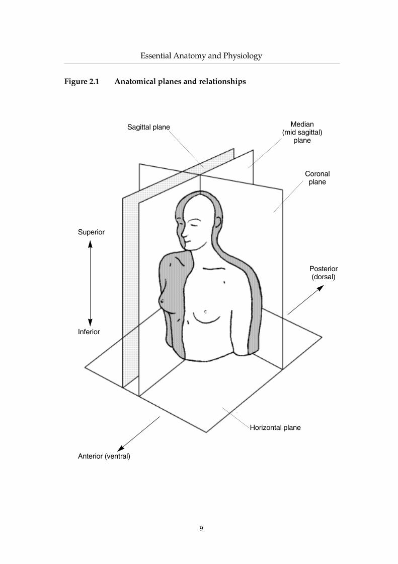
The body is divided by a number of planes or imaginary ‘slices’.The median plane is a vertical slice through the middle of the trunk which
divides the body into symmetrical right and left halves. It passes through thesagittal suture of the skull and any slice parallel to it is described as a sagittalplane.
Vertical slices at right angles to the median plane will pass through orparallel to the coronal suture of the skull and are termed coronal planes.
7
ESSENTIAL ANATOMY AND PHYSIOLOGY

Horizontal planes are, of course, at right angles to the other two. Anterior refers to a position nearer the front when in the anatomical
position or a surface directed forwards. Thus, the face is on the anterior surfaceof the head and the breasts are anterior to the underlying rib cage. The termventral is sometimes substituted.
Posterior, similarly, describes a position nearer the back of the body or abackward facing surface and may be replaced by dorsal.Superior, meaning ‘above’, and inferior, meaning ‘below’, are also always usedwith reference to the anatomical position. Thus, the head is always superior tothe thorax (chest), irrespective of the actual position of the body. In somedescriptions, the terms cranial (nearer the head) and its opposite, caudal, maybe preferred.
Medial means nearer to the midline whereas lateral is further away. Forreference points other than the midline, the terms proximal and distal are used.The reference point may be implied rather than explicitly stated, especially inthe case of the limbs, when ‘proximal’ will mean ‘nearer to the torso’ (that is,the shoulder is proximal to the elbow and the wrist is more distal).
Superficial and deep are used to describe distances from a surface, often, butnot always, the skin. Internal and external are used for the surfaces of hollowstructures such as the thoracic (chest) cavity.
Gynaecology
8
Essential Anatomy and Physiology
9
Figure 2.1 Anatomical planes and relationships
Sagittal plane Median (mid sagittal)
plane
Coronalplane
Posterior(dorsal)
Horizontal plane
Anterior (ventral)
Inferior
Superior

An overview of pelvic anatomy
The bones
The bony pelvis (the word ‘pelvis’ is from the Latin for ‘a basin’) has the formof a ring which, in the adult, is made up of three individual bones, the two hipbones and the sacrum. These, in turn, are formed by the fusion (joining) ofeleven bones. The posterior wall of the pelvis is formed by the triangularsacrum consisting of five fused spinal vertebrae whereas the innominate bone oneach side develops by the joining of the ilium, ischium and pubis. The pubicparts of the innominate bones meet in the midline anteriorly at the pubicsymphysis, thereby completing the circle.
Outside the bony pelvis lie muscles, fat and skin, together with associatedblood and lymphatic vessels and nerves. Many of the muscles find attachmentto the strong pelvic bones and either support the spine or act upon the femur(thigh bone) in the upper part of the lower limb.
Gynaecology
10
Figure 2.2 The pelvic bones
Pubis
5th lumbar vertebra
Sacrum
Ilium
Coccyx
Ischium

Essential Anatomy and Physiology
The bones of the pelvis are also lined by muscle groups within which aresituated the organs of three functional systems, the reproductive tract (theovaries, fallopian tubes, uterus and upper vagina), the lower part of theurinary tract (part of the ureters, the bladder and the urethra) and the digestivetract (the rectum and anal canal).
The bladder is placed anteriorly in the midline behind the pubicsymphysis and in front of the uterus which has a tube and ovary on each side.Behind the uterus, but separated by a space (the pouch of Douglas) usuallyoccupied by loops of small bowel, lies the rectum.
The pelvic organs are partly covered by a thin, smooth, surface layer calledthe peritoneum, which is continuous with the similar surface lining theabdominal cavity itself. This normally separates the organs and allows themto slide freely against one another. Inflammation can cause adjacent peritonealsurfaces to adhere to one another.
The lower pelvis is filled by a muscular sheet (the pelvic floor or pelvicdiaphragm) perforated by openings for the urethra, vagina and anal canal. Thepelvic floor supports the structures above it and it is primarily a stretchingand weakening of this muscular sheet which results in vaginal prolapse(described in Chapter 8).
11
Figure 2.3 Muscles of the pelvic floor (viewed from above)
The constituent parts of the levator ani, together with the adjacent muscles of the pelvicside wall, fill in the pelvic opening, except where apertures are left for the rectum,vagina and urethra.
Urethra
Piriformis
Coccygeus
IliococcygeusPubococcygeus
Obturator internus
PuborectalisVagina
Rectum } Levatorani

The pelvic blood supply
Oxygenated blood leaves the left ventricle of the heart via the aorta which,after giving off branches to supply the head and neck, curves over to passdown on the posterior wall of the thorax and abdomen. Shortly beforeentering the pelvis, the aorta divides into the two common iliac arteries which,in turn, separate into the external and internal iliac vessels. The former mainlysupplies the lower limb whereas the blood in the latter is largely directed tothe pelvic organs.
As elsewhere in the body, the arteries divide into arterioles and eventuallyinto the smallest vessels (capillaries) where oxygen and other nutrients areable to diffuse out of the blood and into the surrounding tissues whilst thewaste products of metabolism, including carbon dioxide, diffuse into theblood to be carried back via venules and veins to the heart for onwardcirculation to the lungs (where the carbon dioxide is extracted and oxygenadded) and to other organs for disposal.
Some fluid also leaves the capillaries and is eventually returned to theblood circulation via a separate group of tubules called the lymphatic system.These tubules never reach the size of the major blood vessels and areinterrupted at intervals by the interposition of the lymph nodes which act asfilters. The final pathway by which much of the contained fluid (lymph)reaches the blood is the thoracic duct which drains into the left subclavian veinin the root of the neck though lymph also returns to the blood stream at thelymph nodes.
The clinical importance of the lymphatic system is that it provides apotential route for the spread of cells from malignant tumours and the earliestdistant deposits (metastases) from a cancer are often found in theneighbouring lymph nodes.
The pelvic nerve supply
The nervous system is divided into a central part, the brain and spinal cord,and a peripheral part (the nerves themselves) but division can also be madeinto somatic and autonomic systems.
The somatic system includes both sensory and motor nerves. The formercarry neural signals from the peripheral sensors to the central structuresallowing the appreciation of both internal events such as muscle position(proprioception) and external stimuli (touch, temperature, smell, etc) whilstthe motor nerves are concerned with the central control of distant structures(mainly the muscles). In contrast, the autonomic system carries signals whichrarely reach consciousness and are concerned with the unconscious andautomatic control of body functions (for example, control of the muscularwalls of arterioles to regulate blood flow).
Gynaecology
12

Essential Anatomy and Physiology
The nerves arising from the spinal cord (carrying both somatic andautonomic fibres) emerge from the spine by passing out through small spacesbetween the vertebrae, the intervertebral foramina. The nerves then divide intodorsal and ventral rami.
The largest and most important neural structure within the pelvis isformed by the intermingling of nerve fibres from the ventral rami of thefourth lumbar to the fourth sacral nerves and is called the sacral plexus. Thislies on the posterior wall of the pelvis on the deep surface of the piriformismuscle and behind the internal iliac blood vessels and the ureter.
The pelvic organs
The gynaecological organs or reproductive tract
As mentioned above, the pelvic reproductive organs consist of the twoovaries, the fallopian tubes, the uterus and the vagina (see Figure 2.4,overleaf).
The ovaries
Normal ovaries are greyish white, almond shaped structures about 30 mmlong, 15 mm wide and about 10 mm thick. They are situated laterally on eitherside of the uterus lying close to the pelvic side-wall in the ovarian fossa wherethey are not far distant from the ureter. They are attached to the posterioraspect of a fold of peritoneum called the broad ligament by an additional fold,the mesovarium, in which run the blood vessels supplying the ovary. Unlikethe remaining pelvic organs which mostly obtain their blood from branches ofthe internal iliac artery, the blood supply of the ovaries is by vessels (theovarian arteries) which arise directly from the anterior aspect of the aorta itselfjust below the level of the renal arteries, that is, high up in the abdomen.
The proximity of the ovary to the ureter has important implications for thesafety of the latter structure when the ovary is removed surgically.
The functional aspects of the ovaries are considered in the section onphysiology.
The fallopian tubes
The uterine (or fallopian) tubes are normally about 10 cm in length and lie inthe upper margin of the broad ligament extending from just above the ovaryto the upper part of the uterus. The outer part (the infundibulum) is the largestbeing trumpet shaped with the edge of the opening into the pelvic cavitybeing extended into small, finger-like, projections call fimbriae.
13

Gynaecology
14
Figure 2.4 The female pelvic organs
Simplified diagrams of this type are very helpful when explaining gynaecologicalconditions and procedures to patients
Uterus
Vagina
CervixOvary
Fallopian tubeUterus
Vagina
Cervix
OvaryFallopian tube

Essential Anatomy and Physiology
The infundibulum narrows medially to become the ampulla whichconstitutes a little more than half of the whole length of the tube. This, in turn,narrows further to become the isthmus. The outside diameter at this point isabout 4–5 mm but the lumen is less than 1 mm. The tube then enters thecornual region of the uterus passing through the muscular wall as the intra-mural segment before opening into the uterine cavity at the inner tubal ostium.
The uterus
The normal, non-pregnant uterus is about the size and shape of a small pear,being some 75 mm long, 50 mm in breadth and 25 mm thick. It liesapproximately centrally in the pelvis (though some deviation to one side isnot uncommon) and is supported by the pelvic floor and by thickenings of thesurrounding connective tissue. For descriptive purposes, it is divided into thebody (or corpus) and neck (or cervix).
The fallopian tubes are attached to the part of the uterus called the cornu(or cornual region), the name being derived from the Latin for a horn. The partof the uterus above the site of insertion of the tubes is termed the fundus.
The cervix is perforated by the cervical canal, which is slightly wider in itsmid-section than at each end. The upper part communicates with theendometrial cavity through the internal cervical os, whereas the lower partopens into the vagina at the external os. The part of the cervix which protrudesinto the upper vagina (usually for a distance of about 12 cm) may be referredto as the portio vaginalis.
The uterine arteries run medially on each side of the cervix just above thelevel at which the ureters pass from back to front (see Figure 2.5). The anterioraspect of the mid-part of the cervix is intimately related to the posterior wallof the bladder.
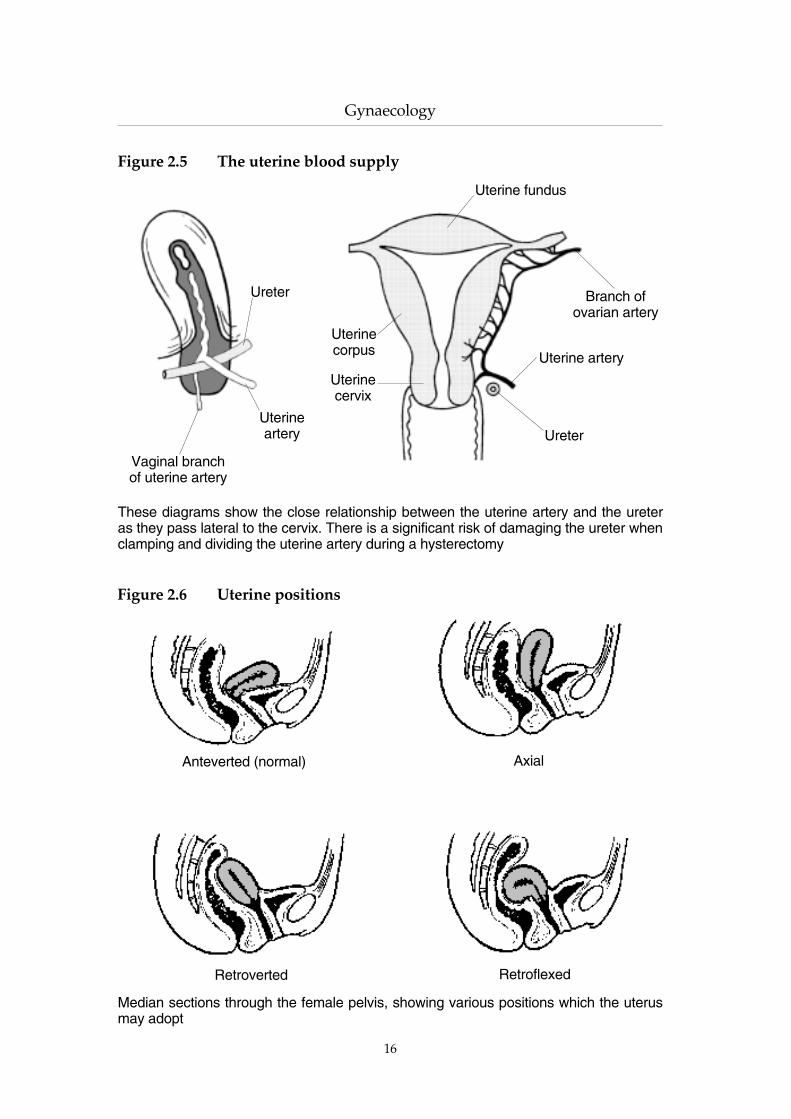
In the pre-pubertal girl, the long axis of the uterus is approximately in linewith the vagina (axial) but, in the sexually mature woman, the long axis isnormally directed forwards (anteverted and pointing approximately towardsthe umbilicus) which, together with the usual slight forward curve(anteflexion), places the uterus almost at right angles to the vagina (see Figure2.6). After the menopause, the uterus gradually reduces in size and tends torevert towards the axial position.
In about 15% of post-pubertal women, the uterus is tilted backwards(retroverted) and may also be bent posteriorly (retroflexion). Again, see Figure2.6.
The anteverted position may not be explicitly recorded but, if it is, it willoften be abbreviated to a/v. Retroversion, if correctly diagnosed, may benoted as r/v. Anteflexion and retroflexion are rarely noted and probablyappreciated rarely, if at all, by British gynaecologists.
15
Gynaecology
16
Figure 2.6 Uterine positions
Figure 2.5 The uterine blood supply
These diagrams show the close relationship between the uterine artery and the ureteras they pass lateral to the cervix. There is a significant risk of damaging the ureter whenclamping and dividing the uterine artery during a hysterectomy
Anteverted (normal)
RetroflexedRetroverted
Axial
Median sections through the female pelvis, showing various positions which the uterusmay adopt
Uterine artery
Ureter
Branch ofovarian artery
Uterine fundus
Uterinecorpus
Uterinecervix
Vaginal branchof uterine artery
Uterineartery
Ureter
Essential Anatomy and Physiology
The uterus is partly covered by peritoneum (sometimes called the serosa orserosal layer) but the wall consists largely of muscle with a thickness of about 2cm. The inner cavity of the uterus (or endometrial cavity) is lined by a welldefined layer called the endometrium which varies in thickness in response tothe endocrine (hormonal) status of the individual. Superiorly, the cavity is incontinuity with the lumen of the fallopian tubes and inferiorly becomes thecanal of the cervix. In the normal, non-pregnant condition, the anterior andposterior surfaces of the endometrial cavity lie in apposition.
The vagina
The vagina extends downwards and forwards from the cervix to reach theexterior surface at the vulva where the introitus lies between the labia minora.The anterior wall measures about 75 mm in length but is extended superiorlyby the cervix of the uterus so that the posterior wall is longer (about 90 mm). Itis flattened antero-posteriorly so that the front and back walls normally lie incontact. Because the cervix protrudes into the vagina, it is surrounded by acircular recess which forms the anterior, posterior and lateral vaginal fornices(singular, fornix)
The bladder and urethra are situated deep to the anterior vaginal wall. Theupper part of the posterior wall is covered by peritoneum and is separatedfrom the rectum by the pouch of Douglas (see above). The middle section ofthe posterior wall is also related to the rectum with loose connective tissue inbetween whereas the lower part of the posterior wall is separated from theanal canal by a fibro-muscular structure called the perineal body.
The surface of the vaginal wall is thrown into small folds or rugae, whichallow expansion, and is covered by non-keratinised, stratified squamousepithelium not greatly dissimilar to the external skin. In a virgin, the lowervagina is partially closed by the incomplete sheet of the hymen, representedafter first coitus by hymeneal tags.
The external female genitalia
The rounded, hair-bearing area of skin over a pad of fat on the pubicsymphysis is the mons pubis, behind which the labia majora and minorasurrounding the vaginal introitus form the vulva. More posteriorly still, theperineum extends from the vaginal fourchette to the anus.
Note that the gynaecological description of the perineum does notcorrespond with the anatomical definition, which refers to the whole regionbounded by the pubic symphysis anteriorly, the tip of the coccyx posteriorlyand the ischial tuberosities on each side. The gynaecological use of the termroughly corresponds with the ‘anal region’ of the anatomical perineum.
17

The urinary tract
The ureters
The ureters drain urine from the medial aspect of the kidneys which aresituated high on the posterior abdominal wall. They pass down the posteriorwall on each side of the spine and a short distance from it before entering thepelvis by crossing in front of the sacro-iliac joints over the point at which thecommon iliac artery divides into the external and internal iliac arteries. Theythen run downwards and forwards passing the cervix uteri just above thelateral vaginal fornices (where they are crossed at right angles by the uterinearteries) to enter the lateral parts of the posterior aspect of the bladder.
As noted above, the ureters are at risk of surgical damage when theovaries are removed. This risk is greatly increased if the ovary is plastereddown in the ovarian fossa as may occur in the presence of endometriosis orafter previous pelvic surgery. They may also sustain damage at hysterectomywhen the injury usually occurs where they pass the cervix.
The (urinary) bladder
The bladder lies behind the symphysis pubis and adjoining parts of thepubic bone. Its size and shape depend on the volume of the contained urine.Posteriorly, it is related to the upper vagina and to part of the cervix. Theureters enter from behind and the urine leaves the bladder inferiorly bydraining into the urethra.
The bladder may be damaged at laparoscopy (by the Veress needle or thetrochar) and at laparotomy. Injury may also occur at hysterectomy and atCaesarean section because of the close relationship between the bladder andthe anterior aspect of the cervix uteri.
The urethra
It is important to distinguish carefully between the urethra and the ureter, asthey are anatomically quite distant.
The female urethra is about 40 mm long and 6 mm in diameter (the maleurethra is almost five times longer). It leaves the bladder and runs downbehind the pubic symphysis being closely applied to the anterior vaginal wall.It ends at the urethral meatus between the anterior ends of the labia minora.
Gynaecology
18

Essential Anatomy and Physiology
The gastro-intestinal tract
The sigmoid colon enters the pelvis on the left side of the posterior wall andpasses down to become the rectum which continues downwards behind thevagina to become the anal canal at the level of the pelvic floor.
Loops of small bowel are also usually present in the pelvis, lying in thepouch of Douglas.
PHYSIOLOGY
Physiology is the study of the processes underlying normal bodily functions.Pathology is the study of disease and its effect upon the body. Gynaecologistsmust therefore have detailed understanding of both the physiology and thepathology of the female reproductive system.
Basic genetics
At conception (the union of a sperm and an egg), a new individual is created.We all inherit many characteristics from each of our parents but these aremixed to produce a distinct individual.
The characteristics are transmitted in a highly controlled manner and willdetermine many of our adult characteristics, although much happens to us aswe mature. For example, eye colour is inherited directly from our parents butwe must learn to ride a bicycle since we are not born with the innate ability todo so.
Chromosomes, genes and DNA
Each cell in the body contains a nucleus and contained within that nucleus arethe chromosomes. These chromosomes are made up of thousands of genesand the genes, in turn, consist of DNA (deoxyribonucleic acid). DNA is thebasic building block of the genetic information at the cellular level.
The genes act as templates for the cellular production of many differentproteins and these, in turn, can alter the form and function of tissue.
Human beings have 46 chromosomes in the nucleus of most of their cells,23 inherited from the mother and 23 inherited from the father.
The gonads (testes in the male and ovaries in the female) are ratherspecialised and have only 23 chromosomes in some of their cells, these beingthe ova (eggs) in the female and the sperm in the male. When the egg and thesperm combine at conception, the number is restored to 46 (23 pairs) with aunique genetic identity.
19

Gynaecology
20
One specific pair of chromosomes, the sex chromosomes, determines anindividual’s sex. At conception, the ovum will always provide an Xchromosome and the sperm may supply either an X or a Y chromosome. XXproduces a chromosomal female, whereas XY produces a male. Consequently,it is the chromosome content of the sperm which determines the genetic sex ofthe offspring.
Sexual differentiation
Chromosomal sex is determined by the presence or absence of a Y chromosome,as outlined above.
Gonadal sex is determined by whether or not the individual possessesovaries or testes. A gonadal female has ovaries and a gonadal male has testes.
Phenotypic sex describes the body’s sexual appearance and applies largely tothe external genitalia. An individual with a penis and scrotum is phenotypicallymale, whereas one with a clitoris and labia is phenotypically female.
This may sound obvious but, because chromosomal males change frombeing phenotypically female to becoming male while in utero, the process iscomplicated and leaves room for error. For example, there is a conditioncalled ‘androgen insensitivity syndrome’ (formerly known as ‘testicularfeminising syndrome’), where the chromosomal male fails to respond to thehormones that would normally cause the change from the phenotypicalfemale form to male. These people are thus born apparently female with aclitoris and labia and often their condition is not discovered until adulthood,when they find they are unable to conceive a pregnancy. On investigation, it isfound they have no ovaries and, in fact, have very underdeveloped testes intheir abdomen; their sex chromosomes are XY, that is, they are a chromosomalmale, but they will have been brought up as female.
The sexual physiology of the unborn
Until the sixth week of pregnancy, the foetus is phenotypically female but thegonads are undifferentiated and have the capacity to become either testes orovaries. The chromosomes, however, will be either male (XY) or female (XX)
In the normal male embryo at the sixth week of pregnancy, the Ychromosome initiates the production of a protein called testiculardifferentiating factor. This causes the undifferentiated gonad to become atesticle which, in turn, begins to produce two hormones, MIF (mullerianinhibiting factor) and testosterone. These cause the foetus to becomephenotypically male and, in so doing, also cause the re-absorption of theprimitive female genital tract.

Essential Anatomy and Physiology
21
In the normal female, the absence of the Y chromosome causes the femaleform to persist and the gonad becomes an ovary, allowing the continueddevelopment of the female genital tract.
Childhood
During childhood, there is little difference in the hormone productionbetween boys and girls but at puberty the two sexes diverge. Furtherdiscussion here is confined to the female physiology.
Puberty
Puberty is the sequence of events by which a child is transformed into anadult and becomes capable of reproduction. Several physical change areevident: breast development, axillary (armpit) and pubic hair growth, anincrease in height commonly known as the ‘growth spurt’, and menarche,which is the onset of menstruation. The ovary will also be maturing andovulation will commence but this has no obvious external manifestationssince it does not necessarily coincide with menarche.
There is much variation in the timing of pubertal events. The first sign isusually breast bud development and an increase in height, followed by hairgrowth and then of menstruation. However, only about 50% of girls willfollow this sequence. The whole process takes approximately two years, withmenstruation usually beginning between ages 11 and 15 years.
The age at which puberty occurs is influenced by a number of factors,particularly that of body weight. Under-nourished girls are more likely tohave a delayed menarche. The age of menarche has decreased in this century,probably because of the improvement in general health and nutrition.
The precise physiological processes that initiate puberty are unknown.Steroid hormones produced from the adrenal gland may be involved,particularly in the growth of the secondary sexual hair or telarche. A bodyweight of 48 kg or more is required to menstruate which explains whyanorexic girls do not have menstrual bleeds. The menarche appears to beinitiated in the hypothalamus which is located in the primitive part of thebrain and produces a variety of ‘releasing’ hormones that act upon thepituitary gland (situated just below the brain) to release other hormones. Thehypothalamic releasing hormone involved in the reproductive system isknown as gonadotrophin releasing hormone (GnRH). During puberty, thehypothalamus gradually produces increasing amounts of GnRH which causesthe release of the pituitary gonadotrophins (trophin – to grow) folliclestimulating hormone (FSH), and luteinising hormone (LH). In response to thegonadotrophins, the ovary begins to produce some ovarian hormones,gradually the female endocrine pattern will become established and cyclical

ovulation and menstruation will occur. Initially, however, ovulation does notnecessarily occur every month and, after the menarche, it usually takes twoyears or more for regular ovulation to become established.
The menstrual cycle
The menstrual cycle conventionally begins with the first day of vaginalbleeding and ends just before the next menstrual bleed starts. Most womenhave a cycle length of 28 days but the range is between 21 and 40 days. It ismore common to have irregular menstrual cycles at the beginning and end ofreproductive life.
The menstrual cycle depends upon the interaction of hormones producedin the hypothalamus, the pituitary gland and the ovary. The cycle consists ofthree distinct phases which are termed the follicular, ovulatory and lutealphases with reference to the events occurring in the ovary.
Follicular phase
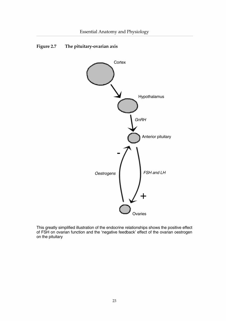
In the follicular phase, the level of follicle stimulating hormone (FSH) from thepituitary increases. This initiates the growth and development of a cohort ofeggs (between three and 20) within the ovary. Only one egg will usually bereleased at ovulation and the rest regress and die. The egg develops within afluid-filled sac which is surrounded by a further layer of specialised cells, thewhole structure being known as a follicle. The surrounding cells produce thehormone oestrogen (as oestradiol), the production of which graduallyincreases. This increase in oestradiol production raises the blood level of thehormone which is detected by the hypothalamus which then produces lessGnRH (see above), thereby reducing the release of FSH and LH from thepituitary. This type of interactive system is widely encountered inphysiological processes and is known as ‘negative feedback’.
Gynaecology
22
Essential Anatomy and Physiology
23
Figure 2.7 The pituitary-ovarian axis
This greatly simplified illustration of the endocrine relationships shows the positive effectof FSH on ovarian function and the ‘negative feedback’ effect of the ovarian oestrogenon the pituitary
Ovaries
-
Oestrogens
+
FSH and LH
Anterior pituitary
GnRH
Hypothalamus
Cortex
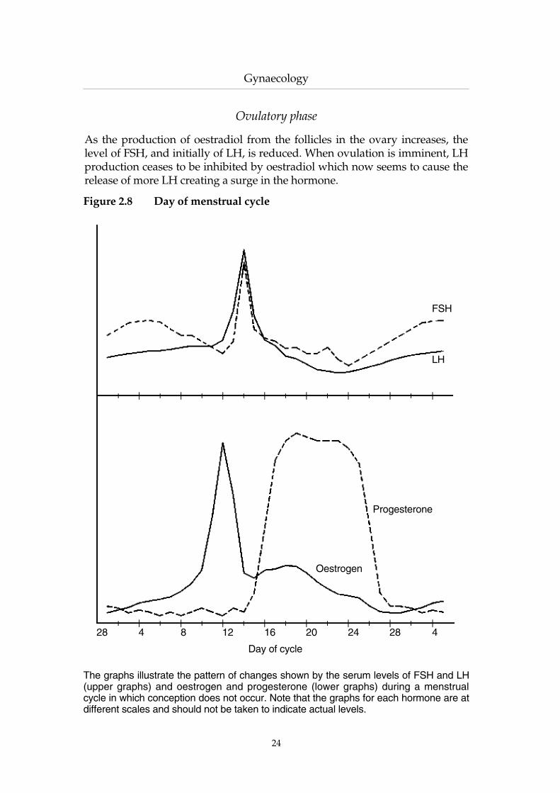
Ovulatory phase
As the production of oestradiol from the follicles in the ovary increases, thelevel of FSH, and initially of LH, is reduced. When ovulation is imminent, LHproduction ceases to be inhibited by oestradiol which now seems to cause therelease of more LH creating a surge in the hormone.
Gynaecology
24
Figure 2.8 Day of menstrual cycle
The graphs illustrate the pattern of changes shown by the serum levels of FSH and LH(upper graphs) and oestrogen and progesterone (lower graphs) during a menstrualcycle in which conception does not occur. Note that the graphs for each hormone are atdifferent scales and should not be taken to indicate actual levels.
FSH
28
Day of cycle
Oestrogen
Progesterone
LH
16 412 288 244 20

Essential Anatomy and Physiology
Ovulation occurs 12–24 hours after the rise in LH. The exact mechanism ofovulation is not understood but the surface cells allow release of the egg intothe pelvic cavity or, perhaps more commonly, into the overlying fallopiantube.
Luteal phase
Once ovulation has occurred, the number of follicular cells increases and theybegin to produce the hormone progesterone. The serum level of this hormoneis frequently measured as an indicator of ovulation; a level greater than about10 nmol/l suggests that ovulation has taken place.
Pregnancy
During pregnancy, the female body undergoes enormous physiologicalchanges in response to an increased hormonal production. The hormones areprimarily produced by the placenta. Some of these are specific to pregnancybut the placenta also produces the female hormones oestrogen andprogesterone which are present in far greater amounts during pregnancy thanin the non-pregnant state.
The changes that occur in pregnancy are not restricted to the reproductivesystem but affect the whole body. It is beyond the scope of this book to do anymore than outline the major changes that occur.
Very early in pregnancy, the amount of blood and other body fluidsincreases. This increase occurs before the uterus has enlarged sufficiently toneed the extra blood supply and the additional blood is diverted to the skincontributing to the sense of warmth and well being that is frequentlyexperienced.
The blood is also more ‘sticky’ than in a non-pregnant woman. Apregnant, or recently pregnant woman, is at least 10 times more likely todevelop a blood clot (thrombus) in the deep veins of the leg (deep venousthrombosis or ‘DVT’) than at any other time during her lifetime. This isreflected in the fact that thrombosis and its sequelae are now the leading causeof deaths related to pregnancy.
The heart works harder during pregnancy and the blood pressure fallsslightly. Elevation of the blood pressure in pregnancy is abnormal.
The rate of breathing is subconsciously increased often leading to a feelingof shortness of breath. This is exacerbated later in pregnancy as the pregnantuterus displaces the abdominal contents upwards against the diaphragm.
The digestive tract relaxes, leading to heartburn and indigestion. Gutmovement is reduced, often leading to constipation. Fat is deposited in thebody’s fat stores.
25

The enlarging uterus presses upon the bladder and there is a dilatation ofthe urinary tract causing partial stasis of the urine and an increased incidenceof urinary tract infections.
These changes subside at a variable rate as the pregnancy is completed.Some resolve within a few days but others take several weeks to return to thepre-pregnancy state. Lactation will further delay complete resolution.
Lactation
The production of breast milk (lactation) is the end result of a variety ofinfluences. Pubertal breast development is initiated by oestrogen hormonesbut, during pregnancy, the breasts increase further in size in response to thehigh level of circulating oestrogen and, probably, also progesterone andhuman placental lactogen (HPL), both of these being produced by theplacenta.
Prolactin (from the pituitary gland) is essential for lactation itself but itseffect is largely suppressed until, after the birth of the infant, the maternallevel of oestrogen falls allowing milk production to begin. Once initiated bythe hormone changes, lactation is maintained by the continued suckling of theinfant which stimulates the nerves endings in the nipples. The impulses arepassed along the nerves to the hypothalamus and thence to the pituitarygland. This gland releases a hormone called oxytocin which acts upon thesmall muscles surrounding the milk ducts in the breasts causing muscularcontraction and release of the milk (the ‘let-down’ reflex).
The menstrual cycle is generally suppressed during lactation. However,ovarian suppression decreases with the passage of time even if lactationcontinues so that the possibility of ovulation and, hence, the chance of anotherpregnancy arises. Consequently, lactation should not be relied upon ascontraception.
The menopause
The term ‘menopause’ refers to the cessation of menstrual bleeding and‘climacteric’ identifies the period of time before and after this occurs. The twowords are often, though incorrectly, used synonymously.
With advancing years, the ovary has fewer primitive eggs and fewerdeveloping follicles; hence, less oestrogen is produced. The GnRH (from thehypothalamus) increases in an attempt to drive the ovaries but to no avail; theFSH and LH levels increase but the oestrogen hormones are low.
Effects of the menopause/climacteric
These may be considered in two different categories.
Gynaecology
26

Essential Anatomy and Physiology
First, there are the obvious symptoms that may be experienced by anywoman in the climacteric. They include facial flushing (which may beextremely embarrassing), night sweats, irritability and an inability toconcentrate. Other symptoms are related to thinning of the epithelium of theuro-genital tract and include bladder weakness and dyspareunia (discomfortduring sexual intercourse). All these symptoms will generally resolve but onequarter of women will continue to experience them in varying degrees formore than five years.
The second group of problems are also due to the lack of oestrogen but donot necessarily cause symptoms; nevertheless, they are vastly more importantin terms of the general health of an increasingly ageing population. The majorfactors are cardiovascular disease and osteoporosis (de-mineralisation of thebones). Their importance lies in the fact that they are responsible for a gooddeal of the ill health and death in the post-menopausal population.Cardiovascular disease includes heart attacks and strokes. Before themenopause, it is uncommon for women to suffer either a heart attack or astroke but, after the cessation of ovarian activity, the female incidence of theseconditions rises and soon matches that of men. The other important healthproblem in the older post-menopausal woman is that of fragile bones orosteoporosis and there is an associated rise in the incidence of fractures. Theseare often incapacitating and, if a large bone such as the femur is involved, mayprove fatal.
Hormone replacement therapy (HRT)
Hormone replacement therapy aims to replace the ovarian hormones that areno longer produced after the menopause. The most important hormone isoestrogen but, unless the woman has already undergone a hysterectomy,progesterone should also be administered to prevent over-stimulation of theuterine lining, a condition which is associated with an increased risk ofendometrial carcinoma. The addition of progesterone for two weeks in eachmonth usually causes the woman to have regular monthly bleeding. Manywomen are happy to tolerate this, certainly for a few years, but women whochoose to have HRT for longer than about five years often desire an end to theinconvenience of vaginal bleeds. Newer preparations provide a smallcontinuous dose of progesterone designed to minimise the chance of bleeding.HRT can be administered by tablet, patch, gels or implants. Somepreparations may have advantages for particular women but the choice isusually determined by the woman’s preference.
HRT commenced at the menopause and continued for five years willreduce the chance of fractures by approximately 50%.
27

Contra-indications and side effects
Much of the practice of medicine consists of the assessment of the benefits andrisks of a possible line of treatment in order to arrive at a balanced decision;the administration of hormone replacement therapy is no exception.
Many women experience no untoward symptoms at the climacteric and,for many more, any symptoms are minor and transient. Unfortunately, asignificant proportion may be subject to episodes of vasomotor instability(‘hot flushes’), disturbances of sleep patterns and emotional lability. All thesesymptoms respond well to HRT, the hot flushes sometimes ceasing within 36hours of starting therapy though other symptoms usually take longer toimprove.
The relief of climacteric symptoms may require HRT for a few months orup to three or four years but many women now continue on the therapy forthe long term benefits of the reduced incidence of heart disease, cerebro-vascular accidents (‘strokes’) and osteoporosis. This may be particularlyappropriate if there is a family history of any of these conditions.
In contrast, HRT is contra-indicated for women who are known orsuspected to be suffering from carcinoma of the breast or from oestrogendependent malignancies in other sites. It is also contra-indicated in thepresence of some severe cardiac or renal diseases or if hepatic disease hasinterfered with liver function.
High doses of oestrogen, such as in some oral contraceptives, have beenimplicated in the causation of veneous thrombosis. The lower, replacementdoses associated with HRT were formerly thought to have no such effect butrecent analysis of the published studies suggests that they may also increasethe thrombosis rate, though to a much smaller extent. The current view is thatthe risk of thrombosis in women not taking HRT is about one in 10,000 peryear and that HRT increases this to about three in 10,000 per year. Most ofthese cases respond to treatment but a few prove fatal, the death rate beingestimated at one in one million per year for non-HRT women and three in onemillion per year on HRT. A history of deep venous thrombosis or of thrombo-embolism is usually regarded as a contraindication to the use of oestrogensupplements.
On first starting HRT, many women will experience cyclical breasttenderness. In the majority of cases, this will improve after the first two orthree months of treatment but sometimes it persists and the symptom mayoccasionally develop in women who have been taking HRT for several years.It is thought to be the commonest reason for women to discontinue treatment,often without reference to medical advice. Many cases resolve if the HRTpreparation is changed.
Some women, particularly those who have suffered from pre-menstrualsymptoms during their natural cycle, may complain of HRT-induced PMS.Again, a change of preparation may be effective.
Gynaecology
28

Essential Anatomy and Physiology
Headaches can be associated with HRT and may be sufficiently severe andfrequent to cause cessation of treatment. In a few instances, true migraineattacks may be precipitated.
The bleeding associated with combined oestrogen/progestogen HRT maybe heavy and scanty but irregular bleeding may occur with the so called ‘non-bleeding’ HRT. Many women attribute weight gain to HRT and there maycertainly be an alteration in dietary needs but any increase in body weightseems to be related to an incorrect dose of oestrogen since, contrary toexpectations, an increased dose may be followed by a return to normalweight.
Oestrogens were thought to cause elevation of the blood pressure but alarge US study some years ago demonstrated that mild hypertension couldactually improve on HRT; severe hypertension remains as a contra-indication.
The role of unopposed oestrogen in the development of endometrialhyperplasia and possibly carcinoma has already been mentioned above, andthe original oestrogen-only preparations are contra-indicated in women whoretain their uterus (though a few, who find the side effects of the combinedpreparations to be unacceptable but who value the oestrogen effects, maychoose to take the risk).
The possible association between HRT and an increased risk of breastcancer has been a subject of discussion and disagreement for many years. Thequestion has been resolved, at least for the present, by the recent publicationof a meta-analysis of the available data which concluded that the risk isprobably doubled if HRT is taken for longer than about eight years. Cancer inwomen on HRT seems to be more amenable to treatment but the mechanismfor this is, as yet, unexplained. It has been suggested that women on therapyare more likely to attend for regular medical review with the result that thedisease is detected sooner than would otherwise be the case.
In any discussion with patients, it is important to realise that the increasedincidence of breast cancer is less than the decreased mortality from heartdisease and strokes. Women wishing to remain on long-term HRT shouldhave regular mammography, perhaps yearly if there is a family history ofbreast cancer or 2–3 yearly otherwise.
Progestogen HRT
In recent years, the use of progestogens derived from plant extract (notablythe yam), have been promoted as alternatives to conventional HRT. In somewomen, they are certainly effective in relieving climacteric symptoms buttheir long term effects remain to be established. They do offer a useful optionfor women who are unable or unwilling to take oestrogens.
29


CHAPTER 3
In this book, important operative points are generally mentioned along withthe conditions with which they are associated but there are some surgicalprocedures which are employed so frequently in gynaecology that they meritseparate consideration. These are dilatation and curettage (D & C), hysteroscopy,laparoscopy and hysterectomy. In addition, this chapter also deals withcommonly used techniques for opening the abdominal cavity.
The procedures to be described are usually performed under generalanaesthesia and this itself carries small but significant hazards. Since thisvolume is concerned with gynaecology, these risks are not considered in detailhere but this fact should not be taken to imply that they are unimportant.
The anaesthetist is usually responsible for the safety of the patient in theoperating theatre and, when electrosurgical equipment is to be used (forexample, diathermy cauterisation), there is the special consideration that it isessential that no part of the anaesthetised patient is in contact with anymetallic part of the operating table or its attachments. If the patient is notproperly insulated by suitable rubber or plastic padding, an otherwiseinsignificant fault in the electrical equipment could result in a serious burn atthe point of contact. The courts have, on occasion, applied the principle of resipse loquitur to such burns and may place the burden of proof on the defendantto show that the injury was not due to negligence.
THE D & C
The term D & C is the widely accepted abbreviation for dilatation andcurettage in which the cervical canal is stretched (dilated) and instrumentsthen introduced into the uterine cavity to scrape (curette) the lining. Patientsoften refer to this as a ‘scrape’ or, sometimes, as ‘cleaning the womb’. Theprocedure is similar to the commonly used method of surgical termination ofpregnancy but, because there are differences both in the instruments used andin the risks involved, the latter is discussed separately (Chapter 13).
The D & C is undoubtedly the most frequent operative procedureundertaken by gynaecologists, though, in recent years, it has been partiallyreplaced by other, more newly developed techniques.
31
COMMON OPERATIVE PROCEDURES INGYNAECOLOGY

Indications
The indications for performing a D & C fall into two groups, diagnostic andtherapeutic.
Diagnostic
The majority of D & Cs are performed as part of the investigation ofgynaecological problems since, at least until the advent of hysteroscopy, it wasthe only way in which the cavity of the uterus could be satisfactorily explored.Unfortunately, gynaecologists often fail to explain to the patient that theprocedure is not necessarily expected to cure their symptoms and, inconsequence, the woman may be understandably aggrieved when, havingundergone a D & C, her symptoms persist unaltered.
The term ‘dysfunctional uterine bleeding’ is unsatisfactory, beingessentially descriptive and unrelated to any particular pathological process.Furthermore, by providing a convenient label, it may impede the search for atrue diagnosis. However, it is convenient to use it here as, prior to the D & C,the precise diagnosis will usually be unknown, the purpose of the curettagebeing to obtain a sample of the endometrium for histopathologicalexamination, following which, appropriate management can be planned. Insome cases, the curettage may be followed by an improvement in thesymptoms for several months but the effect is almost always only temporaryand appropriate follow-up arrangements should be made.
Menorrhagia is a very common presenting symptom in every generalgynaecological clinic and is often investigated by an initial D & C. In aboutone-third of all cases, there will be a reduction in the menstrual loss but this islikely to be transient. In those cases where heavy periods are due to someanatomical factor, such as uterine fibromyomata, no improvement can beexpected and the purpose of the D & C is to exclude other pathology beforeplanning the definitive management.
The possible causes of post-menopausal vaginal bleeding (PMB) arediscussed in Chapter 5, but the most significant of these and the one that must beexcluded in every case is a carcinoma of the endometrium. The only safe rule tofollow is that every case of PMB must be investigated by D & C (or by one ofthe more recent techniques discussed later in this chapter); this should applyeven if an obvious alternative cause for the bleeding is present. The author hasrecently been invited to comment on the case of a 56 year old woman whopresented to her general practitioner with post-menopausal bleeding whichwas attributed to her hormone replacement therapy; in consequence, she wasnot referred for further investigation until 18 months later, when a D & Crevealed her to have an advanced endometrial cancer.
Intermenstrual bleeding may also be due to pathology within the uterinecavity and, in some of these, the D & C may be both diagnostic and
Gynaecology
32

Common Operative Procedures in Gynaecology
therapeutic. For example, some intermenstrual bleeding will be due toendometrial polypi and their removal by curettage may then be curative.
Therapeutic
Dysmenorrhoea may occasionally be treated by dilatation of the cervix (notnecessarily followed by curettage). In the past, this was done quite frequentlyand was based on the assumption that the pain was the result of poordrainage of menstrual fluids due to a narrow cervical canal causing a rise inpressure within the uterine cavity. The theory is unfashionable at present andsome authorities would deny that this indication is ever valid, though otherswould admit that it might have a place in a few cases, especially if thesymptoms follow some other surgical procedure on the cervix, such as a conebiopsy.
The removal of endometrial polypi has already been mentioned but otherintra-uterine conditions may also be treated by the curettage. Occasionally,small pieces of placental tissue may remain within the uterus after thedelivery of a baby. Usually, these fragments are evacuated naturally with thefirst menstrual period after the delivery but, occasionally, they may remain inutero, forming a placental polyp and causing irregular bleeding which maysometimes be heavy. Removal by curettage is simple and effective. Not longago, the author removed a placental fragment that had been present andcausing symptoms for so long that it had undergone calcification.
The different types of abortion (miscarriage of pregnancy) are discussed inthe appropriate section of this book but many of these will need treatment bythe evacuation of retained products of conception, usually abbreviated to‘evacuation’, ‘evac’ or ‘ERPC’. The procedure in such cases is essentially thesame as for a D & C on the non-pregnant uterus but the risks of haemorrhageand perforation are more akin to those associated with the surgicaltermination of pregnancy.
The procedure
In most women, the cervix of the uterus is surprisingly insensitive and may begrasped with sharp instruments, frozen or even cauterised with red-hot wireswith very little discomfort to the patient. In contrast, it is extremely sensitiveto dilatation of its canal and any attempt to do so in the conscious patientusually causes severe pain and may result in profound neurogenic shock (thiswas frequently the cause of death in cases of ‘back street’ abortions). It ispossible to carry out modest dilatation under local analgesia but much morecommon for a general anaesthetic to be given. The development of sedo-analgesia (the so called ‘twilight sleep’) in recent years does offer analternative, especially if some local anaesthetic is also injected on either side ofthe cervix before the dilatation begins.
33

Following the induction of general anaesthesia, the patient will normallybe placed into the ‘lithotomy position’ in which her hips and knees are flexedto 90∞ or slightly more and her thighs are abducted. This position of the legsis usually maintained by placing her feet in double webbing ‘stirrups’ whichare supported by poles attached to the side of the operating table but otherequipment is sometimes preferred, usually some variation of the ‘Lloyd-Davies supports’.
Most women are able to adopt this position without difficulty but thepresence of osteo-arthritic changes in the hips or knees may limit movementof these joints and general anaesthesia will abolish the muscle spasm whichwould, in the conscious patient, protect the affected joint from damage. Theanaesthetist cannot take the proper precautions to safeguard the patient unlessadequately forewarned by the gynaecological team (who will have beenresponsible for the pre-operative clinical history and examination). Anyrelevant non-gynaecological information must therefore be clearly recorded inthe notes and is also best included as an additional comment (for example,‘Stiff right hip’) on the operation list supplied to the operating theatre. This isparticularly important in the case of a D & C which, being classified as a‘minor’ procedure, is often delegated to one of the more junior members of thegynaecological team who may well not have met the patient pre-operatively.
The lithotomy position may also impose some strain on the joints of thelower spine, especially if the patient is positioned too far down the operatingtable, so that her buttocks are unsupported.
After positioning of the patient, the surgeon (now ‘scrubbed-up’ andwearing a sterile gown and gloves) should inspect the vulva for any apparentabnormalities. The presence of any vaginal discharge should be noted and aswab may be taken for bacteriological examination if necessary.
The lower part of the mons pubis, the vulva and the perineum are thencleaned with a gauze or sponge (usually held in sponge-holding forceps)soaked in a water-based antiseptic solution. A second swab is then used tocleanse the vagina using the same solution. Antiseptic solutions containingsurgical spirit should not be used in the vagina; this is especially true if the useof any diathermy is contemplated as severe vaginal burns have resulted fromignition of residual pools of the spirit. The lower part of the patient’s body(usually including the legs) is then covered with sterile towels, leaving thevulva exposed.
The surgeon then performs a bimanual examination, the purpose of whichis primarily to determine the size and position of the uterus, though any otherpalpable abnormalities should also be noted (for example, irregularities of thecervix, pelvic masses, etc). The information should be documented in theoperation note. Omission of this assessment increases the risk of subsequentperforation of the uterine wall by the surgical instruments.
Gynaecology
34
Common Operative Procedures in Gynaecology
A speculum is introduced into the vagina and positioned so that the bestpossible view of the cervix is obtained, following which, the anterior lip of thecervix is grasped with tenaculum or Volsellum forceps and gently drawndownwards to straighten out the curvature of the cervico-uterine canal andalso to steady the uterus.
The length of the uterine cavity may then be measured by introducing auterine sound through the cervical canal and advancing it gently until the tipof the instrument is felt to have reached the fundus of the uterus. Undue forcein this manoeuvre may result in perforation of the uterine wall, especially ifthe operator is unsure of the approximate length of the cavity or its probabledirection (hence the need for the preliminary bimanual assessment). In thecase of a recently pregnant uterus (for example, if the D & C is beingperformed after an abortion or recent delivery), the increased vascularity ofthe uterine wall renders it very soft and it may be difficult or impossible tofeel when the top of the cavity has been reached; in such cases, therefore, itmay be wiser to omit the use of the sound altogether. Difficulties may also beencountered if the cervical canal is very tight, when it may be impossible tointroduce the sound (the rounded tip of which has a diameter of about 3 mm)and it may be necessary to use a very small dilator; such cases should be left toexperienced gynaecological surgeons if the risk of uterine perforation and thecreation of false passages is to be minimised.
The purpose of the uterine sound is to establish not only the length butalso the direction of the uterine cavity in preparation for the subsequentintroduction of the cervical dilators. Various patterns of dilators have beendescribed but the type most commonly used is that attributed to Hegar. Theseare manufactured in single and double-ended versions and are available insizes that increase by 1 mm or by 0.5 mm. For most purposes, the 1 mmincrements are satisfactory but, if the dilatation proves unusually difficult,0.5 mm increments may help to prevent damage to the cervical tissues. Adilatation of, for example, 9 mm is often recorded as ‘Hegar 9’ (or sometimes‘H 9’). The introduction of the dilators should be accompanied by counter-traction on the tenaculum forceps to draw the cervix slightly downwardsthereby steadying it and reducing the natural curve in its canal.
Over dilatation of the cervix, especially if hurried or forcible, may causepermanent damage, though this may not be apparent at the time. Someexperience is required to judge how far dilatation may safely be taken but itshould never be more than is required for the safe introduction of subsequentinstruments and, as a general guide, should not exceed 9 mm for the non-pregnant cervix. The softer cervix of pregnancy may usually be dilated as faras 14 mm without damage. Despite the need to avoid over dilatation,insufficient enlargement of the cervical canal can also cause problems becauseof the increased resistance to the subsequent introduction of otherinstruments, with the result that they may enter the uterine cavity in anuncontrolled manner and themselves cause damage to the uterine wall.
35

Once dilatation of the cervix has been completed, it is then usual tointroduce polyp forceps into the uterine cavity. This instrument is designed tograsp any polypi or other debris that may be present within the uterine cavityso that, if small enough or sufficiently compressible, it may be drawn outthrough the cervical canal.
A suitable curette is then selected, the size of which (like the polyp forcepswhich preceded it) is necessarily limited by the extent of the cervicaldilatation. It is, however, important not to use instruments which are of toosmall a size, since this will again be associated with a risk of uterineperforation. Apart from being manufactured in various sizes, uterine curettesmay be blunt or sharp edged and some gynaecologists claim that, whendealing with the softened (and therefore more easily damaged) wall of therecently pregnant uterus, it is safer to use the blunt variety. The authors do notsubscribe to this view, believing that it is safer to use a sharp curette with duecare than to indulge in over enthusiastic curettage with a blunt instrument inthe mistaken belief that no harm can result. During the curettage, anyfragments of tissue which are obtained should be inspected with the nakedeye (after removing any associated blood clot) before being placed in apreservative solution (usually 10% formaldehyde in water) prior to being sentto the laboratory for histopathological examination. Occasionally, it may alsobe necessary to send a specimen dry or in ‘normal’ (also called ‘physiological’)saline (0.9% sodium chloride in water) if bacteriological examination isindicated.
On conclusion of the curettage, the tenaculum forceps which have beengrasping the cervix are removed and the vagina is mopped out with a gauzeswab, taking care to recover any fragments of endometrium which may bemixed with the blood which will be present. The cervix should then beinspected to determine whether there is any continuing bleeding emergingfrom the cervical canal; if so, it will usually stop if the surgeon, havingremoved the vaginal speculum, places two fingers in the vagina and the otherhand flat on the patient’s lower abdomen, then firmly compressing the uterusfor about 60 seconds. If this fails to control significant haemorrhage, it may bethat the uterine cavity is not yet empty or that its wall has been perforated.
Troublesome bleeding may occur from the site of application of theVolsellum forceps; this may require the insertion of a haemostatic suture.
Once the surgeon is satisfied that any significant blood loss has ceased, thesterile towels are removed, the patient’s legs taken down from the lithotomyposition and the anaesthetic discontinued. In uncomplicated cases, the wholeprocedure will probably take no longer than about 10 minutes and subsequentrecovery of consciousness is rapid. Post-operative pain, if present at all, islimited to minor lower abdominal discomfort, which soon passes off.
Many patients are now discharged from hospital within a few hours of theprocedure but, before leaving, should be warned to return in the event ofincreased vaginal bleeding or significant abdominal pain.
Gynaecology
36

Common Operative Procedures in Gynaecology
Hazards of the procedure
Apart from the risks of anaesthesia and of positioning the patient, the majorhazard of a dilatation and curettage is of perforation of the uterine wall. Thisarises because the cervical canal and uterine cavity have a variable directionand degree of curvature. Although the instruments (uterine sound, dilatorsand curette) are also curved, they are not flexible and, if incorrectlyintroduced, may create a false passage within the uterine wall or evenpenetrate the wall entirely. This is especially so if the natural curve of thecavity is presumed to be forwards (the usual case) when the uterus isretroverted and retroflexed, in which case, the actual curve will be backwards.
37
Figure 3.1 Uterine perforation
Median section through a female pelvis, showing a retroverted uterus with a perforationof the anterior wall by a metal dilator
Metal dilator
Rectum
Sacrum
Uterus
Urinary bladder
Pubic symphysis

Minor perforations probably occur fairly frequently and are of littlesignificance but their real importance lies in the fact that there may beassociated damage to other intra-abdominal structures, in particular, majorblood vessels or loops of bowel resulting in post-operative internal bleedingor sepsis.
Provided all reasonable precautions have been taken, the creation of aperforation is not, of itself, evidence of negligence. It is, however, essentialthat, if a perforation is suspected, the operation is discontinued andconsideration is given to the possible need for a laparoscopy (or laparotomy)to inspect the damage. Furthermore, the situation should be fully explained tothe patient when she has regained consciousness and she should be keptunder observation in hospital if necessary or, at the very least, warned toreturn immediately in the event of any untoward symptoms. Antibiotictherapy may be indicated, especially in the event of possible damage to thebowel.
Uterine perforation is much more common when the uterine wall has beensoftened by the presence of a pregnancy (either current or in the recent past)and there are also other hazards in this situation which are described in thediscussion of the surgical termination of pregnancy.
It has already been pointed out that dilatation of the cervix beyond 9 mm(14 mm in pregnancy) should normally be avoided. The reason for this is thatexcessive dilatation, though not necessarily causing any visible tear, mayresult in rupture of the muscle fibres within the substance of the cervix. Suchinternal damage may give rise to cervical incompetence when, in a subsequentpregnancy, the cervix opens inappropriately early with miscarriage orpremature delivery of the foetus. This can happen at any stage of thepregnancy after about 12 weeks but is most common at 16–18 weeks, whenthe foetus is still incapable of independent, extra-uterine life.
Cervical incompetence was quite frequently encountered in the yearsfollowing the legalisation of the termination of pregnancy in the UK by theAbortion Act 1967, but, since recognition of the need to avoid over dilatation,its incidence has decreased. In some units, there has recently been a trendtowards the re-introduction of later surgical terminations, the long-termeffects of which remain to be assessed.
Cervical incompetence may also follow high cervical cone biopsies andtermination of pregnancy by prostaglandin.
The operation notes
The procedure has here been described in considerable detail but it would notbe usual to include all this information in the clinical record of the operation.A typical operation note might read as follows:
Gynaecology
38

Common Operative Procedures in Gynaecology
DATE: 4-1-99
SURGEON: Mr A Smith
ANAESTHETIST: Dr B Jones
PROCEDURE: D & C
V & V: nad
Cx: nad
Ut: n/s a/v m
Ad: nad
PoD: nad
Cavity 8 cm
Cx dilated to H 8
Sharp curettage smooth cavity normal looking
curettings (histology)
Minimal blood loss
The five lines recording the initial examination indicate that no significantabnormality was detected (nad) in the vulva and vagina (V & V), cervix (Cx),adnexal regions (Ad) or pouch of Douglas (PoD) and that the uterus (Ut) was ofnormal size (n/s), in an anteverted position (a/v) and normally mobile (m).
The operation note must, of course, be signed.
HYSTEROSCOPY
A significant feature of gynaecology in the UK in recent years has been theincreased availability and use of the hysteroscope. This instrument, which isdesigned to enable the surgeon to visually inspect the uterine cavity, was firstdeveloped in Germany and France. Although a few British gynaecologists,including the author, were using early versions of the instrument almost 20years ago, it was not until technical developments in optical systems and lightsources improved the quality of the view that the procedure became morewidely adopted. These improvements have been accompanied by electronicadvances which have produced small, light camera systems which may beattached to the basic instrument allowing the procedures to be viewed onscreen and to be recorded on videotape and as still photographs.
The hysteroscopes themselves have become narrower and, at the time ofwriting, are commercially available as small as 2 mm in diameter (see below),though the most common sizes are between 5 mm and 7 mm. The cervicaldilatation required for their introduction into the uterine cavity is no greaterthan that needed for a routine D & C.
39

Indications
When a pathological change in the endometrium is generalised or, at least,fairly widespread, the diagnostic reliability of the D & C is good. For localisedlesions of the uterine cavity, however, the detection rate can depend as muchon luck as on the skill of the operator. This situation is vastly improved by theuse of a hysteroscope to inspect the endometrium. In addition, recent technicaladvances have greatly advanced the therapeutic capabilities of the instrument.
Diagnostic
The diagnostic indications for the use of the hysteroscope are essentially thesame as for a D & C. At present, not all gynaecological units have the requiredequipment but, in those which do, there are few occasions when visualinspection of the uterine cavity should not precede a curettage. Exceptions arewhen the curettage is performed after a miscarriage or when there is activebleeding in progress, which will obscure the hysteroscopic view.
Therapeutic
In addition to its value as a diagnostic tool, the hysteroscope can also be usedto perform some types of surgical treatment within the uterine cavity. As withthe D & C, though much more reliably, polypi can be removed but so, too, cansome fibroids, as the possibilities for treatment with the hysteroscope arewider than when the curette was used blindly.
One of the most significant advances has been the opportunity of treatingthe common condition of menorrhagia by endometrial ablation (coagulationor resection). When first introduced, these techniques generated a great dealof enthusiasm since they offered an alternative to a hysterectomy. Not onlydid the new methods avoid an abdominal incision and the psychologicalfactors associated with a hysterectomy but they also offered a much shorterstay in hospital (most operative hysteroscopies being done as day cases or, atthe most, with overnight stay) and an early return to all normal activities.Latterly there has been a shift of opinion as the long-term effects have becomeapparent, in particular the incidence of ‘failure’ as evidenced by the return ofheavy periods. This is not to suggest that the hysteroscopic treatments areunsatisfactory but merely that they are now finding their rightful place in thegynaecological armourmentarium.
The procedure
After any necessary dilatation of the cervix, the hysteroscope must be passedinto the uterine cavity with some care if perforation is to be avoided as, unlikemost of the other instruments which are curved to follow the line of the
Gynaecology
40

Common Operative Procedures in Gynaecology
cervical canal, the majority of hysteroscopes are rigid and have no curve. Insome cases, the author has found it possible to enter the uterine cavity underdirect hysteroscopic vision when it had proved impossible to introduce eithera uterine sound or a cervical dilator but this manoeuvre is made more difficultby the fact that the viewing axis is usually at an angle of about 30∞ to thelong axis of the instrument.
In order to distend the uterine cavity (and to flush away any blood), fluidis usually introduced into the cavity by a side tube running beside the opticalpart of the hysteroscope. As some of the fluid will inevitably enter thebloodstream, it must be chosen with regard to its physiological characteristicsas well as to its physical properties. For diagnostic purposes or for laserablation of the endometrium, the fluid will usually be 0.9% saline, though 5%dextrose, dextrose-saline and Ringer lactate solution have also been usedsuccessfully. A special solution (Hyskon), a 32% solution of dextran 70 in 10%dextrose, is available. This has excellent optical properties and does not mixwith blood but instruments must be thoroughly washed immediately after useto prevent crystallisation within the fluid channels. If electrodiathermy isrequired (endometrial resection or roller ball coagulation), electroconductivesolutions are useless and 1.5% glycine should be substituted.
In order to avoid missing any significant features, the uterine must beinspected systematically. The usual method is to begin at the fundus takingcare to identify both tubal ostia. The uterine walls are then checked as thehysteroscope is slowly withdrawn, finally viewing the region of the internalcervical os and the cervical canal itself.
Operative hysteroscopy
The same optical system is usually used but it is introduced into the uterinecavity within a larger sheath (the diameter of which would be typically10 mm), thereby enabling specially designed instruments to be passedalongside. By this means, it is possible to take biopsies of any intra-uterinelesions, to destroy the endometrial lining of the uterus by electro-coagulationusing a ‘roller ball’ or to remove it by electro-resection, or to excise polypi ormoderate sized fibroids.
Although hysteroscopic surgical techniques are themselves quite simple, itis essential, before attempting them, that the operator has acquired goodhand/eye co-ordination by experience with the diagnostic procedures and,ideally, by practising on the specially designed training models which arenow available. Neglect of this basic tuition has caused a number of medico-legal problems, mainly due to the inadvertent, and sometimes unrecognised,perforation of the uterine wall and, as a consequence, some units now insistthat gynaecological surgeons provide documentary evidence of havingattended a course in this type of procedure before they may operate onpatients.
41
Hazards of the procedure
Hysteroscopy, being essentially an extension of the older D & C, is associatedwith the same hazards. As has already been pointed out, the fact that thehysteroscope itself is both straight and rigid makes it somewhat more difficultto follow the normal curve of the cervical canal and uterine cavity. Extra caremust therefore be taken if perforation of the uterine wall is to be avoided.
Operative hysteroscopy carries additional risks as over enthusiasticelectro-cautery may result in full thickness damage to the wall withsubsequent ischaemic necrosis and sloughing of the tissues while poorlycontrolled electro-resection may cut through the wall. Even correctlyperformed operations will result in the presence of tissue fragments which, ifnot removed from the uterine cavity, can provide a focus for intra-uterineinfection which, if not diagnosed and actively treated, may lead on to a moregeneralised pelvic infection.
Operative techniques, especially those involving large areas of theendometrial surface, may also result in troublesome haemorrhage. Inaddition, unless both tubes are obstructed, some of the fluid used to distendthe uterine cavity will find its way into the peritoneal cavity and it may alsoenter the patient’s blood stream via damaged vessels in the uterine wall. Theabsorption of small quantities of these fluids is usually well tolerated but largequantities may cause a significant fall in the patient’s body temperature andcan result in circulatory overload leading to pulmonary and cerebral oedema,convulsions, coma and death. If the fluid in use is 5% dextrose or glycine, theeffect is exaggerated by a dilutional hyponatraemia. In all cases therefore, it isessential that the fluid balance is carefully monitored.
The operation notes
The operation note for a hysteroscopy will follow a similar pattern to thatalready described for a D & C except that the hysteroscopic appearances mustbe included. A simple diagram may provide a useful supplement to the verbaldescription but visual records in the form of still photographs or videotape areeven better.
LAPAROSCOPY
Like the hysteroscope, the laparoscope was first developed on the Continentand it was only later, in the 1960s, that it began to be widely used bygynaecologists in the UK. Since then, its use has also been taken up by generalsurgeons and it has led to an enthusiastic following for ‘key-hole surgery’.
Gynaecology
42

Common Operative Procedures in Gynaecology
Indications
Diagnostic
Laparoscopy provides a simple and reasonably safe method of visuallyinspecting the pelvic organs and some of the other intra-abdominal organssuch as the liver and gall bladder. The appendix can often be seen thoughstructures on and in the posterior abdominal wall such as the kidneys arehidden by the loops of small and large bowel. The technique is therefore ofconsiderable value in the investigation of a wide range of problems beingparticularly valuable in the assessment of pelvic pain, both chronic and acute,of tubal causes for infertility and in cases of suspected ectopic pregnancy.
Therapeutic
There has been a vast proliferation of therapeutic indications forgynaecological laparoscopy in recent years along with a correspondingproliferation of specially designed instruments ranging from knives, needlesand scissors, through electrosurgical instruments of various kinds to thelaparoscopically guided application of laser beams. These developments haveboth facilitated and been stimulated by a vast increase in the range ofprocedures which can be carried out endoscopically.
Until the mid 1980s, laparoscopic surgery was generally limited to theelectrocauterisation of localised lesions (particularly small endometrialdeposits), the division of adhesions and the performance of proceduresdesigned to obstruct the fallopian tubes for elective sterilisation. To these havenow been added myomectomy, ovarian cystectomy, and tubal surgery for thecorrection of obstruction and for the removal of ectopic pregnancy as well aslaparoscopically assisted vaginal hysterectomy.
The techniques employed in these operations are significantly differentfrom those used in traditional gynaecological surgery and it is, therefore,essential that the aspiring laparoscopic surgeon has specific training andsupervised experience. Neglect of this precaution has resulted in these newerprocedures giving rise to a disproportionate number of medico-legal claims inrecent years but, because of their specialised nature, they will not be discussedin detail in this volume.
The procedure
Following the induction of general anaesthesia, the patient is placed on theoperating table. During gynaecological laparoscopies, both abdominal andvaginal access is usually required and the patient’s legs are therefore normallyflexed and abducted at the hips and flexed at the knees, being supported in a
43

similar way to that used for the D & C, though, for laparoscopies, the hipflexion is usually limited to about 45∞ . The vulva and vagina are thencleansed with a water-based antiseptic solution and sterile towels placed tocover the unprepared areas. Preparation of the skin of the abdominal wall isoften done simultaneously though this may be postponed until the vaginalpreparations are completed.
The bladder is then usually emptied by passing a catheter into the urethrathough some surgeons are content to rely on the patient having emptied herbladder shortly before coming to the operating theatre.
In order to facilitate proper inspection of the pelvic cavity, it is usuallynecessary to alter the position of the uterus and, except in the presence of anintra-uterine pregnancy, the anterior lip of the uterine cervix is grasped withtenaculum forceps and a suitable instrument introduced into the uterinecavity. Commonly, this is a hollow metal instrument of the type described byProvis or Spackman but other designs have been produced by specialisedinstrument manufacturers.
The abdominal wall must be cleansed to well above the umbilicusextending down to the groins as it may be necessary to introduce instrumentsat various sites on the abdomen without jeopardising the sterile precautions.Sterile towels are then placed to cover all but the essential operative area andsome degree of head-down tilt is then usually applied to the operating table.
It is important to realise that, in the normal abdominal cavity, thecontained structures are separated from one another by only a thin film offluid. In order to obtain a view of the organs, it is necessary to create spacebetween them artificially; this is done by the introduction of a gas into theabdomen. The gas usually used is carbon dioxide which has the advantages ofbeing readily available and inexpensive, but does not support combustion(electrocautery may therefore be used) and is rapidly absorbed into the bloodstream whence it leaves the body by the normal gas exchanges in the lungs.
The passage of un-physiological amounts of carbon dioxide into the blooddoes have an acidifying effect which, in the normal patient, can becounteracted by a small increase in the ventilation rate during the procedure.Because the effect persists for a short while after the operation is over,however, it may occasionally cause cardiac irregularities in the recovery phasewhen spontaneous respiration is often reduced. The acidaemia may alsoprovoke a sickle cell crisis in susceptible patients and in such individualsnitrous oxide gas is sometimes used though electrocautery cannot then beemployed as nitrous oxide does support combustion.
In order to introduce the gas, a needle must be inserted into the abdominalcavity and the site usually selected is at the lower border of the umbilicus. Notonly is the abdominal wall thinner here than elsewhere but it is normally alsothe site subsequently used for the insertion of the laparoscopic trochar itself.There are dangers associated with the use of this site (see ‘Hazards of the
Gynaecology
44

Common Operative Procedures in Gynaecology
procedure’, below) and insertion of the needle in the midline 3–4 cm belowthe umbilicus has been advocated as being safer.
The needle used is one described by Veress or a variation thereof. This isabout 12 cm long (17 cm overall, with longer needles available for use in obesepatients) with a diameter of about 2 mm. The needle has a rounded tip with aside hole for the passage of the gas but this part is spring loaded within theouter, sharp pointed needle so that, when sufficient resistance is encountered,the blunt tip will be pushed back into the outer sheath by furtheradvancement of the needle, allowing the tissue to be perforated. Theoretically,at least, mobile structures such as loops of bowel will offer insufficientresistance and will be pushed out of the way thereby avoiding puncture.Various techniques have been described to confirm that the tip of the needle iscorrectly sited within the abdominal cavity but none is consistently morereliable than the ‘feel’ of the needle in the hands of an experiencedlaparoscopist.
When the needle is considered to have been correctly placed, it isconnected to a gas source by a sterile, flexible tube. In the early days oflaparoscopy, various improvisations were in use but for the past twenty yearsor so the gas has been supplied by a mechanism which limits both thepressure and the flow rate to pre-set values, usually also with a measure of thetotal amount of gas which has been insufflated. During insufflation, it isimportant that the operator observes the abdomen for symmetrical distension.
Once sufficient gas has been introduced to distend the peritoneal cavity,the Veress needle is removed, the skin is incised with a scalpel at the umbilicalmargin and the laparoscopic trochar and canula are inserted. The trochar isthen removed to allow the passage of the laparoscopic telescope down thecanula. The telescope can be used for direct vision but it is now commonpractice in many units to attach a small colour television camera to theproximal end of the telescope so that the view can be seen on a televisionscreen; as well as being of interest and instruction for the other members ofthe operating team, this enables videotape recordings and still photographs tobe made.
On insertion of the telescope, the surface of the immediately adjacentstructures should be inspected to exclude any apparent damage and theinstrument can then be directed towards the pelvis, where the view may beimproved by manipulation of the uterus using the instrument previouslyinserted for the purpose, increasing the head-down tilt of the operating tableto allow gravity to move loops of small bowel away and, if necessary, by theintroduction of other instruments at other sites on the abdominal wall in orderto manipulate other structures. Proper examination of the pelvic side wallsrequires the additional use of a telescope with a field of view at an angle to itslongitudinal axis, though these are not available in all units.
If patency of the tubes is to be assessed (in the investigation of fertility), adiluted solution of methylene blue is then introduced into the uterine cavity
45
under gentle pressure and the tubes are observed for evidence of the blue dyeas it first fills the tube and then spills out of the fimbrial end.
Before finally concluding the procedure, the laparoscope should be rotatedto allow inspection of the upper abdominal structures.
The telescope is then withdrawn and the trumpet valve on the canula isopened to allow the escape of as much of the gas as possible before the canulato is taken out. The skin incision is then closed in whatever manner isconsidered appropriate (usually by suturing). Finally, any instruments left inthe vagina must be removed.
Hazards of the procedure
A few years ago, laparoscopy was beginning to develop a reputation for beinga hazardous procedure with a high complication rate. This was because thelaparoscope had suddenly been ‘discovered’ by a large number ofgynaecologists who adopted it with an enthusiasm which was not matched byadequate training in its use. To some extent, this was the fault of theequipment manufacturers who actively promoted the instrument, doubtlessseeing it is an untapped source of revenue, but they soon appreciated thedangers, both clinical and financial, and are now the willing sponsors ofnumerous training courses throughout the country.
The design of the Veress needle is intended to minimise the chance ofaccidental injury to the intra-abdominal viscera but such injuries areoccasionally inevitable. Even with the correct placement of the needle,perforation of the bowel wall will occasionally occur, especially if, unbeknownto the surgeon, loops of bowel are adherent to the anterior abdominal wallclose to the point of insertion of the needle. An unnoticed umbilical herniamay cause problems.
If bowel damage is suspected (the author, on withdrawing a Veress needlerecently, found faecal material at the tip) a careful inspection of all visiblebowel should be performed as soon as the laparoscope has been introduced. Ifno major damage or leakage of bowel contents is seen (in most cases, the siteof the damage will not be found), no special treatment is indicated, though acourse of antibiotic may be advisable and it is essential to inform the patient ofthe suspected injury before she is discharged from hospital. She should bewarned to return immediately should she develop any signs that mightindicate a peritonitis, for example, a pyrexia or significant abdominal pain.
Poor technique with the Veress needle may result in damage to any of themajor blood vessels in the abdomen. Injuries to the abdominal aorta and to theinferior vena cava have been described, as well as damage to the common iliacarteries and veins. Bleeding from these vessels may be retained behind theperitoneum of the posterior abdominal wall so that it is not apparent when the
Gynaecology
46

Common Operative Procedures in Gynaecology
laparoscope is introduced but the patient may collapse from hypovolaemiasometime later.
Bladder damage may occur but it is usually not serious.If the insufflation gas is introduced down a misplaced needle, additional
hazards are encountered. Insufflation of the bowel is not, in itself, undulyserious but it may cause further tearing of the bowel wall at the site of theperforation. Rapid insufflation of gas into a blood vessel, however, may causegas embolism with infarction of tissues or potentially fatal effects in the case ofembolism to the lungs. For this reason, the initial insufflation rate should below until the operator is reasonably certain that all is well.
Over-inflation of the abdomen may cause anaesthetic problems byrestricting the excursions of the diaphragm but this risk has been largelyeliminated by improvements in the insufflation equipment which now limitsthe maximum pressure which can be achieved
The creation of a satisfactory pneumoperitoneum minimises the risksassociated with the introduction of the laparoscopic trochar itself but, shouldany of the intra-abdominal structures be injured with this instrument, itsgreater size compared with the Veress needle means that any damage is muchmore likely to be significant. The structures already described are again atrisk.
The insertion of additional portals should be performed under direct(laparoscopic) vision to avoid intra-abdominal injuries but, if the site is low inthe midline, a patent urachus may be encountered and, if, as is more usual, thesite is more lateral, blood vessels in the anterior abdominal wall may give riseto troublesome bleeding.
Any operative procedures performed laparoscopically may causeaccidental injury. When direct damage is inflicted, for example, by amisplaced cut with scissors, it will usually be immediately apparent. Heatinjury at the time of diathermy, whether for haemostasis or for coagulation ofthe tubes at sterilisation, may be much more difficult to detect. The damagemay result from the careless application of a hot instrument to nearbystructures or may result from the conduction of the diathermy current. Lasersurgery may cause damage to structures behind the target point.
Removal of the entry ports is usually straightforward but abdominal wallvessels may continue to bleed. The re-absorption of carbon dioxide gas fromthe abdominal cavity may cause cardiac arrhythmias and its role in theprecipitation of a sickle cell crisis has already been mentioned. The skinincisions may become infected.
47

HYSTERECTOMY
The operation of hysterectomy carries with it considerable emotionalovertones and it is perhaps not surprising that it is associated with a numberof myths and misunderstandings. In recent years, there has also been somecriticism of gynaecologists for advising hysterectomy too frequently and,whilst there may well be some justification for this, it has had the undesirableeffect of increasing many women’s anxiety and causing a few to refuse theoffer of an operation from which they would undoubtedly have benefited.
Many units are now introducing pre-operative counselling for this andsome other gynaecological procedures often recruiting patients who havealready undergone the procedures to give explanations and reassurance toother women.
Types of hysterectomy
Many patients interpret the operation of hysterectomy as meaning that theirentire gynaecological apparatus will be removed and some may even believethat sexual intercourse will not then be possible. It is therefore important thatthe gynaecologist explains exactly what structures are to be removed and howthe patient’s future life will be affected.
The route by which the hysterectomy is performed may be abdominal orvaginal. The former is more common in the UK, though increasing numbers ofvaginal hysterectomies are now being performed. The abdominalhysterectomy involves an incision of the anterior abdominal wall which in thepast was usually made in the midline longitudinal axis and extended fromjust below the umbilicus to a short distance above the symphysis pubis. Thisincision is still used when the uterus is greatly enlarged, when good exposureis required and, sometimes, if the patient is obese but, since the 1960s,increasing use has been made of the supra-pubic transverse incision oftenascribed to Pfannenstiel, a German gynaecologist who described thetechnique in 1900 (though the method used today usually differs slightly fromhis original description).
The vaginal route has long been more popular on the Continent and hasthe advantage that the patient receives no abdominal scar and the hospitalstay is usually shorter, an important consideration in the present costconscious climate. The classical vaginal operation is made much more difficultif the uterus is large or if it is tethered to adjacent structures but this may, atleast in part, be overcome by the use of the ‘laparoscopically assisted vaginalhysterectomy’ a recently introduced technique which requires specialisttraining and which is still not included in the expertise of the majority ofgynaecologists.
Gynaecology
48

Common Operative Procedures in Gynaecology
The author has also been asked whether he intents to remove the uterus bythe ‘vacuum method’ but has yet to discover exactly what patients mean bythis!
Sub-total abdominal hysterectomy
This operation removes the body of the uterus and the upper part (one thirdto half) of the cervix. It was often the operation of choice until the 1950s as itavoids separation of the bladder from the front of the cervix and greatlyreduces the risk of damage to the ureters. Unfortunately, the residual cervixremains susceptible to all the usual diseases of cervical tissue, includingcancer, and the resulting ‘stump carcinoma’, whilst no more common than inthe intact uterus, is associated with a much worse prognosis. For this reason,the operation was largely abandoned in favour of the total hysterectomy but ithas seen a limited return to popularity in recent years because of the beliefthat the enjoyment of sexual intercourse may be adversely affected byremoval of the cervix. Whilst it is certainly true that some women doexperience sensation when their cervix is touched, the evidence that this is afactor in coital pleasure is limited.
The sub-total operation may be useful when extensive disease(particularly endometriosis) has rendered pelvic dissection more than usuallydifficult and dangerous.
A few patients continue to menstruate after the sub-total operation(though the monthly bleed is much reduced in amount), an annoyance whichcan be avoided by the destruction of the lining of the residual cervix byelectrocautery at the time of the operation.
Total abdominal hysterectomy (TAH)
This is currently the most widely practised type of hysterectomy in thiscountry. The body of the uterus and the whole of the cervix are excised andthe open vault of the vagina is then either closed by sutures or, less frequently,left to heal by granulation. It should be noted that neither the sub-total nor thetotal operation includes removal of either the tubes or the ovaries.
Hysterectomy with right/left/bilateral salpingectomy
This implies that one (or, in the case of bilateral, both) of the tubes areremoved. Because of the close approximation of the tubes and ovaries, it isusual to leave both of these structures in situ unless there is some specificreason to do otherwise. Fortunately, diseases of the fallopian tubes are rarebut the author has seen one case of a post-hysterectomy hydrosalpinx whichcaused an intermittent, watery vaginal discharge and which had to beremoved by a second operation. Provided the blood supply to the ovary is notendangered, there may be some small benefit in removing the tubes.
49

Hysterectomy with right/left/bilateral salpingo-oophorectomy
This involves the removal of the whole of the uterus and also one or bothtubes and ovaries. The bilateral operation is the one which is commonly (butincorrectly) assumed when a ‘hysterectomy’ is mentioned. It is oftenabbreviated to TAH & BSO.
The removal of the ovaries has an important influence on the endocrinestatus of the pre-menopausal patient, since she will have, in effect, undergonethe menopause in minutes rather than months. For this reason, unless contra-indicated by the clinical circumstances, such patients may be given hormonereplacement therapy.
Extended hysterectomy
This is a TAH & BSO with the addition of the removal of other structures suchas a cuff of the upper vagina and lymph nodes from the pelvic side wall. Thisoperation is undertaken only for malignant disease. It may be referred to as a‘Wertheim’ hysterectomy, after the Viennese Professor of Gynaecology, ErnstWertheim (1864–1920), who described the radical operation for cervicalcancer.
The vaginal operation
The vaginal hysterectomy has long been chosen for patients who need theremoval of the uterus but who also have a vaginal prolapse in need of repairindeed, the hysterectomy may, in some cases, be advised solely because itoffers the opportunity of a better repair.
Access to the uterine supports is obtained by incision into the uppervaginal wall, the uterus is drawn down by traction on the cervix and thesupports are clamped, divided and ligated. After removal of the uterus, it isimportant that all significant bleeding is controlled. The pelvic peritoneum isusually closed from below and the divided uterine supports brought togetherin the midline before the vaginal wall is repaired. Failure to deal withbleeding may give rise to a pelvic haematoma which may then becomeinfected resulting in the important complication of a pelvic abscess.
The reasonably competent gynaecological surgeon will be able to removeany uterus by the abdominal route (though the degree of difficulty will varywith the clinical situation) but this is not necessarily true of the vaginal route.It is therefore essential that cases for this operation be properly selected. Inpatients with severe degrees of vaginal prolapse, the operation is likely to beeasy, whereas, if the uterus is bound to the surrounding tissues by adhesionsor grossly enlarged by fibroids, it may be more appropriate to choose theabdominal approach. Proper pre-operative assessment with due regard to thesurgeon’s own expertise and experience is therefore essential.
Gynaecology
50
Common Operative Procedures in Gynaecology
Schauta’s operation
This is the vaginal equivalent of the extended abdominal hysterectomy(though without including the pelvic lymph nodes) and commemorates thename of Friedrich Schauta (1849–1919), who, like Wertheim, was a professorin Vienna. The operation was devised as an alternative treatment of cancer ofthe cervix but has not been widely adopted in Britain, where mostgynaecologists have favoured the abdominal route.
The abdominal operation
The operative details will vary depending on which type of hysterectomy isbeing performed as well as on the preferences and training of the individualsurgeon and the clinical features of the case. Consequently, it is not possible todetail every variation and the description which follows is a generalisedversion which assumes that the anatomical situation is normal.
When the patient has been anaesthetised and placed on the operating table,the bladder is catheterised. One of the hazards of opening that part of theabdominal wall just above the symphysis pubis is that, in doing so, thebladder may be damaged; this risk is reduced if the bladder is empty. The skinof the anterior abdominal wall is cleansed with a suitable antiseptic solutionand sterile towels are placed in position around the proposed site of theincision.
In the past, hysterectomies were usually performed through a longitudinalsub-umbilical midline incision extending from a short distance below theumbilicus to just above the symphysis pubis but, since about the 1960s, thesupra-pubic transverse incision has become the more common. This is placedjust below the upper margin of the pubic hair (which is shaved pre-operatively) and is slightly curved to run parallel to the natural skin folds.Unlike the longitudinal incision, the edges of the transverse incision tend tofall together so that, when the wound is sutured, the skin is not under tensionand consequently heals better. The transverse incision also causes the patientless discomfort (aiding the desirable early mobilisation post-operatively) andit is cosmetically much preferred.
The wound is deepened through the subcutaneous fat and, in thelongitudinal incision, the fibrous band joining the medial edges of the sheathof the rectus abdominis muscle. In the transverse incision, only the anteriorlayers of the rectus sheath are incised and the underlying muscles are thenseparated (there is no true posterior or deep sheath at this level).
The parietal peritoneum is then opened. This must be done with care, asthe peritoneal cavity is only a potential space until air is admitted.Consequently, the wall of the underlying bowel (usually a loop of the smallbowel) may easily be damaged if care is not taken. It is also possible todamage the bladder if that organ is situated abnormally high on the under
51

aspect of the anterior abdominal wall or if the peritoneum is opened too closeto the symphysis pubis. The usual method is to pick up the peritoneum withtwo artery clips placed with their tips about one centimetre apart and to thenmake a small nick in the peritoneum with a scalpel to allow the ingress of air.This causes the bowel to fall away (provided it is not adherent) and theopening can then be enlarged.
The edges of the wound are held apart by a self-retaining retractor (ofwhich various patterns are available) and the loops of bowel in the pelvis arelifted out of the way. A few degrees of head-down tilt may be sufficient toprevent the bowel from returning to obscure the view but more commonly itis necessary to hold it out of the pelvis by the use of one or more moist gauzepacks. These packs, like all gauze swabs in use while the abdominal cavity isopen, must have a radio-opaque thread woven into the material and usuallyalso have a long linen tape firmly sewn to one corner. The other end of thetape remains outside the abdomen (often with a spare instrument such as asmall artery clip attached) as a safeguard against inadvertently leaving thepack inside the abdominal cavity when it is later closed.
Two clamps are then applied across the tube and ovarian ligament on oneside and these structures are divided between the instruments (the clampswill be applied lateral to the tubes and ovaries if these structures are alsobeing removed). The tissue contained within the more lateral of the twoclamps is then ligated with an absorbable suture. The round ligaments may beincluded or may be clamped, divided and ligated separately. The sameprocedure is carried out on the other side. The peritoneum of the utero-vesicalfold is then opened and the bladder pushed down from the anterior aspect ofthe cervix after which the tissues on either side of the uterus can be divided.
The uterine artery and vein run in the lower part of the fold of peritoneumcalled the broad ligament and, a short distance below on either side of thecervix, the ureter passes forwards to enter the bladder. Care must therefore beexercised at this point if both haemorrhage and ureteric damage are to beavoided when the vessels are clamped, divided and ligated. The priordownward dislocation of the bladder offers some protection in this respect,since it usually carries the ureters down with it.
It is the author’s practice to clamp and divide the utero-sacral ligaments asindividual structures, though many surgeons include them as part of thevaginal wall when this is divided subsequently.
The lateral vaginal angles are then clamped as a branch of the uterinevessels runs downwards here to supply the upper vagina and may give rise totroublesome haemorrhage if not secured. The upper vagina can then beopened, usually through the anterior fornix, and the incision carried round tofree the uterus, allowing its removal.
In the past, it was common practice to oversew the vaginal edge to preventhaemorrhage, leaving the passage open for the drainage of blood and serum
Gynaecology
52

Common Operative Procedures in Gynaecology
from the pelvis. This greatly reduced the chance of a pelvic haematoma withthe attendant risk of a pelvic abscess. Though a pelvic abscess remains aserious (though, fortunately, rare) complication, its dangers were reducedwith the introduction of antibiotics and most surgeons now close the vaginalvault with sutures. The author then also closes the pelvic peritoneum, thoughthere has been a trend away from doing so in recent years, many surgeonsclaiming that it is unnecessary.
After ensuring that all bleeding has been controlled and that all packs,swabs, instruments and needles have been removed from the abdominalcavity, the abdominal wall is closed by suturing of the layers and a suitabledressing applied to the wound.
Before the abdominal wall is sutured and again afterwards, the scrubnurse will count the swabs, instruments and needles to ensure that all thoseused have been returned to her. By agreement between the medical defencesocieties and the Royal College of Nursing, it is considered a jointresponsibility of the surgeon and the scrub nurse to ensure that nothing isinadvertently left in the abdominal cavity or the wound. Very occasionally,the clinical situation may demand that the usual safeguards be ignored, inwhich case it is essential that a detailed statement to that effect is recorded inthe patient’s notes.
Before the anaesthetic is finally discontinued and the patient leaves theoperating table, some surgeons swab out the vagina to remove any blood orblood clots, considering that, if this is not done, the subsequent post-operativeloss from the vagina may be mistaken for fresh haemorrhage.
The operation notes
A typical operation note might read as follows (see over):
53

DATE: 4-1-99
SURGEON: Mr A Smith
ASSISTANT: Dr M Casey
ANAESTHETIST: Dr B Jones
OPERATION: TAH with conservation of both ovaries
Incision: Supra-pubic transverse
Findings: Normal uterus apart from a small sub-serous fibroid on
anterior surface
Normal tubes and ovaries
No pelvic adhesions or endometriosis
Procedure: Ovarian and uterine pedicles clamped, divided and ligated
Vagina opened and uterus excised
Some bleeding from right vaginal angle
Closure: Vaginal vault closed with interrupted sutures
Abdominal wall closed in layers after all haemorrhage
secured.
Sub-cuticular 4/0 polyglycolic acid suture to skin
Drains: None
Estimated blood loss: 150 ml
The fact that neither ovary was removed is specifically stated, the initialfindings are recorded and the complication encountered is noted. Althoughthere is no detailed account of the exact sutures used at each stage, there is aspecific record of the way in which the skin was closed, as the ward staff willneed to know whether any sutures, clips or staples need to be removed.
As always, the operation note must be signed.
Gynaecology
54

CHAPTER 4
Examination of the early human embryo reveals the presence of twolongitudinal structures on each side of the midline of the posterior wall of theprimitive internal cavity. These structures are the mesonephric (Wolffian) andparamesonephric (Mullerian) ducts. In the male embryo, the former developsinto part of the genital system and the Mullerian duct regresses whereas, inthe female, the reverse happens. The details of these changes, though ofconsiderable interest to the embryologist, are of little clinical importance but,in considering the developmental anomalies of the female genital organs, it ishelpful to realise that they form by partial fusion of two separate tubes.Anomalies can result from incomplete development or incomplete fusion. Itshould also be remembered that the lower part of the vagina forms by aninvagination of the external skin and anomalies of this region therefore occurindependently.
OVARIAN ABNORMALITIES
The ovaries develop from the undifferentiated gonad which is present in theearly embryo on the posterior abdominal wall close to the developingkidneys. In some cases, this development fails and the ovary on one or bothsides is hypoplastic (‘streak ovaries’) or even absent. Menstruation may notoccur or may cease early (‘primary ovarian failure’ or ‘prematuremenopause’), and circulating oestradiol levels will be low.
Many of these cases are associated with chromosome abnormalities andcytogenetic studies should always be performed.
It is usually advisable to recommend oestrogen replacement therapy(combined with progestogen), as these women are otherwise prone to developosteoporosis in later life.
TUBAL ABNORMALITIES
One or both tubes may be absent or present only as a fibrous cord. Bilateralabsence of the tubes obviously precludes a normal conception but unilateralabsence may be associated with apparently normal fertility.
Many years ago, one of the authors admitted a patient for electivesterilisation after the birth of her sixth child. She had undergone no previous
55
DEVELOPMENTAL ANOMALIES

surgery but, when she was laparoscoped (to perform the sterilisation), shewas found to have only ever had one tube.
Most (but not all) cases of tubal maldevelopment are associated withabnormalities of the uterus.
UTERINE ABNORMALITIES
Failure of development
As with any other organ, not all (normal) uteri are the same size. In additionto this genetically determined difference, the uterus also varies in sizedepending on the level of circulating oestrogen (hence its shrinkage after themenopause in the absence of HRT). It is probable that minor degrees ofuterine hypoplasia therefore remain undetected but more severe cases may beassociated with recurrent miscarriage. Pre-conception priming withexogenous oestrogen has been tried but is of doubtful value.
In rare cases, the uterus may be entirely absent or represented by a firmnodule of solid tissue. Such cases are amenorrhoeic and infertile.Chromosome studies may demonstrate an abnormality.
If the ovaries, tubes and uterus are all absent the possibility of testicularfeminising syndrome should be considered (see Chapter 2).
Failure of fusion
In order to form the normal uterus, cervix and upper vagina, the twoMullerian ducts must fuse together and the tissue of their fused walls mustbreak down. The adult tubes, of course, remain separate and are derived onlyfrom the Mullerian duct on the same side.
Arrest of this process may occur at any stage, so that abnormalities in theadult vary from the fairly common ‘Y’ shaped uterine cavity to uterusdidelphys and double vagina (see Figure 4.1).
Minor abnormalities cause little or no clinical problem and may be chancefindings on investigation for some other reason. More surprisingly, the severeanomalies are also, frequently, of little significance and even women withcompletely separate uterine horns may be normally fertile, though the risk ofpremature labour and of the need for delivery by Caesarean section isincreased.
Moderate conditions, such as the septate and sub-septate uterus, are morelikely to be associated with recurrent abortion and also with malpresentationof the foetus, which often adopts a transverse lie (with one foetal pole in eachuterine horn) near term.
Gynaecology
56

Developmental Anomalies
57
Figure 4.1 Developmental Anomalies
Various fusion abnormalities of the uterus and vagina: (a) normal appearances;(b) arcuate fundus, with little effect on the shape of the cavity; (c) bicornate uterus;(d) sub-septate uterus with normal outline; (e) rudimentary horn; (f) uterus didelphys;(g) normal uterus with partial vaginal septum. (Illustration reproduced from Dewhurst,CJ, Integrated Obstetrics and Gynaecology for Postgraduates, 1972, Oxford: BlackwellScientific, p 8, fig 1.9.)
(a)
(g)(f)
(e)
(c)
(d)
(b)
The combination of incomplete fusion and unilateral arrested developmentproduces the rudimentary horn. The poorly developed horn may be incapableof containing a growing conception and may rupture. The symptomsresemble those of the ruptured tubal ectopic pregnancy but tend to occur at alater gestational age (usually 12–14 weeks) and the intra-peritonealhaemorrhage is greater with the rapid onset of hypovolaemic shock.
VAGINAL ABNORMALITIES
Failure of development
Vaginal agenesis is usually associated with absence of the uterus. If this is notthe case, the onset of menstruation will cause the uterus to become distendedby the retained menstrual blood (haematocolpos).
Chromosomal anomalies may be present and abnormalities of the renaltract are common (see below).
Surgical correction can be performed by dissecting a space between thebladder and rectum. The space is then lined by a split skin graft from thethigh. Unfortunately, the graft tends to contract unless regular sexualintercourse occurs or vaginal dilators are used. This procedure was originallydescribed by McIndoe and Read, after whom it is named, but, more recently,the Williams operation was described, in which a ‘U’ shaped incision is madein the vulval skin which is then sutured to form a tube, opened anteriorly andcovered by a second layer of skin. Despite the abnormal direction of the new‘vagina’, it may be functionally very satisfactory.
Failure of fusion
A partial or complete septum may remain in the vagina. The septum isusually flexible and may be unnoticed by the woman or her sexual partner.Problems can occur at vaginal delivery when rupture of the septum maycause severe haemorrhage. Surgical division is usually easy.
The imperforate hymen is described in Chapter 5.
WOLFFIAN DUCT REMNANTS
Wolffian duct tissue may fail to completely regress and may become cystic,forming fimbrial cysts, broad ligament cysts and para-cervical and para-vaginal cysts.
Fimbrial cysts are common and rarely exceed 20 mm in diameter. Beingsmooth, mobile and often pedunculated, they may undergo torsion. They are
Gynaecology
58
Developmental Anomalies
rarely of clinical significance but on pre-operative investigation, are oftenmistaken for ovarian cysts and are therefore removed surgically even thoughthe correct diagnosis is recognised at operation.
Broad ligament cysts are similar to follicular cysts in appearance but liewithin the layers of the broad ligament. They may occasionally grow to adiameter of 10 cm or more.
Para-cervical and para-vaginal cysts may be palpable in the lateral vaginalfornix or beneath the skin of the vaginal wall. They may cause discomfortduring sexual intercourse. Surgical removal must be undertaken with cautionas the cystic cavity may extend for a considerable distance and both the ureterand the uterine vessels may be at risk.
URINARY TRACT ANOMALIES
The development and growth of the genital tract is closely associated withthat of the urinary system. Developmental anomalies of both systems may bepresent and the discovery of a genital anomaly is an indication for furtherinvestigation of the urinary tract by ultrasonic scanning and radiologicaltechniques. Renal agenesis, unilateral or bilateral renal and/or uretericreduplication and the pelvic kidney may be found.
59

CHAPTER 5
Amongst the most common of reasons for consulting a gynaecologist areproblems with the menstrual periods. The bleeding may be too heavy(menorrhagia), too painful (dysmenorrhoea), too frequent (polymenorrhoea),too light or infrequent (oligomenorrhoea), or absent (amenorrhoea).
Apart from cyclical bleeding, vaginal bleeding may also occur at othertimes. This may be between the periods (intermenstrual bleeding or IMB),after sexual intercourse (post-coital bleeding or PCB) or bleeding after thenormal cycle has stopped at the menopause (post-menopausal bleeding orPMB).
Apart from disordered bleeding, the other important cyclical problem isthe pre-menstrual syndrome, which is discussed separately.
CYCLICAL PROBLEMS
Menorrhagia
Menorrhagia (heavy menstrual bleeding) is defined as the loss of 80 ml ormore of blood during each period bleed. Although of some value in research,this definition is clinically unsatisfactory both because the menstrual loss isalmost never measured and also because women have remarkably differenttolerances to menstruation. It is not unusual to diagnose anaemia due tomenorrhagia in a woman who perceives her periods to be normal and just ascommon to be consulted by a woman who complains of ‘heavy periods’ butwho needs to use only one pack of sanitary protection for each monthly bleed.If a woman does not have excessive menstrual loss, any treatment aimed atreducing the bleeding is unlikely to help her.
Detailed questioning may go some way to establishing the amount ofbleeding and whether it is interfering with the woman’s life. The duration ofthe bleeding may also be important in this respect, especially in communitieswhere sexual intercourse does not take place during the bleeding – orthodoxJews are proscribed from having coitus until five days after the last trace ofblood. Enquiry may be made regarding type of sanitary protection needed(for example, ’regular’, ‘super-plus’, etc) and how frequently the protectionmust be changed. The woman should also be asked about the passage of clotsof blood and whether the bleeding may flood through the sanitary protection.Women with severe menorrhagia may have to resort to the use of hand towels
61
MENSTRUAL DISORDERS

or items of bed linen in place of commercial sanitary towels or tamponsduring the heaviest flow.
Menorrhagia is often associated with the presence of fibroids in the uterus,the condition of adenomyosis (endometriosis of the myometrium) and withthe common but diagnostically unsatisfactory condition known asdysfunctional uterine bleeding, sometimes abbreviated to DUB.
Occasionally, general medical conditions can cause heavy cyclicalbleeding. These include thyroid gland dysfunction, usually an underactivethyroid (hypothyroidism), or disordered blood clotting. The latter are rare butmust be considered because of their important implications in the event ofinjury and during childbirth; specific tests in a haematological laboratory maybe required to establish the diagnosis.
Examination
Any woman complaining of menorrhagia or, indeed, any other abnormalvaginal bleeding, should be examined clinically. Before the gynaecologicalexamination is carried out, a brief but reasonably thorough generalexamination should be performed. Anaemia may be suspected if there ispallor of the palms of the hands, nail beds or of the conjunctiva of the eyes,whilst exophthalmos or a goitre may suggest thyroid dysfunction.
Abdominal examination must be performed to detect the presence of anymasses or tenderness.
A vaginal examination then allows assessment of the uterus withparticular attention being paid to its size. Other pelvic abnormalities, such asovarian cysts, must also be excluded.
Investigation
Laboratory estimation of the haemoglobin level (or, more commonly, a fullblood count) is required to detect anaemia, since clinical judgement isnotoriously unreliable. The wife of a gynaecological colleague managed toreduce her haemoglobin to less than 5 g/dl (one-third of normal) bymenorrhagia due to fibroids before the anaemia was noticed!
Early thyroid dysfunction is often clinically undetectable and its exclusionmay require laboratory estimation of the thyroid hormone levels in a bloodsample.
The presence of uterine enlargement or other pelvic mass may indicate aneed for an ultrasonic scan. This investigation is often overused, probablybecause of its presumed safety, and is certainly not mandatory in all cases ofmenorrhagia. Fibroids, endometrial polypi and malignant tumours can beidentified but it is probably inadvisable to rely on a scan unless supplementedby some form of endometrial biopsy (see below).
Gynaecology
62

Menstrual Disorders
A woman over the age of 40 years complaining of heavy menstrualbleeding should have her uterine cavity explored and this is also advisable ifthe uterus is found to be enlarged clinically at any age. Formerly, the usualprocedure was a D & C but it is now preferable to perform a hysteroscopy (seeChapter 3). Hysteroscopes are now available which are small enough to allowthe procedure to be performed in the outpatient clinic without an anaesthetic.
The endometrium can be sampled for histological examination not only atD & C or hysteroscopy but also by the application of mechanical suction to asmall metal or plastic tube passed through the cervix into the uterine cavity.This does cause some discomfort to the patient but is well tolerated by themajority of women. Enthusiasts for the method have claimed that thediagnostic accuracy is as good as for a D & C but independent studies suggestthat reliability is lower though it may be useful if combined with ultrasonicscanning.
Treatment
The options for treatment can be divided into two categories: medical(implying the use of drug therapy) and surgical (operative).
Occasionally, a hysterectomy may be offered as the first choice but, morecommonly, medical treatment is suggested initially, especially if the uterus isnot obviously enlarged by fibroids. Unfortunately, this sometimes results inwomen being obliged to suffer several more years of conservative treatmentof variable and often short term efficacy before having the surgery that theywould have happily accepted much earlier.
Medical treatment
Non-steroidal anti-inflammatory drugs
In the initial treatment of menorrhagia, the most commonly prescribed drugsare of the non-steroidal anti-inflammatory group (abbreviated to NSAID).These preparations are related to aspirin and have the effect of reducingprostaglandin production by the endometrial lining. There are a large numberof different preparations in widespread use for a variety of indications, themost popular in gynaecological practice probably being mefenamic acid. Aswell as reducing menstrual blood flow, they also have analgesic actions. To beeffective they must be taken regularly during the menstrual flow.
Progestogens
Progestogens are substances which are largely converted to the hormoneprogesterone by the patient’s metabolic processes. They are frequently used toregulate and control menstrual bleeding and are most effective in thosewomen who have been shown to have anovulatory (and, therefore,
63

progesterone deficient) menstrual cycles. Common examples of progestogensare norethisterone and medroxyprogesterone acetate (sometimes abbreviatedto NET and MPA).
Tranexamic acid
Tranexamic acid has been widely used in Scandinavian countries and isbecoming increasingly popular in the UK. It is taken during the menstrualflow, having its effect by promoting blood clotting. It is generally very welltolerated, having few side effects, but should not be given to women whohave a history of deep venous thrombosis or of pulmonary embolism.
Ethamsylate
This stabilises the small blood vessels thereby reducing the bleeding from theuterine lining. Like tranexamic acid, it has few side effects and need only betaken during the menstrual period.
LHRH analogues
These preparations abolish the menstrual cycle by causing a reversible‘menopause’. They decrease bone mineralisation and should not beadministered for longer than about six months but may be useful to arrestcyclical bleeding as a temporary measure whilst an anaemia is corrected priorto definitive surgical treatment.
Danazol
Danazol, a steroid derivative, can be used to treat menorrhagia though it israrely used as a first line treatment. It is frequently effective but its use islimited by its side effects (see Chapter 7).
Progesterone releasing intrauterine contraceptive devices
Developed as contraceptive devices, these were found to reduce or abolishmenstrual blood loss. In many patients, the bleeding is heavier or prolongedfor the first three or four months after insertion of the device but the loss thendecreases, sometimes stopping altogether. They are discussed in greater detailin Chapter 10.
Surgical treatment
Hysterectomy and the alternatives
For many years, the surgical treatment of menorrhagia, even in the absence offibroids or other obvious pathology, was limited to a hysterectomy, either withor without removal of the ovaries. It was then realised that removal of theuterine lining, the endometrium, would be sufficient to abolish the bleedingand various techniques were devised which can be collectively described as
Gynaecology
64

Menstrual Disorders
‘endometrial ablation’. These include destruction of the endometrium by heat(thermo-coagulation), by cold (cryo-coagulation) or by laser evaporation underdirect vision at hysteroscopy. Hysteroscopy also permits diathermy coagulationoften applied by a device known as a ‘roller ball’ or the cutting away of theendometrial layer (endometrial resection), sometimes referred to as TCRE(trans-cervical resection of the endometrium). Like the hysterectomy, thesemethods normally remove fertility, though it may be advisable to recommendthe use of contraception to avoid the small risk of an unwanted pregnancy.
Despite these new developments (most of which require specialisedinstruments and training in their use), the hysterectomy continues to hold animportant place in the range of surgical treatments, not least because it is theonly one which can be guaranteed to stop the bleeding. The operation may beperformed abdominally or vaginally, the latter becoming more popular sincethe introduction of the laparoscopically assisted vaginal hysterectomy (LAVH),which reduces the length of the in-patient stay and post-operative recovery.
Myomectomy
If menorrhagia is associated with the presence of fibroids, the removal of thetumours by the operation of myomectomy may, but does not always, reducethe cyclical bleeding. Fibroids which distort the uterine cavity are the mostlikely to cause heavy bleeding and may be best removed at hysteroscopy.Other treatments of fibroids, including laser evaporation and vascularembolisation, are currently being assessed.
Pre-operative assessment
The increased range of surgical options has made it more important than everthat the appropriate operation is selected for the individual patient. The pre-operative discussion should take into consideration the patient’s own wishesas well as the strictly medical factors. For example, as a generalisation, fibroidsare best treated by a hysterectomy, since a myomectomy may be technicallymore difficult, post-operative complications may be more common and morefibroids may grow if the uterus is retained. If the woman wishes to keep theoptions of fertility, however, a myomectomy may be the correct choice,despite its disadvantages.
All the methods of endometrial ablation have significant failure rates andeven surgeons experienced in the techniques are likely to have total success inonly about 80% to 85% of cases (though reduction of the bleeding in a further5% to 10% may be sufficient to satisfy the patient). Nevertheless, the lesserdisruption of daily life when compared with a conventional hysterectomymay make ablation more acceptable in some cases.
Dysmenorrhoea
Cases of dysmenorrhoea are usually classified into ‘primary’ and ‘secondary’,the latter being associated with other pathology.
65

Primary dysmenorrhoea appears to occur almost exclusively in ovulatorycycles so that the first menstrual bleeds after the menarche, often beinganovulatory, may be painless. The pain is often described as being ‘cramping’or ‘colicky’ and seems to be associated with the excessive release of naturalprostaglandins, which may also cause nausea, vomiting and diarrhoea.
Treatment with drugs having an anti-prostaglandin effect may beeffective. This includes aspirin, accounting for much of its ‘over the counter’sales in the past, though more potent anti-prostaglandins are now available.Oral contraceptive tablets are also effective, since they render the cyclesanovulatory and may be a good choice if the woman does not wish toconceive.
The management of secondary dysmenorrhoea relies on treatment of theunderlying condition, the most important of which is endometriosis (andadenomyosis), which is dealt with separately in this volume. Chronic pelvicinflammation from other causes, usually infection, may be more severe at thetime of the periods and may therefore present as dysmenorrhoea.Occasionally, stenosis of the cervical canal following surgery (for example, acone biopsy) may impede the drainage of menstrual blood from the uterinecavity and the consequent uterine contractions may be very painful.
Conventional gynaecological teaching insists that retroversion andretroflexion of the uterus never cause dysmenorrhoea but are merelyvariations of the normal. Although this is certainly true for the majority ofretroversions, the senior author well remembers being consulted by a patientin her early 20s who had suffered from incapacitating dysmenorrhoea formany years. She had previously sought help from three other (highlyreputable) gynaecologists, all of whom had identified the retroflexion of heruterus and told her that it was of no significance. Correction of the uterineposition and the insertion of a Hodge pessary resulted in the complete relief ofher symptoms. Fortunately, she was one of the small number of patients inwhom the uterus remained in its new position even when the pessary wasremoved after four months. It may be that the backwards displacement of theuterus sometimes results in ‘kinking’ of the cervical canal which is therebypartially obstructed and it is unfortunate that uncritical acceptance of the‘official’ view may be condemning a number of women to the suffering ofavoidable pain.
Polymenorrhoea
The normal range of cycle duration is usually taken to be 21 to 40 days. Thecomplaint of abnormally frequent menstrual bleeds is quite common butcareful history taking will often elicit the fact that this is a misinterpretation ofthe true situation.
Gynaecology
66
Menstrual Disorders
The normal range
Some women will have a normal, regular cycle as short as 23 days. They canbe reassured that this is of no significance so long as the bleeding is notexcessively heavy (frequent periods often tend to be rather light). If thewoman wishes to conceive, it may be worth pointing out that ovulationusually occurs about 14 days before the next period which, in a 23 days cycle,means day 9 or perhaps 10 of the cycle.
The altered cycle
A woman who has previously been used to having a very regular cycle ofperhaps 29 days will sometimes seek advice if her periodic bleeding changesto a cycle of 26 days. Providing the bleeds themselves remain normal, it isvery unlikely that there is any pathological significance to the change and allthat is required is reassurance that the new pattern is still within the normalrange.
‘Twice in a month’
Women will sometimes complain of having had two periods in a month. Theactual dates on which the bleeds began should be ascertained and, in manycases, the answers will be something like ‘I bled on the 2nd of July and againon the 30th’. Reference to a calendar may help to explain that this was, in fact,a normal 28 day cycle.
Stress
Stressful events such as the death of a close member of the woman’s familymay provoke a menstrual bleed. In susceptible women, this may even occur inassociation with aeroplane journeys or other apparently innocuous events.
Miscalculation of a normal cycle
Women may claim to have a cycle of 21 days when they actually mean 21days free of bleeding. This is a common trap for doctors who areinexperienced in taking a gynaecological history and may result inunnecessary referral to a gynaecological clinic.
Prolongation of bleeding
The error of stating the number of ‘clear days’ instead of the true cycle mayconceal the fact that the cycle has remained normal but the duration of thebleeding has increased – a woman who states that she is bleeding ‘every 14days’ may actually be bleeding for 14 days in 28. This should be investigatedas described elsewhere.
67

Intermenstrual bleeding
Having excluded the various misunderstandings, a number of women with‘frequent periods’ will be found to be have bleeding between otherwisenormal periods (see ‘Intermenstrual bleeding’, below).
Oligomenorrhoea
As already stated, the normal cycle may range from 21 to 35 days.Oligomenorrhoea is, therefore, a cycle of longer than 35 days but, usually, lessthan three months. A cycle of longer than three months is usually termedsecondary amenorrhoea (see below).
The causes of oligomenorrhoea are essentially the same as for secondaryamenorrhoea and are dealt with in that section.
ACYCLICAL PROBLEMS
Amenorrhoea
Amenorrhoea may be primary (menstrual bleeding has never occurred) orsecondary (the cessation of menstrual bleeding).
Primary amenorrhoea
The age at which menstruation begins (the menarche) is very variable but, inthe UK, it is now usually between 10 and 16 years. The fact that the averageage has tended to become younger is often attributed to the improvement innutritional standards but, worldwide, there is a considerable geographicalvariation and many of the poorest populations have early average menarches.Individual perception of the normal age also varies and, in a single clinicrecently, the question ‘When did you start having periods?’ produced thereplies ‘Very early, I was only 12’ and ‘Quite late, 12 years old’ from twodifferent patients.
It is also important to differentiate between primary amenorrhoea (wherethe endometrial lining of the uterus has not bled) from cryptomenorrhoea, inwhich the uterine lining bleeds but the blood cannot escape due to amechanical obstruction.
Cryptomenorrhoea
Cryptomenorrhoea as a cause of primary amenorrhoea could, theoretically, bedue to obstruction at any level from the lower uterine cavity to the vulva but,in practice, the only condition likely to be encountered is the imperforatehymen.
Gynaecology
68
Menstrual Disorders
The central part of the hymen normally breaks down during laterintrauterine life but, in a few girls, this fails to occur. The condition oftenremains undetected until puberty when, rather than starting to menstruate,the girl will complain of severe but intermittent lower abdominal pain. Thepain may not be obviously cyclical since the first few periods after themenarche are frequently irregular. Interference with bladder function mayresult from pressure from the pelvic mass formed by the distended vagina.
The diagnosis may be obvious if inspection of the vulva reveals a bluishswelling at the vaginal introitus (the colour is due to the retained blood seenthrough the stretched but intact membrane). Occasionally, the hymen may beso thick that the typical colour is not seen and, indeed, the hymen itself maynot be visible until the labia are gently separated.
The treatment consists of making a stab or cruciform incision of the hymento allow the retained blood to escape. Although this can be done under localanaesthetic, a light general anaesthetic is preferable, since it facilitates thedigital evacuation of intra-vaginal blood clot. Usually, there is little freshbleeding but, occasionally, a few small absorbable sutures may be needed tosecure haemostasis at the incised margins.
Congenital vaginal septa are usually placed longitudinally and almostnever obstruct the menstrual flow but very rare cases of vaginal agenesis dooccur and will necessitate more extensive surgical correction.
True primary amenorrhoea
True primary amenorrhoea may result from uterine agenesis (sometimes withan associated failure of vaginal development) but can also be due to a failureof the hormonal mechanism involving the hypothalamus, pituitary gland andovaries. The ovaries themselves may not have developed in embryonic life(ovarian agenesis or, when development is only partial, streak ovaries).Chromosomal abnormalities must also be considered since Turner’ssyndrome (gonadal dysgenesis), in which one of the two sex chromosomes isabsent (45X–), is not rare (approximately one in 3,000 live female births). Intesticular feminising syndrome, body conformation is feminine but thechromosome pattern will be that of a normal male (46XY), the conditionapparently resulting from a failure of the embryonic and foetal tissues torespond normally to circulating testosterone.
Secondary amenorrhoea
Secondary amenorrhoea is usually defined as the cessation of menstrualbleeding for more than three months (some authorities prefer six months). It ismuch more common than the primary absence of menstruation; indeed, if thephysiological amenorrhoea of the menopause is included, it is the inevitable
69
accompaniment of advancing years. Secondary amenorrhoea is, of course, alsoassociated with pregnancy, a cause which should always be considered sinceit is not uncommon for women to seek advice about the cessation of periodswithout realising that they have conceived. The menstrual cycle may also takesome time to be re-established after delivery, especially if the woman breastfeeds.
Amenorrhoea of pregnancy
In most cases, the menstrual cycle will be abolished by the conception of apregnancy but it is not uncommon for the pregnant woman to experienceslight vaginal bleeds at the time of her first (and, sometimes, second) missedperiods. These bleeds are, by definition, threatened abortions and, thoughthey may stop spontaneously, may cause considerable anxiety. They shouldtherefore not be dismissed lightly. Most of these are probably bleeds from thatpart of the uterine cavity as yet unoccupied by the gestation sac whichremains unaffected but some miscarriages may be caused by bleeding at theimplantation site.
Of much more significance is the fact that these early bleeds are muchmore common in cases of ectopic implantation of the pregnancy. It is notunusual for a woman to state that she had an apparently normal period onlytwo or three weeks before presenting with an ectopic pregnancy which isclearly more advanced than that date would suggest.
A few women will resume a regular menstrual cycle about four weeksafter the delivery of a baby but it is much more common for the first post-natalperiod to be delayed. If she is breast feeding her baby she may not bleed forsix months or more. She should realise that ovulation may precede the firstperiod and, unless contraception is used, she may conceive another pregnancywithout menstruating in the interval.
Mechanical obstruction
In rare instances, operative damage to the cervix (for example, a cone biopsy)may cause severe stenosis of the cervical canal so that the menstrual blood isunable to escape from the uterine cavity in the usual way. The woman willexperience severe, spasmodic lower abdominal pain from uterine contractionsas well as from irritation of the pelvic peritoneum by blood forced along thefallopian tubes.
A rather different (and, sadly, less uncommon) mechanical interferencewith menstruation is the condition known as ‘Asherman’s syndrome’, wherethe uterine cavity is obliterated by the opposing walls fusing together. Thiscondition follows over enthusiastic curettage of the cavity, usually at anevacuation of retained products of conception, possibly followed by a low-grade infection. It is not often seen after curettage performed in the UK but is
Gynaecology
70

Menstrual Disorders
by no means rare in some other countries. The amenorrhoea is usuallypainless since there is nothing to bleed but, occasionally, islands ofundamaged endometrium remain.
Hyperprolactinaemia
Breast feeding is associated with an increased production of the hormoneprolactin from the anterior lobe of the pituitary gland (the name itself isderived from pro- (for) lactation) and it is probably the high level of circulatingprolactin which is responsible for the associated amenorrhoea (see above). Ifthe serum prolactin level is raised for other reasons, the menstrual cycle maybe abolished. Although modest rises are seen in association with variousfactors, including psychological stress, the highest levels are found in thepresence of an adenoma (benign tumour) of the pituitary gland. Thesetumours occur as micro-adenomas, small but hyperactive islands of pituitarycells, or as actual localised swellings of the gland. They can be demonstratedby various imaging techniques of which the best currently available isprobably magnetic resonance imaging (MRI).
Disturbance of the pituitary-ovarian axis
The relationship between the anterior lobe of the pituitary gland and theovaries is a complex and delicate one which is outlined in the discussion onphysiology in Chapter 2. Most clinical conditions fall into one of four groupswhich can be defined by the levels of the pituitary hormones folliclestimulating hormone (FSH) and luteinising hormone (LH):
Hypogonadotropic: Low FSH and LH
Normogonadotropic: Normal levels of FSH and LH
Abnormal LH:FSH Ratio: A ratio of >2.5:1 is usually consideredabnormal
Hypergonadotropic: High FSH and LH
Detailed consideration of these is beyond the scope of this book but it is worthmentioning that the distorted LH:FSH ratio is typically seen in cases ofpolycystic ovarian disease (formerly called Stein-Leventhal syndrome), afairly common cause of both amenorrhoea and infertility. (The other featuresof the syndrome are obesity and hirsutism but these are not invariablypresent.)
Anorexia nervosa
Marked loss of body weight from any cause can result in amenorrhoea but themost important association in the Western world is with anorexia nervosa. Inthis condition, the girl (many sufferers are in their teenage years) or woman
71

has a distorted body image and ‘feels’ herself to be overweight even though,on objective assessment, she is underweight or even grossly emaciated. In themanagement of these cases, close co-operation with psychologists andpsychiatrists specialising in eating disorders is essential as many of them arevery resistant to treatment.
Thyroid dysfunction
Both over and under activity of the thyroid gland may be associated withamenorrhoea and investigation of thyroid function is essential in manyamenorrhoeic women since the cessation of the menstrual cycle may be thefirst indication that the thyroid gland is beginning to malfunction.
Intermenstrual bleeding (IMB)
The term intermenstrual bleeding describes the occurrence of vaginal bleedingat times other than the normal cyclical bleeds, which continue unchanged.Post-coital bleeding (see below) is a particular form of IMB and any of itscauses may also result in bleeding which is not associated with sexual activity.All IMB is abnormal and further investigation may be indicated to establishthe cause.
Break-through bleeding (BTB)
Break-through bleeding is an additional, and common, cause of inter-menstrual blood loss which is seen in women taking the oral contraceptivepill. It is usually associated with use of ‘low dose’ or ‘progesterone only’preparations and, once other possible causes have been excluded, is usuallycured by a change to a higher dose pill.
Post-coital bleeding (PCB)
The torn hymen
A small amount of bleeding is a common accompaniment to the rupture of avirginal hymen. Indeed, in some societies, it is the necessary conclusion to thewedding day. Unfortunately, the bleeding can occasionally be much heavier, afrightening experience for the girl and also for her partner.
The bleeding from the torn hymen can invariably be controlled by digitalpressure as a first-aid measure but permanent haemostasis may require theinsertion of one or more absorbable sutures. Whilst local anaesthetic may besufficient, it is often better done under a general anaesthetic in order to enablecareful examination to exclude a vaginal laceration.
Gynaecology
72
Menstrual Disorders
Local lesions of the vagina or cervix
Lacerations
Vaginal lacerations may result from over vigorous coitus, particularly in theelderly woman and especially in association with the offence of rape. Vaginaldamage can also occur if unsuitable objects are introduced into the vagina.Perhaps the most famous instance of such an injury was the death in 1921 ofthe Hollywood starlet Virginia Rappe (whose bladder was also lacerated). Thedamage was alleged to have been due to her rape with a champagne bottlewielded by the film star Roscoe ‘Fatty’ Arbuckle during a drunken party.(Fatty Arbuckle was ultimately acquitted.)
Cervical ‘erosion’/ ectropion and polyp
The normal covering of the portio vaginalis of the cervix is stratifiedsquamous epithelium but the columnar epithelium of an erosion or ectropionis more easily damaged during sexual intercourse so that bleeding may result.The blood is bright though rarely heavy sometimes only being noticed on acondom or diaphragm. Similar symptoms may occur if a polyp is protrudingfrom the external cervical os.
Cervical carcinoma
The early stages of cervical carcinoma are asymptomatic (hence the need forregular routine cervical cytology) but, in the later stages, malignant disease ofthe cervix may be associated with post-coital bleeding. All women presentingwith PCB or IMB should have their cervical cytology checked.
Post-menopausal bleeding (PMB)
Vaginal bleeding occurring after an apparently normal menopause mustalways be taken seriously since, in a significant minority of cases, it will be dueto malignant disease of the cervix or (more frequently) of the endometrium.The traditional teaching was that all cases should be investigated by a D & C.This has been modified by the development of outpatient hysteroscopy,simpler endometrial sampling methods and ultrasonographic assessment ofthe endometrial cavity, but the basic principle remains unchanged (though theD & C should now be accompanied by a hysteroscopy).
Vaginal lesions
Atrophic vaginitis
This condition, formerly rather tactlessly and inappropriately called ‘senile’vaginitis, is due to the gradual thinning of the vaginal skin whichaccompanies the fall in circulating oestrogens. The thinned skin is less able to
73

resist normal minor trauma with the result that minor bleeding occurs fromthe underlying small blood vessels; this may be noticed as scanty bleeding oras a blood-stained discharge. Vaginal dryness or soreness, especially duringcoitus, are also common symptoms.
In most cases, simple visual inspection of the vaginal walls will reveal acharacteristic redness often with petechial haemorrhages. Very mild casesmay only be detected on cervical or vaginal cytology but these are usuallyasymptomatic. Treatment is by local or systemic oestrogens.
Unfortunately, the fact that atrophic vaginitis is common, benign andeasily treated sometimes traps unwary doctors into overlooking a co-existingendometrial carcinoma.
Carcinoma of the vagina
Primary carcinoma of the vagina is uncommon but secondary deposits aremore frequent. In either case, lesions severe enough to cause bleeding arelikely to be at an advanced stage. Treatment is usually by radiotherapythough radical surgery is sometimes undertaken.
Other vaginal and cervical lesions
The conditions described above as causes of post-coital bleeding may also giverise to post-menopausal bleeding when they occur in older women.
Foreign bodies
Plastic devices (pessaries) inserted into the vagina to control a prolapse willoccasionally be forgotten and may, if left in situ for many months or years,cause ulceration of the vaginal wall and hence bleeding. Removal of thedevice (which may be difficult) and the treatment of any infection is all that isrequired but a co-existing endometrial carcinoma must, as always, beexcluded.
Carcinoma of the body of the uterus
As has been stressed above, the only safe rule in the management of post-menopausal bleeding is to assume that all cases are due to malignant diseasein the uterine cavity until that possibility has been positively excluded.
Some years ago, an otherwise fit and healthy woman in her early 60sconsulted her general practitioner because she had noticed a bloodstainedvaginal discharge. She had stopped her menstrual periods about 10 yearsbefore and, on examining her vagina, the GP noted the presence of atrophicchanges. He prescribed an oestrogen cream for local application in the vagina.As the discharge persisted, the woman returned to the surgery about six
Gynaecology
74

Menstrual Disorders
weeks later when, without further examination, she was given hormonereplacement therapy to take by mouth and was advised to continue using thecream. As the discharge was still unchanged, a further attendance a fewmonths later resulted in the vaginal cream being changed to a different brand.
Not wishing to confront her GP with the continued failure of histreatment, the woman did not return until, some twelve months later, sheheard that her usual doctor was away on holiday and consulted his deputywho, finding an upper abdominal mass, referred her to the local hospital. Themass proved to be her liver, which was extensively invaded by a secondarytumour from the uterine cancer which had clearly been present for aconsiderable time. The woman died of her malignant disease not longafterwards.
The tragedy of such a failure of care is that, if diagnosed early, cancer ofthe body of the uterus is often treated successfully.
PRE-MENSTRUAL SYNDROME
Pre-menstrual syndrome (PMS), also called pre-menstrual tension (PMT),typically affects women in their late 30s, though it can prove to be a problemat any age from the menarche to the menopause. The symptoms may disruptthe patient’s life and characteristically occur in the seven to 10 days before theonset of menstruation, improving once the menstrual bleeding begins.
The usual complaints are of emotional instability, depression, irritability,irrational behaviour, tearfulness, fatigue, headaches, swelling or bloating ofthe abdomen and breast tenderness.
The cause of the condition is unknown but it does appear to be dependentupon the presence of an ovarian hormone cycle. Despite this, however, thereis no conclusive evidence of any difference between PMS sufferers and othergroups of women in respect of the levels of circulating hormones. It may bethat symptoms are associated with variations in the hormone levels ratherthan the absolute value or perhaps with changes in the tissue receptors viawhich the hormones produce their effects. In some women, the symptomsseem to be due to a relative lack of progesterone in the second half of themenstrual cycle (though many gynaecologists would dispute this). Sometheories implicate changes in the neurotransmitters within the central nervoussystem. It is, of course, possible that the symptoms are the clinicalmanifestations of a number of different underlying mechanisms, a possibilitymade more likely by the observation that a treatment which is effective in onewoman may offer no benefit at all for another.
75
Management
The first priority in the management of a woman complaining of PMS is toestablish the relationship between the symptoms and the menstrual bleeding.Recollection is often inaccurate and it is better to ask the woman to keep adiary of her symptoms. This may reveal that the symptoms actually have notrue cyclical pattern and an alternative diagnosis should then be sought.
Differential diagnosis
PMS may be confused with a number of other conditions. If the symptoms arepredominantly psychosomatic the true cause may be a psychiatric problem,especially depression, or there may be psychosexual or relationshipdifficulties. If the main complaint is of breast tenderness, a carefulexamination of the breasts (and possibly mammography) should beperformed to exclude benign or malignant breast conditions. Abdominalbloating may be due to irritable bowel syndrome, abdominal tumours or evensimple obesity. Anaemia or hypothyroidism (an under active thyroid gland)may mimic some of these problems.
Dysmenorrhoea is not a symptom of PMS, though PMS sufferers may, ofcourse, also suffer from dysmenorrhoea.
Treatment
For some women, the reassurance of a thorough physical examination may besufficient treatment but, as with most poorly understood conditions,management regimes are often unsatisfactory.
It is important that the woman’s complaints are taken seriously especiallyas she may feel (often correctly) that her partner is unsympathetic. Theclinician should reassure the woman about the nature of the condition andprovide her with information about access to support groups.
The assessment of the effectiveness of the treatment of PMS demonstratesa very strong placebo effect (the patient exhibits a response to therapy evenwhen no active substance has been administered). As many as 60% of womenwith PMS may report an improvement even if the treatment is only a dummytablet. This offends the modern pseudo-scientific doctor who is constantlybeing encouraged to practise ‘evidence-based medicine’ but the improvementis no less welcome to the patient for that; unfortunately, these effects may betransient.
Many women improve with alternative therapies such as homeopathy orreflexology. Although this may also be due to the placebo response,conventional medicine cannot, at present, offer a satisfactory explanation of
Gynaecology
76

Menstrual Disorders
the mechanisms involved in PMS and women should not be discouragedfrom seeking alternative treatments, providing that they are not harmful.
‘Over the counter’ remedies
High doses of the vitamin B6 may be tried. This is thought to work bypromoting the production of specific chemicals (serotonin and dopamine) inthe brain.
If B6 is not helpful, the next step is often a trial of evening primrose oil,which contains essential fatty acids. These are thought to affect the cellmembranes where the tissue receptors are situated and it has been postulatedthat an imbalance in the amounts of fatty acids may influence the effect of thecirculating hormones.
Since these two therapies are available ‘over the counter’, the woman willoften have tried them before consulting a doctor.
Hormonal regimes
If the simpler remedies fail, it is then usual to try the effect of abolishing thecyclical hormone changes that occur in the menstrual cycle and a readilyavailable method is by administration of a combined oral contraceptive. The‘pill’ provides a continuous low level of oestrogen and a progestogenthroughout the month, thereby smoothing out the wide fluctuations in thehormone levels which are thought to have some causative effect in PMS.When given for this purpose, the combined pill may be taken continuouslyinstead of for 21 days in 28.
An alternative hormonal treatment is to prescribe a progestogen for thetwo pre-menstrual weeks on the assumption that PMS may result from a lackof ovarian progesterone. Progestogens can be taken orally as tablets, rectallyas suppositories, vaginally as pessaries or, though not commonly for thisindication, as an intramuscular injection.
Although both the hormonal regimes can be helpful in relieving PMS, it isalways very difficult to distinguish between a true pharmacological effect anda placebo response.
Symptomatic treatments
If the predominant symptom is breast tenderness, it may be helpful to reducethe serum prolactin level by the administration of bromocriptine. The maineffect of the hormone ‘prolactin’ is upon the breast tissue and hence reductionof the circulating level of the hormone can relieve much of the breastdiscomfort associated with PMS.
77

Abdominal bloating is a common complaint in PMS sufferers thoughobjective measurements usually fail to confirm the subjective feeling. Diureticsmay be helpful.
Second line treatments
If troublesome symptoms persist despite treatment, other endocrinemanipulation may be tried.
Danazol interrupts the normal pituitary-ovarian interaction and can beused to suppress the ovarian cycle. Unfortunately, at the high doses it canproduce irritating side effects including weight gain and greasiness of theskin. These generally disappear soon after treatment is stopped but theoccasional huskiness of the voice may persist. Danazol, at lower doses that donot suppress the ovarian cycle, may nevertheless be effective.
Another possible line of treatment is to provide extra oestrogen either inthe form of an implant or by the same skin patches that are usually used forclimacteric symptoms. Like danazol, it works by influencing the negativefeedback of circulating oestrogen on the pituitary gland. This is an effectivetreatment but it has potential problems associated with the giving ofsupplementary oestrogen to a woman with no prior deficiency of thehormone. There is therefore a risk of overstimulating the uterine lining(endometrial hyperplasia) and also of increasing the risk of venousthromboembolism.
The ovarian cycle can, of course, be abolished by performing a bilateraloophorectomy. This rather drastic solution may be indicated in severe cases,especially for women in their forties. Before performing such an operation, itis often advisable to give the woman a course of gonadotrophin releasinghormone (GnRH) to help her to assess her probable response. The GnRHcauses a reversible menopause and, if the woman does not have completeresolution of her PMS during treatment, the advisability of the oophorectomyshould be reconsidered.
Conclusion
None of the currently available treatment options for PMS can be guaranteedto work and not all women are suitable for oestrogen implants, GnRHanalogues or an oophorectomy.
Management is generally based on the simpler methods supplemented bycounselling and psychological support.
Gynaecology
78

CHAPTER 6
INTRODUCTION
Gynaecologists are often consulted by women who fear that they may havecontracted a sexually transmitted condition for, even in these more liberatedtimes, many people feel that there is still a stigma attached to attending a VDclinic. Perhaps as a result of this attitude, these clinics have acquired a myriadof other titles being also known as sexually transmitted disease (STD) clinics,genito-urinary medicine (GUM) clinics, special clinics or, the latest trend,sexual health clinics. These different titles probably reflect an attempt tominimise the embarrassment felt by many of the patients and even somedoctors.
Among the most common problems that present to both gynaecologistsand to STD clinics are vaginal discharge and vaginal irritation. Thecommonest causes of these two complaints are not, in fact, sexuallytransmitted infections and, indeed, the symptoms may not be due to a vaginalinfection at all.
PHYSIOLOGICAL VAGINAL DISCHARGE
The normal healthy vagina produces a clear, mucoid discharge though somewomen may consider this to be abnormal, especially if they fear that they mayhave been exposed to the risk of infection or when they notice an increase inthe discharge, as often occurs during pregnancy.
Glandular cells located near the vaginal entrance and at the uterine cervixare responsible for the production of a fluid secretion which will be manifestas a discharge. These cells respond to changes in the levels of the femalehormones and an increased oestrogen level will result in the production ofmore secretions.
During a woman’s life, her body will be exposed to varying amounts ofoestrogen and other hormones. Chronologically, there are several distinctepisodes of female hormonal activity (see Chapter 2). A newborn baby hasrecently emerged from the intrauterine environment in which it was exposedto maternal hormones. This commonly results in a newborn baby girl havinga small amount of vaginal discharge. The effect rapidly disappears after birth,since the baby herself produces very little oestrogen.
79
INFECTIONS

This low level of oestrogen is maintained until puberty. During thechanges leading up to the menarche (the onset of menstrual bleeding), thegirl’s ovaries begin to produce the female hormones and, not long afterwards,the ova start to develop. As the hormone level increases, the girl (or, moreoften, her mother) may notice some vaginal discharge.
The levels of the hormones, oestrogen and progesterone fluctuate duringthe monthly or menstrual cycle and the vaginal secretions consequently alsovary. Some women are sufficiently aware of the cyclical properties of theirvaginal discharge to be able to use these changes to predict when they arenearing ovulation and accordingly adjust their sexual activity to improve orreduce their chance of conception.
During pregnancy, the levels of both oestrogen and progesterone areincreased and hence the frequently reported increase in vaginal discharge asthe pregnancy proceeds.
By the age of 52 years, most women will have had their last period. Theovaries gradually stop producing both the ova and the hormones. Thus, post-menopausal women return to a low level of oestrogen in their system whichwill be reflected in a reduction in vaginal discharge.
Ectropion
Part of a routine gynaecological examination is the introduction of a speculuminto the vagina to inspect the cervix and the vaginal walls. The visible surfaceof the normal cervix consists of many layers of flattened cells, whilst the canalof the cervix is lined by a single layer of cells, many of which produce mucus.In some cases, possibly under the influence of oestrogen, the mucus secretingcells spread out onto the visible surface and appear as a brighter red areacontrasting with the smooth, pale pink of the normal surface. The appearanceled to the term ‘erosion’ but the implication that the surface has been wornaway is misleading and the word has largely been replaced by ectropion. Theincreased area of glandular cells will result in the production of a largeramount of mucus, which the woman may notice as an abnormal vaginaldischarge, especially if it becomes secondarily infected.
The various infective causes for vaginal discharge must be excluded bytaking the appropriate swabs (see below) and the gynaecologist can thenreassure the woman that the discharge is normal. Nevertheless, some womenfind any sort of discharge distressing and it is possible to destroy the area ofglandular cells by the application of heat (hot wire cautery or diathermy) orcold (cryocautery) or by laser evaporation. Following the treatment, themultilayered epithelium will usually grow to cover the healing area.
Gynaecology
80

Infections
PATHOLOGICAL VAGINAL DISCHARGE
Non-infectious
Some non-infectious conditions can present with a vaginal discharge whichmay be blood stained in the presence of various cancers or of polyps. Arelatively common cause of vaginal discharge is the presence of forgottenmaterial within the vagina, most frequently a retained tampon or condom.The woman may be amazed when the offending item is retrieved!
Infections (not necessarily sexually transmitted)
Candida, also known as ‘thrush’ or ‘monilia’
Candida albicans or ‘thrush’ is the most common infective cause of a vaginaldischarge and vulvo-vaginal irritation. It usually causes a thick whitedischarge and intense itching, which may be so severe that the vulval skinbecomes excoriated. The causative fungal infection is usually spread from thewoman’s own bowel, where it is a normal inhabitant, but it may beintroduced during sexual intercourse from the penis, fingers or mouth. Thespread of the organism is favoured by changes in the vaginal environment,either of an alteration in the acidity of the vagina, or a change in the normalbacterial population of the vagina. This later frequently occurs because ofantibiotics taken for some other infection but sometimes after vaginaldouching, especially when antiseptic solutions are used.
The vagina is normally rendered slightly acid by the cervical secretions, acondition which is ideal for the healthy Lactobacilli. If the acidity of the vaginais altered, then these normal bacteria may not survive and the vagina maythen be colonised by opportunistic organisms. The presence of Candida in thebowel provides a ready source of infection.
A common cause of the loss of vaginal acidity is sexual intercourse. Semenis more alkaline than the normal vaginal pH and its effect is thus to neutralisethe acidity of the vagina. In the majority of women, this will not have anynoticeable effect, but a minority may suffer from bouts of ‘thrush’ each timethey have sexual intercourse. The acidity is also altered prior to the menstrualperiod and some women therefore complain of a pre-menstrual irritation andvaginal discharge.
When vaginal thrush is a recurrent problem, it can give rise topsychosexual difficulties.
81
Bacterial vaginosis, anaerobic infections, gardnerella vaginalis
Another infection that is not sexually transmitted is bacterial vaginosis. Itcauses a profuse, watery discharge with a fishy odour which is frequentlyworse after sexual intercourse. The bacteria responsible for anaerobicinfections are normally present in small numbers within the vagina and onlycause symptoms if their numbers increase. Like Candida, anaerobic infectionsthrive in a non-acid environment, and they can also become recurrent causingpsychological and psycho-sexual problems.
Anaerobic infections alone do not cause any permanent effects but theycan aggravate tubal damage during an episode of pelvic inflammatory disease(see below).
Sexually transmitted infections
When a woman complains of a vaginal discharge, it is vital to elicit details ofher recent sexual activity, to ascertain whether her partner has noticed anyproblems and to establish the interval since her last sexual intercourse withher current and previous partner. Unfortunately, many gynaecologists shyaway from asking questions about sexual partners.
Herpes simplex, cold sores, genital herpes
Herpes simplex classically presents with recurrent vulval sores but thewoman often complains only of vulval or vaginal irritation and may thereforebe misdiagnosed as having the more common candidal infection unless aproper examination is performed. If she reports episodic problems, thegynaecologist should be alerted to the possible diagnosis and arrange to seeher when she has the symptoms. This can be difficult in the gynaecology clinicwith fixed weekly times and long appointment lists. It is worth consideringdirecting the woman to an STD clinic since they frequently have a ‘walk-in’facility.
Trichomonas vaginalis
Trichomonas vaginalis is sexually transmitted and causes a greenish coloured,watery vaginal discharge which is usually associated with vulval soreness.The diagnosis can often be confirmed in the clinic by microscopic examinationof the discharge revealing the moving organisms. If it is detected, the sexualpartner needs to be treated and the possibility that the woman may have morethan one partner must be borne in mind if treatment failures are to beminimised. It is often helpful to refer the woman to a STD clinic, where theyare used to dealing with all of these issues.
Gynaecology
82

Infections
Gonorrhoea
Gonorrhoea is probably the most widely known cause of sexually transmittedgenital infections. However, it is now uncommon in the female populationoutside certain high-risk groups such as prostitutes. This reduction in thenumber of cases is due to the fact that it is easily treated by many of thecommon antibiotics, but despite its reduced incidence, it remains importantbecause it can cause severe pelvic infections which may lead to tubal damageand thus infertility. The organisms live in the cells of the cervix and urethraand therefore will only be detected if the bacteriological sample swabs aretaken from these sites rather than from the vaginal walls.
Chlamydia
Chlamydia is an important cause of pelvic infection that can also give rise toinfertility through causing tubal damage. Like the gonococcus, the organismslive in the cervix but, unlike gonorrhoea, it is not easily eradicated by commonantibiotics and, since the symptoms may be quite mild, the woman may notseek treatment until the damage is already well advanced. Specific samplesmust be taken in order to detect the presence of chlamydia.
Not all women with chlamydia will develop pelvic infections and not allwomen with pelvic infections will develop infertility but the chance of a pelvicinfection developing are increased if an operation such as a termination ofpregnancy is performed when there is chlamydia present at the cervix.Women undergoing a termination of pregnancy appear to have an aboveaverage chance of harbouring chlamydial organisms and one study showedthat up to one in four of women seeking a termination of pregnancy hadchlamydia present.
Management of vaginal discharges
Most of the infections outlined above are relatively easy to treat with theappropriate antibiotic given for the appropriate duration to both partners. It isimperative that they abstain from sexual intercourse during the treatmenttime until they have both been cured otherwise the infection will be passedback and forth between them. If a specific organism is identified it is mostappropriate that the woman is seen in an STD clinic as these clinics have wellestablished contact tracing and treatment systems.
A woman at low risk of sexually transmitted diseases who is sufferingfrom thrush should be treated with an appropriate anti-fungal therapy andalso advised about measures to reduce the risk of recurrence. A disadvantageof the ‘over the counter’ availability of vaginal pessaries and creams is thatwomen may misdiagnose themselves and hence fail to seek expert help forsome of the more serious conditions.
83

Effects and sequelae of sexually transmitted infections
The most significant consequences of sexually transmitted infections arepelvic inflammatory disease (acute or chronic), pelvic adhesions andpsychosexual problems.
Pelvic inflammatory disease
Strictly, this term means that some or all of the pelvic structures are affectedby an inflammatory response to any sort of stimulus. In clinical practice,however, the term (often abbreviated to PID) is usually used to designateinflammation due to infection.
Organisms, such as the gonococcus or chlamydia, spread from the vaginathrough the cervical and uterine cavities to cause infection of the walls of theuterine tubes, usually on both sides. Infection of the tubes results ininflammatory swelling, pus formation and, unless effective antibiotictreatment is instituted promptly, long-term damage. Permanent damage tothe tubal wall decreases fertility, increases the risk of an ectopic pregnancyand may cause complete tubal obstruction. The likelihood of permanentdamage increases with the number of episodes of infection and the length oftime from onset to effective treatment. The risk of permanent tubal damageafter a single episode has been estimated to be about 10% but after threeepisodes this rises to 60%. It is important that sexual contacts of a woman withproven PID are traced and treated effectively.
It is not known why organisms such as chlamydia, which may be presentin the vagina and cervix for long periods without causing any apparentproblem, should, at times, spread to the relatively sensitive tubes. Certainpractises such as douching have been shown to increase the chance of pelvicinfection and procedures such as the insertion of an intrauterine contraceptivedevice or a surgical termination of pregnancy which necessitate the passage ofinstruments through the cervix can precipitate the condition. Prior to suchprocedures, the clinician should consider taking suitable swabs to search forpotentially significant organisms so that appropriate treatment can be given. Ifthe procedure is urgent, a prophylactic antibiotic may be administered.
Unfortunately, the laboratory examination of the usual ‘high vaginalswab’ is unreliable in demonstrating some of these organisms and specificsamples taken from the cervical canal are required. The swab must also beplaced in special transport medium for its journey to the laboratory. Even thebest tests currently available will only detect about 70% of cases of chlamydiaon one sample. New methods of detection are being evaluated.
The clinical features of pelvic infection are very variable since the signsdepend on the severity of the inflammatory response which is influenced notonly by the virulence of the organism but also by the individual patient’sreaction. The woman may complain of lower abdominal pain and may have
Gynaecology
84

Infections
noticed a vaginal discharge. She may be generally unwell and may bepyrexial. Menstrual irregularity or inter-menstrual bleeding suggests that theovaries are involved in the inflammatory process.
Vaginal examination may cause severe pain particularly if the cervix isgently rocked. This sign is called ‘cervical excitation’ and is a fairly reliableindication of inflammation affecting the para-cervical tissues. It is positive inmost cases of PID and may be present in cases of ectopic pregnancy. Somenon-gynaecological conditions such as appendicitis may cause similarinflammation.
The only real diagnostic procedure for PID is a laparoscopy. Oninspection, the tubes will be red and swollen and pus may be visible drainingfrom the fimbrial ends. This will usually be a pool of rather watery pus in thepouch of Douglas and a fibrinous exudate may be present on the peritonealsurfaces. It is reasonable to treat a patient on the clinical diagnosis initially but,if she fails to improve with antibiotic treatment or if she has recurrentsymptoms, a laparoscopy should be considered in order to establish thediagnosis.
Cases of suspected PID frequently present as acute problems in theAccident and Emergency Department. In the absence of systemic symptomsand a pyrexia, many of these woman can be treated as outpatients with fullcourses of antibiotics, usually a tetracycline or erythromycin, for a minimumof two weeks. Arrangements should be made for the patient to be reviewed,preferably by a Sexual Health Clinic, to ensure that they respond to thetreatment. More severe cases must be admitted for inpatient treatment withthree or four days of intra-venous administration of antibiotics beforechanging to oral therapy once a definite response has been achieved.
Recurrent pelvic infection (chronic PID)
Some women in high risk situations may experience multiple episodes of re-infection but it is more common for the symptoms to recur because ofinadequate treatment in the first instance. This may be because ofinappropriate prescribing or because of poor patient compliance.
Some cases labelled as PID will not be due to pelvic infection at all. Forexample, irritable bowel syndrome (IBS) is a common cause of abdominalpain which is not only recurrent but may actually be made worse byantibiotics. A diagnostic laparoscopy is essential for these women who mayotherwise suffer from persistent symptoms.
Women with low grade PID may delay seeking treatment until the diseasehas already become chronic. The treatment of such cases is frequentlydisappointing since tubal damage may already be present and symptomsoften persist even if the infecting organism is successfully eradicated. Thesurgical division of pelvic adhesions may be necessary and removal of one or
85

both tubes may be indicated if the damage is severe. Occasionally, the effectson the pelvic organs may be so widespread that even a hysterectomy withbilateral salpingo-oophorectomy may be required.
Pelvic adhesions
Adherence of peritoneal surfaces within the pelvis frequently causes deepdyspareunia and may give rise to pain unassociated with coitus. Themechanism for this is not properly understood, especially as it seems thatsome cases can remain asymptomatic for months or years before problems areexperienced. Surgical division of the adhesions may be effective in relievingthe symptoms but it is very difficult to prevent the re-formation of adhesionsbetween the raw surfaces. In some instances, the dyspareunia may be due tobruising of a tethered ovary when mobilisation and lateral fixation of theovary may be successful.
Unfortunately, many cases are labelled as ‘psycho-somatic’ but most havea physical basis for the pain, although psychological factors often contribute tothe clinical picture.
Psycho-sexual problems
Not surprisingly, the pain experienced during coitus in the presence of pelvicinflammation may cause psychosexual difficulties, which may persist after theinfection has been treated. Psychosexual problems may also be associatedwith other infections, of which the most notable is genital herpes.
In addition to the effect of the dyspareunia, the relationship with thesexual partner may be at risk because of suspicions of sexual infidelity.
Gynaecology
86

CHAPTER 7
Endometrial tissue forms the interior lining of the uterine cavity andendometrial cells are shed during the monthly menstrual bleed. When islandsof endometrial cells are present at other sites, the condition is called‘endometriosis’.
Endometriosis usually affects women between the ages of 30 and 45 yearsand is an important cause of pelvic pain. It has been estimated that about onein 10 pre-menopausal women have endometriosis but the condition may beassociated with difficulties in achieving a conception and the incidence maybe as high as 30% in women attending infertility clinics. The incidence is highin some families but it is not known why endometriosis develops in somewomen but not in others.
Various theories have been proposed to explain how the endometrial cellscome to be in abnormal situations. It is probable that the commonestmechanism is ‘retrograde menstruation’. In the majority of menstruatingwomen, some of the menstrual blood, instead of being evacuated through thecervical canal, passes though the tubes and into the abdominal cavity, where itis deposited on the adjacent peritoneal surfaces; this is called retrogrademenstruation. The blood contains shed endometrial cells but, in most women,these cells fail to become established and are reabsorbed. In some women,however, possibly because of some fault in the immunological response, itappears that the mechanism fails and the cells become established in the newsite. This mechanism certainly accords with the fact that the commonest sitefor endometriosis is the peritoneum of the pelvis and its contained organs andcan account for endometriotic deposits at other sites within the abdominalcavity. It cannot, however, explain the rare appearance of endometrioticdeposits in the lungs and in the tissues of the upper thigh. Although situatedabnormally, the endometriotic cells are physiologically normal and hencerespond to the endocrine changes of the menstrual cycle by bleeding and it isthis trapped blood which actually causes the adverse effects.
A particular variant of endometriosis is the presence of endometrialdeposits within the myometrial layer of the uterine wall. This is known as‘adenomyosis’ and is especially associated with severe dysmenorrhoea andheavy menstrual blood loss.
87
ENDOMETRIOSIS

DIAGNOSIS
The only certain way of diagnosing endometriosis is by the visualconfirmation of the presence of characteristic lesions at laparoscopy orlaparotomy.
Endometriosis can be recognised by the presence of the classical ‘powderburn’ lesions (the numerous, randomly scattered, diffuse, dark lesions beingreminiscent of the effect of gunpowder at short range) or the less commonflorid red ‘flare’ lesions. Other evidence of endometriosis in the pelvis is thepresence of fibrin deposition either as adhesions or a diffuse layer on theperitoneal surface, localised scar tissue associated with old, burnt outendometriotic sites or defects in the peritoneal surface. The classical lesionsare easy to recognise but the other appearances may be overlooked ormisdiagnosed especially by less experienced gynaecological surgeons and areparticularly difficult to see if situated in the ovarian fossa the properinspection of which may be frustrated by the ovary itself.
Endometriosis may also occur within the ovary and is one of the causes ofovarian cysts. Endometriotic cysts are called ‘chocolate cysts’ because theycontain old, altered blood accumulated from the cyclical bleeding of theendometrial cells lining the cyst wall. This fluid has the colour and consistencyof melted chocolate.
Two additional diagnostic tools may raise the suspicion of endometriosis.An ultrasonic scan may reveal the presence of an ovarian cyst with echogeniccontents though it is ultrasonically not possible to differentiate positivelybetween an endometriotic cyst and other types of echogenic ovarian cysts.Endometriosis may also be associated with a modest increase in the bloodlevel of the tumour marker known as ‘Ca 125’.
Symptoms
Women suffering from endometriosis will usually have consulted theirgeneral practitioner with the complaints of pelvic pain (especially cyclical painincluding dysmenorrhoea) or infertility. Occasionally, the pelvic pain may beso severe that the woman first presents to the Accident and Emergencydepartment of the hospital. They may however be referred to almost anyhospital department because of the variety of situations in whichendometriotic lesions may be found.
In some cases, however, the condition may cause very few problems andin many women endometriosis is only diagnosed when a laparoscopy isperformed for some other indication.
Women may have pain during their periods (dysmenorrhoea), painduring sexual intercourse (dyspareunia) or non-specific lower abdominal
Gynaecology
88

Endometriosis
pain. Dysmenorrhoea that is due to endometriosis is usually described asbeginning with the onset of the menstrual bleeding and lasting throughout theperiod. The pain may be due to peritoneal irritation by the blood releasedfrom the endometriotic lesion but it is a frequent observation that the mostsevere pain often occurs with small and apparently almost insignificantlesions and this may be because a small amount of bleeding into a lesion ofmodest size will cause a greater pressure change than in a large lesion. Thenatural sequel is the deposition of fibrin and the development of scar tissue,possibly with the adherence of adjacent organs.
The disorganisation of the pelvic anatomy by endometriotic scarring maycause infertility, especially if the tubes and ovaries are involved as is often thecase and actual tubal obstruction may occur. Women with endometriosis aremore likely to suffer from infertility even if the tubes remain patent and thereis no intra-abdominal scarring and, rather surprisingly, it has also been foundthat women with this condition have greater difficulty in conceiving evenwith in vitro fertilisation (IVF or ‘test-tube baby’) techniques. It must thereforebe assumed that the endometriotic deposits contribute to sub-fertility in someas yet unknown way.
Extra-pelvic endometriosis can present with a variety of symptoms, forexample, endometriosis in the lung can cause monthly haemoptysis (coughingup of blood) while a lesion in the umbilicus may cause a cyclical dischargefrom that site.
Delay in diagnosis
It is not uncommon for women to have endured pelvic pain ordysmenorrhoea for many years before the diagnosis is made. Sometimes, thisis because they have been told by their female friends and relations (andsometimes, it must be said, by their medical advisors) that this is normal toexperience such symptoms.
Signs
A careful vaginal examination may raise the suspicion of the diagnosis. Theuterus may be fixed in retroversion (long axis directed posteriorly) and theremay be tender nodules behind the uterus in the region of the uterosacralligaments. In other cases, it may be possible to feel an ovarian cyst. Mostendometriosis sufferers will evidence severe pelvic tenderness and willcomplain of pain if an attempt is made to move the uterus.
The presence of adenomyosis is often associated with moderate uterineenlargement and the organ is frequently exquisitely tender especially atperiod times.
89
MANAGEMENT
Once the diagnosis is made, the woman should receive a detailed explanationof the condition with particular emphasis on the extent to which she herself isaffected. The information should be provided in a relaxed and unhurriedfashion and the woman should be given the opportunity of asking questions.Leaflets and diagrams are often very helpful. The implication for her futurefertility is an important aspect in many cases.
Although severe endometriosis can be incapacitating, there are many casesin which the condition is only of minor extent and may have little or noimpact on the woman’s life. If a proper explanation of the individualcircumstances is not provided, many women will refer to medical texts in theirlocal library where they may find graphic descriptions of the severest casesand may become what one of the authors has described as ‘endometriosiscasualties’ with serious, and quite unjustified, psychological consequences.
TREATMENT
Treatment is aimed at addressing three issues: first, providing pain relief;secondly, removing or destroying the endometrial deposits; and thirdly,preserving or restoring fertility. The treatment selected will depend upon theage of the patient, their particular symptoms and the extent and severity of thecondition.
Treatment may be medical or surgical, though, in many cases, bothapproaches may be utilised.
Medical treatment
Analgesics
The simplest drugs are analgesics that are taken during menstruation. Theymay be paracetemol based, with or without codeine. The non-steroidal anti-inflammatory drugs are often very helpful.
Hormones
The next group of drugs is hormonal and all act by suppressing the endocrinefunction of the ovaries. The most basic are the combined oral contraceptivescontaining both oestrogen and progesterone. The hormone levels normallyfluctuate during the month allowing maturation of the egg and thickening ofthe uterine endometrium. Administration of a combined contraceptive
Gynaecology
90
Endometriosis
maintains the hormones at a constant level, thereby preventing the ovariesfrom functioning and, hence, reducing the stimulation to the endometrioticdeposits.
When used to treat endometriosis, the combined oral contraceptive pill isusually given continuously for three months. Longer courses of treatment canbe given but break-through bleeding is common and the therapy is onlyeffective in mild cases of the condition.
Progestogens
Continuous administration of progestogens has been used for many years.They have the advantages of few side effects and of being inexpensive (thelatter making them popular in the present cost conscious environment) but,unfortunately, like the combined contraceptive pill, they are not very effective.
Danazol
Danazol is a synthetic steroid derivative. It acts by blocking the normalhormonal activity of the menstrual cycle in both the ovary and in the pituitary(see Chapter 2). Its effect is to abolish the menstrual cycle and thereforeprevent the growth of the extra endometrial tissue.
Danazol is generally effective in treating endometriosis but unfortunatelycan have troublesome side effects in a small number of women. These includeweight gain, thinning of the hair, greasy skin and, rarely, huskiness of thevoice. The voice change is not always reversible on cessation of treatment.Danazol is not a contraceptive and barrier contraception should therefore beused.
Gestrione and dimetriose
These two drugs act is a similar manner to danazol, the aim being to abolishtemporarily the menstrual cycle. They have similar side effects to danazol.They are administered twice weekly, which may result in doses beingforgotten.
Surgical treatment
Despite the advances in the medical treatment of endometriosis in recentyears, surgical methods are still important both in the management of thecondition itself and also in attempts to correct the damage that endometriosismay cause.
91

Laparoscopy
In addition to its value as a diagnostic method, laparoscopy also has a role inallowing the surgical destruction of endometriotic lesions by the applicationof electrocoagulation or by laser evaporation. Unfortunately, such methodscan only be applied to visible lesions and cannot treat endometriotic depositswhich are buried deeply. Care must be used to avoid damage to other intra-abdominal structures especially if the endometriosis is closely adjacent
Operative laparoscopy can also be used to divide adhesions, exciseendometriomata or an entire ovary and to perform tubal surgery butadhesions caused by endometriosis are often very dense and may beextremely difficult to separate, particularly if important structures areinvolved. Such procedures must therefore be performed (or closelysupervised) only by gynaecological surgeons with appropriate training andexperience.
Laparotomy
If the gynaecologist lacks endoscopic operative experience or if the clinicalcondition renders such methods inadvisable, it may be necessary to perform alaparotomy. Even at open operation, the division of adhesions and theexcision of endometriotic deposits may be exceedingly difficult and there iscertainly no justification for leaving such procedures to be performed byjunior surgeons.
As well as the removal of part or all of an ovary containing endometrioticdeposits, bilateral oophorectomy also has an endocrine role in managementsince the resulting fall in oestrogen levels withdraws the hormonalstimulation of the endometrial cells and hence regression of theendometriosis. Bilateral oophorectomy may be combined with ahysterectomy, especially in cases where the presence of adenomyosis issuspected.
LONG TERM OUTLOOK
Endometriosis can range in severity from a few tiny deposits on theperitoneum of the pouch of Douglas to extensive disruption of the greater partof the pelvic anatomy.
Minor cases will often ‘burn out’ spontaneously and may cause fewsymptoms before ceasing to be a problem when the hormonal stimulationceases with the cessation of periods at the menopause. In contrast, womenwho have the misfortune to suffer from severe endometriosis may experienceincapacitating dysmenorrhoea and intractable infertility as well as agonisingdyspareunia and extreme gastrointestinal symptoms; furthermore, the latter
Gynaecology
92

Endometriosis
two symptoms may persist even when the endometriosis has ceased to beactive.
Early treatment can help to minimise the risk of progressive disease and itslong term consequences. The increased range of medical methods hasafforded considerable advantage not least in reducing the need forhysterectomies in young women. Recent research into a possibleimmunological basis for the development of endometriosis also offers thehope that, in the future, it may be feasible to identify an ‘at risk’ populationand even to develop some form of immunisation against the condition.
93


CHAPTER 8
Most of the gynaecological organs are subject to a large number ofpathological conditions (the vulva and the ovaries being prime examples) butmany of these are relatively uncommon. In this book, we have made noattempt to describe all the potential changes that may affect these organs buthave attempted to portray the range of conditions while emphasising thosepresentations which are most commonly encountered in routinegynaecological practice.
THE VULVA
The term ‘vulva’ refers to the external female genitalia and the surroundingskin. It includes the mons pubis, labia majora and minora, the clitoris and thevestibule of the vagina. It does not include the perineum which separates thevaginal entrance from the anus.
The skin surface of the vulva is essentially the same as that covering therest of the body and it is therefore susceptible to the same dermatologicalconditions that affect other areas. In addition, some pathological changes arespecific to this region. Vulval disease usually presents with the complaint oflocal irritation or of swelling.
Pruritis vulvae
Mild, transient vulval irritation is common but the symptom may becomemuch more severe and persistent. The woman’s sleep may be disturbed andher life disrupted.
General conditions
Vulval irritation is sometimes part of a more general condition such as severekidney disease or diabetes. The latter may cause a typical purplish-reddiscoloration of the vulval skin and this appearance should always alert thegynaecologist to the need for the investigation of glucose tolerance. It was asource of some professional satisfaction to one of the authors to be able torefer a patient back to a general physician after suspecting the correctdiagnosis on clinical inspection alone!
95
CONDITIONS OF SPECIFIC ORGANS

Allergic reactions
Pruritis vulvae may be due to allergic reactions to washing powder residuesin underwear or to deodorants and other cosmetic preparations that may havebeen used on the vulva. The vulval deodorants that were widely advertisedfor a time some fifteen years ago were a potent cause of problems, happily,they soon disappeared from the market. Susceptible individuals should avoidthe use of perfumed soaps and moisturising creams; additional rinsing of theunderwear or a change of washing product may be required.
Occasionally, similar effects may result from direct chemical action on theskin rather than from a true allergic response.
Urinary incontinence may also cause vulval irritation, especially in thevery elderly or infirm, as the presence of urine on the skin acts as a chemicalirritant.
Infections
Vulval infections may be general skin infections or infestations (the term usedto refer to parasites) or they may be specific genital tract infections. The moreimportant specific infections are discussed in Chapter 6: Infections, and areonly summarised here.
Infections may be classified according to the type of organism: viruses,bacteria, fungi or parasites.
Viral infections
Genital herpes
The herpes simplex virus (HSV) is the same virus that causes cold soresaround the mouth. On the vulva, it causes localised irritation for one or twodays followed by the appearance of small vesicles. The surface of the vesiclesthen breaks down converting them into one or more painful ulcers. The ulcersmay be confluent and, if they are near the urethral meatus, may result in suchsevere pain on micturition that the woman is unable to empty her bladder andrequires catheterisation. The primary infection, which is usually acquired bysexual contact with an infected partner, may be accompanied by a fever and ageneral malaise.
Even without treatment, healing occurs in about 10 days but the conditionoften recurs since, once present, the virus cannot be eradicated from the skincells. Extra-genital lesions can occur, usually on adjacent skin areas butoccasionally more distant.
Gynaecology
96

Conditions of Specific Organs
Local treatment with acyclovir cream or ointment may prevent or shortenan attack and systemic treatment with acyclovir tablets may be warranted forwomen who experience frequent recurrences.
Condylomata acuminata (genital warts)
Genital warts are caused by the human papilloma virus (HPV) of which thereare several types. Some are associated with an increased risk of cervicalcarcinoma and other malignancies. The warts are usually multiple, small,pinkish-brown lesions but may coalesce into a larger mass. The virus isusually transmitted sexually but the lesions may not appear for severalmonths. They are influenced by the woman’s immune status, sometimesdisappearing spontaneously after a few weeks. Lesions may also be presenton the vaginal walls, when a yellowish discharge is often noticed.
Treatment is by local destruction of the lesions using diathermy,cryocautery, laser evaporation or the application of podophyllin paint. Theuse of podophyllin paint is contraindicated in pregnancy because of possibleteratogenic effects.
Bacterial infections
Normal skin is very resistant to bacterial infection so that the presence of suchan infection is often secondary to an underlying abnormality.
Folliculitis
As on other hair bearing skin, the hair follicles may become infected causingsmall, sometimes tender, solitary or multiple swellings. The discovery of theselesions may cause considerable anxiety as the woman may think they arecancerous in origin. The swelling contains inspissated pus which can usuallybe evacuated by digital pressure.
Syphilis
The classical ulcerated lesion of primary syphilis (the ‘chancre’) is now rare. Itappears within four weeks of infection and heals spontaneously in a furthermonth or so. There is enlargement of the regional lymph nodes (in the groin).Unlike the herpetic ulcer, the chancre is non-tender.
97

Fungal infections
Monilial vulvitis
The commonest cause of vulval irritation (pruritis vulvae) in routinegynaecological practice is infection with the fungal organism Candida, thediagnosis and treatment of which is described in Chapter 6.
Tinea cruris (‘Dhobie itch’)
Tinea cruris is a superficial skin infection caused by several fungal organisms.These fungi thrive best in warm moist environments and the condition istherefore seen most often in the summer months. Obese woman or those whowork in hot, damp conditions are also more likely to be affected.
The skin becomes inflamed and scratching due to the intense irritationmay lead to extensive excoriation. The broken skin may then becomesecondarily infected by bacteria thereby complicating the picture.
The condition is treated by anti-fungal creams and by keeping the affectedarea as dry and free from sweat as possible. The abandonment of nylonunderclothes and tights in favour of cotton pants and stockings may behelpful.
Parasitic infestations
These conditions are not necessarily sexually transmitted although they maybe passed during intimate bodily contact. They were formerly extremelycommon but improvements in social conditions and general hygiene havedecreased their incidence
Scabies
Infestation of the vulval skin with Sarcoptes scabiei is not common but, when itoccurs, causes intense irritation. The diagnosis is made by finding thecharacteristic burrows in the skin.
The infestation responds to the local application of lindane or malathion.
Pediculosis pubis
The pubic or ‘crab’ louse is less common than in the past but can still be acause of vulval irritation. The parasites are larger than P capitis (head lice) andP corporis (body lice) but may be less numerous.
The treatment is the same as that for scabies.
Gynaecology
98

Conditions of Specific Organs
Strongyloides stercoralis
Threadworms remain common amongst schoolchildren and may betransmitted to the rest of the family. They cause irritation of the perianalregion but this may mistakenly present as pruritis vulvae.
Oral treatment with mebendazole is the drug of choice but piperazine orpyrantel may also be effective. The drug treatment must be supplemented byproper hygienic measures.
Vulval dystrophies
The vulval dystrophies are disorders of the superficial skin layers. In the past,they were classified into hypertrophic or atrophic dystrophies and thedistinction between individual conditions was often confused and unclear.The terminology has recently been rationalised into the three categories,lichen sclerosis et atrophicus, squamous hyperplasia and vulval intra-epithelial neoplasia, depending upon the affected skin layer and the severityof the cellular abnormality.
Both lichen sclerosis and vulval intra-epithelial neoplasia may progress tovulval carcinoma and women suffering from either of these conditions shouldbe kept under review.
Lichen sclerosus
Lichen sclerosus can occur at any age but is most commonly seen after themenopause though it may occur at any age, even in childhood. In the earlystages, it is frequently misdiagnosed as a thrush infection and is thereforeoften treated incorrectly. To confirm the diagnosis, the affected area of skinshould be biopsied (an outpatient procedure under local anaesthetic).Although the changes cannot be totally eradicated, lichen sclerosis usuallyresponds quite well to the local application of a strong steroid cream for threemonths. This may have to be repeated occasionally to keep the conditionunder control. Testosterone cream and surgical excision have been employedin the past.
Vulval intra-epithelial neoplasia
Vulval intra-epithelial neoplasia (VIN) must be removed by laser evaporationor by surgery. If the lesion is small, this may be performed in the outpatientdepartment but the changes may affect multiple or large areas. Extensivedisease may necessitate vulvectomy.
99

Vulval carcinoma
This relatively rare cancer usually affects older women. As with all cancers,treatment depends primarily upon the stage of the disease when the diagnosisis made. The disease usually responds poorly to radiotherapy and treatment isusually by radical vulvectomy. The operation is a major procedure for anelderly woman and the probability of successful outcome must be carefullybalanced against the surgical morbidity.
Miscellaneous vulval swellings
Various vulval structures may be give rise to localised swelling. Examplesinclude vulval varicose veins, endometriotic cysts and large inguinal herniae.Other important causes of vulval swelling are trauma and enlargement ofBartholin’s gland.
Trauma
Trauma may be accidental or due to malicious action.Accidental trauma to the vulva is seen in young girls who have fallen
astride a solid object. Treatment requires extreme sensitivity by the mostexperienced gynaecologist available, preferably with the support and adviceof paediatric colleagues. Trauma may also occur during vigorous sexualintercourse.
Although accidental injury certainly occurs, in every case of vulval trauma,consideration must be given to the possibility of non-accidental injuryresulting from sexual or non-sexual assault.
There may be extensive bruising and the soft tissues may undergoconsiderable swelling. Simple contusions will resolve without treatmentthough the application of ice packs may help to reduce swelling. Effectiveanalgesia is essential.
Damage to larger blood vessels may result in the formation of a localisedhaematoma which should be evacuated surgically and skin damage mayrequire suturing after exploration of the wound for foreign bodies orfragments of clothing.
Abnormalities of the Bartholin’s gland
Bartholin’s glands (first described in women by Casper Bartholinus, a Danishanatomist, in 1675) are paired glands that produce secretions during sexualintercourse. They are located on each side of the posterior part of the vaginalintroitus and their narrow ducts open within the hymeneal ring.
Gynaecology
100

Conditions of Specific Organs
The opening of the duct may become blocked, when continuing secretoryactivity causes distension to produce a Bartholin’s cyst. The blockage isfrequently due to infection which may spread to involve the trapped fluid,forming a pus filled Bartholin’s abscess. A Bartholin’s cyst causes little or nopain but the abscess is exquisitely tender.
Cystic swellings occasionally resolve spontaneously but, especially ifcausing discomfort or inconvenience during coitus, may be excised surgicallyas an elective procedure.
Abscesses require emergency treatment by the excision of part of theabscess wall and overlying skin (preferably from the vaginal aspect). A fewabsorbable sutures are then placed round the cut edge. This procedure, called‘marsupialisation’, allows the pus to drain out and prevents re-closure and thecollection of more pus, thereby allowing the resulting cavity to heal. Simpleincision of the abscess to release the pus is rarely effective and is usuallyfollowed by early recurrence. Unfortunately, attempts are often made to treatabscesses with antibiotics in the first instance. This is never successful sincethe pus cannot escape. Antibiotic therapy may, however, be useful after thesurgical drainage has been performed.
THE VAGINA
The most common conditions affecting the vagina are infections with variousorganisms. Congenital abnormalities are an infrequent problem in clinicalpractice and vaginal neoplasms are rare.
Normal vaginal discharge
The normal healthy vagina contains a small quantity of a non-offensivedischarge produced mainly by transudation of fluid through the vaginal wallswith small contributions from cervical mucus and from glandular secretions.The amount varies in response to the hormone changes of the menstrual cycleand may be sufficient to escape from the vaginal introitus when it may dry toform a pale yellowish stain on the woman’s underclothes. This may causesome women to fear that they have contracted a vaginal infection and theymay then resort to the use of vaginal douches which, by destroying thenatural protective function of the fluid, may aggravate the problem. (See, also,Chapter 6.)
101

Pathological vaginal discharge
Monilial vaginitis (thrush)
The characteristic discharge caused by infection with Candida albicans orother related fungal species is white, usually thick (often likened to cottagecheese, though it can be watery) and extremely irritant. However, the absenceof obvious discharge does not rule out the condition, which can bedemonstrated on bacteriological culture and often also on cytological smears.The vaginal walls may be inflamed and dyspareunia is common.
Formerly, the only available treatment was by the local application offungicidal creams or pessaries. Symptomatic relief was usually achieved butcomplete eradication of the infection was difficult and re-infection from thepatient’s own bowel or from her sexual partner was common. Thedevelopment of systemic treatments taken orally has greatly improved theresults but it is still important that sexual partners are treated. Systemictherapy deals not only with genital infections but also with other reservoirs ofinfection such as the gum margins. A small proportion of cases (about 10% inLondon but less elsewhere in the country) will prove to be sensitive only toitraconazole.
Bacterial vaginosis
Infections with organisms such as Gardnerella vaginalis cause a profuse,watery discharge which usually has an offensive odour. Treatment withantibiotics is effective but recurrences are common.
Trichomonas vaginalis
Trichomonal vaginitis is less common than in the past but remains prevalentin some ethnic groups. The discharge is fluid, greenish in colour and may befrothy. It causes vaginal soreness rather than irritation. The diagnosis can beconfirmed by the presence of motile trichomonads on the microscopicexamination of an unstained sample.
Like thrush, trichomonas may be present in the mouth as well as in thevagina. Oral therapy with metronidazole is usually effective.
Chlamydia
Although the presence of chlamydia in the vagina has long been recognised, itwas thought to be an occasional commensal of no clinical significance. It hasnow been recognised as a potential source of serious intra-pelvic infection.Affected women may notice a minor vaginal discharge and some soreness butmany are symptom free.
Treatment is by tetracycline or one of its derivatives.
Gynaecology
102
Conditions of Specific Organs
Other organisms
Like chlamydia, a number of other organisms can be found in the vagina andmay, occasionally, cause symptoms. These include Escherichia coli and somestreptococcal and staphylococcal strains.
Congenital defects
The developmental abnormalities which may affect the vagina are discussedelsewhere in this volume. They are not common and the only one of realclinical importance is the imperforate hymen which is a rare but importantcause of cryptomenorrhoea. Primary amenorrhoea due to the retention of themenstrual fluid would also result from vaginal agenesis, were it not for thefact that the failure of vaginal development is usually accompanied by severeuterine hypoplasia (hence, no endometrial cavity and no bleeding).Duplication of the vagina or the division of the vagina by a septum issometimes encountered but this very rarely causes any problem to the womanas the septum is flexible and will move to one side to permit normal coitus.
Utero-vaginal prolapse
Utero-vaginal prolapse is the downward displacement of the uterus and thevaginal walls towards or through the introitus. Structures lying deep to thevaginal walls are also involved.
Prolapse of the uterus is classified into ‘degree’ by the position occupiedby the cervix. In a first degree descent, the cervix may lie in the lower vaginabut does not reach the vaginal introitus, descent to the introitus constitutessecond degree prolapse whereas third degree implies that the cervixprotrudes from the vagina. If the whole uterus lies outside the vagina(covered by the inverted vaginal wall), the term ‘procidentia’ is applied.
Vaginal wall prolapse is named according to the structure behind thevaginal wall. The urethra and bladder lie behind the anterior (front) wall andthe prolapse of these structures is called urethrocoele and cystocoelerespectively. The rectum and the peritoneal cavity under the posterior (back)wall form a rectocoele and an enterocoele.
Causes
Of the several causes of utero-vaginal prolapse, the most important is the erectposture adopted by humans. This requires that the entire weight of theabdominal and pelvic organs is supported by a sheet of muscle that must beboth elastic and also perforated by an aperture large enough to permit thepassage of a baby during childbirth. This is an inherently weak design.
103
In addition to the postural factor, there may be a congenital weakness ofthe supporting structures. The situation is aggravated by childbirth,particularly a difficult birth with a long second stage, the delivery of anunusually large baby or numerous births; this is thought to be due to partialdenervation of the pelvic floor muscles.
Any factor causing an increase in the intra-abdominal pressure willincrease the probability of a prolapse; this includes a chronic cough, chronicconstipation with repeated straining to defecate and significant obesity.
After the menopause, the fall in the oestrogen level causes a furtherweakening of the pelvic supports.
Symptoms
Vaginal prolapse can be asymptomatic but commonly present with thecomplaint of ‘something coming down’. There may also be lower abdominaldiscomfort, often described as a feeling of ‘heaviness’ or ‘pressure’. Thewoman may have noticed a swelling at the introitus and some complain ofbackache, particularly in the evening.
Questioning should establish whether the woman has any bladder orbowel problems. Descent of the anterior vaginal wall is frequently associatedwith interference with the function of the bladder neck causing ‘stressincontinence’ (leakage of urine from the urethra on coughing, laughing orrunning). Women with posterior wall prolapse may experience difficulty ondefecation with incomplete evacuation of faeces from the rectum.
If urinary symptoms are present, especially if there is complaint ofurgency, urgency incontinence or frequency, assessment of bladder functionby urodynamic testing is advisable. By measuring bladder capacity and theassociated bladder and urethral pressures, it is possible to ascertain the natureof the bladder dysfunction which can provide some guide to the most suitablemethod of treatment. Many cases will prove to be a combination of bladderneck descent and bladder muscle instability. If the instability predominates,the symptoms may be made worse by surgical correction of the prolapse.
Examination
The woman is usually examined lying on her side with her knees drawn uptowards her chest. A speculum is introduced into the vagina and the posteriorand anterior walls are held back in turn to assess the extent and type ofprolapse. She should be asked to cough or bear down to exacerbate theprolapse. For this purpose, the most suitable speculum is that devised byJames Marion Sims in 1845. This differs from the more commonly usedCusco’s (or ‘duck-billed’) speculum in having one blade rather than two.
Gynaecology
104

Conditions of Specific Organs
The demonstration of stress incontinence may require examination withthe woman standing.
Treatment
The choice of treatment should take into account the site and severity of theprolapse as well as the woman’s wish for further children.
Attempts should be made to reduce or eliminate any aggravating factorssuch as obesity, chronic bronchitis or constipation.
Mild prolapse may be greatly improved by pelvic floor exercises, usuallyunder the instruction of a physiotherapist. Voluntary exercises may beassisted by the use of weighted vaginal cones or by electrical stimulation ofthe muscles. In some cases, this may be sufficient to cure the symptoms.
A number of vaginal pessaries have been devised to treat more severecases. Two types in common use are the ring pessary and Simpson’s shelfpessary. Ring pessaries are made from polyethylene or polyvinyl chloride andare available in a large range of sizes. They are placed in the vagina so that itlies with its upper rim in the posterior fornix and the lower rim just above andbehind the symphysis pubis. Providing the correct size is selected, the vaginalwalls are supported without undue pressure. Unfortunately, even a correctlychosen ring can cause pressure ulceration of the vaginal wall though thechance of this is reduced by changing the ring every three or four months.Rings are not usually very effective in treating urinary symptoms, thoughoccasional cases may experience dramatic improvement.
Severe degrees of uterine descent may defeat the ring pessary because thelarge size of the ring allows the uterus to pass through. Providing the womandoes not wish to have sexual intercourse, the shelf pessary may provide asatisfactory answer by bridging the defect in the pelvic floor.
The major disadvantage of the ring and shelf pessaries is the need tochange the device at frequent intervals and the majority of significantprolapses are best treated by operation. In the traditional procedure, thevaginal wall is incised and dissected free from the underlying structure(bladder, urethra or bowel). The lateral tissues are sutured together in themidline to create a firm bridge and, after trimming off redundant tissue, thevaginal wall is sutured. The operations are called ‘anterior colporrhaphy’(anterior wall only), ‘posterior colpoperineorrhaphy’ (posterior wall only) or‘anterior and posterior colpoperineorrhaphy’ if both walls are involved.
In more severe cases, a ‘Manchester repair’ may be performed, in whichthe lower part of the cervix is amputated. Not only does this shorten thatstructure but it also affords access to the cardinal ligaments, which are thenincorporated into the supporting bridge.
105

A vaginal hysterectomy and repair may be the most suitable choice ofoperation if the woman has completed her family or is post-menopausal (seeChapter 3).
A number of other operations have been devised to correct utero-vaginalprolapse, especially those cases associated with stress incontinence
Although normal childbirth is possible after a vaginal repair, there is a riskof substantial damage to the vaginal wall. Women who have been successfullytreated for stress incontinence or who have had a Manchester repair should bedelivered by elective Caesarean section.
Utero-vaginal prolapse is a progressive condition though the rate ofprogress varies considerably from one patient to another. Childbirth and themenopause may be associated with rapid change but, even without thesefactors, the prolapse will tend to worsen with advancing age. This processcontinues even after surgical correction so that the problem will graduallyrecur. In many cases, the operation will have restored an approximation tonormal anatomy and the subsequent deterioration will be so slow that thesymptoms remain in abeyance for the remainder of the woman’s life. The newfibrous tissue created by the surgery takes three to four months to mature andgain its final strength. Over stressing the tissue during this period mayaccelerate the return of problems and it is therefore important that prolongedstanding and the lifting of heavy objects is avoided after the operation.
Recurrence can be provoked even after the first few post-operativemonths. One patient underwent a vaginal repair with complete relief of hersymptoms until, some eight months later, she returned to her previous parttime job in a public house. Her duties included lifting crates of empty bottlesand, within three months, her problems had returned. A second operationwas also successful but she was advised to seek alternative employment.
Vaginal adenosis and clear cell carcinoma
These two rare conditions gained prominence when, in the early 1970s, it wasdiscovered that they showed an increased incidence in women who had beenexposed to diethylstilboestrol during foetal life. Diethylstilboestrol (DES), asynthetic oestrogen, had been used from the late 1940s for the treatment ofwomen suffering from recurrent abortions and it was the daughters resultingfrom treated pregnancies who were affected.
Vaginal adenosis is a non-malignant condition but can cause a copiousvaginal discharge and may be associated with deep dyspareunia.
Clear cell carcinomas of the vagina tend to develop during the teenageyears and early twenties, the peak incidence being at about 19 years. As in thecase of other malignant conditions, early diagnosis and treatment greatlyimproves the prognosis. Consequently, it is important that young women
Gynaecology
106

Conditions of Specific Organs
who are known or suspected of having foetal exposure to DES should bescreened regularly by a gynaecologist who has knowledge of and an interestin the potential problems (DES Action UK keeps a register of suchindividuals).
Vaginal carcinoma
Primary malignant tumours of the vagina are rare and are almost alwayssquamous cell carcinomas. With the exception of the clear cell carcinoma (seeabove) they occur mainly in older women. Secondary deposits (metastases)and local invasion of the vagina by primary tumours in other sites are morecommon.
Vaginal discharge, often offensive due to secondary infection, or vaginalbleeding are the usual presenting symptoms. Abnormal vaginal cells may befound on routine cervical cytology.
The treatment of pre-invasive and early invasive tumours is usually byradiotherapy. Surgical excision of the vagina may be performed for moreadvanced lesions but the procedure may be very extensive if the bladder orrectum are involved. Careful pre-operative assessment must be made as themorbidity associated with such extensive operations may not be justified if theprospects of a cure are small.
THE UTERINE CERVIX
‘Erosion’/ectropion
The surface layer (epithelium) of the portio vaginalis of the uterine cervix iscontinuous with the surface of the adjacent vaginal wall, with which it sharesthe characteristic of being composed of many layers of flattened cells. It istermed a ‘stratified squamous epithelium’ and is similar to the external skin.Just within the external os of the normal cervix the epithelium changesabruptly to a single layer of somewhat elongated (columnar) cells whichsecrete mucus.
On inspection of the cervix using a vaginal speculum (for example, beforetaking a smear for cervical cytology), it is quite common to find that the linealong which the epithelial change occurs is visible on the ecto-cervix (there is,an area of columnar epithelium is visible around the external os). Thiscondition was formerly thought to have resulted from damage to the normalepithelium and was therefore termed an ‘erosion’ to indicate that the surfacehad been ‘worn away’.
Eversion of the cervix in the region of the external os and lower canal willalso cause an area of columnar epithelium to become visible in the vagina and
107

this condition, sometimes resulting from cervical lacerations duringchildbirth, was called an ‘ectropion’.
The realisation that an erosion was not caused by loss of the surface layercaused the name ‘erosion’ to fall into disfavour and most gynaecologists nowuse ‘ectropion’ to describe both of the conditions outlined in the precedingtwo paragraphs.
The majority of women with these conditions are asymptomatic and manywill require no treatment. In some instances, however, the increased area ofmucus secreting columnar epithelium causes the production of a greateramount of mucus which the woman may notice as a vaginal discharge. She ismore likely to complain if the excess mucus becomes secondarily infected.
The columnar epithelium, being a single layer of cells, is also less resistantto infection and mechanical damage than stratified squamous epithelium. Itmay therefore allow the entry of bacteria into the deeper tissues of the cervixwith the development of a true cervicitis. This is more likely to occur if themucus is already infected. Chronic cervicitis gives the cervix an unhealthyappearance; it is bulky and is tender to touch both during clinical examinationand during sexual intercourse thus being a cause of deep dyspareunia.
The columnar epithelium may also be abraded during coitus and damageto the underlying small blood vessels may give rise to post-coital bleeding.
Treatment
Symptomatic cases may require a course of appropriate antibiotics to dealwith infection before the underlying condition can be treated by destruction ofthe columnar epithelium. This is best done by laser evaporation but, despitethe considerable fall in prices in recent years, laser equipment is still expensiveand hot-wire cautery, diathermy or cryocautery are still more commonlyemployed. Severe cervical damage due to childbirth may require surgicalcorrection (trachelorrhaphy).
Nabothian follicles
Mild chronic cervicitis may resolve spontaneously and, in doing so, themouths of mucus secreting crypts may become sealed off. The columnarepithelium within the crypt continues to produce mucus which can no longerescape but forms a cystic collection within the tissue of the cervix which isvisible from the vaginal aspect as a smooth bulge on the cervix usually 10 mmor less in diameter. These cysts are called Nabothian follicles and may bemultiple. No treatment is necessary but, if they are punctured, the inspissatedmucus within can be released. They are occasionally mistaken for malignantdisease.
Gynaecology
108

Conditions of Specific Organs
Cervical polyps
Cervical polyps are fleshy structures usually found protruding from theexternal cervical os and attached to the wall of the canal by a thin stalk(pedicle). Being vascular, they often present as causes of post-coital orintermenstrual bleeding.Most polyps can be removed by grasping them with a suitable instrument androtating them several times. This breaks the thin stalk after first occluding theblood vessels which run within it.
Most polyps are benign but, after removal, they should always be sent forhistological examination, as a very small proportion may contain malignantcells.
Cervical fibroids
The majority of fibroids arise from the myometrium of the body of the uterusand the condition is described in more detail below. Much more rarely,fibroids may develop in the cervical region when they may interfere withsexual intercourse and, because of the distortion of the cervix, may bemistaken for malignant growth.
In pregnancy, even when a fibroid has developed in the cervix, it isusually drawn up out of the pelvis by the formation of the lower uterinesegment during the later weeks of the pregnancy. It is therefore extremely rarefor a cervical fibroid to obstruct the progress of labour.
Exfoliative cervical cytology
Since the first description of exfoliative cervical cytology by GeorgePapanicolaou and Herbert Tait in 1941 (Papanicolaou had actually suggestedthe possibility as early as 1928), the use of the technique as a screeningprocedure has become widely established. In many countries (including theUS, where Papanicolaou worked for most of his life), the investigation isusually called the Papanicolaou (or ‘Pap’) smear but, in the UK, the term‘cervical smear’ is more common.
The cell sample must be properly taken and this requires goodvisualisation of the cervix using a vaginal speculum. The sample is obtainedusing either a specially shaped spatula of wood or plastic or a small conicalbrush; sometimes both are employed. Most designs of spatula have a narrowprojection which is introduced into the cervical os, the spatula then beingrotated to collect cells from all parts of the region of the external os. The brushis introduced into the lower cervical canal and then rotated several times. Thesample is spread on a glass slide and an alcoholic fixative is then added beforethe cells can undergo drying.
109

For many years, the assessment of samples has depended on the expertise ofspecially trained laboratory workers but the number of cells that can beinspected is limited by the number of technicians employed, theirconcentration span and the volume of work to be performed. Recently,computerised methods of assessment have been developed which allow thescanning of very much larger numbers of cells.
The clinician responsible for taking the smear must also acceptresponsibility for ensuring that a reliable result has been obtained and thatappropriate action is taken in the event of an abnormality being identified.Minor changes may simply necessitate early repeat of the test (usually afterthree or six months) but more significant findings may require referral forcolposcopic assessment.
It must be remembered that cervical cytology is a screening test and shouldnot form the sole basis for major treatment decisions. One of the authorsremembers hearing the tragic story of a general practitioner’s wife whounderwent a hysterectomy because of a single abnormal cytology report. Post-operatively, she developed a deep venous thrombosis of the pelvic veinswhich caused a fatal pulmonary embolus but, when the cervix of the removeduterus was carefully examined, no abnormality could be found.
Gynaecology
110
Figure 8.1 Cervical cytology spatulae
Three common patterns of wooden spatulae used for taking smears for cervical andvaginal cytology. The differently shaped ends are each suitable for use on cervices ofvarious anatomical shapes. The rounded (top) ends of the two upper spatulae aredesigned for the taking of vaginal smears, for example, after hysterectomy.

Conditions of Specific Organs
Cervical cytology is not infallible. It has been reported that results may befalsely reassuring in up to 30% of smears; these are false negative results.Overestimates of the severity of the condition (false positives) occur in up to25% of assessments. A false positive result will be recognised when the smearis repeated or on colposcopy.
Following a false negative report, the true situation should be identified atthe time of the next smear. Providing that routine cervical smears are takenannually or every two years, this will usually be in time to avoid any seriousdeterioration.
It is essential that all cytology laboratories constantly monitor performanceby auditing results in an endeavour to keep the false negative rate as low aspossible.
Malignant disease of the cervix
Following the stimulation of medical interest in cervical cancer by thePapanicolaou smear, a vast amount of literature has been published on pre-invasive and invasive carcinoma of the cervix. Here, it is only possible to givea brief outline of these conditions.
Most cervical malignancies are squamous cell carcinomas. The peakincidence is in the 40–55 year age range, though there is some evidence tosuggest that the incidence may be rising in younger women. A number of riskfactors have been identified including first sexual intercourse at a young age,multiple sexual partners, cigarette smoking and the presence of the humanpapilloma virus in the cervical cells.
Squamous cell carcinoma
The spectrum of this disease ranges from very mild changes in a localised areaof cells within the surface epithelium to advanced, fungating tumours withmetastases in bones, brain, lungs and other organs. Both for descriptivepurposes and in the planning of treatment, it is necessary to classify thisvaried clinical picture.
Pre-invasive disease is now more correctly termed ‘cervical intra-epithelialneoplasia’ (CIN) and is divided into three categories. These categories aredefined by histological appearances but broadly correspond to the cytologicalfindings listed here:
CIN I mild dysplasia
CIN II moderate dysplasia
CIN III severe dysplasia and carcinoma in situ
111

Invasive lesions are ‘staged’ according to the extent of the disease inaccordance with a classification scheme defined by the FédérationInternationale de Gynécologie et d’Obstétrique (usually referred to as FIGO).The scheme extends from Stage Ia (microinvasive disease) to Stage IVb(metastases in distant organs). The FIGO classification actually begins withStage 0 but this is non-invasive disease or CIN as listed above (see, also,below).
Stage 0 Cervical intra-epithelial neoplasia III.
Stage I Disease confined to the cervix.Stage Ia Microinvasive disease.Stage Ia1 No clinical extension but minimal microscopic
disease present.Stage Ia2 Disease extending not more than 5 mm from the
nearest basement membrane nor more than7 mm horizontally.
Stage Ib All other Stage I lesions.
Stage II Extension of disease beyond the cervix but not to the pelvicside-wall or lower third of the vagina.Stage IIa No apparent involvement of the tissues around
the cervix.Stage IIb Involvement of the tissues around the cervix
(but not extending to the pelvic side-wall).
Stage III Extension to the pelvic side-wall or lower third of thevagina. Also includes cases with hydronephrosis or a non-functioning kidney.Stage IIIa The disease reaches the lower vagina but not the
pelvic side-wall.Stage IIIb Other Stage III lesions.
Stage IV Spread to other organs.Stage IVa Spread to adjacent organs. Involvement of the
mucosa of the bladder and/or rectum and/orextension beyond the true pelvis.
Stage IVb Spread to any distant organ (for example, lungor brain).
Gynaecology
112

Conditions of Specific Organs
Cervical intra-epithelial neoplasia (CIN)
In this condition, previously called dysplasia or ‘carcinoma in situ’, thecharacteristic cellular changes of the neoplastic process are present in some ormany of the surface cells but overall architecture of the layers remains intactand abnormal cells have not penetrated the basement membrane (seediagram).
The three grades (CIN I, II and III) denote the severity and extent of thecellular changes, CIN I being the most mild.
About 85% of cases of CIN I will probably revert to normal withouttreatment but the remainder may progress to more severe disease. Similarspontaneous resolution probably occurs with smaller proportions of CIN IIand III but most of these will now be treated.
The disease is usually first detected on exfoliative cervical cytology. Theabnormal smear must be repeated to exclude laboratory errors (both errors ofassessment and also administrative mistakes such as incorrectly labelledslides) but, with the possible exception of the mildest cases, the cervicalepithelium should then be inspected colposcopically and directed biopsiestaken if indicated.
Colposcopy is an examination of the cervix with a specially constructedmicroscope and is usually carried out in the outpatient department. Prior toher appointment, the woman will usually be sent a leaflet explaining what toexpect from the examination. She will be asked to lie on an examination couchand a speculum will be inserted just as if she were having a cervical smeartaken. The colposcopist will then inspect the cervix to search for featuresindicative of abnormalities. Solutions of acetic acid or iodine (Lugol’s iodine)may be used to assist in the demonstration of abnormal areas. Biopsyspecimens may be taken and suitable cases may be treated at the first visit.
Many cases of CIN I may be left untreated but strict follow-up is thenessential with the smear usually being repeated after an interval of three to sixmonths. Persistent or worsening change indicates the need for re-assessmentby colposcopy.
Very small lesions may be entirely removed by colposcopic biopsy butgood follow-up is again necessary to ensure that the subsequent cytologyremains normal. Larger areas of CIN II may be treated by various destructivetechniques of which laser evaporation offers the best healing. These methodsminimise the damage to cervical tissue but have the important disadvantagethat no sample is available for histological examination so that the moreserious cases are better managed by the taking of a cone biopsy. In thisprocedure, a cone shaped sample of tissue is removed from the cervix (seeFigure 8.2) using a scalpel or laser. The laser offers better haemostasis (lasercone biopsies are often virtually bloodless operations) and improved post-operative healing but a thin rim of tissue is lost to histology. If the abnormalitydoes not extend too far up the cervical canal, a ‘loop excision’ may be
113

substituted for the cone biopsy. Regular smear follow-up is necessary once thehealing is complete.
Microinvasive carcinoma (FIGO Stage Ia)
Cervical tissue obtained by local or cone biopsy is fixed, sectioned and stainedbefore being carefully examined to determine the severity and extent of thecellular abnormality. If significant penetration of the basement membrane(5 mm or more) is found, the case is re-classified from CIN to microinvasivedisease. Providing that the biopsy includes a margin of normal tissue around
Gynaecology
114
Figure 8.2 Cone biopsy
Diagrammatic illustration of the shape of the tissue sample removed from the cervix atcone biopsy. The width and length of the cone will vary with individual circumstances.

Conditions of Specific Organs
the lesion in all directions, the cone biopsy may still constitute sufficienttreatment. If no tumour free margin is present or if involvement of lymphaticor blood vessels is seen, management will usually be as for frankly invasivedisease though, in a few cases, a repeat cone biopsy may be sufficient.
115
Figure 8.3 CIN III
Abnormal cells extend throughout the whole of the epithelium but have not penetratedthe basement membrane
Figure 8.4 Microinvasive cancer of the cervix
‘Tongues’ of abnormal cells have penetrated the basement membrane and arebeginning to invade the deeper tissue
Epithelium
Basement membrane
Abnormal cells
Basement membrane
}

Invasive cervical carcinoma
The classification of these cases is described above. Extension of the diseasemay be local (into the tissues around the cervix and, ultimately, out to thepelvic side-wall), lymphatic (to the lymph nodes around the iliac vessels andthence to the para-aortic nodes) or blood-borne (to distant organs, frequentlyto the spine, brain or lungs).
The mainstay of the treatment of invasive cervical carcinoma isradiotherapy but a few cases may be amenable to surgery, such as those inwhich the abnormality is very localised or if radiotherapy is not possible.Sometimes, surgery may be performed six to eight weeks after pre-operativeradiotherapy but most series show this to offer little or no benefit overradiotherapy alone. The operation of extended (or Wertheim’s) hysterectomyremoves the uterus, upper part of the vagina and the lymph nodes on thepelvic side-wall. The ovaries are usually removed as well, though they may beconserved in younger women.
A very small number of locally advanced cases may be considered suitablefor the more extensive surgical procedures if distant metastases are notpresent. Anterior pelvic exenteration involves an extended hysterectomy withthe addition of removal of the bladder and implantation of the ureters into anileal loop or into the rectum. Posterior exenteration removes the rectum withthe creation of a colostomy. Total pelvic exenteration (pelvic clearance) isoccasionally performed. These operations are associated with a highmorbidity rate.
The five year survival rate is closely related to the clinical stage of thedisease at the time of treatment typical figures being:
5 year survivalStage I 85%–90 %Stage II 70%–75 %Stage III 30%–40 %Stage IV 10%
Cervical adenocarcinoma
Though far less common than the squamous cell carcinoma, the incidence ofcervical adenocarcinoma may be rising. The assessment and treatment isessentially similar to that for squamous carcinoma but the prognosis is a littleworse, probably because changes are often not present on exfoliative cytologyand the colposcopic features are usually not obvious.
Sarcoma botryoides
This extremely rare tumour occurs mainly in childhood or adolescence. Theprognosis remains very poor though it has been somewhat improved by
Gynaecology
116

Conditions of Specific Organs
advances in chemotherapy when administered in conjunction with extendedsurgery.
THE UTERINE CORPUS
Functional disorders of the endometrium (dysfunctional uterine bleeding) aredescribed elsewhere in this book (see Chapter 7).
Infections
The normal uterus is generally very resistant to infection but necrotic tissueretained within its cavity may provide an ideal breeding ground for certaintypes of organisms, which can then spread to involve the uterine wall itself.This is particularly likely to happen after an abortion (whether spontaneous orinduced). Illegal induced abortion carries a particularly high risk as invasiveprocedures used to procure the termination of the pregnancy may introducebacteria into the uterine cavity (see Chapter 13).
Because the necrotic tissue lacks a blood supply, the oxygen levels are low,favouring bacteria which multiply best in such conditions. The mostdangerous of these is Clostridium welchii (gas gangrene), which was formerly afrequent cause of septic abortions. After an initial pyrexia, the patient’stemperature may remain elevated or may fall to normal or sub-normal levels.She will experience very severe lower abdominal pain and there will be anoffensive vaginal discharge. The uterus will be exquisitely tender and thepresence of gas bubbles within the tissues may cause crepitus. Severe shockmay develop rapidly. Although the organism is sensitive to penicillin, theinfection may overwhelm the patient and prove fatal before the antibiotic cantake effect.
Tuberculosis
The once common infections with the tubercle bacillus are now rare indeveloped countries but a mention is included here as they are occasionallyseen in visitors from abroad.
Few bacteria are able to colonise the endometrium, probably because, formuch of a woman’s life, the greater part of the layer is shed with eachmenstrual bleed. Tuberculosis can be demonstrated in endometrial curettings,though the sample needs to be taken late in the menstrual cycle. There is oftenan associated tuberculous salpingitis and the infection can spread to the rest ofthe pelvis. Subsequent infertility is common.
The condition responds to treatment with isoniazid and rifampicin, whichshould be continued for at least nine months. Residual tubal damage may be
117
correctable by surgery but many cases will only achieve a pregnancy with thehelp of in vitro fertilisation.
Endometrial hyperplasia
The internal lining of the body of the uterus, the endometrium, is sensitive tothe level of circulating oestrogens which promote proliferation of the layer.Whilst this process is a normal phenomenon of the menstrual cycle, ifcontinued beyond the normal extent it may cause hyperplasia. This is morelikely to occur if the oestrogen level is abnormally high as may be the casewith functioning (that is, hormone producing) tumours of the ovary thoughthe most common association is with the use of unopposed oestrogens forhormone replacement therapy after the menopause. The addition of cyclicalprogestogens protects against this hazard of HRT.
The abnormally thickened endometrium is likely to give rise to irregularvaginal bleeding (or post-menopausal bleeding if normal cyclical bleeding hasended). Since the bleeding originates from within the uterine cavity, it maygive rise to similar sensations to those associated with a normal period. Thismay mislead the woman, and sometimes her medical advisors, into assumingthat the bleeding is not pathological.
Investigations may include the measurement of the endometrial thicknessand the exclusion of ovarian tumours by ultrasonic scanning.
The definitive diagnosis can be made by endometrial sampling. In somecases, this may be done as an outpatient procedure either using smalldisposable suction sampling devices or by outpatient hysteroscopy but othercases may require hysteroscopy and D & C under a general anaesthetic.
Endometrial hyperplasia occasionally occurs in young girls soon after themenarche. This is a benign condition and regresses spontaneously withouttreatment.
Endometrial hyperplasia in older women is also benign but a proportionof cases may undergo malignant change and treatment is therefore advisable.The hyperplasia may regress if unopposed oestrogen HRT is stopped or ifprogestogens are added to the regime. Cases not associated with HRT mayalso respond to progestogen therapy, though many may be best treated by ahysterectomy, particularly if recurrent or associated with severe cellularatypia. Although apparently offering a good option, endometrial ablation orresection has the risk of failing to remove all the potentially pathologicaltissue.
Endometrial polyps
Irregular vaginal bleeding, especially post-menopausal bleeding, may be dueto the presence of one or more endometrial polyps. These are usually easily
Gynaecology
118

Conditions of Specific Organs
removed at hysteroscopy but severe or extensive polypoid change maynecessitate a hysterectomy.
Polyps are usually benign but should always be examined histologically asa few may show localised malignant change, in which case, continued follow-up is advisable.
Fibro-leiomyomata
Fibro-leiomyomata are benign tumours of the muscle wall of the uterus. MostBritish gynaecologists use the term ‘fibroid’, but in other countries ‘myoma’ isusually adopted. They are extremely common and it is sometimes claimedthat all women over the age of 40 years will have fibroids. Fibroids are morecommon in women who have not borne children and show a strong racialassociation, being very common in most of the Negroid races but quite rare inAsians.
All fibroids begin as ‘seedling fibroids’ (a bad term as they are not ‘seeded’from elsewhere) within the myometrium. These are firm, white nodules about1 mm in diameter which gradually grow and may reach 30 cm or more indiameter if untreated, though most measure between 5 and 15 cm. As growthproceeds, they develop a characteristic ‘whorled’ appearance of the cut surfaceand, by differential growth, may come to lie partially within the uterine cavity(sub-mucus) or beneath the external uterine surface (sub-serous). Theyoccasionally become pedunculated and, very rarely, pedunculated sub-serousfibroids may loose their attachment to the uterus after acquiring an alternativeblood supply from adjacent tissues. Pedunculated sub-mucus fibroids are alsotermed ‘fibroid polyps’ and may be extruded through the cervix.
Most fibroids arise from the body of the uterus but they may occasionallyform within the cervix.
Fibroid growth appears to be oestrogen dependent and the tumours tendto shrink after the menopause (unless HRT is used). A useful generalisation isthat fibroids which have not caused symptoms before the menopause do notbegin to do so afterwards. Application of this ‘rule’ reduces the risk ofmisdiagnosing ovarian tumours as fibroids and was formulated by the seniorauthor after he had seen a post-menopausal woman (herself a doctor) with amalignant ovarian cyst treated conservatively for over twelve months on theassumption that her pelvic mass was a fibroid.
Many fibroids are asymptomatic but the resulting enlargement of theuterus may give rise to symptoms due to pressure on adjacent structures (forexample, frequency of micturition due to pressure on the bladder), and sub-mucus fibroids are a common cause of menorrhagia.
Fibroids may under go a number of degenerations but most of these are ofhistological interest rather than clinical significance. They include hyaline,
119

cystic, fatty and myxomatous degeneration. Calcification and, rarely,ossification can occur.
Red degeneration, which is common in pregnancy but can occur at othertimes, is of importance as it causes severe pain and localised tenderness(almost the only circumstance in which the fibroid itself is painful). Thiscondition results from the growth of the fibroid exceeding the capacity of itsblood supply, causing ischaemic necrosis of the central part of the tumour.Strong analgesics may be required for a few days but conservativemanagement is appropriate as the symptoms almost always improvespontaneously. Definitive surgical treatment may be required later. Inpregnancy, it may be difficult to exclude the possibility of abrutio placentae.
Sarcomatous degeneration is a rare, malignant condition. A few instancesmay be suspected because of unusually rapid fibroid growth but, in themajority of cases, it will be a histological finding when examining fibroidswhich have been removed surgically. The sarcomatous change may berestricted to part of the tumour.
Small or moderate sized fibroids causing little or no symptoms do notrequire treatment. Fibroids which fill or overflow the pelvis, which causemenorrhagia or pressure symptoms or which exhibit rapid growth should allbe removed by myomectomy (removal of the fibroids while conserving theuterus) or by hysterectomy. In the past, myomectomy was done by openoperation but improvements in endoscopy surgery have allowed many to beperformed laparoscopically when the tumours may be removed piecemeal orvaporised with a laser. Intra-cavity fibroids may be removedhysteroscopically. These endoscopic techniques should only be employed bygynaecologists with the appropriate training and experience.
Some gynaecologists favour shrinking the fibroids with GnRH agonistseither as the definitive management or prior to surgery. These drugs provide auseful ‘first-aid’ treatment if the fibroids are causing very severe menorrhagiabut their use may make subsequent myomectomy more difficult.
Adenomyosis
Adenomyosis (endometriosis of the myometrium) is discussed with othertypes of endometriosis elsewhere in this book (see Chapter 7).
Malignant disease of the body of the uterus
Cancer of the body of the uterus is usually an adenocarcinoma (that is, derivedfrom glandular tissue), though areas of squamous metaplasia may be present.
Compared with cancer of the uterine cervix, tumours of the body of theuterus usually arise in older women and, with improvements in life
Gynaecology
120

Conditions of Specific Organs
expectation, are showing an increased incidence. They commonly presentwith post-menopausal bleeding, though women who develop the diseasebefore the menopause will notice inter-menstrual bleeding. Only about 50% ofendometrial carcinomas will be detected on cervical cytology.
Although the majority of cases of post-menopausal bleeding will prove tohave a benign cause, it is the risk that the bleeding may have originated froma malignant tumour within the uterine cavity that makes investigationessential.
Improvements in ultrasonic scanning, particularly with the introduction ofvaginal probes, now permit accurate measurements of the thickness of theendometrial layer to be taken. An endometrial thickness of 4 mm or less in apost-menopausal woman is unlikely to be associated with serious pathology.
As in the case of endometrial hyperplasia, the diagnosis ultimatelydepends on obtaining a reliable sample from the uterine cavity (see above)and a hysteroscopy and D & C remain the procedure of choice. There wassome anxiety that the use of distension fluid at hysteroscopy might encouragethe spread of malignant cells along the fallopian tubes and thence into theperitoneal cavity but this does not seem to have been a problem in clinicalpractice.
FIGO staging
As in the case of cervical carcinoma, the Fédération Internationale deGynécologie et d’Obstétrique has published a scheme of staging to define theextent of the disease.
Stage 0 Atypical endometrial hyperplasia.
Stage I Disease confined to the body of the uterus.Stage Ia Uterine cavity 80 mm or less in length.Stage Ib Uterine cavity more than 80 mm in length.
Stage II Local extension of the disease to involve the cervix.
Stage III Extension beyond the uterus but not beyond the pelvis.
Stage IV Spread beyond the true pelvis or involvement of themucosal layer of the bladder and/or rectum.
Stage I is further sub-divided into Grades, according to the histologicalappearance of the tumour tissue:
Grade 1 Well differentiated.Grade 2 Differentiated but with some solid areas.Grade 3 Predominantly solid or undifferentiated tumour.
121

Treatment
With early diagnosis and treatment, the results are good as the disease iscontained within the thick, muscular wall of the uterus but the prognosis foradvanced disease is poor.
The basis of treatment is the total hysterectomy and bilateral salpingo-oophorectomy also taking a cuff of upper vagina (see Chapter 3), though thereis a recent trend towards more extensive operations taking the pelvic lymphnodes as well. This is supplemented by radiotherapy before and/or after thesurgery. Pre-operative radiotherapy can increase the difficulty of theoperation but may reduce the risk of spreading the disease by the operativemanipulations. If pre-operative treatment is given, the surgery is best plannedfor about six weeks after the radiotherapy when the early reactive oedema hasresolved but mature fibrous tissue has not yet developed.
Many of these tumours are responsive to hormone treatment and theadministration of high doses of a progestogen may be valuable in thetreatment of women who are unsuitable for surgery or for palliation inadvanced cases. Medroxyprogesterone acetate 400 mg daily is a commonregime, though others have been described.
Most endometrial carcinomas are relatively resistant to chemotherapy.
Other malignancies of the uterine corpus
Other malignant tumours do occur but are rare.
THE FALLOPIAN TUBES
The two conditions most commonly affecting the fallopian tubes are infection(and its sequelae) and the ectopic implantation of a pregnancy. Infections arealso discussed in Chapter 6 and the ectopic pregnancy is of such importancethat it is accorded a chapter to itself (Chapter 14).
Infection
The fallopian tubes seem to be particularly susceptible to infection, the long-term effect of which is often obstruction of the tubal lumen and the formationof adhesions between the exterior of the tubes and other structures.
During the acute phase of tubal infection, the patient will usually bepyrexial and a blood test will show an elevated white blood cell count(leucocytosis). She will complain of lower abdominal pain and may havenoticed an abnormal vaginal discharge. At laparoscopy the tubal wall isinflamed appearing red and thickened and with a rubbery texture due to
Gynaecology
122

Conditions of Specific Organs
oedema. There may be a thin, purulent fluid in the pouch of Douglas andinflammatory changes may be present on other peritoneal surfaces includingthe wall of the bowel.
After two or three days, the blocked tube becomes filled with pus, when itis called a pyosalpinx. This may measure 10 cm or more in diameter.
Prompt and energetic antibiotic therapy may prevent permanent damage(the tubal obstruction may even be corrected by resolution of the oedematousswelling) but cases which have proceeded to pyosalpinx formation willusually be left with residual damage. In time, the tube may undergo fibrouscontraction or the pus may reabsorb, leaving the distended tube filled with ayellowing, watery fluid (a hydrosalpinx). Untreated or poorly treated casesmay become chronic.
Fimbrial cysts
Small, smooth, thin-walled, fluid filled cysts are often present near the outerends of the tubes and occasionally within the layers of the broad ligament.They are formed from remnants of the primitive (embryonic) tissues. They areoften multiple and are invariably benign. They may be mistaken for ovariancysts on ultrasonic scan. Fimbrial cysts rarely exceed 2 cm in diameter and areinvariably benign. No treatment is needed.
Malignant disease of the tubes
Malignant tubal disease is very rare and is difficult to diagnose; most cases arenot discovered until already well advanced. The tumour is usually a papillaryadenocarcinoma. The age of maximal incidence is around 50 years but thesetumours constitute less than 0.5% of gynaecological malignancies.
THE OVARIES
Infection
The spread of tubal infection (salpingitis) to the ovaries is common and thetwo structures may adhere together forming a tubovarian mass.
Ovarian inflammation (oophoritis) is occasionally associated with anappendicitis (the right ovary) or with bowel perforation due to other diseasessuch as diverticulitis or Crohn’s disease.
Primary ovarian infection is virtually unknown.
123
Polycystic ovarian disease
Polycystic ovarian disease (PCOD or Stein-Leventhal syndrome) wasoriginally described by Irving Stein and Michael Leventhal in 1934 when theypresented their experience with seven cases of women with obesity, hirsutismand secondary amenorrhoea. It is now realised that the condition is morecommon than was formerly assumed and that the endocrine features may bepresent in menstruating women of normal build and hair distribution.
The PCOD ovaries are moderately enlarged and have a white, scleroticouter surface. When cut across, there is typically a band of small cysts justbelow the surface layer. These cysts can also be seen on an ultrasonic scan.
The levels of circulating luteinising hormone and follicle stimulatinghormone from the pituitary are moderately elevated and their relativeproportions usually change so that the LH:FSH ratio equals or exceeds 3:1. Insome cases, these two hormones may be normal but there is a rise in the freetestosterone level.
The condition was originally thought to be caused by the formation of thesclerotic coat on the ovaries but is now known to be due to a complexdisturbance in a number of inter-related hormones.
Anovulation is usual but fertility can be restored in many cases bytreatment with clomiphene or tamoxifen though some may require injectionsof gonadotrophins. The small number of cases which fail to respond tomedical treatment may resume ovulation after ‘drilling’ of the ovaries using alaser beam or needle diathermy.
Ovarian tumours
The ovaries are capable of developing a large number of tumours, many ofwhich are cystic. The large range of pathological appearances provides afruitful source of questions for examiners of medical students but has onlylimited effect on the clinical management.
Ovarian tumours are rare in childhood but are seen with increasingfrequency as age advances. The proportion of malignancies also rises withtime and ovarian cancer is typically a condition of the post-menopausal years.The family history may be important, since some families show an abnormallyhigh incidence of malignant ovarian disease.
Complications which may affect any ovarian tumour
There are a number of potential complications that can occur to any ovariantumours irrespective of the pathology.
Gynaecology
124
Conditions of Specific Organs
Torsion
Pedunculated tumours may twist on the stalk. Rotations of a few degrees mayreverse them spontaneously but, in more severe cases, the twist may obstructthe blood vessels which run within the stalk, resulting in ischaemic necrosis ofthe tumour. Torsion is more likely if the tumour is of small or moderate sizeand is benign (malignant tumours tend to be adherent to surroundingstructures and therefore incapable of twisting).
Although torsion of the tumour may cause severe abdominal pain, themore serious complication is torsion of the whole ovary. On rare occasions,this may happen to a normal ovary but it is much more likely if the ovary isdistorted by a small tumour. Unless an operation is carried out within one totwo hours, the ovary may not be salvageable and an oophorectomy may benecessary. Unfortunately, the symptoms may resemble those of acute pelvicinfection resulting in the woman being treated with antibiotics for three orfour days before the correct diagnosis is appreciated. Torsion may also bemisdiagnosed as the rupture of a cyst which may also result in delayedtreatment.
Haemorrhage
The blood vessels in the tumour may rupture causing intra-peritonealbleeding. In many cases, the vessel will be small and will seal spontaneously.After a few hours of lower abdominal pain, the symptoms will graduallyabate. In more severe cases, operative treatment may be required in order tocontrol the bleeding. In all but trivial cases, ultrasonic scanning will reveal thepresence of fluid (blood) behind the uterus in the pouch of Douglas but this isof little practical help in reaching the diagnosis because fluid is present in thissite in a number of other conditions.
Rupture
Clearly, solid tumours cannot undergo rupture but cysts and partially cystictumours can develop a small but persistent leak or a more dramatic rupture,with discharge of the fluid contents into the peritoneal cavity.
The symptoms will depend on the degree of peritoneal irritation causedby the cyst fluid and vary from minor lower abdominal discomfort to severepain. Dramatic rupture is associated with very sudden onset of pain.
Ultrasonic scanning may reveal fluid in the pouch of Douglas but the cystmay not be visible if its fluid contents have drained out.
Incarceration
Many enlarging pelvic tumours will extend up into the lower abdomen and, ifsufficiently mobile, may come to lie entirely outside the pelvis. Others,however, may remain within the pelvic cavity which they ultimately fill. Mostof these will present with symptoms of pressure on adjacent structures
125
(usually the bladder) but a few may remain unsuspected until elongation andcompression of the urethra results in the inability to void urine. Such casesrequire immediate catheterisation of the bladder to relieve pain and to preventrenal damage due to back pressure. This should be followed by surgicalremoval of the tumour.
Infection
On rare occasions, ovarian cysts may become secondarily infected. This ismore common if tissue necrosis has followed torsion (see above). Theinfecting organisms usually originate from the bowel but may come from thegenital tract; surgically introduced infection can occur if the cyst is drained butnot removed.
‘Physiological’ cysts
During the menstrual cycle, the ovaries develop Graaffian follicles, ova arereleased and corpora lutea are formed. This process is sometimes faulty andeither the follicle or the corpus luteum may become cystic. Because they arederived from normal cells, these cysts are often termed ‘physiological’. Theyare smooth-walled, unilocular (but sometimes multiple) and are filled with ayellowish, watery fluid. They rarely exceed 5 cm in diameter and, because thecontained fluid is non-echogenic, they are easily seen as dark, featureless areason ultrasonic scanning.
Physiological cysts are usually asymptomatic, often being discovered onpelvic examination when the woman attends for routine cervical cytology orbeing seen on ultrasonic scanning for some unrelated reason. However, bothfollicular and luteal cysts may be functional and the hormone production maycause secondary amenorrhoea; follicular cysts may be associated withendometrial hyperplasia because of the persistent oestrogen secretion. In theabsence of complications, both follicular and luteal cysts frequently resolvespontaneously.
Women suspected of having an ovarian cyst should be investigated by anultrasonic scan. If the symptoms, if any, are not too troublesome and the cystis ‘simple’ (that is, unilocular and non-echogenic) and is 5 cm or less indiameter, it is normal to warn the woman of the possible complications (seebelow) and arrange another scan after an interval of six to eight weeks. Inmany cases, the cyst will no longer be present and an unnecessary operationwill have been avoided; persistent cysts should be removed. Multi-loculatecysts or those containing echogenic material or measuring over 5 cm indiameter are unlikely to resolve spontaneously.
Cysts of the corpus luteum are common in early pregnancy and theirremoval is normally postponed until about week 16 (by which time many willhave disappeared). Luteal cysts are very common in the presence oftrophoblastic disease (hydatidiform mole and choriocarcinoma – see Chapter
Gynaecology
126

Conditions of Specific Organs
15).
Benign ovarian tumours
Physiological (follicular and luteal) cysts are benign tumours but they aredescribed separately (see above) because their management differs from thatof pathological neoplasms.
Endometrioma
Unlike the other tumours described in this section, the endometrioma is not aneoplasm but is a cystic tumour formed by the collection of old bloodproduced by an endometriotic deposit within the ovary (see, also, Chapter 7).
The altered blood forms a typical brown and slightly viscous fluidresembling melted chocolate and the alternative name for the lesion istherefore ‘chocolate cyst’.
Isolated endometriomata are fairly easy to remove, though it is importantto include all the active ectopic endometrium if a recurrence is to beprevented, but the condition often co-exists with more generalised pelvicendometriosis and the associated pelvic adhesions may render the operationexceedingly difficult. Removal of the whole ovary may be necessary and post-operative medical treatment is often required.
Dermoid cysts
Unlike all the other common ovarian tumours, benign cystic teratomas,commonly called ‘dermoid cysts’ are found most frequently in teenage girls orwomen in their early twenties. They are mostly small (up to 10 cm indiameter) and are unusual in that they often lie in front of the broad ligament.(The ovary is attached to the back of that structure and other ovarian cysts areusually posterior to it.) Clinically, they are usually unilateral but the otherovary should be carefully assessed as bilateral tumours are not uncommon.
The tumours are part cystic and part solid (though the proportions of eachvary) and the contents are highly echogenic on ultrasonic scan.
In most of these tumours, the solid part is predominantly composed oftissue derived from skin elements (hence the name dermoid – from ‘derma’,Greek for ‘skin’). This includes not only disorganised skin itself but also sweatand sebaceous glands, hair follicles, nail tissue and sometimes even partiallyformed teeth (which may be recognisable on an abdominal X-ray). Tissuemay, however, be found from any other body structure the more commonbeing brain, bone and gut. This is thought to be the origin of the rare tumourcalled a ‘struma ovarii’, in which thyroid tissue predominates; about 5% ofthese produce sufficient thyroid hormone to cause thyrotoxicosis.
The fluid in the cystic part of the tumour is composed of sebaceous
127

material often mixed with copious amounts of hair.Dermoid cysts are thought to be created by the spontaneous development
of unfertilised germ cells. The fluid content is highly irritant to the peritoneumand leakage causes severe pain. It is also important to avoid spilling the fluidduring surgical removal as dense pelvic adhesions may otherwise be formed.Irrigation of the pelvis at the end of the operation with Hartman’s solution is awise precaution in all cases and is essential if accidental spillage has occurred.
Fibroma
Fibromata are small medium sized, solid, white tumours which are oftenbilateral. They are formed from fibrous tissue and, although benign, may beassociated with ascites and even pleural effusion (Meigs’ syndrome) themechanism for which is not known.
Serous cystadenoma
Serous cystadenomata are usually unilocular though they may be divided bya few septa. They contain yellowish, watery fluid and grow to moderate size(15–20 cm in diameter), though some are much larger. The wall is thin andleakage or rupture is quite common. Removal is essential since evenapparently benign growths may, on histological examination, prove to containareas of suspicious or even frankly malignant cells.
Mucinous cystadenoma
Multilocular, thin-walled and containing viscous, almost jelly-like material,mucinous cysts can attain very large size sometimes causing gross abdominaldistension. The largest reported ovarian tumour was removed in 1905 andweighed over 148 kg (328 lb). The woman recovered.
The early removal of these cysts is necessary as their rupture, eitherspontaneous or at operation, may result in the deposition of mucin secretingcells on peritoneal surfaces. This can cause the condition of ‘myxomaperitoneii’, in which the abdomen repeatedly fills with unencapsulated mucinwhich, because of the formation of adhesions, becomes increasingly difficultto remove by paracentesis. Although remaining pathologically benign, thecondition may prove fatal.
Uncommon ovarian tumours
Other benign ovarian neoplasms include the Brenner tumour, the granulosacell tumour, the thecoma and the arrhenoblastoma. Granulosa cell tumoursand thecomata are oestrogen secreting, whereas the arrhenoblastomaproduces testosterone and may cause virilisation.
Malignant ovarian tumours
Gynaecology
128

Conditions of Specific Organs
Many of the benign ovarian neoplasms have malignant counterparts and thehistopathology of these tumours is further complicated as, not only can areasof malignant change be present within a benign growth but, in some cases, thefeatures may lie on the borderlines of malignancy.
Endometrioid carcinoma
As already noted above, the endometrioma is a manifestation ofendometriosis and not a neoplastic condition. It is therefore not surprisingthat malignant change in these tumours is exceedingly rare. However,neoplastic growths with histological features resembling those of uterineadenocarcinoma occasionally develop in the ovary and it is possible that theseoriginate from endometriotic deposits.
Malignant teratoma
A very rare but highly malignant tumour which appears to have similarorigins to the dermoid cyst is the malignant teratoma. Like their benignequivalent, these tumours are usually found in relatively young women andmay contain tissue derived from any of the body’s structures, thoughconsiderable disorganisation and lack of differentiation may render cellidentification impossible. Teratomas are usually solid.
Serous and mucinous cystadenocarcinoma
Although these tumours have different origins and appearances, they aregrouped together here not only because they are the commonest of themalignant ovarian tumours but because they both occur more commonly inwomen over the age of 50 years and also because, in their poorlydifferentiated forms, it may be impossible to distinguish between them.
Other ovarian malignant disease
Several other types of malignancy may arise in the ovaries but are rare.
Metastatic tumours
In common with all other tissues, the ovary can be the site of a metastaticdeposit from a primary growth in some other organ system. A specialisedexample is the Krukenberg tumour which is a metastatic deposit in the ovaryfrom a primary lesion in the gastro-intestinal tract (usually from the stomach).
General considerations
Most ovarian carcinomas are partly cystic and partly solid, though entirelysolid tumours do occur. Spread of the disease is usually early and is by localinvasion and trans-peritoneal seeding as well as by the lymphatic and bloodvessels. Widespread deposits on peritoneal surfaces, including the bowel wall,
129

are common as is involvement of the greater omentum. Ovarian malignancies,even in the early stages, are often associated with ascites which may beclinically demonstrable.
Because of late diagnosis, the prognosis for most malignant ovariantumours is poor, but malignant change within a benign cyst may have afavourable outcome if the malignant cells have not reached the exteriorsurface of the tumour.
Management of suspected ovarian malignancy
Clinical examination is of limited value and special investigations are almostalways required.
Special investigations
Imaging
It has been pointed out above that all ovarian tumours should be scannedultrasonically. In many cases of malignant disease, the ultrasonic appearanceswill suggest the probable diagnosis. The presence of ascites may bedemonstrated.
Computer automated tomography (CAT scanning) and magneticresonance imaging (MRI) may add further useful information the MRI beingparticularly useful in the pre-operative assessment of possible spread.Ultrasonic scanning of the liver and contrast studies of the urinary tract(intravenous urography (IVU) – formerly called intravenous pyelography(IVP)) and of the lower bowel (by barium enema) may be indicated in somecases.
Recently, ultrasonic techniques utilising Doppler flow studies have beendeveloped to assess the blood supply of tumours. The more vascular atumour, the more likely it is to be malignant.
Needle biopsy
Ultrasonically or radiologically guided needle biopsy of tumour masses maybe helpful in determining the histological characteristics but the findings mustbe interpreted with care since the sample obtained may not be representativeof the whole tumour.
Paracentesis abdominis
The withdrawal of intra-peritoneal fluid by the introduction of a needlethrough the anterior abdominal wall under local anaesthesia allowscytological examination for malignant cells and may be helpful in relievingthe discomfort of a patient with gross ascites.
Gynaecology
130

Conditions of Specific Organs
Tumour markers
Abnormally increased levels of certain substances in the blood have beenfound to be associated with many ovarian malignancies.
Carcinoembryonic antigen (CEA) and (ovarian) cancer antigen (Ca 125)are raised in many cases. Unfortunately, these tumour markers are notentirely specific as the levels may be affected by other conditions and cautionmust be exercised in applying them to diagnosis. Their most useful role is inpost-treatment follow-up, when they can provide a valuable early warning ofrecurrence.
Specific ovarian neoplasms may also be associated with changes in theblood levels of human chorionic gonadotrophin (HCG) and a-fetoprotein(AFP).
Laparoscopy
Laparoscopy can provide the opportunity for direct inspection of tumoursand also for the collection of peritoneal fluid (or washings with normal saline)for cytological examination. The surface of the liver should be examined forthe presence of metastases. Laparoscopy is, however, of limited application,since the same information is available at the time of definitive surgicaltreatment (either laparoscopic or at laparotomy).
Surgical treatment
The fact that many ovarian cancers are diagnosed late might suggest thatoperative treatment would have only a small part to play in their treatment.This is not the case however, because the surgical reduction of the tumourmass (de-bulking) can greatly influence the subsequent response toradiotherapy or chemotherapy. The tumours are often bilateral and may havemetastasised to the uterus (possibly via the fallopian tubes but more probablyby blood born spread) so that hysterectomy and bilateral salpingo-oophorectomy is usually indicated.
A few cases of early stage, low malignancy tumours in young women maybe adequately treated by oophorectomy which can sometimes be performedlaparoscopically. It is important that these women fully understand thepossible risks of such treatment and that they continue to attend for carefulfollow-up.
Lymphatic spread is often to the para-aortic nodes and node biopsy in thisarea can offer a guide to subsequent treatment. Omentectomy is frequentlyindicated both to reduce the total tumour mass and also to reduce, or at leastpostpone, the incidence of post-operative ascites. Biopsies should be takenfrom any suspicious area which cannot be excised (including the liver, whichshould always be examined).
131

Radiotherapy
Radiotherapy has little part to play in the treatment of ovarian malignancy.Treatment by external irradiation of the pelvis and abdomen may be
considered if the disease has not spread too widely. The dosage is limited bythe effects on bowel mucosa.
Intra-peritoneal instillation of radioactive isotopes has been used but isprobably of limited value. There are practical problems with supply and withthe protection of staff from exposure to the ionising radiation both during theoperation and in post-operative nursing care.
Chemotherapy
The chemotherapeutic treatment of ovarian carcinoma has been considerablyimproved in recent years with a reduction in the incidence of unpleasant side-effects (nausea, vomiting, diarrhoea, hair loss and bone marrow depression)and better tumour response. This is primarily due to the development ofregimes which combine three or more cytotoxic agents which may beadministered together or sequentially.
The response to chemotherapy (and to radiotherapy) is mainly determinedby the amount of malignant tumour, hence the value of surgical de-bulking.
Cytotoxic drugs can be placed in the peritoneal cavity and this may havesome therapeutic value if performed at the time of operative tumour removal.
‘Second look’ operations
Following treatment, a further laparoscopy or laparotomy may be performedafter several weeks or a few months. This allows for the re-evaluation of anyresidual disease and assessment of the need for further treatment. Tumourexcision and the biopsy of lymph nodes and other deposits may again beindicated.
‘Well woman’ screening
Exfoliative cytology is well established as a screening test for carcinoma of theuterine cervix and mammography is now used to screen for breast cancer. Theroutine use of pelvic ultrasonic scanning in apparently healthy women offersthe possibility of the early detection of ovarian disease but it is not yetsufficiently refined to offer a screening test for the general population.Ultrasonic screening should be offered to women from high risk families –40% of women with two close relatives who have suffered from ovariancarcinoma will themselves develop the disease.
Gynaecology
132
Conditions of Specific Organs
Medico-legal significance
Cancer of the ovary is an important cause of death in women and, having apeak incidence in women over the age of 50 years, may be expected toincrease in frequency as life expectancy improves. A useful, though veryapproximate, guide is that the percentage chance of an ovarian cyst beingmalignant is about the same as the woman’s age in years. As pointed outabove, women with a strong family history of ovarian malignancy are atparticular risk.
The biggest problem is the difficulty of diagnosing the growth of a tumoursituated deep within the body and which frequently remains asymptomaticuntil an advanced stage.
Any woman suspected of having a pelvic mass must be properlyinvestigated and this is especially true if she is post-menopausal.
The failure to offer routine ultrasonic screening may be a source of futureclaims.
THE PITUITARY GLAND
Although part of the endocrine system and neither a pelvic organ nor part ofthe reproductive tract, the pituitary gland is so intimately involved with thefunction of the genital organs that it merits inclusion here. The anterior part ofthe gland produces several hormones which affect ovarian function (seeChapter 2).
Sheehan’s syndrome
In the 1930s, Harold Sheehan, a pathologist in Glasgow, noticed an unusuallyhigh incidence of ischaemic necrosis of the anterior lobe of the pituitary glandin women who had died from postpartum haemorrhage. The damage occursbecause the hypotension associated with hypovolaemic shock may causethrombosis in the blood vessels supplying the gland.
The full picture results in a failure to produce the trophic hormones withconsequent impairment of the function of the ovaries and of the thyroid andadrenal glands. Lactation fails, menstruation is not re-established, secondarysexual characteristics regress and there are signs of hypothyroidism andadrenal insufficiency. Partial damage is associated with less severe changes; inthese cases, reproductive function may be retained.
Improvements in care during pregnancy and labour and the bettermanagement of postpartum haemorrhage have made this a rare condition.
133

Pituitary adenoma
The functional (hormone producing) cells of the anterior pituitary mayundergo selective overgrowth. These cases usually present to thegynaecologist with menstrual abnormalities or infertility, in which thehyperplasia affects the prolactin producing cells. The tumours are oftenreferred to as a ‘prolactinomas’. They are small, tend to be situated in thelateral part of the gland and are often multiple. Very small tumours are called‘microadenomas’.
The normal level of circulating prolactin is up to about 800 iu/l (the exactfigure varies slightly between different laboratories) and can be influenced byseveral factors. Even the stress of attending a hospital clinic may cause amodest rise but, in the presence of a prolactinoma, the level may be 4000 iu/lor more.
In addition to measurement of the prolactin level, investigation formerlyrelied on X-rays of the skull to demonstrate an expansion of the bony cupcontaining the gland but the soft tissue itself can now be visualised bymagnetic resonance imaging (MRI). This is important because upwardsenlargement of the gland may result in pressure on the centre of the opticchiasma (the cross-over of the optic nerves), causing loss of peripheral vision.
Treatment was by surgical resection of part of the gland or by the insertionof radioisotope needles. Medical treatment was introduced in the mid 1970sfollowing the discovery that a substance called bromocriptine couldselectively inhibit the secretion of prolactin. Not only does bromocriptinesuppress prolactin production, but, in many cases, the tumour size willdecrease so that surgery and isotope therapy are less commonly required.
Gynaecology
134

CHAPTER 9
In an evolutionary sense, the primary function of every plant and animalspecies is reproduction and to this general rule humans are no exception. Weare, however, very unusual in indulging in sex for pleasure as well as forprocreation. This is a behaviour trait shared by very few other species, ofwhich the most notable is the bonobo (pigmy chimpanzee) of Central Africa.Unfortunately, for a small but significant number of individuals, sexualactivity may be anything but pleasurable.
The sexual function is affected by many factors ranging from anatomicaland physiological problems to social and psychological influences.Consequently, sexual difficulties must be assessed within a wider context andcomplaints of coital problems may sometimes be the ‘public’ manifestation ofa dysfunctional relationship. The management of such cases may beexceedingly difficult, not least because the patient may be unwilling torecognise the true nature of the problem.
One of the authors once spent many fruitless hours attempting to treat apatient’s deep dyspareunia (see definition below) only to have the symptomdisappear dramatically and completely when the woman left her abusive andviolent husband and entered a more satisfactory relationship.
Men may complain of the failure to achieve or to maintain penile erectionor may be troubled by premature ejaculation whilst women may report theinability to allow penetration (vaginismus) or pain during intercourse(superficial or deep dyspareunia). In both sexes, the difficulty is sometimesinitiated by a physical problem but perpetuated by the consequent anxiety.
Male problems will usually be referred to a urologist, preferably one witha special interest in this field, and are outside the scope of this volume.
VAGINISMUS
The semi-involuntary contraction of the pelvic floor muscles that surround thevaginal orifice is termed ‘vaginismus’ and the consequent narrowing of thevaginal introitus may impede or prevent penetration by the male penis.Unfortunately, the discomfort caused by attempted penetration results inincreased muscular contraction, thereby aggravating the problem.
The same mechanism may render a vaginal examination difficult or evenimpossible though calm reassurance and a good deal of patience may help thewoman to relax sufficiently for the gynaecologist to perform the examination
135
SEXUAL DYSFUNCTION
with one finger only. Achieving this much is often a valuable first step insuccessful management.
Many women will present with the belief that they are anatomicallyabnormal. In a very few cases, there will be a physical problem such as anunusually thick or rigid hymen, which may require surgical correction. In themajority of cases, however, operative treatment should be avoided (theresulting scar tissue may even make the problem worse) and the woman mustbe taught to relax. This can be very time consuming and is difficult to achievein a busy gynaecological clinic. Regular practice with a vaginal dilator in theprivacy of her own home may be helpful and education of the partner in theart of foreplay may also be necessary.
Many cases of vaginismus are due to the woman’s own anxieties aboutsexual intercourse or to her misconceptions about her anatomy but others maybe due to her partner’s insensitive approach. The problem can be initiated bythe discomfort associated with vulvo-vaginal infections (especially thrush andherpes) but the vaginismus may then persist even after the infection has beencured. It may be secondary to the psychological trauma of attempted or actualrape.
DYSPAREUNIA
Dyspareunia means pain or discomfort during sexual intercourse. It may bedue to physical abnormalities in the genital tract or, like vaginismus, it mayreflect problems in the relationship with the sexual partner. Dyspareunia isdescribed as ‘superficial’ if the pain is experienced at the vulva or in thevagina or ‘deep’ if it is within the pelvis. The two may co-exist and it may bevery difficult for the woman to distinguish between them.
Superficial dyspareunia
Superficial dyspareunia may be due to vaginismus (see above) but usuallyimplies a local problem at the vulva or in the vagina. One of the most commonis the presence of sensitive scar tissue at the vaginal introitus following a tearor an episiotomy during the vaginal delivery of a baby. Some studies havereported that up to 25% of women suffer prolonged perineal pain afterchildbirth. This is much more likely to occur if the wound has been suturedwith poor approximation of the tissues at the fourchette.
Other causes of superficial dyspareunia include vulvo-vaginal infectionssuch as thrush or herpes or localised infections of the vulval glands. Thevulval dystrophies can also cause dyspareunia. These conditions are dealtwith in more detail elsewhere in this volume.
Gynaecology
136

Sexual Dysfunction
In the years following the menopause, many women experiencediscomfort during sexual intercourse because the reduced level of circulatingoestrogen causes the vaginal walls to become thinned. The vaginal transudateis reduced so that the woman experiences ‘dryness’ and the nerve endings inthe wall are nearer the surface causing the vagina to feel sore when stimulatedby the penis.
Deep dyspareunia
In deep dyspareunia, the pain is felt in the lower abdomen; it may beunilateral or bilateral and sometimes persists for many hours after coitus hasceased. During a vaginal examination, it may be possible to reproduce thepain, though this must be attempted with care to avoid causing the patientundue distress.
Two of the possible causes of deep dyspareunia, pelvic infection andendometriosis, are important because of their potential for long term damage.Pelvic infection (also called pelvic inflammatory disease or PID) is a frequentclinical diagnosis but many women receive numerous courses of antibioticswithout achieving a cure. Useful guidelines are that, in the treatment ofsuspected pelvic infection, courses of antibiotics should never be shorter thanfor 10 days (14 or even 21 days may be better) and that failure to respond totwo courses is usually an indication for further investigation by laparoscopy.
Cystic swelling of an ovary may cause a localised pain during coitus, thepain often being influenced by the depth of penile penetration and henceoccurring in some sexual positions and not in others. Even normal (non-cystic)ovaries may be a source of severe discomfort if bruised during sexualintercourse; this is more likely to happen if retroversion of the uterus causesthem to lie in the pouch of Douglas. Ultrasonic scanning of the pelvis may behelpful in making the diagnosis.
Many gynaecologists teach that uterine retroversion itself causes nosymptoms but it does seem that pain during coitus may originate from thebody of the uterus if that structure lies in the long axis of the vagina.
Controversy exists among gynaecologists over whether pelvic venouscongestion is a real cause of deep dyspareunia. Recent research has supportedthe hypothesis and one of the authors of this book has had considerablesuccess in relieving severe symptoms by the ligation of pelvic varices.
PSYCHOSEXUAL PROBLEMS
Psychosexual counselling is a specialised area which is outside the scope ofthis book and, indeed, outside the professional expertise of many clinicalgynaecologists. Nevertheless, it is often the gynaecologist to whom the
137

woman first expresses her concerns and it is important to have a basicunderstanding of the subject.
Some reference has already been made above to the role of psychologicalfactors in the production of physical symptoms but the range of problems isfar wider. One specific problem which the gynaecologist may be called uponto address is the loss of libido or sexual drive in the peri-menopausal woman.Though one may theorise about the psychological effects of the impendingchange in the woman’s social role, it is perhaps reassuring to find that manywill respond to treatment with oestrogen replacement therapy (HRT) andthat, for those who do not recover their sexual enthusiasm with oestrogenalone, the addition of a small dose of male hormone may be very effective.
TREATMENT OF SEXUAL DYSFUNCTION
In recent years, there have been considerable advances both in themanagement of psychosexual problems and in the training of counsellors andtherapists. Specialised clinics are now available to which gynaecologists,general practitioners and others may make referrals. Unfortunately, thewaiting lists for such clinics are often long, a reflection of the increaseddemand. It remains uncertain whether this increased demand represents atrue increase in psychopathology or simply a greater awareness andacceptance of the problems.
Although explicit discussions of sexual problems are now common placeon late evening cable and satellite television, there remains a considerablereticence about sexual matters in this country.
A small proportion of gynaecologists will have received psychosexualtraining but, for the majority, their role is primarily to exclude physicalproblems, to recognise the need for counselling and to encourage the patientto co-operate in therapy. Successful treatment often depends on the willingparticipation of both partners and the gynaecologist may also play a part inthe preliminary discussions with the male partner.
Many gynaecologists, whether because of temperament or training, will beunsuited to dealing with psychosexual dysfunction. This need not necessarilyprove a problem so long as they are competent to recognise when suchdifficulties exist and to refer patients appropriately.
Gynaecology
138

CHAPTER 10
The ideal contraceptive would be 100% effective, free of all side effects,completely reversible, and independent of sexual intercourse. It would also beinexpensive and easily available without the need for medical or nursinginvolvement. No such contraceptive yet exists and all the currently availablemethods involve some degree of compromise. For some couples, theprevention of a pregnancy may not be the most important consideration andthey may therefore be content to use a less effective contraceptive that has theadvantage of fewer side effects. It should also be remembered that some formsof contraception may not be acceptable because of cultural or religious beliefs.
The effectiveness of a contraceptive method is expressed as the Pearl Index(PI), which is defined as the number of conceptions which would be expectedin ‘100 women years’ (that is, 100 women using the method for 12 months, 50women for two years or 20 women using the method for five years). The PearlIndex allows the effectiveness of different methods to be compared, though,when considering individual requirements, the age of the woman must alsobe considered as her natural fertility will diminish with advancing age. TheIndex is also used for male contraceptive methods and would therefore bebetter defined in terms of ‘couple years’.
The following table gives approximate failure rates for some examples ofthe various types of contraception available in the UK. The term progestogenis used as this refers to a variety of synthetic hormones that are based uponthe hormone progesterone.
Method Type Pearl indexNo contraception aged 25 ‘Baseline’ fertility 80–90No contraception aged 40 ‘Baseline’ fertility 40–50Others ‘Natural’ 15Spermicides (only) Chemical 12Coitus interruptus ‘Natural’ 7‘Persona’ ‘Natural’ 6Condoms Male barrier 4Diaphragm Female barrier 2Copper IUCD Intrauterine 1.5Progestogen only pill Hormonal 1.2Progestogen injection Hormonal 1Combined pill Hormonal 0.2Female sterilisation Permanent 0.1Progestogen IUCD Intrauterine + hormonal 0.02Male sterilisation Permanent 0.02
139
CONTRACEPTION

COUNSELLING AND CONSENT
Proper counselling should precede the provision of any contraceptivemeasures. The available methods should be explained and adequate timeallowed for the discussion of their benefits, risks and disadvantages.Depending on the methods being considered, contraindications may have tobe excluded.
Particular care must be taken in the counselling of women who requeststerilisation as the request is sometimes prompted by the hope that asterilisation will contribute to the salvation of a foundering relationship. Thisis almost never effective and the acquisition of a new partner after separationor divorce may cause the woman to return seeking reversal of the sterilisation,a very unsatisfactory situation.
Most contraceptive methods do not require formal written consent but itshould be obtained in all cases of sterilisation and also before the fitting of anintrauterine contraceptive device in the occasional instance where this is to bedone under general anaesthesia.
In the past, the sterilisation of a married woman required the prior consentof her husband (the woman’s consent to her husband’s sterilisation has neverbeen required). Although this is no longer necessary, it is clearly preferablethat the partner’s agreement should be obtained before any permanentremoval of fertility is carried out.
‘NATURAL’ BIRTH CONTROL
During the six months or so following the delivery of a baby, a lactating,amenorrhoic woman will have greatly reduced fertility, the most ‘natural’contraception of all.
Coitus interruptus
Coitus interruptus (popularly known as ‘withdrawal’ or ‘being careful’ andsometimes abbreviated to CI) depends on the man withdrawing his penisfrom the vagina before ejaculation. The method has the advantage ofrequiring no preparation, is immediately available and is free, considerationswhich make it popular with some couples. It is, however, unreliable, probablybecause fertile semen can escape from the penis prior to ejaculation itself.
The ‘rhythm’ method
The ‘rhythm’ method of birth control, and the related ‘Billings’ method, areimportant because they may be the only methods available to some religious
Gynaecology
140

Contraception
groups. The female egg normally remains viable for only 24–48 hours afterovulation and sperm usually survive in the vagina for only six hours due tothe acidity of the vaginal secretions (though active sperm have been found aslong as seven days after coitus). To use these ‘natural’ methods, the womanmust know of the length of her menstrual cycle and preferably also be able torecognise the changes that occur in the cervical mucous prior to ovulation; shemay also measure her basal body temperature. The chance of conception maythen be reduced by abstaining from sexual intercourse during the potentiallyfertile phase of the menstrual cycle.
The ‘Persona’
The ‘Persona’ is another natural contraceptive method which becameavailable recently. The necessary equipment can be purchased ‘over thecounter’ without prescription and is designed to detect the woman’s cyclicalchanges by measuring the hormone levels in a morning urine sample therebyaiming to predict for her fertile days. The method is quoted to be 94% effectivebut is not inexpensive.
THE MALE CONDOM (‘SHEATH’)
The male condom is the oldest method of contraception having first been usedin Roman times to prevent the transmission of sexual diseases. Condoms havebeen made from many different materials including linen (in the 14thcentury), animal intestine and bladder wall, and, more recently, from rubber.In the last decade, condoms have experienced a dramatic return to popularity,largely prompted by concerns about the spread of human immunodeficiencyvirus (HIV) and the associated acquired immune deficiency syndrome (AIDS),while marketing techniques have changed their image from the barber’s sottovoce offer of ‘Something for the weekend, Sir?’ almost to the status of afashion accessory.
Condom usage varies geographically. In Japan, 75% of couples use themethod whilst, in Africa, the Middle East and in South America, thecorresponding figure is only about 2%. In the UK, condoms are used by about20% of couples, being particularly popular amongst those under the age of 21.
Although often considered to be one of the less effective methods (somestudies have reported failure rates as high as 30%), reliable research in wellmotivated couples give figures of about four conceptions per 100 womenyears. Even better results are obtained amongst the older (and, therefore, lessfertile) women over the age of 35.
Disadvantages of condoms are that they must be obtained in advance andthat their use causes an unwelcome interruption in sexual foreplay. They must
141

obviously be used for every occasion of intercourse and have to be put ontothe erect penis before any semen leaks out into the vagina (as sperm are oftenpresent in the pre-ejaculate – see ‘Coitus interruptus’, above). The malepartner may complain of reduced sensitivity.
The advantages are that they are widely available, can be bought withoutprescription and are relatively inexpensive. Apart from occasional allergicreactions, they have no medical side effects and are acceptably reliable inpreventing conception if used consistently and correctly. They also offerconsiderable protection against sexually transmitted diseases.
VASECTOMY (MALE STERILISATION)
Some gynaecologists carry out vasectomies but the procedure is much morefrequently performed by urological surgeons. Nevertheless, anyone offeringcontraceptive advice should have some knowledge of the technique.
The operation is usually performed under local anaesthetic, unlike femalesterilisation which usually requires general anaesthesia, and normally takesonly a few minutes. The patient can return to work afterwards and is likely toexperience only minor discomfort from post-operative bruising whichresolves in a few days. It is, however, important that the patient understandsthat the operation is not immediately effective but that it is necessary to waittwo or even three months until the semen no longer contains spermatozoa.The operation occasionally fails because of the congenital re-duplication of thevas deferens on one or both sides; in such cases, re-exploration of the scrotummay be required (usually under a general anaesthetic).
In straightforward cases, after cleansing the scrotal skin and surroundingarea with an antiseptic solution, towels are placed in position to provide asterile field. The surgeon then identifies the vas deferens by palpating itthrough the scrotal skin and holds it in position while the area is infiltratedwith local anaesthetic. After waiting for a few moments to allow theanaesthetic to take effect, the sink is then incised, the vas identified, ligatedtwice and divided between the ligatures. For greater security, each end isusually folded back for a few millimetres and sutured in place. The scrotalskin is sutured and the procedure then repeated on the other side.
The popularity of vasectomy varies. One author has worked in two verydifferent areas. In one, where the population was heterogeneous withgenerally low average income, educational attainments and marriage ratesand a high immigrant component, requests for vasectomy were infrequentand the suggestion of the operation often met with rejection. In the other, amore homogenous group of mostly married couples with higher income andeducation, vasectomy was very popular. This may, however, have reflected agreater acceptance of marital infidelity in the second group, since requests forthe sterilisation of both partners was not uncommon.
Gynaecology
142

Contraception
A vasectomy, although intended to be a permanent method ofsterilisation, is probably more reversible than are female sterilisations.However, the man will sometimes have developed anti-sperm antibodies sothat the reappearance of motile sperm in the semen after a reversal is notalways associated with a return of fertility.
FEMALE BARRIER METHODS
Like the male condom, vaginal barrier methods of contraception can also betraced back to ancient times but, again like the condom, it was the discovery,by Charles Goodyear in 1839, of the vulcanisation of rubber which ultimatelyled to the manufacture of the device that is known as the diaphragm. Thediaphragm, is placed in the vagina to cover the cervix prior to sexualintercourse and acts as a physical barrier to the sperm. It is usually used inconjunction with a spermicidal cream or gel placed on the device as addedprotection against the passage of sperm.
Contraceptive chemical vaginal pessaries (no longer popular) only becamewidely available in the UK in the 1920s and then only to married women. Theprovision of contraception was greatly influenced by the social attitudes andpolitics of the times and there were many (mainly male) opponents whofeared the consequences of allowing free rein to women’s ‘baser instincts’! Thedevelopment of the diaphragm contributed to the emancipation of women byproviding them with a means of controlling their fertility. The device reachedits peak of popularity in the late 1950s but, with the advent of the oralcontraceptive pill, the use of the diaphragm declined to less than 5% of theUK’s contraceptive users.
Although the diaphragm is the most widely used of the vaginal methods,there are other devices that are designed to cover the cervix. These include thecervical cap, the vault cap and the vimule. US women usually use these termscorrectly but, in the UK, the diaphragm is often, incorrectly, called a ‘cap’.
The fact that the diaphragm provides contraception which is under thewoman’s control may be useful (this can be an important considerationespecially if the male partner is prone to alcohol or drug abuse, renderingcondom use unreliable). It has few side effects beyond the occasional allergyto the spermicidal preparation (and, rarely, to the material of the diaphragmitself). It offers protection against sexually transmitted diseases and mayreduce the incidence of premalignant changes to the cervical cells. It can beinserted up to 4 hours before intercourse and, once correctly positioned, is notusually felt by either partner. It is acceptably effective if used with care but itis more reliable in the older woman whose fertility is declining. Someexperience is required in its use, rather higher failure rates being quoted inwomen who have used the method for less than two years.
143

The principle disadvantage is that the appropriate size must first beassessed by a suitably trained doctor or nurse. It is unsuitable for women whoare not comfortable with touching their genital area and is therefore morelikely to be rejected by women who do not use tampons. Its use is somewhatinconvenient and it can occasionally cause pressure upon the bladder neckleading to urinary tract infections. Small abrasions may occur within thevagina during insertion or removal especially in pre-menopausal women inwhom the falling level of circulating oestrogens has caused thinning of thevaginal skin.
Vaginal barrier methods of contraception share many of the disadvantagesof the male condom whilst not having the benefit of being available withoutprescription. The female condom was developed in order to fill this gap in thecontraceptive market, but despite being available since the early 1990s, it hasnot proved to be widely popular.
INTRAUTERINE CONTRACEPTIVE DEVICES (IUCDS)
The use of the abbreviation IUCD is strongly advised rather that the equally,or perhaps more widely, used IUD as the latter may be confused with‘intrauterine death’ of a foetus in pregnancy. The terms ‘coil’ or ‘loop’ areoften used by the lay public.
Intrauterine contraceptive devices are solid objects retained within theuterine cavity that either prevent implantation or occasionally fertilisation.Various types of device have been used since the 19th century, but they haveonly been widely accepted since the early 1960s when the Lippes’ Loop wasdevised by Jack Lippes. One reason for their previous unpopularity was thatthey had been used to aid (illegal) abortions and were therefore associatedwith serious, life threatening infections and with abnormal vaginal bleeding.As with other contraceptive methods, usage of the IUCD varies greatly fromcountry to country, in the UK about 5% of the fertile population use themethod but it is much more popular in China.
Types of IUCD
Although other gynaecologists, mainly in Germany, were using intrauterinecontraceptive devices from the beginning of this century, it was thepainstaking research carried out by Ernst Grafenberg (1881–1957) which laidthe groundwork for later developments. Grafenberg first used a device ofsilkworm gut but later changed to a ring of German silver (its 26% coppercontent accidentally anticipating subsequent discoveries).
The first generation of the modern plastic devices was piloted by theJapanese physician, Tenrei Ota but only gained wide acceptance when the US
Gynaecology
144

Contraception
gynaecologist, Jack Lippes, introduced the Lippes’ Loop in 1959. This was adouble ‘S’ shaped device designed to conform to the shape of the uterinecavity with a thread attached for easy removal. Numerous other designsfollowed, though none gained greater worldwide acceptance until thedevelopment of the copper-bearing IUCDs typified by the ‘Gravigard’. Theaddition of a fine copper wire wound round the vertical stem improved thecontraceptive effect and allowed a reduction in the overall size. Although the‘Gravigard’ is no longer in production, many other similar IUCDs are stillfitted.
The third generation of modern IUCDs substitute controlled progestogenrelease for copper wire. This not only further improves the contraceptiveeffectiveness but also tends to reduce menstrual bleeding. These IUCDs aresometimes referred to by the alternative title of Intrauterine System (IUS) andmay be described as bioactive (meaning that they have biological activity dueto the presence of the progestogen).
The IUCD is very effective, independent of intercourse, reversible andrelatively inexpensive. Once inserted it requires almost no further action untilit is removed to allow conception or, routinely, after four to five years. Thenon-bioactive devices have no apparent effects on other parts of the body andeven from the progestogen releasing devices only very small amounts of thehormone are absorbed, much less than with any oral progestogencontraceptive.
A disadvantage of the IUCD is that it requires insertion by trainedpersonnel, a procedure which may cause marked discomfort and which,especially when inexpertly carried out, may be hazardous. It may not be thebest choice of contraception for the woman who has never been pregnant andside effects may prompt a request for removal of the device. In the event ofcontraceptive failure, there is an increased risk that the pregnancy will abort(see below).
Side effects and complications
The greatest proportion of the complications occur at or soon after insertion ofthe device and the procedure is therefore discussed in some detail.
Insertion
Any person inserting an IUCD into the uterine cavity should have previouslyreceived proper training in the procedure and should maintain expertise byregular insertions. The device should ideally be fitted within the first half ofthe menstrual cycle (except when it is used for post-coital contraception) tominimise the risk of a pre-existing pregnancy. After bimanual examination todetermine the position of the uterus (that is, anteverted, axial or retroverted),
145

the cervix should be thoroughly cleaned with antiseptic solution and a metalor plastic ‘sound’ passed into the uterine cavity to determine the length of theuterine cavity. The device is then inserted and the threads trimmed toapproximately 5 cm from the cervix. The procedure usually causes somediscomfort though this can be reduced by the use of a local anaestheticinjection into the cervix (though, for some women, the injection of theanaesthetic may be more uncomfortable than the insertion of the device).Anaesthetic is more likely to be necessary if the IUCD is inserted into a uteruswhich has never held a pregnancy, as the cervix may be tightly closed.
Perforation
It is possible for the device to be pushed partially or completely through theuterine wall, though this may not be appreciated at the time. Completeperforation results in placement of the device in the pelvic cavity; this mayalso occur with partial perforations when the device is later extruded from theuterine wall by the normal contractions of that organ. The presence of an inertplastic device (for example, Lippes’ Loop) in the peritoneal cavity is usually ofno significance (apart, of course, from its ineffectiveness as a contraceptive)but the woman will usually request its removal. It is usual to advise removalof second generation (copper-bearing) devices as they are more likely to causeperitoneal adhesions. Removal can usually be achieved by laparoscopictechniques.
Expulsion
Expulsion of the device into the vagina can also occur being more common ifthe device was inserted incorrectly or during heavy menstrual bleeding.Expulsion may not be recognised and leaves the woman withoutcontraception; consequently, she should be advised to examine her uppervagina after each menstrual period to confirm that the threads of the IUCDremain in position at the cervix. If she is unable to locate the threads sheshould consider herself unprotected and should use another method ofcontraception.
Missing threads
If a woman attends her doctor because she has been unable to locate the IUCDstring, the device may be correctly placed but with the string drawn up withinthe uterine cavity, the device may have been expelled or it may haveperforated the uterine wall to come to lie wholly or partially within theabdominal cavity. Gentle exploration of the uterine cavity in the outpatientclinic should reveal the presence of the device but, if it cannot be reliably
Gynaecology
146

Contraception
located, an ultrasound scan can be performed to establish the whereabouts ofthe device. If it is not ultrasonically visualised, an X-ray of the abdomenshould be arranged. Since all IUCDs are radio-opaque, the absence of thedevice on X-ray may be taken as evidence that it has been expelled. However,it is important to bear in mind that the whole of the peritoneal cavity mayhave to be checked since devices have been known to migrate as far at thelower surface of the diaphragm.
Even a properly inserted device may later be found to have its stringcurled up in the uterine cavity and therefore neither visible nor palpablevaginally. Provided that correct positioning of the device can be confirmedthis does not affect its effectiveness but it does make regular checkingimpossible. It may be possible to retrieve the string by traction with a suitable(sterile) instrument but care must be taken to avoid dislodging the device inthe attempt.
The string may also be drawn up into the cavity by the enlargement of theuterus in pregnancy. A woman who had been fitted with an IUCD returned ashort time later to report that the string had disappeared. The doctor assumedthat the device had been expelled and fitted another but the same thinghappened again. By the time the woman arrived in one of the author’soutpatient clinics, her uterus contained not only a pregnancy but three IUCDs!
Pelvic infection
A variety of bacteria and other organisms may be present in the vagina butonly a very small proportion of these will ever make the journey up thecervical canal, through the uterine cavity and along the fallopian tubes tocause a pelvic infection. This is probably because the cervical mucus offers abarrier which organisms find difficult to cross. If the cervix and upper vaginahave been inadequately cleansed or if poor technique has causedcontamination of the IUCD, the introduction of the device through the cervicalcanal may carry infection into the uterine cavity whence onward passagealong the tubes may be easier.
The insertion of the IUCD may also cause a flare-up of pre-existing pelvicinflammation and, if the condition is suspected from the clinical history orexamination, a device should not be fitted before a full treatment regime hasbeen completed. Unfortunately, low-grade, chronic pelvic sepsis may bevirtually asymptomatic and may not always be identified even with the mostcareful preliminaries. A wise precaution prior to the insertion of a IUCD is totest for the presence of Chlamydia (one of the commonest organismimplicated in chronic pelvic infection) or to prescribe an appropriate antibioticat the time of insertion as prophylactic therapy.
The occurrence of pelvic infection after the fitting of an IUCD maytherefore be indicative of poor insertion technique (failure to adequately
147

cleanse the vagina and cervix or failure to maintain sterile precautions), it maybe a relapse of the pre-existing condition and it may be due to the intra-cavityinoculation of organisms, especially Chlamydia, already present in thecervical canal. Furthermore, it is, of course, possible that the infection is newlyacquired and this is more likely to occur in women who have multiple sexualpartners or who have begun a new sexual relationship within the precedingthree months.
Although an IUCD does not cause infection if properly fitted, the presenceof a device within the uterine cavity tends to exacerbate the condition andmay prevent adequate treatment. If, therefore, an infection is acquired with adevice in situ, prompt antibiotic treatment should be initiated and continuedfor at least ten days. The clinical situation reviewed 24–48 hours later andunless there has been an obvious improvement the device must be removed.
The details of recent sexual intercourse must be sought whenever an IUCDis removed and the need for post-coital hormonal contraception should alsobe considered.
Any pelvic inflammation may result in damage to the fallopian tubes andcause subsequent problems with the conception of pregnancy. Consequently,intra-uterine devices may not be the most suitable method of contraception foryoung women who have not yet had children especially as sexuallytransmitted infections are more common in the younger age group probablybecause of a greater tendency to have multiple partners.
A special problem is created by the organism actinomycoses, a fungalinfection. This organism can cause rare but serious, sometimes life-threatening, infections of the lungs and other organs as well as of the pelvisbut, in women fitted with an IUCD, it may be found in the vagina where isappears to be relatively innocuous. It seems to survive in the vagina only inthe presence of a local foreign body (in addition to its association with IUCDs,one author has also found it to be present in conjunction with a ring pessary)and the incidence rises with the length of time the device has been present.Vaginal actinomycosis causes no symptoms and its significance is unclear but,because of the possible risk of spread to other pelvic structures, it is usuallyregarded as an indication for the removal of the device, whereupon theinfestation will resolve. A new IUCD may be fitted after an interval of two tothree months. Recent publications have suggested that treatment withpenicillin may also be effective and that removal of the device may not alwaysbe necessary.
Menstrual problems
Approximately 50% of women will find that they experience increasedmenstrual bleeding (in amount, duration or both) after the fitting of an IUCDbut less than 10% will consider this to be severe enough to warrant
Gynaecology
148

Contraception
abandoning the method. Pre-existing menorrhagia is, however, a relativecontraindication to the choice of this form of contraception.
If heavy bleeding proves a problem, the use of a progestogen releasingIUCD may be considered. These will usually cause increased loss in the firsttwo or three cycles but, thereafter, the bleeding will almost always diminishand may stop altogether.
Pregnancy
In the event of contraceptive failure with an IUCD in situ, the pregnancy mayimplant normally or it may be extra-uterine (ectopic).
If the pregnancy is intrauterine, it is advisable to remove the IUCDproviding that this can be achieved without undue difficulty as the continuedpresence of the device is associated with an increased risk of miscarriage andpossibly a greater chance of an intrauterine infection should such amiscarriage occur. Unfortunately, the IUCD removal itself is also associatedwith miscarriages, though, in such cases, the risk is only transient.
If the pregnancy is terminated, it is important to remove the device at thesame time. It is possible to reinsert another IUCD at the time of thetermination but this is inadvisable since the incidence of complications islikely to be higher. In addition, many women will not wish to continue with amethod of contraception which has already let them down.
Ectopic pregnancy
The IUCD is extremely effective in reducing the numbers of intrauterinepregnancies but has much less effect in preventing implantations outside theuterine cavity. (That there is some reduction in the incidence of ectopicpregnancies is thought to be due to increased tubal activity when a foreignbody is present within the uterus.) This differential effect means that,although the absolute numbers of ectopic pregnancies are less in women withIUCDs, the chance of an ectopic is higher in the pregnancies which do occur. Ifa woman conceives despite the continued presence of an IUCD it is thereforeessential to consider the possibility that the pregnancy is ectopic and it wouldbe difficult to defend any failure in this respect.
Contraindications
Active or recent pelvic infection should be regarded as an absolute bar to theinsertion of an IUCD though it may be reasonable to fit a device in suchwomen if they have been symptom free for six months or more followingappropriate therapy. An existing pregnancy is, of course, a contraindication(but, see ‘Post-coital contraception’, below).
149

Menorrhagia is a relative contraindication as is an increased potential riskof contracting a sexually transmitted disease. An IUCD may be an unwisechoice for women who have a past history of an ectopic pregnancy and alsofor young women who have never been pregnant since the nulliparous uterusis less likely to tolerate the presence of the device.
Prior to the fitting of an IUCD, the woman must be warned of the risksand side effects of these devices and told to report early for medicalassessment if they suspect that they may be pregnant or if they experienceabdominal pain and pyrexia.
HORMONAL METHODS OF CONTRACEPTION
Hormonal methods are the most effective yet also the most reversible of all theavailable methods of contraception. Introduced in the 1960s, the ‘pill’ was animportant part of the sexual revolution and, for the first time, provided aneffective contraception that was under the woman’s control. Hormonalmethods now include a wide variety of formulations encompassing not onlytablets but also injectable preparations, implants and the progestogenreleasing IUCD (see above).
In tablet form, the pill may be one of two basic types either containingprogestogen only (often called the ‘mini pill’) or containing both progestogenand oestrogen (the ‘combined’ pill).
Oral combined contraceptive pills (OCCPs or COCPs)
Combined oral contraceptive tablets contain both oestrogen and aprogestogen. These were the first oral contraceptives to be introduced and arestill the most widely used, although their popularity has varied. It is still usedby approximately 30% of female contraceptive users in the UK (progestogenonly tablets are used by less than 5%). With a Pearl Index (see above) of 0.2,oral contraception is more reliable than any other non-permanent methodexcept the new hormone releasing IUCDs.
The contraceptive effect is primarily due to prevention of ovulationsupplemented by the secondary mechanisms of rendering the cervical mucousless permeable to sperm and also causing the uterine lining to be lessfavourable to the implantation of a conceptus (the fertilised ovum) shouldovulation and fertilisation occur. Prevention of ovulation is by the provision ofsmall doses of synthetic oestrogen and progestogen hormones daily, therebyblocking the normal interaction of the hypothalamus, the pituitary gland andthe ovary. The tablets are usually taken for 21 days followed by 7 tablet-freedays, during which time the woman will experience vaginal bleeding. It is
Gynaecology
150

Contraception
important to appreciate that this monthly bleeding is not a normal menstrualbleed as it does not result from the normal hormonal cycle but originates fromthe temporary withdrawal of the synthetic hormones in the pill-free week. It istherefore better described as a ‘withdrawal bleed’ than a ‘period’.
Women often consider the monthly bleeding to be a reassurance though itis not essential either for a contraceptive effect or for general health.
The advantages of oral contraceptives are that they are a very effective andeasily reversible form of contraception as well as being independent of sexualintercourse.
Non-contraceptive uses
In addition to their contraceptive effects, these preparations have beneficialnon-contraceptive functions that are sometimes overlooked. Menstrualbleeding is usually lighter, less painful and more regular, making thesepreparations useful in the management of some cases of menorrhagia anddysmenorrhoea. They provide some protection against pelvic inflammatorydisease (probably because of the pill’s effect upon the cervical mucous) andthey substantially reduce the incidence of ectopic pregnancies; these twofeatures make oral contraceptives a popular choice for the young woman withno children.
They can be used for the treatment of endometriosis and one type is alsohelpful in the treatment of some forms of hirsutism. They also reduce theincidence of cysts both in the breast and the ovary as well as considerablylessening the risk of the later development of ovarian and uterine(endometrial) cancers.
Unwanted side effects
A disadvantage of the oral contraceptives is that, since they are absorbed, theycan have systemic effects. These may be minor side effects or more seriouscomplications.
A common side effect, especially with very low dose formulations, isscanty, irregular, vaginal bleeding often called ‘break-through bleeding’.Women also sometimes complain of a sensation of abdominal bloating.
Infrequent but more serious effects are those on the heart and circulation,the liver, the cervix and the breast tissue.
Cardiovascular effects include an increased risk of thrombosis in the deepveins of the legs (deep venous thrombosis or DVT), whence fragments of clotmay break off and travel to the lungs (pulmonary embolus or PE), an eventwhich, in severe cases, may be fatal. Oral combined contraceptives may alsocause raised blood pressure, strokes (cerebrovascular accident or CVA) and
151
heart attacks (myocardial infarction or MI). Women with a predisposition tothese conditions should therefore not use these preparations without firstobtaining specialist advice. Fortunately, the conditions are uncommon inwomen, especially in the younger age groups, but anyone taking combinedoral preparations should be warned to report the occurrence of headaches orof unexplained pain in the legs or chest.
Women at greater risk are those with other factors that increase the chanceof circulatory problems. These may be related to their life style (for example,smoking), their general condition (for example, age and weight) or geneticfactors as evidenced by their family history. Cigarette smokers over the age of35 years should probably be advised to stop the combined pill but non-smokers with no other risk factors can continue it until the menopause.
Oral contraceptives increase the chance of developing a benign tumour ofthe liver (hepatic adenoma), though the condition remains rare even in thisgroup. There is also a small increase in the incidence of cancer of the uterinecervix or of the breast. Women who have had abnormal cervical cytology inthe past may continue to take the pill but should have annual cervical smeartests.
Assessment
Before prescribing an oral contraceptive, the doctor should take a full accountof the woman’s medical history. This can often be done most effectively byasking her to fill in a proforma questionnaire. If any potential problems areidentified, the doctor should then obtain more detailed information to elicitthe exact nature of the problem and decide whether the proposed method ofcontraception is still suitable. A particular example is a history of migraineassociated with the loss of the use of any part of the body which may indicatean increased risk of a stroke.
The woman should then be examined and her weight and blood pressurerecorded. A vaginal examination should be performed and a cervical smeartaken unless it is known to have been normal within the previous two years.
The woman’s understanding of her contraceptive method is often greatlyimproved if she is given information leaflets such as those published by theFamily Planning Association but these should supplement, rather thanreplace, proper discussion.
Progestogen only contraceptives
Progestogens, progesterone precursors, can circumvent many of the problemsof the oestrogen containing oral contraceptives. Progestogens do have somecirculatory effects but these are much less pronounced than with thecombined preparations. For example, a woman who has developed a raised
Gynaecology
152

Contraception
blood pressure whilst taking a combined preparation is much less likely toremain hypertensive when changed to a progestogen only product.Progestogen methods of contraception are the ‘mini pill’ (or POP), long actinginjections (two to three months), implants that last for five years or theprogestogen releasing IUCD. These methods occasionally cause irregularbleeding or may result in the cessation of all uterine bleeding (amenorrhoea).
Oral progestogen preparations are much more ‘time critical’ thancombined pills and should be taken at about the same time every day(certainly within about 2–3 hours). The long acting methods have theadvantage of not requiring a daily regime, though the woman must stillremember to attend at the appropriate intervals.
POST-COITAL CONTRACEPTION
Post-coital contraception is more widely used than is often appreciated. It maytaken the form of a combined oestrogen/progestogen preparation (100 mgethinyloestradiol and 500 mg levonorgestrel) taken within 72 hours of sexualintercourse and repeated 12 hours later. Alternatively, an intrauterinecontraceptive device may be fitted up to five days after intercourse.
If a conception occurs despite hormonal post-coital measures, atermination of the pregnancy may be considered as there is a theoretical riskof foetal malformation. If an IUCD has been inserted, it should be managed asin the case of any other pregnancy with a device in situ (see above). Thecircumstances will often prompt the woman to seek a termination anyway.
153


CHAPTER 11
Various techniques have been employed in the attempt to offer permanentcontraception. Some are now of historical interest only but the fact that severalremain in current usage is evidence that no single method has proveduniversally suitable.
Although several of the methods were devised with the intention ofoffering the potential of reasonably easy reversal should the patient’scircumstances or wishes change, the difficulty and unreliably of all suchreversals render it sound clinical practice that no sterilisation procedureshould ever be undertaken unless the patient is fully aware that the intentionis to permanently remove all possibility of further fertility.
In a few cases, the sterilisation may be undertaken because of some seriousmedical condition which is likely to be adversely affected by the conception ofa pregnancy, for example a severe and untreatable cardiac problem, but themajority are performed for what may be described as ‘social’ reasons wherefamily size, financial considerations and personal preferences may be amongthe deciding factors.
Sterilisation may, of course, be performed on either the male or the femalepartner but, this being a book on gynaecology, the discussion here is beconcerned with sterilisation of the woman. Nevertheless, many of theconsiderations apply with equal force to male sterilisation (vasectomy).
The procedures themselves are generally straightforward and are oftenperformed by gynaecologists who have not yet acquired consultant status;this is appropriate providing the individual concerned has received thenecessary training and supervision but the more difficult part of the event isthe prior counselling which should take place and which requires sensitivityand experience.
Before any decision is taken concerning a request for sterilisation, amedical and social history should be obtained. A few women may resent this,seeing it as an unwarranted intrusion into their private life but it must beunderstood that the medical staff are responsible for their actions and that it istherefore only reasonable that they should have the opportunity of assessingthe request properly. It is also important to establish that the woman does nothave any unwarranted expectations concerning the sterilisation, especiallywith regard to its reversibility.
Most forms of elective sterilisation have a small failure rate but, in the past,this was not always explained to the patient. As a result of a number ofsuccessful claims for failed sterilisation, it is now the strong recommendation
155
ELECTIVE STERILISATION

of all the medical defence societies that the woman should be told of thepossibility of a failure leading to a subsequent pregnancy. This does not, ofcourse, absolve the medical staff from carrying out the procedure with duecare since a conception due to incompetence cannot be defended simplybecause the possibility of failure has been explained.
As with all surgical procedures, the approximate position and size of anysurgical incisions should be described. In the case of sterilisations which are tobe carried out laparoscopically, it is also important to establish whether,should the laparoscopic route prove impractical for any reason, the womanwould wish a laparotomy to be performed or that the attempt should beabandoned.
The counselling should try to establish, so far as it is practically possible,that the woman will not afterwards have serious regrets over the loss of herfertility and, although every case must be assessed on its own merits, thesenior author regards marital instability as an almost absolutecontraindication to permanent sterilisation. All too often, the woman will havemade the request in the hope of healing the rift with her partner only to findthat the relationship breaks down anyway causing her to seek reversal of thesterilisation should she later acquire a new partner.
In the past, there was an important difference between male and femalesterilisation on the matter of consent in that the sterilisation of a marriedwoman required the prior consent of her husband whereas a man did notneed to have his wife’s consent to undergo a vasectomy. In these enlightenedtimes of sexual equality, there is no obligation to even inform the otherpartner, though it is, of course, desirable that they should be in agreement.
Occasionally, women will request sterilisation when already pregnant oreven when about to undergo a Caesarean section. Whilst there are practicaladvantages to tubal ligation at the Caesarean operation (only one operation isrequired) and to surgery during the puerperium (the need for postpartumcontraception and for later re-admission are avoided), these are generally badtimes to make a decision on future fertility and it is often preferable topostpone the discussion until the baby has been delivered and appears to behealthy. The failure rate of sterilisations performed in association with a recentpregnancy may also be a little higher than when they are done at other times.
The medical notes must contain a record of the discussion and shouldinclude sufficient detail to establish that proper explanations have been given.The exact form which this record should take is not fixed and clinicians willvary in the amount of detail they write but it is generally considered essentialthat two points are specifically noted: first, that the woman knows that theprocedure is intended to be irreversible; and secondly, that she alsounderstands the possibility of failure.
Gynaecology
156

Elective Sterilisation
METHODS
Historical methods
A few methods of elective sterilisation are no longer practised but, becausethey may occasionally be encountered in the past history of older women,they are mentioned briefly.
Ovarian irradiation
The discovery that irradiation of the ovaries with X-rays caused cessation oftheir function was, at first, thought to offer a simple, non-surgical method ofsterilisation. It was soon apparent, however, that the resulting ovarian failureaffected not only the production of ova but also produced a prematuremenopause with all its associated disadvantages. In addition, some ovarieswould not fail completely or would recover function at a later date, with thepossibility that the previous irradiation of the primordial follicles might giverise to the birth of genetically abnormal progeny. Eventually, recognition ofthe inherent risks caused ovarian irradiation to be abandoned for thisindication.
Burying of the ovaries
This operation was performed by the creation of a suitable opening in theback of the broad ligament and burying the ovary between the two leaves ofthat structure thereby placing the ovaries outside the peritoneal cavity andpreventing the tubes from picking up the released ova. This was intended toallow easy reversal by simply bringing the ovaries out from under theperitoneum again.
Unfortunately, the release of follicular fluid within the broad ligamentcauses a marked inflammatory reaction which may give rise to considerablepelvic pain and which also causes the adjacent tissues to undergo fibrosisrendering later extraction of the ovaries very difficult. Post-operative cyclicalpain is common and, contrary to the intention, the procedure is very difficultto reverse; consequently, it is no longer performed.
Current procedures
Methods of sterilisation currently in use may be by laparoscopy, laparotomyor culdotomy. The first of these is the most popular in the UK (and elsewhere)at present whilst the third has been widely used in some other countries.
Culdotomy is carried out by positioning the woman in the lithotomyposition, placing a suitable speculum in the vagina which is then cleansed andthen opening the posterior vaginal fornix to gain access to the pouch of
157

Douglas, where the tubes can usually be found without great difficulty. Afterligation and division of the tubes, the vaginal fornix is sutured with one ortwo absorbable sutures allowing the patient to return home with no need forfollow-up. The most important disadvantage is the risk of a pelvic infectiondue to the impossibility of rendering the conditions fully aseptic.
The basic techniques of laparoscopy and laparotomy are describedelsewhere in this volume (see Chapter 3).
Laparoscopic sterilisation
A large number of methods have been described using laparoscopy. Theseinclude tubal cauterisation, coagulation and division of the tubes as well asthe application of silastic rings or metal and plastic clips across them.
Electrocautery to the tubes
Electrocautery was the popular method of laparoscopic sterilisation in the UKduring the 1970s. The technique was simple to learn and quick to perform theelectrocautery current being applied to the outside of the tubes via a speciallydesigned insulated probe introduced through a second puncture situatedabout 3 cm above the pubic symphysis and a little away from the midline.(Occasionally, separate punctures on each side were used but, in most cases,both tubes could be reached from one site of introduction.)
Early attempts were associated with a rather high failure rate, probablybecause the cauterisation was discontinued when blanching of the externalsurface of the tubes was seen, at which point the endosalpinx had beeninsufficiently coagulated to produce permanent obstruction. This was largelyovercome when cauterisation was performed at two or even three sites oneach tube but this resulted in destruction of a large segment of tube (bythermal conduction), so that such sterilisations are almost impossible toreverse as insufficient tube remains for re-anastomosis. Since the operationwas intended to be permanent this was, perhaps, not too disadvantageousbut, more importantly, a substantial number of cases of damage to otherstructures were reported. This could occur either because of carelessness inpositioning the instrument while its tip was still hot or because of theconduction of the electrical current through the tissues, causing burns atdistant sites (often in places outside the field of laparoscopy view). Damage tothe bowel wall was not uncommon and might result in later perforation dueto ischaemic necrosis, even if no sign of the injury was apparent at the time.Severe peritonitis could follow.
Some surgeons performed cauterisation in three places and then dividedthe tube at the middle site with scissors but this did not significantly improvethe reliability of the operation.
Gynaecology
158

Elective Sterilisation
Thermocoagulation and unipolar diathermy
Because of the risks associated with the use of standard electrocauteryequipment, manufacturers developed special machines to supplycomparatively low temperature heating to the tubes and diathermy probeswhich carried the electrical current in both directions (thereby avoidingconduction through the other tissues). Although unipolar diathermy is stillused occasionally, neither of these methods became widely adopted because,at about the same time and for the same reasons, rings and then clips weredevised which were both safer and more reliable. The senior author has hadonly one case of failed sterilisation of which he is aware and that wasperformed by thermocoagulation because the ring applicator had beenbroken. Happily, the patient, despite having originally requested thesterilisation, was pleased to have conceived again!
Fallope rings
A special instrument is used, which consists of grasping forceps which slideinside a long, thin, double walled cylinder. The fallopian tube is graspedabout its mid-point and drawn up into the cylinder. The fallope ring, which ismade of silastic and resembles a tiny rubber band, has already been loaded bystretching it over the end of the inner cylinder and is then applied to the baseof the loop of tube by advancing the outer cylinder. This causes ischaemicnecrosis of the underlying tissues and consequent tubal obstruction.
The method can result in troublesome bleeding if the mesosalpinx is tornas the loop of tube is created and the technique is unsuitable for bulky tubes(for example, in the puerperium) which cannot be drawn into the instrument.In addition, failures were reported due to spontaneous re-anastomosis of thetube below the ring (very much as can happen with the Madlener operation(see below). For these reasons, rings have now been superseded by clips.
Hulka and Filshie clips
Laparoscopic clip sterilisation is now the most widely practised form offemale sterilisation in most developed countries. It is quick, safe and, whencorrectly performed, has a failure rate which is probably substantially lowerthan the usually quoted figure of one in 1,000.
The two popular types of clips are those described by Hulka in 1972 andby Filshie in 1974. Though differing in pattern, both work on the sameprinciple, that of causing ischaemic necrosis of the tube (and henceobstruction) by the application of a small clamp across the mid-portion of eachfallopian tube. Both have jaws lined by plastic, the Hulka clip being heldclosed by an external spring while the Filshie clip locks at its distal end. Bothare designed to maintain pressure on the tube as the tissue enclosed withinthem decreases in volume.
159

It is important that candidates for clip sterilisation understand that this,like all other current methods, is intended to be permanent. Some patientsimagine the clips to be rather like a strong clothes peg applied across a gardenhose and therefore think of the method as reversible simply by removal of theclip; this misapprehension is fostered by some doctors who tell patients thatthe technique is easily reversible. Although it is true that the damage causedby the clip is more localised and therefore more easily excised than thatproduced by other methods, clip sterilisation should not be undertaken unlessthe woman wishes its effect to be permanent.
A few surgeons routinely apply two clips to each tube (but see‘Sterilisation failures’, below).
It is essential that the spring of the Hulka clip and the locking mechanismof the Filshie are checked and confirmed to be secure before the operation canbe regarded as complete, since both types have been known to fail by opening(presumably soon after the laparoscope was withdrawn).
Steptoe rods
In an attempt to devise a truly reversible sterilisation, Patrick Steptoe (who,with Robert Edwards, also developed in vitro fertilisation) tried thelaparoscopically controlled insertion of a semi-rigid plastic rod into the tuballumen from the distal end. The rod had small swellings along its length andwas held in place by a non-crushing clip applied to the outside of the tube.Unfortunately, the failure rate was quite high and the presence of the rodpredisposed the resulting pregnancy to implant in the tube as ectopicpregnancies.
Open operation methods
Before the popularisation of laparoscopic methods in the 1970s, sterilisationswere normally performed at open operation (laparotomy) though the normalmobility of the tubes generally allowed a smaller incision than usual and thisis often referred to as a ‘mini-laparotomy’. These procedures are now usuallyreserved for occasions when a sterilisation is requested but the abdomen isbeing opened for some other reason, for example, at Caesarean section.
A large number of methods (more than 100) have been described and onlythe most popular are mentioned here.
Madlener’s operation
A loop of tube about 5 cm long is lifted up, its base is crushed and a ligature ofnon-absorbable suture material tied around the loop. This leaves the damagedends of the tube in close proximity, with the result that a small number mayre-canalise under the ligature, allowing the passage of sperm and thus
Gynaecology
160

Elective Sterilisation
enabling a conception to take place distal to the obstruction. The fertilised eggis, however, often too big to traverse the constriction and a tubal ectopicpregnancy results.
Pomeroy’s operation
Two crushing clamps are applied across the tube about 3 cm apart and theintervening tube is excised. Secure ligatures are then tied around the crushedparts of the tube.
This leaves the damaged ends of the tube separated by severalcentimetres, thereby avoiding the problem of the Madlener technique.Failures have been described due to the spontaneous re-opening of theproximal tubal segment (that is, the part still connected to the uterus).
Virtually the same technique is used when the approach is via a posteriorculdotomy (see above).
The Oxford technique
The method of open sterilisation usually adopted by the author, and one forwhich there is a very low failure rate, is that first practised at the RadcliffeInfirmary in Oxford. After performing a Pomeroy operation, the cut ends ofthe tube are separated by the round ligament, the proximal end being suturedbehind that structure whilst the distal end is secured in front.
Cornuectomy
A few gynaecologists have used cornuectomy as a method of sterilisation. Inthis operation, the part of the tube closest to the uterus together with that partof the uterine wall containing the intra-mural part of the tube are excised. Theexcised portion of the uterus is wedge shaped, so that sides can be broughttogether with sutures.
Bilateral salpingectomy
An uncommon but effective method of elective sterilisation is the removal ofthe entire tube on both sides. This leaves no possibility of reversal. (The moremodern endoscopic surgery techniques allow salpingectomies to beperformed laparoscopically if the surgeon is suitably trained.)
Hysteroscopic sterilisations
A small number of sterilisations have been performed hysteroscopically. Thishas been done by the passage of a fine, insulated probe with an exposed tipinto the tube, the probe then being used to carry a cauterising electrical
161

current. Other methods involve the intra-tubal injection of sclerosantsubstances or even ‘super glue’. None of these techniques has found wideacceptance and they must still be considered to be experimental.
OPERATIONS FOR OTHER INDICATIONS
Bilateral oophorectomy has been used to sterilise women in the past but theassociated endocrine changes may be profound and the ovaries are now onlyremoved for other reasons such as hormone dependent breast carcinomas anda few cases of very severe endometriosis.
Removal of the uterus at hysterectomy does involve the loss of fertilitywhich may be an extra benefit or a disadvantage depending on thecircumstances.
STERILISATION FAILURES
No method of sterilisation is completely free of reported failures and thereasons for some of these are mentioned above. Probably the commonestreason for failure is that the wrong structure has been operated upon, usuallythe round ligament or the ovarian ligament. An essential precaution is that thetube is positively identified by looking for its fimbrial end which is quiteunlike any adjacent structure. This is especially true during laparoscopy,when the view is necessarily more limited.
It is generally considered that failures occurring within the first six monthsof sterilisation are more likely to be due to operator failure, whereas thoseoccurring at more than two years after the operation are more probably due tonatural causes.
When acting as an independent expert, the senior author encountered onecase where a particularly cautious consultant gynaecologist had sterilised awoman by the application of two Filshie clips on each side. The right tube wassecurely obstructed but a pregnancy occurred in the uterus because the othertwo clips had been placed on the left ovarian ligament; no matter how manyclips are used they will fail if incorrectly used. Needless to say, the claim wassettled out of court.
Ideally, sterilisation should be carried out in the first half of the menstrualcycle (prior to ovulation) to avoid the small possibility that a pregnancy mayalready be present within the uterus at the time of the operation. This is notalways practical, especially in National Health Service practice, where theappropriate selection of the operation date may prove more difficult than inprivate practice, and it is a wise precaution to perform a light curettage to theendometrium at the same time.
Gynaecology
162

Elective Sterilisation
The occurrence of a pregnancy after sterilisation is clear evidence of failurebut, before any medico-legal liability can be established, it will usually benecessary to determine the reason for the failure. This may require alaparoscopy, often with the introduction of coloured fluid into the uterinecavity to demonstrate any openings in the tubes. Alternatively, ahysterosalpingogram may provide the needed information.
163


CHAPTER 12
Reproduction of the species is perhaps the most essential of all animalfunctions but human reproduction, like that of other animals, formerlydepended largely on chance and it is only during the present century that‘family planning’ has become a reality. For many couples, conception is still amatter of chance and, despite the availability of contraception, somepregnancies will be unwanted and may be terminated. In contrast, theinability to achieve a wanted pregnancy is a source of considerable distress tomany other couples.
Some infertile couples will accept their childless lot and will not seekmedical assistance but, increasingly, both men and women are coming toregard the gift of a child as a right if they so desire. Modern medicaltechniques have gone a long way to support that view.
DEFINITION
Whilst infertility, perhaps more optimistically termed ‘sub-fertility’, isobviously a difficulty in achieving a conception, there is no universallyaccepted definition of the time for which the couple should have been tryingto conceive. In the past, couples were often advised to try for two years beforeinvestigations would be commenced but the combination of more effectivetreatments and greater patient expectations has tended to reduce this to nineto 12 months. The increasing trend for women to work during their 20s and30s also means that attempts to start a family may be postponed so that theremay be more urgency about identifying any problems before the inevitablefall in fertility in the late 30s or early 40s.
‘Primary infertility’ means that the woman has never conceived apregnancy and ‘secondary infertility’ implies that she has conceived before. Inclinical practice, this classification, though always emphasised in the teachingof medical students, is of no great value since the investigations are essentiallythe same. It may also be misleading; a woman suffering from ‘secondaryinfertility’ may not have had a child but only a miscarriage whilst anothermay have had pregnancies by a former partner but not in her currentrelationship.
165
INFERTILITY

ASSESSMENT
Clinical history
As with any other medical complaint, the investigation of the failure toconceive begins with the taking of an appropriate history. In this case, thehistory must include not only current and past medical and surgical problemsbut also social and sexual elements. This is often best done by the GP who willprobably already know something of the couple’s background.
The woman should be questioned about her past menstrual history withparticular reference to any episodes of amenorrhoea and, of course, detailsmust be elicited about any previous pregnancies. The average frequency ofsexual intercourse should be determined, as should the timing of coitus inrelation to the menstrual cycle. This may also provide the opportunity toresolve any misunderstandings about the physiology involved.
The male partner should be asked about any pregnancies he may havefathered with other women, though this information must be interpreted withcare especially if it relates to extra-marital relationships. He should also bequestioned about any problems that he may have experienced with penileerection or with ejaculation.
The family history may be relevant, especially if the woman’s mother or asister has also experienced difficulties with conception.
Examination
A general clinical examination of the woman should be performed, not only toidentify any obvious factors influencing her fertility but also to look for anycondition (for example, previously undetected heart disease) which mightcause problems if treatment results in a pregnancy.
A gynaecological examination follows with inspection of the vulva, vaginaand cervix. Bimanual pelvic examination should identify any abnormalmasses or any tenderness. A smear should be taken for cervical cytologyunless this has been done recently.
Any vaginal discharge should certainly be investigated by one swab forgeneral bacteriology and another specifically for chlamydia. A good case canbe made for taking these swabs in all cases.
The male partner should also be examined, especially with reference toany anatomical abnormality of his penis which could influence intromissionor ejaculation, to assess the size and consistency of the testes and to look forany scrotal abnormality.
Gynaecology
166

Infertility
Special Investigations
The basic assessment of fertility can be resolved into four areas under thefollowing headings:
(a) ovulation;(b) semen;(c) tubes; and(d) sperm-mucus interaction.
Ovulation
‘Ovulation’ here refers not only to establishing whether an ovum is releasedand when in the cycle this occurs but, also, to whether the levels of therelevant hormones are appropriate. The serum level of follicular stimulatinghormone (FSH), luteinising hormone (LH) and prolactin should be measuredin the first half of the menstrual cycle, and the levels of thyroid stimulatinghormone (TSH) and thyroxin (T4) may also be checked if thyroid dysfunctionis suspected.
The serum progesterone should be assayed seven days before the start of amenstrual bleed (day 21 in a 28 day cycle and, hence, sometimes called the‘day 21 progesterone’). A level of 10 nmol/l is indicative of corpus luteumfunction (and, hence, probable ovulation), though a peak value of 30 nmol/lor more is necessary if a fertilised egg is to implant in the uterine cavity.
Ovulation may also be investigated by ‘ovarian follicle tracking’, in whichultrasonic scans of the ovaries are performed, usually on alternate days, toobserve and measure the developing follicle which usually ruptures when itreaches a diameter of 23–24 mm.
The old fashioned basal body temperature chart (BBT) still has a role toplay. The rising level of progesterone after ovulation causes a small butdetectable rise in the body temperature of most women. This can be observedby recording the oral temperature every day (immediately after waking fromsleep) and can help in attempts to predict the timing of ovulation. Theprinciple has also been used to produce a small electronic device which, bythe recording of a series of daily temperatures, can ‘learn’ a woman’smenstrual cycle and signal the fertile period. This device can be used either forcontraception or to achieve conception depending on whether the fertileperiod is avoided or favoured for coitus.
Semen
The production of fertile semen must be confirmed by a semen analysis. Thesample should be collected into a sterile container by masturbation after three
167

to five days of abstinence. It should preferably reach the laboratory within onehour (certainly no longer than two hours), during which time it should bekept at about body temperature. One of the authors had a patient with apersistently low sperm activity which was only explained when it wasestablished that the semen sample was placed in the refrigerator aftercollection! Typical normal results are:
Volume: 2–7 mlSperm density: >40 million/mlSperm motility: >60%Abnormal sperm: <40%
The presence of pus cells may point to a chronic infection of the seminalvesicles or the prostate and should be investigated further.
All the assessment up to this point can reasonably be carried out by the GP(though the special tests are more commonly arranged after referral to ahospital clinic) but the following investigations are usually hospital based.
Tubes
Patency of at least one fallopian tube is a pre-requisite for normal conceptionand there are two methods generally available for the assessment of the tubalstate. The first, distinguished by the imposing title of hysterosalpingography(HSG), is an radiological procedure, in which X-ray contrast medium isinstilled into the uterine cavity through an instrument attached to the cervix.The flow of the contrast can be observed on screen as it passes along the tubesand spills out into the pelvic cavity. A permanent record can be made byvideo recording or, more usually, by taking a number of radiographs. In thepast, the contrast medium was usually oil based, which gave good contrastbut was too viscous to outline small abnormalities. It could also remain in ablocked tube for weeks or even months, whereas the water based media in usetoday are cleared in a matter of hours. Oily media were also more prone toprovoke an inflammatory reaction.
A recent variant of the traditional HSG uses a fluid containing very smallparticles which reflect ultrasound, enabling the passage of the fluid along thetubes to be observed by real-time ultrasonic scanning. This avoids the need forX-ray exposure but cannot demonstrate as much detail as the older technique.
Hysterosalpingography is normally done as an outpatient procedure at atime when the woman is not menstruating and when she could not already bepregnant (usually, therefore, before the 11th day of the cycle). Althoughsimple in concept, the procedure does require a certain amount of technicalability and sound interpretation of the findings also demands experience.Unfortunately, the investigation is often poorly executed and inadequatelyreported but, when correctly carried out, can provide valuable information,
Gynaecology
168

Infertility
not only about tubal patency but also about the cervical canal and the uterinecavity.
The alternative method of tubal assessment is by direct inspection atlaparoscopy (see Chapter 3), when a weak solution of a dye (usuallymethylene blue) can be introduced into the uterine cavity and observed topass through the tubes. This gives information about the condition of the tubalwall and any surrounding adhesion which is not provided by an HSG but, onthe other hand, says nothing about the uterine cavity. The laparoscopy istherefore often combined with hysteroscopy; both techniques can be carriedout under mild sedation but general anaesthesia is more common in thiscountry.
Instruments can also be introduced into the tubes themselves to examinethe lumen (salpingoscopy) but these instruments are not yet widely available.
Sperm-mucus interaction
Under the influence of the rising oestrogen level, the cervical mucus becomesfavourable to the sperm about four or five days before ovulation and remainsso until a rising progesterone renders it inhospitable within about 36 hours ofthe release of the ovum. The investigation of mucus-sperm interaction musttherefore be performed during the short window of opportunity; this can be aproblem, especially if ovulation is irregular.
If, some hours after sexual intercourse, a sample of the mucus is examinedmicroscopically, numerous sperm should be present, many of which shouldbe moving actively and progressively. This is called a post-coital test (PCT)and is best performed on about day 10 (of a 28 day cycle), between four andeight hours after coitus. The absence of sperm (or the presence of only smallnumbers) may be due to sub-fertile semen, failure of ejaculation, anatomicalabnormalities of the penis (for example, severe hypospadias) or poor mucuspenetration because of anti-sperm antibodies. Sluggish or dead sperm presentin good numbers may also be associated with the presence of anti-spermantibodies, especially if the immobilised sperm are clumped together.
Further investigation may be by a ‘mucus hostility test’, where semen andovulatory mucus are placed together and the sperm observed microscopicallyfor their ability to enter and survive in the mucus. A poor result may suggestan antibody problem.
It is possible to measure the level of anti-sperm antibodies in serum,semen and mucus (though it may be difficult to obtain a sufficient sample ofmucus). Although the problem is often called ‘mucus hostility’, this is really amisnomer since, of those couples found to have anti-sperm antibodies, in onlyabout 10% of cases will they be present in the woman. Much more commonthe antibodies are present in the sperm themselves but their effect onlybecomes manifest when the sperm enter (normal) mucus.
169

Various other sophisticated tests are available both of antibody levels andof sperm motility, including computerised analysis of sperm movement.
TREATMENT
The ‘treatment’ of sub-fertility may require only an explanation of thephysiological processes involved, so that sexual intercourse takes place at anappropriate time in the cycle, and some couples will achieve success duringthe course of the assessment. Increasingly, however, more active interventionwill be required.
Medical treatment
In the 1950s and 1960s, medical research made considerable advances in thetreatment of female infertility factors. Unfortunately, the same cannot yet besaid for male sub-fertility, though some progress has taken place.
Bromocriptine
The abnormally high production of prolactin by the pituitary gland(hyperprolactinaemia) may suppress ovulation and can cause amenorrhoea.In the past, this was treated by the surgical ablation of part of the gland or bythe use of local radioisotopes to subdue glandular activity. Both methods wereassociated with significant hazards. In the mid 1960s, it was found that theprolactin level could be reduced by the administration (in tablet form) of asubstance called bromocriptine. This is much safer than the older treatment andthe dosage can be adjusted to match the response in suitable cases. For a whilefollowing its introduction, bromocriptine was almost regarded by some as theuniversal panacea for all infertility problems and this still seems to be the casein some countries where it is prescribed with little regard to the problembeing treated. In suitable cases, however, it is very effective.
Anti-oestrogens
Substances with partially anti-oestrogenic effects (principally, clomiphenecitrate and tamoxifen) are available. These interfere with the normal negativefeedback control of the pituitary-ovarian axis, resulting in an increased driveto the ovary. They may be effective in producing regular ovulation in womenwho otherwise ovulate infrequently or not at all.
Some concern has been expressed recently over the long term use ofclomiphene citrate (for more than about 8–10 months), as it may increase thepossibility of ovarian carcinoma in later life.
Gynaecology
170

Infertility
Exogenous FSH and LH
Not all anovulatory women respond to the anti-oestrogens and it is possible toadminister both follicular stimulating hormone and luteinising hormone byinjection. Unfortunately, this by-passes the safety of the negative feedbackfrom the ovary to the pituitary gland. Unless the dosage of FSH is carefullymonitored, either by daily oestrogen output measurements or by ultrasonicovarian follicle tracking, there is a risk of ovarian over stimulation, with thepossible formation of large ovarian cysts or of high orders of multiplepregnancy; this was the reason for several sets of sextuplets and octupletssome years ago, these pregnancies almost invariably failing due tomiscarriage.
Mechanical treatment
In a few cases, the chance of conception may be improved by artificialinsemination using the partner’s semen (still called AIH, the ‘H’ standing for‘husband’, even though now a high proportion of the couples in manyinfertility clinics are unmarried). If the partner produces only severely sub-fertile semen or is actually azoospermic, the same techniques can be employedto introduce donor sperm (AID).
AIH was of limited value since it merely placed the semen in the lowercervical mucus; it is now possible to introduce washed sperm directly into theuterine cavity (intra-uterine insemination or IUI). Washing is necessarybecause the introduction of ‘raw’ semen into the uterine cavity may causesevere shock.
Surgical treatment
The surgical correction of anatomical deformities and damage may be byhysteroscopy, laparoscopy or laparotomy. The hysteroscopic approach is usedfor abnormalities of the uterine cavity and includes the removal ofendometrial polyps, submucus fibroids, a congenital septum or synaechae(intra-uterine adhesions). Intra-abdominally it may be necessary to divideadhesions, especially if they are peri-tubal (adhesiolysis), or to open the tubes(salpingostomy) or to excise a blocked segment and re-anastomose them.Some surgeons prefer to perform these procedures with the aid of theoperating microscope. All tubal surgery carries an increased risk of asubsequent tubal ectopic pregnancy and the wider availability of IVF and theassociated techniques have reduced the need for the surgical procedures,though they still have a place in the treatment of some cases.
171

Advanced treatment
The development of in-vitro fertilisation (IVF) by Patrick Steptoe and RobertEdwards in the mid 1970s caused a revolution in the management of infertilityand has spawned a series of related techniques all of which are known bystrange acronyms (IGI, GIFT, ICSI, etc). These specialised techniques will notbe described in detail here but it may be worth pointing out that, while IVFwas originally devised for cases of tubal obstruction, the methods are nowalso employed in many other situations, including unexplained infertility andmale sub-fertility.
The first IVF baby, Louise Brown, was delivered by Caesarean section on25 July 1978.
Gynaecology
172

CHAPTER 13
DEFINITION
In non-medical circles, the term ‘abortion’ is usually used to refer to thetermination of an early pregnancy by artificial means whereas ‘miscarriage’ isused for those pregnancy losses occurring because of natural events. Themedical term for both of these is an ‘abortion’, the definition of which is thetermination of a pregnancy before 28 weeks (note that there is no mention of thecause); ‘miscarriage’ has no medical definition. This difference in the use ofthe same word may give rise to confusion. A woman who is alreadydistressed by the loss of a wanted pregnancy may be angered to discover thatthe medical records describe her as having had an abortion and she may,incorrectly, assume that this implies that she chose to rid herself of thepregnancy. Her anger may cause her to seek legal advice and it is importantthat her advisers are able to explain the true interpretation.
An abortion may be classified by cause (spontaneous or induced), infection(septic or non-septic), stage (threatened, inevitable, complete or missed) oroccurrence (isolated or recurrent). Unfortunately, matters are complicated bythe fact that these groups are not mutually exclusive so that, for example, aninduced abortion may or may not be infected and may have proceeded to anystage.
SPONTANEOUS ABORTION
Pregnancy losses before 28 weeks due to natural causes are termedspontaneous abortions. Although they may occur at any stage of the firstseven months they tend to fall into four groups.
Abortions before eight weeks may be hardly more than a slightly delayedand rather heavy menstrual loss, though the greater the delay the morebleeding tends to occur. There is an increased incidence of genetic defects inthe aborted tissue and it is likely that some abortions are due to suchabnormalities.
The second group tend to occur more commonly at the time of the secondor third missed period (that is, at about the eighth or 12th week) and theunderlying cause is usually considered to be unknown, though somegynaecologists believe that partial failure of progesterone production by thecorpus luteum may play a part in some cases.
173
ABORTION

‘Mid trimester’ abortions most often happen at 16–18 weeks and thegreater proportion of these are due to cervical incompetence when the cervixuteri fails to remain closed and, by dilating inappropriately early, removessupport from the membranes surrounding the pregnancy which then rupture.The cervical weakness may be due to past surgical damage (for example, conebiopsy), previous over dilatation or, rather rarely, an intrinsic defect. Cervicalincompetence is correctly (though not infallibly) treated by the insertion of asupporting suture around the cervix in the next pregnancy (usually at about14 weeks) before the cervix has begun to dilate. Occasionally, and providingthe membranes have not ruptured, the suture may be attempted when thecervix has already opened but such ‘salvage sutures’ have a low rate ofsuccess. The suture is called ‘cervical cerclage’, of which the Shirodkar andMacDonald suture are two variants.
The final group of abortions tends to take place at 22–24 weeks and mostof these are due to the premature rupture of the membranes, often inassociation with infection.
According to the strict definition, premature labour cannot occur before the28th week but, in common with all the causes just mentioned, its effects maybecome manifest over quite a long period, perhaps as early as twenty weeks.The author managed the fourth pregnancy of a woman whose previous threeconceptions had ended in loss of the babies at 28, 27 and 24 weeks, despitecervical cerclage in the second and third. A careful history revealed that, oneach occasion, on initial admission to hospital, the cervix had been closed andthis was followed by the pain of uterine contractions for several hours beforethe abortion finally took place. This suggested that the problem was not one ofcervical incompetence but rather that the uterus was going into labour tooearly. No suture was inserted but treatment with a tocolytic enabled thepregnancy to go on to the 36th week and resulted in the birth of a healthydaughter. This illustrates the importance of obtaining an accurate clinicalhistory.
Septic abortion
Infection was once almost pathognomonic of the criminal abortion, theorganism often being Clostridium welchii (gas gangrene), which grows well inthe dead and damaged tissue which was often left in the uterine cavity. Suchcases were frequently fatal.
With the passage of the Abortion Act in 1967 and the resultingdisappearance of the back street abortionist, the majority of septic abortionsare now due to secondary infection, usually by Escherichia coli or other bowelcommensals, of the retained products of conception in incomplete,spontaneous abortions or poorly performed terminations.
Gynaecology
174
Abortion
Non-septic abortion
The majority of abortions, both spontaneous and induced, now remainuninfected.
The most frequently encountered classification of abortions is based on thestage to which the abortion process has progressed. This is because the extentof the progress usually has a direct bearing on the medical management.
Threatened abortion
Painless vaginal bleeding of small amount is not uncommon in earlypregnancy. In some cases, the bleeding will have originated from a minorlesion on the cervix, such as an erosion (ectropion), but sometimes thebleeding will be from within the uterine cavity and a proportion of these willgo on to abort. For this reason, all cases of vaginal bleeding in early pregnancyare classified as threatened abortion until an obvious alternative cause hasbeen established.
Inevitable abortion
The diagnosis of an inevitable rather than a threatened abortion is often one ofclinical judgment. Typically, the inevitable abortion is characterised byheavier bleeding, the pain of uterine contractions and the partial dilatation ofthe cervix but not all of these are necessarily present in every case. Thediagnosis normally implies that the loss of the pregnancy will occur but hasnot yet taken place though there is considerable overlap with the incompleteabortion and some inevitable abortions will go on to become completewithout medical intervention.
Incomplete abortion
This is probably the commonest form of miscarriage to be encountered inroutine gynaecological hospital practice (threatened and complete abortionsare often managed at home by the general practitioner). The use of the term‘incomplete’ refers to the loss of part but not all of the products of conception.Pain may be variable but the bleeding is usually heavy. Sometimes, the use ofa vaginal speculum may reveal pregnancy tissue lying in the cervical canaland protruding through the external os.
Complete abortion
In the case of the complete abortion, all the pregnancy material has been lostfrom the uterine cavity and expelled from the cervix. The empty uterus then
175

usually contracts so that the bleeding diminishes and stops after a few days.The uterine contraction may cause minor lower abdominal discomfort but thepain is less than that of the spasmodic contractions which are present whilethe abortion is still incomplete. The cervical os may still be open if themiscarriage has only recently become complete but will close within a day orso.
Missed abortion
When a pregnancy dies but remains within the uterine cavity, the term‘missed abortion’ is applied. Many of these are now diagnosed on ultrasonicscan and are then evacuated surgically but the majority, if left alone, wouldabort spontaneously after a variable period of time. A few, however, mayremain in utero eventually undergoing changes which convert the tissue intoa carneous mole. A carneous mole may be associated with scanty or absentmenstrual periods and can act as an intrauterine contraceptive.
Recurrent spontaneous abortion
Recurrent abortion is defined as three or more consecutive spontaneousabortions, usually in the first trimester of pregnancy (up to 12 weeks), andoccur in about 1% of women. Appropriate investigations should be offered,even though, without any treatment, the chance of a successful nextpregnancy is 60%.
A couple who experience even one or two spontaneous abortionsfrequently experience considerable psychological upset and may requestinvestigations into the cause of the lost pregnancies.
Chromosomal abnormalities are common in pregnancies which miscarry(though the cells may be hard to grow in the laboratory making investigationof the chromosome pattern difficult). These abnormalities may result fromrandom variations but both parents should be checked to ensure that they arenot carriers of an abnormality.
An abnormality of the uterine cavity may interfere with properimplantation of a pregnancy and may be due to developmental anomalies orto acquired conditions such as past surgical damage (perhaps at an earliertermination of pregnancy) or the growth of fibroids.
Endocrine problems may be identified and the ‘inadequate luteal phase’,which for many years was written off by most gynaecologists as a myth, hasagain been recognised as a potential cause of some early pregnancy loss.
Immunological rejection of the pregnancy has received attention in recentyears and the level of anti-cardiolipin antibody is thought to be usefulindicator in some cases.
Gynaecology
176

Abortion
The treatment of many of these conditions is still controversial and is thesubject of continuing research.
Anembryonic pregnancy
Like the missed abortion, this is usually an ultrasonic diagnosis and applies tocases where, instead of containing an embryo, the gestational sac is empty orcontains only a small amount of embryonic tissue. The sac is, of course, filledwith liquor amnii but this is non-reflective to ultrasound and the sac thereforeappears to be empty. Some authorities regard the anembryonic pregnancy as avariant of the missed abortion, believing that the embryo has simplysuccumbed at a very early stage but it seems more likely that most of thesecases are due to the failure of the differentiating embryonic cells to developproperly or at all. Anembryonic pregnancies probably all miscarryspontaneously by about 12–14 weeks (most do so earlier).
The anembryonic pregnancy was formerly thought to be rare. Doubtless,many cases were not diagnosed because the pregnancy abortedspontaneously and, if no foetal tissue was identified, this was assumed to bebecause it had been passed within blood clot and simple overlooked. With theadvent of routine ultrasonic scanning in early pregnancy, it was realised that itis a fairly common condition but its existence remains almost unknown to thenon-medical public.
Management
Possibly the most important part of the management of an anembryonicpregnancy is to appreciate that the patient may find it exceedingly difficult tocomprehend the nature of the problem. It is particularly difficult to explainbecause the non-medical mind usually equates ‘pregnancy’ to ‘baby’ and theconcept of a genuine pregnancy unassociated with the presence of a foetusmay be very confusing. Patients often assume that a pseudocyesis (falsepregnancy) is what is meant but this is not the case as, in the pseudocyesis, thepregnancy test remains negative and the uterine cavity is empty (in theanembryonic pregnancy, it is the intrauterine gestational sac which is empty).The communication of the diagnosis therefore requires a careful and sensitiveapproach.
As many cases will now be diagnosed by an ultrasonic scan before thewoman has experienced any untoward symptoms, it is essential that thecondition is confirmed to the satisfaction not only of the gynaecologist but,also, of the woman herself. This may require that no action is taken on the firstscan, especially if the pregnancy is still early, but that a second scan beperformed after an interval of 10–14 days, when the sac will again be found tobe empty and its growth will usually, though not always, prove to have beenless than expected.
177

In some cases, the women may refuse intervention when, as already noted,most cases will abort spontaneously by 14 weeks. This is, however, ratherunsatisfactory as vaginal bleeding may start at any time and most women,once fully convinced of the diagnosis, will elect to have the uterine contentsremoved surgically.
The operative procedure is exactly the same as the termination of a normalpregnancy of similar duration but it is advisable to send the evacuated tissuefor both histological and cytogenetic examination. The former will confirmand document the diagnosis and, if placental cells can be cultured, the lattermay provide useful information about the chromosomal pattern.
The woman should be reassured that most anembryonic pregnancies arefollowed by normal pregnancies with the next conception though one authorhas seen a woman who had three consecutive anembryonic pregnanciesbefore achieving a normal conception (which went on to produce a healthybaby).
INDUCED ABORTION
These may be legal or criminal.
Legal termination
Legal abortions (often referred to in medical documents as ‘termination ofpregnancy’ or ‘TOP’) are those carried out in accordance with the terms of theAbortion Act 1967 (subsequently amended in 1991). This required that tworegistered medical practitioners give prior written acknowledgment that thetermination is indicated for one or more of the following reasons:(1) the continuance of the pregnancy would involve risk to the life of the
pregnant woman greater than if the pregnancy were terminated;(2) the continuance of the pregnancy would involve risk of injury to the
physical or mental health of the pregnant woman greater than if thepregnancy were terminated;
(3) the continuance of the pregnancy would involve risk of injury to thephysical or mental health of the existing child(ren) of the family of thepregnant woman greater than if the pregnancy were terminated;
(4) there is a substantial risk that if the child were born it would suffer fromsuch physical or mental abnormalities as to be seriously handicapped.
These indications were changed in 1991 to:(a) the continuance of the pregnancy would involve risk to the life of the
pregnant woman greater than if the pregnancy were terminated;
Gynaecology
178
Abortion
(b) the termination is necessary to prevent grave permanent injury to thephysical or mental health of the pregnant woman;
(c) the pregnancy has not exceeded its 24th week and that the continuance ofthe pregnancy would involve risk, greater than if the pregnancy wereterminated, of injury to the physical or mental health of the pregnantwoman;
(d) the pregnancy has not exceeded its 24th week and that the continuance ofthe pregnancy would involve risk, greater than if the pregnancy wereterminated, of injury to the physical or mental health of any existingchild(ren) of the family of the pregnant woman;
(e) there is a substantial risk that if the child were born it would suffer fromsuch physical or mental abnormalities as to be seriously handicapped.
The appropriate form (HSA1) must be signed by both practitioners before theabortion is performed and the person carrying out the abortion (who need notbe one of the authorising practitioners) must notify specified information tothe Chief Medical Officer (the Chief Medical Officer for Wales in the case ofterminations performed in the Principality) within seven days of performingthe operation.
It is essential that proper counselling is provided before the termination isundertaken or even agreed. The nature and extent of the counselling neededwill vary with the individual circumstances of each case. Clearly, thepsychological trauma may be greater when the indication for the terminationof an otherwise wanted baby is the presence of a severe foetal malformation.
The actual procedure may take a number of forms.
Mifepristone (RU486)
Since about 1991 (longer in some other countries), it has been possible toperform ‘medical’ terminations of early pregnancies by the use ofmifepristone, usually in conjunction with a prostaglandin.
The high levels of natural progesterone suppress uterine contractions inearly pregnancy and mifepristone (still sometimes referred to by its researchdesignation RU486) acts to counteract this effect (that is, it is anti-progestogenic). It is licensed for the termination of pregnancies up to 63 days(9 weeks) from the last menstrual period (or the equivalent size by ultrasonicscanning). As in other cases of termination of pregnancy, the legal provisionsof the Abortion Act should be satisfied.
The usual regime commences with the oral administration of 600 mgmifepristone, often with an anti-emetic to offset the common side effects ofnausea and vomiting. The woman stays in the hospital for two hours beforebeing allowed home and then returns to the hospital after an interval of 48hours when about 3% will have already aborted. The remainder receive an
179

intra-vaginal pessary containing 1 mg of gemeprost (a prostaglandin ester),following which, she is observed in hospital for a further six hours, duringwhich time about 90% will abort. A follow-up assessment to confirm (by anultrasonic scan) that the uterine cavity is empty is carried out 8–12 days later.
The overall success rate is about 95%, the failures being terminated bysurgical evacuation. Some women will also require evacuation of retainedproducts of conception because of incomplete uterine emptying.
Complications of the method include the difficulty of providing adequateanalgesia, the potential risks of retained products in the uterus and thepossibility of heavy vaginal bleeding.
Despite promotion as a preferable alternative to surgical terminations,enthusiasm for the method has been limited and, where both options areavailable, women have tended to continue to choose the more invasive butless psychologically disturbing surgical approach. The costs of the twoprocedures are similar because the savings on the operative costs are offset bythe staff time required to supervise the response.
Surgical termination (vaginal)
The surgical (vaginal) termination of a pregnancy is essentially the sameprocedure as a dilatation and curettage (D & C) but, because the wall of thepregnant uterus is much softer and more vascular than when not pregnant,the risks of perforation and haemorrhage are both much greater. For thisreason, narrow instruments should be avoided (the uterine sound is notnormally used) and the operator needs to have had more experience than inthe case of a straightforward D & C.
To facilitate the dilatation of a small cervix, especially in very earlypregnancy, preoperative cervical preparation may be used. This can be doneby the administration of a prostaglandin gel or pessary into the upper vaginasome hours before the operation. This softens the cervical tissues and enablesthe dilatation to be less traumatic.
Surgical termination is usually limited to pregnancies which are no largerthan 12 or perhaps 14 weeks from the last menstrual period (10–12 weeksfrom conception) and, in the event of any clinical doubt about the size of thepregnancy, for example, if the patient is particularly obese, an ultrasonic scanshould be performed to assess the duration of the pregnancy before themethod of termination is decided.
Before other methods were developed (see ‘Prostaglandin termination’,below), the only alternative for a termination of pregnancy after the 14th weekwas a hysterotomy, to avoid which, vaginal termination was performed up toabout eighteen weeks. This involves more surgical destruction of the largerfoetus and is associated with a greater risk of cervical and uterine damage andfell into disuse after the 1970s. There has recently been a return to the use of
Gynaecology
180

Abortion
the method because it avoids some of the psychological stress associated witha prostaglandin termination and also because the hospital stay tends to beshorter but it is essential that the greater surgical risks are appreciated and itremains to be seen whether the long term complication rate is acceptable.
Hysterotomy
The operation of hysterotomy involves the surgical opening of the anteriorabdominal and uterine walls and the manual extraction of the pregnancy fromthe uterine cavity. Whilst closely resembling a Caesarean section in concept,the hysterotomy is significantly different in its implications for futurepregnancies. The part of the uterine wall which is opened at a Caesareansection in late pregnancy is the ‘lower segment’ most of which forms from theupper part of the cervix only in the last ten or twelve weeks of pregnancy.Consequently, a hysterotomy performed at an earlier stage will involve anincision into the body of the uterus which results in a weaker scar.
There is also a risk that any fragments of the endometrial tissueaccidentally left in the abdominal wound may give raise to an endometrioma.This is a collection of ectopic endometrium which will bleed at the time ofmenstruation giving rise to a painful tumour, perhaps necessitating surgicalremoval.
The reliability of termination of pregnancy by the use of prostaglandin hasrendered this operation obsolete in all but a very few cases.
Prostaglandin termination
In the 1960s, it was already known that mid trimester abortion could beprovoked by the injection of certain substances into the amniotic fluid.Initially hypertonic solutions of glucose were used but the resulting sugarcontent of the fluid provided a good medium for the growth of infectingorganisms and strong solutions of urea were found to be safer. The hypertonicsolutions caused more water to be drawn into the amniotic fluid by osmoticaction but the method was somewhat unreliable.
Extract of the prostate gland had been found to have various biologicaleffects and, in the 1970s, these extracts became commercially available (theextracts of animal prostates were later replaced by synthetic analogues). Oneof the effects is to cause uterine contractions and this is used both in theinduction of labour at term and also to stimulate the uterus to contract inearlier pregnancy.
The uterus is less responsive to prostaglandins in early pregnancy than atterm and the side effects, which include nausea, vomiting, diarrhoea andhypotension, were a problem. It was found that these systemic effects couldbe minimised if the prostaglandin was administered directly into the uterus,
181

thereby enabling the dose to be reduced. Initially, the prostaglandin solutionswere given by direct injection into the amniotic fluid via a needle introducedthrough the anterior abdominal wall but later administration through thecervix via a thin catheter was found to be effective and, as newer and morepowerful analogues were developed, it became possible to put thepreparation into the upper vagina.
Although the administration of the prostaglandin has become easier andless distressing for the patient, the subsequent induced abortion remainsstressful and the uterine contractions are usually painful, requiring theadministration of strong analgesics such as pethidine by intramuscularinjection or, if available, epidural analgesia. Occasionally, instances of patientsbeing refused adequate analgesia have occurred, it sometimes appearing thatthis has been done to ‘punish’ the patient for wishing to terminate thepregnancy; there is no justification for such action.
After a variable time, usually 6–10 hours, the uterine contractions havecaused sufficient dilatation of the cervix to allow the foetus to be expelled intothe vagina. In the past, when urea was used, the foetus was invariable alreadydead but this is not necessarily the case with prostaglandin. In the case oftermination at 18 or 20 weeks, the neonate will succumb within a few minutesbut the occasional very late termination may result in the birth of a potentiallyviable infant. Such terminations are usually only performed where there isprior evidence of a very serious foetal abnormality which itself may proverapidly fatal but, where this cannot be relied upon, the foetus may be killed inutero before the prostaglandin is administered, a procedure termed ‘foeticide’.
After expulsion of the foetus, the placenta and membranes usually followbut, sometimes, uterine emptying may be incomplete and surgical evacuationof the retained products of conception may be necessary. This is associatedwith all the risks and complications of the same procedure undertaken after aspontaneous abortion. These are described elsewhere but, in addition, a smallnumber of cases have been reported where permanent damage to the cervixappears to have occurred due to the force of the prostaglandin inducedcontractions themselves and this may give rise to cervical incompetence in alater pregnancy.
Criminal termination
Prior to 1967, any person procuring an abortion was liable to criminalprosecution. A few were performed by reputable practitioners under the sameconditions as now applied to terminations under the Act but the majoritywere ‘back street’ abortions carried out in unsterile conditions often withhomemade or improvised instruments (knitting needles were commonly usedto attempt to rupture the membranes but could result in perforation of thevaginal walls). Sometimes corrosive or other dangerous fluids were
Gynaecology
182

Abortion
introduced. Deaths were common due to infection, haemorrhage and gas orfluid embolism. These cases, once an everyday feature of many large casualtydepartments, are now rare.
183


CHAPTER 14
DEFINITION
An ectopic pregnancy is a conceptus which has implanted and started to growat some site in the body other than the usual, intrauterine, position.
MEDICO-LEGAL SIGNIFICANCE
Ectopic pregnancies still represent a significant cause of death in earlypregnancy (in the UK, between 1991 and 1993, they contributed nine of the 18deaths which occurred before the 20th week of pregnancy) and, even ifpatients do not succumb, they may suffer considerable morbidity both in theshort and long term.
The ‘classical’ presentation should be readily recognised but other ectopicpregnancies can be notoriously difficult to diagnose and it is essential that theappropriate investigations are carried out.
MECHANISM
Fertilisation of the ovum (egg) by a spermatozoon normally takes place in theampullary part of the fallopian tube (not, as is commonly assumed, in theuterine cavity). Consequently, although ectopic pregnancies have beendescribed in many different parts of the abdominal cavity, the most frequentectopic site is in the tube where implantation can occur in the ampulla, theisthmus or the interstitial segment. Strictly, these should be described as tubalectopic pregnancies but, because other sites are so much less common, whenthe term ‘ectopic pregnancy’ is used, it can be presumed to refer to apregnancy in some part of the tube unless some other site is specified.
The early pregnancy erodes the mucosal lining of the tube and reaches themuscle layer. Unlike the thick uterine wall, the tube cannot contain thegrowing conceptus which distends the tube and ultimately may cause it torupture. Usually, the rupture of the tube occurs on its free surface and theassociated bleeding from the damaged wall of the tube is released into theperitoneal cavity. Less commonly, the breach in the tubal wall may occur intothe mesosalpinx with the formation of a broad ligament haematoma.
185
ECTOPIC PREGNANCY
Occasionally, the tubal wall does not rupture but the pregnancy sac isextruded from the fimbrial end of the tube when it is called a tubal abortion.Infrequently, the pregnancy may die but be retained within the tube as a tubalmole.
The endocrine changes associated with an ectopic pregnancy are usuallyless than those resulting from a normally sited conceptus and may beinsufficient to maintain the uterine endometrium which then begins to breakdown causing vaginal bleeding. This is usually intermittent as well as beingdarker in colour and smaller in amount than the bleeding of a menstrualperiod. It should be noted that the vaginal bleeding does not originate fromthe site of the ectopic pregnancy and the quantity of vaginal bleeding bears norelation to the amount of any intraperitoneal haemorrhage.
PRESENTATION
The clinical presentation of an ectopic pregnancy may be acute, sub-acute orchronic.
In most cases, the menstrual period is delayed by from one to six weeks(the majority present after six to eight weeks of amenorrhoea)
Many women will experience some irregular vaginal bleeding (and maymistake this for menstrual bleeding) and some will complain of spasmodiclower abdominal pain. The pain may be felt on one side only but can becentral or bilateral. This pain is probably due to spasmodic contractions of thetube.
In the acute presentation, the tube then ruptures with torrential bleedinginto the peritoneal cavity, with the result that, in addition to experiencingsevere (and now continuous) lower abdominal pain, the woman becomesshocked due to the loss of blood with facial pallor, sweating and a weak,thready pulse.
As a general rule, shocked patients should be resuscitated beforesubmitting them to surgery but the ruptured ectopic pregnancy is sometimesan exception to this rule, as the rate of intraperitoneal haemorrhage mayexceed the resuscitation capabilities. An immediate laparotomy to control thebleeding may therefore be required (resuscitative measures proceedingconcurrently).
Fortunately, this dramatic presentation is not common and the subacutepicture is more usual, in which the intraperitoneal bleeding is less severe. Thepatient again develops continuous lower abdominal pain and on examinationthe abdomen will be tender, often with guarding and rebound tenderness.Bimanual pelvic examination will reveal a tender mass to one side of theuterus which itself will be somewhat enlarged. These patients will frequently
Gynaecology
186

Ectopic Pregnancy
be found to have some degree of anaemia due to the internal bleeding havingbeen in progress for a few days.
Many women with sub-acute ectopic pregnancies will experience episodesof transient faintness or dizziness and they may also complain of pain over thetip of the shoulder. This latter symptom (a form of ‘referred pain’) is causedby blood tracking up the paracolic gutter (usually on the same side as thepregnancy) and irritating the diaphragm; this structure is supplied withnerves originating from the fourth cervical nerve roots which also enervatethe skin over the shoulder.
Chronic cases are relatively rare but may present with moderateabdominal pain, anaemia and a tender pelvic mass. In these cases, thebleeding has been slow but continuous.
DIAGNOSIS AND INVESTIGATION
The diagnosis of a ruptured tubal ectopic pregnancy in the severely shockedpatient is essentially clinical and depends upon the recognition of the patternof symptoms and signs already described. The dramatic nature of thissituation impresses itself upon the mind of every medical student so that it ismore often the sub-acute or chronic case that is missed.
The differential diagnosis includes:(a) an ovarian cyst which has undergone torsion, haemorrhage or rupture (in
these cases, the signs of pregnancy are absent);(b) the abortion of an intrauterine pregnancy (the vaginal bleeding is usually
heavier, there is no pelvic mass and products of conception may be seen inthe cervical os);
(c) pelvic infection (salpingitis) (there is likely to be a pyrexia and markedlyraised white blood cell count. The pain and tenderness is also, usually,bilateral and there are no pregnancy signs); and
(d) in the case of right sided ectopic pregnancies, appendicitis.In the past, the definitive diagnosis ultimately relied on an exploratorylaparotomy or, if the patient’s condition and the available equipment andexpertise were appropriate, an investigative laparoscopy. To some extent, thisis still true but technical improvements in tests for pregnancy have had aninfluence.
In the past, pregnancy tests were insensitive and often unreliable but forseveral years tests have been available which are based on the detection ofraised levels of the hormone human chorionic gonadotrophin (HCG) or, moreaccurately the fraction or a sub-unit of that fraction. These tests can beperformed on urine or blood (the latter being much more precise) and may bequalitative or quantitative. The qualitative urine test (usually a somewhat
187

more sensitive version of the home pregnancy test) can be done in theconsulting room or ward but a suitably equipped pathology laboratory canprovide a quantitative assay on blood in a few hours. Not all laboratories willbe so equipped, however, and some may need to refer the specimen on, sothat the result may not be available for a few days (typically 3–4 days as amaximum). In the case of an ectopic pregnancy, the quantitative result of theHCG assay will be inappropriately low for the estimated duration of thepregnancy (as compared with a normally sited conceptus). However, lowfigures may also be found in association with other complications of earlypregnancy, in particular with the various types of spontaneous abortion.
The commonest reason for missing an ectopic pregnancy is probably thatthe diagnosis is not considered but any patient with a history that is at allsuggestive of this diagnosis must, at the very least, have an assay of the serum(HCG).
Ultrasonic scanning is also now widely available and many suspectedectopic pregnancies will be scanned. It has long been recognised that,although an experienced ultrasonographer may identify features indicatingthe presence of an ectopic pregnancy, negative findings do not exclude thediagnosis. Some authorities state that the finding of an intrauterine pregnancyexcludes an ectopic gestation but this is a dangerous teaching since theapparent gestation sac may be a ‘pseudo-sac’ (an appearance of thedegenerating uterine lining in the early stages) and it is possible (though rare)that there may be co-existing intra- and extra-uterine twins.
MANAGEMENT
If the diagnosis is uncertain and the patient’s condition is stable, it may bereasonable to postpone operative intervention and to measure the circulatinglevel of HCG on two occasions separated by 48 hours. In the presence of anormal, early pregnancy the level will more than double even in that shorttime. Most ectopic pregnancies will fail to achieve the expected rise.
The surgical management of the ectopic pregnancy has undergonesomething of a revolution in recent years. As mentioned above, the severelyshocked patient will require an immediate laparotomy to arrest thehaemorrhage, but, formerly, all other cases were treated by open surgicaloperation though, in the sub-acute and chronic cases, full resuscitation andany necessary blood transfusion could be completed first. Furthermore, untilthe late 1960s it was the almost invariable practice to perform asalpingectomy, sometimes with an ipsilateral oophorectomy as well.
With the development of in-vitro fertilisation, the removal of the ovary(never universally accepted) was abandoned and, at about the same time, itbecame more common to conserve the tube if not too severely damaged
Gynaecology
188

Ectopic Pregnancy
(though the control of haemorrhage remains the first priority and may, eventoday, necessitate a salpingectomy).
Since the mid 1980s, advances in operative laparoscopic techniques haveenabled many ectopic pregnancies to be removed endoscopically; it isessential, however, that such procedures are only performed by surgeons whohave received the appropriate special training and not all hospitals will havesuch individuals available.
Much more recently, a small number of cases have been managedconservatively by the administration of the cytotoxic drug methotrexate.
THE ‘MISSED ECTOPIC’ AND CLAIMS FOR NEGLIGENCE
Depending on the symptoms and signs in individual cases, the diagnosis of anectopic pregnancy may be blindingly obvious or exceedingly difficult and ithas often been said, with justification, that no gynaecologist of any experiencecan honestly claim never to have missed the diagnosis. Consequently, theassessment of whether or not failure to make the diagnosis was negligentrequires detailed investigation of all the information available to the medicalstaff involved at the time.
The possible consequences of failing to make the diagnosis range from afew days of additional pain and suffering to the death of the patient. Also,since the more conservative managements (medical treatment or surgery withconservation of the tube) depend on early diagnosis, subsequent reduction infertility may also be a basis for claim.
189


CHAPTER 15
The term ‘trophoblastic disease’ encompasses a spectrum of neoplastictumours originating from pregnancy tissue and ranging from the entirelybenign to the highly malignant, which share the unique characteristic of beingderived from a genetically different origin to that of the host patient. Thebenign form is called a ‘hydatidiform mole’, whereas the malignant tumour isdesignated a ‘choriocarcinoma’ (or, sometimes, ‘chorioepithelioma’ or‘chorioblastoma’). Between these two extremes, lies a locally invasive formknown as ‘chorioadenoma destruens’ or ‘invasive mole’.
INCIDENCE
The incidence of hydatidiform mole in the UK and the US is about one in2,000 conceptions, being rather more frequent in teenagers and in women overthe age of 40 years. In Asia and the Far East, the frequency is much higher andmay be up to one in 200 in some areas such as Singapore and Japan.
TYPES OF TUMOUR
Over 80% of hydatidiform moles remain benign but about 15% (untreated)will progress to become locally invasive and 2–3% will develop intochoriocarcinomas. Choriocarcinoma may also arise without passing throughthe preceding stages and the total incidence (in the Western world) is aboutone in 40,000 (that is, about 50% arise de novo after a normal pregnancy).
Local invasion of the myometrium may open larger blood vessels causinghaemorrhage and the tumour may even perforate the uterine wall and invadeadjacent structures. Choriocarcinomas tend to metastasise early,predominantly by the blood vessels but also by the lymphatics. Distantdeposits may be found especially in the lungs, central nervous system, liver,gastrointestinal tract and kidneys.
In most instances, the embryo never develops and the uterine cavity (orother pregnancy site, in the case of ectopic implantations) is filled by theabnormal tissue. Partial moles also occur, in which the abnormal tissuedevelops in only part of an otherwise normal placenta. Partial moles usuallyresult in the subsequent demise of the foetus but, occasionally, the affectedarea may be small enough to allow the pregnancy to continue normally.
191
TROPHOBLASTIC DISEASE

Abnormal numbers of chromosomes are commonly present in the cellnuclei of partial moles but complete moles usually have a normal complement(usually 46, XX). In these cases, there is some evidence to suggest that thegenetic material may be exclusively paternal in origin.
CLINICAL FEATURES
Trophoblastic disease is frequently associated with vaginal bleeding in earlypregnancy (usually 8–10 weeks after the missed period) and is diagnosed as athreatened abortion. The bleeding may be followed by the passage of molartissue which typically consists of grape-like clusters of small cysts filled withclear fluid (hence the term ‘hydatidiform’, from the Greek for ‘watery’); beforethe advent of ultrasonic scanning it was usually not until this point that thecorrect diagnosis was made.
The trophoblastic tissue is hyperactive and large amounts of the hormonehuman chorionic gonadotrophin (HCG) are produced. The urinary pregnancytest is therefore strongly positive and measurements of the hormone in theserum (usually by a radioimmunoassay for the b-subunit, in order tominimise cross-reaction with other hormones) give very high figures. Theexaggerated endocrine changes may cause hyperemesis gravidarum or evenpre-eclamptic toxaemia (PET); this is the only circumstance in which PET maydevelop in the first trimester of pregnancy.
On clinical examination, the uterus will be found to be larger thanexpected for the duration of the pregnancy. Ultrasonic scanning will usuallyshow a typical ‘snowstorm’ appearance caused by the reflection of theultrasonic beam by the multiple interfaces of the hydatidiform cysts and thefoetal echoes will be absent (except for some partial moles). It must be notedthat a multiple pregnancy (twins, triplets, etc) will also be associated with anunusually large uterus, a high b-HCG level and possibly with hyperemesis;the ultrasonic scan should, of course, reveal the true diagnosis. The ultrasonicscan of a choriocarcinoma is also abnormal but the snowstorm appearance isusually absent because the tumour is more solid.
Cystic enlargement of the corpus luteum in the ovary is fairly commonduring the first trimester of a normal pregnancy but is seen far morefrequently in association with trophoblastic tumours. These cysts do notrequire specific treatment as they will usually resolve spontaneously once themolar tissue has been removed. This should be confirmed by a repeatultrasonic scan of the pelvis some 3–4 weeks after the tumour tissue has beenremoved.
Gynaecology
192

Trophoblastic Disease
MANAGEMENT
In the absence of a living foetus, the uterine contents should be evacuated bydilatation of the cervix and vacuum aspiration. The technique is identical tothat described for the legal termination of pregnancy, though the risks ofhaemorrhage and uterine perforation are greater. Evacuation by theprostaglandin induction of uterine contractions has also been employed butthere is evidence that this may increase the likelihood of blood-bornemetastases should the tumour prove to be malignant.
A second aspiration evacuation may be advised about two or three weeksafter the first and is mandatory if the HCG levels fail to fall as anticipated.
The rare cases of trophoblastic disease arising in ectopic pregnancies mayrequire removal by laparotomy.
Management of the partial mole with a living foetus is more difficult andnecessitates careful consideration of the probable risks as assessed largely bythe ultrasonic appearances. If it is decided to allow the pregnancy to continue,regular re-scanning is essential.
The importance of follow-up
Until the 1960s, the death rate from choriocarcinoma was amongst the highestfor any malignant disease. This was especially tragic because it affectedyounger women than most other malignancies. The past 30 years have seen adramatic improvement in the management of these cases and the majoritywill now be successfully treated. This has come about partly because ofimprovements in chemotherapeutic regimes but also because of the realisationthat the HCG production by the tumour tissue afforded an excellent ‘marker’allowing the detection of very small amounts of tumour tissue (and, therefore,the continuation of treatment until it was entirely eradicated) and the earlydetection of recurrence. It is therefore essential that all cases of trophoblasticdisease be followed up by regular estimation of the HCG level in the urine orblood. Contraception should be employed in order to avoid a new conceptionfor up to two years (depending on the histological appearances of the originaltissue). There is some evidence that oral contraception may increase the risk ofa recurrence, which should always be treated quickly and energetically.
In the developed world, the efficient administration of the appropriatefollow-up routines has been greatly facilitated by the establishment ofregional Trophoblastic Tumour Registration Centres, of which there are threein the UK.
In a few cases, perforation of the uterine wall may necessitate ahysterectomy. It may also be necessary to resort to operative removal of theuterus in the management of some recurrences and sometimes in the
193

treatment of the severe haemorrhage which can result from the developmentof arterio-venous fistulae within the uterine wall. Nevertheless, most cases ofhydatidiform mole require no long term treatment and even those withmalignant potential often respond well to chemotherapy, usually courses ofmethotrexate combined with one or more other agents.
Gynaecology
194

APPENDIX
Many of these terms are described in more detail elsewhere in the book. Someterms have several interpretations but, in most cases, only those meaningsrelevant to gynaecology are given here.
Abbreviationsd The Greek letter, ‘delta’, often used to indicate
‘diagnosis of’, as in ‘dchronic pelvic infection’k The Greek letter, ‘kappa,’ sometimes used to
denote ‘menstrual periods’# An initial ‘#’ indicates ‘fracture of’, as in
‘# pelvis’? PID An initial ‘?’ indicates an unconfirmed
diagnosis (of pelvic inflammatory disease inthis case)
° LKKS No clinical enlargement of liver, kidneys orspleen
± 2 SD Plus or minus two standard deviations (acommonly used statistical method ofidentifying part of a population, approximately95%)
c chr 2 cg With sutures of chromic catgut size 2c int 2/0 PDS With interrupted sutures of PDS size 00 (see,
also, below)252 or 2/52 Two weeksAID Artificial insemination by donor spermAIDS Acquired immune deficiency syndromeAIH Artificial insemination by husband’s sperma/v AnteversionBO Bowels openedBSO Bilateral salpingo-oophorectomyCEA Carcinoembryonic antigenCIN Cervical intra-epithelial neoplasiaCVA Cerebrovascular accidentD & C Dilatation and curettage
195
GLOSSARY OF TERMS

DES DiethylstilboestrolDNA Deoxyribonucleic acidDUB Dysfunctional uterine bleedingDVT Deep venous thrombosisERPC Evacuation of retained products of conception
(sometimes, ‘ERPOC’)FIGO Fédération Internationale de Gynécologie et
d’ObstétriqueFSH Follicle stimulating hormoneGIFT Gamete intra-fallopian transferGnRH Gonadotrophin releasing hormoneGTT Glucose tolerance testHCG Human chorionic gonadotrophinHIV Human immuno-deficiency virusHPL Human placental lactogenHPV Human papilloma virusHRT Hormone replacement therapyHSG HysterosalpingogramHSV Herpes simplex virusICSI Intra-cytoplasmic sperm injectionIUCD Intra-uterine contraceptive deviceIUD Intra-uterine death (of the foetus);
unfortunately, and very confusingly, sometimesalso used for ‘intra-uterine contraceptivedevice’
IUI Intr-uterine inseminationIVF In-vitro fertilisationIVP Intravenous pyleogramIVU Intravenous urogramLAVH Laparoscopically assisted vaginal hysterectomyLH Luteinising hormoneLLETZ Large loop excision of the transformation zoneLMP Last menstrual periodMI Myocardial infarctionMPA Medroxyprogesterone acetateMRI Magnetic resonance imagingNSAID Non-steroidal anti-inflammatory drug
Gynaecology
196

Glossary of Terms
NET NorethisteronePE Pulmonary embolusPET Pre-eclampsia toxaemiaPID Pelvic inflammatory disease (but, outside
gynaecology, also commonly used as anabbreviation for ‘prolapsed intervertebral disc’)
POC Products of conceptionr/v RetroversionT4 ThyroxineTAH Total abdominal hysterectomyTOP Termination of pregnancyTSH Thyroid stimulating hormoneUTI Urinary tract infectionVIN Vulval intra-epithelial neoplasia
Surgical suturesThe thickness of surgical sutures is usually described using the United Statespharmacœpia (USP) gauge and ranges form ‘5’ (thickest) to ‘11/0’ (thinnest).Most gynaecological surgery employs sutures from ‘1’ (approximately 0.5 mmin diameter) down to ‘4/0’ (with a diameter of approximately 0.2 mm),though, especially in tubal surgery, material down to about ‘10/0’ (0.02 mm indiameter) may be used.
A metric gauging has now been approved, which identifies the suturediameter in tenths of a millimetre. For example, metric ‘0.4’ corresponds to adiameter of 0.04 mm (in fact, it correponds a diameter of between 0.04 and0.049 mm).
GlossaryAAbdomen The part of the body between the thorax (chest)
and the pelvisAbdominal bloating A subjective feeling of abdominal distensionAbduct To move away from the midlineAbdominal wall:
anterior abdominal wall The layers of the body wall (including skin, fat,and muscles) lying anterior to the peritonealcavity
posterior abdominal wall The layers of the body wall behind theperitoneal cavity
197

Abortion The loss of a pregnancy before the end of the24th week (formerly the 28th week) by naturalor artificial influence
complete abortion Spontaneous abortion with loss of all theproducts of conception
habitual abortion Recurrent abortion (qv)induced abortion Abortion brought about by intervention; often
called a termination of pregnancy (TOP)incomplete abortion Spontaneous abortion where some of the
products have been retained in the uterinecavity
inevitable abortion Abortion in which the dilatation of the uterinecervix and/or the amount of bleeding indicatethat the loss of the pregnancy will occur
mid trimester abortion Abortion occurring between 13 and 24 weeksmissed abortion Abortion in which the foetus has died but the
products of conception are retained within theuterine cavity
recurrent abortion Three or more spontaneous abortions at aboutthe same stage of development
septic abortion Infected abortionspontaneous abortion A miscarriagethreatened abortion Vaginal bleeding in early pregnancy (usually
without cervical dilatation) which may go on toabort
Abrasion Wearing away by an abnormal mechanicalprocess, often referring to damage to skin orother surface layer; a graze
Abruptio placentae,placental abruption Partial or complete separation of the placenta
from the uterine wall before delivery of thefoetus
Abscess A localised collection of pus (qv)Acquired immune deficiency
syndrome (AIDS) Disease due to HIV infection characterised byevidence of impaired immunity
Actinomycoses An infectious disease caused by micro-organisms of the genus Actinomyces
Acute Of short duration and/or sudden onset
Gynaecology
198

Glossary of Terms
Acyclovir Generic name for an anti-viral drugAdduction To move towards the midlineAdenocarcinoma A malignant tumour of glandular tissueAdenoma A benign tumour of glandular tissueAdenomyosis The presence of endometrial deposits within
the muscular layer of the uterine wall;endometriosis of the uterine wall
Adhesiolysis The division of adhesionsAdhesion An area or fibrous band by which organs or
other structures are abnormally stuck togetherAdnexae Structures in close proximity to a part; in
gynaecology, used for the region adjacent tothe cervix and uterus – principally the tubes,ovaries and associated connective tissue(adjective: adnexal)
Adrenal (or suprarenal) gland Bilaterally paired endocrine glands producingsteroid and adrenaline based hormones
Agenesis Failed development of an organAmenorrhoea Absence of menstruation
primary amenorrhoea Failure of menstruation to occur at pubertysecondary amenorrhoea Cessation of menstruation (usually, for at least
3–6 months)Ampulla Flask shaped dilatation of a tubular structure
such as the mid portion of the uterine tubeAnaemia A reduction in the volume or number of red
blood cells or in the amount of haemoglobin inthe blood
Anaerobic bacterial infections Bacterial infection by organisms not requiringoxygen to survive; commonly affect thegynaecological and digestive tracts
Anal canal The terminal part of the digestive tractextending from the rectum to the anus
Analgesia The relief of pain without removal ofconsciousness
Anatomical position The position used for anatomical reference inwhich the body is standing erect with thepalms facing forward
Anembryonic pregnancy A pregnancy in which there is no foetaldevelopment
199
Anorexia nervosa A condition characterised by severe andprolonged refusal to eat accompanied by adistorted body image
Anovulatory Failure of ovulationAnteflexion The forward curvature of the uterusAnterior Situated in frontAnteversion (a/v) The forward tilt of the uterusAnti-sperm antibodies Antibodies in blood, semen or female mucus
that kill or immobilise spermAnti-oestrogen Having an effect that opposes some or all of the
actions of oestrogenAorta The main blood vessel carrying oxygenated
blood from the heart to the rest of the bodyArterio-venous fistula An abnormal connection between an artery
and a vein which bypasses the capillary bedArtificial insemination by
husband’s (AIH) or donor (AID) semen Introduction of semen into the upper vagina
uterine cavity either to circumvent male coitalproblems (AIH), or to provide donor sperm incases of azoospermia (AID) (see, also, Intra-uterine insemination)
Ascites Free fluid in the peritoneal cavityAssay To determine the amount of a constituent in a
mixture or the potency of a drugAtrophy Wasting awayAtypical AbnormalAutonomic Part of the nervous system that is functionally
independent and not subject to voluntarycontrol
Axial In line withAzoospermia The absence of spermatozoa in the semen
BBacteriology The study of micro-organisms (mainly bacteria
but also including viruses, protozoa andfungi, etc)
Bacterial vaginosis A type of vaginal infection
Gynaecology
200

Glossary of Terms
Bartholin’s gland The bilaterally paired greater vestibular glandsat the vaginal orifice
Bartholin’s abscess An abscess of Bartholin’s glandBartholin’s cyst A cyst of Bartholin’s gland
Basal body temperature (BBT) The body temperature taken immediatelyupon waking used to help predict ovulation
Benign Not malignantBilateral Both sides (usually, of the midline)Billing’s method A method of natural contraception, relying
largely on the woman’s assessment of thecharacter of her cervical mucus
Bimanual examination Using both hands; during a vaginalexamination, one of the examiner’s hands restsupon the lower abdomen
Bioactive A substance that has an effect upon livingtissue
Biopsy Removal of tissue from the living body forfurther examination
Blastocyst The conceptus 7–10 days after fertilisation ofthe ovum
Bone marrow depression Reduced activity of the bone marrow resultingin decreased production of blood cells, whichmay cause anaemia, low resistance to infectionand an increased tendency to bleed
Bowel The intestinesmall bowel The duodenum, jejunum and ileumlarge bowel The appendix, caecum, colon, rectum and anal
canalBrenner tumour A rare, usually benign, ovarian tumourBroad ligament A thin fold of peritoneum draped over the
uterine tubes and extending from the uterusout to the pelvic side-wall (but note that thereare similarly named structures associated withthe lungs and the liver)
Bromocriptine A drug which decreases the production of thehormone prolactin by the pituitary gland; usedto suppress lactation and in the treatment ofhyperprolactinaemia (qv)
201

CCa 125 A tumour marker found in the blood; raised
levels may be associated with some ovarianmalignancies
Caesarean section Birth of an infant through an abdominalsurgical operation
Candida A group of yeast-like fungi, which causevarious infections, the most common of whichis a vaginitis
Cannula A tube for insertion into a duct or cavity;during insertion, the lumen is usually occupiedby a trocar
Carcinoembryonic antigen (CEA) A tumour marker found in the blood
Carcinoma A malignant growth arising from epithelialcells
Cardiac Pertaining to the heartcardiac irregularity Irregular heart beat
Cardio-vascular disease Pertaining to the heart and blood vesselsCatheter A flexible tube passed through body channels
for withdrawal or introduction of fluidsCaudal The opposite end of the body to the head
(opposite of cranial)Cautery The application of an instrument or agent to
destroy tissue by heat, cold or chemical actionCentral nervous system The brain and spinal cordCerebrovascular accident A strokeCervical Pertaining to the neck or to the uterine cervix
cervical canal The channel through the centre of the uterinecervix, leading from the uterine cavity to thevagina
cervical cerclage A suture placed around the upper cervix in anattempt to prevent cervical incompetence (qv)
cervical cytology Microscopic examination of a cervical(Papanicolaou) smear
cervical excitation Pain on moving the cervix during vaginalexamination
Gynaecology
202

Glossary of Terms
cervical incompetence Abnormally early, spontaneous dilatation ofthe cervix uteri (usually during the secondtrimester of pregnancy), resulting in amiscarriage or the birth of a very prematureinfant
cervical intra-epithelial neoplasia (CIN) Pre-malignant lesion of the cervix
cervical nerve roots Nerves originating from the spinal cord in theregion of the neck
cervical os Unless qualified, usually refers to the externalcervical os
external cervical os The vaginal opening of the cervical canalinternal cervical os The uterine opening of the cervical canal
Cervicitis Inflammation of the uterine cervixCervix Neck (especially of the uterus)Chancre The primary lesion of syphilisChemotherapy Treatment of disease by chemicals; often used
to refer to drug based treatment of cancerChlamydia Group of obligatory intracellular bacteria; the
word is frequently used alone in gynaecologyto refer to C trachomatis, the causative organismof a number of diseases, including sexuallytransmitted infection, resulting in pelvicinflammatory disease
Chocolate cysts Endometriotic cystsChoriocarcinoma A malignant form of trophoblastic diseaseChromosomes The structures within the cell nucleus
containing strands of DNA (qv) which transmitgenetic information
Chromosomal abnormalities An abnormality of the number or structure ofthe chromosomes
Chronic Persisting for a long timeCirculatory effects Effects upon the blood vesselsClear cell carcinoma A type of uterine carcinomaClimacteric The period of time at the end of the female
reproductive era around the time of themenopause (qv)
Clinically undetectable Not possible to find on physical examinationClitoris The female erectile tissue; homologue of the
male penis
203

Clomiphene An anti-oestrogenic drug that is used forfertility treatment
Clostridium welchii Bacterium that causes gas gangreneCoagulation Formation of a clot or the precipitation of
proteinsCoitus Sexual intercourseColicky Spasmodic (used to describe pain)Colonise To establish growth (usually refers to infecting
organisms)Colposcopy Examination of the vaginal surface of the
uterine cervix under magnificationCompliance A measure of a patient’s co-operation in
treatment, especially in the taking of drugs asinstructed
Conception Fertilisation of the ovum by the sperm; thebeginning of pregnancy
Conceptus The total products of a fertilised ovum, fromfertilisation until birth
Cone biopsy The removal of a cone of tissue from theuterine cervix
Congenital Existing at the time of birthConjunctiva The delicate membrane lining the eyelids and
covering the exposed part of the eyeball exceptfor the cornea
Contact tracing Following up people who have been in contactwith sexually transmitted infections
Contraindication A factor that renders a line of treatmentunsuitable
relative contraindication A factor that may make a treatment unsuitableabsolute contraindication A reason for not giving a treatment
Contusion A bruiseCornu The horn-like extensions of the uterus into the
tubes (plural: cornua)Cornuectomy Removal of the cornu of the uterusCoronal plane An descriptive anatomical term meaning at
right angles to the sagittal (median) planeCorpus Body (especially of the uterus)Corpus luteum Group of cells formed in the ovary from the
Graafian follicle after ovulation has occurred
Gynaecology
204

Glossary of Terms
Cranial Towards the head (opposite of caudal)Crepitus A crackling sound upon pressure, due to gas in
the tissuesCrohn’s disease An inflammatory condition of the bowel, most
frequently affecting the terminal part of theileum
Cryo- ColdCryocautery Tissue destruction by freezingCryptomenorrhoea Menstrual bleeding that is ‘concealed’ from the
exterior, as in the case of an imperforate hymenCuldocentesis Puncture of the posterior vaginal fornix,
usually to aspirate fluid from the pouch ofDouglas (qv)
Culdotomy An incision into the posterior fornix of thevagina to gain access to the pouch of Douglas(qv)
Curette A spoon shaped instrument for scraping asurface
Cyclical progestogen Progestogen given for (usually) two out of fourweeks
Cyst A fluid filled sacBartholin’s cyst A retention cyst of Bartholin’s glandchocolate cyst Endometriotic cystdermoid cyst Ovarian cyst containing a variety of tissues,
often including hair and teethfollicular cyst A cyst formed from the Graafian follicle
Cystic Containing or consisting of one or more cystsCystocoele Prolapse of the upper part of the anterior
vaginal wall and the underlying bladder wallCytogenetics The study of chromosomesCytology The study of cells
DDanazol Generic name for a substance which inhibits
pituitary gonadotrophins; used in thetreatment of endometriosis and some otherconditions
De-bulking Surgical reduction of a tumour mass
205

Deep venous thrombosis (DVT) A clot in a vein, usually of the lower limb
Degeneration A deterioration of tissuecalcified (or calcareous)
degeneration The deposition of calcium in tissuecystic degeneration The formation of cysts within the degenerate
tissuefatty degeneration Deposition of fat within tissuehyaline degeneration The change into a homogeneous, glassy massmyxomatous degeneration Accumulation of mucus in the tissuesossification degeneration The deposition of bone in tissuered degeneration Vascular tissue outgrowing it’s blood supply,
so that ischaemic necrosis occurs at the centreof the mass
sarcomatous degeneration Malignant change in non-epithelial tissueDeoxyribonucleic acid The genetic material in every cell, mainly in the
chromosomes (qv)Dermatology The study of skin diseasesDiabetes A disorder of carbohydrate metabolism,
associated with dysfunction of the insulinproducing cells in the pancreas
Diagnose To make a diagnosisDiagnosis To determine the nature of the disease
differential diagnosis A list of several conditions that are possiblecauses for the patient’s symptoms
Diagnostic reliability The chance that the correct diagnosis has beenreached
Diagnostic tool An aid to formulating a diagnosisDiaphragm (a) The fibromuscular sheet separating the
thorax from the abdominal cavities; (b) avaginal contraceptive device
Diathermy A surgical technique for the cutting orcoagulation of tissues by the passage of highfrequency electromagnetic radiation orultrasonic waves; also used at lower powers tocause warming of the tissue
bipolar diathermy Diathermy system in which the return path forthe electrical current is provided via an earthplate applied to the skin of the patient, usuallyon the upper thigh
Gynaecology
206

Glossary of Terms
unipolar diathermy Diathermy using equipment which avoids thepassage of the electric current through thepatient’s body
Diethylstilboestrol (DES) A synthetic oestrogenDigestive tract The tube passing from the mouth to the anus
and including the immediately associatedorgans
Digital Using one or more fingers (eg, for examinationor to separate tissue planes in surgery)
Dilatation and curettage (D&C) Operative stretching of the cervix and scraping
of the uterine liningDiuretic An agent that promotes urine excretionDiverticulitis An inflammatory condition of the large bowelDopamine A chemical occurring naturally in the brain,
also used as a drug to treat circulatory shockDorsal Anatomical term for the posterior surface of
the bodyDouching To shower or wash within the vaginaDysfunction Abnormal action or activityDysfunctional uterine
bleeding (DUB) Abnormal uterine bleeding for which no causecan be found
Dysgenesis Defective development or a malformationDysmenorrhoea Pain associated with menstruationDyspareunia Pain during sexual intercourseDysplasia Abnormality in the size, shape or organisation
of cellsDystrophy See Vulval dystrophy
EEchogenic (In ultrasonography) giving rise to echoes
(reflections) of ultrasound wavesEchotranslucent (In ultrasonography) permitting the passage of
ultrasound waves without giving rise to echoesEctoderm Outermost of the three primary embryonic
germ cells layers, the other two being themesoderm (middle) and the endoderm (inner)
207

Ectopic In an abnormal placeectopic pregnancy A pregnancy located away from the normal
intrauterine position, most commonly in theuterine tube
Ectropion Eversion or turning outwardscervical ectropion Eversion of the external cervical os, with
exposure of the columnar epithelium lining thecanal
Efficacy Measure of efficiency (usually of drugs)Electrocautery equipment Apparatus for cauterising tissue by means of a
platinum wire heated by an electrical currentElectrodiathermy equipment Apparatus for the heating and destruction of
tissue by the application of an electric currentwith specific waveform
Electrolyte A substance that dissociates into ions insolution, thereby allowing the passage ofelectric current
Electroresection equipment Apparatus for the cutting away of tissue usingan electric current
Electrosurgury Surgery performed with instruments usingelectrical methods
Embolism Blockage of an artery or arteriole by material,such as a blood clot or tumour cells, that hastravelled in the blood stream
Embryo The intrauterine stage of the developing babyfrom shortly after conception until the end ofthe eighth week
Endocrine Pertaining to internal hormonal secretionsEndocrine manipulation Administration of exogenous substances to
adjust the body’s hormonal functionEndometrial Pertaining to the endometrium (qv)
endometrial ablation Removal of the endometrial layerendometrial cavity The potential cavity within the uterusendometrial hyperplasia Excessive increase in the number of normally
formed endometrial cellsendometrial resection Cutting away of the endometrium
Endometrioid carcinoma A malignant ovarian tumour with features ofendometrial tissue
Endometrioma A tumour caused by endometriosis (whencystic, also known as a chocolate cyst)
Gynaecology
208

Glossary of Terms
Endometriosis Ectopic endometrial depositsEndometrium The inner lining of the uterusEndosalpinx The inner lining of the uterine tubeEnterocoele Intestinal hernia; in gynaecology, a prolapse of
the upper part of the posterior vaginal walland the underlying peritoneum of the pouch ofDouglas
Epidural Outside the spinal cord and its coveringsEpisiotomy A surgical incision into the vagina and
perineum, usually to facilitate delivery of thebaby; also occasionally used during surgicaloperations to improve access to the uppervagina
Epithelium Layers of cells above the basement membrane;the surface covering of the internal andexternal body surfaces
Erect Standing uprightErosion (cervical) Replacement of the squamous epithelium of
the vaginal portion of the cervix by columnarepithelium
Erythromycin Antibiotic preparation with an antibacterialspectrum similar to that of penicillin but alsoactive against many penicillin resistantstaphylococci; may be used in patients whosuffer from hypersensitivity to penicillin
Essential fatty acid An unsaturated fatty acid that cannot beformed in the body and is therefore required inthe diet
Ethamsylate A drug used in the treatment of menorrhagiato reduce capillary bleeding
Excoriated Broken skin due to scratchingExenteration Surgical removal of the organs and adjacent
structures of the pelvis; pelvic clearanceExfoliative cytology Study of the cells removed from the body for
examination, such when taking a cervicalsmear
Exogenous Originating outside the organismExophthalmos Abnormal protrusion of the eyes
209
External Outer surfaceexternal cervical os The vaginal opening of the cervical canal
Exudate Viscous fluid that has escaped from bloodvessels, usually during an inflammatoryresponse, containing a large amount of cellsand protein
FFallopian tube The uterine tube or oviductFemur The thighboneFibrin An insoluble protein that is formed by the
blood clotting systemFibrinous exudate Exudate containing fibrinFibroid polyp A pedunculated submucus fibroidFibromyomata/fibro-
leiomyomata/fibroids A benign tumour of the muscle wall of theuterus
Fibrosis Formation of fibrous or scar tissueFibrous tissue Tissue containing collagen fibresFilshie clip Type of clip used in laparoscopic female
sterilisationFimbrial The fringe like processes of the distal end of the
uterine tubeFlexion Bending, usually of a jointFoeticide, feticide The destruction of the foetusFollicle A sac or pouch-like depression or cavity
follicle stimulating hormone (FSH) A hormone released from the pituitary gland
that acts upon the ovaryGraafian follicle The ovarian follicle prior to ovulation,
consisting of the ovum, the follicular fluid andthe surrounding cells
Follicular Pertaining to the (ovarian) folliclesFolliculitis Inflammation of a (hair) follicleForceps A two-bladed instrument for compressing or
grasping tissues or materialsFormulations Preparations of drugs
Gynaecology
210
Glossary of Terms
Fornix The space surrounding the portio vaginalis atthe apex of the vagina, conventionally termedanterior and posterior fornices, and right andleft lateral fornices
Fourchette The posterior margin of the vaginal introitusFundus The base of an organ; the part of the uterus
above the entry of the uterine tubesFusion Merging together
GGalactorrhoea Inappropriate milk production from the breastsGamete intra-fallopian
transfer (GIFT) A method of assisted conception, in which theova and sperm are inserted into the fallopiantube
Gastro-intestinal Pertaining to the digestive tractGene The biological unit of heredity located at a
definite position on a particular chromosomeGeneric Non-proprietary; used in the sense of a drug
not protected by a patentGenetics The study of inheritanceGenital tract The reproductive tractGenitalia The external sexual organsGenitourinary medicine The study and treatment of venereal diseaseGestation sac The amniotic sac produced as a result of
conceptionGestrione Generic name for a drug used in the treatment
of endometriosisGland An aggregation of cells specialising in the
secretion of natural substancesGlandular Pertaining to a glandGlucose tolerance test (GTT) A test of the body’s ability to metabolise sugar,
used to ascertain whether or not the patient issuffering from diabetes mellitus
Glycine A type of amino acidGoitre An enlargement of the thyroid glandGold standard A test by which all others are judgedGonad An ovary or a testis
211

Gonadotrophin releasing hormone (GnRH) A hormone released from the hypothalamus
that influences the pituitary to release LH andFSH
gonadotrophin releasing hormone analogues Drugs that mimic the action of gonadotrophin
releasing hormoneGonorrhoea A sexually transmitted genital infection caused
by the organism Neisseria gonorrhoeaeGranulation The formation of exuberant tissue during
wound healingGranulosa cell tumour A hormone secreting ovarian tumourGravigard Trade name for one of the copper bearing
intrauterine contraceptive devices
HHaematoma A localised collection of bloodHaemoglobin The oxygen carrying pigment of the red blood
cellsHaemoptysis The coughing up of bloodHaemorrhage Bleeding, usually excessiveHepatic adenoma Benign tumour of the liverHernia A bulge of part of an organ or tissue through
an abnormal openingHerpes simplex virus (HSV) A DNA virus; HSV Type II is usually sexually
transmittedHeterogeneous Not of uniform quality or structureHirsutism Excess body hairHistology The study of the minute structure of tissuesHistopathological The study of diseased tissueHormone A substance produced from one part of the
body and having an effect on cells or organs ata distant site
Hulka clips Type of clip used in laparoscopic femalesterilisation
Gynaecology
212

Glossary of Terms
Human chorionic gonadotrophin (HCG) A pregnancy hormone; detection of HCG in
urine or serum is the basis of most test forpregnancy; present in large amounts in mostcases of trophoblastic disease; alsoadministered in certain infertility treatments
Human immuno-deficiency virus (HIV) A viral infection that causes suppression of the
host’s normal defences against infections andcancers
Human papilloma virus (HPV) A viral infection causing warts; some have a
role in the development of malignancies Human placental
lactogen (HPL) A placental hormoneHydatidiform mole A benign form of trophoblastic diseaseHydronephrosis Distension of the renal pelvis often due to a
blockage of the ureterHydrosalpinx Accumulation of fluid in the uterine tubeHymen A membranous fold partially closing the
vagina before the first occasion of coitusHymeneal Pertaining to the hymen
hymeneal ring The site of attachment of the hymenhymeneal tags Small skin tags left after the hymen is ruptured
during coitusHyper- Abnormally high, increased or excessive
hyperemesis gravidarum Excessive vomiting in pregnancyhypergonadotrophic Abnormally increased activity of the gonads
with excessive growth and precocious pubertyhyperplasia Abnormal increase in the number of otherwise
normal cellshyperprolactinaemia Abnormally high level of prolactin hormone in
the bloodhypertension High blood pressurehypertonic A solution with a higher osmotic pressure than
that with which it is being compared
213

Hypo- Abnormally low, reduced or deficienthypogonadotrophic Decreased function of the gonad with
retardation of growth and of sexualdevelopment
hypoplasia Incomplete or otherwise poor growth of anorgan
hypospadias A developmental anomaly in males, in whichthe urethra opens on the underside of the penis(occasionally occurs in females, when theurethra opens into the vagina)
hypotension Low blood pressurehypothalamus Primitive part of the brain that integrates many
autonomic (qv) functions of the bodyhypothyroid Deficiency of thyroid gland functionhypovolaemia Decreased volume of circulating fluid
(eg, blood) in the bodyHysterectomy Removal of the uterus
abdominal hysterectomy Removal of the uterus through an abdominalincision
sub-total hysterectomy Removal of the uterus, including the upperpart of its cervix but not the lower cervix, tubesor ovaries
total hysterectomy Removal of the uterus (including both thecorpus and cervix) but not the tubes or ovaries
vaginal hysterectomy Removal of the uterus through a vaginalincision
Hysterosalpingogram (HSG) An X-ray examination of the uterine cavity andtubes
Hysteroscopic Pertaining to the hysteroscopeHysteroscopy An instrument passed along the cervical canal
to allow visual inspection of the uterine cavityHysterotomy An incision into the uterus
IIliac (vessels) The major blood vessels supplying the pelvis
and its contained structures, and also the lowerlimbs
common iliac artery The terminal division of the aorta
Gynaecology
214

Glossary of Terms
external iliac vessels The major vessels that leave the pelvis tosupply the lower limb
internal iliac vessels The major vessels supplying structures in thepelvis
Ilium One of the three parts of the innominate or hipbone
Immune status The body’s ability to respond to animmunological stimulant
Immunological response The body’s response to an immunologicalstimulant
Immunology The science dealing with the body’s ability torecognise self and non-self; it includesimmunity and allergy
Implant Material inserted into the bodyImplantation The attachment and embedding of the
blastocyst in the uterine liningIncarceration Unnatural confinement within a cavity such as
the pelvisIncise To cut intoInferior Anatomical term for belowInfertility Inability to conceiveInfestation Parasitic subsistence on the skin or skin
derivatives (eg, hair and nails)Inflammatory response A protective response by the body to injuryInfundibulum Funnel shaped (especially of the distal end of
the uterine tube)Inguinal hernia A protrusion of the peritoneal cavity along the
inguinal canal in the groinInnominate bone The hip boneInspissated Thickened, dried or made less fluidIntermenstrual The time between each menstrual bleedInternal Anatomical term for the inner layer of a cavity
internal cervical os The opening from the cervical canal into theuterine cavity
Interstitial Between the cellsIntervertebral foramen The bilateral space between the vertebrae
through which the spinal nerves emerge(plural: foramina)
215
Intracytoplasmic sperm injection (ICSI) An infertility treatment, involving the direct
injection of the sperm into the ovumIntramural Within a wallIntramuscular Within a muscleIntrauterine Within the uterus
intrauterine contraceptive device (IUCD) A contraceptive device that is inserted into the
uterine cavity, where it can remain for severalyears
intrauterine death (IUD) Death of the foetus while still within theuterine cavity
intrauterineinsemination (IUI) The introduction of washed semen into the
uterine cavity, with the intention of causingconception
Intravenous Within or into a veinintravenous
pyelography (IVP) Radiological study of the kidneysintravenous
urography (IVU) Radiological study of the urinary tractIntroitus External (vulval) opening of the vaginaIntromission Penetration (usually of the vagina by the penis
during coitus)Invasive The ability to spread into tissuesInvasive mole A type of trophoblastic disease which spreads
into the uterine muscleIn-vitro fertilisation (IVF) An infertility treatment, in which the egg is
fertilised by a sperm when out of the woman’sbody, the early embryo then being placed intothe uterine cavity; popularly called ‘test tube’pregnancies
Ipsilateral Situated on the same sideIrradiation Exposure to radiant energy, often X-rays, for
diagnostic or treatment purposesIschaemic necrosis Death of tissues caused by loss of their blood
supplyIschium One of the three parts of the innominate or hip
bone
Gynaecology
216

Glossary of Terms
Isoniazid A drug for the treatment of tuberculosisIsthmus Narrow portion (of the fallopian tube)
KKey-hole surgery Popular term for endoscopic surgeryKrukenberg tumour Ovarian tumour that is a metastatic deposit
from another tumour, commonly from thestomach
LLabia majora The outer, hair covered skin folds surrounding
the vaginal introitusLabia minora The delicate skin folds immediately
surrounding the vaginal introitusLaceration A ragged cut or tearLactation The secretion of milkLaparoscopically assisted
vaginal hysterectomy (LAVH) A hysterectomy performed using a
combination of laparoscopic operating andvaginal operating techniques
Laparoscope An instrument for visual examination of theperitoneal cavity
Laparotomy An incision through the anterior abdominalwall to gain access to the peritoneal cavity
Large loop excision of the transformation zone (LLETZ) Removal by diathermy of the whole
transformation zone of the cervixLaser evaporation To remove tissue by the application of laser
energyLateral A position further from the midlineLesion A pathological or traumatic discontinuity of
tissue or the loss of function of a partLeucocytosis Elevated white blood cell count, often
indicating infectionLibido Sexual driveLichen sclerosis et atrophicus A type of vulval dystrophy
217
Ligament (a) A band of fibrous tissue connecting bonesor cartilages; (b) a double layer of peritoneumfrom one organ to another; (c) cord likeremnants of foetal structures that are non-functional after birth
ovarian ligament Bilateral peritoneal fold from the posteriorsurface of the uterus to the ovary
round ligament A fibromuscular band from the anterior uterinesurface, along the inguinal canal, to the labiamajora
uterosacral ligament Bilateral fibrous band from the posteriorsurface of the uterus to the posterolateral pelvicwall
Ligate To tie (especially with reference to bloodvessels or other tubular structures)
Lindane Generic name for a chemical used in a 1%solution as a topical treatment for lice andscabies
Lippe’s Loop A type of intrauterine contraceptive deviceLithotomy The position of the woman’s body during most
gynaecological vaginal surgery (originally usedwhen ‘cutting for stone’ – surgical treatment ofbladder stones – hence, from the Greek: lithos, astone)
Local Having an effect restricted to the immediatearea
local anaesthetic Anaesthetic having its effect restricted to thearea into which it is given
Luteinising hormone (LH) A hormone released from the pituitary glandthat acts upon the ovaries
Lymph The clear, watery fluid contained within thelymphatic vessels
lymph glands Obsolete term for lymph nodeslymph nodes Swellings in the lymphatic system which act as
filtersLymphatic system A system of fine tubules that drains excess
fluid from tissues and returns it to the bloodsystem
Gynaecology
218
Glossary of Terms
MMacDonald suture One version of cervical cerclage (qv)Madlener operation A female sterilisation operationMagnetic resonance
imaging (MRI) An imaging method, in which molecules arestimulated by a strong magnetic field; ionisingradiation is not employed
Malathion Generic name for a substance used in topicalpreparations at 0.5–1% for the treatment of liceand scabies
Malignant CancerousMalignant tumour A cancerous swellingMammography X-ray examination of the breastMarsupialisation To open a cavity, thereby creating a pouchMebendazole Generic name for a substance administered
orally in the treatment of intestinal wormsMedial Towards the midlineMedian The midline of the bodyMedroxyprogesterone
acetate (MPA) A progestogenMefanamic acid A non-steroidal anti-inflammatory drug, which
is widely used in the treatment of menorrhagiaand as a pain killer
Menarche The onset of menstruationMenopause The last menstrual periodMenorrhagia Heavy menstrual blood lossMesonephric duct Wolffian duct; embryological tubal system that
forms the greater part of the male genital tract;it largely regresses in the female
Mesosalpinx Part of the broad ligament which supports thefallopian tube
Mesovarium Part of the broad ligament extending to theovary
Metastasise To form new foci of disease in a distant part ofthe body (adjective: metastatic)
Methotrexate A drug used in chemotherapyMetronidazole An antimicrobial drug, particularly effective
against anaerobic bacteria and protozoa
219

Micro-adenoma Very small (sometimes microscopic) benigntumours, such as a prolactinoma of thepituitary
Microinvasive disease Microscopic extension of malignant cells intosurrounding tissue
Micturition Voiding of urine from the bladderMigraine Symptoms consisting of periodic headaches,
often with nausea, caused by constriction andthen dilation of the cerebral vessels
Mons pubis The rounded fat pad overlying the pubicsymphysis
Motility Spontaneous movement, such as assessed insemen analysis
Mucin A thick, rather gelatinous, yellow fluidMucinous Containing or consisting of mucin
mucinous cystadenocarcinoma A type of malignant ovarian tumour
mucinous cystadenoma A type of benign ovarian tumourMucosa A mucous-secreting layer which lines many
organsMucus-hostility test A test of sperm function in infertility
investigationsMullerian duct See Paramesonephric ductMultilocular Having many cavitiesMultiparous A woman who has already been delivered of
one or more infantsMyocardial infarction (MI) Heart attackMyoma Fibro-leiomyomata/fibroid; benign tumour of
the myometriumMyomectomy An operation to remove fibroids from the
uterusMyometrium The middle, fibro-muscular layer of the uterine
wallMyxoma peritoneii A non-malignant (but sometimes fatal)
condition characterised by widespreaddeposition of mucinous material within theperitoneal cavity
Gynaecology
220

Glossary of Terms
NNabothian follicle Benign retention cysts of the uterine cervixNecrosis Cellular death (adjective: necrotic)Negative feedback The reduction in the hormone output from a
gland in response to an increase in the level ofcirculating hormone
Neoplasia New growth of tissue, in which multiplicationof cells is uncontrolled and progressive
Neurogenic shock Acute circulatory failure in response toexcessive nerve stimulation
Neurotransmitter Chemical substance that is released by nervecells and which provokes a response in another(‘target’) cell
Non-steroidal anti-inflammatory drug (NSAID) Any drug having anti-inflammatory properties
that is not a steroid derivativeNorethisterone (NET) A synthetic progestogenNulliparous A woman who has never borne an infant
OOestradiol An naturally occurring oestrogen, also used in
various therapeutic preparations, includingsome oral contraceptives and HRT
Oestrogen One of a group of related female hormones,including oestradiol, oestrone and oestriol
Oligomenorrhoea Abnormally infrequent menstruationOligospermia An abnormally low sperm countOmentectomy Removal of the omentumOmentum A large fold of peritoneum extending from the
lower border of the stomach and containing fat Oophorectomy Removal of an ovaryOophoritis Inflammation of an ovaryOpportunistic organisms Organisms that thrive when conditions are
suitableOptic chiasma The cross-over of the optic nerves which is
located at the base of the brain and just abovethe pituitary gland
Orally By mouth
221

Organ A body part that performs a special functionOs or ostium (plural: ostia) An orifice; the opening of a tube; a ‘mouth’Osmosis The passage of solvent from a solution of lesser
concentration of solute to one of greaterconcentration of the same solute (adjective:osmotic)
Osteoporosis Abnormally reduced bone density; bone de-mineralisation
Ovarian Pertaining to the ovariesovarian cycle The monthly changes occurring in the ovaryovarian cystectomy Removal of a cyst and its wall from the ovaryovarian failure Loss of the ability of the ovary to release eggs
and/or hormonesovarian follicle tracking Frequent (usually alternate day) ultrasonic
scanning to observe the follicular growthovarian fossa The depression on the side wall of the pelvis in
which the ovaries normally lieOvaries Bilaterally paired structures lying on each side
of the uterus which produce hormones and inwhich the ova develop
streak ovaries Hypoplastic ovariesOver the counter medication Medication that can be purchased from a
chemist without a doctor’s prescriptionOvulation The release of the ovum from the Graafian
follicle in the ovaryOvulatory Relating to ovulationOvum (plural: ova) An eggOxford technique A type of female sterilisation operationOxytocin A hormone from the pituitary gland that
causes uterine contractions
PPapanicolaou (‘Pap’) smear The cervical smearPara- AlongsidePara-aortic nodes Lymph nodes near the aorta into which the
lymph from the ovaries drainsParacentesis Surgical puncture of the abdominal wall for the
aspiration of fluid from the peritoneal cavityParacolic gutter Channels on both sides of the peritoneal cavity
alongside the large bowel
Gynaecology
222

Glossary of Terms
Paramesonephric duct Mullerian duct; an embryological structure thatdevelops into much of the female genital tractbut largely regresses in the male
Partial mole A type of trophoblastic diseasePathological AbnormalPearl index A measure of the efficacy of a method of
contraceptionPedicle A foot or stalk; also used, especially in surgery,
to refer to a group of blood vessels supplyingan organ and to the connective tissuesurrounding them
Pediculosis pubis Pubic licePedunculated On a stalkPelvic cavity The lower part of the peritoneal cavity
bounded by the pelvic bonesPelvic floor A muscular sheet composed mainly of the
levator ani musclesPelvic inflammatory
disease (PID) Inflammation of the pelvic organs, usually dueto infection
Pelvic kidney An abnormal kidney located near or in thepelvis
Pelvic varices Abnormally prominent pelvic veinsPerforation An abnormal hole in a structure or organPerineal Pertaining to the perineum
perineal body Fibromuscular structure between the vaginalintroitus and anus
Perineum (a) (Anatomy) the area below the pelvic floordown to the skin of the inner thighs; (b)(gynaecology) the area between the vaginalfourchette and the anus
Peritoneal cavity The cavity between the diaphragm and thepelvic floor
Peritoneal surface Any surface covered by peritoneumPeritoneum A thin epithelial layer covering many of the
abdominal and pelvic organs and lining theperitoneal cavity
parietal peritoneum Peritoneum lining the abdominal and pelviccavities
visceral peritoneum Peritoneum covering the abdominal and pelvicorgans
223

Peritonitis Inflammation of the peritoneum (usually dueto infection)
Pessary An instrument or drug intended to be placedinto the vagina
Petechial haemorrhage A minute red spot due to the escape of a tinyamount of blood
Phenotype The overall appearancePhysiological Associated with normal functionPhysiology The study of the normal function of living
organismsPill, the Oral contraceptive tablets: ‘combined’ pills
contain both oestrogen and progestogen; ‘minipills’ contain progestogen only
Piperazine A treatment for threadwormsPituitary gland An endocrine gland located at the base of the
brainPlacebo Inactive substance administered in place of an
active drug or (occasionally) other treatment;often used to assess the efficacy of a drug bycomparison
placebo response The effect of a placeboPlacenta The ‘after-birth’ characteristic of mammalian
pregnanciesPleural effusion Fluid in the thoracic cavity between the lungs
and chest wallPodophyllin A toxic substance usually used as a paint for
the topical treatment of wartsPolycystic Having many cystsPolymenorrhoea Abnormally frequent menstruationPolyp A growth arising from a surface to which it is
usually attached by a stalkPomeroy’s operation A type of female sterilisation operationPonstan Trade name for mefanamic acid, a substance
having analgesic and anti-prostaglandin effectsPortio vaginalis The part of the cervix protruding into the
vaginaPost-coital After sexual intercoursePost-coital test A fertility investigation performed on the
woman’s cervical mucous after sexualintercourse
Gynaecology
224
Glossary of Terms
Post-menopausal After the final menstrual bleedPost-natal After childbirth, with reference to the childPostpartum After childbirth, with reference to the motherPouch of Douglas A peritoneal sac forming the lowest part of the
peritoneal cavity and lying between thereproductive and lower digestive tracts
Precocious The unusually early development of mental orphysical traits
Pre-conception priming The use of drug therapy (usually oestrogen)prior to pregnancy that is intended to renderthe intrauterine status more hospitable to theconception
Pre-eclampsia toxaemia (PET) A pregnancy-specific condition associated witha raised blood pressure, protein in the urineand peripheral oedema
Pre-ejaculate Fluid escaping from the urethral meatus of thepenis prior to ejaculation
Pre-malignant A non-cancerous condition that may progressinto a cancer
Premature Occurring before the proper timepremature labour Spontaneous labour occurring before the
beginning of the 37th week of pregnancypremature menopause Menopause occurring before the age of 40
yearsPre-menstrual Before menstruationPrimary carcinoma The initial site of a cancerPrimary ovarian failure Failure of ovarian function not due to the
malfunction of another organ or systemPrimordial follicles Primitive ovarian folliclesProcidentia Complete prolapse of the uterus through the
vaginaProducts of conception (POC) The embryo or foetus, placenta and
membranesProgeny OffspringProgesterone A steroid hormone that is produced by the
corpus luteum of the ovary and by the placentaProgestogen A substance with progestational activityProlactin A pituitary hormone
225

Gynaecology
226
Prolapse Downward displacement from the normal,anatomical position; in gynaecology, whenunqualified, it usually refers to utero-vaginalprolapse (qv)
Prophylactic PreventativeProstaglandin A group of naturally occurring substances that
are widespread in the bodyProstate The gland surrounding urethra and situated
just below the bladder in the maleProximal Closer to a point of reference (usually the
origin or the midline); opposite of distalPruritis Irritation, itching
pruritis vulvae Irritation of the vulvaPseudocyesis ‘False pregnancy’Psychopathology Mental illnessPsychosexual Pertaining to the emotional aspects of sexual
intercoursePsychosomatic Physical symptoms caused by emotional or
mental disturbancePuberty The time of change from childhood to
reproductive maturityPubic symphysis The anterior joint between the two hip bonesPubis One of the three parts of the hip bonePuerperium The time following childbirthPulmonary embolism (PE) An obstruction to blood flow in the lung
caused by material, usually blood clot, that hastravelled via the blood vessels
Pus A fluid produced by an inflammatory processand consisting mainly of white blood cells anda thin, protein rich liquid; in infections, thecausative organisms will also be present butmay be dead (and, therefore, not detectable bythe usual tests)
pus cells The leucocytes, a type of white blood cellPyosalpinx Accumulation of pus in the uterine tubePyrantel A treatment for threadwormsPyrexia Elevated body temperature

Glossary of Terms
227
RRadical surgery Extensive surgery used for cancer treatmentRadiograph An X-ray filmRadioimmunoassay A method for determining the amount of a
substance, often an antibody or antigen, by theuse of a radioisotope marker
Radiology The use of radiant energy (eg, X-rays) in thediagnosis and treatment of disease
Radio-opaque Obstructive to the passage of X-rays so thatarea concerned appears white on the exposedX-ray film
Radiotherapy The use of ionising radiation for the treatmentof diseases, most commonly cancer
Ramus Branchdorsal ramus The posterior branch of a spinal nerveventral ramus The anterior branch of a spinal nerve
Rectocoele A prolapse of the lower part of the posteriorvaginal wall and the underlying rectal wall
Rectum The part of the large bowel between thesigmoid colon and the anal canal
Rectus abdominis The muscle of the anterior abdominal wallextending from the costal margin to thesymphysis pubis on each side of the midline
Referred pain Pain felt in an area distant from the region inwhich the sensation originates
Resection Cutting awayRetractor Instrument for holding structures out of the
field of view during surgical operationsself-retaining retractor A retractor which does not have to be held by
an assistantRetroflexion Backwards bend of the longitudinal axis of the
uterusRetrograde menstruation Menstrual blood passing from the uterine
cavity along the uterine tubeRetroversion (r/v) Backward tilt of the whole uterusRoller ball An instrument used in endometrial ablation to
destroy the endometrium by electrocoagulationRudimentary horn A developmental anomaly of the uterusRugae Ridges

SSacral plexus A network of nerves that supply the pelvic
organsSacro-iliac joint The joint between the sacrum and the
innominate (hip) boneSacrum The triangular bone at the base of the spine
formed from fusion of several of the lowervertebrae
Sagittal plane An anatomical plane parallel to the medianplane
Saline solution Solution of salt in waterSalpingectomy Removal of the uterine tubeSalpingitis Inflammation of the uterine tubeSalpingoscopy Endoscopic examination of the uterine tubesSalpingostomy The creation of an opening in the uterine tubeSalpingotomy Surgical incision into the uterine tubeSarcoptes scabiei ScabiesScalpel A surgical knifeSclerosant A chemical irritant used to produce
inflammation and subsequent fibrosisSclerotic HardenedSebaceous gland Gland associated with the skin and producing
an oily secretion (sebum)Secondary carcinoma A metastasisSecondary sexual
characteristics Breast development in women, facial hair anddeepening of the voice in men and axillary andpubic hair in both sexes
Semen The male ejaculateSeminal vesicles Glands situated at the base of the penis
contributing seminal fluid to the ejaculateSerosa The peritoneal surface of an organSerotonin A central nervous system chemicalSerous cystadenocarcinoma A type of malignant ovarian tumourSerous cystadenoma A type of benign ovarian tumourSerum The fluid component of blood after the clotting
factors have been removedShock Acute peripheral circulatory failure
Gynaecology
228

Glossary of Terms
229
Sickle cell Inherited condition associated with abnormalred blood cells
sickle cell crisis Acute, sometimes fatal, condition occurring insusceptible individuals due to a change in theshape of the red blood cells in response to achange in their physical environment
Sigmoid colon Part of the large bowel between the descendingcolon and the rectum
Silastic An inert material often used in medical devicesSloughing Necrotic tissue separating from healthy tissueSomatic Pertaining to the bodySound (instrument) An instrument used to measure the depth of a
cavityuterine sound An instrument designed for the measurement
of the length of the uterine cavitySpeculum An instrument used to inspect body cavities
(especially the vagina)Auvard’s speculum A weighted vaginal speculum often used to
improve access during vaginal surgeryCuscoe’s speculum A bivalve vaginal speculumSim’s speculum A spoon shaped vaginal speculum
Spermatozoa The mature male germ cell in the semenSquamous metaplasia The change from another tissue type into
squamous tissueStenosis The abnormal narrowing of an opening or tubeSteroid A large group of compounds all sharing a
common molecular ring structure; includesmany hormones and the bile acids
Streak ovaries Dysplastic ovariesStrongyloides stercoralis ThreadwormsStump carcinoma A carcinoma arising in the part of the cervix
remaining after a sub-total hysterectomySubclavian vein The main vein draining the armSubcutaneous Beneath the skinSubcuticular Beneath the epidermal layer of the skin
subcuticular suture A method of suturing the sub-epidermal layerswith a continuous length of suture material
Subfertility Reduced ability to conceive a pregnancySubmucus fibroids Fibroids in or on the inner surface of the uterus

Gynaecology
230
Subserous fibroids Fibroids in or on the outer (peritoneal) surfaceof the uterus
Superficial Near to the surfaceSuperior Anatomical term meaning ‘above’Supra-pubic Above the pubic symphysisSurgical ablation Operative destruction or removalSuture A surgical stitch or the material used for it
absorbable suture A suture that will dissolve in the bodynon-absorbable suture A suture that does not dissolve
Symphysis pubis The joint between the innominate (hip) bonesSyndrome A characteristic collection of symptoms and/or
signsAsherman’s syndrome The partial or complete obliteration of the
uterine cavity by synechiae (qv)Irritable bowel syndrome A disorder of the bowel characterised by
intermittent diarrhoea, nausea and abdominalbloating; previously called ‘spastic colon’
Meig’s syndrome An association between an ovarian fibromaand a pleural effusion
Sheehan’s syndrome Post-partum pituitary failureStein-Leventhal syndrome Polycystic ovarian syndrome characterised by
hirsutism, obesity and infertilitytesticular feminising
syndrome Male pseudohermaphroditism; the externalfeatures are female but the ovaries, tubes anduterus are absent and testes are present
Turner’s syndrome A chromosomal disorder with XO genotype;phenotypically female
Synechia (plural: synechiae) An adhesion, especially adhesions within theuterine cavity
Syphilis Infection caused by the organism Treponemapallidum; usually transmitted sexually oracquired in utero
Systemic Pertaining to the whole body

Glossary of Terms
231
TTamoxifen An oestrogen receptor antagonist used in the
treatment of breast cancer and also some typesof infertility
Telarche The onset of the growth of body hair atpuberty
Teratogenic Having the ability to cause malformations inthe developing embryo
Teratoma Tumour composed of tissue derived from morethan one primitive body layer
benign ovarian teratoma Dermoid cyst of the ovarymalignant ovarian
teratoma Malignant (cancerous) form of dermoid cystTestosterone The principle ‘male’ or androgenic hormone;
also present in small quantities in womenTetracycline A group of broad-spectrum antibiotics; should
not be given to children or pregnant womenbecause they are deposited in growing teethand bones
Therapeutic Used in the treatment of diseaseThermocoagulation Tissue destruction by the application of heatThoracic Pertaining to the thorax
thoracic cavity The body cavity above the diaphragmcontaining the lungs and heart
thoracic duct The final pathway for the lymphatic system asit drains into the blood
Thorax The chestThreatened abortion See AbortionThrombosis The formation of a blood clot, usually within a
blood vesselThyroid gland Gland located in the neck producing the
hormones thyroxine and triiodothyroninewhich have wide metabolic effects
Thyroid stimulating hormone (TSH) A hormone from the pituitary gland that
regulates the function of the thyroid glandThyrotoxicosis A disease caused by over activity of the thyroid
gland

Gynaecology
232
Thyroxine (T4) One of the thyroid gland hormonesTinea cruris A fungal skin infectionTissue A group of similar, specialised cells functioning
togethertissue receptors Specialised areas of the cell surface which
receive information and then alter activity ofthat cell
Torsion Twisting, often of an organ upon its stalkTorso The body excluding the head and the limbs; the
trunkTrachelloraphy Suturing of the uterine cervixTranexamic acid A drug that promotes blood clotting; used in
the treatment of menorrhagiaTransport medium Solution that sustains organisms or tissues
during transitTransudate A fluid derived from blood or lymph by
passing through a membrane or tissueTrichomonas vaginalis A sexually transmitted protozoal organismTrochar A sharp pointed instrument, often equipped
with a cannula, used to puncture the wall of abody cavity
Trophic hormones Hormones that cause the growth of othertissues
Trophoblast The layer of ectodermal tissue on the outside ofthe blastocyst
Trophoblastic disease Disorders of trophoblastic tissueTrunk The body excluding the head and limbs; the
torsoTubal Of or pertaining to any tube but, in
gynaecology, used almost exclusively to referto the fallopian tube or oviduct
tubal ligation Female sterilisation by tying suture materialaround the fallopian tubes to occlude thelumen (in most cases, the tubes are alsodivided)
tubal ostium An opening in a tube, often used to refer to theuterine opening of the fallopian tube as seen onhysteroscopy

Glossary of Terms
233
tubal surgery An operation performed upon the fallopiantube(s); although this could includesterilisation operations, the term is usuallyreserved for procedures intended to restoreimpaired fertility
Tubovarian mass An inflammatory mass consisting of thefallopian tube and the ovary with exudateand/or pus
Tumour Any swelling; often used without otherqualification to refer to neoplasia (qv)
tumour marker A substance, usually in the blood, which maybe associated with the presence of a tumour
UUlcer A local defect in the surface layer of an organ
or tissueUltrasonic scanning A means of imaging using the detection of
reflected high frequency sound wavesUnilateral On one sideUnilocular Having one cavityUnopposed oestrogen Oestrogen therapy without the associated
intermittent use of progesteroneUrea Major end product of protein metabolism
formed in the liver and excreted in the urineUreter The tube draining urine from the kidney to the
bladderUreteric duplication Two ureters, rather than one, originating from
the same kidneyUrethra The tube draining urine from the bladder to
the exterior of the bodyUrethrocoele Prolapse of the lower part of the anterior
vaginal wall and underlying urethraUrinary tract The kidneys, ureters, bladder and urethraUrinary tract infection (UTI) A bacterial infection of the urinary tract Urodynamic tests Study of the dynamics of the flow of urineUro-genital tract Pertaining to the urinary and genital tractsUtero-vaginal prolapse Downward displacement of the uterus and/or
vaginal walls from the normal position (see,also, Cystocoele, Enterocoele, Rectocoele andUrethrocoele)

Utero-vesicular fold Peritoneal fold between the uterus and bladderUterosacral ligament Connective tissue thickening extending from
the posterior uterine surface to the pelvic wall
VVacuum aspiration Removal of the uterine contents by the
application of suction through a hollow curetteVas deferens The male duct from the epididymis of the testis
which joins the urethra in the prostate glandVaginal Pertaining to the vagina
vaginal adenosis A rare condition of the vagina seen in youngwomen exposed to DES in utero
vaginal angles The lateral fornices of the vagina athysterectomy
vaginal introitus The external opening of the vaginavaginal prolapse Downward displacement of the uterus and/or
the vaginal wallsVaginismus Contraction of the pelvic floor muscles
surrounding the vaginal orificeVascular Pertaining to blood vessels or well supplied
with bloodVascular embolisation A method of causing tissue destruction by
obstruction of the blood supplyVasectomy Male sterilisationVault The term applied to the closed upper vagina
after hysterectomyVentral Anatomical term for anterior; towards the front
of the body (the opposite of dorsal)Vertebra(e) The component bone(s) of the spineVesicles A small fluid filled sacVesicular Pertaining to the urinary bladderVimule An occlusive female contraceptive deviceVirilization Development of male features, especially in the
femaleVirulence The ability of an organism to cause diseaseVulcanisation Chemical process for altering the physical
characteristics of rubberVulva The external female genitalia
Gynaecology
234

Glossary of Terms
Vulval Pertaining to the vulvavulval dystrophy A vulval disease characterised by a disordered
growth of the vulval skinvulval intra-epithelial
neoplasia (VIN) A pre-malignant condition of the vulva Vulvectomy Surgical removal of the vulvaVulvo-vaginal Pertaining to the vulva and vagina
WWertheim’s hysterectomy A radical hysterectomy in which the para-
cervical tissues and pelvic lymph nodes areremoved in addition to the uterus and tubes
Wolffian duct See Mesonephric duct
235

INDEX
A-fetoprotein (AFP).........................................131
Ablation, endometrial.................................64, 65
AbortionD & C......................................................31, 33defined .......................................................173induced ................................................178–83recurrent.......................................................56spontaneous ........................................173–78uterine infections......................................117
Abortion Act.......................................38, 174, 179
Actinomycoses .................................................148
Acyclovir cream .................................................97
Adenocarcinomacervical .......................................................116fallopian tubes...........................................123uterine ..................................................120–21
Adenomas...................................................71, 134
Adenomyosis (endometriosis of myometrium) ............................62, 66, 87,
92, 120
Adenosis, vaginal ......................................106–07
Adhesiolysis .....................................................171
Adhesions, pelvic...............................................86
AFP (a-fetoprotein)..........................................131
Agenesisovarian..........................................................69uterine ..........................................................69vaginal....................................................58, 69
AID (fertility treatment...................................171
Amenorrhoea (absent periods) ....................................61, 68–72, 103
Ampulla...............................................................15
Anaemia ........................................................62, 76
Anaerobic infections..........................................82
Anaesthesia.....................................................3, 31
Anal canal ...........................................................19
Analgesics ...........................................................90
Anatomydefined............................................................7pelvic, see Pelvic anatomy
Androgen insensitivity syndrome.....................................................20
Anembryonic pregnancy .........................177–78
Anomalies, developmentalfusion............................................................57ovarian..........................................................55
237
tubal........................................................55–56urinary tract.................................................59uterine ....................................................56–58vaginal..................................................58, 103Wolffian duct remnants ......................58–59
Anorexia nervosa.........................................71–72
Anteflexion .........................................................15
Anterior position..................................................8
Anti-fungal therapy...........................................83
Anti-inflammatory drugs, non-steroidal (NSAID) ..............................63
Anti-oestrogens................................................170
Aorta ....................................................................12
Arbuckle, Roscoe (Fatty) ..................................73
Arteries ...........................................................12,13
Arterioles.............................................................12
Asherman’s sydrome........................................70
Atrophic vaginitis........................................73–74
Automatic nervous system ..............................12
Back street abortions .................................182–83
Bacterial vaginosis.....................................82, 102
Bartholin’s gland........................................100–01
Basal body temperature chart (BBT).................................................167
Benign ovarian tumours...........................127–28
Bilateral salpingectomy ..................................161
Bladder (urinary) ...............................................18
Bleeding, abnormal, see Menstrual disorders
Blood clots...............................................25, 61, 62
Bones, pelvis .................................................10–11
Break-through bleeding (BTB) ........................72
Breast cancer.......................................................28
Breast milk ..........................................................26
Brenner tumour................................................128
British College of Obstetricians and Gynaecologists ......................................1
Brittle bones (osteoporosis)..............................27
Bromocriptine.............................................77, 170
Brown, Louise ..................................................172
BTB (break-through bleeding).........................72

Ca 125 tumour marker..............................88, 131
Caesarean section ..............................56, 156, 181
Cancersbreast ............................................................28cervical ............................................49, 51, 73,
111–17endometrial ..............................27, 29, 32, 62,
73, 74lymphatic system .......................................12ovaries ..................................................129–33sarcomatous degeneration......................120squamous cell .....................................111–12uterus .......................................74–75, 120–22vaginal............................................74, 106–07vulva...........................................................100
Candida albicans (thrush)...............81, 82, 98, 102
Cap (diaphram)................................................143
Capillaries ...........................................................12
Carcinoembryonic antigen (CEA)..........................................................131
Cardiovascular disease, menopause problems ................................27
Casper Bartholinus (anatomist) ....................100
CAT scanning...................................................130
Caudal position....................................................8
CEA (carcinoembryonic antigen) ......................................................131
Cerebrovascular disease (strokes)................................................28, 151
Cervical canal .....................................................15
Cervixanatomy .................................................15, 18cancer .................................49, 51, 73, 111–17cone biopsies ...............................................38D & C......................................................33–38erosion............................................73, 107–08exfoliative cytology............................109–11fibroids .......................................................109lacerations....................................................73polyps.........................................................109
Chaperones .......................................................4–5
Chemotherapy .........................................132, 194
Childhoodphysiology.......................................21
Chlamydia .........................................83, 102, 147, 148, 166
Chocolate cysts...........................................88, 127
Chorioadenoma destruens.............................191
Gynaecology
238
Chromosomal sexual differentiation .............................................20
Chromosomesabsent periods.............................................69physiology .............................................19, 20uterine abnormalities.................................56
CI (Coitus interruptus) ...................................140
CIN (cervical intra-epithelial neoplasia).............................................111–12
Clear cell carcinoma..................................106–07
Clostridium welchii (gas gangrene)...........................................117, 174
Coccyx..................................................................17
COCP (oral combined contraceptive pill) ..............................150–51See, also, Contraceptives, oral
Coitus interruptus (withdrawal) .............................................140
Colposcopy, training in ......................................3
Combined pill...................................................150See, also, Contraceptives, oral
Complementary therapies .........................76–77
Complete abortion.....................................175–76
Completion of Specialist Training certificate (CST) ............................2
Computer automated tomography (CAT scanning) .................130
Condoms.....................................................141–42
Condylomata acuminata (genital warts) .............................................97
Cone biopsy ......................................................114
Contraceptivescondoms...............................................141–42counselling.................................................140female barrier methods .....................143–44intrauterine devices .....................64, 144–50‘natural’ birth control ........................140–41oral .............................................28, 77, 90–91,
150–53post-coital ..................................................153vasectomy............................................142–43
Cornual region ...................................................15
Cornuectomy....................................................161
Coronal planes .....................................................7
Corpus, uterus....................................................15
Counselling...............................................140, 156

Index
239
Dilators, cervical ................................................35
Dimetriose...........................................................91
Discharges, vaginalpathological.............................81–86, 102–03physiological .................................79–80, 101
Disorders, menstrual, see Menstrual disorders
Distal position ......................................................8
DNA (deoxyribonucleic acid)..........................19
Doppler flow studies.......................................130
Dorsal position .....................................................8
DVT (deep venous thrombosis) .............................25, 28, 64, 151
Dysfunctional uterine bleeding (DUB), see Menstrual disorders
Dysmenorrhoea (painful periods)D & C............................................................33endometriosis........................................88, 89PMS...............................................................76primary and secondary.................61, 65–66
Dyspareunia ..........................................27, 86, 88, 102, 136–37
Dysplasia (pre-invasive cervical disease)..................................111–12
Ectopic pregnancy..............................43, 70, 149, 185–89
Ectropion of cervix ..............................80, 107–08
Edwards, Robert ......................................160, 172
Electrocautery...................................................158
Endometrial ablation ............................40, 64, 65
Endometrial carcinomas ...............27, 29, 32, 62, 73, 74
Endometrial cavity ............................................17
Endometrial hyperplasia............29, 78, 118, 121
Endometrial polyps...........................118–19, 171
Endometrial resection .......................................65
Endometrioid carcinoma................................129
Endometrioma .................................................127
Endometriosis ............................49, 62, 66, 87–93
Endoscopic surgery .............................................3
Erosion, cervix......................................73, 107–08
ERPC (evacuation after miscarriage or abortion)............................33
Cranial position....................................................8
Criminal terminations...............................182–83
Crohn’s disease ................................................123
Cryo-coagulation ...............................................64
Cryocautery ........................................................97
Cryptomenorrhoea......................................68–69
CST (Completion of Specialist Training) certificates ....................................2
Culdotomy..................................................157–58
Cusco’s speculum............................................104
CVA (strokes) .............................................28, 151
Cystsfimbrial.................................................58, 123ovarian ....................................62, 88, 126–27,
192para-cervical ................................................59para-vaginal ................................................59
Cytotoxic drugs................................................132
D & C (dilation and curettage)defined..........................................................31diagnostic indications..........................32–33endometrial hyperplasia .........................118hazards...................................................37–38operation notes .....................................38–39procedure...............................................33–36surginal termination ................................180therapeutic indications..............................33uterine cancer............................................121
Danazol ...................................................64, 78, 91
Deep, defined .......................................................8
Deep venous thrombosis (DVT)..................................25, 28, 64, 78, 151
Degeneration, fibroids ..............................119–20
Deoxyribonucleic acid (DNA).........................19
Depression ..........................................................76
Dermoid cysts ............................................127–28
DES Action UK.................................................107
DES (diethylstilboestrol) ................................106
Diabetes ...............................................................95
Diaphragm (cap)..............................................143
Diathermy ...........................................................97
Diethylstilboestrol (DES)................................106
Digestive tract.....................................................11
Dilation and curettage, see D & C

Escherichia coli ...........................................103, 174
Ethamsylate ........................................................64
Evening primrose oil.........................................77
Exfoliative cytology...........................109–11, 132
Exophthalmos.....................................................62
External, defined..................................................8
Fallope rings .....................................................159
Fallopian tubesanatomy .................................................13, 15elective sterilisation....................................43infections..............................................122–23infertility investigations ....................168–69
Fédération Internationale de Gynécologie et d’Obstétrique (FIGO).......................112, 121
Female barrier methods of contraception ......................................143–44
Fibro-leimyomata, see fibroids
Fibroidscervical .......................................................109submucus...................................................171uterine ......................................62, 65, 119–20
Fibroma .............................................................128
FIGO (Fédération Internationale de Gynécologie et d’Obstétrique)...................................112, 121
Filshie sterilisation clips .........................159, 162
Fimbriae...............................................................13
Fimbrial cysts .............................................58, 123
Foetuses, sexual physiology ............................20
Follicle stimulating hormone (FSH)..........................................21, 22, 24, 26,
71, 167, 171
Folliculitis............................................................97
Forceps.................................................................35
Fourchette, vaginal ............................................17
FRCS (Fellows of the Royal College of Surgeons) ....................................1
FSH (follicle stimulating hormone) ..................................21, 22, 24, 26,
71, 167, 171
Fundus.................................................................15
Gynaecology
240
Gardnerella vaginalis................................82, 102
Gastro-intestinal tract........................................19
General Medical Council, registration of doctors..................................1
General practitioners, see GPs
Genes....................................................................19
Genetics .........................................................19–20
Genital herpes ....................................................82
Genital warts ......................................................97
Genitalia, female ................................................17
Genito-urinary medicine (GUM) clinics ..............................................79
Gestational sac..................................................177
Gestrione .............................................................91
GIFT (infertility treatment) ............................172
GnRH (gonadotrophin releasing hormone)..........21, 22, 26, 78, 120
Goitre ...................................................................62
Gonadal sexual differentiation........................20
Gonadotrophin releasing hormone (GnRH) .............21, 22, 26, 78, 120
Gonads...........................................................19, 21
Gonorrhoea.........................................................83
Goodyear, Charles...........................................143
GPs (general practitioners), referral to gynaecologists ............................3
Graaffian follicles.............................................126
Grafenberg, Ernst.............................................144
Gravigard, the ..................................................145
GUM (genito-urinary medicine) clinics .........................................79
Gynaecologists, defined .....................................1
Haemorrhagesovarian tumours .......................................125petechial .......................................................74rupture of septum ......................................58
HCG (human chorionic gonadotrophin) ........................131, 192, 193
Heart attacks.....................................................152
Heart disease ................................................27, 28
Heavy periods ....................................................32
Hegar (dilator)....................................................35
Herpes simplex (HSV) ................................82, 96

Index
241
Hysteroscopydefined..........................................................39endometrial hyperplasia .........................118hazards.................................................42, 171indications ...........................40, 63, 64–65, 73operation notes ...........................................42procedure...............................................40–41sterilisations ........................................161–62uterine cancer............................................121
Hysterotomy.....................................................181
Imaging, see CAT scanning; MRI
IMB (intermenstrual bleeding)......32–33, 68, 72
Improper behaviour........................................4–5
In vitro fertilisation (IVF) .................89, 171, 172
Incompetence, medical .....................................38
Incomplete abortion ........................................175
Incontinence ...............................................96, 105
Inevitable abortions.........................................175
Inferior position ...................................................8
Infertilityassessment .................................................166chlamydia ............................................83, 166defined .......................................................165endometriosis..............................................87investigations, special........................167–70treatment..............................................170–72
Infundibulum...............................................13, 15
Innominate bone................................................10
Intermenstrual bleeding (IMB) .....32–33, 68, 72
Internal, defined...................................................8
Intervertebral foramina ....................................13
Intra-epithelial neoplasiacervical (CIN)........................111–12, 113–14vulval (VIN) ................................................99
Intra-uterine adhesions...................................171
Intrauterine devices (IUCD) ..............64, 144–50
Invasive cervical carcinoma...........................116
Investigations, infertility ..........................167–70
Ischaemic necrosis ...........................................133
Isoniazid ............................................................117
Itraconazole ......................................................102
IUCD (intrauterine devices) ..............64, 144–50
IUS (Intrauterine system) ...............................145
IVF (in vitro fertilisation) .................89, 171, 172
Horizontal planes ................................................8
Hormone replacement therapy (HRT)...............................27–29, 118
Hot flushes....................................................27, 28
House physicians (HP) .......................................1
House surgeons (HS) ..........................................1
HP (house physicians) ........................................1
HPL (human placental lactogen).......................................................26
HRT (hormone replacement therapy)..........................................27–29, 118
HS (house surgeons) ...........................................1
HSG (hysterosalpingography) ..............168, 169
HSV (herpes simplex) .................................82, 96
Hulka sterilisation clips..........................159, 160
Human chorionic gonadotrophin (HCG).............131, 192, 193
Human placental lactogen (HPL) ............................................................26
Hydatidiform moles........................................191
Hymenimperforate............................................68–69incomplete ...................................................17torn................................................................72
Hyperprolactinaemia........................................71
Hypoplastic ovaries...........................................55
Hypothyroidism ..........................................62, 76
Hyskon solution.................................................41
Hysterectomyabdominal operation ...........................51–53damage caused by......................................18endometriosis..............................................92extended...............................................50, 116fibroids .........................................................65heavy bleeding ...............................63, 64–65operation notes ...........................................54ovarian cancer...........................................131salpingectomy.............................................49salpingo-oophorectomy ............................50sub-total abdominal ...................................49total abdominal...........................................49uterine cancers ..........................................122vaginal......................................50–51, 65, 106See, also, Oophorectomy
Hysterosalpingography (HSG) .............168, 169

Key-hole surgery................................................42
Kidney disease ...................................................95
Krukenberg tumour ........................................129
Labia majora .......................................................17
Lactation..............................................................26
Laparoscopyendometriosis..............................................92fallopian tube infections..........................122general indications .....................................43hazards.....................................18, 46–47, 171ovarian cancer...................................131, 132PID ................................................................85procedure...............................................43–46sterilisation ........................................157, 158use .................................................................42
Laparotomy..................................18, 92, 157, 171
Laser evaporation ..............................................97
Lateral position ....................................................8
LAVH (laparoscopically assisted vaginal hysterectomy)................65
Legal terminations.....................................178–82
Let-down reflex..................................................26
LH (luteinising hormone) .............21, 22, 24, 26, 71, 167, 171
LHRH analogues ...............................................64
Lichen sclerosis ..................................................99
Lindane................................................................98
Lippes’ Loop.............................................144, 145
Lithotomy position............................................34
Luteinising hormone (LH)............21, 22, 24, 26, 71, 167, 171
Lymphatic system .............................................12
McIndoe (vaginal abnormality specialist) .....................................................58
Madlener’s operation........................159, 160–61
Magnetic resonance imaging (MRI) ............................................71, 130, 134
Malathion ............................................................98
Male sterilisation........................................142–43
Mammography ............................................29, 76
‘Manchester’ repair..........................................105
Medial position ....................................................8
Median plane........................................................7
Gynaecology
242
Medical examinations (general gynaecological) .........................................3, 5
Medroxyprogesterone acetate (MPA)......................................64, 122
Mefenamic acid..................................................63
Menarche, the, see Menstruation
Menopause .............................................26–29, 55
Menorrhagia (heavy periods) .....32, 61–65, 150
Menstrual cyclefollicular phase............................................22luteal phase..................................................25ovulatory phase ....................................24, 24pituitary-ovarian axis ................................23
Menstrual disordersamenorrhoea .........................................68–73dysmenorrhoea.....................................65–66IUCD problems ..................................148–49menorrhagia....................................32, 61–65polymenorrhoea ...................................66–68post-menopausal bleeding ...........32, 73–75pre-menstrual syndrome ....................75–78
Menstruationnon-existant .................................................55physiology.............................................21–25retrograde ....................................................87
Mesonephric (Wolffian) ducts.........................55
Mesovarium........................................................13
Metasteses, lymphatic system .........................12
Metronidazole ..................................................102
MI (myocardial infarction).............................152
Microadenomas ...............................................134
Microinvasive carcinoma.........................114–15
MIF (mullerian inhibiting factor) .........................................20
Mifepristone (RU486)................................179–80
Mini pill .............................................................150
Minora .................................................................17
Miscarriage, see Abortion
Missed abortion ...............................................176
Moles, pregnancy tissue .................................191
Monilial vulvitis.............................81, 82, 98, 102
Mons pubis .........................................................17
MPA (medroxyprogesterone acetate)..........................................................64
MRI (magnetic resonance imaging).......................................71, 130, 134

Index
243
Oophoritis .........................................................123
Operations, gynaecologicalD & C......................................................31–39hysterectomy.........................................48–54hysteroscopy .........................................39–42laparoscopy ...........................................42–47
Oral contraceptives, see Contraceptives, oral
Organs, pelvicfallopian tubes ......................................13–15ovaries ..........................................................13overview ......................................................14uterus......................................................15–16vagina ...........................................................17
OsteoporosisHRT...............................................................28menopause problems ................................27
Ostium, inner tubal ...........................................15
Ota, Tenrei ..................................................144–45
Ovariesabnormalities...............................................55agenesis ........................................................69anatomy .......................................................13burying of ..................................................157cancers..................................................129–33cystectomy...................................................43cysts ........................................................62, 88gonads ....................................................19, 21Graaffian follicles .....................................126infections....................................................123irradiation ..................................................157polycystic disease...............................71, 124tumours................................................124–32ureters, damage at surgery.......................18
Ovulation ..........................................................167
Oxford technique.............................................161
Painful periods, see Dysmenorrhoea
‘Pap’ smears..............................................109, 111
Papanicolaou, George.....................................109
Paracentesis abdominis ..................................130
Paramesonephric (Mullerian) ducts........................................55
PCB (post-coital bleeding)................................72
PCOD (polycystic ovarian disease) ..................................71, 124
PCT (post-coital tests) .....................................169
Mucinous cystadenocarcinoma.....................129
Mucinous cystadenoma..................................128
Mullerian ducts............................................55, 56
Mullerian inhibiting factor (MIF)....................20
Myocardial infarction (MI).............................152
Myomectomy ...............................................43, 65
Myometriumendometriosis of .........................................62fibroids .................................................119–20
Myxoma peritoneii .............................................128
Nabothian follicles...........................................108
National Health Service (NHS).................2, 162
Needle biopsy...................................................130
Negative feedback .............................................22
Negligence, missed ectopics ..........................189
Nervous system ...........................................12–13
NET (norethisterone) ........................................64
NHS (National Health Service).................2, 162
Non-septic abortions.......................................175
Norethisterone (NET) .......................................64
NSAID (non-steroidal anti-inflammatories) ..................................63
Obstetricians, defined .........................................1
Obstruction, mechanical.............................70–71
OCCP (oral combined contraceptive pill).....................................150
Oestradiol production ................................22, 55
Oestrogenfibroids .......................................................119HRT...............................................................27menstrual cycle ...........................................22PMS treatment ............................................78replacement therapy..................................55vaginal discharge .................................79–80
Oligomenorrhoea (light periods) ........................................................68
Omentectomy...................................................131
Oophorectomy (removal of ovaries)endometriosis..............................................92ovarian tumours...............................125, 131PMS...............................................................78salpingo........................................................50sterilisation ................................................162uterine cancer............................................122

PE (pulmonary embolus) ...............................151
Pearl Index (PI).................................................139
Pediculosis pubis ...............................................98
Pelvic adhesions.................................................86
Pelvic anatomyblood supply ...............................................12bones.......................................................10–11external genitalia ........................................17nerve supply .........................................12–13organs.....................................................13–17urinary tract...........................................18–19
Pelvic examinations.............................................3
Pelvic floor ..........................................................11
Pelvic inflammatory disease...............82, 84–86, 147–48
Perineal body......................................................17
Perineum .............................................................17
Periods, see Menstrual cycle; Menstrual disorders
Peritoneum .............................................11, 17, 92
‘Persona’, the ....................................................141
Pessaries ............................................................105
Pfannenstiel (German gynaecologist) .............................................48
Phenotypic sexual differentiation .............................................20
Physiologychildhood.....................................................21genetics, basic........................................19–20lactation........................................................26menopause ............................................26–29menstrual cycle.....................................22–25pregnancy..............................................25–26puberty...................................................21–22sexual differentiation.................................20unborn....................................................20–21
PID (pelvic inflammatory disease).............................................82, 84–86
Pill, the, see Contraceptives, oral
Pituitary glandadenoma ....................................................134disturbances ................................................71Sheehan’s syndrome................................133
Planes, anatomical .......................................7–8, 9
PMB (post-menopausal bleeding) ........................................32, 73, 121
PMS (pre-menstrual syndrome)......................28
Gynaecology
244
Podophyllin paint..............................................97
Polycystic ovarian disease (PCOD).................................................71, 124
Polymenorrhoea (frequent periods) ..................................................66–68
Polypscervical .......................................................109endometrial .................................118–19, 171
Pomeroy’s operation .......................................161
Portio vaginalis ............................................15, 73
Post-coital bleeding (PCB)..........................72–73
Post-coital contraception ................................153
Post-coital tests (PCT) .....................................169
Post-menopausal bleeding (PMB).............................32, 73, 121
Posterior position.................................................8
Pouch of Douglas .........................19, 85, 92, 125, 137, 157–58
Pre-menstrual syndrome (PMS)................................................28, 75–78
Pregnancyamenorrhoea ...............................................70anembryonic .......................................177–78ectopic ...........................................43, 70, 149,
185–89IUCD in situ ..............................................149physiology.............................................25–26See, also, Sterilisation, elective; Trophoblastic disease
Premature menopause......................................55
Primary ovarian failure ....................................55
Procidentia ........................................................103
Progesterone, physiology...................27, 29, 167
Progestogens ...................................63–64, 77, 91, 118, 152
Prolactin ................................................26, 77, 134
Prolactinomas...................................................134
Prolapse, vaginal................................................11
Prostaglandin terminations .....................181–82
Proximal position.................................................8
Pruritis vulvae..............................................95, 98
Psycho-sexual problems.....................86, 137–38
Puberty, physiology ....................................21–22
Pubic symphysis ................................................10
Pulmonary embolism (PE).......................64, 151

Index
245
Schauta, Friedrich ..............................................51
Scrape, the, see D & C
Screening, cervical ...........................................110
Semen analysis...........................................167–68
Senior house officers (SHO)...............................1
Septate uterus.....................................................56
Septic abortions................................................174
Serosa ...................................................................17
Serous cystadenocarcinoma...........................129
Serous cystadenoma........................................128
Sex chromosomes ..............................................20
Sexual dysfunctiondyspareunia.........................................136–37psychosexual problems.....................137–38treatment....................................................138vaginismus ..........................................135–36
Sexually transmitted diseases ...................82–83clinics (STD).................................................79
Sheath (male condom)..............................141–42
Sheehan, Harold ..............................................133
Sheehan’s syndrome .......................................133
SHO (senior house officer).............................1–2
Sigmoid colon.....................................................19
Simpson’s shelf pessary..................................105
Sims, James Marion.........................................104
Somatic nervous system...................................12
Specialist registrars (SpRs) .................................2
Sperm-mucus interaction.........................169–70
SpRs (specialist registrars) .................................2
Squamous cell carcinoma.........................111–12
Squamous hyperplasia .....................................99
STD (sexually transmitted disease) clinics.............................................79
Stein-Leventhal syndrome.......................71, 124
Steptoe, Patrick ........................................160, 172
Steptoe rods ......................................................160
Sterilisation, elective ...................142–43, 155–63
Streak ovaries ...............................................55, 69
Strokes .........................................................28, 151
Strongyloides stercoralis ......................................99
Stump carcinoma...............................................49
Submucus fibroids...........................................171
Superficial, defined..............................................8
Radiotherapy, ovarian cancer........................132
Rami, dorsal and ventral ..................................13
Rappe, Virginia ..................................................73
RCOG (Royal College of Obstetricians and Gynaecologists) ....................................1, 4, 5
Read (vaginal abnormality specialist) ..............................58
Rectal examinations.............................................3
Rectum.................................................................11
Recurrent pelvic infection ..........................85–86
Recurrent spontaneous abortion.............176–77
Registrars...............................................................2
Reproductive tract .............................................11
Res ipse loquitur ...................................................31
Research Fellow ...................................................2
Research Registrar ...............................................2
Retroflexion ........................................................15
Rhythm method.........................................140–41
Rifampicin.........................................................117
Ring pessaries...................................................105
Ringer lactate solution ......................................41
Roller ball device .........................................64–65
Royal College of Nursing.................................53
Royal College of Obstetricians and Gynaecologists (RCOG) ..............1, 4, 5
Royal College of Surgeons .................................1
Royal College, the................................................1
‘Royal College of Women’s Health’..........................................1
RU486 (mifepristone)................................179–80
Sacral plexus .......................................................13
Sacrum.................................................................10
Sagittal plane ........................................................7
Salpingectomy (removal of fallopian tubes) ...................................49, 161
Salpingitis..........................................................123
Salpingostomy..................................................171
Sarcoma botryoides...................................116–17
Scabies..................................................................98
Scanning, ultrasound................ 62, 63, 121, 125, 132–33

Superior position .................................................8
Syphilis ................................................................97
TAH & BSO (removal of uterus, tubes and ovaries) ......................................50
TAH (total abdominal hysterectomy)..............................................49
Tait, Herbert......................................................109
TCRE (trans-cervical resection of the endometrium)..................................65
Telarche (secondary sexual hair) ....................21
Teratoma, malignant.......................................129
Terminations ..............................................178–83See, also, Abortion
Test tube babies..................................................89
Testes....................................................................19
Testicular feminising syndrome ...............20, 69
Testosterone........................................................20
Thermo-coagulation..........................................64
Thermocoagulation .........................................159
Thoracic duct ......................................................12
Threatened abortions ......................................175
Thrombosis, venous.............................25, 28, 64, 78, 151
Thrombus............................................................25
Thrush .............................................81, 82, 98, 102
Thyroid gland dysfunction........................62, 72
Thyroid stimulating hormone (TSH) .........................................167
Tinea cruris (Dhobie itch).................................98
TOP (termination of pregnancy)...................178
Tranexamic acid.................................................64
Trichomonas vaginalis..............................82, 102
Trophin (gonadotrophins) ...............................21
Trophoblastic disease................................191–94See, also, Pregnancy
Trophoblastic Tumour Registration Centres.................................193
TSH (thyroid stimulating hormone)....................................................167
Tuberculosis ...............................................117–18
Tubual abnormalities..................................55–56
Tumour markers (ovarian cancer) ..................................................88, 131
Gynaecology
246
Tumours, ovarian ......................................124–33See, also, Cancers
Turner’s syndrome............................................69
Twilight sleep.....................................................33
Ultrasound scanning..........................62, 63, 121, 125, 132–33
Underactive thyroid..........................................62
Unipolar diathermy.........................................159
Ureters .................................................................18
Urethra.................................................................18
Urinary tract .................................................11, 59
Uterine perforation................................37, 37, 38
Uterine tubes, see Fallopian tubes
Utero-vaginal prolapse...................................103
Uterusabnormalities...............................................56anatomy ...........................................15–17, 16cancer .......................................74–75, 120–22enlargement.................................................62infections..............................................117–18
Vaginaadenosis......................................................106agenesis ..................................................58, 69anaerobic infections ...................................82anatomy .......................................................17atrophic vaginitis..................................73–74bacterial vaginosis..............................82, 102cancer .............................................74, 106–07developmental
abnormalities ...............................58, 103discharges................................79–86, 101–03dryness .........................................................74foreign bodies .......................................74, 81gardnerella vaginalis .........................82, 102lacerations..............................................73–74pessaries.....................................................105prolapse .........................................11, 103–06thrush .....................................................81, 82trichomonas vaginalis................................82
Vaginismus.................................................135–36
Vasectomy...................................................142–43
Vasomotor instability (hot flushes)...........................................27, 28
Veress needle..........................................45, 46, 47
Vessels, iliac ........................................................12

Index
247
VIN (intra-epithelial neoplasia).....................................................99
Vitamin B6 ..........................................................77
Volsellum forceps ........................................35, 36
Vulvacarcinoma...................................................100dystrophies ..................................................99infections
bacterial.................................................97fungal ....................................................98parasitic.................................................98viral..................................................96–97
physiology ...................................................95pruritis vulvae ......................................95, 96swellings ....................................................100trauma ........................................................100
‘Well woman’ screening .................................132
Wertheim hysterectomy...........................50, 116
Wild yam.............................................................29
Wolffian ducts........................................55, 58–59
Womb, see Uterus
Copyright © 2022 FDOKUMEN