Case Records And Commentaries In Obstetrics And ...
334
CASE RECORDS AND COMMENTARIES IN OBSTETRICS AND GYNAECLLOGY SUBMITTED BY DR. CALLEB G.H. OBONG'O TO THE UNIVERSITY OF NAIROBI, KENYA AS PART FULFILLMENT OF THE REQUIREMENTS FOR MASTER OF MEDICINE DEGREE IN OBSTETRICS AND GYNAECOLOGY S v APRIL, 1989 UNIVERSITY OFMM'ROl UBPAor
-
Upload
khangminh22 -
Category
Documents
-
view
3 -
download
0
Transcript of Case Records And Commentaries In Obstetrics And ...

CASE RECORDS AND COMMENTARIES
IN
OBSTETRICS AND GYNAECLLOGY
SUBMITTED BY
DR. CALLEB G.H. OBONG'O
TO
THE UNIVERSITY OF NAIROBI, KENYA
AS
PART FULFILLMENT OF THE REQUIREMENTS
FOR
MASTER OF MEDICINE DEGREE
IN
OBSTETRICS AND GYNAECOLOGY
S v
APRIL, 1989
UNIVERSITY OFMM'ROlUBPAor

TABLE OF CUN TENTS
PAGE •
Book Cases i-ii
Acknowledgements 111
Dedication iv
Declaration V
Certification Vi
Introduction 1

QB5TETRIC SHORT CASES
p a g e
1. Premature Rupture of Membranes. Conservative
management, Live Baby Delivered at term
2 . Cervical Incompetence. McDonald Stitch
put. Live baby Delivered at term
3. Polyhydramnios with fetal malformation.
Labour induced succesfully
L. Umbilical cord prolapse. Emergency Caesarean
Section done, live baby delivered.5. APH with placenta previa type III. EUA
and Caesarean Section done, Live baby
delivered.6 . An unticipated breech presentation in second
stage, vaginal delivery to live baby.
7. Trial of Scar, succesful vaginal delivery to
live baby.8 . Intrauterine fetal death. Induction ofilabour
extremniotic prostaglandin and syntocinon
succesful delivery9. Cardiac disease in pregnancy Grade II Vaginal
delivery at term10. Diabetes Mellitus in Pregnancy, Live baby
Delivered at term11. Hypertensive Disease in Pregnancy; foetal
distress at term, live baby delivered.
12. Rhesus Isoimmunization, live baby delivered
at term.13. Malaria in pregnancy: premature labour and live
twin deliveries1L. Triplet Gestation with malpresentation.
Caesarean Section done at term and live
babies delivered.15. An anticipated P.P.H.; Active management
third stage
18
2U
32
39
L6
bU
61
68
76
85
95
103
110
118
125
LONG COMMENTARY IN 0B5TETRICS 132
The significance of Placental weight on Tern Deliveriea

- ii -
GYNAECOLOGY SHORT CA5ES
PAGE
1. Incomplete abortion: Uterine Evacuationdone. 170
2 . Ectopic tubal pregnancy: Emergency
Laparatomy done. 177
3. Hydatidiform mole induction of labour and
Suction curratage done, 185
A. Pelvic Abscess: Laparatomy and
drainage done, 193*
5. Bartholin's Abscess: Marsupilization done, 200
6 . Infertility: Bilateral Salingostomy done. 20S
7. Multiparity: Laparascopic Tubal Ligation done, 21A
8 . Translocated IUCO: Removal at laparatomy, 222
9. Vesico-vaginal fistula: Succesful Abdominal
Repair. 229
10. Uterine fibroids, Total abdominal
Hysterectomy. 238
11. Ovarian Cyst: Cystectomy done. 2A6
12. Mullerian Dysgenesis, BilateralSalpingectomy and total hysterectomy done. 253
13. Carcinoma of the cervix stage 1B: Uartheim's
Hysterectomy and radiotherapy 260
11*. Ovarian Cystadenocarcinoma Stage IV:
Debulking and Chemotherapy 269
15. Burst abdomen following Total Abdominal
hysterectomy: Repair with tension sutures. 277
GYNAECOLOGY LONG COMMENTARY
Knowledge, Attitude and Practice of Contraception
(Family Planning Methods) among Teenagers in 2B5
South NyanzB District, Kenya

(lii)
a c k n o w l e d g e m e n t
Preparation of this book would not have been possible withoi contribution of so many people materially and otherwise.
It is not possible to mention all by names. 1 have, however,
to record my thanks and appreciation tc Professor Ojwang, Chairman of the Department for his invaluable encouragement and i that sparred me to completing this book. To Dr. Oyieke, J.B.O., my supervisor, I feel in debt. You havebeenso available uheneve I needed you. The advise and ease with which you always sorted out my problems will remain in my heart. This book would be far from finished if it were not for your speed and sense of commitment. I would also want to record my appreciation to Dr. Sinei S.K. for hiB encouragment toward finishing this cou
Many sincere appreciation go to our consultants without whose help this course would be a nightmare; to my fellow
registrars, doctors, nurses and the patients who have made this
learning enjoyable and practical.
I am grateful to Mr. Allen Furgeson of GTZ support of the
West German Government for his assistance in preparation of the long Gynaecology Commentary and to the Government for the sponsorship. Without this assistance this project would not
have taken off the ground.I would like to register my thanks to my able assistants
Jeremiah Dsogo, and Symon Too. The latter spent sleepless night at Pumwani Maternity Hospital to ensure completion of the
Obstetric project.I would also like to register my thanks to the District
Education Officer, South Nyanza for allowing me to carry on the research in his schools; and to the Medical Officer of Health, Nairobi City Commission for allowing me the opportunity to do my Obstetric project at Pumwani Maternity Hospital.
Typing of this whole book was done by Florence Ndolo
whom I owe unmeasurable gratitude.
(iv)
DEDICATION
I dedicate this book to my wife, Naomi, our sons,
Gregory, Allan and Andrew for their love, understanding and support during this period of training. I will never
forget the innocence of these young boys' "bye-bye, see you" no matter how odd the time uas,to come back to hospital to finish the book.
To my parents who have been monitoring my progress every single day, for their encouragment .
To my brothers and sisters for their support.

DECLARATION
This is to certify that the case records and commentaries
presented in this book are my original work and hove not been
presented for any degree in any other University. The case
records presented here are those of patients managed by myself
and long commentaries are prcjects that were designed and carrl
out by me under the supervision of Senior members of the
Department of Cbstetrics and Gynaecology, University of
Nairobi* Kenya.
/
IM P .
DR. CALLEB G.H. GEONG'D K.B .C h, B,

(vi)
CERTIFICATE I
This is to certify that Obstetrics cases numbers
1,2,3,10,11 and ^ynaecologic cases Numbers
„ , were treated and/or operated upon13.1L
by Dr. Calleb G.H. Obong'o under my ouidance and supervision
during his training for a Master of Medicine degree in
Obstetrics and Gynaecology at the University of Nairobi.
PROF. 5.B.O. OJliANG M.D., M.MED., DIP. GYN. ONCOLOGY ChairmanDepartment of Obstetrics and Gynaecology College of Health Sciences University of Nairobi
Date•2-2 • 6 • ' 2 7

CERTIFICATE 2
This is to certify that Obstetric cases Numbers\
6 ,8 , 12 ar,U Gynaecoloqic cases numbers 1,3 , 7
were treated and/or operated upon by Dr. Calleb u.H. Lbong'
under my guidance and supervision during his training for e
Master of Medicine degree in Obstetrics and Gynaecoloqy at
the University of Nairobi, Kenya.
Department of Obstetrics end Gynaecology College of Health Sciences University of Nairobi.

(v iii)
CERTIFICATE 3
This is to certify that Obstetric cases numbers i*,5 ,1U
and gynaecological cases Numbers 9,10,15,11
were treated and/or operated upon by Or. Calleb ^.H. Obong'o
under my guidance and supervision during his training for
a Master of Medicine degree in Obstetrics and Gynaecology
at the University of Nairobi, Kenya.
Signed .................................
DR. J.B.C. OYIEKE, M.B. ChB, M.MED (0/G), C.F.P. (5hfa). Senior Lecturer and Consultant Department of Obstetrics and Gynaecology University of Nairobi
Date& • l2 6 '-

(ix)
CERTIFICATE U
This is to certify that Obstetric cases Numbers 15^7
and gynaecological cases Numbers
were treated and/or operated upon by Dr. Calleb U .H. Obong'o
under my guidance and supervision during his training
for a Master of Medicine degree in Obstetrics and Gynaecology
at the University of Nairobi, Kenya.
Signed: .. ••
DR. 0. MUAURA, M.3., 3.5. , M.MED (0/G) Chief Government Consultant Division of Obstetrics and Gynaecology - Kenyatta National Hospital •
Date

I(x)
CERTIFICATE 5
This is to certify that Obstetric cases numbers 9,13
and Gynaecological cases Numbers 2 ,6 , 8
were treated end/or operated upon by Dr. Calleb o.H. Obong'o
under my guidance end supervision during his training
for a Master of Medicine degree in Obstetrics and Gynaecology
at the University of Nairobi, Kenya.
Senior Lecturer and Lionsultant Department of Obstetrics and Gynaecology University of Nairobi
Date
(xi)
CERTIFICATE 6
This is to certify that the long commentaries in
Obstetrics and Gynaecology were designed and written by
Dr. Calleb G.H. Ubong'o under my supervision end guidance .
during his training for a Master of Medicine degree in
Obstetrics and Gynaecology at the University of Nairobi, Kenya.
Signed:
DR. J.3.C. OYIEKE M.B.ChB M.MED (0/G), C.F.P. (Shfd). Senior Lecturer end Consultant Department of Obstetrics and Gynaecology University of Nairobi
Date:
]
INTRODUCTION
All the short cases in this book mere done
in the Department of Obstetrics and Gynaecology of Kenyatta National HosDital between the years 1985 and 19e9.
Kenyatte National Hospital is a National
Referral Hospital receiving patients from all over the country. It also offers services to
patients needing special care from other neighbouring countries. It h?s a bed capacity of about 150C.
The hospital also accomodates the Medical School for undergraduate and postgraduate training as well as training of the paramedical staff. Curative and tuition services are offered by lecturers from the Nairobi University as well as doctors from the Ministry of Health working at the
Hospital.
3y act of parliament the hospital now has assumed a parastatal status and it is envisaged that it will be able to generate seme income from charges
on patients. This obviously will make the
services rendered better.
DEPARTMENT DP CB5TETRICS AND GYNAECOLOGY
This is amongst many Departments at Kenyetta
National Hospital.
The department is manned by consultants,
Senior Registrars, Registrars, Medical Officers in Casualty Department and Interns.

2
Other then the prevision of curative services
the department plays a role in farily planning services.It is also actively involved in Research work jn
Human Reproduction and other related field within the community and within the hospital.
It is a department among others which train medical officers.
In collaboration with John Hopkins University
in U.3.A. it offers up date 'training to specialists here and from other countries in Diagnostic Laparoscopic techniques and contraception.
The department comprises of Obstetric and Gynaecological units,for management purposes, the staff in the department are divided into three teams:-
a. ACUTE OBSTETRIC TEAM
This is made up on any given day of 1 intern working for 12 hours end changes with another who
also covers for 12 hours, 3 Registrars during the day excluding weekends and public holidays (1 registrar maneges the labour ward and is always in contact with the second registrar who dees caesarian sections both planned and emergencies; the third registrar does tubal ligation in the
second theatre). This group works for 12 hours from 8am to 8pm at which time they hand over to the
next group. The night group is composed of one registrar and onF intern. The registrar manages the labour ward and also dees emergency operations.Should there be many emergencies the registrar who was coverino theatre two is called upon to help.
For difficult decisions a senior registrar is available all the time for consultation.The senior registrar can also consult the consultant on call. Consultants rotate monthly and senior registrars weekly.

o
there
^very naming-• from Santis a rr jor ward round which is
also used as a teaching session for registrars and underoraduate students.
t. <-|oL._ yi«-<c.Ci-LL Y Ten!-.
This also has one intern and three registrars durinc the day. Cne registrar manages the acute gynaecolc icel ward, the second does emergency laparotomies nd the third does evacuation
of ut^ri dilatation and curratace and other minor procedures.
Again at ninht there is only one registrar to cover all the procr-durps. This team is covered by sericr registrars and c-nsul^ants.
"t weekends =nd public hclid-ys only two registrars from each team work.
c. ul.L Tt'-'f*.
j^is is responsible for th= ward used by the patients who have no emergency problems, they are also responsible for the running cr the outpatient clinics i.e. antanatal clinic, oostnotal clinic end gyn = Fcologi'cal clinic.
The team is also responsible for the cl'nned operations (ccld operations).
ThE senior re-istrars and consultants arr ‘he s e t ones covering the acute teams.
The department has two units:- 1. 3ynaecclogy Unit: This comprises
a. The ClinicsThese are or Tuesdays, Wednesdays and Thursd-ys.
Cn Mondays therE is infertility clinic for investigating infertile couples.
The clinics get patients referred from oth<r hospi*:ls and fmm Casualty. Patients are also seen in these clinics as followups from the gynaecology wards.

I
Those needing admission are admitted as emergency into our acute wards, or for cold operation in the cold wards.
±icute jynEecrluoical ~ard (ward Six)
This caters for emergency gynaecclo-ical emercencies. Patients with carcincma of the cervix are
also admitted in this ward rcr examination under anaesthesia (Ell*) staging and biopsy.
Emergt-ncy operations here are done round thp clock.
c. # Cold Liyn-ecolor ical xards (Wards U end 5)
These are two but are divided intothrer firms. Pati-nts ere admitted into these wards from gynaecology clinic and also from acute gynaecology wards and other wards.
d. Gyn ecology Lncolooy ward
This recently opened ward is intended in thE near future to accomodate the patients with gynaecolo-'icel cancers. At present, not all patients go there
and is mainly having patients^i-^carcincme of tho cervix radiotheraDy and awaiting operation.
e . Rahimtulla Wine
This is where diagnostic laparoscopy, interval laparoscopic and minilaparatomy sterilization
are done. These ere done on daily basis from Monday to Friday.

5f. Ferily „ElfgrF Clinic
Hers m'ny family Pennine, . « « ! « „ ere flonE fron Kondav
to Friday. It is run by specially trained nurses and a registrar who have a monthly rotational dutips there.
The duties or the registrar include teaching medical students and also rrr-naging complicated cases resulting from contraceptive u s e .
g. Gynaecclpoy Room - Casualty
This is manned by the medical officer ungrr direct supervision of the registrar in ward 6. Patients are seen here on unbooked basis. It is also an inlet to unbooked expectant mothers who get admitted for delivery into our labour ward.
Other patients who are seen here either are sent to gynaecology clinic, acute cynaecology ward or treated -nd discharged.
2. OBSTETRIC UNIT
This is composed of a. Antenatal Clinic
This is where mothers of high risk pregranc.y are seen. Bookings are done every menday morning. During thPir first visit their venous blood are taken for grouping and Rhesus typing, and also for heemogram and Khan test.Urine is checked for protein and sugar. Their blood pressure a
their weight are also taken.Somp of the high risk cases that are seen in our
clinic include:-- grand multiparity i.e. para 5 and above- Young primigravida(teenagers)
- old primigravidas- Hypertension, Diabetes, renal disease, cardiac disease
in pregnancy etc.
t
- Bad obstetric history e.g. stillbirths
- previous obstetric or gynaecological problems e.c.PFH, \j\JF , ruptured uterus etc.
- other obstetric indications like multiple pregnancy,breech etc.
The patient booked will be thoroughly examined and those found to have some problems will be admitted in our antenatal wards; those with no problems will be niven
appointment to be seen in our clinic on subsequent days.Unless there is good reason for more frequent
visits those ladies with pregnancy of gestation 32 weeks and below will be seen every U weeks; between 32 =nd 36 weeks are seen pvery 2 weeks; after 36 w-eks they are seen every one week.
Urine is tested on every visit and physical examination done.
Primigravida9, patients with • Caesarian scars and a patients with pelvic deformities must have their pplvis assessed at 36 weeks.
Investigations are done as and when necessary e.g. amniocentesis for assessing foetal maturity, ultrasound scan for placental localisation and for assessing well-being of the fetus, X-ray pelvimetry and others.
Consultation with the members of other disciplines also go on.
b. Antenatal Wards (Wards 1,2. and 3)
The three wards are devided amongst the three firms.Patients admitted here ere those with problems and ere
admitted for investigations and/or delivery. Elective
caesarian sections, placental previa awaiting EUA are but some of the cases.
Patients who have delivered normally and have no problems f discharged after 2A hours; those who had operations are kept fox 7 days and cardiac patients are kept for V* days after delivery. Depending on postpartum condition of the patient the stay can be variable.

Mothers who have delivered in our lebour ward are given appointment to be seen in this clinic after 6 weeks.
This is where thFir condition is assessed by complete physical examination and any of their problems solved. This period also offers them a chance of discussing contraceptive methods.
6 • P o s t Nata l C l i n i c
?
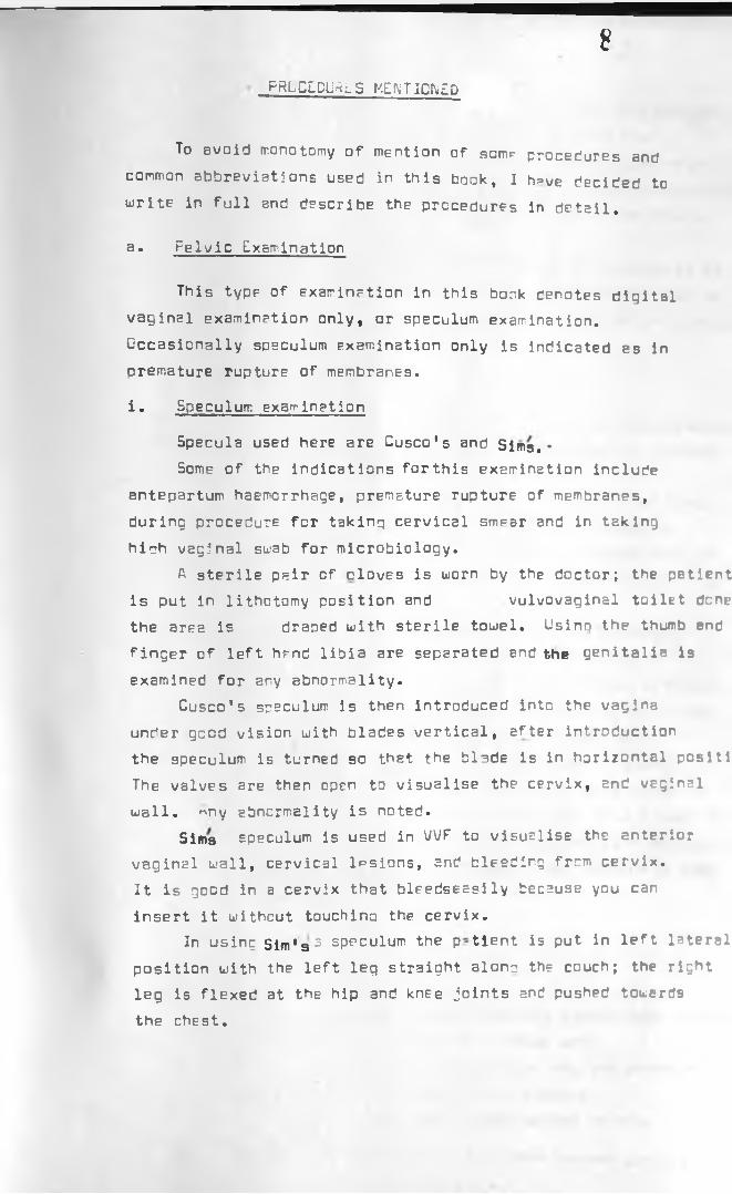
PRLiCEDUr LS MENTIONED
To avoid monotony of mention of somr procedures and common abbreviations used in this book, I h=ve decided to write in full and describe the procedures in detail.
a. Pelvic Examination
This typp of examination in this book denotes digital vaginal examination only, or speculum examination.
Occasionally speculum examination only is indicated as in premature rupture of membranes.
i. Speculum examination
Specula used here are Cusco's and Simg..5ome of the indications forthis examination include
antepartum haemorrhage, premature rupture of membranes, during procedure fcr taking cervical smear and in taking hich vaginal swab for microbiology.
ft sterile pair of cloves is worn by the doctor; the patient is put in lithotomy position and vulvovaginal toilet denethe area is draoed with sterile towel. Using the thumb end
finger of left hrnd libia are separated and the genitalia is
examined for any abnormality.Cusco's speculum is then introduced into the vagina
under gcod vision with blades vertical, after introduction the speculum is turned so that the blade is in horizontal positl The valves are then open to visualise the cervix, and vaginal
wall. nny abnormality is noted.Sims speculum is used in VVF to visualise the anterior
vaginal wall, cervical lesions, and bleeding from cervix.
It is good in a cervix that bleedseasily because you can
insert it without touchino the cervix.In using Sim's3 speculum the p-tient is put in left lateral
position with the left leg straight alone the couch; the right leg is flexed at the hip and knee joints and pushed towards
the chest.

3
Thp Doctor holds speculum in the right hand and re*racts the patient's right buttock by the left hr-nd so that the
introitus is exposed. The speculum is then introduced after lubrication Dr without lubrication, along the posterior vagina] wall towards the cerv:;x gently under good view. Thp cervix can be clearly visualised.
Patients with VVF may be put in lithotomy position so as check the site of leakage cp urine. This position may eIso be
used for any of the above conditions. The speculum is uithdrBt after examination is over.
1i. Vaginal examination
Exceot in cases where digital examination is ccntraindical e.g. antepartum haemorrhage, speculum Examination is followed by digital examination.
The patient is placed in lithotomy position. The doctor putscn nask, scrubs and putson sterile gloves. Vulva is cleaned with antiseptic solution of hibitane. Inspection of the vulva is made, then the right index and middle finger are introduced gently into the vagina.
In obstetric patient cervical dilatation^afface^ent, position, consistency are noted. Presence of membranes is also noted as is ccrd, caput, moulding and colour o' licuor. -hen pelvic assessment is required the fingers are slid along the sacral curve till they reach the sacral promontory. Prominence of ischial spine is noted, curvature of sacrum-,
subpubic angle are also noted. The inter tuberosity space is checkpd whether it can accomodate four knuckles of a closed fis
All these findings 3re noted and correlated with estimated fcetal weight. Adequacy or otherwise pf the pelvis is then
arrived at.
b. Conduct of labour
i. First stage
Patients in labour are admitted through labour ward sdmiss room into first 9tage section of the labour ward.
Pull history is taken from the patient who are unbookpdI.or are referred from other hospitals, or clinics.
In all, patients full physical examination is done
including an abdominal examination to determine the uterine
size, lie and presentation pf the fetus. 1p\/p 1 nf

prEserting part, contraction of the uterus and normalcy or otherwise of foetal heart.
Va*in; 1 examination is then done as above. If meTbr'nas are intact and cervical dilatation is more than 3cm in a mature fetus artificial rupture of membranes (A«M) is done.
Here colour of liauor is checked ; cord is checked whether it has prolapsed or it is presenting.
A H above observations form the baseline from which
subsequent observations are compared. Subsequent observations are done using partogram every half an hour.
«ny timo the cervical dilatation has reached 3 to «*cm a line is drawn from that point to the point when the cervical dilatation would be 1Ccm assumin'- the rate of cervical dilatation from 3cm or Acm to full dilatation, 1Ccm would be 1cm/hour. This rate is observed in 2Cr- of cases; thi line is called 'Alert line', 'action line'is drawn parallel to 'Alert line^but four hours later. Any patient whose progre is sc slow as to cross the action line must have arprocri»te action taken.
Analgesia is usually given in early labor, first sterr c labor ends rhen there is full cervical dilatation.
ii. Second Stage
This commences when there is full cervical dilatation and ends when the baby is delivered. The petient is taker to second stage cuticlF .
This is a sterile procedure where thE doctor or midwife
scrubs end puts on a sterile pair of gloves.The p-tient is in lithotomy position, her vulva and
rineum is cleaned with antiseptic solution, drapped with sterile towels. Vaoinel examination is done to confirm the
stage of labour.

The patient is encouraged to boar down with every contrect the perineum being infiltrated with local analgesia et the site episiotomy will be made. LUith the crownirm 0f the
foetal head a mediolateral episiotomy is made usinr a sterile pair of scissors.
The perineum is supported with sterile gauze as the foetal head comes out. The mouth and nostrils are wiped with a sterile gauze, th<= neck is palpated for cord which is freed if it is there.
«s the baby's anterior shoulder is delivered ergometrine or syntomentrine is given intramuscularly (except where there is contraindication as in cardiac disease in pregnancy). The rpst of thr baby is then delivered. The umbilical cord is clamped and devided between the clumos. The baby is sccred (hFj-v . 5CwKE) and shown to the mother. It is wrapped in a warn sterile cloth weighed and put a label fcr identification. The baby is put in a warm cot.
iii. Third Staoe
The placenta is received with a kidney dish put ageinst the pitient's perineum, ^s soon as there are signs of
placental separation the placenta is delivered by controlled
cord traction.Thp placenta is examined for completeness, infarcts,
retroplacental, clots, number of blood vessels in thE cord, the insertion on thp clacenta; membranes are checked
for completeness and finally is weighed.The third stage thus begins with the end of the beby's
delivery and ends with the delivery of the placenta.
The uterus is palpated for contractions^ .it can be massaged to facilitate strong contractions. The cervix, vaginal wall
and perineum, are inspected for any injuries. These are
repaired accordingly. ThF episiotomy is thEn repaired.

12iv. Repair of episictomy
In lithotomy position still the patient's vulva and perineum are cleaned with antiseptic solution and thP pati nt is draped with clean towels.
Local analgesia can be infiltrated aqain at th«= site of repair. The tip cf the episiotnmy is identified by retractinc the vaginal wall using finners nf the left hand* ne
being held in the rioht hand. Paginal wc11/mucosa is sutured using chromic catqut No 2/0 or 0, it is interrupted.
The deep muscle is now stitched carF being taken not to injure the rectum. Chromic catgut number 2/0 is used and
interrupted sutures applied;
3kin is finally stitched using chromic catgut 2/0, mersilene or polypropylene stitches.C^re is particularly taken h»re net to
injure the rectum.
nerineum is cleaned and sterile pad is applied on the perineum. The patient now rests in supine position and her post delivery observation* taken. Mil delivery data and birth notification are recorded end written by the staff whp conducted the delivery.v. Vacuum Extraction
This is done electively or as omerg-ncy procedure. 5om< elective indications include cardiac disease, hypertrnsicnjand some emergency indications include poor maternal pffart in second stage, ^cetal distress in late first stage or second staqe, mild disproportion, some maloositicns e.c. occipito-posteriDr and occipito-transverse positions.
The patient is put in lithotomy positionvulvaAcleaned and draped.
Digital examination is done tc ascertain cervical diletaticn the presenting part and its station.

1?The Mslstrom cup is selected so that the bicaFst
which can be accomodated bvthe is usee. This is
spoiled to the scalp or thp fetus, afea around it is swept by finger to exclude cervix being bit by the Cup.
Suction pressure is built slowly at a rate cf 0.1ko/cmL per minute to a maximum of C.3kg/cmi. 2 upto 0 to 10 minutes can be allowed in order to have proper chignon formation. In emergency however a shorter duration may be taken.
how with uterine contraction and maternal effort, trectior is applied in the axis of perineum. Episiotomy is made as scor as thF head distends the perineum. Cnee the head is delivered
the suction is released and the cup removed. The cup should n be applied for more than 30 minutes as there are chances of injury to the foetal scalp.
i. 5ubumbillcal midline incisions
This incision is variable but is between umbilicus and symphisis pubis. First knife is used to incise thp skin and second knife is used to cut through the subcutaneous fet to reach the rectus sheath, a small opening being made on the rectus. This incision is increased upwards and downwarc using a p^ir of curved scissors.
The rectus sheath is now deflected to expose the parietal peritoneum. The peritoneum is now lifted using twe
pMrs of long straioht ertery forceps placed about 1 to 2cm apart. «fter p-lpation for absence of gut a small incision is made using a pair of curved scissors. Under direct vision t incision is now exterded upwards and downwards to approximately the length of skin*.

ii. Pfannenstlel Incision
This incision is made transversely about 2cm above the pubic bone. The skin is first incised. A clean knife is then used for subcutaneous fat. The rectus is then reached; all bleeding spots are coagulated or ligated. The rectus is then opened transversely and released from underlying muscles along the linea alba.
After reasonable exposer has been achieved, perietal peritoneum is thus approached through the linea alba and opened vertically as above.
ill. Lower uterine Segment Caesarian Section
Subumbilical incision is used in this book; pfannenstlel incision can also be used.
After opening the parietal peritoneum, the bowels are packed upwards to clear the pelvic area using a pair of sterile wet abdominal pad.
Uteroversical peritoneum is lifted up using a pair of non- toothed pBir of dissecting forceps and an incision is made.
The incision is extended elliptically using a pair of curved scissors. The lower position is then reflected downwards to displace the bladder.
A superficial elliptical incision is thus made using a knife, it is deepened at the centre. The uterine cavity is opened by extending the deep central incision along the line of the elliptical superficial incision using a pair of curved scissors.
A Doyen's retractor is used during the process to hold the bladder down. The retractor is now removed snd the baby delivered by scooping the fetal head and the rest of the body is delivered by gentle traction.
Intravenous ergometrine is given. The placenta is separated and delivered mannually. Haemostasis is achieved, partly
by using Green Armytage clamps. The uterus is then stitched using No. 2 chronic catgut on round body needle in two layers. The first layer is usually interlocking but can be continuous. Uterovesical peritoneum is closed using chromic catgut number 1.
Abdominal packs are removed and peritoneal cavity cleaned. Ii is done for any bleeders and any area that is not closed. The
abdomen is then closed.
15 -
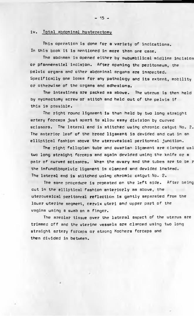
iv. Total abdominal hysterectomy
This operation is done for a variety of indications.
In this book it is mentioned in more than one case.
The abdomen is opened either by subumbilical midline incision or pfannenstiel incision. After opening the peritoneum, the pelvic organs and other abdominal organs are inspected. Specifically one looks for any pathology and its extent, mobility or otherwise of the organs and adhesions.
The intestines are packed as above. The uterus is then held by myomectomy screw or stitch and held out of the pelvis if this is possible.
The right round ligament is then held by two long straight artery forceps just apart to allow easy division by curved scissors. The lateral end is stitched using chromic catgut No. 2. The anterior leaf of the broad ligament is devided and cut in an elliptical fashion above the uterovesical peritoneal Junction.
The right fallopian tube and ovarian ligament are clamped usi two long straight forceps and again devided using the knife or a pair of curved scissors. When the ovary and the tubes are to be r
the infundibopelvic ligament is clamped and devided instead.
The lateral end is stitched using chromic catgut No. 2.
The same procedure is repeated on the left side. After being
cut in the elliptical fashion anteriorly as above, the/
uterovesical peritoneal reflection is gently separated from the lower uterine segment, cervix uteri and upper part of the vagina using a swab on a finger.
The areolar tissue over the lateral aspect of the uterus are trimmed off and the uterine vessels are clamped using two long straight artery forceps or strong Kochers forceps and then divided in between.

16 -
The lateral portion is ligated with chromic catgut No. 2, the same procedure being repeated for the other side. The
cardinal ligaments are then clamped and cut alongside uterus
and stitched using chromic catgut No. 2. Uterosacral ligaments a] cut and separated from the cervix uteri using blunt dissection.
The cervix is then felt with the fingers and a pair of little-wood clamps are applied to hold the upper end of vagina just below the cervix. A stab-incision is made between th( two clamps using a knife. Using a pair of curved scissors the incisions are extended laterally, thus circumsising the cervix. The uterus with the adnexia are removed and taken for histology.
The vaginal vault is held by long straight artery forceps. Haemostasis is ascertained and the vault is closed using number 2 chromic catgut starting at the centre. The central stitches are left long to be tied together, during peritonisation.When haemostasis is achieved the vault is now peritonised to covei the raw areas. The reflected peritoneum anteriorly and posterior]
are stitched together incooperating the round ovarian ligament
or infundibulopelvic and uterosacral ligaments using chromic catgut No. 1.
The abdominal packs are removed and after ascertaining swab and instrument count, the Bbdomen is closed.

L£>3CRh TCRY
/I
The dE-artrent has laboratories for routine investigation* and research work.
a* ^ain departmental laboratory
This is situated within the department and dealswith:-i. Infertility studies including seminalysis end antispern
antibodies.
ii. Hormonal assays using radioimmunoassay
iii. Biochemistry handling lipid assays, blood sugar, serum fructose assays.
iv. icrobiclocy which handles chlamydia and gonococci cultures.
b. Labour ward laboratory
This handles surfactant test, bilirubin spectrophotometry, pregnancy test and oral olucose tolerance test.
c. Cytology Laboratory
This is within the annexe of the department and deals with cervical cytolony, buccal shears, chromosomal analysis
and karyotyping.
LTHl-. bLRWICi.3
a. The department 3lso offers colposcopic and cryosurgery services every Friday mornino.
b. In our labour ward we now have sonicard which is useful in
ascertaining foetal heart normality or otherwise.Lie also have ultrasound machine for scre-ning special
obstetric cases like identifying low-lying placenta, ascertaining fcetal maturity and normalcy emonn other things.

I S
P n _ t- . T L • l •■LlPTLJ-.;. >.r f- _l- j »r-.ii._S h T 2 ? UC L K L
C J - . o - d J - T H / i f- f. ■ i£ [ . £ f . T , L I ' J i B.^BY AT TLRM
i< ame I H • h .
"Qe: 25 years
Parity: 0+1
IP Mi: 903^67
LNf'.P: 3.12.1937 EDO: 10.9.1936 OCA: 29.6.19ee
DCD: 15.8.1936
Pi.rEuATI!Vj LuKPLAlMTE
The patient compl-ined of drainage of liquor for thelpast U days.
n.5 7 .Y -P ZLLT.;
The pr-tient was uj~i 1 until four days before admission uhe noted some fluid dripping down her leqs. ThFre was no history cf trauma, no history of any illness prior to this, she had no accompanying p-in nor did she have vagincl b]<-Fdinr.
~hp had first rune to Erbu Provincial General Hps itel
luhen the Droblem started and was mencyed with tablets ventolin
pheroberbitons and ampicillin cepsules without ir.prcvsrr nt.
~w T i- Sf i ■ j _ >-»i> j, Y[\ _ I -■ L H I T l..; V
She was para 0+1. The abrrticn was at 3 months end no currateqe was done. Her last monthly pe-iod was 3.12.67. She- was getting periods every 28 d-ys lasting 3 -U days. It was not acco^ornisd by any pain. Her manerch t i
at 13 years. She had not used any method cf contraception. P..L5:.f,T SSSTST .;3 hl-T^RY
She was 2r weeks pregnant according to her lest norr->1
monthly periods, .and h'd not attended any antenatal clinic.There had been no problem with this pregnancy.
P~1T ,-£DIL...L .+ I2T^ y
‘■ot contributory

13FmKILY r.D SLwIqL HISTORY
She was a married housewife stayino with her husband in Cmbu. Her husband was r peasant. She did, pot smoke ncr drink and had no medical problem in hEr family.
or. ox.-.; if. -t o .
She uas in good General ccnditicn. ahp was not pale
neithpr was she j=undiced. <-ther general examinations were no Her pulse rate uas eo/minute normal character, blood pres
120/7C3mmHg, respiratory rate was 20/minute normal. Her body temperature was 36.6°C.
Central Nervous System )
Respiratory System ) were all normalCardiovascular system )
■•.SDCt-'ir.-i ex r i f t ; tick
The abdomen uas distended especially the lower asnect, utErine size uas about 26 weeks. Fetal lie was longitudinal the head was presenting and it wes above the pelvic brim. Feti heart beBt was heard and was 152/minute regular. Thpre we: contractions.
V^j IIUL EXAl.Ih TIGfv
She had a normal external genitalia, there was obvious liquor draining from the vagina. She had no veginal bleedinn. Speculum examination:
- Normal vaginal wall
- Cervix was healthy and closed. Clear liquor was draining from the cervical os.
DIAGIUjjIa
A diagnosis of premature rupture of membranes at 29 weeks gestation was made.

2)
In viFw of the gestation and previous history of
pregnancy loss the patient was admitted to our antenatal uard for conservative management. She was started on ampicillin 500mg 6 hourly and bed rest. Her pulse and
temperature was closely observed to be within normal limit throughout her stay. Liquor loss was observed for any sTell or colour change which would indicate infection.
She had weekly white blood cell count, urine For
culture and sensistivity. She hnd her antenatal profile done.Her serial white blood cell count ranoed from 6.L -
9 9.5 x 10 /l, and her serial urine crlture yielded no growth.
she was cf blood group A rhesus positive; her haemngram was 13g/dl with packed cell volume of 2>UV. Her serological screening was negative.
The petipnt however continued to drain liquor and on29.7.88 was taken for ultrasound to check the auantity of amnictic fluid in view of her continued drainage. The report was th*t there was scanty iinuor, single live fetus of biperietal diameter corresponding to 32 weeks. * decision to continue with conservative management was upheld.
Other then the drainage of lipuor the patients observetio remained stsblE. She went into spontaneous labor on 1L.8.38, progressed well enp delivered a babv nirl of apgar score
ll 9 in 1 minute, 10 in 5 minutes end of weight 22DCgms.Her postpartum observations were normal and was discharged after 2U hours to be followed up in E'.i u Provincial Hosoit.’' .
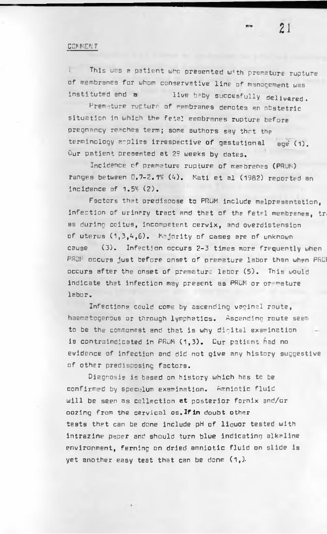
21CO-f-E* T
This was a patient who presented w'th premature rupture of membranes for whom conservative line of management was
instituted and a live baby succesfully deliverecj.Premature rupture of membranes denotes an obstetric
situation in which the fetal membranes rupture before pregnancy reaches term; some authors say thrt the terminology applies irrespective of gestational Bge‘ (1).Our patient presented at 2? weeks by dates.
Incidence of premeturE rupture of membranes (PRUM) ranges between 0.7-2.Tv (£*). ttati et al (1902) reported an incidence of 1.5^ (2).
Factors that predispose to PRUM include malpresentation, infection of urinary tract and that of the fetal membranes, tr. as during coitus, incompetent cervix, and overdistension of uterus (1,3,A,6). ^“jnrity of cases are of unknown cause (3). Infection occurs 2-3 times more frequently when PR2I- occurs just befcre onset of premature labor than when PRLf
occurs after the onset of premature labor (5). This would indicate that infection may present as PRuM or Dr mature labor.
Infections could come by ascending vaginal route, haemategerous or through lymphatics. Ascending route seem to be the commonest and that is why digital examination
is contraindicated in PRuM (1,3). Cur patient had no evidence of infection and did not give any history suggestive
of other predisposing factors.Diagnosis is based on history which has tc be
confirmed by speculum examination. Amniotic fluid will be seen as collection at posterior fornix and/or oozing from the cervical oe.Ifin doubt other tests thet can be done include pH of liauor tested with
intrazine paper and should turn blue indicating alkaline environment, ferning on dried amniotic fluid on slide is
yet another easy test that can be done (1,)-

22
f’-an^gement will depend on gestation and previous obstetri performance. Conservative management is generally
upheld for gestation less then 3k weeks and more than 26 weeks fhp aim is to buy time and in so doing get a maturer baby cnoable of extrauterine existance, to avoid infection the onset of which would be an indication to terminate the pregnancy, and to avoid labor (1,3). Routine observation for infection like abdominal tenderness, fever, high pulse rate, right not really rule cut.infectinn as the commonest
infectin'- orgarism is haemolytic streptococcuwhich i? of low virulence and might net cause hich fever (8). Cither features of infection include foul smelling liquor, fetal tachycardia andleucocytosis of 50°/ above normal (7).Cur patient had obvious liquor drainage,
was m"neced conservatively and showed no evidence of infection throughout the period.
There is generally increased perinatal mortality due tr prematurity and respiratory distress syndrome (3).PRO also predisposes to cord prolapse and fortel distress thereby incrersing caesarian section rate.t ost partum infection has been reported upto of cases, cu Patient recovered well and was discharged with a baby who was healthy.
2 H
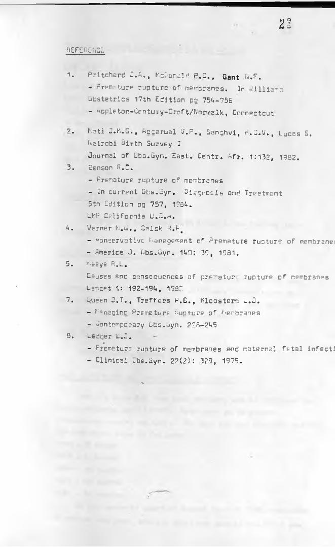
REFERENCE
1. Pritchard J.A., KcDonslri (3.C., Gant f<.F.
- r'rpir.-turp rupture of membranes. In -illlams ubstetrics 17th Edition pc 75L-756
- Hppleton-Century-Croft/r.aruielk, Ccnnectcut
'• f.oti w.K.j., rtozeruisl V.P., Canghvi, n.C.V., Luces S. Aeircbi ^irth Survey I
Journal of Cbs.uyn. East. Centr. Afr. 1:132, 1982.3. Benson R.C.
- Prenature rupture of membranes
- In current Obs.Syn. diagnosis and Treatment 5th Edition pg 757, 155A.LPP California U.C.*.
A. Varner h.ui., Calsk R.F
- uonserv2 tive Management of Premature rupture of membrane!- America J. Lbs.Gyn. 1AC: 39, 1581.
5. P'seye R.L.
Causes end consequences of preratur rupture of membranes Lancet 1: 192-19A, 1532
7. Hueen C.T., Treffers P.E., Klcosterm L.J.- P = neginn Premeture ;>upture of r,erbranes- Enntenporary ubs.Syn. 228-2A5
6. Ledger UI.J.
- Premature rupture of membranes and maternal fEt3l infect!- Clinical Lbs.Syn. 22(2): 329, 1979.
V

21CERUIu mL INCLKPETEf.CE, NCDQNrALD STITCH PUT.
Lll/t 3 'BV DELIVERED
N me : K . G .
Age: 3D years
Parity: 2+5
IF No. 871186 LNMP: i*. 14.87
EDD: 11.1.88 DDA: 7.12.87
DOD: 8.12.88
PRESEN TING ClKPLAINTS
^he patient presented with labor pains for the last 5 hours.
HISTORY LF PRESENT CJf.PLuINTS
The patient was well until 5 hours earlier when she started having abdominal p~in which was intermittent and was increasing in frequency and intensity. She was also noted to have blood stained mucoid discharge.
Pr-.ET MEDICAL HISTORY
Nothing contributory
Prs5T G5STET1-.ICS hKD GV[v-.ECOLOGICAL HISTORY
^he was para 2+5, her last delivery was in 1983 and her first delivery was in 1982. Both were at T k months spontaneous vertex delivery. In both she had cervical stitch. Her abortions were as follows:1978 - 2 months1979 - U months
1980 - months1981 - 6Y? months 1981 - Byk months
In all cases of abortion except that of 1980 evacuation
of uterus was done, and all abortions except the first one,

✓ 25starter4 with drainage of liquor, then abdominal pain and th'rn vaginal bleeding and expulsion of the fetus.
:ahe had not used any contracepiives methods and hpr menerche was at 1<* years.
F-.ZJQ.T „3;>TLT . IC HISTORY
Her last monthly period was She had been
getting her periods regularly every 28 days and lasting t-5 day!“■•he was first seen in cur clinic at 11 weeks pregnancy,
she was bcokEd on the basis of the bad obstetric history- ^he had been previously diagnosed as having cervical tear at 3 o'clock and her cervix was only 1cm long. She was
admitted on 8.7.88 at 1^ weeks for McDonald Stitch which was put on the following day and was discharged the following dey on carsules anoxil 50Cmg, 8 hourly, tabs ventolin ^mg 8 hourly and she wes advised on bed rest.
She subsequently attended clinic 13 tines which were essentially uneventful except for few 2 occasions when she complained of heaviness in the lower abdomen for which she was prescribed bed rest arri ventolin £*mg orally 8 hourly.
Her blood group was D rhesus positive, haemoglobin level was 1t*.t*g/dl, packed cell volume of 35.79* and serology screen was negative.
family r.ap slCIhl histlsy
ahe was a married lady working in Town as a typist.Her husband was a clerk with Teachers Service Comr. ission.They stay at "iruta in Nairobi. The was no contributory history in the family. She does not smoke nor drink alcohol.
CfJ £AmK;Ii\r-.TIuN
^he was in good general condition. She was not pale, nor was she jaundiced. Other general examinations were normal. Her blood pressure was 1lC/60mmHg, pulse rate was
B^/minute, respiratory rate was 20/minute and her body temperature was 36°C.

Central Nervous 5ystem )
Cardiovascular System ) Were essentially normalRespiratory 5ystem )
A3DLFINAL EXAMINATION
The abdomen mas uniformly distended, uterine size was 36 weeks. The fetus was lying longitudinally and head was
presenting station 2/5 above the pelvic brim. She was having two contractions in 10 minutes each lasting 30-A0 seconds.Fetal heart was heard at rate of 138/min regular.
IMG IIMAL CXAM INnT ION
She had normal external genitalia, there was bloody mucous at introitus. Speculum examination revealed normal vaginal wall; bloody mucous was noted to be coming through the os, silk suture was seen, it was held by sponge holding forcep and cut with a long pair of scissors and pulled out.
Digital examination found a cervix which was 5cm dilated, membranes were bulging and artificial rupture of membranes done using middle and index fingers and clear liquor came out. Cn further inspection no c-rd was felt. Head was lying in left occipito-anterior position.-
DIAGNOSIS
diagnosis of parous lady with bad obstetric history at 36 weeks in established labour was made.
FLAN
The patient was taken to first stage of our labor ward
was given intramuscular injection of pethidine lODmg, she was put on left lateral position and progress of labor
monitored by the use of partogram.

2 ?
She had smooth labor progress and delivered a female infant weighing 2250 gms and score 9 at 1 minute and 10
at 5 minutes. Placenta and membranes were delivered whole and weighed UCOmls.
On inspection, perineum, vaginal wall and cervix were normal.
Post partun observations were normal, 3p was llO/VOmmH,PR was BVminute and respiratory rate was 2^/minute. She
was taken to our post natal wards where her condition remained
stable and after 2U hours observation she was discharged with her baby in good condition.
The patient was not seen at the appointed date at our post natal clinic.
ftddenduw
Procedure fcr McDonald stitch as is done at our unit is described. The patient is admitted the day before the operation and pre operative procedure done.
In theatre the patient is put in lithotomy position and Ellf done to determin0 length of cervix and any of the lesions. Cervical dilatation is also assessed. Cervie is the held by sponge holding forcep and using round bedied needle silk number 2 suture is placed at the junction of smooth muscle of portio vaginslisand rugosa mucosa of vagina. The needle is now pushed to oervical stroma bites are now taken at 11-10, 8-7, 5 -U and 2-1, o'clock.future is then tied with surgical knot anteriorly being braided about 2-3cm to allow easy access and removal.

COMMENT
^his was a patient who had had a number of abortions, was subsequently diagnosed as having cervical incopentence and m'.naged with cervical stitch succesfully.
The concept of incompetent cervix was first described by Fulmer and Lacoume in 1940 (1). Cervical incompetence is characterised by painless cervical dilatation in second or early third trimester with membranes prolapsing through cervix and balooninn into vagina followed by their rupture and almost immediate expulsion of fetus who is too immature and usually succumbs (1,2).
Definative diagnosis makes clear incidence difficult to establish. This is because some of the patients end up with the diagnosis and even cervical suture who actually have other aetiological conditions. Njage (1979) at Kenyatta National Hospital found en incidence of 0.1-1&(3). Elsewhere it has been reported to range between0.1^1% (1).
Acquired causes of cervical incompetence include those related to child birth 95^ (7,9). aome of these are delivery of excessively large baby, occiput posterior and breech; these have tendency to cervical laceration or tear. Cther acquired causes include dilatation for curratage or for treatment of dysmenorrhoea, radium, placement, chronic infl .mmation, cervicil conization, cauterization or amputation Exposer to stilbeaterol utero also predisposes to cervical incompetence later (2). k few unexplained cases are due to congenital abnorm lity of cervix for example congenitally short cervix (1).
Cur patirnt had had five abortions which except for the fi one have all been in mid-trimester. ShE had been found to have a cervical tear at 3oclock and cervix wes short, 1cm.
On two previous pregnancies, she had delivered viable babies following McDonald stitch. Sequence of events in her 4
last abortions also are tyDical of cervical incompetence.
Generally the gestation at which these patients present
diminish the subsequent abortions. This was not characteristic
in our patient.

Explanation Is difficult but perhaps she had some residual
cervical masculature and the increasing ability of the uterus to accomodate with subsequent pregnancies.
Diagnosis of cervical incompetence is reached by typical history offered like in our patient; there will be vaginal pressure followed by rupture of membranes and brief or painless labour and delivery of the fetus (1,2,6,9). Gestation will usually be mid trimester to early third trimester. Vaginal examination will reveal short cervix with some degree of dilatation of Internal os, lesion like tear might be apparent. Our patient had both shortness and defect of cervix.
In non pregnant state diagnosis can be confirmed by easy passage of Hagar size 8-10 though the cervical canal (1,7). hysteroaalphlngogram will fall to show the usual sharp Junction of tt
internal oa, instead there is betiding of the lower uterine segment into the cervix, cervix may be short or may be flush with vaginal wall, that is, there are no fornlcea, ultrasound can also be
used for diagnosis (7).Many Gynaecologists were aware of the occurence of this
condition but prospect of surgical correction was only first offered by Lash and Lash (1950). Shirodkar (1953) and McDonald (1957)
It was even then known that conceptus is retained in the uterine cavity by pregnancy hormonea, but at three months onwarda it is the constrictive Influence of the circumferential
- fibromuscular tissue at level of internal oa that assumes the duty (‘ Lash and Laah procedure used to cause cervical stenosis and infertility, and Shirodkar'e procedure was technically difficult; McDonald procedure was less traumatic, less blood loss and simple (1. It is the method which is used in our unit and it is the method whicf
was used in this patient.McDonald's cervical cerclage is a simple procedure as described. It is important to keep the ligature external
to internal cervical vessels.

Generally the procedure is carried out just after the first trimester and seldom before 16 weeks (1,2,8). In our unit it is carried out within 1*+-1B weeks. Too early
insertion is unnecessary os it amounts to futile attempts to keep fetuses most of which have congenital abnbrmalities,
and too late procedures might cause rupture of membranes.
L’ur patient had cerclage put at 1^ w^eks. Ue routinely put our patient on broad spectrum antibiotics following cervical cerclage to take care of any infection though seme workers have seen no need of such action (**,B). These workers
suggest that amniocentesis should be done and specimen submitted for culture and sensitivity any growth would be an indication for uterine evacuation (8). It is also said there is nothing achieved by giving progestagens and
beta-mimetics as adjuvants (4).Complication of cervical cerclage include rupture of
membranes, infections, haemorrhage, premature labor, slipping of suture and rarely the rupture of uterus and/or tear of cervix if suture is not removed with uterine contraction (3) Charles et al (1981) reported lower incidence of complication if procedure is performed by 18 weaks.
Cur patient did not have any of these complications.If there is no early labour or other complication
warranting early removal of stitch, it is usually done at 37 completed weeks.- Removal at 37 completed wepks is a good test of whether the or not the suture was necessary for it was labor usually starts immediately.
Success rate with cervical cerclage is 5D-B0V (7)Rjege (1978) reported it at G U .2 e/ (3).

REFERENCES
1. Aladjera 5.
General Gynaecological problems in pregnancy Obstetrical practice pg 719-720, 1980 Edition The C.V. Mosby Company St. Louis TorGntD. London
2. Pritchard J.A., McDonald P.C., Gant F.N.,Abortion
Williams Obstetrics 17th Edition pg A75f igQ5
Appleton-Century-Croft/Norwslk Connecticuts.
3. P.jage P.,
h-nngrniant of Cervical Incompetence by Purse String future, fi.f'ed T|~iesis University of Nairobi 1979
A. Ihomnson J.L., Campson M.Q.
The incompetent Cervix. A 1902 update.Journ. Tieorad. Med. 27: 187, 1982.
5. Kuhn R.P.J., Peppcrell A.J.
Cervical Ligation; a review of 2A2 pregnancies.>'ust. NewZenland Journ. Obs.Gyn. 17: 79, 1977
6. Gensan, C.K.
Disorders of Uterine Cervix
Current Ubs.Uyn. Diagnosis and Treatment pg 170, 1976 Lange Medical Publication, Los Altos, California.
7. Deuhurst J.- Abortion
- Integrated Ubs.Gyn. for Postgraduate Students 3rd Edition 1901, pg 21A-5
- Jlackwell Scientific Publications
- Oxford London. Edinburg. Boston. Melbourne.8. Charles D., Edward U.R.
Infectious complications of cervical cerclage Am Jn. Obs.Gyn. 1A1: 1066, 1901.

PCLYHYDRAKNIDS V.
Name: R.M.
Agei 25 years
IP No. 906A58
Parity: 5+0 LD: 1986
LMP: Lactational amenorrhoea DOA: 17.7.88
ODD: 21.7.88
PRESENTING CLHPL.-INTS
Generalised abdominal pain for 1/12. - Backache.
HISTORY CF PRESENT ILLNESS
Abdominal pain has been accompanied with fullness of abdomen for one month.
Backache has been there more at times but no relation with time. Over the last one month the backache has been continuous and progressively become worse.
She gives no history of fall nor any history of any trauma.
PAST OBSTETRICS AND GYNAECOLOGICAL -HISTORY
Para 5+0 now gravida 6. A H deliveries have been term and all except the first one which was at Pumwani Maternity Hospital have been at Ngong Health Centre. The children are ■live end well. Herpuerperium were uneventful. Her first delive was in 1978 when she was 15 years and her last delivery was in 1986
She attained menarche at 13 years. Her cycles had been regu'
at 3-5 deys every 28-30 days.^he had no period since last delivery and was not using
any contraceptive method.
Past Medical and Surgical History
She had had uneventful life with no major lllnesees before.
family ~nd Jfirlnl Hlctnry
She Is a married housewife staying together with her husband at Ngong. She does not drink alcohol nor smoke.
Her husband has no permanent Job but does any manual labour available.
No history of any medical problem in her family nor from tht parents' side. No family history of any obstetric complication of the like in the family.
HlbTUHY CF PRESENT DBSTETf<ICS PREGNANCY
The patient was booked at Ngong Health Centre and no r cords were available but patient says the first U attendances were uneventful. The last time she attended two weeks prior to admission, she had the presenting complaints and wa9
given tablets which did not help her.
She does not know her lsst menstrual period because she got pregnant while she was breastfeeding her last born.
PHYSICAL EXAMINATION
She was in fair general condition, was in pain but had no palor, no jaundice, no adenopathy She had no pedal nor
sacral oedema.
Pfr - 72/minute Rfi - 22/minute Bp - 110/80mmHg
Temperature - 36°C.
Central Nervous Syste, respiratory and cardiovascular systems were normal. Breasts were normal and active.

ABDOMINAL EXAMINATION
She had very distended, tense abdomen and tender.
Uterine size reached the Xiphisterum, foetal parts difficult to feel, fluid thrill and shifting dullness present.
VAGINAL EXAMINATION
Was not indicated.
DIAGNOSIS
Diagnosis of polyhydramnios was made.
PLAN:\
Im pethidine lOOma stat
- Tab panadol 2* 8 hourly Investigations ordered include:-
1. Urgent ultrasound - maturity - normality
2. Haemogram3. D/E
A. Blood grouping and Rhesus, Serology5. Amnitoic fluid for cytology and alpha-fetoprotein
6. Blood sugar
Results:
Blood sugar - 3.Bmmol/l Hb - 11.5g/dl WBC - 0 .A x 109/1 PCV - 3A.$
Lymp - 21.7%, 1.8 x 10⁹/1
PLT - 2 *1 x 109/L
Blood group and Rh - Q +ve.
5erology - negative
Amniotic fluid - surfactant test done instead.It was 1 :1 negative K
1 :2 negative. w

- anencephalic fetus in "cephalic" presentation- polyhydramnios
- Fetal heart activity seen
- ^pine difficult to locate.
DIAGNOSIS
Rnencpphaly with polyhydramnios
Plan: - Blood Group and Cross match 2 units
- IV line started
- Sent to labor ward for induction of labour.
VAGINAL EXAMINATION
Normal external genitalia, cervix was 2cm dilated; 1.5cm
long, central, ^mniotomy was done 5L of amber coloured amniotic fluid obtained slowly.
- syntocinon 2.5 units in 50Pmls of 5% dextrose put in a drip.
- observation of vital signs, contractions, liquor colour.
- 5 hours later, she delivered anencephalic fresh stillbirth weight 1000 grammes, placenta 300grams complete.Cervix and perineum were intact, no bleeding, uterus was well contracted. Patient was kept on syntocinon drip for two hours postpartum. Post delivery observations were normal end patient discharged to post natal ward where she continued good observation. She was discharged home to be seen in our post natal clinic but she did net come back.

COKMENT
This was s patient who presented with polyhydramnios due to enencephalic baby, she mas succesfully induced and fresh stillbirth was delivered.
Polyhydramnios is excessive amount of amniotic fluid in the pregnant uterus; a lower limit of 2000 mis anything excessive is polyhydramnios (1,2).
Incidence of polyhydramnios have been reported by various workers. Abdalla (I9fl0)reported an incidence of at Kenyatta National Hospital (1). In other places it
has varied between 0.1-1.6% (3,«*). The incidence of acute hydrai in Abdulla's series was 0 .02%. As defined by Wueenan et al (5) acute polyhydramnios occurs before ZU weeks gestation while chronic polyhydramnios is usually diagnosed in third trimester. Chronic polyhydramnios is rare and incidence is reported of 1:150 (6 ).
Factor usually operational in keeping equilibrium include f
deglutition, fetal micturation, uteroplacental blood flow, fetal
respiratory movement, fetal membrane physiology and a host of other factors. Primigravidas seem to be less affected,
B.B£ and age group seem to peak at 21-30 years with range of
18-UO years (1,7). Cur patient was para 5+0 she was 25 years old and estimating from fetal weight she could have been in the second trimester. So she had more of the attributes of
acute polyhydramnios.Polyhydramnios is found in pregnancies complicated with
diabetes mellitus, rhesus isoimmunization, multiple gestation especially with transplacental transfusion syndrome, pre-eclsmps;
chorioangioma'i,circuinvalate placental syndrome, congenital abnormlities like anencephaly and gastrointestinal obstruction (1,2,3,1*,5,6 ,7). Central Nervous system abnormality seem to account for more cases of hydramnios SCPS of which 6CK are anence; (7,8). Cur patient was investigated for other possible causes but the results obtained were negative. The only finding wes anencephelly. The mechanism by which anencephelic fetus
is associated with polyhydramnion of though to be absencp of
deglutition so thet fluids accumulate.
r - f• — . • * n

3?
Diagnosis cf polyhydramnios clinically depends on accurate menstruel history, finding of uterine size bigger than dates, difficulty in feeling foetal parts either because of tense abdomen or due to fetal abnormality and muffled fetal heart. Definative diagnosis, unless the fluid quantity is too much is by ultrasound. Our patient had not gotten her menses b
her other findings were so pathognomonic that the diagnosis was not a question, rather it was the cause. In other cases where the cause of the polyhydramnios is not obvious
other investigations as had been started in our patient are useful (3,6,7,9). In other cases despite elaborate investigati no apparent cause is demonstrable. These are of Idiopathic cau and the management is to wait till delivery when the diagnosis will be established (9). Queenen found incidence of idiopalhlc case to bp as high as (5).
Management is gsverned by whether the fetas is normal or n Conservative approach with bed rest sedation and analgesic are the cornerstone for those with normal fetuses with minimal discomfort. Antidiuretic hormone intrauterine has been used in some cases to reduce the amount of fluid but with temporpry result (1).. If gestation is low and the patient hes symptoms from grcse distension then controlled removal of. amnlotic fluid is the treatment of choice, although initiation of premature labor, infection, placental abruptio, premature rupture of membranes and possible fetal injury are inherent problems with such therapeutic amniocentesis Our petient had
fetal abnormality and the management was termination of pregnancy.
Maternal complication include antepartum haemorrhage, post haemorrhage and infection; fetal complication are many and give mortality of between UB.6-69% (1 )These include cord prolapse malpresentatlon,, preterm deliveries, intrauterine Infections
and effects arising from primary complicating diseases like
diabetes mellitus (1). In our patient we knew the inherent problem of post partum haemorrhage, and we started the patient oi syntocinon drip and also took blood for group and crossmatch. The patient had no problem, recovered well and was discharged home.

REFERENCES
1. Abdalla S.H.
- Incidence of Hydramnios at Kenyatta Nationel Hospital
- M.Med Thesis 1980 University of Nairobi
Sleek, U.U.; Ohlsson, A.
- Fetal Polyuria and Hydramnios associated uith Bartholin Syndrome
- J.A.M.A. 63(3): 22, 198A
5. Quuenan, J.I., Gadou, E.
- Polyhydramnios, chronic verses acute
- Am. Journ. Obs. Gyn. 108: 3A9, 19706.
Liquor Amnii
Brownes antenatal care Pg 1A6, 1978
7. , Jacoby, H. t Charles
- Liinical Conditions associated with hydramnios
- Am. Jrn. Obs.Gyn. 9A: 910, 1966
8 . Sagar, H.J., Desa, &.J.
- The relationship between hydramnios and some characterlst of the pregnancies complicated by fetal anencephaly.
- Jrn. Obs.Gyn. Brit. Comm. 80: A29, 1973.9. Murray, S.
- Hydramnios: A study of 8A6 cases- Hmer. J. Obs.Gyn. 8 8: 105, 196A.

33
UK3 IL I " ~-L CE 3 F -^l ^FSL, EPE-.GENCY C.-£S-i.-W N GECTILT.'.
HUE BmBY DELIUE.1ED
Name: R.w.K.Mg*: 27
Parity: 1+0
IP No. 875631 LNKP: 15.5.07
EDO: 22.2.88 DDh : 16.2.88 DOD: 23.2.88
PRESENTING CLMPLh INTS
Patient presented with U hours history of labor pains and drainsgp of liouor.
HISTORY uF PRESENT ILLNESS
^he patient was well until frur hours prior to admission when she noticed a sudden gush of water flouing down her
legs from thr vagina, about an hour later shp started having lower abdominal pains which later on became generalised and inte mittent in nature.
There was no history of fall and no history of any sickness prior to the onset of the problem.
PhST PEDIC L HI5TGPY
Nil of note.
P---3T u3 l;TET,:C AND GYP-hECCLGLIChL history
She was para 1+0, her delivery was in 1986 and was normal spontaneous vertex.
Her menarche was at I4* years. She had had regular
periods prior to the last one which was 15.5.87. Jhe used
to get her m*nses every 28 days and could last 3 -U days.She had not used contraceptives.

PRESENT 035 TbTR ICS HISTORY
At the time of admission, the pregnancy was 39 weeks.
She Li s bcokEd in our clinic at 36 weeks because of variable lie. She was to be admitted for stabilizing induction on 1C1.2.38 but she rpfused.
She had no other problem when she was seen in the clinic, and her vital signs were normal.
She was blood group C Rhesus positive, her haemoglobin
level was 10.3g/dl and PMl, 31r . SBrology was negative.
FAMILY AND SOCIAL HISTORY
She was a single lady working at Kahawa Barack as a secret Ty. She drinks alcohol but does not smoke. There were no other contributory history.
CfM EXAMINATION
ahe was in good general condition. She had no palor nor jaundice. Other general examinations were normal.Her blood pressure was 120/7CmmHg, pulse rate was BC/minute normal, respiratory rate was 22/minute and her body temperature was 36.1*°C.
Central Nervous System )Cardiovascular System ) luere normalRespiratory system )
ABDlMr: L EXAMINATION
Her uterine size was term, the baby was lying in oblique position. She was getting one contraction lasting 30-AD seconds in 5 minutes. Foetal heart was heard and was lA2/minute regular.

4 JVhGIIUL cx»m m ^ tidp;
L-lear liquor was oozing from otherwise normal external genitalia. Mo bleeding was noted. Vaginal wall felt
normal and umbilical cord was felt to be in the vagina, it
was pulsating at a rate of 1t«2/minute. Cervix was about 3cm diluted and 2cm long.
DIhjMCS15
" diagnosis of cord prolapse at 39 weeks was made.
K-.MhGEMEMT
The petient was put immediately in knee chest position to lessen pressure on the umbilicus. Venous blood was taken for group and cross match 2 units of compartible blood. Intravenous fluid was started, informed consent got from the patient, a quick preparation was made and patient was taken to theatre.
The operation table was reclined head down, oxygen was given by mask. M quick vulvovaginal toilet was done and bladder
emptied by catheterization. Vaginal examination revealed a cord that was still pulsating, patient was cleaned, drapped, anaesthetised and caesarian section done as in the in roduction. rt live baby delivered of a-gar score 10 at one minute, 10 at 5 minutes, birth weight was 33*+0 grams. Placenta was funrial more anterior and with its membranes were delivered whole, bterus and abdomen were closed as described. Aneesthpsia was reversed and petient taken back to the wards. She made steady recovery post operatively, her check haemoglobin was 9.8g/dl. ahe was discharged in good state with her baby, on the seventh post operative day.
She was reviewed six weeks later in our post natal clinic and she had heeled completely. She had already started
on intrauterine contraceptive device. She had no complaints.

42CCiF'NLIM T5
This was a patient who presented with a complete cord prolapse for which emergency caesarian section was done and live baby delivered.
Cord prolapse can be occult where the cord lies over the
face or head but can not be felt on examination; it can be forelying cord in which the cord preceeds the presenting part but is inside the intact membranes and can be palpated, and e complete prlapse, like in our patient where the cord descends oast the ruptured membranes into the vagina or introitus. (1)
Cord orolapse is caused generally by imperfect adaptation
between the presenting part end pelvic inlet (1,2f3) Factors therefore in essocietion would include abnormal presentation like breech, transverse shoulder, multiple pregn-ncy; hydramnios pelvic tumors or distortion of the pelvis; placental praevia or premature rupture of membranes. Cur patient h^d abnormal lie end 'bncrmal presentation. Cither factors were grandmultiparity, too long umbilical cord, amniotomy and
fetal hypotension (1,3).Incidence of cord orolapse has been reported by Mati
(C) as O.ar. Cither workers have reported it between 1-1.5 per 2CC births (1,3). Majority of these are due to mrlpresent^tion 53¥ of which i*0-5D? are breech (3). Cur patient
had m~lpresentatior. ' .Amniotcmy and manual rotation account for 20v of all
cord prolapse (3).Diagnosis of cord prolapse can be relatively easy as in
our patient who had complete cord prolapse, and can be very difficult in a set up likE ours for an occult prolapse where
close intrapartum foetal moni+oring is not possible.Clinically compression of the cord would give hypoxia and this will result in violent activity which the mother or
an observer might note. Further more there is usually bradycardia during contraction followed by quick recovery
when contraction is over (1). Vaginal examination fcs above might confirm presence of cord. Cur patient had had variable lie and at the same time of adrission had an
oblique lie. ! 1 y
43
This position is unlikFly to cause cord compression because
of the very imperfect uay it fits the pelvic inlet. Changing the petients Dosition result in no change in the fetal heart
rate pattern. Further confirmation of the diagnosis can be achieved by blood gas analysis from fetal scalp (5).
uur patient presented with spontaneous rupture of
membranes, labour pains, fetal heart, and pulsating cord in the vagina. Our diagnosis ui?.s reached at very Fasily.
Factors influencing management include maturity of the fetus, whether alive or not, quality of labor, cervical dilatation, presentation and descent of presenting part, parity and type of prolapse. Cur patient had term pregnancy,
live fetus in oblique lie was having one strong contraction in ten minutes wit cervical dilatation of only 3cm and a complete cord prolapse.
Cur patient needed an expedi^iousc!eli'/ery and because of the low cervical dilatation the only option which was left
was abdominal delivery.The patient at once should be put in knee chest or
deep Trendelenburg03 it ion and pressure to be applied to the presenting part to lessen pressure on the cord. If vaginal
delivery is not feasible then urgen arrrangement should be m^de for abdominal delivery. This was like the case in our patient. mtern3tive methods for relieving pressure on the cord has been the use of tocolytics like ritodrine intravenously at a rate of 267-t*C0mg/minute. Inflation of the bladder by rapid forceful instillation of 5QQ-700mls of saline via polythene catheter (3).
Tfe cord should be kept in warm moist vagina thus preventing vasospasm which result from cold and irritation, and oxygen by mask should be given all along.
If vaginal delivery is not possible readily then it should not be allowed because dilatation of the cervix forcible by manual or through vibrator, incision of a thick undiluted cervix or forceful manual rotation only enhance foetal
and maternel morbidity.

However, if the baby is too small to survive outside the abdominal delivery is better uitheld, also if the presenting part is two-fifth or less
above the pelvic brim and cervical dilatation is 8cm then vacuum extraction should be done without further delay.
Prtients in whom to anticipate possiblity of cord prolapse
include those with representation and malposition; those with unstable lie, and premature rupture of membranes or early rupture of membranes. These types of patients should have an elective admission into the lying wards. f-jr
rupture of membranes delivery should be contanplatadf°r variable lie like in our patient stabalising induction should be done. Cur patient refused to be admitted for stabalising induction and she ended up with emergency abdominal delivery.
*mniotomy has been noted above to predisDose the cord prolapse. Prevention in this aspect must therefore be the aim. Before doing arniotomy the head should be engaged and the liquor should be relpased slowly. Sponatenous rupture must be followed by speculum and/or vaginal examination.
Complications of cord prolapse include trauma to birth canal; hypoxia, brain damags, death or premture delivery
with "11 its seruale.Kazimoto(fi) found perinatal mortality associated with cord prolapse tD be end '-'ggarwal (7) foun^ it to be
of all perinatal deaths.In this case, the dangers of cord prolapse were recognised
early and best available method of management suggested to the patient. The patient however refused and on admission tha only rational way of management which was caesarian
section was offered.
41

However, if the baby is too small to survive outside the abdominal delivery is better uitheld, also if the presenting part is two-fifth or less
above the pelvic brim and cervical dilatation is 9cm then vacuum extraction should be dene without further delay.
Petients in whom to anticipate possiblity of cord prolapse include those with representation and malposition; those with unstable lie, and premature rupture of membranes or early rupture of membranes. These types of patients should have an elective admission into the lying wards. nr rupturF of membranes delivery should be contamplated^or variable lie like in our patient stabilising induction should be done. Cur patient refused to be admitted for stabalising induction and she ended up with emergency abdominal delivery.
rtmniotomy has been noted above to predispose the cord prolapse. Prevention in this aspect must therefore be the aim. Before doing arniotomy the head should be engaged and the liquor should be relpased slowly, iponatencus rupture must be followed by speculum and/or vaginal examination.
Complications of cord prolapse include trauma to birth canal; hypoxia, brain damagi, dpath or premture delivery
with -11 its sernale.Kazimoto(6) found oerinatal mortality associated with cord prolapse to be and ^ggerwal (7) foun-' it to be 1*3.Ul:
of all perinatal deaths.In this case, the dangers of cord prolapse were recognised
early and best available method of management suggested to the patient. ThE patient however refused and on admission the only rational way of management which was caesarian
section was offered.
41
REFERENCES
1. ‘"ritchard J.A., McDonald P.C.f Gant N.F.
prolapsed Cord in Lillians Gbstetrics17 th Edition pg 68D 1°85.
Appleton-Century-Croft/boruialk Connecticut2. Benson R.C.
Prolapse of Umbilical Cord in Current ubs.Syn.
Diagnosis and treatment pg 617-618 1976 Edition LanpE Medical Publication Los Altos, California
3. Dewhurst J.
- Malposition and Malpresentation
Interpreted Gbs.Gyn for postgraduates 3rd Edition pg 393-6, 1981.
- dlackwell Scientific Publication
- Oxford, London, Edinburg, 3oston, Melbourne.L. Mati J.K.G., Mggarwal K.P., Lucas S., et al
- The Nairobi Birth Survey I Study design, population outline results.
- Journ. Obs.uyn. East and Central Africa 1:132, 1982.5. Laveson TJ.H., Miller F.C., Paul, H.
- Continuous Intrapartum monitoring of fet-1 scalp pH.- HTn-r. J. Lbs/Gyn. 133: LL, 1979.
6. Kazzimoto T.P.K.
Review of Perinatal mortality at Muhumbili block Joun. Obs.Gyn. East and Central Africa 1:105, 1982.
7. Aggarwal V.P., Mati J.K.G.
- Review cf Perinatal Mortality at Henyatta rational Hospital. - Journ Obs.uyn. East and Central Africa 1:1, 1

P-ljjCENTft PRH.EVIr. TYPE III, EU* hNQ CAE5+.nI*N 5CCTICN DONE: LIVE BABY DELIVERED
Name: *.M.
Age: 3C years Parity: 6+C
IF No: 717U96 LNhP: 19.3.87
EOD: 26.12.67 DOA: 23.12.87
DQD: 2A.12.87
PRESET.TING CLfPLAINTS
The p-ti°n+ was admitted to our labor ward throuoh casualty with history of louer abdominal pain and vaginal bleeding for five hours.
HIST r-.y EF P R£ JEN TINS C^KFLmINTB
She was well until five hours ago when she started having lower abdominal pain and slight vaginal bleeding. The
complaints increesed in intensity and abdominal pain became more generalised. There was no history of trauma and no history of fall, end no other contributory history.
PmST G55TETRIC hVN^ECuLCEICmL HISTuhY
She was para 6+0, her last delivery wes in 1985. *11her deliveries have been normal with normal puerperium.
Her last ncrmal monthly period was on 19.3.87. 3he otherwise used to h=>ve regular menses lastino L-5 days and coming after every 26 days. Her menarche was at 'll* years. She had not used any method of contraception.
PRESENT OBSTETRIC HISTlRY
At the time of admission she was 39 weeks pregnant. She
was attending antenatal clinic at Makina Health Centre in Nairobi, where she had been seen six times without any problem.
She end her husband had filled forms for surgical sterilization of the patient.

4?
PAST MEDICAL HISTLRY , - ■ —■■
There was nothing contributory
FmMILY AND 5^CI-L HISTORY
iahe was a married housewife staying with her husband at hibera Estate in Nairobi. Her husband works with East
African Industries. She does not stroke nor drink alcohol.
DM EXnK IMrtT ILT-.
i>he was in good general condition, She had no pelor and had no jaundice. Her other general examination were normal.
Her blood pressure was 110/70mmHg, pulse rate of 90/min normal character, respiratory rate of 2A/min regular. Sh had a body temperature of 36.L°C.
Central Nervous System )
Respiratory System ) Were ell normalCardiovascular system )
ABDOMINAL EXAMINATION
Uhe had a uniformly distended abdomen, uterine size was 38 weeks. The baby was in longitudinal lie, presenting pert was difficult to make out as both felt breech.
She was having two contractions lasting 1D-2G 9eccnds in te minutes. Foetal heart was heard end was 152/minute regular.
SPECULUM EXAMINATION
She had normal external genitalia; some clots were
noted to be coming out of the introitus. Vaginal wall was otherwise normal. Cervix was normal and was loose, dilated about 6cms. Membranes were seen. Active bleeding was noted from the os.
DIAGNOSIS
Intrapartum Haemorrhage at 39 weeks was made.
KrtNAGEMLM
An urgent venous blood for group and cross match was taken and three pints requested. Intravenous linn was started with normal saline. The ostient gave informed consent of administration of anaesthesia and operation. The patient was then prepared urgently for and was tBken to theatre.
In theatre patient was prepared for both EU* and caesarian section. The surgeon and his assistant were scrubbed up. In dorsal position the abdomen was cleansed and draoped ready for caesarian section.
Vulval toilet was dene and bladder aspetically catheterised. Speculum was gently inserted, alot of clots were evacuated from the vagina, bleeding actively from os was continuing. Gentle digital examination was done. There was an-'obvious boggy feeling at the lateral and anterior fornices. The procedure was abandoned and a quick caesarian section was dor
Placenta was found covering anterior lower segment and felt to have gone lower towards the os. Placenta was cut and baby delivered through thF placenta by vertex. Beby's score was 6 in 1 minute, 7 in 5 minutes and 5 at 10 minutes, and weight
was 2710gm. Placenta weighed b50gm.Uterus was closed and haemostasis achieved. Bilaterel
tubal lirjation was done, and abdomen closed in layers. Estim=tei
blood loss at operation was SPOmls.She was transfused two units of compartible blood. Under
cover of chloroquin tablets. Her third postoperative check haemoglobin was 9.2g/dl. The patient recovered uneventfully and was discharged on 29.12.87 to come to our postnatal clinic after six weeks. She was seen in our clinic and had
no problem. She was advised on diet.

49CCWIENT
This was a patient who had placental praevia type III who was done caesarian section and live baby delivered.
Pi ec-ntal previa is the implantation of the placenta partly or wholly in the lower uterine segment in which
case the internel os is partly or wholly covered. (1,2,5). It is arbitrarily devided into four types depending on the
extent of placental encroachment to the lower uterine segment. Our patient had type III placental previa which means placente goes opto the internal os and partially covers it (1,2).
Before 10 weeks gestation there is no lower uterine segment but thereafter the upper part of the cervical canal is inco-oporated into the uterine cavity and forma the lower uterine segment (1,5).
Vaginal bleedino during pregnancy can occur before 28 weeks of gestation in which case it is considered abortion or after 28 weeks gestation in which case it is termed antepartum, haemorrhage. This division takes into account whether the fetus at that stage is viable in which case it antepartum haemorrhage or the fetus is not viable in which case it is abortion.
Incidence of placental previa has been reported to be C.3%-0.9'i at Kenyatta National Hospital (8,9).. Incidence of other places heve been shown to vary perheDS depending
on differing diagnostic methods (1D,11,12,13).Causes of placental previa are not known but is thought
to be associated with multiparity advancing age, short stature, previous caesarian section, prior perinatal deaths, pri
preterm deliveries, or cases of abnormally large placentas or multiple restation (1,2,3,5,13).
Cther cause would include delayed implantation (1,13). Cur patient though was multipercus was young, everage
height, her placenta weighed **5Cg, and had no other eetiologicel
factors mentioned above that was easily demonstratable.

Presentation of plecent3 previe is vacin~l bleeding
which is causeless, painless, end recurrent subseouent bleeding being more severe that the first one, this usually happens around 32 weeks of gestation, but could occur in labor in thF fourth or occasionally third type
(3,L,5,6). Depending on how nuch blood has been lost patient could be seen in shock with hypovolaemia or could came with slight anaemic. Cotton et al (1960) reported bleeding without labour pains in 7C.Sr-( of patients and 20v occur in the presence of uterine contraction and 7* were
without symptoms (7). Cur patient was at 39 weeks gestation came with labor pains end vaginal bleeding.
Abdominal palpation revealed 3 soft abdomen, with eosily felt foetal parts end foetal heart. Representation
is common and presenting part might be difficult to make out, the head nay be high if it is cephalic (3,L,5,6,13). Breech presentation is three times more common in such cases (13).In our patient we had problems identifying whet was presenting otherwise there was no problem with other
palpation findings.Management aims at deliverying the fetus capable of
survival outside, and who has not been jeopardised by intrauterin
anoxia; «nd to avoid maternal shock and its sequele. All patiants with antepartum haemorrhage are admitted an0 any rescucitating measures taken. Conservative approach is adopted in patients with gestation below 37 weeks. Localisation of placenta is done by ultrasound. If placental previa is established then the patient is kept in the ward until 38 weeks of gestation when she is taken to theatre for examinatio
under anaesthsia and delivery.At examination under anaesthesia plecenta type I and IIA
amniotomy is donF end are put on syntocinon. In the rest of the
types caesarian section is done. Our patient had type III
at examination under anaesthesia and was delivered by caesarian section without complication.
53

i
Chances of getting placenta anteriorly exist it is better to cut through it quickly, claim th^ cord and deliver
the baby. Another hazard is morbidly adherent pl^cEnta on the lower uterine segment with uncontrollable haemorrhage; hysterectomy is advised if all efforts fail (1,2,5,13).
Complication include puerperal infection, anaemia
to both mother and baby (13). Perinatal mortality rate is increased (C#6,12)> and maternal mortality is also appreciable (12,13). Cur patient was discharged on haematinics with a heemoolobin level of 9.2g/dl. The baby was normal.

REFERENCES
1. Dewhurst J.
- Antepartum haemorrhage
- Interpreted Obs Byn for Postgraduates 3rd Edition pg. 2L8 , 1981.
- Blackwell Scientific Publication. London
2. Kyerscouch F.R.
- "ntepartum Haemo-rhc-ge
- Munro Kerr' sOperative Obstetrics 10th Edition pg LOO, 19Q;- Beillier, Tindall, London.
3. Green-Thompson R.U.- Antepartum Haemorrhage
- Clinics in Gbs-Gyn 9(3): L79, 1962.
L. Silver R, Lepp R.
- Placenta previa, Aggresive expectant management- Amenium 0. Cbs-Syn 150: 15, 198L.
5. Donald I.
- Antepartum Haemorrhage
- Practical Obstetrics Practice. 5th Edition pg L20, 1979- Lloyd Kedicel Soaks Limited, London
6. Browne J.C.M., Dixon G.
- antepartum haemorrhage
- Brownes antenatal care, 11th Edition pg 176, 1980.
7. Cotton D.D., Reed, J.A., Paul R.H.
Conservative Aggresive m-negement of placental previa American Journal Cbs-Gyn. 137, 6B7, 1980.
B. Ojweng 5.B.O.
Placenta previa at Kenyatta National Hospital M.Ked Thesis University of Nairobi, 197L.
9. Kirima J.
- Antepartum Haemorrhage at Kenyatta National Hospital f'.Med ^hesis, University of Nairobi, 1981.

10. Mati Aggarwal K.P., Sanghvi H.C.6., et alLabor and Delivery N3S II.
J. Cbs-Gyn East and Central Africa 2:L7, 1983.
12. McShane P.M. , Heyl R.K., Epstein M.F.
- Maternal and perinatal morbidity resulting from placental previa.- Lbs/Gyn 65: 176, 1985.
13. Aladjem 5.,
-• Bleeding in Pregnancy- Obstetrical Practice, pg L67-L69 1980 Edition.
- The C.V. Mosby Company
- St. Louie Toronto, London (1980)
c n

BREECH PRESENTATION IN L-TE FIRST STh GE If; F~,IMSRRU IDA
UAGIMhL DEC I'JERY TC H U E FETUS
Name: H.w.
Age: 2G years Parity: D+D IP No : 36585C LNKP: 8.3.87
EDO: 15.12.87 DG'A: 9.12.87
DCD: 1C.12.83
PRESENTING CEKPLm INTS
The patient presented to our labor ward as a referal from Ngong Health Centre where she had presented with labor oains foT' 6 hours. She had started draining liouor since two hours before she was seen in our labor ward.
H ISTORY Cr PRESENT ILLNESS
The patient started experiencing abdominal pain
which was intermittent and had increased in both frequency and intensity over the six hours. She had also started having linuor drainage but no vaginal bleeding.
PAST C3LTETRICS - NO GYNAECuLDGICAL HISTORY
ihp was para D+D. Her last normal monthly period was 8.3.57. ^hp had been gettino her periods every 3G days lasting 3-U days. There was no accompanying pain. Her menarche was at 11 years and had not used any contraception.
PRESENT OBSTETRIC HISTORY
She was 39 weeks pregnant by dates, ^he was attending Ngc.np Health Centre, where she had attended four times.There was no problem noted during her attendances nor was
breech noted till labor ensued.
PAST NEOICAL HISTORY
Not contributory.

FAMLY .-Up SCCL-.L HISTCRY
^he was an unmarried lady staying with her perents uho are farmers at '«nonn. She was the last born in a family of and reached standard A level of education. She neither smokes nor drinks.
nr, EX^'ir^Tidr,
She was in good general condition, she was not p-le, nor jaundiced. Cither general examinations were normal.Here breasts were normal and active.
Her resDiratDry rate was 22/minute normal, pulse rate B8/minute regular, and op 120/70mmHg.
Central Nervous System )Respiratory System ) lilere all normalCardiovascular System )
ABDCMfJ-L CX i IN T rrj
AbtJrrren was uniformly distended with uterine size of about 38 weeks. Fetus was in longitudinal lie and breech was presenting about 2/5 above the pelvic brim. The baby felt about 2 5"hQrnS. Uterus was having three contractions in
10 minutes lasting 3C-A0 seconds. Fetal heart was heard and was 132/minute regular.
VAGI|y;u. EX-iFIlvuTIOM
She had normal external genitalia, vagin'1 well was normal
cervix was dilated 8 centimetres and was fully effaced.Breech was felt with sacrum at right anterior. Felvis felt adequate. Umbilical cord not felt.
DIAGF.CSIS
A diagnosis of priminravida with breech presentation at
39 weeks in established labor was made.

PLAN
t co 0
Patient was to be allowed vagin 1 breech delivery in view of size of the baby, size of oclvis, quality of labor and state of cervix.
VEnous blood was takFn for group and cross catch and intravenous fluid started to keep the line open incase a need for theatre could arise. The patient was nursed in left lateral position and was observed using partogram in first stage
One and half hour later the cervix was fully dilated and breech could be visualised at the introitus and was transferred tc second stage of our labor ward.Patient was rut in lithotomy position on a delivery couch. Vulva-perineal toilet was done and drapped with sterile gowns. Bladder was cathFtrised and drained of clear urine.
As the buttocks were distending the perineum was infil-rated with I5mls of 2% linnocaine hydrochloride and a medioleteral episiotomy made on left side. The patient was encouraged to bear dcwn with each contraction.
Proqress was well, fetus was delivered down to the umbilicuswhen the ccrd was polled gently to form a lccp. Foetal hipswere held with a towel wrapped round so as to make surethe back is anterior. Shoulder blades were visualisedwith gentle rotary traction onE shoulder was brought anteriorlyand the arm was delivered by placing one finger of theright hand over the clavicle and sweeping it round the shoulderpaint and down thE humerous to the elbow and then freeing
the forearm. The ankles were lifted up and posterior
arm was freed the same way.Mauriceau - Smellie - Veit manouvre was used for head
delivery. The b^by was l^id along the left ?rm and middle finger was pieced in the mouth. The index and ring finger
was placed ovft the molar processes. Traction of these fingers promoted flexion of the head. IndEx finger and the right thumb grasp thE left shoulder while the middle finger pressed the occiput and the other two fingers the right shoulder. Traction of the two hands kept the head in flexion,
it was done slowly to decrease trauma to the brain by sudden release. *ir passage was cleaned as soon as the head was delivered. A female baby was delivered of apgar score
6 in 1 minute, ? in 5 minutes and 10 in 10 minutes,
weight was 235G crams.
Uterus was w -11 contracted. Cervix and vaqinal u~lls were intact, Coisictomy was sutured.
Post delivery observations:-
Up 13C/7DnnHg.
PR °C/minute normal
RR 2t*/minutH
Baby ifcs reviewed by paediatrician and discharged to join her mother. Che had sitisfectcry recovery and was discharged tc be-followed up in Nqonc Health Centre.
CONTENT:E8
This was 2 patient who presented in advanced labor with breech presentation and succesfully delivered vaginally.
In a cood set up breech presentations will usually bE diagnosed bFfnre labor unless the patient does not attend any clinic. This usually permits evaluation to be done and mode of delivery determined. In the patient presented breech delivery was net anticipated and by thp time the patient reached our hospital, the labor was already advanced and risk of vaginal delivery had to be undertaken.
Breech presentation is comnon before the third trimester and only 2>-U% persifet to term in sinnleton deliveries (1)*Njuki (197*+) found an incidence of 3.5% at Kenyatta National Hospital (2).
Factors which predispose to breech presentation include prematurity, multiple gestation, hydramnics, uterine tumors, contracted pelvis, placental previa, hydrocephalus, anencephaly and laxed uterus as in multipority (1,3,**).
Placental previa is associated with breech in 7% of cases, end in 25% of breech presentation, no cause can
be demonstrated (1 ,M.Our patient had no predisposing factor. Various degrees
of posture of the limbs in relation to the buttocks exist and forms thp b sis fGr naming es frank, complete sitting or foolting breech (1,3). Dur patient had frank breech which is commonest and usually found in primigravidas (O.
m high index of suspicion might be called upon to diagnose breech presentation in the absence of the usual round, hard foeti
head.presenting (1,3,6). In labor this is easily confirmed by vaginal Examination, ultrasonogram is a handier tool in confirming the diagnosis and al90 assessing possibility of vaginal delivery before labor. In the absence or ultrasound
then X-ray of the abdemen and pelvis is indicated. Our patient did not pose any problem of diagnosis as fetal head was easily palpable at the fundus and breech could be felt at pelvic examination.
Breech presentation poses significant risk to mother and fetus. This very fact has led mD9t obstetricians to opt for abdominal delivery instead of vaginal route (3 ,U).Njuki (1970 found a perinatal mortality rate of ten times in breech compared to vertex delivery (2 ). Other workers have
found three-fold increase in perinatal death at term compered to Vertex presentation (5). Some workers argue that morbidity associated with abdominal delivery compounded with high chances of delivery through same route in subsequent pregnancies tends to be underplayed in comparison to trauma to birth canals, yet some maintain that morbidity is actually decreased by doing caesarian section (1 ,6 ).
Fetal morbidity is related to asphyxia from compressed or prolapsed cord; difficult extraction can cause injury to internal organs, traction on brachial plexus, fructures of the limbs, and intracranial haemorrhage (3,t,5,6).
In a bid to rectify the presentation to cephalic 9ome obstetricians prefer to do external cephalic version before 36 weeks; it is easier in laxed abdominal wall like the multiparous patients, this procedure is not without complications and these include premature rupture of membranes ante-partum haemorrhage, premature labor and even foetal death. Our patient came at term in advanced first stage and uneventful vaginal delivery wes affected.
Some guidelines are however, worth mentioning in trying to decide on breech delivery vaginally, maternal pelvis must be radiologicelly adequate, presentation must frank breech,
there should not be any hyperextension of the head, estimated fetal weight of not more than 3500gm and previous breech delivery of fetus of weight more than 3500gm (1,3,6).Some authorities claim that fetal mortality is higher in
meltiparous patients.

REFERENCES
1. Pritchard J.A.f McDonald P.C., Grant fo.F.Breech presentation pg 651-659
UJil 1 iarrs Obstetrics 17th Edition 1985.
Appleton-Century-Croft/Norualk Connecticut.2 . Njuki S.K.
M.Med Thesis 1979
llniveriity of Nairobi3. Benson R.C.
Operative Delivery
- Current ObstEtricd and Gynaecology, Diagnosis and Treatment, pg. 807-820, 1976
- Lange Medical Publications- Lost Altos, California
L. Donald I.Breech Presentation
Practical Obstetrics Problems, Lth Edition pg 336, 3A9. Lloyd-Luke. London
5. Crawford J.S., Weaver, J.B.Breech DeliveryClinics Gbs.Gyn. 9 (2): 291-6 August, 1982.
6 . Goldenbery R.L., Kathleen G.N.On anticipated breech in laborClinic Cbs Gyn 27(1): 95-105 March 198L.

61
5UCCE5FUL TRIAL OF 5CAR
Name: D.A.
Age: 30
IP No. 865AL2 Parity: A+1
LNMP: 6.3.87
EDD: 13.12.87 DQA: 7.12.87 DDD: 8.12.87
PRESENTING COMPLAINTS
The patient was admitted in labour ward complaining of labour p^ins since 1pm the previous day.
HISTORY CF PRESENT ILLNESS
The patient was well until 1pm the previous day when she started having intermittent abdominal pain. The pain wa9 general. There was no history of vaginal bleeding nor any history of vaginal discharge.
The pain had progressed and it was more frequent by the time the patient came to the hospital.
PAST OBSTETRICS AND GYNAECOLOGICAL HISTORY
She was para L+0, her last delivery was in 198U.
She had an abortion at 3 months and evacuation of the uterus
was performed.Her first delivery was by vacuum extraction snd was
fresh stillbirth, the second was a normal spontaneous vertex delivery, the third was a caesarian section due to transverse lie, and the fourth -one was spontaneous vertex
delivery. Her menarche was at 15 years.

PRESENT OBSTETRIC HISTORY
62
Her lest monthly period mas on 13.12.87 and she bias expected to deliver on 7.12.87. By dates the maturity of the pregnancy was 39 weeks.
She was booked in our clinic at 26 weeks gestation and attended 7 times.
^he had no problem during the antenatal period. Her haemoglobin level I0 .6g/dl, her blood group was A RhD positive, and serology was negative,
She was five feet 5 inches tall and her pelvis was assessed
at 36 weeks, and was found to be adequate then assessed radiologically. A decision for trial of scar was then made.
PAST MEDICAL HISTORY/
Nil of significance
FAMILY AND SOCIAL HISTORY
She was a married lady staying with her husband in
Kawangware estate in Nairobi.The husband is an accountant in Nairobi. She does not drink alcohol nor smoke. She has no contributing family and
social history.
ON EXAMINATION
She was found to be in good general condition, she was
not pale, she had no oedema, and was not Jaundiced.Her blood pressure was 120/80mmHg, pulse rate was 88/minute
regular, respiratory rate was 20/minute and temperature was 36 C.
C^tral Nervous System )Respiratory System ) Essentially normal
Cardiovascular System )
. r
63
flBDOMIAlML EXAMINATION
Baesarian section scar was seen. She had a uterine size which was term, the lie of the fetus was longitudinal cephalic presentation. Presenting part was U / 5 up; foetal heart was heard and was 1AA/minute regular.
VAGINAL EXAMINATION
ahe had a normal external genitalia, there was no vaginal bleeding nor discharge. She had a normal vaginal wall, cervix was soft, parous but blosed, it was about 1cm long and central.
DIAGNOSIS
A diagnosis of multipara in early labour with a previous scar was made.
PLAN
The patient was admitted to first stage in labour ward, her venous blood was withdrawn for group end cross match and intravenous fluid put to keep the vein open.
She was put on # hourly partogram; a close watch for
vaginal bleeding, any tenderness at the previous scar site, and particular watch for maternal pulse not to
exceed 1 0 0/minute.All observations went on well. The patient progressed
well with labour and delivered on 7.12.07 at 8.55am a female infant weighing 2900gm with an apgar score of 10 in
one minute and 10 in five minutes.
Placenta Bnd membranes were delivered whole, weight was
500gm. Estimated blood loss was lOOmls. Uterus was explored and found to be intact. Post partum observations were normal and the patient was discharged to maternity ward.
She was observed in the ward for one day and was dischargedin satisfactory condition./
The patient was seen 6 weeks later and had no problem. Contraceptive methods were discussed.

£ 1
COMMENT
^his was a patient who had a previous scar for a nonrecurrent indication whom trial of scar was done and delivered vaginally to live baby.
First caesarian section was done during the rule of Caesar in 1715 8C. Since then improvements have occured in technique and the advent of lower uterine segment scar has allowed possiblity of vaginal route to be possible.
Incidence of trial of scar is not known here, but incidence of Caesarian at ^enyatta National Hospital and Pumwani Maternity Hospital is 17.1% and A.A% respectively (2
Reasons for clear cut repeat section are various amongst which are two caesarian sections unless the babv is extremely premature. Classical caesarian section scars are also prone to rupture and therefore should also be repeat section (3)
Criteria for choice of subjects for trial of scar still hinges on Dalton's (1978) (<♦). These are:-- Caesarian section done for non-recurrent cause- Only one lower uterine scar.- Absence of other obstetrics or medical complications- Absence of previous history uterine rupture.- True conjugate of 10.5cm or more.
Our patient met all these criteria.Clinical assessment -is invaluable as a finding of
clinically contracted pelvis is ascertained in 81.6%
of time by radiology; patients who have adequate pelvis radiologically need to have radiological confirmation but those with clinically inadequate pelvis should be sectioned. (5)
Our patient was first assessed clinically and was found tD have adequate pelvis, she was then subjected to radiological examination where she was found to have adequate pelvis.
Some authors have maintained that real measure of feto pelvic disproportion was progress of labor, they argue
that there is little or no constant relation between pelvimetry and caesarian section rate (6 ).
Patients who have been screened for trial of scar are
advised to come to labor ward incase they start having labor
pains. Those for elective operation are admitted and prepared.

6a
Spontaneous onset of labour Is desired in a case of trial of scar, induction urtien indicated is limited to amniotomy and sweeping the lower segment. Augmentation of labor with uterine stimulants is controversial. It is not the practice in our unit to augment labor. It is important that intravenous line is set and blood taken for group and cross match, patient also very ready should induction fail or any undesired observation is noted she is taken to theatre. Chances of uterine rupture increases with decrease in chances of vaginal delivery (7). With electronic fetal monitoring oxytocin can be used (8 ).
During labor eriticallo°k is kept for any observation which would tilt the decision towards abandoning the trial; these observations include maternal tachycardia which is the
earliest symptom, maternal distress, fetal distress, vaginal bleeding, falling Bp and failure of progress (A,9).Some authorities argue that lower abdominal tenderness and scar tenderness are not valuable indication for abandoning the trial, (9). Our patient^had smooth progress pf labour
and delivered without any bad observation.After delivery of the placenta, exploration of uterus,
lower segment is important. Some people argue that exploration is not necessary as it could give rise to infection, post partum haemorrhage and they argue that
close observations are enough (9). Our patient was explored and lower segment found to be intact.
Good selection of patients, well staffed centre, availability of operating and transfusion services are
prerequisite for deciding on trial of scar.

REFERENCES
1 . Benson R.C.
Caesarian Section
- Current Bbs-Gyn. Diagnosis and Treatment pg 822, 1976- Lange Medical Publication
- Los Altos California.
2. Mati J.K.G., Aggarwal V.P., Sanghvi H.C.G., et al The NBS IV early perinatal mortality rates.
Journ Ob.Gyn. for East and Central Africa 2M29, 1983.
3. Obayeim, T.O.B.
Maternal Mortality in ruptured uterus Niger. Med. Journal 8(5): L33, 1979
L. Uialton S.M.- The antenatal and intrapartum management of patients
with previous caesarian scar
- E.A.M.J. 55:1, 1978
5. Fraser R.B., Colden J.F., Auiti J.A.- An Assessment of value of radiological pelvimetry at
Kenyatta National Hospital
- E.A.M.J. 56: 513 1979
6 . Kelvin M.K., Dave A.M., Joseph~S.A.
- Evaluation of X-ray Pelvimetry and normal Labor
- Clinical Obs.Gyn. 25: 1L8, 1982.7. Lavin, T.P.t Stepheus R.J., Miodovink M., Berden T.p.
- Vaginal Delivery in patients with previous caesarian
section scar.
- Journ Repr Medic. 29:-3, 198L
66

67
8 . Eglinton G., Phelan J., Yeh S.f Diaz F.
- Outcome of Trial of labor after prior caesarian delivery- Journ Reproduct Medicine 29: 3, 198<*.
9. Walton, S.M.
- Uterine Rupture in Kenya, A guide to prevention and early diagnosis- E.A.M.J. 55:9, 1978.

68INTRAUTERINE FETAL DEATH: LABOUR INDUCED U1ITH PROSTAGLANDIN
iAND SYNTQCINCN
Name: M.N.
Age: 30 years Parity: A+0
IP No: 870810 LNMP: 16.6.87
EDD: 23.3.88 DOA: A.1.88
DOD: 9.1.88
PRESENTING COMPLAINTS
The patient presented with history of fetal movement and abdominel pain for one week.
v
HISTORY OF PRESENT ILLNESS
the patient noted that she was ret getting foetal movements as before and at same time she felt some dull pain in the lower abdomen over the uterus.
There was no history of trauma and no history of vaginal bleeding. She had not been ill the last few days.
The pain which initially felt like labour pains tied
since been less over the last one day.
PAST OBSTETRICS AND GYNAECOLOGICAL HISTORY
She was para A+0, the la s t d e liv e ry was in 198A. H11
d e liv e r ie s have been spontaneous vertex and puerperium have
been uneventful.
She had her last monthly period on 19.6.87. She used to get it after every 28 days and could last 3 to A days, usually there could be Borne discomfort but not pain in the lower abdomen. There was no history of intramenstrual discharge.
She had not used any method of contraceptives.

6 9
PRE5ENT OBSTETRICS HISTORY
By the time the patient was admitted, the pregnancy was 28 weeks. ahe had not had any problem during the pregnancy.
She was attending her antenatal clinic at Coa9t
General Hospital and her attendances were uneventful (ANC card was not available). She 9aid to have had her blood removed for ANC profiles at the clinic.
PAST MEDICPL HISTORY
No contributory past medical history
FAMILY AND SOCIAL HISTORY
She was a married lady working as a typist with a private company in Mombasa town. Her husband was a clerk in Mombasa. They were not smoking nor drinking alcohol.
ON ESAM INAT ION
She was in good general condition, She was n°t nor was she jaundiced. Her other general examinations were normal.
She had a blood pressure of 120/80mmHgt pulse rate of
80/minute regular-, respiratory rate of 20/minute regular, and temperature of 36°C.
Central Nervous System )
Respiratory System ) all were normalCardiovascular System) ^
ABDOMINAL EXAMINATION
The abdomen was rather full in the lower aspect. She had
b uterine 9ize corresponding to 28 weeks; there was no
tenderness and foetal parts were difficult to feel. No
foetal heart was heard. There were no other masses felt.

7 0
VAGINAL EXAMINATION
She had a normal external genitalia. There was no vaginal discharge nor bleeding. Vaginal wall was smooth
and cervix was parous but closed, it uias long and central; uterine size uias about 28 weeks.
Pouch of Douglas and adnexiae were normal.
DIAGNOSIS
A diagnosis 0f parous patient with intrauterine foetal
death at 28 weeks was diagnosed.
PLAN;
A decision was made to have an urgent ultrasound to
confirm the diagnosis. This was done on 5.1.88 and comments were as follows: single fetus with no cardiac activity, placenta
anterior and not low lying.The diagnosis was upheld and plan to induce her was
reached.
DONE;
- Blood was taken for group and cross match two units.- Venous blood was taken for coagulation screen.- and the? patient was takpn to antenatal ward for cervical
ripening with prostaglandin pessaries overnight.
FINDINGS ON 6.1.88
- ®he was blood group A Khesus P positive and one unit
was available.- There were no reagents for coagulation screen and a
decision to do a bedside clotting time was reached
and effected. It was 3 minutes and LO seconds.
- the patient was then taken to labour ward where pelvic
exam revealed soft cervix, admitting tip of finger with ease

DONE:
20mg of prostaglandin F^d was drawn in a 2 0cc syringe and topped up with normal saline to 2 0cc mark.
This was firmly connected to the Folley ' 9 catheter uainq
strapping. The dead space of the folley's catheter was got
rid of by pushing in about 3cc of the solution- The patient was put in lithotomy position on delivery coach
and vulvovaginal toilet was done and patient drapped. Using
Cusco's speculum the cervix was visualised, it was then held
with tenaculum and the ^oley's catheter pushed inside the uterus. The catheter was then ballooned using 30cc of normal
saline.- 5% dextrose drip was started.- The prostaglandin solution was pushed 2 cc at once and was to be pushed 1cc subsecuently per hour. This was to continue until she gets uterine contractions and cervical dilatationto expel the balooned catheter. It was started at 12.30pm
on 6 .1.8 8.By 2am 7.1.88 she had started getting mild contractions .
The prostaglandin got finished before the patient getting strong contractions. At 6.15pm another lOmg of prostaglandin was started on 20cc syringe toped to 20cc with normal 9aline.This was similarly pushed. At this time she was also put on amoxil capsules 500mg 8 hourly and 2 .5 I.U. of 9yntocinon was added to 500mls 5% dextrose drip. This was to be titrated in order to get 3 strong contractions. It was started with 10 drops per minute to be increased every 30 minutes ny 10 drops to a maximum of 60 drops per minute.
By 10pm she was getting 3 strong contractions in 10 minutes, her blood pressure was 110/70mmHg, pulse rate was 88/minute. T^eae observations had been steady all along.Vaginal examination eas done at this time gently. The catheter was out in the vaginal cervix was 6 cm dilated.Membranes were intact.

7 2
Vntocinon drip was continued and by 1.30am on 8.1.88 the patient delivered a macerated stillbirth weighing SOOgm. Pl-cente and membranes were complete and weighed 300grams. dlood loss was 150mls.
Post partum observations were normal, syntocinon drip was discontinued and patient was taken to post natal ward for investigations.
bhe was done Khsn test, and 8rucella abortus and malitensia screening test. °oth were negative. Her random blood sugar was <*.flmmol/l.
Post morterm examination was not done on the fetus.
The patient was discharged on the 9.1.88 in good condition. =>he went home on capsules amoxil 500mg for five days and tablets lasix 20mg twice a day for 10 dayB.

COMMENT73
This was a patient urfio had unexplained intrauterine fetal deeth at 28 weeks in whom labour was induced with proBtaglendin and ayntocinon succesfully.
Intrauterine fetal deBth (IUFD) donote9 those foetesws dying from 28 weeks gestation or those whose weights are 3500mg and over, UJ.H.O. classifies foetal death for
statistical purposes in I-IV. Our patient falls in group III because the gestation was 28 weeks (1,2).
Marceration appears after 12 hours of death and is Indicated by peeling off of skin.
Other types of stillbirths is fresh stillbirth snd this occurs usually psripartum (1,2). Our patient presented with loss of fetal movements for five days and was found to have a marcerated still birth.
Incidence of UJFD is not available locally, but common causes include hypertensive diseases in pregnancy, hyperpyrexia, rhesus isoimmunization, diabetes mellitus, infections like syphillis, Brucellosis, toxoplasmosis and foetal congenital abnormalities (1,2,3). A large percentage of IUFD can not be explained (2,0. Ihese are usually thought to be due to placental deficiency. Marcerated foetuses are difficult to clearly see the cause of death especielly with marked autolysis. Our patient had no obvious cause .for IUFD, and looking at the weight and expectant weight for the gestation there seem to be lack of evidence of prlonged suffering in utero.
'“ith the presnt adjuvants to our diagnostic armamentarium reaching a diagnosis of IUFD should not be difficult. A patient like ours who had the experience of previous pregnancy giving history of missed foetal movements especially when they had consistently been there should
be taken seriously. Ausculetion is usually useful, ultrasound will be diagnostic especially with a gestation like our
patient had. Pregnancy test has not much place aa the placenta
can be functional for some weeks (1,2). °ther diagnostic
points especially in prolonged foetal death are failure of uterine size growth, loss of pregnancy symptoms like
vomiting (7).
u
Radiological film will show spelling sign, gas in foetus
and a rather exaggerated curvature of fetal spine (1,2).
Management of IUFO is to remove the dead fetus out of uterus. The older conservative approach has dangerous
disadvantages of hypofibrinogenaemia with resultant bleeding disorders usually after four weeks (1,20. Our patient had her cervix ripened overnight by protaglandin pessaries,
and extramniotic prostaglandin and intravenous syBtocinon were used and delivery achieved. In late pregnancy where the cervix i8 already soft ayntocinon alone can be used. Uther methods off delivery include hypertomic saline, glucose and even urea,
hystero tomy and even caesarean sections. With better methods available, these procedures are giving way, at least they are not routinely used in our unit (1,6).
Intractible bleeding arising from hypofibrinogenesis must always be kept in mind and blood preferable fresh must
be at hand, and coagulation screen done before commencement of the induction.
Our patient had blood ready and although coagulation screen was not done, bed side clotting time was normal.

7 5
REFEHLNCES
1. Donald, I.
- Intrauterine fetal death
- Practical Obstetrics Problem 5th Edition pg 0 0 , 1979- Llyod-Luke Medicel BookB Ltd. London
2 , Aladjem S.- Perinatal Mortality- Obstetrical Practice pg 26A, 1980- The C.V. mos.by Company- St. Louis Toronto. London
U . Romero, R., Copel, J.A., Hobbins, J.C.- Intrauterine foetal Demise and haemostatic failure:- foet
death syndrome
- ulin. Obs.Gyn. 28: 2L, 1985.5. Marisson, I.; Olsen
height, Specific Stillbirth and associated causes of deaths- Analysis of 765 Stillbirths
- "m. J. Gbs.uyn. 152: 975, 1985.6 . Cameron, J.M., Dayan, A.D.
- Associated of Brein Damage with therapeutic abortions induced by amniotic fluid replacements. Report of 2 cases.
- Brit. Med. J 1:1010, 19667. °orell, U., Fernstrom, I., Ohlson L.
- The Halo Sign in the living and dead fetus- Am. j. Obs.Gyn. 87: 906, 1963.

i sORDIoC DISEASE GRADE II Ify PHCGIMAi'jCY; LIUt. 5h BY DELIVERED
V/AGINhLLY at term
f tne: F.N.
Age: 19 years Parity: D+D IP t i a : 901352 LNNP: 29.10.87
EDO: 6 .6 .8 8
OCA: 20.6.88
OLD: 15.8.88
PnESCMIto C-PPLuIMa
fhe patient presented in our antenatal clinic with
complaints of easy fatigability and palpitation for 6 months.
HISTlHY PRE5EI\lT ILLiNiESS
Th = patient was well untill six months before shE was seen in the clinic when she started having palpitations and she also noted that she was getting tired ouickly. The complaints had increased over the 6 months. 5he had had no such problem before.
She gave no history of nuctornal dyspnoea or orthopnoea. She gave no history of swollen legs.
P-j T OSSTETRIOS AND LYforiECCLCSICmL HI5TL.1Y
She was pera 0+0. Her last normal monthly period was on 29.10.87 and before that she used to get her periods every 28-29 days and were lasting U -5 days. She had no accompanying pain.
ahe had not used any method of contraception.

PRESENT BBaTETRIC HlSTuRV
At the time she was seen in the clinic her pregnancy was 3A weeks by dates. She had not attended any clinic, ether than the problems she presented with, she had had no other.
•
PAST t-iLD IC/-L HISTIRV
5he have no history of childhood infection of throat nor the chest. She could not remember any of the infection in edult life. She also gave no history of skin infections. There was no other contributing history.
Fdt ILY AND SLCL-L HlaTCRY
She was a married lady stayin'-* in Thogoto with her husband. Her husband works in a Textile industry in Nairobi in stitching section. ShF was a housewife who did not drink
alcohol nor smoke cigarrettes.There was no medical condition in the family.
GN CXAMN,-TIlK
She was in rood general condition. She was in nc obvious resoirstcry distress. She was not pale and had no jaundice; there was no pedal nor sacral oedema, ether general examinati were normal.
Her resciratcry rate was 2?/min; pulse rate was 9C/minute regular, not colepsing; blood pressure 1lC/60mmHg; and temperature was 3S.1°C.
Central r.ervous System - Nil of note
Respiratory system
Chest was normal in shape end movements. She however had an active precordium. Air entry was normal e°d equal
bilaterally; There were no crepitations nor were there anyra le*./
CAnDIQVASCLLr-R SYSTEM
Her general observations were as above. Her jugular
venous pressure was not raised, she had a systolic thrill.

7?She had a systolic murmur and diastolic murmur uhich were best heard at the apex. There was radiation to the back.
» 3 d l 'k i k .-l : x - . ; ir> T ie r .
ahe had a uterine size of 3<* weeks, the fetus was in
longitudinal liE end the head was presenting and was 5/5 above the pelvic brim. She had no organomegally and fetal heart was heard at 1k£*/minute regular. Uterus was not contracting.
Vaginal Examination - This was not done.
Diagnosis
Cardiac Disease Grade II with mitral stenosis at 3t weeks
MAN^GEKEu T
Investigations for antenatal profile were done, blood urea and electrolyte, urine for culture and sensitivity, electrocardiogram, and echocardiogram were ordered. Cardiologis
was consulted when the investigations were back.She was put on dicoxin tablets 0.25mg daily, tablet
lasix UCmg daily, and haemetinics. She was observed closely for any cardiac decompensation and infection especially
upper respiratory was to be avoided at all costs.Pulse rate end temperature were closely monitored.
L«SLR*T_F.Y kESULTS
_ Blood group B Rhesus Positive- Haercgram - Hb 11.1g/di
- pcv - 32.6%- UiBC 6 .k x 109/1
- Urea and electrolytes- K+ 3.9mmol/l
- Na+ 150mmol/l- BUM 2.5mmol/l
- Creatinine 89 umol/ 1
- Dric Acid 163.** umol/1
- Urine - pH- c/s - no growth obtained.

She subsequently had serial haemogram and urine for culture and sensitivity. Haemoglobin ranged from 12.1-13g/dl, WBC 5.8-7.3 x 10⁹/1.
Urine specimens did not grow any organism.
She was reviewed by the cardiologist on 20.6.88 with the res and it waB recommended that in view of her cardiac lesion she stays in the ward till delivery.
She had uneventful stay in the ward, and at 37 weeks, her pelvis was assessed and was found to be adequate. She also had a cervix which was 75% effaced and 3cm dilated.
On 31.7.88 the patient went into spontaneous labour which lasted 8 hours. She was given intramuscular morphine 15mg stat. She was in propped-up position and oxygen was available if the need arose. Aa full dilatation was reached, trolley for vacuum extraction was made, available and as the head was distending perineum, a mediolateral episiotomy was made. Assisted vaccum delivery was performed. She delivered a male baby weighing 3300gms with apgar score of 9 in 1 minute and 10 at 5 minutes. Placenta and membranes were delivered whole. Estimated blood los was 250mls. Episiotomy was sutured well and after rest in the ac room for 2U hours the patient was taken to postnatal ward where the nursing in prop-up position continued. She was put on capsul ampicillin, tablets lasix i*0mg daily. Her third post operative
postpartum haemoglobin was 11.8g/dl.
She was reviewed by cardiologist on 5th postpartum day and was found to be in b stable state. She was to continue on lasix and normal diet to include ripe bananas daily, and to be seen in
cardiac clinic in six weeks.
The patient made uneventful recovery and was discharged on the 15th postpartum day to attend our postnatal and cardiac clinic in U and 6 weeks respectively. She was seen in our post natal clinic and both the baby and mother were well. Contraceptl methods were discussed and patient was sent to Family Planning
room for choice on barrier methods.

3 i
GCKKEP. T
Thi< was a potient w^o had cardiac disease Grade II symptomatic with pure mitral stenosis who was managed in our unit and succesfully delivered vaeinally a live fetus.
Hccordinr to 'Yew York Heart Association, patient with cardiac disease Grade II is that patiFnt who has heart lesion without symptoms at rest but has mild limitation of activity on ordinary performance for example fatiguebility and dyspnoea (2 ).
Incidence of heart disease varies from C.5-3.7;. (1,3,6)*his variation reflect difference in environments and hence differences in aetiolonical associations (2,3)."‘ootho (1982) found the incidence at nenyatta National Hospital
of 0 .66^.Incidence of rheumatic heart disease in developed countries
has steadily gone down and with good diagnostic techninue mere
and more congenital heart diseases are unveiled (1 ,2 ,A,5).In patients with rheumatic heart disease the main lesion usually is mitral valve disease (1,3).
Rheumatic fever affect young people and heart lesions
manifest 1C-2C years later, often provoked by some stressful situation like infection, anaemia or pregnancy (1). Cur
patient had been assymptomatic but started having symptoms in third trimester. This trend has been observed by •'•gotho (3) and others (5). Piigctho also showed that the patients are usually between 2G-2*» years (3 ;. Lur patient was more or less in the age bracket, 19 years.
i\orrr"lly the heart of a pregnant woman undergoes three
major burdens end if the heart disease is diagnosed its ability
to undergo these stages must be critically appraised undEr the circumstances. During pregnancy period of maximum cardiac stress is between 1A-32 weeks by which time cardiac output has increased by 33'% heart beat by 1D per minute, and blood Uolume by 25% (5). In patients like ours, with mitral
stenosis obstruction of blood flow from right ventricle to left ventricle can easily tip the patient over to develop
pulmonary oedema and even hypertension.

Gur patient had mitral stenosis and presented with symptoms at around the peak time the heart gets the extra burden from pregnancy increase.
diagnosis is made on stronr contributing history, characteristic ausculatcry findings, uther supportive investigations like echocardiography, electrocardiography ere confirmatory (1 ,2 ,0 .
••‘rnagement of cardiac patients with pregnancy revolve around proper antenatal care. General principle of
management is dictated by function*!, capacity Df the heart, control of excessive weight gain, abnormal retention of
fluid, anaemia, and infection (2,3,*+ ,5,6 ). *11 patientswith heart lesion must on their first visit have a thorough cardiovascular assessment, assistance must be sought from cardiologist with obstetric commitment (O. Clasrificati on basis of symptoms is very subjective as it tends to give some patipnts false degre° of security, it is known thet
i
patients in grade I C3n progress to grade IV (3,0.Fatic.nts like ours in grade II can be seen in clinic weekly (2,3,*+). They should be seen by both obstetric! ns and a cardiologist. Dur patient was seen by the attending cardiologist, and recommended that she stays in the wards till delivery. A patient like ours with mitral stenosis at thp l = st few weeks of pregr-ncy C3n easily go into
-pulmonary oedema due to obstructed flow, so it was imperative that she stays in the hcsoital where supoortive treatment could easily be 3v'-ilable.
Patients with grade III and IV are managed as inpatients till delivery in our unit.
P'tients ed~itted should be put on prophyllactic digital! to prevent occurence Df fibrillation, reduce tachycardia which
would compromise bloofi flew to the left ventricle all the mere. Both atrial fibrillation and tachycardia
can result in pulmonary oedema (1 ,2 ,6 ). Anaemia must be
avoided at all costs because the tissues oxygenation ia borderline.; infections such as upper respiratory
tract infections must be aggresively treated; fluid overload must be checked end these patients are usually on low dcse
duretics and salt intake must be controlled.

82
tur pati=nt ren=:ned stable throughout on tsblFts digoxin
0.25ng doily, lasix UQmg daily and antibiotics given when necessary. She was a primigravida and as such hEr pelvis was assessed to confirm whether she can be allowed vaginal route for delivery. The findings were compartible with vrginal delivery. This is what is wished (2,3) as strain with cephalopelvic disporoportion would tip the patient into pulmonary oedema and possibly cardiac failure.
In labor, patients are proped up in bed o r assumes position most comfortable for them. Usually tiiey lie in
left lateral position; this minismises compression of inferior
vene-cava and hEnce maternal hypotension ant fetal distress (3). The patient is observed quarter hourly during first stegE with chest auscultation to the bases, urine output is also to be monitored as this may give indication of early heart failure. Cu t> patient was managed like this.
Systemic antibiotic is given to check against infective endocarditis, morphine is given as an analgesia
and also to alley anxiety. This was dene in our patient.In second stage our p=tient was observed every 12 minutes
she w~s put at the edge of the delivery couch with two as"i9tant
supportin') the legs makinn sure they don't rise above the heart. Delivery was shortened by episiotomy and assisted vacuum. Ergnmetrine was not given in third stage as this would
overload the heart.Post pa-tum close observation is necessary in the labor
ward for 2t hours then to post natal ward for thn next 1t* days (3). This is the period when blood is shuntPd back into circulation from uterus, the placental bed and also
from extravascular compartment (2). Critical check for anaemia, infection and anxiety must be done es these cen easily tip the patient into failure. Cur patient did well post partum and was discharged home after joint review with
the cardiologist.

8]
raternal mortality hrs been recorte0 aB (5). Ngoth„
renortsd 8 rate cf 3.2* (3). Deaths are usually due to
pulmonary oedema and occurs within the first 2U hours (1).
F rrily pl-npim m-thod should be discussed bearing in mind contraindication of intrauterine contraceptive devices because infection or combined pill bec=use nf
thromboembolic episode. Sterilization should be discussed with f ’cse whose condition shruld not allow them to carry
another pregnancy cir those who have finished their families. Dur patient opted for barrier method.
REFERENCES:84
1. Abadjem S.- Medical problems in pregnancy- Cbstetrical ''rsctice po 695, 1920- ThF C/V. l-osby Company; - St. Louis. Toronto. London.
2. Pritchard C.N., McDonald P.C., Gant N.F.- diseases Df Heart end qrp-t vessels.- Williams Lbstetrics 17th Edition po 589, 1905.- Appleton - Century - Croft/Norualk Connecticut.
3. Ngctho D.K.Cardiac Disease in Pregnancy at Kenyatta National Hospital (1970-197L).» K.Med Thesis University of Nairobi 1982
U. Deuihurst 0.- nesrt Disease in Pregnancy- Integrated obstetrics and Gynaecology for Postgraduate
students 3rd Edition pg 280-238, 1921.
5. Benson R.L.- Pedicel and surgical Complication during pregn ncy
- Current Lbs.uyn. Diagnosis and treatment p§ 725, 1976
- Lange Medical Publications- Los nltos, California
6 . Sugrue D., Blake G., McDonald D.- Freonency complicated by meternal heart diseaseat National Maternity Hpsnital, Dublin Irdand (1975-1972
- Am Courn. Gbs.Gyn. 139:1, 1991.

85
CLASS B: DIABETES frZLLITUS IN PREGNANCY, TERM LIUC B.-tBY DEL IVERC
Name: H.K.
Age: 30 years
IP No: 5L2857
Parity: 1+0
LNMP: 1L.11.07
EDO: 21.8.88 DOA: L.2.B8DOD: 16.8.88
PRESENTING COMPLAINTS
The patient complained of vomiting and counh for the past 3 days.
h IS TJ. RY CF PaL h L \ T IT£ .IL
The patient was uell until 3 days prior to admission when she started having cough and vomiting. The vomiting has been worse in the mornings and cough has been worse at night.
She gave no history of chest pain, no history of chills.The complaints have been worse over the lest 2 days.
P^ST L35TLTSIC5 r-ND GYNAECLLLuICAL HISTORY
She was para 1+0. Her last delivery was in 1977, it was spontaneous vertex delivery and puerperium was uneventful.
She got her monthly peTiod lastly on 1L.11.87. She used to get it every 29 days to 29 days and was lasting 3 days.There was no accompanying pain.
She had her menarche at 1L years. She used oral contraceptive from 1979 to 1°82 and IUCD from 1986 to 1997.

PRESENT OBSTETRIC HISTCRY
She was booked on **.1 .8 8 at 6 weeks because of diabetes mellitus on insulin.
She had had no problem prior to admission and she was on lente insuline 28 units daily and was free of symptoms.
^he admitted she had run out of len te in s u lin fo r the previous
3 days.
She was f-unri to be of blood group 0 Rhesus negative, her haemonlobin level was 13.^g/dl and her serology was negative,
PhST Kl DIC- L HistCRY
She was diagnosed as having diabetes in 1983 and was manager and controlled in ward 30. She had since been attending diabetic clinic, upto the time she was beaked in our antenatal clinic. She had a good control on lente insulin 23 units daily.
There was no other past medical history contributing.
family w o social history
uhe was a married lady working as a clerk to Kenya Film
Co-operation. Her husband works as clerk to Kenya's fdnistry of Home Hffairs. They stay in Ndenderu in Kianbu District.She does not smoke nor drink alcohol.There was no history of diabetes in her family and no other medical problem of relevance.
__ CM EXr.PiruTIuM
She was in good general condition, ahe was not pale, nor was she jaundiced. Other general examinations were normal.
Her pulse rate was SC/minute regular, her blood pressure was 130/80rrnHg, and her respiratory rate was 20/minute regular. She had a temperature of 37°C.

Central f.ervous SystPm
Cardiovascular system)) Nil of note.
RESri'-4Tb3V SY^TZK
She had inflammed pharynx. Her chESt uias clear.
rtBDCMN..L LX -f INATIGN
'here was no abnormality detected.
UAGIPj .L EXAr.If.nTICfa
She had normal external genitalia, there was no discharge neither was there any bleeding. Vaginal wall was smooth; cervix was long, central, soft and closed.
Uterus felt bulky. There was no adnexial tenderness nor feeling of pouch of ^cugles.
DIAGNUSIS
n diagnosis of parous lady with diabetes mellitus and f‘hesus negative blood at 6 weeks pregnancy was made.
K.--N*GlMENT
She was admitted into our antenatal ward for control of her blood sugar initially on soluble insulin, to find out whether isoimunization has taken place and to treat her pharyngitis.
M random blood sugar was taken and serials were planned for the next day, blood for indirect Coomb's test was taken.
She w?s put on amoxil 50Qmg 0 hourly for 1 week,daily urinalysis and serial blood sugar twice weekly. She was also on the four hourly observation.
Her condition remained stable, her random blood sugar was ^.2nmol/l, her indirect coomb's test was negative.
Her serials throughout her stay in the ward ranged between 3.2 to 7.3mmol/l and this was controlled by soluble insulin three times a day.

8 s
Each dose varied between 12 to 18 units, these were of course varied according to serial blood sugar report. The pharyngitis cleared.
Un 18.3.88 an ultrasound was taken to check the well
being of the fitus. It showed a maturity of 18.5 weeks which more or less corresponded with the maturity by dates.
It was a live fetus with placenta fundoposterior, not low lyingCn 7 . U . B B the patient was done fundoscopy as she was
complaining of visual disturbance. It was found to be normal and thereafter her vision also continuously became normal.2.6.88 her haemoglobin level was 13.2g/dl, BUN was 3.2mmol/l uric acid 106 umol/1. She was put on fetal kick chart.
her serial indirect Coombs test remained negative. 0n the2.6.88 at the end of 37 weeks she had Bishop scoring and she got 6 pcints.
Amniocentesis was done Dn and result came back positivThe patient was prepared for induction of lcbor and taken to labor ward first stage on 5.8.8 8 .
pBLXEDL'EE
Jhe was put on intravenous drip of 5^ dextrose with U units of soluble insulin added; syntocinon was also put 2.5 units in 5'. dextrose to be titrated against contraction as usual. One hourly blood sugar was to be taken and the level was to influence the dose of insulin.
Vaginal examination was done end cervix was found to be 3 centimetres at 11am, artificial rupture of membranes was done end clear liquor was obtained. There was no cord felt. Lcwer uterine segment was swept with the fingers.
The patient went into active labour in the next three hours, progressed well and delivered a rpale baby boy of
weight **.15 kilogrammes of apgar score 6 in one minute
8 at 5 minutes and 1D at 10 minutes. The baby was taken to nursery.
89
Past delivery observation were blood pressure H O /1 lOmmHg, pulse rate was 92/minute. Intravenous 20 units of hydrallazine was given.
Subsequent observation regained normal in labor ward.She was given *nti-D injection.
Her intrapartum blood sugar ranged between if. 6 and 7.8mmol/ un 7.Q.88 she was taken to labor ward to be dene serial blood sugar and be converted to three times a day soluble insulin then to once a day lente insulin.
On 16.8.83 she was discharged home on lente insulin 20 units daily. Baby was normal.
She was seen 6 weeks later in good condition, she had no complaints and normal examination findigns. She was
adviced to go to the Family Uelfare Clinic for appropriate family planning method, and was also booked in Diabetic clinic. 1
f
90
COMMENT:
This was a patient who had long standing diabetes mellitus in whom control was achieved and delivered live
baby at term.Her lsoimmunizAionstatus was also monitored*
Diabetes mellitus is a disorder of a carbohydrate metabolism characterised by high levels of blood sugar. It can be adult onset type II or juvenile onset type I.
Most workers believe that 59' of the population now have avert diabetes (2). Mgnyara (1979) found an incidence at Kenyatta National Hospital of 1:3A3 in 18,000 deliveries. Environmental factors play alot in its pathogenesis especially in type I.
Pregnancy produces changes in metabolism and affects nearly all aspects of the fuelling of the body; Glucose metabolism is not spared. Some patients are known to have diabetes only during pregnancy i.§. gestational diabetes.
Our patient had diabetes before pregnency and was diagnosed at 25 years, it would be difficult to know whether high blood
sugar existed way before the diagnosis.Diagnosis in our patient was not difficult as already
she was on treatment; but history of unexplained fetal death in 3rd trimester, history of large babies, family history of diabetes should (keep one alert as to the possibility of diabetes mellitus (1,3,0. Patients who come with Overt symptoms are relatively easy to diagnose.
Classification of diabetes mellitus are dependant on agp at onset, complications arising from the diabetes and duration of the disease. It is as follows:-a. Chemical diabetes before pregnancy and managed by diet.b. Maturity onset diabetes (age over 2D years) duration
under 10 years. Insulin treatment before pregnancy.c. Age of onset 10-19 years, duration 10-20 years, insulin
management before pregnancyd. Onset before 10 years, duration more than 20 years, or
chronic hypertension (not preeclompsia), or background
retinopathy Ctiny haemorrhages).

f. Diabetic nephropathy with protenuria.h. Coronary artery disease.r# Proliferative retinopathy
This is Ihite classification in which our patient
fell in class 3. Manyara (1979) (5) found at Kenyatta
National Hospital Class A 10.2^, B 67.A¥, C 1B.A°f-, 22°' and F 2?'. Tha othpr classes were not there.
Pregnancy horrones or factors tend to impair insulin action, fa0st recognised is placental lactcgen and to a lesser extent oestrogen, progestorn and placental insulinase.So diabetes in pregnancy is more difficult to control and they invariably require more insulin dose (1,9,10). Our patient was interestingly controlled with ease and there were normal
flactuetions of blood sugar level.Effect of diabetes in pregnancy include hypertension,
eclampsia (increased A times} difficulty in delivery due to large fetuses, infections especially moniliBl and urinary tract. Our patient developed upper resoiratory tract infection which
was vigorously treated. She however had a big baby but delivery was uneventful.
Perintal mortality are high in diabetes and are due to foetal distress from metabolic causes or difficult birth; respiratory distress and fetal anomalies (3,7).Cur patient h d no such complication.
Soluble insuline- is the treatment of choice in our set up in pregnancy. It is flexible, short acting and can be given in even intravenous route, should urgent action be needed (*»,6 ).
Timing and mode of delivery are important. Diabetic
pregnancy are not allowed to go past 3 7 weeks as chances of intrauterine death are more and once the baby is mature there is nothing gained by keeping fetus in the intrauterine environment. Mode of delivery is decided upon early (3).□ur patient had no contraindication to vaginal delivery and thus with adequate pelvis vaginal delivery was advised.

92
Caesarian section rate varies with different centres Gebbie (1977) reported 55^; Kitzmiller J.L. (1973) reported 69"'.; Leveno (1979) reported 81%, and Kanyara found 30.6%
at henyatta i<etional Hospital. hanyara reported spontaneous vertex delivery of and vaginal delivery afterinduction of 55.97 were induced (5 ,7 ,9 ,1 0).
budden intrauterine fetal death occur in 5%; ketoacidosis account for 50% of the deaths (1). Perinatal
mortality varies from 36/1000 to 192/1000 (5,7,9,10).Congenital anomalies is 6-12% as opposed to 2% in normal population (1 ,2 ).
Prevention of mortality rnd morbidity lie in detection and control of diabetes mellitus.
REFERENCES
1 . 3enson, R.C.
- Current Obstetric and Gyn. Diagnosis and Treatment 5th Edition 198L pg 901-906
Lange Medicel Publishers
Los Altos California
2. Gand, C.P., Soelder, 5.S.
- Genetic acquired and related factors in aetiology of diabetes mellitus.- arch. Intern. Med: 137: L61, 1977
3. Pritchard O.A., McDonald P.C., Gant N.
- Diabetes Mellitus- Williams Obstetrics 17th Edition pg 7L0-7^8f 1985- ^ppleton-Century Croft
U . Donald I.
Diabetes Mellitus- Practical Obstetric Problems pg 181-196 5th Edition 1979
- Lloyd-Luke (Medical Books) Ltd.5. Manyera R.W.
- * preliminary review of Diabetic pregnant mothers with a look at the value of surfactant test in timing of delivery at Kenyatta National Hospital, Nairobi
- K.Hed Thesis 1979, Univerisity of Nairobi.
6 . Spinuato C.A., ldatson D.L.,- Diabetes- Ubs.Gyn 6L : 629/816, 198L.
7. Gabbie S.G.
- Management and Outcome of Diabetes Mellitus Class B-R- Am Jour. Obs.Gyn. 129: 723, 1977.

T -
8 . Gabbe S.G.
- Application of Scientific rationale to the management of pregnant diabetics.
- Semin. Perinatal 2: 361, 1978.9. Kitzmiller, J.L.
- Diabetic pregnancy and perinatal outcome- «m. On Obs.Gyn. 131: 560, 1978
10. Lovero, K.J.
- Appranisal of "rigid" blood glucose control during pregnancy in the overt diabetic woman.- «m.Jn. Cbs.Gyn. 135: 793, 197q.
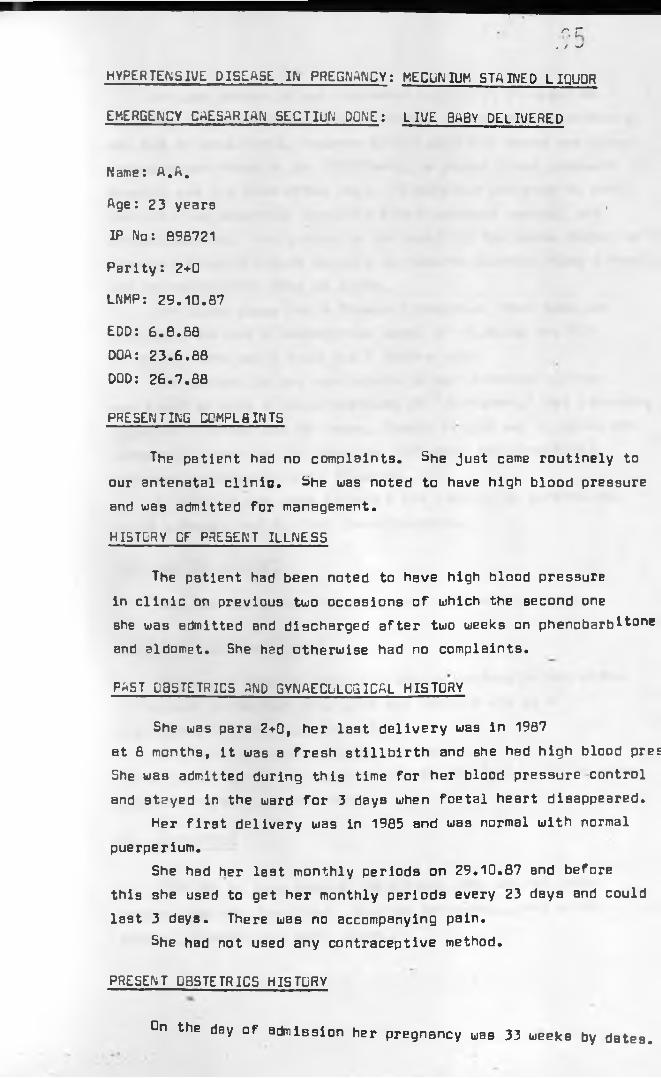
r' r . / ^
HYPERTENSIVE DISEASE IN PREGNANCY: MECONIUM STAINED LIQUOR
EMERGENCY CAESARIAN SECTIUN DONE; LIVE BftBY DELIVERED
Name: A.A.
Age: 23 years ,
IP No: B98721
Parity: 2+0
LNMP: 29.10.87
EDO: 6 .8 . 8 8
DOA: 23.6.88 DOD: 26.7.88
PRESENTING COMFLSINTS
The patient had no complaints. She just came routinely to our antenatal clinio. She was noted to hav/e high blood pressure
and was admitted for management.
HISTORY GF PRE5ENT ILLNESS
The patient had been noted to have high blood pressure
in clinic on previous two occasions of which the second one she was admitted and discharged after two weeks on phenobarbi*one and aldomet. She had otherwise had no complaints.
PAST OBSTETRICS AND GYNAECOLOGICAL HISTORY
She was para 2+0, her last delivery was in 1987 at 8 months, it was a fresh stillbirth and she had high blood pres She was admitted during this time for her blood pressure control and stayed in the ward for 3 days when foetal heart disappeared.
Her first delivery was in 1985 and was normal with normal puerperium.
She had her last monthly periods on 29.10.87 and before this she used to get her monthly periods every 23 days and could
lest 3 days. There was no accompanying pain.She had not used any contraceptive method.
PRESENT DBSTETRIC5 HISTORY
On the day of admission her pregnancy was 33 weeks by dates.

96
She wa9 booked in our antenatal clinic at 2U weeks on
18.L.B8. She attended clinic 6 times, during her attendances she had no complaints, however on 2.6.88 at 3D weeks her blood pressure was found to be 130/90mmHg, a repeat blood pressure reading was the same after rest. A decision was made to admit her into our antenatal ward for blood pressure control and investigations. She stayed in the ward for two weeks during whic time her blood pressure settlPd on tablets aldomet 250mg 8 hourly and phenobarbitone 30mg at night.
Her blood group was B Rhesus D positive, Khan test was negative, she had a haemoglobin level of 12.8g/dl and PCV
of 37. T*. She was 5 feet and 6 inches tall.The patient in her next visit to our antenatal clinic
was found to have a blood pressure of 130/90mmHg. Her pregnancy
maturity by dates was 39 weeks, fundal height was 32 weeks and ultrasound taken on the same day indicated the biparietal
diameter corresponding to 35 weeks.A decision was made to admit the patient to control her
blood pressure and further investigations.
PAST MEDICAL HISTORY
she has had no contributing past medical history
FAMILY nUD SOCIAL HISTliRY
She was a married lady, a clerk by profession and stays
in DonhcJn in Nairobi City with her husband who is a
supervisor in a firm in the city.She does not drink alcohol nor smoke.
There is no history of hypertension in the family and no other
relevant history in the family.
ON EXAMINATION
*3he was in good general condition, she was not pale,
neither was she jaundiced. She had no oedema. Her other
general examinations were normal.

97
Her blood pressure uas 130/90mmHg, pulse rate was 80/minute reguler and respiratory rate uas 22/minute normal. She had a temperature of 36°C.
Central Nervous System )
Respiratory System ) No abnormality detectedCardiovascular System )
Except for the high blood pressure the other systems were
normal.
ABDOMINAL EXAMINATION
ahe had a uniformly distended abdomen with uterine size of
32 weeks. The fetus uas lying longitudinal and head uas
presenting 5/5 above the pelvic brim.Foetal heart uas heard and uas 1A2/minute regular.
DIAGNOSIS
A diagnosis of hypertensive disease in pregnancy uas made.
P U N :
The patient uas prescribed bed rest and phenobarbitane
tablet 30mg 8 hourly.Henous blood uas removed for urea and electrolytes
estimation and haemoglohin check. 2A hour urine sample uas to
be collected for creatinine clearance estimation.She uas to have ri"ily urine analysis for protein and sugar.
She uas to plot the foetal kick chart to give us the
insight of the fetus well being.Throughout her stay in the ward the patient did not spill
any protein nor sugar in urine. Her blood pressure however fluctuated a lot within the first feu days of admission and the highest recorded was 150/IOOmmHg. She uas gradustedslowly from phenobarbitone 30mg daily and bedrest only through both the above
end tablets aldomet 250mg 8 hourly to aldomet 50Cmg 8 hourly
with bed rest only.Blood pressure uas controlled between 1A0/90mmHg
and 130/80mmHg.
foetal kick chart remained satisfactory throughout.

5 8
On 29.6.88 amniocentesis was done to check on foetal
maturity if it would he viable outside the uterine enviornment.Its surfactant test was negative.
LrtBLKATCftV RESULTS
Lab No. 20381
K+ - A.8mmol/l Na+ - lL2 .0mmol/l BUN - 3.0mmol/l Creatinine - 87 umol/ 1
Uric Acid - 163 Umol/ 1
Creatinine clearnace was 126mls/minute Her haemoglobin level was 13.2g/dl.
On 18.7.88 maturity of the pregnancy by dates was 37 weeks, a decision to repeat amniocentesiB for surfactant test was reached end was effected. It was positive.
The following day she had Bishop Scoring done where her cervix was found to be 1 centimetre long, soft, central and 3 centimetres dilated. The presenting part was however still 5/5 above the pelvic brim.
l ith the positive surfactant test end favourable bishop Score a decision was made to prepare the patient for induction of labour in labcur ward.
Blood for group and cross match was taken and intravenous line was set. In labour ward artificial rupture of membranes was done and thick meconium staining liquor was obtained. The patient was urgently taken to second stage for oxygen by mask and intravenous 5% dextose, and to lie in left lateral, position. Uith this regime the foetal heart was also noted to be irregular and was 118/minute.
A diagnosis of hypertensive disease in pregnancy with foetal distress at 37 weeks was made, a decision was reached to take her to theatre for emergency caesarian section.
I

A female baby was delivered of weight 2350 grams and of apga score of 9 in 1 minute and 10 at 5 minutes. Pjacenta weighed 350 grars. Blood loss was 500mls.
The patient made uneventful recovery; her third day post operative haemoqlobin level was 1 1g/dl.
Her blood pressure was stable on tablet aldomet 250mg 8
hourly and hydrallazine tablets ^50mg 8 hourly. She was discharged from our post natal ward on 26.7.88 to be seen
in our post natal clinic after six weeks; she was also booked in medical clinic.
On 2.9.88 she was seen in our post natal clinic. She had earlier attended medical clinic on 30.9.88 where she was put on tablets viskaldix one daily and hydrallazine 25mg 8 hourly. She had no obstetric problem and her scar had healed nicely.
She was advised on family planning methods and was encouraged to go on with medical clinic.

COMMEN T5
1
This was a patient who had hypertensive disease in preanancy in whom conservative management was done and live baby delivered at term.
Hypertensive disease in pregnancy can be defined as a rise of systolic pressure of equal to or more than lAOmmHg and diastolic pressure of equal to or more than 90mmHg (1,2,3),
or a rise in diastolic blood pressure of equal to or more than 30mmHg, or systoclic blood pressure of more than 15mmHg (1).
Hypertensive disease in pregnancy (HOP) occurs in 10-15% of the pregnancies (6,7). The causes are not known but factors thBt predispose include diabetes mellitus, multiple pregnancy, foetal hydrop, H.Mole, renal disease, history of eclampsia or pre-eclampsia, or previoushistory of hypertension (2 ).Age has also been thought to contribute (1). Our patient had positive history of hypertension even outside pregnancy.She had no protenuria, end no oedema. Elevated blood pressure before 20 weeks gpstation is nearly always HOP. A persistent hypertension beyond 6 weeks post partum according to the Committe on Terminology of American Collage of Obstetrics and Gynaecology fits in with the defination of HDP (2,3).
Chronic hypertension predispose to PET and when patient presents after 20 weeks diagnosis can be difficult.
Hanrgement of HDP aims at giving a hpalthy viable baby to a healthy mother. It aims at controlling the disease so that existing lesions don't progress and fetops the genesis of new ones. Hypertensive patients should be admitted and renal, hepBti cardiovascular and haematologicel studies done. It is important to determine whether changes are likely to get worse with continued pregnancy or not (1 ,2). If theses investigations are done and are satisfactory and blood pressure is less than 90mmHg, diastolic the patient can be discharged home to continue with clinic. But if blood pressure is persisti^gly 90mmHg and above then the pati°nt must be admitted and be kept uni
serial investigation and observation.

Cur patient's investigation results were satisfactory, her fclcod pressure did not settle at below 9QmmHgf but foetal uell being mas ascertained by fetal kick chart ubich the mother herself was charting.
In the absence of any abnormal observation end blood pressui is kept safely around 90mmHg, pregnancy can be carried till baby is felt capable of survival outside as in our patient. Should the blood pressure be uncontrollable despite rest and medication, then it would constitute an indication for termination no matter what the gestation is. If investigations indicate deteriorating renal, cardiac or hepatic function then
termination is also thp management. Should the baby's well bEinc in utero be threatened as would be manifest by reduced foetal movement or some irregular pattern in fetal cardiac activity the baby should be brought out of the uterus ( l , 2 ,3 , t» , 5).
Drugs we use commonly is our unit is aldomet tablet,
hydrallazine, propranalol, phenobarbitone and paracetamol if there is any pain. Bed rest is very valuable as it has been shou to improve the placental blood flow (1 ).
Complication of HOP include deterioration manifested by cardiac decompersation, cerebrovascular accident, renal failure, hepatic failure and visual disturbance. Subsenuent pregnancies are likely to be more complicated with higher blood pressure (A). Abruptio placentae is common 5-10% of the HOP (3).
There is Progressive fibrosis of vessels, placental bed affected gives reduced foetal activity. This would result in intrauterine fetal growth retardation and occasional distress like in our patient (3 ,A). The intrauterine life was perhaps too hypoxic in the case of our patient to be tolerated by the fetus. Emergency abdominal delivery was recommended and the baby scored well.
In general mode of delivery depends on age, parity, gestatio previous obstetrics history and state of pelvis and cervix. Caesarian section is recommended for premature babies (A). Our
patient did well post operatively and is now attending renal clinic.

REFERENCES 1 D 2
1. ^ladjem, S.
- Pre-eclampsia, eclampsia and other hypertensive disorders in pregnancy
- Obstetrical Practice pg 577-598 1960- C.V. Moaby Company
- St. Louis. LUNDllN.2. Benson, R.C.
- Preclampsia, eclampsia and hypertensive disorders in pregnancy.
- Current Cbs.uyn. Diagnosis and Teatment pg 631, 1976- Lange Medical Publication- Los Altos California
3. Pritchard, J.*., McDonald, P.C., Gant, N.E.- Hypertensive Disorders in Pregnancy- Williams Obstetrics 17th Edition pg 525-528 1985.- Mppieton-Century-Croft/riorwalS Connecticut
A. Deuihurst, 0.- Preeclampsia, eclampsia, Hypertension and chronic
rEnal disease.- Interpreted Libs.Gyn. for Postgraduates 3rd Edition pg
27L-276, 1981.- igckwe].]. scientific Publication- Oxford. London. Edinburg, Boston. Melbourne.
5. Kenyon, N., Janesen A.A#j.
- iueight changes during pregnancy and birth weights in Rusinga Island.- Journ. of Qbs.Gyn. East end Central Africa 2: 20-22, 1983
6 . Urasss, E.J.N.Pregnancy Outcome in patients presenting with
hypertension in pregnancy- Bourn, of Cbs.Gyn. East and Central Africa,
3:55-58, 198A.. Mati, J.K.G.
- N.B.S.V- Journ. of Cbs.Byn. East and Central Africa, 1:13A-139, 198
7

* 103
RHE5U5 I5LKTUNIZATIGN, INDUCT IGN OF LABOUR. LIVE Bm BV DELIVERED
Name: A.G.
Age: 31 years
Parity: 0+1 IP No. 859003 LNMP: 18.3.87
EDD: 25.12.87 DOA: 1.12.87
DOD: 7.12.87
PRESENTING CGKPLAINTS
^he patient had earlier been diagnosed in cur antenatal clini where her planned adrission was arranged on the date above.
PAST C35TETRICS ND GYNhEGCLGGICmL HI5TCHY
5he was para 0+1 , the one abortion was at 2H months and evacuation of the uterus was done in 1985. She had her menarche at 15 years. Sh? had been getting regular periods every 28 days lasting 3-A days. She was not aware of her blood group and it was not discovered during her last pregnancy.
PAST MEDICAL HI5TLRY
Not significant
HTRTfTSY OF PRESENT PREGNANCY
She was booked at our antenatal clinic because she was rhesus
negative at 33 weeks. She had had no problem with this pregnancy
Her antenatal results are as follows:-- Her blood group - A Rh D negative- Husbands Blood Group - 0 Rh D positive
- Haemoglobin - 13.4*g/dl- PCV - A2.2*
- Kahn test - negative
- Indirect coombs test at 33 weeks - positive 1:A"iU *• r ~

\
10*4 -1 G 4
- 3*» weeks - positive 1:8
FAMILY AND SOCIAL HISTORY
She was a married lady working in Muranga Hospital as a nurse. She was not taking alcohol or smoking. There were no medical problems in the family and no contributory history.
ON EXAMINATION
She was in good general condition. She was not pale, and she had no Jaundice. Her other general examinations were normal.
She had a pulse rate of 78/minute regular and good volume,BP of lU0/90mmHg, RR 20/minute normal, and she had a body temperature of 36.8°C.
Her respiratory, cardiovascular, central nervous system were normal.ABDOMINAL EXAMINATION
The abdomen was uniformly distended. She had a uterine size corresponding to 3A weeks, the fetus was lying longitudinally and head was presenting 5/5 above the pelvic brim. Foetal heart was heard at 1L0/minute regular and there were no contractions.
Vaginal examination was not indicated.
DIAGNOSIS
A diagnosis of Rhesus issoimmunization at 36 weeks was made.
MANAGEMENT
The patient had earlier been admitted into antenatal ward for amniocentesis on 3.11.87. She was observed like other antenatal patients.
The procedure was explained to the patient and after scrubbing and putting on sterile gloves, lower abdomen was cleaned and drappi with sterile towels. U8ing the left hand, fetal head was displacec upwards. Lumber puncture needle was introduced suprapubically through the abdominal wall and advanced till amniotic cavity was reached. The stylet was withdrawn and the needle connected to a syringe 5mls of
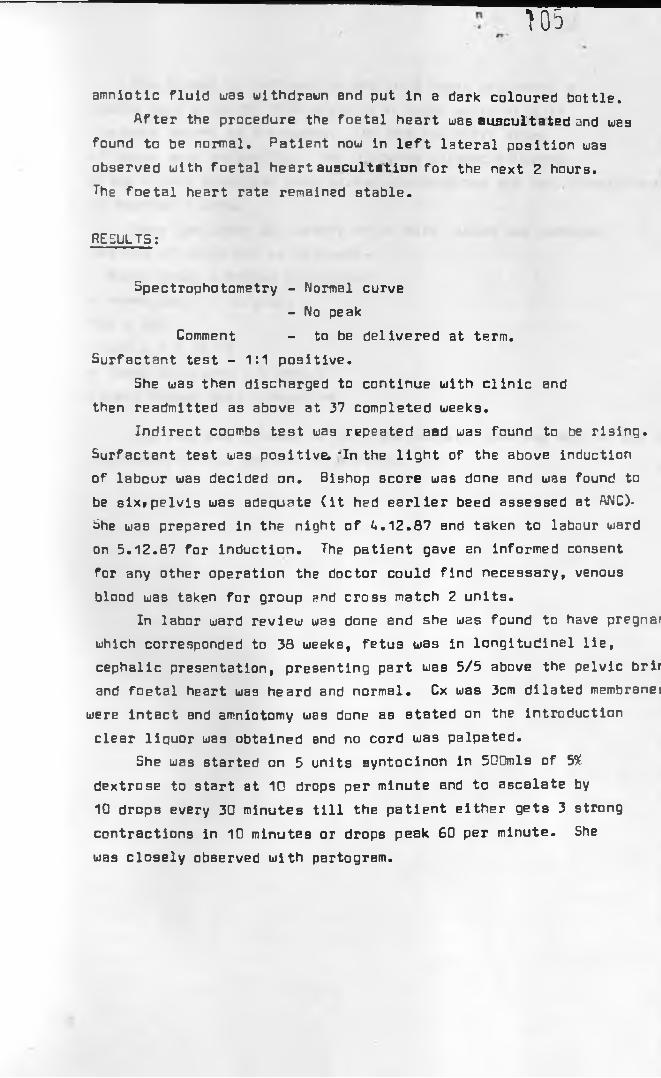
>05
amniotic fluid was withdrawn and put in a dark coloured bottle.
After the procedure the foetal heart was auscultated and was found to be normal. Patient now in left lateral position was
observed with foetal heart auscu lta tion for the next 2 hours.The foetal heart rate remained stable.
RESULTS:
Spectrophotometry - Normal curve- No peak
Comment - to be delivered at term.Surfactant test -1:1 positive.
She was then discharged to continue with clinic and then readmitted as above at 37 completed weeks.
Indirect coombs test was repeated aBd was found to be rising. Surfactant test was positive. -In the light of the above induction of labour was decided on. Bishop score was done and was found to be sixtpelvis was adequate (it had earlier beed assessed at ANC).She was prepared in the night of L.12.87 and taken to labour ward on 5.12.87 for induction. The patient gave an informed consent for any other operation the doctor could find necessary, venous blood was taken for group and cross match 2 units.
In labor ward review was done and she was found to have pregnar which corresponded to 38 weeks, fetus was in longitudinal lie, cephalic presentation, presenting part was 5/5 above the pelvic brir and foetal heart was heard and normal. Cx was 3cm dilated membranes
were intact and amniotomy was done as stated on the introduction
clear liquor was obtained snd no cord was palpated.She was started on 5 units syntocinon in 50Dmls of 5%
dextrose to Btart at 1D drops per minute and to ascalete by 10 drops every 30 minutes till the patient either gets 3 strong
contractions in 10 minutes or drops peak 60 per minute. She was closely observed with partogram.
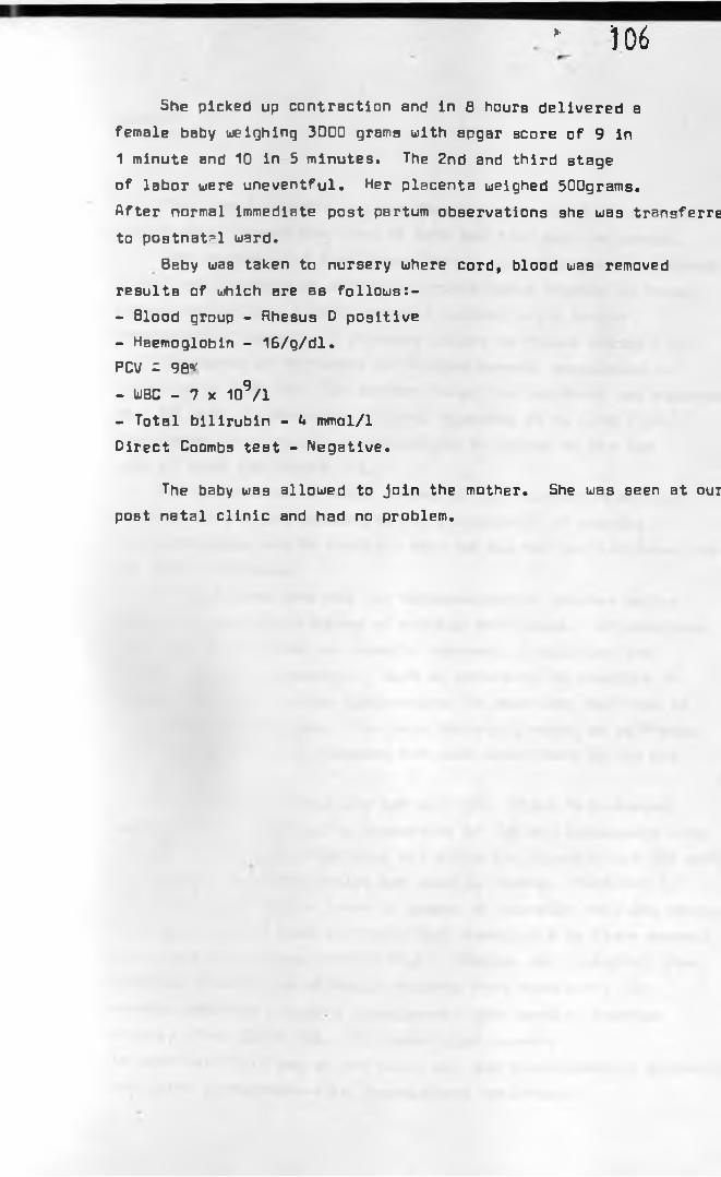
>• 106
She picked up contraction and in 8 hours delivered a female baby weighing 3000 grams with apgar score of 9 in
1 minute and 10 in 5 minutes. The 2nd and third stage of labor were uneventful. Her placenta weighed 500grams.
After normal immediate post partum observations she was transferre to postnatal ward.
Baby was taken to nursery where cord, blood was removed results of which are as follows:-- Blood group - Rhesus D positive- Haemoglobin - 16/g/dl.PCV i 98*
- UJBC - 7 x 10⁹/1- Total bilirubin - U mmol/1
Direct Coombs test - Negative.
The baby was allowed to join the mother. She was seen at our post natal clinic and had no problem.

>07COMHENT
This was a patient who had Rhesus isoimmunization in whom induction of labour was done at term and live baby delivered.
The discovery of the Rhesus factor by Landsteiner and Idiener (19M) and founding of affective prophylactic protein by Freda, Corman and Pollack (1963) marked a turning point to the management of spectra of diseases caused by Rhesus factor (1).
Incidence of Rh factor in African general population in Tanzania is 3.5% (2). In Western Kenya the incidence was reporte as 2 .U % while in Kenyatta National Hnspital it is <+.1% (3,0. There seem to be racial preponderance in whites as per the work of Race and Sanger (5).
Our patient wag a nurse in one of our District Hpspitals and as such she was exposed to the opportunity of knowing her blood group and Rh factor. When we saw her she had known she was Rhesus Negative.
*n individual who has had isoimmunization denotes having antibodies against antigens of another individual. In pregnancy fetnl red blood cells can pass to maternal circulation and evokes antibody response. Such an occurence is possible in normal pregnancy, during partufcation, in abortion, and also in accidental haemorrhages. The baby obviously needs to be Rhesus positive (1,2,3). Our patient had been sensitised during her abortion.
Hntibodies produced are 1gM and IgG. First fetomaternal transfusion is typified by production of IgM and subsequent ones by both IgM and IgG. IgM does not cross the placenta but IgG cro: the placenta so first babies are usually spared. Maternal IgG into fetal circulation leads to grades of diseases entities rBngii from mild anaemia from red blood cell haemolysis to frank anaemia with congestive heart failure (£*,6 ). Babies are protected from jaundice in utero as bilitubin arising from haemolysis are
shunted into the mother's circulation; they develop jaundice shortly after birth (6 ). ^ilirubin also appears
in anniotic fluid and is the basis for spectrophotometric estimat: and guide to management in isoimmunized individuals.

Aim of manpgement is to screen the mothers at the antenatal clinics and know their rhesus factor status. Before 2t*-26 week of gestation, transfer of maternal antibodies to foetal
circulation is less but thereafter chances increase. These patients are therefore followed by serial blood for indirect coombs test four weekly till 32 weeks then two weekly,
to term. If antibody titres do not rise or indirect Coombis test is negative, tnen the mother is delivered at term so
long as surfactant test is positive. Pregnancy should be not allowed to reach 40 weeks bs there is increased risk in antibody passage or fetomaternal transfusion ( (1,^,6 ).
In severer cases spectrophotometric estimation afvlevel af bilirubin in amniotic fluid is a guide as to when -to deliver the patient using Liley's chart. Amniocentesis and spectrophotometry are indicated as long as antibody titers exceed 1:16(1,6). Subsequent results will indicate whether pregnancy is allowed to term like in our patient, induced or
pregnancy terminated. Some fetuses can be transfused in utero (1 , (Induction of labour is as discussed above, however, some
patipnts who have r.contrainriication to this and those whose babies are too small, less than 3A weeks, caesarian section is to be opted for (6 ).
Baby cord blood must always be taken in order to know the rhesus factor, haemoglobin level end bilirubin estimation.
In our patient the baby did not require any further management.* If patipnt is not immunised, anti Mhesus D gamma globulin
should be given within 72 hours of delivery, to a rhesus positive baby, an abortion, ammocentesis, external cephalic version or even of ectopic gestation (1 ,<»,6 ,*7,8 ) Dur petient was sensitised and as such did not need the anti D.
REFERENCES
1. Pritchard, McDonald, P*C.f Gent N.F.- Haemoglobin from Maternal Rh Isoimmunization
Williams Cbstetrics 17th Edition pg 773-776, 1965.
Appleton-Century-Croft/NewYork2. Nhololi, tt.M., Kiango J.S.
- The distribution of blood group freauencies in Mainland Tanzania Africans.- c.A.K.J. 51: 6 , 197A
3. Kiango, J.S., John S.F.,
- Prevalence of Irrigular red blood group antibodies, in pregnant African Women in Dar-es-Selaam- E.A.H.J. 6(2): 88, 1983.
A. Mulandi, T.N.- The effectiveness of Anti-0-Immunoglobulin in prevention
of Rhesus Iso-immunization in Kenyan Women of African
origin at Kenyatta National Hospital.- M.Med Thesis 1965, University of Nairobi.
5. Race, R.R., Sanger R.,- Blood Groups in Man. 6 th Edition pg 186, 1976- Blackwell Scientific Publication- Oxford London.
6 . Dewhurst J.- Blood Disorders in pregnancy- Integrated Obs.Gyn. for Postgraduates, 3rd Edition Pg 291,
- Blackwell Scientific Publication- Oxford, London, Edinburg. Boston Melbourne
7. Benson, R.C.- Post pertum immunization- Current Obs.Gyn. Diagnosis and Treatment
5 th Edition pg 85A.- Lgnge Medical Publications. California, U.S.A. 1S8A
8. Mushbacher, 0.; Bove, J.R.- Rhesus Immunoprophylixiss, is antepartum therapy desrirable
- New Eng. M.J. 16: 935, 1981.

SEVERE MALARIA IK PRZ3:. NCY. FRCI-’.r TUNC LULL'S. T liy LIVE
b a bi e s delivered
Name: E.A.
Age: 2D years
IP No: 98A309
Parity: 0+0
LNKP: B.9.87
EDD: 15.6.88
DQA: 15.3.88 1.50am DOD: 26.3.88
Fr.EaLNTir.B GLKPL 'INTS
Lower abdominal pain for 1 day, joint pains for 2 days. Sweating on and off about ? days, whitish vaginal discharge with pruritus A days.
h ISTLKY OF PRESENTING COMPLAINTS
The patient was well until two days befcre admission into our labour ward when she started having some vague joint pains. She also noted to have chills and sweat on and off.M11 these complaints have increased over the last * day and have been accompanied by lower rbdominal pain. She had been on chloroquin tablet L stat, 2 after 6 hours and 2 on the morning without relief.
she had been having pruritis and whitish vaginal discharge for the last U days. These problems have else increased in severity.
No history of vomiting, and no history of vaginal bleeding and no dysuria.
PAST DBSTETriIC AND GYNAECuLCGID-.L HISTuRY
^he was para D+C. Her last monthly period was on 8.9.87.^he had been having it every 30 days and used to last 2 to
3 days. There was no accompanying abdominal pain. Her/ .
menarche was at 13 years. 6he had not used any method ofcontraception.

■1
FHCSCNT OBSTETRIC HISTORY
Uy dates she was about 25 weeks. ahe was expected
to deliver cn 15.6.88. She hed attended ^ntcn- tal clinic in Crescent tedical Centre in Nairobi where she had gene
two times. She had had no problem during the nregnancy.ShF started having foetal nav'T.ents at 2C we=-ks gestation
FAST MEDICAL HISTORY
None of significance.
FAMILY f-ND Sl CIML HIOTuHY
She was a married lady staying oath her husband at Kibera Estate in Nairobi. She was in secretarial college in Nairobi, and her husb nd was an '■‘uriitor with the Kenya Fipeline Limited
5he does not drink alcohol nor smoke.There is no relevant history from the family.
ON EXrtMKrtTICr.
She was sick looking, ^he was not pale, nor jaundiced.
She had no oedema of the legs, uther general examinations
were normal. Her temperature was 38.5°C, her pulse rate was 116/minute regular, her respiratory rate was 2L/minute norrel and_her blood pressure was 120/B0mmHg.
Central Nervous System )Cardiovascular System ) Cere essentially normal
Respiratory System )
ABDOMINAL EX-.KIN*TILN
Her abdomen was uniformly distended. Uterine size was 36 weeks. Many fetal parts felt but head was presenting
Spleen was palpable about 3cm below the subcostal margin just lateral to mid clavicular line lleft side, liver was not palpable and no tenderness was elicited anywhere.
She was having one to two contractions in ten minutes
each lasting about 20 seconds. Foetal heart rate was heard at 136/minute regular; there was also a muffled sound at a different site corresponding to a rate of 1A2/ninute and was also regular.
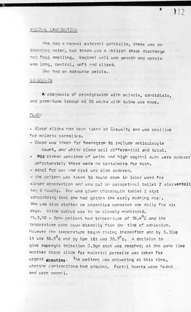
* *12
VMSIIUL ZXA.VINATICI;
-he had a normal external genitalia, there was no bleeding noted, but there was a uhitish thick discharge not foul smelling. Vaginal wall was smooth and cervix was long, central, soft and closed.
She had an adequate pelvis.
Di.,ar:Cols
A diagnosis of primigravida with malaria, candidisis, and premature labour at 28 weeks with twins was made.
PLhH :
- 3lood slides had been taken at Casualty and was positive for malaria parasites.
- Blood was t~ken for haerponram to include reticulocyte
count, and white blood cell differential and total.- Mid stream specimen of urine and high vaginal swab were ordered
unfortunately there were no containers for both.- stool for ova and cyst w?s also ordered.- the patient was taken to acute room in labor ward forcloser observation and was put on paracetamol tablet 2 statventoli **mg B hourly. She was given chloroquin tablet 2 stat considering that she had gotten the early morning dosr. ahe was also started on pessaries canasten one daily for six days. Urine outcut was to be closely monitored.15.3,88 - 2pm- p a t ie n t had tem perature of 36.f*°C and the
tem perature canE down s t e a d i ly from th r time o f adm iss ion .
However the temperature began rising thereafter and by 5.30pqp it was 38.5°c and by 6pm itt was 38.7°c. A decision to give aspergic injection 0.5gm stat was reached; at the same time another blood slide for malarial parasite was taken for
urgent scrutiny* The patient was shivering at this time, uterine contractions had stopped. Foet-l hearts were heard and were norm?!.

Blood slide result came as trophozoites of plas'-odium faciparum seen, heavy parasitaemis.
diagnosis of chlorcpuin resistant malarii was node and
patient started cn intravenous guinine 600mg In 500mls of 5% dextrose to run 8 hourly. This was started at 7.30pm the s2me day.
Her temperature settled steadily and by 11pm the sane day it was 36.L°C. From 5.3Cam to 6.30am on the following
morning 16.3.88 temperature -ose again to 39.23c. her urine output uptcj 6am was 13P0mls.
A decision was made to continue treatment and to take another sample of blood for blood slide for malaria parasites. This specimen did not seem to have reached the laboratory.17.3.88 the patient's outlook was better. Her urine output was satisfactory for the previrus 2U hours, it was 1370mls against 2620mls input, her temperature of the morning was 37°c abd had been below 37.5°c for the previous 18 hours, repeat blood slide for malaria parasite showed few malaria parasi seen (trophozoite of p. falciparum).
The patient was discharged to antenatal ward far further management. Her urinalysis showed protein which ranged between trace and plus I, urine sugar was nil thrcunout her stay in the acute room, no bilirubin.Her haemograrr results Lab (Vo. 003870 revealed - Haemoglobin
of 1D.5g/dla
L3C - total 9.7 x 10'1/1. f-o differential was given.
KCU - 89.2fb.- Reticulocytes - were not done.
18.3.88She had steady improvement, all parameters were normal.
Antenatal profile were ordered. Urea end electrolytes
were alsc ordered.
She had uneventful day on 19.3.88.20.3.88 - She complained of drainage of liquor and w2 s seen
in labor ward at 12.30pm where she was noted to be in
established labor.

-ervical dilatation uas 0cm, she was obviously draining^ head was felt and there was nn cord.
Jhe was put on pgrtogram and observation in first stage. She progressed well and delivered first twin et 2 .**5em, it was a female of birth weight 1100 grammes, second twin was male and was also spontaneous vertex, his weight was 2000 grammes;
it was delivered at 2.50am. A'pgar score for the first twin was 8 in 1 minute and 9 in 5 minutes; for the second twin was the seme.
Efiisiotomy was sutured and the rest of the genital was intact.
They had different placentas which were delivered whole sith membranes.
Tost partum observations were normal and the pati nt was taken to Dur post natal ward where she made stEady recovery.
Repeat haemoglobin on 23.3.88 and blood slide of the same day were Hb 10.6g/dl, wBC - 11.7 x 10⁹/1 -
normal in distribution, R3C were normocytic normochromic lab number 00932**; malarial parasites were not seen - lab number 623.
Urea end electrolytes of 18.3.88 K - 3.0mmol/l
- 1?7mmol/l BU0 - ‘•Zmmol/l U.A. - 359 umol/1 Creat - U 0 Umol/ 1
Thr patient finished her dose of quinine on the 25.3.88. Her
observations remained steady. She was still seeing her babies in nursery.
^he was discharged tn mother’s hostels on 26.3.88 in good condition. She was discharged from mother's hostel on25.A.88 with the babies in good condition. She was put on fansidar tablets 1 wpekly for six weeks. She did not come to our postnatal clinic as was arranged.

p > 4
CD! tl :EfJ T
This Lias a patient who had svere malarial attack in
pregncrcy and went on to have premature labor and delivery to twins.
<• laria is caused by sporozoa of four spacies of
plasnodium namely vivax, ovale, melariae and falciparum.Mr-lcri' is endemic in coastal regions and lake region falciparum being the comrenest type. Frevelance and density of infection are oreatEr in pregnancy due to reduced immunity. Those from endemic areas have some immunity
and even then the immunity is reduced with pregnancy (1,2,3).The effect malaria exerts in a population depends on epidemiological pattern. Malaria can be stable where there is constant repeated infection (holoendcmic). Here the population has high degree of immunity and epidemics don't occur. In unstable nolaria its transmission is intermittent. Comrunal inrunity is low *nd dramatic epidemics can occur (L). dur patient came from Lake region and is supposed to have high immunity. Lie can supoose th-t the Dregnancy related depress immunity was responsible for such a serious infection.Infection is also severer in pri-igravidas like ours (7).
Reduced immunity in pregnancy has been shown to be responsible for high pararitaemia rates, repeated infection
end anaemia invariably results, 1DX of pregnant mothers in endemic areas have haemoglobin level of 5g/dl (3).The anaemia is usually megaloblastic due to h-emolysis, defective erythropoiesis and hypersplinism U .3 V of total admission into our antenatal wards s f e due to anaemia whose
aetiology is malaria (8).Malaria in pregnancy becomes important because it is
not only a cause of maternal morbidity but also a cause of fetal loss and premature deliveries. There is evidence of accumulation of placental inflammatory masses in the intervil space, there is also thickening of trophoblastic basement membranes (?,L)J These changes affect fetoplacental transfer and likely to cause fetal distress, and intrauterine growth retardation.

16
The latter could also result from anaemia (2). Our patient delivered twins one of whose weight was half the other.The had different placentas, it is possible that the parasltaemia was more in one placenta than thp other though
other explanations could also be brought forth. .
Malarial parasites rarely pass to foetal side and further, fetil haemoglobin are resistant to malarial attack;
maternal IgG which passes accross the placenta also protects the baby from attack (*♦). Other problems with malarial infection include intrauterine fetal death, puerperal
fever and psychosis. Kertman (1972) in Tanzania reported an incidence of umbilical cord malarial pqrasitaemia of 3.8%, placental of 19.7% and maternal blood of 23.2%. He also observed occurence of congenital malaria to be more in non immune subjects contracting malaria for the first time (5)The babies were not screened in our patient.
Management depends on expeditious diagnosis and commencement of medication at once. Our patient had taken close to full dose before 9he came to the hospital, but her blood film revealed parasitaemia which subsequently became heavy. The aim i9 to eradicate the parasite from the system.Chloroquin is the drug of choice and if there is resistance as was in our patient, quinine is given for 10 days.Prophylacis in pregnancy should be given. In our patient she went into premature labor and delivered live babies.Puerperal pyrexia should still be watched for (<4,5,6,7).

REFERENCES
1. Sinei, 3.K.A.; Mati J.K.G., Mungai, J. et al
- prevalence of snaemle In pregnancy and role of
anaemia in its aetiology in rural Kenya.
- Journ. Obs.Gyn. East end Central Africa 3(3): 119, 19S3
2. Allen, S.Malaria in Pregnancy with foetal complication British Journ. Obs.Gyn. 91: 399-A03, 198L.
3. Ngwalle, K.E.N.- Role of melaria in causation of anaemia in first
pregnancy as seen at Mahumbili Medical Centre.- Journ. Bbs.Gyn. East and Central Africa U i 13-16, 1985.
A. Lawson J.B., Stewart, D.B.- Malaria and Pregnancy- Cbs.Gyn in the tropics and developing countries
pg 59-72, 1983.- English Language Book “"ociety
5. Kortman H.F.M.- Malaria in Pregnancy- DrukkeriJ Elinkwijk Utretch, 1972.
6. Formul-u J.N.- A 2 year retrospective survey of anaemia in pregnancy
- M.Med Thesis 1981 University of Nairobi.
7. Kelsey, A.H.- Anaemia, Malaria and aickle Cell Disease
- Clinic in Cbs. Gyn. 9: U U 5 , 1982.8. Mati, J.K.G.t Hatimy A.f Gebbie D.A.M.
- Importance of anaemia of pregnancy in Nairobi and the role of malaria in the aetiology of
megaloblastic anaemia.- The journ. Tropic. Med. Hyg. 7A: 1, 1971
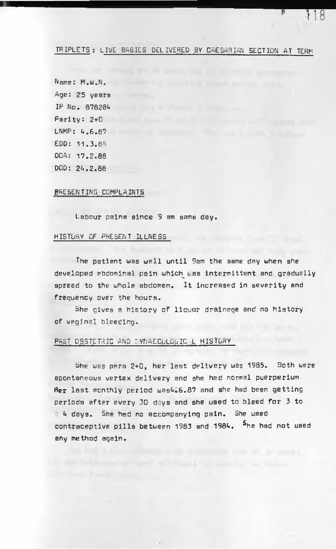
TRIPLETS ; LIV/E BABIES DEL IUERED BY CAESARIAN SECTION AT TERM
Name: M.uJ.N.
Age: 25 years
IP No. 87828**
Parity: 2+0 LNMP: *4.6.87
EDD: 11.3.88 DOA: 17.2.86
DOD: 2L.2.88
BRESENTING COMPLAINTS
Labour pain9 since 9 am same day.
HISTORY OF PRESENT ILLNESS
The patient was well until 9am the same day when she developed abdominal pain which^ uas intermittent and gradually spread to the whole abdomen. It increased in severity and frequency over the hours.
She gives a history of liquor drainage and no history of vaginal bleeding.
PAST OBSTETRIC AND : YNAECOLOGIC. L HISTORY
She was para 2+0, her last delivery was 1985. Both were spontaneous vertex delivery and she had normal puerperium Her last monthly period was**.6.87 and she had been getting periods after every 30 days and she used to bleed for 3 to
** days. She hBd no accompanying pain. She U9ed contraceptive pills between 1983 and 198**. ^he had not used any method again.

PRESENT OBSTETRIC HISTORY
^he was booked at 26 weeks due to multiple pregnancy and attended our entenptal clinic 8 times during which time she had no problem.
Her blood group was A Rhesus D positive,
her haemoglobin level was 11.6g/dl and packed cell volume was
33.99 . She had negative serology. 3he was 5 feet 5 inches tall.
PAST MEDICAL HISTORY
Nothing of significant
FAMILY AND SOCIAL HISTORY
She was a married housewife, she reached form II level of education. Her husband is a police officer and they stay in Kiambu town.
She neither drinks elcohol nor smokes. There is no history of twins in their families.
ON EXAMINATION
She wes in good general condition. She was not pale, and had no jaundice. Her other general examinations were normal. Vital signs were RR 22/minute, PR 8B/minute regular, Bp 120/80mmHgt and body temperature w b s 36.2°C.
Central Nervous Syfcem )
Respiratory System ) No abnormality detected.Cardiovascular system )
ABDOMINAL EXAMINATION
She had a full abdomen with a uterine size of 38 weeks,
lie end presentation were difficult to make out as there were many foetal parts.

Foetal heart us heard at different positions.
VAGINAL EXAMINATION
Idas not done.
DIAGNOSIS
diagnosis of a para 2+0 with triplets at 37 weeks uas
made.
PLAN:
The plan uas to give her bed rest; she ues put on Tablet
folic acid 5mg daily, pnd tabs ferrous sulphate 200mg three
times a day and observe her vital signs U hourly.
The patient's condition remained stable during the day of
admission and overnight.The following day, however, she developed labor pains
and was taken to labor ward theatre for delivery.Her venous blood was taken for crossmatch and intravenous
fluids started, informed consent got.She uas taken to theatre and after routine preoperative
procedures uere done she uas put on operating table
and caesarian section was dene.The first baby uas transverse and was delivered by
breech; the second baby was breech and was delivered by breech, and the last uas oblique and uas delivered by vertex. They were in different sacs but shared one placenta.
The first baby scored 8 in one minute, A in 5 minutes and 6 in ten minutes, and ueighed 2600 grams; the second baby scored 8 in 1 minute, 9 in 5 minutes and weighed 2200 grams, and the third one scored 8 in 1 minute, 9 in 5 minutes and
weighed 2300grams. The placenta ueighed 1500grams and uas
delivered complete uith membranes. Estimated blodd loss 700mls.
<IS,Mre
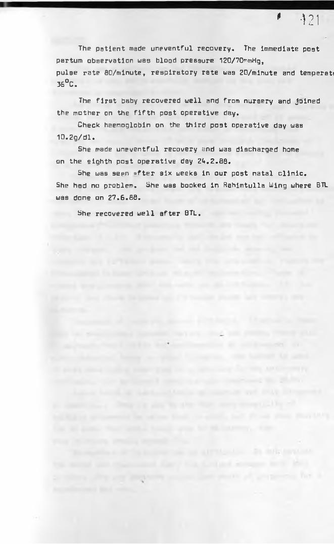
* 421
The patient made uneventful recovery. The immediate post partum observation was blood pressure 120/70mmHg,
pulse rate 80/minute, respiratory rate was 20/minute and temperati 36°C.
The first baby recovered well and from nursery and joined the mother on the fifth post operative day.
Check haemoglobin on the third post operative day was
I0.2g/dl.She made uneventful recovery and was discharged home
on the eighth post operative day 2 U .2 .B B .
She uas sepn after six weeks in our post natal clinic.
She had no problem. She uas booked in Rahimtulla Uing where BTL was done on 27.6.88.
She recovered well after BTL.

COP K E M S
This was a patient who had triplets diagnosed during antenatal period and succesfully managed by bed rest and delivered by caesarian section.
Incidence of triplets in an area that has not bfeert explored m« because their birth occur rarely. Over a period of 28 years, a total of 75,506 deliveries took place in University Town Hospital and out of these 20 sets were triplets (incidence of 1:1,822) (1). Local data is not available with which to compare It is higher in blacks than in Caueasians. (8)
Common causes of multiple gestation is fertilization of two ova (dizygotic twins) where the fetuses have separate placenta* and amniotic sacs. These types of pregnancies are influenced by race, hereditary, increased maternal age and parity, elevated endogenous fplliclestimulating hormone and drugs fcr ovulation induction (2,3,L). Monozygotic deliveries are not affected by these factors. Our patient had the triplets sharing one placenta but different sacs. Among the aetiological factors for
monozygotic fetuses include delayed implantation. There is always one placenta but the sacs can be different. (5) Our
patient had three fetuses in different saces but shared one
placenta.Diagnosis of triplets is not difficult. Clinically there
will be descrepancy between uterine size and dates, there will be multiple fetal parts and confirmation by ultrasound, or plain abdominal X-ray in third trimester, the latter is used in most developing countries as ultrasound is not universely available. Our patient's condition was diagnosed by 26/52.
About 5-50% of term multiple deliveries are only diagnosed at labor.(6). This is due to the fact that possiblity of multiple pregnancy is never kept in mind, but if we keep Hellin's law in mind that twins occur once in 86 births, then this thinking should abound (1).
Management of triplets can be difficult.5 In our patient
the state was discovered early and patient managed with this in mind. She was admitted in the last month of pregnancy for a
supervised bed rest.
ro

Mode of delivery libs already decided upon, and when
patient went into labour, caesarian section was done and three babies with good weights were delivered. Some authors say the average age of delivery in sponataneous ovulation average
V * weeks and that mothers of restricted activity deliver at 3C.3 weeks (1). Cur patient delivered at 37 weeks.
Complication of triplets seem to be shared by twin pregnancy and include malpresentation, preterm labor, premature
rupture of membranes (1,2). Saunders found preterm deliveries were common among patients kept for bed rest than the control group (6). Marivate et al (1902) however thinks that preterm labor can be avoided by use of bed rest; tocolytis and even cervical cerclage (7). In support of Marivate et al, other worker! have noted that bed rest delays preterm labor and promote
fetal growth (2).
Anticipated problem in multiple pregnancy i9 uterine atony after third stage. Blood should be made available Our patient had no such complication and the puerperium
was uneventful.-
REFERENCES
1* Syrop, C.H.; Varner, M.W.
- Triplet Gestation; Maternal and Neonatal Implication- Obs.Gyn. Survey L1(L): 223-226, 1986.
2. Pritchard, J.A., McDonald, P.C., Gant, N.F.- Multifetal pregnancy
- Williams Obstetrics and Gyn. 17th Edition pg 503, 1985
- Appleton-Century-Crofts/Norwalk Connecticut.*3. Gemzell, C.A.
- Ovulation Induction
- Journ. of Endocr. Metabol. 18: 1333, 1958 A. Greenblatt, R.B. Clomphen in Ovulation
Fertility Steril. 12: U02, 19615. Aladjt«, S.
Multiple Pregnancy in Obstetrics Practice pg 1980- The C.V. Hosby Company. London. St. Louis, Toronto.
6. Saunders, M.L.
- Preterm Deliveries- Lancet 2: 793, 1985.
7. Marivate, M., Normal R.J.
- Twin
- Clinic Obs.Gyn.-9(3): 728, 1982.8. Guttmacher, A.F.
- Multiple Births- Obs.Gyn. 2: 22, 1953

A CASE: CF ANTICIPATED P.P.H.: m CTIUE K^N-'GlKERT THIRD STnGE
Name: T.h.
Age: 30
IF No. 865137
Parity: 7+1LNPP: 2.3.87
EDO: 9.12.87DOA: U . 12.87 11.30 am.
PRESENTING CCKFL h INTS
The patient presented with history of labor pains since the sane day of admission.
She also complained of linuor drainage for 5 hours.
HISTORY LF PRESENTING CuKFLAIKTS
The patient was well until the morning of *+.12.87 6.00am when she started having intermittent generalised abdominal pain. The pain increased in intensity as well as frequency.
She started draining liquor 5 hours before admission.No history of vaginal bleeding.
Fr,5T CuSTET.-ilC -ND SYNftECDLCSIC L HISTORY
She was para 7+1. Her last delivery was 1983. All her deliveries were spontaneous vertex and all lasted about
1 hour. In the last three deliveries she had primary post partum haemorrhage, in the second last pregnancy she had retained placenta which was removed in Busia District Hospital She was transfused 3 units of blood. In the last pregnancy the loss was such that she was transfused 1 unit of blood.
She had an abortion at 7 months. Evacuation of the uterus was not done. She had her last monthly period on 2.3.87
Her periods used to last 5 to 6 days and she was getting it after every 30 days. There was no' pain accompanying the periods.
She had not used any method of contraception.
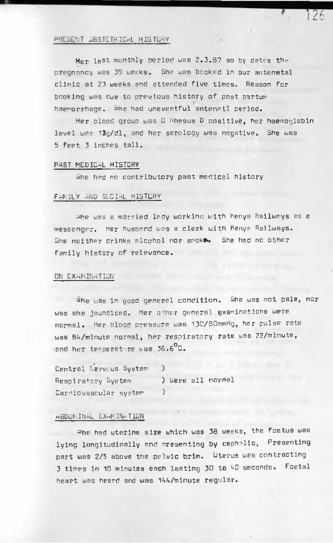
PRESENT SBSTETHIChL H 15 TORY
Her last monthly period was 2.3.B7 so by dates the pregnancy was 39 weeks. She was bcokEd in our antenatal clinic at 23 weeks and attended five times. Reason for
booking was due to previous history of post partum
haemorrhage. She had uneventful antenati period.Her blood group was 0 rhesus D positive, her haemoglobin
level was I3g/dl, and her serology was negative. She was
5 feet 3 inches tall.
PAST MEDICAL HISTORYShe had no contributory past medical history
FaN.ILY AND SLCI->L HI5TCRYv .
she was a married lady workinn with Kenya Railways as a
messenger. Her husband was a clerk with Kenya Railways.She neither drinks alcohol nor smokfr. She had no other
family history of relevance.
ON EXAMINATION
She was in oood general condition. She was not pale, nor
was she jaundiced. Her o^her general examinations were normal. Her blood pressure was 1 3 C /B 0 m m H g , her pulse rate
was BA/minute normal, her respiratory rate was 22/minute,
and her temperature oas 36.6°C.
Central '.ervrus System )Respiratory System ) Uere all normal
Cardiovascular system )
HBDiJMlMAL EX^'INnTIUN
^he had uterine size which was 3B weeks, the foetus was
lying longitudinally and oresenting by cephalic, Presenting
part was 2/5 above the pelvic brim. Uterus was contracting
3 timps in 10 minutes each lasting 30 to AO seconds. Foetal
heart was heard and was 1AA/minute regular.
IvO

< y*i
VAGINAL EXr^fINATICf;
Srm had a normal exte nal genitalia. There was no bleeding. Vaginal wall was smooth and normal.
Cervix was Scentimetres dilated with a rather predominant anterior lip. Foptal head was felt and it was in right
occipito-anterior. There was no caput and no moulding. The cervix was well applied to the fptal head. At this point the patient was noted tD be pushing and she was discouraged from doing so by citing itohe possible dangers involved .
DImS • 3£iI5
A diagnosis of parous patient with previous history of F.f-.H. in established labour was made.
PLAf.:
An urgent intravenous line was put and her blood taken
for group and crossmetch, 2 units. The patient was taken to second stage for closer observation and monitoring of hrr labor progress, with particular emphais on not letting her push before full dilatation.
The patient progressed well and at rbout 1.1Cpm cervix reached fall dilatation; she was given a medidateral
• episiotomy as the perineum was being distended by the head. Stw delivered ten minutes later a female baby weighing 30r0 g^ar^es with an apgar score of 9 in 1 minute and 10 at 5 minutes. Intramuscular ergomEtrine was given with the
delivery of anterior shoulder of the baby.Signs of placental separation were observed and by controlled cord traction whole placenta and membranes were delivered.A drip of 5% dextrose 50Cmls with 20 units pf syntocinon was commenced at the delivery of the placenta.
ro

The cervix was found to be intact, the vagina was intact Episio tomy was carefully sutured. No bleeding was observed for the next 2 hours and the potipnt was tak°n to first
stage for further observation. Her estimated hlood loss was 3Gnmls.
Her immediate post partum observation were pulse rate 80/minute, blood pressure was 150/90mmHg, respiratory
rate was 20/minute. Her observations after 6 hours were pulse rate 80/minute and blood pressure was 1£»0/80mmHg.Jhe was taken to post natal ward where further observations were continued U hourly for the next 2 1* hours. She was discharged with her baby in good condition.
3he did not come- to our post notal clinic as was arranged..

129>
CGKMEKTS
This was a patient who had several history of postpartum haemorrhage a number requiring blood transfusion.
Post partum haemorrhage is bleeding after delivery of the baby of an amount of blood exceeding 5C0mls. It can be primary when it occurs within the first 2U hours or secondary when it occurs later (1,2). Our patient gave history of
ptimary post partum haemorrhage in her previous deliveries which had necessitated her transfusion while in hospital.
Sauses of post partum haemorrhage are various, the commonest being poor uterine contraction due to retained product of conceotion (PUC). Others are uterine atony due to exhaustion or precipitate labor, unrecognised teat at cervix, vagina, or
perineum, or even a neglected episiotomy, grandmultiparity, induced labor, multiple pregnancy, polyhydramnios, fibromyomataa are all associated with uterine atony and hence post partum haemorrhage (PPH) 1,2,3).
FPH tends to recur especially in parous ladies. CJur
patient had no history of any particular management done in the previous episodes, *ut what came to light is that her labor usually takes short time, i.e. precipitate labor, she was also para 7+0, clinical findings were such that the cervix was rather prDtruberant on its anterior aspect which could_ denote split cervix in. earlier deliveries and hence lfess resistance to labor end short duration.
In anticipated PPH like in our patient, management revolves around active management of 3rd stage. This involve presence of grouped and crossratched blood, intravenous line, and being ready with syntocinon to be given intravenous or added into the drip (3,^,5). Patient needs to be reassured and gently discouraged from bearing down before full cervical dilatation (1).
This was achieved in our patient. Third stage is important
as this is when retained placenta, retained PbCs occur.; also
at this stage tears and improperty sutured episiotomy can cause bleeding.

*
Our patient's third stage of labor was particularly observed and intramuscular ergnmetrin given at the birth of anterior shoulder cf the baby; placenta bias delivered whole with membranes and drip of syntocinon started. Inspection of the birth canal did not reveal any tear and episiotomy was carefully sutured.
Foseiblity of retaining undiagnosed second twin with the injection of ergometrine exist as also that of causing hour-gless constriction on the placenta by the cervical
internal os. Cur patient did not lose much blood, 300mls»Her observation were normal and was discharged with her baby in good state.
Obstetric h emorrhages are responsible for 10-20r' of maternal deaths of which HPH is the biggest contributor; cause of death is usually haemorrhagic shock (5).
5hould bleeding continue despite every effort then hypogastric artery should be ligated, or hysterectomy done.Use of prostaglandin F2 alpha given tranBabdominally into the
myometrium should also be considered in case of intractable bleeding.

* 1-31
REFERENCES
1. Donald, I.
- Post partum haemorrhage
- In practical Obstetric problems 5th Edition pg 2L8, 1979.- Llyod-luke (medical Books) Ltd. London
2. Pritchard, J.«., McDonald P.C., Gant , [j.F.
- In Williams Obstetrics 17th Edition pg 707, 1985- "ppleton-Century-Croft/Neu York
3. Myerscough, P.R.
- In Munro-herr's Operative Obstetrics 9th Edit, pg 785, 197L- Bailliere Tindal, Norwich
U . Pritchard 3.A., McDonald P.C., Gant F.l\i.
In williams Obstetrics 17th Edition pg 687, 1985 Aonleton-Century-Croft/Leu York
5. Menon, K.K.K., Devi P.K., Bhasker, R.K.
- In Dostgraduate Gbs.Byn. 3rd Edition pg 196, 1986.- Orient Longman Ltd. 160 Anna Salai, Kadrass 6CC0D2
“ In obstetrical Practice > Pg L69-^71, 1980 - The 0 , \ j . Mosby Company - London* Toronto, St. Lou is
6-
I 132
LONG COMMENTARY
IN
OBSTETRICS
THE SIGNIFICANCE OF PLACENTAL WEIGHT ON TERM
DELIVERIES AT PUMLJANI MATERNITY HOSPITAL, NAIROBI, KENYA
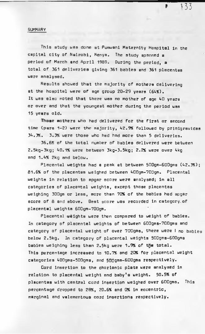
* 133
SUMMARY
This study was done at Pumwani Maternity Hospital in the capital city of Nairobi, Kenya. The study spanned a period of March and April 1989. During the period, a total of 361 deliveries giving 361 babies and 361 placentas were analysed.
Results showed that the majority of mothers delivering at the hospital were of age group 20-29 years (6AX).It was also noted that there was no mother of age AO years or over and that the youngest mother during the period was 15 years old.
Those mothers who had delivered for the first or second time (para 1-2) were the majority, <*2.9% followed by primigravidas 3A.3%. 3.3% were those who had had more than 5 deliveries.
36.6% of the total number of babies delivered were between 2.5kg-3kg; A0.1% were between 3kg-3.5kg; 2.2% were over Akg and 1.A% 2kg and below.
Placental weights had a peak at between 500gm-600gms (A2.3%); 81.6% of the placentas weighed between A00gm-700gm. Placental weights in relation to apgar score were analysed; in all categories of placental weights, except those placentas weighing 300gm or less, more than 70% of the babies had apgar score of 8 and above. Best score was recorded in category-of placental weights 600gm-700gm.
Placental weights were then compared to weight of babies.In category of placental weights of between 600gms-700gms and category of placental weight of over 700gms, there were I no babies below 2.5kg. In category of placental weights 500gms-600gms babies weighing less than 2.5kg were 1.9% of tge total.This percentage increased to 10.7% and 20% for placental weight categories A00gms-500gms, and 500gms-600gms respectively.
Cord insertion to the chorionic plate were analysed in relation to placental weight and baby's weight. 50.5% of placentas with central cord insertion weighed over 600gms. This percentage dropped to 28%, 20.6% and 0% in eccentric, marginal and valementous cord insertions respectively.
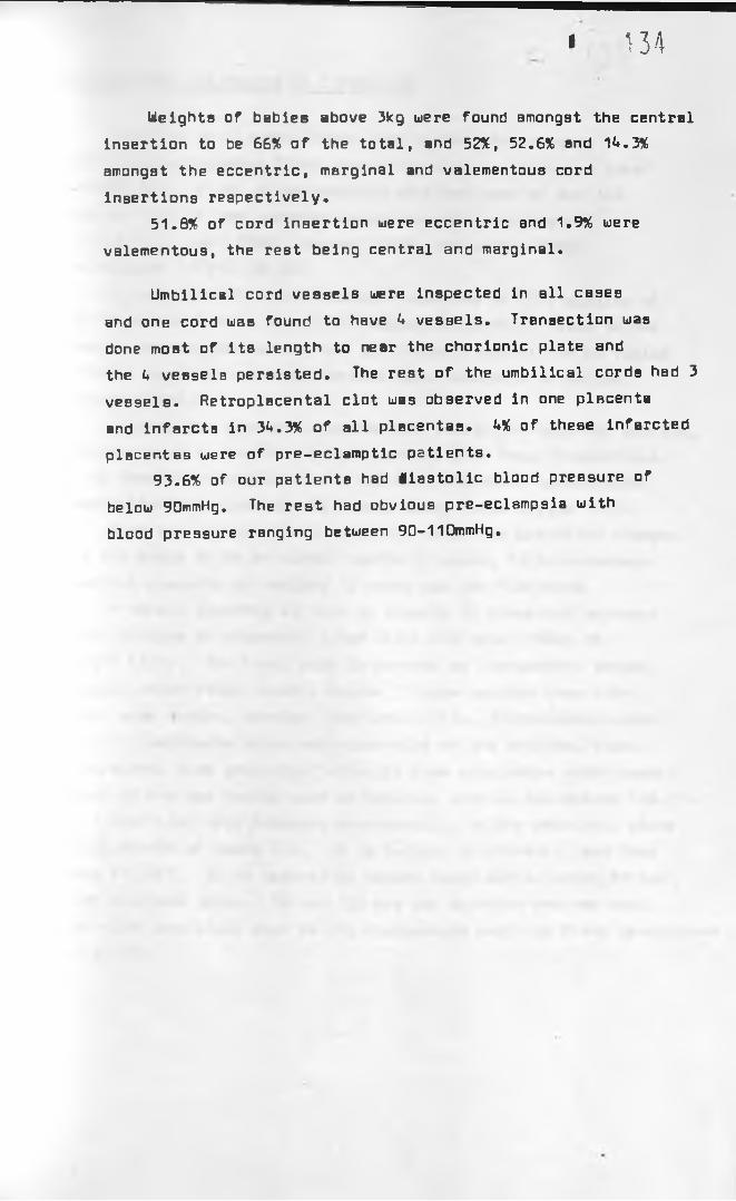
• t34
Weights of babies above 3kg were found amongst the central insertion to be 66% of the total, and 52%, 52.6% and H*.3% amongst the eccentric, marginal and valementous cord insertions respectively.
51.6% of cord insertion were eccentric and 1.9% were valementous, the rest being central and marginal.
Umbilical cord vessels were inspected in all cases
and one cord was found to have U vessels. Transection was done most of its length to near the chorionic plate and the U vessels persisted. The rest of the umbilical cords had 3 vessels. Retroplacental clot was observed in one placenta
and infarcts in 3^.3% of all placentas. <♦% of these infarcted
placentas were of pre-eclamptic patients.93.6% of our patients had iiastolic blood pressure of
below 90mmHg. The rest had obvious pre-eclampsia with blood pressure ranging between 90-110mmHg.

•i .
. * 1 3 5 ' -INTRODUCTION AND REWIEW OF L IT ERA TU RE
Interest on placenta dates back to the lAth century
with Realdus Columbus first describing it as a 'circular cake' in 1959 (9). A lot of painstaking uork has gone on and the much uie know of the placenta we owe to early workers who include Arantius (1560, Harvey (1651), Malpighi (I960) and Mrasman (1937) (9,10).
Mnssman (1937) first described placenta as that portion of the foetal membranes that is in apposition with or fused to the
4
uterine mucosa, and as we know any intimate opposition or fusion of fetal organ to maternal physiological exchange is called placenta (10).
In man, the placental trophoblast invade the maternal decidua, materral blood coming into direct contact with fetal trophoblast; it i- therefore described as a discoid, diciduate haemochorial chorioallantoic placenta all layers being fetal origin (11).
Size and composition of placenta alters as gestation changes.For the fetus to be of normal health in utero, it is necessary that the placenta be healthy to carry out its functions.
A normal placenta at term is discoid in shape and measures aboui 15-20cm in diameter, 2-3cm thick and about 500mg in weight (2,9). The fetal side is covered by transparent amnion
beneath which fetal vessels course. Cross section show from fetal side amnion, chorion, chorionic villi, intervillous space, then the deciduate plate and myometrium on the maternal side.
On maternal side groupings 1-3 villi form cotyledons which number about 30 and are easily seen on maternal side as elevations (10,29).
Umbilical cord attaches eccentrically to the chorionic plate in A8.5%-80% of cases (2). It is 1-2.5cm in diameter, and 55cm long (11,12). It is covered by amnion inner matrix being filled with Whartons jelly. It usually has two arteries and one vein, the right umbilical vein having disappeared early in fetal development
(9,10,12).

1«35
Functions of placenta Include respiratory, nutrition,
hormone production, barrier functions, excretion among others.
Although foetal growth depends on genetic potential and availe&ility of nutrients a host of factors can interfere
with this supply of nutrients that the genetic potential be secondary fj.) Physiolooical adaptation needs placental hormonps and this needs properly nourished placenta getting enough nutrients and enough blood flow. A host of factors have been known to affect this delicate balance some of which incli socioeconomic status, ethnic group, maternal hypertension, infections, blood groups and other immunological factors
(9,23,2**,20). ..The main factor that has been observed by many workers
to influence placental weight., and fetal outcome i* adequate uteroplacental blocdfloiu <22,27-, 32)%
Minute marginal subchorionic foci of infarcts are said to be normal and are present in all placentas, they only become significant if they are abudant as these may cause fetal death at most (9). OthFr workers also argue that placental calcifj,catior is a no-mal process and is a normal event in 3rd trimesters,
and that infarcts caused by fibrin deposition are normal except in widespread cases (18). It hes been shown that placenta from mothers with pre-eclampsia are smaller with total cells of even less than 50% normal, and thBt the\< are invariably associated with intrauterine growth retardation (32)#
The maternal blood volume se°m to, once again be very important in placental growth and fetal maturation. Myron (1970)^, that route for fdod to the fetus is very complex. It begins with aderjurte maternal blood flow. This flow becomes important if its composition and amount is such that it meets thP fetal needs Placental transport is Important because a placenta that is diseased will not be able to convey the substances that reach it to the fetus. Foetal growth will therefore depend on rate of perfusion of the placenta and the ability of the placenta to transport (?1j32), lilinick (1967) reported a case of severely
emaciated mother who carried pregnancy to term. It was found that the placenta had less than 50% of expected number of cell and she cave birth to a 2.5kg baby (23).

r —
I
This r=ally shows how plecenta has high functional reserve.The same p«rulier nature of placenta has been sighted by other
*workers (28)* More important is the neurological development that follow from so affected fetusses. Maternal undernutrition, maternal vascular disease will impede placental cell division and thus curtail brain cell division (20. Follow up of these
cases of obvious intrauterine growth retardation over a time of intellectual growth would be useful so as to guard egainst such situations.
Immunology of placenta is one of a complex of events and a success story itself. It seems to be dependent on absence Df
transplantation antigen from syntitotrophoblast. Immune attack on the placenta is not a clear pathological entity but may occur with placental villitis and phemphigoid gestations. These
especially villitis can in 5-10% of cases be associated with intrauterine growth retardation (26). Initial trigger for
cause is thought to be infective in nature especially with treponemas and cytomegaloviruB.
Placental size bears no universal relation with fetal.It is known that placenta undergoes a series of progressive changes that relate to gestational ege and fetal maturity.It has a huge functional reserve as has been witnessed with placentas of high altitude, erythroblastosis and anaemia.But its growth in terms of cell division steps at 3^-36 weeks but protein increase go on to term. Other workers have also noted that placental growth decline towards term (22,27,28).
As concerns prolonged pregnancy one would therefore expect that fetus would be distressed. This has been shown to be true and not only might the fetus not grow but will also lose weight (11). Also blamed for intrauterine growth retardation is retroplacental haemorrhage, circumvaliate placenta, marginal insertion of umbilical cord and especially valementous has been shown to be frequently associated with intrauterine growth retardation (11,13,1^,15). Capacity and reserve of aging placenta are reduced after term; the reduction continues upto a point at which optimal functions can not really be sustained and fetus
is bound to succumb.
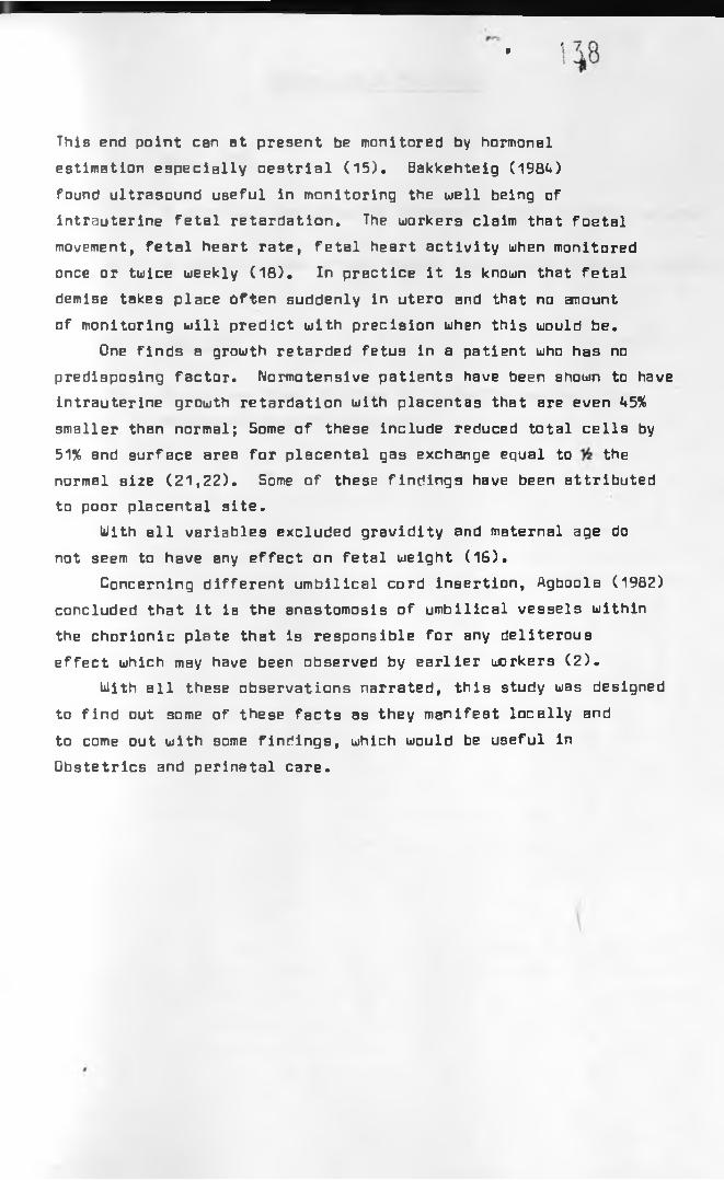
»
This end point can at present be monitored by hormonal estimation especially oe9trial (15). Bakkehteig (198U)
found ultrasound useful in monitoring the well being of intrauterine fetal retardation. The workers claim that foetal movement, fetal heart rate, fetal heart activity when monitored once or twice weekly (18). In practice it i9 known that fetal demise takes place dften suddenly in utero and that no anount of monitoring will predict with precision when this would be.
One finds a growth retarded fetus in a patient who has no predisposing factor. Normotensive patients have been shown to have intrauterine growth retardation with placentas that are even <*5%
smaller than normal; Some of these include reduced total cells by 51% and surface area for placental gas exchange equal to the normal size (21,22). Some of these findings have been attributed to poor placental site.
With all variables excluded gravidity and maternal age do not seem to have any effect on fetal weight (16).
Concerning different umbilical cord insertion, Agboola (1982) concluded that it is the anastomosis of umbilical vessels within the chorionic plate that is responsible for any deliterous effect which may have been observed by earlier workers (2).
With all these observations narrated, thi9 study was designed
to find out some of these facts as they manifest locally and
to come out with some findings, which would be useful in
Obstetrics and perinatal care.

' , 1 3 9t
OBJECTIVES OF THE STUDY
A. SHORT TERK
1. To find out the effect of placental weight on fetal outcome
2. To identify some of thr factors influencing placental weight.
8. LONG TERM
3. To utilise these findings for a better preconception, antenatal and perinatal care.
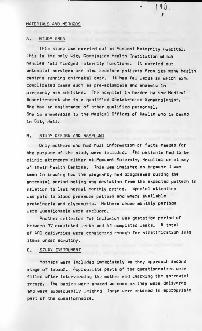
" 140 .
*
HATER IflLS AND METHODS
A. STUDY AREA
This study was carried out at Pumwani Maternity Hospital.This is the only City Commission Health Institution which
handles full fledged maternity functions. It carried out antenatal services and b I so receives patients from its many health centres running antenatal care. It has feu wards in which some complicated cases such as pre-eclampsia and anaemia in pregnancy are admitted. The hospital is headed by the Medical Superitendent who is a qualified Obstetrician Gynaecologist.She has an assistance of other qualified personnel.She is answerable to the Medical Officer of Health who is based in City Hall.
B. STUDY DESIGN AND SAMPLING
Only mothers who had full information of facts needed for the purpose of the study were included. The patients had to be clinic attenders either at Pumwani Maternity Hospital or at any of their Health Centres. This was insisted on because I was keen in knowing how the pregnancy had progressed during the antenatal period noting any deviation from the expected pattern in relation to last normal monthly period. Special attention was paid to blood pressure pattern and where available proteinuria and glycosuria. Mothers whose monthly periods
were questionable were excluded.Another criterion for inclusion was gestation period of
between 37 completed weeks and completed weeks. A total of AOO deliveries were considered enough for stratification into
items under scrutiny.
C. STUDY INSTRUMENT
Mothers were included immediately as they approach second stage of labour. Appropriate parts of the questionnaires were filled after interviewing the mother and checking the antenatal record. The babies were scored as soon as they were delivered and were subsequently weighed. These were entered in appropriate
part of the questionnaire.
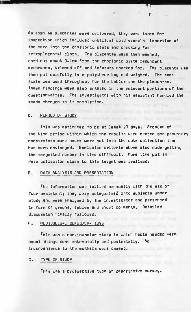
i
*
As soon as placentas were delivered, they were taken for inspection which included umbilical cord vessels, insertion of the cord into the chorionic plate and checking for retroplacental clots. The placentas were then washed, cord cut about 3-Acm from the chorionic plate redundant membranes, trimmed off and infarcts checked for. The placenta was then put carefully in a polythene bag and weighed. The same scale wbs used throughout for the babies and the placentas.These findings were also entered in the relevant portions of the questionnaires. The investigator with hiB assistant handled the study through to it completion.
D. PERIOD DF STUDY
This was estimated to be at least 20 days. Because of the time period within which the results were needed and pecuniary constraints more hours were put into the data collection than had been envisaged. Exclusion criteria above also made getting the targetted number in time difficult. More time put in data collection close to this target was realised.
E. DATA ANALYSIS AND PRESENTATION
The information was tallied mannually with the aid of four assistant; they were categorised into subjects under study and were analysed by the investigator and presented in form of graphs, tables and short comments. Detailed
discussion finally followed.
F. MEDICOLEGAL CONSIDERATIONS
^his was a non-invasive study in which facts needed were
usual things done antenatally and postnatally. No inconvenience to the mothers were caused.
G. TYPE OF STUDY
This was a prospective type of descriptive survey.

It
H. CONSTRAINTS
i. Prolopged formality of getting permission fromCity Hall before starting the research mas real and felt.
ii. The hospital has many sectors which are operational on 2U hour basis. As such midwives work in shift and it is during the changeovers that me manage to lose some placentas of mothers whose antenatal records had been entered in our questionnaires. These mere delivered and discarded in the sluice thereby making some questionnaires useless. Ordinarily these placentasmere supposed to be kept by the bed-side of the delivered mother and be collected by the investigator for further examination and record. Such collections mere carried out usually within 5 minutes and at most 15 minutes.
iii. This was not a sponsored project and as such
financial constraint was felt by the investigator.
HOD THE CONSTRAINTS WERE OVERCOME
i. Although the formality through which permission is granted is long and takes time, this was anticipated and as such the urgency of the study was stressed to the Medical Officer of Health who was considerate enough as to push •
through the permitting letter in good time.
ii. The inconvenience of losing some of the placentas before recording was compensated for by the more amount of time that was put in the data collection fortunately, by the good number of deliveries taking place at the hospital.The midwives needed reminding every now and again.
iii. The devotion of the assistant and understanding of the staff at the hospital enabled the investigator to complete the data collection in time and hence cut down
on extra expense.

RESULTS
A total of 389 mothers were Interviewed and relevant portions of questionnaires filled. Out of thesfe'28
questionnaires were found to be unacceptable for inclusion in the study. Some of the reasons were that thequestionnaires were wrongly labelled or that an item was either omitted or wrongly placed. As a result 361 questionnaire after thorough scrutiny, passed for the purpose of the study and are analysed and presented here.
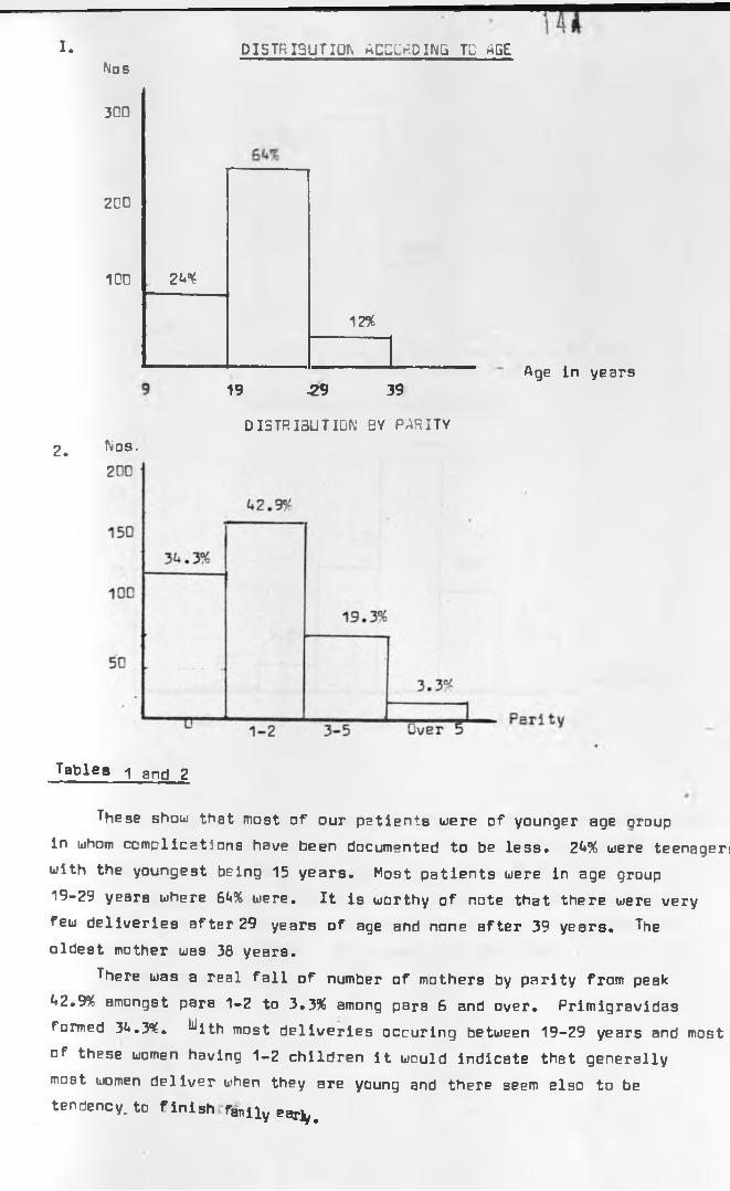
I. DISTRIBUTE ACCORDING TO AGENos
2.
300
200
100 2N%
12%
19 -29 39
DISTRIBUTION BY PARITYNos.
Age in years
Tables 1 and 2
These show that most of our patients were of younger age aroup in whom complications have been documented to be less. 2*»% were teenager: with the youngest being 15 years. Mo9 t patients were in age group19-29 years where GU% were. It is worthy of note that there were very few deliveries after 29 years of age and none after 39 years. The oldest mother was 38 years.
There was a real fall of number of mothers by parity from peak t«2.9¥ amongst para 1-2 to 3.3% among para 6 and over. Primigravidas formed 3*4.3%. ^ith most deliveries occuring between 19-29 years and most of these women having 1-2 children it would indicate that generally most women deliver when they are young and there seem also to be tendency, to f inish Tanlly ear^

blRTHli-'I IGHT DISTRI BUT ION
Wt (Kg)
Uit (gms)
Tables 3 and U
277 babies (76.7%) weighed between 2.5-3.5kg. Only U .2% weighed less then 2.5kg. It is also noteworthy that only 15.7% of babies weighed between 3.5-t*kg. Between 2.5-<*kg were 92.<*%.
Placental weight varied alot with peak at between 500-600gms (*♦2.3%). Only 6.2% of the placenta weighed were below UOOgms and only 11% were above 700mg. So between U00-700gm had r- 81.6% of the whole total.

146
Most of the pptients were noted to have delivered at Bver 1*0 weeks by dates 68.5*^. It Is also significant to realise that none of the mothers we saw had pregnancy past U2 weeks.
As pointed out in the summary 93.6* Df patients had normal blood pressure. The rest G.U% were r.others whose diastolic Blood pressures were 90mmHg and above.
I

TABLE 5 : PLACENTAL WEIGHT VERSUS FETAL WEIGHT
UTS INGMS> 700
600
500
LOO
L . 300
The above table compares placental weight with fetal weight. Percentagt of fetuses weighing less than 2.5kg were analysed per category of
placental weights. For placental weights of more than 700gms and the category of placental weights of 600gms-700gms there were no babies
recorded. For placental weights categories 500gms-600gms, L00gms-500gm, and 300gms-L00gms percentages of babies recorded were 1 ,9 % , 10.7% and 20% respectively.
Babies weighing between 3kg-Lkg in placental weight category
700gms and above accounted for 76.6%, for category 600gm-700gms they accounted for 72.8%, but accounted for 50%, 7L.3% and 50% for placental weights category 500gm-600gm, L00-500gms, and 3OOgm-L0Ogm respectively.
Babies of more than Lklgg were 13.L% of placental weight category over 700gms and 1.9% of placental weight category 500gms-600gms.
This table shows that placental weight has some influence on foetal weight. Lower placental weights are accompanied with lower fetal weights, and higher placental weights are accompanied by higher fetal weight as exemplified by fetal weights below 2.5kg, and placental weights of above 700gms. For placental weights between L00gm-700gm or fetal weights between 3kg-Lkg there does not seem to be a consistent relation between the weights.
0.0 0.0 10.0 L3.3 33.3 13. U 100
0.0 0.0 27.2 L5.5 27.3 0.0 100
0.0 1.9 L8.1 33.3 16.7 1.9 100
0.0 10.7 1L.7 66.3 8.0 0.0 100
0.0 20 30 LO 10 0.0 100
0.0 0.0 0.0 0.0 0.0 0.0 0.0
2»5 3 3.5 ^ L UTS IN KG
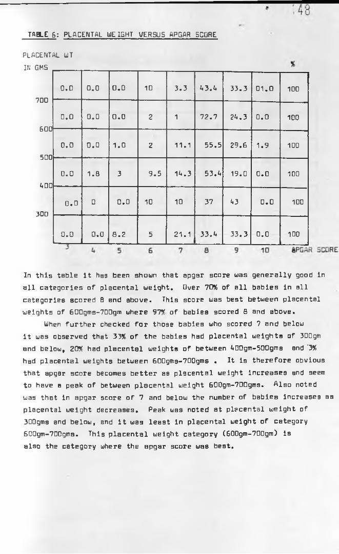
TABLE 6 : PLACENTAL WE IGHT VERSUS APGAR SCORE
placental w tIN GMS
700
600
500
AOO
300
0.0 0.0 0.0 10 3.3 A3.A 33.3 01.0 100
0.0 0.0 0.0 2 1 72.7 2A.3 0.0 100
0.0 0.0 1.0 2 11.1 55.5 29.6 1.9 100
0.0 1.8 3 9.5 1A.3 53. A 19.0 0.0 100
0.0 0 0.0 10 10 37 A3 0.0 100
0.0 0.0 8.2 5 21.1 33.A 33.3 0.0 1D0
In this table it has been shown that apgar score was generally good in all categories of placental weight. Over 70% of all babies in all categories scored 8 and above. This score was best between placental weights of 600gms-700gm where 97* of babies scored 8 and above.
When further checked for those babies who scored 7 and below it was observed that 33* of the babies had placental weights of 300gm and below, 20* had placental weights of between A00gm-500gms and 3* had placental weights between 600gms-700gms . It is therefore obvious that apgar score becomes better as placental weight increases and seem to have a peak of between placental weight 600gm-700gms. Also noted was that in apgar score of 7 and below the number of babies increases as placental weight decreases. Peak was noted at placental weight of 300gms and below, and it was least in placental weight of category 600gm-700gms. This placental weight category (60Dgm-700gm) is also the category where the apgar score was best.

TABLE 7: FREQUENCY OF CuRD INSERTION
Nos. % 1 2 3 L
Eccentric 187 51.8 62.5 62.2 LB.5 6L
Central 13L 37.1 26.5 22.8 L6.5 25
Marginal 33 9.1 9.2 1L.9 L. 1 10
Valementous 7 1.9 1.8 — 0.0 10
TOTAL 361 100
Table-' 7 shows percentage distribution of cord
insertion to chorionic plate. It has been compared to other
work done by other researchers.
Series named are as follows:
1- Agboola (1976) (2)
2 - Dorste (1571) (5)3 - Purola (1960) (L)A - Krone (1965) (3)
Reference Numbers.
Our findings of distribution seem comparable with that
of others. In other series and ours eccentric insertion is the commonest 51.8^, and valementous is the least common in our series like in other series.
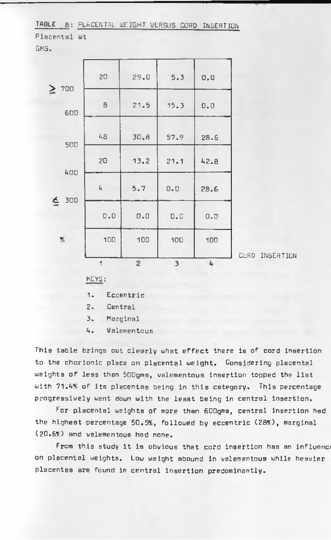
TABLE 8 : PLACENTAL LlEIGHT VERSUS CORD INSERT ION
Placental Wt GMS.
> 700
600
5C0
AOO
d 3D0
20 29.0 5.3 0.0
8 21.5 15.3 0.0
A8 30.8 57.9 28.6
20 13.2 21.1 A2.8
U 5.7 0.0 28.6
0.0 0.0 O.C 0.0
100 100 100 100
1
KEYS:
CURD INSERTION
1. Eccentric2. Central3. MarginalA. Valementous
This table brings out clearly uhat effect there is of cord insertion
to the chorionic place on placental weight. Considering placental weights of less than 500gms, valementous insertion topped the list with 71.U% of its placentas being in this category. This percentage progressively went down with the least being in central insertion.
for placental weights of more than 600gms, central insertion had the highest percentage 50.5%, followed by eccentric (28%), marginal (20.6%) and valementous had none.
From this study it is obvious that cord insertion has an influenc on placental weights. Low weight abound in valementous while heavier placentas are found in central insertion predominantly.

” 151’TABLE 9: CORD INSERTION VERSUS FETAL U)EIGHT
Insertions
% ■
Valementous 0.0 28.6 57.1 1A.3 0.0 0.0 100
Marginal 0.0 0.0 A7.A 36.8 15.8 0.0 100
Central 0.0 1.9 32.0 33.9 26.A 5.7 100
Eccentric 0.0 6.0 A2 AO 8
t
A 100
^ 2 2.5 3 3.5 > A UTS. IN KG
After noting the obvious influence of cord insertion on placental weights, a step was taken further to see whether the cord insertion could affect fetal weights.
Babies who weighed less than 2.5kg were considered. 28.6% of those who had valementous cord insertion were in this category, none in marginal, 6% in eccentric and 1.9% in central insertion. In babies who weighed over 3kg, central insertion had 66%, eccentric 52%, marginal 52.6% and valementous insertion 1A.3%.
These findings corrolate well with findings in table B»
It can be argued that type of cord insertion onto the chorionic plate has a direct effect on both placental and fetal weights.

! fL
' TABLE w . MATERNAL AGE VERSUS PLACENTAL WEIGHT
TABLE: 10:
This table shews distribution of placental weight versus maternal age. In teenagers the majority of placentas weighed between LOO-7O0Qms, in age group 20-29 years, majority of placentas weiohed between LOO to over 700 grams while in age group 30-39 years majority of placentas weighed between 500 to over 700 grams.
So there seem to be some increase of placental weight
with maternal age.

TABLE 11: MATERNAL AGE (YR5) VERSUS FETAL HEIGHT (KGS)
FETAL WT (KG)
> 4
3.5
£19 20-29 30-39 £ 4 0
3.0
2.5
£ .2.0
TOTAL
0 7 5 0 12
8 43 10 0 12
30 91 16 0 137
45 81 8 0 134
0 9 . 3 0 12
4 0 0 0 4
87■
232 - 42 0 361
MATERNAL AGE ( YRS)
In the teenBge group 86.2% of them had fetal weights of between 2.5-3.5kg, while in age group 30-39 years peak is at between 3-4kgf 38%, while 26% were between 2 and 3kg and 11.9% above 4kg. Age group 20-29 years shows a very marked peak between 2.5-4kg (92.6%). It can also be concluded that teenagers tend to have comparatively lower birth weight than in older patients
especially after 30 years.
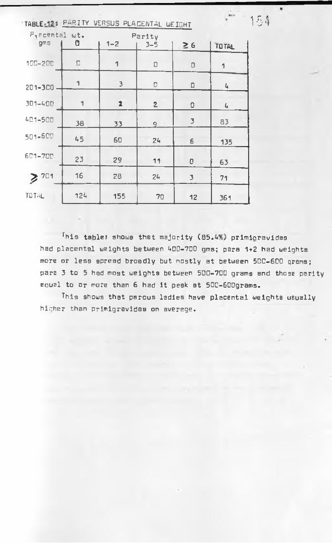
T A B L E P A R I T Y VERSUS PLACENTAL UEIGHT 1 5 4P^Fcental wt. Parity
gms 0 1-2 3-5 > 6 total
100-200 C 1 0 0 1
201-3C0 1 3 0 0 U
301-LOO 1 2 Z 0 U
L01-5C0 38 33 9 3 83
501-600 L5 60 2U 6 135
6C1-70r 23 29 11 0 63
£ 7 0 1 16 28 2U 3 71
TOTAL 12L 155 70 12 361
^his table? shows that majority ( 8 5 . primioravidas had placental weights between L0D-700 gmB; para 1+2 had weights
more or less spread broadly but mostly at between 5DC-6DD grans; para 3 to 5 had most weights between 500-7C0 grams and those parity eaual to or more than 6 had it peak at 50C-600grams.
This shows that parous ladies have placental weights usually higher than primigravidas on average.

TABLE 13> PhRITY VERSUS FETAL WEIGHT
FETAL WT (kg)
0 1-2 3-5 ^ 6 TOTAL
u+ 2 3 5 0 11
U .O 16 26 17 6 65
3.5 39 65 2A U 132
3.0 60 55 22 2 139
2.5 6 6 2 0 V *
2.0 1 1 0 0 2
total 12U 155 70 12 361
This table shows that 79^ of primigravidas had fetal weights between 2.5-3.5kg; in the same weight bracket para 1-2 were 77*. and para 3-5*65^. 83^ of those who werepara 6 or more had their babies weighing between 3-Lkg It shows that uptoPara2 the difference in weight distribution does not exist then after that there seem to be increase in weight favouring more parous ladies.

A total of 23 pre-ecla~psia patients delivered during the study period. 2C had blood pressure of 9D -1C 0m m H g and 3 had blood pressure of over IC O m m H g. 21 of the patients hed fetal weights betwPen 3-3.5kg. P-|2cental weight also ranged between i*5C and 6TCgms. uf the 2 remaining patients one had widspread placental infarction of weight 300gms and fetal weiaht
2.3kg. The other was fetus of weight 2.2kg with a placenta of weigh* tOCgns.
This shows how pre-eclampsia can interfere with placental and fetal weights and well being.

157DISCUSSION
Physiological adaptation of the mother during pregnancy has been shown to be important for normal fetal growth in utero.This adaptation needs normal placenta producing normal
pregnancy hormones in normal amounts. Some factors have been shown to affect this relationship and these reflect in poor fetal growth and poor fetal outcome. Placental pathologies have also been noted (9,1D,11,23,2^,28).
This study found some interesting relation between fetal and placentBl weight, and has also shown some factors that has influence on fetal and placental weights.
A total of 361 cases were discussed, 6A% of the mothers were in age group 20-29 years. Teenagers formed 2A% and age group 30-39 yean were 12% of the total. The youngest mother in this series was 15 years, and the oldest was 38 years.
Mpthers who had delivered 1-2 times were the majority, 0.9%.Those who were pregnant for the first time formed 303%. The
percentage decreased to 3.3% for those who had delivered over 5 times (those who were having their sth pregnancies or more). The mother of the highest parity was para 8 gravida 9. These findings
showed that the mothers seen during the study period were young and of low parity. 88% of the mothers were below 30 years of age;and 77.3% of the mothers had had 0-2 deliveries. This could be inferre as a tendency to finish family early and also to have small family size.
Maternal age has been shown to have effect on fetal growth and that optimum performance has been noted to be around 20-30 years of age; above 35 years there is usually an element of growth retardatio (1,33). Obstetric performance in primlgravidas have been noted to be
poorer with more perinatal deaths than other parity, and that after parity the performance becomes poorer again (33). In Nairobi Birth Survey increase of risk factor in para U and above was not noted (30.
Birth weight of between 2.5kg-3.5kg accounted for the majority 76.7%. In our series low birth weight (birth weight below 2.5kg) accounted for 5.2%. This category accounted for 7.5% in Nairobi Birth Survey (30.
Placental weights showed a peak of between 500gm-600gms, <*2.3%*
Between t*OQgm-700gm percentage rose to 81.6%.
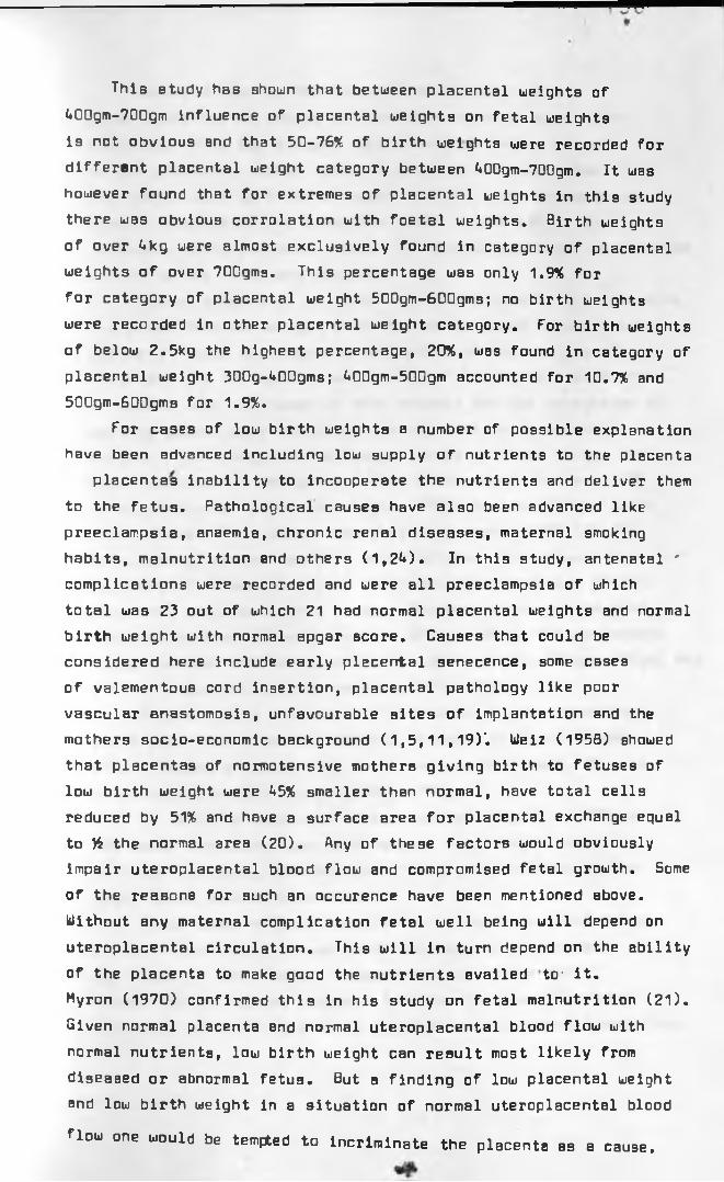
This study has shown that between placental weights of A00gm-700gm influence of placental weights on fetal weights is not obvious and that 50-76% of birth weights were recorded for different placental weight category between A00gm-700gm. It was however found that for extremes of placental weights in this study there was obvious correlation with foetal weights. Birth weights of over Akg uere almost exclusively found in category of placental weights of over 70Cgms. This percentage was only 1.9% for
for category of placental weight 500gm-600gms; no birth weights were recorded in other placental weight category. For birth weights of below 2.5kg the highest percentage, 20%, was found in category of placental weight 300g-A00gms; A00gm-500gm accounted for 10.7% and 500gm-600gms for 1.9%.
For cases of low birth weights a number of possible explanation have been advanced including low supply of nutrients to the placenta
placentas inability to incooperate the nutrients and deliver them to the fetus. Pathological causes have also been advanced like preeclampsia, anaemia, chronic renal diseases, maternal smoking habits, malnutrition snd others (1,20. In this study, antenatal ' complications were recorded and were all preeclampsia of which total was 23 out of which 21 had normal placental weights and normal birth weight with normal apgar score. Causes that could be considered here include early plecental senecence, some cases of valementous cord insertion, placental pathology like poor vascular anastomosis, unfavourable sites of implantation and the mothers socio-economic background (1,5,11,19)'. Uleiz (1958) showed that placentas of normotensive mothers giving birth to fetuses Df
low birth weight were A5% smaller than normal, have total cells reduced by 51% and have a surface area for placental exchange equal to } i the normal area (20). Any of these factors would obviously impair uteroplacental blood flow and compromised fetal growth. Some of the reasons for such an occurence have been mentioned above.
Without any maternal complication fetal well being will depend on uteroplacental circulation. This will in turn depend on the ability
of the placenta to make good the nutrients availed to' it.Myron (1970) confirmed this in his study on fetal malnutrition (21). Given normal placenta and normal uteroplacental blood flow with
normal nutrients, low birth weight can result most likely from diseased or abnormal fetus. But a finding of low placental weight and low birth weight in a situation of normal uteroplacental blood
flow one would be temped to incriminate the placenta as a cause.

T j 7
Immune attack on the placenta 19 usually not a clear pathological entity but has been shown to occur with villitus. This may trigger some amount of immune attack on the placenta, attack being directed to the placentas
chorionic villi in 5-10% of cases and may cause intrauterine growth retardation and hence low birth weight (26). Such
instances could obviously result in reduced placental weights and effect on the fetus are obvious.
Foetal outcome at an instant is recognised by apgar scoring, and other than the weight which is not included in the scoring system it forms an immediate reflection of the quality of antenatal and intrapartum well being of the fetus. With this in mind a look was taken in this respect and two categories of
scoring grades were considered in relation to placental weight. The categories were apgar score of 7 and below; and apgar scores of 8 and above. This study shows that over 70% of all babies of placental weights 300gm to over 700gms score B and above. This percentage just fell sort of 70% (66.7%) in placental weight category below 300gms. This shows that majority of fetuses had good apgar score irrespective of the placental weights in this study. The best apgar scores were recorded between plecental weight 500gms-700gm; the percentage ranged between 87% for placental weight category 500gms-600gm, and 97% for placental weights category 600gm-700gms.
Of the babies who scored 7 and below, higher percentage
33.3% were from placental weights category of below 300gms.Others showed a steady fall in percentage the least, 3%, was
in placental weight category 600gm-700gm.It could be concluded that placental weights of 300gms
and above are associated with fetuses whose apgar score at birth are good, and that below this placental weight the
apgar score becomes precarious.Apgar score reflect usually the intrauterine well being
of the fetus during labour until delivery or existance of the fetus in utero before commencement of labour and thereafter influenced labour. In labor the most important influence on fetal well being is good uteroplacental blood flow.

«
If this is compromised as might occur in aging placenta; placenta with borderline reserve, arising from chronic infarct, uterine endarteritis; or in prolonged second stage of labour, then the fetus will be delivered with poor apgar score (11,15,30.It therefore means that with normal uteroplacental blood flow, placenta of normal integrity and normal fetus apgar score at birth should be good. Delay in second stage of labor for 30 minutes has been shown to compromise fetal well being and increase perinatal mortality by 1.8 times; this becomes 7.3 times if delay is 0-59 minutes (30. In labor of normal duration therefore and assuming normal fetus then it is the
functional reserve of the placenta that becomes important.It is known that placental growth slows as term approaches and that at 3U -3G weeks placental cell division stops though weight and protein increases to term. Blood supply to the placenta which at peak is in excess of 5D% of the maternal cardiac output also becomes less (21,27). If other factors encroach on this delicate balance like infarction of placental parenchyma or thrombosis of maternal uteroplacental vessels, then the babies fate in utero even before labor hangs on a balance. Infarcts aQ such
signify severely restricted maternal blood flow to placenta and is a hallmark of markedly abnormal maternal vasculature (25,27). Small placentas with small fetuses therefore would denote some embarassed relation between maternal placental blood supply and placental fetal blood supply as in our cases cited above. In a situation of good maternal record antenatally and normal fetus except for weight then the small placenta must be the cause of intrauterine growth retardation.
Distribution of cord insertion onto the placental chorionic plate was looked into in this study. Eccentric insertion were 51.8% of the total followed by central insertion 37.1%, marginal 9.1%
and least number was valementous 1.9%. These were compared with other work that have been done elsewhere and results were comparable.
The cord insertions were then compared in relation to placental weights. 71.*♦% of all valementous insertions had placental weights of less than 50ngms. This was the highest percentage amongst the four types of placental insertions, the least was recorded in central insertion which in the same placental weight category accounted for 18.9% of the total central insertions.

¥
Central insertion 50.5%, followed by eccentric 28%, marginal
20.6%, and none for valementous were the percentages of the total per each type of insertion for those placentas weighing equal to or above 600gms.
It could be concluded that central insertions are generally characterised by heavier placentas, and that valementous by lighest placentas. Other types of insertions fall in between.
Valementous insertion type of umbilical cord has been known to be associated with intrauterine growth retardation and has been considered by others as pathological (7,11,29). Marginal placenta has also been noted to be associated with intrauterine growth retardation (11). This being the case it is not surprising in our study that these two type of umbilical cord insertions are accompanied by small placentas. It would seem from this study that central insertion is the ideal form of umbilical cord insertion onto the chorionic plate; it has bigger placentas. It so happens that it is not the commonest type of cord insertion but this could be due to influence of intrauterine environment where placental growth might be affected in any direction by the prevailing circumstances inside the uterus for example low lying placenta over poerly vascularised lower segment, growth will be assymetrlal towards the upper segment, growth over scar tissue. It can be envisaged that placenta with umbilical cord placed centrally stands a better chance of getting blood and nutrient supply equally from all sides Bnd therefore should supply it with maximum to the fetus who should also have maximum growth and good weight. This also explains why the valementous type of insertion has the lightest placenta, and why other insertions have placental weights in between central and
valementous.With the above postulations in mind effect of cord insertion
on fetal weight was looked into. It was found that in 66% of central cord insertions fetal weights were 3kg and above; eccentric insertion of the cord total fetal weight of 3kg and above were 52% for each and only 1L.3% for valementous insertion. Looked at the other way round for babies weighing less than 2.5kg, 28.6% of valementous insertions fell in this group while in central insertion only 1.9% of the total were in this group. Again other cord insertions were in between valementous and central.

It can be concluded that type of cord insertion onto the chorionic place has an influence on the placental weight as well as fetal weight. Central insertion being more favourable,
while valementous insertion the least favourable. Findings on this subject of type of cord insertion and its influence on birth weight have been described by others (6,8). Agboola (1982) in his study of Nigerian population found that cord insertion does not have any deliterous effect on the fetus rather it is the vascular anastomotic pattern that is significant (2). While this study agrees with the above the effect on placental and fetal weight were noted to be obvious.
Having noted clearly that umbilical cord insertion onto
the chorionic plate haB an effect on placental weight, this study looked into other parameters in an effort to find out possible factors that could influence placental weight. Effect of maternal age on placental weight was next looked into. This study showed that teenagers had placental weight in category l»00gm to 700gm accounting for 72%, while 30-39 years old mothers placentBl weight category 500gms-7b0gms accounted for 90.A%. Maternal ages between 20-29 years had placental weights evenly spread between AOOgms-over 700gms.
There seem to be increase in placental weight with increase of
maternal age. As most of the mothers in this study had delivered by 30 years, this finding would indicate the easy adaptability of uterus and its vessels to the stress of expanding uterine content. This therefore would easily accomodate the expanding
placenta and possibly the fetus.When maternal age and fetal weights were next studied
it was found that it follows the trend as the placenta's above.86.2% of teenagers had fetal weights between 2.5-3.5kg, while in age group 20-29 years 92.7% had fetal weight of between 2.5kg to Akg$ and age group 30-39 years had fetal weights spread over weight categories 2.5kg - over Akg. It would be concluded that the seme age influence on the placenta also affect fetal weight.So teenagers have lower birth weight compared to older mothers especially after 3Cyears.

Effect of parity on placental weight was also studied. It w found that para 3-5 had heavier placentas than primigravidas generally. Again para 1-2 had placental weight evenly spread to encompas both primigravidas and para 3-5. Effect of
parity on fetal weiqht was then studied. It was found that this followed more or less the age pattern above. 79% of primigravidas had fetBl weight of between 2.5-3.5kg, in the same weight bracket para 1-2 were 77% and para 3-5 65%. Although majority of fetal weight were between 2.5-3.5kg by proportion the were more primigravidas as compared to other parity where weights were more spread and showed encroachment on the Akg weight category. Those who are of parity equal to or more than 6 have obviously heavier babies, than by proportion than other
parities.23 preeclamptic mothers were delivered during this study
period. 21 of the mothers had fetal weight of between 3-3.5kg and placental weights of between A50gms-600gms. Of the two mothers remaining one had widespread placental infarction with weight of 300gms and fetal weight 2.3g. The other had placental weight of UOOgms with foetal weight of 2.2kg. Observations made for these pre-eclampsia lends weight to the fact that placenta has huge functional reserve and that even with distruction of sizable portion it will still be the uteroplacental blood flow that will determine placental and fetal being. In the two cases mentioned it seems as if the placental function had been compromised thereby compromising fetal well being. This observation is in keeping with that made by others regarding effect of pre-eclampsia on placenta and hence fetus. Intrauterine growth retardation and occasionally intrauterine fetal death are common with pre-eclampsia (18,27,26,30).
Infarcts occur in at least half of all the placenta (9).
It is the quatity of the infarct and the reserve function that count (11,15,27). Lue had 3A.3% of placentas with infarcts. Our finding was that other than the two cases who were also pre-eclamptica the other patients delivered normal weight infants
and normal weight placentas.

In conclusion this study has found that placental weight
affect fetal weight; lower placental weight, give lower fetal weinht and higher placental weight give higher fetal weight.
Best apgar score were noted in placental category 600gm-700gm, and worst apgar score was noted in placental weight of below 300gms.
Central cord insertion had the best placental weights and fetal weights; valementous had the least fetal end placental weights.
Maternal age has a bearing in placental weights. After 30 years placental weights on average are heavier than teenagers.
Parity after 3 is accompanied by rise in placental weights on average which are higher than others.
Maternal age affect fetal weight. Teenagers have lower birth weight than those of 30 years and above.
Parity especially after para 2 has a favourable influence on fetal weight.
Placenta has high functional reserve and small placenta does not necessarily mean small baby. Rather it is the efficiency of residual function in the presence of normal uteroplacental blood flow that is important.

RECOMMENDATION
■* ' rh H
This study has given us some light as to the relation of placental and fetal weight and factors operational which influence the relation.
There is a need to study more pre-eclamptics to show for sure the effect on fetal and placental weights.
Grand multiparas need to be studied so that we can know
when the edge of parity and/or age stops.Ultrastructure of placentas obviously affected need to
be done so as to rule out vascular cause.Use of Vacuum extraction need reappraisal as often times
it was used when it should not be used either due to poor indication or too early application.
The midwives should learn to score the babies within the first minute, and then fifth and tenth minutes. This was an area which had a lot of inertia and made the investigator to discard a few questionnaires because of suspected wrong information.
On the whole, good.
c

REFERENCES
1. Browne, J.C.,- Duration of Pregnancy and Birth Weight- Brownes Antenatal care 7th Edition pg BA-85 1978- Churchil-Livingston, Edinburg, New York, London
2. Agboola, A.
- The significance of distribution of Umliilical cord insertion into the chorionic plate.
- Journ. Obs/Gyn. E.C.A. 1: 1A5, 1982.3. Krone, H.A., Jopp, H, Schellerer, W.
- The importance attached to different types of umbilical cord insertion.
- Z. Geburtsh Gynale, 163: 205, 1965 A. Porula, E.
- Length and Insertion of Umbilical cord- Ann. Chir. Gynaec. Feun. 57: 621, 168
5. Dorste, P.
The incidence and statistical relationship between umbilical cord insertion and placental weight- Zentralbl, Gynaek. 93: 1705, 1971.
6. Knaus, H.H.
- The weight of placenta and its Forensic Significance- Arch. Gynach. 198: 73, 1983.
7. Morice, I.W.
- Valementous insertion of the cord in early pregnancy- Am. Joum. Qbs.Gyn. 93: 276, 1965
8. Aberle, 5.B.O.; Morse A.H., Thompson, W.R., Pitney E.H.
- Relationship of weight of placenta cord and membrane weight to weight of infant in normal full term and in premature deliveries.
- Am. Journ. Obs.Gyn. 20: 397, 1930.9. Pritchard, J.A.; McDonald, P.C., Gant, N.F.
- Diseases and abnormality of placenta and fetal membranes- Williams Obstetrics 17th Edition pg AA3 and A57, 1985- Appleton-Century-Croft/California
10. Trudal R., Scoff, J.S.
- Placental Cacification: A study of 3025 singleton and multiple pregnancy
- Brit. Journ. Obs.Gyn. 72: 356, 1965

11. Pritchard, J.A.; McDonald P.C., Gant, N.F.- Preterm and Postter^ pregnancies and fetal growth retardation.- Williams Obstetrics 17th Edition pg 758, 1985.- Appleton-Century-Croft, California
12. Phillip, A.G.S.- Fetal growth retardation: Femur Fontanalle and Followup- Paediatric: 62: LL6, 1978.
13. Aldjem, S.
- Placental and Fetal membrane Pathology- Obstetrical Practice pg 515-530, 1980- C.V. M sby Company, Ltd. London.
1L. Benson, R.C.
- Placenta and Fetus- Current Obs.Gyn. Diagnosis and Treatment Pg. A97-505, 1976 Edition.
- Lange Medical Publishers, London15. Benson R.C.
- Placental Dysfunction- Current Obs.Gyn Diagnosis and Treatment Pg A89 1976 Edition
- Lange Medical Publishers, London16. Patricia H.5., Mark A.K., Barry J.G.
- Birth Weight arong women of different ethnic groups- Journ. -Mmer- Med. Ass. 255: L8, 1986
17. Hendricks C.H.- Delivery Patterns, reproductive efficiency preterm rates and
birthweights in Claveland and differing socioeconomicand ethnic groups.- Am. Journ. Obs/Gyn. 97: 608, 1967
18. Bakkenteig, L.S.,- Ultrasound for Intrauterine growth retardation- Lancet 2: 2071, 198L.
19. Phillip K., Skodler W.D., Peteisky N.- Uteroplacental blood flow, measurement using radiosotope
in small placentas.- Obs.Gyn. Survey L1(10): 622, 1986.
20. Weiz E.B. Jr.
- Disappearnace of radioactive sodium from uterine muscle in normal and abnormal pregnancy.

f- R
- Am. Journ. Obs/Gyn 76: 3A0, 1958
21. Myron, W.- Fetal Malnutrition- Clinic Obs.Gyn 13(3): 526, 1970
22. Winick, M.- quantitative Cellular Changes during growth of organs- Pediatria 10: 255, 1967
23. Uinick, M., Noble, A.
- Cellular response during malnutrition of various ages- J. Nutrition 91: 179, 1967
2 U . QJinick, N.- Nucleic acid and protein content of placenta and fetal brain.- Am. Journ. Obs. Gyn. U0: 601, 1970
25. Brinkman, C.R.- Umbilical blood flow and fetal oxygen consumption- clinic Obs.Gyn. 13(3): 565, 1970
26. Redman, C.Ui.G.-- Immunology of Placenta ^- Clinic. Obs.Gyn. 13:3, A87, 1986
27. Fox H.- Pathology of Placenta- Clinic Obs.Gyn. 13: 3, 501, 1986
28. Kazzi, G.M., Gross, L.T., Rossem, G.M.- The relationship of placental grade, fetal lung maturity
and neonatal outcome in. normal and complicated pregnancy
- Hmer. J. Obs.Gyn. 1A8, 5 U t 198A.
29. Dewhurst, J.- Fertilization, implantation and placental function- Integrated Obs Gyn for Postgraduates 3rd Edition
pg 8A-85, 1981.30. Bodis, J., Zambo, K., Nemessanyi, Z.
- Determination of intervillous perfussion index in pregnancies with intrauterine growth retardation
- Obs.Gyn. «*1(1): 26, 1986.31. Hunyer, S.N.
- Vascular Volume and Cardiac response to normal and hypertensive pregnancies.
- Obs.Gyn Survey U1(5) 279, 1986

32. Erskine R.L.A,; Ritchie J^W.K.- Umbilical artery blood flow characteristics in normal
and growth retarded fetuses.- British Bourn. Gbs.Gyn 92: 605, 1985.
33. Chemberlsin, G., Phillip, E.t Hawlett, B., Masters, K.- British Births 1970, Vol. 2- William Heineman Medical Books Ltd., Lnndon 1978
3L. Mati, J.K.G., Aggarwal, U.P., Lucas, S., Sanghvi H.C.G.,
- Early Perinatal Mortality Rate- MBS I\ l
- Bourn. Obs. Gyn. E.C.A. 2(0: 130, 1983.
Corkhills,

IT.XKt-LLT: 5LF.TIl‘. ; EIV/- CU TICr, Of UTl kLJS DCNE UNDER SEDATION
Name: B.K.Age: 22 years Farity: D+0IP f.o. : 913CU7 ^LNMP: 6.6.86 DC A: li*.a.QB DCD: 15.8.88
PRESENTING CCKPL-.IMS
The patient presented with lower abdominal pain and vaginal bleeding for a duration of six days.
HIETC:-Y EF PRESENTING ILLNESS
The patient was well until six days earlier when she
developed lower abdominal pain which was intermittent in
nature but increased in intensity and frequency, then she later noticed vaginal bleeding. Prior to admission she had heavy vaginal bleeding with clots.
She gave no history of fever, no history of trauma, nor a history of dysuria. There was no history of interference
either.
P-oT L.3GTETRICE :.D .NYN-'ECGLCG IC~L HISTORY
She was para D+0. Her last normrl monthly oeriod was 6.6.85. She used tD get the periods every 23 days and were lasting 2-3 days. There were no accompanying pains, nc history of discharges in between the periods.
uhe had h°r menarche at 1N years, and she had not used any contraceptives.
PRST MEDICAL HISTORY
None of significance.

HI£T:-JY CF rr.LSSi.T r'...5:..-i,\CV
By dates h»r pregnancy mas 9 weeks old. -he had not attended any antenatal clinic and she had had no problem durinr the 9 weeks.
F->i ILY r 1, „.Ci.'L HIjTuRY
She was a mrrried lady who l'es working in a restaurant in fjaircbi as a Secretary. She reached form level of education Her husband was a sales representative with Karsalls (K) Ltd.
She dees r.Dt drink nGr smoke. "Jo histcry of eny medical problem in her home.
Of. Ca-u Ir»r.TIwU
She was in good general condition. She was not pale
and not jaundiced. Her other generalexamination was normal.
Her body temperature was 36DC, pulse rate was S8/min blood pressure was 11Q/£0mmHg and respiratory rate of 20/minute
Central f.ervous System )nespiratory System ) Were essentially normalCardiovascular System )
-SDCrilv .L CX-d- IWsTILfJ
abdomen was not distended, she had mild tenderness lower abdomen but there were no masses felt.
VAGir.r-.L EXnNIhnTIOr,
She had a normal extern-1 genitalia, there was slight
vaginal bleeding. Cervix uas 2 centimetres long and 3 centimetres dilated with products of conception felt.Uterus was bulky and was mildly tender. Adnexia were normal. There were some clots and products of conception at the posterior fornix otherwise pouch of Douglus was empty.
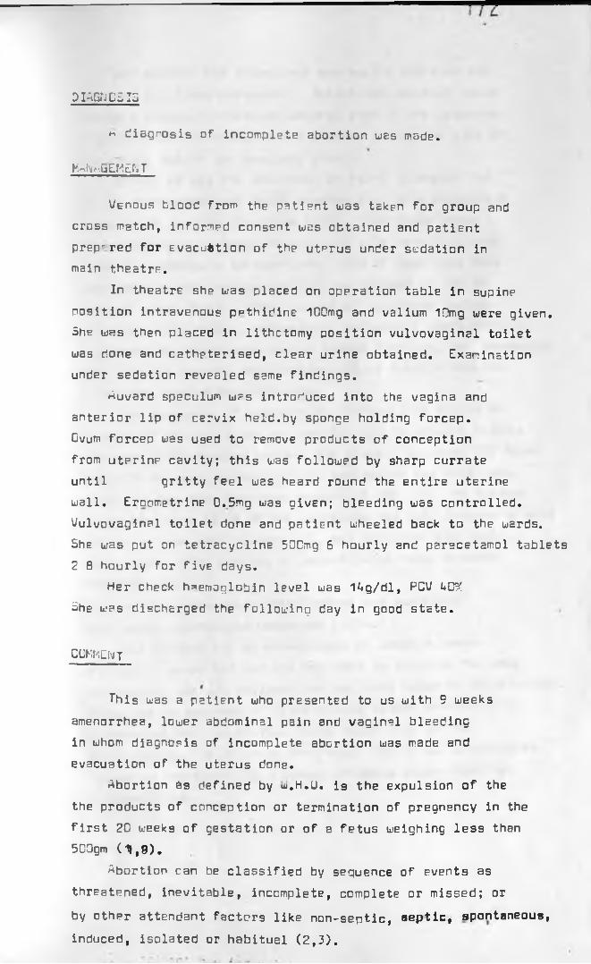
DIAGNOSIS
« diagrosis of incomplete abortion was mode.
M ^ IW j EM ENT
Uenous blood from the patient was taken for group and cross match, informed consent was obtained and patiEnt prep' red for evacuation of the uterus under sedation in main theatre.
In theatre she was placed on operation table in supine
position intravenous ppthidine 100mg and valium 1Dng were given. She was then placed in lithotomy position vulvovaginal toilet was done and catheterised, clear urine obtained. Examination under sedation revealed same findings.
Auvard speculum was introduced into the vagina and anterior lip D f cervix held.by sponge holding forcep.Ovum forcep was used to remove products of conception from uterine cavity; this was followed by sharp currate until gritty feel was heard round the entire uterinewall. Ercometrine 0.5mg was given; bleeding was controlled. Vulvovaginal toilet done and patient wheeled back to the wards.She was put on tetracycline 50Cmg 6 hourly and paracetamol tablets 2 8 hourly for five days.
Her check haemoglobin level was 1<*g/dl, PCV UZft.
She was discharged the following day in good state.
CCKMCNT
This was a patient who presented to us with 9 weeks amenorrhea, lower abdominal pain and vaginal bleeding in whom diagnosis of incomplete abortion was made and
evacuation of the uterus done.Abortion 6s defined by w.H.U. is the expulsion of the
the products of conception or termination of pregnsncy in the first 2D weeks of gestation or of a fetus weighing les9 than 5C0gm (H,9).
Abortion can be classified by sequence of Fvents as threatened, inevitable, incomplete, complete or missed; or
by othpr attendant factors like non-septic, septic, spontaneous,
induced, isolated or habitual (2,3).

uur patient had incomplete non-septlc abortion and this was h~r first pregnancy. Incomplete abortion would denote 8 clinical situation whereby part of the products of conception are still in the uterine cavity and part of
it rlready out of the cervical canal.5C-S0V of alj, thr abcrtions in first trimester = re
due to chromosomal and fetal abnormality, this percentage
drops to 5a' in late abortions (2,5). £03 blood group
incompartibility between husband and wife have also been
noted to contribute to abortions. U 5 % of abortions have
been seen in AG3 incompartibility as opposed to 3D% in
control group (5,6). Other causes of abortions include placental abnormality, endometrial infection, utero-CErvical
malformation, inadequate corpus luteum function, and immunology
(7). Cthers include debilitating and/or fEbrile maternal
illnesses, trauma,interferences.
The incidence of abortion is difficult to arrive at
as some of the abortions are carried out in private clinics
and some patients also conceal the event. *ggarwal (1) found
that 6C" of total gynaecological admissions are abortions,
other workers have reported between 25-30% (7). He further
found that upto 62,3% of the abortions had been induced cr were likely tc have been induced (1), and pati-nts that could
voluntarily offer history of interference rangF bFtween
10-33.6C'. (1,0,.
Most of the cases where interference was suspected
were young, unemployed unmarried girls.Cur patient had an anenorrhoea of about 9 weeks
she was 22 years old and she had been in marriage for only six months, sh= was emoloyed and was very happy in the marriage. There was no pvidFnce of intFrference either on history or
on examination. An obvious cause could also not be attributable. It is known that about 12% of all pregnancies end up as abortions (7} a number of u/tioss ceuse might not
be identified.

The hallmark of diagnosis in abortion rests history.Th ere will be history of amenorrhea usually 8-20 weeks, intermittent lower abdominal pain, vaginal bleeding with or without clots, with or without expulsion of products of conception. Pelvic examination will confirm some
of the above history. Uterus will usually be smaller than the perior* of amenorrhea, cervix will or will not be open with or without products of conception. Evidence of interference may be elicited. General examination might reveal variable degree of anaemia, shock or sepsis (2,5). Our patient was quite alert with normal vital signs and no evidence of anaemia as was later confirmed with a haemoglobin level of l^g/dl. ahe had some lower abdominal tenderness, and had clot at the introitus. Her cervix was 2cm dilated and had products of conception ^dnexiae and pouch of Douglas were normal. Uterus was only mildly tender and was bulky. These findings were in keeping with incomplete abortion.
Management of incomplete abortion depends on clinical findings at the time of admission, but of grave importance is evacuating the uterus as undue delay in doing this will predispose to further bleeding and chances of sepsis. Rescusitation in case of anaemia is necessary, where there is sepsis, urgent specimen should be taken for culture and sensitivity, and broad spectrum antibiotic started at once. Dur patient had non-septic incomplete abortion with no other complication needing rescucitatory measures.
In our unit evacuations are done under valium and pethidine
IV. The patient was prepared as mentioned and evacuation proceeded uneventfully. It is important to ensure thet evacuation is complete so as to avoid further bleeding and chancf» of sepsis. Occasionally there is bleeding after evacuation in which case an intramuscular ergometrine should be given
or intravenous syntocinon. (2,3).Complications of abortion include excessive bleeding, shock
and death
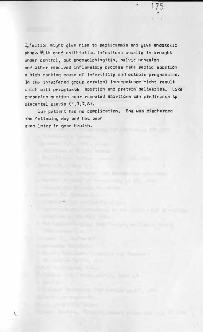
Infection might give rise to septicaemia and give endotoxic
shock. With good antibiotics infections usually is brought under control, but endosalphingitis, pelvic adhesion and other resolved inflamatory process make septic abortion a high ranking cause of infertility and ectopic pregnancies. In the interfered group cervical incompetence might result which will perpetuate abortion and preterm deliveries. Like caesarian section scar repeated abortions can predispose tp
placental previa (1,3,7,8).Our patient had no complication. She was discharged
the following day and has been
seen later in good health.

p 176
REFERENCES
1. Aggarwal, V/.P., Mati, J.K.G.
- Epidemiology of Induced Abortions in Nairobi- Journal of Qbst.Gynae E.C.A. 1982.
2. Dpuhurst J.- Abortion
- Integrated Obstetrics and Gynaecology for Postgraduates- 3rd Edition pg 203, 1981- BlBckuiell Scientific Publications- Oxford, London. Edinberg. °oston, Melbourne
3. Oeffcoate, N.- Abortion
- Principles of Gynaecology Lth Edition pg 187 1975- “utterworths London.
A. Aggaruial, V.P., Mati, J.K.G.- Abortions at K.N.H. Review- East African Medical Journal 57: 138, 1980
5. Jone.e H.W.f Jones G.S.
- Infertility, recurrent and Spontaneous abortions- Noviks Textbook of Gynaecology pg 69A, 1982.- William and UJilkins, Baltimore.
6. Lawson J.0., Stewart D.B.
- Underfertility and~ overfertility- Obstetrics snd Gynaecology in the tropics and developing
countries pg 582-583, 1983.- The Egglish Language Book Society and Edward Arnold
(Publishers) Ltd.7. Raphael, B.t Durfee M.u.
Spontaneous Abortions
- Current Obst.Gynae Diagnosis and Treatment 5th Edition Pg 710, 198A
L.M.P. California, U.S.A.
8. Pritchard, J.N., McDonald P.C.f Gant N.F.- Abortion- Williams Obstetrics 17th Edition pg A77, 1985
Appleton-Century-Croft9. W.H.O. Scientific Group
Induced Abortion, Technical report series 623, pgg 22 1978.
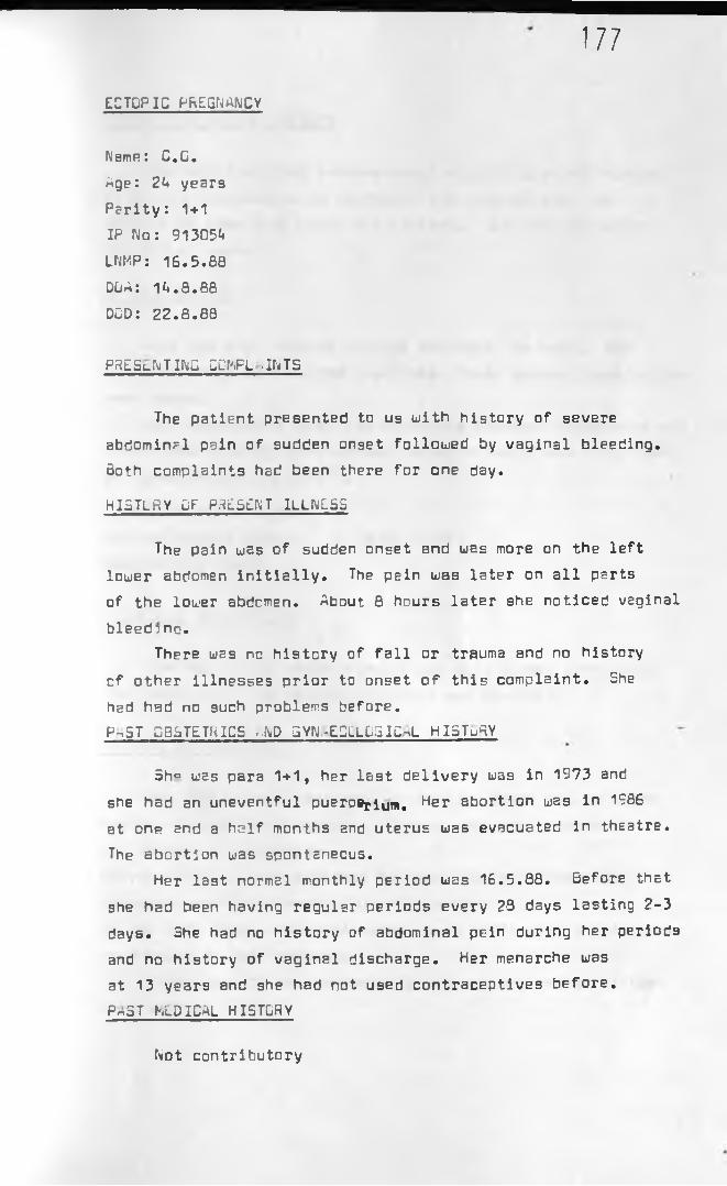
M l
ECTOPIC PREGNANCY
Name: C.C.Age: 2U years Parity: 1+1 IP No: 913054*
LNMP: 16.5.88 DBA: 1 £4.8.88 DGD: 22.8.88
PRESUMING COt-'iFL .INT5
The patient presented to us with history of severe abdominal pain of sudden onset followed by vaginal bleeding. Both complaints had been there for one day.
HI3TLRY OF PRESENT ILLNESS
The pain was of sudden onset and was more on the left lower abdomen initially. The pain was later on all parts of the lower abdomen. About 8 hours later she noticed vaginal
bleed:no.There was no history of fall or trauma and no history
of other illnesses prior to onset of this complaint. She hed had no such problems before.P-.5T C33TETN ICS .AD SYN -ECLLLGIC->L HISTuRY
3h° was para 1+1, her last delivery was in 1973 and
she had an uneventful puerperlum. Her abortion was in 1986 at one end a half months and uterus was evacuated in theatre.
The abortion was spontaneous.Her last normal monthly period was 16.5.88. Before that
she had been having regular periods every 23 days lasting 2-3 days. She had no history of abdominal prin during her periods and no history of vaginal discharge. Her menarche was at 13 years and she had not used contraceptives before.P«5T h.LDICAL HISTGRY
Not contributory

FAMILY *KD SlCI«L HI5TCRY
ihe was a married businesswoman staying with her husband at Sheuri I oyo Estate in Nairobi. Ths> husband work in Industrial area with eccl (K) Limited, She dees not drink alcohol or smoke.
OK EX-.; .XLTICK
->he was sick looking and was obviously in pain, She
was moderately pale but not jaundiced. Other general examination were normal.
Her respiratory rate was 22/minute regular; pulse rate was irr/minute regular, good volume; blood pressure was 1EE/7C?mmHg
and temperature was 3£.5°C.
Central Ksrvcus System )Cardiovrsoul~r system ) Were Normal Respiratory system
A3Dut- IN -L IN,TIEN
Sh>- had a full abdem-n which was very tender especially the lower part. No proper palpation was passible.
VAGINhL EXW'IKm TILN
She had a normal external genitalia, there- wes no blood cominp through the intreitus. Cervix was long, central,
and tender on excitation.Literin= size wes not possible to gauge due tc tenderness.
Adnexiee were both tender, but more markedon the left side. Adnexial masses were difficult to rule out
on account of the tenderness.A decision to do paracentesis was reached and it yielded
non-clotting blood*

179DIAjfrUSIS
A diagnosis of ruptured ectopic pregnancy was made.
Ki.-rj«GEP£M
Urgent blood was taken for group and cross match, two units. Intravenous line was set with normal saline. Close observation of vital signs were ordered. After obtaining informed consent, the patient was urgently prepared for emergency laparotomy as stated in the introduction.
The patient was on the operation table in one hour. Pre-operative procedures in theatre were done as mentioned in the introduction and her abdomen opened in layers.Peritoneum was held on either side by two pairs of long artery forcep and a small incision made with curved pair of scissors. Haemoperitoneum was identified. A quick suction of blood and removal of clot just enough to make identification of site of rupture was done. Th= following were the findings:-- Ruptured left ampullary tubal pregnancy involving the broad ligament; bleeders clamped.- Haemoperitoneum of about 12CCmls- Fe,tus with intact cord and part of placenta was found
in the peritoneal cavity.- Foetgl sac and pert of placental tissue were found at the
site of the rupture.- left ovary was normal.- Rinht ovary and tube were normal.
After the removal of haemoperitoneum, fetus, placenta
and membranes partial left. Salphingectomy was done and
haemostasis achieved. Abdomen was closed in layers and anaesthesia reversed.
She was put on the usual post operative management as per introduction and also on intravenous crystacen 2 mega units
6 hourly. She was -=lso transfused two units of compatible blood. The patient made steady recovery. Her postoperative
check haemoglobin was 9.kg/dl. She was discharged in good health on 22.8.B8 to be seen in our gynaecology clinic in 6
weeks. °he attended as was appointed end she had fully recovered.

1 8 0
This was a patient who had ruptured left ampullary tubal r«gnr-ncy for which left partial salphingectomy was done.
' ctopic pregnancy result from implantation at sites ■th.’r than the uterine cavity commonest site being the ^-bes (3,5,10). SitP of ectcpic is tubal in 95?.' of cases ■' ■'rity bping on the rinht side; ampulla is affected in 65%
cases, isthmus 25e-, and Interstitial %2%. (3). Uur patient ''d B_pullary pregnancy.
Cctopic gestation can ogcur at any time from menarche tn monopause but age group 20-29 years
to be more at risk (1,3).Incidence varies from region to region belno as low as
’ : 30C normal deliveries in United Kinodom to as high as 1:26 normal ‘fliveri-s in Jamaica (11). Kwathe (7) reported an incidence •t Kenyatta National Hospital at U cases per week, wM I f uinei ft el (1) found admission of 2 per week.
Aetiology is not known but factors that would iffeet tho transport of fertilized ovum would contribute to ;hv genesis of ectopic oregnancy.>■ sc wLuld in c ludE adherent luminal folds as would be consequent - inf1-mation, spasm, inadeauate peristalsis. Excessive ,pgth end tortuousity of the tube; developmental defects like • r~' essory ostia, diverticielum; previous surgery end extrinsic «-^p«ion (1,2 ,3,6). Other causes postulated include mstrual reflex with delayed fertilization of ovum with ■ e n s t r u e l bleeding at the time of implantation, external •1-ration, ectopic endometrialtissue in the tube or fertilised
rxtruded ovum (3,0.Ectopic pregnancy seem to be more frequent in infertile
„En , women within low socio-economic group and women who -ve hed pelvic inflamatory, disease or tubal surgery 1 3) Sinei et al (1) observed low parity in his series f 2 or less to account for 68.6%; 63.2% between age group >' 23 years, arid evidence of previous infection in U2.1% .
.. ) in his histolooical evaluation of fallopianPCtD** ± ° V C% - *-es of e c t o p i c s found 69% had salphingitis.
^ur patient was in the age greup, was of low
rlty, and of low socio economic bracket.

There seem to be more Increase of incidence uith IUCD users (1,5), but if infection sets in then chances are that tubes
will be affected. The incidence is 8 times morE in women undergoing investigation for infertility, end those who have been operated for ectopic have 1-2:20 ch=nce of getting another ectopic ('-,5$. Generally incidence seem to be on the upward trend and could be contributed to by increased sexual freedom and high prevelance of sexually transmitted diseases, use of effective antibiotic which however leave the tubes already damaged but good enough for passage of fertilised ovum to some extent, use of
fertility drugs, better end early diagnosis and increased tubal operation especially the coagulation technique for tubal ligation (1,L,8). Cur patlEnt had not used any
contraceptive method and had no tubal operation and had no history suggestive of past infection.
No clear cut signs or symptoms that would be listed as diagnostic of ectopic pregnancy. However symptoms that are mere frecuently found are abdominal pain 75-100K (1,3,7), Birtfnorrhoea, 75-76.2 * (3,7), vaginal bleeding 67.2-30^ (1,3) and abdominal mass **9.8* (7).Cur patient presented with abdominal pain, 12 weeks anenerrhta, vorn-1 bleeding and adnexiel masses was
difficult to tliclt^ue to gross tenderness.Pregnancy test is not useful rs it will only be
po-sitive in LC-5DC of cases (3). Paracentesis is useful in rupture cf ectopic as was in our pati: nt and is positive in over BS0-- cases (3). Ultrascund can be used but has
error upto IlF'. (3,0.
ribr'cminel pain is due to amnio-chorion sac rupture with
local bleeding, pulsion and fra c t io n . Uterine bleeding
is usually due to failure of pregnancy to survive,
followed by endometrial invo lu tion .
Rupture is spontaneous in most cases but occasionally
accelerated by trauma of vigorous bimanual palpation or coitus. Isthrral pregnancy tends to rupture early 6-8 weeks,
ampullary 8-12 weeks and intestilled months (3). Lur patient had ampullary pregnancy and was by dates~12 weeks
-Hp presented to us with ruptured ectopic pregnancy.Lack of resistance or response of tissues into which the

1 8 0
CCMMENT
This was 3 patient who had ruptured left ampullary tubal pregnancy for which left partial salphinoectomy was done.
Ectopic pregnancy result from implantation at sites other than the uterine cavity commonest site being the tubes (3,5,10). 5itp of ectcpic is tubal in 95?' of cases majority being on the rinht side; ampulla is affected in 65%
of cases, isthmus 25%, and inter9tittal **2%. (3). uur patient had ampullary pregnancy.
Ectopic gestation can ogcur at any time from menarche to menopause but age group 20-29 years seem to be more at risk (1,3).
Incidence varies from region to region being as low as 1:30C normal deliveries in United Kincriom to as high as 1:28 normal deliveries in Jamaica (11). Kwathe (7) reported an incidence at Kenyatta National Hospital at U cas^s per week, while Sinei
et al (1) found admission of 2 per week.Aetiology is not known but factors that would
affect th° transport of fertilized ovum would contribute to the genesis of ectopic pregnancy.These would include adherent luminal folds as would be consequent on infl=mation, spasm, inadeauate peristalsis, Excessive
length and tortuousity of the tube; developmental defects like accessory ostia, diverticielum; previous surgery and extrinsic
adhesion (1,2,3,6). Other causes postulated include menstrual reflex with delayed fertilization of ovum with menstrual bleeding at the time of implantation, external migration, ectopic endometrialtissue in the tube or fertilised
extruded ovum (3,0.Ectopic pregnancy seem to be more frequent in infertile
women, women within low socio-economic group and women who have h?d pelvic inflamatory, disease or tubal surgery(1,3). Sinei et al (1) observed low parity in his series of 2 or less to account for 68.6%; 63.2% between age group20-29 years, and evidence of previous infection in **2.1%.Iiiebala (2, .) in his histological evaluation of fallopian
tubes of ectopics found 69% had salphingitis.
Cur patient was in the age group, was of low
parity, and of low socio economic bracket.

T 8 1
There seem to be more increase of incidence with IUCD users (1,5), but if infection sets in then chances are that tubes
will be affected. The incidence is B times more in women undergoing investigation for infertility, and those who have been operated for ectopic have 1-2:20 ch=nce of getting another ectopic ('-,5$. Generally incidence seem to be on the upward trend and could be contributed to by increased sexual freedom and high prevelance of sexually transmitted diseases, use of effective antibiotic which however leave the tubes already damaged but good enough for passage of fertilised ovum to some extent, use of fertility drugs, better and early diagnosis and increased tubal operation especially the coagulation technique for tubal lication (1,**,S). Our patient had not used any contraceptive method and had no tubal operation and had no history suggestive of past infection.
IMo clear cut signs or sy-ptoms that would be listed as diagnostic of ectopic pregnancy. Hcwever symptoms that are mr re frecupntly found are abdominal pain 75-1DCK (1,3,7), eirtinorrhoea, 75-76.2* (3,7), vaginal bleeding
57.2-3Qc£ (1,3) and abdominal mass (7).uur p-tient presented with abdominal pain, 12 weeks amenorrhea, vor- n-1 bleeding and adnexial masses was
difficult to tlicit^ue to qross tenderness.Pregnancy test is not useful es it will only be
positive in LC-50C of cases (3). Paracentesis is useful in rupture cf ectopic es was in our patient end is positive in over B50’' cases (3). Ultrasound can be used but has error upto 10?/ (3,L).
abdominal pain is due to amnio-chorion sec rupture with
local bleeding, pulsion and traction. Uterine bleeding
is usually due to failure of pregnancy to survive,
followed by endometrial involution.Rupture is spontaneous in most cases but occasionally
accelerated by trauma of vigorous bimanual palpation or coitus. Isthmal pregnancy tends to rupture early 6-8 weeks,
ampullary 8-12 weeks and intestilled L months (3). uur patient had ampullary pregnancy and was by dates'12 weeks
ahe presented tc us with ruptured ectopic pregnancy.Lack of resistance or response cf tissues into which the

* 182V
abnormal implantation takas plac^, minimal or no decidual
reaction without defence aqainst permenting tropholast and
thinness af distended wall pred: spose to easy rupture.ManagETent of tubal pregnancy is emmediate surgery.
In most cases in cur unit surgery is even undertaken while blood is still being looked For as this will ensure no further bleeding, operation ranges from milking of the products of conception, partial or total selphingectomy to wedge resection and reconstruction of thp uterus (3,0. Cur patient had partial salphingectomy done. she did well post operatively and was discharged.
Katern^l mortality associated with ectopic pregnancy is high and ranges between 9-9.2% in U.K. and U.S.A (5).
In this patient everything was done and in just under one hour the patient was on operation table. Delay such as recorded by f-’.wathe (7) of between G-28 days can cost the patient her life. These often occur with symptoms which are not clear cut or which have stmnger shades of other clinical entities, like threatened abortion, or dysfunctional
uterine bleedina.Obstetric perfomance of patients operated for ectopic
is not clear but she has chance of 10% developing another ectopic and 5C* chance of getting normal intrauterine
.pregn ncy (*0.
183
>
references
*>inei S.K.f Okumu, i>.Ectopic Pregnancy at KNH, Nairobi, Kenya Journal of Obs.Gyn. East Central Africa 6(1): 9 Vol. 6 No. 1, Pg 9.
2. iiiebala, G.S.R.Tubal Pregnancy at Keriyatta National Hospital; The roleof PID in it aetiologyM.Med Thesis 1979, University of Nairobi
3. Benson R.C.f Complications of Pregnancy Current Obs/Gyn. Diagnosis and Treatment 1976 Chap. 32 pg 559-603Lange Medical Publications, Los *ltos, California
•*. Pritchard A.J., McDonald P.S., Gant F.N.Ectopic PregnancyWilliams Obstetrics 17th Edition 1985, pg L23 Appleton-Century-Croft/Norwalk, Connecticut
5. Deuhurst J.Ectopic PregnancyIntergrated Obs/Gyn. for Postgrad. 3rd Edition 1981 pg 222-9Blackwell Scientific PublicationsOxford London. Edinburg, Boston, Melbourne
6. L0ulson O.B., Stewart D.B.Extraut PregnancyObs/Gyn in the tropics and Developing countries 1983 Edition pg 371The English Language Book Society and Eduard Arnold (Publishers) Ltd.
7 . Mwathe E.G.Pattern of Ectopic Pregnancy at K.N.H.M.Med Thesis 19BA, University of Nairobi.
6. Ory H.W.Ectopic Pregnancy and IUCD, New Perspectives Obs/Gyn. S 57: 137, 1981.
9 . Makokha E.A.Maternal M0rtality at Kenyatta National Hospital E.A.M.J. 57(7) L51, 1986.

it
10. Jeffcoate N.- Ectopic Pregnancy- Principles of Gynaecology- Uth Edition pg 207, 1975.SutterijJorthB, London 11
11. Weston L., Bengtssen L.O.M., Maron P.H.
Incidence trend and risk of Ectopic Pregnancy in a population
of women.8.M.J. 11:15, 1981
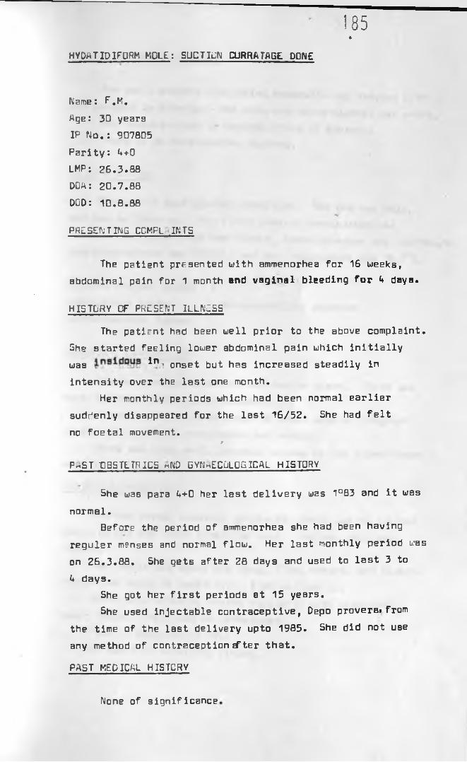
HVDATIDIFORM MOLE: SUCTILN CURRATAGE DONE
185l
Name: F.M.
Age: 30 years IP No. : 907805
Parity: A+0 LMP: 26.3.88 DOA: 20.7.88
DOD: 10.8.88
PRESENTING CCHFL IMS
The patient presented with ammenorhea for 16 weeks, abdominal pain for 1 month and vaginal bleeding for A days.
HISTORY OF PRESENT ILLNE55
The patient had been well prior to the above complaint. She started feeling lower abdominal pain which initially was in., onset t)Ut has increased steadily in
intensity over the last one month.Her monthly periods which had been normal earlier
suddenly disappeared for the last 16/52. She had felt
no foetal movement.r
ProST OBSTETRICS mIMO GYNAECOLOGICAL HISTORY
She was para A+0 her last delivery was 1D83 and it was
normal.Before the period of ammenorhea she had been having
regular menses and normal flow. Her last monthly period was
on 26.3.88. She gets after 28 days and used to last 3 to
A days.She got her first periods at 15 years.She used injectable contraceptive, Depo provera»from
the time of the last delivery upto 1985. She did not use
any method of contraception rf1 ter that.
PAST MEDICAL HISTCRY
None of significance

.186
FAMILY AND SOCIAL HISTORY
She was a married lady being housewife and staying with her husband in Kinango. She doEs not drink alcohol nor smoke.
Her husband works in medical store of Kinango.There is no contributing history.
ON EXAMINATION
She was in good general condition. She was not pale, and had no jaundice. Her other general exanination was normal. Her pulse rate was 82/min; blood pressure was 130/90mmHg respiratory rate was 20/min, and her temperature was 36.7°0.
Central Nervous System )Respiratory System ) No abnormality detectedCardiovascular System )
ABDOMIN L EXAMINATION
Abdomen was not distended and had no scars. There was some fullness in the lower abdomen.
There was pelvic mass about 16 weeks size of pregnant uterus; it was firm, non-tender and mobile.
There was also small immobile masses at the iliac fossa both left and right. -
VAGINAL EXAMINATION
She had normal external genitalia, there was no vaginal bleeding nor was there discharge seen. Vaginal wall was normal.
Her cervix was about 2cm long, firm, central and closed. Uterus was about 16 weeks size, firm and mobile.
There were adnexial masses bilateral eaeh measuring about 7x7cm. There was no tenderness.
Puch of Douglus was free No blood on examining finger.

787
DIAGNOSIS
A diagnosis of hydatidiform mole was made.
PLAN;
- Her venous blood was taken for haemogram, urea and electrolytes group and x match.
- she uias given urine bottle to pass urine in the morning for pregnancy test.
LABORATORY RESULTS
20.7.80 - Hb - 11.8g/dl- UBC - 10x109/l PCV - 3A.8¥
Na+ - 130mmol/l
K+ - A.2 mmol/1; BUN 3.8mmol/l
Her blood group was 0 Rh-D positive
21.7.88 - pregnancy test was positive.She had ultrasound done on 19.7.88 and report uias as follows
- Uterus was bulky showing infiltration by hydatidiform mole.- Bilateral ovarian cyst measuring 9.5 x 7.Acm and 8.7 x 6 .6cm
With these results the patient was prepared for suction Curratage on 28.7.88. 3 units of compertible blood was
available.Preoperative preparations were done on the night of
27.7.88 and the patipnt was premedicated 30 minutes before
theatre.After the usual preoperative procedures the patient was
anaesthetised, a drip of 5% dextrose 500mls with AO units
syntocinon was put up.The petient was put in lithotomy position and vulvo
vaginal toilet done and then drapped. Examination under anaesthsia was done.
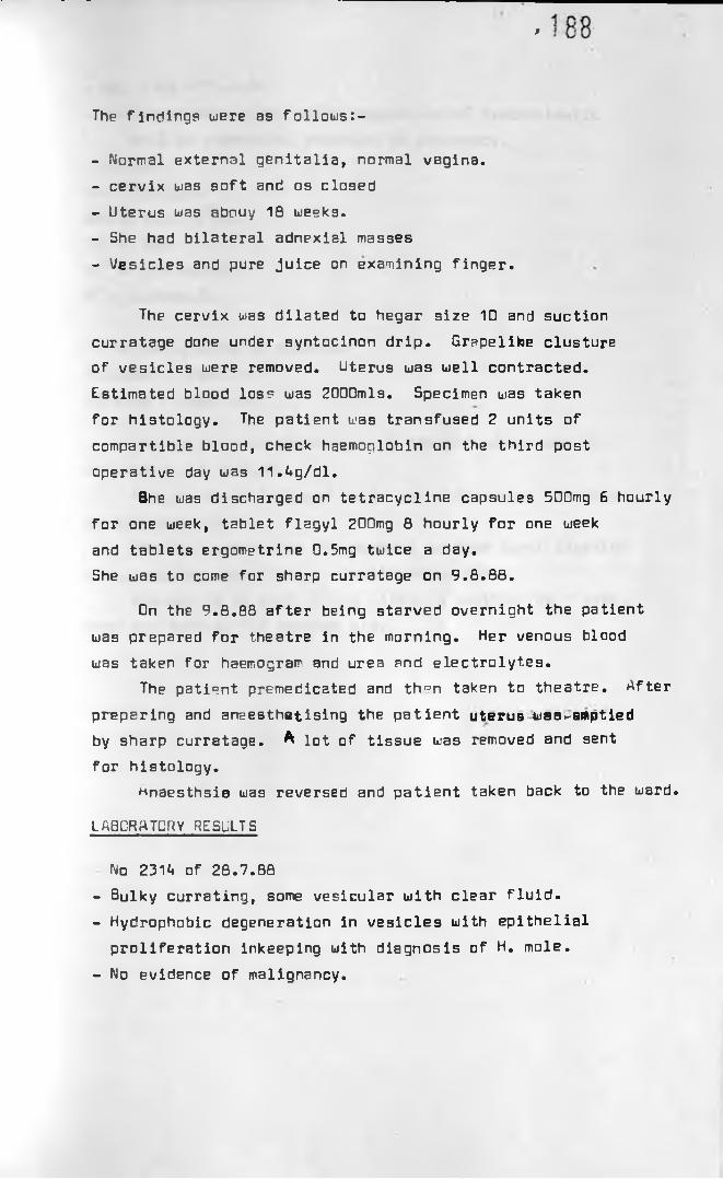
f
The findings were as follows:-
- Normal external genitalia, normal vagina.- cervix was soft and os closed- Uterus was abouy 18 weeks.- She had bilateral adnexial masses- Vesicles and pure juice on examining finger.
The cervix was dilated to hegar size 10 and suction
curratage done under syntocinon drip. Grapelihe clusture of vesicles were removed. Uterus was well contracted.Estimated blood loss was 2000mls. Specimen was taken for histology. The patient was transfused 2 units of compartible blood, check haemoglobin on the third post
operative day was 1 1.Ag/dl.Bhe was discharged on tetracycline capsules 500mg 6 hourly
for one week, tablet flagyl 20 0mg 8 hourly for one week
and tablets ergometrine 0.5mg twice a day.She was to come for sharp curratage on 9.8.88.
On the 9.8.88 after being starved overnight the patient
was prepared for theatre in the morning. Her venous blood was taken for haemogram and urea and electrolytes.
The patient premedicated and th^n taken to theatre. After
preparing and anaesthetising the patient uterus i»ie8 .-emptied by sharp curratage. A lot of tissue was removed and sent
for histology.Anaesthsia was reversed and patient taken back to the ward.
LABORATORY RESULTS
No 231A of 28.7.88- Bulky currating, some vesicular with clear fluid.- Hydrophobic degeneration in vesicles with epithelial
proliferation inkeeping with diagnosis of H. mole.
- No evidence of malignancy.
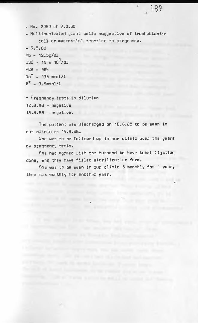
J 189
- No. 2363 of 9.0.88- Multinucleated giant cells suggestive of trophoblastic
cell or myometrial reaction to pregnancy.
- 9.8.88Hb - 12.5g/dl UBC - 15 x 109/dl PCV - 38%
Na+ - 135 mmol/1 K+ - 3.9mmol/l
■ Pregnancy tests in dilution
1 2.8 . 8 8 - negative18.8.88 - negative.
The patient was discharged on 1 0 .8 .8 8 to be seen in
our clinic on 1U.9.8 8 .She was to be followed up in our clinic over the years
by pregnancy tests.She had agreed with the husband to have tubal ligation
done, and they have filled sterilization form.She was to be seen in our clinic 3 monthly for 1 year,
then six monthly for another year.

190
COMMENT
This was a patient who had gestational trophoblastic disease. This disorder include hydatiriiform mole, invasive mole, and choriocarcinoma. The beni|n form which our patient
had could still progress to malignant choriocarcinoma.Hydatidiform mole is characterised by replacement
of normal placental architecture by grape like vesicles.These vesicles are individual villi that have undergone hydrophic changes and show microscopically detectable loss of foetal vasculature.
The highest incidence has been reported in Taiwan 1 :02
deliveries (1); in U.S.A. it is 1:2000 pregnancies (2). Incidence in Kenya is not known, but Armon (3) reports 1:1200 in Tanzania.
Common presenting symptoms include vaginal bleeding 77.A%, nausea vomitting and lower abdominal pains (A).
Socioeconomic factors tend to play some part and the disease is common in developing countries. Majority of patients are 20-
A0 years, the disease being common in first pregnancies, first matings, the very beginning and very end of reproductive life (5). Our patient was 30 years old, she was para A and we have no reason to assume that she had fresh mating. Other
findings include uterine size larger than dates 63%, it is smaller in 11% of cases where the mole is either dead or partially aborted (5). Our patient's uterine size corresponded with dates.
It may coexist with fetus, may set early onset of preeclampsia or hyperthroidism (6 ). Our patient had none of these.
Moler pregnancies at Kenyatta National Hospital are usually induced with syntocinon in an escalating fashion, followed by suction curratage, and two weeks later sharp curratage done. Cur patient had dilatation and suction curratage followed by sharp curratage 2 weeks later.The aim of sharp curratage is to remove any molar tissue remaining. 12% of these patients still go ahead and develop choriocarcinoma (A).

191
During the two year follow up patients are not allowed to
get pregnant and contraceptive advice is given. Our patient opted for surgical sterilization. 2L.6% of patients put on oral contraception will require chemotherapy as
compared to 0.8£ of patients using other methods (7). Patients are followed up by monitoring H.C.G. levels. In our patient pregnancy test was performed in dilution and in all cases was negative. Levels can be detected by pregnancy test if HCG levels exceed 750 IU/2L hours. If pregnancy tests in followup are negative le ve lsa are measured of bet«-HCG as th is is more
sensitive. Levels of L.H. can be measured until it reverts to normal then a more expensive beta MOB can be done (8 ).
If negative pregnancy test reverts to positive, or levels of HCG are rising pbove 20,000 IU/L after L weeks
or detected L weeks after evacuation then chemotherapy should be started.
Cur patient's pregnancy test remained negative during
the follow up period.Anticipated problems include haemorrhage, sepsis, uterine
perforation. Lur patient did not have any of these.

%
REFERENCES
1. Jeffcoate T.N.A.
- Principles of Gynaecology Lth Edition pg 2 2 0 , 1975.- &utterworths, London
2. Danforth D.N.
- Obs.Gyn 3rd Edition pg 72L, 1977- Harper and Rouj Publishers
3. Armon P.J., Kajembe A.H.
- Choriocarcinoma in Tanzania, Medical Presentation- E.A.M.J. 5 5: t*3 , 1978
L. Makokha E.A.
- Choriocarcinoma at Kenyatta National H0spital
- Presentation at 1 KOGS Annual Scientific Congress Nairobi 2 1 : 2 - 2 U - 1979
5. Ayengade D.
- Gestational Trophoblastic disease in Nigeria;A 10 year review.- E.A.M.J. 56: 278, 1979
6 . Ratnum S.S., Lacheran A.- Disease of Trophoblast- Clinical Obs.Gyn. 9:539, 1*82.
7. Stone, M., Bagshawe K.D.
- An analysis of influence of maternal age, gestational age, contraceptive method and mode of primary treatment of patient with hydatidiform mole on the incidence of subsequent chemotherapy- Br. Jn. Obs.Gyn 8 6 : 782, 197L.
8 . Kigondu S.C.B., Fongoh F.B., Makokha ^.E., Mati J.K.G. Bensal B.
- The use of L.H. and B-HCG radioimmunoassay in diagnosis and treatment of choriocarcinoma at K.N.H.
- Journal of Obs.Gyn. East and Central Africa 1 :1L8 , 1982.

PELVIC ABSCESS
Name: Y.M.
Age: 23 years IP No.: 913176
Parity: 3+0 LNMP: 10.8.88 DOA: 17.8.88
DQD: 29.8.88
PRESENTING COMPLAINTS
The patient presented with history of sudden onset of lower abdominal pain, and foul smelling vaginal discharge.
HI5TCRY OF PRESENT ILLNE5S
The patient was well until 7 days prior to admission when she started having her monthly period and pains which
increased in intensity especially after the monthly period. It was accompanied by foul smelling yellowish discharge per veginum.
PA5T MEDICAL HISTORY
3he had seme inguinal abscess which was drained
here in npril 1988.
PAST OBSTETRICS AND GVIMAECLlLG IChL HISTORY
She was para 3+0, her last delivery was in 198L. Her last monthly period was 10.8.88. She was getting her menses for 2 days and used to come every 30 days. She had been having moderate abdominal pain with every monthly period.
She had her menarche at 15 years and she used oral contraceptive from September 1986 to May 1988.
FAMILY AND SOCIAL HISTORY
She married but got divorced in 1986 and works as a
housemaid in Nairobi City. She stays in Nairobi's Buru Buru Estate.
She doesn't drink alcohol nor smokes. No history of
chronic illness in the family.

p
ON EXAMINATION
194
General condition was fair. She was not pale and had
no jaundice. Other general examinations were normal.Bp 110/BOmmHg, PR BL/min normal, temperature 37.B°C
RR 20/minute normal.
Central Nervous system )RpSpiratcry system ) Nothing of significance
Cardiovascular system )
ABDCMEN
Full abdomen, mild tenderness, subumbilical especially at the iliac fossa.. No obvious masses.
VAGINAL EXAMINATION
Normal external genitalia. She was not blpeding but there was obvious discharge coming out of vagina, foul smelling.
Uterus was normal size and anteverted. Adnexiae had mild tenHerness but no obvious masses. Pouch of ^ouglas had obvious fulness; rather firm, culdocentesis attempted
but nothing came out.
DIAGNOSIS
- Acute pelvic Inflamatory disease- R/0 pus collection
PLAN:
The patient was planned for high vaginal swab.- was put on capsules ampicillin 500mg G hourly- Tablets flagyl AOOmg B hourly
- Tablets panadols 2 8 hourly.- Booked for u/s to rule out pus collection.- blood for haemogram was taken also U/E- U hourly observation.
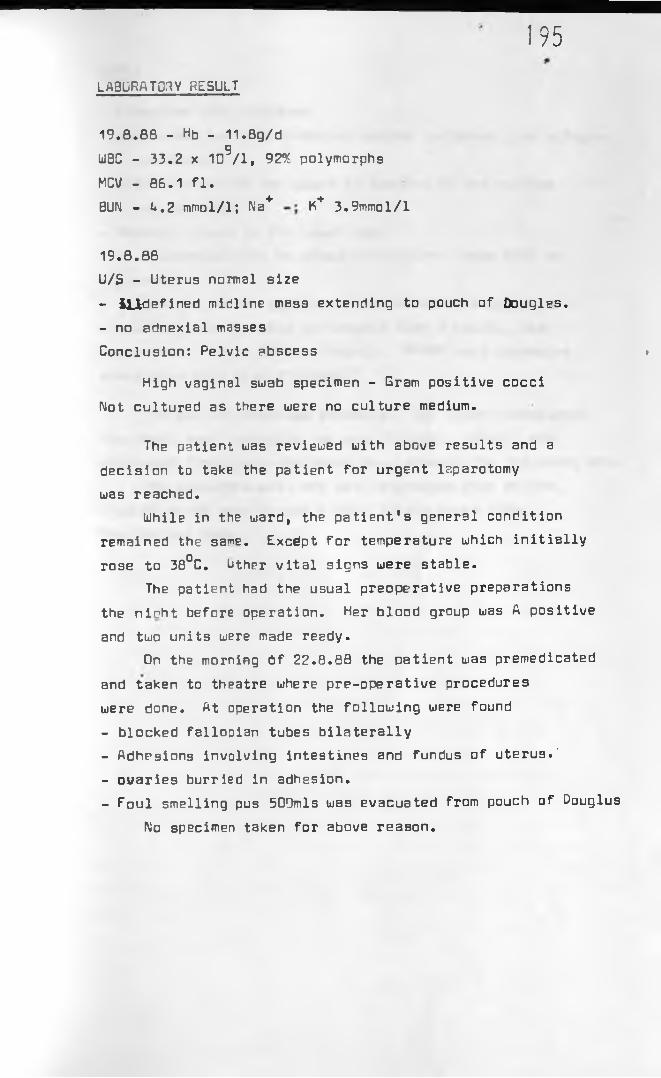
LABORATORY RESULT
195
19.8.88 - Hb - 11.Bg/d
UBC - 33.2 x 10⁹/1, 92% polymorphs
MCV - 86.1 fl.BUN - U . 2 mmol/1; Na+ K+ 3.9mmol/l
19.8.88U/S - Uterus normal size- ilidefined midline mass extending to pouch of Douglas.
- no adnexial massesConclusion: Pelvic abscess •
High vaginal swab specimen - Gram positive cocci Not cultured as there were no culture medium.
The patient was reviewed with above results and a decision to take the patient For urgent laparotomy
was reached.While in the ward, the patient's general condition
remained the sane. Excdpt for temperature which initially
rose to 38°C. Uther vital signs were stable.The patient had the usual preoperative preparations
the nicht before operation. Her blood group was A positive
and two units were made ready.On the morning 6 f 22.8.88 the patient was premedicated
and taken tD theatre where pre-operative procedures were done. At operation the following were found
- blocked fallopian tubes bilaterally- Adhesions involving intestines and fundus of uterus.'- ovaries burried in adhesion.- Foul smelling pus 500mls was evacuated from pouch of Douglus
No specimen taken for above reason.

J 5 6DC-NE :
- Adhesions were released- pus sucked out and abdominal cavity irrigated with rifocin
solution.- drain was put at the pouch of Douglas to the surface
at the side.- Abdomen closed in the usual way.
Anaesthesia was reversed and patient taken back to
the ward.She was put on crystapen intravenous 2 mega units
6 hourly; intramuscular gentamycin BOmg 8 hourly, and intravenous flagyl 500mg 8 hourly. Other post operative
procedures were also observed.
The patient improved steadily. Her check haemoglobin
the thrid post operative day was 10.1g/dl. Drain was shortened first post operative day, removed the following day.
She recovered well and was discharged home on the29.8.88 to be seen in our clinic in six weeks time,
i he did not come.
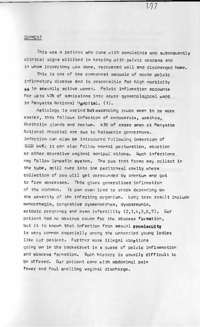
1- 97*
COMMENT
This was a patient who came with complaints and subsequently clinical signs elicited in keeping with pelvic abscess and in whom laparotomy was done, recovered well and discharged home.
This is one of the commonest sequale of acute pelvic inflematory disease and is responsible for high morbidity as in sexually active women. Pelvic inflamation accounts
for upto LCft of admissions into acute gynaecological ward in Kenyatta National Hospital. (1).
Aetiology is varied but ascending route seem, to be more easier, this follows infection of endocervix, urethra,
Bartholin glands and rectum. of cases seen at KenyattaPJational Hospital are due to Naisseria gonorrhoea.Infection can also be introduced following insertion ofIUCD UU%] it can also follow normal perturation, abortion
or other operative vaginal manipul ations. Such infectionsmay follow lympatic system. The pus that forms may collect inthe tube, spill over into the peritoneal cavity wherecollection of pus will get sorrounded by omentum and gutto form abscesses. This gives generalised inflamationof the abdomen. It can even lead to shock depending onthe severity of the infecting organism. Long term result includemenorrhagia, congestive dysmenorrhea, dyspareunia,ectopic pregnancy and even infertility (2,3,L,5,6,7). Curpatient had no obvious cause for the abscess formation,but it is known that infection from sexual promiactlityis very common especially among the unmarried young ladieslike our patient. Further more illegal abortionsgoing on in the backstreet is a cuase of pelvic inflammationand abscess formation. Such history is usually difficult tobe offered. Cur patient came with abdominal painfever and foul smelling vaginal discharge.

Other organisms responsible for pelvic infections include t - . coli, streptococcus, staphyllococcus and
actinomycetes (7,8,9). lie did not do culture though it had been intended due to lack of culture media, gram stain however showed gram positive cocci.
Once diagnosis of pelvic abscess has been made
drainage must be done as any delay will lead to generalised peritoniis and possible shock. Supportive therapy is
important; antibiotics analgesics/antipyretic, intravenous fluid and if indicated intravenous alimentation, “'urgical procedure is governed by the parity of the patient and hpr desire to have more children (l). Our patient was a divorcee but had 3 children living. It was not easy
to rule out possibility of her being married again and the likelyhood of wanting another baby. The tubes wereconserved for a possible later operation should such a
%need arise.
Pelvic inflammatory disease with its sequale is a grave problem that calls for aggressive management and early diagnosis; but perhaps the most important is health education on sexual behaviour.
REFERENCES
1. Khehar, B.S.
- Pelvic Inflammatory disease, a review of current practice.- E.h .K.J. 57: 72, 1980.
2. Carty M.J. , Nzicki, J.K., Verhagen, A.R.
- The role of Naisseria gonorrhoea in Acute Inflammatory Disease in Nairobi.
- E.M.M.J. i*9: 376, 1972.3. Broun, I.
- The management of Pelvic Abscess- Progress in Obs.Gyn. 2 (1): 27A, 1982.
L. Golrje, S.H., Israel, R.; Ledger, tii.G.
- Unilateral tubovarian abscess: a distinct entity- Amer; Corn. Cbs.Gyn. 127: 807, 1979
5. Bartlett, J.G.
- Anaerobic infection of the pelvis- Clinical Obs.Gyn. 22: 351, 1979
6 . F o m u l u , J . N .
- Bacteriological Sensitivity pattern in septic abortion Post abortal sepsis and pelvic abscesses at Kenyatta
National Hospital- M.Ked Thesis 1981, University of Nairobi.
7. Burkman, J.R.- The women Health study: Association between intrauterine
contraceptive devices and pelvic inflammatory disease.- Obs.Gyn. 57: 269, 1981.
8 . Soderberg, G.; Lindgren, S.,- Influence of IUCD on the course of salphingitis- Contraception 2A : 127, 1981.
9. Rubestein, P.R., Mishell D.R., Ledger, W.J.- Cclpotomy drainage of Pelvic Abscesses.
- Obs.Gyn. UB: 1L2, 1976

20Q
RIGHT B^RTHLiL IN 15 ABSCESS, Km r 3UPImL IZATILN DONE
Name: M.N.
Age: 18 years Parity: 2+0 IP No. 913359 LNMP: 9.8.88 DBA: 2C.8 .8e ODD: 21.8.8B
PRESENTING C f-PLAINTS
The patient presented with painful vulval swelling on the
rioht side ror 2 days.
HI5Tl -vY OF PRESENT ILLN-aS
The patient was well until 2 days prior to admission uhen she started having vulval swelling which was progressive
in size nd pain during the two days. She gives no such complaints
before. There was no history of trauma end no history of recent discharge per vaginum in recent past.
Ph ST l--EDIC'L HISTORY
This was not contributory
GBSTE Tr, ZG5 PiS C YN^ECLLEj ICAL HIGTCF.Y
She was para 2+0 . The last delivery was in 1986 and was uneventful. Her last normal period was on 9.8.88, it usuelly come every 28 days and lasts 5-6 days. There js no accompanying pain and no intermenstrual discharge.
Her menarche was at 15 years, she was not using any
contraception.

FAMILY AND SCilL-L HI5TLKY
ahe was not married and stays with her sister in Eastleigh. 5he sells second hand clothes, ^he doesn't smoke nor -rink alcohol.
Th^re was no family history that was contributory.
ON EXAUN.ATICN
She was in good general condition, she was not pale, and was not jaundiced. Her body temperature was 37°C.Her pulse rate was 80/minute, Blood pressure was 110/70mmHg She had a respiratory rate of 20/min regular.
RESPIRATlRY system )Cardiovascular system ) Sere essentially normal Abdominal Examination )
vagi n a l examination
She had a normal external genitalia e*cept for the right
vulva which was swollen. Cn inspection there was a foul smelling bloody discharge from the sinus from the swelling.
Cther vaginal examinations were normal; and pelvic findings
were normal.
BIAGND5I5
Burst right sided Bartholin's abscess.
MANAGEMENT
^he patient was informed of her condition and consent for operation and administration of anaesthesia was got. The patient was prepared for marsupialization. Venous blood was taken for full haemogram and intravenous line was started.The patient was taken to theatre and marsupialization was done
as follows:-
Patient was put 4n operation table, anaesthesia was
administered and patient was positioned in lithotomy.Vulval and vaginal toilet was done. Examination confirmed the earlier findings. A longitudinal incision was made at the macocutaneous junction just outside the hymenal ring;
this exactly passed through the opening of the rupture.

202
The thick bloody foul smelling discharge was evacuated from
a big cavity of capacity about 120mls. finger was introduced to break up the loculi. Currate was then used to remove the necrotic tissue. The cavity was cleaned with antiseptic
solution and the cut edges were everted and stitched to the normal vulval skin using ch'-nmic catgut number 2/0
Haemaostasis was achieved and a sufratulle gauze was used to pack the cavity, and was to be removed in 2U hours.
Hnaesthesia was reversed and patient taken to the ward.She was prescribed oral ampicillin 500mg 6 hourly
salt bath to the perineum three times a day. She had normal
post operative period. Unfortunately her haemogram request came back with the comment that the specimen was clotted.
The patient was seen at our gynaecology clinic and had
fully recovered.

COMMENT
This was a patient who had Bartholin abscess for which marsupialization was done.
Bartholin glands are two in number and are situated
on each side of the introitus inferiolateral to the
bulbocarvenosus muscle at 5 and 7 O'clock; they secrete alkaline solution during excitement phase of coitus and normally not palpable (1,5). admission due to Bartholin
abscess forms 1.7% of all admiesions at Kenyetta National Hospital acute gynaecology, 55% are associated with
pregnancy (1).Bartholin's abscess can occur anytime from menarche
to menopause and parity doesn't seem to have any influence (1,3).Genesis of Bartholin's abscess is infective, th? gland
being infected as such or infection of pre existing cyst (O. This is followed by obstruction; obstruction with secondary infection can also result from trauma of parturition or episiotomy (3). Cur patient was 18 years old para 2+0 with last delivery being in 1-8 6 . It was not obvious what could have been the cause of the lesion; perhaps trauma from previous birth or perhaps just primary infection.
A wide range of organisms have been isolated from the lesion of which Neisseria gonorrhea is the commonest (3,5,6). Organisms mostly isolated include staphylococcus, E.coli and streptococcus. Others include fucultative rods, proteus mirabilis and other gram negative rods; in 3 .5-1 1.8% no organism is isolated (5). Cur patient had already bad a burst abscess and contaminants from vaginal flora
would have made usefulness of culture futile.Presentation is painful swelling of the vulva associated
with difficulty in walking or sitiing (1,0. Other abscesses may rupture spontaneously, may regress or progress (3,5,6).
Mumia (1981) found that 82.7% of the patients were aged between 12-29 years while 53.3% were married. Our patient
was single and fall in the age bracket above.

The present techniaue was originally described by Jackobs (1950) and has since undergone modification; the aim of which
is to re-establish the petency of the duct (7), the technique marsupirlization is prefered to excision of the swelling
as the latter might lead to dry perineum, recurrence, haemorrhage, haematoma formation and scar formation with dyspareunia(7).
r-»
Excision, however, is done as an elective procedure in
recurrent abscess after an initial marsupialization has been done. Our patient was managed by marsupialization. With this procedure potency is maintained as there is construction of a new mucocutaneous junction between the wall of the cyst and skin of the libia (2,7). 3% of patients so treated haverecurrence (1). Our patient had had no recurrence so far.
This method is simple as patient can have post-opBrative care at home (7). Bur patient was put on antibiotics which has been shown to hasten recovery of inflamation (7) and cavity packed with surfratulle to prevent adhesion (1).

REFERENCES
1. Mumia J.A.Bartholin's Abscess at K,N.H.
M.Med Theis 1980 University of Nairobi
2. Pritcharo, J.A., McDonald P.C., Grant N.Bartholin's Glands AbscessWilliams Obstetrics 17th Edition 1985 oq A91Appleton Century Craft/Norwalk Connecticut
3. Deuihurst J.Bartholin's Cyst and Abscess Integrated Gbs.Gyn. for Postgraduates 3rd Edition 19B1 pg 663-607 Blackwell Scientific publications, Lnndon
A. Jeffcoate N.Tumors of Bartholin Glands pg 307, 379 Principle of Gynaecology Ath Edition 1975.
Butterworths London
5. Olipherit M., Gail V.A.Management of Bartholin Duct Cyst and abscess. Obstetrics and Gynaecology 16: A76-A78.
6 . Lee Y., Rankia J., Alpert S., Daly A.K.- Microbiological Investigation of Bartholin Gland Absce
and Cyst.- American Jpurnal of Cbs/Gyn 129: 150-153, 1979
7. Jackobson P.- Marsupialization of Vulvovaginal Bartholin Cyst.
- American Journal of Obs/Gyn. 79: 73, 1960.

206PRIMARY INFERTILITY, BILATEHAL SAL PH IN3Q5TLMY DONE
Name: K.U.
A§e: 28 years Parity: O+O IP No. 8391422 LNMP 13.2.89 DOA: 9.2.89 ODD: 7.3.89
PRESENTING CLMPLAINTS
The patient first presented in our gynaecological clinic with complaints of inability to conceive for 6 years of marriage.HISTORY OF PRESENT ILLNESS
The patient was married in 1981 and has since lived happily with the husband without any marital problems.They have, however, failed to achieve pregnancy despite
succesful coital behaviour.
Pm ST MEDICAL HISTORY
The patient has not had any major illness and denies any history of vaginal discharge or any related history.
The husband also had had no relevant medical problem like any painful testis in youth or any urethral discharge
in adulthood.
PRESENT GYNAECOLOGICAL HISTORY
She was para 0+0 , her last monthly period was 13.2.89
it used to come every 28-29 days and used to last 5 days.There was no accompanying pain and no history of intramenstrual
discharge. She had her menarche at years and had not
used any contraceptive.

family and social h i st o r y
3he was a married housewife staying with her husband
at Kiambu. Her husband is a farmer. No relevant history in the family.
ON EXAMINATION
She was in good general condition. She had no palor nor did she have jaundice. Her other general examinations were normal.
Her pulse rate was 80/minute, blood pressure of 1l0/90mmHg, respiratory rate of 20/minute regular and body temperature of 36.9°C. She had female configuration with female hair distribution
Central Nervous system )Cardiovascular system ) Were esrentially normal
Respiratory system )
Her breasts were well developed with well developed nipple.They were not active.
ABDOMINAL EXAMINATION
The abdomen was scophoid and was symmetrical in shape.
There was laparoscopy scar just below the umbilicus. Otherwise there were no masses and no tenderness anywhere.
UAGINAL EXAMINATION
She had a normal external genitalia, there was no
discharge or bleeding per vaginum.Speculum exam done earlier showed a healthy cervix
and pap smear was taken. Vaginal wall looked normal.Digital examination revealed normal vaginal wall,
cervix that was long, firm, closed and central.Uterus was normal size and anteverted. Adnexiae and pouch
of Douglas were normal. There was no discharge on examining finger.

2C8DIAGNOS IS
A diagnosis of primary infertility was made.
HAI\iAG:K£MT
Investigations done while she was in the gynaecology clinic
include the following:-
Pap smear - class II (Trichomomasis)Seminalysis - Normal findingsHSG - Normal uterine size and configuration. °oth tubes
demonstrated with bilateral hydrosalphinx, no free
peritoneal spill.Dye laparoscopy: adhesions with burrying of the fimbrial end. No free spill of dye but distension of distal portion
of tubes noted.
She was admitted in the ward as stated above for tubal
operation and preoperative investigations were:
haemogram - Hb 1A.3g/dl
- pcv -- Adequate platelets HIV screen - negative.
BUN - 3.9mmol/lK+ - Ammol/1
Na+ - 13A mmol/1 -\
The patient was planned for and operated on the 27.2.89.
The usual preoperative procedures were done and at operation
the following were found.Uterus was normal in size and was anteverted. The tubes
were hurried in adhesions and h°ld posteriorly in the pouch
of Douclus. Ovaries were equally burried.
DONE:
Release of adhesion was affected using cutting diathermy.
Tubes were freed and ovaries were visible*. The tubes were then opened using the cutting diathermy blade; fine adhesions holding the fimbria were released. The fimbria were everted
using nylon number 6/0 and stitched to the serosa.
During the procedure operation site of tube was irrigated
with Hartman solution continuously.
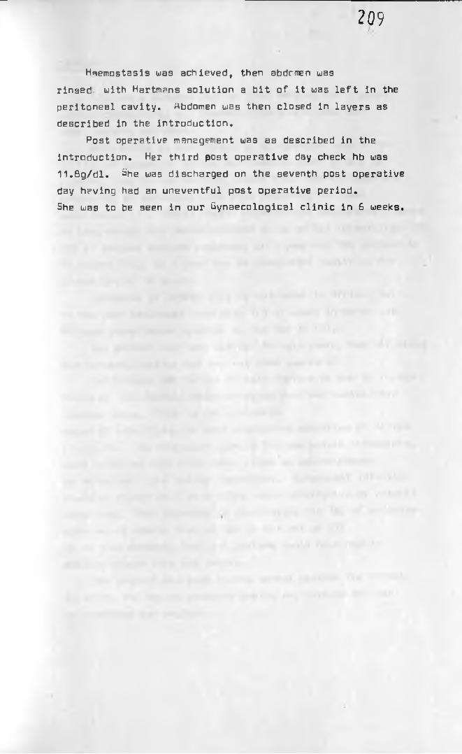
Haemostasis was achieved, then abdomen was
rinsed with Hartmans solution a bit of it was left in the
peritoneal cavity. Abdomen was then closed in layers as described in the introduction.
Post operative management was as described in the introduction. Her third post operative day check hb was
11.8g/dl. She was discharged on the seventh post operative day having had an uneventful post operative period.She was to be seen in our Gynaecological clinic in 6 weeks.
209

210
COP,KENT
This was a patient who had been married for 6 years without conceiving, all investigations were done and
tubal surgery was done.Infertility is the inability to conceive despite the
ability of succesful coitus and cohabitation; it may be primary, like in our patient, where there had not been any pregnancy, or secondary like when pregnancy had been experienced (1). World Health Organisation recommends 2 years as long enough and investigations to go on for infertility (2). 80% of couples achieve pregnancy in 1 year and 90% achieve in 18 months (1). So 1 year may be considered justified for
investigation to go on.Incidence of infertility is hot known in Africa, but
it has been estimated that upto 1/3 of women in major East
African towns become sterile by the age 30 (3).Our patient had been married for six years, had all along
had succesful coitus but had not been succesful.Infertility can be due to male factors or due to female
factors. Our investigation revealed that our patient had
blocked tubes. This is the commonestcause of infertility in most developing countries of Africa (1,3,5,6). The condition usually follows pelvic infections, such infection must have taken place as was evidenced by adhesions found during laparotomy. Gonococcal infection could be silent only to manifest with infertility or ectonic pregnancy. This organism is responsible for 75% of patients with pelvic absees that we see in our set up (7).It is thus possible that our patient could have had it
earlier though this was denied.Our patient had been having normal periods for normal
duration, she had no problems during her periods and had
no discharge per vaginum.
211
She had some coarse adhesion and flimsy ones. Release
of the adhesion was not difficult. Our patient could have
some hope of getting pregnant. Meti et al and Uaghmarae et al (5,8) found most patients to be unfit for tubal surgery
ax laparoscopy. They showed bilateral tubal occlusion of 62% to 73.1% with only 12-30% of affected patients suitable for surgery
Technique used here were as set up by Rosenberg and Board (9). These include careful handling of tissue with fine atramatic instrument, perfect haemostasis, use of fine non-reactive suture and continued irrigation of operation site with a balanced salt solution. These are aimed at preventing adhesion formation post operatively. Responsible for adhesion formation include haemorrhage, infection, denuded surface, ischaemia, foreign body reaction and^reum^. ^rophyllactic
antibiotics is particularly important in these patients as it ensures reduced chances of adhesion. Other measures to reduce adhesion include leaving some solutions into the peritoneal cavity as was left In our patient; others are
systemic steroids to lessen body reaction. Rosenberg and Board (9) support the use especially of dextran - 70 as it excerts osmotic pressure, creates ascitis and floats the tissues thereby lessening chances of contact and adhesion.
Our patient is still being followed up. Salphingostomy has been claimed to have succes rate of~10-15% and an overall success rate of 25% for tubal surgeries (10). An unwanted effect of tubal operation is ectopic pregnancy. Mattingly
quotes a tubal pregnancy rate of 8% (11).

212
REFERENCES
1. Nasah B.T.
- Infertility. Reproduction in Africa- Mati J.K.G., Ladipo R.A. (editors) /- The JEPIEGO pg 105-130, 190A.
2. LJ.H.O. Technical report series 582, 1975, Geneva.3. Chatfield tu.H., Suter P.*-.N.
- Investigation and management of infertility in East Africa- A prospective study of 200 cases.- E.A.M.J. U 2 : 212, 1970
A. Bennet F.J.
- The social determinants of Gonorrhea in E.African Town- E.A.M.J. 35: 332, 19G2.
5. Mati, J.KG., Hnderson G.- Second look of problems of primary infertility in Kenya.- E.A.M.J. 50: 9A, 1978
6. Gebbie, D.^.M.- Obs.Gyn. in health and disease in Kenya pg A9L, 197A.
East nfrican Literature Bureau, Nairobi.7. Carty M.J., Nzioki J.M., Verhagen A.R.
- The role of gonococcus acute inflamatory disease in
Nairobi.- E.A.M.J. L9: 376, 1972.
8. kiaghmarw 0., Ansari, A.- Mnalysis of a series of laparoscopy proceedings performed in NgirobiE.A.M.J. 53: 1A9, 1976.

213
9. Rosenberg 3.M., °oard J.A.- « high molecular weight dextran in human infertility
surgery.- Am. Jn. Obs. Gyn. 1L8: 380, 198A. 10 11
10. Jeffcoate N.- Sterility and subfertility- Principles of Gynaecology 9th Edition 1975, pg 583y607
- Butterworths end Company Ltd.- London-aoston
11. Mattingly R.F.- Surgical Conditions of the fallopian tube- Te Linde's operative Gynaecology 5th Edition, 1977
pg 325-326.
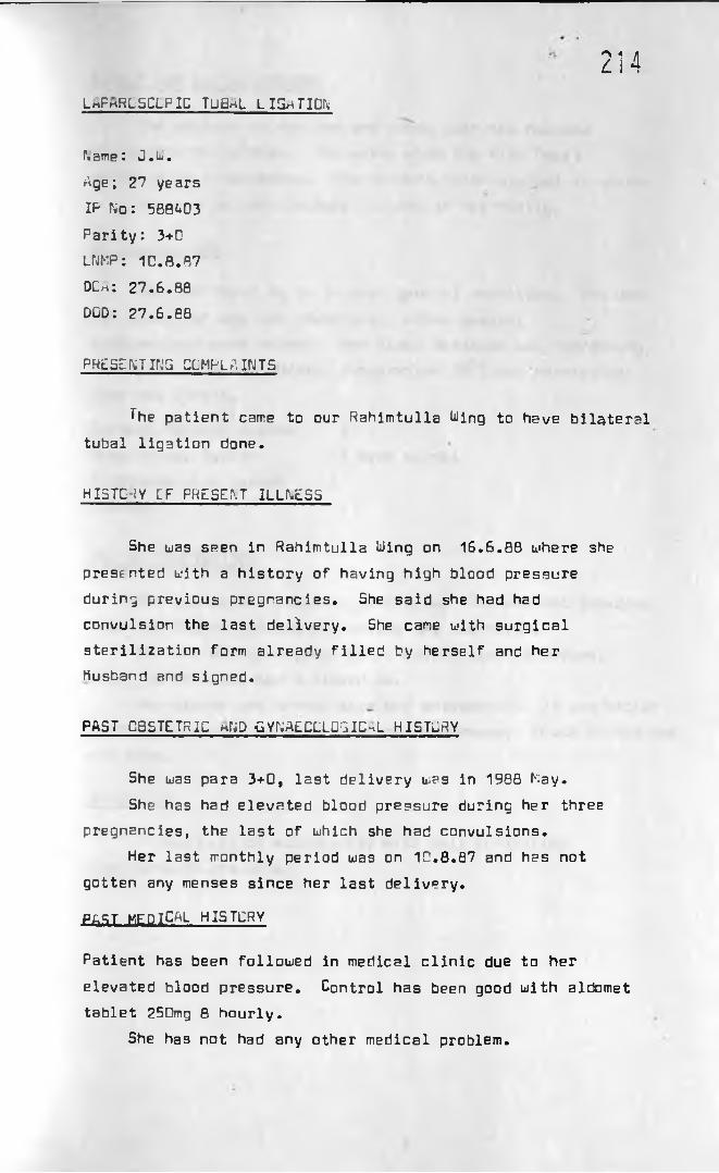
214LhFhRLSCLFIC TLIBhL LIS^TION
r.anp: J .L .
Age; 27 years
IF No: 5B8L03 Parity: 3+0 LNMP: 1C.B.87
DOn: 27.6.88 DGD: 27.6.88
PRESENT IKS COMPLAINTS
The patient came to our Rahimtulla Wing to have bilateral tubal ligation done.
HISTC-iY CF PRESENT ILLNESS
She uas seen in Rahimtulla Wing on 16.6.88 where she presented with a history of having high blood pressure during previous pregrancies. She said she had had convulsion the last delivery. She came with surgical sterilization form already filled by herself and her husband and signed.
PAST OBSTETRIC hND -GYNAECOLOGICAL HI5TLRY
She was para 3+0, last delivery was in 1988 Fay.She has had elevated blood pressure during her three
pregnancies, the last of which she had convulsions.Her last monthly period was on 10.8.87 and has not
gotten any menses since her last delivery.
PAST MEDICAL HISTORY
Patient has been followed in medical clinic due to her
elevated blood pressure. Control has been good with aldomet tablet 25Dmg 8 hourly.
She has not had any other medical problem.
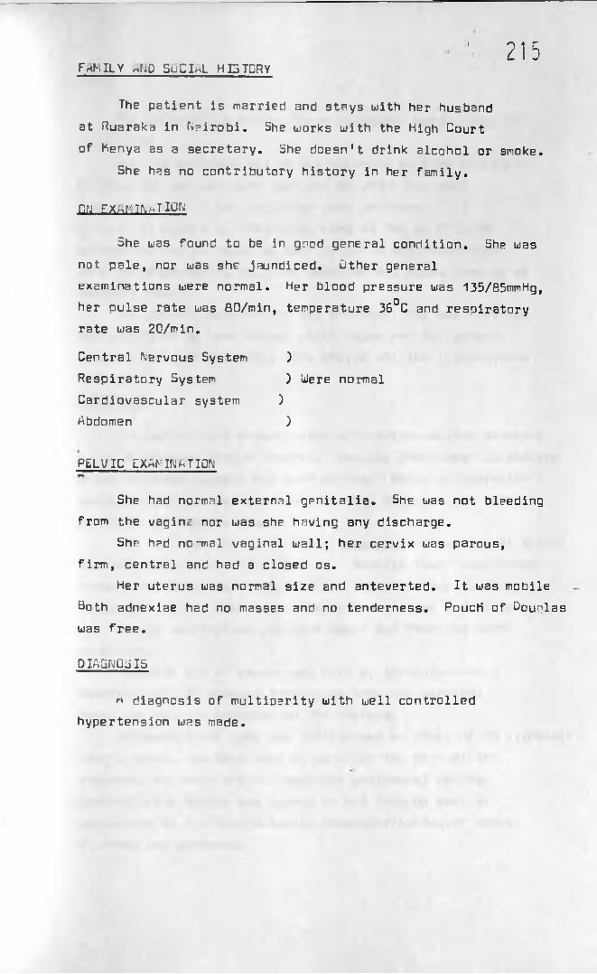
Far-.ILY r,f;D SuCI^L H ETD R Y
i 215
The patient is married and stays with her husband at Ruaraka in Nairobi. 5he works with the High Court
of Kenya as a secretary. She doesn't drink alcohol or smoke.
She has no contributory history in her family.
cm e x m KINhTIGn
She was found to be in good general condition. She was
not pale, nor was she jaundiced. Other general examinations were normal. Her blood pressure was 135/85mmHg, her pulse rate was 80/min, temperature 36°C and respiratory rate was 2G/min.
Central Nervous System
Respiratory System Cardiovascular system Abdomen
)) Were normal
))
PELVIC EXAMINATION
She had normal external genitalia. She was not bleeding from the vagina nor was she having any discharge.
She had no-mal vaginal wall; her cervix was parous, firm, central and had a closed os.
Her uterus was normal size and anteverted. It was mobile Both adnexiae had no masses and no tenderness. Pouch of Douplas was free.
DIAGNOSIS
A diagnosis of multiparity with well controlled hypertension was made.
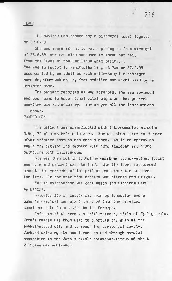
PLAN:
• 216
the patient was booked for a bilateral tubal ligation on 27.6.88
She was supposed not to eat anything as from midnight of 26.6.88; she was also supposed to shave her hair from the level of the umbilicus upto perineum.
She was to report to Rahimtulia Uing at 7am on 27.6.88 accompanied by an adult as such patients get dischargedsame day a fte r waking up, from sedation and might need to be
\
assisted home.The patient reported as was arranged, she was reviewed
and was found to have normal vital signs and her general conriit.on was satisfactory. She obeyed all the instructions
above.PkLCc DLHL;
The patient was premedicated with intramuscular atropine
0.6mg 30 minutes before theatre. She was then taken to theatre after informed consent had been signed. While on operation table the patient was sedated with 10mg diazepam and iCOmg pethidine bcth intravenous.
She W3s then out in lithotomy position vulvo-vaginal toilet was done and patient catheterised. Sterile towel was pieced beneath the buttocks of the patient and other two to cover the legs. At the same time abdomen was cleaned and drapped.
Pelvic examination was done again and findings were as before.
interior lin of cervix was held by tenaculum and a Cohen's cervical cannula introduced into the cervical canal and held in position by the forceps.
Infraumbilical areE was infiltrated by I5mls of 2% lignocain. Vera's needle was then used to puncture the skin at the anaesthetised site and to reach thE peritoneal cavity. Carbondioxide sypply was turned on and through special connection to the Vera's needle pneumoperitoneum of about
2 litres was achieved.

217The needle is now withdrawn and small incisicn made at the same site through the skin only, h trocar and canula
then introduced at C5° pointing to the pelvis; with both sides of abdominal wall lifted gentle force is used until there is sudden feeling of giving way. The trocar is now
withdrawn and bubble of gas trying to escape fron peritoneal cavity will bp seen at the end of the cannula. The cannula
can now be pushed further inside the peritoneum cavity.1 he scope having been loaded with double Falop rings is now
pushed inside the cannular and clear vision of abdominal cavity obtained by moving the scope up and down.
With the assistant moving the Cohen's cannula upwards and downward, to the right and to the left clear vision
of the tubes was attained. Each tubp was held by hook pushed from the scope; as the tube was drawn inside the scope a ring was released. The same was repeated for the other side by ^justing the lock of the scope.
The scope was withdrawn and gas was turned to let off the pneumoperitoneum; trocar was now pushed inside the cannula and both were withdrawn and the incision closed by absorbable vicryl suture.
The wound was dressed and patient taken back to the ward, where she was alert after three hours. She was then discharged on tablets paracetamol 2 8 hourly for 5 days and ampicillin capsules 5C0mg 6 hourly.
She was reviewed at the 7th post operative day and was found to have healed well. She was then discharged from Rahimtulla toing.
*
COMMENT
This was a patient who had had three deliveries and had decided with her rpouse to have a surgical sterilisation as the lady had been having problems of high blood pressure
during pregnancy, and also because they thought the number of children they had was enough .
Sterilization is used to destroy the precreative function of the patient. This is a permanent method whereby the tubes are permanently closed and no fertilization can take place.
Frevelance of surgical sterilization isnotsasy to auote, but Kakokha (1980 reported 329 operations in a year at Kenyatta National Hospital (1). It forms 3rd commonly used contraceptive method after the pills and intrauterine contraceptive device according to Kenya contraceptive survey procedure of 1980
Sterilization was used for patients who could not be allowed to carry pregnancy due to medical conditions and also those who satisfied rule of 120 Cage X parity = 120)(2,3). Sterilisation is now widely used for a normal cases and with the advent of laparoscope the procedure has become easier simpler -nd auicker. Other methods of tubal sterilization include Pomeroy, Irving, Madlener, and uchida. Pomeroy method is used mostly in our hospital, it has the advantage of ease, speed and less trauma to tissue”, it has low complication and low failure rate (^,5). We used laparoscopic methcd in our patient, although she had medical problem, the couple had also decided on the family size they needed. Women now come for sterilization for various reasons comron-rst is completed families, but some come for socioeconomic reasons especially single ladies (U,5).
We used Falop ring to occlude the tubal lumen. We do it under valium and pethidine and patients are usually ready to go home same day. Through’llparoscdpe it can be
accomplished by electrocoagulation, or Hulka silastic clips (6).

219
Gynaecological complication should be carefully ruled
out as ignoring or oversight might lead to failure of method or unnecessary two operations. About 16-20? of patients develop menstrual disorder which is postulated to be due to improved uterotubal circulation ((*,5,6). Psychiatric disturbance,
disturbed sex, are some of the listed complications.Hydrosalphinx occur in 5r'. Failure of operation occur in
0.71? (2,(*,6). Failure rate with falop ring is 3.3? with electocoaguletion 1.9r', with rocket clip 5.9? (S).
Failures could be due to ring insertion (7). luteal phase pregnancy 23r of total incidence of 2.3/1000 (9). Other complications include unintended laparatomy
arising from perforated uterus (10,11).Ectopic pregnancies with uncorrected incidence of 0.6V' (12).
Cur patient did well and though we don't continue seeingl
her it is assumed she has not gotten pregnant.

REFERENCES 220; i
1. Nakokha «.E.
- Jurgical Contraceptive in henya, An overview;
Proceedings of first Notional Conference on Reproductive health pg 63, 198A.
2. S^p^rd, K.K.
- Female Contraceptives- Lbs.Gyn. 29: 73q, 1q7A.
3. Rabol, M., Stocklund, K.- Steriliz tion of women through minilaps- Dennish Medical Bulletin 25: 177, 1978
A. Dewhurst, J.Sterilization
In, Intergrated Obs.Gyn for Postgraduates 3rd Edition, 1981.
Blackwell Scientif ic'’Publication. Oxford. London Edinburg, Boston, Melbourne
5. Jeffcaate, N.- Sterilization procedure, Contraception
- In principles o Gynaecology Ath Edition- Butterworths, London, 1975
6. Mattingly, K.F.Fertility ControlTe Linde's operative Gynaecology 5th Edition pg 336 1977J.B. Lippicot Compariy, Philadelphia.
7. Chi, I., Mumford B.D., ^-anfe, L.E.
- Technical failure in tubal ring sterilization Incidence, perceived reasons outcome and risk factors.- H rr. J. Gbs. Gyn. 138: 3D7, 1980.
8. Hulka 3.F., Omran J.K.
- Lrp?roscopic Sterilization with spring loaded clips Instrumentation development and current clinical experience- Am. Journ. Dbs.Gyn. 135: 1016, 1979

221
9. Chi, I., Potts, M.; Wilkins, L.- Rare events associated with tubal sterilization
nn International experience.- Family Health International, Research Triangle Perk
Worth Carolina.10. Chi, I., Feltiblum P.,
- Uterine Perforation during sterilization laparaacopy
and minilap.- Rm. J. Dbs. Gyn. 139: 735, 1981. 11
11. Chi, I; Feldblum P.- Laparoscopic Sterilization requiring laparatomy
- «meric n Journ. Obs.Gyn. 1L2: 712, 1982.

TRANSLOCATED IUCD: REKC-V L «T L/iPh RATOMY222
Name: J.iii.
Age: 27 years Parity: 3+0
IP Mo. 918533 LNMP: lr86 DOA: 1A.9.88
DOO: 5.10.88
PRESENTING CLKFLAINTS
The patient was admitted through casualty with complaints of not feeling the threads of her intrauterine contraceptive device.
HISTl-RY OF PRESENTING COMPLAINTS
The patient had the IUCD inserted in January 1988 and has since been feeling the threads. She noted th2t she mas not feeling the threads for the last one month.
5hr has not had monthly periods since her delivery in HUgust 1987. She has had no irregular bleeding, gnd has had no episode of any abdominal pain.
PmST OBSTETRIC *ND GYNhECELCGIChL HISTORY
She was para 3+0, her last delivery was in 1987. A n deliveries have been spontaneous vertex and all are alive and well.
Her last monthly period was sometimes in 19B6. She used to have it regularly after 30 days and used to last 3 to A days. There was no accompanying pain and no instramenstrual discharge. She had not used any other method of contraception.
PAST MEDICAL HISTORY
There was nothing worthy of note.
223FAMILY aND SOCIAL HISTGRY
She was a married housewife staying with her family
at Limuru. The husband works with Bata Shoe Company at Limuru.
She does not drink alcohol nor smoke cigarretes and no contributing family history.
DM EXAMINATION
She was in good general condition. She was not pale, nor jaundiced. Her other general examinations were normal.
Her blood pressure was 1l0/70mmHg, pulse rate of 78/minute and respiratory rate of 20/minute normal. She had a temperature of 36.2°C.
Central Mervcus System )
Respiratory System ) No abnormality detectedCardiovascular system )
ABDOMINAL EXAMINATION
Nothing noteworthy
PEL VIC EXn, IN a TIJN
She had a normal external genitalia. She had no vaginal bleeding nor discharge. Shp had a cervix which was long, firm, central and closed. IUCD thread was not felt.
Uterus was normal size and anterverted. Pouch of Dou^lus had some vague mass which was hard in part and cystic especially right side. Left adnexia was normal
No tenderness was elicited on examination.
DIAGNOSIS
A diagnosis of translocated IUCD was made.
•v .
PLAN224
An urgent ultrasound was ordered to locate the devise and this was done on 15.9.88 No, A58A2/88 and showed- Normal uterus in size and shape- IUCD seen at fundus- No adnexial mass seen
Conclusion: IUCD intrauterine
The patient was then taken for removal of IUCD in theatre under anaesthesia. She was prepared the usual way.
Examination done in lithotomy position under anaesthesia using uterine sound and artery forcep did not reveal the presence of IUCD in the uterus. A decision was then made to take the patient to Rahimtulla liiing for laparoscopic removal. The patient was taken to clinic number 66 where tracer coil was put to outline the uterus. A pelvic X-ray was then done on 26.9.88 number 5019A/88. Only AP view was taken and the two coils, , one Liooes loop and Copper T coil were seen to overlap each other.
Leparascopy was then planned for the next day.
The patient was prepared and taken to theatre and findings were es follows:-
- A bulge in pouch of Uou^las with adhesion around it.There appears to be IUCD which has partly gone through uterine wall.
- A right ednexiel haemorrhagic mass about 6cm diameter involving right tube/broad ligament noted.
- Rptrival of IUCD difficult.a decision was to take the patient back to ward 6 for
urgent leperatomy which was done the same day 27.9.88.The patient had earlier been prepared for the above
operation. In theatre patient was prepared and abdomen opened throuoh subumbilical midline incision.The following were the findings:-

T T 5
- cystic mass about 6cm x 6cm at the right parametrium- Normal size utPrus, normal tubes nc? ovaries.- IUCD outside the uterus with its tail embeded between
posterior uterine wall of uterus and s^ail gut. No perforation of gut wall.
Pi NE:
- The IUCD was retrieved Hopes Loop with ease
- The broad ligament was incised and haemetome was evacuated No fresh bleeding noted, abdomen was cleaned with rifocin solution.
abdomen was them dlosed in layers.
The patient had uneventful immediate post operative
period. She was seen in our clinic U weeks later and only had slight itchineas on the scar.

7 7 6
COMME.PJ T
This was a patipnt who had presented to us with complaints consistent with translocated HICD and in whom laparotomy
was done to remove the device.IUSD is usually inserted during menstrual oeriod, post
abortal or post partum (1,2). Uterine wall after abortion of parturition is soft and thin and more likely to perforate than when the device is inserted during the monthly period (3)
Incidence of uterine perforation ranges from D.05-13 /1000insertions (3,L,5,6,7). Incidence tend to be related to the
type of the device used, experience of the operator, position of the uterus, time relation to delivery or abortion (3,5,7). Lippes Loop seem to havd the highest incidence of perforation (7).Mcferma et al (1902) found that 87^ of their patients had the device post abortal or post delivery (7). Cur patient
had lippes loop, it was inserted after delivery.Perforation can else occur as a result of
disproportion between the device and the big uterine cavity following an abortion or parturition (10). Perforation mostly occur at the time of insertion (L,5,8). It has been shown that a device which is not completely out of the uterine wall may become expelled out into the abdomen by uterine contractions (8). Our patient had been feeling the • threads for about 8 months. It would flieah that the
device got expelled much later.Site of perforation is usually the fundus but it may
perforate any site. It might lodge into the bladder anteriorly, pouch of Oouglus and rectum posteriorly
or laterally into the btood ligaments. At the time of operation site of perforation is usually healed if the perforation is complete and may not be discernable (8).

227
Patients usually present with complaints of inability to feel the threads of the device, they may have some abdominal discomfort, dysuria and pyuria if the device is in the bladder. The intestines may strangulate and the patient present with intestinal obstruction. Examination may not reveal much even in the abdomen; thread mill not be
seen on speculum examination, if the device is in the Dou las pouch it might be felt on digital examin- tion (3,*+,8,9). The situation can be confused with fructure of the device with loss of polarity, withdrawal of thread into uterine cavity following enlargment due to pregnancy or other lesion (1).
History of type of device, time of insertion, last period and its character and any other bleeding is noted. Examination of abdomen for any tenderness, vaginal exam will confirm or not presence of any adnexial mass, mass of pouch of Douglas or pressence of thread. Uterine size will also be noted (5,9).
The device can be retrived by gentle dilatation and curratage. In our patiFnt this was done after the ultrasound suggested IUCD seen at fundus. Tracer coil is usually useful in actual location of the uterine cavity and its relation to the original coil. In our patient there was overlap
of both in the X-ray film and it was not useful (3,^,7). Treatment depends on location of the IUCD but options left are colpotomy laparatomy and laparoscopy (t»,7,9). In our patient we had to resort to laparatomy for although thp devicE was seen by laparoscope, it was not easy to retrieve it with the scope. Lur patipnt recovered well post operatively.

228
REFERENCES
1. Jeffcoate, N.
- Contraception
- Principles of Gynaecology Lth Edition
- ButteruorthB, Lnndon 1975
2. Jones H.fal., Jones G.S.
- Family Planning
- Novak's textbook of Gynaecology 10th Edition
- The Williams and Wilkin's Company - Baltimore 1981
3. Siegler A.M., Gentile G.P.
- The misplaced or missing IUCD
- Obs/Gyn. Survey 32: 627, 1977 U. Mishel D.R.
- Intrauterine Contraceptive Device
- Clin, in Obs/Gyn. 11: 679, 198A
5. Gorsllne J.C.- Management of missing IUCD, repr of a case
- Am. Journ. Obs/Gyn. 153: 228, 1985
6. Osborne, J.L.: Bennet, M.J.
- Removal of IUCD
Br. Journ. Obs/Gyn. 85: 868, 1978
7. McKenna, P.J., Mylotte, M.J.- Laparascopic removal of translocated IUCD
- Br. Journ Obs/Gyn. 89: 163, 1982
8. Modley J.T.
- Usual Deplacement of an IUCD, Case Report
- S.A. Med. Journ. 66: 110, 198A
9. Okunna, E.M., Equuatu, V.E.
- Abnormal Location of Missing Copper T IUCD
- E.A.M.J. 61: 6A5, 198A
10. Gunaratne M.- Accomodation Defects of the IUCD
- E.A.M.J. 57: 105, 1980.

VESICG-VAS IT.mL FI5TULr.. SLICCESFUL A3DJ-'.INnL REPAIR229
Name: «.N.
Age: 27 years Parity: U+O IF No.: 818620 LNKP: 29.10.87 DOA: 15.1C.87
DCD:
PRESENTING ClKPL^INTS
The patient presented with urinary leakage since her
last delivery 8 months before.HISTORY OF PRESENT ILLNESS
The patient had an uneventful antenatal period and went to a maternity hospital to deliver when she went into spontaneous labor. A vacuum extraction was attempted without success when the patient was noted to heve delay in 2nd stage. Caesaria section was then done end fresh stillbirth was delivered of weight 3.3kg. The patient noted dribbling of urine while still in the hosoital for which she was reviewed and transferred to our hospital, She hed no other prohlem.
P*5T i.j3TETRICS AND jVNrt£CuL0SICr»L HISTORY
She was para U+0, her last delivery was in 1987; her last normal monthly period was 29.10.87 whilp she was still in the ward. 3he had been petting her periods every 30 days lasting t»-5 days. There was no accompanying pain
she had her menarche at 15 years and was not on any
contraceptives.
Pm ST k e d i c-l history\
Not of significance

230FAMILY AND SOCIAL HISTORY
She was a married housewife staying with her husband in Nairobi. The husband works with Central Medical Stores as a clerk. She does not smoke nor drink. There was no other contributing family history.GPJ EXAMINATION
-he was in gcDti general condition, she was not pale and had no jaundice. Other general e*arrinetions were normal.
Her blood pressure was 120/70mmHg, pulse rate was 78/minute normal, respiratory rate was 1B/min. Her body temperature was 36.A°C.
Central Nervous System Respiratory System Cardiovascular system
h BDLMIN^L EXAMINATION
She had a subumbilical midline scar, she had no messes and no tenderness on the abdominal palpation.
UAGIIUL EXAMINATION
She had minimal excorlationof the vulva, urine was draining freEly from the introitus and not through the urethral opening. Cn Speculum examination no obvious site of lesion noted. &inital examination was done and cx felt parous with an os that was admitting tip of finger. There was no defect felt by the examining finger. Uterus felt normal
size, anteverted; adnExige and pouch of Douolas were normal.
DIAGNG5I5
A diagnosis of vesico-vaginal fistula was made.
0
) Were essentially normal )

231
Mj-NnGEKlM
The patient was planned for examination under anaesthesia
and rpp?lr et the same time. Baseline investigations
were done end results were as follows:-
Hb levels - 1kg/dl, white blood cell counts - B.*+ x 1Q'/1,
PCU l* 1.8r-; BUr, 3.9mmol/l, K+ - 3.6mirol/l, Na+ I30mmol/1;
urine for culture and sensitivity revealed nc growth.
The petient was planned for operation on 23.10.87.
On thp night before operation the usual preoperative
procedures were made. Blood specimen was taken for group
and cross match 2 units. The patient was taken to theatre
on the mornino of 23.10.37.
The petient was wheeled to theatre and put on operation
tabln then put under anaesthesia. The patient was put in
lithotomy position, vulval-vaginal toilet was done.
Nelaton's catheter was then passed into the urethra and
bladder. Sim's speculum was used by thp surgeon for
good, visibility of the vault. Methylene blue was poured gently
into a funnel attached to the r,elaton*a catheter. An obvious
leakage was noted just at the enterior fornix. ThE Nalaton's catheter was secured by stitch. Trensabdominal route wes
decided rn. The patient was cleaned end drapped; a surgeon and two assistants performed the surgery.
PROCEDURE
The scar tissue was excised and abdomen opened through some adhesions. Uterus, tubes, and ovaries were identified and were normal. Bladder was identified and opened through a vertical incision about <4cm. Fistula was identified
superior to uteteric orifices. Circumferential exiision was made ar~und the fistula and uterinp wall and vaginal
wall visualised. After mobilising the tissues aroundt
the defect was closed in two layers of interrupted sutures using

232
rxtrachromic catgut number 2/0. The vertical incision made through the bladder was similarly closed, S3V- only for a
space for which DePezzer catheter was inserted into bladder and where the catheter will be ritrived.
abdomen was closed in layers and urine bags were
attached to the delation's and De Pezzer catheter, anaesthesia was reversed and patient taken back to the ward.
The prtitnt was pu + on usual post operative regime antibiotic b?inc changed to sectrin tablets 2 twice a day when the patient was on oral. Thereafter the antibiotics were chosen depending on the organism growth on urine specimen which oas taken every third day. She was not transfused as she did not loose much blood. Her 3rd post operative haemocjlobin level was 12.2g/dl.
De Pezzer's catheter was removed on the 10th day post operative, urethral catheter on the 1^th post operative day. Her suprapubic area where the catheter was removed was raw, but with daily dressing it completely heeled.
The patient was advised on frequent mlcturation and physiotherapists were at hand to help her train her bladder function. 5he was discharged home fully continent. She has been seen in our clinic twice without complaints.

233
COMMENT
This was a patient wno developed vesicovaginal fistula following obstructed labor and was succesfully repaired through a bdominal route.
Vesicovaginal fistula is one of the most distressing
conditions to the patient and a serious obstetrical and gynaecological condition in the practice. The condition is characterised by abnormal passage of urine from bladder lumen into the vaginal canal.
This is a common gynaecological problem in developing countries mostly attributable to obstructed labor. (1,3,0* Gunaratne and Kati in 1982 (3) found that 87.1% of all urinary fistulas at Kenyetta National Hospital were due to obstructed labor; Crwenyo in 198^ ( O found that 92.2% were due to obstructed labor. This indicates that majority of urinary fistulas are due to obstetric courses.
\
As pregnancy advances bladder becomes an abdominal organ, and in labor the close proximity between the lower ends of urethra and bladder to the uterus and vagina render these sites subject of compression between the presenting part =nd putic bone. In prolonged labor and prolonged compression as in obstructed labor, the intervening tissues are devitalised and ischeamic necrosis and sloughing off of the tissues ensue. Usually between 3-10 days later. Cur patient had obstructed lebor in whon vacuum extraction failed end the surgeon had to resort to caesarian section.
The commonest site of vesicovaginal fistula is juxtacervical (3,0. Other sites include vaginal wall,
cervix and trigone (1).Our patient developed leakage within 7 days of the
delivery and was found to have an anterior fistula next to the cervix. Although urinary fistula is common in young ladie
of 18-24* years, age group and in their first or second pregnancy, our patient was 27 years and had three previous deliveries normally.

2 3 4
Cither obstetric causes include craniotomy where the
perforator penetrates the bladder or specule of bone tears the bladder; symphysiotomy which may result from bladder neck injury; ruptured uterus following previous caesarian section, and in caesarian section where there may be accidental incision into the bladder or bladder could be torn during reflection or suture could be passed through the bladder during closure of the uterus. (1). Mon- obstetrical procedures include hysterectomy, colphorrhaphy, Manchester repair, hysterotomy, radiation necrosis, erosion from carcinoma of the cervix, tuberculosis of the bladder or cervix, lymphogramuloma venereum, schistosomasis and syphilis (1,5). But perhaps contributing immensely to the genesis of the urinary fistulas in developing countries is lack of education, lack of exposer to modern antenatal care, lack of antenatal facilities and adherence to traditional beliefs (3). Our patient was staying in Mairobi City and when labor pains started she went to a maternity hospital, it could be ccncluded that it is the unjudgecious approach of the hospital staff, or lack of trained personnel capable
of monitoring and detecting difficult labor, or lack of equipment to effect decision which resulted in such a misery.
There is usually history of prolonged labor either at home or in some remote health facility followed by delivery of dead fetus either vaginally or by some assistance like,vacuum, fcrcep, craniotomy or caesarian section.The patient may develop sepsis, in 3 to 10 days develop urinary leakage per vaginum.
Cn examination there might be excoriation et the vulva
with or without smell of urine, there may be obvious leakage through the vaginal opening. Digital examination which must be done first before speculum to rule out stenosis, may reveal obvious defect on vaginal wall or cervix; speculum
examination may reveal site of defect as demonstrated by leakage
of urine on cough impulse. Definate diagnosis is usually arrived at by using methylene blue dye preferable with patient under anaesthesia (1,5).

235
Lur patient hrd some typical history and some typical findings both on inspection and Purine examination unHer anaesthesia.
•-reparation cf the pati :nt with urinary fistula is vary important prior tr surgery. Supportive treat-ent like proper nutrition, correction of anaemia, clearing any
infection anP managing vulval dermatitis inciripnt on excoriation by urine are important. The patient shoulr be allowed a per^a o' at least three months before repair is attempt""1. This period will allow the tissues torecov-r completely, vascularity to be reestablished and the defect tr demarcate properly (1).
Our patient hap no infection, was in perfect nutritional state, was not anaemic, and had only minimal eaccnation at the vulva. 3y the time she was admitted, a period of 8 months had elapsed.
Before operation investigations are done like intravenous pyelecram to determine intergrity of other urinary system organ, urea and electrolytes, heemegram, urine for culture and sensisitivity. *11 being well lik- it was with our patient,the patient is prepared the usual way for operation. Some surge-ns admit the patient for examination under aneesthsia first
then plan for the operation later. In this patient, all preparation was made and examination un^er ansesthsia then repair were done in cne sitiing. Reason for examinrtion under anaesthesia is to identify site, size, number of
fistulas and tc asses thp amount of loose tissue around it and finally to recommend position best for the repair.Methods or rep'ir include p=ritcn_al transversical,
extraperitcnaal, transabdominal, transplantation of ureters, ssucarisation or flap splittinq technicue or vaginal route (1,5). Our patien+ was foundwith fistula which was decided to be repaired abdominally.
Succes of repair depends on good exposer adequate mobilizatiG of fistula, and tension along the suture
line should be avoided, prevention of dead spaces, collection of blood at site-of operation and prevention of infection, good nursing care is also paramount (1).

236
Continuous bladder drainage must be stressed as any pressure build up will cause tension at the site of
ren^ir which mi^ht subseouently give way. antibiotics are changed according to sensisitivity of the infecting
organism. ~s soon es the urethral catheter is out bladder exercises should start without delay. Cur patient was continent at the tim<- of discharge and subsequently during fellow up in our clinic.
Vesicovaginal fistulas are preventable. Detection of cases of cephalopelvic disproportion early antenetally, is the most important aspect of prevention. During labor, however, difficult cases should be detected early end caesarian sections done. <-<t the end of any difficult vaginal delivery speculum examination should be performed to see any defect in anterior vaginal wall and repair the defect immediately (1). Sladdpr should be rested on any obstructed l3bcr by indwelling catheter, check any sepsis and control by systemic antibiotics, there may be need for gpntle irrigation of vagina by antiseptic.
Success rate vary with different types of fistula but ranges between 60.5---55.7; (3,^ .
IComplications of vesico-vaginal fistula include social
and marital disharmony conseouent upon amenorrhoea, infertility, divorce is not uncommon (3).
mfter surgery chances of incontinence following interference with urethral length occur in 5~S% in sucresful fistulas repair (6).

237
REFi-itLr.C ES
1. Lawson J.G., Stewart D.u.
- Injuries of Urinary tract- Lbstetric end Gynaecology in the Tropics end Developing countries 1983.- English Language Sook Joolety and Edward nrnsrd (Pub'ishers) Ltd.
2. Dpwhurst J.- General Surgical Procedures,Urinary tract injuries
- Integrated Obstetrics and Gynaecology for Pcstgrsduates
3. Gunarotne K., Mati J.h.j.
- Acquired fistulas of female Genital tract; m comprehensive 5 year review.- journal of Obstetrics and gynaecology for East and Central Africa 1:11, 1932.
L. Orwenyo E.n.
- Review of Uesico-Uagin=l and recto-vaginal fistulas at hrnyatta r.etianel bos-ital 1=72-1932.- r.Mert Thesis 1r-82, University of i\ieirrbi.
5. Mattingly R.F., Thompson G.D.
- Vesica-vaginal fistulas- Te Lindes uperative Gynaecology 5th Edition pg 573- J.3. Lippincot Company, Philadelphia, Toronto, U.S.r-. 1977
6. Kati G.H.3.
Upsico-Uaginal Fistula- Reproductive Health in Mfrica; Edited by Kati and Ladipo pa 181Jhpieoo, 198L

uterine fibroid
L
Name: M.N.
Mge: ^8 years IP No.: 772101.
Parity: 1+1 LNKP: 15.6.87DGn: 3.7.67
%
DDD: 31.8.87
PRESENTING CLK.PL.->IMS
The patient presented with lower abdominal pain and swelling for a period of A ycars. Pain increases during
menses.
HISTORY LF PRESENT ILLNESS
Abdominal swelling has been gradual but progresrive
over the A years.Abdominal pain has alsc been increasing over the four
years. It is more especially during monthly periods which
is also prolonged in duration.
P-.ST LDLTETMEE -A'D GYL ElCLLL'-iIC..L HISTORY
The patient was para 1+1; last delivery was 1r57. It
was spontaneous vertex delivery and was at term.Her last monthly period was 15.6.87. Her periods have been heavy of late and lasting longer durations then before.It has also been more painful and irregularly irregular.It used to take 3 to 5 days but now for the last four
months it has been taking 7-10 days.
No history of vaginal discharge in between the menses.
No history of use of contraceptives. Her menorche was at
15 years, she had an operation for ectopic pregnancy in 1566.
PAST MEDICAL HISTORY
Nil of note.

FAT'ILY , NO Ul Z I + L HIj TCRV
She was separated from her husband in 1979. She lives
in P'athare Estate in Nairobi i-ity and operates a water kiosk.
P.o other contributory history
CiM Ex ;\i ,in; TIGfJ
She was in good general condition, she was not pale, not
jaundiced. Other general examinations were normal.Her blood pressure was 130/aOmmHg, pulse rate 82/minute
respiratory rate was 18/minute and temperature of 36.2°C.
Central Nervous System )Cardiovascular System ) Nothing abnormal detected Respiratory System )
A3DLKEM
She had a laparatomy scar miclino suhumbilicalnbdomen was rather distended subunbilically, there was
a mass about 20 weeks size of a pregnant uterus, it was firm, non-tender, mobile and central. It seemed to be arising from the pelvis.
No other masses felt; there was no spleen nor liver,
palpable.
V/-.G IIMr.L EXAf' INm T IOf.
She had normal external genitalia. There was no bleeding
nor discharge noted.Cervix was short, central and closed. It was smooth.
Adnexire and pouch of Douglas were normal. The mass
felt uterine.There was some blood on examinino finger.
DIAGNOSIS
Uterine fibroid with secondary infertility.

240
FLrifJ
18.8.86 - pap smear - Done at our clinic and ut s found to bn class il.
2<«.?.87 - U/5 - Features suggestive of uterine fibroid.
3.7.87 - Hb - 15.3g/fil PC'J - «*2.6?'UIBC - 5.6 x 1C9/1 Na+ - 132 mmol/1 K+ - U.U mmol/1
BUC 2.7 mmol/1
1C.7.88 Hb - U.7c/dl tu3C - 6.3 x 10⁹/1
The patient was planned for total abdominal hysterectomy and on the night or: 13.7.80 was starved, her venous blood was taken for group and cross match.
^hE was premedirated on the morning of 1^.7.88 and taken to
theatre. 3 units of compartible blood was available.nfter the usual pre-oper3tive procedures the patient was
anaesthetised, cleaned, drapped and then opened up. ;->t operation the following were foun-':-
- multiple uterine fibroid
- large intestine adherent to uterus- both tubes and ovaries were grossly normal.
DO ML:
_ ndhesion was freed
- Total abdominal hysterectomy was done- Specimen was sent for histology
Abdomen was closed in the usual way and anaesthesia reversed. Patient u p s taken back to the ward.
The patient made steady recovery and her third post
operative check haemoglobin level was 13.3g/dl.
241
She uas in the ward under the usual post operative regime and uas discharged the seventh day in
satisfactory crndition.
She uas seen in our clinic on the 7.10.87 and had complete
recovered.
HISTOLOGY .iEPCRT
Simple leiomata

242COl KENT
This was a patient mho presented to us with symptcms attributable to uterine fibroid in whom total abdominal hysterectomy was dcnc.
Lterine ribroids are tumors of smooth muscles of the body com-only arising from uterus. It is thp commonest
tumor of the female genital tract being three times more common in blacks than in whites. Its aetiology is not known but has been found in association with infertility, pelvic
inflamatory diseases, oestrogen human growth hormone ancl prolactin (1,2,3,^ ,5). 20' of women have it by theage of 35 years (2,3). It commonly presents at age group 35-^5 years. (1,2,3).
^Dm^onest presenting complaints include infertility 76-85?-' (2,5), abdominal pain especially during meBSEs 30°;(5); abnormal uterine bledding 25r‘. (2) end symptoms due to weight of fibroid and its pressure (1).
Our patient was 08 years old, was infertile and had
abdominal pain and menorrhagia. Clinically she was found to have abdominal mass which was in keeping with the diagnosis of uterine fibroid.
Whether infertility occurs first or its coexistence with uterine ribrbid a coincidence is not fully explained.Ue however, know that submucous fibroids could result in infertility by being and unfavourable site ^or implantation; intramural fibroids causes infertility by increased vascularity endometrial hyperplasia, venous congestion of myometrium and endometrium and increased endometrial bleeding space (2,3)
Infertility cn the other hand could be as a result of pelvic inflammatory disease which causes general adhesion and periovarian fibrosis with hyperoestrinism, pndometrium will also be abnormal (1).
243
As size of fibroid increases pain which is otherwise a late synptor becomes manifest. It is due to degenerative changes, red degeneration is commoner with high progesterone as in pregnancy. Lther causes cf pain include twisting of the fibroid often pedunculated, infected fibroid, effort of myometrium to expel myomas; backprin r-Guld be due to
mass impacted in the pplvis with pressure to the nerves (2).Could also be due to vascular stasis made worse by pelvic in^l m-tory disease. (1).
3leeriing disorder is due to increased endometrial surface area, low myometrial contractility, mechanical destruction due to myoma giving endometrial venule actasia and functional
idisturbance in in large oedematous ovary so common in fibroid patients (1,?,3).
bther symptoms include pressure effect and minh4- manifest as micturition freouency or retention of urine if the bladder neck is distorted by the fibroid (1). Altered bowel habi is also con-cn. Dyspareumia could result from shortened vagina/ adherent prolapsed tubes and ovaries in pouch o ~ou:las of tender utarosacral liganpnt associated with chronic cervicitis (1). Our patient had adhesions which signified past pelvic infect on and could have accounted for seme of the complaints shp had.
Sarcomatous change is rare □. 1-0.5?' (1,7,3). Pap smear was done in our patiEnt and it was class II, histology report showed simple leiomata.
I'*an‘‘gr-ent of patients with fibroids will depend on size cf the fibroid, age and parity, and symptoms caused by the fibreids. small cibroids without symptoms need to be left alone end patient followed up A-6 monthly. Fibroids
larger than 12 weeks even without symptoms should be removed, and same happens for fibroids that are pedunculated as they can cause complication any time.

2 4 4
In patirnts with infertility who sre less then UC years and have no oth r ccrrplication other than that then myomectomy should be done. However if one cf the complications of uterine fibroids becomes unbearable hysterectomy should be
considered. It is however imoortant to consider a whole
patient taking into account her medical as well as social needs,those who consider loss of uterus as loss of womanhood need real counselling. Although our patient was <+a years
old, she was still having her periods and though infertile, she readily gave in tc the operation. She did well after the operation.
i

245
REFERENCES
1. Lawson 3.3., Stewart D.o.
- Uterine fitruid
- -bs.Gyn in thp Tropics and D^velooing countries
1983 -dition pg 363-33°- English L-nguage Book Society and Eduard Arnold
Publishers (Ltd).2. Benson R.C.
- Current Lbs.Cyn Diagnosis and Treatrcent 1976 Edition Chapt. 1D pg 196-199- Lange Medical Publications, Los riltos, California
3. Deuhurst C.- Benign Tumors of the uterus- Interpreted Obs.Gyn for Poetgraduates 3rd Edi. 1961. Chapter *+0 pg 69B-7D3- Blackuell Scientific Publications- Lxford London-Edinburg, 3oston. flelbourne
i*. Ndeto G.Lj.T.
- Prolactin Levels in Fatients uith Uterine fibroids- M.Meri Thpsis University of Nairobi 1982
5. Canjala S.M.H.
Uter-inp fibroids at P>enyatta National Piospital 197L-1C75M.Ked TheSis University of Nairobi 1980

246
CVARIa:. CYST
Metre: c-.U.Age: 26 years IF Mo. 855523 Parity: 0+0 LNMP: 1k.1C.87 DOA: k.11.87
DCD: 7.12.87
PRECCPiTIf.J COMPLAINTS
The patipnt presented to us with 3 months history D f
spelling abdomen and legs
HISTL iY OF PftES-f.T ILLNESS
Swelling of the abdomen was insidious in onset but has been progressive. There is no accompanying pain.
Cough persistent and intially orogressive has remained dry. There is however accompanyingchest pain. There is no history of difficulty in breathing. Cough has no relation to time of day or night. No history of night sweats.
PAST LCSTETk IC GYNaECl-LCSICAL HlSTuRY
She was para 0+0. Her lest normal monthly period was 1k.10.87; period comes after every 30 days and lasts 5 to 7 days. Periods arp painless.
She had her menarche at 15 years. She has not used any contraceptives.
PAST KEDIChL HISTLRY
PJothing of significance.
family ,...MD SOCIAL HISTORY
She is a first born in a family of 5 children. She is
single end stays with her mother at LJengige, a surburb around Nrirobi City.
She does not drink alcrhol nor smoke. She did not attend school.

ON tA«PINATIUN
She- uas in good general condition. Shp uas not pale nor jaundiced. Other general examinations mere normal.
Her pulse rate uas %/minute normal, blood pressure 12C/8CmmHg, respiratory rate uas 2A/minute regular and temperature uas 36°C.
Central Nervous System - No abnormality detected RESr I^ATLNY SYSTEM
Throat uas hypereanic and tonsils uere suollen slightly.
There uas no evidence of ulceration or obvious pocket of pus.
Chest was clear.
Cardiovascular system - Was normal
ABDQMIN- L LX»: INnTIirj
She had a distended abdomen especially in the louer aspect. There uas mass arising from pelvis about 30 ueeks size of a pregnant uterus. It uas non-tender cystic and mobile.
She had no orgpnomegaly nor did she have any other masses
WAG IN.-.L EXAMINATION
She had a normal external genitalia. Normal distribution of female hair. She had an intact hymen only small hole for vaginal and uterine secretion. Digital examination uas therefore not possible.
DIAGNOSIS
Ovarian cyst uith upper respiratory tract infection
PLAN:
- Haemonram, Urea and electrolytes- pelvic end abdominal scan- Chest X-ray
- toantoux test

The patient uas started on Septrin tablets tuice daily,
actifed tablets, 2 tuice a day and tablets panadol
2 three tines a day.with the results of the investigations the patient uas
scheduled for laparotomy
Lm Bl.!ATuhV f.E CULTS
11.11.87
Hb - n.^g/fll UBC - 3.6 X 10⁹/1 Platelets - 311 x 109/1 BUN - 3.1 mmol/1 K+ - i* nrr.ol/1 Na+ - 13S mmol/1
RADILLoGIC'-L RESULTS
These uere ordered earlier in the Gynaecology clinic 26.10.87 - Chest X-ray - Normal findings
U/S - Normal uterus- normal liver parenchyma- big cystir mass arising from pelvis mainly on right side
of pelvis.
MAhTLUX TOST
13.11.97 - Negative.
The petirnt uas planned for laparotomy on 20.11.97 ThF usual preoperative procedures uere done. She uss
premedicated in the usual way on the morning of 20.11.87
and taken to theatre, and operated.rtt operation the follouing uas found:-
- fright ovarian cyst 1# the size of football right ovary
uas all thinned out.- fright fallopian tube, left fallopian tube and left ovary
uere normal
- uterus uas normal.

2*9
DGr.' Z :
- tsight. ovarian cystectomy and oophorectomy was done,
apecim-n was taken for histology.- Abdomen was closed in layers and anaesthesia reversed.
Patient was taken beck to the ward.The pati nt recovered uneventfully in the ward under
th;_ usual postoperative care. Third day post operative check
haemoglobin was 1l.2g/dl, Pui/, 33.^%
The patient was discharged on the seventh post operative • day tc come to our clinic after six weeks.
The patiEnt was seen in our clinic and had improved remarkably, she hod no complaints.
HISTOLOGY uEFLk T
Simple ovarian cyst with thick fibrous wall.

2 5 C
COMMEM T:
This was a patient who was diagnosed as having a right sided ovarian cyst., laparatomy done and the cyst removed.
□ur pat-: nt hrd a simple ovarian cyst. These are oftenf
unilocular with thin wall inside is usually smooth with
papilliform processes. It is an epithelial tumor of the ovary (1,7).Incidence of simple ovarian cysts have not been worked in Kenya but ujwang (197**) found that 5.6% of tubovarian masses (TOM) end ovarian tumors arF follicular (2).Serous cystadenoma accounts for 3.5°' - 9.2^ df all tumors (1,3,5).
Aetiology is not known but parity, age, genetic predispositior
socioeconomic status and hormenal influence has been shown to affect (1,3). Serous cystadenomas can grow to very large size. Cur patient had a cyst which was 1% times size of football.
Pres--ntinq complaints are usually abdominal swelling, pressure effect, occasional painwhich could be due to haemorrhage, tension, rupture or infection (5,7). Our patient presented with all of the above.Cyst usually have pedicles and are relatively mobile, most are thus symptomless.
Diagnosis is made on history and physical findings. Mo functional disturbance with the simple cysts. Cur nntient had been having normal monthly period and was physically of feminine nature. Examination of the abdomen in a patient like ours elicits cysts no ascitis, ^mall cyst can be ascertained by pelvic examination.
Management of sim- le ovarian cyst depends on size. General
principle in our unit is to leave cysts less than 5 x 5cms. alone.

*1 251
size more than that should be removed. Adnexial masses
really pose some fear to gynaecologists, because it is not easy to know whether it is malignant or not. This fear is genuine and the practice is to follow up this patient
for an increase or decrease in size of the tumor or i f i t is
same size on subsequent visits. <40-70% of serous andmuciuous have been found to be malignant in Africa,20-30% in U.S.A. (1).
Laparatomy is the treatment of choice in large simple cyst like was accorded our patient. Histology report is very important as it will for sure tell the simplicity or otherwise of ovarian cyst.

REFERENCES252
1. Lucas, S.B., Vella, E.J.
- Ovarian turners in Malawi: A histopathological study- Jour Qbs.Gyn. East and Central Africa 2(3): 97-101, 1983
2. Djuiang S.B.O.- Ovarian tumors and Tubovarian masses at Kenyatta
National Hospital4 - M.K-ed Theiis 197L, University of Nairobi
3. Hitman, A.O.; Sweerts K.,
- Ovarian neonlasm of Western Cape- S.A. Med. Jour 6(10): 3L3-3L5, 1982.
U . Denis, P.M., Coode., F.E., Hulewitz B.S.F. ; Kungu, A.- Comparative Study of ovarian neoplasm in Kenya and Britain- E.A.H.J. 57(8): 562-567, 1980
5. Beck, R.P., Lalour J.P.A.- Review of 1C19 benign ovarian neoplasms- Lbs.Gyn. 16: L79TA85, 1960.
6. Greech, F.S. ; and Lewis K.G.- Ovarian tumors in Ugandan Africans- E.A.M.J. LL(2): LL7-L92, 1967
7. Burkins, J.; Hudson, C.N.- Ovarian Cysts, In Shows Textbook of Gynaecology 5th“£dition 1983- Churchill Livingstone
253MULLERIAN DYSGENESIS
Name: J.K.Age: 19 years
If- No. BA2MD0 Parity: 0+Q LNMP: Nil
DCh : 2.8.87 DUD: 2.7.89
PRESENTING CuMFLAlNTS
Patient was admitted with complaints of not having menstruated since birth, and periodic lower abdominal pain for the last six months.
HI5TLRY ZF PRESENT ILLNESS
^hE patient has been well until six months ago when she starter* havinr some abdominal pain which came roughly monthly and lasted about one week. The pain subside
by itself during which period she was relatively symptom free.She also noted that she had never had her menstrual flow
since birth.
PAST L8STETRIC AND GYNmECLLOGICAL HISTORY
She was para □+□. She never had her menses. The patient admits having ha^ coitus a few times though with pain.
PAST MEDIC- L H15TORY
None of significance
FhMILY mND SOCIAL HISTuRY
She was a single lady working as a housngirl at Nairobi's
South B Estate. She reached standard four level of education She does not smoke nor drink.
(

■
2 5 4
Her parents are peasants at Kuranga. No similar history
in the family.
ON TILf.
-he mas in good general condition. 5he had no jaundice
nor palor. Her other general examinations mere normal.Sh had a Bp of 120/BOmrHg, PR 80/min normal;
respiratory rate of 20/min and temperature of 36.2°C.
Central Nervous System )Respirat' ry System ) No abnormality detected.
Cardiovascular System )
MBDOf-'ir: .l cx ;-:in-;tiln
No gross fulness noted. Tenderness suprapubic area;
uterus felt 1*» weeks size, it mas central, mobile and not
firm. Some tender mass felt on both sides of uterus.She had no other mass palpable per abdomen.
WAGIN-^L EXAf INm TICN
She had normal external genitalia, there mas no bleeding
nor discharge.In^roi'us ended blindly, about 3cm deep. It mas
difficult to feel rnith the fingEr, anything above it as the
mall of the blind pouch mas thick .and tough.5peculum examination mas difficult as the blades couldn't
pass deeper than 3cm. However the pouch mas pink in colour.
RECTnL EX.-tf'INftTICN
Rectal mall mas smooth, there mas no mass felt at the normal
site of vag;na, but some mass mas felt higher up most
probably uterine.
DIAGNC-IS
Mullerian Dysgenesis with vaginal atresia.

255
MANAGEMENT
The patient uias planned for examination under anaesthesia (EUA).
IIWESTIGm TICUb DChE
11.8.88 - Blood group 0 +ve (o- positive)Hb - 15g/dlliiBC - 8.3 x 10⁹/1
Platelets - 328 x 109/1BUN - L.1 mmol/1K+ - L.6nmol/lNa+ - 1A6 - mmol/1
5.8.87 - ultrasound of pelvis: Features suggestive of haematome tra- 28.8.87 IVP - Normal findings.
The patient was prepared for EUA on the 21.8.87 After routine preoprative procedure the patient u?s examined under anaesthsia. The findings were:-
Abdcmen : There was a central mass and bilateral cystic masses which were separate from the central mass.
Per Vaginum and Per Rectum - Mass higher up felt, no masses at normal vaginal site.
- needle aspiration attempted to reach the pouch but in vain.The operation was left and plan for laparatomy was reached. Laboratory results before operation werE:
Hb - 1L.9g/dl UBC - 8.8 x 10⁹/1 BUN 2.8 mmol/1 Na+ - 133mmol/l K+ - A.3 mmol/1
The patient was starved overnight on 17.9.87. Her blood was taken for group and cross match and 3 units of blood was available for the laparatomy on 18.9.37. 5he was
premedicated as above and pre-operative procedures were done.

At laperatcmy the following were found:-
- large bilateral haematosalphinx
- formal uterine wall; slightly enlarged in size.
- No vaginr- No opening of tho uterus i.p. cervix- peritubular Bdhesion found. Flimsy in nature; easy
release was effected.*ith the above findings a decision to remove both the
uterus and tubes were reached and effected. Specimens were taken.
The operation was uneventful, patient was reversed from anaesthesia and taken back to the ward for the usuel post operative management. She wes transfused one unit
of blood.The patient recovered well. Her post operative
haemoglobin level at day 3 was 13.8g/dl.On the seventh post operative day the patient was
discharged to be seen in our clinic after six weeks.She has been seen thereafter and she has no complaints so
far.
HISTuLCGY RlFORT
Number SN 3778
- Tubes distended with gelatinous fluid- Normal myometrium
- Dilated tubes with haematosalphinx.
ro

257
CCM.MEP.T
This u>as a young lady who presented with periodic abdominal pain and complaints of not having seen her
periods since birth. She was found to lack vagina.
fuller (1990) reviewed ''he origin of female genital tract
tc arise from what is now called Mullerian Duct and that the lower portion arise from urogenital sinus. It is now known thal! origin of the vagina is from Mullerian, Uolfian and urogenital sinus (2). Uterine tubes, uterus and upper portion of vagina arise from Mullerian ducts (1,2). By 9th week of intrauterine life two fused Mullerian ducts have reached the urogenital sinus, canalisation of the fused ducts are not complete till 21 weeks of intrauterine life.Errors in timing rievelopm nt and of development lead to varieties of genital malformation. Cur patient was found to have well developed uterine '■ubes and uterus but no vagin3.
Uterine development is usually complete but if it is defective it is often associated with inadequate ovarian
function. This is more so in neutral individuals in whorp there is defective mullerian development (3). Pigumbi (197E) reviewed causes of prim ry ammenorrhea in an 6 year period -nd found 13 cases h^d hypoplastic uterus, though genotyping female all the cases have underdeveloped secondary sexual chfracteristicsC 1B)Qurriatient had norma', looking
utprus and as stated above normal secondary sexual characteristics indicating adquate ovarian function, further mere periodic p^ins and finding of blood collection in the tubes and uterus denoted adequate endometrial response
to ovarian stimulus.Marc (5) showed an association between vaginal and upper
genital anomaly in 83% of his cases, and sporadic occurence
of vaginal agenesis at 1:5000. P'igumbi however found that UU .2*' of the cases he studied had mullerian agenesis. Our
patient had no vagina and not even cervical opening was
visualised on the removed uterus.

Vertical and horizantal effect have been noted.
Verp (6) observed an affected mother, daughter and
grand daughter, Hey (7) found affected daughter, sister and niece (brother's daughter). Given familial
agnrogates potential explanation include shared environmental toxins, single mutant gene, or polygenic/multifactorial
inheritance. In view of generation however, non-genetic factors are unlikely. Lu- patient tv d no affected relative. Karanja (198?) showed that aetiology of amenorrhea can be diagnosed by a single FSH, LH and prolacting determination
(8) the same view is shared by Friedman (1°72) (9). Aetiology of our patient's amenorrhea was obvious.
ftgumbi series (1976) 65.6^ of patients presented at 20-29 years and ammenorrhea seem tc be highest presenting complaints (10). Uur patiFnt presented with amenorrhea and was 19 years.
Our patient had her uterus and tubes removed as the construction of vagina for the purpose or procreation was difficult. The tubes were also very damaged. The patient still attends clinic and if she so desires she can have vaginoplasty for the purpose of suxual fulfillment.She can also adopt a child.

259
REFERENCES
1. Grosby, ui.H., Hill, E.C.
- Embryology of Mullerian duct system- Cbs.Gyn. 20: 307, 1962
2. Sueldo, C.E., "otman, C.A., Coopermen, N.R., Rana, l\i.
- Transverse Uaoinal Septum - Report of four cases
- Journ. Reproduct, Medicine, 30: 127, 1985.3. Uerrel, U.U.
- Malformation of female genital tract.
- Postgraduate Cbs.Gyn. Pathology 1st Edition pg 21 1973- Fox and langley
L. Kerr, C.B.
Cytogenelics and genetics- Human reproductive physiology 2nd Edition pg 9, 1978- Edited by Rodney P. Shearman
5. Mere, A.B., London S.N., Haney A.F.
- Unusual Mullerian anomalies associated with distal
extremity abnormalities- Journ. American Collage of Cbs.Gyn. 65(2): 293, 1985.
6. Verp, M.S.
- Heritable aspect of uterine anomalies- Fertility and sterility U : 80, 1983.
7. Hey, D.
- Uterusunicollis and its relationship to pregnancy- Journ. Df CbsGyn. Brit Empire 68; 371, 1961
8. Karanja, J.G., Kati J.K.G., Sekadde-hicondu, C. et al.- Vr.lue of FSH, LH and prolactin assays in aetiological diagnosis of amenorrhea.- Journ. of Cbs.Gyn. for Lst and Central Africa 1(3): 1p82
9. Friedman, S.,
- Clinical use of Serum FSH, LH measurement- Ufcs.Gyn 39: 811, 1972.
10. Ngumi P.K.
- Primary ammenorrhea, presentation and management at Kenyatta National Hospital (1967-197L).
- M.Ked Thesis, University of Nairobi, 1976
STAGE IB: CARCINOM* OF THE CERVIX UTERI.
BERTHEIH'a^ HY5TERECTLMY DLNE
Name: P.W.
Age: 50Parity: 12+0IP No.: 9326A3LNMP: 10 Years earlierDOA: 28.11.88
lPRESENTING COMPLAINTS
The patient presented with complaints of lost intrauterine contraceptive device and vaginal bledding.
HISTORY CF PRESENT ILLNESS
Patient was well until about 6 months ago when she started having vaginal bleeding. She had earlier been feeling intrauterine contraceptive device (IUCD) threads but has not been feeling it recently. She ignored the bleeding until she found she could not feel the threa d at which time she thought it was the lost IUCD that was causing the problem.
PAST MEDIC; L HISTjRY
Nothing significant
PAST C35Tl T,.IC ND GYN-ECCLCGIGAL HISTORY
She was para 12+0, her last delivery she could not remember but her first born was born in 19A2. She could not remember when she had her menarche but her cycles had been regular. Her last period was about 18 ypars before the day of admission.

261
FAMILY AND SOCIAL HISTCRY
She was a married lady, her husband having died
in 1°82. She stays in Lower Kabete with her children.
No other contributing history.
ON EXAMINATION
She was in good general condition, she was not pale and was
not jaundiced. She had no adenopathy. Other general examinations were normal. She had a pulse rate of 80/minute RR rate of 22/minute Bp 15C/80mmHg and body temperature
of 36°C.
Central Nervous systpmnormai Respiratory system - Nothing noteworthy
She had normal chrst findings Cardiovascular system - Nothing noteworthy
ABDOMINAL EXAMINATION
She had an obese abdomen otherwise there was no
tenderness and no masses palpable.
PELVIC EXAMINATION
She had normal external genitalia, some bleeding was noted to be coming from the introitus. Speculum examination revealed normal vagina. Cervix looked raw especially posterior
aspect. Digital examination revealed closed cervix with rough surface especially posterior. It was closed, adnexiae
and pouch of Douglas were normal. IUCD thread was
not visualised nor felt.
BIAGN8SI5
A diagnosis of carcinoma of the cervix with lost IUCD
was made

262KANAGEKEKT
Patient was prepared for examination under anaesthesia and for location/removal of IUCD. Blood for haemogram was taken.
The patient was then taken for EUA as described in the
introduction, haemoglobin level was 1Lg/dl.
Findings of EUA:-- normal vulva and vagina, cervix had a hard mass posteriorly.
Uterus felt bulky- Adnexiae and pouch of Douglas were free.- Endocervical currating done and specimen taken.- IUCD (Lippes Loop) was identified and removed.
- Cervical biopsy was taken.Patient did well postoperatively. She was staged as IB
Histology report were as follows:-- Cervix section showed invasive anaplastic carcinoma of cervix; uuterine curratings showed nests of anaplastic carcinoma.
The patient was then planned for tijertheims hysterectomy
Investigations done included:.Haemogram - Hb - 1L.9tUBC - 7.1 x 10⁹/1BUf\i - U mmol/1
K+ - U mmol/1Na - 1LOmmol/lBlood sugar - 5.6 mmol/1Blood group - 0 hhesus positiveIl/P - normal findings.She was put on operation list of 20.3.89. Vpnous blood was drawn and L units were available to accompany the patient
to theatre. Informed consent was obtained and the usual
preoperative procedures done.

UERTHEIMS HYSTERECTOMY
The day before operation the patient was fasted overnight.On the morning of the operation she was premedicated with Atropine Sulphate O.Smg and pethidine 100mgs IM.
In the theatre, after appropriate anaesthesia, the patient was put in lithotomy position, vulvovaginal toilet was done and the area drapped. The patient was catheterised and the catheter left in situ. The vagina was painted with methylene blue dye and packed with a vaginal pack attached to a piece of gauze to facilitate removal.
The patient was put in a supine position and the abdomen cleaned and drapped with sterile gowns. The abdominal cavity was opened through a midline subumbilical incision in layers.There was no free peritoneal fluid or adhesions found. The uterus, tube and ovaries looked normal. The uterus was freely mobile and there was no evidence of parametrial
spread of the tumor.
A self retaining retractor was inserted and the gut packed away using sterile ebdcminal packs. The uterus was then delivered through the incision. The right round ligament was divided between two artery clumps. The two stumps were tied using chromic catgut No. 2 using transfixing sutures. The uterine-^veslcle
peritoneum was incised anteriorly to the utero-vesical
pouch. Then the infindibulopelvic ligament was divided between two artery clumps and the stumps ties with chromic catgut No. Z using transfixing sutures. A similar procedure was carried out on the left side.
The bladder was dissected using blunt dissection from the uterus, cervix and vaginal vault.
The posterior leaf of the uterine vesical! peritoneum was opened and deflected downwards to reveal the para-rectal! space.

264
The right ureter was identified at the pelvic brim and exposed by opening the peritoneal covering. It was then held in a ribton tape. The ureter was exposed upto the ureterovesical Junction. The uterine vessels were identified and divided between two artery clumps. The stumps were then doubly ligated using transfixing sutures with chromic catgut No. The same procedure was repeated on the left side. The utero-sacral ligaments were identified and divided between two artery clumps and the stumps ligated using transfixing sutures with chromic catgut No . 2 •
The uterus was then removed by circumsicion of the vaginal vault including one inch of the vaginal walls. The vaginal canal was then closed starting at the apexes using chromic cutgut No.^ leaving an opening at the centre approximately an inch.
Lymphnode dissection was done along the external iliac vessels and in the obuturator fossa. The lymphnodes were not enalrged or fixed. Further dissection along the common iliac vessels was not felt to be necessary. These lymphnodes were sent for histology.
Peritonization was done with chromic catgut after placing some surgical packs on the raw areas to control any oozing.The abdomen was cleaned and inspected. The rest of the cavity was then closed in layers using continuous chromic catgut No. 1. for the peritoneum, interupted catgut No. 2 for the rectus sheath and silk for the skin. The wound was cleaned and dressed.Blood loss was QOOmls.
The patient was reversed from anaesthesia.
265
Histology report: Tumor consist of large ribbons of small
undifferentiated cells. Tumor limited tp cervix.Endometrium is systic-atrophic. Conclusion invasive anaplastic carcinoma IB of cervix.
The patient was marked and started on radiotherapy
and is due to finish her course in two weeks time, to
get a total of *+0-50 grays.The patient is very stable. Her investigations are
normal throughout.
COMMENT
This was a patient who had carcinoma of the cervix
stage IB in whom Wertheims hysterectomy was done and
patient started on radiotherapy./
in 1B9B Vienna Dertheim performed the first radical
hysterectomy as a-surgical cure for carcinoma of the cervix.
In 1902 the Curies isolated radium and used it in the treatment of the cancer. This remains the mainstay of treatment, combination of surgery and radiotherapy as was in our patient. Generally in our unit stage I and stage IIA are
treated with ti-ertheims hysterectomy and then followed
by radiotherapy.Carcinoma of the cervix has its peak of U5-55 years
in henya, it peaks at 58-59 years (2,3). True incidence in Kenya is not known but its frequency is only second to tumors of the skin (2). It is found in a ratio of 22:1 to endometrial carcinoma in Tanzania (1).
In developing countries, Kpnya included patients tend to report late either because of - lack of faciltieswith which to screen the females as in developed countries.Most patients present late and yet most patients complain of symptoms of short duration (3). Our patient presented with complaints of lost intrauterine contraceptive device and vaginal bleeding. Average duration, of symptoms range from
6-8months (1,3,A).Most patients present with vaginal bleeding* In
Ojuang's study (3)« while 71# of patients were found to be complaining of vaginal discharge by Everett (O. Dleeding can be intramenstrual, postcoital, postmenopousel or
unprovoked. Our patient was post menopousal.Clinical staging has important prognostic factors (5).Most patients in Kenyatta National Hospital have squamous
cfell carcinoma. In developing countries mean age is lower than in developed countries (3,7). The difference seem to be attributable to early intercourse and marriage, high parity
with short period of rest between pregnancies and poor socio-economic status. These associations are not uniformly agreed on (1). Cur patient was oostmenopousal
of high parity and she married early.

2 6 7
Herpes Type 2 virus and human papilloma virus have been incrimin ted as eetiological factors and Herpes simplex
DIVA has been demonstrated in invasive cancer, carcinoma-in-situ and dysplasia (8,9).
For patients whose stages are such that they can not be dene surgery, external cobalt
60 radiation is given first because most tumors (60?O
arp exophytic, friable and infected and are thus difficultfor intracavitary caesium (1,3,7). Our patient
was done Ulertheims hysterectomy and followed with radiotherapy.Combined radiotherapy and surgery has been shown to enhancesurvival rate and radiation is given to a total of £*0-50grays in £* to 5V£ weeks.
Complication of surgery that is guarded most is injury to the ureter. It m=y not be physically damaged but may undergo avascular necrosis from wounding of the blood supply. Uur patient has withstood the radiation therapy well and her blood parameters have been well within normal limit.

268
REFERENCES
1. Armen, D.S. Missaleck lu.
- Carcinoma of Cervix in Tanzania- E.h .M.J. 55: 53U, 197S.
2. Einhorn N, Gebbie D.a .M*
- The effect of preoptrative radiotherapy on the cure rate after surgical treatment for carcinoma of the
cervix in E. Africa.
- E.h .K.J. 51: 1%, 1974*.
3. Ojwang S.G.O., Kati J.K.G.- Carcinoma of the cervix in Kenya- E.A.M.J. 55: 1%, 1978.
U. Everett V.J.
- Carcinoma of the cervix in Dar - es - Salaam- E.A.M.J. 52: 111, 1975.
5. Salim A.M., So-Bosite L.3,, Little B.A., Topdinick Li.
- Carcinoma of the Cervix Clinical experience- C'bs.Gyn. 1L: 77, 197L
6. Taussig F.J.- Carcinoma of the Uterine Cervix- Am. Jn. Obs.Gyn. 28: 650, 193L.
7. Eglin, R.P., Sharp F., McLean M.B.,- Detection of RNA complimentary to HSU, DNA in human cervical squamous neoplasm- Cancer Research L1: 3597, 1931.
6. Crum C.P., Fegara, K., Barron B.
- Human Papilloma Virus infection of cervix and CIN- uyn. Oncol. 15: 88, 1983.
2 6 95ERlL_ CYLT«3Lf.0C»-«Cirjwr.t OF CIURY ST GE. IV: DEBULKING
~.ND SHr.rSTHEH.-»F Y
Name: D.B.
Age: 60 years
Parity: 11+r IP No.: 9L5622 LPJMP 1965 DCh : 10.2.89
DUD: 13.3.89
PRESENTING CLt-.PL." IfJTS
The patient came to us as a rpferal from a private clinic in town where she was thought to have overian nalinnancy.
HISTLRY CF PRESENT ILLNESS
She had had right sided pain for the last 5 years. She had had some treatment at Mumias Hospital without improvement.
She gives history of vaginal bleeding sometimes in January this year. She has been constipated on 3nd off for 5 months but has had no problem with passing stool. Has occasional problem with passing urine but has no dysuria
F-GT KEDICnL HISTORY
l«one significant
F~ST l ESTl T-IC:; hF.D GYIV-ECLLCGICAl HISTORY
3he was pare. 11+0. ahe hed her last delivery in 1956 and her last monthly period in 1965. She was on no contraception

F.-.MLY «.\D 3LCIAL HISTLRY 2 7 iHer husband had died sometimes back. She lives in
3ungcn •• District. She does not s^oke nor drink alcohol.FiO other contributory history.
Uu LX-.Mr. ,TIOFy
She was an elderly femele in fair general condition end nutritional status. She had no palor nor any jaundice, had no adneopathy and no oedema secral or pedal. Other
general examinations were normal. Pulse rate was 70/minute regular, blood pressure 100/60mmHg, respiratory rate was
2C/rr.inute regular and she had a body temperature of 36°C.
Respiratory system, cardiovascular system and central
nervous system were normal.
HEDOr IM, L CXMF.ir.MTlLjrj
Abdomen mas distended espicially the lower portion, more on the right side. There was a mass 20 weeks size firm, tender and mobile seem to arise from the pelvis.There was no demcnstratable ascitis and no organomegally.
PELVIC EX:-.i-iir.nTICrj
She had atrophic vulva. There was no bleeding and no discharge. Uterus was rather lower down with the cervix
pushed posteriorly. Slight pressure of uterus caused it tc return to its normal position. The mass was not attached to the uterus. Richt adnexia ther>= was fullness of the mass lEft adnexia was normal. Pouch of ^ougles showed the same filling by the mass.
DIA3f.CS IS
A diagnosis of ovarian tumor with uterine prolapse
She
was made

KnKnGaU£,\T 271It was decided to work up the patient for theatre for
surgery for the n?ss ther to deal with the uterine prolapse later. Investigations done were as follows:-
- U/5 - large nulticystic mass characteristic of ovarian cyst.
In view of the age malignant ovarian turner can not be fuled out.
- chest A_ray - normal
- Haemogram
Hb - I0.6g/ril liiBC - i.,1 x 105/1 PCT - 207 x 109/1
BUN - 5.1 mmol/1 K+ - t.2 mmol/1 Na+ - 130 mmol/1HIV screen - Negative
Blood group - B rhesus positive IV/P - Normal LFTs - NormalM55U for c/s - No growth obtained.
The patient was started on tablets FeSC^ ?CCmg 6 hourly, folic acid 5mg daily and analgesics.
5he was prepared for operation on-27.2.89, U units of compartible blood was made available and patient prepared as usual. In theatre, the usual preoperative procedures were dene.
Patient in supine position, abdomen was opened in layers and the fallowing were the findings
- cystic right ovarian mass with small solid and haemorrhagic mass.- No ascitis
- There were seedling on the omentum, liver, pouch of Nouglas left ovary and intestines.
- left tube looked nrcssly normal.
272Total abdominal hysterectomy was done with bilateral
salphingo-ophorectony. Bleeding was controlled and abdomen
closed in layers, anaesthesia reversed and patient taken back to the wards where shp made uneventful recovery, on the usual regime.
Histology report - Serous cystadenocarcinoma.
She was started on alkeran tablets 10mg daily For 5
days monthly. She has now gotten the chemotherapy twice. Her investigations and general outlook are normal.

COMMENT
This was an old lady who had ovarian cancer in whom debulking was done and chemotherapy started.
This is the 3rd commonest meligancy of female genital
tract in Henya. Peak incidence of this malignancy is
L6.7 years. Its incidence ratio to that of carcinoma of
the cervix is 5.1:Q (1,2,3).Like most cancers the aetiology of the malignancy is
not known but familial tendencies has been attributable.Cther factors include low parity, oophoritis, mumps and use of talc containing asbsestos (O. Our patient was 60 years, was of high parity and does not give any history of similar problem at home, or any infection earlier on.
At Kenyatta National Hospital, epithelial tumours account for 71CA of all ovarian malignant tumors seen 56.6-65.1# being adenocarcinoma (1,2).
The commonest presenting symptom is abdominal swelling, then pain. Other symptoms include bowel disturbance weight loss, abnormal vaginal bleeding and urinary symptoms(1,3). In L0-7CW palpable masses are found. Our patient had all these symptoms.
Definative diagnosis is made at laparatomy and by histology report.
Management revolves around laparatomy at which time staging is also done. The debulking can involve any organ
in the abdomen which can safely be removed. In our patient uterus, tubes and ovaries were removed. The volume of tumor left behind is important prognostically (2,7).
In our umit, surgery is followed by chemotherapy.In other centres radiotherapy is used in form of external
beam radiation or instillation of radio isotopes into peritoneal cavity (8). Dur patient has been on alkeran tablets, the same can be given intravenously in 5C0mls of normal saline at a dose of 1mg/kg body weight over 30 minutes.

Z / 4
It can also be used in combine tion with other chemotherapeutic
agent and better survival rates have been reported, the
only disadvantage ia the severer side effect (9).
During the chemotherapy blood is monitored for white blood cell count, haemoglobin and platelets. Platelets should be kept above 150,000 per ml, and white cell count
above 3000 per cent, tur patient satisfied these conditions."fter one year a second look operation is advised at
which time the response is assessed. Biopsies are taken at various sites for histological assessment. If the patient is found free of the disease then chemothprapy is stopped (10). Prolonged use of chemotherapy is associated with
bone marrow depression and development of non-lymphatic leukaemia (11).
Our patient is still going on with chemotherapy and so far no adverse effect noted.
Outlook of ovarian cancer is poor as the patients report late, and will already have distant metastasis (5).Cur patiEnt had distant metasstasis in liver and was therefore in stage IV.
275R E F E R E E S :
1. Njuki S.K.Carcinoma of ovary
M.Ked Thesis, University of r.airobi, 19792. Cjwang, o.O.C.; Mekokha, «.E., Sinei S*K.A.
- Ovarian Cancer in Kenya- E.A.M.J. 57: 131, 1980.
3. Morrow C.P.,- Tumors of the ovary- uynaecologic Lncology Vol. 2- Edited by Copoleson, M.- Churchill, Livingstone, Edinburg.
U . Richardson, G.S., Scully R.E., Nifrin,!*. , Nelson J.H.
Common epithelial Cancer of Ovary Lew Eng. J. Med. 312 (7): A15, 1n85.
5. Fiver, M.S., Barlow, J.J.; Lele, S.B.,
- Incidence of Subumbilical metastasis in Stage 1 and 2.
of ovarian carcinoma.- Obs.Gyn. 52: 10, 1978.
6. Beller, V.f 3igelow, B., Bechman, E.M., Brown, B.- '-pithelial carcinoma of thE ovary in reporductive years- Clinical and morphological characteristics.- Oyn. 0ncol. 15: A22, 1983.
7. Llarton, J.T., Hersor, 0.- Surgery for common epithelial tumors of ovary- Cancer UQ: 582, 1981.
8. Dembo, A.J., Bush, R.S., Bael, F.«., Been H.A.f- Ovarian Carcinoma: Improved survival rate following irradiation (abdominal-pelvic) in patients with complete
pelvic operation.

276
9. Wharton, I.T., Greighton, L.E., Stringer, C.h .,
Oelctos L.J.- uhemotherapy and radiotion therapy in treatment of ovarian carcinoma of common epithelial origin.
- clinic. obs.Gyn. 28(0: 806, 1985.10. Gerheuson, D.M., Copeland L.J., ^harton, I.T.
- Prognosis of surgically Determined complete Responders
in advanced ovarian cancer.- Cancer 55: 1129, 1985. 11
11. Green, M.H., Boice O.D., Green B.E., Blessing J.A.
Acute non-lymphocytic leukaemia after therapywith alkylating agents for ovarian cancer; a study of
five randomized trials.New Eng. J. Ked. 307: 1A16, 1982.

277BURST ABDGMEN: REPAIRED uJlTH TENSION SUTURE
Name: A.A.
Age: 39 IP No: 851318 Parity: 7+1 LKP: 10.9.87 DOM: 30.9.87
DOD: 5.11.87
PRESENTING CCKPLAINTS
The patient presented w ith h is to ry of excessive bleeding
fo r 7 months and progressive abdominal sw e lling fo r 2 years.
HTSTCRY UF PRESENT ILLNESS
Abdominal swelling has been increasing steadily. ^here was no pain usually except during monthly period when the
swelling was painful.She noted excessive vaginal bleeding during her periods
for a duration of 7 months. There was an accompanying pain
in the abdomen during the periods.There was no history of vaginal discharge between
the periods.
PAST 03STETRICS AND GYNAECOLOGICAL HISTORY
She was para 7+1, her last delivery was in 1976 by caesarian section because of transverse lie. The abortion was plontaneous at 2 months and dilatation and curratage
was dene.Her last monthly period was 10.9.87, it lasted 7 days
and was painful.She used contraceptive pills from 1976 to 1980, then used
intrauterine contraceptive devise from 1980-198A. Now she does
not use any family planning method.

PAST MEDICAL HISTlRV
278
Nil of significance.
FAMILY AND SOCIAL HISTORY
!ahe was a married lady and works as a secretary in the °ity. She does not smoke nor drink alcohol. Her husband also works in the city as an accountant. There is no contributory history in the family.
ON EXAMINATION
She was found to be in good general condition. She was
pale moderately, but she had no jaundice. Other general examination was normal.
She had a pulse rate of 80/min regular, blood pressure of 120/75mmHg. Respiratory rate of 20/min and
temperature was 36.A°C.
Central Nervous System )Respiratory System ) No abnormality detectedCardiovascular system )
ABDOMINAL EXAM INh TILN
There was a laparotomy scar. Abdomen was rather futl in its lower aspect. There was a single, firm, smooth surfaced mobile mass arising from the pelvis. It was slightly tender.
It was the size of 20 weeks pregnant uterus.
0PlLUIC EXAMINATION
ahe had a normal external genitalia. Cervix was firm
short, parous and smooth. Uterus was 20 weeks.Adnexiae and pouch of Douglas were normal.
DIMGNDSIS
A diagnosis of uterine fibroid was made
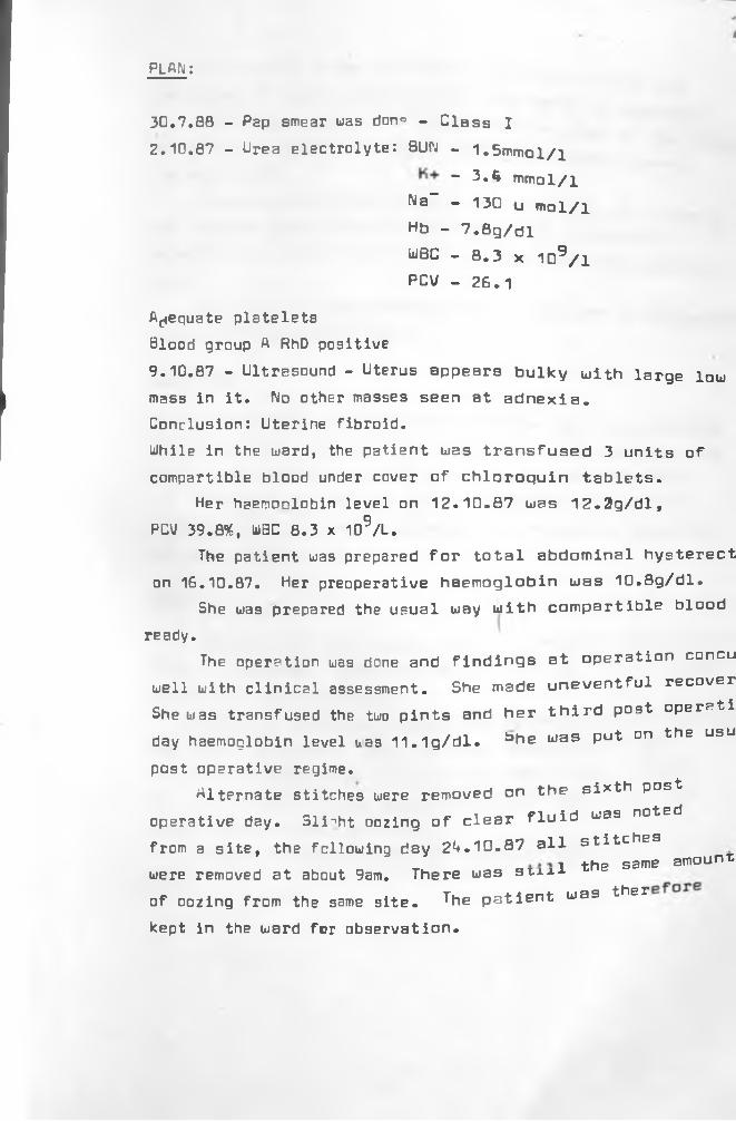
PLAN:
30.7.80 - Pap smear was dom - C la ss I
2.10.87 - Urea e lectro ly te: BUN - 1.5mmol/i
“ 3 . ft mmol/l Na" - 130 u mol/l Hb - 7.8g/dl UBC - 8.3 x 10^/1 PCV/ - 26.1
Adequate platelets Blood group A RhD positive
9.10.87 - Ultrasound - Uterus appears bulky with large low mass in it. No other masses seen at adnexia.Conclusion: Uterine fibroid.
While in the ward, the patient was transfused 3 units of compartible blood under cover of chloroquin tablets.
Her hsemocilobin level on 12.10.87 was 12.2g/dl,PC\/ 39.8%, U13C 8.3 x 10⁹/L.
The patient was prepared for total abdominal hysterect on 16.10.87. Her preoperative haemoglobin was 10.8g/dl.
She was prepared the usual way with compartible blood
ready.The operation was done and findings at operation concu
well with clinical assessment. She made uneventful recover
She was transfused the two pints and her third post operati day haemoglobin level uas 11.1g/dl. ^he was put on the usu
post operative regime.Alternate stitches were removed on the sixth post
operative day. Slight oozing of clear fluid was notedfrom a site, the following day 2ft.10.S7 all stitches
_ 1 the same amountwere removed at about 9am. There was s u i a of oozing from the same site. The patient was ther
kept in the ward for observation.

280
On 25.10.87 at 8am the patient went to toilet and
complained of severe abdominal pain and some pv bleeding On examining the patient was found to have part of the large
and small intestines herniating from the halfway oppned incision wound.
A diagnosis of Burst Abdomen was made and the patient was prepared immediately for theatre. The herniated viscera were reduced asceptically in the ward and sterile packs put.
At 11am the p tient was already in theatre on operation table. She was anaesthetised.
PROCEDURE
Found: The patient was catheterised after the vulvovaginal toilet was d^ne. She was then cleaned at the abdomen, the packs being removed out. The patient was then drapped.
On inspection omentum, small intestine and portion of large intestine was eviscerated and partially stuck to the wound which was 75% open.
DONE:
Incision was opened whole length, adherent gut was separatee from the incision. Abdominal cavity was found to be clean with
no pus nor any abnormality.Abdominal cavity was rinsed with warm saline then wound
edges were freshened, ^hree tension sutures put: two horizontal matress with nylon and one vertical. Abdomen was then closed in three layers the usual way. Tension
susutreswwere then tightened.Anaesthesia was reversed and patient taken back to the
ward

281
She had uneventful recovery.She was started on oral sips on the second post
operative day and then fully on oral diet on the third day.
Her third post operative check haemoglobin was I0.5g/dl.On the tenth post operative day the tension sutures were
removed, other skin stitches were removed on the
seventh day.The patient was discharged home on the eleventh
post operative day.She was seen in our clinic four weeks later and other
than slight pain around the incision site for which she got assurance and panadol, she had no major problems.
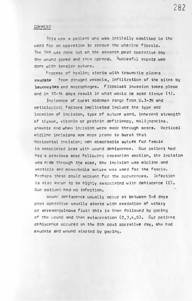
282
COMMENT
This was a patient who was in i t i a l l y admitted to the
ward fo r an operation to remove the uterine f ib ro id .
The TAH was done but on the seventh post operative day
the wound gaped and then opened, ^uccesful repair was done with tension suture.
Process of healing starts with traumatic plasma exudate from damaged vessels, infiltration of the sites by leucocytes and macrophages. Fibrolast invasion takes place and in 10-H days result in what would be scar tissue (1).
Incidence of burst abdomen range from 0.3-3% and aetioloaical factors implicated include the type and location of incision, type of suture used, inherent strength
of tissue, vitamin or protein deficiency, malignancies, anaemia and when incision were made through scars. Vertical midline incisions are more prone to burst that horizontal incision; non absorbable suture for fascia is associated less with wound dehiscence. Our patient had had a previous scar following caesarian section, the incision was m-de through the scar, the incision was midline and verticle and absorbable suture was used for the fascia. Perhaps these could account for the occurences. Infection is also kn-wn to be highly associated with dehiscence (2).Our patient had no infection.
Wound dehiscence usually occur at between 5-8 days post operative usually starts with exadation of watery or serosanguinous fluid this is then followed by gaping of the wound and then evisceration (2,3,U,5). Cur patient dehiscence occured on the 8th post operative day, she had
exudate and wound started by gaping.

Diagnosis of wound dehiscence should bp made quickly
and action taken fast as the complication can result
in mortality in 15-35%.Management aims at immediate repair in theatre. In
the intervening period sterile gauze should be put to cover the wound. The wound edges should be freshened and a non-absorbable inert suture to pass through all layers
of abdominal cavity. These stitches are left in situ for 10-2*4 days depending on the state of the wound when it was opened. Our patient had all these procedures fulfilled.
Prevention is geared towards identification of at risk patients and choosing the right incisions and right technique and suture. In such patients some authorities recommend use of tension suture from the beginning.

REFERENCES\
1. Walker, J.. McGillivary, I., MacNsughton, M.- In combined Textbook of Obs Gyn. 9th Edit.- Churchill, Livingstone 1976.
2. Mattingly, R.F., Thompson, J.D.
- In Te Lindes Operative Gynaecology 6th Edition pg 171, 19E- J.B. Lippincott Company Philadelphia.
3. Alexander, A.C., Pruden J.F.- The cause of Abdominal Wound disruption
- Surg. Gyn. Obs. 122: 1223, 1966.L. Mgnn L.S., Spimnazola A.J. et al
- disruption of abdominal wounds - J.A.M.A 180: 1021, 1962.
5. Gustendel, G.F. and Collins G.G.
- Abdominal Wound Complication in Obs. Gyn. Surgery
- Obst. Gyn. 1: 26L, 1953.6. Nelson J.A.
- In Atlas of radical pelvic surgery- Appleton-Century-Crofts.
GYNAECOLOGY LONG COMMENTARY
TOPIC:
KNOWLEDGE, ATTITUDE AND PRACTICE OF CONTRACEPTION (FhKILY PLANNING METHODS) AMONG TEENAGERS IN SOUTH NYANZA DISTRICT, KENYA.

• 286INTRCDUCTIUI\ - ND LITERATURE REVIEW
"The future of a country, and of manking depends on its children, For children to grow into healthy, able adults they need good food, clean water, education and medical care" (1).
•he above opening rpmark summarises in a nutshell whet anv country should consider a priority in planning for her people. In Kenya, about 50y. of the population is below 15 years of age and there are more females than males. This excess in the numfc of females is kept throunhout thp reproductive years i.e. upto 3A years (2). It is therefore clear that the bulk of Kenya's budgetary plan is utilised by the youth who don't contribute tc
the economy.In sub-Saharan Africa, population growth is fastest in the
world, 3°-, this simply means that more youth is added into the population with the negative result of even higher dependency ratio. Sy many quality-of-life indexes, for example education, infant mortality, industrial input, majority of African countri are at a standstill, or growing backwards (3).
In a number of African countries, a lot of resources are expended in putting up sophisticated hospitals in urban areas e neglectinn provision of basic health care (A). There is a neec for complete reappraisal of priorities of health needs.
In Third world two thirds oF the population are children under 15 years of age and females of child bearing age (1).This cert'inly means that for any government policy to have imp on a population, the children and females must be born in mind.
Children need to be born healthy, end so need mothers who are already healthy and families that can give them care and atten!
they need.Development plans tend to fall astray due to the high rate
of population increase (5). This obviously, is the very base c
the basic health care and, like the preventable diseases, is
capable of being controlled at the grassroots.1969 Kenya cencus showed a population growth rate of 3.3^,
one of the highest in Sub-Saharan Africa. It was 11,2A7,000 people and is projected to triple by the year 2000 A.D. (6). Kenya is 538,ODD sq km. It was projected that by 1980 there we
2.6 acres for every Kenyan, and by the year 2DDD «.D., there wi
be 0.5 acre per person. To have the picture clearer, only 17?£

L O I
of Kenya land surface Is cultivable under present technology, and four fifths of the Kenyan population reside in this cultiva area. (7). It is not difficult to compute the kind of chaos that there will be by the year 2000 A.D. The ‘Alma-Ata Declarat about health for all by the year 2000 *.D. (U), will even at that time still be an aspiration.
500,000 women in the Third World die during pregnancy and
childbearing every year H O ) . Some of these deaths, if not mos
are preventable by simple basic health care.
There is increased tendency to marry late all over the wor The girls are often victims of culture in transition. (85. Pregnancies occur at much earlier age all over the world thjfTn the past. By 1977 in Kenya, 7.6% of teenagers, 15-19 years, we currently pregnant; in 1963 in U.S.A. 1<*.5% of deliveries were teenagers, this went up to 17.1% by 1969 and 19.3% by 1972 (9). Ngoka found teenage pregnancy at Kenyatta National Hospital of in I980$0l,ln Nairobi the incidence of teenage pregnancy in 1983
was 18.6% and the youngest mother was 10 years and 2 months (11 Kuraye, in his study of teenage pregnency in ^uTal Kenya, found that 26% had had one or * more pregnancies, and that 58.A%
became pregnant in the first 8 years of school. He also found incidence of teenage pregnancy to be 10%.53.3% of teenagers were already married.
Jagdeo found adolescent fertility to be generally high in Africa reaching 130/1000. In Indonesia, Asia, it is as high as 2<*0/1000 and by 17 years of age <*1% of girls have delivered. (1
Teenage pregnancy is therefore raising alarm. There is al
a 10 year 2 months old mother and this is an alarm enough. aomething is got to be done.
Kean age at menarche hes continued to be lower over the years with USA recording 12.7 ♦_ 1.2 years; in Madras, it is 12.
in Addis Ababa it is 13.7 years (1<*).Modern Education with concomitant loss of cultural bonds (
has put the youth of today in a very contemptous state to his e Some youth suffer not because of being contempt but because of lack of parental guidance (10). Hoffmeyer found that almost 100% of the 18-19 year old seeking abortion came from homes where their parents for one reason or another could not satisfy
them emotionally (16).
I »•

288
Early menarche and increased teenage sexuality and expose: to school puts the teenagers in a dangerous position that unle! they are protected we will witness unplanned pregnancies, induced abortion and its consequences, population explosion wi disorganisation of the country's plan. The need for protects has shown itself so clearly that there is no longer the question as "Is there a need for instruction ? . Uhen shall si instructions be given, in childbearing or in puberty ?»(19).
NJoroge showed thBt 22.1% of undergraduate students had he sex by 19 years of age, the youngest bad sex when she was 12 years old (20). It has been shown that girls with better education seem to know more about ways of getting rid of unplar pregnancy. The teenagers most of whom are still in ptimary srt or early secondary schools therefore are mostly unaware of ways of getting rid of unplanned pregnancy and therefore carry pregnancy to term with all their unpreparedness (21).
Cyril Ybiing et alfound that 1:8 girls and 1:3 boys had had sexual intercourse by 16 years of age and were thus exposed to unplanned pregnancy (22). Kenya Fertility survey showed th; 28% of 15-19 year olds were married already (23). In USA more than 50% of the 19 year olds and below had engaged in sexual activity and their fertility were comparable to the women in
their twenties (23).On realisation of the rate of population growth, Kenya ^oi
adopted official policy on Fptnily -Planning in 1965 and Kenya National Union of Teacher® noting the vulnerability of the adolescents declared that sex education should be introduced ir
school syllabus in 1972 (6).Studies have shown that legalising abortion does not alte:
the rate of teenage pregnancy as there will obviously be
bureacratic restrictions, distance to approved centres and expense involved (2U ) . In developing countries there is generc
increase in the incidence of illegal abortion and this pay burc to governments budgets^ as a>y illustration in Bolivia 60% of c
of running Obstetrics and Gynaecology departaent is devoted for the treatment of complications of abortion; in Turkey one in seven women had had illegal abortion by 1975 while the
figure was one in fourteen in 1963. In El Salvador maternity hospital, one quarter of hospital deaths were due to illegal
abortion (8).

289
It has been shown that sex education without prouision of
contraception increases conception rate (25).It is therefore clear that intervention must be done at
a stage early enough to interrupt this vicious cycle where unplanned pregnancies in young girls result in high maternal mortality and morbidity and infant mortality due to unprepared maternity; this gives poor national quelity-of-life indexes in which people engage in nothing else but in bringing up childre in a state of gambling that if some die some will remain.The intervention has got therefore to be at the teenage, and t means should be contraception as already reproductive biology is being taught in sbhools. This must be done if we have to see our population and development improve.
This study will be done at Western part of Kenya in ^cuth Tjyanza District which is in Nyenza Province.
The District has total water and land area of 7,778 sq km (Land erpa is 5713 sq.km). This forms 1:33* of National surfa area. It has a population as at 1979 of about 817601 with pop density of 1L3 people per sq. km. It has nine administrative Divisions namely Oyuqis, Kendu Bay, ^ongo, Rangwe, Mbita, f«dhiwa, Migori, Kecalder and Kehancha. Except for Kehancha Di the rest of the Districts is composed mostly of Luo ethnic gro
Luos formed 31* of total teenage pregnancy which was very high considering their population is only 1995 of their Nairobi
population (2f6). Incidentally, the district also has the hinhest infant mortality rate in the whole country.
The District borders Kisii, Kericho, Kisumu and *>iaya
Districts, and also borders Tanzania through its Kehancha
and Kacalder divisions.

J
■ 290
OBJECTIVES OF THE STUDY
IMMEDIATE OBJECTIVES
A.1. To evaluate knowledge attitude and practice of contraception, and sexuality amongst the teenagers in South Nyanza District.
2. To assess their degree of exposure to Bex and their vulnerability to unplanned parenthood.3. To find suitable means of availing contraceptive services within easy reach to run hand in hand with sex education.
LONG TERM OBJECTIVES
1. To reduce teenage pregnancies and attendant complications2. To create awareness of reproductive body function at a stage that is critical, when the teenagers are capable of procreation and yet know little about reproductive body function.3. To establish family planning services at strategic points for easy access and sustained supply to the clients.U, To attain population growth sustainable by rate of economic growth.
STUDY ME THQDOLDGY
SELECTION OF SUBJECTS AND SIZE OF SAMPLE
The district has 9 administrative divisions. The aim of the study was to include primary, harambee and Government schools. Each division had to be given equal chances. Our target was 2000 students. We covered a total of students but as 10<«questionnaires were not properly filled or the respondents were either underage or overage only 18 *0 questionnaires qualified and are presented here.

291
A H teenagers were Included in the study. Schools were chosen
randomly from the District Education Officer's register, for
every dlviBon. Primary schools were chosen first and then the
nearest secondary schools chosen. A limited number of students
per school were chosen so that a division is represented as muc
as possible. In so doing, we covered a total of 12 primary
girl schools, 13 primary boys schools, 5 harambee |irl seconder
schools, t* Harambee boys secondary schools, U Government girls schools and U Government boys schools.
STUDY TYPE
Thi3 was a descriptive survey type of study.
STUDY INSTRUMENT
This study was conducted using questionnaires. Host o f
the questions were c lose ended. A p i lo t study had been done
e a r lie r in d if fe re n t parts and necessary adjustments made.
A group o f students le g ib le fo r the adm in istration o f
questionnaires were put in a c la s s and b rie fed before commences
of the response. Questionnaires were given to a l l and any
corrections made before they s ta r t . They were made free to ask
any questions corncerning any question they don't
understand w e ll. This was done q u ie t ly w ith the student invo lv
DATA COLLECTION AND ANALYSIS
Questionnaires were f i l l e d same time in same schoo l.
Three assistants were at hand to c o lle c t , so rt out questionnair
into d iffe ren t categories and give them s e r ia l numbers thereby
knowing how many students we have interviewed. The responses
were then analysed and t a l l i e d .
ETHICAL CONSIDERATION
No ethical problems involved. Informed consent was obtaini
before administration of questionnaire.

292
CONSTRA Jti TS
Reliance uas put on an assumed subjects ability to understand English language and questions. This uas overcome by assurance and freedom of expression.
Some students might not have been telling the truth but assurance and anonimity of the respondent and guaranteed confidentiality ia hoped to have eliminated if not reduced the number of intentional urong response.
All except one headteacher found the questions very petinent in light of high premarital and school age sexual activity. More so they found discussion that followed very interesting and informative. The school whose headteacher was hesitant uas left out of the study.
Incomplete filling of the questionnaire uas envisaged and this uas minimised as much as possible by encouraging the students not to leave any vacant place.
In view of the recommendations, follow up is necessary.
PRESENTATION
This is in form of tables accompanied by comments.
Discussion Bnd lite ra tu re review fo llow .

293
RESULTS
This study interviewed a total of 18MD students. They were spread amongst boys and girls in primary schools, secondary schools Harambee, and Secondary Schools Government. The responses were recorded and grouped into the various subjects under study; analysed and presented.

294
COMPOSITION OF STUDENTS BY TYPE OF SCHOOL AND NUMBER
TABLE I------- BOYS GIRLS TOTAL %
PRIMARY
------------ I
377 330 707 38.A
harambee 176 359 535 29.1
GOUERNMENT 271 327 598 32.5
TOTAL 82A 1016 18A0 100
Table 1
This table shows the breakdown of the total number of students Interviewed as they were distributed per school typ
The breakdown was as follows:
- Girls Primary School
- Girls Harambee Secondary- Girls Government Secondary- Boys Primary School- Boys Harambee Secondary Boys Government Secondary TOTAL
Distribution per School
Primary School Students 707 38.AHarambee School Students 535 29.1Government School Students 598 32.5
18A0 100
Number % _330 17.9359 19.5327 17.8377 20. A176 09.7271 1A.718A0 100
TOTAL

295
X AGE DISTRIBUTION
TABLE 2PB PG HB HG GB GG
AGE YRS
12-15 3 2 0 0 0 0
16-18 65 35 58 16 36 55
19-20 28 60 39 82 63 U5
NORESPONSE U 3 3 0 1 0
TOTAL 100 100 100 100 100 100
KEYS:
PB - Primary Boys PG - Primary Girls HB - Harambee Boys HG - Harambee Girls GB - Government Boys
GG - Government Girls

29(
TABLE 2
Over 90% of students in each of the six categories in this study were of age group 16-20 years. Considering that primary school students formed the majority of our study group 38.A% and also noting that only 5% of primary school students were in age group 12-15 years it would appear that age gap between secondary and primary school students was not wide.
Lema (30) dealing with secondary school students found peak of ages 15-17 years and this accounted for only 56.U%.
In Maggwa (29) series age group 16-20 years accounted for 68.3%. This study was in a rural set up.

% OF STUDENTS NOT HARRIED
297
TABLE 3 GG
NotMarried
ro
AO
----
100
,na----
86
nu---
90 99 97
2B5rAEdresponse 60 0 1A 10 1 3
100 100 100 100 100 100
Marital status of students were next looked Into.
Percentage responding as not married ranged from A0% for primary boys to between 8696-100% for other categories,The response in the primary boys category look suspicious as the percentage who were married is rather too high and seem to be in isolation.
This question was specifically asked because in South Nyanza Districts, primary school students who fall out of school due to pregnancy and later on join schools from their husbands' ho m es are not uncommon.

f 298
% WHOSE PARENTS STAY TOGETHER
TABLE UP0 PG HB HG GB GG
StayTogether 90 85 80 96 89 90
Separated or divorced 2 10 11 3 6 5
The study found that 85-96% of student's parents stay together. An average of about 6.1% of parents are either divorced or separated. Maggwa (29) also found in his series over 90% of student to have both parents. This compares well
with this study.
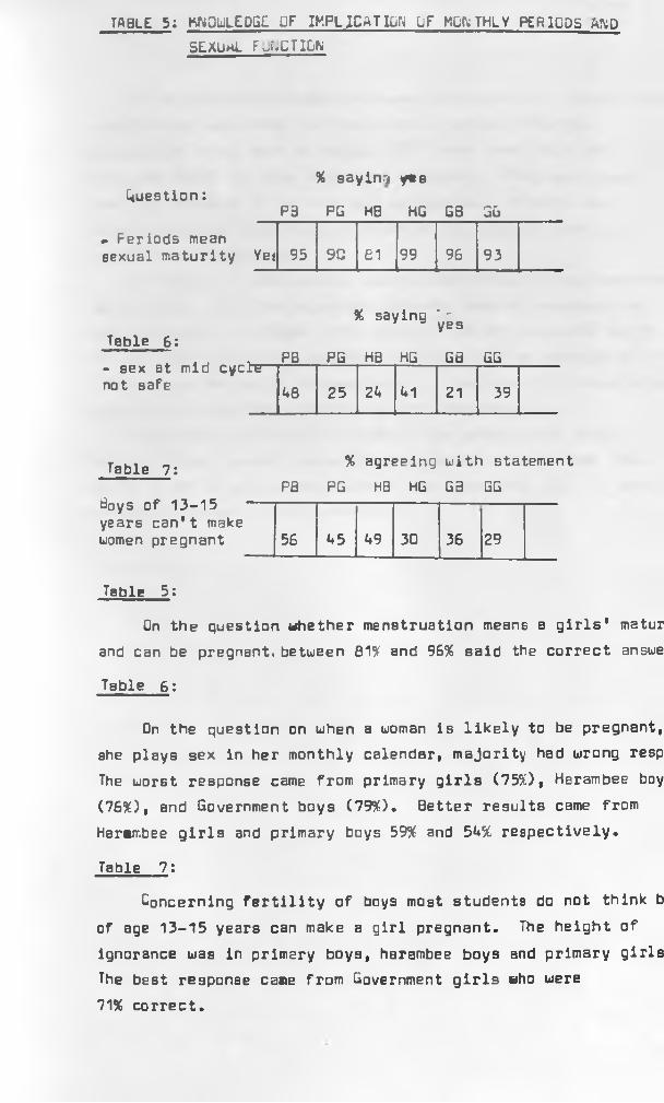
TABLE 5: KNOWLEDGE DF IMPLICATION LF MONTHLY PERIODS AND SEXUmL FuNCTILN
Question:
► Periods mean sexual maturity
% saying ye6
P3 PG HB HG GB GG
Yeti 95 90 61 99 96 93
Table 6 :
- sex at mid cycle- not safe
% saying ' . r 1 * yes
PB PG HB HG GB GG
UB 25 2U 1*1 21 39
Table 7 :
Boys of 13-15 years can't make women pregnant
% agreeing with statementPB PG HB HG GB GG
56 <♦5 US 30 36 29
Table 5:
On the question whether menstruation means a girls' matur and can be pregnant, between 81% and 96% said the correct answe
Table 6 :
On the question on when a woman is likely to be pregnant, she plays sex in her monthly calendar, majority had wrong resp
The worst response came from primary girls (7550, Harambee boy (7650, and Government boys (7950. Better results came from Harambee girls and primary boys 595( and 5A% respectively.
Table 7 :
Concerning fertility of boys most students do not think b
of age 13-15 years can make a girl pregnant. The height of ignorance was in primary boys, harambee boys and primary girls The best response came from Government girls who were 71% correct.

300
Aa to whether menstruation means fertility for a girl, the question was searching for knowledge on sexual maturity. Comparative study done by Maggwa (29) found that 73.3% of girls and 6 8 .1% of boys responded correctly. This was lower than this study's in all our six categories. It might be interpreted as a regional variation as the studies were done
in different districts.Knowledge on safe period during the monthly period was thei
looked into. The finding ahows pathetic lack of reproductive body function. In Maggwa (29) series 27.6% of girls and 26.5% o£ boys had the correct answer. Our study had an average of 31! 35% for boya and girls respectively had the right knowledge Again our series look slightly better than Maggwa'a.
Concerning fertility of boys of age group 13-15 years, Maggwa's study showed a more knowledgeable response than this series 61.8% of girls and 80.*4% of boys knew that boys of that
age group can make a girl pregnant.

301SEXUAL MATURITY AMD SEXUAL BEHAVIOUR
!*-
Table 8: Age at Menarche %
HG GG
Age 10 -12 7 3in
13-15 61 63years
» 1620 13
Table 9: PB PG HB HG Gfl GG
%Age at
B-12
first sex13-15
>15Not played sex or no response
19 5 0 0 6 1
31 29 10 u 29 1
25 32 20 22 37 19
2 23 9 20 9 3
23 11 61 _5ii_ 19 76
Table B:
The only reasonable assessment of menarche was possible in secondary school girls 61%-63% of the girls had their menarche at between 13-15 years, 3-7% had it between 10-12 years and
13-20% at 16 years or after. gne glri ^ad mepapghe at 11 years.
Table 9
Age of first coitus seem to be earlier for boys then for gi: Primary school boys and girls seem to have an edge over their secondary counterparts. At least 59.U% of the students interviei had had sexual contact.
'31.6% of boys were sexually active by the end of 12 years compared to i>.jx for girls. By the end of 15 years, ' 5^ ofboys had sexual experience compared to 37„fs% of girls.
' r-‘* K 1.
I

302
Question of sexual maturity for girls was addressed.
It was found that a reasonable percentage 3-7% had had menstruation by 12 years and that about 66% had had it by
15 years. Percentage of those who had had it by 20 years were 76-88%. In Lema's series (30) 96.2% had had their periods.
This comparable with ours.
Age at first sex. Lema's series range between 5 years to 17 years, with 77.7% having had it between 1L-17 years.In Maggwa'a series, 6A.1% of boys and 26.2% of girls had had sexual intercourse by 15 years. This is comparable to our series where 59% of boys and 37.6% of girls had had sexual intercourse by 15 years. In Maggwa'a series like in ours,boys seem to have an edge over girls. It is worth noting in present series that 19% primary school boys students had had se>
by 8 years, and 5% of girls.

CONTRACEPTION KNOWLEDGE AND PRACTICE 303
X of those who knew defination
TableQuestions: -P fl PG ___u a HG GB SG
Know F.P.Correct defination 67 75 uu 86 87 80
Table 1 1 : % of those who know
Wyetion: PB PG HQ HG GQ GG
Which F.P. methods you know 39 <♦2 1A 55 51 62
Table 10:
Harmabee School boys had the poorest knowledge of defination of family planning, 60* of them did not know the meaning. Generally knowledge of family planning seem good with harambee school girls, Government boys and Government girls scoring over 80%. Primary school boys and girls were right in
between 67%-75%, of cases.
Table 11
Knowledge of family planning methods were generally lower t that of defination of family planning, itself. Harambee boys ag did very poorly with only 1<*% frflcnirrjg any family planning method. Again Harambee girls, Government boys and girls socred higher 51%-62*. Primary School girls and boys were much better than the harambee boys.

304
Knowledge and practice of contraception was studied with a view to finding the depth of knowledge and extent of use.
Correct defination was generally better than enumerating
types known. In Lemma's series, 82.6% had the correct knowledge of what contraceptives are. This was comparable to our secondary school girls 80-87%. Scanty knowledge of contraceptive methods were also bone of contention in Lema's and Maggwa's series (29,30).

TABLE 12: CONTRACEPTION KNOWLEDGE AND PRACTICE
Table 1 3 :
% of those never used FP methods
PB PG HB HG G3 GG
92 92 97 L3 79 57
PB
< not using any F.P method now•
PG HB HG GB GG
95 92 97 t*U 86 93
Table 12:
Harmabee school girls and government school girls had used contraceptive more than others A3*-57%. Others'
usage only ranged between 3*-21%.
Table 13:
Even at the time of interview 56* of Harambee school git]
were using one or the other method of contraception. In the
other category only 3-1A* use Bny method.
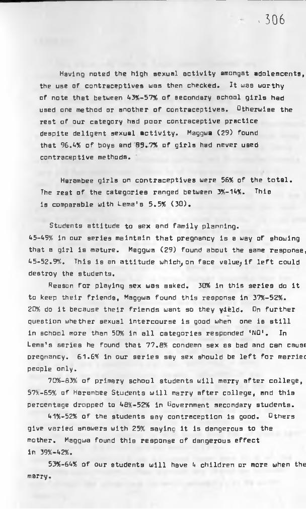
. 306
Having noted the high sexual activity amongst adolescents, the use of contraceptives was then checked. It was worthy of note that between L3%-57% of secondary achool girls had used one method or another of contraceptives. Otherwise the rest of our category had poor contraceptive practice despite deligent sexual activity. Maggwa (29) found that 96.L% of boys and 89.7% of girls had never used contraceptive methods.
Harambee girls on contraceptives were 56% of the total.The rest of the categories ranged between 3%-1L%. This
is comparable with Lema's 5.5% (30).
Students attitude to sex and family planning.
U5-U3% in our series maintain that pregnancy is a way of showing that a girl is mature. Maggwa (29) found about the same response, L5-52.9%. This is an attitude which, on face value; if left could destroy the students.
Reason for playing sex was asked. 30% in this series do it to keep their friends, Maggwa found this response in 37%-52%.20% do it because their friends want so they yield. On further question whether sexual intercourse is good when one is still in school more than 50% in all categories responded 'NO'. In Lema's series he found that 77.8% condemn sex as bad and can cause pregnancy. 61.6% in our series say sex should be left for marriec people only.
70%-83% of primary school students will merry after college, 57%-65% of Harambee Students will marry after college, and this
percentage dropped to L8%-52% in Government secondary students.L1%-52% of the students say contraception is good. Others
give varied answers with 25% saying it is dangerous to the mother. Maggwa found this response of dangerous effect in 39%-L2%.
53%-6L% of our students will have U children or more when themarry

307
DISCUSSION
This study was done in South Nyanza District and involved 18^0 students. Results and tables of results are as already shown. Breakdown is as shown in Figure 1.
Primary school students were more than any of the other
two categories. This was intended as it was considered that these students mostly come from the District and assessment of their knowledge, attitude and practice of the subjects under study would be a better reflection of the area than the other study groups. Further, Harambee schools and Government schools were put for comparison. Results of boys are compared with that of girls whenever obvious differences were encountered.
Over 90* of the total students interviewed were in the age group 16-20 years. This was significant considering that more than 1/3 of the students were from primary schools. It also shows that the age gap amongst almost all students was very narrow.3% of primary school boys Bnd 2* of primary school girls were below 16 years. In Maggwa (1987) study done in a rural population in the same country, age group 16-20 years accounted for 68.3*. His series had slightly younger students than the present series. This finding implies that we were dealing with very mature people who have the biological capacity of procreation (9,10).
Some parts-of the District where this study was done are known to have their girls marry early. Some of these marriages don't hold and the girls often return to schools.These girls are essentially married, with this in mind, question of marital status was raised. 86%-100* of the students stated that they were not married; but only U0* of primary school boys responded in this manner. This percentage was rather too low; some could have actually married but some perhaps did not understand the question clearly.
Kenya Fertility Survey in 1978 found that 28* of girls aged
between 15-19 years were already married (23).Jagdeo (198*0 working in the Carribean found that 53.3* of the teenagers were married. This question was therefore relevant
and although the percentage is small generally, it is worth recording.

308
Religion is known to have influence on sexuality. There are presently schools of thought that this influence does not actually exist, (29,30). In this study 78% of the students were protestants with 82% of these being Seventh Day Adventists (SDA). The remaining students were so spread into small different religious groups that it was not practicable to really come out with a conclusion.
85%-96% of the parents of students interviewed stay together About 6.1% of the students had their parents either divorced or separated. In Maggwa's series over 90% of the parents stay together (29). In Lema's series done in an urban set up 77.7% of the students had their parents staying together (30). Difference with Lema's series perhaps arises from the known problems of urban centres where incidence of single parents are many. Thi9 series compares well with Maggwa's above.Parental guidance and stability of homes have been shown to contribute to steady performance in school and less tendency to deviant attitudes. Hoffmeyer found out that almost 100% of girl9 between 18-19 years going for abortions were not satisfied with their parents emotionally (16). Lema (1987) showed that sexual activities were minimum, 16.8%, amongst girls staying
with their parents. This percentage went up to 39.8% if staying with brother, A5.9% if staying with sister and to 70% if staying with relatives (30). Stable homes therefore breeds stable children with stable behavio.ur. Our number for those whose parents are are separated was small and mongst the affected students no untoward observations were mBde.
Knowledge of monthly period and implication was studied. 81%-96% of the students knew that it marks a point when a girl becomes capable of being pregnant. Maggwa found this response to be 73.3% for the girls and 68.1% for the girls (29). This type of knowledge is mainly acquired in schools, but can also be imparted by friends and agemate9.

- 309
This knowledge was further tested to find out whether the student knows more about the monthly period. A question of safe period in monthly cycles was asked. The majority gave wring answers. The largest percentage of wrong answers came from
primary school girls, Harambee boyB and Government boys who scored 75%, 76% and 79% wrong response respectively. In Maggwa's series 31% and 35% of boys and girls respectively had correct answer. This is, converted to wrong response is 69% and 65% respectively. (29) These findings are comparable though Maggwa's series looks better.
As to the fertility of boys, Government girls, Harambee girls and Government boys had reasonable responses with only 29%,30% and 36% respectively of them giving wrong answer. The rest ranged between A5%-56% wrong answers. They did not know that boys of 13-15 years can make a girl pregnant. Maggwa's series had a better result with 38.2% of girls and 19.6% of boys getting the wrong answer. (29)
These findings send bad signal to the question of fertility control. These are essentially people capable of giving birth, on the one hand they know that when a woman gets her monthly periods, she can be pregnant and give birth to a baby, on the other hand they dont know that there is a specific period within the monthly cycle that is not safe; they also dont know that at 13-15 years most boys are already fettile and can impregnate a girl (29). This information puts the students in a very precarious situation.
61%~63% of the girls had their menarche at between 13-15 years, with 3%-7% who had it before 13 years. The earliest time recorded was at 11 years. A total of 76%-88% had had monthly period by 20 years. This figure compared to Lema's 96.2% could indicate some stimuli to menstruation in Lema's series (30). It is known that the student in urban set up is exposed to some of the things that a student in rural areas are not. These include television, magazine and closer relation with peers. These tend to stimulate earlier onset of puberty.

310
By the end of 15 years 59% of the boys had had sexual
Intercourse. Boys were observed to have sexual relation earlierthan girls, and primary students seem to have an edge in thisrespect o*«r their secondary counterparts. This edge could #be a reflection of the area of the study as was mentioned earlier. Maggwa (29) found that 6A% of boys and 26.2% of girls in his series had had sexual intercourse. The difference between boys and girls was more marked in Maggwa's study findings. While the difference is obvious between the boys and girls, it is not easy to know why. We can postulate that boys have intercourse with older girls, or that boys share some few girls that have been known to yield easily. Lema (30) found that 77.7% of girls in his series between 1L-17 years had had sexual intercourse.
These observations are alarming. Age of menarche has been known to lower, sexuality of contemporary societies is rampant, breakdown of cultural bonds which were once responsible for general guidance, and peergroup pressure are all responsible for early sexuality amongst teenagers. It is alarming that they involve in these acts without knowing the implications and consequences. This trend has been observed all over the world. Fundikira found the youngest age of menarche at 8 years (21). In Indonesia U1% of girls by 17 years have delivered (13), in U.S.A. by 1977 19.3% of teenagers delivered, in Nairobi, incidence of teenage pregnancy was 18.6% in 1984* (9); Young found that 1:8 girls and 1:3 boys had sexual intercourse by 16 years and were exposed
to unplanned pregnancy (22), Njoroge showed that 22.1% of undergraduete students had had sexuel intercourse by 19 years.(20), It is obvious that these students are sexually active and also sexually mature, they are capable of parenthood and also vulnerable to unplanned pregnancy.
Defination of family planning was good, except for Harambee boys who scored very low percentage, the correct defination was got by 67-87% of the students. For the secondary school students, this correct answer was recorded in between B0%-87%.Lema found correct response in 82.6% (30). There, therefore, does not seem to be any difference in urban set up and rural set up
for secondary school students.

311
Our study recorded small percentage for those who know definate contraceptive mthods. This scanty knowledge also seem to have been
an experience in Maggwa's and Lemma's series (29,30).
Practice of contraception was looked into next. L3% of Harambee girls had used a method of contraception or another;57% of Government girls also had used contraception. For the rest 79%-97% had not used any method of contraception.
UU% of Harambee girls were using contraceptive methods at the
time. For the others,usage ranged between 3%-21%. Maggwa's findings were the same 96.L% of boys, and 95.7% of girls had never used any method of contraception (29).
These students are maturing early, they do not know when they can be pregnant, they have sporadic unprotected sexual relation, they do not know methods of contraception and they do not practice contraception. This is a pathetic situation. It can be seen that these youths Bre exposed to unplanned pregnancies, unprotected sexuality is rampant and specific contraceptive knowledge and practice is almost zero. Lema in urban centre found contraceptives use as low as our?, 5.5% (30). The problem is therefore as serious in rural as it is in urban areas. It is worth noting that most of the students who had ever used contraception used unreliable methods, and although a number gave examples of the contraceptives theymse, most gave safe period method and went ahead to point at wrong timing in menstrual calender. The finding is very pathetic. Source of knowledge about contraceptive
perhaps was contributory. In this study 21.1% got information from school, 15% from magazines, and 7.2% from friends. Lema's serips 33.3% got information from magazines, 21.A% from school
and 7.2 from friends (30). As can be noted, information from school will mainly be reproductive biology revolving around safe periods at best. Other sources of information are likely to
be unreliable. Thus the student indulges in sexual relations with false sense of security. They get caught unawares and they do not know what next to do.
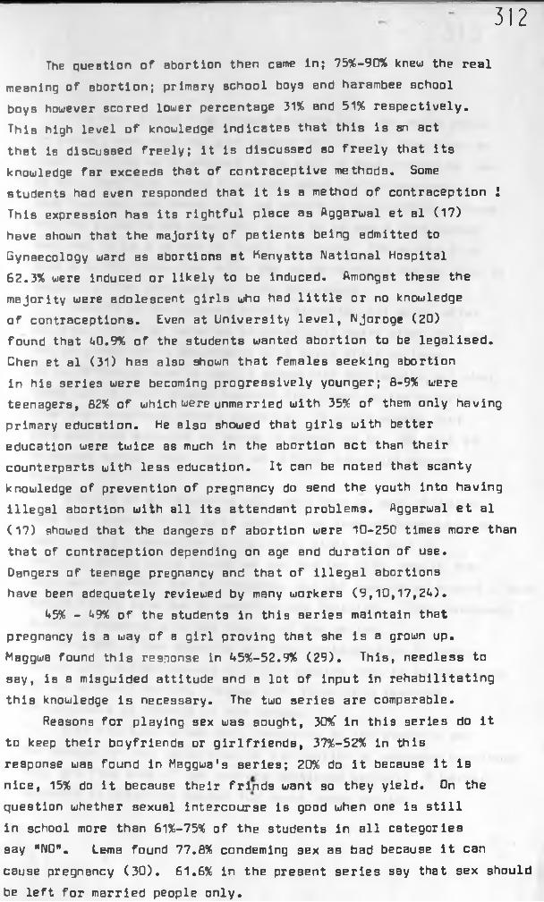
312
The question of abortion then came in; 75%-90% knew the real
meaning of abortion; primary school boya and harambee school boys however scored lower percentage 31% and 51% respectively.This high level of knowledge indicates that this is an act
that is discussed freely; it is discussed bo freely that its knowledge far exceeds that of contraceptive methods. Some students had even responded that it is a method of contraception 1 This expression has its rightful place as Aggarwal et al (17) have shown that the majority of patients being admitted to Gynaecology ward as abortions at henyatta National Hospital 62.3% were induced or likely to be induced. Amongst these the majority were adolescent girls who had little or no knowledge of contraceptions. Even at University level, Njoroge (20) found thBt AO.9% of the students wanted abortion to be legalised.Chen et al (31) has also shown that females seeking abortion in his series were becoming progressively younger; 8-9% were teenagers, 82% of which were unmarried with 35% of them only having primary education. He also showed that girls with better education were twice as much in the abortion act than their counterparts with less education. It can be noted that scanty knowledge of prevention of pregnancy do send the youth into having illegal abortion with all its attendant problems. Aggarwal et al (17) showed that the dangers of abortion were 10-250 times more than that of contraception depending on age and duration of use.Dangers of teenage pregnancy and that of illegal abortions have been adequately reviewed by many workers (9,10,17,2A).
A5% - A9% of the students in this series maintain that pregnancy is a way of a girl proving that she is a grown up.Maggwa found this response in A5%-52.9% (29). This, needless to say, is a misguided attitude and a lot of input in rehabilitating this knowledge is necessary. The two series are comparable.
Reasons for playing sex was sought, 30% in this series do it to keep their boyfriends or girlfriends, 37%-52% in this response was found in Maggwa's series; 20% do it because it is nice, 15% do it because their frinds want so they yield. On the question whether sexual intercourse is good when one is still
in school more than 6l%-75% of the students in all categories say "NO". Lema found 77.8% condeming sex as bad because it can
cause pregnancy (30). 61.6% in the present series say that sex should
be left for married people only.

313
The fact stands that these students play sex while still
in school, they have no good reason for the act, they have an inner conflict as to whether it is good to have premarital sex or not, they have scanty knowledge about their reproductive body function and these acts are generally unprotected. These are the students who know what abortion is and a good number say that it is a method of family planning. Thb author find this a sticky situation which the students might not be able to reconcile. An intervention looks inevitable.
70%-83% of the primary school students will marry after college, 57-65% of Harambee students will marry after college, and only <«8%-52% in Government will marry after college.
It is difficult here to really appreciate the results but what comes out is that primary student aim higher at the material time than secondary school students. It might appear that this ambition dwindles as years in school go by. So that at secondary school those aiming at college education become fewer.
53%-6U% of the students will want four or more children
when they marry, 7%-18% will want two or less children when they marry. This observation is important given the sort of sexually active young people we are dealing with, should they fall out of school due to pregnancy and get married we expect a much
larger fanily sire due to uncontrolled fertility. This obviously does not argue well to the health of the nation.
U lX -5 2 % of the students say that contraception is good.Others gave varied answers with 25% stating that it is dangerous to the mothers' health. Maggwa (29) found this response of dangerous in 39%-^2% in his series.
This attitude is not only dangerous to the students and the population but could also be aieflection of whatever knowledge they get from some of the sources mentioned earlier. A better approach is therefore needed for these young people.

314CONCLUSION nhD RECliKhENDATIONS
This study has shown that sexual activities amongst the
students start early even before they reach fertility ages.Boys seem to start an an earlier age than girls. They indulge in sexual activities sporadically without knowing the likely consequences. This sexual activities are not protected because the students do not know how to get these protections and which
protections are available. The ones that claim to be practicing any contraception use unreliable methods.
Knowledge of abortion is almost perfect but what sends chills down the spine is that some students know it is a form of family planning. Such erroneous knowledge is dangerous
to the students who will not even know where or when to go for professional termination of pregnancy. The implications are
obvious.Attitude towards sexual activities was reasonable but the
very act itself makes attaching importance to this attitude not valuable. The students could really have the urge to restrain from sex while still in school, but peer group pressure and the urge to explore makes these feelings secondary.
It is worth noting that sexual activities in primary schools were comparable to or even hiqher at some instances, than their secondary counterparts. It might be concluded that this was due to region of the study and that early sexuality is not uncommon.The finding really is a challenge because it pouses a question of where, if at all, should intervention start, and by whom?
Contraception attitude was poor and if considered together with practice the situation is close to hopeless. Sexuality is peak, contraceptive knowledge and practice is close to zero. Worse still,knowledge of reproductive body function was very poor.
The question of whether these teenagers are exposed to sex, therefore, is foregone conclusion and with the sort of zeal and ignorance with which they have their sexual relation vulnerability to unplanned pregnancy is high.

315
The proper meaning of contraception need to beinculcated into the youth for already we know that they practice
it. Secondly there is a need for intergrated lessons touching anreproductive body functions or sex education together withcontraception. It has been noted that sexuality begins earlyso that these people are exposed to the risk of unplanned
r — the questionpregnancy at a very tender age. From this study1which does not need to bog our minds any more is "where shall we start this sex education?". The need has been shown clearly that for sex education to have any impact on the youth's fertility then education must be imparted at lower primary school level, and this must go hand in hand with provision of family planning methods.
As parents there is a need to reappraise our thinking and accept that these are capable of procreation and that to this effect they need parental guidance and careful counselling.
Government policy bottlenecks need to be re-evaluated in the
face of current trend of adolescent fertility. Freehand should be given to health experts to work in conjuction with school teachers to come with appropriate carriculum on sex education
and contraception.Contraceptions are for the risk of unplanned pregnancy; a
aadolescents are at special risk of unplanned pregnancy. This study therefore recommends supply of adequate knowledge of
contraceptives and sustainable supply of contraceptives.Because teachers and parents are very important in the life of
youth especially during this tender age, it is recommended that they should be enlightened about contraception. An abudant amount of ignorance was perceived amongst many school teachers about contraception. It is recommended in this study that they should be a target group.
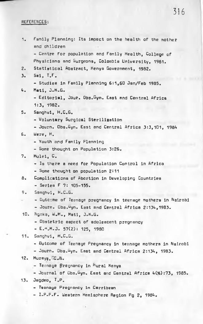
REFERENCES:
316
1. Family Planning: Its impact on the health of the mother and children
- Centre for population and Family Health, College of
Physicians and Surgeons, Colombia University, 1981.2. Statistical Abstract, Kenya Government, 1982.3. Sai, T.F.
- Studiea in Family Planning 6:1,60 Jan/Feb 1985.U . Mati, J.K.G.
- Editorial, Jour. Obs.Gyn. East and Central Africa 1:3, 1982.
5. Sanghvi, H.C.G.- V/oluntary Surgical Sterilization- Journ. Obs.Gyn. East and Central Africa 3:3,101, 198A
6. Were, K.
- Youth and Family Planning
- Some thought on Population 3:26.7. Mulei, C.
- Is there a need for Population Control in Africa
- Some thought on population 2*118. Complications of Abortion in Developing Countries
- Series F 7: 105-155.9. Sanghvi, H.C.G.
- Outcome of Teenage pregnancy in teenage mothers in Nairobi- Journ. Cbs.uyn. East and Central Africa 2:13A,1983.
10. Mgoka, W.M., Mati, J.K.G.
- Obstetric aspect of adolescent pregnancy- E.h .m .J. 57(2): 125, 1980
11. Sanghvi, H.C.G.
- Outcome of Teenage Pregnancy in teenage mothers in Nairobi- Journ. Obs.Syn. East and Central Africa 2:13<+, 1983.
12. Murayq,
- Teenage Pregnancy in ^ural Kenya
- Journal of Obs.Gyn. East and Central Africa A04):73, 1985.13. Jagdeo, T.p.
- Teenage Pregnancy in Carribean
- I.P.P.F. Western Hemisphere Region Pg 2, 198^.

317
1A. Diejomaoh, F.M.E.
- The age of menarche In a group of secondary school girls in Benin City, Nigeria.
- Journ. Obs.Gyn. East and Central Africa 3:1,38-39, 198A.15. Sanger, M.
- Uihat every boy and girl should know, pg 7.16. Hoffmeyer, H.
- Bole of Fairily Planning and school in sex education of a child- I.P.P.F., Warsaw, Poland 71: 152-15A.
17. Aggarwal, V.P., Mati, J.K.G.- Epidemiology of induced abortion in Nairobi, Kenya- Jrn. Cbs.^yn. East Central Africa 1(2): 56, 1982.
18. Atiqur Rahman- Maternal mortality in Rural Bangladesh, Uamalpur District- Studies in Family Planning 17(1):9., 1986.
19. Katner, J.F., Zelnik M.- Sexual experience in young unmarried women in United States
- Studies in Family Planning A(A): 9, 198220. Njoroge, C.N.
- K A.P* of contraceptives on female University Undergraduates
- Long Commentary gynaecology- M.Ked Thesis University of Nairobi, 198A.
21. Fundikira S.R.- Sexuality, Fetility and Contraception knowledge and Attitude
among secondary school girls in Dar es salaamDPH Thesis 1985, Dar es salaam University
22* Young, C;, Cowper, A.- Family Planning 10: 129, 1980.
23. Kenya Fertility Survey- rirst report 1:70-82, 1977-1978
2A. Tietze, C.
- Induced abortion, a world review- Population Council of Ath February, 1981.
25. Nicholas, D., Ladipo, O.A., Paxman, J.M., Qtolorin, E.Q.- Sexual behaviour, Contraceptive practice, and reproductive
Health among Nigerian adoloescents.- studies in Family Planning 17:100, 1986.

318
26. Kati, J.K.G.- Nairobi Birth Survey
- Journ. Obs.Gyn. East and Central Africa, 2(0: 13A 1582
27. Sadja Goldsmith- K.A.P. on Teenage Contraceptives
- Advances in Planned parenthood- Volume H I proceedings, 9th annual meeting of American
Pjanned parenthood Physicians, Aprilm 1971.28. Oyeka, I.C.
- Family Planning among Nigerian Post Secondary Female Students
- Studies in Family Planning 17(3): 1A6-152.
29. Kagcwa, A.B.N.- Knowledge, attitude and Practice Survey on Sex, contraception
and teenage pregnancy among teenagers in Rural set up in
Kenya.- M.Ked Thesis University of Nairobi, 1987.
30. Lema, V.M.
Knowledge, Attitude and use of Contraceptives with relation to sexual knowledge and behaviour amongst adolescent secondary schsol girls in a cosmopolitan city in Africa.
- M.Ked Thesis, 1987, University of Nairobi.
31. Chen, A.J.
- Legalised Abortion
- Studies in Family Planning 16(3): 170-178, 1985