IEHR - Records Collections
228
IEHR SEP 311991 Institute for Evaluating Health Risks President John A. Moore Suite 608 NAS-Beckman Center 1101 Vermont Avenue, NW Irvine, California Washington, DC 20005 Tel: (202) 289-8721 Fax: (202) 289-8530 REASSESSMENT OF LIVER FINDINGS in FIVE PCB STUDIES IN RATS July 1, 1991 800629
-
Upload
khangminh22 -
Category
Documents
-
view
2 -
download
0
Transcript of IEHR - Records Collections

IEHRSEP 311991
Institute for Evaluating Health Risks
President John A. Moore Suite 608 NAS-Beckman Center1101 Vermont Avenue, NW Irvine, CaliforniaWashington, DC 20005Tel: (202) 289-8721Fax: (202) 289-8530
REASSESSMENT OF LIVER FINDINGSin
FIVE PCB STUDIES IN RATS
July 1, 1991
800629

REASSESSMENT OF LIVER FINDINGSIN PCB STUDIES IN RATS
TABLE OF CONTENTS
1. IEHR LETTER OF TRANSMITTAL TO H. HABICHT, July 1, 1991
2. IEHR LETTER OF TRANSMITTAL TO E. BRETTHAUER, July 1, 1991
3. PATHOLOGY WORKING GROUP NARRATIVE SUMMARY
4. REASSESSMENT APPENDIX A:Aroclor 1260 in Female Sherman Rats (Kimbrough et al.,1975)
5. REASSESSMENT APPENDIX B:Aroclor 1260 Reproduction Study in Male and FemaleSherman Rats (Linder, et al., 1974)
6. REASSESSMENT APPENDIX C:Aroclor 1260 in Male and Female Sprague Dawley Rats(Norback and Weltman, 1985)
7. REASSESSMENT APPENDIX D:National Cancer Institute (NCI) 1977 Aroclor 1254 Studyin Male and Female Fischer 344 Rats (NCI 1977)
8. REASSESSMENT APPENDIX E:Clophen A 60 in Male wistar Rats (Schaeffer, et al.,1984) and Clophen A 30 in Male Wistar Rats (Schaeffer,et al., 1984)
9. REFERENCES:Induction of Liver Tumors in Sherman Strain Female
Rats by Polvchlorinated Biphenyl Aroclor 1260.Kimbrough et al., JNCI (1975) 55:6, 1453-1459.
The Effect of Polychlorinated Biphenyls on RatReproduction. Linder et al., Fed Cosmet Toxicol (1974)12:, 63-77.
Bioassy of Aroclor 1254 for PossibleCarcinogenicity. National Cancer InstituteCarcinogenesis Technical Report Series, Number 38,1978.
Polychlorinated Biphenvl Induction ofHepatocellular Carcinoma in the Sprague Dawley Rat.Norback, D.H. & Weltman, R.H., Env Hlth Perspect (1985)60:, 97-105.
Pathology of Chronic Polychlorinated Biphenvl(PCB) Feeding in Rats. Schaeffer, E., Greim, H., &Goessner, W. Tox and Applied Pharm. (1984) 75:, 272-288.
800630

PCB Cancer Risk Policy 2
It is not proper to continue a policy which does notconsider data, developed subsequent to the initialjudgement, that demonstrates other formulations are eithernot carcinogens or at best, weak carcinogens. There isprecedent for such action; several years ago the ScienceAdvisory Panel, which advises the State of California oncancer designations under Proposition 65, voted to recommendAroclor 1260 as a carcinogen rather than all PCBs.
Utilize all available data when calculating cancer potencyfor PCB mixtures that have 60% chlorination
When one compares the consensus pathology diagnoses acrossfour studies, in three different laboratories, there appearsto be no scientific basis for continuing the practice ofselecting only part of the available data for derivingpotency estimates. Using this approach, the current EPAvalue of 7.7 would be replaced with a value of approximately1.9.
I am not asking you to focus on an issue that is only ofarcane scientific interest. The current cancer policy is clearlyoverstating the cancer risks associated with many exposures toPCBs in the environment. In a number of instances it is drivingregulatory decisions that, by any standard are a major economicimpact for, at best, trivial public health gain. As anillustration, mixtures with 60% or greater chlorination comprisedabout 12% of total PCB sales in this country; yet currentpolicy calculates all PCB exposure as if it were equivalent toAroclor 1260. While PCBs in the environment undergo changes incomposition none develops into the chemical "fingerprint" thatidentifies Aroclor 1260. Therefore, 88% of the PCB that was usedis being treated as if it were a potent carcinogen when the dataindicate that these lower chlorinated mixtures are either ofmarkedly diminished potency or not carcinogenic at all!
A request to develop a risk assessment utilizing allpertinent data, I believe, is consistent with the Agency's statedgoals of focusing on risks which represent true public health orenvironmental concern and of reducing the uncertainties in riskassessment by applying sound scientific knowledge.
I would be pleased to work with the Agency in explaining theresults of this project and discussing alternative approaches toestimating PCB risks. A copy of the pathology reassessment and aletter sent to Agency colleagues which provides greater detailson risk related issues is enclosed.
Sincerely,
John A. Moore
800631

1Institute for Evaluating Health Risks
President John A. Moore Suite 608 NAS-Beckman Center1101 Vermont Avenue, NW Irvine, CaliforniaWashington, DC 20005Tel: (202) 289-8721
July 1, 1991 Fax:(202)289-8530
The Honorable Erich BretthauerAssistant AdministratorOffice of Research and DevelopmentEnvironmental Protection Agency401 M Street SWWashington, DC 20460
Dear Erich:The Institute for Evaluating Health Risks has just completed
a project in which the pathological diagnoses in five key rat PCBstudies were reassessed.1 Based on the results of this
Mfc^
reassessment, a copy of which is enclosed, these studies couldthen be analyzed for consistency of result and it could bedetermined whether the differences in tumor incidence and typewere due to the differing levels of chlorination in the testedPCB mixtures. The analysis clearly indicates that areconsideration of the Agency's traditional cancer risk policy iswarranted.
In the studies that were reviewed in this project rats werechronically exposed to commercial PCB formulations with threedifferent levels of chlorination. The results of the pathologyreassessment are briefly summarized as follows:
1 The project, which was funded by General Electric, was managedby the Institute for Evaluating Health Risks; coordination of thepathology reassessment was performed by Experimental PathologyLaboratories Inc.
These specific studies were selected because they were utilized,/****\ or discussed in previous EPA risk assessments and they represent
the best studies for evaluating the cancer potential of thesemixtures of chemicals.
800632
1EHR 1Institute for Evaluating Health Risks
President John A. Moore Suite 608 NAS-Beckman Center1101 Vermont Avenue, NW Irvine, CaliforniaWashington, DC 20005Tel: (202) 289-8721Fax: (202) 289-8530
July 1, 1991
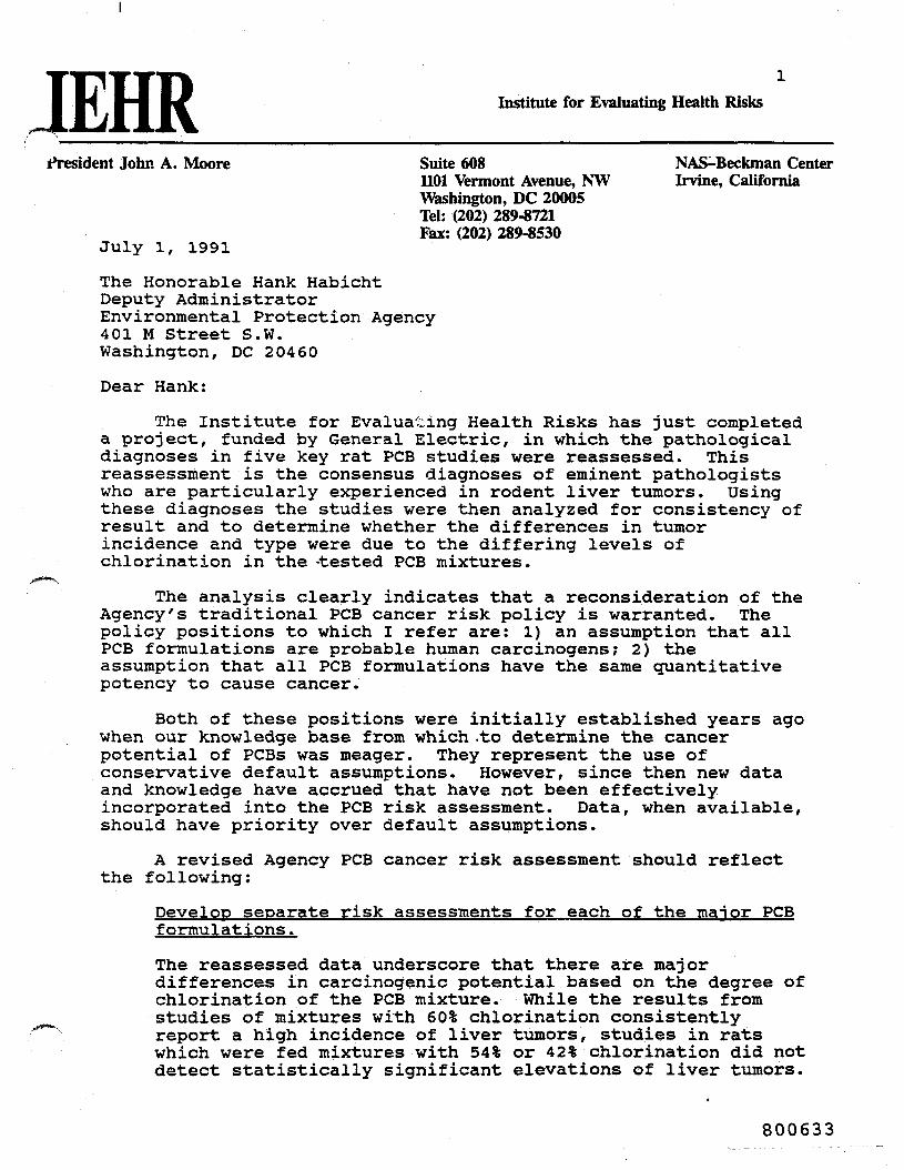
The Honorable Hank HabichtDeputy AdministratorEnvironmental Protection Agency401 M Street S.W.Washington, DC 20460
Dear Hank:
The Institute for Evaluating Health Risks has just completeda project, funded by General Electric, in which the pathologicaldiagnoses in five key rat PCB studies were reassessed. Thisreassessment is the consensus diagnoses of eminent pathologistswho are particularly experienced in rodent liver tumors. Usingthese diagnoses the studies were then analyzed for consistency ofresult and to determine whether the differences in tumorincidence and type were due to the differing levels ofchlorination in the -tested PCB mixtures.
~"S
The analysis clearly indicates that a reconsideration of theAgency's traditional PCB cancer risk policy is warranted. Thepolicy positions to which I refer are: 1) an assumption that allPCB formulations are probable human carcinogens; 2) theassumption that all PCB formulations have the same quantitativepotency to cause cancer.
Both of these positions were initially established years agowhen our knowledge base from which .to determine the cancerpotential of PCBs was meager. They represent the use ofconservative default assumptions. However, since then new dataand knowledge have accrued that have not been effectivelyincorporated into the PCB risk assessment. Data, when available,should have priority over default assumptions.
A revised Agency PCB cancer risk assessment should reflectthe following:
Develop separate risk assessments for each of the manor PCBformulations.
The reassessed data underscore that there are majordifferences in carcinogenic potential based on the degree ofchlorination of the PCB mixture. While the results fromstudies of mixtures with 60% chlorination consistently
"" report a high incidence of liver tumors, studies in ratswhich were fed mixtures with 54% or 42% chlorination did notdetect statistically significant elevations of liver tumors.
800633
PCB Cancer Risk Policy
reaffirmed that chronic dietary exposure of rats, in threedifferent studies, to 60% chlorinated PCB formulationsresulted in the development of benign and malignant livertumors;
reaffirmed that chronic exposure of rats to a PCBformulation that was 54% chlorinated did not yield astatistically significant increase of either benign ormalignant tumors;
revealed that rats chronically exposed to a PCB formulationthat was 42% chlorinated did not develop any increase inmalignant tumors or a statistically significant increase inbenign tumors.
These reassessment results indicate that the following twotraditional EPA PCB policy positions be reconsidered: 1) anassumption that all PCB formulations are probable humancarcinogens; 2) the assumption that all PCB formulations have thesame quantitative potency to cause cancer.
Both of these positions were initially established years agowhen our knowledge base from which to determine the cancerpotential of PCBs was meager. They represent the use ofconservative default assumptions. However, since then new dataand knowledge have accrued that have not been effectivelyincorporated into the PCB risk assessment.2
I believe that a revised PCB cancer risk assessment shouldreflect the following:
2 Because of insufficient data default assumptions commonly are anecessary component of a risk assessment. However, there isanother policy position which should guide the decision thatdetermines the use of defaults; that overarching policy shouldestablish a clear bias for the use of data whenever it isavailable. In other words the operant policy position is to usedata, the burden should lie on the risk assessor to clearlyestablish why available data should not be used before it can bereplaced by a default assumption.
800634

PCB Cancer Risk Policy
Develop separate risk assessments for each of the manor PCBformulations.
The reassessed data underscore that there are majordifferences in carcinogenic potential based on thedegree of chlorination of the PCB mixture. While theresults from studies of mixtures with 60% chlorinationconsistently report a high incidence of liver tumorsstudies in rats which were fed mixtures with 54% or 42%chlorination did not detect statistically significantelevations of liver tumors. It is not proper tocontinue a policy which does not consider data,developed subsequent to the initial judgement, thatdemonstrates other formulations are either notcarcinogens or at best, weak carcinogens. There isprecedent for such action; several years ago theScience Advisory Panel, which advises the State ofCalifornia on cancer designations under Proposition 65,voted to recommend Aroclor 1260 as a carcinogen ratherthat list all PCBs.
The tissue diagnoses of the expert group of patholoaistsshould be used for risk assessment.
There are three factors that support the use of theseconsensus diagnoses:
1) it reflects the use of current pathology conventionsthat are endorsed by the National Toxicology Programand the Environmental Protection Agency;
2) it represents the consensus opinion of pathologiststhat are experienced in the evaluation of rodentbioassays; specifically liver tumors.
800635

PCB Cancer Risk Policy 4
3) the results of the present review permits greaterconfidence that observed differences in tumor incidenceand type in each study are due to differences in thetest substances.
Utilize all available data when calculating cancer potencyfor PCB mixtures that have 60% chlorination.
There is no logical basis to continue the currentpractice of only using the results obtained in femaleSprague Dawley rats. A comparison of the results ofeach of these studies3 shows a striking similarity inthe nature of the tumor response. It should be notedthat three separate strains of rats were used and thatthe similarity of response is apparent when onecompares female Sherman rats, male Wistar rats, andfemale Sprague Dawley rats. Male Sprague Dawley rats,while developing the same type of liver tumors, did soat a lower incidence. To assume that this reducedresponse reflects a generic tendency of male rats notto develop tumors is not supported by the data. Thegreatest incidence of liver tumors (91.2%) was observedin male Wistar rats. The results in male Wistar ratsalso do not support continuing the practice ofcensoring the male Sprague Dawley results from thecalculation of a cancer slope factor.
3 Induction of Liver Tumors in Sherman Strain Rats ByPolychlorinated Biphenyl Aroclor 1260. Kimbrough, R.D. et'al,JNCI (1975) 55:6, 1453-1459.Polychlorinated Biphenyl Induction of Hepatocellular Carcinoma
in the Sprague Dawley Rat. Norback, D.H. & Weltman, R.H., EnvHlth Perspect (1985) 60:, 97-105.Pathology of Chronic Polychlorinated Biphenyl (PCB) Feeding in
Rats. Schaeffer, E., Greim, H., & Goessner, W., Tox & AppliedPharm. (1984) 75:, 272-288.
800636

PCB Cancer Risk Policy 5
When using the results from each of these studies oneshould apply a consistent decision rule to thecensoring of animals from studies; each author used adifferent convention in their publications. Observingthe convention employed by the National ToxicologyProgram may be more appropriate and consistent for allstudies. The group size in several of these studieswould increase if this recommendation were adopted.
Employing the geometric mean of the cancer potencyfactors of the four study groups, female Sherman, maleWistar, male Sprague Dawley, and female Sprague Dawleyrats would reflect a less arbitrary use of all existingdata. There is ample precedent for this approach in anumber of Agency decisions. The geometric mean, usingthe re-evaluation results, would yield a cancer potencyfactor of approximately 1.9. The current valuecalculated by EPA is 7.7 using only the female SpragueDawley rat.
The reassessment of the NCI study5 clarifies thesignificance of "nodular hyperplasia"
This study which evaluated a PCB mixture with 54%chlorination, essentially reaffirmed the originalfindings that the bioassay did not show a carcinogenicresponse in either male or female F344/N rats. Thegroup size at each treatment level was 24 rats.
4 Censor all rats that died during the first year of the study orcensor rats that died prior to the diagnosis of the first tumorin a target organ; whichever date is earlier.5 Bioassay of Aroclor 1254 for Possible Carcinogenicity. NCTCarcinogenisis Technical Report Series, Number 38, 1978.
800637

PCB Cancer Risk Policy 6
Utilizing the current pathology nomenclature theconsensus diagnoses by the expert panel classified"nodular hyperplasia" lesions, a. designation used inthe original report, as nonneoplastic. Therefore,continuing to incorporate the incidence of nodularhyperplasia in a cancer potency calculation, as wasdone in the most recent Water Quality Criteria
• Document6 would fail to have a supportable scientificbasis.
Rather than exclusively focus on how to estimate acancer potency factor for the 54% chlorination PCBmixture I would urge consideration of a morefundamental question; namely, the estimation of cancerpotency from any negative study.
The reassessment of the pathology diagnoses of lesions inthe liver of rats fed a PCB mixture containing 42%chlorination reveals that there is no statisticallysignificant increase in tumors.
This study, which was performed in parallel with a PCBmixture with 60% chlorination, has not been accordedthe attention that it deserves from a risk assessmentperspective.8
6 Drinking Water Criteria Document for Polychlorinated Biphenyls,April 1988, (PB89-192256) pp VIII-32 to VIII-35.7 Liver tumor incidence in controls 8/120 (hepatocellular adenoma6/120, hepatocellular carcinoma 2/120). Liver tumor incidence intreated group 16/128 (hepatocellular adenoma 14/128,hepatocellular carcinoma 2/128). Fisher exact test, one tailed,p =.098). It is arguable that a two tailed test should be usedgiven that a decrease in pituitary tumors and endocrine tumorswas reported in several of these studies. A two tailed testwould further erode the p value.8 Pathology of Chronic Polychlorinated Biphenyl Feeding in Rats.Schaeffer, E., Greim, H., & Goessner, W., Tox. & Applied Pharm.(1984) 75:, 272-288
800638

PCB Cancer Risk Policy 7
Factors which underscore the value of this studyinclude:
1) it is the only major study of a PCB mixture withthis level of chlorination.
2) it has far better statistical power to detect aneffect than do most bioassays, e.g., the number ofanimals studied were about two and a half times greaterthan required by EPA or used by the National ToxicologyProgram.
3) the selection of male rats as the test subject wouldnot appear to be a limitation. A parallel group ofmale rats, fed a PCB mixture containing 60% chlorine,yielded a liver tumor incidence of 91%, the highestincidence reported in any of the studies that werereassessed.
4) the study duration was approximately 118 weeks, thisis three months longer than the protocol requirementsof either EPA or the National Toxicology Program. Itis generally held that studies of longer duration favorthe detection of tumors, particularly with these typesof chemicals.
I am not asking you to focus on an issue that is only ofarcane scientific interest. The current cancer policy is clearlyoverstating the cancer risks associated with many exposures toPCBs in the environment. In a number of instances it is drivingregulatory decisions that, by any standard are a major economicimpact for, at best, trivial public health gain. As anillustration, mixtures with 60% or greater chlorination wereabout 12% of total PCB sales in this country; current policycalculates all PCB exposure as if it were equivalent to Aroclor1260.
800639

PCB Cancer Risk Policy 8
While PCBs in the environment undergo changes in composition theydo not develop into the chemical "fingerprint" that identifiesAroclor 1260. Therefore, 88% of the PCB that was used is beingtreated as if it were a potent carcinogen when the data indicatethat these lower chlorinated mixtures are either of markedlydiminished potency or not carcinogenic at all!
A request to develop a risk assessment utilizing allpertinent data, I believe, is consistent with the Agency's statedgoals of focusing on risks which represent true public health orenvironmental concern and of reducing the uncertainties in riskassessment by applying sound scientific knowledge.
I would be pleased to work with the Agency in explaining theresults of this project and discussing alternative approaches toestimating PCB risks.
Sincerely,
A/John A. Moore
800640

EPLEXPERIMENTAL PATHOLOGY LABORATORIES, INC.
REASSESSMENT OF LIVER FINDINGSIN PCB STUDIES IN RATS
PATHOLOGY WORKING GROUP REVIEW
Submitted to:
Institute for Evaluating Health RisksWashington, DC 20005
Submitted by:
Experimental Pathology Laboratories, Inc.Research Triangle Park, NC 27709
June 27, 1991
800641

EPLEXPERIMENTAL PATHOLOGY LABORATORIES, INC.
PW6 PARTICIPANTS:
Dr Jerry F. Hard i styExperimental Pathology
Laboratories, Inc. (Chairperson)
- ' - l. W. Ray /JDr. W. Ray /Jrown
Research Pathology Services, Inc.
QJLDr. Ernest E. McConnellConsultant
/^ufe» (2.James A. Popp
Chemical Industry Instituteof Toxicology
Dr. Robert A. SquiJohns Hopkins University
Di^Jerrold M. WardConsultant
Dr. Deborah A. BanasExperimental Pathology
Laboratories, Inc.(Reviewing Pathologist)
800642

EPLEXPERIMENTAL PATHOLOGY LABORATORIES, INC.
REASSESSMENT OF LIVER FINDINGSIN PCB STUDIES IN RATS
PATHOLOGY WORKING GROUP REVIEW
NARRATIVE SUMMARY
INTRODUCTION
Polychlorinated biphenyls (PCBs) are compounds whose
physical and chemical properties led to their widespread use for a
nansber of commercial applications. Because of their extensive use in
the past and their resistance to chemical and biological breakdown, PCBs
are now widely distributed in the environment.
PCRs were manufactured commercially by the chlorination of
biphenyl. The number and placement of chlorine atoms introduced into
the biphenyl molecule determines each PCBs' structure. There are 209
possible PCB congeners or homologs. Commercial PCB formulations are
composed of complex mixtures of individual PCBs rather than a single
congener. The percent chlorine by weight increases as the average
number of chlorine atoms per biphenyl is increased. The chlorine
content of various commercial formulations differs according to the
desired physical characteristics for specific applications (Siberhorn,
et al.( 1990).
A number of studies have been undertaken to assess the
potential toxicity and carcinogenicity of commercial PCB preparations.
The main target organ of PCBs is the liver. A number of investigators
have studied the potential carcinogenic effect of various PCBs in the
800643
EPLEXPERIMENTAL PATHOLOGY LABORATORIES, INC.
liver of rats and mice. These studies have indicated that benign or
malignant hepatocellular tumors and various nonneoplastic hepatic
changes are observed in the liver of rodents when given at appropriate
doses for extended periods of time. These studies also indicated that
PCB mixtures with a high chlorine content are more potent in inducing
neoplastic nodules and hepatocellular carcinomas than mixtures with less
chlorination. In most of t sse studies, the criteria for diagnosis and
nomenclature*designations according to Squire and Levitt were used by
the investigators to classify the hepatocellular changes (Siberhorn, et
a!., 1990).
In the recent past, there have been changes in the criteria
and nomenclature for hepatoproliferative lesions of rats (Maronpot, et
al., 1986). These changes resulted from the increased accumulation of
data, as well as a better understanding of the mechanisms of toxicity
and carcinogenesis. In light of these changes, it was considered
reasonable to reexamine the livers from several earlier PCB studies to
assess the risk posed by these compounds based on the current
understanding of hepatic changes. At the request of the Institute for
Evaluating Health Risks (IEHR), Experimental Pathology Laboratories,
Inc. (EPL) assembled all liver slides from five (5) key chronic PCB
studies for the purpose of reassessment of the liver findings following
current diagnostic criteria and nomenclature. These studies included
the following: Aroclor 1260 in female Sherman rats (Kimbrough, et al.,
800644

EPLEXPERIMENTAL PATHOLOGY LABORATORIES, INC.
1975); Aroclor 1260 in male and female Sprague-Dawley rats (Norback and
Weltman, 1985); National Cancer Institute (NCI) Aroclor 1254 study in
male and female Fischer 344 rats (NCI, 1977); Clophen A 60 in male
Wistar rats (Schaeffer, et a!., 1984); and Clophen A 30 in male Wistar
rats (Schaeffer, et al., 1984). Additionally, the liver slides from
male and female rats used r,. a reproduction study in male and female
Sherman rats with Aroclor 1260, in which the exposure was limited to
nine months, were reexamined co characterize the degree and extent of
hepatic lesions resulting from subchronic exposure to Aroclor 1260
(Linder, et al., 1974). References for each of these studies are
included in Appendix F. The concurrent review of these studies provide
a unique opportunity to compare the incidence, type and severity of
hepatic lesions observed in each.
A summary of the experimental design for each of the studies
included in this current review is presented on the following table.
800645

Study
Kimbrough, et al., 1975
SUMMARY OF EXPERIMENTAL DESIGNPCB STUDIES IN RATS
Chemical % C1 by Ht. Strain Sex Dosage levels
Aroclor 1260* 60% Sherman F 100 ppm
Durationof Exposure*
23 Months
Norback and Weltman, 1985 Aroclor 1260 60% Sprague- M/FDawley
100 ppm for 16months followedby 50 ppm for 8months then controldiet for 5 months
29 Months
Schaeffer, et al., 1984 Chlopen A 60 60% Wlstar M 100 ppm 801-832Days
NCI, 1977 Aroclor 1254 52-54% F344 M/F 25 ppm, 50 ppm 104-105100 ppm Weeks
Schaeffer, et al., 1984 Clophen A 30 40-42% Wistar M 100 ppm 801-832Days
oooo
o\
Under, et a]., 1974(Reproduction Study)
Aroclor 1260 60% Sherman M/F 100 ppm
*Durat1on of exposure published by original investigator.
9 Months

EPLEXPERIMENTAL PATHOLOGY LABORATORIES, INC.
METHODS
After assembling the hematoxylin and eosin stained
microscope slides and data from each of the studies to be reviewed, the
procedures developed by the National Toxicology Program which utilizes a
Pathology Working Group (PWG) were generally followed to conduct the
review. The PWG members consisted of Veterinary Pathologists with
extensive experience in the microscopic evaluation and interpretation of
hepatic changes observed in bioassay studies conducted in rodents. The
PWG's task was to confirm the incidence of hepatocellular neoplasms,
resolve diagnostic discrepancies, validate treatment-related effects and
discuss the biological significance of the potential effects present.
Prior to the PWG review, all slides within each individual
study were randomized by animal number and then coded in ascending
numerical order. The randomized slides were examined without knowledge
of treatment group by the Reviewing Pathologist, Dr. Deborah Banas,
Experimental Pathology Laboratories, Inc. The reviewing pathologist
recorded all changes present in the liver sections including both
neoplastic and nonneoplastic lesions. After microscopic examination,
the data was decoded and presented by treatment group for interpretation
of the results and preparation for the Pathology Working Group review.
The Pathology Working Group was chaired by Dr. Jerry F.
Hardisty, Experimental Pathology Laboratories, Inc., who organized andpresented the material to a panel of five board certified Veterinary
800647

EPLEXPERIMENTAL PATHOLOGY LABORATORIES, INC.
Pathologists. The PWG review was performed on May 29-31, 1991 in the
Research Triangle Park, North Carolina. Individuals attending or
participating in the PWG review are listed as follows:
Dr. W. Ray BrownDr. E.E. McConnellDr. James A. PoppDr. Robert A. SquireDr. Jerrold M. Ward
Dr. Deborah A. Banas
Dr. Jerry F. Hardisty
Dr. Ronald MochDr. D. SinghDr. Jack MooreDr. Renate KimbroughDr. W. GoessnerDr. Diane NorbackDr. William M. Busey
(PWG Participant)(PWG Participant)(PWG Participant)(PWG Particioant)(PWG Participant)
(Reviewing Pathologist)
(PWG Chairperson)
(Observer, PDA)(Observer, EPA)(Observer, IEHR)(Observer. IEHR)(Observer, GSF)(Observer, University of Wisconsin)(Observer, EPL)
Each participant recorded his diagnoses and comments on
worksheets which were prepared by the PWG Chairman. The worksheets for
each participant are on file at EPL. Each lesion was discussed by the
group, reexamined if necessary and the final opinions were recorded on
the Chairperson's Worksheets also maintained on file at EPL, Inc. The
consensus diagnoses of the PWG was reached when at least three of five
PWG participants were in agreement.
After the PWG completed the slide review for each study and
the diagnoses recorded by the PWG Chairperson, the slides were decoded
by treatment group. The consensus diagnoses was entered into a
800648

EPLEXPERIMENTAL PATHOLOGY LABORATORIES, INC.
computerized pathology reporting system at EPL, and Histopathology
Incidence Tables for individual animals and Summary Incidence Tables
were prepared for evaluation and interpretation of the PWG findings.
The Histopathology Incidence Tables and Summary Incidence Tables are
presented in Appendices A - E for each of the studies reviewed.
In addition to providing specific diagnoses for neoplastic
and hyperplastic hepatocellular lesions present on the slides examined,
the PWG participants also provided comments and opinions on the presence
of a variety of nonneoplastic changes including foci of cellular
alteration, and other lesions which may be indicative of potential
hepatotoxicity. The PWG members were consistently in agreement with the
reviewing pathologist concerning the nonneoplastic changes present.
Since the reviewing pathologist had examined all liver slides in both
control and treated animals in these studies using consistent criteria
for diagnosis and severity grading, there was agreement by the PWG
members to accept the reviewing pathologist1s findings for nonneoplastic
changes as the "consensus diagnosis". Therefore the consensus diagnoses
presented in the Histopathology Incidence Tables and Summary Incidence
Tables represent the majority opinion of the PWG for all neoplastic and
hyperplastic changes and the reviewing pathologist's diagnosis for other
nonneoplastic changes.
The diagnostic criteria used by the PWG participants and the
reviewing pathologist for diagnosis of foci of cellular alteration,
800649

EPLEXPERIMENTAL PATHOLOGY LABORATORIES, INC.
hepatocellular hyperplasia and hepatocellular neoplasms is summarized
below:
Foci of Cellular Alteration
1. Localized lesions recognized by virtue of tinctorial staining
variation from surrounding hepatic parenchyma.
2. Usually do not compress but merge imperceptibly with surrounding
parenchyma.
3. Minimal disruption of hepatic lobular architecture.
4. Hepatocytes within focus may have clear, eosinophilic, or
basophilic cytoplasm or a mixture of these and may be larger or
smaller than surrounding hepatocytes.
Focal Hepatocellular Hyperplasia
1. Usually a multifocal nodular lesion found associated with previous
or concurrent hepatic damage. In this context, may be considered
as multifocal regenerative hyperplasia.
2. Lesion consists of spherical proliferation of hepatocytes without
nuclear atypia. May contain cytologic alterations similar to
those seen in foci of cellular alteration.
3. An increased number of mitoses may be evident. Hyperplastic cells
may be hypertrophic or contain intracytoplasmic vacuoles. The
cells within a hyperplastic focus are usually uniform and have a
homogeneous growth pattern.
8
800650

E P LEXPERIMENTAL PATHOLOGY LABORATORIES, INC.
4. Hepatic lobular architecture is evident but may be distorted.
Portal triads can often be found within hyperplastic foci.
Hepatocellular Adenoma
1. Nodular proliferations of hepatocytes which are sharply demarcated
by definite compression of surrounding hepatic parenchyma and
usually by virtue of tinctorial staining differences.
2. Hepatic plates of an adenoma are usually not continuous with
surrounding liver plates but impinge with them at a sharp angle.
3. Loss of normal lobular architecture.
4. Often have an increased mitotic index, may contain areas of
cellular atypia, and have an irregular growth pattern.
5. Cells within an adenoma may be degenerative, hypertrophic, and/or•
contain intracytoplasmic vacuoles.
Hepatocellular Carcinoma1. Usually considerably larger and more irregular than hepatocellular
adenoma.
2. Compress or extend into surrounding hepatic parenchyma.
3. Characterized by one or more of the following: cellular atypia,
local invasiveness, haphazardly arranged cells, broad sheets ofcells, "trabecular" patterns, gland-like formations.
800651

EPLEXPERIMENTAL PATHOLOGY LABORATORIES, INC.
RESULTS
A. Aroclor 1260 in Female Sherman Rats (Kimbrouah. et al..
1975)
Only one control female rat had a hepatocellular neoplasm.
Control female 8231 had a hepatocellular carcinoma. In treated female
rats, 21 rats had hepatocellular carcinomas and 135 rats had-,i!hepatocellular adenomas. A few of the treated female rats had both
hepatocellular adenomas and hepatocellular carcinomas. The
hepatocellular carcinomas were well-differentiated trabecular types.The hepatocytes varied from a normal appearance to enlarged, acidophilic
or diffusely basophilic cells. The cytoplasm often contained
£osinophilic inclusions within vacuoles. Hepatocellular adenomas were
generally spherical and well demarcated. The cells in the adenomas were
generally enlarged with nuclear and cytoplasmic features similar to the
carcinomas. In three of the treated rats the liver tumors had
glandular, papillary patterns giving the appearance of both hepatocytic
and biliary epithelium (8326, 8377 and 8406). These tumors were
considered to represent a subtype of hepatocellular adenoma or carcinoma
and were not given a unique diagnosis. A summary of the incidence of
rats with only hepatocellular adenoma and those with at least one
hepatocellular carcinoma is presented as follows:
10
800652

EPLEXPERIMENTAL PATHOLOGY LABORATORIES,
No. Examined
No. Animals with onlyHepatocellular Adenoma
No. Animals with at Leastone Hepatocellular Carcinoma
Total Animalswith Hepatocellular Neoplasms
INC.
Control
187
0 (0%)
1 (0.5%)
1 (0.5%)
Aroclor 1260
189
117 (61.9%)
21 (11.1%)
138 (73.0%)
Eosinophilic foci were present in 173 of the treated rats
and only seven of the control rats. Other foci frequently occurred in
addition to the eosinophilic foci in treated rats. The incidence of
focus/foci of cellular alteration in the liver of control and treated
female rats is presented as follows:
No. Examined
Focus/Foci, Eosinophilic
Focus/Foci, Basophilic
Focus/Foci, Clear Cell
Focus/Foci, Mixed Cell
No. Animals with Any Typeof Focus/Foci
Control
187
7 (3.7%)
4 (2.1%)
14 (7.5%)
1 (0.5%)
25 (13.4%)
Aroclor 1260
189
173 (91.5%)
67 (35.4%)
67 (35.4%)
38 (20.1%)
177 (93.7%)
Nonneoplastic lesions which appeared to be increased with treatment
included centrilobular hepatocytomegaly, centrilobular fatty change,
11
800653

EPLEXPERIMENTAL PATHOLOGY LABORATORIES, INC.
bile duct hyperplasia, and pigment deposition. Oval cell proliferation,
angiectasis, and dilated bile ducts were also increased in treated rats.Focal necrosis was often noted in treated rats in association with
hepatocellular carcinomas. Cholangiofibrosis characterized by atypical
bile ducts surrounded by abundant dense collagenous connective tissue
was present in three treated rats. The incidence of nonneoplastic
lesions which were increased in treated rats as compared to control rats
is presented below:
No. Examined
Centrilobular Hepatocytomegaly
Centrilobular Fatty Change
Bile Duct Hyperplasia
Pigment Deposition
Oval Cell Proliferation
Angiectasis
Dilated Bile Ducts, Focal
Focal Necrosis
Cholangiofibrosis
Control
187
1 (0.5%)
1 (0.5%)
4 (2.1%)
9 (4.8%)
2 (1.1%)
1 (0.5%)
0 (0%)
0 (0%)
0 (0%)
Aroclor 1260
189
108 (57.1%)
32 (16.9%)
29 (15.3%)
66 (34.9%)
17 (9.0%)
17 (9.0%)
14 (7.4%)
13 (6.9%)
3 (1.6%)
12
800654

EPLEXPERIMENTAL PATHOLOGY LABORATORIES, INC.
B. Reproduction Study With Aroclor 1260 in Male and Female
Sherman Rats. F. Generation (Under, et al.. 1974)
Slight to severe centrilobular hepatocytomegaly was noted
in all ten treated males, and minimal to moderately severe centrilobular
hepatocytomegaly was present in seven of ten treated females. This
lesion was characterized by enlarged hepatocytes, occasionally with
atypical nuclei surrounding the central veins and, in the case of
moderately severe or severe lesions, extending over a fair portion of
the liver lobule. Cytoplasmic inclusions were prominent in these
enlarged hepatocytes in two of the males. Minimal to slight
centrilobular fatty change was noted in two of the treated females andfocal fatty change occurred in one. Single eosinophilic foci were noted
in two treated females. These lesions were characterized by foci of
enlarged, eosinophilic hepatocytes having a ground-glass appearance tothe cytoplasm and having a distinctly different tinctorial appearance
compared to the surrounding parenchyma. Multiple, small, clear cell
foci were noted in one treated male and were characterized by enlarged
hepatocytes having a clear or slightly granular pale cytoplasm. Brownpigment deposition was noted in four of the treated females.
Other lesions noted in the liver sections of both control
and treated rats were microgranulomas. These lesions consisted of focalaccumulations of macrophages and mononuclear inflammatory cells, and
13
800655

EPLEXPERIMENTAL PATHOLOGY LABORATORIES, INC.
were unrelated to treatment with Aroclor 1260. The incidence of these
changes are summarized as follows:Male Rats Female Rats
Control Aroclor 1260 Control Aroclor 1260
No. ExaminedCentrilobular
Focus/Foci. £
Focus/Foci. Clear CellFatty Change, Centr
Fatty Change, Focal
Cytoplasmic InclusionsMicrogranuloma(s)
Pigment Deposition
wtocytomegalylophilic
• Cellitrilobular
:aljslons
)in
Aroclor 1260
and Weltman.
10
0 (0%)
0 (0%)
0 (0%)
0 (0%)
0 (0%)
0 (0%)
3 (30%)
0 (0%)
in Male
1985)
10
10 (100%)
0 (0%)
1 (10%)
0 (0%)
0 (0%)
2 (20%)
2 (20%)
0 (0%)
10
0 (0%)
0 (0%)
0 (0%)
0 (0%)
0 (0%)
0 (0%)
5 (50%)
0 (0%)
10
7 (70%)
2 (20%)
0 (0%)
2 (20%)
1 (10%)
0 (0%)
3 (30%)
4 (40%)
and Female Soraque-Dawlev Rats (Norback
Hematoxylin and eosin stained slides were examined frdm 31
male control, 40 male treated, 45 female control and the 46 female
treated Sprague-Dawley rats. Due to the fact that partial hepatectomies
were performed on two control females (30 and 31) and two treated males
(189 and 212), these animals are represented twice in the Histopathology
Incidence Tables. The animal number followed by "A" represents the
liver section examined following the partial hepatectomy and followed by
"B" for the liver section examined at termination.
A marked sex difference in the number of hepatocellular
neoplasms was present in this study. No hepatocellular neoplasms were
present in any of the control male rats. I.n treated male rats four had
14
800656

EPLEXPERIMENTAL PATHOLOGY LABORATORIES, INC.
hepatocellular adenomas and one had a hepatocellular carcinoma. Only
one control female rat had a hepatocellular adenoma. Hepatocellular
adenomas were present in 29 treated female rats and hepatocellular
carcinomas were present in 19 treated female rats. Additionally, three
treated female rats had cholangiocarcinomas. Most of the hepatocellular
carcinomas were well-differentiated trabecular neoplasms.
Hepatocellular neoplasms in treated female rats 192, 212, 213 and 214
had glandular, papillary patterns similar to that described above for
the Kimbrough study in female Sherman rats. Hepatocellular adenomas
were generally spherical, we11-demarcated tumors composed of well-
differentiated cells which produced mild to moderate compression of the
surrounding hepatic parenchyma. A few of the treated female rats had
both hepatocellular adenoma and carcinoma. A summary of the incidence
of male and female rats with only hepatocellular adenoma and rats with
at least one hepatocellular carcinoma is presented as follows:
Hale Rats Female Rats
No. ExaminedNo. Animals with onlyHepatocellular Adenoma
No. Animals with at Leastone Hepatocellular Carcinoma
Total Animals .with Hepatocellular Neoplasms
Control31
0 (0%)
0 (0%)
Aroclor 1260
40
4 (10%)
1 (2.5%)
Control45
1 (2.2%)
0 (0%)
Aroclor 1260
46
22* (47.8%)
19* (41.3%)
0 (0%) 5 (12.5%) 1 (2.2%) 41 (89.1%)
*Two female rats with hepatocellular adenoma and one with hepatocellular carcinomaalso had a cholangiocarcinoma.
An increased incidence of eosinophilic cell foci was present
in treated male and female rats as compared to control rats. Although a
15
800657

EPLEXPERIMENTAL PATHOLOGY LABORATORIES, INC.
few basophilic, clear cell and mixed cell foci were also observed they
did not occur in a treatment-related manner. A summary of the incidence
of foci of cellular alteration in the liver in Sprague-Dawley Rats is
presented below:Hale Rats Female Rats
Control Aroclor 1260 Control AroclorNo. Examined 31 40 45 46
Focus/Foci. Eosinophillc 1 (3.2%) 16 (40%) 5 (11.1%) 36 (78.3%)
Focus/Foci. Basophilic 1 (3.2%) 0 (0%) 2 (4.4%) 1 (2.2%)Focus/Foci. Clear Cell 4 (12.9%) 0 (0%) 1 (2.2%) 0 (0%)
Focus/Foci. Mixed Cell 0 (0%) 2 (5%) 0 (0%) 0 (0%)
No. Animals with Any Typeof Focus/Foci 5 (16.1%) 16 (40%) 7 (15.6%) 36 (78.3%)
Other hepatic lesions which appeared to be increased in
treated groups as compared to control male and female rats included
centrilobular hepatocytomegaly, centrilobular fatty change and
centrilobular necrosis. In treated female rats the incidence of bile
duct hyperplasia, cholangiofibrosis, cystic bile ducts, periportal
fibrosis and pigment deposition was also increased in incidence as
compared to control female rats. The incidence of selected
hepatocellular lesions which were increased in incidence in treated male
and/or female rats as compared to controls are presented as follows:
16
800658

EPLEXPERIMENTAL PATHOLOGY LABORATORIES, INC.
Male Rats
No. Examined
Centrilobular Hepatocytomegaly
Centrilobular Fatty Change
Centrilobular Necrosis
Bile Duct Hyperplasia
Cholangiofibrosis
Cystic Bile DuctsPeri portal Fibres is
Pigment Deposition
D. Cloohen A 60
Control
31
0 (0%)
0 (0%)
0 (0%)
6 (19.4%)
0 (0%)
0 (0%)2 (6.5%)
1 (3.2%)
in Male
Aroclor 1260
40
15 (37.5%)
17 (42.5%)
7 (17.5%)
8 (20%)
0 (0%)
1 (2.5%)3 (7.5%)
2 (5.0%)
Wistar Rats
Female RatsControl
45
0 (0%)
0 (0%)
2 (4.4%)
2 (4.4%)
0 (0%)
1 (2.2%)0 (0%)
2 (4.4%)
Aroclor 1260
46
5 (10.9%)
2 (4.3%)
4 (8.7%)
19 (41.3%)
4 (8.7%)
5 (10.9%)12 (26.1%)
13 (28.3%)
(Schaeffer. et al.. 1984)
Hepatocellular adenomas were present in six control males
and in 85 treated males. Hepatocellular carcinomas were present in two
control males and in 67 treated males. Additionally one of the control
males with a hepatocellular carcinoma also had a cholangiocarcinoma in
the liver. Although most of the hepatocellular neoplasms consisted of
well-differentiated adenomas and carcinomas, nine of the tumors present
in the treated group had glandular, papillary patterns suggestive of a
mixture of hepatocellular and biliary epithelium. Although this pattern
was observed only in treated rats, it was considered to most likelyrepresent a subclassification of hepatocellular adenoma or carcinoma and
was not given a unique morphologic diagnosis. A few of the treated male
rats had both hepatocellular adenomas and hepatocellular carcinomas. Asummary of the incidence of male Wistar rats with only hepatocellular
17
800659

EPLEXPERIMENTAL PATHOLOGY LABORATORIES, INC.
adenoma and rats with at least one hepatocellular carcinoma is presented
as follows:
No. Examined
No. Animals with onlyHepatocellular Adenoma
No. Animals with at Leastone Hepatocellular Carcinoma
Total Animalswith Hepatocellular Neoplasms
Control
120
6 (5.0%)
2*(1.7%)
8 (6.7%)
*0ne control male also had cholangiocarcinoma.
Cloohen A 60
125
47 (37.6%)
67 (53.6%)
114 (91.2%)
Eosinophilic foci were present in the liver of 51 control
and 101 treated rats. Additionally, the number of male rats with clear
cell and mixed cell foci were greater in the treated group than in
the control group. The incidence of focus/foci of cellular alteration
in the liver of control and treated rats is presented as follows:
No. Examined
Focus/Foci, Eosinophilic
Focus/Foci, Basophilic
Focus/Foci, Clear Cell
Focus/Foci, Mixed Cell
No. Animals with Any Typeof Focus/Foci
Control
120
51 (42.5%)
2 (1.7%)
7 (5.8%)
7 (5.8%)
Cloohen A 60
125
101 (80.8%)
4 (3.2%)
28 (22.4%)
28 (22.4%)
55 (45.8%) 108 (86.4%)
18
800660

EPLEXPERIMENTAL PATHOLOGY LABORATORIES, INC.
With the exception of an increased incidence of focal
necrosis which was associated with hepatocellular carcinomas in rats
receiving Clophen a 60, other nonneoplastic lesions appeared to be
comparable or decreased in treated rats.
E. Cloohen A 30 in Male Wistar Rats (Schaeffer. et al.. 1984)
The Clophen A 30 study in Wistar rats was conducted at the
same laboratory at the same time as the Clophen A 60 study discussed
above and therefore shared the same control group. In the Clophen A 30
treated rats 14 hepatocellular adenomas and two hepatocellular
carcinomas were present as compared to six adenomas and two carcinomas
in the control group. All of the hepatocellular tumors in the control
and treated rats occurred as singular tumors and none of the rats had
both a hepatocellular adenoma and a hepatocellular carcinoma. One
control male had a cholangiocarcinoma in addition to a hepatocellular
carcinoma. A summary of the incidence of male rats with hepatocellular
neoplasms is presented below:
No. Examined
Hepatocellular Adenoma
Hepatocellular Carcinoma
Total Hepatocellular Neoplasms
Control
120
6 (5.0%)
2 (1.7%)
8 (6.7%)
Cloohen A 30
128
14 (10.9%)
2 (1.6%)
16 (12.5%)
19
800661

EPLEXPERIMENTAL PATHOLOGY LABORATORIES, INC.
The incidences of focus/foci of cellular alteration of all
types (basophilic, eosinophilic, clear cell and mixed cell) were greater
in treated male rats than in control male rats. The incidence of
focus/foci of cellular alterations present in control and treated groups
is summarized as follows:
Control Clophen A 30
No. Examined 120 128
Focus/Foci, Eosinophilic 51 (42.5%) 98 (76.6%)
Focus/Foci, Basophilic 2 (1.7%) 15 (11.7%)
Focus/Foci, Clear Cell 7 (5.8%) 39 (30.5%)
Focus/Foci, Mixed Cell 7 (5.8%) 49 (38.3%)
No. Animals with Any Typeof Focus/Foci 55 (45.8%) 106 (82.8%)
The incidence of other nonneoplastic lesions were either decreased orunchanged in the treated group as compared to the control group.
F. Aroclor 1254 in Male and Female Fischer 344 Rats (NCI. 1977)
The results of the PWG review generally confirmed the
conclusion of the NCI Technical Report that "under the conditions of the
bioassay Aroclor 1254 was not carcinogenic in Fischer 344 rats at the
doses tested; however, a high incidence of hepatocellular proliferative
lesions in both male and female rats was related to administration of
the chemical".
20
800662

nodular hyp«rplasia in both the male and female animals. There
was one carcinoma and four adenocarcinooas in che gastrointes-
tinal tract of treated rats. These neoplastic lesions are seen
only sporadically and at a lov incidence in the Fischer 344 rat;
in this .study no lesions of these types were diagnosed in either
the sal* or female control*.
0. Statistical Analyses of Results
Tables Cl and C2 in Appendix C contain the statistical analyses
of the incidences of those primary tuaors that occurred in at
least two animals in one group and with an incidence of at least
5Z of one or more treated groups.
la male rats, the results of the Cochran-Armitage test for
positive dose-related trend in the incidences of leukemia and of
coobined leukemia and lymphoms axe significant (P • 0.022 and P •
0.009, respectively). The corresponding results of the Fisher
exact test, however, axe not significant in any treated group
when compared with the; controls* Th*re is no other incidence of
tumors at any specific site in either sex which is statistically
significant. A significant Cochraa-Axmitage trend in the
negative direction is observed in the incidence of interstitial-
cell tumor of the testis, where the incidence in the controls
exceeds those in the mid- and high-dose groups.
21
800663

In each of the 95J confidence intervals of relative risk., shown
in the tables, the value of one is included; this indicates the
absence of significant positive results. It should also be noted
that each of the intervals has an upper limit greater than one,
indicating the theoretical possibility of the induction of tumors
by Aroclor 1254, which could not be detected under the
conditions •&£ this test.
22
800664

IV. DISCUSSION
At the doses used in this bioassay, Aroclor® 1254 was toxic to
both male and female Fischer 344 rats, as shown by the
dose-related depression of mean body weights and the clinical
signs which occurred during the second year. Mean body weights
of mid- and high-dose males and of all treated females were
consistently lower than those of the corresponding controls after
the initial growth pha*e. An intercurrent respiratory infection
at week 30 resulted in temporary weight loss, but no deaths, in
all groups including the controls; the animals later recovered
without treatment. Clinical signs including alopecia, amber-
colored urine, facial edema, ezophthalmos, and cyanosis occurred
in the high-dose groups beginning at week 72 and in the mid-dose
groups at week 104. Survival among males, but not among females,
showed a significant dose-related trend. Adequate numbers of
animals of both sexes survived fox meaningful statistical
analyses of the incidences of tumors.
The combined incidences of lympboaa and 'leukemia -in males were
significant (controls 3/24, low-dose 2/24, mid-dose 5/24, high-
dose 9/24, P - 0.009), using the Cockran-Armitage test for
positive dose-related trend, but not in females (controls 4/24,
low-dose 6/24, mid-dose 6/24, high-dose 6/24). Since the results
of the Fisher exact test for increased incidence were not
23
800665

significant for any of these groups, the occurrence of these
lesions cannot clearly be related to the administration of
Aroclor® 1254.
Uepatocellular changes including hyperplastic nodules, adenomas,
and carcinomas were found in treated animals, but none of these
lesions were found in control animals in this study. Bepato-
cellular carcinomas were observed In one mid-<iose and two high-
dose (sales, and hepatocellular adenomas were observed in one
high-dose male, one mid-dose female, and two high-dose females.
Nodular hyperplasia was diagnosed with a dose-related frequency
in the low-, mid-, and high-dose male and female rats. Although
the incidences of the tumors were not significant, the occurrence
of these proliferatlve lesions appeared to be related to
treatment.
In the stomach, jejunum, or ceeua, adenocarcinomas were observed
in two treated males and in tiro treated females as well as a
carcinoma -in one treated male. None of these lesions was found
in control animals in this study, suggesting that the lesions —
although not statistically significant — may be related to the
administration of Aroclor® 1254.
The toxicity of polychlorinated biphenyls (PCBs) has been
reviewed by several groups, including the Environmental
24
800666

Protection Agency (1976), National Research Council (1976), Panel
on Hazardous Trace Substances (1972), and International Agency
for Research on Cancer (1974). Kiabrough et al. (1972)
demonstrated hepatic adeoofibrosis in aale and female Sherman
rats fed Aroclor® 1254 at up eo 300 ppm for 8 months. A similarAPCB, Kaneehlor 500, fed for 12 months to male Vlstar rats,
induced nodular hyperplasia at doses of 100*1,000 ppm; at 1,000
ppm, cholangiofibrosis also was induced (Ito et al., 1974).
teplisiger et al. (1971; see also SPA, Critera Document PCBs, 1976)
fed Charles River rats up to 100 ppm Aroclor® 1254 for 24 months
and reported, originally that there MS mo significant increase
in hepatic tumors in this study; re-evaluation of the liver
slides, however, indicated a. significant incidence of nc&ilar
hyperplasia in created rats, compared with controls. Zto et al.
(1973) observed nodular hyperplasia and veil-differentiated
hepatocellular carcinoma in male strain dd mice fed 500 ppm
Kanechior* 500 for 8 months, and Kimkrough and Linder (1974)
observed adenofibrosis and h*patomes in BALB/cJ mice fed 300 ppm
Aroclor* 1254 for 11 months.
In a study of a closely related PCB, Kiabrough et al. (1975)
observed hepatocellular carcinomas in female Sheraan rats fed 100
ppm Aroclor® 1260 for 21 months.
It is concluded that under the conditions of this bioassay,
25
800667

Aroclor® 1254 was not carcinogenic in Fiacher 344 rats; however,
a high incidence of hepatocellular proliferative lesions in both
male and female rats was related to treatment. In addition, the
carcinomas of the gastrointestinal tract may be associated with•
treatment in both males and females.
26
800668

V. BIBLIOGRAPHY
Armitage, P., Statistical Methods in Medical Research. John Wiley& Sons, Inc., New York, 1971, pp. 362-365.
Berenblum, I., ed., Carcinogenic!tv Testing; A Report of thePanel on Careinogenicity of the Cancer Research Commissionof the UICC. Vol. .2. International Union Again*t Cancer,Geneva, 1969.
Broadhurst, M. C., Use and replaceability of polychlorinatedbiphenyls. Environ. Health Persoect. .2:81-102, 1972.
Cox, 0. R., Regression model* and life tables, J. R. Statist.Soc. B34(2):187-220. 1972.
Cox, D. R., Analysis of Binary Data. Methuen I Co., Ltd., London,1970, pp. 48-52.
Environmental Protection Agency, Polychlorinated Biphenyls(PCBa). Toxic Substances Control. Federaj. Register.42(100), 26564-26577, 1977.
• «
Environmental Protection Agency, Criteria Document PCBs. U. S.Government Printing Office, Washington, 0. C., 440/9-76-021.
Finklea, J., Priester, L. E., Creason, J. P., Hauser, T.,Hinners, T., and Hamaer, D. I., Polychlorinated bipaenylresidues la bum*a plasm* expose a major urban pollutionproblem. AJPH 62(5>i645-651. 1972.
Gart, J. J., The comparison of proportion*: « review ofsignificance tests, confidence limits and adjustments forstratification. Rev. Int. Stat. Inst. 39(2);148-169. 1971.
Hubbard, H. L., Chlorinated biphenyl and related compounds.Kirk-0thaer Encyclopedia of Chemical Technology. Vol. .5,,Interscience Publishers, New York, 1964, pp. 289-297.
International Agency for Research on Cancer, Some anti-thyroidand related substances, nitrofurans and industrialchemicals. IARC Monographs on the Evaluation of theCarcinogenic Risk of. Chemicals to Man; Vol. 2, World .HealthOrganization, Geneva, 1974, pp. 261-289.
27
800669

Ito, N. , Nagasaki, B., Makiura, S., and Aral, M.,Histopathological studies on liver tuoorigenesis In ratstreated with polychlorinated biphenyls. Gann 65(6):545-549, 1974.
Ito, N., Nagasaki, H., Arai, M., Makiura, S., Sugihara, S., andHirao, K., Histopathologic studies en liver tunorlgenesisinduced in mice by technical polychlorinated biphenyls andits pronoting effect on liver tumors induced by benzenehexachloride. J. Natl. Cancer Inst. 51(5): 1637-1642, 1973.
Kaplan, E. L. and ISaler, P., Nonparametrlc estimation fromincomplete observations. J. Aner. Statist. Assoc.13:457-481, 1958.
Keplingftr, H. L., Fa richer, 0. £., and Calandra, J. C.,Toxicologic studies with polychlorinated biphenyls.Toxicol. Appl. Pharmaeol. 19(2);402-403. 1971.
Kiobrough* R. D., Sqtrire, R. A., Linder, R. E., Strandberg, J.0., Montali, R. J., and Burse, V. W., Induction of livertumors in Sheroan strain female rats by polychlorinatedbiphenyl Aroclor 1260. J- National Cane, last. 55(6);1453-1456, 1975.
Klmbrough, R. D. and Linder, R. E., Induction of adenofibrosisand hepatomas of the liver in BALB/cJ nice bypolychlorinated biphenyls Uroclor 1254). J. Natl* CancerInst. 53(2);547-552. 1974.
Klabrough, R. D., Linder, R. E., and Gaines, I. B., Morphologicalchanges in livers of rats fed polychlorinated biphenyls.Arch. Environ. Health 25;354-364. 1972.
Linhart, M. S., Cooper, J. A., Martin, R. L., Page, N. P., andPeters, J. A., Carcinogenesis bioassay data system. Comp.and Bionad. Res. 7;230-246. 1974.
Killer, R. G., Jr., Simultaneous Statistical Inference. McGraw-Hill Book Co., New York, 1966, pp. 6-10.
National Research Council, Report of Organic Contaminants(unpublished), National Research Council, Assembly of LifeSciences, Washington, D.C., 1976.
28
800670

Panel on Hazardous Trace Substances, Polychlorinacedbiophenyls-environmental impact. Environ. Res. -5_:249-362,1972.
Poffenberger, N. and Hubbard, K. L., Diphenyl and terphenyls.Klrk-Othaer Encyclopedia of Chemical Technology. Vol. 1,Interscience Publishers, New York, 1965, p. 193. ~
Saffiotci, U., Montesano, R., Sellakuaer, A. R., Cefis, F., andKaufoan, D. C., Respiratory tract carcinogenesis in haastersInduced by different numbers of administrations of benzo(a)pyrene and ferric oxide. Cancer Res 32;1073-1081. 1972.
Squire, R. A. and L*vitt, H. H., Report on a workshop onclassification of specific heptocellular lesions in rats.Cancer Res. 35;32H-3223. 1975.
Tarone, R. E., Testa for trend in life table analysis.Bioaetrika 62(3);67f-682. 1975.
29
800671
Environmental Health PerspectivesVol. eo. pp. s;-/o5. IBM
Polychlorinated Biphenyl Induction ofHepatocellular Carcinoma in theSprague-Dawley Ratby D. H. Norback* and Robert H. Weltman*t
Male and female Sprague-Dawley rats ("0 males and 70 females in the initial group) were fed a dietcontaining a polychlorinated biphenyl mixture (Aroclor 1260. ]00 ppm for 16 months and 50 ppm for anadditional 8 months) for 2 years followed by a control diet for a months. A control group initially consistedOf 63 males and 63 females. Sequential morphologic changes were evaluated throughout the study. In thePCB-exposed group the following hepatocellular lesions developed in sequence: centrolobular cell hyper-trophy at 1 month, foci of cell alteration at 3 months, areas at 6 months, neoplastic nodules at 12 months,trabecular carcinoma at 15 months, and adenocarcinoma at 24 months. In addition, simple and cysticcholangioma at 18 and 23 months, respectively, and adenofibrosis at 22 months were present. With theexception of hepatocyte hypertrophy and adenofibrosis, all lesions contained cells that were positive forgamma glutamyl transpeptidase activity. In the PCB-exposed group that was examined after 18 months,hepatocellular neoplasms were present in 95"? of the 47 females and in 15Cr of the 46 males. Distant organmetastases did not occur and the mortality rate was not increased in the PCB exposed group, 'n 81 controlrats examined after the 18th month, only 1 hepatocellular neoplasm (a neoplastic nodule(occurred. PCB-exposed and control rats developed simple cholangioma, cystic cholangioma and adenofibrosis; the inci-dence of each was greater in the PCB group. Thus, within the Sprague-Dawley rat group exposed to adiet with relatively high concentrations of Aroclor 1260 for 2 years a hepatocarcinogenic effect manifestedby formation of slowly growing hepatocellular carcinomas was produced. The incidence of hepatocellularneoplasms in females was strikingly greater than in males.
IntroductionPolychlorinated biphenyl (PCB) mixtures have pro-
duced a variety of oncogenic effects in the rat liver.Adenofibrosis developed in male and female Shermanrats which received a diet containing 500 fig Aroclor1254/g for 30 weeks (I), in female Sherman rats whichreceived a diet containing 100 p.g Aroclor 1260/g for 21months (2), and in male Wistar rats which received adiet containing 1 mg Kanechlor 500, 400 or 300/g for 40to 52 weeks (3). Foci and areas of hepatocyte alterationand neoplastic nodules developed in female Shermanrats which received 100 jxg Aroclor 1260/g diet for 21months (2). Neoplastic nodules also developed in female,but not male, Donryo rats which received a diet con-taining Kanechlor 400 ranging in concentration from33.5 to 616 jtg/g for 400 days (4) and in male Wistarrats which received a diet containing 100 u.g Kanechlor500, 400 or 300/g for 40 to 52 weeks (5). Hepatocellularcarcinoma developed in female Sherman rats which re-
*University of Wisconsin, Clinical Laboratories, Department of Pa-thology and Laboratory Medicine, Madison, WI 53792.
^Present address: Hazleton Laboratories America Inc., P.O. Box7545, Madison, WI 53707.
ceived 100 ng Aroclor 1260/g diet for 21 months (.2).Liver lesions designated as hepatomas by the investi-gator occurred in albino rats which received 100 p.g ofAroclor 1242, 1245 or 1260/g diet for 24 months (5).
We investigated the hepatocarcinogenic potential ofthe highly chlorinated PCB mixture Aroclor 1260 inanother strain of rat, the Sprague-Dawley, which hasa low incidence of spontaneous hepatocellular neoplasms(6), Enzyme histochemistry and other morphologicstudies further characterized the lesions. The study,which spanned the natural life of the animal, allowedus to further evaluate the potential of the hepatocellularcarcinoma to metastasize to distant organs and the ef-fect of PCBs on longevity of the animal. Morphologicstudies of the liver throughout the course of the exper-iment permitted evaluation of the sequential develop-ment of the liver lesions. The incidence of tumorsoccurring in male and female rats was determined.
Materials and MethodsWeanling Sprague-Dawley rats, initially weighing 100
gm, were divided into two groups. The PCB-treatedgroup, initially containing 70 males and 70 females, re-
800672

NORHACK A\D WELTMAX
FIGURE 1. PCB-exposed rat liver; at 23 months. The liver surface isdotted with nonelevated tan foci, 0.5 to 1 nun in diameter. Aneoplastic nodule is present at the tip of one lobe.
ceived a basal diet (Purina Rat Chow, St. Louis, MO)-vith added Aroclor 1260 (Monsanto Chemical Co., St.l^ouis. MO) at a concentration of 100 fig/g diet for 16months and 50 ng/g diet for an additional 8 months. Thediet was prepared by mixing Aroclor 1260 with corn oil,adding the mixture to ground chow, and pelleting thefinal mixture. The control group, initially containing 63r sales and 63 females, received the basal diet with addedcorn oil for 18 months and the basal diet alone for anadditional 5 months. All surviving rats received the basaldiet from the 25th month to the 29th month. Both groupsreceived water ad libitum. After a 24-hr fast, the medialand left lobes of the liver often etherized rats (two malecontrols, two female controls, three male PCB-treated,and three female PCB-treated rats for each time period)were removed at 1, 3, 6, 9,12,15 and 18 months. Partialhepatectomy was performed once per animal in thesegroups. At 24 months a similar group and at 29 monthsall remaining animals were sacrificed. Throughout thestudy moribund rats were sacrificed. At death all ratswere necropsied. Liver weights and body weights wererecorded. Representative slices from all liver tissue ob-tained at surgery and at necropsy, and slices from otherselected organs obtained at necropsy, were preparedfor microscopy. Tissue slices were placed in a formal-dehyde fixative, dehydrated in ethanol, embedded inparaffin, sectioned at 5 ftm, and stained with hematox-ylin and eosin (H + E) or periodic acid-Schiff (PAS)stain. Liver tissue was also diced into 1 mm cubes, fixedin 2.5% glutaraldehyde buffered with 0.1 M sodiumphosphate (pH 7.4-7.5) for 4 to 24 hr, rinsed with buffer,
FIGURE 2. PCB-exposed rat liver; at 27 months. One lobe (cutsurface) is replaced by hepatocellular carcinoma with necroticcenter. A neoplastic nodule protrudes from another lobe. Small tanfoci are numerous.
FIGURE 3. PCB-exposed rat liver; the liver lobe (at left) is replacedby adenocartinoma which appeared as a tan tumor with cysts atthe 29th month. Adenofibrosis is observed as a firm white lesionwith central depression.
post-fixed in 1% osmium tetroxide buffered with 0.1 Mveronal acetate (pH 7.4) for 30 min, dehydrated inethanol, infiltrated with propylene oxide and then Epon-Araldite, sectioned at 1 to 2 nm and stained with To-luidine Blue (TB). Between 9 and 29 months, liver slicesfrom at least two animals of each group at each exam-ination time point were frozen on dry ice and processedfor -y-glutamyl transpeptidase (GGT) activity accordingto the procedure of Rutenburg et al. (7).
800673
PCB-INDUCED HEPATOCELLULAR CARCINOMA
IIM E.1•1R 1 C« 1 #•2
METRICFIGURE 4. PCB-exposed rat liven (A) at 24 months, prominent
vasculature and cystic spaces .of this hepatocellular carcinoma areevident through the hepatic capsule; (B) the cut surface showsirregular borders and extensive replacement of the -lobe.
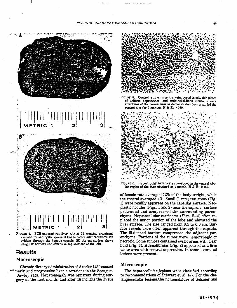
ResultsMacroscopic
Chronic dietary administration of Aroclor 1260 causedand progressive liver alterations in the Sprague-
Jawley rats. Hepatomegaly was apparent during sur-gery at the first month, and after 18 months the livers
FIGURE 6. Control rat liver, a central vein, portal triads, thin platesof uniform hepatocytes, and endothelial-lined sinusoids werestructures of the normal liver as demonstrated from a rat fed thecontrol diet for 9 months. H & E; xl60.
FIGURE 6. Hypertrophic hepatocytes developed in the central lobutar region of the liver obtained at 1 month. H & E; x 160.
of female rats averaged 12% of the body weight, whilethe control averaged 4%. Small (1 mm) tan areas (Fig.1) were readily apparent on the capsular surface. Neo-plastic nodules (Figs. 1 and 2) near the capsular surfaceprotruded and compressed the surrounding paren-chyma. Hepatocellular carcinoma (Figs. 2-4) often re-placed the major portion of the lobe and elevated theliver surface. The size ranged from 0.5 to 6.0 cm. Sur-face vessels were often apparent through the capsule.The ill-defined borders compressed the adjacent par-enchyma. Portions of the tumor were hemorrhagic ornecrotic. Some tumors contained cystic areas with clearfluid (Fig. 3). Adenofibrosis (Fig: 3) appeared as a firmwhite area with central depression. In some livers, alllesions were present.
MicroscopicThe hepatocellular lesions were classified according
to recommendations of Stewart et al. (8). For the cho-langiocellular lesions, the nomenclature of Schauer and
800674

KM) XORBACK AND WELTMAN
7. Foci of altered cells developed in the central and middlelobular regions in this liver obtained at 9 months. The cells mergedwith adjacent hepatic plates. Cells of the focus were usuallyeosinophilic and larger than normal. H & E; x 150.
FIGURE 8. This enzyme altered focus was present at 9 months. GGT;x!60.
FIGURE 10. Cells, nuclei, and nucleoli in the neoplastic noduledescribed for Fig. 9 were larger than their counterparts of theadjacent parenchyma. In this preparation, the nucleuses light witha very dark nucleolus. The granularity of the cytoplasm is mainlydue to mitochondria. TB; x400.
FIGURE 9. This neoplastic nodule in a liver obtained at 15 monthslacked lobular architecture and compressed the adjacent nontumorparenchyma. H & E; x40.
;^r&*" i»" If*.-FIGURE 11. In a trabecular carcinoma from a liver obtained at 24
months, wide plates of cells were separated by sinusoids. Thelarge cells contained large, abnormal nuclei. H & E; x!60.
Kunze (9) is applied. The normal architecture of the liveris shown in Figure 5. In the PCB-exposed group, thefollowing hepatocellular lesions were observed in se-quence (Table 1): centrolobular cell hypertrophy (Fig.6) at 1 month, foci (Figs. 7 and 8) at 3 months and thenareas of cell alteration at 6 months, neoplastic nodules(Figs. 9 and 10) at 12 months, trabecular carcinoma(Figs. 11 and 12) at 15 months, and adenocarcinoma(Fig. 13) at 24 months. In addition, simple (Fig. 14) andcystic (Fig. 15) cholangioma at 18 and 23 months, re-spectively, and adenofibrosis (Fig. 16) at 22 months werepresent.
There was no evidence of metastasis to the lung bygross or microscopic examination.
Control livers and livers containing all lesions wereevaluated for GGT activity. Throughout the study, GGTpositive areas of hepatocytes were absent in control
800675
PCB-IXDUCED HEPATOCELLULAR CARC/.VOA/.4 101
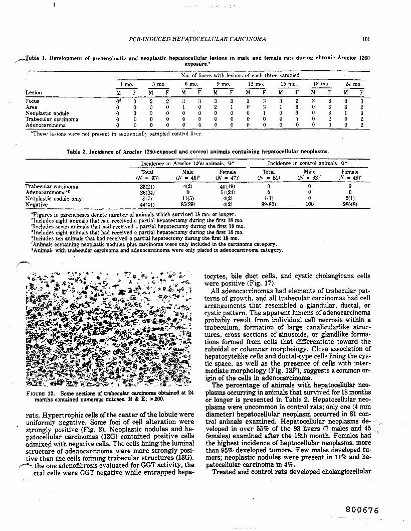
1. Development of preneoplaslic and neoplastic heptatocellular lesions in male and female rats during chronic Aroclor 1260exposure."
LesionFocusAreaNeoplastic noduleTrabecular carcinomaAdenocarcinoma
1MO2
0000
mo.F00000
3M20000
mo.Fo0000
No6
M31000
. of livers with lesions of each three sampledmo.
F30000
9M32000
mo.F31000
12M30000
mo.F33100
15M31000
mo.F33310
M;.;C000
; mo.F33320
24M33100
mo.F39
322
"These lesion? were not present in sequentially sampled control liver.
Table 2. Incidence of Aroclor 1260-exposed and control animals containing hepatocellular neoplasms.
Incidence in Aroclor 1200 animals, #"
Trabecular carcinomaAdenocarcinoma'''Neoplastic nodule onlyNegative
Total(AT = 93)
23(21)26(24)8(7)
44(41)
Male(A" = 46)1-
4(2)0
11(5)85(39)
Female(AT = 47)'
40(19)51(24)
4(2)4(2)
Incidence in control animals. <5fTotal
(\ = 81)00
1(1)9<*80)
Malt(A7 = 32)"
000
100
Female(A' = 49)'
00
2(1)98(48)
'Figures in parentheses denote number of animals which survived 18 mo. or longer.Includes eight animals that had received a partial hepatectomy during the first 18 mo.'Includes seven animals that had received a partial hepatectomy during the first 18 mo."Includes eight animals that had received a partial hepatectomy during the first 18 mo.'Includes ten animals that had received a partial hepatectomy during the first 18 mo.'Animals containing neoplastic nodules plus carcinoma were only included in the carcinoma category.'Animal?- with trabecular carcinoma and adenocarcinoma were only placed in adenocarcinoma category.
FIGURE 12. Some sections of trabecular carcinoma obtained at 24months contained numerous mitoses. H & E; x200.
rats. Hypertrophic cells of the center of the lobule wereuniformly negative. Some foci of cell alteration werestrongly positive (Fig. 8). Neoplastic nodules and he-patocellular carcinomas (13G) contained positive cellsadmixed with negative cells. The cells lining the luminalstructure of adenocarcinoma were more strongly posi-tive than the cells forming trabecular structures (13G).
the one adenofibrosis evaluated for GGT activity, the,ctal cells were GGT negative while entrapped hepa-
tocytes, bile duct cells, and cystic cholangioana cellswere positive (Fig. 17).
All adenocarcinomas had elements of trabecular pat-terns of growth, and all trabecular carcinomas had cellarrangements that resembled a glandular, ductal, orcystic pattern. The apparent lumens of adenocarcinomaprobably result from individual cell necrosis within atrabeculum, formation of large canalicularlike struc-tures, cross sections of sinusoids, or glandlike forma-tions formed from cells that differentiate toward thecuboidal or columnar morphology. Close association ofhepatocytelike cells and ductal-type cells lining the cys-tic space, as well as the presence of cells with inter-mediate morphology (Fig. 13F), suggests a common or-igin of the cells in adenocarcinoma.
The percentage of animals with hepatocellular neo-plasms occurring in animals that survived for 18 monthsor longer is presented in Table 2. Hepatocellular neo-plasms were uncommon in control rats; only one (4 mmdiameter) hepatocellular neoplasm occurred in 81 con-trol animals examined. Hepatocellular neoplasms de-veloped in over 55% of the 93 livers (7 males and 45females) examined after the 18th month. Females hadthe highest incidence of heptocellular neoplasms; morethan 95% developed tumors. Few males developed tu-mors; neoplastic nodules were present in 11% and he-patocellular carcinoma in 4%.
Treated and control rats developed cholangiocellular
800676

102 \ORBACK AXD \VELTMAX
Table 3. Incidence of Aroclor 1260-exposed and control animals containing cholangiocellular lesions.
Incidence in Aroclor 1200 animals. '"iV1'
Chulangioma (simple)Cholanpioma (cystic 1'Adenofibrosis*Negative
Totali.V = 93 1
38(35,8(7;9(!sj
Male(A' = 46)''
30(14)4(2)2(1)
63l29>
Female(.V = 41 v
I.V 7)30(1-1)
Incidence in controlanimals. '/* "
Totali.V = 81)
5(4)1(1)4(3)
90(73)
Male(A1 = 32)"
6(2 10
6(2)88(28)
Female(.V = 49)'
4(2)2(1)2(1)
92(45)"Figures in parentheses denote number of animals which survived lh mo. or lonjrtr.' 'Includes eiirht animals that had received a partial hepatectomy during tht first 1* nm>.liu-'mrifs seven animal.- that had received a pan;*; hepau-ctnmy dunnf the first !>• m\'
"Includes eipht animals that had received a partial hepatectomy during the first Its mo.'Includes ten animals that had received a partial hepatec-.omy durinp the first 18 mo.'Animals contain simple cholangioma (but not adenofibrosis)"Animals also containing cholangioma were placed in this group.
fr*.&}
c
^ t*^*Si •^" fV". •"•«"•-••'. \% r.'Cr-v:"- .-»*. ^4S*fe»*V*T>i|^,I.A*tt'>*af* - /- 5
FIGURE 13. All tumors with adenocarcinoma pattern contained trabecular regions. The cells forming gland-, duct-, or cystlike structuresappeared to be hepatocellular with unusual features and the luminal structures likely arose from several processes. (A) In this liver obtainedat 24 months, some cystic spaces appeared to result from degeneration of individual cells within trabeculae; H & E, x 160. (B) In this liverobtained at 29 months, the glandular spaces represent exaggerated canaliculi formed by three to five hepatocytes. Occasionally, a crosssection of a sinusoid may appear as a glandular lumen; however, the presence of endothelial cells should exclude this interpretation; H &E, x 180. (C) In this liver obtained at 29 months, cuboidal cells formed duct-or cyst-like structures among hepatocyte-type cells; H & E,x 160. (D) In this liver obtained at 29 months, columnar cells also lined duct-like structures and covered papillary projections; H & E,x!60.
800677

PCS-INDUCED HEPATOCELLULAR CARCINOMA 103
IcftB^Kj.&S'^EKiTOC J* sLVli ^3l3**tt
iiMOlesions. The simple cholangioma, cystic cholangioma,and cholangiofibrosis of Schauer and Kunze (0) are re-ferred to as bile duct hyperplasia, cyst, and adenofi-brosis, respectively, by Stewart et al. (8). Although thecholangiocellular tumors occurred in the control rats,the incidence of each was greater in the PCB-treatedgroup (Table 3).
DiscussionHepatocarcinogenic activity of PCBs was demon-
strated in the Spague-Dawley rat after long term ex-posure to relatively high dietary concentrations ofAroclor 1260. Large hepatocellular carcinomas, meas-uring up to 6 cm in diameter, nearly replaced the liverlobes. Histologic features of carcinoma included widetrabeculae formed from large hepatocytelike cells con-taining large abnormal nuclei with clumped peripheralchromatin and huge nucleoli. Some microscopic fields
•^ontained numerous mitotic figures. Central necrosisjid hemorrhage were sometimes present. The tumors
* .i£fi29!bi'~ * •» •
FIGURE 13 (con't). (E) In this liver obtained at 29 months, intra-cellular mucus was not demonstrated with special stains; PAS,x 180. (F) Cells lining ductlike structures with hepatocellular-tjpemorphology were continuous with the cuboidal cells; TB. x 300.(G) In a carcinoma with trabecuiar and adeno- patterns, the liningcells are strongly GGT-positive, and the trabeculae show a var-iegated pattern; GGT, x 140.
were not encapsulated and extended into the adjacentnontumorous parenchyma.
Although the tumors met the morphologic criteria formalignancy, their biologic behavior was relatively un-aggressive. The neoplasms did not metastasize to dis-tant organs nor invade blood vessels. Mortality of theanimals was not increased. The lack of greater morbid-ity or mortality is likely due to slow progression of theneoplastic process and late appearance and slow growthof the hepatocellular carcinoma.
PCBs have been established as very effective pro-moters in carcinogenesis. Kanechlor 400 (400 ng/g dietgiven for 6 months) after 3'-methyl-4-dimethylaminoa-zobenzene increased the incidence of hepatocarcinomaover that for the initiator alone in female Donryo ratsin the 800-day study (10). Kanechlor 500 (0.01 mL giventwice weekly by gastric intubation for 12 weeks) re-sulted in liver tumors at 40 and 52 weeks in male Wistarrats (11). Kanechlor 500 (500 or 1000 ng/g diet) causeddevelopment of neoplastic nodules in male F344 ratswhen given for 8 weeks after a nontumorigenic dose of#-2-fluorenylacetamide (12). Aroclor 1254 (100 ng/gdietfor 18 weeks) increased the incidence of hepatocellularcarcinoma when given to male Sprague-Dawley rats afterdiethylnitrosamine (13). Aroclor 1254 (500 mg/kg bodyweight given by intraperitoneal injection) reduced thetime required for the appearance of enzyme altered fociin partially hepatectomized male Sprague- Dawley rats(14).
It remains to be established whether PCBs also havean initiating effect or whether the neoplasms result frompromotion of a background incidence of initiated cells.In hepatocarcinogenesis, it is difficult to distinguish a
800678

NORBACK AND WELTMAN
FIGURE 14. A simple cholangioma, a proliferation of bile ducts, oc-curred in a periportal region at 18 months. H & E: x 160.
FIGURE 15. A cystic cholangioma. which does not compress sur-rounding parenchyma, was present at the 29th month. H & E;X160.
weak initiator from a strong promoter (15). A possiblemechanism of initiation by PCBs is the formation of anarene oxide of a PCB analog, which is an electrophilicintermediate metabolite capable of forming an adductwith DN A. The identification of a trans- dihydrodiol inthe rat (16) supports the supposition that the PCBs aremetabolized via arene oxides. Evidence against the abil-ity of PCBs to act as an initiator resulted from muta-genic studies. While most initiators tested with theSalmonella/microsome test are detected as mutagens(J D. a PCB mixture (Aroclor 1254) was not mutagenicin the Salmonella assay in the absence or presence ofuninduced or PCB-induced rat liver homogenate (18).
Hepatocellular lesions developed in more than 95% ofthe Aroclor 1260 fed female rats, whereas male rats hada 15% incidence of hepatocellular neoplasms. Kimuraand Baba (4) also noted a higher incidence of hepaticneoplasms in female than in male Donryo rats. Sex dif-ference in the incidence of PCB-induced hepatocarcin-ogenesis may be related to sex-linked differences inenzymatic activation and deactivation of carcinogens as
FIGURE 16. Adenofibrosis obtained at 24 months consisted of gland-like structures formed from columnar epithelium surrounded byabundant stroma. Some cells contained mucin. PAS; x 160.
"o?5*!.?.:1 . - - . ' . . ' •.'
' <:-
'
FIGURE 17. Glandular formations of adenofibrosis are negative forGGT activity, while cystic cholangioma and a focus of hepatocyteswere positive. GGT; x30.
proposed for acetylaminofluorine hepatocarcinogenicity(19), or presence of androgens or estrogens which com-pete for the carcinogen for metabolism as proposed forbenzopyrene (-20), aflatoxin (21) or dimethylbenzanthra-cene (22) hepatocarcinogenicity.
Adenofibrosis consists of glandular structures linedby mucin-secreting columnar or cuboidal cells and sur-rounded by layers of connective tissue. An electron mi-croscopic study identified the lesion as intestinalmetaplasia with goblet cells, enterochromaffin cells, andPaneth cells (23). The lack of GGT activity in adenofi-brosis also suggests the lesion is quite distinct fromsimple cholangioma or cholangiocarcinoma, both beingstrongly positive for GGT (24).
The GGT assays were performed in Henry Pitot's laboratory atMcArdle Cancer Research Institute.
This work was supported in part by NIH Grant CA22140, theVeterans Administration, and the Graduate School of the Universityof Wisconsin, Madison, Wl.
Portions of this paper have been published in abstract form (25-89).
800679

HEl'ATOCELU'LAK CAKCIXOMA
REFERENCES
1. Kimbrough, R. D.. Linder, R. E.. and Gaines. T. B. Morphol-opical changes in livers of rats fed poiychlorinated biphenvKLight microscopy and ultrastructure. Arch. Environ. Health 2X:354-3920972).'
2. Kimbrough, R. D., Squire. R. A.. Linrier. R. E.. Strandberp, .1.D.. Montali. R. J., and Burse, V. M. Induction of liver tumor*in Sherman strain female rats by poiychlorinated biphenyl Aroelor1260. J. Natl. Cancer Inst. 55:" 1453 -1-159 (1975).
3. Ito. N., Nagasaki. H. J.. Makiura. S., and Arai. M. Histopath-ologica) studies on liver tumorigenesis in rats treated with poi-ychlorinated biphenyls. Gann '65: 545-.r>49 (1974).
4. Kimura. N. T.. and Bab;i. T. Neoplastii- chances in the rat liverinduced by poiychlorinated biphenyl. Gann 64: 105-1'is H973).
5. Calandra. J. C. Summary of lexicological studies on commercialr'CBs. In: Proceedings of the 1975 National Conference on Pol-y chlorinated Biphenyls. Chicago. EPA-560/6- 75-0(14. Environ-mental Protection Agency, Washington D.C.. 197(3. pp. 35-42.
6. Gross, L., and Drey-fuss. Y. Spontaneous tumor? in sprague-Dawley and Long-Evans rats and in their F hybrids: carcinogeniceffect of total body \-irradiation. Proc. Natl. Acad. Sci. (U.S.)"6: 5910-5913 (1970).
7. Rutenberg. A. M., Kirn. H.. Fischbein. J. W.. Hanker, J. S..Wasserkrug, H. L.. and Seligman. A. J. Histochemical and ul-trastructural demonstration of gamma-glutamyl iranspeptidaseactivity. J. Histochem. Cytochem. 17: 517-522 (1969).
8. Stewart. H. C.. Keysser, C. H.. Lombard, L. S.. Montali. R. J..and Williams. G. M. Histologic typing of liver tumors of the rat.J. Natl. Cancer Inst. 64: 179-206 (1980).
9. Schauer. A., and Kunze, E. Tumours of the liver. In: Pathologyof Tumours in Laboratory Animals, Vol. 1. Part 2. Inter. Agencyfor Research on Cancer, Lyon 1976, pp. 41-72.
10. Kimura. N. T., Kanematsu. T., and Baba. T. Poiychlorinatedbiphenyl(s) as a promoter in experimental hepatocarcinogenesis
^ in rats. Z. Krebsforsch. 87: 257-266 (1976).1. Nishizumi, M. Effect of phenobarbital. dichlorodiphenyltrichloro-
ethane. and poiychlorinated biphenyls of diethyiniirosamine-in-duced hepatocarcinogenesis. Gann 70: 835-837 (1979).
12. Tatematsu, M., Nakanishi, K., Murasaki. G., Miyata, Y., andHirose. M. Enhancing effect of inducers of liver microsomal en-zymes on induction of hyperpiastic liver nodules by .V-2-fluoren-ylacetamine in rats. J. Natl. Cancer Inst. 63: 1411-1416 (1979).
13. Preston, B. D., Van Miller. J. P., Moore, R. W.. and Alien, J.R. Promoting effects of poiychlorinated biphenyls (Aroelor 1254)and poiychlorinated dibenzofuran-free Aroelor 1254 on diethyl-nitrosamine-induced tumorigenesis in the rat. J. Natl. CancerInst. 66: 509-515 (1981).
14. Pereira, M. A., Herren. S. L., Britl. A. L., and Khoury, M. M.Promotion by poiychlorinated biphenyls of enzyme-altered foci inrat liver. Cancer "Letters 15: 185-190 (1982).
15. Pilot, H. C., and Sirica, A. W. The stages of initiation and pro-
21.
motion in hepalocarcinopenesis. Hiochim. Biophys. Actaii()5:1<»1_215 (1980).
16. Norback. I). H.. Seymour..). L.. Knieriem. K. M.. Peterson. R.E.. and Alien. J. R. Biliarv metabolites of 2.5.2'..V-telracholor-biphenyl in the rat. Res. < nmmun. ('hem. 1'athol. I'harm.icol. 14:527-5.'W(197fi).
17. McCann. J.. and Ames, B. N. Detection of carcinogens ;i.- mu-lapens in Ihe 5f!/wo»f//s.-microsome test: assay of 3(Ki chemicals:Discussion. Proc. Natl. Acad. Sci. (U.S.) 73: 5I50-H54 (1!»7(>).
IS. Shahin. M. M.. Andrillon. P., Goetz. N., Bore. P.. Bupaut. A.,and Kalopissis. <.!. Studies on the mutapenicity of//-phenylene-diamine in Ifahnniiclln tifii/iiiniirinm. Presence of PCB's in rat-liver microsomal fraction induced by Aroelor. Mutal. Res. (IS:
Miller. J. A. Carcinopenesis by chemicals: an overview—G.H.A.Chnves Memorial Lecture. Cancer Res. HO: ">5H-o7fi (1H70).Neben. I). W.. Bausserman. L. I... and Bates. R. R. Effect of17-0-estradiol ami testosterone on aryl hydrocarbon hydroxylaseactivity in mouse tissues m i'itmzn<\ in cell culture. Int. J. Cancer<5: 470^48.1 (1970).Tulunay. F. ('., and Turker. R. K. Steroid sex hormones as in-hibitor? of aflatoxin metabolism in liver homupenates. Specialia15: 929-931 (1972).Booth. J.. Keysell. G. R.. and Sims. P. Effect of cestradiol onthe in i-itro metabolism of 7,12-dimethylhen.'.ia)anlhracene andils hydroxymethyl derivatives. Biochem. Pharmacol. 23: 734-744(1974).Terao, K.. and Nakano. M. Cholangiofibrosis induced by short-term feeding of 3'-methyl-4-(dimethyiamino>azobenzene: an elec-tron microscopic observation. Gann 65: 249-200 (1974).Harada, M., Okabe. K.. Shibata. K., Masada. H., Miyata. K..and Enomoto, M. Histochemical determination of increased ac-tivity of gamraa-glutamyl transpeptidase in rat liver during hep-tacarcinogenesis. Acta Histochem. Cytochem. 9: 168-179 (1976).Cihla. H. P.. Weltman. R. H.. Pilot. H. C.. and Norback. D. H.Sex difference in the acquisition of gamma-glytamy] transpepti-dase (GGT) positive foci in the liver of Sprague-Dawley rals fedpoiychlorinated biphenyls (PCB). Toxicologisi 1: 131 (1981).Cihla. H. P.. Weltman. R. H.. Pilot. H. C. and Norback. D. H.The gamma-glutamyl transpeptidase (GGT) activity of liver le-sions in rats exposed to Aroelor 1260. Toxicologist 2: 97 (1982).Weltman. R. H.. and Norback, D. H. Sequential, light and elec-tron microscopic analysis of hepatocellular carcinoma induced byAroelor 1260. Toxicologist 1: 65 (19*1).Weltman. R. H.. and Norback, D. H. The light and electronmicroscopic evaluation of choiangiocellular neoplasms in the ratliver following chronic Aroelor 1260 exposure. Toxicologist 2:100(1982).Weltman. R. H., and Norback, D. H. Development of hepato-cellular and choiangiocellular neoplasms in the livers of male andfemale Sprague-Dawley rats after chronic administration of Ar-oelor 1260. Fed. Proc. 41: 446 (1962).
23.
24.
26.
28.
29.
800680

TOXtOOUOOY AND AfTUUI fHAKMACDUMY 75, 271-211 (I9M)
Pathology of Chronic Polychlorinated Biphenyl (PCB) Feeding in Rats
EKXEHARD SCHAEFTER, HELMUT GREIM, AND WOLFGANG GOESSNER
GfitlltfHafi fir Si'oMf"- wi4 I'mwrltfcficftunt mbH. Ivtilui fiir Biologie. Abttilunf fur Falhologir.vnd Ininita fur Tojtikalofie vnd Biochrmie. Atxtilunj fur Toxikoiofie. Infoliicrdirr Landarasse I.
KmHfrbtrt. H'tv Gtrmany
Kectitd Attfua 2. 19S3 octrpted March 6.
Pathology of Chronic Polychlorinaied Biphenyl (PCB) Feedio* io Rau. SouEmx. £.,CREIM. H. AND GOESSVER. W. (19M). Taxied Aj>pl Fharmacol 7$, 278-28S. TV bepato-carrinotenic eBeci of Gopheo A 30 and Clophen A 60 was tested ID male wcanlint nu bylonj-term feeditr over a,period of 132 day*. The mortality rale was investigated ie 100-dayintervals. In JM nrn 800 days liver carcinoma accounted for 21* of necropsies in the OopbcnA 60 poup but t -«y 21 of the rtecropiiej in the Oophen A 30 px>up aod oooe to the coolroianimals The tumors were rtnt observerf after 700 days. After 800 days hepatoctlluUr carcinomawas tht mon common lesion observed io the Clophea A 60 animals (61%) wbcrtas h MJ onlyobserved io 3% of animals ID the Clophen A 30 troup and 2% in the oontrots. Pmieoptasiiclesions, such as to of hepatoceOular alterations aad oeopiastk ooduJea, were first observedafter Day 500. The incidence of foci predominated io aD time intervals, but an iocreax ioneoplaslic nodules and bepatocctlular carcinomas was observed with increased time. There wasa marked trend from foci to neoptastk noduk to bepatoccUulai carcinoma with one. Thetotal mortality rate aod the incidence of thyrooma, iofiammatory lesioos of the orotenitaltract, io the experiment were s>tni6cant]y reduced by Oopheo •dministraboo. VVbether thisprotective <~eci could be induced by polvchlorinated bipheoyls (PCB*) b discussed
Polychlorinated biphenyh (PCBs) have been been induced in rodents by highly chlorinated$hov.-n to produce foci of hepatocellular al- PCBs only. Feeding experiments over 224terations (KJmbrough ft ol.. 1975), neoplaslic days with Kanechlor 300, 400, and 500 innodules (Kimura and Baba, 1973; ho ti al., mict were performed by ho et al. (1973).1974; KJmbrough ft al., 1975), and hepato- Hepatocellular carcinoroas were induced bycellular carcinomas (KJmbrough et al.. 1975) the highest chlorinated compound only (Ka-in rats, and hepatomas (Kimbrough and Lin- nechlor 500). In a similar experiment in ratsder, 1974; Nagasaki ft al.. 1972; Ito ft al.. (Uo et al, 1974), different PCBs were admin-1973) and hepatocellular carcinomas (Ito et istered for 364 days. Although no carcinomasal, 1973) in mice. Furthermore PCBs have were observed, the occurrence of cholangiofi*been shown to have a promoting activity on brosis and nodular hypcrplaaa indicated thatenzyme-altered islands and neoptastk nodules bepatocellular carcinomas could probablyin rats (Kimura et al.. 1976; Nishizumi, emerge after a longer administration period.1976; Tatematsu et al.. 1979; Preston et al.. Administration of the highly chlorinated PCB1981; Pertira et al. 1982;Oesterle and Deml, Aroclor 1260 to rats for 600 days resulted in1983). Adenofibrosis in rats (Kimbrough et hepatocellular carcinomas (Kimbrough et al.al. 1972, 1973; Ito el al.. 1974) and in mice 1975).(Kimbrough and Under, 1974) resulting from The present experiments were designed toPCB consumption has also been observed. test the hepatocarcinogenic effect of two dif-
In general hepatocellular carcinomas have ferent chlorinated commercial PCB mixtures,
0041-008X/M $3.00 271CopTOht e I«S4 by Aadcwuc rm. fee.AJ) n*Su al rtpn*v«M io uj farn imrwl
800681

fATHOLOGY OF POLYCHLORINATED BIFHENYL FEEDING 279
Qophen A 30 (30 wt% of chlorines) andGophen A 60 (60 wt% of chlorines) in makrau when administered by continuous feedingover a period of more than 800 days, i.e..until the end of the experiment.
METHODS
A tola! of 433 male weanling, ipecinc-paihogen-free.random-bred Wuur rau (Neunerberg. FRO were usedin the experiments. Animals were maintained it a rootstemperature of 22*Ctnd humidity of 50 i 5% and givesfeed (commercial stock diet Altromin (1324) tuppte>-mented as described below) and water ad libitum. Forthe first 8 weeks all rau received the standard diet. After8 weeks the rats were divided into three groups. Oochundred thirty-nine control animals (group 1) receivedthe basic diet; 152 animals (group 2) received the basicdiet supplemented with 100 ppm Clophen A 30; andMl animals (group 3) received the basic diet supple-mented with 100 ppm Qophen A 60.
The PCBs were the kind gift of Dr. Wrtbeu. B»yer(Leverkusen, FRG) and added to the pellets by Altrouun(Lage, FRG) during production. Clophen A 30 had SBaverage composition of 1.0% monochlorbiphenyl. 20.7%dichjorbiphenyl, 57.4% thchlorbiphenyt, 17.3% tetra-chlorbiphenyl, 1.8% penuchlorbiphenyl, 1.0% hexa-chlorbiphenyl. 0.6% heptachlorbiphenyl, and 0.1% octa-chlorbiphenyl; furans were absent. Oophen A 60 had «naverage composition of 0.2% monochlorbiphenyt, 1.1%dichlorbipheoyl, 2.2% trichlorbiphenyl, 3.1% tttrachlor-biphenyl. 19.8% penuchlorbiphenyl. 43.2% heiachlor-biphenyl. 25.3% heptachlorbiphenyl. 4.7% octachlorfai-phenyl. and 0.3% nonachlorbiphenyt; furans were absent(Vr'rabeu. 1979. personal communication).
During the experiment the animals were checked 5days a week for death or morbidity. After Day 101.randomly selected animals from aU three experimentalgroups were killed dairy so that the experiment wwcompleted after 832 day*.
In general, necropsies were performed on aD rau thathad died or had been kffled in a moribund condiboe.Rau which died but were not available for oecropiywere designated "lost animals."
Tissues were fixed in Formalin and embedded inparaffin; 5-pro-thick lections were stained with hematca-yiin and eosin (HE). Tbymus, lungs, spleen, kidneys,ventral prostate, urethn with dorsal prostate, and urinaryMadder were investigated routinely. Other tissues (e-g^skin, teflev pituitary gland, and glandula praeputialh)were examined when there was macroscopic indicatioeof tumor or inflammation. The left lateral lobe of theliver- was investigated in each case; other liver lobes wenalso taken when there was any indication of lesions ortumor-like changes. Liver lesions were classified into
major types, continent with thoae lifted by Squirt andLevin (1975). by the Committee on Histoiofic CUsain-cation of Laboratory Animal Tumors (1910) and byWard (1981). The significance of differences in incidencewas tested with Fisher's exact ten.
RESULTS
Mortality Rate
Table 1 shows the mortality rate for allthree experimental groups in 100-day inter-vals, to day 800. Within the first 500 days ofexperiment, the mortality rate was relativelylow.. Between SOI and 600 days, the mortalityrate in both the control and Qophen A 30groups reached 12%, that in the Qophen A60 group, 8*. In the next interval (601-700days) mortality in the control group rose to] 7%, whereas that in the Qophen A 30 groupremained at 12%, and that in the Qophen A60 group dropped to only 5%: In the lastinterval (Day 701-800) the mortality rate incontrols was 42%, whereas that in group 2was only 19% and that in group 3 was 35%.The total mortality in the first 800 days inthe two treated groups (43 and 40%) wassignificantly lower than the mortality of 62%observed in the control group (p < 0.05).
Lesions Leading to Death up to Day 800 ofthe Experiment
By Day 800, 60% of the control animals,37% of the Qophen A 30 animals, and 33%of the Qophen A 60 animals had beenoecropsied. The most frequent lesions leadingto death or termination in these animals areshown in Table 2. The distribution of theselesions per 100-day interval is shown inTable 3 (a-c). Although the incidence ofbepatocellular carcinoma was fairly low, dif-ferences were observed: in the first 800 days,liver carcinoma accounted for 21% of animalsin the Qophen A 60 group but only 2% ofanimals in the Qophen A 30 group andnone of control animals. The incidence ofliver carcinomas in the two treated groups
800682

280 SCHAEFFER, GREIM. AND GOESSNER
TABLE
MORTAUTY RATE IN CONTROL RATS (GROUP I». RATS WITH CLOPHEN A JO-SUPPLEMENTED DIET (GROUP 2).AND RATS WITH CLOPHEN A W-SUPPUMENTED Din (GROUP J)
Group I (A'- 139) Group 2 (N - 152) Group 3 ( A ' « 141)
Interval(Days)
101-200201-300301-400401-500501-«00601-700701-800
Deaderkilled whenmoribund
(1)' 1*(1) 1%
0 0%1 1 (2) 8%15(2) 12%19 (2) 17%39 42%
Animals sur-viving at thebeginning oftime interval
139138137J37126i l l92
Dead orkiUed whenmoribund
0 0%(2) 1%
3(2) 2%8 (2) 5%
17 (6) 12%15(2) 12%20 19%
Animals sur-viving at thebeginning oftime interval
152152150147139122107
Dead orkilled whenmoribund
0 0%(3) 2%
3(2) 2%4 ( 2 ) 3%
10(5) 8%6 5%
30 35%
Animals sur-viving at thebeginning oftime interval
14113813513112111585
1-800 86(8) 62% 139 6SU4)» 43* 152 S6(I2C 40% 141
* Numbers in parenih<rses refer to animzii dead but not available for pttiopsy.* Significantly dirTcrtn1. (p < 0.05) compared.to untreated group.
TABLE 2MOST FREQUENTLY OCCURRING LEMONS FOUND IN NECROPSIED ANIMALS UNTIL DAY 800
Group
A * number of animals at the start of the experiment minus animalsnot necropsied flos animals)
n * cumber of animals necropsied until Day 800
Neoplasiic lesionsHcpatocellular carcinoma
%«%N
Thymoma% «%N
Other neoptasas% n%N
NonneopUstk teaon*Urogenital system Prosutitu p. Abscess gl. praep.
% n%N
Wistar- nephritis% n%A"
13178(60%)
000
162112526740
1924IS9
127
13851 (37%)
1214»S3
28»5520
1529I I000»
12944 (33%)
9*21
72»S4
U»4114
716500<r
* Significantly different (p < 0.05) compared to uoirtated group.
800683

PATHOLOGY OF POLYCHLORINATED BIPHENYL FEEDING
TABLE 3
RATI (%) OF THE MOST FMOt'tNTLY OCXVUKINC LESIONS LCADI<*G TO DlATH. OBSERVEDOVER INTERVALS or 100 DAYS
281
Timeinterval(Days) I
Group
2 3
Timeinterval(Days)
(a) Hepatoctllular carcinoma
301-400
401-700
701-800
0(0/137)'
0(0/111)
0(0/92)
0(0/150)
0(0/122)
1(1/107)
0(0/135)
0(0/115)
II"(9/85)
601-700
701-800
1
(c) Other
10(11 /111)
27(25/92)
Group
2 3
nroptiiiu continued
3(4/122)
12*(13/107)
3(4/115)
8*(7/85)
(d) Urojeniul tynem (prosutiuspurulent*. p>e)onephritiv abscess
(Jandula praeputialu)^ ••
301-400
•\ *•
401-500 •-':'•.
501-600
601-700
701-800
301-400
401-500
501-600
0(0/137)
1(1/137)
4(5/126)
5(5/111)
5(5/92)
(c)
0(0/137)
5(7/137)
7(9/126)
(b) Thtmoma
0(0'150)
0(0/147)
1(2/139)
0(0/122)
2(2/107)
Other neopiisias
r(1/150)
2(3/147)
5(7/139)
0(0/135)
1(1/131)
0(0/121)
,(1/115)
0(0/85)
|
(2/135)
2(2/131)
2(3/121)
301-400
401-500
501-600
601-700
701-800
301-400
401-500
501-600
601-700
701-800
0(o/i 3n
3(4/137)
3(4/126)
5(5/111)
7(6/92)
(e)0
(0/13?)0
(0/137)0
(0/126)0
(0/1 II)21
(9/92)
1(1/150)
2(3/147)
2(3/139)
3(4/122)
5(5/107)
WistaMjephritJ*0
(0/150)0
(0/147)0
(0/139)0
(0/122)<r
(0/107)
0(0/135)
0(0/131)
1(1/121)
1u/ns)
6(5/85)
0(0/135)
0(0/131)
0(0/121)
0(0/115)
0*(0/85)
* Data are percentages and, in parentheses, proportions.• Significantly different (p < 0.05) compared to untreated group.
up to Day 800 of experiment was 7 and \%,respectively (Table 2). The tumors were firstobserved after 700 days. Thyrnoma accountedfor 21% of animals in the control group(incidence 12%) but only 8% in the ClophenA 30 group (incidence 3%) and 5% in the
Gophen A 60 group (incidence 4%). Thethymoma rate in control animals remainedconstant between 500 and 800 days (Table3b). The group of "other neoplasJas" includedtumors of the skin, testes, and pituitary gland.These tumors were also more frequent in the
800684

282 SCHAEJTER. CRE1M. AND COESSNER
control group (incidence 40%) than in eitherof the treated groups (incidence 20 and 14%),the rate rising steadily from Day 400 to 800(Table 3c). Nonneoplastic lesions of the uro-geniial tract (i.e., prostatitis purulenu, abscessof glandula praeputialis, and Wistar-nephritis)accounted for 36% of necropsied controlanimals, 29% in the Gophen A 30 group,and 16% in the Gophen A 60 group. Pros-tatitis purulenta was observed in all groupswith a higher incidence in the control group(15%) than in either of the treated groups(11 and 5%). The first cases were seen afterDay 401 (Table 3d), the rate increasing slowlyuntil Day 800. The severe form of Wistar-nephritis was only observed in control ani-mals (incidence 7%).
Lesions in Animals Killed at ihe End ofExperiment
Table 4 shows the frequency of majorlesions in animals killed at the end of theexperiment. Hepatocellular carcinoma was
TABLE 4FREQUENCY OF MOST FREQUENTLY OCCURRING LE-
SIONS IN ANIMALS KILLED AT THE END OF THE EXPERI-MENT
Most frequentlesions
HepatoctUularcarcinoma
Thymoma
Otheroeoplasiu
Urogenitalsystem
Wisur-nephritis
1
20/53)'
17(9/53)
3$(19/53)
0(0/53)
40(21/53)
Croup
23
(3/87)
14(12/87)
3S(33/87)
1(1/87)
8"(7/87)
361"
(52/85)Of
(0/85)IS
(13/85)0
(0/85)0
(0/85)
* Data are percentages and. in parentheses, proportions.• Significantly different (p < 0.05) compared to un-
treated animals.
the most common lesion observed in theGophen A 60 animals (61%), whereas it wasonly observed in 3% of animals in the Go-phen A 30 group and 2% in the controlgroup. In contrast, thymomas, observed in17% of the control animals and 14% of theGophen A 30 animals, were not identifiedin the animals treated with Gophen A 60.Other neoplasias were observed in 36% ofthe control animals and 381 of the GophenA 30 animals but only 15% of the GophenA 60 animals. After 800 days, nonneoplasticlesions of the urogenital tract, other thanWistar-nephritis, ceased to be of importance.The incidence of severe Wistar-nephritis roseto 40% in control animals. Even after 800days, no cases of severe Wistar-nephritis wereobserved in the Gophen A 60 group, but thefirst cases were observed in the Gophen A30 group (incidence 8%). Various pathologicaleffects were observed in liver cells (e.g., hy-pertrophy of individual cells, hyperchromaticnuclei, foamy or vacuolated cytoplasm) butwere not further investigated in this study.Similar findings have been described byKimbrough et al. (1972) and are nonspecificeffects associated with long-term feeding ofGophen.
Preneoplastic lesions such as foci of hepa-locellular alterations (Fig. 1) and neoplasticnodules (Fig. 2) were regularly observed.Their incidence is shown in Table 5 togetherwith the rate of hepatocellular carcinoma.Only the highest stage of neoplastic devel-opment (i.e., focus of hepatocellular altera-tions or neoplastic nodule or hepatocellularcarcinoma) per animal is included. In generalfoci and nodules were first observed afterDay 500. The incidence in control animalswas low until Day 800 after which the inci-dence of foci rose to 32%, the Dumber ofneoplastic nodules and neoplastic lesions re-maining low (4 and 2%, respectively). Theincidence of preneoplastic and neoplastic le-sions in necropsied Gophen A 30 animalsremained constant at 50-60% between Day500 and 800, rising to 100% in animals killed
800685

PATHOLOGY OF POLYCHLORINATED BIPHENYL FEEDING 283
^taswssas•* -*.'•••->>-" v>SS££^
Fic. 1. Dear oeO area of altered hepatocyto in a nt fed Oophen A 60. Note empty appearance ofcytoplasm of altered cells. HE X 140.
after Day 800. The incidence of foci predona- cinoma (Fig. 3) was observed with increasedinated in all time intervals, but an increase time. Preneoplastic or neoplastic lesions werein neoplastic nodules and hepatocellular car- observed in 100% of the Clophen A 60
800686

284 SCHAEFFER. CREIM. AND GOESSNER 28<
FlG. 2. Left tide. Neoplastic oodule in • rat fed Oopheo A 30. Periphery is ihaipiy demarcated fromsurroundini nonnal Uver ptrttichynu. HE x 13. Richt tide. Edge of the tune oeopltnk nodule. Thesurroundint Uver plites vt compressed. HE X 323.
animals necropsied afler Day 500. There wasa time-dependent trend from foci to neoptas-tic nodules to hepatoceliular carcinoma.
Nonneoplastic proliferative kadons i.e., bikduct hyperplasia and associated lesions suchas adenofibrosis and cysts (cholangiomas).
TABLESFREOUENCY OF NECROPSIED ANIMALS WITH Foci OF HE/ATOCTLLULAR ALTERATIONS. NEOPLASTK NOOULCS,
AND HEPATOCELLULAR CARCINOMAS (ONLY THE HIGHEST STAGE OF UVER CARCINOGENESIS PER ANIMAL is IN-O.UOCD)
Timeinterval
301-400401-300301-600601-700701-SOOS01-432
Foci
0(0/0)'1IU/9)»(I/I3)6(1/17)3 (2/39)
32(17/53)
1
Neoplanxooduta
0(0/0)0(0/9)Sd/13)
12(2/17)0 (0/39)4 (2/53)
HCPMO.crtlular
carcinoma
0(0/0)0(0/9)0(0/13)0 (0/17)0 (0/39)2(1/53)
Foci
0(0/0)0(0/0)
55(6/1!)33 (4/12)
49 (43/*7)
Croup
2
Neoptubctndula
0(0/0)0(0/0)0(0/11)
17(2/12)3(1/20)
40 (3i/»7f
IfefttO.edubr
CUDBOIM
0(0/0)0(0/0)0(0/11)0(0/12)5(1/20)S (3/17)
Foci
0(0/1)0(0/0)
40 (2/3)0(0/5)3(1/30)o w/tsr
3
NeopUaieooduks
100 (I/I)0(0/0)
60(3/5)100 (3/5)67 (20/30J-
Heptio-ccOular
earciaona
0(0/1)0(0/010(0/5)0(0/3)
30 (9/30r61 (S2/«r
* Dau trt pcreenuja aAd. in poitnthoo. proponioai.• Si(niftc*nUy difltnm (p < 0.05) compared to untreated I/oup.
Pera Igrccin6 l f
on!her89-TruliomaPC:Tturesi.lha:ratsa/.nestto taiorBoi.;Efleichcpot?be p.creationspec
7._nee;careTheOopberwithnurr.cordregartors.Jon|com:ma>atingsubs:
Thnodugrourwhich
800687

PATHOLOGY OF POLYCHLORINATED BIPHENYL FEEDING 285
FK>. 3 Hepatoctllular (trabecular) cr-cinema in a nt fed Clophea A 60. Ncoplastic cells are arrancedin trabecular cords and irregular n«u; note hepatoctllular pleomorphism. HE x 243.
were seen in both control and treated groups. DISCUSSIONThe incidences of such lesions are shown inTable 6. However, they were not further Clophen A 60 had a definite, and Qophenstudied because there is no convincing evi- A 30 a weak, carcinogenic effect on rat liverdence that they are precancerous lesions in these experiments. The first hepatocellular(Stewart and Snell, 1957). carcinomas were detected after a latency
TABLE 6NONNEOPLASTV PROUFIRATIVE UVER LESIONS
Timeinierta)(Diy»)
401-500501-600601-700701-100SOI -(32
Bikductbypcrpbu*
30(S/»)'3* (5/13)» (5/17)31 (12/39)»3 (7/53)
1
Adeno-ibrea
0(0/9)0(0/13)0 (0/17)
IS (7/39)23 (12/53)
C»*u
0(0/9)0(0/13)0 (0/17)0 (0/39)0 JO/53)
Bile duethypopUsu
0(0/6)27(J/1I)J5 (3/1J)«( 10/21)32 (2S/S7)*
Group
2
Adrao-Bbraut
0(0/6)0(0/11)0(0/12)3(1/21)1 (6/S7f
Cyitt
0(0/6)0(0/11)0 (0/12)0(0/21)0 (0/S7)
Kleductfcypeptau
»{I/J)60(3/5)S3 (3/6)73(22/30r7) (62/tif
3
Adeno-ibraat
0(0/2)0(0/5)0(0/6)3(1/30)2<2/S5C
C»«
0(0/2)0(0/5)0(0/6)3(1/30)
12 do/ssr
* Dati vt pcnxntata and. in putnthao. proponiom.• Sifnihandy different (f < 0.05) eomptired le uatreated |reup.
800688

286 SCKAEFFER. CREIM, AND GOESSNER
period of 700 days in both groups, but witha higher incidence in the Oophen A 60group. The incidence of hepatocellultr car-cinoma in the Oophen A 60 group reached61% at the end of the experiment. Althoughonly 3% of the Clophen A 30 group showedhepatocellular carcinomas after 800 days,89% of the animals had preneoplastic lesions.These findings confirm the observations ofho ei al. (1974) that hepatocellular carcino-mas emerge in rats after administration ofPCBs (Kanechlor) for more than 364 days.The findings are also consistent with theresults described by McConnell (1980), i.e.,that the occurrence of neoplastic nodules inrats (Ito el al., 1974) and mice (Nagasaki ftal, 1972) following administration: of Japa-nese commercial mixtures of PCBs is relatedto the number and location of the halogenatoms by which the mixtures are classified.Both the DHEW Subcommittee OE« HealthEffects of PCBs and PBBs (1978) and Ecob-ichon (1975) have reported that the toxicpotency of PCBs (hepatic enzyme induction,hepatocarcinogenic effect) increases with in-creasing chlorination and chlorine substitu-tion in the para. onho. meta positions, re-spectively.
The high incidence of preneoplasiic andneoplastic lesions in the livers indicates acarcinogenic effect of both PCB mixtures.The application of the higher chlorinatedGophen A 60 results in an appreciable num-ber of hepatocellular carcinomas, whereaswith the lower chlorinated Oophen A 30numerous preneoplastic foci were found. Ac-cording to the present knowledge, PCBs areregarded to be promoters rather than initia-tors. However, it should be mentioned thatlong-term feeding of potential carcinogeniccompounds as in the present experimentmay not allow distinction between the initi-ating and the promoting activity of a chemicalsubstance (Schulte Hermann el al. 1983).
The maximum incidence of neoplasticnodules was observed in the Oophen A 60group in the period Day 601 to 700 (afterwhich the increase in hepatocellular carci-
noma accounted for the drop in neoplasticnodules) and in the Oophen A 30 group atthe end of the experiment The incidence ofneoplastic nodules in untreated animals wasvery low. This is consistent with the obser-vations that neoplastic nodules are rare inuntreated rats, except in aged rats, and thatthey are only induced by hepatocarcinogens(Farber, 1963; Marugami et al.. 1967; Ito aal.. 1969, 1974; Kimbrough, 1979; Pollardand Luckert, 1979; Committee on HistologicClassification of Laboratory Animal Tumors,1980).
Foci of cellular alterations were observedin all three groups after Day 500, but againthe greatest occurrence was observed intreated animals. Although foci are known toappear in old untreated rats, increased num-bers encountered in bioassay studies are ac-cepted as an indication of possible carcino-genicity (Committee on Histologic Oassifi-cation of Laboratory Animal Tumors, 1980).In the rat, foci have also been shown torepresent stages in the development of he-patocellular carcinoma (Gossner and Fried-rich-Freksa, 1964; Bannasch, 1968; Fischeret al., 1983). The promoting activity of PCBson preneoplastic enzyme-altered foci and is-lands in rat liver has been described recentlyby Pereira ei al. (1982) and by Oesterle andDeml(1983).
The total mortality rate in the experimentwas significantly reduced by Oophen admin-istration. This mainly resulted from themarked appearance of thymoma and other(nonliver) neoplasias, in purulent inflamma-tory lesions of the urogenital system, and ofWistar-nephritis commonly found in old rats(Bullock ei al.. 1968) (see Tables 2 and 3b-e). This may be explained by an interactionwith the immune system. In recent years aDumber of compounds have been shown toalter immunocompetence (Vos ft al.. 1980;Chang a at.. 1981; Fraker, 1980; IARC.1978). In particular, Vos et al. (1980) havesuggested that PCBs may modulate host de-fense mechanisms and immune responses.Among other lesions, atrophy of lymphoid
800689

PATHOLOGY OF POLYCHLORINATED BIPHENYL FEEDING 287
tissues, luch as spleen, lymph nodes, andthymus, has been induced by treatment withPCBs in chickens, rabbits, and guinea pigs(Vos et al. 1980), and by polybrominatedbiphenyls in mice (Fraker, 1980). Moreovermacrophage functions, as determined by invivo bacteria] clearance, are inhibited by PCBexposure (Vos et a!., 1980). Wistar-nephritisis thought to be immunopathic (Fridman-Manduzio et al., 1980; Hirokawa. 1975).Thus the protection from neoplasms of thethymus and from Wistar-nephritis in theQophen-lreated groups in the present exper-iment might result from PCB-induced alter-ations in the immune system.
Furthermore the present study supportsthe conclusion of the DHEW Subcommittee*'on Health Effects of PCBs and PBBs that theliver is the primary target organ for PCBs inthe rat.
ACKNOWLEDGMENTS
We thank Ms. G. B6U for excellent technical assistance.Dr. A. B. Murray, Dr. E. Deml, and Dr. D. Oesterle forcritically nadint the manuscript, and Dr. K. Wrabetz,fSayer, Leverkusen, FRO, for kindly supplying the Co-phen A 30 and A 60.
REFERENCES
BAVSASCK P. (1968). The cytoplasm of bepatocytesduring carcinogeneiii. Recent Remits Cancer Ret
BULLOCX, B. C, BANKS, K. 1_, AND MANNING, P. J.(1968). Common lesions in the aged rat In TheLaboratory Animal in Cerontological Reiearch. pp.62-82. Natl. Acad. So., Washington. D.C
CHANC, K.-J.. HSIEH, K.-H.. UE. T.-P., TANC. S.-Y-.AND TUNC, T.-C. (1981). Imrnunotogic evaluation ofpatients with potychlorinated bipheoyl poisoning: De-termination of lymphocyte subpopulations. Taxicol.Appl Phamacol. 61, 38-43.
Committee on Histologic Classification of LaboratoryAnimal Tumors (1980). Histologk typing of livertumors of the rat. / Kail Cancer Iras 64, 179-206.
DHEW Subcommittee on Health Effects of PCBs andPBBs (1978). General summary and conclusion. En-viron. Health Penpect. 24, 191-198.
ECOBICHON, D. J. (197S). The influence of polychlori-naled biphenyl compounds on hepatic function in therat In Ecological Toxicology Research (A. D. Mclntyre
and C F. Mills, tot.}, pp. 207-214. Plenum. NewYork/London
FARBFR. E. (1963) Ethionine cascinogenesis In Attvanceiin Cancer Research (A. Haddo* and S. Weinhouse.edi.). Vol. 7. pp. 38J-474. Academic Press. New York.
FISCMER. G.. UU.RICH, D.. KATZ, N.. BOCK. K. W..AND SCHAUER, A. (1983). Immunohisiochemica] andbiochemical detection of undine-diphosphate-gJucuro-nyltransferase (UDP-GT) activity in putative preaeo-plastic Urn foci. Virchows Arch B 42, 193-200.
FRAKIR, P. J. (1980V The antibody-mediated and delayedtype h)pcrsensiuvity response of mice exposed topolybrominated biphenyls. Toxieol Appl Pharmacot$3,1-7
FRIDMAN-MANDCZIO. A., SALMON. J.. AND BETZ,E H. (1980). Spontaneous immunglomerulonephritisin Wisui rats. Pothol Res Proa. 167. 301-312.
GOSSSER, W., AND FRJEDRICH-FREKSA. H. (1964). His-tochemische Uniersuchungen fiber die Glucose-6-Phosphause in der Ranenleber wihrend der Kanzer-isierung durch Nitrosamioe. Z Naturforsch B AnorgChem Org Chem 19, 861-863.
HIROKAWA. K. (1975). Charanerization of age-associatedkidney disease in Wistar rats. Mech Ageing Df» 4.301-316.
IARC (1978). Po!>chlorinated biphenyls and polybromi-nated biphecyls Monograph} on the Evaluation of theCarcinogenic Risk of Chemicals 10 Humans 18, 68-71.
ITO. N., HlASA, Y.. KON1SHI. YM AND MARUCAM1, M.(1969). The development of carcinoma in liver of ratstreated with m-tduytenediamine and the synergisucand antagonistic effects with other chemicals. CancerRes 29, 1137-1145.
ITO, N.. NAGASAKI, H.. ARAL M.. MAKIURA, S.. SUGI-HARA, S.. AND HiRAO, K. (1973). Histopathologicnudies OD liver tumorigenesis induced in mice bytechnical polychlorinated biphenyls and its promotingeffect on liver tumors induced by benzene nexachlohde.J.'Nod. Cancer Inn 51. 1637-1646.
ITO, N., NAGASAKI, ri, MAKIURA, S., AND ARAI, M.(1974). Hiflopaiholopcal studies on bver tumorigenesisin rats treated with polychlorinated biphenyls. Conn6$. $45-549.
KJMBROOCH. R. D. (1979). The carcinogenic and otherchronic effect! of persistent balogenated organic com-pounds. Ann. N.Y.Acad. Sri. 320, 415-418.
KJMBROOCH, R. D, AND UNDER, R. E. (1974). Inductionof adenofibrosis and bepatomas of the liver in BALB/cJ mice by polychlorinated biphenyb (Aroclor 1254)./ Noil. Cancer Ins S3, $47-552.
KJMBROOGH, R. D.. UNDER, R. E.. BURSE, V. W.. ANDJENNINOS, R. W. (1973). Adenofibrosis in the ratlivtr. Arch. Environ. Health 27, 390-395.
KJMBROOCH, R. D., UNDER, R. E., AND GAMES, T. B.(1972). Morphological changes in liven of rats fedpolycMorinated biphenyls. Arch. Environ Health 25,354-364.
800690
288 SCHAEFTFER. CREIM. AND GOESSNER
KJMBHOUOH, R. D., SQUIRE, R. A., LJNDCR, R. E,.STR«SD»CRO, J D.. MONTALI. R. ).. AND BURS*.V. W (1975). Induction of liver tumon in Shermanttrtin female nu by poiychlorinited biphenyl Aroclor1260 J Hall Cancer Inst 55, 14 5 3-14 59.
KJMURA, N. T.. AND B*»A.T.< 1973) Neoplasuc craniain the m liver induced b> polychlohnated biphenyl.Conn 64, 105-10$
KJMUIU. N. T., KANEM*TSU, T.. AND BA»A, T. (1976).Polychlohnated bipcxnyl(s) as a promotor in experi-ment!) hepatocarnnogenesis in rau. Z Krebsforsch.87, 257-266.
MARUGAMI, M.. Iio. N.. KosiSHi, Y., HIASA, Y.. ANDFARBER. E. (1967) Influence of 3-methylcholamhreneon liver carcmogenesis in rau ingesting DL-ethioninc.J'-methyM-dimethyUmmoaiobenzene. and A'-2-fluo-roenylacetamide Cancr Rn 17. 2011-2019.
McCosNEU, E. E. (1980) Acute and chronic toxicity.carcinogenesis, reproduction, teratogenesis and muta-tennis in animals ID Topics in Environmental Health.Vol 4. Halogrnatrd Biphenyls. Terphtnyli. Naphtha-lenes. Diben:odioxins and Related Products (R. D.KJmbrough. ed.X pp 109-150. Elsevier^onh-HollandBiomedicaJ Pres*. Amsierdam,'Ne*' York/Oxford.
NAGASAKI. H., TOMJl, S.. MECA, T.,'MARUOAMI, M.,AVD ITO, N. (1972). Hepatocarrmojenidty of poly-chlorinated biphenyb in mice. Conn 63. 805.
NISHIZUMI, M. (1976). Enhancement of diethylDiUosa-mine hepatocarcinotenesis in rats by exposure topolychlorinated bipbenyls or phenobarbiuJ. CancerLett X 11-16.
OESTERLE, D.. AND DEMU E. (1983). Promoting effectof polychlorinated biphenyls on development of en-zyme-altered islands in livers of weanling and adultrau. Cancer Res Clin Oncol 105, MI-147.
PIREIR.A. M. A., HERHEV S L.. BRITT. A L. ANDKXOCRV, M. M. (I9SJ). Promotion b> polychlonnatedbiphenyls of eniyme-aliertd foci in rat bver. CancerLett 15, 185-190.
POLLARD. M.. AND LUCXERT, P. H. (1979). Spontaneousliver tumon in ajed at rm free Wisur rats. Lab Anim.Sfi », 74-77.
PRESTON, B D . VAN Mnu*. ). P., MOORE. R. W..AND AILEN, J. R (1981). Promoting effects of poly-chlonnated biphenyls (Aroclor 1254) and polychlori-nated diberuofunn-frw Aroclor 1254 on dieihylniuo-samine-induced tumonfeness in the rat. J Noll CancerInsi 66.509-516.
HERMANN, R., TIMMERMANN-TROSIENER. L.AND SCHUfTLER. ). (1983). Promotion of spontaneouspreneoplastic cells in rat liver as a possible explanationof tumor production b> nonmuugcnic compounds.Cancer Kes «, 839-844
SofiRt. R A., AND Ltvrrr. M. H. (1975). Repon of tworkshop on classification of specific hepaiocellularlesions in rats. Cancer Kes 35. 3214-3223.
STEWART, H. L. AND SKEU, K C. (1957) The hino-p«jiolog> of experimental tumors of the liver of therat- A critical review of the histopathogenesis. AnaUnto 1m Concern 13, 770-803.
TATEMATSIJ, M.. NAUAVttHi, K... MURASAKI, G., Mi-YAT.'., Y., HIROSE. M.. AND ITO, N. (1979). Enhancingeffc-t of inducers of bver microsomal enzymes oninduction of hyperplastic liver nodules by N-2-fluoren-ylacttamide in rats. J Hail Cancer Inst. 63, 14 It-1416.
Vos. J. G.. FAtTH, R. E.. AND LUSTER, M. I. (1980).Immune alterations. In Topics in EnvironmentalHealth. Vol. 4. Halogrnaied Biphenyls. Terphenyis.Naphthalenes. Dibemodioxins and Related Products(R. D. Kimbrough. eA\ pp 141-266. Elsevier /North-Holland BiomedicaJ Press. Amsterdam/New York/Oxford.
WARD. J M. (1981). Morphology of foci of alteredhepaiocytes and naturaJly-occumng hepatoctUulax tu-mors in F344 nu. Vircho** Arch A 390. 339-345.
800691

EPLEXPERIMENTAL PATHOLOGY LABORATORIES, INC.
During the PWG review, the PWG panel examined all sections
of liver in which a diagnosis of a hepatocellular neoplasm or nodular
hyperplasia had been either diagnosed by the reviewing pathologist or
reported in the NCI Technical Report.
No hepatocellular neoplasms were observed in control male or
female rats. Hepatocellular adenomas and/or carcinomas were present in
low numbers in all treated groups. When present, they occurred as
singular tumors and none of the affected rats had both a hepatocellular
adenoma and a hepatocellular carcinoma. The incidences of
hepatocellular neoplasms present in male and female F344 rats are
summarized as follows:
No. Examined
Hepatocellular Adenoma
Hepatocellular Carcinoma
Total Animals withHepatocellular Neoplasms
Male F344 Rats
Control Low
24 24
0 (0%) 1 (4.2%)
0 (0%) 0 (0%)
Mid High
24 23
1 (4.2%) 1 (4.3%)
0 (0%) 2 (8.7%)
0 (0%) 1 (4.2%) 1 (4.2%) 3 (13%)
21
800692

EPLEXPERIMENTAL PATHOLOGY LABORATORIES, INC.
No. Examined
Hepatocellular Adenoma
Hepatocellular Carcinoma
Total Animals withHepatocellular Neoplasms
Female F344 Rats
Control Low
23 24
0 (0%) 1 (4.2%)
0 (0%) 0 (0%)
0 (0%) 1 (4.2%)
Mid High
24 24
2 (8.3%) 1 (4.2%)
0 (0%) 0 (0%)
2 (8.3%) 1 (4.2%)
Most of the hepatocellular lesions which were reported as
hyperplasia, nodular in the NCI Technical Report were considered to be
foci of cellular alteration when reexamined by the PWG. An increased
number of foci of cellular alteration were present in treated groups in
this study and consisted primarily of basophilic, eosinophilic and mixed
cell foci. The incidences of foci of cellular alteration in the liver
are summarized as follows:
Male F344 Rats
Control Low Mid High
24 24 24 23
0(0%) 4(16.7%) 5(20.8%) 4(17.4%)
No. Examined
Focus/Foci, Eosinophilic
Focus/Foci, BasophilicFocus/Foci, Mixed Cell
Focus/Foci, Clear Cell
Total Animals with AnyType of Focus/Foci
0 (0%) 1 (4.2%) 0 (0%) 7 (30.4%)0 (0%) 2 (8.3%) 4 (16.7%) 8 (34.8%)
0 (0%) 0 (0%) 0 (0%) 0 (0%)
0 (0%) 7 (29.2%) 9 (37.5%) 16 (69.6%)
22
800693

E P LEXPERIMENTAL PATHOLOGY LABORATORIES, INC.
No. Examined
Focus/Foci, Eosinophilic
Focus/Foci, Basophilic
Focus/Foci, Mixed Cell
Focus/Foci, Clear Cell
Total Animals with AnyType of Focus/Foci
Female F344 Rats
Control Low Mid High
23 24 24 24
0 (0%) 10 (41.7%) 13 (54.2%) 6 (25%)
2 (8.7%) 2 (8.3%) 0 (0%) 4 (16.7%)
0 (0%) 1 (4.2%) 0 (0%) 5 (20.8%)
0 (0%) 0 (0%) 1 (4.2%) 2 (8.3%)
2 (8.7%) 12 (50%) 14 (58.3%) 15 (62.5%)
Other lesions which occurred more frequently in Aroclor 1254
treated rats included centrilobular hepatocytomegaly whjch occurred in a
few treated rats of each sex in each group and pigment deposition which
occurred frequently in treated females, often in conjunction with
focal/multifocal granulomatous hepatitis. The incidence of these
nonneoplastic changes are presented as follows:
Male F344 Rats
Control Low Mid High
24 24 24 23No. Examined
CentrilobularHepatocytomegaly
Pigment Deposition
Focal/MultifocalGranulomatous Hepatitis
0 (0%) 3 (12.5%) 3 (12.5%) 3 (13%)
0 (0%) 0 (0%) 2 (8.3%) 2 (8.7%)
0 (0%) 0 (0%) 1 (4.2%) 3 (13%)
23
800694

EPLEXPERIMENTAL PATHOLOGY LABORATORIES, INC.
Female F344 Rats
Control Low
23 24No. Examined
CentrilobularHepatocytomegaly
Pigment Deposition
Focal/MultifocalGranulomatous Hepatitis
Mid
24
High
24
0 (0%) 3 (12.5%) 1 (4.2%) 3 (12.5%)
3 (13%) 16 (66.7%) 16 (66.7%) 18 (75%)
3 (13%) 4 (16.7%) 11 (45.8%) 14 (58.3%)
SUMMARY AND CONCLUSIONS
Of the six studies reviewed by the PWG, four studies used
the PCB Aroclor 1260 or its equivalent, Clophen A 60. Three of these
studies were long-term studies to examine the potential chronic toxicity
and carcinogenic activity of Aroclor 1260 or Clophen A 60. The fourth
study was a reproduction study in which the F0 generation male and
female rats were examined following nine months of exposure to 100 ppm
Aroclor 1260. The other two studies examined by the PWG included long-
term studies of Clophen A 30 and Aroclor 1254. These chemicals differ
from Aroclor 1260 and Clophen A 60, in that they both have a lower
chlorine content. Clophen A 30 has the lowest chlorine content of the
chemicals reviewed.The studies reviewed varied in the strain of rat used, the
sex of rat utilized, and the age of sacrifice for tissue examination.
The results of the PWG review revealed that the most prominent
24
800695

EPLEXPERIMENTAL PATHOLOGY LABORATORIES, INC.
difference determining the carcinogenic potential was the specific
chemical tested. Additionally, strain and sex differences were noted in
the spectrum of liver lesions present in the various studies reviewed.
In F0 generation male and female Sherman rats that received
100 ppm of Aroclor 1260 for nine months in a reproduction study,
centrilobular hepatocytomegaly was the principle effect and appeared to
be slightly more severe in male rats than in female rats at this age.
However, the early appearance of small eosinophilic foci occurred in
female Sherman rats as did the deposition of brown pigment in Kupffer
cells. The overall degree of toxicity in the liver of male and female
rats following nine months of treatment was judged to be minimal and the
centrilobular hepatocytomegaly represents an adaptive hepatocellular
response to Aroclor 1260 by the liver.
One of the objectives of the PWG review was to compare the
results of the long-term studies which were conducted in various strains
of rats with different polychlorinated biphenyls. The incidence of
hepatic neoplasms, foci of cellular alteration and centrilobular
hepatocytomegaly are presented for each of the long-term studies on the
following page:
25
800696

Comparison of PHG Results for Chronic Studies Conducted InFour Strains of Rats with Aroclor 1260. Clophen a 60, Clophen a 30 and Aroclor 1254*
Chemical: Aroclor 1260Strain: ShermanDose: 100 ppmSex: Female
No. Examined
Hepatocellular Adenoma
Hepatocellular Carcinoma
Animals with HepatocellularAdenoma and/or Carcinoma
Cholangiocarcinoma
Centrilobular Hepatocytomega ly
Focus/Foci, Eosinophlllc
Focus/Foci, Basophilic
Focus/Foci. Clear Cell
Focus/Foci. Mixed Cell
Control187
0
1
1
0
1
7
4
14
1
Test189
135
21
138
0
108
173
67
67
38
Aroclor 1260Sprague-Dawley100 ppm
Hale Female
Control31
0
0
0
0
0
1
1
4
0
Test40
4
1
5
0
15
16
0
0
2
Control45
1
0
1
0
0
5
2
1
0
Test46
29
19
41
3
5
36
1
0
0
ClophenWistar100 ppmHale
Control120
6
2
81
1
51
2
7
7
A 60125
85
67
114
0
2
101
4
28
28
A 30128
14
2
16
0
2
98
15
39
49
Aroclor 1254Fischer 344100 ppm
Hale Female
Control24
00
00
000
0
0
Test23
1
2
3
0
3
4
7
0
8
Control23
0
0
0
0
0
0
2
0
0
Test24
1
0
1
0
3
6
4
2
5
Animals with3Focus/Fociof Any Type 25 177 16 7 36 55 108 106 16 15
*Aroclor 1254 given at dosages of 25. 50 and 100 ppm. Only data for 100 ppm groups presented for comparative purposes.
oooo 26

E P LEXPERIMENTAL PATHOLOGY LABORATORIES, INC.
In female Sherman rats that received 100 ppm of Aroclor 1260for up to 23 months, 73% of the treated rats examined were diagnosed
with hepatocellular neoplasms. The overwhelming majority of these
neoplasms were benign (62% of the treated rats examined had only
hepatocellular adenomas; approximately 11% of the rats had
hepatocellular carcinomas or both benign and malignant tumors). Three
rats had unusual tumors with glandular, papillary patterns giving the
appearance of both hepatocellular epithelium and biliary epithelium
simultaneously within a single tumor. Multiple eosinophilic foci
occurred in nearly all of the treated rats examined, frequently in rats
already diagnosed with hepatocellular neoplasms. The distinction
between large eosinophilic foci with enlarged hepatocytes exhibitingcompression and hepatocellular adenomas was sometimes very difficult.
Other types of foci were also increased in treated rats. Centrilobular
hepatocytomegaly was also a very frequent finding occurring in over 50%
of the treated rats.
In male and female Sprague-Dawley rats that received Aroclor
1260 at 100 ppm for 16 months, followed by eight months at 50 ppm and up
to an additional five months on laboratory chow, 12.5% of the males and
approximately 90% of the females had hepatocellular neoplasms. In
males, four of the five neoplasms were diagnosed as hepatocellular
adenomas. In females, approximately equal numbers of benign and
malignant neoplasms were diagnosed with approximately 48% having only
27
800698

EPLEXPERIMENTAL PATHOLOGY LABORATORIES, INC.
benign neoplasms and approximately 41% of the female rats having
malignant or both benign and malignant neoplasms. However, it was noted
that a few of the rats in this study were subjected to surgical
procedures (partial hepatectomy). The time on study for the rats
examined varied as much as 12 months with tissues not examined from rats
prior to 18 months, then examined at monthly intervals thereafter up to
29 months. These factors could affect the incidence of various
neoplasms.
Hepatocellular tumors appearing to have mixed hepatocellular
and biliary elements occurred in four female rats and biliary neoplasms
(cholangiocarcinoma) occurred in three treated female rats. These
neoplasms usually occurred in livers in which purely hepatocellular
neoplasms had already been diagnosed. Eosinophilic foci occurred in
slightly less than half of the treated males and in more than three-
fourths of the treated females. These lesions were similar to those
noted in the female Sherman rats and occasionally the distinction
between large eosinophilic foci with enlarged hepatocytes exhibiting
compression and hepatocellular adenomas was sometimes very difficult.
Centrilobular hepatocytomegaly was a common diagnosis in treated males
similar to that noted in male Sherman rats sacrificed at nine months.
In females, only a few rats were diagnosed with this lesion, but this
may be due to the limited amount of nonneoplastic tissue available for
examination.
28
800699

EPLEXPERIMENTAL PATHOLOGY LABORATORIES, INC.
In male Wistar rats that received 100 ppm of Clophen 60 for
up to 832 days, approximately 91% of the rats examined were diagnosed
with hepatocellular neoplasms. Approximately 38% of the rats had benign
neoplasms only, while 54% of the rats had hepatocellular carcinomas or
both benign and malignant neoplasms. This is a reversal of the
proportions noted in the study utilizing female Sherman rats in which
the majority of the rats had only benign neoplasms. However, it should
be noted that the length of the time the rats received the test material
was also increased. Tumors appearing to have mixed hepatocellular and
biliary elements occurred in nine of the treated rats, frequently in
livers in which purely hepatocellular tumors also had been diagnosed.
Eosinophilic foci occurred in approximately 80% of the treated rats
examined. However, the eosinophilic foci in this study were smaller and
blended better with adjacent parenchyma compared to the large
compressive foci in the study with female Sherman rats. Centrilobular
hepatocytomegaly was not a frequent finding in the treated male Wistarrats but the diagnosis of this lesion may have been hampered by the
limited amount of hepatocellular tissue available for examination from
each rat. Frequently, only single sections of liver lobes were
available and the entire section was neoplastic.
The principle toxic change noted in rats receiving Aroclor
1260 or Clophen A 60 appeared to be proliferative in nature. Beginning
with centrilobular hepatocytomegaly, probably due to enzyme induction
29
800700

EPLEXPERIMENTAL PATHOLOGY LABORATORIES, INC.
resulting in proliferation of smooth endoplasmic reticulum and cell
hypertrophy, the affected livers appear to progress to foci of cellular
alteration (principally eosinophilic) and ultimately to neoplasia.
Hepatocellular hyperplasia was only infrequently diagnosed in the rats
treated with Aroclor 1260 due to the overall absence of significant
areas of degeneration or necrosis which would have triggered attempted
regeneration of hepatic parenchyma. Although bile duct proliferation,
oval cell proliferation and/or periportal fibrosis were present as
treatment-related lesions in some of the studies, these lesions were not
usually very severe, and there did not appear to be any significant loss
of hepatic parenchyma in these rats.
In male Wistar rats that received 100 ppm of Clophen A 30
for up to 832 days the incidence of hepatocellular carcinomas was
identical in the control and test group. A mild increase in the
incidence of hepatocellular adenoma was present in the treated group as
compared to the control group (5% in the control and 11% in the treated
group). Increased incidences of foci of cellular alteration of
hepatocytes was present in male Wistar rats given 100 ppm Clophen A 30.
As noted for Wistar rats given Clophen A 60, centrilobular
hepatocytomegaly was not a frequent finding but this observation may
have been affected by the tissue sampling limiting the amount of hepatic
parenchyma available for microscopic examination.
30
800701

EPLEXPERIMENTAL PATHOLOGY LABORATORIES, INC.
Administration of Aroclor 1254 to male and female Fischer
344 rats at dosages of 25, 50 and 100 ppm for 104-105 weeks resulted in
an increase in the incidence of foci of cellular alteration in the
liver. A few hepatocellular neoplasms were diagnosed in treated rats,
however; the total number of affected rats was small and within the
expected range for rats of this age and strain. Centrilobular
.hepatocytomegaly and pigment deposition, often occurring with multifocal
granulomatous hepatitis, also occurred more frequently in Aroclor 1254
treated rats.
F. flARDISTY, D.V.Rfl.PWG Chairman
Date
31
800702

EPLEXPERIMENTAL PATHOLOGY LABORATORIES, INC.
REFERENCES
Kimbrough R.D., Squire, R.A., Linder, R.E., Strandberg, J.L., Montali,R.J. and Bruse, V.W., Induction of liver tumors in Sherman strainfemale rats by PCB Aroclor 1260. J. National Cane. Inst. 55 (6): 1453-1456, 1975.
Linder, R.E., Gaines, T.B. and Kimbrough, R.D., The effect ofpolychlorinated biphenyls on rat reproduction. Fd Cosmet. Toxicol. 12;63-77, 1974.
Maronpot, R.R., Montgomery, C.A., Jr., Boorman, G.A. and McConnell,E.E., National toxicology program nomenclature for hepatoproliferativelesions of rats. Toxicol. Pathol. 14 (2): 263-273, 1986.
National Cancer Institute (NCI, Bioassay of Aroclor 1254 for PossibleCarcinogenicity. Carcinogenesis Tech. Rep. Ser. No. 38, DHEW Publ. No.(NIH) 78-838, U.S. Department of Health, Education, and Welfare,National Institutes of Health, Bethesda, MD, 1977.
Norback, D.H. and Weltman, R.H., Polychlorinated biphenyl induction ofhepatocellular carcinoma in the Sprague-Dawley rat. Environ. HealthPersoect. 60: 97-105, 1985.
Siberhorn, E.M., Glauert, H.P. and Robertson, L.W., Carcinogenicity ofpolyhalogenated biphenyls: PCBs and PBBs. Critical Reviews in Toxicology20 (6): 440-496, 1990.
Schaeffer, E., Greim, H. and Goessner, W., Pathology of chronicpolychlorinated biphenyl (PCB) feeding in rats. Toxicol. Appl.Pharmacol. 75; 278-288, 1984.
800703

EPLEXPERIMENTAL PATHOLOGY LABORATORIES, INC.
APPENDIX A
Aroclor 1260 in Female Sherman Rats(Kimbrough, et al., 1975)
Group Al - ControlGroup A2 - 100 ppm Aroclor 1260
800704

EPLEXPERIMENTAL PATHOLOGY LABORATORIES, INC.
SUMMARY INCIDENCE TABLES
800705

SUMMARY INCIDENCE TABLEPCB Liver ReassessmentCDC/1260 SacrificeFemale Rat
LIVER, CONSENSUS (NO. EXAMINED)Hepatocellular AdenomaHepatocellular CarcinomaLeukemia, GranulocyticLeukemia , LymphocyticLymphoma , Malignant
AngiectasisBile Duct HyperplasiaCellular Atypia, DiffuseCholangiofibrosisCystic Bile DuctDegeneration, CysticDilated Bile Ducts, FocalExtramedullary HematopoiesisFatty Change, CentrilobularFatty Change, DiffuseFatty Change, FocalFocus /Foci, BasophilicFocus/Foci, Clear Cell
^Focus/Foci, Eosinophilic'ocus/Foci, Mixed CellHepatitis, Granulomatous,Multifocal
Hepatitis, Necrotizing,Multifocal
Hepatitis, Suppurative,Multifocal
Hepatocytomegaly ,. CentrilobularHepatocytomegaly, DiffuseHepatocytomegaly, PeriportalLeukocytosisNecrosis, CentrilobularNecrosis, FocalNecrosis, MultifocalOval Cell ProliferationPigment Deposition
GROUPAl
(187)
1111
14
12
111241471
1
1
1
11
29
GROUPA2
(189)13521
1729132714
3264676717338
1
1
2
10831
1311766
'&:
EPLExperimental Pathology Laboratories, Inc.
800706

EPLEXPERIMENTAL PATHOLOGY LABORATORIES, INC.
HISTOPATHOLOGY INCIDENCE TABLES
800707

HISTOPATHOLOGY INCIDENCE TABLEGROUP
Al
i _i> Liver ReassessmentCDC/1260 SacrificeFemale Rat *
NiMAL
LIVER, CONSENSUSHepatocellular AdenomaHepatocellular CarcinomaLeukemia , GranulocyticLeukemia , LymphocyticLymphoma, Malignant
AngiectasisBile -Duct HyperplasiaCellular Atypia , DiffuseCholangiofibrosisCystic Bile DuctDegeneration, CysticDilated Bile Ducts, FocalExtramedullary Hematopoiesis
/-""Satty Change, Centrilobular>tty -Change, Diffuse
Fatty Change, FocalFocus /Foci, BasophilicFocus/Foci, Clear CellFocus /Foci, EosinophilicFocus /Foci, Mixed CellHepatitis, Granulomatous ,Multifocal
Hepatitis, Necrotizing,Multifocal
Hepatitis, Suppurative,Multifocal
Hepatocytomegaly ,Centrilobular
Hepatocytomegaly, DiffuseHepatocytomegaly, PeriportalLeukocytosisNecrosis, CentrilobularNecrosis, FocalNecrosis, MultifocalOval Cell ProliferationPigment Deposition
8119X
8120X
8121X
8122
2
8123X
8
2AX
8
25N
8
26X
8
27X
8
28X
8
29
1
8130X
8131X
8
32X
8
33X
8
3AX
8
35X
8
36X !
8
37
1
8138X
EPLExperimental Pathology Laboratories, Inc.
Key :X-Not Remarkable N-No Section I-Inconplete A*Autolys1s1-mlniiMl 2-$1lBht/«11d 3-n»tfer«te 4-moderetely severe 5-severe/MjhP-Present B*Benlgn M-Maltgnantn-alfslng one paired organ u-oor1Dund sac./death
800708

HISTOPATHOLOGY INCIDENCE TABLEGROUP
Al
PCB Liver ReassessmentCDC/1260 SacrificeFemale Rat *
niMAL
LIVER, CONSENSUSHepatocellular AdenomaHepatocellular CarcinomaLeukemia , GranulocyticLeukemia , LymphocyticLymphoma, Malignant
AngiectasisBile Duct HyperplasiaCellular Atypia, DiffuseCholangiofibrosisCystic Bile DuctDegeneration, CysticDilated Bile Ducts, FocalExtramedullary HematopoiesisFatty Change , CentrilobularFatty Change, DiffuseFatty Change, FocalFocus /Foci, BasophilicFocus /Foci, Clear CellFocus /Foci, EosinophilicFocus /Foci, Mixed CellHepatitis, Granulomatous ,Multifocal
Hepatitis, Necrotizing,Multifocal
Hepatitis, Suppurative,Multifocal
Hepatocy tomegaly ,Centrilobular
Hepatocy tomegaly, DiffuseHepatocytomegaly, PeriportalLeukocytosisNecrosis, CentrilobularNecrosis, FocalNecrosis, MultifocalOval Cell ProliferationPigment Deposition
8
39X
8
0
23
8
1X
8
2X
8
3X
8
4N
8
5X
8
6X
8
7N
8
8X
8
9
1
8
50
1
8
51X
8152X
8
53X
8
54X
8
55X
8 |
56X i
i
8
57X j
"
8
58X
H-dh
:
|
EPLExperimental Pathology Laboratories, Inc.
Key :X-Not Remarkable H-Ho Section I-Incomplete A-Autolysisl-Bi1n1nal 3-sl1flht/»1ld 3-noderate 4-noder«tely severe 5-severe/hiahP-Present B-Senign M-Mal1gnantn-n1ss1ng one paired organ u-nor1ound sac./death
800709

HISTOPATHOLOGY INCIDENCE TABLE
FOB Liver ReassessmentCDC/1260 SacrificeFemale Rat *
NiMAI
LIVER, CONSENSUSHepatocellular AdenomaHepatocellular CarcinomaLeukemia, GranulocyticLeukemia , LymphoeyticLymphoma, Malignant
AngiectasisBile Duct HyperplssiaCellular Atypia, DiffuseCholangiofibrosisCystic Bile DuctDegeneration, CysticDilated Bile Ducts, FocalExtramedullary HematopoiesisFatty Change, CentrilobularFatty Change, DiffuseFatty Change, FocalFocus /Foci, BasophilicFocus /Foci, Clear CellFocus /Foci, EosinophilicFocus /Foci, Mixed CellHepatitis, Granulomatous,Multifocal
Hepatitis, Necrotizing,Multifocal
Hepatitis, Suppurative,Multifocal
Hepatocytomegaly ,Centrilobular
Hepatocytomegaly, DiffuseHepatocytomegaly, PeriportalLeukocytosisNecrosis, CentrilobularNecrosis, FocalNecrosis, MultifocalOval Cell ProliferationPigment Deposition
GROUPAl
8
59X
8
60X
8
61
5
8
62
1
8
63X
8
6AX
8
65X
8
66X
8
67X
8
68X
8
69X
8
70X
8
71
3
8
72X
8
73
1
8
7AX
8
75
3
8
76X
C
EPLExperimental Pathology Laboratories, Inc.
Key : X-Not Remarkable N-No Section I-Incooplete A-Autolysis1-alnlnl 2-sllght/Blld 3-noder«e 4-imterately severe 5-jP-Present B«Benlgn H-Mal1gmnt•missing one paired organ u-
800710

HISTOPATHOLOGY INCIDENCE TABLEGROUP
Al
PCS Liver ReassessmentCDC/1260 SacrificeFemale Rat *
N1MAI
LIVER, CONSENSUSHepatocellular AdenomaHepatocellular CarcinomaLeukemia, GranulocyticLeukemia , LymphocyticLyaiphoma, Malignant
AngiectasisBile Duct HyperplasiaCellular Atypia, DiffuseCholangiofibrosisCystic Bile DuctDegeneration, CysticDilated Bile Ducts, FocalE^tramedullary HematopoiesisFatty Change , CentrilobularFatty Change, DiffuseFatty Change, FocalFocus /Foci, BasophilicFocus /Foci, Clear CellFocus /Foci, EosinophilicFor: /Foci, Mixed CellHe-. itis, Granulomatous ,
h_-.r.ifocalHepatitis, Necrotizing,Multifocal
Hepatitis, Suppurative,Multifocal
Hepatocytomegaly ,Centrilobular
Hepatocytomegaly, DiffuseHepatocytomegaly, PeriportalLeukocytosisNecrosis, CentrilobularNecrosis, FocalNecrosis, MultifocalOval Cell ProliferationPigment Deposition
8
79X
8
80X
8
81X
8
82X
8
83N
8
8AX
8
85
1
8
86
1
8
87X
8188
2
22
8189X
8190X
8191X
8192
3
8
93X
8
94X
j
8
95X
8
|X
i
8
;X
!
(!
1
i
i
f
8
98X
--i
.—
1—
P ..
EP'LExperimental Pathology Laboratories, Inc.
Key : X-Not Remarkable N-No Section I-Inconplete A-Auto lysisl-m1n1nal Z-sllght/nlld 3-noderate 4-noderately severe 5-severe/nlghP-Present B-BenIgn M-MaHgnantre-missing one paired organ u-oor1bund sac./deatti
800711

HISTOPATHOLOGY INCIDENCE TABLEGROUP
Al
PCB Liver ReassessmentCDC/1260 SacrificeFemale Rat „N
1MAI
LIVER, CONSENSUSHepatocellular AdenomaHepatocellular CarcinomaLeukemia, GranulocyticLeukemia , LymphocyticLymphoma , Malignant
AngiectasisBile Duct HyperplasiaCellular Atypia, DiffuseCholangiofibrosisCystic Bile DuctDegeneration, CysticDilated Bile Ducts, Focal
^^^xtramedullary Hematopoiesisitty Change, Centrilobular
r'atty Change, DiffuseFatty Change, FocalFocus /Foci, BasophilicFocus/Foci, Clear CellFocus /Foci, EosinophilicFocus /Foci, Mixed CellHepatitis, Granulomatous ,Multifocal
Hepatitis, Necrotizing,Multifocal
Hepatitis, Suppurative,Multifocal
Hepatocy tomegaly ,Centrilobular
Hepatocytomegaly, DiffuseHepatocytomegaly, PeriportalLeukocytosisNecrosis, CentrilobularNecrosis, FocalNecrosis, MultifocalOval Cell ProliferationPigment Deposition
8199
1
8200X
8201X
8202X
8203X
8204X
8205
P
8206X
8207X
8208X
-
8209
2
8210X
82I1X
8212X
8213
1
8214X
8215X
8216N
(
8217X
8218X
EPLExperimental Pathology Laboratories, Inc.
Key : X-Not Remarkable N«No Section I-Inconiplete A-AutolysU>m1n1nal 2-s11gnt/»1ld 3-wderate 4-moderately severe S-severe/MBhP-Present B-BeMgn M-Ma11gnantn-giss1ng one paired organ u-nor1bund sac./death
800712

HISTOPATHOLOGY INCIDENCE TABLEGROUP
Al
PCB Liver ReassessmentCDC/1260 SacrificeFemale Rat *
niMAI
LIVER, CONSENSUSHepatocellular AdenomaHepatocellular CarcinomaLeukemia, GranulocyticLeukemia , Lymphocy ticLymphoma, Malignant
AngiectasisBile Duct HyperplasiaCellular Atypia.^ DiffuseCholangiofibrosi&Cystic Bile DuctDegeneration, CysticDilated Bile Ducts, FocalExtramedullary HematopoiesisFatty Change, CentrilobularFatty Change , DiffuseFatty Change , FocalFocus /Foci, BasophilicFocus /Foci, Clear CellFocus /Foci, EosinophilicFocus /Foci, Mixed CellHepatitis, Granulomatous ,Multifocal
Hepatitis, Necrotizing,Multifocal
Hepatitis , Suppurative ,Multifocal
Hepatocytomegaly ,Centrilobular
Hepatocytomegaly, DiffuseHepatocytomegaly, PeriportalLeukocytosisNecrosis, CentrilobularNecrosis, FocalNecrosis, MuitifocaiOval Cell ProliferationPigment Deposition
8219
1
8220
3
8221
1
8222X
8223
1
8224
1
8225X
8226X
8227
2
8228
2
8229X
8230
2
8231
P
3
2
8232
2
8233
1
1
8234X
8235X
8236X
——
8237
i
2
8238X
-=1
E P LExt ital Pathology Laboratories, Inc.
Key : X-Net Remarkable K-No Section I-Inconplete A-Autolysisl-n1n1nal 2-sl1ght/»1ld 3-noderate 4«noderately severe S-severe/MgtiP-Present B-Ben1gn M-Hallgnant•missing one paired organ u-oorlbumj MC./death
800713

HISTOPATHOLOGY INCIDENCE TABLEGROUP
Al
PCB Liver ReassessmentCDC/1260 SacrificeFemale Rat *
NiMAI
LIVER, CONSENSUSHepatocellular AdenomaHepatocellular CarcinomaLeukemia, GranulocyticLeukemia , LymphocyticLymphoma, Malignant
AngiectasisBile Duct HyperplasiaCellular Atypia, DiffuseCholangiofibrosisCystic Bile DuctDegeneration, CysticDilated Bile Ducts, Focal. Extramedullary Hematopoiesis
itty Change, Centrilobularr'atty Change, DiffuseFatty Change, FocalFocus /Foci, BasophilicFocus /Foci, Clear CellFocus /Foci, EosinophilicFocus /Foci, Mixed CellHepatitis, Granulomatous,Multifocal
Hepatitis, Necrotizing,Multifocal
Hepatitis, Suppurative,Multifocal
Hepatocytomegaly,Centrilobular
Hepatocytomegaly, DiffuseHepatocytomegaly, PeriportalLeukocytosisNecrosis, CentrilobularNecrosis, FocalNecrosis, MultifocalOval Cell ProliferationPigment Deposition
8239X
82
0X
82
1X
82
2X
82
3X
82
4X
82
5X
82
6X
82
7
..* '
3
82
8X
82
9X
8250X
8251X
8252X
8253
4
3
8254X
8255
1
8256N
82
7X |
8258X
EPLExperimental Pathology Laboratories, Inc.
Key : X-Not Remarkable N-Ho Section I-Incoaplete A-AutolysUl-n1n1na1 2-sl1ght/n11d 3«noderate 4-noderately severe 5-severe/ti1jhP-Present B-Ben1gn M-Ma11gnantm-Bl«1ng one paired organ u-norlbund sac./death
800714
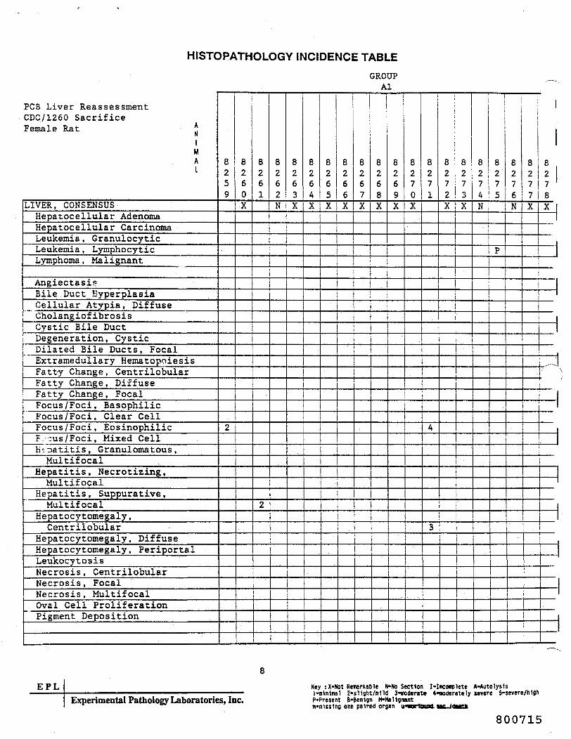
HISTOPATHOLOGY INCIDENCE TABLE
PCB Liver ReassessmentCDC/1260 SacrificeFemale Rat *
NiMAI
LIVER, CONSENSUSHepatocellular AdenomaHepatocellular CarcinomaLeukemia, GranulocyticLeukemia , LymphocyticLymphoma , Malignant
AngiectasisBile Duct EyperplasiaCellular Atypia, DiffuseGholangiof ibrosisCystic Bile DuctDegeneration, Cystic
; Dilated Bile Ducts, FocalExtramedullary HematopoiesisFatt-y Change, CentrilobularFatty Change, DiffuseFatty Change, Focal
| Focus/Foci, Basophilici Focus/Foci, Clear Cell
Focus /Foci, EosinophilicF. :us/Foci, Mixed CellHepatitis, Granulomatous ,Multifocal
Hepatitis, Necrotizing,Multifocal
Hepatitis, Suppurative,Multifocal
Hepatocy tomegaly ,Centrilobular
Hepatocy tomegaly , DiffuseHepatocytomegaly , PeriportalLeukocytosisNecrosis, CentrilobularNecrosis, FocalNecrosis, MultifocalOval Cell ProliferationPigment Deposition
GROUP __Al
8259
2
8260X
8261
2
——
8262N
8263X
8264X
1
8265X
8266X
8267X
t
-j
8268X
8269X
8270X
8271
4
3
o
o
72
8273
X i X
j
8274N
8275
P
8276N
i
8277X
8278X
E P LExperimental Pathology Laboratories, Inc.
Key :X-Not Remarkable N-No Section I-lnconclete A-Autolys1sl-n1n1mal 2-sligtit/mlld 3-roderete 4-noderately severe 5-severe/hfghP-Present B-Ben1gn (HtaUsnantin-missing one paired organ u^Borttuaa. sa^JOMttt
800715

HISTOPATHOLOGY INCIDENCE TABLE
PCB Liver ReassessmentCDC/1260 SacrificeFemale Rat „
NiMAI
LIVER, CONSENSUSHepatocellular AdenomaHepatocellular CarcinomaLeukemia, GranulocyticLeukemia , LymphocyticLymphoma , Ma 1 i gnant
AngiectasisBile Duct HyperplasiaCellular Atypia, DiffuseCholangiofibrosisCystic Bile DuctDegeneration, CysticDilated Bile Ducts, Focal
^jCxtramedullary Hematopoiesis"f tty Change , Centrilobular
_atty Change, DiffuseFatty Change , FocalFocus /Foci, BasophilicFocus /Foci, Clear CellFocus /Foci, EosinophilicFocus /Foci, Mixed CellHepatitis, Granulomatous ,Multif ocal
Hepatitis, Necrotizing,Multif ocal
Hepatitis, Suppurative,Multifocal
Hepatocytomegaly ,Centrilobular
Hepatocytomegaly, DiffuseHepatocytomegaly, Periportal
\ LeukocytosisNecrosis, CentrilobularNecrosis, Focal
i Necrosis, Multifocal: Oval Cell Proliferation
Pigment Deposition
GROUPAl
8279X
8280X
8281
2
8282X
8283X
8284X
8285X
8286X
8287N
8288X
8289
1
8290X
8291X
8292X
8293
3
2
1
8294X
8295
1
8296
1
8297N
8298N
—— \-
EPLExperimental Pathology Laboratories, Inc.
Key : X-Not Remarkable N-No Sectionl-Bln1ma) 2*sl1glit/n1ld 3-noderateP-Present 8-Ben1gn M-Ma11gnantm-«1sj1ng one paired organ u*MrtMi
[•Incomplete A-AutolysIs4«K>derately sever* 5-severe/hlght sac./death
800716

HISTOPATHOLOGY INCIDENCE TABLE
PCB Liver ReassessmentCDC/1260 SacrificeFemale Rat *
NiMAL
LIVER, CONSENSUSHepatocellular AdenomaHepatocellular CarcinomaLeukemia, GranulocyticLeukemia , LymphocyticLymphoma, Malignant ,
AngiectasisBile Duct HyperplasiaCellular Atypia, DiffuseGholangiof ibrosisCystic Bile DuctDegeneration, CysticDilated Bile Ducts , FocalExtramedullary HematopoiesisFatty Change, CentrilobularFatty Change, DiffuseFatty Change, FocalFocus/Foci, BasophilicFocus /Foci, Clear CellFocus /Foci, EosinophilicFocus /Foci, Mixed CellHepatitis, Granulomatous ,Multifocal
Hepatitis, Necrotizing,Multifocal
Hepatitis, Suppurative,Multifocal
Hepatocy tomegaly ,Centrilobular
Hepatocytomegaly , DiffuseHepatocytomegaly , PeriportalLeukocytosisNecrosis , CentrilobularNecrosis, FocalNecrosis, MultifocalOval Cell ProliferationPigment Deposition
GROUPAl
8299X
8300X
8301
1
8302N
8303
2
8304X
8305
P
5
8306X
8307X
8308X
1
8309X
8310X
8311
3
83
2
1
83
3X
83
4X
!
83
5X
83
6X
i
83
7X
————
83
8X
-
1
j
J
:
——ii
- 1_., - J
10EPL
Experimental Pathology Laboratories, Inc.Key :X*Not Remarkable H-No Section Mnconplete A-Autolysislitilnlnal 2-sHght/mlld 3-nodeme 4-noderately severe 5-severe/l>1gl>P-Present B-Benlgn H-Hal1gnantn*mUs1ng one paired organ u-norlbund iK./death
800717

HISTOPATHOLOGY INCIDENCE TABLEGROUP
A2
PCB Liver ReassessmentCDC/1260 SacrificeFemale Rat *
1MAt
LIVER, CONSENSUSHepatocellular AdenomaHepatocellular CarcinomaLeukemia, GranulocyticLe ukemia , Lympho cy t i cLymphoma , Ma 1 i gnan t
AngiectasisBile Duct HyperplasiaCellular Atypia, DiffuseCholangiofibrosisCystic Bile DuctDegeneration, CysticDilated Bile Ducts, Focal
^JSxtramedullary Hematopoiesisitty Change , Centrilobular
ratty Change, DiffuseFatty Change, FocalFocus /Foci, BasophilicFocus /Foci, Clear CellFocus /Foci, EosinophilicFocus /Foci, Mixed CellHepatitis, Granulomatous ,Multifocal
Hepatitis, Necrotizing,Multifocal
Hepatitis, Suppurative,Multifocal
Hepatocytomegaly ,Centrilobular
Hepatocytomegaly, DiffuseHepatocytomegaly, PeripprtalLeukocytosisNecrosis, CentrilobularNecrosis, FocalNecrosis, MultifocalOval Cell ProliferationPigment Deposition
8319
P
1
24
2
8320X
8321
P
5
5
23
8322N
8323N
8324
335
8325
P
534
4
8326
P
2
5
3
8327
P
32
1
525
2
2
8328
P
4
4
2
8329
P
3
43
1
8330N
8331
P
42
22
8332
P
2
35
2
2
8333
1
353
2
4
2
8334
P
1
3
4
2
8335
P
3
5
3
12
8336
——
1
2
8337
3
52
——
3
8338
P
3
3
——
EPL11
Experimental Pathology Laboratories, Inc.
800718
Key : X-Not Remarkable tMto Section I-Incooplete A-AutolysU1-minimal 2-sl1ght/»1ld 3-aoderate 4-aoderataly severe 5-severe/li1gliP-Present B-Ben1gn H-Ma11gnant
one paired organ u-nor1bund sac./death

HISTOPATHOLOGY INCIDENCE TABLE
PC6 Liver ReassessmentCDC/1260 SacrificeFemale Rat *
NiMAI
LIVER, CONSENSUSHspatocellular Adenoma
• Hepatocellular CarcinomaLeukemia, GranulocyticLeukemia , LymphocyticLymphoma , Ma 1 i gnant
AngiectasisBile Duct Hyperplasia
' Cellular Atypia, Diffuse: Cholangiof ibrosis
Cystic Bile DuctDegeneration, CysticDilated Bile Ducts, FocalExtramedullary HematopoiesisFatty Change , CentrilobularFatty Change, DiffuseFatty Change, Focal
j Focus /Foci, BasophilicFocus/Foci, Clear CellFocus /Foci, EosinophilicFocus /Foci, Mixed CellHepatitis, Granulomatous ,Multif ocal
Hepatitis, Necrotizing,Multifocal
Hepatitis, Suppurative,Multifocal
Hepatocy tomegaly ,Centrilobular
Hepatocytomegaly , DiffuseHepatocytomegaly, PeriportalLeukocytosisNecrosis, CentrilobularNecrosis, FocalNecrosis, MultifocalOval Cell ProliferationPisment Deposition
GROUPA2
8339
PP
T
5
2
83
0
P
3
3
2
5
32
83
1
P
2352
83
2
P
5
3
83
3»
83
4
12
5
1
2
3
4
3
83
5
P
5
2
83
6
P
325
2
83
7
3P
4
33
83
8N
83
9N
8350
PP
1
3
2
8351
43
3
8352
3
8353
P
34
8354
3
2
8355
P
2
8356
4
4
4
3
8357
2
2
8358
,-~---.
34
1
EPL12
Experimental Pathology Laboratories, Inc.v.
800719
Key : X-Not Remarkable H-No Section l-inconplete A-AutolysH1-mlnioal 2-sllght/nlld 3-moderate 4-noderately severe 5-severe/MghP-Present B-Ben1gn H-Hatigrantnt-oflsslng one paired organ u*nor1bund sac./death

HISTOPATHOLOGY INCIDENCE TABLEGROUP
A2
PCS Liver ReassessmentCDC/1260 SacrificeFemale Rat *
niMAt
LIVER, CONSENSUSHepatocellular AdenomaHepatocellular CarcinomaLeukemia, GranulocyticLeukemia , LymphocyticLymph oma , Malignant
AngiectasisBile Duct Hyperplasia
. Cellular Atypia, DiffuseCholangiofibrosis
! Cystic Bile DuctDegeneration, CysticDilated Bile Ducts, FocalExtramedullary Hematopoiesis
/~*r~T~ N,tty Change, Centrilobularatty Change, DiffuseFatty Change, Focal
, _ Focus/Foci, BasophilicFocus /Foci, Clear CellFocus /Foci, EosinophilicFocus /Foci, Mixed CellHepatitis , Granulomatous ,Multifocal
Hepatitis, Necrotizing,Multifocal
Hepatitis, Suppurative,Multifocal
Hepatocytomegaly ,Centrilobular
Hepatocytomegaly, DiffuseHepatocytomegaly, Periportal
1 Leukocytosisi Necrosis, Centrilobulari Necrosis, Focal. Necrosis, Multifocal
Oval Cell ProliferationPigment Deposition
8359
3
3
8360
P
1
44
2
8361
P
325
2
8362
15
3
2
8363
P
1
4
3
8364
1
3
1
8365
P
223
8366
P
1
3
54
1
8367
P
2
1
2
5
2
8368
P
2
1
3
2
8369
P
2
4
3
83
0
3
1
83
1
1
25
2
83
2
1
3
23
1
83
3
P
2
4
235
32
83
4
P
1
254
3
1
83
5
P
3
5
83
6
34
3
83
7
PP
34
5
2
32
83
8
P
124
2
EPL13
Experimental Pathology Laboratories, Inc.
800720
Key : X-Not Rraarkable N-No Section I-Ineonplete A-AutolysisI*m1n1ma1 2»s11ght/n1ltf 3-noderate 4-noderttely severe £-$evere/h1gbP-Present B-Benign H-Hallgnantn-n1ss1ng one paired organ u-norlbund sac./death

HISTOPATHOLOGY INCIDENCE TABLEGROUP
A2
PCB Liver ReassessmentCDC/1260 SacrificeFemale Rat *
N1MAI
LIVER, CONSENSUSHepatocellular AdenomaHepatocellular CarcinomaLeukemia, GranulocyticLeukemia , Lymphocy ticLymphoma, Malignant
AngiectasisBile Duct HyperplasiaCellular Atypia, DiffuseCholangiofibrosisCystic Bile DuctDegeneration, CysticDilated Bile Ducts, FocalExtramedullary HematopoiesisFatty Change, CentrilobularFatty Change, DiffuseFatty Change, FocalFocus /Foci, BasophilicFocus /Foci, Clear CellFocus /Foci, EosinophilicFocus /Foci, Mixed CellHepatitis, Granulomatous ,Multifocal
Hepatitis, Necrotizing,Multifocal
Hepatitis, Suppurative,Multifocal
Hepatocytomegaly ,Centrilobular
Hepatocytomegaly, Diffuse n
Hepatocytomegaly, PeriportalLeukocytosisNecrosis, CentrilobularNecrosis, FocalNecrosis, MultifocalOval Cell ProliferationPigment Deposition
8379N
8380
2
5
3
8381
P
4
44
2
2
8382
225
3
2
8383
P
2
2
1
8384
1
1
3
1
1
8385
3
2
2
8386
PP
2
1
5
4
22
8387
2
2
1
8388
P
242
2
8389
2
1
8390
P
325
8391
1
32
8392
P
2
4
2
8393
PP
5
2
8394
13
43
53
8395
P
2
53
5
2
83 !96
P
4
2
j
8397
P
2
335
2
8398
^
23
2
EPL14
Experimental Pathology Laboratories, Inc.
800721
Key : X-Not Remarkable N-No Section I-Inconplete A-Autolysisl-m1n1nal 2-slight/nlld 3-«oderate 4-noderately severe 5-severe/h1g!>P'Present B-Ben1gn M-HaUgnantn-nlss1ng one paired organ u-nor1bund sac./death

HISTOPATHOLOGY INCIDENCE TABLEGROUP
A2
PCB Liver ReassessmentCDC/1260 SacrificeFemale Rat *
NiMAI
LIVER, CONSENSUSHepatocellular AdenomaHepatocellular CarcinomaLeukemia, GranulocyticLeukemia , LymphocyticLymphoma, Malignant
AngiectasisBile Duct HyperplasiaCellular Atypia, DiffuseCholangiofibrosisCystic Bile DuctDegeneration, CysticDilated Bile Ducts, Focal
^JSxtramedullary Hematopoiesis\tty Change, Centrilobular
-atty Change, DiffuseFatty Change_, FocalFocus /Foci, Basophilic
i Focus /Foci, Clear CellFocus /Foci, EosinophilicFocus /Foci, Mixed CellHepatitis, Granulomatous,Multifocal
Hepatitis, Necrotizing,Multifocal
Hepatitis, Suppurative,Multifocal
Hepatocytomegaly ,Centrilobular
Hepatocytomegaly, DiffuseHepatocytomegaly, PeriportalLeukocytosisNecrosis, CentrilobularNecrosis, FocalNecrosis, MultifocalOval Cell ProliferationPigment Deposition
8399
PP
4
2
8
00
P
2
3
1
8
01
2
22
3
2
1
8
02
P
2
3
52
4
8
03
P
15
3
8
04
P
2
3
5
4
8
05
P
11
2
2
8
0"6
P
35
1
1
8
07
PP
21
4
5
8
08
P
14
8
09
PP
2
5
4
2
8
0N
8
1
P
2
t3
4
2
2
2
8
2
P
2
1
223
2
8
3
P
2
2
8
4
PP
2
5
2
1
8
5
P
3
4
8
16
P
1
5
2
1
j
8417
P
... ..
224
3
8418
P
433
1
15
E P LExperimental Pathology Laboratories, Inc.
800722
Key : X-Not Remarkable N-No Section I-lncooplete A-AutolysIsl-a1n1nal 2-sHght/nlld 3-aoderate 4-noderately severe 5-*evere/MghP-Present B-Ben1gn M-MaUgnam:nilsslng one paired organ u-aorlbund MC./death
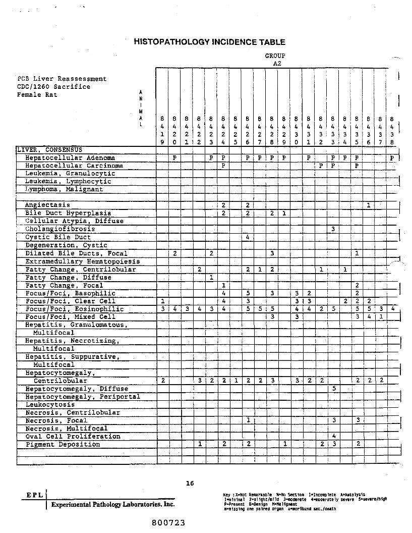
HISTOPATHOLOGY INCIDENCE TABLEGROUP
A2
PCS Liver ReassessmentCDC/1260 SacrificeFemale Rat *
nIMAI
LIVER, CONSENSUSHepatocellular AdenomaHepatocellular CarcinomaLeukemia, GranulocyticLeukemia , LymphocyticLymphoma, Malignant
AngiectasisBile Duct HyperplasiaCellular Atypia, DiffuseCholangiof ibrosisCystic Bile DuctDegeneration, CysticDilated Bile Ducts, FocalExtramedullary HematopoiesisFatty Change , CentrilobularFatty Change, DiffuseFatty Change , FocalFocus /Foci, BasophilicFocus /Foci, Clear CellFocus /Foci, EosinophilicFocus /Foci, Mixed CellHepatitis , Granulomatous ,Multifocal
Hepatitis, Necrotizing,Multifocal
Hepatitis, Suppurative,Multifocal
Hepatocy tomegaly ,Centrilobular
Hepatocy tomegaly, DiffuseHepatocytomegaly, PeriportalLeukocytosisNecrosis, CentrilobularNecrosis, FocalNecrosis, MultifocalOval Cell ProliferationPigment Deposition
8
19
13
2
8
20
P
2
4
8
21
3
8
22
2
4
3
1
8
23
P
2
1
3
2
8
24
PP
22
1444
2
2
8
25
1
8
26
P
22
4
2
535
2
1
2
8
27
P
1
5
2
8
28
P
2
3
2
3
53
3
8
29
P
1
1
8
30
3343
3
8
31
P
234
2
8
32
P
1
2
2
2
8
33
PP
3
r-——
5
5
'
3
43
8434
P
1
2
8435
PP
1
22253
2
3
2
1
8436
"I
1
254
2
8437
1
31
2
8438
P
1
|
„- — .
4
——
._|
EPL16
Experimental Pathology Laboratories, Inc.
800723
Key : X-Not Renarkable N-Ko Section I-Incoaplete A-A«tolys1sl-m1n1mal 2"Sl1ght/Blld 3-noderate «-modemely severe S-severe/nljliP-Present B-Benign M*Mal1gnantB*n1ss1ng one paired organ u-norltaund sac./death

HISTOPATHOLOGY INCIDENCE TABLEGROUP
A2
PCB Liver ReassessmentCDC/1260 SacrificeFemale Rat *
N1MAI
LIVER, CONSENSUSHepatocellular AdenomaHepatocellular CarcinomaLeukemia, GranulocyticLeukemia , LymphocyticLymphoma , Malignant
AngiectasisBile Duct HyperplasiaCellular Atypia, DiffuseCholangiof ibrosisCystic Bile DuctDegeneration, CysticDilated Bile Ducts, Focal
/-»»§xtrainedullary Hematopoiesisitty Change, Centrilobular
ratty Change, DiffuseFatty Change, FocalFocus /Foci, BasophilicFocus /Foci, Clear CellFocus /Foci, EosinophilicFocus /Foci, Mixed CellHepatitis, Granulomatous ,Multifocal
Hepatitis, Necrotizing,Multifocal
Hepatitis, Suppurative,Multifocal
Hepatocy tomegaly ,Centrilobular
Hepatocy tomegaly, DiffuseHepatocy tomegaly, PeriportalLeukocytosisNecrosis, CentrilobularNecrosis, FocalNecrosis, MultifocalOval Cell ProliferationPigment Deposition
8
39
P
2
22
8
40
P
3
3
5
8
41
P
53
2
8
42
2
3
3
3
2
8
43
13
2
2
8
44
PP
23
2
8
5
P
3
4
54
3
8
6
P
8
7
P
2
53
2
2
8
8
P
3
1
1
8
9N
8
50
P
2
5
4
2
8
5I•P
3
1
8452
P
2
323
2
2
8453
P
1
53
3
3
2
8454X
8.455
P
4
2
8456
P
252
3
8457
P
•
8458X
EPL
17
Experimental Pathology Laboratories, Inc.
800724
Key :X-Not Remarkable N-No Section I-Inconplete A-Autolysis1-nlnlnal 2-sHght/BHd 3-noderate 4-noderately sever* 5-severe/h1gtiP»Present B-Ben1gn M-Ma11cnantn-m1ss1ng one paired organ u-oorlbund sac./deatt

HISTOPATHOLOGY INCIDENCE TABLE
PCB Liver ReassessmentCDC/1260 SacrificeFemale Rat *
niMAI
LIVER , CONSENSUSHspatocellular AdenomaHspatocellular CarcinomaLeukemia , GranulocyticLeukemia , LymphocyticLymph oma , Ma 1 i gnant
AngiectasisBile Duct HyperplasiaCellular Atypia, Diffuse
| Cholangiofibrosis; Cystic Bile Duct
Degeneration, CysticDilated Bile Ducts, FocalSxtramedullary HematopoiesisFatty Change, CentrilobularFatty Change, DiffuseFatty Change, FocalFocus /Foci, BasophilicFocus /Foci, Clear CellFocus /Foci, EosinophilicFocus /Foci, Mixed CellHepatitis, Granulomatous ,Multifocal
Hepatitis, Necrotizing,Multifocal
Hepatitis , Suppurative ,Multifocal
Hepatocytomegaly ,Centrilobular
Hepatocytomegaly, DiffuseHepatocytomegaly, PeriportalLeukocytosisNecrosis, CentrilobularNecrosis, FocalNecrosis, MultifocalOval Cell ProliferationPigment Deposition
GROUPA2
8
59
P
2
244
3
8
60
243
8
61
P
414
3
2
8
62
P
2
1
2
8
63
P
2
8
64N
8
65
P
4
8
66
P
5
2
8
67
P
1
22
2
8
68
P
3
3
35
2
8
69
P
3352
8
70
P
3354
2
8
71
P
114
2
8
72
P
1
5
2
8
73
P
2
4
2
8
4
P
3
1
44
2
8
5
P
4
2
8
6
3
1
8
7
1
224
1
1
i
8
8
P
x —
334
2
E P L18
Experimental Pathology Laboratories, Inc.
800725
Key : X-Not Remarkable N-No Section I-lncooplete A-AutolysIs1-mlnfmal 2-sllBht/m1ld 3-w>der«te 4-»oderately sever* i-severe/hlflhP-Present B-Ben1gn H-Mallgnantn-alsslng one paired organ u-nor1bund sac./death

HISTOPATHOLOGY INCIDENCE TABLEGROUP
A2
PUB Liver ReassessmentCDC/1260 SacrificeFemale Rat *
niMAL
LIVER, CONSENSUSHepatocellular AdenomaHepatocellular CarcinomaLeukemia, GranulocyticLeukemia , LymphocyticLymphoma, Malignant
AngiectasisBile Duct Hyperplasia
: Cellular Atypia, DiffuseCholangiofibrosisCystic Bile DuctDegeneration, CysticDilated Bile Ducts, FocalExtramedullary Hematopoiesis
,-"" stty Change, Centrilobular.tty Change , Diffuse
Fatty Change, FocalFocus /Foci, BasophilicFocus /Foci, Clear CellFocus /Foci, EosinophilicFocus /Foci, Mixed Cell
i Hepatitis, Granulomatous ,Multifocal
Hepatitis, Necrotizing,Multifocal
Hepatitis, Suppurative,Multifocal
Hepatocytomegaly ,Centrilobular
Hepatocytomegaly, DiffuseHepatocytomegaly, PeriportalLeukocytosisNecrosis, CentrilobularNecrosis, FocalNecrosis, Multifocal
i Oval Cell ProliferationPigment Deposition
8
79
2
2
1
8
80
PP
3
43
3
2
8
81
P
3
3
8
82
P
3
235
2
8
83
P
4
8
84
P
1
5
3
1
8
85
PP
2
243
3
8
86
P
1
5
8
87N
8488
P
22
4
5
3
33
8
89
1
12
8
90
P
45
8
91
P
4
2
8
92
P
1
23
1
8
93
PP
343
2
8
94
P
13
8
95
P
5
1
8
96
P
2
435
8
97
P
2
1
8
98
P
2251
EPL19
Experimental Pathology Laboratories, Inc.
800726
Key :X-Not Reurkafale N-No Section I-Inconplete A'AutolysUl-n1n1ial 2-$llBht/n1ld 3-noderate 4-noderately severe 5-$evere/h1ghP-Present B-Ben1gn M-Ha11gnantn-«i1ss1ng one paired organ u-norlbund sac./death

HISTOPATHOLOGY INCIDENCE TABLEGROUP
A2
PCS Liver ReassessmentCDC/1260 SacrificeFemale Rat *
niMAI
LIVER, CONSENSUSHepatocellular AdenomaHepatocellular CarcinomaLeukemia, GranulocyticLeukemia, LyrophocyticLymph oma, Malignant
AngiectasisBile Duct HyperplasiaCellular Atypia, Diffuse
* (JholangiofibrosisCystic Bile DuctDegeneration, CysticDilated Bile Ducts, FocalExtramedullary HematopoiesisFatty Change , CentrilobularFatty Change, DiffuseFatty Change , FocalFocus /Foci, BasophilicFocus /Foci, Clear CellFocus /Foci, EosinophilicFocus /Foci, Mixed CellHepatitis, Granulomatous ,Multifocal
Hepatitis, Necrotizing,Multifocal
Hepatitis, Suppurative,Multifocal
Hepatocytomegaly ,Centrilobular
Hepatocytomegaly, DiffuseHepatocytomegaly, PeriportalLeukocytosisNecrosis, CentrilobularNecrosis, FocalNecrosis, MultifocalOval Cell ProliferationPigment Deposition
8499
P
43
8500
P
325
2
8501
P
1
2
35
2
1
8502
P
2
215
5
2
8503X
8504
PP
2
3
5
1
8505
P
4
3
8506
P
4
2
8507
PP
4
2
8508
P
1
4
1
8509
P
2
4
8510
1
3
3
1
8511N
8512
P
4
8513
21
3
85
4
P
2
2
5
4
21
85
5
1
1
85
6
P
2
A
5
3
2
i
85
7
P
2
i_
32
1
85
8
P
——1
5
3
EPLExperimental Pathology Laboratories, Inc.
20Key : X-Not Remarkable N-No Section I-lnconplete A-AutolysIsItn1n1tnal 2-sl1flht/n1ld 3-noderate 4-noderately severe 5-severe/nlohP-Present B-Ben1gn H>Hal1gnantn*n1sstng one paired organ u-norlbund sac./death
800727

EPLEXPERIMENTAL PATHOLOGY LABORATORIES, INC.
APPENDIX B
Aroclor 1260 Reproduction Study inMale and Female Sherman Rats
(Under, et al., 1974)
Hales
Group Al- ControlGroup A3 - 100 ppm Aroclor 1260
Females
Group A2 - ControlGroup A4 - 100 ppm Aroclor 1260
800728

EPLEXPERIMENTAL PATHOLOGY LABORATORIES, INC.
SUMMARY INCIDENCE TABLES
800729

SUMMARY INCIDENCE TABLEPCB Liver ReassessmentCDC/1260(7-70) SacrificeMale Rat
LIVER, CONSENSUS (NO. EXAMINED)Cytoplasmic InclusionsFatty Change , CentrilobularFatty Change, FocalFocus /Foci, Clear CellFocus /Foci, EosinophilicHepatocy tomegaly ,Centrilobular
Microgranuloma(s )Pigment, Deposition
SSS"*
GROUPAl(10)
3
GROUPA3(10)2
1
102
EPL* Experimental Pathology Laboratories, Inc.
800730

SUMMARY INCIDENCE TABLEPCB Liver ReassessmentCDC/1260(7-70) SacrificeFemale Rat
LIVER, CONSENSUS (NO. EXAMINED)Cytoplasmic InclusionsFatty Change, CentrilobularFatty Change, FocalFocus /Foci, Clear CellFocus /Foci, EosinophilicHepatocy tomegaly ,Centrilobular
Microgranuloma ( s )Pigment, Deposition
^^,
GROUPA2(10)
5
GROUPA4(10)
21
2
734
E P LExperimental Pathology Laboratories, Inc.
800731

EPLEXPERIMENTAL PATHOLOGY LABORATORIES, INC.
HISTOPATHOLOGY INCIDENCE TABLES
800732

HISTOPATHOLOGY INCIDENCE TABLEGROUPAl
PCB Liver ReassessmentCDC/1260(7-70) SacrificeMale Rat *
NiMAI
LIVER, CONSENSUSCytoplasmic InclusionsFatty Change , CentrilobularFatty Change, FocalFocus /Foci, Clear CellFocus /Foci, EosinophilicHepatocytomegaly ,
CentrilobularMicrogranuloma ( s )Pigment, Deposition
/" ~\
8901X
8902X
8903X
890AX
8905
1
8906
1
8907X
8908
1
8909X
8910X
•
1
EPLExperimental Pathology Laboratories, Inc.
Key : X-Not Remarkable N-Ho Section I-lncooplete A-AutolysisI«n1n1nal 2-slight/mlId 3-iioderate 4-woderately severe 5-severe/highP-Present 8-Ben1gn H-Ha11gnant••Hissing one paired organ u-mor1bund sac./death
800733

HISTOPATHOLOGY INCIDENCE TABLEGROUP
A3
PCB Liver ReassessmentCDC/1260(7-70) SacrificeMale Rat *Ni
MAI
LIVER, CONSENSUSCytoplasmic InclusionsFatty Change, CentrilobularFatty Change, FocalFocus /Foci, Clear CellFocus /Foci, .EosinophilicHepatocytomegaly ,Centrilobular .
Microgranuloma ( s )Pigment, Deposition
....
..-
8921
2
8922
21
8923
3
8924
4
8925
1
4
8926
4
5
8927
31
8928
3
5
8929
4
8930
3
1
J
'
-
1
1
EPLExperimental Pathology Laboratories, Inc.
Key :X-Not Remarkable H-No Section Mnconplete A"Autolys1sl-n1n1mal 2-sl10ht/n1ld 3-noderate 4-«oderately severe S-severe/HljtiP-Present B*Ben1gn H-Ma11gnant••missing one paired organ u-morlbund sac./death
800734

HISTOPATHOLOGY INCIDENCE TABLEGROUP
A2
PCB Liver ReassessmentCDC/1260(7-70) SacrificeFemale Rat *
NiMAI
LIVER, CONSENSUSCytoplasmic InciusionsFatty Change , CentrilobularFatty Change, FocalFocus /Foci, Clear CellFocus /Foci, EosinophilicHepatocytomegaly ,Centrilobular
Microgranuloma ( s )Pigment, Deposition
JxT ,
8911
1
8912X
8913
1
8914X
89
5
2
89
6
1
89
7
1
89
8X
89
9X
8920X
•
"7
EPLExperimental Pathology Laboratories, Inc.
Key :X-Not Rewritable M-No Section I-Incomplete A-Auto1ys1$1-nlnlmal 2«sl1ght/»1ld 3-noderate 4«noderate1y severe 5-severe/h1gh("•Present B-Ben1gn M-Mallgnantn-n1ss1ng one paired organ u-i»r1bund sac./death
800735
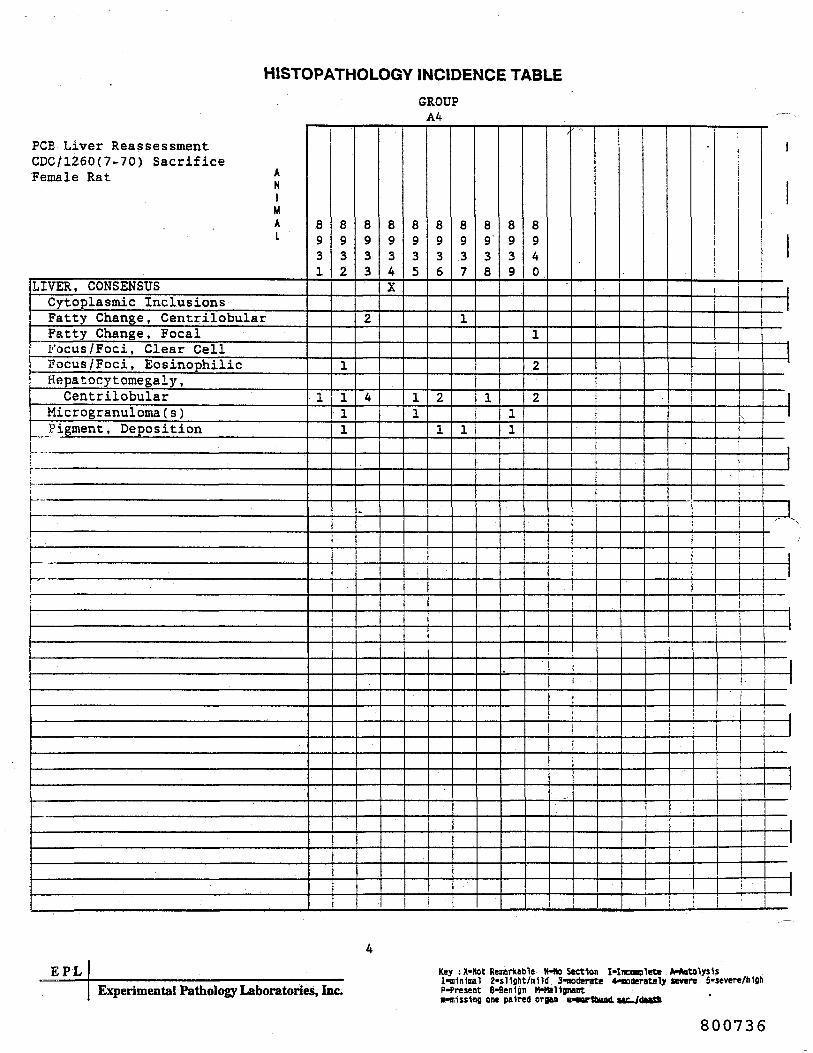
HISTOPATHOLOGY INCIDENCE TABLE
PCS Liver ReassessmentCDC/1260(7-70) SacrificeFemale Rat *
NiMAL
LIVER, CONSENSUSCytoplasmic InclusionsFatty Change, CentrilobularFatty Change, FocalFocus /Foci, Clear CellFocus /Foci, EosinophilicHepatocy tomegaly ,Centrilobular
Microgranuloma ( s )Pigment, Deposition
i!
GROUPA4
8931
1
8932
1
111
8933
2
4
^
8934X
8935
11
8936
2
1
8937
1
1
8938
1
8939
11
8940
1
2
2•
i
1
(
EPLExperimental Pathology Laboratories, Inc.
Key : X-Not Remarkable H-No Section I-Inconplete A-*utolys1s1-nlnlmal 2-sUght/ralld 3-noderste *nncttrately severe 5-severe/hlghP-Present 8-Ben1gn H-HBltgnantn"«1ss1ng one paired argu MNrtbund.«" /«<^«>
800736

EPLEXPERIMENTAL PATHOLOGY LABORATORIES, INC.
APPENDIX C
Aroclor 1260 in Male and Female Spraque-Dawley Rats(Norback and Weltman, 1985)
Group ControlGroup 1260
- Control- 100 ppm for 16 Months Followedby 50 ppm for 8 Months ThenControl Diet for 5 Months
800737

EPLEXPERIMENTAL PATHOLOGY LABORATORIES, INC.
SUMMARY INCIDENCE TABLES
800738
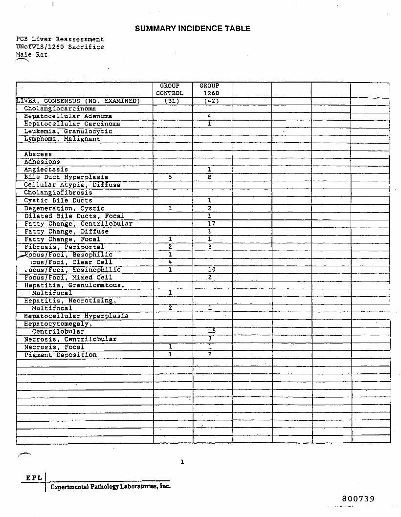
SUMMARY INCIDENCE TABLEFOB Liver ReassessmentUNofWIS/1260 SacrificeMale Rat
LIVER, CONSENSUS (NO. EXAMINED)CholangiocarcinomaHepatocellular AdenomaHepatocellular CarcinomaLeukemia, GranulocyticLymphoma, Malignant
AbscessAdhesionsAngiectasisBile Duct HyperplasiaCellular Atypia, DiffuseCholangiofibrosisCystic Bile DuctsDegeneration, CysticDilated Bile Ducts, FocalFatty Change, CentrilobularFatty Change, DiffuseFatty Change, FocalFibrosis, Periportal
^wEpcus/Foci, Basophilic)Cus/Foci, Clear Cell
tOcus/Foci, EosinophilicFocus /Foci, Mixed CellHepatitis, Granulomatous ,Multif ocal
Hepatitis, Necrotizing,Multifocal
Hepatocellular HyperplasiaHepatocy tomegaly ,Centrilobular
Necrosis, CentrilobularNecrosis, FocalPigment Deposition
GROUPCONTROL
(31)
6
1
12141
1
2
11
GROUP1260(42)
41
18
12117113
162
1
15712
E P LExperimental Pathology Laboratories, Inc.
800739

SUMMARY INCIDENCE TABLEPCB Liver ReassessmentUNofWIS/1260 SacrificeFemale Rat
LIVER, CONSENSUS (NO. EXAMINED)CholangiocarcinomaHepatocellular AdenomaHepatocellular CarcinomaLeukemia , GranulocyticLymphoma , Malignant
AbscessAdhesionsAngiectasisBile Duct HyperplasiaCellular Atypia, DiffuseCholangiofibrosisCystic Bile DuctsDegeneration, CysticDilated Bile Ducts, FocalFatty Change, CentrilobularFatty Change, DiffuseFatty Change , FocalFibrosis, Periportal
^^ocus/Foci, Basophilic>cus/Foci, Clear Cell
rocus/Foci, EosinophilicFocus /Foci, Mixed CellHepatitis, Granulomatous ,Multifocal
Hepatitis, Necrotizing,Multifocal
Hepatocellular HyperplasiaHepatocytomegaly ,Centrilobular
Necrosis, CentrilobularNecrosis, FocalPigment Deposition
GROUPCONTROL
(47)
1
11
1112
1
1
215
212
GROUP1260(46)32919
119145322
121
36
2
54513
EPLExperimental Pathology Laboratories, Inc.
800740

EPLEXPERIMENTAL PATHOLOGY LABORATORIES, INC.
HISTOPATHOLOGY INCIDENCE TABLES
800741

HISTOPATHOLOGY INCIDENCE TABLEGROUPCONTROL^ — N
i Liver ReassessmentUNofWIS/1260 SacrificeMale Rat *Ni
MAI
LIVER, CONSENSUSCholangiocarcinomaHepatocellular AdenomaHepatocellular CarcinomaLeukemia, GranulocyticLymphoma, Malignant
t AbscessAdhesions'AngiectasisSile Duct HyperplasiaCellular Atypia, DiffuseCholangiof ibrosis
, Cystic Bile DuctsDegeneration, Cystic
_/ja41ated Bile Ducts, Focaltty Change, Centrilobular
ratty Change, DiffuseFatty Change , FocalFibrosis, PeriportalFocus /Foci, BasophilicFocus /Foci, Clear CellFocus /Foci, EosinophilicFocus /Foci, Mixed CellHepatitis, Granulomatous ,Multifocal
Hepatitis, Necrotizing,Multifocal
Hepatocellular HyperplasiaHepatocytomegaly ,Centrilobular
Necrosis, CentrilobularNecrosis, FocalPigment Deposition
.-:.
20X
23X
24
1
1
25X
26X
28X
29X
30A
33X
34
2
1
1
35
1
1
11
37
3
38
1
1
0
1
1X
2X
3
2
6X
1
51X
52
1
EPLExperimental Pathology Laboratories, Inc.
Key : X-Not Remarkable N-No Section Mnconplete A-Autolysisl-n1n1i»Bl 2-sl1glit/in1ld 3-tnoder«te 4-noder«tely sever* 5-severe/hlghP-Present B-Benlgn M-Mal1gnantm-n1ss1ng one paired organ u*w>r1bund sac./death
800742

HISTOPATHOLOGY INCIDENCE TABLEGROUPCONTROL
*-^J Liver Reassessment
UNofWIS/1260 SacrificeMale Rat *
niMAI
LIVER, CONSENSUSCholangiocarcinomaHepatocellular AdenomaHepatocellular CarcinomaLeukemia, GranulocyticLymphoma , Malignant
AbscessAdhesionsAngiectasisBile Duct HyperplasiaCellular Atypia, DiffuseCholangiof ibrosisCystic Bile DuctsDegeneration, Cystic
_ Dilated Bile Ducts, Focalatty Change, Centrilobular
^atty Change, DiffuseFatty Change, FocalFibrosis, PeriportalFocus /Foci, BasophilicFocus /Foci, Clear CellFocus /Foci, EosinophilicFocus /Foci, Mixed CellHepatitis, Granulomatous,Multifocal
Hepatitis, Necrotizing,Multifocal
Hepatocellular HyperplasiaHepatocy tomegaly ,Centrilobular
Necrosis, CentrilobularNecrosis, FocalPigment Deposition
56A
60
1
61X
66
_^ —
3
69X
71
2
2
3
72X
74X
76
1
78X
79X
i
1
_l_1
EPLExperimental Pathology Laboratories, Inc.
Key : X-Not Remarkable N*No Section I-Incomplete A'AutolysUl-m1n1nal 2-sl1ght/»1ld 3-noderate 4-moderstely severe 5-severe/hlghP-Present B*Ben1gn H*Hal1gnantB-aljj1no one paired organ u-nor1bund sac./death
800743
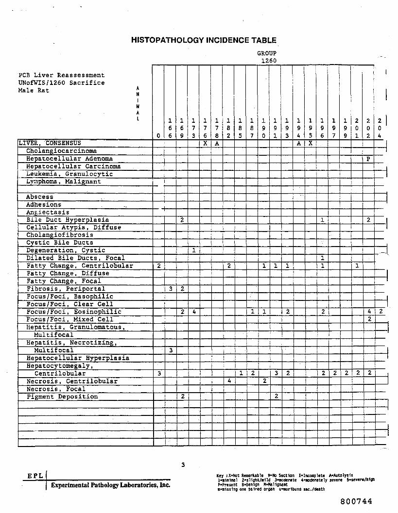
HISTOPATHOLOGY INCIDENCE TABLE
PCS Liver ReassessmentUNofWIS/1260 SacrificeMale Rat *
NiMAL
LIVER, CONSENSUSCholangiocarcinomaHepatocellular AdenomaHepatocellular CarcinomaLeukemia, GranulocyticLyziphoma, Malignant
AbscessAdhesionsAngiectasisBile Duct HyperplasiaCellular Atypia, DiffuseCholangiofibrosisCystic Bile DuctsDegeneration, CysticDilated Bile Ducts, FocalFatty Change, CentrilobularFatty Change, DiffuseFatty Change, FocalFibrosis, PeriportalFocus /Foci, BasophilicFocus /Foci, Clear CellFocus /Foci, EosinophilicFocus /Foci, Mixed CellHepatitis, Granulomatous ,Multif ocal
Hepatitis, Necrotizing,Multifocal
Hepatocellular HyperplasiaHepatocy tomegaly ,Centrilobular
Necrosis, CentrilobularNecrosis, FocalPigment Deposition
GROUP1260
066
-H —— |
2
3
3
3
69
2
2
2
2
1 ——
73
1
4
76X
78A
82
2
4
85
1
87
1
2
90
1
1
2
91
1
3
2
93
1
2
2
94 'A
__
j
195X
196
1
11
2
2
197
2
1
199
2
201
1
2
i
202
P
2
42
2
204
„
2
EPLExperimental Pathology Laboratories, Inc.
Key : X-Not Remarkable N-No Section I-Incooplete A-AutolysUl-n1n1nal 2-sl1flht/Blld 3-noderate 4-noderately severe 5-severe/lt1gftP-Present B-Ben1gn M-HsHgnantnin1s5lng one paired organ u-norlbund sac./death
800744

HISTOPATHOLOGY INCIDENCE TABLEGROUP
1260
. ..4 Liver ReassessmentUNofWIS/1260 SacrificeMale Rat *
NiMAI
LIVER, CONSENSUSCholangiocarcinomaHepatocellular AdenomaHepatocellular CarcinomaLeukemia, GranulocyticLymphoma, Malignant
AbscessAdhesions
; AngiectasisBile Duct HyperplasiaCellular Atypia, Diffuse
, Cholangiofibrosisi Cystic Bile Ducts
Degeneration, Cystic^»J?ilated Bile Ducts, Focal
atty Change, CentrilobularFatty Change, DiffuseFatty Change, F.ocalFibrosis, PeriportalFocus /Foci, BasophilicFocus /Foci, Clear CellFocus /Foci, EosinophilicFocus /Foci, Mixed CellHepatitis, Granulomatous ,Multifocal
Hepatitis, Necrotizing,Multifocal
Hepatocellular HyperplasiaHepatocytomegaly ,Centrilobular
Necrosis, CentrilobularNecrosis, FocalPigment Deposition
206X
207
2
210
I
2
3
211
1
2
214N
216X
217
2
1
220A
-
221
2
1
1
222
1
223
2
2
2
32
2
225
P
3
226
P
A
1
1
3
230
2
2
1
.
22
231
——
1
2
4
232
2
233
P
234
1
1
i
235N
—— t
236A
3
2
2
EPLExperimental Pathology Laboratories, Inc.
Key : X-Not Remarkable N-No Section I-Incomplete A-Autolys1sl-m1nlMl 2-sllght/ulld 3-moderate 4-moderstely severe S-severe/hljhP-Present 8-Ben1gn H-Mallgnantm-m1ss1ng one paired organ u-morlbund sac./death
800745

HISTOPATHOLOGY INCIDENCE TABLEGROUP
1260
PCS Liver ReassessmentUNofWIS/1260 SacrificeMale Rat *
NiMAL
LIVER, CONSENSUSCholangiocarcinomaHepatocellular AdenomaHepatocellular CarcinomaLeukemia , GranulocyticLymphoma , Malignant
AbscessAdhesionsAngiectasisBile Duct HyperplasiaCellular Atypia, DiffuseCholangiofibrosisCystic Bile DuctsDegeneration, CysticDilated Bile Ducts, FocalFatty Change, CentrilobularFatty Change, DiffuseFatty Change, FocalFibrosis, PeriportalFocus /Foci, BasophilicFocus /Foci, Clear CellFocus /Foci, EosinophilicFocus /Foci, Mixed CellHepatitis, Granulomatous ,Multifocal
Hepatitis, Necrotizing,Multifocal
Hepatocellular HyperplasiaHepatocytomegaly ,Centrilobular
Necrosis, CentrilobularNecrosis, FocalPigment Deposition
185BN
189A
1
2
189B
P
2
212A
1
212B
1
EPLExperimental Pathology Laboratories, Inc.
Key : X-Not Remarkable M-Ho Section I-Inconplete A-AutolytUlinlnlnal 2-s11sht/a1ld 3-noderate 4-noderately severe 5-severe/tiljhP-Present B-Benlgn M-HaUjnantn-n1ss1ng one paired organ u-aorlbund sac./death
800746

HISTOPATHOLOGY INCIDENCE TABLEGROUPCONTROL
Liver ReassessmentUNofWIS/1260 SacrificeFemale Rat *N
1MAI
LIVER , CONSENSUS •CholangiocarcinomaHepatocellular AdenomaHepatocellular CarcinomaLeukemia, GranulocyticLymphoma, Malignant
AbscessAdhesionsAngiectasisBile Duct HyperplasiaCellular Atypia, DiffuseCholangiofibrosisCystic Bile DuctsDegeneration, CysticDilated Bile Ducts, Focal
f N,tty Change, Centrilobularatty Change, DiffuseFatty Change , FocalFibrosis, PeriportalFocus /Foci, BasophilicFocus /Foci, Clear CellFocus /Foci, EosinophilicFocus /Foci, Mixed CellHepatitis, Granulomatous ,Multifocal
Hepatitis, Necrotizing,Multifocal
Hepatocellular HyperplasiaHepatocytomegaly ,Centrilobular
Necrosis, CentrilobularNecrosis, FocalPigment Deposition
16X
23X
24X
25X
26X
27
2
2
28X
29
PP
1
32
1
34X
35X
36X
37
2
38X
0
2
2X
3X
4X
5
2
1
6
3
E P LExperimental Pathology Laboratories, Inc.
Key sX-Hot Re»arkab1e N-No Section I-Inconplete A-AutolysIsHulntoal 2-s11ght/Blld 3-noderate 4-moderately severe 5-severe/hlghP-Present B*Ben1gn M-Ha11gnantnttUflng one paired organ u-aorlbund sac./death
800747

HISTOPATHOLOGY INCIDENCE TABLEGROUPCONTROL
PCB Liver ReassessmentUNofWIS/1260 SacrificeFemale Rat *
NiMAI
LIVER, CONSENSUSCholangiocarcinomaHepatocellular AdenomaHepatocellular CarcinomaLeukemia, GranulocyticLymphoma , Malignant
AbscessAdhesionsAngiectasis3ile Duct HyperplasiaCellular Atypia, DiffuseCholangiofibrosisCystic Bile DuctsDegeneration, CysticDilated Bile Ducts, FocalFatty Change, CentrilobularFatty Change, DiffuseFatty Change, FocalFibrosis, PeriportalFocus /Foci, BasophilicFocus/Foci, Clear CellFocus /Foci, EosinophilicFocus/Foci, Mixed CellHepatitis, Granulomatous,Multifocal
Hepatitis, Necrotizing,Multifocal
Hepatocellular HyperplasiaHepatocy tomegaly ,Centrilobular
Necrosis, Centrilobulariecrosis, FocalPigment Deposition
48
1
50X
51X
52
2
53X
55
2
3
3
56
P
2
58X
59X
60
2
62X
1
65X
67X
68X
70
P
71X
72X
7AA
75X
76X
1
EPLExperimental Pathology Laboratories, Inc.
800748
Key : X-Not Rewritable H-No Section I-Inconplete A-AutolysIsl-m1n1mal 2-sllsht/mnd 3-noderate 4-noderately severe S-severe/MjhP-Present B-Ben1gn M-Ma11grant«-n1ss1ng one paired organ u-norltaund sac./death

HISTOPATHOLOGY INCIDENCE TABLEGROUPCONTROL
,-«"-N
Liver ReassessmentUNofWIS/1260 SacrificeFemale Rat „
NiMAL
LIVER, CONSENSUSCholangiocarcinomaHepatocellular AdenomaHepatocellular CarcinomaLeukemia, GranulocyticLymphoma, Malignant
AbscessAdhesionsAngiectasisBile Duct HyperplasiaCellular Atypia, DiffuseCholangiof ibrosisCystic Bile DuctsDegeneration, Cystic^ilated Bile Ducts, Focal
f \tty Change, Centrilobular,atty Change, DiffuseFatty Change, FocalFibrosis, PeriportalFocus /Foci, BasophilicFocus /Foci, Clear CellFocus /Foci, EosinophilicFocus /Foci, Mixed CellHepatitis, Granulomatous ,Multifocal
Hepatitis, Necrotizing,Multifocal
Hepatocellular HyperplasiaHepatocytomegaly ,Centrilobular
Necrosis, CentrilobularNecrosis, FocalPigment Deposition
77
4
78
P
80X
30AX
30B
2
31AX
31BX
-- —
i
I 1
!
ii
EPLExperimental Pathology Laboratories, Inc.
Key : X-Not Remarkable N-No Section I-Inconplete A-AutolysUlinlnlnal 2-sl1ght/Blld 3-aoderate 4-aoderstely severe 5-severe/MghP-Present B-Benign M-Mal1giwntn-n1ss1ng one paired organ u-norlbund sac./death
800749

HISTOPATHOLOGY INCIDENCE TABLEGROUP
1260
FOB Liver ReassessmentUNofWIS/1260 SacrificeFemale Rat *N
1MAI
LIVER, CONSENSUSCholangiocarcinomaHepatocellular AdenomaHepatocellular CarcinomaLeukemia, GranulocyticLymphoma, Malignant
AbscessAdhesionsAngiectasisBile Duct HyperplasiaCellular Atypia, DiffuseCholangiofibrosisCystic Bile DuctsDegeneration, CysticDilated Bile Ducts, FocalFatty Change, CentrilobularFatty Change, DiffuseFatty Change, FocalFibrosis, PeriportalFocus /Foci, BasophilicFocus /Foci, Clear CellFocus /Foci, EosinophilicFocus /Foci, Mixed CellHepatitis, Granulomatous ,Multifocal
Hepatitis, Necrotizing,Multifocal
Hepatocellular HyperplasiaHepatocy tomegaly ,Centrilobular
Necrosis, CentrilobularNecrosis, FocalPigment Deposition
0
P
3
2
2
166
P
170N
77
2
2
5
78
P
3
3
2
2
82
PP
3
5
3
87
P
3
2
88
3
89
3
4
2
90
P
3
5
2
5
P
91
P
2
4
5
92
P
5
93
P
2
2
4
31
95
P
2
4
3
P
2
96
3
2
1
97
P
2
4.
2
2
2
98
PP
3
3
201
PP
5
202
P
1
4
24
203
P
4
s —
3
EPLExperimental Pathology Laboratories, loc
800750
Key : X-Not Remarkable H-No Section 1-lncooplete A-Autolysis1-nlnloul 2-sl1ght/Blld 3-noderate 4-noderately severe 5*severe/MgftP-Present B-Benlgn (MtaHgiwntB-Blss1ng one paired organ u-aor1taund sac./death
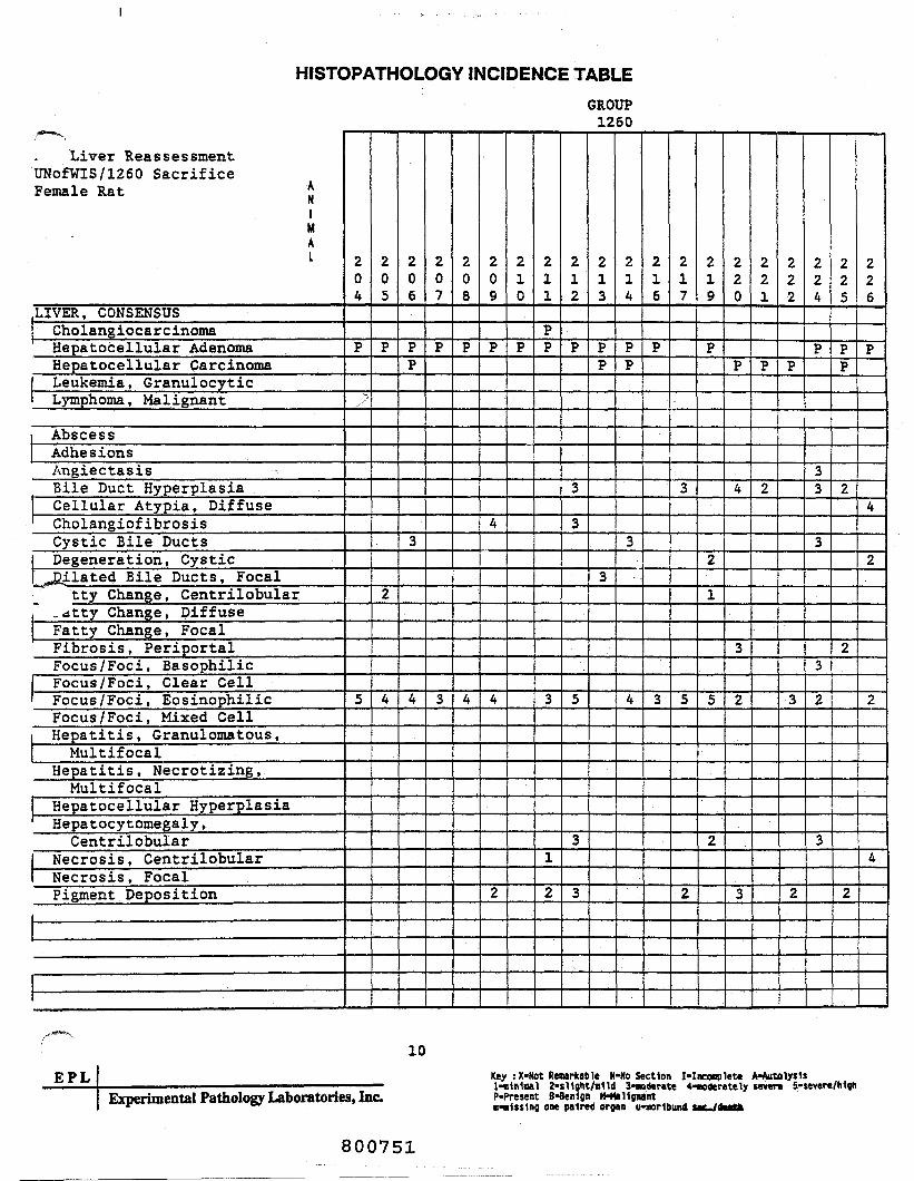
HISTOPATHOLOGY INCIDENCE TABLE
Liver ReassessmentUNofWIS/1260 SacrificeFemale Rat *
niMAI
LIVER, CONSENSUSCholangiocarcinomaHepatocellular AdenomaHepatocellular CarcinomaLeukemia, GranulocyticLymphoma, Malignant
AbscessAdhesionsAngiectasisBile Duct HyperplasiaCellular Atypia, DiffuseCholangiofibrosisCystic Bile DuctsDegeneration, CysticJJilated Bile Ducts, Focal
tty Change, Centrilobular_dtty Change, DiffuseFatty Change, FocalFibrosis, PeriportalFocus /Foci, BasophilicFocus /Foci, Clear CellFocus /Foci, EosinophilicFocus /Foci, Mixed CellHepatitis, Granulomatous,Multifocal
Hepatitis, Necrotizing,Multifocal
Hepatocellular HyperplasiaHepatocy tomegaly ,Centrilobular
Necrosis, CentrilobularNecrosis, FocalPigment Deposition
GROUP1260
204
P
S
5
205
P
2
4
206
PP
3
4
207
P
3
208
P
4
209
P
4
4
2
2
0
P
2
1
PP
3
1
2
2
2
P
3
3
5
3
3
2
3
PP
3
2
4
PP
3
4
2
6
P
3
2
7
3
5
2
2
9
P
2
1
5
2
220
P
4
3
2
3
221
P
2
222
P
3
2
224
P
33
3
3
2
3
225
PP
2
2
2
226
P
4
2
2
4
10
EPLExperimental Pathology Laboratories, Inc.
Key sX-Not Rewritable H-No Section I-Inconplett A*AutolysUl-Bln1aal 2*sl1ght/Blld 3-noderate 4-ooderately severe 5-severe/hlghP-Present B-Benlgn M-Mallgnant«-«1js1nj one paired organ u*nor1bund uc./d*tffc
800751

HISTOPATHOLOGY INCIDENCE TABLEGROUP
1260
PCS Liver ReassessmentUNofWIS/1260 SacrificeFemale Rat *
NiMAL
LIVER, CONSENSUSCholangiocarcinomaHepatocellular AdenomaHepatocellular CarcinomaLeukemia , GranulocyticLymphoma, Malignant
AbscessAdhesionsAngiectasisBile Duct HyperplasiaCellular Atypia, DiffuseCholangiof ibrosisCystic Bile DuctsDegeneration, CysticDilated Bile Ducts, FocalFatty Change, CentrilobularFatty Change, DiffuseFatty Change, FocalFibrosis, PeriportalFocus /Foci, BasophilicFocus /Foci, Clear CellFocus /Foci, EosinophilicFocus /Foci, Mixed CellHepatitis, Granulomatous,Multifocal
Hepatitis, Necrotizing,Multifocal
Hepatocellular HyperplasiaHepatocytomegaly ,Centrilobular
Necrosis, CentrilobularNecrosis, FocalPigment Deposition
227
P
P
3
5
23
228
P
229•
P
3
3
231
PP
3
5
233
P
4
235
P
3
236
P
4
1
_ .._j- -1
—
-J
i
11EPL
Experimental Pathology Laboratories, Inc.Key : X-Not Remarkable N-No Section 1-Inconplete A-Autolysisl-nlnlnal 2-sl1flht/ni1d 3-aoderate 4-noderately severe 5-severe/MghP-Present B-Ben1gn H-Hal1gnantB-m1ss1ng one paired organ uiorlbund sac./death
800752

EPLEXPERIMENTAL PATHOLOGY LABORATORIES, INC.
APPENDIX D
National Cancer Institute (NCI) 1977 Aroclor 1254 Studyin Male and Female Fiscner 344 Rats
(NCI, 1977)
Hales
Group 01-1645 - ControlGroup 01-1639 - 25 ppm AroclorGroup 01-1641 - 50 ppm AroclorGroup 01-1643 - 100 ppm Aroclor
Females
Group 01-1646Group 01-1640Group 01-1642Group 01-1644
Control25 ppm Aroclor50 ppm Aroclor100 ppm Aroclor
800753

EPLEXPERIMENTAL PATHOLOGY LABORATORIES, INC.
SUMMARY INCIDENCE TABLES
800754

SUMMARY INCIDENCE TABLEPCB Liver ReassessmentNTP/1254 SacrificeMale Rat
LIVER, CONSENSUS (NO. EXAMINED)Hepatocellular AdenomaHepatocellular CarcinomaMononuclear Cell Leukemia
Bile Duct HyperplasiaCystic Bile DuctExtramedullary HematopoiesisFatty Change, CentrilobularFatty Change, DiffuseFatty Change, FocalFibrosis, PeriportalFocus/Foci, BasophilicFocus /Foci, Clear CellFocus /Foci, EosinophilicFocus/Foci, Mixed CellHepatitis, Granulomatous ,Focal
Hepatitis, Granulomatous,Multifocal
^JSepatitis , Necrotizing," Multifocalaepatocellular HyperplasiaHepatocytomegaly ,Centrilobular
Hepatocytomegaly, DiffuseNecrosis, CentrilobularNecrosis, FocalOval Cell ProliferationPigment Deposition
GROUP01-1645
(24)
2
2
1
GROUP01-1639(24)1
3
1
11
1
42
3
1
GROUP01-1641
(24)1
5
2
54
1
3
2
GROUP01-1643
(23)127
1
7
48
1
2
11
31
312
EPLExperimental Pathology Laboratories, Inc.
800755

SUMMARY INCIDENCE TABLEPCB Liver ReassessmentNTP/1254 SacrificeFemale Rat
LIVER, CONSENSUS (NO. EXAMINED)Heoatocellular AdenomaHepatocellular CarcinomaMononuclear Cell Leukemia
Bile Duct HyperpiasiaCystic Bile DuctExtramedullary HematopoiesisFatty Change, CentrilobularFatty Change, DiffuseFatty Change, FocalFibrosis, PeriportalFocus /Foci, BasophilicFocus /Foci, Clear CellFocus /Foci, EosinophilicFocus /Foci, Mixed CellHepatitis, Granulomatous ,
FocalHepatitis, Granulomatous,Multifocal
^Hepatitis, Necrotizing,Multifocal
^epatocellular HyperpiasiaHepatocytomegaly ,Centrilobular
Hepatocytomegaly, DiffuseNecrosis, CentrilobularNecrosis, FocalOval Cell ProliferationPigment Deposition
,
GROUP01-1646
(23)
3
2
1
2
2
3
GROUP01-1640
(24)1
7
3
1
12
101
1
3
1
313
16
GROUP01-1642
(24)2
4
4
113
N
11
121
16
GROUP01-1644
(24)1
5
11
2
3
4265
14
3
218
-
E P LExperimental Pathology Laboratories, Inc.
800756

EPLEXPERIMENTAL PATHOLOGY LABORATORIES, INC.
HISTOPATHOLOGY INCIDENCE TABLES
800757

HISTOPATHOLOGY INCIDENCE TABLEGROUP01-1645
., Liver ReassessmentNTP/1254 SacrificeMale Rat *
NiMAI
LIVER, CONSENSUSHepatocellular AdenomaHepatocellular CarcinomaMononuclear Cell Leukemia
Bile Duct HyperplasiaCystic Bile DuctExtramedullary HematopoiesisFatty Change, CentrilobularFatty Change, DiffuseFatty Change, FocalFibrosis, PeriportalFocus /Foci, BasophilicFocus /Foci, Clear CellFocus /Foci, Eosinophilic
if^S_ ocus /Foci , Mixed Celljpatitis, Granulomatous ,Focal
Hepatitis, Granulomatous,Multifocal
Hepatitis, Necrotizing,Multifocal
Hepatocellular HyperplasiaHepatocy tomegaly ,Centrilobular
Hepatocytomegaly, DiffuseNecrosis, CentrilobularNecrosis, FocalOval Cell ProliferationPigment Deposition
01I
p
012X
013X
021X
022
1
023
2
031X
032X
033X
041J\
042X
043X
051X
052X
053X
I
061X
;! 1! Ii i
0 0 0 0667172 3 l.i 2X X X
I
1
! P
1
1 '
! |
|
I
j 1
I
i ! ,i i; j•
i2 I i
1i
iii
—— ! ——i iL
i
EPLExperimental Pathology Laboratories, Inc.
Key : X-Not Remarkable N-No Section I-Incooplete A-Autolysisl-n1n1nal 2-sl1ght/n1ld 3-noderate 4-noaerately severeP-Present B*6en1gn M-Mallpnantmissing one paired oraan u-eorlbund MC./death
800758
HISTOPATHOLOGY INCIDENCE TABLEGROUP01-1645
PCB Liver ReassessmentNTF/1254 SacrificeMale Rat *Ni
MAI
LIVER, CONSENSUSHepatocellular AdenomaHepatocellular CarcinomaMononuclear Cell Leukemia
Bile Duct HyperplasiaCystic Bile DuctExtramedullary HematopoiesisFatty Change, CentrilobularFatty Change, DiffuseFatty Change, FocalFibrosis, PeriportalFocus /Foci, BasophilicFocus /Foci, Clear CellFocus /Foci, EosinophilicFocus /Foci, Mixed CellHepatitis, Granulomatous ,Focal
Hepatitis, Granulomatous,Multif ocal
Hepatitis, Necrotizing,Multif ocal
Hepatocellular HyperplasiaHepatocytomegaly ,Centrilobular
Hepatocytomegaly, DiffuseNecrosis, CentrilobularNecrosis, FocalOval Cell ProliferationPigment Deposition
073X
081X
082X
083X
——
.
11
1
i_
1
i
i
j
ii iit
: 1
'
!
}
i
ii
. j 'i '
—i i; :
i !1
f1i j!
i I
i,, ,„i '— 1 ——
! !i : |
i
EPLExperimental Pathology Laboratories, Inc.
Key :X-Kot Remarkable N-No Section I-Inccwplete A-AutolysU1-mntmal Z-sl1ght/«1ld 3-wderate 4-*>derate1y severe S-seyere/hlghP-Present B-Ben1pn IHIalltMnt•-•Using one paired organ u«Bor1taund sac./death
800759

HISTOPATHOLOGY INCIDENCE TABLEGROUP01-1639
PCB Liver ReassessmentNTP/1254 SacrificeMale Rat *
NiMA1
LIVER, CONSENSUSHepatocellular AdenomaHepatocellular CarcinomaMononuclear Cell Leukemia
i Bile Duct Hyperplasiai Cystic Bile Duct
Extramedullary HematopoiesisFatty Change , Centrilobular
i Fatty Change, DiffuseFatty Change, FocalFibrosis, PeriportalFocus /Foci, BasophilicFocus /Foci, Clear Cell
x^cus/Foci, Eosinophilic:us/Foci, Mixed Cell
Hepatitis, Granulomatous,Focal
Hepatitis, Granulomatous,Multifocal
Hepatitis, Necrotizing,Multifocal
Hepatocellular HyperplasiaHepatocytomegaly ,Centrilobular
Hepatocytomegaly, DiffuseNecrosis, CentrilobularNecrosis, FocalOval Cell ProliferationPigment Deposition
011
2
012
P
P
3
013
1
021
P
3
022X
023X
031
2
1
032
1
2
033X
04IX
042X
043X
"051X
052X
053X
061X
062
P.
i!1 ;
i0 0 i 06 7 73 1 2
| 1
i l:
1
2 21
] !
|
i! 'j t
i• i
!
• |
ii i
1
1
E P LExperimental Pathology Laboratories, Inc.
Key : X-Hot Remarkable N-Ho Section I-Incoaplete A-AutolysisImlnlMl 2-sl1glit/i1ld 3-tnderate 4-wderately severe 5-tevere/hlgnP-Present B-Ben1gn M-Ha11gnantnmlsslng one paired organ u-Borlbund sac./death
800760

HISTOPATHOLOGY INCIDENCE TABLEGROUP01-1639
PCB Liver ReassessmentNTP/1254 SacrificeMale Rat *
NiMAL
LIVER, CONSENSUSHepatocellular AdenomaHepatocellular CarcinomaMononuclear Cell Leukemia
Bile Duct HyperplasisCystic Bile DuctExtramedullary HematopoiesisFatty Change, CentrilobularFatty. Change, Diffusee'atty Change , Focal
t Fibrosis, Periportalj Focus /Foci, Basophilic
Focus/Foci, Clear CellFocus /Foci, EosinophilicFocus /Foci, Mixed CellHepatitis, Granulomatous ,Focal
Hepatitis, Granulomatous,; Multifocal
Hepatitis, Necrotizing,Multifocal
Hepatocellular HyperplasiaHepatocy tomegaly ,Centrilobular
Hepatocytomegaly , DiffuseNecrosis, CentrilobularNecrosis, FocalOval Cell ProliferationPigment Deposition
073
1
081X
082
1
1
083X »
——
{
i
h
i
ji
i
i
;i
|
i
i
\
I
*
1
— 1
1
1
1
—— 1
ii
H
EPLExperimental Pathology Laboratories, Inc.
Key : X«Not Rewritable N-No Section I«Inccw>lete A»Auto)ys1sl-m1n1inal 2"s11gnt/n11d 3-noderate 4t»«r«te1y severe .i-severe/hlghP-Present B-Ben1gn M-Halignantm-m1$s1ng one paired organ u-aorlbund we./death
800761

HISTOPATHOLOGY INCIDENCE TABLEGROUP01-1641
PCB Liver ReassessmentNTP/1254 SacrificeMale Rat *
NiMAL
LIVER, CONSENSUSHepatocellular AdenomaHepatocellular CarcinomaMononuclear Cell Leukemia
,Bile Duct Sy-perplasiaCystic .Bile DuctExtramedullary Hematopoiesis
i Fatty Change, CentrilobularFatty Change, DiffuseFatty Change, FocalFibrosis, PeriportalFocus /Foci, Basophilic
I Focus/Foci, Clear CelljL—^ocus/Foei, Eosinophilic
ocus/Foci, Mixed CellHepatitis , Granulomatous ,Focal
: Hepatitis, Granulomatous,Multifocal
Hepatitis, Necrotizing,Multifocal
Hepatocellular HyperplasiaHepatocytomegaly ,Centrilobular
Hepatocytomegaly, DiffuseNecrosis, CentrilobularNecrosis, FocalOval Cell ProliferationPigment Deposition
171
1
172X
73
p
3
81
2
1
82
3
1
83
P
91X
92X
93
1
201
2
202
P
2
203X
211X
212
i
213
2
221
2
222X
23
2
2
231X
232
1
•
EPLExperimental Pathology Laboratories, Inc.
Key :X-Not Remarkedle N-No Section I-Jnconplete A-AutolysIsl-»1n1nal 2-il1ght/>11d 3-roderaW 4««od*rat*ly sever* 5"sev«re/h1ghP-Present 8*8en1gn H-Hallgnant•••Using one paired organ u-norlbund MC./death
800762

HISTOPATHOLOGY INCIDENCE TABLEGROUP01-1641
PCB Liver ReassessmentNTP/1254 SacrificeMale Rat *
NiMAI
LIVER, CONSENSUSHepatocellular AdenomaHepatocellular CarcinomaMononuclear Cell Leukemia
Bile Duct HyperplasiaCystic Bile DuctExtramedullary HematopoiesisFatty Change, CentxilobularFatty Change, DiffuseFatty Change, FocalFibrosis, PeriportalFocus /Foci, BasophilicFocus /Foci, Clear CellFocus /Foci, EcsinophilicFocus /Foci, Mixed CellHepatitis, Granulomatous ,Focal
Hepatitis , Granulomatous ,Multifocal
Hepatitis, Necrotizing,Multifocal
Hepatocellular HyperplasiaHepatocytomegaly ,Centrilobular
Hepatocytomegaly, DiffuseNecrosis, CentrilobularNecrosis, FocalOval Cell ProliferationPigment Deposition
233
P
JL.
.
241
2
242X
243
P
'
'
•**
:
i
Ii
i
t: i: j1 i1 i
; j
1i
, ;1 !
• !
'
ii !
1 i| •"1
! i; 1: i; 1! i
! jj ii jii
i• \• \1 iI
i— 1 —— i — i —i ii iii; i;
i
!i
EPLExperimental Pathology Laboratories, Inc.
Key : X-Hot Remarkable N-No Section I-lncooplete **utolysUl-m1n1mal 2-s11gtit/n1ld 3-mocterBte 4^wderate)y severe 5'severe/nlghP«Present B-Benlgn H-MaUgnant»-«1ss1ng one paired organ IHBUI ttiuM mc.ttntlr
800763

HISTOPATHOLOGY INCIDENCE TABLEGROUP01-1643
PCS Liver ReassessmentNTP/1254 SacrificeMale Rat *
NiMAL
LIVER, CONSENSUSHepatocellular AdenomaHepatocellular CarcinomaMononuclear Cell Leukemia
Bile Duct Hyperplasia 'Cystic Bile DuctExtramedullary HematopoiesisFatty Change, CentrilobularFatty Change, DiffuseFatty Change, Focal'Fibrosis, PeriportalFocus /Foci, BasophilicFocus /Foci, Clear CellFocus /Foci, Eosinophilic
•"' — cus/Foci, Mixed Cellepatitis, Granulomatous ,Focal
Hepatitis, Granulomatous,Multifocal
Hepatitis, Necrotizing,Multifocal
Hepatocellular HyperplasiaHepatocytomegaly ,Centrilobular
Hepatocytomegaly, DiffuseNecrosis, CentrilobularNecrosis, FocalOval Cell ProliferationPigment Deposition
331
1
332
3
333
3
3
341
1
1
1
342
P
3
P
3
343
P
3
351
P
P
•-
4
1
352
P
353X
361
3
362X
363
P
3
371
2
2
372
42
373
P
1
381
P
3
382
P
3
383
391
2
2
2
n
392
P
2
3
2
2
EPLExperimental Pathology Laboratories, Inc.
Key s X-Kot Reaarteable K-No Section I-Incaaplete *-Autoly$1*lilnlMl 2-$11ght/«1ld 3-*»der«e 4-»oderately sever* 5-ievere/hlgliP-Present B'Benlgn tHtaHgnant
OM paired organ u-«or1bund tac./death
800764

HISTOPATHOLOGY INCIDENCE TABLEGROUP
01-1643
PCS Liver ReassessmentNTP/1254 SacrificeMale Rat *
N1MAI
LIVER, CONSENSUSHepatocellular AdenomaHepatocellular CarcinomaMononuclear Cell Leukemia
Bile Duct HyperplasiaCystic Bile DuctExtramedullary HematopoiesisFatty Change, Centrilobular "Fatty Change, DiffuseFatty Change, FocalFi_rosis, PeriportalFocus /Foci, Basophilic
• Focus /Foci, Clear CellFocus /Foci, EosinophilicFocus /Foci, Mixed CellHepatitis, Granulomatous,Focal
! Hepatitis, Granulomatous,Multifocal
Hepatitis, Necrotizing,Multifocal
Hepatocellular HyperplasiaHepatocytomegaly ,Centrilobular
Hepatocytomegaly, DiffuseNecrosis, CentrilobularNecrosis, FocalOval Cell ProliferationPigment Deposition
393N
401
2
402X
403
1
1
2
——h-
•
-,-
.
\
i
1
j1
:
i
i
.
'
1
1
.
1i
1
,- — -
I _____
t
j
i
1 I
-
E P LExperimental Pathology Laboratories, Inc.
Key : X*Not Remarkable H-No Section I-lnconplete A»Autolys1s1-n-tnlnal 2-s1l9ht/n11d 3-mooerate 4-Bodemely severe 5>tevere/k1gliP-Present B-Benlgn M-MaHfinatitn-alsslng one paired organ u-nortbund MC./death
800765

HISTOPATHOLOGY INCIDENCE TABLEGROUP01-1646
PuB Liver ReassessmentNTP/1254 SacrificeFemale Rat J
NiMAI
LIVER, CONSENSUSHepatocellular AdenomaHepatocellular CarcinomaMonomial ear Cell Leukemia
Bile Duct HyperplasiaCystic Bile DuctExtramedullary HematopoiesisFatty Change, CentrilobularFatty Change, DiffuseFatty Change, FocalFibrosis, PeriportalFocus /Foci, BasophilicFocus /Foci, Clear CellFocus /Foci, Eosinophilic
/***>>cus/Foci, Mixed Celljpatitis, Granulomatous,Focal
Hepatitis, Granulomatous,Multifocal
Hepatitis, Necrotizing,Multifocal
Hepatocellular HyperplasiaHepatocy tomegaly ,Centrilobular
Hepatocytomegaly , DiffuseNecrosis, CentrilobularNecrosis, FocalOval Cell ProliferationPigment Deposition
i - ,
091X
092X
093X
01X
02X
03
P
11
P
2
3
12X
13X
21
1
22X
.
23X
31N
32
2
1
133X
141X
142X
143X
1 ! 15 51 2X
i
11
1
—————1
11
1
i
r
EPLExperimental Pathology Laboratories, Inc.
Key :X-Not Renarkable N-No Section I-Incooplete A-Auto lysis1-alnlMl 2"sl1o.ht/»1ld 3-moderate 4-nooerately severe 5-severe/MghRepresent B-Bentgn M-HaUgnantm-ctsslng one paired organ u-moribund sac./death
800766

HISTOPATHOLOGY INCIDENCE TABLEGROUP01-1646
PCB Liver ReassessmentNTP/1254 SacrificeFemale Rat *
NiMA1
LIVER, CONSENSUSHepatocellular AdenomaHepatocellular CarcinomaMononuclear Cell Leukemia
Bile Duct HyperplasiaCystic Bile DuctExtramedullary HematopoiesisFatty Change, CentrilobularFatty Change, DiffuseFatty Change , FocalFibrosis, PeriportalFocus /Foci, BasophilicFocus /Foci, Clear CellFocus /Foci, EosinophilieFocus /Foci, Mixed CellHepatitis, Granulomatous ,Focal
Hepatitis, Granulomatous,Multifocal
Hepatitis, Necrotizing,Multifocal
Hepatocellular HyperplasiaHepatocy tomegaly ,Centrilobular
Hepatocytomegaly , DiffuseNecrosis, CentrilobularNecrosis, FocalOval Cell ProliferationPigment Deposition
153X
161X
162
1
163
P
.
2
...
!
!
"
11 ;
ii :
i
l :i '
; i
i
iIiii
— i1 | s —— l
•,
— i — 1 — -
i I
i :
i
i
1j
— h-
; '
E P L10
Experimental Pathology Laboratories, Inc.Key : X-Not Remarkable N-No Section J-Jncoaplete A-Auto1ys1sI«n1n1mal 2-sl1ght/n11d 3-oooerate 4noder«tely severe i-severe/ktghP-Present B-6en1gn *Hto11gnant .•••1sslnj one paired organ u-Mrtbund s«c./death
800767
HISTOPATHOLOGY INCIDENCE TABLEGROUP01-1640
PCS Liver ReassessmentNTP/1254 SacrificeFemale Rat *
NiMAI
LIVER, CONSENSUSHepatocellular AdenomaHepatocellular CarcinomaMonomial ear Cell Leukemia
Bile Duct HyperplasiaCystic Bile DuctExtramedullary HematopoiesisFatty Change, CentrilobularFatty Change, DiffuseFatty Change, FocalFibrosis, PeriportalFocus /Foci, BasophilicFocus /Foci, Clear CellFocus /Foci, EosinophilicvFocus/Foci, Mixed CellHepatitis, Granulomatous,Focal
Hepatitis, Granulomatous,Multifocal
Hepatitis, Necrotizing,Multifocal
Hepatocellular HyperplasiaHepatocytomegaly ,Centrilobular
Hepatocytomegaly, DiffuseNecrosis, CentrilobularNecrosis, FocalOval Cell ProliferationPigment Deposition
091
1
092
P
2
1
093
2
1
101
1
102
2
2
103
P
2
111
3
I12
P
3
P
2
13
2
2
21
1
22
P
1
2_
3
2
23
1
1
31
P
2
2
2
32
2
1
33
2
141
——
P
3
142
——P
9
i
143
—— 1— — i
2
1
1
1
1 I5 51 I 2
,:
!
!
i
1
4|
2 2i
—— 1 ——
11
i
2
!:
11EPL
Experimental Pathology Laboratories, Inc.
800768
Key : X-Not fieaarkable N-No Section MncMplete A-Autolys1tl-*1nlna1 2-sl1ght/i1ld 3-aooerate 4-wderately severe S*severe/h1gliP-Pr«ient 8*BenlQn M-Ma11giant
one paired organ u-norlbund sac./death
HISTOPATHOLOGY INCIDENCE TABLEGROUP01-1640
PCB Liver ReassessmentNTP/1254 SacrificeFemale Rat *
NiMAI
LIVER, CONSENSUSHepatocellular AdenomaHepatocellular CarcinomaMononuclear Cell Leukemia
Bile Duct HyperplasiaCystic Bile DuctExtramedullary HematopoiesisFatty Change, CentrilobularFatty Change, DiffuseFatty Change, FocalFibrosis, PeriportalFocus /Foci, BasophilicFocus /Foci, Clear CellFocus /Foci, EosinophilicFocus /Foci, Mixed CellHepatitis, Granulomatous,Focal
Hepatitis, Granulomatous,Multifocal
Hepatitis, Necrotizing,Multifocal
Hepatocellular HyperplasiaHepatocy tomegaly ,Centrilobular
Hepatocytomegaly , DiffuseNecrosis , CentrilobularNecrosis, FocalOval Cell ProliferationPigment Deposition
153
2
2
161
P
2
162
2
163
2
1
•
|i
•
i
j
1
-— •
,
11-
H
i
12EPL
Experimental Pathology Laboratories, Inc.Key : X-Not Remarkable N*No Section 1-Incooplete A-Autolys1s1-mlnlnal H-slight/mlId 3-noderate 4««oderately severe 5-severe/h1gHP-Present B-Ben1gn H-ttallgnant••Hissing one paired organ umorlbund sac./death
800769

HISTOPATHOLOGY INCIDENCE TABLEGROUP01-1642
PUB Liver ReassessmentNTP/1254 SacrificeFemale Rat *
NiMAI
LIVER, CONSENSUSHepatocellular AdenomaHepatocellular CarcinomaMononuclear Cell Leukemia
Bile Duct HyperplasiaCystic Bile DuctExtramedullary HematopoiesisFatty Change, CentrilobularFatty Change, DiffuseFatty Change, FocalFibrosis, PeriportalFocus /Foci, BasophilicFocus /Foci, Clear CellFocus /Foci, Eosinophilic
f/"""Svocus/Foci, Mixed Cell.epatitis, Granulomatous ,Focal
Hepatitis, Granulomatous,Multifocal
Hepatitis, Necrotizing,Multifocal
Hepatocellular HyperplasiaHepatocytomegaly,Centrilobular
Hepatocytomegaly, DiffuseNecrosis, CentrilobularNecrosis, FocalOval Cell ProliferationPigment Deposition
2
I
p
1
252
2
1
253
J
2
261
2
1
26
2
263
1
2
271
2
272
1
1
2
273
2
2
2
281
,.
1
1
282X
283
2
2
291
P
2
292
P
P
3
3
2
293
2
1
301
P
2
3
2
•
302
1
2
; j
3 3 1 3O i l3 1 2
ii i
i
i4
!
3 2 3[
i
i
3 3 11
ii
— j — j —
i
EPL13
Experimental Pathology Laboratories, Inc.Key :X-Hot Rewritable N-Ko Section I-Incoopiete A-Autolyt1$l-n1n1n1 2-jl1ght/»1ld 3-nodeme 4-rodemely severe 5-jevere/hlBhP-Present B-Ben1gn M*Ha11gnant•••Using one paired organ u-norlfeund sac./death
800770

HISTOPATHOLOGY INCIDENCE TABLEGROUP01-1642
PCB Liver ReassessmentNTP/1254 SacrificeFemale Rat *
NiMAI
LIVER, CONSENSUSHepatocellular AdenomaHepatocellular CarcinomaMononuclear Cell Leukemia
Bile Duct HyperplasiaCystic Bile DuctExtramedullary HematopoiesisFatty Change, CentrilobularFatty Change, Diffuseb'atty Change , Focal
| Fibrosis, Periportalj ^'ocus/Foci, Basophilic
t-'ocus/Foci, Clear CellFocus /Foci, EosinophilicFocus /Foci, Mixed CellHepatitis, Granulomatous,Focal
Hepatitis, Granulomatous,Multifocal
Hepatitis, Necrotizing,Multifocal
Hepatocellular HyperplasiaHepatocytomegaly ,Centrilobular
Hepatocytomegaly, DiffuseNecrosis, CentrilobularNecrosis, FocalOval Cell ProliferationPigment Deposition
313
P
2
5
1
321
2
2
3
3
322
3
1
2
323
4
,•-/
1
i
•
1
f
—?=*,
1
14
EPLExperimental Pathology Laboratories, Inc.
Key : X-Not Remarkable N*Mo Section Mncnplcte A-tato1yj1tl-Bln1nal 2-s)1gtit/»1ld 3-aoderate 4-wderetely severe S-severe/lilfliP-Present B-Ben1gn IMtallgnantn-»1t$1ng on* paired organ u-iorlbund sac./death
800771

HISTOPATHOLOGY INCIDENCE TABLEGROUP01-1644
FuS Liver ReassessmentNTP/1254 SacrificeFemale Rat „Ni
MAL
LIVER , CONSENSUSHepatocellular AdenomaHepatocellular CarcinomaMononuclear Cell Leukemia
Bile Duct HyperplasiaCystic Bile DuctExtramedullary HematopoiesisFatty Change, CentrilobularFatty Change, DiffuseFatty Change, FocalFibrosis, PeriportalFocus /Foci, BasophilicFocus /Foci, Clear CellFocus /Foci, Eosinophilic
/^—Njcus/Foci, Mixed Cellapatitis, Granulomatous ,Focal
Hepatitis, Granulomatous,Multifocal
Hepatitis, Necrotizing,Multifocal
Hepatocellular HyperplasiaHepatocytomegaly ,Centrilobular
Hepatocytomegaly, DiffuseNecrosis, CentrilobularNecrosis, FocalOval Cell ProliferationPigment Deposition
11
2
12
2
13
3
1
2
21
1
4
1
3
22
P
1
23
2
.
1
31
P
2
2
2
32
P
33
31
2
2
41
1
3
1
2
42
1
43
2
1
.
2
51
1
1
2
52
2
1
2
2
53
1
1
2
61
2
2
2
62
P
1
63
1
2
1
2
3
2
71
P
72
3
1
2
15E P L
Experimental Pathology Laboratories, Inc.Key :X-Not Reaartcable N-No Section I-Inccaplete A>Autolysisl-n1n1»al 2-sl1ght/»1ld 3-w*rat» t «u«r«uly MIIK 5*severe/filgliP-Present B-6en1gn tHtollgnutwining on* paired organ
800772

HISTOPATHOLOGY INCIDENCE TABLEGROUP01-1644
PCB Liver ReassessmentNTP/1254 SacrificeFemale Rat *N
1MAL
LIVER, CONSENSUSHepatocellular AdenomaHepatocellular CarcinomaMononuclear Cell Leukemia
Bile Duct HyperplasiaCystic Bile DuctExtramedullary HematopoiesisFatty Change, CentrilobularFatty Change , DiffuseFatty Change, FocalFibrosis, PeriportalFocus /Foci, BasophilicFocus /Foci, Clear CellFocus /Foci, EosinophilicFocus /Foci, Mixed CellHepatitis, Granulomatous ,Focal
Hepatitis, Granulomatous,j Multi focal
Hepatitis, Necrotizing,Multifocal
Hepatocellular HyperplasiaHepatocy tomegaly ,Centrilobular
Hepatocytomegaly, DiffuseNecrosis, CentrilobularNecrosis, FocalOval Cell ProliferationPigment Deposition
473
P
481
1
2
13
482
3
1
22
483
1
2
1
1i|
ji
iiiii(iiiiiiii!
1
i
;
i1
1
i1
i
j
j
1
t
i
I
———
!"i!
I
j
jJ
!1
j
—— 1 ——I>11
i |i —''1
!
i
|
I
i i
EPL16
Experimental Pathology Laboratories, Inc.Key : X-Not Remarkable N-No Section I-Incooptete A-Autolysis1-nlnlmal 2-sl1ght/Blld 3-noderate 4-noderately severe 5-seirere/hljliP-Present B*Benlgn M-Mal1<jnant
one paired organ u-aoMbund sac./death
800773

E P LEXPERIMENTAL PATHOLOGY LABORATORIES, INC.
APPENDIX E
Clophen A 60 in Male Wistar Rats(Schaeffer, et al., 1984)
and Clophen A 30 in Male Wistar Rats(Schaeffer, et al., 1984)
Group CO - ControlGroup 30 - 100 ppm Clophen A 30Group 60 - 100 ppm Clophen A 60
800774

EPLEXPERIMENTAL PATHOLOGY LABORATORIES, INC.
SUMMARY INCIDENCE TABLES
800775

SUMMARY INCIDENCE TABLEPCS Liver ReassessmentGSF/CLOPHEN30-60 SacrificeRat
LIVER, CONSENSUS (NO. EXAMINED)CholangiocarcinomaHepatocellular AdenomaHepatocellular CarcinomaHistiocytic SarcomaLymphoma, Malignant
AngiectasisBile Duct HyperplasiaCholangiof ibrosisCystic Bile DuctDegeneration, CysticDilated Bile Ducts, FocalFatty Change, CentrilobularFatty Change, DiffuseFatty Change, FocalFatty Change, PeriportalFibrosis, CentrilobularFibrosis, PeriportalFocus /Foci, Basophilic
^J£pcus/Foci, Clear Cellcus/Foci, Eosinophilic
..ocus/Foci, Mixed CellHepatitis, Necrotizing,Multifocal
Hepatitis, Suppurative,Multifocal
Hepatocellular HyperplasiaHepatocytomegaly ,Centrilobular
Hyperplasia, FocalNecrosis, CentrilobularNecrosis, FocalNecrosis, MultifocalOval Cell ProliferationPericholangitis, GranulomatousPigment Deposition
GROUPCO
(120)162
3
2112
99544115127517
1
1
331
11
GROUP30
(128)
142
1
6110
2572
11
1915399849
1
1
2
14
GROUP60
(125)
856721
710312671122
1542810128
11
21221
1
4
*
^
EPLExperimental Pathology Laboratories, Inc.
800776

EPLEXPERIMENTAL PATHOLOGY LABORATORIES, INC.
HISTOPATHOLOGY INCIDENCE TABLES
800777

HISTOPATHOLOGY INCIDENCE TABLEGROUP
CO
Liver ReassessmentUSF/CLOPHEN30-60 SacrificeRat J
1MAL
LIVER, CONSENSUSCholangiocarcinomaHepatocellular AdenomaHepatocellular CarcinomaHistiocytic SarcomaLymphoma, Malignant
AngiectasisBile Duct HyperplasiaCholangiofibrosisCystic Bile DuctDegeneration, CysticDilated Bile Ducts, FocalFatty Change, Centrilobular
"Fatty Change, DiffuseFatty Change, Focal
* *atty Change , Periportal,ibrosis, CentrilobularFibrosis, PeriportalFocus /Foci, BasophilicFocus /Foci, Clear CellFocus /Foci, EosinophilicFocus /Foci, Mixed CellHepatitis, Necrotizing,Multifocal
Hepatitis, Suppurative,Multifocal
Hepatocellular HyperplasiaHepatocytomegaly ,Centrilobular
Hyperplasia, FocalNecrosis, CentrilobularNecrosis, FocalNecrosis, MultifocalOval Cell ProliferationPericholangitis, GranulomatousPigment Deposition
78/27
P
3
1
2
1
78/33X
77/684
2
77/700
?,
1
77/746
3
77/796X
77/821N
77/968
2
78/104
1
1
2
78/116
2
1
78/235
P
1
2
78/240
3
1
2
2
78/275
2
3
24
78/288•
2
2
78/305
2
1
3
78/
306
2
i
!
78/332
1
1
1
ii
78/566N
!1
78 i
./599
5
78/672
P
P
4
3
EPLExperimental Pathology Laboratories, Inc.
Key :X-Hot Remarkable N-Ho Section I-Incooplete A-Aute1yit>I*«1n1ul 2-»)10ht/Bl1d 3-Boderate 4-aoderately severe 5-sever*/h1ghP-Present B-Benlgn (Mfallgnut«-»1s$1ng one paired organ u-aorlbund sac./death
800778

HISTOPATHOLOGY INCIDENCE TABLEGROUP
CO
PCB Liver ReassessmentGSF/CLOPHEN30-60 SacrificeRat J
1MAL
S*
LIVER, CONSENSUSCholangiocarcinomaHepatocellular AdenomaHepatocellular CarcinomaHistiocytic SarcomaLymphoma , Malignant
AngiectasisBile Duct HyperplasiaCholangiofibrosisCystic Bile DuctDegeneration, CysticDilated Bile Ducts, FocalFatty Change, CentrilobularFatty Change , DiffuseFatty Change , FocalFatty Change , PeriportalFibrosis , CentrilobularFi: >Eis, PeriportalFc:- . •• Foci, BasophilicFocus /Foci, Clear CellFocus /Foci, EosinophilicFocus /Foci, Mixed CellHepatitis, Necrotizing,Multifocal
Hepatitis, Suppurative,Multifocal
Hepatocellular HyperplasiaHepatocytomegaly ,Centrilobular
Hyperplasia, FocalNecrosis, CentrilobularNecrosis, FocalNecrosis, MultifocalOval Cell ProliferationPericholangitis, GranulomatousPigment Deposition
78/691
3
2
1
78/821
3
78/822N
78/823N
-
78/906
A
3
78/918
k
2
78/920
3
2
78/987
A
3
78/991
2
1
2
77/1003N
77/1096N
77/1225
2
77/1347
2
1
77/131• o
P
2
2
77/1658
1
77/1713N
77/1778N
!
7 7 77 717/ I I I1 ! 1 i 1718 97 i 1 29 i 3 i 2N i :
; i
————j: i
——— 12 1 5 '1
1
t ii ii
3-" —— •"-.
52
i
: i; 'i
: |;!
!ii
—— n! 4—j j
EPLExperimental Pathology Laboratories, Inc.
Key : X-Not Remarkable N-NO Section I-Incwplete A-Autolysisintlnlwl 2-sllght/Blld 3-noderate 4-*><terately severe 5-severe/l)1gt>P-Present B'Benlgn fHtellgiuntni1ss1ng one paired organ u-oorlbund sac./death
800779

HISTOPATHOLOGY INCIDENCE TABLEGROUP
CO
PCS Liver ReassessmentGSF/CLOPHEN30-60 SacrificeD o (- ARat N
1MAI
LIVER, CONSENSUSCholangiocarcinomaHepatocellular AdenomaHepatocellular CarcinomaHistiocytic SarcomaLymphoma, Malignant
AngiectasisBile Duct HyperplasiaCholangiofibrosisCystic Bile DuctDegeneration, CysticDilated Bile Ducts, FocalFatty Change, CentrilobularFatty Change, Diffuse
"""""" tty Change, Focal.atty Change, PeriportalFibrosis, CentrilobularFibrosis, PeriportalFocus /Foci, BasophilicFocus /Foci, Clear CellFocus /Foci, EosinophilicFocus /Foci, Mixed CellHepatitis, Necrotizing,Multifocal
Hepatitis, Suppurative,Multifocal
Hepatocellular HyperplasiaHepatocy tomegaly ,Centrilobular
Hyperplasia, FocalNecrosis, Centrilobular .Necrosis, FocalNecrosis, MultifocalOval Cell ProliferationPericholangitis, GranulomatousPigment Deposition
7/
923
2
2
78/
001
2
2
3
8/
023
3
8/
025
2
1
3
8/
074
2
2
8/
098
3
2
1
2
8/1099
2
/
I
811132
3•
8/1163
2
2
2
78/1165
2
2
32
78/1193
3
78/1219
2
3
1
78/1227
2
2
78/1252
*)
78/1264
3
2
2
3
78/1265
4
78/1271
3
2
78/13
2X
J,8 8/ ! /i ; i3 32 34 7
i
i
3 4
\
1
1
2
1
1
EPLExperimental Pathology Laboratories, Inc.
Key : X-Kot Rewritable N-Ho Section I-Incooplete A-Auto1ys1sl-»1n1mal 2-sl1ght/«1ld 3-ooderate 4-noderately severe 'S-jP«Ptr*«»nt B*Ben1an M*Mal1fliunt
one paired organ u-norlbund Me./death
800780

HISTOPATHOLOGY INCIDENCE TABLEGROUP
CO
PCB Liver ReassessmentGSF/CLOPHEN30-60 SacrificePot- ARat H
1MAI
LIVER, CONSENSUSCholangiocarcinomaHepatocellular AdenomaHepatocellular CarcinomaHistiocytic SarcomaLymphoma, Malignant
__AngiectasisBile Duct HyperplasiaCholangiofibrosisCystic Bile DuctDegeneration, CysticDilated Bile Ducts, FocalFatty Change, CentrilobularFatty Change, DiffuseFatty Change, FocalFatty Change , PeriportalFibrosis , CentrilofaularFibrosis, PeriportalFocus /Foci, BasophilicFocus /Foci, Clear CellFocus /Foci, EosinophilicFocus /Foci, Mixed CellHepatitis, Necrotizing,Multifocal
Hepatitis, Suppurative,Multifocal
Hepatocellular HyperplasiaHepatocytomegaly ,Centrilobular
Hyperplasia, FocalNecrosis, CentrilobularNecrosis, FocalNecrosis, MultifocalOval Cell ProliferationPericholangitis, GranulomatousPiement Deposition
78/1338
4
2
78/1353
2
3
78/1377
3
2
2
78/1381
3
2
78/
397A
4
2
8/
07
2
2
8/
53
1
8/
72
2
1
8/
82
3
1
1
8/
94
3
2
8/
95
1
2
8/
502
3
\
78/1519
1
2
78/1522
uj
-"A
2
78/1529
3
2
78/
531
P
3
3
5
1
78/
536
2
3
1
78/
537
3
-1
2
78/
539
i
1
2
I
78/
555
2
"'"~"'"\
2
2
.
EPLExperimental Pathology Laboratories, Inc.
Key : X-Not Reoarkable H-No Section I-Inconplete A-Autoly$1tl-n1n1nal 2«sl1ght/«1ld 3«x>derate 4-BOderately severe S-severe/klghRepresent B-6«n1gn M-Hallgnant••Bitting one paired organ u-norlbund sac./death
800781

HISTOPATHOLOGY INCIDENCE TABLEGROUP
CO
tcB Liver ReassessmentGSF/CLOPHEN30-60 SacrificeRat J
lMA1
LIVER, CONSENSUSCholangiocarcinomaHepatocellular Adenoma
! Hepatocellular CarcinomaHistiocytic SarcomaLymphoma, Malignant
AngiectasisBile Duct HyperplasiaCholangiofibrosisCystic Bile DuctDegeneration, Cystic
i Dilated Bile Ducts, FocalFatty Change, CentrilobularFatty Change, Diffuse
''""""""atty Change, Focalatty Change, PeriportalFibrosis, CentrilobularFibrosis, Periportal
' Focus /Foci, BasophilicFocus/Foci, Clear CellFocus /Foci, EosinophilicFocus /Foci, Mixed CellHepatitis, Necrotizing,Multifocal
Hepatitis, Suppurative,Multifocal
Hepatocellular HyperplasiaHepatocytomegaly ,Centrilobular
Hyperplasia, FocalNecrosis, CentrilobularNecrosis, FocalNecrosis, MultifocalOval Cell ProliferationPericholangitis , GranulomatousPigment Deposition
78/1558
3
2
2
78/1559N
78/1579
P
1
78/1582
2
78/1583
2
1
78/1585
3
2
1
78/1590
2
1
1
78/159I
3
22
78/1592
4
3
3
78/1603
2
1
221
78/1604
3
1
2
78/1605
3
78/1606
1
3
78/1627
12
1
2
78/1628
2
1
78/1629
4
2
78/1630
—— 1
1
1
iI
7 18 i/ !1 !6 j31 !
:
2
——
1
ii
7 i8 i/ ii ;6 i3 j3 i
;
ii
2
i
i
2
78/1657N
——
EPLExperimental Pathology Laboratories, Inc.
Key :X-Not Remarkable N-No Section l-lncoaplete A*Auto)ys1s1-Blntaal 2-»ll9«/«H<l 3-KMtorttt 4-tBderately uvtrc 5-severe/hlBhP-Pr*sent B-8«n1gn M-Mal1jnant»-«»«*1ng one paired organ vwrlbund uc./death
800782

HISTOPATHOLOGY INCIDENCE TABLEGROUP
CO
PCE Liver ReassessmentGSF/CLOPHEN30-60 SacrificeRat J
1MAI
LIVER, CONSENSUSCholangiocarcinomaHepatocellular AdenomaHepatocellular CarcinomaHistiocytic SarcomaLymphoma, Malignant
AngiectasisBile Duct HyperplasiaCholangiofibrosisCystic Bile DuctDegeneration, CysticDilated Bile Ducts, FocalFatty Change, CentrilobularFatty Change, DiffuseFatty Change, FocalFatty Change, PeriportalFibrosis, CentrilobularFibrosis, Periportal
j Focus /Foci, BasophilicFocus/Foci, Clear CellFocus /Foci, EosinophilicFocus /Foci, Mixed CellHepatitis, Necrotizing,Multifocal
Hepatitis , Suppurative ,Multifocal
Hepatocellular HyperplasiaHepatocytomegaly ,Centrilobular
Hyperplasia, FocalNecrosis, CentrilobularNecrosis, FocalNecrosis, MultifocalOval Cell ProliferationPericholangitis, GranulomatousPigment Deposition
78/1659
P
2
3
78/1664
rt
78/1669
2
2
78/1670
P
4
2
78/1678
2
21
:
78/1680
2
3
78/1683
1
11
78/1686
3
2
!
78/I687
2
12
11
78/
688
2
11
1
78/
689
2
2
78/
706
3
2
78/
707
2
2
2
7 i8 i/ i
7 1i !2 i
ii!!
i
[
i
i
3 :
;
1
2
1
781
1714
2
22
j
7 78 8/ i /1 17 71 15 i 6
i
iiji
2 2
i 1i
2 ! 1
i
i
! i
7 ! 7 ; 78 8 ! 8/ / /1 j 1 11 1 12 3 42 7 0
;
P
!
pi
; 3 |2 2 3 '
i !! t
2! 5
1 •'""~" s-
.2 I 2 1 4 J
' 2; ;
i • '•i
tt
{!
! .
EPLExperimental Pathology Laboratories, Inc.
Key :X-Not Remarkable N-No Section I-lncomplete A-AutolysUIio1n1«al 2-sl1gtit/«1ld 3-noderate 4-noderately severe S-severe/AIghP-Present B-BeMon H-ftoHgnant
one paired organ utnrlbund sac./death
800783

HISTOPATHOLOGY INCIDENCE TABLEGROUP
CO
PCS Liver ReassessmentGSF/CLOPHEN30-60 SacrificeRat J
1MAI
ILIVER, CONSENSUSCholangiocarcinomaHepatocellular AdenomaHepatocellular CarcinomaHistiocytic SarcomaLymphoma, Malignant
AngiectasisBile Duct HyperplasiaCholangiofibrosisCystic Bile DuctDegeneration, CysticDilated Bile Ducts, FocalFatty Change, CentrilobularFatty Change, Diffuse
/""~>tty Change, Focalitty Change, Periportal
Fifarosis, CentrilobularFibrosis, PeriportalFocus /Foci, BasophilicFocus /Foci, Clear CellFocus /Foci, EosinophilicFocus /Foci, Mixed CellHepatitis, Necrotizing,Multifocal
Hepatitis, Suppurative,Multifocal
Hepatocellular HyperplasiaHepatocy tomegaly ,Centrilobular
Hyperplasia, FocalNecrosis, CentrilobularNecrosis, FocalNecrosis, MultifocalOval Cell ProliferationPericholangitis, Granulomatous •Pigment Deposition
78/
750
3
1
78/
768
3
2
2
78/
793
2
3
78/
824
3
2
2
78/
842
2
1
4
I
7811857
4
2
32
78/1865
3
3
78/1875
3
3
78/1885
3
3
78/1902
3
33
78/1905
P
3
1
2
!
j
I
EPLExperimental Pathology Laboratories, Inc.
Key t X-Not Rewritable N-No Section Mno»plete A-Autolyrl*1-Blntial Z-sl1ght/n1ld 3-moderate 4*noderately severe 5-$evere/hlghP-Present 8-Benlgn «-Mal1gnantB-»1is1ng one paired organ u-norlbund MC./death
800784

HISTOPATHOLOGY INCIDENCE TABLEGROUP
30
PCB Liver ReassessmentGSF/CLOPHEN30-60 SacrificeRat jj
1MAI
LIVER, CONSENSUSCholangiocarcinomaHepatocellular AdenomaHepatocellular CarcinomaHistiocytic SarcomaLymphoma , Malignant
-AngiectasisBile Duct HyperplasiaCholangiofibrosisCystic Bile DuctDegeneration, CysticDilated Bile Ducts, FocalFatty Change, CentrilobularFatty Change, DiffuseFatty Change , FocalFatty Change, PeriportalFibrosis, CentrilobularFibrosis, PeriportalFocus /Foci, BasophilicFocus /Foci, Clear CellFocus /Foci, EosinophilicFocus /Foci, Mixed CellHepatitis^ Necrotizing,Multifocal
Hepatitis, Suppurative,Multifocal
Hepatocellular HyperplasiaHepatocytomegaly ,Centrilobular
Hyperplasia, FocalNecrosis, CentrilobularNecrosis, FocalNecrosis, MultifocalOval Cell ProliferationPericholangitis , GranulomatousPigment Deposition
78/63
33
77/623
1
77/710A
77/921X
77/977
1
78/
05
3
2
1
78/
17
2
78/
18
22
2
78/
28
1
1
78/
29
2
32
78/
53
P
2
2
78/220
2
78/236
2
2
78/237
3
2
2
1
78/322
1
4
7 j8 I/533X
78/555X
78/563
——
3
1
1
113
7 j8 1/697
3
•
A
78/
0A
14 1
- — -1
'•-3
2
1
EPLExperimental Pathology Laboratories, Inc.
Key : X-Mot Remarkable H-Ho Section 1-Inconplete A-Auto1ys1sl-a1n1«al 2-sl1ght/«1ld 3-wdertte 4-vottcrvuly severe 5-severe/MotiP-Prewnt B-Ben1gn M-Hallgnant
one paired orgtn • •• lljuirt atWdMCk
800785
HISTOPATHOLOGY INCIDENCE TABLEGROUP
30
PuB Liver ReassessmentGSF/CLOPHEN30-60 SacrificePot- ARat N
IMAI
LIVER, CONSENSUSCholangiocarcinoma
! Hepatocellular AdenomaHepatocellular CarcinomaHistiocytic Sarcoma
|_ Lymphoma, Malignant
AngiectasisBile Duct HyperplasiaCholangiofibrosisCystic Bile DuctDegeneration, CysticDilated Bile Ducts, FocalFatty Change, CentrilobularFatty Change, Diffuse
/"""Ntty Change, Focaltty Change , Periportal
Fibrosis, CentrilobularFibrosis, Periportal
! Focus /Foci, BasophilicFocus /Foci, Clear CellFocus /Foci, Eosinophilic
j Focus /Foci, Mixed CellHepatitis, Necrotizing,Multifocal
Hepatitis, Suppurative,Multifocal
Hepatocellular HyperplasiaHepatocytomegaly ,
1 CentrilobularHyperplasia, FocalNecrosis, CentrilobularNecrosis, FocalNecrosis, MultifocalOval Cell ProliferationPericholangitis, GranulomatousPigment Deposition
78/858
3
78/946
2
1
78/947
3
78/961
1
11
77/1160
2
3
77/1164
2
77/1183
1
1
77/1432X
77/1686
1
1
2
77/1780
P
3
77/1915N
7
/1100
2
1
7
/1166
3
7
/1254
2
32
7
/1304
3
1-
1
7
/1318
3
7
/1326
3
22
7
/1347
2
3
2
78/1356
P
44
2
25
P
78/1434N
EPLExperimental Pathology Laboratories, Inc.
Key : X-Not ReMrtabl* IMto Section MncMplete A-AutolysUl-«1nlMl 2-sHflht/ind 3-vxfcrate 4-Mxterately *eyere 5-seyere/lilghP-Present S-Ben1gn M-flatlgwnt
one paired organ u-aoHbund jac./death
800786

HISTOPATHOLOGY INCIDENCE TABLE
PCB Liver ReassessmentGSF/CLOPHEN30-60 SacrificeRat {
1MAI
LIVER, CONSENSUSCholangiocarcinomaHepatocelluiar AdenomaHepatocellular CarcinomaHistiocytic SarcomaLymphoma , Malignant
; AhgiectasisBile Duct .HyperplasiaCholangiof ibrosisCystic Bile DuctDegeneration, CysticDilated Bile Ducts, FocalFatty Change, CentrilobularFatty Change, DiffuseFatty Change, FocalFatty Change, PeriportalFibrosis , CentrilobularFibrosis, PeriportalFocus /Foci, BasophilicFocus /Foci, Clear CellFocus /Foci, EosinophilicFocus /Foci, Mixed CellHepatitis, Necrotizing,Multifocal
Hepatitis, Suppurative,Multifocal
Hepatocellular HyperplasiaHepatocy tomegaly ,Centrilobular
Hyperplasia, FocalNecrosis, CentrilobularNecrosis, FocalNecrosis, Multifocal .Oval Cell ProliferationPericholangitis, GranulomatousPigment Deposition
GROUP30 _.
78/1460
1
78/1471
78/1478
4
78/1527
78/1554
78/1557
!
2 3 4 3!
78/1580
2
1 2
2
I
1
11 i
2
H2 2
2
^i
ii
2
32
78/1588
78/.1589
3 2
78/1598
78/1609
78/16.10
1
P
i
7 78 8/ /1 16 61 31 !' 8
78/1660
1
P P
!i
3
2
2i
i
2
1
2
78/1662
i i
2 3 3 i 3I
j
1
1 'I
I1
1i
2
I
3 5 | 2t
23 3
22
!
j
1
1
;1i!j
i ii
i
78/1665
7 7 j8 8/ /1 16 66 67 8
78/1674
ii
2
I
2
1 i
1j
2
2
2
.--~— •--
3 1 2 23
1i ' '.
ij!
i
i :
iii
EPLExperimental Pathology Laboratories, Inc.
10Key : X-Hot Remarkable It-No Section I-lnconplete A-Atito1ys1sl-n1n1ma1 2-sl1ght/a1ld 3-nodeme 4-mooerately severe. 5-severe/MjtiP-Present B-Ben1gn M>Mal1grant••missing one paired organ u-norlbund sac./death
800787

HISTOPATHOLOGY INCIDENCE TABLEGROUP
30
. -> Liver ReassessmentGSF/CLQPHEN30-60 Sacrifice5a«- ARat N
1MAI
LIVER, CONSENSUSCholangiocarcinomaHepatocellular AdenomaHepatocellular CarcinomaHistiocytic SarcomaLymphoma, Malignant
AngiectasisBile Duct HyperplasiaCholangiofibrosisCystic Bile DuctDegeneration, CysticDilated Bile Ducts, FocalFatty Change, CentrilobularFatty Change, Diffuse
x**atty Change, Focal.tty Change, Periportal
f ibrosis , CentrilobularFibrosis, PeriportalFocus /Foci, BasophilicFocus /Foci, Clear CellFocus /Foci, EosinophilicFocus /Foci, Mixed CellHepatitis, Necrotizing,Multifocal
Hepatitis , Suppurative ,Multifocal
Hepatocellular HyperplasiaHepatocytomegaly ,Centrilobular
Hyperplasia, FocalNecrosis, CentrilobularNecrosis, FocalNecrosis, MultifocalOval Cell ProliferationPericholangitis, GranulomatousPigment Deposition
78/1675
P
3
2
2
78/1679
32
223
78/1681
2
8/1682
3
13
8/1703
2
1
32
8/1704
2
42
8/1705
1
1
332
8/1108
P
2
78/1738
2
2
78/1739
23
3
3
78/1751
3
2
33
78/1753
2
33
78/1754
2
2
2
78/1755
3
3
2
23
78/1756
3
2
223
78/1757
2——
12
78/1759
2
3
78/1760
1
2
2
78/1764
P
3
1
78/1765
3
11EPL
Experimental Pathology Laboratories, Inc.Key :X-Not Reoarkable N-No Section Mnconplete A-AutolysislilnlMl 2-sl1jht/Blld 3-notferate 4*aoderate1y severe 5'severe/highP-Present B«Benlgn M-Mallgnant•••Using one paired organ u*oor1bund sac./death
800788

HISTOPATHOLOGY INCIDENCE TABLEGROUP
30
PCB Liver ReassessmentGSF/CLOPHEN30-60 Sacrifice'Pot- ARat N
1MAI
LIVER, CONSENSUSCholangiocarcinomaHepatocellular AdenomaHepatocellular CarcinomaHistiocytic SarcomaLymphoma, Malignant
AngiectasisBile Duct HyperplasiaCholangiof ibrosisCystic Bile DuctDegeneration, CysticDilated Bile Ducts, FocalFatty Change , CentrilobularFatty Change, DiffuseFatty Change, FocalFatty Change, PeriportalFibrosis, CentrilobularFibrosis, PeriportalFocus /Foci, BasophilicFocus /Foci, Clear CellFocus /Foci, EosinophilicFocus /Foci, Mixed CellHepatitis, Necrotizing,Multifocal
Hepatitis, Suppurative,Multifocal
Hepatocellular HyperplasiaHepatocytomegaly ,Centrilobular
Hyperplasia, FocalNecrosis, CentrilobularNecrosis, FocalNecrosis, MultifocalOval Cell ProliferationPericholangitis , GranulomatousPigment Deposition
8/
7
0
3
22
8/
7
1
2
312
8/
7
4
P
2
3
8/
7
5
2
242
8/
7
9
2
1
22
8/
7
0
P
3
2
232
8/
1
P
23
2
3
8/
2
12
8/
9
2
33
8/
90
3
34
78/
96
12
78/
97
2
3
78/
99
1
1
8/1
01
3
1
22
8/1
02
P
2
1
12
33
8/I
I7
3
32
8/1
18
2
2
4
8/1
19
1
253
8/1
20
1
33
8/1
22
2
f-l
32
EPL12
Experimental Pathology Laboratories, Inc.Key : X-Hot Rraarkeble N-No Section I-Incomplete A<Auto1ys1sl-a1n1ul 2>sl1gl)t/Bl)d 3-BOderate 4-»oderately severe 5-«»«r«/l>tjfiP-Present B-Benlgn M-Malljnant•••Isslng one paired organ a-wrlbund tac./death
800789

HISTOPATHOLOGY INCIDENCE TABLEGROUP
30
Pud Liver ReassessmentGSF/CLOPHEN30-60 SacrificeRat jj
1MAL
LIVER, CONSENSUSCholangiocarcinomaHepatocellular AdenomaHepatocellular CarcinomaHistiocytic SarcomaLymphoma, Malignant
AngiectasisBile Duct HyperplasiaCholangiofibrosisCystic Bile DuctDegeneration, CysticDilated Bile Ducts, FocalFatty Change, CentrilobularFatty Change, Diffuse
./" — "tty Change, Focal.tty Change, Periportal
Fibrosis, CentrilobularFibrosis, PeriportalFocus /Foci, BasophilicFocus/Foci, Clear CellFocus /Foci, EosinophilicFocus /Foci, Mixed CellHepatitis, Necrotizing,Multifocal
Hepatitis , Suppurative ,Multifocal
Hepatocellular HyperplasiaHepatocytomegaly,Centrilobular
Hyperplasia, FocalNecrosis, CentrilobularNecrosis, FocalNecrosis, MultifocalOval Cell ProliferationPericholangitis, GranulomatousPigment Deposition
78/1825
4
2
24
78/1826
P
3
1
78/1827
4
2
2
78/1828
3
1
33
.
78/1829
2
113
78/1835
13
333
78/1838
213
78/1839
3
23
78/1840
3
333
78/1841
3
21
33
78/1843
2
1
2
78/1845
3
243
1
78/1860
2
252
78/1862
3
3
3
78/1863
P
2
332
1
78/1866
3
2
32
78/1870
3
2
43
78/1872
78/1878
2
78/1879
1
—————i
3 2
i
1
32 2
233
EPLExperimental Pathology Laboratories, Inc.
13Key sX-ltot Reurkablt H-No Section I-lncocplet* A-AutolysisI>a1n1ul 2-sl1ght/Bl1<t 3-aoderat* 4-wderat*ly seven 5-nnrt/hlghPcpMMiit 8*Renian M»M« Mount
*p • 1 fyllvfw t IK *— HIM MUI I
» 8-Benlgn H-Mallgnantwining one paired organ u-wrlbund tac./death
800790

HISTOPATHOLOGY INCIDENCE TABLEGROUP
30
PC I Liver ReassessmentGSF/CLOPHEN30-60 SacrificeRat J
lMAL
LIVER, CONSENSUSCholangiocarcinomaHepatocellular AdenomaHepatocellular CarcinomaHistiocytic SarcomaLymphoma , Malignant
v, AngiectasisBile Duct HyperplasiaCholangiof ibrosisCystic Bile DuctDegeneration, CysticDilated Bile Ducts, FocalFatty Change, CentrilobularFatty Change, DiffuseFatty Change, FocalFatty Change, PeriportalFibrosis , CentrilobularFibrosis, PeriportalFocus /Foci, BasophilicFocus /Foci, Clear CellFocus /Foci, EosinophilicFocus /Foci, Mixed CellHepatitis, Necrotizing,Multifocal
Hepatitis, Suppurative,Multifocal
Hepatocellular HyperplasiaHepatocy tomegaly ,Centrilobular
Hyperplasia, FocalNecrosis, CentrilobularNecrosis, FocalNecrosis, MultifocalOval Cell ProliferationPericholangitis, GranulomatousPigment Deposition
78/I886
2
1
222
78/1887
2
22
78/1901
P
33
3242
78/1903
P
2
1
32
_,
78/1906
222
78/1907
4
2
3
78/1915
2
3*
I
78/1916
3
2
4
i
78/1917
3
13
78/1918
3
3
2
I
!
•
i
i
i
.
i! * "'i
i
i
i
i
———— I ———
i
ii
! /-^
-t -J!
i—— 1 ——I
E P L14
Experimental Pathology Laboratories, Inc.Key t X-Hot Rewrkable N-No Section Mnconplete A-Auto1ysUl-m1n1mal 2-slight/ml Id 3-moOerate 4-aoaemely severe 5-severe/h1gtiP-Present B*Benign H-MallgrantB-»i«1ng one paired organ u*nr1bund sac./death
800791

HISTOPATHOLOGY INCIDENCE TABLEGROUP
60
«. -j Liver ReassessmentGSF/CLOPHEN30-60 SacrificeRat }
1MAI
LIVER , CONSENSUSCholangiocarcinomaHepatocellular AdenomaHepatocellular CarcinomaHistiocytic SarcomaLymphoma, Malignant
AngiectasisBile Duct HyperplasiaCholangiofibrosisCystic Bile DuctDegeneration, CysticDilated Bile Ducts, FocalFatty Change, CentrilobularFatty Change, Diffuse
/— atty Change, Focal.tty Change, Periportal
Fibrosis, CentrilobularFibrosis, PeriportalFocus /Foci, BasophilicFocus /Foci, Clear CellFocus /Foci, EosinophilicFocus /Foci, Mixed CellHepatitis, Necrotizing,Multifocal
Hepatitis, Suppurative,Multifocal
Hepatocellular HyperplasiaHepatocytomegaly ,Centrilobular
Hyperplasia, FocalNecrosis, CentrilobularNecrosis, FocalNecrosis, MultifocalOval Cell ProliferationPericholangitis , GranulomatousPigment Deposition
78/70X
77/624
P
3
2
2
3
77/797
P
77/827
P
2
78/119
P
3
4
78/189N
78/241
P
2
2
43
8/508
P
2
23
8/556
P
2
2
4
2
8/692
P
3
22
2
8/769
P
2
2
353
8/857
P
3
11
2
2
2
8/919
PP
2
3
3
8/948
P
„
2
3
77/1916
...
3
77/1917
P
132
78/1002
3-4-i
2
2
78/1024
P
2
42
78/1029
P
5
78/1085
P
2
23
15
EPLExperimental Pathology Laboratories, Inc.
Key : X-Not Renarkaftle M-No Section I-Inconplete A-AntolysIsl-n1nlna) 2-sl1gtit/«1ld 3-aoderate 4*t»derate1y severe i-severe/hlghP-Present B-Ben1gn M-Ma 11 grunt•••Using one paired organ u-norlbund sac./death
800792

HISTOPATHOLOGY INCIDENCE TABLEGROUP
60
PCB Liver ReassessmentGSF/CLOPHEN30-60 SacrificeRat J
1MAI
LIVER, CONSENSUSCholangiocarcinomaHepatocellular AdenomaHepatocellular CarcinomaHistiocytic SarcomaLymphoma , Malignant
AngiectasisBile Duct HyperplasiaCholangiof ibrosisCystic Bile DuctDegeneration, CysticDilated Bile Ducts, FocalFatty Change, CentrilobularFatty Change , DiffuseFatty Ciiange , FocalFatty Change, PeriportalFibrosis , CentrilobularFibrosis, PeriportalFocus /Foci, BasophilicFocus /Foci, Clear CellFocus /Foci, EosinophilicFocus /Foci, Mixed CellHepatitis, Necrotizing,Multifocal
Hepatitis, Suppurative,Multifocal
Hepatocellular HyperplasiaHepatocy tomegaly ,Centrilobular
Hyperplasia, FocalNecrosis, CentrilobularNecrosis, FocalNecrosis, MultifocalOval Cell ProliferationPericholangitis, GranulomatousPigment Deposition
78/1086
P
2
3
78/1164
2
2
32
78/1218
PP
3
3
78/1323
P
2
2
78/1325
P
2
343
78/
342
P
3
4
78/
345
P
2
3
2
78/
346
P
42
232
78/
355
P
3
4
3
i
78/
387
PP
3
2
3
!
78/1396
3
3
3
P
33
78/
36
P
3
22
3
78/
41
PP
2
4
78/
42
3
3
I
78/
52
P
2
2
2
i
78/
54
PP 1
11!
3
4
78/
70A
P
•
1
78/
79
P 11
i
i
3
m
i
7 78 8/ /
8 93 6
Aii
P
.„-
! •'.!
t
,-'" — "
I
i
1
—— h—
;<t
-t~
i
EPLExperimental Pathology Laboratories, Inc.
16Key :X-Not Reoarkable N-No Section I-Incoaplete A*Autolysis1-nlnlnal 2-sllflht/Blld 3-moOerate 4-wderstely severe 5-severe/hlghP-Present B-Ben1jn H-Mallgnantmissing one paired organ u-norlbund sac./death
800793

HISTOPATHOLOGY INCIDENCE TABLE
i. -> Liver ReassessmentGSF/CLOPHEN30-60 SacrificeRat {
1MAL
LIVER, CONSENSUSCholangiocarcinomaHepatocellular AdenomaHepatocellular CarcinomaHistiocytic SarcomaLymphoma , Malignant
AngiectasisBile Duct HyperplasiaCholangiofibrosisCystic Bile DuctDegeneration, CysticDilated Bile Ducts, FocalFatty Change, CentrilobularFatty Change, Diffuse
/— *atty Change, Focalitty Change, Periportal
Fibrosis , Centrilobular' Fibrosis, Periportal
Focus /Foci, BasophilicFocus /Foci, Clear CellFocus /Foci, EosinophilicFocus /Foci, Mixed CellHepatitis, Necrotizing,Multif ocal
Hepatitis, Suppurative,Multifocal
Hepatocellular HyperplasiaHepatocytomegaly ,Centrilobular
Hyperplasia, FocalNecrosis, CentrilobularNecrosis, FocalNecrosis, MultifocalOval Cell ProliferationPericholangitis, GranulomatousPigment Deposition
GROUP60
78/
525
4
78/
526
P
3
5
78/
528
P
3
5
2
78/
530
PP
2
31
2
3
78/
565
P
3
2
5
78/
581
PP
3
4
78/
584
P
2
3
78/
593
P
4
3
3
3
78/
594
P
2
243
78/
595
PP
3
4
78/'
596
P
3
25
1
78/
597
78/
607
78/
608
j t
P
5
P
3
5
78/1634
1
P
2
5
P
3
2
33
78/163
5
PP
3
14
78/1636
78/1637
78/1639
! ii
P P
2
3
3
2
3
78/1658
P | PP
2
24
2
2
4
——
EPLExperimental Pathology Laboratories, Inc.
17Key : X-Not Rewritable N-No Section I-Ineoaplete A-AutoJyjtsl-«1n1wl 2-sl1gltt/«1W 3-vodtrit» 4-M*rat*ly MM?* &>sevtr*/k1ghP-Present B-Benlgn M-ftallgnant«-«1i*1ng one paired organ u-«orMu>4 ocjMHft
800794

HISTOPATHOLOGY INCIDENCE TABLEGROUP
60
PCE Liver ReassessmentGSF/CLOPHEN30-60 SacrificeRat J
1MAI
LIVE?. CONSENSUSChoiangiocarcinomaHepatocellular AdenomaHepatocellular CarcinomaHistiocytic SarcomaLymphoma , Malignant
AngiectasisBile Dvct HyperplasiaCholangiofibrosisCystic Bile DuctDegeneration, CysticDilated Bile Ducts, FocalFatty Change , CentrilobularFatty Change , DiffuseFatty Change , FocalFatty Change, PeriportalFibrosis, CentrilobularFibrosis, PeriportalFocus /Foci, BasophilicFocus /Foci, Clear CellFocus /Foci, EosinophilicFocus /Foci, Mixed Cell _^Hepatitis, Necrotizing,Multifocal
Hepatitis , Suppurative ,Multifocal
Hepatocellular HyperplasiaHepatocytomegaly ,Centrilobular
.- -oerplasia. FocalNecrosis, CentrilobularNecrosis, FocalNecrosis, MultifocalOval Cell ProliferationPericholangitis, GranulomatousPigment Deposition
78/1661
P
2
1
4
78/1663
P
2
43
78/1666
P
3
4
4
78/1671
PP
3
3
78/
676
PP
32
•
5
;
1i
3
i
ii
78/
677
P
1
1
78/
684
P
2
2
78/
709
P
2
3
78/
710
P
1
3
78/1711
P
5
78/1713
PP
3
33
78/17.17
P
35
'
3
.
78/1736
P
2
2
78/174•1
PP
<5
3
5
78/1742
3
5
78/
743
PP
;.:.
3
44
78/
744
P
-
-3"
2
4
78/
745
P
3
23
7B1
752
PP
3
3
4
•
78/
758
P
2 12 1
r-l
4
i
—i
EPLExperimental Pathology Laboratories, Inc.
18Key : X-Not Remarkable N-No Section I-Inconplete A-AutolysHl-Bln1mal 2-sl1ght/i1ld 3-noderate 4*noderately severe S-severe/tilghP'Present B-Ben1jn M'Mallgnantn-n1ss1ng one paired organ utaorlbund sac./death
800795

HISTOPATHOLOGY INCIDENCE TABLEGROUP
60
?Cc Liver Reassessment3SF/CLOPHEN30-60 SacrificePat- ARat H
lMA1
LIVER, CONSENSUSCholangiocarcinomaHepatocellular AdenomaHepatocellular CarcinomaHistiocytic SarcomaLymphoma , Malignant
AngiectasisBile Duct HyperplasiaCholangiofibtosisCystic Bile DuctDegeneration, CysticDilated Bile Ducts, FocalFatty Change , CentrilobularFatty Change, Diffuse" "tty Change, Focal
cty Change, PeriportalFibrosis, CentrilobularFibrosis, PeriportalFocus /Foci, BasophilicFocus/Foci, Clear CellFocus /Foci, EosinophilicFocus /Foci, Mixed CellHepatitis, Necrotizing,Multifocal
Hepatitis, Suppurative,Multifocal
Hepatocellular HyperplasiaHepatocy tomegaly ,Centrilobular
Hyperplasia, FocalNecrosis, CentrilobularNecrosis, FocalNecrosis, MultifocalOval Cell ProliferationPericholangitis, GranulomatousPigment Deposition
78/1761
P
——
2
5
78/1762
P
1
3
78/1763
P
2
3
78/
769
P
2
2
4
8/
772
P
2
243
8/
773
P
44
8/
776
PP
3
2
8/
777
P
3
3
8/
778
PP
2
2
4
78/
788
PP
3
33
78/
791
PP
2
4
78/
792
PP
3
23
78/
794
P
32
78/
798
P
3
3
78/1800
P
2
4
78/1816
P
3
5
78/1821
P
1
3
78/
1823
PP
2
3
4
78/1830
P
333
2
78/1833
PP
33
3
19
EPLExperimental Pathology Laboratories, Inc.
Key : X-Mot Remarkable H-No Section I»Inco*>lete A-Autolystsl-»1n1«) Z«*11glit/«11d 3i»serate 4-Kd»r«te1y tevere 5-mere/MgliP-Preseitt B*Ben1gi> M-Mallgnant«-«1»s1ng one paired organ u-norlbvnd uc./owtli
800796

HISTOPATHOLOGY INCIDENCE TABLEGROUP
60
PCB Liver ReassessmentGSF/CLOPHEN30-60 SacrificeRat }
1MAL
LIVER, CONSENSUSCholangiocarcinomaHepatocellular AdenomaHepatocellular CarcinomaHistiocytic SarcomaLymphoma, Malignant
•-AngiectasisBile Duct HyperplssiaCholangiofibrosisCystic Bile DuctDegeneration, CysticDilated Bile Ducts, FocalFatty Change, CentrilobularFatty Change, , DiffuseFatty Change, FocalFatty Change , PeriportalFibrosis , CentrilobularFibrosis, PeriportalFocus /Foci, BasophilicFocus /Foci, Clear CellFocus /Foci, EosinophilicFocus /Foci, Mixed CellHepatitis, Necrotizing,Multifocal
Hepatitis, Suppurative,Multifocal
Hepatocellular HyperplasiaHepatocytomegaly ,Centrilobular
Hyperplasia, FocalNecrosis, CentrilobularNecrosis, FocalNecrosis, MultifocalOval Cell ProliferationPericholangitis , GranulomatousPigment Deposition
78/i834
P
3
22
3
78/1836
P
2
3
78/1837
P
4
4
78/1844
P
78/1854
P
3
25
2
78/1855
PP
32
43
78/1856
PP
3
2
5
3
78/
858
P
22
5
3
78/
859
PP
3
24
78/
861
PP
3
33
78/
867
P
2
45
78/
868
PP
2
5
2
78/1869
PP
-~' -•-"-"
3-
4
i78/1871 !
ii
P !P 1
1i
3 !
I
,.l
5
78/1873
PP
2
4
78/1874
PP l
2
3
:
78 1/1877
PP 1
Tf
2
7 :8/1900
;
;
P
i
I
2[
!
3
78/1904
P
1
3
244
——
78/1919
PP
3 1
-4x>
243
2
2
EPLExperimental Pathology Laboratories, Inc.
20Key : X-Not Remarkable N-No Section I-lnccnplete A«Autotys1sl-a1n1wl 2-sl1ght/m1ld 3-noderate 4-noderately severe 5-se»ere/h1gtiP-Present B-Benlgn H-MaHgnant••missing one paired organ u-aorlbund sec./death
800797
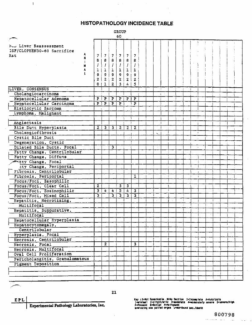
HISTOPATHOLOGY INCIDENCE TABLEGROUP
60
K~J Liver Reassessment3SF/CLOPHEN30-60 SacrificeRat J
1MAI
LIVER, CONSENSUSCholangiocarcinomaHepatocellular AdenomaHepatocellular CarcinomaHistiocytic SarcomaLymphoma, Malignant
AngiectasisBile Duct HyperplasiaCholangiof ibrosisCystic Bile DuctDegeneration, CysticDilated Bile Ducts, Focal
1 Fatty Change, CentrilobularFatty Change, Diffuse
./• Ktty Change, Focalcty Change, Periportal
Fibrosis, CentrilobularFibrosis, PeriportalFocus /Foci, BasophilicFocus /Foci, Clear CellFocus /Foci, EosinophilicFocus /Foci, Mixed CellHepatitis, Necrotizing,Multifocal
Hepatitis, Suppurative,Multifocal
Hepatocellular HyperplasiaHepatocytomegaly ,Centrilobular
Hyperplasia, FocalNecrosis, CentrilobularNecrosis, FocalNecrosis, MultifocalOval Cell ProliferationPericholangitis, GranulomatousPigment Deposition
78/1920
PP
2
233
78/1921
PP
3
4
2
78/1922
PP
3
3
43
78/1923
PP
2
353
78/1924
P
2
343
78/1925
PP
2
1
33
3
•NS
._ .,.., .,
i
i
EPLExperimental Pathology Laboratories, Inc.
21Key : X-Not Reoarkable N-No Section I-Inctaplete A-Auto lysisl-«1nlMl 2-sHght/BlW 3*ao4er«te 'federate ly severe S*severe/h1ohP-Present B-Benlgn M-ftaHgnant
on* paired organ iraorttand sac./dtitk
800798

REFERENCES
800799

Induction of Liver Tumors in Sherman Strain Female Rats by Pol/chlorinated Biphenyl1 26O '• =
Renate D. Kimbrough,3 Robert A. Squire,4 Ralph E. Linder,5 John D. Strandberg,8 Richard J. Montali,6 andVirlyn W. Burse 3< '
SUMMARY—Sherman strain female rats (200) were fed 100ppm of a polychlorinated biphenyl (Arocior 1260) for approxi-mately 21 months, and 200 female rats were kept as controls.The rats were killed when 23 months old. Twenty-six of 184experimental animals and 1 of 173 controls had hepatocellularcarcinomas. None of the controls but 146 of 184 experimentalrats had neoptastic nodules in their livers, and areas of hepa-tocellular alteration were noted in 28 of 173 controls and 182of 184 experimental animals. Thus the polychlorinated bi-phenyl Arocior 1260, when fed in the diet, had a hepatocarcin-ogenic effect in these rats. The incidence of tumors in otherorgans did not differ appreciably between the experimentaland control groups.—J Natl Cancer Inst 55: 1453-1459, 1975
Polychlorinated biphenyls (PCB's) have been usedover the past 44 years as transformer, capacitor, andcooling fluids in various systems (/). Since they are ex-cellent dielectrics and flame retardants, many new in-dustrial uses have been found for them, particularly inthe past two decades. These compounds are persistent asenvironmental contaminants and were first establishedas pollutants in 1966 (2). The toxicity of these and re-lated compounds has recently been reviewed (3).
_^In a preliminary feeding study in which the toxicitkstwo PCB mixtures (Arocior 1254 and Arocior 1260)
.re compared (J), a bladder tumor was found in 1 of10 female rats fed 100 ppm Arocior 1260 in their diet.This bladder tumor was tentatively classified at the timeas a poorly differentiated epidermoid carcinoma. Severeautolysis hampered the microscopic examination (Kim-brough RD: Unpublished observation). To establishwhether this tumor had developed spontaneously or wasinduced by the PCB, 200 female rats were fed 100 ppmArocior 1260. Another group of 200 females were thecontrols. This paper reports the results of this study.
MATERIALS AND METHODSFour hundred weanling Sherman strain COBS8 fe-
male rats 21-26 days old and weighing 48-97 g were dis-tributed into two groups of 200 animals each accordingto a table of random numbers. Ten animals werehoused per cage in conventional humidity, light, andtemperature-controlled surroundings. Two hundred ratswere fed plain ground Purina Laboratory Chow and 200were fed the same diet containing 100 ppm Arocior 1260(lot No. AK-3; Monsanto Industrial Chemical Co., St.Louis, Mo.). For incorporation of Arocior 1260 into thediet, 5.2 g was dissolved in ethyl ether; this was addedto 100 g cornstarch, and the ether was allowed to evapo-rate. The PCB cornstarch mixture was blended with in-creasing amounts of ground chow. The diets were pre-pared fresh every 10-14 days. Random samples from thefinal mixes and samples of control chow were taken at
ular intervals to determine PCB levels, at first bi-ekly and later bimonthly.For determination of the PCB levels, the samples were
extracted for 2 hours on an automatic shaker with a 3:1
mixture of hexane and isopropanol (5). After filtrationof the extract through glass wool, the extracts werewashed with a 2% sodium chloride solution and driedwith sodium sulfate. Samples were eluted through Flor-isil (6). The 6% fraction was collected and elutedthrough the silicic acid-celite column of Armour andBurke (7). Samples were analyzed by electron-capture,gas-liquid chromatography, and/or Coulson Conducto-metric gas-liquid chromatography.
During the first year of the study, three batches offood, both when first received and after having been inthe food cups for a week, were analyzed for aflatoxins(*)•
The combined body weight of rats in each cage wasrecorded weekly until the rats were 6 months old, bi-weekly until 12 months, and monthly thereafter. Indi-vidual weights were recorded only at the onset of the ex-periment and at death or when the animals were killed.The rats were observed briefly each day, and those ex-hibiting debilitating signs or large tumors were removedfrom the group cage and housed individually. At eachweighing the animals were examined individually andabnormalities were noted. Food consumption was meas-ured on all rats during the first 2 weeks of the experi-ment, and then during weeks 5, 8, 11, and 20, and every12 weeks thereafter. The dietary exposure of the experi-mental group to Arocior 1260 was discontinued 6 weeksbefore they were killed. Autopsies were performed on allthat died. When the animals were 23 months old, theywere anesthetized and their venae cavae were severed.Tissues were fixed in 10% buffered formalin and stainedwith hematoxylin and eosin. Selected tissue sectionswere stained with periodic acid-Schiff, Wilder's reticu-lum, azure eosin, and Masson's trichrome. In addition totissue masses, the following organs were studied micro-scopically: brain, pituitary, thyroid, parathyroid, tra-
i Received May 20, 1975; accepted July 24, 1975.= Supported in part by Public Health Service contracts N01
CIM3300 and NOl CP43288 from the Division of Cancer Cause and1'revention. National Cancer Institute.
3 Toxicology Branch. Center for Disease Control, Public HealthService, U.S. Department of Health, Education, and Welfare, At-lanta. Ga. 30333.
* National Cancer Institute. National Institutes of Health. PublicHealth Service. U.S. Department of Health, Education, and Welfare,Bethesda. Md. 20014.
s Environmental Protection Agency (EPA), Research TrianglePark. N. C. 27711.
« The Johns Hopkins University, School of Medicine, Baltimore,Md., 21205.
• We thank Mr. Martin Goldstein. Federal Drug Administration(PDA), New Orleans District, for the feed analysis for aflatoxins andMr. Sol Cohen, FDA. Atlanta District, for helpful suggestions. Mr.Richard Moore (EPA) assisted with animal care and autopsies.
s The Sherman rats are descendants of the Sherman strain devel-oped at Columbia University from the Osborne Mendel strain. Theyhave l>een randomly bred in our colony at the Center for DiseaseControl since 1950. They were cesarean obtained and barrier sus-tained (COBS) in 1966. In conventional rats, the incidence of murinepneumonia was high. Since they have been COBS, this is no longera problem.
JOURNAL OF THE NATIONAL CANCER INSTITUTE. VOL. JS. NO. 6, DECEMBER 1975 1453
800800

chea, esophagus, lung, stomach, kidney, heart, liver,spleen, adrenal, ovary, uterus, and urinary bladder. Stu-dent's Mest was used (or comparison of mean bodyweights and mean weight gain.
RESULTS
The PCB concentrations in the experimental dietranged usually from 70 to 10J ppm. Occasionally, lowervalues were found at the beginning of the study. Theoccurrence of low PCB concentrations in the experimen-tal diet was later resolved by improvement of the extrac-tion method. PCB s in the control diets were less than0.1 ppm and usually below the limit of detection. Aria-toxins were not detected in either diet. The sensitivitylevels were 2.5 ppb aflatoxins B, and G, and 0.1 ppb aria-toxins Bj and G2. A study in which aflatoxin £t wasadded to a portion of the feed showed 94% recovery.
No definite dose-related signs of toxicity were ob-served in the test animals. Comparative curves of body-weight gain and food consumption on the basis of body*weight, as well as PCB intake of the test group, aregiven in text-figures 1 and 2. A slight decline in the rateof weight gain in the test group compared to that in thecontrol group began about 3 months after onset of theexperiment. Mean final body weights were 420 g (so=72g, SE-«5.4 g) for the control group and 392 g (so=62 g,SE = 4.6 g) in the test group; the difference was statisti-cally significant (P<0.001). Food consumption (text-fig.2) was comparable in both groups. Mean weight gainwas 350 g (so = 70 g, SE = 5.3 g) and 323 g (so-60 g,SE = 4.5 g) for the control and test groups, respectively;the difference in weight gain was also statistically signifi-cant (P<0.001). PCB intake declined from 11.6mg/kg/day during the first week of exposure to 6.1mg/kg/day at 3 months of exposure and to 4.3mg/kg/day at 20 months (text-fig. 2).
Pathologic Findings
Control rats (173) and experimental animals (184)were examined grossly and microscopically. The experi-mental group included 5 rats that were killed 1-2months before the final kill. The remaining animals
500-1
400-
o>.£ 300-
KIMBROUCH ET AL.
'v
200O
CD
100
Control—a 1OO ppm Aroclor 126O
1 2 3 4 5 6 7 8 9 1011 1213 1415 1617 18192031 232924Months
120-i
110-
100-
|90-
S 80-
.70-oJ 60
I50
40
30
20
Control1OO Aroclor 126O
Sslffi
4
3
1 2 3 4 5 6 7 6 9 10 11 12 t3 1415 16 17 16 192021 222324Months
TEXT-FIGURE 2.—Food intake of control and experimental rats andconsumption of Aroclor 1260 by experimental rats.
were not included becaiue of improper tissue fixation orbecause they died early in the experiment.
A consistent difference in the appearance of the liverswas, observed between (he experimental and controlgroups. Almost all (17C/V84) livers of the experimentalanimals had from a few to multiple elevated tan nod-ules on the surfaces; additional nodules were usuallyseen on sectioning. These nodules varied from 0.1 toseveral cm in diameter, and in some rats replaced almostthe entire liver. In contrast, the liver of only one con-trol showed gross 'bnormalities and was markedly en-larged, nodular, tan, and firm. In addition, a variety oftumors of ether organs was observed in both the experi-mental and the control groups. The incidence and typeof tumors are given in table 1.
Histologic examination showed that 26 experimentalanimals and 1 control with enlarged, nodular livers hadhepatocellular carcinomas. The additional 144 experi-mental rats with gross liver nodularity had hepatocellu-lar nodules characteristic of neoplastic nodules [syn-onym, "hyperplastic nodules" (°)]. A recent workshopsponsored by The National Cancer Institute* recom-mended the term "neoplastic nodules" for these lesionsas a more accurate indication of their biologic signifi-cance. No nodules were in control animals.
The hepatocellular carcinomas were well-differen-tiated trabecular types (figs. 1-3), except for three in theexperimental animals which had a glandular, papillarypattern (fig. 4). The trabecular tumors showed severedisruption of normal liver architecture and were usuallyeasily recognized at low magnification. Liver plates, twoor more cells thick in some areas, were arranged in hap-hazard linear, branching, or pseudoglandular patterns.Different patterns were usually present in the sametumor. Blunt-ended plates, sinusoidal ectasia, andcongestion were frequent. The hepatocytes varied froma normal appearance to enlarged, acidophilic, or dif-fusely basophilic cells with large, hyperchromatic nucleiand prominent nucleoli. The cytoplasm often containedeosinophilic inclusions within vacuoles. Mitotic figureswere sometimes present. Foci of coagulative necrosiswere occasionally observed in cancerous areas, but there
I. — Average body weight of control rats and those con-suming Aroclor 1260.
•Rat Liver Tumor Workshop at Silver Spring, Md., Dec. 11-13,1974; sponsored by Carcinogenesis, Division of Cancer Cause andPrevention, National Cancer Institute.
800801
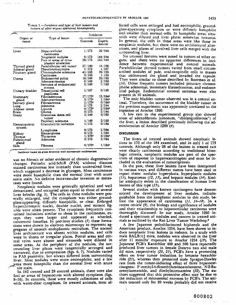
HEPATOCARCINOGENICITY BY AROCLOR 1260 1455
TAHLE 1.—fnridenre and type of liver lesions andtumors of other organs examined histologicaily
Organ ortissue
Liver
Thyroid glandAdrenal glandPituitary gland
Uterus
Urinary bladder
Mammarygland
Salivary glandLungAdipose tissueBrainOvary
Hematopoieticsystem
KidneyThymusParathyroid
glandSkin
Type of lesion
Hepatocellularcarcinoma
Neoplastic nodulesFoci or areas of cyto-
plasmic alterationParafollicular cell tumorPheochromocytomaChromophobe adenomaCarcinomaEndometrial polypAdenocarcinomaSarcoma of endometrial
stromaTransitional cell
papillomaFibroadenomaAdenocarcinomaFibrosarcomaAdenomaLipomaGliomaGranui-jsa theca cell
tumorPapillary adenomaGranulocytic leukemia
LymphomaHemangiomaThymomaAdenoma
Fibroma
Incidence
Controls
1/173
0/17328/173
37/1601 / 173
41/1530/153
18/1490/1493/149
1/167
17/173"5/173"1/173"2/1730/173"0/1735/149
1/1491/173
0/1730/1731/173"0/173
0/173"
Experi-mental
26/184
144/184182/184
18/1661/167
28/1391/139
25/1632/1637/163
0/169
13/184"1/184"0/184"2/1842/184"2/1840/163
2/1630/184
2/1841/1840/184"2/184
1/184"-
Incidence baaed on groes detection with microscopic confirmation.
was no fibrosis or other evidence of chronic degenerativechanges. Periodic acid-Schiff (PAS) without diastasestained carcinomas less intensely than uninvolved liver,which suggested a decrease in glycogen. Most carcinomaswere more basophilic than the normal liver with azureeosin stain. No definite imravascular invasion or metas-tases were found.
N'eoplastic nodules were generally spherical and welldemarcated, and occupied areas equal to those of severalliver lobules (fig. 5). The cells in these nodules were gen-erally enlarged, and the cytoplasm was either ground-glass-appearing, diffusely basophilic, or clear. Enlargedhyperchromatic nuclei, double nuclei, and mitotic fig-ures were often present. The cytoplasm frequently con-tained inclusions similar to those in the carcinomas, ex-cept they were larger and appeared as whorled,concentric lamellae. In previous studies (3) these forma-tions were shown by electron microscopy to represent ag-gregates of smooth endoplasmic reticulum. The normalliver architecture was absent within nodules, and cellswere in sheets or irregular plates. Portal areas and cen-tral veins were absent and sinusoids were dilated insome areas. At the periphery of the nodules, the sur-rounding liver plates were tangentially arranged andnarrowed, due to compression (figs. 6, 7). Nodules variedin PAS positivity, but always differed from surroundingliver. Most nodules were more eosinophilic, and a few
J^ere more basophilic than the normal liver with azuresin stain.In 182 treated and 28 control animals, there were also
foci or areas of hepatocytes with altered cytoplasm (figs.8-10). In controls, these were mostly collections of cellswith water-clear cytoplasm. In treated animals, most af-
fected cells were enlarged and had eosinophilic, ground-glass-appearing cytoplasm or were diffusely basophilicand smaller than normal cells. In basophilic areas, sinu-soids were dilated and liver plates somewhat tortuous.In general, the cells in these areas were like those inneoplastic nodules, but there were no architectural alter-ations, and plates of involved liver cells merged with thesurrounding liver.
No unusual features were noted in tumors of other or-gans, and there were no apparent differences in inci-dence between experimental and control animals.Parafollicular thyroid tumors varied from small circum-scribed nodules of pale, oval-to-spindle cells to massesthat obliterated the gland and invaded the capsule.They were similar to those described by Boorman et al.(10). Other frequent tumors included pituitary chromo-phobe adenomas, mammary fibroadenomas, and endome-trial polyps. Endometrial stromal sarcomas were alsopresent in 10 animals.
The only bladder tumor observed was in a control an-imal. Therefore, the occurrence of the bladder tumor inthe previous experiment was apparently unrelated to theingestion of Aroclor 1260.
A few rats in the experimental group also showedareas of adenofibrosis (synonym, "cholangiofibrosk") ofthe liver, a lesion described previously following the ad-ministration of Aroclor 1260 (4).
DISCUSSIONThe livers of treated animals showed neoplastic le-
sions in 170 of the 184 examined, and in only 1 of 173controls. Although only 26 of the lesions in treated ratswere clearly carcinomas according to traditional histo-logic criteria, neoplastic nodules are part of the spec-trum of response to hepatocarcinogens and must be in-cluded in the evaluation of tumorigenesis.
In the past, these liver lesions have been interpretedin various ways, and different names have been used toreport them: nodular hyperplasia, hyperplastic nodules(//), hepatomas (12, 13), and hepatic nodules (14). Simi-lar ambiguity exists in the classification of human liverlesions of this type (15).
Several studies with known carcinogens have demon-strated the development of liver nodules, indistin-guishable from the neoplastic nodules in this study, be-fore the appearance of carcinoma (11, 16-18). In arecent review (9), the biology and significance of nodulesand their relationship to hepatocellular carcinoma werethoroughly discussed. In our study, Aroclor 1260 in-duced a spectrum of nodules and cancers in treated ani-mals as outlined by the Rat Liver Tumor Workshop.'
A few Japanese polychlorinated biphenyls and theAmerican product, Aroclor 1254, have been shown to in-duce neoplastic liver lesions in rodents. In a study withmale BALB/cJ mice, nodules were observed in the liverafter 11 months' exposure to Aroclor 1254 (19). TheJapanese PCB's Kanechlor 400 and 500. have reportedlyproduced liver tumors in female Donryu rats and maledd mice, respectively (20, 21). PCB's have a promotingeffect on liver tumor induction by benzene hexachlo-ride (27), whereas they protected male Sprague-Dawleyrats from the tumor-inducing effect of three hepatocar-cinogens: 3'-methyl-4-dimethylaminoazobenzene, N-2-flu-orenylacetamide, and diethylnitrosamine (22). The au-thors suggested that this protective effect may be due tothe induction of microsomal enzymes by PCB's. The ani-mals treated only for 20 weeks probably did not receive
800802

1456 K1MBROUGH ET AL.
the material long enough for the PCB's to induce tu-mors.
11 is not known whether mixtures of polychlorinatedbiphenyls that are primarily composed of isomers withfour or less chlorines on the ring would also induceliver tumors, or whether only those mixtures containingan appreciable proportion of pentachlorobiphenyl, hex-achlorobiphenyl, and heptachlorobiphenyl have this ef-fect. Kanechlor 400 contains 3% dichlorobiphenyl,32.8% trichlorobiphenyl, 43.8% tetrachlorobiphenyl,15.8% pentachlorobiphenyl, and 4.6% hexachloro-biphenyl. Kanechlor 500 contains 5% trichlorobiphenyl,26% tetrachlorobiphenyl, 55% pentachlorobiphenyl,and 13% hexachlorobiphenyl (27). Chlorinated dibenzo-furan was found in one Kanechlor 400 sample (23). Thecomposition of the Aroclor 1260 studied by us is notknown. Sissons and Welti (24) analyzed Aroclor 1260manufactured by Monsanto Chemicals Ltd. and con-cluded that hexachlorobiphenyl and heptachloro-biphenyl were major constituents.
REFERENCESsted(/) BROADHLRST MG: Use and replaceability of polychlorir
biphenyls. Environ Health Perspect 1:81-102, '972(2) JENSEN S: A new chemical hazard. New Sci 32:612, 1966()) K.IMBROLCK RD: The toxicity c? polychlorinaled polyc/chc
compounds and related chemicals. CRC Crit Rev Ttvicot2:445-498, 1974
(t) KIMBROU.H RD, LINGER RE, GAINES TB: Morphologicalchanges in livers of rats fed polychlorinated biphenyls.Arch Environ Health 25:354-364, 1972
(5) DAWSEY LH. BARTHEL WF. SCHUTZMANN RL, et al: South-haven Region Manual. L'.S. Department of Agriculture,Agriculture Research Service, Plant Pesticide Control Divi-sion. June 1965, pp 56-59
(6) MILLS PA: Detection and semiquantitative estimation ofchlorinated organic pesticide residues in foods by paperchromatography. J Assoc Off Agri Chem 42:734-740^ 1959
(7) ARMOL-R J. BURKE J: Method for separating polychlorinatedbiphenyls for DDT and its analogs. J Assoc Off Anal Chem53:761, 1970
(S) PONS WA JR. CUCLLLU AF, LEE LS: Determination of afla-toxins in mixed feeds, hi Third International Congress ofFood, Science and Technology (S.O.S./70). Washington, D.C.,1970, pp 705-711
(9) FARBER E: Hyperplastic liver nodules. In Methods in Cancer
Research, vol 7, chapl VIII. New York and London, Aca-demic Press, 1973. pp 345-375
(10) BOORMAN CA, VAN BOORD M. HOLLANDER CF: Naturally occur-ring medullary thyroid carcinoma in the rat. Arch Pathol94:35-41. 1972
(11) EI-STF.IS S. ITO N. MF.RKOW L, et al: Cellular analysis of livercarcinogenesis: The induction of large hypcrplastic nodulesin the liver with 2-ftuorenylacetamide or elhionine andsome aspects of their morphology and glycogen metabolismCancer Res 27:1702-1711, 1967
(12) VESSELINOVITCH SD, MIHAILOVICH X: The induction of benignand malignant liver tumors by urethan in newborn rats.Cancer Res 28:881-887. 1968
(D) EDWARDS JE. WHITE J: Pathologic changes, with special refer-ence to pigmentation and classification of hepatic tumorsin rats fed £>-dimethyiaminoazobenzene (butter yellow). JNail Cancer Inst 2:157-183, 1941
(H) POITF.R H. STEKNBF.RC SS, OSEK BL, et al: The carcinogeniceffect of aramite in rats. Cancer 13:1035-1046, 1960
(15) MAYS ET, CHRISTOPHERSON WM. BARROWS GH: Focal nodularhyperplasia of the liver. Am J Clin Pathol 61:735-746, 1974
(16) KLAVINS JV, KENNEY TD, K A U F M A N N: Hepatic nodulesresembling tumors in rats after administration and with-drawal of ethionine. Proc Soc Exp Biol Med 89:540-543,1965
(/") POPFER H, DE LA HUERGA J, YESiNicK C: Tumors due to pro-longed ethionine feeding. Science 118:80-82, 1953
(IS) WACHSTEIN M, MEISEL E: Enzymatic histochemistry of ethio-nine induced liver cirrhosis and hepatoma. J HistochemCytochem 7:189-201, 1959
(19) KIMBROUGH RD, LINDER RE: Induction of adenofibrosis andhcpatomas of the liver in BALB/cJ -mice by polychlorinatedbiphenyls (Aroclor 1254). J Na'tl Cancer Inst 53:547-552,1974
(20) KIMURA NT, BAB A T: Neoplastic changes in the rat liverinduced by polychlorinated biphenyl. Gann 64:105-108. 1973
(21) ITO N, NAGASAKI H, ARIA M, et al: Histopathologic studies onliver tumorigenesis induced in mice by technical poly-chlorinaled biphenyls and its promoting effect on livertumors induced by benzene hexachloride. J N'atl Cancer Inst51:1637-1646, 1973
(22) MAKIURA S, AOE H, SL-CIHARA S, et al: Inhibitory effect of poly-chlorinated biphenyls on liver tumorigenesis in rats treatedwith 3'-methyl-4-dimethvlarninoazobenzene, A'-2-ftuorenyl-ncetamide. and diethylnitrosamine. J Nat! Cancer Inst53:1253-1257. 1974
(2)) ROACH JA, POMERANTZ JH: The findings of chlorinated diben-zofurans in a Japanese polychlorinated biphenyl sample.Bull Environ Contam Toxicol 12:338-341. 1974
(2-1) SISSONS D, \VELTI D: Structural identification of polychlori-nated biphenyls in commercial mixtures by gas-liquidchromatography. nuclear magnetic resonance and massspcctromctry. J Chromatogr 60:15-32, 1971
800803

FIGURE I.—Focus of hepatocellular carcinoma in rat fed Aroclor 1260. Tumor cells are in sheets, irregular nests, and cords Hcmatoxyiinand eosin (H & E). X 45
FIGURE 2.—Focus of hepatocellular carcinoma in rat fed Aroclor 1260. Note plates and nests of cells, two or more cells in thickness,enveloped by lining cells. H t E. x 145 •
FIGURE 3.—Focus of hepatocellular carcinoma in rat fed Aroclor 1260 showing nests of cells in pseudoacinar patterns. H & E. X 360
FiCLRE 4.—Focus of hepatocellular carcinoma with glandular pattern in rat fed Aroclor 1260. H & E. X 105
KIMBROUCH ET AL. 1457
800804
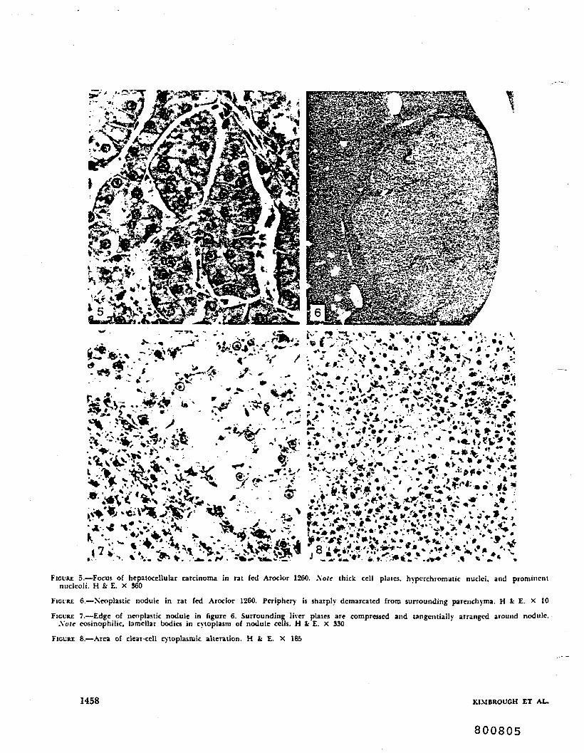
FICLRE 5.—Focus of hepalocellular carcinoma in rat fed Aroclor 1260. \ote thick cell plates, hypcichromatic nuclei, and prominentnucleoli. H & E. X 360
FICLRE 6.—Neoplastic nodule in rat fed Aroclor 1260. Periphery is sharply demarcated from surrounding parenchyma. H & E. X 10
FIGURE 7.—Edge of neoplastic nodule in figure 6. Surrounding liver plates are compressed and tangentially arranged around nodule.\ote cosinophilic, lamellar bodies in cytoplasm of nodule cells. H tc E. X 330
FIGURE 8.—Area of clear-cell cytoplasmic alteration. H & E. X 185
1458 KIMBROUGH ET AL.
800805

FIGURE 9.—Area o£ cosinophilic cytoplasmic alteration having the appearance of ground glass. H & £. x 185
FIGURE 10.—Area of basophilic cytoplasmic alteration. H & £. X 105
KIMBROUCH ET AL. 1459
800806
J..»i..iJ Vrtl II pp ftl T? Perfunxn Press !«•?< Printed in Grejt Britain
THE EFFECT OF POLYCHLORINATED BIPHENYLSON RAT REPRODUCTION
R. E. LINDER'* ,'T. B. GAINES and R. D. KIMBROUGHBio-Effects Branch. Primate and Pesticides Effects Laboratory. Enrironmemal
Protection Agency. 4770 Buford Highway, Chambtee. Georgia 30341. USA
{Received IS August 1973)
Abstract—Reproduction, pathology and acute toxicity were studied in Sherman rats exposed tothe polychlorirtated biphenyis. Aroclor 1254 and Aroclor 1260. Rats exposed to Aroclor 1254 atdietary levels of 20 ppm or more had fewer pups per litter than the controls in the Flb and F,generations. The 100 ppm exposure level of Aroclor 1254 increased mortality in the F,b offspringand markedly decreased mating performance of the F,b adults. The 500 ppm dietary level of Aroc-lor 1260 reduced litter size and decreased survival in the F, litters. Dietary levels of 5 ppm Aroclor1254 and 100 ppm Aroclor 1260 had no effect on reproduction in rats exposed through two gene-rations. Liver weights were increased in 21-day-old F, male weanlings at thp 1 ppm level of Aroclor125*4 and in either sex of F, and F, weanlings at 5 ppm or higher levels of both Aroclor 1254 and1260. Histologies! changes in the liver and increased liver weights were observed in adult ratsexposed to the higher levels. Pregnant rats given Aroclor 1254 at the rate of 100 mg/kg/day ondays 7-15 of gestation produced grossly norm?' litters, but only 30-1% of the pups survived toweaning. Reproduction and pup survival were not affected at dosage rates of 50 mg. Aroclor1254 kg/day or 100 mg Aroclor 1260/kg/day. Oral LD,0 values in 3-4-wk-oid male rats were 1295and 1315 mg kg for Aroclors 1254 and 1260, respectively. The iv LD50 for Aroclor 1254 in adultfemales was 358 mg kg.
I N T R O D U C T I O NReproductive defects due to commercial mixtures of polychlorinated biphenyis (PCBs)
have been reported in experimental birds (Dahlgren, Linder & Carlson, 1972) and mam-mals (Ringer. Aulerich & Zabik. 1972). Porphyria (Vos & Koeman, 1970), microsomal-enzyme induction (Street, Urry, Wagstaff & Blau, 1969) and liver damage (Kimbrough,Linder & Gaines. 1972; Miller, 1944) have also been reported in experimental animalsexposed to PCB mixtures.
The present investigation was initiated in 1970 to study the effects on reproduction andpathology produced by two American-made PCB mixtures sold under the trade-namesAroclor 1254® and Aroclor 1260®. Aroclor 1254 contains 54% (w/w) chlorine and is com-posed of 11% tetra-. 49% penta-, 34% hexa- and 6% heptachlorobiphenyls; Aroclor 1260has 60% (w/w) chlorine, with a composition of 12% penta-, 38% hexa-, 41% septa-, S%octa- and 1% nonochlorobiphenyls (Thruston, 1971). An almost complete identificationof isomers in Aroclor 1254 and Aroclor 1260 has been obtained (Sissons & Welti, 1971).Since 1970, the Monsanto Co., the only US manufacturer of PCBs, has voluntarily reducedsales of Aroclor mixtures and has taken steps to limit their use to closed systems (Mon-santo Co., 1971). '
The present communication is an account of reproduction studies in rats. Also includedare acute toxicity values from preliminary studies and comments on pathology and haema-tology in animals from the reproduction experiments. A detailed account of the liver path-ology of the F0 rats given 20 ppm and higher dietary levels has been published (Kim-brough et al. 1972).•Present address: United States Environmental Protection Agency. National Environmental Research Center.
Research Triangle Park. North Carolina 27711.^Registered trademark of the 'Monsanto Company.
t f . r i : i - r 63
800807

64 R. E. LINDER. T. B. GAINES and R. D. KIMBROUGH
E X P E R I M E N T A LAnimals and niaterials. All animals used in the studies were Sherman-strain rats pro-
duced under specific-pathogen-free conditions. Aroclor 1254 (Lot AK-38) and Aroclor1260 (Lot AK-3) were supplied by Monsanto Industrial Chemicals Company. St. Louis,Mo.
Experimental design and conduc:LDu, studies. The single-dose oral LD50 was determined for Aroclor 1254 and 1260 in
weanling male rats (3-4 wk old). The iv LD50 was determined in adult females for Aroclor1254. Between five and eight groups, each of ten rats, were used to obtain each LDSO value.The procedure described by Gaines (1969) was followed for oral dosing; Aroclor 1254 orAroclor 1260 was dissolved in peanut oil and the solution was administered by oral intuba-tion at the rate of 5 ml/kg. For iv dosing. Aroclor 1254 was first dissolved in peanut oil.One part of this solution was added to nine parts of a 1% lecithin-saline suspension andhomogenized. This formulation was administered in a single injection via the tail vein atthe rate of 5 ml/kg. A control group of five rats received 10% peanut oi) in lecithin-salinein the same volume. Survivors were observed for 10-14 days.
Two-generation reproduction studies. For these studies the rats were fed either untreatedground laboratory chow (control) or ground chow fortified with Aroclor 1254 or Aroclor1260. The Aroclors were dissolved in ether and mixed with cornstarch. The ether wasallowed to evaporate and the Aroclor-cornstarch mixture was mixed into increasingamounts of ground chow. Aroclor 1254 was fed at dietary levels of 0 (control). 1. 5, 20and 100 ppm, while Aroclor 1260 was fed at levels of 0. 5. 20 and 100 ppm. Because expo-sure schedules had to be staggered, a specific control group was added each time one ormore treated groups were started on a dietary regimen. The F0 rats were started on thediets at 3-4 wk of age and the F, b rats at weaning. Exposure was continuous through mat-ing, gestation and lactation until the rats were killed. Ten males and 20 females were fedat each dietary level in both the F0 and F,b generations. The F0 males at each dietarylevel and half of the females were caged individually. Other F0 females and all the Flhrats were caged in groups except during the reproduction cycle. Body weights wererecorded weekly on F0 rats, except during the reproduction phase, and food consumptionwas measured during wk 2 and 5, the week before each mating and the week after weaningof the F,. litters. Flb rats were weighed when started on the diet, during the week beforemating and at sacrifice.
The F0 rats were pair-mated when 3 and 7 months old to produce the F,, and Flb gene-rations, respectively. Breeding-stock F,b rats were selected at weaning from all availablelitters and pair-mated when 3 months old to produce the F2, generation. F,b rats on diet-ary levels of 0, 20 and 100 ppm Aroclor 1254 were mated a second time when 8 monthsold to produce the F2b generation. Viability counts of offspring were made at birth, day3. day 7 and day 21 (weaning). Litters were inspected daily for condition and the presenceof dead pups. After the F,b offspring had been weaned, ten adult F0 rats of each sex werekilled and their livers were weighed and fixed for histological examination. Following theweaning of the F2b generation, for Aroclor 1254, and the F2» generation, for Aroclor 1260.haematological values (total leucocyte count, haematocrit. haemoglobin and differentialleucocyte count) were determined on ten adult Flb rats of each sex at the dietary levelsof 0,20 and 100 ppm. These rats were then killed and weights of the spleen, heart, lungs,brain, kidneys, testes and liver were recorded. Tissues were fixed for microscopic study.
800808

> Effects of PCBs on rat reproduction 65i
At lower dietary levels, hacmatology was not done and only the livers were weighed. Atdietary levels of 5 ppm or less of both Aroclors. one male and one female weanling fromeach of ten litters from each generation were killed at 21 days of age and the livers wereweighed. F,., weanlings at dietary levels of 0. 20 and 100 ppm Aroclor 1254. and F,b andF:a weanlings at levels of 0, 20 and 100 ppm Aroclor 1260 were killed in the same mannerand the livers were fixed for further study.
One-yeneration reproduction studies. Preliminary studies were conducted on both Aroc-lors at dietary levels of 0. 100 and 500 ppm. Thi experimental design was the same as thatdescribed for the two-generation studies, except that only ten females were used. This studywas terminated after the Flb offspring had been weaned. Rats fed 500 ppm Aroclor 1254were bred only once. Haematology values and organ weights were determined and tissueswere examined microscopically on the F0 rats.
Post-implantation exposure studies. Stock rats 100 days old were pair-mated. Insemina-tion was verified by microscopic inspection of a daily vaginal smear. Counting the dayof insemination as day 0, the females were dosed by oral intubation on days 7-15 ofgestation (a total of nine doses). To maintain a constant dose volume on a body-weightbasis. Aroclor 1254 or 1260 was dissolved in peanut oil at appropriate concentrations andthese formulations were administered at the rate of 5 ml/kg body weight, based on day7 of pregnancy. Aroclor 1254 was given at dosage levels of 0 (peanut oil only), 10, 50 and100 mg;kg/day. Aroclor 1260 was given at dosage levels of 0 and 100 mg/kg/day. Thefemales were allowed to deliver and the litters were observed through weaning.
Tissue preparation. Tissues were fixed in buffered 4% formaldehyde solution and stainedwith haematoxylin and eosin for microscopic examination. An Oil Red O stain on fixedfrozen sections was used on occasional livers to demonstrate lipids.
Statistics and calculationsLDio values were calculated by the method of Lilchfield & Wilcoxon (1949). A t test
was used for comparing the litter sizes, organ weights, body weights and haematologicalparameters of the treated groups with those of the control groups. Survival percentagesof offspring were compared by the Mann-Whitney U test as proposed by Weil (1970).Food consumption values were arrived at by averaging the consumption of both malesand females over the five periods in which food was measured.
RESULTS
LD<0 studies on Aroclor 1254 and 1260The single-dose oral LDSO for Aroclor 1254 in weanling male rats was 1295 mg/kg with
95% confidence limits of 1136-1476 mg/kg. The lowest lethal dose tested was 1200 mg/kg,and 1000 mg/kg produced mild diarrhoea. Signs of toxicity at lethal levels were diarrhoea,depression and salivation, with death occurring in 1-3 days.
For Aroclor 1260, the single-dose oral LD50 in weanling males was 1315 mg/kg with95°0 confidence limits of 1174-1473 mg/kg. The lowest lethal dose tested was 1000 mg/kg,800 mg/kg produced no signs of toxicity. Rats given lethal doses developed diarrhoea anddepression, with death occurring in 1-7 days.
Under the same test conditions the estimated oral LD50 for Aroclor 1254 and 1260 inadult rats was 4-10 g/kg (Kimbrough et al. 1972).
The single-dose iv LDJO for Aroclor 1254 in adult female rats was 358 mg/kg with 95%
800809
4 R. l:.. L I M M K . T. » (iMMsaiul R. D. KIMIWIM (;n
confidence limits of 328 3l)0 mg kg. The lowest lethal dose tested was 3(X) mg kg: 250 \nig kg produced dyspnoea, depression, diarrhoea and salivation soon after dosing. Most ;rats died w i t h i n 25 I Hi inin. hut al (he liigliesl dose (400 mg kg), death occurred as soon jas 5 min after dosing in some animals. Rats . that survived appeared normal after -IS hr. jIn the control group, which received the formulation vehicle only, one rat developed mild jdiarrhoea. • j
jPreliminary tine-iieneritiion reproduction studies on Aroclor 1254 anil 12M \
The reproduction summary for the Aroclor 1254 feeding studies is presented in Table iI. Exposure for 67 days (at the time of pair-mating) to 5(X) ppm Aroclor 1254 (37-0 img kg,day) resulted in fewer litters, smaller litter sizes and 100",, mortality by day 3 in \the F,a pups. The survival of offspring of the rats fed 100 ppm (7-2 mg kg day) for 67 and j186 days before mating was reduced, only 85-9 and 68-1 "„ of the pups surviving to weaningin the Fj., and F,h generations, respectively. Litter si/es from both ma tings were smaller,but the difference was not statistical!) significant. The mean body weight of the 100 ppmpups at weaning was 7-8 and 10 g less than that of the controls for the Fu and Flh gene-rations, respectively. This difference was probably dose-related, as the treated litters weresmaller, but a statistical evaluation was not done. In general the pups appeared small, butotherwise were normal at weaning. .
Aroclor 1260 fed at a dietary level of 5(K) ppm (35-4 mg kg day) for 67 and 186 daysprior to mating markedly reduced live-litter size and survival-to-weaning in the F^ andFlh generations (Table 2). No effect on reproduction was observed in rats fed 100 ppmffr° ir,;; I-;* day) in cither the F, or F.,. generations.
The F0 rats in the one-generation studies were sacrificed after exposure for 8 months.Organ-weight analysis indicated that the liver was the primary organ affected. As reportedin the previous paper, the livers in male rats receiving either Aroclor 1254 or 1260 at diet-ary levels of 100 and 500 ppm were heavier than in controls: this was also observed infemales fed Aroclor 1254 at levels of 100 and 500 ppm or Aroclor 1260 at 5(X) ppm (Kiin-brough et til.. 1972). The testcs-to-body weight ratios of F0 rats fed Aroclor 1254 or 1260at the 500 ppm level were greater than those of the controls (P < 0-025). The differencein actual weight of the testes. however, was not significant. This observation may reflectthe reduced body-weight gain observed in all F,, rats fed the 5(X) ppm level of both Aroe-lors. No difference in other organ weights was found.
After 8 months exposure, terminal haematoiogical values in F,, rats indicated a redtic-tion in haemoglobin and haematocrit (P < 0-005) in both sexes fed 500 ppm Aroclor 1254or 1260. The haematocrit was reduced in males (P < 0-05) on UK) ppm Aroclor 1260. buthaemoglobin values were normal in this group. The total leucocyte count was normal inrats fed Aroclor 12611. hut there was a shift in the dillerenlial count. The number of lym-phocytes wax increased in female* I/' < 0-005) fed 100 and 500 ppm Aroclor 1260 and inmales (P < 0-05) led 5<X) ppm. A corresponding decrease (/' < 0-05) in poly morphonuclcarleucocytes was observed. Differential values for rats fed Aroclor 1254 were normal, butan increase (P < ( H K ) l ) in total leucocytes was observed in the 5<X) ppm females.
Two-iienerulioit reproduction xmdien on Aroclor 1254F,, rats fed UK) ppm Aroclor 1254 (7-6 mg kg day) for 62 or 188 days before mating pro-
duced smaller litters than the controls in both the I ,., and I",,, generations (Table 11. Sur-vival-to-vveaning was not affected in the 1 - , , oll'spring. Only "3-6",, of the 100 ppm 1-',,, pups
800810

Table I. KcyrWitfliwi mill />»/> .Mirrirnf in I/IOII/LV iifrats fctl Arocltir /-W
Dieiarvlevel
(ieneration (ppml
1,, 0too500;020100015
l',h <> 'X X I .020MX)015
1" s "20UK)015
1 ,h 020UK)
Parentalexposure
676767626262676767IS6IS6ISSISSISS201201201
292V29252525
274274274
No. offemalesmated
10101020202020202010102020202020202020202020IV202020
No. of liners
Horn*
8V4 ( 2 117IVIVIS16177X17IX20ISIX20IX177(21IV151717124 (2)
Weaned
78(I17IVIVIX15177617ISIVIS17IVI X174IV151716122
l.iller si/e!i
- Albirth
I I I9440*1241 1 7107*11292I D S116us123104*95**123I I II I I125106*7-2**1171 1 71 2 112796**3-5**
Alweaning
1068-1
U S115103I I I91KIS116SOI I I10170I I I )98102122101561 1 51 1 51 1 711-38-535
Tola! ptips group
Horn (found)
Dead
365031023110216632057301467
Ali\e
8985X211222204202147IS48194209ISS
. IS9222200222225IS)362231752052161157
Alive at
I*.) 3
86780205220199200145184SI74194IS7164211194218221174292201741992011037
Weaning
85730201219IVft2'XI145183SI64ISVISI139I'M1762032201712s2IS173I9S|9|1027
Survival
95585909539X696- 199 09X6995111(1
6S •!•*904963736S9-2XX-t l91497X94577X**97S9S-V966X S 4SS-711X11)
Meanboil) weight
(ei
392314
3S-73X-3 '3293S 54234 2 1r-927437139-03 2 140738-0 •3X935536X35 237-437-23573193613S-3
+ < "nnei-plion to nialing for the patents of the I-',, and I-",,, generations.* Niitnlvis in pareniheseN iiulieate nunihers of liners in \\hich no live oll'spring were found.i Ntimher of Ihe nllspring live liner born.
One-eeneralion >lnd\.\ alueMiiarked with astetisks differ significant!) Irom the control value: * /' < 005; ** P < 0-005.
00oo00
L.

68 R. E. UNDER. T. B. GAINES and R. D. KIMBROUGH
survived to weaning, but this was not statistically significant (P = 0-058). Because of thepoor condition of a number of the 100 ppm Fib pups, all were held for 30-days post-wean-ing observation. Fifteen of these pups died within 30 days of weaning. The mean bodyweights of the 100 ppm Fj generation pups from either mating was about 6 g less thanthe controls at weaning. This is probably significant, since the treated litters containedfewer pups. A reduction in litter size was also observed in the Flb generation at 20 ppm(1-5 mg/kg/day).
F,b rats bred after exposure for 129 or 274 days (conception to mating) to 20 or 100ppm had smaller litters than the controls (F2l and F2b generations). A marked decreasein mating performance was observed in the 100 ppm group, in which onlyseven and fourlitters were born in the F2, and F2b generations, respectively. In each case two litters con-tained no viable offspring when first observed. The data also suggest a decrease in matingperformance at 20 ppm, at which level only 12 F2b litters were produced. Survival-to-wean-ing was reduced in the 100 ppm F2, offspring. All 100 ppm F2b offspring survived to wean-ing, but this is of little significance since only seven pups were found alive. No effect onreproduction was observed in rats fed 5 ppm (0-32 mg/kg/day) or 1 ppm (0-06 mg/kg/day)through two generations.
Although reproduction was not affected at lower dietary levels of PCBs, an increase inliver weight in 21-day-old weanlings was found at all levels tested (Table 2). At 5 ppm orhigher, the liver-to-body weight ratios of weanlings were increased in both sexes in both
Table 2. Mean liver weights of 21-day-old rats from parents fed Aroclor 1254 or 1260
Liver weight
Males Females
Aroclor Generation
1254 Fu
F,b
Fj,
Fj.
~1260 F,.
FH,
F
Fi,
p,4
Dietarylevel '
(ppm)
015015020100015050201000502010005
g
1-401-81*1-92'1-581-541-771-372-26*2-63*1-631-491-561-541-731592-06*259'1-351-581-652-06*2-50*1-41166*
°/0ofbodyweight
3-694-01'4-34*3-693-94*4-63'3-815-78*6-97*3-783-744-23*3-664-18*3-735-04*6-33*3-594-01 '3-825-21*612*3-854-2 1 '
g
1-381-631-86*1-581-491-721-282-23*2-67*1-421-361-381-571-701-552-14*2-59*1-291-52*1-641-942-49*1-361-60*
% of bodyweight
3-833-914-43*3-843-924-75*3-74 .6-08*7-10'3-633-694-09*3-924.39.3-935-18*6-48*3-704-0?*4-025-06*6-85*3-874-25*
Values marked with un asterisk differ significantly from the control value: * P < 0-05.
800812

Effects of PCBs on ral reproduction (,9
the F, and F: generations, but at 1 ppm the liver enlargement was observed only in theF, a and F, h male weanlings. No liver-weight increase was found in either sex of the 1 ppmF:J weanlings.
In 10-month-old Fjb rats, an increase in liver-to-body weight ratios was observed inboth sexes at 100 ppm. but at 20 ppm it was seen only in the males (Table 3). At 5 ppmor less no increase in liver weight was found in 6- month-old Flb rats or in F0 rats exposedfor 10 months. Both sexes of F0 rats fed 20 ppm or more Aroclor 1254 for 8 months hadenlarged livers (Kimbrough et al. 1972). The testes-to-body weight ratios were increased(P < 0-001 (in 100 ppm Flb adult males. Body-weight gains of adults in all the test groupswere comparable to those of the controls except in the Flb females at 100 ppm. whichgained less weight than the controls (Table 3). Reduced haemoglobin (P < 0-005) and hae-matocrit (P < 0-0251 were observed in female Flb adults at 100 ppm, while Flb males atthis level had reduced (P < 0-05) haemoglobin only. Haematological values at lower levelswere normal. No dose-related signs of toxicity were observed in adult F0 or F,b rats fed100 ppm or less.
reproduction study on Aroclor 1260No effect on reproduction was observed in rats fed Aroclor 1260 at levels of 100, 20 or
5 ppm (7-4. 1-5 and 0-39 mg/kg/day) through two generations (Table 4). The liver-to-bodyweight ratios of 21 -day-old pups were increased at all dietary levels (Table 2\ This effectwas observed at the lowest level tested (5 ppm) in all generations.
An increase in liver-to-body weight ratios was observed at all dietary levels in Flb malesbetween 5 and 7 months old. but only at 100 ppm in the Flb females (Table 3). After 8months exposure, an increase in liver weight was observed at 20 and 100 ppm in F0 malesbut not in females (Kimbrough et al. 1972). No effect on liver weight was found in eithersex of F0 rats fed 5 ppm for 8 months. The testes-to-body weight ratios of the 100 ppmFlb males were increased (P < 0-05); at 20 ppm the increase was not significant. Body-weight gain in all test groups was comparable to the control groups. Haematologicalvalues were normal in the 20 ppm F0 adults and in FJb adults at 100 ppm or less. Nosigns of toxicity were observed in any of the test animals.
Post-implantation exposure studies on Aroclor 1254 and 1260The data on reproduction and survival of offspring from females dosed during organo-
genesis is summarized in Table 5. Nine oral doses of Aroclor 1254 (100 mg/kg/day) givenon days 7-15 of gestation resulted in a decrease in the survival of the pups. At this doseonly 30- 1 °0 of the offspring of the treated group survived to weaning, compared with 98-2 /;>in the control group, and the mean body weight of the test pups was 7-1 g less than thatof the controls at weaning. No effect was observed in the groups receiving 50 mg/kg/dayor less.
No effect on reproduction or survival was observed with Aroclor 1260 given at a rateof 100 mg/kg day.
Summary of pathology after treatment with Aroclor 1254 and 1260A detailed account of the liver pathology in the F0 rats fed Aroclor 1254 or 1260 at 20
ppm or higher dietary levels has been published (Kimbrough et al, 1972). Of the rats consi-dered in the current report, similar changes were observed in the liver of adult rats, particu-larly in the Flb generations at the higher dietary levels. The incidence of the various
800813

Table 3. Body weight, liver weight and liver pathology ofFy and FH, adult rats fed Aroctor 1254 or 1260
00OO00
ExposureGeneration Sex (days)
F« M
F
Fib M
F
Fib M
F
F» M
F
FII. M
F
I,- M
1
310
313
328
328
190
190
250
250
179
179
217
217
Dietarylevel
(ppm)
015015020100020100015015
0505020too020I(X)0505
Terminalbody
weight
601602614370358362588602554349340300*475453495287294299
5635673223314724945092973043044925052792X6
Liver weight
?•; of No. ofbody livers
g weight examine
15-2215881537103410141066145116-11*1943*9929411053II 13II 1412 19827844892
1445150610 0910X012681472*1663*966103011-14*119713 16793849
253260251280284294247267*354*285278360*234244246287286298
257265313325268299*327*327340370*243260*284296
10101010101010810107lot101010101010
1010101010101010101010101010
Liver pathology (no. of livers affected)
Enlargedd hepatocytes
Aroclor 12540000|408100710002011
Aroclor 1260r30108100690702
Inclusions
000001035022000010
000005100000200
Foamycytoplasm
001000077058002000
02000430010200
FibrousPigment strands
002143014079000000
00000000050100
00000000200301104I
0000000
< 0020000
Adenofibrosis
000000003005000000
00000000010000
Nodules
000000001037000000
00000010020000
70
riHCD
O
sO
* Necrosis in two livers.Values marked with an asterisk ilitTct significantly from control group: ' P < 0025.

Table 4. Reproduction and pup uirt'iiwf in groups ofralsfcd Arorhr 1260
Dietarylevel
Generation (ppm)
F,. 0§I00§500$02010005
Flb OSI00§500?02010005
Fj. 02010005
Parentalexposure(days)t
6767676868687171186186186187187187188188128128128127127
No. offemalesmated
101082020202020109820202020192020202020
No. of litters
Born Weaned
988191717191895617161317161819201318
983191717191885217161316151819201318
Litter sizej
Atbirth
12311685**1 1 7118116117121121no67**I I I1191041031121091 1 3101118118
Atweaning
122116331 1 211311211411810310-823no1141019210-6103I I I99116116
Total pups/group
Born (found)
Dead
20102010110161510211110
Alive
I I I936822320119722?2171095540189191135175179197215201153213
Alive at
Day 3
I I I9355217193192218213965438189191131160171187213200152212
Weaning
no9326213191191216212935414187183131156170185211197151209
Survival
991100038-2*95595097097397785398235098995897089195093998198098798 1
Meanbody weightat weaning
(gl
37035636340939838033838540936-2407408410392361393403387386348385
m1Z
-o0CD
O
3
13o.eo53
t Conception to mating for parents of the F2, generation.J Numbers of live offspring/live litter born.§ One-generation study.Values marked with asterisks dilTcr significantly from the control value: <0025;**P <OOOI .
00OO00H07

Table 5. Reproduction anil survival o/ pwp* from dam\ dosrj nrall y with Aroclor 1254 or l2Nt on duvs 7/5 of pregnancy
Dose(mg/kg/dayl
010001050
0too
No. offemalestreated
9910910
1212
No- •-
Born
9710910
1212
of tillers
Weaned
9510910
12II
Total pups/group
Litter st/ct Horn (found) Alive at
At hirth
12.11191191211.14
120117
At weaning Dead
Aroclor 1254121 0.16 RII K 0120 0127 0
Aroclor 1260115 410.1 10
-Alive
1 1 1K.I1191091.14
144140
Day.1
110641191091.11
1401.15
Weaning
10925UKIOK127
I.IK124
Survival("„»
9K2.101*99 199 194K
95-8K86
Meanbody weightat weaning
<g)
.179
.108406.176.105
.1.19
.1.19
50
rnrs30
H
OV.
tiaCL.joPXi
t No. of live offspring/litter rH)rn.The value marked with an asterisk dilTcrs significantly from the control value- *P < 0001.
O
COOO00H

lirTecis of PC'Bs on rat reproduction 73
changes is given in Table 3. Enlargement of the liver cells, involving primarily an increaseof the cytoplasm surrounding the nucleus, was the most consistent finding and was occa-sionally observed at the 5 and 1 ppm dietary levels. Inclusions within the cytoplasm, whichhave also been referred to by some authors (Bennett. Drinker & Warren, 1938; Miller.1944) as hyaline bodies, were observed more frequently in the males than in the femalesand were only occasionally noticed at the lower dietary levels. At 5 and 1 ppm the cyto-plasm of many liver cells was vacuolated or foamy. According to the studies reported pre-viously (Kimbrough ct al. 1972). these changes were indicative of lipid accumulation. Theywere more pronounced at the higher dietary levels of 20 and 100 ppm. Adenofibrosis,which was also described in the previous paper, was observed only at the 100 ppm dietarylevel.
Two additional findings made in adult Flb rats had not been observed previously inthe F0 rats. One was the occasional occurrence of strands of fibrous tissue which traversedthe liver. In addition, hepatic nodules measuring up to 0-5 cm in diameter were observedin occasional livers of Flb adults fed 100 ppm Aroclor 1254 or 1260. In addition, of the sevenlivers studied from Flb adults fed 20 ppm Aroclor 1254. three showed one or more hepaticnodules. The hepatic nodules consisted of enlarged liver cells, which formed well-organizedhepatic cords. The cytoplasm of many of these liver cells, which often stained somewhatlighter than the surrounding liver tissue, contained one or more inclusions. The nuclei ofthe liver cells within the hepatic nodules appeared normal (Fig. 1).
Apart from the findings in the livers, occasional rats of the control as well as the exposedgroups showed mi Id-to-moderate chronic pyelonephritis. The incidence in the exposedgroup did not differ from that of the controls. One and two female rats fed 20 and 100ppm Aroclor 1260, respectively, and two male rats fed 100 ppm Aroclor 1254 showed hy-perplasia of the thyroid. Hyperplasia of the thyroid was not observed in the controls, butoccasionally it does occur spontaneously in our rats. Whether this low incidence of hyper-plasia of the thyroid is related to the consumption of Aroclor cannot be established withcertainty from this study. No other morphological changes were observed in any of theother organs, including the testes.
The livers of Flb weanlings from the group given the 20 ppm level of Aroclor 1254showed slight enlargement of the liver cells but no other morphological changes. The liversoften Flb weanling rats at the 100 ppm level of Aroclor 1254 showed marked enlargementof the liver cells, which had either foamy or vacuolated cytoplasm. At 20 ppm, the liversof ten males and ten female F2, weanling rats showed greatly enlarged liver cells, and thecytoplasm was vacuolated in most instances. Oil Red O stain on a few of these liversshowed that vacuolization was due to lipid accumulation. Similar findings were made infour male and four female F:a weanlings at the 100 ppm level of Aroclor 1254.
The livers often male and ten female Flb weanling offspring from dams fed 20 ppm Aroc-lor 1260 had normal livers on microscopic examination, while those at 100 ppm showedenlarged and vacuolated hepatocytes. The livers of ten of 20 F2i weanlings at the 20 ppmlevel of Aroclor 1260 and 17 of 19 livers of FJa weanlings at the 100 ppm level showedenlarged cells and vacuolated cytoplasm.
At the lower dietary levels of both Aroclor 1254 and 1260 (5 ppm or less). livers of wean-ling rats were only occasionally examined in the different generations. The only micro-scopic change observed consisted of slightly enlarged hepatocytes in some of thelivers. }-£-:
800817

74 R. E. UNDER. T. B. CAINES and R. D. KIMBROUGH
DISCUSSIONThe results of our studies confirm the capability of some commercial PCB mixtures to
alter reproductive processes in mammals. Our data are in general agreement with thoseof Keplinger, Fancher & Calandra (1971), who observed effects on mating performanceand/or pup survival in rats fed 100 ppm Aroclor 1242 or 1254, but found no effect at thesame dietary level of Aroclor 1260. In our study, 20 ppm Aroclor 1254 affected litter sizeand probably decreased mating performance in the second mating of the Flb rats. Thetrend suggested the possibility that even lower levels might affect reproduction in sub-sequent generations, but M. L. Keplinger (personal communication 1973) found no effectthrough three generations in rats fed 10 ppm Aroclor 1254. Reproductive failure in minkexposed to PCBs has also been reported (Ringer et al. 1972). Although actual PCB con-sumption on a mg/kg basis was not reported, the mink is apparently more sensitive toPCB exposure than the rat, since levels of 1 and 5 ppm Aroclor 1254 affected reproductionafter only 4 months' exposure. In the present study in rats, fewer pups per litter wereobserved at dietary levels of 500 ppm Aroclor 1260 and of 20 ppm or more of Aroclor1254. Reported litter sizes are based on the pups found, but since it is common for thedam to eat dead or defective offspring, the reduced litter size probably reflects decreasedfertility and/or increased perinatal and foetal mortality.
Aroclor 1254 (100 mg/kg/day) given to pregnant rats during gestation produced no effecton the viability or morphology of foetuses examined on day 22 of gestation (Villeneuve,Grant, Khera, Clegg, Baer & Phillips, 1971). In the present study under a similar dosingregimen, dams dosed orally during gestation with Aroclor 1254 (100 mg/kg/day) gave birthto litters that were grossly normal, but only 30-1% of the pups survived to weaning. SincePCBs are excreted in milk (Curley, Burse & Grim, 1973; Fries, 1972), it is possible thatPCB ingestion via the milk contributed to the increased mortality of pups observed in boththe post-implantation and dietary-exposure studies. The milk of rats 11 days after the lastof nine daily doses of Aroclor 1254 (50 mg/kg/day) contained 66 ppm of PCB-derivedmaterial (Curley et al. 1973). Kimbrough et al. (1972) estimated the oral LD50 for Aroclor1254 and 1260 at 4-10 g/kg in adult rats. From the oral LDSO values in weanlings in thepresent study (about 1300 mg/kg), it is evident that the Aroclor mixtures tested are con-siderably more toxic to immature rats than to adults. Although we have no acute toxicitydata for PCBs in neonates, nor figures on their milk consumption, it is possible that lactealexposure could approach a toxic level in suckling rats at the higher exposure levels.
In both the dietary and post-implantation studies, direct PCB exposure occurred inutero, since PCBs have been shown to cross the placenta (Curley et al. 1973; Grant. Vil-leneuve, McCully & Phillips, 1971). PCBs have been reported in eggs (Peakall. 1971) andembryonic mortality has been observed in birds treated experimentally with commercialPCB mixtures (Dahlgren et al. 1972; Peakall. Lincer & Bloom. 1972). Dahlgren et al.(1972) also reported a decrease in survival and a depression of weight gain in 6-wk-oldpheasant chicks from hens treated with Aroclor 1254. In the present studies, similar effectswere observed in young rats at the higher exposure levels of Aroclor 1254. Besides a reduc-tion in observed litter size, there was additional evidence of late foetal mortality in thegroup given 100 ppm Aroclor 1254, no live offspring being found in four of the F: litters.Thus it is apparent that some PCB mixtures affect the embryo in both mammalian andavian systems, although the mechanism of this effect remains undefined.
Although the suckling rats ate some of the Aroclor-treated diet for 4-5 days beforeweaning, it is probable that the observed increase in liver weight in 21-day-old rats was
800818

Fig. 1. Section of rat liver, showing a hepatic nodule measuring 0-2 cm in diameter surroundedby normal liver cells (arrow at margin or hepatic nodule). The hepatocytes within the noduleare larger than those of the surrounding tissue and stain less well. Haematoxylin and eosinx 100.
800819

due lo PCB exposure via the mothers' milk. Curley et al. (1973) also observed a liver-weight increase in weanlings from dams dosed only during gestation. When Aroclor 1254was given during pregnancy to rabbits, maternal liver weight was increased but not foetalliver weight on day 29 of gestation (Villeneuve et al. 1971). thus eliminating the possibilitythat placental transfer of PCB was the cause of the liver-weight increase seen in weanlings.When adult rats of the Flb generation were killed after dietary exposure for 5-6 months,liver weights at the lower dietary levels were comparable to those in the controls exceptin males given 5 ppm Aroclor 1260. Presumably once exposure via the milk ceased, thelivers returned to normal in spite of a continued low dietary intake of PCB. At a dietarylevel of 1 ppm Aroclor 1254, only the male weanlings of the Flt and Flb generations hadenlarged livers. The absence of this effect in the F2, weanlings is inconsistent. One explana-tion could be the credibility of the 1 ppm formulation. Problems inherent in consistentlyreproducing a 1 ppm formulation, as well as contamination of the basal diet with PCBs,are possible sources of error. Over the last 18 months this laboratory has analysed thebasal diet for PCB contamination at 6-wk intervals. Most values have been less than 0-1ppm, but occasionally values of up to 0-5 ppm have been found. The latter level of contam-ination would introduce a 50% error in a 1 ppm formulation. It is likely that 1 ppm inaje parental diet is very near a threshold effect level for the observed liver-weight increasein weanling rats.
At the lower dietary levels, adverse effects were not observed in weanling rats, whichsxhibited only an increase in liver weight However, Aroclor 1254 fed to 1-month-old ratsat the 100 ppm dietary level (approximately 7-9 mg/kg/day) increased cytochrome P-450and liver weight after 2 days and microsomal protein after 3 days, while a single oral doseof 10 mg/kg increased cytochrome P-450 after 24 hr (Goldstein, Hickman & Jue, 1973).A 50-70% increase in nitroreductase activity was observed with Aroclor 1254 and 1260fed to rats at a dietary level of 0-5 ppm for 4 wk (Litterst, Farber, Baker & Van Loon,1972). In view of these reports it is very probable that offspring in the reproduction studies,even those involving the lowest levels of PCBs, were subject to inductive effects.
Acknowledgements— We wish to thank Mr. R. L. Moore for assistance with the animal tests. Mrs. Estelle C Grayfor statistical analysis and Mrs. Linda W. Anderson and Mrs. Annie R. Alford for preparation of tissues.
REFERENCESBennett G. A., Drinker, C. K. & Warren, M. F. (1938). Morphological changes in the livers of rats resulting
from exposure to certain chlorinated hydrocarbons. J. ind. Hyg. Toxicoi 20,97.Curley. August, Burse, V. W. & Grim, Mary E. (1973). Polychlorinated biphenyls: Evidence of transplacental
passage in the Shcrman rat Fd Cosmet. Toxicoi. 11,471.Dahlgren. R. B.. Lindef, R. L. & Carlson, C. W. (1972). Polychlorinated biphenyls: their effect on penned phea-
sants. Enuir. Hlth Perspec. 1, 89.Fries, G. F. (1972). Polychlorinated biphenyl residues in the milk of environmentally and experimentally contami-
nated cows. Eni'ir. Hlih Perspec. 1.55.Games. T. B. (1969). Acute toxicity of pesticides. Toxic, appl. Pharmac. 14, 515.Goldstein, J. A.. Hickman, P. & Jue, D. L. (1973). Hepatic porphyria induced by polychlorinated biphenyls
(PCBs): Relationship between porphyria. delta-ammolevulinic acid symhetase and cytochrome P-450. Pre-sented at the FASEB 57th Annual Meeting,, 15-20 April 1973, Atlantic City. New Jersey.
Grant. D. L., Villeneuve, D. C. McCully, K. A. & Phillips, W. E. J. (1971). Placental transfer of polychlorinatedbiphenyls in the rabbit Eneir. PhysioL 1,61.
Kephnger. M. L., Fancher, O. E. & Calandra, J. C. (1971). Toxicologic studies with polychlorinated biphenyls.Presented at the Tenth Annual Meeting, Society of Toxicology, 7-11 March 1971, Washington. D.C
Kimbrough. R. D.. Under, R. E & Gaines. T. B. (1972). Morphological changes in livers of rats fed polychlor-inated biphenyls. Archs emir. Htth 25, 354.
800820

76 R- E. LINDER, T. B. GAINES and R. D. KIMBROUGH
Litchfield, J. T., Jr. & Wilcoxon, F. (1949). A simplified method of evaluating dose-effect experiments. J. Pharmac.txp. Ther. 96,99.
Litterst, C L., Farber.T. M., Baker. A. M. & Van Loon, E. J. (1972). Effect of polychlorinated biphenyls onhepatic microsomal enzymes in the rat Toxic, appl. Pharmac. 23, 112.
Miller, J. W. (1944). Pathologic changes in animals exposed to a commercial chlorinated diphenyl. Publ. HlthRep., Wash. 59, 1085.
Monsanto Co. (1971). Monsanto releases PCB data. Chem. Engng News. 6 December, p. 15.Peakall, D. B. (1971). Effect of poiychlorinated biphenyls on the egg-shells of ring doves. Bull, em: contain. &
Toxicol.(U.S.)6, 100.Peakall, D. B., Lincer. J. L. & Bloom, S. E. (1972). Embryonic mortality and chromosomal alterations caused
by Aroclor 1254. Enrir. Hllh Perspec. 1,103.Ringer, R. K.. Aulerich, R. J. & Zabik. M. (1972). Effect of dietary polychlorinated biphenyls on growth and
reproduction in mink. American Chemical Society Reprints of Papers 12 (2). 149.Sissons, D. & Welti. D. (1971). Structural identification of polychlorinated biphenyls in commercial mixtures by
gas-liquid chromatography. nuclear magnetic resonance and mass spectrometry. J. Chrontat. 60, 15.Street. J. C. Urry. F. M. Wagstaff, D. J. & Blau. A. D. (1969). Comparative effects of polychlorinated biphenyls
and organochlorine pesticides in induction of hepatic microsomal enzymes. Presented at 158th Meeting of theAmerican Chemical Society, 8-12 September 1969, New York.
Thruston, A. (1971). Quantitative analysis of PCBs. PCB Newsletter. No. 3. 28 July.Villeneuve. D. C. Grant. D. L. Khera. K., Clegg, D. J.. Baer. H. & Phillips. W. E. J. (19711 The fetotoxicity
of a polychlorinated biphenyl mixture I Aroclor* 1254) in the rabbit and in the rat. Enrir. Physioi. 1, 67.Vos. J. G. & Koeman. J. H. (19701 Comparative toxicologic study with polychlorinated biphenyls in chickens
with special reference to porphyria. edema formation, liver necrosis, and tissue residues. Toxic. <//>/>/. Pharmac.17. 656.
Weil, C. S. (1970). Selection of the valid number of sampling units and a consideration of their combination inlexicological studies involving reproduction, teratogenesis or carcinogenesis. Fd Cosmci. Toxicol. 8, 177.
L'effet dcs biphenyles polychlorures sur la reproduction du rat
Resume—On a etudie la reproductioa la pathologic et les indices d'intoxication aigue chez desrats auxquels on administrait 1'un ou 1'autre des biphenyles polychlorures Aroclor 1254 et Aroclor1260. Les rats qui recevaient 1'Aroclor 1254 a raison de 20 ppm ou plus du regime avaient desnichees moins nombreuscs que les animaux temoins aux generations F,b ct F,. La dose de 100ppmd'Aroclor 1254 a fail augmemer la mortalitc dans la generation F,b el fait ncuemem diminuer iesperformances d'accouplement des adultes de cette generation. Administre a raison de 500 ppm duregime. I'Aroclor 1260 a fait diminuer I'importance des nichees ct le taux dc survic des nichees F,. {
Des taux de 5 ppm d'Aroclor 1254 et dc 100 ppm d'Aroclor 1260 soni restes sans effet sur Id jreproduction des rats soumisa ces regimes pendant deux generations. Le poids du foie a augmentechez les males F, ages de 21 jours au regime a 1 ppm d'Aroclor 1254 ct chcz les males et femellesF, et F; sevres des groupes a 5 ppm ou plus d'Aroclor 1254 ou 1260. Des modifications histologi-ques du foie et des augmentations du poids de cet organe ont cte observers chcz les rats adultesqui recevaient les plus fortes doses. Des femelles gravides qui avaient recu 100 mg d'Aroclor1254'kg de poids vif et par jour du 7eme au I Seme jour de gestation ont mis has des nichees apeu pres normales. mais seulement 30,1% de ces jeuncs ont survecu au sevrage. La reproductionet le laux de survie des jeunes n'ont pas ete influences par les doses de 50 mg d'Aroclor 1254 kg jouret de 100 mg d'Aroclor 1260kg jour. Les valeurs DL50 orales chez les rats males ages de 3-4semaines etaieni de 1295 mg.kg d'Aroclor 1254 et de l? )5mg kg d'Aroclor 1260. La LD.0 intru-veineuse d'Aroclor 1254 etait de 358 mg kg chez les femelles adultes.
800821

Effects of PCBs on rat reproduction 77
Dcr Einfluss von pol vchlorierten Biphenylen auf die Fortpfianzung der Ratte
7-usammcnfassung -Vermehrung. Pathologic und akute Toxizitat wurden an Sherman-Ratten un-tcrsueht. die den polvehlorierten Biphenylen Aroclor 1254 und Aroclor 1260 ausgesetzt wurdenRatten, die Aroclor 1254 in Konzentrationen von 20ppm oder dariibcr im Putter crhiclten. hattenwemger Junge je Wurf als die Kontrolltiere in den Flb- und F2-Generationen. Die Konzemration100 ppm von Aroclor 1254 erhohte die Sterblichkeit der F16-Nachkommen und sctzte deutlich diePaarungsleistung der Flh-Erwachscnen herab. Die Konzentraiion von 500 ppm Aroclor 1260 imFuller vermmderte die Grosse der Wiirfe und vcrmmdenc das Uberleben in den F,-Wurfen. DieKonzentranoncn von 5 ppm Aroclor 1254 und 100 ppm Aroclor 1260 hatten kemen Einfluss aufdie Fortpflaiuung von Ratien. die ihnen Uber zwei Generationcn ausgesetzt waren. Das Lebergew-icht war bei 21 Taae alten mannlichen abgeset/tcn F,-Tieren hei der Konzentration von I ppmAroclor 1254 und bei beiden Geschlechtern von abgeseuten F,- und F,-Jungen bei 5 ppm o'derhoheren Konzentraiionen von Aroclor 1254 und 1260 erhoht. Histologische Veranderungen derLeber und erhohtcs Lebergewicht wurden bei erwachsenen Ratten beobachleu welchc die hoherenKonzentraiionen erhiciien. Trachtige Ratten, die Aroclor 1254 in der Konzcntration 100mg.k&Tag vom 7. bis 15. Tag der Trachtigkeit erhielten. brachtcn ausserlich normale Wiirfe her-vor. aber nur 30. !"„ der Jungcn iiberlebten bis zum Absetzen. Die Fortpfianzung und das Uber-leben der Nachkommen wurden bei Dosierungen von 50 mg Aroclor 1254/kg.Tag Oder 100 mgAroclor 1260 kg Tag .itchl beeinflusst. Die oralen LD,n-Werte bei 3-4 Wochen alten mannlichenRatien bctrugen u95 >.-nd 1.M5 mg,kg bei Aroclor 1254 bzw. 1260. Die iv LDJO fiir Aroclot 1254bei erwachsenen weib .shen Tieren betrug 358 mg/kg.
800822

Bloassay of Aroclor (Trademark) 1254 for Po««ibl« Carcinogenic!cyCAS No. 27323-18-8
National Cancer Inat, Bethesda, Kd Carcinogcnesis Prograa
October 1977
U.S. DEPARTMENT OF COMMERCENational Technical Information Senrici
800823

PB 279 624
National Cancer InstituttCARCINOGENESISTechnical Rtport S«r>MNo. 381978
BIOASSAY OFAROCLOR® 1254FOR POSSIBLE CARCINOGENICITY
CAS No. 27323-1W
NCI-CG-TR-3J
r
U.S. DEPARTMENT OF HEALTH, EDUCATION, AND WELFAREPublic Health ServiceNational tn*tituta» of Health
NATIONAL TECHNICALB^ORMATION SERVICE
800824

-£*uo6iur«c •«* ' *•">« "ftIH) 78-8384. Tit It •** Submit
*
Bioassay of Aroclor 1254 for Possible Card nogeni city
7. Aythorui Carclnogenesis Program, Division of Cancer Causeand Prevention. National Cancer Institute
9. Pffiormmi Or|»»«auem N«mt ud Addrctt
Carcinogenesis ProgramDivision of Cancer Cause 4 PreventionNational Cancer InstituteB$thesdar MD 20014
12. SpoaiociAf Orf*aut*tioa N»»« tad Adjtrvu
Carcinogenesis ProgramDivision of Cancer Cause & PreventionNational Cancer InstituteB*»thesda. MD 20014
J. !1'':ip^Bi V ^r/. i'ofj N»,
i C £~ t 1 0 ~~ i. H«p*ri U«tr
October 1977«.
1. Ptriominji OrjtcniiBtien Rcpc.N«. NCI-CG-TR-38
10. PfO|«et/T«»k/»ork Una N».
1 1. C»Btt»et/Oc»«t No.
IX Trp« •< Ktpen * Ptfi«dCo*tr«4
Technical Report14,
1 i. 5u?yl*»warr Now*
14. n bioass«y of Aroclor 1254 for possible card nogeni city was conducted byadministering the test chemical 1n feed to Flscher 344 rats. Groups of 24 rats of eachsex were administered Aroclor 1254 at one of three doses » either 25, 50, or 100 ppm, for104-105 weeks. Matched controls consisted of groups of 24 untreated rats of each sex.Survival among males, but not among females, showed a significant dose-related trend.Adequate numbers of animals of both sexes survived for Meaningful statistical analysesof the Incidences of tumors. The combined Incidences of lymphomas and leukemlas showeda significant dose-related trend 1n males. However, the direct comparisons of eachtreated group with those of the matched controls were not significant, and the tumorscannot clearly be related to treatment with Aroclor 1254. Hepatocellular adenomas andcarcinomas were found in the treated groups, but not 1n the controls. Although theincidences of tumors were not significant, the occurrence of the hyperplastlc nodulesappeared to be related to treatment. _______ (continued on attached sheet)
17. 17*.
Aroclor 1254Bioassay •RodentsRats
17k U*atifi«n/6»«*-e«dt4 TCIM
17e. COSATI F
Release Unlimited 2O. ^rtvtttf (.ku (ThiBUNCll.ASStFIKD
22. Phw
V M T I » M «««». <•••>» EKDOASED BY ANSI AND UNESCO. • THIS FOAM MAY BE REPRODUCED w*CCI
800825
N O T I C E
T H I S DOCUM.ENT H A S S E E N R E P R O D U C E D
F R O M TEE BEST C O P Y F U R N I S E E D US 3Y
TEE S P O N S O R I N G A G E N C Y . A L T H O U G H IT
I S R E C O G N I Z E D TEAT C E R T A I N P O R T I O N S
ARE I L L E G I B L E , IT IS BUNG R E L E A S E D
IN TEE I N T E R E S T OF M A K I N G A V A I L A B L E
A S M U C E I N F O R M A T I O N A S P O S S I B L E .
800826

Attachment, Bioassay of Aroclor 1254 for Possible Carcinogenicity
16. ...It is concluded that under the conditions of this bioassay, Aroclor 1254 was notcarcinogenic in Fischer 344 rats; however, a high incidence of a spectrum of prolif-erative lesions of the liver in both male and female rats was related to treatment.In addition, the carcinomas of the gastrointestinal tract may be associated withtreatment in both males and females.
800827

331-001.Z
BIDASSAY OF
AROCLOR® 1254
FOR POSSIBLE CARCINOGENICITY
/***"**"" Carcinogeneais Tasting ProgramDivision of Cancer Cause and Prevention
National Cancer InstituteNational Institutes of HealthBethesda, Maryland 20014
U.S. DEPARTMENT OF HEALTH, EDUCATION, AND WELFAREPublic Health Service
National Institutes of Health
DREW Publication No. (NIB) 78-838
800828

BIOASSAY OFAROCLOR^ 1254
FOR POSSIBLE CARCINOGENICITY
Carcinogenesis Testing ProgramDivision of Cancer Cause and Prevention
National Cancer InstituteNational Institutes of Health
CONTRIBUTORS; This report present* the results of the bloassayof Aroclor® 1254 for possible carcinogenicity, conducted for theCarcinogenesia Testing Program, Division of Cancer Cause andPrevention, National Cancer Institute (NCI), Bethesda, Maryland.The bioasaay was conducted by Stanford Research Institute, MenloPark, California, initially under direct contract to NCI andcurrently under a subcontract to Tracer Jitco, Inc., primecontractor for the NCI Carcinogenesis Testing Program.
The experimental design and dose* were determined by Drs. R. R.Bates1*2, D. C. L. Jones3. D. P. Sasmore3, C. V. Nevell3, and R.M. Elashoff4, and Mr. V. £. Davi*3. The principal investigatorwas Or. D. C. L. Jones; the technical supervisor of animaltreatment, observation, and data handling was Mr. V. E. Oavis;necropsy and tissue fixation were supervised by Dr. D. P.Sasaore.
Histopathologic examinations were performed by Dz«'H. Elater^ andthe diagnoses included in this report represent hisinterpretation.- Neopleams tad compound-related hyperplssticlesions were reviewed by Drs* V. M. Busey* aad J. F. Hardisty6,who also prepared the interpretive pathology summary included inthis report.
Animal pathology tables and survival tables were compiled at EG&CMason Research Institute?. The statistical analyses wereperformed by Dr. J. R. Joiner^, using methods selected for thebioassay program by Dr. J. J. Cart'. Chemicals used in this
ptfl
800829

bioassay vert analyzed at Stanford Research Institute, and theanalytical results vere reviewed by Dr. C. V. Jameson®.
This report was prepared at Tracer Jitco8 under the direction ofNCI. Those responsible for the report at Tracer Jiteo were Dr.Marshall Steinberg, Director of the Bioassay Program; Drs. J. F.Robens and R. V. Fogleaan, lexicologists; Dr. R. L. Schueler,pathologist; Ms. L. A. Waltz and Mr. V. D. Reichardt, biosciencewriters; and Dr. £. W. Cunberg, technical editor, assisted byMs. Y. £. Fresley.
The statistical analysis was reviewed, by oieabers of theMathematical Statistics tad Applied Mathematics Section of KCI5:Dr. John J. Cart, Mr. Jun-*o Nam, Dr. Hugh M. Pettigrew, andDr. Robert E. Tarone.
The following other scientists at the National Cancer Institutewere responsible for evaluating the bioassay experiment,interpreting the results, and reporting the findings:
Dr. Kenneth C* CnuDr. Cipriano Cueto, Jr.Dr. Jo Fielding DouglasDr. Dawn G. GoodmanDr. Richard A. CriesernerMr. Barry A. MilmanDr. Thomas V. OneDr. Robert A. Squire10Dr. Jerrold M. Ward
Carcinogenesis Testing Program, Division of Cancer Cause andPrevention, National Cancer Institute, National Institutes ofHealth, Bethesda, Maryland.
2?iow with the Office of the Commissioner, Food and DrugAdministration, Rockvllle, Maryland.
^Stanford Research Institute, Menlo Park, California.
iv
800830

^Department of Biomathemacics, Center for che Health Sciences,University of California, Los Angeles, California.
^Department of Pathology, David M. Brotman Memorial Hospital,3828 Hughes Avenue, Culver City, California.
^Experimental Pathology Laboratories, Inc., P.O. Box 474,Herndon, Virginia.
7EC&C Hason Research Institute, 1530 East Jefferson Street,Rockville, Maryland.
*Iracor Jitco, Inc., 1776 Ease Jefferson Street, Eockville,Maryland.
^Mathematical Statistics and Applied Mathematics Section,Biometry Branch, field Studies and Statistics, Division ofCancer Cause and Prevention, National Cancer Institute, NationalInstitutes of Health, Bethesda, Maryland.
i0Now with, the Division of Comparative Medicine, Johns HopkinsUniversity, School of Medicine, Traylor Building, Baltimore,Maryland.
800831

800832

SUMMARY
A bioassay of Aroclor* 1254 for possible carcinogenicicy wasconducted by administering the test chemical in feed to Fischer344 rats.
Croups of 24 rats of each sax were administered Aroclor* 1254 atone of three doses, either 25, 50, or 100 ppm, for 104-105 weeks.Matched controls consisted of groups of 24 untreated rats of each•ex. All surviving rats were killed at 104-105 weeks.
Mean body weighta of males and females receiving mid and highdoses and females receiving low doses of the chemical wereconsistently below those of the corresponding controls, beginningat about week 10 of the study. The decrease in survival amongmales, but not among females, showed a significant dose-relatedtrend. Adequate numbers of animals of both sexes survived formeaningful statiatical analyses of the incidences of tumors.
The combined incidences of lymphomas and leukemiaa showed asignificant dose-related tread in males (controls 3/24, low-dose 2/24. mid-dose 5/24, high-dose 9/24, ? • 0.009). However,th* direct comparisons of each dosed group with those of thematched controls were not statistically significant, and thetumors cannot clearly b* related to administration of withAroclor* 1254.
Hepatocellular adenomas sad carcinomas were found ia the dosedgroups, but not ia the controls (males: aid-4o*e 1/24, high-dose3/24; females: mid-dose 1/24, high-dose 2/24). Additionally, ahigh incidence of nonneoplastic hyperplastic nodules was -noted inthe dosed animals (males: controls 0/24, low-dose 5/24,. mid-dose8/24, high-dose 12/24; females: controls 0/23, low-dose 6/24.mid-dose 9/22, high-dose 17/24). Although the incidences oftumors were not significant, the occurrence of the hyperplasticnodules appeared to be related to administration of the chemical.
vii
800833

In the stomach, jejunum, or ceetra, adenocarcinomas were observedin two dosed sales and in tvo dosed females as well as acarcinoma in one dosed male. None of these lesions vas found incontrol animals in this study. Historical incidences of thesetuaors at this laboratory (6/600 males UZJ, 2/600 females 10.32]suggest that the lesions — although not statistically signifi-cant — may be related to the administration of Aroclor* 1254.
It is concluded that under the conditions of this bioassay,Aroclor* 1254 vas not carcinogenic in Fisch r 344 rats; however,a high incidence of hepatocellular prolif_..ative lesions in bothmale and female rats was related to administration of thechemical. In addition, the carcinomas of the gastrointestinaltract may be associated with administration of Aroclor* 1254 inboth males and females.
viii
800834

TABLE OF CONTENTS
Page
I. Introduction................................................. 1
II. Materials and Methods........................................ 3
A. Chemical. . . . . . . . . . . . . . . . . . . . . . . . . . . . . . . . . . . . . . . . . . . . . . . 3B. Dietary Preparation.................................... 3C. Animals................................................ 4D. Animal Maintenance..................................... 5E. Subchronie Studies..................................... 6F. Design of Chronic Studies.............................. 7G. Clinical and Pathologic Examinations................... 7H. Data Recording and Statistical Analyses................ 9
III. Results - Rats.............................................. 15
A. Body Weights and Clinical Signs ....................... 15• B. Survival .............................................. 15
C. Pathology ............................................. 18D. Statistical Analyses of Results ....................... 21
IV. Discussion.................................................. 23
7. Bibliography................................................ 27
APPENDIXES
Appendix A Summary-of the Incidence of Neoplasms iaRats Fed Aroclor® 1254 in the Diet*................ 31
Table Al - .Summary of the Incidence of Neoplasms inHale R*ta Fed Aroclor® 1254 in the Diet............ 33
Table A2 Summary of the Incidence of Neoplasms inFemale Rats Fed Aroclor® 1254 in the Diet.......... 37
Appendix B Summary of the Incidence of NonneoplasticLesions in Rats Fed Aroclor® 1254 in the Diet...... 41
Table Bl Summary of the Incidence of NonneoplasticLesions in Hale Rats Fed Aroclor® 1254in the Diet........................................ 43
800835
Page
Table B2 Summary of the Incidence of NonneoplasticLesions in Female Rats Fed Aroclor® 1254in the Diet........................................ 47
Appendix C Analyses of the Incidence of Primary Tumorsin Rats Fed Aroclor® 1254 in the Diet.............. 51
Table Cl Analyses of the Incidence of Primary Tunersin Kale Eats Fed Aroclor® 1254 In the Diet......... 53
Table C2 Analyses of the Incidence of Primary Tutorsin Female Rats Fed Aroclor® 1254 In the Diet....... 57
TABLES
Table 1 Design of Aroclor® 1254 Chronic FeedingStudies in Rats.................................... 6
FIGURES
Figure 1 Growth Curves for Rats Fed Aroclor® 1254in the Diet........................................ 16
Figure 2 Survival Curves for Rats Fed Aroclor® 1254in the Diet........................................ 17
800836

I. INTRODUCTION
Aroclor® (CAS 27323-18-8; NCI C02664) is the registered trademark
of the Monsanto Chemical Company for their polychlorinated
biphenyls (PCBs). PCBa were developed in 1929 primarily for use*
as heat transfer fluids and dielectrics (insulators). Aroclor*
1254, a biphenyl containing approximately • S4Z chlorine, is a
nonflammable heat transfer agent which functions in the range of
230~360°C (Hubbard, 1964; Poffenberger and Hubbard, 1965).
PCBs have been used ia transformers and capacitors; as industrial
fluids in hydraulic, gas turbine, and vacuum pumps; as lubricants
and plasticizers (for flame retardation); and as additives in
surface coatings, inks, papers, adhesive*, sealants, pesticides,
and dyes for carbonless duplicating paper (Hubbard, 1964;
Broadhurst, 1572). These compounds tend to accumulate in the
biosphere (Finklea et al., 1972). Because of direct and indirect
human and Jti1Ml exposure., food contamination, and environmental
pollution from many of these us*«, the marketing of PCBs has been
markedly curtailed in recent years (EPft, 1977).
This bioasaay of Aroclor® 12S4 was conducted as a part of a
larger study designed to assess the combined effects of a group
of known or suspected carcinogens. Only the results of the study
of the administration of Aroclor* 1254 are reported herein.
800837

800838

II. MATERIALS AND METHODS
A. Chemical
Aroclor® 1254 was obtained in a single batch (Lot No. KB01-604)
from the Monsanto Chemical Company, St. Louis, Missouri. The
identity and relative purity of the test chemical were confirmed
at Stanford Research Institute. Elenental analyses (C, H, Cl)
indicated 54.67Z chlorine. Gas-liquid chroaatography and mass
spectroscopy showed that the Aroclor® 1254 contained at least 18
isomers of polychlorinated biphenyls ranging from 4 to 7 chlorine
atoms per ao.lecule. Identity was confirmed by nuclear magnetic
resonance, infrared, and ultraviolet spectra, which were in agree-
ment with the structure. Ho attempt was made to identify or
quaatitace impurities.
The chemical was stored at room temperature in 1-gallon amber
glass jars.
B. Dietary Preparation
All diets were formulated every 2 weeks uaing Low Fat Lab Chow®
(Ralston Purina Co., St. Louis, Ho.). A stock diet was first pre-
pared by hand mixing a weighed amount of the Aroclor® 1254 with
corn oil (Staley Manufacturing Co., Orange, Calif.) and adding
this mixture to a small amount of feed which was also mixed by
page blank
800839

hand. More corn oil and feed were then added to give • final
concentration of 3,000 ppn Aroclor® 1254 and 3X corn oil and then
machine mixed in a Hobart blender for 30 minutes. Each stock
diet was analyzed for content of Aroclor® 1254 by a method
involving extraction, Florisil® chromatography, and quantitation
by gas-liquid chromatography. Concentrations of 3,000 ppa + 102
were considered acceptable for use in preparing the test diets.
Aroclor® 1254 at 3,000 ppm in the stock diet was found to be
stable when held in rat feeders at room temperature for a 2-week
period.
To obtain test diets having appropriate concentrations of
Aroclor® 1254, che stock diet vas dilated, AS required, with
control diet containing 31 corn oil and mixed in a Hobart
blender. The stock and test diets were stored at room
temperature in covered plastic containers.
C. Animals
Male and female Fischer 344 rats, obtained through contracts of
the Division of Cancer Treatment, National Cancer Institute, were
used in these bioassays. The rats were obtained from Simonsen
Laboratory, Gilroy, California. On arrival at the laboratory,
all animals were quarantined for 2 weeks as an acclimation
period. Following this period, all males gaining less than 25
800840

grams, all fenales gaining less than 15 grams, and aii unhealthy
animals were culled. The remaining animals were assigned cc
cages, one per cage, until each cage contained cnree animals.
Cages were then numbered and assigned to control and treated
groups using a computer-generated randomization table. Rats were
ear-clipped for individual identification.
D. Animal Maintenance
All at>imals were housed in temperature- and humidity-controlled
room*. The temperature was maintained at 22°C with a range of
21-24°C, and the relative humidity was maintained at approxi-
mately A5Z. The room air was changed 10 times per hour aad was
maintained under positive pressure to the access halls.
Fluorescent lighting provided illumination 12 hours per day.
Food and water were available ad libitum. Drinking water was
softened, filtered, sterilized with ultraviolet light, aad
supplied by means of an automatic watering system.
The rats were housed three per cage in polycarbonate cages
equipped with disposable polyester woven filter tops. Autoclaved
hardwood chips (Iso-Ori , Becton, Dickinson, and Car-worth,
Varrensburg, N.Y.) were used as bedding. The cages were changed,
washed, and provided with fresh bedding twice per week. Filter
tops were replaced once per month.
800841

Rats fed Aroclor® 1256 were housed in the same roon as rats
treated with aflatoxin BA (CAS 1162-65-8), lead (II) .acetate (CAS
301-04-2), hexachlorophene (CAS 70-30-6), or dieldrin (CAS
60-57-1) in the feed.•
E. Subchronic Stadifs
Subchronic feeding studies were conducted with male end female
Fischer 344 rats to estimate the maximum tolerated dose of
Aroclor® 1254, on the basis of which low, old, and high
concentrations (hereinafter referred to as "low doses", "mid
doses1', and "high do*e«M) were determined for administration in
the chronic studies. In the subchronic studies, Aroclor® 1254
was added to feed in concentrations of 25, 50, 100, 200, or 400
ppm. Treated and control groups each consisted of 15 male and 15
female rats. The chemical was provided in feed to the treated
groups for 6 weeks.
The animals receiving 400 ppn were inactive, had occasional
diarrhea and tremors, and failed to gain weight. At this dose
4/15 males and 1/15 females died. Enlarged livers were observed
on gross examination, and histologically atypical hyperplasia was
observed. At 200 ppm, body weights for both males and females
were approximately 70Z of those of the controls, and mild hepato-
cellular pleomorphiso was seen histologically in the livers.
800842

Rats treated with 25 ppm Aroclor® 1254 had enlarged livers, but
no evidence of histologic abnormalities. Weight gain in all
animals treated at doses lower than 200 ppm was comparable to
that in controls, and there was no mortality below 400 ppm. The
low, old, and high doses for the chronic studies were set at 25,
50, and 100 ppm.
P. Design of Chronic Studies
The design of the chronic studies is shown in table 1.
G. Clinical and Pathologic Examinations
All animals %?ere observed daily for signs of toxicity and
palpated for masses at each weighing. Animals war* weighed
individually every other week for 12 weeks, and once every fourth
week for the remainder of the study. Animals that were moribund
at the tin* of clinical examination were killed and necropsied.
The pathologic evaluation consisted of gross examination of major
organs and tissues from killed animals and from animals found
dead. The following tissues were routinely examined micro-
scopically from both treated and control animals: lungs and
bronchi, spleen* liver, testes, pituitary, kidney, and brain. In
addition, sections of stomach, urinary bladder, thyroid, uterus,
and ovary were examined in a majority, of the controls; these
800843

Table 1. Design of Aroclor® 1254 Chronic Feeding Studies in Rate
Sex andTreatmentCroup
Males
Hatched-Control
Low-Dose
Mid-Dose
High-Dose
Females
Hatched-Control
Low-Dose
Mid-Dose
High-Dose
InitialNo. ofAnimals8
24
24
24
24
24
24
24
24
Aroclor® 1254in Dietb(pptt)
0
25
50
100
0
25
50
100
TimeTreatedc(weeks )
105
105
105
104-105
104-105
105
on StudyUntreated(weeks )
105
105
*A11 aniaals were 53 £ 2 days of age when placed on study.«
bAll diets contained 32 corn oil.CA11 aniaals were started on study within 2 days of each other.
800844

tissues were taken from treated rats only if a lesion was found
at necropsy. Occasionally, additional tissues were examined
microscopically. Cross lesions from all animals were also
examined microscopically. The different tissues were preserved
in IOZ buffered formalin, embedded in paraffin, sectioned, and
stained with hematoxylin and eosin. Special staining techniques
were utilized when indicated for more definitive diagnosis.
A few of the tissues selected by design fro* some animals were
not examined, particularly fro* those animals that died early.
Thus, the number of animals fro* which particular organs or
tissues were microscopically examined varies* and does not
necessarily represent the number of animals that were placed on
study in each group.
H. Data tecording. and Statistical Analyses
Pertinent data on this experiment have been recorded in as auto-
matic data processing system, the Carcinogenesis .Bioassay Data
System (tiahart st al., 1974). The data elements include descrip-
tive information on the chemicals, animals* experimental design,
clinical observations* survival* body weight* and individual
pathologic results* as recommended by the International Onion
Against Cancer (Berenblum* 1969). Data tables were generated for
verification of data transcription and for statistical review.
800845

These data were analyzed using the statistical techniques
described in this section. Those analyses of the experimental
results that bear on the possibility of carcinogenicity are
discussed in the statistical narrative sections.
Probabilities of survival vere estimated by the product-limit
procedure of Raplan and Keier (1958) and are presented in this
report in the form of graphs. Animals were statistically. t
censored a* of the tine that they died of other than natural
causes or were found to be missing; animals dying from natural
causes were not statistically censored. Statistical analyses for
a possible dose-related effect on survival used the method of Cox
(1972) for testing two groups for equality and Tarone's (1975)
extensions of Cox's methods for testing for a dose-related trend.
One-tailed P values have been reported for all tests except the
departure from linearity test, which is only reported when its
two-tailed P value is less than 0.05.
The incidence of neoplastic or , nonneoplastic lesions has been
given as the ratio of the number of animals bearing such lesions
at a specific anatomic site (numerator) to the number of animals
necropsied (denominator).
The purpose of the statistical analyses of tumor incidence is to
determine whether animals receiving the test chemical developed a
10
800846
significantly higher proportion of tumors than did the control
animals. As a part of these analyses, the one-tailed Fisher
exact test (Cox, 1970) was used to compare the tumor incidence of
a control group with that of a group of treated animals at each
dose level. When results for a number of treated groups (k) are
compared simultaneously with those for a control group, a
correction co ensure an overall significance level of 0.05 may be
made. The Bonferroni inequality (Miller, 1966) requires that the
P value for any comparison be less than or equal co 0.05/k. In
cases where this correction was used, it is discussed in the
narrative section. It is not, however, presented in the tables,
where the Either exact P value* are shewn.
The Cochran-Armitage test for linear trend in proportions, with
continuity correction (Armltage, 1971), was also used. Under the
assumption of a linear trend, this test determines if the slope
of the dose-respons* curve is different froa zero at tfaa one-
tailed 0.05 level of significance. Unless otherwise noted, the
direction of the significant trend Is a positive dose relation-
ship. This method also provides a two-tailed test of departure
from linear trend.
A time-adjusted analysis was applied when numerous early deaths
resulted from causes that were not associated with the formation
of tumors. In this analysis, deaths that occurred before the
11
800847

first tumor was observed were excluded by basing the statistical
tests on animals that survived at least 52 weeks, unless a tuaor
was found at the anatomic site of interest before week 52. When
such an early tumor was found, comparisons were based exclusively
on animals that survived at least as long as the animal in which
the first tumor was found. Once this reduced set of data was
obtained, the standard procedures for analyses of the incidence
of tumors (Fisher exact tests, Cochran-Armitage tests, etc.) were
followed.
When appropriate, life-table methods were used to analyze the
incidence of tumors. Curves of the proportions surviving without
an observed tumor were computed as in Saffiotti et al. (1972).
The week during which an animal died naturally or was sacrificed
was entered as the time point of tumor observation. Cox's
methods of comparing these curves were used for two groups;
Tarone's extension to testing for linear trend was used for three
groups. The statistical tests for the incidence of tuners which
used life-table methods were one-tailed and, unless otherwise
noted, in the direction of a positive dose relationship.
Significant departures from linearity (P < 0.05, two-tailed test)
were also ncted.
"he approxir.-te 55 percent confidence interval for the relative
risk cf each treated group compared to its control was calculated
12
800848

from the exact interval on che odds ratio (Cart, 1971). The
relative risk is defined as pc/pc where pt is the true binomial
probability of the incidence of a specific type of tumor in a
treated group of animals and pe is the true probability of the
spontaneous incidence of the sue type of tumor in a control
group. The hypothesis of equality between the true proportion of
a specific tumor in a treated group and che proportion in a
control group corresponds to a relative risk of unity. Values in
excess of unity represent the condition of a larger proportion in
the treated group than in the control.
The lover and upper limits of the confidence interval of the
relative risk have been included in the tables of statistical
analyses* The interpretation of the limits is that in
approximately 95Z of a large number of Identical experiments, the
true ratio of the risk.in a treated group of animals to that in a
control group would b« within tn* interval calculated from the
experiment. When th* lower limit of Ch« confidence interval is
greater than one, it can b« inferred that a statistically
significant result (P < 0.025 one-tailed test when the control
incidence is not zero, P < 0.050 when the control incidence is
. tero) has occurred. When the lower limit is less than unity, but
the upp*r limit is greater than unity, th* lover limit indicates
the absence of a significant result while the upper limit
13
800849

indicates that there is a theoretical possibility of the
induction of tumors by the test chemical, which could not be
detected under the conditions of this test.
800850

III. RESULTS
A. Body Weights and Clinical Signs
Bagianing at about week 10 for the high-doc* groups mad about
week 20 for the aid-dose groups, mean body weights of boch male
and female rats fed Aroclor® 1254 ac the doses used la this
bioassay were lower than those of the coatrols (figure 1). Keen
body weights of low-dose sales appeared comparable to those of
coatrols throughout the study, while aeaa body weights of low-
dose females were lower durlag the secoad year of the study. At
we>ek 50, an latercurreat respiratory lafectloa la the colony
caused weight loss, but ao deaths; animals recovered within 30
days without treatment for the infection.
Clinical signs associated with administration of Aroclor® 1254
included alopecia, amber-colored urine* facial edema, exoph-
thalmos, and cyanosis. These signs were apparent among the
high-dose groups beginning at week 72 aad among the mid-dose
groups at week 104 of the study.
1. Survival
The taplan and Meier curves estimating the probabilities of
survival for male and female rats fed Aroclor® 1254 in the diet
800851
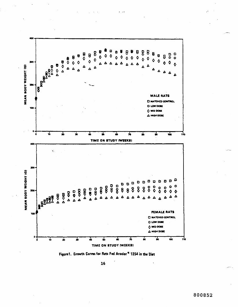
400
OO
e o o o o o
g .
MALE RATSO MATCHED CONTMOl
O LOW DDK
QMDDOKAMWHDOK
10 110
TIME ON STUOY IWffXS)400.
oonzUJ
aoo-
100 J FEMALE RATSD MATCHED CONTMOL
OLOWDOK
AM4DDOK
A MIOH DDK
10 40 M tO 70
TIME ON STUDY (WEEKS)
•o 100 110
Growth Curvu for Rits Ftd Aroder* 1254 in thi Dkt
16
800852
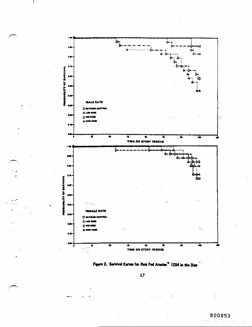
••4—— -r -6- - - - —<)——o
0-
••5
MUkUKATS
0«1»TO.
ouwnQMDDC
TIMS ON truer IWIIKSI^__._._____
2o
O iM
TMIM ON rruor cwf MA
SumvdCimmfvlUttFfdAradW* 12S4ia«• Ditt
17
800853

at the do«t§ used in this study, together with those of the
controls, are shown in figure 2.
For males, the result of the Tarone test for positive dose-
related trend In mortality over the period is significant (P <
0.001); 922 of the control, 832 of the low-dose, 582 of the
mid-dose, and *62 of the ' igh-dose rats survived to the end of
the study. Among females, the Tarooe test shoved a probability
level greater than 0.05. 1:-, females, 672 of the control, 792 of
the low-dose, 832 of the aid-dose, and 712 of the high-dose rats
survived to termination ef the study. Sufficient numbers of rats
of both sexes were available for meaningful statistical analyses
of the incidences of late-developing tumors,
C. Pathology
Histopathologie findings on neoplasms in rats are sumaarixed in
Appendix i, tables Al and A2; findings on nonneoplastic lesions
are summarized in Appendix B, tables Bl and B2.
A variety of neoplastie processes were observed in both the
control and treated rats, and, with the exception of the liver,
the incidences of these neoplasms were comparable in the control
and treated groups. Interstitial-cell tumors of the testes were
present in the majority of control and treated males. The next
most frequently observed neoplasm was leukemia of either the
18
800854

Igranulocycic or lymphocytic type, and it involved multiple
organs. The incidence of this neoplastic process was comparable
in the control and treated groups. The following neoplasms were
also present in some control and treated rats but without
compound association: squamoua-cell carcinomas of the skin,
alveolar/bronchiolar adenomas of the lung, and uterine
endonetrial stromal polyps.
No prolifecative lesions of tha hepatocytes were found in the
control animals in the study. The incidence of these prolifera-
tive lesions among treated animals was as follows:
MALES FEMALES
LowDose
Number of AnimalsNecropsied (24)
NodularHyperplasia 5
Adenoma, SOS* 0
HepatocellularCarcinoma JO
TotalIncidence 5
*Not otherwise specified
MidDose
(24)
8
0
HighDose
(24)
12
i
i
L5
Low Mid HighDose Dose Dose
(24) (22) (24)
6
0
9
1
17
2
10 19
The areas of nodular hyperplasia appeared to be microscopically
similar to what is currently termed "focal areas of cellular
19
800855
alteration" (Squire and Levitt, 1975). Neither this lesion nor
any hepatocellular adenomas or carcinomas were diagnosed in the
controls. The hepatocellular carcinomas were characterized
microscopically by large foci of proliferating hepatocytes
involving several lobules. These hepatocytes were bizarre in
appearance, sometimes containing two or mere nuclei. The
sinusoidal architecture was lost, am? frequently sitotic figures
were present. These neoplasms compretjed the surrounding normal
liver tissue, and the cell plates urually were three to five
cells in thickness. The hepatocellolar adenomas were character-
ized by large foci involving several lobules of svollen, severely
vacuolated hepate^ytec still maintaining the general sinusoidal
architecture of the liver* In general, the foci of nodular
hyperplasia involved two or more hepatic lobules and contained
hepatocytes whose tinctorial properties were distinctly different
from those of the surrounding liver tissue. Occasionally, these
foci would contain severely vacuolated hepatocytes, and in some
instances, there were small foci of basophilic hepatocytes.
The results of the histopathologic examination indicate that the
administration of Aroclor® 1254 at the three doses used in this
study had an effect with respect to proliferative lesions of the
liver and gastrointestinal tract. There were three hepatocellu-
lar carcinomas in male rats and a dose-related increase in
20
800856