Advanced Life Support in Obstetrics - Amazon S3
317
Advanced Life Support in Obstetrics PROVIDER MANUAL EIGHTH EDITION
-
Upload
khangminh22 -
Category
Documents
-
view
3 -
download
0
Transcript of Advanced Life Support in Obstetrics - Amazon S3

Advanced Life Support in Obstetrics
P R O V I D E R M A N U A L
E I G H T H E D I T I O N

Provider ManualJune 2017
ALSO© Editorial Advisory BoardLarry Leeman, MD, MPH
Professor, University of New Mexico School of Medicine Departments of Family and Community Medicine and Obstetrics and Gynecology
Albuquerque, NM
Jeffrey D. Quinlan, MD, CAPT, MC, USNAssociate Professor and Chair Department of Family Medicine
Uniformed Services University Bethesda, MD
Lee T. Dresang, MDProfessor, University of Wisconsin School of Medicine and Public Health
Department of Family Medicine Madison, WI
David S. Gregory, MDProgram Director, Centra-Lynchburg Family Medicine Residency
Associate Clinical Professor, University of Virginia Charlottesville, VA
Larry Howell, Medical IllustratorHowell Graphics and Illustration
Blue Springs, MO
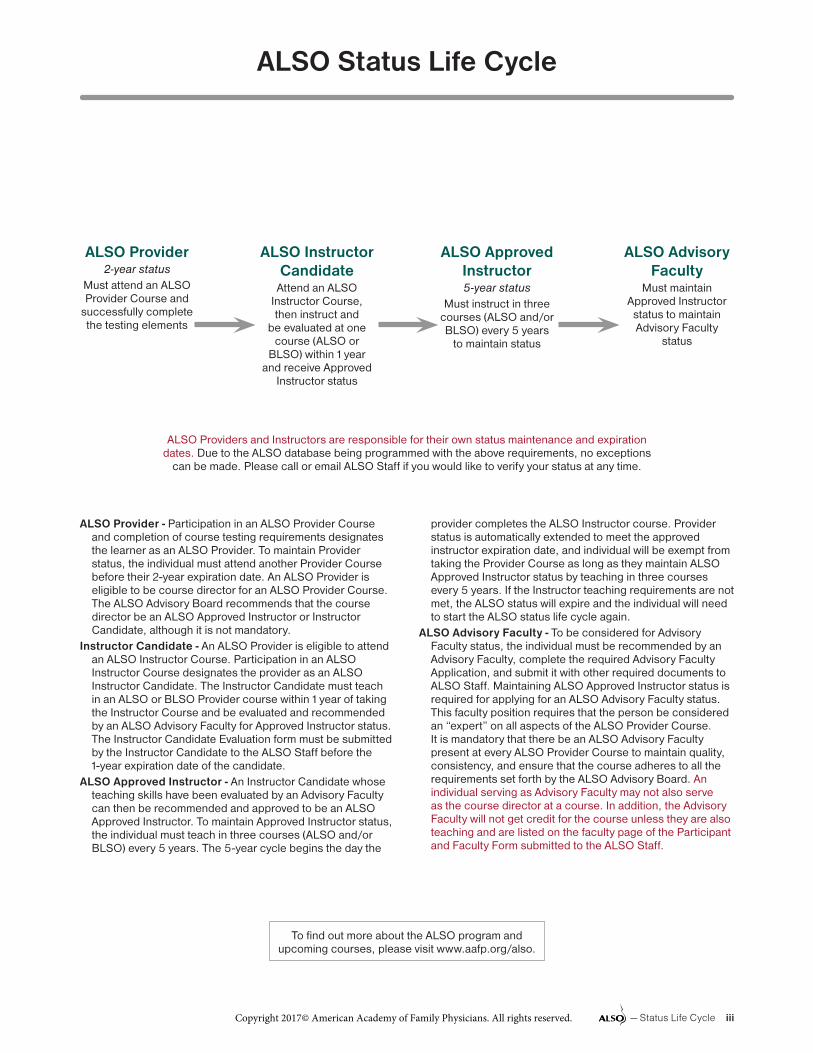
ALSO Status Life Cycle
To find out more about the ALSO program and upcoming courses, please visit www.aafp.org/also.
ALSO Provider - Participation in an ALSO Provider Course and completion of course testing requirements designates the learner as an ALSO Provider. To maintain Provider status, the individual must attend another Provider Course before their 2-year expiration date. An ALSO Provider is eligible to be course director for an ALSO Provider Course. The ALSO Advisory Board recommends that the course director be an ALSO Approved Instructor or Instructor Candidate, although it is not mandatory.
Instructor Candidate - An ALSO Provider is eligible to attend an ALSO Instructor Course. Participation in an ALSO Instructor Course designates the provider as an ALSO Instructor Candidate. The Instructor Candidate must teach in an ALSO or BLSO Provider course within 1 year of taking the Instructor Course and be evaluated and recommended by an ALSO Advisory Faculty for Approved Instructor status. The Instructor Candidate Evaluation form must be submitted by the Instructor Candidate to the ALSO Staff before the 1-year expiration date of the candidate.
ALSO Approved Instructor - An Instructor Candidate whose teaching skills have been evaluated by an Advisory Faculty can then be recommended and approved to be an ALSO Approved Instructor. To maintain Approved Instructor status, the individual must teach in three courses (ALSO and/or BLSO) every 5 years. The 5-year cycle begins the day the
provider completes the ALSO Instructor course. Provider status is automatically extended to meet the approved instructor expiration date, and individual will be exempt from taking the Provider Course as long as they maintain ALSO Approved Instructor status by teaching in three courses every 5 years. If the Instructor teaching requirements are not met, the ALSO status will expire and the individual will need to start the ALSO status life cycle again.
ALSO Advisory Faculty - To be considered for Advisory Faculty status, the individual must be recommended by an Advisory Faculty, complete the required Advisory Faculty Application, and submit it with other required documents to ALSO Staff. Maintaining ALSO Approved Instructor status is required for applying for an ALSO Advisory Faculty status. This faculty position requires that the person be considered an “expert” on all aspects of the ALSO Provider Course. It is mandatory that there be an ALSO Advisory Faculty present at every ALSO Provider Course to maintain quality, consistency, and ensure that the course adheres to all the requirements set forth by the ALSO Advisory Board. An individual serving as Advisory Faculty may not also serve as the course director at a course. In addition, the Advisory Faculty will not get credit for the course unless they are also teaching and are listed on the faculty page of the Participant and Faculty Form submitted to the ALSO Staff.
ALSO Providers and Instructors are responsible for their own status maintenance and expiration dates. Due to the ALSO database being programmed with the above requirements, no exceptions
can be made. Please call or email ALSO Staff if you would like to verify your status at any time.
ALSO Provider2-year status
Must attend an ALSO Provider Course and
successfully complete the testing elements
ALSO Advisory Faculty
Must maintain Approved Instructor status to maintain Advisory Faculty
status
ALSO Approved Instructor5-year status
Must instruct in three courses (ALSO and/or BLSO) every 5 years
to maintain status
ALSO Instructor Candidate
Attend an ALSO Instructor Course, then instruct and
be evaluated at one course (ALSO or
BLSO) within 1 year and receive Approved
Instructor status
— Status Life Cycle iiiCopyright 2017© American Academy of Family Physicians. All rights reserved.

Overview of the Program . . . . . . . . . . . . . . . . . . . . . . . . . . . . . . . . . . . . . . . . . . . . . . . . . . . . . . . . . . . . .1 – 4
A First-Trimester Pregnancy Complications . . . . . . . . . . . . . . . . . . . . . . . . . . . . . . . . . . . . . . . . . . . .1 – 16
B Medical Complications of Pregnancy . . . . . . . . . . . . . . . . . . . . . . . . . . . . . . . . . . . . . . . . . . . . . . .1 – 29
C Vaginal Bleeding in Late Pregnancy . . . . . . . . . . . . . . . . . . . . . . . . . . . . . . . . . . . . . . . . . . . . . . . . .1 – 14
D Preterm Labor and Premature Rupture of Membranes . . . . . . . . . . . . . . . . . . . . . . . . . . . . . . . . .1 – 21
E Intrapartum Fetal Surveillance . . . . . . . . . . . . . . . . . . . . . . . . . . . . . . . . . . . . . . . . . . . . . . . . . . . . . .1 – 21
F Labor Dystocia (Case handouts will be provided at course) . . . . . . . . . . . . . . . . . . . . . . . . . . . .1 – 14
G Malpresentations, Malpositions, and Multiple Gestation . . . . . . . . . . . . . . . . . . . . . . . . . . . . . . . .1 – 26
H Assisted Vaginal Delivery . . . . . . . . . . . . . . . . . . . . . . . . . . . . . . . . . . . . . . . . . . . . . . . . . . . . . . . . . .1 – 13
I Shoulder Dystocia . . . . . . . . . . . . . . . . . . . . . . . . . . . . . . . . . . . . . . . . . . . . . . . . . . . . . . . . . . . . . . . .1 – 20
J Postpartum Hemorrhage . . . . . . . . . . . . . . . . . . . . . . . . . . . . . . . . . . . . . . . . . . . . . . . . . . . . . . . . . .1 – 16
K Maternal Resuscitation and Trauma . . . . . . . . . . . . . . . . . . . . . . . . . . . . . . . . . . . . . . . . . . . . . . . . .1 – 20
L Safety in Maternity Care . . . . . . . . . . . . . . . . . . . . . . . . . . . . . . . . . . . . . . . . . . . . . . . . . . . . . . . . . . .1 – 11
M OB Cases . . . . . . . . . . . . . . . . . . . . . . . . . . . . . . . . . . . . . . . . . . . . . . . . . . . . . . . . . . . . . . . . . (Handouts)
N Third and Fourth Degree Perineal Lacerations . . . . . . . . . . . . . . . . . . . . . . . . . . . . . . . . . . . . . . . .1 – 12
O Diagnostic Ultrasound in Labor and Delivery . . . . . . . . . . . . . . . . . . . . . . . . . . . . . . . . . . . . . . . . .1 – 15
P Neonatal Resuscitation . . . . . . . . . . . . . . . . . . . . . . . . . . . . . . . . . . . . . . . . . . . . . . . . . . . . . . . . . . .1 – 15
Q Cesarean Delivery . . . . . . . . . . . . . . . . . . . . . . . . . . . . . . . . . . . . . . . . . . . . . . . . . . . . . . . . . . . . . . . .1 – 34
R Birth Crisis . . . . . . . . . . . . . . . . . . . . . . . . . . . . . . . . . . . . . . . . . . . . . . . . . . . . . . . . . . . . . . . . . . . . . .1 – 11
ALSO Table of Contents
— Table of Contents v
ALSO Program
ALSO Advisory Board, 2017-2018
Jennifer Frank, MD, Chair
Ann Evensen, MD
Gretchen Heinrichs, MD
Sarah Jones, MD
Tony Ogburn, MD
Sajjini Thomas, MD
Janice Taleff, CNM
Barbara True, RNC-OB
Johanna Warren, MD
© 2017 American Academy of Family Physicians
11400 Tomahawk Creek Parkway, Leawood, Kansas 66211-2672
1-800-274-2237 • 913-906-6000
The American Academy of Family Physicians (AAFP) wishes to acknowledge the initial development of the ALSO Program by the University of Wisconsin Department of Family Medicine and the original national ALSO Development Group of family physi-cians, obstetricians, and nurses, which formed in 1991. The ALSO Program, originally conceptualized by James R. Damos, MD, was developed under the leader-ship of Dr Damos and John W. Beasley, MD. The AAFP acquired the ALSO Program in 1993.
The 2000 edition of the ALSO Course curriculum added levels of evidence for recommendations and
references used in the new curriculum. The curriculum demonstrates the evidence, and quality of that evidence, on which any recommendations of care are based.
The current ALSO Provider Manual continues to be an ongoing process. Each chapter is reviewed on a 3-year cycle. Revisions will be published annually and 1-page addendums will only be published if an important/key evidence-based practice recommenda-tion becomes available that would significantly alter a change in practice.
— Overview 1
ALSO StaffRobyn Brumble, BSN, RNC-OB
Manager of Maternity Services
Brian DotyAssociate Editor
Carla CherryALSO Program Specialist
Ruth FlemingALSO Program Specialist
Jennifer HeadALSO Program Specialist

2 Overview—
Managing Editor and AuthorLarry Leeman, MD, MPHProfessor University of New Mexico School of Medicine Albuquerque, New Mexico
Associate Editors and AuthorsLee T. Dresang, MDProfessor University of Wisconsin, Department of Family Medicine & Community Health
Jeffrey D. Quinlan, MD, CAPT, MC, USN Associate Professor and Chair Department of Family Medicine, Uniformed Services University, Bethesda, Maryland
Medical IllustratorLarry HowellHowell Graphics and Illustration Blue Springs, Missouri
Current AuthorsKaren Ailsworth MD, MS, FAAP Professor Baraboo Family Practice Residency Rural Training Program St . Mary’s/Dean Venture, Baraboo, Wisconsin Affiliate University of Wisconsin Department of Family Medicine-Madison
Janice M. Anderson, MDAssociate Program Director Forbes Family Medicine Residency Program
Lesley A. Atwood, MD, FAAP Clinical Associate Professor Family Medicine University of Minnesota Allina Health, Hastings
Zachary Baeseman, MDThedaCare Physicians Waupaca, WI
R. Eugene Bailey, MDAssociate Professor, Department of Family Medicine Assistant Professor, Department of Obstetrics and Gynecology
Timothy P. Canavan, MD, MSc Attending Perinatologist Greenwich Hospital, Yale New Haven Health System
Brendon Cullinan, MDSenior Medical Director for Primary Care Hennipen County Medical Center, Minneapolis, Minnesota Assistant Professor, University of Minnesota Department of Family Medicine
Stan E. Davis, MD, FACOGHealthcare Consultant TeamSTEPPS and In Situ Simulation
Mark Deutchman, MDProfessor University of Colorado School of Medicine
Ann Evensen, MDAssistant Professor, CHS University of Wisconsin School of Medicine and Public Health Verona, WI
Patricia Fontaine, MD, MSSenior Clinical Investigator HealthPartners Institute for Education and Research
Jennifer Frank, MDSenior Medical Director ThedaCare Physicians
Robert W. Gobbo, MDProgram Director Providence Oregon Family Medicine, Hood River Rural Residency Program .
Kim Hinshaw, MB, BS, FRCOGConsultant Obstetrician & Gynaecologist, Director of Research, Sunderland Royal Hospital and Visiting Professor, Dept of Health Sciences, University of Sunderland, UK
Caroline Homer RM, MMedSci(ClinEpi), PhDProfessor of Midwifery University of Technology Sydney, Australia
Sarah K. Jorgensen, DOAssociate Program Director National Capitol Consortium Family Medicine Residency Program Fort Belvoir Community Hospital Fort Belvoir, VA
Paul Lewis, OBEChair of ALSO UK Executive Board of Trustees Emeritus Professor of Midwifery Practice and Professional Development Bournemouth University Bournemouth, England
Susanna R. Magee, MD, MPHAssociate Professor of Family Medicine Albert Medical School of Brown University
Kristi K. Miller, MS, RN Quality Consultant Medical Teamwork Consultants LLC
Neil J. Murphy, MD, FACOG Southcentral Foundation Alaska Native Medical Center
Stephen Ratcliffe, MD, MSPHProgram Director Lancaster General Family Medicine Residency Lancaster, Pennsylvania
William G. Sayres, Jr, MDSmith Family Endowed Chair in Medicine Assistant Dean for Foundations University of Washington School of Medicine
Barbara A. True, MN, CNS, RNC-OB, C-EFMClinical Nurse Specialist Texas Health Arlington Memorial
Sara G. Shields, MD, MS, FAAFPClinical Professor of Family Medicine and Community Health University of Massachusetts Family Health Center of Worcester
Mary Beth Sutter, MD Family Medicine Faculty University of New Mexico School of Medicine
J. Ely Walker, MD, MPHRural Obstetrics Fellow University of Colorado School of Medicine
Johanna B. Warren, MDFaculty Physician Providence Oregon Family Medicine Residency Program
Helen Welch MSN, CNMKaiser Permanente Northwest
Karen Wildman, MD, FAAFPClinical Associate Professor Family Medicine, UWSOM Family Medicine Residency Spokane
Colleen Zimmermann, MDFaculty, Clinical Instructor Family Medicine Residency Spokane
Provider Manual Disclosures — It is the policy of the AAFP that all individuals in a position to control content disclose any relationships with commercial interests upon nomination/invitation of participation. Disclosure documents are reviewed for potential conflicts of interest and, if identified, conflicts are resolved prior to confirmation of participation. Only those participants who had no conflict of interest or who agreed to an identified resolution process prior to their participation were involved in this CME activity. All individuals in a position to control content for this Provider Manual have indicated that they have no relevant financial relationships to disclose.

—Overview 3
PrefaceThe ALSO Provider Course is an educational program designed to assist health professionals in developing and maintaining the knowledge and skills needed to effectively manage the emergencies which arise in maternity care. The course includes required reading, lectures, and hands-on workstations. Evaluation is by a written exam and skills assessment stations. There are many appropriate ways of managing emergencies. The treatment guidelines presented in ALSO do not neces-sarily represent the only way to manage problems and emergencies. Instead, these guidelines are presented as reasonable methods of management in obstetrical emer-gencies. Each maternity care clinician must ultimately exercise his or her own professional judgement in deciding on appropriate action in emergency situations. Completion of the ALSO Provider Course does not imply competency to perform the procedures discussed in the course materials.
Overall Course Objectives1. Discuss methods of managing pregnancy and
birth urgencies and emergencies, which may helpstandardize the skills of practicing maternity careclinicians.
2. Demonstrate content and skill acquisition as demon-strated by successful completion of the course testingrequirements.
CDC RecommendationThe Centers for Disease Control and Prevention recommend universal precautions be used in all situa-tions where a risk of exposure to blood or bodily fluids is present, and the potential infectious status of the patient is unknown. All bodily fluids (blood, urine stool, saliva, vomitus, etc) should be treated as poten-
tially infectious. Universal precautions should always be followed in pregnancy and birth care.
CopyrightThe American Academy of Family Physicians (AAFP) owns the ALSO copyright and trademark on all of the course materials, including the Provider Manual, slide set, and written exam. Use of portions of the materials outside of an authorized ALSO course is strictly pro-hibited without prior written approval from the AAFP.
Course DisclaimerThe material presented at this course is being made available by the AAFP for educational purposes only. This material is not intended to represent the only, nor necessarily best, methods or procedures appropriate for the medical situations discussed, but rather is intended to present an approach, view, statement, or opinion of the faculty which may be helpful to others who face similar situations. The AAFP disclaims any and all liability for injury, or other damages, resulting to any individual attending this course and for all claims which may arise out of the use of the techniques dem-onstrated therein by such individuals, whether these claims shall be asserted by physician, or any other per-son. Every effort has been made to ensure the accuracy of the data presented at this course. Physicians may care to check specific details, such as drug doses and con-traindications, etc, in standard sources prior to clinical application. This Course has been determined to be a Level 2 classification under the AMA/PRA Credit and Classification Guidelines. The AAFP does not certify competence upon completion of the ALSO Provider Course, nor does it intend this course to serve as a basis for requesting new or expanded privileges.
4 Overview—
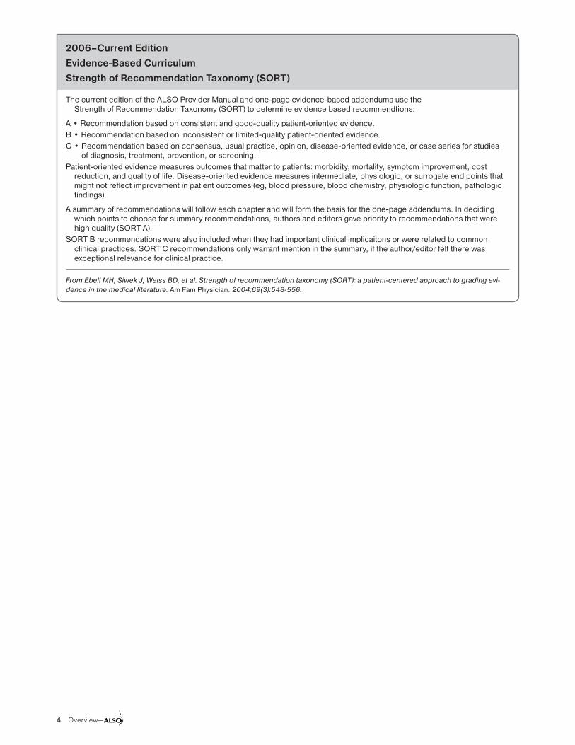
2006–Current Edition
Evidence-Based Curriculum
Strength of Recommendation Taxonomy (SORT)
The current edition of the ALSO Provider Manual and one-page evidence-based addendums use the Strength of Recommendation Taxonomy (SORT) to determine evidence based recommendtions:
A • Recommendation based on consistent and good-quality patient-oriented evidence. B • Recommendation based on inconsistent or limited-quality patient-oriented evidence. C • Recommendation based on consensus, usual practice, opinion, disease-oriented evidence, or case series for studies
of diagnosis, treatment, prevention, or screening.Patient-oriented evidence measures outcomes that matter to patients: morbidity, mortality, symptom improvement, cost
reduction, and quality of life. Disease-oriented evidence measures intermediate, physiologic, or surrogate end points that might not reflect improvement in patient outcomes (eg, blood pressure, blood chemistry, physiologic function, pathologic findings).
A summary of recommendations will follow each chapter and will form the basis for the one-page addendums. In deciding which points to choose for summary recommendations, authors and editors gave priority to recommendations that were high quality (SORT A).
SORT B recommendations were also included when they had important clinical implicaitons or were related to common clinical practices. SORT C recommendations only warrant mention in the summary, if the author/editor felt there was exceptional relevance for clinical practice.
From Ebell MH, Siwek J, Weiss BD, et al. Strength of recommendation taxonomy (SORT): a patient-centered approach to grading evi-dence in the medical literature. Am Fam Physician. 2004;69(3):548-556.

— Chapter A 1
Chapter A First-Trimester Pregnancy Complications
IntroductionComplications during the first trimester of pregnancy are common. Approximately 15% of clinically recog-nized pregnancies result in spontaneous miscarriage, and estimates of miscarriage before clinical recognition are as high as 50%.1 In addition to miscarriage, vaginal bleeding can be associated with ectopic pregnancy, trophoblastic disease, or cervical bleeding from causes unrelated to pregnancy, or bleeding may occur in preg-nancies that proceed without further complications.
Normal First-Trimester Pregnancy ProgressPregnancy is clinically dated from the first day of the last normal menstrual period, which is an observable event, instead of the conception date. Conception occurs approximately 2 weeks later. All gestational landmarks in this chapter are based on menstrual dat-ing; embryology textbooks commonly use conception dating, which is 2 weeks less.
The placenta produces human chorionic gonadotro-pin (hCG) after implantation. Implantation occurs at approximately 23 menstrual days, which is approxi-mately 8 days after conception. Commonly available over-the-counter urine pregnancy tests are approxi-mately 100% sensitive and specific at detecting the beta subunit of hCG at levels of 25 mIU/mL, which may allow detection of pregnancy around the time of the first missed period.2,3 Serum tests can detect hCG
as low as 5 mIU/mL. The rate of rise in quantitative serum hCG levels may be used to monitor patients with pain or bleeding whose initial ultrasound exami-nation did not yield a definitive diagnosis. A large study of such patients before 10 weeks’ gestation showed that a viable intrauterine pregnancy with hCG levels greater than 5,000 mIU/mL had an hCG rise of 53% in 48 hours. However, women with a miscarriage or an ectopic pregnancy also can have a rise that is within this range. Therefore, an adequately rising hCG does not rule out nonviable pregnancy.3,5,6
The gestational sac first becomes visible on transvagi-nal ultrasound during the 5th menstrual week as a 2- to 5-mm sonolucent area surrounded by an echogenic ring of chorionic villi. This early gestational sac is only visible using a high-frequency transducer (5 MHz or greater) and the transvaginal scan route. A small sono-lucent fluid collection, or pseudosac, can also be pres-ent in cases of ectopic pregnancy, so additional features of a normal gestational sac can be sought, particularly the eccentric location of the gestational sac indicating that it is implanted within the endometrium rather than being located in the endometrial stripe. The yolk sac appears during transvaginal scanning during the 6th menstrual week and provides clear evidence of an intra-uterine pregnancy. By the end of the 6th menstrual week, the fetal pole becomes visible during transvaginal scanning as a 2 to 8 mm pole with embryonic cardiac
Learning ObjectivesAt the end of this activity, learners will be able to:1. Describe the process, diagnosis, and management
of miscarriage, ectopic pregnancy, and gestational trophoblastic disease.
2. Discuss the value of human chorionic gonadotropin(hCG) levels and sonographic discriminatory criteria in diagnosing first-trimester pregnancy complications.
3. Describe the spectrum of psychological reactionsto early pregnancy loss.
4. Describe the techniques of uterine aspiration forthe treatment of incomplete miscarriage (Optional).
Mark Deutchman, MD, J. Ely Walker, MD
Revised January 2016
Copyright 2017© American Academy of Family Physicians. All rights reserved.

Chapter A
2 Chapter A —
activity. These sonographic landmarks are visible with transabdominal scanning approximately 1 week later than with transvaginal scanning.7
Embryologic, clinical, hCG, and sonographic findings are closely correlated and are displayed in Table 1.8,9 Ultrasonography is so valuable that, when it is readily available, many clinicians use it as a primary tool in evaluating first-trimester complications, leaving serum hCG testing as a secondary tool used only if sonographic findings are equivocal. A routine first-trimester ultrasound in early pregnancy appears to enable better assess-ment of gestational age and earlier detection of multifetal pregnancies. However, the benefits for other substantive outcomes are less clear.10
The use of specific sonographic findings as discriminatory criteria to diagnose early pregnancy loss is presented in detail in the pathophysiology section of this chapter.
After the embryo is sonographically visible, first-trimester menstrual age is calculated from crown-rump length using parameters such as those shown in Table 2, which can be commonly found in textbooks and are normally included in the calculation software of ultrasound equipment.
Table 1. Early Pregnancy Landmarks
Menstrual Age (weeks) Embryologic Event/Sonographic/hCG Correlation
3 to 4 Implantation site – Decidual thickening
4 Trophoblast – Peritrophoblastic flow on color flow Doppler
4 to 5 Gestational sac typically visible when hCG reaches 1,500 to 2,000 mlU/mL
5 to 6 Yolk sac
5 to 6 Embryo and cardiac activity
Table 2. Crown-Rump Length and Menstrual Age
Crown-Rump Length (mm)
Menstrual Age (days)
Crown-Rump Length (mm)
Menstrual Age (days)
2 42 16 57
4 44 20 60
6 47 25 65
8 49 30 69
10 51 35 72
13 54 40 76

First-Trimester Pregnancy Complications
— Chapter A 3
Early Pregnancy Loss: Pathophysiology, Discriminatory Criteria, Clinical Course and Prognosis
Spontaneous Abortion (Miscarriage)
The cause of spontaneous abortion is rarely determined in clinical practice, but it is known that approximately half are because of major genetic abnormalities, typically trisomy, triploidy, or monosomy.12 Environmental factors linked to spontaneous abortion are shown in Table 4.
Spontaneous abortion can manifest clinically in several different ways. Most commonly, vaginal bleeding and cramping are present. But occasion-ally, regression of pregnancy symptoms or lack of Doppler-detected fetal heart tones by 10 to 12 weeks’ gestation are the first clinical signs in the
setting of anembryonic pregnancy or embryonic demise. Spontaneous abortion may also be dis-covered incidentally when asymptomatic patients
Table 3. Terms Applied to Early Pregnancy Loss11,13
Embryonic demise — an embryo with a crown-rump length >7 mm without cardiac activity.
Anembryonic pregnancy — presence of a gestational sac >25 mm without evidence of embryonic tissues (ie, yolk sac, embryo). This term is preferred to the older and less accurate phrase blighted ovum.
Spontaneous abortion — spontaneous loss of a pregnancy before 20 weeks’ gestation. Can be further described as:• Incomplete — occurs when some but not all of the products of conception have passed.• Complete — all products of conception have passed through the external cervical os.• Septic — incomplete abortion associated with ascending infection of the endometrium, parametrium, adnexa,
or peritoneum.• Inevitable — bleeding in the presence of a dilated cervix, indicating that passage of the conceptus is
unavoidable.• Missed — the fetus or embryo is dead but no tissue has been passed. The cervix is closed. These patients
often present with no growth in uterine size or no audible fetal heart tones.
Threatened abortion — bleeding before 20 weeks’ gestation in the presence of an embryo with cardiac activity or a gestational/yolk sac and a closed cervix.
Subchorionic hemorrhage — ultrasonographic finding of blood between the chorion and uterine wall, typically seen in the setting of vaginal bleeding.
Recurrent pregnancy loss — more than two consecutive pregnancy losses. The phrase habitual aborter has been used but is no longer appropriate.
Ectopic pregnancy — pregnancy outside the uterine cavity, most commonly in the fallopian tube but can occur in the broad ligament, ovary, cervix, or elsewhere in the abdomen.
Heterotopic pregnancy — simultaneous intrauterine and ectopic pregnancy. Incidence is rare, thought to occur in 1/30,000 spontaneous pregnancies, but occur in 1.5/1,000 pregnancies involving assisted reproductive techniques.12,13
Gestational trophoblastic disease, or hydatidiform mole — complete mole: placental proliferation in the absence of a fetus. Most have a 46,XX chromosomal composition, all derived from paternal source. Partial mole: molar placenta occurring together with a fetus. Most are genetically triploid (69,XXX).
Vanishing twin — A multi-fetal pregnancy is identified and one or more fetuses later disappear. More commonly seen now that early ultrasound scanning is common. If this occurs early in pregnancy, the embryo is often reabsorbed. Later occurrence results in a compressed or mummified fetus or amorphous material.
Table 4. Environmental Factors Linked to Spontaneous Abortion
Uterine anomaliesLeiomyomataIncompetent cervixTobacco, alcohol, or cocaine useProgesterone deficiency because
of luteal phase defect
IrradiationMaternal diethylstilbestrol
exposureAdvanced maternal ageInfectionsOccupational chemical exposure
Chapter A
4 Chapter A —
undergo early ultrasound examinations for other reasons such as pregnancy dating or genetic screening.
Clinical examination should include palpation of the abdomen and pelvis. The size and position of the uterus, the location of any tenderness, the presence of rebound tenderness, and the presence of masses should be noted. Adnexal tenderness and any masses should raise suspicion for ectopic pregnancy, although a normal corpus luteum cyst can also be the cause of either. If the patient’s last menses was at least 9 to 10 weeks prior, and ultra-sound is not readily available, consider listening for the fetal heartbeat during the bimanual pelvic examination while elevating the uterus with the intravaginal hand.
A speculum examination will reveal non-uterine causes of bleeding, the degree of cervical dilation and, if present, tissue being passed. The quantity of blood in the vault and the source of bleeding (from the os versus other sites) should be noted. If an intact gestational sac, an embryo, or the character-istic fronds of chorionic villi are seen, miscarriage is proven and ectopic pregnancy is virtually ruled out, except in the rare case of heterotopic preg-nancy. If there is doubt about the origin of expelled tissue, an examination for chorionic villi can be
performed. To examine for chorionic villi, rinse and float the tissue in saline. Low magnification, backlighting, and teasing the tissue can help. Passed tissue should be submitted for pathologic examina-tion, which is definitive in questionable cases.
If products of conception are seen at the cervi-cal os, ring forceps can be used to gently remove the tissue. More aggressive attempts to remove partially expelled tissue should be preceded by discussion with the patient, informed consent, and administration of analgesia or sedation.
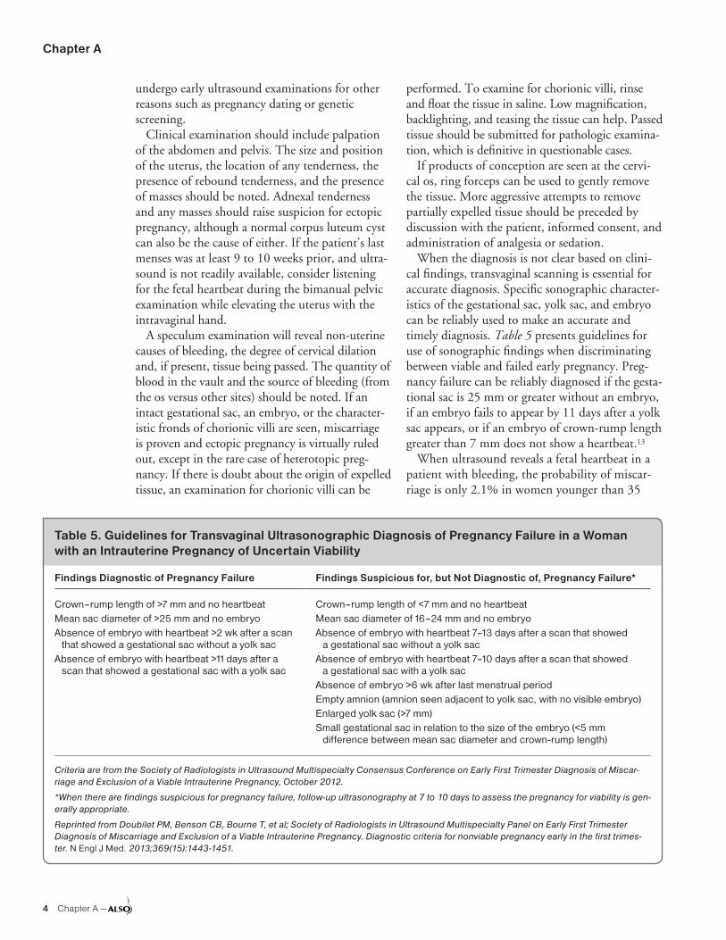
When the diagnosis is not clear based on clini-cal findings, transvaginal scanning is essential for accurate diagnosis. Specific sonographic character-istics of the gestational sac, yolk sac, and embryo can be reliably used to make an accurate and timely diagnosis. Table 5 presents guidelines for use of sonographic findings when discriminating between viable and failed early pregnancy. Preg-nancy failure can be reliably diagnosed if the gesta-tional sac is 25 mm or greater without an embryo, if an embryo fails to appear by 11 days after a yolk sac appears, or if an embryo of crown-rump length greater than 7 mm does not show a heartbeat.13
When ultrasound reveals a fetal heartbeat in a patient with bleeding, the probability of miscar-riage is only 2.1% in women younger than 35
Table 5. Guidelines for Transvaginal Ultrasonographic Diagnosis of Pregnancy Failure in a Woman with an Intrauterine Pregnancy of Uncertain Viability
Findings Diagnostic of Pregnancy Failure Findings Suspicious for, but Not Diagnostic of, Pregnancy Failure*
Crown–rump length of >7 mm and no heartbeatMean sac diameter of >25 mm and no embryoAbsence of embryo with heartbeat >2 wk after a scan
that showed a gestational sac without a yolk sacAbsence of embryo with heartbeat >11 days after a
scan that showed a gestational sac with a yolk sac
Crown–rump length of <7 mm and no heartbeatMean sac diameter of 16–24 mm and no embryoAbsence of embryo with heartbeat 7–13 days after a scan that showed
a gestational sac without a yolk sacAbsence of embryo with heartbeat 7–10 days after a scan that showed
a gestational sac with a yolk sacAbsence of embryo >6 wk after last menstrual periodEmpty amnion (amnion seen adjacent to yolk sac, with no visible embryo)Enlarged yolk sac (>7 mm)Small gestational sac in relation to the size of the embryo (<5 mm
difference between mean sac diameter and crown-rump length)
Criteria are from the Society of Radiologists in Ultrasound Multispecialty Consensus Conference on Early First Trimester Diagnosis of Miscar-riage and Exclusion of a Viable Intrauterine Pregnancy, October 2012.
*When there are findings suspicious for pregnancy failure, follow-up ultrasonography at 7 to 10 days to assess the pregnancy for viability is gen-erally appropriate.
Reprinted from Doubilet PM, Benson CB, Bourne T, et al; Society of Radiologists in Ultrasound Multispecialty Panel on Early First Trimester Diagnosis of Miscarriage and Exclusion of a Viable Intrauterine Pregnancy. Diagnostic criteria for nonviable pregnancy early in the first trimes-ter. N Engl J Med. 2013;369(15):1443-1451.

First-Trimester Pregnancy Complications
— Chapter A 5
years, but increases to 16.1% in women older than 35 years.14
Complete spontaneous miscarriage might result in an empty uterus with a bright endometrial stripe as a result of the uterine walls having collapsed against each other. Echogenic material within the endo-metrial cavity commonly creates an endometrial stripe greater than 15 mm after treatment of early pregnancy failure with misoprostol.15 Therefore, endometrial thickness alone is not an indication of the need for surgical intervention after medical management of miscarriage with misoprostol.
Septic abortion should be assumed when the patient is febrile or has excessive uterine or adnexal tenderness, or signs of peritonitis. The clinician should ask about a history of attempted therapeu-tic abortion or illegal abortion that might have left tissue behind or perforated the uterus. Septic abortion is a potentially life-threatening condition requiring prompt resuscitation, uterine evacuation, and broad-spectrum antibiotic treatment.16,17
Sonography may reveal a hematoma between the chorion and uterine wall in subchorionic hem-orrhage and a gestational sac and embryo will be present. When subchorionic hemorrhage is visible on ultrasound, the likelihood of miscarriage aver-ages approximately 10% even when a heartbeat is detected, but varies by maternal age, size of the hematoma, and gestational age.18 Therefore, the patient should be advised to expect bleeding.
The quantity of bleeding predicts pregnancy loss only when it is heavy. A prospective analysis of 4,510 women who were followed in the early first trimester showed that 1,204 (27%) had some bleeding or spotting. There was no increase in miscarriage risk in early pregnancy when there was spotting or light bleeding. However, miscarriage risk increased significantly in the 8% of women who reported heavy bleeding. This was the only group to have an increased risk of miscarriage (adjusted odds ratio = 2.84, 95% confidence inter-val = 1.82 to 4.43).19
Management of Spontaneous AbortionIf an ultrasound reveals an intrauterine pregnancy with cardiac motion, the patient should be fol-lowed with cautious optimism and an explanation that there are no known interventions to prevent miscarriage.
When the clinician’s examination reveals incomplete miscarriage, the patient must choose
between expectant, medical, or surgical manage-ment. The majority of first-trimester miscarriages occur completely and spontaneously without intervention. Although surgical intervention in the form of uterine aspiration has traditionally been used liberally, expectant management and medical treatment are valid options. Women with excessive bleeding, pain, or infection benefit from medi-cal or surgical intervention.11 In an observational study of 451 cases, 91% of those with incomplete miscarriage, 76% of those with missed miscarriage, and 66% of those with anembryonic pregnancy completed their miscarriage without surgical intervention. Overall, 70% of women completed their miscarriage within 14 days of classification.20 Women who have not completed the miscarriage for an emotionally uncomfortable period of time may prefer medical or surgical intervention. The woman’s emotional state and personal preferences are important in choosing the course of action. Ultimately the decision should be primarily driven by the patient’s desires after being well informed of her options.21
Clinical trials comparing expectant management with misoprostol and misoprostol with surgical treatment have shown:22-24
• In incomplete miscarriage, expectant and medi-cal management with misoprostol are highly successful.
• In missed abortion, medical management withmisoprostol and surgical treatment are more effective than expectant treatment.
• Typical misoprostol doses are 600 µg orally or600 to 800 µg vaginally.
• Women treated with misoprostol have morebleeding but less pain than those treated surgically.
• Women treated expectantly have more outpa-tient visits than those treated with misoprostol.
• Surgery is associated with more trauma and infec-tion complications than misoprostol treatment.
• Misoprostol has fewer gastrointestinal adverseeffects when administered vaginally than when administered orally.As a result of trials conducted on the medi-
cal management of miscarriage, a change to less surgical management has been suggested, even though this is an off-label use of misoprostol.25 It is also reasonable for a woman to change treat-ment course and mix the three treatment options. Women commonly choose a period of expectant

Chapter A
6 Chapter A —
management followed by medical therapy with misoprostol if they no longer desire to wait. This could also be followed by uterine aspiration if medical therapy is not successful.
There is no evidence supporting the use of prophylactic antibiotics in early pregnancy fail-ure,26 although there is such evidence for uterine aspiration for induced abortion. When misopro-stol is used for medical abortion, the incidence of infection complications may be reduced by administering the drug by the buccal route and by administering doxycycline 100 mg twice daily for 7 days. However, this study is low-quality evidence because it compares two different time periods.27 It is unclear whether it is the use of antibiotics or the change in the route of misoprostol administration that resulted in the lowered incidence of infection. It is also unclear whether this is also true when misoprostol is used to manage early pregnancy loss.
After a miscarriage, it is customary to recom-mend a brief period of contraception before attempting another pregnancy. However, this practice is not supported in the literature. A prospective study showed no statistically signifi-cant difference of recurrent miscarriage in women who had interpregnancy intervals of less than 6 months versus those who had longer intervals.28 For patients desiring long-term contraception, an intrauterine device placed immediately after spontaneous or induced first-trimester abortion is safe and effective.29
Ectopic PregnancyPathophysiology and Risk FactorsEctopic pregnancy is typically located in the fal-lopian tube, but rarely can occur in the broad ligament, ovary, cervix at the site of a prior hys-terotomy, or elsewhere in the abdomen. Ectopic pregnancy can result in impairment or loss of fer-tility and, because of internal hemorrhage, remains the second most common cause of maternal mortality. Early diagnosis is the key to preventing morbidity and mortality and preserving fertility.
All health care professionals who care for women of childbearing age should have an active work-ing knowledge of ectopic pregnancy and should have a high index of suspicion in any woman who presents with bleeding and/or pain during early pregnancy. Risk factors are shown in Table 6, but many ectopic pregnancies occur in women with-out risk factors.
Signs, Symptoms, and DiagnosisPain and vaginal bleeding are the hallmark symptoms of ectopic pregnancy. Pain is almost universal; it is typically unilateral and in the lower abdomen. Bleeding is also common after a short period of amenorrhea. Physical examination may reveal a tender adnexal mass, often mentioned in texts, but only noted clinically in a minority of cases. Furthermore, it may easily be confused with a tender corpus luteum of a normal intra-uterine pregnancy. Finally, signs and symptoms of hemoperitoneum and shock can occur, including a distended silent, doughy abdomen, shoulder pain, bulging cul-de-sac into the posterior fornix of the vagina, and hypotension.
Initially, the serum hCG level in ectopic preg-nancy rises but then typically plateaus or falls. Transvaginal ultrasound scanning is a key diagnos-tic tool and can rapidly make these diagnoses:
1. Ectopic pregnancy is ruled out by the presenceof an intrauterine pregnancy with the exceptionof rare heterotopic pregnancy.
2. Ectopic pregnancy is proven when a gestationalsac and an embryo with a heartbeat are seenoutside of the uterus.
3. Ectopic pregnancy is highly likely if any adnexalmass distinct from the corpus luteum or a sig-nificant amount of free pelvic fluid is seen.30
When ultrasound findings are not definitive,the location of the pregnancy is unknown. Table 7 provides guidelines for using hCG measurements in combination with transvaginal ultrasound find-ings to establish a diagnosis and avoid interrupting a potentially viable pregnancy. When transvaginal
Table 6. Ectopic Pregnancy Risk Factors
History of tubal surgery, including tubal ligation and/or reanastomosis of the tubes after tubal ligation
History of tubal infection including pelvic inflammatory disease
Contraception with progestin-only pills
Contraception with intrauterine devices
History of in utero exposure to diethylstilbestrol
History of ectopic pregnancy

First-Trimester Pregnancy Complications
— Chapter A 7
ultrasound shows no intrauterine fluid (gesta-tional sac) and the hCG is above a discriminatory zone threshold, viable intrauterine pregnancy is unlikely, so the clinician should have a high index of suspicion for ectopic pregnancy. This threshold for hCG is in question; some sources use 3,000 mIU/mL,15 some use 3,510 mIU/mL31 and the threshold is dependent on the quality of the ultra-sound equipment and the sonographer performing the ultrasound. In stable patients, repeat hCG test-ing and transvaginal ultrasound are prudent before diagnosing and treating for ectopic pregnancy.13,31
In some cases of ectopic pregnancy, a small fluid collection within the uterus can be mistaken for a true gestational sac. However, this pseudoges-tational sac lacks a surrounding echogenic ring of chorionic villi, a yolk sac, or fetal pole. An unrup-tured corpus luteum cyst can be mistaken for an ectopic gestational sac, and a ruptured corpus luteum cyst can produce free pelvic fluid suggest-ing ruptured ectopic pregnancy. Culdocentesis can be helpful in differentiating the thin pink fluid of
a ruptured ovarian cyst, which can be managed expectantly from the frank hemorrhage because of ruptured ectopic pregnancy. However, improved use of ultrasound and highly sensitive hCG have decreased the need for this procedure. The presence of any cul-de-sac fluid indicates ectopic pregnancy until proven otherwise.
When hCG levels are not rising normally and ultrasound cannot confirm pregnancy location, a uterine aspiration may yield chorionic villi or a gestational sac. When this happens, a failed intra-uterine pregnancy is diagnosed and treatment for an ectopic pregnancy avoided. When suspicion for ectopic pregnancy is high but cannot be confirmed with noninvasive testing, laparoscopy can confirm the diagnosis and accomplish surgical treatment or methotrexate can be administered if a viable intra-uterine pregnancy has been definitively excluded.
ManagementWith early diagnosis, the management of ectopic pregnancy occurs most frequently in the outpa-
Table 7. Diagnostic and Management Guidelines Related to the Possibility of a Viable Intrauterine Pregnancy in a Woman with a Pregnancy of Unknown Location*
Finding Key Points
No intrauterine fluid collection and normal (or near-normal) adnexa on ultrasonography†
• A single measurement of hCG, regardless of its value, does not reliably distinguish between ectopic and intrauterine pregnancy (viable or nonviable).
• If a single hCG measurement is <3,000 mIU/mL, presumptive treatment for ectopic pregnancy with the use of methotrexate or other pharmacologic or surgical means should not be undertaken, in order to avoid the risk of interrupting a viable intrauterine pregnancy.
• If a single hCG measurement is >3,000 mIU/mL, a viable intrauterine pregnancy is possible but unlikely. However, the most likely diagnosis is a nonviable intrauterine pregnancy, so it is generally appropriate to obtain at least one follow-up hCG measurement and follow-up ultrasonogram before undertaking treatment for ectopic pregnancy.
Ultrasonography not yet performed
• The hCG levels in women with ectopic pregnancies are highly variable, often <1,000 mIU/mL, and the hCG level does not predict the likelihood of ectopic pregnancy rupture. Thus, when the clinical findings are suspicious for ectopic pregnancy, transvaginal ultrasonography is indicated even when the hCG level is low.
*Criteria are from the Society of Radiologists in Ultrasound Multispecialty Consensus Conference on Early First Trimester Diagnosis of Miscarriage and Exclusion of a Viable Intrauterine Pregnancy, October 2012.† Near-normal (ie, inconsequential) adnexal findings include corpus luteum, a small amount of free pelvic fluid, and para-tubal cyst.
hCG = human chorionic gonadotropin.
Reprinted from Doubilet PM, Benson CB, Bourne T, et al; Society of Radiologists in Ultrasound Multispecialty Panel on Early First Trimester Diagnosis of Miscarriage and Exclusion of a Viable Intrauterine Pregnancy. Diagnostic criteria for nonviable pregnancy early in the first trimester. N Engl J Med. 2013;369(15):1443-1451.
Chapter A
8 Chapter A —
tient setting by a clinician with experience and confidence in ectopic management. Current treatment options favor medical and laparoscopic surgical management with expectant management reserved for cases with a declining quantitative hCG level less than 1,000 mIU/mL.32,33
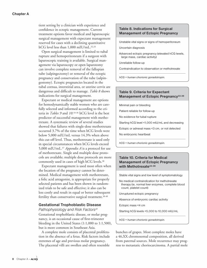
Open surgical management is limited to tubal rupture and hemoperitoneum if a surgeon with laparoscopic training is available. Surgical man-agement via laparoscopy or open laparotomy can involve complete removal of the fallopian tube (salpingectomy) or removal of the ectopic pregnancy and conservation of the tube (salpin-gostomy). Ectopic pregnancies located in the tubal cornua, interstitial area, or uterine cervix are dangerous and difficult to manage. Table 8 shows indications for surgical management.
Expectant or medical management are options for hemodynamically stable women who are care-fully selected and informed according to the cri-teria in Tables 9 and 10.33-36 hCG level is the best predictor of successful management with metho-trexate. A systematic review of several studies showed that failures with single-dose methotrexate occurred 3.7% of the time when hCG levels were below 5,000 mIU/mL versus 14.3% when above this cut-off level. Thus, methotrexate is used only in special circumstances when hCG levels exceed 5,000 mIU/mL.37 Appendix A is a protocol for use of methotrexate. Single and multiple dose proto-cols are available; multiple dose protocols are more commonly used in cases of high hCG levels.38
Expectant management is used most often when the location of the pregnancy cannot be deter-mined. Medical management with methotrexate, a folic acid antagonist, is appropriate for properly selected patients and has been shown in random-ized trials to be safe and effective; it also can be less costly and result in equal or better subsequent fertility than conservative surgical treatment.38-40
Gestational Trophoblastic DiseasePathophysiology and Risk Factors41
Gestational trophoblastic disease, or molar preg-nancy, is an occasional cause of first-trimester bleeding in the United States (1:1,000 to 1:1,500), but is more common in Southeast Asia.
A complete mole consists of placental prolifera-tion in the absence of a fetus. Risk factors include extremes of age and previous molar pregnancy. The placental villi are swollen and often resemble
bunches of grapes. Most complete moles have a 46,XX chromosomal composition, all derived from paternal sources. Mole recurrence may prog-ress to metastatic choriocarcinoma. A partial mole
Table 8. Indications for Surgical Management of Ectopic Pregnancy
Unstable vital signs or signs of hemoperitoneum
Uncertain diagnosis
Advanced ectopic pregnancy (elevated hCG levels, large mass, cardiac activity)
Unreliable follow-up
Contraindication to observation or methotrexate
hCG = human chorionic gonadotropin.
Table 9. Criteria for Expectant Management of Ectopic Pregnancy33-36
Minimal pain or bleeding
Patient reliable for follow-up
No evidence for tubal rupture
Starting hCG level <1,000 mlU/mL and decreasing
Ectopic or adnexal mass <3 cm, or not detected
No embryonic heartbeat
hCG = human chorionic gonadotropin.
Table 10. Criteria for Medical Management of Ectopic Pregnancy with Methotrexate33-36
Stable vital signs and low level of symptomatology
No medical contraindication for methotrexate therapy (ie, normal liver enzymes, complete blood count, platelet count)
Unruptured ectopic pregnancy
Absence of embryonic cardiac activity
Ectopic mass <4 cm
Starting hCG levels <5,000 to 10,000 mlU/mL
hCG = human chorionic gonadotropin.

First-Trimester Pregnancy Complications
— Chapter A 9
is a molar placenta occurring together with a fetus, which is typically nonviable. Genetic testing typi-cally reveals triploidy (69,XXY). Partial mole is less common than a complete mole, and has a lower risk of recurrence.
Signs, Symptoms, and DiagnosisThe signs and symptoms of gestational trophoblas-tic disease are shown in Table 11.
TreatmentPrompt evacuation of the uterus is the primary treatment. After evacuation of a complete mole, all patients should have serial monitoring of hCG levels for 6 months to 1 year with use of a highly effective method of contraception.42 If the hCG level plateaus or increases, then recurrence must be assumed, investigated, and treated with chemother-apy (methotrexate). Because of the relative rarity of this disease and the many possible complications, consultation is recommended when the HCG level is not falling appropriately. Theca-lutein ovarian cysts (functional ovarian cysts which are typically bilateral and caused by elevated hCG levels) do not require management and will resolve after evacu-ation of the molar tissue. Approximately 20% of women with a complete mole will experience recurrence in the form of a mole that invades the myometrium or becomes aggressively metastatic.
Grief and Psychological Management of Early Pregnancy Loss43,44 Miscarriage represents a major loss to the preg-nant woman and her family. The grief reaction that often follows is similar in intensity to that experienced after other major losses, though women experience and describe it in varied ways. Although healing will occur, the time to recovery also varies. The feelings of loss tend to be strongest in the first 6 months after the miscarriage, but can be persistent and pervasive enough to cause long-term symptoms or even affect the patient’s next pregnancy. Women at risk of a stronger grief reaction include those who experience a missed abortion, with loss at a later gestational age, with a longer time to conception of their next pregnancy, and with a critical self-perception.
Partners also experience grief with pregnancy loss. Because partners are often a primary social support for patients and attend many post-loss visits, it is important for the health care profes-
sional to include them in the care plan. Available evidence suggests that although the vast majority of women desire support from their health care professional, many do not receive the quantity or quality of support they desire. The types of interventions that are most effective in managing psychological symptoms are uncertain, but the fol-lowing approaches may be practical ways to miti-gate the normal grief after early pregnancy loss.
Acknowledge and attempt to dispel guilt. Many women think that some action on their part caused or contributed to the miscarriage. This guilt can revolve around sexual activity, food, minor trauma, physical activity, or emotional stress. Women whose losses can be ascribed to a definite cause have lower levels of anxiety and grief. Therefore, evaluation of available tissue for chromosomal anomalies is recommended when possible. Even when a definitive cause cannot be ascertained, reassurance that the patient did nothing to cause the loss is appropriate. This reassurance may need to be repeated several times. Women should be counseled that genetic or developmental errors likely occurred early in the pregnancy, and there was no possibility of the pregnancy progressing to produce a live infant. The post-loss follow-up visit is not the time to focus on modifiable risk factors that might have contributed to the loss (ie, alcohol or tobacco use). Addressing these issues before future pregnancies is
Table 11. Trophoblastic Disease Signs and Symptoms
Uterus larger than expected for gestational age
Absent fetal heartbeat
Higher than expected hCG levels (except in cases of partial mole)
Hyperemesis, pregnancy-induced hypertension at an early gestational age, and/or thyrotoxicosis
Ovarian enlargement, caused by theca-lutein cysts resulting from ovarian hyperstimulation because of high hCG levels
Vaginal bleeding in the first or early second trimester, which is often dark and may cause anemia
Grape-like vesicles are passed in cases that progress into the second trimester
hCG = human chorionic gonadotropin.

Chapter A
10 Chapter A —
indicated, but is better done after the acute trauma of loss fades. The patient’s religious belief system can be called on during this counseling.
Acknowledge and legitimize grief. Allowing patients to discuss their emotions surrounding their loss might be the most important aspect of psychological care. One study suggests that women who had a medical follow-up visit at which they were not provided an opportunity to discuss their feelings had more anxiety and depres-sion than those who had no follow-up. Patients and their partners should be allowed to cry or feel sad. Minimizing their feelings can isolate them and decrease the medical professional’s credibility. Legitimize their feelings by confirming that mis-carriage is the death of a baby. Comments such as “you can try again” or “at least it happened early” are inappropriate. Simple measures that validate grief should not be underestimated. Listening to the patient, holding her hand, or telling her how sad you feel for her can help her through this trau-matic period. The patient should be seen within 1 to 2 weeks in the office, or called on the phone a few days after the miscarriage.
Reassure about the future. Grief will fade with time. Most patients have an excellent likelihood of a subsequent normal pregnancy. With fewer than three miscarriages, the risk of miscarriage in future pregnancies is no greater than usual. It is important to explain that the next pregnancy will not need to be managed differently because of the miscarriage. This is an excellent time to encourage the initiation of a prenatal vitamin.
Counsel the patient how to tell family and friends about the miscarriage. If family members and friends knew about the pregnancy, a designated individual can inform them of the loss. This allows them to express their sympathy and provide emotional support, and may avoid embarrassing encounters in which others assume the pregnancy is progressing. If the pregnancy was unknown to family and friends, they may recognize and be concerned about external signs of grief or distress. A decision must be made whether to tell them. Informing other children in the family can also be difficult. However, families often find comfort in allowing children to share in the grieving and remembering process. Parents should be encour-aged to discuss the loss in honest and develop-mentally appropriate ways, just as they would the death of another family member.
Warn patients of the anniversary phenomenon. A recurrence of grief feelings on their due date or the anniversary of the miscarriage can occur. This can also occur at the birth of a friend’s baby or during the patient’s subsequent pregnancy. Posttraumatic stress disorder should be considered in women experiencing prolonged grieving, anxiety, or other symptoms that affect their general or reproductive functioning.
Include the partner in your psychological care. Partners often feel the pain of loss and should be included in counseling and decisions. Men’s reac-tions to pregnancy loss are more strongly influ-enced by the status of the marital relationship than are women’s. Offering couples counseling and including men in the healing process can speed resolution for both partners.
Assess level of grief and adjust counseling accord-ingly. Many women are ambivalent or distressed by the pregnancy and may experience mixed feelings or profound relief at the loss. A history of abortion, failed birth control, or rape may further complicate the emotional response. Allowing the patient to express her emotions in a supportive and nonjudgmental atmosphere is always an appropriate intervention.
Rh Prophylaxis and Future Conception after Pregnancy LossSeveral follow-up issues must be addressed after any type of pregnancy loss. Rh-negative women who miscarry during the first trimester should receive 50 µg of anti-D immune globulin.45 Con-traception should be discussed and started imme-diately if conception is not desired; all methods are equally safe immediately after spontaneous abortion or ectopic pregnancy. There is no good evidence suggesting an ideal interpregnancy inter-val.46 Folic acid supplementation before future conception attempts substantially reduces the risk of neural tube defects.47
SummaryFirst-trimester pregnancy complications are common and the differential diagnosis includes life-threatening conditions such as ectopic preg-nancy. Knowledge and application of discrimina-tory criteria can significantly aid in distinguishing among normal early pregnancy, miscarriage, and ectopic pregnancy. Medical treatment of ectopic pregnancy is possible in properly selected cases. In

First-Trimester Pregnancy Complications
— Chapter A 11
incomplete miscarriage, nonsurgical management has a high likelihood of success depending on the diagnosis. In embryonic demise or anembryonic pregnancy, misoprostol or surgical management are significantly more effective than expectant management.
Because there is a lack of clear superiority of expectant versus surgical management of mis-carriage, the woman’s preference should play a dominant role in decision-making.48 When the choice is made to manage early pregnancy failure by other than expectant means, vaginal misopro-stol is highly effective, safe, and well-accepted by women, with fewer gastrointestinal adverse effects than the oral route. Evidence does not support the use of antibiotics in all women with incomplete abortion. After any type of first-trimester preg-nancy loss, Rh-negative women should receive 50 µg of anti-D imm une globulin. Acknowledge-ment of grief and demonstrations of empathy and reassurance are useful techniques in counseling women after miscarriage.
Optional SectionSurgical Management of Miscarriage: Dilation and Curettage and Manual Vacuum AspirationThe majority of first-trimester miscarriages occur completely and spontaneously without interven-tion. And when intervention is selected, medical management is highly effective. Uterine aspiration by electric suction or manual vacuum aspiration may be indicated when:
1. Heavy bleeding is present (greater than one padper hour).
2. Patient is clinically stable (no bleeding orcramping), but pregnancy loss is shown con-clusively and the patient prefers intervention toexpectant management.
3. Ectopic pregnancy needs to be ruled out. Incertain situations, a clinical distinction cannotbe made between an ectopic and an intrauterinepregnancy. If tissue from a dilation and curet-tage (D&C) procedure contains chorionic villi,the pregnancy was intrauterine. Rarely, an intra-uterine and an ectopic pregnancy (heterotopicpregnancy) may coexist, creating a confusingand dangerous clinical situation.
Contraindications to Uterine Aspiration1. Medical contraindications are rare but include
active pelvic infection.
2. Pregnancy loss is not proven to the patient’ssatisfaction, the physician’s satisfaction, or both.
3. The patient prefers to await spontaneouscompletion for any reason (eg, religious beliefs,cost, desire to avoid surgical procedures, etc).Uterine aspiration is not appropriate if the spon-
taneous abortion appears to be complete based on the following criteria:• The uterus is small and firm.• Scant or no bleeding is occurring.• Tissue has been passed and is available for
inspection, and appears complete.• The patient is reliable for follow-up.• Ultrasound examination (preferably transvaginal)
shows an empty uterus.
Uterine Aspiration Performed Under Local Anesthesia 1. Place an intravenous (IV) line if patient is
bleeding heavily or if IV drugs will be used.
2. An Rh test should be obtained in womenwhose status is unknown. A hematocrit orhemoglobin should be obtained if there issuspicion of anemia or excessive blood loss.A white blood cell count, prothrombin time,partial thromboplastin time, fibrin splitproducts, and blood type and screen may beobtained depending on clinical circumstances(eg, heavy bleeding).
3. Sedation and analgesia should be adminis-tered. Two to 5 mg of IV midazolam and50 to 100 µg of IV fentanyl are commonlyused. Alternatively, other opioids or benzodi-azepines may be used. In many situations, thepatient’s partner or another support individualmay be present during the procedure.
4. The size and position of the uterus shouldbe identified by bimanual examination.If the fetal measurements and uterine sizeare greater than those for 14 weeks’ gesta-tion, a dilation-and-extraction procedure isindicated, which requires advanced train-ing beyond that required for first-trimester

Chapter A
12 Chapter A —
aspiration. If a procedure is required beyond 14 weeks’ gestation, consider adding 20 units of oxytocin to IV fluids.
5. The cervix should be exposed with a specu-lum. A medium Graves speculum typicallysuffices. The cervix and posterior fornix arecleansed with an antiseptic solution. Theanterior lip of the cervix is then grasped with asingle-toothed tenaculum.
6. A paracervical block can be achieved with20 cc of 1% lidocaine or another local anes-thetic agent via a 20-gauge spinal needle.One-fourth of the amount of the block isadministered at 3:00, 5:00, 7:00, and 9:00;or one-half the amount of the block at 4:00and 8:00 where the cervix meets the vagina.A superficial wheal is raised and the syringe isaspirated before injecting to avoid intravascu-lar injection. Several variants of the paracervi-cal block exist, and all are equally satisfactory.
7. If the cervix is closed, or insufficiently dilatedto easily admit the required suction curette,it can be progressively dilated using cervicaldilators. Dilation should occur to the num-ber of mm that is equivalent to the estimatedgestational age in weeks or 1 mm less (eg, dilateto 9 or 10 mm to aspirate a 10-week missedabortion). Control is indicated for this por-tion of the procedure, as dilators and uterinesounds cause the largest number of uterineperforations. If the patient is clinically stable,overnight dilation with laminaria is an option.Another means of dilation that has been suc-cessful, but is not approved by the US Foodand Drug Administration, is the buccal, sublin-gual, or vaginal administration of misoprostol400 µg 2 to 3 hours before the procedure.
8. If the os is open, ring forceps can be used toremove any loose tissue that is encountered.
9. The suction curette should be equivalent to thesize of the uterus in weeks (eg, a no. 10 curettefor a 10 weeks’ sized uterus) is appropriate. Acurved curette is used if the uterus is anteflexedor retroflexed. A straight curette can be used ifthe uterus is midposition. The suction curetteis inserted along the previously determined
axis of the uterus, until slight resistance is felt, while exerting slight traction on the tenaculum to stabilize the cervix and straighten out the cervico-vaginal angle. The curette should never be forced after passing the internal os because perforation is the most serious potential com-plication of the procedure.
10. After the curette is in place, the suction hosingis attached and the suction machine is turnedon. Then the suction valve on the handle ofthe hose. Sixty cm of mercury (Hg) or greatermust be achieved for adequate suction.
11. With the suction on, the curette is rotated sev-eral times in one direction, then several timesin the other direction, with a slight in-and-out motion. Most of the pressure should bemaintained lateral, and forceful jabbing at theuterine fundus should be avoided because per-foration is a risk. The amount and nature oftissue that appears in the plastic curette shouldbe observed carefully. Products of conceptionoften appear tan or grey, admixed with bloodand clots. Yellowish fluid can be noted. Thecurette is withdrawn slowly, avoiding the vagi-nal side wall while the suction is operating.
12. The suction and rotation sequence can berepeated after inserting the curette into theuterus again.
13. Manual vacuum aspiration is accomplishedwith a simple handheld plastic syringe thatgenerates its own suction mechanically. Thisdevice is inexpensive, easy to use, and does notrequire electricity. It is particularly appropriatefor completions of early gestations (eg, less than8 to 10 weeks’ menstrual age). It can be usedin the office setting where a suction machine isnot accessible. It is also appropriate in develop-ing countries where electricity is not available.
14. A light, sharp curettage of the uterus canbe performed to determine that it is empty,followed by one more pass of the suctioncurette. This is no longer routinely indicatedbecause of the increased pain and increasinguse of postprocedure vaginal ultrasound toconfirm completion in association with tissueexamination.

First-Trimester Pregnancy Complications
— Chapter A 13
15. After examination of the tissue, it should undergo examination for confirmation of diagnosis. To confirm an intrauterine preg-nancy, chorionic villi must be identified. The tissue must be sent to a laboratory for pathol-ogy unless the presence of villi or an embryo is conclusively confirmed by the physician performing the uterine aspiration.
16. After the uterine aspiration is completed, the patient should be monitored for excessive bleeding. Misoprostol can be administered by the rectal, buccal, or sublingual route in a dose of 400 to 800 µg. Methergine® 0.2 mg may be administered intramuscularly or orally. Trans-fusions are required rarely.
17. If the patient is Rh negative, 50 µg (mini dose) of Rh immune globulin should be administered.45
18. Doxycycline 100 mg is administered orally pre-procedure to decrease the likelihood of endometritis.
Complications of Uterine Aspiration Complications of uterine aspiration (suction D&C) can occur. Careful performance of the procedure, consultation with more experienced physicians when needed, and a high index of suspicion for identifying complications can prevent complications.
1. Perforation — Perforation occurs when an instrument passes through the uterine wall. The diagnosis is typically apparent when a sound or dilator passes through the cervix to a significantly greater depth than expected. Occasionally, a suc-tion curette or a sharp curette will draw maternal abdominal contents such as omentum or bowel out through the cervix. Heavy bleeding, signs of peritonitis, or evidence of intra-abdominal bleeding also can help identify perforation. If perforation occurs with a blunt instrument, such as a uterine sound, and if the D&C has been completed, observation alone for a minimum of 2 hours may suffice. If perforation has occurred with a sharp instrument, such as a curette or with a suction curette, laparoscopy or laparot-omy may be indicated. If the uterine aspiration has not been completed at the time the perfora-tion is identified, it can be completed under ultrasonic or laparoscopic guidance. Broad-spec-
trum antibiotics, such as a cephalosporin, should be considered for any perforation.
2. Incomplete Evacuation — Incomplete evacu-ation is identified by continued bleeding and cramping after the procedure, or ultrasound evidence of retained tissue or endometritis. Incomplete evacuation can be managed by repeating the procedure. Ultrasonic guidance or general anesthesia is often helpful. Antibiot-ics are recommended if the second procedure occurs more than a few hours after the first.
3. Bleeding — The differential diagnosis of bleed-ing includes perforation, incomplete evacuation with retained tissue, cervical or uterine injury, or a bleeding disorder. Methylergonovine 0.2 mg four times a day for 2 days is commonly administered to patients with more than average bleeding dur-ing and after a procedure. An alternative is miso-prostol 200 µg four times a day for 2 days. This is an off-label use of misoprostol, but is effective in practice because of its powerful uterotonic effect.24
4. Infection — Infection may be referred to as sep-tic abortion, endometritis, paraendometritis or pelvic peritonitis. It is diagnosed by the presence of fever, uterine and parauterine tenderness, peritonitis, and an elevated leukocyte count. Management is with antibiotics. For ill patients who require hospitalization, an IV cephalospo-rin or triple antibiotics (ampicillin, gentamycin, and clindamycin or metronidazole) may be required. Less ill patients can be treated in the outpatient setting. Clear guidelines for antibiotic regimens are lacking. When tissue is found to be retained, repeating the uterine evacuation may be necessary. Oxytocics should be administered as described previously in 2. Incomplete Evacua-tion. Rarely, in very ill patients, hospitalization and hysterectomy may be necessary.
5. Late Sequelae — Intrauterine synechiae (Asher-man syndrome) is often discussed but rarely seen. It is most likely to occur when a suction D&C is performed in the presence of infection, a prolonged missed abortion, or postpartum. Incompetent cervix can occur rarely because of cervical injury. The most common late sequelae to suction D&C are depression and related psy-chological reactions to the loss of the pregnancy.

Chapter A
14 Chapter A —
SORT: Key Recommendations for Practice
Clinical RecommendationEvidence Rating References
Pregnancy failure can be reliably diagnosed if the gestational sac is 25 mm or greater without an embryo, if an embryo fails to appear by 11 days after a yolk sac appears, or if an embryo of crown-rump length greater than 7 mm does not show a heartbeat.
C 13
Success of miscarriage management depends on diagnosis. In an incomplete miscarriage, nonsurgical management has a high likelihood of success. In an embryonic demise or anembryonic pregnancy, misoprostol or surgical management is considerably more effective than expectant management.
A 22-24
There is a lack of clear superiority of expectant versus surgical management of miscarriage. Therefore, the woman’s preference should play a dominant role in the decision-making process.
A 48
When the choice is made to manage early pregnancy failure by other than expectant means, vaginal misoprostol is highly effective and safe, and well-accepted by women, with fewer gastrointestinal adverse effects than the buccal or oral routes.
A 22,23
Evidence does not support the use of antibiotics in all women with incomplete abortion.
A 26
Women undergoing induced abortion benefit from a single dose of doxycycline to prevent infection.
A 50
After any type of first-trimester pregnancy loss, Rh-negative women should receive 50 µg of anti-D immune globulin.
C 45
Acknowledgement of grief and demonstration of empathy and reassurance are useful techniques in counseling women after miscarriage.
C 43,44
Strength of Recommendation Taxonomy (SORT)
Strength of Recommendation Definition
A • Recommendation based on consistent and good-quality patient-oriented evidence.
B • Recommendation based on inconsistent or limited-quality patient-oriented evidence.
C • Recommendation based on consensus, usual practice, opinion, disease-oriented evidence, or case series for studies of diagnosis, treatment, prevention, or screening.
Patient-oriented evidence measures outcomes that matter to patients: morbidity, mortality, symptom improvement, cost reduction, and quality of life. Disease-oriented evidence measures intermediate, physiologic, or surrogate end points that might not reflect improvement in patient outcomes (eg, blood pressure, blood chemistry, physiologic function, pathologic findings).
From Ebell MH, Siwek J, Weiss BD, et al. Strength of recommendation taxonomy (SORT): a patient-centered approach to grading evidence in the medical literature. Am Fam Physician. 2004;69(3):548-556.

First-Trimester Pregnancy Complications
— Chapter A 15
References1. Wilcox AJ, Weinberg CR, O’Connor JF, et al. Incidence of
early loss of pregnancy. N Engl J Med. 1988;319(4):189-194.
2. Cole LA, Sutton-Riley JM, Khanlian SA, Borkovskaya M, Rayburn BB, Rayburn WF. Sensitivity of over-the-counter pregnancy tests: comparison of utility and marketing mes-sages. J Am Pharm Assoc (2003). 2005;45(5):608-615.
3. Ehrenkranz JR. Home and point-of-care pregnancytests: a review of the technology. Epidemiology. 2002;13(Suppl 3):S15-S18.
4. Barnhart KT, Sammel MD, Rinaudo PF, Zhou L, HummelAC, Guo W. Symptomatic patients with an early viableintrauterine pregnancy: HCG curves redefined. ObstetGynecol. 2004;104(1):50-55.
5. Barnhart KT. Clinical practice. Ectopic pregnancy.N Engl J Med. 2009;361(4):379-387.
6. Seeber BE, Barnhart KT. Suspected ectopic pregnancy.Obstet Gynecol. 2006;107(2 Pt 1):399-413. Erratum inObstet Gynecol. 2006;107(4):955.
7. Laing FC, Frates MC, Benson CB. Ultrasound Evalua-tion During the First Trimester of Pregnancy. In: CallenPW, ed. Ultrasonography in Obstetrics and Gynecology.4th ed. Philadelphia, PA: W.B. Saunders Co; 2000.
8. Paspulati RM, Bhatt S, Nour SG. Sonographic evalu-ation of first-trimester bleeding. Radiol Clin North Am.2004;42(2):297-314. Erratum in Radiol Clin North Am.2008;46(2):437.
9. Kadar N, Bohrer M, Kemmann E, Shelden R. The dis-criminatory human chorionic gonadotropin zone forendovaginal sonography: a prospective, randomizedstudy. Fertil Steril. 1994;61(6):1016-1020.
10. Whitworth M, Bricker L, Neilson JP, Dowswell T.Ultrasound for fetal assessment in early pregnancy.Cochrane Database Syst Rev. 2010;(4):CD007058.
11. Chen BA, Creinin MD. Contemporary management ofearly pregnancy failure. Clin Obstet Gynecol. 2007;50(1):67-88.
12. Goddijn M, Leschot NJ. Genetic aspects of miscar-riage. Baillieres Best Pract Res Clin Obstet Gynaecol.2000;14(5):855-865.
13. Doubilet PM, Benson CB, Bourne T, et al; Society ofRadiologists in Ultrasound Multispecialty Panel on EarlyFirst Trimester Diagnosis of Miscarriage and Exclusionof a Viable Intrauterine Pregnancy. Diagnostic criteriafor nonviable pregnancy early in the first trimester.N Engl J Med. 2013;369(15):1443-1451.
14. Smith KE, Buyalos RP. The profound impact of patientage on pregnancy outcome after early detection of fetalcardiac activity. Fertil Steril. 1996;65(1):35-40.
15. Creinin MD, Harwood B, Guido RS, Fox MC, ZhangJ; NICHD Management of Early Pregnancy FailureTrial. Endometrial thickness after misoprostol usefor early pregnancy failure. Int J Gynaecol Obstet.2004;86(1):22-26.
16. Eschenbach DA. Treating spontaneous and inducedseptic abortions. Obstet Gynecol. 2015;125(5):1042-1048.
17. Stubblefield PG, Grimes DA. Septic abortion. N Eng JMed. 1994;331(5):310-314.
18. Bennett GL, Bromley B, Lieberman E, Benacerraf BR.Subchorionic hemorrhage in first-trimester pregnancies:prediction of pregnancy outcome with sonography.Radiology. 1996;200(3):803-806.
19. Hasan R, Baird DD, Herring AH, Olshan AF, JonssonFunk ML, Hartmann KE. Association between first-trimester vaginal bleeding and miscarriage. [Level II].Obstet Gynecol. 2009;114(4):860-867.
20. Luise C, Jermy K, May C, Costello G, Collins WP,Bourne TH. Outcome of expectant management ofspontaneous first trimester miscarriage: observationalstudy. BMJ. 2002;324(7342):873-875.
21. Allison JL, Sherwood RS, Schust DJ. Management offirst trimester pregnancy loss can be safely moved intothe office. Rev Obstet Gynecol. 2011;4(1):5-14.
22. Zhang J, Gilles JM, Barnhart K, Creinin MD, Westhoff C,Frederick MM; National Institute of Child Health HumanDevelopment (NICHD) Management of Early PregnancyFailure Trial. A comparison of medical managementwith misoprostol and surgical management for earlypregnancy failure. N Engl J Med. 2005;353(8):761-769.
23. Bagratee JS, Khullar V, Regan L, Moodley J, Kagoro H.A randomized controlled trial comparing medical andexpectant management of first trimester miscarriage.Hum Reprod. 2004;19(2):266-271.
24. Weeks A, Alia G, Blum J, et al. A randomized trial ofmisoprostol compared with manual vacuum aspirationfor incomplete abortion. Obstet Gynecol. 2005;106(3):540-547.
25. Winikoff B. Pregnancy failure and misoprostol—time fora change. N Engl J Med. 2005;353(8):834-836.
26. May W, Gülmezoglu AM, Ba-Thike K. Antibiotics forincomplete abortion. Cochrane Database Syst Rev.2007;(4):CD001779.
27. Fjerstad M, Trussell J, Sivin I, Lichtenberg ES, Cullins V.Rates of serious infection after changes in regimens formedical abortion. N Engl J Med. 2009;361(2):145-151.
28. Buss L, Tolstrup J, Munk C, et al. Spontaneous abor-tion: a prospective cohort study of younger womenfrom the general population in Denmark. Validation,occurrence and risk determinants. Acta Obstet GynecolScand. 2006;85(4):467-475.
29. Grimes DA, Lopez LM, Schulz KF, Stanwood NL.Immediate postabortal insertion of intrauterine devices.Cochrane Database Syst Rev. 2010;(6):CD001777.
30. Sawyer E, Jurkovic D. Ultrasonography in the diagnosisand management of abnormal early pregnancy. ClinObstet Gynecol. 2007;50(1):31-54.
31. Connolly A, Ryan DH, Stuebe AM, Wolfe HM. Reevalu-ation of discriminatory and threshold levels for serumβ-hCG in early pregnancy. Obstet Gynecol. 2013;121(1):65-70.
32. van Mello NM, Mol F, Ankum WM, Mol BW, van derVeen F, Hajenius PJ. Ectopic pregnancy: how the diag-nostic and therapeutic management has changed. FertilSteril. 2012;98(5):1066-1073.

Chapter A
16 Chapter A —
33. Cohen MA, Sauer MV. Expectant management of ecto-pic pregnancy. Clin Obstet Gynecol. 1999;42(1):48-54,quiz 55-56.
34. Mashiach S, Carp HJ, Serr DM. Nonoperative manage-ment of ectopic pregnancy. A preliminary report.J Reprod Med. 1982;27(3):127-132.
35. Adoni A, Milwidsky A, Hurwitz A, Palti Z. Decliningbeta-HCG levels: an indicator for expectant approach inectopic pregnancy. Int J Fertil. 1986;31(1):40-42.
36. Garcia AJ, Aubert JM, Sama J, Josimovich JB. Expect-ant management of presumed ectopic pregnancies.Fertil Steril. 1987;48(3):395-400.
37. Menon S, Colins J, Barnhart KT. Establishing a humanchorionic gonadotropin cutoff to guide methotrexatetreatment of ectopic pregnancy: a systematic review.Fertil Steril. 2007;87(3):481-484.
38. American College of Obstetricians and Gynecolo-gists. ACOG Practice Bulletin No. 94: Medical man-agement of ectopic pregnancy. Obstet Gynecol.2008;111(6):1479-1485.
39. Stovall TG, Ling FW. Single-dose methotrexate: anexpanded clinical trial. Am J Obstet Gynecol. 1993;168(6Pt 1):1759-1762, discussion 1762-1765.
40. Lipscomb GH, McCord ML, Stovall TG, Huff G, PorteraSG, Ling FW. Predictors of success of methotrexatetreatment in women with tubal ectopic pregnancies. NEngl J Med. 1999;341(26):1974-1978.
41. Lurain JR. Gestational trophoblastic disease I: epidemi-ology, pathology, clinical presentation and diagnosis ofgestational trophoblastic disease, and management ofhydatidiform mole. Am J Obstet Gynecol. 2010;203(6):531-539.
42. Berkowitz RS, Goldstein DP. Clinical practice. Molarpregnancy. N Engl J Med. 2009;360(16):1639-1645.
43. Brier N. Grief following miscarriage: a comprehensivereview of the literature. J Womens Health (Larchmt).2008;17(3):451-464.
44. Murphy FA, Lipp A, Powles DL. Follow-up for improvingpsychological well being for women after a miscarriage.Cochrane Database Syst Rev. 2012;3:CD008679.
45. American College of Obstetrics and Gynecology.ACOG practice bulletin. Prevention of Rh D alloimmuni-zation. Number 4, May 1999 (replaces educational bul-letin Number 147, October 1990). Clinical managementguidelines for obstetrician-gynecologists. Int J Gynae-col Obstet. 1999;66(1):63-70.
46. Wong LF, Schliep KC, Silver RM, et al. The effect ofa very short interpregnancy interval and pregnancyoutcomes following a previous pregnancy loss. Am JObstet Gynecol. 2015;212(3):375.e1-375.e11.
47. Cordero AM, Crider KS, Rogers LM, Cannon MJ, BerryRJ. Optimal serum and red blood cell folate concentra-tions in women of reproductive age for prevention of neu-ral tube defects: World Health Organization guidelines.MMWR Morb Mortal Wkly Rep. 2015;64(15):421-423.
48. Nanda K, Lopez LM, Grimes DA, Peloggia A, NandaG. Expectant care versus surgical treatment for miscar-riage. Cochrane Database Syst Rev. 2012;3:CD003518.
49. Barnhart KT; Supplement to. Clinical practice. Ectopicpregnancy. N Engl J Med. 2009;361(4):379-387.
50. Achilles SL, Reeves MF; Society of Family Planning.Prevention of infection after induced abortion: releasedate October 2010: SFP guideline 20102. Contracep-tion. 2011;83(4):295-309.

— Chapter B 1
Chapter B Medical Complications of Pregnancy
IntroductionThe presence of a fetus complicates many medi-cal problems as a result of the complex interactions between the mother, the disease, and the treatment. Understanding these interactions is crucial in optimiz-ing outcomes for both mother and baby. The mother is the first priority in any medical emergency since the fetus is dependent on her for physiologic sup-port. In managing an eclamptic seizure or pulmonary embolism, for example, every effort is directed toward supporting maternal vital functions and using neces-sary critical care interventions. Concern for the fetus is demonstrated by choosing expectant management for preeclampsia without severe features when the fetal gestational age is < 37 weeks, treating the mother with medications that minimize adverse fetal effects, and by choosing tests that limit direct fetal x-ray exposure. This chapter focuses on four potentially life-threatening medical complications: hypertensive disorders, acute fatty liver of pregnancy (AFLP), peripartum cardio-myopathy (PPCM), and thromboembolic disease. The hypertensive disorders are the most common medical complications of pregnancy, while AFLP and PPCM are uncommon disorders unique to pregnancy that cause significant morbidity and mortality. Thrombo-embolic disease is a leading cause of maternal mortality in more developed countries.
Hypertensive Disorders of PregnancyWorldwide, hypertensive disorders represent the most common medical complication of pregnancy, affect-
ing up to 10 percent of gestations.1 As defined by the National High Blood Pressure Education in Pregnancy (NHBPEP) Working Group, hypertension in preg-nancy may be chronic (occurring prior to 20 weeks’ ges-tation or persisting beyond 12 weeks post partum), may arise de novo during the pregnancy (gestational hyper-tension or preeclampsia), or may represent a superim-position of preeclampsia on chronic hypertension.1,2
Chronic HypertensionChronic hypertension is defined as an elevated blood pressure greater than 140/90 mm Hg on two occasions at least four hours apart prior to or during the first 20 weeks of pregnancy. Chronic hypertension is associated with adverse perinatal outcomes including preeclamp-sia, intrauterine growth restriction and placental abrup-tion. The severity of maternal blood pressure at 20 weeks is associated with worse outcomes.3 Treatment of mild to moderate chronic hypertension in pregnancy has no proven fetal benefit, nor has it been shown to prevent preeclampsia.4,5 Excessively lowering the blood pressure may result in decreased placental perfusion and adverse perinatal outcomes.6 However, when the blood pressure is persistently greater than 150 to 160/100 to 110 mm Hg, pharmacologic treatment is indicated in order to prevent maternal end organ damage.1,2,7 A lower threshold is appropriate for treating women who already manifest target organ damage such as renal insufficiency and left ventricular hypertrophy.1
Methyldopa, labetalol, and nifedipine are the oral agents most commonly used for severe, chronic hyper-
Learning ObjectivesAt the end of this activity, learners will be able to:1. Describe the following potentially life-threatening
medical complications of pregnancy:a. Hypertensive disorders.b. Acute fatty liver of pregnancy.
c. Peripartum cardiomyopathy.d. Deep venous thrombosis.e. Pulmonary embolism.
2. Formulate a plan for diagnosis and treatment.
Larry Leeman, MD, MPH, Lee Dresang, MD, Patricia Fontaine, MD
Revised February 2015
Copyright 2017© American Academy of Family Physicians. All rights reserved.
Chapter B
2 Chapter B —
tension in pregnancy in the 2013 report of the American College of Obstetricians and Gynecolo-gists (ACOG) Hypertension in Pregnancy task force.2 ACE inhibitors and angiotensin II receptor antagonists should not be used due to associa-tion with intrauterine growth restriction (IUGR), neonatal renal failure, oligohydramnios, effects of oligohydramnios (including limb abnormalities, cranial ossification defects and pulmonary hyper-plasia) and death.2 The beta-blocker atenolol, has been associated with IUGR.2 Thiazide diuret-ics may be continued if used prior to pregnancy however they can exacerbate the intravascular fluid depletion of preeclampsia if chronic hyper-tension becomes complicated by superimposed preeclampsia and in the scenario must be discon-tinued.8,9 For this reason thiazide diuretics are not first line antihypertensive agents for chronic hypertension in pregnancy.
Women in active labor with uncontrolled severe chronic hypertension require treatment with intravenous labetalol or hydralazine in doses similar to those used for preeclampsia with severe features as described below.9 Although intrave-nous medications have traditionally been recom-mended rather than oral medications, a small 2013 RCT demonstrated a more rapid lowering of blood pressure with oral nifedipine compared to intravenous labetalol10 and oral nifedipine is considered to be a therapeutic option for severe acute hypertension in the 2013 ACOG Task Force recommendations.2
Women with chronic hypertension should be monitored carefully for the development of super-imposed preeclampsia and IUGR.2 The development of proteinuria or a sudden sustained increase in proteinuria, a sudden increase in blood pressure in a woman whose hypertension has previously been well controlled, or development of the severe features of preeclampsia (RUQ pain, headache, visual changes, pulmonary edema, rise in creatinine or transaminases, or development of thrombocytopenia (< 100,000/mcL), are diagnos-tic for superimposed preeclampsia.2 Fetal growth should be assessed by serial ultrasounds starting after 24 weeks of gestation to screen for develop-ing IUGR.2 Although we lack evidence for an optimal interval for fetal growth ultrasound assess-ments, every four weeks is a reasonable option if no evidence of intrauterine growth restriction or superimposed preeclampsia.
Gestational HypertensionThe NHBPEP Working Group has recommended that “gestational hypertension” replace the term “pregnancy-induced hypertension.”1 Pregnant women who develop hypertension after 20 weeks and do not have significant, preeclampsia-level pro-teinuria should be diagnosed with gestational hyper-tension. Gestational hypertension is a provisional diagnosis used for a heterogeneous group of women including 1) those that will eventually develop proteinuria during the pregnancy and be diagnosed with preeclampsia, 2) those who will have persistent hypertension after 12 weeks postpartum and be diagnosed with chronic hypertension, and 3) those who do not develop preeclampsia and whose blood pressures normalize postpartum. Women in the last group are ultimately diagnosed as having had “transient hypertension of pregnancy.”
Gestational hypertension is not a benign cat-egory. Approximately 50 percent of women diagnosed with gestational hypertension between 24 to 35 weeks ultimately develop preeclampsia.11 Expectant management of gestational hyperten-sion can reduce the increased cesarean delivery rate that occurs with inductions.12 If the blood pressure progresses to the severe range (systolic greater than 160 mm Hg or diastolic greater than 110 mm Hg), then management similar to a preeclamptic with severe features is required even if the patient does not have proteinuria, because women with severe gestational hypertension have worse perinatal outcomes than women with preeclampsia without severe features.13 ACOG recommends induction at 37 weeks if not delivered sooner.8 A retrospective analysis of women with gestation hypertension, mild preeclampsia and mild chronic hypertension demonstrated higher rates of maternal ICU admis-sion, postpartum hemorrhage and blood transfu-sion in the gestational hypertension group.14
PreeclampsiaDefinitionsPreeclampsia is a multi-organ disease process char-acterized by hypertension and either proteinuria or severe features of preeclampsia. To meet diagnos-tic criteria for preeclampsia, systolic blood pressure must be 140 mm Hg or greater, or diastolic blood pressure 90 mm Hg or greater, on at least two occasions no less than fours hours apart.2 Blood pressure should be measured at each prenatal visit using an appropriate sized cuff with the patient
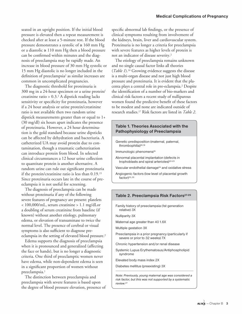
Medical Complications of Pregnancy
— Chapter B 3
seated in an upright position. If the initial blood pressure is elevated then a repeat measurement is checked after at least a 5-minute rest. If the blood pressure demonstrates a systolic of ≥ 160 mm Hg or a diastolic ≥ 110 mm Hg then a blood pressure can be confirmed within minutes and the diag-nosis of preeclampsia may be rapidly made. An increase in blood pressure of 30 mm Hg systolic or 15 mm Hg diastolic is no longer included in the definition of preeclampsia2 as similar increases are common in uncomplicated pregnancies.
The diagnostic threshold for proteinuria is 300 mg in a 24-hour specimen or a urine protein/creatinine ratio > 0.3.2 A dipstick reading lacks sensitivity or specificity for proteinuria, however if a 24 hour analysis or urine protein/creatinine ratio is not available then two random urine dipstick measurements greater than or equal to 1+ (30 mg/dl) six hours apart indicates the presence of proteinuria. However, a 24 hour determina-tion is the gold standard because urine dipsticks can be affected by dehydration and bacteriuria. A catheterized UA may avoid protein due to con-tamination, though a traumatic catheterization can introduce protein from blood. In selected clinical circumstances a 12 hour urine collection to quantitate protein is another alternative. A random urine can rule out significant proteinuria if the protein/creatinine ratio is less than 0.19.15 Since proteinuria occurs late in the course of pre-eclampsia it is not useful for screening.
The diagnosis of preeclampsia can be made without proteinuria if any of the following severe features of pregnancy are present: platelets < 100,000/mL, serum creatinine > 1.1 mg/dLor a doubling of serum creatinine from baseline (if known) without another etiology, pulmonary edema, or elevation of transaminase to twice the normal level. The presence of cerebral or visual symptoms is also sufficient to diagnose pre-eclampsia in the setting of elevated blood pressure.2
Edema supports the diagnosis of preeclampsia when it is pronounced and generalized (affecting the face or hands), but is no longer a diagnostic criteria. One third of preeclamptic women never have edema, while non-dependent edema is seen in a significant proportion of women without preeclampsia.1
The distinction between preeclampsia and preeclampsia with severe features is based upon the degree of blood pressure elevation, presence of
specific abnormal lab findings, or the presence of clinical symptoms resulting from involvement of the kidneys, brain, liver and cardiovascular system. Proteinuria is no longer a criteria for preeclampsia with severe features as higher levels of protein is not an indicator of disease severity.2
The etiology of preeclampsia remains unknown and no single causal factor links all theories (Table 1).16 Growing evidence suggests the disease is a multi-organ disease and not just high blood pressure and proteinuria. It is evident that the pla-centa plays a central role in pre-eclampsia.2 Despite the identification of a number of bio-markers and clinical risk factors a recent study of nulliparous women found the predictive benefit of these factors to be modest and none are indicated outside of research studies.17 Risk factors are listed in Table 2.
Table 1. Theories Associated with the Pathophysiology of Preeclampsia
Genetic predisposition (maternal, paternal, thrombophilia)18,19
Immunologic phenomena19
Abnormal placental implantation (defects in trophoblasts and spiral arterioles)22-23
Vascular endothelial damage24 and oxidative stress
Angiogenic factors (low level of placental growth factor)25,26
Table 2. Preeclampsia Risk Factors27-29
Family history of preeclampsia (1st generation relative) 3X
Nulliparity 3X
Maternal age greater than 40 1.6X
Multiple gestation 3X
Preeclampsia in a prior pregnancy (particularly if severe or prior to 32 weeks) 7X
Chronic hypertension and/or renal disease
Systemic Lupus Erythematosus/Antiphospholipid syndrome
Elevated body mass index 2X
Diabetes mellitus (preexisting) 3X
Note: Previously, young maternal age was considered a risk factor, but this was not supported by a systematic review.27

Chapter B
4 Chapter B —
PreventionRandomized controlled trials fail to support a role for routine prenatal supplementation with calcium, omega three fatty acids, or antioxidant vitamins E and C to prevent preeclampsia.28,30-35 Calcium supplementation may decrease the inci-dence of hypertension, preeclampsia and maternal death among women at high risk of developing those conditions and women with low calcium intakes.36 The World Health Organization has recommend routine supplementation with of 1.5 to 2.0 grams of elemental calcium for women with low calcium intake.37 Women in the United States or other high resource countries are unlikely to have low calcium intake due to the widespread supplementation of food and the ACOG 2013
task force recommended against supplementation except in populations with low calcium intake.2,36
An association has been shown between low levels of Vitamin D and the subsequent develop-ment of preeclampsia however it is unknown if supplementation will decrease the incidence of preeclampsia. Antiplatelet agents (e.g. low dose aspirin), have small to moderate benefits for pre-vention. A Cochrane analysis of low-dose aspirin for women at increased risk for preeclampsia dem-onstrated a 17% reduction in the risk of develop-ing preeclampsia with a NNT of 72 to prevent one case of preeclampsia. In the subgroup of women at highest risk due to a history of previous pre-eclampsia with severe features, diabetes, chronic hypertension, renal or autoimmune disease, only 19 women needed to be treated with low-dose aspirin to prevent one case of preeclampsia.38
The 2013 ACOG taskforce recommended initiation of 60 to 80 mg of aspirin in the late first trimester in women with history of preeclampsia in a prior pregnancy requiring delivery before 34 0/7 weeks or had preeclampsia in more than one prior pregnancy.2
Management of Preeclampsia without Severe FeaturesExpectant management of women with pre-eclampsia without severe features may include twice weekly blood pressures, weekly lab tests (CBC, ALT and/or AST, LDH, uric acid, and creatinine), twice weekly non-stress tests (NSTs) and weekly amniotic fluid indices (AFIs) or weekly biophysical profiles (BPPs), and ultrasounds for fetal growth every three weeks.1,2,39 Fetal umbili-cal artery Doppler studies are recommended to be included in antenatal surveillance for women with preeclampsia when IUGR has been detected.2
The decision to bring about delivery by induc-tion or cesarean section involves balancing pre-maturity-related risks with the risk of worsening preeclampsia. Delivery is generally indicated for women with preeclampsia or gestational hyperten-sion at 37 weeks based on the HYPITAT RCT of induction vs., expectant management and the recommendations of a 2011 workshop sponsored by the Society of Maternal Fetal Medicine and the National Institute of Child Health and Human Development.40.41 A secondary analysis of HYPI-TAT showed greater benefit of labor induction on preventing high-risk maternal situations and
Figure 1. Recommended Management of Gestational Hypertension or Preeclampsia Without Severe Features39
Information from Roberto, JM, August, PA, Bakris, G, et al. Task force on Hyper-tension in Pregnancy: American College of Obstetrics and Gynecology, 2013.
Maternal and fetal findings
37 0/7 weeks or more of gestationWorsening maternal or fetal conditionLabor or premature rupture of
membranes
Less than 37 0/7 weeks of gestationInpatient or outpatient management
Maternal evaluation: twice weekly Fetal evaluation
With preeclampsia: twice weekly nonstress test
With gestational hypertension: weekly nonstress test
37 0/7 weeks or more of gestation or
37 0/7 weeks or more of gestation with:Labor or rupture of membranesAbnormal maternal-fetal test resultsUltrasonographic estimate of fetal
weight less than fifth percentileSuspected abruptio placentae
DeliveryProstaglandins
if needed for induction
YesNo
Yes

Medical Complications of Pregnancy
— Chapter B 5
reducing the cesarean delivery rate in women with an unfavorable cervical exam presumably because these women were more remote from spontane-ous labor.42 An economic analysis of HYPITAT demonstrated cost saving from labor induction as compared to expectant monitoring.43
Severe Features of PreeclampsiaDiagnostic criteria for preeclampsia with severe features are listed in Table 3. Preeclampsia with severe features may result in multi-system dete-rioration that can be gradual or fulminant. Severe headache, visual disturbances, and progressive hyperreflexia may signal impending generalized seizures (eclampsia). Increasing peripheral vascu-lar resistance stresses the cardiovascular system, and pulmonary edema may result. A decreased glomerular filtration rate may progress to oliguria and acute renal failure. Hemodilution usually lowers pregnancy creatinine levels; levels above 0.9 mg/dl in pregnancy are abnormal. Liver mani-festations include elevated transaminases, sub-capsular hemorrhage with right upper quadrant pain, and capsular rupture with life-threatening intraabdominal bleeding. Preeclampsia-related coagulopathies include HELLP syndrome and disseminated intravascular coagulation (DIC). Obstetric complications include IUGR, abrup-tion, and fetal or maternal demise.
Management of Preeclampsia with Severe FeaturesThe progression of preeclampsia is only reversed by delivery. Patients with preeclampsia with severe features should be admitted to the hospital, placed on bedrest, and carefully monitored.39 The overall
treatment goals are to 1) prevent seizures, 2) lower blood pressure in order to prevent maternal cere-bral hemorrhage, and 3) expedite delivery based on a decision that takes into account disease sever-ity and fetal maturity.
Maternal Evaluation and StabilizationSample admitting orders for preeclampsia with severe features are outlined in Table 4. Fluid management requires special care. Excessive fluid
Table 3. Diagnostic Criteria for Severe Preeclampsia with Severe Features
Blood pressure equal to or exceeding 160 mm Hg systolic or 110 mm Hg diastolic on at least two occasions four hours apart
Any of the following signs and symptoms:Progressive renal insufficiency (Serum creatinine
> 1.1 mg/dL or double baseline)Cerebral or visual disturbancesPulmonary edemaImpaired liver function (transaminases 2x normal),
right upper quadrant pain or epigastric painThrombocytopenia (< 100,000/ml)
Table 4. Admitting Orders for Severe Preeclampsia with Severe Features
Bed rest with seizure precautions
Vital signs (blood pressure, pulse, respiration), deep tendon reflexes, and neurologic checks every 15 minutes until stable
Accurate intake and output; Foley catheter if needed
Intravenous: Lactated Ringer at 50 to 125 ml per hour to maintain urine output of 30 to 40 ml per hour. Total intake (intravenous and oral) should not exceed 125 ml per hour or 3000 ml per day
External monitor for contractions and fetal heart rate
Labs:Dipstick urine for protein on admission (not needed if have definitive
finding of severe features)Begin 24 hour urine for total protein (not needed if have definitive finding
of severe features)Complete blood count and platelet countCreatinineAST or ALTUric acidLDHPeripheral blood smear
Medications:1. Magnesium sulfate (see Table 5 for dosing)2. For systolic BP ≥ 160 mm Hg or diastolic BP ≥ 110 mm Hg on two
measurements 15 minutes apart, give one of the following:Hydralazine 5 to 10 mg IV over two minutes, if after 20 minutes BP
remains elevated, then give additional 10 mg IV. If above threshold after 20 minutes then change to IV Labetalol.9
orLabetalol 20 mg IV initial dose over 2 minutes. If BP remains elevated
after 10 minutes, double the dose to 40 mg and then to 80 mg at 10 to 30 minute intervals until target blood pressure is reached. If SBP is ≥ 160 mm Hg or DPB ≥ 100 mm Hg after the 80 mg dose, then change to IV Hydralazine.1,39 The maximum dose of IV labetalol is 300 mg in a 24 hour period.9
orNifedipine 10 to 20 mg orally. May repeat dose every 30 minutes if
needed until 40 mg po has been given then administer 10 to 20 mg po every 4 to 6 hours.
3. Calcium gluconate one gram IV: keep at bedside in case of respiratorydepression due to magnesium sulfate.

Chapter B
6 Chapter B —
administration can result in pulmonary edema, ascites and cardiopulmonary overload, while too little fluid can exacerbate an already con-stricted intravascular volume and lead to further end-organ ischemia. Urine output should be maintained above 30 ml per hour using intra-venous lactated ringers or normal saline.44 Total intravenous fluid intake should be limited to 100 ml per hour44,45 and total oral and intrave-nous fluid intake should not exceed 125 ml per hour or 3,000 ml per day. A Foley catheter allows accurate monitoring of urine output. A Swan-Ganz catheter may optimize fluid management if pulmonary edema and renal failure are present but should not be routinely used.44
Plasma volume is reduced among women with preeclampsia, suggesting that increasing plasma volume with colloid solution might improve uteroplacental circulation, and perinatal outcomes. However, risk/benefit data regarding this practice is lacking.46
In addition to the basic laboratory investigation for preeclampsia, the woman with signs of severe disease may be evaluated with, LDH, peripheral blood smear and labs for evidence of hemolysis and disseminated intravascular coagulation (DIC) depending upon clinical scenario.
Magnesium Sulfate (MgSO4)Magnesium sulfate helps prevent seizures in women with preeclampsia47-49 and is more effec-tive in preventing recurrent seizures in eclamptic patients than phenytoin, diazepam or a lytic cock-tail (chlorpromazine, promethazine and meperi-dine).48,50-53 The Magpie trial demonstrated that 63 women with severe preeclampsia need to receive magnesium sulfate prophylaxis to prevent one eclamptic seizure.47
Should magnesium sulfate be used for women with preeclampsia without severe features? Assuming 50 percent of seizures are prevent-able by MgSO4,47 then 400 women with mild preeclampsia need to be treated to prevent one eclamptic seizure.54 The 2013 ACOG Task Force report recommended that preeclamptic women who are not symptomatic and have blood pressure under 160/110 mm Hg should not universally receive magnesium sulfate for seizure prophylaxis. However, blood pressure is only mildly elevated in 30 to 60 percent of women who develop eclampsia;55 Women with preeclampsia without
severe features should be monitored closely and magnesium started if they develop severe features.2
Magnesium sulfate works by slowing neuromus-cular conduction and depressing central nervous system irritability. It does not have significant effects on lowering blood pressure. A quarter of women have side effects, most commonly flush-ing.56 Table 5 presents a standard dosing regimen.
Magnesium sulfate is excreted by the kidneys. Women with normal renal function do not require routine monitoring of serum magnesium levels, however women with absent reflexes, elevated serum creatinine or decreased urine output (< 30 cc/hr), should have magnesium levels drawn every six hours after the loading dose has been given and the infusion rate adjusted accordingly.55,59
Magnesium toxicity can lead to respiratory paralysis, central nervous system depression and cardiac arrest. With magnesium overdose, vital functions are lost in a predictable sequence. If DTRs are present, magnesium concentrations are rarely toxic.59 The magnesium sulfate infusion should be discontinued and a magnesium level checked immediately if absent DTRs are noted, the respiratory rate is less than 12 per minute, or urine output is less than 30 ml per hour.55,59 Maternal deaths have resulted from overdoses due to administration of improperly prepared solu-tions.60 The antidote for magnesium sulfate over-dose is one gram of calcium gluconate (10 ml of a 10 percent solution) infused intravenously over two minutes.44 Avoid rapid intravenous admin-istration or extravasation. Use calcium gluconate
Table 5. Magnesium Sulfate in Preeclampsia with Severe Features or Severe Gestational Hypertension57
Loading dose: four to six grams mixed in 100 ml, given IV over 15 to 20 minutes, followed by a continuous infusion of two grams per hour
Monitor:ReflexesMental statusRespiratory statusUrine output
Magnesium levels (therapeutic range = 4 to 8 mg/dl)58 should be checked if renal dysfunction elevated creatinine 70.9 mg/dL, urine output < 30 cc/hr, loss of reflexes or other symptoms of magnesium toxicity

Medical Complications of Pregnancy
— Chapter B 7
with caution in women with renal failure, severe hypophosphatemia, or acidosis.
Antihypertensive MedicationsThe optimal level of blood pressure control in pregnancies complicated by hypertension is unknown.61,62 Less tight control may decrease the risk of infants being small for gestational age, but may potentially increase the risk of respiratory distress syndrome, severe hypertension, antenatal hospitalization, and proteinuria at delivery.6,61 Although traditional recommendations are based on diastolic blood pressures, a retrospective review of 28 women with preeclampsia with severe fea-tures who experienced a cerebrovascular accident demonstrated that over 90 percent had systolic BP over 160 mm Hg, but only 12.5 percent had diastolic BP over 110 mm Hg.63
There are several possible choices for the antihy-pertensive agent depending on whether the goal is acute or chronic control. For acute management, intravenous labetalol and hydralazine are com-monly used.1,64 Doses for intravenous labetalol and hydralazine and oral nifedipine are given in Table 4, which is based on the 2013 ACOG report on Hypertension in Pregnancy.2 A Cochrane review of medications for treating severe hyperten-sion in pregnancy showed no evidence that one agent had superior effectiveness.64 The role of hydralazine as a first line choice has been ques-tioned by a meta-analysis showing more maternal hypotension, tachycardia, and headaches com-pared to other antihypertensives.62 The need for intravenous antihypertensives, either in repeated doses or by continuous infusion, indicates an unstable patient who is likely to need continuous monitoring and careful management.
Oral nifedipine or labetalol are alternatives to intravenous medications when severe range blood pressures require treatment. Traditionally intra-venous medications have been preferred to allow for rapid lowering of blood pressure and careful titration to avoid maternal and fetal effects of an excessive decrease in blood pressure. In two studies nifedipine has been shown to control blood pressure more rapidly than intravenous labetelol,10,65 and a third trial demonstrated equivalent time to adequate blood pressure control.66 Nifedipine has been shown to cause a greater increase in both cardiac index66 and urinary output65 than labetalol, as well as a decreasing systemic vascular resistance. The use of
these three antihypertensive agents is supported by a 2013 ACOG task force report on HTN in preg-nancy,2 the United Kingdom’s NICE guidelines7 and a Cochrane review.64 Oral labetalol at a dose of 200 mg is considered an alternative by ACOG for lowering severe blood pressure when intravenous medications are not an option and is recommended in the NICE guidelines.7,9 If the blood pressure remains ≥ 160/110 mm Hg and intravenous medi-cations are still not an option then 200 mg oral labetalol can be repeated.9 We recommend that each maternity care unit choose a single first line medica-tion as well as have alternatives available for women who are refractory to the selected agent.
For women with preeclampsia with severe features undergoing expectant management at a gestational age under 34 weeks, oral labetalol and nifedipine are acceptable options.39 Delivery is recommended for women with preeclampsia with severe features at 34 0/7 weeks or greater.2
Fetal SurveillanceAssessment for uteroplacental insufficiency may be achieved utilizing non-stress tests (NSTs), amni-otic fluid measurements and biophysical profiles. Umbilical artery Doppler systolic-to-diastolic ratios may detect early uteroplacental insufficiency and this examination is indicated for fetuses with intrauterine growth restriction. The presence of reversed end diastolic umbilical artery flow is an indication for delivery (after corticosteroids are administered if < 34 weeks EGA).2 Monitoring frequency varies depending on the clinical context. A common regimen for preeclampsia without severe features at less than 37 0/7 weeks includes twice weekly NSTs and weekly measurement of amniotic fluid index with biophysical profile for follow-up of non-reactive NSTs.1,39 Ultrasound for assessment of fetal growth should be repeated every three weeks.2 Women with gestational hypertension at less than 37 0/7 weeks may receive antenatal surveillance with weekly NSTs and amniotic fluid volume measurement. Women with preeclampsia with severe features are admitted to a hospital and may receive daily monitoring. Corticosteroids are administered to accelerate lung maturity for fetuses between 24 and 34 weeks gestation, either betamethasone (two doses of 12 mg given intramuscularly 24 hours apart) or dexamethasone (four doses of 6 mg given intra-muscularly 12 hours apart).39

Chapter B
8 Chapter B —
Delivery Decisions in Preeclampsia with Severe FeaturesDelivery is the only known cure for preeclamp-sia. Decisions regarding the timing and mode of delivery are based on a combination of maternal and fetal factors. Fetal factors include gestational age, evidence of lung maturity and signs of fetal
compromise on antenatal assessment. Maternal fac-tors include the degree to which the hypertension is controllable and any clinical or laboratory signs of impending decompensation. For patients with resistant severe hypertension, eclampsia pulmo-nary edema, placental abruption or other signs of maternal or fetal deterioration delivery is indicated
Figure 2. Management of Preeclampsia and HELLP Syndrome
HELLP = hemolysis, elevated liver enzymes, and low platelet count.
From Roberto, JM, August, PA, Bakris, G, et al. Task force on Hypertension in Pregnancy: American College of Obstetrics and Gynecology, 2013.
• Observe in labor and delivery for first 24 to 48 hours• Corticosteroids, magnesium sulfate prophylaxis, and anti-
hypertensive medications• Ultrasonography, monitoring of fetal heart rate, symptoms,
and laboratory tests
• Achievement of 34 0/7 weeks of gestation• New-onset contraindications to expectant management• Abnormal maternal-fetal test results• Labor or premature rupture of membranes
Expectant management• Facilities with adequate maternal and neonatal intensive care
resources• Fetal viability—33 6/7 weeks of gestation• Inpatient only and stop magnesium sulfate• Daily maternal—fetal tests• Vital signs, symptoms, and blood tests• Oral antihypertensive drugs
Are there additional expectant complications?• Greater than or equal to 33 5/7 weeks of gestation• Persistent symptoms• HELLP or partial HELLP syndrome• Fetal growth restriction (less than fifth percentile)• Severe oligohydramnios• Reversed end-diastolic flow (umbilical artery Doppler studies)• Labor or premature rupture of membranes• Significant renal dysfunction
Contraindications to continued expectant management• Eclampsia• Pulmonary edema• Disseminated intravascular coagulation• Uncontrollable severe hypertension• Nonviable fetus• Abnormal fetal test results• Abruptio placentae• Intrapartum fetal demise
Delivery once maternal condition is stable
Corticosteroids for fetal maturation
Delivery after 48 hours
Delivery
Yes
Yes
Yes
No
No
No

Medical Complications of Pregnancy
— Chapter B 9
after maternal stabilization without waiting the full 48 hours for antenatal corticosteroids, irrespective of gestational age. Women at < 34 weeks gestation should be delivered after 48 hours of antenatal corticosteroids for the indications of thrombocyto-penia with platelets < 100,000/ml, transaminases twice the normal value, IUGR (< 5 percent), severe oligohydramnios (AFI < 5 cm), umbilical artery reversed end-diastolic flow or new or worsening renal dysfunction. If maternal and fetal conditions allow, trying to delay labor and give corticosteroids is recommended for preeclampsia in the setting of preterm premature rupture of membranes or preterm labor.2
There is limited data regarding the optimal management of women with preeclampsia with severe features between 24 and 34 weeks with the 2013 Cochrane review based on only four RCTS with a total of 425 women.67-70 The use of expect-ant management with close maternal and fetal surveillance in a hospital with perinatal and neona-tology services appears to decrease neonatal mor-bidity and length of stay in the newborn intensive care unit (NICU), however many women are not candidates for expectant management or may need urgent delivery due to complications including eclampsia, HELLP syndrome, pulmonary edema, renal insufficiency, concerning fetal surveillance, or placental abruption.67-70 In one study, bed rest and close monitoring of women between 28 to 32 weeks with preeclampsia prolonged pregnancy by an average of 15 days, resulting in fewer days in the NICU and fewer cases of respiratory distress syndrome and necrotizing enterocolitis, without increasing maternal morbidity.69 The largest RCT is the Mexpre Latin study which was a multi-site study of 8 centers in Latin America, which despite a delay in delivery of 10.3 vs. 2.2 days, did not show neonatal benefit.71 Of note the varied criteria for intervention led to many more deliver-ies for uncontrolled BP than in Sibai’s smaller US study.69,71 A commentary accompanying the Mex-pre study recommended that a woman with pre-eclampsia with severe features should be delivered after administration of corticosteroids in countries with limited resources rather than attempting continued expectant management.71,72
Attempted vaginal delivery is recommended for women with preeclampsia with severe features if there is no evidence of maternal or fetal compro-mise, or other obstetric contraindication.1 Potential
indications for cesarean delivery may include status epilepticus, severe range blood pressures resistant to medication management, or other situations indicat-ing worsening of maternal condition remote from delivery (e.g. pulmonary edema, severe thrombocy-topenia). Some experts recommend cesarean deliv-ery for fetuses under 30 weeks when the cervix is not ripe, but a trial of induction may be considered.1,39
Postpartum Management of PreeclampsiaMost patients with preeclampsia respond promptly to delivery, with decreased blood pres-sure, diuresis, and general clinical improvement. Eclampsia may occur postpartum with the greatest risk for postpartum eclampsia occurring in the first 48 hours.55 Magnesium sulfate should be contin-ued for 12 to 24 hours, or occasionally longer if the clinical situation warrants.39,55,73 Patients on magnesium sulfate require ongoing monitoring of blood pressure and urine output, as they are at risk for pulmonary edema due to intravenous fluid overload, mobilization of third space fluids, and decreased renal function.
Hypertension may worsen in the days follow-ing delivery as fluid in the “third space” returns to the vasculature. For this reason ACOG recom-mends observation in the hospital for 72 hours after delivery with gestational hypertension and preeclampsia or the equivalent monitoring at home. As there are no longer fetal concerns with regard to blood pressure lowering the postpartum threshold to SBP of ≥ 150 or DBP ≥ 100 at least four hours apart. If blood pressure is ≥ 160 or DBP ≥ 110, recheck within 15 minutes and if BP remains elevated initiate antihypertensive treat-ment within 60 minutes of diagnosis. Women with persistent blood pressure elevation greater than 24 hours after delivery should not be treated with nonsteroidal anti-inflammatory agents as these may worsen blood pressure.2 Although we lack high quality studies on postpartum hyperten-sive management,74 oral nifedipine or labetalol are commonly used and if needed intravenous labet-alol or hydralazine may be used as described for intrapartum management.9,39 Patients should be evaluated in the office 7 to 10 days after hospital discharge or sooner if they are symptomatic.2
EclampsiaThe generalized seizures of eclampsia represent a life-threatening emergency, requiring immediate
Chapter B
10 Chapter B —
attention while honoring the concept of “primum non nocere” or “first do no harm.”
PathophysiologyPreeclampsia is characterized by a loss of regula-tion of cerebral blood flow and plasma exudation into the brain. The precise mechanism leading to seizures is unknown, but may include cerebral edema, transient vasoconstriction, ischemia, or microinfarcts.55
Clinical CourseEclampsia may be preceded by worsening of the signs and symptoms of preeclampsia with severe features, or may appear unexpectedly in a patient whose preeclampsia lacked severe features and had minimally elevated or normal blood pressure. In one large series, 15 percent of the cases had dia-stolic blood pressure below 90 mm Hg.75 It is rare for eclampsia to occur prior to 20 weeks gestation in the absence of gestational trophoblastic disease. Neurological symptoms often precede eclamptic seizures as demonstrated by a study of 46 eclamp-tic women at a Tanzanian hospital. Eighty percent of these women had a preceding headache and forty five percent had visual changes.76
Eclamptic seizures usually last from 60 to 90 seconds, during which time the patient is with-out respiratory effort. A postictal phase may follow with confusion, agitation, and combativeness. The timing of an eclamptic seizure can be antepartum (53 percent), intrapartum (19 percent), or post-partum (28 percent).75
ManagementAn eclamptic seizure is dramatic and disturbing. The attending clinician is challenged to remain calm and avoid unnecessary interventions that can result in iatrogenic complications.55,77
1. Do not attempt to shorten or abolish theinitial seizure by using drugs such as diazepam or phenytoin. These drugs can lead to respiratory depression, aspiration, or frank respiratory arrest, particularly when they are given repetitively or used in combination with magnesium sulfate. Further, phenytoin is less effective than magnesium sulfate in preventing recurrent eclamptic seizures.50
2. Protect the airway and minimize the riskof aspiration by placing the woman on her
left side and suctioning her mouth. Summon someone skilled in intubation to be immediately available.77 The adult CPR recovery position involves the patient being in as lateral a position as possible. Allow for observation of breathing and avoiding any pressure on the chest.78 This posi-tion helps a semiconscious or unconscious person breathe and permits fluids to drain from the nose and throat to avoid aspiration; in addition, it maximizes venous return.
3. Prevent maternal injury. Falls from the bedcan result in contusions or fractures, and head injury may result from violent seizure activity. Close observation, soft padding and use of side rails on the bed may help prevent these complications.
4. Give magnesium sulfate to control seizures.If the patient with preeclampsia has already received a prophylactic loading dose of magne-sium sulfate and is receiving a continuous infusion when the seizure occurs, an additional two grams should be infused intravenously. Otherwise, a six-gram loading dose of magnesium sulfate should be given intravenously over 15 to 20 minutes, followed by a maintenance dose of two grams per hour. A total of more than eight grams magnesium sulfate should not be exceeded over a short period of time.55,77 A serum magnesium level may be obtained four to six hours after the loading dose, and the maintenance infusion adjusted accord-ingly. After the seizure has ended, administer supplemental oxygen. When the patient has stabi-lized, plan for prompt delivery. Avoid the tempta-tion to perform immediate cesarean delivery for a self-limited seizure episode.
Maternal and Fetal Outcomes in EclampsiaThe perinatal mortality from an eclamptic seizure in high resource area is less than one percent, however in low resource settings, rates of 6.7 to 7.5 percent were demonstrated in a 2008 study in Morrocco79 and 2011 study in Nigeria, respectively.
From 2006-2010, 9.4 percent of US preg-nancy-related deaths were due to hypertensive disorders of pregnancy.81 An early study found approximately 50 percent of US preeclampsia/eclampsia related deaths occurred in women with eclampsia.82 Abruption (seven to 10 percent), disseminated intravascular coagulation (seven to 11 percent), aspiration pneumonia (two to three
Medical Complications of Pregnancy
— Chapter B 11
percent) and cardiopulmonary arrest (two to five percent) are serious causes of morbidity and mor-tality in eclamptic women.79
Most fetal eclampsia-related morbidity and mortality result from prematurity, growth restric-tion and placental abruption. During an eclamptic seizure, the fetus will frequently manifest hypoxia-related bradycardia. In the absence of other serious medical or obstetric complications, the fetus usu-ally recovers.
In rural or remote areas, maternity care provid-ers need to balance the risk of transfer versus the benefits of tertiary maternal and neonatal care. When the patient is adequately treated with mag-nesium sulfate and stabilized, a successful transfer can be made. Close coordination with consultants at the receiving institution is mandatory.
HELLP SyndromeThe acronym HELLP describes a variant of severe preeclampsia with severe features characterized by Hemolysis, Elevated Liver enzymes, and Low Plate-lets.83 HELLP syndrome poses significant chal-lenges to maternity care providers: first, to maintain a high index of suspicion for the diagnosis, par-ticularly in pregnant patients who are remote from term and may not be hypertensive; and second, to manage the life-threatening, multi-organ system complications. Research has yet to elucidate why a small subset of women with preeclampsia with severe features develop the HELLP syndrome.
Risk Factors and Clinical Presentation of HELLP SyndromeHELLP syndrome occurs in less than one percent of pregnancies, but up to 20 percent of pregnan-cies are complicated by preeclampsia with severe features.84 The clinical presentation of HELLP syndrome is quite variable.
At the time of diagnosis, 30 percent of women were postpartum, 18 percent were term, 42 percent are preterm (27 to 37 weeks gestation) and 11 percent are extremely preterm (less than 27 weeks).84 The most common presenting complaints are right upper quadrant or epigastric pain, nausea, and vomiting. Many patients will give a history of malaise or non-specific symptoms suggesting an acute viral syndrome.85 A subset presents with headache and visual disturbances consistent with preeclampsia with severe features. Advanced coagulopathy may cause hematuria or
gastrointestinal bleeding. Physical findings include right upper quadrant and epigastric tenderness. As 12 to 18 percent of women with HELLP are nor-motensive and 13 percent do not have proteinuria, clinicians must consider HELLP in patients who lack these classic findings of preeclampsia.85
Differential Diagnosis of HELLP SyndromeOne of the most difficult challenges posed by HELLP syndrome is its extensive differential diag-nosis. The differential of right upper quadrant pain includes cholecystitis, hepatitis, acute fatty liver of pregnancy, gastroesophageal reflux, gastroenteritis and pancreatitis. Urinalysis or kidney function abnormalities may suggest pyelonephritis, hemo-lytic uremic syndrome, or ureteral calculi. Other causes of thrombocytopenia in pregnancy include: gestational thrombocytopenia, pseudothrombocy-topenia, HIV, immune thrombocytopenic purpura, systemic lupus erythematosus, antiphospholipid syndrome, hypersplenism, DIC, thrombotic throm-bocytopenic purpura, hemolytic uremic syndrome, congenital thrombocytopenias and medications.86 A high index of suspicion is the key to diagnosing HELLP syndrome. Any patient with complaints of right upper quadrant or epigastric pain, nausea, vomiting, or any signs of preeclampsia should be evaluated with a complete blood count, platelet count, and liver enzyme determinations.87
Laboratory Diagnosis and Classification of HELLP SyndromeLaboratory tests are used both for diagnosis and as an indicator of severity in HELLP syndrome. A falling platelet count and rising serum LDH (indicative of both hemolysis and liver dysfunc-tion) reflect the severity of the disease. Thrombo-cytopenia also forms the basis of a commonly used classification system.45 Table 6 lists some com-monly used laboratory criteria for the diagnosis of HELLP syndrome.85
In addition, when the platelet count is less than 50,000 per mm,61 or concerns develop regarding active bleeding due to a coagulopathy, then fibrin-ogen, fibrin degradation products or d-dimer, pro-thrombin and partial thromboplastin times should be assessed to rule out superimposed DIC.
Management of HELLP SyndromeManagement of HELLP follows the general guide-lines for preeclampsia with severe features. All

Chapter B
12 Chapter B —
women with HELLP should receive magnesium sulfate from the time of hospital admission until 24 to 48 hours postpartum.85
Management issues specific to HELLP syn-drome include the following:
1. Corticosteroids: Although a few small random-ized controlled trials have demonstrated improve-ment in laboratory measurements, particularly platelet counts, with the use of high dose steroids,85 a Cochrane analysis did not demonstrate improved maternal or fetal outcomes beyond the known benefits of corticosteroids for fetuses less than 34 weeks.88 The only randomized, double-blind, pla-cebo controlled clinical trial failed to demonstrate any improved maternal outcomes with antepar-tum or postpartum use of dexamethasone except for a shorter time to platelet count recovery in women with platelet counts below 50,000/mcL.89 Increased platelet counts may permit the use of regional anesthesia.90 High dose corticosteroids are not recommended for routine use in women beyond 34 weeks gestational age or postpartum.
2. Blood products: Fresh frozen plasma, plate-lets, and packed red blood cells may be needed to correct coagulation defects or acute hemorrhage. Women with platelets greater than 50,000/mcL are unlikely to bleed, but intrapartum plate-let transfusions are indicated if the count dips below 20,000/mcL prior to anticipated vaginal delivery or in the presence of significant bleeding (e.g. ecchymosis, bleeding from puncture sites, bleeding gums). Platelets may be considered prior to a cesarean delivery if platelets re < 50,000/mcL.
Regional anesthesia is safe with platelet counts above 100,000/mcL and should be avoided if platelet counts are less than 50,000/mcL. Between 50,000/mcL and 100,000/mcL, regional anesthe-sia may be safe, but “its use in such patients will require a consensus among the obstetrician, anes-thesiologist, and patient.”86 Platelet transfusions usually increase platelet counts by 10,000/mcL per unit and are given 6 to 10 units at a time.86
3. Spontaneous rupture of a subcapsular liverhematoma: This is a life threatening complication that must be suspected in any patient with HELLP who develops shock and massive ascites. Emer-gent laparotomy may be life saving. A subcapsular hematoma may be suggested by right upper quad-rant, epigastric or shoulder pain. The diagnosis is confirmed by CT or US. If unruptured, the hema-toma may be monitored with serial US or CT scans in a facility with a readily available vascular or general surgeon and a blood bank aware of the potential need for massive transfusions.87
Delivery and Postpartum ManagementThe decision regarding timing of delivery is weighted toward earlier delivery for women with HELLP than for women with preeclampsia with severe features without HELLP. Specifi-cally, infants greater than 28 weeks gestation are routinely delivered 24 to 48 hours after the first maternal dose of dexamethasone or betamethasone is administered.87 Conservative management of HELLP remains experimental and in most women the clinical course is too rapid to wait for the com-plete steroid course before initiating delivery.2,85
The choice between vaginal and cesarean delivery should be based on obstetric factors (e.g. parity and cervical ripeness), fetal maturity, and the severity of medical complications.85,87 Cesar-ean delivery carries special risks, such as bleeding due to thrombocytopenia and difficulty control-ling blood pressure due to depleted intravascular volume. The surgeon may elect to place a sub-fascial drain or perform secondary skin closure due to expected continued oozing. After delivery, some women with HELLP syndrome experience a period of clinical and laboratory deterioration before recovery. Magnesium sulfate infusion is continued for at least 24 hours. The platelet count typically reaches its nadir and the LDH its peak 24 to 48 hours after delivery.91 Unfortunately,
Table 6. Criteria for Laboratory Diagnosis of HELLP Syndrome2,45,87
HemolysisAbnormal peripheral blood smear (evidence of
damaged erythrocytes – schistocytes, burr cells, helmet cells)
Serum bilirubin greater than, or equal to, 1.2 mg/dL LDH greater than 600 IU/L
Elevated Liver EnzymesTransaminases (AST and/or ALT) > twice upper limit
of normal
Low Platelet CountLess than 100,000/mcL

Medical Complications of Pregnancy
— Chapter B 13
postpartum deterioration sometimes progresses to include hepatic rupture, renal failure, pulmo-nary edema, ascites, pleural effusion, postpartum hemorrhage, acute respiratory distress syndrome (ARDS) or DIC. These patients may require prolonged intensive care with continuous cardiac monitoring, central lines, respirator care, dialysis and other major interventions. There is a one per-cent risk of maternal mortality in high-resources settings.8 Clinical signs of recovery include a decreasing blood pressure, mobilization of fluid from peripheral edema, ascites, or pleural effu-sions, and subsequent diuresis.
Acute Fatty Liver of PregnancyAcute fatty liver of pregnancy (AFLP) is a rare condition that occurs in the third trimester and may be initially diagnosed as HELLP syndrome due to similarities in clinical and laboratory find-ings. The incidence of AFLP is approximately one in 7,000 to 16,000 pregnancies.92,93 In the past and today in low-resource settings, fetal and maternal mortality were each as high as 85 percent, but with earlier recognition and prompt delivery mortality is now less than 15 percent.92,93
The pathophysiology of AFLP involves abnor-mal hepatic mitochondrial function that leads to accumulation of fat droplets in hepatocytes, and culminates in fulminant hepatic failure if left untreated. The etiology is unknown. Women carrying infants with a mutation affecting fatty acid oxidation, Long-Chain 3 Hydroxyacyl CoA Dehydrogenase deficiency (LCHAD), have an increased incidence of AFLP. Infants of moth-ers with AFLP should be tested for LCHAD, as 19 percent of AFLP cases are associated with this mutation. Affected infants have a 75 to 90 percent mortality rate, which can be decreased dramatically through dietary treatment.94
AFLP presents in the third trimester with vom-iting (71 to 75 percent of cases), upper abdominal pain (43 to 50 percent), malaise (31 percent) and jaundice (28 to 37 percent).92,93 Physical exami-nation findings are non-specific, and the liver size is normal or small. With disease progression, liver failure develops with signs of coagulopathy, asterixis, encephalopathy and coma. There may be ascites (due to portal hypertension), pancre-atitis, and gastrointestinal bleeding secondary to severe vomiting, esophagitis, and associated coagulation disorders.
Differential DiagnosisMost women with AFLP are misdiagnosed on ini-tial hospital admission: preeclampsia and hepatitis are the most common initial diagnoses.92
Many clinical features of AFLP overlap those of preeclampsia and HELLP syndrome, and patients may have both diseases. Approximately half of patients with AFLP will have hypertension, proteinuria or edema. Acute hepatitis and liver damage secondary to drugs or toxins should also be considered in the differential diagnosis.
The diagnosis of AFLP is heavily dependent on laboratory findings. Early in the disease course, bilirubin is elevated and may be detected in the urine, the international normalized ratio (INR) and activated partial thromboplastin time (aPTT) are prolonged, while the platelet count is only mildly decreased (100,000 to 150,000/mcL). This contrasts with HELLP, where significant throm-bocytopenia is an early finding and bilirubin is usually normal.45,95 In AFLP, the AST and ALT are usually elevated, but not to the extent that would be expected with acute infectious hepatitis. Appropriate serologic tests for acute infectious hepatitis can further clarify the diagnosis. In one case series, all women with AFLP had laboratory evidence of DIC, including markedly decreased antithrombin III levels.92 Although hypoglyce-mia was found in all patients in one study, it was only present in 50 percent of patients in another study and its absence does not exclude AFLP. A case series of 51 cases at Parkland Hospital demonstrated kidney injury in almost all cases, with 76 percent having a creatinine over 1.5 mg/dl.96 Radiologic tests are of limited usefulness in diagnosing AFLP, as ultrasound studies, computed tomography (CT) scans and magnetic resonance imaging (MRI) of the liver all have high false negative rates.92 Liver biopsy can confirm the diagnosis of AFLP but is invasive and not usually necessary in order to proceed with treatment.92,95
TreatmentThe most important treatment for AFLP is delivery, since the disease never remits and severe complications can develop if delivery is delayed. As is the case with preeclampsia and HELLP syndrome, the choice between vaginal and cesar-ean delivery should be based on obstetric fac-tors, fetal maturity and the severity of medical complications.92 Hepatotoxic general anesthet-

Chapter B
14 Chapter B —
ics should be avoided. Coagulopathy should be corrected although infusion of antithrombin has not been shown to improve clinical outcomes.92 Hypoglycemia may be corrected with infusions of 10 percent dextrose, supplemented by boluses of 50 percent dextrose.92 If diagnosis and delivery are accomplished early, postpartum improvement is generally rapid. The Parkland study demonstrated resolution of ongoing hepatic necrosis within a few days of delivery and clinical improvement common by 3 to 4 days postpartum, however, laboratory evidence of AFLP can persist for 7 to 10 days or more.96 Rarely, liver transplantation has been required for multisystem failure that does not improve with delivery.97 When patients continue to worsen after delivery plasmapheresis has been used with promising results in a case series of 29 women from China, however the rarity of AFLP and usual postpartum clinical improvement make clinical trials unlikely.98
Peripartum Cardiomyopathy (PPCM)The incidence of PPCM is between one in 3,000 to 4,000 births.99 Its importance lies in its high mortality rate, which has been estimated at five to 20 percent.100,101 Cardiomyopathy accounted for 11.8 percent of US pregnancy-related deaths from 2006 to 2010.81 By definition, PPCM is heart failure developing in the last month of pregnancy or within five months of delivery in a woman without another identifiable cause of the heart failure.81 Left ventricular systolic dysfunction is documented with echocardiography.99
The etiology of PPCM remains unknown but evidence points to myocarditis, perhaps due to a weakened immune response to viral infection of the myocardium.99 Other etiologies that have been suggested but not proven include malad-aptation to the normal hemodynamic stress of pregnancy, stress-activated cytokines and genetic factors. There are cases of familial PPCM99 and an increased rate in African Americans.101
Initial diagnosis may be delayed because the signs and symptoms of systolic dysfunction, including dyspnea, fatigue, tachypnea, and lower extremity edema, are common in the last month of preg-nancy and immediate postpartum period. The differential diagnosis includes cardiomyopathy due to other etiologies such as valvular disease, ischemia or myocardial dysfunction secondary to preeclamp-sia, dyspnea from respiratory disease including
pulmonary embolism, amniotic fluid embolism or pneumonia as well as iatrogenic fluid overload.
The management of PPCM during pregnancy differs from standard congestive heart failure treatment, because ACE inhibitors are contrain-dicated in pregnancy and care must be taken to avoid excess diuresis with its accompanying risk of uteroplacental insufficiency. Close collaboration between maternal fetal medicine and cardiology specialists is recommended when the diagnosis is made prior to delivery. Severe cases that do not improve with at least two weeks of standard therapy may be treated with immunosuppressive therapy if an endomyocardial biopsy demonstrates myocarditis.99,102 The prognosis for women with PPCM depends on the degree of myocardial dysfunction.101 Future pregnancies are at risk for recurrent, life-threatening PPCM. Women whose cardiac function does not recover fully should be discouraged from conceiving again.99,101
Venous Thromboembolism (VTE) During PregnancyDefinitionsVenous thromboembolism (VTE) refers to both deep venous thrombosis (DVT) –a blood clot in the venous system of the lower extremities – and pulmonary embolism (PE) – a clot that breaks loose and lodges in the pulmonary arteries.103
Incidence and Clinical SignificanceVTE complicates 0.5 to 2.0 per 1,000 pregnan-cies and is a leading cause of maternal mortality in developed countries, accounting for 9 percent of US maternal deaths.103 The incidence of VTE is higher postpartum than antenatal with the peak incidence in the first week after delivery.104 The importance of timely diagnosis is underscored by the fact that up to 25 percent of patients with untreated DVT develop PE, and undiagnosed PE has a mortality rate of 30 percent.105 Morbidity is also common: following DVT, 29 to 79 percent of women suffer post-thrombotic syndrome, with chronic leg pain and swelling, varicose veins, skin discoloration, and ulceration.106
Pathophysiology and Risk FactorsVTE develops as a result of multiple interacting risk factors.107 The classic predisposing factors of hypercoagulation, venous stasis, and vascu-lar damage are present in every pregnancy and

Medical Complications of Pregnancy
— Chapter B 15
postpartum.108 Hypercoagulability of pregnancy results from an increased concentration of pro-coagulant factors VII, VIII, X, Von Willebrand factor, Plasminogen activator inhibitor-1 and Plasminogen activator inhibitor-2, combined with decreased anticoagulant free Protein S.103 Stasis results from increased venous distension and from obstruction of the inferior vena cava by the gravid uterus. Reduction in venous flow is evident by 13 weeks gestation, reaches a nadir at 36 weeks, and returns to non-pregnant levels approximately 6 weeks postpartum.109 Vascular damage may occur during vaginal or cesarean delivery. Table 7 lists additional risk factors for VTE in pregnancy. The most important risk factor for VTE is a personal history of VTE: 15 to 25 percent of VTEs in pregnancy are recurrent events.103,110 Overall, the risk of VTE is five to fifty times higher for a pregnant woman than a non-pregnant woman of the same age.111
Thrombophilic DisordersInherited or acquired thrombophilic disorders are among the important risk factors for VTE, which may be inherited or acquired. Approximately 50 percent of women with VTE in pregnancy have a thrombophilic disorder, compared to only 10 percent of the general population in the United States.109
The inherited thrombophilias are listed in Table 8. Factor V Leiden and prothrombin G20210A mutations are the most common.108 Women with protein C and protein S deficiencies have an eight-fold increased risk of pregnancy-related VTE.114
Universal screening for thrombophilia is not recommended; however, testing is recommended for women with a personal history of VTE that is not attributable to a non-recurrent risk factor (such as trauma, surgery, travel or immobilization) or who have a first degree family member with a high-risk thrombophilia (antithrombin deficiency; double heterozygous for prothrombin G20210A mutation and factor V Leiden; factor V Leiden homozygous or prothrombin G20210A mutation homozygous) or a venous thrombosis before age 50 in the absence of other risk factors.108 Throm-bophilia screeing is no longer recommended for women with pregnancy complications such as intrauterine growth restriction (IUGR) and pre-eclampsia.115 ACOG recommends not screening
for the MTHFR mutation or homocysteine levels because of a lack of evidence that these factors affect maternal or fetal outcomes.108
Accurate interpretation of screening tests requires knowledge of the effects of pregnancy and other disorders. Normal pregnancy decreases pro-tein S levels.116 Antithrombin and Protein C levels remain normal throughout pregnancy, but protein C resistance increases during the second and third trimesters.116 Massive thrombus decreases antithrombin levels; nephrotic syndrome and pre-eclampsia are associated with decreased anti-thrombin levels, and liver disease with decreased protein C and S levels.117
Table 7. Risk Factors for VTE103,112,113
Personal or family history of VTE
Thrombophilic disorders
Multiparity (more than four deliveries)
Age greater than 35 years
Obesity
Severe varicose veins
Hyperemesis
Hypertensive disorders of pregnancy
Prolonged bed rest or immobility during travel
Infection/sepsis
Dehydration
Major medical problems (mechanical heart valve, inflammatory bowel disease, nephrotic syndrome, sickle cell disease, myeloproliferative disorders)
Cesarean delivery, especially if emergent
Postpartum hemorrhage
Smoking
Multiple pregnancy
Table 8. Inherited Thrombophilias108
Factor V Leiden mutation
Prothrombin G20210A mutation
Methylene tetrahydrofolate reductase (MTHFR) mutation
Antithrombin deficiency
Protein C deficiency
Protein S deficiency

Chapter B
16 Chapter B —
Antiphospholipid antibodies are the most com-mon and clinically important acquired thrombo-philic defects. Antiphospholipid syndrome (APS) in pregnancy is defined by at least one clinical and one laboratory criteria.118 Clinical criteria include 1) arterial, venous or small vessel throm-bosis of any tissue or organ, 2) unexplained fetal death beyond 10 weeks, 3) birth before 34 weeks of a normal appearing fetus due to preeclampsia/eclampsia or placental insufficiency, or 4) three or more unexplained, consecutive spontaneous miscarriages before 10 weeks. Laboratory criteria include 1) lupus anticoagulant, 2) anticardiolipin antibody, or 3) anti-B2-glycoprotein I on at least two occasions twelve or more weeks apart.118 Lupus anticoagulant is more specific but less sensi-tive than the other two laboratory criteria.118
Deep Venous ThrombosisClinical Signs and SymptomsUnlike DVTs in non-pregnant patients, most DVTs in pregnant women occur in the left leg, perhaps because of the gravid uterus compress-ing the left iliac vein. A 2010 systematic review of 6 studies (not all RCTs) involving 124 women found that 88 percent of DVTs in pregnancy occurred on the left and 71 percent were restricted to proximal veins without involving calf veins.119
Deep venous thrombosis may have a subtle clinical presentation and may be difficult to dis-tinguish from gestational edema. Typical symp-toms are unilateral leg pain and swelling. Two or more centimeters difference in lower leg circum-ference is associated with a higher risk of DVT [adjusted odds ratio (OR) 13.62; 95 percent CI 4.56 to 40.67].120
The mnemonic “LEFt” can help identify pregnant women at higher risk of having a DVT. “L” stands for Left leg symptoms; “E” stands for Edema (2 or more centimeter leg circumference discrepancy) and “Ft” stands for First trimester symptoms. The “LEFt” mnemonic has a better negative than positive predictive value. A study of the “LEFt” mnemonic effectiveness found that DVT in pregnancy was diagnosed in 13 of 111 (11.7 percent; 95 percent CI 8.3 to 20.9) with at least one of the LEFt criteria versus none of 46 (0 percent; 95 percent CI 0.0 to 7.9 percent) with none of the LEFt criteria.121 Although only 29.4 percent of DVTs in the LEFt study were in the first trimester, a multivariate analysis found that
presentation in the first trimester was a significant predictor of DVT.120
Less than 10 percent of women with signs and symptoms of DVT have the diagnosis confirmed by objective testing. Definitive diagnosis is essen-tial due to the need for acute treatment, evaluation for underlying thrombophilia, and prophylaxis in future pregnancies.
Diagnostic TestingWhen DVT is strongly clinically suspected, anticoagulation should be given until results of confirmatory tests are available.103,115,117 The first-line diagnostic test for DVT is Doppler ultrasound.103,115,117 A Doppler study indicating deep vein thrombosis in the affected leg is suf-ficient to recommend a full course of therapeutic anticoagulation.103,115,117 Negative Doppler results with low clinical suspicion do not require anti-coagulation. If iliac vein thrombosis is suspected and the Doppler is negative, magnetic resonance imaging (MRI) or empiric anticoagulation is indi-cated.103,122 If empiric anticoagulation is chosen, venous Doppler should be repeated in a week.117
Because of its high false positive rate in preg-nancy, D-dimer is not recommended in the evalu-ation for acute VTE in pregnancy.103,117 A low level D-dimer, however, does make VTE unlikely.117,123
TreatmentLow-molecular weight heparins (LMWHs) are the treatment of choice for VTE in pregnancy.103,115 LMWH is discussed in more detail in the section Anticoagulation in Pregnancy.
Pulmonary EmbolismClinical Signs and SymptomsIn contrast to DVT, which is as common during pregnancy as postpartum, at least two thirds of PEs occur postpartum.104 Dyspnea and tachypnea are the most common presenting symptoms of PE. The clinical picture varies from mild dyspnea and tachypnea accompanied by chest pain to dramatic cardiopulmonary collapse.
Clinical pre-test probability assessments, such as the Wells score, have not been validated in pregnancy.
Diagnostic TestingAn approach to the diagnosis of suspected PE using non-invasive testing is outlined in Figure 3.

Medical Complications of Pregnancy
— Chapter B 17
When a patient presents with possible PE, stabilization should be the first priority. Please see Chapter K: Maternal Resuscitation and Trauma for more details on stabilization. Consideration should be given to anticoagulation until a more definitive diagnosis is made.117
Some experts recommend ordering a venous Doppler prior to considering a V/Q scan or com-
puted tomography pulmonary angiogram (CTPA) in order to avoid the radiation of these tests.117 If a DVT is diagnosed, anticoagulation is recom-mended whether or not there is a PE.117
A chest x-ray may help in deciding whether to order a V/Q scan or CTPA. A cohort study found a V/Q scan to be preferable to CTPA for diagnosis of PE in women who have a negative chest x-ray;
Figure 3. Algorithm for Diagnosis of Pulmonary Embolism103,117
*Consideration of factors including: availablity of CT vs. V/Q scan; higher radiation to breast tissue with spiral CT; highersensitivity and specificity with spiral CT; low but higher fetal radiation with V/Q scan
CTPA = Computed tomography pulmonary angiogram; PE = pulmonary embolism; V/Q scan = Ventillation-perfusion scan.
Abnormal and minimal suspicion of PE
Clinical suspicion of PE
Stablilization and consideration of anticoagulation
Venous Doppler lower extremities (optional)
Negative and low suspicion Thrombosis
Clinical surveillance Therapeutic anticoagulation
Normal and ongoing suspicion of PE
Anticoagulation not indicated
Consider anticoagulation and repeat imaging
Therapeutic anticoagulation
Normal and ongoing suspicion of PE
Abnormal
CTPA or V/Q scan*
Negative and ongoing suspicion of PE
Chest X-ray
CTPA
Abnormal

Chapter B
18 Chapter B —
CTPA was more likely to be diagnostic for women with an abnormal chest x-ray.124 V/Q scans are less likely to be non-diagnostic in pregnant women as they are young with fewer comorbidities.125 The choice between V/Q scan and CTPA may be affected by availability of each in a particular medical center.
Fetal radiation exposure with CTPA is less than 10 percent of that with a V/Q scan but the abso-lute fetal risk for both is low. The fetal radiation dose for CTPA is equivalent to a < 1 in 1,000,000 risk of cancer at age 15 compared with a 1 in 280,000 risk for a V/Q scan.126
Maternal breast radiation exposure is a concern with CTPA.103 CTPA involves 2.5 to 3.0 rads of radiation; 1 rad is estimated to increase the lifetime risk of breast cancer by 13.6 percent for a woman younger than 35.127 Some feel that this is an overestimation and 1 rad would increase a 30-year-old woman’s lifetime risk of breast cancer by 0.2 percent.128 The patient should be involved in choosing between a CTPA and V/Q scan when possible.129
The sensitivity of CTPA has increased with technological advances. First-generation single-detector-row CT scanners have had a positive predictive value of only 85 percent130 and are only 30 percent sensitive for subsegmental defects, which account for 20 percent of symptomatic PE.130 Newer multidetector-row CT scanners allow improved visualization of the segmental and subsegmental pulmonary arteries131 and have posi-tive and negative predictive values of 99 percent, comparable to pulmonary angiography which is now rarely used.132 Multidetector-row CT scan-ners allow quicker scanning of the lung, avoiding respiratory movement and artifact: the 16-slice CT can image the entire chest with submillime-ter resolution in less than 10 seconds.133 CTPA can identify an alternative diagnosis in about two thirds of cases in which PE is not present; how-ever, it may detect suspicious-appearing abnormal-ities that require further evaluation or even biopsy but actually are benign.134
MRI for diagnosis of pulmonary embolism is an attractive option because it does not expose the fetus to ionizing radiation, and it is as sensi-tive and specific as CTPA in diagnosing PE.105,135 Disadvantages of MRI include expense, questions about accessibility, and the fact that it is relatively unstudied in pregnancy.105,134
Arterial blood gas determination and electrocar-diogram may help determine the clinical likeli-hood of PE or may suggest other conditions.
When there is high suspicion of PE and V/Q scan or CTPA is negative or low-probability/equivocal, consideration should be given to con-tinuing anticoagulation and repeating imaging.117
TreatmentAs discussed above, treatment should begin with stabilization, which is covered in detail in Chapter K: Maternal Resuscitation and Trauma. Antico-agulation may be started empirically while await-ing diagnostic tests. Most women who die from PE die in the first 30 minutes, so prompt action is essential.136
LMWH is the treatment of choice for PE as well as DVT.103,115 For more details, please see the Anticoagulation in Pregnancy section. If antico-agulation is contraindicated or repeat PE occurs despite adequate anticoagulation, it may be neces-sary to insert a filter in the inferior vena cava.117 Anticoagulation is continued after the filter is placed unless contraindicated.
In the case of life-threatening massive PE, thrombolytic therapy, percutaneous catheter thrombus fragmentation or surgical embolectomy may be utilized, depending on local expertise.117,137 A 2010 Cochrane Review of eight trials involving 679 non-pregnant women did not find thrombo-lytic therapy better than heparin for PE.138
Anticoagulation in PregnancyWhen clinical findings and the results of diagnos-tic testing show DVT or PE, therapeutic antico-agulation is indicated. Anticoagulation options include low molecular weight heparins (LMWHs) such as dalteparin, enoxaparin and tinzaparin; unfractionated heparin (UFH); and, in the post-partum period, warfarin (Coumadin®).
The 2012 American College of Chest Physician (ACCP) Guidelines state, “For pregnant patients, we recommend LMWH for the prevention and treatment of VTE, instead of UFH.”115 This is supported by a 2010 Cochrane Review and a 2005 systemic review of 64 studies (not all RCTs). Two other 2010 Cochrane Reviews did not find enough evidence to recommend one form of thromboprophylaxis over another.139,140
Heparin is considered safe for use during preg-nancy because it does not cross the placenta and is

Medical Complications of Pregnancy
— Chapter B 19
not secreted in breast milk.141 Neither LMWH nor UFH cause significant bone loss in pregnancy.142,143 LMWHs are at least as effective as UFH and are less likely to cause allergy or result in infection from contaminated multidose vials.103 There is no evidence favoring one LMWH over another.144,145 LMWH is compatible with breastfeeding.146-148
Warfarin should be avoided during pregnancy. It crosses the placenta and increases the risk of miscarriage and stillbirth, embryopathy — nasal hypoplasia and/or stippled epiphyses — when used in the first trimester, CNS abnormalities when used in any trimester, and maternal and fetal hemorrhage when used near time of delivery.115 However, warfarin is safe for breastfeeding.117,149,150
There is limited evidence on direct thrombin inhibitors [e.g., Argatroban, bivalirudin, dabi-gatran and lepirudin] and factor Xa inhibitors [e.g., apixaban, rivaroxaban and fondaparinux] during pregnancy and breastfeeding. Direct thrombin inhibitors and factor Xa inhibitors have been assigned to FDA pregnancy categories B or C, but both classes of anticoagulants should be avoided in pregnancy until more evidence on their safety is available. The 2012 ACCP guidelines rec-
ommend against using direct thrombin and factor Xa inhibitors in pregnancy.115 Society of Obstetri-cians and Gynaecologists (SOGC) guidelines also recommend against direct thrombin and factor Xa inhibitors in pregnancy.112 Exceptions include women with a severe heparin allergy or heparin-induced thrombocytopenia.103,115 A prospective cohort study of 12 pregnancies in 10 women involving fondaparinux thrombophrophylaxis because of allergy to LMWH, found no maternal or fetal complications.
Table 9 lists the baseline laboratory evalua-tion that should be considered prior to initiating anticoagulation.
Therapeutic anticoagulation is recommended for VTE in pregnancy; it should continue for at least three months from diagnosis.112,115,117 After three months of therapeutic dosing, anticoagula-tion can be reduced to intermediate or prophylac-tic dosing through at least six weeks postpartum. Acceptable therapeutic doses for LMWH are listed in Table 10. A 2013 Cochrane Review of 5 trials involving 1,508 participants found that, in non-pregnant patients, once per day LMWH is as effective as twice-daily LMWH.112,151 No RCTs have looked at once vs twice daily dosing in pregnancy. Some lower quality studies sup-port once-daily dosing in pregnancy, while others do not.103 A retrospective study of 126 pregnant women with a history of prior VTE, 66 percent of whom received once-daily LMWH found no recurrent VTE in either group.152 A retrospec-tive study of once-daily tinzaparin in 37 pregnant women found two thrombotic events. Dosages of LMWH should be adjusted in the setting of renal insufficiency, notably with preeclampsia with severe features.145
Hospitalization may be indicated for initial anticoagulation for VTE in pregnancy, especially if a woman is unstable, has a large thrombus or has co-morbidities.103 Initial IV UFH may be preferable if a woman may need delivery, surgery or thrombolysis.103
Table 9. Baseline Laboratory Tests for Initiating Anticoagulation129
Thrombophilia profile (See Table 8) — controversial; best when not pregnant
Creatinine (LMWHs require dose adjustment with abnormal renal function)
Liver function tests (warfarin is contraindicated with significantly abnormal liver function)
Complete blood count with platelet count
PT/INR
Activated partial thromboplastin time (aPTT)
Anti-Xa level (not recommended routinely; indicated with women < 50 kg or > 90 kg, recurrent VTE, recurrent VTE or renal insufficiency)
Table 10. Therapeutic Dosing of Low Molecular Weight Heparin108
Enoxaparin (100 units/mg) Dalteparin Tinzaparin
Therapeutic dose 1 mg/kg subcutaneously (SQ) every 12 hrs
100 units/kg SQ every 12 hrs or 200 units/kg SQ every 24 hours
175 units/kg SQ every 24 hrs

Chapter B
20 Chapter B —
The optimal protocol for monitoring treat-ment with LMWHs has not been established. It is not necessary to follow the aPTT as with UFH. Whether to follow Anti-Xa levels is controversial, and the target range is not well established.153 The use of Anti-Xa levels are generally not followed unless a woman is < 50 kg or more than 90 kg, has renal insufficiency or high-risk factors such as recurrent VTE.117 With twice daily therapeu-tic LMWH, the target anti-Xa level is 0.6 to 1.0 units/ml; with once daily dosing the target should be slightly higher.108 Platelet counts are monitored initially after injection for women taking UFH; if thrombocytopenia occurs, it is usually between 7 to 14 days from therapy initiation.154 Recom-mendations vary regarding the need to recheck platelets counts after the initial baseline, with SOGC recommending a platelet count one week after initiating LMWH while the Royal College of Obstetricians and Gynecologists (RCOG) states it is not necessary to recheck platelet counts after LMWH initiation.112,117
Intravenous (IV) and/or subcutaneous (SQ) forms of UFH may be used instead of LMWH for
the initial treatment of DVT or PE in pregnancy. UFH may be chosen over LMWH in some set-tings for reasons of cost or availability. Recom-mended dosages and monitoring are described in Table 11.
For postpartum DVT or PE, warfarin may be started concomitantly with heparin.117 Because of an initial inhibition of Protein C, warfarin can cause a hypercoagulable state for the first 3 to 5 days of therapy.155 The LMWH or UFH should be continued until the target INR of 2.0-3.0 is achieved for two consecutive days.117 Typically, this level of anticoagulation is obtained within five days.156 LMWH and warfarin therapy can be started concomitantly in an outpatient setting for selected postpartum patients who are medi-cally stable, with a supportive home environment and access to daily monitoring until the INR is therapeutic.157 Patients only requiring six weeks of anticoagulation may opt to continue on LMWH rather than transitioning to warfarin.103
Delivery Management of the Anticoagulated PatientDelivery issues include how to alter heparin dos-ing during labor and under what conditions to use epidural analgesia or spinal anesthesia. The American Society of Regional Anesthesia (ASRA) recommends delaying regional anesthesia until 10 to 12 hours after last prophylactic dose of LMWH or 24 hours after last therapeutic dose of LMWH.158 The 2012 ACCP guidelines suggest discontinuing LMWH 24 hours before an induc-tion or scheduled cesarean.115
Women who go into spontaneous labor may be instructed to discontinue heparin at the onset of regular uterine contractions. An epidural or spinal should not be placed for 24 hours after the last dose of therapeutic LMWH, 12 hours after SQ UFH and six hours after IV UFH.117
ProphylaxisProphylaxis against VTE in pregnancy may be required antenatally for women with a history of DVT or PE and for those with a known his-tory of thrombophilia. While better studies are needed, LMWH appear to be the safest and most effective form of thromboprophylaxis in preg-nancy.141,144,159 Prophylactic doses of LMWH are listed in Table 12. Subcutaneous UFH may be used as a lower cost alternative to LMWH; doses
Table 12. Prophylactic Dosage for LMWH108,112
Enoxaparin (100 units/mg) Dalteparin Tinzaparin
Prophylactic dose
40 mg SQ daily 5,000 units SQ daily
4,500 units SQ daily
Obesity 60 mg SQ daily 7,500 units SQ daily
75 U/kg SQ daily
Table 11. Therapeutic Dosages and Monitoring of Intravenous (IV) and Subcutaneous (SQ) UFH
IV regimen117
IV bolus of 80 international units (IU)/kilogram (kg)Followed by a continuous infusion of 18 IU/kg/houraPTT 4 to 6 hours after the loading dose and 6 hours after any dose
changeThereafter, check the aPTT at least daily and adjust dosage to achieve
aPTT in the therapeutic range of 1.5 to 2.5 times the mean laboratory control value
SQ regimen108
10,000 or more IU SQ every 12 hoursMonitor aPTT and adjust SQ dose to achieve aPTT of 1.5 to 2.5 at six
hours after injection

Medical Complications of Pregnancy
— Chapter B 21
are listed in Table 13. Some experts recommend adjusting prophylactic LMWH doses for obesity, but there are no evidence-based guidelines for this practice.108 SOGC recommndations for dosing with obesity are included in Table 12.112
Antenatal low dose aspirin (75 to 100 mg) is recommended in combination with LMWH or UFH for women with antiphospholipid anti-body syndrome and a history of three or more pregnancy losses.115 Adding aspirin is also recom-mended for women with prosthetic heart valves at high risk for thromboembolism.115
Aspirin appears to be relatively safe in preg-nancy, although it has been associated with an increased risk of gastroschisis when taken in the first trimester. A meta-analysis of eight stud-ies – only one of which was an RCT -found no overall risk of congenital anomalies for women taking aspirin in pregnancy (OR 1.33; 95 percent CI 0.94 to 1.89), but did find an increased risk of gastroschisis after first trimester exposure (OR 2.37; 95 percent CI 1.44 to 3.88).160 A meta-analysis of thirty-eight studies of variable quality found that aspirin in pregnancy compared with placebo was not associated with an increased risk of miscarriage (7 studies; RR 0.92; 95 percent CI 0.71 to 119), perinatal mortality (20 studies; RR 0.92; 95 percent CI 0.81 to 1.05), or small for gestational age infants (12 studies; RR 0.96; 95 percent CI 0.87 to 1.07).161
Clinical indications for anticoagulant prophy-laxis and recommendations for when to initi-ate and discontinue therapy are summarized in Table 14 (ACCP) and Table 15 (ACOG). ACCP recommendations have been criticized for only recommending antenatal prophylaxis for women with no history of VTE who are homozygous for factor V Leiden or the prothrombin 20210A mutation and for not recommending postpar-tum prophylaxis in women with thrombophilia who have a family history of thrombophilia other than homozygous for factor V Leiden or
the prothrombin 20210A mutation or who have other risk factors for VTE.162 The authors of the ACCP guidelines defend their recommendations but acknowledge that evidence is lacking in many areas leaving room for variations in recommenda-tions and clinical judgment.163
Women with mechanical heart valves should be transferred to a high-risk specialist or man-aged with close consultation. The manufacturer of enoxaparin issued a warning against its use for
Table 13. Prophylactic Dosage for UFH108
First trimester 5,000–7,500 IU SQ daily
Second trimester 7,500–10,000 IU SQ daily
Third trimester 10,000 IU SQ daily, unless aPTT is elevated
Table 14. Clinical Indications for Anticoagulant Prophylaxis per 2012 ACCP Guidelines115
Indication 1: Personal history of DVT or PE, no known thrombophilia
0.A. DVT or PE with thrombogenic event (such as a hip fracture or aprolonged surgery) Antenatal: no prophylaxisPostpartum: six weeks LMWH or warfarin
0.B. DVT or PE with no thrombogenic event, pregnancy-related orestrogen-related VTE, history of multiple VTE but not on chronic anticoagulationAntenatal: anticoagulation with prophylactic or intermediate dose
of LMWH Postpartum: six weeks LMWH or warfarin
Indication 2: Women on chronic anticoagulation prior to pregnancy
Antenatal: anticoagulation with adjusted dose LMWH or 75 percent therapeutic dose of LMWH
Postpartum: resumption of chornic anticoagulation
Indication 3: Women with no history of VTE but known thrombophilia
2.A. Homozygous for factor V Leiden or the prothrombin 20210Amutation and positive family history for VTE Antenatal: prophylactic- or intermediate-dose of LMWHPostpartum: six weeks anticoagulation wiht prophylactic- or
intermediate-dose LMWH or warfarin
2.B. Homozygous for factor V Leiden or the prothrombin 20210Amutation and no family history for VTE Antenatal: no prophylaxisPostpartum: six weeks LMWH or warfarin
2.C. Thrombophilia other than homozygous for factor V Leiden or the prothrombin 20210A mutation and positive family history for VTEAntenatal: no prophylaxisPostpartum: six weeks LMWH or warfarin
2.D. Thrombophilia other than homozygous for factor V Leiden or theprothrombin 20210A mutation and no family history for VTEAntenatal: no prophylaxis Postpartum: no prophylaxis
2.E. Antiphospholipid antibody syndrome by laboratory and clinical criteria Antenatal: prophylactic LWMH and low-dose aspirin (75 to 100 mg
per day) Postpartum: six weeks LMWH or warfarin

Chapter B
22 Chapter B —
the treatment of pregnant patients with mechani-cal heart valves because of an undisclosed number of post-marketing reports of thrombosed valves in patients receiving enoxaparin.164
Recommendations regarding post-cesarean thromboprophylaxis vary. 2012 ACCP guidelines
suggest that VTE prophylaxis after cesarean should be based on risk factors and states women with-out other risk factors for VTE prophylaxis other than cesarean delivery only need early ambulation however ACOG currently recommends pneu-matic compression devices during cesarean for
Table 15. Clinical Indications for Anticoagulant Prophylaxis per 2013 ACOG Practice Bulletin108
Indication 1: Personal history of single DVT or PE, no known thrombophilia
0.A. Single DVT or PE with thrombogenic event (such as a hip fracture or a prolonged surgery) not pregnancy- or estrogen-relatedAntenatal: no prophylaxisPostpartum: 4-6 weeks LMWH or warfarin (no prophylaxis per some experts)
0.B. Single DVT or PE with no thrombogenic event (idiopathic), pregnancy-related or estrogen-related Antenatal: prophylactic dose of LMWH or UFH (no prophylaxis per some experts)Postpartum: 4-6 weeks LMWH or warfarin
Indication 2: Personal history of multiple DVT or PE, thrombophilia or no thrombophilia
1.A. Not on chronic anti-coagulationAntenatal: prophylactic or therapeutic dose LMWH or UFHPostpartum: 4-6 weeks LMWH or warfarin OR prophylactic or therapeutic dose LMWH or UFH for 6 weeks
1.B. On chronic anti-coagulationAntenatal: therapeutic dose LMWH or UFHPostpartum: resume chronic anticoagulation
Indication 3: Women with no history of VTE and known thrombophilia
2.A. Factor V Leiden heterozygous; prothrombin G20210A heterozygous; protein C or protein S deficiency (low-risk thrombophilias)Antenatal: no prophylaxisPostpartum: no prophylaxis or 4-6 weeks LMWH or warfarin if additional risk factors
2.B. Antithrombin deficiency; double heterozygous for prothrombin G20210A mutation and factor V Leiden; factor V Leiden homozygous or prothrombin G20210A mutation homozygous (high-risk thrombophilias) and no family history (first-degree relative) of VTEAntenatal: no prophylaxis or prophylaxis with LWMH or UFHPostpartum: 4-6 weeks LMWH or warfarin
2.C. Antithrombin deficiency; double heterozygous for prothrombin G20210A mutation and factor V Leiden; factor V Leiden homozygous or prothrombin G20210A mutation homozygous (high-risk thrombophilias) AND family history (first-degree relative) of VTE – not on chronic anticoagulationAntenatal: prophylactic, intermediate-dose or adjusted-dose LMWH or UFHPostpartum: 4-6 weeks LMWH or warfarin OR intermediate or adjusted-dose LMWH or UFH for 6 weeks
(therapy level at least as high as antepartum treatment)
Indication 4: Women with history of single VTE and known thrombophilia — not on chronic anticoagulation
3.A. Factor V Leiden heterozygous; prothrombin G20210A heterozygous; protein C or protein S deficiency (low-risk thrombophilias)Antenatal: no prophylaxis or prophylactic or intermediate-dose LWMH or UFHPostpartum: 4-6 weeks LMWH or warfarin OR intermediate-dose LMWH or UFH
3.B. Antithrombin deficiency; double heterozygous for prothrombin G20210A mutation and factor V Leiden; factor V Leiden homozygous or prothrombin G20210A mutation homozygous (high-risk thrombophilias)Antenatal: prophylactic, intermediate-dose or adjusted-dose LMWH or UFHPostpartum: 4-6 weeks LMWH or warfarin OR intermediate or adjusted-dose LMWH or UFH for 6 weeks
(therapy level at least as high as antepartum treatment)

Medical Complications of Pregnancy
— Chapter B 23
all women not already receiving pharmacological thromboprophylaxis.103,115
Due to concern for increasing maternal mortality in the United States the Joint Commission in 2010 recommended that pregnant women who are at high risk for VTE should be given postpartum LMWH. Cesarean delivery is a major risk factor for VTE, however a 2010 Cochrane Review concluded that there is not enough evidence to recommend for or against the routine use of LMWH prophylaxis after cesarean delivery.139 There are many different guide-lines regarding which post-cesarean patients to start on heparin; an example is shown in Table 16.115,165
There is a paucity of data and a lack of consistent recommendations regarding which patients should have pharmacologic prophylaxis initiated after a vagina delivery. The decision whether to give pharmacologic thromboprophylaxis after a vaginal delivery to women with risk factors (Table 7) may be made on an individual basis.112,113 The RCOG recommends heparin prophylaxis after vaginal delivery for women with a BMI > 40.113
Evidence is also lacking regarding the timing for initiation of post-delivery pharmacologic throm-boprophylaxis. Based on ACOG and RCOG rec-ommendations, it is reasonable to start or resume prophylactic heparin dosing four to six hours after a vaginal delivery, 6 to 12 hours after a cesarean and four hours after epidural removal.103,113 There is less than one percent risk of wound hematoma with LMWH prophylaxis.146
Given the complexity of management decisions and variety of consensus guideline recommenda-tions regarding postpartum thromboprophy-laxis, hospitals and systems may want to develop consistent local guidelines and standards of care to ensure consistent practice and reduce the morbid-ity and mortality from VTE in pregnancy.
SummaryPregnancy is a natural process that involves many complex physiologic changes. Multiple medi-
cal challenges can evolve during pregnancy. This chapter attempts to better participants’ under-standing of the risk factors, diagnosis and man-agement of hypertensive disorders of pregnancy, AFLP, peripartum cardiomyopathy and VTE. The key to diagnosis of these problems is clinical vigilance coupled with appropriate lab or imaging studies. A common clinical challenge is balancing maternal and fetal well-being in diagnostic and treatment decisions.
Table 16. Post Cesarean Prophylaxis
Risk factorsAge > 35 yr Obesity (BMI > 30) Parity > 3Gross varicose veins Current infection PreeclampsiaImmobility for >4 days before operation Major current illnessEmergency cesarean section during labor
Prophylaxis recommendation:Women with at least two of these risk factors should receive LMWH OR
mechanical prophylaxis (elastic compression stockings or intermittent pneumatic compression) until hospital discharge.
Women with three or more risk factors should receive mechanical prophylaxis until ambulation and LMWH at least until hospital discharge. Continuation of LMWH beyond hospital discharge for up to six weeks may be considered in selected patients
Major risk factors Morbid obesity (BMI > 40) Cesarean hysterectomyPrevious deep-vein thrombosis or known thrombophilia
Prophylaxis recommendation:Women with any of these risk factors should receive LMWH and
mechanical prophylaxis at least until discharged from hospital and in the case of c-hysterectomy, prior DVT or some thombophilias for six or more weeks postpartum (see Tables 14 and 15).
Table adapted from Marik PE, Plante LA. Venous thromboembolic disease and pregnancy. N Engl J Med. 2008 Nov 6; 359(19): 2025-2033.

Chapter B
24 Chapter B —
SORT: Key Recommendations for Practice
Clinical RecommendationEvidence Rating References
Magnesium sulfate is the treatment of choice for women with preeclampsia with severe features to prevent eclamptic seizures (NNT=100) and placental abruption (NNT=100).
A 56
Magnesium sulfate is more effective in preventing recurrent eclamptic seizures and decreasing maternal mortality than diazepam or phenytoin.
A 51,52
Low dose aspirin (75 to 81 mg daily) has small to moderate benefits for prevention of preeclampsia (NNT=72), preterm delivery and fetal death in women at risk for developing preeclampsia.For women at greater risk of developing preeclampsia the NNT falls to 19.
B 38
Calcium supplementation may decrease the incidence of hypertension, preeclampsia and mortality among women at high risk of developing hypertension in pregnancy and women with low calcium intakes, however women in the United States or other high resource countries are unlikely to benefit from calcium supplementation.
B 36
Women with gestational hypertension or preeclampsia without severe features should have planned delivery at 37 weeks gestational age.
B 41
For managing preeclampsia with severe features between 24 and 34 weeks gestation, limited data suggests that, compared with an interventionist approach (induction or cesarean delivery 12 to 24 hours after corticosteroid administration), expectant management, with close monitoring of the mother and fetus, reduces neonatal complications and neonatal stay in the newborn intensive care nursery.
B 70
Either intravenous labetalol or hydralazine or oral nifedipine may be used for treating severe hypertension in pregnancy.
B 1,2,64
When possible, a woman should be involved in choosing what study to order when a V/Q scan or CTPA is indicated. Fetal radiation is higher with a V/Q scan than a CTPA but absolute risk is low. Maternal radiation from CTPA increases lifetime risk of breast cancer.
B 126-128
LMWHs are the agents of choice for antenatal thromboprophylaxis. Heparin and warfarin are safe with breastfeeding.
B 146-150
Strength of Recommendation Taxonomy (SORT)
Strength of Recommendation Definition
A • Recommendation based on consistent and good-quality patient-oriented evidence.
B • Recommendation based on inconsistent or limited-quality patient-oriented evidence.
C • Recommendation based on consensus, usual practice, opinion, disease-oriented evidence, or case series for studies of diagnosis, treatment, prevention, or screening.
Patient-oriented evidence measures outcomes that matter to patients: morbidity, mortality, symptom improvement, cost reduction, and quality of life. Disease-oriented evidence measures intermediate, physiologic, or surrogate end points that might not reflect improvement in patient outcomes (eg, blood pressure, blood chemistry, physiologic function, pathologic findings).
From Ebell MH, Siwek J, Weiss BD, et al. Strength of recommendation taxonomy (SORT): a patient-centered approach to grading evidence in the medical literature. Am Fam Physician. 2004;69(3):548-556.

Medical Complications of Pregnancy
— Chapter B 25
References1. National High Blood Pressure Education Program Working Group
on High Blood Pressure in Pregnancy. Report of the National HighBlood Pressure Education Program Working Group on High BloodPressure in Pregnancy. Am J Obstet Gynecol. 2000;183:S1-S22.
2. American College of Obstetricians and Gynecologists. Hyperten-sion in pregnancy. Report of the American College of Obstetriciansand Gynecologists’ Task Force on Hypertension in Pregnancy.Obstet Gynecol. 2013;122(5):1122-1131.
3. Ankumah N-A, Cantu J, Jauk V, et al. Risk of Adverse PregnancyOutcomes in Women With Mild Chronic Hypertension Before 20Weeks of Gestation. Obstetrics & Gynecology. 2014.
4. Magee LA, Duley L. Oral beta blockers for mild to moderate hyper-tension during pregnancy. Cochrane Database Syst Rev. 2003;3.
5. Abalos E, Duley L, Steyn DW. Antihypertensive drug therapy formild to moderate hypertension during pregnancy. Cochrane Data-base Syst Rev. 2014;2:CD002252.
6. Von Dadelszen P, Ornstein MP, Bull SB, Logan AG, Koren G,Magee LA. Fall in mean arterial pressure and fetal growthrestriction in pregnancy hypertension: a meta-analysis. Lancet.2000;355:87-92.
7. National Institute for Health and Clinical Excellence: Guidance.Hypertension in Pregnancy: The Management of HypertensiveDisorders During Pregnancy. National Collaborating Centre forWomen’s and Children’s Health (UK). 2010.
8. Churchill D, Beevers GDG, Meher S, Rhodes C. Diuretics forpreventing pre-eclampsia. Cochrane Database of SystematicReviews. 2007(1).
9. American College of Obstetricians and Gynecologists. Committeeopinion no. 623: emergent therapy for acute- onset, severe hyper-tension during pregnancy and the post-partum period. ObstetGynecol. 2015;125:521-525.
10. Shekhar S, Sharma C, Thakur S, Verma S. Oral nifedipine or intra-venous labetalol for hypertensive emergency in pregnancy: a ran-domized controlled trial. Obstet Gynecol. 2013;112(5):1057-1063.
11. Barton JR, O’Brien JM, Bergauer NK, Jacques DL, Sibai BM. Mildgestational hypertension remote from term: Progression and out-come. Am J Obstet Gynecol. 2001;184:979-983.
12. Gofton EN, Capewell V, Natale R, Gratton RJ. Obstetrical interven-tion rates and maternal and neonatal outcomes of women withgestational hypertension. Am J Obstet Gynecol. 2001;185:798-803.
13. Buchbinder A, Sibai BM, Caritis S, et al. Adverse perinatal out-comes are significantly higher in severe gestational hypertensionthan in mild preeclampsia. Am J Obstet Gynecol. 2002;186:66-71.
14. Cruz MO, Gao W, Hibbard JU. Obstetrical and perinatal out-comes among women with gestational hypertension, mild pre-eclampsia, and mild chronic hypertension. Am J Obstet Gynecol.2011;205(3):260.e261-269.
15. Rodriguez-Thompson D, Lieberman ES. Use of a random urinaryprotein-to-creatinine ratio for the diagnosis of significant protein-uria during pregnancy. Am J Obstet Gynecol. 2001;185:808-811.
16. Davison JM, Homuth V, Jeyabalan A, et al. New Aspects inthe Pathophysiology of Preeclampsia. J Am Soc Nephrol.2004;15:2440-2448.
17. Kenny LC, Black MA, Poston L, et al. Early pregnancy predictionof preeclampsia in nulliparous women, combining clinical risk andbiomarkers: the Screening for Pregnancy Endpoints (SCOPE) inter-national cohort study. Hypertension. Sep 2014;64(3):644-652.
18. Lin J, August P. Genetic Thrombophilias and Preeclampsia: Ameta-analysis. Obstet Gynecol. 2005;105:182-192.
19. Colucci F, Boulenouar S, Kieckbusch J, Moffett A. How does vari-ability of immune system genes affect placentation? Placenta. Aug2011;32(8):539-545.
20. McMaster MT, Zhou Y, Fisher SJ. Abnormal placentation and thesyndrome of preeclampsia. Semin Nephrol. 2004;24:540-547.
21. Merviel P, Carbillon L, Challier JC, Rabreau M, Beaufils M, UzanS. Pathophysiology of preeclampsia: links with implantation disor-ders. Eur J Obstet Gynecol Reprod Biol. 2004;115:134-147.
22. Migini LE, Latthe PM, Villar J, Kilby MD, Carroli G, Khan KS. Map-ping the theories of preeclampsia: the role of homocysteine.Obstet Gynecol. 2005;105:411-425.
23. Nelissen EC, van Montfoort AP, Dumoulin JC, Evers JL. Epi-genetics and the placenta. Human Reproduction Update. May-Jun2011;17(3):397-417.
24. George EM, Granger JP. Endothelin: key mediator of hyperten-sion in preeclampsia. American Journal of Hypertension. Sep2011;24(9):964-969.
25. Levine RJ, Thadhani R, Quian C, et al. Urinary placental growthfactor and the risk of preeclampsia. JAMA. 2005;293:77-85.
26. Wang A, Rana S, Karumanchi SA. Preeclampsia: the role of angio-genic factors in its pathogenesis. Physiology (Bethesda, Md.). Jun2009;24:147-158.
27. Milne F, Redman C, Walker J, et al. The pre-eclampsia communityguideline (PRECOG): how to screen for and detect onset of pre-eclampsia in the community. BMJ. 2005;330:576-580.
28. Spinnato JAn, Freire S, Pinto E, et al. Antioxidant therapy to pre-vent preeclampsia: a randomized controlled trial. Obstet Gynecol.2007;110(6):1311-1318.
29. Duckitt K, Harrington D. Risk factors for pre-eclampsia at antena-tal booking: systematic review of controlled studies. BMJ. Mar 122005;330(7491):565.
30. Villar J, Purwar M, Merialdi M, et al. World Health Organisationmulticentre randomised trial of supplementation with vitamins Cand E among pregnant women at high risk for pre-eclampsia inpopulations of low nutritional status from developing countries.BJOG. 2009;116(6):780-788.
31. Levine RJ, Hauth JC, Curet LB, et al. Trial of calcium to preventpre-eclampsia. N Engl J Med. 1997;337:69-76.
32. Salvig JD, Olsen SF, Secher NJ. Effects of fish oil supplementationin late pregnancy on blood pressure: a randomized controlled trial.Br J Obstet Gynaecol. 1996;103:529-533.
33. Poston L, Briley AL, Seed PT, Kelly FJ, Shennan AH. VitaminC and vitamin E in pregnant women at risk for pre-eclampsia(VIP trial): randomised placebo-controlled trial. Lancet. Apr 82006;367(9517):1145-1154.
34. Roberts JM, Myatt L, Spong CY, et al. Vitamins C and E to PreventComplications of Pregnancy-Associated Hypertension. New Eng-land Journal of Medicine. 2010;362(14):1282-1291.
35. Rumbold A, Duley L, Crowther CA, Haslam RR. Antioxidants forpreventing pre-eclampsia. Cochrane Database of SystematicReviews. 2008(1).
36. Hofmeyr GJ, Lawrie TA, Atallah AN, Duley L, Torloni MR. Calciumsupplementation during pregnancy for preventing hypertensivedisorders and related problems. Cochrane Database Syst Rev.2014;6:CD001059.

Chapter B
26 Chapter B —
37. Guideline: Calcium Supplementation in Pregnant Women. Geneva:World Health Organization; 2013.
38. Duley L, Henderson-Smart DJ, Meher S, King JF. Antiplateletagents for preventing pre-eclampsia and its complications(Review). Cochrane Database of Systematic Reviews. 2007(2):121.
39. Sibai BM. Diagnosis and Management of Gestational Hyperten-sion and Preeclampsia. Obstet Gynecol. 2003;102:181-192.
40. Spong CY, Mercer BM, D’Alton M, Kilpatrick S, Blackwell S, SaadeG. Timing of Indicated Late-Preterm and Early-Term Birth. ObstetGynecol. 2011;118(2):323-333.
41. Koopmans CM, Bijlenga D, Groen H, et al. Induction of labourversus expectant monitoring for gestational hypertension or mildpre-eclampsia after 36 weeks’ gestation (HYPITAT): a multicentre,open-label randomised controlled trial. Lancet. 2009;374(9694):979-988.
42. Tajik P, van der Tuuk K, Koopmans CM, et al. Should cervicalfavourability play a role in the decision for labour induction ingestational hypertension or mild pre-eclampsia at term? An explor-atory analysis of the HYPITAT trial. BJOG. 2012;119(9):1123-1130.
43. Vijgen SM, Koopmans CM, Opmeer BC, et al. An economic analy-sis of induction of labour and expectant monitoring in women withgestational hypertension or pre-eclampsia at term (HYPITAT trial).BJOG. Dec 2010;117(13):1577-1585.
44. Dildy III GA. Complications of preeclampsia. In: Dildy III GA, BelfortMA, Saade GR, Phelan JP, Hankins GDV, Clark SL, eds. CriticalCare Obstetrics. 4th ed. Malden, Massachusetts: Blackwell Science;2004.
45. Magann EF, Martin JN. Twelve steps to optimal management ofHELLP syndrome. Clinical Obstet Gynecol. 1999;42:532-550.
46. Duley L, Williams J, Henderson-Smart DJ. Plasma volume expan-sion for treatment of pre-eclampsia. Cochrane Database Syst Rev.2000;2.
47. The Magpie Trial Collaboratve Group. Do women with pre-eclampsia, and their babies, benefit from magnesium sulphate?The Magpie Trial: a randomized placebo-controlled trial. Lancet.2002;359:1877-1890.
48. Lucas MJ, Leveno KJ, Cunningham FG. A comparison of mag-nesium sulphate with phenytoin for the prevention of eclampsia.N Engl J Med. 1995;333:201-205.
49. Belfort MA, Anthony J, Saade GR, Allen JC, Jr. A Comparisonof Magnesium Sulphate and Nimodipine for the Prevention ofEclampsia. N Engl J Med. 2003;348:304-311.
50. Eclampsia trial collaborative group. Which anti-convulsant forwomen with eclampsia? Evidence from the collabortive trial. Lan-cet. 1995;345:1455-1463.
51. Duley L, Henderson-Smart DJ, Chou D. Magnesium sulphate ver-sus phenytoin for eclampsia. Cochrane Database of SystematicReviews. 2010(10).
52. Duley L, Henderson-Smart DJ, Walker GJA, Chou D. Magnesiumsulphate versus diazepam for eclampsia. Cochrane Database ofSystematic Reviews. 2010(12).
53. Duley L, Gulmezoglu AM, Chou D. Magnesium sulphate ver-sus lytic cocktail for eclampsia. Cochrane Database Syst Rev.2010(9):CD002960.
54. Sibai B. Magnesium sulfate prophylaxis in preeclampsia: evidencefrom randomized trails. Clin Obstet Gynecol. 2005;48(2):478-488.
55. Sibai BM. Diagnosis, Prevention, and Management of Eclampsia.Obstet Gynecol. 2005;105:402-410.
56. Duley L, Gulmezoglu AM, Henderson-Smart DJ, Chou D. Magne-sium sulfate and other anticonvulsants for women with pre-eclamp-sia. Cochrane Database Syst Rev. 2010.
57. ACOG Committee on Practice Bulletins--Obstetrics. ACOG Prac-tice Bulletin No. 33: Diagnosis and Mangement of Preeclampsiaand Eclampsia. Obstet Gynecol. 2002;99:159-167.
58. Euser AG, Cipolla MJ. Magnesium sulfate for the treatment ofeclampsia: a brief review. Stroke; a journal of cerebral circulation.Apr 2009;40(4):1169-1175.
59. Lu J, Nightingale CH. Magnesium sulfate in eclampsia and pre-eclampsia: pharmacokinetic principles. Clin Pharmacokinet. 2000;38(4):305-314.
60. Sibai BM. Magnesium sulfate prophylaxis in preeclampsia:Lessons learned from recent trials. Am J Obstet Gynecol.2004;190:1520-1526.
61. Abalos E, Duley L, Steyn DW, Henderson-Smart DJ. Antihyperten-sive drug therapy for mild to moderate hypertension during preg-nancy. Cochrane Database Syst Rev. 2001;2.
62. Von Dadelszen P, Magee L. Antihypertensive Medications in Man-agement of Gestational Hypertension-Preeclampsia. Clin ObstetGynecol. 2005;48(2):441-459.
63. Martin JN, Jr., Thigpen BD, Moore RC, Rose CH, Cushman J,May W. Stroke and severe preeclampsia and eclampsia: A para-digm shift focusing on systolic blood pressure. Obstet Gynecol.2005;105:246-254.
64. Duley L, Meher S, Jones L. Drugs for treatment of very high bloodpressure during pregnancy. Cochrane Database Syst Rev. 2013(7).
65. Vermillion ST, Scardo JA, Newman RB, Chauhan SP. A random-ized, double-blind trial of oral nifedipine and intravenous labetalolin hypertensive emergencies of pregnancy. Am J Obstet Gynecol.1999;181(4):858-861.
66. Scardo JA, Vermillion ST, Newman RB, Chauhan SP, Hogg BB. Arandomized, double-blind, hemodynamic evaluation of nifedipineand labetalol in preeclamptic hypertensive emergencies. Am JObstet Gynecol. 1999;181(4):862-866.
67. Odendaal HJ, Pattinson RC, Bam R, Grove D, Kotze TJ. Aggressiveor expectant mangement of patients with severe pre-eclampsiabetween 28-34 weeks’ gestation: a randomized controlled trial.Obstet Gynecol. 1990;76:1070-1075.
68. Sibai BM, Medicine SoM-F. Evaluation and management of severepreeclampsia before 34 weeks’ gestation. American Journal ofObstetrics and Gynecology. 2011;205(3):191-198.
69. Sibai BM, Mercer BM, Schiff E, Friedman SA. Aggressive versusexpectant management of severe preeclampsia at 28 to 32 weeks’gestation: a randomized controlled trial. Am J Obstet Gynecol.1994;171:818-822.
70. Churchill D, Duley L, Thornton JG, Jones L. Interventionist ver-sus expectant care for severe pre-eclampsia between 24 and 34weeks’ gestation. Cochrane Database of Systematic Reviews.2013(7).
71. Vigil-De Gracia P, Tejada OR, Minaca AC, et al. Expectant manage-ment of severe preeclampsia remote from term: the MEXPRE LatinStudy, a randomized, multicenter clinical trial. Am J Obstet Gyne-col. 2013;209(5):8.
72. Sibai BM. What to expect from expectant management in severepreeclampsia at <34 weeks gestation: pregnancy outcomes indeveloped vs developing countries. Am J Obstet Gynecol. Nov2013;209(5):400-401.

Medical Complications of Pregnancy
— Chapter B 27
73. Maia SB, Katz L, Neto CN, Caiado BV, Azevedo AP, Amorim MM.Abbreviated (12-hour) versus traditional (24-hour) post-partummagnesium sulfate therapy in severe pre-eclampsia. Internationaljournal of gynaecology and obstetrics: the official organ of theInternational Federation of Gynaecology and Obstetrics. Sep2014;126(3):260-264.
74. Magee LA, von Dadelszen P. Prevention and treatment of postpar-tum hypertension. Cochrane Database of Systematic Reviews.2013(4).
75. Mattar F, Sibai BM. Eclampsia. VIII. Risk factors for maternal mor-bidity. Am J Obstet Gynecol. 2000;182:307-312.
76. Cooray SD, Edmonds SM, Tong S, Samarasekera SP, WhiteheadCL. Characterization of symptoms immediately preceding eclamp-sia. Obstet Gynecol. Nov 2011;118(5):995-999.
77. Aagaard-Tillery KM, Belfort MA. Eclampsia: Morbidity, Mortality,and Management. Clinical Obstet Gynecol. 2005;48:12-23.
78. Circulation. Adult Basic Life Supprt. 2000; http://circ.ahajournals.org/content/102/suppl_1/I-22. Accessed 10/19/2014.
79. Miguil M, Chekairi A. Eclampsia, study of 342 cases. Hypertensionin pregnancy : official journal of the International Society for theStudy of Hypertension in Pregnancy. 2008;27(2):103-111.
80. Eke AC, Ezebialu IU, Okafor C. Presentation and outcome ofeclampsia at a tertiary center in South East Nigeria--a 6-yearreview. Hypertension in pregnancy: official journal of the Inter-national Society for the Study of Hypertension in Pregnancy.2011;30(2):125-132.
81. Prevention CfDCa. Pregnancy Mortality Surveillance System. 2014;http://www.cdc.gov/reproductivehealth/ MaternalInfantHealth/PMSS.html. Accessed Oct. 11, 2014.
82. MacKay AP, Berg CJ, Atrash HK. Pregnancy-Related Mor-tality From Preeclampsia and Eclampsia. Obstet Gynecol.2001;97:533-538.
83. Weinstein L. Syndrome of hemolysis, elevated liver enzymes andlow platelet count: a severe consequence of hypertension in preg-nancy. Am J Obstet Gynecol. 1982;142:159-167.
84. Sibai BM, Ramadan MK, Usta I, Salama M, Mercer BM, FriedmanSA. Maternal morbidity and mortality in 442 pregnancies withhemolysis, elevated liver enzymes, and low platelets (HELLP syn-drome). Am J Obstet Gynecol. 1993;169:1000-1006.
85. Sibai BM. Diagnosis, Controversies, and Mangement of the Syn-drome of Hemolysis, Elevated Liver Enzymes, and Low PlateletCount. Obstet Gynecol. 2004;103:981-991.
86. Silver RM, Berkowitz RL, Bussel J. ACOG Practice Bulletin #6:thrombocytopenia in pregnancy. Chicago: American College ofObstetricians and Gynecologists; 1999.
87. Barton JR, Sibai BM. Diagnosis and mangement of hemolysis,elevated liver enzymes, and low platelets syndrome. Clin Perinatol.2004;31:807-833.
88. Woudstra DM, Chandra S, Hofmeyr GJ, Dowswell T. Corticoste-roids for HELLP (hemolysis, elevated liver enzymes, low platelets)syndrome in pregnancy. Cochrane Database of SystematicReviews. 2010(9).
89. Fonseca JE, Mendez F, Catano C, Arias F. Dexamethasone treat-ment does not improve the outcome of women with HELLP syn-drome: a double-blind, placebo-controlled, randomized clinicaltrial. Am J Obstet Gynecol. 2005;193:1591-1598.
90. O’Brien JM, Shumate SA, Satchwell SL, Milligan DA, Barton JR.Maternal benefit of corticosteriod therapy in patients with HELLP(hemolysis, elevated liver enzymes, and low platelet count) syn-drome: impact on the rate of regional anesthesia. Am J ObstetGynecol. 2002;186:475-479.
91. Martin JN, Blake PG, Perry KG, Jr., McCaul JF, Hell LW, Martin RW.The natural history of HELLP syndrome: patterns of disease pro-gression and regression. Am J Obstet Gynecol. 1991;164:1500-1509.
92. Castro MA, Fasset MJ, Reynolds TB, Shaw KJ, Goodwin TM.Reversible peripartum liver failure: a new perspective on the diag-nosis, treatment, and cause of acute fatty liver of pregnancy basedon 28 cases. Am J Obstet Gynecol. 2000;181:389-395.
93. Fesenmeir MF, Coppage KH, Lambers DS, Barton JR, Sibai BM.Acute fatty liver of pregnancy in 3 tertiary care centers. Am JObstet Gynecol. 2005;192:1416-1419.
94. Ibdah JA, Bennett MJ, Rinaldo P, et al. A fetal fatty-acid oxidationdisorder as a cause of liver disease in pregnant women. N Engl JMed. 1999;340:1723-1731.
95. Steingrub JS. Pregnancy-associated severe liver dysfunction. CritCare Clin. 2004;20:763-776.
96. Nelson DB, Yost NP, Cunningham FG. Acute fatty liver of preg-nancy: clinical outcomes and expected duration of recovery. Am JObstet Gynecol. 2013;209(5):456.e451-457.
97. Amon E, Allen SR, Petrie RH, Belew JE. Acute fatty liver of preg-nancy associated with pre-eclampsia: management of hepaticfailure with postpartum liver transplantation. Am J Perinatol.1991;8:278-279.
98. Jin FL, Cao MB, Bai YQ, Zhang YR, Yang YX, Zhang BY. Thera-peutic Effects of Plasma Exchange for the Treatment of 39Patients with Acute Fatty Liver of Pregnancy. Discovery Medicine.2012;13(72):369-373.
99. Pearson GD, Veille JC, Rahimtoola S. Peripartum Cardiomyopathy:National Heart, Lung, and Blood Institute and Office of Rare Dis-eases (National Institute of Health) Workshop Recommendationsand Review. JAMA. 2000;283:1183-1188.
100. Felker GM, Thompson RE, Hare JM, et al. Underlying causes and long-term survival in patients with initially unexplained cardiomy-opathy. N Engl J Med. 2000;342:1077-1084.
101. Harper MA, Meyer RE, Berg CJ. Peripartum Cardiomyopathy Population-Based Birth Prevalence and 7-Year Mortality. Obstet Gynecol. 2012;120(5):1013-1019.
102. Bozkurt B, Villanueva FS, Holubkuv R, et al. Intravenous Immune Globulin in the Therapy of Peripartum Cardiomyopathy. J Am Coll Cardiol. 1999;34:177-180.
103. James A. Practice bulletin no. 123: thromboembolism in preg-nancy. Obstet Gynecol. Sep 2011;118(3):718-729.
104. Heit JA, Kobbervig CE, James AH, Petterson TM, Bailey KR, Melton LJ, 3rd. Trends in the incidence of venous thromboembo-lism during pregnancy or postpartum: a 30-year population-based study. Annals of internal medicine. Nov 15 2005;143(10):697-706.
105. Doyle NM, Ramirez MM, Mastrobattista JM, Monga M, Wagner LK, Gardner MO. Diagnosis of pulmonary embolism: a cost-effective analysis. Am J Obstet Gynecol. Sep 2004;191(3):1019-1023.
106. McColl MD, Ellison J, Greer IA. Tait RC. Walker ID. Prevalence of the post-thrombotic syndrome in young women with previous venous thromboembolism. British Journal of Haematology. Feb 2000;108(2):272-274.

Chapter B
28 Chapter B —
107. Gerhardt A, Scharf RF, Zotz RB. Effect of hemostatic risk factors on the individual probability of thrombosis during pregnancy and the puerperium. Thrombosis and Haemostasis. Jul 2003;90(1):77-85.
108. ACOG Practice Bulletin No. 138: Inherited thrombophilias in preg-nancy. Obstet Gynecol. Sep 2013:122(3):706-717.
109. Zotz RB, Gerhardt A, Scharf RE. Prediction, prevention, and treat-ment of venous thromboembolic disease in pregnancy. Seminars in Thrombosis and Hemostasis. Apr 2003;92(2):143-154.
110. Pabinger I, Grafenhofer H, Kyrle PA, et al. Temporary increase in the risk for recurrence during pregnancy in women with a history of venous thromboembolism. Blood. Aug 1 2002;100(3):1060-1062.
111. Greer IA. Prevention of venous thromboembolism in preg-nancy. Best practice & research. Clinical haematology. Jun 2003;16(2):261-278.
112. Chan WS, Rey E, Kent NE, et al. Venous thromboembolism and antithrombotic therapy in pregnancy. Journal of obstetrics and gyn-aecology Canada : JOGC = Journal d’obstetrique et gynecologie du Canada : JOGC. Jun 2014;36(6):527-553.
113. Royal College of Obstetricians and Gynaecologists. Reducing the risk of thrombosis and embolism during pregnancy and the puer-perium. RCOG. 2009.
114. Friederich PW, Sanson BJ, Simioni P, et al. Frequency of preg-nancy-related venous thromboembolism in anticoagulant factor-deficient women: implications for prophylaxis. Annals of Internal Medicine. Dec 15 1996;125(12):955-960.
115. Bates SM, Greer IA, Middeldorp S, Veenstra DL, Prabulos AM, Vandvik PO. VTE, thrombophilia, antithrombotic therapy, and preg-nancy: Antithrombotic Therapy and Prevention of Thrombosis, 9th ed: American College of Chest Physicians Evidence-Based Clini-cal Practice Guidelines. Chest. Feb 2012;141(2 Suppl):e691S-736S.
116. Walker MC, Garner PR, Keely EJ, Rock GA, Reis MD. Changes in activated protein C resistance during normal pregnancy. Am J Obstet Gynecol. Jul 1997;177(1):162-169.
117. Greer IA, Thomas A. Thromboemboli disease in pregnancy and the puerperium. Guidelines and Audit Committee of the Royal Col-lege of Obstetricians and Gynaecologists. 2010.
118. Committee on Practice Bulletins: Obstetrics. Practice Bulletin No. 132: Antiphospholipid syndrome. Obstet Gynecol. Dec 2012;120(6): 1514-1521.
119. Chan WS, Spencer FA, Ginsberg JS. Anatomic distribution of deep vein thrombosis in pregnancy. CMAJ : Canadian Medical Associa-tion journal = journal de l’Association medicale canadienne. Apr 20 2010;182(7):657-660.
120. Chan WS, Lee A, Spencer FA, et al. Predicting deep venous throm-bosis in pregnancy: out in “LEFt” field? Annals of Internal Medi-cine. Jul 21 2009;151(2):85-92.
121. Righini M, Jobic C, Boehlen F, et al. Predicting deep venous throm-bosis in pregnancy: external validation of the LEFT clinical predic-tion rule. Haematologica. Apr 2013;98(4):545-548.
122. Nijkeuter M, Ginsberg JS, Huisman MV. Diagnosis of deep vein thrombosis and pulmonary embolism in pregnancy: a systematic review. Journal of Thrombosis and Haemostasis: JTH. Mar 2006; 4(3):496-500.
123. Kearon C, Akl EA, Comerota AJ, et al. Antithrombotic therapy for VTE disease: Antithrombotic Therapy and Prevention of Thrombosis, 9th ed: American College of Chest Physicians Evi-dence-Based Clinical Practice Guidelines. Chest. Feb 2012;141(2 Suppl):e419S-494S.
124. Cahill AG, Stout MJ, Macones GA, Bhalla S. Diagnosing pul-monary embolism in pregnancy using computed-tomographic angiography or ventilation-perfusion. Obstet Gynecol. Jul 2009;114(1):124-129.
125. Lapner ST, Kearon C. Diagnosis and management of pulmonary embolism. BMJ. 2013;346:f757.
126. Cook JV, Kyriou J. Radiation from CT and perfusion scanning in pregnancy. BMJ. Aug 6 2005;331(7512):350.
127. Remy-Jardin M, Remy J. Spiral CT angiography of the pulmonary circulation. Radiology. Sep 1999;212(3):615-636.
128. Allen C, Demetriades T. Radiation risk overestimated. Radiology. Aug 2006;240(2):613-614; discussion 614.
129. I G. The acute management of thrombosis and embolism during pregnancy and the puerperium. Green-top Guideline No. 37b. 2010.
130. Kearon C. Diagnosis of pulmonary embolism. CMAJ: Canadian Medical Association journal = journal de l’Association medicale canadienne. Jan 21 2003;168(2): 183-194.
131. Perrier A, Roy PM, Sanchez O, et al. Multidetector-row computed tomography in suspected pulmonary embolism. N Engl J Med. Apr 28 2005;352(17):1760-1768.
132. Quiroz R, Kucher N, Zou KH, et al. Clinical validity of a nega-tive computed tomography scan in patients with suspected pulmonary embolism: a systematic review. JAMA. Apr 27 2005;293(16):2012-2017.
133. Schoepf UJ, Goldhaber SZ, Costello P. Spiral computed tomography for acute pulmonary embolism. Circulation. May 11 2004;109(18): 2160-2167.
134. Ramzi DW, Leeper KV. DVT and pulmonary embolism: Part I. Diag-nosis. American family physician. Jun 15 2004;69(12):2829-2836.
135. Kline JA, Johns KL, Colucciello SA, Israel EG. New diagnostic tests for pulmonary embolism. Annals of emergency medicine. Feb 2000;35(2):168-180.
136. Committee on Practice Bulletins: Gynecology. ACOG Practice Bul-letin No. 84: Prevention of deep vein thrombosis and pulmonary embolism. Obstet Gynecol. Aug 2007;110(2 Pt 1):429-440.
137. Pillny M, Sandmann W, Luther B, et al. Deep venous throm-bosis during pregnancy and after delivery: indications for and results of thrombectomy. Journal of Vascular Surgery. Mar 2003;37(3):528-532.
138. Dong BR, Hao Q, Yue J, Wu T, Liu GJ. Thrombolytic therapy for pulmonary embolism. Cochrane Database Syst Rev. 2009(3):CD004437.
139. Tooher R, Gates S, Dowswell T, Davis LJ. Prophylaxis for venous thromboembolic disease in pregnancy and the early postnatal period. Cochrane Database Syst Rev. 2010(5):CD001689.
140. Che Yaakob CA, Dzarr AA, Ismail AA, Zuky Nik Lah NA, Ho JJ. Anticoagulant therapy for deep vein thrombosis (DVT) in preg-nancy. Cochrane Database Syst Rev. 2010(6):CD007801.
141. Laurent P, Dussarat GV, Bonal J, et al. Low molecular weight hepa-rins: a guide to their optimum use in pregnancy. Drugs. 2002;62(3): 463-477.
142. Carlin AJ, Farquharson RG, Quenby SM, Topping J, Fraser WD. Prospective observational study of bone mineral density during pregnancy: low molecular weight heparin versus control. Human reproduction (Oxford, England). May 2004;19(5):1211-1214.

Medical Complications of Pregnancy
— Chapter B 29
143. Casele H, Haney EI, James A, Rosene-Montella K, Carson M. Bone density changes in women who receive thromboprophylaxis in pregnancy. Am J Obstet Gynecol. Oct 2006;195(4):1109-1113.
144. McColl MD, Greer IA. Low-molecular-weight heparin for the pre-vention and treatment of venous thromboembolism in pregnancy. Current opinion in pulmonary medicine. Sep 2004;10(5):371-375.
145. Gouin-Thibault I, Pautas E, Siguret V. Safety profile of different low-molecular weight heparins used at therapeutic dose. Drug safety : an international journal of medical toxicology and drug experi-ence. 2005;28(4):333-349.
146. Greer IA, Nelson-Piercy C. Low-molecular-weight heparins for thromboprophylaxis and treatment of venous thromboembolism in pregnancy: a systematic review of safety and efficacy. Blood. Jul 15 2005;106(2):401-407.
147. Richter C HA, Huch R. Transfer of low molecular weight heparin during breast feeding. 1997;78:734.
148. Richter C, Sitzmann J, Lang P, Weitzel H, Huch A, Huch R. Excre-tion of low molecular weight heparin in human milk. British journal of clinical pharmacology. Dec 2001;52(6):708-710.
149. Orme ML, Lewis PJ, de Swiet M, et al. May mothers given warfa-rin breast-feed their infants? British medical journal. Jun 18 1977; 1(6076):1564-1565.
150. McKenna R, Cole ER, Vasan U. Is warfarin sodium contraindicated in the lactating mother? The Journal of pediatrics. Aug 1983;103(2): 325-327.
151. Bhutia S, Wong PF. Once versus twice daily low molecular weight heparin for the initial treatment of venous thromboembolism. Cochrane Database Syst Rev. 2013;7:CD003074.
152. Voke J, Keidan J, Pavord S, Spencer NH, Hunt BJ. The manage-ment of antenatal venous thromboembolism in the UK and Ireland: a prospective multicentre observational survey. British Journal of Haematology. Nov 2007;139(4):545-558.
153. Shiach CR. Monitoring of low molecular weight heparin in pregnancy. Hematology (Amsterdam, Netherlands). Feb 2003;8(1):47-52.
154. Linkins LA, Dans AL, Moores LK, et al. Treatment and prevention of heparin-induced thrombocytopenia: Antithrombotic Therapy and Prevention of Thrombosis, 9th ed: American College of Chest Physicians Evidence-Based Clinical Practice Guidelines. Chest. Feb 2012;141(2 Suppl):e495S-530S.
155. Clouse LH, Comp PC. The regulation of hemostasis: the protein C system. N Engl J Med. May 15 1986;314(20):1298-1304.
156. Hull RD, Raskob GE, Rosenbloom D, et al. Heparin for 5 days as compared with 10 days in the initial treatment of proximal venous thrombosis. N Engl J Med. May 3 1990;322(18):1260-1264.
157. Ageno W, Steidl L, Ultori C, et al. The initial phase of oral anticoag-ulation with warfarin in outpatients with deep venous thrombosis. Blood coagulation & fibrinolysis : an international journal in hae-mostasis and thrombosis. Jan 2003;14(1):11-14.
158. Horlocker TT, Wedel DJ, Rowlingson JC, Enneking FK. Executive summary: regional anesthesia in the patient receiving antithrom-botic or thrombolytic therapy: American Society of Regional Anesthesia and Pain Medicine Evidence-Based Guidelines (Third Edition). Regional anesthesia and pain medicine. Jan-Feb 2010;35(1):102-105.
159. Pettila V, Leinonen P, Markkola A, Hiilesmaa V, Kaaja R. Postpar-tum bone mineral density in women treated for thromboprophy-laxis with unfractionated heparin or LMW heparin. Thrombosis and haemostasis. Feb 2002;87(2):182-186.
160. Kozer E, Nikfar S, Costei A, Boskovic R, Nulman I, Koren G. Aspirin consumption during the first trimester of pregnancy and congenital anomalies: a meta-analysis. Am J Obstet Gynecol. Dec 2002;187(6):1623-1630.
161. Kozer E, Costei AM, Boskovic R, Nulman I, Nikfar S, Koren G. Effects of aspirin consumption during pregnancy on pregnancy outcomes: meta-analysis. Birth defects research. Part B, Develop-mental and reproductive toxicology. Feb 2003;68(1):70-84.
162. De Stefano V, Grandone E, Martinelli I. Recommendations for prophylaxis of pregnancy-related venous thromboembolism in carriers of inherited thrombophilia. Comment on the 2012 ACCP guidelines. Journal of thrombosis and haemostasis : JTH. Sep 2013;11(9):1779-1781.
163. Bates SM, Greer IA, Middeldorp S, Veenstra DL, Prabulos AM, Vandvik PO. Recommendations for prophylaxis of pregnancy-related venous thromboembolism in carriers of inherited thrombo-philia. Comment on the 2012 ACCP guidelines: a rebuttal. Journal of thrombosis and haemostasis: JTH. Sep 2013;11(9):1782-1784.
164. Seshadri N, Goldhaber SZ, Elkayam U, et al. The clinical challenge of bridging anticoagulation with low-molecular-weight heparin in patients with mechanical prosthetic heart valves: an evidence-based comparative review focusing on anticoagulation options in pregnant and nonpregnant patients. American heart journal. Jul 2005;150(1):27-34.
165. Marik PE, Plante LA. Venous thromboembolic disease and preg-nancy. N Engl J Med. Nov 6 2008;359(19):2025-2033.
— Chapter C 1
Chapter C Vaginal Bleeding in Late Pregnancy
IntroductionA 22-year-old multiparous woman presents to labor and delivery at 28 weeks’ gestation after having an epi-sode of vaginal bleeding. Initially, she noticed a small amount of blood-tinged mucus, but 1 hour before admission, she experienced a gush of bright red blood. Her first delivery was a cesarean delivery for failure to progress at term and her second was an elective repeat cesarean delivery at 38 weeks’ gestation.
A 30 year-old nulliparous woman is undergoing oxyto-cin augmentation at 36 weeks’ gestation for preeclampsia with severe features. Her blood pressure has been as high as 170/110 mm Hg, and she is receiving an intravenous (IV) infusion of magnesium sulfate. Her cervix has been dilated 6 cm for 1 hour, and she has begun to report pain between contractions. A large amount of vaginal bleeding follows her next cervical check.
Both of these cases raise important questions. What is the cause of the vaginal bleeding? Is it life-threatening for the mother and/or infant? What steps should be taken for diagnosis and management?
Vaginal bleeding after mid-pregnancy is associated with maternal and fetal risks. In addition to maternal morbidity secondary to acute hemorrhage and operative delivery, the fetus may be compromised by uteroplacental insufficiency, premature birth, and perinatal mortality.1,2 Optimal man-agement of late pregnancy bleeding depends on accurate identification of the cause and timely intervention.
Causes of Late Pregnancy BleedingThe four conditions that account for most cases of seri-ous or life-threatening hemorrhage are placenta previa,
placental abruption, uterine scar disruption (uterine rupture), and vasa previa. Non-emergent causes of bleeding include cervical dilatation during normal labor, which is commonly accompanied by a small amount of blood or blood-tinged mucus (bloody show). Many pregnant women experience spotting or minor bleeding after sexual intercourse or a digital vaginal examination. Cervicitis, cervical ectropion, cervical polyps, and cervi-cal cancer are other possible causes of minor bleeding.
The history, physical examination, ultrasonography for placental location, and a brief period of observa-tion typically differentiate minor from serious causes of vaginal bleeding. Evaluation with a sterile speculum examination may be performed safely before ultra-sonographic evaluation; however, digital examina-tion should not be performed until ultrasonography excludes a placenta previa.2
Management of Antepartum HemorrhageMajor causes of vaginal bleeding in pregnancy and their risk factors are listed in Table 1. The initial manage-ment of significant bleeding in late pregnancy is similar regardless of the etiology and is focused on assessing maternal and fetal well-being. Assessment of vital signs and circulatory stability are the first steps along with establishing IV access and beginning prompt fluid resuscitation, as indicated. Mild tachycardia is normal in pregnancy, but orthostatic changes in blood pressure or pulse may indicate hypovolemia. Signs and symp-toms of shock are late findings in pregnant women, and the earliest signs of hypovolemic shock may not appear until there has been greater than 1 L of maternal
Learning ObjectivesAt the end of this activity, learners will be able to:1. Identify serious causes of vaginal bleeding in the second half of pregnancy.2. Describe a systematic approach to identify the cause of bleeding.3. Describe specific treatment options based on diagnosis.
Jennifer Frank, MD, Zachary Baeseman, MD, Larry Leeman, MD, MPH
Revised January 2017
Copyright 2017© American Academy of Family Physicians. All rights reserved.
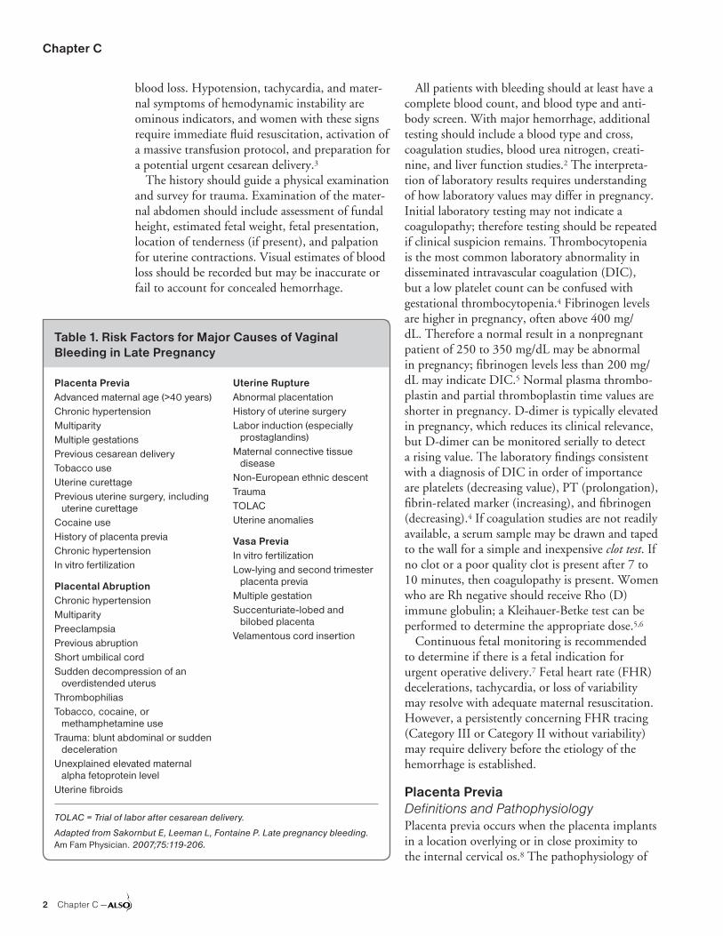
Chapter C
2 Chapter C —
blood loss. Hypotension, tachycardia, and mater-nal symptoms of hemodynamic instability are ominous indicators, and women with these signs require immediate fluid resuscitation, activation of a massive transfusion protocol, and preparation for a potential urgent cesarean delivery.3
The history should guide a physical examination and survey for trauma. Examination of the mater-nal abdomen should include assessment of fundal height, estimated fetal weight, fetal presentation, location of tenderness (if present), and palpation for uterine contractions. Visual estimates of blood loss should be recorded but may be inaccurate or fail to account for concealed hemorrhage.
All patients with bleeding should at least have a complete blood count, and blood type and anti-body screen. With major hemorrhage, additional testing should include a blood type and cross, coagulation studies, blood urea nitrogen, creati-nine, and liver function studies.2 The interpreta-tion of laboratory results requires understanding of how laboratory values may differ in pregnancy. Initial laboratory testing may not indicate a coagulopathy; therefore testing should be repeated if clinical suspicion remains. Thrombocytopenia is the most common laboratory abnormality in disseminated intravascular coagulation (DIC), but a low platelet count can be confused with gestational thrombocytopenia.4 Fibrinogen levels are higher in pregnancy, often above 400 mg/dL. Therefore a normal result in a nonpregnant patient of 250 to 350 mg/dL may be abnormal in pregnancy; fibrinogen levels less than 200 mg/dL may indicate DIC.5 Normal plasma thrombo-plastin and partial thromboplastin time values are shorter in pregnancy. D-dimer is typically elevated in pregnancy, which reduces its clinical relevance, but D-dimer can be monitored serially to detect a rising value. The laboratory findings consistent with a diagnosis of DIC in order of importance are platelets (decreasing value), PT (prolongation), fibrin-related marker (increasing), and fibrinogen (decreasing).4 If coagulation studies are not readily available, a serum sample may be drawn and taped to the wall for a simple and inexpensive clot test. If no clot or a poor quality clot is present after 7 to 10 minutes, then coagulopathy is present. Women who are Rh negative should receive Rho (D) immune globulin; a Kleihauer-Betke test can be performed to determine the appropriate dose.5,6
Continuous fetal monitoring is recommended to determine if there is a fetal indication for urgent operative delivery.7 Fetal heart rate (FHR) decelerations, tachycardia, or loss of variability may resolve with adequate maternal resuscitation. However, a persistently concerning FHR tracing (Category III or Category II without variability) may require delivery before the etiology of the hemorrhage is established.
Placenta PreviaDefinitions and PathophysiologyPlacenta previa occurs when the placenta implants in a location overlying or in close proximity to the internal cervical os.8 The pathophysiology of
Table 1. Risk Factors for Major Causes of Vaginal Bleeding in Late Pregnancy
Placenta PreviaAdvanced maternal age (>40 years)Chronic hypertensionMultiparityMultiple gestationsPrevious cesarean deliveryTobacco useUterine curettagePrevious uterine surgery, including
uterine curettageCocaine useHistory of placenta previaChronic hypertensionIn vitro fertilization
Placental AbruptionChronic hypertensionMultiparityPreeclampsiaPrevious abruptionShort umbilical cordSudden decompression of an
overdistended uterusThrombophiliasTobacco, cocaine, or
methamphetamine useTrauma: blunt abdominal or sudden
decelerationUnexplained elevated maternal
alpha fetoprotein levelUterine fibroids
Uterine RuptureAbnormal placentationHistory of uterine surgeryLabor induction (especially
prostaglandins)Maternal connective tissue
diseaseNon-European ethnic descentTraumaTOLACUterine anomalies
Vasa PreviaIn vitro fertilizationLow-lying and second trimester
placenta previaMultiple gestationSuccenturiate-lobed and
bilobed placentaVelamentous cord insertion
TOLAC = Trial of labor after cesarean delivery.
Adapted from Sakornbut E, Leeman L, Fontaine P. Late pregnancy bleeding. Am Fam Physician. 2007;75:119-206.

Vaginal Bleeding in Late Pregnancy
— Chapter C 3
placenta previa is not fully understood. Normally, placental implantation favors a fundal location. As pregnancy progresses, the apparent migration of the placenta away from the lower uterine segment is caused by the growth of placental trophoblasts toward the fundus (with its richer blood supply) and by the development or elongation of the lower uterine segment. Abnormal implantation of the placenta may occur when there is disruption or scarring of the uterine cavity, most commonly as a result of a previous cesarean delivery.
Transvaginal ultrasonography allows precise assessment of the distance between the internal os and the placental edge. The placenta is described as a complete previa when it covers the internal os and as a marginal previa when the edge lies within 2 cm of the os. When the edge is 2 to 3.5 cm from the os, the placenta may be described as low-lying (Figure 1).9
EpidemiologyPlacenta previa is a common incidental finding on second trimester ultrasonography, present on approximately 4% of ultrasound studies per-formed at 20 to 25 weeks’ gestation but only 0.5% of pregnancies at term.10 The likelihood of a previa persisting until term increases if the previa is complete, if it is present at a later gestational age, or if there is a history of cesarean delivery.11,12 The extent to which the placenta overlaps the internal os at 18 to 23 weeks is highly predictive for the persistence of placenta previa.13,14 If the overlap is less than 1.5 cm at 18 to 23 weeks, placenta previa typically resolves.14 If the overlap is 2.5 cm or greater at 20 to 23 weeks, persistence to term is likely.13 When a placenta previa or low-lying placenta is identified during the second trimester, ultrasound should be repeated at approximately 32 weeks to determine if it has regressed. Consider a repeat scan at 36 weeks for patients where the previa has persisted to determine the optimal route and timing of delivery.10 Routine late pregnancy ultrasound in low-risk or unselected populations does not confer benefit to the mother or infant and may increase cesarean delivery rates.15
Risk factors associated with placenta previa include chronic hypertension, multiparity, mul-tiple gestations, increasing maternal age, previous cesarean delivery, cocaine use, tobacco use, uter-ine curettage, previous placenta previa, and male fetal sex.2,10,12,16 Increasing numbers of cesarean
deliveries are associated with increasing risk of placenta previa.17
MorbidityMaternal morbidity associated with placenta pre-via can result from maternal hemorrhage, cesar-ean delivery, or abnormal placental attachment, specifically placenta accreta, increta, or percreta. Complete placenta previa is associated with higher morbidity than low-lying or marginal placenta previa.18,19
Women who have had a prior cesarean delivery and have a placenta previa or low anterior placenta in a subsequent pregnancy are at increased risk of abnormal placental attachment.2,20 The risk of placenta accreta, increta, or percreta reaches approximately 10% when placenta previa coex-ists with previous cesarean delivery.21,22 The risk of abnormal placental attachment also increases with the number of previous cesarean deliveries.20 Women with a history of cesarean delivery who present with placenta previa or a placenta located at the site of the previous incision should be evaluated for potential placenta accreta with color flow Dop-pler ultrasonography by an experienced sonogra-pher.23,24 Magnetic resonance imaging of the pelvis may help confirm the diagnosis of an invasive pla-centa and delineate organ involvement in women with a placenta percreta.20 A suspected placenta accreta necessitates preparation for possible cesar-ean hysterectomy, including appropriate surgical expertise and availability of blood products.
Perinatal morbidity and mortality associated with placenta previa are mainly related to the com-
Figure 1. Abnormal Placental Implantation
Low-Lying2 to 3.5 cm from os
CompleteCovers os
MarginalWithin 2 cm of os

Chapter C
4 Chapter C —
plications of prematurity because the blood loss comes from the maternal circulation.25 Therefore the management of placenta previa and timing of delivery is influenced by gestational age and fetal lung maturity balanced against the degree of hem-orrhage and urgency of the maternal condition. A cohort study of American women with placenta previa showed that greater than half delivered at term (more than 37 weeks’ gestation), approxi-mately 28% delivered between 34 and 37 weeks’ gestation, and approximately 17% delivered before 34 weeks’ gestation.10
Clinical PresentationSymptomatic placenta previa typically manifests as vaginal bleeding in the late second or third tri-mester, often after sexual intercourse. The bleed-ing is painless unless labor or placental abruption occurs. A large central previa will typically present with bleeding at 26 to 28 weeks’ gestation—the so-called sentinel bleed. This initial sentinel bleed typically is not sufficient to produce hemodynamic instability or to threaten the fetus in the absence of cervical instrumentation or digital examination.
DiagnosisPlacenta previa should be suspected in patients who have a persistent malpresentation. A cephalic presentation may be impossible because of the presence of a large placenta filling the pelvis. Regardless of previous imaging results, vaginal bleeding (particularly if it is painless or provoked by intercourse) in the setting of a high presenting part or abnormal lie should prompt clinical suspi-cion for placenta previa.2
The diagnosis of placenta previa is confirmed by ultrasound localization of the placenta. When placenta previa is suspected on transabdominal ultrasonography, transvaginal ultrasonography should be performed.2 Transvaginal ultrasonogra-phy is safe and more accurate than transabdominal ultrasonography for localizing the placental edge and the internal os. Transvaginal ultrasound iden-tifies that 26% to 60% of placentas diagnosed as low-lying on transabdominal scan are not actually low-lying and do not require further monitoring, and the addition of transvaginal ultrasonography clarifies the diagnosis in 26% of cases.2
Preoperative ultrasound can give information on fetal lie and placental location to determine the location of uterine incision.10 Gentle insertion of
a speculum to view the vaginal vault and cervix should not result in disruption of a placenta previa.
ManagementA Cochrane review showed few randomized trials of interventions for placenta previa.26 Outpatient management of placenta previa in stable women who do not have active bleeding can be considered, but the data is limited to make a firm recommen-dation.2 If selected, outpatient management carries the important criteria of close proximity to the hospital of delivery during the third trimester and having someone available to assist in the event of bleeding or onset of labor. Women with asymp-tomatic previa in the second trimester can continue normal activities until follow-up ultrasonography is performed. Women with persistent placenta previa in the third trimester should report any bleeding and abstain from intercourse and use of tampons. When bleeding occurs, women with placenta pre-via should be evaluated in the hospital.2
Because most neonatal morbidity and mortal-ity associated with placenta previa results from complications of prematurity, the main thera-peutic strategy is to prolong pregnancy until fetal lung maturity is achieved. Tocolytic agents may be used safely to prolong gestation if vagi-nal bleeding occurs with preterm contractions. One prospective study showed that the use of tocolytics in women with symptomatic placenta previa was associated with prolonged gestation and increased birth weight without increased risk of maternal hemorrhage or fetal complica-tions.27 Corticosteroids should be administered to women with bleeding from the placenta previa at 24 to 34 weeks’ estimated gestation to promote fetal lung maturity.28 A recent randomized trial showed benefit of antenatal steroid administra-tion between 34 and 36 5/7 weeks gestational age in reducing the risk of respiratory complications compared with no treatment.29
Cervical cerclage has been proposed as a means of prolonging pregnancies complicated by placenta previa because bleeding occurs in late pregnancy as the placenta is sheared from a lengthening lower uterine segment and dilat-ing cervix.30 Although a Cochrane meta-analysis showed that cerclage decreased the risk of pre-mature birth before 34 weeks, it is recommended that additional studies of cerclage be performed before this clinical practice is introduced.2,26

Vaginal Bleeding in Late Pregnancy
— Chapter C 5
The need for emergency cesarean delivery is more common in women with three or more epi-sodes of antepartum bleeding and first episode of bleeding before 29 weeks’ gestation.31 In women with a complete placenta previa, cervical length measured on transvaginal ultrasound may pre-dict risk of preterm emergency cesarean delivery secondary to massive hemorrhage.32 For women with a marginal previa, it is recommended that decision on mode of delivery be deferred until ultrasonography is performed at 35 to 36 weeks.2 Women whose placental edge is 2 cm or more from the internal os at term can expect to deliver vaginally unless heavy bleeding ensues.9 Women whose placenta is located 1 to 2 cm from the os may attempt vaginal delivery in a facility capable of moving rapidly to cesarean delivery if neces-sary, with the majority of women achieving vagi-nal delivery without increased hemorrhage.8,9,33
The optimal time for delivery for a stable patient with placenta previa is not firmly established, but delivery between 36 and 37 weeks has been recommended to maximize maternal and neona-tal outcomes.34-36 Amniocentesis for assessment of fetal lung maturity is not recommended for determining the optimal timing of delivery.36 In a retrospective study evaluating the optimal timing of delivery, infants born at 35 to 37 weeks were no more likely to have fetal anemia, fetal distress, neonatal seizures, increased ventilator needs, or infant mortality compared with infants born at 38 weeks. Infants born at 35 to 36 weeks were at increased risk of 5-minute Apgar scores less than 7 and neonatal intensive care unit admission.37
Indications for operative delivery include the presence of persistent, brisk vaginal bleeding, which poses a threat to the stability of the mater-nal-fetal dyad, or any vaginal bleeding in a preg-nancy where the fetus is sufficiently mature to be delivered with safety. General anesthesia has been associated with increased intraoperative blood loss and need for blood transfusion. Regional anesthesia appears to be a safe alternative, but it may need to be converted to general anesthesia if surgery is prolonged.24
Placental AbruptionEpidemiologyPlacental abruption is the separation of the placenta from the uterine wall before delivery. It can be partial or complete and can vary in degree.
Abruption is the most common cause of serious vaginal bleeding, occurring in 1% of pregnan-cies.38 The incidence of abruption increased between 1979 and 2001, possibly as a result of rising rates of hypertension, increased stimulant abuse, and increased surveillance bias by diagnosis with ultrasonography.39
Risk factors associated with abruption include abdominal trauma; cocaine, amphetamine, or tobacco use; chronic hypertension; preeclampsia; thrombophilias; chorioamnionitis; oligohydram-nios; iron deficiency anemia; premature rupture of membranes; and abruption in a previous pregnancy.38,40-43
PathophysiologyPlacental abruption can result from several dif-ferent pathophysiological processes. In some cases, abnormalities in placental development and implantation that start in the first trimester lead to specific pathologic changes that in turn lead to abruption.38 With blunt trauma to the abdomen, shearing of the uterine-placenta interface leads to placental detachment and hemorrhage that can be overt or retroplacental.44 In one large retrospective study of all injured pregnant women at Level I and Level II trauma centers, 84% experienced blunt trauma and 16% had penetrating injuries. Placen-tal abruption was the most common complication, occurring in 3.5% of injured pregnant women and resulting in a rate of intrauterine demise greater than 50%.45 Other etiologies of abruption include vasoconstriction associated with cocaine use and sudden uterine decompression after rupture of membranes or delivery of a first twin.38,46
PreventionThe incidence of placental abruption may be decreased by cessation of tobacco, cocaine, or amphetamine use and appropriate care for hyper-tensive disorders of pregnancy.38 In patients with preeclampsia, treatment with magnesium sulfate is associated with reduced risk of placental abruption (relative risk 0.64; 95% confidence interval [CI] = 0.5 to 0.83).47
Pregnant women involved in severe motor vehi-cle accidents have an increased risk of abruption; proper restraints are frequently not used because of discomfort.48 Appropriate use of seat belts during pregnancy should be routinely encouraged during prenatal care.

Chapter C
6 Chapter C —
Clinical PresentationPlacental abruption typically manifests as vaginal bleeding associated with abdominal pain, which may vary from mild cramping to severe pain. Women with posterior placental abruption may report back pain rather than abdominal pain, and patients with abruption because of trauma may experience pain without bleeding (concealed hem-orrhage). Concealed placental abruption has been shown to occur in 20% to 63% of abruptions.49-52 Although the rate of concealed hemorrhage varies widely and has not been studied in large numbers of women, it is important for the clinician to be aware of a possible significant abruption even in the absence of vaginal bleeding.53
The history should include questions regarding trauma (including domestic violence), presence of pain and contractions, rupture of membranes, and assessment of risk factors, including a history of hypertension (or symptoms of preeclampsia) and stimulant (cocaine, amphetamines) use.
The bleeding may be bright, dark, or inter-mixed with amniotic fluid. Blood from concealed hemorrhage is typically dark, having been seques-tered behind the membranes. The amount of vaginal bleeding is not indicative of the severity of abruption.38 It may be difficult to determine whether bleeding represents exuberant bloody show or abruption. If bleeding is noted at the time of rupture of membranes, vasa previa should also be considered.
Physical Examination and Diagnostic TestingFetal heart tones and uterine activity should be documented by continuous monitoring.7 Tetanic contractions may be present and, if measured by an intrauterine pressure catheter, are typically recorded as a high resting tone with superimposed small frequent contractions. The presence of this finding is significant because it will often be accompanied by a concerning (Category II or III) fetal tracing.
Ultrasound examination may show retroplacental clot or hemorrhage. However, acute blood clots and the placenta are hyperechoic on ultrasonog-raphy and can be difficult to distinguish from one another.54 The diagnosis of abruption is largely a clini-cal diagnosis and urgent management should never be delayed for ultrasound confirmation. If the mother and fetus are stable, the placental location and appearance and fetal lie and fetal weight estimation
may be of assistance in planning care. Ultrasound findings, if present, may include a retroplacental echolucency, abnormal thickening of the placenta, or an abnormally round torn up edge of the pla-centa. Computerized tomography (CT) is also able to identify placental abruption, and pregnant patients undergoing CT evaluation after trauma should have careful evaluation of the placenta.55
ManagementBecause the unpredictable nature of abruption does not allow for controlled trials, management remains empiric. A Cochrane review found no randomized controlled trials assessing interven-tions for placental abruption that met inclusion criteria.56 However, a recent large cohort study has indicated the risk of placental abruption in a subsequent pregnancy in women with a history of abruption in their first pregnancy is significantly increased (adjusted odds ratio [OR] = 93). They propose medical induction at 37 weeks in women with a history of placental abruption.41
Mild AbruptionA stable patient with a small partial abruption and a preterm fetus may be managed successfully in a conservative manner. Tocolysis is typically contraindicated, except in mild abruption before 34 weeks’ gestation when it may be used to allow administration of corticosteroids.57 It is impor-tant to note that IV magnesium sulfate may be indicated before 32 weeks’ gestation, but not as a tocolytic, but rather for fetal neuroprotection to reduce the incidence of cerebral palsy during premature delivery.
Women experiencing recurrent bleeding attrib-uted to placental separation may be diagnosed as having a chronic abruption. Management will be based on the degree of bleeding and gestational age. When expectant management occurs in the setting of chronic abruption, serial ultrasonogra-phy for fetal growth and antepartum surveillance are indicated in the third trimester because of the potential for uteroplacental insufficiency.39
Severe AbruptionInitial management includes rapid stabilization of maternal cardiopulmonary status and assess-ment of fetal well-being. Delay can be fatal to the fetus; 30% of perinatal mortalities in one case series occurred within 2 hours of admission.58

Vaginal Bleeding in Late Pregnancy
— Chapter C 7
In a study of placental abruption in the United States, perinatal mortality was 119/1,000 births in pregnancies complicated by placental abruption in comparison with 8.2/1,000 births among other births; the majority of perinatal mortality was associated with preterm delivery.59
Maternal stabilization requires monitoring of vital signs and urine output along with serial eval-uation of the hematocrit and coagulation studies to determine whether disseminated intravascular coagulation is present.40 The circulatory status of the patient with abruption should be maintained to permit a margin of reserve. Hourly urine output should be maintained at 30 mL/hour or greater. Hematocrit should be maintained above 30%. In patients with preeclampsia or other confounding factors, central blood pressure monitoring may assist in the fluid management.
A concerning FHR tracing (eg, persistent Category III or Category II without variability) in the setting of placental abruption necessitates rapid, typically cesarean, delivery.60,61 A decision-to-delivery interval of 20 minutes or less resulted in improved neonatal outcomes in a case-control study of severe abruption.60 Occasionally, abrup-tion occurs during the second stage and an opera-tive vaginal delivery may be attempted. Neonatal resuscitation personnel should be available for all deliveries, vaginal or operative.
When fetal mortality occurs secondary to abruption, vaginal delivery should be the goal.62 Labor should be permitted as long as adequate progress is made and maternal status can be sup-ported. Although labor is often hypertonic with abruption, it may also be hypotonic. Oxytocin augmentation is not contraindicated, but should be used judiciously with intrauterine pressure monitoring. Indications for operative delivery with fetal demise include other maternal indica-tions for cesarean delivery, failure of labor pro-gression, and brisk hemorrhage that cannot be compensated for by transfusion.
Approximately one-third of patients with placental abruption with fetal demise will develop coagulopathy. Coagulopathy is typically not seen in the patient presenting with abruption and a live fetus. Coagulopathy occurring with abrup-tion may be related to two etiologies: consumptive coagulopathy and DIC. Replacement of platelets and fresh-frozen plasma should be administered just before operative delivery to provide maximum
efficacy. In addition, cryoprecipitate or factor VIII may be of specific benefit in severe coagulopathy.
Consideration of maternal transfer from a rural site is based on many factors. Patients present-ing with abruption and a live fetus are typically not stable for transfer because operative delivery may be needed on an immediate basis at any time during labor. In this instance, neonatal transfer (rather than maternal-fetal) may be a necessary intervention for the premature or sick newborn. If fetal demise has occurred, a patient who does not have coagulopathy and is hemodynamically stable may be cared for with appropriate resources. Blood bank supply may determine whether or not a patient needs transport to a referral facility.
Fetomaternal hemorrhage may occur with rup-ture of fetal vessels in the placenta. The Kleihauer-Betke test is useful to determine dosage of Rho (D) immune globulin in Rh-negative patients, but is not useful for the diagnosis of abruption.63,64
Uterine RuptureEpidemiology and PathophysiologyUterine scar disruption spans a spectrum from occult dehiscence discovered at repeat cesarean delivery to complete uterine rupture requiring emergency laparotomy. In uncommon instances, uterine rupture can be spontaneous and occur in the absence of risk factors. In complete rupture, the fetus or placenta may be partially or com-pletely extruded from the uterus. This chapter focuses only on uterine rupture presenting with third trimester bleeding.
Spontaneous uterine rupture is found in only 0.03% to 0.08% of all delivering women, but in approximately 0.8% of women with a uterine scar from prior surgery.65 Previous cesarean incision is the most common etiology for uterine rupture. Classic or T-shaped uterine incisions are associ-ated with a higher likelihood of uterine rupture compared with a low transverse incision.66 Two or more previous cesarean deliveries and labor induc-tion each increase the incidence of uterine rupture from approximately 0.9% to 1.8% and 0.77% to 2.24% respectively.65 Although uncommon, there is a concomitant rise in the incidence of uterine rupture with increasing rates of cesarean deliver-ies.67 Other causes include inappropriate oxytocin usage and trauma.66
Conditions that predispose to uterine scar disruption include previous uterine surgery

Chapter C
8 Chapter C —
(eg, myomectomy) that involves the full thickness of the myometrium, congenital uterine anomaly, uterine overdistension, intra-amniotic installa-tion, gestational trophoblastic neoplasia, previous uterine rupture, maternal obesity, and adenomyo-sis.66,68 Other risk factors include labor induction (OR 12.60; 95% CI = 4.4 to 36.4), labor induc-tion with prostaglandins (OR 2.72; 95% CI = 1.6 to 4.7), and non-European ethnicity (OR 2.87; 95% CI = 1.8 to 4.7).66 Conditions present during delivery that predispose to uterine rupture include fetal anomaly, vigorous uterine pressure, difficult manual removal of the placenta, or abnormalities of placental implantation.68
The most common maternal morbidity asso-ciated with uterine rupture is hemorrhage and subsequent anemia, requiring blood transfusion.69 Other morbidities include bladder injury (8.8%) and hysterectomy, which accompanies 14% to 33% of uterine ruptures.70,71 Although rare, maternal mortality from DIC and sepsis has been reported.69 The incidence of perinatal mortal-ity associated with uterine rupture is variably reported, ranging from 0% to 60%.72 Fetal and maternal morbidity are higher in cases of uterine rupture of an unscarred uterus.70
Clinical PresentationThe classic presentation for symptomatic, sig-nificant uterine rupture includes vaginal bleed-ing, pain, cessation of contractions, absence of fetal heart tones, loss of station, easily palpable fetal parts through the maternal abdomen, and profound maternal tachycardia and hypotension. However, most cases (67% to 70%) of uterine rupture initially present with abnormal fetal monitoring.65,70 In a review of 159,456 deliveries, the most frequent finding associated with uterine rupture was a sudden deterioration of the FHR pattern.73 In one case-control study, markedly abnormal FHR tracings starting 1 hour before birth were significantly associated with uterine rupture in patients undergoing a trial of labor after cesarean delivery.74 There may be a progression of signs from nonspecific, severe variable decelera-tions to the characteristic recession of the fetal head or suprapubic bulging. Contractions may show a stair-step appearance of gradually decreas-ing amplitude on tocodynamometry.75
Thirteen percent of uterine ruptures occur out-side of the hospital. Patients with a prior uterine
scar should be advised to come to the hospital for evaluation of new onset of contractions, abdomi-nal pain, or vaginal bleeding as soon as possible.73 Spontaneous antepartum rupture in nonlaboring women is extremely rare and is typically associ-ated with identifiable risk factors. In a case series of women experiencing spontaneous uterine rupture in the second or third trimester, six of seven events (during 13 years) involved placenta previa or per-creta and five of the seven uterine ruptures occurred in women with prior cesarean deliveries.76 These findings suggest that prior uterine scar and abnor-mal placenta both play a role in uterine rupture.77
ManagementIn the case of a sudden change in fetal baseline or the onset of repetitive FHR decelerations, the clinician should institute intrauterine resuscitation with maternal position change, IV fluids, discon-tinuation of oxytocin, oxygen administration, and consideration for subcutaneous terbutaline. If these measures are not effective, then emergent cesarean or operative vaginal delivery may be indicated. Asymptomatic scar disruption may be found at the time of cesarean delivery or palpation of the uterine cavity after vaginal delivery. Routine inspection of the uterine scar after vaginal deliv-ery is not recommended and asymptomatic scar disruption is expectantly managed.
Vasa PreviaVasa previa occurs when fetal blood vessels, unprotected by the umbilical cord or placenta, run through the membranes and across or within 2 cm of the cervix. Although uncommon, with an incidence of 1/2,500 deliveries, it is important to be familiar with vasa previa because rapid inter-vention is essential for fetal survival.10,78
Epidemiology and PathophysiologyThis uncommon cause of severe obstetric hemor-rhage typically occurs in pregnancies with a low-lying placenta and velamentous insertion, bipartite placenta, or a placenta with a succenturiate lobe.79 During rupture of the membranes, the fetal vessels are at risk of rupture, which can lead to significant fetal blood loss. Historical studies showed a 33% to 100% rate of perinatal mortality secondary to vasa previa,80 but antenatal diagnosis is associated with a reduction in the rate of neonatal morbid-ity and mortality.79 Risk factors for vasa previa

Vaginal Bleeding in Late Pregnancy
— Chapter C 9
include in vitro fertilization, multiple gestation, placenta previa, and bilobed or succenturiate-lobed placentas.80,81
Clinical Presentation and DiagnosisThe goal of diagnosis is antenatal detection and delivery before membrane rupture.82 Prenatal diag-nosis using ultrasound with color flow Doppler significantly affects fetal survival. Prenatal diagno-sis is associated with a 98% survival rate compared with a 44% survival rate with intrapartum or post-partum diagnosis.78 Because of increased prenatal diagnosis, contemporary case series show perinatal mortality rates less than 10%.78 Prenatal ultra-sound has a 93% detection rate and a specificity of 99% with an optimal detection window between 18 and 26 weeks’ gestation; detection is less likely on ultrasound performed in the third trimester.78 Clinically, vasa previa is suspected when vaginal bleeding occurs with membrane rupture, clas-sically in the setting of a FHR tracing showing bradycardia or a sinusoidal pattern.78
The hemorrhage is fetal blood, and exsanguina-tion can occur rapidly because the average blood volume of a term fetus is approximately 250 mL.83 Transabdominal combined with transvaginal ultrasound may improve diagnostic accuracy. Power Doppler and three-dimensional ultraso-nography are reported to assist with diagnosis, but superiority to two-dimensional ultrasound has not been shown.10 In addition to diagnosis by ultra-sound or classic clinical presentation, vasa previa may be diagnosed by MRI, and intrapartum iden-tification of fetal blood intermixed with vaginal blood.79 Rarely, vessels are palpated in the pre-senting membranes, prohibiting artificial rupture and vaginal delivery. FHR tracing may show fetal tachycardia or a sinusoidal pattern.10
If fetal heart tones are not concerning, a blood sample from the vaginal vault may be obtained to check for fetal blood cells or fetal hemoglobin. The Kleihauer-Betke test and hemoglobin electro-phoresis measuring the presence of fetal hemoglo-bin are sensitive but are too slow to use clinically.79 The Apt test (alkali denaturation test) is faster to perform but is insensitive.79 The Apt test is based on the resistance of fetal hemoglobin to denatur-ation by alkaline agents and can be performed on the labor and delivery unit.84 Performance of the Apt test involves obtaining a sample of blood from the vagina and then mixing it with a small
amount of tap water to lyse red blood cells. After 5 minutes of centrifugation, the supernatant is removed and mixed with 1 mL of 1% sodium hydroxide (NaOH) for every 5 mL of supernatant. A pink color indicates fetal hemoglobin. Adult hemoglobin is brown. Additionally, a Wright stain of blood collected from the vagina can be evalu-ated for the presence of nucleated red blood cells, which are common in fetal blood but uncommon in adult blood. This test can be performed without delay, assuming a stable FHR.
ManagementIn a woman with antenatally detected vasa pre-via who presents with premature rupture of fetal membranes or labor, cesarean delivery should be performed.78 Delivery should not be deferred for confirmation of fetal blood in women with severe hemorrhage or when fetal heart tones are concern-ing. If the onset of vaginal bleeding occurred with rupture of membranes and the FHR is concerning, cesarean delivery should be performed immedi-ately. Because fetal exsanguination is the cause of neonatal mortality in this disorder, preparation for resuscitation at the delivery includes availability of normal saline for a 10 to 20 mL/kg bolus to administer if the newborn is in shock.
In the presence of antenatal diagnosis of vasa previa, serial ultrasounds are recommended to evaluate for regression of vessels, which can occur in approximately 20% of women.78 With per-sistent vasa previa, antenatal steroids should be administered between 28 and 32 weeks’ gesta-tion. Hospitalization at 30 to 34 weeks should be considered, allowing for closer observation for signs of labor onset and proximity to operative delivery if membranes rupture. However, data to support this approach compared with outpatient management is lacking.78 Outpatient manage-ment can be considered for asymptomatic women with no uterine activity and a long closed cervix on transvaginal ultrasound.82 More than half of women managed as outpatients will eventually require hospitalization for a complication.78 The optimal gestational age for delivery is not deter-mined, but cesarean delivery between 34 and 36 weeks has been recommended to balance the risk of respiratory distress syndrome with risk of membrane rupture and fetal exsanguination.10,78,82 Amniocentesis to assess for fetal lung maturity is not indicated because timing of delivery would

Chapter C
10 Chapter C —
not be influenced by the result.78 Delaying deliv-ery beyond 37 weeks is not recommended.
PreventionThere are no strategies for primary prevention of vasa previa. However, hemorrhage theoretically is preventable with antenatal screening for women at high risk and cesarean delivery at 34 to 36 weeks when vasa previa is present. Screening is carried out with transvaginal color flow Doppler to identify the presence of vessels in the fetal membranes. Screen-ing in a general population has not been recom-mended because the condition is rare (one diagnosis per 5,215 screenings).85 Screening is recommended for women at increased risk, which includes women after the detection of a low-lying, bilobed, multi-lobed, or succenturiate-lobed placenta, in women with velamentous cord insertion on a routine ultra-sound, or women who are pregnant after in vitro fertilization.10,79 Careful evaluation of the placenta including site of cord insertion on routine ultra-sound may facilitate identifying women at greater risk of vasa previa. A transvaginal ultrasound with color and pulsed Doppler at 32 weeks has been
recommended for women who had a placenta pre-via or low-lying placenta during a second trimester ultrasound with subsequent resolution.78
SummaryVaginal bleeding in late pregnancy may occur because of potentially life-threatening conditions for mother and newborn. Clinicians must be able to distinguish emergent causes of bleeding from those that are less urgent and be prepared to act quickly and decisively with severe maternal hemorrhage or suspected vasa previa. Vaginal examination should be avoided until placental location is known. The timely diagnosis of vaginal bleeding in late preg-nancy, including antenatal diagnosis with color flow ultrasound, can reduce perinatal mortality. This chapter identifies major causes of vaginal bleeding in the second half of pregnancy, describes a system-atic approach to identifying the cause of bleeding, and identifies the appropriate management of the woman with late vaginal bleeding. Institutional policies should be in place to ensure adequate blood bank response to massive hemorrhage and to mobi-lize resources for emergent cesarean delivery.

Vaginal Bleeding in Late Pregnancy
— Chapter C 11
SORT: Key Recommendations for Practice
Clinical RecommendationEvidence Rating References
Placenta previa is a common incidental finding on second trimester ultrasonography and should be confirmed in the third trimester.
A 10,11
Corticosteroids should be administered to women with bleeding from placenta previa at 24 to 34 weeks’ gestation.
A 28
Treatment of preeclampsia with magnesium sulfate decreases the risk of placental abruption and improves maternal outcomes.
A 47
Antenatal diagnosis of vasa previa is associated with significant reduction in perinatal mortality. A 78
Delivery in pregnancies complicated by placenta previa may be safely delayed by the use of tocolytics. B 29
Outpatient management of placenta previa is appropriate in selected patients who do not have active bleeding and who can rapidly access a hospital with operative labor and delivery services.
B 2
Transvaginal ultrasonography may be safely performed in women with a placenta previa and is more accurate in detecting placental location than transabdominal ultrasonography.
B 2
In patients presenting with abruption, a decision-to-delivery interval of 20 minutes or less results in improved neonatal outcomes.
B 60
A sterile speculum examination can be performed safely in women with second or third trimester vaginal bleeding before ultrasonographic evaluation of placental localization, but a digital examination should be avoided until placenta previa is excluded by ultrasonography.
C 2
Women with bleeding in late pregnancy who are Rh negative should receive Rho (D) immune globulin after performance of a Kleihauer-Betke test to determine appropriate dose.
C 5,6
In women with placenta previa and previous cesarean delivery, imaging with color flow Doppler ultrasonography should be performed to evaluate for placenta accreta.
C 2
Ultrasound and color flow Doppler screening for vasa previa is recommended in women at increased risk including women who are pregnant after in vitro fertilization, when a low-lying placenta is detected on second trimester ultrasound, and in the presence of low or velamentous cord insertion, bilobate, or succenturiate placenta.
C 2,85
Magnetic resonance imaging of the pelvis may help confirm a diagnosis of invasive placenta and identify organ involvement associated with placenta percreta.
C 20
Strength of Recommendation Taxonomy (SORT)
Strength of Recommendation Definition
A • Recommendation based on consistent and good-quality patient-oriented evidence.
B • Recommendation based on inconsistent or limited-quality patient-oriented evidence.
C • Recommendation based on consensus, usual practice, opinion, disease-oriented evidence, or case series for studies of diagnosis, treat-ment, prevention, or screening.
Patient-oriented evidence measures outcomes that matter to patients: morbidity, mortality, symptom improvement, cost reduction, and quality of life. Disease-oriented evidence measures intermediate, physiologic, or surrogate end points that might not reflect improvement in patient outcomes (eg, blood pressure, blood chemistry, physiologic function, pathologic findings).
From Ebell MH, Siwek J, Weiss BD, et al. Strength of recommendation taxonomy (SORT): a patient-centered approach to grading evidence in the medical literature. Am Fam Physician. 2004;69(3):548-556.

Chapter C
12 Chapter C —
References1. McCormack RA, Doherty DA, Magann EF, Hutchinson
M, Newnham JP. Antepartum bleeding of unknownorigin in the second half of pregnancy and pregnancyoutcomes. BJOG. 2008;115(11):1451-1457.
2. Royal College of Obstetricians and Gynaecologists.Green-top Guideline No. 63. Antepartum Haemorrhage.London, England: Royal College of Obstetricians andGynaecologists; 2011.
3. De Kock J, Heyns T, Van Rensburg GH. The ABC ofhaemorrhagic shock in the pregnant woman. Profes-sional Nursing Today. 2008;12(5):54-57.
4. Erez O, Mastrolia SA, Thachil J. Disseminated intra-vascular coagulation in pregnancy: insights in patho-physiology, diagnosis and management. Am J ObstetGynecol. 2015;213(4):452-463.
5. Jain V, Chari R, Maslovitz S, et al; Maternal Fetal Medi-cine Committee. Guidelines for the management of apregnant trauma patient. J Obstet Gynaecol Can. 2015;37(6):553-574.
6. American College of Obstetrics and Gynecology.ACOG practice bulletin. Prevention of Rh D alloimmuni-zation. Number 4, May 1999 (replaces educational bul-letin Number 147, October 1990). Clinical managementguidelines for obstetrician-gynecologists. Int J Gynae-col Obstet. 1999;66(1):63-70.
7. American College of Obstetricians and Gynecolo-gists. ACOG Practice Bulletin No. 106: Intrapartum fetalheart rate monitoring: nomenclature, interpretation,and general management principles. Obstet Gynecol.2009;114(1):192-202.
8. Vergani P, Ornaghi S, Pozzi I, et al. Placenta previa: dis-tance to internal os and mode of delivery. Am J ObstetGynecol. 2009;201(3):266.e1-266.e5.
9. Bhide A, Thilaganathan B. Recent advances in the man-agement of placenta previa. Curr Opin Obstet Gynecol.2004;16(6):447-451.
10. Silver RM. Abnormal Placentation: Placenta Previa,Vasa Previa, and Placenta Accreta. Obstet Gynecol.2015;126(3):654-668.
11. Mustafá SA, Brizot ML, Carvalho MH, Watanabe L, Kah-hale S, Zugaib M. Transvaginal ultrasonography in pre-dicting placenta previa at delivery: a longitudinal study.Ultrasound Obstet Gynecol. 2002;20(4):356-359.
12. Dashe JS, McIntire DD, Ramus RM, Santos-Ramos R,Twickler DM. Persistence of placenta previa accordingto gestational age at ultrasound detection. Obstet Gyne-col. 2002;99(5 Pt 1):692-697.
13. Becker RH, Vonk R, Mende BC, Ragosch V, EntezamiM. The relevance of placental location at 20-23 ges-tational weeks for prediction of placenta previa atdelivery: evaluation of 8650 cases. Ultrasound ObstetGynecol. 2001;17(6):496-501.
14. Taipale P, Hiilesmaa V, Ylöstalo P. Transvaginal ultraso-nography at 18-23 weeks in predicting placenta previa atdelivery. Ultrasound Obstet Gynecol. 1998;12(6):422-425.
15. Bricker L, Neilson JP, Dowswell T. Routine ultrasoundin late pregnancy (after 24 weeks’ gestation). CochraneDatabase Syst Rev. 2008;(4):CD001451.
16. Yang Q, Wen SW, Phillips K, Oppenheimer L, BlackD, Walker MC. Comparison of maternal risk factorsbetween placental abruption and placenta previa.Am J Perinatol. 2009;26(4):279-286.
17. Silver RM, Landon MB, Rouse DJ, et al; National Insti-tute of Child Health and Human Development Maternal-Fetal Medicine Units Network. Maternal morbidityassociated with multiple repeat cesarean deliveries.Obstet Gynecol. 2006;107(6):1226-1232.
18. Bahar A, Abusham A, Eskandar M, Sobande A, Alsuna-idi M. Risk factors and pregnancy outcome in differ-ent types of placenta previa. J Obstet Gynaecol Can.2009;31(2):126-131.
19. Tuzovic L. Complete versus incomplete placentaprevia and obstetric outcome. Int J Gynaecol Obstet.2006;93(2):110-117.
20. Committee on Obstetric Practice. Committee opinionno. 529: placenta accreta. Obstet Gynecol. 2012;120(1):207-211.
21. Miller DA, Chollet JA, Goodwin TM. Clinical risk factorsfor placenta previa-placenta accreta. Am J Obstet Gyne-col. 1997;177(1):210-214.
22. Zaki ZM, Bahar AM, Ali ME, Albar HA, Gerais MA. Riskfactors and morbidity in patients with placenta previaaccreta compared to placenta previa non-accreta. ActaObstet Gynecol Scand. 1998;77(4):391-394.
23. Comstock CH, Love JJ Jr, Bronsteen RA, et al. Sono-graphic detection of placenta accreta in the second andthird trimesters of pregnancy. Am J Obstet Gynecol.2004;190(4):1135-1140.
24. Oppenheimer L; Society of Obstetricians and Gyn-aecologists of Canada. Diagnosis and manage-ment of placenta previa. J Obstet Gynaecol Can.2007;29(3):261-273.
25. Zlatnik MG, Cheng YW, Norton ME, Thiet MP, CaugheyAB. Placenta previa and the risk of preterm delivery. JMatern Fetal Neonatal Med. 2007;20(10):719-723.
26. Neilson JP. Interventions for suspected placenta prae-via. Cochrane Database Syst Rev. 2003;(2):CD001998.
27. Sharma A, Suri V, Gupta I. Tocolytic therapy in conser-vative management of symptomatic placenta previa. IntJ Gynaecol Obstet. 2004;84(2):109-113.
28. Roberts D, Dalziel S. Antenatal corticosteroids foraccelerating fetal lung maturation for women atrisk of preterm birth. Cochrane Database Syst Rev.2006;(3):CD004454.
29. Gyamfi-Bannerman C, Thom EA, Blackwell SC, et al;NICHD Maternal–Fetal Medicine Units Network. Antena-tal Betamethasone for Women at Risk for Late PretermDelivery. N Engl J Med. 2016;374(14):1311-1320.
30. Cobo E, Conde-Agudelo A, Delgado J, Canaval H,Congote A. Cervical cerclage: an alternative for themanagement of placenta previa? Am J Obstet Gynecol.1998;179(1):122-125.
31. Pivano A, Alessandrini M, Desbriere R, et al. A scoreto predict the risk of emergency caesarean delivery inwomen with antepartum bleeding and placenta praevia.Eur J Obstet Gynecol Reprod Biol. 2015;195:173-176.

Vaginal Bleeding in Late Pregnancy
— Chapter C 13
32. Ghi T, Contro E, Martina T, et al. Cervical length andrisk of antepartum bleeding in women with com-plete placenta previa. Ultrasound Obstet Gynecol.2009;33(2):209-212.
33. Al Wadi K, Schneider C, Burym C, Reid G, Hunt J, Men-ticoglou S. Evaluating the safety of labour in womenwith a placental edge 11 to 20 mm from the internal cer-vical Os. J Obstet Gynaecol Can. 2014;36(8):674-677.
34. Blackwell SC. Timing of delivery for women with stableplacenta previa. Semin Perinatol. 2011;35(5):249-251.
35. Zlatnik MG, Little SE, Kohli P, Kaimal AJ, Stotland NE,Caughey AB. When should women with placenta pre-via be delivered? A decision analysis. J Reprod Med.2010;55(9-10):373-381.
36. American College of Obstetricians and Gynecologists.ACOG committee opinion no. 560: Medically indicatedlate-preterm and early-term deliveries. Obstet Gynecol.2013;121(4):908-910.
37. Balayla J, Wo BL, Bédard MJ. A late-preterm, early-term stratified analysis of neonatal outcomes by ges-tational age in placenta previa: defining the optimaltiming for delivery. J Matern Fetal Neonatal Med.2015;28(15):1756-1761.
38. Oyelese Y, Ananth CV. Placental abruption. ObstetGynecol. 2006;108(4):1005-1016.
39. Ananth CV, Oyelese Y, Yeo L, Pradhan A, Vintzileos AM.Placental abruption in the United States, 1979 through2001: temporal trends and potential determinants. Am JObstet Gynecol. 2005;192(1):191-198.
40. Hladky K, Yankowitz J, Hansen WF. Placental abruption.Obstet Gynecol Surv. 2002;57(5):299-305.
41. Ruiter L, Ravelli ACJ, de Graaf IM, Mol BWJ, Pajkrt E.Incidence and recurrence rate of placental abruption: alongitudinal linked national cohort study in the Nether-lands. Am J Obstet Gynecol. 2015;213(4):573.e1-573.e8.
42. Arnold DL, Williams MA, Miller RS, Qiu C, SorensenTK. Iron deficiency anemia, cigarette smoking andrisk of abruptio placentae. J Obstet Gynaecol Res.2009;35(3):446-452.
43. Brenner B, Aharon A. Thrombophilia and adverse preg-nancy outcome. [v.]. Clin Perinatol. 2007;34(4):527-541, v.
44. Brown HL. Trauma in pregnancy. Obstet Gynecol.2009;114(1):147-160.
45. Rogers FB, Rozycki GS, Osler TM, et al. A multi-institutional study of factors associated with fetal deathin injured pregnant patients. Arch Surg. 1999;134(11):1274-1277.
46. Fleming AD. Abruptio placentae. Crit Care Clin. 1991;7(4):865-875.
47. Duley L, Gülmezoglu AM, Henderson-Smart DJ, ChouD. Magnesium sulphate and other anticonvulsants forwomen with pre-eclampsia. Cochrane Database SystRev. 2010;(11):CD000025.
48. Reis PM, Sander CM, Pearlman MD. Abruptio placen-tae after auto accidents. A case-control study. J ReprodMed. 2000;45(1):6-10.
49. Golan A, Sandbank O, Teare AJ. Trauma in late preg-nancy. A report of 15 cases. S Afr Med J. 1980;57(5):161-165.
50. Leunen K, Hall DR, Odendaal HJ, Grové D. Theprofile and complications of women with placentalabruption and intrauterine death. J Trop Pediatr.2003;49(4):231-234.
51. Pepperell RJ, Rubinstein E, MacIsaac IA. Motor-car acci-dents during pregnancy. Med J Aust. 1977;1(7):203-205.
52. Sinha P, Kuruba N. Ante-partum haemorrhage: anupdate. J Obstet Gynaecol. 2008;28(4):377-381.
53. Doyle C, Angelotti T. Diagnosis of an unsuspectedmaternal hemorrhage via fetal heart rate tracing. J ClinAnesth. 2004;16(6):465-468.
54. Glantz C, Purnell L. Clinical utility of sonography in thediagnosis and treatment of placental abruption. J Ultra-sound Med. 2002;21(8):837-840.
55. Manriquez M, Srinivas G, Bollepalli S, Britt L, DrachmanD. Is computed tomography a reliable diagnostic modal-ity in detecting placental injuries in the setting of acutetrauma? Am J Obstet Gynecol. 2010;202(6):611.e1-611.e5.
56. Neilson JP. Interventions for treating placental abrup-tion. Cochrane Database Syst Rev. 2003;(1):CD003247.
57. Towers CV, Pircon RA, Heppard M. Is tocolysis safein the management of third-trimester bleeding? Am JObstet Gynecol. 1999;180(6 Pt 1):1572-1578.
58. Knab DR. Abruptio placentae. An assessment of thetime and method of delivery. Obstet Gynecol. 1978;52(5):625-629.
59. Ananth CV, Wilcox AJ. Placental abruption and peri-natal mortality in the United States. Am J Epidemiol.2001;153(4):332-337.
60. Kayani SI, Walkinshaw SA, Preston C. Pregnancy out-come in severe placental abruption. BJOG. 2003;110(7):679-683.
61. Witlin AG, Sibai BM. Perinatal and maternal outcomefollowing abruptio placentae. Hypertens Pregnancy.2001;20(2):195-203.
62. Boisramé T, Sananès N, Fritz G, et al. Placental abrup-tion: risk factors, management and maternal-fetal prog-nosis. Cohort study over 10 years. Eur J Obstet GynecolReprod Biol. 2014;179:100-104.
63. Emery CL, Morway LF, Chung-Park M, Wyatt-AshmeadJ, Sawady J, Beddow TD. The Kleihauer-Betke test.Clinical utility, indication, and correlation in patients withplacental abruption and cocaine use. Arch Pathol LabMed. 1995;119(11):1032-1037.
64. Update ACOG. Major and minor trauma in pregnancy.Vol. 35, No. 6. December 2009. Medical InformationSystems Inc. 2 Seaview Blvd. Port Washington, NewYork. Available at http://www.acogupdate.com/.
65. American College of Obstetricians and Gynecologists.ACOG Practice bulletin no. 115: Vaginal birth after previ-ous cesarean delivery. Obstet Gynecol. 2010;116(2 Pt 1):450-463.
66. Mirza FG, Gaddipati S. Obstetric emergencies. SeminPerinatol. 2009;33(2):97-103.
67. Al-Zirqi I, Stray-Pedersen B, Forsén L, Vangen S. Uter-ine rupture after previous caesarean section. BJOG.2010;117(7):809-820.

Chapter C
14 Chapter C —
68. Smith JG, Mertz HL, Merrill DC. Identifying risk factorsfor uterine rupture. [viii.]. Clin Perinatol. 2008;35(1):85-99, viii.
69. Aziz N, Yousfani S. Analysis of uterine rupture atuniversity teaching hospital Pakistan. Pak J Med Sci.2015;31(4):920-924.
70. Zwart JJ, Richters JM, Ory F, de Vries JIP, BloemenkampKWM, van Roosmalen J. Uterine rupture in The Neth-erlands: a nationwide population-based cohort study.BJOG. 2009;116(8):1069-1078, discussion 1078-1080.
71. National Institutes of Health Consensus DevelopmentConference Panel. National Institutes of Health Consen-sus Development conference statement: vaginal birthafter cesarean: new insights March 8-10, 2010. ObstetGynecol. 2010;115(6):1279-1295.
72. Guise JM, McDonagh MS, Osterweil P, Nygren P, ChanBK, Helfand M. Systematic review of the incidence andconsequences of uterine rupture in women with previ-ous caesarean section. BMJ. 2004;329(7456):19-25.
73. Leung AS, Leung EK, Paul RH. Uterine rupture afterprevious cesarean delivery: maternal and fetal conse-quences. Am J Obstet Gynecol. 1993;169(4):945-950.
74. Desseauve D, Bonifazi-Grenouilleau M, Fritel X, Lathél-ize J, Sarreau M, Pierre F. Fetal heart rate abnormali-ties associated with uterine rupture: a case-controlstudy: A new time-lapse approach using a standard-ized classification. Eur J Obstet Gynecol Reprod Biol.2016;197:16-21.
75. Matsuo K, Scanlon JT, Atlas RO, Kopelman JN. Stair-case sign: a newly described uterine contraction patternseen in rupture of unscarred gravid uterus. J ObstetGynaecol Res. 2008;34(1):100-104.
76. Vaknin Z, Maymon R, Mendlovic S, Barel O, Herman A,Sherman D. Clinical, sonographic, and epidemiologicfeatures of second- and early third-trimester spontane-ous antepartum uterine rupture: a cohort study. PrenatDiagn. 2008;28(6):478-484.
77. Jauregui I, Kirkendall C, Ahn MO, Phelan J. Uterinerupture: a placentally mediated event? Obstet Gynecol.2000;95(4)(Suppl 1):S75.
78. Sinkey RG, Odibo AO, Dashe JS; Society of Maternal-Fetal (SMFM) Publications Committee. #37: Diagnosisand management of vasa previa. Am J Obstet Gynecol.2015;213(5):615-619.
79. Gagnon R, Morin L, Bly S, et al; Diagnostic ImagingCommittee; Maternal Fetal Medicine Committee. Guide-lines for the management of vasa previa. J Obstet Gyn-aecol Can. 2009;31(8):748-760.
80. Oyelese KO, Turner M, Lees C, Campbell S. Vasa pre-via: an avoidable obstetric tragedy. Obstet GynecolSurv. 1999;54(2):138-145.
81. Baulies S, Maiz N, Muñoz A, Torrents M, Echevarría M,Serra B. Prenatal ultrasound diagnosis of vasa praeviaand analysis of risk factors. Prenat Diagn. 2007;27(7):595-599.
82. Hasegawa J, Arakaki T, Ichizuka K, Sekizawa A. Man-agement of vasa previa during pregnancy. J PerinatMed. 2015;43(6):783-784.
83. Nelle M, Zilow EP, Kraus M, Bastert G, Linderkamp O.The effect of Leboyer delivery on blood viscosity andother hemorheologic parameters in term neonates. AmJ Obstet Gynecol. 1993;169(1):189-193.
84. Apt L, Downey WS Jr. Melena neonatorum: the swal-lowed blood syndrome; a simple test for the differentia-tion of adult and fetal hemoglobin in bloody stools. JPediatr. 1955;47(1):6-12.
85. Lee W, Lee VL, Kirk JS, Sloan CT, Smith RS, Com-stock CH. Vasa previa: prenatal diagnosis, naturalevolution, and clinical outcome. Obstet Gynecol.2000;95(4):572-576.

— Chapter D 1
Chapter D Preterm Labor and Premature
Rupture of Membranes
IntroductionPrevious preterm delivery (PTD) is the most impor-tant historical risk factor for subsequent PTD. Other important antenatal risk factors include multiple gestation, bacterial vaginosis (BV), and a short cervical length (CL) on ultrasound. Progesterone may be used to decrease the likelihood of PTD in patients with a prior PTD or a short CL. In a patient presenting with preterm contractions, CL on transvaginal ultrasound or a fetal fibronectin (fFN) test can be used to help assess the risk of PTD. Administration of antenatal corticosteroids (ACS) remains our most important intervention to improve neonatal outcomes in women presenting with preterm labor (PTL). Tocolytic agents may delay PTD, which allows time to administer ste-roids or transfer to a facility with a neonatal intensive care unit (NICU). Management of preterm premature rupture of membranes (PPROM) may include admin-istering antibiotics or ACS, or inducing labor based on the gestational age at presentation. Management of premature rupture of membranes (PROM) at term includes early induction/augmentation of labor.
EpidemiologyThe incidence of PTD, defined as occurring before 37 weeks’ gestation, was 11.4% in 2013.1 Approxi-
mately 40% to 45% of PTDs are the result of spon-taneous PTL with intact membranes. Another 25% to 30% are associated with PPROM. The remaining 30% to 35% of PTDs are the result of delivery by labor induction or cesarean delivery for medical indications such as preeclampsia with severe features, placental abruption, or intrauterine growth restriction.2
From 1981 through 2006, the incidence of PTD increased by more than 30%, and peaked at 12.80% in 2006.1 Since that time, the rate of PTD has gradually decreased each year. The rate of early PTD (delivery before 34 weeks) has declined 3.7% and the rate of late PTD (delivery at 34 to 36 weeks) has declined 9.2% since 2006.3 The declining rate of PTD may be attrib-uted to multiple factors. Since 2006, both the teenage birth rate and the rate of higher order multiples (trip-lets or higher gestations) have declined.4 These groups are at higher risk for PTD. There is some evidence that public policy recommendations to reduce elective early term (37 to 38 weeks) deliveries have also reduced PTDs.4,5 Finally, interventions such as vaginal proges-terone, intramuscular 17 alpha-hydroxyprogesterone caproate (17P), and use of cerclage in selected popula-tions are likely contributing to the decreased rate.4
The PTD rate in the United States still remains among the highest in the developed world and signifi-
Learning ObjectivesAt the end of this activity, learners will be able to:1. Discuss risk factors associated with preterm labor (PTL) and premature
rupture of membranes (PROM).2. Identify patients who may benefit from antenatal progesterone.3. Define PTL and PROM, and describe their significance.4. Outline initial evaluation of PTL and PROM.5. Describe management of PTL and PROM.6. Discuss neonatal group B streptococcal prevention strategies.
William Sayres, MD, Karen Wildman, MD, FAAFP, Colleen Zimmermann, MD
Revised January 2016
Copyright 2017© American Academy of Family Physicians. All rights reserved.

Chapter D
2 Chapter D —
cant disparities exist in the rates between racial ethnic groups. In 2012, the PTD rate for non-Hispanic white infants was 10.3% compared to 16.5% for non-Hispanic black infants, and 11.6% for Hispanic infants.6
Risk Factors for PTDAlthough many risk factors for PTD have been identified, up to 50% of PTDs occur in pregnan-cies with no known risk factors, and over half of women with identifiable risk factors for PTD will ultimately deliver at term.7,8 Furthermore, the causality of identified risk factors has been diffi-cult to prove. Many risk-scoring systems for PTD have been developed, but none have been shown to reduce preterm birth.9 Despite this, identifying women at risk is of growing importance as treat-ment options such as progesterone and cerclage become available. Common risk factors for PTD are outlined in Table 1.
Prior PTD is the single most important identifi-able risk factor for recurrent PTD. Women carrying a singleton after a previous singleton PTD have a 20% risk of recurrence (95% confidence interval [CI] = 19.9 to 20.6).10 In contrast, after a term delivery, women have a 2.7% risk of PTD in the subsequent pregnancy. To break this down further, after delivery between 32 to 36 weeks, a subse-quent pregnancy has a 14.7% risk of PTD (95% CI = 5.84 to 6.42), and after delivery before 28
weeks’ gestation, a subsequent pregnancy has a 26% risk of PTD (95% CI = 10.8 to 15.9).11 Multiple prior PTDs further increase risk. Women with a preceding, medically indicated PTD are at increased risk for a subsequent medically indicated PTD, and are also at risk for a subsequent spontaneous PTD.12
Multiple gestation is a strong predictor for PTD with the majority of twin pregnancies deliver-ing at less than 37 weeks because of spontaneous PTL or medical indications for delivery.13 Higher order multiples further increase the risk for PTD. In 2011, 11% of twin, 36% of triplet, and more than 67% of quadruplet pregnancies delivered before 32 weeks.14
Cervical shortening in the second trimester is associated with increased risk for PTD.15 When measuring CL, transvaginal ultrasound (TVU) should be used because transabdominal ultra-sound may miss up to 57% of cervices shorter than 25 mm.16 The gestational age at which TVU CL is most predictive of preterm birth is 14 to 34 weeks,17,18 with most TVU CL measurements performed at 18 to 24 weeks. The cutoff to define a short cervix varies between studies, with 20 mm to 25 mm most commonly used. The shorter the cervix and the earlier in gestation the shortening is detected, the higher the risk of PTD.19 In women without vaginal bleeding, the rate of change in cer-vical shortening when measured by sequential cervi-cal ultrasounds is associated with increased risk of PTD (odds ratio [OR] 1.2; 95% CI = 1.1 to 1.4).20
Universal screening of TVU to measure CL of women with a singleton gestation and no prior PTD is a subject of debate. Two large randomized controlled trials (RCTs) showed that universal screening of CL and treatment with daily vaginal progesterone in women with a short CL reduces the occurrence of preterm birth and improves neonatal outcomes.21,22 Universal CL screening at 18 to 24 6/7 weeks would require 400 to 600 screenings to prevent one early preterm birth at less than 34 weeks’ gestation.23 The Society for Maternal Fetal Medicine in 2012 concluded that CL screening is a reasonable approach but can-not be universally recommended at this time, and practitioners who elect to implement universal CL screening should follow published guidelines.24
Genitourinary infection is a risk factor for PTD. BV increases the risk of PTD (OR 2.19; 95% CI = 1.54 to 3.12) and spontaneous abor-tion (OR 9.91; 95% CI = 1.99 to 49.34).21,25
Table 1. Risk Factors for PTL
Maternal CharacteristicsNon-Hispanic black race6
BMI <19 kg/m2 31
Low economic status2
Stressful life events2
Maternal Medical HistoryPrevious PTD10
Previous abortions (>1 versus none)143
Uterine anomalies2
InfectionBacterial vaginosis25
Intrauterine infection27
Periodontal infection28
Pregnancy CharacteristicsShortened cervix15
Multiple gestation13
Use of assisted reproductive technology2
Interpregnancy interval <6 months30
Cocaine or heroin use2
Alcohol use (>10 drinks/week)144
Tobacco use36
Maternal abdominal surgery during pregnancy2
Maternal depression during pregnancy145
Presence of thyroid autoantibodies146
Polyhydramnios or oligohydramnios2
Vaginal bleeding from placental abruption or placenta previa
Low vitamin D level147
BMI = body mass index; PTD = preterm delivery; PTL = preterm labor.

— Chapter D 3
Women screened and found positive for BV at less than 16 weeks’ gestation had an even higher likelihood of PTD (OR 7.55; 95% CI = 1.80 to 31.65).25 Other infections such as trichomoniasis, chlamydia, and/or gonorrhea are also associated with increased risk for PTD, although evidence is conflicting about whether treatment for these infections reduces the risk of PTD.26 Pyelone-phritis, symptomatic lower urinary tract infec-tion, and asymptomatic bacteriuria have all been associated with increased risk for PTD.26
Among women who experience PTL, Myco-plasma species and Ureaplasma urealyticum are the most commonly identified microorganisms in the amniotic cavity.2 These vaginal flora likely ascend from the vagina and have been impli-cated in chorioamnionitis, PTL, and PPROM.27 Inflammatory mediators (such as cytokines and TNF-alpha), and cellular immune activation likely play a role in initiating labor, and are major areas of ongoing research. Non-genitourinary infec-tion such as pneumonia has also been associated with PTD. Periodontal infection doubles the risk for PTD, however antenatal treatment does not appear to alter outcomes.28
The presence of uterine contractions is associ-ated with an increased likelihood of PTD. In a study of 306 women with 34,908 hours of monitoring, increased uterine contractions were associated with PTD; however, there was not a threshold frequency of contractions that effectively identified a high-risk group.29
Other maternal characteristics are associated with PTD. Non-Hispanic black race is signifi-cantly associated with increased risk for PTD.6 An inter-pregnancy interval of less than 6 months increases the risk of PTD by 1.4 (95% CI = 1.24 to 1.58).30 Maternal nutritional status and, in particular, pre-pregnancy body mass index of less than 19 kg/m2 increases the odds of PTD by 3.96.31 Whether cervical excisional procedures such as cone biopsy or loop electrosurgical excision
increase risk of PTD remains a subject of debate. A recent meta-analysis demonstrated that women with a prior loop electrosurgical excision have a similar risk for PTD as women with cervical dys-plasia without an excisional procedure, suggesting a shared underlying risk factor.32,33
Maternal toxin exposure, including pollution and ozone exposure, have been linked with PTD.34 The association of maternal cigarette smoking with increased risk of PTD and small-for-gesta-tional-age infants has been recognized for more than 50 years.35 Maternal cigarette smoking has a dose dependent association with PTD; smoking 10 to 20 cigarettes per day has a relative risk (RR) of 1.2 to 1.5 and smoking more than 20 cigarettes per day has a RR of 1.5 to 2.0.36 A prospective study of 2,504 nulliparous women found the risk for PTD and small-for-gestational-age infants was similar for women who quit smoking before 15 weeks’ gestation and non-smokers, emphasizing the importance of counseling and intervention on this modifiable risk factor.37
Prevention of PTDPrevention of PTD has been the focus of intense research for the past several decades. Antenatal progesterone has shown promise in recent tri-als. Screening and treatment of BV has been less successful historically, although better results are apparent with earlier screening, focus on high risk women, and appropriate choice of antibiotic. Additionally, cervical cerclage continues to be used for specific clinical indications.
Antenatal ProgesteroneThe mechanisms by which progesterone prevents PTL include reduction of gap junction formation, oxytocin antagonism, maintenance of cervical integrity and anti-inflammation.38
Intramuscular 17P should be used to treat women with a history of spontaneous PTD (Table 2).24,39,40 Women with a history of preterm
Table 2. Progesterone Formulation and Dosage for the Prevention of PTD24,39,40
Indication Progestogen Dosing
Prior PTD 17-alpha-hydroxyprogesteron caproate 250 mg IM weekly from 16 to 36 weeks
No prior PTD, CL <20 mm at <24 weeks
Vaginal progesterone gel 90 mgVaginal progesterone capsule 200 mg
Daily from diagnosis of short CL until 36 weeks
CL = cervical length; IM = intramuscularly; PTD = preterm delivery.

Chapter D
4 Chapter D —
birth who are treated with weekly 17P injections experience lower incidences of perinatal mortal-ity, PTD before 34 and 37 weeks’ gestations.41 In contrast, the largest vaginal progesterone trial in women with previous PTD recorded no improve-ment in PTD rates at 32 weeks or less compared to placebo; therefore vaginal progesterone is not recommended in this demographic group.42 After 17P is started, it should not be discontinued because that increases the risk of recurrent PTD.43
Cervical length should be evaluated by TVU every 2 weeks from 16 to 23 6/7 weeks’ gesta-tion in women with a history of PTD (who are also being treated with 17P). If CL is less than 25 mm, cervical cerclage should be offered.24,39,40 Although premature cervical change might be the result of a structurally weak cervix, shorten-ing might also be the result of endocrine, para-crine, or inflammatory processes.38 For example, infection is associated with up to 51% of patients presenting with cervical insufficiency.44 Cerclage specifically addresses structural deficits in the cervix and would not necessarily be as effective for other processes such as infection. Meta-analysis of five trials of cervical cerclage for CL less than 25 mm demonstrated reduced PTD, and reduced composite perinatal mortality and morbidity.45 A large (1,014 patients) randomized trial demon-strated reduced previable birth, perinatal mortal-ity, and incidence of birth at less than 35 weeks if CL was less than 15 mm at time of cerclage.46 In an adjusted indirect meta-analysis of 9 stud-ies, vaginal progesterone and cerclage reduced the incidence of PTD at less than 32 weeks as well as composite perinatal morbidity and mortality.47
Women without a history of PTD but with a shortened cervix (less than 20 mm by ultra-sound) should receive vaginal progesterone from time of diagnosis until 36 weeks.24,39,40 Two large
trials21,22 and three meta-analyses have demon-strated decreased incidence of PTD and neonatal morbidity and mortality with this intervention (Table 3).41,47,48 In contrast, a large randomized trial of intramuscular 17P in women with a CL less than 30 mm failed to reduce the frequency of PTD or improve neonatal outcome.49 The indica-tions and benefit for cerclage in women with no history of PTD and a short cervix on endovaginal ultrasound are unclear.39,50
In the special case of multiple gestation, neither progesterone nor cerclage improves outcomes.41,51,52
Cervical pessary is actively being studied for the prevention of PTD.53 The Arabin pessary may delay onset of labor by sealing the endocervi-cal canal or by changing the angle of the cervi-cal canal. A trial of cervical pessary in women with short cervix resulted in decreased PTD and improved neonatal outcomes.54 However, a large Dutch trial of pessaries in twin pregnancies did not result in improved outcomes.55
Management of InfectionMicrobe-induced inflammation is associated with PTL.56 Periodontal disease is associated with PTD, but antenatal treatment does not affect pregnancy outcome.57 Screening for and treating asymptomatic bacteriuria, possibly by preventing pyelonephritis, is associated with a reduction in the incidence of low-birth-weight newborns but not in PTD.58
Asymptomatic BV is associated with PTD (less than 33 weeks [OR 1.9; CI = 0.94 to 3.84]) and late miscarriage (OR 2.77; CI = 0.94 to 8.16).59 Three studies screened asymptomatic, low-risk women for BV early in the second trimester and treated with clindamycin (vaginally in two and orally in one). In all three studies, the treated women had lower rates of PTD and fewer late
Table 3. Progesterone and Cervical Cerclage for the Prevention of PTD
Indication PTD RR (CI) Neonatal Morbidity and Mortality
Vaginal progesterone for short cervix48 Delivery <33 weeks: 0.58 (0.42 to 0.80) Composite: 0.57 (0.40 to 0.81)
17P for previous PTD41 Delivery <34 weeks: 0.31 (0.14 to 0.69) Mortality: 0.50 (0.33 to 0.75)
Cerclage for short cervix and previous PTD47 Delivery <32 weeks: 0.63 (0.45 to 0.88) Mortality: 0.58 (0.35 to 0.98)
17P = 17 alpha-hydroxyprogesterone caproate; CI = confidence interval; PTD = preterm delivery; RR = relative risk.
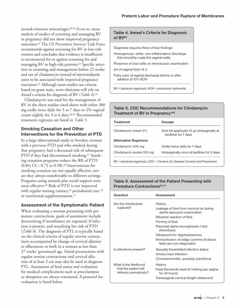
Preterm Labor and Premature Rupture of Membranes
— Chapter D 5
second-trimester miscarriages.60-62 Even so, meta-analysis of studies of screening and managing BV in pregnancy did not show improved pregnancy outcomes.63 The US Preventive Services Task Force recommends against screening for BV in low-risk women and concludes that evidence is insufficient to recommend for or against screening for and managing BV in high-risk patients.64 Specific atten-tion to screening and management before 22 weeks and use of clindamycin instead of metronidazole seem to be associated with improved pregnancy outcomes.65 Although most studies use criteria based on gram stain, most clinicians will rely on Amsel’s criteria for diagnosis of BV (Table 4).66
Clindamycin was used for the management of BV in the three studies cited above with either 300 mg orally twice daily for 5 to 7 days or 2% vaginal cream nightly for 3 to 6 days.60-62 Recommended treatment regimens are listed in Table 5.
Smoking Cessation and Other Interventions for the Prevention of PTDIn a large observational study in Sweden, women with a previous PTD and who smoked during that pregnancy had a decreased risk of subsequent PTD if they had discontinued smoking.67 Smok-ing cessation programs reduce the RR of PTD (0.84; CI = 0.72 to 0.98).68 Interventions for smoking cessation are not equally effective, nor are they always transferrable to different settings. Programs using rewards plus social support were most effective.68 Risk of PTD is not improved with regular nursing contact,69 periodontal care,70 or nutritional supplementation.71
Assessment of the Symptomatic PatientWhen evaluating a woman presenting with pre-mature contractions, goals of assessment include determining if membranes are ruptured, if infec-tion is present, and stratifying her risk of PTD (Table 6). The diagnosis of PTL is typically based on the clinical criteria of regular uterine contrac-tions accompanied by change of cervical dilation or effacement or both in a woman at less than 37 weeks’ gestational age. Initial presentation with regular uterine contractions and cervical dila-tion of at least 2 cm may also be used to diagnose PTL. Assessment of fetal status and evaluation for medical complications such as preeclampsia or abruption are always warranted. A protocol for evaluation is listed below.
Table 4. Amsel’s Criteria for Diagnosis of BV66
Diagnosis requires three of four findings:
Homogenous, white, non-inflammatory discharge that smoothly coats the vaginal walls
Presence of clue cells on microscopic examination
pH of vaginal fluid >4.5
Fishy odor of vaginal discharge before or after addition of 10% KOH
BV = bacterial vaginosis; KOH = potassium hydroxide.
Table 5. CDC Recommendations for Clindamycin Treatment of BV in Pregnancy148
Treatment Dosage
Clindamycin cream 2% One full applicator (5 g) intravaginally at bedtime for 7 days
Alternative Regimens
Clindamycin 300 mg Orally twice daily for 7 days
Clindamycin ovules 100 mg Intravaginally once at bedtime for 3 days
BV = bacterial vaginosis; CDC = Centers for Disease Control and Prevention.
Table 6. Assessment of the Patient Presenting with Premature Contractions72,77
Question Assessment
Are the membranes ruptured?
HistoryLeakage of fluid from cervical os during
sterile speculum examinationNitrazine reaction of fluidFerning of fluidPlacental alpha microglobulin-1 test
(AmniSure)Ultrasound for oligohydramniosAmnioinfusion of indigo carmine (if above
tests are non-diagnostic)
Is infections present? Sexually transmitted infection statusUrinary tract infectionChorioamnionitis, possibly subclinical
What is the likelihood that the patient will delivery prematurely?
HistoryFetal fibronectin level (if nothing per vagina
for 24 hours)Transvaginal cervical length ultrasound

Chapter D
6 Chapter D —
Are the Membranes Ruptured?A sterile speculum examination assists in evaluation of membrane integrity and facilitates collection of fluid for an fFN test. Digital cervical examinations should be avoided to minimize risk of infection. Direct observation of amniotic fluid leaking from the cervical os and pooling in the vaginal vault is diagnostic of ruptured membranes. Gentle fundal pressure or having the patient cough during the examination may facilitate leakage. Amniotic fluid, when allowed to air dry on a slide, will demon-strate ferning or arborization under microscopic evaluation. A false-positive test for ferning is pos-sible if cervical mucus is inadvertently tested. The normal vaginal environment has a pH of 4.5 to 6.0, whereas the pH of amniotic fluid is 7.1 to 7.3. Amniotic fluid will therefore change the color of nitrazine paper from orange to blue.72 A false-posi-tive nitrazine test is possible if blood, semen, or BV is present.72 In equivocal cases, rupture of mem-branes may be diagnosed with a test for placental alpha microglobulin-1 protein (AmniSure) in cervi-covaginal fluid. In a large meta-analysis, the test for placental alpha macroglobulin-1 was 96% sensitive and 98.9% specific for rupture of membranes.73 In some cases of prolonged rupture of membranes, very little fluid may be present for analysis making diagnosis difficult. Oligohydramnios on ultrasound supports the diagnosis of ruptured membranes, but is not diagnostic. If the diagnosis of membrane rupture remains uncertain after a complete evalua-tion, ultrasound guided transabdominal instillation of indigo carmine into the amniotic fluid may be used. The passage of blue-dyed fluid through the vagina, staining a pad or tampon, is diagnostic of membrane rupture.72
Is Infection Present?A high index of suspicion for infection should be used when evaluating a woman with preterm con-tractions. Testing for sexually transmitted infec-tions, urinary tract infection, and BV is warranted. Subclinical chorioamnionitis, infection without the classic findings of fever, uterine tenderness, foul-smelling discharge, and maternal tachycardia, may be present.74 Unless group B Streptococcus (GBS) testing has been done in the past 5 weeks, a vaginal/rectal culture should be collected. If delivery appears likely and GBS culture is either unavailable or positive, GBS prophylaxis should be administered.75
What is the Likelihood of PTD?Diagnosing PTL remains challenging despite the multitude of clinical tools available to the clinician. Fewer than 15% of women presenting to triage with preterm contractions will go on to deliver pre-maturely.76 In one observational study of women hospitalized for threatened PTL, 38% went on to delivery during their first admission, but greater than half who were discharged to home continued their pregnancy until term.77,78 Although frequency of uterine contractions is significantly related to risk of PTD, contraction frequency alone is not sensitive and has a low positive predictive value for PTD.79 Assessment of the cervix by digital exami-nation is subjective and also has a low predictive value for PTD.80 Early, accurate diagnosis of PTL increases the ability to transfer a laboring mother to a facility with an NICU, administer glucocorti-coids, consider magnesium sulfate for neuroprotec-tion, and initiate prophylactic treatment for GBS.
Fetal fibronectin testing may aid in identify-ing women at low risk for PTD who present with symptoms of PTL. fFN is an extracellular matrix glycoprotein found at the maternal-fetal interface, and in normal pregnancies it is almost undetect-able in vaginal secretions. With disruption of this interface, which occurs before the onset of labor, fFN is released into vaginal secretions. fFN testing may be done between 24 and 34 weeks’ gesta-tion and is most useful to identify women who are at low risk for delivery in the following 10 to 14 days. The negative predictive value of the fFN assay is greater than 99% for delivery within 14 days, as confirmed by multiple studies.81 How-ever, the positive predictive value is only 13% to 30% for delivery in 7 to 10 days for symptomatic patients.82 The test should not be performed when there is active vaginal bleeding, or when inter-course, digital vaginal examination, or endovaginal ultrasound has occurred in the preceding 24 hours because these can yield a false positive test result.83
The Role of CL Measurement in PTL ManagementThe presence of a shortened CL has been asso-ciated with preterm birth, although the utility of CL measurement by ultrasound in assessing threatened PTL is not as clear. A retrospective study of 1,077 women presenting with preterm contractions found that although cervical shorten-ing was associated with increased risk of delivery

Preterm Labor and Premature Rupture of Membranes
— Chapter D 7
in the following 7 to 14 days, the overall accuracy of CL alone to predict PTD was relatively poor.76 Another prospective study of 665 women com-bined both CL and fFN testing to risk stratify women presenting for PTL. This study found that women with CL of at least 30 mm had a low risk of delivery within 7 days (0.7%), regardless of the fFN result. Women with a CL less than 15 mm had a high risk of delivery within 7 days (27% if fFN was negative, and 52% if fFN was positive). For women with an intermediate CL between 15 mm and 30 mm, a negative fFN result correctly identified women at low risk (less than 5%) for delivery within 7 days (Figure 1).77 These results are consistent with results from a similar study conducted in 2005.84 By using tests such as fFN and CL measurements, which have high negative predictive values, it may be possible to identify women at low risk for PTD. However, there is no test for early PTL with a high positive predictive value, and accurate determination of PTL remains a challenging diagnosis.
Management of PTLAfter PTL has been diagnosed, several interven-tions should be implemented to improve neonatal outcomes, including maternal transfer to a facility with a higher level nursery if indicated, adminis-tration of corticosteroids, antibiotic prophylaxis of neonatal GBS infection, consideration of magnesium for neuroprotection, and preparation for a preterm birth. Each of these interventions is discussed in more detail below.
Antenatal Corticosteroid (ACS) TreatmentTreatment with ACS decreases neonatal mortality and morbidity as listed in Table 7.
Treatment with ACS does not increase the risk of maternal mortality, chorioamnionitis, or puerperal sepsis.85 ACS use is effective in women with PROM and pregnancy-related hypertension syndromes.72,85 A course of ACS is recommended for women between 24 and 34 weeks’ gestation who are at risk of PTD within 7 days. Steroids may be considered for women between 23 and 24 weeks.86 The optimal effects of ACS appear to wane after a week.87 For women who remain at risk of PTD, a single repeat dose of ACS improves the outcomes of respiratory distress syndrome and poor infant outcomes without affecting long term outcomes. It is recommended that women at
less than 33 weeks’ gestation who remain at risk of delivery within 7 days, and who received their first ACS dose at least 14 days prior, receive a single repeat course of ACS.85,86 However, further doses of ACS do not show additional benefit and are not recommended.88
Table 7. Effect of ACS on Fetal Outcomes in PTL87
Outcome RR (95% CI)
Neonatal mortality 0.69 (0.58 to 0.81)
Respiratory distress syndrome
0.66 (0.59 to 0.73)
Intraventricular hemorrhage 0.54 (0.43 to 0.69)
Necrotizing enterocolitis 0.46 (0.29 to 0.74)
Respiratory support, intensive care admissions
0.80 (0.65 to 0.99)
Systemic infections in the first 48 hours of life
0.56 (0.38 to 0.85)
ACS = antenatal corticosteroids; CI = confidence inter-val; PTL = preterm labor; RR = relative risk.
Figure 1. Risk of Preterm Delivery Within 7 Days Using Fetal Fibronectin and Cervical Length for Risk Stratification
fFN = fetal fibronectin.
Information from van Baaren GJ, Vis JY, Wilms FF, et al. Predictive value of cer-vical length measurement and fibronectin testing in threatened preterm labor. Obstet Gynecol. 2014;123(6):1185-1192.
0
10
20
30
40
50
60
Negative fFNPositive fFN
Cervical length<15 mm
Cervical lengthbetween 15 and 30 mm
Cervical length>30 mm
Pre
term
De
live
ry W
ith
in 7
Day
s (%
)
3.20.0
14.0
2.6
52.0
27.0

Chapter D
8 Chapter D —
A 2016 study suggests that betamethasone reduces respiratory complications when given to women with threatened preterm labor (at least 3cm dilated or 75% effaced), rupture of mem-branes, or an indication for planned late preterm delivery between 34 0/7 and 36 6/7 weeks.
Doses of corticosteroids are noted in Table 8. In general, dexamethasone and betamethasone show similar results in respiratory distress and perinatal mortality, but one meta-analysis showed a decrease in intraventricular hemorrhage and length of NICU stay with use of dexamethasone compared with betamethasone.89 There is evidence that even a single dose of either agent shows benefit, so it is recommended that a first dose be given even if it is unlikely that the patient will receive the subsequent doses. However, there is no evidence of improved outcomes with accelerated dosing (ie, shortening the dosage interval to complete the course before an imminent delivery).85,88,89
Magnesium Sulfate for NeuroprotectionMagnesium sulfate administered immediately before and at the time of delivery of a preterm infant decreases the rate of cerebral palsy (RR 0.68; 95% CI = 0.54 to 0.87, number needed to treat is 63).90 Various trials have shown this clear benefit, but have varied in regard to the regi-
men used in magnesium administration. A 2012 Cochrane review did not show superiority of any regimen over any other and called for further stud-ies to define the optimal dosing.91,92 Two com-monly used regimens are listed in Table 9, and are applicable to women between 24 and 32 weeks at high risk of delivery within 24 hours.91,93
TocolysisTocolytic drugs are used for short-term pregnancy prolongation (up to 48 hours), with the goal of allowing time for administration of ACS, magne-sium sulfate for neuroprotection, antibiotics for GBS prophylaxis, and maternal transfer if neces-sary. They may be useful in pregnancies between viability and 34 weeks’ estimated gestational age with established PTL, and in the absence of evi-dence of maternal or fetal compromise (ie, cho-rioamnionitis, severe preeclampsia, maternal instability, fetal demise or lethal anomaly, worri-some fetal status). After cessation of labor, there is no evidence of benefit of long term tocolysis for further prolongation of pregnancy.85 Pharmaceuti-cal treatment options are listed in Table 10.
Nifedipine decreases the likelihood of deliver-ing within 48 hours (RR 0.30; 95% CI = 0.21 to 0.43). Nifedipine may show advantages over other tocolytics. Compared with betamimetics, nifedip-ine increased time before birth, decreased maternal adverse events, and improved neonatal outcomes (very preterm birth, respiratory distress syndrome, necrotizing enterocolitis, intraventricular hemor-rhage, neonatal jaundice, and NICU admissions). Compared with magnesium sulfate, nifedipine showed decreased maternal adverse events and decreased NICU admissions.94
Betamimetics (eg, terbutaline, ritodrine) are effective in delaying delivery for 48 hours.95 Stud-ies have not shown an improvement in measures of fetal outcomes, but maternal adverse effects and
Table 8. ACS for Fetal Maturation
Corticosteroid Dosage
Betamethasone Two doses of 12 mg IM twice administered 24 hours apart
Dexamethasone Four doses of 6 mg IM administered every 6 hours
ACS = antenatal corticosteroids; IM = intramuscularly.
Table 9. Magnesium Sulfate for Neuroprotection 91,93
Magnesium Sulfate Loading Dose Maintenance Dose Repeat Treatment
4 g over 20 to 30 minutes 1 g/hour continued until birth or for 24 hours
No immediate repeat doses
6 g over 20 to 30 minutes 2 g/hour continued until birth or for 12 hours
If less than 6 hours have elapsed since cessation, restart maintenance dose. If more than 6 hours have elapsed, rebolus and then start maintenance dose.

Preterm Labor and Premature Rupture of Membranes
— Chapter D 9
adverse events are significant.95 Terbutaline is the most commonly used betamimetic for tocolysis. In 2011, the US Food and Drug Administration issued a warning against the use of terbutaline for
tocolysis longer than 48 to 72 hours because of the lack of demonstrated efficacy and the poten-tial for serious maternal cardiac complications and mortality.96 Injectable terbutaline may have
Table 10. Pharmaceutical Agents for Tocolysis
Drug (Class) Dosage Comments Contraindications and Adverse Effects
Magnesium sulfate 4 to 6 g bolus over 15 to 20 minutes, then 1 to 2 g/hour (3 g/hour maximum)
In widespread use in the United States, meta-analysis fails to show improvement in outcomes. Comparison studies show similar effectiveness to other agents in delay of delivery.
May be used for fetal neuroprotection at less than 32 weeks. Dosing regimens vary; use hospital protocols.
Contraindication: myasthenia gravisMaternal adverse effects: flushing, lethargy,
headache, muscle weakness, diplopia, dry mouth, pulmonary edema, cardiac arrest. Toxicity rare with serum level <10 mg/dL. Respiratory depression and subsequent arrest can occur at levels >10 to 12 mg/dL.
Newborn adverse effects: lethargy, hypotonia, respiratory depression, demineralization with prolonged use
Nifedipine (Calcium channel blocker)
30 mg oral loading dose, then 10 to 20 mg every 4 to 6 hours
Nifedipine increases the incidence of delivery before 48 hours. Neonatal mortality not affected. Decreased incidence of neonatal respiratory distress syndrome, necrotizing enterocolitis, intraventricular hemorrhage, and jaundice.
Contraindication: maternal hypotensionMaternal adverse effects: flushing,
headache, dizziness, nausea, transient hypotension
No fetal adverse effects noted.
Terbutaline (Betamimetic)
0.25 mg subcutaneously every 20 minutes for up to three doses
Betamimetic drugs may delay delivery for 48 hours, but neonatal outcomes are variable and maternal adverse effects are common.
Injectable terbutaline may have a narrow role in treatment of tachysystole or in emergent situations.
US FDA warning against long-term or oral use because of maternal adverse events and lack of efficacy.
Maternal contraindications: heart disease, poorly controlled diabetes, thyrotoxicosis
Maternal adverse effects:Cardiac arrhythmias, pulmonary edema,
myocardial ischemia, hypotension, tachycardia
Hyperglycemia, hyperinsulinemia, hypokalemia, antidiuresis, altered thyroid function
Physiologic tremor, palpitations, nervousness, nausea/vomiting, fever, hallucinations
Fetal and neonatal adverse effects: tachycardia, hypoglycemia, hypocalcemia, hyperbilirubinemia, hypotension, intraventricular hemorrhage
Indomethacin (NSAID)
Loading dose: 50 mg rectally or 50 to 100 mg orally
Maintenance dose: 25 to 50 mg orally every 4 hours for 48 hours
NSAIDs theoretically intervene more proximally in the labor cascade than the other agents. Efficacy appears similar to other agents.
Maternal adverse-effect profile is favorable.
Other NSAIDs (sulindac, ketorolac) may be used.
Contraindications: maternal renal or hepatic impairment, active peptic ulcer disease, oligohydramnios
Maternal adverse effects: nausea, heartburnFetal adverse effects: constriction of the
ductus arteriosus (not recommended after 32 weeks’ gestation), pulmonary hypertension, reversible decrease in renal function with oligohydramnios, intraventricular hemorrhage, hyperbilirubinemia, necrotizing enterocolitis
FDA = Food and Drug Administration; NSAID = nonsteroidal anti-inflammatory drug.

Chapter D
10 Chapter D —
a narrow role for tocolysis in emergent settings or with uterine tachysystole.85
Indomethacin, a nonsteroidal anti-inflammatory drug (NSAID), used as a tocolytic agent increases the likelihood of delivery at greater than 37 weeks and average gestational age at delivery (weighted mean difference 3.53 weeks) with low maternal adverse effects.97 Because NSAIDs can interfere with fetal prostaglandin synthesis, concerns have been raised regarding fetal safety. Meta-analyses of observational studies have shown conflicting results in regard to fetal safety (mostly no effects), but have raised concerns about severe intraven-tricular hemorrhage, necrotizing enterocolitis, and periventricular leukomalacia.98-100 More research and RCTs are needed in this area. According to one decision analysis, prostaglandin inhibitors may be the optimal first-line agent for PTL before 32 weeks’ gestation.101 Because of the risk of pre-mature closure of the ductus arteriosus, NSAIDs should not be used for more than 48 hours or beyond 32 weeks’ gestation.
Magnesium sulfate used for tocolysis has not been shown to prolong pregnancy or improve neonatal outcomes when compared with placebo or other tocolytics.102,103 In addition, a Cochrane meta-analysis shows an increased risk of fetal and neonatal mortality of borderline significance (RR 4.56; 95% CI = 1.00 to 20.86) and an increase in length of NICU admission when compared to calcium channel blockers (but not in comparison to NSAIDs).102,103
In women receiving magnesium sulfate for neu-roprotection who continue to labor after admin-istration of magnesium sulfate, consideration may be given to addition of another tocolytic. However, a Cochrane meta-analysis was unable to reach any conclusions regarding combinations of tocolytics in regards to safety and efficacy, cit-ing a lack of large well-designed trials.104 Caution should be used if using calcium channel blockers and magnesium because of theoretical maternal cardiac complications.85
Neonatal GBS ProphylaxisThe incidence of neonatal GBS infection and mortality has dropped significantly since the wide adoption of the guidelines from the Centers for Disease Control and Prevention (CDC) for the prevention of neonatal GBS disease. However, GBS remains the leading cause of mortality due to
infection among neonates.75 Several large retro-spective record reviews show gaps in adherence to current recommendations, especially in PTL, PPROM, and the use of clindamycin as opposed to cefazolin in penicillin-allergic patients.105,106
A vaginal-rectal swab for GBS culture should be obtained when women present with PTL or PPROM if results from prior testing done within the last 5 weeks are not available. Intrapartum antibiotics (penicillin or ampicillin) should be started on admission and continued until birth, or until it is determined that the woman is not in true PTL or a negative GBS culture result becomes available. In cases of PPROM when antibiotics are used for prolonging latency, antibiotic coverage should include coverage for GBS.
Women who are allergic to penicillin should receive cefazolin unless the allergic response was anaphylaxis, angioedema, respiratory distress, or urticaria (Figure 2).75 Clindamycin and vancomy-cin are the antibiotics of last resort for women with life threatening penicillin allergies. GBS isolates show increasing resistance to clindamycin, and clindamycin has poor penetration into amniotic fluid.105 It should be used only when the GBS iso-late can be shown to be sensitive to both clindamy-cin and erythromycin. Vancomycin should be used in women with the serious penicillin aller-gies described and unknown GBS status, or GBS strains resistant to clindamycin. CDC algorithms for screening and GBS prophylaxis treatment of women with threatened PTD are presented below:
Preterm Premature Rupture of the Fetal Membranes (PPROM)Twenty-five percent to 30% of PTDs are pre-ceded by PPROM.2 The biochemical events leading to PPROM appear to be different from those leading to PTL. Collagen fibers contribute to the tensile strength of the fetal membranes. Matrix metalloproteinase (a collagenase) activ-ity is increased, possibly the result of infection or other inflammation, and may be the final com-mon pathway leading to membrane rupture. In the context of placental abruption, thrombin may be involved as well.107
The earlier in pregnancy PROM (and PTL in general) occurs, the more likely that its etiol-ogy is associated with infection.72 Risk factors for PPROM are similar to those for PTL with intact membranes.108 Delivery is likely within a week of
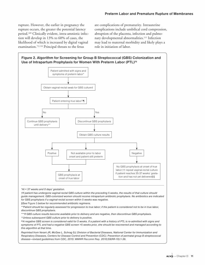
Preterm Labor and Premature Rupture of Membranes
— Chapter D 11
rupture. However, the earlier in pregnancy the rupture occurs, the greater the potential latency period.109 Clinically evident, intra-amniotic infec-tion will develop in 13% to 60% of cases, the likelihood of which is increased by digital vaginal examination.72,110 Principal threats to the fetus
are complications of prematurity. Intrauterine complications include umbilical cord compression, abruption of the placenta, infection and pulmo-nary developmental abnormalities.111 Infection may lead to maternal morbidity and likely plays a role in initiation of labor.
Figure 2. Algorithm for Screening for Group B Streptococcal (GBS) Colonization and Use of Intrapartum Prophylaxis for Women With Preterm Labor (PTL)75
*At < 37 weeks and 0 days’ gestation.†If patient has undergone vaginal-rectal GBS culture within the preceding 5 weeks, the results of that culture should guide management. GBS-colonized women should receive intrapartum antibiotic prophylaxis. No antibiotics are indicatedfor GBS prophylaxis if a vaginal-rectal screen within 5 weeks was negative.§See Figure 3 below for recommended antibiotic regimens.**Patient should be regularly assessed for progression to true labor; if the patient is considered not to be in true labor, discontinue GBS prophylaxis.***If GBS culture results become available prior to delivery and are negative, then discontinue GBS prophylaxis.††Unless subsequent GBS culture prior to delivery is positive.§§A negative GBS screen is considered valid for 5 weeks. If a patient with a history of PTL is re-admitted with signs and symptoms of PTL and had a negative GBS screen >5 weeks prior, she should be rescreened and managed according to this algorithm at that time.
Reprinted from Verani JR, McGee L, Schrag SJ; Division of Bacterial Diseases, National Center for Immunization and Respiratory Diseases, Centers for Disease Control and Prevention (CDC). Prevention of perinatal group B streptococcal disease—revised guidelines from CDC, 2010. MMWR Recomm Rep. 2010;59(RR-10):1-36.
Patient admitted with signs and symptoms of preterm labor*
Obtain vaginal-rectal swab for GBS culture†
YesNo
Continue GBS prophylaxis until delivery**
Discontinue GBS prophylaxis
Patient entering true labor?¶
Obtain GBS culture results
NegativeNot available prior to labor onset and patient still preterm
Positive
No GBS prophylaxis at onset of true labor;†† repeat vaginal-rectal culture if patient reaches 35-37 weeks’ gesta-
tion and has not yet delivered§§GBS prophylaxis at onset of true labor
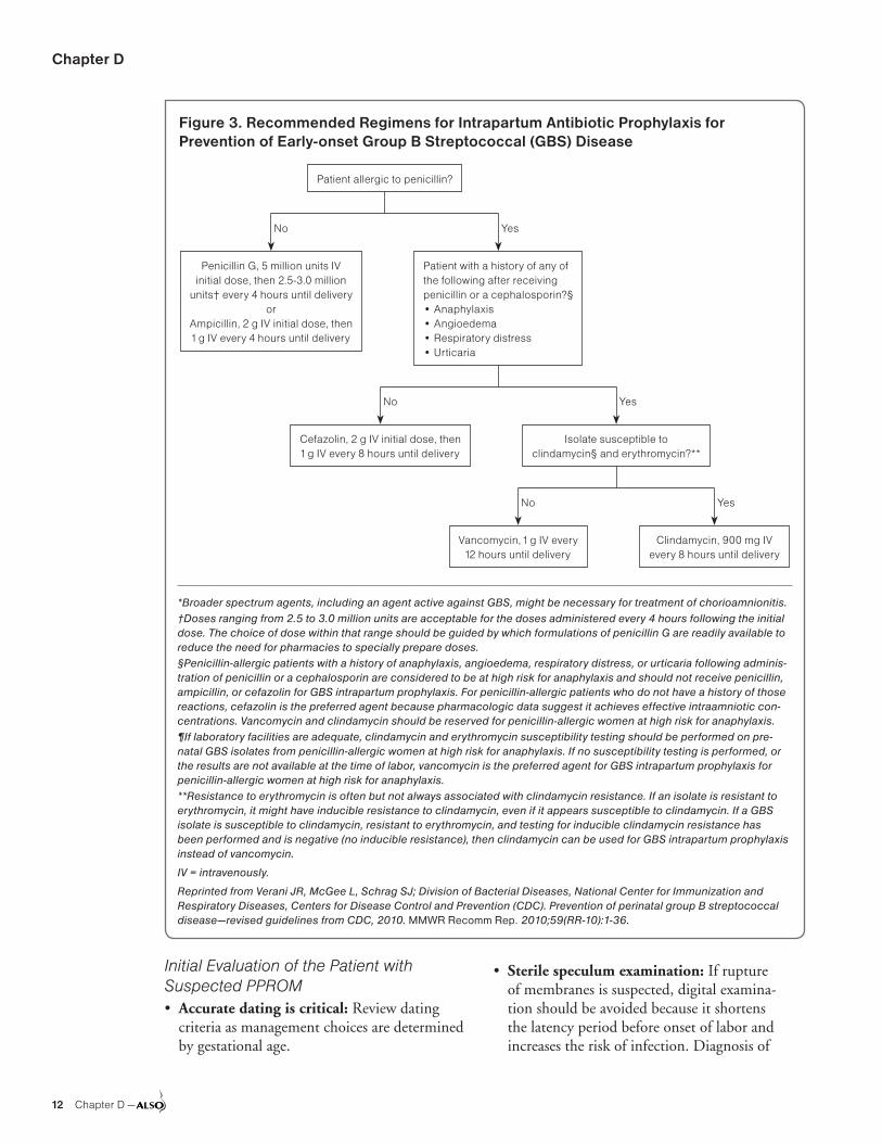
Chapter D
12 Chapter D —
Initial Evaluation of the Patient with Suspected PPROM• Accurate dating is critical: Review dating
criteria as management choices are determined by gestational age.
• Sterile speculum examination: If rupture of membranes is suspected, digital examina-tion should be avoided because it shortens the latency period before onset of labor and increases the risk of infection. Diagnosis of
Figure 3. Recommended Regimens for Intrapartum Antibiotic Prophylaxis for Prevention of Early-onset Group B Streptococcal (GBS) Disease
*Broader spectrum agents, including an agent active against GBS, might be necessary for treatment of chorioamnionitis.†Doses ranging from 2.5 to 3.0 million units are acceptable for the doses administered every 4 hours following the initial dose. The choice of dose within that range should be guided by which formulations of penicillin G are readily available to reduce the need for pharmacies to specially prepare doses.§Penicillin-allergic patients with a history of anaphylaxis, angioedema, respiratory distress, or urticaria following adminis-tration of penicillin or a cephalosporin are considered to be at high risk for anaphylaxis and should not receive penicillin, ampicillin, or cefazolin for GBS intrapartum prophylaxis. For penicillin-allergic patients who do not have a history of those reactions, cefazolin is the preferred agent because pharmacologic data suggest it achieves effective intraamniotic con-centrations. Vancomycin and clindamycin should be reserved for penicillin-allergic women at high risk for anaphylaxis.¶If laboratory facilities are adequate, clindamycin and erythromycin susceptibility testing should be performed on pre-natal GBS isolates from penicillin-allergic women at high risk for anaphylaxis. If no susceptibility testing is performed, or the results are not available at the time of labor, vancomycin is the preferred agent for GBS intrapartum prophylaxis for penicillin-allergic women at high risk for anaphylaxis.**Resistance to erythromycin is often but not always associated with clindamycin resistance. If an isolate is resistant to erythromycin, it might have inducible resistance to clindamycin, even if it appears susceptible to clindamycin. If a GBS isolate is susceptible to clindamycin, resistant to erythromycin, and testing for inducible clindamycin resistance has been performed and is negative (no inducible resistance), then clindamycin can be used for GBS intrapartum prophylaxis instead of vancomycin.
IV = intravenously.
Reprinted from Verani JR, McGee L, Schrag SJ; Division of Bacterial Diseases, National Center for Immunization and Respiratory Diseases, Centers for Disease Control and Prevention (CDC). Prevention of perinatal group B streptococcal disease—revised guidelines from CDC, 2010. MMWR Recomm Rep. 2010;59(RR-10):1-36.
Patient allergic to penicillin?
YesNo
Penicillin G, 5 million units IV initial dose, then 2.5-3.0 million
units† every 4 hours until deliveryor
Ampicillin, 2 g IV initial dose, then 1 g IV every 4 hours until delivery
Patient with a history of any of the following after receiving penicillin or a cephalosporin?§• Anaphylaxis• Angioedema• Respiratory distress• Urticaria
YesNo
Cefazolin, 2 g IV initial dose, then 1 g IV every 8 hours until delivery
Isolate susceptible to clindamycin§ and erythromycin?**
YesNo
Vancomycin, 1 g IV every 12 hours until delivery
Clindamycin, 900 mg IV every 8 hours until delivery

Preterm Labor and Premature Rupture of Membranes
— Chapter D 13
rupture of membranes was previously discussed.
• Ultrasound evaluation: Oligohydramniossupports the diagnosis of membrane rupture. Oligohydramnios will also decrease the accuracy of fetal weight and gestational assessment. Low amniotic volume increases the likelihood of cord compression and other complications.
• Assessment of fetal lung maturity: Vaginalamniotic fluid may be tested for lamellar body count and phosphatidyl glycerol to evaluate fetal lung maturity between 32 and 34 weeks’ gestation.112,113 Amniocentesis allows collection of fluid for fetal pulmonary maturity testing and for evaluation of infection.108
• Screen for infection: Infection is associatedwith PPROM. If GBS status is unknown, then culture should be obtained. Consider screening for sexually transmitted infections.
• Fetal monitoring: Electronic fetal heart rateand uterine contraction monitoring during initial assessment may identify fetal compromise and asymptomatic contractions
Management of PPROMAs in the case of PTL with intact membranes, the management of PPROM necessitates a balance between the advantages of delaying delivery and risks of prolonging fetal exposure to a poten-tially hostile environment. Initial management is predicated on gestational age with the expected benefits of expectant management and admin-istration of corticosteroids increasing for earlier gestational ages. The potential for development of infection and the decreasing likelihood of neonatal morbidity leads to the current standard practice of induction of labor at 34 weeks.114 Aside from that standard, clinical trials have not demonstrated clear advantages to elective delivery.115,116
• Monitor for clinical infection: Maternal fever,uterine tenderness, and fetal tachycardia are indicators of infection.
• Antepartum fetal testing: Recommendationsfor antenatal surveillance are based primar-ily on expert opinion. The non-stress test and biophysical profile are used to assess for fetal compromise.108,117
• Antibiotic therapy: At gestations between 24and 32 weeks, management with antibiotics prolongs pregnancy and decreases fetal morbid-ity, chorioamnionitis, and maternal infection.118
PPROM antibiotic regimens that include ampicillin 1 g intravenously every 6 hours are adequate for GBS prophylaxis, even though a 4-hour dosing interval is typical for routine GBS intrapartum prophylaxis.75 One gram oral dose of azithromycin has been substituted in some protocols for erythromycin with no appar-ent change in outcomes.119,120 The antibiotics and dosages used in a large National Institute of Child Health and Human Development trial are listed in Table 11.121
• Corticosteroids: ACS administration in thesetting of PPROM reduces the risk of neonatal respiratory distress syndrome, intraventricular hemorrhage, and necrotizing enterocolitis.87 There is a trend to decreased neonatal mortality. Incidence of maternal and neonatal infection is not increased.87,122 Corticosteroid dosages are listed in Table 10.
• Tocolysis: A common argument for the use oftocolytics in PTL is that delaying delivery allows for the administration of corticosteroids. As opposed to antibiotic and corticosteroid treat-ment, tocolysis in the context of PPROM lacks evidence of benefit.123 Association of infection with onset of labor would be expected to be stronger in PPROM as opposed to PTL with intact membranes indicating circumspection in choosing tocolysis.
• Magnesium sulfate for neuroprotection: In asecondary analysis of a RCT, magnesium sulfate did not change neonatal outcomes or deliv-ery latency.124 However, magnesium sulfate is
Table 11. Antibiotic Therapy in PPROM121
Antibiotic Dosage
Initial Therapy
Ampicillin 2 g IV every 6 hours for 48 hours
Erythromycin 250 mg IV every 6 hours for 48 hours
Follow-Up Therapy
Amoxicillin 250 mg orally every 8 hours for 5 days
Erythromycin base 333 mg orally every 8 hours for 5 days
IV = intravenously; PPROM = preterm premature rupture of membranes.

Chapter D
14 Chapter D —
typically administered according to one of the several protocols for fetal neuroprotection.
Gestational Age and Management of PPROM• 37 0/7 or more weeks: Induce labor. If
unknown GBS and no other indication forGBS prophylaxis, then there is no need to startantibiotics unless there has been rupture ofmembranes greater than 18 hours or tempera-ture ≥38°C (100.4°F).
• 34 0/7 to 36 6/7 weeks: Offer expectant man-agement versus induction. If GBS unknown,start antibiotics pending results. See the Ante-natal Corticosteroid (ACS) Treatment section for2016 evidence regarding benefit of betametha-sone in this time frame. A 2016 study demon-strated that expectant management of PPROMbetween 34 0/7 and 36 6/7 weeks’ gestation(late preterm) resulted in significantly lowerrates of respiratory distress, mechanical ventila-tion, days spent in NICU, and cesarean deliverybut significantly higher rates of antepartum orintrapartum hemorrhage, intrapartum fever, useof postpartum antibiotics, and longer hospitalstay.125 The authors concluded that offeringexpectant management for women with latepreterm PPROM is reasonable.125
• 24 0/7 to 33 6/7 weeks: Administer antibiot-ics and corticosteroids. Add magnesium sulfatefor neuroprotection when in labor if under 32weeks gestational age. Monitor for infection andother intrauterine fetal complications. If there isno evidence of fetal compromise and labor doesnot begin spontaneously, these pregnancies aremanaged expectantly until they reach 34 weeks.
• 23 0/7 to 23 6/7 weeks: Consider administeringantibiotics and corticosteroids based on parentalchoice only after counseling with neonatologyand maternal-fetal medicine clinicians regardingoutcomes of interventions including cesareandelivery and resuscitation at periviable gestationalage. If resuscitation is planned, add magnesiumsulfate for neuroprotection when in labor.86
PROM at TermRupture of the membranes before the onset of labor occurs in 8% of pregnancies beyond 37 weeks’ ges-tation. In most women, PROM at term is followed quickly by spontaneous labor. Ninety-five percent of women managed expectantly will deliver within
28 hours after PROM.72,126 Steps to diagnose rup-ture of membrane were discussed previously.
Maternal and neonatal infection are the primary concerns when PROM is not followed quickly by spontaneous labor. Antibiotic prophylaxis and early induction have shown benefit in reducing infection in infants of mothers with PROM who are colonized with GBS.127 Planned induction (compared with expectant management) shows a reduction in chorioamnionitis and endometri-tis without affecting risk of operative delivery. Planned induction had no effect on the number of neonatal infections but did reduce the number of NICU admissions.128 Routine administration of antibiotics in PROM does not reduce neonatal sepsis, maternal infections, stillbirth or neonatal mortality, although the quality of the studies included are judged to be low.129
Planned Delivery Versus Expectant Management in Patient Not in LaborLabor induction with oxytocin is recommended when term PROM occurs and labor does not follow.128 In one trial women preferred active to expectant management.126 However, the differ-ences in maternal and neonatal outcomes are minimal for induction versus expectant manage-ment. If mother and fetus are well, then expect-ant management of PROM would be acceptable along with appropriate counseling regarding potential risk.128 Women who are colonized with GBS should receive antibiotic prophylaxis and be encouraged to proceed with induction to reduce neonatal GBS infection.75,127 Other routine administration of antibiotics does not appear to affect maternal or neonatal outcomes.129
Delivery of the Preterm InfantDespite prevention efforts, PTD affected 1 in 9 pregnancies in the United States in 2013 and remains a major factor in infant morbidity and mortality.1 A few interventions can improve out-comes for those infants who are born prematurely.
Premature delivery at a facility with a high-volume level III NICU results in better neonatal outcomes. If delivery is not imminent and level III services are unavailable, then maternal trans-fer is indicated.130,131 For fetuses between 24 to 34 weeks’ gestation, administration of ACS, as discussed previously, decreases the rate of respira-tory distress syndrome, intracranial hemorrhage,

Preterm Labor and Premature Rupture of Membranes
— Chapter D 15
and necrotizing enterocolitis. Administration of magnesium for neuroprotection can decrease the rate of cerebral palsy.
Delayed Umbilical Cord ClampingFor the preterm infant, delayed clamping of the umbilical cord has been shown to decrease intra-ventricular hemorrhage and the need for neonatal transfusion.132,133 To perform delayed cord clamp-ing, the infant should be held at or below the level of the placenta while waiting 30 to 120 seconds before clamping the cord. Required neonatal resuscitation efforts should not be delayed to allow for delayed cord clamping. Delayed cord clamp-ing is recommended as a standard practice in the delivery of the preterm infant. However, further research and larger studies are indicated to confirm and refine these findings.133,134
Route of DeliveryRetrospective observational studies have not shown a clear benefit to any particular route of delivery for the preterm infant.135 The choice of vaginal or cesarean birth should be made based on standard obstetrical indications. The rate of cesarean delivery is higher for preterm than for term deliveries because the indications for surgery are more common in prematurity. Preterm fetuses are more likely to have a malpresentation and are less able to handle the potential stresses of labor. Continuous fetal monitoring is important to detect signs of fetal intolerance of labor. Prophy-lactic episiotomy or forceps delivery have not been shown to have any benefit to the preterm fetus. Vacuum assisted delivery should not be performed at less than 34 weeks because of the risk of intra-cranial hemorrhage.135,136
Infants at the Threshold of ViabilityInfants born at 22 to 25 weeks’ gestation are con-sidered to be at the threshold of viability. They are at a high risk of neonatal mortality and severe long term disability, and the difference in maturity of even a few days can significantly affect these risks. Parents of these infants should receive careful counseling and accurate information to assist them in making decisions regarding tocolysis, labor management, resuscitation, and NICU interven-tions.130 Based on outcome information from 1997 to 2003, the National Institute of Child Health and Human Development (NICHD) has
developed a web-based tool for describing out-comes of these neonates based on gestational age, weight, sex, and administration of ACS, which is available at http://www.nichd.nih.gov/about/org/cdbpm/pp/prog_epbo/epbo_case.cfm. The physician counseling the parents should also know the most recent outcome data for their own NICU facility to assist with decision making.
Global ImplicationsLow-income countries often do not have the resources needed for the prevention and manage-ment of PTL. Infants born at 32 weeks, who may have had a normal life in other parts of the world, may die of respiratory failure because a respirator and surfactant are unavailable, as is an infrastructure for rapid transport to a tertiary care facility. Com-plex social and medical infrastructure necessary for technology-based care and follow-up of preterm neonates may not exist in these settings. In 2012, a coalition of experts released Born Too Soon: The Global Action Report on Preterm Birth. It lists the first country-level estimates of preterm birth and revealed that there are about 15 million preterm births per year137 with over 85% occurring in Asia and Africa.138 In 2014, the stakeholders who created that report also released Every Newborn: An Action Plan to Avoid Preventable Deaths. The compo-nents of the action plan for preterm birth included worldwide access to newborn resuscitation, low costs corticosteroids,139 and kangaroo care in which maternal skin-to-skin care is used in place of an incubator. Others have urged caution with intro-ducing corticosteroids until there is evidence sup-porting benefit in low-resource settings.140 An RCT on ACS treatment was conducted in six countries: Argentina, Guatemala, India, Kenya, Pakistan, and Zambia.141 The study showed no benefit from ACS treatment and increased rates of maternal infection. Additional research is needed to clarify the role of ACS treatment in low-resource settings. More information on PTL in developing countries can be found in the Global ALSO Program (available at www.aafp.org/globalalso).
ConclusionIt is possible to identify some patients at high risk for PTD, and a subset of these may benefit from preventive interventions such as antenatal proges-terone. In the triage of patients presenting with preterm contractions, it is possible to stratify their

Chapter D
16 Chapter D —
risk of subsequent PTD. The major role of tocoly-sis is delay of delivery during the 48 hours neces-sary for full therapeutic effect of antenatal steroids. Management of PPROM presents its own set of challenges and is primarily based on gestational age. Antenatal steroids and antibiotics are useful in some cases. Although term PROM is often man-
aged expectantly, there is little research to support that approach and some research supports imme-diate induction of labor or induction after a short interval of expectant management. PTL remains an area of intense research activity and therapeutic evolution. Addressing the role of infection in PTL is a particular area of interest.
SORT: Key Recommendations for Practice
Clinical RecommendationEvidence Rating References
Antenatal progesterone supplementation decreases the incidence of preterm delivery in high-risk patients.
A 41
Administration of corticosteroids in women with preterm labor between 26 and 34 weeks’ gestation reduces the incidence of neonatal mortality, respiratory distress syndrome, and intraventricular hemorrhage.
A 87
Magnesium sulfate before delivery of infants between 24 to 32 weeks’ gestation reduces the rate of cerebral palsy.
A 90
Betamethasone reduces respiratory complications when given to women with threatened preterm labor (at least 3 cm dilated or 75% effaced) or ruptures of membranes or an indication for planned late preterm delivery between 34 0/7 and 36 6/7 weeks.
B 142
Expectant management with PPROM between 34 0/7 and 36 6/7 weeks is associated with significantly lower rates respiratory distress, mechanical ventilation, days spent in NICU, and cesarean delivery but significantly higher rates of antepartum or intrapartum hemorrhage, intrapartum fever, use of postpartum antibiotics, and longer hospital stay.
B 125
Strength of Recommendation Taxonomy (SORT)
Strength of Recommendation Definition
A • Recommendation based on consistent and good-quality patient-oriented evidence.
B • Recommendation based on inconsistent or limited-quality patient-oriented evidence.
C • Recommendation based on consensus, usual practice, opinion, disease-oriented evidence, or case series for studies of diagnosis, treatment, prevention, or screening.
Patient-oriented evidence measures outcomes that matter to patients: morbidity, mortality, symptom improvement, cost reduction, and quality of life. Disease-oriented evidence measures intermediate, physiologic, or surrogate end points that might not reflect improvement in patient outcomes (eg, blood pressure, blood chemistry, physiologic function, pathologic findings).
NICU = neonatal intensive care unit; PPROM = preterm premature rupture of membranes.
From Ebell MH, Siwek J, Weiss BD, et al. Strength of recommendation taxonomy (SORT): a patient-centered approach to grading evidence in the medical literature. Am Fam Physician. 2004;69(3):548-556.

Preterm Labor and Premature Rupture of Membranes
— Chapter D 17
References1. Martin JA, Hamilton BE, Osterman MJK. Births in the
United States, 2013. NCHS data brief, no 175. Hyattsville, MD: National Center for Health Statistics. Dec 2014.
2. Goldenberg RL, Culhane JF, Iams JD, Romero R. Epi-demiology and causes of preterm birth. Lancet. 2008;371(9606):75-84.
3. Hamilton BE, Martin JA, Osterman MJK, Curtin SC.Births: Prelimnary data for 2013. National vital statisticsreports; vol 63 no 2. Hyattsville, MD: National Center forHealth Statistics. May 2014.
4. Schoen CN, Tabbah S, Iams JD, Caughey AB, BerghellaV, Why the United States preterm birth rate is declining.Am J Obstet Gynecol. 2015;213(2):175-180.
5. Chang HH, Larson J, Blencowe H, et al; Born Too Soonpreterm prevention analysis group. Preventing pretermbirths: analysis of trends and potential reductions withinterventions in 39 countries with very high humandevelopment index. Lancet. 2013;381(9862):223-234.
6. Martin JA, Hamilton BE, Osterman MJ, Curtin SC, Mat-thews TJ. Births: final data for 2012. Natl Vital Stat Rep.2013;62(9):1-68.
7. Muglia LJ, Katz M. The enigma of spontaneous pretermbirth. N Engl J Med. 2010;362(6):529-535.
8. Liong S, Di Quinzio MK, Fleming G, Permezel M, RiceGE, Georgiou HM. Prediction of spontaneous pretermlabour in at-risk pregnant women. Reproduction. 2013;146(4):335-345.
9. Davey MA, Watson L, Rayner JA, Rowlands S. Riskscoring systems for predicting preterm birth withthe aim of reducing associated adverse outcomes.Cochrane Database Syst Rev. 2011;(11):CD004902.
10. Kazemier BM, Buijs PE, Mignini L, Limpens J, de GrootCJ, Mol BW; EBM CONNECT. Impact of obstetric historyon the risk of spontaneous preterm birth in singletonand multiple pregnancies: a systematic review. BJOG.2014;121(10):1197-1208, discussion 1209.
11. Lykke JA, Paidas MJ, Langhoff-Roos J. Recurring com-plications in second pregnancy. Obstet Gynecol. 2009;113(6):1217-1224.
12. Ananth CV, Vintzileos AM. Medically indicated pretermbirth: recognizing the importance of the problem. [viii.].Clin Perinatol. 2008;35(1):53-67, viii.
13. Stock S, Norman J. Preterm and term labour in multiplepregnancies. Semin Fetal Neonatal Med. 2010;15(6):336-341.
14. Martin JA, Hamilton BE, Ventura SJ, Osterman MJ,Mathews TJ. Births: final data for 2011. Natl Vital StatRep. 2013;62(1):1-69, 72.
15. Iams JD, Goldenberg RL, Meis PJ, et al; National Insti-tute of Child Health and Human Development MaternalFetal Medicine Unit Network. The length of the cervixand the risk of spontaneous premature delivery. N EnglJ Med. 1996;334(9):567-572.
16. Hernandez-Andrade E, Romero R, Ahn H, et al. Trans-abdominal evaluation of uterine cervical length duringpregnancy fails to identify a substantial number ofwomen with a short cervix. J Matern Fetal NeonatalMed. 2012;25(9):1682-1689.
17. Berghella V, Tolosa JE, Kuhlman K, Weiner S, BologneseRJ, Wapner RJ. Cervical ultrasonography compared withmanual examination as a predictor of preterm delivery.Am J Obstet Gynecol. 1997;177(4):723-730.
18. Berghella V, Baxter JK, Hendrix NW. Cervical assess-ment by ultrasound for preventing preterm delivery.Cochrane Database Syst Rev. 2013;1(1):CD007235.
19. Berghella V, Roman A, Daskalakis C, Ness A, BaxterJK. Gestational age at cervical length measurementand incidence of preterm birth. Obstet Gynecol. 2007;110(2 Pt 1):311-317.
20. Behrendt N, Gibbs RS, Lynch A, Hart J, West NA, IamsJD. Rate of change in cervical length in women withvaginal bleeding during pregnancy. Obstet Gynecol.2013;121(2 Pt 1):260-264.
21. Fonseca EB, Celik E, Parra M, Singh M, Nicolaides KH;Fetal Medicine Foundation Second Trimester Screen-ing Group. Progesterone and the risk of preterm birthamong women with a short cervix. N Engl J Med. 2007;357(5):462-469.
22. Hassan SS, Romero R, Vidyadhari D, et al; PREGNANTTrial. Vaginal progesterone reduces the rate of pretermbirth in women with a sonographic short cervix: a mul-ticenter, randomized, double-blind, placebo-controlledtrial. Ultrasound Obstet Gynecol. 2011;38(1):18-31.
23. Berghella V. Universal cervical length screening for pre-diction and prevention of preterm birth. Obstet GynecolSurv. 2012;67(10):653-658.
24. Society for Maternal-Fetal Medicine Publications Com-mittee, with assistance of Vincenzo Berghella. Proges-terone and preterm birth prevention: translating clinicaltrials data into clinical practice. Am J Obstet Gynecol.2012;206(5):376-386.
25. Leitich H, Bodner-Adler B, Brunbauer M, Kaider A,Egarter C, Husslein P. Bacterial vaginosis as a risk fac-tor for preterm delivery: a meta-analysis. Am J ObstetGynecol. 2003;189(1):139-147.
26. Cunnington M, Kortsalioudaki C, Heath P. Genitourinarypathogens and preterm birth. Curr Opin Infect Dis.2013;26(3):219-230.
27. Murtha AP, Edwards JM. The role of Mycoplasma andUreaplasma in adverse pregnancy outcomes. ObstetGynecol Clin North Am. 2014;41(4):615-627.
28. Offenbacher S, Boggess KA, Murtha AP, et al. Progres-sive periodontal disease and risk of very preterm deliv-ery. Obstet Gynecol. 2006;107(1):29-36.
29. Iams JD, Newman RB, Thom EA, et al; National Instituteof Child Health and Human Development Network ofMaternal-Fetal Medicine Units. Frequency of uterinecontractions and the risk of spontaneous preterm deliv-ery. N Engl J Med. 2002;346(4):250-255.
30. Conde-Agudelo A, Rosas-Bermúdez A, Kafury-GoetaAC. Birth spacing and risk of adverse perinatal out-comes: a meta-analysis. JAMA. 2006;295(15):1809-1823.
31. Moutquin JM. Classification and heterogeneity of pre-term birth. BJOG. 2003;110(Suppl 20):30-33.

Chapter D
18 Chapter D —
32. Conner SN, Frey HA, Cahill AG, Macones GA, ColditzGA, Tuuli MG. Loop electrosurgical excision procedureand risk of preterm birth: a systematic review and meta-analysis. Obstet Gynecol. 2014;123(4):752-761.
33. McIntosh J, Feltovich H, Berghella V, Manuck T; Soci-ety for Maternal-Fetal Medicine (SMFM). Electronicaddress: [email protected]. The role of routine cervicallength screening in selected high- and low-risk womenfor preterm birth prevention. Am J Obstet Gynecol.2016;215(3):B2-B7.
34. Hansen C, Neller A, Williams G, Simpson R. Maternalexposure to low levels of ambient air pollution andpreterm birth in Brisbane, Australia. BJOG. 2006;113(8):935-941.
35. Simpson WJ. A preliminary report on cigarette smokingand the incidence of prematurity. Am J Obstet Gynecol.1957;73(4):807-815.
36. Behrman RE, Butler AS, eds. Preterm Birth: Causes,Consequences, and Prevention. Washington, DC:National Academies Press; 2007.
37. McCowan LM, Dekker GA, Chane E, et al;SCOPEconsortium. Spontaneous preterm birth and small forgestational age infants in women who stop smokingearly in pregnancy: prospective cohort study. BMJ.2009;338:b1081. Erratum in BMJ. 2009;338:b1558.
38. Iams JD, Romero R, Culhane JF, Goldenberg RL. Pri-mary, secondary, and tertiary interventions to reducethe morbidity and mortality of preterm birth. Lancet.2008;371(9607):164-175.
39. Iams JD. Clinical practice. Prevention of preterm parturi-tion. N Engl J Med. 2014;370(3):254-261.
40. Committee on Practice Bulletins—Obstetrics, TheAmerican College of Obstetricians and Gynecologists.Practice bulletin no. 130: prediction and prevention ofpreterm birth. Obstet Gynecol. 2012;120(4):964-973.
41. Dodd JM, Jones L, Flenady V, Cincotta R, Crowther CA.Prenatal administration of progesterone for prevent-ing preterm birth in women considered to be at risk ofpreterm birth. Cochrane Database Syst Rev. 2013;7(7):CD004947.
42. O’Brien JM, Adair CD, Lewis DF, et al. Progesteronevaginal gel for the reduction of recurrent preterm birth:primary results from a randomized, double-blind, pla-cebo-controlled trial. Ultrasound Obstet Gynecol. 2007;30(5):687-696.
43. Rebarber A, Ferrara LA, Hanley ML, et al. Increasedrecurrence of preterm delivery with early cessation of17-alpha-hydroxyprogesterone caproate. Am J ObstetGynecol. 2007;196(3):224.e1-224.e4.
44. Romero R, Espinoza J, Kusanovic JP, et al. The pretermparturition syndrome. BJOG. 2006;113(Suppl 3):17-42.
45. Berghella V, Rafael TJ, Szychowski JM, Rust OA, Owen J.Cerclage for short cervix on ultrasonography in womenwith singleton gestations and previous preterm birth: ameta-analysis. Obstet Gynecol. 2011;117(3):663-671.
46. Owen J, Hankins G, Iams JD, et al. Multicenter random-ized trial of cerclage for preterm birth prevention inhigh-risk women with shortened midtrimester cervicallength. Am J Obstet Gynecol. 2009;201(4):375.e1-375.e8.
47. Conde-Agudelo A, Romero R, Nicolaides K, et al. Vagi-nal progesterone vs. cervical cerclage for the preven-tion of preterm birth in women with a sonographic shortcervix, previous preterm birth, and singleton gestation:a systematic review and indirect comparison metaanaly-sis. Am J Obstet Gynecol. 2013;208(1):42.e1-42.e18.
48. Romero R, Nicolaides K, Conde-Agudelo A, et al.Vaginal progesterone in women with an asymptomaticsonographic short cervix in the midtrimester decreasespreterm delivery and neonatal morbidity: a systematicreview and metaanalysis of individual patient data. AmJ Obstet Gynecol. 2012;206(2):124.e1-124.e19.
49. Grobman WA, Thom EA, Spong CY, et al; Eunice Ken-nedy Shriver National Institute of Child Health andHuman Development Maternal-Fetal Medicine Units(MFMU) Network. 17 alpha-hydroxyprogesterone capro-ate to prevent prematurity in nulliparas with cervicallength less than 30 mm. Am J Obstet Gynecol. 2012;207(5):390.e1-390.e8.
50. Alfirevic Z, Stampalija T, Roberts D, Jorgensen AL.Cervical stitch (cerclage) for preventing preterm birthin singleton pregnancy. Cochrane Database Syst Rev.2012;4(4):CD008991.
51. Rafael TJ, Berghella V, Alfirevic Z. Cervical stitch (cer-clage) for preventing preterm birth in multiple pregnancy.Cochrane Database Syst Rev. 2014;9(9):CD009166.
52. Rode L, Tabor A. Prevention of preterm delivery in twinpregnancy. Best Pract Res Clin Obstet Gynaecol. 2014;28(2):273-283.
53. Arabin B, Alfirevic Z. Cervical pessaries for preventionof spontaneous preterm birth: past, present and future.Ultrasound Obstet Gynecol. 2013;42(4):390-399.
54. Goya M, Pratcorona L, Merced C, et al; Pesario Cervi-cal para Evitar Prematuridad (PECEP) Trial Group.Cervical pessary in pregnant women with a short cervix(PECEP): an open-label randomised controlled trial.Lancet. 2012;379(9828):1800-1806.
55. Liem S, Schuit E, Hegeman M, et al. Cervical pessariesfor prevention of preterm birth in women with a mul-tiple pregnancy (ProTWIN): a multicentre, open-labelrandomised controlled trial. Lancet. 2013;382(9901):1341-1349.
56. Romero R, Dey SK, Fisher SJ. Preterm labor: one syn-drome, many causes. Science. 2014;345(6198):760-765.
57. Macones GA, Parry S, Nelson DB, et al. Treatment oflocalized periodontal disease in pregnancy does notreduce the occurrence of preterm birth: results from thePeriodontal Infections and Prematurity Study (PIPS). AmJ Obstet Gynecol. 2010;202(2):147.e1-147.e8.
58. Smaill F, Vazquez JC. Antibiotics for asymptomaticbacteriuria in pregnancy. Cochrane Database Syst Rev.2007;(2):CD000490.
59. Leitich H, Kiss H. Asymptomatic bacterial vaginosisand intermediate flora as risk factors for adverse preg-nancy outcome. Best Pract Res Clin Obstet Gynaecol.2007;21(3):375-390.
60. Lamont RF, Duncan SLB, Mandal D, Bassett P. Intra-vaginal clindamycin to reduce preterm birth in womenwith abnormal genital tract flora. Obstet Gynecol.2003;101(3):516-522.

Preterm Labor and Premature Rupture of Membranes
— Chapter D 19
61. Ugwumadu A, Manyonda I, Reid F, Hay P. Effect of earlyoral clindamycin on late miscarriage and preterm deliv-ery in asymptomatic women with abnormal vaginal floraand bacterial vaginosis: a randomised controlled trial.Lancet. 2003;361(9362):983-988.
62. Kiss H, Petricevic L, Husslein P. Prospective ran-domised controlled trial of an infection screening pro-gramme to reduce the rate of preterm delivery. BMJ.2004;329(7462):371-374.
63. Brocklehurst P, Gordon A, Heatley E, Milan SJ. Anti-biotics for treating bacterial vaginosis in pregnancy.Cochrane Database Syst Rev. 2013;1(1):CD000262.
64. U.S. Preventive Services Task Force. Screening for bac-terial vaginosis in pregnancy to prevent preterm delivery:U.S. Preventive Services Task Force recommendationstatement. Ann Intern Med. 2008;148(3):214-219.
65. Oliver RS, Lamont RF. Infection and antibiotics in theaetiology, prediction and prevention of preterm birth.J Obstet Gynaecol. 2013;33(8):768-775.
66. Amsel R, Totten PA, Spiegel CA, Chen KC, EschenbachD, Holmes KK. Nonspecific vaginitis. Diagnostic criteriaand microbial and epidemiologic associations. Am JMed. 1983;74(1):14-22.
67. Cnattingius S, Granath F, Petersson G, Harlow BL. Theinfluence of gestational age and smoking habits on therisk of subsequent preterm deliveries. N Engl J Med.1999;341(13):943-948.
68. Coleman T, Chamberlain C, Davey MA, Cooper SE,Leonardi-Bee J. Pharmacological interventions for pro-moting smoking cessation during pregnancy. CochraneDatabase Syst Rev. 2012;9(9):CD010078.
69. Dyson DC, Danbe KH, Bamber JA, et al. Monitoringwomen at risk for preterm labor. N Engl J Med. 1998;338(1):15-19.
70. Michalowicz BS, Hodges JS, DiAngelis AJ, et al; OPTStudy. Treatment of periodontal disease and the risk ofpreterm birth. N Engl J Med. 2006;355(18):1885-1894.
71. Kramer MS, Kakuma R. Energy and protein intake inpregnancy. Cochrane Database Syst Rev. 2003;(4):CD000032.
72. Practice bulletins No. 139: premature rupture of mem-branes. Obstet Gynecol. 2013;122(4):918-930.
73. Ramsauer B, Vidaeff AC, Hösli I, et al. The diagnosisof rupture of fetal membranes (ROM): a meta-analysis.J Perinat Med. 2013;41(3):233-240.
74. Ratcliffe SD, Baxley EG, Cline MK, Sakornbut EL, eds.Family Medicine Obstetrics. 3rd ed. Philadelphia, PA:Elsevier, 2008. 448-457.
75. Verani JR, McGee L, Schrag SJ; Division of Bacte-rial Diseases, National Center for Immunization andRespiratory Diseases, Centers for Disease Control andPrevention (CDC). Prevention of perinatal group B strep-tococcal disease—revised guidelines from CDC, 2010.MMWR Recomm Rep. 2010;59(RR-10):1-36.
76. Melamed N, Hiersch L, Domniz N, Maresky A, BardinR, Yogev Y. Predictive value of cervical length in womenwith threatened preterm labor. Obstet Gynecol. 2013;122(6):1279-1287.
77. van Baaren GJ, Vis JY, Wilms FF, et al. Predictive valueof cervical length measurement and fibronectin testingin threatened preterm labor. Obstet Gynecol. 2014;123(6):1185-1192.
78. McPheeters ML, Miller WC, Hartmann KE, et al. The epi-demiology of threatened preterm labor: a prospectivecohort study. Am J Obstet Gynecol. 2005;192(4):1325-1329, discussion 1329-1330.
79. Berghella V, Iams JD, Newman RB, et al; National Insti-tute of Child Health and Human Development Networkof Maternal-Fetal Medicine Units. Frequency of uterinecontractions in asymptomatic pregnant women with orwithout a short cervix on transvaginal ultrasound scan.Am J Obstet Gynecol. 2004;191(4):1253-1256.
80. Gomez R, Galasso M, Romero R, et al. Ultrasonographicexamination of the uterine cervix is better than cervicaldigital examination as a predictor of the likelihood of pre-mature delivery in patients with preterm labor and intactmembranes. Am J Obstet Gynecol. 1994;171(4):956-964.
81. Anwar A, Lindow SW, Greaves L, Hall S, Jha R. Theuse of fetal fibronectin in suspected pre-term labour.J Obstet Gynaecol. 2014;34(1):45-47.
82. Honest H, Bachmann LM, Gupta JK, Kleijnen J, KhanKS. Accuracy of cervicovaginal fetal fibronectin test inpredicting risk of spontaneous preterm birth: systematicreview. BMJ. 2002;325(7359):301.
83. Ramsey PS, Andrews WW. Biochemical predictors ofpreterm labor: fetal fibronectin and salivary estriol. ClinPerinatol. 2003;30(4):701-733.
84. Gomez R, Romero R, Medina L, et al. Cervicovaginalfibronectin improves the prediction of preterm deliverybased on sonographic cervical length in patients withpreterm uterine contractions and intact membranes.Am J Obstet Gynecol. 2005;192(2):350-359.
85. American College of Obstetricians and Gynecologists;Committee on Practice Bulletins—Obstetrics. ACOGpractice bulletin no. 127: Management of preterm labor.Obstet Gynecol. 2012;119(6):1308-1317.
86. American College of Obstetricians and Gynecologists.Practice Bulletin No. 159: Management of PretermLabor. Obstet Gynecol. 2016;127(1):e29-e38.
87. Roberts D, Dalziel S. Antenatal corticosteroids foraccelerating fetal lung maturation for women at risk ofpreterm birth. Cochrane Database Syst Rev. 2006;(3):CD004454.
88. Crowther CA, McKinlay CJD, Middleton P, Harding JE.Repeat doses of prenatal corticosteroids for women atrisk of preterm birth for improving neonatal health out-comes. Cochrane Database Syst Rev. 2011;(6):CD003935.
89. Brownfoot FC, Gagliardi DI, Bain E, Middleton P, CrowtherCA. Different corticosteroids and regimens for accelerat-ing fetal lung maturation for women at risk of pretermbirth. Cochrane Database Syst Rev. 2013;8(8):CD006764.
90. Doyle LW, Crowther CA, Middleton P, Marret S, RouseD. Magnesium sulphate for women at risk of pretermbirth for neuroprotection of the fetus. Cochrane Data-base Syst Rev. 2009;(1):CD004661.
91. Reeves SA, Gibbs RS, Clark SL. Magnesium for fetalneuroprotection. Am J Obstet Gynecol. 2011;204(3):202.e1-202.e4.

Chapter D
20 Chapter D —
92. Bain E, Middleton P, Crowther CA. Different magnesiumsulphate regimens for neuroprotection of the fetus forwomen at risk of preterm birth. Cochrane DatabaseSyst Rev. 2012;2(2):CD009302.
93. Magee L, Sawchuck D, Synnes A, von Dadelszen P.SOGC Clinical Practice Guideline. Magnesium sulphatefor fetal neuroprotection. J Obstet Gynaecol Can. 2011;33(5):516-529.
94. Flenady V, Wojcieszek AM, Papatsonis DNM, et al. Cal-cium channel blockers for inhibiting preterm labour andbirth. Cochrane Database Syst Rev. 2014;6(6):CD002255.
95. Neilson JP, West HM, Dowswell T. Betamimetics forinhibiting preterm labour. Cochrane Database Syst Rev.2014;2(2):CD004352.
96. US Food and Drug Administration. FDA Drug SafetyCommunication: New warnings against use of terbuta-line to treat preterm labor. Available at http://www.fda.gov/Drugs/DrugSafety/ucm243539.htm.
97. Khanprakob T, Laopaiboon M, Lumbiganon P, Sang-komkamhang US. Cyclo-oxygenase (COX) inhibitorsfor preventing preterm labour. Cochrane Database SystRev. 2012;10(10):CD007748.
98. Loe SM, Sanchez-Ramos L, Kaunitz AM. Assessing theneonatal safety of indomethacin tocolysis: a system-atic review with meta-analysis. Obstet Gynecol. 2005;106(1):173-179.
99. Hammers AL, Sanchez-Ramos L, Kaunitz AM. Ante-natal exposure to indomethacin increases the riskof severe intraventricular hemorrhage, necrotizingenterocolitis, and periventricular leukomalacia: a sys-tematic review with metaanalysis. Am J Obstet Gynecol.2015;212(4):505.e1-505.e13.
100. Amin SB, Sinkin RA, Glantz JC. Metaanalysis of the effect of antenatal indomethacin on neonatal outcomes. Am J Obstet Gynecol. 2007;197(5):486.e1-486.e10.
101. Haas DM, Imperiale TF, Kirkpatrick PR, Klein RW, Zollinger TW, Golichowski AM. Tocolytic therapy: a meta-analysis and decision analysis. Obstet Gynecol. 2009;113(3):585-594.
102. Crowther CA, Brown J, McKinlay CJD, Middleton P. Magnesium sulphate for preventing preterm birth in threatened preterm labour. Cochrane Database Syst Rev. 2014;8(8):CD001060.
103. Mercer BM, Merlino AA; Society for Maternal-Fetal Medicine. Magnesium sulfate for preterm labor and pre-term birth. Obstet Gynecol. 2009;114(3):650-668.
104. Vogel JP, Nardin JM, Dowswell T, West HM, Oladapo OT. Combination of tocolytic agents for inhibiting pre-term labour. Cochrane Database Syst Rev. 2014;7(7): CD006169.
105. Fairlie T, Zell ER, Schrag S. Effectiveness of intrapar-tum antibiotic prophylaxis for prevention of early-onset group B streptococcal disease. Obstet Gynecol. 2013; 121(3):570-577.
106. American College of Obstetricians and Gynecologists Committee on Obstetric Practice. ACOG Committee Opinion No. 485: Prevention of early-onset group B streptococcal disease in newborns. Obstet Gynecol. 2011;117(4):1019-1027.
107. Thomson A, Norman J. Biology of preterm labour. In: Norman J, Greer I, eds. Preterm Labour, managing risk in clinical practice. Cambridge, UK: Cambridge Univer-sity Press; 2005. 26-75.
108. Simhan HN, Canavan TP. Preterm premature rupture of membranes: diagnosis, evaluation and management strategies. BJOG. 2005;112(Suppl 1):32-37.
109. Melamed N, Hadar E, Ben-Haroush A, Kaplan B, Yogev Y. Factors affecting the duration of the latency period in preterm premature rupture of membranes. J Matern Fetal Neonatal Med. 2009;22(11):1051-1056.
110. Alexander JM, Mercer BM, Miodovnik M, et al. The impact of digital cervical examination on expectantly managed preterm rupture of membranes. Am J Obstet Gynecol. 2000;183(4):1003-1007.
111. Mercer BM, Crocker LG, Boe NM, Sibai BM. Induction versus expectant management in premature rupture of the membranes with mature amniotic fluid at 32 to 36 weeks: a randomized trial. Am J Obstet Gynecol. 1993;169(4):775-782.
112. Lewis DF, Futayyeh S, Towers CV, Asrat T, Edwards MS, Brooks GG. Preterm delivery from 34 to 37 weeks of gestation: is respiratory distress syndrome a problem? Am J Obstet Gynecol. 1996;174(2):525-528.
113. Besnard AE, Wirjosoekarto SA, Broeze KA, Opmeer BC, Mol BW. Lecithin/sphingomyelin ratio and lamellar body count for fetal lung maturity: a meta-analysis. Eur J Obstet Gynecol Reprod Biol. 2013;169(2):177-183.
114. Naef RW III, Allbert JR, Ross EL, Weber BM, Martin RW, Morrison JC. Premature rupture of membranes at 34 to 37 weeks’ gestation: aggressive versus conservative management. Am J Obstet Gynecol. 1998;178(1 Pt 1): 126-130.
115. van der Ham DP, van der Heyden JL, Opmeer BC, et al. Management of late-preterm premature rupture of mem-branes: the PPROMEXIL-2 trial. Am J Obstet Gynecol. 2012;207(4):276.e1-276.e10.
116. Buchanan SL, Crowther CA, Levett KM, Middleton P, Morris J. Planned early birth versus expectant man-agement for women with preterm prelabour rupture of membranes prior to 37 weeks’ gestation for improving pregnancy outcome. Cochrane Database Syst Rev. 2010;(3):CD004735.
117. Sharp GC, Stock SJ, Norman JE. Fetal assessment methods for improving neonatal and maternal out-comes in preterm prelabour rupture of membranes. Cochrane Database Syst Rev. 2014;10(10):CD010209.
118. Kenyon S, Boulvain M, Neilson JP. Antibiotics for pre-term rupture of membranes. Cochrane Database Syst Rev. 2013;12(12):CD001058.
119. Pierson RC, Gordon SS, Haas DM. A retrospective com-parison of antibiotic regimens for preterm premature rup-ture of membranes. Obstet Gynecol. 2014;124(3):515-519.
120. Haas D. Preterm Premature Rupture of Membranes: Erythromycin Versus Azithromycin a Randomized Trial Comparing Their Efficacy to Prolong Latency (PEACE Trial). Available at http://clinicaltrials.gov/show/NCT01556334.

Preterm Labor and Premature Rupture of Membranes
— Chapter D 21
121. Mercer BM, Miodovnik M, Thurnau GR, et al; National Institute of Child Health and Human Development Maternal-Fetal Medicine Units Network. Antibiotic therapy for reduction of infant morbidity after preterm premature rupture of the membranes. A randomized controlled trial. JAMA. 1997;278(12):989-995.
122. Harding JE, Pang J, Knight DB, Liggins GC. Do antenatal corticosteroids help in the setting of pre-term rupture of membranes? Am J Obstet Gynecol. 2001;184(2):131-139.
123. Mackeen AD, Seibel-Seamon J, Muhammad J, Bax-ter JK, Berghella V. Tocolytics for preterm premature rupture of membranes. Cochrane Database Syst Rev. 2014;2(2):CD007062.
124. Horton AL, Lai Y, Rouse DJ, et al; Eunice Kennedy Shriver National Institute of Child Health and Human Development Maternal-Fetal Medicine Units Net-work. Effect of magnesium sulfate administration for neuroprotection on latency in women with preterm premature rupture of membranes. Am J Perinatol. 2015;32(4):387-392.
125. Morris JM, Roberts CL, Bowen JR, et al; PPROMT Col-laboration. Immediate delivery compared with expect-ant management after preterm pre-labour rupture of the membranes close to term (PPROMT trial): a randomised controlled trial. Lancet. 2016;387(10017):444-452.
126. Hannah ME, Ohlsson A, Farine D, et al; TERMPROM Study Group. Induction of labor compared with expect-ant management for prelabor rupture of the membranes at term. N Engl J Med. 1996;334(16):1005-1010.
127. Hannah ME, Ohlsson A, Wang EE, et al. Maternal colonization with group B Streptococcus and prelabor rupture of membranes at term: the role of induction of labor. TermPROM Study Group. Am J Obstet Gynecol. 1997;177(4):780-785.
128. Dare MR, Middleton P, Crowther CA, Flenady VJ, Varatharaju B. Planned early birth versus expectant management (waiting) for prelabour rupture of mem-branes at term (37 weeks or more). Cochrane Database Syst Rev. 2006;(1):CD005302.
129. Wojcieszek AM, Stock OM, Flenady V. Antibiotics for prelabour rupture of membranes at or near term. Cochrane Database Syst Rev. 2014;10(10):CD001807.
130. American College of Obstetricians and Gynecologists. ACOG Practice Bulletin: Clinical Management Guide-lines for Obstetrcian-Gynecologists: Number 38, Sep-tember 2002. Perinatal care at the threshold of viability. Obstet Gynecol. 2002;100(3):617-624.
131. Warner B, Musial MJ, Chenier T, Donovan E. The effect of birth hospital type on the outcome of very low birth weight infants. Pediatrics. 2004;113(1 Pt 1):35-41.
132. Rabe H, Diaz-Rossello JL, Duley L, Dowswell T. Effect of timing of umbilical cord clamping and other strate-gies to influence placental transfusion at preterm birth on maternal and infant outcomes. Cochrane Database Syst Rev. 2012;8(8):CD003248.
133. Committee on Obstetric Practice, American College of Obstetricians and Gynecologists. Committee Opinion No.543: Timing of umbilical cord clamping after birth. Obstet Gynecol. 2012;120(6):1522-1526.
134. Tarnow-Mordi WO, Duley L, Field D, et al. Timing of cord clamping in very preterm infants: more evidence is needed. Am J Obstet Gynecol. 2014;211(2):118-123.
135. American College of Obstetrics and Gynecology. Oper-ative vaginal delivery. Clinical management guidelines for obstetrician-gynecologists. Int J Gynaecol Obstet. 2001;74(1):69-76.
136. Cargill YM, MacKinnon CJ, Arsenault MY, et al; Clinical Practice Obstetrics Committee. Guidelines for operative vaginal birth. J Obstet Gynaecol Can. 2004;26(8):747-761.
137. Lawn JE, Kinney MV, Belizan JM, et al; Born Too Soon Preterm Birth Action Group. Born too soon: accelerating actions for prevention and care of 15 million newborns born too soon. Reprod Health. 2013;10(Suppl 1):S6.
138. March of Dimes. White Paper on Preterm Birth: The Global and Regional Toll. 2009. Available at http://www.marchofdimes.org/materials/white-paper-on-preterm-birth.pdf.
139. Lawn JE, Blencowe H, Oza S, et al; Lancet Every New-born Study Group. Every Newborn: progress, priorities, and potential beyond survival. Lancet. 2014;384(9938): 189-205.
140. Azad K, Costello A. Extreme caution is needed before scale-up of antenatal corticosteroids to reduce preterm deaths in low-income settings. Lancet Glob Health. 2014;2(4):e191-e192.
141. Althabe F, Belizán JM, McClure EM, et al. A population-based, multifaceted strategy to implement antenatal corticosteroid treatment versus standard care for the reduction of neonatal mortality due to preterm birth in low-income and middle-income countries: the ACT clus-ter-randomised trial. Lancet. 2015;385(9968):629-639.
142. Gyamfi-Bannerman C, Thom EA, Blackwell SC, et al; NICHD Maternal–Fetal Medicine Units Network. Antena-tal Betamethasone for Women at Risk for Late Preterm Delivery. N Engl J Med. 2016;374(14):1311-1320.
143. Makhlouf MA, Clifton RG, Roberts JM, et al; Eunice Kennedy Shriver National Institute of Child Health Human Development Maternal-Fetal Medicine Units Network. Adverse pregnancy outcomes among women with prior spontaneous or induced abortions. Am J Peri-natol. 2014;31(9):765-772.
144. Kesmodel U, Olsen SF, Secher NJ. Does alcohol increase the risk of preterm delivery? Epidemiology. 2000;11(5):512-518.
145. Li D, Liu L, Odouli R. Presence of depressive symptoms during early pregnancy and the risk of preterm deliv-ery: a prospective cohort study. Hum Reprod. 2009; 24(1):146-153.
146. Negro R, Stagnaro-Green A. Thyroid autoantibodies, preterm birth, and miscarriage. BMJ. 2011;342:d2260.
147. Bodnar LM, Platt RW, Simhan HN. Early-pregnancy vitamin D deficiency and risk of preterm birth subtypes. Obstet Gynecol. 2015;125(2):439-447.
148. Workowski KA, Berman S; Centers for Disease Control and Prevention (CDC). Sexually transmitted diseases treatment guidelines, 2010. MMWR Recomm Rep. 2010;59(RR-12):1-110.

— Chapter E 1
Chapter E Intrapartum Fetal Surveillance
IntroductionFetal monitoring is essential during the labor and deliv-ery process. Use of continuous electronic fetal moni-toring (CEFM) remains the mainstay technique used during labor and delivery,1 but structured intermittent auscultation (SIA) can be applied to low-risk pregnan-cies. Overall, CEFM has resulted in a reduction in neonatal seizures but no significant decrease in cerebral palsy, infant mortality, or other standard measures of infant well-being.2 CEFM has resulted in increased use of cesarean delivery and operative vaginal deliveries.2
Use of CEFM is limited by interobserver and intrao-bserver variability in interpretation, mainly as a result of lack of standardization.3 Management guidelines based on standardized interpretation have recently been developed to provide a framework for applying to clinical practice the information obtained through fetal monitoring. This includes a five-tier classification of tracings and a separate algorithm for interpreting Category II tracings.4,5
Fetal Heart Rate Surveillance TechniquesThere are two techniques for assessing fetal heart rate (FHR): (1) SIA using a handheld Doppler device or fetal stethoscope and (2) CEFM via external electronic fetal monitor or internal electronic fetal monitor (fetal spiral electrode [FSE]). Both techniques have advan-
tages and disadvantages, and providers should be aware of these.
Structured intermittent auscultation is a noninvasive technique. While this technique sounds simple, provid-ers need to be sure they are obtaining the FHR and not the maternal heart rate.6-8 This can be done by simulta-neously palpating or electronically reading the maternal radial pulse and listening for the FHR. Auscultation also can be a difficult technique to perform in obese patients.
Continuous electronic fetal monitoring can be exter-nal or internal. External CEFM consists of a Doppler ultrasound to capture the FHR and a tocotransducer (tocodynamometer) to detect uterine activity. External monitors require frequent readjusting to obtain an adequate tracing and signal quality, and they may be difficult to use in patients who are obese, ambulating, active in bed, or pushing in varied birth positions.
Internal CEFM consists of an FSE and an intrauter-ine pressure catheter (IUPC) to record contractions. Rupture of membranes and cervical dilatation are required for placement. Morbidity, although uncom-mon, includes maternal and fetal soft tissue injuries and intrauterine infections. Relative contraindications include active maternal genital herpes, HIV infection, hepatitis C virus infection, and any presentation in which placement would not be possible, such as pla-centa previa.9 In addition, FSEs should not be placed
Learning ObjectivesAfter completing this activity, learners will be able to:1. Summarize fetal monitoring techniques.2. Explain the concepts of structured intermittent
auscultation (SIA) and continuous electronic fetalmonitoring (CEFM).
3. Apply standardized National Institute of ChildHealth and Human Development (NICHD)terminology when interpreting CEFM (NICHD1997, revised 2008).
4. Develop an overall assessment and managementplan for CEFM and SIA, especially for Category IItracings, using the mnemonic DR C BRAVADOand published algorithms.
5. Discuss future research in fetal monitoring.
Barbara A. True, MN, CNS, RNC-OB, C-EFM, R. Eugene Bailey, MD, FAAFP
Revised January 2016
Copyright 2017© American Academy of Family Physicians. All rights reserved.

Chapter E
2 Chapter E —
in the setting of undiagnosed vaginal bleeding until placenta previa is ruled out by ultrasound. The electrode may be placed on the buttocks in breech presentations; however, care must be taken to avoid placement on the fetal genitalia. The FSE captures the FHR using an electrocardiogram signal. The IUPC allows for the accurate calibra-tion of uterine activity using Montevideo units (MVUs), measured in mm Hg. Adequate uterine activity is generally MVUs of 180 to 240 mm Hg with 91% of women delivering successfully with MVUs of 200 to 224 mm Hg.10
Considerations in Selecting Fetal Surveillance TechniqueThe ability to use SIA is dependent upon an adequate nurse-to-patient ratio and training in SIA. In some settings, CEFM is the only option. Selection of monitoring technique depends on risk factors present, patient and medical/nursing care provider preferences, available resources, and departmental policies.
Risk for Woman and FetusThe decision to choose SIA or CEFM begins with assessing maternal medical and obstetrical risk factors, and fetal risks for uteroplacental insuf-ficiency. After assessing the risk factors, a decision can be made regarding optimal fetal surveillance.
Staff Availability and Comfort LevelOne of the critical steps in choosing a fetal surveil-lance technique is consideration of the personnel available in the maternity care unit, especially in the case of SIA. The nurses’ comfort levels with SIA should be assessed. In-service training can be offered to familiarize nurses with the auscultative monitoring technique or enhance their skills and comfort level.6,9
Informed Consent of the PatientA discussion of labor monitoring should occur before labor onset so options can be explored and questions answered. Advantages and disadvantages of CEFM and SIA should be reviewed, and patient preferences can be determined.
Electronic Fetal Monitoring at AdmissionIn the United States most, if not all, women in labor in a hospital maternity care unit will have an EFM placed continuously for at least 20 minutes.
The method of subsequent fetal monitoring will then be determined based on the interpretation of this 20-minute tracing and other factors including risk factors, institutional policies, and patient and provider preferences. In some cases, women will remain on the fetal monitor longer if the tracing is suspicious. A 2012 systematic review of more than 13,000 women demonstrated that compared with SIA, an EFM tracing in triage for low-risk women showed no benefit and actually increased the risk of cesarean delivery by approximately 20%.11 In another systematic review of three randomized controlled trials including 11,259 women, and 11 observational studies including 5,831 women, no significant difference was found in outcomes when comparing women who underwent EFM on admission with those who did not.12
Indications for CEFMIndications for CEFM include maternal medi-cal complications (ie, type 1 diabetes), maternal obstetrical complications (ie, preeclampsia), intra-partum complications, use of uterine stimulants, and known fetal conditions (ie, anomalies, anemia, intrauterine growth restriction [IUGR]). If a preg-nancy is considered low-risk, the American Col-lege of Obstetricians and Gynecologists (ACOG) and the Association of Women’s Health, Obstetric and Neonatal Nurses (AWHONN) consider inter-mittent auscultation an acceptable choice.6,9,13,14 ACOG recommends that women with high-risk conditions should undergo CEFM.13
Effect of Fetal Surveillance on Patient, Support Personnel, and StaffThe effect that fetal surveillance has on all indi-viduals present during labor and delivery must be considered. Use of CEFM may decrease maternal mobility and reduce contact between the woman and her labor partner. In addition, CEFM may dis-tract maternity providers and nurses if they focus on the monitor rather than on the care and attention to the laboring woman. CEFM should not be used as a substitute for continuous care during labor.
CEFM OutcomesThe only clinically significant benefit shown with routine CEFM is the reduction of neonatal seizures in the immediate newborn period. This is a short-term benefit because at the end of 1 year, these infants did not have permanent sequelae.2

Intrapartum Fetal Surveillance
— Chapter E 3
An unrealistic expectation persists that an abnor-mal FHR tracing will predict and allow for the prevention of cerebral palsy.13 The incidence of cerebral palsy has been stable since the introduc-tion of CEFM. Cerebral palsy is attributed to events that occur before labor in approximately 70% of cases. Only 4% of cases caused by hypoxic ischemic encephalopathy can be directly linked to intrapartum events.13 In newborns with an estimated fetal weight greater than or equal to 2,500 g (5.5 lb), it has been estimated that the positive predictive value of an abnormal FHR tracing for cerebral palsy is 0.14%,13 and the false-positive rate of EFM for cerebral palsy is greater than 99%.13 Considering there are no clear ben-efits to CEFM but there are associated increases in obstetrical interventions, providers should allow for the use of SIA in low-risk pregnancies.
Any potential benefit of CEFM should be evaluated based on a woman’s risk status. A joint decision between the pregnant woman and her provider should then be made regarding use of CEFM versus SIA during labor. In maternity centers that offer SIA, appropriately selected patients should be educated and encouraged to use this modality. To date, no studies have been conducted to assess the optimal frequency for SIA in the absence of risk factors. Professional organi-zations differ slightly as to their recommendations for SIA frequency (Table 1).
General guidelines for performing SIA have been set forth by AWHONN and ACNM (Table 2).
Successful implementation of SIA can be achieved keeping in mind the following guidelines:8
1. The presence of nurses and providers experi-enced in auscultation, palpation of contractions, and auditory recognition of FHR changes are necessary.
2. Institutional policy should be developed to address the technique and frequency of assessment.
3. Clinical interventions should follow when con-cerning findings are present.
4. Nurse-to-laboring woman ratio is 1:1.6,9,15 Obtaining the FHR via auscultation can be a time-consuming and challenging process when occurring at every 5- to 30-minute intervals. In addition, the nurse actually has a 1:2 ratio in that he/she is caring for both the woman and the fetus(s).
5. User-friendly documentation tools for record-ing SIA findings.
6. Ready availability of auscultation devices.7. Culture embracing normalcy of childbirth and
minimization of unnecessary interventions.
Interpretation of SIA FindingsNormal findings for SIA include a normal FHR baseline between 110 to 160 bpm regular rhythm, presence of FHR increases from baseline, and the absence of FHR decreases from baseline. These findings indicate a fetus that is well-oxygenated and non-acidotic, which can be followed in a routine manner. SIA findings of FHR baselines
Table 1. Professional Organization Recommendations for SIA
Latent Phase Active First Stage Active Second Stage
ACOG13 Every 15 min Every 5 min
ACOG/AAP Guidelines for Perinatal Care14
Every 15 min Every 5 min
ACNM15 Every 15 to 30 min Every 15 min, every 5 min with initiation of pushing
AWHONN9 Every 15 to 30 min Every 5 to 15 min
RCOG16 Every 15 min Every 5 min
SOGC17 At time of assessment and approximately every hour thereafter
Every 15 to 30 min Every 5 min
AAP = American Academy of Pediatrics; ACNM = American College of Nurse-Midwives; ACOG = American College of Obstetricians and Gynecologists: AWHONN = Association of Women’s Health, Neonatal and Obstetric Nurses; RCOG = Royal College of Obstetricians and Gynaecologists; SOGC = Society of Obstetricians and Gynaecologists of Canada.

Chapter E
4 Chapter E —
greater than or equal to 160 bpm or less than or equal to 110 bpm, irregular rhythms, and the presence of either gradual or abrupt decreases from the baseline may be indicative of a fetus who is not optimally oxygenated. These SIA findings require increased surveillance and follow-up depending on maternal and fetal risk factors and labor progress.9
Interpretation of CEFM FHR TracingsContinuous electronic fetal monitoring has been under close scrutiny because of the lack of con-sistent interpretation of FHR tracings.3,4,18,19 In 1997, the National Institute of Child Health and Human Development (NICHD) developed guidelines to “develop standardized and unam-biguous definitions for FHR tracings.”18 The goal of this standardization was to set forth research guidelines to enable future studies to determine the predictive value of EFM, which could lead to an evidence-based approach for management of EFM tracings.18 In 2008, the NICHD revised their definitions, interpretation, and research guidelines.19 ACOG incorporated these guidelines into a 2009 practice bulletin on EFM definitions and the three-tiered FHR interpretation system.13 In 2010, ACOG released a second practice bul-letin on management of intrapartum fetal heart tracings based on the three-tier category system and management of uterine tachysystole.20 ACOG
describes FHR tracings as visual patterns that should be adaptable to computerized interpreta-tion, and notes that definitions and categorizations should be applied to intrapartum tracings but also can be used in the antepartum period.13,19
When performing CEFM, it is recommended that the monitor strips be reviewed frequently and that healthcare providers periodically document their assessments.13 In uncomplicated patients, fetal monitor strips should be reviewed every 30 min-utes during the first stage of labor, and every 15 minutes during the second stage. If the preg-nancy is complicated (ie, IUGR, preeclampsia), the monitor strip should be reviewed more frequently: every 15 minutes during the first stage of labor and every 5 minutes during the second stage.13
DR C BRAVADOThe mnemonic DR C BRAVADO—(DR = deter-mine risk), contractions (C), the fetal monitor strip (BRA = baseline rate, V = variability, A = accelera-tions, and D = decelerations), and interpretation (O = overall assessment)—is a systematic approach to the interpretation of FHR tracings for CEFM; it was developed specifically for the ALSO pro-gram. When using this mnemonic, the tracing of the FHR and uterine contractions should be of adequate quality for visual interpretation; if using SIA, adequate quality is required of the assessment of FHR sounds or a composite of intermittent aus-cultation between contractions (baseline) and dur-ing 6- to 10-second intervals for 60 seconds during and after palpated contractions.9 Definitions for decelerations and variability apply to either internal or external monitoring.19 Definitions for late decel-erations, early decelerations, variable decelerations, and variability are reserved for electronic tracings alone and should not be used with SIA tech-niques.6,9 However, increases and decreases from baseline can be accurately determined using SIA.6,9
Determine RiskBefore any FHR tracing can be interpreted, the history of the patient and the clinical context of the situation need to be evaluated in order to determine risk.19 A number of FHR character-istics are dependent on gestational age and the physiologic status of the woman. Consideration must be given to medications, prior antepartum testing, maternal status, and fetal medical condi-tions, such as anomalies, growth restriction, and
Table 2. Guidelines for Auscultation6,9,15
Procedure for Auscultation
1. Palpate the abdomen to determine the position of the fetus (Leopold maneuvers).
2. Place the Doppler over the area of maximum intensity of fetal heart tones, generally over the fetal back.
3. Differentiate maternal heart rate from FHR by palpating maternal radial artery.
4. Palpate for uterine contraction during period of FHR auscultation to determine relationship.
5. Count FHR between contractions for at least 30 to 60 seconds to determine the average baseline rate.
6. Count FHR after uterine contraction using multiple consecutive 6- to 10-second intervals for 30 to 60 seconds to determine differences between baseline FHR and fetal response to contractions (this may be subject to local protocols).
FHR = fetal heart rate.

Intrapartum Fetal Surveillance
— Chapter E 5
arrhythmias.19 In this context, risk primarily refers to the risk of having or developing uteroplacental insufficiency or the risk of a sudden event such as placental abruption or cord prolapse. The ability of the fetus to respond to hypoxic events, based on these risks, must be considered in the overall man-agement of fetal monitoring (SIA or CEFM). For example, a tracing with late decelerations might be interpreted and managed differently in a woman with preeclampsia and long labor compared with that of a woman with normal pregnancy and a normal previous tracing who just received epidural analgesia and is hypotensive.
ContractionsUterine monitoring can be performed internally using an IUPC, externally using a tocodynamom-eter (tocotransducer), or by palpation to deter-mine the duration and frequency of contractions. Strength of a contraction cannot be determined with the external tocodynamometer and requires placement of an IUPC or palpation of the abdom-inal wall. Uterine contraction frequency can be quantified as the number present over a 10-minute period, averaged over 30 minutes. Routine use of an IUPC is discouraged due to lack of clinical benefit and increased risk of fever.21-23
Contractions are classified as normal (5 or fewer contractions in a 10-minute period averaged over 30 minutes) or tachysystole (more than 5 con-tractions in a 10-minute period averaged over 30 minutes).13,19 Tachysystole is qualified as to the presence or absence of decelerations. Tachysystole applies to both spontaneous and stimulated labor, but management may differ if the uterine activ-ity is induced rather than spontaneous.13 Hyper-stimulation and hypercontractility are poorly defined, and therefore use of these terms should be discontinued.13,20
Baseline RateThe baseline heart rate is calculated by averaging the rate rounded to 5 bpm intervals over a 10-min-ute segment. Segments that should be excluded are those that have marked variability (more than 25 bpm), are greater than or equal to 25 bpm above or below the baseline, or contain accelerations or decelerations. There must be at least a 2-minute identifiable segment within any 10-minute period. This 2-minute segment does not need to be con-tiguous. The normal range is 110 to 160 bpm.19
When performing SIA, the average baseline rate should be determined between contractions. Causes of a change in baseline rate may include change in fetal status, chorioamnionitis, drugs, maternal fever, position, and prematurity.9,20,24
BradycardiaBradycardia is defined as a baseline heart rate less than 110 bpm for greater than or equal to 10 minutes.19 Mild bradycardia (90 to 110 bpm) with moderate variability may represent normal physiologic variation with increased vagal tone, and typically is not associated with hypoxemia.9,23 Rates less than 70 bpm may be seen in fetuses with congenital heart disease or myocardial conduc-tion defects.24 Maternal causes of fetal bradycardia include supine positioning, hypotension, hypogly-cemia, tachysystole, or hypothermia. Fetal causes include prolonged cord occlusion, prolapsed cord, rapid descent, or fetal decompensation.9,20,24
TachycardiaTachycardia is defined as a baseline heart rate greater than 160 bpm for greater than or equal to 10 minutes.13,19,20 Fetal movement and maternal anxiety, fever, dehydration, ketosis, and beta-adrenergic agent use all can cause fetal tachycardia unassociated with hypoxia.9,20 Fetal immaturity, thyrotoxicosis, and anemia also can cause fetal tachycardia.9,20 Fetal tachycardia can also be caused by illicit drug use9,20 and, in some instances, mater-nal drug screening is indicated. Persistent tachy-cardia greater than 180 bpm, especially if maternal fever is present, suggests chorioamnionitis.25 An FHR baseline greater than 200 bpm is frequently due to fetal arrhythmia or other congenital anomaly.20 “In isolation, fetal tachycardia is poorly predictive for fetal hypoxemia or acidemia unless accompanied by minimal or absent FHR variability or recurrent decelerations or both.”20 If tachycardia persists in the preterm fetus, close surveillance and assessment for other causes is warranted.24
VariabilityThe FHR normally exhibits fluctuations in base-line heart rate activity that is irregular in ampli-tude and frequency. The presence of variability represents an intact nervous pathway through the cerebral cortex, the midbrain, the vagus nerve, and the normal cardiac conduction system. When the fetus is well oxygenated, the central nervous
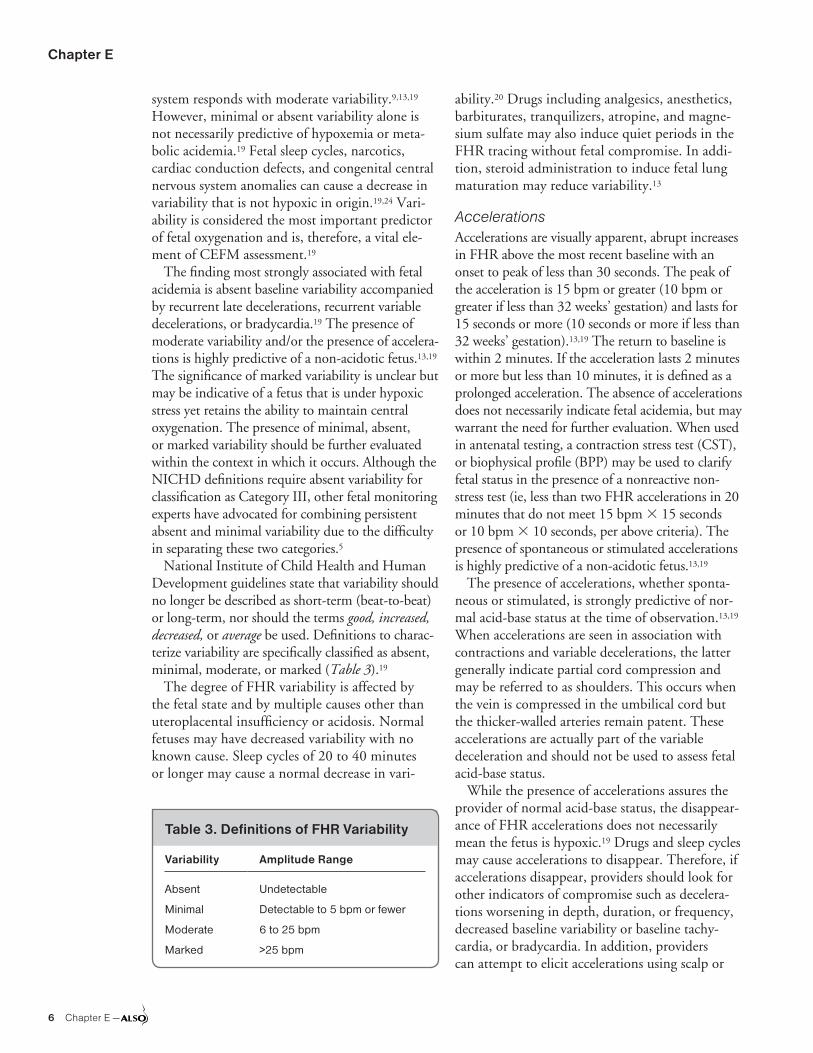
Chapter E
6 Chapter E —
system responds with moderate variability.9,13,19 However, minimal or absent variability alone is not necessarily predictive of hypoxemia or meta-bolic acidemia.19 Fetal sleep cycles, narcotics, cardiac conduction defects, and congenital central nervous system anomalies can cause a decrease in variability that is not hypoxic in origin.19,24 Vari-ability is considered the most important predictor of fetal oxygenation and is, therefore, a vital ele-ment of CEFM assessment.19
The finding most strongly associated with fetal acidemia is absent baseline variability accompanied by recurrent late decelerations, recurrent variable decelerations, or bradycardia.19 The presence of moderate variability and/or the presence of accelera-tions is highly predictive of a non-acidotic fetus.13,19 The significance of marked variability is unclear but may be indicative of a fetus that is under hypoxic stress yet retains the ability to maintain central oxygenation. The presence of minimal, absent, or marked variability should be further evaluated within the context in which it occurs. Although the NICHD definitions require absent variability for classification as Category III, other fetal monitoring experts have advocated for combining persistent absent and minimal variability due to the difficulty in separating these two categories.5
National Institute of Child Health and Human Development guidelines state that variability should no longer be described as short-term (beat-to-beat) or long-term, nor should the terms good, increased, decreased, or average be used. Definitions to charac-terize variability are specifically classified as absent, minimal, moderate, or marked (Table 3).19
The degree of FHR variability is affected by the fetal state and by multiple causes other than uteroplacental insufficiency or acidosis. Normal fetuses may have decreased variability with no known cause. Sleep cycles of 20 to 40 minutes or longer may cause a normal decrease in vari-
ability.20 Drugs including analgesics, anesthetics, barbiturates, tranquilizers, atropine, and magne-sium sulfate may also induce quiet periods in the FHR tracing without fetal compromise. In addi-tion, steroid administration to induce fetal lung maturation may reduce variability.13
AccelerationsAccelerations are visually apparent, abrupt increases in FHR above the most recent baseline with an onset to peak of less than 30 seconds. The peak of the acceleration is 15 bpm or greater (10 bpm or greater if less than 32 weeks’ gestation) and lasts for 15 seconds or more (10 seconds or more if less than 32 weeks’ gestation).13,19 The return to baseline is within 2 minutes. If the acceleration lasts 2 minutes or more but less than 10 minutes, it is defined as a prolonged acceleration. The absence of accelerations does not necessarily indicate fetal acidemia, but may warrant the need for further evaluation. When used in antenatal testing, a contraction stress test (CST), or biophysical profile (BPP) may be used to clarify fetal status in the presence of a nonreactive non-stress test (ie, less than two FHR accelerations in 20 minutes that do not meet 15 bpm × 15 seconds or 10 bpm × 10 seconds, per above criteria). The presence of spontaneous or stimulated accelerations is highly predictive of a non-acidotic fetus.13,19
The presence of accelerations, whether sponta-neous or stimulated, is strongly predictive of nor-mal acid-base status at the time of observation.13,19 When accelerations are seen in association with contractions and variable decelerations, the latter generally indicate partial cord compression and may be referred to as shoulders. This occurs when the vein is compressed in the umbilical cord but the thicker-walled arteries remain patent. These accelerations are actually part of the variable deceleration and should not be used to assess fetal acid-base status.
While the presence of accelerations assures the provider of normal acid-base status, the disappear-ance of FHR accelerations does not necessarily mean the fetus is hypoxic.19 Drugs and sleep cycles may cause accelerations to disappear. Therefore, if accelerations disappear, providers should look for other indicators of compromise such as decelera-tions worsening in depth, duration, or frequency, decreased baseline variability or baseline tachy-cardia, or bradycardia. In addition, providers can attempt to elicit accelerations using scalp or
Table 3. Definitions of FHR Variability
Variability Amplitude Range
Absent Undetectable
Minimal Detectable to 5 bpm or fewer
Moderate 6 to 25 bpm
Marked >25 bpm

Intrapartum Fetal Surveillance
— Chapter E 7
vibroacoustic stimulation. Of note, FHR accelera-tions should not be elicited during FHR decelera-tions or bradycardia (ie, use of scalp stimulation).9 Such actions only delay initiation of appropriate intrauterine resuscitation measures and further compromise fetal status.
DecelerationsDecelerations are defined in terms of the rate of onset (abrupt versus gradual) and the relationship to uterine contractions. If decelerations occur with 50% or greater of contractions during 20-minutes, they are considered to be recurrent decelerations. If they occur with less than 50% of contractions during 20 minutes, they are called intermit-tent decelerations.13,19 Use of the word repetitive, although used in the past, should be avoided because it implies that decelerations are occurring with every contraction rather than with the major-ity (50% or greater). Decelerations are classified as early, variable, late, or prolonged.13,19
Early DecelerationsEarly decelerations are visually apparent gradual decreases in FHR with return-to-baseline in association with a uterine contraction. The onset to nadir is 30 seconds or more and the nadir occurs at the same time as the peak of the contrac-tion.13,19 They are almost always benign if no other abnormalities of the FHR tracing are noted, and represent transient local changes in blood flow as a result of stimulus of the vagal nerve centers due to head compression.
Variable DecelerationsVariable decelerations are visually apparent abrupt decreases in FHR below the baseline with onset to nadir of 30 seconds or less. The decrease in FHR is 15 bpm or greater from the baseline with duration of 15 seconds or more but less than 2 minutes, and may or may not be associated with contractions.13,19 Variable decelerations are most commonly the result of cord compression result-ing in a rise in peripheral resistance and change in oxygenation. This causes sudden fetal hyper-tension, increased parasympathetic outflow, and slowing of the fetal pacemaker. However, inter-pretation is complicated because variable decelera-tions also can result from decreased arterial oxygen concentration secondary to uteroplacental insuf-ficiency from other causes.
Variable decelerations may be accompanied by other characteristics, the clinical significance of which requires further investigation. Some exam-ples include a slow return of the FHR after the end of the contraction, biphasic decelerations, tachycar-dia after variable deceleration(s), accelerations pre-ceding and/or following a deceleration (sometimes called shoulders or overshoots), and fluctuations in the FHR in the trough of the deceleration.19
Variables are the most common FHR decelera-tion that occur during labor and are generally associated with normal perinatal outcomes.20 Assessment of the tracing should include changes in the overall baseline rate and variability, and the recurrence, depth, and duration of the decel-eration. The presence of moderate variability or accelerations suggests the absence of acidemia.20 Management generally involves relieving cord com-pression by turning the woman or amnioinfusion. Administering oxygen and reducing or discontinu-ing uterine stimulants may also be helpful.20
Late DecelerationsLate decelerations are visually apparent gradual decreases in FHR with return to baseline with the onset to nadir of 30 seconds or more. The onset, nadir, and recovery of the deceleration generally occur after the beginning, peak, and ending of the contraction, respectively.19 Late decelerations are associated with uteroplacental insufficiency and fetal hypoxemia. If uncorrected they can lead to acidemia and myocardial depression. When combined with absent or minimal variability or other FHR abnormalities, there is an increased likelihood of significant fetal compromise; imme-diate evaluation and intervention are indicated. Subtle, shallow late decelerations are easily missed but clinically significant. They can be detected by holding a straight edge along the baseline.
Prolonged DecelerationsProlonged decelerations are visually apparent decreases in FHR baseline of 15 bpm or more, last-ing 2 minutes or more, but less than 10 minutes.19
A sudden deterioration in the FHR tracing can be seen after vaginal examination, placement of fetal spiral electrode, amniotomy, uterine tachy-systole secondary to administration of oxytocin or a cervical ripening agent, maternal hypotension (eg, secondary to regional anesthesia), maternal seizures, or fetal movement producing transient

Chapter E
8 Chapter E —
cord compression. If the fetus was not previously compromised, recovery will typically occur with discontinuation of the inciting event or agent, position change, increased intravenous fluids, maternal oxygen supplementation, or a combina-tion of these measures. When accompanied by change in variability or baseline, decelerations are more likely to be associated with fetal acid-base abnormalities. Factors known to cause these changes should be identified and corrected.
Sinusoidal Fetal Heart Rate TracingsA sinusoidal FHR tracing is a specific pattern that is not consistent with any of the above defini-tions. It is generally thought to be due to severe fetal anemia.24-26 It is described as a visually appar-ent, regular smooth sine wave-like undulating tracing seen within the FHR baseline. Providers are cautioned not to confuse this tracing with FHR variability. The sine waves undulate slowly and regularly, generally with a cycle frequency of 3 to 5 cycles (waves)/minute. The tracing must con-tinue for at least 20 minutes,19 but providers may need to intervene sooner in emergencies (eg, bleed-ing from a ruptured vasa previa). Of note, the term pseudosinusoidal is not recognized by the NICHD. In the past, this term was used to denote a tracing that frequently occurred following narcotic admin-istration or sometimes during ultrasound, and was associated with rhythmic fetal movements such as rapid breathing, sucking movements of the mouth, hiccoughing, and thumb sucking.26
Overall AssessmentHaving assessed the contraction pattern and FHR tracing and defined the risk, an overall assessment of the situation and management plan should be made. The terms fetal distress and birth asphyxia are inappropriate and have no place in the assess-ment. In the past, terms describing the FHR trac-ing were reassuring and non-reassuring, but since the report from the 2008 NICHD workshop, the assessment of fetal status has been organized into a three-tiered system: Category I, II, or III. Manage-ment of the woman must be based on the clinical context and the FHR tracing category, and must include a plan for further fetal surveillance if labor is allowed to continue.
Typically, Category I tracings are considered normal and can be followed routinely. Category II tracings are indeterminate and not predic-
tive of abnormal fetal pH status. These tracings require prompt evaluation and implementation of interventions to address the tracing. Category III tracings are clearly abnormal and predictive of abnormal acid-base status.19 Prompt evaluation and consideration of immediate delivery is required.
NICHD FHR Classification System19
Category I FHR TracingsCategory I tracings are normal tracings that are strongly predictive of normal fetal acid-base status at the time of observation and must include all of the following:• Baseline of 110 to 160 bpm• Moderate baseline variability• Absence of late or variable decelerations• Absence or presence of early decelerations• Absence or presence of accelerations (spontane-
ous or elicited)
Category II FHR TracingsCategory II tracings are indeterminate tracings that are not predictive of fetal acid base status and cannot be classified as Category I or Category III. The presence of moderate variability and/or accel-erations is highly predictive of normal fetal acid base status.13,19,20 These tracings require prompt evaluation and implementation of interventions to address the tracings. Category II tracings occur in the majority of fetuses during labor.13,19 Some examples of causes of Category II tracings include:• Tachycardia• Bradycardia not accompanied by absent
variability• Baseline with minimal or marked variability• Baseline with absent variability not accompanied
by recurrent decelerations• Recurrent variable decelerations with minimal to
moderate variability• Recurrent late decelerations with moderate
variability• Variable decelerations with slow return, overshoot
or shoulders• Prolonged deceleration lasting more than
2 minutes but less than 10 minutes• No acceleration after fetal stimulation
Category III FHR TracingsCategory III tracings are predictive of abnormal fetal acid-base status at the time they are observed. These require prompt evaluation and expedient

Intrapartum Fetal Surveillance
— Chapter E 9
interventions to address the pattern. Interventions may include, as appropriate, administration of oxygen, administration of an IV fluid bolus, repo-sitioning, discontinuation of uterine stimulants, and treatment of maternal hypotension. If these fail, immediate delivery should be considered.
Category III tracings include:• Sinusoidal patternOR• Absent FHR variability with any of the following:
– Recurrent late decelerations– Recurrent variable decelerations– BradycardiaWhen using CEFM during labor, providers
and nurses should review tracings regularly.5,9,20 This periodic review includes maintaining an adequate quality tracing, identifying and respond-ing to FHR abnormalities, and appropriately communicating findings. Some institutions now use tools for risk management and patient safety including communication strategies. An example is the Situation-Background-Assessment-Recom-mendation (SBAR) technique developed by Kaiser Permanente of Colorado. This technique provides a framework for communication among members of the health care team about a patient’s status.27
Adequate documentation is necessary and many institutions now use computerized charting, flow sheets, clinical pathways, or fetal tracing archival processes. Any written information on the trac-ing (ie, emergent situations during labor) should coincide with these automated processes to avoid confusion and minimize litigation risk.9 Docu-mentation may occur at different intervals than the actual assessment and may take the form of summary notes.9
Documentation of the fetal heart tracing and categorization during labor should include:1. FHR data (ie, baseline rate, variability, periodic
changes, categorization)2. Uterine activity characteristics obtained by
palpation or pressure transducer (ie, frequency, duration, intensity, presence or absence of tachysystole)
3. Specific actions taken when changes occur inFHR or uterine activity
4. Other maternal observations and assessments5. Maternal and fetal responses to interventions6. Subsequent return to normal findings7. Pertinent communication with other health
care providers
Categorization of SIA FindingsAWHONN and the American College of Nurse Midwives have recently proposed a categorization for interpretation of SIA (Table 4).6,9 Standard-izing language for both of these FHR assessment techniques may decrease communication errors and provide for a common standardized language. There is not a Category III for SIA because this category requires the ability to assess variability, which is not reliably and consistently possible when using SIA.6,9 In addition, the terms for decel-eration tracings (ie, late, variable, early) are reserved for CEFM and should not be used for SIA.
Suggested FHR ManagementThe 2010 ACOG Practice Bulletin on manage-ment of intrapartum FHR tracings presents a stan-dardized approach to FHR management.20 This was based in part on the framework developed by Parer and colleagues who analyzed studies of FHR tracings to develop four hypotheses:28,29
1. Moderate variability, even in the presenceof decelerations, is strongly associated with an absence of acidemia (pH greater than 7.15) and a nondepressed fetus (5-minute Apgar score greater than or equal to 7).
2. Minimal or absent (undetectable) variabilityin the presence of late or variable decelerations is associated with acidemia (pH less than 7.15) and a depressed fetus (5-minute Apgar score less than 7).
Table 4. Interpretation of Auscultation Findings6,9,15
Category I - Normal: Includes ALL of the following:Normal FHR baseline 110 to 160 bpmRegular rhythmPresence of FHR increases, or accelerations, from
the baselineAbsence of FHR decreases, or decelerations, from
the baseline
Category II - Indeterminate: May include any of the following:
Irregular rhythmPresence of FHR decreases or decelerations from
the baselineTachycardia (baseline FHR >160 bpm >10 min)Bradycardia (baseline FHR <110 bpm >10 min)
FHR = fetal heart rate.

Chapter E
10 Chapter E —
3. The depth of the deceleration or bradycardia is directly proportional to fetal depression and acidemia, especially with late decelerations and in tracings with minimal or absent FHR variability.
4. After an initial normal FHR tracing, progres-sive decelerations, in the absence of catastrophic events, results in acidemia that develops over a significant amount of time, generally 1 hour.
Parer and colleagues concluded that the presence of moderate variability had a 98% negative predic-tive value for fetal acidosis or an Apgar score less than 7.28,29 Minimal or absent variability with late or variable decelerations was predictive of neona-tal acidosis or neonatal depression, with 23% of fetuses having these adverse findings; there was some evidence that absent variability is more pre-dictive. The depth of decelerations has a stronger association with fetal acidosis for late decelerations than for variable decelerations. In the fetus with decreased variability and decelerations, acidosis was shown to develop slowly over time, except in the setting of sudden bradycardia as may occur with placental abruption or cord prolapse.28,29
Category I EFM TracingsCategory I EFM tracings are considered normal and are not associated with fetal acidemia.13,19,20 Recommendations are to continue the current monitoring (whether SIA or EFM); periodically evaluate and document the clinical status, under-lying risk factors, and tracings; and change the management strategy only if the tracing changes to a Category II or III.
Category III EFM TracingsCategory III EFM tracings are considered abnor-mal and predictive of abnormal fetal acid-base status at the time of observation.13,19,20 Category III tracings require prompt evaluation. Recom-mendations are to correct fetal acidemia to reduce outcomes of neonatal encephalopathy, cerebral palsy, and neonatal acidosis. Preparation for deliv-ery, development of a time frame for delivery, and performance of intrauterine resuscitative measures are essential. If tracings do not improve with appropriate corrective maneuvers, prompt delivery of the fetus is indicated.13,19,20
Considerations in preparing for an operative delivery in the presence of a Category III tracing should be made judiciously and expeditiously.
The standard rule of 30 minutes from decision-to-incision, although used frequently, has not been supported in the literature to reduce adverse neonatal outcomes.20,30 In addition, immediate delivery of a fetus with an unknown duration of a Category III tracing may not improve outcomes if the fetus has experienced a pre-existing hypoxic insult.20
Category II EFM TracingsCategory II tracings include all tracings that are not classified as Category I or III; these occur at some time in over 80% of labors.31 Because these tracings may represent fetal compromise, recom-mendations are to evaluate the tracing, perform appropriate corrective measures when indicated, and then re-evaluate. Because Category II tracings represent a wide variety of concerns, the pres-ence of accelerations (spontaneous or induced) or moderate variability are useful in assessing normal fetal acid-base status. If neither of these charac-teristics is present after appropriate intrauterine resuscitative measures, or if the tracing progresses to Category III, consideration should be given to delivery of the fetus. If the tracing reverts to Category I after appropriate intervention, then previous monitoring may be resumed.
Category II tracings are frequently encountered at some point during labor, and their manage-ment can be challenging without defined man-agement protocols. Category II tracings can range from almost normal to tracings in which acidemia is either present or developing rapidly. Without rapid intervention, these tracings can rapidly evolve into Category III tracings. Clini-cal considerations when managing these tracings must include gestational age, fetal growth status (ie, IUGR), maternal medical and obstetrical con-ditions, comorbidities, labor progress, and avail-able resources and personnel to respond.
Algorithm for Category II ManagementClark and colleagues have suggested an algorithm for managing Category II tracings (Figure 1 and Table 5).5 This algorithm follows the defini-tions set forth by the NICHD and is supported by a growing body of evidence. The goal of this algorithm is to present a standardized way to man-age Category II tracings that encourages vaginal delivery in fetuses whose FHR tracings demon-strate minimal risk for progression to clinically

Intrapartum Fetal Surveillance
— Chapter E 11
significant acidemia. Use of this algorithm requires application of specific definitions for significant decelerations and takes into account labor phase and labor progress (Table 5). This algorithm is only applicable to Category II tracings and is not to be used in extremely premature fetuses.
When a Category II tracing is identified, intra-uterine resuscitation measures are instituted and the algorithm is delayed for 30 minutes to allow these interventions to improve the tracing. After 30 minutes, if these measures do not alleviate the Category II tracing, the algorithm is begun. The algorithm is initiated by an assessment of moder-ate variability or FHR accelerations because this rules out clinically significant acidemia. From there, the assessment includes the presence or absence of significant decelerations, the stage of labor, and whether labor is progressing normally.
If delivery is indicated by the algorithm, ideally it should be initiated within 30 minutes of the deci-sion. The algorithm can be discontinued at any time the provider feels more rapid intervention is required.
While this algorithm is somewhat complex, a web-based app has simplified its use for clinical practice (available at http://cat2.perigen.com/cat2/).
Five-Tier ClassificationIn addition to the above approach for manag-ing Category II tracings, a five-tiered classifica-tion scheme has also been proposed by Parer and colleagues (Table 6).29,32 Similar to the algorithm in Table 5, the purpose of this classification system is to classify FHR monitor tracings according to risk of fetal acidemia, determine the risk of evolution to a more serious tracing, and construct
Figure 1. Algorithm for Management of Category II Fetal Heart Rate Tracings
*That have not resolved with appropriate conservative corrective measures, which may include supplemental oxygen, maternal position changes, intravenous fluid administration, correction of hypotension, reduction or discontinuation of uterine stimulation, administration of uter-ine relaxant, amnioinfusion, and/or changes in second stage breathing and pushing techniques.
OVD = operative vaginal delivery.
Reprinted from Clark SL, Nageotte MP, Garite TJ, et al. Intrapartum management of category II fetal heart rate tracings: towards standardization of care. Am J Obstet Gynecol. 2013;209(2):89-97.
Moderate variability or accelerations
Significant decelerations with >50% of contractions for 1 hour*
Significant decelerations with >50% of contractions for 30 minutes*
Cesarean or OVD
Observe for 1 hour
Persistent pattern
Manage per algorithm
Observe Observe Cesarean or OVD
Cesarean
Latent phase Active phase Second phase
Normal labor progress Normal progress
Yes No
Yes NoYes No
Cesarean
Yes No
Observe Cesarean or OVD
Yes No
Yes No

Chapter E
12 Chapter E —
a standard process for FHR tracing management with the ultimate aim of minimizing newborn infant acidemia without excessive obstetric intervention.
A grid categorizes all possible heart rate patterns based on baseline rate (ie, normal, tachycardia, bradycardia), type of decelerations (ie, early, late, variable, prolonged), and quantity of variability (ie, undetectable, minimal, moderate, marked). All
definitions are according to the NICHD state-ment on the nomenclature of FHR tracings.
In the five-tier approach, each FHR tracing has been color-coded to represent no threat of acide-mia (green, no intervention required) to severe threat for acidemia (red, emergent delivery recom-mended). Three intermediate categories—blue, yellow, orange—are NICHD Category II tracings and represent increasing concern and response for evolving acidemia (Table 7).29,32
In 2012 Coletta and colleagues compared the NICHD three-tier system to the proposed five-tier system in Table 6.32 For tracings categorized by either orange or red, there was 79% sensitivity and 100% specificity for a pH less than 7 with no false positives. By using the five-tier system 79% of fetal acidemia was correctly identified in the orange and red tracings, compared with only 12% in the NICHD Category III. All tracings with pH greater than 720 were correctly categorized as blue, green, or yellow. The five-tier system also better identified tracings that resulted in lower Apgar scores, admission to the NICU, and need for oxygen supplementation.32 The complexity of this five-tier system has made practical application difficult, however, this has changed recently due to the availability of a mobile app (available at http://www.obapps.org/), making the application to the clinical setting more user-friendly.33 The Japan Society of Obstetrician and Gynecologists also recommended the use of this five-tier system based on its validity with acid-base balance.34,35 How-ever, another study reports no difference between very normal or very abnormal tracings using this system. They conclude that it is yet to be deter-mined whether one system is superior in predict-ing fetal acidemia.36
Intrauterine Resuscitative MeasuresVarious intrauterine resuscitative measures are to be undertaken for any FHR tracing that is con-cerning regardless of classification scheme used (Table 8).20,37 The goals of these corrective mea-sures are aimed at the underlying suspected cause of the abnormality in the FHR tracing. They are also dependent on the associated FHR abnormal-ity identified.
Possible interventions include:• Change in maternal position to lateral position-
ing (left or right) or on hands and knees• Administer maternal oxygen
Table 5. Management of Category II FHR Tracings: Clarifications for Use of Algorithm
1. Variability refers to predominant baseline FHR pattern (marked,moderate, minimal, absent) during a 30-minute evaluation period,as defined by NICHD.
2. Marked variability is considered same as moderate variability forpurposes of this algorithm.
3. Significant decelerations are defined as any of the following:• Variable decelerations lasting longer than 60 seconds and
reaching a nadir more than 60 bpm below baseline.• Variable decelerations lasting longer than 60 seconds and
reaching a nadir less than 60 bpm regardless of the baseline.• Any late decelerations of any depth.• Any prolonged deceleration, as defined by the NICHD. Due to
the broad heterogeneity inherent in this definition, identificationof a prolonged deceleration should prompt discontinuation of thealgorithm until the deceleration is resolved.
4. Application of algorithm may be initially delayed for up to 30minutes while attempts are made to alleviate category II patternwith conservative therapeutic interventions (eg, correction ofhypotension, position change, amnioinfusion, tocolysis, reduction ordiscontinuation of oxytocin).
5. Once a category II FHR pattern is identified, FHR is evaluated andalgorithm applied every 30 minutes.
6. Any significant change in FHR parameters should result inreapplication of algorithm.
7. For category II FHR patterns in which algorithm suggests delivery isindicated, such delivery should ideally be initiated within 30 minutesof decision for cesarean.
8. If at any time tracing reverts to category I status, or deterioratesfor even a short time to category III status, the algorithm no longerapplies. However, algorithm should be reinstituted if category Ipattern again reverts to category II.
9. In fetus with extreme prematurity, neither significance of certain FHRpatterns of concern in more mature fetus (eg, minimal variability) orability of such fetuses to tolerate intrapartum events leading to certaintypes of category II patterns are well defined. This algorithm is notintended as guide to management of fetus with extreme prematurity.
10. Algorithm may be overridden at any time if, after evaluation of patient,physician believes it is best interest of the fetus to intervene sooner.
FHR = fetal heart rate; NICHD = National Institute of Child Health and Human Development.
Reprinted from Clark SL, Nageotte MP, Garite TJ, et al. Intrapartum manage-ment of category II fetal heart rate tracings: towards standardization of care. Am J Obstet Gynecol. 2013;209(2):89-97.
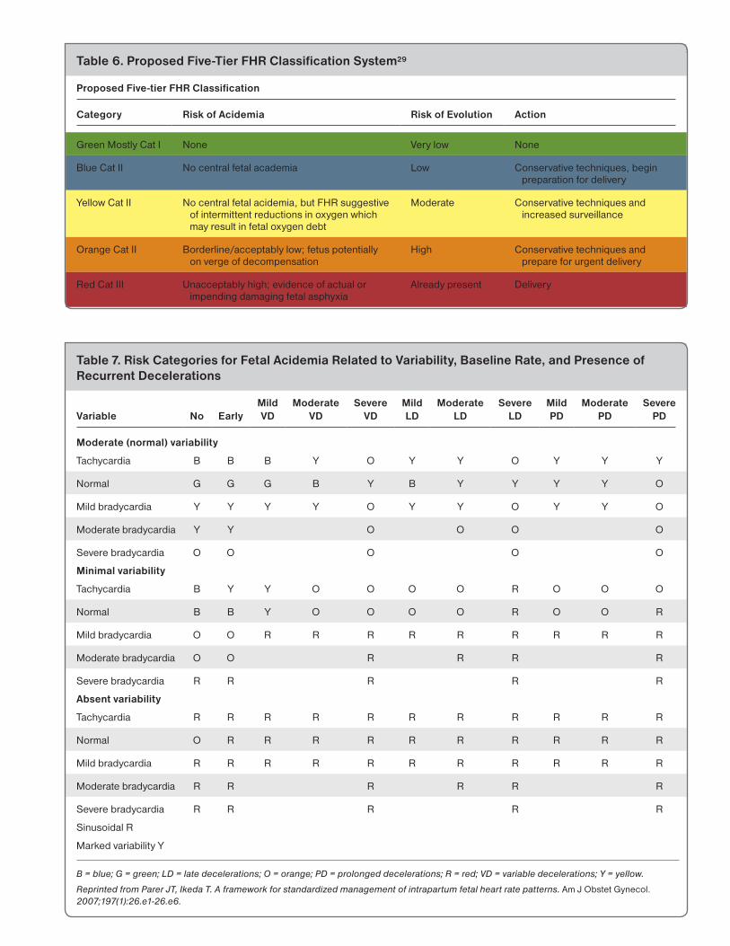
Intrapartum Fetal Surveillance Table 6. Proposed Five-Tier FHR Classification System29
Proposed Five-tier FHR Classification
Category Risk of Acidemia Risk of Evolution Action
Green Mostly Cat I None Very low None
Blue Cat II No central fetal academia Low Conservative techniques, begin preparation for delivery
Yellow Cat II No central fetal acidemia, but FHR suggestive of intermittent reductions in oxygen which may result in fetal oxygen debt
Moderate Conservative techniques and increased surveillance
Orange Cat II Borderline/acceptably low; fetus potentially on verge of decompensation
High Conservative techniques and prepare for urgent delivery
Red Cat III Unacceptably high; evidence of actual or impending damaging fetal asphyxia
Already present Delivery
Table 7. Risk Categories for Fetal Acidemia Related to Variability, Baseline Rate, and Presence of Recurrent Decelerations
Variable No EarlyMild VD
Moderate VD
Severe VD
Mild LD
Moderate LD
Severe LD
Mild PD
Moderate PD
Severe PD
Moderate (normal) variability
Tachycardia B B B Y O Y Y O Y Y Y
Normal G G G B Y B Y Y Y Y O
Mild bradycardia Y Y Y Y O Y Y O Y Y O
Moderate bradycardia Y Y O O O O
Severe bradycardia O O O O O
Minimal variability
Tachycardia B Y Y O O O O R O O O
Normal B B Y O O O O R O O R
Mild bradycardia O O R R R R R R R R R
Moderate bradycardia O O R R R R
Severe bradycardia R R R R R
Absent variability
Tachycardia R R R R R R R R R R R
Normal O R R R R R R R R R R
Mild bradycardia R R R R R R R R R R R
Moderate bradycardia R R R R R R
Severe bradycardia R R R R R
Sinusoidal R
Marked variability Y
B = blue; G = green; LD = late decelerations; O = orange; PD = prolonged decelerations; R = red; VD = variable decelerations; Y = yellow.
Reprinted from Parer JT, Ikeda T. A framework for standardized management of intrapartum fetal heart rate patterns. Am J Obstet Gynecol. 2007;197(1):26.e1-26.e6.

Chapter E
14 Chapter E —
• Administer intravenous fluid bolus• Reduce uterine contraction frequency• Discontinue oxytocin or cervical ripening agents• Administer tocolytic drug• Initiate amnioinfusion with recurrent variable
decelerations• If prolapsed umbilical cord is noted, continuous
elevation of the presenting fetal part until opera-tive delivery occurs
• Modify pushing efforts in second stage of laborOther important considerations are:• Assess maternal vital signs to assess for hypotension• Perform vaginal examination to assess progress of
labor and rule out prolapsed umbilical cord• Change method of FHR monitoring if strip is
inadequate quality for assessment• If recent epidural and hypotension with new late
decelerations, prepare for possible administrationof ephedrineLateral positioning reduces compression of
the inferior vena cava and aorta. Repositioning the woman may also reduce compression of the umbilical cord. Several studies indicate that lateral
positioning on either side is superior when com-pared to supine positioning.37
Administration of maternal oxygen with the goal of improving fetal oxygenation remains a common intervention, yet study results regarding its benefit for the fetus remain controversial and may even suggest a possible detrimental effect.37-43 A recent Cochrane review found that there was not enough evidence to support or refute the use of oxygen for intrauterine resuscitation.44 Due to the controversy in the literature, a reasonable approach is to provide oxygen after other appro-priate intrauterine resuscitation techniques have not been effective, and to discontinue its use once the desired fetal response has been achieved.9,39
Administration of an intravenous fluid bolus is another common technique thought to increase intravascular volume, uteroplacental perfusion and, thus, fetal oxygenation.37,39 Simpson and James found that fetal oxygenation saturation improved with a 500 mL bolus of lactated Ringer solution and increased greatest when a 1,000 mL bolus was administered. The positive effect
Table 8. Potential Intrauterine Resuscitative Measures for Category II or Category III Tracings20
Goal Associated FHR Changes Potential Interventions
Improve uteroplacental blood flow
Recurrent late decelerationsProlonged decelerationsMinimal or absent FHR
variability
Lateral positioningAdminister maternal oxygenAdminister intravenous fluid bolusDiscontinue or reduce uterine stimulantsAdminister tocolytic drugsCorrect maternal hypotensionModify maternal expulsive (pushing) efforts
Reduce uterine activity
Tachysystole with Category II or Category III tracing
Lateral positioningAdminister intravenous fluid bolusDiscontinue or reduce uterine stimulantsAdminister tocolytic drugs
Alleviate/reduce umbilical cord compression
Recurrent variable decelerations
Prolonged decelerationsBradycardia
Reposition to where FHR is most improvedDiscontinue uterine stimulantsInitiate amnioinfusion if variable decelerations
recurrentModify maternal expulsive (pushing) effortsCheck for prolapsed cord. If identified,
continuously elevate presenting part until operative delivery occurs.
FHR = fetal heart rate.
Always: check cervix, maternal vital signs

Intrapartum Fetal Surveillance
— Chapter E 15
of the bolus continued for 15 minutes after completion.39
Modification of pushing efforts can have a significant effect on the FHR tracing. Coached sustained Valsalva pushing (ie, closed glottis, “hold your breath and count to 10” for three times with each contraction) can have a deleterious effect on maternal and fetal oxygenation37,45-47 with a resulting increase in number and severity of FHR decelerations.46,48 A preventive measure would be to avoid pushing until the woman feels the urge to push, thereby minimizing the length of the active pushing phase and fetal exposure to the hypoxic stress of pushing. Modification of pushing efforts includes temporarily discontinuing pushing to allow the fetus to recover, or to push with every second or third contraction.9,37,45,49,50
Ancillary Testing for Category II and III FHR TracingsFetal scalp pH testing is no longer commonly per-formed in the United States and has been replaced with assessment of moderate FHR variability or the presence of FHR accelerations. If accelerations are not present, fetal stimulation may be used to elicit accelerations. Current evidence indicates the presence of either moderate variability or accelera-tions reliably predicts the absence of fetal metabolic acidemia at the time either of these characteristics is observed.19 A meta-analysis showed that if there is absent or minimal variability without spontane-ous accelerations, the presence of an acceleration after scalp stimulation or fetal acoustic stimulation indicates that the fetal pH is greater than 7.20,51
Intrauterine resuscitation should be utilized appropriately for the clinical situation. If the fetal heart tracing is a Category III and does not resolve quickly, plans for an expedient delivery should be undertaken. Category II tracings need to be assessed in light of the entire clinical picture. Some Category II tracings may require emergent delivery if there is no response to interventions.
AmnioinfusionAmnioinfusion should be considered for suspected cord compression to reduce the occurrence of recurrent variable heart rate decelerations in the first stage of labor and to lower the use of cesarean delivery.52 Amnioinfusion has been associated with a reduction in cesarean delivery rates, FHR decel-erations, Apgar scores less than 7 at 5 minutes,
postpartum endometritis, and the incidence of neonatal and maternal hospital stays greater than 3 days.53 In addition, mean cord umbilical pH was higher. No improvement in long-term neonatal outcomes has been detected.
Although typically considered safe, amnioin-fusion carries a few precautions and potential complications. Amnioinfusion is indicated only for recurrent variable decelerations and is not indi-cated for late decelerations, fetal bradycardia, thick meconium, or oligohydramnios with a normal heart rate tracing.20,52-54
Amnioinfusion should also not be attempted when cesarean delivery is indicated, such as in transverse lie or placenta previa. It should never be undertaken when doing so would result in a delay of an indicated definitive treatment. With breech presentation, multiple gestation, or when placental abruption is suspected, caution should be taken in performing amnioinfusion. Complications include umbilical cord prolapse, rupture of a previous cesarean scar, amniotic fluid embolism, acute uter-ine hypertonus with a Category II or Category III fetal heart tracing, and acute polyhydramnios.
In the past, amnioinfusion was used to dilute thick meconium as a prophylactic measure to pre-vent newborn meconium aspiration. Randomized controlled trials did not confirm the efficacy of this approach and the ACOG no longer considers this an indication for amnioinfusion.4,54
Guidelines for Performing AmnioinfusionAmnioinfusion can be performed by continuous or intermittent techniques. A randomized controlled trial showed there was no difference between the two techniques for resolving variable decelerations.55
For continuous infusion:1. Perform a vaginal examination to determine
presentation and dilation, and to rule out cord prolapse.
2. Obtain informed consent.3. Place the patient in the left-lateral position.
Place an IUPC and consider placement of an FSE. If available, use a double lumen catheter for saline infusion.
4. If a double lumen catheter is not available, attach an 18-gauge needle to IV tubing con-nected to normal saline or lactated Ringer solution. Attach extension tubing filled with distilled water between the IUPR and the transducer. Insert the 18-gauge needle into the

Chapter E
16 Chapter E —
side port of the extension tubing. Alternatively, insert a second single lumen catheter (one for IUPC and one for amnioinfusion).
5. Infuse fluid, giving 250 to 500 mL initially,followed by 50 to 60 mL/hour maintenance infusion until FHR abnormalities resolve.note: Resting tone will be increased while the
infusion is running, but elevated baseline tone before infusion is a contraindication.
Management of TachysystoleUterine contractions cause a cessation of uterine blood flow and, thus, oxygen delivery to the fetus. In most healthy fetuses, this temporary decline is well tolerated. However, uterine tachysystole has been shown to cause a progressive decline in fetal oxygenation and arterial blood gases at birth,56-60 increased risk for operative delivery, lower Apgar scores, as well as an increase in NICU admissions.59 Tocolysis should be considered, especially in the setting of tachysystole associated with Category II or Category III tracings when standard intrauter-ine resuscitative measures are not successful.20,60 A recent Cochrane review demonstrated that betami-metic therapy reduced the number of FHR abnor-malities.61 If tachysystole is induced and a Category II or III tracing is present, uterine stimulants should also be decreased or discontinued.20 Other interventions for tachysystole may include maternal repositioning and IV fluid bolus.9,20,60 Evaluation for placental abruption should occur; tocolysis is contraindicated when abruption is suspected because it may worsen the abruption.39
Suggested Management of Tachysystole20
An algorithm for the management of uterine tachysystole is shown in Figure 2.
Areas for Future DevelopmentAlthough CEFM continues to be the standard for fetal monitoring, research is being conducted to enhance CEFM with computerized interpretation and to develop newer methodologies to monitor fetal well-being during labor.
Fetal hypoxemia results in biphasic changes in the ST segment of the fetal electrocardiogram (fECG) waveform and an increase in the T/QRS ratio. ST segment automated analysis software can record the frequency of ST events and, combined with changes in CEFM, can be used to deter-mine if intervention during the labor process is
indicated. Several studies have evaluated fECG analysis documenting the effectiveness to reduce operative vaginal deliveries, fetal scalp sampling, neonatal encephalopathy, and fetal acidosis (pH less than 7.05).62-65 One drawback to this technol-ogy is that it requires rupture of the membranes and internal fetal scalp monitoring.
Another area of research is the use of computer analysis of key components of the fetal tracing,66-68 or decision analysis, for the interpretation of the EFM tracing.69 These have not been shown to improve clinical outcomes.
Fetal pulse oximetry was developed using an internal monitoring device and required rupture of membranes to continuously monitor fetal oxy-genation saturation during labor. Trials have not shown significance differences in reducing cesar-ean delivery rates or interventions with the use of fetal pulse oximetry, therefore use of fetal pulse oximetry is no longer recommended.70-72
International ContextIn many low-resource settings, EFM is not an option. SIA may be with a fetoscope rather than a Doppler stethoscope. Although amnioinfusion is no longer indicated for meconium in high-resource settings where EFM is available, it does seem to be indicated for meconium in low-resource settings where monitoring capabilities are limited.73 More information on intrapartum fetal surveillance and other topics relevant to low-resource settings can be found in the Global ALSO manual (available at www.aafp.org/globalalso).
SummaryInitiation of fetal monitoring begins with assess-ment of maternal and fetal risk. Because CEFM has a low positive predictive value and can result in increased rates of cesarean delivery, intermittent auscultation is recommended for low-risk preg-nancies. However, hospital policy as well as staff availability and experience must be considered before deciding to use this technique. Clinicians should be ready to change monitoring to CEFM if a high-risk situation develops or if the provider is unable to adequately assess fetal heart tones.
If CEFM is selected for fetal surveillance, inter-pretation needs to be done with consideration of the clinical background, the overall tracing, and stage of labor, and in conjunction with fetal scalp or acoustic stimulation. Outcomes may still be

Intrapartum Fetal Surveillance
— Chapter E 17
unaffected using this technique, even in high-risk pregnancies. Efforts have recently been undertaken to standardize the definitions, interpretation, and general management of FHR tracings. DR C BRAVADO is a helpful mnemonic for defin-ing risk and EFM interpretation. It is critical that institutions and hospitals ensure that all labor and delivery personnel are trained in FHR surveil-lance and interpretation, and in management of findings. Communication among team members is critical, and tools or strategies to maximize accu-racy and completeness of transfer of information should be used (eg, SBAR) to minimize medical errors and maximize patient safety.
Regardless of the technology used, the patient/support relationship is paramount during the labor process. Clinicians should not allow any monitor-ing approach to substitute for personal attention to the woman and fetus throughout labor.
If an institution has a risk management or patient safety committee, regular monitoring and compliance with all aspects of fetal surveillance should be undertaken. The committee should be composed of physicians, nurses, administra-tors, and other pertinent staff for successful implementation.
Figure 2. Management Algorithm for Uterine Tachysystole
FHR = fetal heart rate.
Reprinted from American College of Obstetricians and Gynecologists. ACOG practice bulletin No 116: Management of intrapartum fetal heart rate tracings. Obstet Gynecol. 2010;116(5):1232-1240.
Uterine tachysystole
Spontaneous labor Labor induction or augmentation
Category I FHR tracing
Category II or III FHR tracing
No interventions required
Intrauterine resuscitative measures*
If no resolution, consider tocolytic
Category I FHR tracing
Category II or III FHR tracing
Decrease uterotonics
Decrease or stop uterotonics
Intrauterine resuscitative measures
If no resolution, consider tocolytic

Chapter E
18 Chapter E —
SORT: Key Recommendations for Practice
Clinical RecommendationEvidence Rating References
Structured intermittent auscultation should be offered to low-risk women as an alternative to CEFM. CEFM does not decrease perinatal mortality. CEFM reduced neonatal seizures (NNT= 661) but does not reduce the incidence of cerebral palsy. CEFM increased cesarean delivery rates (overall) (NNT = 20) and instrumental vaginal births (NNT = 33).
A 2
The presence of moderate FHR variability is predictive of normal fetal acid-base status. B 19,28
The presence of FHR accelerations, whether spontaneous or stimulated, is highly predictive of normal fetal acid-base status.
B 19,51
A Category III fetal monitoring tracing increases the likelihood of fetal acidosis and requires prompt measures for intrauterine resuscitation and consideration of the need for urgent delivery if these measures do not result in improvement.
B 19,20
A period of CEFM on maternity unit admission for low-risk women compared to initiation of structured auscultation at time of admission results in significant increased interventions including epidural analgesia (NNT = 19), CEFM (NNT = 7) and fetal blood scalp testing (NNT = 45), and is not recommended.
A 11,12
Amnioinfusion for umbilical cord compression in the presence of decelerations reduces FHR decelerations (NNT = 3), cesarean delivery overall (NNT = 8), Apgar score <7 at 5 min (NNT = 33), low cord arterial pH (<7.20) (NNT = 8), neonatal hospital stay >3 days (NNT = 5), and maternal hospital stay greater than 3 days (NNT = 7). When the FHR tracing includes recurrent variable FHR decelerations (Category II or Category III) amnioinfusion should be considered. Amnioinfusion is not indicated for late FHR decelerations.
A 53
Tachysystole, whether spontaneous or stimulated, when accompanied by a Category II or Category III tracing, warrants evaluation and appropriate management.
B 57-60
IV fluid bolus, lateral positioning, and oxygen administration, when used together, may improve fetal oxygenation during labor.
B 39
Modifying pushing techniques improves fetal oxygenation and may be considered during the second stage of labor. Modifying pushing techniques includes discontinuation of pushing, pushing every 2nd or 3rd contraction, pushing with an open glottis, and/or pushing for 6- to 8-second intervals.
B 37,46-48
Strength of Recommendation Taxonomy (SORT)
Strength of Recommendation Definition
A • Recommendation based on consistent and good-quality patient-oriented evidence.
B • Recommendation based on inconsistent or limited-quality patient-oriented evidence.
C • Recommendation based on consensus, usual practice, opinion, disease-oriented evidence, or case series for studies of diagnosis, treat-ment, prevention, or screening.
Patient-oriented evidence measures outcomes that matter to patients: morbidity, mortality, symptom improvement, cost reduction, and quality of life. Disease-oriented evidence measures intermediate, physiologic, or surrogate end points that might not reflect improvement in patient outcomes (eg, blood pressure, blood chemistry, physiologic function, pathologic findings).
CEFM = continuous electronic fetal monitoring; FHR = fetal heart rate; IV = intravenous; NNT = number needed to treat.
From Ebell MH, Siwek J, Weiss BD, et al. Strength of recommendation taxonomy (SORT): a patient-centered approach to grading evidence in the medical literature. Am Fam Physician. 2004;69(3):548-556.

Intrapartum Fetal Surveillance
— Chapter E 19
References1. Martin JA, Hamilton BE, Osterman MJ, Curtin SC, Mat-
thews TJ. Births: final data for 2013. Natl Vital Stat Rep. 2015;64(1):1-65.
2. Alfirevic Z, Devane D, Gyte GML. Continuous cardioto-cography (CTG) as a form of electronic fetal monitoring(EFM) for fetal assessment during labour. CochraneDatabase Syst Rev. 2013;5(5):CD006066.
3. Chauhan SP, Klauser CK, Woodring TC, Sanderson M,Magann EF, Morrison JC. Intrapartum nonreassuringfetal heart rate tracing and prediction of adverse out-comes: interobserver variability. Am J Obstet Gynecol.2008;199(6):623.e1-623.e5.
4. Parer JT, Hamilton EF. Comparison of 5 experts andcomputer analysis in rule-based fetal heart rate interpre-tation. Am J Obstet Gynecol. 2010;203(5):451.e1-451.e7.
5. Clark SL, Nageotte MP, Garite TJ, et al. Intrapartummanagement of category II fetal heart rate tracings:towards standardization of care. Am J Obstet Gynecol.2013;209(2):89-97.
6. Feinstein N, Sprague A, Trepanier M, eds. Fetal HeartRate Auscultation. 2nd ed. Washington DC: Associa-tion of Women’s Health, Obstetric and Neonatal Nurses(AWHONN); 2008.
7. Murray ML. Maternal or fetal heart rate? Avoiding intra-partum misidentification. J Obstet Gynecol NeonatalNurs. 2004;33(1):93-104.
8. Nurani R, Chandraharan E, Lowe V, Ugwumadu A,Arulkumaran S. Misidentification of maternal heart rateas fetal on cardiotocography during the second stageof labor: the role of the fetal electrocardiograph. ActaObstet Gynecol Scand. 2012;91(12):1428-1432.
9. Lyndon A, Ali L, eds. Association of Women’s HealthObstetric and Neonatal Nursing. Fetal Heart MonitoringPrinciples and Practices. 4th ed. Washington DC: Kend-all Hunt Professional; 2009.
10. Hauth JC, Hankins GD, Gilstrap LC III, StricklandDM, Vance P. Uterine contraction pressures withoxytocin induction/augmentation. Obstet Gynecol.1986;68(3):305-309.
11. Devane D, Lalor JG, Daly S, McGuire W, Smith V.Cardiotocography versus intermittent auscultation offetal heart on admission to labour ward for assess-ment of fetal wellbeing. Cochrane Database Syst Rev.2012;2(2):CD005122.
12. Bix E, Reiner LM, Klovning A, Oian P. Prognostic valueof the labour admission test and its effectivenesscompared with auscultation only: a systematic review.BJOG. 2005;112(12):1595-1604.
13. American College of Obstetricians and Gynecolo-gists. ACOG Practice Bulletin No. 106: Intrapartum fetalheart rate monitoring: nomenclature, interpretation,and general management principles. Obstet Gynecol.2009;114(1):192-202.
14. Riley LE, Stark AR, eds. Guidelines for Perinatal Care.7th ed. Washington DC: American College of Obstetri-cians and Gynecologists; 2012.
15. American College of Nurse-Midwives. Intermittent Aus-cultation for Intrapartum Fetal Heart Rate Surveillance(replaces ACNM Clinical Bulletin #9, March 2007). JMidwifery Womens Health. 2010;55(4):397-403.
16. Royal College of Obstetricians and Gynaecologists.The Use of Electronic Fetal Monitoring. The use andinterpretation of cardiotocography in intrapartum fetalsurveillance. Evidence-based Clinical Guideline Num-ber 8. London: RCOG Press; 2001.
17. Liston R, Sawchuck D, Young D; Society of Obstetricsand Gynaecologists of Canada; British Columbia Peri-natal Health Program. Fetal health surveillance: antepar-tum and intrapartum consensus guideline. [Erratum in JObstet Gynaecol Can. 2007;29] [11]. J Obstet GynaecolCan. 2007;29(9)(Suppl 4):S3-S56.
18. National Institute of Child Health and Human Develop-ment Research Planning Workshop. Electronic fetalheart rate monitoring: research guidelines for interpreta-tion. Am J Obstet Gynecol. 1997;177(6):1385-1390.
19. Macones GA, Hankins GD, Spong CY, Hauth J,Moore T. The 2008 National Institute of Child Healthand Human Development workshop report on elec-tronic fetal monitoring: update on definitions, inter-pretation, and research guidelines. Obstet Gynecol.2008;112(3):661-666.
20. American College of Obstetricians and Gynecolo-gists. Practice bulletin no. 116: Management of intra-partum fetal heart rate tracings. Obstet Gynecol.2010;116(5):1232-1240.
21. Bakker JJ, Verhoeven CJM, Janssen PF, et al. Out-comes after internal versus external tocodynamometryfor monitoring labor. N Engl J Med. 2010;362(4):306-313.
22. Bakker JJ, Janssen PF, van Halem K, et al. Internalversus external tocodynamometry during inducedor augmented labour. Cochrane Database Syst Rev.2013;8(8):CD006947.
23. Harper LM, Shanks AL, Tuuli MG, Roehl KA, Cahill AG.The risks and benefits of internal monitors in laboringpatients. Am J Obstet Gynecol. 2013;209(1):38.e1-38.e6.
24. Freeman RK, Garite TJ, Nageote MP, Mille LA. FetalHeart Rate Monitoring, 4th ed. New York, NY: WoltersKluwer, Lippincott Williams & Wilkins; 2012.
25. Gabbe SG, Niebyl JR, Galan HL, et al. Intrapartum FetalEvaluation. In: Obstetrics: Normal and Problem Preg-nancies. 6th ed. Philadelphia, PA: Saunders; 2012.
26. Modanlou HD, Murata Y. Sinusoidal heart rate pattern:Reappraisal of its definition and clinical significance. JObstet Gynaecol Res. 2004;30(3):169-180.
27. SBAR Technique for Communication. A SituationalBriefing Model. Available at http://www.ihi.org/resources/Pages/Tools/SBARTechniqueforCommuni-cationASituationalBriefingModel.aspx.
28. Parer JT, King T, Flanders S, Fox M, Kilpatrick SJ. Fetalacidemia and electronic fetal heart rate patterns: isthere evidence of an association? J Matern Fetal Neo-natal Med. 2006;19(5):289-294.
29. Parer JT, Ikeda T. A framework for standardized man-agement of intrapartum fetal heart rate patterns. Am JObstet Gynecol. 2007;197(1):26.e1-26.e6.

Chapter E
20 Chapter E —
30. Bloom SL, Leveno KJ, Spong CY, et al; National Insti-tute of Child Health and Human Development Maternal-Fetal Medicine Units Network. Decision-to-incisiontimes and maternal and infant outcomes. Obstet Gyne-col. 2006;108(1):6-11.
31. Jackson M, Holmgren CM, Esplin MS, Henry E, Var-ner MW. Frequency of fetal heart rate categoriesand short-term neonatal outcome. Obstet Gynecol.2011;118(4):803-808.
32. Coletta J, Murphy E, Rubeo Z, Gyamfi-Bannerman C.The 5-tier system of assessing fetal heart rate tracingsis superior to the 3-tier system in identifying fetal acide-mia. Am J Obstet Gynecol. 2012;206(3):226.e1-226.e5.
33. Apps OB. FHR 5-Tier: Fetal Heart Rate. Available athttp://www.obapps.org/.
34. Sadaka A, Furuhashi M, Minami H, Miyazaki K, YoshidaK, Ishikawa K. Observation on validity of the five-tier sys-tem for fetal heart rate pattern interpretation proposedby Japan Society of Obstetricians and Gynecologists.J Matern Fetal Neonatal Med. 2011;24(12):1465-1469.
35. Okai T, Ikeda T, Kawarabayashi T, et al; PerinatologyCommittee of the Japan Society of Obstetrics andGynecology. Intrapartum management guidelinesbased on fetal heart rate pattern classification. J ObstetGynaecol Res. 2010;36(5):925-928.
36. Gyamfi Bannerman C, Grobman WA, Antoniewicz L,Hutchinson M, Blackwell S. Assessment of the con-cordance among 2-tier, 3-tier, and 5-tier fetal heartrate classification systems. Am J Obstet Gynecol.2011;205(3):288.e1-288.e4.
37. Garite TJ, Simpson KR. Intrauterine resuscitation duringlabor. Clin Obstet Gynecol. 2011;54(1):28-39.
38. Haydon ML, Gorenberg DM, Nageotte MP, et al. Theeffect of maternal oxygen administration on fetalpulse oximetry during labor in fetuses with nonreas-suring fetal heart rate patterns. Am J Obstet Gynecol.2006;195(3):735-738.
39. Simpson KR, James DC. Efficacy of intrauterine resus-citation techniques in improving fetal oxygen status dur-ing labor. Obstet Gynecol. 2005;105(6):1362-1368.
40. Hamel MS, Anderson BL, Rouse DJ. Oxygen for intra-uterine resuscitation: of unproved benefit and poten-tially harmful. Am J Obstet Gynecol. 2014;211(2):124-127.
41. Garite TJ, Nageotte MP, Parer JT. Should we really avoidgiving oxygen to mothers with concerning fetal heartrate patterns? Am J Obstet Gynecol. 2015;212(4):459-460, 459.e1.
42. Haydon ML, Gorenberg DM, Nageotte MP, et al. Theeffect of maternal oxygen administration on fetalpulse oximetry during labor in fetuses with nonreas-suring fetal heart rate patterns. Am J Obstet Gynecol.2006;195(3):735-738.
43. Hamel MS, Hughes BL, Rouse DJ. Whither oxygenfor intrauterine resuscitation? Am J Obstet Gynecol.2015;212(4):461-462, 461.e1.
44. Fawole B, Hofmeyr GJ. Maternal oxygen administra-tion for fetal distress. Cochrane Database Syst Rev.2012;12(12):CD000136.
45. Association of Women’s Health Obstetric and NeonatalNursing. Nursing Care and Management of the Sec-ond Stage of Labor: Evidence-Based Clinical PracticeGuideline. 2nd ed. Washington DC; 2008.
46. Simpson KR, James DC. Effects of immediate ver-sus delayed pushing during second-stage labor onfetal well-being: a randomized clinical trial. Nurs Res.2005;54(3):149-157.
47. Yildirim G, Beji NK. Effects of pushing techniques inbirth on mother and fetus: a randomized study. Birth.2008;35(1):25-30.
48. Hansen SL, Clark SL, Foster JC. Active pushingversus passive fetal descent in the second stage oflabor: a randomized controlled trial. Obstet Gynecol.2002;99(1):29-34.
49. Brancato RM, Church S, Stone PW. A meta-analysisof passive descent versus immediate pushing in nul-liparous women with epidural analgesia in the sec-ond stage of labor. J Obstet Gynecol Neonatal Nurs.2008;37(1):4-12.
50. Hanson L. Second-stage labor care: challenges inspontaneous bearing down. J Perinat Neonatal Nurs.2009;23(1):31-39, quiz 40-41.
51. Skupski DW, Rosenberg CR, Eglinton GS. Intrapartumfetal stimulation tests: a meta-analysis. Obstet Gynecol.2002;99(1):129-134.
52. Hofmeyr GJ, Lawrie TA. Amnioinfusion for potentialor suspected umbilical cord compression in labour.Cochrane Database Syst Rev. 2012;1(1):CD000013.
53. American College of Obstetricians and Gynecologistand American Academy of Pediatrics. Intrapartum Con-siderations and Assessment. In Neonatal Encephalopa-thy. 2nd ed. Washington DC;2014.
54. ACOG Committee Obstetric Practice. ACOG Commit-tee Opinion Number 346, October 2006: amnioninfu-sion does not prevent meconium aspiration syndrome.Obstet Gynecol. 2006;108(4):1053.
55. Rinehart BK, Terrone DA, Barrow JH, Isler CM, Barril-leaux PS, Roberts WE. Randomized trial of intermittentor continuous amnioinfusion for variable decelerations.Obstet Gynecol. 2000;96(4):571-574.
56. Ross MG. Labor and fetal heart rate decelerations: rela-tion to fetal metabolic acidosis. Clin Obstet Gynecol.2011;54(1):74-82.
57. Bakker PCAM, Kurver PHJ, Kuik DJ, Van Geijn HP. Ele-vated uterine activity increases the risk of fetal acidosisat birth. Am J Obstet Gynecol. 2007;196(4):313.e1-313.e6.
58. Simpson KR, James DC. Effects of oxytocin-induceduterine hyperstimulation during labor on fetal oxygenstatus and fetal heart rate patterns. Am J Obstet Gyne-col. 2008;199(1):34.e1-34.e5.
59. Heuser CC, Knight S, Esplin MS, et al. Tachysystole interm labor: incidence, risk factors, outcomes, and effecton fetal heart tracings. [Erratum in Am J Obstet Gyne-col. 2014;210] [2]. Am J Obstet Gynecol. 2013;209(1):32.e1-32.e6.
60. Simpson KR, Miller L. Assessment and optimizationof uterine activity during labor. Clin Obstet Gynecol.2011;54(1):40-49.

Intrapartum Fetal Surveillance
— Chapter E 21
61. Kulier R, Hofmeyr GJ. Tocolytics for suspected intra-partum fetal distress. Cochrane Database Syst Rev.2000;(2):CD000035.
62. Neilson JP. Fetal electrocardiogram (ECG) for fetalmonitoring during labour. Cochrane Database Syst Rev.2013;5(5):CD000116.
63. Devoe LD, Ross M, Wilde C, et al. United Statesmulticenter clinical usage study of the STAN 21 elec-tronic fetal monitoring system. Am J Obstet Gynecol.2006;195(3):729-734.
64. Vayssiere C, Haberstich R, Sebahoun V, David E, RothE, Langer B. Fetal electrocardiogram ST-segment analy-sis and prediction of neonatal acidosis. Int J GynaecolObstet. 2007;97(2):110-114.
65. Ojala K, Vääräsmäki M, Mäkikallio K, Valkama M,Tekay A. A comparison of intrapartum automatedfetal electrocardiography and conventional cardioto-cography—a randomised controlled study. BJOG.2006;113(4):419-423.
66. Greene MF. Obstetricians still await a deus ex machina.N Engl J Med. 2006;355(21):2247-2248.
67. Salamalekis E, Hintipas E, Salloum I, et al. Computer-ized analysis of fetal heart rate variability using thematching pursuit technique as an indicator of fetalhypoxia during labor. J Matern Fetal Neonatal Med.2006;19(3):165-169.
68. Giannubilo SR, Buscicchio G, Gentilucci L, Palla GP,Tranquilli AL. Deceleration area of fetal heart rate traceand fetal acidemia at delivery: a case-control study.J Matern Fetal Neonatal Med. 2007;20(2):141-144.
69. Westgate JA, Wibbens B, Bennet L, Wassink G, ParerJT, Gunn AJ. The intrapartum deceleration in centerstage: a physiologic approach to the interpretation offetal heart rate changes in labor. Am J Obstet Gynecol.2007;197(3):236.e1-236.e11.
70. East CE, Brennecke SP, King JF, Chan FY, Colditz PB;FOREMOST Study Group. The effect of intrapartumfetal pulse oximetry, in the presence of a nonreassur-ing fetal heart rate pattern, on operative delivery rates:a multicenter, randomized, controlled trial (the FORE-MOST trial). Am J Obstet Gynecol. 2006;194(3):606.e1-606.e16.
71. Bloom SL, Spong CY, Thom E, et al; National Institute ofChild Health and Human Development Maternal-FetalMedicine Units Network. Fetal pulse oximetry and cesar-ean delivery. N Engl J Med. 2006;355(21):2195-2202.
72. East CE, Begg L, Colditz PB, Lau R. Fetal pulse oxim-etry for fetal assessment in labour. Cochrane DatabaseSyst Rev. 2014;10(10):CD004075.
73. Hofmeyr GJ, Xu H, Eke AC. Amnioinfusion for meco-nium-stained liquor in labour. Cochrane Database SystRev. 2014;1(1):CD000014.
acknowledgement: The ALSO® Program thanks and acknowledges Mr. Kim Hinshaw, MB, BS, FRCOB for the creation of the DR C BRAVADO mnemonic. Mr. Hinshaw is a consultant obstetri-cian and past chair of ALSO® United Kingdom.

— Chapter F 1
Chapter F Labor Dystocia
IntroductionCaring for women with dystocia, literally meaning diffi-cult labor, is one of the greatest challenges of maternity care. Although this particular challenge may not need the same emergent treatment as other clinical scenarios in the ALSO course, labor dystocia is common, and appropriate evidence-based care can improve clinical outcomes. This chapter reviews important concepts in the diagnosis, treatment, and prevention of dystocia.
Dystocia refers to prolonged or slowly-progressing labor, which is a common situation for nulliparous women as gauged by the number that require augmen-tation, operative vaginal delivery, or cesarean delivery. In 2014, approximately 20% of women in the United States received oxytocin augmentation,1 and, in 2013, the primary cesarean delivery rate (cesarean delivery in women without a prior cesarean) in reporting states was 22.4%.2 Dystocia is responsible for 30% to 50% of primary cesarean deliveries in the United States.3,4 Although the overall US cesarean delivery rate has declined slightly in recent years, it remains high at more than 32% (Figure 1),5 and thus attention is focusing on the two main drivers of the primary cesarean rate—dys-tocia and fetal heart rate (FHR) tracing interpretation.3,6 The wide variation in cesarean delivery rates for labor dystocia suggests a need for more thoughtful, evidence-based care.3,4,7 All maternity care clinicians need exper-tise in the care of women experiencing dystocia.
Rethinking Latent LaborUnderstanding normal and abnormal labor progress requires understanding latent or prodromal labor, so as
to avoid performing cesareans during a prolonged latent phase that is mistaken for active labor. During latent labor, regular painful contractions result in minimal or slow cervical change. The latent phase of labor begins with maternal perception of painful regular contractions and ends when the rate of dilation begins to accelerate.8
Recent studies suggest that normal labor may have a longer latent phase and thus a wider time range and a less clear-cut transition to active labor than previ-ously defined by Friedman in the 1950s.9 Reanalysis of data from the large National Collaborative Perinatal Project from the 1960s, when fewer obstetric interven-tions were practiced, has allowed the normal course of natural spontaneous labor to be more accurately defined. This reanalysis showed that the active phase of labor, when rapid dilation begins, may not start for multiparous women until at least 5 cm of dilation, and it may not start for nulliparous women until an even greater dilation that is harder to define.10 Similar data from contemporary studies suggest that the active phase of labor may not occur for multiparous women until 6 cm dilation.11 A 2013 study showed that more than 40% of primiparous women and over 30% of multipa-rous women who are undergoing cesarean delivery for dystocia are actually less than 5 cm dilated and not in active labor.4
Not only is the start of active labor later and harder to define than previously thought, the normal rate of cervical change in active labor is slower. Table 1 lists more accurate labor stage durations. In one large retrospective cohort study, after 6 cm dilation, nullipa-rous women took an average of 2.2 hours to reach full
Learning ObjectivesAt the end of this activity, learners will be able to:1. Understand the difference between normal labor
and labor dystocia.2. Describe how to diagnose and treat dystocia.
3. Summarize prevention methods.4. Describe how obesity and induction may affect
labor dystocia.
Sara G. Shields, MD, MS, Stephen Ratcliffe, MD, MPH
Revised January 2017
Copyright 2017© American Academy of Family Physicians. All rights reserved.

Chapter F
2 Chapter F —
dilation, with the 95th percentile standard devia-tion at 10.1 hours, or less than 0.5 cm per hour.11 According to Friedman, the outer limit of progress for nulliparas is 1.2 cm per hour in active phase.
The clinical implication of using an inaccurate labor curve is that many women are inadvertently admitted before progressive labor yet held to traditional expectations of the rate of progress of active labor,12 thus resulting in a misdiagno-sis of labor dystocia with a resultant cascade of
interventions that increases the risk of cesarean delivery. Clinicians need to recognize that cervical dilation is not linear, particularly for nulliparous women in early labor.10
To prevent the misdiagnosis of labor dystocia in women who are still in the latent phase, clinicians can avoid admitting women too early.13 This prac-tice reduces by more than half the risk of needing augmentation of labor or epidural analgesia.14 In a recent prospective cohort study, women admitted
Figure 1. Total and Primary Cesarean Rate and Vaginal Birth After Previous Cesarean (VBAC): United States, 1989-2014
Primary Cesarean and VBAC Rates From Revised (2003) US Birth Certificate, 2005-2012
YearPrimary
cesarean rate VBAC rateNumber of states using certificate
% of total US births
2005 24.3% 10.1% 12 31%
2006 23.5% 8.5% 19 49%
2007 23.4% 8.3% 22 53%
2008 23.8% 8.4% 27 65%
2009 23.8% 8.4% 28 66%
2010 23.6% 9.2% 33 76%
2011 23.3% 9.7% 36 83%
2012 21.5% 10.2% 38 86%
Data are not available to delineate national trends in rates of primary cesarean section and vaginal birth after cesar-ean (VBAC) from 2005 onward, due to differences in adoption of the revised (2003) birth certificate form. The above data are provided to help understand recent patterns of use but should not be used to compare figures from year to year due to the changing cohorts using the revised 2003 form.
Rat
e p
er
100
Liv
e B
irth
s34
30
26
22
18
14
10
6
1989 1991 1993 1995 1997 1999 2001 2003 2005 2007 2009 2011 2013
Year
Total cesarean section Primary (initial) Cesarean section Vaginal birth after cesarean section (VBAC)
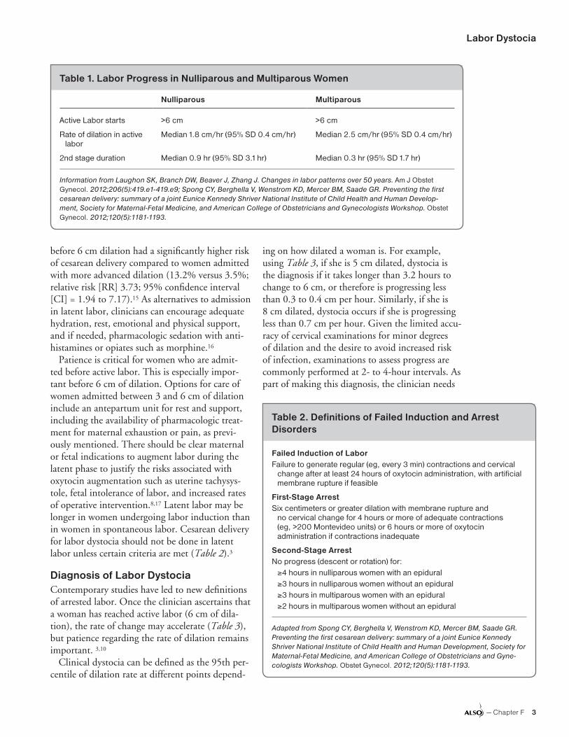
Labor Dystocia
— Chapter F 3
before 6 cm dilation had a significantly higher risk of cesarean delivery compared to women admitted with more advanced dilation (13.2% versus 3.5%; relative risk [RR] 3.73; 95% confidence interval [CI] = 1.94 to 7.17).15 As alternatives to admission in latent labor, clinicians can encourage adequate hydration, rest, emotional and physical support, and if needed, pharmacologic sedation with anti-histamines or opiates such as morphine.16
Patience is critical for women who are admit-ted before active labor. This is especially impor-tant before 6 cm of dilation. Options for care of women admitted between 3 and 6 cm of dilation include an antepartum unit for rest and support, including the availability of pharmacologic treat-ment for maternal exhaustion or pain, as previ-ously mentioned. There should be clear maternal or fetal indications to augment labor during the latent phase to justify the risks associated with oxytocin augmentation such as uterine tachysys-tole, fetal intolerance of labor, and increased rates of operative intervention.8,17 Latent labor may be longer in women undergoing labor induction than in women in spontaneous labor. Cesarean delivery for labor dystocia should not be done in latent labor unless certain criteria are met (Table 2).3
Diagnosis of Labor DystociaContemporary studies have led to new definitions of arrested labor. Once the clinician ascertains that a woman has reached active labor (6 cm of dila-tion), the rate of change may accelerate (Table 3), but patience regarding the rate of dilation remains important. 3,10
Clinical dystocia can be defined as the 95th per-centile of dilation rate at different points depend-
ing on how dilated a woman is. For example, using Table 3, if she is 5 cm dilated, dystocia is the diagnosis if it takes longer than 3.2 hours to change to 6 cm, or therefore is progressing less than 0.3 to 0.4 cm per hour. Similarly, if she is 8 cm dilated, dystocia occurs if she is progressing less than 0.7 cm per hour. Given the limited accu-racy of cervical examinations for minor degrees of dilation and the desire to avoid increased risk of infection, examinations to assess progress are commonly performed at 2- to 4-hour intervals. As part of making this diagnosis, the clinician needs
Table 1. Labor Progress in Nulliparous and Multiparous Women
Nulliparous Multiparous
Active Labor starts >6 cm >6 cm
Rate of dilation in active labor
Median 1.8 cm/hr (95% SD 0.4 cm/hr) Median 2.5 cm/hr (95% SD 0.4 cm/hr)
2nd stage duration Median 0.9 hr (95% SD 3.1 hr) Median 0.3 hr (95% SD 1.7 hr)
Information from Laughon SK, Branch DW, Beaver J, Zhang J. Changes in labor patterns over 50 years. Am J Obstet Gynecol. 2012;206(5):419.e1-419.e9; Spong CY, Berghella V, Wenstrom KD, Mercer BM, Saade GR. Preventing the first cesarean delivery: summary of a joint Eunice Kennedy Shriver National Institute of Child Health and Human Develop-ment, Society for Maternal-Fetal Medicine, and American College of Obstetricians and Gynecologists Workshop. Obstet Gynecol. 2012;120(5):1181-1193.
Table 2. Definitions of Failed Induction and Arrest Disorders
Failed Induction of LaborFailure to generate regular (eg, every 3 min) contractions and cervical
change after at least 24 hours of oxytocin administration, with artificial membrane rupture if feasible
First-Stage ArrestSix centimeters or greater dilation with membrane rupture and
no cervical change for 4 hours or more of adequate contractions (eg, >200 Montevideo units) or 6 hours or more of oxytocin administration if contractions inadequate
Second-Stage ArrestNo progress (descent or rotation) for:≥4 hours in nulliparous women with an epidural≥3 hours in nulliparous women without an epidural≥3 hours in multiparous women with an epidural≥2 hours in multiparous women without an epidural
Adapted from Spong CY, Berghella V, Wenstrom KD, Mercer BM, Saade GR. Preventing the first cesarean delivery: summary of a joint Eunice Kennedy Shriver National Institute of Child Health and Human Development, Society for Maternal-Fetal Medicine, and American College of Obstetricians and Gyne-cologists Workshop. Obstet Gynecol. 2012;120(5):1181-1193.

Chapter F
4 Chapter F —
to address the following questions, considering the 6 Ps mnemonic:
• Passenger: Is there a malposition or malpresen-tation or suspected macrosomia?
• Power: Are contractions adequate in fre-quency, duration, and strength?
• Pelvis: Is there cephalopelvic disproportion because of a contracted pelvis?
• Patient: Are there other coexisting clinical issues such as chorioamnionitis or nonreassuring fetal monitoring that affect the treatment choices?
• Psyche: How are the woman and her support individuals coping with the labor?
• Provider: Is there a consistent examiner to evaluate subtle change? Are there any indica-tions for consultation or second opinion?
Treatment of Active-Phase Labor DystociaFirst Stage Active-Phase DystociaTreatment strategies for women with slowly pro-gressing active-phase labor include amniotomy, pharmacologic augmentation with oxytocin, or both.
Amniotomy alone. A meta-analysis showed that women of any parity undergoing amniotomy alone were less likely to have nonprogressing labor than women who did not (RR 0.75; 95% CI = 0.64 to 0.88); similarly, women who underwent amniotomy routinely were less likely to need oxy-tocin augmentation (RR 0.73; 95% CI = 0.57 to
0.95).18 Nulliparous women who had amniotomy had a slightly shorter second stage of labor than those who did not (but only by 1 to 10 minutes). However, routine amniotomy alone in spontane-ous labor does not change overall first stage labor length or cesarean delivery rates.
Pharmacologic augmentation. Oxytocin alone does not improve vaginal delivery rates. Although the overall duration of labor may shorten in women who receive oxytocin early in labor for slow labor, this intervention alone does not ulti-mately change the cesarean delivery rate.19
Both amniotomy and pharmacologic aug-mentation. Combining amniotomy with oxytocin may be the most effective augmentation strategy. In trials of prevention strategies for labor dystocia, early augmentation with both amniotomy and oxytocin was associated with a slight reduction in the number of cesarean births (RR 0.87; 95% CI = 0.77 to 0.99) and a shortened duration of labor (average mean difference [MD] -1.28 hours; 95% CI = -1.97 to -0.59), without differences in neo-natal outcome or maternal morbidity or satisfac-tion.20 In this analysis, the number needed to treat to prevent 1 cesarean by using amniotomy and oxytocin together early in labor was 65. However, only three studies in this meta-analysis were for treatment for dystocia, with the combination of amniotomy and oxytocin use showing no reduc-tion in cesarean delivery rates for women already diagnosed with slow progress in labor.
For women with protracted or arrested active-phase labor, the clinician can evaluate the strength and frequency of uterine contractions by abdomi-nal palpation or an intrauterine pressure catheter (IUPC), which allows calculation of Montevideo units (MVUs) (Figure 2). Two hundred or more MVUs in 10 minutes is considered evidence of adequate contractions.21 IUPC use may be most helpful if contractions seem to be of sufficient frequency and duration but are not causing the expected cervical change. However, a meta-analysis concluded that the use of an IUPC does not appear to change labor duration or cesarean rates.22 The use of an IUPC may increase the risk of maternal fever so should not be routine. 23
If contractions are inadequate, administering intravenous (IV) oxytocin increases contraction frequency, duration, and strength. There are numerous approaches to dosage, dosing interval, and duration of oxytocin treatment. Low-dose
Table 3. Duration of Each Centimeter Change in Cervical Dilatation for Nulliparous Women with Spontaneous Onset of Labor
Cervical Change (cm)
Median (hour)
95th Percentile (hour)
3 to 44 to 55 to 66 to 77 to 88 to 99 to 10
1.81.30.80.60.50.50.5
8.16.43.22.21.61.41.8
Adapted from Zhang J, Landy HJ, Branch DW, et al; Consortium on Safe Labor. Contemporary patterns of spontaneous labor with normal neonatal outcomes. Obstet Gynecol. 2010;116(6):1281-1287.
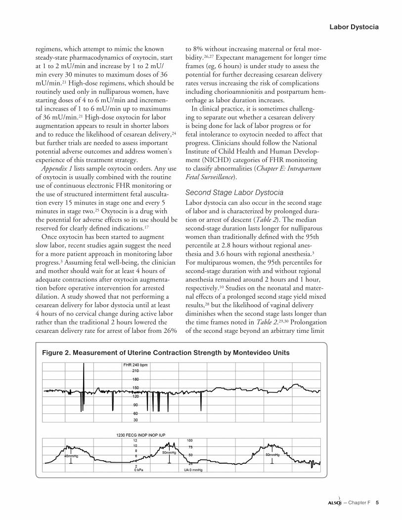
Labor Dystocia
— Chapter F 5
regimens, which attempt to mimic the known steady-state pharmacodynamics of oxytocin, start at 1 to 2 mU/min and increase by 1 to 2 mU/min every 30 minutes to maximum doses of 36 mU/min.21 High-dose regimens, which should be routinely used only in nulliparous women, have starting doses of 4 to 6 mU/min and incremen-tal increases of 1 to 6 mU/min up to maximums of 36 mU/min.21 High-dose oxytocin for labor augmentation appears to result in shorter labors and to reduce the likelihood of cesarean delivery,24 but further trials are needed to assess important potential adverse outcomes and address women’s experience of this treatment strategy.
Appendix 1 lists sample oxytocin orders. Any use of oxytocin is usually combined with the routine use of continuous electronic FHR monitoring or the use of structured intermittent fetal ausculta-tion every 15 minutes in stage one and every 5 minutes in stage two.25 Oxytocin is a drug with the potential for adverse effects so its use should be reserved for clearly defined indications.17
Once oxytocin has been started to augment slow labor, recent studies again suggest the need for a more patient approach in monitoring labor progress.3 Assuming fetal well-being, the clinician and mother should wait for at least 4 hours of adequate contractions after oxytocin augmenta-tion before operative intervention for arrested dilation. A study showed that not performing a cesarean delivery for labor dystocia until at least 4 hours of no cervical change during active labor rather than the traditional 2 hours lowered the cesarean delivery rate for arrest of labor from 26%
to 8% without increasing maternal or fetal mor-bidity.26,27 Expectant management for longer time frames (eg, 6 hours) is under study to assess the potential for further decreasing cesarean delivery rates versus increasing the risk of complications including chorioamnionitis and postpartum hem-orrhage as labor duration increases.
In clinical practice, it is sometimes challeng-ing to separate out whether a cesarean delivery is being done for lack of labor progress or for fetal intolerance to oxytocin needed to affect that progress. Clinicians should follow the National Institute of Child Health and Human Develop-ment (NICHD) categories of FHR monitoring to classify abnormalities (Chapter E: Intrapartum Fetal Surveillance).
Second Stage Labor DystociaLabor dystocia can also occur in the second stage of labor and is characterized by prolonged dura-tion or arrest of descent (Table 2). The median second-stage duration lasts longer for nulliparous women than traditionally defined with the 95th percentile at 2.8 hours without regional anes-thesia and 3.6 hours with regional anesthesia.3 For multiparous women, the 95th percentiles for second-stage duration with and without regional anesthesia remained around 2 hours and 1 hour, respectively.10 Studies on the neonatal and mater-nal effects of a prolonged second stage yield mixed results,28 but the likelihood of vaginal delivery diminishes when the second stage lasts longer than the time frames noted in Table 2.29,30 Prolongation of the second stage beyond an arbitrary time limit
Figure 2. Measurement of Uterine Contraction Strength by Montevideo Units

Chapter F
6 Chapter F —
is no longer an indication for operative vaginal or cesarean delivery. When the second stage is prolonged, it is important to evaluate if progress is occurring, chorioamnionitis has developed, or there are concerns regarding fetal monitoring.
Second stage labor dystocia may be because of fetal malposition, inadequate contractions, poor maternal efforts, or true cephalopelvic dispropor-tion. Each of these possible causes has potential management options.
Fetal malposition. The most common fetal malposition is occiput posterior (OP), where the fetus lies with its occiput toward the mother’s spine and its face toward the mother’s pubic symphysis. Persistent OP is associated with prolonged second stage labor and an increased need for oxytocin augmentation.31 The clinical care of women with persistent OP position, including the technique of manual rotation of a fetus from OP to occiput anterior position, is described in Chapter G: Mal-presentations, Malpositions, and Multiple Gestations.
For a laboring woman without an epidural, her position during the second stage does not appear to affect its duration significantly,32 but if she stays more upright, this position may reduce the risk of abnormal FHR tracings (RR 0.46; 95% CI = 0.22 to 0.93) and assisted vaginal delivery com-pared to the supine position (RR 0.78; 95% CI = 0.68 to 0.90), with a possible increased risk of postpartum hemorrhage if upright compared to supine (RR 1.65; 95% CI = 1.32 to 2.60).32 Stud-ies of the effects of upright position for women with an epidural are inconclusive.33 Women in the second stage with or without an epidural should be allowed to assume whatever position is most comfortable for them.
Inadequate contractions. Just as in first stage labor dystocia, if contractions have decreased in strength or frequency during the second stage, pharmacologic treatment with oxytocin may be necessary. Thus when fetal descent has slowed in the second stage, the clinician may need to initiate or increase IV oxytocin.34,35
Maternal exhaustion. Exhaustion can affect the length of second stage labor. For women with epidural analgesia, an alternative to initiating active pushing at full cervical dilation is allowing the fetus to labor down to a lower station, or delaying pushing for 60 to 90 minutes until the mother feels an urge to push or the fetus is at the introitus. This may be especially important for nulliparous
women. Although such delayed pushing may shorten the active phase of labor, there is no change in cesarean delivery rates or rates of instrumental deliveries,36 and the overall length of the second stage increases along with the risk of maternal fever. If the contractions are greater than 3 minutes apart, then the addition of oxytocin should be con-sidered during the laboring down process.35
During a long second stage, the clinician should remain vigilant to ongoing assessment of fetal well-being. Preventing vena cava compression by moving the mother out of the dorsal lithotomy position and allowing adequate periods of rest between pushing are two basic strategies to prevent fetal intolerance of second stage labor. Repetitive deep variables in the second stage do not neces-sarily indicate fetal acidosis and may respond to amnioinfusion if an IUPC is already in place.37 If the FHR tracing still deteriorates with the omi-nous combination of minimal/absent variability and repetitive late or variable decelerations, the clinician should institute measures of fetal resusci-tation as described in Chapter E: Intrapartum Fetal Surveillance while expediting delivery via assisted vaginal delivery or cesarean delivery.
PreventionMaternity clinicians can attempt to prevent dys-tocia with the following antepartum and intrapar-tum strategies: undertaking prenatal interventions to decrease the incidence of fetal macrosomia, provision of labor support, hydration, avoidance of elective induction with an unripe cervix, judi-cious use of epidural analgesia, and prevention of chorioamnionitis.
ObesityMaternal obesity, particularly in association with excessive maternal weight gain during pregnancy or with gestational diabetes, increases the risk of fetal macrosomia, which may predispose women to prolonged labor and operative outcomes.38,39 Dietary counseling during preconception, inter-conception, and prenatal care can help overweight and obese women limit their weight gain and has been shown to decrease the risk of shoulder dys-tocia,40,41 but these approaches have not yet been shown to specifically change the rate of macroso-mia except possibly in the highest-risk women.42 Therefore clinicians should provide counseling and initiate referrals to nutritionists for these at-

Labor Dystocia
— Chapter F 7
risk women. Although meta-analysis continues to show that induction for macrosomia alone may increase the risk of cesarean delivery compared to expectant care,43 a recent European randomized trial showed a lower risk of cesarean delivery and birth injury for women induced between 37 and 39 weeks for ultrasound-confirmed estimated fetal weight greater than the 95th percentile.44
Clinicians should follow the same initial guide-lines for recognition and treatment of dysfunc-tional labor for obese women as for non-obese women. However, patience with slower labor progress in obese women is important. In one study, overweight and obese nulliparas women had significantly longer active phases of labor than women of normal weight.45 Another retro-spective study confirmed that obese women have slower progress especially in the early part of active labor.46 Induction with prostaglandins appears to take longer in obese women;47 one retrospec-tive study also suggests that misoprostol rather than dinoprostone may improve the likelihood of vaginal delivery for obese women.48 Finally, obese women may need higher doses of oxytocin than lean women to achieve adequate contractions.49
Labor SupportMeta-analysis of studies on providing trained labor support companions (doulas) shows a decrease in the incidence of labor dystocia, opera-tive vaginal deliveries, and cesarean deliveries, particularly for first-time mothers.50 The greatest effect on labor comes when trained lay individu-als rather than hospital employees are used, when epidural analgesia is not routinely used, and when support begins in early labor.50 A potential low-cost alternative is for women to choose a female friend or family member to receive specific brief labor support training as part of prenatal care and to accompany the woman in labor; in one trial, this strategy led to shorter overall labors but no difference in cesarean delivery rates.51 For addi-tional information about doula programs, see the Doulas of North America website (available at https://www.dona.org) and Appendix 2.52
HydrationHydration during active labor may prevent pro-longed labor. Two randomized trials of IV hydra-tion, one in prolonged labor and one in normal labor, suggested shorter labors, less oxytocin use,
and a reduced risk of cesarean delivery in women receiving a higher rate (250 mL/hour) of IV fluids than traditionally used (125 mL/hour).53,54 However, if women have unrestricted oral intake during labor, such IV hydration may not make a difference in these outcomes.55
InductionsAlthough induction rates in the United States have declined in the last few years, the number of births involving induction of labor remained higher than 23% in 2012.56 Clinicians should avoid induction of women for nonmedical indications.57,58
How labor induction affects cesarean risk is becoming more controversial with recent stud-ies suggesting less risk than previously thought. Cochrane reviews of mechanical methods for cervical ripening, vaginal misoprostol, and vaginal prostaglandin showed that these agents decrease the length of labor but do not change the overall cesarean delivery rate for labor dystocia;59-61 oral misoprostol for induction compared to vaginal dinoprostone or oxytocin leads to fewer cesarean deliveries.62 Various studies have demonstrated a decreased cesarean delivery rate through selective induction of women at or near term with specific risk factors for developing cephalopelvic dispropor-tion or uteroplacental insufficiency;63-65 this style of management includes patient-centered care and patience with cervical ripening, which may par-tially explain the favorable cesarean delivery rates.66 A large retrospective cohort study from 2015 sug-gested that low-risk nulliparous women induced at 38 or 40 weeks have a lower risk of maternal complications and neonatal complications without an increased cesarean delivery risk,67 and analysis of data from the Consortium on Safe Labor suggested that elective induction compared to expectant management had a lower risk of cesarean delivery.68 A 2016 randomized trial of induction at 39 weeks for women older than 35 years showed no differ-ence in cesarean delivery rates for induced women compared to expectantly managed women.69 How-ever, until further studies are conducted, induction should be reserved for women with clear maternal or fetal indications.3 Elective induction without medical indication should only be performed after 39 weeks’ gestation and is best reserved for mul-tiparous women with a favorable cervix.57,58
With the goal of vaginal delivery, induction requires patience in allowing adequate time for

Chapter F
8 Chapter F —
progress in latent and active labor. Several stud-ies confirm the clinical safety of allowing women undergoing induction who have ruptured mem-branes after oxytocin has started to have a latent phase of labor of at least 12 hours after ruptured membranes, and longer may be safe.70 Induced labor takes longer than spontaneous labor, par-ticularly for nulliparous women before 6 cm of dilation, with one retrospective study concluding that each cm of dilation before 6 cm could take between 2 to 5.5 hours longer with induced labor than for spontaneous labor.71 Thus a failed induc-tion should be diagnosed only when a woman has not had regular (every 3 minute) contractions with cervical change after at least 24 hours of oxytocin administration,3 assuming fetal well-being through-out this time. If a woman has intact membranes, if fetal monitoring is not concerning, and if there are no other medical concerns such as hypertension, it may be possible to send her home rather than doing a cesarean delivery when cervical change is not occurring despite best efforts.
Epidural AnesthesiaAlthough meta-analyses consistently find no dif-ference in cesarean delivery rates among women receiving low-dose epidurals compared to par-enteral opioids,72 epidural analgesia does impact labor progress and other outcomes. Women receiving epidurals are more likely to require oxy-tocin augmentation in the first stage of labor, have longer second stages,73 have a six-fold increase in the incidence of maternal fevers, have increased incidence of persistent OP malposition, and undergo more operative vaginal deliveries.74
Whether administering epidural analgesia early in labor (before 4 to 5 cm dilation) increases the risk of cesarean delivery has been controversial.75 Epidural analgesia is not a single entity, and randomized controlled trials that have specifically investigated early versus standard placement are small or do not use contemporary low-dose tech-niques.75 The study most commonly cited to sup-port the use of early epidurals actually compared a combined spinal epidural analgesia technique (intrathecal opioid administered at 2 cm cervical dilation) to epidural administered at 4 cm or later. This study showed no significant differences in labor duration or rates of cesarean delivery.76
Maternal request is sufficient indication for pain relief during labor,77,78 and epidurals are associated
with significantly lower pain scores compared to systemic opioids.72 Clinicians should individualize whether and when to administer epidural anal-gesia. Women with significant pain early in labor should not be required to reach 4 to 5 cm before epidural placement.77 Conversely, a woman who is informed and prepared to handle labor pain with lesser interventions should not be subjected to the expectation of a routine epidural. Clinicians should support the availability of other pain-relief options that may have less impact on labor duration such as water immersion, nitrous oxide, and sterile water injections.79-82
If a woman is having severe back labor (feeling her contractions most intensely in her lower back), this may indicate a persistent OP position. If changing maternal position does not relieve this discomfort, another option for pain relief before resorting to regional anesthesia is to try sterile water injections (Appendix 3). The effectiveness of these injections in reducing pain or changing the cesarean delivery rate is debatable; one review showed studies inconclu-sive,83 whereas another with slightly different trials included showed that sterile water injections for low back pain in labor reduced pain for up to 2 hours and reduced the risk of cesarean delivery (RR 0.51; 95% CI = 0.30 to 0.87) compared to alternative therapies (however, the overall cesarean delivery rate was below 10% in the comparison therapy group).81
InfectionChorioamnionitis is associated with an increased incidence of labor dystocia (adjusted odds ratio [OR] 2.3; 95% CI = 2.0 to 2.7 for first stage labor, and OR 1.8; 95% CI = 1.5 to 2.2 for second stage labor) and cesarean delivery (adjusted OR 1.8; 95% CI = 1.5 to 2.1).84 Clinicians should delay the initial digital vaginal examination and substi-tute a sterile speculum examination in any patient with term rupture of membranes who is not in labor. Attempts should be made to limit the total number of vaginal examinations after rupture of membranes to five or less,85 although more recent studies suggest less risk than previously thought.86
Ambulation and PositionA recent meta-analysis concludes that women who walk or remain upright during the first stage of labor have shorter labors and less risk of cesarean delivery than women who remain recumbent or supine during labor.87 Clinicians should encourage

Labor Dystocia
— Chapter F 9
low-risk women to assume whatever position is most comfortable for them during labor.
Quality ImprovementCertain aspects of clinician style and health care systems may prevent labor dystocia and resultant cesarean delivery. These include caregiver continu-ity during the assessment of early labor,33 encourag-ing a pronatalist cultural attitude toward natural childbirth,88,89 requiring consultation with a second clinician before non-emergent cesarean deliveries for labor dystocia,88 and providing regular feedback to clinicians regarding their cesarean delivery rates.90
One small study suggests a lower first-stage cesar-ean delivery rate when a woman has her primary prenatal clinician attending her in labor rather than an on-call hospitalist.91 Finally, improving quality of care as part of overall maternity safety has led to a plateauing of cesarean delivery rates.92 Whether a laborist model of care or a midwifery model of care affects cesarean rates remains unclear.93-95
In summary, labor dystocia is common and requires the maternity clinician to have excellent clinical assessment skills and thorough knowledge of nonpharmacologic and pharmacologic strategies to prevent and treat nonprogressing labor.
SORT: Key Recommendations for Practice
Clinical RecommendationEvidence Rating References
Amniotomy with oxytocin may help prevent labor dystocia, but routine amniotomy or oxytocin use alone is not recommended.
A 18-20
Clinicians should support having women receive continuous labor support from a labor companion as the use of such a doula has been shown to help women use less analgesia, have lower rates of operative vaginal and cesarean delivery, and feel more satisfied with their childbirth experiences.
A 50
Clinicians should use epidural analgesia with care as epidurals are associated with a prolongation of the second stage of labor, and an increase in maternal fever, oxytocin use, and operative vaginal delivery compared to opioids or no analgesia.
A 72
Cesarean delivery for labor dystocia should not occur until at least 4 hours without cervical change during active stage of labor with adequate contractions.
A 3
Normal second stage labor may take longer than previously thought; second stage arrest is now defined after 4 hours without rotation or descent in nulliparous women with epidural or 3 hours without epidural.
A 3
Active labor does not start until 6 cm of dilation so cesarean delivery for dystocia should be avoided before this dilation.
B 3
Quality improvement and patient safety strategies that monitor cesarean delivery indications and rates can help lower these rates safely.
B 6,92
Clinicians should allow slower labor progress in latent labor in women undergoing induction compared to spontaneous labor assuming fetal tolerance and no other compelling obstetric indication for intervention.
B 3,70
Clinicians should allow slower labor progress throughout all stages of labor in obese women compared to non-obese women assuming fetal tolerance and no other compelling obstetric indication for intervention.
B 3,45,46
Strength of Recommendation Taxonomy (SORT)
Strength of Recommendation Definition
A • Recommendation based on consistent and good-quality patient-oriented evidence.
B • Recommendation based on inconsistent or limited-quality patient-oriented evidence.
C • Recommendation based on consensus, usual practice, opinion, disease-oriented evidence, or case series for studies of diagnosis, treat-ment, prevention, or screening.
Patient-oriented evidence measures outcomes that matter to patients: morbidity, mortality, symptom improvement, cost reduction, and quality of life. Disease-oriented evidence measures intermediate, physiologic, or surrogate end points that might not reflect improvement in patient outcomes (eg, blood pressure, blood chemistry, physiologic function, pathologic findings).
From Ebell MH, Siwek J, Weiss BD, et al. Strength of recommendation taxonomy (SORT): a patient-centered approach to grading evidence in the medical literature. Am Fam Physician. 2004;69(3):548-556.

Chapter F
10 Chapter F —
Appendix A Sample Labor Orders for Oxytocin
Augmentation
1. Consider oxytocin augmentation if:a) Cervical dilation rate is <0.4 cm/hour for
a woman up to 6 cm dilation or <1.0 cm/hour if ≥6 cm dilation
b) Contractions occur less frequently thanevery 3 minutes
c) Contractions last <30 secondsd) Contractions are not palpablee) Intrauterine pressure catheter documents
<200 Montevideo units (MVUs) in a30-minute period
f) Amniotomy does not cause labor progressin 1 to 3 hours
2. Document the decision to augment in themedical record.
3. Electronically monitor fetal heart rate anduterine activity for a minimum of 30 minutesbefore initiation of oxytocin.
4. Measure blood pressure every 15 to30 minutes.
5. Start primary intravenous (IV) infusion of1,000 mL Lactated Ringer’s solution at a keepvein open rate.
6. Through the infusion pump, add a second-ary IV of 1,000 mL 5% dextrose/LactatedRinger’s solution with 10 to 20 units of oxy-tocin (20 units preferred to decrease the totalamount of fluid administered).
7. Begin oxytocin infusion at 0.5 to 2 mU/min.8. Increase oxytocin by 1 to 2 mU/min every
30 minutes, until an adequate contractionpattern has been reached. After 10 mU/minhas been reached, the rate of increase may be1 to 4 mU/minute.
9. Notify the clinician before exceeding20 mU/min. The clinician should documentin the medical record any decision to exceedthis dosage. The maximum infusion rate is36 mU/min.
10. Stop or decrease oxytocin by 50% if any ofthe following occur:a) Tachysystole (more than 5 contractions
within 10 minutes averaged over30 minutes)
b) Uterine tone between contractions exceeds15 to 20 mm Hg
c) Tetanic contractions occur (contractionslasting >2 minutes in length)
d) Severe variable, late decelerations, brady-cardia, or tachycardia occur
e) Less severe patterns may be managed as ifthe patient were in spontaneous labor byadministration of a fluid bolus, positionchange, and oxygen at 10 L/min via non-rebreather mask
f) Interventions such as these should be docu-mented in the medical record
11. Consider augmentation to have failed if:a) The woman is at least 6 cm dilated with
ruptured membranes and has had nocervical change after 4 hours of adequatecontractions (defined as >200 MVUs in30 minutes) by intrauterine pressure cath-eter or 6 hours of inadequate contractionswithout intrauterine pressure catheter
b) No regular contractions (every 3 minutes)after 24 hours of oxytocin, preferably withrupture of membranes

Labor Dystocia
— Chapter F 11
Appendix B Doula Organizations and Resources
This list is not meant to be complete but to pro-vide suggested resources. Most of the doula certi-fication programs require trainees to provide labor support for a certain number of births to obtain certification. Additional resources, including lists of doula professionals, are available at http://www.doulas.com/.
Doulas of North America International (DONA): http://www.dona.org
Childbirth and Postpartum Professional Asso-ciation (CAPPA): http://www.cappa.net
The International Childbirth Education Asso-ciation (ICEA): http://www.icea.org
Appendix C Intradermal Sterile Water Injections
The use of intradermal sterile water injections can be used to treat first-stage back labor. Four 0.1 mL intradermal injections of sterile water with a 25 gauge or 27 gauge needle are given to form small blebs in the skin. Two injection sites are over the posterior superior iliac spines; two are 2 to 3 cm below and 1 to 2 cm medial to the first points.
The injections cause intense stinging for 15 to 30 seconds, followed within 2 minutes by partial to complete relief of back pain lasting 45 to 90 min-utes. These injections can be repeated if necessary. Using two clinicians, these injections can be easily injected simultaneously to decrease the duration of discomfort.

Chapter F
12 Chapter F —
References1. National Center for Health Statistics. User Guide to the
2014 Natality Public Use File. Available at ftp://ftp.cdc.gov/pub/Health_Statistics/NCHS/Dataset_Documenta-tion/DVS/natality/UserGuide2014.pdf.
2. Childbirth Connection. Cesarean Section Trends, 1989-2014. Available at http://www.childbirthconnection.org/article.asp?ck=10554.
3. Spong CY, Berghella V, Wenstrom KD, Mercer BM,Saade GR. Preventing the first cesarean delivery: sum-mary of a joint Eunice Kennedy Shriver National Insti-tute of Child Health and Human Development, Societyfor Maternal-Fetal Medicine, and American College ofObstetricians and Gynecologists Workshop. ObstetGynecol. 2012;120(5):1181-1193.
4. Boyle A, Reddy UM, Landy HJ, Huang CC, DriggersRW, Laughon SK. Primary cesarean delivery in theUnited States. Obstet Gynecol. 2013;122(1):33-40.
5. Hamilton BE, Martin JA, Osterman MJK, et al; Division ofVital Statistics. Births: Final Data for 2014. National VitalStatistics Reports. Vol 64, No 12. 2015. Available at http://www.cdc.gov/nchs/data/nvsr/nvsr64/nvsr64_12.pdf.
6. Main EK, Morton CH, Hopkins D, Giuliani G, MelsopK, Gould J. Cesarean Deliveries, Outcomes, andOpportunities for Change in California: Toward a PublicAgenda for Maternity Care Safety and Quality. CaliforniaMaternal Quality Care Collaborative, 2011. Available athttps://www.cmqcc.org/resource/cesarean-deliveries-outcomes-and-opportunities-change-california-toward-public-agenda.
7. King TL. Preventing primary cesarean sections: intra-partum care. Semin Perinatol. 2012;36(5):357-364.
8. Greulich B, Tarrant B. The latent phase of labor: diag-nosis and management. J Midwifery Womens Health.2007;52(3):190-198.
9. Friedman EA. Primigravid labor; a graphicostatisticalanalysis. Obstet Gynecol. 1955;6(6):567-589.
10. Zhang J, Landy HJ, Branch DW, et al; Consortium onSafe Labor. Contemporary patterns of spontaneouslabor with normal neonatal outcomes. Obstet Gynecol.2010;116(6):1281-1287.
11. Laughon SK, Branch DW, Beaver J, Zhang J. Changesin labor patterns over 50 years. Am J Obstet Gynecol.2012;206(5):419.e1-419.e9.
12. Neal JL, Lowe NK, Ahijevych KL, Patrick TE, CabbageLA, Corwin EJ. “Active labor” duration and dilation ratesamong low-risk, nulliparous women with spontaneouslabor onset: a systematic review. J Midwifery WomensHealth. 2010;55(4):308-318.
13. Bailit JL, Dierker L, Blanchard MH, Mercer BM.Outcomes of women presenting in active versuslatent phase of spontaneous labor. Obstet Gynecol.2005;105(1):77-79.
14. McNiven PS, Williams JI, Hodnett E, Kaufman K, Han-nah ME. An early labor assessment program: a random-ized, controlled trial. Birth. 1998;25(1):5-10.
15. Wood AM, Frey HA, Tuuli MG, et al. Optimal AdmissionCervical Dilation in Spontaneously Laboring Women.Am J Perinatol. 2016;33(2):188-194.
16. Simkin P, Ancheta RS. The Labor Progress Handbook:Early Interventions to Prevent and Treat Dystocia. 3rded. West Sussex, UK: Wiley-Blackwell; 2011.
17. Clark SL, Simpson KR, Knox GE, Garite TJ. Oxytocin:new perspectives on an old drug. Am J Obstet Gynecol.2009;200(1):35.e1-35.e6.
18. Smyth RMD, Alldred SK, Markham C. Amniotomy forshortening spontaneous labour. Cochrane DatabaseSyst Rev. 2013;(1):CD006167.
19. Bugg GJ, Siddiqui F, Thornton JG. Oxytocin versus notreatment or delayed treatment for slow progress in thefirst stage of spontaneous labour. Cochrane DatabaseSyst Rev. 2013;6(6):CD007123.
20. Wei S, Wo BL, Qi HP, et al. Early amniotomy and earlyoxytocin for prevention of, or therapy for, delay in firststage spontaneous labour compared with routine care.Cochrane Database Syst Rev. 2013;8(8):CD006794.
21. American College of Obstetrics and GynecologyCommittee on Practice Bulletins-Obstetrics. ACOGPractice Bulletin Number 49, December 2003: Dys-tocia and augmentation of labor. Obstet Gynecol.2003;102(6):1445-1454.
22. Bakker JJH, Janssen PF, van Halem K, et al. Internalversus external tocodynamometry during inducedor augmented labour. Cochrane Database Syst Rev.2013;8(8):CD006947.
23. Harper LM, Shanks AL, Tuuli MG, Roehl KA, Cahill AG.The risks and benefits of internal monitors in laboringpatients. Am J Obstet Gynecol. 2013;209(1):38.e1-38.e6.
24. Kenyon S, Tokumasu H, Dowswell T, Pledge D, MoriR. High-dose versus low-dose oxytocin for augmenta-tion of delayed labour. Cochrane Database Syst Rev.2013;7(7):CD007201.
25. American College of Obstetricians and Gynecolo-gists. ACOG Practice Bulletin No. 106: Intrapartum fetalheart rate monitoring: nomenclature, interpretation,and general management principles. Obstet Gynecol.2009;114(1):192-202.
26. Rouse DJ, Owen J, Hauth JC. Active-phase labor arrest:oxytocin augmentation for at least 4 hours. ObstetGynecol. 1999;93(3):323-328.
27. Rouse DJ, Owen J, Savage KG, Hauth JC. Active phaselabor arrest: revisiting the 2-hour minimum. ObstetGynecol. 2001;98(4):550-554.
28. Altman MR, Lydon-Rochelle MT. Prolonged secondstage of labor and risk of adverse maternal and perinataloutcomes: a systematic review. Birth. 2006;33(4):315-322.
29. El-Sayed YY. Diagnosis and management of arrest disor-ders: duration to wait. Semin Perinatol. 2012;36(5):374-378.
30. Rouse DJ, Weiner SJ, Bloom SL, et al; Eunice KennedyShriver National Institute of Child Health and HumanDevelopment Maternal-Fetal Medicine Units Network.Second-stage labor duration in nulliparous women:relationship to maternal and perinatal outcomes. Am JObstet Gynecol. 2009;201(4):357.e1-357.e7.
31. Carseldine WJ, Phipps H, Zawada SF, et al. Doesocciput posterior position in the second stage of labourincrease the operative delivery rate? Aust N Z J ObstetGynaecol. 2013;53(3):265-270.

Labor Dystocia
— Chapter F 13
32. Gupta JK, Hofmeyr GJ, Shehmar M. Position in the sec-ond stage of labour for women without epidural anaesthe-sia. Cochrane Database Syst Rev. 2012;5(5):CD002006.
33. Kemp E, Kingswood CJ, Kibuka M, Thornton JG.Position in the second stage of labour for women withepidural anaesthesia. Cochrane Database Syst Rev.2013;1(1):CD008070.
34. O’Driscoll K, Meagher D, Robson M. Active Manage-ment of Labour: the Dublin Experience. 4th ed. Edin-burgh; New York: Mosby; 2003.
35. Sprague AE, Oppenheimer L, McCabe L, Brownlee J,Graham ID, Davies B; Ottawa Hospital. The Ottawa Hos-pital’s Clinical Practice Guideline for the Second Stageof Labour. J Obstet Gynaecol Can. 2006;28(9):769-779.
36. Lemos A, Amorim MMR, Dornelas de Andrade A, deSouza AI, Cabral Filho JE, Correia JB. Pushing/bear-ing down methods for the second stage of labour.Cochrane Database Syst Rev. 2015;10(10):CD009124.
37. Hofmeyr GJ, Lawrie TA. Amnioinfusion for potentialor suspected umbilical cord compression in labour.Cochrane Database Syst Rev. 2012;1(1):CD000013.
38. American College of Obstetricians and Gynecologists.ACOG Practice Bulletin No 156: Obesity in Pregnancy.Obstet Gynecol. 2015;126(6):e112-e126.
39. Ehrenberg HMMB, Mercer BM, Catalano PM. The influ-ence of obesity and diabetes on the prevalence of mac-rosomia. Am J Obstet Gynecol. 2004;191(3):964-968.
40. Thangaratinam S, Rogozinska E, Jolly K, et al. Effectsof interventions in pregnancy on maternal weight andobstetric outcomes: meta-analysis of randomised evi-dence. BMJ. 2012;344:e2088.
41. Oteng-Ntim E, Varma R, Croker H, Poston L, Doyle P.Lifestyle interventions for overweight and obese preg-nant women to improve pregnancy outcome: systematicreview and meta-analysis. BMC Med. 2012;10:47.
42. Muktabhant B, Lawrie TA, Lumbiganon P, LaopaiboonM. Diet or exercise, or both, for preventing excessiveweight gain in pregnancy. Cochrane Database SystRev. 2015;6(6):CD007145.
43. Sanchez-Ramos L, Bernstein S, Kaunitz AM. Expect-ant management versus labor induction for suspectedfetal macrosomia: a systematic review. Obstet Gynecol.2002;100(5 Pt 1):997-1002.
44. Boulvain M, Senat MV, Perrotin F, et al; Groupe deRecherche en Obstétrique et Gynécologie (GROG).Induction of labour versus expectant management forlarge-for-date fetuses: a randomised controlled trial.Lancet. 2015;385(9987):2600-2605.
45. Vahratian A, Zhang J, Troendle JF, Savitz DA, Siega-RizAM. Maternal prepregnancy overweight and obesityand the pattern of labor progression in term nulliparouswomen. Obstet Gynecol. 2004;104(5 Pt 1):943-951.
46. Norman SM, Tuuli MG, Odibo AO, Caughey AB, RoehlKA, Cahill AG. The effects of obesity on the first stage oflabor. Obstet Gynecol. 2012;120(1):130-135.
47. Pevzner L, Powers BL, Rayburn WF, Rumney P, WingDA. Effects of maternal obesity on duration and out-comes of prostaglandin cervical ripening and laborinduction. Obstet Gynecol. 2009;114(6):1315-1321.
48. Suidan RS, Rondon KC, Apuzzio JJ, Williams SF. Laboroutcomes of obese patients undergoing induction oflabor with misoprostol compared to dinoprostone. Am JPerinatol. 2015;30(2):187-192.
49. Hill M, Reed KL, Cohen WR. Oxytocin utilization forlabor induction in obese and lean women. J PerinatMed. 2015;43(6):703-706.
50. Hodnett ED, Gates S, Hofmeyr GJ, Sakala C. Continu-ous support for women during childbirth. CochraneDatabase Syst Rev. 2015;7(7):CD003766.
51. Campbell DA, Lake MF, Falk M, Backstrand JR. A ran-domized control trial of continuous support in labor by alay doula. J Obstet Gynecol Neonatal Nurs. 2006;35(4):456-464.
52. Doulas of North America. Available at www.dona.org.
53. Garite TJ, Weeks J, Peters-Phair K, Pattillo C, Brew-ster WR. A randomized controlled trial of the effectof increased intravenous hydration on the course oflabor in nulliparous women. Am J Obstet Gynecol.2000;183(6):1544-1548.
54. Eslamian L, Marsoosi V, Pakneeyat Y. Increased intrave-nous fluid intake and the course of labor in nulliparouswomen. Int J Gynaecol Obstet. 2006;93(2):102-105.
55. Coco A, Derksen-Schrock A, Coco K, Raff T, Horst M,Hussar E. A randomized trial of increased intravenoushydration in labor when oral fluid is unrestricted. FamMed. 2010;42(1):52-56.
56. Osterman MJK, Martin JA. Recent Declines in Inductionof Labor by Gestational Age. NCHS Data Brief No. 155.National Center for Health Statistics. 2014. Available athttp://www.cdc.gov/nchs/data/databriefs/db155.pdf.
57. ACOG Committee on Practice Bulletins — Obstetrics.ACOG Practice Bulletin No. 107: Induction of labor.Obstet Gynecol. 2009;114(2 Pt 1):386-397.
58. American Academy of Family Physicians. ChoosingWisely. Don’t schedule elective, non-medically indicatedinductions of labor or Cesarean deliveries before 39weeks, 0 days gestational age. Available at http://www.choosingwisely.org/clinician-lists/american-academy-family-physicians-elective-c-section-labor-induction.
59. Jozwiak M, Bloemenkamp KWM, Kelly AJ, MolBWJ, Irion O, Boulvain M. Mechanical methods forinduction of labour. Cochrane Database Syst Rev.2012;3(3):CD001233.
60. Hofmeyr GJ, Gülmezoglu AM, Pileggi C. Vaginal miso-prostol for cervical ripening and induction of labour.Cochrane Database Syst Rev. 2010;(10):CD000941.
61. Thomas J, Fairclough A, Kavanagh J, Kelly AJ.Vaginal prostaglandin (PGE2 and PGF2a) for induc-tion of labour at term. Cochrane Database Syst Rev.2014;6(6):CD003101.
62. Alfirevic Z, Aflaifel N, Weeks A. Oral misoprostol forinduction of labour. Cochrane Database Syst Rev.2014;6(6):CD001338.
63. Nicholson JM, Kellar LC, Cronholm PF, Macones GA.Active management of risk in pregnancy at term in anurban population: an association between a higherinduction of labor rate and a lower cesarean deliveryrate. Am J Obstet Gynecol. 2004;191(5):1516-1528.

Chapter F
14 Chapter F —
64. Nicholson JM, Stenson MH, Kellar LC, Caughey AB,Macones GA. Active management of risk in nulliparouspregnancy at term: association between a higher preven-tive labor induction rate and improved birth outcomes.Am J Obstet Gynecol. 2009;200(3):254.e1-254.e13.
65. Caughey AB. Preventive induction of labor: can its uselower the cesarean delivery rate? J Womens Health(Larchmt). 2009;18(11):1743-1745.
66. Klein MC. Association not causation: what is the inter-vention? Ann Fam Med. 2007;5(4):294-297.
67. Bailit JL, Grobman W, Zhao Y, et al; Eunice KennedyShriver National Institute of Child Health and HumanDevelopment Maternal-Fetal Medicine Units (MFMU)Network. Nonmedically indicated induction vs expect-ant treatment in term nulliparous women. Am J ObstetGynecol. 2015;212(1):103.e1-103.e7.
68. Gibson KS, Waters TP, Bailit JL. Maternal and neonataloutcomes in electively induced low-risk term pregnan-cies. Am J Obstet Gynecol. 2014;211(3):249.e1-249.e16.
69. Walker KF, Bugg GJ, Macpherson M, et al; 35/39 Trial Group. Randomized Trial of Labor Induction in Women 35Years of Age or Older. N Engl J Med. 2016;374(9):813-822.
70. Rouse DJ, Weiner SJ, Bloom SL, et al; Eunice KennedyShriver National Institute of Child Health and HumanDevelopment (NICHD) Maternal-Fetal Medicine Units Net-work (MFMU). Failed labor induction: toward an objectivediagnosis. Obstet Gynecol. 2011;117(2 Pt 1):267-272.
71. Harper LM, Caughey AB, Odibo AO, Roehl KA, ZhaoQ, Cahill AG. Normal progress of induced labor. ObstetGynecol. 2012;119(6):1113-1118.
72. Anim-Somuah M, Smyth RMD, Jones L. Epidural versusnon-epidural or no analgesia in labour. Cochrane Data-base Syst Rev. 2011;(12):CD000331.
73. Cheng YW, Shaffer BL, Nicholson JM, Caughey AB.Second stage of labor and epidural use: a larger effectthan previously suggested. Obstet Gynecol. 2014;123(3):527-535.
74. Lieberman E, Davidson K, Lee-Parritz A, ShearerE. Changes in fetal position during labor and theirassociation with epidural analgesia. Obstet Gynecol.2005;105(5 Pt 1):974-982.
75. Klein MC. Does epidural analgesia increase rate of cesar-ean section? Can Fam Physician. 2006;52(4):419-421.
76. Wong CA, Scavone BM, Peaceman AM, et al. The riskof cesarean delivery with neuraxial analgesia given earlyversus late in labor. N Engl J Med. 2005;352(7):655-665.
77. American College of Obstetricians and GynecologistsCommittee on Obstetric Practice. ACOG committeeopinion. No. 339: Analgesia and cesarean deliveryrates. Obstet Gynecol. 2006;107(6):1487-1488.
78. Goetzl LM; ACOG Committee on Practice Bulletins-Obstetrics. ACOG Practice Bulletin. Clinical Man-agement Guidelines for Obstetrician-GynecologistsNumber 36, July 2002. Obstetric analgesia and anes-thesia. Obstet Gynecol. 2002;100(1):177-191.
79. Cluett ER, Burns E. Immersion in water in labour andbirth. Cochrane Database Syst Rev. 2009;(2):CD000111.
80. Rooks JP. Safety and risks of nitrous oxide labor anal-gesia: a review. J Midwifery Womens Health. 2011;56(6):557-565.
81. Collins MR, Starr SA, Bishop JT, Baysinger CL. Nitrousoxide for labor analgesia: expanding analgesic optionsfor women in the United States. Rev Obstet Gynecol.2012;5(3-4):e126-e131.
82. Hutton EK, Kasperink M, Rutten M, Reitsma A, Wain-man B. Sterile water injection for labour pain: a system-atic review and meta-analysis of randomised controlledtrials. BJOG. 2009;116(9):1158-1166.
83. Derry S, Straube S, Moore RA, Hancock H, CollinsSL. Intracutaneous or subcutaneous sterile waterinjection compared with blinded controls for painmanagement in labour. Cochrane Database Syst Rev.2012;1(1):CD009107.
84. Mark SP, Croughan-Minihane MS, Kilpatrick SJ. Cho-rioamnionitis and uterine function. Obstet Gynecol.2000;95(6 Pt 1):909-912.
85. Seaward PG, Hannah ME, Myhr TL, et al. InternationalMulticentre Term Prelabor Rupture of MembranesStudy: evaluation of predictors of clinical chorioam-nionitis and postpartum fever in patients with prelaborrupture of membranes at term. Am J Obstet Gynecol.1997;177(5):1024-1029.
86. Cahill AG, Duffy CR, Odibo AO, Roehl KA, Zhao Q,Macones GA. Number of cervical examinations andrisk of intrapartum maternal fever. Obstet Gynecol.2012;119(6):1096-1101.
87. Lawrence A, Lewis L, Hofmeyr GJ, Styles C. Mater-nal positions and mobility during first stage labour.Cochrane Database Syst Rev. 2013;(10):CD003934.
88. Leeman L, Leeman R. A Native American communitywith a 7% cesarean delivery rate: does case mix, ethnic-ity, or labor management explain the low rate? Ann FamMed. 2003;1(1):36-43.
89. Deline J, Varnes-Epstein L, Dresang LT, Gideonsen M,Lynch L, Frey JJ III. Low primary cesarean rate and highVBAC rate with good outcomes in an Amish birthingcenter. Ann Fam Med. 2012;10(6):530-537.
90. Khunpradit S, Tavender E, Lumbiganon P, LaopaiboonM, Wasiak J, Gruen RL. Non-clinical interventions forreducing unnecessary caesarean section. CochraneDatabase Syst Rev. 2011;6(6):CD005528.
91. Abenhaim HA, Benjamin A, Koby RD, Kinch RA,Kramer MS. Comparison of obstetric outcomesbetween on-call and patients’ own obstetricians. CMAJ.2007;177(4):352-356.
92. Clark SL, Belfort MA, Byrum SL, Meyers JA, Perlin JB.Improved outcomes, fewer cesarean deliveries, andreduced litigation: results of a new paradigm in patientsafety. Am J Obstet Gynecol. 2008;199(2):105.e1-105.e7.
93. Feldman DS, Bollman DL, Fridman M, et al. Do labor-ists improve delivery outcomes for laboring women inCalifornia community hospitals? Am J Obstet Gynecol.2015;213(4):587.e1-587.e13.
94. Iriye BK, Huang WH, Condon J, et al. Implementa-tion of a laborist program and evaluation of theeffect upon cesarean delivery. Am J Obstet Gynecol.2013;209(3):251.e1-251.e6.
95. Nijagal MA, Kuppermann M, Nakagawa S, Cheng Y.Two practice models in one labor and delivery unit:association with cesarean delivery rates. Am J ObstetGynecol. 2015;212(4):491.e1-491.e8.

— Chapter G 1
Chapter G Malpresentations, Malpositions,
and Multiple Gestation
DefinitionsDefinitions are important to a discussion of malpresen-tations. Lie refers to the relationship of the long axis of the fetus to that of the woman, specified as longitudi-nal, transverse, or oblique (also referred to as unstable). Presentation refers to the portion of the fetus that is foremost, or presenting, in the birth canal. The fetus may present by vertex, breech, face, brow, or shoulder. Position refers to a reference point on the present-ing part, and how it relates to the maternal pelvis. For example, the reference point on the vertex is the occiput. When the fetal occiput is directed toward the woman’s symphysis, or anteriorly, the fetus is in occiput anterior (OA) position. When the occiput is directed toward the maternal spine, the fetus is in occiput poste-rior (OP) position. Intermediate positions around the compass are left and right occiput anterior (LOA and ROA), left and right occiput transverse (LOT and ROT), and left and right occiput posterior (LOP and ROP).
Methods of DiagnosisThere are three principal methods of determining fetal lie, presentation, and position. The first is Leop-old maneuvers, or abdominal palpation. The second method is vaginal examination. The third method is imaging. Ultrasound is the preferred imaging method. Labor unit ultrasound examination is widely used in hospitals of all sizes, and all maternity care clinicians
should have ultrasound skills in this setting to deter-mine fetal lie, presentation, and position. X-ray was previously used to determine presentation, but it is rarely needed in modern obstetric practice because of ready access to high quality obstetric ultrasound. Hand-held devices are now available.
Vaginal ExaminationIn vertex presentation, one feels the scalp (sometimes with hair) and can palpate the sagittal suture, the Y-shaped posterior fontanelle, and the larger diamond shaped anterior fontanelle. Following the sagittal suture to determine the location of the posterior fontanelle allows determination of OA and OP position. Malpre-sentation rates are listed in Table 1.
In breech presentation, one feels the buttocks (smooth skin, no hair). One may feel an orifice (meconium may be present on glove if a finger is introduced into the orifice) and/or the ischial tuberosities (in a line with the anus).
In a face presentation, one feels the face (smooth skin, no hair). One may feel an orifice (introducing a finger into the mouth may elicit a sucking response from the infant), and/or the malar prominences, which form a triangle with the mouth. This presentation may deliver vaginally if the mentum (chin) is anterior allowing flexion of the head around the symphysis pubis.
In brow presentation, one may feel the anterior fontanelle, orbital ridges, eyes and the base of the nose.
Learning ObjectivesAt the end of this activity, learners will be able to:1. Define six types of malpresentations.2. List complications associated with various
malpresentations.3. Discuss criteria for selection and management of
vaginal delivery with malpresentations.
4. Discuss delivery management of multiple gestation.5. List the steps to safely perform delivery of various
malpresentations.
Larry Leeman, MD, MPH
Revised January 2016
Copyright 2017© American Academy of Family Physicians. All rights reserved.

Chapter G
2 Chapter G —
This presentation is unstable and will convert to face or vertex in most cases. This can be a difficult presentation to detect by examination.
In a transverse lie, the pelvis will be empty on vaginal examination and the diagnosis is typically made easily by palpation or ultrasound.
In a prolapse of the cord or other fetal part (eg, arm), the diagnosis is typically made by visual inspection or palpation on immediate vaginal examination.
The Fetal Head and the Maternal PelvisMost fetal malpresentations (ie, OP, breech, face, brow) are clinically significant because the fetal head is not round, but rather ovoid (or egg-shaped). The smallest of the fetal diameters is the suboccipitobregmatic; the largest is the occipito-mental (Figure 1). The difference between them is 3 cm, or approximately 24%. When the head is in full flexion, the suboccipitobregmatic diameter
presents to the pelvis. When the head is in full extension (or deflexion), the occipitomental diam-eter presents. Delivery is more likely to occur, and be easier, if a smaller diameter presents. Therefore, the attitude of the fetal head (flexion versus exten-sion) as it presents to the pelvis is of paramount importance. A degree of fetal extension of the head occurs with OP, face, and brow presentations, and some breeches.
Asynclitism also plays a major role in the mechanics of labor. Asynclitism is lateral flexion of the head that causes the sagittal suture to not be in the middle of the birth canal. Some degree of asynclitism is normal, and the fetal head may even shift back and forth from anterior to poste-rior asynclitism as it accommodates more deeply into the pelvis. Extreme degrees of asynclitism may prevent labor from progressing. Asynclit-ism becomes a major factor in achieving correct application of forceps.
The maternal pelvis also plays an important role in the cause of various malpresentations and prognosis for delivery. There are four pure types of pelvises (Figure 2):• Gynecoid (round)• Anthropoid (oval, with the long axis in the
anteroposterior [AP] plane)• Platypelloid (oval, with the long axis in the
transverse plane)• Android (triangular or heart-shaped, with the
apex of the triangle anteriorly)Most women have a gynecoid pelvis. A narrow
pelvis such as the anthropoid can cause persistent OP; the platypelloid pelvis can cause a transverse arrest; the android pelvis is prejudicial to delivery with all malpresentations; and an inadequate or small pelvis can be associated with most of the malpresentations. This is mainly based on the inability of the head to descend, engage, or rotate.
OP PositionIn the OP position, the fetus lies with its occiput toward the woman’s spine and its face toward the woman’s symphysis and abdomen. The fetus is face up when the woman is supine or in lithotomy position. Typically the fetus in OP position will rotate spontaneously to OA position and deliver spontaneously. Spontaneous rotation fails to occur in 5% to 12% of cases, and the fetus remains in persistent OP position.1 The exact cause of persistent OP positioning is unknown,
Table 1. Incidence of Malpresentations and Malpositions at Term1-4
Malpresentation Incidence Percentage
Occiput posterior (persistent) 1 in 8 to 20 5 to 12
Breech 1 in 25 to 33 3 to 4
Transverse lie or shoulder 1 in 300 to 400 0.33 to 0.25
Face 1 in 500 to 600 0.2 to 0.3
Brow 1 in 1,400 0.07
Figure 1. Fetal Head Diameters
Occiptomental diameter: 12.5 cm
Coronal suture
Posterior fontanel
Anterior fontanel
Subocciptobregmatic diameter: 9.5 cm

Malpresentations, Malpositions, and Multiple Gestation
— Chapter G 3
but transverse narrowing of the pelvis plays a role. All OP fetuses are somewhat deflexed because the vertex drops back to fill the hollow of the sacrum. The combination of deflexion and posterior presentation causes less favorable diameters of the fetal head to present to the pelvis than when the fetus is in the OA position.
The diagnosis of OP position is based on obser-vation of the patient and examination. Back pain, or back labor, is a clinical hallmark of OP position. Easy palpation of the anterior fontanel on vaginal examination is a diagnostic aid in determining OP position. This is true because the anterior fonta-nel is most easily felt when the head is partially deflexed. If the anterior fontanel is palpated, then the sagittal suture must be identified. This can be accomplished by following each suture with the examining finger until the posterior fontanel is encountered. Occasionally an ear can be palpated, revealing the fetal position. The examination find-ings can be confusing because of molding, overrid-ing of sutures, edema, and asynclitism. Dilation is often asymmetric, and a persistent anterior lip is common. Ultrasound imaging can be helpful but occasionally confusing.
The diagnosis of OP position can be difficult. Most maternity care clinicians make a last-minute diagnosis when the fetal head seems to fill the pos-terior pelvis as it delivers, or even later as the fetal face becomes visible under the symphysis. Even
skilled clinicians occasionally rotate fetuses the wrong way, from the OA position to OP position.
The conduct of labor and delivery with a persistent OP position is not markedly different from the delivery of a fetus in the OA position. The progress of labor can be followed by cervi-cal dilation and the descent of the vertex through the birth canal. Labor with OP position is often prolonged and there is an increased incidence of operative vaginal delivery, cesarean delivery, and anal sphincter lacerations.1 Perinatal mortality does not differ significantly from OA position, and there is no significant difference in Apgar scores except for the infants requiring operative vaginal delivery.5 Bruising of the fetal face is not uncommon. Perineal lacerations and extensions of episiotomies may be increased because the vertex sweeps through the posterior pelvis, larger diam-eters are presented to the pelvic outlet, and the occiput places maximal pressure on the perineum as it delivers. There are five possibilities for vagi-nal delivery when persistent OP position occurs: spontaneous delivery, manual rotation, vacuum delivery, forceps delivery, and forceps rotation.
1. Spontaneous Delivery — Spontaneous deliv-ery occurred 45% of the time in one study using expectant management.6 Because the fetal head cannot stem upward until the face has cleared the symphysis, the fetal vertex must pass through
Figure 2. Pelvic Types
Platypelloid
Triangular inlet Prominent spines, narrow forepelvis
Oval inlet (transverse) Flat spines
Oval inlet (AP) Prominent spines
Round inletNormal spine
AndroidAnthropoidGynecoid

Chapter G
4 Chapter G —
the posterior pelvis, where it places strain on the perineum. These fetuses look like they want to deliver through the rectum. However, the delivery is frequently easy.
2. Manual Rotation — Midwives and labor anddelivery nurses have long sought to turn fetuses in OP position by placing the laboring woman in various positions such as on her side, squat-ting, ambulating, on hands and knees, or with her back arched (to make the fetus so uncomfortable it turns itself). Failing these maneuvers, manual rotation becomes an attractive alternative dur-ing a long second stage of labor because it can be attempted during any vaginal examination. If successful, delivery may be greatly expedited; if unsuccessful, no harm would be done.
The key to manual rotation is to enhance the natural and normal forces of rotation. Rotation normally occurs when the flexed fetal head strikes the muscles of the pelvic floor, known as the leva-tor sling. Therefore, the clinician must first flex the fetal head. This is accomplished by placing a hand in the posterior pelvis behind the occiput. The clinician’s hand essentially replicates and enhances the levator sling effect, acting as a wedge to flex the head and then apply rotatory force. Some clinicians also grasp the head with the thumb. The rotation should be attempted at the same time as a contraction when the woman is pushing to force the head down on the levator sling (and the hand), utilizing the natural mechanism for flexion and rotation. An experienced assistant may massage the fetal shoulder in the direction of the rotation with suprapubic or abdominal pressure. Manual rotation may be attempted with the patient in the lithotomy position, the lateral Sims position, or on hands and knees. In the hands-and-knees position, the abdominal assist is impractical.
A commonly encountered question relates to which hand should be used to rotate the fetus. If the fetus were in straight OP position, the domi-nant hand would naturally be used. But if the fetus is in the ROP or LOP position, then rotation should go “the shortest distance.” Therefore, the fetus in the ROP position should be rotated clock-wise, and the fetus in the LOP position should be rotated counterclockwise. The hand that pronates during the rotation (like closing a book) should be used; left hand for ROP position and right hand for LOP position.7
Manual rotation is part of the art of maternity care. It is a neglected skill, but one that requires no technology or instrumentation. Risk is minimal. With practice, confidence and skill improve. Suc-cessful manual rotation may shorten the second stage of labor and decrease the likelihood of an operative vaginal or cesarean delivery. Additional information regarding manual rotation can be found in Chapter F: Labor Dystocia.
3. Vacuum Delivery — Vacuum delivery is anoption in persistent OP presentation when the vertex is not appropriately descending. However, OP is not itself an indication for operative vaginal delivery. The vacuum cup should be applied to the flexion point anterior to the posterior fon-tanelle to facilitate flexion, which will increase the likelihood of a successful operative vaginal delivery. The clinician may not be sure of the exact position of the head because of molding, edema, and overriding of sutures. When available, ultrasound should be considered to determine or confirm position because it is more accurate than digital examination.8 The vacuum may suc-cessfully draw the head out in the OP position. Alternatively, in flexing the head and drawing it down against the levator sling, the vacuum may promote rotation. Delivery will then occur in the OA position. The vacuum allows the fetal head to find its own best plane for delivery. Many clini-cians have been startled to see the head rotate 180 degrees as they tract, sometimes in the moment before delivery. However, no direct rotary force should be applied to the cup because this may cause a cookie cutter type injury to the scalp and also may cause the cup to disengage.
The vacuum cup typically needs to be placed as far posteriorly on the head as possible to reach the flexion spot for the OP fetus. The mushroom bell cup is best suited for vacuum assisted deliv-ery of OP babies (see Chapter H: Assisted Vaginal Delivery). The mechanism of delivery for an OP presentation position is the same with a vacuum as with forceps or a spontaneous delivery: the fetal vertex takes a more posterior course through the pelvis. As with any vacuum delivery, the shaft of the extractor must be kept at right angles to the plane of the cup, or detachment will occur. OP position increases the incidence of third- and fourth-degree lacerations because the forces are directed toward the rectum.9

Malpresentations, Malpositions, and Multiple Gestation
— Chapter G 5
4. Forceps Delivery — The usual indications forforceps delivery apply. Forceps fit the OP vertex equally as well as the OA vertex. The presence of an OP presentation is not in itself a sufficient indication for forceps use. The mechanism of delivery is the same as for a spontaneous OP delivery. The head is actually delivered by flexion, not extension. The fetal face must pass beneath the symphysis before the head can flex upward, so traction on the forceps must be in a more posterior direction for longer than in OA deliver-ies. Pressure on the perineum can be intense with resulting third- and fourth-degree lacerations, especially after a midline episiotomy.
Occasionally, with an OP delivery and a pro-longed second stage of labor, severe molding and edema will occur. Then the fetal vertex will initially appear to be at +2 station or even on the perineum, but careful examination will reveal that the fetal head is elongated and the bipari-etal diameter is not even engaged. Under such circumstances, attempts at operative delivery are not likely to be successful and may be hazardous. Cesarean delivery is indicated, and the lack of engagement may be confirmed by the ease with which the fetus is lifted out of the pelvis.
5. Forceps Rotation — Only skilled clinicianstrained in the Scanzoni maneuver of rotation with Kielland forceps should consider using forceps for rotation from OP. The application of forceps rotation is not included in the ALSO curriculum. In most US hospitals, these techniques are seldom practiced now. However, a recent series of articles has shown a high likelihood of successful rotation with minimal morbidity,10,11 leading to advocacy of expanded training and utilization of rotational forceps.1 Cesarean delivery should always be the backup method of delivery for any OP presenta-tion that cannot be safely delivered vaginally.
Breech PresentationBreech presentation is defined as the fetal breech or buttocks presenting in the birth canal, with the head aftercoming in the uterine fundus. Breech presentations may be classified as:• Frank breech: the hips are flexed and legs are
extended over the anterior surface of the body.• Complete breech: the hips and legs are flexed
(tailor sitting or squatting).• Footling breech: One or both hips are extended,
with one or both feet presenting. One or both feet may commonly be palpable on vaginal examination of a complete breech presentation, but the presentation is not considered footling if the knees are flexed and the buttocks are lower than the feet.Breech presentation has many predisposing
factors. Prematurity is commonly associated with breech presentation. As the fetus approaches term, the incidence of breech presentation drops to 3% to 4%.
Other predisposing factors for breech presen-tation include high parity and relaxation of the uterine and abdominal wall; uterine anomalies (including fibroids); pelvic tumors; polyhydram-nios; oligohydramnios; various fetal anomalies including hydrocephalus, anencephaly, and Down Syndrome; macrosomia; multiple pregnancy; pla-centa previa; absolute cephalopelvic disproportion; and previous breech delivery. A formal ultrasound with a fetal anatomical survey is indicated when the diagnosis of breech presentation is made in the mid-third trimester or later, but the cause is typi-cally not found.
DiagnosisThe diagnosis of breech presentation can often be made by abdominal palpation and vaginal examination. On Leopold maneuvers, the firm, ballotable, rounded head is felt in the fundus. Unfortunately it is common for a breech presen-tation to be missed on Leopold maneuvers during prenatal visits and the diagnosis is not made until the patient presents in labor or with rupture of membrane at term. A study of antenatal detection of breech presentation in a large maternity care unit in the United Kingdom showed that 27.9% of breech presentations were not detected during prenatal care and that the percentage rose from 23.2% to 32.5% from 1999 to 2009.13 Whenever an examiner is unsure about the presentation at 35 weeks or greater gestational age, it is recom-mended that they perform a vaginal examination or limited ultrasound. The fetal head may be low in the pelvis and difficult to palpate on Leopold maneuvers yet sutures are easily palpable on vagi-nal examination.
On vaginal examination of a breech presenta-tion, small parts or the breech may be detected. If small parts are palpated, it is essential to distin-guish between a hand and a foot. The breech itself

Chapter G
6 Chapter G —
is smooth and rounded, and may feel remarkably like a vertex. Most childbirth clinicians have had the experience of missing a breech on vaginal examination. The key is to seek fontanels and sutures with the examining finger, which always signify a vertex.
Breech can be confused with face presentation. In breech presentation, the anus and ischial tuber-osities form a straight line, whereas the mouth and malar prominences form a triangle. Additionally, the skin of the fetal buttock is smooth. An alert examiner can distinguish it from the hairy feel of the scalp. This subtle sign may raise an examiner’s index of suspicion to perform a more definitive examination. If the examiner’s finger encounters an orifice when membranes are ruptured, then the finger can be gently inserted into the orifice. If it is the mouth (signifying a face presentation), the fetus will suck on the finger. If it is the anus (signifying a breech), the finger will be coated with meconium when withdrawn.
Prenatal Management of BreechThere are four elements to the prenatal manage-ment of breech presentation. First, a cause must be sought for the breech presentation. Most of the causes that can be identified are detectable by ultrasound. Secondly, the woman may attempt certain exercises to turn the breech. Thirdly, exter-nal cephalic version (ECV) should typically be
offered and attempted. Fourthly, failing a success-ful version, a decision must be reached regarding the most favorable mode of delivery.
Postural Management of Breech PresentationVarious exercises and positions have been tried in an attempt to turn a breech fetus. No difference in outcome has been shown in a review of trials in which women were randomized to a postural management group or a control group.14,15
The exercises themselves are simple. One ver-sion of the exercises is for the woman to assume a knee-chest position for 15 minutes 3 times a day for 5 days after the diagnosis of the breech. Another version is for the woman to assume a deep Trendelenburg position by elevating her hips 9 to 12 inches while lying supine with a pillow(s) under her hips for 10 minutes once or twice a day (Figure 3). Pelvic rocking while in these positions is often recommended.
Although efficacy cannot be proven, these exer-cises are not harmful, and they do provide a focus of activity for the woman, who may be anxious regarding her fetus being in a breech presentation. There are no contraindications to these exercises.
External Cephalic Version (ECV)External cephalic version, or turning a breech fetus to vertex by manipulation through the woman’s abdominal wall and uterus, has become an accepted component of the prenatal manage-ment of breech presentation. It is widely sup-ported, including by national guidelines from the American College of Obstetricians and Gynecolo-gists (ACOG) and the Royal College of Obstetri-cians and Gynaecologists (RCOG), and a 2015 Cochrane systematic review.16,17,79 This procedure is low-tech, low-cost, and can lower cesarean deliv-ery rates, which saves these women from poten-tial operative morbidity. The risk of an adverse event occurring as a result of ECV is low, and the cesarean delivery rate is significantly lower among women who have undergone a successful ver-sion. Women near term with breech-presentation fetuses should be offered a version attempt.17
The success rate of ECV was 53% in a 2008 meta-analysis of 53 articles.18 In a comprehensive program of ECV, cesarean delivery for breech presentation can be reduced by half. The primary factors associated with a successful ECV are par-
Figure 3. Breech Postural Exercises
Deep Trendelenburg position
Knee-chest position

Malpresentations, Malpositions, and Multiple Gestation
— Chapter G 7
ity, gestational age and the amount of amniotic fluid, frank breech presentation, and a relaxed uterus. The clinician’s skill and the woman’s tolerance of the procedure play a large role in the success. Gestational age is also a factor in the success rate. ECV is not recommended before approximately 37 weeks unless a patient presents in preterm labor or for a medically indicated induction while in breech presentation. A study of version at 34 to 35 weeks compared to 37 weeks showed a higher proportion of cephalic presenta-tion at term but the overall cesarean rate was not decreased.19,20 Performing ECV at 34 to 35 weeks presents the risk of delivering a premature infant if urgent cesarean delivery is needed. After 37 weeks, the likelihood of successful ECV decreases as the breech may become engaged in the pelvis. Deferring beyond 37 weeks also incurs an increas-ing risk of labor or rupture of membranes occur-ring while the fetus is in the breech presentation. ECV may be attempted in early labor when the membranes are intact.
Although many contraindications to ECV are commonly listed in clinical guidelines or recom-mendations, there is limited evidence regarding many of these contraindications. A systematic review of contraindications for ECV evaluated 5 guidelines and showed 18 different contraindica-tions with a range of 5 to 13 per guideline.18 The review also analyzed 60 articles that describe a total of 39 different contraindications, but evidence for the contraindications could only be assessed for 6 contraindications. The authors of the review concluded that there was only reasonable evidence to support three contraindications: history of pla-cental abruption or current abruption, preeclamp-sia with severe features or HELLP syndrome, and concerning fetal monitoring including abnor-mal Doppler tests.18 All the guidelines included oligohydramnios and four of the five included intrauterine growth restriction, but the systematic review did not show evidence for these common recommendations.18 A commentary on the article recommended that in some of the clinical sce-narios commonly described as contraindications, an ECV may be successfully and safely performed in the operating room with regional anesthesia. The author of the commentary questioned severe preeclampsia being considered a contraindication. A prior cesarean delivery in a woman who is a candidate for a trial of labor after cesarean section
is not a contraindication for ECV based on small studies and a 2010 ACOG guideline.21-24
Various strategies have been used to increase the success of ECV. Routine tocolysis appears to reduce the failure rate of ECV at term.25 Although promising, there is not enough evidence to evalu-ate the use of fetal acoustic stimulation,25 hyp-nosis, or moxibustion.25,26 Regional anesthesia in combination with a tocolytic has been shown to be effective, especially in primiparous women and without an increased rate of complications.25,27-29 The increase in success rates with regional anes-thesia does involve increased costs and requires the patient to spend a prolonged time on the labor and delivery unit while waiting for the regional anesthesia to wear off; however, two studies have shown a beneficial cost analysis.30,31 A small case series using gloves with built-in pressure sensors to measure the amount of pres-sure used for cephalic version with and without regional anesthesia showed that less pressure was applied when the patient received regional anes-thesia, presumably because the abdominal skeletal muscles provided less resistance.32
Complications of ECV occur infrequently. Fetal bradycardia and decelerations are common, but they typically resolve spontaneously or with cessa-tion of the procedure. Sporadic reports have been made of abruption, fetal hemorrhage, maternal hemorrhage, a knotted or entangled cord, and fetal mortality.33 When performing ECV, facilities and personnel must be available for performing an immediate cesarean delivery.13
Procedure for ECV(This is a sample protocol similar to many pub-lished protocols. Other variations exist.)Preparation• Patient may be accompanied by a support
individual• Patient NPO (nothing by mouth) for 6 to
8 hours prior to the procedure• Patient gowned, and bladder emptied• Confirm breech by ultrasound and evaluate for
fetal anomalies if not done by prior anatomic survey
• Perform NST (nonstress test)• Obtain consent• Cesarean delivery personnel and facilities
available• Intravenous access

Chapter G
8 Chapter G —
• Tocolysis: 0.25 mg of terbutaline (subcutane-ous) 15 minutes before starting the procedure,or intravenously (IV) immediately before theprocedure
• Position: supine, slight left lateral tilt,Trendelenburg, knees slightly bent
• Abdomen coated with ultrasound gel (optional)
Procedure (for two clinicians)• Clinician 1 elevates breech fetus from pelvis by
driving a hand suprapubically beneath the breech fetus (Figure 4).
• Clinician 1 pushes the breech fetus into the iliacfossa.
• Clinician 2 flexes the head (for a forward roll),and rotates the fetus into an oblique lie.
• Two-thirds of the force or pressure should beapplied to the breech, and one-third of the force should be applied to the head. Avoid excessive force.
• Both clinicians should rotate the fetus slowlyaround. Just enough force to move the fetus should be used. Progress will occur in stages, or cogwheel fashion. The fetus will rotate slightly, then resist, then rotate more. Allow the woman
and fetus brief rest periods when resistance is felt, while attempting to maintain the progress already achieved.
• Monitoring should be performed periodicallyduring and after the attempt at version and may be by ultrasound, external fetal monitor, or a Doppler stethoscope.
• When the fetus is just past the transverse, it mayrotate the rest of the way without effort as it accommodates to the shape of the uterus.
• The vertex may be guided gently over and intothe pelvic inlet with suprapubic manipulation and fundal pressure.
• Perform ultrasound to confirm success.• After successful version, monitor for a minimum
of 20 to 40 minutes and until a reactive non-stress test is present.34
• In Rh negative patients, administer anti-Dimmune globulin; may obtain Kleihauer-Betketest.
• If the forward roll fails, then try a backward flip,especially if the vertex and breech lie on the sameside of the maternal midline.
• If no success after 15 to 20 minutes, then discon-tinue the procedure.
• If the patient feels sharp pain or is unable to tol-erate the procedure, then stop until she is com-fortable and then reassess whether to proceed ordiscontinue the procedure.
• If bradycardia occurs, stop the procedure. Ifit persists, then revert the fetus to its originalbreech position. If the bradycardia still persists,then prepare for cesarean delivery.
• If using regional anesthesia, wait until the anes-thesiologist is confident that blood pressure isstable because it can be difficult to distinguisha fetal bradycardia from hypotension versus theversion procedure itself.After a successful version, physicians and
patients may contemplate elective induction of labor if the patient is at term. The impetus for induction may be a concern that the fetus will revert to breech or because the pregnant woman is already in the hospital with IV access and occasionally working regional anesthesia. This is typically not a wise choice unless there is an obstetrical indication. The likelihood of revision to breech is approximately 5%.34,35 If a second ECV is required after a fetus has reverted to the breech position, then induction may be consid-ered if at 38 weeks or later.
Figure 4. Elevating Breech with Suprapubic Hand
Starting version with one physician elevating the breech into the iliac fossa and the second prepared to flex the head after the breech has been elevated.

Malpresentations, Malpositions, and Multiple Gestation
— Chapter G 9
Choosing Route of Delivery for Breech PresentationThe optimal delivery route for breech fetuses has been the subject of controversy. The majority of breech fetuses in the United States are delivered by cesarean delivery. In 2003, 85% of breech fetuses were delivered via cesarean and the rate now is greater than 95% in many areas.36,37 The practice of routine cesarean for breech was adopted without high-level evidence supporting this intervention. Cesarean delivery does not prevent all infant mor-bidity, which in some cases may arise from the same problems that caused the breech presentation in the first place (eg, neuromuscular disease, oligohy-dramnios, or polyhydramnios).38 A multi-center, international, randomized controlled trial (Term Breech Trial [TBT]) compared elective cesarean delivery to vaginal delivery for select breech presen-tations: greater than 37 weeks’ gestation, frank or complete breech, and less than 4,000 g estimated fetal weight.39 This trial was terminated early in 2000 after analysis of short-term outcomes showed significant reduction in perinatal mortality and morbidity, and no increase in serious maternal com-plications in the elective cesarean delivery group.39 The short-term outcomes from the TBT showed that the incidence of perinatal mortality, neonatal mortality, or serious neonatal morbidity was 1.6% in the planned cesarean delivery group compared with 5% for the planned vaginal birth group (rela-tive risk [RR] 0.33; 95% confidence interval [CI] = 0.19 to 0.56; P <0.0001). National guidelines and commentaries after the TBT suggested that planned vaginal breech delivery may no longer be an acceptable option except when a woman refused the recommend cesarean delivery.4,16,40
Subsequent to the publication of the TBT, two additional studies were published that led to a reconsideration of the initial recommenda-tions that were made after the TBT that planned vaginal breech delivery may no long be appropri-ate. The 2-year neonatal outcomes of the TBT were published in 2003 and showed no difference in neurodevelopmental outcomes in the 79.6% of the infants that were followed for 2 years.41 The 2-year outcomes were followed only in centers that estimated that they could achieve greater than an 80% 2-year follow-up. The long-term outcomes showed that the surrogate outcomes of morbidity in the short-term TBT outcomes such as decreased neuromuscular tone at 2 hours were
poorly predictive of long-term developmental outcomes. In the subset followed for 2 years, there was no difference in the combined perinatal mor-tality and abnormal neurological outcome: 3.1% in the planned cesarean delivery group, and 2.8% in the trial of labor (TOL) group. Seventeen of the 18 infants with serious neonatal morbidity were developmentally healthy at 2 years.
The PREMODA observational study took place at 174 centers in France and Belgium.42 Strict protocols for patient selection for planned vaginal breech and for labor management were used and 8,105 women were followed, representing 4 times the number followed in TBT. Thirty-one percent of women in the study planned for vaginal delivery and of the 2,526 women having a TOL, 1,796 (71%) delivered vaginally for an overall vaginal birth rate of 22.5% in the entire cohort. There was no difference in fetal mortality (0.08% versus 0.15%; RR 0.64; 95% CI = 0.13 to 3.06) or com-bined fetal/neonatal mortality and serious neonatal morbidity (1.6% versus 1.4%; RR 1.10; 95% CI = 0.75 to 1.61) between the planned vaginal and planned cesarean groups. The PREMODA study differed from the TBT in requiring an obstetrical ultrasound, having rapid access to emergency cesar-ean delivery, and converting to cesarean sooner when labor was prolonged. An example is that a second stage of up to 3.5 hours was permissible in the TBT, but in the PREMODA study, the trial of labor was typically converted to cesarean delivery when active second stage exceeded 60 minutes. Only 0.2% of women in the PREMODA study had an active second stage of labor longer than 60 minutes compared to 5% in the TBT.39,42
After the publication of the 2-year neonatal outcomes of the TBT and the PREMODA study, ACOG, RCOG, and the Society of Obstetri-cians and Gynaecologists of Canada (SOGC) all revised their guidelines regarding planned vaginal breech.4,16,43 ACOG and RCOG indicated that it was acceptable to offer vaginal breech delivery based on hospital-based protocols after careful counseling regarding risks. ACOG indicated, because of loss of physician experience and train-ing, that it was anticipated that most breech births would continue to deliver by cesarean. The SOGC released guidelines in 2009 that encour-aged retraining of obstetricians in vaginal breech delivery and recommended that women be offered options of vaginal breech or cesarean based on

Chapter G
10 Chapter G —
their individual clinical situation.43 The SOGC based its guidelines on patient selection and labor management on the PREMODA study protocols. The SOGC guidelines estimated that vaginal breech delivery may be associated with a 1 in 500 increased rate of perinatal mortality.43
Planned vaginal breech delivery remains contro-versial. Epidemiological studies in Scandinavia and Canada have shown that vaginal breech deliv-ery continues to be associated with an increased incidence of neonatal morbidity and mortality; the rates of which appear to be falling overall in association with rising cesarean rates for breech presentation.3,37,44 A 2015 Cochrane review showed decreased short term neonatal mortality and morbidity with increased short-term maternal mortality in settings with low perinatal mortal-ity. The level of evidence was considered low and the authors concluded that the benefits of elective cesarean needed to be weighed against the prefer-ence of some mothers for vaginal birth.19 A 2015 meta-analysis of 27 studies, including observational studies and a total of 258,953 women, showed increased RRs of neonatal morbidity and mortal-ity in the range of 2 to 5 times with vaginal breech delivery versus cesarean yet with very low absolute delivery rates. The analysis concluded that individ-ualized decision making remains appropriate.45
Two considerations, not strictly medical, enter into the decision regarding cesarean versus vaginal delivery. First, the skills to perform a safe vaginal breech delivery are not taught in many residency programs, and clinicians who retain these skills are aging. Second, the medical-legal ramifications of vaginal delivery are prohibitive in the minds of many clinicians. Elective vaginal breech delivery is beyond the scope of the ALSO course, but the skills to deliver an unplanned emergency vaginal breech infant are essential for all maternity care clinicians. An understanding of the selection criteria and controversies regarding elective vaginal breech delivery can help maternity care clinicians facing an unplanned vaginal breech delivery decide if vaginal delivery is a reasonable option. In some situations, the physician or midwife will not have time to assess appropriate candidacy or to convert to emergency cesarean delivery.
Certain contraindications exist for elective vagi-nal delivery of breech fetuses (Table 2):• Unfavorable pelvis: if the pelvis is known to be
small, or if it is android or platypelloid, vaginal
delivery should not be attempted. Magnetic resonance imaging (MRI) or computed tomog-raphy (CT) pelvimetry have been used in some studies.42,46 However, radiological pelvimetry has not been shown to improve outcomes in vaginal breech deliveries. The SOGC recommended clinical pelvimetry and stated radiological pel-vimetry was not necessary for a safe trial of labor with breech presentation.43 MRI is the preferred technique if available because of risks of maternal and fetal radiation exposure from pelvic CT.
• Macrosomia (defined variously, from 3,800 gupward)
• Severe prematurity (defined variously)• Intrauterine growth restriction or evidence of
placental insufficiency• Footling breech position: The feet may be
palpable in many complete breech presentationsand a trial of vaginal breech delivery is accept-able, but if the feet descend below the buttocksduring the first stage of labor, then a cesareandelivery is indicated.
• Hyperextension of the fetal head (stargazer):delivery can be difficult and labor can result inhigh incidence of spinal cord or other neurologi-cal injuries with a hyperextended head. An ultra-sound at the start of labor should be performedto determine the attitude of the fetal head.
• Fetal anomalies such as hydrocephalus• Absence of labor, as in premature rupture of
membranes, or nonprogressive labor: inductionand augmentation of labor are controversial in theliterature, but often avoided in favor of cesareandelivery. However, in the PREMODA study, 74%
Table 2. Contraindications to Planned Vaginal Breech Delivery
Estimated fetal weight <2,500 g or >4,000 gLack of physician experienced with vaginal breech
deliveryFootling breech presentationFunic (cord) presentationIntrauterine growth restrictionLack of facilities and personnel to convert rapidly to
cesarean deliveryFetal anomalies preventing vaginal deliveryClinical or radiological evidence of inadequate
pelvisExtended (stargazing) head on ultrasound
examination

Malpresentations, Malpositions, and Multiple Gestation
— Chapter G 11
of women received augmentation with oxytocin.42 Augmentation may be reasonable if done for infrequent contractions to increase contractions to every 2 to 4 minutes rather than to increase the strength of contractions that are already occurring at an acceptable frequency. RCOG recommends against augmentation, but the SOGC states it is acceptable for uterine dystocia.4,43
• Lack of a clinician with the experience and skill necessary for vaginal delivery and ability to maintain an operating room team (nursing and anesthesia personnel)Various scoring systems have been developed
to predict outcomes of vaginal breech delivery. The best known of these systems is the Zatuchni-Andros prognostic scoring index. It awards points for parity, gestational age less than 37 weeks, estimated fetal weight less than 3.18 kg (7.01 lb), previous breech delivery, dilation at presentation, and station at presentation.47 This system has sev-eral faults including rewarding prematurity and the woman who labors at home, thus presenting at a greater dilation and lower station. However, a bet-ter validated system of predicting breech outcomes has not been developed. The use of appropriate selection criteria and converting to cesarean when labor progress is not adequate are the essential clin-ical components in the management of the woman who desires a trial of vaginal breech delivery.
In summary, the decision regarding the best mode of delivery of a breech fetus is complicated. Many factors must be considered, including the best conclusions from the medical literature, com-munity and national standards, the specifics of the individual case, the woman’s wishes, and the skill of the clinician.
Vaginal Breech Labor and DeliveryPresented here is a standard method of vagi-nal breech delivery. Acceptable variations exist. ALSO offers this technique as not necessarily the only one, nor even the best, but as one that is widely accepted and can be learned and practiced on the mannequin. Although planned vaginal breech delivery remains an acceptable option for the skilled maternity care clinician under well-delineated hospital guidelines, the focus of ALSO is the unplanned urgent vaginal breech delivery. As vaginal breech deliveries do occur, sometimes emergently under circumstances in which cesar-ean delivery or consultation is not possible, every
maternity care clinician should have a working knowledge of how to deliver a breech fetus.
There are fundamental differences in delivery between cephalic and breech presentations. With cephalic or vertex presentation, the largest part of the fetus, the head, delivers first. Molding of the cranium can occur over several hours. With a breech delivery, first the buttocks, then the shoul-ders, then the head delivers, each larger and less compressible than the previous part. Molding of the head has no opportunity to occur because the fetal head is in the pelvis only for a few minutes, and because it enters the pelvis with the base of the skull leading, which unlike the vertex, cannot mold. The great challenge of the vaginal breech delivery is that the last part of the fetus to deliver is the largest part, and it might not fit through the pelvis.
Most clinical recommendations regarding vaginal delivery of the breech fetus are based on delivery occurring in the dorsal lithotomy posi-tion, which was common in the era when vaginal breech delivery was fairly routine (pre-1980s). Based on this experience, the RCOG advised in 2006 that vaginal breech birth occur in the dorsal lithotomy position because of a lack of studies or experience with alternative positions including squatting, on hands and knees, or upright.4
Labor with a breech fetus is not much different from labor with a vertex fetus, and may be allowed to continue spontaneously if the woman is an appropriate candidate for vaginal breech delivery (see above) and has been appropriately counseled as long as progressive dilation and descent occurs, and there is no fetal or maternal compromise. Most vaginal breech deliveries require a mini-mal amount of maneuvers. Careful observation during each stage of the delivery, and having the knowledge of when to assist the delivery and how to manage complications are essential for safe vaginal breech delivery. ALSO has developed the CAREFUL mnemonic to help physicians and midwives learn a standardized technique.
Breech mnemonic: CAREFULCheck dilation, presentation, cordAwait umbilicusRotate for arms Enter for the Mauriceau-Smellie-Veit (MSV) maneuverFlex head(back) Up (sacrum anterior)Lift baby onto mother

Chapter G
12 Chapter G —
Check for Complete Dilation, Presenting Part, and Rule Out Cord ProlapseIt is essential that the cervix be completely dilated to prevent the potential for a catastrophic cervical head entrapment. The determination of complete dilation with a breech can be harder as the exam-iner is feeling for the soft thin cervix against the soft buttocks rather than the hard skull. Because of the increased incidence of cord prolapse, it is essential during the examination to feel for an occult cord and to ensure that the buttocks are the leading part.
After a patient is fully dilated, a period of passive descent should be considered. This gives addi-tional assurance that no cervix remains and short-ens the period of active pushing. The breech fetus can experience repetitive variable decelerations because of cord compression during the active second stage. The SOGC guidelines recommend converting to cesarean if delivery is not imminent after 60 minutes of active pushing but permit up to 90 minutes of passive second stage in addition.
A frank breech will distend the perineum and dilate the introitus in a manner similar to a vertex. Episiotomy was traditionally recommended, but it will not create more room in the bony pelvis and selective use is recommended in the situation where more room is needed to enter the vagina to perform maneuvers to facilitate delivery,48 such as Piper forceps application. Episiotomy can be dif-ficult to perform after the whole body except the head has been delivered.
Await UmbilicusThe frank breech typically delivers with the axis of the hips in the AP plane, and the fetal sacrum will be to the left or the right. Typically, the anterior hip descends to the introitus and passes below the symphysis in a manner analogous to the anterior shoulder. Then with lateral flexion of the fetal body, the posterior hip delivers over the perineum. External rotation follows delivery of the breech, allowing the fetus’ back to turn anteriorly.
Delivery should proceed spontaneously until the fetal umbilicus appears at the introitus. The woman should be making strong, controlled push-ing efforts at this point in the delivery. Traction by the clinician before delivery of the umbilicus may promote extension of the fetal head or nuchal placement of the arms; therefore, the clinician should not pull on the fetus until the umbilicus is
delivered, and even then, traction is not necessary if the delivery continues to progress.
When the umbilicus delivers, a loop of several inches of cord may be gently pulled down but this can be considered optional. If performed, it prevents tension on the cord as the body delivers, and also allows easy monitoring of the fetal pulse by palpa-tion. The legs of a frank breech fetus may be deliv-ered by inserting a finger behind the knee to flex the knee and abduct the thigh (Pinard maneuver, see Figure 5). Active efforts to deliver the legs are not mandatory because the legs will deliver spontane-ously and the feet will spring free eventually.
After the umbilicus is delivered, gentle down-ward traction may be used to deliver the fetus’ torso. The fetus may be grasped by the clinician’s fingers on the fetal pelvis, with thumbs on the sacroiliac regions. This avoids placing the hands too high on the fetus and injuring abdominal organs such as the spleen or liver. Traction should be in a 45-degree downward axis, toward the floor. It is helpful for the clinician to assume a position below the fetus (ie, on one knee in front of a deliv-ery room table).
Rotate for ArmsThe fetal trunk may deliver quickly and with-out clinician effort, or considerable effort may be required to deliver the trunk. Rotation of the fetal back from one anterior oblique to the other anterior oblique may be helpful in extracting the trunk, and it also encourages the fetal arms to gather in a flexed position across the chest. This rotational maneuver is sometimes called the Løvset maneuver. Keeping the back up during the delivery is critically important because it allows the fetal head to enter the pelvis OA. If the fetus rotates to the abdomen-up position, the fetal head will present unfavorable diameters to the maternal pelvis, severely jeopardizing a safe delivery.
Delivery of the arms is accomplished by rotating the fetal body into an oblique position. The tip of the fetal scapula will come into view, typically being easy to identify because it is winged. The anterior arm may then be swept down across the fetal chest and out of the introitus. If possible, the humerus should be splinted with two fingers rather than simply hooking the antecubital fossa with a finger. Rotation of the fetus into the oppo-site oblique lie allows delivery of the opposite arm in a similar fashion.
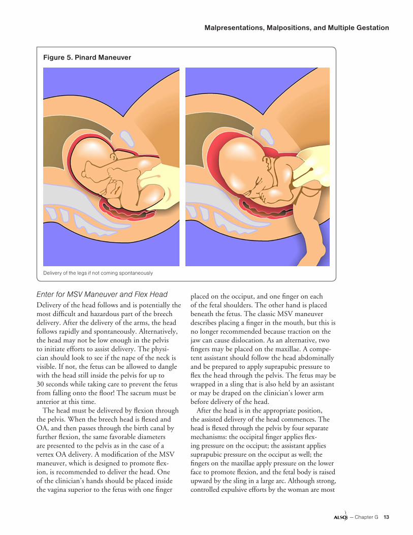
Malpresentations, Malpositions, and Multiple Gestation
— Chapter G 13
Enter for MSV Maneuver and Flex HeadDelivery of the head follows and is potentially the most difficult and hazardous part of the breech delivery. After the delivery of the arms, the head follows rapidly and spontaneously. Alternatively, the head may not be low enough in the pelvis to initiate efforts to assist delivery. The physi-cian should look to see if the nape of the neck is visible. If not, the fetus can be allowed to dangle with the head still inside the pelvis for up to 30 seconds while taking care to prevent the fetus from falling onto the floor! The sacrum must be anterior at this time.
The head must be delivered by flexion through the pelvis. When the breech head is flexed and OA, and then passes through the birth canal by further flexion, the same favorable diameters are presented to the pelvis as in the case of a vertex OA delivery. A modification of the MSV maneuver, which is designed to promote flex-ion, is recommended to deliver the head. One of the clinician’s hands should be placed inside the vagina superior to the fetus with one finger
placed on the occiput, and one finger on each of the fetal shoulders. The other hand is placed beneath the fetus. The classic MSV maneuver describes placing a finger in the mouth, but this is no longer recommended because traction on the jaw can cause dislocation. As an alternative, two fingers may be placed on the maxillae. A compe-tent assistant should follow the head abdominally and be prepared to apply suprapubic pressure to flex the head through the pelvis. The fetus may be wrapped in a sling that is also held by an assistant or may be draped on the clinician’s lower arm before delivery of the head.
After the head is in the appropriate position, the assisted delivery of the head commences. The head is flexed through the pelvis by four separate mechanisms: the occipital finger applies flex-ing pressure on the occiput; the assistant applies suprapubic pressure on the occiput as well; the fingers on the maxillae apply pressure on the lower face to promote flexion, and the fetal body is raised upward by the sling in a large arc. Although strong, controlled expulsive efforts by the woman are most
Figure 5. Pinard Maneuver
Delivery of the legs if not coming spontaneously

Chapter G
14 Chapter G —
helpful, some traction is also required for the deliv-ery. This is accomplished by downward pressure of the fingers on the shoulders. The assistant holding the fetus by a sling may also hold the feet, and pull gently as the body describes its arc. The fetal body should stay in a neutral position with regard to the head to avoid hyperextension. Ultimately, the body becomes inverted in the vertical plane, and at this point, an assistant must hold the feet to prevent the fetus from falling on the floor.
Back UpThe breech delivery is almost always accompa-nied by rotation into a sacrum anterior position as the trunk is delivering after the buttocks. In the unusual situation where the fetus attempts to move into a sacrum posterior position, the deliver-ing clinician must gently guide and rotate the fetus into the sacrum anterior position before the delivery of the arms.
Lift Baby onto MotherAs the mouth and nose appear over the perineum, they may be suctioned. The cranial vault then deliv-ers by further flexion. The clinician may use a Rit-gen maneuver on the perineum. As the head finally emerges, the infant’s body actually flips over past vertical onto the woman’s abdomen. Delayed cord clamping is appropriate if the infant does not need resuscitation or additional support. It is more com-mon for breech infants to be born with decreased tone and in need of resuscitation such as positive pressure ventilation likely because of increased cord compression during the second stage. All vaginal breech deliveries include additional personnel to perform neonatal resuscitation if needed.
Cesarean Delivery for BreechMost planned breech deliveries in high-resource settings occur by cesarean. Even when a vaginal breech delivery is planned, a substantial propor-tion will need to be converted to cesarean deliv-ery. A small proportion of these will need to be converted emergently because of cord prolapse, fetal intolerance of second stage labor, or rarely, abdominal rescue from a difficult vaginal breech delivery. The SOGC recommends that active second stage labor takes place near an operating room with personnel present to convert to emer-gent cesarean delivery and RCOG states that ready access to cesarean is important.16,43
Extraction of a breech fetus at cesarean deliv-ery requires maneuvers similar to those used in vaginal birth. Therefore, cesarean delivery of a breech fetus is an opportunity for the surgeon to practice. The goal of cesarean delivery of a breech fetus is to afford a gentle delivery. If the uterine or abdominal incisions are too small for easy delivery, they can be enlarged. This is not an option during vaginal birth.
Piper ForcepsPiper forceps are specifically designed to deliver the aftercoming head of a breech fetus. They are long and have an axis-traction curve built in. It is impossible to determine if a Piper forceps applica-tion is appropriate by visualizing the placement of the Piper forceps on the fetal head so they are always applied the same way—straight to the maternal pelvis as if the position were OA. The blades are springy and grasp the fetal head in a nonspecific basket catch that has proved safe and effective.
Forceps are indicated when the MSV maneuver fails. Although strict guidelines are lacking, Piper forceps should be considered if 2 or 3 minutes have passed without progress while attempting the MSV maneuver. Piper forceps may also be applied prophylactically if a fetus is thought to be fragile, such as a premature fetus. It is prudent to have the Piper forceps readily available for any vaginal breech delivery, but in an emergency, Simpson forceps or other forceps can be used.
To apply Piper forceps, the fetus (including the arms) is wrapped in a sling and gently held up and to the clinician’s left. The left blade is always applied first. It is held in the clinician’s left hand and is applied to the left side of the woman’s pel-vis (but to the right side of the fetus). Unlike other forceps applications, the clinician holds the handle in a horizontal position and below the fetus. The right hand is placed in the vagina alongside the fetal head to protect the vaginal sidewalls. Then the forceps blade is inserted between the right hand and the fetal head, following the cephalic curve of the blade around the head. After inser-tion, the handle may be allowed to dangle or may be supported by an assistant.
The right blade is then inserted in a similar fashion by grasping the handle with the right hand and sliding the blade into the vagina alongside the head while protecting the sidewall with the left

Malpresentations, Malpositions, and Multiple Gestation
— Chapter G 15
hand. The forceps should then be locked. When the right blade is applied over the left blade, the lock will articulate normally. The handles are typically separated slightly away from the lock and should not be squeezed together. Because the clini-cian cannot determine how the blade is applied to the fetal skull and face, no effort is made to do so.
Delivery of the head may commence when the application is complete. The clinician applies a small amount of traction to the forceps. Because the shanks of the forceps have a large axis-traction curve built in, no special maneuvers such as the Pajot maneuver are required to ensure that trac-tion is in the correct vector. The primary motion of the forceps is to raise the handles in a large arc, starting approximately horizontal and ending at or past vertical. This arc will flex the head through the pelvis with exactly the same geometry as the MSV maneuver but with greatly increased lever-age because of the length of the forceps. None of the flexing maneuvers of the MSV maneuver are required when the Piper forceps are used. The fetus may be held in the sling or laid on the shanks of the forceps during the delivery.
The principal difficulty in applying Piper for-ceps is a result of the condition that indicates their use. That is, failure of the MSV maneuver implies a tight fit of head to pelvis. There may be insuf-ficient room to place a hand alongside the head. In this situation, the blade must be blindly applied with risk of injury to woman and fetus. After the Piper forceps are on, delivery can be accomplished in almost every case. Training in Piper forceps is beyond the scope of the ALSO Malpresentations workshop, but physicians who anticipate partici-pating in emergency or elective vaginal breech deliveries in sites where they may be called on to deliver a trapped head are encouraged to seek additional training. Elective use of Piper forceps during cesarean delivery can provide one opportu-nity for training.49
Symphysiotomy is an emergency maneuver for incising the ligaments of the pelvic symphysis to release a trapped aftercoming head (Figure 6). It is rarely used in high-resource settings, but its use in low-resource settings without ready access to emergency cesarean can be lifesaving. Maternal risks include urological and orthopedic injuries. Although not a part of ALSO for high-resource settings, there is a workshop in the Global ALSO curriculum.
Complications of Breech DeliveryA nuchal arm occurs when one or both arms are extended upward behind the neck and may impede delivery of the head. There are three delivery options in this event. If the fetus is small or the pelvis is large, the head and extended arm may be delivered together. Alternatively, the clinician may attempt to flex the arm and sweep it down over the face and chest. As a maneuver of last resort, the clinician may rotate the fetus 90 to 180 degrees in the direction of the hand to sweep the arm out of its nuchal position (clockwise for a left nuchal arm, counterclockwise for a right nuchal arm).
Medical management of cervical entrapment may be attempted using nitroglycerin to cause rapid but transient relaxation of the uterus. Intra-venous nitroglycerin may be administered by an anesthetist or sublingual spray may be used if avail-able.50 Because of the rare and emergent nature of head entrapment during vaginal breech birth, there are no published studies of medical management.
Resolution without excessive traction may require cutting the cervix, a procedure known as Dührssen incisions. Ring forceps are placed in pairs, parallel to each other at 2 o’clock, 10 o’clock and if possible 6 o’clock, extending 3 cm to 4 cm into the cervix. A radial incision is made between the ring forceps of each pair. Anesthesia and exposure are major technical problems, and
Figure 6. Symphysiotomy to Free Trapped Aftercoming Head

Chapter G
16 Chapter G —
hemorrhage is a major potential complication. This procedure is recommended only in the most extreme life-threatening circumstances.
Hydrocephalus may present as a breech deliv-ery with an entrapped head. The appearance of a meningomyelocele or spina bifida may herald hydrocephalus, occurring in approximately one-third of such cases. Although prenatal diagnosis will call for highly individualized management and probable cesarean delivery, the unexpected diagnosis at the time of a breech delivery presents a significant dilemma. If cesarean delivery is avail-able, then emergent cesarean for abdominal rescue will be needed. Decompression of the fetal ven-tricles or cephalocentesis may be prejudicial to the fetus, but it is the only way for the delivery to be completed if the fetus is alive and rapid cesarean is not an available option. This procedure may be accomplished transvaginally or transabdominally with a long needle.
Transverse Lie or Shoulder PresentationIn transverse lie, the long axis of the fetus is approximately perpendicular or at right angles to that of the woman. In the back down transverse lie or shoulder presentation, the shoulder is over the pelvic inlet, the head is lying in one of the iliac fossae, and the breech in the other. Transverse lie can also occur in the back-up orientation, most commonly in the second twin. Occasionally an unstable or oblique lie will be noted, in which the fetus changes from a breech or vertex to a trans-verse lie or assumes an intermediate lie.
Transverse lie occurs in approximately 0.3% of singleton births.2 The common causes of transverse lie are: unusual relaxation of the abdominal wall, preterm fetus, placenta previa, abnormal uterus (eg, subseptate, fibroid), contracted pelvis, tumor occluding the birth canal, and polyhydramnios.
DiagnosisDiagnosis of a brow presentation is by vaginal examination. The frontal sutures, anterior fontanel, orbital ridges, eyes, and root of the nose may be felt. Frequently the examination is confusing because of edema and unfamiliarity of the presenting features.
Mechanism of Labor and Management of DeliverySpontaneous birth of a full-term fetus in trans-verse lie is impossible. Therefore, cesarean
delivery is mandatory in most cases. If transverse lie is encountered before the onset of labor or in early labor with intact membranes, then an attempt at ECV is reasonable, assuming there are no contraindications to vaginal delivery such as placenta previa.
If a fetus presents with a back-up transverse lie and a fully dilated cervix, such as potentially with a second twin, then ECV or internal podalic version may be considered depending on esti-mated fetal weight and obstetrical history. This is described in the section on multiple gestation in this chapter.
When labor ensues with a back-down transverse lie, the shoulder is forced into the pelvis, and an arm may prolapse. With continued labor, a retrac-tion ring may develop. Ultimately, in a neglected labor, the uterus ruptures and the woman and fetus risk mortality. This scenario is rarely seen in mod-ern obstetrics, but it may be encountered in parts of the world where access to care is a problem.
Cesarean delivery for a back-down transverse lie may require a low vertical or classical incision for the clinician to successfully deliver one of the fetal poles through the uterine incision. Often a trans-verse uterine incision will be adequate for delivery and has the benefit of allowing a trial of labor in future pregnancies.51 If the initial transverse inci-sion is not adequate to reach the feet, then a T extension can be performed.
Face PresentationIn a face presentation, the head is hyperextended so the occiput is in contact with the fetal back, and the face is the presenting part. The fetal skull diameter that presents to the pelvis is the sub-mentobregmatic, which when the chin (mentum) is anterior, is favorable for delivery in most cases. Face presentation occurs in 0.2% to 0.3% of singleton deliveries.2
The causes of face presentation are numerous and often obscure. When the fetus is large or the pelvis is contracted, there is a predisposition to extension of the fetal head. The pendulous abdo-men of a grand multipara also promotes exten-sion of the fetal head. In exceptional instances, enlargement of the neck because of goiter or cystic hygroma or numerous coils of cord around the neck may cause extension. Anencephalic fetuses often present by the face because of absent devel-opment of the cranium.

Malpresentations, Malpositions, and Multiple Gestation
— Chapter G 17
DiagnosisThe clinical diagnosis of a face presentation relies principally on the vaginal examination. The mouth, nose, and the malar prominences may be palpated. As previously discussed, a face presenta-tion may be confused with a breech presentation, particularly because breech is 20 times more com-mon. The mouth may be mistaken for the anus, and the malar prominences may be mistaken for the ischial tuberosities.
Mechanism of LaborThe key for successful delivery of a face is for the chin to end up under the symphysis, or be in men-tum anterior position. Then, with further descent of the fetus, the cranial vault can sweep through the posterior pelvis and the head can be delivered by flexion with conversion to an OP delivery.
Although this mechanism does not present the most favorable diameter of the fetal head to the pelvis, if the fetus is not too large and the pelvis is adequate, spontaneous delivery can occur. If the chin rotates or remains posteriorly (mentum posterior), then there is no mechanism that allows the fetus to utilize the space in the posterior pelvis in the hollow of the sacrum and delivery cannot occur. Forceps or manual rotation of a mentum posterior presentation should not be attempted because of the risk of fetal spinal cord injury.52
Management of DeliverySpontaneous vaginal delivery may occur some-times with surprising ease. The fetus must rotate to a mentum anterior position. A persistent mentum posterior mandates a cesarean delivery. Forceps can be safely and successfully applied to a mentum anterior that is on the perineum. The vacuum extractor is absolutely contraindicated. Likewise, scalp electrode internal monitoring is contraindicated to avoid injuring the face. There is an increased incidence of variable and late decelerations, and oxytocin augmentation should only be used with caution. Parents should be prepared for the significantly bruised and edema-tous face of the infant, but recovery occurs within 24 to 48 hours.
Brow PresentationIn a brow presentation, the portion of the fetal head between the orbital ridge and the anterior fontanel presents at the pelvic inlet. The fetal head is in an
attitude between full flexion (or occiput) and full extension (or face). The presenting diameter of the fetal skull is the occipitomental, which is unfavor-able for delivery. Delivery of a persistent brow typically cannot take place unless the fetus is small or the pelvis is large.
Brow presentation is found in 0.007% of single-ton deliveries.2,53 The causes of this rare presenta-tion are similar to those for face presentation. A brow presentation is typically unstable and often will convert to a face or a vertex presentation.
DiagnosisDiagnosis of a brow presentation is by vaginal examination. The frontal sutures, anterior fonta-nel, orbital ridges, eyes, and root of the nose may be felt. Frequently the examination is confusing because of edema and unfamiliarity of the present-ing features.
Mechanism and Management of LaborA persistent brow cannot deliver vaginally under normal conditions. If it converts to vertex or face, then delivery may occur according to their respec-tive mechanisms. In the absence of conversion and progress in labor, cesarean delivery is required.
Compound PresentationIn a compound presentation an extremity, typically a hand, prolapses alongside the main presenting part, typically the head. Often no cause is found. It is more common with premature infants and when the fetal presenting part does not completely occlude the pelvic inlet.
DiagnosisThe diagnosis of compound presentation is typi-cally readily made on vaginal examination. It is critically important to distinguish between a hand and a foot prolapsed alongside the head.
Management of DeliveryAs long as labor is progressing normally, no intervention is necessary. Most commonly the prolapsed limb will deliver spontaneously along with the head, or sometimes the fetus will retract its limb spontaneously. If the prolapsed arm appears to be impeding descent, it should be gently elevated upward and the head manipulated simultaneously downward. Occasionally cesarean delivery will be necessary. The parents should be
Chapter G
18 Chapter G —
warned to expect bruising and edema of the pro-lapsed extremity.
Prolapse of the Umbilical CordProlapse of the umbilical cord is a true obstetri-cal emergency. The cord may become compressed or occluded between the presenting part of the fetus and the pelvic brim or sidewall, resulting in asphyxia and mortality. The incidence of cord pro-lapse is 0.1% to 0.6% in vertex presentations54 and approximately 1% in breech presentations with the incidence greater in footling (approximately 10%) and complete breech presentations than in frank breech presentation (less than 1%).43 Cord prolapse is most common when the fetus does not occlude the pelvic inlet well, as is the case with a footling breech. Other factors that may contribute to cord prolapse are prematurity, polyhydramnios, high presenting part, and a long cord. Approximately 50% of cases of umbilical cord prolapse follow obstetrical interventions,55 such as when the mem-branes are ruptured with the presenting part high out of the pelvis. A gush of fluid may then wash the cord down into the vagina. On the other hand, the cord may have already been coiled beneath the fetal presenting part (occult cord prolapse) such that rupture of the membranes merely revealed the prolapse, but did not cause it. The proportion of cases that are iatrogenic appears to be decreasing, but the use of a balloon catheter for cervical ripen-ing is one modern intervention that can lead to cord prolapse by elevating the presenting part.56
Rapid identification and response may save the life of the fetus. The management steps are:1. Diagnose the cord prolapse by visual inspec-tion or palpation on immediate vaginal examina-tion. The cord may be found extruded from the vagina, coiled in the vagina, or wrapped across the presenting part. The only hint may be a severe variable deceleration or bradycardia after rupture of the membranes.
2. Quickly assess the fetal status by fetal heart ratemonitoring or ultrasound.
3. Assess the dilation and status of labor. In theuncommon situation where the fetus can be deliv-ered more quickly and safely vaginally than by cesarean delivery, then proceed immediately using forceps, vacuum, or in the case of the second twin, total breech extraction when appropriate.
4. If immediate vaginal delivery is not feasible,then prepare for cesarean delivery. Elevate the presenting part out of the pelvis in an effort to protect the cord from occlusion. This may be performed by placing a hand in the vagina and forcefully but carefully elevating the presenting part upward. Alternatively, some success has been achieved by filling the bladder rapidly with 500 to 700 cc of saline solution and then clamping the catheter. Oxytocin should be discontinued. Tocolysis (eg, terbutaline 0.25 mg subcutane-ously) is helpful if the patient is in labor and there are recurrent fetal heart rate decelerations or cesarean delivery will be delayed.54 A deep Tren-delenburg position also is useful to add gravity to other efforts to elevate the fetus off the cord. The effectiveness of these maneuvers can be measured by monitoring the fetus or palpating the cord.
5. Do not attempt to replace the cord in the uterus.
6. Perform an emergent cesarean delivery whilecontinuing all efforts to hold the presenting part off the cord.
7. If a delay is encountered, wrap the cord inwarm wet packs.
Prevention of cord prolapse is difficult but maybe accomplished on occasion by identifying risk factors or by identifying a cord presentation by ultrasound. Artificial rupture of the membranes should not be done when the station is high. If artificial rupture of membranes is essential to manage a difficult obstetric situation, and the head is unengaged and high, the membranes can be needled under double set-up conditions.56 The same procedure can be used to rupture the mem-branes in cases of polyhydramnios.
Patients in the later stages of pregnancy who are at high risk of cord prolapse (eg, footling breech, polyhydramnios) should be identified. They can be instructed to examine themselves for cord prolapse if their membranes rupture outside of a hospital. If a prolapse is identified, they should assume a deep knee-chest position and maintain the position during transport to the hospital.
Multiple GestationMultiple gestation occurred in 3.4% of births in the United States in 2013.57 The twinning rate rose 76% from 1980 to 2009 with the increase attrib-

Malpresentations, Malpositions, and Multiple Gestation
— Chapter G 19
uted to the use of fertility therapies and increased proportion of women with advanced maternal age.57 Twin pregnancies are 7 times more likely to result in delivery less than 32 weeks estimated gestational age and multifetal pregnancies have 5 times the risk of stillbirth compared to singleton pregnancies.58 Congenital anomalies, intrauter-ine growth restriction (IUGR), and intrapartum complications also contribute. Dizygosity (fraternal twinning) occurs in approximately two-thirds of twin gestations, and increases with age, parity, and certain familial and racial circumstances. Mono-zygosity (identical twinning) occurs in one-third of twin gestations and is unrelated to any predispos-ing factors. Morbidity and mortality are higher in monozygotic twins.
Maternal complications are common in multiple gestation. These include gestational hypertension, gestational diabetes, anemia, hyperemesis, abrup-tion, placenta previa, postpartum hemorrhage, and increased operative delivery.
DiagnosisMultiple gestation is now routinely diagnosed by ultrasound during prenatal care in high-resource settings and the intrapartum diagnosis of the second twin is an uncommon event that is most likely to occur in the setting of a lack of prenatal care. Historical and physical findings suggestive of multiple gestation and indicating an ultra-sound are: uterus larger than dates, hyperemesis gravidarum, early preeclampsia, elevated maternal serum alpha fetoprotein levels, suggestive palpa-tory or auscultatory findings, polyhydramnios, ovulation induction, and family history.
Prenatal ManagementSeveral issues distinguish multiple gestations from singleton pregnancies:
1. Prematurity is the greatest threat to multiple gestation fetuses, and prevention of prematurity is the highest priority. Unfortunately, no preven-tive measures, including bed rest, routine cerclage, and tocolytics, have been shown to effectively prevent premature labor.59,60 Although progester-one has been shown to be beneficial in singleton pregnancies with a history of prior preterm birth (intramuscular weekly progesterone) and singleton pregnancies with a short cervix diagnosed between 16 and 24 weeks (vaginal progesterone), the role
of progesterone in twin pregnancies remains controversial and no benefit has been shown for routine use of vaginal progesterone or intramuscu-lar 17 alpha-hydroxyprogesterone caproate in twin pregnancies with a short cervical length.61-63 Use of intramuscular progesterone in twin pregnancies complicated by a prior preterm delivery may be reasonable based on the singleton date, but there is no evidence to support or refute this practice.64
2. Compared to singleton pregnancies, the rates of congenital anomalies and developmental defects are doubled in twin pregnancies, and higher in monozygotic twin pregnancies. An ultrasound including a detailed anatomic survey is recom-mended for all women with a multiple gestation at approximately 18 weeks’ gestation. A first trimester ultrasound may be done to confirm or determine gestational age as part of genetic screening and to determine chorionicity.
3. Compared to singleton pregnancies, preeclamp-sia occurs twice as often in twin gestations.65 A daily baby aspirin by mouth starting at 10 to 12 weeks is recommended for women with mul-tiple gestations by the US Preventive Services Task Force,66 but multiple gestation is not included in the ACOG recommendations for pregnancy prevention with aspirin. Active surveillance and management are recommended. Iron deficiency is common, and iron supplementation is typically indicated. Twin pregnancies are at greater risk of gestational diabetes, but routine screening in early pregnancy is not recommended.67,68
4. Growth restriction, size discordance and twin-twin transfusion: The possibility of IUGR and discordant growth requires surveillance with ultra-sound for interval growth.69 Discordance of greater than 20% is associated with a sevenfold increase in major neonatal morbidity.70 Ultrasound exami-nations every 4 weeks starting at approximately 24 weeks’ gestation is recommended to assess interval growth and concordance. Monochori-onic pregnancies with increased risk of twin-twin transfusion should start earlier surveillance with assessment of amniotic fluid volume. Intervention in the presence of significant discordancy before 36 weeks or twin-twin transfusion is a complex issue where perinatal consultation is appropriate and beyond the scope of this chapter.

Chapter G
20 Chapter G —
5. Fetal mortality including stillbirth is much morecommon in twin than singleton pregnancies and in monoamniotic/dichorionic than in diamniotic/dichorionic pregnancies. Assessing fetal heart rate at each prenatal visit should be done with ultrasound (or electronic fetal monitor with two fetal heart rate attachments) rather than Dop-pler imaging. When stillbirth of one twin occurs, conservative management of the surviving twin is indicated, at least until 34 weeks. Surviving twins in monochorionic/diamniotic pregnancies are at risk of neurological injury when the loss occurs after 14 weeks, but early delivery is not beneficial as the injury is felt to have occurred before the time of diagnosis on a single twin demise. Because of the higher risk of intrauterine fetal demise, routine induction of diamniotic/dichorionic twin pregnan-cies at 38 weeks and monoamniotic/dichorionic at 36 to 37 weeks is commonly recommended.71
6. The incidence of placenta previa is increased inmultiple gestation and should be detected by the recommended anatomic survey and/or interval growth ultrasounds.
Intrapartum ManagementDelivery of twin pregnancies presents a range of challenges. Intrapartum complications include malpresentations, locking of twins, cord prolapse,
abruption, concerning fetal heart rate tracing, dysfunctional labor, and postpartum hemorrhage. Several of these complications can arise from the manner in which the twins present. Either fetus may be vertex, breech, or in a transverse lie. Theo-retically there are nine combinations of presenta-tions of twins A and B, but for practical purposes there are three (Table 3).
Vertex-vertex presentations are the most com-mon and least complicated. With appropriate monitoring and capability to respond to an emer-gency with urgent cesarean or operative vaginal delivery, labor may be allowed to progress to vaginal delivery of both fetuses.73 Oxytocin induc-tion or augmentation, epidural anesthesia, and other interventions are all acceptable with caution. The interval between deliveries is not critical as long as the second fetus is doing well, but oxytocin augmentation is often used when delay is encoun-tered between deliveries. A 2011 Cochrane review showed little evidence regarding optimal mode of delivery in twin pregnancies.74 Based on a 2015 randomized controlled trial that showed no benefit from routine cesarean as long as twin A was vertex, current recommendations are not to perform cesarean for vertex/vertex twins without another indication.75 A physician experienced in vaginal breech delivery and breech extraction should be available for all twin vaginal deliveries because the second twin can change to a breech or transverse presentation after the delivery of the first twin. Because of diminishing training and experience with vaginal breech delivery, having an experi-enced physician may be challenging.
When vaginal delivery is attempted, the time of greatest risk occurs after the delivery of twin A, when the physician must determine the presenta-tion of twin B, which may be different from its presentation before twin A was delivered. A combi-nation of external examination, internal examina-tion, and ultrasound may be used. Assuming twin B is breech or in a transverse lie, a decision must be made whether to attempt an external version to vertex, deliver twin B as a breech, or perform a cesarean delivery. The exact obstetric circum-stances, the experience of the clinician, condition of the fetus, state of mind of the woman, and the available resources are all factors in the decision. Twin B must be carefully monitored as placental abruption and umbilical cord prolapse are two obstetrical emergencies that may occur between the
Table 3. Presentation of Twins72
Twin A Twin B Percentage of Occurrence
Vertex Vertex 40
Vertex Nonvertex 40 (30% breech/10% transverse)
Breech Vertex/Nonvertex 20
Vertex/Vertex 40% Vertex/Nonvertex 40% Nonvertex Twin A 20%

Malpresentations, Malpositions, and Multiple Gestation
— Chapter G 21
delivery of twin A and twin B. If concerning fetal monitoring requires expeditious delivery of twin B, then a vacuum-assisted vaginal delivery may be done at a slightly higher station than would typi-cally be considered for a singleton and a midpelvic vacuum of twin B at 0 or +1 station is a reason-able option as long as the estimated fetal weight of twin B is not considerably larger than twin A.76 The delivery of twins is best accomplished in the operating room in the event that a rapid cesarean is needed. Anesthesia should be on standby.
When twin A is vertex but twin B is nonver-tex, controversy exists regarding the best mode of delivery. Some clinicians advocate cesarean delivery, but as long as a physician experienced in the breech delivery of twin B is available, routine cesarean is not necessary. Vaginal breech delivery of twin B is a reasonable option for a fetus with an estimated weight greater than 1,500 g as long as criteria for vaginal delivery of a singleton breech fetus discussed above are met.75
When twin B is presenting in a transverse or oblique presentation or as a footling breech, then a breech extraction can be performed.77 The physician will identify and grasp the feet without rupturing the second gestational sac and bring the feet down to the vagina. Abdominal ultrasound to determine the location of the feet can be help-ful. The delivering physician’s second hand or an assistant can apply gentle abdominal pressure to help guide the fetal head upward into the uterine fundus. With the clinician exerting steady down-ward traction on the feet to maintain the breech as the presenting part, the membranes are then ruptured. After delivery to the umbilicus, the deliv-ery of the arms and head is similar to other vaginal breech deliveries. Breech extraction of twin B may also occur as part of an internal podalic version in where the vertex is elevated out of the pelvis before reaching for the feet. Breech extraction and internal podalic version should not be attempted unless the physician has training and experience. This is perhaps the most difficult and dangerous procedure permissible in modern obstetrics. Breech extraction and podalic version should only be performed if the estimated fetal weight of twin B is not substan-tially (eg, 20%) greater than twin A.75,77
Undiagnosed twins are rare in areas where ultra-sound is frequently used. In the pre-ultrasound era, as many as 50% of twin gestations were unsuspected until delivery. When no ultrasound
has been performed, birth attendants should always be alert for this possibility. If the scenario of a nonvertex second twin unexpectedly occurs in a setting without an experienced clinician because of undiagnosed second twin or no prenatal care, then options include ECV (as previously discussed in this chapter) or cesarean delivery.
Situations mandating cesarean delivery for a twin gestation include cord prolapse, abruption, or inability to reach the feet to perform internal podalic version and breech extraction for a fetus in a transverse lie. A contributing problem occurs when the cervix closes down after twin A is born. These situations can arise suddenly, so resources for immediate cesarean delivery should be avail-able. When twin A is nonvertex, cesarean deliv-ery is typically recommended but high-quality evidence is lacking. ECV of a breech twin A is not technically feasible. When twin A is breech and twin B is vertex or transverse, locking or collision of the twins is a rare but disastrous event.
Cesarean delivery in multiple gestation pres-ents anesthetic and surgical challenges because of the enlarged uterus, the exaggerated physiologic response to pregnancy, and the potential for unusual presentations of the fetuses. The neces-sity for a vertical incision in both skin and uterus is a special consideration when the twins are in unusual or entwined positions. Conjoining of twins is a rare condition and is beyond the scope of this chapter, but should always be considered if ultrasound shows twins in a face-to-face or back-to-back position.
After delivery, postpartum hemorrhage is relatively common because of the overdistension of the uterus. Clinicians should be fully prepared with IV access, proper oxytocics, and readily available blood products. Neonatal resuscitation is often required because of prematurity or the many potential complications of multiple gestation. Not uncommonly, two infants and the mother need attention simultaneously. Adequate personnel and equipment must be available.
Global Perspectives: MalpresentationsThe diagnosis, management, and perinatal outcomes for pregnancies complicated by malpre-sentation in low-resource settings will be strongly influenced by the availability of obstetrical ultra-sound and urgent cesarean delivery. A greater pro-portion of fetuses in breech presentation will be

Chapter G
22 Chapter G —
delivered vaginally when the diagnosis is delayed because of the lack of ultrasound and the time to initiate a cesarean can be lengthy. In the TBT, 9.6% of women assigned to the elective cesarean group had vaginal deliveries.39 The safest route for breech deliveries is not as clear for developing countries, as the TBT noted that the reduction in adverse perinatal outcomes was less pronounced and did not reach statistical significance in coun-tries where the perinatal mortality rate was higher than 20/1,000.39
A 2015 Cochrane review showed that early ultrasound significantly decreases the number of post-date pregnancies and undiagnosed twins. In settings where routine obstetrical ultrasound is uncommon, a higher proportion of twin preg-nancies may be diagnosed in the third trimester or even after delivery of the first twin. For these reasons, skills in delivery of breech infants and nonsurgical maneuvers (cephalic version, internal podalic version, and manual rotation from OP) are essential in low-resource settings.78 Transverse lie of a dead fetus is a life-threatening complica-tion for women in developing countries and almost unheard of in high-resource areas. Emer-gent treatment may require destructive delivery, internal version, or laparotomy.
For more details on malpresentations, mal-positions, and multiple gestation in developing
countries, see the Global ALSO manual available at www.aafp.org/globalalso.
SummaryThere are six types of malpositions or malpresen-tations. Some are common (OP position, breech presentation) and some are rare (transverse lie, brow presentation, face presentation, compound presentation). Diagnosis is made by a combina-tion of physical examination and imaging. A high index of suspicion is helpful in making the diag-nosis. Each variation in position or presentation has its own potential complications. Clinicians should be alert not only for complications result-ing from labor and delivery, but also for problems that may be etiologic for the malpresentation in the first place.
Vaginal delivery may be considered for four of these presentations: OP, frank breech, men-tum anterior position, and compound. With OP position, the clinician has several management choices for delivery. With breech presentation, complex criteria determine if vaginal delivery can occur safely. ECV should be offered for nonvertex presentation at 36 or more weeks of gestation. A high degree of technical skill and judgment is required to deliver fetuses with malpresentations safely. Multiple gestation presents a wide variety of special challenges to the clinician.

Malpresentations, Malpositions, and Multiple Gestation
— Chapter G 23
SORT: Key Recommendations for Practice
Clinical RecommendationEvidence Rating References
Patients with breech presentation who meet standard criteria near term should be offered an attempted external cephalic version at 36 to 37 weeks to reduce the likelihood of breech presentation at birth and cesarean delivery.
A 17,21,79
Betamimetics (eg, terbutaline) increase the likelihood of successful external cephalic version and should be routinely used.
A 25
Routine cesarean delivery for twins is not recommended as long as the first twin has a cephalic presentation and appropriately trained physicians and facilities are available. Routine cesarean delivery has not been shown to improve neonatal outcomes and planned vaginal delivery is recommended.
A 75
Elective delivery of uncomplicated diamniotic/dichorionic twins at 37 weeks does not appear to be associated with harm, and elective delivery at 37 to 38 weeks by labor induction or cesarean (if nonvertex twin A) should be offered.
B 80
Strength of Recommendation Taxonomy (SORT)
Strength of Recommendation Definition
A • Recommendation based on consistent and good-quality patient-oriented evidence.
B • Recommendation based on inconsistent or limited-quality patient-oriented evidence.
C • Recommendation based on consensus, usual practice, opinion, disease-oriented evidence, or case series for studies of diagnosis, treatment, prevention, or screening.
Patient-oriented evidence measures outcomes that matter to patients: morbidity, mortality, symptom improvement, cost reduction, and quality of life. Disease-oriented evidence measures intermediate, physiologic, or surrogate end points that might not reflect improvement in patient outcomes (eg, blood pressure, blood chemistry, physiologic function, pathologic findings).
From Ebell MH, Siwek J, Weiss BD, et al. Strength of recommendation taxonomy (SORT): a patient-centered approach to grading evidence in the medical literature. Am Fam Physician. 2004;69(3):548-556.

Chapter G
24 Chapter G —
References1. Barth WH Jr. Persistent occiput posterior. Obstet Gyne-
col. 2015;125(3):695-709.
2. Ratcliffe SD, Baxley EG, Cline MK, Sakornbut EL. Fam-ily Medicine Obstetrics. 3rd ed. Philadephia, PA: MosbyElsevier; 2008.
3. Vlemmix F, Bergenhenegouwen L, Schaaf JM, et al.Term breech deliveries in the Netherlands: did theincreased cesarean rate affect neonatal outcome? Apopulation-based cohort study. Acta Obstet GynecolScand. 2014;93(9):888-896.
4. Royal College of Obstetricians and Gynaecologists.The Management of Breech Presentation: Guideline No.20b. 2006.
5. Cheng YW, Shaffer BL, Caughey AB. The associationbetween persistent occiput posterior position and neo-natal outcomes. Obstet Gynecol. 2006;107(4):837-844.
6. Pearl ML, Roberts JM, Laros RK, Hurd WW. Vaginaldelivery from the persistent occiput posterior position.Influence on maternal and neonatal morbidity. J ReprodMed. 1993;38(12):955-961.
7. Le Ray C, Serres P, Schmitz T, Cabrol D, Goffinet F.Manual rotation in occiput posterior or transverse posi-tions: risk factors and consequences on the cesareandelivery rate. Obstet Gynecol. 2007;110(4):873-879.
8. Malvasi A, Tinelli A, Barbera A, et al. Occiput posteriorposition diagnosis: vaginal examination or intrapartumsonography? A clinical review. J Matern Fetal NeonatalMed. 2014;27(5):520-526.
9. Fitzpatrick M, McQuillan K, O’Herlihy C. Influence ofpersistent occiput posterior position on delivery out-come. Obstet Gynecol. 2001;98(6):1027-1031.
10. Bahl R, Van de Venne M, Macleod M, Strachan B, Mur-phy DJ. Maternal and neonatal morbidity in relation tothe instrument used for mid-cavity rotational operativevaginal delivery: a prospective cohort study. BJOG.2013;120(12):1526-1532.
11. Burke N, Field K, Mujahid F, Morrison JJ. Use andsafety of Kielland’s forceps in current obstetric practice.Obstet Gynecol. 2012;120(4):766-770.
12. Scheer K, Nubar J. Variation of fetal presenta-tion with gestational age. Am J Obstet Gynecol.1976;125(2):269-270.
13. Hemelaar J, Lim LN, Impey LW. The Impact of an ECVService is Limited by Antenatal Breech Detection: A Ret-rospective Cohort Study. Birth. 2015;42(2):165-172.
14. Hofmeyr GJ, Kulier R. Cephalic version by posturalmanagement for breech presentation. Cochrane Data-base Syst Rev. 2012;10:CD000051.
15. Smith C, Crowther C, Wilkinson C, Pridmore B, RobinsonJ. Knee-chest postural management for breech at term:a randomized controlled trial. Birth. 1999;26(2):71-75.
16. ACOG Committee on Obstetric Practice. ACOG Com-mittee Opinion No. 340. Mode of term singleton breechdelivery. Obstet Gynecol. 2006;108(1):235-237.
17. Hofmeyr GJ, Kulier R, West HM. External cephalic ver-sion for breech presentation at term. Cochrane Data-base Syst Rev. 2015;4(4):CD000083.
18. Kok M, Cnossen J, Gravendeel L, van der Post J, OpmeerB, Mol BW. Clinical factors to predict the outcome ofexternal cephalic version: a metaanalysis. Am J ObstetGynecol. 2008;199(6):630.e1-e7; discussion e1-e5.
19. Hutton EK, Hannah ME, Ross SJ, et al; Early ECV2Trial Collaborative Group. The Early External CephalicVersion (ECV) 2 Trial: an international multicentre ran-domised controlled trial of timing of ECV for breechpregnancies. BJOG. 2011;118(5):564-577.
20. Hutton EK, Hofmeyr GJ, Dowswell T. External cephalicversion for breech presentation before term. CochraneDatabase Syst Rev. 2015;7(7):CD000084.
21. Rosman AN, Guijt A, Vlemmix F, Rijnders M, Mol BW,Kok M. Contraindications for external cephalic versionin breech position at term: a systematic review. ActaObstet Gynecol Scand. 2013;92(2):137-142.
22. Sela HY, Fiegenberg T, Ben-Meir A, Elchalal U, EzraY. Safety and efficacy of external cephalic version forwomen with a previous cesarean delivery. Eur J ObstetGynecol Reprod Biol. 2009;142(2):111-114.
23. American College of Obstetricians and Gynecologists.ACOG Practice bulletin no. 115: Vaginal birth after previ-ous cesarean delivery. Obstet Gynecol. 2010;116(2 Pt 1):450-463.
24. Burgos J, Cobos P, Rodríguez L, et al. Is externalcephalic version at term contraindicated in previouscaesarean section? A prospective comparative cohortstudy. BJOG. 2014;121(2):230-235, discussion 235.
25. Cluver C, Gyte GM, Sinclair M, Dowswell T, HofmeyrGJ. Interventions for helping to turn term breechbabies to head first presentation when using exter-nal cephalic version. Cochrane Database Syst Rev.2015;2(2):CD000184.
26. Coyle ME, Smith CA, Peat B. Cephalic version by moxi-bustion for breech presentation. Cochrane DatabaseSyst Rev. 2012;5(5):CD003928.
27. Goetzinger KR, Harper LM, Tuuli MG, MaconesGA, Colditz GA. Effect of regional anesthesia onthe success rate of external cephalic version: a sys-tematic review and meta-analysis. Obstet Gynecol.2011;118(5):1137-1144.
28. Weiniger CF, Ginosar Y, Elchalal U, Sela HY, Weiss-man C, Ezra Y. Randomized controlled trial of externalcephalic version in term multiparae with or without spi-nal analgesia. Br J Anaesth. 2010;104(5):613-618.
29. Weiniger CF, Ginosar Y, Elchalal U, Sharon E, NokrianM, Ezra Y. External cephalic version for breech presen-tation with or without spinal analgesia in nulliparouswomen at term: a randomized controlled trial. ObstetGynecol. 2007;110(6):1343-1350.
30. Carvalho B, Tan JM, Macario A, El-Sayed YY, Sultan P.Brief report: a cost analysis of neuraxial anesthesia tofacilitate external cephalic version for breech fetal pre-sentation. Anesth Analg. 2013;117(1):155-159.
31. Weiniger CF, Spencer PS, Weiss Y, Ginsberg G, EzraY. Reducing the cesarean delivery rates for breechpresentations: administration of spinal anesthesia facili-tates manipulation to cephalic presentation, but is itcost saving? Isr J Health Policy Res. 2014;3(1):5.

Malpresentations, Malpositions, and Multiple Gestation
— Chapter G 25
32. Suen SS, Khaw KS, Law LW, et al. The force applied tosuccessfully turn a foetus during reattempts of externalcephalic version is substantially reduced when per-formed under spinal analgesia. J Matern Fetal NeonatalMed. 2012;25(6):719-722.
33. Nassar N, Roberts CL, Barratt A, Bell JC, Olive EC, PeatB. Systematic review of adverse outcomes of externalcephalic version and persisting breech presentation atterm. Paediatr Perinat Epidemiol. 2006;20(2):163-171.
34. Coco AS, Silverman SD. External cephalic version. AmFam Physician. 1998;58(3):731-738, 742-744.
35. Collins S, Ellaway P, Harrington D, Pandit M, ImpeyLW. The complications of external cephalic ver-sion: results from 805 consecutive attempts. BJOG.2007;114(5):636-638.
36. Lee HC, El-Sayed YY, Gould JB. Population trends incesarean delivery for breech presentation in the UnitedStates, 1997-2003. Am J Obstet Gynecol. 2008;199(1):59.e1-59.e8.
37. Lyons J, Pressey T, Bartholomew S, Liu S, Liston RM,Joseph KS; Canadian Perinatal Surveillance System(Public Health Agency of Canada). Delivery of breechpresentation at term gestation in Canada, 2003-2011.Obstet Gynecol. 2015;125(5):1153-1161.
38. Danielian PJ, Wang J, Hall MH. Long-term outcome bymethod of delivery of fetuses in breech presentation atterm: population based follow up. BMJ. 1996;312(7044):1451-1453.
39. Hannah ME, Hannah WJ, Hewson SA, Hodnett ED, Sai-gal S, Willan AR; Term Breech Trial Collaborative Group.Planned caesarean section versus planned vaginalbirth for breech presentation at term: a randomised mul-ticentre trial. Lancet. 2000;356(9239):1375-1383.
40. Greene MF. Vaginal breech delivery is no longer justi-fied. Obstet Gynecol. 2002;99(6):1113-1115.
41. Whyte H, Hannah ME, Saigal S, et al; Term Breech TrialCollaborative Group. Outcomes of children at 2 yearsafter planned cesarean birth versus planned vaginalbirth for breech presentation at term: the InternationalRandomized Term Breech Trial. Am J Obstet Gynecol.2004;191(3):864-871.
42. Goffinet F, Carayol M, Foidart JM, et al; PREMODAStudy Group. Is planned vaginal delivery for breechpresentation at term still an option? Results of an obser-vational prospective survey in France and Belgium. AmJ Obstet Gynecol. 2006;194(4):1002-1011.
43. Kotaska A, Menticoglou S, Gagnon R, et al; Society ofObstetricians and Gynaecologists of Canada. SOGCclinical practice guideline: Vaginal delivery of breechpresentation: no. 226, June 2009. Int J GynaecolObstet. 2009;107(2):169-176.
44. Joseph KS, Pressey T, Lyons J, et al. Once more unto thebreech: planned vaginal delivery compared with plannedcesarean delivery. Obstet Gynecol. 2015;125(5):1162-1167.
45. Berhan Y, Haileamlak A. The risks of planned vaginalbreech delivery versus planned caesarean section forterm breech birth: a meta-analysis including observa-tional studies. BJOG. 2016;123(1):49-57.
46. van Loon AJ, Mantingh A, Serlier EK, Kroon G, Mooy-aart EL, Huisjes HJ. Randomised controlled trial ofmagnetic-resonance pelvimetry in breech presenta-tion at term. Lancet (London, England). Dec 20-271997;350(9094):1799-1804.
47. Zatuchni GI, Andros GJ. Prognostic index for vaginaldelivery in breech presentation at term. Am J ObstetGynecol. 1965;93:237-242.
48. BABE. Becoming a Breech Expert. Course manual. 1sted. 2015.
49. Locksmith GJ, Gei AF, Rowe TF, Yeomans ER, HankinsGD. Teaching the Laufe-Piper forceps technique atcesarean delivery. J Reprod Med. 2001;46(5):457-461.
50. Dufour P, Vinatier D, Puech F. The use of intravenousnitroglycerin for cervico-uterine relaxation: a review ofthe literature. Arch Gynecol Obstet. 1997;261(1):1-7.
51. Shoham Z, Blickstein I, Zosmer A, Katz Z, BorensteinR. Transverse uterine incision for cesarean delivery ofthe transverse-lying fetus. Eur J Obstet Gynecol ReprodBiol. 1989;32(2):67-70.
52. Vialle R, Piétin-Vialle C, Ilharreborde B, Dauger S, Vin-chon M, Glorion C. Spinal cord injuries at birth: a mul-ticenter review of nine cases. J Matern Fetal NeonatalMed. 2007;20(6):435-440.
53. Gardberg M, Leonova Y, Laakkonen E. Malpresenta-tions—impact on mode of delivery. Acta Obstet GynecolScand. 2011;90(5):540-542.
54. Royal College of Obstetricians and Gynaecologists. Umbilical Cord Prolapse: Green-top Guidline No. 50. 2014.
55. Usta IM, Mercer BM, Sibai BM. Current obstetricalpractice and umbilical cord prolapse. Am J Perinatol.1999;16(9):479-484.
56. Holbrook BD, Phelan ST. Umbilical cord prolapse.Obstet Gynecol Clin North Am. 2013;40(1):1-14.
57. Martin JA, Hamilton BE, Osterman MJ, Curtin SC, Mat-thews TJ. Births: final data for 2013. Natl Vital Stat Rep.2015;64(1):1-65.
58. Martin JA, Hamilton BE, Ventura SJ, et al. Births: finaldata for 2009. Natl Vital Stat Rep. 2011;60(1):1-70.
59. Hofmeyr GJ, Hannah M, Lawrie TA. Planned caesareansection for term breech delivery. Cochrane DatabaseSyst Rev. 2015;7(7):CD000166.
60. Rafael TJ, Berghella V, Alfirevic Z. Cervical stitch (cer-clage) for preventing preterm birth in multiple pregnancy.Cochrane Database Syst Rev. 2014;9(9):CD009166.
61. Brizot ML, Hernandez W, Liao AW, et al. Vaginal proges-terone for the prevention of preterm birth in twin gesta-tions: a randomized placebo-controlled double-blindstudy. Am J Obstet Gynecol. 2015;213(1):82.e1-82.e9.
62. Senat MV, Porcher R, Winer N, et al; Groupe de Recher-che en Obstétrique et Gynécologie. Prevention of pre-term delivery by 17 alpha-hydroxyprogesterone caproatein asymptomatic twin pregnancies with a short cervix:a randomized controlled trial. Am J Obstet Gynecol.2013;208(3):194.e1-194.e8.
63. Brubaker SG, Pessel C, Zork N, Gyamfi-Bannerman C,Ananth CV. Vaginal progesterone in women with twingestations complicated by short cervix: a retrospectivecohort study. BJOG. 2015;122(5):712-718.

Chapter G
26 Chapter G —
64. Collins A, Shennan A. A clinical opinion on howto manage the risk of preterm birth in twins basedon literature review. J Matern Fetal Neonatal Med.2016;29(7):1125-1130.
65. Day MC, Barton JR, O’Brien JM, Istwan NB, SibaiBM. The effect of fetal number on the development ofhypertensive conditions of pregnancy. Obstet Gynecol.2005;106(5 Pt 1):927-931.
66. LeFevre ML; U.S. Preventive Services Task Force. Low-dose aspirin use for the prevention of morbidity andmortality from preeclampsia: U.S. Preventive ServicesTask Force recommendation statement. Ann InternMed. 2014;161(11):819-826.
67. Committee on Practice Bulletins—Obstetrics. PracticeBulletin No. 137: Gestational diabetes mellitus. ObstetGynecol. 2013;122(2 Pt 1):406-416.
68. Rauh-Hain JA, Rana S, Tamez H, et al. Risk for develop-ing gestational diabetes in women with twin pregnan-cies. J Matern Fetal Neonatal Med. 2009;22(4):293-299.
69. Cleary-Goldman J, D’Alton ME. Growth abnor-malities and multiple gestations. Semin Perinatol.2008;32(3):206-212.
70. Yinon Y, Mazkereth R, Rosentzweig N, Jarus-Hakak A,Schiff E, Simchen MJ. Growth restriction as a determi-nant of outcome in preterm discordant twins. ObstetGynecol. 2005;105(1):80-84.
71. Spong CY, Mercer BM, D’alton M, Kilpatrick S, BlackwellS, Saade G. Timing of indicated late-preterm and early-term birth. Obstet Gynecol. 2011;118(2 Pt 1):323-333.
72. Cruikshank DP. Intrapartum management of twin gesta-tions. Obstet Gynecol. 2007;109(5):1167-1176.
73. Rossi AC, Mullin PM, Chmait RH. Neonatal outcomes oftwins according to birth order, presentation and modeof delivery: a systematic review and meta-analysis.BJOG. 2011;118(5):523-532.
74. Hofmeyr GJ, Barrett JF, Crowther CA. Planned cae-sarean section for women with a twin pregnancy.Cochrane Database Syst Rev. 2011;(12):CD006553.
75. Barrett JF, Hannah ME, Hutton EK, et al; Twin BirthStudy Collaborative Group. A randomized trial ofplanned cesarean or vaginal delivery for twin preg-nancy. N Engl J Med. 2013;369(14):1295-1305.
76. Vacca A. Handbook of Vacuum Delivery in ObstetricPractice. 3rd ed. Brisbane, Australia: Vacca Research;2009.
77. Fox NS, Silverstein M, Bender S, Klauser CK, SaltzmanDH, Rebarber A. Active second-stage management intwin pregnancies undergoing planned vaginal deliveryin a U.S. population. Obstet Gynecol. 2010;115(2 Pt 1):229-233.
78. Whitworth M, Bricker L, Mullan C. Ultrasound for fetalassessment in early pregnancy. Cochrane DatabaseSyst Rev. 2015;7(7):CD007058.
79. Royal College of Obstetricians and Gynaecologists.External Cephalic Version and Reducing the Incidenceof Breech Presentation: Guideline No. 20a. 2006.
80. Dodd JM, Deussen AR, Grivell RM, Crowther CA.Elective birth at 37 weeks’ gestation for women with anuncomplicated twin pregnancy. Cochrane DatabaseSyst Rev. 2014;2(2):CD003582.

— Chapter H 1
Chapter H Assisted Vaginal Delivery
IntroductionAssisted vaginal delivery, with vacuum extraction or forceps, is an important skill for managing the second stage of labor. Each labor is a dynamic event, and may require emergent or elective assisted vaginal delivery for indications including concerning fetal heart tones, prolonged second stage, and maternal exhaustion. Situations that require immediate and competent use of vacuum devices or forceps can arise quickly, even in low-risk labors. Birth attendants in all communities should learn to provide assisted vaginal delivery support for laboring women.
The rate of operative vaginal delivery has steadily decreased in the United States, from 9% of live births in 1990 to 3.3% in 2013.1 Vacuum-assisted vaginal deliveries now comprise 2.7% of live births, and the remaining 0.6% are forceps deliveries.1 The decrease in operative vaginal deliveries has significantly lessened training opportunities for family physicians and obste-tricians. A 2007 survey of chief residents in obstetrics and gynecology residencies in the United States found that only 50% of senior trainees felt competent in the use of forceps, but more than 90% felt competent in vacuum-assisted delivery.2 A 2014 study of hospitals teaching obstetrics showed that 3.7% did not perform vacuum extractions, and 38.3% did not perform for-ceps deliveries.3
As the number of assisted vaginal deliveries has decreased in the United States, the cesarean delivery rate has rapidly risen, increasing from 24% of deliv-eries in 1996 to 34% in 2006.4 Regional differences are significant, ranging from 1% to 23% of births,
suggesting somewhat arbitrary decision-making.5 Performing vacuum-assisted vaginal deliveries when appropriate may have a significant role in preventing a cesarean delivery.4 Fewer than 3% of vacuum-assisted vaginal delivery attempts result in a cesarean delivery.6 Simulation programs can assist in developing and maintaining the manual skills required to perform assisted vaginal deliveries.7
PreventionAssisted vaginal delivery is a procedure with an inherent complication rate; therefore, it is preferable to use labor management approaches that minimize the need for an assisted vaginal delivery. Factors that may contribute to the need for assisted vaginal delivery include the use of epidural anesthesia and dorsal lithotomy position-ing in the second stage and others. Epidural analgesia increases the rate of instrument-assisted vaginal deliv-ery.8 However, in patients without dense epidural anesthesia, the risk of assisted vaginal delivery can be counteracted with the use of any upright or lateral posi-tion instead of supine or lithotomy positions.9 Upright or lateral positioning decreases duration of the second stage of labor but slightly increases second-degree peri-neal laceration rates.9,10 The continuous presence of a labor support companion is associated with a decreased length of labor and decreased likelihood of operative vaginal delivery.11 The use of second-stage techniques to increase the effectiveness of maternal efforts, such as using a bed sheet held by the laboring woman on one end and the clinician on the other, can be helpful, espe-cially in women reporting exhaustion (Figure 1).12
Learning ObjectivesAt the end of this activity, learners will be able to:1. Discuss indications and prerequisites for vacuum and forceps use.2. Discuss pelvic landmarks and define instrument procedures.3. Demonstrate proper use of equipment on maternal-fetal mannequin.
Susanna R. Magee, MD, MPH, Mary Beth Sutter, MD
Revised January 2016
Copyright 2017© American Academy of Family Physicians. All rights reserved.

Chapter H
2 Chapter H —
The use of oxytocin administration in the second stage of labor is associated with a decrease in the need for assisted vaginal delivery (num-ber needed to treat = 12) and decreased cesarean delivery rates (number needed to treat = 65).13 Placing arbitrary limits on the appropriate length of the second stage of labor can increase the rate
of assisted vaginal deliveries. Recent guidelines support allowing more time in the first and second stages of labor.14 Clinicians may even choose to exceed these guidelines and continue the second stage as long as labor is progressing and there is no evidence of fetal compromise.15
InstrumentsOriginally, vacuum devices had a rigid metal cup with a separate suction catheter attached laterally and connected to a foot-operated pedal. Today’s vacuum cups can be soft or rigid and are available in many different shapes and sizes. Rigid plastic posterior cups (eg, Kiwi OmniCup [Figure 2A], Mityvac M-cup, and Bird or O’Neil cups) have been designed for occiput posterior (OP) and asynclitic deliveries.16,17 The flatter cup allows for better placement at the flexing position on the fetal head, which is usually much further back in the maternal pelvis during OP presentation with the cup placed underneath the perineal skin. Newer devices allow for an assistant to pump suc-tion by hand using a separate device (Figure 2B) or for the user to pump suction by hand with a single handheld device (Figure 2C). In the United States, these handheld devices are intended for a single use and are disposable.
Figure 1. Use of a Bed Sheet to Avoid Assisted Vaginal Delivery
Image from Dresang LT. Using a bed sheet to avoid an assisted delivery. J Am Board Fam Pract. 2004;17(5):394-395.
Figure 2. Types of Vacuum Devices
A B C

Assisted Vaginal Delivery
— Chapter H 3
A Cochrane review of 32 studies (6,597 women) undergoing assisted or instrumental vaginal deliv-eries showed that forceps achieved a higher rate of vaginal delivery compared to vacuum-assisted delivery, and that a rigid metal cup was better at achieving a successful delivery than a soft plastic cup, but with more risk of injury to the baby.6 The specific vacuum device used in an ALSO course may vary based on local practice patterns; however, familiarity with a plastic posterior cup for OP and asynclitic deliveries, as well as a soft bell-shaped cup, is encouraged.
Examples of different vacuum devices; the cups can vary in shape and size. (A) The Kiwi OmniCup is a rigid plastic cup that is disc-shaped and mod-eled after the original Bird posterior cup; it is suited for occipitoposterior deliveries. Newer devices (B) allow for an assistant to pump suction by hand using a separate device or (C) for the user to pump suction by hand with a single handheld device.
As with vacuum devices, there are many types of forceps that are suited for different uses (eg, Piper, Elliot, Kielland). Simpson forceps are adaptable forceps that are available in most labor and delivery units, and are suitable for use with large molded fetal heads. The ALSO course has chosen the Simp-son forceps to teach outlet delivery. Piper forceps are for breech deliveries. Kielland forceps are for rota-tion and should only be used by physicians trained and experienced in rotational forceps. Whereas Simpson forceps have a fenestration, Tucker-McLean forceps do not. Smaller Simpson forceps may be used for preterm assisted vaginal delivery.
Indications and Prerequisites for Instrument DeliveryThe major indications for assisted vaginal deliv-ery are prolonged second stage, maternal indica-tions (eg, exhaustion, maternal conditions such as cardiac disease), and fetal intolerance of the second stage of labor.
Prolonged Second StageA prolonged second stage has been recently rede-fined. Previous cutoffs, such as 2 hours of push-ing for a multiparous woman and 3 hours for a nulliparous woman with epidural anesthesia, have underestimated the normal length of second stage labor.18 These new definitions are described in Table 1 and allow far more time for a woman to deliver her baby than previous guidelines, assum-
ing no evidence of fetal compromise based on fetal monitoring.15,19,20 Please refer to Chapter F: Labor Dystocia for additional information.
There are many causes of a prolonged second stage—some are more safely managed with assisted vaginal delivery than others. Prolonged descent because of soft tissue resistance or the effects of epidural analgesia is less of a concern than descent that is slowed because of relative cephalopelvic disproportion (ie, bony pelvis-fetal dispropor-tion), malposition (eg, OP, occiput transverse), or malpresentation (face-mentum anterior delivered by forceps only).6
Maternal Indications1. Maternal exhaustion or prolonged second stage. Factors associated with maternal exhaustion include starting to push too early in the labor pro-cess and the absence of a labor support compan-ion.11 Newer guidelines allow for a longer second stage; however, the clinician may offer the use of assisted vaginal delivery earlier in the second stage if it is clear that progress is not being made and other interventions have been attempted.
2. Drug-induced analgesia (epidural anesthe-sia). Analgesia may also result in interference with maternal voluntary expulsive efforts.19 In patients with epidural anesthesia, it may be beneficial to have the patient refrain from pushing until after a period of passive descent of the head.21 Turning off an epidural in the second stage to assist with pushing is a common practice. However, there is no evidence that this practice decreases the risk of operative delivery, and the increased pain may be hard to tolerate.22
3. Maternal conditions. Intensive Valsalva maneuvers may be contraindicated in patients with
Table 1. Intervals for Defining Prolonged Second Stage (95th percentile)15
Parity
Without Regional Anesthetic
With Regional Anesthetic
Nullipara 3 hours 4 hours
Multipara (parous)
2 hours 3 hours
Chapter H
4 Chapter H —
cardiorespiratory or intracranial disease, requiring the use of instrumentation in the second stage.16 Forceps are commonly the instrument of choice in this situation because vacuum delivery requires maternal effort. However, vacuum-assisted vaginal delivery after passive descent with an epidural may also benefit women with cardiopulmonary con-cerns by shortening the length of the second stage.
Concerning Fetal StatusThis includes evidence of immediate or impend-ing fetal compromise, including Category III and some Category II fetal heart rate tracings that do not improve with conservative measures or evidence of abruption during the second stage of labor. Please refer to Chapter E: Intrapartum Fetal Surveillance for guidelines on fetal heart rate trac-ing interpretation.
Prerequisites to Instrumented DeliveryCertain conditions must exist before an instru-mented vaginal delivery is attempted including16:1. Vertex presentation of the head with pelvic
engagement2. Complete cervical dilation3. Amniotic membrane rupture4. Lack of suspicion of severe cephalopelvic
disproportion5. Willingness to abandon the procedure if dif-
ficulties occur, including a plan for proceedingto cesarean delivery if needed
DefinitionsEngagement is defined as the passage of the bipa-rietal diameter of the fetal head through the plane of the pelvic inlet. By definition, clinical evidence of engagement on examination is when the leading edge of the fetal skull is at or below the ischial spines. The distance between the ischial spines and the pelvic inlet is usually thought to be greater than the distance between the leading edge of the fetal skull and the biparietal diameter. However, the fetal skull may be elongated and molded with caput formation after vigorous labor.
Zero station does not prove engagement, especially with a posterior presentation or a large degree of molding.23 Clinicians can improve their clinical estimate of engagement by feeling how much of the fetal head is above the upper level of the pubic symphysis with the abdominal hand. When the fetal head is just engaged, the head may
be palpated with the fingers on one side only. The occiput may not be palpated above the upper border of the symphysis pubis. The head is deeply engaged when neither the sinciput nor occiput is palpable above the upper border of the symphy-sis pubis. Because of the difficulties of clinically estimating engagement and confusion surrounding terminology of stations for mid-instrument appli-cation, the American Congress of Obstetricians and Gynecologists (ACOG) reclassified instru-ment deliveries as follows:16
1. Outlet forceps or vacuum: The fetal skullhas reached the pelvic floor. The scalp is visible between contractions. The sagittal suture is in the anterior-posterior diameter or in the right or left occiput anterior (OA) or OP position, but not more than 45 degrees from the midline.
2. Low forceps or vacuum: The leading edgeof the fetal skull is beyond +2 (out of 5) station. The head is not on the pelvic floor. Rotations are divided into 45 degrees or less and more than 45 degrees.
3. Mid forceps or vacuum: The head is engaged,but the leading edge of the skull is above +2 sta-tion in centimeters. This procedure is reserved for experts in the application of forceps and vacuum. Most clinicians would move toward cesarean delivery if delivery needed to be expedited and the infant was higher than +2 station. An exception is the delivery of the second twin when forceps or vacuum-assisted delivery at 0 or 1+ station may be appropriate.
The terms high forceps and high vacuum have been eliminated.
Vacuum would not be considered to aid in fetal head rotation, although autorotation may occur during the vacuum procedure.23 Attempts to pur-posefully rotate the head with a vacuum may lead to scalp laceration.
Choice of Vacuum Versus ForcepsIn the United States, the vacuum extractor is the more commonly preferred instrument when assisted delivery is indicated. However, clinician choice may be dependent on training and experi-ence. The vacuum extractor is easier to apply. Vac-uum-assisted vaginal delivery teaches the clinician to follow the pelvic curve. Less force is applied

Assisted Vaginal Delivery
— Chapter H 5
to the fetal head, although this may be a liability when a rapid delivery is essential. Using a vacuum extractor causes less maternal trauma than forceps, but it is associated with increased rates of cepha-lohematoma and retinal hemorrhages.6 Vacuum extraction is more likely to be unsuccessful than forceps extraction,6 but it is less likely to lead to third- and fourth-degree perineal lacerations.6,24,25 The OP position can present a challenging choice. A vacuum extraction is less likely to be successful, but there is an increased incidence of anal sphinc-ter lacerations with an OP forceps application.26
Recent evidence shows forceps and vacuums as equivalent in their ability to expedite delivery of the fetus by experienced clinicians.27 However, forceps can be used in cases of mentum anterior face presentation, and Piper forceps for the after-coming head in a breech presentation. The relative safety of vacuum extraction versus forceps for assisted vaginal delivery was assessed in a random-ized controlled trial of 313 patients. After 5 years of close follow-up, there were no differences in maternal or fetal outcomes.28
Application of Vacuum InstrumentsThe acronym ABCDEFGHIJ may be useful for beginners and provides a systematic approach for all:29
A = Ask for help, Address the patient, and is Anesthesia adequate?
Many patients will have an epidural in place. If time allows, a pudendal block can be considered in women with no analgesia. All patients should verbally consent to the procedure as time allows. The clinician should discuss the potential need for an assisted vaginal delivery before labor as part of routine prenatal care, and document the discus-sion. This will allow for a prompt consent by the patient if an expedited delivery is required.
B = Bladder emptyC = Cervix must be completely dilatedD = Determine position. Consider shoulder
dystocia. Review the HELPERR mnemonic.The fetal head position should be assessed
continuously throughout the first stage of labor. It becomes increasingly difficult to determine as fetal caput develops during stage two of labor. In determining position, the clinician should remem-ber that:1. The anterior fontanel is larger and forms a cross
or diamond.
2. The posterior fontanel is smaller and forms a Y or triangle.
3. The clinician can feel for an ear and assess which way it bends.
4. Molding should be assessed when considering dystocia. It is often an indication of the extent of fetal head compression. Molding can be divided into mild, moderate, and severe catego-ries. If the parietal bones are touching but not overlapped at the sagittal suture line, molding is mild. If the parietal bones are overlapping but can be easily reduced to the normal position by finger pressure, molding is said to be moderate. Severe molding exists when the overlapping of bones cannot be reduced.30 Severe molding, as is often found with deflexed and asynclitic heads, often leads to dislodgement of the cup and may increase risk of intracranial injury.31
In considering shoulder dystocia, assisted vaginal delivery should not be attempted if risk seems too high. For example, a diabetic with a large baby and a prolonged labor should probably not have an attempted assisted vaginal delivery because of risk of shoulder dystocia. If a decision is made to proceed with assisted vaginal delivery, plans can be made for managing shoulder dystocia if it does occur. The patient can be advised of what will need to happen. Someone can be designated to apply suprapubic pressure. The direction of pushing can be planned, and a step stool can be obtained if needed.
E = Equipment ready. The device should be prepared, and an assistant should be available.
F = Apply the cup over the Flexion point, and Feel for maternal tissue before and after applying suction.
With the suction off, the center of the cup should be applied 3 cm anterior to the posterior fontanel, centering the sagittal suture under the vacuum. The edge of the cup will be over the pos-terior fontanel (most cups have a diameter of 5 to 7 cm). This point, located in the midline along the sagittal suture, approximately 3 cm in front of the posterior fontanel and approximately 6 cm from the anterior fontanel, is called the flexion point. The flexion point is important in maximizing traction and minimizing cup detachment. Check-ing for cup placement using the anterior fontanel as the landmark may be easier because the pos-terior fontanel will be obscured by the cup. The risk of subgaleal hemorrhage increases if the cup

Chapter H
6 Chapter H —
edge is placed on the sagittal suture.32 Improper application appears to be common with attempted vacuum-assisted delivery33 and is thought to be a primary factor in unsuccessful attempts to deliver.34 A finger sweep should be performed before and after application to ensure no maternal tissue is under the cup to minimize trauma and maximize seal. A Cochrane review showed that rapid application of pressure with the vacuum device is recommended versus step-wise applica-tion because it results in a faster delivery with no change in maternal or neonatal outcomes.35
G = Gentle tractionTraction should be applied steadily and at right
angles to the plane of the cup. Proper axis traction is the most efficient means of affecting progress with the least amount of force. Rocking move-ments or torque should never be applied to the vacuum device to avoid birth trauma.34 As the fetal head flexes, passes under the symphysis, and begins to extend, the vacuum handle will rise from an approximately horizontal position to an almost vertical position. The position of the bed and the operator should be taken into consideration. If the shaft is bent or a rotary force is applied, the vacuum seal will break, resulting in a pop-off.
In most cases, traction should be applied only during contractions and combined with maternal pushing efforts. An exception to this rule may be made during a more urgent situation, such as the presence of abnormal fetal heart tones, in which traction may be needed without a contraction to achieve delivery quickly.
When a contraction is over, It is optional to decrease pressure between contractions by trig-gering the vacuum release valve. Repeat the cycle when the next contraction begins. Some experi-enced clinicians advocate maintaining pressure between contractions to prevent loss of station.36,37
H = Halt traction between contractionsHalt the procedure entirely if 20 minutes has
passed, if the cup disengages 3 times, or if there is no progress in 3 consecutive contractions.36 One study suggested allowing no more than 10 minutes of total vacuum application, as the rate of fetal injury increases significantly between durations of 11 to 20 minutes, compared with durations of less than 10 minutes.38 Nulliparity, mid-station vacuum attempts, cup dislodgement, and pulls with greater than three contractions have also been associated with increased neonatal complications.31
In many cases, cesarean delivery is a safer option than attempting delivery with a second instrument.36,39 ACOG advises against the use of sequential instruments except in an emergency when a cesarean delivery is not immediately pos-sible. Failed operative vaginal delivery followed by cesarean delivery carries a greater risk of adverse fetal and maternal outcomes compared with cesar-ean delivery alone in the second stage, especially in the setting of nonreassuring fetal heart tracing.41
I = Evaluate for IncisionRoutine episiotomy with every vaginal delivery
is not indicated and is associated with increased maternal trauma. Episiotomy may be appropriate if there is a compelling indication. Compared with women who have spontaneous vaginal delivery without episiotomy, the odds of a severe (third- or fourth-degree) perineal laceration are increased in women who undergo vacuum-assisted delivery even without episiotomy (odds ratio [OR] = 3.1; 95% confidence interval [CI] = 1.9 to 4.3). The use of episiotomy with vacuum delivery is associ-ated with increased risk of a severe perineal lacera-tion (OR = 13.7; 95% CI = 10.1 to 17.3).42
J = Remove the vacuum cup when the Jaw is delivered
Disadvantages of Vacuum InstrumentsThere are several disadvantages of the use of vacuum devices:1. Delivery is expedited in emergency situations
only when there is cooperation from the patient with pushing and/or there is minimal cephalo-pelvic disproportion.
2. Proper traction at a right angle to the plane of the vacuum cup is necessary to avoid losing suction.
3. There is a small increase in the incidence of cephalohematoma. Predelivery risk factors for neonatal cephalohematoma formation include increasing asynclitism and time from vacuum application to delivery exceeding 10 minutes.3,6
Neonatal Risks Associated with Vacuum InstrumentsVacuum delivery carries increased risks of cephalo-hematoma (14% to 16% with vacuum versus 2% with forceps), retinal hemorrhage (38% vacuum versus 17% forceps), and jaundice.40 Subgaleal hematoma is a life threatening complication that occurs when emissary veins are damaged and

Assisted Vaginal Delivery
— Chapter H 7
blood accumulates in the potential space between the galea aponeurotica and the periosteum of the skull. This complication is uncommon (26 to 45/1,000) but potentially fatal (12 mortalities from 1994 to 1998).40 Signs of subgaleal hemato-mas include diffuse swelling of the head that may shift dependently when the fetal head is repo-sitioned. This swelling indents easily on palpa-tion and may be significant enough to result in hypovolemic shock. It is treated with a pressure bandage and replacement of volume. Subgaleal hematoma is different from cephalohematoma, which is a collection of blood that accumulates under the periosteum of a skull bone (usually a parietal bone) and is characteristically limited to the confines of the cranial bone.
Intracranial hemorrhage (eg, subdural, sub-arachnoid, intraventricular, intraparenchymal) may also be life threatening. Signs of intracra-nial hemorrhage include convulsions, lethargy, obtundation, apnea, bulging fontanel, poor feeding, increased irritability, bradycardia, and/or shock.43 Vacuum-assisted delivery is not a harm-less procedure and vacuum should be used only with proper application and when indications and prerequisites exist.
Contraindications to Use of Vacuum instruments1. Prematurity: Use of vacuum extraction is gen-erally considered inappropriate before 34 weeks’ gestation because of the risk of intraventricular hemorrhage. In certain settings, emergent use of a vacuum for a delivery before 34 weeks’ gestation may be necessary and appropriate if no alternative is available. Rates of intraventricular hemorrhage, extraventricular hemorrhage, and brachial plexus injuries are increased with vacuum extraction. However, overall rates of injury are low and may be confounded by gestational age.44
2. Breech, face, brow, or transverse lie
3. Incomplete cervical dilatation: Application ofa vacuum without complete cervical dilation carries serious risks of cervical laceration and hemorrhage.
4. Suspected cephalopelvic disproportion: Thisdiagnosis is often made after a trial of vacuum has resulted in failure of delivery. The clinician should use caution when applying vacuum to a fetus with
suspected macrosomia especially when presenting at a higher station.
5. Head is not engaged.
Care After Vacuum Instrument DeliveryThe mother and newborn should be examined for evidence of birth trauma. Localized caput forma-tion or small cephalohematomas persist up to a week, usually disappearing within hours after delivery.43 The newborn should be observed closely for hyperbilirubinemia and subgaleal hematoma because there are slightly higher incidences of these conditions after vacuum extraction. If there is con-cern that scalp swelling may represent a subgaleal bleed, then serial head circumference measure-ments, hematocrit testing, and head imaging are indicated along with potential transfer to a site with neonatology and neurosurgery specialists. An operative note in the patient’s chart is indicated. A sample operative note is included in Table 2.
Forceps Instrument DeliverySimpson forceps consist of two interlocking parts, named right and left according to the side of the woman’s pelvis in which they lie when applied (Figure 3). Each set of forceps has a handle, a shank, a lock, and a blade. The distal end of the blade is called the toe, and the part nearest the shank is the heel of the blade. The blades are curved on the inner-medial side, producing the cephalic curve conforming to the fetal head. The superior and inferior edges of the blades curve in such a way to reproduce the pelvic curve, which fits in the hollow of the sacrum and conforms to the maternal pelvis.
The acronym ABCDEFGHIJ is again used:A = Ask for help, Address the patient, and is
Anesthesia adequate? An epidural block or pudendal block are most effective; local anesthesia can be considered.
B = Bladder empty? Ensure the bladder is not full, as this may lead to shoulder dystocia and also may lead to bladder injury with an instrument delivery. If needed, the clinician should perform a straight catheterization.
C = Cervix must be completely dilated.D = Determine position of the fetal head. Think
about shoulder Dystocia.E = Equipment ready (eg, suction, cord clamp,
instrument table).

Chapter H
8 Chapter H —
F = Forceps ready. Many clinicians will coat the forceps blades with surgical lubricant or soapy water for ease of application.
The two forceps blades should be articulated and held up to the vaginal introitus by the clinician in exactly the position and attitude they will be in after they are applied. Then the blades are disarticu-lated, and the left blade is taken in the left hand.
The left forceps blade is applied first under usual conditions. It is grasped in the left hand with a pen-cil grip and is applied to the left side of the patient’s pelvis, and also to the left side of the fetus. The cephalic curve should be inward toward the vulva, and the shank vertical as the application is begun.
The blade is applied to the left side of the fetal head (in OA positions), typically with two fin-gers of the right hand inserted deeply into the left posterolateral aspect of the vagina to protect the maternal vaginal tissues and to guide the blade into position. The right thumb on the heel of the blade is used to apply the force of application of the left blade, rather than the left hand on the handle or shank of the forceps. The forceps blade should slide almost effortlessly into place as the handle of the forceps drops down in a large lateral arc.
The right forceps handle is then held in the right hand during insertion, and is applied to the right side of the fetal head (if OA) on the patient’s right, with the left hand protecting the maternal right pelvis and guiding the blade into position. The locks should articulate if the blades are cor-rectly applied. Depressing the handles slightly to bring the locks together is sometimes helpful.
Figure 3. Simpson Forceps
Table 2. Operative Note After Instrument Delivery
Preoperative Diagnosis — Prolonged second stage of labor with maternal exhaustion.
Postoperative Diagnosis — Prolonged second stage of labor with maternal exhaustion. Vaginal delivery of a term male newborn, birth weight 3.8 kg (8 lb, 6 oz), Apgar scores of 9 and 9.
Operation — Outlet vacuum extraction.
History — A 20-year-old primigravida at 39 and 4/7 weeks by reliable dates with early ultrasound admitted in spontaneous labor, membranes intact, no vaginal bleeding, stable vital signs. Prenatal risks – smoker. Verbal and/or written consent obtained before procedure.
First Stage — 16 hours, 32 minutes. Spontaneous labor to complete cervical dilatation. Artificial rupture of membranes performed 6 hours before delivery with clear fluid. Analgesia with 100 µg of fentanyl times 2.
Second Stage — 2 hours, 22 minutes. After 2 hours of pushing in the second stage, the patient was exhausted with stable vital signs. Fetal heart tones were reassuring (Category I) with intermittent Doppler auscultation per ACOG guidelines. Estimated fetal weight was 3,800 g. Vacuum extraction was offered to the patient. The risks and benefits of vacuum delivery, including neonatal and maternal trauma, had been previously discussed in the office before delivery.
Procedure — No regional anesthesia was used. The bladder was emptied with a straight catheter. On examination, the cervix was completely dilated and the position of the fetus was direct OA, station +3. The vacuum cup was applied with the center of the cup over the flexion point and the edge of the cup over the posterior fontanel. A finger sweep ensured that no maternal tissue was trapped beneath the cup. Pressure was applied to green zone, and again, a finger sweep ensured no entrapped maternal tissue. During a contraction, moderate traction in line with the pelvic axis was applied. With two pushes, delivery of a 3.8 kg male newborn occurred with Apgar scores of 9 at 1 minute and 9 at 5 minutes. No cup detachment occurred. No episiotomy was necessary.
Third Stage — 10 minutes. 10 units of oxytocin administered intramuscularly after the anterior shoulder. The third stage was actively managed. Placenta was intact and spontaneous, umbilical cord contained three vessels, blood loss less than 500 mL, no maternal tears or trauma noted.
Photo courtesy of the University of New Mexico Department of Family and Community Medicine.

Assisted Vaginal Delivery
— Chapter H 9
The following are points to check the adequacy of the application:
1. The posterior fontanel should be midwaybetween the shanks and 1 cm above the plane of the shanks. This ensures the proper flexion of the head to present the narrowest diameter to the pelvis. If the posterior fontanel is higher than 1 cm above the plane of the shanks, then traction will cause extension of the head, present greater fetal diameters to the pelvis, and make the delivery more difficult.
2. The fenestrations should be barely palpableand admit no more than a fingertip. If more than a fingertip is felt, then the blades are not inserted far enough to be below the fetal malar eminence and will dig into the fetal cheeks, potentially caus-ing injury.
3. The lambdoidal sutures should be above andequidistant from the upper or superior surface of each blade. This ensures the sagittal suture is in the midline in between the blades, where it should be to ensure proper forceps application.
To summarize, ensure the forceps are applied correctly by thinking of “position for safety” (“p”osterior fontanelle, “f”enestration, “s”utures: lambdoidal and sagittal).
G = Gentle traction (Pajot maneuver). The birth canal curves through the pelvis from the inlet through the outlet, and the curve is often described as an arc or a J-shape when viewed from a sagit-tal projection. For the clinician during a forceps delivery, the curve starts in a downward direction, and then sweeps in a large arc toward the clinician, almost completing a 180-degree turn depending on the initial station of the head. The direction of traction on forceps blades should always be in the same axis as the pelvic curve for any given station of the head. This is the concept of axis traction.
The Pajot maneuver consists of having one of the clinician’s hands pulling on the forceps handles in the same direction that the handles extend outward and away from the patient. The other hand should be placed on the shanks, from above or below, and a downward pull exerted. Thus, there are two vectors of force: one roughly horizontal outward and one roughly vertical downward. These vectors summate to a direction of force that is outward and downward. When the
fetus is at a +3 (of 5) station, this downward and outward force will be in the axis of traction and will bring the head down under the symphysis. Traction in this manner will complete the down-ward part of the pelvic curve. After the head has come down under the symphysis, the axis of trac-tion begins to stem upward as the head begins to extend under the symphysis.
H = Handle elevated to follow the J-shaped pel-vic curve. If the forceps are not removed before the delivery is complete, the shanks will be vertical, or even past vertical as the head extends up and out of the outlet.
I = Evaluate for Incision for episiotomy when the perineum distends. Episiotomy may be needed if there is not adequate room for the physician to safely guide the forceps into the vagina. However, episiotomy is usually not indicated and it increases the risk of anal sphincter lacerations.
J = Remove forceps when the Jaw is reachable. The forceps are removed in the reverse order of their application. The right blade is removed first by following the curve of the blade up and over the head anteriorly. Then the left blade is removed in a similar fashion. Removal may be accom-plished before the head has completely emerged to decrease tension on the perineum.
Delivery of an OP Position Fetus with ForcepsThe ALSO Provider Course does not teach forceps rotation. However, forceps delivery of a fetus in the OP position is possible. Forceps are applied in the usual manner. The mechanisms of labor are different with this position: extension will not occur, and further flexion of the head is limited by the symphysis pubis. Therefore, horizontal trac-tion is applied to the forceps until the top of the nose appears beneath the symphysis. Slow upward motion then exposes the occiput, followed by downward pressure to deliver the face.23 Rota-tional instrumental delivery is often regarded as a potentially unsafe practice, especially in inexpe-rienced hands. However, one study of more than 800 women with singleton pregnancies with mal-position in the second stage showed great success with these rotational maneuvers and a decreased risk of severe obstetric hemorrhage45 and perineal laceration.46 The available data regarding the use of Kielland rotational forceps suggest success in rotating the fetal head to OA and expediting the
Chapter H
10 Chapter H —
vaginal delivery while causing minimal perineal trauma in the form of third- and fourth-degree lacerations and low rates of postpartum hemor-rhage and urinary incontinence.17,47 Kielland for-ceps are only used for rotation. When rotation is achieved, they are removed and other forceps, such as Simpson forceps, are applied to assist delivery. More training for obstetrical clinicians in this rotational procedure should be considered.
Care After Forceps Instrument DeliveryAfter the newborn is delivered, thorough cervical and vaginal examinations are essential to rule out lacerations. Clinicians should also be prepared for managing postpartum hemorrhage.
The neonate should be examined for evidence of birth trauma (eg, fractured clavicle, cephalohe-matoma, lacerations-abrasions, facial nerve palsy). Newborns often have visible forceps marks. These usually disappear in 1 to 2 days. These marks should be inspected carefully because they provide evidence of the accuracy of the forceps application.
On the first postpartum day, the mother should be questioned about perceptions of the need for forceps and how the delivery progressed. Any misconceptions about the delivery should be addressed. A detailed written or dictated operative note, similar to the operative note in Table 2 for vacuum-assisted delivery, should be completed.
Global Health NeedsWorldwide, intrapartum complications account for half of all maternal mortalities and over 2 mil-lion stillbirth and neonatal mortalities each year.48 Seventeen of 23 countries in Latin America and the Caribbean, and 40% of sub-Saharan countries neither use nor teach vacuum-assisted vaginal
delivery.49 Especially in low-resource settings, reus-able vacuums may be essential for reducing costs. Vacuum-assisted delivery in developing countries may help avoid fistulas resulting from unassisted obstructed labor. Additionally, avoiding potentially unsafe cesarean delivery through vacuum-assisted delivery can decrease risk of uterine rupture in future pregnancies. In 2012, the World Health Organization began trialing the Odón device, which inserts a polyethylene material around the entire head of the baby using an inserter and expedites delivery nearly instantaneously in simula-tion trials with similar clinician effort as a vacuum delivery in terms of force used. Results of that trial are not yet available, but if these results mimic simulation data, clinicians will have another tool to expedite vaginal delivery when indicated, especially in resource-poor settings.48 More information can be found in the Global ALSO manual (available at www.aafp.org/globalalso).
SummaryAlthough the incidence of assisted vaginal delivery continues to decline, all clinicians offering labor and delivery care should be familiar with vacuum devices and/or forceps (including indications and complications) and how to properly document any operative vaginal procedure. Use of operative vagi-nal delivery can be essential in emergencies and can help prevent primary cesarean deliveries. Although vacuum devices and forceps are safe to use in the majority of circumstances, there are associated risks of maternal and neonatal complications that must be considered. This chapter discussed the prerequisites to operative vaginal delivery, outlined standard definitions, and described the instruments and their acceptable uses.

Assisted Vaginal Delivery
— Chapter H 11
SORT: Key Recommendations for Practice
Clinical RecommendationEvidence Rating References
Epidural anesthesia is associated with longer first and second stages of labor, an increased incidence of fetal malposition, use of oxytocin, and assisted vaginal deliveries.
A 8
The continuous presence of a labor support companion was associated with a decrease in the length of labor and the likelihood of assisted vaginal delivery.
A 11
The use of any upright or lateral position was associated with a decrease in the duration of the second stage of labor and the number of assisted deliveries and episiotomies, but a small increase in second-degree perineal tears.
A 9,10
Operative vaginal delivery using a vacuum device can cause less maternal trauma than forceps, but it can increase the risk of neonatal cephalohematoma and retinal hemorrhage.
A 6
No differences in outcomes (eg, time to delivery, method failure, maternal lacerations, episiotomy extension, incidence of cephalohematoma, neonatal outcomes) have been shown with intermittent reduction in vacuum pressure between contractions or maintaining pressure to prevent loss of station.
A 36,37
For uncomplicated deliveries in presentation, soft plastic cups are appropriate. When delivering occiput posterior presentation, manual rotation should first be considered, or forceps rotation if there is an experienced clinician. Rigid plastic cups may be useful for persistent occiput posterior presentations despite these interventions or asynclitic presentations.
A 6,17
Operative vaginal delivery with sequential use of a vacuum device and forceps has been associated with worse neonatal outcomes than use of a single instrument. Morbidity increases with failed operative vaginal delivery followed by cesarean delivery in the setting of fetal distress.
B 41
Strength of Recommendation Taxonomy (SORT)
Strength of Recommendation Definition
A • Recommendation based on consistent and good-quality patient-oriented evidence.
B • Recommendation based on inconsistent or limited-quality patient-oriented evidence.
C • Recommendation based on consensus, usual practice, opinion, disease-oriented evidence, or case series for studies of diagnosis, treat-ment, prevention, or screening.
Patient-oriented evidence measures outcomes that matter to patients: morbidity, mortality, symptom improvement, cost reduction, and quality of life. Disease-oriented evidence measures intermediate, physiologic, or surrogate end points that might not reflect improvement in patient outcomes (eg, blood pressure, blood chemistry, physiologic function, pathologic findings).
From Ebell MH, Siwek J, Weiss BD, et al. Strength of recommendation taxonomy (SORT): a patient-centered approach to grading evidence in the medical literature. Am Fam Physician. 2004;69(3):548-556.

Chapter H
12 Chapter H —
References 1. Martin JA, Hamilton BE, Osterman MJ, Curtin SC, Mat-
thews TJ. Births: final data for 2013. Natl Vital Stat Rep. 2015;64(1):1-65.
2. Powell J, Gilo N, Foote M, Gil K, Lavin JP. Vacuum and forceps training in residency: experience and self-reported competency. J Perinatol. 2007;27(6):343-346.
3. Kyser KL, Lu X, Santillan D, et al. Forceps delivery volumes in teaching and nonteaching hospitals: are vol-umes sufficient for physicians to acquire and maintain competence? Acad Med. 2014;89(1):71-76.
4. Hirshberg A, Srinivas SK. Role of operative vaginal deliveries in prevention of cesarean deliveries. Clin Obstet Gynecol. 2015;58(2):256-262.
5. Clark SL, Belfort MA, Hankins GD, Meyers JA, Houser FM. Variation in the rates of operative delivery in the United States. Am J Obstet Gynecol. 2007;196(6): 526e1-526e5.
6. O’Mahony F, Hofmeyr GJ, Menon V. Choice of instru-ments for assisted vaginal delivery. Cochrane Database Syst Rev. 2010;(11):CD005455.
7. Vadnais MA, Dodge LE, Awtrey CS, Ricciotti HA, Golen TH, Hacker MR. Assessment of long-term knowledge retention following single-day simulation training for uncommon but critical obstetrical events. J Matern Fetal Neonatal Med. 2012;25(9):1640-1645.
8. Anim-Somuah M, Smyth RM, Jones L. Epidural versus non-epidural or no analgesia in labour. Cochrane Data-base Syst Rev. 2011;(12):CD000331.
9. Kemp E, Kingswood CJ, Kibuka M, Thornton JG. Posi-tion in the second stage of labour for women with epi-dural anaesthesia. Cochrane Database Syst Rev. 2013; 1(1):CD008070.
10. Gupta JK, Hofmeyr GJ, Shehmar M. Position in the sec-ond stage of labour for women without epidural anaesthe-sia. Cochrane Database Syst Rev. 2012;5(5):CD002006.
11. Hodnett ED, Gates S, Hofmeyr GJ, Sakala C. Continu-ous support for women during childbirth. Cochrane Database Syst Rev. 2015;7:CD003766.
12. Dresang LT. Using a bed sheet to avoid an assisted delivery. J Am Board Fam Pract. 2004;17(5):394-395.
13. Saunders NJ, Spiby H, Gilbert L, et al. Oxytocin infusion during second stage of labour in primiparous women using epidural analgesia: a randomised double blind placebo controlled trial. BMJ. 1989;299(6713):1423-1426.
14. Caughey AB, Cahill AG, Guise JM, Rouse DJ; American College of Obstetricians and Gynecologists (College); Society for Maternal-Fetal Medicine. Safe prevention of the primary cesarean delivery. Am J Obstet Gynecol. 2014;210(3):179-193.
15. Cheng YW, Hopkins LM, Caughey AB. How long is too long: Does a prolonged second stage of labor in nullipa-rous women affect maternal and neonatal outcomes? Am J Obstet Gynecol. 2004;191(3):933-938.
16. Ali UA, Norwitz ER. Vacuum-assisted vaginal delivery. Rev Obstet Gynecol. 2009;2(1):5-17.
17. Barth WH Jr. Persistent occiput posterior. Obstet Gyne-col. 2015;125(3):695-709.
18. Spong CY, Berghella V, Wenstrom KD, Mercer BM, Saade GR. Preventing the first cesarean delivery: sum-mary of a joint Eunice Kennedy Shriver National Insti-tute of Child Health and Human Development, Society for Maternal-Fetal Medicine, and American College of Obstetricians and Gynecologists Workshop. Obstet Gynecol. 2012;120(5):1181-1193.
19. Cheng YW, Shaffer BL, Nicholson JM, Caughey AB. Second stage of labor and epidural use: a larger effect than previously suggested. Obstet Gynecol. 2014;123(3): 527-535.
20. Myles TD, Santolaya J. Maternal and neonatal out-comes in patients with a prolonged second stage of labor. Obstet Gynecol. 2003;102(1):52-58.
21. Hansen SL, Clark SL, Foster JC. Active pushing versus passive fetal descent in the second stage of labor: a randomized controlled trial. Obstet Gynecol. 2002;99(1): 29-34.
22. Torvaldsen S, Roberts CL, Bell JC, Raynes-Greenow CH. Discontinuation of epidural analgesia late in labour for reducing the adverse delivery outcomes associated with epidural analgesia. Cochrane Database Syst Rev. 2004;(4):CD004457.
23. Cunningham FG, Lenovo KJ, Bloom SL, et al. Forceps and vacuum delivery. In: Cunningham FG, Lenovo KJ, Bloom SL, et al, eds. Williams Obstetrics. New York, NY: McGraw-Hill; 2014.
24. Caughey AB, Sandberg PL, Zlatnik MG, Thiet MP, Parer JT, Laros RK Jr. Forceps compared with vacuum: rates of neonatal and maternal morbidity. Obstet Gynecol. 2005;106(5 Pt 1):908-912.
25. Fitzpatrick M, Behan M, O’Connell PR, O’Herlihy C. Randomised clinical trial to assess anal sphincter func-tion following forceps or vacuum assisted vaginal deliv-ery. BJOG. 2003;110(4):424-429.
26. Johnson JH, Figueroa R, Garry D, Elimian A, Maulik D. Immediate maternal and neonatal effects of forceps and vacuum-assisted deliveries. Obstet Gynecol. 2004;103(3):513-518.
27. Murphy DJ, Koh DK. Cohort study of the decision to delivery interval and neonatal outcome for emergency operative vaginal delivery. Am J Obstet Gynecol. 2007;196(2):145.e1-145.e7.
28. Johanson RB, Heycock E, Carter J, Sultan AH, Walklate K, Jones PW. Maternal and child health after assisted vaginal delivery: five-year follow up of a randomised controlled study comparing forceps and ventouse. BJOG. 2014;121(Suppl 7):23-28.
29. Bachman JW. Forceps delivery. J Fam Pract. 1989;29 (4):360.
30. Vacca A. Handbook of Vacuum Delivery in Obstetric Practice. 3rd ed. Brisbane, Queensland, Australia: Vacca Research; 2009.
31. Simonson C, Barlow P, Dehennin N, et al. Neonatal complications of vacuum-assisted delivery. Obstet Gynecol. 2007;109(3):626-633.

Assisted Vaginal Delivery
— Chapter H 13
32. Boo NY, Foong KW, Mahdy ZA, Yong SC, Jaafar R. Riskfactors associated with subaponeurotic haemorrhage infull-term infants exposed to vacuum extraction. BJOG.2005;112(11):1516-1521.
33. Sau A, Sau M, Ahmed H, Brown R. Vacuum extraction:is there any need to improve the current training in theUK? Acta Obstet Gynecol Scand. 2004;83(5):466-470.
34. McQuivey RW. Vacuum-assisted delivery: a review. JMatern Fetal Neonatal Med. 2004;16(3):171-180.
35. Suwannachat B, Lumbiganon P, Laopaiboon M. Rapidversus stepwise negative pressure application forvacuum extraction assisted vaginal delivery. CochraneDatabase Syst Rev. 2012;8(8):CD006636.
36. Yeomans ER. Operative vaginal delivery. Obstet Gyne-col. 2010;115(3):645-653.
37. Bofill JA, Rust OA, Schorr SJ, Brown RC, Roberts WE,Morrison JC. A randomized trial of two vacuum extrac-tion techniques. Obstet Gynecol. 1997;89(5 Pt 1):758-762.
38. Murphy DJ, Liebling RE, Patel R, Verity L, SwinglerR. Cohort study of operative delivery in the secondstage of labour and standard of obstetric care. BJOG.2003;110(6):610-615.
39. Towner D, Castro MA, Eby-Wilkens E, Gilbert WM. Effectof mode of delivery in nulliparous women on neonatalintracranial injury. N Engl J Med. 1999;341(23):1709-1714.
40. American College of Obstetrics and Gynecology. Oper-ative vaginal delivery. Clinical management guidelinesfor obstetrician-gynecologists. Int J Gynaecol Obstet.2001;74(1):69-76.
41. Alexander JM, Leveno KJ, Hauth JC, et al.; EuniceKennedy Shriver National Institute of Child Health andHuman Development (NICHD) Maternal-Fetal MedicineUnits Network (MFMU). Failed operative vaginal deliv-ery. Obstet Gynecol. 2009;114(5):1017-1022.
42. Kudish B, Blackwell S, Mcneeley SG, et al. Operativevaginal delivery and midline episiotomy: a bad combi-nation for the perineum. Am J Obstet Gynecol. 2006;195(3):749-754.
43. Doumouchtsis SK, Arulkumaran S. Head trauma afterinstrumental births. [viii.]. Clin Perinatol. 2008;35(1):69-83, viii.
44. Corcoran S, Daly N, Eogan M, Holohan M, Clarke T,Geary M. How safe is preterm operative vaginal deliveryand which is the instrument of choice? J Perinat Med.2013;41(1):57-60.
45. Aiken AR Aiken CE, Alberry MS, Brockelsby JC, ScottJG. Management of fetal malposition in the secondstage of labor: a propensity score analysis. Am J ObstetGynecol. 2015;212(3):355e1-355e7.
46. Hirsch E, Elue R, Wagner A Jr, et al. Severe perineallaceration during operative vaginal delivery: the impactof occiput posterior position. J Perinatol. 2014;34(12):898-900.
47. Burke N, Field K, Mujahid F, Morrison JJ. Use andsafety of Kielland’s forceps in current obstetric practice.Obstet Gynecol. 2012;120(4):766-770.
48. World Health Organization Odon Device ResearchGroup; Schvartzman JA, Krupitzki H, Betran AP, et al.Feasibility and safety study of a new device (Odóndevice) for assisted vaginal deliveries: study protocol.Reprod Health. 2013;10(33):1-8.
49. Ameh CA, Weeks AD. The role of instrumental vaginaldelivery in low resource settings. BJOG. 2009;116(Suppl 1):22-25.

— Chapter I 1
Chapter I Shoulder Dystocia
IntroductionIt is 2 AM, and your patient, a 22-year-old G1PO gestational diabetic with an estimated fetal weight of 3,800 grams, has been in labor for what seems like an eternity. She has been pushing for two hours and is becoming exhausted. She tells you she can’t go on. You assess that the infant is at +3 station and offer her a vacuum assisted delivery. After consent, the vacuum cup is well applied and, despite one pop-off, the head subsequently delivers with her third assisted contrac-tion. You are about to breathe a sigh of relief, but the head fails to restitute: the anterior shoulder does not deliver. An attempt to facilitate delivery of the ante-rior shoulder with gentle assistance is unsuccessful. The infant’s face and head are retracted against her perineum. The anterior shoulder is stuck, impeded by the pubic bone of the mother’s pelvis. You realize that you are dealing with a shoulder dystocia and the atmo-sphere in the room changes from one of excitement and anticipation to one filled with confusion, anxiety and fear. A focused calm approach to this obstetric emer-gency is needed. It is essential that you are able perform the maneuvers to free the impaction skillfully, and that you direct assisting caregivers’ efforts effectively.
Definition and EtiologiesShoulder Dystocia (SD) is the failure to deliver the fetal shoulders(s) after the delivery of the head when attempts at gentle downward traction are unsuccessful. It may be preceded by the classic “turtle sign” (neo-natal face and head retracted up against the maternal
perineum). Attempts at standardizing the definition of SD have included a head-to-body delivery time interval of greater than or equal to 60 seconds, or alternatively, the use of any ancillary maneuvers to affect delivery.1
Shoulder dystocia most commonly occurs when the bisacromial (breadth of the shoulders) diameter exceeds the diameter of the pelvic inlet.2 This typically results in a bony impaction of the anterior shoulder against the maternal symphysis pubis. Other mecha-nisms for SD can include a narrow maternal pelvis, persistent anterior-posterior orientation of the fetal shoulders due to increased resistance between the fetal body and vaginal walls, failure of the fetal trunk to rotate (as in cases of a precipitous or an assisted vaginal delivery), and impaction of the posterior fetal shoulder on the maternal-sacral promontory with the shoulders often in an oblique position.2
IncidenceThe overall incidence of shoulder dystocia varies and ranges from 0.2 to 3.0 percent of all deliveries. The reported rate is influenced by both fetal weight and body configuration along with clinical subjectivity and clinician reporting behavior. Shoulder dystocia has been reported to occur in 0.3 to 1 percent of vaginal deliveries with an infant weight of 2,500 to 4,000 grams, increasing to five to seven percent of infants weighing 4,000 to 4,500 g, 14.3 percent for those 4,500 to 4,750 g and 21.1 percent of those weighing 4750 to 5,000 g.3, 4,5 When shoulder dystocia is defined by head-to-body interval delivery times of more than
Learning ObjectivesAt the end of this activity, learners will be able to:1. Recognize risk factors for shoulder dystocia.2. Utilize a systematic approach to managing shoulder dystocia.3. List the appropriate maneuvers to reduce a shoulder dystocia using the
HELPERR mnemonic.
Robert Gobbo, MD, Johanna Warren, MD, Kim Hinshaw, MB, BS, FRCOG
Published March 2012
Copyright 2017© American Academy of Family Physicians. All rights reserved.

Chapter I
2 Chapter I —
60 seconds, its reported incidence increases to ten percent with only 25 to 45 percent of these cases documented as a shoulder dystocia by the deliv-ering practitioner.1 Despite the additional risks posed by the macrosomic fetus, over 50 percent of cases of shoulder dystocia occur in the normal birth weight fetus and are usually unanticipated.
Risk Factors (See Table 1)AntenatalA number of antenatal and intrapartum factors have been associated with an increased incidence of shoulder dystocia. Antenatal factors identi-fied include prior shoulder dystocia, gestational diabetes, maternal obesity, post-dates pregnancy, macrosomia, short stature, male fetal gender, and abnormal pelvic anatomy. Two recent retrospective cohort studies of women with a history of shoulder dystocia in a previous pregnancy demonstrate a 7.3 to 25 percent incidence of recurrence.6,7
To create a prediction model for shoulder dys-tocia, a retrospective multivariate analysis was per-formed evaluating known risk factors for SD. One hundred consecutive cases of SD were matched with 100 non-SD controls. The three significant independent variables associated with shoulder dystocia were birth weight > 4,000 grams, one-hour 50 g glucola > 140 mg/dL [7.8 mmol/L], and operative vaginal delivery.8
Macrosomia. The overall incidence of fetal mac-rosomia is rising, and is associated with a four to six fold greater risk of shoulder dystocia than nor-mal weight infants.9,10 There is no consensus on the definition of non-diabetic macrosomia; it has been defined variably as infants with birth weights > 4,000 grams, 4,500 grams, or 5,000 grams. The
most widely adopted definition of macrosomia is an infant with birth weight > 4,500 grams in a non-diabetic mother. The lack of a clear definition of macrosomia, the challenge of accurately predict-ing birth weight prior to delivery given the known inaccuracies of both clinician weight-estimation by physical exam as well as sonographic exam11, 12,13 impacts antepartum management (see Prevention section below for management).14
Gestational diabetes. Gestational diabetes affects three to six percent of all pregnancies.15 Diabe-tes has been shown to increase the incidence of shoulder dystocia by up to 70 percent. Macro-somic infants of diabetic mothers have larger shoulder and extremity circumferences and higher percentages of body fat than their weight-matched non-diabetic controls and account for an increased number of shoulder dystocia deliveries and bra-chial plexus palsies.3 In a large retrospective cohort study of 36,241 singleton pregnancies stratified by the diagnosis of gestational diabetes, infants born to mothers with gestational diabetes and a birth weight > 4,000 g were found to have a higher fre-quency of shoulder dystocia (10.5 percent vs. 1.6 percent, P < 0.001) and Erb’s palsy (2.6 percent vs. 0.2 percent, P < 0.001).16
A 2009 Cochrane review demonstrated that treatment of diabetes in pregnancy with either oral agents or insulin significantly reduced the risk of macrosomia when defined as birth weight > 4,000 grams (one trial, 1,030 infants, RR 0.46, 95% CI 0.34-0.63) or birth weight > 90th percentile (three trials, 223 infants, RR 0.55, 95% CI 0.30-0.9). When the isolated outcome of shoulder dystocia was evaluated, the intensive treatment of diabe-tes with dietary advice and insulin was shown to reduce the risk of SD. A 2009 systematic review and meta-analysis17 confirmed that treatment for gestational diabetes reduces the risk of shoulder dystocia (two trials, 1,961 total infants, RR 0.40, 95% CI 0.21-0.75).18
It is commonly believed that induction of labor at term (versus expectant management) for suspected macrosomia in women with gestational diabetes may decrease actual birth weight, prevent-ing escalation to > 90th percentile, and thereby decrease shoulder dystocias. A 2009 systematic review showed a significantly greater percentage of infants with birth weight > 90th percentile in the expectant management group (23 percent vs. 10
Table 1. Risk Factors for Shoulder Dystocia
Antenatal Risk Factors Labor Risk Factors
Prior delivery with a shoulder dystocia
Gestational or preexisting diabetes
Macrosomia
Male gender
Maternal obesity
Post-dates pregnancy
Abnormal pelvic anatomy
Short stature (< 5 ft or 1.5 m)
Assisted vaginal delivery with vacuum or forceps
Labor dystocia/arrest disorders
Prolonged second stage

Shoulder Dystocia
— Chapter I 3
percent, P = 0.02), but no significant differences in rates of shoulder dystocia. More studies are needed to help guide our obstetric practices in this area.19
IntrapartumLabor. Although two earlier studies found no significant differences in active phase disorders in shoulder dystocia and control groups,20,21 a large retrospective cohort study of women with term, singleton, cephalic gestations diagnosed with active phase arrest (no cervical change for two hours despite adequate uterine contractions) revealed that among women delivering vaginally, the adjusted odds ratio for shoulder dystocia was 2.37 (95% CI 1.33-4.25). Despite this increased odds ratio, however, there were no differences in neonatal outcomes of acidemia, NICU admission, sepsis, or Erb’s palsy.22
Operative Vaginal Delivery. Operative vaginal deliveries with either vacuum or forceps, and cer-tainly with the sequential use of both instruments, carry a significant risk for subsequent shoulder dystocia and fetal neurologic injury.3,23 The fetus is normally in a position of flexion and shoulder adduction while in the birth canal. One sug-gested mechanism for SD with operative vaginal deliveries surmises that as the instrument is placed on the fetal vertex and traction instituted, the vertex can be pulled away from the body causing the neck to elongate and the shoulder to abduct. This produces an increased bisacromial diameter and increases risk of shoulder entrapment by the maternal symphysis pubis.24 Vacuum and forceps deliveries increase the risk of a brachial plexus injury to odds ratio 2.7 (95% CI 2.4 to 3.1) and 3.4 (95% CI 2.7 to 4.3), respectively.25,26
Currently, risk factor assessment is a relatively poor positive predictor for the occurrence of shoulder dystocia.27 Without reliable prediction models, it is a challenge for clinicians to counsel mothers on risks to themselves and their infants, and to make effective joint decisions regarding planned modes of delivery.
Morbidity and MortalityInjuries to both the mother and infant can produce both serious short-term and long-term consequences. Prevention strategies and appropri-ate management and training can reduce these complications.
Soft tissue injuries are the most common mater-nal complication, with increased rates of third and fourth degree extensions or tears, and subse-quent potential for recto-vaginal fistula formation. Postpartum hemorrhage due to uterine atony or birth canal trauma is also more common. Sym-physeal diathesis and uterine rupture occur rarely, although symphyseal separation and transient femoral neuropathy have been associated with the McRobert’s maneuver.28
NeonatalNot all shoulder dystocia case series’ have com-prehensively documented adverse neonatal out-comes, thus an exact incidence for fetal injury is difficult to determine. Brachial plexus palsies are among the most common and worrisome compli-cations of shoulder dystocia and occur in seven to 20 percent of infants whose deliveries were diag-nosed with a shoulder dystocia.29 While nearly all infants recover within six to 12 months, one to two percent will be left with a permanent and disabling injury. Two types of nerve injury are generally described: Erb’s palsy (more common) involving C5 to C6 nerve roots, and Klumpke’s Palsy involving C8 to T1 nerve roots. Clavicular and humeral fractures are additional potential injuries associated with shoulder dystocia, though simple fractures typically heal without deformity or complication.
There is a common perception in the medical-legal community that the clinician delivering the infant has a significant role in causing injury dur-ing a shoulder dystocia, via excessive traction and lateral extension exerted on the fetal neck during delivery. Stretching, tearing, or avulsing cervical nerve roots from the spinal cord are identified as potential mechanisms of injury. Simulation models suggest that rapid jerking or pulling movements are likely to cause significant stretch and injury to the brachial plexus and should be avoided.30-32
Although shoulder dystocia at the time of delivery is commonly blamed for brachial plexus palsies, it is now accepted that both in-utero positioning of the infant (with uterine anomalies such as a lower uterine segment fibroids or an intrauterine septum)33 and propulsive labor forces (including abnormal labor patterns of dilata-tion and descent) have been identified as causing brachial plexus palsies.27 Simulation-based studies evaluating mechanical fetal response during deliv-

Chapter I
4 Chapter I —
eries (with and without shoulder dystocias) have revealed the following:
• The posterior brachial plexus experiencedgreatest stretch during descent for non-SD deliveries.
• The anterior brachial plexus experienced simi-lar quantified stretch among non-SD deliveries as well as deliveries with both a unilateral and bilateral SD simulation.34
• Force with clinician-applied maneuvers duringa SD is reduced with use of standard maneu-vers compared to continuing the delivery in the lithotomy position.
• Delivery of the posterior arm correlated withthe most significant reduction in delivery force and nerve stretch, but may be more difficult to perform than other maneuvers.35
A review of the medical literature and legal case law surrounding Obstetric Brachial Plexus Injury (OBPI) in the UK presented a template for reviewing the strength of evidence for OBPI in clinical negligence. This paper revealed evidence-based criteria predicting which cases of shoulder dystocia are more likely to be due to propulsive or labor forces and which are more likely due to iatrogenic causes (See Table 2).30 Other studies, however, have disputed the claim that anterior OBPI are purely iatrogenic in nature.
Birth asphyxia and neonatal encephalopathy due to prolonged delivery time are other potential neonatal injuries associated with shoulder dysto-
cias. The time interval considered safe from the moment of the delivery of the fetal head to the resolution of the shoulder dystocia and ultimate delivery of the infant is not clear. Once the fetal head has delivered during a shoulder dystocia, umbilical cord compression between the fetal body and the maternal pelvis is a concern and could result in fetal hypoxemia, metabolic aci-dosis, and with significant delays in reduction of the impacted shoulder and delivery of the infant, permanent neurological damage or even death.36
One investigator found that shoulder dystocia resulted in statistically significant, but clinically insignificant, reductions in mean umbilical artery blood gas parameters when compared with the mean arterial pH of all vaginal deliveries in their institution (7.23 G 0.082 vs. 7.27 G 0.069, P = 0.001). Surprisingly, among the group of 44 shoulder dystocia cases with recorded intervals, increasing head-to-body delivery interval (HBDI) was not correlated with a lower pH (P = 0.9), nor a higher pCO2 (P = 0.496), or base deficit (P = 0.618) nor did the time for shoulder dysto-cia resolution correlate with a lower five-minute Apgar scores.37 When comparing cases of shoulder dystocia stratified by the number of maneuvers (ranging from one to three), no significant dif-ferences in umbilical artery pH were found using pH thresholds of 7.10 and 7.00. Rates of cord pH < 7.20 were 25.6 percent, 28.6 percent, and 25 percent as the number of maneuvers required for delivery increased from one to three, respectively. Finally, cord pO
2, pCO
2, and base excess were also
comparable between the groups.38
In the only study specifically evaluating neonatal brain injury as an outcome measure, Ouzounian found that brain injury cases were associated with significantly prolonged head-shoulder delivery intervals (10.6 +/- 3.0 minutes vs. 4.3 +/- 0.7 minutes, P = .03), and that a head-shoulder delivery interval threshold of seven minutes had a sensitivity and specificity of 67 percent and 74 percent, respectively, in predict-ing brain injury.39 Two recent studies revealed that the respective incidences of severe acidosis (pH < 7) and hypoxic ischemic encephalopathy with HBDI of < 5 minutes and ≥5 minutes were 0.5 and 0.5 versus 5.9 and 23.5 percent40 and that majority of neonates with depression (57 percent) had head-to-body delivery intervals greater than four minutes.41
Table 2. Mechanisms of Injury in Obstetric Brachial Plexus Injuries
Propulsion Injury Iatrogenic Injury
Posterior arm injured No shoulder dystocia Up-to-date trainingAppropriate protocol followed
and all maneuvers correctly performed
No evidence of excess tractionCorrect number of birth attendantsPrecipitous second stageTemporary injury
Anterior arm injured Shoulder dystocia Lack of recent trainingIncorrect maneuvers/persistence
with an ineffective maneuverEvidence of excess traction Insufficient number of birth
attendantsUse of fundal pressurePermanent injury
Adapted from Draycott T, Sander s C, Crofts J and Lloyd J. A template for reviewing the strength of evidence for obstetric brachial plexus injury in clini-cal negligence claims. Clinical Risk 2008;14:96–100.

Shoulder Dystocia
— Chapter I 5
During shoulder dystocia, it has empirically been believed that fetal acidemia results from umbilical cord compression, compression of the fetal neck resulting in cerebral venous obstruction, excessive vagal stimulation, and bradycardia may also contribute to severe clinical deterioration out of proportion to the duration of hypoxia.27
Given the lack of scientific data concerning the impact of shoulder dystocia upon the fetal pH, there is no clearly established point at which irre-versible brain injury can be predicted. However, the important clinical point is that most impacted fetuses can tolerate a delay in delivery allowing for maneuvers to be carried out in a calm, organized and safe manner. It is reasonable to assume that the risk of permanent central neurologic dysfunc-tion may be associated with prolongation of the head-shoulder delivery interval greater than five to seven minutes. Once cord pH is less than 7.00, fetal brain asphyxia and neurological impairment will become more likely. In the vast majority cases of SD, this time interval should be more than ade-quate to deliver the infant utilizing the described maneuvers below, with rare exception.
PreventionThere may be little that can be done to prevent a shoulder dystocia. Two Cochrane reviews42,43 con-cluded that there is insufficient evidence regarding the beneficial effect of induced labor on shoulder dystocia prevention. Predicting a macrosomic fetus using standard ultrasound fetal biometry extrapolations in an uncomplicated pregnancy, is modest at best. Biometrically derived fetal birth weights vary by as much as seven to 15 percent between predicted and actual, not statistically better than an experienced clinician’s estimation by abdominal palpation44 and between 12 to 37 percent in another.25 Infants of diabetic moth-ers have more body fat distribution in the upper torso, they are considered at higher risk for shoul-der dystocias, than infants of the same weight to non-diabetic mother.44,45 Fetal cheek-to-cheek diameters (CCD) and bisacromial diameters (BAD) offer slightly more accurate predictions of fetal weight and body fat composition and may assist in clinical decision making for pregnancies complicated by gestational diabetes,46 yet still are not commonly utilized.
Two retrospective cohort studies evaluated the fetal abdominal diameter (AD) and biparietal
diameter (BPD) as markers of fetal asymmetry in the prediction of shoulder dystocia. One found that with an AD-BPD of ≥ 2.6 cm, the risk of shoulder dystocia was 25 percent overall, and 38.5 percent in patients with diabetes.47 The second study revealed an adjusted odds ratio of shoulder dystocia in the group with a sonographic AD-BPD difference ≥ 2.6 cm of 3.67 (95% CI 1.44-9.36).48
With these current limitations in prediction accuracy, suspected macrosomia alone is not an indication for induction or primary cesarean delivery in uncomplicated pregnancies. There is insufficient evidence to establish a threshold of estimated fetal weight that would mandate cesar-ean delivery.44 Despite the lack of evidence, most clinicians in the US follow the American Col-lege of Obstetricians and Gynecologists (ACOG) acknowledgement that a planned cesarean delivery for shoulder dystocia prevention may be a rea-sonable management option in pregnancies with suspected fetal macrosomia when the estimated fetal weight is > 5,000 g in a non-diabetic woman or > 4,500 g in a woman with diabetes.49
However, the current literature provides no clear evidence regarding when to recommend the use of elective cesarean delivery for the prevention of shoulder dystocia in patients at risk. Analytic decision models have estimated that 3,695 elective cesarean deliveries would be needed to prevent one permanent brachial plexus injury among non-diabetic patients with an estimated fetal weight greater than 4,500 grams.50 A policy of routine cesarean delivery for all infants with macrosomia would result in at least a five to six fold increase in cesarean rate in this group of patients. Spon-taneous vaginal delivery should be expected for most babies who are estimated antenatally to be macrosomic, but anticipatory management during labor should be carefully organized. In patients with significant risk factors for SD, the risks and benefits of cesarean delivery should be discussed and considered.49,51,52
A cost-effectiveness analysis for deliveries of infants weighing greater than 4,500 g found that expectant management (allowing the patient to go into labor) was determined to be the preferred strategy at a cost of $4,014 per injury-free child, compared with elective cesarean delivery at a cost of $5,212. However, with a shoulder dystocia permanent injury rate of ten percent, many clini-cians and parents in developed countries with low

Chapter I
6 Chapter I —
rates of perinatal morbidity and mortality would not likely see this cost savings as reason enough to forego a cesarean delivery.53
The correlation between assisted vaginal deliveries and shoulder dystocia is well docu-mented.3,45,54-55 The rate of shoulder dystocia is lower among women undergoing forceps (1.5 percent) than vacuum delivery (3.5 percent).56 As mentioned previously, the application of traction, increasing number of tractive efforts and dura-tion of the vacuum attempt are strongly associated with brachial plexus palsy. While assisted vaginal deliveries are important techniques to aid patients’ deliveries with appropriate indications, the injudi-cious use of these instruments will place patient and their infants at risk for injury.57
Using the combination of McRobert’s maneu-ver with suprapubic pressure as a prophylactic measure for preventing a shoulder dystocia in all deliveries was addressed by a recent Cochrane review.55 Although one of the evaluated studies did find a reduction in shoulder dystocia in the preventative treatment group (RR 0.44), a higher rate of cesarean deliveries in the treatment group could have skewed the data.58 In the second study, evaluating only the McRoberts’ maneuver, there was a statistically insignificant trend toward more therapeutic maneuvers for women who were in the control group (RR 0.31, 95 percent CI 0.09-1.02).58,59 Although these results are intriguing, the sample sizes of the studies are sufficiently small so as to necessitate further studies to determine whether these interventions are effective.
ManagementAnticipationIf antenatal or intrapartum risk factors suggest that a shoulder dystocia may be encountered, many tasks can be accomplished in advance of the deliv-ery, through anticipation and preparation. Key personnel can be called prior to delivery and placed on alert. The patient and her family should be edu-cated about the possibility of a potentially difficult delivery, and can be shown what they may be asked to do in that event. The patient’s bladder can be emptied and the room cleared of any unnecessary clutter, to make room for additional personnel and equipment. A bedpan should be available to raise the maternal pelvis if stirrups are not available.
Stopping to suction the oropharynx before the anterior shoulder has delivered is not required
and may impede the natural process of descent, restitution and delivery.60 The amount and degree of gentle downward traction required to deliver the anterior shoulder has been studied in simula-tion settings and is a frequent question among participants in the ALSO® workshop. Laboratory modeling has shown that, for relatively small or normal shoulder dimensions, shoulder extraction can occur successfully when a force of about 45 to 60 N (5 kg or 12.5 lbs) is applied, whereas greater bisacromial dimensions require a traction force up to 100 N (9.8 kg or 25 lbs).61
Computer and simulation models have suggested that force in excess of 100 N can cause significant stretch to the fetal brachial plexus leading towards injury. Most simulation studies suggest that partici-pants are more likely to exceed this threshold of force if they have not involved in recent training, are more senior in experience or (most impor-tantly) persist with an initial attempt or technique rather than moving onto another maneuver.61,62 It is therefore imperative that the clinician and team respond to the emergent situation, determine fetal shoulder orientation and maternal obstruction in a systematic yet thoughtful approach, and avoid the use of excessive force, tugging, jerking and fundal pressure. The goal is to deliver the shoulders and fetus with methodical care to avoid trauma and injury to the mother and fetus.
Developing an Institutional PlanA critical step in addressing the emergency man-agement of shoulder dystocia is to insure that all hospital personnel who may be involved are familiar with their roles and responsibilities. An institutional plan can be designed to delineate health care providers’ individual roles, and hospi-tal “drills” or simulations can be conducted to test and rehearse this plan with labor and delivery staff on a regular basis.
On-Site AssistanceOnce shoulder dystocia is diagnosed, the presence of additional assistants in the delivery room is critical. One of nurses is to be assigned the duties of recording events, obtaining designated equip-ment and supplies, and notifying the clinician of time intervals. Documentation of the maneuvers used and the duration of each maneuver may be valuable to prompt the clinician to move on to other maneuvers, rather than persisting in one that

Shoulder Dystocia
— Chapter I 7
is not proving effective. All remaining individu-als involved should have clearly defined roles (see Appendix A for sample recording document).63
Additional Back-UpA pre-arranged plan should identify members of a team ready to respond to this emergency. This team may include a family physician, midwife or obstetrician, a pediatrician or neonatologist, one or two labor and delivery nurses to assist with maneuvers, and a nurse capable of caring for the newborn. At least one other provider with mater-nity or neonatal skills should be called immedi-ately when a shoulder dystocia is encountered. In large centers, this may be a neonatologist, while at smaller hospitals, this may be a family physician, pediatrician or obstetrician. In some rural areas, this individual can be an emergency room physi-cian or a physician partner who comes in from the office or home. Anesthesia staff should be called in order to administer medications as needed. A ward clerk or hospital operator should be available and prepared to assist in summoning appropriate individuals to the delivery room. This may involve developing a priority list of individuals to contact, and may be accomplished in part through a gen-eral overhead page such as “Code D: Labor and Delivery” or other appropriate notification that an obstetric emergency is underway.
Reduction Maneuvers and the HELPERR MnemonicShoulder dystocia becomes obvious after the head emerges and then retracts up against the perineum, commonly referred to as the “turtle sign.” Exces-sive force must not be applied to the fetal head or neck, and fundal pressure must be avoided. These activities are unlikely to free the impaction and may cause fetal and maternal injury while wasting valuable time. If standard levels of traction do not relieve the shoulder dystocia, the clinician must quickly move to alternate maneuvers to aid in delivery of the fetus. The family and nursing staff should be notified of the diagnosis and the staff asked to summon other personnel.
The clinician attending the delivery should direct the activities of the personnel in the room, in the same manner as if running a cardiopulmo-nary arrest code. It is important that other person-nel listen to the directions being given and all act in a team-like fashion to address this emergency.
An individual recording the events should keep time. Awareness of maneuver duration is essential, so that, if one maneuver is not successful after a reasonable amount of time, another maneuver can be attempted.
The HELPERR mnemonic can provide birth providers with a structured framework in which to deal with an extremely difficult and charged situation. Although there is no indication that any one of these techniques is superior to another, together they help clinicians take effective steps in relieving the impacted shoulder.27,49,51 The steps need not always be done in the same speci-fied order as the mnemonic, although the initial steps up to and including suprapubic pressure are logically applied first as they are effective in the majority of cases and avoid initial internal manipulation. Recognize that if a maneuver is not working after an initial attempt (i.e., there is no further descent) or if the clinical situation war-rants (i.e., an impacted posterior shoulder) it is acceptable to choose one step over another. It is more critical that the steps be employed efficiently and appropriately rather than in any particular order. Up to thirty seconds is recommended as the appropriate amount of time to spend on each maneuver. Although three to five minutes may seem like a brief window of time in which to act, it is will be adequate to conduct all of the maneu-vers described in the HELPERR mnemonic.
Each of these maneuvers is designed to do one of three things:1. Increase the functional size of the bony pelvis.2. Decrease the bisacromial diameter (width of the
presenting shoulders).3. Change the relationship of the shoulders-bisa-
cromial diameter within the bony pelvis.Fundal pressure (placing hand on the top on
the maternal fundus and pushing the fetus and uterus towards the vagina) should be avoided. This simply duplicates a directional expulsive force that has already failed to deliver the fetal shoulders and serves only to further impact the anterior shoulder behind the symphysis pubis.27 There may also be a risk of uterine rupture. Many advocate that beside fundal pressure being prohibited in deliveries com-plicated by shoulder dystocia; documentation after the delivery should also clearly state that no fundal pressure was used in the delivery.
There is no clinical evidence elevating any one maneuver as superior over another in releasing

Chapter I
8 Chapter I —
the impacted shoulder or reducing the chance of injury.49,64 Persistence in using only one ineffective or difficult maneuver has been associated with an increased incidence of brachial plexus palsy.65
H = Call for HelpThis step refers to activating the pre-arranged plan for personnel to respond with necessary equip-ment to the labor and delivery unit. If such a pre-arranged plan has not yet been developed, the appropriate equipment and personnel should be requested, including someone to assist in neonatal resuscitation, and anesthesia personnel to assure that appropriate medications will be immediately available. As different people enter the room, each should understand and be given a defined role. Extraneous people present in the delivery room can increase the confusion and anxiety for the patient and staff.
E = Evaluate for EpisiotomyEpisiotomy should be considered, but is not required in the management of shoulder dystocia. Shoulder dystocia is a bony impaction, so sim-ply performing an episiotomy will not cause the shoulder to release. Since the majority of cases of shoulder dystocia can be relieved with McRobert’s maneuver and suprapubic pressure, many women can be spared a surgical incision unless it becomes necessary to make room for the clinician‘s hand in the vagina to perform internal maneuvers. How-ever, do note that an episiotomy is very difficult to perform when the fetal head is tight against the perineum so clinical judgment may dictate per-forming an episiotomy before delivery if shoulder dystocia is strongly anticipated, as in a primipa-rous patient with a narrow vaginal fourchette and an infant with suspected macrosomia. Therefore we recommend that the clinician evaluate for an episiotomy in cases such as these.
Support for avoiding episiotomies for shoulder dystocia comes from a prospective cohort study of primiparous women undergoing an assisted vaginal delivery by forceps or vacuum of a single-ton, cephalic, term fetus.66 Women receiving an episiotomy had a shoulder dystocia rate of 3.5 percent; women without an episiotomy had a rate of 1.7 percent. The adjusted OR was 1.42 (CI 0.53-3.85) and was not statistically significant. Furthermore, management by episiotomy or proctoepisiotomy has been associated with a nearly
seven-fold increase in perineal trauma without benefit in neonatal outcomes.67
L = Legs (McRoberts Maneuver)The simplicity of McRobert’s maneuver, and its proven effectiveness, make it an ideal first step in management. The maneuver requires flexing the maternal hips beyond 90 degrees, with abduction and external rotation to a position alongside the maternal abdomen. This simulates the squat-ting position, with the advantage of increasing the inlet diameter. Nurses and family members present at the delivery can provide assistance for this maneuver. When anticipated, it is helpful to demonstrate this to family members in advance. McRobert’s maneuver also straightens the lumbo-sacral lordosis, flattening the sacral promontory. This procedure simultaneously flexes the fetal spine, often pushing the posterior shoulder over the sacral promontory and allowing it to fall into the hollow of the sacrum. When this occurs, the maternal symphysis may rotate over the impacted shoulder. Finally, the direction of maternal force in this position is perpendicular to the plane of the inlet. When this maneuver is successful, normal traction should deliver the fetus. Delivery should be attempted in this position for approxi-mately 30, seconds. McRobert’s maneuver alone is believed to relieve more than 40 percent of all shoulder dystocias. Combined with suprapubic pressure and/or episiotomy, over 50 percent of dystocias can be delivered.68,69
P = Suprapubic PressureAn assistant should attempt external manual suprapubic pressure for no longer than 30 seconds while the delivering clinician continues gentle traction. The suprapubic hand should be placed over the fetus’ anterior shoulder, applying pres-sure in a firm constant manner or in a “CPR” style in such a way that the shoulder will adduct or collapse anteriorly and pass under the sym-physis. The pressure should be applied from the side of the mother that will allow the heel of the assistant’s hand to move in a downward and lateral motion on the posterior aspect of the fetal shoulder. The delivering clinician should direct the assistant as to the correct direction and to the effectiveness of the effort. Initially, the pressure can be continuous, but if delivery is not accom-plished, a rocking or “CPR” motion is recom-

Shoulder Dystocia
— Chapter I 9
mended to dislodge the shoulder from behind the pubic symphysis. If this procedure fails after 30 seconds, the next procedure should be immedi-ately attempted. Fundal pressure is never appro-priate and only serves to worsen the impaction, potentially injuring the fetus and/or mother.
E = Enter – Internal Rotary ManeuversThese maneuvers attempt to manipulate the fetus in order to rotate the anterior shoulder into an oblique plane under the maternal symphysis. This can be accomplished with either the Rubin or Woods’ Screw maneuver.
Introduction to the Internal Maneuvers: A) Rotatory: Rubin, Woods, Reverse Woods andB) Removal of the posterior arm. All of theseinternal maneuvers are perfectly acceptable and clinicians should use the maneuver that they are most comfortable or familiar with. However, there is some controversy among practitioners and
within the literature as to which maneuvers are most effective. There is some evidence to suggest that removal of the posterior arm as the initial internal maneuver is more successful at reducing the degree of brachial plexus stretch, and poten-tially being less traumatic to the fetus.70 A sug-gested algorithm is offered below.
Several studies found that about one-third of patients required more than two internal maneu-vers. These maneuvers are often the most difficult to understand and can lead to some confusion, yet with practice in the workstations, can be learned effectively. Gaining access for these maneuvers is best achieved by utilizing a posterior approach mak-ing use of the space in the sacral hollow. This will allow the clinician to perform internal maneuvers using two fingers or, in some cases, the whole hand.
Shoulder Dystocia Management Algorithm: HELPERR
Call for HELP
Secondary maneuvers1. Second attempt of initial maneuvers2. Rescue maneuvers (posterior axillary sling,
symphysiotomy, Zavanelli, abdominal rescue)
Evaluate for episiotomy
Legs (into McRobert’s position)
Add suprapubic Pressure
Enter – internal rotatory maneuvers
Roll onto all fours (Gaskin maneuver)
Remove poste-rior arm
Images courtesy of Kim Hinshaw, MD

Chapter I
10 Chapter I —
There has been some discussion about whether having the eponyms included in the instruction of these maneuver’s is necessary.71 Historically, while the authors would like to acknowledge the clini-cians credited with popularizing these techniques, it is more important that the learner understand the technique and directions required to relieve the dystocia and document them accurately.
1. Alan Rubin, in 1964, described two maneuvers,now sometimes called Rubin’s maneuver I and II. The first maneuver (Rubin I) is to rock thefetus‘ shoulders from side to side once or twice by pushing on the mother’s lower abdomen just above her pubic bone. This is the PRESSURE component of the ALSO HELPERR mnemonic. The Rubin II maneuver consists of inserting the first two fingers of one hand vaginally behind the anterior fetal shoulder and pushing the shoulder anteriorly toward the fetal chest. Since there is often little room to insert a hand directly behind the impacted anterior shoulder, we recommend having the clinician insert his/her hand behind the posterior shoulder where there is more space.
Once the hand is inside the birth canal, it is slid over the fetal back to the anterior shoulder and pressure is applied.
Rubin had made the point that this pressure will adduct, or collapse, the fetal shoulder girdle, reducing its diameter. The ALSO-recommended method refers to pressure behind the anterior shoulder as in the Rubin II and can also be termed the “Anterior Rubin”, and is the basis for the first part of the ENTER component of the HELPERR mnemonic. McRobert’s maneuver can still be applied during this maneuver and may help facili-tate its success.
2. If Rubin II is unsuccessful, the Woods’ Screwmaneuver can be combined with the Rubin II maneuver. The Woods’ screw maneuver was first described by Dr. C. E. Woods in 1943, this maneuver calls for the provider to use the opposite hand to approach the posterior shoulder from the front of the fetus, and rotate the shoulder toward the symphysis in the same direction as with the Rubin II maneuver.72 Thus, in this combination, the provider has two fingers behind the anterior
Images courtesy of Kim Hinshaw, MD
1 2
3 4

Shoulder Dystocia
— Chapter I 11
shoulder, and two fingers of the other hand in front of the posterior shoulder. The Rubin II maneuver collapses or flexes either the anterior or posterior shoulder while the Woods’ screw maneu-ver abducts or opens the posterior shoulder. This is why the combination of the two maneuvers may be more successful than the Woods’ Screw alone. With this movement, the infant’s shoulders rotate and deliver much like the turning of a threaded screw. The Woods’ Screw maneuver can require a large episiotomy or proctoepisiotomy to provide room for posterior manipulation, while the Rubin II maneuver generally does not.73
3. If the Rubin or Woods’ maneuvers fail, theReverse Woods’ Screw or also called the Posterior Rubin maneuver may be tried.
The fingers of the hand that had been on the front aspect of the posterior shoulder during the Wood’s screw maneuver should be removed from within the vagina. The fingers of the opposite hand, which have been on the posterior aspect of the anterior shoulder, are slid down to lie behind the scapula posterior shoulder. Once placed on
the posterior shoulder from behind, the attempt is to rotate the fetus in the opposite direction as the Woods’ Screw maneuver. This rotates the fetal shoulders out of the impacted position and into an oblique plane from which they can deliver.
Much confusion has occurred regarding these maneuvers, and even leading obstetrical texts have described them differently.74 These maneuvers can be very difficult to perform, particularly when the anterior shoulder is partially wedged underneath the symphysis. At times it is necessary to push the posterior shoulder, or sometimes the anterior shoulder, back up into the pelvis slightly in order to accomplish them.
Some investigators have experimented with mechanical simulation models in an attempt to quantify the degree of fetal neck rotation, brachial plexus stretch and the amount of force required to accomplish a difficult delivery. In laboratory models of initial maneuvers for shoulder dysto-cia, anterior Rubin’s (II) maneuver required the least traction for delivery and produced the least amount of brachial plexus tension. Both Rubin II and the Posterior Woods maneuvers (applying
Images courtesy of Kim Hinshaw, MD
1 2
3 4
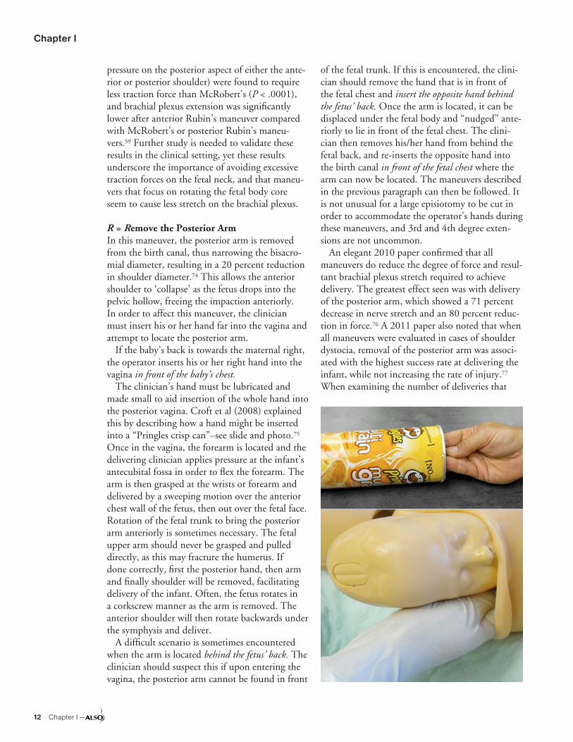
Chapter I
12 Chapter I —
pressure on the posterior aspect of either the ante-rior or posterior shoulder) were found to require less traction force than McRobert’s (P < .0001), and brachial plexus extension was significantly lower after anterior Rubin’s maneuver compared with McRobert’s or posterior Rubin’s maneu-vers.59 Further study is needed to validate these results in the clinical setting, yet these results underscore the importance of avoiding excessive traction forces on the fetal neck, and that maneu-vers that focus on rotating the fetal body core seem to cause less stretch on the brachial plexus.
R = Remove the Posterior ArmIn this maneuver, the posterior arm is removed from the birth canal, thus narrowing the bisacro-mial diameter, resulting in a 20 percent reduction in shoulder diameter.74 This allows the anterior shoulder to ‘collapse’ as the fetus drops into the pelvic hollow, freeing the impaction anteriorly. In order to affect this maneuver, the clinician must insert his or her hand far into the vagina and attempt to locate the posterior arm.
If the baby’s back is towards the maternal right, the operator inserts his or her right hand into the vagina in front of the baby’s chest.
The clinician’s hand must be lubricated and made small to aid insertion of the whole hand into the posterior vagina. Croft et al (2008) explained this by describing how a hand might be inserted into a “Pringles crisp can”–see slide and photo.75 Once in the vagina, the forearm is located and the delivering clinician applies pressure at the infant’s antecubital fossa in order to flex the forearm. The arm is then grasped at the wrists or forearm and delivered by a sweeping motion over the anterior chest wall of the fetus, then out over the fetal face. Rotation of the fetal trunk to bring the posterior arm anteriorly is sometimes necessary. The fetal upper arm should never be grasped and pulled directly, as this may fracture the humerus. If done correctly, first the posterior hand, then arm and finally shoulder will be removed, facilitating delivery of the infant. Often, the fetus rotates in a corkscrew manner as the arm is removed. The anterior shoulder will then rotate backwards under the symphysis and deliver.
A difficult scenario is sometimes encountered when the arm is located behind the fetus’ back. The clinician should suspect this if upon entering the vagina, the posterior arm cannot be found in front
of the fetal trunk. If this is encountered, the clini-cian should remove the hand that is in front of the fetal chest and insert the opposite hand behind the fetus’ back. Once the arm is located, it can be displaced under the fetal body and “nudged” ante-riorly to lie in front of the fetal chest. The clini-cian then removes his/her hand from behind the fetal back, and re-inserts the opposite hand into the birth canal in front of the fetal chest where the arm can now be located. The maneuvers described in the previous paragraph can then be followed. It is not unusual for a large episiotomy to be cut in order to accommodate the operator’s hands during these maneuvers, and 3rd and 4th degree exten-sions are not uncommon.
An elegant 2010 paper confirmed that all maneuvers do reduce the degree of force and resul-tant brachial plexus stretch required to achieve delivery. The greatest effect seen was with delivery of the posterior arm, which showed a 71 percent decrease in nerve stretch and an 80 percent reduc-tion in force.76 A 2011 paper also noted that when all maneuvers were evaluated in cases of shoulder dystocia, removal of the posterior arm was associ-ated with the highest success rate at delivering the infant, while not increasing the rate of injury.77 When examining the number of deliveries that

Shoulder Dystocia
— Chapter I 13
included the different maneuver types, irrespective of order, 84.4 percent of deliveries had successful resolution of the shoulder dystocia when delivery of the posterior shoulder was attempted. This was higher than the other maneuvers, which ranged from 24.3 percent (McRoberts) to 72.0 percent (Woods corkscrew).77
R = Roll the PatientThe “all fours” or “Gaskin” maneuver is a safe, rapid and effective technique for the reduction of shoulder dystocia. Ms. Gaskin is a renowned direct entry midwife in the United States, best known for her book Spiritual Midwifery. The maneuver requires the patient to roll from the existing supine or dorsal lithotomy position to an all fours posi-tion. The precise mechanism by which this maneu-ver acts to relieve the shoulder dystocia is unknown although it has been documented that the pelvic diameters increase when laboring women change from the dorsal recumbent position.78
Radiographic studies indicate that pelvic measurements are least favorable for delivery in the dorsal lithotomy position. By rotating to the all fours position, the true obstetrical conjugate increases by as much as 10 mm and the sagittal measurement of the pelvic outlet increases up to 20 mm.79 The fetal shoulder often dislodges dur-ing the act of turning from a supine to “all fours” position, indicating that this movement alone may be sufficient to allow enough pelvic change to dislodge the impaction. Additionally, once the position change is completed, gravitational forces may aid in disimpaction of the fetal shoulder.
The “all fours” maneuver may be difficult for a woman who is fatigued or restricted by intravenous lines, fetal monitors, epidural anesthesia or a Foley catheter. The patient will often need assistance to re-position, given these entrapments. At one facility, all patients with epidural anesthesia are instructed to perform a shoulder dystocia drill and practice getting into the all-fours position, in case it is needed later.80,81 Consideration should be given to including this as part of prenatal education. This position may be disorienting to clinicians who are unfamiliar attending a delivery in this position, however by providing gentle traction downward, the clinician can deliver the posterior shoulder first with the aid of gravity in the same direction as if the patient was in the dorsal lithotomy posi-tion. The all-fours position is compatible with all
intra-vaginal manipulations for shoulder dystocia, but it is incompatible with suprapubic pressure. A tip to remember is to always go with gravity first, thus provide gentle traction downward to deliver the shoulder closest to the ceiling first, (in this case the infant’s posterior rather than the anterior shoulder). Performing a few “normal” or simulated deliveries in this position, before encountering an acute need to do so, may assist the provider in being prepared for more emergent situations.
The order in which these maneuvers are attempted may be flexible. However, a logical progression of various efforts is essential to allow adequate time for each one to potentially accom-plish delivery. The suggested length of time for each maneuver is meant only as a guideline. Clini-cal judgment should always guide the progression of procedures used.
Second attempt at all the maneuvers described above can be tried again; sometimes there has been some change in the position or station of the shoulder and a repeat attempt can be successful. If the second attempt is unsuccessful, the following techniques have been described as “last resort” or “salvage” maneuvers.
Sling: posterior axillary traction — The tech-nique to deliver the posterior arm has been described in case series two different ways.82,83 It may prove helpful in cases of SD when the sacral promontory holds up the posterior shoulder and all other maneuvers have failed.
The first variation is for the clinician’s hand to enter the birth canal along the sacral hollow and deliver the posterior shoulder before delivering the posterior arm. To make access easier to the poste-rior pelvis, an assistant holds (not pulls) the fetus’s head flexed upward toward the anterior shoulder. An episiotomy is helpful if the perineum is too rigid to allow entry. The birth clinician will need to get on one knee below the mother’s perineum in order to employ the correct downward vector. The right middle finger is placed into the fetus’s posterior axilla from the left side of the pelvis and the left middle finger is placed into the axilla that lies posteriorly. By using the two middle fingers in the axilla, traction is employed following the curve of the sacrum. Once the shoulder has emerged from the pelvis, the posterior arm can be delivered. At other times, the shoulder may not actually be delivered first but instead is brought down low

Chapter I
14 Chapter I —
enough in the pelvis that the posterior arm can be grasped and brought out.82
Another version of the posterior sling involves the use of a 14 Fr soft, plastic suction catheter and has been described in cases of severe shoulder dystocias that have persisted so long that the infant has died but is still undelivered. This technique has been noted to avoid having to put a mother through a symphysiotomy to accomplish the delivery. If the previously mentioned maneuvers are unsuccessful, the clinician uses the suction catheter to create a sling through the axilla of the infant’s posterior shoulder. The clinician folds the catheter in half over the operator’s right index fingertip and manually inserts the loop around the posterior shoulder and under the axilla. The loop is then retrieved with the left index finger and withdrawn so that the proximal end and tip of the catheter meet to create a sling around the shoul-der.83 A hemostat or curved clamp can be applied to the ends of the sling and traction is applied in a posterior downward vector.
These techniques have their drawbacks. Fracture of the fetus’s posterior humerus is frequent. For the mother, tears of the anal sphincter and rectum will commonly occur due to the posterior pres-sure. Posterior axillary traction is by no means an original technique and has been described in at least two publications in the past,84,85 but it does not seem to have been popularized. Success of this technique, however, presupposes that the poste-rior shoulder is accessible and is not held up by the sacral promontory. If the anterior shoulder is impacted by the symphysis pubis and the posterior shoulder is above the sacral promontory—in other words, if neither shoulder is in the pelvis—only cephalic replacement or symphysiotomy is likely to resolve the problem.
The Zavanelli ManeuverThe Zavanelli maneuver requires reversal of the cardinal movements of labor: derestitution (inter-nal rotation), flexion, and subsequent manual replacement of the fetal vertex into the vaginal canal, followed by cesarean delivery.27,86 Continu-ous upward pressure is then maintained on the fetal head until cesarean delivery can be accom-plished. Induction of uterine relaxation with IV or SQ terbutaline or nitroglycerin administered by the oral, sublingual or IV route is valuable adjunct to this procedure, and potentially will prevent a
uterine rupture. Alternatively musculoskeletal or uterine relaxation can be induced with halothane or other general anesthetic. Before considering cephalic replacement, an operating team, anes-thesia, and physicians capable of performing a cesarean delivery must be present. This maneuver should never be attempted if a nuchal cord has been previously clamped and cut.
Surgery and Symphysiotomy1. Symphysiotomy. Intentional division of thefibrous cartilage of the symphysis pubis under local anesthesia has been used successfully in developing countries more than in North America and Europe. Reports in the U.S. are related to its use after failed Zavanelli maneuvers.87 Because the procedure takes at least two minutes from the time a decision is made, if needed, it should be initiated within five to six minutes of delivery of the fetal head and should only be used when all other maneuvers have failed and cesarean delivery capability is not available.88,89 Some women with this procedure might encounter chronic symphy-seal pain due to separation and could potential encounter urethral trauma. Global ALSO includes an optional symphysiotomy workshop (www.aafp.org/also).
2. Abdominal Surgery and Hysterotomy. Thistechnique has been reported to facilitate vaginal delivery of the fetus in a case of shoulder dystocia unresponsive to the above noted maneuvers. In a small series of case reports of severe shoulder dystocia, general anesthesia was induced and the uterus opened as similar to a cesarean section. The surgeon would then rotate the infant transabdomi-nally through the hysterotomy incision, allowing the shoulders to rotate, much like a Woods Screw maneuver. Delivery is then accomplished by vagi-nal extraction by another clinician.90
3. Deliberate Clavicular Fracture and Cleido-tomy. Direct upward pressure on the mid-portion of the fetal clavicle will result in fracture and reduce the shoulder-to-shoulder distance. Older textbooks have described this technique, however this has not been reported in recent literature because it is technically difficult to perform and risks serious injury to the underlying vascular and pulmonary structures in the newborn.27 However, formal cleidotomy (surgical division of one or

Shoulder Dystocia
— Chapter I 15
both clavicles with heavy surgical scissors) can be considered in cases of shoulder dystocia when the fetus is dead and standard management techniques have failed. Obviously, there is a potential for maternal trauma unless great care is taken with this procedure.
DocumentationDocumentation in the medical records after a completed delivery is an essential risk manage-ment tool and several studies point to the lack of standardization and compliance in this matter. The use of a comprehensive, standardized proce-dure note in cases of shoulder dystocia has been advocated by many.91,92
It is important to record the elapsed time of the shoulder dystocia, the maneuvers employed and the condition of the mother and infant after the delivery.93 Terms like mild, moderate, or severe shoulder dystocia offer little information about the maternity care that was provided or future legal issues. The documentation should also include other team members present and umbilical cord venous and arterial cord pH, if obtained. In case of subsequent nerve palsy develops, it is worthwhile to document which arm was impacted against the pubis and on which arm maneuvers were per-formed for removal. Please see Appendix A.63
A retrospective observational study in the UK compared documentation of SD under three institutional documentation conditions; non-standardized, written delivery notes; standard-ized delivery notes after electronic medical record implementation; and standardized delivery notes in the electronic medical record following SD simulation drills. Standardized electronic notes improved documentation, but further addition of SD drills further increased SD documentation.94
Simulation Training And Institutional PlansSimulation training and repeated practice of emer-gency drills has been demonstrated to improve performance in the management of simulated obstetrical emergencies, including shoulder dys-tocia. Simulation has evolved to include complex, high fidelity models, yet low-fidelity models can
still be quite valuable, especially for the purpose of team training.
The SaFE study in Great Britain documented that a standardized training program with simu-lated deliveries involving 450 participants, resulted an increase in successful delivery rate and reduction in force amplitudes administered by clinicians.70
High-fidelity training pelvises (models with electronic and computer circuitry such as the Noelle™) have been shown to improve clinicians’ ability to use minimum traction force and a reduc-tion in the actual incidence of neonatal injury. However, low fidelity (models used in the ALSO course—such as Simulaids™) models can provide the communication and team skills needed to suc-cessfully manage a shoulder dystocia delivery and limit psychological and medico-legal problems. Training programs should consider the inclusion of patient–actors with mannequins to increase the fidelity of simulation exercises. Further studies evaluating the effectiveness of ALSO training and ways of measuring traction forces with low fidelity models are needed to document course effective-ness in providing clinicians with improved shoul-der dystocia management skills.62,94-98
SummaryShoulder dystocia is a relatively uncommon and dangerous event, yet it is difficult to predict and 50 percent of shoulder dystocias have no anteced-ent factors. Anticipation and preparation are criti-cal for successful management.
An institutional plan is highly recommended, in which each member of the team has an assigned duty. The recommended management of shoulder dystocia is based on the mnemonic HELPERR, which provides a memory guide and a structured framework for action, which is useful in this crisis. The elements of the HELPERR mnemonic are all effective, and they should be tried in a logical and calm sequence. Practice on the mannequin is an essential aid to clinicians who are likely to encoun-ter this obstetric emergency. The time allotted to each maneuver, and the exact sequence, are best determined by the clinical circumstances and the clinician’s best judgment, yet incorporating sug-gested guidelines.

Chapter I
16 Chapter I —
SORT: Key Recommendations for Practice
Clinical RecommendationEvidence Rating References
Routine late pregnancy ultrasound to detect macrosomia in low-risk or unselected populations does not confer benefit by allowing reduction in morbidity from shoulder dystocia, and there is not enough evidence to evaluate the prediction of SD based on fetal size through fundal height, palpation, or ultrasound.
A 1,3,42-45,47,48
Induction of labor in non-diabetic women with suspected macrosomic infants does not prevent shoulder dystocia.
B 3,43,44,49
Suspected macrosomia is not an indication for induction or primary cesarean delivery in uncomplicated non-diabetic pregnancies unless estimated fetal weight is > 5,000 grams.
B 42-45
Shoulder dystocia is associated with instrumented vaginal delivery and vacuum extractor use in excess of 10 minutes or more than five tractive efforts should be conducted with caution, as it is a prominent risk factor for neonatal BPP.
B 3,25,54,57
Episiotomy performed at the time on an assisted delivery does not reduce the incidence of shoulder dystocia.
B 66,67
Simulation training, with both low and high fidelity models, has demonstrated benefit at improving clinicians’ skill levels, reducing force required to achieve a simulated SD delivery. In the clinical setting, simulation training has been shown to reduce the incidence of brachial plexus palsy where it has been required.
B 62,94-98
An institutional plan for anticipating and managing shoulder dystocias is recommended, in which each member of the team has an assigned duty.
C
The elements of the HELPERR mnemonic are effective. They should be instituted in a logical and calm sequence.
C 73
Half of shoulder dystocias can be relieved by McRoberts maneuver and suprapubic pressure and these noninvasive maneuvers are recommended for initial management.
C 27,68
Rotary maneuvers and removal of the posterior arm have demonstrated less tractive force and stretch in simulation and computer animated models.
C 61,62,70,76,94-98
The use of a comprehensive, standardized procedure note in cases of shoulder dystocia is strongly recommended.
C 91-94
Strength of Recommendation Taxonomy (SORT)
Strength of Recommendation Definition
A • Recommendation based on consistent and good-quality patient-oriented evidence.
B • Recommendation based on inconsistent or limited-quality patient-oriented evidence.
C • Recommendation based on consensus, usual practice, opinion, disease-oriented evidence, or case series for studies of diagnosis, treat-ment, prevention, or screening.
Patient-oriented evidence measures outcomes that matter to patients: morbidity, mortality, symptom improvement, cost reduction, and quality of life. Disease-oriented evidence measures intermediate, physiologic, or surrogate end points that might not reflect improvement in patient outcomes (eg, blood pressure, blood chemistry, physiologic function, pathologic findings).
BPP = brachial plexus palsy; SD = shoulder dystocia.
From Ebell MH, Siwek J, Weiss BD, et al. Strength of recommendation taxonomy (SORT): a patient-centered approach to grading evidence in the medical literature. Am Fam Physician. 2004;69(3):548-556.

— Chapter I 17
Appendix A: Shoulder Dystocia Documentation63
Date _________________________ Time __________________________
Person completing form _________________________________________
Designation ___________________________________________________
Signature _____________________________________________________
Called for help at: Emergency call via switchboard at:
Staff present at delivery of head Additional staff attending for delivery of shoulders
Name Role Name Role Time arrived
Procedures used to assist delivery
By whom Time Order Details Reason if not performed
❑ McRoberts’ position
❑ Suprapubic pressure From maternal left / right (circle as appropriate)
❑ Episiotomy Enough access / tear present / already performed (circle as appropriate)
❑ Delivery of posterior arm Right / left arm (circle as appropriate)
❑ Internal rotational maneuver
Description of rotation
Description of traction Routine (as in normal vaginal delivery)
Other: Reason if not routine:
Other maneuvers used
Mode of delivery of head
❑ Spontaneous
❑ Instrumental — vacuum / forceps
Time of delivery of head
Time of delivery of baby
Head-to-body delivery interval
Fetal position during dystocia
Head facing maternal left Left fetal shoulder anterior
Head facing maternal right Right fetal shoulder anterior
Birth weight kg Apgar 1 min: 5 mins: 10 mins:
Cord gases Art pH: Art BE: Venous pH: Venous BE:
Explanation to parents ❑ Yes By:
Neonatal assessment at delivery
Assessment by:
Any sign of arm weakness? ❑ Yes ❑ No
Any sign of potential bony fracture? ❑ Yes ❑ No
Baby admitted to Neonatal Intensive Care Unit? ❑ Yes ❑ No
If yes to any of these questions for review and follow up by Consultant neonatologist.
Mother’s name ____________________________________________
Date of birth ______________________________________________
Hospital number __________________________________________
Consultant _______________________________________________

Chapter I
18 Chapter I —
References1. Beall MH, Spong C, McKay J, Ross MG. Objective defi-
nition of shoulder dystocia: a prospective evaluation. Am J Obstet Gynecol. 1998;179(4):934-937.
2. Gherman RB. Shoulder dystocia: an evidence-basedevaluation of the obstetric nightmare. Clin Obstet Gyne-col. 2002;45(2):345-362.
3. Nesbitt TS, Gilbert WM, Herrchen B. Shoulder dystociaand associated risk factors with macrosomic infants bornin California. Am J Obstet Gynecol. 1998;179(2):476-480.
4. Cohen BF, Penning S, Ansley D, Porto M, Garite T. Theincidence and severity of shoulder dystocia correlateswith a sonographic measurement of asymmetry inpatients with diabetes. Am J Perinatol. 1999;16(4):197-201.
5. Verspyck E, Goffinet F, Hellot MF, Milliez J, Marpeau L.Newborn shoulder width: a prospective study of 2222consecutive measurements. Br J Obstet Gynaecol.1999;106(6):589-593.
6. Overland EA, Spydslaug A, Nielsen CS, Eskild A.. Riskof shoulder dystocia in second delivery: does a historyof shoulder dystocia matter? Am J Obstet Gynecol.2009;200(5):506.e1-6.
7. Usta IM, Hayek S, Yahya F, Abu-Musa A, Nassar AH.Shoulder dystocia: what is the risk of recurrence? ActaObstet Gynecol Scand. 2008;87(10):992-997.
8. Belfort MA, Dildy GA, Saade GR, Suarez V, Clark SL.Prediction of shoulder dystocia using multivariate analy-sis. Am J Perinatol. 2007;24(1):5-10.
9. Ju H, Chadha Y, Donovan T, O’Rourke P. Fetal mac-rosomia and pregnancy outcomes. Aust N Z J ObstetGynaecol. 2009;49(5):504-509.
10. Bjørstad AR, Irgens-Hansen K, Daltveit AK, Irgens LM.Macrosomia: mode of delivery and pregnancy outcome.Acta Obstet Gynecol Scand. 2010;89(5):664-669.
11. Neilson JP. Symphysis-fundal height measurement inpregnancy. Cochrane Database Syst Rev. 2000;(2):CD000944.
12. Bricker L, Neilson JP. Routine ultrasound in late preg-nancy (after 24 weeks gestation). Cochrane DatabaseSyst Rev. 2000;(2):CD001451.
13. Johnstone FD, Prescott RJ, Steel JM, Mao JH, ChambersS, Muir N. Clinical and ultrasound prediction of macroso-mia in diabetic pregnancy. Br J Obstet Gynaecol. 1996;103(8):747-754.
14. Henriksen T. The macrosomic fetus: a challenge in cur-rent obstetrics. Acta Obstet Gynecol Scand. 2008;87(2):134-145.
15. Alwan N, Tuffnell DJ, West J. Treatments for gestationaldiabetes. Cochrane Database Syst Rev. 2009;(3):CD003395.
16. Esakoff TF, Cheng YW, Sparks TN, Caughey AB. Theassociation between birthweight 4000 g or greater andperinatal outcomes in patients with and without gesta-tional diabetes mellitus. Am J Obstet Gynecol. 2009;200(6):672.e1-672.e4.
17. Landon MB, Spong CY, Thom E, et al; Eunice KennedyShriver National Institute of Child Health and HumanDevelopment Maternal-Fetal Medicine Units Network. Amulticenter, randomized trial of treatment for mild gesta-tional diabetes. N Engl J Med. 2009;361(14):1339-1348.
18. Horvath K, Koch K, Jeitler K, et al. Effects of treatmentin women with gestational diabetes mellitus: systematicreview and meta-analysis. BMJ. 2010;340:c1395.
19. Witkop CT, Neale D, Wilson LM, Bass EB, NicholsonWK. Active compared with expectant delivery manage-ment in women with gestational diabetes: a systematicreview. Obstet Gynecol. 2009;113(1):206-217.
20. McFarland M, Hod M, Piper JM, Xenakis EM, Langer O.Are labor abnormalities more common in shoulder dys-tocia? Am J Obstet Gynecol. 1995;173(4):1211-1214.
21. Gemer O, Bergman M, Segal S. Labor abnormalities asa risk factor for shoulder dystocia. Acta Obstet GynecolScand. 1999;78(8):735-736.
22. Henry DE, Cheng YW, Shaffer BL, Kaimal AJ, BiancoK, Caughey AB. Perinatal outcomes in the settingof active phase arrest of labor. Obstet Gynecol.2008;112(5):1109-1115.
23. Brimacombe M, Iffy L, Apuzzio JJ, et al. Shoulder dysto-cia related fetal neurological injuries: the predisposingroles of forceps and ventouse extractions. Arch Gyne-col Obstet. 2008;277(5):415-422.
24. Carlan SJ, Angel JL, Knuppel RA. Shoulder dystocia.Am Fam Physician. 1991;43(4):1307-1311.
25. Gilbert WM, Nesbitt TS, Danielsen B. Associated fac-tors in 1611 cases of brachial plexus injury. Obstet Gyne-col. 1999;93(4):536-540.
26. Mollberg M, Hagberg H, Bager B, Lilja H, Ladfors L.Risk factors for obstetric brachial plexus palsy amongneonates delivered by vacuum extraction. Obstet Gyne-col. 2005;106(5 Pt 1):913-918.
27. Gherman RB, Chauhan S, Ouzounian JG, Lerner H,Gonik B, Goodwin TM. Shoulder dystocia: the unpre-ventable obstetric emergency with empiric managementguidelines. Am J Obstet Gynecol. 2006;195(3):657-672.
28. Gherman RB, Ouzounian JG, Incerpi MH, Goodwin TM.Symphyseal separation and transient femoral neuropa-thy associated with the McRoberts’ maneuver. Am JObstet Gynecol. 1998;178(3):609-610.
29. Gherman RB, Ouzounian JG, Goodwin TM. Obstetricmaneuvers for shoulder dystocia and associated fetalmorbidity. Am J Obstet Gynecol. 1998;178(6):1126-1130.
30. Draycott T, Sanders C, Crofts J, Lloyd J. A template forreviewing the strength of evidence for obstetric brachialplexus injury in clinical negligence claims. Clin Risk.2008;14:96-100.
31. Allen R, Sorab J, Gonik B. Risk factors for shoulder dys-tocia: an engineering study of clinician-applied forces.Obstet Gynecol. 1991;77(3):352-355.
32. Allen RH, Bankoski BR, Butzin CA, Nagey DA. Com-paring clinician-applied loads for routine, difficult, andshoulder dystocia deliveries. Am J Obstet Gynecol.1994;171(6):1621-1627.

Shoulder Dystocia
— Chapter I 19
33. Gherman RB, Goodwin TM. Shoulder dystocia. Curr Opin Obstet Gynecol. 1998;10(6):459-463.
34. Allen RH, Cha SL, Kranker LM, Johnson TL, Gurewitsch ED. Comparing mechanical fetal response during descent, crowning, and restitution among deliveries with and without shoulder dystocia. Am J Obstet Gyne-col. 2007;196(6):539.e1-539.e5.
35. Grimm MJ, Costello RE, Gonik B. Effect of clinician-applied maneuvers on brachial plexus stretch during a shoulder dystocia event: investigation using a computer simulation model. Am J Obstet Gynecol. 2010;203(4): 339.e1-339.e5.
36. Beer E, Folghera MG. Time for resolving shoulder dys-tocia. Am J Obstet Gynecol. 1998;179(5):1376-1377.
37. Stallings SP, Edwards RK, Johnson JWC. Correlation of head-to-body delivery intervals in shoulder dystocia and umbilical artery acidosis. Am J Obstet Gynecol. 2001; 185(2):268-274.
38. McFarland MB, Langer O, Piper JM, Berkus MD. Peri-natal outcome and the type and number of maneuvers in shoulder dystocia. Int J Gynaecol Obstet. 1996;55(3): 219-224.
39. Ouzounian JG, Korst LM, Ahn MO, et al. Shoulder dys-tocia and neonatal brain injury: significance of the head-shoulder interval. Am J Obstet Gynecol. 1998;178:S76.
40. Leung TY, Stuart O, Sahota DS, Suen SS, Lau TK, Lao TT. Head-to-body delivery interval and risk of fetal aci-dosis and hypoxic ischaemic encephalopathy in shoul-der dystocia: a retrospective review. BJOG. 2011;118(4): 474-479.
41. Lerner H, Durlacher K, Smith S, Hamilton E. Relation-ship between head-to-body delivery interval in shoulder dystocia and neonatal depression. Obstet Gynecol. 2011;118(2 Pt 1):318-322.
42. Boulvain M, Stan C, Irion O. Elective delivery in dia-betic pregnant women. Cochrane Database Syst Rev. 2001;(2):CD001997.
43. Irion O, Boulvain M. Induction of labour for suspected fetal macrosomia. Cochrane Database Syst Rev. 2000;(2):CD000938.
44. Chauhan SP, Grobman WA, Gherman RA, et al. Suspi-cion and treatment of the macrosomic fetus: a review. Am J Obstet Gynecol. 2005;193(2):332-346.
45. Wong SF, Chan FY, Cincotta RB, Oats JJ, McIntyre HD. Sonographic estimation of fetal weight in macrosomic fetuses: diabetic versus non-diabetic pregnancies. Aust N Z J Obstet Gynaecol. 2001;41(4):429-432.
46. Abramowicz JS, Rana S, Abramowicz S. Fetal cheek-to-cheek diameter in the prediction of mode of delivery. Am J Obstet Gynecol. 2005;192(4):1205-1211, discussion 1211-1213.
47. Miller RS, Devine PC, Johnson EB. Sonographic fetal asymmetry predicts shoulder dystocia. J Ultrasound Med. 2007;26(11):1523-1528.
48. Rajan PV, Chung JH, Porto M, Wing DA. Correlation of increased fetal asymmetry with shoulder dystocia in the nondiabetic woman with suspected macrosomia. J Reprod Med. 2009;54(8):478-482.
49. ACOG Committee on Practice Bulletins-Gynecology, The American College of Obstetrician and Gynecologists. ACOG practice bulletin clinical management guidelines for obstetrician-gynecologists. Number 40, November 2002. Obstet Gynecol. 2002;100(5 Pt 1):1045-1050.
50. Rouse DJ, Owen J, Goldenberg RL, Cliver SP. The effectiveness and costs of elective cesarean delivery for fetal macrosomia diagnosed by ultrasound. JAMA. 1996;276 (18):1480-1486.
51. American College of Obstetricians and Gynecologists. ACOG practice patterns. Shoulder dystocia. Number 7, October 1997. American College of Obstetricians and Gynecologists. Int J Gynaecol Obstet. 1998;60(3):306-313.
52. Royal College of Obstetrics and Gynecology Green Top Guideline #42. December 2005.
53. Herbst MA. Treatment of suspected fetal macrosomia: a cost-effectiveness analysis. Am J Obstet Gynecol. 2005;193(3 Pt 2):1035-1039.
54. Ouzounian JG, Gherman RB. Shoulder dystocia: are historic risk factors reliable predictors? Am J Obstet Gynecol. 2005;192(6):1933-5; discussion 1935-1938.
55. Athukorala C, Middleton P, Crowther CA. Intrapar-tum interventions for preventing shoulder dystocia. Cochrane Database Syst Rev. 2006;(4):CD005543.
56. Caughey AB, Sandberg PL, Zlatnik MG, Thiet MP, Parer JT, Laros RK Jr. Forceps compared with vacuum: rates of neonatal and maternal morbidity. Obstet Gynecol. 2005;106(5 Pt 1):908-912.
57. Mollberg M, Hagberg H, Bager B, Lilja H, Ladfors L. Risk factors for obstetric brachial plexus palsy among neonates delivered by vacuum extraction. Obstet Gyne-col. 2005;106(5 Pt 1):913-918.
58. Beall MH, Spong CY, Ross MG. A randomized con-trolled trial of prophylactic maneuvers to reduce head-to-body delivery time in patients at risk for shoulder dystocia. Obstet Gynecol. 2003;102(1):31-35.
59. Poggi SH, Allen RH, Patel CR, Ghidini A, Pezzullo JC, Spong CY. Randomized trial of McRoberts versus lithot-omy positioning to decrease the force that is applied to the fetus during delivery. Am J Obstet Gynecol. 2004;191(3):874-878.
60. Locatelli A, Incerti M, Ghidini A, et al. Head-to-body delivery interval using ‘two-step’ approach in vaginal deliveries: effect on umbilical artery pH. J Mat-Fetal & Neonatal Medicine, 2010.
61. Gonik B, Allen R, Sorab J. Objective evaluation of the shoulder dystocia phenomenon: effect of maternal pelvic orientation on force reduction. Obstet Gynecol. 1989;74(1):44-48.
62. Crofts JF, Ellis D, James M, Hunt LP, Fox R, Draycott TJ. Pattern and degree of forces applied during simu-lation of shoulder dystocia. Am J Obstet Gynecol. 2007;197(2):156.e1-156.e6.
63. Shoulder Dystocia Documentation – Personal Commu-nication with T. Draycott. 2011.
64. Gurewitsch ED, Kim EJ, Yang JH, Outland KE, McDon-ald MK, Allen RH. Comparing McRoberts’ and Rubin’s maneuvers for initial management of shoulder dystocia: an objective evaluation. [Erratum in: Am J Obstet Gynecol. 2005;192:662.] Am J Obstet Gynecol. 2005;192(1):153-160.

Chapter I
20 Chapter I —
65. Royal College of Obstetricians and Gynaecologists,Royal College of Midwives. Towards Safer Childbirth.Minimum Standards for the Organization of LabourWards: Report of a Joint Working Party. London: RCOGPress; 1999.
66. Macleod M, Strachan B, Bahl R, et al. A prospectivecohort study of maternal and neonatal morbidity in rela-tion to use of episiotomy at operative vaginal delivery.BJOG. 2008;115(13):1688-1694.
67. Gurewitsch ED, Donithan M, Stallings SP, et al. Episi-otomy versus fetal manipulation in managing severeshoulder dystocia: a comparison of outcomes. Am JObstet Gynecol. 2004;191(3):911-916.
68. McFarland MB, Langer O, Piper JM, Berkus MD. Peri-natal outcome and the type and number of maneuversin shoulder dystocia. Int J Gynaecol Obstet. 1996;55(3):219-224.
69. Gherman RB, Goodwin TM, Souter I, Neumann K,Ouzounian JG, Paul RH. The McRoberts’ maneuver forthe alleviation of shoulder dystocia: how successful isit? [Level III]. Am J Obstet Gynecol. 1997;176(3):656-661.
70. Grimm MJ, Costello RE, Gonik B. Effect of clinician-applied maneuvers on brachial plexus stretch dur-ing a shoulder dystocia event: investigation using acomputer simulation model. Am J Obstet Gynecol.2010;203(4):339.e1-339.e5.
71. Woywodt A, Matteson E. Should eponyms be aban-doned? Yes. BMJ. 2007;335(7617):424.
72. Woods CE. A principle of physics as applicable toshoulder delivery. Am J Obstet Gynecol. 1943;45:796.
73. Ramsey PS, Ramin KD, Field CS. Shoulder dysto-cia. Rotational maneuvers revisited. J Reprod Med.2000;45(2):85-88.
74. Poggi SH, Spong CY, Allen RH. Prioritizing posteriorarm delivery during severe shoulder dystocia. ObstetGynecol. 2003;101(5 Pt 2):1068-1072.
75. Crofts JF, Fox R, Ellis D, Winter C, Hinshaw K, DraycottTJ. Observations from 450 shoulder dystocia simu-lations: lessons for skills training. Obstet Gynecol.2008;112(4):906-912.
76. Grimm MJ, Costello RE, Gonik B. Effect of clinician-applied maneuvers on brachial plexus stretch dur-ing a shoulder dystocia event: investigation using acomputer simulation model. Am J Obstet Gynecol.2010;203(4):339.e1-339.e5.
77. Hoffman MK, Bailit JL, Branch DW, et al; Consortiumon Safe Labor. A comparison of obstetric maneuversfor the acute management of shoulder dystocia. ObstetGynecol. 2011;117(6):1272-1278.
78. Bruner JP, Drummond SB, Meenan AL, Gaskin IM. All-fours maneuver for reducing shoulder dystocia duringlabor. J Reprod Med. 1998;43(5):439-443.
79. Borell U, Fernstrom I. A pelvimetric method for theassessment of pelvic mouldability. Acta radiol. 1957a;47(5):365-370.
80. Meenan AL, Gaskin IM, Hunt P, Ball CA. A new (old)maneuver for the management of shoulder dystocia.J Fam Pract. 1991;32(6):625-629.
81. Bruner JP, Drummond SB, Meenan AL, Gaskin IM. All-fours maneuver for reducing shoulder dystocia duringlabor. J Reprod Med. 1998;43(5):439-443.
82. Menticoglou SM. A modified technique to deliver theposterior arm in severe shoulder dystocia. Obstet Gyne-col. 2006;108(3 Pt 2):755-757.
83. Cluver CA, Hofmeyr GJ. Posterior axilla sling traction:a technique for intractable shoulder dystocia. ObstetGynecol. 2009;113(2 Pt 2):486-488.
84. Schramm M. Impacted shoulders—a personal experi-ence. Aust N Z J Obstet Gynaecol. 1983;23(1):28-31.
85. Holman MS. A new manoeuvre for delivery of animpacted shoulder, based on a mechanical analysis.S Afr Med J. 1963;37:247-249.
86. Zelig CM, Gherman RB. Modified Zavanelli maneuverfor the alleviation of shoulder dystocia. Obstet Gynecol.2002;100(5 Pt 2):1112-1114.
87. Goodwin TM, Banks E, Millar LK, Phelan JP. Cata-strophic shoulder dystocia and emergency symphysi-otomy. Am J Obstet Gynecol. 1997;177(2):463-464.
88. Crichton D, Seedat EK. The technique of symphysi-otomy. S Afr Med J. 1963;37:227-231.
89. Management of labor and delivery complications. (in)Douglas-Stromme Operative Obstetrics. 5th ed. ZuspanFP, Quilligan EJ (eds.) Appleton and Lange. Norwalk,Connecticut. Pages 337-8, 1988.
90. O’Shaughnessy MJ. Hysterotomy facilitation of the vagi-nal delivery of the posterior arm in a case of severe shoul-der dystocia. Obstet Gynecol. 1998;92(4 Pt 2):693-695.
91. Crofts JF, Bartlett C, Ellis D, Fox R, Draycott TJ. Docu-mentation of simulated shoulder dystocia: accurate andcomplete? BJOG. 2008;115(10):1303-1308.
92. Clark SL, Belfort MA, Dildy GA, Meyers JA. Reducingobstetric litigation through alterations in practice pat-terns. Obstet Gynecol. 2008;112(6):1279-1283.
93. Clinical Negligence Scheme for Trusts. Maternity: Clini-cal Risk Management Standards. London: NHS LitigationAuthority; 2006. Available at http://www.nhsla.com/NR/rdonlyres/002DB3DE-F1A1-4153-AFE1-B59A3D743239/0/CNSTMaternityStandardsApril2006final.pdf.
94. Nguyen T, Fox NS, Friedman F Jr, Sandler R, Rebar-ber A. The sequential effect of computerized deliverycharting and simulation training on shoulder dysto-cia documentation. J Matern Fetal Neonatal Med.2011;24(11):1357-1361.
95. Deering S, Poggi S, Macedonia C, Gherman R, SatinAJ. Improving resident competency in the managementof shoulder dystocia with simulation training. ObstetGynecol. 2004;103(6):1224-1228.
96. Johannsson H, Ayida G, Sadler C. Faking it? Simulationin the training of obstetricians and gynaecologists. CurrOpin Obstet Gynecol. 2005;17(6):557-561.
97. Black RS, Brocklehurst P. A systematic review of train-ing in acute obstetric emergencies. BJOG. 2003;110(9):837-841.
98. Crofts JF, Bartlett C, Ellis D, Hunt LP, Fox R, DraycottTJ. Management of shoulder dystocia: skill retention6 and 12 months after training. Obstet Gynecol. 2007;110(5):1069-1074.

— Chapter J 1
Chapter J Postpartum Hemorrhage
IntroductionPostpartum hemorrhage (PPH) is excessive bleeding after delivery of the fetus and may occur before or after delivery of the placenta. Clinicians must learn to recognize excessive bleeding and intervene, prefer-ably before other signs and symptoms of PPH develop (see Table 1).
Definition, Epidemiology, and SignificancePostpartum hemorrhage is traditionally defined as the loss of more than 500 mL of blood following vaginal delivery or more than 1,000 mL following cesarean delivery.1 PPH is considered severe when blood loss exceeds 1,000 mL after vaginal delivery or results in signs or symptoms of hemodynamic instability.1 How-
ever, the definition of PPH is debated as more recent studies have shown that the median blood loss at spon-taneous vaginal delivery exceeds 500 mL.2 Postpartum hemorrhage can be classified as primary, which occurs within 24 hours of delivery, or secondary, which occurs 24 hours to 12 weeks postpartum.3 Primary PPH is more common than secondary PPH.
Even with appropriate management, approximately five percent of obstetric patients will experience PPH, and one percent of vaginal deliveries will result in severe PPH.4-7 PPH is the number one cause of mater-nal mortality in developing countries and is the cause of 25 percent of maternal deaths worldwide.8 It is a common maternal morbidity in high resource countries and is trending upward.6
Potential sequelae of PPH include orthostatic hypotension, anemia and fatigue which can make breastfeeding and maternal care of the newborn more difficult.9 Postpartum hemorrhage may increase the risk of postpartum depression and acute stress reac-tions.9,10 Transfusion may be necessary and carries associated risks including infection and transfusion reaction.11 In the most severe cases, dilutional coagu-lopathy should be anticipated.
Hemorrhagic shock may lead to Sheehan’s Syndrome (posterior pituitary ischemia with delay or failure of lactation), occult myocardial ischemia, or death.11,12
Risk Factors for Postpartum HemorrhageRisk factors include antepartum and intrapartum conditions as listed in Table 2. However, 20 percent
Learning ObjectivesAt the end of this activity, learners will be able to:1. List important causes of postpartum hemorrhage
(PPH) and describe how to prevent PPH.2. Understand importance of early recognition and
response to PPH.
3. Describe the management of PPH.4. Describe the components of the “OB hemorrhage
bundle” created to improve patient safety.
Ann Evensen, MD, Janice Anderson, MD
Revised February 2015
Table 1. Signs and Symptoms of Postpartum Hemorrhage
Symptoms Signs
Lightheadedness Weakness Palpitations Restlessness ConfusionAir hunger
Bleeding over 500 mL with vaginal delivery
HypotensionTachycardia Diaphoresis Syncope Pallor Oliguria Hypoxia
Copyright 2017© American Academy of Family Physicians. All rights reserved.

Chapter J
2 Chapter J —
of patients who develop postpartum hemorrhage have no risk factors, so providers must be prepared to treat it at every delivery.4
PreventionThe best preventive strategy is active manage-ment of the third stage of labor (AMTSL).14 This includes: 1. administering oxytocin with, or soon after, the
delivery of the anterior shoulder2. cutting the cord after a delay of one to three
minutes3. controlled cord traction to deliver the placenta4. uterine massage after delivery of the pla-
centa.14,15 To perform controlled cord traction, grasp the cord with one hand and gently apply traction while simultaneously applying suprapu-bic (NOT fundal) pressure with the other hand (called the “Brandt maneuver”).Administration of a uterotonic drug is the most
important step in reducing PPH.16 The benefits of the other steps are less clear.17 Earlier defini-tions of AMTSL did not include transabdominal uterine massage after placental delivery, but it is a reasonable approach and is now included in some AMTSL protocols.15,18,19 In addition, initial AMTSL protocols included cutting the umbili-cal cord immediately after delivery (less than 30 seconds). However, trials have shown that a delay in cord clamping of one to three minutes has ben-efits to the newborn without an increase in PPH or neonatal morbidity.20 These benefits include a decrease in anemia in pre-term and term infants
and decrease in intraventricular hemorrhage in the very pre-term newborn.21,22
Active management decreases severe PPH, reduces the risk of postpartum anemia, and shortens the third stage of labor with no significant increase in cases of retained placenta.14,23,24 A reduction in the incidence of PPH also occurs if oxytocin is given after placental delivery.23 AMTSL is recom-mended by the American College of Obstetricians and Gynecologists (ACOG), International Federa-tion of Gynecologists and Obstetricians (FIGO), International Confederation of Midwives (ICM), and the World Health Organization (WHO).1,25 Hospital guidelines that encourage the use of active management result in significant reduction in the incidence of severe PPH.26
Oxytocin (IM or IV) is the preferred uterotonic agent for preventing PPH because it is at least as effective and has fewer side effects than the ergot alkaloids and prostaglandins.16,23 There is a long-held view that intravenous administration of oxytocin increases the risk of transient hypo-tension, but a randomized controlled trial found that a 10-unit IV bolus in patients delivering vaginally did not cause significant hypotension.27 As many women in high resource settings have intravenous access, IV oxytocin is commonly used for AMTSL.28 A reasonable dose would be 5 to 10 units of oxytocin given as a bolus over one to two minutes.28 This dosing regimen requires oxy-tocin in a higher concentration than that used for labor induction or augmentation (for example, 10 units in 50 to 100 mL crystalloid bolus or 30 units
Table 2. Risk Factors for Postpartum Hemorrhage (PPH)
Antepartum Risk Factors Labor Risk Factors Surgical Interventions
History of PPH (estimated 10 percent recurrence with subsequent deliveries)
NulliparityGrand multiparity (> five deliveries)Coagulopathy (congenital or acquired including
use of medications such as aspirin or heparin)Abnormal placentationAge > 30 yearsAnemiaOverdistension of the uterus
Multiple gestationPolyhydramniosFetal macrosomia
Prolonged labor (first, second, and/or third stage)
Preeclampsia and related disorders
Fetal demiseInduction or augmentationUse of magnesium sulfateChorioamnionitis
Operative vaginal delivery
Cesarean sectionEpisiotomy

Postpartum Hemorrhage
— Chapter J 3
in 500 mL crystalloid given as a 100 to 150 mL bolus followed by a maintenance infusion).
Misoprostol has been evaluated for prevention of PPH because of its advantages in resource-poor areas: it is inexpensive, heat and light stable, and can be administered without the use of syringes.29-31 Misoprostol (oral or sublingual) com-pared to placebo reduces severe PPH and blood transfusion.16,29-31 However, oxytocin remains the drug of choice for the prevention of PPH when available. In a meta-analysis of 29,797 women in 17 trials, oral misoprostol compared to conven-tional injectable uterotonics showed increased risk of severe PPH [relative risk (RR) 1.33; 95 percent confidence interval (CI) 1.16 to 1.52] and increased use of additional uterotonics but with fewer blood transfusions (RR 0.84; 95 percent CI 0.66 to 1.06; 15 trials, 28,213 women).16 A randomized controlled trial of 652 women, which occurred after the meta-analysis, demonstrated superiority of powdered, sublingual misoprostol 400 mcg compared to intramuscular oxytocin 10 units (OR = 0.32; 95 percent CI 0.16 to 0.67) for the prevention of PPH.29 As in other stud-ies, misoprostol had a greater incidence of side effects including increased shivering and fever.16,29 Misoprostol-related fever has a typical pattern: it is usually preceded by shivering, has an onset at less than 20 minutes postpartum, peaks at one to two hours, and spontaneously declines over three hours. One researcher recommended that women with this typical presentation of a postpartum fever after peripartum misoprostol are initially moni-tored with interventions and treatment only if the fever persists beyond three hours postpartum.32
At this time, misoprostol should be used for prevention of PPH only when oxytocin is not avail-able. The World Health Organization lists misopro-stol as an essential medication for preventing PPH, but it is not FDA-approved for this indication.33 A reasonable dose is 600 mcg orally.29-31 More research is needed in this area to define the most effective regimens for preventing PPH, especially in low resource and pre-hospital settings.
In addition to oxytocin given at the time of delivery, complications of PPH can be reduced by specific strategies before, during, and after labor.
Antenatally, patients who are at high risk for invasive placenta (such as prior uterine surgery, placenta previa, or coagulopathy) should have a sonogram.34,35 Women found to have invasive
placenta and others at high risk of PPH should be delivered at a facility with blood bank, anesthe-sia, and surgical capabilities. All women should be screened for anemia and treated for reversible causes of anemia (e.g. iron deficiency, malaria). Women of African, Southeast Asian or Mediterra-nean descent should be offered screening for sickle cell or thalassemia by hemoglobin electrophoresis and complete blood count.36 Risks and benefits of these interventions should be discussed with patients and their choices documented. Clinicians should identify Jehovah’s Witnesses and other patients who refuse use of blood products.
Avoiding routine episiotomy, use of perineal warm compresses, and using a vacuum rather than forceps when assisted vaginal delivery is required may decrease the incidence of perineal trauma.37-39 Oxytocin and second line uterotonics (e.g. methy-lergonovine, misoprostol, carboprost) should be readily available in the delivery suite and operating room. Vital signs and lochia should be assessed frequently to detect slow but significant blood loss.
For patients at very high risk of PPH, “type and cross” packed red blood cells (PRBCs) and other blood products should be readily available in the delivery suite or operating room. Women with anemia should have aggressive prevention and treatment of PPH as complications may occur at smaller volumes of blood loss.
Diagnosis and ManagementPreparation, early recognition, and quick response to excessive blood loss will reduce morbidity associated with both primary and secondary PPH. The diagnosis of PPH begins with recognition of excessive bleeding and methodical examination for its cause. The mnemonic, “The Four Ts – Tone, Trauma, Tissue, and Thrombin” can be used to remember specific causes (Table 3).
Table 3. Mnemonic for the Specific Causes of Postpartum Hemorrhage – The Four Ts
Four Ts Specific Cause Relative Frequency
Tone Atonic uterus 70 percent
Trauma Lacerations, hematomas, inversion, rupture
20 percent
Tissue Retained tissue, invasive placenta 10 percent
Thrombin Coagulopathies 1 percent

Chapter J
4 Chapter J —
General Approach to a Woman with Postpartum HemorrhagePregnant women have increased plasma volume and red blood cell mass.3 In addition, they are typically healthy and can accommodate mild to moderate blood loss without having signs or symptoms such as orthostasis, hypotension, tachy-
cardia, nausea, dyspnea, oliguria, or chest pain. Blood loss should be monitored in every delivery and action taken before the woman develops symptoms. Once excessive blood loss is suspected, treatment must be initiated quickly by progressing through the Four T’s mnemonic (Tone, Trauma, Tissue, and Thrombin). As seen in Figure 1, many
Figure 1. “The Four Ts” and Management of PPH
Many of the steps involved in diagnosing and treating PPH can be undertaken simultaneously.
BP = blood pressure; CBC = complete blood count; HR = heart rate; IM = intramuscularly; IMM = intramyometrially; IU = international units; NS = normal saline; PO = by mouth; PPH = postpartum hemorrhage; PR = per rectum; RBCs = red blood cells; SL = sublingually; coags = coagulation screen: platelet count, prothrombin time (INR), partial thromboplastin time, fibrinogen level, and fibrin split products (d-dimer).
Active management of the third stageOxytocin with or soon after delivery
Cut cord in one to three minutesControlled cord traction
Uterine massage after placenta delivers
Blood loss > 500 mL, brisk bleeding, BP falling, HR rising and/or symptoms
Postpartum hemorrhageUse team approach
Determine cause: Four Ts
Blood loss > 1,000 mL:Severe PPH
Transfuse RBCs, platelets, clotting factorsConsult anesthesia, surgery
Bimanual uterine massageOxytocin 20 IU (range 10 to 40)
in 1 L NS infuse 500 mL over 10 min then 250 mL/hour
ResuscitationTwo large bore IVs
Oxygen 10 L/minute via face maskMonitor BP, HR, urine output CBC,
type and cross match
Soft “boggy” uterusTONE
Genital tract tearInversion of the uterus
TRAUMA
Placental retainedTISSUE
Blood not clottingTHROMBIN
Blood loss > 1,500 mL:Institute massive transfusion protocolUterine packing/tamponade procedure
Vessel embolization/ligation/compression suturesRecombinant factor Vlla
Support BP with vasopressorsConsider intensive care unit
Hysterectomy
Carboprost 0.25 mg IM or IMM misoprostol 800 to 1,000 mcg SL, PO or PR
methylergonovine 0.2 mg IM or ergometrine 0.5 mg IM
Suture lacerationsDrain hematomas > 5 cmReplace inverted uterus
Inspect placentaManual removal (analgesia)
Curettage
Check coagsReplace factors
Fresh frozen plasma
Postpartum Hemorrhage
— Chapter J 5
of the steps in diagnosis and management must be carried out simultaneously. Regardless of the suspected cause of bleeding, additional medical personnel will be needed to assist the delivering clinician. Assistants should be directed to start two large-bore intravenous lines (16 to 18 gauge). When bleeding occurs prior to placental delivery, attention is directed to its removal and inspection. If there is a delay in placental delivery or it is not intact, manual removal may be required. Dif-ficulty locating a plane between the placenta and the uterus may signify invasive placenta.
After delivery of the placenta, vaginal bleeding will most often (70 percent) be due to uterine atony. The first maneuver to reduce bleeding is uterine massage. Oxytocin can be given next, via the (equally effective) intramuscular or intrave-nous route.1,28 If uterine tone does not improve with compression, massage, and oxytocin, a sec-ond uterotonic can be administered. During this time, the genital tract can be explored and lacera-tions repaired.
If vaginal bleeding persists after uterine atony has been treated and no lacerations or hematomas have been recognized, it is useful to explore the uterus (preferably after analgesia) to determine if retained placental fragments are responsible for continued bleeding. Uterine exploration will also allow detec-tion of ruptured or partial uterine inversion. Hypo-tension or shock out of proportion to the amount of blood loss raises the suspicion for concealed hematomas, uterine rupture, or uterine inversion. Anaphylaxis, sepsis, and amniotic fluid or pulmo-nary embolism should also be considered. Persistent oozing or lack of clotting may signal a coagulopa-thy, sometimes caused by the hemorrhage itself.
Blood loss greater than 1,500 mL requires immediate resuscitation measures using an inter-disciplinary team approach, including anesthesia, laboratory, nursing, surgery, and blood bank staff. As part of the initial management of this emergency, clinicians should perform a primary maternal survey and institute care to support circula-tion, airway, and breathing (the ‘C-A-B’):1. Open the airway and give supplemental oxygen
to maintain oxygen saturation of greater than 95 percent.
2. Ventilate the patient if needed, with 100 per-cent oxygen.
3. Provide intravenous fluid and possibly blood replacement by starting two large bore IV’s
with normal saline or other crystalloid fluids. Elevating the foot of the bed or having an assistant elevate the patient’s legs will improve venous return and raise the patient’s blood pressure.After the primary maternal survey, obtain stat
labs (type and cross, complete blood count, coagu-lation studies, and hold a red top for clot evalua-tion) if not already done when intravenous access was obtained. Place a Foley catheter to empty the bladder and monitor urine output. Heart rate and blood pressure should be monitored closely and times of relevant events should be documented. While vital sign changes may be delayed even with significant hemorrhage, the earliest to occur is tachycardia and narrowing pulse pressure. It may be necessary to infuse O-negative blood while waiting for type-specific blood.
Institute a “massive transfusion protocol,” if available, for any hemorrhage of greater than 1,500 mL or ongoing blood-loss that is symptom-atic.40,41 Research with critically injured trauma victims has shown improved survival with use of massive transfusion protocols that recom-mend infusion of fresh frozen plasma (FFP) and platelets whenever large numbers of PRBCs are needed. Typical massive transfusion protocol use ratios of four to six units of FFP and one unit of platelets for every six units of PRBCs without waiting for laboratory results to document coagu-lopathy.42,43 Dilutional coagulopathy may still occur with use of these protocols, so coagulation studies and platelet counts should be checked fre-quently and deficiencies corrected with additional FFP, platelets, and/or cryoprecipitate. Intractable hemorrhage may require uterine packing (plain gauze or soaked with vasopressin, chitosan or carboprost), placement of an intrauterine tam-ponade device (see description below), angio-graphic embolization, or hemostatic drugs such as recombinant factor VIIa.44-46 Compression of the aorta or the use of anti-shock garments can be done as a temporizing measure.44,47 Surgery may be required (e.g., B-lynch procedure [Figure 2], hemostatic multiple square suturing, surgical liga-tion of arteries, or hysterectomy).1,42 In the setting of continued hemorrhage despite use of bimanual massage, uterotonics and other surgical methods, a plan for rapid hysterectomy must be initiated as continued attempts at uterine conservation may increase the risk of maternal mortality.

Chapter J
6 Chapter J —
Uterine Tamponade DevicesUterine tamponade devices (Bakri™, ebb™, and BT Cath® balloons) can be used to limit uterine bleeding while definitive treatment is arranged and/or while patient is transported to a facility with more advanced surgical capabilities. A uterine tamponade balloon is placed through the cervix (after vaginal delivery) or through the abdominal and uterine incisions (after cesarean section or at the time of uterine surgery to treat recalcitrant postpartum hemorrhage) (Figure 3). A uterine tamponade balloon works by pressing against the hemorrhaging endometrial surface with a force that exceeds the uterine arterial and venous blood pressure. Uterine tamponade balloons are contraindicated in cases of allergy to the balloon material (latex, rubber), geni-tal tract infection, cervical cancer, pregnancy, anomalies that distort the uterine cavity (e.g. large leiomyoma, congenital anomalies) and in situations where other treatment (e.g., arterial embolization, surgical exploration, hysterectomy) is needed. Studies of uterine tamponade balloons have been done with case series and their use is recommended in consensus guidelines.33,45,48,49 The potential danger of postpartum hemorrhage makes the design of an RCT to evaluate tam-ponade balloons very unlikely. Manufacturers of uterine tamponade balloons have published instructions for safe use of their devices.
Figure 2. Anterior Uterine Wall with B-Lynch Suture in Place
Anterior viewPosterior viewAnterior view
A large Mayo needle with No. 2 chromic catgut is used to enter and exit the uterine cavity anteriorly. The suture is looped over the fundus and then reenters the uterine cavity posteriorly as demonstrated in Figure A and Figure B. The suture should be pulled very tight at this point. It is looped back over the fundus, and anchored by entering and exiting the anterior lateral lower uterine segment as shown in Figure A. The free ends of the suture are tied down securely to compress the uterus as shown in Figure C.
A B C
Figure 3. Bakri Balloon Catheter
The Bakri balloon catheter is used for temporary control or reduction of postpartum hemorrhage when conserva-tive management of uterine bleeding is warranted. It is easy to place and rapidly achieves tamponade within the uterine cavity, thereby potentially avoiding a hysterectomy. The tip allows drainage of accumulated blood. Under ultrasound guidance, the balloon portion of the catheter is inserted into the uterus, making certain that the entire balloon is inserted past the cervical canal and internal ostium. The device is intended for one-time use.

Postpartum Hemorrhage
— Chapter J 7
Clinicians should be familiar with the device that is available to them locally. Because the bal-loon is a temporary device and may fail or require a definitive treatment such as a B-lynch uterine suture or hysterectomy, a surgeon should be noti-fied at the time of placement.
In secondary PPH (24 hours to 12 weeks after delivery), atony is still the most likely cause of bleeding. Bleeding may occur at a slow rate, obscur-ing the overall volume of blood loss. Endometritis may complicate diagnosis and management. Pelvic ultrasound or Doppler studies may be used, but nondiagnostic findings are common. Careful curet-tage may be needed to remove retained tissue.1
Please see the ALSO® Chapter on Maternal Resuscitation for a general response to hemorrhage and related emergencies.
Cause-Specific Approach to Postpartum Hemorrhage with the Four TsToneUterine atony is the most common cause of PPH.50 Because hemostasis following placental separation depends on myometrial contraction, transabdomi-nal massage is recommended following delivery of the placenta in every delivery.19 Atony unrespon-sive to transabdominal massage is treated initially by bimanual uterine massage while awaiting drugs that promote contraction of the uterus.
Uterine Massage. The appropriate response to a soft, “boggy” uterus and brisk flow of blood from the vagina after delivery of the placenta is bimanual uterine massage. To perform bimanual massage, the clinician uses one hand over the lower abdomen to massage the uterine fundus and one hand in the vaginal vault to massage the lower uterine segment. The position of the clinician’s hands with respect to the uterus depends on the position of the uterus and the patient’s body habi-tus. Figure 4 shows an anteverted uterus with the clinician’s hand on the abdomen massaging the posterior aspect of the uterus. Two or more fingers of the vaginal hand are typically used for bimanual massage. Using the entire vaginal hand or fist to compress the uterus may be necessary for severe, persistent atony.
Uterotonic Agents. Uterotonic agents include oxytocin, prostaglandins, and ergot alkaloids (Table 4). Uterotonic agents stimulate the myome-trium to contract, constricting spiral arteries and decreasing blood flow through the uterus. Oxy-
tocin is an effective first line treatment for PPH.1 Oxytocin 20 to 40 units can be added to one liter of normal saline. An initial 500 mL (10 to 20 units of oxytocin) can be infused over 10 minutes without complications. Following this initial infu-sion, the oxytocin solution can be infused intrave-nously at 250 mL per hour. If atonic hemorrhage continues, the rate of infusion or oxytocin con-centration may be increased (e.g. 40 to 80 units of oxytocin in one liter of normal saline).48
If oxytocin alone is insufficient to improve uterine atony and hemorrhage, the choice of second-line agent should be based upon patient risk factors (such as the presence of hypertension or asthma) and local maternity care practices.
Methylergonovine and ergometrine, not avail-able in the U.S.) are ergot alkaloids that stimulate uterine muscle contraction.50 A typical dose is 0.2 mg IM.1,48,50 It can be repeated every two to four hours. Because ergot alkaloid agents cause vasoconstriction and raise blood pressure, they are contraindicated in women with preeclampsia, gestational hypertension, or chronic hypertension. Other adverse effects include nausea and vomit-ing. They should not be used in patients with HIV taking protease inhibitors. Protease inhibitors
Figure 4. Bimanual Massage for Uterine Atony

Chapter J
8 Chapter J —
increase circulating levels of ergots; this increases their potential for side effects including arteriolar spasm and stroke.51
Prostaglandins such as 15-methyl prostaglan-din F-2a (carboprost) and misoprostol are strong uterotonics and can be used when adequate tone is not achieved with oxytocin.16
Carboprost is given IM in a dose of 0.25 mg, and can be repeated every 15 minutes for a total dose of 2 mg. Carboprost can be used at the same dose injected into the myometrium, typically during cesarean section or a postpartum surgical procedure to treat severe PPH. Carboprost has been shown to control hemorrhage in up to 87 percent of cases.52 In cases where it was not effective, chorioamnion-itis or other risk factors for hemorrhage were often present.52 Hypersensitivity is the only absolute contraindication, but carboprost should usually be
avoided in patients with asthma, or significant car-diac, hepatic, or renal disease. Common side effects include nausea, vomiting, and diarrhea.16
Misoprostol can be administered by sublin-gual, oral, vaginal, or rectal routes, sometimes in combination.29-31,53 Oral and sublingual dosing allow more rapid onset of action, but rectal dosing allows for longer duration of action and fewer gastrointestinal side effects. Acceptable dosages are 800 to 1,000 mcg rectally or 600 to 800 mcg orally or sublingually.1,31,33,50,54 Higher levels and larger doses are associated with more side effects including shivering, pyrexia, and diarrhea.50 Even at low doses, misoprostol use is associated with more side effects than oxytocin use.55 The use of misoprostol in addition to oxytocin does not significantly improve treatment of PPH as com-pared to oxytocin alone, especially if prophylactic
Table 4. Medications Used for Prevention and Treatment of Postpartum Hemorrhage
Medication Dose Prevention TreatmentContraindications/ Cautions Mechanism of Action
Side effects/Comments
First Line Agent:
Oxytocin Prevention:10 IU IM or 5 to 10 IU IV bolusTreatment: 20 to 40 IU in
1,000 mL NS. Infuse 500 mL over 10 minutes then 250 mL/hour
+ + Overdose or prolonged use can cause water intoxication
Possible hypotension with IV use following cesarean section
Stimulates the upper segmentof the myometrium to contractrhythmically, constrictingspiral arteries decreasing blood flow through the uterus.
Rare
Second Line Agent:
CarboprostProstaglandin F-2α analog
0.25 mg, IM or into myometrium repeated every 15 to 90 minutes for a total dose of 2 mg
- + Avoid in patients with asthma or significant renal, hepatic or cardiac disease
Improves uterine contractilityby increasing the numberof oxytocin receptors and causes vasoconstriction
Nausea, vomiting,and diarrhea
Methylergonovine
Ergometrine (not available in the US) May be combined with oxytocin as Syntometrine in United Kingdom and other nations
0.2 mg IM repeat every two to four hours
0.5 mg IMUsed IV in countries other
than the US
-
Not used in the US for prevention due to potential side effects
+ Avoid in hypertensive disorders of pregnancy including chronic hypertension
Caution in patients with HIV taking protease inhibitors
Vasoconstriction and contractssmooth muscles upper and lower segments of the uterus tetanically
Nausea, vomiting,and increased BP
Misoprostol*Prostaglandin E1 analog
Prevention: 600 mcg oralTreatment: 800 to 1,000 mcg
rectally, or 600 to 800 mcg sublingually or orally
+Use only when
oxytocin is not available
+ Caution in patients with cardiovascular disease
Generalized smooth musclecontraction
Nausea, vomiting,diarrhea,pyrexia, and shivering
*Misoprostol is not approved by the U.S. Food and Drug Administration for use in prevention or treatment of postpartum hemorrhage.
AMTSL = active management of third stage of labor; BP = blood pressure; IM =intramuscular; IV = intravenous; NS = normal saline.
Table References 1,14,16,30,31,33,50,52,54,57

Postpartum Hemorrhage
— Chapter J 9
oxytocin has already been given as part of the AMTSL.33,55,56 Misoprostol has not been approved by the US Food and Drug Administration for the treatment of PPH. However, its use for PPH is recommended in the ACOG postpartum hemor-rhage patient safety checklist.48
After initial stabilization of a patient with atony, ongoing monitoring is necessary, including check-ing of vital signs and assessment of any ongoing or recurrent bleeding. Methylergonovine (such as 0.2 mg by mouth every four hours for four doses) or oxytocin (such as 10 to 20 units in one liter of normal saline infused over four to six hours) can be given to maintain uterine tone.
TraumaLacerations and hematomas resulting from birth trauma can cause significant blood loss that can be
lessened by hemostasis and timely repair. Sutures for hemostasis are placed if direct pressure does not stop the bleeding. Episiotomy increases blood loss as well as the risk of anal sphincter tears and should be avoided unless urgent delivery is neces-sary and the perineum is felt to be a limiting factor in achieving delivery.13,38
Hematomas can present as pain or as a change in vital signs out of proportion to the amount of blood loss observed. Patients with persistent signs of volume loss despite fluid replacement, or with large or enlarging hematomas, require incision and evacuation of the clot.58 The involved area should be irrigated and the bleeding vessels ligated. Often a specific vessel cannot be identified and hemo-static figure of eight sutures are placed. Where there is diffuse oozing, a layered closure will help to secure hemostasis and eliminate dead space. Small, nonexpanding vaginal or vulvar hematomas (typically less than 4 cm) can be managed conser-vatively with ice packs, analgesia, and continued observation.58
Uterine Inversion. Uterine inversion is rare, occurring in about one in 2,500 deliveries.59 Active management of the third stage, including the Brandt maneuver described above, does not appear to increase the incidence of uterine inver-sion.59,60 Fundal, adherent, or invasive implanta-tion of the placenta may lead to inversion; the role of fundal pressure and undue cord traction are uncertain.61 The patient may show signs of shock (pallor, hypotension) without excess blood loss. Upon inspection, the inverted uterus may be in the vaginal vault or may protrude from the vagina, appearing as a bluish-gray mass that may not be readily identifiable as an inverted uterus. Roughly half the time, the placenta is still attached and it should be left in place until after reduction to limit hemorrhage.59 If oxytocin is running, it should be stopped, and an attempt should be made to replace the uterus quickly. There are sev-eral methods for reduction. The Johnson method involves grasping the protruding fundus with palm of the hand, fingers directed toward the posterior fornix. The uterus is returned to position by lifting it up through the pelvis and into the abdomen with steady pressure towards the umbilicus.59 Once the uterus is reverted, uterotonic agents should be given to promote uterine tone and prevent recurrence. If initial attempts to replace the uterus have failed or a cervical contraction ring
Table 4. Medications Used for Prevention and Treatment of Postpartum Hemorrhage
Medication Dose Prevention TreatmentContraindications/ Cautions Mechanism of Action
Side effects/Comments
First Line Agent:
Oxytocin Prevention:10 IU IM or 5 to 10 IU IV bolusTreatment: 20 to 40 IU in
1,000 mL NS. Infuse 500 mL over 10 minutes then 250 mL/hour
+ + Overdose or prolonged use can cause water intoxication
Possible hypotension with IV use following cesarean section
Stimulates the upper segment of the myometrium to contract rhythmically, constricting spiral arteries decreasing blood flow through the uterus.
Rare
Second Line Agent:
CarboprostProstaglandin F-2α analog
0.25 mg, IM or into myometrium repeated every 15 to 90 minutes for a total dose of 2 mg
- + Avoid in patients with asthma or significant renal, hepatic or cardiac disease
Improves uterine contractility by increasing the number of oxytocin receptors and causes vasoconstriction
Nausea, vomiting, and diarrhea
Methylergonovine
Ergometrine (not available in the US) May be combined with oxytocin as Syntometrine in United Kingdom and other nations
0.2 mg IM repeat every two to four hours
0.5 mg IMUsed IV in countries other
than the US
-
Not used in the US for prevention due to potential side effects
+ Avoid in hypertensive disorders of pregnancy including chronic hypertension
Caution in patients with HIV taking protease inhibitors
Vasoconstriction and contracts smooth muscles upper and lower segments of the uterus tetanically
Nausea, vomiting, and increased BP
Misoprostol*Prostaglandin E1 analog
Prevention: 600 mcg oralTreatment: 800 to 1,000 mcg
rectally, or 600 to 800 mcg sublingually or orally
+Use only when
oxytocin is not available
+ Caution in patients with cardiovascular disease
Generalized smooth muscle contraction
Nausea, vomiting, diarrhea, pyrexia, and shivering
*Misoprostol is not approved by the U.S. Food and Drug Administration for use in prevention or treatment of postpartum hemorrhage.
AMTSL = active management of third stage of labor; BP = blood pressure; IM =intramuscular; IV = intravenous; NS = normal saline.
Table References 1,14,16,30,31,33,50,52,54,57

Chapter J
10 Chapter J —
develops, terbutaline, nitroglycerin, or general anesthesia may allow sufficient uterine relaxation for manipulation.59
Uterine Rupture. Although rare in an uns-carred uterus, clinically significant uterine rupture complicates approximately 0.8 percent of trials of labor after cesarean (TOLAC) at term.62 The risk is significantly increased in women with previ-ous classical uterine incisions or a myomectomy that goes completely though the uterine wall; these women should not have a trial of labor and should be delivered by elective cesarean at 37 to 38 weeks.63 Risk of uterine rupture is increased to a lesser extent with shorter intervals between pregnancies or a history of multiple prior cesarean sections, particularly with no previous vaginal delivery.64,65 Compared to spontaneous labor, induction of a patient with a uterine scar increases the rate of uterine rupture to 1.0 to 1.5 per-cent.62,64-66 The use of prostaglandins for cervical ripening appears to be associated with an increased risk of uterine rupture.66 Although the evidence with regard to specific prostaglandins is limited, misoprostol (PGE1) is considered to be contrain-dicated while the use of the dinoprostone insert (Cervidil®, PGE2) remains controversial.64,66 The dinoprostone insert has the advantage of being easily removed if tachysytole or concerning fetal heart rate decelerations occur.
Foley balloon may be considered for cervical ripening if induction is indicated in a patient desiring TOLAC.64
During labor, the first sign of uterine rupture is usually fetal heart rate changes such as fetal bradycardia.64,67 Other signs or symptoms include: vaginal bleeding, abdominal tenderness, increas-ing abdominal girth, loss of uterine contractions, elevation of presenting fetal part, maternal tachy-cardia, or circulatory collapse.64
Uterine rupture can cause harm to both fetus and mother. Uterine rupture may require surgi-cal repair of the defect, blood transfusion, or hysterectomy. Small, asymptomatic lower uterine segment defects incidentally noted on postpartum uterine exploration can be followed expectantly.64 An Agency for Healthcare Research and Quality (AHRQ) sponsored summary about trial of labor (TOL) found no maternal deaths from uterine rupture among patients with term pregnancies. This report calculated that the overall maternal mortality was 13.4 per 100,000 for elective repeat
cesarean delivery (ERCD) and 3.8 per 100,000 for TOLAC.66 The rates of hysterectomy, hemor-rhage, and transfusions did not differ significantly between TOLAC and ERCD.
Although maternal mortality is reduced by choosing TOLAC over ERCD, this choice is asso-ciated with increased fetal mortality. ERCD is asso-ciated with 0.5 perinatal deaths per 1,000 births compared with 1.3 perinatal deaths per 1,000 TOLAC births.66 This TOLAC perinatal mortality rate is comparable to the perinatal mortality rate of laboring nulliparous women.62 Hypoxic ischemic encephalopathy (HIE) is also slightly higher for TOLAC compared with ERCD, but “it is not pos-sible to know the true relationship due to the low strength of overall evidence.”62,66,67
TissueRetained tissue (placenta, placental fragments, and blood clots) prevents the uterus from contracting enough to achieve optimal tone.
Retained Placenta. A small gush of blood with lengthening of the cord and a slight rise of the uterus in the pelvis are the classic signs of placental separation. Firm traction on the umbilical cord with one hand while the other applies suprapubic counter-pressure (Brandt maneuver) typically achieves placental delivery. The mean time from delivery until placental expulsion is eight to nine minutes.5 A longer interval is associated with an increased risk of PPH, doubling after 10 minutes.5 Retained placenta, defined as the failure of the placenta to deliver within 30 minutes after birth, occurs in less than three percent of vaginal deliv-eries.68 Injecting the umbilical vein with saline and oxytocin (UVI) does not reduce the risk of retained placenta.69
If the placenta does not deliver after 30 min-utes, manual removal of the placenta should be considered.70 If the patient is stable, taking time to establish adequate analgesia is strongly recom-mended. This will make the procedure easier to perform and will reduce the patient’s emotional and physical distress.
To manually remove the placenta:1. Cease uterine massage and allow the uterus torelax. Subcutaneous or intravenous terbutaline 0.25 mg, intravenous nitroglycerin 100 to 200 mcg, or general anesthesia may infrequently be required to relax the uterus.71 When medications

Postpartum Hemorrhage
— Chapter J 11
for uterine relaxation are administered the patient can lose large amounts of blood, so it becomes imperative to accomplish the removal rapidly and then reverse the relaxation with oxytocic agents.
2. Identify the cleavage plane between the placenta and the uterine wall. Advance your fingertips in the plane until the entire placenta is free.
3. Cup the separated cotyledons into your palm. Deliver the placenta intact if possible.
4. After examining the uterine cavity and the pla-centa to ensure that the entire placenta and mem-branes have been removed, massage the uterus and give oxytocin.
If the cleavage plane cannot be identified or parts of the plane cannot be developed completely, prepare for surgical removal of the placenta:
1. Ensure that the patient has oxygen, two large bore intravenous catheters with replacement fluids running, adequate anesthesia started, proper surgical setup available, and appropriately trained providers present.
2. Curette the uterine cavity with a large blunt curette or large suction catheter. Take care to pre-vent perforating the soft, postpartum uterus.
3. Use ring forceps to grasp and remove placental tissue.
Invasive placenta can be life threatening.68 The incidence has increased to at least 0.04 percent of deliveries, likely related to the increase in cesarean section rates.35 Other risk factors include: prior invasive placenta, placenta previa (especially in combination with prior cesarean sections, increas-ing to 67 percent with placenta previa and four or more prior cesareans), advanced maternal age, and high parity.35,68
Classification is based on the depth of invasion. Placenta accreta adheres to the myometrium, placenta increta invades the myometrium, and placenta percreta penetrates the myometrium to or beyond the serosa.35,68 The usual treatment for invasive placenta is hysterectomy. For select patients, however, conservative management is sometimes successful. Conservative treatment options include partial removal of the placenta, arterial embolization, methotrexate and/or watch-
ful waiting.35,72 Women treated for a retained pla-centa must be observed for late sequelae, including infection and late postpartum bleeding.35,72
ThrombinCoagulation disorders, a rare cause of PPH, are unlikely to respond to the uterine massage, uterotonics, and repair of lacerations.1 Coagula-tion defects may be the cause and/or the result of a hemorrhage and should be suspected in those patients who have not responded to the usual mea-sures to treat PPH, are not forming blood clots, or are oozing from puncture sites.
Many patients taking medications such as heparin or aspirin or who have chronic coagu-lopathies such as idiopathic thrombocytopenic purpura, thrombotic thrombocytopenic purpura, von Willebrand’s disease, and hemophilia are identified prior to delivery, allowing advanced planning to prevent PPH. Coagulopathic bleeding before or during labor can be the result of HELLP syndrome (Hemolysis Elevated Liver enzymes and Low Platelets) or disseminated intravascular coagulation (DIC). Obstetric conditions that can cause DIC include severe preeclampsia, amniotic fluid embolism, sepsis, placental abruption (often associated with cocaine use or hypertensive dis-orders), massive PPH and prolonged retention of fetal demise.73
Evaluation should include a platelet count, prothrombin time (INR), partial thromboplastin time, fibrinogen level, and fibrin split products (d-dimer). If rapid laboratory testing is not avail-able, an empty whole blood tube (“red top”) can be filled with maternal blood and taped to the wall. It should form a clot within five to 10 minutes. Management of coagulopathy consists of treating the underlying disease process, seri-ally evaluating the coagulation status, replacing appropriate blood components, and supporting intravascular volume, using a massive transfusion protocol if indicated.41,73
Poststabilization Care and DebriefingPostpartum hemorrhage can be frightening for the woman, her family, and her medical caregiv-ers. Nine percent of women screen positive for posttraumatic stress disorder (PTSD) due to traumatic childbirth.74 Treatment of a woman with postpartum hemorrhage does not conclude with control of bleeding and stabilization of her

Chapter J
12 Chapter J —
vital signs. Screening for, diagnosing, and treating acute stress disorder (occurring in the first month post-trauma) or PTSD is warranted to prevent long-term emotional sequelae. In addition to sup-port of the health care team, patients with acute stress symptoms benefit from cognitive behavioral therapy.75 Please see ALSO Birth Crisis chapter and workstation for more information regarding recommended post-trauma procedures and sup-port for clinical staff.
Preventing Complications from PPH: A Systems-Based ApproachComplications of PPH are too common, even in high-resource countries and well-staffed delivery suites. Based on analysis of systems errors identi-fied in The Joint Commission’s 2010 Sentinel Event Alert, the Commission recommended that hospitals “identify specific triggers for responding to changes in the mother’s vital signs and clinical condition and develop and use protocols and drills for responding to change, such as hemorrhage. Hospitals should use the drills to train staff in the protocols, to refine local protocols, and to iden-tify and fix system problems that would prevent optimal care.”76 The use of a massive transfusion protocol is one example of a systems approach to respond to obstetric emergencies.
ALSO training can be part of a systems approach to improve patient care. The use of interdisciplin-ary team training with in-situ simulation has been shown to improve perinatal safety.77 Training hospital maternity care staff in an ALSO Provider course in a Tanzanian referral hospital significantly reduced the incidence of PPH and severe PPH.78
Global Perspectives: PPHAlthough there is risk of PPH at every delivery, severe complications of PPH including mater-nal mortality are most common in developing countries.6,7,33 Table 2 lists PPH risk factors, some of which may be more significant in develop-ing countries, e.g., prolonged labor and chronic anemia from malnutrition or parasitic infec-tions. Lack of skilled attendants, lack of access to medications to prevent and treat hemorrhage, and great distances from medical centers capable of providing blood transfusions and surgery further increase risks of PPH morbidity and mortality.79 Uterine atony accounts for the majority of PPH in all settings. It is also important to consider causes
that are more common in low resource areas such as uterine rupture following prolonged labors and genital tract lacerations in patients with female genital circumcision.
If used at every birth, active management of the third stage of labor would reduce PPH by 30 to 50 percent.14,23 Oxytocin is the preferred drug for PPH prevention and treatment. How-ever, it requires refrigeration and the use of vials and needles.80 A single-dose, prefilled syringe, Uniject (Becton Dickinson, Franklin Lakes, NJ, USA) has been developed to decrease complex-ity of use.81 If a health center cannot use or store oxytocin safely, misoprostol may be the preferred drug for prevention and treatment of PPH.6 Misoprostol availability in some countries may be limited due to legal or political concerns related to the potential use of misoprostol for elective pregnancy termination. Other prevention strate-gies include: 1) detecting and correcting maternal anemia prior to delivery, and 2) avoiding unnec-essary instrumental deliveries and routine episiot-omy.7,36,38 Treatment possibilities being evaluated for use in developing countries include the use of anti-shock garments and uterine tamponade with a hydrostatic condom catheter (sterile rubber catheter fitted with a condom, placed into the uterus through the vagina and inflated with 250 to 500 mL of saline).47 Proprietary devices such as the Bakri™ balloon are effective for uterine atony but may not be readily available due to financial and logistical concerns.45
Additional details regarding PPH in develop-ing countries can be found in the PPH Chapter Addendum of the Global ALSO Manual (avail-able at www.aafp.org/globalalso). In addition, the Global ALSO Maternal Resuscitation Chapter Addendum contains information on blood bank-ing and blood transfusion.
SummaryPostpartum hemorrhage is unpredictable and can occur in women with no risk factors. Active management of the third stage of labor (AMTSL) should be used routinely. AMTSL includes oxyto-cin after delivery of the fetal anterior shoulder and controlled cord traction with the Brandt maneu-ver. Uterine massage after delivery of the placenta is a reasonable approach and is included in some AMTSL protocols. Delayed cord clamping (one to three minutes after delivery) may be considered to

Postpartum Hemorrhage
— Chapter J 13
decrease risk of infant anemia without increasing maternal hemorrhage risk.
Management of PPH requires rapid diagnosis and treatment. Diagnosis and treatment occur simultaneously using “The Four Ts” mnemonic. Uterine atony (TONE) is responsible for the majority of PPH, and can be effectively treated with uterine massage and uterotonic medications (oxytocin, misoprostol, methylergonovine, and 15-methyl prostaglandin F2 alpha). Oxytocin remains the first line medical treatment for treat-ment of PPH due to atony. TRAUMA, such as perineal lacerations and hematomas, is the second
most common cause of PPH and may require intervention. The third most common cause of PPH, TISSUE, requires careful uterine explora-tion to remove clot and retained placenta and anticipation of the rare cases with invasive pla-centa. For women with suspected coagulopathy such as DIC, clotting factors need to be replaced and the cause of coagulopathy identified and corrected (THROMBIN). Early recognition, systematic evaluation and treatment, and prompt fluid resuscitation minimize the morbidity and mortality associated with postpartum hemorrhage, regardless of cause.
SORT: Key Recommendations for Practice
Clinical RecommendationEvidence Rating References
Active management of the third stage of labor should be utilized to decrease the risk of postpartum hemorrhage, postpartum maternal hemoglobin less than 9 mg/dL, and the need for manual removal of the placenta.
A 14,23
Delayed cord clamping (one to three minutes) decreases neonatal risk of anemia and does not increase risk of PPH.
A 20-22
Oxytocin remains the first choice for prevention of PPH because it is as, or more, effective than ergot alkaloids or prostaglandins and has fewer side effects.
A 16,22
Misoprostol has advantages for prevention in low-resource settings because it is effective (NNT = 18), inexpensive, heat stable, and simple to administer.
A 29,30
Misoprostol is less effective for prevention of postpartum hemorrhage than oxytocin and has more side effects.
A 50,55,56
The use of interdisciplinary team training with in-situ simulation has been shown to improve perinatal safety.
B 77
Strength of Recommendation Taxonomy (SORT)
Strength of Recommendation Definition
A • Recommendation based on consistent and good-quality patient-oriented evidence.
B • Recommendation based on inconsistent or limited-quality patient-oriented evidence.
C • Recommendation based on consensus, usual practice, opinion, disease-oriented evidence, or case series for studies of diagnosis, treatment, prevention, or screening.
Patient-oriented evidence measures outcomes that matter to patients: morbidity, mortality, symptom improvement, cost reduction, and quality of life. Disease-oriented evidence measures intermediate, physiologic, or surrogate end points that might not reflect improvement in patient outcomes (eg, blood pressure, blood chemistry, physiologic function, pathologic findings).
NNT = number needed to treat; PPH = postpartum hemorrhage.
From Ebell MH, Siwek J, Weiss BD, et al. Strength of recommendation taxonomy (SORT): a patient-centered approach to grading evidence in the medical literature. Am Fam Physician. 2004;69(3):548-556.

Chapter J
14 Chapter J —
References1. American College of Obstetricians and Gynecologists.
ACOG Practice Bulletin: Clinical Management Guide-lines for Obstetrician-Gynecologists Number 76, Octo-ber 2006: postpartum hemorrhage. Obstet Gynecol. 2006;108(4):1039-1047.
2. Stafford I Dildy GA, Clark SL, Belfort MA. Visuallyestimated and calculated Blood Loss in Vaginal andCesarean Delivery. American Journal of Obstetrics andGynecology 2008;199:519 E1-7.
3. Rajan PV, Wing DA. Postpartum hemorrhage: evidence-based medical interventions for prevention and treat-ment. Clin Obstet Gynecol. 2010;53(1):165-181.
4. Magann EF, Evans S, Hutchinson M, Collins R, How-ard BC, Morrison JC. Postpartum hemorrhage aftervaginal birth: an analysis of risk factors. South Med J.2005;98(4):419-422.
5. Magann EF, Evans S, Chauhan SP, Lanneau G, Fisk AD,Morrison JC. The length of the third stage of labor andthe risk of postpartum hemorrhage. Obstet Gynecol.2005;105(2):290-293.
6. Trends in Postpartum Hemorrhage in High ResourceCountries. A Review and Recommendations from theInternational Postpartum Hemorrhage CollaborativeGroup. 2009.
7. Al-Zirqi I, Vangen S, Forsen L, Stray-Pedersen B. Preva-lence and risk factors of severe obstetric haemorrhage.BJOG. 2008;115(10):1265-1272.
8. Trends in Maternal Mortality. 1990 to 2010. 2012.
9. Thompson JF, Heal LJ, Roberts CL, Ellwood DA. Wom-en’s breastfeeding experiences following a significantprimary postpartum haemorrhage: A multicentre cohortstudy. Int Breastfeed J. 2010;5:5.
10. Sentilhes L, Gromez A, Clavier E, Resch B, DescampsP, Marpeau L. Long-term psychological impact of severepostpartum hemorrhage. Acta Obstet Gynecol Scand.2011;90(6):615-620.
11. The 2009 National Blood Collection and Utilization Sur-vey Report. Us Department of Health And Human Ser-vices, Office of the Assistant Secretary for Health, 2011.
12. Sert M, Tetiker T, Kirim S, Kocak M. Clinical report of28 patients with Sheehan’s syndrome. Endocr J. 2003;50(3):297-301.
13. Combs CA, Laros RK Jr. Prolonged third stage of labor:morbidity and risk factors. Obstet Gynecol. 1991;77(6):863-867.
14. Begley CM, Gyte GM, Devane D, McGuire W, WeeksA. Active versus expectant management for women inthe third stage of labour. Cochrane Database Syst Rev.2011;(11):CD007412.
15. Hofmeyr GJ, Abdel-Aleem H, Abdel-Aleem MA. Uterinemassage for preventing postpartum haemorrhage.Cochrane Database Syst Rev. 2008;(3):CD006431.
16. Tunçalp Ö, Hofmeyr GJ, Gülmezoglu AM. Prostaglan-dins for preventing postpartum haemorrhage. CochraneDatabase Syst Rev. 2012;8(8):CD000494.
17. Chen M, Chang Q, Duan T, He J, Zhang L, Liu X. Uterinemassage to reduce blood loss after vaginal delivery:a randomized controlled trial. Obstet Gynecol. 2013;122(2 Pt 1):290-295.
18. Abdel-Aleem H, Hofmeyr GJ, Shokry M, El-Sonoosy E.Uterine massage and postpartum blood loss. Int J Gyn-aecol Obstet. 2006;93(3):238-239.
19. Lalonde A, Daviss BA, Acosta A, Herschderfer K. Post-partum hemorrhage today: ICM/FIGO initiative 2004-2006. Int J Gynaecol Obstet. 2006;94(3):243-253.
20. McDonald SJ, Middleton P. Effect of timing of umbili-cal cord clamping of term infants on maternal andneonatal outcomes. Cochrane Database Syst Rev.2008;(2):CD004074.
21. Andersson O, Hellström-Westas L, Andersson D,Domellöf M. Effect of delayed versus early umbilicalcord clamping on neonatal outcomes and iron sta-tus at 4 months: a randomised controlled trial. BMJ.2011;343:d7157.
22. Mercer JS, Vohr BR, McGrath MM, Padbury JF, WallachM, Oh W. Delayed cord clamping in very preterm infantsreduces the incidence of intraventricular hemorrhageand late-onset sepsis: a randomized, controlled trial.Pediatrics. 2006;117(4):1235-1242.
23. Cotter Am NA, Tolosa JE. Prophylactic Oxytocin for TheThird Stage of Labour. Cochrane Database of System-atic Reviews 2001 (Updated In 2004).
24. Prendiville WJ, Harding JE, Elbourne DR, Stirrat GM.The Bristol third stage trial: active versus physiologicalmanagement of third stage of labour. BMJ. 1988;297(6659):1295-1300.
25. Stanton C, Armbruster D, Knight R, et al. Use of activemanagement of the third stage of labour in seven devel-oping countries. Bull World Health Organ. 2009;87(3):207-215.
26. Rizvi F, Mackey R, Barrett T, McKenna P, Geary M. Suc-cessful reduction of massive postpartum haemorrhageby use of guidelines and staff education. BJOG. 2004;111(5):495-498.
27. Davies GA, Tessier JL, Woodman MC, Lipson A, Hahn PM. Maternal hemodynamics after oxytocin bolus com-pared with infusion in the third stage of labor: a random-ized controlled trial. Obstet Gynecol. 2005;105(2):294-299.
28. Leduc D, Senikas V, Lalonde AB, et al.; Clinical PracticeObstetrics Committee; Society of Obstetricians andGynaecologists of Canada. Active management of thethird stage of labour: prevention and treatment of post-partum hemorrhage. J Obstet Gynaecol Can. 2009;31(10):980-993.
29. Bellad MB, Tara D, Ganachari MS, et al. Prevention ofpostpartum haemorrhage with sublingual misoprostolor oxytocin: a double-blind randomised controlled trial.BJOG. 2012;119(8):975-982, discussion 982-986.
30. Derman RJ, Kodkany BS, Goudar SS, et al. Oralmisoprostol in preventing postpartum haemorrhage inresource-poor communities: a randomised controlledtrial. Lancet. 2006;368(9543):1248-1253.

Postpartum Hemorrhage
— Chapter J 15
31. Sheldon WR, Blum J, Durocher J, Winikoff B. Misopros-tol for the prevention and treatment of postpartum hem-orrhage. Expert Opin Investig Drugs. 2012;21(2):235-250.
32. Elati A, Weeks A. Risk of fever after misoprostol for the prevention of postpartum hemorrhage: a meta-analysis. Obstet Gynecol. 2012;120(5):1140-1148.
33. WHO. Guidelines for the Management of Postpartum Haemorrhage and Retained Placenta. In: Organization WH, ED. Geneva; 2009.
34. Eller AG, Bennett MA, Sharshiner M, et al. Maternal morbidity in cases of placenta accreta managed by a multidisciplinary care team compared with standard obstetric care. Obstet Gynecol. 2011;117(2 Pt 1):331-337.
35. Committee on Obstetric Practice. Committee opinion no. 529: placenta accreta. Obstet Gynecol. 2012;120(1): 207-211.
36. ACOG Committee on Obstetrics. ACOG Practice Bul-letin No. 78: hemoglobinopathies in pregnancy. Obstet Gynecol. 2007;109(1):229-237.
37. Aasheim V, Nilsen AB, Lukasse M, Reinar LM. Perineal techniques during the second stage of labour for reduc-ing perineal trauma. Cochrane Database Syst Rev. 2011;(12):CD006672.
38. Carroli G, Mignini L. Episiotomy for vaginal birth. Cochrane Database Syst Rev. 2009;(1):CD000081.
39. O’Mahony F, Hofmeyr GJ, Menon V. Choice of instru-ments for assisted vaginal delivery. Cochrane Database Syst Rev. 2010;(11):CD005455.
40. Hemorrhage Care Guideline OB. 2010.
41. Burtelow M, Riley E, Druzin M, Fontaine M, Viele M, Goodnough LT. How we treat: management of life-threatening primary postpartum hemorrhage with a standardized massive transfusion protocol. Transfusion. 2007;47(9):1564-1572.
42. Prevention and Management of Postpartum Haemor-rhage. Guidelines and Audit Committee of the Royal College of Obstetricians and Gynaecologists, 2009.
43. Bonnet MP, Deneux-Tharaux C, Bouvier-Colle MH. Critical care and transfusion management in maternal deaths from postpartum haemorrhage. Eur J Obstet Gynecol Reprod Biol. 2011;158(2):183-188.
44. Riley DP, Burgess RW. External abdominal aortic com-pression: a study of a resuscitation manoeuvre for post-partum haemorrhage. Anaesth Intensive Care. 1994;22 (5):571-575.
45. Georgiou C. Balloon tamponade in the management of postpartum haemorrhage: a review. BJOG. 2009;116(6): 748-757.
46. Schmid BC, Rezniczek GA, Rolf N, Saade G, Gebauer G, Maul H. Uterine Packing with Chitosan-Covered Gauze for Control of Postpartum Hemorrhage. American Jour-nal of Obstetrics and Gynecology 2013;209:225 E1-5.
47. Miller S, Martin HB, Morris JL. Anti-shock garment in postpartum haemorrhage. Best Pract Res Clin Obstet Gynaecol. 2008;22(6):1057-1074.
48. ACOG. Patient Safety Checklist Number 10: Postpartum Hemorrhage from Vaginal Delivery. Obstet Gynecol. 2013;120:1256-1257.
49. Grönvall M, Tikkanen M, Tallberg E, Paavonen J, Ste-fanovic V. Use of Bakri balloon tamponade in the treat-ment of postpartum hemorrhage: a series of 50 cases from a tertiary teaching hospital. Acta Obstet Gynecol Scand. 2013;92(4):433-438.
50. Mousa HA, Alfirevic Z. Treatment for primary postpar-tum haemorrhage. Cochrane Database Syst Rev. 2007; (1):CD003249.
51. Lexi-Comp™. Methylergonovine: Drug Information. Waltham, MA: Uptodate; 2012.
52. Oleen MA, Mariano JP. Controlling refractory atonic postpartum hemorrhage with Hemabate sterile solution. Am J Obstet Gynecol. 1990;162(1):205-208.
53. Winikoff B, Dabash R, Durocher J, et al. Treatment of post-partum haemorrhage with sublingual misoprostol versus oxytocin in women not exposed to oxytocin dur-ing labour: a double-blind, randomised, non-inferiority trial. Lancet. 2010;375(9710):210-216.
54. Treatment of Post-Partum Haemorrhage with Misopros-tol. Figo, 2012.
55. Blum J, Winikoff B, Raghavan S, et al. Treatment of post-partum haemorrhage with sublingual misoprostol versus oxytocin in women receiving prophylactic oxy-tocin: a double-blind, randomised, non-inferiority trial. Lancet. 2010;375(9710):217-223.
56. Widmer M, Blum J, Hofmeyr GJ, et al. Misoprostol as an adjunct to standard uterotonics for treatment of post-partum haemorrhage: a multicentre, double-blind randomised trial. Lancet. 2010;375(9728):1808-1813.
57. Dildy GA III. Postpartum hemorrhage: new management options. Clin Obstet Gynecol. 2002;45(2):330-344.
58. Benrubi G, Neuman C, Nuss RC, Thompson RJ. Vulvar and vaginal hematomas: a retrospective study of con-servative versus operative management. South Med J. 1987;80(8):991-994.
59. You WB, Zahn CM. Postpartum hemorrhage: abnormally adherent placenta, uterine inversion, and puerperal hematomas. Clin Obstet Gynecol. 2006;49(1):184-197.
60. Gülmezoglu AM, Widmer M, Merialdi M, et al. Active management of the third stage of labour without con-trolled cord traction: a randomized non-inferiority con-trolled trial. Reprod Health. 2009;6:2.
61. Shah-Hosseini R, Evrard JR. Puerperal uterine inver-sion. Obstet Gynecol. 1989;73(4):567-570.
62. Cunningham GF; National Institutes of Health Consen-sus Development Conference Panel. National Institutes of Health Consensus Development conference state-ment: vaginal birth after cesarean: new insights March 8-10, 2010. Obstet Gynecol. 2010;115(6):1279-1295.
63. Spong CY, Mercer BM, D’alton M, Kilpatrick S, Blackwell S, Saade G. Timing of indicated late-preterm and early-term birth. Obstet Gynecol. 2011;118(2 Pt 1):323-333.
64. American College of Obstetricians and Gynecologists. ACOG Practice bulletin no. 115: Vaginal birth after previ-ous cesarean delivery. Obstet Gynecol. 2010;116(2 Pt 1): 450-463.

Chapter J
16 Chapter J —
65. Landon MB, Hauth JC, Leveno KJ, et al.; National Insti-tute of Child Health and Human Development Maternal-Fetal Medicine Units Network. Maternal and perinataloutcomes associated with a trial of labor after prior cesar-ean delivery. N Engl J Med. 2004;351(25):2581-2589.
66. Guise JM, Eden K, Emeis C, et al. Vaginal birth aftercesarean: new insights. Evid Rep Technol Assess (FullRep). 2010;(191):1-397.
67. Guise JM, McDonagh MS, Osterweil P, Nygren P, ChanBK, Helfand M. Systematic review of the incidence andconsequences of uterine rupture in women with previ-ous caesarean section. BMJ. 2004;329(7456):19-25.
68. Wu S, Kocherginsky M, Hibbard JU. Abnormal pla-centation: twenty-year analysis. Am J Obstet Gynecol.2005;192(5):1458-1461.
69. Nardin JM, Weeks A, Carroli G. Umbilical vein injectionfor management of retained placenta. Cochrane Data-base Syst Rev. 2011;(5):CD001337.
70. Weeks AD. The retained placenta. Best Pract Res ClinObstet Gynaecol. 2008;22(6):1103-1117.
71. Axemo P, Fu X, Lindberg B, Ulmsten U, Wessén A. Intra-venous nitroglycerin for rapid uterine relaxation. ActaObstet Gynecol Scand. 1998;77(1):50-53.
72. Timmermans S, van Hof AC, Duvekot JJ. Conserva-tive management of abnormally invasive placentation.Obstet Gynecol Surv. 2007;62(8):529-539.
73. Richey ME, Gilstrap LC III, Ramin SM. Management ofdisseminated intravascular coagulopathy. Clin ObstetGynecol. 1995;38(3):514-520.
74. Beck CT, Gable RK, Sakala C, Declercq ER. Posttrau-matic stress disorder in new mothers: results from atwo-stage U.S. national survey. Birth. 2011;38(3):216-227.
75. Roberts NP, Kitchiner NJ, Kenardy J, Bisson JI. Earlypsychological interventions to treat acute traumaticstress symptoms. Cochrane Database Syst Rev. 2010;(3):CD007944.
76. Joint Commision. Preventing Maternal Death. Jt CommPerspect Jrnl. 2010;30:7-9.
77. Riley W, Davis S, Miller K, Hansen H, Sainfort F, SweetR. Didactic and Simulation Nontechnical Skills TeamTraining to Improve Perinatal Patient Outcomes ina Community Hospital. Joint Commission Journalon Quality And Patient Safety/Joint CommissionResources 2011;37:357-64.
78. Sorensen BL, Rasch V, Massawe S, Nyakina J, Elsass P,Nielsen BB. Advanced life support in obstetrics (ALSO)and post-partum hemorrhage: a prospective interven-tion study in Tanzania. Acta Obstet Gynecol Scand.2011;90(6):609-614.
79. Ronsmans C, Graham WJ; Lancet Maternal SurvivalSeries steering group. Maternal mortality: who, when,where, and why. Lancet. 2006;368(9542):1189-1200.
80. Gard JW, Alexander JM, Bawdon RE, Albrecht JT. Oxy-tocin preparation stability in several common obstetricintravenous solutions. Am J Obstet Gynecol. 2002;186(3):496-498.
81. Althabe F, Mazzoni A, Cafferata ML, et al; Oxytocin inUniject Study Group. Using Uniject to increase the useof prophylactic oxytocin for management of the thirdstage of labor in Latin America. Int J Gynaecol Obstet.2011;114(2):184-189.

— Chapter K 1
Chapter K Maternal Resuscitation and Trauma (including Amniotic Fluid Embolism)
Case ScenarioJE is a 36-year-old G5 P3, 0, 1, 3 at 38 6/7 weeks’ gestation in active labor who you have been follow-ing since her first prenatal visit at nine weeks. Her prenatal risk factors include gestational hypertension, gestational diabetes Class A 1, and a history of recent intimate partner violence.
JE was 7 cm dilated at 0 station and having uterine contractions every two to three minutes with a Cat-egory 1 fetal heart rate tracing. You have been called away to assist at a cesarean delivery with a colleague on another patient when a labor and delivery staff member rushes into the operating suite to say JE has just started tonic clonic seizures and has blood oozing from her IV site. What should you do at this point in time?
Introduction and EpidemiologyCardiopulmonary arrest is the final pathway in many life-threatening diseases. Sudden cardiac death results in 600,000 deaths annually in the United States. The rate of cardiac arrest in pregnancy appears to have increased and is now estimated to occur in one in 20,000 pregnancies.1
About 50 percent of maternal deaths are due to acute potentially treatable causes. Approximately 50 percent of maternal mortality can be assigned as preventable,
and could be impacted to some degree by the health care system.2 Although this is not a daily occurrence, the prudent clinician is skilled in the techniques of cardiopulmonary resuscitation (CPR), which includes basic life support (BLS) and advanced cardiac life support (ACLS). Providers must be familiar with the underlying diseases leading to arrest—both those unique to pregnancy and those present in the general population. Furthermore they must understand the aspects of maternal physiology that influence resusci-tative efforts and the evaluation and management of trauma in pregnancy. Fetal outcome is directly related to the well-being of the mother.
Etiology and Differential DiagnosisMaternal resuscitation may be required as a result of pregnancy specific conditions, conditions not specific to pregnancy, or trauma.
Providers should be familiar with pregnancy-specific diseases and procedural complications. Providers should try to identify these potentially reversible causes of cardiac arrest in pregnancy during resuscitation attempts.2 This section will therefore first examine causes of cardiopulmonary arrest unique to pregnancy, including amniotic fluid embolism, magnesium toxic-ity, preeclampsia/eclampsia, and postpartum hemor-
Learning ObjectivesAt the end of this activity, learners will be able to:1. Describe the aspects of maternal physiology that affect maternal
resuscitation and response to trauma during pregnancy.2. List the modifications of basic life support and advanced cardiac life
support needed in pregnancy.3. Describe the evaluation and management of major and minor trauma
as it relates to pregnancy.4. Describe the technique for emergency hysterotomy (cesarean delivery).
Neil J. Murphy, MD, Brendon Cullinan, MD
Revised June 2014
Copyright 2017© American Academy of Family Physicians. All rights reserved.

Chapter K
2 Chapter K —
rhage. Next, causes of cardiopulmonary arrest that are not limited to pregnancy, including acute coronary syndrome (ACS), cerebrovascular accident (CVA), aortic dissection, and pulmonary embolism are reviewed. Table 1 summarizes the conditions that may be associated with cardiopul-monary arrest. Finally trauma is reviewed at the conclusion of the chapter.
Amniotic fluid embolism: Historically this has been thought to be a hypersensitivity reaction to fetal or amniotic fluid antigen in the maternal circulation. Though the trigger is not certain, during labor or other procedure, amniotic fluid or some other fetal substance enters the maternal circulation and triggers a massive anaphylactic reaction, activation of the complement cascade, or both. Initially, the combination of pulmonary artery vasospasm, pulmonary hypertension, and elevated right ventricular pressure cause hypoxia. Hypoxia leads to myocardial and pulmonary injury, left heart failure, and acute respiratory dis-tress syndrome. Subsequent manifestations of this syndrome are massive hemorrhage with uterine atony and disseminated intravascular coagulopathy (DIC). Clinicians have reported successful use of cardiopulmonary bypass and treatment with activated factor VIIA to treat for coagulopathic
bleeding, for women with life-threatening amni-otic fluid embolism during labor and delivery.3 This condition is discussed further at the end of the chapter.
Preeclampsia/eclampsia: Develops after the 20th week of gestation and can produce severe hypertension and ultimate diffuse organ system failure. If untreated it may result in maternal and fetal morbidity and mortality. This spectrum of diseases is addressed in a separate chapter (Chapter B - Medical Complications of Pregnancy).
Excess magnesium sulfate: Iatrogenic overdose is possible in women with preeclampsia/eclampsia who receive magnesium sulfate, particularly if the woman becomes oliguric. Cardiac and pulmonary manifestations include respiratory depression, prolonged atrioventricular conduction, complete heart block, and cardiac arrest. Administration of calcium gluconate (1 ampule or 1 g) is the treat-ment of choice for magnesium toxicity. Empiric calcium administration may be lifesaving.4
Postpartum hemorrhage: Accounts for a large percentage of maternal death and is treated in a separate chapter. (Chapter J – Postpartum Urgen-cies) Remember the Four T’s (tone, trauma, tis-sue, thrombin) and invisible hemorrhage (uterine rupture).
Aortic dissection/pulmonary embolism/stroke: Pregnant women are at increased risk for spontaneous aortic dissection, life threatening pulmonary embolism and stroke. The successful use of fibrinolytics for a massive, life-threatening pulmonary embolism and ischemic stroke, have been reported in pregnant women.1
Non-accidental trauma and drug overdose: Pregnant women are not exempt from the trauma and mental illnesses that afflict much of society. Intimate partner violence also increases during pregnancy; in fact, homicide and suicide are lead-ing causes of mortality during pregnancy.2 Trauma is discussed in a separate section below. Medica-tion errors that may have caused the arrest should be considered and new intravenous preparations should be reformulated for each medication.
Acute coronary syndromes: Pregnant women may experience acute coronary syndromes, typi-cally in association with other medical conditions. Because fibrinolytics are relatively contraindicated in pregnancy, percutaneous coronary interven-tion is the reperfusion strategy of choice for ST-elevation myocardial infarction.1
Table 1. Obstetric and Non-Obstetric Causes of Cardiac Arrest in pregnancy
Non-Obstetric CausesAnesthesia complicationsAortic dissectionBleeding (non-uterine)Cardiac Disease (acute coronary
syndrome, myocardial infarction)Cerebrovascular accidentDisseminated intravascular
coagulopathySepsisThromboembolism
Obstetric Causes Amniotic fluid embolismEclampsiaHELLPMagnesium toxicityPostpartum hemorrhagePreeclampsiaUterine atonyPeripartum cardiomyopathy
Information from Vanden Hoek TL, Morrison LJ, Shuster M, et al. Part 12: Car-diac arrest in special situations: 2010 American Heart Association Guidelines for Cardiopulmonary Resuscitation and Emergency Cardiovascular Care,” Circulation. 2010; 122(18) sup 3:S829-S861. 2010; Cantwell R, Clutton-Brock, T, Cooper G, et al. Saving Mother’s Lives: reviewing maternal deaths to make motherhood safer: 2006-2008. The Eighth Report of the Confidential Enqui-ries into Maternal Deaths in the United Kingdom,” BJOG. 2011; 118:1-203; Campbell TA, Sanson TG. Cardiac Arrest and pregnancy. J Emerg Trauma Shock. 2009 Jan-Apr; 2(1): 34-42.
Maternal Resuscitation and Trauma
— Chapter K 3
The same reversible causes of cardiac arrest that occur in nonpregnant women can occur during pregnancy.5 Increasingly, women are presenting for maternity care with serious medical problems including Type I diabetes, cystic fibrosis, hemoglo-binopathies, steroid dependent asthma, congeni-tal and acquired heart disease, and transplanted organs. Advanced reproductive technologies have made it possible for older women, and those with other medical conditions usually associated with infertility, to become pregnant. These demo-graphic and condition-specific risk factors increase the likelihood of cardiopulmonary arrest.
Cardiac disease has increased to become the leading cause of maternal deaths overall, exceed-ing the rates of death from sepsis, hypertension, thrombosis, and amniotic fluid embolism.6 In the United States between 1996 and 2006 the rate of hospitalization for postpartum patients with chronic heart disease tripled.7
Maternal Physiologic Changes in PregnancyCardiovascularPregnancy is a high flow, low-resistance state. The uterine arteries lack autoregulation, so uterine per-fusion decreases with any drop in maternal blood pressure. The uteroplacental vascular bed func-tions as a maximally dilated, passive, low resistance system so that uterine blood flow is determined by perfusion pressure.
Management of cardiopulmonary arrest or trauma must balance the need for sufficient volume to preserve utero-placental flow with the tendency of the capillaries to leak because of the pregnancy related reduction of oncotic pressure. As summarized in Table 2, these adaptations of pregnancy make the maternal-fetal unit susceptible to deleterious effects of ineffective circulation.
During pregnancy, 20 to 30 percent of the car-diac output flows to the uterus, compared to less than two percent when the woman is not preg-nant. In addition, in a supine pregnant woman the uterus can compress the aorta and inferior vena cava enough to sequester up to 30 percent of the blood volume in the lower extremities. To achieve this same degree of compression of the vena cava, a surgeon would have to completely occlude the vessel manually. Delivery, or left uterine displace-ment (LUD), relieves aortocaval compression with an increase in cardiac output by 20 to 25 percent.8
The thorax is less compressible by external pressure because of the cephalad displacement of the abdominal contents, hypertrophied breasts, and the presence of the gravid uterus. Pregnant women with hemorrhage may lose 1,200 to 1,500 ml of their blood volume before exhibiting signs of hypovolemia.9 The clinician’s first indication of significant hemorrhage may be an abnormal fetal heart rate pattern. Fluid resuscitation is especially important in pregnancy. Maternal shock is associ-ated with an 80 percent fetal mortality.10
Estrogen increases excitability in uterine muscle fibers and is thought to have a similar effect on cardiac excitability. Although catecholamine levels do not appear to change during pregnancy, estro-gen increases sensitivity to them by increasing the number of myocardial alpha-adrenergic receptors. This effect may increase the propensity for supra-ventricular arrhythmias.11
RespiratoryProgesterone increases tidal volume and respira-tory rate, which increases the amount of expired carbon dioxide and decreases the amount of carbon dioxide dissolved in serum. This chronic hyperventilation results in a compensated respira-tory alkalosis with a decreased serum bicarbonate. During pregnancy, maternal apnea is associated with rapid declines in arterial pH and PaO2. These changes result in decreased buffering capac-ity compared to the non-pregnant state and make the pregnant patient more susceptible to organ damage from hypoventilation and hypoxia due to greater levels of acidosis. As obesity increases in our population, challenges to successful CPR will increase because of difficulty with intubation and chest compression.
Chronic hypocapnia (PaCO2 of less than 30 mm Hg) is common in late pregnancy. Therefore a PaCO2 of 35 to 40 mm Hg, within the normal range for non-pregnant adults, is abnormal in pregnancy and may indicate impending respira-tory failure. Oxygen consumption is increased in pregnancy, hence maintenance of arterial oxygen-ation is especially important in the resuscitation.
The pregnant patient has decreased functional residual capacity and functional residual volume, but increased tidal volume and minute ventila-tion. There will be a need to tailor ventilatory support due to these pregnancy associated meta-bolic changes.
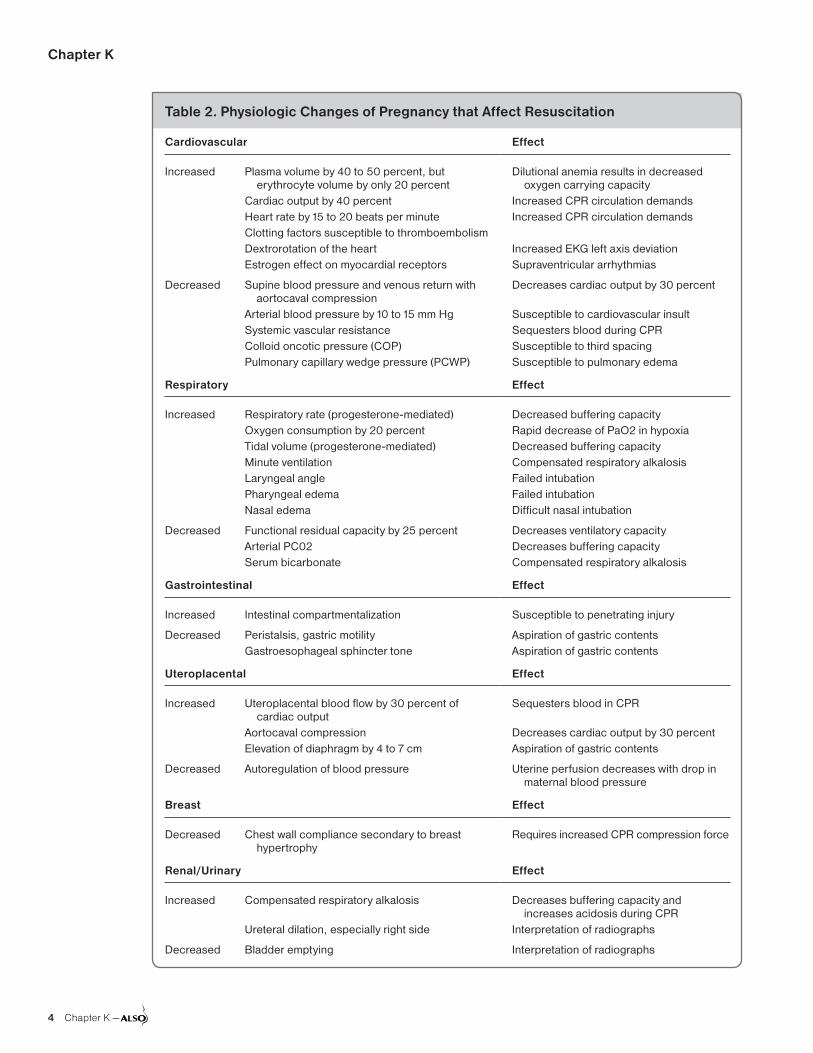
Chapter K
4 Chapter K —
Table 2. Physiologic Changes of Pregnancy that Affect Resuscitation
Cardiovascular Effect
Increased Plasma volume by 40 to 50 percent, but erythrocyte volume by only 20 percent
Cardiac output by 40 percentHeart rate by 15 to 20 beats per minute Clotting factors susceptible to thromboembolismDextrorotation of the heartEstrogen effect on myocardial receptors
Dilutional anemia results in decreased oxygen carrying capacity
Increased CPR circulation demands Increased CPR circulation demands
Increased EKG left axis deviation Supraventricular arrhythmias
Decreased Supine blood pressure and venous return with aortocaval compression
Arterial blood pressure by 10 to 15 mm Hg Systemic vascular resistanceColloid oncotic pressure (COP)Pulmonary capillary wedge pressure (PCWP)
Decreases cardiac output by 30 percent
Susceptible to cardiovascular insult Sequesters blood during CPR Susceptible to third spacing Susceptible to pulmonary edema
Respiratory Effect
Increased Respiratory rate (progesterone-mediated) Oxygen consumption by 20 percent Tidal volume (progesterone-mediated) Minute ventilationLaryngeal angle Pharyngeal edema Nasal edema
Decreased buffering capacity Rapid decrease of PaO2 in hypoxia Decreased buffering capacity Compensated respiratory alkalosis Failed intubationFailed intubation Difficult nasal intubation
Decreased Functional residual capacity by 25 percent Arterial PC02Serum bicarbonate
Decreases ventilatory capacity Decreases buffering capacityCompensated respiratory alkalosis
Gastrointestinal Effect
Increased Intestinal compartmentalization Susceptible to penetrating injury
Decreased Peristalsis, gastric motilityGastroesophageal sphincter tone
Aspiration of gastric contents Aspiration of gastric contents
Uteroplacental Effect
Increased Uteroplacental blood flow by 30 percent of cardiac output
Aortocaval compressionElevation of diaphragm by 4 to 7 cm
Sequesters blood in CPR
Decreases cardiac output by 30 percent Aspiration of gastric contents
Decreased Autoregulation of blood pressure Uterine perfusion decreases with drop in maternal blood pressure
Breast Effect
Decreased Chest wall compliance secondary to breast hypertrophy
Requires increased CPR compression force
Renal/Urinary Effect
Increased Compensated respiratory alkalosis
Ureteral dilation, especially right side
Decreases buffering capacity and increases acidosis during CPR
Interpretation of radiographs
Decreased Bladder emptying Interpretation of radiographs

Maternal Resuscitation and Trauma
— Chapter K 5
Fetal oxygen requirements: The fetus of an apneic and pulseless mother has two minutes or less of oxygen reserve because the oxygen tension in the umbilical vein is always less than the uterine vein. The chances for a successful resuscitation of the mother or fetus decrease after four minutes. Therefore the clinician has four minutes to affect the return of spontaneous circulation (ROSC) before she/he must begin dramatic action, hence the “four minute rule.”12,13
The best survival rate for the infant occurs when the infant is delivered no more than five minutes after the mother’s heart stops beat-ing.12,14 The American Heart Association’s (AHA) goal is to achieve delivery by five minutes.1,15,16 Typically this requires that the provider begin the hysterotomy about four minutes after maternal cardiac arrest.1,15,16
Resuscitation in PregnancyThe following discussion includes an overview of the principles of BLS and ACLS but pre-sumes a familiarity with the AHA algorithms and CPR.16,17 In 2010 the AHA changed the sequence of BLS steps from ‘airway — breathing — circu-lation’ (A-B-C) to ‘circulation - airway – breath-ing’ (C-A-B). ACLS in-hospital providers should tailor the sequence of rescue actions to the most likely cause of arrest.17 Modifications of technique due to changes in maternal physiology are sug-gested. (Table 3)1,15
The initial resuscitative maneuvers are the same regardless of pregnancy status and are referred to as the BLS Algorithm16,17:• Check responsiveness• Activate the emergency response system/Get
automated external defibrillator (AED)• Circulation: check carotid pulse• Defibrillation
Adult basic non-pregnant CPR should include18:Chest Compressions• providing chest compressions of adequate rate
(at least 100/minute)• providing chest compressions of adequate depth• a compression depth of at least two inches (5 cm)• allowing complete chest recoil after each
compression• minimizing interruptions in compressions• avoiding excessive ventilation
Airway and Ventilation• Deliver ventilations at a regular rate one breath
every six to eight seconds (8 to 10 breaths/min-ute) and chest compressions can be deliveredwithout interruption.
• If multiple rescuers are available, they shouldrotate the task of compressions every twominutes.
Table 3. Possible Modifications of Resuscitative Efforts in Pregnancy/Special Circumstances
Basic Life Support (BLS)
Action Rationale
Manual uterine displacement Decreases aortocaval compression
Increase chest wall compression force
Decreased chest wall compliance with breast hypertrophy and diaphragmatic elevation
Use cricoid pressure, if assistance is available
Decreases gastric aspiration
Perform compressions higher on sternum (slightly above center of sternum)
Elevated diaphragm and abdominal contents
Defibrillation: remove fetal or uterine monitors
Loss of adequate cardiac shock dose, produced skin burns at monitor sites
Advanced Cardiac Life Support (ACLS)
Action Rationale
Early tracheal intubation, use short laryngoscope handle and smaller endotracheal tube
Difficult ventilation with pharyngeal edema, breast hypertrophy, diaphragmatic elevation
Consider other etiologies (eg, magnesium toxicity)
Tocolytic therapy
Consider left wide paddle, adhesive pad, or breast displacement
Dextrorotation of heart, breast hypertrophy
Verify endotracheal tube with CO2 detector
Esophageal detector more likely to not re-inflate after a compression
Ventilation volumes and rates altered
Tailor ventilatory support to oxygenation and ventilation
Emergency hysterotomy after four minutes
Decreases aortocaval and venous compression
Remove both internal and external monitors
Clear patient for defibrillation
No Change
Defibrillation regimen Early return of effective maternal circulation
Pharmacologic therapy Early return of effective maternal circulation

Chapter K
6 Chapter K —
The above recommendations should be modi-fied to allow for the changes in maternal anatomy and physiology. [See Modifications for Pregnancy (below), Table 2, Table 3, Table 4]
If BLS is not successful, then begin the ACLS Algorithms1,16,17:• Airway: early use of advanced airway• Breathing: assist with ventilation and oxygen
supplementation• Circulation: chest compressions higher on sternum• Defibrillation (Figure 1)
The above recommendations and other modifi-cations (See Modifications for Pregnancy below), allow for the changes in maternal anatomy and physiology (Table 2, Table 3).
Figure 1 outlines the AHA Cardiac Arrest Algorithm.1,16,17 Further discussion of other clini-cal scenarios are available from the AHA, e.g., bradycardia, tachycardia, pulmonary edema, acute MI, hypothermia, and acute stroke.
Complications of CPR/ResuscitationPregnant women are more susceptible to rib fractures and other iatrogenic injuries including liver lacerations and pneumothorax. Hence, a high index of suspicion is warranted looking for any of these complications. In the face of underlying toxemia, thrombocytopenia may predispose to bleeding and hematomas of the liver.
Modifications of CPR In PregnancyAs summarized in Table 2, many unique aspects of maternal physiology influence the conduct of resuscitative maneuvers in pregnancy. In order to prevent aortocaval compression by the uterus after
20 weeks, the uterus should be manually moved off the aorta, vena cava, and pelvic great vessels.
Manual Left Uterine Displacement (LUD)In a systematic review it was noted that chest compressions were less forceful when a patient was in full-body left lateral tilt compared to the supine position.19 Furthermore, manual LUD has been found to reduce the incidence of hypoten-sion and ephedrine requirements when compared to 15 degree left-lateral tilt in patients under-going routine cesarean delivery.20 Based on these find-ings and the 2015 American Heart Association recommendations for cardiac arrest in pregnancy, left lateral tilt, which may be accomplished with a back board or Cardiff wedge, is no longer a recom-mend method of uterine displacement.21 Manual LUD provides an alternative technique for aorto-caval decompression, whereby at the same time the patient can remain supine and receive concurrent higher quality chest compressions while not delay-ing the onset of effective chest compressions by a need to facilitate a total body tilt. Manual LUD may also allow for easier delivery of defibrillation, IV access, and intubation. Two common methods include the two handed technique (Figure 2) and the one handed technique (Figure 3).
Several methods can be utilized to improve the effectiveness of chest compressions:1. Manually provide LUD, e.g., displace the
uterus leftward (Figures 2 and 3)2. Chest compressions more cephalad on sternum3. Emergency hysterotomy (perimortem cesarean
delivery)Despite all the maneuvers, CPR in pregnancy
should NOT be considered effective circulation, hence the need to relieve the aortocaval compres-sion (Table 4). If there has not been ROSC by four minutes of ACLS, then aim for delivery by emergency hysterotomy within 5 minutes of resus-citative efforts (Table 5).1,16
Trauma in PregnancyTrauma complicates one in 12 pregnancies, and is the leading non-obstetric cause of death among pregnant women.22-25 The most common traumatic injuries to pregnant women are: motor vehicle collisions (48 percent), assaults (17 per-cent), including intentional [suicides (3.3 percent), intimate partner violence, homicide, gunshot wounds (4 percent)] and non-intentional injuries,
Table 4. Cardiac Arrest in the Pregnant Patient
Activate cardiac arrest team
Lay patient in supine position and manually displace the uterus leftward
Begin chest compressions higher on the sternum
Use 100% oxygen when ventilating–secure airway early in the resuscitation
Remove both fetal and/or uterine monitors
Give typical ACLS drugs and doses
Entire Team Should Prepare for Possible Perimortem Cesarean Delivery
If no return of spontaneous circulation (ROSC) after four minutes of resuscitation, consider perimortem cesarean delivery
Delivery should occur within five minutes of the beginning of the resuscitation if no ROSC

Maternal Resuscitation and Trauma
— Chapter K 7
Figure 1. Pregnant Adult Arrest Algorithm
ACLS = advanced cardiac life support; BLS = basic life support; CPR = cardiopulmonary resuscitation; ETT = endotracheal tube; IO = intraosse-ous; IV = intravenous; LUD = left uterine displacement; ROSC = return of spontaneous circulation.
Adapted from Jeejeebhoy FM, Zelop CM, Lipman S, et al; American Heart Association Emergency Cardiovascular Care Committee, Council on Cardiopulmonary, Critical Care, Perioperative and Resuscitation, Council on Cardiovascular Diseases in the Young, and Council on Clinical Car-diology. Cardiac Arrest in Pregnancy: A Scientific Statement From the American Heart Association. Circulation. 2015;132(18):1747-1773.
BLS team actions ongoing
High-quality chest compressions
Defibrillation when indicated
Monitor CPR quality
If no ROSC by 4 minutes of resuscitative efforts, consider performing immediate emergency cesarean delivery
Neonatal team:Prepare to receive infant
Prepare for emergency cesarean delivery
Remove/detach fetal monitors
Continuous manual LUD
If patient receiving IV magnesium prearrest, discontinue magne-sium and administer IV/IO calcium chloride 10 mL in 10% solution or calcium gluconate 30 mL in 10% solution
IV above diaphragm
Give typical ACLS drugs and dosages
Assess for hypovolemia and administer fluid bolus +/- blood products when required
Appropriate airway managementb
Anticipate the difficult airway
Most experienced clinician preferred
Consider etiology of cardiac arresta
Obstetric interventions for patient whose uterus is at or above the umbilicusa
Maternal interventions
Maternal cardiac arrest team arrives
aPotential etiology of maternal cardiac arrest:
Anesthetic complications/ accidents
Bleeding
Cardiovascular
Drugs
Embolic
Fever
General nonobstetric causes of cardiac arrest (H’s and T’s)
Hypertension
bAppropriate airway management for pregnancy:
100% oxygen at ≥15 L/min and continue BLS airway strategies
Optimally two attempts per technique:
• First intubation attempt: if failed, go to
• Second intubation attempt: if failed, go to
• First supraglottic airway attempt: if failed, go to
• Second supraglottic airway attempt: if failed, go to mask ventilation
• If mask ventilation is inadequate: attempt cricothyrotomy
Avoid airway trauma
Ventilate with 8 to 10 breaths/min
Monitor capnography
Minimize interruptions in chest compressions dur-ing advanced airway placement
Recommend 6- to 7-mm inner diameter ETT

Chapter K
8 Chapter K —
falls (25 percent), poisonings, and burns (2.7 per-cent).25,26 Nine out of 10 traumatic injuries during pregnancy are minor. However, 60 to 70 percent of fetal losses are a result of minor injuries.26 Trauma can also be divided into minor and major trauma. Minor trauma (e.g. a cut to the arm, or a twisted ankle without a fall) does not involve the abdomen/uterus, rapid compression, deceleration or shearing forces, and the patient does not report pain, vaginal bleeding, loss of fluid and has good fetal movement.27
Physicians, regardless of their involvement in intrapartum care, frequently see pregnant women in the emergency department, urgent care, or in their offices with minor accidents, falls or motor vehicle accidents. These physicians should be aware of the differences in CPR for pregnant patients and the technique of perimortem cesarean delivery for pregnant patients in cardiopulmonary arrest.
This section includes a review of the anatomic and physiologic changes of pregnancy important in evaluating and treating pregnant trauma victims. The evaluation and treatment of major trauma will be reviewed, and an evaluation protocol for preg-nant women who suffer blunt abdominal trauma and falls is offered. This section concludes with a review of injuries sustained by pregnant women in motor vehicle crashes and from assault.
Anatomy and Physiology Relating to TraumaMany anatomic and physiologic changes of preg-nancy relate to the occurrence, diagnosis, and management of trauma.9,25,28 During the first trimes-ter, the thick walled uterus is well protected from trauma by the pelvic girdle. In the second trimester, relatively abundant amniotic fluid volume protects the fetus. By the third trimester, the now thin-walled and prominent uterus is subject to potential blows, penetration, or rupture. As the pregnancy approaches term, the relative fluid volume is decreased, which reduces the cushioning effect around the fetus. The fetal head at this point in gestation is usually protected within the bony pelvis.
The placenta is an inelastic organ attached to an elastic organ (the uterus). Accelerations or decelerations may deform the uterus and shear the placenta off of its implantation site, creating an abruption. The risk of abruption is independent of the placental location.
Gastrointestinal: Gastric emptying time is prolonged during pregnancy. As such, the cli-
Figure 2. Two Handed Manual Left Uterine Displacement Technique
Figure 3. One Handed Manual Left Uterine Displacement Technique

Maternal Resuscitation and Trauma
— Chapter K 9
nician should always assume the stomach of a pregnant patient is full. Early gastric tube decom-pression should be considered. The intestines are relocated to the upper part of the abdomen and may be shielded by the uterus. Signs of peritoneal irritation, such as distention, rebound tenderness, guarding, and rigidity are frequently detected on examination after trauma, but may be less pro-nounced during pregnancy.
Urinary: Renal blood flow and the glomerular filtration rate are increased in pregnancy. Blood urea nitrogen and serum creatinine levels are decreased. Glycosuria is common, due to a low-ered threshold of excretion. There may be bilateral or unilateral hydronephrosis with ureteral dilation. These changes may affect interpretation of lab and x-ray studies in trauma.
Major TraumaWhen major trauma occurs in pregnancy, evalua-tion and treatment of the mother is the first prior-ity. This approach also serves the best interests of the fetus. The diagnosis and management of the pregnant woman who has experienced major trauma does not differ significantly from the care of a non-pregnant trauma victim, except for rec-ognition of, and adjustment for, the anatomic and physiologic changes of pregnancy. The Advanced Trauma Life Support, Student Course Manual (9th Edition)9 outlines this Trauma Primary Survey:
A Airway maintenance with cervical spine protection
B Breathing and ventilation
C Circulation with hemorrhage controlD Disability: neurologic statusE Exposure/Environmental control: Completely
undress the patient, but prevent hypothermiaThe Trauma Primary Survey of resuscitation
hold true in pregnancy and are, in fact, more important because of the risks to the fetus from maternal hypotension and hypoxia.
The Primary Maternal Survey (Table 6) addresses life support and resuscitation. Fluid resuscitation should be pursued very aggres-sively.28 A pregnant patient can lose a significant amount of her blood volume before she dem-onstrates hypotension and other signs of shock. Crystalloid and early type-specific packed red blood cells are indicated to restore the physiologic hypervolemia of pregnancy. The mother should be returned to the left lateral position after a thor-ough physical exam.
The primary fetal survey (Table 7) is pursued after initial assessment and stabilization of the mother. The major diagnostic details relating to the fetal primary survey are: fetal viability and
Table 6. Primary Maternal Survey
Patent airway
Adequate ventilation and oxygenation
Effective circulatory volume
Fluid support
Blood replacement
Table 5. Steps in Performing an Emergency Hysterotomy
Initiate immediate CPR and ACLS with manual lateral uterine displacement.
Initiate hysterotomy after four minutes of ACLS and no ROSC.
Attempt procedure with an obviously gravid uterus.*
Prepare equipment/personnel for emergency hysterotomy and neonatal resuscitation. Avoid delays for fetal heart tones or waiting for an obstetrician.
The first skilled clinician available should initiate the hysterotomy.
Wear appropriate personal protective equipment to protect health personnel from transmission of infection.
Perform hysterotomy with a Joel-Cohen method or vertical midline skin incision, and a vertical uterine incision. (Refer to Chapter Q -Cesarean Delivery) Use modified sterile technique, e.g., ‘crash’. Give infant to attendant for drying and warming and/or resuscitation.
Pack the uterus with moist sponges. Remove lateral tilt. Continue ACLS throughout.
When hemodynamically stable, remove the placenta and close uterus with #0 absorbable suture. Close anatomically, depending on available personnel and location. Obtain hemostasis with interrupted 0 absorbable suture.
*An obviously gravid uterus is clinically deemed large enough to cause aortocaval compression which correlates to approximately 20 week size or a fundal height at or above the umbilicus1,15,16

Chapter K
10 Chapter K —
well-being, likelihood of fetal injury and feto-maternal transfusion, gestational age, abruption, premature labor, rupture of membranes, presenta-tion and uterine rupture.
The maternal secondary survey (Table 8) is not different from non-pregnant patients. Indi-cations for abdominal computed tomography (CT), focused assessment sonography in trauma (FAST), and diagnostic peritoneal lavage (DPL) have the same indications as in non-pregnant patients. If DPL is performed, the catheter should be placed above the umbilicus, using an open technique. Indicated x-ray studies can be per-formed without concern for radiation injury to the fetus, as the immediate diagnostic benefits far outweigh any theoretical risk to the fetus.29 How-ever, shielding the fetus and minimizing exposure is prudent when possible.
The use of abdominal ultrasound by a skilled operator should be considered in detecting preg-nancy and possible causes of the cardiac arrest, but this should not delay other treatments. Ultrasound provides fetal assessment, e.g., amniotic fluid volume, fetal presentation, estimated gestational age, and is occasionally helpful in cases of anterior
abruption. Pelvic examination can be performed after documentation of placental location.
Laboratory testing of severely injured patients should include the same tests done for non-preg-nant patients. Monitoring with a central venous pressure line can be extremely helpful, as is mea-suring the urine output.
Coagulopathy can accompany abruption, amniotic fluid embolism, and other obstetric events relating to trauma, so measurement of fibrinogen, platelet count, and fibrin split products (D-Dimer) are recommended. The fibrinogen level may double in late pregnancy. A normal fibrinogen level may indicate early disseminated intravascular coagulation (DIC) which can be seen with abruptio placenta.9
Definitive Care: Certain maternal injuries, such as visceral injury or retroperitoneal hemorrhage, require immediate surgery to save the life of the mother and the fetus. Uterine rupture and abrup-tion threaten the baby more directly. Uterine rupture manifests by signs of hypovolemia and hemoperitoneum. The fetus will often be acidotic or dead, and may appear on x-ray or ultrasound exam with extended limbs or in a bizarre position. Abruption presents with classical signs: contrac-tions, rigid and tender uterus, expanding uterine height, vaginal bleeding, abnormal fetal heart rate tracing or demise, and coagulopathy. Both condi-tions require surgery for mother and fetus alike.
Appropriate imaging studies should not be avoided due to the presence of the fetus. Typically concerns about ionizing radiation and fetal harm begin somewhere between 5,000 and 10,000 milli-rads, or 5 to 10 rads.28,29 The average CT scan of the abdomen and pelvis is in the order of 2 to 2.5 rads.
After management of the primary trauma sur-vey, two large bore intravenous catheters should be placed and supplemental oxygen adminis-tered to maintain a saturation of greater than 95 percent. Aggressive transfusion of blood products may provide volume and improve oxygen carrying capacity.9 The use of pressors should be avoided until adequate volume replacement has been com-pleted. The uterus should be displaced from the vena cava and aorta. Following maternal stabiliza-tion, the secondary maternal and fetal survey can be completed.
Fetal injuries are highly variable. Skull and brain injuries are common when the maternal pelvis is fractured and the fetal head is engaged.
Table 8. Secondary Maternal Survey
X-rays
Focused assessment sonography in trauma
Diagnostic peritoneal lavage
Central venous pressure (CVP)
Urine output
Baseline laboratory to include: Serum bicarbonateKleihauer BetkeCoagulation factors with fibrinogen level
Table 7. Primary Fetal Survey
Fundal height
Uterine activity
Fetal heart rate pattern and movement
Assess for vaginal bleeding
Assess for ruptured membranes
Assess cervix for dilation and effacement
Maternal Resuscitation and Trauma
— Chapter K 11
Fetal contrecoup injuries may occur. Penetrating injuries may be present from gunshot or knife attacks. Although penetrating injuries have a lower maternal mortality in pregnancy, the fetal mor-bidity and mortality is significantly higher.24 This may occur because the gravid uterus is acting as a ‘shield’ to the maternal organs. Maternal mortality from penetrating wounds is less than four percent in pregnancy.24 On the other hand, 60 to 90 per-cent of stab or gunshot wounds to the abdomen result in fetal injury or death.30 Surgical explora-tion of the maternal abdomen is almost always necessary in cases of penetrating abdominal injury.
Routine administration of Rh Immune Globulin is indicated for unsensitized Rh nega-tive women with significant abdominal trauma. Indications for tetanus prophylaxis do not change in pregnancy and appropriate candidates should be vaccinated.
The Kleihauer Betke (KB) assay should be considered on patients following significant blunt uterine trauma, regardless of Rh status, in order to assess the degree of fetomaternal hemorrhage.31-35 In pregnant trauma patients with an Injury Sever-ity Score > 2 (Appendix 1), a positive KB is an effective predictor of adverse perinatal outcomes, particularly with more severe trauma.36,37
The mean estimated blood volume of injected fetal blood is usually less than 15 ml, and in more than 90 percent of patients, is less than 30 ml. One amp of Rh immune globulin, which is 300 ug, will cover a 15 ml red blood cell hemorrhage, or 30 ml total blood volume hemorrhage. Serial testing may be appropriate to assess ongoing hemorrhage.
Unfortunately the Kleihauer Betke assay is often not available in a timely fashion for acute clini-cal management. Fetal heart monitoring, uterine tocographic monitoring, or ultrasound evalu-ation may be more useful in the acute setting. An alternative approach has been suggested to expedite therapy by calculating the total blood volume of a fetus.38 First estimate the fetal weight by ultrasound, then multiply the estimated fetal weight in kilograms by the total blood volume of a fetus (approximately 100 mL/kg) to determine the maximum total dose of Rh immune globulin.38 Each 300 mcg Rhogam vial will cover a 30 mL total blood loss, so two vials would cover the total fetal blood loss for a 600 gram fetus. In the case of a 3.5 kg fetus, 3.5 kg times 100 mL/kg would produce an estimated total fetal blood volume
of 350 mL. This approach will overestimate the maternal exposure to fetal blood since only a limited amount of fetal blood usually will enter maternal circulation even in severe trauma.38
Emergency hysterotomy (cesarean delivery) may be required for several reasons. It may be difficult to treat the maternal condition around the gravid uterus; or the obstetric pathology contributes to the patient’s worsening condition (as in abruption with coagulopathy); or the fetus is acidotic. Emer-gency hysterotomy may improve the mother’s status, but may increase her risk of hypovolemia. After 23 to 24 weeks’ gestation emergency hyster-otomy may save the fetus, the second life at risk.
Minor TraumaThe most common trauma-related occurrence among pregnant women is a minor trauma, such as a fall, minor MVC, or blunt abdominal trauma, which cause little or no maternal injury. In minor cases, the clinician must often make a judgment about the necessity of examination or monitor-ing. Erring on the side of caution is recommended to alleviate anxiety and because of cases in which seemingly insignificant trauma has resulted in fetal injury or demise. Abruption usually becomes apparent shortly after injury, fetal monitoring in women who experience trauma beyond 20 weeks gestation should be initiated as soon as the patient is stabilized. The patient should be monitored by cardiotocograph for a minimum of four hours but may be extended to a six hour minimum.9,31 In 100 percent of patients found subsequently to have a placental abruption, eight or more contractions per hour were present in the first four hours.39
Monitoring should be continued for a mini-mum of 24 hours if there is presence of six uterine contractions per hour25,40 abnormal fetal heart rate patterns, vaginal bleeding, significant uterine tenderness, serious maternal injury or rupture of membranes occurs during the initial four to six hours monitoring period. If none of these concerning findings is present then the patient may be discharged with instructions to return if she develops vaginal bleeding, leakage of fluid, decreased fetal movement or severe abdominal pain.9,28 A guideline for managing minor trauma is presented in Table 9.
The presence of six uterine contractions per hour25,40 and fetal red blood cells in the mater-nal circulation are good indicators of fetal risk

Chapter K
12 Chapter K —
from abruption. Ultrasound has poor sensitivity (24 percent) for detection of abruptio placenta, however it is very specific (96 percent); resulting in a positive predictive value of 88 percent if abrup-tion is seen on ultrasound and a negative predic-tive value of 53 percent if abruption is not seen.41
Motor Vehicle CollisionsMotor vehicle collisions (MVC) accounted for 34,080 deaths and four million injuries in the United States in 2012 and are increasing.42 Two percent of pregnant women are involved in a MVC during their pregnancy.35 MVC pro-duce a disproportionate number of fetal deaths, e.g., MVC 82 percent versus 6 percent gunshotwounds and 3 percent falls.43,44
Seat belted pregnant women sustain death and severe injury at a rate of about half that of women who are not seat belted. Seat belt use actually drops during pregnancy, because women fear that the seat belt will hurt the baby. In blunt trauma patients, the most common cause of fetal death is maternal death.39 Women may also fear being trapped by a seat belt in a fire or under-water submersion when, in fact, such incidents are rare. The dangers related to ejection from the vehicle, or secondary collision within the vehicle, are far greater than being trapped. In any event, a conscious and uninjured occupant is more likely to unfasten a seat belt and escape from a burning or submerged car than an unbelted, but severely injured, occupant. Seat belt use should be a major issue of prenatal counseling in every pregnancy.
Proper use of seatbelts may be the best predictor of maternal and fetal outcome. There is an 85 per-
cent reduction in fetal morbidity and mortality, as well as serious fetal injury by wearing seatbelts.45 Incorrect seat belt use can contribute to intrauter-ine injury and fetal death.46 The lap belt should be placed as low as possible under the protuber-ant portion of the abdomen and the shoulder belt positioned off to the side of the uterus, between the breasts and over the midportion of the clavicle. Placement of the lap belt over the dome of the uterus significantly increases pressure transmission to the uterus and has been associated with signifi-cant uterine and fetal injury.
There should not be excessive slack in either belt, and both the lap and shoulder restraints should be applied as snugly as comfort will allow. Airbag deployment reduces injury to pregnant women. Pregnant occupants are not at increased risk of adverse pregnancy outcomes while traveling in a vehicle that is equipped with an air bag and crashes.47 The American College of Obstetricians and Gynecologists (ACOG)48 and the National Highway Traffic and Safety Administration49 both recommend that pregnant women who are occupants in motor vehicles wear lap and shoulder seatbelts and not turn off air bags.
Direct AssaultDirect assault on the abdomen can be a regrettable manifestation of intimate partner violence. Abused women are a frequently undetected high-risk group. ACOG and the U.S. Preventive Services Task Force (USPSTF) recommends universal screening for intimate partner abuse.50-53 Every provider should be alert to this possibility and may, indeed, uncover battering when a patient
Table 9. Management of Minor Trauma in Pregnancy
Interventions:Primary maternal and fetal surveyLab: blood type, Rh, hematocrit.
Kleihauer Betke (KB) test; coagulation studies
Consider obstetric ultrasoundIf greater than 20 weeks, monitor for
contractionsIf less than six contractions per hour
and no risk factors, monitor for six hours, then discharge9,25,40
If > 6 contractions per hour or risk factors, monitor for 24 hours
Risk factors9:Maternal heart rate greater than
110 bpmInjury Severity Score greater than
nine (Appendix 1)65
Evidence of placental abruptionFetal baseline heart rate greater than
160 bpm or less than 110 bpmEjection during motor vehicle
accidentMotorcycle or pedestrian collision
Discharge criteria:Resolution of contractionsNormal fetal heart tracingIntact membranesNo uterine tendernessNon vaginal bleedingAll Rh negative patients
receive Rh immune globulin full dose (300 mcg) (more if indicated by KB)

Maternal Resuscitation and Trauma
— Chapter K 13
presents with a vague or inconsistent history of trauma. Battering may escalate in pregnancy and the abdomen is the most frequent target for blows, kicks, and other assaults. Prenatal care should include a routine screening for intimate partner violence, and identified patients should be appro-priately counseled and referred. In patients who have experienced intimate partner violence (IPV), an assessment for depression and suicidality should accompany assessment for immediate safety.54
Indicators that suggest the presence of intimate partner violence include:
• Injuries inconsistent with the stated history• Diminished self-image, depression, or suicide
attempts• Self abuse• Frequent emergency department (ED) or pro-
vider’s office visits• Symptoms suggestive of substance abuse• Self-blame for injuries• Partner insists on being present for interview
and examination and monopolizes discussion
Case Scenario: JE ContinuedYou immediately scrubbed out of the cesarean delivery you were assisting with and find JE has stopped the tonic clonic movements, but is now non-responsive to painful stimuli and in respira-tory arrest. JE has a thready pulse at 180 bpm, blood pressure 40 over palpable. She is now ooz-ing serous blood from all her recent venipuncture sites, as well as the IV site. While you are assessing JE, she becomes pulseless.
You call for assistance and begin CPR with JE. You apply your chest compressions slightly above center of sternum. The patient is quickly intu-bated by a colleague from the emergency depart-ment, but JE does not respond to resuscitative efforts within four minutes.
You are a maternity provider without privileges to perform cesarean delivery as the primary sur-geon. The only cesarean delivery provider in your small rural hospital is still operating on a patient under general anesthesia and will be unavailable for the next 20 minutes.
What should do you do next?
Special Technique: Emergency Hysterotomy (Cesarean Delivery)Historically, perimortem cesarean delivery was performed to give birth to an infant whose mother
was dead to facilitate separate burials for the mother and infant. Emergency hysterotomy has re-emerged as a procedure to be performed after maternal cardiac arrest to increase the survival of the mother and fetus.12,13 The AHA recommends that if there has not been ROSC by four minutes of Advanced Cardiac Life Support (ACLS), then aim for delivery by emergency hysterotomy within five minutes of resuscitative efforts.1,15,16
Emergency hysterotomy is indicated when:• Personnel with appropriate skill and equipment
to perform the procedure are involved,• The mother fails to respond with a return of
spontaneous circulation within four minutes,• The mother is known to be at least 20 weeks
gestation or there is an obviously gravid uterus*• Appropriate facilities and personnel are avail-
able to care for the mother and infant after the procedure. (Table 10)*An obviously gravid uterus (i.e. at last 20 week
size or fundal height above the umbilicus) can be assumed to cause aortocaval compression and maternal benefit is anticipated with delivery of the fetus.1,15,16 Fetal benefit occur when gestational age reaches fetal viability at 23-24 weeks.
The best fetal survival rates are obtained when emergency hysterotomy is performed within five minutes of ineffective maternal circulation. There was 98 percent intact neurologic status in a cohort of 45 surviving infants delivered within five minutes of maternal death but this decreased to 33 percent of the nine infants surviving a deliv-ery that occurred 16 to 25 minutes after maternal death.55 It may still be worthwhile to pursue delivery after this period, as fetal mortality is 100 percent if no action is taken. Some infants have survived up to 20 to 30 minutes after ineffective circulation.12,13
Despite the recommendation for rapid initiation of hysterotomy there are rare documented cases of intact fetal survival as long as thirty minutes after maternal death as demonstrated by a recent case report of a woman who jumped out of a fourth floor window while in labor and a perimortem cesarean was performed thirty minutes after death was confirmed.56
A systematic review found that the predictors of maternal survival for those patients undergoing emergency hysterotomy were in-hospital loca-tion arrest and hysterotomy occurring less than 10 minutes after maternal arrest.57
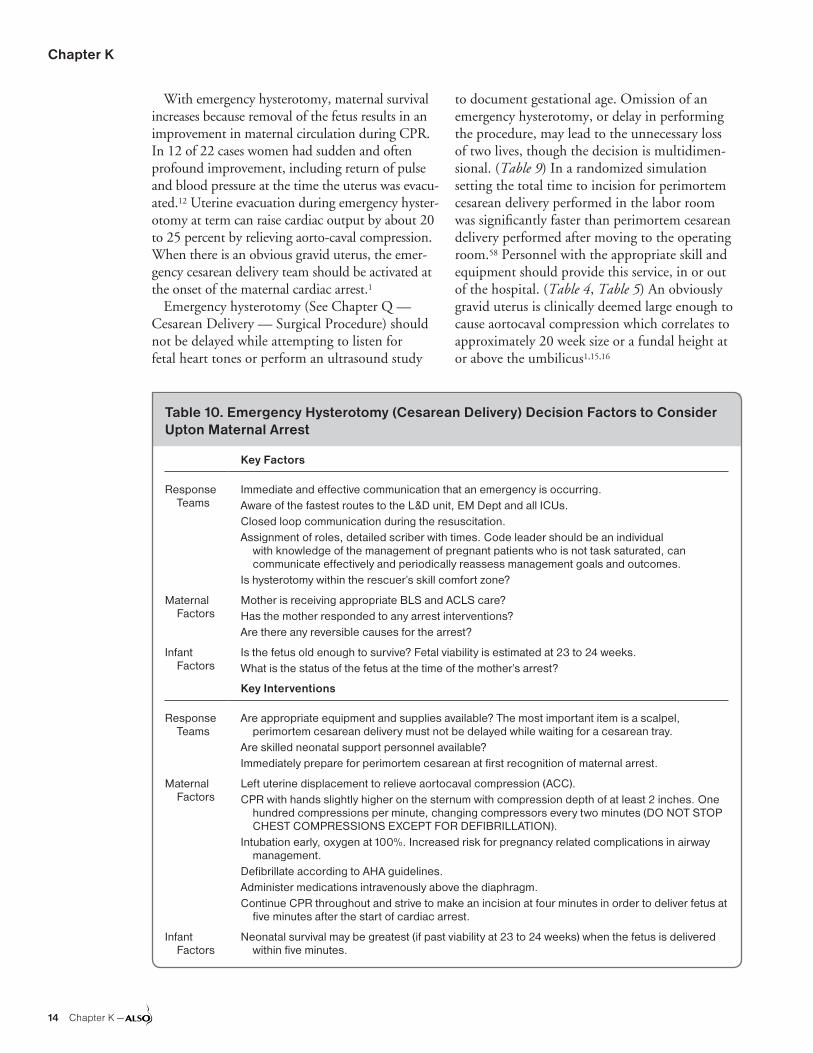
Chapter K
14 Chapter K —
With emergency hysterotomy, maternal survival increases because removal of the fetus results in an improvement in maternal circulation during CPR. In 12 of 22 cases women had sudden and often profound improvement, including return of pulse and blood pressure at the time the uterus was evacu-ated.12 Uterine evacuation during emergency hyster-otomy at term can raise cardiac output by about 20 to 25 percent by relieving aorto-caval compression. When there is an obvious gravid uterus, the emer-gency cesarean delivery team should be activated at the onset of the maternal cardiac arrest.1
Emergency hysterotomy (See Chapter Q — Cesarean Delivery — Surgical Procedure) should not be delayed while attempting to listen for fetal heart tones or perform an ultrasound study
to document gestational age. Omission of an emergency hysterotomy, or delay in performing the procedure, may lead to the unnecessary loss of two lives, though the decision is multidimen-sional. (Table 9) In a randomized simulation setting the total time to incision for perimortem cesarean delivery performed in the labor room was significantly faster than perimortem cesarean delivery performed after moving to the operating room.58 Personnel with the appropriate skill and equipment should provide this service, in or out of the hospital. (Table 4, Table 5) An obviously gravid uterus is clinically deemed large enough to cause aortocaval compression which correlates to approximately 20 week size or a fundal height at or above the umbilicus1,15,16
Table 10. Emergency Hysterotomy (Cesarean Delivery) Decision Factors to Consider Upton Maternal Arrest
Key Factors
Response Teams
Immediate and effective communication that an emergency is occurring.Aware of the fastest routes to the L&D unit, EM Dept and all ICUs.Closed loop communication during the resuscitation.Assignment of roles, detailed scriber with times. Code leader should be an individual
with knowledge of the management of pregnant patients who is not task saturated, can communicate effectively and periodically reassess management goals and outcomes.
Is hysterotomy within the rescuer’s skill comfort zone?
Maternal Factors
Mother is receiving appropriate BLS and ACLS care?Has the mother responded to any arrest interventions?Are there any reversible causes for the arrest?
Infant Factors
Is the fetus old enough to survive? Fetal viability is estimated at 23 to 24 weeks.What is the status of the fetus at the time of the mother’s arrest?
Key Interventions
Response Teams
Are appropriate equipment and supplies available? The most important item is a scalpel, perimortem cesarean delivery must not be delayed while waiting for a cesarean tray.
Are skilled neonatal support personnel available?Immediately prepare for perimortem cesarean at first recognition of maternal arrest.
Maternal Factors
Left uterine displacement to relieve aortocaval compression (ACC).CPR with hands slightly higher on the sternum with compression depth of at least 2 inches. One
hundred compressions per minute, changing compressors every two minutes (DO NOT STOP CHEST COMPRESSIONS EXCEPT FOR DEFIBRILLATION).
Intubation early, oxygen at 100%. Increased risk for pregnancy related complications in airway management.
Defibrillate according to AHA guidelines.Administer medications intravenously above the diaphragm.Continue CPR throughout and strive to make an incision at four minutes in order to deliver fetus at
five minutes after the start of cardiac arrest.
Infant Factors
Neonatal survival may be greatest (if past viability at 23 to 24 weeks) when the fetus is delivered within five minutes.

Maternal Resuscitation and Trauma
— Chapter K 15
Consent from family members before perform-ing the procedure is helpful, but not necessary. It is the responsibility of the health care provider to perform an emergency hysterotomy if the above criteria are met. As long ago as 1931, Campbell and Miller concluded that “a civil action for damages might follow for the negligence or the malpractice of the surgeon or physician in failing to follow the usual and customary practice” in regard to emergency hysterotomy.59 The surgical team should be prepared to care for the newborn infant. A newborn can lose 30 percent of the avail-able energy reserve in the first five minutes in a cold moist environment, hence immediate drying and warming is indicated. Table 11 provides a list of instruments helpful to perform emergency hysterotomy.
If those instruments are not available, a scal-pel to conduct the delivery and a blanket for the infant are the items that are immediately neces-sary. Antibiotic and wound management will be necessary if the mother survives.
Avoid chaos. Perform practice drills with all clinical stakeholders.
Some medical conditions indicate a need for simultaneous CPR and hysterotomy, e.g., mitral
and aortic stenosis, cardiomyopathy, pericardial disease, core temperature less than 34 degrees Fahrenheit, pulmonary/cardiac injury or disease, and carbon monoxide poisoning. If the pregnant patient’s hemodynamic status is stabilized, then “the four minute rule” is not applicable. Restora-tion of maternal function sufficient to maintain the pregnancy, even if recovery is incomplete, may be of value for the fetus and the family. This is especially important the more remote from term the patient is. Even patients sustaining irreparable trauma deserve attentive assessment and institu-tion of life support.
Amniotic Fluid EmbolismAmniotic fluid embolism (AFE) is rare, with an estimated incidence of 1:15,200 and 1:53,800 deliveries in North America and Europe, respec-tively.60 In the past, the maternal mortality rate from this complication was initially estimated at 85 percent, with half of the deaths occurring in the first hour.61 With the advent of intensive care units, population-based data now suggests that the case fatality rate and perinatal mortality associated with AFE are 13 to 30 percent and 9 to 44 per-cent, in North America and Europe respectively.60 AFE remains one of the most catastrophic condi-tions that can befall the pregnant patient. AFE is a catastrophe for the fetus as well, with neurologi-cally intact survival, in only 39 percent.61
Predisposing Factors and PathophysiologyRisk factors associated with an increased risk of AFE include advanced maternal age, placental abnormalities, operative deliveries, eclampsia, polyhydramnios, cervical lacerations, and uterine rupture.60 Since the first description of AFE in 1941 it has been thought of as a mechanical event in which a bolus of amniotic fluid enters the sys-temic circulation due to tetanic contraction, moves through the pulmonary circuit, produces a massive perfusion failure, bronchospasm and shock.62 The density of particulate matter in the fluid was felt to be related to the lethality of the event.
The number of cases failing to fit this picture led to a reconsideration of the pathophysiology, which suggests that the syndrome can occur with simple exposure to small amounts of amniotic fluid. A good case can be made to include AFE in a group of “anaphylactoid syndromes” occurring in late pregnancy or labor. Pathophysiologic studies sug-
Table 11. Emergency Hysterotomy Kit
Obstetric SuppliesKnife handle, #10 scalpel blade Bladder blade retractorBandage and dissecting scissors Sterile laparotomy sponges Abdominal retractorsHeaney clamps Hemostats Ring forceps Cord clamps Needle holdersToothed and non-toothed dissecting forceps or
pickups0 Chromic sutures One Vicryl sutures Sterile gloves
Pediatric SuppliesNeonatal blanketsSelf-inflating resuscitation bag: Infant, child Resuscitation masks: Neonate, infant DeLee suction trapBulb syringe

Chapter K
16 Chapter K —
gest that left heart failure and pulmonary vaso-spasm are prime etiologic factors in cardiovascular collapse, but that the underlying mechanism may be an anaphylactic-like event with an associated 41 percent incidence of atopy or allergy.61 The hemodynamic response in AFE is biphasic, with initial pulmonary hypertension and right ventricu-lar failure, followed by left ventricular failure.60 Disseminated intravascular coagulopathy is the most common complication, possibly due to the large amount of tissue factor in amniotic fluid.
Clinical PictureThe clinical picture develops with stunning rapid-ity. The syndrome begins with respiratory distress (tachypnea and dyspnea) accompanied by restless-ness, cyanosis, and sometimes nausea and vomit-ing. Seizures are common, occurring in 50 percent of women.63 Unexpected cardiovascular collapse occurs and profound DIC may follow. Finally, the patient goes into a coma and dies. In many cases, these events progress quickly so that only the most rudimentary diagnostic studies and resuscitative efforts can be made.
DiagnosisDefinitive diagnosis of this condition is gener-ally made post-mortem. Clinical diagnosis relies primarily on clinical observations. If time permits, helpful laboratory values include blood gases and coagulation factors. The differential diagnosis includes other catastrophic causes of cardiopul-monary compromise, such as massive pulmonary embolism, bilateral pneumothorax, myocardial infarction, or gastric fluid aspiration. Obstetric conditions that mimic AFE include severe abrup-tion, uterine rupture, inverted uterus and uterine atony. In specific circumstances, consider septic shock and eclampsia.
ManagementThe management of AFE is largely supportive. An aggressive medical approach seems justified and certainly can do no harm for the patients who survive the initial catastrophic event.
When an obstetric patient collapses unexpect-edly, the BLS algorithm and then the appropri-
ate ACLS algorithm should be attended to, with the airway secured and ventilation assured, using endotracheal intubation if necessary. Oxygen should be administered at 100 percent. Two large bore intravenous catheters should be placed and aggressive fluid replacement using crystalloid solu-tion begun. Pressor agents, such as dopamine will likely be required.
Blood should be drawn for complete hemogram, coagulation panel, and chemistry panel, includ-ing electrolytes and renal function. Arterial blood gases should be obtained. Urinary output should be monitored via indwelling Foley catheter, and a portable chest x-ray and 12-lead electrocardiogram should be obtained. A cardiac monitor should be applied and ACLS initiated. Hemodynamic moni-toring will probably be required, using an arterial access line and possibly a Swan-Ganz catheter.
Coagulation factors should be assessed every two hours and blood component therapy initiated as needed with packed red cells, platelets (if the platelet count is less than 50,000/mcL), fresh fro-zen plasma, or cryoprecipitate. Given the possible anaphylactoid nature of the condition, epineph-rine is theoretically useful. Positive end expiratory pressure is usually required to prevent alveolar collapse and to recruit atelectatic alveoli. Fluids, dopamine and furosemide should be administered based on hemodynamic parameters.
SummaryThe rate of cardiac arrest in pregnancy appears to have increased and is now estimated to occur in one in 20,000 pregnancies.1 With 44 to 50 percent preventable maternal deaths, the next challenge will be to improve maternal mortality one pregnant patient at a time. CPR and ACLS modified for maternal physiology, prompt diagno-sis, and emergency hysterotomy provide important tools to meet that challenge.17,18
Regardless of the cause of the maternal col-lapse, your facility should perform regular drills to assure that your entire labor and delivery staff is functioning with the same set of assumptions.64 Documentation should be thorough, but fact focused and non-speculative in these emotionally and medicolegally charged scenarios.38

Maternal Resuscitation and Trauma
— Chapter K 17
SORT: Key Recommendations for Practice
Clinical RecommendationEvidence Rating References
Perimortem cesarean delivery should be performed in the labor room rather than relocating to the operating room.
A 58
Pregnant women who experience trauma beyond 20 weeks’ gestation should be monitored by cardiotocograph for a minimum of four hours.
B 9,31
When there is an obvious gravid uterus, the emergency cesarean delivery team should be activated at the onset of the maternal cardiac arrest.
B 1
Screen women of childbearing age for IPV, such as domestic violence, and provide or refer women who screen positive to intervention services.
B 51-53
Screening for fetomaternal transfusion (e.g Kleihauer Betke) should occur in pregnant women who suffer significant blunt uterine trauma beyond 11 weeks’ gestation.
B 31-37
To relieve aortocaval compression during chest compressions and optimize the quality of CPR, it is reasonable to perform manual left uterine displacement.
C 1,19
Emergency hysterotomy within four minutes of maternal cardiac arrest improves maternal and neonatal outcomes.
C 1,13
Strength of Recommendation Taxonomy (SORT)
Strength of Recommendation Definition
A • Recommendation based on consistent and good-quality patient-oriented evidence.
B • Recommendation based on inconsistent or limited-quality patient-oriented evidence.
C • Recommendation based on consensus, usual practice, opinion, disease-oriented evidence, or case series for studies of diagnosis, treatment, prevention, or screening.
Patient-oriented evidence measures outcomes that matter to patients: morbidity, mortality, symptom improvement, cost reduction, and quality of life. Disease-oriented evidence measures intermediate, physiologic, or surrogate end points that might not reflect improvement in patient outcomes (eg, blood pressure, blood chemistry, physiologic function, pathologic findings).
From Ebell MH, Siwek J, Weiss BD, et al. Strength of recommendation taxonomy (SORT): a patient-centered approach to grading evidence in the medical literature. Am Fam Physician. 2004;69(3):548-556.
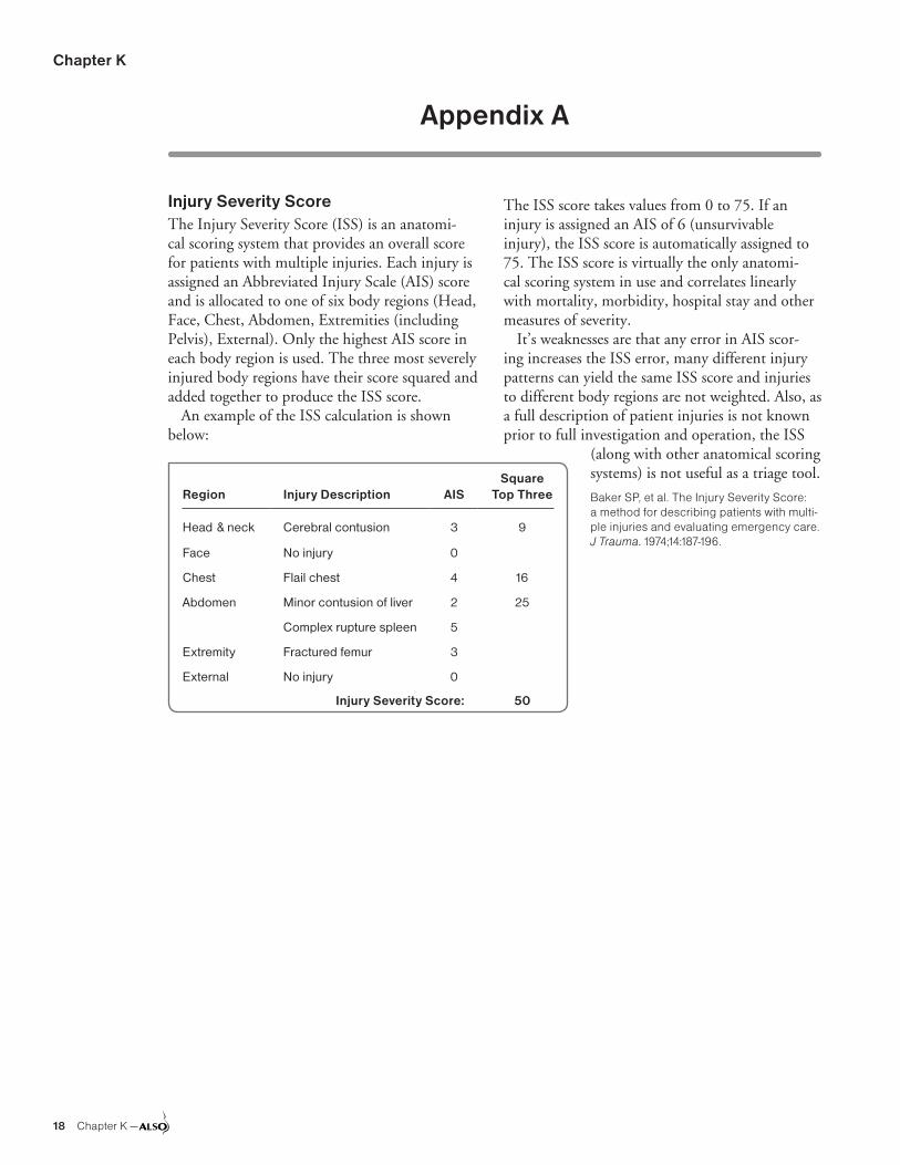
Chapter K
18 Chapter K —
Appendix A
Injury Severity ScoreThe Injury Severity Score (ISS) is an anatomi-cal scoring system that provides an overall score for patients with multiple injuries. Each injury is assigned an Abbreviated Injury Scale (AIS) score and is allocated to one of six body regions (Head, Face, Chest, Abdomen, Extremities (including Pelvis), External). Only the highest AIS score in each body region is used. The three most severely injured body regions have their score squared and added together to produce the ISS score.
An example of the ISS calculation is shown below:
The ISS score takes values from 0 to 75. If an injury is assigned an AIS of 6 (unsurvivable injury), the ISS score is automatically assigned to 75. The ISS score is virtually the only anatomi-cal scoring system in use and correlates linearly with mortality, morbidity, hospital stay and other measures of severity.
It’s weaknesses are that any error in AIS scor-ing increases the ISS error, many different injury patterns can yield the same ISS score and injuries to different body regions are not weighted. Also, as a full description of patient injuries is not known prior to full investigation and operation, the ISS
(along with other anatomical scoring systems) is not useful as a triage tool.
Baker SP, et al. The Injury Severity Score: a method for describing patients with multi-ple injuries and evaluating emergency care. J Trauma. 1974;14:187-196.
Region Injury Description AISSquare
Top Three
Head & neck Cerebral contusion 3 9
Face No injury 0
Chest Flail chest 4 16
Abdomen Minor contusion of liver 2 25
Complex rupture spleen 5
Extremity Fractured femur 3
External No injury 0
Injury Severity Score: 50

Maternal Resuscitation and Trauma
— Chapter K 19
References1. Vanden Hoek TL, Morrison LJ, Shuster M, et al. Part
12: cardiac arrest in special situations: 2010 American Heart Association Guidelines for Cardiopulmonary Resuscitation and Emergency Cardiovascular Care. Cir-culation. 2010;122(18)(Suppl 3):S829-S861.
2. Department of Health, Welsh Office, Scottish OfficeDepartment of Health, Department of Health and SocialServices, Northern Ireland. Why mothers die. Reporton confidential enquiries into maternal deaths in theUnited Kingdom 2000–2002. London, England: TheStationery Office; 2004.
3. Lim Y, Loo CC, Chia V, Fun W. Recombinant factorVIIa after amniotic fluid embolism and disseminatedintravascular coagulopathy. Int J Gynaecol Obstet.2004;87(2):178-179.
4. Poole JH, Long J. Maternal mortality—a review of currenttrends. Crit Care Nurs Clin North Am. 2004;16(2):227-230.
5. Atta E, Gardner M. Cardiopulmonary Resuscitation inPregnancy. Obstet Gynecol Clin N Am. 2007;34:585-597.
6. Cantwell R, Clutton-Brock T, Cooper G, et al. SavingMothers’ Lives: Reviewing maternal deaths to makemotherhood safer: 2006-2008. The Eighth Report ofthe Confidential Enquiries into Maternal Deaths in theUnited Kingdom. BJOG. 2011;118(Suppl 1):1-203.
7. Kuklina E, Callaghan W. Chronic heart disease andsevere obstetric morbidity among hospitalisations forpregnancy in the USA: 1995-2006. BJOG. 2011;118(3):345-352.
8. Ueland K, Akamatsu TJ, Eng M, Bonica JJ, Hansen JM.Maternal cardiovascular dynamics. VI. Cesarean sec-tion under epidural anesthesia without epinephrine. AmJ Obstet Gynecol. 1972;114(6):775-780.
9. American College of Surgeons. Trauma in Pregnancyand Intimate Partner Violence. In: Advanced TraumaLife Support Student Course Manual, Ninth Edition. Chi-cago, IL, American College of Surgeons, 2012.
10. Tsuei BJ. Assessment of the pregnant trauma patient.Injury. 2006;37(5):367-373.
11. Page RL. Treatment of arrhythmias during pregnancy.Am Heart J. 1995;130(4):871-876.
12. Katz VL, Dotters DJ, Droegemueller W. Perimortemcesarean delivery. Obstet Gynecol. 1986;68(4):571-576.
13. Katz V, Balderson K, DeFreest M. Perimortem cesareandeliv- ery: Were our assumptions correct? Am J ObstetGynecol. 2005;192:1917-1921.
14. Strong TH Jr, Lowe RA. Perimortem cesarean section.Am J Emerg Med. 1989;7(5):489-494.
15. American Heart Association. Advanced CardiacLife Support for Experienced Providers: Manual andResource Text, Chapter 18: Cardiac Arrest Associatedwith Pregnancy. 2013
16. Morrison LJ, Deakin CD, Morley PT, et al.; AdvancedLife Support Chapter Collaborators. Part 8: Advancedlife support: 2010 International Consensus on Cardio-pulmonary Resuscitation and Emergency Cardiovas-cular Care Science With Treatment Recommendations.Circulation. 2010;122(16)(Suppl 2):S345-S421.
17. American Heart Association. Advanced Cardiac LifeSupport, Provider Manual, 2011.
18. Travers AH, Rea TD, Bobrow BJ, et al. Part 4: CPRoverview: 2010 American Heart Association Guidelinesfor Cardiopulmonary Resuscitation and EmergencyCardiovascular Care. Circulation. 2010;122(18)(Suppl3):S676-S684.
19. Jeejeebhoy FM, Zelop CM, Windrim R, Carvalho JC,Dorian P, Morrison LJ. Management of cardiac arrestin pregnancy: a systematic review. Resuscitation.2011;82(7):801-809.
20. Kundra P, Khanna S, Habeebullah S, Ravishankar M.Manual displacement of the uterus during Caesareansection. Anaesthesia. 2007;62(5):460-465.
21. Jeejeebhoy FM, Zelop CM, Lipman S, et al; AmericanHeart Association Emergency Cardiovascular CareCommittee, Council on Cardiopulmonary, Critical Care,Perioperative and Resuscitation, Council on Cardiovas-cular Diseases in the Young, and Council on ClinicalCardiology. Cardiac Arrest in Pregnancy: A ScientificStatement From the American Heart Association. Circu-lation. 2015;132(18):1747-1773.
22. Hill CC, Pickinpaugh J. Trauma and surgical emer-gencies in the obstetric patient. Surg Clin North Am.2008;88(2):421-440, viii.
23. Mirza FG, Devine PC, Gaddipati S. Trauma in preg-nancy: a systematic approach. Am J Perinatol. 2010;27(7):579-586.
24. El Kady D. Perinatal outcomes of traumatic injuries dur-ing pregnancy. Clin Obstet Gynecol. 2007;50(3):582-591.
25. Mendez-Figueroa H, Dahlke JD, Vrees RA, Rouse DJ.Trauma in pregnancy: an updated systematic review.Am J Obstet Gynecol. 2013;209(1):1-10.
26. El-Kady D, Gilbert WM, Anderson J, Danielsen B,Towner D, Smith LH. Trauma during pregnancy: ananalysis of maternal and fetal outcomes in a large popu-lation. Am J Obstet Gynecol. 2004;190(6):1661-1668.
27. Smith R. Crane P. Perinatal Joint Practice Committee.Post Trauma Care in Pregnancy. University of Michigan.February 2003.
28. Brown S, Mozurkewich E. Trauma during pregnancy.Obstet Gynecol Clin North Am. 2013;40(1):47-57.
29. ACOG Committee on Obstetric Practice, ACOG Com-mittee Opinion No. 299 September 2004 Guidelines fordiagnostic imaging during pregnancy. Obstet Gynecol2004; 104:647-651. (Re-affirmed 2009)
30. Chames MC, Pearlman MD. Trauma during pregnancy:outcomes and clinical management. Clin Obstet Gyne-col. 2008;51(2):398-408.
31. Pearlman MD, Tintinallli JE, Lorenz RP. A prospectivecontrolled study of outcome after trauma during preg-nancy. Am J Obstet Gynecol. 1990;162(6):1502-1507,discussion 1507-1510.
32. Barraco RD, Chiu WC, Clancy TV, et al.; EAST PracticeManagement Guidelines Work Group. Practice manage-ment guidelines for the diagnosis and management ofinjury in the pregnant patient: the EAST Practice Manage-ment Guidelines Work Group. J Trauma. 2010;69(1):211-214.

Chapter K
20 Chapter K —
33. Rose PG, Strohm PL, Zuspan FP. Fetomaternal hemor-rhage following trauma. Am J Obstet Gynecol. 1985;153 (8):844-847.
34. Muench MV, Baschat AA, Reddy UM, et al. Kleihauer-betke testing is important in all cases of maternal trauma. J Trauma. 2004;57(5):1094-1098.
35. Muench MV, Canterino JC. Trauma in Pregnancy. Obstet Gynecol Clin N Am. 2007;34:555-583.
36. Trivedi N, Ylagan M, Moore TR, et al. Predicting adverse outcomes following trauma in pregnancy. J Reprod Med. 2012;57(1-2):3-8.
37. Melamed N, Aviram A, Silver M, et al. Pregnancy course and outcome following blunt trauma. J Matern Fetal Neonatal Med. 2012;25(9):1612-1617.
38. Update Series ACOG. Major and minor trauma in preg-nancy. Vol. 35, No. 6 December 2009 Medical Informa-tion Systems Inc. 2 Seaview Blvd. Port Washington, New York. http://www.acogupdate.com/.
39. Shah AJ, Kilcline BA. Trauma in pregnancy. Emerg Med Clin North Am. 2003;21(3):615-629.
40. Dahmus MA, Sibai BM. Blunt abdominal trauma: are there any predictive factors for abruptio placentae or maternal-fetal distress? Am J Obstet Gynecol. 1993;169 (4):1054-1059.
41. Glantz C, Purnell L. Clinical utility of sonography in the diagnosis and treatment of placental abruption. J Ultra-sound Med. 2002;21(8):837-840.
42. National Highway Traffic Safety Administration. Accessed November 19, 2013. Available at http://www.nhtsa.gov/.
43. Mattox KL, Goetzl L. Trauma in pregnancy. Crit Care Med. 2005;33(10 Suppl):S385-S389.
44. Weiss HB, Songer TJ, Fabio A. Fetal deaths related to maternal injury. JAMA. 2001;286(15):1863-1868.
45. Klinich KD, Flannagan CA, Rupp JD, Sochor M, Schnei-der LW, Pearlman MD. Fetal outcome in motor-vehicle crashes: effects of crash characteristics and maternal restraint. Am J Obstet Gynecol. 2008;198:450e1-e9.
46. Weinberg L, Steele RG, Pugh R, Higgins S, Herbert M, Story D. The pregnant trauma patient. Anaethesia and Intensive care. 2005;33:167-180.
47. Schiff MA, Mack CD, Kaufman RP, Holt VL, Grossman DC. The effect of air bags on pregnancy outcomes in Washington State: 2002-2005. Obstet Gynecol. 2010;115(1):85-92.
48. American College of Obstetricians and Gynecologists. Car safety for you and your baby. 2011.
49. National Highway Traffic Safety Administration. Should Pregnant Women Wear Seat Belts? 2002.
50. American Academy of Pediatrics and American Col-lege of Obstetricians and Gynecologists. Guidelines for Perinatal Care, 7th Edition, 2012.
51. ACOG. ACOG Committee Opinion No. 518: Intimate part-ner violence. Obstet Gynecol. 2012;119(2 Pt 1):412-417.
52. Moyer VA; U.S. Preventive Services Task Force. Screen-ing for intimate partner violence and abuse of elderly and vulnerable adults: U.S. preventive services task force recommendation statement. Ann Intern Med. 2013;158(6):478-486.
53. U.S. Preventive Services Task Force. Screening for Intimate Partner Violence and Abuse of Elderly and Vul-nerable Adults: Recommendation Statement Am Fam Physician. 2013;87(8):online.
54. Cronholm PF, Fogarty CT, Ambuel B, Harrison SL. Inti-mate partner violence. Am Fam Physician. 2011;83(10): 1165-1172.
55. Whitty JE. Maternal cardiac arrest in pregnancy. Clin Obstet Gynecol. 2002;45(2):377-392.
56. Capobianco G, Balata A, Mannazzu MC, et al. Peri-mortem cesarean delivery 30 minutes after a laboring patient jumped from a fourth-floor window: baby sur-vives and is normal at age 4 years. Am J Obstet Gyne-col. 2008;198(1):e15-e16.
57. Einav S, Kaufman N, Sela HY. Maternal cardiac arrest and perimortem caesarean delivery: evidence or expert-based? Resuscitation. 2012;83(10):1191-1200.
58. Lipman S, Daniels K, Cohen SE, Carvalho B. Labor room setting compared with the operating room for simulated perimortem cesarean delivery: a randomized controlled trial. Obstet Gynecol. 2011;118(5):1090-1094.
59. Weber CE. Postmortem cesarean section: review of the literature and case reports. Am J Obstet Gynecol. 1971;110(2):158-165.
60. Conde-Agudelo A, Romero R. Amniotic fluid embo-lism: an evidence-based review. Am J Obstet Gynecol 2009;201:445.e1-13.
61. Clark SL, Hankins GD, Dudley DA, Dildy GA, Porter TF. Amniotic fluid embolism: analysis of the national registry. Am J Obstet Gynecol. 1995;172(4 Pt 1):1158-1167, discussion 1167-1169.
62. Steiner PE, Lushbaugh CC. Landmark article, Oct. 1941: Maternal pulmonary embolism by amniotic fluid as a cause of obstetric shock and unexpected deaths in obstetrics. By Paul E. Steiner and C. C. Lushbaugh. JAMA. 1986;255(16):2187-2203.
63. Moore J, Baldisseri MR. Amniotic fluid embolism. Crit Care Med. 2005;33(10)(Suppl):S279-S285.
64. Clarke J, Butt M. Maternal collapse. Curr Opin Obstet Gynecol. 2005;17(2):157-160.
65. Baker SP, et al. The Injury Severity Score: a method for describing patients with multiple injuries and evaluating emergency care. J Trauma. 1974;14:187196.

— Chapter L 1
Chapter L Safety in Maternity Care
IntroductionOn January 15, 2009, US Airways flight 1549 lost thrust in both engines. The plane landed on the Hud-son River near New York City, and all 155 individuals aboard survived. Teamwork and communication were cited as key factors in the excellent outcome. Preflight training and simulations prepared airline personnel for their roles when the accident occurred. Commu-nication was effective between pilots, crew members, passengers, ground control, and rescuers. Everyone contributed to the successful outcome.
From its inception, ALSO (Advanced Life Support in Obstetrics) courses have promoted interdisciplin-ary teamwork. The courses ideally include partici-pants from different disciplines and different settings. Courses have been taken by many attending physicians (family medicine, obstetrics/gynecology and emergency medicine), midwives, nurses, residents, students and others. Providers from rural and urban, and academic and community programs can learn from each other’s experiences and perspectives.
ALSO promotes safety by teaching standardized approaches to emergency situations. Standardiza-tion is a key patient-safety element that can reduce variation in practice, alleviate duplication of time and resources, and provide reliability of patient care procedures. Knowledge of the content plus practice of manual skills and the use of mnemonics can reduce the propensity for error and reduce maternal and fetal morbidity and mortality.
Importance of Safety in Maternity CareApproximately 289,000 women died from childbirth related causes worldwide in 2013.1 Childbirth is the most common reason for hospital admission, account-ing for 11%, and cesarean delivery is the most common operative procedure performed in the United States.2 Improvements in public health and hygiene, advance-ments in technology, development of targeted pharma-ceuticals, increased training of nurses and physicians, and the development of a regionalized approach to perinatal care have combined to reduce the overall risk of death and disability related to childbirth in the past century in the United States. With greater than four million births occurring in the United States annually,2 this equates to more than 80,000 adverse obstetrical events. According to the Joint Commission (an orga-nization that accredits health care organizations in the United States), between 2004 and 2014, communica-tion was the root cause in 48% of maternal and 70% of perinatal sentinel events.3
Even highly trained and dedicated medical profes-sionals make mistakes. Fortunately, most errors do not result in harm, and fatal errors are relatively rare.4 Nonetheless, according to the Institute of Medicine’s 1999 report To Err is Human and the follow up 2001 report Crossing the Quality Chasm, an estimated 44,000 to 98,000 Americans die each year of preventable medi-cal errors.5,6 A 2013 study showed this number to be between 210,000 and 400,000 with nonlethal errors being 10 to 20 times more common.7 This means that
Learning ObjectivesAt the end of this activity, learners will be able to:1. Discuss need for a patient safety focus in maternity
care.2. Review maternity care team personnel.3. Cite evidence that team training improves
outcomes.
4. Demonstrate teamwork tools that improve safety.5. Discuss risk management issues in obstetrics and
possible solutions (the Five Cs).
Lee T. Dresang, MD, Stan Davis, MD, Kristi K. Miller, MS, RN, Lesley Atwood, MD
Revised January 2016
Copyright 2017© American Academy of Family Physicians. All rights reserved.

Chapter L
2 Chapter L —
preventable medical errors are the third leading cause of death, after heart disease and cancer, in the United States.8 Seven percent of hospital patients experience a serious medication error; more Americans die each year due to medical errors than of breast cancer, AIDS, or car crashes.6 The cost associated with medical errors is esti-mated at $8 billion to $29 billion annually.6
Team training has been a requirement of the Joint Commission since the National Safety Patient Goals became effective in 2003; these goals require hospitals to “incorporate(s) methods of team training to foster an interdisciplinary, collaborative approach to the delivery of patient care.”9 The 2008 manual requires that ongoing in-service training and other training or education incorporate meth-ods of team training when appropriate.10 Addition-ally, staff must participate in education and training that incorporate the skills of team communication, collaboration, and coordination of care.10
Although some adverse outcomes cannot be pre-vented even with exemplary care provided under the best of circumstances, a significant proportion of these outcomes result from communication and systems problems. One study found that 87% of adverse events and potential adverse events were preventable, and that poor teamwork, protocol violation, and staff unavailability were the most common problems.4
“A team of experts does not make an expert team.” 11
Most maternity care units involve so many providers that a patient care team rarely involves exactly the same people. For example, a mater-nity care unit with 81 obstetricians, 50 registered nurses, 16 anesthesiologists, 12 neonatal nurse practitioners, 14 surgical technologists, and 35 nurse anesthetists could results in 381 million different teams.12 This high variability in team membership is a key threat to patient safety. Even the most knowledge and skilled specialist cannot function to the best of his or her ability without support from a wide array of colleagues.
Evidence for Teamwork Improving OutcomesA growing body of evidence shows that improv-ing teamwork improves outcomes. The University of Minnesota and the Fairview Health System in Minneapolis have provided an evidence-based framework for the dissemination of in situ simula-tion to enhance interdisciplinary communication
and teamwork.12,13 A 2011 study documented a persistent and statistically significant 37% decrease in perinatal morbidity at a hospital with team-work training and simulations compared with no change at a hospital with teamwork training alone and a control hospital where neither were taught.14 Weighted Adverse Outcome Score (WAOS) and maternal severity index (MSI) improved 50% following implementation of team training on a maternity care unit at the Harvard-affiliated Beth Israel Deaconess Medical Center.15
From 2004 to 2006, a patient safety interven-tion that included outside expert review, protocol standardization, creation of a patient safety nurse position and patient safety committee, and training in team skills and fetal heart monitoring interpreta-tion resulted in a decrease in the Adverse Outcome Index (AOI).16 A randomized controlled trial comparing the American Academy of Pediatrics Neonatal Resuscitation Program course with and without additional teamwork training demon-strated that individuals receiving teamwork train-ing in conjunction with the course demonstrated more teamwork behavior at the end of the course.17
Team training may not be enough, however. “The best team training in the world will not yield the desired outcomes unless the organization is aligned to support it. The next frontier lies in making effective teamwork, as seen in high-perfor-mance teams, an essential element in high-reliabil-ity organizations.”18
Maternity Care TeamThe maternity care team focuses on the pregnant woman. A woman’s family members and support network often play an important role. The health care team includes the birth attendant, nurses, support personnel (eg, nursing assistants), and consultants. The presence of a doula or profes-sional support person can increase the probability of spontaneous vaginal delivery and reduce the need for pain drugs and instrumental delivery.19
Provider strategies for supporting pregnant women include listening and encouraging mutual trust, anticipating potential problems, discuss-ing options, developing an acceptable birth plan, allowing a sense of control, and assessing for entrenched health beliefs, expectations, and concerns. Patient-centered interviewing, caring communication skills, and shared decision making will promote effective patient-provider communi-

Safety in Maternity Care
— Chapter L 3
cation.20 Involving women in their own care can improve outcomes, satisfaction, and adherence.21
Provider strategies for working with a woman’s family and support network include developing relationships with a woman’s partner and/or fam-ily before the intrapartum period, encouraging or expecting the woman and her family to be part of the perinatal team, assessing cultural norms and expectations, assessing family dynamics, encourag-ing attendance at childbirth classes, and acknowl-edging existing anger or anxiety.
The health care team can improve patient safety and satisfaction through good communication, a readily available birth attendant, and consultants who are willing to assist in a timely manner. All team member contributions should be respected and encouraged. Characteristics of effective teams include having shared mental models, having clear roles and responsibilities, having a clear, valued, and shared vision, optimizing resources, giving and receiving assistance, managing and optimizing performance outcomes, having strong team leader-ship, engaging in a regular discipline of feedback, developing a strong sense of collective trust and confidence, and creating mechanisms for coopera-tion and coordination.22
Occasionally the provider and woman do not agree on the care plan. If this conflict cannot be resolved to both parties’ satisfaction, transfer of care may be the preferred option. Documentation is always important, especially in cases of conflict. Providers should document that they explained the implications of the patient’s decisions.
Impediments to team function include personal-ity conflicts, competitive pressures, fixed beliefs about abilities or roles, biases regarding manage-ment, and inadequate resources.
When conflict arises, several strategies can help. First, separate the people from the problem: be hard on the problem, soft on the people. Focus on what is right for the patient, not who is right; this includes focusing on interests, not positions, and focusing on concerns and desired outcomes. Create options for mutual gain by brainstorming to yield win-win solutions. Insisting on the use of objective criteria provides the basis for further improvement.23
At a systems level, a rapid response team can be created to quickly assemble people with essential skills to respond to emergencies. An important part of developing an effective response team
involves identifying appropriate triggers for activating the team. Early activation can improve outcomes.24 Protocol should designate the role of different team members. Team function can be optimized through simulations, feedback, and quality review when activation occurs.
Teamwork ToolsLike the medical management and technical skills taught in the ALSO course, teamwork can be taught and learned. Important concepts and tools that can improve teamwork and patient safety include situational awareness, standardized lan-guage, closed-loop communication, and a shared mental model.
Situational AwarenessIn an emergency, it is easy to become fixated on a particular task and lose sight of the complete situation. For example, a provider may fixate on decelerations on the electronic fetal monitor and overlook elevated blood pressure levels, head-ache, and hyper-reflexia prior to an eclamptic seizure. Another provider may focus on preterm contractions and miss an abruption that worsens dangerously with administration of a tocolytic. A provider may focus on difficult family dynamics and fail to prepare the team to deal with shoulder dystocia despite a large estimated fetal weight and prolonged second stage of labor.
Team members can help each other remain aware of active issues and potential complications by cross monitoring. Early briefings followed by huddles when new issues arise can ensure that all team members have the same understanding of the situation. Situational monitoring is an impor-tant patient safety tool that facilitates situational awareness.
The acronym STEP (Status of patient, Team members, Environment, Progress towards goal) can be used to remember important components of situational monitoring.
Standardized LanguageInadequate communication at shift change can compromise patient safety. For example, failing to mention the presence of meconium at a sign-out that occurs just prior to delivery may result in inadequate newborn resuscitation preparation.
Call-outs are used to quickly inform all team members simultaneously when new critical events

Chapter L
4 Chapter L —
arise, particularly during an emergency when several caregivers are at the bedside. When dealing with a postpartum hemorrhage, a call-out of high blood pressure can alert the managing provider that methylergonovine is contraindicated.
SBAR is an acronym for Situation, Back-ground, Assessment, and Recommendation, and is a standard format for communicating critical information.25,26 Use of the SBAR technique in one institution resulted in a 72% to 88% improvement in updating the medication list on admission, and a 53% to 89% improvement in having a corrected medication list on discharge.26 The rate of adverse events decreased from 89.9 per 1,000 patient days to 39.96 per 1,000 patient days.26 The SBAR technique can be an effective tool to communicate critical patient care information for any new team member who enters a room, a nurse calling out to a secretary to phone someone to come to a room, physician-nurse communication at shift changes, and between different specialty providers.27
Situation – What is going on with the patient?Background – What is the clinical background
or context?Assessment – What do I think the problem is?Recommendation – What would I do to cor-
rect it?Miscommunications in the transfer of care from
one provider or care team to another can result in life-threatening errors. Effective patient handoffs should include interactive communications, lim-ited interactions, a process for verification, and an opportunity to review relevant historical data.28
Hand-offs occur not only between providers, but also between levels of care or different hospital units such as labor and delivery and postpartum. One of the significant difficulties in many countries is hav-ing an organized and respectful process for handing off a patient from her community care provider to prehospital transport, and timely referral and trans-port to the appropriate level of hospital care.
Closed-Loop CommunicationClosed-loop communication means that the individual receiving a message confirms, or repeats back, what they have heard from the individual sending the message so that he/she can affirm that the message is correct or offer a correction. It is a three-step process that ensures clarity and accountability. Closed-loop communication allows for a clear, shared mental model of the care plan
and the assurance that someone is handling the request. For example, a physician may request 10 units of oxytocin intramuscularly after delivery of the anterior shoulder, and a nurse may repeat back that the physician requested 10 units of oxyto-cin intramuscularly after delivery of the anterior shoulder as confirmation that the message was understood. Without closed-loop communication, messages may be missed or misinterpreted. In this example, the oxytocin may not have been given or a wrong dose may have been given.
Shared Mental ModelSituational awareness, standardized language, and closed-loop communication can allow a team to have a shared mental model. Without a shared mental model, teamwork and patient safety can be compromised. The HELPERR mnemonic for shoulder dystocia in the ALSO course can create a shared mental model where nurses and physi-cians work together via the McRoberts maneuver, suprapubic pressure, and other interventions to avoid fetal injury or mortality.
HELPERR is an acronym for Call for Help, Evaluate for Episiotomy, Legs—McRoberts Maneuver, Suprapubic Pressure, Enter—rota-tional maneuvers, Remove the posterior arm, Roll the patient to her hands and knees.
Briefings, Huddles, and DebriefingsBefore any patient care episode, a briefing session allows team members to review risk factors, desig-nate roles, and ensure that everyone has a shared mental model regarding how to proceed. Briefings are a way to plan ahead.
Huddles are brief gatherings of team players to discuss patient status and the management plan when issues arise in the course of patient care. Examples of events that should precipitate a hud-dle are development of high blood pressure levels, fever, and concerning fetal heart tracings during labor. A huddle may take place in person or via teleconference if a key team member is not physi-cally present when the huddle is needed. Huddles are a way to problem solve in the moment.
Debriefing allows team members to learn from patient care episodes regardless of the outcome. Team members can quickly answer the questions:1. What went well, and why?2. What could have gone better, and why?3. What would you do different the next time?

Safety in Maternity Care
— Chapter L 5
During debriefings it can be helpful to discuss three levels of emergency care management:1. Medical management2. Teamwork3. Systems or process/protocol issues.
Discussion may naturally drift toward medical management. Team leaders can guide the discus-sion back to teamwork and systems issues. Debrief-ings can allow the team to do process improvement.
Debriefing can include root cause analysis after a sentinel event. A 2015 Joint Commission state-ment clarifies that a sentinel event is “a patient safety event (not primarily related to the natural course of the patient’s illness or underlying condi-tion) that reaches a patient and results in any of the following: death, permanent harm, severe temporary harm.”29 In obstetrics, severe temporary harm is defined as receiving 4 or more units of blood products (subsequently revised to 4 or more units of red blood cells) and/or admission to an intensive care unit.30
Debriefings and root cause analysis are also encouraged for near misses and severe maternal mortality that do not constitute a sentinel event. Debriefings can also be useful for reinforcing posi-tive practices after deliveries in which everything went well.
FatigueFatigue can affect patient safety factors including memory, speed, and mood.31 With standardized testing, adults with fewer than 5 hours of sleep per night have trouble with short-term memory, retention, and concentration.31 Federal Railroad Administration data indicates that fatigue is causative in approximately 29% of train crashes.32 Resident work-hour requirements are an attempt to prevent fatigue-related medical errors.
Individuals can make sure that they are fit for work by reviewing the I’M SAFE (Illness, Medica-tion, Stress, Alcohol and Drugs, Fatigue, Eating and Elimination) checklist.
Systems and colleagues can monitor to ensure work conditions allow for self-care. Employee assistance programs should be high quality and accessible. Work-hour limits such as those intro-duced for medical residents may prevent the fatigue often involved with medical errors.33 Work hours and facilities can facilitate employees eating and eliminating so they will be performing opti-mally while working.
Medication ErrorsOn average, US patients experience one medica-tion error per patient per hospitalization day.34 Some can result in mortalities. This happened in 2006 when a healthy 16-year-old woman who was in active labor was admitted to a hospital in Madison, Wisconsin.35 She was positive for group B streptococcus and requested an epidural. The anesthesiologist left the epidural infusion bag on the counter and left the room. A nurse entered the room and hung the epidural bag, thinking it contained penicillin. Despite resuscitative efforts, the young woman died. Her infant survived after a perimortem cesarean delivery.
Electronic health records (EHRs) are help-ful in reducing errors due to poor legibility and can identify drug allergies and drug interactions. Prescribing errors can be reduced by avoiding nonstandard abbreviations and using the always lead, never follow rule of placing a zero before numbers less than one and not placing a zero after a decimal point.34 EHR alerts can prevent errors such as unrecognized drug interactions. However, too many alerts may lead to desensitization: 49% to 96% of alerts are overridden.36
Medication errors are common after transi-tions in care. These errors can be reduced through systematic, careful medication reconciliation on admission, transfer, and discharge.
Distraction can lead to errors. Areas for dispens-ing medications can be established as noise-free, distraction-free zones.
As with other aspects of patient safety, com-munication problems are often at the root of errors. Using closed-loop communication can be lifesaving.
Health Information TechnologyHealth information technology (IT) can be a valuable patient safety tool beyond its role in safe prescribing of medications. Examples include: facilitating provider communication, tracking and reporting data, providing point of care reading material, promoting adherence to practice guide-lines, and increasing patient engagement.37 For data to be useful, it must be interpreted and acted upon appropriately. Use of health IT has risks including templates that introduce and duplicate information that is inaccurate or not reviewed, and the compromise of patient privacy.
Larger databases can produce more powerful

Chapter L
6 Chapter L —
research and recommendations. Two organizations promoting safety in maternity care through the use of information technology are the California Maternal Quality Care Collaborative (CMQCC) (https://www.cmqcc.org) and the Family Medi-cine Education Consortium (FMEC) IMPLICIT: Interventions to Minimize Preterm and Low birth weight Infants through Continuous Improve-ment Techniques network (http://www.fmec.net/implicitnetwork.htm).
Fix Systems Rather than Blame IndividualsReducing medical errors to improve patient safety is a high priority in the United States and other countries. Traditionally, medical culture expects perfection. The typical tactic to fix errors is to ascribe individual blame.
While the analysis of a particular error may often lead to one human action or omission, the error is often caused by numerous other factors. Blaming the individual does not address those other factors in place and allows the error to be repeated. For example, firing an employee who makes an error at the end of a double shift does not fix the work-hour structure that will likely result in fatigue and error again. Examples of ways to affect change at a systems level include using checklists and protocols, which have been docu-mented to improve outcomes through standardiza-tion of practice.38
In the early days of aviation, plane crashes were often blamed on pilot error without much further analysis. Blaming the faulty, and usually dead, pilot did not do much to prevent further crashes. The aviation industry made minimal progress in safety and reliability until they developed a broader notion of safety and considered the mul-tiplicity of factors underlying airplane crashes and pilot errors. Aviation safety improved through a “collective sense of urgency for maintaining safety and a mutual understanding that all team mem-bers will state their observations, opinions, and recommendations, and actively solicit and con-sider input from other team members.”39
Efforts to reduce nonindicated, near-term labor inductions and cesarean deliveries are an example of a successful system-level patient safety interven-tion. Delivery before 39 weeks’ gestation is associ-ated with increased respiratory distress syndrome, transient tachypnea of the newborn, ventilator use,
pneumonia, respiratory failure, newborn inten-sive care unit admission, hypoglycemia, 5-minute Apgar score less than 7, and neonatal mortality.40 A hospital hard-stop where elective deliveries are not allowed by hospital personnel was the most effective approach to reducing non-indicated near-term deliveries.41
An important tenet of aviation safety is to empower each member of the flight team to identify and correct potential errors.42 The aviation industry has found that this helps overcome the effects of its traditionally hierarchical organization, which otherwise tends to discourage error reporting by subordinates. The medical profession has a simi-larly hierarchical organization and must overcome this tendency toward silence. Tools such as the two-challenge rule43 and CUS establish mechanisms for all individuals to speak up and contribute when issues arise. This rule states that after a concern is voiced twice, any individual can stop current actions until that concern is addressed. With the CUS tool, every individual in a care unit is trained to listen when the buzzwords “I’m Concerned; I’m Uncomfortable; This is a Safety issue” are spoken.
Patient Safety and Malpractice RiskAn additional anticipated benefit of a reduc-tion in adverse obstetric outcomes is a decrease in malpractice loss experience for physicians and hospitals providing maternity care. Throughout the United States, pregnancy and birth-related malpractice claims are the highest of all malprac-tice loss expenses; it is not surprising that these losses have caused many hospitals and physicians to stop the practice of maternity care. It is esti-mated that approximately $80 billion per year is spent on practicing defensive medicine.44 Prevent-ing patient errors is an important part of a multi-faceted approach to resolving what is perceived as a current malpractice crisis.45
The cost of malpractice insurance can affect the ability to provide maternity care and the satisfac-tion of physicians who pay high insurance premi-ums. Multivariate regression analysis of a survey of obstetricians and gynecologists practicing in Michigan, with 365 respondents, showed paying more than $50,000 per year for liability insurance was associated with lower career satisfaction (odds ratio [OR] 0.35; 95% confidence interval [CI] = 0.13 to 0.93) compared with insurance coverage provided by an employer.46

Safety in Maternity Care
— Chapter L 7
Medical liability concerns do not need to detract from the joy and satisfaction of providing mater-nity care. Knowledge of the principles of risk management and common obstetrical allegations provides greater understanding and a greater sense of control, and improves patient safety.
Pregnancy is unique from a liability standpoint in several ways: 1) two patients are involved: the woman and her fetus, 2) the woman is usually healthy when she presents for care, and 3) she and her family often have expectations of a perfect baby and a perfect birth experience.
An unhappy patient is the usual trigger for a lawsuit.47 This may reflect the patient’s or her fam-ily’s feelings of disappointment at the outcome, the kind or cost of care she received, or the cost of caring for a child with a handicapping condition.
Malpractice litigation takes a significant toll on all individuals involved. Lawsuits usually take many years to resolve. There are uncounted costs, and even the loss of maternity care providers. Defensive medical practices, time lost in litigation activities, increased wariness toward patients, and emotional turmoil are costly results of litigation. Loss of access to maternity care is also exacerbated, especially in rural areas.48
Malpractice litigation is a common event. Eighty-nine percent of respondents to a 2006 survey of fellows of the American Congress of Obstetricians and Gynecologists (ACOG) fellows indicated that they had been sued with an average of 2.6 claims per obstetrician.49 Costs of litigation and awards continue to climb in the United States and Canada. The likelihood of a suit appears to be directly related to the volume of deliveries a professional performs rather than to quality or specialty. Family physicians are not exempt. It is a myth that poor people sue more frequently.50
Diagnostic error is currently the second lead-ing cause of all professional liability claims in the United States and affects all medical specialties. Costs of failure to diagnose claims have increased by 285% since the late 1990’s.51 Eight common allegations are made in maternity claims:
1. Negligent antepartum care (eg, failure to diagnose gestational diabetes or multiple gestation)
2. Inadequate or negligent genetic counseling (eg, failure to identify an inheritable disease such as Tay-Sachs disease, or failure to offer genetic
counseling and testing to the woman who reaches age 35 years by delivery)
3. Negligent management of pregnancy-related complications (eg, mismanagement of preeclampsia)
4. Negligent monitoring of the fetus during labor (eg, failing to follow established protocols for auscultative or electronic monitoring of the fetus and failure to document fetal well-being)
5. Improper use of an oxytocic for induction and augmentation of labor (eg, using a high-dose oxytocin regimen for induction of a multiparous woman, or elective induction of labor resulting in iatrogenic prematurity)
6. Improper diagnosis and management of abnormal labor (eg, failure to have a management plan for a patient with abnormal progression of labor, or failure to diagnose uterine rupture)
7. Negligent management of delivery complica-tions including malpresentation, forceps, and shoulder dystocia (eg, failure to use established safety guidelines for vacuum extraction, excessive attempts at vacuum extraction)
8. Improper timing of cesarean delivery (eg, failure to choose to do an indicated abdomi-nal delivery or inability to initiate an operative birth within 20 to 30 minutes of the decision to operate)
Risk management is a strategy that attempts to prevent or minimize patient injuries, decreases the chance of successful malpractice litigation when an injury does occur and attempts to reduce the amount of the award in a successful claim. Risk management strategies in hospitals have used early case reporting to attempt to decrease claims. Malpractice claims are not sensitively or specifically identified by these strategies. Newer strategies focus on root cause analysis to prevent future adverse outcomes. The Veterans Health Administration system has used a novel approach of intense case finding, coupled with apology and negotiation, with injured patients.52 They have successfully decreased overall claims costs while compensating injured patients even before initia-tion of lawsuits.
Chapter L
8 Chapter L —
Malpractice Insurance RatesProfessional liability insurance companies may offer discounts on medical malpractice premiums to maternity care provider clients who take the ALSO Provider course or other maternity care training designed to reduce liability such as fetal monitoring courses. Northwest Physicians Mutual Insurance Company recommends successful completion of the ALSO Provider course as a prerequisite for eligibility for professional liability coverage.53
The Five Cs of Risk ManagementSince the inception of the Safety in Maternity Care chapter (Chapter L), ALSO has taught the Five Cs of risk management: Compassion, Communica-tion, Competence, Charting54 and Confession.
Compassion — Every lawsuit begins with a dissatisfied patient. Often this dissatisfaction starts before the event leading to the lawsuit. Patients find it more difficult to sue someone they like and who they think cares about them. Allow the woman to choose from a variety of options of care when pos-sible. Allow her to share her concerns. Open-ended questions that can improve empathy include “Tell me more,” “How did you feel?” “Anything else?” and “What concerns do you have?”55
Communication — Spending more time with patients may result in fewer lawsuits. Patients do not want to feel rushed. Patients who receive adequate explanations about their conditions and test results are more satisfied. Patients do not want to feel that their maternity care provider ignored their concerns.
Communication implies being available to the patient and maternity care team. A woman in labor essentially takes precedence over any other patient.
Informed consent is an important tool to use to help women and families understand and share some of the uncertainty and risk inherent in preg-nancy. Informed consent is inherently imperfect: it “depends on there being a shared understand-ing of the language used to describe the risks and benefits of the appropriate available options.”56 Closed-loop communication and the combination of verbal description, numerical data, and graphi-cal representation can facilitate common under-standing of risks and benefits.
To inform a patient, information must be given in their primary language. Head nodding is often misinterpreted as understanding when talking with women who speak a different language. It is not appropriate to rely on family members to
translate; content may be private or technical and family members should be allowed to focus on their own priorities.
Strategies for facilitating communication include:57
• Speaking slowly and using plain, nonmedicallanguage
• Limiting the amount of information providedand repeating the information
• Using teach-back or show-me techniques (askingthe patient to repeat any instructions given) to confirm that the patient understands what has been explained
• Encouraging patients to ask questions• Providing written materials to reinforce oral
explanationsCommunication between other colleagues
involved in the woman’s care is also crucial. In the evaluation of infant mortality in the United King-dom, one of the more frequently identified aspects of suboptimal care was a lapse in communication between parts of the system. This led to delays in diagnosis and early intervention of adverse events.
Competence — The clinician must know his or her ability in any given situation. Honesty and ensuring that interventions are solidly indicated are important features of competence. The pro-vider must possess skill, training, experience and comfort to provide appropriate care. Consultation or referral should be obtained and appropriately documented when these criteria are not met.
Charting — Many lawsuits are filed against maternity care providers and lost because of inad-equate documentation in the medical record. The medical record serves as the principal witness when an action is filed. A suit is usually litigated years after the suit is initiated, and memories fade. Records should be dated, timed, complete, contemporane-ous, accurate, and objective. Recording errors should be addressed, corrected, and explained; they should never be ignored or covered up. Even an uncompli-cated vaginal delivery should have a complete and legible record. Dictated reports should be read, cor-rected, and signed. Avoid inflammatory, incorrect, and vague terms such as fetal distress and asphyxia.
In one study, with 54% of malpractice suits involving shoulder dystocia, the factor influencing damages was lack of clear documentation of events surrounding the management of the dystocia.58 Damages were awarded in only 25% of the suits as a result of deviation from standard of care.58

Safety in Maternity Care
— Chapter L 9
Confession — Discussing mistakes with the patient has been actively discouraged in the past. However, many studies confirm that one of the more common reasons for filing a suit is a sus-pected cover-up.59 A survey of patients at an aca-demic internal medicine clinic found that almost all patients wanted their physicians to disclose even minor errors.60
These 5 Cs of risk management are as a reminder of strategies that can decrease malpractice risk. More importantly, they serve as strategies for the maternity care provider to ensure satisfying, safe care for pregnant women and their families.
An ACOG Committee Opinion makes seven patient safety recommendations: 1) develop a commitment to encourage a culture of patient safety, 2) implement recommended safe medi-cine practices, 3) reduce the likelihood of surgical errors, 4) improve communication with health care providers, 5) improve communication with patients, 6) establish a partnership with patients to improve safety, and 7) make safety a priority in every aspect of practice.61
SimulationSimulations can take place in a simulation lab or in situ in maternity care units. The latter has the advantage of better replicating patient care chal-lenges and system issues, which may not arise in a simulation lab.12
ALSO offers simulations that can be used to practice the communication and teamwork concepts taught in this chapter in the context of managing obstetric emergencies, as taught in this course. Simulations can take place equally effec-tively with patient volunteers, low-fidelity manne-quins, and high-fidelity mannequins. Scenarios for the simulations include a variety of topics covered in this course. The simulations sent to ALSO course directors with each course can be obtained in the course materials.
Simulations allow multidisciplinary teams to practice managing obstetrical emergencies when patient lives are not at risk. In one study, in situ simulations involving all staff and providers two to three times at one hospital led to a significant and persistent 37% decrease in perinatal morbid-ity compared with hospitals with didactic training only or no training.14 With simulations, teams have a briefing before managing a labor to discuss
roles. The team then manages an emergency. Finally, the team debriefs, focusing on what went well and why, what did not go well and why, and what can be done to make things better in the future. Video recording the entire simulation can provide a powerful tool for use in debriefing sessions. Providers may see themselves and others quite differently when reviewing videotaped man-agement. In situ simulations allow latent system errors to be identified and corrected before they become active errors leading to patient harm.
Patient Safety in Low-Resource SettingsIn low-resource settings, teamwork and commu-nication can save lives just as in higher-resource settings. Systems issues have a greater impact where there is a lack of infrastructure including ambulance services, roads, telephones, clinics and hospitals, electronic medical records, and medica-tions. Delays that lead to maternal morbidity and mortality can be categorized as those in 1) seeking medical care, 2) getting to a medical facility and 3) receiving quality care once at a medical facility.62 More information on safety in maternity care and other issues in developing countries is available through the Global ALSO Program (available at www.aafp.org/globalalso).
SummaryWomen and their neonates die or experience permanent injury because of preventable errors. Routine use of briefings, huddles, and debrief-ings can help avoid communication errors, which account for greater than 70% of medical errors. Teamwork tools include situational awareness, standardized language, closed-loop communica-tion, and development of shared mental models. Tools such as the two-challenge rule and CUS words empower all individuals involved in patient care to speak up and influence care when they perceive errors are occurring. ALSO mnemon-ics help team members to approach the situation similarly when emergencies arise. Following the Five Cs can reduce the risk of malpractice litiga-tion through improved patient care. Learners are encouraged and challenged to incorporate team thinking during ALSO training and subsequent practice. ALSO simulations can be implemented to enhance team function for more effective man-agement of obstetric emergencies.

Chapter L
10 Chapter L —
References 1. WHO, UNICEF, UNFPA, The World Bank, United Nations
Population Division. Trends in Maternal Mortality: 1990 to 2013. Available at http://apps.who.int/iris/bitstream/10665/112682/2/9789241507226_eng.pdf.
2. Podulka J, Stranges E, Steiner C. Hospitalizations Related to Childbirth, 2008: Statistical Brief #110. In: Healthcare Cost and Utilization Project (HCUP) Statisti-cal Briefs [Internet]. Rockville, MD: Agency for Health Care Policy and Research (US); 2006-2011.
3. The Joint Commission. Sentinel Event Data: Root Causes by Event Type 2004 - 2014. Available at http://www.jointcommission.org.
4. Forster AJ, Fung I, Caughey S, et al. Adverse events detected by clinical surveillance on an obstetric service. Obstet Gynecol. 2006;108(5):1073-1083.
5. Committee on Quality of Health Care in America, Insti-tute of Medicine. Crossing the Quality Chasm: A New Health System for the 21st Century. Washington, DC: National Academies Press; 2001.
6. Committee on Quality of Health Care In Medicine; Institute of Medicine. To Err is Human: Building a Safer Health System. Washington, DC: National Academy Press; 2000.
7. James JT. A new, evidence-based estimate of patient harms associated with hospital care. J Patient Saf. 2013; 9(3):122-128.
8. Centers for Disease Control and Prevention. Deaths: Final Data for 2013, table 10. Available at http://www.cdc.gov/nchs/data/nvsr/nvsr64/nvsr64_02.pdf.
9. The Joint Commission. Improving America’s Hospitals: The Joint Commission’s Annual Report on Quality and Safety 2003. Washington, DC: Joint Commission; 2003.
10. The Joint Commission. Improving America’s Hospitals: The Joint Commission’s Annual Report on Quality and Safety 2008. Washington, DC: Joint Commission; 2008.
11. Burke CS, Salas E, Wilson-Donnelly K, Priest H. How to turn a team of experts into an expert medical team: guidance from the aviation and military communities. Qual Saf Health Care. 2004;13(Suppl 1):i96-i104.
12. Miller KK, Riley W, Davis S, Hansen HE. In situ simula-tion: a method of experiential learning to promote safety and team behavior. J Perinat Neonatal Nurs. 2008;22(2): 105-113.
13. Davis S, Riley W, Gurses AP, Miller K, Hansen H. Failure Modes and Effects Analysis Based on In Situ Simula-tions: A Methodology to Improve Understanding of Risks and Failures. In: Henriksen K, Battles J, Keyes M, Grady M, eds. Advances in Patient Safety: New Direc-tions and Alternative Approaches (Vol. 3: Performance and Tools). Rockville, MD: Agency for Healthcare Research and Quality (US); 2008.
14. Riley W, Davis S, Miller K, Hansen H, Sainfort F, Sweet R. Didactic and simulation nontechnical skills team training to improve perinatal patient outcomes in a com-munity hospital. Jt Comm J Qual Patient Saf. 2011;37(8): 357-364.
15. Mann S, Marcus R, Sachs B. Lessons from the cockpit: How team training can reduce errors on L&D. Contemp Ob Gyn. 2006;51(1):34-45.
16. Pettker CM, Thung SF, Norwitz ER, et al. Impact of a comprehensive patient safety strategy on obstetric adverse events. Am J Obstet Gynecol. 2009;200(5):492.e1-492.e8.
17. Thomas EJ, Taggart B, Crandell S, et al. Teaching team-work during the Neonatal Resuscitation Program: a randomized trial. J Perinatol. 2007;27(7):409-414.
18. Salas E, Gregory ME, King HB. Team training can enhance patient safety—the data, the challenge ahead. Jt Comm J Qual Patient Saf. 2011;37(8):339-340.
19. Hodnett ED, Gates S, Hofmeyr GJ, Sakala C. Continu-ous support for women during childbirth. Cochrane Database Syst Rev. 2015;7:CD003766.
20. American college of Obstetricians and Gynecologists. ACOG Committee Opinion No. 587: Effective patient-physician communication. Obstet Gynecol. 2014;123 (2 Pt 1):389-393.
21. American College of Obstetricians and Gynecologists Committee on Patient Safety and Quality Improvement. ACOG Committee Opinion No. 490: Partnering with patients to improve safety. Obstet Gynecol. 2011;117(5): 1247-1249.
22. US Department of Health and Human Services; Agency for Healthcare Research and Quality. TeamSTEPPS 2.0 Online Master Trainer Course. Available at http://www.ahrq.gov/professionals/education/curriculum-tools/teamstepps/teamstepps-mastercourse.html.
23. Fisher R, Ury W, Patton B. Getting to Yes: Negotiating Agreement Without Giving In. New York, NY: Penguin Books; 2011.
24. American College of Obstetricians and Gynecologists Committee on Patient Safety and Quality Improvement. Committee opinion no. 590: preparing for clinical emer-gencies in obstetrics and gynecology. Obstet Gynecol. 2014;123(3):722-725.
25. McFerran S, Nunes J, Pucci D, Zuniga A. Perinatal Patient Safety Project: a multicenter approach to improve performance reliability at Kaiser Permanente. J Perinat Neonatal Nurs. 2005;19(1):37-45.
26. Haig KM, Sutton S, Whittington J. SBAR: a shared men-tal model for improving communication between clini-cians. Jt Comm J Qual Patient Saf. 2006;32(3):167-175.
27. Leonard M, Graham S, Bonacum D. The human factor: the critical importance of effective teamwork and com-munication in providing safe care. Qual Saf Health Care. 2004;13(Suppl 1):i85-i90.
28. American College of Obstetricians and Gynecolo-gists. ACOG Committee Opinion No. 517: Communica-tion strategies for patient handoffs. Obstet Gynecol. 2012;119(2 Pt 1):408-411.
29. 2015 Joint Commission National Patient Safety Goals Manual. Available at http://www.jcrinc.com.
30. American College of Obstetricians and Gynecolo-gists. Severe Maternal Morbidity: Clarification of the New Joint Commission Sentinel Event Policy. January 2015. Available at http://www.acog.org/About-ACOG/News-Room/Statements/2015/Severe-Maternal-Morbid-ity-Clarification-of-the-New-Joint-Commission-Sentinel-Event-Policy.

Safety in Maternity Care
— Chapter L 11
31. Committee on Patient Safety. ACOG Committee Opin-ion No. 519: Fatigue and patient safety. Obstet Gynecol.2012;119(3):683-685.
32. US Department of Transportation; Federal RailroadAdministration. The Railroad Fatigue Risk ManagementProgram at the Federal Railroad Administration: Past,Present and Future. November 2006. Available at http://www.fra.dot.gov/eLib/Details/L04335.
33. Spritz N. Oversight of physicians’ conduct by statelicensing agencies. Lessons from New York’s LibbyZion case. Ann Intern Med. 1991;115(3):219-222.
34. Committee on Patient Safety and Quality Improvement.Committee opinion No. 531: improving medicationsafety. Obstet Gynecol. 2012;120(2 Pt 1):406-410.
35. Smetzer J, Baker C, Byrne FD, Cohen MR. Shapingsystems for better behavioral choices: lessons learnedfrom a fatal medication error. Jt Comm J Qual PatientSaf. 2010;36(4):152-163.
36. van der Sijs H, Aarts J, Vulto A, Berg M. Overridingof drug safety alerts in computerized physician orderentry. J Am Med Inform Assoc. 2006;13(2):138-147.
37. Committee on Patient Safety and Quality Improvement;Committee on Practice Management. Committee opin-ion no. 621: Patient safety and health information tech-nology. Obstet Gynecol. 2015;125(1):282-283.
38. ACOG Committee on Patient Safety and QualityImprovement. ACOG Committee Opinion No. 526: Stan-dardization of practice to improve outcomes. ObstetGynecol. 2012;119(5):1081-1082.
39. Lyndon A. Communication and teamwork in patientcare: how much can we learn from aviation? J ObstetGynecol Neonatal Nurs. 2006;35(4):538-546.
40. American College of Obstetricians and Gynecolo-gists. ACOG committee opinion no. 561: Nonmedi-cally indicated early-term deliveries. Obstet Gynecol.2013;121(4):911-915.
41. Clark SL, Frye DR, Meyers JA, et al. Reduction inelective delivery at <39 weeks of gestation: compara-tive effectiveness of 3 approaches to change and theimpact on neonatal intensive care admission and still-birth. Am J Obstet Gynecol. 2010;203(5):449.e1-449.e6.
42. Nance J. Keynote speech. Presented at: NationalForum on Quality Improvement in Healthcare. 1999;New Orleans, LA.
43. Macready N. Two-challenge rule averts errors, improvessafety. OR Manager. 1999;15(1):12.
44. Pearlman MD. Patient safety in obstetrics and gynecol-ogy: an agenda for the future. Obstet Gynecol. 2006;108(5):1266-1271.
45. Weinstein L. A multifacited approach to improvepatient safety, prevent medical errors and resolvethe professional liability crisis. Am J Obstet Gynecol.2006;194(4):1160-1165, discussion 1165-1167.
46. Xu X, Siefert KA, Jacobson PD, Lori JR, Ransom SB.The impact of malpractice burden on Michigan obstetri-cian-gynecologists’ career satisfaction. Womens HealthIssues. 2008;18(4):229-237.
47. Huycke LI, Huycke MM. Characteristics of potentialplaintiffs in malpractice litigation. Ann Intern Med. 1994;120(9):792-798.
48. Burns LR, Connolly T, DeGraaff RA. Impact of physi-cians’ perceptions of malpractice and adaptive changeson intention to cease obstetrical practice. J RuralHealth. 1999;15(2):134-146.
49. Chervenak JL. Overview of professional liability. [v.].Clin Perinatol. 2007;34(2):227-232, v.
50. Baldwin LM, Greer T, Wu R, Hart G, Lloyd M, RosenblattRA. Differences in the obstetric malpractice claims filedby Medicaid and non-Medicaid patients. J Am BoardFam Pract. 1992;5(6):623-627.
51. Bartlett EE, Holman KI, Bobetic K, et al. The early diag-nosis project: a collaborative approach to risk manage-ment. J Healthc Risk Manag. 1999;19(1):21-27.
52. Kraman SS, Hamm G. Risk management: extreme hon-esty may be the best policy. Ann Intern Med. 1999;131(12):963-967.
53. Northwest Physicians Mutual Insurance Company. OBinitiatives. Recommendation #4.
54. Roberts RG. Seven reasons family doctors get suedand how to reduce your risk. Fam Pract Manag. 2003;10(3):29-34.
55. American College of Obstetricians and GynecologistsCommittee on Ethics. Committee Opinion No. 480:Empathy in women’s health care. Obstet Gynecol. 2011;117(3):756-761.
56. Cabeeza PJ, Ramisetty P, Thompson PJ, Khan KS. Riskcommunication: illusion or reality? J Obstet Gynaecol.2005;25(7):635-637.
57. American Medical Association. Health Literacy andPatient Safety: Help Patients Understand. Available athttp://www.ama-assn.org/.
58. Clark SL, Belfort MA, Dildy GA, Meyers JA. Reducingobstetric litigation through alterations in practice pat-terns. Obstet Gynecol. 2008;112(6):1279-1283.
59. Committee on Patient Safety and Quality Improvement;Committee on Professional Liability. ACOG CommitteeOpinion No. 520: Disclosure and discussion of adverseevents. Obstet Gynecol. 2012;119(3):686-689.
60. Witman AB, Park DM, Hardin SB. How do patients wantphysicians to handle mistakes? A survey of internalmedicine patients in an academic setting. Arch InternMed. 1996;156(22):2565-2569.
61. American College of Obstetricians and GynecologistsCommittee Committee on Patient Safety and Qual-ity Improvement. ACOG Committee Opinion No. 447:Patient safety in obstetrics and gynecology. ObstetGynecol. 2009;114(6):1424-1427.
62. Thaddeus S, Maine D. Too far to walk: maternal mortal-ity in context. Soc Sci Med. 1994;38(8):1091-1110.

— Chapter N 1
Chapter N Third and Fourth Degree Perineal Lacerations
IntroductionThird and fourth degree perineal lacerations are a common complication of vaginal delivery, especially in the presence of a midline episiotomy or an opera-tive delivery. Correct evaluation and repair is necessary to prevent complications. The degree of laceration is graded and determines the method of repair.
EpidemiologyThird and fourth degree lacerations can follow any type of vaginal delivery. Many studies have found that episiotomy is the factor with the strongest association with a third or fourth degree laceration.1 Restrictive episiotomy policies appear to have a number of ben-efits compared to routine episiotomy policies. There is less posterior perineal trauma, suturing, and pain, but there is an increased risk of anterior perineal trauma with restrictive episiotomy.2 Anterior trauma has not been found to have an effect on postpartum urinary continence at 12 weeks postpartum, but the long term effects continue to be uncertain.3
Certain other factors have been found to predispose to these lacerations (Table 1). A Cochrane review of 10 trials4 found that, compared to forceps, the vacuum extractor was associated with less maternal trauma and therefore, less likely to be associated with a third or fourth degree laceration. Factors not associated with these lacerations include: body mass index (BMI), ges-tational age, marital status, prepregnant weight, weight gain in pregnancy, height, education, time of birth or physical fitness.3,7
AnatomyThe perineum is composed of multiple layers, including an epithelium and several fascial and muscular layers which are disrupted during the occurrence of a third or fourth degree laceration. Detailed understanding of the anatomy of the perineum is essential for proper repair.
From superficial to deep, the laceration involves the squamous epithelium of the perineum and the vaginal mucosa. Directly beneath this layer at the posterior margin of the introitus, is the perineal body. The perineal body is the central tendon of the perineum where the bulbocavernosus (also called the bulbospon-giosus), superficial transverse perineal and external anal sphincter muscles join. The perineal body is actually
Learning ObjectivesAt the end of this activity, learners will be able to:1. Discuss the classification of perineal lacerations.2. Describe the etiology of third and fourth degree lacerations.3. Explain techniques for repair.4. Discuss complications of third and fourth degree lacerations.
Timothy Canavan, MD, MSc
Revised June 2012
Table 1. Factors Associated with Third and Fourth Degree Lacerations1-9
Anesthesia (local and epidural)
Asian or Pacific Islander ethnicity
Delivery with stirrups (delivery table, lithotomy)
Routine episiotomy (midline > mediolateral)
Increasing birth weight
Increased second stage of labor
Nulliparity
Occiput transverse or occiput posterior positions
Operative delivery (forceps > vacuum)
Patient age (< 21 years)
Use of oxytocin
Copyright 2017© American Academy of Family Physicians. All rights reserved.

Chapter N
2 Chapter N —
triangular in shape when visualized in the sagittal plane. Its base is at the perineum and the apex at the top of the vagina. Superficial and deep to these muscles are layers of fascial tissue. Beneath the deep layer of fascia lies the deep transverse perineal muscle and the levator ani muscles, respectively, as you dissect superiorly. Posterior to and originating inferiorly from the levator ani muscle, is the inter-nal anal sphincter. Directly beneath the internal anal sphincter is the rectal mucosa.
The internal anal sphincter may not be a familiar anatomic structure for many physicians and mid-wives as it was not well described in older obstetri-cal texts. It is smooth muscle and provides most of the resting anal sphincter tone and continence. There is evidence that a laceration involving both the external anal sphincter (EAS) and internal anal sphincter (IAS) has a higher likelihood of postpar-tum fecal incontinence. The presence of an IAS defect on endoanal ultrasound is associated with anal incontinence in some but not all studies.10,11
ClassificationA classification system establishes a frame of refer-ence for evaluation, research and discussion of the degree of damage as well as the proper means for repair. In the US, a four level classification system is used as compared to a three level system in Europe (the European third being equivalent to the US fourth) (Table 2).
PreventionAvoiding an operative delivery or episiotomy has been shown to be the best way to prevent a third or fourth degree laceration. If one must
perform an operative delivery, then use of the vacuum extractor, rather than the forceps, appears to reduce maternal morbidity.4 There have been several studies evaluating other preventive strate-gies with mixed results. A systemic review found that perineal massage started weeks before delivery increased the chance of an intact perineum with a first vaginal delivery but not in women with a previous vaginal birth.5 A randomized controlled trial (RCT) found that although a perineal warm pack did not reduce the likelihood of nulliparous women requiring suturing, it did significantly reduce third and fourth degree lacerations and pain during birth.6 Another RCT12 did not find any difference in the risk of genital trauma with the use of warm compresses, massage with lubricant, or no touching of the perineum. The literature is mixed on the best birth position, but the lateral position has the strongest evidence for an increased likeli-hood of an intact perineum after delivery especially in the presence of regional anesthesia.13,14 One RCT15 found that perineal support at delivery did not decrease the frequency or degree of perineal lacerations compared to a “hands-off” approach while a second RCT found that a “hands on” (perineal support with pressure on the fetal head) approach decreased perineal pain on postpartum day 10 with an increased risk of episiotomy.16 There was a decrease in the number of 3rd and 4th degree laceration with the “hands” on approach which was not statistically significant (Table 3).
Surgical RepairEvaluationPrior to beginning the repair, the clinician should obtain the necessary equipment and assistance for proper exposure (Table 4, Table 5). It is essential that the full extent of the laceration be known with specific attention to the presence of exten-sion into the internal anal sphincter and rectal
Table 2. Classification1
Degree of Laceration Description
First degree Superficial laceration of the vaginal mucosa or perineal body.
Second degree
Laceration of the vaginal mucosa and/or perineal skin and deeper subcutaneous tissues.
Third degree
Incomplete Second degree laceration with laceration of the capsule and part (but not all) of the anal sphincter muscle.
Complete As above with complete laceration of the anal sphincter muscle.
Fourth degree Laceration of the rectal mucosa.
Table 3. Preventive Strategies4-6,12-16
Allow time for adequate perineal thinning Avoid an operative delivery (forceps > vacuum) Avoid episiotomyPerineal massage during the weeks before delivery
in nulliparas Lateral birth positionPerineal warm packs during the second stage

Third and Fourth Degree Perineal Lacerations
— Chapter N 3
mucosa and how superior the laceration extends. Even experienced physicians and midwives com-monly miss the presence of a 3rd degree lacera-tion.16 A rectal examination is recommended for deeper second degree lacerations. It is important to check for the so-called “button hole” defect in the rectal mucosa. This describes the fourth degree laceration in which the mid portion of the rectal mucosa is spared. The clinician misses the upper injury, repairing only the more inferior laceration. As a result, a high rate of fistula and infectious morbidity occurs. The clinician should perform a detailed rectal exam before beginning the repair. The examiner should gently elevate the anterior rectal wall into the vagina. Either the examiner or an assistant sponges and retracts so that optimal visualization is made of the defect and the sur-rounding tissues. The length of the laceration, the presence of buttonholes, the condition of the surrounding anatomical structures, and any bleed-ing points can be easily identified. The rectal glove may be changed before continuing with the repair and copious irrigation is encouraged to minimize the risk of infection from colonic bacteria.
The choice of suture material has historically been at the discretion of the clinician; however, evidence suggests that absorbable, synthetic suture is the best choice. A Cochrane review18 comparing absorbable synthetic with plain or chromic catgut suture for perineal repair included 18 trials of good quality and found that synthetic suture was
associated with less pain in the first three days after delivery, less need for analgesia, and less suture dehiscence. There was no significant difference in long term pain or the amount of dyspareunia women experienced. Suture removal was signifi-cantly more common with synthetic suture. There was no difference between standard synthetic and rapidly absorbing synthetic sutures; except for an increased risk of suture removal. One trial com-paring monofilament with standard polyglycolic sutures found no difference for most outcomes.20
Table 4. Suture Materials18,19
SutureBrand Name(s) Composition Qualities Duration
Polyglycolic acid VicrylVicryl Rapide
Dexon
Braided synthetic of a glycolic acid homopolymer
Better and longer tensile strength than chromic. Mild tissue response. Decreases short term pain.
Predictable absorption by hydrolysis. Totally absorbed at 60 to 90 days.
Polyglycolic acid and trimethylene
Maxon Monofilament composed 1:3 of the noted polymers
Excellent tensile strength. Smooth and resists kinking.
Absorbed completely by hydrolysis at 180 days.
Polydioxanone sulfate
PDS Duracryl Monofilament long lasting, delayed absorbable
Excellent tensile strength. Smooth and pliable.
Absorbed completely by hydrolysis at 180 days
Chromic gut Chromic Twisted lengths of proteinaceous sheep or beef intestinal lining
Good tensile strength. Strong inflammatory response.
Absorption by phagocytosis which is unpredictable.
Maintains tensile strength from seven to 10 days. Faster absorption in infected tissue.
Table 5. Equipment List for Third and Fourth Degree Laceration Repair
Allis clamps (two) Gelpi retractor IrrigationLocal anesthetic Needle holderSharp tooth tissue forceps Sponges needle/sutures*
2-0 Polyglycolic acid or polydioxanone sulfate on a large taper needle for anal sphincter and vagina/perineum repair
4-0 Polyglycolic acid on a small taper needle for rectal mucosa repair
Syringe & needle (Iowa trumpet)Vaginal pack
*Sutures may vary depending on clinician.
PDS = polydioxanone sulfate.

Chapter N
4 Chapter N —
AnalgesiaPudendal block with or without local anesthesia should provide adequate analgesia for the majority of repairs. This can be placed prior to the perfor-mance of an episiotomy or after the birth prior to the repair. The choice of anesthetic can be at the discretion of the provider but most use lidocaine, chloroprocaine or bupivacaine with epinephrine. Lidocaine transfers quickly to the fetus and must be used sparingly.21 Chloroprocaine is rapidly metabolized in the mother and fetus and has poor placental transfer. Bupivacaine is more protein bound and has a lower transfer rate to the fetus but has potential for maternal cardiac toxicity.
A pudendal anesthesia is an ideal regional block for the repair of a third or fourth degree lacera-tion when an epidural is not in place. It provides excellent anesthesia of the central perineum and lower vagina by blocking the dorsal nerve of the clitoris, labial nerves and inferior rectal nerve.21 Patients who are unable to tolerate the repair with an adequate pudendal block, may be best man-aged with inhalation or intravenous analgesia. Those patients with an epidural in place can have it “topped off” to provide the necessary analgesia. Postpartum spinal anesthesia is another option as a cooperative and comfortable patient is important for good exposure and adequate repair.
The pudendal regional block can be given using a transvaginal approach with the assistance of an Iowa trumpet and a 20-gauge spinal needle.21 Five to 10 mL of a local anesthetic are injected bilater-ally just below the ischial spines.21 The pudendal nerve is in close proximity to the pudendal artery and vein. This increases the likelihood for intra-vessel injection, vessel laceration and rapid circula-tory absorption of the anesthetic. The operator should aspirate prior to injection to ascertain that the needle is not within a vessel.
Adequate analgesia can also be obtained using local infiltration with an anesthetic. Infusing a local anesthetic into the vaginal mucosa, perineum and rectal sphincter may provide enough analgesia to complete the repair. Care should be taken to assure that the rectal sphincter is adequately anes-thetized before beginning the repair.
Laceration RepairOnce adequate analgesia has been obtained, the repair can be started. The following describes a fourth degree laceration repair but a third degree
laceration repair begins after closure of the rectal mucosa.
Adequate exposure and visualization of the lac-eration is the most important step prior to begin-ning the repair. Several techniques can be used to provide this. Many clinicians used one or two assistants to provide retraction and a vaginal pack (a long gauze pad with a blue string for removal) placed into the upper vagina to stop the flow of blood into the surgical field. Some clinicians use sponges when a vaginal pack is unavailable. When the clinician does not have the luxury of assistants, a retractor can be used called a gelpi. The gelpi is an adjustable, self-retaining retractor that can be used to spread the vaginal walls.
After good exposure is obtained, a 3-0 or 4-0 absorbable synthetic suture on a small tapered needle is used to reapproximate the rectal mucosa using an interrupted imbricating pattern. An anchor stitch should be placed at least 0.5 cm above the apex of the incision and running unlocked sutures should be placed approximately 0.5 cm apart. This suture should be carried beyond the anal verge onto the perineum by one stitch. Some clinicians use Interrupted sutures.1 Most clinicians use an imbricating method to avoid placement of sutures into the rectal lumen and we do not recom-mend revising a repair if a suture is palpable in the rectal lumen after repair is completed. There are no data to support one method over another.1
Prior to closure of the external anal sphincter, the internal anal sphincter should be identified and re-approximated. It is usually seen as a pale to white, longitudinal, fibrous layer between the rec-tal mucosa and the external anal sphincter. It can vary in thickness and can be difficult to distinguish from the rectal mucosa. Once identified, it should be re-approximated with a running unlocked or interrupted 2-0 absorbable synthetic suture.
The ends of the external anal sphincter must be clearly identified. They usually retract into the capsule laterally and will need to be grasped and brought anteriorly and medially. This can be done using Allis clamps. The Allis clamp is used to grasp the muscle with its associated posterior and anterior capsules. Historically, there have been two techniques recommended to reapproximate the sphincter: the end-to-end repair and the overlap repair. A Cochrane review22 of 3 randomized con-trolled trials found that the overlap technique was associated with lower risks for fecal urgency and

Third and Fourth Degree Perineal Lacerations
— Chapter N 5
anal incontinence symptoms. However, there was considerable heterogeneity among the studies and the experience of the surgeon was not addressed in these trials. The review was unable to recommend one technique over the other. A randomized trial11 published after the Cochrane review found that an end-to-end repair was associated with lower rates of anal incontinence compared to the overlap technique. The primary outcome was flatal incon-tinence at 6 months. These findings would suggest that the end to end repair may have some benefit over the overlap technique.
The sphincter should be reapproximated using several carefully placed 2-0 polyglycolic acid inter-rupted sutures on a large tapered needle. The first suture should be placed at the bottom, which is the most posterior portion of the sphincter. It is important to grasp at least 1 cm of the fascial cap-sule since this is the most supportive tissue. This should be followed by interrupted sutures at the front, back and top. Other sutures can be placed as clinically necessary for complete reapproxima-tion of the sphincter. Many clinicians recommend avoiding figure-of-eight sutures because of the theoretical risk of tissue strangulation with resul-tant devascularization and poor healing.1
The remaining laceration should then be examined for depth. To decrease the dead space, and prevent future rectocele development, the rectovaginal fascia (also known as the perineal membrane, Denoviller’s fascia or the rectovaginal septum) should be repaired with interrupted or running 2-0 or 3-0 absorbable synthetic suture.
This layer help with posterior pelvic floor sup-port and extends to the levator ani muscle, sacrum and the perineal body. Care should be taken to prevent entry into the rectal lumen. Some clini-cians use a running 00 absorbable synthetic suture closure. There are no data to support one method over the other. In a third of fourth degree lacera-tion much of the recto vaginal fascia has been exposed and can be repaired as another layer after the internal sphincter. In more superficial vaginal lacerations the rectovaginal fascia may be repaired as part of the running suture that reapproximates the vaginal mucosa and the inferior margin of the rectovaginal fascia may be reattached to its inser-tion on the perineal body muscles.
The remainder of the closure can be performed in the usual technique of a midline episiotomy. The vaginal mucosa can be closed with a variety of
techniques and sutures, but an absorbable syn-thetic suture material is recommended. The apex of the incision should be identified and the suture placed at least 1 cm above the apex. This provides improved hemostasis and less risk of hematoma. The mucosa is then closed using a running or run-ning lock to the level of the hymeneal ring. The suture should have adequate depth to incorporate the recto-vaginal septum but not deep enough to enter the rectum.
The suture can then be brought out onto the per-ineal body. It can be used to close the perineal body, if necessary. Alternatively, the running suture can be tied inside the vagina proximal to the hymenal ring if the perineal body will be repaired with inter-rupted sutures for the transverse perineal and bulbo-cavernosus muscles. If the depth exceeds 1-1.5 cm, deep interrupted sutures may be necessary to close the dead space and restore the perineal anatomy if a running suture was used. If interrupted sutures are the primary method of repairing the perineal body then deeper sutures will not be needed.
If a running suture was used then at this point the suture can be used to place the “crown stitch” or separate, interrupted 2-0 absorbable synthetic sutures can be used. The “crown stitch” is used to reapproximate the perineal body at the attachments of the bulbospongiosus muscles. It is intended to restore the introitus and bring the labia together. Care should be taken not to overly restrict or tighten the introitus. This can lead to dyspareunia.1
The suture from the vaginal mucosal closure or the crown stitch can be used to close the perineal skin in a subcuticular fashion. A new stitch of 3-0 or 4-0 absorbable synthetic sutures can also be used. A Cochrane review21 evaluating continuous versus interrupted skin sutures for perineal repair found that a continuous subcuticular technique was associated with less pain for up to 10 days postpartum, reduction in analgesia use, reduc-tion in dyspareunia and a decrease in the need for suture removal. There were no differences in the need for re-suturing of the wound, or long term pain. There was a greater reduction in pain when continuous suturing was used for all layers.
Several trials have investigated whether suturing of the perineal skin and/or muscles makes a difference for healing and postpartum pain. The SUNS trial24 evaluated the outcomes of primiparous women with first and second degree perineal lacerations, compar-ing those with repair of the muscle and skin to those

Chapter N
6 Chapter N —
without. The trial was limited by small sample size but found no difference between the two groups with regard to pain. There was a statistically signifi-cant difference in wound closure at six weeks, with 84 percent of the sutured group showing complete approximation, versus 44 percent for the unsutured group (P = 0.001) however it is unknown if this has any clinical relevance. The Ipswich Childbirth Stud-ies25,26 compared a 2-layer closure (leaving the skin edges unsutured) with a 3-layer closure. They found a trend toward decreased pain, decreased dyspareu-nia at three months, and less altered perineal sensa-tion at one year in the 2-layer group compared to the 3-layer group. Oboro27 compared a 2-layer clo-sure to a 3-layer closure and found patients with a 2-layer closure had less perineal pain at 48 hours (57 percent versus 65 percent, RR 0.87, CI 0.78, 0.97) and 14 days (22 percent versus 28 percent, RR 0.77, CI 0.61, 0.98), less need for suture removal, and less dyspareunia at three months. Rates of wound heal-ing were similar. These studies would suggest that a 2-layer closure, leaving the skin edges unsutured, may be of patient benefit.
Following the repair, care should be taken to assure all sponges have been removed from the vagina. A retained sponge can increase the risk for post-operative infection and repair breakdown. Removal can be accomplished by a simple post procedure vaginal exam. A sponge count will ensure that all vaginal sponges have been removed. When removal is difficult, an Allis clamp can be passed along a finger and the sponge grasped. A postoperative rectal exam is recommended to assure that all rectal mucosal defects have been repaired and the rectal sphincter mass feels adequate. Optimally these exams were done after the repair of the rectal mucosa and external anal sphincter as the repair should not proceed if any layer has not been adequately reapproximated.
The final step in the performance of a repair is to write or dictate a good operative note. It should include a detailed description of the laceration, a summary of the steps taken to repair it and con-clude with a statement about the post-operative examination of the repair. A good concluding statement would be, “Post-operative examination revealed the vaginal and perineal laceration to be well approximated without active bleeding or hematoma. The rectal exam revealed good sphinc-ter approximation without defect and no palpable sutures or rectal defects.”
The Complicated RepairNo two lacerations are exactly the same. Some have lateral extensions and some have multidi-rectional extensions. Others may extend into the lateral vascular bundles giving rise to heavy bleed-ing. Some patients may have such extreme pain that they are unable to tolerate the repair under local or regional anesthesia. The clinician must be prepared to call for consultation if he or she is hav-ing difficulty with the repair or feels they lack the necessary expertise.
Timely consultation is extremely important especially in patients having excessive bleeding. Patients can quickly lose large volumes of blood through these lacerations and it is difficult for the clinician to measure this loss due to absorption from the drapes and sponges. The clinician should first expose the laceration and gain hemostasis. Suture ligatures with a 3-0 absorbable synthetic suture work well for this. If the upper limit of a sulcus laceration cannot be visualized then place a suture as far into the vagina as possible and retract downward after. Once hemostasis is obtained, the repair can be completed as described above.
For those lacerations that extend in multiple directions, the clinician should first concentrate on those that are bleeding. The extensions should be closed in order of depth, with the deeper lacerations closed first and the superficial ones left for last. The clinician should attempt to close the rectal mucosa and anal sphincter as soon as hemostasis is obtained and deep lacerations are closed. Care must be taken not to close down the laceration and restrict expo-sure to the rectal mucosa and/or anal sphincter.
AntibioticsThe use of a single, prophylactic dose of antibiot-ics at the time of a third or fourth degree repair has been recommended by many clinicians. A Cochrane review of antibiotic prophylaxis28 revealed one trial and found that a single, intrave-nous dose of a second generation cephalosporin did help to prevent perineal wound complications. However, the review stated that the results should be interpreted with caution because the findings are based on one trial with a high loss to follow up.
ComplicationsIt has been difficult to quantify the rates of compli-cation from third and fourth degree lacerations due to the lack of uniformity in the literature in describ-

Third and Fourth Degree Perineal Lacerations
— Chapter N 7
ing complications. Several studies provided indi-vidual rates which when combined gives a rough estimate of 15 percent for the complication rate of third and fourth degree laceration repairs.1 The most common complications are listed in Table 6.
The factors that lead to complications are numerous but the most common is breakdown secondary to infection.30 Poor approximations due to inadequate surgical technique or post-operative hematoma can also result in severe complications. Table 7 lists many of the contributing factors for repair complications.
Although rare, necrotizing fasciitis of the perineum has such a high rate of morbidity and mortality that it deserves special note. Often occurring in patients with insulin-dependent diabetes, cancer or an immunosuppressive disor-der, necrotizing fasciitis is a severe infection due to multiple bacterial pathogens, particularly anaer-
obes. It presents with a cyanotic discoloration of the wound with a loss of sensation. This infec-tion should be managed aggressively with surgical debridement and broad-spectrum antibiotics.
SummaryThird and fourth degree perineal lacerations are common complications of all types of vaginal delivery, but are dramatically increased in the pres-ence of a midline episiotomy. These lacerations usually heal well when repaired appropriately in the absence of infection. However, a substantial proportion of patients will have long-term anal incontinence symptoms (should be mentioned and referenced earlier). Complications can occur and the most severe are rectovaginal fistula and necrotizing fasciitis. Patients who sustain these lacerations require closer observation and more aggressive treatment of wound infections. Most treatment is based on expert opinion; however, there is some evidence to guide the evaluation and treatment of these lacerations (Table 8).
Practice Recommendations• Avoid episiotomy unless absolutely necessary.• The application of a warm pack (38 - 44 degrees
C) to the perineum could be considered during the second stage of labor.
• Consider instructing nulliparas patients to perform perineal massage starting at 36 weeks’ gestation. Support the perineum, especially in patients at high risk for a laceration.
• If an operative delivery is necessary, the vacuum extractor is associated with less maternal morbid-ity than forceps.
• Carefully examine all second degree lacerations as it is not uncommon to miss a third degree laceration
• Use synthetic suture materials for perineal repair since they cause less maternal pain.
• During the repair, build up the rectovaginal septum as much as possible (the greater the dis-tance, the less likely a fistula will develop).
• Hemostasis and good approximation of tissue planes are extremely important.
• Examine your repair, and if you find it is inad-equate — redo it!
• Stay vigilant for post-operative infection and treat it aggressively.
• Prescribe a stool softener postpartum for at least two weeks.
Table 6. Complications of Third and Fourth Degree Laceration Repair1,29
Anal incontinence DehiscenceDyspareuniaHematomaInfection (superficial cellulitis, necrotizing fasciitis)Perineal abscessRectocutaneous fistulaRectovaginal fistula
Table 7. Factors Contributing to Third and Fourth Degree Repair Complications26,27
AnemiaBlunt or penetrating trauma Cigarette smoking Connective tissue disease ConstipationEndometriosis Forceful coitus Hematologic disease HematomaInfectionInflammatory bowel disease MalnutritionObesityPoor perineal hygienePoor tissue approximation (poor surgical technique)
Prior pelvic radiation

Chapter N
8 Chapter N —
Table 8. Summary of Evidence
Outcome
% of Women with Outcome Odds Ratio or Relative Risk (95% Confidence Interval) NNT/NNH†Vacuum Forceps
Significant maternal injury4 9.8% 20.3% 0.41 [0.33, 0.50] 10
Synthetic Catgut
Pain at 3 days18 54.3% 65.4% 0.62 [0.54, 0.71] 9
Analgesic use 18.4% 24.2% 0.63 [0.52, 0.77] 17
Suture dehiscence 2.9% 5.4% 0.45 [0.29, 0.70] 40
Suture removal 18.0% 10.1% 2.01 [1.56, 2.58] 13
Continuous Interrupted
Short term pain23 20.3% 27.3% 0.68 [0.53, 0.86] 14
Suture removal 26.0% 36.8% 0.61 [0.46, 0.80] 9
2-layer‡ 3-layer
Perineal pain at 24 to 48 hours25 61.6% 64.0% 0.96 [0.90, 1.03] *
Perineal pain at 10 days25 24.9% 27.6% 0.90 [0.77, 1.06] *
Perineal pain at 1 year26 7.7% 10.4% 0.74 [0.55, 1.01] *
Gaping at 10 days25 25.6% 16.4% 1.56 [1.30, 1.88] 11
Dyspareunia at 3 months25 15.4% 19.4% 0.80 [0.65, 0.99] 25
Area cut or torn feels different26 29.6% 39.6% 0.75 [0.61, 0.91] 10
*Since the 95% confidence interval includes 1.00, the result is not statistically significant and the NNH is not calculated.† NNT is the number needed to treat; NNH is the number needed to harm.‡ A 2-layer closer leaves the skin unsutured

Third and Fourth Degree Perineal Lacerations
— Chapter N 9
SORT: Key Recommendations for Practice
Clinical RecommendationEvidence Rating References
Episiotomy is associated with third and fourth degree lacerations. A 2,7
The continuous subcuticular technique of perineal skin repair is associated with less pain in the immediate postpartum period and a reduction in analgesia and dyspareunia compared to the interrupted suture technique.
A 1,23
Use of the vacuum extractor rather than forceps for assisted delivery appears to reduce maternal morbidity.
A 4
Absorbable synthetic suture material in the form of polyglycolic acid and polyglactin sutures for perineal repair following childbirth appears to decrease the women’s experience of short-term pain. The length of time taken for the synthetic material to be absorbed is of concern.
A 2,19
Restrictive episiotomy policies appear to have a number of benefits compared to routine episiotomy policies. There is less posterior perineal trauma, less suturing and fewer complications, no difference for most pain measures and severe vaginal or perineal trauma, but there was an increased risk of anterior perineal trauma with restrictive episiotomy.
A 2
Operative delivery (forceps>vacuum) increases the risk of third and fourth degree lacerations.
B 8,9
A lateral delivery position has the highest association with an intact perineum at birth.
B 13,14
Anesthesia (local and epidural), increased second stage of labor, nulliparity, occiput transverse or occiput posterior positions, use of oxytocin and patient age less than 21 years old, increase the risk of third and fourth degree lacerations.
B 8,9,19,21
A single, intravenous dose of a second generation cephalosporin can help prevent perineal wound complications.
B 29
The methods described for the repair of third and fourth degree lacerations represent available expert opinion.
C 1
Strength of Recommendation Taxonomy (SORT)
Strength of Recommendation Definition
A • Recommendation based on consistent and good-quality patient-oriented evidence.
B • Recommendation based on inconsistent or limited-quality patient-oriented evidence.
C • Recommendation based on consensus, usual practice, opinion, disease-oriented evidence, or case series for studies of diagnosis, treatment, prevention, or screening.
Patient-oriented evidence measures outcomes that matter to patients: morbidity, mortality, symptom improvement, cost reduction, and quality of life. Disease-oriented evidence measures intermediate, physiologic, or surrogate end points that might not reflect improvement in patient outcomes (eg, blood pressure, blood chemistry, physiologic function, pathologic findings).
From Ebell MH, Siwek J, Weiss BD, et al. Strength of recommendation taxonomy (SORT): a patient-centered approach to grading evidence in the medical literature. Am Fam Physician. 2004;69(3):548-556.

Chapter N
10 Chapter N —
References1. Episiotomy. In: Gilstrap LC, Cunningham FG, Vandor-
sten JP, eds. Operative Obstetrics. 2nd ed. Connecticut: Appleton & Lange, 2002; 63-89.
2. Carroli G, Mignini L. Episiotomy for Vaginal Delivery.The Cochrane Database of Systematic Reviews 2009,Issue 1.
3. Rogers RG, Leeman LM, Migliaccio L, Albers LL. Doesthe severity of spontaneous genital tract trauma affectpostpartum pelvic floor function? Int Urogynecol J Pel-vic Floor Dysfunct. 2008;19(3):429-435.
4. O’Mahony F, Hofmeyr GJ, Menon V. Choice of instru-ments for assisted vaginal delivery. Cochrane Databaseof Systematic Reviews 2010, Issue 11.
5. Eason E, Labrecque M, Wells G, Feldman P. Preventingperineal trauma during childbirth: a systematic review.Obstet Gynecol. 2000;95(3):464-471.
6. Dahlen HG, Homer CSE, Cooke M, Upton AM, Nunn R,Brodrick B. Perineal outcomes and maternal comfortrelated to the application of perineal warm packs in thesecond stage of labor: a randomized controlled trial.Birth. 2007;34(4):282-290.
7. Klein MC, Gauthier RJ, Robbins JM, et al. Relationshipof episiotomy to perineal trauma and morbidity, sexualdysfunction, and pelvic floor relaxation. Am J ObstetGynecol. 1994;171(3):591-598.
8. Landy HJ, Laughon SK, Bailit JL, et al.; Consortium onSafe Labor. Characteristics associated with severe peri-neal and cervical lacerations during vaginal delivery.Obstet Gynecol. 2011;117(3):627-635.
9. Combs CA, Robertson PA, Laros RK Jr. Risk factors forthird-degree and fourth-degree perineal lacerations inforceps and vacuum deliveries. Am J Obstet Gynecol.1990;163(1 Pt 1):100-104.
10. Mahony R, Behan M, Daly L, Kirwan C, O’Herlihy C, etal.Internal anal sphincter defect influences continenceoutcome following obstetric anal sphincter injury. Am JObstet Gynecol 2007;196:217. e1-217.e5.
11. Farrell SA, Gilmour D, Turnbull GK, et al. Overlappingcompared with end-to-end repair of third- and fourth-degree obstetric anal sphincter tears: a randomizedcontrolled trial. Obstet Gynecol. 2010;116(1):16-24.
12. Albers LL, Sedler KD, Bedrick EJ, Teaf D, Peralta P. Mid-wifery care measures in the second stage of labor andreduction of genital tract trauma at birth: a randomizedtrial. J Midwifery Womens Health. 2005;50(5):365-372.
13. Shorten A, Donsante J, Shorten B. Birth position,accoucheur, and perineal outcomes: informing womenabout choices for vaginal birth. Birth. 2002;29(1):18-27.
14. Soong B, Barnes M. Maternal position at midwife-attended birth and perineal trauma: is there an associa-tion? Birth. 2005;32(3):164-169.
15. de Souza Caroci da Costa A, Gonzalez Riesco ML.A comparison of “hands off” versus “hands on” tech-niques for decreasing perineal lacerations during birth.J Midwifery Womens Health. 2006;51(2):106-111.
16. McCandlish R, Bowler U, van Asten H, et al. A ran-domised controlled trial of care of the perineum duringsecond stage of normal labour. Br J Obstet Gynaecol.1998;105(12):1262-1272.
17. Andrews V, Sultan AH, Thakar R, Jones PW. Occultanal sphincter injuries—myth or reality? BJOG.2006;113(2):195-200.
18. Kettle C, Dowswell T, Ismail KMK. Absorbable suturematerials for primary repair of episiotomy and sec-ond degree tears. Cochrane Database of SystematicReviews 2010, Issue 6.
19. Anderson RM, Romfh RF. Technique in the use of surgi-cal tools. New York: Appleton-Century-Crofts, 1980.
20. Dencker A, Lundgren I, Sporrong T. Suturing afterchildbirth—a randomised controlled study testing a newmonofilament material. [Evidence level 1, high qualityRCT]. BJOG. 2006;113(1):114-116.
21. Hawkins JL, Goetzl L, Chestnut DH. Obstetric Anesthe-sia In: Gabbe SG, Niebyl JR, Simpson JL, eds. Obstet-rics: Normal & Problem Pregnancies. 5th ed. New York:Churchill Livingstone, 2007; 396-427.
22. Fernando RJ, Sultan AHH, Kettle C, Thakar R, RadleyS. Methods of repair for obstetric anal sphincter injury.The Cochrane Database of Systematic Reviews 2010,Issue 2.
23. Kettle C, Hills RK, Ismail KMK. Continuous versus inter-rupted sutures for episiotomy or second degree tears.The Cochrane Database of Systematic Reviews 2007,Issue 4.
24. Fleming VE, Hagen S, Niven C. Does perineal suturingmake a difference? The SUNS trial. BJOG. 2003;110(7):684-689.
25. Gordon B, Mackrodt C, Fern E, Truesdale A, Ayers S,Grant A. The Ipswich Childbirth Study: 1. A randomisedevaluation of two stage postpartum perineal repair leav-ing the skin unsutured. Br J Obstet Gynaecol. 1998;105(4):435-440.
26. Grant A, Gordon B, Mackrodat C, Fern E, Truesdale A,Ayers S. The Ipswich childbirth study: one year followup of alternative methods used in perineal repair. BJOG.2001;108(1):34-40.
27. Oboro VO, Tabowei TO, Loto OM, Bosah JO. A multi-centre evaluation of the two-layered repair of postpar-tum perineal trauma. J Obstet Gynaecol. 2003;23(1):5-8.
28. Buppasiri P, Lumbiganon P, Thinkhamrop J, Thinkham-rop B. Antibiotic prophlaxis for third- and fourth-degreeperineal tear during vaginal birth (review). The CochraneDatabase of Systematic Reviews 2010, Issue 11.
29. Legino LJ, Woods MP, Rayburn WF, McGoogan LS.Third- and fourth-degree perineal tears. 50 year’s expe-rience at a university hospital. J Reprod Med. 1988;33(5):423-426.
30. Homsi R, Daikoku NH, Littlejohn J, Wheeless CR Jr.Episiotomy: risks of dehiscence and rectovaginal fistula.Obstet Gynecol Surv. 1994;49(12):803-808.
— Chapter N 11
A Cochrane review of 10 trials found that, compared to forceps, the vacuum extractor was associated with less maternal trauma.1
A Cochrane review comparing absorbable synthetic with plain or chromic catgut suture for perineal repair included 8 trials of good quality and found that syn-thetic suture was associated with less pain in the first 3 days, less need for analgesia, and less suture dehis-cence. There was no significant difference in long term pain or the amount of dyspareunia women experi-enced. Suture removal was significantly more common with synthetic suture.
A Cochrane review evaluating continuous versus interrupted skin sutures for perineal repair found that a continuous subcuticular technique was associated with less pain for up to 10 days post partum and a decrease in the need for suture removal. There were no dif-ferences in the need for analgesia, re-suturing of the wound, or dyspareunia. Several trials have investigated whether perineal suturing makes a difference for healing and postpartum pain. The SUNS trial evaluated the outcomes of primiparous women with first and second
degree perineal lacerations, comparing those with repairs to those without. The trial was limited by small sample size but found no difference between the two groups with regard to pain. There was a significant difference in wound closure at six weeks, with 84 percent of the sutured group showing completeapproximation, versus 44 percent for the unsutured group (P = 0.001). The Ipswich Childbirth Studies, compared a 2-layer closure (leaving the skin edges unsutured) with a 3-layer closure. They found a trend toward decreased pain, decreased dyspareunia at three months, and less altered perineal sensation at one year in the 2-layer group compared to the 3-layer group. Oboro compared a 2-layer closure to a 3-layer closure and found patients with a 2-layer closure had less perineal pain at 48 hours (57 percent versus 65 percent, RR 0.87, CI 0.78, 0.97) and 14 days (22 percent versus 28 percent, RR 0.77, CI 0.61, 0.98), less need for suture removal, and less dyspareunia at three months. Rates of wound healing were similar.
A systematic review on the use of antibiotic prophy-laxis for fourth degree laceration repair found insuffi-cient data to make any recommendations for practice.7
Chapter N Perineal Lacerations Update 2006
Evidence review completed by Timothy P. Canavan, MD, MSc and Allan Wilke, MD
Completed November 18, 2005 and Update published January 20, 2006

Chapter N
12 Chapter N —
References1. Johanson RB, Menon V. Vacuum extraction versus for-
ceps for assisted vaginal delivery. The Cochrane Data-base of Systematic Reviews 1999, Issue 2.
2. Kettle C, Johanson RB. Absorbable synthetic versuscatgut suture material for perineal repair. The CochraneDatabase of Systematic Reviews 1999, Issue 4.
3. Kettle C, Johanson RB. Continuous versus interruptedsutures for perineal repair. The Cochrane Database ofSystematic Reviews 1998, Issue 1.
4. Gordon B, Mackrodt C, Fern E, Truesdale A, Ayers S,Grant A. The Ipswich Childbirth Study: 1. A randomisedevaluation of two stage postpartum perineal repairleaving the skin unsutured. Br J Obstet Gynaecol.1998;105(4):435-440.
5. Grant A, Gordon B, Mackrodat C, Fern E, Truesdale A,Ayers S. The Ipswich childbirth study: one year followup of alternative methods used in perineal repair. BJOG.2001;108(1):34-40.
6. Oboro VO, Tabowei TO, Loto OM, Bosah JO. A multi-centre evaluation of the two-layered repair of postpar-tum perineal trauma. J Obstet Gynaecol. 2003;23(1):5-8.
7. Buppasiri P, Lumbiganon P, Thinkhamrop J, et. al. Anti-biotic prophylaxis for fourth-degree perineal tear duringvaginal birth. The Cochrane Database of SystematicReviews 2005, Issue 4.
SORT: Key Recommendations for Practice
Clinical RecommendationEvidence Rating References
Vacuum assisted vaginal delivery results in a lower incidence of severe perineal trauma than forceps delivery.
A 1
For perineal repair, a synthetic absorbable suture is associated with less postpartum pain and need for analgesia than catgut suture.
A 2
A continuous subcuticular skin closure is associated with less pain and less need for suture removal compared to interrupted transcutaneous sutures and is preferred when the perineal skin is sutured.
A 3
A 2-layer closure, leaving skin edges unsutured, is preferred over a 3-layer closure because it is associated with a trend toward less pain and significantly less dyspareunia. There is an increased risk of gaping at 10 days postpartum.
B 4-6
There are insufficient data to recommend any type of routine antibiotic prophylaxis for fourth degree laceration repair.
B 7
Strength of Recommendation Taxonomy (SORT)
Strength of Recommendation Definition
A • Recommendation based on consistent and good-quality patient-oriented evidence.
B • Recommendation based on inconsistent or limited-quality patient-oriented evidence.
C • Recommendation based on consensus, usual practice, opinion, disease-oriented evidence, or case series for studies of diagnosis, treatment, prevention, or screening.
Patient-oriented evidence measures outcomes that matter to patients: morbidity, mortality, symptom improvement, cost reduction, and quality of life. Disease-oriented evidence measures intermediate, physiologic, or surrogate end points that might not reflect improvement in patient outcomes (eg, blood pressure, blood chemistry, physiologic function, pathologic findings).
From Ebell MH, Siwek J, Weiss BD, et al. Strength of recommendation taxonomy (SORT): a patient-centered approach to grading evidence in the medical literature. Am Fam Physician. 2004;69(3):548-556.

— Chapter O 1
Chapter O Diagnostic Ultrasound in Labor and Delivery
IntroductionDiagnostic ultrasonography is an essential tool in preg-nancy management. Every health care clinician caring for pregnant women can benefit from familiarity with ultrasound scanning. Bedside ultrasound can facilitate point-of-care clinical decisions, including evaluation for fetal demise; determining fetal number, presentation, and position; assessing amniotic fluid status; and evaluat-ing placental location. Practice is essential before using ultrasound for patient care. With additional training and practice, basic biometry can be learned to determine fetal gestational age, which is a useful skill when the patient with no prenatal care presents in labor. Advanced appli-cations take considerable additional training and practice.
IndicationsThe majority of pregnant patients will develop one or more recognized indications for diagnostic ultrasonog-raphy during their pregnancy. Indications for medical use of diagnostic ultrasound were first developed by a National Institutes of Health Consensus Development Conference in 1984 and have been reaffirmed over time by professional organizations, including the American Institute of Ultrasound in Medicine (AIUM), American College of Radiology, (ACR) and American College of Obstetricians and Gynecologists (ACOG).1-4
Types of Ultrasound Examinations During PregnancyProfessional organizations also have agreed on the types of ultrasound examinations and the standard content, nomenclature, and required documentation.2-4 Table 1 lists the types of ultrasound examinations. Table 2 contains a brief summary of those defini-tions and standards. By definition, labor and delivery ultrasound scans performed to answer specific clinical questions are limited examinations.
This chapter divides applications of limited exami-nations into basic and advanced based on how readily clinicians can typically learn them. Practicing clinicians with a base of knowledge in maternal-fetal anatomy and physiology often master the basic applications in a 1-day workshop. Significant additional study and supervised practice is necessary to learn the advanced applications.5-7 Knowledge of ultrasound diagnosis by maternity care clinicians in labor and delivery is espe-cially valuable in areas with limited access to hospital-based sonographers.
LiabilityUltrasound scanning at the point of care can provide answers needed to guide clinical management, improve patient care, and decrease liability. Those who use ultrasound should be familiar with the available equip-ment and have training in scanning and interpretation of findings. Consultation should be obtained when the diagnosis or management is in doubt. Some institutions
Learning ObjectivesAt the end of this activity, learners will be able to:1. Discuss the difference between a limited intrapartum and full
antepartum sonogram.2. Identify intrapartum factors that make scanning difficult.3. Describe applications of ultrasonography in pregnancy.4. List basic labor and delivery applications of ultrasonography.
Mark Deutchman, MD
Revised January 2017
Copyright 2017© American Academy of Family Physicians. All rights reserved.

Chapter O
2 Chapter O —
or practices may require specific credentialing for point-of-care ultrasound scanning.
When a limited scan is performed, the patient should be counseled on the limitations of the examination. Verbal and/or written consent should be obtained and documented.
To avoid missing major anomalies, if a patient has not had a standard examination/anatomic survey before a limited scan, one can be ordered afterwards, assuming the patient has not already delivered.
Technical ConsiderationsMaternity care clinicians should have basic under-standing of ultrasound technology and its limita-tions and be familiar with the equipment available on the labor and delivery unit where they work. Essential baseline knowledge includes selection of the proper transducer and frequency, operation of gain and depth controls, performance of measure-ments and, when required, storage of images. Table 2 defines some basic ultrasound terms and concepts. Written documentation should include indication and limited scope of the scan, findings, and management. A sample form for recording
findings of a labor and delivery scan is included as Appendix A and should be customized based on local protocols.
Scanning TechniquesThis chapter provides background information on common labor and delivery ultrasound applica-tions. Scanning techniques for the basic applications are introduced in the optional workshop portion of this course. Advanced applications require addi-tional training, techniques, and practice.
Transabdominal scanning can be performed in any trimester and during labor. Limitations include fetal position, decreased amniotic fluid (oligohydramnios) with loss of acoustic window, fetal crowding at advanced gestational age, low fetal station, or maternal obesity.
1. Position the patient for comfort; a left lateraltilt can decrease the incidence of supine hypo-tension. Discuss the reason for and scope of the examination.
2. Turn on the ultrasound machine and enter thepatient’s name, date, and other required identify-ing information according to local protocols.
3. Apply scanning gel to the transducer scanningsurface and/or the woman’s abdomen and hold the transducer so that the image is oriented properly on the video screen. By convention, scanning is performed with the examiner on the patient’s right side. Transducer position and image orientation are described relative to the maternal body, not to the body of the fetus. With transabdominal scanning in a sagittal plane, the mother’s head is off the left side of the screen and her feet are off the right side of the screen. While scanning in a transverse plane, the mother’s right is off the left side of the screen and her left is off the right side of the screen, viewed as if standing at her feet and looking up towards her head. This convention is the same as that used for other cross-sectional imaging techniques such as computed tomography and magnetic resonance imaging.4. Make necessary gain and depth adjustmentswhile performing a series of longitudinal and transverse sweeps of the maternal abdomen and viewing all quadrants of the uterus to note:
a. fetal cardiac motionb. number of fetuses
Table 1. Types of Ultrasound Examinations
Standard First-Trimester Ultrasound Examination includes a complete evaluation of the embryo, uterus, and adnexa.
Standard Second- or Third-Trimester Examination includes a complete evaluation of fetal presentation, amniotic fluid volume, cardiac activity, placental position, fetal biometry, and fetal number, plus a fetal anatomic survey. The maternal adnexa and cervix are examined as clinically appropriate when technically feasible. All standard examinations include these components. Terminology describing examinations as Level I, Level II, etc is obsolete.
Limited Examinations are performed to answer a specific clinical question, such as to confirm fetal heart activity in a bleeding patient, to verify fetal presentation in a laboring patient, or assess amniotic fluid volume.
Specialized Examinations include detailed anatomic examinations when an anomaly is suspected on the basis of history, biochemical abnormalities, or the results of either the limited or standard scan. Other specialized examinations might include fetal Doppler sonography and fetal echocardiography.
Information from AIUM Practice Parameter for the Performance of Obstetric Ultrasound Examinations. Laurel, MD: American Institute of Ultrasound in Medi-cine; 2013. Available at http://www.aium.org/resources/guidelines/obstetric.pdf; American College of Obstetricians and Gynecologists. ACOG Practice Bulletin No. 101: Ultrasonography in pregnancy. Obstet Gynecol. 2009;113(2 Pt 1):451-461; American College of Radiology. ACR–ACOG–AIUM–SRU Practice Guideline for the Performance of Obstetrical Ultrasound. 2014. Available at http://www.acr.org/~/media/f7bc35bd59264e7cbe648f6d1bb8b8e2.pdf.
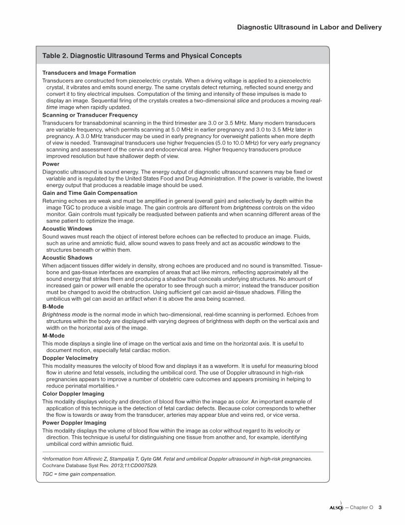
Diagnostic Ultrasound in Labor and Delivery
— Chapter O 3
Table 2. Diagnostic Ultrasound Terms and Physical Concepts
Transducers and Image FormationTransducers are constructed from piezoelectric crystals. When a driving voltage is applied to a piezoelectric
crystal, it vibrates and emits sound energy. The same crystals detect returning, reflected sound energy and convert it to tiny electrical impulses. Computation of the timing and intensity of these impulses is made to display an image. Sequential firing of the crystals creates a two-dimensional slice and produces a moving real-time image when rapidly updated.
Scanning or Transducer FrequencyTransducers for transabdominal scanning in the third trimester are 3.0 or 3.5 MHz. Many modern transducers
are variable frequency, which permits scanning at 5.0 MHz in earlier pregnancy and 3.0 to 3.5 MHz later in pregnancy. A 3.0 MHz transducer may be used in early pregnancy for overweight patients when more depth of view is needed. Transvaginal transducers use higher frequencies (5.0 to 10.0 MHz) for very early pregnancy scanning and assessment of the cervix and endocervical area. Higher frequency transducers produce improved resolution but have shallower depth of view.
PowerDiagnostic ultrasound is sound energy. The energy output of diagnostic ultrasound scanners may be fixed or
variable and is regulated by the United States Food and Drug Administration. If the power is variable, the lowest energy output that produces a readable image should be used.
Gain and Time Gain CompensationReturning echoes are weak and must be amplified in general (overall gain) and selectively by depth within the
image TGC to produce a visible image. The gain controls are different from brightness controls on the video monitor. Gain controls must typically be readjusted between patients and when scanning different areas of the same patient to optimize the image.
Acoustic WindowsSound waves must reach the object of interest before echoes can be reflected to produce an image. Fluids,
such as urine and amniotic fluid, allow sound waves to pass freely and act as acoustic windows to the structures beneath or within them.
Acoustic ShadowsWhen adjacent tissues differ widely in density, strong echoes are produced and no sound is transmitted. Tissue-
bone and gas-tissue interfaces are examples of areas that act like mirrors, reflecting approximately all the sound energy that strikes them and producing a shadow that conceals underlying structures. No amount of increased gain or power will enable the operator to see through such a mirror; instead the transducer position must be changed to avoid the obstruction. Using sufficient gel can avoid air-tissue shadows. Filling the umbilicus with gel can avoid an artifact when it is above the area being scanned.
B-ModeBrightness mode is the normal mode in which two-dimensional, real-time scanning is performed. Echoes from
structures within the body are displayed with varying degrees of brightness with depth on the vertical axis and width on the horizontal axis of the image.
M-ModeThis mode displays a single line of image on the vertical axis and time on the horizontal axis. It is useful to
document motion, especially fetal cardiac motion.Doppler VelocimetryThis modality measures the velocity of blood flow and displays it as a waveform. It is useful for measuring blood
flow in uterine and fetal vessels, including the umbilical cord. The use of Doppler ultrasound in high-risk pregnancies appears to improve a number of obstetric care outcomes and appears promising in helping to reduce perinatal mortalities.a
Color Doppler ImagingThis modality displays velocity and direction of blood flow within the image as color. An important example of
application of this technique is the detection of fetal cardiac defects. Because color corresponds to whether the flow is towards or away from the transducer, arteries may appear blue and veins red, or vice versa.
Power Doppler ImagingThis modality displays the volume of blood flow within the image as color without regard to its velocity or
direction. This technique is useful for distinguishing one tissue from another and, for example, identifying umbilical cord within amniotic fluid.
aInformation from Alfirevic Z, Stampalija T, Gyte GM. Fetal and umbilical Doppler ultrasound in high-risk pregnancies. Cochrane Database Syst Rev. 2013;11:CD007529.
TGC = time gain compensation.
Chapter O
4 Chapter O —
c. fetal lie, presentation, and positiond. quantity of amniotic fluid (performing an
amniotic fluid index if desired)e. placental location
5. Record labeled images of significant findings for the medical record. When finished, make a written record of findings and plan. Discuss findings and plan with the patient.
Transvaginal scanning is essential during the first trimester to adequately visualize the uterus, adnexa, cul-de-sac, early gestational sac, and embryo (Chapter A: First Trimester Pregnancy Complications). Transvaginal scanning also is useful during the second and third trimesters to visualize the cervix and endocervical area in cases of preterm labor, incompetent cervix, and placenta previa. Skill in transvaginal scanning is not con-sidered to be a basic skill that can be learned in the workshop associated with this course.
These features apply to transvaginal scanning:1. After discussion of the reason and scope of the examination, the patient is placed in lithotomy position.
2. Scan gel is applied to the tip of the transvaginal transducer, which is then covered with a clean or sterile condom or glove. Lubricating gel is applied over the cover and the transducer tip is introduced into the vagina. Sonographic visualization starts as soon as the transducer is introduced. Inserting the transducer tip too far can cause the examiner to miss seeing the cervix and lower uterine seg-ment as the transducer tip reaches the posterior or anterior fornix.
3. Gain and depth controls are adjusted, and the transducer tip is positioned to obtain whatever sagittal, coronal, or oblique views are necessary to visualize the structures. The vaginal opening acts as a fulcrum, making it necessary to move the transducer handle toward the opposite side of the body that the examiner wants to visualize. For example, to view the left adnexal area, the trans-ducer handle is moved to the patient’s right, and to look toward the bladder, the handle is dropped toward the patient’s rectum.
4. Transvaginal sagittal images are oriented like transabdominal images, with the mother’s head
off to the left of the screen and feet to the right. In coronal planes, the mother’s right shoulder is off to the left of the screen and left shoulder is off to the right side of the screen.
Scanning by the transperineal route is a useful alternative to transvaginal scanning during the second and third trimesters to visualize the cervix and endocervical area. This can be particularly helpful in cases of preterm labor, incompetent cervix, and suspected placenta previa, because the vagina is not entered. Although the same transducer is used in the transperineal scan as in the transabdominal scan, a wider field of view is obtained than when a transvaginal transducer is used. To perform transperineal scanning, gel is applied to the scanning surface of a transabdomi-nal transducer which is then covered with a clean or sterile glove. Lubricating gel is applied over the glove and the transducer face is placed against the introitus and perineum. Transperineal images are oriented the same as transvaginal images. A potential disadvantage is less detailed resolution than transvaginal scanning affords. Skill in trans-perineal scanning is not considered to be a basic skill that can be learned in the workshop associ-ated with this course.
Basic ApplicationsBasic applications are rapidly learned by most clinicians. These include determining that the fetus is alive by visualizing the heartbeat, diagnos-ing fetal number, visualizing fetal lie, making a quantitative assessment of amniotic fluid volume, and determining basic placental location.
Fetal LifeIf a fetal heartbeat is not heard by auscultation or Doppler, determining fetal life is a critical first step in management. Cardiac activity can be documented on a still image with the use of an M-mode tracing. Sonographic characteristics of fetal mortality are listed in Table 3. If the find-ings are in doubt, consultation should be obtained before offering a final diagnosis of fetal mortality. When fetal mortality is diagnosed, the approach to the patient and family must be individualized, but maintain communication and avoid medical jargon. During real-time scanning, the presence or absence of cardiac motion is typically apparent to the examiner and the patient. After stating simply and directly the fact that the baby has died, it is
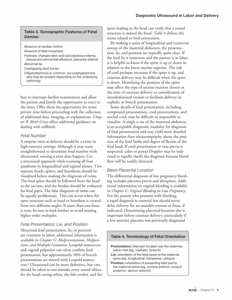
Diagnostic Ultrasound in Labor and Delivery
— Chapter O 5
best to interrupt further examination and allow the patient and family the opportunity to react to the news. Offer them the opportunity for some private time before proceeding with the collection of additional data, imaging, or explanations. Chap-ter R: Birth Crisis offers additional guidance on dealing with stillbirth.
Fetal NumberA surprise twin at delivery should be a rarity in high-resource settings. Although it may seem straightforward to determine fetal number with ultrasound, missing a twin does happen. Use a structured approach while scanning all four quadrants in longitudinal and sagittal planes. Two separate heads, spines, and heartbeats should be visualized before making the diagnosis of twins. The fetal spine should be followed from the head to the sacrum, and the fundus should be evaluated for fetal parts. The false diagnosis of twins can be equally problematic. This can occur when the same structure such as head or heartbeat is viewed from two different angles. If more than one fetus is seen, be sure to look further to avoid missing higher order multiples.
Fetal Presentation, Lie, and PositionAbnormal fetal presentation, lie, or position are common in labor; additional information is available in Chapter G: Malpresentations, Malposi-tions, and Multiple Gestations. Leopold maneuvers and vaginal palpation can often confirm fetal presentation, but approximately 30% of breech presentations are missed with Leopold maneu-vers.8 Ultrasound can be more definitive, but care should be taken to not mistake every round object for the head; seeing orbits, the falx cerebri, and the
spine leading to the head can verify that a round structure is indeed the head. Table 4 defines the terms related to fetal orientation.
By making a series of longitudinal and transverse sweeps of the maternal abdomen, the presenta-tion, lie, and position are typically quite clear. If the fetal lie is transverse and the patient is in labor, it is helpful to know if the spine is up or down in relation to the lower uterine segment. The risk of cord prolapse increases if the spine is up, and cesarean delivery may be difficult when the spine is down. Identifying the position of the spine may affect the type of uterine incision chosen at the time of cesarean delivery or consideration of intraabdominal version to facilitate delivery in cephalic or breech presentation.
Some details of fetal presentation, including compound presentations, cord presentations, and nuchal cord, may be difficult or impossible to visualize. A single x-ray of the maternal abdomen is an acceptable diagnostic modality for diagnosis of fetal presentation and may yield more detailed information than ultrasonography about the posi-tion of the fetal limbs and degree of flexion of the fetal head. If cord presentation or vasa previa is suspected, color or power Doppler may be indi-cated to rapidly clarify the diagnosis because blood flow will be readily detected.
Basic Placental LocationThe differential diagnosis of late pregnancy bleed-ing includes placenta previa and abruption. Addi-tional information on vaginal bleeding is available in Chapter C: Vaginal Bleeding in Late Pregnancy. For the patient who presents with bleeding, a rapid diagnosis is essential but should never delay delivery for an unstable woman or fetus, if indicated. Determining placental location also is important before cesarean delivery, particularly if a low anterior placenta was previously diagnosed
Table 3. Sonographic Features of Fetal Demise
Absence of cardiac motionAbsence of fetal movementHydropic changes (skin and subcutaneous edema,
pleural and pericardial effusions, placental edema)Abnormal lieOverlapping skull bonesOligohydramnios is common, but polyhydramnios
also may be present depending on the underlying pathology
Table 4. Terminology of Fetal Orientation
Presentation: fetal part located over the maternal pelvic inlet (eg, cephalic, breech)
Lie: orientation of the fetal spine to the maternal spine (eg, longitudinal, transverse, oblique)
Position: orientation of presenting fetal part to the maternal pelvis (eg, occiput anterior, occiput posterior, sacrum anterior)
Chapter O
6 Chapter O —
or suspected. Placenta previa or implantation over a previous scar significantly increases the likeli-hood of abnormal placental attachment (placenta accreta, increta, percreta) and the patient’s risk of severe hemorrhage and need for cesarean hyster-ectomy. The placenta is typically more echogenic than the myometrium, but uterine contractions can alter the apparent location, appearance, and thickness of the placenta in relation to the endo-cervical area. A distended bladder can lead to a false diagnosis of placenta previa. Crowding and oligohydramnios can make it difficult to visualize the placenta. Posterior locations are obscured by acoustic shadows of the fetus, and succenturiate lobes may be difficult to find. Examination for placenta previa and abruption are fraught with difficulty and not considered a basic applica-tion. However, it may be easy to visualize that an anterior placenta ends distant from the cervix, excluding the diagnosis of placenta previa in the evaluation of second or third trimester bleeding.
Placental abruption also can present with vagi-nal bleeding and is an obstetrical emergency that is largely a clinical diagnosis. Ultrasound can be an adjunctive diagnostic tool, but as with placenta previa, ultrasound should never delay clinically indicated treatment. Even in experienced hands, ultrasound can miss more than 50% of placental abruptions, but it does have a high positive predic-tive value when diagnosed sonographically.9
Amniotic Fluid AssessmentThe ability to determine either a subjective or objective adequacy of amniotic fluid has many applications. Amniotic fluid is a function of placen-
tal health and fetal well-being. Several obstetrical complications as well as fetal anomalies can cause a high (polyhydramnios) or a low (oligohydramnios) amniotic fluid volume. Techniques of quantitative amniotic fluid assessment include an amniotic fluid index (AFI) or a single largest vertical pocket (LVP) determination. Other terms synonymous with LVP include: deepest vertical pocket, single deepest pocket, and maximum vertical pocket. Table 5 includes defi-nitions of quantitative amniotic fluid assessment.
Assessment of amniotic fluid volume is an essential part of biophysical testing and can provide ancillary evidence of prior membrane rupture. To perform an AFI, the LVP of amniotic fluid in each of the four quadrants of the uterus is measured and the sum is computed. The trans-ducer should be perpendicular to the floor, and the measured pockets should not contain umbili-cal cord or fetal extremities.
Measurement of the single LVP is done by identifying the single largest pocket of fluid that is free of cord or fetal parts. The vertical dimension relative to the transducer should measure at least 2 cm. The LVP has become the preferred param-eter because of higher specificity over AFI.10
Oligohydramnios in a postdates pregnancy is an indication for further testing or delivery. Oligohydramnios may be associated with other causes of placental insufficiency. When reflective of an anatomic anomaly, oligohydramnios is often associated with a renal system anomaly. Mild to moderate polyhydramnios is common, typically idiopathic, but strongly associated with gestational diabetes. More severe polyhydramnios is a potent indicator of fetal anomalies, including central nervous system defects, ear, nose, and throat anomalies, gastrointestinal defects, and skeletal dysplasias. Polyhydramnios also may be an indica-tor of congestive heart failure, sometimes second-ary to alloimmune disease.
Advanced ApplicationsBiophysical AssessmentThere are many indications for assessing fetal well-being in pregnancy. As discussed in Chapter E: Intrapartum Fetal Surveillance, fetal surveillance can be accomplished using multiple techniques, includ-ing the nonstress test (NST), contraction stress test (CST), modified biophysical profile (MBPP), and biophysical profile (BPP). The MBPP and the BPP require ultrasound skills. The NST is often
Table 5. Amniotic Fluid Quantitative Assessment Terminology
Amniotic Fluid Index (cm)
Single Largest Vertical Pocket (cm)
Oligohydramnios <5 <2
Borderline (equivocal) 5 to 8 n/a
Normal 8 to 23 2 to 7
Polyhydramnios ≥24 ≥8
Information from American College of Obstetricians and Gynecologists. ACOG Practice Bulletin No. 101: Ultrasonography in pregnancy. Obstet Gyne-col. 2009; 113(2 Pt 1):451-461.

Diagnostic Ultrasound in Labor and Delivery
— Chapter O 7
the first-line method of fetal surveillance and is discussed in detail in Chapter E: Intrapartum Fetal Surveillance. Definition of reactive NST is based on National Institute of Child Health and Human Development criteria of a minimum of two epi-sodes of acceleration more than 15 bpm lasting more than 15 seconds in a period of 20 minutes.11 The NST alone has a high false-positive rate, which can lead to unnecessary intervention.12,13
The first fetal biophysical parameter to become compromised in the presence of fetal acidemia is the last to manifest as the fetus matures. In other words, the most primitive brain functions are the last to be compromised. Fetal tone is the earli-est (most primitive) biophysical parameter; it can be identified as early as 8 weeks gestational age. Fetal body movement follows approximately 1 to 2 weeks later. Fetal breathing is detectable by 21 weeks’ gestation, but fetal heart rate reactivity is not well established until 28 to 32 weeks. Com-promised fetal heart rate reactivity is therefore the most sensitive marker of acute fetal acidosis. As the insult becomes more profound and chronic, fetal breathing, then body movement, then tone are compromised. Amniotic fluid volume is consid-ered a marker of placental perfusion, and therefore decreased fluid is considered a marker of chronic vascular compromise. Table 6 summarizes these concepts. Using these concepts and combining the NST with a MBPP or BPP can significantly improve the predictive value of the test as a marker for fetal well-being.
Modified Biophysical Profile (MBPP)An MBPP is an NST (marker of acute fetal status) along with an amniotic fluid assessment (marker of chronic fetal status). A reactive NST combined with a 2 cm or greater single LVP of amniotic fluid is reassuring of fetal well-being and compares favorably with the complete BPP. However, if the NST does not meet criteria for reactive and/or if oligohydramnios is present, further fetal testing is indicated and delivery may be considered, espe-cially near term.
Biophysical Profile (BPP)The BPP is another method of predicting antepar-tum fetal acidemia. Although the test can be used as a primary method of fetal surveillance, it can be especially helpful in cases where the NST is not reactive. There is some evidence that the BPP may
even be useful during labor as an adjunct to fetal heart rate monitoring.14 The five components of a BPP integrate fetal cardiac activity, amniotic fluid assessment, fetal breathing, fetal movement, and fetal tone. Each component is scored 2 points if present or reactive NST, and 0 points if abnormal. The presence of fetal breathing is likely as reliable as the NST in predicting absence of fetal acido-sis. A BPP can be performed as early as 26 to 28 weeks’ gestation. The test can be completed in as little as 5 minutes if the fetus is active and awake, or it can take as long as 30 minutes. Acoustic stimulation is appropriate to use to shorten the time of the study. Table 7 includes criteria for each component of the BPP.
The maximum BPP score is 10. A score greater than or equal to 8 is considered reassuring with a low risk of fetal asphyxia within 1 week of the test. A score of 6 is equivocal, and a score less than or equal to 4 is abnormal with a significantly higher risk of fetal asphyxia within 1 week of the test. However, not all components of the BPP are con-sidered equal. Oligohydramnios, when not associ-ated with ruptured membranes, is a marker for placental dysfunction and chronic fetal hypoxemia or acidemia and may require further action or more frequent surveillance regardless of the total BPP score. However, if the amniotic fluid volume is normal but the NST is not reactive, the presence of fetal breathing predicts reassuring fetal status.
Table 6. Biophysical Parameters Sensitivity to Acute Hypoxia
Component Process Assessed Type of Indicator
Fetal tone CNS, cortex/subcortical Least sensitive acutely
Fetal movement CNS, ventral fourth ventricle
Intermediate, acute
Fetal breathing CNS, posterior hypothalamus, medulla
Intermediate, acute
Nonstress test CNS, cortex/nuclei Most sensitive acutely
Amniotic fluid quantity
Uteroplacental/fetal perfusion
Chronic indicator
CNS = central nervous system.
Information from Manning FA, Platt LD, Sipos L. Antepartum fetal evaluation: development of a fetal biophysical profile. Am J Obstet Gynecol. 1980;136(6): 787-795; Manning FA. Antepartum fetal testing: a critical appraisal. Curr Opin Obstet Gynecol. 2009;21(4):348-352.
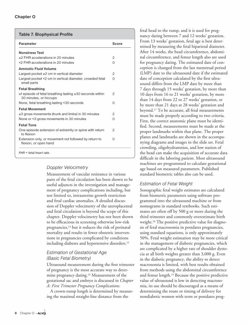
Chapter O
8 Chapter O —
Doppler VelocimetryMeasurement of vascular resistance in various parts of the fetal circulation has been shown to be useful adjuncts in the investigation and manage-ment of pregnancy complications including, but not limited to, intrauterine growth restriction and fetal cardiac anomalies. A detailed discus-sion of Doppler velocimetry of the uteroplacental and fetal circulation is beyond the scope of this chapter. Doppler velocimetry has not been shown to be efficacious in screening otherwise low-risk pregnancies,14 but it reduces the risk of perinatal mortality and results in fewer obstetric interven-tions in pregnancies complicated by conditions including diabetes and hypertensive disorders.15
Estimation of Gestational Age (Basic Fetal Biometry)Ultrasound measurement during the first trimester of pregnancy is the most accurate way to deter-mine pregnancy dating.16 Measurement of the gestational sac and embryo is discussed in Chapter A: First Trimester Pregnancy Complications.
A crown-rump length is determined by measur-ing the maximal straight-line distance from the
fetal head to the rump, and it is used for preg-nancy dating between 7 and 12 weeks’ gestation. From 13 weeks’ gestation, fetal age is best deter-mined by measuring the fetal biparietal diameter. After 14 weeks, the head circumference, abdomi-nal circumference, and femur length also are used for pregnancy dating. The estimated date of con-ception is changed from the last menstrual period (LMP) date to the ultrasound date if the estimated date of conception calculated by the first ultra-sound differs from the LMP date by more than 7 days through 15 weeks’ gestation, by more than 10 days from 16 to 21 weeks’ gestation, by more than 14 days from 22 to 27 weeks’ gestation, or by more than 21 days at 28 weeks’ gestation and beyond.17 To be accurate, all fetal measurements must be made properly according to two criteria. First, the correct anatomic plane must be identi-fied. Second, measurements must be made at the proper landmarks within that plane. The proper planes and landmarks are shown in the accompa-nying diagrams and images in the slide set. Fetal crowding, oligohydramnios, and low station of the head can make the acquisition of accurate data difficult in the laboring patient. Most ultrasound machines are programmed to calculate gestational age based on measured parameters. Published standard biometric tables also can be used.
Estimation of Fetal WeightSonographic fetal weight estimates are calculated from biometric parameters using software pro-grammed into the ultrasound machine or from nomograms in standard textbooks. Such esti-mates are often off by 500 g or more during the third trimester and commonly overestimate birth weight.18 The positive predictive value for diagno-sis of fetal macrosomia in postdates pregnancies, using standard equations, is only approximately 50%. Fetal weight estimation may be more critical in the management of diabetic pregnancies, which are complicated by a higher rate of shoulder dysto-cia at all birth weights greater than 3,000 g. Even in the diabetic pregnancy, the ability to detect macrosomia is limited, with best results obtained from methods using the abdominal circumference and femur length.19 Because the positive predictive value of ultrasound is low in detecting macroso-mia, its use should be discouraged as a means of determining the route or timing of delivery for nondiabetic women with term or postdates preg-
Table 7. Biophysical Profile
Parameter Score
Nonstress Test≥2 FHR accelerations in 20 minutes<2 FHR accelerations in 20 minutes
20
Amniotic Fluid VolumeLargest pocket ≥2 cm in vertical diameterLargest pocket <2 cm in vertical diameter, crowded fetal
small parts
20
Fetal Breathing≥1 episode of fetal breathing lasting ≥30 seconds within
30 minutes, or hiccupsNone, fetal breathing lasting <30 seconds
2
0
Fetal Movement≥3 gross movements (trunk and limbs) in 30 minutesNone or <3 gross movements in 30 minutes
20
Fetal ToneOne episode extension of extremity or spine with return
to flexionExtension only, or movement not followed by return to
flexion, or open hand
2
0
FHR = fetal heart rate.

Diagnostic Ultrasound in Labor and Delivery
— Chapter O 9
nancies. There is evidence that sonographic diag-nosis of macrosomia influences physician behavior in the management of labor. The use of retrospec-tive cohort studies comparing pregnancies with a false positive diagnosis of macrosomia (estimated fetal weight [EFW] greater than 4,000 g) and true negatives show significantly increased rates of cesarean delivery (up to 50%) and failed induc-tion without significant reduction in shoulder dystocia.20-23 Limited data from a single random-ized controlled trial suggest that induction of labor in women with gestational diabetes treated with insulin reduces the risk of macrosomia.24
American College of Obstetricians and Gyne-cologists does recommend consideration of the option of planned cesarean delivery when EFW is more than 4,500 g in diabetic women or more than 5,000 g in nondiabetic women.25
Cervical Length DeterminationScanning by the transperineal or transvaginal route may be useful to assess cervical length, to detect funneling and dilatation of the cervix, and to detect bulging membranes in patients with suspected preterm labor or cervical incompetence. Shortening of the cervix in the second trimester is associated with increased risk of preterm delivery; 20 to 25 mm is commonly cited as the thresh-old,26 and the use of progesterone for short cervical length after screening has been shown to decrease the likelihood of preterm delivery.27 This is dis-cussed in more detail in Chapter D: Preterm Labor and Premature Rupture of Membranes. A Cochrane review of five randomized controlled trials looked at the effectiveness of transvaginal ultrasound cer-vical length screening and concluded that there is insufficient evidence to recommend routine screen-ing of symptomatic or asymptomatic women to prevent preterm birth.28 However, a 2012 ACOG Practice Bulletin considers routine screening an acceptable but not mandatory practice.29
Guidance for External Cephalic VersionExternal cephalic version of the fetus from breech or transverse to cephalic presentation is discussed in Chapter G: Malpresentations, Malpositions, and Multiple Gestations, including a discussion of parameters that affect success. A complete ultra-sound scan, including biometry and anatomic survey, should be performed before cephalic ver-sion because the incidence of anomalies doubles
with breech presentation.30 Cord presentation should be ruled out, which may be facilitated by color or power Doppler technique. Ultrasound is useful during the version procedure to monitor the changing fetal position and to visualize the fetal heartbeat.
Examination for Placenta PreviaPlacenta previa can be difficult to diagnose sonographically depending on the location of the presenting part, the quantity of amniotic fluid, and the amount of urine in the maternal bladder. If the maternal bladder is overdistended, the lower uterine segment may be compressed, creating a false impression of placenta previa. Ultrasound evaluation of the lower uterine segment for pos-sible placenta previa is best performed first with the bladder partially filled and then with an empty bladder. If possible, it should be performed during an interval without uterine contractions.
The lower uterine segment can be effectively visualized by the transperineal route using a glove-covered 3.0 or 3.5 MHz transducer placed at the introitus or on the perineum. Careful transvaginal scanning with a 5.0 or 7.5 MHz transducer may be undertaken. Technical aspects of transvaginal and transperineal scanning are discussed in the Scanning Techniques section of this chapter.
Examination for Placental AbruptionPlacental abruption may have a variable sono-graphic appearance depending on its age. The amount of bleeding is often underestimated. Fresh hemorrhage may appear as a sonolucent area between the uterine wall and the placenta or in the substance of the placenta. Clotted blood may exhibit echogenicity similar to that of placental tissue. Nonpathologic venous lakes and marginal sinuses can have a similar appearance. Abruption also may present as an abnormal thickening or rounding of the placental edge, presumably from a marginal separation. The sonographic diagnosis of abruption should be incorporated into available clinical information to arrive at a management plan. Chapter C: Vaginal Bleeding in Late Preg-nancy includes more discussion on clinical man-agement options in cases of abruption. The failure to diagnose abruption sonographically in a sus-pected clinical setting does not exclude the diagnosis and the clinician should not change management based on a negative ultrasound scan alone.

Chapter O
10 Chapter O —
Anatomic Survey for Anomalies
Skill in performance of the anatomic survey for fetal anomalies requires significant additional training and supervised experience beyond what can be accomplished in the workshop associ-ated with this course. The standard antepartum ultrasound examination includes a comprehensive survey of fetal anatomy as agreed on by several professional organizations. Attention to the items in this survey will detect most, but not all, fetal anomalies depending on gestational age. It is common for the patient to ask is my baby all right? during a limited labor and delivery scan. Because limited labor and delivery scans do not include a fetal anatomic survey, the examiner should routinely explain the goals and limitations of the examination and what he or she can and cannot diagnose on the basis of the examination being performed. Often the patient will have had an earlier examination that included a standard sur-vey of fetal anatomy that provides reassurance. If clinical questions exist about the presence of fetal anomalies, a standard examination that includes an anatomic survey should be performed when time and conditions permit.
lntrapartum Twin ManagementThe intrapartum management of twins in the delivery room can be facilitated by assessing the initial presentation and lie of the fetuses. After delivery of the first twin, ultrasound is often needed to determine the presentation and moni-tor the heart rate of the second twin. If the second twin is nonvertex, ultrasound may be used during an external cephalic version, internal podalic ver-sion, or breech extraction.31
Amniocentesis GuidanceUltrasound guidance can facilitate third trimester amniocentesis for lung maturity testing or to rule out amnionitis. In cases of preterm labor, such information may guide decisions about transfer from hospitals without neonatal intensive care facilities. Sonographic guidance aids in locating a pocket for sampling that is free of the umbilical cord, away from the fetal face, and not directly underneath the placenta. Alternatively, the fetal head may be lifted abdominally after the maternal bladder has been emptied, leaving a pocket of fluid, which may be tapped suprapubically. This latter technique may carry a higher risk of rupture
of membranes. Continuous sonographic obser-vation of the amniocentesis needle makes the procedure safer.
Perspective on Routine Scanning During PregnancyThis chapter has dealt with specific indications and techniques for limited sonographic examina-tion, mostly during late pregnancy and in the labor and delivery setting. It has not addressed the issue of the value of routine scanning for all preg-nant patients. A detailed exposition of this topic is beyond the scope of this chapter. This issue continues to be debated and is complicated by significant regional and international differences in practice patterns and patient populations. A variety of clinical benefits have been ascribed to routine ultrasound examination of all pregnant patients, some of which are supported by high-quality evi-dence. Routine early pregnancy ultrasound exami-nation before 24 weeks and subsequent adjustment of delivery date appear to reduce the incidence of postterm pregnancy and postdates induction and lead to earlier detection of multifetal pregnancy.32 More accurate gestational dating can facilitate rou-tine induction of labor at 41 weeks, which appears to reduce perinatal mortality.31,33 Scanning preg-nancies after 24 weeks in unselected and low-risk populations does not reduce perinatal mortality or confer other benefits.34
Routine second trimester anatomic survey, although commonly performed in developed countries, has not been shown to improve peri-natal outcomes. A detailed anatomic survey may be indicated for a number of reasons, including abnormal serum screening, advanced maternal age, personal history of fetal anomalies, or exposures to teratogenic agents. Detection of major anomalies or genetic conditions may help patients and fami-lies prepare emotionally for anomalies and plan the optimal delivery site. Identification of anoma-lies and genetic conditions in the second trimester allows the option of pregnancy termination.
American College of Obstetricians and Gyne-cologists recognizes the importance of accurate pregnancy dating and fetal anatomic survey, but stops short of recommending routine screening ultrasound for all patients. ACOG and the Ameri-can Academy of Family Physicians (AAFP) also take the position against nonmedically indicated or recreational ultrasound, and view this prac-

Diagnostic Ultrasound in Labor and Delivery
— Chapter O 11
tice as inappropriate and contrary to responsible medical practice.35,36
Learning StrategyA variety of methods are available to clinicians interested in learning to include diagnostic ultra-sound in their practice. Start with self-study using textbooks and multimedia materials, and then follow up with formal hands-on courses or informal apprenticeship. Simulators using physical phantoms or electronically-stored images also are available.37
Ultrasound in Low-Resource SettingsSome have suggested that the ultrasound machine will probably be the stethoscope of the 21st century.38 However, the cost of ultrasound technology makes this unrealistic in many low-resource settings. The optional ALSO workshop has been one of the most popular workshops according to evaluation forms from multiple international courses. Porta-bility, affordability, maintenance, and repair are important issues for units produced or imported for developing countries, and inexpensive compact machines offer promise but outcomes research is needed.39 Teleradiology may help expand valu-able ultrasound services to low-resource areas.40 Ultrasound may have a greater impact in develop-ing than developed countries by allowing preven-
tion and early intervention where resources are scarce. Improved diagnosis of ectopic pregnancy, intrauterine growth restriction, placenta previa, malpresentation, and multiple gestation may have lifesaving implications.40,41 In a study in Rwanda, midwives performing ultrasound had a 100% agreement with sonographers when evaluating for multiple gestation, malpresentation, and viabil-ity.42 Of 542 ultrasounds ordered for suspected abnormal findings, 39% aided in diagnosis and 22% changed management.42 For additional infor-mation on ultrasound in developing countries, refer to the Global ALSO Program available at www.aafp.org/globalalso.
SummaryAll clinicians who deliver infants can benefit from learning the basic applications of diagnostic ultra-sound. The ability to rapidly assess fetal life, fetal number, fetal presentation, quantity of amniotic fluid, and placental location may be learned rather quickly and can have significant bearing on clinical management. Advanced applications require further study and practice. Frequent use of the basic applications will help maintain proficiency while keeping in mind the shortcom-ings, complicating factors, and potential pitfalls of ultrasound scanning.

Chapter O
12 Chapter O —
SORT: Key Recommendations for Practice
Clinical RecommendationEvidence Rating References
Routine pregnancy ultrasound examination before 24 weeks, with subsequent adjustment of delivery date if indicated, appears to reduce the incidence of postterm pregnancy and facilitate induction of labor at 41 weeks to reduce perinatal mortality.
A 30-33
Routine ultrasound scanning before 24 weeks improves recognition of clinically unsuspected multifetal pregnancy.
A 32
Routine ultrasound scanning after 24 weeks in unselected low-risk patients does not reduce perinatal mortality.
A 34
The use of Doppler ultrasound in high-risk pregnancies with hypertensive disease or fetal growth restriction appears to improve a number of obstetric care outcomes and appears promising in helping to reducing perinatal mortalities.
A 14
Ultrasonographic estimates of fetal weight have limited accuracy and should not be used to determine route of birth in nondiabetic pregnancies except for the discussion of the option of scheduled cesarean delivery if the estimated fetal weight is more than 5,000 g
B 20-23,25
An MBPP or BPP, along with an NST and a CST, are appropriate methods of antepartum fetal surveillance in high-risk patients.
B 13,43
An abnormal NST or MBPP should be further evaluated with a CST or full BPP. B 13,43
Maternity care clinicians can learn the basic sonographic skills in a short time. Sonographic applications including fetal biometry, anatomic survey, examination for placental abruption and previa, and assessment of the cervix are extended skills that require significantly more study and practice than the basic skills.
C 5-7
Strength of Recommendation Taxonomy (SORT)
Strength of Recommendation Definition
A • Recommendation based on consistent and good-quality patient-oriented evidence.
B • Recommendation based on inconsistent or limited-quality patient-oriented evidence.
C • Recommendation based on consensus, usual practice, opinion, disease-oriented evidence, or case series for studies of diagnosis, treatment, prevention, or screening.
Patient-oriented evidence measures outcomes that matter to patients: morbidity, mortality, symptom improvement, cost reduction, and quality of life. Disease-oriented evidence measures intermediate, physiologic, or surrogate end points that might not reflect improvement in patient outcomes (eg, blood pressure, blood chemistry, physiologic function, pathologic findings).
BPP = biophysical profile; CST = contraction stress test; MBPP = modified biophysical profile; NST = nonstress test.
From Ebell MH, Siwek J, Weiss BD, et al. Strength of recommendation taxonomy (SORT): a patient-centered approach to grading evidence in the medical literature. Am Fam Physician. 2004;69(3):548-556.

— Chapter O 13
Appendix A: Documentation of Labor and Delivery Ultrasound Scan
Patient name: ___________________________________________________ Date: ____________ Patient I.D. number: ______________________________
Attending physician: _____________________________________________ Other physician: ___________________________________________________
Pregnancy data: Maternal age: _______________ G: _______________ P: _______________ Ab: _______________
Today’s gestational age: _______________ weeks + or - _______________ weeks
Based on: LMP of: _______________ and/or previous ultrasound scan on: _______________ (date)
Findings of today’s fetal heart rate tracing: _______________________________________________________________________________________________
Reason for L&D ultrasound scan (check all that apply):
No prenatal care / unknown dates PROM Pain
Part of antepartum testing (amniotic fluid index) Bleeding Trauma
Adjunct to amniocentesis Suspected breech presentation Preterm labor
Adjunct to external version Preterm labor ___________________________________________________
Fetal weight estimate (reason: ______________________________________________________________________________________________________ )
Other coexisting problems: ____________________________________________________________________________________________________________
Findings: (complete only relevant items) (attach photos to reverse side or new page)
Fetal heartbeat: Yes No Fetal number: ____________________
Fetal lie, presentation, and position: _____________________________________________________________________________________________________
Placental location: ___________________________________________________________________________________________________________________
Biophysical data:
Amniotic fluid index: ____________ + ____________ + ____________ + = ____________ cm. Or largest vertical pocket: ____________ cm
Breathing: Yes No Tone: Yes No Body movement: Yes No
Biophysical score: _______________ points out of possible ________________ points
Fetal biometry:
BPD = _______________ cm = _______________ weeks + or - _______________ weeks
Femur = _______________ cm = _______________ weeks + or - _______________ weeks
Head Circ. = _______________ = _______________ weeks + or - _______________ weeks
Abd. Circ. = _______________ cm = _______________ weeks + or - _______________ weeks
Composite gestational age = _______________ weeks + or - _______________ weeks
EDD: _______________ + or - _______________ weeks. Estimated fetal weight: _______________ grams
Describe any abnormalities seen: ____________________________________________________________________________________________________
____________________________________________________________________________________________________________________________________
____________________________________________________________________________________________________________________________________
Consultative scan ordered: Yes No
Diagnosis related to reason for scan: _________________________________________________________________________________________________
____________________________________________________________________________________________________________________________________
Plan of treatment: ____________________________________________________________________________________________________________________
____________________________________________________________________________________________________________________________________
Signature(s): _____________________________________________________________________ Date and time ____________________________________
Courtesy of Mark Deutchman, MD

Chapter O
14 Chapter O —
References1. Diagnostic Ultrasound Imaging in Pregnancy. NIH Con-
sens Statement. 1984;5(1):1-16.
2. AIUM Practice Parameter for the Performance ofObstetric Ultrasound Examinations. Laurel, MD: Ameri-can Institute of Ultrasound in Medicine; 2013. Availableat http://www.aium.org/resources/guidelines/obstetric.pdf.
3. American College of Radiology. ACR–ACOG–AIUM–SRU Practice Guideline for the Performance of Obstetri-cal Ultrasound. 2014. Available at http://www.acr.org/~/media/f7bc35bd59264e7cbe648f6d1bb8b8e2.pdf.
4. American College of Obstetricians and Gynecologists.ACOG Practice Bulletin No. 101: Ultrasonography inpregnancy. Obstet Gynecol. 2009;113(2 Pt 1):451-461.
5. Hahn RG, Roi LD, Ornstein SM, et al. Obstetric ultra-sound training for family physicians. Results from amulti-site study. J Fam Pract. 1988;26(5):553-558.
6. Smith CB, Sakornbut EL, Dickinson LC, Bullock GL.Quantification of training in obstetrical ultrasound:a study of family practice residents. J Clin Ultrasound.1991;19(8):479-483.
7. Rodney WM, Deutchman ME, Hartman KJ, Hahn RG.Obstetric ultrasound by family physicians. J Fam Pract.1992;34(2):186-194, 197-200.
8. Hemelaar J, Lim LN, Impey LW. The Impact of an ECVService is Limited by Antenatal Breech Detection: A Ret-rospective Cohort Study. Birth. 2015;42(2):165-172.
9. Glantz C, Purnell L. Clinical utility of sonography in thediagnosis and treatment of placental abruption. J Ultra-sound Med. 2002;21(8):837-840.
10. Nabhan AF, Abdelmoula YA. Amniotic fluid index versussingle deepest vertical pocket as a screening test forpreventing adverse pregnancy outcome. CochraneDatabase Syst Rev. 2008;(3):CD006593.
11. American College of Obstetricians and Gynecologists.Practice bulletin no. 145: antepartum fetal surveillance.Obstet Gynecol. 2014;124(1):182-192.
12. Manning FA, Platt LD, Sipos L. Antepartum fetal evalu-ation: development of a fetal biophysical profile. Am JObstet Gynecol. 1980;136(6):787-795.
13. Manning FA. Antepartum fetal testing: a critical appraisal.Curr Opin Obstet Gynecol. 2009;21(4):348-352.
14. Alfirevic Z, Stampalija T, Medley N. Fetal and umbilicalDoppler ultrasound in normal pregnancy. CochraneDatabase Syst Rev. 2015;4(4):CD001450.
15. Alfirevic Z, Stampalija T, Gyte GM. Fetal and umbilicalDoppler ultrasound in high-risk pregnancies. CochraneDatabase Syst Rev. 2013;11(11):CD007529.
16. Eik-Nes SH, Salvesen KA, Okland O, Vatten LJ. Routineultrasound fetal examination in pregnancy: the ‘Alesund’randomized controlled trial. Ultrasound Obstet Gynecol.2000;15(6):473-478.
17. American College of Obstetricians and Gynecologists.Committee opinion no 611: method for estimating duedate. Obstet Gynecol. 2014;124(4):863-866.
18. Delpapa EH, Mueller-Heubach E. Pregnancy outcomefollowing ultrasound diagnosis of macrosomia. ObstetGynecol. 1991;78(3 Pt 1):340-343.
19. Pollack RN, Hauer-Pollack G, Divon MY. Macrosomiain postdates pregnancies: the accuracy of routineultrasonographic screening. Am J Obstet Gynecol.1992;167(1):7-11.
20. McLaren RA, Puckett JL, Chauhan SP. Estimators ofbirth weight in pregnant women requiring insulin: a com-parison of seven sonographic models. Obstet Gynecol.1995;85(4):565-569.
21. Levine AB, Lockwood CJ, Brown B, Lapinski R,Berkowitz RL. Sonographic diagnosis of the large forgestational age fetus at term: does it make a difference?Obstet Gynecol. 1992;79(1):55-58.
22. Parry S, Severs CP, Sehdev HM, Macones GA, WhiteLM, Morgan MA. Ultrasonographic prediction of fetalmacrosomia. Association with cesarean delivery. JReprod Med. 2000;45(1):17-22.
23. Blackwell SC, Refuerzo J, Chadha R, Carreno CA. Over-estimation of fetal weight by ultrasound: does it influencethe likelihood of cesarean delivery for labor arrest? Am JObstet Gynecol. 2009;200(3):340.e1-340.e3.
24. Boulvain M, Stan C, Irion O. Elective delivery in dia-betic pregnant women. Cochrane Database Syst Rev.2001;(2):CD001997.
25. ACOG Committee on Practice Bulletins. ACOG PracticeBulletin No. 22: Fetal Macrosomia. Obstet Gynecol.2000;96(5).
26. Iams JD, Goldenberg RL, Meis PJ, et al.; National Insti-tute of Child Health and Human Development MaternalFetal Medicine Unit Network. The length of the cervixand the risk of spontaneous premature delivery. N EnglJ Med. 1996;334(9):567-572.
27. Hassan SS, Romero R, Vidyadhari D, et al; PREGNANTTrial. Vaginal progesterone reduces the rate of pretermbirth in women with a sonographic short cervix: a mul-ticenter, randomized, double-blind, placebo-controlledtrial. Ultrasound Obstet Gynecol. 2011;38(1):18-31.
28. Berghella V, Baxter JK, Hendrix NW. Cervical assess-ment by ultrasound for preventing preterm delivery.Cochrane Database Syst Rev. 2013;1(1):CD007235.
29. Committee on Practice Bulletins—Obstetrics, TheAmerican College of Obstetricians and Gynecologists.Practice bulletin no. 130: prediction and prevention ofpreterm birth. Obstet Gynecol. 2012;120(4):964-973.
30. Mostello D, Chang JJ, Bai F, et al. Breech presentationat delivery: a marker for congenital anomaly? J Perina-tol. 2014;34(1):11-15.
31. Fox NS, Silverstein M, Bender S, Klauser CK, SaltzmanDH, Rebarber A. Active second-stage management intwin pregnancies undergoing planned vaginal deliveryin a U.S. population. Obstet Gynecol. 2010;115(2 Pt1):229-233.
32. Whitworth M, Bricker L, Mullan C. Ultrasound for fetalassessment in early pregnancy. Cochrane DatabaseSyst Rev. 2015;7(7):CD007058.

Diagnostic Ultrasound in Labor and Delivery
— Chapter O 15
33. Gülmezoglu AM, Crowther CA, Middleton P, HeatleyE. Induction of labour for improving birth outcomes forwomen at or beyond term. Cochrane Database SystRev. 2012;6(6):CD004945.
34. Bricker L, Medley N, Pratt JJ. Routine ultrasound in latepregnancy (after 24 weeks’ gestation). Cochrane Data-base Syst Rev. 2015;6(6):CD001451.
35. The American Academy of Family Physicians. Diagnos-tic Ultrasonography in Women’s Health Care (PositionPaper). Available at http://www.aafp.org/about/poli-cies/all/ultrasonography-diagnostic.html.
36. ACOG Committee on Ethics. ACOG Committee Opinion.Number 297, August 2004. Nonmedical use of obstetricultrasonography. Obstet Gynecol. 2004;104(2):423-424.
37. Dresang LT, Rodney WM, Dees J. Teaching prenatalultrasound to family medicine residents. Fam Med.2004;36(2):98-107.
38. Rodney WM. More on the use of ultrasonography in the emergency department. West J Med. 1995;163(4):393-394.
39. Harris RD, Marks WM. Compact ultrasound for improv-ing maternal and perinatal care in low-resource set-tings: review of the potential benefits, implementationchallenges, and public health issues. J Ultrasound Med.2009;28(8):1067-1076.
40. Stanton K, Mwanri L. Global Maternal and Child HealthOutcomes: The Role of Obstetric Ultrasound in LowResource Settings. World Journal of Preventive Medi-cine. 2013;1(3):22-29.
41. Sippel S, Muruganandan K, Levine A, Shah S. Reviewarticle: Use of ultrasound in the developing world. Int JEmerg Med. 2011;4(4):72.
42. Stein W, Katunda I, Butoto C. A two-level ultrasono-graphic service in a maternity care unit of a rural districthospital in Tanzania. Trop Doct. 2008;38(2):125-126.
43. Miller DA, Rabello YA, Paul RH. The modified biophysi-cal profile: antepartum testing in the 1990s. Am J ObstetGynecol. 1996;174(3):812-817.

— Chapter P 1
Chapter P Neonatal Resuscitation
OverviewThis chapter provides an overview of the basic tech-niques used in neonatal resuscitation. These guide-lines apply to newborns as well as infants who require resuscitation within the first few months after birth. An accompanying workshop will cover the techniques of airway management, positive-pressure (bag and mask) ventilation and alternatives, endotracheal intu-bation, and protocols for dealing with meconium. The information presented is consistent with guidelines established by the American Academy of Pediatrics (AAP) and the American Heart Association (AHA) in the 6th edition of the Neonatal Resuscitation Program (NRP).1,2 It has been updated to reflect changes rec-ommended in the 2015 American Heart Association Guidelines Update for Cardiopulmonary Resuscitation and Emergency Cardiovascular Care.3 The 7th edition of the NRP is based on the 2015 AHA guidelines; it was released in June 2016 and its use will be required after January 1, 2017. The ALSO program is not intended to substitute for the advanced training and formal certification in neonatal resuscitation provided by the NRP.
One of the most satisfying events in medicine is being present at the birth of a healthy baby. Fortu-nately, more than 90% of infants have an uneventful birth and can immediately be given directly to their mother. Approximately 1 in 10 infants have some diffi-culty with the transition to extrauterine life. More than half of distressed infants have identifiable risk factors; but because many do not, it is imperative to be pre-
pared for the resuscitation of a distressed infant at every delivery. Although resuscitating a depressed newborn may seem intimidating, the maternity care clinician can take comfort in the knowledge that newborns gener-ally benefit from simple, nonpharmacological measures and that mastery of a few simple techniques will allow the resuscitator to deal effectively with all but the most severely depressed infants. Less than 1% of newborns will require drugs.2
The transition from intrauterine to extrauterine life depends on a physiologically complicated series of events that are driven by changes in oxygen pressure and the ability of the infant to take its first breaths effectively. If the infant’s airway is blocked by mucus or meconium, or if it has poorly compliant lungs because of immaturity or meconium, oxygenation and ventila-tion may not be effective. Obtaining an airway and assisting with breathing remain the mainstays of infant resuscitation.1-3 This is in contrast to recent changes in adult and pediatric resuscitation protocols that give priority to the C in the A-B-C (Airway-Breathing- Circulation) mnemonic.
PathophysiologyNeonatal depression is most often caused by hypoxemia or by drugs administered to the mother.2 The unborn infant can tolerate a degree of hypoxemia and acidosis that would be detrimental to a child or an adult, but if the hypoxemia is severe enough, a predictable sequence of events ensues. A hypoxemic infant will initially go through a brief period of rapid breathing, and then
Learning ObjectivesAt the end of this activity, learners will be able to:1. Differentiate primary from secondary apnea.2. Describe resuscitation steps for a depressed newborn (ventilation,
chest compressions, and drugs).3. Describe the management of meconium.
Karen Ailsworth, MD, MS, FAAP
Revised January 2017
Copyright 2017© American Academy of Family Physicians. All rights reserved.

Chapter P
2 Chapter P —
it will stop breathing. This is known as primary apnea. During primary apnea, the heart rate and neuromuscular tone decrease. If the infant is deliv-ered during this stage, it will generally benefit from stimulation and supplemental oxygen. However, if the hypoxic stress continues, the infant’s condition will deteriorate steadily. The infant will go through a phase of deep, irregular gasping and then it will stop breathing. This is called secondary apnea. During secondary apnea, the infant’s heart rate, blood pressure, and PaO2 will fall profoundly.
An infant delivered in secondary apnea will not resume breathing spontaneously, nor will it benefit from stimulation and supplemental oxygen. This infant must have assisted ventilation to correct the hypoxemia or it will deteriorate further and die. The hypoxemic infant may progress through primary apnea into secondary apnea in utero. Because the two phases of apnea are clinically indistinguishable from each other, any infant born apneic must be presumed to be in secondary apnea.1,2 This does not mean the maternity care clinician should not attempt stimulation of the infant, but that the clinician should try only one or two attempts at stimulation before progressing to a more aggressive intervention. For an infant in secondary apnea, any delay in treatment will lead to a much longer delay in establishing effective spontaneous respirations, or even death.
There are two features of the fetal cardiopul-monary system that may cause problems for a depressed infant in its attempts to establish extrauterine life. First, the infant’s lungs are filled with fluid in utero. To expand the alveoli and clear this fluid, the first few breaths require more pressure than subsequent breaths. Therefore, an infant who is making only weak respiratory efforts will not be able to move air into its lungs and will become increasingly hypoxic and acidotic with-out intervention. Second, relatively little blood flows through the pulmonary vascular bed at birth because most is shunted around the lungs through the ductus arteriosus. Normally, as the infant begins to breathe, the increasing concentration of oxygen in the blood causes the ductus to constrict and the pulmonary arterioles to dilate. In the hypoxemic infant, the ductus remains open and allows oxygenated blood to bypass the lungs. This persistent fetal circulation further impairs adequate oxygenation. If the infant is mildly to moderately hypoxemic, positive pressure ventilation (PPV) via
bag and mask will be sufficient to establish normal newborn circulation. However, if the infant is severely hypoxemic, cardiac compressions or drugs become necessary. It should be emphasized that the vast majority of depressed newborns will benefit from effective ventilation; few of them will need compressions, and even fewer will need drugs. Thus, it is beneficial to learn the basics of neonatal oxygenation and ventilation.
Getting Ready for ResuscitationOne of the most important steps in neonatal resuscitation is preparation. Frequently, the pre-natal or perinatal history will reveal risk factors that may indicate a need for resuscitation (Appen-dix A). Because the need for resuscitation cannot be predicted with absolute certainty, appropri-ate personnel and equipment should be ready at every delivery. An overview on neonatal resuscita-tion is shown in Figure 1.
Personnel Needed for Neonatal ResuscitationThere should be one individual trained to resus-citate newborns present in the delivery room for each infant to be delivered. This individual should not have any other duties after the infant is born. If there is more than one infant, there needs to be at least one resuscitator per infant at the delivery (not on call in another part of the hospital or at home).
Equipment Needed for Neonatal ResuscitationWherever newborn infants are cared for, appropri-ate resuscitation equipment should be immediately available and in working order (Appendix B). The maternity care clinician must be familiar with the location and operation of all the equipment necessary for neonatal resuscitation, especially because infant workstations may be set up differ-ently in various labor and delivery units. Because the need for resuscitation cannot be predicted, the minimum required setup is a radiant warmer (turned on before delivery) and towels or blankets for drying the newborn. It may be helpful to have drug doses, targeted preductal SpO2 after birth guidelines, and other cheat sheets posted on or near the radiant warmer for quick reference.
The maternity care clinician should ensure that oxygen lines are connected and able to deliver

— Chapter P 3
a flow of 5 to 10 L/min. The resuscitation bag and mask unit should work properly and have appropriately sized masks. Suction lines should be connected and adjusted to keep occluded suction between 80 to 100 mm Hg. The laryngoscope should have appropriately sized blades (blade size No. 1 for term infants and No. 0 for pre-term infants) and a properly functioning light. If conditions suggest that the delivery of a depressed infant is likely, all equipment should be set up in advance.
Initial Stabilization and Evaluation of the NewbornResuscitation always begins with a rapid evalu-ation of the newborn. Evaluation begins with determining if the infant is term, has normal tone, and is breathing or crying.3 Apgar scores are not useful indicators of the need for resuscitation. Resuscitation of a depressed newborn must begin within 30 seconds, well before the 1-minute Apgar score is calculated.
Rapid Evaluation of the NewbornResuscitation is a rapid series of specific steps carried out in response to the newborn’s clini-cal condition (Figure 1). All newborns should be dried, provided warmth, and have their airway suctioned if obstructed. Stimulation is provided if needed. The term infant, born breathing or crying and with good tone, can remain with their mother while these steps are carried out. The meconium stained infant, if vigorous, can stay with its mother for the initial steps.
Research supports delayed cord clamping for 1 to 3 minutes after delivery in term and preterm infants.3-8,9 The Baby-Friendly USA guidelines recommend immediate skin-to-skin contact, that will place the infant above the level of the placenta.47 However, research supports that the position of the infant does not affect the volume of the placental transfusion.48 There are many benefits to delayed cord clamping, including sig-nificant decreases in intraventricular hemorrhage, necrotizing enterocolitis, anemia, and higher blood pressure.6,7 Evidence is not as strong for term infants in high-resource settings and research is ongoing, but delayed cord clamping is a rea-sonable option for term infants and these infants can then be placed skin to skin on the mother’s chest.3,4,7 There is insufficient evidence to support
Figure 1. Overview of Newborn Resuscitation
CPAP = continuous positive airway pressure; ECG = electrocardiogram; ETT = endo tracheal tube; HR = heart rate; IV = intravenous; PPV = positive pressure ventilation; UVC = umbilical venous catheter.
Reprinted from Wyckoff MH, Aziz K, Escobedo MB, et al. Part 13: Neonatal Resuscitation: 2015 American Heart Association Guidelines Update for Car-diopulmonary Resuscitation and Emergency Cardiovascular Care. Circulation. 2015;132(18 Suppl 2):S543-S560.
Antenatal counselingTeam briefing and equipment check
Term gestation?Good tone?
Breathing or crying?
Infant stays with mother for routine care: warm and maintain
normal temperature, position airway, clear secretions if
needed, dry.Ongoing evaluation
Labored breathing or persistent cyanosis
Intubate if not already doneChest compressionsCoordinate with PPV
100% O2
ECG monitorConsider emergency UVC
Apnea or gasping?HR below 100/min?
PPVSpO2 monitoring
Consider ECG monitor
Check chest movementVentilation corrective steps if needed
ETT or laryngeal mask if needed
Birth
Yes
No
Warm and maintain normal temperature, position airway, clear secretions if needed, dry, stimulate
No
Yes
Position and clear airwaySpO2 monitor
Supplementary O2 as neededConsider CPAP
HR below 100/min?Postresuscitation care
Team debriefing
No
Yes
HR below 60/min?
Yes
No
Yes
IV epinephrineIf HR persistently below 60/min
Consider hypovolemiaConsider pneumothorax
HR below 60/min?
Yes
1 m
inu
te
Targeted Preductal SpO2 After Birth
1 min 60%-65%
2 min 65%-70%
3 min 70%-75%
4 min 75%-80%
5 min 80%-85%
10 min 85%-95%

Chapter P
4 Chapter P —
cord clamping for infants needing resuscitation. Delayed cord clamping is also covered in the Chapter J: Postpartum Hemorrhage.
The routine use of cord milking for infants born at fewer than 29 weeks’ gestation outside of a research setting is not recommended. Studies show cord milking may improve initial mean blood pressure, hematologic indices, and reduce intracra-nial hemorrhage, but there currently is no evi-dence for improvement in long-term outcomes.3
AssessmentOngoing assessment is necessary for all newborns. Assessment consists of evaluating three signs: heart rate, respirations, and oxygenation. Increas-ing heart rate is the most sensitive indicator of effective resuscitation. Heart rate in the initial assessment can be determined by auscultation (preferred method), or by palpation of the pulses at the umbilical stump (preferred site) or femoral or brachial arteries.1,2 Oxygenation can be assessed with an oximeter. An oximeter is more reliable than clinical assessment of the level of cyanosis. Oximetry is recommended after PPV is adminis-tered beyond a few breaths, if supplemental oxy-gen is administered, or for persistent cyanosis. The oximeter should be placed on the right (preductal) wrist or palm. Numerous studies have established the predictable SpO2 of healthy, term newborns within the first 10 minutes of life (Figure 1).10-13 These apply for vaginal and cesarean deliveries. At altitudes above 4,000 m, SpO2 will be slightly lower and take longer to optimize.13
Heart rate, respirations, and oxygenation should be assessed every 30 seconds initially, and then continuously with prolonged resuscitation.
Clearing the AirwayIf the infant is preterm, not breathing or crying, or has poor muscle tone, initial resuscitation steps are taken. The first step is to ensure the infant’s airway is open by positioning the infant prop-erly. The proper airway position is with the neck slightly extended (the sniffing position) because underextension or hyperextension of the neck will obstruct the airway. Brief, gentle suction is usually adequate for secretion removal. Deep suction, even with a bulb syringe, should be avoided in healthy, vigorous newborns unless there is suspi-cion of airway obstruction because it may cause or aggravate bradycardia through vagal-mediated
reflexes. When needed, suction the mouth first and then the nose.14,15
If there are no effective, spontaneous respirations after the infant has been dried and airway patency ensured, stimulation by slapping the soles of the feet or rubbing the infant’s back may be necessary. Either form of stimulation should be attempted only once or twice before PPV is started.
MeconiumMeconium stained amniotic fluid is a common complication of labor and delivery. New recom-mendations are to treat a meconium stained infant the same as an infant born through clear amniotic fluid. This means all infants, regardless of vigor or presence or thickness of meconium, should have the initial steps of resuscitation performed under a radiant warmer or on maternal chest. There is no evidence to support attempts to prevent the infant from inhaling during transport from the perineum or abdomen to the radiant warmer. If the infant is not breathing or has a heart rate less than 100 bpm after the initial steps of resuscitation, PPV should be started. Intubation or suctioning should be performed if the airway is obstructed, as in the resuscitation of any infant, but no longer as the initial step. This recommendation is based on the balance of avoidance of known risk (of intubation and suctioning, and of delays in providing PPV) against unknown benefit (of routine intubation and suctioning).3,16-18
Temperature ControlDuring resuscitation of a nonasphyxiated new-born, normothermia (36.5 to 37.5°C [97.7 to 99.5°F]) is the goal.2 A cold environment places stress on the newborn’s circulatory system dur-ing adaptation to extrauterine life, and careful drying can minimize this. Drying the newborn also provides stimulation, which may be enough to initiate spontaneous respirations in cases of mild depression. Very low birth weight newborns may require additional methods to maintain body temperature, including wrapping in plastic and using additional heat sources such as heated saline bottles, a radiant warmer, or a heated mattress.19,20 It may also be helpful to raise the temperature of the delivery room.21 Infants born at fewer than 29 weeks’ gestation should be wrapped in plastic as soon as possible after birth, before drying, to prevent as much heat loss as possible.2 How-

Neonatal Resuscitation
— Chapter P 5
ever, their temperature should be monitored to avoid overheating; the goal axillary temperature is 36.5°C (97.7°F).2 Notify the parents if their infant is wrapped in plastic so they do not make wrong assumptions.
Hyperthermia (greater than 38°C [100.4°F]) should be avoided because it may worsen the extent of brain injury after hypoxia-ischemia. However, hypothermia may significantly reduce brain injury in infants with hypoxic-ischemic encephalopathy.22 Current protocol is that, within 6 hours of birth, the affected infant is cooled to 33 to 34.5°C (91.4 to 94.1°F) and maintained at that temperature for 72 hours. Infants must be at 36 weeks’ gestation or more. Other inclu-sion criteria are seizures, evidence of intrapartum hypoxia plus two of the following: 10-minute Apgar score less than 6, needing ventilation and/or continued resuscitation at 10 minutes, or any blood gas within the first hour with a pH of 7.0 or less. Therapeutic hypothermia should only be per-formed in neonatal intensive care unit settings.23
OxygenSupplemental oxygen administration to new-borns can be beneficial but also has risks. Lack of oxygen can cause tissue damage or mortality, and a poorly oxygenated heart is less effective. Oxygen toxicity may occur because fetuses and newborns are well adapted to oxygen levels below 95% to 100% and can sustain tissue and organ damage if SpO2 levels are maintained at high levels for more than a few minutes. Premature infants are even more susceptible to oxygen toxicity. The 2011 NRP guidelines recommend room air for PPV of term newborns. For infants younger than 35 weeks’ gestation, 21% to 30% oxygen should be initiated and blended until the SpO2 amount appropriate for the infant’s age in minutes is reached (Figure 1).24-26 One hundred percent oxy-gen should be used if chest compressions or drugs are needed (Figure 2). Furthermore, an oximeter probe should be applied to the newborn’s right (preductal) hand if PPV is initiated.1,2
Flow rates higher than 5 to 10 L/min do not improve oxygen delivery and subject the infant to thermal stress from the stream of cold air. The mask end of a self-inflating bag should not be used to administer oxygen because a flow rate of 5 L/min is often insufficient to open the valves in the bag and move the oxygen through to the infant.
Positive Pressure Ventilation (PPV, Bag and Mask Ventilation)Positive pressure ventilation is the mainstay of ventilatory support for the depressed newborn. Its advantages include being immediately available, requiring less operator skill than endotracheal intubation, and having less potential for injury to the infant. If necessary, PPV can be performed effectively for long periods of time. PPV does require more than one individual to be present to assist with resuscitation.1,2
The infant is positioned with the neck slightly extended to ensure airway patency. A small cloth roll under the infant’s shoulders can help maintain the sniffing position and prevent obstruction of the airway by inadvertent flexion of the infant’s neck.
The resuscitator should select a mask that covers the infant’s nose and mouth just to the tip of the chin. If a mask is too small, it will not provide a good seal. If a mask is too large, it can prevent a good seal or put pressure on the infant’s eyes, causing a vagal response. The easiest bags to use are the self-inflating models. However, the resus-citator should become skilled at using whatever equipment is in use at his or her facility.
Before beginning ventilation, the resuscitator should ensure the bag moves air through the mask when compressed, oxygen is flowing through the system, and the oxygen flow rate is set at 5 to 10 L/min. Anesthesia bags produce this concentra-tion automatically, whereas self-inflating bags must have a reservoir attached to do this. A safety feature that all systems should have is a means for con-
Figure 2. Condition of Infant Versus Percent Oxygen Initiated
0
20
40
60
80
100
ResuscitationBradycardia Preterm <35 weeks
Term
Pe
rce
nt
of O
xyg
en
21
21-30
21
100

Chapter P
6 Chapter P —
trolling the amount of pressure generated during ventilation to prevent the risk of iatrogenic pneu-mothorax from excessive pressure. Anesthesia bags normally have a pressure gauge attached, whereas most self-inflating bags have a pop-off valve that releases at a pressure of 30 to 40 cm H2O.
When ventilating an infant, most physicians find it easiest to use the nondominant hand to hold the mask on the infant’s face and the domi-nant hand to compress the bag. The resuscita-tor usually stands at the infant’s head, but other positions are acceptable as long as there is a clear view of the infant’s chest. The resuscitator can use the hand that holds the mask to maintain and adjust the seal on the infant’s face and to control the position of the infant’s head to maintain and adjust the airway. The bag should be compressed with the fingertips only instead of the entire hand. The tidal volume of a term infant’s lungs is only approximately 20 to 30 mL, so full ventilation can be accomplished with minimal compression of the bag. It is now recommended that initial breaths be administered at 20 cm H2O. Suc-cess can be documented by noting rising heart rate, rising oxygen saturation, and audible breath sounds bilaterally. Increasing pressure to 30 to 40 cm H2O is sometimes necessary.2
Ventilation should be performed at a rate of 40 to 60 breaths per minute. Every 30 seconds, the assistant should report heart rate and breath sounds, assessing heart rate first. Increasing heart rate is the primary sign of effective ventilation; other signs are improving color, spontaneous breathing, and improving muscle tone.1,2 A 3-lead electrocardiogram (EKG) monitor is preferred over an oximeter for heart rate monitoring dur-ing resuscitation; note that an EKG monitor is in addition to an oximeter.27 When spontaneous respirations are present and the heart rate is greater than 100 bpm, PPV may be gradually reduced and discontinued.2 During or after prolonged PPV, it may be necessary to insert an oro- or nasogastric tube (8F feeding tube), vented to air, to relieve any gastric distention caused by bagging.
Endotracheal IntubationIndications for endotracheal intubation are to provide prolonged ventilation, to ventilate an infant for whom bag-mask PPV has been ineffec-tive, to administer surfactant, and to ventilate an infant suspected of having a diaphragmatic hernia.
Two individuals are needed—one to intubate and ventilate, and one to manage the equipment.
The infant should be positioned in the same sniffing position as for PPV. The laryngoscope is held in the left hand, using the thumb and fingertips rather than the closed fist to avoid using excessive force, which could injure the infant.
The laryngoscope blade is introduced into the vallecula, between the anterior wall of the hypo-pharynx and the epiglottis. The blade is introduced along the midline of the tongue until the tip is nearly in position. Some clinicians prefer to intro-duce the blade along the right side of the tongue and sweep it to the midline when the tip is at the base of the tongue. This is an acceptable alternative technique. The laryngoscope is then lifted until the vocal cords are visualized. Care should be taken not to rock the blade back on the upper alveolar ridge. Final correction of blade placement can then be made and the tube inserted.
The appropriately sized endotracheal tube (ETT) is chosen based on estimated fetal weight and gestational age (Table 1). ETTs for newborns should be uncuffed. Insert the tube until the tip lies 1 to 2 cm beyond the vocal cords. Many tubes have markings to indicate the appropriate placement.
A stylet stiffens the tube and may facilitate intubation. If used, the stylet should be completely covered within the ETT to prevent injury to the infant’s airway and removed after the ETT passes the vocal cords. After the tube is in place, either suction, the ventilation bag, or the respirator tubing is attached. If the tube is used for ventila-tion, its position should be confirmed by noting improvement in heart rate and/or with a carbon dioxide detector (preferred methods).3,28 Watching the tube enter the trachea between the vocal cords, observing symmetrical chest wall motion, listen-ing to both sides of the chest above the nipple line for equal breath sounds, and watching for mist in the ETT may also be helpful but have not been studied systematically.1,2 The tube should then be taped securely in place and an x-ray obtained for final confirmation of the tube position.
Alternative AirwaysThere are several alternatives when face masks or endotracheal intubation are unsuccessful. These include the laryngeal mask airway, oral airway, and continuous positive airway pressure.29

Neonatal Resuscitation
— Chapter P 7
The laryngeal mask airway (LMA) is a device with a tube at one end and a mask at the other. The mask is blindly inserted into an infant’s oro-pharynx and seals the larynx off from the esopha-gus. This allows oxygen to enter the trachea from the tube. It can be used for PPV. The LMA can be useful when a mask may not fit well because of facial or oral anomalies, when a laryngoscope cannot visualize the larynx because of abnormal oropharynx or neck, or when PPV by face mask or T-piece is ineffective and intubation is not possible.
Downsides to the LMA are that it cannot be used to suction or for infants weighing less than approxi-mately 1.5 to 2 kg. There is insufficient evidence to support drug administration, prolonged PPV, or chest compressions with an LMA. However, if that is the only viable option at the time, the team should avail themselves of the best that they can and perform resuscitative efforts while calling for help from more experienced personnel.30,31
Oral airways are firm pieces of curved plastic that, when inserted into an infant’s mouth over the tongue, can open or maintain an airway. They should only be used in unconscious infants to avoid potential airway obstruction by emesis as a consequence of gag reflex stimulation. Nasopha-ryngeal airways cause less of a gag reflex and are another option when cardiopulmonary resuscita-tion is needed and intubation isn’t feasible. Both of these airways can be useful in a spontaneously breathing infant with a large tongue.
Continuous positive airway pressure (CPAP) is a third alternative to endotracheal intubation. It is useful for infants needing positive end-expiratory pressure or in spontaneously breathing preterm infants with respiratory distress. Studies have shown that starting CPAP resulted in decreased
rate of intubation in the delivery room, decreased duration of mechanical ventilation with potential benefit of reduction of mortality and/or broncho-pulmonary dysplasia, and no significant increase in air leak or severe intraventricular hemorrhage.32,33
Cardiac CompressionsIn the unusual event that the infant’s heart rate is less than 60 bpm after 30 seconds of effective PPV, cardiac compressions will be necessary to support circulation until effective oxygenation and pulmonary blood flow is established.1,2 Again, because oxygenation is necessary for perfusion of coronary muscle, effective ventilation should be ensured before starting chest compressions.
A second resuscitator will be necessary for chest compressions. The lower third of the sternum should be used for compression, being careful to avoid the xiphoid process. The two thumbs tech-nique uses two thumbs on the sternum, super-imposed or adjacent to each other, with fingers encircling the chest to support the back. The two thumbs chest compression is the preferred tech-nique for newborns because it may offer some advantages in generating peak systolic and coronary perfusion pressures with less rescuer fatigue.34 The two fingers technique is no longer recommended.3 The two thumbs technique can be performed from the head of the bed even if access to the umbili-cus for line placement is needed; the individual performing chest compressions stands at the head of the bed while the individual performing ventila-tions stands on the side opposite the individual inserting the umbilical catheter. Compression to approximately one-third the depth of the chest is recommended.35,36 Three compressions are per-formed and a pause for ventilation left in place
Table 1. Neonatal Estimated Weight for Gestational Age with ETT Sizes and Drug Doses
GestationEstimated Weight ETT Size
Epinephrine Dose (ETT) (1:10,000)
Epinephrine Dose (IV) (1:10,000)
Normal Saline Bolus
>38 weeks >3 kg 3.5 mm 1.5 to 3 mL 0.3 to 0.9 mL 35 mL
34 to 38 weeks 2 to 3 kg 3.5 mm 1.5 to 3 mL 0.3 to 0.9 mL 35 mL
28 to 34 weeks 1 to 2 kg 3.0 mm 1 to 2 mL 0.2 to 0.6 mL 30 mL
<28 weeks <1 kg 2.5 mm 0.5 to 1 mL 0.1 to 0.3 mL 25 mL
ETT = endotracheal tube; IV = intravenous.

Chapter P
8 Chapter P —
of the fourth compression. This gives an effective rate of 90 compressions and 30 ventilations per minute. A higher ratio of compression to ventila-tion (15:2) may be administered if the cause of the arrest is thought to be cardiac in origin. The heart rate should be re-evaluated every 45 to 60 seconds while compressions are being performed. If the heart rate is less than 60 bpm after 45 to 60 sec-onds of chest compressions with effective ventila-tion, then drug therapy will be necessary. During chest compressions, oxygen should be increased to 100%. It should then be weaned as soon as a normal heart rate is established.
DrugsDrugs are rarely used in neonatal resuscitation, but may be needed for the most critically depressed newborns and those with significant anomalies. Because drugs are so rarely needed, a card listing the doses should be kept with the resuscitation sup-plies for quick reference. Note that all drug doses are calculated based on the infant’s weight. Because the weight is not precisely known, the physician must clinically estimate the infant’s size to calcu-late the doses. Table 1 can be used to estimate the infant’s weight if the gestational age is known and the infant is appropriately sized for gestational age.
A few drugs may be administered via an ETT, but intravenous (IV) access is the preferred route for administration of fluids and of some drugs. Three acceptable forms of IV access are umbilical catheterization, peripheral IV, or intraosseous. Resuscitators should use the method that they are most comfortable performing quickly. For many, quick and effective IV access can be secured with an umbilical venous catheter. It is important to not cut the cord too short (less than 2 to 3 cm) if there is any possibility it will be needed for IV access. To cannulate the umbilical vein, the umbilical cord is cut approximately 2 cm above the skin, and the stump is swabbed with antisep-tic solution. The umbilical vein is easily identified as the largest of the three vessels and is usually at the 11:00 to 12:00 position. A 3.5 mm or 5.0 mm umbilical venous catheter is inserted into the umbilical vein until blood is returned (this usually happens just below the abdominal wall). Do not insert the catheter beyond where blood can first be aspirated; drugs can cause liver damage if the catheter is too close to the liver. The catheter is secured with umbilical tape to the abdomen and/
or sutures to the umbilical stump. Umbilical cath-eterization should be done as quickly as possible and with sterile technique.
EpinephrineThe primary drug used in neonatal resuscitation is epinephrine. It is administered if the infant’s heart rate is less than 60 bpm after 45 to 60 seconds of cardiac compressions combined with effective ventilation. Epinephrine acts as a positive ino-tropic and positive chronotropic agent, thereby improving systemic and pulmonary perfusion. The recommended dose is 0.1 to 0.3 mL/kg of 1:10,000 concentration (0.01 to 0.03 mg/kg), and the IV route is preferred.1,2,37 When epinephrine is administered through an IV catheter, it should be followed with a 0.5 to 1 mL flush of normal saline.2 The endotracheal (ET) route may be considered while IV access is being obtained, but safety and efficacy data are lacking for this prac-tice.38 The ET dose is higher, up to 0.5 to 1 mL/kg of 1:10,000 concentration (0.05 to 0.1 mg/kg). Do not give the higher dose IV because it has been associated with hypertension and brain and heart damage.2 Epinephrine may be repeated every 5 minutes as needed. Chest compressions and ventilation should be continued, and the heart rate should be re-evaluated every 45 to 60 seconds.
Volume ExpandersVolume expanders are reserved for situations in which blood loss has occurred or the infant is clinically hypovolemic.2 Using volume expand-ers in the absence of hypovolemia can cause fluid overload after effective circulation is established. Normal saline or O negative blood are the volume expanders of choice.1-3,39 Lactated Ringer’s solu-tion is no longer recommended.3 O negative blood may be necessary to correct severe fetal anemia. The dose of volume expanders is 10 mL/kg IV, administered slowly over 5 to 10 minutes to avoid intraventricular hemorrhage.
NaloxoneIt is no longer recommended to give naloxone as an initial step of neonatal resuscitation for newborns with respiratory depression.2,40 The first treatment for respiratory depression is PPV, even if the mother has received narcotics less than 4 hours before delivery. If respiratory depression persists, naloxone can be considered. Avoid giving
Neonatal Resuscitation
— Chapter P 9
naloxone to an infant whose mother has a his-tory of narcotic dependence because naloxone can cause withdrawal seizures.
Non-Initiation and Discontinuation of ResuscitationEthical considerations may cause a clinician to con-template non-initiation of resuscitation in extremely premature infants and infants with severe congenital abnormalities.1,2 Resuscitation is unlikely to result in survival, and high morbidity is likely among the rare survivors, for infants with confirmed gestation less than 23 weeks or birth weight less than 400 g,41 in cases of anencephaly, confirmed lethal malforma-tion, or genetic disorder. In such cases, non-initia-tion may be appropriate. Although there is no US federal law requiring neonatal resuscitation under all circumstances, health care clinicians and facilities must be aware of the laws and regulations in their area, including which relatives have rights to make decisions for the infant.
In conditions with uncertain prognosis in which possibility of survival is questionable, the morbidity rate is relatively high, and the antici-pated burden to the child is high, parental desires concerning initiation of resuscitation should be supported. This may include infants of 23 to 24 weeks’ gestation, who are at the borderline of sur-vivability. Discontinuation of resuscitation may be justified if there is no heartbeat and no respiratory effort after 10 minutes of complete and adequate resuscitative efforts.1,2 After 10 minutes of asystole, resuscitation is unlikely to result in survival or survival without severe disability.
Ideally these conversations would take place before birth with the understanding that manage-ment decisions may change because more accurate information regarding gestational age and the infant’s condition may be available after delivery.
Neonatal TransportOccasionally a newborn will require care that is beyond the resources of the facility where the birth occurs because of prematurity or neonatal morbidity. When the need for a higher level of care is anticipated before delivery and circum-stances allow, it is best to arrange a maternal transport before birth because neonatal outcomes are improved when delivery of preterm infants occurs in facilities with Level III neonatal inten-sive care.42,43
When high-risk neonatal transport is needed, a neonatal transport team with advanced newborn resuscitation skills should be sent if possible. Tem-perature control is important, and infants younger than 29 weeks’ gestational age or weighing less than 1 kg should be wrapped in a food-grade polyethylene plastic bag up to their neck.1 Moni-tor the infant’s temperature when a plastic bag is used because overheating can occur; the ideal axil-lary body temperature is 36.5°C (97.7°F). Infants born at 36 weeks’ gestation or greater who have a hypoxic-ischemic encephalopathy and receive therapeutic hypothermia within 6 hours of birth have decreased risk of mortality and disability among survivors. Care coordination with trans-port to a tertiary care center is indicated to discuss temperature goals during transport.
SummaryAlthough a minority of newborns require resuscita-tion, preparation and practice are critically impor-tant. Many of the infants needing resuscitation do not have identifiable risk factors before the delivery that permit prediction of the need for resuscitation. PPV is sufficient for the majority of infants need-ing resuscitation. Oximetry, when available, can guide oxygen administration during resuscitation and avoid toxicity from excessive oxygen. Chest compression and drugs are needed in a minority of resuscitations. Epinephrine is best administered intravenously. Naloxone should only be admin-istered to infants with recent but not chronic narcotic exposure with respiratory depression unre-sponsive to PPV. Routine intubation to clear the airway of meconium is no longer indicated.
Global Perspectives: Neonatal ResuscitationApproximately 4 million of the 130 million infants born worldwide each year die within the first 4 weeks of life.44 A child in a developing country is almost 14 times more likely to die in the first month of life than a child born in a devel-oped country; 38% and 99% of the neonatal mor-talities are in low- and middle-income nations.45 At least 1 million of these mortalities (75%) occur in the first 24 hours and many may be prevent-able through training in the basics of neonatal resuscitation: keeping the newborn dry and warm, providing appropriate stimulation, clearing the airway, and providing bag and mask ventilation
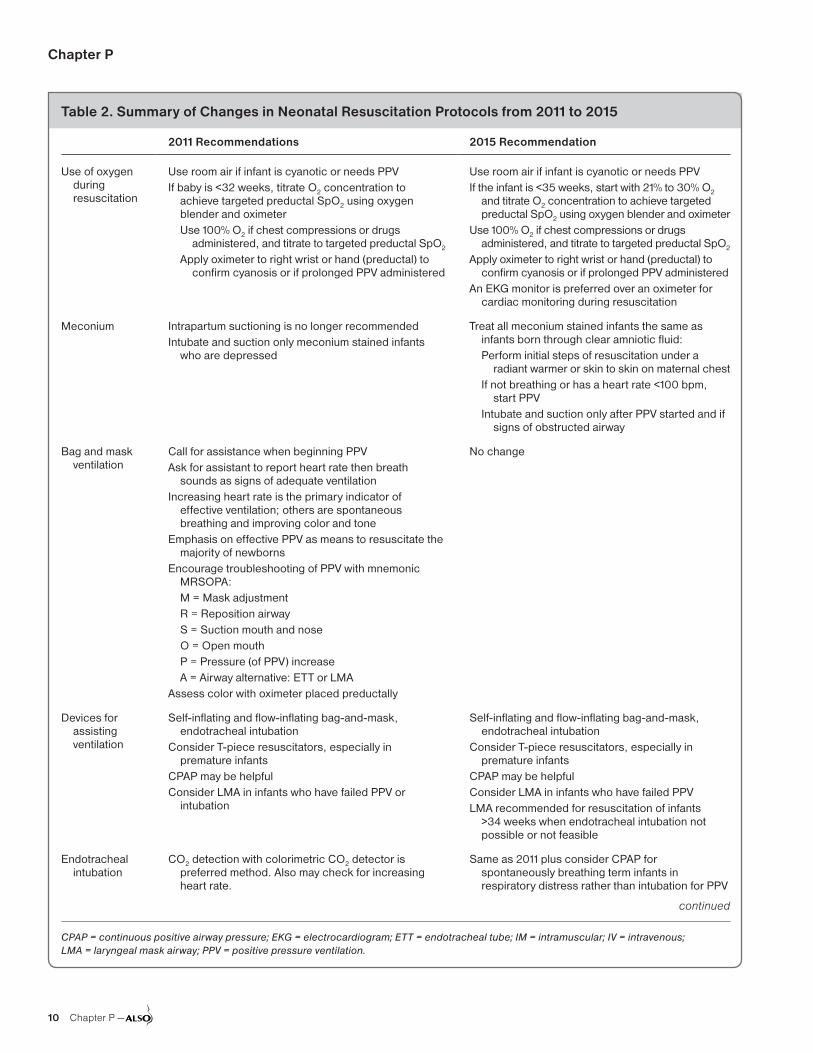
Chapter P
10 Chapter P —
Table 2. Summary of Changes in Neonatal Resuscitation Protocols from 2011 to 2015
2011 Recommendations 2015 Recommendation
Use of oxygen during resuscitation
Use room air if infant is cyanotic or needs PPVIf baby is <32 weeks, titrate O2 concentration to
achieve targeted preductal SpO2 using oxygen blender and oximeterUse 100% O2 if chest compressions or drugs
administered, and titrate to targeted preductal SpO2
Apply oximeter to right wrist or hand (preductal) to confirm cyanosis or if prolonged PPV administered
Use room air if infant is cyanotic or needs PPVIf the infant is <35 weeks, start with 21% to 30% O2
and titrate O2 concentration to achieve targeted preductal SpO2 using oxygen blender and oximeter
Use 100% O2 if chest compressions or drugs administered, and titrate to targeted preductal SpO2
Apply oximeter to right wrist or hand (preductal) to confirm cyanosis or if prolonged PPV administered
An EKG monitor is preferred over an oximeter for cardiac monitoring during resuscitation
Meconium Intrapartum suctioning is no longer recommendedIntubate and suction only meconium stained infants
who are depressed
Treat all meconium stained infants the same as infants born through clear amniotic fluid:Perform initial steps of resuscitation under a
radiant warmer or skin to skin on maternal chestIf not breathing or has a heart rate <100 bpm,
start PPVIntubate and suction only after PPV started and if
signs of obstructed airway
Bag and mask ventilation
Call for assistance when beginning PPVAsk for assistant to report heart rate then breath
sounds as signs of adequate ventilationIncreasing heart rate is the primary indicator of
effective ventilation; others are spontaneous breathing and improving color and tone
Emphasis on effective PPV as means to resuscitate the majority of newborns
Encourage troubleshooting of PPV with mnemonic MRSOPA:M = Mask adjustmentR = Reposition airwayS = Suction mouth and noseO = Open mouthP = Pressure (of PPV) increaseA = Airway alternative: ETT or LMA
Assess color with oximeter placed preductally
No change
Devices for assisting ventilation
Self-inflating and flow-inflating bag-and-mask, endotracheal intubation
Consider T-piece resuscitators, especially in premature infants
CPAP may be helpfulConsider LMA in infants who have failed PPV or
intubation
Self-inflating and flow-inflating bag-and-mask, endotracheal intubation
Consider T-piece resuscitators, especially in premature infants
CPAP may be helpfulConsider LMA in infants who have failed PPVLMA recommended for resuscitation of infants
>34 weeks when endotracheal intubation not possible or not feasible
Endotracheal intubation
CO2 detection with colorimetric CO2 detector is preferred method. Also may check for increasing heart rate.
Same as 2011 plus consider CPAP for spontaneously breathing term infants in respiratory distress rather than intubation for PPV
continued
CPAP = continuous positive airway pressure; EKG = electrocardiogram; ETT = endotracheal tube; IM = intramuscular; IV = intravenous; LMA = laryngeal mask airway; PPV = positive pressure ventilation.

Neonatal Resuscitation
— Chapter P 11
when needed. The AAP developed a course called Helping Babies Breathe (HBB) (available at http://www.helpingbabiesbreathe.org/) that teaches neonatal resuscitation in a manner more appropri-ate for low-resource settings. A study in Tanzania showed that training in HBB lead to a significant
reduction in neonatal mortalities.46 Additional details regarding neonatal resuscitation in develop-ing countries can be found in the Neonatal Resus-citation Chapter Addendum of the Global ALSO Manual available at https://nf.aafp.org/Shop/advanced-life-support-in-obstetrics/global-also-cd.
Table 2. Summary of Changes in Neonatal Resuscitation Protocols from 2011 to 2015
2011 Recommendations 2015 Recommendation
Drugs IV (umbilical vein) is preferred. Consider ETT while obtaining IV access. May also use intraosseous line.
No change
Epinephrine dose: IV: 0.1 to 0.3 mL/kg of 1:10,000 solution, drawn up in a 1-mL syringe
ETT: 0.5 to 1.0 mL/kg of 1:10,000 solution, drawn up in a 3-mL or 5-mL syringe
No change
NaloxoneNo longer recommended as a primary drugRestore heart rate and color by supporting ventilation
before giving naloxoneRoutes: IV (preferred) or IM
No change
Temperature control
Euthermia is the goalPrevent heat loss via evaporation in premature infants
by wrapping in polyethylene from the neck down, immediately after birth, before drying
Hypothermia under the auspices of a tertiary care center within hours of birth may improve outcomes in infants affected by hypoxic-ischemic encephalopathy
No change
Withholding or discontinuing resuscitative efforts
Consider withholding efforts when gestation, birth weight, or congenital anomalies are almost certain to result in early mortality or unacceptably high morbidity
Consider discontinuing efforts if no signs of life after 15 minutes of continuous, effective ventilation
May discontinue after 10 minutes of continuous, effective ventilation without signs of life
Non-initiation and discontinuation of resuscitation are considered ethically equivalent
Parental views on starting resuscitation should be supported in conditions with uncertain prognosis, where there is a high rate of morbidity and borderline survival, and where the burden to the child is high
No change
Teamwork Teamwork and good communication result in better outcomes during emergency situations. These are skills that can be learned.
No change
Chest compressions
Two thumbs encircling technique preferredAllow 45 to 60 seconds of chest compressions before
reassessing
Two fingers technique no longer supported
CPAP = continuous positive airway pressure; EKG = electrocardiogram; ETT = endotracheal tube; IM = intramuscular; IV = intravenous; LMA = laryngeal mask airway; PPV = positive pressure ventilation.
Information from Kattwinkel J, Perlman JM, Aziz K, et al. Part 15: neonatal resuscitation: 2010 American Heart Association Guidelines for Cardiopulmonary Resuscitation and Emergency Cardiovascular Care. Circulation. 2010;122(18)(Suppl 3):S909-S919; Kattwinkel J; American Academy of Pediatrics, American Heart Association. Textbook of Neonatal Resuscitation. 6th Ed. American Academy of Pediatrics; 2011; Wyckoff M, Aziz K, Escobedo M, et al. Part 13: Neonatal Resuscitation: 2015 American Heart Association Guidelines Update for Cardiopulmonary Resus-citation and Emergency Cardiovascular Care. Circulation. 2015;132(18 Suppl 2):S543-S560.

Chapter P
12 Chapter P —
Appendix A: Risk Factors for Neonatal Resuscitation
Maternal
Bleeding in second or third trimester
Pregnancy induced hypertension or preeclampsia
Chronic hypertension
Substance abuse
Drug therapy (eg, lithium, magnesium, adrenergic blocking agents, narcotics)
Diabetes
Chronic illness (eg, renal, thyroid, cardiac, pulmonary, or neurologic disease)
Maternal pyrexia
Maternal infection
Premature rupture of membranes
Chorioamnionitis
Heavy sedation
Previous fetal or neonatal mortality
Mother older than 35 years
No prenatal care
Fetal
Multiple gestation (eg, twins, triplets)
Preterm gestation (especially <35 weeks)
Postterm gestation (>41 weeks)
Size/dates discrepancy
Alloimmune hemolytic disease
Polyhydramnios and oligohydramnios
Reduced fetal movement before onset of labor
Congenital abnormalities, which may affect breathing, cardiovascular function, or other aspects of perinatal transition
Intrauterine infection
Hydrops fetalis
Intrapartum
Prolonged rupture of membranes (>18 hours)
Non-reassuring fetal heart rate patterns
Abnormal presentation
Prolapsed cord
Prolonged labor (>18 hours)
Precipitate labor
Antepartum hemorrhage (eg, abruption, placenta previa, vasa previa)
Meconium in the amniotic fluid
Narcotic administration to mother within 4 hours of birth
Forceps or vacuum-assisted birth
Maternal general anesthesia
Emergency cesarean delivery
Breech or other abnormal presentation

Neonatal Resuscitation
— Chapter P 13
Appendix B: Equipment Needed for Neonatal Resuscitation
Standard Kits
Prior preparation of standardized kits containing equipment for procedures (eg, umbilical catheterization) can save considerable time in emergencies
General
Firm but padded resuscitation surface
Overhead warmer
Light for the area
Clock with timer in seconds
Warm blankets or towels
Polyethylene bag or sheet, big enough for newborns <1,500 g birth weight
Stethoscope (neonatal preferred)
Pulse oximeter plus neonatal probe
Airway Management
Suction apparatus and suction catheters (sizes 5F or 6F, 8F, 10F 12F or 14F)
Oropharyngeal airways (sizes 0, 00, and 000)
Intubation equipment:
• Laryngoscopes with neonatal blades (sizes 00, 0, 1)
• Spare batteries and bulbs
• Compatible laryngoscope handles and blades
• Endotracheal tubes (uncuffed, no eye, sizes 2.5, 3.0, and3.5 mm internal diameter)
• Endotracheal tube introducer/stylet
• Supplies for fixing endotracheal tubes (eg, sterile scissors,tape)
End-tidal carbon dioxide detector (to confirm intubation)
Meconium suction device (to apply suction directly to endotracheal tube)
Magill forceps (neonatal size optional)
Laryngeal mask (size 1, suitable for newborns ≤5 kg) and 5 cc syringe
Breathing Support
Face masks (range of sizes suitable for premature and term newborns)
Positive pressure ventilation device:
• T-Piece resuscitation device, or
• Flow-inflating bag with pressure safety valve and manometer,or
• Self-inflating bag (240 mL) with a pressure release valve anda removable oxygen reservoir
Medical gases to remain:
• Source of oxygen (allowing flow rate of up to 10 L/min) withflow meter and tubing
• Air/oxygen blender
• Oximeter and probe
Feeding tubes for gastric decompression (size 8F) and 20 mL syringe
Circulation Support
Umbilical venous catheter kit (including sizes 3.5F and 5F)
Peripheral intravenous cannulation kit
Skin preparation solution suitable for neonate skin
Tapes/devices to secure umbilical venous catheter/intravenous cannula
Syringes and needles (assorted sizes)
Intraosseous needles (50 mm length)
Drugs
Epinephrine 1:10,000 concentration (0.1 mg/mL)
Volume expander:
Normal saline (0.9% sodium chloride) or Lactated Ringer’s solution
Blood suitable for emergency neonatal
Transfusion should be readily available
Documentation
Resuscitation record sheet
Equipment checklist

Chapter P
14 Chapter P —
References1. Kattwinkel J, Perlman JM, Aziz K, et al. Part 15: neonatal
resuscitation: 2010 American Heart Association Guide-lines for Cardiopulmonary Resuscitation and Emer-gency Cardiovascular Care. Circulation. 2010;122(18)(Suppl 3):S909-S919.
2. Kattwinkel J; American Academy of Pediatrics, Ameri-can Heart Association. Textbook of Neonatal Resuscita-tion. 6th ed. American Academy of Pediatrics; 2011.
3. Wyckoff M, Aziz K, Escobedo M, et al. Part 13: Neona-tal Resuscitation: 2015 American Heart AssociationGuidelines Update for Cardiopulmonary Resuscitationand Emergency Cardiovascular Care. Circulation. 2015;132(18 Suppl 2):S543-S560.
4. Committee on Obstetric Practice, American College ofObstetricians and Gynecologists. Committee OpinionNo.543: Timing of umbilical cord clamping after birth.Obstet Gynecol. 2012;120(6):1522-1526.
5. American Academy of Pediatrics. Statement ofendorsement: timing of umbilical cord clamping afterbirth. Pediatrics. 2013;131:e1323.
6. Mercer JS, Vohr BR, McGrath MM, Padbury JF, WallachM, Oh W. Delayed cord clamping in very preterm infantsreduces the incidence of intraventricular hemorrhageand late-onset sepsis: a randomized, controlled trial.Pediatrics. 2006;117(4):1235-1242.
7. Andersson O, Hellström-Westas L, Andersson D,Domellöf M. Effect of delayed versus early umbilicalcord clamping on neonatal outcomes and iron statusat 4 months: a randomised controlled trial. BMJ. 2011;343:d7157.
8. McDonald SJ, Middleton P. Effect of timing of umbilicalcord clamping of term infants on maternal and neo-natal outcomes. Cochrane Database Syst Rev. 2008;(2):CD004074.
9. Katheria AC, Leone TA, Woelkers D, Garey DM, RichW, Finer NN. The effects of umbilical cord milking onhemodynamics and neonatal outcomes in prematureneonates. J Pediatr. 2014;164(5):1045-1050.e1.
10. Mariani G, Dik PB, Ezquer A, et al. Pre-ductal and post-ductal O2 saturation in healthy term neonates afterbirth. J Pediatr. 2007;150(4):418-421.
11. Toth B, Becker A, Seelbach-Göbel B. Oxygen satura-tion in healthy newborn infants immediately after birthmeasured by pulse oximetry. Arch Gynecol Obstet.2002;266(2):105-107.
12. Altuncu E, Ozek E, Bilgen H, Topuzoglu A, Kavunc-uoglu S. Percentiles of oxygen saturations in healthyterm newborns in the first minutes of life. Eur J Pediatr.2008;167(6):687-688.
13. Kamlin CO, O’Donnell CP, Davis PG, Morley CJ. Oxygensaturation in healthy infants immediately after birth. JPediatr. 2006;148(5):585-589.
14. Gungor S, Kurt E, Teksoz E, Goktolga U, Ceyhan T,Baser I. Oronasopharyngeal suction versus no suctionin normal and term infants delivered by elective cesar-ean section: a prospective randomized controlled trial.Gynecol Obstet Invest. 2006;61(1):9-14.
15. Perlman JM, Volpe JJ. Suctioning in the preterm infant:effects on cerebral blood flow velocity, intracranial pres-sure, and arterial blood pressure. Pediatrics. 1983;72(3):329-334.
16. Chettri S, Adhisivam B, Bhat BV. Endotracheal Suctionfor Nonvigorous Neonates Born through MeconiumStained Amniotic Fluid: A Randomized Controlled Trial.J Pediatr. 2015;166(5):1208-1213.e1.
17. Wiswell TE, Gannon CM, Jacob J, et al. Delivery roommanagement of the apparently vigorous meconium-stained neonate: results of the multicenter, internationalcollaborative trial. Pediatrics. 2000;105(1 Pt 1):1-7.
18. Vain NE, Szyld EG, Prudent LM, Wiswell TE, Aguilar AM,Vivas NI. Oropharyngeal and nasopharyngeal suction-ing of meconium-stained neonates before delivery oftheir shoulders: multicentre, randomised controlled trial.Lancet. 2004;364(9434):597-602.
19. Cramer K, Wiebe N, Hartling L, Crumley E, Vohra S.Heat loss prevention: a systematic review of occlusiveskin wrap for premature neonates. J Perinatol. 2005;25(12):763-769.
20. Kent AL, Williams J. Increasing ambient operatingtheatre temperature and wrapping in polyethyleneimproves admission temperature in premature infants.J Paediatr Child Health. 2008;44(6):325-331.
21. DeMauro SB, Douglas E, Karp K, et al. Improving deliv-ery room management for very preterm infants. Pediat-rics. 2013;132(4):e1018-e1025.
22. Mosalli R. Whole body cooling for infants with hypoxic-ischemic encephalopathy. J Clin Neonatol. 2012;1(2):101-106.
23. Jacobs SE, Berg M, Hunt R, Tarnow-Mordi WO, InderTE, Davis PG. Cooling for newborns with hypoxic isch-aemic encephalopathy. Cochrane Database Syst Rev.2013;1:CD003311.
24. Tan A, Schulze A, O’Donnell CPF, Davis PG. Air versusoxygen for resuscitation of infants at birth. CochraneDatabase Syst Rev. 2005;(2):CD002273.
25. Davis PG, Tan A, O’Donnell CP, Schulze A. Resusci-tation of newborn infants with 100% oxygen or air: asystematic review and meta-analysis. Lancet. 2004;364(9442):1329-1333.
26. Rabi Y, Rabi D, Yee W. Room air resuscitation of thedepressed newborn: a systematic review and meta-analysis. Resuscitation. 2007;72(3):353-363.
27. van Vonderen JJ, Hooper SB, Kroese JK, et al. Pulseoximetry measures a lower heart rate at birth comparedwith electrocardiography. J Pediatr. 2015;166(1):49-53.
28. Hosono S, Inami I, Fujita H, Minato M, Takahashi S,Mugishima H. A role of end-tidal CO(2) monitoring forassessment of tracheal intubations in very low birthweight infants during neonatal resuscitation at birth.J Perinat Med. 2009;37(1):79-84.
29. Finer NN, Rich W, Wang C, Leone T. Airway obstruc-tion during mask ventilation of very low birth weightinfants during neonatal resuscitation. Pediatrics.2009;123(3):865-869.

Neonatal Resuscitation
— Chapter P 15
30. Grein AJ, Weiner GM. Laryngeal mask airway versus bag-mask ventilation or endotracheal intubation for neo-natal resuscitation. Cochrane Database Syst Rev. 2005; (2):CD003314.
31. Trevisanuto D, Micaglio M, Pitton M, Magarotto M, Piva D, Zanardo V. Laryngeal mask airway: is the manage-ment of neonates requiring positive pressure ventilation at birth changing? Resuscitation. 2004;62(2):151-157.
32. Finer NN, Rich W, Craft A, Henderson C. Comparison of methods of bag and mask ventilation for neonatal resuscitation. Resuscitation. 2001;49(3):299-305.
33. Morley CJ, Davis PG, Doyle LW, Brion LP, Hascoet JM, Carlin JB; COIN Trial Investigators. Nasal CPAP or intu-bation at birth for very preterm infants. N Engl J Med. 2008;358(7):700-708.
34. Dorfsman ML, Menegazzi JJ, Wadas RJ, Auble TE. Two-thumb vs. two-finger chest compression in an infant model of prolonged cardiopulmonary resuscita-tion. Acad Emerg Med. 2000;7(10):1077-1082.
35. Orlowski JP. Optimum position for external cardiac compression in infants and young children. Ann Emerg Med. 1986;15(6):667-673.
36. Braga MS, Dominguez TE, Pollock AN, et al. Estimation of optimal CPR chest compression depth in children by using computer tomography. Pediatrics. 2009;124 (1):e69-e74.
37. Barber CA, Wyckoff MH. Use and efficacy of endotra-cheal versus intravenous epinephrine during neonatal cardiopulmonary resuscitation in the delivery room. Pediatrics. 2006;118(3):1028-1034.
38. Perondi MB, Reis AG, Paiva EF, Nadkarni VM, Berg RA. A comparison of high-dose and standard-dose epi-nephrine in children with cardiac arrest. N Engl J Med. 2004;350(17):1722-1730.
39. Wyckoff MH, Perlman JM, Laptook AR. Use of volume expansion during delivery room resuscitation in near-term and term infants. Pediatrics. 2005;115(4):950-955.
40. Moe-Byrne T, Brown JVE, McGuire W. Naloxone for opiate-exposed newborn infants. Cochrane Database Syst Rev. 2013;2(2):CD003483.
41. Field DJ, Dorling JS, Manktelow BN, Draper ES. Sur-vival of extremely premature babies in a geographically defined population: prospective cohort study of 1994-9 compared with 2000-5. BMJ. 2008;336(7655):1221-1223.
42. Phibbs CS, Baker LC, Caughey AB, Danielsen B, Schmitt SK, Phibbs RH. Level and volume of neonatal intensive care and mortality in very-low-birth-weight infants. N Engl J Med. 2007;356(21):2165-2175.
43. Lorch SA, Baiocchi M, Ahlberg CE, Small DS. The dif-ferential impact of delivery hospital on the outcomes of premature infants. Pediatrics. 2012;130(2):270-278.
44. Lawn JE, Cousens S, Zupan J; Lancet Neonatal Sur-vival Steering Team. 4 million neonatal deaths: when? Where? Why? Lancet. 2005;365(9462):891-900.
45. Say L, Chou D, Gemmill A, et al. Global causes of maternal death: a WHO systematic analysis. Lancet Glob Health. 2014;2(6):e323-e333.
46. Msemo G, Massawe A, Mmbando D, et al. Newborn mortality and fresh stillbirth rates in Tanzania after helping babies breathe training. Pediatrics. 2013;131(2): e353-e360.
47. Baby-Friendly USA. Guidelines and Evaluation Crite-ria for Facilities Seeking Baby-Friendly Designation. Sandwich, MA: Baby-Friendly USA, 2010. Available at https://www.babyfriendlyusa.org/get-started/the-guidelines-evaluation-criteria.
48. Vain NE, Satragno DS, Gorenstein AN, et al. Effect of gravity on volume of placental transfusion: a multicen-tre, randomised, non-inferiority trial. Lancet. 2014;384 (9939):235-240.

— Chapter Q 1
Chapter Q Cesarean Delivery
IntroductionThe Recommended Curriculum Guidelines for Family Medicine Residents: Maternity and Gynecologic Care, which was jointly created by the American Academy of Family Physicians (AAFP) and the American College of Obstetricians and Gynecologists (ACOG), describes core and advanced obstetric training for family physi-cians.1 Among other issues, the AAFP’s position paper titled Cesarean Delivery in Family Medicine details scope of practice for family physicians, training methods, test-ing, demonstrated proficiency, and documentation, as well as credentialing and privileges.2
This chapter will review cesarean delivery within the contexts of family physicians, OB/GYN residents, or Certified Nurse-Midwives (CNMs) consulting, first assisting, operating as primary surgeon, performing perimortem cesarean delivery, and attending a trial of labor after cesarean (TOLAC).
HistoryThe origin of the term cesarean is not entirely clear. It is unlikely that Julius Caesar was born by abdominal delivery because that was almost universally fatal for the parturient during that era and Caesar’s mother is known to have survived his birth.3 Another possible origin of the term is the Latin verb caedere, which means to cut. Others think the term originated from the Roman cus-tom, Lex Cesare, which mandated postmortem opera-tive delivery when mothers died during childbirth so the mother and child could be buried separately.
The term cesarean section is also a matter of discus-sion. The term is a tautology–using different words to say the same thing twice–where the additional words fail to provide additional clarity while repeating a meaning. Both words in this case refer to an incision. A more proper term for the procedure is cesarean delivery.
EpidemiologyFrequencyThe cesarean delivery rate was 32.2% of US births in 2014.4 The cesarean delivery rate in the United States increased from 4.5% in 1965 to an all-time high of 32.9% in 2009.4,5 That represents an increase of 56% from the 20.7% rate in 1996.5 The increase is a result of the increased rate of primary cesarean delivery and the decreased rate of vaginal birth after cesarean deliv-ery (VBAC).
Safe Prevention of the Primary Cesarean DeliveryNoted by ACOG and the Society for Maternal-Fetal Medicine (SMFM), the rapid increase in cesarean birth rates from 1996 to 2011 without clear evidence of a concomitant decrease in maternal or neonatal morbidity or mortality raises significant concern that cesarean delivery is overused.6 In 2014, ACOG and SMFM made recommendations for the safe preven-tion of the primary cesarean delivery (Table 1). The consensus statement gave several examples of interven-tions which can contribute to the safe lowering of the
Learning ObjectivesAt the end of this activity, learners will be able to:1. Describe anatomy and physiology relevant to cesarean delivery.2. Describe steps leading to cesarean delivery of a fetus.3. Describe complications of cesarean delivery.4. Describe risks and benefits of trial of labor after cesarean (TOLAC).5. Describe the indications for perimortem cesarean delivery.
Neil J. Murphy, MD, FACOG, FAAFP, Sarah K. Jorgensen, DO, Jeffrey D. Quinlan, MD, FAAFP
Revised January 2017
Copyright 2017© American Academy of Family Physicians. All rights reserved.
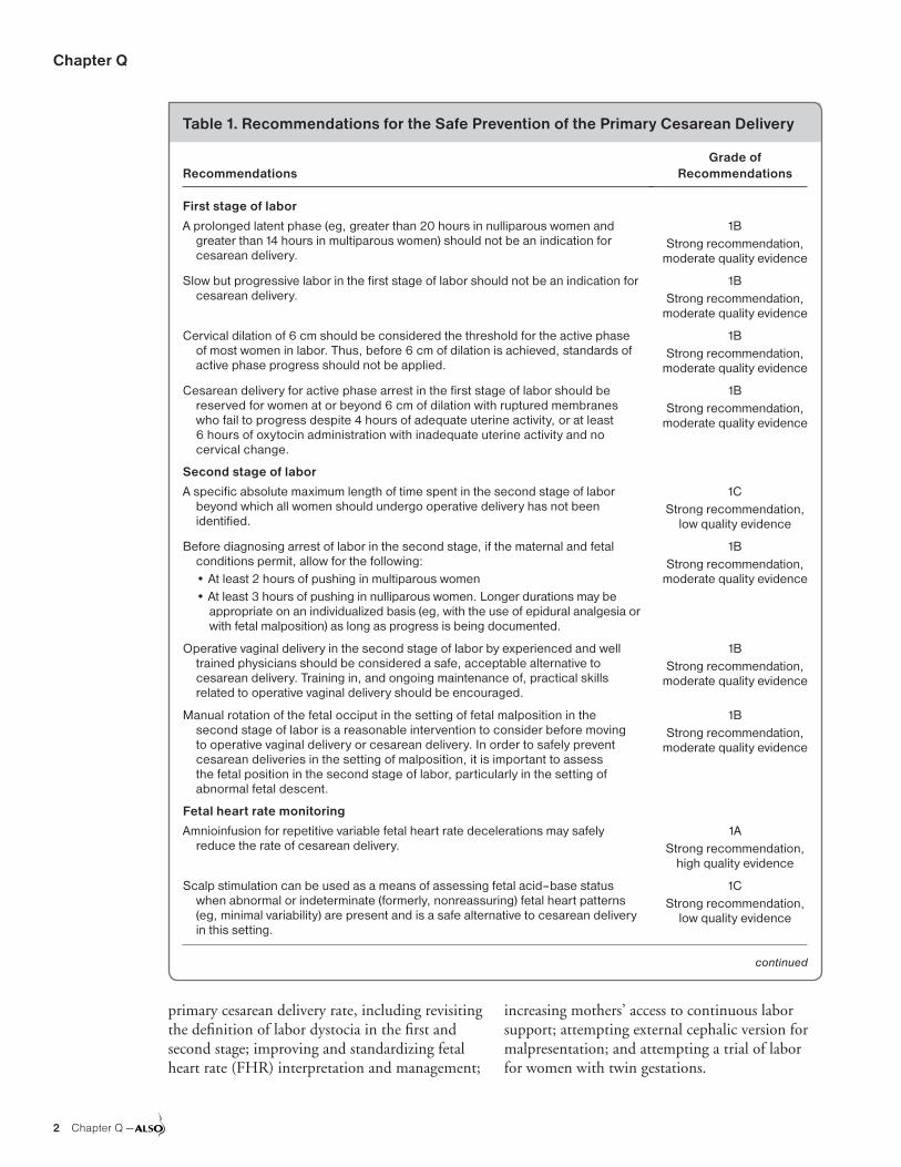
Chapter Q
2 Chapter Q —
primary cesarean delivery rate, including revisiting the definition of labor dystocia in the first and second stage; improving and standardizing fetal heart rate (FHR) interpretation and management;
increasing mothers’ access to continuous labor support; attempting external cephalic version for malpresentation; and attempting a trial of labor for women with twin gestations.
Table 1. Recommendations for the Safe Prevention of the Primary Cesarean Delivery
RecommendationsGrade of
Recommendations
First stage of labor
A prolonged latent phase (eg, greater than 20 hours in nulliparous women and greater than 14 hours in multiparous women) should not be an indication for cesarean delivery.
1BStrong recommendation,
moderate quality evidence
Slow but progressive labor in the first stage of labor should not be an indication for cesarean delivery.
1BStrong recommendation,
moderate quality evidence
Cervical dilation of 6 cm should be considered the threshold for the active phase of most women in labor. Thus, before 6 cm of dilation is achieved, standards of active phase progress should not be applied.
1BStrong recommendation,
moderate quality evidence
Cesarean delivery for active phase arrest in the first stage of labor should be reserved for women at or beyond 6 cm of dilation with ruptured membranes who fail to progress despite 4 hours of adequate uterine activity, or at least 6 hours of oxytocin administration with inadequate uterine activity and no cervical change.
1BStrong recommendation,
moderate quality evidence
Second stage of labor
A specific absolute maximum length of time spent in the second stage of labor beyond which all women should undergo operative delivery has not been identified.
1CStrong recommendation,
low quality evidence
Before diagnosing arrest of labor in the second stage, if the maternal and fetal conditions permit, allow for the following:• At least 2 hours of pushing in multiparous women• At least 3 hours of pushing in nulliparous women. Longer durations may be
appropriate on an individualized basis (eg, with the use of epidural analgesia orwith fetal malposition) as long as progress is being documented.
1BStrong recommendation,
moderate quality evidence
Operative vaginal delivery in the second stage of labor by experienced and well trained physicians should be considered a safe, acceptable alternative to cesarean delivery. Training in, and ongoing maintenance of, practical skills related to operative vaginal delivery should be encouraged.
1BStrong recommendation,
moderate quality evidence
Manual rotation of the fetal occiput in the setting of fetal malposition in the second stage of labor is a reasonable intervention to consider before moving to operative vaginal delivery or cesarean delivery. In order to safely prevent cesarean deliveries in the setting of malposition, it is important to assess the fetal position in the second stage of labor, particularly in the setting of abnormal fetal descent.
1BStrong recommendation,
moderate quality evidence
Fetal heart rate monitoring
Amnioinfusion for repetitive variable fetal heart rate decelerations may safely reduce the rate of cesarean delivery.
1AStrong recommendation,
high quality evidence
Scalp stimulation can be used as a means of assessing fetal acid–base status when abnormal or indeterminate (formerly, nonreassuring) fetal heart patterns (eg, minimal variability) are present and is a safe alternative to cesarean delivery in this setting.
1CStrong recommendation,
low quality evidence
continued

Cesarean Delivery
— Chapter Q 3
Morbidity and MortalityThe maternal mortality rate for elective repeat cesarean delivery is 13.4/100,000 births in the United States.7 Half of these mortalities are related to intraoperative complications, and the others are related to anesthetic and postoperative complica-tions. In recent years, there has been a shift in the
etiology of mortalities from hemorrhage and infec-tion to thromboembolic events.7
IndicationsThe most common indication for overall cesarean delivery is a repeat procedure, whereas the most common indications for primary cesarean deliv-
Table 1. Recommendations for the Safe Prevention of the Primary Cesarean Delivery (continued)
RecommendationsGrade of
Recommendations
Induction of labor
Before 41 0/7 weeks of gestation, induction of labor generally should be performed based on maternal and fetal medical indications. Inductions at 41 0/7 weeks of gestation and beyond should be performed to reduce the risk of cesarean delivery and the risk of perinatal morbidity and mortality.
1AStrong recommendation,
high quality evidence
Cervical ripening methods should be used when labor is induced in women with an unfavorable cervix.
1BStrong recommendation,
moderate quality evidence
If the maternal and fetal status allow, cesarean deliveries for failed induction of labor (up to 24 hours or longer) in the latent phase can be avoided by allowing longer durations of the latent phase and requiring that oxytocin be administered for at least 12 to 18 hours after membrane rupture before deeming the induction a failure.
1BStrong recommendation,
moderate quality evidence
Fetal malpresentation
Fetal presentation should be assessed and documented beginning at 36 0/7 weeks of gestation to allow for external cephalic version to be offered.
1CStrong recommendation,
low quality evidence
Suspected fetal macrosomia
Cesarean delivery to avoid potential birth trauma should be limited to estimated fetal weights of at least 5,000 g in women without diabetes and at least 4,500 g in women with diabetes. The prevalence of birth weight of 5,000 g or more is rare, and patients should be counseled that estimates of fetal weight, particularly late in gestation, are imprecise.
2CWeak recommendation,
low quality evidence
Excessive maternal weight gain
Women should be counseled about the IOM maternal weight guidelines in an attempt to avoid excessive weight gain.
1BStrong recommendation,
moderate quality evidence
Twin gestations
Perinatal outcomes for twin gestations in which the first twin is in cephalic presentation are not improved by cesarean delivery. Thus, women with either cephalic/cephalic-presenting twins or cephalic/noncephalic presenting twins should be counseled to attempt vaginal delivery.
1BStrong recommendation,
moderate quality evidence
Other
Individuals, organizations, and governing bodies should work to ensure that research is conducted to provide a better knowledge base to guide decisions regarding cesarean delivery and to encourage policy changes that safely lower the rate of primary cesarean delivery.
1CStrong recommendation,
low quality evidence
IOM = Institute of Medicine.
Reprinted from American College of Obstetricians and Gynecologists; Society for Maternal-Fetal Medicine. Obstetric care consensus no. 1: safe prevention of the primary cesarean delivery. Obstet Gynecol. 2014;123(3):693-711.

Chapter Q
4 Chapter Q —
ery include, in order of frequency, labor dystocia (34%), abnormal or indeterminate (previously, nonreassuring) FHR tracing (23%), fetal malpre-sentation (17%), multiple gestation (7%), and sus-pected fetal macrosomia (4%).6 Other indications are listed in Table 2. Many indications are depen-dent on other clinical factors (eg, viral load with HIV, unable to meet criteria for vaginal breech delivery, and current status of medical conditions).
ContraindicationsThere are few contraindications for cesarean delivery. A guiding principle is what is best for the mother is best for the fetus. If the mother is medi-cally unstable and the fetus is nonviable, then it is recommended that the maternal condition be stabilized regardless of fetal consideration. If the fetus is of a gestational age consistent with viabil-ity, then the maternal condition should also be stabilized first and delivery considered only for obstetric indications. The exception to this dictum is perimortem cesarean delivery.
Timing of Elective Cesarean DeliveryAlthough term pregnancy is defined as 37 weeks of gestation or later; elective cesarean delivery is typically not performed before 39 weeks’ gestation secondary to the risk of fetal lung nonmaturity. Despite this risk, Tita showed that over one-third of elective cesarean deliveries completed at their facility between 1999 and 2002 were performed before 39 weeks’ gestation. Additionally, they showed that delivery before 39 weeks was associ-
ated with increased rates of respiratory issues, sep-sis, hypoglycemia, and prolonged hospitalization or requirement for increased level of care.8 There-fore, cesarean delivery should not be performed before 39 weeks’ gestation unless there are medical indications for the mother or the fetus.
For patients with complicating factors (eg, pla-centa previa or prior classical uterine incision), the risk of maintaining the pregnancy to 39 weeks may outweigh the risks to the mother and fetus if delivered before 39 weeks. Similarly, women with planned repeat cesarean deliveries may have obstetrical complications with indications for delivery before 39 weeks such as preeclampsia or intrauterine growth restriction with abnormal Doppler studies.
Anatomy and PhysiologyMany alterations in maternal cardiovascular physi-ology during pregnancy are relevant to cesarean delivery. More information is available in Chapter K: Maternal Resuscitation and Trauma. These phys-iologic changes increase maternal blood volume and flow in the pelvic organs, rendering the woman more susceptible to serious hemorrhage during cesarean delivery. The surgical anatomy is described in this chapter with each aspect of the procedure. An understanding of pelvic blood supply is essential for physicians performing a cesarean delivery.
Uterine ArteryThe aorta bifurcates into bilateral common iliac arteries at the level of the fourth lumbar vertebra.
Table 2. Common Indications for Cesarean Delivery
FetalCategory II or III FHR tracingMalpresentation:
Transverse lieBreechBrowFace/mentum posterior
Cord prolapseHIV with viral load >1,000/mLActive herpes virus or prodromal symptomsCongenital anomaliesVasa previa
Maternal-FetalFailure to progress in labor:
Arrest of descentArrest of dilation
Placental abruptionPlacenta previaConjoined twinsPerimortem
MaternalRepeat cesarean deliveryContracted pelvis:
CongenitalFracture
Obstructive tumorsAbdominal cerclageReconstructive vaginal surgeryMedical conditions (eg, cardiac,
pulmonary, thrombocytopenia)
FHR = fetal heart rate.

Cesarean Delivery
— Chapter Q 5
The common iliac divides into external and inter-nal iliac arteries. The internal iliac or hypogastric artery drops medio-inferiorly along the border of the psoas muscle and divides into anterior and posterior divisions. The anterior division has pari-etal and visceral branches of variable origin.
The uterine artery, a main visceral branch of the anterior division of the hypogastric artery, descends for a short distance, enters the base of the broad ligament, and turns medially to the lat-eral aspect of the uterus. The relationship between the uterine artery and ureter is surgically signifi-cant. Approximately 2 cm lateral to the cervix, the uterine artery crosses over the ureter. The ureter can be injured in the process of clamping and ligating the uterine vessels in postpartum hemor-rhage or during hysterectomy. A common way to remember this is that the uterine vessels form a bridge over running water.
The inferior branch of the uterine artery sup-plies the upper vagina and the lower cervix, and the marginal branch traverses the lateral aspect of the uterus before dividing into three terminal branches: ovarian, tubal, and fundal. Near the upper lateral portion of the uterus, the ovarian artery anastomoses with the ovarian branch of the uterine artery. Throughout its length, the mar-ginal branch is a convoluted vessel with numer-ous branches penetrating the body of the uterus, including one large branch that extends to the upper portion of the cervix.
Ovarian ArteryThe ovarian artery is a direct branch of the aorta and enters the broad ligament through the infun-dibulopelvic ligament. At the ovarian hilum, the ovarian artery divides into ovarian branches and a main branch that traverses the broad ligament.
Uterine and Ovarian VeinsThe lateral uterus is composed largely of venous sinuses. These sinuses coalesce into arcuate veins that unite to form the uterine vein. Several large uterine veins accompany the uterine artery and empty into the hypogastric vein, which empties into the common iliac vein. The ovarian vein collects blood from the upper part of the uterus through a large pampiniform plexus in the broad ligament. The right ovarian vein empties into the vena cava, and the left ovarian vein empties into the left renal vein.
Vaginal Blood SupplyThe vagina receives blood from the inferior exten-sion of the uterine artery along the lateral sulci of the vagina and from a vaginal branch of the hypogastric artery. These form an anastomotic arcade along the lateral aspect of the vagina at the 3:00 and 9:00 o’clock positions. Branches of these vessels also merge along the anterior and posterior vaginal walls.
Physical Findings and DiagnosisHistoryHistory should be obtained that is pertinent to impending surgery, including medical, surgical, obstetric, gynecologic, family, tobacco and drug habits, transfusion, medications, allergies, and thrombotic and anesthetic complications. Infor-mation about the current obstetric indication is required (eg, length of labor and duration of ruptured membranes, day of operation, and time of last food and drink should be reviewed).
Physical ExaminationThe physical examination for cesarean delivery should address major medical, obstetrical, and anesthetic concerns. The operating team must be cognizant that regional anesthesia may be con-verted to general anesthesia at any time during the procedure. A vaginal examination should be per-formed in most laboring patients just before surgi-cal draping to ensure that sufficient progress in labor has not occurred that would permit vaginal delivery. Patients with a history of herpes should have a careful examination for active lesions.
Ancillary TestsPreoperative laboratory evaluation should include a complete blood count, blood type, and screen. A blood clot tube should be present in the blood bank for cross matching in the event that transfu-sion is required. HIV status should be known on all prenatal patients so that measures can be taken to decrease the risk of vertical transmission of infection. A Kleihauer-Betke test to quantify the amount of fetal maternal hemorrhage may be indi-cated for Rh negative women in situations such as major trauma. If the cesarean delivery is being considered because of abnormal FHR monitoring that does not resolve with conservative measures, additional testing may be indicated because of the high rate of false positive tests with electronic fetal

Chapter Q
6 Chapter Q —
monitoring (EFM). More information is avail-able in Chapter E: Intrapartum Fetal Surveillance. Short-term evidence of fetal reserve can be docu-mented by acceleration of the FHR in response to scalp or acoustic stimulation.
ProcedurePreoperative and Nonsurgical ConsiderationsThe patient should be prepared in the same manner as any other major abdominal procedure (Table 3). Additional fluids are necessary because of peripheral vasodilation caused by regional anes-thesia, increased insensible loss with labor, blood loss and intraoperative loss of 1,000 cc/hour because of exposed viscera.
Fluid administration before epidural or spinal anesthesia in normotensive patients typically involves a 1,000 cc bolus of isotonic fluids. Iso-tonic fluids are good first-line agents in the event of excessive bleeding, but blood product replace-ment is often necessary for blood loss greater than 1,000 mL. Use of prophylactic antibiotics has been shown to decrease the incidence of fever, endometritis, wound infection, urinary tract infection, and serious postoperative infection after cesarean delivery.9 Ampicillin and first-generation cephalosporins have similar efficacy in reducing postoperative endometritis. There does not appear to be added benefit in using a broader spectrum agent or a multiple-dose regimen.10 All cesarean
delivery patients should receive intravenous (IV) antibiotics before skin incision, unless the patient is already receiving appropriate antibiotics (eg, for chorioamnionitis) or there is not adequate time because of a crash cesarean delivery.11,12 Pre-incision prophylaxis is advantageous for the mother and not harmful to the newborn, there-fore the practice of administering antibiotics after cord clamping has been abandoned.13 Consider clindamycin in patients who are allergic to penicil-lin with a history of anaphylaxis, urticaria, or other life threatening reaction. If a procedure exceeds 4 hours, then re-dosing should be considered. Addi-tionally, recent data suggests that in obese women, the cefazolin dose should be increased from a 1 or 2 g IV dose to a 3 g IV dose.14
Infective endocarditis prophylaxis is no longer recommended for vaginal or cesarean delivery in the absence of infection, regardless of the type of maternal cardiac lesion.15 Abdominal hair removal often is not necessary. If hair is removed, it should be removed in the operating room and not the evening before the procedure. The hair should be clipped and not shaved to decrease the risk of infection.
Informed ConsentThe surgeon should thoroughly discuss the risk and benefits of the procedure in medical and non-medical terminology with the mother and a family member, if available. The counseling is best docu-mented in narrative form, though a preprinted form can be used. Preoperative documentation should be signed and dated by the patient and sur-geon. Documentation should include diagnosis, procedure, common and important risk factors, alternatives to the proposed procedure, and other potential procedures anticipated by the surgeon (eg, tubal ligation or oophorectomy for known adnexal mass). The risk factors can be simplified to bleeding, infection, internal organ damage, anesthesia risk, hysterectomy, injury to fetus, and risk of maternal mortality.
Cesarean Delivery ProcedureAlthough the primary surgeon and assistant typi-cally focus their conversation before a cesarean delivery on variations of the technique and the increased risk of complications based on the clini-cal scenario, they should also carefully review the basic skill discussion in Table 4.
Table 3. Preoperative Preparation/Orders
Vital signs and nonstress test on admissionAnesthesia consultationNothing per mouth (except nonparticulate citrate antacid)IV: Lactated Ringer solution at 125 cc/hour; if regional anesthetic,
then bolus of IV fluids per anesthesiaNonparticulate citrate antacid 30 cc per mouth 1 hour pre-op,
or on-call to ORCefazolin 1 to 2 g IV to be administered 15 to 60 minutes before
skin incisionPlace patient in left lateral decubitus positionInsert bladder catheterClip lower abdominal hair as neededSequential compression devices on lower extremitiesLab: Complete blood count, blood type and screenPatient education: Cesarean delivery
IV = intravenous; OR = operating room.

Cesarean Delivery
— Chapter Q 7
Abdominal Wall IncisionOptions for the abdominal wall incision include the modified-Pfannenstiel, Joel-Cohen, and mid-line vertical incisions, plus several variants of these incisions (Figure 1).16 The midline vertical incision was traditionally considered the quickest abdomi-nal wall incision, but most experienced surgeons can perform the Joel-Cohen or the modified Pfan-nenstiel incision just as quickly.
PfannenstielThe modified-Pfannenstiel incision is made 3 cm (two-finger breadths) above the pubic symphysis. The incision is extended beyond the lateral bor-ders of the rectus muscles in a curvilinear fashion to within 2 to 3 cm inferior and medial of the anterior superior iliac crests. The incision may be placed under the pannus in very obese patients, but this area is heavily colonized with bacteria and may be difficult to prepare surgically, keep dry, and inspect in the postoperative period.
The subcutaneous tissues are completely sepa-rated from the fascia and a transverse incision
Table 4. Techniques of Cesarean Delivery
Prepare patientInformed consentNothing by mouth except nonparticulate antacidAnesthesiaBladder catheterClip hair, cleanse skin, left lateral decubitus positionChlorhexidine-alcohol for skin antisepticCefazolin intravenously within 60 minutes before
skin incisionSequential compression devices on lower
extremitiesAbdominal wall incisionJoel-Cohen (Misgav Ladach modification)PfannenstielMidline verticalOthers: Maylard, CherneyFascial incisionJoel-Cohen: small midline fascial incision, stretch
tissuePfannenstiel: long transverse incision, separate
rectus muscle/sheath, stretch rectus muscles apart
Peritoneal incisionParietal: longitudinal; transverse (Joel-Cohen)Visceral: transverse vesicouterine; ±develop
bladder flap
Uterine incisionLow transverseClassicalLow verticalElevate the fetal presenting partElevate presenting part, maintain
flexion if cephalicSecond assistant to dislodge
presenting part vaginally, if deeply seated
Apply fundal pressureAdminister oxytocin after delivery of
presenting partClamp and cut umbilical cordObtain cord blood for:
Type and direct Coombs, if clinically indicated
Cord pH from loop of cord obtained before cord blood sample, if clinically indicated
Delivery of placentaAssisted spontaneousManualCleanse uterine cavity
Uterine closurePlace ring forceps at apices of
uterine incision (optional)Externalize uterus (optional)Inspect for possible extensionsTwo layers of absorbable sutureInspect pelvic and abdominal
contentsRemove foreign material from
peritoneal cavitySponge and needle countPeritoneal closure (optional)Visceral (bladder flap)ParietalFascial closureSingle nonlockingTwo lines of suture meeting in the
midline, nonlocking (optional)Subcutaneous (if tissue >2 cm)Close dead space if >2 cmIrrigate subcutaneous tissueSkin closure: subcuticular,
staples, widely spaced mattress sutures
Apply sterile dressing
Figure 1. Abdominal Wall Incisions

Chapter Q
8 Chapter Q —
is made through the fascia. The fascial sheath is then completely separated from the underlying rectus muscles by blunt and sharp dissection to the umbilicus and caudad until the pubis is palpable. Blood vessels perforating through the muscles can be ligated with electrocautery, or cut and clamped as required for hemostasis. The peritoneum is elevated and sharply or bluntly opened longitudi-nally in the midline.
Joel-Cohen (Misgav Ladach Modification)The Joel-Cohen abdominal wall incision, modi-fied by the Misgav Ladach Hospital in Jerusalem, emphasizes stretching tissue within existing planes, rather than sharp dissection (Table 5).17
Joel-Cohen-based cesarean delivery compared with Pfannenstiel cesarean delivery was associated with reduced blood loss, operating time, time to oral intake, fever, duration of postoperative pain, analgesic injections, and time from skin incision to birth of the newborn.16
Available evidence suggests that the Joel-Cohen-based techniques (Joel-Cohen, Misgav Ladach, and modified Misgav Ladach) have advantages over Pfannenstiel and traditional cesarean delivery tech-niques in relation to short-term outcomes. There is no evidence in relation to long-term outcomes.16
This technique has particular advantages in remote or rural areas because it requires fewer instruments than other methods for opening the abdominal wall and can be performed quickly. The modified Joel-Cohen incision begins with a transverse incision, 15 to 17 cm long, made 3 cm
below the anterior superior iliac crests. The skin is opened superficially, followed by sharp dissection of the subcutaneous fat to open the fascia in the midline only.
The fascial incision is extended sharply 2 to 3 cm under the intact subcutaneous tissue. After opening the fascia, the remaining subcutaneous tis-sue, fascia, and rectus muscles are dissected bluntly. The fascia is best opened with cephalad and caudad pressure followed by transverse pressure on the rectus muscles laterally. The incision is rapid and results in less blood loss than other techniques. There is a decreased need for transfusion and less risk of HIV transmission because the technique simply stretches tissues transversely. Decreased tis-sue damage also leads to less postoperative analgesia and early resumption of feeding and activity.
Midline VerticalThe midline-vertical skin incision extends from the pubic symphysis to within 2 cm of the umbi-licus. The fascia is elevated and sharply dissected from the pubis to the umbilicus. This midline-vertical abdominal wall incision can be performed rapidly and provides excellent exposure of the pelvis and sidewalls.
Other IncisionsThe transverse Maylard rectus-cutting incision begins with a curvilinear skin incision that extends 18 to 19 cm between the anterior superior iliac crests. The Maylard offers maximal exposure for abnormal lie, multiple gestation, or macrosomia. In the transverse Cherney incision, the rectus mus-cles are detached from their insertion at the pubic symphysis. The transversalis fascia and peritoneum are incised transversely in the Cherney, as opposed to the longitudinal Pfannenstiel approach.
Peritoneal IncisionParietal PeritoneumThe parietal peritoneum should be entered as high as possible to avoid inadvertent bladder injury, especially in repeat procedures. The parietal peri-toneum is sharply incised or entered with blunt digital dissection and then stretched open.
Urinary BladderThe urinary bladder can be divided into two por-tions, the dome and base. The base of the blad-der, which rests on the upper vagina and cervix,
Table 5. Misgav Ladach Method of Cesarean Delivery
1. Perform abdominal incision utilizing a modifiedJoel-Cohen approach
2. Use index fingers to open the parietalperitoneum transversely
3. Use a small lower uterine transverse incisionand extend laterally using index fingers
4. Close the uterus with a single continuousnonlocked layer of absorbable monofilament
5. Do not close the visceral or parietal peritoneum 6. Close the fascia with a single nonlocking
continuous closure of polyglactin7. Skin is closed with several widely spaced skin
stitches and the margins between sutures areapproximated with Allis clamps for 5 minutes

Cesarean Delivery
— Chapter Q 9
contains the trigone and is contiguous with the muscle of the vesical neck and urethra. The mus-cular dome of the bladder is relatively thin when distended. The bladder base is thicker and varies less with distention. The bladder is encountered twice before delivery. Initially, the surgeon visual-izes the bladder when opening the peritoneum, and it is encountered again when dissecting the bladder flap off the lower uterine segment. The extent of the bladder can be confirmed by palpa-tion of the catheter bulb.
Visceral PeritoneumOmission of the bladder flap provides short-term advantages such as reduction of operating time and incision-delivery interval, reduced blood loss, and need for analgesics.18 Long-term effects still need to be evaluated. In those cases in which a cesarean hysterectomy is planned, a bladder flap should be created.
If deemed necessary, the vesico-uterine perito-neum is elevated and opened transversely 1 cm above the bladder reflection onto the lower uterine segment. The bladder flap is bluntly and sharply developed transversely 10 to 12 cm, then inferiorly to the level of the bladder’s apposition to the cervix. If a bladder flap is developed, the bladder blade is repositioned to lower and protect the bladder.
Uterine IncisionCesarean delivery is performed via one of sev-eral uterine incisions. The most common is a low transverse or Kerr incision. A less common surgical approach is the classical or vertical uterine incision. Both can be performed through any abdominal incision.
UterusAs the uterus enlarges, it reaches almost to the liver and displaces the intestines laterally and superiorly. The uterine musculature is arranged in three layers. The muscle cells in the middle layer are interlaced such that when they contract after delivery, they constrict the perforating blood vessels. When a pregnant woman is supine, her uterus falls back to rest on the vertebral column and the great vessels, especially the aorta and the inferior vena cava. With ascent from the pelvis as pregnancy progresses, the uterus undergoes dextro-rotation, resulting in the left margin facing anteriorly.
Low Transverse IncisionThe low transverse Kerr incision is made in the inactive or noncontractile lower uterine segment. Most cesarean deliveries use a low transverse uterine incision because of the ease of delivery and low rate of immediate and subsequent wound dehiscence. The low incidence of dehiscence occurs because the low transverse incision avoids the active uterine segment. It also requires less surgical repair, results in less blood loss, and is less likely to result in formation of adhesions to the bowel or omentum.
The lower uterine segment is delicately scored in the median aspect with a scalpel 1 to 2 cm from the upper margin of the bladder while taking care to avoid injury to the fetus. If the lower uterine segment is thin, fetal laceration injury, which occur in between 0.7% and 1.9% of cesarean deliveries,19 can be avoided by elevating the lower uterine seg-ment with Allis clamps. Another method involves cutting to within a few cells of the uterine cavity, then tapping the closed blades of a pair of scissors against the incision. Opening and closing the blunt tips of the scissors will not hurt the fetus nor enter the amniotic cavity, but will penetrate the remain-ing layers of lower segment.
The uterine incision is extended bluntly using two fingers. Blunt expansion of the uterine incision by separating the fingers in a cephalad-caudad direction results in less unintended extension and blood loss than expansion in a transverse direction.20 The incision should extend approximately 10 cm transversely and slightly cephalad in a curvilinear fashion. If the uterine wall is thickened or there is need for an exten-sion, then bandage scissors should be used. In this case, care should be taken to avoid injury to the fetus or umbilical cord. The incision should be large enough to avoid fetal injury and to avoid inadvertent extension into lateral vessels. If it is necessary to extend the uterine incision, then the first superior curvilinear incision should be to the right to avoid the lateral vessels because of the dextro-rotation. Some surgeons place laparotomy sponges in the peritoneal cavity to minimize contamination with chorioamnionitis or thick meconium. All laparotomy sponges placed in the abdomen should have radio-opaque tails. The sur-geon should know the location of every sponge in the abdomen, and not simply rely on the operat-ing team’s count.

Chapter Q
10 Chapter Q —
Classical/Vertical IncisionThe classical uterine incision is made vertically into the active myometrium. The classical incision is indicated in significant prematurity with a poorly developed narrow lower uterine segment, dense adhesions, or structural uterine abnormalities (eg, myoma in the lower uterine segment or Bandl’s contractile uterine ring). The classical incision may be used in some cases of anterior placenta previa and malpresentation (eg, back-down transverse lie, preterm breech, and interlocking twins).
Low Vertical IncisionThe low vertical incision begins as inferiorly as pos-sible to avoid the active uterine segment. The inci-sion is typically made approximately 2 cm above the bladder and is carried as far cephalad as neces-sary to allow facile delivery. This may be performed completely in the lower uterine segment and repair may be as strong as the low transverse incision. In a study of TOLAC with 2,912 women with a low transverse uterine incision and 377 women with a low vertical incision, there was no significant differ-ence in the rate of uterine rupture.21
The major disadvantage of the low vertical inci-sion is the possibility of extension cephalad into the uterine fundus or caudally into the bladder, cervix, or vagina. It is also difficult to determine if the low vertical incision is truly low, as the separa-tion between lower and upper uterine segments is not easily identifiable clinically.
Anterior Placenta ManagementIf an anterior placenta is present, it should be dis-sected or separated from the uterine wall facilitat-ing exposure of the fetus. There is a short-term risk of fetal hemorrhage unless delivery is rapid. If the placenta is lacerated, the operator should cut through the rest of the placenta and quickly delivery the infant and clamp the cord. A vertical incision may be necessary. A preoperative ultrasound for placental location may be helpful. Additionally, if the patient has had a previous cesarean delivery, the possibility of placenta accreta should be considered and the operating team should be prepared for the potential of hemorrhage and need for hysterectomy.
Delivery of the FetusCephalic PresentationTo deliver an infant from a cephalic presentation, remove the retractors and elevate the presenting
part with the operator’s hand, then the assistant applies transabdominal pressure to the uterine fundus. If the presenting part is deeply applied to the cervix or vagina, then gently insinuate the hand into the uterus with side-to-side motions to break the suction and act as a lever to elevate the presenting part. The operator should avoid using the uterine incision as a fulcrum for elevat-ing the presenting part to avoid extension of the uterine incision. The assistant applies fundal pressure when the operator feels the presenting part is elevated enough that the force will push the presenting part up and out of the incision instead of deeper into the pelvis. Flexion is desirable in occiput posterior or anterior positions. Too much manipulation of a thin lower uterine segment may lead to a deep cervical laceration. If the presenting part is deeply seated in the pelvis, then an addi-tional assistant may need to go under the operative drapes to manually dislodge the presenting part cephalad via the vagina.
If the presenting part is high, then a vacuum or single forceps blade may be helpful. Deliver the torso by gently working the shoulders out one at a time with continued fundal pressure. Alterna-tively, the fetal feet may be delivered first. The newborn is transferred to an attendant after the umbilical cord is clamped and cut.
Breech PresentationThe presenting part should be confirmed preop-eratively with ultrasound. A breech presentation may require a slightly larger abdominal wall and uterine incision for adequate exposure. A verti-cal uterine incision may be necessary if the lower uterine segment is not well developed (eg, very preterm). The techniques for a breech cesarean delivery are similar to those used in a breech vaginal delivery. Additional information on breech deliveries is available in Chapter G: Malpresenta-tions, Malpositions, and Multiple Gestations. The abdominal and uterine incisions can be extended if delivery of the fetal head is difficult. The uterine incision can be extended vertically into the active myometrium, perpendicular to the transverse uter-ine incision in an inverted T shape, or extended perpendicularly to the uterine vessels in a J shape. These extensions should be noted in the opera-tive report and the patient informed that repeat cesarean delivery is indicated in future pregnancies because of increased risk of uterine rupture.

Cesarean Delivery
— Chapter Q 11
After Delivery of the FetusCord blood is obtained and may be sent for infant blood type and Rh status, Coombs testing, HIV and/or rapid plasma reagin testing based on facil-ity guidelines.22 In addition, a 10 to 15 cm seg-ment of umbilical cord may be saved for blood gas measurement. To obtain a sufficient amount of arterial pH specimen, the cord should be clamped close to the placenta. Delayed cord clamping for 1 minute may be used if the infant is vigorous and the bleeding from uterine incision is not excessive.
Delivery of the PlacentaAn infusion of 20 to 40 units of oxytocin in a liter of isotonic crystalloid is begun immediately upon delivery. Assisted spontaneous delivery of the placenta involves fundal massage and gentle traction on the umbilical cord. Manual extraction of the placenta may be necessary on occasion, but assisted spontaneous delivery of the placenta with gentle cord traction is preferred.
Assisted spontaneous delivery of the placenta with fundal massage and cord traction at cesar-ean delivery has advantages compared to manual removal. It results in fewer instances of endometri-tis; less blood loss; less decrease in hematocrit levels postoperatively; shorter duration of hospital stay, and it does not add significantly to operative time.23
The uterine cavity should be inspected and cleansed with a laparotomy sponge. Routine manual/instrumental cervical dilatation before closing the uterus in an elective cesarean delivery is unnecessary. A randomized controlled trial (RCT) showed that this practice does not improve blood loss and postoperative infectious morbidity.24
Repair of the UterusThe uterus can be repaired within the peritoneal cavity or while externalized. There is no evidence from a 2004 Cochrane review to make defini-tive conclusions about which method of uterine closure offers greater advantages.25 Externalization offers increased exposure of the uterus and adnexa, plus ease of fundal massage.
No differences in complication rates were shown between extraabdominal and intraabdominal repair at cesarean delivery.26 In addition, there was no difference in rates of intraoperative nausea/vomit-ing among those who underwent cesarean delivery under regional anesthesia.26 A 2015 meta-analysis of randomized trials of extraabdominal (exterior-
ized) versus intraabdominal (in situ) repair showed no clinically significant differences in blood loss, intraoperative nausea, vomiting, or pain between the two approaches.27 Uterine repair by exterior-ization may reduce blood loss and the associated decrease in hemoglobin, but the difference may not be clinically relevant. In situ repair may be associ-ated with a faster return of bowel function.27,28
Significant bleeding vessels should be clamped with ring forceps and the fundus of the uterus covered with a moist sponge. The margins of the uterine incision should be identified. The uterine incision is initially closed with a single layer of No. 0 or No. 1 absorbable suture in a running-locked manner.
The surgeon traditionally sutures toward her-self/himself. To ensure that each apex is closed, a suture should be placed just beyond each apex while taking care to avoid lateral vessels. A second imbricating suture layer is indicated if a woman may become pregnant again, but is not necessary if she is having a tubal ligation unless performed for hemostasis. There is conflicting evidence regarding the advantage of the second suture layer.29,30 Evidence based on systematic review of RCTs does not support a specific type of uterine closure for optimal maternal outcomes and is insufficient to conclude about the risk of uterine rupture.31 Single-layer closure and locked first layer are possibly coupled with thinner residual myometrium thickness.31 For women who would consider a trial of labor after a previous cesarean delivery, a two-layer uterine closure is recom-mended to prevent the possibility that she will not be considered a TOLAC candidate based on having had a single layer closure, although data to support this is not conclusive.29 Closure of a vertical incision requires a two or three layered closure, using No. 0 or No. 1 absorbable suture.
Closure of Peritoneal, Fascial, Subcutaneous, and Skin LayersAfter exploration of the pelvis and abdomen, all foreign material should be removed from the pelvis. It is essential to confirm that needle and sponge counts are correct. Closure of the perito-neum offers no advantage, and increases operative time; febrile morbidity; rates of cystitis, narcotic use, antibiotic use; and length of stay in the hospi-tal. There was improved short-term postoperative outcome if the peritoneum was not closed.32

Chapter Q
12 Chapter Q —
The fascia is closed with a No. 0 or No. 1 nonlocked continuous long-lasting absorbable suture (eg, polyglactin 910 [Vicryl]). Sutures should be placed at 1-cm intervals approximately 1.5 cm from the margin of the cut fascia. Some surgeons close the fascia with two lines of suture that meet in the midline. Many surgeons perform a Smead-Jones mass closure with a No. 1 non-absorbable suture for vertical incisions at high risk of dehiscence.
Suture closure of subcutaneous fat during cesarean delivery results in a 34% decrease in risk of wound disruption in women with fat thick-ness greater than 2 cm.33,34 The skin can be closed with staples, subcuticular 4-0 absorbable sutures, skin adhesive, or with widely spaced mattress sutures. A 2015 meta-analysis of RCTs showed that closure of the transverse skin incision with a subcutaneous suture significantly decreases wound morbidity, specifically wound separation, without significant differences in pain, patient satisfaction, or cosmesis.35 Suture placement took 7 minutes longer than staples.
Tips for Surgical Assistance at Cesarean DeliveryThe surgical assistant plays a key role in cesarean delivery (Table 6). The key elements are maintain-ing excellent exposure, maintaining the flow of the procedure, and being prepared for the unexpected.
Technical PitfallsClosing the Uterine IncisionA common error is placement of sutures beyond the uterine incision. This may result in increased bleeding from the lateral uterine vessels and increases ureteral injury. An inexperienced opera-tor, or one operating with inadequate exposure, may inadvertently suture incorrect tissue. The edge of the uterus, uterine incision, and any extensions should be carefully identified before closure.
Malpresentation DeliveriesSome experienced clinicians consider converting a breech or transverse to cephalic after opening the abdomen but before the uterine incision. Intraoperative version before uterine incision may avoid a traumatic delivery, a classical uterine incision, an inverted T incision, or an extension. Adequate abdominal wall and uterine exposure is critical for atraumatic delivery of a malpresenta-tion. A skilled assistant must be available to avoid hyperextension and assist with fetal head flexion when performing a cesarean delivery of an infant in breech presentation.
Postoperative CareAlthough it is customary for fluids and/or food to be withheld for a period of time after abdominal operations, there is no evidence from randomized
Table 6. Tips for Surgical Assistance at Cesarean Delivery
ExposureLateral aspects of the following on opening and
closing: Rectus fascia, vesicouterine peritoneum, and uterus
Rectus fascia (with Pfannenstiel incision)Elevate fascia with Kocher clampsBlunt dissection of rectus muscle from fasciaProvide counter traction on muscle while surgeon
dissects fascia from muscleRe-check under fascia for bleeding or defects before
closureUterine incisionSuction blood and fluid from incision as surgeon
delicately scores uterus sharplyDeliveryApply fundal pressure when requestedAssist with clamping and cutting umbilical cordObtain cord blood samples
If uterus is externalized after deliveryHold tension on fundus with a moist lap while keeping
the uterine incision dry for visualization of repairRetract anterior abdominal wall for better visualizationFirst assistant should create exposure with bladder
bladeUterine closureFollow holding sutureIf surgeon is locking sutures, then loop suture over
needle each passKnot tyingThree loops or throws for chromic gut sutureFour loops or throws for polyglactin 910, with first
being a double throw or surgeon’s knot
Cesarean Delivery
— Chapter Q 13
trials reviewed to justify a policy of withholding oral fluids after uncomplicated cesarean delivery.36
Choice of ProcedureRandomized-controlled studies have shown that many aspects of the traditional cesarean delivery practiced in the United States are unnecessary. The modified Joel-Cohen cesarean delivery avoids these steps and is associated with less operative time, fewer complications, and shorter length of stay.16
Uterine closure with interrupted or single-layer continuous locking suture has short-term benefits. However, the evidence from observational studies of an increased risk of scar rupture may favor the use of double-layer closure pending evidence on this outcome from adequately powered RCTs.30
Although no clear benefits of one method over another have been shown, the choice may have been influenced by the clinical setting. For exam-ple, in a resource-constrained environment where large numbers of cesarean deliveries are performed, a cost-effective choice may be spinal analgesia and Joel-Cohen-based surgical methods, which require only two lengths of suture material for the opera-tion and double-layer closure of the uterus.16
Summary of Cesarean Delivery Techniques1. No preoperative hair removal. No clipping or
depilatory creams on the day of surgery or thepreceding day (no shaving).
2. Chlorhexidine-alcohol solution should beused for antiseptic before draping.37
3. Antibiotic prophylaxis with ampicillin or afirst-generation cephalosporin.10
4. Double gloving is advised in areas with highrates of bloodborne infections to achieve fewerperforations in inner glove and prevent needlestick injuries.16
5. Transverse lower abdominal wall openingand uterine opening using Joel-Cohen-basedmethods.17
6. Bladder peritoneum may be reflected down-ward or not.38
7. Placental removal with fundal massage andcord traction.23
8. Intraabdominal or extraabdominal repair ofthe uterus.25,27,28
9. Uterine closure with interrupted, single-layeror double-layer continuous locking suture. Inwomen who may have another baby, double-layer closure should be strongly considered.29-31
10. Nonclosure of both peritoneal layers.32
11. Closure of the subcutaneous tissues if greaterthan 2 cm thickness.29,30,33,34
12. No routine drainage of the subcutaneoustissues.39
13. Skin closure with subcuticular sutures orstaples.35
14. No withholding of oral fluids after surgery.36
Poor Documentation in Operative ReportsAccurate documentation of the operative proce-dure can prevent confusion and complications in the future. In particular, the surgeon should take care in describing the uterine incision. For example, repeat low transverse cesarean delivery can be ambiguous to some. It could mean:• The prior incision was a classical or low vertical
and the current procedure was a repeat cesarean performed via a low transverse incision.
• The previous operation was a low transverseincision and the same procedure was used in the current procedure.In the first example, the patient should never
undergo TOLAC. Whereas in the second exam-ple, she is a TOLAC candidate. A better descrip-tion of the procedure would be low transverse uterine incision, repeat cesarean delivery. When describing indications for the cesarean, the type of previous scar should be listed. It is also important to properly document the extent of active uterine segment involved in a low vertical incision, the extent of an inverted T incision, and the nature of any uterine lacerations. The dictation should contain a brief description of the ovaries, tubes, and surrounding structures.
Intraoperative ComplicationsIntraoperative injuries are uncommon, but they can still occur despite careful attention to tech-nique. The operative team is responsible for identifying and repairing injuries, or seeking appropriate assistance.
HemorrhageThe most common cause of hemorrhage dur-ing cesarean delivery is uterine atony. The first management steps are uterine massage and pharmacological therapy, then surgical manage-ment. Pharmacological therapy should proceed in a stepwise fashion from oxytocin 20 to 40 U/L intravenously, to methylergonovine 0.2 mg

Chapter Q
14 Chapter Q —
intramuscularly, to 15-methyl-prostaglandin F2α 0.25 mg intramuscularly or intramyometrially. 15-methyl-prostaglandin F2α can be repeated every 15 minutes to a maximum dose of 2.0 mg. More information on hemorrhage is available in Chapter J: Postpartum Hemorrhage. Misoprostol can be used as an alternative to 15-methyl-prostaglan-din F2α. In an awake alert patient, 400 mcg may be administered subingually.40,41 Oral misoprostol peak levels are lower than with sublingual admin-istration and declines rapidly over 2 hours because of hepatic metabolism.40 Rectal administration (800 mcg) takes longer to reach peak concentration compared with oral or sublingual administration but has a longer duration of action than oral/sub-lingual routes (4 hours versus 2 to 3 hours).40
The surgical management of hemorrhage should proceed in a step-wise fashion depending on the patient’s hemodynamic status. The first step is unilateral or bilateral O’Leary sutures of the uterine arteries. These No. 0 or No. 1 absorb-able sutures are placed in the lateral aspect of the uterus just cephalad to the ureter. A second step to decrease uterine bleeding if the O’Leary sutures are not sufficient is bilateral ligation of the uterine vessels just medial to the ovaries.
Next, uterine compression sutures are an effec-tive method for reducing postpartum hemorrhage
and avoiding hysterectomy. Limited follow-up of women who have had a uterine compression suture suggests that there are no adverse effects on future pregnancies. The B-Lynch suture envelops and compresses the uterus, similar to the result achieved with manual uterine compression (Figure 2).42,43 In case reports and small series, it has been highly suc-cessful in controlling uterine bleeding from atony when other methods have failed.44
Bilateral ligation of the internal iliac arter-ies (hypogastric arteries) was often used in the past to control uterine hemorrhage by reducing pulse pressure of blood flowing to the uterus. The technique is difficult, especially with a large uterus, a small transverse incision, a pelvis full of blood, and a surgeon who rarely operates in the pelvic retroperitoneal space. A case series of 19 patient showed that the majority (58%) still went on to hysterectomy despite bilateral internal iliac ligation.45 For these reasons, uterine artery ligation and use of compressive uterine sutures has largely replaced this procedure.46
If the hemorrhage continues after atony is resolved and the patient is hemodynamically stable, then placement of one or more No. 30 French Foley catheter with a full 30 cc balloon through the cervix into the uterine cavity may tamponade the bleeding. The Bakri tamponade balloon was
Figure 2. Anterior Uterine Wall with B-Lynch Suture in Place
Anterior viewPosterior viewAnterior view
A large Mayo needle with No. 2 chromic catgut is used to enter and exit the uterine cavity anteriorly. The suture is looped over the fundus and then reenters the uterine cavity posteriorly as demonstrated in Figure A and Figure B. The suture should be pulled very tight at this point. It is looped back over the fundus, and anchored by entering and exiting the anterior lateral lower uterine segment as shown in Figure A. The free ends of the suture are tied down securely to compress the uterus as shown in Figure C.
A B C

Cesarean Delivery
— Chapter Q 15
specifically designed for uterine tamponade to con-trol postpartum bleeding.47 It is a silicone balloon with a capacity of 500 mL of saline and strength to withstand a maximum internal and external pres-sure of 300 mm Hg (Figure 3). The balloon is filled until bleeding is controlled or 300 to 500 mL.
These temporizing measures may allow time for correction of reversible conditions such as coagu-lopathy or thrombocytopenia. An IV fluid bag can be attached to the catheter as it exits the vagina to provide traction.
Another modality to stop uterine bleeding is selective arterial embolization.
If these efforts fail, a hysterectomy may be necessary. In the setting of severe postpartum hemorrhage, obstetrical surgeons must balance the maternal risks of attempting to avoid hysterectomy (including massive transfusion and even mortality) compared to the loss of desired fertility.
Cesarean HysterectomyIndications for cesarean hysterectomy are uterine hemorrhage unresponsive to treatment, uterine laceration that would result in an unstable repair,
placenta accreta, laceration of major pelvic ves-sels, and advanced cervical dysplasia or carcinoma. Complications of cesarean hysterectomy are more common during emergent procedures and include increased blood loss and anesthesia time, plus infection, blood transfusion, and the psychological effects of unanticipated sterility. Each facility should have a predetermined plan for urgent consultation or transfer to a higher level of care if necessary.
Extensions of the Uterine IncisionExtension of the uterine incision is more com-mon with malpresentations, macrosomia, or very attenuated lower uterine segment such as one that occurs in a patient with a preterm delivery. If the lower uterine segment is thin, then laceration can sometimes be avoided if the uterine incision is made slightly higher than normal. Other common extensions are into the broad ligament and vagina. To achieve a satisfactory repair, the full extent of the laceration must be exposed and visualized. The first suture should be placed just beyond the apex of laceration. The remaining sutures can be placed in a locked or interrupted fashion. If ureteral injury is suspected, cystoscopy should be performed.
Urinary Tract InjuryBladder injury is more common with repeat cesar-ean delivery, uterine rupture, and cesarean hysterec-tomy. The ureter is most often injured during efforts to control bleeding from lateral uterine lacerations.
Lower urinary tract injury is shown to occur in 0.3% of cesarean deliveries compared to a rate of 3% in cesarean hysterectomies.48,49 Fifty-four per-cent were full-thickness bladder, 3% ureteral, and 43% were partial-thickness bladder injuries.48 The risk of cystotomy is higher for cesarean deliver-ies performed in the second stage than in the first stage and in repeat cesarean deliveries.50,51
Before initiating a cesarean delivery, the surgeon should check the Foley bag for the presence of blood. If none exists before initiation but develops during delivery, bladder injury should be suspected. If a bladder injury is suspected, instillation of methy-lene blue, indigo carmine, or sterile milk through the Foley catheter can assist in identification of injury if they are found to leak into the surgical field.
The dome of the bladder can be repaired with two-layers of 2-0 absorbable suture. If the base or trigone of the bladder is involved, then consulta-tion is suggested. The ureters should be cannu-
Figure 3. Bakri Balloon Catheter
The Bakri balloon catheter is used for temporary control or reduction of postpartum hemorrhage when conserva-tive management of uterine bleeding is warranted. It is easy to place and rapidly achieves tamponade within the uterine cavity, thereby potentially avoiding a hysterectomy. The tip allows drainage of accumulated blood. Under ultrasound guidance, the balloon portion of the catheter is inserted into the uterus, making certain that the entire balloon is inserted past the cervical canal and internal ostium. The device is intended for one-time use.
Chapter Q
16 Chapter Q —
lated to facilitate their identification during the repair. A urethral catheter should remain in place for 5 to 7 days after cystotomy.
Ureteral injury may go unrecognized, but if suspected, it is necessary to dissect the length of the ureter to ensure that ureteral peristalsis is present. Cystoscopy should be performed to demonstrate the presence of bilateral ureteral flow into the blad-der. Ureteral repair will require consultation from urology, urogynecology, or gynecological oncology surgeons. If the ureter is transected, a No. 8 French ureteral catheter should be threaded directly into the ureteral orifice by cystoscopy. Another approach is to cannulate the ureter through a cys-totomy in the bladder dome. The cystotomy can be closed with two layers of 2-0 absorbable suture.
Gastrointestinal InjuryGastrointestinal injuries occur in 0.04% to 0.08% of cesarean deliveries and are more common when patients have adhesions from prior surgical proce-dures.52,53 The risk of bowel injury can be mini-mized by limiting sharp dissection to transparent peritoneum, lysis of adhesions, and sharp dissec-tion with the scissors pointed away from the bowel. Full-thickness defects of less than 1 cm are repaired in a double-layered transverse closure of a longi-tudinal laceration to avoid bowel lumen narrow-ing. The mucosa is repaired with 3-0 absorbable suture in an interrupted fashion. The muscular and serosal layers are closed with a 3-0 silk suture in an interrupted fashion. Larger or complex lacerations may require consultation and assistance from a general or colorectal surgeon. If fecal contamina-tion of the operative field occurs, then copious irrigation and broad-spectrum antibiotics with gram-negative aerobic and anaerobic coverage are needed. Appropriate antibiotics include: cefoxitin 1 to 2 g IV every 6 hours; cefotetan 1 to 2 g IV every 12 hours ± gentamicin sulfate 1.5 mg/kg every 8 hours.54 If the colon is involved, consider adding metronidazole 0.5 to 1 g IV or ampicillin/sulbactam 3 g IV to the cephalosporin.54 Prophy-lactic wound drainage is rarely needed outside the settings of morbid obesity or a wet wound. Signifi-cant contamination may require secondary closure, especially in obese patients.
Anesthetic ComplicationsDespite the advances in anesthesia and increased use of regional anesthesia, the number of mor-
talities caused by anesthesia has not decreased.55 These mortalities are frequently attributed to the inability to intubate or ventilate the patient, and are more common when the patient is obese. Other complications are aspiration, inadequate ventilation, respiratory failure, cardiac arrest, local anesthetic toxicity, high spinal/epidural-related hypotension, over dosage, and spinal headache. As a result of these risks, the American Society of Anesthesiologists Task Force on Obstetric Anesthesia recommends that neuraxial (spinal or epidural anesthetic) is preferred for most cesar-eans deliveries.56 Interestingly, a Cochrane review showed no evidence to support that regional anes-thesia is superior to general anesthesia in terms of major maternal or neonatal outcomes.57
Although typically quite safe, anesthetic tech-niques are associated with various unwanted effects. Adverse effects such as systemic local anesthetic toxicity or spinal headache are caused by technical factors and/or dosing, such as inad-vertent IV injection or unrecognized dural punc-ture. Spinal hematoma is a rare complication, and is more likely in patients receiving anticoagulants. Meticulous attention to proper technique reduces the risk of these complications. Should they occur, prompt treatment is indicated.
Other common adverse effects are caused by known pharmacological effects of the analgesic drugs, such as hypotension, pruritus, nausea and vomiting, and respiratory depression. Hypoten-sion may be treated by administering fluids and/or a vasopressor. Opioid-induced adverse effects are optimally managed by administering small doses of an opioid antagonist. Itching from opioid spinal anesthesia responds better to naloxone than to antihistamines.58
A variety of other adverse effects have been attributed to regional analgesic techniques, such as long-term backache, effects on the progress and outcome of labor, and effects on breastfeeding success. Although an association between regional analgesia and these adverse effects may exist, a cause-and-effect relationship has not been estab-lished. The risk of potential unwanted effects must be weighed against the unparalleled pain relief these techniques provide.
Studies have shown that women who have regional anesthesia for cesarean have decreased blood loss and less of a postoperative drop in their hemato-crit compared to those who have general anesthesia.57

Cesarean Delivery
— Chapter Q 17
Postoperative CareThe care of the postoperative cesarean delivery patient is similar to that provided for any major abdominal surgery (Table 7).
The wound dressing should be removed in 24 hours and the wound monitored daily. Staples, when used, can be removed in 3 days for trans-verse skin incisions and 7 to 10 days for vertical skin incisions; with both, extra-support can be obtained via Steri-Strips and either tincture of benzoin or Mastisol when staples are removed. Breastfeeding should be encouraged for all moth-ers. The postoperative hemoglobin will determine need for iron replacement. Discharge can typically be accomplished in 2 to 4 days with a gradual return to full activity based on patient comfort. Fertility planning should be discussed before dis-charge and/or at clinic visits.
Postoperative Patient InstructionsPost-cesarean delivery patient instructions are similar to those for any major gynecologic or abdominal surgery:
• Call the office for any issues, including increased abdominal pain, spreading redness, wound dehis-cence, fever, vaginal bleeding, or depression.
• The abdominal wound should be kept dry and is best treated with minimal dressing. The area can be cleansed with warm water and mild soap. The patient should notify her clinician if she notices red-ness or increased warmth, drainage, fluid under the skin, or a temperature greater than 38° C (100.4° F).
• If the patient has unanswered questions after her cesarean delivery, she should discuss them with her physician or case manager. When she is feeling well, an unhurried conversation with her physician will go a long way toward resolving her questions.
• Patients should be given information regarding whether she would currently be viewed as eligible for a TOLAC in a future pregnancy and the poten-tial increased risk of uterine rupture when a trial of labor occurs within 18 months of a prior cesarean delivery.59 Women with a vertical uterine inci-sion or extension should be informed that current recommendations support repeat cesarean at 36 to 37 weeks with a history of a prior classical cesarean delivery.60 It is reasonable to provide the patient
with a copy of her operative report for presentation to her clinician during a subsequent pregnancy.
Recommendations for activity after obstetric and gynecologic procedures remain based on tradition and anecdote. The available data do not support many of the recommendations currently pro-vided.61 Restrictions on lifting and climbing stairs should likely be abandoned. Guidance on driving should focus on the concern regarding cognitive function and analgesics rather than concerns of wound separation/dehiscence. Given the effect of these recommendations on daily life events, the use of consistent, evidence-based advice on when and how women can safely resume exercise, driving, working, and sexual intercourse is critical (Table 8).
Early Postoperative ComplicationsThe most common early complications after cesarean delivery are infectious. Maternal wound infection occurs in 68 and 97 per 1,000 deliver-ies, maternal endometritis occurs in between
Table 7. Postoperative Orders
1. Vital signs and fundal status every hour for 4 hours, every 4 hours for 24 hours, then every 8 hours
2. Massage uterus per schedule above; report extra lochia 3. Intake and output every 4 hours for 24 hours 4. Activity ad lib, encourage ambulation three times per day. Assistance
at first. 5. Cough, deep breathing, and incentive spirometry every hour when
awake 6. Foley to closed drainage, discontinue catheter first postoperative
morning, or when ambulating well 7. Diet as tolerated after nausea resolved 8. D5 lactated Ringer’s with 20 units of oxytocin per liter at 125 cc/hour
times two bags, then D5 lactated Ringer’s at 125 cc/hour. Convert to heparin lock when tolerating oral well.
9. Morphine sulfate 2 to 8 mg intravenously every 2 hours as needed for pain.
10. NSAIDs may be used for pain management, but are discouraged beyond 24 hours postpartum in setting of hypertension
11. Droperidol 1.25 to 5.0 mg every 4 hours intravenously as needed for nausea (or promethazine 25 to 50 mg every 4 hours intramuscularly as needed or ondansetron 4 to 8 mg orally every 12 hours)
12. Oxycodone 5 mg once or twice every 3 to 4 hours as needed after tolerating oral intake (should be used with caution secondary to potential effect on breast feeding newborns)
13. Lab: first postoperative AM — hemoglobin/hematocrit 14. Administer Rho(D) immune globulin if indicated by infant cord blood
Rh status 15. Administer rubella, hepatitis, varicella, and Tdap vaccines at
discharge if indicated

Chapter Q
18 Chapter Q —
39 and 160 per 1,000 deliveries, and serious maternal infectious complications occur in 25 per 1,000 deliveries without the use of prophy-
lactic antibiotic. A single dose of a first genera-tion cephalosporin or ampicillin is as effective as other regimens, including multiple doses or lavage
Table 8. Evidence Supporting Advice
Advice Evidence Our Recommendations Future Research
Lifting Lifting increases intraabdominal pressure much less than the Valsalva maneuver, forceful coughing, or rising from supine to erect position
1. Patients should continue lifting patterns asbefore surgery
2. Patients need an adequate postoperativeanalgesic regimen
3. Preprocedure and postprocedurerecommendations should be consistent
1. Prospective cohort studyof patients encouraged toresume regular exerciseprogram
2. Trial in which women arerandomly assigned to liftweights lighter than beforesurgery or lift the sameweights as before surgery
Climbing stairs
Climbing stairs increases intraabdominal pressure much less than Valsalva, forceful coughing, or rising from supine to erect position
1. Patients should continue climbing stairs asbefore surgery
2. Patients need an adequate postoperativeanalgesic regimen
3. Preprocedure and postprocedurerecommendations should be consistent
Prospective cohort study of patients encouraged to resume regular exercise program, including climbing stairs
Driving No retrospective or prospective evidence
1. Patients need an appropriate postoperativeanalgesic regimen that does not cause aclouded sensorium when driving
2. Patients may resume driving whencomfortable with hand and footmovements required for driving
3. Preprocedure and postprocedurerecommendations should be consistent
Prospective cohort study of women encouraged to resume normal activities, including driving
Exercise Limited retrospective and prospective evidence. Forceful coughing increases intraabdominal pressure as much as jumping jacks.
1. Patients need an appropriatepostoperative analgesic regimen
2. Patients may resume preprocedureexercise level
3. Exercise program may need to be tailoredfor postpartum women
4. Preprocedure and postprocedurerecommendations should be consistent
Prospective interventional studies to encourage women to resume exercise programs, as well as build strength and cardiovascular health
Vaginal intercourse
No consistent retrospective evidence; no prospective evidence
1. Women and their partners should make thedecision to resume intercourse mutually
2. Women should use vaginal lubricants andsexual positions permitting the woman tocontrol the depth of vaginal penetration
3. Women should use appropriatecontraception after childbirth
4. Preprocedure and postprocedurerecommendations should be consistent
Prospective interventional studies aimed to help women resume sexual intimacy after gynecologic surgery; such studies should capture data on incidence of vaginal vault dehiscence and its associated factors
Returning to work
No consistent prospective or retrospective evidence
1. Women should be encouraged to return towork relatively soon postprocedure
2. Consider graded return to work3. Preprocedure and postprocedure
recommendations should be consistent
Prospective studies evaluating the optimal strategies to permit women to return to effective work
Reprinted from Minig L, Trimble EL, Sarsotti C, Sebastiani MM, Spong CY. Building the evidence base for postoperative and postpartum advice. Obstet Gynecol. 2009;114(4):892-900.

Cesarean Delivery
— Chapter Q 19
techniques reducing the risk of maternal wound infection by 60%, maternal endometritis by 62%, and maternal serious infectious complications by 69%.9,10 Atelectasis is a common source of fever and can lead to pneumonitis. Septic shock, pelvic abscess, and septic thrombophlebitis occur in less than 2% of cases.
EndomyometritisEndomyometritis is a clinical diagnosis that presents with uterine or parametrial tenderness, fever (two postoperative temperatures greater than or equal to 38° C [100.4° F] beyond 24 hours), and leukocytosis. The leukocyte count is normally elevated in labor and the early puerperium, averag-ing 14,000 to 16,000 per mm3 and may not help in distinguishing an infectious etiology. Cultures of the lochia are often misleading. Blood cultures are frequently negative. Some patients will develop septic thrombophlebitis, parametrial phlegmon, pelvic abscess, and peritonitis. Treatment for endomyometritis should include antibiotics with coverage for penicillin-resistant anaerobic bacte-ria, such as IV clindamycin and gentamycin. IV ampicillin/sulbactam 3 g every 6 hours is another acceptable regimen. After symptoms resolve, addi-tional oral antibiotics are not warranted.62
Wound Separation/InfectionWound separation or opening is a common surgi-cal complication after cesarean delivery, occurring in approximately 5% of cases. Of those wounds that open, approximately two-thirds are infected.63
Wound infection presents with erythema and tenderness, and may develop purulence and fever. Wound infection is a clinical diagnosis with labo-ratory data serving as an adjunct. The leukocytosis is variable and wound cultures are often mislead-ing. Ultrasound of the abdominal wall may be helpful to localize an abscess. Treatment includes broad-spectrum antibiotics and vigorous wound care. The wound may need to be probed, opened, irrigated, and packed, and necrotic tissue debrided if the wound infection does not respond quickly to antibiotics. The patient and caregiver should be instructed about ongoing home care. The decision about delayed secondary closure versus healing by secondary intention will be influenced by the size of the wound and the logistics of follow-up care.
Fascial dehiscence occurs in approximately 6% of open wounds.63 Fascial dehiscence presents with
copious discharge followed by protrusion of bowel through the surgical wound. If this occurs, the bowel should be covered with a moist sterile gauze pad and consultation obtained immediately. The wound should be explored, cleansed, debrided, and closed with retention sutures or a mass clo-sure (eg, Smead-Jones closure), using long-term absorbable suture.
Urinary Tract InfectionUrinary tract infections are often associated with use of an indwelling urethral catheter. Treatment should be initiated with broad-spectrum antibiot-ics, and subsequent antibiotic therapy based on urine culture and sensitivity results.
Gastrointestinal ComplicationsAn ileus presents with abdominal distention, nausea, vomiting, and failure to pass flatus. Physi-cal examination may reveal the absence of bowel sounds. Radiographic studies show distended loops of small and large bowel, with gas typically present in the colon. Treatment involves withholding oral intake, awaiting the return of bowel function, and providing adequate fluids and electrolytes.
In contrast, obstruction has high-pitched bowel sounds and peristaltic rushes. Radiographic studies show single or multiple loops of distended bowel, typically in the small bowel, with air-fluid levels. The patient may need nasogastric suctioning or a duodenal/jejunal tube. Surgical consultation and possible lysis of adhesions may be needed if an obstruction persists.
Thromboembolic ComplicationsDeep venous thrombosis (DVT) is is about twice as common after cesarean delivery than vaginal deliv-ery.64 DVT can progress to pulmonary embolus if untreated. Pulmonary embolism is increased 2.5× to 20× in setting of a cesarean delivery. DVT typically presents with leg tenderness, swelling, or a palpable cord. Additional information is available in Chapter B: Medical Complications of Pregnancy.
The American College of Chest Physicians practice guidelines recommend early mobiliza-tion in postpartum women with no risk factors for DVT other than the postpartum state and the operative delivery.65 For women with at least one additional risk factor, they suggest pharmacologic thromboprophylaxis (prophylactic low-molecular-weight heparin or unfractionated heparin) or

Chapter Q
20 Chapter Q —
mechanical prophylaxis while the patient is in the hospital. For women with multiple risk factors for thromboembolism, they suggest pharmacologic thromboprophylaxis combined with graduated compression stocking and/or intermittent pneu-matic compression. ACOG recommends mechani-cal thromboprophylaxis before and after cesarean delivery.66,67 ACOG notes that weight-based dosage for venous thromboembolism thrombo-prophylaxis may be more effective than dosage strategies based on body mass index in class III obese women after cesarean delivery.66,67 Please see Table 9 for a risk stratification approach to venous thromboembolism prophylaxis.68
Septic ThrombophlebitisSeptic thrombophlebitis is a diagnosis of exclu-sion. Persistent and unexplained fever is often the only symptom of septic thrombophlebitis, although some patients report pelvic pain. Physical
examination, ultrasound, and computed tomog-raphy are frequently negative. Continued fever without a known origin despite several days of antibiotic therapy suggests septic thrombophle-bitis. Defervescence on heparin therapy provides effective treatment and confirms the diagnosis.
Delayed Postoperative ComplicationsUterine Dehiscence and/or RuptureDehiscence and rupture of a uterine scar are uncom-mon complications that are diagnosed during a sub-sequent pregnancy. They are discussed in detail in the section Trial of Labor After Cesarean (TOLAC).
Placenta AccretaWith a placenta previa and one previous cesarean delivery, the risk of placenta accreta is 24%; this risk continues to increase to approximately 67% with a placenta previa and four or more cesarean deliveries.69 There is a significant increased risk of placenta previa, placenta accreta, placenta previa with accreta, and the need for gravid hysterectomy after a woman’s second cesarean delivery.69-71 One in four patients who undergoes repeat cesarean delivery because of placenta previa will require cesarean hysterectomy for hemorrhage caused by placenta accreta.69-71 This complication increases with the number of prior uterine incisions.72 Composite maternal morbidity in women with placenta previa and zero, one, two, or three prior cesarean deliveries was 15%, 23%, 59%, and 83%, respectively, and almost all of the excess composite maternal morbidity in women with a prior cesar-ean delivery was related to complications associated with placenta accreta.73 In focal placenta accreta, the placental bed can be curetted and oversewn with interrupted sutures placed around the area of hemorrhage. If unsuccessful, then complete hys-terectomy may be necessary because supracervical hysterectomy may not control the hemorrhage.
Repeat Cesarean DeliveryA major complication of cesarean delivery is that 92% of patients will undergo cesarean delivery with subsequent pregnancies.7 Repeated surgeries may involve adhesions and subfertility, chronic pain syndromes, and keloid formation.
Cesarean HysterectomyIndications for cesarean hysterectomy are uterine hemorrhage unresponsive to treatment, uterine
Table 9. Risk Assessment for Thromboembolism in Patients Who Undergo Cesarean Sectiona
Low risk: early ambulationCesarean delivery for uncomplicated pregnancy
with no other risk factors
Moderate risk: low-molecular-weight heparin or compression stockings
Age >35 yrObesity (BMI >30)Parity >3Gross varicose veins Current infection PreeclampsiaImmobility for >4 days before operation Major current illnessEmergency cesarean section during labor
High risk: low-molecular-weight heparin and compression stockings
Presence of more than two risk factors from the moderate-risk section
Cesarean hysterectomyPrevious deep-vein thrombosis or known
thrombophilia
aBMI denotes body-mass index (the weight in kilograms divided by the square of the height in meters).
Reprinted from Marik PE, Plante LA. Venous thrombo-embolic disease and pregnancy. N Engl J Med. 2008; 359(19):2025-2033.

Cesarean Delivery
— Chapter Q 21
laceration that would result in an unstable repair, placenta accreta, laceration of major pelvic vessels, large myomas, and advanced cervical dysplasia or carcinoma. Complications of cesarean hys-terectomy are more common during emergent procedures and include increased blood loss and anesthesia time, plus infection, blood transfusion, and the psychological effects of unanticipated sterility. Obstetric surgeons that do not perform hysterectomy should plan for urgent consultation or transfer of care.
ControversiesElectronic Fetal Monitoring (EFM)The widespread use of EFM and the increased rate of cesarean delivery in response to fetal heart patterns detected with EFM have neither decreased acidosis-related newborn morbidity, nor decreased the incidence of cerebral palsy. Additional information is available in Chapter E: Intrapartum Fetal Surveillance.
Breech PresentationThe American College of Obstetricians and Gyne-cologists recommends that the decision regarding mode of delivery should depend on the experience of the health care clinician. Cesarean delivery will be the preferred mode for most physicians because of the diminishing expertise in vaginal breech deliv-ery.74 Planned vaginal delivery of a term singleton breech fetus may be reasonable under hospital-specific protocol guidelines for eligibility and labor management.75 Before a vaginal breech delivery is planned, women should be informed that the risk of perinatal or neonatal mortality or short-term serious neonatal morbidity may be higher than if a cesarean delivery is planned,76 and the patient’s informed consent should be documented.74
Women with a breech presentation at 37 weeks’ gestation should be encouraged to undergo exter-nal cephalic version if they do not have a con-traindication. When TOLAC or vaginal breech delivery are not offered by the patient’s clinician or planned facility, referral to a clinician and/or facility that does allow TOLAC or vaginal breech delivery should be offered.77 Planned cesarean delivery compared with planned vaginal birth reduced perinatal or neonatal mortality as well as the composite outcome mortality or serious neonatal morbidity at the expense of somewhat increased maternal morbidity. In a subset with
2-year follow-up, infant medical conditions were increased after planned cesarean delivery and no difference in long-term neurodevelopmental delay or the outcome death or neurodevelopmental delay was shown.76 Additional information is available in Chapter G: Malpresentations, Malpositions, and Multiple Gestations.
Incidental ProceduresSome clinicians choose to perform a cesarean delivery on patients near term if the patient has another indication for surgery (eg, desires steriliza-tion). Several simple methods that result in tubal occlusion are available. Performance of an elective cesarean delivery because of the second surgical procedure should be discouraged because of the increased morbidity and hospital stay.
The surgeon’s primary responsibility is safe operative delivery, even when pathology is found. Removal of adnexal abnormalities should be reserved for obvious malignancy, or lesions sus-ceptible to torsion. Most leiomyomas regress after pregnancy and are highly vascular, hence removal should not be attempted unless an accessible pedi-cle and torsion is anticipated. Such lesions should be cross-clamped and Heaney transfixion ligated with an absorbable suture. A woman’s lifetime risk of acute appendicitis is approximately 6.7%.78 Thus, routine elective removal of the appendix at the time of cesarean delivery is not indicated.
MacrosomiaAlthough the diagnosis of fetal macrosomia is imprecise, prophylactic cesarean delivery may be considered for suspected fetal macrosomia with estimated fetal weights greater than 5,000 g in women without diabetes and greater than 4,500 g in women with diabetes.79,80 A review of the available literature on the sonographic detection of macrosomia (greater than or equal to 4,000 g) in general obstetrical populations showed widely varying results: sensitivity 12% to 75%, specific-ity 68% to 99%, and posttest probability after a positive test 17% to 79%; results for populations with a high prevalence of macrosomia were at the upper end of these ranges.81 The diagnosis of macrosomia defined as greater than or equal to 4,500 g was even less accurate, and there were no data on the ability to identify fetuses greater than 5,000 g. Hence 3,695 cesarean deliveries would have to be performed at an additional cost

Chapter Q
22 Chapter Q —
of $8.7 million to prevent one permanent bra-chial plexus injury in fetuses greater than 4,500 g in mothers without diabetes.82 Most brachial plexus injuries resolve spontaneously and can occur in fetuses weighing less than 4,000 g born by cesarean delivery. The results of the single RCT comparing elective delivery with expectant management at term in pregnant women with insulin-requiring diabetes show that induction of labor reduces the risk of macrosomia.83 Induction of labor for suspected fetal macrosomia in nondia-betic women has not been shown to alter the risk of maternal or neonatal morbidity.84
LitigationConcerns over liability risk have a major effect on the willingness of physicians and health care institutions to offer trial of labor.7 These concerns derive from the perception that catastrophic events associated with trial of labor could lead to compen-sable claims with large verdicts or settlements for fetal/maternal injury, regardless of the adequacy of informed consent. Clearly, these medical malprac-tice issues affect practice patterns among clinicians and facilities and they played a role in the genesis of ACOG’s 1999 immediately available guideline.
Studies have attempted to model the effect of tort reform on primary and repeat cesarean deliv-ery rates and have shown that modest improve-ments in the medical-legal climate may result in increases in vaginal birth after cesarean delivery and reductions in cesarean deliveries.7 These anal-yses suggest that caps on noneconomic damages and reductions in physician malpractice premiums would result in fewer cesarean deliveries.
Many health care professionals incorrectly assume that performing a cesarean delivery helps avoid malpractice litigation. Performance of a cesarean delivery offers no protection against allegations of malpractice if a less-than-perfect infant is born. The plaintiff’s legal team may shift to other issues, such as the cesarean delivery not being performed sooner, or a perceived lack of antenatal testing or prenatal care.
Family Centered Gentle Cesarean DeliveryThe hospital-based procedures accompanying normal vaginal delivery have undergone many changes over the past few decades to support greater participation of family and to support early
skin-to-skin contact in the delivery room. In 2008, a description of a natural cesarean from England was published describing a cesarean delivery where the surgical drape is lowered to allow the patient to observe her baby slowly emerging from the abdominal incision followed by immediate place-ment on the maternal chest.85 Various additional modifications have been described in hospitals in the United States since 2013, including the use of a clear drape to facilitate observation, delayed cord clamping, additional support individuals with the mother to assist with helping latch on during the cesarean delivery, and environmental changes including dimmed ambient light and a quieter operating room.
The family centered cesarean delivery is appro-priate for scheduled cesarean deliveries including elective repeat procedures and fetal malpresenta-tions as well as nonemergent cesarean deliveries for labor dystocia between 37 and 41 weeks. Emer-gency cesarean deliveries occurring for indica-tions such as concerning FHR monitoring, cord prolapse, placental abruption, or requiring general anesthesia would not be appropriate. Although RCTs or cohort studies have not been performed, a case series of 144 gentle cesarean delivery births showed good outcomes.86 There is evidence that does not support the need for the presence of a pediatrician at a term cesarean delivery occurring for maternal indications. Evidence supports the benefits of immediate skin-to-skin contact and delayed cord clamping for vaginal deliveries.87-89 Safely introducing a gentle cesarean delivery approach requires collaborative planning between the obstetrical surgeons, newborn care team, nurs-ing, and anesthesiology.
Perimortem Hysterotomy (Cesarean Delivery)Cesarean delivery has come full circle from its ancient origins as a postmortem procedure to bury mother and infant separately, to the current recommendation that all appropriately skilled phy-sicians should be able to perform a perimortem cesarean delivery that could save two lives.90 The American Heart Association has recommended perimortem cesarean delivery in pregnant women who have not responded to resuscitative efforts since the 1990s.91 Uterine evacuation can increase cardiac output by 25% by relief of aortocaval compression. If promptly performed, perimortem

Cesarean Delivery
— Chapter Q 23
cesarean delivery improves infant and maternal survival. The best survival rates are obtained when perimortem cesarean delivery is performed within 4 minutes of ineffective maternal circulation. It is still worthwhile to pursue delivery after 4 minutes because fetal mortality is 100% if no action is taken. It is not necessary to obtain consent from family members before performing the procedure. Additional information is available in Chapter K: Maternal Resuscitation and Trauma.
Emergency hysterotomy is indicated when all of these requirements are met:
• Personnel with appropriate skill and equip-ment to perform the procedure are involved
• The mother does not respond with a return of spontaneous circulation within 4 minutes
• Singleton gestation of 20 weeks or greater• Appropriate facilities and personnel are avail-
able to care for the mother and infant after the procedure
Trial of Labor After Cesarean (TOLAC)Counseling regarding cesarean delivery should occur during the prenatal care of all women. Most nulliparous women have a strong preference for spontaneous vaginal delivery and will be interested in ways to decrease the likelihood of requiring a cesarean delivery.77
The decision by a pregnant woman to attempt a TOLAC or a planned repeat cesarean delivery involves a balancing of maternal and neonatal risks for each woman, as well as personal preference. The rate of perinatal mortality associated with TOLAC is similar to the perinatal mortality rate for infants born to nulliparous women in labor—1.3 per 1,000 births compared to 0.5 per 1,000 births in women choosing repeat cesarean delivery.92
During the informed consent process, at least three basic issues need to be addressed:
• What is the patient’s plan for future family size?• What is the likelihood of a successful vaginal
birth after cesarean delivery?• What are the safety concerns?
Future Family SizeAlthough there is no difference between planned cesarean delivery and planned vaginal delivery in risk of peripartum hysterectomy in a woman’s first delivery, there is a significant increased risk of placenta previa, placenta accreta, placenta previa with accreta, and the need for gravid hysterectomy
after a woman’s second cesarean delivery.71 This emphasizes the need to consider the mother’s total number of planned or expected pregnancies if cesarean delivery on maternal request is discussed during her first pregnancy, realizing that many pregnancies are unplanned.93 These are also factors that may be influenced by parity and planned fam-ily size. Uterine scars put women at increased risk of uterine rupture in subsequent pregnancies.92,94
For those considering larger families, TOLAC may avoid potential future maternal consequences of multiple cesarean deliveries93 such as hysterec-tomy, bowel or bladder injury, transfusion, infec-tion,95 and abnormal placentation such as placenta previa and invasive placenta.72
Possibility of Successful Vaginal BirthMost women with one previous cesarean delivery with a low transverse incision are candidates for and should be counseled about VBAC and offered TOLAC.93
Approximately 75% of women who attempt TOLAC will be successful; this rate varies depend-ing on the clinical situation that led to the first cesarean birth.7 VBAC is highest in women with a previous successful TOLAC, previous vaginal delivery, previous cesarean delivery for nonver-tex presentation, and women with spontaneous onset of labor. Based on 43 US studies, 74% of women attempting a TOLAC deliver vaginally.7 Prior vaginal delivery increases the likelihood to approximately 94% and a prior cesarean deliv-ery for labor dystocia decreases the likelihood to approximately 54%.7,96
The studies of women with twin gestations who attempt VBAC have consistently shown that their outcomes are similar to those of women with singleton gestations who attempt VBAC with regard to the likelihood of success and the risk of uterine rupture or maternal or perinatal morbid-ity complications.97,98 Women with one previous low transverse cesarean delivery, who are otherwise appropriate candidates for twin vaginal delivery, may be considered candidates for TOLAC.93
Use of the currently available VBAC calcula-tors is discouraged. They often underestimate the likelihood of successful VBAC.99 In reality, the underestimation of success will be even greater as the definition of active labor has changed from 4 cm to 6 cm since this validation study was completed.100 VBAC calculators are most accurate

Chapter Q
24 Chapter Q —
when they predict a likely successful VBAC and when performed after admission to the hospital.99 The VBAC calculators can distract from the most important part of counseling: risk of uterine rup-ture versus risks of repeat cesarean delivery. Why use calculators in women who have had a cesar-ean delivery when we don’t in women who have not?101 By underestimating TOLAC success rates and giving an apparent objective number, VBAC calculators in their current form likely contribute to the low number of women in the United States choosing TOLAC; the current VBAC rate is 8%, but it could be 74% or higher if all women with a prior cesarean delivery chose a TOLAC.102
Safety ConcernsWomen with previous cesarean delivery have two options for delivery during subsequent pregnancies; they may have an elective repeat cesarean delivery or they may undergo a TOLAC. Sixty percent to 80% of women with previous cesarean delivery can experience a successful vaginal delivery with a prior cesarean delivery.92 The benefit of this is decreased maternal risk associated with vaginal delivery (decreased mortality, blood loss, risk of transfu-sion, risk of thromboembolism, and risk of infec-tion) and a quicker recovery period with decreased length of hospitalization (Appendix 1).
Planned elective repeat cesarean delivery and planned vaginal birth after cesarean delivery for women with a prior cesarean birth are associated with benefits and harms. The two major risks of TOLAC are uterine dehiscence and/or rupture. Because of the risk of uterine rupture, fetal mor-tality has been shown to increase with TOLAC versus repeat elective cesarean delivery. Evidence for the risks and benefits of TOLAC versus repeat cesarean delivery are predominantly from retro-spective cohort studies because of the inability to conduct RCTs.103
Uterine Dehiscence and/or RuptureDehiscence and rupture of a uterine scar are uncommon complications that are diagnosed during a subsequent pregnancy. The overall rate of uterine rupture during a subsequent TOLAC is 0.7%.94 On average, the incremental risk of rup-ture with TOLAC compared with elective repeat cesarean delivery is 2.7 per 1,000.
The term uterine dehiscence is commonly applied to asymptomatic scar separation that does not pen-
etrate the serosa and does not produce hemorrhage. Dehiscence occurs in 0 to 19 per 1,000 TOLACs (mean weighted average 12.6 per 1,000 TOLACs). This rate is comparable to that in women undergo-ing elective repeat cesarean delivery.92 Dehiscence presents as a serosal window and is often discovered unexpectedly during a repeat cesarean delivery. Rupture of lower segment scars typically occurs during labor, but may occur antepartum, particu-larly with classical uterine scars.104
RuptureIn contrast to dehiscence, uterine rupture is a through-and-through scar separation that is clini-cally symptomatic and requires surgical interven-tion. Uterine rupture occurs in approximately 0.7% of women with a prior cesarean delivery.94
The risk of perinatal mortality or hypoxic-isch-emic encephalopathy during TOLAC is approxi-mately 1 in 2,000.105 This risk is similar to that for infants of women during their first delivery.106
Fetal bradycardia is the most common and char-acteristic clinical manifestation of uterine rupture, occurring in 33% to 70% of symptomatic cases.107 Variable or late decelerations may precede the bradycardia, but there is no FHR pattern pathog-nomonic of rupture. Perinatal mortality/morbidity is higher in fetuses that experience complete extru-sion into the maternal abdomen.108
Maternal manifestations are variable. In women with known uterine scarring or trauma, uterine rupture should always be strongly considered if constant abdominal pain and signs of intraab-dominal hemorrhage are present. Vaginal bleeding is not a cardinal symptom, because it may be mod-est, despite major intraabdominal hemorrhage. Other clinical manifestations include maternal tachycardia, hypotension ranging from subtle to severe (hypovolemic shock), cessation of uterine contractions, loss of station of the fetal presenting part, uterine tenderness, and change in uterine shape. Postpartum uterine rupture is characterized by pain and persistent vaginal bleeding despite use of uterotonic agents. Hematuria may occur if the rupture extends into the bladder.
Treatment of symptomatic uterine rupture is largely dependent on the patient’s hemodynamic status and desire for future fertility. In some cases, a layered closure of the myometrium with absorb-able suture will suffice, though hysterectomy may be necessary.

Cesarean Delivery
— Chapter Q 25
Previous Uterine IncisionThe preponderance of evidence suggests that most women with one previous cesarean delivery with a low transverse incision are candidates for and should be counseled about VBAC and offered TOLAC.
A 2006 study with sufficient size to control for confounding variables showed no increased risk of uterine rupture (0.7% versus 0.9%; P = 0.37) in women with one versus multiple prior cesarean deliveries.109 Additionally, the likelihood of achiev-ing VBAC appears to be similar for women with one or more cesarean deliveries. Given the overall data, it is reasonable to consider women with two previous low transverse cesarean deliveries to be candidates for TOLAC and to counsel them based on the combination of other factors that affect their probability of achieving a successful VBAC.93
Those at high risk of complications (eg, those with previous classical or T-incision, prior uterine rupture, or extensive transfundal uterine surgery) and those in whom vaginal delivery is otherwise contraindicated (eg, those with placenta previa) are not typically candidates for planned TOLAC.93
Induction LaborInduction of labor for maternal or fetal indica-tions remains an option in women undergoing TOLAC.93 Studies of specific prostaglandins are limited in size, but indicate that rupture risk may vary among these agents. Given the lack of compelling data suggesting increased risk with mechanical dilation and transcervical catheters, such interventions may be an option for TOLAC candidates with an unfavorable cervix.93
Evidence from small studies show that the use of misoprostol (prostaglandin E1) in women who have had cesarean deliveries is associated with an increased risk of uterine rupture.110 There-fore, misoprostol should not be used for third trimester cervical ripening or labor induction in patients who have had a cesarean delivery or major uterine surgery.93
Regional AnalgesiaEpidural analgesia for labor may be used as part of TOLAC, and adequate pain relief may encour-age more women to choose TOLAC.93,111 No high quality evidence suggests that epidural analgesia is a causal risk factor for an unsuccessful TOLAC.112 In addition, effective regional analgesia should not be expected to mask signs and symptoms of uterine
rupture, particularly because the most common sign of rupture is fetal heart tracing abnormalities.
External Cephalic VersionLimited data regarding external cephalic version for breech presentation in a woman with a prior uterine incision suggest that external cephalic version is not contraindicated if a woman is at low risk of adverse maternal or neonatal outcomes from external cephalic version and TOLAC.113 The likelihood of successful external version has been shown to be similar in women with and without a prior cesarean delivery.93
Unknown Type of Previous Uterine IncisionThe type of uterine incision performed at the time of a prior cesarean delivery cannot be confirmed in some patients. Although some have ques-tioned the safety of offering VBAC under these circumstances, two case series from large tertiary care facilities reported rates of VBAC success and uterine rupture similar to those from other con-temporaneous studies of women with documented previous low transverse uterine incisions.114 No significant association was shown with the pres-ence of an unknown scar when evaluating risk factors for uterine rupture. The absence of an asso-ciation may result from the fact that most cesarean incisions are low transverse, and the uterine scar type can often be inferred based on the indication for the prior cesarean delivery. Therefore, TOLAC is not contraindicated for women with one previ-ous cesarean delivery with an unknown uterine scar type unless there is a high clinical suspicion of a previous classical uterine incision.93
The percentages listed in Table 10 are estimates of the influence of a single factor’s influence on the likelihood of uterine rupture. Because patients rarely present with only one of these factors and evidence is limited on the additivity of multiple fac-tors, physicians or midwives must try to assess the effect of a series of influences to provide individual-ized guidance to a patient during prenatal care.
Level of CareAfter consideration of the data from the Nation Institutes of Health Consensus Development Conference on Vaginal Birth After Cesarean: New Insights, ACOG stated that a trial of labor after previous cesarean delivery should be undertaken at facilities capable of emergency deliveries.93

Chapter Q
26 Chapter Q —
“Because of the risks associated with TOLAC and that uterine rupture and other complications may be unpredictable, the College recommends that TOLAC be undertaken in facilities with staff immediately available to provide emergency care. When resources for immediate cesarean delivery are not available, the College recommends that health care clinicians and patients considering TOLAC discuss the hospital’s resources and avail-ability of obstetric, pediatric, anesthetic, and oper-ating room staffs. Respect for patient autonomy supports that patients should be allowed to accept increased levels of risk, however, patients should be clearly informed of such potential increase in risk and management alternatives.”93
Furthermore, ACOG stated that after counsel-ing, the ultimate decision to undergo TOLAC or a repeat cesarean delivery should be made by the patient in consultation with her health care clini-cian.93 The potential risks and benefits of TOLAC and elective repeat cesarean delivery should be
discussed. Documentation of counseling and the management plan should be included in the medi-cal record.
The AAFP guideline on planning for vaginal birth after cesarean states that “All women desir-ing LAC/VBAC should be counseled about the capabilities of their specific delivery setting, and women determined to be at high risk for compli-cations with either labor and vaginal birth after cesarean or repeat cesarean birth should be referred to facilities capable of effectively treating problems as they develop.”
The Northern New England Perinatal Qual-ity Improvement Network (NNEPQIN) VBAC Guidelines has offered a three tiered risk based sys-tem with management suggestions (Table 11).115 This system would need to be modified for individual facilities, level of resources, and as new data emerge.
Global Considerations for Cesarean DeliveryAlthough some low-resource settings have cesarean delivery rates that are too low, others have cesar-ean delivery rates that are too high.116 The World Health Organization (WHO) recommends a cesarean delivery rate of approximately 15%.117
Low cesarean delivery rates can lead to maternal and neonatal mortality and morbidity, includ-ing obstetric fistulas. In a WHO study, cesarean delivery was underused; in the study of 83,439 births, the average cesarean delivery rate was 8.8% and only 73% of facilities had cesarean delivery capability.118 Even when cesarean delivery is fea-sible and available, another limiting factor may be the availability of stored safe blood supply or safe anesthesia. Emergent cesarean deliveries are associ-ated with increased morbidity and mortality.118
Other low-resource areas have cesarean delivery rates that are too high, which is associated with increased maternal and neonatal morbidity and mortality. In a WHO study of Latin America, private hospitals had a mean cesarean delivery rate over 50%.119 The higher cesarean delivery rates were associated with increased fetal mortality rates, higher preterm delivery rates, and longer neonatal intensive care unit stays.119
Wherever cesarean delivery is performed, it is important that the woman understands the indica-tions for her cesarean delivery and any implica-tions that the type of incision made may have on
Table 10. Factors Influencing Risk of Uterine Rupture
Decreased risk of uterine rupture (<1%)Prior vaginal deliveryLow uterine segment incision from prior cesarean deliveryPreterm deliveryTwo-layer closure of uterine incision (data suggestive but not conclusive)Unknown uterine scar without high risk of prior classical incision
Mild increased risk of uterine rupture (1% to 2%)Induction of labor with good Bishop score with oxytocinOne-layer uterine closureGestational age of more than 40 weeksLow vertical uterine incision (limited data; could be increased to up to 5%)Morbid obesity (BMI ≥40 kg/m2)Two or more prior uterine incisions without vaginal deliveryInduction of labor with poor Bishop score with oxytocin
Increased risk of uterine rupture (<2% to 4%)Unknown scar in the setting of high risk of prior classical incision
(eg, preterm abnormal lie or term transverse lie)Classical or T-shaped uterine incision (4% to 9%)Prior, myomectomy, cornual resection, or other full-thickness uterine
surgeryPrior uterine rupture
Information from Spong CY, Landon MB, Gilbert S, et al; National Institute of Child Health and Human Development (NICHD) Maternal-Fetal Medicine Units (MFMU) Network. Risk of uterine rupture and adverse perinatal outcome at term after cesarean delivery. Obstet Gynecol. 2007;110(4):801-807; Roberge S, Demers S, Berghella V, Chaillet N, Moore L, Bujold E. Impact of single- vs double-layer closure on adverse outcomes and uterine scar defect: a system-atic review and metaanalysis. Am J Obstet Gynecol. 2014;211(5):453-460.

Cesarean Delivery
— Chapter Q 27
future delivery options, especially in areas with poor access to prenatal care. This education should be provided before discharge.
For additional details on cesarean delivery in developing countries, refer to the Global ALSO Pro-gram available at http://www.aafp.org/globalalso.
SummaryCesarean delivery is the most common operative procedure in the United States and has accounted for approximately one-third of all deliveries. Cesarean delivery can involve significant morbid-ity and mortality, both of which can be minimized
by thoughtful preoperative, intraoperative, and postoperative care, and careful patient selection. All maternity care clinicians should be familiar with the diagnosis and management of post-cesar-ean delivery complications. Efforts to lower the primary cesarean delivery rate and increase access to TOLAC are important to public health because of the increased morbidity and mortality of repeat cesarean delivery.
The application of evidence-based practice to cesarean delivery and support for patient safety in the operating room and postpartum settings can decrease operative morbidity.
Table 11. Vaginal Birth After Cesarean Guidelines
Patient Classification Condition/Complications Management Approach
Low risk 1 or 2 prior low transverse cesarean deliveries
Spontaneous onset laborNo need for augmentationNo repetitive FHR abnormalitiesPatients with a prior successful VBAC
are especially low risk. However, their risk status escalates the same as other low risk patients.
No additional interventionsCesarean delivery clinician may have other acute
patient care responsibilities
Medium risk Induction of laborOxytocin augmentation<18 months between prior cesarean
delivery and current delivery3 or more prior low transverse
cesarean deliveries
Cesarean delivery clinician in the hospital during the active phase of labor. Cesarean delivery clinician may have other acute patient care responsibilities.
An open and staffed operating room is available or there is a plan in place if immediate delivery is required. This may be a room where there is adequate lighting, instruments, and general anesthesia can be administered if needed.
Anesthesia staff is present in the facility during the active phase of labor
Anesthesia staff may have other acute patient care responsibilities
There is an established backup protocol for anesthesia services during busy times
High risk Recurrent clinically significant deceleration (variable, late or prolonged FHR decelerations) not responsive to clinical intervention
Significant bleeding of uterine originNew onset of intense uterine pain2 hours without cervical change in the
active phase despite adequate labor
The cesarean delivery clinician is present in the hospital and does not have other acute patient care responsibilities
Anesthesia staff is present and does not have other acute patient care responsibilities.
An open and staffed operating room is available
FHR = fetal heart rate; VBAC = vaginal birth after cesarean.
Information from Northern New England Perinatal Quality Improvement Network. VBAC Guidelines. Updated 2011. Avail-able at http://www.nnepqin.org/documentUpload/NNEPQIN_VBAC_Guideline_revised_2011.docx.

Chapter Q
28 Chapter Q —
SORT: Key Recommendations for Practice
Clinical RecommendationEvidence Rating References
The Joel-Cohen-based techniques have advantages over Pfannenstiel and traditional cesarean delivery techniques in relation to short-term outcomes. There is no evidence in relation to long-term outcomes.
A 16
No preoperative hair removal. No clipping or depilatory creams on the day of surgery or the preceding day (no shaving).
A 16
Chlorhexidine skin antiseptic applied before draping. A 37
Chlorhexidine skin antiseptic applied before draping. A 37
Subcuticular sutures, interrupted sutures, staples, or tissue adhesive skin closure are acceptable. A 35
No withholding of oral fluids after surgery. A 36
Antibiotic prophylaxis with a single dose of ampicillin or a first-generation cephalosporin before skin incision at cesarean delivery.
B 10-12
Use universal precautions. Double gloving is advised in areas with high rates of bloodborne infections to achieve fewer perforations in inner glove and prevent needle stick injuries.
B 16
Transverse lower abdominal wall opening and uterine opening using Joel-Cohen-based methods. B 17
Bladder peritoneum may be reflected downward or not. B 38
Spontaneous placental removal with cord traction reduces blood loss. B 23
Intraabdominal or extraabdominal repair of the uterus are acceptable. B 25,27,28
Uterine closure with interrupted, single-layer or double-layer continuous locking suture. B 29-31
Nonclosure of both peritoneal layers. B 32
No routine drainage of the subcutaneous tissues. B 39
Most women with one previous cesarean delivery with a low transverse incision are candidates for and should be counseled about VBAC and offered TOLAC.
B 93,94
Epidural analgesia for labor may be used as part of TOLAC. B 93,112
Women with two previous low transverse cesarean deliveries may be considered candidates for TOLAC. B 93,109
Women with one low transverse incision, who are otherwise appropriate candidates for twin vaginal delivery, may be considered candidates for TOLAC.
B 93,97,98
External cephalic version for breech presentation is not contraindicated in women with a prior low transverse uterine incision who are at low risk of adverse maternal or neonatal outcomes from external cephalic version and TOLAC.
B 93,113
TOLAC is not contraindicated for women with previous cesarean delivery with an unknown uterine scar type unless there is a high clinical suspicion of a previous classical uterine incision.
B 93,114
Strength of Recommendation Taxonomy (SORT)
Strength of Recommendation Definition
A • Recommendation based on consistent and good-quality patient-oriented evidence.
B • Recommendation based on inconsistent or limited-quality patient-oriented evidence.
C • Recommendation based on consensus, usual practice, opinion, disease-oriented evidence, or case series for studies of diagnosis, treat-ment, prevention, or screening.
Patient-oriented evidence measures outcomes that matter to patients: morbidity, mortality, symptom improvement, cost reduction, and quality of life. Disease-oriented evidence measures intermediate, physiologic, or surrogate end points that might not reflect improvement in patient outcomes (eg, blood pressure, blood chemistry, physiologic function, pathologic findings).
TOLAC = trial of labor after cesarean; VBAC = vaginal birth after cesarean.
From Ebell MH, Siwek J, Weiss BD, et al. Strength of recommendation taxonomy (SORT): a patient-centered approach to grading evidence in the medical literature. Am Fam Physician. 2004;69(3):548-556.

Cesarean Delivery
— Chapter Q 29
Appendix A Information About Trial of Labor and Vaginal
Delivery After Cesarean Delivery
Many women in the United States deliver their babies by cesarean delivery, an operation where the baby is born through an incision, or cut, in the mother’s abdomen and uterus. For many years, doctors believed that if a mother had one cesarean delivery, she must have another cesarean delivery to deliver any other babies. Studies have shown that it is safe for most women who have had a cesarean delivery in the past to try to have a vaginal birth. This is called a “Trial Of Labor After Cesarean” or TOLAC, and if she is successful, she has had a “Vaginal Birth After Cesarean” or VBAC.
Although it is safe for most women to try a TOLAC/VBAC, there are some women with risk factors that might make it unsafe. Your provider will review your history and records to find out if you would be a good candidate for a TOLAC/VBAC. Many experts encourage mothers who do not have risk factors to attempt a TOLAC/VBAC. At [INSERT YOUR FACILITY NAME HERE] we feel that it is the best choice for many of our patients. About three out of four women (75%) attempting a TOLAC will be successful.
There are some advantages and some risks to either having a repeat cesarean delivery or a TOLAC/VBAC. There are some risks to both these choices. The decision about whether to try a TOLAC/VBAC is a very personal one. This infor-mation sheet provides general facts about repeat cesarean delivery and TOLAC/VBAC. Please discuss your personal case with your provider to help you make the right decision for you.
Advantages to You of Having a TOLAC/VBAC:1. Less risk to the mother. Women who have avaginal birth have less chance of getting an infec-tion. Usually there is less bleeding and less risk of needing a blood transfusion.
2. Shorter recovery time. Most women can leavethe hospital 1 or 2 days after a vaginal birth. Most
women stay at least 2 to 3 days after a cesarean delivery. After going home, women who have had a vaginal delivery usually go back to normal activities sooner than those who have had a cesarean delivery. There is usually less pain after a vaginal delivery.
3. More involvement in the birth. Many womenfeel a vaginal delivery allows them to be more involved in the birth. After a vaginal delivery, the mother can usually hold the baby right away and begin breast-feeding. After a cesarean delivery, the mother often cannot hold the baby or breast-feed until the operation is over and the mother recovers from anesthesia. More than one family member may be in the room for a vaginal delivery if the mother wishes. Only one person can be in the room for a cesarean delivery. If the mother needs general anesthesia (goes to sleep), no one is allowed to be in the operating room.
4. Future pregnancies. Women who have a suc-cessful VBAC will have less risk of complications with future pregnancies compared to women hav-ing a repeat cesarean delivery.
Disadvantages of a TOLAC:1. Unsuccessful labor. Not all women who tryto have a vaginal delivery are successful. Moth-ers who need a repeat cesarean delivery after an unsuccessful labor may have more risk of infec-tion, bleeding and blood transfusion, or injury to nearby organs such as the bowel and bladder. Approximately three out of four (75%) women who try a TOLAC will have a successful VBAC. The likelihood of a successful VBAC is higher if a woman has had a vaginal delivery in the past.
2. Rupture of the uterus. There is a small chancethat the scar in the uterus from the previous cesar-ean delivery may rupture, or come apart, in labor. If this happens, an emergency cesarean delivery is required. There is a risk that the baby may suf-

Chapter Q
30 Chapter Q —
fer serious injury or death. At [INSERT YOUR FACILITY NAME HERE], there are staff in the hospital 24 hours a day to do an emergency cesar-ean delivery. In most cases, the baby is delivered before it is harmed. Rupture of the uterus also increases the risk to the mother of injury to the nearby tissues, such as the bladder or bowel. There is also a risk of needing a hysterectomy. The risk of rupture is higher if you have had more than one cesarean delivery in the past and lower if you have had a vaginal delivery in the past.
Other Issues:1. Pain. Many women worry that labor may be painful and that a cesarean delivery will mean they don’t have to go through the pain of labor. Although labor is painful, there are many ways to give pain relief. Medicine may be given through an IV and this works for many patients. Epidur-als (a procedure that numbs the abdomen so that the patient does not feel labor pains) are available for women who want them. Women who have a cesarean delivery usually have more pain for a longer time than women who have a vaginal birth.
2. Labor induction. The medicine oxytocin (Pitocin) is used to start labor or help it along
if it is not progressing normally. Many studies show that oxytocin does not increase the risk of problems for women undergoing TOLAC/VBAC if used to help a labor that has already begun. If oxytocin is used to start labor (induction), the likelihood of uterine rupture increases from about 1 in 200 to 1 in 100 women. At [INSERT YOUR FACILITY NAME HERE] we use oxytocin in patients trying a TOLAC/VBAC if it is needed.
3. Monitoring. Patients trying a TOLAC/VBAC at [INSERT YOUR FACILITY NAME HERE] have continuous monitoring of the baby’s heart rate and uterine contractions. Also an IV will be placed. This helps us to identify problems and do a cesarean delivery quickly if necessary.
This information should help you decide if you want a TOLAC/VBAC or a repeat cesarean deliv-ery for the birth of your baby. Please talk to your provider about any questions you have so that you may get the information you need to help make this choice. It also may help to talk to family mem-bers. For either decision you make, our goal is to provide you with the best possible care for a good outcome—a healthy mother and healthy baby.
I have read the information sheet and have had the chance to discuss it with my provider. Any questions I had have been answered to my satisfaction.
❑ I choose a trial of labor and vaginal birth after cesarean delivery
❑ I choose to have a repeat cesarean delivery
Patient (please print) Patient Signature Date
Provider (please print) Provider Signature Date
Witness (please print) Witness Signature Date
Adapted from the University of New Mexico. Developed by Tony Ogburn, MD and Larry Leeman, MD

Cesarean Delivery
— Chapter Q 31
References 1. American Academy of Family Physicians. Maternity
and Gynecologic Care. 1998. Updated 2014. Avail-able at http://www.aafp.org/dam/AAFP/documents/medical_education_residency/program_directors/Reprint261_Maternity.pdf.
2. American Academy of Family Physicians. Cesarean Delivery in Family Medicine (Position Paper). Available at http://www.aafp.org/about/policies/all/cesarean-delivery.html.
3. Sewell JE. Cesarean Section-A brief history. Washing-ton, D.C., American College of Obstetricians and Gyne-cologists, 1993.
4. Hamilton BE, Martin JA, Osterman MJ, Curtin SC, Mat-thews TJ. Births: Final Data for 2014. Natl Vital Stat Rep. 2015;64(12):1-64. Available at http://www.cdc.gov/nchs/data/nvsr/nvsr64/nvsr64_12.pdf.
5. Menacker F, Hamilton BE. Recent trends in cesarean delivery in the United States. NCHS data brief No. 35. 2010. Available at http://www.cdc.gov/nchs/products/databriefs/db35.htm.
6. American College of Obstetricians and Gynecologists; Society for Maternal-Fetal Medicine. Obstetric care con-sensus no. 1: safe prevention of the primary cesarean delivery. Obstet Gynecol. 2014;123(3):693-711.
7. NIH Consensus Development Conference on Vaginal Birth After Cesarean: New Insights. Final Panel State-ment. 2010. Available at http://consensus.nih.gov/2010/vbacstatement.htm.
8. Tita ATN, Landon MB, Spong CY, et al; Eunice Kennedy Shriver NICHD Maternal-Fetal Medicine Units Network. Timing of elective repeat cesarean delivery at term and neonatal outcomes. N Engl J Med. 2009;360(2):111-120.
9. Smaill FM, Grivell RM. Antibiotic prophylaxis ver-sus no prophylaxis for preventing infection after cesarean section. Cochrane Database Syst Rev. 2014;10(10):CD007482.
10. Gyte GML, Dou L, Vazquez JC. Different classes of anti-biotics given to women routinely for preventing infection at caesarean section. Cochrane Database Syst Rev. 2014;11(11):CD008726.
11. Sullivan SA, Smith T, Chang E, Hulsey T, Vandorsten JP, Soper D. Administration of cefazolin prior to skin inci-sion is superior to cefazolin at cord clamping in prevent-ing postcesarean infectious morbidity: a randomized, controlled trial. Am J Obstet Gynecol. 2007;196(5):455.e1-455.e5.
12. American College of Obstetricians and Gynecologists. ACOG Practice Bulletin No. 120: Use of prophylactic antibiotics in labor and delivery. Obstet Gynecol. 2011;117(6):1472-1483.
13. Mackeen AD, Packard RE, Ota E, Berghella V, Baxter JK. Timing of intravenous prophylactic antibiotics for preventing postpartum infectious morbidity in women undergoing cesarean delivery. Cochrane Database Syst Rev. 2014;12(12):CD009516.
14. Swank ML, Wing DA, Nicolau DP, McNulty JA. Increased 3-gram cefazolin dosing for cesarean deliv-ery prophylaxis in obese women. Am J Obstet Gynecol. 2015;213(3):415.e1-415.e8.
15. Wilson W, Taubert KA, Gewitz M, et al; American Heart Association Rheumatic Fever, Endocarditis, and Kawa-saki Disease Committee; American Heart Association Council on Cardiovascular Disease in the Young; Ameri-can Heart Association Council on Clinical Cardiology; American Heart Association Council on Cardiovascular Surgery and Anesthesia; Quality of Care and Outcomes Research Interdisciplinary Working Group. Prevention of infective endocarditis: guidelines from the Ameri-can Heart Association: a guideline from the American Heart Association Rheumatic Fever, Endocarditis, and Kawasaki Disease Committee, Council on Cardiovas-cular Disease in the Young, and the Council on Clinical Cardiology, Council on Cardiovascular Surgery and Anesthesia, and the Quality of Care and Outcomes Research Interdisciplinary Working Group. Circulation. 2007;116(15):1736-1754.
16. Hofmeyr JG, Novikova N, Mathai M, Shah A. Tech-niques for cesarean section. Am J Obstet Gynecol. 2009;201(5):431-444.
17. Mathai M, Hofmeyr GJ, Mathai NE. Abdominal surgical incisions for caesarean section. Cochrane Database Syst Rev. 2013;5(5):CD004453.
18. Hohlagschwandtner M, Ruecklinger E, Husslein P, Joura EA. Is the formation of a bladder flap at cesar-ean necessary? A randomized trial. Obstet Gynecol. 2001;98(6):1089-1092.
19. Alexander JM, Leveno KJ, Hauth J, et al; National Institute of Child Health and Human Development Maternal-Fetal Medicine Units Network. Fetal injury associated with cesarean delivery. Obstet Gynecol. 2006;108(4):885-890.
20. Cromi A, Ghezzi F, Di Naro E, Siesto G, Loverro G, Bolis P. Blunt expansion of the low transverse uterine incision at cesarean delivery: a randomized comparison of 2 tech-niques. Am J Obstet Gynecol. 2008;199(3):292.e1-292.e6.
21. Shipp TD, Zelop CM, Repke JT, Cohen A, Caughey AB, Lieberman E. Intrapartum uterine rupture and dehis-cence in patients with prior lower uterine segment verti-cal and transverse incisions. Obstet Gynecol. 1999;94 (5 Pt 1):735-740.
22. American Academy of Pediatrics Committee on Fetus and Newborn; American College of Obstetricians and Gynecologists Committee on Obstetric Practice. Guidelines for Perinatal Care. 7th ed. Elk Grove Village, IL: AAP. Washington, DC: American College of Obstetri-cians and Gynecologists; 2012.
23. Anorlu RI, Maholwana B, Hofmeyr GJ. Methods of delivering the placenta at caesarean section. Cochrane Database Syst Rev. 2008;(3):CD004737.
24. Güngördük K, Yildirim G, Ark C. Is routine cervical dilatation necessary during elective caesarean section? A randomised controlled trial. Aust N Z J Obstet Gynae-col. 2009;49(3):263-267.
25. Jacobs-Jokhan D, Hofmeyr G. Extra-abdominal ver-sus intra-abdominal repair of the uterine incision at caesarean section. Cochrane Database Syst Rev. 2004;(4):CD000085.
26. Walsh CA, Walsh SR. Extraabdominal vs intraabdomi-nal uterine repair at cesarean delivery: a metaanalysis. Am J Obstet Gynecol. 2009;200(6):625.e1-625.e8.

Chapter Q
32 Chapter Q —
27. Zaphiratos V, George RB, Boyd JC, Habib AS. Uterineexteriorization compared with in situ repair for Cesareandelivery: a systematic review and meta-analysis. Can JAnaesth. 2015;62(11):1209-1220.
28. Coutinho IC, Ramos de Amorim MM, Katz L, Bandeirade Ferraz AA. Uterine exteriorization compared within situ repair at cesarean delivery: a randomized con-trolled trial. Obstet Gynecol. 2008;111(3):639-647.
29. Dodd JM, Anderson ER, Gates S, Grivell RM. Surgicaltechniques for uterine incision and uterine closure atthe time of caesarean section. Cochrane Database SystRev. 2014;7(7):CD004732.
30. Gyamfi C, Juhasz G, Gyamfi P, Blumenfeld Y, Stone JL.Single- versus double-layer uterine incision closure anduterine rupture. J Matern Fetal Neonatal Med. 2006;19(10):639-643.
31. Roberge S, Demers S, Berghella V, Chaillet N, Moore L,Bujold E. Impact of single- vs double-layer closure onadverse outcomes and uterine scar defect: a system-atic review and metaanalysis. Am J Obstet Gynecol.2014;211(5):453-460.
32. Bamigboye AA, Hofmeyr GJ. Closure versus non- closure of the peritoneum at caesarean section: short- and long-term outcomes. Cochrane Database Syst Rev.2014;8(8):CD000163.
33. Gurusamy KS, Toon CD, Davidson BR. Subcutaneousclosure versus no subcutaneous closure after non-cae-sarean surgical procedures. Cochrane Database SystRev. 2014;1(1):CD010425.
34. Anderson ER, Gates S. Techniques and materials forclosure of the abdominal wall in caesarean section.Cochrane Database Syst Rev. 2004;(4):CD004663.
35. Mackeen AD, Schuster M, Berghella V. Suture versusstaples for skin closure after cesarean: a metaanalysis.Am J Obstet Gynecol. 2015;212(5):621.e1-621.e10.
36. Mangesi L, Hofmeyr GJ. Early compared with delayedoral fluids and food after caesarean section. CochraneDatabase Syst Rev. 2002;3(3):CD003516.
37. Tuuli MG, Liu J, Stout MJ, et al. A randomized trialcomparing skin antiseptic agents at cesarean delivery.N Engl J Med. 2016;374(7):647-655.
38. Tuuli MG, Odibo AO, Fogertey P, Roehl K, Stamilio D,Macones GA. Utility of the bladder flap at cesareandelivery: a randomized controlled trial. Obstet Gynecol.2012;119(4):815-821.
39. Gates S, Anderson ER. Wound drainage for caesar-ean section. Cochrane Database Syst Rev. 2013;12(12):CD004549.
40. Tang OS, Gemzell-Danielsson K, Ho PC. Misoprostol:pharmacokinetic profiles, effects on the uterus and side-effects. Int J Gynaecol Obstet. 2007;99(Suppl 2):S160-S167.
41. Winikoff B, Dabash R, Durocher J, et al. Treatment ofpost-partum haemorrhage with sublingual misoprostolversus oxytocin in women not exposed to oxytocin dur-ing labour: a double-blind, randomised, non-inferioritytrial. Lancet. 2010;375(9710):210-216.
42. B-Lynch C, Coker A, Lawal AH, Abu J, Cowen MJ. TheB-Lynch surgical technique for the control of massivepostpartum haemorrhage: an alternative to hysterec-tomy? Five cases reported. Br J Obstet Gynaecol. 1997;104(3):372-375.
43. Professor Christopher B-Lynch. Description of Tech-nique. Available at http://www.cbl.uk.com/2008/02/description-of-technique/.
44. Kayem G, Kurinczuk JJ, Alfirevic Z, Spark P, Brockle-hurst P, Knight M; U.K. Obstetric Surveillance System(UKOSS). Uterine compression sutures for the manage-ment of severe postpartum hemorrhage. Obstet Gyne-col. 2011;117(1):14-20.
45. Clark SL, Phelan JP, Yeh SY, Bruce SR, Paul RH. Hypo-gastric artery ligation for obstetric hemorrhage. ObstetGynecol. 1985;66(3):353-356.
46. Joshi VM, Otiv SR, Majumder R, Nikam YA, ShrivastavaM. Internal iliac artery ligation for arresting postpartumhaemorrhage. BJOG. 2007;114(3):356-361.
47. Bakri YN, Amri A, Abdul Jabbar F. Tamponade-bal-loon for obstetrical bleeding. Int J Gynaecol Obstet.2001;74(2):139-142.
48. Oliphant SS, Bochenska K, Tolge ME, Catov JM,Zyczynski HM. Maternal lower urinary tract injuryat the time of Cesarean delivery. Int Urogynecol J.2014;25(12):1709-1714.
49. Shellhaas CS, Gilbert S, Landon MB, et al; Eunice Ken-nedy Shriver National Institutes of Health and HumanDevelopment Maternal-Fetal Medicine Units Network.The frequency and complication rates of hysterectomyaccompanying cesarean delivery. Obstet Gynecol.2009;114(2 Pt 1):224-229.
50. Alexander JM, Leveno KJ, Rouse DJ, et al; National Insti-tute of Child Health and Human Development (NICHD)Maternal-Fetal Medicine Units Network (MFMU). Com-parison of maternal and infant outcomes from primarycesarean delivery during the second compared with firststage of labor. Obstet Gynecol. 2007;109(4):917-921.
51. Phipps MG, Watabe B, Clemons JL, Weitzen S, MyersDL. Risk factors for bladder injury during cesareandelivery. Obstet Gynecol. 2005;105(1):156-160.
52. Nielsen TF, Hökegård K-H. Cesarean section and intra-operative surgical complications. Acta Obstet GynecolScand. 1984;63(2):103-108.
53. Jones OH. Cesarean section in present-day obstetrics.Presidential address. Am J Obstet Gynecol. 1976;126(5):521-530.
54. Antimicrobial prophylaxis for surgery. Treat Guidel MedLett. 2009;7(82):47-52.
55. Hawkins JL, Koonin LM, Palmer SK, Gibbs CP. Anesthe-sia-related deaths during obstetric delivery in the UnitedStates, 1979-1990. Anesthesiology. 1997;86(2):277-284.
56. American Society of Anesthesiologists Task Force onObstetric Anesthesia. Practice guidelines for obstetricanesthesia: an updated report by the American Societyof Anesthesiologists Task Force on Obstetric Anesthe-sia. Anesthesiology. 2007;106(4):843-863.
57. Afolabi BB, Lesi FE. Regional versus general anaes-thesia for caesarean section. Cochrane Database SystRev. 2012;10(Oct):CD004350.
58. Kumar K, Singh SI. Neuraxial opioid-induced pruritus:An update. J Anaesthesiol Clin Pharmacol. 2013;29(3):303-307.
59. Bujold E, Gauthier RJ. Risk of uterine rupture associ-ated with an interdelivery interval between 18 and 24months. Obstet Gynecol. 2010;115(5):1003-1006.

Cesarean Delivery
— Chapter Q 33
60. Spong CY, Mercer BM, D’alton M, Kilpatrick S, BlackwellS, Saade G. Timing of indicated late-preterm and early-term birth. Obstet Gynecol. 2011;118(2 Pt 1):323-333.
61. Minig L, Trimble EL, Sarsotti C, Sebastiani MM, SpongCY. Building the evidence base for postoperative andpostpartum advice. Obstet Gynecol. 2009;114(4):892-900.
62. Mackeen AD, Packard RE, Ota E, Speer L. Antibioticregimens for postpartum endometritis. Cochrane Data-base Syst Rev. 2015;2(2):CD001067.
63. Martens MG, Kolrud BL, Faro S, Maccato M, HammillH. Development of wound infection or separation aftercesarean delivery. Prospective evaluation of 2,431cases. J Reprod Med. 1995;40(3):171-175.
64. James A; Committee on Practice Bulletins—Obstetrics.Practice bulletin no. 123: thromboembolism in preg-nancy. Obstet Gynecol. 2011;118(3):718-729.
65. Guyatt GH, Akl EA, Crowther M, Gutterman DD,Schuünemann HJ; American College of Chest Phy-sicians Antithrombotic Therapy and Prevention ofThrombosis Panel. Executive summary: AntithromboticTherapy and Prevention of Thrombosis, 9th ed: Ameri-can College of Chest Physicians Evidence-Based Clini-cal Practice Guidelines. Chest. 2012;141(2 Suppl):7S-47S.
66. Committee on Practice Bulletins—Gynecology, Ameri-can College of Obstetricians and Gynecologists. ACOGPractice Bulletin No. 84: Prevention of deep veinthrombosis and pulmonary embolism. Obstet Gynecol.2007;110(2 Pt 1):429-440.
67. ACOG Practice Bulletin No 156: Obesity in Pregnancy.Obstet Gynecol. 2015;126(6):e112-e126.
68. Marik PE, Plante LA. Venous thromboembolic diseaseand pregnancy. N Engl J Med. 2008;359(19):2025-2033.
69. Clark SL, Koonings PP, Phelan JP. Placenta previa/accreta and prior cesarean section. Obstet Gynecol.1985;66(1):89-92.
70. Miller DA, Chollet JA, Goodwin TM. Clinical risk factorsfor placenta previa-placenta accreta. Am J Obstet Gyne-col. 1997;177(1):210-214.
71. Silver RM, Landon MB, Rouse DJ, et al; National Insti-tute of Child Health and Human Development Maternal-Fetal Medicine Units Network. Maternal morbidityassociated with multiple repeat cesarean deliveries.Obstet Gynecol. 2006;107(6):1226-1232.
72. Ananth CV, Smulian JC, Vintzileos AM. The associa-tion of placenta previa with history of cesarean deliveryand abortion: a metaanalysis. Am J Obstet Gynecol.1997;177(5):1071-1078.
73. Grobman WA, Gersnoviez R, Landon MB, et al; NationalInstitute of Child Health and Human Development(NICHD) Maternal-Fetal Medicine Units (MFMU) Net-work. Pregnancy outcomes for women with placentaprevia in relation to the number of prior cesarean deliv-eries. Obstet Gynecol. 2007;110(6):1249-1255.
74. ACOG Committee on Obstetric Practice. ACOG Com-mittee Opinion No. 340. Mode of term singleton breechdelivery. Obstet Gynecol. 2006;108(1):235-237.
75. Kotaska A, Menticoglou S, Gagnon R, et al; Society ofObstetricians and Gynaecologists of Canada. SOGCclinical practice guideline: Vaginal delivery of breechpresentation: no. 226, June 2009. Int J GynaecolObstet. 2009;107(2):169-176.
76. Hofmeyr GJ, Hannah M, Lawrie TA. Planned caesareansection for term breech delivery. Cochrane DatabaseSyst Rev. 2015;7:CD000166.
77. Leeman LM. Prenatal counseling regarding cesar-ean delivery. [ix.]. Obstet Gynecol Clin North Am.2008;35(3):473-495, ix.
78. Addiss DG, Shaffer N, Fowler BS, Tauxe RV. The epi-demiology of appendicitis and appendectomy in theUnited States. Am J Epidemiol. 1990;132(5):910-925.
79. ACOG Committee on Practice Bulletins-Gynecology, TheAmerican College of Obstetrician and Gynecologists.ACOG practice bulletin clinical management guidelinesfor obstetrician-gynecologists. Number 40, November2002. Obstet Gynecol. 2002;100(5 Pt 1):1045-1050.
80. American College of Obstetricians and Gynecologists.Fetal macrosomia. ACOG Practice Bulletin 22. Washing-ton, DC: ACOG; 2000.
81. Chauhan SP, Grobman WA, Gherman RA, et al. Suspi-cion and treatment of the macrosomic fetus: a review.Am J Obstet Gynecol. 2005;193(2):332-346.
82. Rouse DJ, Owen J, Goldenberg RL, Cliver SP. Theeffectiveness and costs of elective cesarean deliveryfor fetal macrosomia diagnosed by ultrasound. JAMA.1996;276(18):1480-1486.
83. Boulvain M, Stan CM, Irion O. Elective delivery in dia-betic pregnant women. Cochrane Database Syst Rev.2009;(2):CD001997.
84. Irion O, Boulvain M. Induction of labour for suspectedfetal macrosomia. Cochrane Database Syst Rev. 2000;(2):CD000938.
85. Smith J, Plaat F, Fisk NM. The natural caesarean: awoman-centred technique. BJOG. 2008;115(8):1037-1042,discussion 1042.
86. Magee SR, Battle C, Morton J, Nothnagle M. Promotionof family-centered birth with gentle cesarean delivery.J Am Board Fam Med. 2014;27(5):690-693.
87. Gordon A, McKechnie EJ, Jeffery H. Pediatric presenceat cesarean section: justified or not? Am J Obstet Gyne-col. 2005;193(3 Pt 1):599-605.
88. Moore ER, Anderson GC, Bergman N, Dowswell T.Early skin-to-skin contact for mothers and their healthynewborn infants. Cochrane Database Syst Rev. 2012;5:CD003519.
89. McDonald SJ, Middleton P, Dowswell T, Morris PS.Effect of timing of umbilical cord clamping of terminfants on maternal and neonatal outcomes. CochraneDatabase Syst Rev. 2013;7:CD004074.
90. Katz VL, Dotters DJ, Droegemueller W. Perimortemcesarean delivery. Obstet Gynecol. 1986;68(4):571-576.
91. Jeejeebhoy FM, Zelop CM, Lipman S, et al; AmericanHeart Association Emergency Cardiovascular CareCommittee, Council on Cardiopulmonary, Critical Care,Perioperative and Resuscitation, Council on Cardiovas-cular Diseases in the Young, and Council on ClinicalCardiology. Cardiac Arrest in Pregnancy: A ScientificStatement From the American Heart Association. Circu-lation. 2015;132(18):1747-1773.

Chapter Q
34 Chapter Q —
92. Guise JM, McDonagh M, Hashima J, et al. Vaginal BirthAfter Cesarean (VBAC). Evidence Report/TechnologyAssessment No. 71. AHRQ Publication No. 03- E018.Rockville, MD: Agency for Healthcare Research andQuality; 2003.
93. American College of Obstetricians and Gynecologists.ACOG Practice bulletin no. 115: Vaginal birth after previ-ous cesarean delivery. Obstet Gynecol. 2010;116(2 Pt 1):450-463.
94. Spong CY, Landon MB, Gilbert S, et al; National Instituteof Child Health and Human Development (NICHD) Mater-nal-Fetal Medicine Units (MFMU) Network. Risk of uterinerupture and adverse perinatal outcome at term aftercesarean delivery. Obstet Gynecol. 2007;110(4):801-807.
95. Nisenblat V, Barak S, Griness OB, Degani S, Ohel G, Gonen R. Maternal complications associated with multiplecesarean deliveries. Obstet Gynecol. 2006;108(1):21-26.
96. King VJ, Fontaine PL, Atwood LA, et al. Clinical practiceguideline executive summary: Labor after cesarean/planned vaginal birth after cesarean. Ann Fam Med.2015;13(1):80-81.
97. Cahill A, Stamilio DM, Paré E, et al. Vaginal birth aftercesarean (VBAC) attempt in twin pregnancies: is it safe?Am J Obstet Gynecol. 2005;193(3 Pt 2):1050-1055.
98. Varner MW, Thom E, Spong CY, et al; National Instituteof Child Health and Human Development (NICHD)Maternal-Fetal Medicine Units Network (MFMU). Trial oflabor after one previous cesarean delivery for multifetalgestation. Obstet Gynecol. 2007;110(4):814-819.
99. Costantine MM, Fox KA, Pacheco LD, et al. Does infor-mation available at delivery improve the accuracy ofpredicting vaginal birth after cesarean? Validation of thepublished models in an independent patient cohort. AmJ Perinatol. 2011;28(4):293-298.
100. American College of Obstetricians and Gynecologists; Society for Maternal-Fetal Medicine. Obstetric care con-sensus no. 1: safe prevention of the primary cesarean delivery. Obstet Gynecol. 2014;123(3):693-711.
101. Dresang LT, Hampton A. Vaginal Birth After Cesarean (VBAC) Calculator Risks. Birth. 2015;42(4):379-380.
102. Deline J, Varnes-Epstein L, Dresang LT, Gideonsen M, Lynch L, Frey JJ III. Low primary cesarean rate and high VBAC rate with good outcomes in an Amish birthing center. Ann Fam Med. 2012;10(6):530-537.
103. Dodd JM, Crowther CA, Huertas E, Guise JM, Horey D. Planned elective repeat caesarean section ver-sus planned vaginal birth for women with a previ-ous caesarean birth. Cochrane Database Syst Rev. 2004;(4):CD004224.
104. Halperin ME, Moore DC, Hannah WJ. Classical versus low-segment transverse incision for preterm caesarean section: maternal complications and outcome of subse-quent pregnancies. Br J Obstet Gynaecol. 1988;95(10): 990-996.
105. Landon MB, Hauth JC, Leveno KJ, et al; National Institute of Child Health and Human Development Maternal-Fetal Medicine Units Network. Maternal and perinatal out-comes associated with a trial of labor after prior cesar-ean delivery. N Engl J Med. 2004;351(25):2581-2589.
106. Smith GC, Pell JP, Cameron AD, Dobbie R. Risk of peri-natal death associated with labor after previous cesar-ean delivery in uncomplicated term pregnancies. JAMA. 2002;287(20):2684-2690.
107. Ridgeway JJ, Weyrich DL, Benedetti TJ. Fetal heart rate changes associated with uterine rupture. Obstet Gyne-col. 2004;103(3):506-512.
108. Leung AS, Leung EK, Paul RH. Uterine rupture after previous cesarean delivery: maternal and fetal conse-quences. Am J Obstet Gynecol. 1993;169(4):945-950.
109. Landon MB, Spong CY, Thom E, et al; National Institute of Child Health and Human Development Maternal-Fetal Medicine Units Network. Risk of uterine rupture with a trial of labor in women with multiple and single prior cesarean delivery. Obstet Gynecol. 2006;108(1):12-20.
110. Wing DA, Lovett K, Paul RH. Disruption of prior uterine incision following misoprostol for labor induction in women with previous cesarean delivery. Obstet Gyne-col. 1998;91(5 Pt 2):828-830.
111. Sakala EP, Kaye S, Murray RD, Munson LJ. Epidural analgesia. Effect on the likelihood of a successful trial of labor after cesarean section. J Reprod Med. 1990;35(9):886-890.
112. Landon MB, Leindecker S, Spong CY, et al; National Institute of Child Health and Human Development Maternal-Fetal Medicine Units Network. The MFMU Cesarean Registry: factors affecting the success of trial of labor after previous cesarean delivery. Am J Obstet Gynecol. 2005;193(3 Pt 2):1016-1023.
113. Clock C, Kurtzman J, White J, Chung JH. Cesarean risk after successful external cephalic version: a matched, retrospective analysis. J Perinatol. 2009;29(2):96-100.
114. Leung AS, Farmer RM, Leung EK, Medearis AL, Paul RH. Risk factors associated with uterine rupture dur-ing trial of labor after cesarean delivery: a case-control study. Am J Obstet Gynecol. 1993;168(5):1358-1363.
115. Northern New England Perinatal Quality Improvement Network. VBAC Guidelines. Updated 2011. Available at http://www.nnepqin.org/documentUpload/NNEPQIN_VBAC_Guideline_revised_2011.docx.
116. Ronsmans C, Holtz S, Stanton C. Socioeconomic differ-entials in caesarean rates in developing countries: a ret-rospective analysis. Lancet. 2006;368(9546):1516-1523.
117. UNICEF. WHO; UNFPA. Guidelines for monitoring the availability and use of obstetric services. New York, NY: United Nations Children’s Fund; 1997
118. Shah A, Fawole B, M’imunya JM, et al. Cesarean deliv-ery outcomes from the WHO global survey on maternal and perinatal health in Africa. Int J Gynaecol Obstet. 2009;107(3):191-197.
119. Villar J, Valladares E, Wojdyla D, et al; WHO 2005 global survey on maternal and perinatal health research group. Caesarean delivery rates and preg-nancy outcomes: the 2005 WHO global survey on maternal and perinatal health in Latin America. Lancet. 2006;367(9525):1819-1829.
120. Cook Medical. Bakri Postpartum Balloon. Available at https://www.cookmedical.com/data/IFU_PDF/T_J-SOSR_REV1.pdf.

— Chapter R 1
Chapter R Birth Crisis
“And ever has it been that love knows not its own depth until the hour of separation.” (– Khalil Gibran)
BackgroundBirth Crisis includes, but is not limited to:• Spontaneous pregnancy loss in first and second
trimester• Stillbirth• Neonatal death (death of the baby within the first
four weeks of life)• Diagnosis of abnormality compatible with life• Diagnosis of abnormality incompatible with life• Critical maternal illness or death during the peripar-
tum periodFor the purposes of this chapter the term birth crisis
is used to describe an adverse outcome of pregnancy resulting in a loss for the parent(s), critical illness of the mother, or death of the mother. When women and their partners experience this type of loss, the effects are often profound, causing long-lasting emotional trauma not only to the parent(s) but also to family members.1-4
Provider management of these crises is of great importance since parents may recall, many years after the event, the words used by the practitioner, and the provider’s ability to interact with them.3,5,6
Birth crisis also affects those who provide care to these women and their families. Health care profes-sionals can experience significant emotional trauma.7,8 While the depth and level of loss is generally not as profound as for the family, it can still have a significant
impact on the wellbeing of the health care provider, especially if multiple losses over time are experienced.
In caring for women and families who experience birth crisis, health care professionals must seek to provide mindful, compassionate, and empathetic care. Accusations and blame are never helpful and indeed may prevent or delay a positive resolution in the process of dealing with grief and loss. Compassionate, humanis-tic psycho-social management of adverse birth outcomes is imperative for all providers of maternity care.9
The ability of the health care practitioner to provide this care may be hindered by the clinical detachment often required in a busy practice and the absence of emotional support by colleagues.8,10 It is not unusual for a busy physician or midwife to assist in the birth of a stillborn baby and then rush to the birth of a healthy newborn or return to a busy clinic practice with no time to process the grief and loss experienced.
This chapter will address the needs of the parents and family members following a birth crisis and recommend ways in which practitioners can support parents and provide effective care. Provider resources will also be offered and suggestions on how to better support each other during and following birth crisis will be discussed.
Emotional Support in Birth CrisisPerinatal bereavement is an emotional and difficult time for all involved and the needs and support of the parents and family are paramount. However, the needs of the practitioner should not be overlooked. Despite
Learning ObjectivesAt the end of this activity, leaners will be able to:1. Identify types of birth crises encountered in
practice.2. Describe the varying emotional responses to
birth crises.3. Describe an approach to handling a birth crisis
utilizing the expanded four Cs mnemonic.
4. Identify support groups and agencies to assistfamilies with a birth crisis.
5. Identify resources for health professionals aftera birth crisis.
Helen Welch, CNM, MSN, Caroline Homer, MScMed, PhD, RM, Paul Lewis, MSc, BSc (Hons), RM
Revised July 2013
Copyright 2017© American Academy of Family Physicians. All rights reserved.

Chapter R
2 Chapter R —
the constancy of death and the inevitability of distress when a birth crisis occurs, practitioners are often uncertain or ill prepared on how best to proceed and often have anxieties that they will “say the wrong thing.” Some practitioners are able to develop a routine that serves them well. How-ever, others flounder or are overwhelmed by the enormity of parental grief and loss.
There is no universal approach for these situa-tions. Practitioners should acknowledge this and do all that is possible to empathize with paren-tal grief and humanize the situation.10 There is no evidence from randomized controlled trials (RCT) to determine the specific psychological support or counseling needs of parents and fami-lies after perinatal death.11 It will depend on the circumstances, context, and expressed needs of the family. However, even after counseling, many families report receiving inadequate emotional support from their health care provider.12,13 In their guideline on stillbirth and late intrauterine fetal death (IUFD), The Royal College of Obstet-rics and Gynecology in the UK acknowledges that many strategies have been described for discussing bad news and that a crucial component is deter-mining the emotional feelings and needs of the mother and her support team.14 The empathetic approach seeks to identify women’s thoughts and wishes but without trying to shape them.15
Women and their partners generally want support from their care providers. In one study about neonatal death, women felt sad when they perceived that they received too little support from professionals, particularly related to the time they could spend with their baby. They were also disap-pointed when those looking after them neither acknowledged nor validated their motherhood. These women also felt hurt when health profes-sionals lacked respect or when they felt abandoned by those who were meant to be providing care. These feelings turned to anger when they were treated with indifference or practitioners were cal-lous towards their loss.7
Understanding how parents process and address grief can be useful for practitioners. Drotar, Baskiewicz and Irvin16 have a model that helps describe the adaptation of parents to the birth of an infant with a congenital malformation. The main feelings in the model experienced by parents are: Shock, Denial, Sadness, Anger, Equilibrium and Reorganization.16 Most parents will experience
these feelings at different times following birth crisis but the length of time taken to navigate the complex process of mourning will differ in each case.4 Many of the issues in their model are useful in perinatal death as well.
There can also be discordant grieving where parental grief is equal but different. This can create tension and disruption in relationships following birth crisis. This is important because the ability for the mother to talk to the father about the stillborn baby in the postpartum period has been shown to result in a reduced risk of maternal depression.17
It is often difficult for parents to know how to help their children cope with the death of a sib-ling. The parent often tries to maintain a balance between mourning the loss of their child and yet maintaining the normalcy of everyday life for the other sibling(s).18 The profound parental grief can cause unintended neglect of the older siblings and failure to explain and comfort adequately.19 This can cause feelings of guilt in the older child(ren). The child may, even from a young age sense the change in the family dynamic and this may cause confusion and a feeling of insecurity.20 The grief a sibling experiences may be as intense as that of the parents and yet most parents are completely unprepared and have no resources available to help them.21 Unresolved grief can last for many years.21
Other family members including grandparents may also suffer grief and mourning following a pregnancy loss.20 Grandparents not only mourn the loss of their grandchild but also feel sadness and a sense of helplessness at the intensity of their child’s grief.22 Grandparents may have very differ-ent ideas than their adult children about how to process grief and believe that talking to children about death is harmful. Parents and their rela-tives may be at a loss as to how to help each other resulting in a disruption of relationships.23
A study by Roose and Blanford in 2011.20 dem-onstrated evidence that intergenerational perinatal bereavement programs can help all family members, including siblings and grandparents, understand the mourning process and cope better with pregnancy loss. The guidance and support offered by a profes-sional team was positively received by grandparents.
The ALSO chapter on Safety in Maternity Care, describes the four Cs comprising of compassion, competence, confession and charting which are utilized when an adverse outcome occurs or a medical error is made.

Birth Crisis
— Chapter R 3
Building on this approach, a provider’s actions and care of the patient and her family could be outlined with a second four Cs mnemonic for the care of women and families after a birth crisis
C - Coming together: Ensuring the right time, place and environment.
C - Communication and consideration: What is the goal of this encounter? What do I know, what can I do to help, how will I do it? What will I say and how best to say it? Maintain a mindful, empathetic and caring manner and approach.
C - Contact: We should not be afraid of mak-ing emotional or physical contact
C - Consultation: Do we need to discuss issues or seek guidance from others for advice and sup-port? Do the parents and family need additional information, counseling or support from internal or external agencies? Do we need similar support for ourselves?
Guidelines for initial management of a birth crisis are outlined in Table 1 and are adapted from Managing Adverse Birth Outcomes: Helping Par-ents and Families Cope.24
An institutional standardized check list should be utilized to ensure all aspects of care are con-sistently and appropriately managed including mementoes for a memory box, photographs taken, death certificates completed, funeral arrangements prepared and appropriate testing done to try and establish the cause perinatal loss.25
Stillbirth And Neonatal LossDefinitionsStillbirth. The World Health Organization (WHO) defines stillbirth as a “fetal death late in pregnancy” and allows each country to define the minimum gestational age. In the United Kingdom the accepted gestational age is 24 completed weeks.26 In Australia, a stillborn is defined as a baby weigh-ing more than 400 grams, or if weight is unknown, more than 20 completed weeks of gestation. These infants must have their birth registered.27
In the USA, the definition of stillbirth varies per state. The Centers for Disease Control (CDC) maintains a list of current definitions found at www.cdc.gov/nchs/products/other/miscpub/statereq.htm. According to the 1992 Model State Vital Statistics Act and Regulations the reporting requirement recommendation is as follows:
“Each fetal death of 350 grams or more, or if weight is unknown, of 20 completed weeks gesta-
Table 1. Guidelines for Initial Management of Adverse Pregnancy or Birth Outcome24
Meet with the woman and her partner as soon as possible.
Share the information with both parents together, if possible.
Express feelings for the parent’s loss – e.g. “I am sorry for the loss of your baby.”
Remember that saying “I am sorry this happened” in the case of neonatal asphyxia or birth trauma is not considered a confession of guilt.
Involve family members as appropriate for psychosocial support and information sharing.
Sit at eye level.
Do not be afraid of physical contact if accepted by parent(s).
Do not be concerned to display some of the emotion you are experiencing.
Avoid medical jargon.
Enable and support parents to express their feelings.
Recognize that guilt and self-blame are common.
Review the facts but acknowledge their limits (do not be afraid to say “I don’t know why”).
Avoid assigning blame and or premature diagnostic labels.
Recognize that most parents must attach before letting go and that parental grief may be equal but expressed differently (maternal reactions are based on degree of prenatal attachment whereas paternal reactions are based on connectedness to pregnancy, a sense of fatherhood and image of the infant).
Encourage parents to see and hold the infant. In the case of a severely deformed infant holding the baby completely wrapped in a blanket and exposing a foot or hand may be sufficient.
Talk about and value the normal aspects of the infant.
Allow the parent(s) to take their time with this process.
Offer mementos such as footprints, hair, photographs. (If parents decline these initially they may return at a later date requesting them. A safe storage process is necessary). It is important to take photographs that are flattering – black and white photos often work well. Photos of the baby’s hands and feet or of the parents holding the baby wrapped are often treasured by the family.
Plan the timing of follow up meetings and enable family members to attend if the parents wish.
Undertake ongoing assessments of the needs of the family.
Attend to the patient and family’s emotional concerns.
Involve social workers/counselors specialized in grief with children to help parents support other siblings.
Give resources and guidance for funeral arrangements/memorial service plans.
Monitor maternal physical, social and emotional health and refer to other providers if required.
Address financial issues and refer to social services if possible.
Anticipate anniversary grief and explain to families that this is likely to occur.

Chapter R
4 Chapter R —
tion or more, calculated from the date last normal menstrual period began to the date of delivery, which occurs in this state shall be reported within five days after delivery to the (Office of Vital Statis-tics) or as otherwise directed by the State Registrar.”
The lack of a clear definition and lack of accu-rate global recording of stillbirth complicates and compromises data collection and research into the causes and thus, prevention.28
Neonatal loss. Neonatal loss is defined as the death of a baby after live birth in the first 28 days (four weeks) of life.29 The emotional responses of the family and health care providers to neonatal death occurring soon after birth as well as the etiologies of the loss and medical evaluation have many similarities. Early neonatal death (within the first seven days) may reflect an unanticipated out-come of the birth process (e.g. neonatal encepha-lopathy), postnatal events (e.g. sepsis) or occur secondary to lethal anomalies that may or may not have been known prior to birth.
PreventionIt is essential that we begin to better understand the underlying causes of stillbirth. Medical cause such as diabetes, infection, congenital abnormali-ties, obesity, smoking, advanced maternal age, placental dysfunction preeclampsia, or other condi-tions which inhibit fetal growth, are linked to early pregnancy loss, miscarriage and stillbirth.14 Women with a pregnancy history of preterm birth and/or intrauterine growth retardation are at higher risk of stillbirth in a subsequent pregnancy.30,31 Women living on the margins of society or from particular ethnic groups are also at increased risk.32 The risk should be discussed with parent(s) preconceptually and continually through the prenatal period. Pro-viders and institutions need to evaluate and audit care, documenting and researching outcomes and alternatives, so that in future, risks may be readily identified and preventative measures developed.
Evidence Based ManagementAll providers of maternity care are likely to encounter an unexpected adverse birth outcome at some point in their career. Diagnosis of intra-uterine fetal death (IUFD) may follow a maternal concern about decreased fetal movement or it may come as a complete surprise when the fetal heart is not heard during a routine exam. In developed
countries ultrasound will usually be utilized to confirm the finding.
When a maternity care provider suspects fetal demise it is not acceptable for a couple to be left with uncertainty or a long wait until the final diag-nosis is reached. Depending upon their level of expertise with obstetrical ultrasound the maternity care provider performing the ultrasound demon-strating a lack of fetal heart motion may desire to have a second examiner confirm the diagnosis. The woman should be informed in a private space, preferably with her partner or support person. She and her family will need time to come to terms with the news. Sometimes several meetings are needed for the parents to understand what has happened.24,33 The provider will need to be aware of the amount of information being conveyed as well as the amount being absorbed and be will-ing to repeat information several times in order to ensure full understanding. The provider must make every attempt to make time so that meet-ings are not rushed to try and avoid the frequent perception of lack of support.34
In the case of an intrauterine death, induction of labor will usually be offered in the next few days. In the absence of acute maternal illness there is no urgency and offering the option of induction or expectant management is reasonable and may be beneficial psychologically.35 Dilation and extrac-tion (D&E) may be a preferred option for second trimester loss if a physician skilled in the procedure is available.36 Lengthy delays in induction (over two weeks) potentially increase the risk of maternal coagulopathy.37 Cervical ripening may be required in a case of recent stillbirth. Adequate analgesia/anesthesia must be made available and women’s wishes in terms of the labor and birth need to be accommodated and respected. Many women will be anxious about the thought of a labor and giving birth to a stillborn baby. Some women may ask for a cesarean section at this stage. Avoiding cesarean section and the well-documented potential for maternal morbidity without fetal benefit is encour-aged except in unusual circumstances such as a prior vertical uterine incision or women with more than two prior cesarean deliveries. Women with one or two prior cesarean deliveries may receive prostaglandins for cervical ripening and induction after careful counseling regarding the maternal risk.35 Continuity of care through this time is very important for most families.
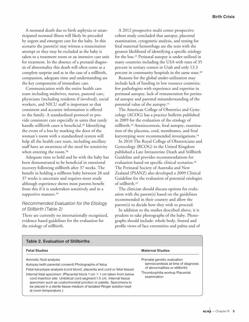
Birth Crisis
— Chapter R 5
A neonatal death due to birth asphyxia or unan-ticipated neonatal illness will likely be preceded by urgent and emergent care for the baby. In this scenario the parent(s) may witness a resuscitation attempt or they may be excluded as the baby is taken to a treatment room or an intensive care unit for treatment. In the absence of a prenatal diagno-sis of abnormality this death will often come as a complete surprise and as in the case of a stillbirth, compassion, adequate time and understanding are the key components of immediate care.
Communication with the entire health care team including midwives, nurses, pastoral care, physicians (including residents if involved), social workers, and NICU staff is important so that consistent and accurate information is offered to the family. A standardized protocol to pro-vide consistent care especially in units that rarely handle stillbirth cases is beneficial.30 Identifying the event of a loss by marking the door of the woman’s room with a standardized system will help all the health care team, including ancillary staff have an awareness of the need for sensitivity when entering the room.38
Adequate time to hold and be with the baby has been demonstrated to be beneficial in emotional recovery following stillbirth after 37 weeks. The benefit in holding a stillborn baby between 28 and 37 weeks is uncertain and requires more study although experience shows most parents benefit from this if it is undertaken sensitively and in a supportive manner.39
Recommended Evaluation for the Etiology of Stillbirth (Table 2)There are currently no internationally recognized, evidence based guidelines for the evaluation for the etiology of stillbirth.
A 2012 prospective multi center prospective cohort study concluded that autopsy, placental examination, cytogenetic analysis, and testing for fetal maternal hemorrhage are the tests with the greatest likelihood of identifying a specific etiology for the loss.32 Perinatal autopsy is under-utilized in many countries including the USA with rates of 35 percent in tertiary centers in Utah and only 13.3 percent in community hospitals in the same state.40
Reasons for the global under-utilization may include lack of funding in low resource countries, few pathologists with experience and expertise in perinatal autopsy, lack of remuneration for perina-tal autopsy and parental misunderstanding of the potential value of the autopsy.41
The American College of Obstetrics and Gyne-cology (ACOG) has a practice bulletin published in 2009 for the evaluation of the etiology of stillbirth.34 Amniocentesis, fetal autopsy, examina-tion of the placenta, cord, membranes, and fetal karyotyping were recommended investigations.34
In 2010 The Royal College of Obstetricians and Gynecology (RCOG) in the United Kingdom published a Late Intrauterine Death and Stillbirth Guideline and provides recommendations for evaluation based on specific clinical scenarios.44 The Perinatal Society of Australia and New Zealand (PSANZ) also developed a 2009 Clinical Guideline for the evaluation of potential etiologies of stillbirth.43
The clinician should discuss options for evalu-ation with the parent(s) based on the guidelines recommended in their country and allow the parent(s) to decide how they wish to proceed.
In addition to the studies described above, it is prudent to take photographs of the baby. Photo-graphs should include: whole body, frontal and profile views of face extremities and palms and of
Table 2. Evaluation of Stillbirths
Fetal Studies Maternal Studies
Amniotic fluid analysisAutopsy (with parental consent) Photographs of fetusFetal karyotype analysis (cord blood, placenta and cord or fetal tissue)Internal fetal specimen: (Placental block 1 cm × 1 cm taken from below
cord insertion site. Umbilical cord segment 1.5 cm. Internal tissue specimen such as costochondral junction or patella. Specimens to be placed in a sterile tissue medium of lactated Ringer solution kept at room temperature.)
Prenatal genetic evaluation (amniocentesis at time of diagnosis of abnormalities or stillbirth)
Thrombophilia workup Placental examination

Chapter R
6 Chapter R —
any specific abnormalities. It is important to docu-ment all findings particularly any abnormalities. Remember that the family may read your descrip-tions at some time in the future so be careful to write in a sensitive and respectful manner.
Care Of Women in Subsequent PregnancyThere is little evidence to guide the care of women following an unexplained stillbirth. In low risk women following an unexplained stillbirth, the risk of a stillbirth in a subsequent pregnancy after 20 weeks ranges from 7.8 to 10.5 in 1,000.34
The risks of early induction of labor for a woman with an uncomplicated subsequent preg-nancy, following a prior unexplained stillbirth before 39 completed weeks of gestation, must be weighed against the real risks of an early or late preterm birth.44 Although it has been a common practice to induce prior to 39 weeks with such a history this is no longer recommended based on a 2011 workshop sponsored by National Institute of Child Health and Human Development and Society for Maternal-Fetal Medicine.45
It is reasonable to consider serial growth ultra-sounds beginning at 28 weeks if there is lag in fun-dal height measurement or other concern about fetal growth, as intra uterine growth retardation is associated with a stillbirth risk of 21.8 per 1,000.48 ACOG recommends beginning fetal surveillance starting at 32 weeks gestation or one to two weeks earlier than prior stillbirth.23 There is an estimated 1.5 percent rate of iatrogenic prematurity for intervention based on a false positive result during antenatal surveillance.47
Amniocentesis to assess fetal lung maturity to permit delivery under 39 weeks is not a recom-mended approach since there are other organ systems to consider prior to initiating an induction before 39 completed weeks; however if a maternity care provider and their patient do choose delivery at < 39 weeks it may be considered.45,46
There is evidence that decreased fetal move-ments are associated with adverse outcomes, therefore, educating a woman to be aware of fetal movements engages her in the monitoring of her baby. Unfortunately, there is not good evidence to show that maternal monitoring of fetal kick counts prevents stillbirth.48 Emotional support will be an essential component of any care. More frequent antenatal visits while not necessarily
preventing recurrence, may offer reassurance. Listening to concerns and helping steer women through their anxieties, especially towards the end of pregnancy is important. Once again, continuity of care provider is likely to be beneficial.
Management of Second Trimester LossSecond trimester loss deserves the same emotional care and support outlined above. Clinical man-agement following diagnosis includes offering the option of labor induction or dilatation and evacuation (D&E).34 Usually this will depend on maternal preference and the ability to find a provider skilled in second trimester D&E. Maternal grief resolution is not affected by the choice of termination if the woman self-selects the process.36 In the USA most second trimester pregnancy terminations are completed by a D&E, however, in the late second trimester medical induction becomes the more common proce-dure for fetal anomalies.49 The benefits of labor induction are the ability to better evaluate for fetal anomaly and the ability for parent(s) to see and hold the baby. Chromosomal analysis can be performed on products obtained from D&E.49
Misoprostol, as a single agent administered vagi-nally or sublingually, is an effective form of labor induction following second trimester loss.50,52 Use of misoprostol to induce labor is considered safe in patients with a prior cesarean birth with a low transverse scar before 28 weeks gestation [11]. From 13 to 17 weeks use 200 mcg every 6 to 12 hours with the potential to double the dose to 400 mcg q 6 to 12 hours if the first dose does not result in adequate contractions. For 18 to 26 weeks start with 100 mcg q 6 to 12 hours and consider doubling to 200 mcg q 6 to 12 hours if the first dose does not provide adequate contrac-tions. In women with a prior uterine scar consider a lower initial dose and do not double after the first dose is given.50,52
Prenatal Diagnosis of AbnormalityWhile there has been an increase in the amount of research into the effects on parents and families of perinatal loss there remains little research available regarding the psychological effects of the diagnosis of an abnormality and the decision to terminate the pregnancy as a result. Without “full disclosure” and transparency, parents are hardly prepared to make an informed choice as to whether to test or

Birth Crisis
— Chapter R 7
not. Before proceeding with testing parents need to be carefully counseled as to the limits of testing and the potential choices they may face.
The same emotional support and counseling should be made available to parents who choose to terminate a pregnancy as to those who experience the spontaneous loss of a baby.
The Baby with an Abnormality or DisabilityCaring for a child with disabilities is a stressful situation for the parents, and the best predic-tor of the level of stress is the severity of the disability.52,53
Caregivers of children with intellectual dis-abilities are likely to suffer from depression.54 The depression may be the result of the financial, social and physical stressors experienced by the family.55
Parents with a baby born with a disability or abnormality should be offered the same emotional support as described in this paper. There is some evidence that family centered care provides the best support for caregivers of children with disabil-ities.56 A family physician is likely to be a caregiver for the family and possibly the child over the long term and is in a prime position to help diagnose and provide family support. Parents should be offered information about community resources available for the particular condition and directed to any support groups that are available.
Maternal Critical Illness and DeathWhile this chapter concentrates on the experience of birth crisis in relation to the baby, the death of or critical illness of a mother in pregnancy or around childbirth is equally important and dis-tressing for all.
Maternal critical illness may be related to prior comorbidities such as obesity or hypertension. It may also occur as a result of a pregnancy com-plication such as preeclampsia or hemorrhage. Prenatal recognition of a high-risk pregnancy is important in ensuring the appropriate level of care is offered with the utilization of and referral to
the maternal fetal medicine team as appropriate. All facilities providing obstetric care must be pre-pared to handle emergencies such as massive blood loss, maternal seizure and sepsis. The implementa-tion of evidence-based protocols will help guide providers in facilities without immediate access to an intensive care team.57 The regular practice of
team based drills to manage obstetric emergencies has been shown to improve outcomes.58
In developing countries maternal death is rare and usually unexpected when it occurs. The maternal mortality rate in the United States appears to no longer be decreasing, perhaps due to the obesity epidemic and the increased proportion of births that are by cesarean. In Australia, mater-nal death is unexpected and the numbers of deaths are small. However, across Australia more than one woman dies every two weeks in circumstances associated with childbearing.59 In the USA the pregnancy-related mortality ratio was 15.1 deaths per 100,000 live births for the period 2006–2007.60 Every maternal death has a major impact on the family, the community and the health care providers who were involved.61
Maternity care providers’ experience of the death of a mother has been shown to be compa-rable with that of emergency personnel attending large-scale disasters and most maternity providers are completely unprepared.61 Maternity care pro-viders also report having flashbacks and memories of the death also impinge on personal and profes-sional relationships.61
While this chapter focuses on the loss of a baby, the principles of providing support to families who experience birth crisis are essentially the same for families and staff who experience a maternal death.
Health Care ProvidersHealth care providers struggle with their own fears that they have either missed something that could have prevented this outcome or have directly con-tributed to the outcome. The amount of support that providers need to deal with their own grief or fears should not be under-estimated.
A number of mostly qualitative studies have explored providers’ experiences related to perinatal loss. One common theme from these studies is that staff often experience guilt as they feel that their role is about saving lives rather than being around death.44 Guilt is also tied up with blame and sometimes, blaming one another for not doing enough to give emotional support to the woman and families.62-64 Personal grief has been found to be common in obstetricians following the care of a woman experiencing a stillbirth. Self-doubt, self-blame and depression were also frequent reactions experienced by the physician.8

Chapter R
8 Chapter R —
Another common response is rationalization where staff compartmentalize the difficult and sad aspects of their role to manage later.44 This is espe-cially the case when working in busy environments. In these environments, there is often little time to grieve, acknowledge the event, and assess how they might be impacted by them.63 Unresolved personal issues also affect the way health care providers cope themselves and these need to be addressed.63
There are a number of strategies that are useful for the wellbeing of health care providers. These include reflection and review; emotional and prac-tical support; access to counselling services; initial and ongoing education and training; institutional policies and guidelines.62
Reflection and ReviewSeeking out people to discuss the tragedy and its personal impact can be helpful. Formal reflection on the care of the woman during her pregnancy with colleagues is also very important. Many hos-pitals or health settings will have regular perinatal mortality meetings where the care of women who have had a stillbirth or neonatal death will be discussed. These meetings should provide a safe and confidential environment for care providers to reflect upon the care provided and to determine whether anything could have been done differ-ently. Changes in policy and practice to reduce the risk of similar events occurring in the future should be addressed.42
Fear of litigation has been identified as one con-cern from health care providers involved in this area,64 which needs to be acknowledged so that it does not affect future practice adversely.
Emotional and Practical SupportSupport from colleagues, both emotional and practical, is essential. Health care providers who work in a supportive environment that has a no-blame culture are more likely to provide better, more effective care and be able to make changes in their practice to improve care in the future.62
Time pressures have been identified by care providers as a barrier to providing and receiving support. It is important for managers and other in leadership positions to seek out staff that have been involved in caring for families that have experienced perinatal loss and ensure they have adequate support. Looking after you own team members is also necessary.
Midwives’ satisfaction with working with fami-lies experiencing perinatal loss was related to the level of support they received from the organiza-tion in which they worked.64 This includes being in a hospital that acknowledged loss and grieving, as well as, being able to provide continuity of care to these families.65
Access to Counselling ServicesSome health services offer a bereavement coun-sellor and this has been found to be useful.63 This type of counselling may also be provided in a group rather than one-on-one. Peer support groups, multidisciplinary team meetings and formal debriefing with trained personnel are all strategies that can provide support for clinicians.
An element of personal healing needs to take place to enable health care providers to move beyond the strong emotions after a perinatal loss.64
Initial and Ongoing Education and TrainingNewer obstetrical providers and staff may need mentoring to care for families who experience perinatal loss.59 Families who have experienced perinatal loss should be included in the educa-tion and training programs for maternity care staff.62,63,66 This could also include communica-tion training and role playing breaking the bad news and expressing condolences appropriately to families experiencing perinatal loss.67 Just knowing what to say and being brave enough to say it needs to be included in training programs.65
There also needs to be an emphasis on the emotional and psychological aspects of perinatal loss and grief, as well as on the sound theoretical knowledge of grief.63 Students are often ‘protected’ from working with families experiencing perina-tal loss; and, while this is helpful at times, it also means that these students have few skills in this area when they graduate. Students should have the opportunity to develop the clinical experience of caring for bereaving couples under supervision and with compassionate support.62
Education and training is not only important for initial education but also should be part of continuing professional development and ongoing study days or seminar attendance.62
Policies and GuidelinesStaff caring for families who experience perina-tal loss can become overwhelmed especially if

Birth Crisis
— Chapter R 9
they also face staff shortages.63 It is essential that hospitals and health services develop policies and guidelines to help support staff including access to counselling services and support networks.51
In the United Kingdom, statutory midwifery supervision provides a framework of professional support in which a named supervisor is readily available to listen, advise and offer support in times where adverse clinical incidences arise.66,68 This not only ensures that the practitioner is given time to reflect and learn from such events, but also enables them to begin to process the emotions and experiences that arise and if not addressed, might well persist following a birth crisis.69
It is important to be aware of one’s own feelings of loss following a birth crisis. Health care provid-ers are invested in a healthy baby and mother and feel deeply saddened by catastrophic outcomes. It is important not to become defensive or prematurely admit to any wrongdoing as a means of resolving your own grief. If health care providers have suf-fered a similar loss, it is their discretion whether or not to reveal that information to the family. While each situation is different, this might help to mini-mize or dilute the parent’s grief.
SummaryBirth crisis is affects women and their families, providers, and obstetrical staff. Provider manage-ment of these crises is of great importance and is often challenging because of the emotional trauma,
personal responsibility, and fear of litigation. In caring for women and families who experience birth crisis, health care professionals must seek to provide mindful, compassionate, and empathetic care. Accusations and blame are never helpful and indeed may prevent or delay a positive resolution in the process of dealing with grief and loss.
The cases associated with this chapter can assist providers and staff in preparing to handle a birth crisis in their health care system.
Birth Crisis in Global or Low-Resource EnvironmentWomen in developing countries experience birth crisis with a greater frequency than those in devel-oped countries. However, despite the increased frequency, each loss has the potential to be equally devastating. As with recommendations for caring for women experiencing birth crisis in developed countries, evidence for women in developing countries is also lacking.
Social support, as noted above, remains a key element to caring for a woman and her family experiencing a birth crisis. When the crisis occurs outside of the hospital setting, the basis of this support may be from family and the community. In the hospital setting, providers should be cogni-zant of cultural and religious morays and how they apply to the provision of empathy and sympathy. When possible, translators should be employed to avoid miscommunication and misunderstanding.

Chapter R
10 Chapter R —
References1. Solnit AJ, Stark MH. Mourning and the birth of a defec-
tive child. Psychoanal Study Child. 1961;16:523-537.
2. Jacobs J, Harvey J. Evaluation of an Australian miscar-riage support programme. Br J Nurs. 2000;9(1):22-26.
3. Vance JC, Boyle FM, Najman JM, Thearle MJ. Coupledistress after sudden infant or perinatal death: a30-month follow up. J Paediatr Child Health. 2002;38(4):368-372.
4. Bainbridge L. Not quite perfect! Diagnosis of a minorcongenital abnormality during examination of the new-born. Infant. 2009;5(1):28-31.
5. Hughes P, Riches S. Psychological aspects of perinatalloss. Curr Opin Obstet Gynecol. 2003;15(2):107-111.
6. Gold KJ. Navigating care after a baby dies: a systematicreview of parent experiences with health providers.J Perinatol. 2007;27(4):230-237.
7. Nordlund E, Börjesson A, Cacciatore J, Radestad I.When a baby dies: Motherhood, psychosocial care, andnega- tive affect. Br J Midwifery. 2012;20(11):780-784.
8. Farrow VA, Goldenberg RL, Fretts R, Schulkin J. Psy-chological impact of stillbirths on obstetricians.J Matern Fetal Neonatal Med. 2013;26(8):748-752.
9. Sanghavi DM. What makes for a compassionatepatient-caregiver relationship? Jt Comm J Qual PatientSaf. 2006;32(5):283-292.
10. Youngson R. (2012) Time to care: How to love yourpatients and your job. Raglan, NZ: Rebelheart Publishers.
11. Flenady V, Wilson T. Support for mothers, fathers andfamilies after perinatal death. Cochrane Database SystRev, 2008 Jan 23;(1).
12. Kavanaugh K, Trier D, Korzec M. Social support follow-ing perinatal loss. J Fam Nurs. 2004;10(1):70-92.
13. Lawn JE, Kinney M. Stillbirths: An executive summaryfor the Lancet’s series. Lancet. 2011;(April):14.
14. Siasssakos D, Fox R, Draycott T, Winter C. GuidelinesCommittee of the Royal College of Obstetricians andGynaecologists. Late Intrauterine Fetal Death and Still-birth. Green-top Guidelines No. 55. October 2010.
15. Lalor JG, Begley CM, Devane D. Exploring painfulexperiences: impact of emotional narratives on mem-bers of a qualitative research team. J Adv Nurs. 2006;56(6):607-616.
16. Drotar D, Baskiewicz A, Irvin N, Kennell J, Klaus M. Theadaptation of parents to the birth of an infant with a con-genital malformation: a hypothetical model. Pediatrics.1975;56(5):710-717.
17. Surkan PJ, Rådestad I, Cnattingius S, Steineck G, Dick-man PW. Events after stillbirth in relation to maternaldepressive symptoms: a brief report. Birth. 2008;35(2):153-157.
18. Avelin P, Erlandsson K, Hildingsson I, Rådestad I. Swed-ish parents’ experiences of parenthood and the needfor support to siblings when a baby is stillborn. Birth.2011;38(2):150-158.
19. Ostler T. Grief and coping in early childhood. ZeroThree. 2010;31(1):29-35.
20. Roose RE, Blanford CR. Perinatal grief and supportspans the generations: parents’ and grandparents’ eval-uations of an intergenerational perinatal bereavementprogram. J Perinat Neonatal Nurs. 2011;25(1):77-85.
21. Kempson D, Murdock V. Memory keepers: a narrativestudy on siblings never known. Death Stud. 2010;34(8):738-756.
22. Bennett M. Chichester M. Forgotten voices: How peri-natal loss affects grandparents. Paper presented at theAssociation of Women’s Health, Obstetrics and Neona-tal Nurses, June 2008, Los Angeles.
23. O’Leary J, Warland J, Parker L. Bereaved parents’ per-ception of the grandparents’ reactions to perinatal lossand the pregnancy that follows. J Fam Nurs. 2011;17(3):330-356.
24. Van Dinter MC, Graves L. Managing adverse birthoutcomes: helping parents and families cope. Am FamPhysician. 2012;85(9):900-904.
25. Gamble J, Creedy D, Moyle W, Webster J, McAllister M,Dickson P. Effectiveness of a counseling interventionafter a traumatic childbirth: a randomized controlledtrial. Birth. 2005;32(1):11-19.
26. “Stillbirth.” Stillbirth. National Health Service. Web. 20June 2013. Available at http://www.nhs.uk/conditions/Stillbirth/Pages/Definition.aspx.
27. “Stillbirth (fetal Death).” Stillbirth (fetal Death). Austra-lian Instititute of Health and Welfare. 20 June 2013.Available at meteor.aihw.gov.au/content/index.phtml/itemId/327266.
28. Goldenberg RL, Culhane JF, Iams JD, Romero R. Epi-demiology and causes of preterm birth. Lancet. 2008;371(9606):75-84.
29. Barfield WD; Committee on Fetus and Newborn. Stan-dard terminology for fetal, infant, and perinatal deaths.Pediatrics. 2011;128(1):177-181.
30. Spong CY. Add stillbirth to the list of outcomes toworry about in a pregnant woman with a history of pre-term birth or fetal growth restriction. Obstet Gynecol.2012;119(3):495-497.
31. Gardosi J, Madurasinghe V, Williams M, Malik A, Fran-cis A. Maternal and fetal risk factors for stillbirth: popula-tion based study. BMJ. 2013;346:f108.
32. Centre for Maternal and Child Enquiries (CMACE).Perinatal Mortality 2009: United Kingdom. CMACE:London, 2011.
33. Serwint JR, Rutherford L. Sharing bad news with par-ents. Contemp Pediatr. 2000;17(3):45-66.
34. American College of Obstetricians and Gynecologists.ACOG Practice Bulletin No. 102: management of still-birth. Obstet Gynecol. 2009;113(3):748-761.
35. Trulsson O, Rådestad I. The silent child—mothers’experiences before, during, and after stillbirth. Birth.2004;31(3):189-195.
36. Burgoine GA, Van Kirk SD, Romm J, Edelman AB,Jacobson SL, Jensen JT. Comparison of perinatalgrief after dilation and evacuation or labor induction insecond trimester terminations for fetal anomalies. Am JObstet Gynecol. 2005;192(6):1928-1932.

Birth Crisis
— Chapter R 11
37. Maslow AD, Breen TW, Sarna MC, Soni AK, WatkinsJ, Oriol NE. Prevalence of coagulation abnormalitiesassociated with intrauterine fetal death. Can J Anaesth.1996;43(12):1237-1243.
38. Leduc L, Farine D, Armson BA, et al.; Maternal-FetalMedicine Committee; Clinical Practice Obstetrics Com-mittee. Stillbirth and bereavement: guidelines for still-birth investigation. J Obstet Gynaecol Can. 2006;28(6):540-552.
39. Rådestad I, Surkan PJ, Steineck G, Cnattingius S,Onelöv E, Dickman PW. Long-term outcomes for moth-ers who have or have not held their stillborn baby. Mid-wifery. 2009;25(4):422-429.
40. Korteweg FJ, Erwich JJHM, Timmer A, et al. Evaluationof 1025 fetal deaths: proposed diagnostic workup. AmJ Obstet Gynecol. 2012;206(1):53.e1-53.e12.
41. Silver RM. Optimal “work-up” of stillbirth: evidence! AmJ Obstet Gynecol. 2012;206(1):1-2.
42. Slassakos D, Fox R, Draycott T, Winter C. R GuidelinesCommittee of the Royal College of Obstetricians andGynaecologists. Late Intrauterine Fetal Death andStillbirth: Greentop Guideline 55. RCOG. 2010, London,United Kingdom.
43. Flenady V, King J, Charles A, Gardener G, Ellwood D,Day K, McGowan L, Kent A, Tudehope D, Richardson R,Conway L, Lynch K, Haslam R, Khong Y for the PerinatalSociety of Australia and New Zealand (PSANZ) PerinatalMortality Group. PSANZ Clinical Practice Guideline forPerinatal Mortality. Version 2.2, Brisbane April 2009.
44. Chescheir N, Menard MK. Scheduled deliveries: avoidingiatrogenic prematurity. Am J Perinatol. 2012;29(1):27-34.
45. Spong CY, Mercer BM, D’alton M, Kilpatrick S, BlackwellS, Saade G. Timing of indicated late-preterm and early-term birth. Obstet Gynecol. 2011;118(2 Pt 1):323-333.
46. Bukowski R. Stillbirth and fetal growth restriction. ClinObstet Gynecol. 2010;53(3):673-680.
47. Miller DA, Rabello YA, Paul RH. The modified biophysi-cal profile: antepartum testing in the 1990s. Am J ObstetGynecol. 1996;174(3):812-817.
48. Hofmeyr GJ, Novikova N. Management of reporteddecreased fetal movements for improving pregnancyoutcomes. Cochrane Database Syst Rev. 2012;4(4):CD009148.
49. Borgatta L, Kapp N; Society of Family Planning. Clini-cal guidelines. Labor induction abortion in the secondtrimester. Contraception. 2011;84(1):4-18.
50. Dodd JM, Crowther CA. Misoprostol for inductionof labour to terminate pregnancy in the second orthird trimester for women with a fetal anomaly or afterintrauterine fetal death. Cochrane Database Syst Rev.2010;14(4):CD004901.
51. Gómez Ponce de León R, Wing D, Fiala C. Misopros-tol for intrauterine fetal death. Int J Gynaecol Obstet.2007;99(Suppl 2):S190-S193.
52. Hassall R, Rose J, McDonald J. Parenting stress inmothers of children with an intellectual disability: theeffects of parental cognitions in relation to child char-acteristics and family support. J Intellect Disabil Res.2005;49(Pt 6):405-418.
53. Sanders JL, Morgan SB. Family Stress and Adjust-ment as Perceived by Parents of Children with Autismor Down Syndrome: Implications for Intervention. ChildFam Behav Ther. 1997;19(4):15-32.
54. Mbugua M N, Kuria MW, Ndetei DM. The Prevalence ofDepression among Family Caregivers of Children withIntellectual Disability in a Rural Setting in Kenya. Int JFamily Med. Vol. 2011, Article ID 534513, 5 pages; 2011.
55. Mak WS, Ho SM. Caregiving perceptions of Chinesemothers of children with intellectual disability in HongKong. J Appl Res Intellect Disabil. 2007;20:145-156.
56. Floyd FJ, Gallagher EM. Parental stress, care demands,and use of support services for school-age childrenwith disabilities and behavior problems. Fam Relat.1997;46:359-371.
57. American College of Obstetricians and GynecologistsCommittee on Patient Safety and Quality Improvement.ACOG Committee Opinion No. 487: Preparing for clini-cal emergencies in obstetrics and gynecology. ObstetGynecol. 2011;117(4):1032-1034.
58. Dresang L. (2012) Safety in Maternity Care. AdvancedLife Support in Obstetrics. Kansas City, MO. AmericanAcademy of Family Physicians.
59. Kildea S, Pollock WE, Barclay L. Making pregnancysafer in Australia: the importance of maternal deathreview. Aust N Z J Obstet Gynaecol. 2008;48(2):130-136.
60. MacDorman MF, Kirmeyer SE, Wilson EC. Fetal andperinatal mortality, United States, 2006. Natl Vital StatRep. 2012;60(8):1-22.
61. Mander R. The midwife’s ultimate paradox: a UK-basedstudy of the death of a mother. Midwifery. 2001;17(4):248-258.
62. Nallen K. Midwives’ needs in relation to the provision ofbereavement support to parents affected by perinataldeath. Part one. MIDIRS Midwifery Digest. 2006;16(4):537-542.
63. Modiba LM. Experiences and perceptions of midwivesand doctors when caring for mothers with pregnancyloss in a Gauteng hospital. Health SA Gesondheid.2007;13(4):29-40.
64. McCool W, Guidera M, Stenson M, Dauphinee L. Thepain that binds us: midwives’ experiences of loss andadverse outcomes around the world. Health CareWomen Int. 2009;30(11):1003-1013.
65. Fenwick J, Jennings B, Downie J, Butt J, Okanaga M.Providing perinatal loss care: satisfying and dissatisfyingaspects for midwives. Women Birth. 2007;20(4):153-160.
66. Nursing and Midwifery Council. Supervision, supportand safety: NMC quality assurance of LSAs 2010-2011.13 Dec 2011. London, United Kingdom.
67. Smith R, Homer A, Homer DJW, Homer CSE. Learningabout grief and loss through Harper’s story. MIDIRSMidwifery Digest. 2011;21(1):19-21.
68. Nursing and Midwifery Council. Standards to supportlearning and assessment in practice: NMC standardsfor mentors, practice teachers, and teachers. Jul 2008.London, United Kingdom.
69. Deery R. An action-research study exploring midwives’support needs and the affect of group clinical supervi-sion. Midwifery. 2005;21(2):161-176.

American Academy of Family Physicians
11400 Tomahawk Creek Parkway
Leawood, Kansas 66211-2680
(800) 274-2237 | (913) 906-6000 | aafp.org/also
CME17010160