
Incidence, diagnosis and management of tubal and nontubal ...
20
REVIEW Open Access Incidence, diagnosis and management of tubal and nontubal ectopic pregnancies: a review Danielle M. Panelli 1* , Catherine H. Phillips 2 and Paula C. Brady 1 Abstract Background: Ectopic pregnancy is a potentially life-threatening condition occurring in 1-2 % of all pregnancies. The most common ectopic implantation site is the fallopian tube, though 10 % of ectopic pregnancies implant in the cervix, ovary, myometrium, interstitial portion of the fallopian tube, abdominal cavity or within a cesarean section scar. Findings: Diagnosis involves a combination of clinical symptoms, serology, and ultrasound. Medical management is a safe and effective option in most clinically stable patients. Patients who have failed medical management, are ineligible, or present with ruptured ectopic pregnancy or heterotopic pregnancy are most often managed with excision by laparoscopy or, less commonly, laparotomy. Management of nontubal ectopic pregnancies may involve medical or surgical treatment, or a combination, as dictated by ectopic pregnancy location and the patient's clinical stability. Following tubal ectopic pregnancy, the rate of subsequent intrauterine pregnancy is high and independent of treatment modality. Conclusion: This review describes the incidence, risk factors, diagnosis, and management of tubal and non-tubal ectopic and heterotopic pregnancies, and reviews the existing data regarding recurrence and future fertility. Keywords: Ectopic pregnancy, Nontubal ectopic pregnancy, Heterotopic pregnancy Findings An ectopic pregnancy (EP) refers to the implantation of an embryo outside of the uterus. Due to advances in la- boratory testing, transvaginal ultrasound, chemotherapy and laparoscopy, the evaluation, diagnosis and manage- ment of EP has rapidly evolved. In parallel, maternal mortality has declined, from 3.5 of 10,000 pregnancies in 1970 to 2.6 of 10,000 in 1992 [1]. The most common EP location is in the fallopian tube, predominantly the ampullary region of the fallopian tube. Implantation outside the fallopian tube—in the cervix, ovary, myometrium, abdominal cavity, interstitial (i.e., intramuscular/proximal) portion of the fallopian tube or coincidentally with an intrauterine pregnancy—occurs in less than 10 % of EPs. Heterotopic pregnancy (HP) refers to the coexistence of an intrauterine pregnancy with an EP in any of these locations. ‘ Cornual’ pregnancies are those implanted in a horn of an anomalous uterus (i.e., unicornu- ate, bicornuate, didelphys or septate uteri); these do not uniformly require intervention and will not be included in this review [2–4]. This review will describe the incidence, risk factors, diagnosis and management of women with tubal and nontubal EPs, as well as review the existing literature regarding their future fertility. Review Incidence The overall rate of EP is 1–2 % in the general population, and 2–5 % among patients who have utilized assisted reproductive technology (ART) [5, 6]. Although the over- all mortality has decreased over time, ruptured EPs still account for up to 6 % of all maternal deaths; a review of mortality in ART-associated EPs similarly reported a mor- tality rate of 31.9 deaths per 100,000 pregnancies [5, 7]. Nontubal EPs are pregnancies that implant at sites other than the fallopian tube. These pregnancies account for less than 10 % of all EPs, though their overall incidence has been increasing in recent years [5]. Furthermore, nontubal EPs contribute disproportionately to maternal morbidity and mortality in comparison to tubal EPs. Cervical EPs are estimated to occur in 1:2000 to 1:18,000 pregnancies [8]. * Correspondence: [email protected] 1 Department of Obstcpetrics and Gynecology, Brigham and Women’s Hospital, Harvard Medical School, 75 Francis St., Boston, MA 02115, USA Full list of author information is available at the end of the article © 2015 Panelli et al. Open Access This article is distributed under the terms of the Creative Commons Attribution 4.0 International License (http://creativecommons.org/licenses/by/4.0/), which permits unrestricted use, distribution, and reproduction in any medium, provided you give appropriate credit to the original author(s) and the source, provide a link to the Creative Commons license, and indicate if changes were made. The Creative Commons Public Domain Dedication waiver (http://creativecommons.org/publicdomain/zero/1.0/) applies to the data made available in this article, unless otherwise stated. Panelli et al. Fertility Research and Practice (2015) 1:15 DOI 10.1186/s40738-015-0008-z
-
Upload
khangminh22 -
Category
Documents
-
view
0 -
download
0
Transcript of Incidence, diagnosis and management of tubal and nontubal ...

REVIEW Open Access
Incidence, diagnosis and management of tubaland nontubal ectopic pregnancies: a reviewDanielle M. Panelli1*, Catherine H. Phillips2 and Paula C. Brady1
Abstract
Background: Ectopic pregnancy is a potentially life-threatening condition occurring in 1-2 % of all pregnancies. Themost common ectopic implantation site is the fallopian tube, though 10 % of ectopic pregnancies implant in thecervix, ovary, myometrium, interstitial portion of the fallopian tube, abdominal cavity or within a cesarean section scar.
Findings: Diagnosis involves a combination of clinical symptoms, serology, and ultrasound. Medical management is asafe and effective option in most clinically stable patients. Patients who have failed medical management, areineligible, or present with ruptured ectopic pregnancy or heterotopic pregnancy are most often managed withexcision by laparoscopy or, less commonly, laparotomy. Management of nontubal ectopic pregnancies may involvemedical or surgical treatment, or a combination, as dictated by ectopic pregnancy location and the patient's clinicalstability. Following tubal ectopic pregnancy, the rate of subsequent intrauterine pregnancy is high and independentof treatment modality.
Conclusion: This review describes the incidence, risk factors, diagnosis, and management of tubal and non-tubalectopic and heterotopic pregnancies, and reviews the existing data regarding recurrence and future fertility.
Keywords: Ectopic pregnancy, Nontubal ectopic pregnancy, Heterotopic pregnancy
FindingsAn ectopic pregnancy (EP) refers to the implantation ofan embryo outside of the uterus. Due to advances in la-boratory testing, transvaginal ultrasound, chemotherapyand laparoscopy, the evaluation, diagnosis and manage-ment of EP has rapidly evolved. In parallel, maternalmortality has declined, from 3.5 of 10,000 pregnancies in1970 to 2.6 of 10,000 in 1992 [1].The most common EP location is in the fallopian tube,
predominantly the ampullary region of the fallopian tube.Implantation outside the fallopian tube—in the cervix,ovary, myometrium, abdominal cavity, interstitial (i.e.,intramuscular/proximal) portion of the fallopian tube orcoincidentally with an intrauterine pregnancy—occurs inless than 10 % of EPs. Heterotopic pregnancy (HP) refersto the coexistence of an intrauterine pregnancy with an EPin any of these locations. ‘Cornual’ pregnancies are thoseimplanted in a horn of an anomalous uterus (i.e., unicornu-ate, bicornuate, didelphys or septate uteri); these do not
uniformly require intervention and will not be included inthis review [2–4].This review will describe the incidence, risk factors,
diagnosis and management of women with tubal andnontubal EPs, as well as review the existing literatureregarding their future fertility.
ReviewIncidenceThe overall rate of EP is 1–2 % in the general population,and 2–5 % among patients who have utilized assistedreproductive technology (ART) [5, 6]. Although the over-all mortality has decreased over time, ruptured EPs stillaccount for up to 6 % of all maternal deaths; a review ofmortality in ART-associated EPs similarly reported a mor-tality rate of 31.9 deaths per 100,000 pregnancies [5, 7].Nontubal EPs are pregnancies that implant at sites other
than the fallopian tube. These pregnancies account for lessthan 10 % of all EPs, though their overall incidence hasbeen increasing in recent years [5]. Furthermore, nontubalEPs contribute disproportionately to maternal morbidityand mortality in comparison to tubal EPs. Cervical EPs areestimated to occur in 1:2000 to 1:18,000 pregnancies [8].
* Correspondence: [email protected] of Obstcpetrics and Gynecology, Brigham and Women’sHospital, Harvard Medical School, 75 Francis St., Boston, MA 02115, USAFull list of author information is available at the end of the article
© 2015 Panelli et al. Open Access This article is distributed under the terms of the Creative Commons Attribution 4.0International License (http://creativecommons.org/licenses/by/4.0/), which permits unrestricted use, distribution, andreproduction in any medium, provided you give appropriate credit to the original author(s) and the source, provide a link tothe Creative Commons license, and indicate if changes were made. The Creative Commons Public Domain Dedication waiver(http://creativecommons.org/publicdomain/zero/1.0/) applies to the data made available in this article, unless otherwise stated.
Panelli et al. Fertility Research and Practice (2015) 1:15 DOI 10.1186/s40738-015-0008-z

The estimated incidence of cesarean scar EPs is 1:1800 to1:2216 pregnancies, or 6 % of all EPs in women with atleast one cesarean delivery [9, 10]. Interstitial EPs accountfor 4 % of EPs, though the associated morbidity is muchhigher, with mortality rates of 2.5 % or 7 times the mortal-ity rate associated with other EP locations, largely due tohemorrhage [11, 12]. Pregnancies embedded within themyometrium (intramural EPs) account for an estimated1 % of EPs [13]. Abdominal pregnancies account for 1.3 %of EPs [14]. These have been classified as primary or sec-ondary; secondary abdominal EPs are theorized to resultfrom extrusion from the fallopian tube and subsequentintraabdominal reimplantation [15]. Most common im-plantation sites are in the pouches posterior and anteriorto the uterus and on the serosa of the uterus and adnexa;retroperitoneal, omental, bowel, hepatic and splenic im-plantations have also been reported [16].Estimates of the incidence of heterotopic pregnancy
(HP) vary by article and decade; the risk has been reportedfrom 1:4000 to 1:30,000 women in the general population[5, 17]. The risk of HP following in vitro fertilization (IVF)has been estimated as high as 1:100 women [5, 17, 18].HPs can include an EP in any of the previously describedlocations; a triplet HP that included tubal and cervical EPshas even been described [19]. The majority are tubal HPs;in a review of 80 cases of HP in the literature, 66 (72.5 %)were in the ampullary or interstitial portion of the fallo-pian tube, while 7 were cervical and 3 were implanted inthe cesarean scar [17].
Etiology of tubal ectopic pregnancyThe fallopian tube is a carefully controlled environment tofacilitate oocyte transport, fertilization, and migration ofthe early embryo to the uterus for implantation [20] Mostdata suggest tubal EP stems from both abnormal embryotransport and an alteration in the tubal environment,which enables abnormal implantation to occur [21].The transport of an oocyte and embryo through the tube
relies on both smooth muscle contraction and ciliary beat-ing, which are affected by several local factors—toxic, infec-tious, immunologic and hormonal. Smoking and infectionhave been shown to decrease cilia density, while ciliary beatfrequency has been shown to be responsive to the changinghormonal milieu of the menstrual cycle [22–24]. Samplesof fallopian tube epithelium incubated in estradiol (E2) andnitric oxide (NO) have been found to demonstrate in-creased ciliary motility, which may cause aberrant tubaltransport [25, 26]. NO also affects smooth muscle contract-ility in the fallopian tube; expression of NO has been foundto vary during the menstrual cycle, with possible implica-tions for normal and ectopic implantations [27]. Finally,E2-mediated effects via estrogen receptors on gene regula-tion and expression—including pathways implicated in im-plantation and apoptosis—may be involved in aberrant
tubal function and ectopic pregnancy, though more re-search is needed to clarify these pathways [28–30].Inflammation in the fallopian tubes is also implicated in
the establishment of EP, by inducing tubal dysfunction ordamage that may lead to retention of an oocyte orembryo, and by promoting embryo implantation in thefallopian tube via inflammatory cytokines [31]. Followingtubal damage by smoking or infection, upregulation ofpro-inflammatory cytokines has been noted, promotingembryo receptivity, invasion and angiogenesis in the tube.For instance, interleukin 1 (IL-1), produced by tubalepithelial cells following Chlamydia trachomatis infection,is a vital signal for embryo implantation in the endomet-rium; IL-1 also recruits neutrophils downstream, leadingto further tubal damage [32]. Macrophages and intrae-pithelial lymphocytes are also increased in women withEP, potentially affecting tubal function and predisposing totubal EP [33–35].
Clinical risk factorsUp to 50 % of women diagnosed with EPs have no iden-tifiable risk factors; however, a number of risk factorshave been associated with EP [5]. These include age,smoking, history of EP, tubal surgery or tubal damage,prior pelvic infection, DES exposure, IUD use and preg-nancy conceived by assisted reproduction.Age has been shown to be a risk factor for EP, with the
highest incidence over the age of 35 in both spontaneouspregnancies and those conceived after assisted reproduct-ive technologies [7, 36]. The explanation for this observa-tion is unknown, however age is theorized to affect tubalfunction, including delay of oocyte transport [36, 37].Prior EP is a strong risk factor for recurrent EP, with a
recurrence rate of 5–25 %, or up to 10 times the risk inthe general population [38–40]. Prior treatment for EP,whether medical or surgical, may result in pathologicchanges in tubal motility, ciliary function and uterinecontractions [41]. (For fertility outcomes after prior EP,please see “Recurrence and future fertility”).Smoking is thought to increase the risk of EP by caus-
ing tubal dysfunction, including deciliation [22]. To-bacco may cause dysregulation of the paracrine signalsneeded for coordinated embryo transport and develop-ment [21]. In a retrospective review of 481 IVF cycles,the likelihood of a tubal EP was three times higheramong smokers [41]. A dose-dependent relationship be-tween smoking and EP has also been confirmed in spon-taneous pregnancies [36].A history of pelvic infection or pelvic inflammatory
disease is associated with increased risk for subsequentEP. Chlamydia trachomatis in particular has been impli-cated as a risk factor for EP, with the risk increasing witheach successive infection; ascending infection and result-ant salpingitis is thought to lead to tubal dysfunction
Panelli et al. Fertility Research and Practice (2015) 1:15 Page 2 of 20

and aberrant implantation [5, 21, 42, 43]. Other infec-tions potentially associated with pelvic inflammatorydisease and tubal damage include Neisseria gonorrhoeae,Mycoplasma and schistosomiasis [44, 45].Prior tubal surgeries, including but not limited to tubal
reanastamosis, salpingostomy, tuboplasty and lysis of ad-hesions, are risk factors for EP. Similarly, any causes ofpelvic adhesions, including endometriosis, appendicitis,or other pelvic surgeries, may distort the anatomy of thefallopian tube [46, 47]. The risk of EP after tubal reanas-tamosis, specifically, is estimated at 2–13 %, and is simi-lar between abdominal and robotic approaches [6, 48].After surgical sterilization, the failure rate across all
sterilization methods is estimated at 18.5 per 1000,approximately one-third of which are EPs [49]. Womensterilized before 30 years of age are twice as likely tohave subsequent EPs as those sterilized after age30 years. Rates of EP vary by sterilization technique:After bipolar coagulation, 65 % of pregnancies are EP,while after unipolar or clip sterilization, approximately15 % of pregnancies are EP [50]. The proportion of EPhas been shown to be three times higher between 4 and10 years after sterilization, as compared to the first3 years [49].Current IUD use dose not predispose to EP, though a
higher proportion of pregnancies conceived with an IUDin place are ectopic as compared to the general popula-tion [43, 51]. Among pregnancies conceived with IUDsin place, half are ectopic with a levonogestrel device inplace, compared to 1:16 with a copper IUD in place [52].Overall, any contraceptive use decreases the risk of bothintrauterine and ectopic pregnancy.Assisted reproductive technologies constitute a risk fac-
tor for EP, as 2–5 % of pregnancies from assisted repro-ductive technologies are ectopic [7]. The three mainfactors contributing to this increased risk are the specifictype of procedure, the reproductive health characteristicsof the woman, and the estimated embryo implantation po-tential [21, 53]. A history of infertility, even in the absenceof known tubal disease, is associated with EP, with the EPrisk increasing with a longer duration of infertility [18, 43].Tubal factor infertility specifically is associated with a two-fold risk of EP following IVF [52, 54, 55].Several IVF cycle parameters may be associated with
an increased risk of EP. Patients undergoing cycles trig-gered with gonadotropin releasing hormone (GnRH)agonists instead of recombinant hCG may be at higherrisk of EP; in a review of 466 IVF cycles, GnRH agonisttriggers were associated with a significantly higher EPrate (5.3 % versus 1.4 % following hCG triggers). Thisfinding is theorized to be due to poor endometrial recep-tivity following GnRH agonist administration [56].The number of embryos transferred may be correlated
to the EP risk; in a review of 9480 ectopic pregnancies
following IVF, the rate of EP following fresh cycles rosesignificantly from 1.7 % following single embryo transferto 2.5 % following the transfer of 4 embryos [7]. Depth oftransfer may also have an effect; a randomized prospectivestudy of deep versus mid-fundal transfer reported an EPrate of 1.5 versus 0.4 % [57]. Day of embryo transfer hasinconsistently been associated with risk of EP in priorstudies. A series of 13,654 fresh cycles reported an EP rateof 2.1 % following day 3 embryo transfers, as compared toa rate of 1.6 % following day 5 embryo transfers, which didnot reach statistical significance [58]. Conversely, a reviewof 1994 fresh transfers reported a significant difference inectopic pregnancy rates between day 3 and day 5 transfers,at 2.4 and 1.7 %, respectively [59].The transfer of fresh embryos is associated with a higher
EP risk as compared to the transfer of thawed embryos;following 15,042 fresh cycles, the EP rate was 1.97 %,which was significantly higher than the 1.01 % EP ratefollowing 12,752 cryopreservation cycles [55]. It is theo-rized that the controlled ovarian hyperstimulation andhyperestrogenic environment preceding a fresh embryotransfer negatively effects endometrial receptivity [60].
Risk factors for nontubal EPOverall, the risk factors for ovarian EPs, interstitial EPs,and tubal HPs are similar to those for tubal pregnancy.These include a history of a prior EP, pelvic infections anduse of in vitro fertilization [61]. The transfer of four ormore embryos during IVF is an additional risk factor forHP [18, 62]. IUD use may be a risk factor for nontubal EP,particularly for ovarian EP [63]. An additional risk factorfor interstitial implantation includes prior ipsilateral sal-pingectomy, with interstitial ectopic pregnancies occur-ring up to 13 years after salpingectomy [4]. Risk factorsfor other specific types of nontubal EPs are outlined in thefollowing subsections.
Intramural EPRisk factors for these rare EPs are theorized to includemyometrial injury following uterine curettage, and priormyomectomy or cesarean section [13]. Assisted repro-ductive technologies have been used in approximately20 % of case reports, and another 19 % of patients car-ried a diagnosis of adenomyosis.
Cesarean section EPRisk for cesarean scar implantation is not clearly corre-lated to the number of prior cesarean sections [64]. Riskfor cesarean section scar implantation has not been cor-related to single versus double layer closure of the hys-terotomy at the time of cesarean section. Cesarean scarimplantation may be more common following cesareansections for elective indications, which is theorized to be
Panelli et al. Fertility Research and Practice (2015) 1:15 Page 3 of 20

due to impaired healing of an unlabored lower uterinesegment [65].
Cervical EPA history of dilation and curettage (D&C) in a previouspregnancy has been associated with subsequent cervicalEP; this risk factor is present in nearly 70 % of cases[66, 67] In-vitro fertilization has been proposed as arisk factor, but often coincides with D&C and otherpossible risk factors, so is difficult to isolate as an inde-pendent contributor to risk [68].
Abdominal pregnancyRisk factors for abdominal pregnancy are similar to thosefor tubal EPs, including pelvic inflammatory disease, useof assisted reproductive technologies and endometriosis[69]. Most abdominal pregnancies have been published incase reports; one details the occurrence of a twin preg-nancy implanted in the broad ligament after IVF. Uterineperforation was suggested as a possible cause as the em-bryo transfer was performed using a stylet, which is morerigid than standard transfer catheters [70].
DiagnosisSerum beta-human chorionic gonadotropin (β-hCG)The diagnosis of EP often begins with the preliminarydiagnosis of pregnancy of unknown location (PUL). PULis defined as a positive serum beta-human chorionicgonadotropin (β-hCG) in the absence of ultrasound find-ings indicative of intrauterine or extrauterine pregnancy.Approximately 30 % of patients with PUL will developan ongoing intrauterine pregnancy (IUP), while themajority (50–70 %) will be diagnosed with failing preg-nancies, either miscarriages or EPs [71].In the stable patient, measurement of β-hCG is crucial
to clarify pregnancy location and prognosis. Produced pri-marily by the syncytiotrophoblast in the placenta, β-hCGis detectable in the blood by the second week of preg-nancy until a peak at 10–12 weeks [72]. A single measureof β-hCG is insufficient to clarify pregnancy prognosis,and serial β-hCG levels are commonly used to monitorearly pregnancies. The most recent recommendations forβ-hCG trends in early pregnancy, derived from a retro-spective review of 1005 patients with PUL, suggest theminimum β-hCG rise of an IUP is 35 % in 2 days [73]. Aβ-hCG rise less than 35 % in 2 days has a positive predict-ive value of 96.2 %, a negative predictive value of 69.7 %,and an overall accuracy of 80.2 % in predicting EP.Conversely, in this study, in patients eventually diag-nosed with miscarriages, the minimum expected β-hCG decline in 2 days is 36–47 % (depending on thestarting β-hCG level). Β-hCG cut-offs, however, are notironclad; using these cut-offs, 16.8 % of EPs and 7.7 %of IUPs would be misclassified solely using serial β-
hCG levels. Obtaining a third β-hCG and early ultra-sound decreased the misclassification of IUP to 2.7 % [73].The expected rates of β-hCG rise and decline are the samefor multiple pregnancies, following assisted reproduction,and in obese patients [74]. The absolute β-hCG values,however, may be higher in multiple pregnancies or lowerin patients with elevated body mass index.
Serum progesteroneSerum progesterone has been explored as a possible serummarker for nonviable pregnancies, including EPs, as pro-gesterone levels have been shown to be lower in ectopicand failing pregnancies than IUPs [75]. Several studies sug-gest a progesterone cut-off of 10 nanograms per milliliter(ng/mL) for the most accurate identification spontaneousEPs. In a meta-analysis including 4689 patients under14 weeks gestational age with pain and/or bleeding, aserum progesterone level of less than 10 ng/mL predicteda non-viable pregnancy with a sensitivity of 66.5 % and spe-cificity of 96.3 % [76]. The optimal cut-off may be higher inpatients who received fertility treatments, as these patientsoften have multiple corpora lutea secreting progesteroneand often receive exogenous progesterone. A cut-off of30 ng/mL, 28–49 days after the last menstrual period, maybe more appropriate in patients who received clomiphenecitrate, while an optimal cut-off has yet to be identified inpatients after IVF and is likely highly dependent on thenumber of days since embryo transfer [77, 78].Serum progesterone levels, however, have been shown
to misclassify more normal pregnancies than serial β-hCGmeasurements [79]. Serum progesterone also cannot fur-ther distinguish between miscarriages and EPs. While pro-gesterone may highlight patients at greater risk for EP, it isinsufficient in itself to discriminate between IUPs, miscar-riages and EPs [80].
Other serum markersSeveral studies have explored alternative serum markersof EP, focusing on proteins associated with placental,endometrial and/or corpus luteal functions, angiogen-esis and inflammation [75]. These potential proteinsinclude, but are not limited to: Inhibin A, which isproduced by the corpus luteum; activin A, pregnancy-associated plasma protein-A (PAPP-A) and A Disinte-grin and Metalloprotease-12 (ADAM-12) which aregenerated by the placenta; and vascular endothelial growthfactor (VEGF), which, produced by a variety of cell types,is crucial for angiogenesis and may be upregulated in EP[75]. Various messenger and micro-RNA—regulators ofdownstream gene expression—may also be differentiallyexpressed by an EP [81, 82].Studies have also attempted to combine multiple mea-
sures; one such study incorporated VEGF, PAPP-A, andprogesterone, and reported sensitivity of 97.7 % and
Panelli et al. Fertility Research and Practice (2015) 1:15 Page 4 of 20

specificity of 92.4 % in diagnosing EP, though this modelhas not been validated in further studies [83]. For manyof these markers, studies are inconclusive, and for allmarkers, more research is needed before any of thesesupplants β-hCG as the primary serologic method ofdifferentiating intra- and extrauterine pregnancies [81].
ImagingDiscriminatory zoneVisualization of a gestational sac by transvaginal ultra-sound (TVUS), confirming an intrauterine pregnancy(IUP), is expected at serum β-hCG levels above the “dis-criminatory zone.” The discriminatory zone was initiallyproposed as 6500 milli-international units (mIU)/mL in1981, using transabdominal ultrasound [84]. With ad-vances in ultrasound imaging, particularly with the useof transvaginal sonography, the discriminatory zone hasbeen lowered to 1000 to 2000 mIU/mL [85].Studies report that normal IUPs may still develop in
patients with PULs and serum β-hCGs above the dis-criminatory zone. In a review of patients with PUL,nine women with β-hCGs above 2000 mIU/mL at thetime of their TVUS developed intrauterine pregnancies;the highest β-hCG in this group was 4336 mIU/mL[86]. In patients with multiple pregnancies, the serumβ-hCG at which an intrauterine gestational sac is seen canbe higher than the discriminatory zone identified forsingleton IUPs. One review reported a serum β-hCG of9083 mIU/mL without definitive ultrasound findings in apatient later diagnosed with a triplet pregnancy [87].Other factors such as obesity or uterine fibroids may alsobe associated with nonvisualization of an intrauterinegestational sac above the β-hCG discriminatory zone.Serum β-hCG in the absence of definitive ultrasound
findings should not be the sole factor in diagnosingpregnancy location or viability or dictating management,and that gestational age must be taken into account. Apositive pregnancy test at any level in the absence of anintrauterine pregnancy should be approached as an EPuntil proven otherwise.
Accessory ultrasound findingsIn the absence of definitive visualization of an EP, additionalmarkers on ultrasound can increase a clinician’s suspicionfor EP, and may be useful in conjunction with other clinicaldata. These include a thin endometrial stripe thickness andthe presence of intraabdominal free fluid.In a review of 591 patients with vaginal bleeding and
PUL, IUPs had a significantly higher mean endometrialstripe thickness than miscarriages or EPs (17 mm versus12 mm, respectively); no intrauterine pregnancies oc-curred in patients with endometrial stripes below 8 mm inthickness [88]. However, studies have not consistently
shown a significant difference in endometrial stripe thick-ness between miscarriages and EPs [89]. Endometrialstripe thickness may indicate patients at higher risk forabnormal pregnancies but cannot reliably be used in isola-tion to diagnose EP [90].A small amount of anechoic free fluid in the posterior
cul de sac is normal in both intra- and extrauterine preg-nancies [91]. Larger amounts of complex free fluid, par-ticularly in Morrison’s pouch by the liver, may indicaterupture of an EP, and correlates well to hemoperitoneumobserved intraoperatively [91, 92]. Hemoperitoneum canbe assessed in the emergency setting using a FocusedAssessment with Sonography for Trauma (FAST) scan,which is a bedside ultrasound assessing for free fluid in theperihepatic, perisplenic and pelvic space; the full traumaassessment, not applicable to patients with suspected EP,also includes the pericardial space [93]. The determinationof extent of hemoperitoneum and need for interventiondepends on clinician assessment and the patient'shemodynamic stability. Of note, if an ultrasound shows anIUP, the risk of EP is much reduced, though not zero.Though rare, the patient may still be at risk of heterotopicpregnancy, particularly following IVF [94].
Ultrasound diagnosis of tubal EPIn women presenting with bleeding or pain, pregnancylocation is often not definitively visualized on the initialultrasound at presentation; however, diagnosis of EP byultrasound is possible when following careful guidelines.Identification of a gestational sac and fetal pole, with orwithout cardiac activity, or a hyperechoic ring—calledthe ‘bagel’ or ‘tubal’ sign (Fig. 1)—with circumferentialDoppler flow (Fig. 2) is highly suggestive of an ectopicpregnancy [95, 96]. If a suspicious mass moves separ-ately from the ovary—called the ‘blob’ sign - the positivepredictive value is above 90 % in a symptomatic womanwith a positive serum b-hCG and no IUP on transvaginalultrasound [97, 98].
Ultrasound findings specific to nontubal EPsEctopic pregnancies occurring in less common anatomicareas can also be identified by ultrasound according tospecific criteria. Diagnostic criteria of each type of nontu-bal ectopic pregnancy are discussed below. All of thesediagnostic criteria assume the absence of a visualized IUP.Ultrasound criteria for diagnosis of an interstitial ec-
topic pregnancy include a gestational sac at least 1 cmlateral to the edge of the uterine cavity, with a thin(5 mm or less) layer of overlying myometrium surround-ing it (Figs. 3 and 4) [99, 100]. An ‘interstitial line’ mayalso be seen (Fig. 5) [101].A cervical EP is identified on ultrasound by a distended
cervical canal containing a gestational sac with peripheralDoppler flow (Fig. 6), below a closed internal cervical os
Panelli et al. Fertility Research and Practice (2015) 1:15 Page 5 of 20

[102, 103]. The ‘sliding organ’ sign, or movement whenpressure is applied with the transvaginal probe, is associ-ated with spontaneous abortions in progress and shouldbe absent in a cervical ectopic pregnancy.Diagnostic criteria for a cesarean scar EP by ultrasound
include visualization of the gestational sac at the site of theprior hysterotomy (Fig. 7), outside the endometrial cavity[104]. The myometrium should be very thin (1–3 mm) or
absent between the gestational sac and the bladder (Fig. 8).A negative ‘sliding organ’ and the presence of peripheralDoppler flow are expected [9].On ultrasound or MRI, intramural EPs should be com-
pletely surrounded by myometrium circumferentially, withno communication with the intrauterine cavity [69, 105].Intramural EPs are notoriously challenging to diagnose onultrasound and have been mistaken for fibroids or intra-uterine pregnancies [106].Ovarian EPs may be suspected by ultrasound when
a hypoechogenic area is seen surrounded by a wide
Fig. 2 Tubal ectopic pregnancy by transvaginal ultrasound. The arrowindicates the ectopic gestation with circumferential Doppler flow, calledthe “Ring of Fire”
Fig. 3 Interstitial ectopic pregnancy by transvaginal ultrasound. Thearrow indicates thin (<5 mm) myometrium overlying the ectopicpregnancy. This finding by ultrasound, in combination with thelateral location of the gestation, has a reported specificity of 88-93 %but a sensitivity of just 40 % [101].
Fig. 1 Tubal ectopic pregnancy by transvaginal ultrasound. The arrowindicates the ectopic gestation with a surrounding hyperechoic ring,called the ‘bagel’ or ‘tubal’ sign
Fig. 4 Interstitial ectopic pregnancy by magnetic resonance imaging,T1 weighted. The arrow indicates thin (<5 mm) myometrium overlyingthe ectopic pregnancy. In a stable patient, MRI may be useful in theconfirmation of interstitial pregnancy location
Panelli et al. Fertility Research and Practice (2015) 1:15 Page 6 of 20

echogenic ring with peripheral Doppler flow. Some may becompletely surrounded by ovarian cortex. [106, 107] A fetalpole is seldom present [107]. When pressure is appliedwith the transvaginal probe, an ovarian EP will move withthe ovary, and should be connected to the uterus by theovarian ligament [107, 108]. These EP can be difficult todifferentiate from ovarian cysts, which may have a similarappearance and peripheral Doppler flow [108]. Given thedifficulty of making this diagnosis by imaging, laparoscopyis often required for definitive diagnosis [107].Abdominal EP is rare, and ultrasound guidelines are
few. Suggested guidelines include visualization of an extra-uterine gestational sac, fetus and/or placenta, with nomyometrium seen between the fetus and urinary bladder[109]. This gestational sac or fetus will be in unusuallyclose proximity to the anterior abdominal wall, and maybe surrounded by loops of bowel. There should be no
evidence of more common ectopic implantation sites,such as the fallopian tubes or cesarean section scar [110].
Endometrial samplingAt many medical centers, a patient with a PUL and anabnormal β-hCG trend as described above may receivemethotrexate (MTX) without precise diagnosis [111]. Upto 40 % of these patients, however, may ultimately havefailing IUPs, unnecessarily exposing these patients toMTX [112]. While not universally employed, endomet-rial sampling may allow these patients to avoid unneces-sary treatment with MTX [113].Identification of villi on endometrial sampling is diagnos-
tic of a failed IUP, and in such cases, no further treatmentis usually needed [5, 114, 115]. Serum β-hCG should alsobe checked the day after endometrial sampling; a declineof 15–20 % after sampling indicates the disruption of a
Fig. 6 Cervical ectopic pregnancy by transvaginal ultrasound. Dopplershows circumferential flow. The arrow indicates Doppler flow inside thegestational sac, associated with the embryo. Such Doppler flow will notbe found in a spontaneous abortion, which may slide down into asimilar position at the cervix
Fig. 5 Interstitial ectopic pregnancy by transvaginal ultrasound. Thearrow indicates the ‘interstitial line,’ extending from the endometriumto the cornua, abutting the suspicious mass
Fig. 7 Cesarean scar ectopic pregnancy by transvaginal ultrasound.The arrow shows the gestational sac implanted in the region of thecesarean scar, clearly outside the endometrial canal
Fig. 8 Cesarean scar ectopic pregnancy by transvaginal ultrasound.The arrow indicates the thin myometrium (3 mm) between the bladder(indicated with the number 1) and the gestational sac
Panelli et al. Fertility Research and Practice (2015) 1:15 Page 7 of 20

failing IUP, even if no villi are identified [5]. A postopera-tive plateau or increase in the β-hCG value strongly sug-gests an EP. A patient with an adequate decrease in the β-hCG level after sampling can be monitored with serial β-hCG measurements until levels are undetectable, or untilpathological evaluation of the curettage specimen showschorionic villi [116].The standard endometrial sampling method for PUL
is D&C, though these are associated with greater costand anesthesia requirement than outpatient procedures[112, 117, 118]. Endometrial biopsy pipelles, whileeffective in screening for endometrial carcinoma, haveinsufficient sensitivity and specificity to replace D&C inthe diagnosis of PUL [119, 120]. Karman cannulas, at-tached to hand-held suction devices, are as efficaciousas D&C for diagnosing endometrial pathology and arefrequently used for the evacuation of first trimesterpregnancies in the outpatient setting [121–124]. In a re-view of 45 patients with PUL and abnormal β-hCGtrends after IVF, over two-thirds of patients were diag-nosed with failing IUP by final pathology and/or fallingβ-hCG, and were spared MTX [116]. It is unknownwhether this device performs similarly in spontaneouspregnancies.
Medical managementThe most common interventions for the treatment of EPare medical management with systemic MTX and surgicalremoval of the pregnancy. Medical management of EP withMTX has been demonstrated to be more cost-effectivethan surgical management while maintaining similar treat-ment success and future fertility [38, 125–127]. Injectionsof hyperosmolar glucose into tubal EPs have been studied,but have significantly higher failure rates than standardmedical or surgical management and are not recom-mended [128–130].MTX is a dihydrofolate reductase inhibitor, disrupting
DNA and RNA precursor synthesis; it targets rapidly
dividing cells and, in an EP, disrupts primarily tropho-blastic tissue [131]. Its use as treatment for EP was firstreported in 1982 [132]. The most common side effectsassociated with MTX treatment for EP include pelvicpain, nausea, headaches, abdominal pain, and dermatitis.Less common side effects include mucositis, diarrhea, andalopecia [127, 133].
Several studies have demonstrated the efficacy ofintramuscular (IM) MTX for treatment of EP, thoughsuccess rates are inversely correlated to β-hCG levels[134]. In a meta-analysis including 503 women with EPstreated with single dose MTX, successful treatment,defined as avoidance of surgery, for initial β-hCG levelsbetween 1000 and 1999 mIU/mL was 94.4 %, comparedwith just 81.8 % in patients with starting β-hCG levels of10,000 to 150,000 mIU/mL [134]. A β-hCG above 5000mIU/mL was proposed as a relative contraindication totreatment with MTX, with a success rate of just 85 %.
Pre-methotrexate assessmentBefore treatment with MTX, blood work should be ob-tained to assess hematologic, hepatic and renal function;a chest x-ray should be considered in patients with activepulmonary disease. The patient’s Rhesus (Rh) status mustalso be obtained in order to determine the need for Rho(D)immune globulin therapy, required in Rh negative patients.A pelvic ultrasound should be obtained to characterize anyectopic mass and exclude a concomitant IUP. Several con-traindications for treatment of EP with MTX exist (Table 1)[135]. Patients with ectopic pregnancies and relative con-traindications to treatment with methotrexate may receivethe medication if deemed appropriate by the clinician;these patients should be hemodynamically stable and wellcounseled and have capacity to make the decision [6].Patients should be advised to stop taking prenatal vita-
mins, as the folate supplementation will counteract theaction of MTX. Patients should also avoid excessive sun-light due to possible MTX-induced dermatitis; nonsteroidal
Table 1 Absolute and relative contraindications of treatment of EP with MTX
Absolute Contraindications Relative Contraindications
Clinical instability or significant pain suggestive of ruptured EP Presence of fetal cardiac activity
Heterotopic pregnancy with viable and desired IUP β-hCG level over 5000 mIU/mL
Liver function tests more than 2 times the upper limit of normal An ectopic mass size greater than 4 cm in largest dimension
White blood cell count of <1500/uL Patient refusal of blood transfusion
Platelet count <100,000/uL Patient inability to follow up
Creatinine≥1.5 mg/dL
Current breastfeeding
Active pulmonary disease
Active peptic ulcer disease
Moderate to severe anemia
Sensitivity to methotrexate
Panelli et al. Fertility Research and Practice (2015) 1:15 Page 8 of 20

anti-inflammatory drugs, which may delay renal excretionof MTX; alcohol, which may lead to elevation of hepaticenzymes; and sexual activity, vigorous physical activity andpelvic exams, which could lead to rupture of the EP [136].
Systemic methotrexateSingle dose methotrexate The single dose regimenconsists of an IM injection of MTX (50 mg/m2 of bodysurface area), with administration of additional doses atweekly intervals in patients with an inadequate response(Table 2) [6]. Repeat injections are permitted every 7 daysfor up to 4 doses; a second dose is needed in 20 % ormore of patients, while less than 1 % of patients require3 or more doses [137]. The single dose regimen is asso-ciated with fewer side effects as compared to other regi-mens [6, 138, 139].
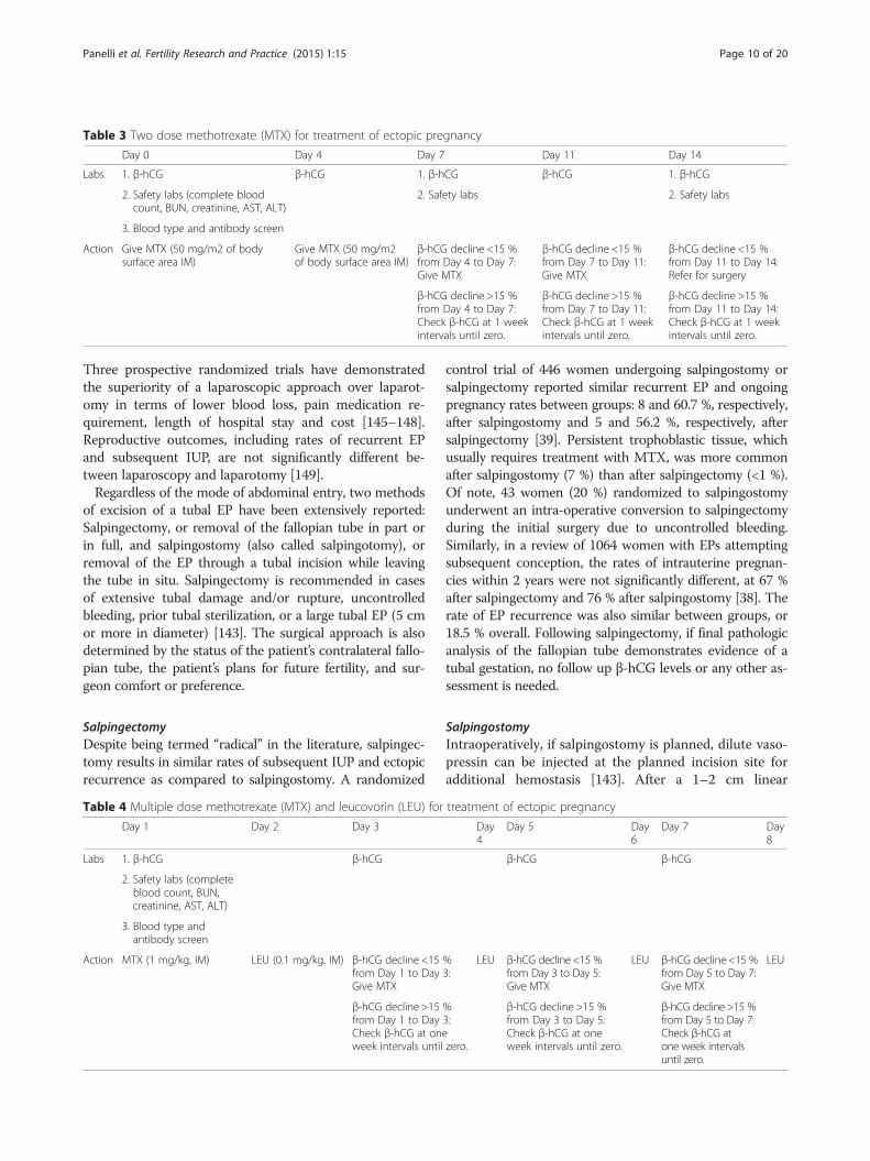
Two dose methotrexateA two dose regimen has been proposed by Barnhart andcolleagues (Table 3) [133]. In their prospective study of 101women with a mean serum β-hCG at treatment initiationof 2013 mIU/mL, the success rate, defined as avoidance ofsurgery, was 87 %. Patients suffered just mild and transientside effects, including nausea, headaches and abdominalpain, despite the lack of leucovorin supplementation.
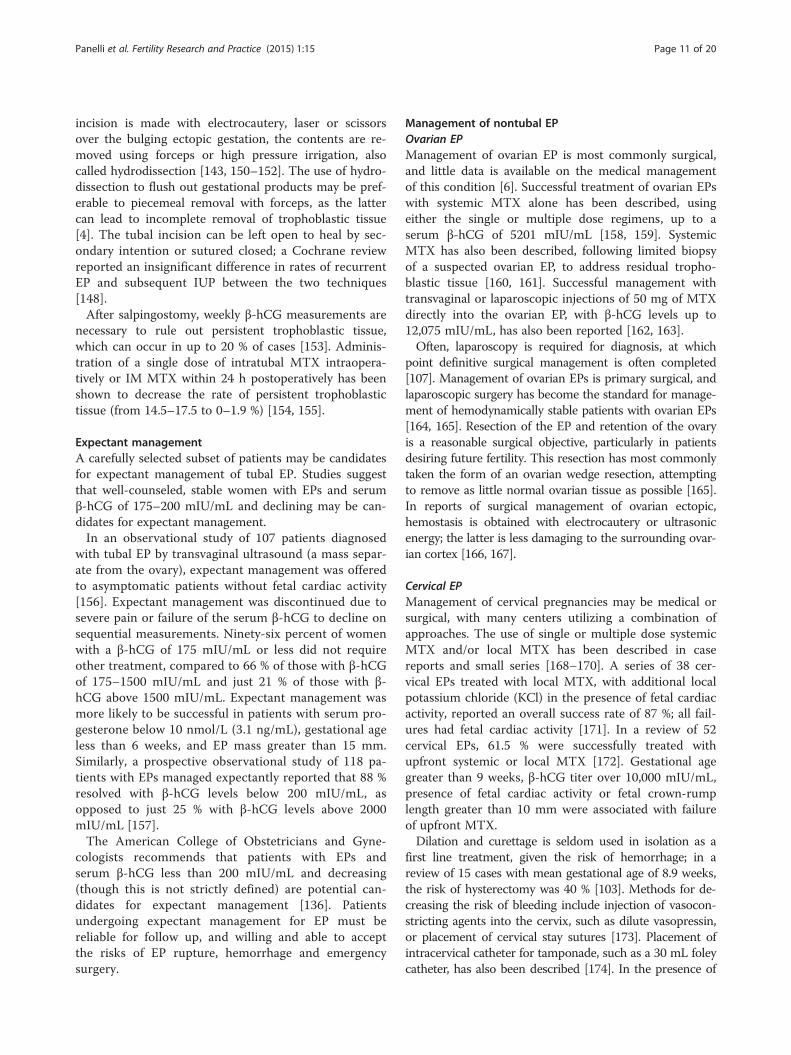
Multiple dose methotrexateThe multiple dose regimen was derived from chemo-therapeutic regimens for gestational trophoblastic dis-ease, involving administration of MTX and leucovorin(folinic acid) on alternating days for 8 days or until theβ-hCG falls by 15 % from its peak value (Table 4) [6].Up to 50 % of patients will not require the full 8 dayregimen [6]. Leucovorin is administered to counteractthe mechanism of MTX to limit side effects.The reported success rates among the dosing regimens
vary in the literature [127, 137, 138]. A recent randomizedcontrolled trial of 120 women receiving single or multipledose MTX reported no difference in success rates, thoughthe time until β-hCG normalization was longer followingthe single dose regimen (22.3 vs. 18.3 days, respectively)[140]. Conversely, a meta-analysis of 1327 EPs reportedthat the rate of successful treatment with multiple doseMTX was significantly higher than with single dose MTX
(92.7 vs. 88.1 %, respectively) [137]. Side effects, includingnausea, vomiting and alopecia, were less common in thesingle dose treatment group. Of note, both treatment regi-mens were more likely to be successful in patients report-ing side effects of the MTX.Few comparisons have been published involving the two
dose regimen. A retrospective comparison of 87 womenreceiving either single or two dose MTX regimens reportedcomparable success rates of 87 and 90 % at mean startingserum hCGs of 4801 and 4278 mIU/mL, respectively, andno difference in side effects [141]. In the literature, it isunclear which of these (single, two or multiple dose) regi-mens is used most commonly, though single and multipledose regimens are discussed more often than the two doseregimen; MTX dosing is likely dependent on the providerand/or institution.Regardless of which treatment regimen is chosen, if
the β-hCG level does not decline adequately—after themultiple dose regimen, or 4 doses of MTX in single ortwo dose regimens—surgical management should beconsidered. A continued rise in serum β-hCG through-out the multiple dose regimen or after 2 doses of singledose MTX may indicate higher risk of rupture of a tubalEP [6, 142]. Finally, medical management should beabandoned in favor of surgical management if thepatient presents with hemodynamic instability or otherclinical parameters concerning for ruptured EP, such aspain. If a patient’s serum β-hCG declines adequatelyand she requires no further intervention, the β-hCGlevel should be monitored weekly to an undetectablelevel. On average, the β-hCG normalizes in 2 to 3 weeks,but can take up to 8 weeks in patients with higher start-ing β-hCG levels [6, 143].
Surgical managementSurgical management is indicated in patients with con-traindications to medical treatment as described in theprevious section, hemodynamic compromise or otherclinical signs of ruptured EP including pain or evidenceof intra-abdominal bleeding, and according to patientpreference.The standard surgical intervention was laparotomy until
the laparoscopic approach was introduced in 1973 by Sha-piro and Adler; it has since gained wide acceptance [144].
Table 2 Single dose methotrexate (MTX) for treatment of ectopic pregnancy
Day 1 Day 4 Day 7
Labs 1. β-hCG β-hCG 1. β-hCG
2. Safety labs (complete blood count, BUN, creatinine, AST, ALT) 2. Safety labs
3. Blood type and antibody screen
Action Give MTX (50 mg/m2 of body surface area IM) no action β-hCG decline <15 % from Day 4 to Day 7: MTX, return to day 1 ofprotocol. Repeat MTX up to a total of 4 doses
β-hCG decline >15 %: Check β-hCG at 1 week intervals until zero.
Panelli et al. Fertility Research and Practice (2015) 1:15 Page 9 of 20
Three prospective randomized trials have demonstratedthe superiority of a laparoscopic approach over laparot-omy in terms of lower blood loss, pain medication re-quirement, length of hospital stay and cost [145–148].Reproductive outcomes, including rates of recurrent EPand subsequent IUP, are not significantly different be-tween laparoscopy and laparotomy [149].Regardless of the mode of abdominal entry, two methods
of excision of a tubal EP have been extensively reported:Salpingectomy, or removal of the fallopian tube in part orin full, and salpingostomy (also called salpingotomy), orremoval of the EP through a tubal incision while leavingthe tube in situ. Salpingectomy is recommended in casesof extensive tubal damage and/or rupture, uncontrolledbleeding, prior tubal sterilization, or a large tubal EP (5 cmor more in diameter) [143]. The surgical approach is alsodetermined by the status of the patient’s contralateral fallo-pian tube, the patient’s plans for future fertility, and sur-geon comfort or preference.
SalpingectomyDespite being termed “radical” in the literature, salpingec-tomy results in similar rates of subsequent IUP and ectopicrecurrence as compared to salpingostomy. A randomized
control trial of 446 women undergoing salpingostomy orsalpingectomy reported similar recurrent EP and ongoingpregnancy rates between groups: 8 and 60.7 %, respectively,after salpingostomy and 5 and 56.2 %, respectively, aftersalpingectomy [39]. Persistent trophoblastic tissue, whichusually requires treatment with MTX, was more commonafter salpingostomy (7 %) than after salpingectomy (<1 %).Of note, 43 women (20 %) randomized to salpingostomyunderwent an intra-operative conversion to salpingectomyduring the initial surgery due to uncontrolled bleeding.Similarly, in a review of 1064 women with EPs attemptingsubsequent conception, the rates of intrauterine pregnan-cies within 2 years were not significantly different, at 67 %after salpingectomy and 76 % after salpingostomy [38]. Therate of EP recurrence was also similar between groups, or18.5 % overall. Following salpingectomy, if final pathologicanalysis of the fallopian tube demonstrates evidence of atubal gestation, no follow up β-hCG levels or any other as-sessment is needed.
SalpingostomyIntraoperatively, if salpingostomy is planned, dilute vaso-pressin can be injected at the planned incision site foradditional hemostasis [143]. After a 1–2 cm linear
Table 4 Multiple dose methotrexate (MTX) and leucovorin (LEU) for treatment of ectopic pregnancy
Day 1 Day 2 Day 3 Day4
Day 5 Day6
Day 7 Day8
Labs 1. β-hCG β-hCG β-hCG β-hCG
2. Safety labs (completeblood count, BUN,creatinine, AST, ALT)
3. Blood type andantibody screen
Action MTX (1 mg/kg, IM) LEU (0.1 mg/kg, IM) β-hCG decline <15 %from Day 1 to Day 3:Give MTX
LEU β-hCG decline <15 %from Day 3 to Day 5:Give MTX
LEU β-hCG decline <15 %from Day 5 to Day 7:Give MTX
LEU
β-hCG decline >15 %from Day 1 to Day 3:Check β-hCG at oneweek intervals until zero.
β-hCG decline >15 %from Day 3 to Day 5:Check β-hCG at oneweek intervals until zero.
β-hCG decline >15 %from Day 5 to Day 7:Check β-hCG atone week intervalsuntil zero.
Table 3 Two dose methotrexate (MTX) for treatment of ectopic pregnancy
Day 0 Day 4 Day 7 Day 11 Day 14
Labs 1. β-hCG β-hCG 1. β-hCG β-hCG 1. β-hCG
2. Safety labs (complete bloodcount, BUN, creatinine, AST, ALT)
2. Safety labs 2. Safety labs
3. Blood type and antibody screen
Action Give MTX (50 mg/m2 of bodysurface area IM)
Give MTX (50 mg/m2of body surface area IM)
β-hCG decline <15 %from Day 4 to Day 7:Give MTX
β-hCG decline <15 %from Day 7 to Day 11:Give MTX
β-hCG decline <15 %from Day 11 to Day 14:Refer for surgery
β-hCG decline >15 %from Day 4 to Day 7:Check β-hCG at 1 weekintervals until zero.
β-hCG decline >15 %from Day 7 to Day 11:Check β-hCG at 1 weekintervals until zero.
β-hCG decline >15 %from Day 11 to Day 14:Check β-hCG at 1 weekintervals until zero.
Panelli et al. Fertility Research and Practice (2015) 1:15 Page 10 of 20
incision is made with electrocautery, laser or scissorsover the bulging ectopic gestation, the contents are re-moved using forceps or high pressure irrigation, alsocalled hydrodissection [143, 150–152]. The use of hydro-dissection to flush out gestational products may be pref-erable to piecemeal removal with forceps, as the lattercan lead to incomplete removal of trophoblastic tissue[4]. The tubal incision can be left open to heal by sec-ondary intention or sutured closed; a Cochrane reviewreported an insignificant difference in rates of recurrentEP and subsequent IUP between the two techniques[148].After salpingostomy, weekly β-hCG measurements are
necessary to rule out persistent trophoblastic tissue,which can occur in up to 20 % of cases [153]. Adminis-tration of a single dose of intratubal MTX intraopera-tively or IM MTX within 24 h postoperatively has beenshown to decrease the rate of persistent trophoblastictissue (from 14.5–17.5 to 0–1.9 %) [154, 155].
Expectant managementA carefully selected subset of patients may be candidatesfor expectant management of tubal EP. Studies suggestthat well-counseled, stable women with EPs and serumβ-hCG of 175–200 mIU/mL and declining may be can-didates for expectant management.In an observational study of 107 patients diagnosed
with tubal EP by transvaginal ultrasound (a mass separ-ate from the ovary), expectant management was offeredto asymptomatic patients without fetal cardiac activity[156]. Expectant management was discontinued due tosevere pain or failure of the serum β-hCG to decline onsequential measurements. Ninety-six percent of womenwith a β-hCG of 175 mIU/mL or less did not requireother treatment, compared to 66 % of those with β-hCGof 175–1500 mIU/mL and just 21 % of those with β-hCG above 1500 mIU/mL. Expectant management wasmore likely to be successful in patients with serum pro-gesterone below 10 nmol/L (3.1 ng/mL), gestational ageless than 6 weeks, and EP mass greater than 15 mm.Similarly, a prospective observational study of 118 pa-tients with EPs managed expectantly reported that 88 %resolved with β-hCG levels below 200 mIU/mL, asopposed to just 25 % with β-hCG levels above 2000mIU/mL [157].The American College of Obstetricians and Gyne-
cologists recommends that patients with EPs andserum β-hCG less than 200 mIU/mL and decreasing(though this is not strictly defined) are potential can-didates for expectant management [136]. Patientsundergoing expectant management for EP must bereliable for follow up, and willing and able to acceptthe risks of EP rupture, hemorrhage and emergencysurgery.
Management of nontubal EPOvarian EPManagement of ovarian EP is most commonly surgical,and little data is available on the medical managementof this condition [6]. Successful treatment of ovarian EPswith systemic MTX alone has been described, usingeither the single or multiple dose regimens, up to aserum β-hCG of 5201 mIU/mL [158, 159]. SystemicMTX has also been described, following limited biopsyof a suspected ovarian EP, to address residual tropho-blastic tissue [160, 161]. Successful management withtransvaginal or laparoscopic injections of 50 mg of MTXdirectly into the ovarian EP, with β-hCG levels up to12,075 mIU/mL, has also been reported [162, 163].Often, laparoscopy is required for diagnosis, at which
point definitive surgical management is often completed[107]. Management of ovarian EPs is primary surgical, andlaparoscopic surgery has become the standard for manage-ment of hemodynamically stable patients with ovarian EPs[164, 165]. Resection of the EP and retention of the ovaryis a reasonable surgical objective, particularly in patientsdesiring future fertility. This resection has most commonlytaken the form of an ovarian wedge resection, attemptingto remove as little normal ovarian tissue as possible [165].In reports of surgical management of ovarian ectopic,hemostasis is obtained with electrocautery or ultrasonicenergy; the latter is less damaging to the surrounding ovar-ian cortex [166, 167].
Cervical EPManagement of cervical pregnancies may be medical orsurgical, with many centers utilizing a combination ofapproaches. The use of single or multiple dose systemicMTX and/or local MTX has been described in casereports and small series [168–170]. A series of 38 cer-vical EPs treated with local MTX, with additional localpotassium chloride (KCl) in the presence of fetal cardiacactivity, reported an overall success rate of 87 %; all fail-ures had fetal cardiac activity [171]. In a review of 52cervical EPs, 61.5 % were successfully treated withupfront systemic or local MTX [172]. Gestational agegreater than 9 weeks, β-hCG titer over 10,000 mIU/mL,presence of fetal cardiac activity or fetal crown-rumplength greater than 10 mm were associated with failureof upfront MTX.Dilation and curettage is seldom used in isolation as a
first line treatment, given the risk of hemorrhage; in areview of 15 cases with mean gestational age of 8.9 weeks,the risk of hysterectomy was 40 % [103]. Methods for de-creasing the risk of bleeding include injection of vasocon-stricting agents into the cervix, such as dilute vasopressin,or placement of cervical stay sutures [173]. Placement ofintracervical catheter for tamponade, such as a 30 mL foleycatheter, has also been described [174]. In the presence of
Panelli et al. Fertility Research and Practice (2015) 1:15 Page 11 of 20

fetal cardiac activity, preoperative injection of feticides maydecrease the risk of hemorrhage [175].Uterine artery embolization (UAE) may have a role in
preventing or controlling hemorrhage; case series havereported both prophylactic UAE prior to medical and/orsurgical management, or emergent use to controlhemorrhage [170, 176–178]. This therapy is not currentlyrecommended for women who wish to conceive in thefuture, as its ramifications for fertility have not beenconclusively described.
Cesarean scar EPInterruption of a cesarean scar EP upon diagnosis is rec-ommended, given the risk of hemorrhage, hysterectomyand maternal morbidity [179, 180]. Live births resultingfrom a cesarean scar ectopic implantation have beendescribed; however, these deliveries are frequentlyassociated with hemorrhage and emergent cesareanhysterectomy [9, 181, 182]. In a series of 10 patientswith cesarean scar EPs with fetal cardiac activity whoelected for expectant management, 4 patients (40 %)had live births, 3 of whom (75 %) required hysterecto-mies; overall, 80 % required hysterectomies [183].Medical management with single or multiple dose
systemic MTX regimens has been described. Patients withserum β-hCG greater than 6000 mIU/mL may be at higherrisk of requiring additional therapies, including local MTX,D&C or uterine artery embolization (UAE) [9, 64]. Localinjections of MTX or KCl have also been described, usuallyin conjunction with systemic MTX or other surgical man-agement (D&C or hysteroscopy) [184].Several surgical approaches have also been reported,
with the benefit of leading to more rapid resolution ofβ-hCG levels as compared to medical management [185].Regardless of the chosen treatment modality, serum β-hCG should be followed to zero, as persistent tropho-blastic tissue may occur after any medical or surgicaltreatments except hysterectomy [9, 186]. In patientsundergoing upfront surgical management, D&C alone isoften complicated by hemorrhage. In a meta-analysis of 21cases, 76 % required further treatment, and 14 % requiredhysterectomy [64]. Initial steps for managing hemorrhageinclude tamponade with a transcervical catheter andhemostatic cervical cerclage sutures [187]. UAE has beenused as both hemorrhage prophylaxis and salvage therapyin the event of hemorrhage [188]. UAE is not currentlyrecommended for patients desiring future fertility.Hysteroscopic resection of cesarean scar EPs has been
performed successfully and without complication usingbiopolar or ‘electric’ loops, in patients with serum β-hCGup to 28,333 mIU/mL [186, 189, 190]. Hysteroscopic re-section is not recommended when the residual myome-trium is less than 3 mm, given the risk of anterior wallperforation and bladder injury [191, 192].
Transabdominal excision of these lesions has been de-scribed by laparotomy, and standard or robotic-assistedlaparoscopy [191]. Resection also allows for revision ofthe lower uterine segment, which theoretically may re-duce risk for recurrence [193]. Laparotomy may be indi-cated in patients with suspected uterine rupture andhemodynamic instability, and hysterectomy may be re-quired for otherwise uncontrollable hemorrhage [194].Definitive management with total laparoscopic hyster-ectomy has also been described, in a patient with astarting β-hCG of 155,009 mIU/mL who failed treat-ment with local KCl and multiple dose MTX [195]. Ofnote, complications of medical or surgical managementinclude formation of arterio-venous malformations,which are prone to bleeding; in one series of 60 cesareanscar EPs, this occurred at a rate of 8.5 %, requiring UAEor hysterectomy (Fig. 9) [183].
Interstitial EPIn patients who are hemodynamically stable withoutevidence of rupture of the interstitial EP, non-surgicalmanagement may be appropriate. Both single dose and
Fig. 9 Left uterine artery arterio-venous malformation (AVM) by pelvicangiogram. This patient had undergone an uncomplicatedultrasound-guided D&C for a 10 week size cesarean scar ectopicpregnancy 2 months prior to presentation with vaginal bleeding anddiagnosis of a left uterine artery AVM (arrow). The AVM was embolizedwith coils, but the patient required emergent hysterectomy forhemorrhage
Panelli et al. Fertility Research and Practice (2015) 1:15 Page 12 of 20

multiple dose MTX regimens have been used to treatinterstitial EPs with comparable success, ranging from66 to 100 % [196]. UAE has also been successfully usedas an adjunct to these therapies [197, 198]. Local MTXhas also been used; a meta-analysis of 11 cases reporteda success rate of 86 %, up to a serum hCG of 35,000mIU/mL [4].Surgical intervention is indicated following failed
medical management, according to patient preference,or when the patient demonstrates hemodynamic in-stability and/or findings concerning for rupture of aninterstitial EP, including pain or evidence of hemoperi-toneum on imaging. Laparotomy and hysterectomywere formerly first line treatment, likely due to latediagnosis of interstitial pregnancies and higher rates ofrupture and hemorrhage. These methods may still benecessary in patients with hemodynamic instability andsevere hemorrhage.Minimally invasive surgeries are increasingly pursued as
imaging modalities allow for earlier diagnosis. Small caseseries have described ultrasound or laparoscopy-guideddilation and curettage [199–201]. Several laparoscopic sur-gical approaches have been described, including cornuost-omy, salpingostomy, and cornual resection.Cornuostomy entails injection of dilute vasopressin at
the cornua followed by a linear incision, through whichthe gestation is removed with blunt and/or sharp dissec-tion or hydrodissection, after which the incision is closedwith absorbable suture [4]. Case series have also de-scribed successful surgical management with placementof an Endoloop around the base of the cornua before orafter excision for both hemostasis and closure [202].Less commonly, salpingostomy for interstitial ectopichas been reported, which is most appropriate for inter-stitial EP less than 3.5 cm, given the smaller incisionwith limited visualization [203, 204].Cornual resection has been recommended for surgical
management of more advanced interstitial pregnancies(greater than 3–4 cm) [12, 205]. This technique entailsinjection of dilute vasopressin followed by a circumfer-ential incision using scissors or an energy source—elec-trosurgical or ultrasonic—preferably 1–2 cm above thecornual pregnancy to allow for redundant serosa andmyometrium for closure [11, 12]. This incision shouldbe closed in layers akin to a myomectomy closure. Thefallopian tube adjacent to this cornua should also be ex-cised. UAE has also been used as a prophylactic measurebefore laparoscopic cornual resection [206].
Intramural EPAs with other types and locations of EP, management ofintramural EPs is largely dictated by patients’ clinical sta-bility at presentation. In clinically stable patients with
intramural EPs diagnosed by imaging, medical manage-ment is an option. In case reports, intramural EPs havebeen treated with single or multiple dose systemic MTX,successful in patients with serum β-hCG up to 25,140mIU/ml [207–209]. Successful management with localMTX and KCl for an intramural EP with fetal cardiacactivity and a β-hCG of 74,872 mIU/mL has also been re-ported, as well as UAE for an intramural EP with a β-hCGof 12,250 mIU/mL [210, 211].Most cases of intramural EP reported in the literature
have been managed surgically via laparotomy, sometimesrequiring hysterectomy, as many patients present withrupture of the EP and hemorrhage [13]. Given the increas-ing ability of noninvasive imaging to diagnose intramuralEPs and the advancement of minimally invasive surgery,more recent case reports have described laparoscopic ex-cision of intramural ectopic gestations [212, 213]. A surgi-cal approach should be determined by a patient’s clinicalstability, desire for future fertility, and location of the ec-topic gestation.
Abdominal EPIntervention for resolution of an abdominal EP isrecommended upon diagnosis, given the extremely highrisk for maternal morbidity; the mortality risk associ-ated with abdominal EPs is nearly 8 times the rate withtubal EPs [16]. Rare reports detail expectant manage-ment in order to attain a live birth. Expectant manage-ment of abdominal EPs may potentially be consideredwhen the diagnosis is made after 20 weeks of gestationin a healthy patient who can be followed very closelythrough a tertiary care center. The fetus should have nocongenital malformations, and the placenta should beimplanted away from the upper abdomen. Delivery isrecommended at 34 weeks, and the placenta is often leftin place given the risk for hemorrhage [214, 215].Most abdominal EPs reported in the literature have
been managed surgically; the operative approach must betailored to the patient’s clinical presentation and stability,and the location of the EP. Abdominal EPs have beenapproached by laparoscopy or laparotomy, with or with-out prophylactic embolization of the placental bed; morerecent cases in the literature have been managed laparo-scopically in hemodynamically stable patients [216–218].Intraoperative blood transfusion is common; in a meta-analysis, the highest transfusion rate was associated withhepatic (46 %) and retroperitoneal (40 %) implantations,while abdominal wall implantations had the lowest trans-fusion rate (14 %) [16].When abdominal EPs are removed surgically at any
gestational age—though more commonly after 20 weeksof gestation—the placenta can be left in place to avoidhemorrhage [16]. Embolization of the remaining placentaand/or administration of systemic MTX or mifepristone
Panelli et al. Fertility Research and Practice (2015) 1:15 Page 13 of 20

have been employed to hasten resolution of these retainedplacentas [219, 220]. The most common complication ofan intraabdominal retained placenta is infection [16].As diagnostic modalities have advanced and these
pregnancies are diagnosed earlier, case reports of med-ical management for abdominal EP have been pub-lished. Medical management with systemic MTX and/or local injections of MTX or KCl has been reported,though nearly half may require subsequent surgicalmanagement [16, 221–223]. Despite logistic regression,a meta-analysis failed to identify risk factors for failedmedical management [16].
Heterotopic pregnanciesTreatment of a HP is tailored to the specific EP location,and the patient’s clinical presentation and stability [78].Medical management of tubal HPs includes local injectionsof KCl or a hyperosmolar glucose solution, though overhalf of tubal HPs managed with local KCl may require sub-sequent salpingectomy [17, 224]. Treatment with systemicor local MTX, a known teratogen, is contraindicated in thepresence of a viable IUP [225]. Surgical management hasbeen described more frequently, as patients with tubal HPspresent more often with rupture and hemodynamic com-promise than those with tubal EPs [226]. Salpingectomy ispreferable to salpingostomy as persistent trophoblastictissue cannot be monitored in the setting of ongoing IUP[78]. Patients with HPs suffer spontaneous abortions athigher rates than intrauterine-only pregnancies (up to30 %) [18].
Nontubal HPFor the management of interstitial HPs, expectant man-agement, aspiration or injection of hyperosmolar glucoseof the interstitial HP, and cornual resections have beenreported, leading to live birth [227–229]. One patientattempting expectant management required a laparot-omy for rupture of the interstitial EP [227].Cesarean HPs have been successfully managed using
local KCl and/or aspiration of the gestation, or excisionby laparoscopy or hysteroscopy [230]. Hysteroscopy car-ries the theoretical risk of disrupting an IUP due to thehigh pressure infusion of fluid.Cervical HPs addressed with expectant management,
local KCl or hyperosmolar glucose injections, extractionwith forceps, suction curettage or hysteroscopic resec-tion, with or without subsequent foley tamponade, haveresulted in live birth. Rare case reports also detail cerc-lage placement following intervention. Following thisrange of interventions, a review of 30 cases reported alive birth rate of 80 % [231].Abdominal HPs are rarely encountered, though live
birth after local injection of KCl into the abdominalpregnancy has been reported in 3 cases [232]. Ovarian
HPs are similarly rare; live birth after local hyperosmolarglucose injection has been reported, as well as after lap-aroscopic wedge resection; surgical intervention carriesthe theoretical risk of interrupting hormonal support ofthe coexisting IUP by the corpus luteum [233, 234].
Recurrence and future fertilityThe risk of recurrence of tubal EP ranges from 5 to 25 %[38–40, 235]. The risk of recurrent EP is not affected bytreatment modality—medical or surgical—or surgical pro-cedure [38]. In a randomized controlled study of 446women undergoing surgical management for tubal EP, therecurrence rate was similar after salpingostomy (8 %) andsalpingectomy (5 %) [39].A review of 53 cases of prior interstitial EP reported a re-
currence rate of 9.4 % following either medical or surgicalmanagement [236]. In patients with a prior interstitial EP,data is limited regarding the risk of uterine rupture in asubsequent IUP, though uterine rupture has been reportedafter both expectant management and cornual resection[237, 238]. Vaginal deliveries have been reported followingcornuostomy or cornual resection; the optimal mode ofdelivery in this group remains to be determined [12].The reported rate of recurrent cesarean scar EP is highly
variable, as high as 25 % in small series [239, 240]. Riskfactors for recurrence are bulging of the prior cesareanscar EP into the uterovesical fold, initial presentation withirregular vaginal bleeding or pain, early termination(≤56 days) of the first cesarean scar EP, prior cesarean de-livery at a rural community hospital and thin lower uterinesegment (5 mm or less at the time of diagnosis of recur-rent cesarean scar EP) [241].The risk of recurrent cervical EP appears to be low:
One recurrence was noted in a series of 34 pregnantwomen with prior cervical EP treated with several differ-ent modalities [67]. The data are insufficient to com-ment on subsequent IUP and recurrence rates inpatients with prior ovarian, intramural or abdominalEPs. Rates of recurrence and IUP after HP have not beenextensively reported in the literature, and likely dependon the location of the HP and the treatment modality.Regardless of ectopic location, conception is not recom-
mended for 3 months after exposure to MTX, though datafor this recommendation is lacking [6]. Results ofpopulation-based studies of pregnancy outcomes after aprior tubal EP are encouraging, and independent of treat-ment modality. The rates of IUP have been shown to besimilar following salpingectomy and salpingostomy in sev-eral large series [39, 40]. Additionally, among 1064 womenwith prior tubal EPs attempting conception, the rates ofIUP within 2 years were similar among salpingectomy(67 %), salpingostomy (76 %), and medical management(76 %) [38]. After two prior EPs, however, the rate ofsubsequent IUP may be as low as 4 % [235].
Panelli et al. Fertility Research and Practice (2015) 1:15 Page 14 of 20

ConclusionsEctopic pregnancy is a relatively common clinical scenarioin general gynecology and reproductive medicine. Whiletubal pregnancies are the most common, EPs can occurthroughout the abdomen and pelvis. Treatment in stablepatients is often medical, though patients meeting certainclinical criteria or with EPs outside the fallopian tube mayrequire differing and/or more invasive treatment, includ-ing excision by laparoscopy or, less commonly, laparot-omy. Of patients with tubal EPs, the likelihood of futureIUP is high and independent of treatment modality.
AbbreviationsADAM-12: A Disintegrin and Metalloprotease-12; ART: Assisted reproductivetechnology; D&C: Dilation and curettage; E2: Estradiol; EP: Ectopic pregnancy;FAST: Focused assessment with sonography for trauma;GnRH: Gonadotropin-releasing hormone; hCG: Human chorionicgonadotropin; HP: Heterotopic pregnancy; IM: Intramuscular; IUD: Intrauterinedevice; IUP: Intrauterine pregnancy; IVF: In vitro fertilization; KCl: Potassiumchloride; LEU: Leucovorin; MTX: Methotrexate; NO: Nitric oxide;PAPPA: Pregnancy-associated plasma protein-A; PUL: Pregnancy of unknownlocation; TVUS: Transvaginal ultrasound; UAE: Uterine artery embolization;VEGF: Vascular endothelial growth factor.
Competing interestsThe authors declare that they have no competing interests.
Authors’ contributionsDP and PB participated in the planning and drafting of the manuscript. CPselected and edited the radiologic imaging. All authors read and approvedthe final manuscript.
Authors’ informationDanielle Panelli is a resident in the Brigham and Women’s Hospital/Massachusetts General Hospital Integrated Residency Program in Obstetricsand Gynecology, an affiliate of the Harvard Medical School.Catherine Phillips is the chief resident at the Brigham and Women’s HospitalDiagnostic Radiology Residency Program, an affiliate of Harvard Medical School.Paula Brady is a resident in the Brigham and Women’s Hospital/MassachusettsGeneral Hospital Integrated Residency Program in Obstetrics and Gynecology,an affiliate of the Harvard Medical School.
Author details1Department of Obstcpetrics and Gynecology, Brigham and Women’sHospital, Harvard Medical School, 75 Francis St., Boston, MA 02115, USA.2Department of Radiology, Brigham and Women’s Hospital, Harvard MedicalSchool, Boston, MA, USA.
Received: 16 June 2015 Accepted: 29 September 2015
References1. Creanga AA, Shapiro-Mendoza CK, Bish CL, Zane S, Berg CJ, Callaghan WM.
Trends in ectopic pregnancy mortality in the United States: 1980–2007.Obstet Gynecol. 2011;117:837–43.
2. Arleo EK, DeFilippis EM. Cornual, interstitial, and angular pregnancies:clarifying the terms and a review of the literature. Clin Imaging.2014;38:763–70.
3. Hoffman BL, Schorge JO, Schaffer JI, Halvorson LM, Bradshaw KD,Cunningham F, et al. Chapter 7. Ectopic pregnancy. In: Hoffman BL,Schorge JO, Schaffer JI, Halvorson LM, Bradshaw KD, Cunningham F,Calver LE, editors. Williams gynecology. 2nd ed. New York: McGraw-Hill;2012. http://accessmedicine.mhmedical.com.ezp-prod1.hul.harvard.edu/content.aspx?bookid=399&Sectionid=41722295.
4. Lau S, Tulandi T. Conservative medical and surgical management ofinterstitial ectopic pregnancy. Fertil Steril. 1999;72:207–15.
5. Barnhart K. Ectopic pregnancy. N Engl J Med. 2009;361:379–87.
6. Practice Committee of the American Society for Reproductive Medicine.Medical treatment of ectopic pregnancy: a committee opinion. Fertil Steril.2013;100:638–44.
7. Perkins KM, Boulet SL, Kissin DM, Jamieson DJ, National ART Surveillance(NASS) Group. Risk of ectopic pregnancy associated with assistedreproductive technology in the United States, 2001–2011. Obstet Gynecol.2015;125:70–8.
8. Yankowitz J, Leake J, Huggins G, Gazaway P, Gates E. Cervical ectopicpregnancy: review of the literature and report of a case treated by single-dosemethotrexate therapy. Obstet Gynecol Surv. 1990;45:405–14.
9. Jurkovic D, Hillaby K, Woelfer B, Lawrence A, Salim R, Elson CJ. First trimesterdiagnosis and management of pregnancies implanted into the loweruterine segment cesarean section scar. Ultrasound Obstet Gynecol.2003;21:220–7.
10. Seow KM, Huang LW, Lin YH, Lin MY, Tsai YL, Hwang JL. Cesarean scarpregnancy: issues in management. Ultrasound Obstet Gynecol. 2004;23:247–53.
11. Moawad NS, Mahajan ST, Moniz MH, Taylor SE, Hurd WW. Current diagnosisand treatment of interstitial pregnancy. Am J Obstet Gynecol. 2010;202:15–29.
12. Ng S, Hamontri S, Chua I, Chern B, Siow A. Laparoscopic management of 53cases of cornual ectopic pregnancy. Fertil Steril. 2009;92:448–52.
13. Kirk E, McDonald K, Rees J, Govind A. Intramural ectopic pregnancy: a caseand review of the literature. Eur J Obstet Gynecol Reprod Biol.2013;168:129–33.
14. Khan KS, Wojdyla D, Say L, Gulmezoglu AM, Van Look PF. WHO analysis ofcauses of maternal death: a systematic review. Lancet. 2006;367:1066–74.
15. Rana P, Kazmi I, Singh R, Afzal M, Al-Abbasi FA, Aseeri A, et al. Ectopicpregnancy: a review. Arch Gynecol Obstet. 2013;288:747–57.
16. Poole A, Haas D, Magann EF. Early abdominal ectopic pregnancies: asystematic review of the literature. Gynecol Obstet Invest. 2012;74:249–60.
17. Barrenetxea G, Barinaga-Rementeria L, Lopez de Larruzea A, Agirregoikoa JA,Mandiola M, Carbonero K. Heterotopic pregnancy: two cases and acomparative review. Fertil Steril. 2007;82:9–15.
18. Clayton HB, Schieve LA, Peterson HB, Jamieson DJ, Reynolds MA, Wright VC.Ectopic pregnancy risk with assisted reproductive technology procedures.Obset Gynecol. 2006;107:595–604.
19. Lin CK, Wen KC, Sung PL, Lin SC, Lai CR, Chao KC, et al. Heterotopic tripletpregnancy with an intrauterine, a tubal, and a cervical gestation followingin vitro fertilization and embryo transfer. Taiwan J Obstet Gynecol.2013;52:287–9.
20. Talbot P, Riveles K. Smoking and reproduction: the oviduct as a target ofcigarette smoke. Reprod Biol Endocrinol. 2005;3:52.
21. Shaw JL, Dey SK, Critchley HO, Horne AW. Current knowledge of theaetiology of human tubal ectopic pregnancy. Hum Reprod Update.2010;16:432–44.
22. Magers T, Talbot P, DiCarlantonio G, Knoll M, Demers D, Tsai I, et al.Cigarette smoke inhalation affects the reproductive system of femalehamsters. Reprod Toxicol. 1995;9:513–25.
23. Lyons RA, Saridogan E, Djahanbakhch O. The reproductive significance ofhuman Fallopian tube cilia. Hum Reprod Update. 2006;12:363–72.
24. Jansen RP. Endocrine response in the fallopian tube. Endocr Rev. 1984;5:525–51.25. Paltieli Y, Eibschitz I, Ziskind G, Ohel G, Silbermann M, Weichselbaum A.
High progesterone levels and ciliary dysfunction–a possible cause ofectopic pregnancy. J Assist Reprod Genet. 2000;17:103–6.
26. Jain B, Rubinstein I, Robbins RA, Sisson JH. TNF-alpha and IL-1 betaupregulate nitric oxide-dependent ciliary motility in bovine airwayepithelium. Am J Physiol. 1995;268:L911–7.
27. Al-Azemi M, Refaat B, Amer S, Ola B, Chapman N, Ledger W. The expressionof inducible nitric oxide synthase in the human fallopian tube during themenstrual cycle and in ectopic pregnancy. Fertil Steril. 2010;94:833–40.
28. Nilsson S, Makela S, Treuter E, Tujague M, Thomsen J, Andersson G, et al.Mechanisms of estrogen action. Physiol Rev. 2001;81:1535–65.
29. Deroo BJ, Korach KS. Estrogen receptors and human disease. J Clin Invest.2006;116:561–70.
30. Shao R, Feng Y, Zou S, Weijdegård B, Wu G, Brännström M, et al. The role ofestrogen in the pathophysiology of tubal ectopic pregnancy. Am J TranslRes. 2012;4:269–78.
31. Shaw JL, Horne AW. The paracrinology of tubal ectopic pregnancy. Mol CellEndocrinol. 2012;358:216–22.
32. Hvid M, Baczynska A, Deleuran B, Fedder J, Knudsen HJ, Christiansen G,et al. Interleukin-1 is the initiator of Fallopian tube destruction duringChlamydia trachomatis infection. Cell Microbiol. 2007;9:2795–803.
Panelli et al. Fertility Research and Practice (2015) 1:15 Page 15 of 20

33. Shaw JL, Fitch P, Cartwright J, Entrican G, Schwarze J, Critchley HO, et al.Lymphoid and myeloid cell populations in the non-pregnant humanFallopian tube and in ectopic pregnancy. J Reprod Immunol. 2011;89:84–91.
34. Ulziibat S, Ejima K, Shibata Y, Hishikawa Y, Kitajima M, Fujishita A, et al.Identification of estrogen receptor beta-positive intraepithelial lymphocytesand their possible roles in normal and tubal pregnancy oviducts. HumReprod. 2006;21:2281–9.
35. von Rango U, Classen-Linke I, Kertschanska S, Kemp B, Beier HM. Effects oftrophoblast invasion on the distribution of leukocytes in uterine and tubalimplantation sites. Fertil Steril. 2001;76:116–24.
36. Bouyer J, Coste J, Shojaei T, Pouly JL, Fernandez H, Gerbaud L, et al. Risk factorsfor ectopic pregnancy: a comprehensive analysis based on a large case–control,population-based study in France. Am J Epidemiol. 2003;157:185–94.
37. Yeko TR, Handler A. The effect of aging on tubal function and ectopicpregnancy. Semin Reprod Endocrinol. 1991;9:215–20.
38. de Bennetot M, Rabischong B, Aublet-Cuvelier B, Belard F, Fernandez H,Bouyer J, et al. Fertility after tubal ectopic pregnancy: results of apopulation-based study. Fertil Steril. 2012;98:1271–6. e1-3.
39. Mol F, van Mello NM, Strandell A, Strandell K, Jurkovic D, Ross J, et al.Salpingotomy versus salpingectomy in women with tubal pregnancy (ESEPstudy): an open-label, multicentre, randomised controlled trial. Lancet.2014;383:1483–9.
40. Butts S, Sammel M, Hummel A, Chittams J, Barnhart K. Risk factor andclinical features of recurrent ectopic pregnancy: a case control study. FertilSteril. 2003;80:1340–4.
41. Weigert M, Gruber D, Pernicka E, Bauer P, Feichtinger W. Previous tubal ectopicpregnancy raises the incidence of repeated ectopic pregnancies in in vitrofertilization-embryo transfer patients. J Assist Reprod Genet. 2009;26:13–7.
42. Cooper MD, Rapp J, Jeffery-Wiseman C, Barnes RC, Stephens DS. Chlamydiatrachomatis infection of human fallopian tube organ cultures. J GenMicrobiol. 1990;136:1109–15.
43. Farquhar CM. Ectopic pregnancy. Lancet. 2005;366:583–91.44. Aminu MB, Abdullahi K, Dattijo LM. Tubal ectopic gestation associated with
genital schistosomiasis: a case report. Afr J Reprod Health. 2014;18:144–6.45. Haggerty CL, Gottlieb SL, Taylor BD, Low N, Xu F, Ness RB. Risk of sequelae
after Chlamydia trachomatis genital infection in women. J Infect Dis.2010;201:S134–55.
46. Senterman M, Jibodh R, Tulandi T. Histopathologic study of ampullary andisthmic tubal ectopic pregnancy. Am J Obstet Gynecol. 1988;159:939–41.
47. Dun EC, Nezhat CH. Tubal factor infertility: diagnosis and management inthe era of assisted reproductive technology. Obstet Gynecol Clin North Am.2012;39:551–66.
48. Rodgers AK, Goldberg JM, Hammel JP, Falcone T. Tubal anastomosis by roboticcompared with outpatient minilaparotomy. Obstet Gynecol. 2007;109:1375–80.
49. Peterson HB, Xia Z, Hughes JM, Wilcox LS, Tylor LR, Trussell J. The risk ofpregnancy after tubal sterilization: findings from the U.S. CollaborativeReview of Sterilization. Am J Obstet Gynecol. 1996;174:1161–8.
50. Bartz D, Greenberg JA. Sterilization in the United States. Rev Obstet Gynecol.2008;1:23–32.
51. Xiong X, Buekens P, Wollast E. IUD use and the risk of ectopic pregnancy: ameta-analysis of case–control studies. Contraception. 1995;52:23–34.
52. Furlong LA. Ectopic pregnancy risk when contraception fails. A review J ReprodMed. 2002;47:881–5.
53. Chang HJ, Suh CS. Ectopic pregnancy after assisted reproductive technology:what are the risk factors? Curr Opin Obstet Gynecol. 2010;22:202–7.
54. Li C, Meng CX, Zhao WH, Lu HQ, Shi W, Zhang J. Risk factors for ectopicpregnancy in women with planned pregnancy: a case–control study. Eur JObstet Gynecol Reprod Biol. 2014;181:176–82.
55. Huang B, Hu D, Qian K, Ai J, Li Y, Jin L, et al. Is frozen embryo transfer cycleassociated with a significantly lower incidence of ectopic pregnancy? Ananalysis of more than 30,000 cycles. Fertil Steril. 2014;102:1345–9.
56. Sahin S, Ozay A, Ergin E, Turkgeldi L, Kürüm E, Ozornek H. The risk ofectopic pregnancy following GnRH agonist triggering compared with hCGtriggering in GnRH antagonist IVF cycles. Arch Gynecol Obstet.2015;291:185–91.
57. Nazari A, Askari HA, Check JH, O’Shaughnessy A. Embryo transfer techniqueas a cause of ectopic pregnancy in in vitro fertilization. Fertil Steril.1993;60:919–21.
58. Smith LP, Oskowitz SP, Dodge LE, Hacker MR. Risk of ectopic pregnancyfollowing day-5 embryo transfer compared with day-3 transfer. ReprodBiomed Online. 2013;27:407–13.
59. Fang C, Huang R, Wei LN, Jia L. Frozen-thawed day 5 blastocyst transfer isassociated with a lower risk of ectopic pregnancy than day 3 transfer andfresh transfer. Fertil Steril. 2015;103:655–61.
60. Imudia AN, Awonuga AO, Doyle JO, Kaimal AJ, Wright DL, Toth TL, et al. Peakserum estradiol level during controlled ovarian hyperstimulation is associatedwith increased risk of small for gestational age and preeclampsia in singletonpregnancies after in vitro fertilization. Fertil Steril. 2012;97:1374–9.
61. Tulandi T, Al-Jaroudi D. Interstitial pregnancy: results generated from theSociety of Reproductive Surgeons Registry. Obstet Gynecol. 2004;103:47–50.
62. Talbot K, Simpson R, Price N, Jackson SR. Heterotopic pregnancy. J ObstetGynaecol. 2011;31:7–12.
63. World Health Organization. A multinational case–control study of ectopicpregnancy. The World Health Organization’s Special Programme of Research,Development and Research Training in Human Reproduction: Task Force onIntrauterine Devices for Fertility Regulation. Clin Reprod Fertil. 1985;3:131–43.
64. Rotas MA, Haberman S, Levgur M. Cesarean scar ectopic pregnancies:etiology, diagnosis, and management. Obstet Gynecol. 2006;107:1373–81.
65. Maymon R, Halperin R, Mendlovic S, Schneider D, Herman A. Ectopicpregnancies in a caesarean scar: review of the medical approach to aniatrogenic complication. Hum Reprod Update. 2004;10:515–23.
66. Pisarska MD, Carson SA. Incidence and risk factors for ectopic pregnancy.Clin Obstet Gynecol. 1999;42:2–8.
67. Ushakov FB, Elchalal U, Aceman PJ, Schenker JG. Cervical pregnancy: pastand future. Obstet Gynecol Surv. 1997;52:45–59.
68. Bennett S, Waterstone J, Parsons J, Creighton S. Two cases of cervicalpregnancy following in vitro fertilization and embryo transfer to the loweruterine cavity. J Assist Reprod Genet. 1993;10:100–3.
69. Molinaro TA, Barnhart KT. Ectopic pregnancies in unusual locations. SeminReprod Med. 2007;25:123–30.
70. Deshpande N, Mathers A, Acharya U. Broad ligament twin pregnancyfollowing in-vitro fertilization. Hum Reprod. 1999;14:852–4.
71. Kirk E, Condous G, Bourne T. Pregnancies of unknown location. Best PractRes Clin Obstet Gynaecol. 2009;23:493–9.
72. Fournier T, Guibourdenche J, Evain-Brion D. Review: hCGs: Different sourcesof production, different glycoforms and functions. Placenta. 2015;36:S60–5.
73. Morse CB, Sammel MD, Shaunik A, Allen-Taylor L, Oberfoell NL, Takacs P, et al.Performance of human chorionic gonadotropin curves in women at risk forectopic pregnancy: exceptions to the rules. Fertil Steril. 2012;97:101–6.
74. Chung K, Sammel MD, Coutifaris C, Chalian R, Lin K, Castelbaum AJ, et al.Defining the rise of serum HCG in viable pregnancies achieved through useof IVF. Hum Reprod. 2006;21:823–8.
75. Rausch ME, Barnhart KT. Serum biomarkers for detecting ectopic pregnancy.Clin Obstet Gynecol. 2012;55:418–23.
76. Verhaegen J, Gallos ID, van Mello NM, Abdel-Aziz M, Takwoingi Y, Harb H, etal. Accuracy of single progesterone test to predict early pregnancy outcomein women with pain or bleeding: meta-analysis of cohort studies. BMJ.2012;345, e6077.
77. Long CA, Lincoln SR, Whitworth NS, Cowan BD. Serum progesteronepredicts abnormal gestations in clomiphene citrate conception cycles aswell as in spontaneous conception cycles. Fertil Steril. 1994;61:838–42.
78. Fernandez H, Gervaise A. Ectopic pregnancies after infertility treatment:modern diagnosis and therapeutic strategy. Hum Reprod Update.2004;10:503–13.
79. Guha S, Ayim F, Ludlow J, Sayasneh A, Condous G, Kirk E, et al. Triagingpregnancies of unknown location: the performance of protocols based onsingle serum progesterone or repeated serum hCG levels. Hum Reprod.2014;29:938–45.
80. Mol BW, Lijmer JG, Ankum WM, van der Veen F, Bossuyt PM. The accuracyof single serum progesterone measurement in the diagnosis of ectopicpregnancy: a meta-analysis. Hum Reprod. 1998;13:3220.
81. Senapati S, Barnhart KT. Biomarkers for ectopic pregnancy and pregnancy ofunknown location. Fertil Steril. 2013;99:1107–16.
82. Takacs P, Jaramillo S, Datar R, Williams A, Olczyk J, Barnhart K. PlacentalmRNA in maternal plasma as a predictor of ectopic pregnancy. Int JGynaecol Obstet. 2012;117:131–3.
83. Mueller MD, Raio L, Spoerri S, Ghezzi F, Dreher E, Bersinger NA. Novelplacental and nonplacental serum markers in ectopic versus normalintrauterine pregnancy. Fertil Steril. 2004;81:1106–11.
84. Kadar N, DeVore G, Romero R. Discriminatory hCG zone: its use in thesonographic evaluation for ectopic pregnancy. Obstet Gynecol.1981;58:156–61.
Panelli et al. Fertility Research and Practice (2015) 1:15 Page 16 of 20

85. Mehta TS, Levine D, Beckwith B. Treatment of ectopic pregnancy: is ahuman chorionic gonadotropin level of 2000 mIU/mL a reasonablethreshold? Radiology. 1997;205:569–73.
86. Doubilet PM, Benson CB. Further evidence against the reliability of the humanchorionic gonadotropin discriminatory level. J Ultrasound Med. 2011;30:1637–42.
87. Ko JK, Cheung VY. Time to revisit the human chorionic gonadotropindiscriminatory level in the management of pregnancy of unknown location.J Ultrasound Med. 2014;33:465–71.
88. Moschos E, Twickler DM. Endometrial thickness predicts intrauterinepregnancy in patients with pregnancy of unknown location. UltrasoundObstet Gynecol. 2008;32:929–34.
89. Mehta TS, Levine D, McArdle CR. Lack of sensitivity of endometrial thicknessin predicting the presence of an ectopic pregnancy. J Ultrasound Med.1999;18:117–22.
90. Levgur M, Tsai T, Kang K, Feldman J, Kory LA. Endometrial stripe thickness intubal and intrauterine pregnancies. Fertil Steril. 2000;74:889–91.
91. Sickler GK, Chen PC, Dubinsky TJ, Maklad N. Free echogenic pelvic fluid:correlation with hemoperitoneum. J Ultrasound Med. 1998;17:431–5.
92. Kirk E, Bottomley C, Bourne T. Diagnosing ectopic pregnancy and currentconcepts in the management of pregnancy of unknown location. HumReprod Update. 2014;20:250–61.
93. Scalea TM, Rodriguez A, Chiu WC, Brenneman FD, Fallon Jr WF, Kato K, et al.Focused Assessment with Sonography for Trauma (FAST): results from aninternational consensus conference. J Trauma. 1999;46:466–72.
94. Society for Assisted Reproductive Technology, American Society forReproductive Medicine. Assisted reproductive technology in the UnitedStates: 2001 results generated from the American Society for ReproductiveMedicine/Society for Assisted Reproductive Technology registry. Fertil Steril.2007;87:1253–66.
95. Brown DL, Doubilet PM. Transvaginal sonography for diagnosing ectopicpregnancy: positivity criteria and performance characteristics. J UltrasoundMed. 1994;13:259–66.
96. Pellerito JS, Taylor KJ, Quedens-Case C, Hammers LW, Scoutt LM, Ramos IM,et al. Ectopic pregnancy: evaluation with endovaginal color flow imaging.Radiology. 1992;183:407–11.
97. Frates MC, Visweswaran A, Laing FC. Comparison of tubal ring and corpusluteum echogenicities: a useful differentiating characteristic. J UltrasoundMed. 2001;20:27–31.
98. Doubilet PM. Ultrasound evaluation of the first trimester. Radiol Clin NorthAm. 2014;52:1191–9.
99. Bourdel N, Roman H, Gallot D, Lenglet Y, Dieu V, Juillard D. InterstitialPregnancy. Ultrasonographic diagnosis and contribution of MRI: a casereport. Gynecol Obstet Fertil. 2007;35:121–4.
100. Timor-Tritch IE, Monteagudo A, Matera C, Veit CR. Sonographic evolution ofcornual pregnancies treated without surgery. Obstet Gynecol. 1992;79:1044–9.
101. Ackerman TE, Levi CS, Dashefsky SM, Holt SC, Lindsay DJ. Interstitial line:sonographic finding in interstitial (cornual) ectopic pregnancy. Radiology.1993;189:83–7.
102. Hofmann HM, Urdl W, Höfler H, Hönigl W, Tamussino K. Cervical pregnancy:case reports and current concepts in diagnosis and treatment. ArchGynecol Obstet. 1987;241:63–9.
103. Jurkovic D, Hacket E, Campbell S. Diagnosis and treatment of early cervicalpregnancy. A review and a report of two cases treated conservatively.Ultrasound Obstet Gynecol. 1996;8:373–80.
104. Vial Y, Petignat P, Hohlfeld P. Pregnancy in a cesarean scar. UltrasoundObstet Gynecol. 2000;16:592–3.
105. Katano K, Ikuta K, Matsubara H, Oya N, Nishio M, Suzumori K. A case ofsuccessful conservative chemotherapy for intramural pregnancy. Fertil Steril.1999;72:744–6.
106. Jurkovic D, Mavrelos D. Catch me if you scan: ultrasound diagnosis ofectopic pregnancy. Ultrasound Obstet Gynecol. 2007;30:1–7.
107. Comstock C, Huston K, Lee W. The ultrasonographic appearance of ovarianectopic pregnancies. Obstet Gynecol. 2005;105:42–5.
108. Ghi T, Banfi A, Marconi R, Iaco PD, Pilu G, Aloysio DD, et al. Three-dimensional sonographic diagnosis of ovarian pregnancy. UltrasoundObstet Gynecol. 2005;26:102–4.
109. Allibone GW, Fagan CJ, Porter SC. The sonographic features of intra-abdominal pregnancy. J Clin Ultrasound. 1981;9:383–7.
110. Gerli S, Rossetti D, Baiocchi G, Clerici G, Unfer V, Di Renzo GC. Earlyultrasonographic diagnosis and laparoscopic treatment of abdominalpregnancy. Eur J Obstet Gynecol Reprod Biol. 2004;113:103–5.
111. Reid S, Condous G. Is there a need to definitively diagnose the location of apregnancy of unknown location? The case for “no.”. Fertil Steril.2012;98:1085–90.
112. Barnhart KT, Katz I, Hummel A, Gracia CR. Presumed diagnosis of ectopicpregnancy. Obstet Gynecol. 2002;100:505–10.
113. Rubal L, Chung K. Do you need to definitively diagnose the location of apregnancy of unknown location? The case for “yes.”. Fertil Steril. 2012;98:1078–84.
114. Shaunik A, Kulp J, Appleby DH, Sammel MD, Barnhart KT. Utility of dilationand curettage in the diagnosis of pregnancy of unknown location. Am JObstet Gynecol. 2011;204:130. e1-6.
115. Stovall TG, Ling FW. Ectopic pregnancy. Diagnostic and therapeuticalgorithms minimizing surgical intervention. J Reprod Med. 1993;38:807–12.
116. Brady P, Imudia AN, Awonuga AO, Wright DL, Styer AK, Toth TL. Pregnancies ofunknown location after in vitro fertilization: minimally invasive managementwith Karman cannula aspiration. Fertil Steril. 2014;101:420–6.
117. Mertz HL, Yalcinkaya TM. Early diagnosis of ectopic pregnancy. Does use ofa strict algorithm decrease the incidence of tubal rupture? J Reprod Med.2001;46:29–33.
118. Ailawadi M, Lorch SA, Barnhart KT. Cost-effectiveness of presumptivelymedically treating women at risk for ectopic pregnancy compared with firstperforming a dilatation and curettage. Fertil Steril. 2005;83:376–82.
119. Ries A, Singson P, Bidus M, Barnes JG. Use of the endometrial pipelle in thediagnosis of early abnormal gestations. Fertil Steril. 2000;74:593–5.
120. Barnhart KT, Gracia CR, Reindl B, Wheeler JE. Usefulness of pipelle endometrialbiopsy in the diagnosis of women at risk for ectopic pregnancy. Am J ObstetGynecol. 2003;188:906–9.
121. Rivera V, Nguyen PH, Sit A. Change in quantitative human chorionicgonadotropin after manual vacuum aspiration in women with pregnancy ofunknown location. Am J Obstet Gynecol. 2009;200:e56–9.
122. Beric B, Kupresanin M, Hulka JF. The Karman catheter: a preliminaryevaluation as an instrument for termination of pregnancies up to twelveweeks of gestation. Am J Obstet Gynecol. 1972;114:273–5.
123. Suarez RA, Grimes DA, Majmudar B, Benigno BB. Diagnostic endometrialaspiration with the Karman cannula. J Reprod Med. 1983;28:41–4.
124. Tansathit T, Chichareon S, Tocharoenvanich S, Dechsukhum C. Diagnosticevaluation of Karman endometrial aspiration in patients with abnormaluterine bleeding. J Obstet Gynaecol Res. 2005;31(5):480–5.
125. Alexander JM, Rose DJ, Varner E, Austin JM. Treatment of the smallunruptured ectopic pregnancy: A cost analysis of methotrexate versuslaparoscopy. Obstet Gynecol. 1996;88:123–6.
126. Hajenius PJ, Engelsbel S, Mol BW, Van der Veen F, Ankum WM, Bossuyt PM.Randomised trial of systemic methotrexate versus laparoscopicsalpingostomy in tubal pregnancy. Lancet. 1997;350:774–9.
127. Alleyassin A, Khademia A, Aghahosseini M, Safdarian L, Badenoosh B, Hamed EA.Comparison of success rates in the medical management of ectopic pregnancywith single dose and multi dose administration of methotrexate: a prospective,randomized clinical trial. Fertil Steril. 2006;85:1661–6.
128. Pisarska MD, Carson SA, Buster JE. Ectopic pregnancy. Lancet. 1998;351:1115–20.129. Fernandez H, Pauthier S, Doumerc S, Lelaidier C, Olivennes F, Ville Y, et al.
Ultrasound-guided injection of methotrexate versus laparoscopicsalpingotomy in ectopic pregnancy. Fertil Steril. 1995;63:25–9.
130. Sadan O, Ginath S, Debby A, Rotmensch S, Golan A, Zakut H, et al.Methotrexate versus hyperosmolar glucose in the treatment of extrauterinepregnancy. Arch Gynecol Obstet. 2001;265:82–4.
131. Sand PK, Stubblefield PA, Ory SJ. Methotrexate inhibition of normaltrophoblasts in vitro. Am J Obstet Gynecol. 1986;155:324–9.
132. Tanaka T, Hayashi H, Kutsuzawa T, Fujimoto S, Ichinoe K. Treatment ofinterstitial ectopic pregnancy with methotrexate: report of a successful case.Fertil Steril. 1982;37:851–4.
133. Barnhart KT, Hummel AC, Sammel MD, Menon S, Jain J, Chakhtoura N. Useof “2 dose” regimen of methotrexate to treat ectopic pregnancy. Fertil Steril.2007;87:250–6.
134. Menon S, Colins J, Barnhart KT. Establishing a human chorionic gonadotropincutoff to guide methotrexate treatment of ectopic pregnancy: a systematicreview. Fertil Steril. 2007;87:481–4.
135. Lipscomb GH, Bran D, McCord ML, Portera JC, Ling FW. Analysis of threehundred fifteen ectopic pregnancies treated with single dose methotrexate.Am J Obstet Gynecol. 1998;178:1354–8.
136. American College of Obstetricians and Gynecologists. ACOG PracticeBulletin No. 94: Medical management of ectopic pregnancy. ObstetGynecol. 2008;111:1479–85.
Panelli et al. Fertility Research and Practice (2015) 1:15 Page 17 of 20

137. Barnhart KT, Gosman G, Ashby R, Sammel M. The medical management ofectopic pregnancy: a meta-analysis comparing “single dose and multidose”regimens. Obstet Gynecol. 2003;101:778–84.
138. Lipscomb GH. Comparison of multidose and single dose methotrexateprotocols for the treatment of ectopic pregnancy. Am J Obstet Gynecol.2005;192:1844–8.
139. Lipscomb GH, Stovall TG, Ling FW. Nonsurgical Treatment of ectopic pregnancy.N Engl J Med. 2000;343:1325–9.
140. Guvendag Guven ES, Dilbaz S, Dilbaz B, Aykan Yildirim B, Akdag D, Haberal A.Comparison of single and multiple dose methotrexate therapy for unrupturedtubal ectopic pregnancy: a prospective randomized study. Acta ObstetGynecol Scand. 2010;89:889–95.
141. Gungorduk K, Asicioglu O, Yildirim G, Gungorduk OC, Besimoglu B, Ark C.Comparison of single dose and two dose methotrexate protocols for thetreatment of unruptured ectopic pregnancy. J Obstet Gynaecol.2011;31:330–4.
142. Dudley P, Heard MJ, Sangi-Haghpeykar H, Carson SA, Buster JE.Characterizing ectopic pregnancies that rupture despite treatment withmethotrexate. Fertil Steril. 2004;82:1374–8.
143. Alkatout I, Honemeyer U, Strauss A, Tinelli A, Malvasi A, Jonat W, et al.Clinical diagnosis and treatment of ectopic pregnancy. Obstet Gynecol Surv.2013;68:571–81.
144. Shapiro HI, Adler DH. Excision of an ectopic pregnancy through thelaparoscope. Am J Obstet Gynecol. 1973;117:290–1.
145. Lundorff P, Thorburn J, Hahlin M, Källfelt B, Lindblom B. Laparoscopicsurgery in ectopic pregnancy. A randomized trial versus laparotomy. ActaObstet Gynecol Scand. 1991;70:343–8.
146. Vermesh M, Silva PD, Rosen GF, Stein AL, Fossum GT, Sauer MV.Management of unruptured ectopic gestation by linear salpingostomy: aprospective randomized clinical trial of laparoscopy versus laparotomy.Obstet Gynecol. 1989;73:400–4.
147. Murphy AA, Nager CW, Wujek JJ, Kettel LM, Torp VA, Chin HG. OperativeLaparoscopy versus laparotomy for the management of ectopic pregnancy:a prospective trial. Fertil Steril. 1992;57:1180–5.
148. Hajenius PJ, Mol F, Bossuyt PMM, Ankum WM, van der Veen F. Interventionsfor tubal ectopic pregnancy, The cochrane library. 2007. CD000324.
149. Yao M, Tulandi T. Current status of surgical and nonsurgical management ofectopic pregnancy. Fertil Steril. 1997;67:421–33.
150. Oshea RT, Thompson GR, Harding A. Intra-amniotic methotrexate versusCO2 laser laparoscopic salpingotomy in the management of tubal ectopicpregnancy: a prospective randomized trial. Fertil Steril. 1994;62:876–8.
151. Maymon R, Shulman A. Controversies and problems in the currentmanagement of tubal pregnancy. Human Reprod. 1996;2:541–51.
152. DeCherney AH, Diamond MP. Laparoscopic salpingostomy for ectopicpregnancy. Obstet Gynecol. 1987;70:948–50.
153. Seifer DB, Diamond MP, DeCherney AH. Persistent ectopic pregnancy.Obstet Gynecol Clin North Am. 1991;18:153–9.
154. Graczykowski JW, Mishell Jr DR. Methotrexate prophylaxis for persistentectopic pregnancy after conservative treatment by salpingostomy. ObstetGynecol. 1997;89:118–22.
155. Akira S, Negishi Y, Abe T, Ichikawa M, Takeshita T. Prophylactic intratubalinjection of methotrexate after linear salpingostomy for prevention ofpersistent ectopic pregnancy. J Obstet Gynaecol Res. 2008;34:885–9.
156. Elson J, Tailor A, Banerjee S, Salim R, Hillaby K, Jurkovic D. Expectantmanagement of tubal ectopic pregnancy: prediction of successful outcomeusing decision tree analysis. Ultrasound Obstet Gynecol. 2004;23:552–6.
157. Korhonen J, Stenman U-H, Ylostalo P. Serum human chorionicgonadotropin dynamics during spontaneous resolution of ectopicpregnancy. Fertil Steril. 1994;61:632–6.
158. Di Luigi G, Patacchiola F, La Posta V, Bonitatibus A, Ruggeri G, Carta G. Earlyovarian pregnancy diagnosed by ultrasound and successfully treated withmultidose methotrexate. A case report. Clin Exp Obstet Gynecol. 2012;39:390–3.
159. Kiran G, Guven AM, Köstü B. Systemic medical management of ovarianpregnancy. Int J Gynaecol Obstet. 2005;91:177–8.
160. Habbu J, Read MD. Ovarian pregnancy successfully treated with methotrexate.J Obstet Gynaecol. 2006;26:587–8.
161. Ayakannu T, Rogers J, Wordsworth S, Jayagopal N, Vine S. Conservativelaparoscopic approach with systemic medical management of an ovarianectopic gestation. J Obstet Gynaecol. 2007;27:449–50.
162. Mittal S, Dadhwal V, Baurasi P. Successful medical management of ovarianpregnancy. Int J Gynaecol Obstet. 2003;80:309–10.
163. Pagidas K, Frishman GN. Nonsurgical management of primary ovarianpregnancy with transvaginal ultrasound-guided local administration ofmethotrexate. J Minim Invasive Gynecol. 2013;20:252–4.
164. Joseph RJ, Irvine LM. Ovarian ectopic pregnancy: aetiology, diagnosis, andchallenges in surgical management. J Obstet Gynaecol. 2012;32:472–4.
165. Odejinmi F, Rizzuto MI, Macrae R, Olowu O, Hussain M. Diagnosis andlaparoscopic management of 12 consecutive cases of ovarian pregnancyand review of literature. J Minim Invasive Gynecol. 2009;16:354–9.
166. Nadarajah S, Sim LN, Lo SF. Laparoscopic management of an ovarianpregnancy. Singapore Med J. 2002;43:95–6.
167. Eskandar O. Conservative laparoscopic management of a case of rupturedovarian ectopic pregnancy by using a Harmonic scalpel. J Obstet Gynaecol.2010;30:67–9.
168. Ginsburg ES, Frates MC, Rein MS, Fox JH, Hornstein MD, Friedman AJ. Earlydiagnosis and treatment of cervical pregnancy in an in vitro fertilizationprogram. Fertil Steril. 1994;61:966–9.
169. Weibel HS, Alserri A, Reinhold C, Tulandi T. Multidose methotrexatetreatment of cervical pregnancy. J Obstet Gynaecol Can. 2012;34:359–62.
170. Zakaria MA, Abdallah ME, Shavell VI, Berman JM, Diamond MP, Kmak DC.Conservative management of cervical ectopic pregnancy: utility of uterineartery embolization. Fertil Steril. 2011;95:872–6.
171. Jeng CJ, Ko ML, Shen J. Transvaginal ultrasound-guided treatment ofcervical pregnancy. Obstet Gynecol. 2007;109:1076–82.
172. Hung TH, Shau WY, Hsieh TT, Hsu JJ, Soong YK, Jeng CJ. Prognostic factorsfor an unsatisfactory primary methotrexate treatment of cervical pregnancy:a quantitative review. Hum Reprod. 1998;13:2636–42.
173. Trojano G, Colafiglio G, Saliani N, Lanzillotti G, Cicinelli E. Successfulmanagement of a cervical twin pregnancy: neoadjuvant systemicmethotrexate and prophylactic high cervical cerclage before curettage.Fertil Steril. 2009;91:935.
174. Fylstra DL. Cervical pregnancy: 13 cases treated with suction curettage andballoon tamponade. Am J Obstet Gynecol. 2014;210:581.
175. Mesogitis S, Pilalis A, Daskalakis G, Papantoniou N, Antsaklis A. Managementof early viable cervical pregnancy. BJOG. 2005;112:409.
176. Frates MC, Benson CB, Doubilet PM, Di Salvo DN, Brown DL, Laing FC, et al.Cervical ectopic pregnancy: Results of conservative treatment. Radiology.1994;191:773–5.
177. Wang Y, Xu B, Dai S, Zhang Y, Duan Y, Sun C. An efficient conservativetreatment modality for cervical pregnancy: angiographic uterine arteryembolization followed by immediate curettage. Am J Obstet Gynecol.2011;204:31.
178. Trambert JJ, Einstein M, Banks E, Frost A, Goldberg G. Uterine arteryembolization in the management of vaginal bleeding from cervicalpregnancy. J Reprod Med. 2005;50:844–50.
179. Einenkel J, Stumpp P, Kosling S, Horn LC, Hockel M. A misdiagnosed case ofcaesarean scar pregnancy. Arch Gynecol Obstet. 2005;271:178–81.
180. Ash A, Smith A, Maxwell D. Caesarean scar pregnancy. Br J Obstet Gynecol.2007;114:253–63.
181. Michaels AY, Washburn EE, Pocius KD, Benson CB, Doubilet PM, Carusi DA.Outcome of cesarean scar pregnancies diagnosed sonographically in thefirst trimester. J Ultrasound Med. 2015;34:595–9.
182. Herman A, Weinraub Z, Avrech O, Mon R, Ron-El R, Bukovsky Y. Follow upand outcome of isthmic pregnancy located in a previous caesarean sectionscar. Br J Obstet Gynaecol. 1995;102:839–41.
183. Timor-Tritsch IE, Khatib N, Monteagudo A, Ramos J, Berg R, Kovács S.Cesarean scar pregnancies: experience of 60 cases. J Ultrasound Med.2015;34:601–10.
184. Timor-Tritsch IE, Monteagudo A, Santos R, Tsymbal T, Pineda G, Arslan AA.The diagnosis, treatment, and follow-up of cesarean scar pregnancy. Am JObstet Gynecol. 2012;207:44.
185. Wang JH, Xu KH, Lin J, Xu JY, Wu RJ. Methotrexate therapy for cesareansection scar pregnancy with and without suction curettage. Fertil Steril.2009;92:1208–13.
186. Fylstra DL. Hysteroscopy and suction evacuation of cesarean scar pregnancies:a case report and review. J Obstet Gynaecol Res. 2014;40:853–7.
187. Reyftmann L, Vernhet H, Boulot P. Management of massive uterine bleedingin a cesarean scar pregnancy. Int J Gynaecol Obstet. 2005;89:154–5.
188. Zhang B, Jiang ZB, Huang MS, Guan SH, Zhu KS, Qian JS, et al. Uterineartery embolization combined with methotrexate in the treatment ofcesarean scar pregnancy: results of a case series and review of the literature.J Vasc Interv Radiol. 2012;23:1582–8.
Panelli et al. Fertility Research and Practice (2015) 1:15 Page 18 of 20

189. Mollo A, Conforti A, Alviggi C, De Placido G. Successful direct bipolarresection of 6th week cesarean scar pregnancy: case report and literaturereview. Eur J Obstet Gynecol Reprod Biol. 2014;179:229–31.
190. Wang CJ, Chao AS, Yuen LT, Wang CW, Soong YK, Lee CL. Endoscopicmanagement of cesarean scar pregnancy. Fertil Steril. 2006;85:494.
191. Wang G, Liu X, Bi F, Yin L, Sa R, Wang D, et al. Evaluation of the efficacy oflaparoscopic resection for the management of exogenous cesarean scarpregnancy. Fertil Steril. 2014;101:1501–7.
192. Marotta ML, Donnez J, Squifflet J, Jadoul P, Darii N, Donnez O. Laparoscopicrepair of post-cesarean section uterine scar defects diagnosed innonpregnant women. J Minim Invasive Gynecol. 2013;20:386–91.
193. Siedhoff MT, Schiff LD, Moulder JK, Toubia T, Ivester T. Robotic-assistedlaparoscopic removal of cesarean scar ectopic and hysterotomy revision.Am J Obstet Gynecol. 2015;212:681.
194. Fylstra DL. Ectopic pregnancy within a cesarean scar: a review. ObstetGynecol Surv. 2002;57:537–43.
195. Berhie SH, Molina RL, Davis MR, Anchan RM, Wang KC. Beware the scar:Laparoscopic hysterectomy for 7-week cesarean delivery scar implantationpregnancy. Am J Obstet Gynecol. 2015;212:247.
196. Jermy K, Thomas J, Doo A, Bourne T. The conservative management ofinterstitial pregnancy. BJOG. 2004;111:1283.
197. Tamarit G, Lonjedo E, González M, Tamarit S, Domingo S, Pellicer A.Combined use of uterine artery embolization and local methotrexateinjection in interstitial ectopic pregnancies with poor prognosis. Fertil Steril.2010;1:93–1348.
198. Deruelle P, Lucot JP, Lions C, Robert Y. Management of interstitialpregnancy using selective uterine artery embolization. Obstet Gynecol.2005;106:1165–7.
199. Zhang X, Liu X, Fan H. Interstitial pregnancy and transcervical curettage.Obstet Gynecol. 2004;104:1193–5.
200. Fritz RB, Rosenblum N, Gaither K, Sherman A, McCalla A. SuccessfulLaparoscopically Assisted Transcervical Suction Evacuation of InterstitialPregnancy following Failed Methotrexate Injection in a Community HospitalSetting. Case Rep Obstet Gynecol. 2014. doi:10.1155/2014/695293.
201. Ross R, Lindheim SR, Olive DL, Pritts EA. Cornual gestation: a systematicliterature review and two case reports of a novel treatment regimen.J Minim Invasive Gynecol. 2006;13:74–8.
202. Moon HS, Choi YJ, Park YH, Kim SG. New simple endoscopic operations forinterstitial pregnancies. Am J Obstet Gynecol. 2000;182(1 Pt 1):114–21.
203. Pasic R, Wolfe WM. Laparoscopic diagnosis and treatment of interstitialectopic pregnancy: a case report. Am J Obstet Gynecol. 1990;163:587–8.
204. Tulandi T, Vilos G, Gomel V. Laparoscopic treatment of interstitial pregnancy.Obstet Gynecol. 1995;85:465–7.
205. Grobman WA, Milad MP. Conservative laparoscopic management of a largecornual ectopic pregnancy. Hum Reprod. 1998;13:2002–4.
206. Takeda A, Koyama K, Imoto S, Mori M, Sakai K, Nakamura H. Successfulmanagement of interstitial pregnancy with fetal cardiac activity bylaparoscopic-assisted cornual resection with preoperative transcatheteruterine artery embolization. Arch Gynecol Obstet. 2009;280:305–8.
207. Bannon K, Fernandez C, Rojas D, Levine EM, Locher S. Diagnosis andmanagement of intramural ectopic pregnancy. J Minim Invasive Gynecol.2013;20:697–700.
208. Ko HS, Lee Y, Lee HJ, Park IY, Chung DY, Kim SP, et al. Sonographic and MRfindings in 2 cases of intramural pregnancy treated conservatively. J ClinUltrasound. 2006;34:356–60.
209. Bouzari Z, Keshani M, Yazdani S, Barat S, Zinalzadeh M. Intramuralpregnancy. J Obstet Gynaecol. 2010;30:195–6.
210. Ong C, Su LL, Chia D, Choolani M, Biswas A. Sonographic diagnosis andsuccessful medical management of an intramural ectopic pregnancy. J ClinUltrasound. 2010;38:320–4.
211. Wang S, Dong Y, Meng X. Intramural ectopic pregnancy: treatment usinguterine artery embolization. J Minim Invasive Gynecol. 2013;20:241–3.
212. Wu PJ, Han CM, Wang CJ, Lee CL. Early detection and minimally invasivemanagement of intramural pregnancy. J Minim Invasive Gynecol.2013;20:123–6.
213. Nabeshima H, Nishimoto M, Utsunomiya H, Arai M, Ugajin T, Terada Y, et al.Total laparoscopic conservative surgery for an intramural ectopic pregnancy.Diagn Ther Endosc. 2010. doi:10.1155/2010/504062.
214. Beddock R, Naepels P, Gondry C, Besserve P, Camier B, Boulanger JC, et al.Diagnosis and current concepts of management of advanced abdominalpregnancy. Gynecol Obstet Fertil. 2004;32:55.
215. Hreshchyshyn M, Bogen B, Loughran C. What is the actual present-daymanagement of the placenta in late abdominal pregnancies? Am J ObstetGynecol. 1961;81:302–17.
216. Rahaman J, Berkowitz R, Mitty H, Gaddipati S, Brown B, Nezhat F. Minimallyinvasive management of an advanced abdominal pregnancy. ObstetGynecol. 2004;103(5 Pt 2):1064.
217. Bertrand G, Le Ray C, Simard-Emond L, Dubois J, Leduc L. Imaging in themanagement of abdominal pregnancy: a case report and review of theliterature. J Obstet Gynaecol Can. 2009;31:57–62.
218. Shaw SW, Hsu JJ, Chueh HY, Han CM, Chen FC, Chang YL, et al.Management of primary abdominal pregnancy: twelve years ofexperience in a medical centre. Acta Obstet Gynecol Scand.2007;86:1058–62.
219. Cardosi RJ, Nackley AC, Londono J, Hoffman MS. Embolization for advancedabdominal pregnancy with a retained placenta. A case report. J ReprodMed. 2002;47:861.
220. Huang K, Song L, Wang L, Gao Z, Meng Y, Lu Y. Advanced abdominalpregnancy: an increasingly challenging clinical concern for obstetricians. IntJ Clin Exp Pathol. 2014;7:5461–72.
221. Vingnali M, Busacca M, Brigante C, Doldi N, Spagnolo D, Belloni C.Abdominal pregnancy as a result of gamete intrafallopian transfer (GIFT)and subsequent treatment with methotrexate: case report. Int J Fertil.1990;35:280–3.
222. Giannopoulos T, Katesmark M. Ruptured tubal ectopic pregnancy withsecondary implantation in the pouch of Douglas. J Obstet Gynaecol.2004;24:199–200.
223. Crespo R, Campillos JM, Villacampa A, Madani B, Navarro R, Tobajas JJ.Successful methotrexate treatment of an abdominal pregnancy in thepouch of Douglas. Clin Exp Obstet Gynecol. 2008;35:289–90.
224. Goldstein JS, Ratts VS, Philpott T, Dahan MH. Risk of surgery after use ofpotassium chloride for treatment of tubal heterotopic pregnancy. ObstetGynecol. 2006;107(2 Pt 2):506–8.
225. Louis-Sylvestre C, Morice P, Chapron C, Dubuisson JB. The role oflaparoscopy in the diagnosis and management of heterotopic pregnancies.Hum Reprod. 1997;12:1100.
226. Soriano D, Shrim A, Seidman DS, Goldenberg M, Mashiach S, Oelsner G.Diagnosis and treatment of heterotopic pregnancy compared with ectopicpregnancy. J Am Assoc Gynecol Laparosc. 2002;9:352–8.
227. Qiong Z, Yanping L, Deep JP, Lin Z. Treatment of cornual heterotopicpregnancy via selective reduction without feticide drug. J Minim InvasiveGynecol. 2011;18:766–8.
228. Prorocic M, Vasiljevic M, Tasic L, Smiljkovic O. Treatment of earlyheterotopic interstitial (cornual) gestation with subsequent deliveryof an intrauterine pregnancy–case report. Clin Exp Obstet Gynecol.2012;39:396–8.
229. Eom JM, Choi JS, Ko JH, Lee JH, Park SH, Hong JH, et al. Surgical andobstetric outcomes of laparoscopic management for women withheterotopic pregnancy. J Obstet Gynaecol Res. 2013;39:1580–6.
230. Ugurlucan FG, Bastu E, Dogan M, Kalelioglu I, Alanya S, Has R. Managementof cesarean heterotopic pregnancy with transvaginal ultrasound-guidedpotassium chloride injection and gestational sac aspiration, and review ofthe literature. J Minim Invasive Gynecol. 2012;19:671–3.
231. Kim JW, Park HM, Lee WS, Yoon TK. What is the best treatment ofheterotopic cervical pregnancies for a successful pregnancy outcome? ClinExp Reprod Med. 2012;39:187–92.
232. Yeh J, Aziz N, Chueh J. Nonsurgical management of heterotopic abdominalpregnancy. Obstet Gynecol. 2013;121:489–95.
233. Kamath MS, Aleyamma TK, Muthukumar K, Kumar RM, George K. A rare casereport: ovarian heterotopic pregnancy after in vitro fertilization. Fertil Steril.2010;94:1910.
234. Allison JL, Aubuchon M, Leasure JD, Schust DJ. Hyperosmolar glucoseinjection for the treatment of heterotopic ovarian pregnancy. ObstetGynecol. 2012;120:449–52.
235. Skjeldestad FE, Hadgu A, Eriksson N. Epidemiology of repeat ectopicpregnancy: a population-based prospective cohort study. Obstet Gynecol.1998;91:129–35.
236. Siow A, Ng S. Laparoscopic management of 4 cases of recurrent cornualectopic pregnancy and review of literature. J Minim Invasive Gynecol.2011;18:296–302.
237. Weissman A, Fishman A. Uterine rupture following conservative surgery forinterstitial pregnancy. Eur J Obstet Gynecol Reprod Biol. 1992;44:237–9.
Panelli et al. Fertility Research and Practice (2015) 1:15 Page 19 of 20

238. Downey GP, Tuck SM. Spontaneous uterine rupture during subsequentpregnancy following non-excision of an interstitial ectopic gestation.Br J Obstet Gynaecol. 1994;101:162–3.
239. Yamaguchi M, Honda R, Uchino K, Tashiro H, Ohba T, Katabuchi H.Transvaginal methotrexate injection for the treatment of cesarean scarpregnancy: efficacy and subsequent fecundity. J Minim Invasive Gynecol.2014;21:877–83.
240. Maymon R, Svirsky R, Smorgick N, Mendlovic S, Halperin R, Gilad K, et al.Fertility performance and obstetric outcomes among women with previouscesarean scar pregnancy. J Ultrasound Med. 2011;30:1179–84.
241. Qian ZD, Guo QY, Huang LL. Identifying risk factors for recurrent cesareanscar pregnancy: a case–control study. Fertil Steril. 2014;102:129–34.
Submit your next manuscript to BioMed Centraland take full advantage of:
• Convenient online submission
• Thorough peer review
• No space constraints or color figure charges
• Immediate publication on acceptance
• Inclusion in PubMed, CAS, Scopus and Google Scholar
• Research which is freely available for redistribution
Submit your manuscript at www.biomedcentral.com/submit
Panelli et al. Fertility Research and Practice (2015) 1:15 Page 20 of 20