Incidence, microbiology, and outcomes of endophthalmitis ...
13
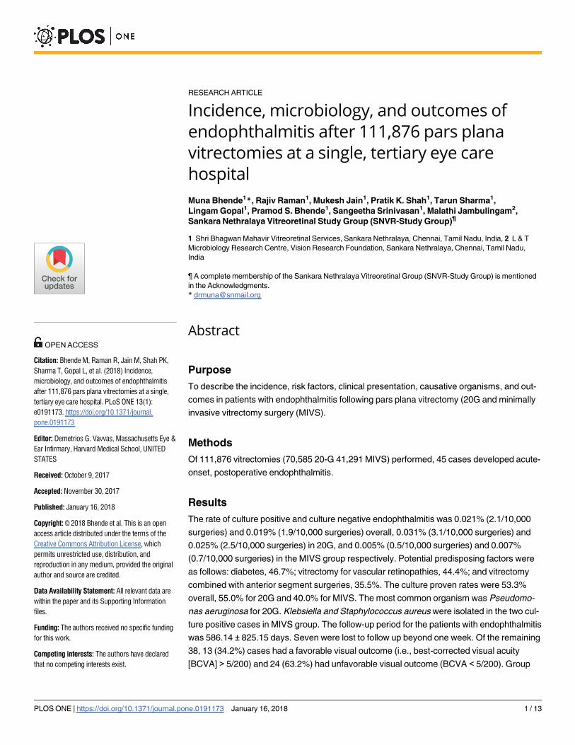
RESEARCH ARTICLE Incidence, microbiology, and outcomes of endophthalmitis after 111,876 pars plana vitrectomies at a single, tertiary eye care hospital Muna Bhende 1 *, Rajiv Raman 1 , Mukesh Jain 1 , Pratik K. Shah 1 , Tarun Sharma 1 , Lingam Gopal 1 , Pramod S. Bhende 1 , Sangeetha Srinivasan 1 , Malathi Jambulingam 2 , Sankara Nethralaya Vitreoretinal Study Group (SNVR-Study Group) ¶ 1 Shri Bhagwan Mahavir Vitreoretinal Services, Sankara Nethralaya, Chennai, Tamil Nadu, India, 2 L&T Microbiology Research Centre, Vision Research Foundation, Sankara Nethralaya, Chennai, Tamil Nadu, India ¶ A complete membership of the Sankara Nethralaya Vitreoretinal Group (SNVR-Study Group) is mentioned in the Acknowledgments. * [email protected] Abstract Purpose To describe the incidence, risk factors, clinical presentation, causative organisms, and out- comes in patients with endophthalmitis following pars plana vitrectomy (20G and minimally invasive vitrectomy surgery (MIVS). Methods Of 111,876 vitrectomies (70,585 20-G 41,291 MIVS) performed, 45 cases developed acute- onset, postoperative endophthalmitis. Results The rate of culture positive and culture negative endophthalmitis was 0.021% (2.1/10,000 surgeries) and 0.019% (1.9/10,000 surgeries) overall, 0.031% (3.1/10,000 surgeries) and 0.025% (2.5/10,000 surgeries) in 20G, and 0.005% (0.5/10,000 surgeries) and 0.007% (0.7/10,000 surgeries) in the MIVS group respectively. Potential predisposing factors were as follows: diabetes, 46.7%; vitrectomy for vascular retinopathies, 44.4%; and vitrectomy combined with anterior segment surgeries, 35.5%. The culture proven rates were 53.3% overall, 55.0% for 20G and 40.0% for MIVS. The most common organism was Pseudomo- nas aeruginosa for 20G. Klebsiella and Staphylococcus aureus were isolated in the two cul- ture positive cases in MIVS group. The follow-up period for the patients with endophthalmitis was 586.14 ± 825.15 days. Seven were lost to follow up beyond one week. Of the remaining 38, 13 (34.2%) cases had a favorable visual outcome (i.e., best-corrected visual acuity [BCVA] > 5/200) and 24 (63.2%) had unfavorable visual outcome (BCVA < 5/200). Group PLOS ONE | https://doi.org/10.1371/journal.pone.0191173 January 16, 2018 1 / 13 a1111111111 a1111111111 a1111111111 a1111111111 a1111111111 OPEN ACCESS Citation: Bhende M, Raman R, Jain M, Shah PK, Sharma T, Gopal L, et al. (2018) Incidence, microbiology, and outcomes of endophthalmitis after 111,876 pars plana vitrectomies at a single, tertiary eye care hospital. PLoS ONE 13(1): e0191173. https://doi.org/10.1371/journal. pone.0191173 Editor: Demetrios G. Vavvas, Massachusetts Eye & Ear Infirmary, Harvard Medical School, UNITED STATES Received: October 9, 2017 Accepted: November 30, 2017 Published: January 16, 2018 Copyright: © 2018 Bhende et al. This is an open access article distributed under the terms of the Creative Commons Attribution License, which permits unrestricted use, distribution, and reproduction in any medium, provided the original author and source are credited. Data Availability Statement: All relevant data are within the paper and its Supporting Information files. Funding: The authors received no specific funding for this work. Competing interests: The authors have declared that no competing interests exist.
-
Upload
khangminh22 -
Category
Documents
-
view
0 -
download
0
Transcript of Incidence, microbiology, and outcomes of endophthalmitis ...
RESEARCH ARTICLE
Incidence, microbiology, and outcomes of
endophthalmitis after 111,876 pars plana
vitrectomies at a single, tertiary eye care
hospital
Muna Bhende1*, Rajiv Raman1, Mukesh Jain1, Pratik K. Shah1, Tarun Sharma1,
Lingam Gopal1, Pramod S. Bhende1, Sangeetha Srinivasan1, Malathi Jambulingam2,
Sankara Nethralaya Vitreoretinal Study Group (SNVR-Study Group)
1 Shri Bhagwan Mahavir Vitreoretinal Services, Sankara Nethralaya, Chennai, Tamil Nadu, India, 2 L & T
Microbiology Research Centre, Vision Research Foundation, Sankara Nethralaya, Chennai, Tamil Nadu,
India
¶ A complete membership of the Sankara Nethralaya Vitreoretinal Group (SNVR-Study Group) is mentioned
in the Acknowledgments.
Abstract
Purpose
To describe the incidence, risk factors, clinical presentation, causative organisms, and out-
comes in patients with endophthalmitis following pars plana vitrectomy (20G and minimally
invasive vitrectomy surgery (MIVS).
Methods
Of 111,876 vitrectomies (70,585 20-G 41,291 MIVS) performed, 45 cases developed acute-
onset, postoperative endophthalmitis.
Results
The rate of culture positive and culture negative endophthalmitis was 0.021% (2.1/10,000
surgeries) and 0.019% (1.9/10,000 surgeries) overall, 0.031% (3.1/10,000 surgeries) and
0.025% (2.5/10,000 surgeries) in 20G, and 0.005% (0.5/10,000 surgeries) and 0.007%
(0.7/10,000 surgeries) in the MIVS group respectively. Potential predisposing factors were
as follows: diabetes, 46.7%; vitrectomy for vascular retinopathies, 44.4%; and vitrectomy
combined with anterior segment surgeries, 35.5%. The culture proven rates were 53.3%
overall, 55.0% for 20G and 40.0% for MIVS. The most common organism was Pseudomo-
nas aeruginosa for 20G. Klebsiella and Staphylococcus aureus were isolated in the two cul-
ture positive cases in MIVS group. The follow-up period for the patients with endophthalmitis
was 586.14 ± 825.15 days. Seven were lost to follow up beyond one week. Of the remaining
38, 13 (34.2%) cases had a favorable visual outcome (i.e., best-corrected visual acuity
[BCVA] > 5/200) and 24 (63.2%) had unfavorable visual outcome (BCVA < 5/200). Group
PLOS ONE | https://doi.org/10.1371/journal.pone.0191173 January 16, 2018 1 / 13
a1111111111
a1111111111
a1111111111
a1111111111
a1111111111
OPENACCESS
Citation: Bhende M, Raman R, Jain M, Shah PK,
Sharma T, Gopal L, et al. (2018) Incidence,
microbiology, and outcomes of endophthalmitis
after 111,876 pars plana vitrectomies at a single,
tertiary eye care hospital. PLoS ONE 13(1):
e0191173. https://doi.org/10.1371/journal.
pone.0191173
Editor: Demetrios G. Vavvas, Massachusetts Eye &
Ear Infirmary, Harvard Medical School, UNITED
STATES
Received: October 9, 2017
Accepted: November 30, 2017
Published: January 16, 2018
Copyright: © 2018 Bhende et al. This is an open
access article distributed under the terms of the
Creative Commons Attribution License, which
permits unrestricted use, distribution, and
reproduction in any medium, provided the original
author and source are credited.
Data Availability Statement: All relevant data are
within the paper and its Supporting Information
files.
Funding: The authors received no specific funding
for this work.
Competing interests: The authors have declared
that no competing interests exist.

with culture test results negative had significantly better outcomes (P < 0.05) as compared
to those with positive.
Conclusions
MIVS does not increase the risk of endophthalmitis. Outcomes are poor despite appropriate
treatment, particularly in cases with culture results positive.
Introduction
Endophthalmitis after vitreous surgery is an uncommon but a devastating complication.[1]
The reported incidence of endophthalmitis after vitreous surgery ranges from 0.05% to 0.02%
with 20-G vitrectomy.[2–10] Initial reports showed higher rates of post-minimally invasive
vitreoretinal surgery (MIVS) endophthalmitis, whereas recent reports have shown a declining
trend.[6,8,10,11]
A recent multicentric study from India reported incidence rates of endophthalmitis after
vitrectomy surgery to be 0.052% with culture-positive endophthalmitis being 0.031%.[12]
Although worldwide large multicentric studies have been conducted, these are also not with-
out limitations.[8,10,12,13] These retrospective multicentric studies had among centers het-
erogeneous case selection criteria, had contrasting antibiotic prophylaxis practice patterns,
had different infection control protocols, and had different laboratory and treatment protocols
which could affect the anatomical and functional outcomes.
The purpose of this study was to describe the 20-year incidence (20 G and MIVS), causative
organisms, and visual outcomes associated with endophthalmitis after pars plana vitrectomy
from a single, tertiary eye care center in India. Furthermore, to identify the differences in
clinical presentation between post-cataract surgery endophthalmitis and post-pars plana vit-
rectomy endophthalmitis, we compared our data with previous post-cataract surgery
endophthalmitis studies after cataract surgery from India. To the best of our knowledge, this is
the largest case series of incidence of post-vitrectomy endophthalmitis reported in literature.
Methods
Study population
Approval of the Institutional Review Board of Sankara Netharalaya was obtained to analyze
the hospital-based data, and the tenets of the Declaration of Helsinki were followed. This was
a retrospective review of case records of patients with suspected postoperative infectious
endophthalmitis after pars plana vitreous surgery performed at a tertiary eye care institute in
India was done. Data were collected from the electronic/paper medical records between Janu-
ary 1995 and December 2015; the charts of all patients who developed endophthalmitis were
reviewed. For cross-validation of our data, the endophthalmitis surveillance log of the Depart-
ment of Microbiology was reviewed. Acute endophthalmitis after vitrectomy was defined as
the presence of unusual inflammation in the anterior segment and vitreous cavity within 6
weeks of surgery along with microbiological evidence (staining/culture/PCR) of bacterial
and fungal infection. Culture-positive endophthalmitis was considered to be present if micro-
biological organisms were isolated on culture. If the fungal, aerobic, and anaerobic culture
results were found to be all negative, then this case was considered to be culture-negative
Endophthalmitis after PPV
PLOS ONE | https://doi.org/10.1371/journal.pone.0191173 January 16, 2018 2 / 13
Abbreviations: BCVA, best-corrected visual acuity;
IOAB, intraocular antibiotics; IOP, intraocular
pressure; MIVS, minimally invasive vitreoretinal
surgery; PCR, polymerase chain reaction.
endophthalmitis. Polymerase chain reaction (PCR) data on the presence or absence of eubacte-
rial, Propionibacterium acnes, and fungal genomes were also noted.
Data such as presenting complaints, systemic illness, time of presentation, presenting and
final visual acuity, clinical findings, intraoperative procedure, microbial profile of aqueous, vit-
reous, or any other tissue/material, antimicrobial susceptibility, management including both
intravitreal antibiotics and vitrectomy, and follow-up were collected. Informed written con-
sent was obtained to have their medical records used in research.
Surgical procedure details
Before 2009, all cases underwent 20-G pars plana vitrectomy. After that the practice shifted to
MIVS and the number of cases undergoing 20-G vitrectomy declined considerably. Of the
111,876 vitrectomies performed during the study period, 70,585 (63.1%) were 20-G surgeries
and 41,291 (36.9%) were MIVS. Before the year 2000 the hospital infection control protocol
included pre-operative antibiotics (sulfacetamide, 10.0%) eye drops four times per day for
atleast 3 days prior to surgery. Intra-operatively, peri-ocular cleaning was done with 0.5%
cetrimide solution (Cettan, Tansi Polish Unit, Chennai). After the year 2000, preoperative
antibiotic protocol included instillation of ciprofloxacin (0.3%) or sulfacetamide (10.0%) eye-
drops 2 hourly one day before surgery. Pre-operatively 5% povidone-iodine solution was
instilled in conjunctival sac and was allowed a contact period of 3 minutes followed by peri-
ocular cleaning with 1% povidone iodine solution before draping. The details of indication for
surgery, additional procedures performed along with vitrectomy, gauge of vitrectomy instru-
ments, lens status at the end of vitreous surgery, type of tamponade used during surgery, and
surgical time were noted in all the cases who developed endophthalmitis.
Procedures done in presence of endophthalmitis
On clinical suspicion of endophthalmitis, if vitreous surgery was not contemplated in the next
6 hours, an anterior chamber tap was performed and the samples were subjected to gram stain-
ing, KOH staining, bacterial and fungal cultures, antibiotic sensitivity, and PCR for P. acnes,eubacterial and panfungal genomes. If surgery was contemplated, vitreous specimen was col-
lected during vitrectomy and similar microbiological tests were conducted. On the basis of
microbiological results, initial antibiotics were given. However, in cases of initial negative
results of staining, choice of antibiotics was based on clinical judgment. Scleral or corneal
scraping were taken for microbiological evaluation, when indicated.
Statistical analysis
Statistical analysis was performed using SPSS Statistics for Windows version 21.0. Continuous
variables were expressed as mean ± SD and were analyzed using Mann–Whitney U test. Cate-
gorical variables were analyzed using Chi-Square and Fisher exact test, as applicable. The P val-
ues of�0.05 were considered to be statistically significant.
Results
Table 1 shows the incidence rates of endophthalmitis after pars plana vitrectomy in 20G and
MIVS group. The overall incidence of clinically evident and culture-proven endophthalmitis
after vitrectomy was 0.040% (4.0 cases per 10,000 surgeries) and 0.021% (2.1 cases per 10,000
surgeries), respectively. The incidence of clinically evident and culture-proven endophthalmi-
tis after 20G vitrectomy was 0.057% (5.7 cases per 10,000 surgeries) and 0.031% (3.1 cases per
10,000 surgeries), and 0.012% (1.2 case per 10,000 surgeries, P< 0.05) and 0.005% (0.5 cases
Endophthalmitis after PPV
PLOS ONE | https://doi.org/10.1371/journal.pone.0191173 January 16, 2018 3 / 13
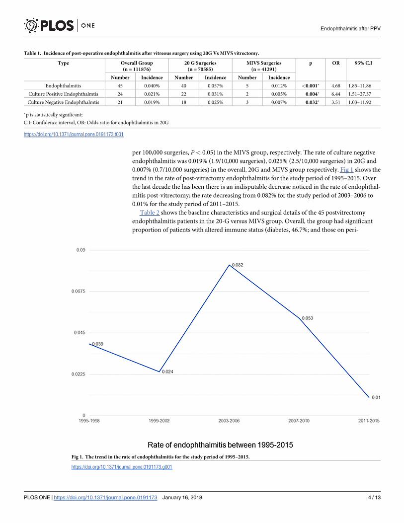
per 100,000 surgeries, P< 0.05) in the MIVS group, respectively. The rate of culture negative
endophthalmitis was 0.019% (1.9/10,000 surgeries), 0.025% (2.5/10,000 surgeries) in 20G and
0.007% (0.7/10,000 surgeries) in the overall, 20G and MIVS group respectively. Fig 1 shows the
trend in the rate of post-vitrectomy endophthalmitis for the study period of 1995–2015. Over
the last decade the has been there is an indisputable decrease noticed in the rate of endophthal-
mitis post-vitrectomy; the rate decreasing from 0.082% for the study period of 2003–2006 to
0.01% for the study period of 2011–2015.
Table 2 shows the baseline characteristics and surgical details of the 45 postvitrectomy
endophthalmitis patients in the 20-G versus MIVS group. Overall, the group had significant
proportion of patients with altered immune status (diabetes, 46.7%; and those on peri-
Table 1. Incidence of post-operative endophthalmitis after vitreous surgery using 20G Vs MIVS vitrectomy.
Type Overall Group
(n = 111876)
20 G Surgeries
(n = 70585)
MIVS Surgeries
(n = 41291)
p OR 95% C.I
Number Incidence Number Incidence Number Incidence
Endophthalmitis 45 0.040% 40 0.057% 5 0.012% <0.001� 4.68 1.85–11.86
Culture Positive Endophthalmtis 24 0.021% 22 0.031% 2 0.005% 0.004� 6.44 1.51–27.37
Culture Negative Endophthalmtis 21 0.019% 18 0.025% 3 0.007% 0.032� 3.51 1.03–11.92
�p is statistically significant;
C.I: Confidence interval, OR: Odds ratio for endophthalmitis in 20G
https://doi.org/10.1371/journal.pone.0191173.t001
Fig 1. The trend in the rate of endophthalmitis for the study period of 1995–2015.
https://doi.org/10.1371/journal.pone.0191173.g001
Endophthalmitis after PPV
PLOS ONE | https://doi.org/10.1371/journal.pone.0191173 January 16, 2018 4 / 13
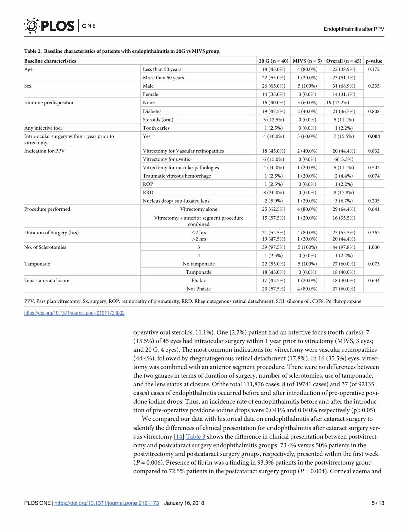
operative oral steroids, 11.1%). One (2.2%) patient had an infective focus (tooth caries). 7
(15.5%) of 45 eyes had intraocular surgery within 1 year prior to vitrectomy (MIVS, 3 eyes;
and 20 G, 4 eyes). The most common indications for vitrectomy were vascular retinopathies
(44.4%), followed by rhegmatogenous retinal detachment (17.8%). In 16 (35.5%) eyes, vitrec-
tomy was combined with an anterior segment procedure. There were no differences between
the two gauges in terms of duration of surgery, number of sclerotomies, use of tamponade,
and the lens status at closure. Of the total 111,876 cases, 8 (of 19741 cases) and 37 (of 92135
cases) cases of endophthalmitis occurred before and after introduction of pre-operative povi-
done iodine drops. Thus, an incidence rate of endophthalmitis before and after the introduc-
tion of pre-operative povidone iodine drops were 0.041% and 0.040% respectively (p>0.05).
We compared our data with historical data on endophthalmitis after cataract surgery to
identify the differences of clinical presentation for endophthalmitis after cataract surgery ver-
sus vitrectomy.[14] Table 3 shows the difference in clinical presentation between postvitrect-
omy and postcataract surgery endophthalmitis groups: 73.4% versus 50% patients in the
postvitrectomy and postcataract surgery groups, respectively, presented within the first week
(P = 0.006). Presence of fibrin was a finding in 93.3% patients in the postvitrectomy group
compared to 72.5% patients in the postcataract surgery group (P = 0.004). Corneal edema and
Table 2. Baseline characteristics of patients with endophthalmitis in 20G vs MIVS group.
Baseline characteristics 20 G (n = 40) MIVS (n = 5) Overall (n = 45) p-value
Age Less than 50 years 18 (45.0%) 4 (80.0%) 22 (48.9%) 0.172
More than 50 years 22 (55.0%) 1 (20.0%) 23 (51.1%)
Sex Male 26 (65.0%) 5 (100%) 31 (68.9%) 0.235
Female 14 (35.0%) 0 (0.0%) 14 (31.1%)
Immune predisposition None 16 (40.0%) 3 (60.0%) 19 (42.2%)
Diabetes 19 (47.5%) 2 (40.0%) 21 (46.7%) 0.808
Steroids (oral) 5 (12.5%) 0 (0.0%) 5 (11.1%)
Any infective foci Tooth caries 1 (2.5%) 0 (0.0%) 1 (2.2%)
Intra-ocular surgery within 1 year prior to
vitrectomy
Yes 4 (10.0%) 3 (60.0%) 7 (15.5%) 0.004
Indication for PPV Vitrectomy for Vascular retinopathies 18 (45.0%) 2 (40.0%) 20 (44.4%) 0.832
Vitrectomy for uveitis 6 (15.0%) 0 (0.0%) 6(13.3%)
Vitrectomy for macular pathologies 4 (10.0%) 1 (20.0%) 5 (11.1%) 0.502
Traumatic vitreous hemorrhage 1 (2.5%) 1 (20.0%) 2 (4.4%) 0.074
ROP 1 (2.5%) 0 (0.0%) 1 (2.2%)
RRD 8 (20.0%) 0 (0.0%) 8 (17.8%)
Nucleus drop/ sub-luxated lens 2 (5.0%) 1 (20.0%) 3 (6.7%) 0.205
Procedure performed Vitrectomy alone 25 (62.5%) 4 (80.0%) 29 (64.4%) 0.641
Vitrectomy + anterior segment procedure
combined
15 (37.5%) 1 (20.0%) 16 (35.5%)
Duration of Surgery (hrs) �2 hrs
>2 hrs
21 (52.5%)
19 (47.5%)
4 (80.0%)
1 (20.0%)
25 (55.5%)
20 (44.4%)
0.362
No. of Sclerotomies 3 39 (97.5%) 5 (100%) 44 (97.8%) 1.000
4 1 (2.5%) 0 (0.0%) 1 (2.2%)
Tamponade No tamponade 22 (55.0%) 5 (100%) 27 (60.0%) 0.073
Tamponade 18 (45.0%) 0 (0.0%) 18 (40.0%)
Lens status at closure Phakic 17 (42.5%) 1 (20.0%) 18 (40.0%) 0.634
Not Phakic 23 (57.5%) 4 (80.0%) 27 (60.0%)
PPV: Pars plan vitrectomy, Sx: surgery, ROP: retinopathy of prematurity, RRD: Rhegmatogenous retinal detachment, SOI: silicone oil, C3F8: Perfluropropane
https://doi.org/10.1371/journal.pone.0191173.t002
Endophthalmitis after PPV
PLOS ONE | https://doi.org/10.1371/journal.pone.0191173 January 16, 2018 5 / 13
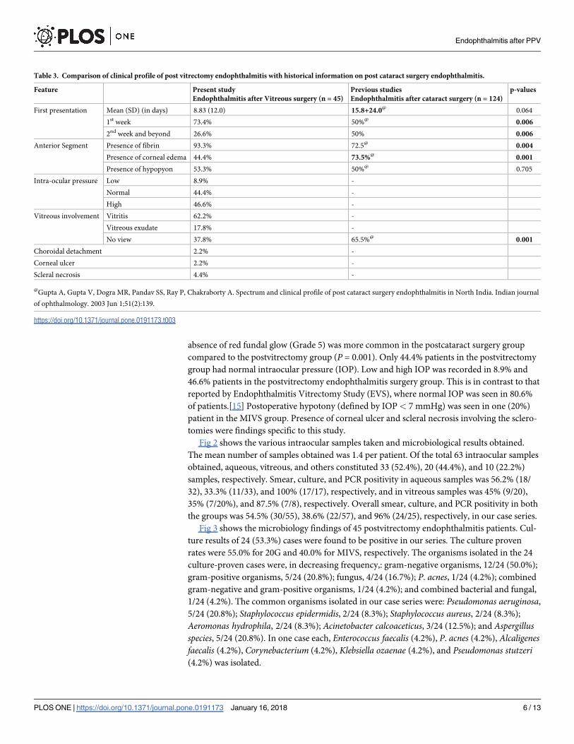
absence of red fundal glow (Grade 5) was more common in the postcataract surgery group
compared to the postvitrectomy group (P = 0.001). Only 44.4% patients in the postvitrectomy
group had normal intraocular pressure (IOP). Low and high IOP was recorded in 8.9% and
46.6% patients in the postvitrectomy endophthalmitis surgery group. This is in contrast to that
reported by Endophthalmitis Vitrectomy Study (EVS), where normal IOP was seen in 80.6%
of patients.[15] Postoperative hypotony (defined by IOP< 7 mmHg) was seen in one (20%)
patient in the MIVS group. Presence of corneal ulcer and scleral necrosis involving the sclero-
tomies were findings specific to this study.
Fig 2 shows the various intraocular samples taken and microbiological results obtained.
The mean number of samples obtained was 1.4 per patient. Of the total 63 intraocular samples
obtained, aqueous, vitreous, and others constituted 33 (52.4%), 20 (44.4%), and 10 (22.2%)
samples, respectively. Smear, culture, and PCR positivity in aqueous samples was 56.2% (18/
32), 33.3% (11/33), and 100% (17/17), respectively, and in vitreous samples was 45% (9/20),
35% (7/20%), and 87.5% (7/8), respectively. Overall smear, culture, and PCR positivity in both
the groups was 54.5% (30/55), 38.6% (22/57), and 96% (24/25), respectively, in our case series.
Fig 3 shows the microbiology findings of 45 postvitrectomy endophthalmitis patients. Cul-
ture results of 24 (53.3%) cases were found to be positive in our series. The culture proven
rates were 55.0% for 20G and 40.0% for MIVS, respectively. The organisms isolated in the 24
culture-proven cases were, in decreasing frequency,: gram-negative organisms, 12/24 (50.0%);
gram-positive organisms, 5/24 (20.8%); fungus, 4/24 (16.7%); P. acnes, 1/24 (4.2%); combined
gram-negative and gram-positive organisms, 1/24 (4.2%); and combined bacterial and fungal,
1/24 (4.2%). The common organisms isolated in our case series were: Pseudomonas aeruginosa,
5/24 (20.8%); Staphylococcus epidermidis, 2/24 (8.3%); Staphylococcus aureus, 2/24 (8.3%);
Aeromonas hydrophila, 2/24 (8.3%); Acinetobacter calcoaceticus, 3/24 (12.5%); and Aspergillusspecies, 5/24 (20.8%). In one case each, Enterococcus faecalis (4.2%), P. acnes (4.2%), Alcaligenesfaecalis (4.2%), Corynebacterium (4.2%), Klebsiella ozaenae (4.2%), and Pseudomonas stutzeri(4.2%) was isolated.
Table 3. Comparison of clinical profile of post vitrectomy endophthalmitis with historical information on post cataract surgery endophthalmitis.
Feature Present study
Endophthalmitis after Vitreous surgery (n = 45)
Previous studies
Endophthalmitis after cataract surgery (n = 124)
p-values
First presentation Mean (SD) (in days) 8.83 (12.0) 15.8+24.0@ 0.064
1st week 73.4% 50%@ 0.006
2nd week and beyond 26.6% 50% 0.006
Anterior Segment Presence of fibrin 93.3% 72.5@ 0.004
Presence of corneal edema 44.4% 73.5%@ 0.001
Presence of hypopyon 53.3% 50%@ 0.705
Intra-ocular pressure Low 8.9% -
Normal 44.4% -
High 46.6% -
Vitreous involvement Vitritis 62.2% -
Vitreous exudate 17.8% -
No view 37.8% 65.5%@ 0.001
Choroidal detachment 2.2% -
Corneal ulcer 2.2% -
Scleral necrosis 4.4% -
@Gupta A, Gupta V, Dogra MR, Pandav SS, Ray P, Chakraborty A. Spectrum and clinical profile of post cataract surgery endophthalmitis in North India. Indian journal
of ophthalmology. 2003 Jun 1;51(2):139.
https://doi.org/10.1371/journal.pone.0191173.t003
Endophthalmitis after PPV
PLOS ONE | https://doi.org/10.1371/journal.pone.0191173 January 16, 2018 6 / 13
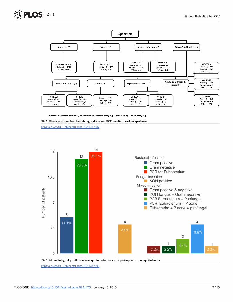
Fig 2. Flow-chart showing the staining, culture and PCR results in various specimen.
https://doi.org/10.1371/journal.pone.0191173.g002
Fig 3. Microbiological profile of ocular specimen in cases with post-operative endophthalmitis.
https://doi.org/10.1371/journal.pone.0191173.g003
Endophthalmitis after PPV
PLOS ONE | https://doi.org/10.1371/journal.pone.0191173 January 16, 2018 7 / 13
S1 Table shows the antibiotic sensitivity of the cases with positive cultures. The antibiotic
sensitivity of gram-negative organisms was as follows: amikacin, 9/11 (81.8%); ciprofloxacin,
9/11 (81.8%); ceftazidime, 7/9 (77.8%); gentamicin, 9/12 (75.0%); and cefatoxime, 7/11
(63.6%) In gram-positive organisms, the antibiotic sensitivity pattern was as follows: cefatox-
ime, 4/4 (100%); ciprofloxacin, 4/4 (100%); vancomycin, 3/3 (100%); clindamycin, 3/4 (75%);
and ceftazidime, 2/3 (66.7%). Of the 21 cases whose culture results were found to be negative,
PCR results were positive in 20 (95.2%) and smear results were positive in only 1 (4.8%)
patient (gonococcal bacilli). The results of PCR were as follows: eubacterial only (45%); eubac-
terial + pan fungal (15%); eubacterial + P. acnes (30%); and eubacterial + pan fungal + P. acnes(10%).
Seven eyes were lost to follow up beyond one week and were excluded from further analysis.
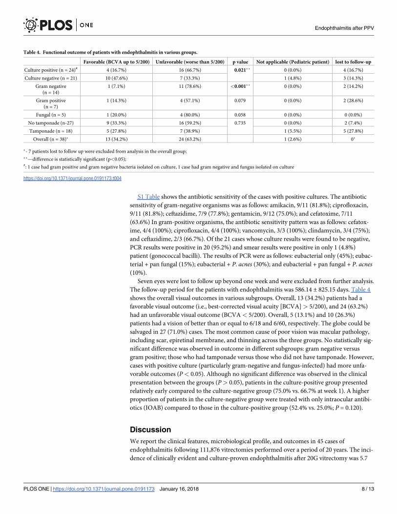
The follow-up period for the patients with endophthalmitis was 586.14 ± 825.15 days. Table 4
shows the overall visual outcomes in various subgroups. Overall, 13 (34.2%) patients had a
favorable visual outcome (i.e., best-corrected visual acuity [BCVA] > 5/200), and 24 (63.2%)
had an unfavorable visual outcome (BCVA < 5/200). Overall, 5 (13.1%) and 10 (26.3%)
patients had a vision of better than or equal to 6/18 and 6/60, respectively. The globe could be
salvaged in 27 (71.0%) cases. The most common cause of poor vision was macular pathology,
including scar, epiretinal membrane, and thinning across the three groups. No statistically sig-
nificant difference was observed in outcome in different subgroups: gram negative versus
gram positive; those who had tamponade versus those who did not have tamponade. However,
cases with positive culture (particularly gram-negative and fungus-infected) had more unfa-
vorable outcomes (P< 0.05). Although no significant difference was observed in the clinical
presentation between the groups (P> 0.05), patients in the culture-positive group presented
relatively early compared to the culture-negative group (75.0% vs. 66.7% at week 1). A higher
proportion of patients in the culture-negative group were treated with only intraocular antibi-
otics (IOAB) compared to those in the culture-positive group (52.4% vs. 25.0%; P = 0.120).
Discussion
We report the clinical features, microbiological profile, and outcomes in 45 cases of
endophthalmitis following 111,876 vitrectomies performed over a period of 20 years. The inci-
dence of clinically evident and culture-proven endophthalmitis after 20G vitrectomy was 5.7
Table 4. Functional outcome of patients with endophthalmitis in various groups.
Favorable (BCVA up to 5/200) Unfavorable (worse than 5/200) p value Not applicable (Pediatric patient) lost to follow-up
Culture positive (n = 24)# 4 (16.7%) 16 (66.7%) 0.021�� 0 (0.0%) 4 (16.7%)
Culture negative (n = 21) 10 (47.6%) 7 (33.3%) 1 (4.8%) 3 (14.3%)
Gram negative
(n = 14)
1 (7.1%) 11 (78.6%) <0.001�� 0 (0.0%) 2 (14.2%)
Gram positive
(n = 7)
1 (14.3%) 4 (57.1%) 0.079 0 (0.0%) 2 (28.6%)
Fungal (n = 5) 1 (20.0%) 4 (80.0%) 0.058 0 (0.0%) 0 (0.0%)
No tamponade (n-27) 9 (33.3%) 16 (59.2%) 0.735 0 (0.0%) 2 (7.4%)
Tamponade (n = 18) 5 (27.8%) 7 (38.9%) 1 (5.5%) 5 (27.8%)
Overall (n = 38)� 13 (34.2%) 24 (63.2%) 1 (2.6%) 0�
�- 7 patients lost to follow up were excluded from analysis in the overall group;
��—difference is statistically significant (p<0.05);#: 1 case had gram positive and gram negative bacteria isolated on culture, 1 case had gram negative and fungus isolated on culture
https://doi.org/10.1371/journal.pone.0191173.t004
Endophthalmitis after PPV
PLOS ONE | https://doi.org/10.1371/journal.pone.0191173 January 16, 2018 8 / 13
and 3.1 cases per 10,000 surgeries, respectively. Previous studies have shown varied incidences
with 20-G vitrectomy: 7 cases per 10,000 surgeries in earlier studies and reducing to 2 cases
per 10,000 surgeries reported in the later studies.[2–10]
The incidence of endophthalmitis after MIVS reported in literature in the last decade have
shown a declining trend. In a large, multicenter retrospective Pan-American Collaborative
Retina Study Group, Wu et al.[10] found an incidence of 0.026% (2.6 cases per 10,000 surger-
ies) for endophthalmitis after MIVS during 2005–2009. Scott et al.[6] in their multicenter
retrospective study found an incidence of 0.048% (4.8 cases per 10,000 surgeries) for
endophthalmitis after MIVS during 2007–2008. They also found that the rate of endophthal-
mitis after MIVS in their study period was marginally lower than that estimated for 2005–
2006, suggesting the decreasing trend. Similar indisputable decreasing trend is evident in our
study (Fig 1) with the rate decreasing from 0.082% to 0.010% in the last decade. Lower rates
observed in our study compared with previous studies are possibly due to the various reasons.
As the incidence of endophthalmitis in the MIVS group is calculated for 2009–2014, we ascribe
these lower rates to the decreasing trend observed in the past.[6,8,10,11] Also, as our data rep-
resent the incidence from a single center, data are possibly less heterogeneous in terms of
inclusion criteria, antibiotic prophylaxis, standard infection prevention protocols, and surgical
technique except for those that have evolved over time. Moreover, the inclusion criteria of our
study is more robust. We included only those cases that had an unusual post-operative inflam-
mation sufficient enough to warrant a microbiological evaluation of ocular fluids and had
microbiological evidence (staining positive/culture positive/PCR positive) of the same.
Although we found that the risk of endophthalmitis is significantly lower after MIVS
compared to conventional 20-G vitrectomy, caution is warranted in interpreting this find-
ing. As the incidence rates of endophthalmitis after 20-G vitrectomy and MIVS represent
different study periods, comparing the two may not reflect the true difference. Since the first
introduction of 25-G vitrectomy by Fujii et al.[16] in 2002, the incisional techniques and
technology of MIVS have evolved considerably, exemplified by the reported of decreasing
incidence of endophthalmitis in the last decade. At best, our results suggest no increased risk
of endophthalmitis after MIVS.
The potential predisposing factors identified in our study were as follows: diabetes (46.7%),
vitrectomy for vascular retinopathies (44.4%), vitrectomy combined with anterior segment
procedures (35.5%), and absence of tamponade (59.5%). History of intraocular surgery was
more often seen in patients in the MIVS group. Diabetes, and immune-compromised states,
can theoretically increase the risk of endophthalmitis after vitrectomy. Complicated vitreoret-
inal surgeries requiring multiple exchange of instruments, longer operative time, increased
incidence of cataract requiring combined anterior segment surgeries, and poor wound healing
can increase the risk of endophthalmitis. Similar associations have been noted in other studies.
[1–5,7,9]1–5,7,9
The differential surface tension of a tamponading agent compared to balanced salt solution
helps to seal the sclerotomy wounds, minimizing hypotony and associated infections.[17–19]
Although this study has not been designed to show the association of use of tamponade and
endophthalmitis, we found absence of tamponading agent in all cases with endophthalmitis
(n = 5) who underwent MIVS where sclerotomies were self sealing. Park et al.[11] in their
series found that vitrectomy for retinal detachment was associated with reduced risk of infec-
tion. They suggested that as in these cases the use of a tamponading agent probably results in
better wound integrity.
Because most patients have varying degree of pain, redness, chemosis, lid edema, and ante-
rior segment inflammation, recognizing endophthalmitis early is sometimes challenging. We
found a different presentation of endophthalmitis after postvitrectomy compared to that after
Endophthalmitis after PPV
PLOS ONE | https://doi.org/10.1371/journal.pone.0191173 January 16, 2018 9 / 13
cataract surgery in terms of fibrin, corneal edema, and IOP, suggesting a differential response
of eyes in these two conditions.[14–15] We found that a significant proportion of postvitrect-
omy endophthalmitis patients (73.4%) presented within 1 week of surgery, which is in accor-
dance with the results of the previous studies. This implies that after vitrectomy, patients must
be reviewed in the first week to allow early detection of this dreaded complication. With the
presence of fibrin (present in 93.3% patients) and abnormal IOP (present in 55.6% patients,
especially high IOP), clinician should suspect endophthalmitis as an important differential
diagnosis. One additional finding in patients of postvitrectomy endophthalmitis was the pres-
ence of corneal ulcer and scleral necrosis in the area of sclerotomy. This indicates that meticu-
lous examination of the sclerotomy site is important in the post-operative period.
We found 53.3% culture positivity in our case series, which is in accordance with previously
reported rates between 44.4% and 66.7%.[6,8,9,11,20] Park et al.[20] suggested the importance
of aqueous samples in isolating the microorganisms in endophthalmitis after vitrectomy. We
found that aqueous was the most common intraocular sample obtained (52.4%) with a cul-
ture-positive yield similar to that of vitreous samples (33.3% vs. 35.0%), further validating its
use.
An important distinctive feature of our study is the use of PCR for microbial analysis. Chi-
quet et al.[21] found culture positivity in only 32% aqueous samples as compared to 61% on
PCR in their postcataract surgery endophthalmitis case series study. Using culture and PCR
combination, diagnosis can be made in 71% cases. Similarly, Therese et al.[22] showed that
by inclusion of PCR, detection of microorganisms increased from 46.5% to 75.8% cases.
Likewise, Anand et al.[23] showed a high sensitivity for detection of fungus using PCR in
endophthalmitis.
Reports from Western countries have shown coagulase negative Staphylococcus as the most
common organism in postoperative endophthalmitis.[1–3,5,6,9,13,22] Gram-negative bacilli
were the most common group isolated in our study. Pseudomonas was the most common
organism cultured. Our finding is in agreement with previous reports from Indian subconti-
nent. Sharma et al.[24] have reported a high prevalence of gram-negative bacteria and fungus
in postoperative endophthalmitis. Anand et al.[25] and Jambulingam et al.[26] have reported
similar spectrum in postoperative endophthalmitis from our center. A similar high prevalence
of gram-negative bacteria has been reported in other infections in subjects with diabetes.[27]
We believe both varied geographical distribution and predominance of subjects with diabetes
in our study are responsible for this microbial spectrum.
In this study, a significant number of culture-negative cases could be effectively managed
with intravitreal antibiotic injections with relatively better visual and anatomical outcomes.
Because the genomic bacterial load predicts severity and outcomes, it is possible that a lower
bacterial load favored better outcomes and corresponding inability to capture the microbe on
culture.[28] This seems most probable as 75% culture-positive patients presented within 1
week of surgery, in contrast to 66.7% culture-negative patients, owing to this difference in
genomic bacterial load.
Overall, the functional outcomes in endophthalmitis after vitrectomy were poor, 24 (63.2%)
had final vision <5/200, these results are in accordance with those of previous studies.[1–
7,9,12,13] The outcomes are poorer than that of endophthalmitis following cataract surgery
due to the possible underlying retinal pathology. The high prevalence of gram-negative and
fungal microorganisms in our study partly explains the poor visual outcome in our series. Park
et al.[22] reported only 14.8% patients had vision better than 6/12. Scott et al. reported 7 of the
13 cases had a vision worse than 20/100.
The study has several strengths. First, it is the largest case series of endophthalmitis after vit-
rectomy reported in literature; Second, being a single-center study, it scores over multicenter
Endophthalmitis after PPV
PLOS ONE | https://doi.org/10.1371/journal.pone.0191173 January 16, 2018 10 / 13
studies that are confounded by their inherent heterogeneous study design. Third, it addresses
the concerns regarding the fear of increased endophthalmitis rate with MIVS using the largest
database of 41,921 MIVS surgeries till date. Fourth, the differences between culture-positive
and culture-negative cases have been shown in endophthalmitis after vitrectomy. And finally,
the use of PCR, a modern sensitive tool used in our study, to exclude probable noninfectious
cases has been appropriately demonstrated.
The inherent retrospective study design and unavailability of data on wound construction
and suturing are the limitations of our study. Also, as the subgroups have a smaller sample, the
true differences in them may be difficult to comment.
In conclusion, using a large dataset, we showed that MIVS does not increase the risk of
endophthalmitis, despite recent concerns. Meticulous surgical asepsis, use of endotamponade,
suturing of sclerotomy in doubtful cases and judicious use of antibiotics are crucial to mini-
mize the risk of infection. Surgeons should be aware of the differences in presentation, micro-
biological profile, and factors responsible for unfavorable outcome. The outcomes are often
guarded despite appropriate treatment. The risk of poor outcomes is potentially higher in cul-
ture-positive cases, which need to be treated aggressively.
Supporting information
S1 Table. Culture sensitivities of organisms in cases with endophthalmitis.
(DOCX)
S1 File. Highlights.
(DOCX)
S2 File. IRB certificate.
(PDF)
Acknowledgments
Sankara NethralayaVitreoretinal Study Group (SNVR-Study Group)
Dr Pramod S Bhende; Dr Muna Bhende; Dr Rajiv Raman; Dr Tarun Sharma; Dr Lingam
Gopal; Dr Parveen Sen; Dr Dhanashree Ratra; Dr Ekta Rishi; Dr Pukhraj Rishi; Dr Chetan
Rao; Dr Pradeep S; Dr Vikas Khetan; Dr Swakshyar Saumya Pal; Dr Suganeswari G; Dr Vinata
Muralidharan; Dr Rupak Roy; Dr Sudipta Das; Dr Aditya Verma; Dr Kumar Saurabh; Dr
Mohammad Arif Mulla; Dr Jaydeep Avinash Walinjkar; Dr Jaya Prakash V
Author Contributions
Conceptualization: Muna Bhende, Rajiv Raman, Lingam Gopal, Pramod S. Bhende.
Data curation: Rajiv Raman, Mukesh Jain, Pratik K. Shah.
Formal analysis: Muna Bhende, Mukesh Jain, Pratik K. Shah, Sangeetha Srinivasan.
Investigation: Muna Bhende, Pramod S. Bhende.
Methodology: Muna Bhende, Rajiv Raman, Pratik K. Shah, Tarun Sharma, Lingam Gopal,
Pramod S. Bhende, Sangeetha Srinivasan, Malathi Jambulingam.
Project administration: Tarun Sharma, Pramod S. Bhende.
Supervision: Muna Bhende, Rajiv Raman, Lingam Gopal.
Endophthalmitis after PPV
PLOS ONE | https://doi.org/10.1371/journal.pone.0191173 January 16, 2018 11 / 13

Validation: Muna Bhende, Malathi Jambulingam.
Writing – original draft: Muna Bhende, Rajiv Raman, Mukesh Jain, Pratik K. Shah, Tarun
Sharma, Lingam Gopal, Pramod S. Bhende, Sangeetha Srinivasan, Malathi Jambulingam.
Writing – review & editing: Muna Bhende, Mukesh Jain, Tarun Sharma, Pramod S. Bhende,
Sangeetha Srinivasan, Malathi Jambulingam.
References1. Cohen SM, Flynn HW, Murray TG, Smiddy WE, Avins LR, Blodi CF, et al. Endophthalmitis after pars
plana vitrectomy. Ophthalmology. 1995; 102(5):705–712. PMID: 7777268
2. Joondeph BC, Blanc JP, Polkinghorne PJ. Endophthalmitis after pars plana vitrectomy: a New Zealand
experience. Retina. 2005; 25(5):587–589. PMID: 16077355
3. Eifrig CW, Scott IU, Flynn HW, Smiddy WE, Newton J. Endophthalmitis after pars plana vitrectomy: inci-
dence, causative organisms, and visual acuity outcomes. Am J Ophthalmol. 2004; 138(5):799–802.
https://doi.org/10.1016/j.ajo.2004.06.035 PMID: 15531315
4. Kunimoto DY, Kaiser RS, Service WE. Incidence of endophthalmitis after 20-and 25-gauge vitrectomy.
Ophthalmology. 2007; 114(12):2133–2137. https://doi.org/10.1016/j.ophtha.2007.08.009 PMID:
17916378
5. Scott IU, Flynn HW, Dev S, Shaikh S, Mittra RA, Arevalo JF, et al. Endophthalmitis after 25-gauge and
20-gauge pars plana vitrectomy: incidence and outcomes. Retina. 2008; 28(1):138–142. https://doi.org/
10.1097/IAE.0b013e31815e9313 PMID: 18185150
6. Scott IU, Flynn HW, Acar N, Dev S, Shaikh S, Mittra RA, et al. Incidence of endophthalmitis after 20-
gauge vs 23-gauge vs 25-gauge pars plana vitrectomy. Graefes Arch Clin Exp Ophthalmol. 2011; 249
(3):377–380. https://doi.org/10.1007/s00417-010-1505-8 PMID: 20853005
7. Shimada H, Nakashizuka H, Hattori T, Mori R, Mizutani Y, Yuzawa M. Incidence of endophthalmitis
after 20- and 25-gauge vitrectomy: causes and prevention. Ophthalmology. 2008; 115(12): 2215–2220.
https://doi.org/10.1016/j.ophtha.2008.07.015 PMID: 18930557
8. Oshima Y, Kadonosono K, Yamaji H, Inoue M, Yoshida M, Kimura H, et al. Japan Microincision Vitrec-
tomy Surgery Study Group. Multicenter survey with a systematic overview of acute-onset endophthal-
mitis after transconjunctival microincision vitrectomy surgery. Am J Ophthalmol. 2010; 150(5):716–725.
https://doi.org/10.1016/j.ajo.2010.06.002 PMID: 20719299
9. Chen JK, Khurana RN, Nguyen QD, Do DV. The incidence of endophthalmitis following transconjuncti-
val sutureless 25- vs 20-gauge vitrectomy. Eye. 2009; 23(4):780–784. https://doi.org/10.1038/eye.
2008.160 PMID: 18535597
10. Wu L, Berrocal MH, Arevalo JF, Carpentier C, Rodriguez FJ, Alezzandrini, et al. Endophthalmitis after
pars plana vitrectomy: results of the Pan American Collaborative Retina Study Group. Retina. 2011; 31
(4):673–678. https://doi.org/10.1097/IAE.0b013e318203c183 PMID: 21394065
11. Park JC, Ramasamy B, Shaw S, Prasad S, Ling RH. A prospective and nationwide study investigating
endophthalmitis following pars plana vitrectomy: incidence and risk factors. Br J Ophthalmol. 2014; 98
(4):529–533. https://doi.org/10.1136/bjophthalmol-2013-304485 PMID: 24420916
12. Dave VP, Pathengay A, Basu S, Gupta N, Basu S, Raval V, et al. Endophthalmitis after pars plana vit-
rectomy: clinical features, risk factors, and management outcomes. Asia Pac J Ophthalmol (Phila).
2016; 5(3):192–195.
13. Czajka MP, Byhr E, Olivestedt G, Olofsson EM. Endophthalmitis after small-gauge vitrectomy: a retro-
spective case series from Sweden. Acta Ophthalmol. 2016; 94(8):829–835. https://doi.org/10.1111/
aos.13121 PMID: 27273917
14. Gupta A, Gupta V, Dogra MR, Pandav SS, Ray P, Chakraborty A. Spectrum and clinical profile of post
cataract surgery endophthalmitis in North India. Indian J Ophthalmol. 2003; 51(2):139. PMID:
12831144
15. Endophthalmitis Vitrectomy Study Group. Results of the endophthalmitis vitrectomy study. Arch
Ophthalmol. 1995; 113:1479–1496. PMID: 7487614
16. Fujii GY, de Juan E, Humayun MS, Pieramici DJ, Chang TS, Ng E, et al. A new 25-gauge instrument
system for transconjunctival sutureless vitrectomy surgery. Ophthalmology. 2002; 109(10):1807–1812.
PMID: 12359598
17. Chiang A, Kaiser RS, Avery RL, Dugel PU, Eliott D, Shah SP, et al. Endophthalmitis in microincision vit-
rectomy: outcomes of gas-filled eyes. Retina. 2011; 31(8):1513–1517. https://doi.org/10.1097/IAE.
0b013e3182209290 PMID: 21878799
Endophthalmitis after PPV
PLOS ONE | https://doi.org/10.1371/journal.pone.0191173 January 16, 2018 12 / 13
18. Gupta OP, Weichel ED, Regillo CD, Fineman MS, Kaiser RS, Ho AC, et al. Postoperative complications
associated with 25-gauge pars plana vitrectomy. Ophthalmic Surg Lasers Imaging Retina. 2007; 38
(4):270–275.
19. Gupta OP, Ho AC, Kaiser PK, Regilo CD, Chen S, Dyer DS, et al. Short-term outcomes of 23-gauge
pars plana vitrectomy. Am J Ophthalmol. 2008; 146(2):193–197. https://doi.org/10.1016/j.ajo.2008.04.
010 PMID: 18547538
20. Park JC, Ramasamy B, Shaw S, Ling RH, Prasad S. A prospective and nationwide study investigating
endophthalmitis following pars plana vitrectomy: clinical presentation, microbiology, management and
outcome. Br J Ophthalmol. 2014; 98(8):1080–1086. https://doi.org/10.1136/bjophthalmol-2013-304486
PMID: 24686917
21. Chiquet C, Lina G, Benito Y, Cornut PL, Etienne J, Romanet JP, et al. Polymerase chain reaction identi-
fication in aqueous humor of patients with postoperative endophthalmitis. J Cataract Refract Surg.
2007; 33(4):635–641. https://doi.org/10.1016/j.jcrs.2006.12.017 PMID: 17397736
22. Therese KL, Anand AR, Madhavan HN. Polymerase chain reaction in the diagnosis of bacterial
endophthalmitis. Br J Ophthalmol. 1998; 82(9):1078–1082. PMID: 9893601
23. Anand AR, Madhavan HN, Neelam V, Lily TK. Use of polymerase chain reaction in the diagnosis of fun-
gal endophthalmitis. Ophthalmology. 2001; 108(2):326–330. PMID: 11158808
24. Sharma S, Padhi TR, Basu S, Kar S, Roy A, Das T. Endophthalmitis patients seen in a tertiary eye care
centre in Odisha: a clinico-microbiological analysis. Indian J Med Res. 2014; 139(1):91. PMID:
24604043
25. Anand AR, Therese KL, Madhavan HN. Spectrum of aetiological agents of postoperative endophthalmi-
tis and antibiotic susceptibility of bacterial isolates. Indian J Ophthalmol. 2000; 48(2):123. PMID:
11116508
26. Jambulingam M, Parameswaran SK, Lysa S, Selvaraj M, Madhavan HN. A study on the incidence,
microbiological analysis and investigations on the source of infection of postoperative infectious
endophthalmitis in a tertiary care ophthalmic hospital: an 8-year study. Indian J Ophthalmol. 2010; 58
(4):297. https://doi.org/10.4103/0301-4738.64132 PMID: 20534919
27. Turhan V, Mutluoglu M, Acar A, Hatipoglu M, Onem Y, Uzun G, et al. Increasing incidence of Gram-neg-
ative organisms in bacterial agents isolated from diabetic foot ulcers. J Infect Dev Ctries. 2013; 7
(10):707–712. https://doi.org/10.3855/jidc.2967 PMID: 24129622
28. Lisboa T, Waterer G, Rello J. We should be measuring genomic bacterial load and virulence factors.
Crit Care Med. 2010; 38(10):S656–S662.
Endophthalmitis after PPV
PLOS ONE | https://doi.org/10.1371/journal.pone.0191173 January 16, 2018 13 / 13