Medical Diagnosis
147
Editor Prof. Dr. Sıddık Keskin Medical Diagnosis and Treatment Methods in Basic Medical Sciences Medical Diagnosis and Treatment Methods in Basic Medical Sciences Health livredelyon.com livredelyon livredelyon livredelyon ISBN: 978-2-38236-061-3
-
Upload
khangminh22 -
Category
Documents
-
view
0 -
download
0
Transcript of Medical Diagnosis

EditorProf. Dr. Sıddık Keskin
Medical Diagnosisand
Treatment Methodsin
Basic Medical Sciences
Medical Diagnosis and Treatment Methods in Basic Medical Sciences
Healthlivredelyon.com
livredelyon
livredelyon
livredelyon
ISBN: 978-2-38236-061-3

Medical Diagnosis and Treatment Methods
in
Basic Medical Sciences
Editor
Prof. Dr. Sıddık Keskin
Lyon 2020

Editor • Prof. Dr. Sıddık Keskin 0000-0001-9355-6558
Cover Design • Aruull Raja
First Published • December 2020, Lyon
ISBN: 978-2-38236-061-3
© copyright
All rights reserved. No part of this publication may be reproduced, stored
in a retrieval system, or transmitted in any form or by an means,
electronic, mechanical, photocopying, recording, or otherwise, without
the publisher’s permission.
The chapters in this book have been checked for plagiarism by
Publisher • Livre de Lyon
Address • 37 rue marietton, 69009, Lyon France
website • http://www.livredelyon.com
e-mail • [email protected]

I
PREFACE
Basic medical sciences are the entire of studies that aim to
diagnose and treatments of diseases in order to maintain healthy life or to
protect health and to treat disorders. Malpractices in health are still an
important problem for many societies even for developed societies. The
reflection of this problem on patients, relatives of patient and doctors is
very serious financially and morally. It is obvious that this problem can
be reduced substantially by applying the proper diagnosis and treatment
methods in Medical Sciences. Today, Evidence-Based Medicine (EBM)
aims to minimize the problems arising from malpractices in Health
Sciences and directly affecting the patients, doctors and institution.
Therefore, diagnosis and treatment methods in medical sciences are very
important in the process of treating the patient and eliminating the
diseases. In order to apply the proper treatment method, the proper
diagnosis of the disease is basic prerequisite. When considering the role
of diagnosis and treatment for clinicians, well-designed, accurate and
reliable diagnostic and treatment methods are needed. Thus, many studies
have been carried out on the diagnosis and treatment methods as well as
efficacy and reliability of these methods used in Basic Medical Sciences.
In this framework, the aim of this book is to gather and to present the
studies to clinician for easy access and evaluation medical diagnosis and
treatment methods. Thus, our book has 9 chapters. The first and second
chapters present conceptual overview of biomedical and biochemical
histopathology, respectively. The third chapter is about the review of
cancer treatment and the fourth chapter is about the coagulation analysis
in diagnosis and treatment. The 5th chapter is about the prevalence of
Palmaris longus. The 6th chapter is focused on Treatment of erectile
dysfunction. The 7th chapter describes possible effect of pre-eclampsia
and eclampsia on pregnancy, and examination of relationship between
smoking and pregnancy is presented in the 8th chapter. Finally, Chapter 9
addresses the effect of lavender on wound healing as alternative
medicine.
We tried to prepare this book as carefully as possible and by
minimizing mistakes as time and possibilities allowed. However, I
apologize to you readers for the mistakes. I would like to thank for all my
teammates who contributed to the preparation of this book. We hope that
this book will be useful to our readers and researchers who want to make
new studies.
Prof. Dr. Sıddık KESKİN

II

CONTENTS
PREFACE....………………………………………………………….....I
REFEREES……………………………………………………………..V
Chapter I G. Naz Yazıcı & M. Sunar
A BIOMEDICAL TREASURE: NANOFIBERS IN WOUND
HEALING………………………………………….…...1
Chapter II G. Naz Yazıcı
BIOCHEMICAL AND HISTOPATHOLOGICAL
PARAMETERS USED IN EXPERIMENTAL
OVARIAN ISCHEMIA / REPERFUSION MODELS IN
RECENT YEARS...........................................................13
Chapter III G. Ozcan
CLINICAL DEVELOPMENT OF HIF-1α INHIBITORSFOR
CANCER THERAPY....................................................26
Chapter IV M. E. Duz
COAGULATION ANALYSES IN DIAGNOSIS AND
TREATMENT................................................................39
Chapter V M. Sunar
PREVALENCE OF PALMARIS LONGUS AGENESIS (PLA)
AMONG TURKISH MEDICAL STUDENTS..............55
Chapter VI N. Eser
TARGETS FOR THE TREATMENT OF ERECTILE
DYSFUNCTION............................................................67
Chapter VII Y. Altuner
POSSIBLE EFFECTS OF PREEKLAMPSY AND EKLAMPSY
THAT MAY OCCUR WITH MATERNAL OBESITY
ON THE HEALTH OF PREGNANCY: A
METAANALYSIS.........................................................89
Chapter VIII E. Balcıoğlu & H. Yeşil Sarsmaz & G. S. Gürgen
AN EXAMINATION OF THE RELATIONSHIP BETWEEN
SMOKING DURING-BEFORE PREGNANCY AND
NEWBORN BIRTH WEIGHT/PLACENTA WEIGHT
AND ITS HISTOPATHOLOGICAL IMPACTS ON
THE PLACENTA TISSUE..........................................115
Chapter IX A. Demiröz & Z. G. Özünal
EFFECTS OF LAVENDER OIL ON WOUND HEALING…129

IV

REFEREES
Assoc. Prof. Dr. Alper Gümüş, Başakşehir City Hospital
Assoc. Prof. Dr. Kemal Sarsmaz, University of Health Sciences Etlik
Zübeyde Hanım Gynecology Training and Research Hospital
Asst. Prof. Dr. Ayhan Güler, Hakkari University

VI

CHAPTER I
A BIOMEDICAL TREASURE: NANOFIBERS IN WOUND
HEALING
Gülce Naz Yazıcı1 & Mukadder Sunar2
1(Asst. Prof, PhD) Erzincan Binali Yıldırım University,
e-mail: [email protected]
0000-0002-6989-997X
2(Asst. Prof, PhD) Binali Yıldırım University, e-mail: [email protected]
0000-0002-6744-3848
INTRODUCTION
The skin that covers the body from the outside, protects against
external-environmental factors and has the largest surface; It is also very
prone to damage. The most important function of healthy skin is to provide
homostasis. It acts as a barrier for exogenous substances, chemicals,
moisture loss and pathogens. It is vitally important to quickly and
effectively recycle any damage that may occur in the skin.
Wound is the deterioration of normal anatomical structure and
functional integrity. Skin; It can be damaged by the effect of many harmful
stimuli such as trauma, surgical applications, heat, cold, radiation,
infection and neoplasia. In the event of injury, a complex healing and
regeneration process occurs in the skin. The healing process firstly aims to
restore homeostasis and within the framework of this process; It covers the
steps of blood coagulation, inflammation, epithelium regeneration,
granulation tissue and tissue reconstruction.
Normal wound healing takes place through the regular and
sequential work of many cellular and biochemical functions. Under normal
circumstances, these steps result in repairing the damage to the injured skin
area. However, as a result of damage repair, a permanent scar formation
occurs in the healing area. Scar tissue, unlike normal tissue; It is a texture
that does not contain any leather additions and has lost its appearance -
elasticity. While wound healing continues rapidly and regularly in healthy
individuals, it is delayed in cases such as age, infection, diabetes, use of
systemic steroids and cancer treatment. In these cases, chronic and
incomplete healing ulcers may occur by disrupting the healing process (1,
2, 3). The aim of topical and systemic applications used to prevent this is

2
to provide ideal scar formation by affecting factors such as inflammatory
cells, thrombocytes, mediators and extracellular matrix (4). In this context,
different treatment methods are tried, new treatment searches are continued
to accelerate the healing process, prevent excessive inflammation and
infections, and to prevent any symptoms by monitoring the healing process
(5).
Open wounds mean an open door for infection. Therefore, the
wound surface should not be left open and should be covered with suitable
materials. Wound dressings were originally fabricated from natural
materials such as plant fibers and animal fats to simply cover the wounds.
This has evolved to the present day whereby artificial materials can be
fabricated by various advanced technologies to create multifunctional
wound dressings. The two essential requirements of suitable modern
wound dressings include rapid hemostasis property and good antibacterial
property. The purpose of a wound dressing is to achieve rapid hemostasis
function and it should also possess good antibacterial property to prevent
infections from surrounding bacteria.
Electrospinning has attracted much interest for its versatility to
fabricate nanofibrous membranes for a wound dressing which can create a
moist environment around the wound area to promote healing (6).
ELECTROSPINNING
Historically, cells were grown and studied as monolayers on tissue
culture plates. In recent years, advances in biomaterial synthesis and
microfabrication have made it possible to pattern cells into complex, three-
dimensional structures by using appropriate scaffolds as the templates (7,
8).
Electrospinning is a remarkably simple, robust, and versatile
technique capable of generating fibers with diameters down to the
nanoscale (7, 9). A non-woven mat of electrospun nanofibers possesses
high porosity and spatial interconnectivity well-suited for nutrient and
waste transport and cell communication (7, 10). A scaffold based on
electrospun nanofibers also has a large specific surface area for loading of
bioactive molecules to facilitate efficient and selective cellular responses
(9).
Electrospinning was first being introduced in early 1930s for
fabrication of nanofibers as filter materials and textile yarns. Since 1990s,
after Reneker et al. demonstrated the feasibility to produce electrospun
nanofibers from many polymers, the number of publications about
electrospinning has grown exponentially, especially for recent ten years
(11, 12, 13). Electrospinning received much attention for biomedical
applications mainly due to the growing interest in nano-technologies and

3
the unique material properties. Electrospinning is an inexpensive and
simple method to create nanoscale polymer fibers with diameter range
from 3–5000 nm (11, 14). Nanofibers are suitable to mimic biological
environment because they are in the same scale as biological molecules. In
fact, nanomaterials like particles, fibrous morphologies or other complex
forms, have shown improved interactionswith cells, for example, selective
endocytosis, adhesion and orientation (11, 15, 16).
A useful wound healing patch should have the following properties
(a) antibacterial, (b) able to promote rapid hemostasis and (c) having good
biocompatibility to promote cell growth (6). Nanofibers were usually
fabricated by incorporating antibacterial materials in the polymers. To
date, five methods of incorporating active agents in the fibers; including
the active agent blending with polymer solution before electrospinning,
fabricating core/shell structure through coaxial electrospinning,
encapsulating the active agent before mixing them with electrospinning
solution, posttreatment of the fiber after electrospinning to convert a
precursor to its active form or attaching the fiber surface with the active
compound (6).
The electrospinning process utilizes high electrostatic forces for
nanofiber generation; generally from a polymeric solution or melt. The
main assembly of the electrospinning unit comprises of electrical supply
(for the generation of high electrical voltage), a syringe (contains solution/
melt/suspension to be electrospun), a metallic needle (as a capillary), and
a grounded conductive collector (rotatable drum or static plate type) all
enclosed within a chamber (17, 18). During the spinning process, high
voltage (5–15 kV) is applied between a needle capillary end and a
collector. The polymer solution is electrically charged. At the needle tip,
the polymer solution deforms from a spherical pendant droplet to a conical
shape, known as “Taylor cone”. As the electric field is stronger than the
surface tension of the polymer solution, the jet is ejected from the cone
surface. As the jet travels, the solvent evaporates in the air, together with
the stretching and acceleration of the polymer jet, leading to the extremely
thin polymer fibers deposition on the collector (11, 19). Electrical bending
instability occurs when the distance from the tip to the collector is
sufficiently long; in the case of a short distance, the jet is typically straight.
Under the action of the electric field, polymer jets experience bending
instability primarily due to mutual repulsion of the excess electric charges
carried by electrospun jets. The electrospinning process and the formation
of polymer fibers are affected by many parameters. Spinnability, fiber
diameters, fiber uniformity, fiber alignment, defects control (e.g. beads,
junctions, and pores), and other properties are tunable by changing these
parameters, (a) substrate-related parameters (polymer concentration,

4
viscosity, molecular weight, surface tension); and (b) apparatus-related
parameters (flow rate and electric field) (11).
Figure 1: Schematic representation of a typical electrospinning apparatus.
(Felgueiras, H. P., & Amorim, M. T. P. (2017). Functionalization of
electrospun polymeric wound dressings with antimicrobial
peptides. Colloids and Surfaces B: Biointerfaces, 156, 133-148.)
Electrospinning has been applied to more than 100 different types of
polymers. While preparing a product using the electrospin method, it is
generally desired to obtain a high effect result by adding an active
ingredient to a polymer solution. The most used polymer solutions can be
listed as; PAA (poly(acrylic acid)), PVA (poly(vinyl alcohol)), PLA
(poly(lactic asid)), PAN(polyacrylonitrile), PCL (poly(ε-caprolactone)),
PVP (poly(vinyl pyrrolidone)), PEG (polyethylene glycol), PEO
(poly(ethylene oxide)), PLLA (poly(L-lactide)), and PLGA (Poly (lactic-
co-glycolic) acid) for biomedical production (20). For the purpose of the
experimental study to be done on polymer solutions; Antibacterial agents,
herbal extracts, various antioxidant agents are added to these polymer
based solutions.

5
Figure 2: Diagrammatic illustration to high light the practical applications
of E-spun NFs. (Thenmozhi, S., Dharmaraj, N., Kadirvelu, K., & Kim, H.
Y. (2017). Electrospun nanofibers: New generation materials for advanced
applications. Materials Science and Engineering: B, 217, 36-48.)
NANOFIBERS (NFs) on WOUND HEALING
Many tissues in the human body do not have sufficient
regenerative capacity, so damage to these tissues is irreversible. In addition
to poor healing ability, injuries to tissues such as nerves, tendons, cartilage,
and myocardium also cause severe pain and disability. At the end of the
healing process that is completed after the injuries, especially the formation
of scar tissue on the skin; It is a difficult situation to overcome both in
terms of reducing the quality of life and cosmetics. Even with surgery, the
return of function is often limited, and the healing response is scar
mediated rather than regenerative (7, 21). Patients with organ trauma,
disease, or congenital abnormalities should rely on organ transplantation
to regain function. Despite its tremendous clinical success, this approach
grapples with post-surgical immune reactions and severe limitations in the
number of donors available, leaving thousands of patients on waiting lists.
These limitations of organ transplantation have pushed researchers to find
solutions in different areas for decades.
WOUND DRESSER PROPORTIES of NFs
When the advantages and disadvantages of wound dressings are
evaluated; A good dressing must have some distinctive features. Wound

6
dressings prepared by electrospin method; It has many prominent features
that will provide advantages in the wound healing process. NFs based
wound dresses can promote hemostasis without resorting to a haemostatic
agent, water adsorption is 8–92 times superior on electrospun dressings
compared to typical film dressings. The arrangement of the nanofibers
results in small pores that not only allow cell respiration, oxygen exchange,
and to control the moisture at the wounded site preventing dehydration,
but, because of its small size, can also effectively protect the wound from
infection preventing microorganisms from penetrating. These wound
dresses also comfortable, functional because unlike other commercially
available dressings that use multi-layer technology, which tends to be more
unpredictable and less stable, with electrospinning it is possible to blend
various functional materials simultaneously to produce an all-in-one
wound dressing. Use of electrospinning to produce biomimetic
nanofibrous structures with good cell conductivity can enhance blood and
tissue compatibility, which facilitates wound healing and tissue
regeneration and provide scar-free healing (22, 23, 24).
ANTIBACTERIAL PROPORTIES of NFs
Microbes on the human skin are divided into three general
categories: pathogens, potential pathogens, and harmless symbiotic
organisms (25, 26). Bacteria are one of these cutaneous microbes. Normal
bacteria that are commonly found on the skin include Staphylococcus
epidermidis, corynebacterium, propionibacterium,
occasionally Staphylococcus aureus, and peptostreptococcus species (27).
Some of the aforementioned cutaneous bacteria, which are known as
opportunistic pathogens, can become pathogenic when the human immune
system is weakened, or when there are concurrent infections and illnesses
such as cancer. They can infect the human body and cause many diseases
(25, 28).
When the human skin barrier is removed or damaged, chances of
more serious infections and issues increase (25). Usually, the polymers
used for fabricating antibacterial nanofibers possess inherent abilities such
as non-toxicity, goodmechanical properties, biocompatibility and
biodegradability including PCL, PLA, PLGA, PVA and chitosan. The
common antibacterialmaterials such as antibiotics, triclosan,
chlorhexidine, quaternary ammonium compounds (QACs), biguanides,
silver nanoparticles, and metal oxide nanoparticles have been reported to
be used in fabricating antibacterial nanofibers (6).
Sohrabi A. et al used ampicillin incorporated PMMA (polymethyl
methacrylate)–nylon6 core/shell fibers were fabricated utilizing the coaxial
electrospinning technique for investigate these materials antibacterial
activity in gram-positive Listeria innocua. They confirmed their fibers

7
formation of the core/shell structure and the smooth surface morphology
with TEM and SEM analysis. They designed drug delivery system for all
the concentrations of the encapsulated drug indicates a three stages drug
release over a period of 31 days with a sustained manner and suppressed
burst release, which occurred only for 6 h. Antibacterial investigations
showed a gradual decrease in the OD (concentration of the bacteria) with
an increase in the concentration of the encapsulated drug from 1% to 20%.
A higher amount of drug in the fibers led to enhanced drug release after 18
h incubation which resulted in a higher degree of growth inhibition (29).
Khampieng T. et al, used electrospinning technique and post-
spinning sorption method DOXY-h loaded-poly(acrylic acid) (PAA)
nanofiber mats (PAA/DOXY-h nanofiber mats) at various doses:
PAA/DOXY-h125, PAA/DOXY-h250, PAA/DOXY-h500, and
PAA/DOXY-h1000. The morphology, drug content, release
characteristics, and antibacterial activities of the PAA/DOXY-h nanofiber
mats were investigated with SEM, UV–vis spectrophotometry, and disc
diffusion methodology. They tested these products in gram-positive
bacteria, Staphylococcus aureus and Streptococcus agalactiae, and gram-
negative bacteria, Pseudomonas aeruginosa. They found that; these
particular nanofiber mats demonstrated antibacterial properties against
gram-positive bacteria; S. aureus and S. agalactiae, therewithal appeared
to be more effective and concentration-dependent against gram-negative
bacteria; P. aeruginosa (30).
Kim SS. et al, producted Polyacrylonitrile (PAN)–chitosan
double-face films and nanofibers for investigating their antibacterial
activity. They dissolved PAN and a chitosan salt in dimethyl sulfoxide, and
then thin-layered on a glass plate or electrospun followed by coagulation
in sodium hydroxide solution. The morphology of the PAN–chitosan
double-face films and nanofibers was analyzed by SEM. The antibacterial
efficacy was measured by a swatch test with bacterial suspensions. They
concluded that; the PAN–chitosan nanofibers produced a 5-log reduction
(Log reduction is a mathematical term that is used to express the relative
number of living microbes that are eliminated by disinfection) against
Escherichia coli, Staphylococcus aureus, and Micrococcus luteus (31).
Sarhan WA and Azzazy HME, produced chitosan and honey were
co-spun with polyvinyl alcohol (PVA) allowing the fabrication of
nanofibers with high honey concentrations up to 40% and high chitosan
concentrations up to 5.5% of the total weight of the fibers using
biocompatible solvents (1% acetic acid). The fabricated nanofibers were
further chemically crosslinked, by exposure to glutaraldehyde vapor, and
physically crosslinked by heating and freezing/thawing. The new HP–
chitosan nanofibers showed pronounced antibacterial activity against
Staphylococcus aureus but weak antibacterial activity against Escherichia

8
coli. The developed HP–chitosan nanofibers revealed no cytotoxicity
effects on cultured fibroblasts. And they concluded that, biocompatible,
antimicrobial crosslinked honey/polyvinyl alcohol/chitosan nanofibers
were developed which hold potential as an effective wound dressing (31).
CONCLUSION
Despite their potential for continuous exploration and evolu-tion,
antimicrobial dressings are already one of most importantdiscoveries of
our century in biomedical research and their thera-peutic abilities are well
recognized (22). Electrospinning is a simple and cost effective but
fascinating fiber forming technology with less time consumption under
optimized conditions (32). Electrospinning technique, allows the
fabrication of fibers with high-surface area due to their diameters being
scalable down to a few nanometers. Electrospin based wound dressers can
be surface functionalized to tune the physical and chemical properties of
the fiber surface while the fiber structure, morphology, and
spatialdistribution can be controlled to achieve specific mechanical
properties. In addition, electrospinning allows the combination of different
synthetic and natural polymers to be used to make nanofibers. The
possibility of large-scale production combined with simplicity
andversatility makes the electrospinning process very attractive for a broad
variety of applications (33). Hundreds of polymer options and hundreds of
healing agents that can be added to these polymer solutions show that this
biomedical research field is in need of much more research.

9
REFERENCES
1. Meyer, M., Müller, A. K., Yang, J., Moik, D., Ponzio, G., Ornitz, D. M.,
... & Werner, S. (2012). FGF receptors 1 and 2 are key regulators
of keratinocyte migration in vitro and in wounded skin. Journal of
cell science, 125(23), 5690-5701.
2. Atalan, G., Demirkan, İ., Yaman, H., Cihan, M., Önder, F., & Sözmen,
M. (2003). Effect of topical kefir application on open wound
healing an in vivo study. Kafkas Üniversitesi Veteriner Fakültesi
Dergisi, 9(1), 43-47.
3. Omeroglu, S., Cam, M., & Erdoğan, D. (2003). Streptozotosin ile
deneysel diyabet oluşturulmuş sıçanlardaki deri yaralarında,
insülin benzeri büyüme faktörü-I’in lokalizasyonunun
immünohistokimyasal olarak gösterilmesi. Düzce Tıp Fakültesi
Dergisi, 5, 5-8.
4. Kapan, M., Aslanmirza, M. Y., Karip, A. B., Bozkurt, Y., Evsen, M. S.,
Sak, E., & Öngören, A. U. (2008). Lokal fenitoin ve üre
uygulamasının yara iyileşmesi üzerine olan etkilerinin
karşılaştırılması. Yeni Tıp Dergisi, 25(4), 209.
5. Alpsoy, A., Atasever, S., İlter, K., Özbeser, E., & Öztürk, A. Sıçanlarda
lokal midkin tedavisininyara iyileşmesine etkisi.
6. Liu, M., Duan, X. P., Li, Y. M., Yang, D. P., & Long, Y. Z. (2017).
Electrospun nanofibers for wound healing. Materials Science and
Engineering: C, 76, 1413-1423.
7. Liu, W., Thomopoulos, S., & Xia, Y. (2012). Electrospun nanofibers for
regenerative medicine. Advanced healthcare materials, 1(1), 10-
25.
8. Kelleher, C. M., & Vacanti, J. P. (2010). Engineering extracellular
matrix through nanotechnology. Journal of the Royal Society
Interface, 7(suppl_6), S717-S729.
9. Agarwal, S., Wendorff, J. H., & Greiner, A. (2009). Progress in the field
of electrospinning for tissue engineering applications. Advanced
Materials, 21(32‐33), 3343-3351.
10. Pham, Q. P., Sharma, U., & Mikos, A. G. (2006). Electrospinning of
polymeric nanofibers for tissue engineering applications: a
review. Tissue engineering, 12(5), 1197-1211.
11. Kai, D., Liow, S. S., & Loh, X. J. (2014). Biodegradable polymers for
electrospinning: towards biomedical applications. Materials
Science and Engineering: C, 45, 659-670.

10
12. Reneker, D. H., & Chun, I. (1996). Nanometre diameter fibres of
polymer, produced by electrospinning. Nanotechnology, 7(3),
216.
13. Doshi, J., & Reneker, D. H. (1995). Electrospinning process and
applications of electrospun fibers. Journal of electrostatics, 35(2-
3), 151-160.
14. Subbiah, T., Bhat, G. S., Tock, R. W., Parameswaran, S., & Ramkumar,
S. S. (2005). Electrospinning of nanofibers. Journal of applied
polymer science, 96(2), 557-569.
15. Jiang, W., Kim, B. Y., Rutka, J. T., & Chan, W. C. (2008).
Nanoparticle-mediated cellular response is size-dependent. Nature
nanotechnology, 3(3), 145-150.
16. Kai, D., Jin, G., Prabhakaran, M. P., & Ramakrishna, S. (2013).
Electrospun synthetic and natural nanofibers for regenerative
medicine and stem cells. Biotechnology journal, 8(1), 59-72.
17. Thakkar, S., & Misra, M. (2017). Electrospun polymeric nanofibers:
New horizons in drug delivery. European Journal of
Pharmaceutical Sciences, 107, 148-167.
18. Schiffman, J. D., & Schauer, C. L. (2008). A review: electrospinning
of biopolymer nanofibers and their applications. Polymer
reviews, 48(2), 317-352.
19. Deitzel, J. M., Kleinmeyer, J., Harris, D. E. A., & Tan, N. B. (2001).
The effect of processing variables on the morphology of
electrospun nanofibers and textiles. Polymer, 42(1), 261-272.
20. Vilchez, A., Acevedo, F., Cea, M., Seeger, M., & Navia, R. (2020).
Applications of electrospun nanofibers with antioxidant
properties: A review. Nanomaterials, 10(1), 175.
21. Vacanti, J. P., & Langer, R. (1999). Tissue engineering: the design and
fabrication of living replacement devices for surgical
reconstruction and transplantation. The lancet, 354, S32-S34.
22. Felgueiras, H. P., & Amorim, M. T. P. (2017). Functionalization of
electrospun polymeric wound dressings with antimicrobial
peptides. Colloids and Surfaces B: Biointerfaces, 156, 133-148.
23. Zahedi, P., Rezaeian, I., Ranaei‐Siadat, S. O., Jafari, S. H., & Supaphol,
P. (2010). A review on wound dressings with an emphasis on
electrospun nanofibrous polymeric bandages. Polymers for
Advanced Technologies, 21(2), 77-95.
24. Zhang, Y., Lim, C. T., Ramakrishna, S., & Huang, Z. M. (2005). Recent
development of polymer nanofibers for biomedical and

11
biotechnological applications. Journal of Materials Science:
Materials in Medicine, 16(10), 933-946.
25. Khampieng, T., Wnek, G. E., & Supaphol, P. (2014). Electrospun
DOXY-h loaded-poly (acrylic acid) nanofiber mats: in vitro drug
release and antibacterial properties investigation. Journal of
Biomaterials Science, Polymer Edition, 25(12), 1292-1305.
26. Cogen, A. L., Nizet, V., & Gallo, R. L. (2008). Skin microbiota: a
source of disease or defence?. British Journal of
Dermatology, 158(3), 442-455.
27. Mackowiak, P. A. (1982). The normal microbial flora. New England
Journal of Medicine, 307(2), 83-93.
28. National Institutes of Health (US). Biological Sciences Curriculum
Study. NIH curriculum supplement series [Internet]. Bethesda
(MD): National Institutes of Health (US); 2007. Understanding
Emerging and Re-emerging Infectious Diseases. Available from:
http://www.ncbi.nlm.nih.gov/books/NBK20370/
29. Sohrabi, A., Shaibani, P. M., Etayash, H., Kaur, K., & Thundat, T.
(2013). Sustained drug release and antibacterial activity of
ampicillin incorporated poly (methyl methacrylate)–nylon6
core/shell nanofibers. Polymer, 54(11), 2699-2705.
30. Kim, S. S., & Lee, J. (2014). Antibacterial activity of polyacrylonitrile–
chitosan electrospun nanofibers. Carbohydrate polymers, 102,
231-237.
31. Sarhan, W. A., & Azzazy, H. M. (2015). High concentration honey
chitosan electrospun nanofibers: biocompatibility and antibacterial
effects. Carbohydrate polymers, 122, 135-143.
32. Thenmozhi, S., Dharmaraj, N., Kadirvelu, K., & Kim, H. Y. (2017).
Electrospun nanofibers: New generation materials for advanced
applications. Materials Science and Engineering: B, 217, 36-48.
33. Abrigo, M., McArthur, S. L., & Kingshott, P. (2014). Electrospun
nanofibers as dressings for chronic wound care: advances,
challenges, and future prospects. Macromolecular
bioscience, 14(6), 772-792.

12

CHAPTER II
BIOCHEMICAL AND HISTOPATHOLOGICAL
PARAMETERS USED IN EXPERIMENTAL OVARIAN
ISCHEMIA / REPERFUSION MODELS IN RECENT YEARS
Gülce Naz Yazıcı
(Asst. Prof, PhD), Erzincan Binali Yıldırım University,
e-mail: [email protected]
0000-0002-6989-997X
INTRODUCTION
Ovarian torsion, which affects females of all ages, is a
gynecological emergency (1). Adnexal torsion is uncommon, occurring
most often during reproductive years. Torsion of the normal adnexa, which
is rare, is more common among children than adults. Typically, one ovary
is involved, but sometimes tuba uterina is also involved. Adnexal torsion
causes sudden, severe pelvic pain, and sometimes nausea and vomiting.
Adnexal torsion is defined as at least one full turn around the midline of
the adnexa, ovary or, more rarely, the tuba uterina alone, including the
infundibulopelvic and tubo-ovarian ligaments (2). The infundibulopelvic
ligaments suspend the movable ovary, allowing the ovary to position
laterally or posteriorly to the uterus (1). Adnexal torsion usually indicates
an ovarian abnormality. Risk factors for adnexal torsion include;
pregnancy, induction of ovulation for IVF, ovarian enlargement,
particularly by benign tumors. Benign tumors are more likely to cause
torsion than malignant ones. Because of adnexal tissue is not fixed, a big
leading point, such as tumorous growth, can induce twisting (1). Adnexal
torsions, which are the fifth most common cause in patients operated on
due to gynecological emergencies, constitute 2.7% of gynecological
emergencies. Although it is seen in all age groups, its incidence is higher
in women of reproductive age. Adnexal torsion (AT) in children is rare,
approximately 15% of cases occur during infancy and childhood (3). In a
large series by Guthrie et al (4), the estimated incidence of AT among
females 1 to 20 years old was estimated to be 4.9 of 100,000 (3,
5).Although the exact etiology is unknown, an ovarian mass is detected in
the majority of cases. Torsion is very rare in normal sized ovaries. Its
incidence is increasing in pathologies that enlarge the size of the ovary,
such as polycystic ovary and benign cystic teratoma (6).

14
Ischemia due to ovarian torsion has been the subject of research
in recent years. Ischemia occurs when some of them cannot get enough
blood and oxygen through a reduction or interruption in blood flow. The
gold standard to treat ovary torsion is surgery, and this is also the only way
to confirm the torsion by using the techniques like laparoscopy and
laparotomy (1). In cases of ovarian torsion, blood flow is provided to the
region again with surgical intervention and thus detorsion is performed.
EXPERIMENTAL DESIGN
Animals
Many researchers, who have set up experimental research in
recent years, have focused on supporting the ovarian damage caused by
torsion not only with surgical intervention but also with systemic
therapeutic agent applications. When Wistar albino or Spraque Dawley rats
were evaluated in terms of feeding, housing, cost, response to surgical
intervention; They are among the most suitable laboratory animals to these
kind of experiments. While creating the experimental groups, young adult
/ adult, healthy, non-pregnant, 200 – 290 g weighing female rats were
generally used.
Procedure of experimental ischemia/reperfusion model
Rats are starved by cutting their access to food, 24 hours before
the operation. Surgical procedures in the rats were performed under
anesthesia. Different anesthesia applying protocols and amounts of
anesthetic agents used in ovarian ischemia / reperfusion studies conducted
in recent years are listed in Table 1. The skin around the incision was
shaved and disinfected. After the rats were placed in the dorsal recumbent
position, under sterile conditions; A laparotomy was performed by making
a 2-cm (7, 8, 9, 10, 11, 12, 13, 14, 15) or 2,5-cm (16, 17, 18, 19, 20, 21)
longitudinal incision in the midline area of the lower abdomen and the
uterine horns and adnexa were located. The abdomen opened and then
gently separated the intestines. After incision in the right / left or bilateral
fossa paralumbalis area, adnexa were torsioned 3600 clockwise (13, 18, 22)
or 7200 counter clockwise (23) and fixed on abdominal wall to stay for
during the predetermined time as ischemic. Then incision was sutured; re-
laparotomy was performed and adnexa were detorsioned; reperfusion was
allowed for additional predetermined time after operation sites were
sutured, at the end of experimental time detorsion ovaries were harvested,
and blood samples were collected. Rats were sacrificed after collecting
samples. Ischemia / reperfusion times that vary according to studies are
shown in the Table 2. The therapeutic agents to be applied in the
experiments are usually administered intraperitoneally or by gavage half
an hour before the detorsion application (with exceptions). Comparison of

15
ischemia / reperfusion procedures of experimental studies and agents used
for treatment is presented in the Table 2.
BIOCHEMICAL PARAMETERS
Ischemic damage, which is caused by a drop in adnexal
circulation, is one possible cause of adnexal injury resulting from torsion.
Levels of lactic acid, hypoxanthine, and lipid peroxide in the tissues
increase because of the hypoxia that is caused by this decreased blood flow
(5). Removing the cause of ischemia and restoring the tissue blood supply
(reperfusion/detosion) are significant procedures, unavoidable in cases of
ischemia. During reperfusion, however, the return of oxygenated blood
starts a reaction that can cause even more damage to the tissue. Reperfusion
injury starts with the formation of reactive oxygen species, triggering a
series of events (24).
Among all possible pathological mechanisms of ischemia-
reperfusion injury, free radical damage (mainly oxidative/nitrosative stress
injury) has been found to play a key role in the process. Free radicals lead
to protein dysfunction, DNA damage, and lipid peroxidation, resulting in
cell death. Free radicals are divided into two main groups: reactive oxygen
species (ROS) and reactive nitrogen species (RNS). ROS and RNS play
key roles in many pathological processes during ischemia reperfusion.
Currently, free radicals’ toxicity in ischemia-reperfusion injury is being
intensively studied (25). Lipidoxidation results in the production of toxic
substances, includingmalondialdehyde (MDA). MDA, a marker of tissue
injury, is a secondary product of oxidative damage formed during
lipidperoxidation (26, 27). MDA disrupts ionic transport and
enzymaticactivity, severely modifying cell membrane permeability and
flu-idity to cause discontinuities and breaks that separate cells and
organelle contents (7). For this reason, MDA has been chosen as the basic
biochemical parameter among many studies we have examined in this
review.
The cells are protected against ROS damage by various ways
such as scavenging enzyme systems including Catalase which converts
hydrogen peroxide into hydrogen oxide (water) and superoxide dismutase
(SOD). SOD catalyzes the partitioning of the superoxide (O2_) radical into
either ordinary molecular oxygen (O2) or hydrogen peroxide (H2O2).
Superoxide is produced as a by-product of oxygen metabolism. It needs to
be regulated; otherwise it causes many types of cell damage. Lipid
peroxidation which plays a role in decreased binding of hypothalamic
hormones to their receptors causes a decline in antioxidant systems (28).
The concentrations of the metabolic intermediates of ROS are
kept under strict control by a complex defense system that includes
enzymes such as superoxide dismutase (SOD), catalase (CAT), and

16
glutathione peroxidase (GSH-Px) (29, 30). Excess ROS and their toxic
products cause DNA damage, lipid peroxidation in the cellular and
mitochondrial membranes, and cellular damage (8).
Reactive oxygen species, including hydrogen peroxide, hydroxyl
radicals, superoxide anions, and the formation of NO and peroxynitrite are
enhanced during reperfusion of the ischemic tissue. These free radicals
cause further cellular damage through the peroxidation of lipids in
mitochondrial and cell membranes (31). NO acts in a variety of tissues to
regulate a diverse range of physiological processes, but excess of NO can
also be toxic (22).
The reasons for IR-induced ovarian injury include release of
cytokines and free radicals, thrombocyte and neutrophil activation,
apoptosis, and nitric oxide (17). The reactive oxygen species (ROS) are
produced during the ischemia or reperfusion period and release activated
neutrophils such as superoxide, hydrogen peroxide, and hydroxyl radical.
Independent evaluation on these markers would be unpractical and also
insufficient for a wide-ranging assessment. The total antioxidant status
(TAS) was employed to assess the general antioxidative status (32). As
well, total oxidant status (TOS) is obtained to ascertain the overall
oxidation status (33). Represented as the ratio of TOS to TAS, the oxidative
stress index (OSI) is regarded as a more precise index of oxidative stress
on the tissue. A number of anti-inflammatory and antioxidant agents were
used to prevent I/R injury (17). Biochemical parameters frequently
preferred in ovarian ischemia / reperfusion studies in recent years are listed
in the Table 3.
HISTOPATHOLOGICAL CHANGES
For histopathological examinations, tissues are collected
immediately after the lethal dose of anesthesia administered at the end of
the experimental procedure. In small-sized organs such as the ovary, at
least half of the organ should be taken into the fixation solution for
histopathological examination. Half of the right ovary of each rat was put
separately into a formaldehyde media. Tissues were detected in the 10%
buffered formalin, after which a routine tissue follow-up was performed
and they were placed in paraffin blocks. Slides of 4–5 μm thickness were
cut from the parafin blocks using a microtome and deparaffinized. The
samples were dyed with hematoxylin–eosin stain and examined under a
light microscope (11).
Basic histopathological changes noted in ovarian ischemia /
reperfusion studies; cherry like appearance across the tissue (5, 8, 18),
congestion / dilatation of the vessels, diffuse hemorrhage in the ovarian
tissue, leukocyte infiltration, edema, necrosis, and follicular degeneration.
17
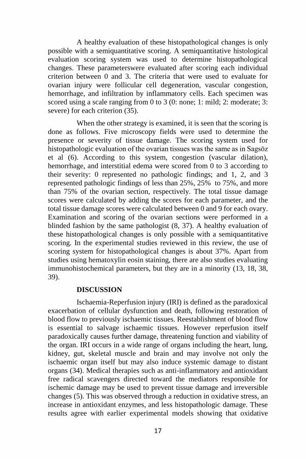
A healthy evaluation of these histopathological changes is only
possible with a semiquantitative scoring. A semiquantitative histological
evaluation scoring system was used to determine histopathological
changes. These parameterswere evaluated after scoring each individual
criterion between 0 and 3. The criteria that were used to evaluate for
ovarian injury were follicular cell degeneration, vascular congestion,
hemorrhage, and infiltration by inflammatory cells. Each specimen was
scored using a scale ranging from 0 to 3 (0: none; 1: mild; 2: moderate; 3:
severe) for each criterion (35).
When the other strategy is examined, it is seen that the scoring is
done as follows. Five microscopy fields were used to determine the
presence or severity of tissue damage. The scoring system used for
histopathologic evaluation of the ovarian tissues was the same as in Sagsöz
et al (6). According to this system, congestion (vascular dilation),
hemorrhage, and interstitial edema were scored from 0 to 3 according to
their severity: 0 represented no pathologic findings; and 1, 2, and 3
represented pathologic findings of less than 25%, 25% to 75%, and more
than 75% of the ovarian section, respectively. The total tissue damage
scores were calculated by adding the scores for each parameter, and the
total tissue damage scores were calculated between 0 and 9 for each ovary.
Examination and scoring of the ovarian sections were performed in a
blinded fashion by the same pathologist (8, 37). A healthy evaluation of
these histopathological changes is only possible with a semiquantitative
scoring. In the experimental studies reviewed in this review, the use of
scoring system for histopathological changes is about 37%. Apart from
studies using hematoxylin eosin staining, there are also studies evaluating
immunohistochemical parameters, but they are in a minority (13, 18, 38,
39).
DISCUSSION
Ischaemia-Reperfusion injury (IRI) is defined as the paradoxical
exacerbation of cellular dysfunction and death, following restoration of
blood flow to previously ischaemic tissues. Reestablishment of blood flow
is essential to salvage ischaemic tissues. However reperfusion itself
paradoxically causes further damage, threatening function and viability of
the organ. IRI occurs in a wide range of organs including the heart, lung,
kidney, gut, skeletal muscle and brain and may involve not only the
ischaemic organ itself but may also induce systemic damage to distant
organs (34). Medical therapies such as anti-inflammatory and antioxidant
free radical scavengers directed toward the mediators responsible for
ischemic damage may be used to prevent tissue damage and irreversible
changes (5). This was observed through a reduction in oxidative stress, an
increase in antioxidant enzymes, and less histopathologic damage. These
results agree with earlier experimental models showing that oxidative

18
stress plays an important role in ovarian torsion (5) and the results of many
experimental studies we gathered from this study show that antioxidants
used for therapeutic purposes can reduce ovarian tissue damage. We hope
that our study will be a helpful resource for the design of future ovarian
ischemia / reperfusion studies.
Table 1: Anesthetics used for surgical procedure in ovaryan torsion
studies.

Table 2: Experimental procedures and therapeutic agents used in ovarian ischemia / reperfusion studies in recent years.

20
Table 3: Parameters used for biochemical evaluation in ovarian ischemia / reperfusion studies in recent years. (MDA (
Malondialdehyde), SOD (Superoxide dismutase), GSH/tGSH (Total glutathione), MPO (Myeloperoxidase), LPO (
Lactoperoxidase), CAT (Catalase), GPx (Glutathione peroxidase), NO (Nitric oxide), TAS (Total antioxidant status), TOS (Total
oxidant status), OSI (Oxidative stability index)).

REFERENCES
1. Huang C, Hong MK, Ding DC. A review of ovary torsion. Ci Ji Yi Xue
Za Zhi. 2017;29(3):143-147. doi:10.4103/tcmj.tcmj_55_17
2. Anne A, Dalley A. Grant’s Atlas of Anatomy. 13th ed. Philadelphia:
Lippincott Williams & Wilkins; 2012.
3. Eskander, R.N., Bristow, R.E. Adnexal Masses in Pediatric and
Adolescent Females: A Review of the Literature. Curr Obstet
Gynecol Rep 1, 25–32 (2012). https://doi.org/10.1007/s13669-
011-0001-4
4. Guthrie BD, Adler MD, Powell EC. Incidence and trends of pediatric
ovarian torsion hospitalizations in the United States, 2000–2006.
Pediatrics. 2010;125(3):532–8.
5. Bozkurt S, Arikan DC, Kurutas EB, Sayar H, Okumus M, Coskun A,
Bakan V. Selenium has a protective effect on ischemia/reperfusion
injury in a rat ovary model: biochemical and histopathologic
evaluation. J Pediatr Surg. 2012 Sep;47(9):1735-41. doi:
10.1016/j.jpedsurg.2012.03.053. PMID: 22974615.
6. Cihangir Mutlu ERCAN, Hakan ÇOKSÜER, Uğur KESKİN, K. Emre
KARAŞAHİN, İbrahim ALANBAY, İskender BAŞER.(2011).
Adneksiyal torsiyon tedavisinde laparoskopik yaklaşım
sonuçlarımız. Gülhane Tıp Dergisi
7. Akdemir A, Erbas O, Gode F, Ergenoglu M, Yeniel O, Oltulu F,
Yavasoglu A, Taskiran D. Protective effect of oxytocin on ovarian
ischemia-reperfusion injury in rats. Peptides. 2014 May;55:126-
30. doi: 10.1016/j.peptides.2014.02.015. Epub 2014 Mar 11.
PMID: 24630974.
8. Arikan, Deniz & Bakan, Vedat & Kurutas, Ergul & Sayar, Hamide &
Coskun, Ayhan. (2010). Protective effect of tadalafil on
ischemia/reperfusion injury of rat ovary. Journal of pediatric
surgery. 45. 2203-9. 10.1016/j.jpedsurg.2010.07.011.
9. Melekoglu, R., Ciftci, O., Eraslan, S., Alan, S., & Basak, N. (2018). The
protective effects of glycyrrhetinic acid and chrysin against
ischemia-reperfusion injury in rat ovaries. BioMed research
international, 2018.
10. Turkler, C., Kulhan, N. G., Ata, N., Kiremitli, T., Cimen, F. K., &
Suleyman, H. (2018). The ameliorative effect of lutein on ovarian
ischemia-reperfusion injury in rats. Bratislavske Lekarske
Listy, 119(11), 713-717.

22
11. Yurtcu, E., Togrul, C., Ozyer, S., Uzunlar, O., Karatas, Y. H., Seckin,
K. D., ... & Cicek, N. (2015). Dose dependent protective effects of
vardenafil on ischemia–reperfusion injury with biochemical and
histopathologic evaluation in rat ovary. Journal of pediatric
surgery, 50(7), 1205-1209.
12. Incebiyik, A., Seker, A., Camuzcuoglu, H., Kocaslan, S.,
Camuzcuoglu, A., Hilali, N. G., ... & Aksoy, N. (2015). Does
sildenafil have protective effects against ovarian ischemia-
reperfusion injury in rats?. Archives of gynecology and
obstetrics, 291(6), 1283-1288.
13. Başer, B. G., Taşkın, M. İ., Adalı, E., Öztürk, E., Hısmıoğulları, A. A.,
& Yay, A. (2018). Does progesterone have protective effects on
ovarian ischemia-reperfusion injury?. Journal of the Turkish
German Gynecological Association, 19(2), 87.
14. Yilmaz, E. P. T., Un, H., Gundogdu, B., Polat, E., Askin, S., Topdagi,
Y. E., & Halici, Z. (2020). Protective Effect of Lycopene against
Reperfusion Injury in Rats with Ovarian Torsion: A Biochemical
and Histopathological Evaluation. Journal of Laboratory
Physicians, 12(1), 32.
15. Erbas, O., & Karadadas, N. Effects of Methylene Blue on Ovarian
TorsionLDetorsion Injury in a Rat Model.
16. Cadirci, E., Oral, A., Odabasoglu, F., Kilic, C., Coskun, K., Halici, Z.,
... & Unal, B. (2010). Atorvastatin reduces tissue damage in rat
ovaries subjected to torsion and detorsion: biochemical and
histopathologic evaluation. Naunyn-Schmiedeberg's archives of
pharmacology, 381(5), 455-466.
17. Sak, M. E., Soydinc, H. E., Sak, S., Evsen, M. S., Alabalik, U.,
Akdemir, F., & Gul, T. (2013). The protective effect of curcumin
on ischemia-reperfusion injury in rat ovary. International Journal
of Surgery, 11(9), 967-970.
18. Gungor, A. N. C., Gencer, M., Karaca, T., Hacivelioglu, S., Uysal, A.,
Korkmaz, F., ... & Cosar, E. (2014). The effect of hesperetin on
ischemia–reperfusion injury in rat ovary. Archives of gynecology
and obstetrics, 290(4), 763-769.
19. Nayki, C., Nayki, U., Keskin Cimen, F., Kulhan, M., Yapca, O. E.,
Kurt, N., & Bilgin Ozbek, A. (2018). The effect of rutin on ovarian
ischemia-reperfusion injury in a rat model. Gynecological
Endocrinology, 34(9), 809-814.
20. Eser, A., Hizli, D., Haltas, H., Namuslu, M., Kosus, A., Kosus, N., &
Kafali, H. (2015). Effects of curcumin on ovarian ischemia-

23
reperfusion injury in a rat model. Biomedical reports, 3(6), 807-
813.
21. Bayir, Y., Cadirci, E., Polat, B., Kilic Baygutalp, N., Albayrak, A.,
Karakus, E., ... & Karaca, M. (2016). Aliskiren–a promising
strategy for ovarian ischemia/reperfusion injury protection in rats
via RAAS. Gynecological Endocrinology, 32(8), 675-683.
22. Ergun, Y., Koc, A., Dolapcioglu, K., Akaydin, Y., Dogruer, G., Kontas,
T., ... & Aslan, E. (2010). The protective effect of erythropoietin
and dimethylsulfoxide on ischemia-reperfusion injury in rat
ovary. European Journal of Obstetrics & Gynecology and
Reproductive Biology, 152(2), 186-190.
23. Aslan, M. K., Boybeyi, Ö., Şenyücel, M. F., Ayva, Ş., Kısa, Ü., Aksoy,
N., ... & Çakmak, M. (2012). Protective effect of intraperitoneal
ozone application in experimental ovarian ischemia/reperfusion
injury. Journal of pediatric surgery, 47(9), 1730-1734.
24. Kurt, A., Isaoglu, U., Yilmaz, M., Calik, M., Polat, B., Hakan, H., ... &
Suleyman, H. (2011). Biochemical and histological investigation
of famotidine effect on postischemic reperfusion injury in the rat
ovary. Journal of pediatric surgery, 46(9), 1817-1823.
25. Ming-Shuo Sun, Hang Jin, Xin Sun, Shuo Huang, Fu-Liang Zhang,
Zhen-Ni Guo, Yi Yang, "Free Radical Damage in Ischemia-
Reperfusion Injury: An Obstacle in Acute Ischemic Stroke after
Revascularization Therapy", Oxidative Medicine and Cellular
Longevity, vol. 2018, ArticleID 3804979, 17 pages, 2018. https://
doi.org/10.1155/2018/3804979
26. Akdemir A, Erbas ̧ O, Ergenoğlu M, Ozgür Yeniel A, Oltulu F,
Yavasoğlu A, et al.Montelukast prevents ischemia/reperfusion-
induced ovarian damage in rats.Eur J Obstet Gynecol Reprod Biol
2014;173:71–6.
27. Erkanli Senturk G, Erkanli K, Aydin U, Yucel D, Isiksacan N, Ercan
F, et al. The pro-tective effect of oxytocin on ischemia/reperfusion
injury in rat urinary bladder.Peptides 2013;40:82–8.
28. Aslan, M., Senturk, G. E., Akkaya, H., Sahin, S., & Yılmaz, B. (2017).
The effect of oxytocin and Kisspeptin-10 in ovary and uterus of
ischemia-reperfusion injured rats. Taiwanese Journal of
Obstetrics and Gynecology, 56(4), 456-462.
29. Naziroglu M, Simşek M, Kutlu M. Moderate exercise with a dietary
vitamin C and E combination protects against streptozotocin-
induced oxidative damage to the blood and improves fetal
outcomes in pregnant rats. Clin Chem Lab Med 2004;42:511-7.

24
30. Eren I, Naziroglu M, Demirdas A. Protective effects of lamotrigine,
aripiprazole and escitalopram on depression-induced oxidative
stres in rat brain. Neurochem Res 2007;32:1188-95.
31. Somuncu S, Cakmak M, Dikmen G, Akman H, Kaya M. Ischemia-
reperfusion injury of rabbit ovary and protective effect of trapidil:
an experimental study. Pediatr Surg Int 2008;24:315–8. doi:
10.1007/s00383-007-2079-3.
32. Erel O. A novel automated method to measure total antioxidant
response against potent free radical reactions. Clin Biochem
2004;37:112-9.
33. Erel O. A new automated colorimetric method for measuring total
oxidant status. Clin Biochem 2005;38:1103-11.
34. Cowled P, Fitridge R. Pathophysiology of Reperfusion Injury. In:
Fitridge R, Thompson M, editors. Mechanisms of Vascular
Disease: A Reference Book for Vascular Specialists [Internet].
Adelaide (AU): University of Adelaide Press; 2011. 18. Available
from: https://www.ncbi.nlm.nih.gov/books/NBK534267/
35. Guven S, Muci E, Unsal MA, et al. The effects of carbon dioxide
pneumoperitoneum on ovarian blood flow, oxidative stress
markers, and morphology during laparoscopy: a rabbit model.
Fertil Steril 2008;93(4).
36. Sagsoz N, Kisa U, Apan A. Ischemia-reperfusion injury of rat ovary
and the effects of vitamin C, mannitol and verapamil. Hum Reprod
2002;17:2972-6.
37. Gezer, Ş., & Başaran, M. The protective effect of letrozole in a rat
ovarian ischemia-reperfusion injury model. Van Medical
Journal, 27(4), 407-414.
38. Sengul, O., Ferah, I., Polat, B., Halici, Z., Bayir, Y., Yilmaz, M., ... &
Keles, O. N. (2013). Blockade of endothelin receptors with
bosentan limits ischaemia/reperfusion-induced injury in rat
ovaries. European Journal of Obstetrics & Gynecology and
Reproductive Biology, 170(2), 458-463.
39. Unlubilgin, E., Suleyman, B., Balci, G., Al, R. A., Cankaya, M. U. R.
A. T., Nayki, U. A., & Suleyman, H. (2017). Prevention of
infertility induced by ovarian ischemia reperfusion injury by
benidipine in rats: Biochemical, gene expression,
histopathological and immunohistochemical evaluation. Journal
of Gynecology Obstetrics and Human Reproduction, 46(3), 267-
273.

CHAPTER III
CLINICAL DEVELOPMENT OF HIF-1α INHIBITORS
FOR CANCER THERAPY
Gülnihal Özcan
(Asst. Prof.) Koç University School of Medicine, e-mail:[email protected]
0000-0001-5354-0148
INTRODUCTION
Hypoxia inducible factors (HIFs) are transcription factors that play
a central role in adaptation of cells to hypoxia. HIFs are stabilized in the
hypoxic environment leading to transcription of many target genes that
induce angiogenesis to re-establish oxygen homeostasis and regulate
cellular energetic metabolism to adapt to hypoxic conditions 1. Cancer cells
utilize these mechanisms in their advantage to survive despite unfavorable
conditions, gain replicative immortality and stem cell characteristics,
escape from apoptotic stimuli and immunosurveillance, metastasize to
different areas and resist anti-cancer therapy. As a result, hypoxia or
increased activity of HIFs bring more malignant characteristics to tumors
and worsen patient prognosis 2.
Among the HIF isoforms HIF-1 come forward with its prominent
involvement in tumor progression and chemoresistance. HIF-1 is a
heterodimer of HIF-1α and HIF-1β subunits. HIF-1α is overexpressed in
several tumors and its expression is associated with poor overall survival 3-5. HIF-1α expression was found to be more abundant at the invasive
margins of tumor specimens 6-8. High expression of HIF-1α is also
associated with high a risk of metastasis in cancer 9-11. Therefore, targeting
HIF-1α is of prime importance to increase therapeutic success in cancer
chemotherapy.
REGULATION OF HIF-1α ACTIVITY
In the presence of oxygen HIF-1α is hydroxylated by prolyl
hydroxylases (PHDs). Then hydroxylated HIF-1α is ubiquitinated and
degraded by ubiquitin proteasome system with a reaction dependent on
tumor suppressor protein von Hippel–Lindau (pVHL). On the other hand,
in hypoxic conditions HIF-1α cannot be hydroxylated by PHDs and
becomes stable. Then, it translocates to the nucleus and couples with HIF-
1β to form HIF-1. After that, HIF-1 binds p300/CBP co-activator forming
the HIF transcription complex which binds to hypoxia response elements

26
and induces the transcription of target genes such as vascular endothelial
growth factor (VEGF), glucose transporter-1, epithelial-mesenchymal
transition genes and matrix metalloproteinases 12-14.
The expression of HIF-1α is also regulated by several other
mechanisms besides hypoxia in cancer cells. Reactive oxygen species
generated by various stimuli stabilize HIF-1α and increases HIF-1 activity.
PI3K/Akt/mTOR and RAS/MAPK pathways which are oncogenic
pathways overactivated in several cancers are positive regulators of HIF-
1α expression. Mutations in tumor suppressor genes phosphatase and
tensin homologue (PTEN), VHL and p53 are also associated with
increased HIF-1α expression in cancer cells 14,15.
HIF-1α INHIBITORS IN CLINICAL TRIALS
In accordance with the importance of HIF-1α in tumor progression
and chemoresistance, last 20 years have witnessed the testing of several
direct or indirect inhibitors of HIF-1α in clinical trials for cancer. Some of
these agents such as camptothecins, rapamycin analogs, and
anthracyclines, which exhibit antitumor action by mechanisms that does
not directly involve HIF-1α inhibition, have already been approved and
widely used for the treatment of several cancers. However, efforts to
develop new agents that directly inhibit HIF-1α is still growing 16,17. In this
chapter, we present the major HIF-1α inhibitors that were translated to
clinical studies so far with registered clinical trials in ClinicalTrials.gov
(https://www.clinicaltrials.gov/). Relevant clinical trial identifiers are
given for each of the studies in the text. According to their mechanism of
action, anti-HIF-1α agents were classified into five in this chapter, namely
HIF-1α antisense oligonucleotides, drugs that inhibit HIF-1α translation,
drugs that destabilize HIF-1α, inhibitors of HIF-1/DNA binding, and drugs
that inhibitor HIF-1α at multiple levels.
1. HIF-1α Antisense Oligonucleotides
Efforts to translate antisense oligonucleotides into the treatment of
cancer patients is growing for the last decade 18. EZN-2968 is a HIF-1α
antisense oligonucleotide developed by Enzon Pharmaceuticals. A phase I
trial has been conducted between 2007 and 2011 for determination of the
safety, pharmacokinetic profile, and anticancer activity of EZN-2968 in
patients with advanced solid tumors and lymphoma (NCT00466583). First
results suggested well-tolerability in cancer patients who have undergone
previous treatments 19. In 2010, a second phase I study for EZN-2968 was
commenced under the sponsorship of National Health Institute to
determine the safety profile and efficacy in advanced solid tumors with
liver metastasis (NCT01120288). A reduction in HIF-1α expression was
observed in a very limited number of patients enrolled into the study and
the trial was ended early 20. Later in 2016, a phase Ib study was started to

27
test a synthetic locked nucleic acid form of EZN-2968 (RO7070179) in
hepatocellular carcinoma (HCC) (NCT02564614), since dose limiting
toxicity in the previous studies was hepatotoxicity due to accumulation in
liver. Study results suggested that HCC patients may benefit from
RO7070179 and, decrease in HIF-1α mRNA during the first cycle may
have predictive value for therapeutic effect 21.
2. Drugs That Inhibit HIF-1α Translation
2a. Camptothecins
Camptothecins which act primarily by inhibition of topoisomerase I
enzyme, is an important group of chemotherapeutics used in the treatment
of cancer. Camptothecin and its derivatives like topotecan has been shown
to down-regulate HIF-1α through regulation of the HIF-1α-targeting
miRNAs 22. After the confirmation of its anti-HIF1α activity together with
the anti-angiogenetic and anti-tumorigenic effects in xenograft models,
topotecan was evaluated in a phase I study for its effectiveness in
suppressing HIF-1α expression in advanced solid tumors refractory to
treatment (NCT00117013). Although a median reduction of 7.5% percent
was detected in HIF-1α expression and a correlation was observed between
this reduction and radiological outcomes, the results were not statistically
significant because of low patient accrual 23.
Besides their use as single agents, inclusion of campothecins in
combination chemotherapy where an anti-VEGF agent is involved may
prevent the increase in HIF-1α due to inhibition of angiogenesis. With this
notion, combination of camptothecins with anti-angiogenetic agents have
been tested in several clinical trials. Combination of irinotecan with anti-
VEGF monoclonal antibody bevacuzimab was tested for its efficacy in
pediatric patients with recurrent, progressive, or refractory glioma,
medulloblastoma, ependymoma, or low-grade glioma in a phase II trial
started in 2006 (NCT00381797). In another phase II trial started in 2007,
efficacy of topotecan in combination with cisplatin and bevacuzimab was
tested in patients with recurrent or persistent cervical cancer
(NCT00548418). However, the toxicity profile of this combination was
disappointing 24.
EZN-2208, a pegylated form of SN38, which is the active metabolite
of irinotecan, was combined with bevacuzimab in a phase I trial to test its
safety, efficacy and anti-HIF-1α activity in solid tumors refractory to
treatment (NCT01251926, no posted results). CRLX101, which is a
nanoparticle formulation of camptothecin, was tested for its safety and
efficacy profile in combination with bevacizumab in patients with ovarian,
tubal, and peritoneal cancers resistant to platinium-based therapy
(NCT01652079) 25. The trial was completed in 2018 with no results posted
yet. Although the emphasis was not on inhibition of HIF-1α, the efficacy
28
of anti-VEGF aflibercept in combination with the FOLFIRI (folinic acid,
fluorouracil, and irinotecan) regimen was investigated in metastatic
colorectal cancer (NCT02129257). The regimen was found to be tolerable.
However, 6 months of first-line treatment with FOLFIRI + aflibercept was
followed by aflibercept without accompanying anti-HIF1α treatment and a
substantial increase in efficacy has not been detected 26.
2b. PI3K/AKT/mTOR Pathway Inhibitors
PI3K/AKT/mTOR pathway is an important intracellular signaling
pathways that induces cell proliferation, migration, and tumor progression.
Several anticancer drugs that inhibit this pathway at distinct molecular
levels have been used or being tested in chemotherapy of several cancers.
Rapamycin (sirolimus) is the first agent discovered among these, which
gives its name to the mTOR protein, the mammalian target of rapamycin 27. Rapamycin analogs (rapalogs), temsirolimus and everolimus are first
approved for the treatment of advanced renal carcinoma and have been
tested in preclinical and clinical studies for the treatment of several other
cancers 28. Besides their primary action on inhibition of cell proliferation
and migration, rapalogs also downregulate HIF-1α since
PI3K/AKT/mTOR pathway is a positive regulator of HIF-1α expression 14,17. Due to this action, rapalogs have been started to be tested clinically in
combination with other chemotherapeutics and anti-angiogenic agents.
A phase I study started in 2010 to determine maximum tolerated
dose of everolimus (RAD001), in combination with capecitabine and
oxaliplatin (XELOX) regimen in patients with advanced gastric cancer
(NCT01049620). The same year, another clinical trial started to determine
safety of everolimus and its efficacy in combination with FOLFOX and
bevacizumab in the first-line treatment of colorectal cancer
(NCT01047293). The combination exhibited a tolerable profile and fair
efficacy 29. Combination of everolimus with sorafenib, which is an anti-
tumorigenic and anti-angiogenic agent due to its multi-kinase inhibitor
action, was tested in a Phase I-II study for advanced solid tumors.
However, the study was suspended early due to toxicity (NCT01226056).
Combination of rapamycin and irinotecan has also been tested in refractory
solid tumors in children for the synergistic effect in inhibition of
angiogenesis and suppression of HIF-1α (NCT01282697, no results
posted).
Perifosine is a multitarget tyrosine kinase inhibitor that has AKT
inhibitor activity. It has been extensively tested as a single agent or
combination in several different cancers in tens of different clinical trials
(e.g., NCT00398879, NCT01048580, NCT00398814, NCT00060437,
NCT00873457, NCT02238496 and NCT02238496). Due to AKT
inhibitory action, perifosine downregulates HIF-1α 16. However, this action

29
was not the main emphasis in the clinical trials of perifosine conducted so
far. Despite the promising results in preclinical studies, its clinical
effectiveness in cancer is not fully established yet.
2c. Digoxin
Since preclinical studies suggested that digoxin blocks HIF-1α 30, its
efficacy in blocking HIF-1α in breast cancer and Kaposi’s Sarcoma has
been tested in two different clinical trials. The former one aimed to
investigate whether two weeks of digoxin intake starting from the first
biopsy to the time of surgery decreases HIF-1α expression in newly
diagnosed breast cancer (NCT01763931). However the trial stopped to
recruit patients early since accrual goals could not be met 31. HIF-1α has
been shown to induce lytic life cycle of Human Herpes Virus 8 32.
Regarding the efficacy of digoxin in downregulation of HIF-1α, digoxin
may have a potential in the treatment of HHV-8 related Kaposi Sarcoma.
A multi-centre phase II study was conducted to test this hypothesis
(NCT02212639). The study was expected to be completed in 2019.
However, the results are not published yet. In addition to these two trials
that put the emphasis on anti-HIF-1α action, there are more than 20
different clinical trials that test digoxin alone or in combination with
chemotherapy in several different cancers as a hypoxia modulator agent or
p-glycoprotein inhibitor (e.g., NCT04141995, NCT02106845). A Phase II
study in prostate cancer patients exhibited promising results suggesting
that digoxin can prolong prostate specific antigen doubling time and may
be effective in androgen-dependent prostate cancer 33 (NCT01162135).
New clinical trials are recruiting patients or will start accrual to evaluate
digoxin in solid tumors and pancreatic cancer (NCT03889795,
NCT04141995).
2d. 2-Methoxyestradiol
2-methoxyestradiol (2ME2) is an estradiol metabolite which inhibits
microtubule activity and acts as an antitumor agent. 2ME2 inhibits the
translation of HIF-1α mRNA. Due to this action, 2ME2 was started to be
evaluated as an agent to inhibit tumor blood flow in two different phase I
trials in 2001 and 2002 (NCT00030095, NCT00028821). Since 2ME2
exhibit inhibitory action on cell proliferation, migration, and angiogenesis 17, a nanocrystal colloidal dispersion formulation of 2ME2, Panzem was
developed which has higher bioavailability. Panzem entered several phase
II trials, starting from 2006 (NCT00394810, NCT00306618,
NCT00481455, and NCT00444314). However, the results were
disappointing in terms of anti-tumor activity and tolerability 34,35. Later a
new analogue of 2ME2, ENMD-1198 was developed. ENMD-1198
exhibited potent anti-angiogenic activity in cell models 36. However, to the

30
best of our knowledge, there is no registered clinical trial for ENMD-1198
in clinicaltrials.gov yet.
3. Drugs That Destabilize HIF-1α
3a. Geldanamycin and Its Derivatives
Geldanamycin and its derivatives are ansamycin antibiotics with
anti-cancer activity. They inhibit heat shock protein 90 (HSP90), leading
to degradation of its targets which regulate the activity of several
intracellular pathways 37. Geldanamycin and its derivative 17AAG (17-
allylamino 17-demethoxygeldanamycin) or tanespimycin have been tested
in clinical trials for the treatment of several cancers since the beginning of
2000’s (e.g., NCT00003969, NCT00058253, NCT00093405,
NCT00119236, NCT00004065, NCT00019708, NCT00093821,
NCT00087386, and NCT00319930). Since HSP90 takes part in the
stabilization of HIF-1α, this makes it an important target to induce
destabilization and degradation of HIF-1α. With the emphasis on this
mechanism, efficacy of 17AAG was evaluated in VHL disease and renal
tumors in a phase II trial started in 2004 (NCT00088374). The study could
not achieve accrual goals. However, the evidence that point out to the
potential of geldanamycin and its derivatives in cancer therapy is
growing38,39.
3b. Histone Deacetylase Inhibitors
Histone deacetylases (HDAC) play important roles in epigenetic
regulation of genes associated with cancer. HDAC inhibitors were
approved for the treatment of several cancers such as lymphoma and
multiple myeloma. They show antitumor effect by inducing cell cycle
arrest, apoptosis, and anti-angiogenic response 40. HDAC is also important
for the acetylation of HIF-1α/p300 complex and therefore HDAC
inhibitors lead to destabilization of HIF-1α 41. Among the HDAC inhibitors
romidepsin come forward with this action and there are more than 20
different active clinical trials that assess romidepsin in the treatment of
lymphoma and several other cancers (e.g., NCT02512172, NCT04257448,
NCT01947140, NCT01755975, and NCT01638533).
4. Inhibitors of HIF-1/DNA Binding
4a. Anthracyclines
Anthracyclines are among the most widely used and most potent
chemotherapeutics in cancer therapy today. They show anti-tumor action
by binding to DNA and inhibiting the progression of topoisomerase II
enzyme 42. They also inhibit binding of HIF-1 to DNA. Hence, they inhibit
the expression of HIF-1α regulated genes 43,44. Even though they are
already in use in the treatment of several cancers, their anti-HIF action may

31
increase the use of anthracyclines further especially in chemotherapy
protocols with anti-VEGF agents.
4b. Echinomycin
Echinomycin is a peptide antibiotic with DNA intercalating activity.
With the aim of identifying new drugs which inhibit binding of HIF-1 to
DNA, a high-throughput screening was conducted with National Cancer
Institute’s comprehensive small-molecule library. The screening identified
echinomycin as a potent inhibitor of HIF-1/DNA binding 45. Echinomycin
has been tested in several Phase II trials for the indications of soft tissue
sarcoma, endometrial carcinoma, non-small cell lung carcinoma and
colorectal cancer 46-49. However, the clinical efficacy in these trials were
disappointing. Therefore, echinomycin could not progress into the clinic
with the expected speed as an anti-HIF-1 agent. However, nano-liposomal
formulations of echinomycin are being developed, which may offer an
improved efficacy and pharmacokinetic profile for the agent 50,51.
5. Drugs That Inhibit HIF-1α at Multiple Levels
5a. Bortezomib
The ubiquitin proteasome pathway plays a central role in protein
homeostasis, leading to degradation of misfolded or damaged proteins.
Proteasome is an attractive target in cancer therapy since inhibition of
proteosome induces cell death via endoplasmic reticulum stress, inhibition
of survival pathways and stimulation of apoptosis 52. Bortezomib is one of
the most prominent proteasome inhibitors evaluated for clinical use in
cancer. It was approved by FDA in the treatment of multiple myeloma in
2003 53, and there are more than 900 different clinical trials registered that
evaluate bortezomib, mostly in combination to conventional
chemotherapeutics in a wide spectrum of cancers. Since proteasomal
degradation of HIF-1α has substantial importance in regulation of HIF-1,
bortezomib also has an anti-HIF-1 action. In the presence of bortezomib,
ubiquitinated HIF-1α could not be degraded and accumulates in an inactive
form. In this form HIF-1α cannot bind to p300 and induce gene
expression54. Additionally, it was shown that bortezomib can inhibit
PI3K/AKT/mTOR pathway. Moreover, bortezomib was reported to
suppress nuclear translocation of HIF-1α via inhibition of MAPK
pathway55. With these mechanisms bortezomib exhibits anti-HIF-1 action
at multiple levels which makes it an attractive agent in cancer therapy.
5b. PX-478
PX-478 is a melphalan derivative that downregulates the HIF-1α
mRNA and inhibits its translation. It was also reported to inhibit de-
ubiquitination of HIF-1α to a smaller extent 56. Due to inhibition of HIF-
1α, PX-478 may have a potential in cancer treatment. The safety and

32
efficacy of PX-478 was evaluated in advanced solid tumors in a phase I
trial conducted in 2007-2010 (NCT00522652). The results were promising
in terms of tolerability and HIF-1α inhibition57. However, a registration
could not be found in clinicaltrial.gov that suggest the further evaluation
of PX-478 in clinical studies.
CONCLUSION
Clinical development of HIF-1α inhibitors for cancer treatment is
ongoing at a great pace. Till the safety and efficacy of direct HIF-1α
inhibitors is fully established, anti-cancer agents already approved in
cancer such as camptothecins, rapalogs, anthracyclines and bortezomib
seem to be involved more frequently in combination chemotherapy for
HIF-1α inhibitory effect in addition to their primary mechanisms of anti-
cancer action.

33
REFERENCES
1. Semenza GL. Hypoxia-inducible factors in physiology and medicine.
Cell. 2012;148(3):399-408.
2. Ruan K, Song G, Ouyang G. Role of hypoxia in the hallmarks of human
cancer. J Cell Biochem. 2009;107(6):1053-1062.
3. Branco-Price C, Zhang N, Schnelle M, et al. Endothelial cell HIF-1α
and HIF-2α differentially regulate metastatic success. Cancer cell.
2012;21(1):52-65.
4. Kitajima Y, Miyazaki K. The Critical Impact of HIF-1a on Gastric
Cancer Biology. Cancers. 2013;5(1):15-26.
5. Semenza GL. Defining the role of hypoxia-inducible factor 1 in cancer
biology and therapeutics. Oncogene. 2010;29(5):625-634.
6. Hao LS, Liu Q, Tian C, et al. Correlation and expression analysis of
hypoxia‑inducible factor 1α, glucose transporter 1 and lactate
dehydrogenase 5 in human gastric cancer. Oncol Lett.
2019;18(2):1431-1441.
7. Rohwer N, Lobitz S, Daskalow K, et al. HIF-1α determines the
metastatic potential of gastric cancer cells. British Journal of
Cancer. 2009;100(5):772-781.
8. Griffiths EA, Pritchard SA, Welch IM, Price PM, West CM. Is the
hypoxia-inducible factor pathway important in gastric cancer?
European Journal of Cancer. 2005;41(18):2792-2805.
9. Miao ZF, Wang ZN, Zhao TT, et al. Peritoneal milky spots serve as a
hypoxic niche and favor gastric cancer stem/progenitor cell
peritoneal dissemination through hypoxia-inducible factor 1α.
Stem cells (Dayton, Ohio). 2014;32(12):3062-3074.
10. Jung J-H, Im S, Jung ES, Kang CS. Clinicopathological Implications of
the Expression of Hypoxia-related Proteins in Gastric Cancer.
International Journal of Medical Sciences. 2013;10(9):1217-
1223.
11. Chen L, Shi Y, Yuan J, et al. HIF-1 Alpha Overexpression Correlates
with Poor Overall Survival and Disease-Free Survival in Gastric
Cancer Patients Post-Gastrectomy. PLOS ONE.
2014;9(3):e90678.
12. Kaelin WG, Jr., Ratcliffe PJ. Oxygen sensing by metazoans: the central
role of the HIF hydroxylase pathway. Molecular cell.
2008;30(4):393-402.
13. Lee P, Chandel NS, Simon MC. Cellular adaptation to hypoxia through
hypoxia inducible factors and beyond. Nature reviews Molecular
cell biology. 2020;21(5):268-283.
14. Muz B, de la Puente P, Azab F, Azab AK. The role of hypoxia in cancer
progression, angiogenesis, metastasis, and resistance to therapy.
Hypoxia (Auckland, NZ). 2015;3:83-92.

34
15. Ravi R, Mookerjee B, Bhujwalla ZM, et al. Regulation of tumor
angiogenesis by p53-induced degradation of hypoxia-inducible
factor 1alpha. Genes Dev. 2000;14(1):34-44.
16. Burroughs SK, Kaluz S, Wang D, Wang K, Van Meir EG, Wang B.
Hypoxia inducible factor pathway inhibitors as anticancer
therapeutics. Future medicinal chemistry. 2013;5(5):553-572.
17. Masoud GN, Li W. HIF-1α pathway: role, regulation and intervention
for cancer therapy. Acta pharmaceutica Sinica B. 2015;5(5):378-
389.
18. Quemener AM, Bachelot L, Forestier A, Donnou-Fournet E, Gilot D,
Galibert M-D. The powerful world of antisense oligonucleotides:
From bench to bedside. Wiley Interdiscip Rev RNA.
2020;11(5):e1594.
19. Patnaik A, Chiorean EG, Tolcher A, et al. EZN-2968, a novel hypoxia-
inducible factor-1α (HIF-1α) messenger ribonucleic acid (mRNA)
antagonist: Results of a phase I, pharmacokinetic (PK), dose-
escalation study of daily administration in patients (pts) with
advanced malignancies. Journal of Clinical Oncology
2009;27(15_suppl):2564-2564.
20. Jeong W, Rapisarda A, Park SR, et al. Pilot trial of EZN-2968, an
antisense oligonucleotide inhibitor of hypoxia-inducible factor-1
alpha (HIF-1α), in patients with refractory solid tumors. Cancer
Chemother Pharmacol. 2014;73(2):343-348.
21. Wu J, Contratto M, Shanbhogue KP, et al. Evaluation of a locked
nucleic acid form of antisense oligo targeting HIF-1α in advanced
hepatocellular carcinoma. World journal of clinical oncology.
2019;10(3):149-160.
22. Bertozzi D, Marinello J, Manzo SG, Fornari F, Gramantieri L,
Capranico G. The natural inhibitor of DNA topoisomerase I,
camptothecin, modulates HIF-1α activity by changing miR
expression patterns in human cancer cells. Molecular cancer
therapeutics. 2014;13(1):239-248.
23. Duffy AG, Melillo G, Turkbey B, et al. A pilot trial of oral topotecan
(TPT) in patients with refractory advanced solid neoplasms
expressing HIF-1α. Journal of Clinical Oncology
2010;28(15_suppl):e13518-e13518.
24. Zighelboim I, Wright JD, Gao F, et al. Multicenter phase II trial of
topotecan, cisplatin and bevacizumab for recurrent or persistent
cervical cancer. Gynecologic oncology. 2013;130(1):64-68.
25. Krasner CN, Birrer MJ, Berlin ST, et al. Phase II clinical trial evaluating
CRLX101 in recurrent ovarian, tubal, and peritoneal cancer.
Journal of Clinical Oncology. 2014;32(15_suppl):5581-5581.
26. Pentheroudakis G, Kotoula V, Koliou GA, et al. AMALTHEA:
Prospective, Single-Arm Study of the Hellenic Cooperative

35
Oncology Group (HeCOG) Evaluating Efficacy and Safety of
First-Line FOLFIRI + Aflibercept for 6 Months Followed by
Aflibercept Maintenance in Patients With Metastatic Colorectal
Cancer. Clinical colorectal cancer. 2018;17(4):e631-e637.
27. LoRusso PM. Inhibition of the PI3K/AKT/mTOR Pathway in Solid
Tumors. Journal of clinical oncology. 2016;34(31):3803-3815.
28. Tian T, Li X, Zhang J. mTOR Signaling in Cancer and mTOR Inhibitors
in Solid Tumor Targeting Therapy. International journal of
molecular sciences. 2019;20(3).
29. Weldon Gilcrease G, Stenehjem DD, Wade ML, et al. Phase I/II study
of everolimus combined with mFOLFOX-6 and bevacizumab for
first-line treatment of metastatic colorectal cancer. Investigational
new drugs. 2019;37(3):482-489.
30. Zhang H, Qian DZ, Tan YS, et al. Digoxin and other cardiac glycosides
inhibit HIF-1alpha synthesis and block tumor growth. Proc Natl
Acad Sci U S A. 2008;105(50):19579-19586.
31. Bardia A, Santa-Maria CA, Jacobs LK, et al. Digoxin as an inhibitor of
global hypoxia inducible factor-1α (HIF1α) expression and
downstream targets in breast cancer: Dig-HIF1 pharmacodynamic
trial. Journal of clinical oncology. 2013;31(15_suppl):TPS1144-
TPS1144.
32. Haque M, Wang V, Davis DA, Zheng ZM, Yarchoan R. Genetic
organization and hypoxic activation of the Kaposi's sarcoma-
associated herpesvirus ORF34-37 gene cluster. Journal of
virology. 2006;80(14):7037-7051.
33. Lin J, Hoffman-Censits JH, Duffy D, et al. A pilot phase II study of
digoxin in patients with recurrent prostate cancer as evident by a
rising PSA. Journal of Clinical Oncology.
2013;31(15_suppl):5061-5061.
34. Harrison MR, Hahn NM, Pili R, et al. A phase II study of 2-
methoxyestradiol (2ME2) NanoCrystal® dispersion (NCD) in
patients with taxane-refractory, metastatic castrate-resistant
prostate cancer (CRPC). Investigational new drugs.
2011;29(6):1465-1474.
35. Bruce JY, Eickhoff J, Pili R, et al. A phase II study of 2-
methoxyestradiol nanocrystal colloidal dispersion alone and in
combination with sunitinib malate in patients with metastatic renal
cell carcinoma progressing on sunitinib malate. Investigational
new drugs. 2012;30(2):794-802.
36. Pasquier E, Sinnappan S, Munoz MA, Kavallaris M. ENMD-1198, a
new analogue of 2-methoxyestradiol, displays both antiangiogenic
and vascular-disrupting properties. Molecular cancer
therapeutics. 2010;9(5):1408-1418.

36
37. Bisht KS, Bradbury CM, Mattson D, et al. Geldanamycin and 17-
Allylamino-17-demethoxygeldanamycin Potentiate the Radiation
Response of Cervical Tumor Cells via the Heat Shock Protein 90-
Mediated Intracellular Signaling and Cytotoxicity. Cancer Res.
2003;63(24):8984-8995.
38. Weber H, Valbuena JR, Barbhuiya MA, et al. Small molecule inhibitor
screening identifified HSP90 inhibitor 17-AAG as potential
therapeutic agent for gallbladder cancer. Oncotarget.
2017;8(16):26169-26184.
39. Liu H, Lu J, Hua Y, et al. Targeting heat-shock protein 90 with
ganetespib for molecularly targeted therapy of gastric cancer. Cell
Death Dis. 2015;6(1):e1595.
40. Eckschlager T, Plch J, Stiborova M, Hrabeta J. Histone Deacetylase
Inhibitors as Anticancer Drugs. International journal of molecular
sciences. 2017;18(7).
41. Fath DM, Kong X, Liang D, et al. Histone deacetylase inhibitors repress
the transactivation potential of hypoxia-inducible factors
independently of direct acetylation of HIF-alpha. The Journal of
biological chemistry. 2006;281(19):13612-13619.
42. Coufal N, Farnaes L. Anthracyclines and Anthracenediones. In: Minev
B. (eds) Cancer Management in Man: Chemotherapy, Biological
Therapy, Hyperthermia and Supporting Measures. Cancer Growth
and Progression, vol 13. Springer, Dordrecht.
https://doi.org/10.1007/978-90-481-9704-0_5
43. Lee K, Qian DZ, Rey S, Wei H, Liu JO, Semenza GL. Anthracycline
chemotherapy inhibits HIF-1 transcriptional activity and tumor-
induced mobilization of circulating angiogenic cells. Proc Natl
Acad Sci U S A. 2009;106(7):2353-2358.
44. Tanaka T, Yamaguchi J, Shoji K, Nangaku M. Anthracycline inhibits
recruitment of hypoxia-inducible transcription factors and
suppresses tumor cell migration and cardiac angiogenic response
in the host. The Journal of biological chemistry.
2012;287(42):34866-34882.
45. Kong D, Park EJ, Stephen AG, et al. Echinomycin, a Small-Molecule
Inhibitor of Hypoxia-Inducible Factor-1 DNA-Binding Activity.
Cancer Res. 2005;65(19):9047-9055.
46. Gradishar WJ, Vogelzang NJ, Kilton LJ, et al. A phase II clinical trial
of echinomycin in metastatic soft tissue sarcoma. Investigational
new drugs. 1995;13(2):171-174.
47. Muss HB, Blessing JA, DuBeshter B. Echinomycin in recurrent and
metastatic endometrial carcinoma. A phase II trial of the
Gynecologic Oncology Group. Am J Clin Oncol. 1993;16(6):492-
493.

37
48. Chang AY, Kim K, Boucher H, et al. A randomized Phase II trial of
echinomycin, trimetrexate, and cisplatin plus etoposide in patients
with metastatic nonsmall cell lung carcinoma: an Eastern
Cooperative Oncology Group Study (E1587). Cancer.
1998;82(2):292-300.
49. Wadler S, Tenteromano L, Cazenave L, et al. Phase II trial of
echinomycin in patients with advanced or recurrent colorectal
cancer. Cancer Chemother Pharmacol. 1994;34(3):266-269.
50. Bailey CM, Liu Y, Peng G, et al. Liposomal formulation of HIF-1α
inhibitor echinomycin eliminates established metastases of triple-
negative breast cancer. Nanomedicine : nanotechnology, biology,
and medicine. 2020;29:102278.
51. Thomas A, Samykutty A, Gomez-Gutierrez JG, et al. Actively Targeted
Nanodelivery of Echinomycin Induces Autophagy-Mediated
Death in Chemoresistant Pancreatic Cancer In Vivo. Cancers.
2020;12(8).
52. Roeten MSF, Cloos J, Jansen G. Positioning of proteasome inhibitors
in therapy of solid malignancies. Cancer Chemother Pharmacol.
2018;81(2):227-243.
53. Bross PF, Kane R, Farrell AT, et al. Approval Summary for Bortezomib
for Injection in the Treatment of Multiple Myeloma. Clin Cancer
Res. 2004;10(12):3954-3964.
54. Shin DH, Chun YS, Lee DS, Huang LE, Park JW. Bortezomib inhibits
tumor adaptation to hypoxia by stimulating the FIH-mediated
repression of hypoxia-inducible factor-1. Blood.
2008;111(6):3131-3136.
55. Befani CD, Vlachostergios PJ, Hatzidaki E, et al. Bortezomib represses
HIF-1α protein expression and nuclear accumulation by inhibiting
both PI3K/Akt/TOR and MAPK pathways in prostate cancer cells.
Journal of molecular medicine (Berlin, Germany). 2012;90(1):45-
54.
56. Koh MY, Spivak-Kroizman T, Venturini S, et al. Molecular
mechanisms for the activity of PX-478, an antitumor inhibitor of
the hypoxia-inducible factor-1α. Mol Cancer Ther. 2008;7(1):90-
100.
57. Tibes R, Falchook GS, Hoff DDV, et al. Results from a phase I, dose-
escalation study of PX-478, an orally available inhibitor of HIF-
1α. Journal of Clinical Oncology. 2010;28(15_suppl):3076-3076.

38

CHAPTER IV
COAGULATION ANALYSES IN DIAGNOSIS AND
TREATMENT
Muhammed Emin Düz
(Biochemistry Specialist, MD),. Amasya Sabuncuoğlu Şerefeddin Training and Research
Hospital, e-mail: [email protected]
0000-0002-1837-6415
INTRODUCTION
In living macroorganisms, life-long expected and unexpected
bleeding and coagulation mechanisms are activated at regular intervals.
Excessive coagulation can lead to life-threatening damage resulting from
a blockage in the vessels, and uncontrolled bleeding can cause serious
complications, especially death. To prevent bleeding, blood clotting
(clotting) occurs a few seconds after the vessel injury. The primary purpose
of coagulation is the vascular repair that occurs by collecting thrombocytes
at the damage site. Coagulation is known as the clotting of blood or plasma.
Hemostasis is the procedure that tries to stop bleeding and is the first
component of the metabolism's response to vascular injury. As a result of
hemostasis, a product called a hemostatic plug or hemostatic clot is formed.
Thrombosis is an unexpected clot formation in a healthy vascular structure.
Thrombus formation occurs as a result of thrombosis. Blood clotting can
occur at an injury site (hemostasis), in a healthy vessel (thrombosis), or on
a foreign surface such as a laboratory test tube. Still, hemostasis is a
physiological process that can only occur in a living, bleeding organism.
Hemostasis consists of primary hemostasis, in which platelets are attached
and activated to a site of damage, and a fibrin network rigidly structures
secondary hemostasis, in which the primary platelet plug formed. The
hemostatic process represents a delicate, well-balanced chain of steps
between regulatory mechanisms and control that prevent the efficient
activation of local hemostatic agents and improper activation or
prolongation of coagulation steps due to vascular damage. The interactions
of the protein ingredients of coagulation cascade can be studied in cell-free
plasma and characterized as a "cascade" of proteolytic reactions.
Conversely, hemostasis's operation takes place on cell surfaces in a living
organism and is dependent on regulation by various biochemical and
cellular mechanisms. The competence of plasma procoagulant levels can
be defined in the routine plasma clotting assays: prothrombin time (PT)
and activated partial thromboplastin time (aPTT). Platelet count and

40
function can be determined in the clinical laboratory. Levels of individual
plasma coagulation inhibitors and other regulatory proteins can also be
evaluated. However, no laboratory test can provide a general evaluation of
hemostasis's competency or thrombosis risk. Each laboratory test offers a
small fraction of the information required, and the assessment of
hemostatic function always requires a comparison of laboratory results
with patients' clinical condition.
HEMOSTASIS
The hemostatic system consists of the plasma coagulation system,
including blood vessels, platelets, fibrinolytic factors, and inhibitory
mechanisms. When vessel damage occurs, three local agencies are
activated to control bleeding: (1) vessel wall contraction, (2) platelet
adhesion and aggregation (platelet plug formation), and (3) clot to form
fibrin. All these mechanisms need to work, and defects in one or more of
them result in abnormal bleeding. Primary hemostasis is used for rapid
plug formation upon damage to the vessel wall, followed by
vasoconstriction, platelet adhesion, and aggregation. This stage is for a
sudden and quick solution, and fibrin formation is not required. However,
primary hemostasis is transient, and bleeding could begin again, except
secondary hemostasis strengthens the platelet plug by forming a stable
fibrin clot. Eventually, the fibrinolytic system's mechanisms ensure the
fibrin clot's dissolution and the restoration of normal blood flow.
Platelets cannot adhere to the healthy, smooth endothelial surface,
and coagulation is not initiated. Endothelial thromboresistance is generated
by various antiplatelet and anticoagulant materials produced by the
endothelial cells like; prostacyclin (prostaglandin I2, PgI2), nitrite oxide
(NO), thrombomodulin, heparin-like glycosaminoglycans, tissue factor
pathway inhibitor (TFPI), tissue plasminogen activator (t-PA). Endothelial
cells also show procoagulant activity by synthesizing and secreting von
Willebrand factor (VWF) and PAI-1. It is crucial that blood not clot inside
the vascular system. In the baseline state, vascular endothelial cells
maintain a nonthrombogenic interface with the circulating blood.
Endothelial cells do not ordinarily reveal molecules that promote platelet
adhesion or aid activation and activity of the coagulation proteins. After
vascular damage, procoagulant factors and tissue factor (TF) are activated,
and thrombocyte adhesion, aggregation, and local thrombin formation are
achieved.
Platelets are nucleated cells that are released by the breakdown of
megakaryocytes in the bone marrow. They can be found in circulation for
up to nine days. The approximate number of platelets in peripheral blood
is between 150,000 to 400,000 per microliter. On the outer surface of
platelets are various molecules such as integrins and leucine-rich

41
glycoproteins. They mediate platelet adhesion and aggregation as receptors
for adenosine diphosphate (ADP), arachidonic acid, and other agonists.
Platelet connecting is further intervened by von Willebrand factor (vWF)
bridging between collagen and the platelet receptor glycoprotein (GP) Ib.
Thrombin stimulation produced as a result of coagulation reactions is
required for complete platelet activation. The platelet surface receptor,
GPIIb / IIIa, rapidly changes platelet formation by stimulating it with
fibrinogen. This change in conformation allows platelet aggregates to be
consolidated by connecting to fibrinogen ahead of fibrin conformation.
Platelet activation also launches prostaglandins and thromboxanes'
synthesis, which regulates platelet activation and assists vasoconstriction.
Activation of platelets motivates several changes resulting in hemostasis
increase via two main mechanisms:
1. Production of the hemostatic plug at the site of injury (primary
hemostasis).
2. Providing of phospholipids as a procoagulant surface for
plasmatic coagulation.
Early platelet plug formation can be divided into separate stages
that are tightly related: platelet adhesion, structural changes, content
release, and platelet aggregation. Immediately following endothelial
damage, platelets bind to adhesive proteins such as collagen via specific
glycoprotein surface receptors (platelet adhesion). In this process, VWF
acts as a mediator that first adheres to collagen fibers and then realizes fiber
strength. The platelet then binds to VWF via membrane glycoproteins (GP)
Ib and IX. After adhesion, the platelets change from disc shape to spherical
shape and elongate pseudopods. It accelerates plug formation by releasing
ADP, serotonin, Thromboxane A2 (TxA2), beta-TG, PF4, and VWF stored
in thrombocyte granules also initiates platelet aggregation. At the end of
the aggregation process, tightly packed degranulated platelets form the
hemostatic plug. Fibrinogen, which binds to specific glycoprotein
receptors (GP IIb / IIIa), is required for platelet aggregation. Thromboxane
A2 (TXA2) secreted from granules enables vasoconstriction starting with
endothelin to continue. ADP stimulates platelet aggregation, while calcium
activates the coagulation system starting with factor XII.
Two congenital bleeding disorders, von Willebrand disease and
Bernard-Soulier syndrome, are characterized by defective adhesion that
causes a lifelong tendency to bleed due to VWF and GP Ib / IX deficiency.
Besides, in Glanzmann thrombasthenia, a disease characterized by the
absence of GP IIb / IIIa receptors, bleeding time is significantly prolonged,
platelet aggregation cannot occur, resulting in a lifelong bleeding tendency.
The transformation of inactive proenzymes activates coagulation
factors into activated enzymes. Enzyme, substrate, and cofactor are

42
involved in all coagulation steps. These components accumulating on the
phospholipid complex require calcium ions for activation, limiting the
affected area's reactions. All of the coagulation factors, except factor IV
(Ca + 2), are proteins, and most of them are found in the blood as inactive
proenzymes (zymogen). Factors V and VIII, on the other hand, are not in
the enzyme structure and are synthesized by megakaryocytes in the bone
marrow. It is stored in platelet granules and released after activation.
Vitamin K is required for factor II, VII, IX, X, protein C and S synthesis
and function. The only coagulation factor not circulating is tissue factor
(Factor III) located in the subendothelial area. The coagulation system is a
chain of sequential reactions in which coagulation factors activate each
other. It is known to be initiated by two classical ways, extrinsic and
intrinsic. The intrinsic pathway begins with Hageman factor (factor XII)
activation, and the extrinsic pathway begins with the release of tissue
factor. Both paths provide stable clot formation as a result of burst
reactions that activate factor X. In the coagulation cascade, many factors
intertwine, trigger, and inhibit each other's work under a single system
simultaneously. The interconnected structure of the system at many points
also allows better control of bleeding and clotting conditions. The
coagulation system is triggered by the tissue factor released due to
endothelial damage or inflammation either by factor XII activated by
collagen. TF then interacts with factor VIIa, activating factors IX and X.
Factor Xa also produces a small amount of thrombin from prothrombin.
Propagation of coagulation consists of when thrombin repeatedly activates
factors XI, IX, V, and VIII. Thrombin also stimulates the aggregation and
activation of platelets. Coagulation factors, which are frequently activated
and multiplied in the system each time, generate too much thrombin from
prothrombin. Finally, thrombin transforms fibrinogen into fibrin. Also,
thrombin activates factor XIII and forms the desired stable clot with a fibrin
fiber network.
The main initiating pathway of in vivo blood coagulation is the
extrinsic system. The crucial component is TF, an intrinsic membrane
component expressed by cells in most extravascular tissues. The
terminology "extrinsic" continues to be used today in terms of education,
although quite outdated. TF is not frequently extrinsic to the circulatory
system, but it is also expressed by endothelial cells and leukocytes under
definite pathological conditions. TF serves as a cofactor to the significant
factor VII in the extrinsic pathway. TF and factor VII complex provide
factor VIIa activation, whereby factor X is converted into factor Xa, the
main compartment in the common path. The factor VIIa-TF complex also
converts factor IX to factor IXa. Also, as levels of factor Xa increase, the
factor VIIa-TF complex is subject to inhibition by the factor Xa-dependent
tissue factor pathway inhibitor (TFPI). The intrinsic pathway earlier begins
with the contact phase. This phase is achieved by factor XII (contact

43
factor), prekallikrein, and high-molecular-weight (HMW) kininogen. In
vitro contact phase is launched by binding factor XII to negative surfaces
of glass and kaolin. This leads to the generation of cofactors XIIa and
kallikrein and the release of bradykinin from HMW kininogen. Factor XIIa
then activates factor XI, which converts factor IX to factor IXa, a reaction
requiring calcium presence. After factor IXa complexes with its cofactor
protein factor VIIIa on a negatively charged membrane surface. This
enzymatic complex, also referred to as the tenase complex, converts factor
X to factor Xa. After both the extrinsic and intrinsic pathways generate
factor Xa, the coagulation pathway's ensuing reactions are the same and
are referred to as the common pathway. Based on the discovery that the
factor VIIa-TF complex also converts factor IX to factor IXa, that is the
privileged reaction, a new modified concept of coagulation was proposed
in which factor VIIa-TF complex seems to be the core initiator of
coagulation, but the intrinsic pathway is aforethought essential to pursue
the coagulation response.
Figure 1. Coagulation pathway scheme
ENDOGENOUS ANTICOAGULANTS AND FİBRİNOLYSIS
Anticoagulant substances consist of antithrombin III (AT-III),
protein C, protein S, and TFPI. Intact endothelium ensures that the
formation of clots is stopped, and if endothelial repair is completed, the

44
fibrin is destroyed by fibrinolysis. When clot formation reaches the intact
endothelium, it activates the heparin-like molecule AT-III, which runs to
the endothelial surface. Antithrombin III is a potent anticoagulant
substance produced in the liver and commonly found in body fluids and is
a significant inhibitor of serine proteases formed during coagulation.
Antithrombin III inactivates all active factors; Xa, XIa, XIIa, IXa,
thrombin, and TF-VIIa complex. On the other hand, thrombin binds to
thrombomodulin, which is a specific thrombin (IIa) receptor on the
endothelial surface and activates protein C and protein S. Activated protein
C neutralizes cofactors such as factor Va and VIIIa. Thus, the coagulation
system is stopped by inactivating all active and auxiliary factors.
Plasminogen is an inactive circulating protein that binds to fibrin during
clot formation. Plasminogen is activated by endothelium produced tissue
plasminogen activator (t-PA), urokinase-type plasminogen activator
(uPA), and contact factors such as XIa, XIIa, quinine, and kallikrein and
converted to plasmin. tPA is the most effective in activating fibrin-bound
plasminogen. Besides, it prevents fibrinolysis from moving beyond the clot
formation site. Fibrinogen is a colossal plasma protein with 340 kDa MW.
It is produced from the liver, and its concentration range is 200 to 400
mg/dL. Fibrinogen is stable in plasma for about 4 to 5 days. It is a dimeric
protein of highly low solubility designed with two pairs of three
nonidentical polypeptide chains covalently connected by disulfide bonds.
Fibrinolysis event is the breakdown of fibrin strands that hold plasmin,
formed by plasminogen, and fibrin. Fibrin is broken down into fibrin
breakdown products by fibrinolysis. The transformation of fibrinogen to
fibrin occurs in three stages. First, thrombin splits four small peptides—
the fibrinopeptides A (FPA) and fibrinopeptides B (FPB)—from the
fibrinogen molecule, following the formation of a new molecule called
fibrin monomer. Secondly, polymerization comprises unconsciously by
noncovalent end-to-end and side-to-side relations to develop fibrin
polymers. In the third step, a resistant and consistent fibrin molecule is
acquired by the action of factor XIII (fibrin-stabilizing factor) and calcium
ions. Fibrin degradation products (FDPs) begin to form while plasmin
diminishes the thrombus. Multiple FDPs, involving fibrinopeptide B and
other fibrin degradation monomers and dimers, consist. When fibrin
polymers are splitted from the D fragment site, the following D-dimer
fragment pictures thrombosis diffusiveness and plasmin efficiency. D-
dimer assays anticipate and have prognostic value in several disease
extents, bearing disseminated intravascular coagulation (DIC), pulmonary
embolism, deep vein thrombosis, and cancer-associated thrombosis.

45
COAGULATION TESTS
Numerous tests assess the risk of bleeding to guide clinical
diagnosis and treatment approaches. Commonly used coagulation tests
contain the following:
-CBC
-Bleeding time
-Platelet function tests
-Routine hemostasis tests as prothrombin time (PT), partial
thromboplastin time (PTT), thrombin
time (TT), fibrinogen level, and D-dimer
- Antithrombin III levels
- Private analysis suchlike anti-Xa assay, diluted thrombin time,
and tests for heparin-induced thrombocytopenia
- Thromboelastography (TEG)
-Hypercoagulability and fibrinolysis tests
Complete blood count (CBC) is one of the most common
laboratory tests requested by clinicians, and this test is also required before
surgery to evaluate a patient for bleeding risk. Platelet count and mean
platelet volume are essential parameters in predicting bleeding
complications, especially in surgical interventions. Performing CBC is for
diagnosing thrombocytopenia when the platelet count is less than 150,000/
µL. Unexpected thrombocytopenia should be confirmed by peripheral
blood smear. The probability of red blood cell fragments or pseudo
thrombocytopenia may provide hints for further assessing the patient.
Bleeding time is defined as the time between applying a small
standard incision and bleeding stops. Although it is a simple test, many
factors affect bleeding time, making it difficult to standardize. Bleeding
time measures platelets' interaction with the vessel wall, followed by
forming the first hemostatic plug. Bleeding time will be prolonged in cases
of decreased platelet count, dysfunction, or vascular wall pathology.
Besides, when there is a decrease in VWF or fibrinogen, the bleeding time
may be prolonged. The depth, width, and position of the skin incision may
differ for each application, so its diagnostic utility as an individual test is
of limited value.
Analyzers available for platelet function tests include PFA-100,
Plateletworks, and VerifyNow. Also, a platelet aggregation test can be
performed. These tests are used to determine platelet function and to decide
whether a platelet transfusion is required. The PFA-100 system is a platelet

46
function analyzer that can be used to appraise platelet-related primary
hemostasis. If the collagen-epinephrine (CEPI) closure time is extended
but the collagen-adenosine diphosphate (CADP) closure time is standard,
this is presumably due to aspirin. If both CEPI and CADP closure times
are prolonged or CEPI closure time is typical and CADP closure time is
abnormal, this means platelet dysfunction or von Willebrand disease. PFA-
100 is senseless to von Willebrand disease type 2N, clopidogrel,
ticlopidine, and storage pool disease. PFA-100 results are biased by
thrombocytopenia and low hematocrit. VerifyNow is a quick, turbidimetric
whole blood assay able to evaluate platelet aggregation. This assay is based
on the competence of activated platelets to bind with fibrinogen. If
GpIIb/IIIa (glycoprotein IIB/IIIa) competitors are present in patients'
plasma, platelet aggregation lessens. Results are declared in reactive
aspirin units (ARU) or clopidogrel, plavix reactive units (PRU).
Plateletworks evaluates platelet function in whole blood. It compares the
platelet count before and after exposure with a selected platelet agonist. In
agonist exposure, functional platelets should be collected, and
nonfunctional platelets should not aggregate. A hematology analyzer such
as ICHOR II then measures the number of non-clumped platelets in the
EDTA tube and agonist tube. With the difference, platelet functions are
evaluated. The Plateletworks is useful in monitoring the platelet response
in patients using antiplatelet, such as aspirin and clopidogrel. Platelet
aggregometry is a widely used laboratory test to screen patients with
genetic or obtained platelet function defects. Platelet aggregometry
evaluates light transmission rising through platelet-rich plasma when
platelets are aggregated due to an agonist's addition. Diversified agonists
used for this test are; AA, collagen, ristocetin, ADP, and epinephrine. The
first aggregation wave is through the activation of the GpIIb/IIIa receptor.
The secondary wave is due to platelet granule release. Secondary wave
deficit means a storage pool disorder by a reduced number of granules or
faulty release of granule substances. The use of these agonists in advanced
concentrations results in the two waves joining into one aggregation wave.
The prothrombin time (PT) is performed by adding a raw
preparation of TF (usually an extract of the brain) to citrate- anticoagulated
plasma, recalcification of the plasma, and assessing the clotting time.
Thromboplastin and CaCl2 are generally added in a single step. Clotting
time is established at 37°C using a diversity of methods, including photo-
optical and electromechanical detection. The prothrombin time may be
elongated because of the extrinsic coagulation pathway's deficiency, i.e.,
factors II, V, VII, X, and/or fibrinogen. PT is a practical adjustment of the
extrinsic pathway and common pathway, and the reference range is 8.8s -
11.6 s. A circulating anticoagulant directed against these factors may also
provoke the PT's prolongation. However, frequently PT is reported as the
international normalized ratio (INR). The thromboplastin used may alter

47
from laboratory to laboratory and from country to country. However,
reporting results as INR ensures results are comparable among different
laboratories. The INR is calculated as dividing the patient PT by the mean
normal pt and taking the result's power as the ISI (International Sensitivity
Index) value. The INR has two main advantages: it lets a comparison
between results acquired from several laboratories, and it allows
investigators to standardize anticoagulant therapy in clinical trials and
scientific publications. The normal INR range is 0.8-1.2. Reasons for
prolonged PT include coumarin (warfarin), vitamin K deficiency, factor
deficiency (inherited or acquired, e.g., liver disease), or factor inhibitors.
Activated partial prothrombin time (aPTT) is another practical test
for the assessment of secondary homeostasis. Citrated plasma (or oxalate),
surface activating agent (silica, kaolin, celite, or ellagic acid), calcium, and
platelet substitute (rough phospholipid) are mixed and clotting time is
determined most likely using an automated coagulation analyzer. It is a
useful measure of the intrinsic pathway as well as the common pathway. It
can evaluate hereditary or acquired faults of the coagulation factors XII,
XI, X, IX, VIII, V, prothrombin, and fibrinogen. PTT or aPTT is named
partial thromboplastin time because of the lack of tissue factor
(thromboplastin) in the tests. The expected value differs from laboratory to
laboratory but is generally within 25 and 39 s. Causes of extended PTT
include the following;
-Heparin and other direct thrombin inhibitors (DTIs)
-Factor deficiency, factor inhibitors, or lupus anticoagulant (LA)
-von Willebrand disease
-HMWK (Fitzgerald factor) deficiency
-Pre-kallikrein (Fletcher factor) deficiency
Falsely measured prolonged PT and aPTT reasons contain high
hematocrit, underfilling of the citrate tube, and EDTA contamination.
Delay in transport and processing may also cause false results.

48
Figure 2. Clinical evaluation of PT and PTT results
Thrombin time (TT) is a test that the patient's plasma is mixed with
thrombin, and clotting time is measured. It is an assessment of functional
fibrinogen. Heparin and abnormal or low fibrinogen molecules prolonges
TT. The most performed fibrinogen assay is a modified thrombin test
known as the Clauss fibrinogen assay. This method contains an initial (10-
fold) dilution of the plasma sample to guarantee that fibrinogen is rate-
limiting for clotting and the following measurement of the clotting time by
supplementing an excess of thrombin to the sample. The length of the
clotting time is contrarily related to the concentration of fibrinogen. D-
Dimer forms via fibrin degradation and contains two cross-linked D
fragments of the fibrin protein. Elevated levels suggest increased
fibrinolysis in DIC and thrombotic states. Antithrombin disables factor Xa
(activated factor X) and activated factor II (thrombin). Therefore, activity
is induced by heparin. Low ATIII levels may need additional heparin to
obtain the desired anticoagulated state. This is called "heparin resistance."
Adipose tissue may release heparin storage back into the circulation a few
hours after surgery. This is mentioned as rebound heparin and could result
in bleeding. Both heparin resistance and rebound heparin can be prevented
if low ATIII levels are corrected before surgery. Low levels of ATIII levels
may be seen in; inherited deficiency, liver disease, nephrotic syndrome,
DIC, and L-Asparaginase therapy.
Coagulation tests may include different approaches other than
routine analysis. PT and aPTT prolongation in individuals may be due to
factor deficiency or factor inhibitors. At this point, mixing studies come to

49
the fore. In this test, the patient's plasma and normal plasma are mixed in
equal volume, and PT, PTT, or both are evaluated at 0 and 1-2 hours.
Prolonged PT / PTT is not shortening, despite factor addition indicates the
presence of inhibitors. 0 and 1-2 hour PTT results mean lupus
anticoagulant in the same cases. Results that extend over time reveal the
presence of the antibody (typically factor VIII inhibitor). If the PT and PTT
are both prolonged and the mixing study shows correction, the only
inference is common pathway problems. Plasma anti-Xa testing can be
used to monitor patients receiving unfractionated heparin (UFH) or low
molecular weight heparin (LMWH) therapy. LMWH primarily has anti-Xa
activity, and UFH shows both anti-Xa and anti-IIa activity. Coagulation-
based or chromogenic methods can measure the anti-Xa activity of AT.
Additionally, a patient who is bleeding but shows typical values of PT/PTT
may be due to; thrombocytopenia and thrombocytopathia, factor XIII
deficiency (clot is not soluble in urea), and increased primary fibrinolysis
(e.g., lack of α2-antiplasmin). The Factor XIII screening test is based on
mono chloric acid's ability to dissolve the fibrin clot in factor XIII
deficiency. Quantitative tests depend on factor XIII transamidase
properties. Although prolonged bleeding time is determinant for von
Willebrand disease, it is not specific for the disease. At this point,
measurements of the von Willebrand antigen amount, the functional
capacity of VWF, and the function of the associated factor VIII molecule
are valuable. VWF protein is measured by the ELISA method. A decrease
in VWF levels is found in most patients. The average VWF antigenic level
in people with blood type 0 is about 25% lower than in other blood groups.
The functionality of VWF is evaluated by calculating the cofactor activity
of ristocetin (standardized normal platelet suspension) in the patient
plasma. In this assay, the agglutination capacity of VWF in ristocetin-
added plasma is evaluated in terms of the rate or extent of platelet
agglutination.
Disruption of coagulation mechanisms may cause bleeding, and
impairment of control mechanisms may cause thrombosis. Control
modulators include AT, protein C and immunological and functional
methods can measure protein S and AT. Functional tests involving
chromogenic substrates detect AT as a heparin cofactor. For protein C,
tests are available evaluating the prolongation of aPTT in terms of
inactivating activated protein C factors V and VIII. The determination of
protein S is involved in its combination with a free protein S and C4b. Free
protein S acts as the cofactor of active protein C. Protein S assay can be
evaluated using protein S deficient plasma and activated protein C. In the
past, various fibrin/fibrinogen degradation products were determined by
different methods. Currently, only D-dimer is measured as a diagnostic
parameter for DIC and other thrombotic events. Monoclonal antibodies
that can detect D-dimer in plasma were synthesized. The antibody did not

50
react with fibrinogen, resulting in high sensitivity and specificity of the
test. ELISA can measure prothrombin fragments 1 and 2 (F1 + 2),
thrombin-antithrombin complex (TAT), beta-thromboglobulin, and PF 4
to assess hypercoagulability in research laboratories. However, these
assays are generally not accepted for evaluating a bleeding patient or a
patient with thrombotic disorders. Thromboelastography (TEG) is a
method used to measure the effectiveness of blood coagulation. Although
it is used more and more in emergency departments, intensive care units,
and resuscitation in delivery rooms, it mainly comes to the fore in surgery
and anesthesiology. TEG can also evaluate platelet function, clot strength,
and fibrinolysis in addition to known coagulation tests.
Thromboelastometry (TEM), previously known as rotational
thromboelastography (ROTEG) or rotational thromboelastometry
(ROTEM), is a different TEG modeling in which the rotating TEG is the
sensor shaft instead of the container.
COAGULATION DISORDERS
Laboratory tests are essential for diagnosing coagulation disorders.
However, there are no standardized panels required for accurate diagnosis,
and measurements can differ significantly between laboratories. Routine
laboratory tests for coagulation disorders include platelet count,
prothrombin time (PT), partial thromboplastin time (aPTT), international
normalized ratio (INR), and bleeding time. Platelets should have not only
sufficient quantity but also be functional. Bleeding time evaluates the
function of platelets. Bleeding times may increase in VWF deficiency or
antithrombotic substance use (NSAID, aspirin, and valproic acid). PT
shows the function of factors II, V, VII, and X, which are vitamin K-
dependent factors synthesized from the liver. Drugs that reduce the
synthesis of vitamin K, such as warfarin, will cause an increase in PT and
INR values. INR is a ratio used to predict the percentage of active
coagulation factors. For example, an INR of 2 to 3 means about 10%
clotting factor activity. This activity should be at least 30% for standard
coagulation. Partial thromboplastin time evaluates the von Willebrand
factor's activity and factors VIII, IX, XI, and XII. Hemophilia patients have
regular PT / INR, bleeding time, and thrombocyte count despite high aPTT.
Low FVIII levels will cause prolonged aPTT values through the intrinsic
pathway. An elevated aPTT requires investigation of factors VIII and IX.
When evaluating the established factor levels, the amount of factors
present will determine the disease's severity and prognosis. When
prolonged aPTT values raise clinical suspicion, the focus should be on the
mother's entire family history, especially the first-degree male relatives.
Diagnosis of von Willebrand disease is made with increased bleeding time
and decreased vWF levels in ristocetin cofactor analysis, in addition to
bleeding history and family history. Since vWF levels are affected by intra-
51
individual variation, stress, pregnancy, exercise, and inflammatory
processes, the test should be repeated three times before making a
diagnosis. It can affect gender and blood type levels. Factor multimer tests
may be required to diagnose the specific subtype of the disease. Also, aPTT
can be slightly elevated at second to low factor VIII levels in about 50% of
patients with von Willebrand disease.
Disseminated intravascular coagulation (DIC) is a
clinicobiological syndrome defined by excessive clotting activation
leading to the accumulation of fibrin into the vessel, organ disorders,
excessive consumption of coagulation factors and platelets, and bleeding
that can be fatal. DIC can be triggered by different clinical conditions
(sepsis, cancer, trauma, and pregnancy complicated by eclampsia or other
diseases). Elimination of the activation factor and the destruction of the
trigger mechanism are the main therapeutic approaches. DIC-specific
treatment strategies aim to limit the activation of blood clotting and the risk
of bleeding. Fibrin formation is accompanied by plasminogen activator
inhibitor 1 (PAI-1), thrombin activatable fibrinolysis inhibitor (TAFI), and
fibrinolysis activation due to comorbid diseases and clinical conditions. If
fibrinolysis does not occur at the required level, fibrin accumulation will
cause diffuse blockages in the microvascular system. Microvascular
thrombosis, together with hemodynamic and metabolic disorders, can lead
to multiple organ failure. It may have clinical consequences in organ
failure, renal failure, respiratory failure, hypotension, circulatory failure,
or impaired brain function. Besides, intravascular coagulation activation
may contribute to macrovascular thrombus formation, leading to venous or
arterial thrombus and embolization. Diagnostic criteria include:
-Presence of additional disease that may predispose to DIC,
-Clinical signs (bleeding or organ dysfunction)
-Laboratory tests (platelet count, PT, fibrinogen, and
fibrin/fibrinogen degradation products [FDPs])
Rapidly evolving DIC results in severe thrombocytopenia,
prolonged PT and PTT, rapidly decreasing plasma fibrinogen levels, and
elevated plasma D-dimer levels.
Preanalytical requirements for routine coagulation
measurements
Blue-capped tubes with a citrate concentration of 105-109 mmol /
L (3.2%) are recommended for coagulation tests. Laboratory professionals
should keep in mind that there may be a reference range difference
depending on the citrate concentration in the same method and correct
reference ranges. In ensuring the correct blood/anticoagulant ratio, care
should be taken to ensure that the blood amount corresponds to the tube

52
filling line specified by the tube manufacturer. It should be noted that the
tube filling line is determined to provide the correct blood citrate ratio (9:
1), not the volume of the citrated tube. Blood should be taken into the
citrate tube first in patients who are requested coagulation tests together
with other laboratory tests. Unless the manufacturer specifies a different
procedure, the tubes containing anticoagulants should be mixed by
inverting 3-6 times after blood is taken. Blood should be taken into the
citrate tube first in patients who are requested coagulation tests together
with other laboratory tests. Unless the manufacturer specifies a different
procedure, the tubes containing anticoagulants should be mixed by
inverting 3-6 times after blood is taken. Coagulation tests at high
hematocrit values (> 55%) should be run from plasma with an adjusted
citrate concentration. All coagulation measurements should be performed
within the first 4 hours after the blood sample is collected. PT and D-dimer
tests can be run within 24 hours. However, laboratories must evaluate the
sample stability for their system. If the aPTT test is requested for standard
heparin monitoring, the whole blood sample should be centrifuged within
1 hour after the sample is taken, and the plasma should be separated. To
evaluate protein-S, protein-C, and activated protein C resistance, it is
recommended that these treatments be discontinued two months before
women receiving combined oral contraceptives and hormone replacement
therapy. When sampling is required for inherited diseases such as vWF,
FVIII deficiency, protein S deficiency, samples should be taken when
regular menstrual cycles begin or at least two months after birth. All
abnormal values, including pregnancy-associated antiphospholipid
antibodies, should be confirmed by re-drawing.

53
REFERENCES
1. Doherty TM, Kelley A. Bleeding Disorders. [Updated 2020 Aug 12]. In:
StatPearls [Internet]. Treasure Island (FL): StatPearls Publishing;
2020 Jan-. Available from:
https://www.ncbi.nlm.nih.gov/books/NBK541050/
2. Hiller E. (2007) Basic Principles of Hemostasis. In Munker R., Hiller
E., Glass J., Paquette R. (eds) Modern Hematology. Contemporary
Hematology. Humana Press. https://doi.org/10.1007/978-1-
59745-149-9_19
3. Harold R. Roberts, Dougald M. Monroe, Miguel A. Escobar, Richard B.
Weiskopf; Current Concepts of Hemostasis: Implications for
Therapy. Anesthesiology 2004; 100:722–730 doi:
https://doi.org/10.1097/00000542-200403000-00036
4. Antonio Blanco and Gustavo Blanco. Hemostasis. In Medical
Biochemistry, 2017. Pages 781 – 789. doi:
https://doi.org/10.1016/B978-0-12-803550-4.00031-8
5. Hoffman, Maureen, 2010. Regulation of Hemostasis and Thrombosis.
doi: https://doi.org/10.1016/B978-1-4160-3773-6.10009-6
6. Nguyen, Andy & Dasgupta, Amitava & Wahed, Amer. (2016).
Coagulation-Based Tests and Their Interpretation. doi:
https://doi.org/10.1016/B978-0-12-803531-3.00001-X.
7. Papageorgiou C, Jourdi G, Adjambri E, et al. Disseminated Intravascular
Coagulation: An Update on Pathogenesis, Diagnosis, and
Therapeutic Strategies. Clin Appl Thromb Hemost.
2018;24(9_suppl):8S-28S. doi:
https://doi.org/10.1177/1076029618806424
8. Favaloro EJ, Funk DM, Lippi G. Pre-analytical variables in coagulation
testing associated with diagnostic errors in hemostasis. Lab Med.
2012;43:1-10. doi:
https://doi.org/10.1309/LM749BQETKYPYPVM

54

CHAPTER V
PREVALENCE OF PALMARIS LONGUS AGENESIS (PLA)
AMONG TURKISH MEDICAL STUDENTS
Mukadder Sunar1
1(Asst. Prof, PhD) Erzincan Binali Yıldırım University,
e-mail: [email protected]
0000-0002-6744-3848
INTRODUCTION
The palmaris longus (Figure 1) (1) is a small, fusiform-shaped
muscle located on the anterior forearm of the human upper extremity. This
muscle belongs to the superficial forearm flexor group with a most
common proximal attachment at the medial epicondyle of the humerus via
the common forearm flexor tendon and a most common distal attachment
into the connective tissue fibers of the palmar aponeurosis and the flexor
retinaculum, a ligamentous structure forming the roof of the carpal tunnel
and containing the median nerve and digital flexor tendons (2, 3). The
palmaris longus can be morphologically quite variable but most commonly
has a tendinous proximal attachment, a mid-length, spindle-shaped muscle
belly, and a long and thin tendinous distal portion. The majority of fibers
in the palmaris longus tendon pass superficially to the flexor retinaculum,
the tendon broadens into a flattened collection of fibers, and the fibers
interweave with the palmar aponeurosis (2, 3). The functional contribution
of the palmaris longus appears to be minimal, but it has clinical
significance due to its frequent intraoperative harvest for many surgical
procedures, often involving tendon repair in the upper extremity. The thin
distal tendinous portion, superficial location, and lack of functional
significance make the muscle easily accessible and ideal for intraoperative
harvest for tendon reconstruction and other surgical procedures. The
palmaris longus muscle is commonly present but may be absent in a small
percentage of the population, ranging from 2.5% to 26% of individuals,
depending on the studied population (2, 3, 4). The palmaris longus tendon
is located near the anatomical center of the anterior wrist, medial to the
tendon of flexor carpi radialis, and lateral to the tendon of flexor carpi
ulnaris (3). The presence of the palmaris longus muscle can be determined
through noninvasive and standard physical examination of the volar wrist
(5, 6).

56
MATERIALS AND METHODS
Subjects
This is a cross sectional study that involved 712 (429 male, 283
female) healthy subjects studying at Atatürk and Erzincan Binali Yıldırım
University Faculty of Medicine. We excluded subjects with physically
disabilities, any prior surgery (to upper or lower limbs) and any upper limb
injuries. Subjects with a history of injury/disability (including those
resulting from trauma, be it spine, lower or upper limb), disease or
abnormality of the upper limb which would preclude examination for the
presence of the PL tendon.
Tests
In our current study, the Scheaffer’s test (7, 8, 9, 10, 11, 12), the
Thompson’s test (5, 8, 11, 13) and the Mishra’s test (7, 8, 9, 11, 13), which
are the classical tests frequently used in the studies, were applied to test the
presence of Palmaris longus:
- The Schaffer’s test: This is the Standard test for the assessment
of the palmaris longus tendon. It involves opposing the thumb to the little
finger and then followed by the flexion of the wrist. Each of the subjects
was asked to oppose the thumb to the little finger and then flexes the wrist
slightly (8,11) (Fig. 2).
- Thompson’s test: The test involves the flexion of the fingers to
form a fist followed by the flexion of the wrist and finally, the thumb is
opposed and flexed over the fingers. The subjects were asked to make a
fist and then flex the wrist and finally, the thumb is opposed and flexed
over the fingers (8,11) (Fig. 3).
- Mishra’s test: The test involves the abduction of the thumb
against the resistance with the wrist in slightly palmar flexion. The subjects
were asked to abduct the thumb against resistance with the wrist in slight
palmar flexion (8,11) (Fig. 4).
Statistical analysis
IBM SPSS 22.0 package program was used for statistical analysis
of the data (IBM Corp. Released 2013. IBM SPSS Statistics for Windows,
Version 22.0. Armonk, NY: IBM Corp.). Mean ± standard deviation and
(minimum-maximum) value were used when summarizing age, which is a
continuous variable, and number (n) and percentage (%) for categorical
variables. The Chi-square test was used in the analysis of categorical
variables. Phi coefficient was used to evaluate the PLA relationship
between the right and left sides. For Phi coefficient <0.20 no relationship,
0.20-0.29 was considered weak, 0.30-0.49 moderate, 0.50-0.69 strong,

57
0.70-1.0 very strong (14). Statistical significance was accepted as p <0.05
in all tests.
RESULTS
Study was carried out with 712 medical students. There were 283
(39.7%) female and 429 (60.3%) male students. The mean age
was19.5±1.4 and ranged from 18 to 26 years.
For all students PLA prevalence was found 7.6% (n=54). The
prevalence of PLA for females was 6.4% (n=18 out of 283) and for males
8.4% (n=36 out of 429). Prevalence was not significantly different between
males and females (p=0.316).
Out of 54 students who had PLA was examined for sides according
to gender. The percentage of unilateral PLA was 50.0% (n=9 out of 18) for
female sand 61.1% (n=22 out of 36) in males. Percentage of unilaterality
was not statistically significant for gender (p=0.436). The distribution of
agenesis according to gender was shown in (Table 1).
When the sides were examined for PLA, both for males andf
emales, right and left limbs have strong positive correlation of PL agenesis
(φ=0.576, p<0.001). For females correlation between agenesis and sides
was (φ=0.576, p<0.001) and (φ=0.650, p<0.001) for males.
Table 1. PLA distribution according to gender

58
DISCUSSION
Palmaris longus (PL) is one of the forearm muscles that lie
between the flexor carpi ulnaris and the flexor carpi radialis muscles. The
absence of the PL has been shown to vary based on body side, gender, and
ethnicity in prior studies (7, 11, 15).
Roohi et al. studied in 450 healthy subjects (equally distributed
among Malaysia’s 3 major ethnic groups) for the presence or absence of
palmaris longus. Palmaris longus was found to be absent unilaterally in
6.4% of study subjects, and bilaterally in 2.9% of study participants.
Malays have a high prevalence of palmaris longus absence at 11.3%
followed closely by Indians at 10.7% whilst Chinese had a low absence
rate of 6.0% (10). Godwin and Adedayo examined in 600 subjects
comprising 335 males and 265 females aged 8-60years were used to assess
the prevalence of agenesis of the PL in Yoruba tribe. As a result, The
overall prevalence of absence both unilaterally and bilaterally in the two
sexes was 6.7%. In males, unilateral absence was 5.4%. The distribution
on the right and left were 2.4% and 3.0% respectively. The bilateral
absence was 1.5%. In females, unilateral absence was 6.0%. The
distribution on the right and left were 2.6% and 3.4% respectively.
Bilaterally, it was 0.4%. In one subject unilaterally, PL was observed to
have differentiated from flexor carpi radialis (13).
Sater MS et al. examined 1,043 subjects, 3–85 years old, from the
Kingdom of Bahrain for the presence or absence of the palmaris longus
muscle using the conventional test for the presence of this muscle. The
palmaris longus muscle was absent in 36.8% of subjects. Bilateral absence
(19%) was more common than unilateral absence (17.9%) with
preponderance in female subjects. The muscle was absent more often on
the left side than the right (P=0.003). In the right upper limbs the muscle
was absent in female subjects more than male subjects (P=0.031) (16).
Lahiji et al. study on Iranian people and repoted that, the prevalence of the
PLA was estimated to be 22.8%; 10.2% agenesis on the right side, 5.9%
on the left side, and 6.7% bilaterally PLA. The relationship between PLA
and gender did not appear to be significant. Among people with PLA 43%
and among people withour PLA 17% were left handed (17).
Most standard textbooks of hand surgery quote the prevalence of
the absence of PL at around 15% among Caucasians. The prevalence of its
agenesis has been variously reported to be from 1.5% in black people to
64% in Turkish people (17).
According to a META analysis performed by 2013, their results
demonstrated that the prevalence of PLA is the lowest among East Asians
and Africans, and particularly among the East and South East African
population (18).

59
Alabbat A. et al; studied in 200 normal female students from King
Faisal University – Saudi Arabia and resulted that in female subjects; the
overall prevalence of absence both unilaterally and bilaterally is 40.5 %.
Unilateral absence was 20.5%. The bilateral absence was 20%. The
distribution on the right and left was 29% and 31.5% respectively (5).
Kikano et al, evaluated 335 Labanese patients including a total of
339 by using magnetic resonans imaging (MRI) scans. The Palmaris
longus was present in 221 wrists (65.2%), and bilaterally in only one (25%)
out of the 4 bilateral cases. All palmaris longus were located using the
Axial T1 views. Univariate and multivariate analyses showed no
correlation with side, gender, or Tesla power. The only morphological
variation was a reversed PL in 2 cases (0.6%). The mean width was 4.24 ±
1.2 mm. The mean thickness was 2.75 ± 0.6 mm (19).
The PLM is located superficially, is easily accessible, and is fully
developed at birth, making it one of the most frequently used donor
material for tendon and joint reconstructive surgeries in all age groups. As
PLM is considered an accessory muscle, its tendon is often used as a graft
for tendon transfer (18, 20) and in other reconstructive surgeries (21, 22).
Also, It is well known that there is a wide variation in the reported
prevalence of the PL absence in different ethnic groups. The highest
reported prevalence of absence of the PL tendon is seen in the Turkish
population with an overall prevalence of absence of 63.9% (7, 23).
Considering its dispensability, surgeons agree that PL is the best
choice for tendon grafts in tendon reconstruction, helping the function of
paralyzed muscles used for repairing ptosis, treatment of facial paralysis,
and urinary incontinence. Plastic surgeons also utilize the PL in lip
augmentation and restoration of lip and chin defects. As its absence is
immaterial to the function of the wrist, PL has the greatest variation in the
human body and its most common variation is agenesis (PLA) (24).
However, this muscle variation can lead to median and/or ulnar nerve
compression syndromes in some rare cases (25).
A retrospective review of patient charts undergoing endoscopic
carpal tunnel release was performed by Boltuch et al (26). They considered
the rates of palmaris longus agenesis (PLA) and compared to a population
matched data set by using Schaffers’s test. Study reported that; the
Palmaris longus was more prevalent in a population of patients undergoing
carpal tunnel release. These findings can be used to provide further insight
into the pathophysiology of carpal tunnel syndrome (26).

60
CONCLUSION
In conclusion, this study demonstrates absence of palmaris longus
(PL) muscle in Turkish students compatible with other population studies.
ACKNOWLEDGEMENTS
The authors of this work wish to thank our mentor Prof. Dr. Samet
KAPAKİN and all the Students in college of Medicine, Atatürk and
Erzincan Binali Yıldırım University for their help for collecting the sample
and doing the research.
Figure 1: Dissected cadaveric forearm with present palmaris longus
muscle (Pękala, P. A., Henry, B. M., Pękala, J. R., Skinningsrud, B.,
Walocha, J. A., Bonczar, M., & Tomaszewski, K. A. (2017). Congenital
absence of the palmaris longus muscle: A meta-analysis comparing
cadaveric and functional studies. Journal of Plastic, Reconstructive &
Aesthetic Surgery, 70(12), 1715-1724.)

61
Figure 2: Schaffer test. (Thidar, A. M., Myint, T. T., Naing, D. K. S., &
Mustapha, Z. A. (2013). Palmaris Longus Agenesis (PLA) among Dusun
and Bajau ethnic groups of Northern Borneo. International Journal of
Collaborative Research on Internal Medicine & Public Health, 5(6), 0-
0.)
Figure 3: Thompson test. (Thidar, A. M., Myint, T. T., Naing, D. K. S.,
& Mustapha, Z. A. (2013). Palmaris Longus Agenesis (PLA) among
Dusun and Bajau ethnic groups of Northern Borneo. International
Journal of Collaborative Research on Internal Medicine & Public
Health, 5(6), 0-0.)

62
Figure 4: Mischa test. (Thidar, A. M., Myint, T. T., Naing, D. K. S., &
Mustapha, Z. A. (2013). Palmaris Longus Agenesis (PLA) among Dusun
and Bajau ethnic groups of Northern Borneo. International Journal of
Collaborative Research on Internal Medicine & Public Health, 5(6), 0-
0.)

63
REFERENCES
1. Pękala, P. A., Henry, B. M., Pękala, J. R., Skinningsrud, B., Walocha, J.
A., Bonczar, M., & Tomaszewski, K. A. (2017). Congenital
absence of the palmaris longus muscle: A meta-analysis
comparing cadaveric and functional studies. Journal of Plastic,
Reconstructive & Aesthetic Surgery, 70(12), 1715-1724.
2. Cooper DW, Burns B. Anatomy, Shoulder and Upper Limb, Hand
Palmaris Tendon. [Updated 2020 Sep 15]. In: StatPearls [Internet].
Treasure Island (FL): StatPearls Publishing; 2020 Jan-. Available
from: https://www.ncbi.nlm.nih.gov/books/NBK519516/
3. Georgiev GP, Iliev AA, Dimitrova IN, Kotov GN, Malinova LG,
Landzhov BV. Palmaris Longus Muscle Variations: Clinical
Significance and Proposal of New Classifications. Folia Med
(Plovdiv). 2017 Sep 01;59(3):289-297. [PubMed]
4. Cetin A, Genc M, Sevil S, Coban YK. Prevalence of the palmaris longus
muscle and its relationship with grip and pinch strength: a study in
a Turkish pediatric population. Hand (N Y). 2013 Jun;8(2):215-
20. [PMC free article] [PubMed]
5. Alabbad, A., Alkhamis, M., Alsultan, M., & Alahmad, S. (2018). The
Frequency of Palmaris Longus Absence among Female Students
in King Faisal University in Al-Ahsa, Saudi Arabia. The Egyptian
Journal of Hospital Medicine, 70(11), 1959-1962.
6. Erić M, Koprivčić I, Vučinić N (2011): Prevalence of the palmaris
longus in relation to the hand dominance. Surgical and Radiologic
Anatomy, 33 (6):481–484.
7. Kose, O., Adanir, O., Cirpar, M., Kurklu, M., & Komurcu, M. (2009).
The prevalence of absence of the palmaris longus: a study in
Turkish population. Archives of Orthopaedic and Trauma
Surgery, 129(5), 609.
8. Thidar, A. M., Myint, T. T., Naing, D. K. S., & Mustapha, Z. A. (2013).
Palmaris Longus Agenesis (PLA) among Dusun and Bajau ethnic
groups of Northern Borneo. International Journal of
Collaborative Research on Internal Medicine & Public
Health, 5(6), 0-0.
9. Hiz, Ö., Ediz, L., Ceylan, M. F., Gezici, E., Gülcü, E., & Erden, M.
(2011). Prevalence of the absence of palmaris longus muscle
assessed by a new examination test (Hiz-Ediz Test) in the
population residing in the area of Van, Turkey. Journal of Clinical
& Experimental Investigations/Klinik ve Deneysel Arastirmalar
Dergisi, 2(3).

64
10. Roohi, S. A., Choon-Sian, L., Tan, G. H., Naicker, A. S., & Med Rehab,
M. (2007). A study on the absence of palmaris longus in a
multiracial population. Malaysian Orthopaedic Journal, 1(1), 26-
28.
11. Saxena, S. (2013). A study on the absence/presence of the muscle
Palmaris Longus in an Indian population. International J of
Healthcare & Biomedical Research, 2(1), 48-53.
12. Venter, G., Van Schoor, A. N., & Bosman, M. C. (2014). Degenerative
trends of the palmaris longus muscle in a South African
population. Clinical Anatomy, 27(2), 222-226.
13. Mbaka, G. O., & Ejiwunmi, A. B. (2009). Prevalence of palmaris
longus absence–a study in the Yoruba population. The Ulster
medical journal, 78(2), 90.
14. Lewis-Beck, M., Bryman, A. E., & Liao, T. F. (2003). The Sage
encyclopedia of social science research methods. Sage
Publications.
15. Wehbé, M. A. (1992). Tendon graft donor sites. Journal of Hand
Surgery, 17(6), 1130-1132.
16. Sater, M. S., Dharap, A. S., & Abu‐Hijleh, M. F. (2010). The
prevalence of absence of the palmaris longus muscle in the
Bahraini population. Clinical Anatomy, 23(8), 956-961.
17. Lahiji, F. A., Ashoori, K., & Dahmardehei, M. (2013). Prevalence of
palmaris longus agenesis in a hospital in Iran. Archives of Iranian
medicine, 16(3), 187.
18. Yammine, K. (2013). Clinical prevalence of palmaris longus agenesis:
A systematic review and meta‐analysis. Clinical Anatomy, 26(6),
709-718.
19. Kikano, R., Charbel, C., Assi, C., & Yammine, K. (2020). Prevalence,
variants, and morphometrics of Palmaris Longus tendon: a
magnetic resonance imaging study. Surgical and Radiologic
Anatomy, 1-5.
20. Raouf, H. A., Kader, G. A., Jaradat, A., Dharap, A., Fadel, R., & Salem,
A. H. (2013). Frequency of palmaris longus absence and its
association with other anatomical variations in the Egyptian
population. Clinical Anatomy, 26(5), 572-577.
21. Agarwal, P. (2010). Absence of the palmaris longus tendon in Indian
population. Indian journal of orthopaedics, 44, 212-215.

65
22. Ahn, D. S., Yoon, E. S., Koo, S. H., & Park, S. H. (2000). A prospective
study of the anatomic variations of the median nerve in the carpal
tunnel in Asians. Annals of plastic surgery, 44(3), 282-287.
23. Kurihara, K., Kojima, T., & Marumo, E. (1984). Frontalis suspension
for blepharoptosis using palmaris longus tendon. Annals of plastic
surgery, 13(4), 274-278.
24. Lam, D. S., Ng, J. S., Cheng, G. P., & Li, R. T. (1998). Autogenous
palmaris longus tendon as frontalis suspension material for ptosis
correction in children. American journal of
ophthalmology, 126(1), 109-115.
25. Ceyhan, O., & Mavt, A. (1997). Distribution of agenesis of palmaris
longus muscle in 12 to 18 years old age groups. Indian journal of
medical sciences, 51(5), 156-160.
26. Boltuch, A. D., Marcotte, M. A., Treat, C. M., & Marcotte, A. L.
(2020). The Palmaris Longus and Its Association with Carpal
Tunnel Syndrome. Journal of Wrist Surgery.

66

CHAPTER VI
TARGETS FOR THE TREATMENT OF ERECTILE
DYSFUNCTION
Nadire Eser
(Asst. Prof. PhD), Kahramanmaraş Sütçü İmam University,
e-mail: [email protected]
0000-0003-1607-5114
1. INTRODUCTION
1.1. Epidemiology
Erectile dysfunction (ED) is defined as the inability to achieve or
maintain a repetitive/permanent erection for any sexual activity.
Worldwide; it is a condition that depends on many factors, where more
than 150 million men are affected, with 39% of men around the age of 40
and 67% of men around the age of 70 1.
The most important point in terms of the definition is that the
erectile problem is not limited to one or more times, this problem must be
repetitive or long-term to be diagnosed with erectile dysfunction. In
addition to the age factor, the presence of some chronic diseases (heart
diseases, hypertension, diabetes, and depression), smoking, alcohol use,
stress, drug addiction, obesity, and some lifestyles also trigger the
formation of this disorder. In fact, evidence has been shown that there is
a close relationship between metabolic syndrome, cardiovascular disease
and ED 2. Erectile dysfunction can also be caused by psychogenic,
organic (vascular, neurogenic, hormonal, anatomical/structural), or a
combination of the organic and psychogenic state 3. Sexual dysfunction
is the most emphasized subject in psychogenic/organic differentiation. It
requires a multidisciplinary approach in both its differential diagnosis
and its treatment 4.
Although ED is a benign disease, this condition is associated
with physical and psychosocial health and has a significant impact on the
quality of life of both affected individuals and their partners and families 5. ED is an important problem that impairs the quality of life of men with
this condition all over the world.

68
2. NEUROPHYSIOLOGICAL AND MOLECULAR BASES
OF PENIS ERECTION
Three processes that work synergistically and simultaneously are
needed for normal erectile function:
1) Increased arterial blood flow through the neurogenic way,
2) Cavernosal smooth muscle relaxation,
3) The restriction of venous return flow.
ED occurs due to failure in any of these ways 6. Penis consists of
3 anatomical compartments 7. The corpus spongiosum supports and
protects the urethra along the ventral face of the penis. On the other
hand, located on the dorsal and consists of two adjacent parts the corpus
cavernosum, fills with blood at the time of erection and provides a penile
erection. The cavernosal compartment consists of a widespread network
of vascular sinuses fed by helical arteries. When the penis is flaccid, the
smooth muscle cells supporting the vascular sinuses are tonically
contracted, allowing for low blood circulation. In contrast,
neuromodulator release, which occurs in response to sexual stimulation
and consists of cavernosal nerve terminals and endothelium, causes
relaxation of cavernosal smooth muscles and consequently penile
erection.
Central regulation of erectile function includes both spinal and
supraspinal systems. Central supraspinal systems mainly; it is localized
in the limbic system (hippocampus, medial preoptic region, nucleus
accumulate), hypothalamus and its nucleus (paraventricular and
ventromedial nucleus) as well as the nucleus paragigantocellularis of the
reticular formation 8. While gamma-aminobutyric acid (GABA), opioid
peptides and serotonin reduce sexual responses, oxytocin, nitric oxide,
excitatory amino acids, adrenocorticotropin (ACTH), and alpha-
melanocyte-stimulating hormone (α-MSH) facilitates sexual responses 9.
The penis is innervated by autonomic and somatic nerves. Other
factors such as cholinergic nerves, nonadrenergic/noncholinergic nerves
(nitric oxide), Vaso-Intestinal Polypeptide (VIP) and Calcitonin Gene
Related Peptide (CGRP) cause smooth muscle relaxation 6,10
.
Apart from this, shear stress and muscarinic receptors located in
the trabecular endothelium also stimulate nitric oxide production. Nitric
oxide diffuses into smooth muscle cells and interacts with guanylate
cyclase to increase intracellular cGMP levels. Increasing cGMP levels
also causes cellular processes that result in smooth muscle relaxation and
act through cGMP-dependent protein kinase and ion channels. This
mechanism causes the reduction of intracellular Ca+2
levels and the

69
opening of Potassium ion channels that cause smooth muscle cell
hyperpolarization 11–13
.
Similarly; pathways mediated by VIP, CGRP, and Prostaglandin
also cause smooth muscle cell relaxation by increasing intracellular
cAMP levels 14
.
A feature that is very important in all these erection mechanisms
and must be remembered is the ion channels known as gap junctions and
located in the cavernosal smooth muscle cell membrane. Thanks to these
channels that cause tight communication, syncytial relaxation and
contraction can occur, which has a very important role in erectile
physiology 13
.
The phosphodiesterase enzyme system is required for
detumescence (reversal of erection): A series of biological events
develop in the arterial blood, which is filled and stuck in the cavernous
bodies, to return to the venous system and the penis to regain its flaccid
state. First, the enzyme called "phosphodiesterase 5" in the cavernous
structure c-GMP and possibly also enzymes called "phosphodiesterase
2,3,4" break down c-AMP into GMP and AMP, respectively. Cells in
hyperpolarized states are then depolarized, calcium channels open, and
muscular filaments start to contract by interacting with each other as
calcium enters the cell. Blood is pumped into the congested veins, and as
the amount of blood in the cavernous body decreases, the pressure on the
veins stuck in the periphery decreases, and a normal venous return begins 15
.
Nitric oxide (NO) is a potent peripheral smooth muscle relaxant
acting through cyclic guanosine monophosphate (cGMP). NO released
from nonadrenergic-noncholinergic nerve endings and endothelium is the
main mediator that provides an erection. NO; It is synthesized from L-
arginine by nitric oxide synthase (NOS) enzyme in many tissues in the
body. NO synthase; endothelial (eNOS), neuronal (nNOS), and induced
(iNOS), it is known that there are 3 isoforms. Mainly, eNOS and nNOS
play a role in penile erection due to NO. NO is synthesized from the
endothelium (eNOS) in response to shear stress, from cholinergic nerves
as co-transmitter with acetylcholine, and nonadrenergic-noncholinergic
(NANK) nerve endings by nNOS, and it activates intracellular guanylate
cyclase by diffusing into smooth muscle cells 16
. This causes the
conversion of GTP to cGMP. Thus, intracellular cGMP concentrations
increase when there is NO stimulation. Increased cGMP concentrations
cause cGMP-dependent protein kinase (PKG) activation and
consequently smooth muscle relaxation. PKG; reduces intracellular Ca+2
levels by decreasing Ca+2
channel activity, causes smooth muscle cell

70
hyperpolarization by opening Ca+2
dependent K+
channels, and also
causes smooth muscle relaxation by providing membrane
hyperpolarization by inhibition of calcium sensitizing pathways such as
the Rho-kinase pathway (Figure 1) 11,17
.
Figure 1: cGMP-mediated smooth muscle cell relaxation in
corpora cavernosa17
NO from nNOS in nitrergic nerves is responsible for most of
smooth muscle relaxation and initiation of relaxation, while NO from
eNOS has been shown to contribute to the maintenance of erection 18
. It
has been determined that the activity of nNOS in the penis is regulated
by androgens. In castrated rats, it has been shown that the erectile
response caused by cavernosal nerve stimulation and the nNOS activity
in the penis decrease, and these effects can be corrected by androgen
administration 18
. Molecular oxygen, along with L-arginine, is a
necessary substance for NO synthesis. It has been shown that NO
synthesis in the corpus cavernosum is directly regulated by oxygen
concentration19
.
3. POTENTIAL GOALS FOR THE TREATMENT OF
ERECTILE DYSFUNCTION
3.1. Targets Associated with NO-cGMP Pathway
3.1.1. Increasing NO Production
In the treatment of erectile dysfunction, Tetrahydrobiopterin
(BH4), a NOS cofactor, is used as a potential therapeutic agent in the

71
treatment of erectile dysfunction 20
. BH4 dysfunction or scarcity, which
is the most important factor in eNOS dependent NO release, causes
eNOS dysfunction. This leads to the release of NO-mediated oxygen
radicals. Here, with the reaction of NO and free oxygen radicals, NO
levels decrease and peroxynitrite is formed 20
. In vitro and in vivo studies
have demonstrated that there is a significant relationship between
Erectile dysfunction and ROS (Reactive Oxygen Species) production,
especially in diabetic animal models 21,22
. O, which is a ROS2-
it turns
into peroxynitrite by reacting with NO. Peroxynitrite; it reacts with
lipids, proteins, nucleic acids and creates structural damage. They found
that basal O2-
levels increased in the tissue segments of the aging rat
model compared to the control 23
. With the increased O2- concentration,
NO is destroyed and NO-dependent vasodilation is impaired. Superoxide
dismutase enzyme (SOD) preserves NO activity by preventing the
formation of peroxynitrite of ROS. It is known that arginase activity
increases in ED due to age and diabetes 24
. Decreased arginase activity in
the penis has been shown to improve endothelial function and restore
erectile responses 24,25
. Also, pretreatment of diabetic Corpus cavernosum
with arginase (ABH) inhibitors has been shown to partially inhibit the
disruption of NO-mediated corporal relaxation and the enhancement of
arginase activity.
An extracellular signal-regulated kinase (ERK), known to
mediate cellular responses by growth factors, has recently been shown to
play a role in vascular reactivity and reduce eNOS activity 26,27
. ERK
inhibition has been shown to improve cavernosal relaxation in diabetic
mice 28
.
Another way to increase intracellular NO concentrations is by
inhibiting the endogenous NOS competitive inhibitor asymmetric
dimethylarginine (ADMA), the L-arginine analog 29
.
Inhibition of increasing systemic levels of TNF-α, known to be
positively correlated with ED, significantly improves sexual function 30
.
3.1.2. Increasing the cGMP Level
3.1.2.1. PDE-5 (Phosphodiesterase-5) Inhibitors
The PDE5 enzyme has 2 subunits, catalytic, and regulatory. The
catalytic domain is the target site for PDE5 inhibitors and contains the
binding unit for cGMP. When the cGMP fills this area, the catalytic
mechanism located right next to this connection area comes to the closest
position and catalyzes the 5'-GMP by breaking the cyclic phosphate bond
of the cGMP. When sildenafil or other PDE5 inhibitors also bind to the
catalytic site, cGMP entry is blocked. These inhibitors, which show

72
affinity to the catalytic region, are competitively prevented from binding
of cGMP to reach the catalytic mechanism. Thus, they inhibit cGMP
degradation. However, PDE5 inhibitors cannot be destroyed by the
catalytic mechanism 31
.
Although the catalytic area is the direct target of PDE5
inhibitors, some features of the regulatory region affect the inhibition
developed on it. This area contains allosteric cGMP binding domains and
at the same time phosphorylation sites for enzyme regulation. Catalytic
when cGMP is attached to the allosteric domain does not break down as
in the area and enzyme function It is activated by the reaction that
develops as a result of binding. In this case binding of cGMP also acts as
a stimulator for binding of PDE5 inhibitor to the catalytic site. In this
regard, when the cGMP level increases in smooth muscle cells after the
patient intake sildenafil or other PDE5 inhibitors, this stimulates the
binding of more inhibitors to the catalytic site. In this way PDE5
inhibitors stimulate their own activity. In addition to binding of cGMP to
the allosteric region Phosphorylation of PDE5 by cGMP dependent
protein kinase also affect PDE5 inhibition 31
.
3.1.2.2. NO Donors
Currently, researchers have focused on a new class of
compounds whose biological and medical significance is recognized.
One of these agents is NCX 911, a derivative of sildenafil developed by
NixOx, which provides NO release. NCX 911 spontaneously releases
NO and increases intracellular cGMP level through PDE5 inhibition and
activation of sGC in the absence of endogenous NO. NCX 911 is more
potent than sildenafil both in enhancing tissue relaxation with EFS and
reversing contraction in isolated human corpus cavernosum strips. While
sildenafil has minimal effect on increasing tissue cGMP level in the
absence of endogenous NO, it increases cGMP level dependent on NCX
911 concentration 32
.
3.1.2.1. Guanilyl Cyclase (sGC) Activity
Prototype of NO-independent sGC activators, initially potent
platelet aggregation YC-1 known for its inhibitory effect 33
. In
experimental studies, it has been shown that YC-1 strengthens the
relaxation phase created by transmural electrical field stimulation (EFS)
in the rat corpus cavernosum, reverses the contraction caused by
noradrenaline in the isolated rabbit corpus cavernosum, and this effect is
parallel with the tissue cGMP level 34
. After that, NO-independent sGC
activators were developed that were more potent than YC-1 (BAY 41-
2272, BAY 41 8543, BAY 58-2667). Of these three molecules, BAY 41-
2272 and BAY 41 8543 are affiliated to the heme group, and BAY 58-

73
2667 exerts its effects independently from the heme group. Both
dependent sGC activators act synergistically with NO. Therefore,
quinoxaline derivative is sensitive to inhibition by sGC inhibitors such as
ODQ (1H- [1,2,4] oxadiazole [4,3, -a] quinoxaline-1). The efficacy of
only BAY 41-2272 in erectile tissue has been investigated till this day,
and it has been shown to induce relaxation in human and mouse corpus
cavernosum samples contracted with phenylephrine. BAY 41-2272 was
found to be 32 times more potent than YC-1 and 2 times more than the
NO donor. Intravenous and oral administration of the compound
produced weak erections in rabbits, but it was observed that the efficacy
of BAY 41-2272 was potentiated when administered simultaneously with
NO. 35,36
.
3.1.2.3. Natriuretic Peptides
Another target that inducing penile erection by stimulating
cGMP production is the particulate guanylyl cyclase (pGC). PGC
activation by natriuretic peptides enables relaxation of the penile tissue
and thus, erection 37
.
3.1.2.4. Carbon monoxide (CO) and heme-oxygenase (HO)
inducers
Limited number of studies shows that CO releasing molecules
(CORMs) can be useful in the treatment of ED, and correcting the HO
activity in diabetes-related ED, can improve erectile function 38,39
.
3.2. Independent Goals from the NO-cGMP Pathway
3.2.1. Adenylate Cyclase (AC) -cAMP Pathway
Activation of the AC-cAMP pathway is known to be beneficial
in the treatment of the ED 40
. PGE1 (Prostaglandin E1) or PGE1 is using
phosphodiesterase inhibitors specific to mimetic-induced AC activation,
smooth muscle relaxation and penile erection can be induced to increase
cAMP pathway activity and cAMP levels 40
. PGE1 the use of Alprostadil,
its analogue, is known to be useful in the treatment of ED 41
.
It is thought that the vasointestinal polypeptide (VIP) and its
receptors, which relax corporal tissues through the cAMP signaling
pathway, may represent the therapeutic target 42,43
.
3.2.1. Hydrogen sulfide (H2S) Pathway
H2S, which plays a role in the control of vascular homeostasis, is
seen as a promising target for the development of new therapeutics for
ED, as it not only promotes corporal dilatation but also alleviates penile
fibrosis 44,45
.

74
3.3. Targets associated with vasoconstriction
3.3.1. RhoA/Rho-Kinase Inhibitors
Most of the time, smooth muscle cells in the erectile tissue are in
a contraction and the blood flow into the cavernous sinuses is minimal.
Many studies have reported that RhoA/Rho-kinase-mediated
vasoconstriction may be the primary mechanism responsible for penile
smooth muscles to maintain this contracted state 46,47
. RhoA/Rho-kinase
actively increases intracellular calcium (Ca2+
) plays a role in maintaining
penile flaccid state as it increases the noradrenergic tone regardless of its
concentration 48
. Although there is sufficient conceptual information to
support the importance of intracellular calcium in the regulation of
vascular tone in the penile structure, Ca2+
sensitization has been recently
investigated. RhoA is a small protein that binds with the GTP and is
involved in many cellular events such as smooth muscle contraction, cell
roof, and morphology and secretion 49
. RhoA is inactive when bound to
GDP, but with heterotrimeric G protein activation, it is activated by the
conversion of GDP to GTP and migrates to the cell membrane. Rho A
activation process is regulated by 3 groups of proteins:
1) GDI (guanine nucleotide dissociation inhibitor) inhibits RhoA
activation,
2) GEF (guanine nucleotide exchange factor) regulates the
conversion of GDP to GTP,
3) GAP (GTPase associated protein) inhibits RhoA activity with
intrinsic GTPase activity.
Theoretically, factors that stop RhoA activation by affecting the
activity of these proteins will prevent smooth muscle contraction and
allow relaxation. Activated RhoA targets Rho-kinase in penile smooth
muscle. It catalyzes the phosphorylation of MLC phosphatase enzyme,
which inhibits Rho-kinase enzyme activity. When MLC (myocyte light
chain) phosphotase activity is weak, the MLC predominantly remains in
phosphorylated form and smooth muscle contraction is continued. The
interruption of this process, in which RhoA activates Rho-kinase, leads
to the reversal of the flat contraction and the formation of relaxation 49
.
Y-27632, a pyridine derivative, is a selective Rho-kinase inhibitor 50
.
3.3.2. Angiotensin receptors
Increased Ang-II activity may contribute to the development of
ED by increasing harmful effects such as vasoconstriction, proliferation,
fibrosis and oxidative stress 51
, in this context, angiotensin converting
enzyme inhibitors (ACEIs) and angiotensin receptor blockers (ARB;
Ang-II /AT1) have been shown to have beneficial effects on erection in

75
recent years 52
. The view that drugs such as ACEI/ARBs such as
telmisartan, ramipril, captopril and losartan should reduce these harmful
effects and be seen as treatment targets for ED 53–55
.
3.3.3. Endothelin and Adrenergic receptors
Downregulation of ETA receptor expression or activity of
Endothelin (ET-1) synthesized by the vascular endothelium in the penis
is potentially effective in alleviating ED 26
. Studies with drugs such as
tamsulosin, doxazosin, and alfuzosin have shown that α-adrenergic
blockade is effective in the ED treatment 56
.
3.3.4. Transient receptor potential (TRP) channels and Store
operated calcium entry
Intracellular Ca2+
is playing an important role in modulating
levels of TRP channels (TRPC6) are seen as a new molecular target for
ED therapy 57
. Recently, transfer of dominant negative genes for 2
proteins, named ORAI1 and STIM1 identified in cavernous smooth
muscle cells has been shown to restore erectile function in diabetic rats
and is seen as a new treatment target for ED 58
.
3.4. Anti-inflammatory and anti-fibrotic treatment targets
This pathway is directed towards the prevention of collagen
accumulation and smooth muscle cell loss in CC 59
. Upregulation of the
TGF-β1/Smad pathway by inducing fibrous-muscle structural changes 60
;
inhibition of Poly (adenosine diphosphate ribose) polymerase (PARP),
which is important for DNA repair, by increasing NO-mediated
relaxation 61
; by suppressing IL-6, a pleiotropic cytokine 62
. It has been
shown to improve erectile function.
3.5. Centrally acting drug targets
Positive results have been developed in clinical trials with
centrally acting drugs such as dopamine receptor agonists and
melanocortin receptor agonists. Apomorphine, a centrally-acting non-
opioid dopaminergic agonist, induces penile vascular and corporal
smooth muscle relaxation, through the activation of the receptors D1 and
D2 in the MPOA (Medial Preoptic area) and PVN (Paraventricular
nucleus of hypothalamus) regions, which maintain the activation of
central neurogenic pathways in erection 63
.
Melanocortins such as melanocyte-stimulating hormone (MSH)
and adrenocorticotropic hormone (ACTH) are involved in important
homeostatic mechanisms controlled by the hypothalamus as well as
sexual functions such as penile erection and sexual motivation 63
.
Melanocortins act on dopaminergic neurons located in the pro-erectile

76
centers of the hypothalamus, causing central and spinal oxytocin release.
They show their effects by binding with the MC1-R to MC5-R receptors.
These receptors have been identified in the skin, gastrointestinal tract,
male and female reproductive system, adrenal cortex, and brain 64,65
.
Especially expressed in peripheral tissues, MC5-R provides the
relationship between central and peripheral control of sexual behavior 64
.
3.6. Alternative therapy goals
3.6.1 Herbs, Toxins and Polyphenols
In recent years, complementary and alternative medicine has
become increasingly popular and has been used as herbal supplements
for ED. These plants and the properties of active components in their
content 66–68
and there are preclinical studies that reveal their mechanisms
of action and significant efficacyies are promising 69–71
. The mechanism
of action of some herbs has been demonstrated by its stimulatory effect
on the NO-cGMP signaling pathway 71
, some aphrodisiac 72
increase
fertility and sexual function 73
. It has been shown to have such effects.
It shows that Tx2-6, a toxin purified from the venom of the P.
nigriventer spider, induces erection, by activating the nNOS of the
PnTx2-6 toxin, increasing NO production and improving vascular
smooth muscle relaxation that animal venoms can be potential drugs in
ED treatment 74,75
.
There is some evidence that polyphenols, which have been
shown to be effective in erectile dysfunction, do this by acting as
antioxidants that improve the NO/cGMP pathway and stimulate
angiogenesis. Resveratrol, a polyphenol found in red wine, is reported to
increase cGMP in corporal smooth muscle cells, activate eNOS, thereby
improving endothelial function 76
. On the other hand, Quercetin is
beneficial on the corpus cavernosum with its anti-inflammatory, anti-
apoptotic and antioxidant effects 77
.
3.7. Regenerative medical
Regenerative medical especially in the last few decades has
increased in importance, and preclinical trials have shown that growth
factor therapy, gene transfer, stem cell therapy, and tissue engineering
can be beneficial for the restoration of erectile function 78–81
.
Although human growth hormone (GH) is not considered as the
classic sex hormone, it is involved in the regulation of male reproductive
functions and sexual maturation. GH has physiological significance not
only in LH and FSH secretion but also in early testosterone stimulation 82
. It is assumed that the biological effect of growth hormone is through
the stimulation of endothelial NO formation mediated by "insulin-like

77
growth factor (IGF-1)". These data indicate the importance of growth
hormone in maintaining erectile capacity 83
. Beneficial effect of IGF-1 in
endothelial damage caused by spinal cord injury 84
has been shown that
treatment with recombinant GH creates a dose-dependent relaxation on
the erectile tissue and this effect is parallel to the tissue cGMP level 83
.
Gene Transplant Methods: One of the most important conditions
of successful gene therapy is that the genetic material (RNA or DNA) to
be transferred is transported with the appropriate vector or vehicle that
will provide the most effective treatment. Some vectors used to date can
be listed as: nonviral vectors (naked DNA, plasmid DNA, and
liposomes), adenoviruses, adeno-associated viruses, and retroviruses. All
of these vectors have specific efficacy and side effects inside the host
cell. An ideal vector should be capable of providing efficient
transduction and long-term gene expression with little or no side effects
such as infection, immunological reaction, and host cell mutation 79
.
First of all, it is necessary to consider whether gene therapy is a
viable treatment method in the treatment of erectile dysfunction. Gene
therapy was initially recommended as an appropriate treatment option in
the treatment of cardiovascular diseases, and vascular pathologies being
one of the most important causes of the etiology of ED, suggesting that
the same method can be used in ED treatment 80
. Preclinical studies in
recent years show that gene transfer has corrective effects on ED 79,80
.
Due to the structural characteristics of the cardiovascular system, there is
a drawback in the treatment of diseases in this system: gene therapy can
easily reach beyond the targeted area. On the other hand, a similar
drawback in ED treatment can be minimized. With a simple tourniquet to
be applied to the root of the penis, the possible systemic effect that may
occur after gene injection can be minimized. Another problem with gene
therapy is that most of the targeted cells need to be transfected in order to
achieve therapeutic efficacy. On the other hand, transfecting only a
certain group of cells in the penis is sufficient to provide therapeutic
effect. As is known, there is an interconnection network provided by gap
junctions between corpus cavernosum smooth muscle cells. The
therapeutic effect achieved after gene transplantation in a particular cell,
thanks to these connections, can spread throughout the penis as if all
cells were transfected 79
. Human large conductance, voltage dependent
Ca2+
Clinical trials using naked DNA gene therapy with a plasmid (hSlo)
expressing the sensitive K channel (hMaxi-K channel) represent the only
gene therapy to be evaluated in phase 1 clinical trials to date 85–87
Tissue engineering aims to reconstruct the penile tissue.
Successful monocultures of cavernous endothelial and smooth muscle
cells have been reported in recent years. 88
.

78
4. OTHER GOALS
There are studies showing that physical exercise also contributes
to ED treatment as a non-therapeutic approach. Corpus Cavernosum
relaxation in hypertensive rats induced by NO blockade 89
and has been
shown to exert anti-inflammatory effects on the hypothalamus and
testicles, increase LH levels, and support the primary role of lifestyle
modification in hypogonadism and erectile dysfunction 90
.
5. CONCLUSION
As a result, the increasing concentration of biochemical, genetic,
physiological, pharmacological and histological research on the erectile
dysfunction at the molecular and tissue level will enable a better
understanding of erectile function mechanism, and the formation of new
treatment targets and clinical treatment protocols in the treatment of ED.

79
REFERENCES
1. Mesquita JFP, Ramos TF, Mesquita FP, Netto JMB, Bastos MG, de
Figueiredo AA. Prevalence of erectile dysfunction in chronic
renal disease patients on conservative treatment. Clinics.
2012;67(2):181-183. doi:10.6061/clinics/2012(02)15
2. Müller A, Mulhall JP. Cardiovascular disease, metabolic syndrome
and erectile dysfunction. Current Opinion in Urology.
2006;16(6):435-443. doi:10.1097/01.mou.0000250284.83108.a6
3. Shamloul R, Ghanem H. Erectile dysfunction. The Lancet.
2013;381(9861):153-165. doi:10.1016/S0140-6736(12)60520-0
4. Perelman MA. Psychosocial evaluation and combination treatment
of men with erectile dysfunction. Urologic Clinics of North
America. 2005;32(4):431-445. doi:10.1016/j.ucl.2005.08.010
5. Feldman HA, Goldstein I, Hatzichristou DG, Krane RJ, McKinlay
JB. Impotence and its medical and psychosocial correlates:
Results of the Massachusetts Male Aging Study. Journal of
Urology. 1994;151(1):54-61. doi:10.1016/S0022-5347(17)34871-
1
6. Lue TF. Erectile Dysfunction. Wood AJJ, ed. New England Journal
of Medicine. 2000;342(24):1802-1813.
doi:10.1056/NEJM200006153422407
7. Yiee JH, Baskin LS. Penile Embryology and Anatomy.
TheScientificWorldJOURNAL. 2010;10:1174-1179.
doi:10.1100/tsw.2010.112
8. Giuliano F, Rampin O. Neural control of erection. Physiology and
Behavior. 2004;83(2):189-201.
doi:10.1016/j.physbeh.2004.08.014
9. PRIVIERO FBM, LEITE R, WEBB RC, TEIXEIRA CE.
Neurophysiological basis of penile erection. Acta
Pharmacologica Sinica. 2007;28(6):751-755. doi:10.1111/j.1745-
7254.2007.00584.x
10. Stief CG, Noack T, Andersson KE. Signal transduction in cavernous
smooth muscle. World Journal of Urology. 1997;15(1):27-31.
doi:10.1007/BF01275153
11. Burnett AL, Lowenstein CJ, Bredt DS, Chang TSK, Snyder SH.
Nitric oxide: A physiologic mediator of penile erection. Science.
1992;257(5068):401-403. doi:10.1126/science.1378650
12. Rajfer J, Aronson WJ, Bush PA, Dorey FJ, Ignarro LJ. Nitric Oxide

80
as a Mediator of Relaxation of the Corpus Cavernosum in
Response to Nonadrenergic, Noncholinergic Neurotransmission.
New England Journal of Medicine. 1992;326(2):90-94.
doi:10.1056/nejm199201093260203
13. Christ GJ. The “syncytial tissue triad”: A model for understanding
how gap junctions participate in the local control of penile
erection. World Journal of Urology. 1997;15(1):36-44.
doi:10.1007/BF01275155
14. Bivalacqua TJ, Champion HC, Rajasekaran M, et al. Potentiation of
erectile response and camp accumulation by combınatıon of
prostaglandin e 1 and rolipram, a selectıve inhibitor of the type 4
phosphodiesterase (PDE 4). Journal of Urology.
1999;162(5):1848-1855. doi:10.1016/S0022-5347(05)68249-3
15. Lin CS, Lin G, Lue TF. Cyclic nucleotide signaling in cavernous
smooth muscle. Journal of Sexual Medicine. 2005;2(4):478-491.
doi:10.1111/j.1743-6109.2005.00080.x
16. Nathan C, Xie Q wen. Nitric oxide synthases: Roles, tolls, and
controls. Cell. 1994;78(6):915-918. doi:10.1016/0092-
8674(94)90266-6
17. Morelli A, Filippi S, Vignozzi L, Mancina R, Maggi M. Physiology
of Erectile Function: An Update on Intracellular Molecular
Processes{A figure is presented}. EAU-EBU Update Series.
2006;4(3-4):96-108. doi:10.1016/j.eeus.2006.03.003
18. Burnett AL, Tillman SL, Chang TSK, et al. Immunohistochemical
localization of nitric oxide synthase in the autonomic innervation
of the human penis. Journal of Urology. 1993;150(1):73-76.
doi:10.1016/S0022-5347(17)35401-0
19. Kim N, Vardi Y, Padma-Nathan H, Daley J, Goldstein I, De Tejada
IS. Oxygen tension regulates the nitric oxide pathway.
Physiological role in penile erection. Journal of Clinical
Investigation. 1993;91(2):437-442. doi:10.1172/JCI116220
20. Sommer F, Klotz T, Steinritz D, Bloch W. Evaluation of
tetrahydrobiopterin (BH4) as a potential therapeutic agent to treat
erectile dysfunction. Asian Journal of Andrology. 2006;8(2):159-
167. doi:10.1111/j.1745-7262.2006.00122.x
21. Deng W, Bivalacqua TJ, Champion HC, Hellstrom WJ, Murthy SN,
Kadowitz PJ. Superoxide dismutase - a target for gene therapeutic
approach to reduce oxidative stress in erectile dysfunction.
Methods in molecular biology (Clifton, NJ). 2010;610:213-227.

81
doi:10.1007/978-1-60327-029-8_13
22. Long T, Liu G, Wang Y, Chen Y, Zhang Y, Qin D. TNF-α, Erectile
Dysfunction, and NADPH Oxidase-Mediated ROS Generation in
Corpus Cavernosum in High-Fat Diet/Streptozotocin-Induced
Diabetic Rats. Journal of Sexual Medicine. 2012;9(7):1801-1814.
doi:10.1111/j.1743-6109.2012.02739.x
23. Bivalacqua TJ, Armstrong JS, Biggerstaff J, et al. Gene transfer of
extracellular SOD to the penis reduces O2-· and improves erectile
function in aged rats. American Journal of Physiology - Heart
and Circulatory Physiology. 2003;284(4 53-4).
doi:10.1152/ajpheart.00770.2002
24. Segal R, Hannan JL, Liu X, et al. Chronic Oral Administration of
the Arginase Inhibitor 2(S)-amino-6-boronohexanoic Acid (ABH)
Improves Erectile Function in Aged Rats. Journal of Andrology.
2012;33(6):1169-1175. doi:10.2164/jandrol.111.015834
25. Toque HA, Tostes RC, Yao L, et al. Arginase II deletion increases
corpora cavernosa relaxation in diabetic mice. Journal of Sexual
Medicine. 2011;8(3):722-733. doi:10.1111/j.1743-
6109.2010.02098.x
26. Carneiro FS, Nunes KP, Giachini FRC, et al. Activation of the ET-
1/ETA pathway contributes to erectile dysfunction associated
with mineralocorticoid hypertension. Journal of Sexual Medicine.
2008;5(12):2793-2807. doi:10.1111/j.1743-6109.2008.01009.x
27. Sommer F, Klotz T, Steinritz D, et al. MAP kinase 1/2 (Erk 1/2) and
serine/threonine specific protein kinase Akt/PKB expression and
activity in the human corpus cavernosum. International Journal
of Impotence Research. 2002;14(4):217-225.
doi:10.1038/sj.ijir.3900856
28. Nunes KP, Toque HA, Caldwell RB, Caldwell RW, Webb RC.
Extracellular Signal-Regulated Kinase (ERK) Inhibition
Decreases Arginase Activity and Improves Corpora Cavernosal
Relaxation in Streptozotocin (STZ)-Induced Diabetic Mice.
Journal of Sexual Medicine. 2011;8(12):3335-3344.
doi:10.1111/j.1743-6109.2011.02499.x
29. Palm F, Onozato ML, Luo Z, Wilcox CS. Dimethylarginine
dimethylaminohydrolase (DDAH): Expression, regulation, and
function in the cardiovascular and renal systems. American
Journal of Physiology - Heart and Circulatory Physiology.
2007;293(6):3227-3245. doi:10.1152/ajpheart.00998.2007

82
30. Oh JS, Heo HM, Kim YG, Lee SG, Lee CK, Yoo B. The effect of
anti-tumor necrosis factor agents on sexual dysfunction in male
patients with ankylosing spondylitis: A pilot study. International
Journal of Impotence Research. 2009;21(6):372-375.
doi:10.1038/ijir.2009.44
31. Ghofrani HA, Osterloh IH, Grimminger F. Sildenafil: From angina
to erectile dysfunction to pulmonary hypertension and beyond.
Nature Reviews Drug Discovery. 2006;5(8):689-702.
doi:10.1038/nrd2030
32. Seidler M, Ückert S, Waldkirch E, et al. In vitro effects of a novel
class of nitric oxide (NO) donating compounds on isolated human
erectile tissue. European Urology. 2002;42(5):523-528.
doi:10.1016/S0302-2838(02)00397-4
33. Friebe A, Müllershausen F, Smolenski A, Walter U, Schultz G,
Koesling D. YC-1 potentiates nitric oxide- and carbon monoxide-
induced cyclic GMP effects in human platelets. Molecular
Pharmacology. 1998;54(6):962-967. doi:10.1124/mol.54.6.962
34. Mizusawa H, Hedlund P, Brioni JD, Sullivan JP, Andersson K-E.
Nitric Oxide Independent Activation of Guanylate Cyclase by
YC-1 Causes Erectile Responses in the Rat. Journal of Urology.
2002;167(5):2276-2281. doi:10.1016/S0022-5347(05)65142-7
35. Bischoff E, Schramm M, Straub A, Feurer A, Stasch JP. BAY 41-
2272: A stimulator of soluble guanylyl cyclase induces nitric
oxide-dependent penile erection in vivo. Urology.
2003;61(2):464-467. doi:10.1016/S0090-4295(02)02121-0
36. Stasch JP, Alonso-Alija C, Apeler H, et al. Pharmacological actions
of a novel NO-independent guanylyl cyclase stimulator, BAY 41-
8543: In vitro studies. British Journal of Pharmacology.
2002;135(2):333-343. doi:10.1038/sj.bjp.0704484
37. Fonteles MC, do Nascimento NRF. Guanylin peptide family:
History, interactions with ANP, and new pharmacological
perspectives. Canadian Journal of Physiology and
Pharmacology. 2011;89(8):575-585. doi:10.1139/y11-050
38. Abdel Aziz MT, El Asmer MF, Mostafa T, et al. Effects of
Losartan, HO-1 Inducers or HO-1 inhibitors on erectile signaling
in diabetic rats. Journal of Sexual Medicine. 2009;6(12):3254-
3264. doi:10.1111/j.1743-6109.2009.01517.x
39. Abdel Aziz MT, El-asmar MF, Mostafa T, et al. Effect of hemin and
carbon monoxide releasing molecule (CORM-3) on cGMP in rat

83
penile tissue. Journal of Sexual Medicine. 2008;5(2):336-343.
doi:10.1111/j.1743-6109.2007.00695.x
40. Hurt KJ, Sezen SF, Lagoda GF, et al. Cyclic AMP-dependent
phosphorylation of neuronal nitric oxide synthase mediates penile
erection. Proceedings of the National Academy of Sciences of the
United States of America. 2012;109(41):16624-16629.
doi:10.1073/pnas.1213790109
41. Costa P, Potempa AJ. Intraurethral alprostadil for erectile
dysfunction: A review of the literature. Drugs. 2012;72(17):2243-
2254. doi:10.2165/11641380-000000000-00000
42. Decaluwé K, Pauwels B, Verpoest S, Van de Voorde J. New
Therapeutic Targets for the Treatment of Erectile Dysfunction.
Journal of Sexual Medicine. 2011;8(12):3271-3290.
doi:10.1111/j.1743-6109.2011.02459.x
43. Burian B, Ortner A, Prassl R, Zimmer A, Mosgoeller W. Clinical
Potential of VIP by Modified Pharmaco-kinetics and Delivery
Mechanisms. Endocrine, Metabolic & Immune Disorders-Drug
Targets. 2012;12(4):344-350. doi:10.2174/187153012803832594
44. Shukla N, Rossoni G, Hotston M, et al. Effect of hydrogen sulphide-
donating sildenafil (ACS6) on erectile function and oxidative
stress in rabbit isolated corpus cavernosum and in hypertensive
rats. BJU International. 2009;103(11):1522-1529.
doi:10.1111/j.1464-410X.2009.08415.x
45. Meng J, Adaikan PG, Srilatha B. Hydrogen sulfide promotes nitric
oxide production in corpus cavernosum by enhancing expression
of endothelial nitric oxide synthase. International Journal of
Impotence Research. 2013;25(3):86-90. doi:10.1038/ijir.2012.39
46. Gur S, Kadowitz PJ, Hellstrom WJG. RhoA/Rho-Kinase as a
therapeutic target for the male urogenital tract. Journal of Sexual
Medicine. 2011;8(3):675-687. doi:10.1111/j.1743-
6109.2010.02084.x
47. Jiang X, Chitaley K. The promise of inhibition of smooth muscle
tone as a treatment for erectile dysfunction: Where are we now.
International Journal of Impotence Research. 2012;24(2):49-60.
doi:10.1038/ijir.2011.49
48. Jin L, Burnett AL. RhoA/Rho-kinase in erectile tissue: Mechanisms
of disease and therapeutic insights. Clinical Science.
2006;110(2):153-165. doi:10.1042/CS20050255
49. Zewdie KA, Ayza MA, Tesfaye BA, Wondafrash DZ, Berhe DF. A

84
systematic review on rho-kinase as a potential therapeutic target
for the treatment of erectile dysfunction. Research and Reports in
Urology. 2020;12:261-272. doi:10.2147/RRU.S255743
50. Uvin P, Albersen M, Bollen I, et al. Additive effects of the Rho
kinase inhibitor Y-27632 and vardenafil on relaxation of the
corpus cavernosum tissue of patients with erectile dysfunction
and clinical phosphodiesterase type 5 inhibitor failure. BJU
International. 2017;119(2):325-332. doi:10.1111/bju.13691
51. Jin LM. Angiotensin II signaling and its implication in erectile
dysfunction. In: Journal of Sexual Medicine. Vol 6. Blackwell
Publishing Ltd; 2009:302-310. doi:10.1111/j.1743-
6109.2008.01188.x
52. Fraga-Silva RA, Montecucco F, Mach F, Santos RAS, Stergiopulos
N. Pathophysiological role of the renin-angiotensin system on
erectile dysfunction. European Journal of Clinical Investigation.
2013;43(9):978-985. doi:10.1111/eci.12117
53. Schlimmer N, Kratz M, Böhm M, Baumhäkel M. Telmisartan,
ramipril and their combination improve endothelial function in
different tissues in a murine model of cholesterol-induced
atherosclerosis. British Journal of Pharmacology.
2011;163(4):804-814. doi:10.1111/j.1476-5381.2011.01267.x
54. Kilarkaje N, Yousif MHM, El-Hashim AZ, Makki B, Akhtar S,
Benter IF. Role of angiotensin II and angiotensin-(1-7) in
diabetes-induced oxidative DNA damage in the corpus
cavernosum. Fertility and Sterility. 2013;100(1):226-233.
doi:10.1016/j.fertnstert.2013.02.046
55. Chen Y, Cui S, Lin H, et al. Losartan improves erectile dysfunction
in diabetic patients: A clinical trial. International Journal of
Impotence Research. 2012;24(6):217-220. doi:10.1038/ijir.2012.4
56. Jo HW, Yoo DS, Ju HT, et al. Effect of Patient-Optimized Doses of
Tamsulosin on Erectile Function in Men With Erectile
Dysfunction and Lower Urinary Tract Symptoms. Korean Journal
of Urology. 2013;54(2):100. doi:10.4111/kju.2013.54.2.100
57. Jung JH, Kim BJ, Chae MR, et al. Gene transfer of TRPC6DN
(Dominant negative) restores erectile function in diabetic rats.
Journal of Sexual Medicine. 2010;7(3):1126-1138.
doi:10.1111/j.1743-6109.2009.01634.x
58. Sung HH, Kam SC, Lee JH, et al. Molecular and functional
characterization of ORAI and STIM in human corporeal smooth

85
muscle cells and effects of the transfer of their dominant-negative
mutant genes into diabetic rats. Journal of Urology.
2012;187(5):1903-1910. doi:10.1016/j.juro.2011.12.051
59. El-Sakka AI, Yassin AA. Amelioration of Penile Fibrosis: Myth or
Reality. Journal of Andrology. 2010;31(4):324-335.
doi:10.2164/jandrol.109.008730
60. Zhou F, Li G-Y, Gao Z-Z, et al. The TGF- 1/Smad/CTGF Pathway
and Corpus Cavernosum Fibrous-Muscular Alterations in Rats
With Streptozotocin-Induced Diabetes. Journal of Andrology.
2012;33(4):651-659. doi:10.2164/jandrol.111.014456
61. Li WJ, Zhou J, Li B, Wang H, Peng YB, Wang Z. PARP Inhibition
Restores Erectile Function by Suppressing Corporal Smooth
Muscle Apoptosis in Diabetic Rats. Journal of Sexual Medicine.
2011;8(4):1072-1082. doi:10.1111/j.1743-6109.2010.02176.x
62. Yamashita S, Kato R, Kobayashi K, Hisasue S ichi, Arai Y,
Tsukamoto T. inhibition of interleukin-6 attenuates erectile
dysfunction in a rat model of nerve-sparing radical prostatectomy.
Journal of Sexual Medicine. 2011;8(7):1957-1964.
doi:10.1111/j.1743-6109.2011.02283.x
63. Simonsen U, Comerma-Steffensen S, Andersson K-E. Modulation
of Dopaminergic Pathways to Treat Erectile Dysfunction. Basic &
Clinical Pharmacology & Toxicology. 2016;119:63-74.
doi:10.1111/bcpt.12653
64. Xu Y, Guan X, Zhou R, Gong R. Melanocortin 5 receptor signaling
pathway in health and disease. Cellular and Molecular Life
Sciences. 2020;77(19):3831-3840. doi:10.1007/s00018-020-
03511-0
65. Dowejko M, Smith C, Getting S, Le TPR, Murray JF. The
melanocortin system in the male reproductive axis. Endocrine
Abstracts. Published online March 1, 2014.
doi:10.1530/endoabs.34.p338
66. Eser N, Yoldas A. Identification of heat-resistant chemical
components of Ferula elaeochytris root extracts by gas
chromatography-mass spectrometry. Tropical Journal of
Pharmaceutical Research. 2019;18(1):55-60.
doi:10.4314/tjpr.v18i1.9
67. Eser N, Yoldaş A, Koçer F. GC/FID ile ekstrakte edilen Ferula
elaeochytris kök ekstresinden yağ asidlerinin analizi. Sakarya
Medical Journal. 2020;10(2):264-269. doi:10.31832/smj.674963

86
68. Eser N, Kartlaşmış K, Uçar Y, Kökbaş U. Ferula elaeochytris Kök
Ekstresinin Toplam Fenolik Bileşikler ve Glukoz Seviyesi
Üzerine Etkisi. Mersin Üniversitesi Tıp Fakültesi Lokman Hekim
Tıp Tarihi ve Folklorik Tıp Dergisi. 2020;10(2):154-161.
doi:10.31020/mutftd.648699
69. Eser N, Buyuknacar HS, Cimentepe OO, et al. The effect of Ferula
elaeochytris root extract on erectile dysfunction in streptozotocin-
induced diabetic rat. International Journal of Impotence
Research. 2020;32(2):186-194. doi:10.1038/s41443-019-0137-8
70. Eser N, Yoldaş A, Yigin A, et al. The protective effect of Ferula
elaeochytris on age-related erectile dysfunction. Journal of
Ethnopharmacology. 2020;258:112921.
doi:10.1016/j.jep.2020.112921
71. Liao H, Jacob R. [Chinese herbal drugs for erectile dysfunction
through NO-cGMP-PDE5 signaling pathway]. Zhonghua nan ke
xue = National journal of andrology. 2012;18(3):260-265.
72. Goel B, Kumar Maurya N. Aphrodisiac Herbal Therapy for Erectile
Dysfunction.; 2020.
73. Goswami SK, Inamdar MN, Jamwal R, Dethe S. Effect of
Cinnamomum cassia Methanol Extract and Sildenafil on Arginase
and Sexual Function of Young Male Wistar Rats. Journal of
Sexual Medicine. 2014;11(6):1475-1483. doi:10.1111/jsm.12535
74. Nunes KP, Toque HA, Borges MH, Richardson M, Webb RC, de
Lima ME. Erectile Function Is Improved in Aged Rats by PnTx2-
6, a Toxin from Phoneutria nigriventer Spider Venom. Journal of
Sexual Medicine. 2012;9(10):2574-2581. doi:10.1111/j.1743-
6109.2012.02878.x
75. Nunes KP, Cordeiro MN, Richardson M, et al. Nitric oxide-induced
vasorelaxation in response to pntx2-6 toxin from phoneutria
nigriventer spider in rat cavernosal tissue. Journal of Sexual
Medicine. 2010;7(12):3879-3888. doi:10.1111/j.1743-
6109.2010.01978.x
76. Dalaklioglu S, Ozbey G. The potent relaxant effect of resveratrol in
rat corpus cavernosum and its underlying mechanisms.
International Journal of Impotence Research. 2013;25(5):188-
193. doi:10.1038/ijir.2013.6
77. Çevik Ö, Çadırcı S, Şener TE, et al. Quercetin treatment against
ischemia/reperfusion injury in rat corpus cavernosum tissue: a
role on apoptosis and oxidative stress. Free Radical Research.

87
2013;47(9):683-691. doi:10.3109/10715762.2013.814912
78. Aghamir SMK, Guitynavard F. Erectile Dysfunctions. In: Stem Cells
in Urology. Springer International Publishing; 2020:75-88.
doi:10.1007/978-3-030-41476-4_6
79. Yu Z, Zhang Y, Tang Z, et al. Intracavernosal Adeno-Associated
Virus-Mediated S100A1 Gene Transfer Enhances Erectile
Function in Diabetic Rats by Promoting Cavernous Angiogenesis
via VEGF-A/VEGFR2 Signaling. Journal of Sexual Medicine.
2019;16(9):1344-1354. doi:10.1016/j.jsxm.2019.06.011
80. Gur S, Abdel-Mageed AB, Sikka SC, Bartolome AR, Hellstrom
WJG. Destination Penis? Gene Therapy as a Possible Treatment
for Erectile Dysfunction. Current Gene Therapy. 2018;18(4):225-
239. doi:10.2174/1566523218666180730110432
81. Andersson KE, Christ GJ. Gene Therapy in Erectile Dysfunction:
Dead or Alive? Journal of Sexual Medicine. 2020;17(9):1587-
1589. doi:10.1016/j.jsxm.2020.06.012
82. Chen Z, Yu Y, He M, et al. Higher growth hormone levels are
associated with erectile dysfunction in male patients with
acromegaly. Endocrine Practice. 2019;25(6):562-571.
doi:10.4158/EP-2018-0518
83. Galdiero M, Pivonello R, Grasso LFS, Cozzolino A, Colao A.
Growth hormone, prolactin, and sexuality. Journal of
Endocrinological Investigation. 2012;35(8):782-794.
doi:10.1007/BF03345805
84. Li H, Kong R, Wan B, et al. Initiation of PI3K/AKT pathway by
IGF-1 decreases spinal cord injury-induced endothelial apoptosis
and microvascular damage. Life Sciences. 2020;263:118572.
doi:10.1016/j.lfs.2020.118572
85. Melman A, Bar-Chama N, McCullough A, Davies K, Christ G.
Plasmid-based gene transfer for treatment of erectile dysfunction
and overactive bladder: Results of a phase I trial. In: Israel
Medical Association Journal. Vol 9. ; 2007:143-146.
86. Melman A, Bar-Chama N, McCullough A, Davies K, Christ G.
hMaxi-K gene transfer in males with erectile dysfunction: Results
of the first human trial. Human Gene Therapy. 2006;17(12):1165-
1176. doi:10.1089/hum.2006.17.1165
87. Melman A, Bar-Chama N, McCullough A, Davies K, Christ G. The
first human trial for gene transfer therapy for the treatment of
erectile dysfunction: Preliminary results. European Urology.

88
2005;48(2):314-318. doi:10.1016/j.eururo.2005.05.005
88. Atala A. Tissue engineering of reproductive tissues and organs.
Fertility and Sterility. 2012;98(1):21-29.
doi:10.1016/j.fertnstert.2012.05.038
89. Ribeiro F, Alves AJ, Duarte JA, Oliveira J. Is exercise training an
effective therapy targeting endothelial dysfunction and vascular
wall inflammation? International Journal of Cardiology.
2010;141(3):214-221. doi:10.1016/j.ijcard.2009.09.548
90. Morelli A, Filippi S, Comeglio P, et al. Physical activity counteracts
metabolic syndrome-induced hypogonadotropic hypogonadism
and erectile dysfunction in the rabbit. American Journal of
Physiology - Endocrinology and Metabolism. 2019;316(3):E519-
E535. doi:10.1152/ajpendo.00377.2018

CHAPTER VII
POSSIBLE EFFECTS OF PREEKLAMPSY AND
EKLAMPSY THAT MAY OCCUR WITH MATERNAL
OBESITY ON THE HEALTH OF PREGNANCY: A
METAANALYSIS
Yılmaz Altuner (Asst. Prof. Dr..),Karabük University, e-mail: [email protected]
0000-0002-8162-2842
1. INTRODUCTION
It has been determined that the prevalence of overweight "obesity"
has gradually increased in women in the last 20 years (88). As a result, it
has been suggested that it is associated with obesity during pregnancy,
gestational diabetes, hypertensive diseases, preeclampsia-eclampsia,
traumboeubalism diseases, preterm labob, macrosomia, birth
complications and increased cesarean delivery (89). In addition,
epidemiological studies have shown that excess maternal body weight
increase during pregnancy causes high birth weight newborns. (90) It has
also been reported that newborns may increase the risk of being overweight
and obese in childhood and adulthood (91).
According to the data of the World Health Organization (WHO),
it is stated that there are over 400 million obese and 1.6 billion slightly
obese individuals in the world. It was stated that in 2015, this rate could
reach 2.3 billion per 700 million, respectively (92). 2016 According to the
data of the adult population in the world, approximately 13% were obese,
and especially if the prevalence of obesity before pregnancy with an
average 0.5 point increased knowledge of literature a year i, in 2000
Turkey's National Diabetes Obesity and Hypertension Epidemiology
(TURDEP) that the obesity prevalence according to study data 22.3% It
has been suggested (93).
In particular, increased obesity was found to be associated with
pregnancy loss and the risk of recurrent pregnancy in the first trimester
(94). In overweight and obese pregnant women, in addition to the
increasing number of undesirable perinatal effects, obviously obesity; It
has also been determined to increase the risk of severe gestational
hypertension, preeclapsia-eclipse, gestational diabetes and macrosomia in
terms of the mother and fetus (95). In a study involving 1.4 million women
with a large sample, it was found that the risk of preeclapse doubles with

90
every 5-7 kg / m2 increase in body mass index in pregnant women (96).
Again, when compared with obese and normal body weight women, it was
found that triglyceride, very small density lipoprotein, insulin and leptin
levels were higher in obese pregnant women (97). It has been concluded
that obese women, especially in the postpartum period, breastfeed less than
those of normal weight (98).
It is clear that maternal obesity increases a number of serious
problems such as gestational diabetes, hypertension, preeclapse and eclipse
during pregnancy, as it is associated with short and long-term
complications in the mother and newborn at the end of pregnancy, delivery
and postpartum.
2. MATERNAL OBESITY
The alarming increase in the prevalence of obesity worldwide has
explained the importance of the World Health Organization (WHO)
considering obesity as one of the most serious global health problems of
the 21st century (50). WHO defined obesity and obesity as abnormal or
excessive fat accumulation that harm health and stated that the prevalence
of obesity during pregnancy is between 1.8% and 25.3% (25).
The Center for Disease Control (The Center for Disease Control)
stated that the prevalence of obesity in pregnant women mirrors obesity in
women of reproductive age (42); stated that 25% of women of childbearing
age are obese and 25% of them are obese especially in the USA and
maternal obesity is a major risk factor for maternal and perinatal mortality
(40).
It was found that the mother who was overweight and obese during
pregnancy and at birth was at significant risk in terms of antenatal,
intapartum, postpartum and neonatal complications (25). Among the
prenatal complications; It has also been determined that recurrent
miscarriages, congenital anomalies cause pregnancy-induced
hypertension, preeclampsia, gestational diabetes mellitus (GDM) and
venous thromboembolism (29). The risk of induction and cesarean section
increases in obese and obese mothers and it has been stated that the babies
of these mothers are mostly macrosomic and stay in the hospital for a
longer time (1).
It has been emphasized that obesity is a strong risk factor for
gestational diabetes (35) and is effective in the development of diabetes,
cardiovascular disease and hypertension in the following years (30).
The Royal Collage of Obstetricios and Gynecologist (2004)
emphasizes that those with prenatal and postnatal BMI over 30 or body
weight over 90 kg carry a significant risk for postpartum troboembolic

91
events (30) Stated that between 2003-2005, body mass index (BMI) was
above 25 in 65% of maternal deaths due to thromboembolism (40).
Madan et al. (2010) (28) found that 23% of women were obese and
5% were extremely obese; He stated that as the age and parity of women
increased, their BMI increased. Pre-pregnancy diabetes, gestational
diabetes, chronic hypertension and preeclampsia, dysfunctional birth, use
of induction, premature rupture of membranes (PROM) and cesarean
delivery were found to be increased in extremely obese patients. In this
study, premature birth was found in 8% of normal weight women, 8% of
obese women, 9% of obese women, and 10% of extremely obese women;
It has been stated that it may be associated with the increased risk of
preterm birth in obese patients. Chorioamnionitis, low birth weight baby
(LBW) and placental detachment were observed more frequently as a
result of PROM and preterm delivery in obese pregnant women (29).
In a systematic analysis of 13 scientific studies, it was found that
increasing BMI is an independent risk factor for preeclampsia; It has been
reported that obese mothers are at twice the risk of preeclampsia than
mothers of normal weight. Gestational hypertension is more common in
morbidly obese women who did not have hypertension before; Therefore,
maternal obesity is not only associated with a high risk of preeclampsia,but
has also been associated with gestational hypertension (40).
In a randomized controlled study with 1661 women in order to
evaluate peripartum and neonatal outcomes in obese and obese mothers; It
was found that 27% of women were obese, 16% were obese, there was an
artificial initiation of labor and an increase in cesarean rate, and more
antibiotics were used for wound infection in the postpartum period. Again
in this study, it was found that the risk of developing gestational diabetes
was two and four times higher in obese and obese than normal weight
women, respectively; It has been reported that babies born to obese and fat
mothers are larger and macrosomic (1).
Pregnancy outcomes were examined in obese women who gave
birth in a large university hospital in Europe; Singleton pregnancies with
birth weight> 500 g and women whose BMI was calculated in the first
trimester (n = 5824) were included in the study; It was determined that
0.6% of women were morbidly obese. While the mean age of morbidly
obese women was 31, 46.1% of women with normal BMI were found to
be primigravida. While 35.8% of morbidly obese women have
hypertension or preeclampsia caused by pregnancy, these problems were
detected in 9.8% of women in the normal BMI group. The frequency of
fetal macrosomia (birth weight> 4-5 kg) is 1.7% in the normal BMI group
and 6.3% in the obese group (13).

92
2.1. EFFECT OF MATERNAL OBESITY ON
HYPERTENSIVE DISEASES
It has been suggested that hypertensive disorders are the most
common medical problems complicating pregnancy in developing and
developed countries, and cause significant morbidity and mortality in
mother, fetus and newborn, and hypertensive disorders are seen in
approximately 10% of pregnancies worldwide (11). Previously, 30 mmHg
systolic blood pressure (SBP) increase or 15 mmHg diastolic blood
pressure (DBP) increase were included in the definition of hypertension
(HT) in pregnant women, but it was no longer defined since it was found
that it had no effect on prognosis (6). However, these pregnant women
were followed up more closely in terms of the possibility of developing
hypertension (HT). High blood pressure is a symptom and has been the
result of different pathogenesis. Accordingly, its effect on the mother and
fetus was also different. In addition, hypertension during pregnancy has
varied from mild high blood pressure to severe high blood pressure causing
multiorgan dysfunction.
HT can be present before pregnancy as well as detected during
pregnancy for the first time. In some, it becomes evident only in the
intrapartum or postpartum period. Classification is extremely important in
terms of prognosis, management and determination of maternal fetal risks.
The National High Blood Pressure Education Program (NHBPEP) made
the following classification in 2000 (34):
1. Preeclampsia-eclampsia
2. Chronic hypertension
3. Preeclampsia combined with chronic hypertension
4.Gestational hypertension
Preeclampsia-Eclampsia: It was defined as de novo hypertension
accompanied by proteinuria, which emerged after 20 weeks of gestation.
Hypertension blood pressure was found to be above 140/90 mmHg in at
least two measurements. Proteinuria is defined as 300 mg protein leakage
in 24-hour urine. Since edema, which was included in the old definition, is
nonspecific and is seen in most normotensive pregnant women, it has not
been included in the new definition. It has been said that preeclampsia is
not severe if there is no target organ damage. The definition of severe
preeclampsia is defined as excessive elevation in blood pressure or target
organ damage.
Chronic Hypertension: Known to be present before pregnancy.
It was detected before or before the 20th week of pregnancy. In the first
half of the pregnancy, despite the increase in intravascular and
extravascular volume as well as the increase in cardiac output and

93
intrvascular volume, the mean blood pressure decreased as the total
peripheral resistance decreased with the increase in vascular compliance.
Increasing cardiac output plateaued or continued to increase slightly in the
second half of pregnancy (9).
Preeclampsia Added to Chronic Hypertension: Preeclampsia
was added to the picture of a pregnant woman with chronic hypertension.
The diagnosis of preeclampsia was determined by the emergence or
increase of new proteinuria, increased blood pressure under control,
increased liver enzymes, and thrombocytopenia.
Gestational hypertension: It is known as hypertension that occurs
in the second half of pregnancy or within the first 24 hours postpartum, and
returns to normal within 6-12 weeks after birth without proteinuria.
Although the reason is unclear, it has been found that women are very
likely to have HT in the future. It has been estimated that 15-25% of
women with gestational HT will develop preeclampsia (37). In those who
had gestational HT before 32 weeks, this rate approached 50%.
2.2. EFFECT OF MATERNAL OBESITY ON
MACROZOMY
Clinically, macrosomia has been described in several ways.
Although fetal weight over 4000-5000 gr is a generally accepted definition,
it is considered more correct to talk about the baby according to the
gestational age (20). The concept of LGA is different from absolute fetal
weight, and the expected fetal weight according to gestational age has been
achieved to be above the 90% percentile (11). Although not much, in some
studies, fetuses with a ponderal index above 2.85 were evaluated as LGA
(36).
Ponderal index = Weight (gr) .100 / height cm
In the Prevention of Macrosomia: Macrosomia observed
especially in diabetes has been reported at a rate of 8-45% despite advanced
perinatal medicine practices (44). The most important point in the
prevention of fetal macrosomia has been the diagnosis of diabetic pregnant
women. (4) Although patients were aware of their condition in diabetic
cases, timely recognition of gestational diabetes, especially seen in risk
groups, was required (26). Regardless of the type of diabetes, an
appropriate metabolic balance has been maintained throughout pregnancy
(23). It was stated that if insulin application will be applied for macrosomia
prophylaxis, the last trimester should be selected and treatment should be
started as early as possible and before 36th gestational week at the latest
(10).
94
2.2. THE EFFECT OF MATERNAL OBESITY ON
THROMBOEMBOLI DISEASES
Physiological and anatomical changes during pregnancy were
important factors in venous stasis and decreased venous blood flow, uterine
pressure on inferior vena cava and pelvic veins and immobility thrombus
formation. While some coagulation factors increased during pregnancy,
the protein level decreased (41). Age> 35, obesity, cesarean delivery,
severe ovarian hyperstimulation, heart disease, DM, multiparity have been
reported to increase the risk of pulmonary embolism (PE) (18).
Physiological changes during pregnancy cause PE-like symptoms
causing diagnostic difficulties. Dyspnea, tachypnea, tachycardia and leg
swelling occurred during pregnancy. The complaint of shortness of breath
gradually increased during pregnancy. In particular, the unusual increase
in dyspnea and the accompanying DNT symptoms were among the
important clues for PE. The most common symptoms were listed as
dyspnea, pleuritic, chest pain and cough (8).
Thrombolytic Treatment in Pregnant Women: When
thrombolytic therapy in pregnant women is used in patients who are not
pregnant, it carries a similar risk to the risk of bleeding (31). No large
studies have been conducted on the use of pregnancy thrombolytic drugs.
Therefore, experience in this matter has been limited (33). Tissue -
plasminogen activator (tPA) has been recommended in pregnant women
using thrombolytics in the recently published European PE guideline (22).
3.PREEKLAMPSİA AND EKLAMSİA
Preeclampsia has been a common occurrence in pregnant women.
It has been characterized by protein in the urine accompanying high blood
pressure. Women with preeclampsia have had swelling in their feet, legs,
and hands. If preeclampsia is present, it may occur earlier, and it is usually
seen in the second half of pregnancy, the last part of the second trimester,
or the third trimester.
Eclampsia has been identified as the final and most severe stage of
preeclampsia if left untreated. In addition to the previously mentioned
findings of preeclampsia, it was found that women with eclampsia mostly
had a stroke. Eclampsia caused the mother and the baby to go into a coma
or even death. In addition, the disease was discovered before, during or
after the birth of the child.
It has been stated that preeclampsia can cause your baby to be born
very small and prevent the placenta from getting enough blood. It has also
been the main cause of difficulties such as premature birth and
accompanying learning defects, epilepsy, cerebral palsy, and hearing and

95
vision problems. It has been suggested that the only true cure for
preeclampsia and eclampsia is the birth of a baby.
Other treatments included: It has been stated as intravenous
magnesium injection to prevent strokes due to eclampsia, Hydralazine or
another antihypertensive drug to relieve severe spikes in blood pressure, It
has been reported as observing fluid intake (88).
3.1. THE RELATIONSHIP OF MATERNAL OBESITY AND
PREEKLAMPSY
Preeclampsia has progressed as a pregnancy-specific syndrome
affecting 3-5% of pregnancies. Preeclampsia was defined as the presence
of new onset of hypertension and proteinuria or end-organ dysfunction or
both in a woman who was normotensive before pregnancy (48). Severe
hypertension and end-organ damage signs / symptoms have been accepted
as the severe spectrum of the disease (49)
Eclampsia is a form of new onset, general, tonic clonic seizures or
coma in a woman with preeclampsia, a convulsive sign of preeclampsia,
and has been determined by various clinical data seen at the end of severe
preeclampsia (50). It occurred in the second half of pregnancy (49). It has
been suggested that it is a pregnancy-specific syndrome characterized by
hypertension and proteinuria (47). It has been reported that it is responsible
for approximately 70,000 maternal deaths and 500,000 infant deaths in the
world every year (52) and preeclampsia and eclampsia are the second
(17%) causes of maternal death, especially in our country (53). Risk factors
for plexus are shown in table 1.

96
Table 1. PREEKLAMPSY RISK FACTORS (59, 63, 64, 65)
First pregnancy Family history of
preeclampsia
Preeclampsia in previous pregnancy Prolonged birth
interval (>10 years)
Antiphospholipid syndrome or Type I Diabetes Mellituda Type
Diabetes Mellitus
hereditary thrombophilia
Chronic hypertension High body mass index
(29)
Multiple pregnancy Systemic lupus erythematosus
Maternal age <18 and >40 Chronic kidney
disease
Undisclosed fetal growth retardation Vascular or
connective tissue diseas
Fetal growth retardation in previous pregnancy ablatio placenta or fetal
loss Factors associated with the partner
Mole hydatidiform Predisposition to genes
In vitro fertilization Black race
3.2. INTERACTIONS OF PLEXUS RISKS
First pregnancy: It has been suggested that women with a first
pregnancy may increase the risk of preeclampsia (48, 52, 55). Although it
is not known exactly why the first pregnancy is a predisposing factor, one
theory suggests that these women may have limited exposure to antigens
from the father, and it has been claimed that this situation may play a role
in the pathogenesis of the disease (57).
Preeclampsia history in the family: It has been determined that
the medical history of first-degree close relatives increases the risk of
preeclampsia from 2 times to 4 times in those with preeclampsia (51, 55).
Although this situation suggests a hereditary mechanism, it has been
suggested that the father of the baby may contribute to the increased risk
or the father's contribution to fetal genes may have a role in insufficient
placentation and subsequent preeclampsia (48).
Preeclampsia in previous pregnancy: It has been reported that
women with a history of preeclampsia in their previous pregnancy increase

97
the risk of developing preeclampsia 7 times compared to those who do not
(Relative Risk (RR): 7.19) (48, 51,55).
Prolonged birth interval (≥10 years): Too long a period between
two pregnancies (10 years or more) increased the risk of preeclampsia (52,
53,54).
Multiple pregnancy: Preeclampsia is more common in multiple
pregnancies (especially triplets and quadruplets) (48, 51, 55). RR in twin
pregnancies: 2.93 (48). Triplet pregnancies have created a higher risk of
preeclampsia development compared to twin pregnancy (51).
Maternal age <18 and > 40: Maternal age over 40 increased the
risk of preeclampsia. (RR: 1.96 in multipara, RR: 1.68 in primipara). Older
women tended to have more risk factors such as diabetes mellitus, obesity,
and chronic hypertension. This has been explained by the increase in
preeclampsia frequency among older women (48, 51, 55).
Diabetes Mellitus: RR in diabetes that exists before pregnancy:
3.56. Various factors such as underlying renal or vascular diseases, high
plasma insulin level / insulin resistance, and abnormal lipid metabolism
were thought to affect the risk of preeclampsia. (48,55).
Chronic kidney disease: The relative risk has changed in pre-
pregnancy kidney disease, depending on the degree of reduction in
glomerular filtration rate and the presence or absence of hypertension.
Those with advanced chronic kidney disease (grade 47, 48, 49) were
diagnosed with preeclampsia after the second half of pregnancy at a rate of
40-60%. (48, 55).
High diastolic blood pressure: Increased the risk of blood
pressure ≥130/80 mmHg at the first prenatal visit (RR: 1.38). Women with
the highest risk of superimposed preeclampsia were found to have diastolic
blood pressure ≥110 mmHg (RR: 5.2) and ≥100 mmHg (RR: 3.2) before
the 20th gestational week (48).
Other: Pre-existing hypertension (RR: 1.38) (52, 55),
antiphospholipid syndrome (RR: 9.72) (59, 66) and BMI ≥35 (66) were
among the other risk factors. It has been controversial that the risk of
preeclampsia is higher in adolescents. It has been found that women who
smoke are less likely to have preeclampsia than non-smokers. (48). The
risk is increased in those who become pregnant with donor egg, embryo
donation or donor insemination (52). It has been difficult to assess the
racial differences in the incidence and severity of preeclampsia due to
confounding socioeconomic and cultural factors (51).

98
3.3. MATERNAL AND FETAL EFFECTS OF
PREECLAMPSIA
Preeclampsia has been an important problem affecting maternal
and infant health. (58). In cases where preeclampsia developed,
convulsion, pulmonary edema and ablatio placenta were seen. Women
with preeclampsia were found to have a higher risk of hypertension (3.7
times), coronary artery disease (2.2 times) and stroke (1.8 times) in their
later life (52). It has been observed that the changes in lipid metabolism of
women with preeclampsia (such as increased total cholesterol and
triglyceride levels) are greater than the changes in normotensive pregnant
women (57, 59). One of the rare complications of preeclampsia is acute
pancreatitis and can mimic preeclampsia (60, 61). Ablatio placenta is
rarely seen (less than 1%) in women with preeclampsia without severe
symptoms, while it has been reported that 3% of women have severe
symptoms (48).
Fetal growth retardation and oligohydramnios were observed
due to chronic fetal hypoperfusion in the fetus (48). Severe and early-onset
preeclampsia resulted in a serious decrease in fetal birth weight compared
to normotensive pregnancies (62). Fetal birth weight was found to be lower
than expected for gestational age by 12% in severe preeclampsia and 23%
in early-onset preeclampsia (52). Among the long-term effects of
preeclampsia on children is an increased risk of stroke and hypertension
(52). The frequency of neonatal diseases such as respiratory distress,
intraventricular hemorrhage and necrotizing enterocolitis was similar to
babies of preeclamptic pregnant women and babies of non-hypertensive
women of the same age (48).
3.4. TREATMENT METHODS IN PREECLAMPSIA
According to the long held view, "childbirth" has been the only
solution in preeclampsia treatment. In the management of preeclampsia,
guided by evidence-based practices, early diagnosis of risk groups,
prevention of multiple organ damage in the mother, and effective
management of labor or ensuring the safe continuation of pregnancy for
mother and baby until fetal lung maturity is achieved (54, 58, 63).
Women deemed to have a high risk of developing preeclampsia
were directed to pre-pregnancy counseling in order to determine
changeable risk factors before pregnancy. Being at the highest level of
health before pregnancy has decreased the risk of developing preeclampsia
(51, 52, 64, 65). It has been found that there is no ideal antenatal care
program for women with a high risk of preeclampsia. However, when these
women were diagnosed, antenatal management included more frequent
follow-up, anomaly screening, regular blood pressure and urine controls,

99
and fetal growth assessments to evaluate fetal growth retardation every 4
weeks (52).
In mild preeclampsia cases, the mother's life is not in danger and
the continuation of the pregnancy did not put the mother at serious risk and
gained time for the baby (47). Maternal and fetal risks are well balanced
when the decision is made for the delivery of a woman with preeclampsia.
Generally, in this case, delivery at the 37th gestational week is
recommended, but if the maternal and fetal condition is stable, a woman
with mild preeclampsia has been followed closely. Laboratory evaluation
twice a week and daily fetal monitoring are generally recommended for the
management of all women with preeclampsia by hospitalization. If the
woman was deemed suitable for outpatient follow-up, weekly prenatal visit
and twice weekly fetal testing were provided (66). Initiating the emergency
treatment of persistent severe blood pressure to reduce adverse vascular
events has been the main factor in the treatment of severe preeclampsia
(67). Another priority in the treatment of severe preeclampsia has been the
prevention of eclampsia (51, 66).
All complications of preeclampsia have been observed especially
in the first 48 hours after delivery (49). Women with hypertension
throughout pregnancy were followed up after delivery until they were sure
that the hypertension problem was completely resolved. Women who had
preeclampsia during pregnancy have been told that their risk of
cardiovascular and kidney disease increases in their later life. After
discharge, the woman should be followed up regularly and her blood
pressure was monitored closely. Those diagnosed with severe
preeclampsia were more likely to recur in their next pregnancy (52).
3.5. INITIATIVES TO PREVENT PREECLAMPSIA
The fact that preeclampsia threatens maternal and infant health
significantly, and the etiology and pathogenesis are not fully known, draw
attention to the importance of preventive approaches. Determining the
risky groups before the development of preeclampsia and taking the
necessary precautions contributed to the improvement of maternal and
infant health. In this context, the development of preeclampsia, which has
widespread and serious consequences, can be stopped, slowed down,
reduced in effect or treated early with some simple measures. To prevent
the development of preeclampsia; Life style changes such as physical
activity, dietary regulations, pharmacological methods, and avoidance of
stress have been suggested (51, 68, 69).
Acetylsalicylic Acid: If there is an early onset of preeclampsia in
the medical history, preterm birth before the 34th week or a history of
preeclampsia in more than one pregnancy, it is recommended to start low
dose acetylsalicylic acid (60-80 mg) at the end of the first trimester. In the

100
meta-analysis of randomized studies conducted for the prevention of
preeclampsia by acetylsalicylic acid and involving more than 30,000
women, although long-term fetal effects could not be ignored, it was stated
that it provided a small decrease in the rate and morbidity of preeclampsia
and there was no evidence against an acute risk (70).
Antioxidant Supplements (With Vitamins C and E): Antioxidant support with vitamin C or vitamin E is not recommended for
the prevention or treatment of preeclampsia. (70). High-quality evidence
of ineffectiveness has been found. In two studies in which antioxidants
were given to women with severe preeclampsia for treatment, no clinical
benefit was found (71, 72). In order to prevent preeclampsia and reduce its
complications; Although a pilot study found that supplementation of
1000mg of vitamin C and 400 IU of vitamin E daily during the second and
third trimesters significantly reduced the risk of preeclampsia (73), it was
broadly and in multiple, multicenter, randomized women with both low-
and high-risk groups. No meaningful results have been achieved in studies
(74, 75).
Folic Acid Supplement: Although it has been stated that folic acid
reduces the risk of preeclampsia, there are studies reporting the opposite
(48, 49, 51).
Antihypertensive Drugs: It has not been shown in controlled
studies that lowering blood pressure with antihypertensive drugs reduces
the risk of preeclampsia or ablatio placenta or improves fetal and
pregnancy outcomes. However, treatment has decreased the frequency of
moderate and severe hypertension (50).
Calcium Supplement: No benefit of routine calcium supplements
has been observed for the prevention of preeclampsia in healthy
nulliparous women whose dietary calcium intake is sufficient. It has been
shown to be beneficial in the prevention of preeclampsia in the high-risk
group, especially in societies where dietary calcium intake is low (64).
World Health Organization (WHO) recommended 1500-2000 mg of
calcium supplements per day to reduce the risk of preeclampsia for
pregnant women who are at high risk for developing hypertension in places
where calcium intake is low (51). In a randomized controlled study
conducted by WHO in the female population (n: 8325) whose daily
calcium intake is less than 600 mg per day; calcium supplementation
provided a small statistically insignificant reduction in preeclampsia.
Eclampsia and severe gestational hypertension were significantly lower in
the calcium group. At the same time, the severe maternal mortality and
morbidity index and neonatal mortality rates decreased in the group
receiving calcium. As a result, 1.5 g calcium supplementation per day

101
cannot prevent preeclampsia, but its severity, maternal 1 morbidity and
neonatal mortality have been reduced (65).
Fish Oil: There are studies indicating that fish oil supplements
have a protective effect on the vessels, lowers systemic blood pressure,
reduces the incidence of preeclampsia and pregnancy-induced
hypertension (52, 53). On the contrary, fish oil supplementation has no
There have been many studies stating that it has no effect (54, 55, 56).
Nitric Oxide: There is no sufficient level of evidence that nitric
oxide prevents preeclampsia (35).
Other Dietary Interventions: There have been only a few small
randomized studies investigating the role of diet in preventing
preeclampsia. No beneficial results were found from dietary
recommendations such as protein and energy supplements, protein and
energy restriction (in obese women), magnesium supplementation, and salt
restriction (21, 55, 58, 59).
Vitamin D Supplementation: Some observational studies
indicate an association between vitamin D deficiency and increased risk of
preeclampsia (38) and early-onset severe preeclampsia (61), while no
association was found in a prospective cohort study of women with high-
risk preeclampsia (76). As with other nutritional interventions, evidence
was insufficient to prevent preeclampsia (70).
Weight Loss: Although there is an increased risk between
maternal obesity and preeclampsia, few studies have evaluated the effect
of weight loss on risk. In cohort studies of patients undergoing surgery for
weight loss, weight loss was reported to significantly reduce the risk of
preeclampsia (76). Additionally, in a cohort study, it was determined that
the risk of recurrent preeclampsia was reduced in women with
preeclampsia who lost weight between pregnancies (62).
Rest and Physical Activity: There is no strong evidence that
exercise or physical activity affects the risk of developing preeclampsia.
(43). Restriction of bed rest or other physical activities is not recommended
primarily in the prevention of preeclampsia and its complications. (80). It
has been hypothesized that regular exercise prevents preeclampsia by
improving vascular functions. Although bed rest is recommended as a
preventive strategy, it has been reported that the evidence for this is limited.
Only two small studies evaluated bed rest as a protective strategy, but did
not evaluate perinatal-maternal morbidity and mortality and adverse
effects of bed rest (44, 45).
Moderate activity has been found to reduce the risk of
hypertension and cardiovascular disease in non-pregnant women. In
normal pregnancy, 30 minutes of moderate activity is not recommended on

102
most days of the week, if not every day. (46). It has been postulated that
moderate activity also stimulates placental angiogenesis and maternal
endothelial dysfunction. A few small clinical studies have evaluated the
effects of moderate exercise in preventing preeclampsia, but data have
been limited to make realistic interpretations of the results (47). In addition,
long bed rest during pregnancy increased the risk of thromboembolism
(65). The American Collage of Obstetricians and Gynecologists (ACOG)
has not recommended strict bed rest without severe symptoms for women
with gestational hypertension or preeclampsia (70).
Coping with Stress: According to Çelik and Özdemir (70),
prolonged stress situations caused a constant high blood pressure. The
stress that will be experienced during pregnancy also caused blood
pressure changes. Hypercortisolemia caused by maternal stress affected
pregnancy by affecting many factors affecting the placenta and fetus. (71).
Studies have shown that maternal stress leads to the delivery of low birth
weight babies and an increased risk of preeclampsia (72, 73). The
stimulating effect of stress on the sympathetic nervous system causes
changes in peripheral vascular resistance during pregnancy as well as in
the immune system, and it has been found to be associated with the
development of preeclampsia (52). Shamsi et al. (64) reported that
maternal stress experienced during pregnancy increased the risk of
preeclampsia. Vollebregt et al. (72) also reported that the risk of
developing preeclampsia and hypertensive disorders increased
approximately twice in working women compared to non-working women
in relation to the tensions experienced in working life.
Coping with stress, which is among the lifestyle changes that are
effective in hypertension, has been done with an effective stress
management. In stress management, relaxation techniques such as deep
breathing, muscle relaxation exercises and mental relaxation were included
in addition to finding a solution to the cause of the stress. It was found that
blood pressure decreased with the application of deep breathing exercises.
Respiratory exercises have relaxing effects on the cardiovascular system,
and mental relaxation has positively affected blood pressure by affecting
the sympathetic nervous system. (53). It is thought that a similar effect can
be seen in preeclampsia that may develop during pregnancy. Because
rhythmic and regular breathing calms the nervous system, people can cope
with stress more easily by deep breathing in stressful situations. As the
abdominal muscles are used during deep breathing, the tension in the neck
and shoulders is also reduced (54).
4. GESTATIONAL DIABETES
Gestational diabetes mellitus (GDM) has been described as
carbohydrate intolerance of various degrees, which started during

103
pregnancy or was first diagnosed during pregnancy (23). This definition
did not exclude the possibility of diabetes, which was also present before
conception but was unknown until the first examination during pregnancy.
Although the American Obstetrics and Gynecological Association
still uses the same terminology, in recent years, the International Diabetic
Working Groups Association (IADPSG), the American Diabetes
Association (ADA), and the World Health Organization (WHO) and others
were first diagnosed during pregnancy but probably previously thought to
be diabetic. stated that transient diabetes due to pregnancy-related insulin
resistance should be differentiated (17). These organizations have used the
term gestational diabetes emerging in the second half of pregnancy: overt
diabetes or diabetes mellitus during pregnancy, and the terms for diabetes,
which is recognized by standard non-gestational criteria in early
pregnancy, when insulin resistance is less (45).
4.1. PATHOPHYSIOLOGY OF GESTATIONAL DIABETES
A subclinical metabolic dysfunction is thought to occur in women
who have normal glucose tolerance during normal pregnancy but develop
GDM in the late period of pregnancy. The 60% decrease in insulin
sensitivity that occurs during normal pregnancy has led to clinical
hyperglycemia / GDM in these women. Maternal obesity, which is
frequently associated with gestational diabetes, has been associated with
increased inflammation in maternal white adipose tissue and placenta.
Adipokines and cytokines such as leptin, adiponectin, TNF-a, interlokin-6
were secreted from white adipose tissue (5). The placenta showed a similar
cytokine gene expression profile except for adinopectin. It is thought that
inflammation caused by secreted cytokines may be related with increased
insulin resistance in pregnant women with gestational diabetes (20). If the
maternal pancreatic B-cells cannot provide enough insulin to meet the
increasing insulin need, GDM has developed (2).
4.2. FETAL AND MATERNAL EFFECTS OF
GESTATIONAL DIABETES
It is known that both the mother and the fetus are at many risks
associated with diabetes during pregnancy. There was an increase in risks
(12). While negative consequences in the mother may be short-term
(hypertension, preeclampsia), they have also been long-termed, such as the
increased risk of Type 2 DM in later life (21). Although postpartum
glucose levels return to normal in women with GDM, it has been known
that the risk of developing Type 2 diabetes mellitus (DM) increases 7 times
in later life (3, 62).
GDM history has also been found to be a determinant for increased
cardiovascular risk and early atherosclerosis (14). Hypertensive

104
complication rates were found to be higher in diabetic pregnancies
compared to normal pregnancies (48). It was found that the risk of
preeclampsia increased from 5-7% to 15-20% depending on glycemic
control, GDM severity and pre-pregnancy body mass index (BMI) (12). In
women with GDM, the risk of developing type 2 DM in the later years of
life increased by 20-80% (24). Glucose can pass freely from mother to
fetus, but maternal insulin could not. Maternal glucose in high
concentrations passing through the placenta stimulated insulin secretion in
the fetus, increasing growth factors and causing macrosomia (5).
4.3. BIRTH COMPLICATIONS AS A RESULT OF
MATERNAL OBESITY AND INCREASE IN CAESARY
DELIVERY
Maternal obesity has been a recognized risk factor for serious
obstetric complications, which are becoming more common worldwide
and include hypertensive disorders, preeclampsia, gestational diabetes,
thrombophlebitis, urinary tract infections, chorioamnionitis, preterm labor,
fetal macrosomia, shoulder dystosis, and intrauterine fetal death (7).
Obese women have a high risk of intrapartum complications, and
it is thought that various factors may have an effect on this situation.
Ineffective uterine activity comes first, and macrosomia, which is also a
fetal event, reduces the progression of labor; Difficult assessment of fetal
position during delivery in obese women made it difficult to define the
breech presentation precisely (10). Spontaneous pregnancy loss and neural
tube defect in the first pregnancy period; gestational diabetes in the second
timester; In the third trimester, the risks of gestational hypertension,
difficulty in fetal follow-up, anesthesia complications, macrosomia,
stillbirth and cesarean section increased. In the postpartum period, obese
women have been reported to have bleeding, infection, wound opening,
and venous thromboembolism risks (25). At the beginning of the
difficulties experienced by obese women during childbirth, changing
position is the first thing that comes to mind. There have been difficulties
in inserting an epidural catheter due to excessive adipose tissue.
Transporting the woman to the operating table can also become difficult,
and excessive neck, abdominal, and chest weight have made intubation and
mechanical ventilation more difficult and complex. Due to the increasing
surgical deliveries, prenatal anesthesia consultation was recommended
(19).
Although there is a relationship between BMI over 30 and
prolonged delivery, techniques that stimulate contraction such as nipple
stimulation and walking were used. It was important to follow up the
follow-ups for uterine contractions and fetal heart rate just after the
induction use. The ability to palpate and record uterine interactions using

105
excessive abdominal adipose tissue is limited by using a tachodianometer.
Calculation of fetal heart rate with dops is also limited in obesity.
Therefore, the most accurate monitoring was performed with fetal scalp
electrode and internal pressure catheter (19). The first and second stages of
macrosomic deliveries are prolonged, the risk of intervention vaginal birth,
third degree perineal trauma, emergency cesarean section, postpartum
hemorrhage, and apgar score below 4 is increased. It was found that he
stayed in the hospital for a longer period of time (38).
Studies have shown that shoulder dystocia is more common in
women who are obese and gain excessive weight during pregnancy. It was
understood that women with a BMI above 40 had a five-fold higher risk of
uterine rupture at delivery, prolonged labor and increased risk of fetal
distress during this period, therefore healthcare professionals might have
reservations about trying vaginal delivery after cesarean in obese patients
(19).
Although there is a relationship between prolonged birth and being
on it, techniques that stimulate contraction such as nipple stimulation and
walking were used. It was important to follow up the follow-ups for uterine
contractions and fetal heart rate just after the induction use. The ability to
palpate and record uterine interactions using excessive abdominal adipose
tissue is limited by using a tachodonometer. In obesity, calculation of fetal
heart rate with dops is also limited. Therefore, the most accurate
monitoring was performed with fetal scalp electrode and internal pressure
catheter (19). In a study, the rate of those who had medical problems or
birth complications during pregnancy was 44.7%, and 30.7% of them were
found to be obese / obese before becoming pregnant (21). It has been
deemed extremely important that obese women are not encouraged in
terms of access to antenatal service and continuity in service. Counseling
and quality follow-ups led by healthcare professionals from the early
stages of pregnancy have had important effects on intrapartum and
neonatal outcomes at birth (10).
In a retrospective study of 100 obese women with single pregnancy
in the United Kingdom, obesity was examined in three categories. These;
Group A: BMI 30-34 (n = 39), group B: BMI 35-39.9 (n = 43); Group C:
BMI> 40 (n = 21), spontaneous onset vaginal delivery rate was 60% in
non-obese women, 62% in group A, 49% in group B and 24% in group C.
The use of induction is increased in moderately and severely obese women,
with rates being 37% and 38%, respectively, and 28% in non-obese
women. When the groups are compared according to delivery types,
spontaneous vaginal delivery is 60% in non-obese patients, 44% (7139 and
9143) in group A and B and 62% in group C (7). Simic et al. (2010) stated
that the frequency of posttherm births increased in obese mothers, while a

106
delay of at least 7 days was 31.9% in women with a BMI of 30 and above,
while this rate was 23.7% in women with normal weight (41).
5. PRETERM BIRTH
In determining whether maternal obesity is a risk factor for preterm
delivery, it was found that optional preterm births and spontaneous preterm
births should be examined separately. In general, cohort studies have found
that obese women have a high risk for voluntary preterm birth, but
miscarriage risk for spontaneous preterm delivery. However, the
relationship between maternal obesity and preterm birth risk is complex
and potentially influenced by age, parity, smoking and other factors
including ethnicity.
The mechanism that obesity reduces the risk of spontaneous
preterm birth is not fully explained, but it has been stated that there may be
a decrease in the level of spontaneous uterine activity in obese women
compared to normal weight and thin women. More than 3000 preterm
births were analyzed in the Danish National Birth Cohort study (n = 62,167
women); premature rupture of membranes and spontaneous preterm
delivery are more common in obese women; It has been found that the risk
of spontaneous preterm birth is low with intact membranes in obese women
(32).
MCP-I (Monocyte Chemolactic Protein-I) was significantly
increased in overly obese mothers. In recent studies; He suggested that the
relationship between obesity and inflammation in pregnancy is partially
related to the increased risk of pregnancy complications observed in obese
women. Inflammation, infection, and associated cytokines that are
increased during pregnancy have been reported to be associated with risks
for the fetus such as preterm birth, intraventricular hemorrhage,
periventricular leukomalacia and brocopulmonary dysplasia (28).
In a systematic review and meta-analysis of 84 studies consisting
of 64 cohorts and 20 case-control studies in order to examine the relation
of obesity and obesity in mothers in singleton pregnancies in developed
and developing countries with preterm birth and low birth weight babies,
obese and obese women were It was found that they were at high risk for
preterm labor and preterm delivery with induction before 37 weeks. (14)
In a systematic review of 49 studies, it was stated that the risk of preterm
birth increased as BMI increased, respiratory distress occurred in babies,
and this situation increased resuscitation practice (39). Between 2000 and
2008, it was determined that the risk of preterm labor increased
significantly among obese women in women with 417 twin pregnancies in
Japanese maternity hospital and the independent risk factor in preterm
labor developing in dichorionic twin pregnancies was maternal obesity
(39).

107
While cesarean delivery was observed in one case out of four obese
pregnant women at 30 weeks of gestation due to severe preeclampsia, in
the other 3 cases, unavoidable preterm delivery following EMR was
performed without preeclampsia. Chorioamnionitis was added to the
picture in the next three cases. The incidence of chorioamnionitis was
higher in obese women who had a preterm delivery compared to other
women (43).
In another study, the relationship between maternal obesity uterine
contraction frequency and spontaneous preterm birth risk was evaluated.
20-36. 253 women who had one or more spontaneous preterm birth or
second trimester vaginal bleeding in their current pregnancy between
weeks of gestation were connected to uterine activity monitor twice a day
from 22 weeks to 34 weeks of gestation. It was found that obese and obese
women who were at risk of preterm labor with external tocometer had less
frequent uterine contraction and overweight women were at risk of
spontaneous preterm birth before 35 weeks. Analyzes after controlling for
other factors, 22-24 or 31-32. No significant relationship was found
between contraction frequency and SPD at 27-28 weeks. no such
relationship has been shown in weeks. In conclusion, obese women who
showed contraction frequency similar to normal and thin women when
screened for preterm labor risk stated that they were especially at risk for
delivery before term (12).

108
KAYNAKÇA
1. Athukorala C. Rumbold AR, Willson KJ, Crowther CA.: The risk of
adverse pregnancy outcomes in women who are overweight or
obese, BMC Pregnancy Childbirth,2010;17(10):56
2. Barbour LA, Mc Curdy CE, Hernandez TL, Kirwan JP, Catalano P M,
Friedman JE.: Cellular mechanisms for insulin resistance in
normal pregnancy and gestational diabetes, Diabetes Care
2007;30(suppl 2): s112-119
3. Bellomy L,Casas JP, Hingorani AD, Williams D. Type 2 diabetes
mellitus after gestational diabetes :a systematic review and meta
analysis .Lancet 2009 ;373:1773-9
4. Berk MA, Mimouni F, Miodovnik M,etal: Macrosomia in infants of
insulin - dependent diabetic mothers, Pediatrics 83(6) 1029,1989
5. Bion X, Gao P, Xiong X, XuH, QlanM,Lius.: Risk factors for
development of diabetes mellitus in women with a history et
gestational diabetes mellitud.Chin Med J (Engl)2000;113:759-62
6. Bulletins- Obstetrics ACoP.: ACOG practice bulletin, Diognosis and
management of preeclampsia and eclampsia. Number 33, January
2002. Obstet Gynecol 2002;99: 159-167
7. Chen M, McNiff C, Madan J, Goodman E,Davis JM, Dammann O.:
Maternal obesity and neonatal apgar scores ,J Matern Fetal
Neonatal Med .2010;23(1):89-95
8. Cherman R B, Coodwin T M, Leung B, Byrne J D Hethumumi R,
Montoro M,: Incidence,clinical characteristics,and timing of
objectively diagnosed venous thromboembolism during
pregnancy obstet Gynecol 1999;94:730-4
9. Christianson RE,: Studies on blood pressure during pregnancy .I,
Influence of parity and age Am J Obstet Gynecol 1976;125 :509 -
513
10. Constant DR: Maternal insulin to lower the risk of fetal macrosomia in
diabetic pregnancy ,Clin Obstet Gynecol 34(2):288,1991
11. Duley L.: The global impact of pre-eclampsia and eclampsia. semin
perinatol 2009;33:130-137.
12. Ehrenberg HM,Durnwald CP,Catalone P,Mercer BM.: influence of
pbesity and diabetes on the risk of cesarean delivery Am J Obstet
Gynecol 2004;191:969-74
13.Farah N, Maher N, Barry S, Kennelly M, Stuart B, Turner MJ.:
Maternal morbid obesity and obstetric outcomes. Obes Facts
2009;2(6):352-354
14.Gunders EP,chiang V, pletscher MI ,Jacobs DR, Quesenberry CP,
Sidney S,et al.: History of gestational diabetes mellitus and future
risk of atherosclerosis in mid -life :the Coronary Artery Risk

109
Development in young Adults Study,J Am Heart Assoc
2014;3:e00049
15. HAPO Study Cooperative Research Group, Matzger BE, Lowe
LP,Dyer AR, Trimble ER, Chaovariner U,Coustan DR,et al.:
Hyperglycemia and adverse pregnancy outcome .N Engl J Med
2008;358:1991-2002
16. Hauguel de Mouzon S,Guerre-Millom.: The placenta cytokine network
and inflammatory signals Placenta 2006;27:794-98
17. International Association of Diabetes and Pregnancy study Groups
Consensus Panel, Metzger BE, Gabbe SG, persson B,Buchanan
TA,Catalano PA, et al.. International association of diabetes and
pregnancy study groups recommendations on the diagnosis and
classification of hyperglycemia in pregnancy Diabetes Care 2010;
33:676-82
18.Jacobsen AF, S Kjeldstad FE, Sandset PM.: Incidence and risk patterns
of venosus thromboembolism in pregnancy and puerperium -a
register -based case -control study .Am I Obstet Gynecol
2008;198:233, e-1-7
19. Jevitt C. pregnancy complicated by obesity: midwifery ,J Midwifery
womens Health 2009: 54(6):445-451
20. Kirtzmiller J, Cloherty J, Younger MD, et al.: Diabetes Preg nancy and
Perinatal Morbidity. Amj Obstet Gynecol 131:560,1978
21. Kitsantas P, Pawlowski LR, Maternal Obesity : health status during
pregnancy ,and breastfeeding initiation and duration J Matern
Fetal Neonatal Med .2010:23(2): 135-141
22. Konstantinides SV, Torbicki A, Agnelli G, Danchin N, Fitzmaurice D,
Galie N, et al.: 2014 ESC guidelines on the diagnosis and
management of acute pulmonary embolism ,Eur Heart J
2014;35:3033-69, 3069a-3069k,doi:10.1093/eurheartj/ehu 283
23. Langer O, an egg bunamA, Brustman L, et al.: Management of women
with one abnormal oral glucose tolerance test valve reduces
adverse outcome in pregnancy .Am J Obstet Gynecol
161:593,1989
24. Lauenburg J, Hansen T,Jensen DM,vestergaard, H, Mølsted -Pedersen
L,Homnes P, et al. ıncreasing incidence of diabetes after
gestational diabetes: a long -term follow -up in a Danish
population Diabetes. Care 2004; 27:1194-a
25. Lee CY, Koren G,Maternal Obesity.: effects on pregnancy and the role
of preconception Counselling. J Obstet Gynaecol 2010;30(2):101-
106
26. Lindsay NK,Graves W, Klein L .: The relationship of one abnormal
glucose tolerance test value and pregnancy complications obstet.
Gynecol 173:103,1989

110
27. London MB,Mela L,Spong CY Carpenter MW,Rahim SM,Casey B,et
al.: The relationship between maternal glycemia and perinatal
outcome.obste.t Gynecol 2011;117 :218-24
28. Madan J Chen M.Goodman E, Davis J Allan W.Dammann O. Maternal
Obesity ,gestational hypertension and preterm delivery .J Matern
Fetal Neonatal Med.257.
29. Madan JC, Davis JM, Craig WY.et al.: Maternal Obesity and markers
of inflammation in pregnancy. Cytokine, 2009;47(1):61-64
30. Majumdar A, Saleh S, Candelier CK. Failure to recognise the impact
of moderate obesity (BMİ 30-40) on adverse obstetric outcomes. J
Obstet Gynaecol,2010;30(6):567-57040.
31. Marik PE, Plante LA.: Venous thromboembolic disease and pregnancy.
N Engl J Med. 2008 others and risk of preterm birth and low birth
weight infants:systematic review and meta-analyses. BUJ.
2010;2013(41):c3428
32. McGuire W, Dyson L, Renfrew M, Maternal Obesity: Consequences
for children, challenges for clinicians and carers, Semin Fetal
Neonatal Med .2010;15(2):108-112
33. Middeldorp S. How I treat pregnancy -related venous
thromboembolism , Blood 2011;118:5394-400. doi: 10.1182/
blood -2011-04-30658959.
34. Report of the National High Blood Pressure Education Program
Working Group on High Blood Pressure in Pregnancy . Am I
Obstet Gynecol 2000;183 ;S1 -S22
35. Riskin. Mashiah S, Damti A, Younes G, Auslander R.: Pregestationd
body mass index, weight gain during pregnancy and maternal
hyperglycemia. Gynecol Endocrinol. 2011;27(7):464-467
36. Rosenn B, Miodovnik M, Combs CA, et al: Human versus animal
insulin in the management of insulin -dependent Diabetes:Lack of
effect on fetal growth . Obstet Gynecol 78:590,1991.
37. Saudan P, Brown MA, Buddle ML, Jones M.: Does gestational
hypertension become preeclampsia Br J Obstet Gynaecol
1998;105:1177-1184
38. Shaikh H, Robinson S. Teah TG.: Management of maternal obesity
prior to and during pregnancy. Cytokine. 2009:47(1):61-64
39. Shailch H, Robinson S, Teoh TG.Management of maternal obesity
prior to and during pregnancy. Semin Fetal Neonatal Med.
2010;15(2):77-82
40. Sheikh H. Robinson S, Teoh TG.: Management of maternal obesity
prior to and during pregnancy. Semin Fetal Neonatal Med. 2010
15(2):77-82
41. Simic M, Wahlin A, Marsal K,Kallen K.: Maternal Obesity is a
potential source of error in midtrimester ultrasound estimation
gestational age .ultrasound obstet. Gynecol. 2010;35(1):48-53

111
42.Sirimi N. Goulis D.G: Obesity in pregnancy Hormones. 2010; 9(4):
299-30 51.Sullivan EA, Ford JB, Chambers C, Slaytor EK.
Maternal mortality in Australia ,1973 -1996, Aust NZJ Obstet
Gynaecol 2004;44:452 -7
43.Suzuki S , Y. Miyake H Maternal Obesity as a risk factor for very
preterm delivery in dichorionic twin pregnancies J obstet
Gynaecol 2010;30(4):354-356
44.Tamura RK, Dooley SL: The role of ultrasonography in the
management of diabetic pregnancies Clin Obstet Gynecol 34(3):
526 ,1989
45.Word Health Organisation Diagnostic criteria and classification of
Hyperglycemia: First Detected in Pregnancy,August 2013,
46. Y, Xenakis EM, Langer O. The association between preeclampsia and
the severity of gestational diabetes :the impact of glycemic control
.Am J obstet Gynecol 2004;191:1655-60
47. Madazlı R. Preeklampsi. Turkiye Klinikleri J Gynecol Obst-Special
Topics 2010; 3(1):45-
48. August P, Sibai BM. “Preeclampsia: Clinical Features and Diagnosis”.
Up To Date 2017 (online). Available from:
www.UpToDateInc.com/card INTERNET. Accessed 2016 May
15.
49. Mol Ben W J, Roberts CT, Thangaratinam S, Magee LA, Groot CJM,
Hofmery GJ. Pre-eclampsia. Lancet 2016; 387:999-1011.
50. Norwitz ER. “Eclampsia”. In: Up To Date 2017 (online). Available
from: www.UpToDateInc.com/card INTERNET. Accessed 2016
May 16.
51. Roberts JM. Hypertension in Pregnancy. The American Collage of
Obstetricians and Gynecologists 2012-2013.
52. English FA, Kenny LC, McCarthy FP. Risk factors and effective
management of preeclampsia. Integrated Blood Pressure Control
2015; 8 7-12.
53. Türkiye Halk Sağlığı Kurumu Kadın ve Üreme Sağlığı Dairesi
Başkanlığı, Anne Ölümleri 2014.
http://www.halksagligi.hacettepe.edu.tr/sunumlar_ve_seminerler/
20mart_Sempozyum/Anne_olumleri.pdf
54. Vest AR, Cho LS. Hypertension in pregnancy. Current atherosclerosis
reports 2014; 16(3), 1-11.
55. Milne F, Redman C, Walker J, Baker P, Bradley J, Cooper C et all. The
pre-eclampsia community guideline (PRECOG): how to screen for
and detect onset of pre-eclampsia in the community. Bmj 2005;
330(7491),576-80.

112
56. PRECOG: Pre-Eclampsia Community Guideline 2004. http://action-
on-pre-eclampsia.org.uk/wp-content/uploads/2012/07/PRECOG-
Community-Guideline.pdf.
57. Gallos ID, Sivakumar K, Kilby M, Coomarasamy A, Thangaratinam S,
Vatish M. Preeeclampsia is associated with, and preceded by,
hypertriglyceridaemia: a meta-analysis. BJOG: An International
Journal of Obstetrics & Gynaecology 2013; 120(11), 1321-1332.
58. Ozan YD, Ertuğrul M, Okumuş H. Preeklampsi Tanılama,
Değerlendirme ve Hemşirelik Yönetimi. Cumhuriyet Hemşirelik
Dergisi 2012; 2, 59-65.
59. Spracklen CN, Smith CJ, Saftlas AF, Robinson JG, Ryckman KK.
Maternal hyperlipidemia and the risk of preeclampsia: a meta-
analysis. American journal of epidemiology 2014; 180(4), 346-
358.
60. Swank M, Nageotte M, Hatfield T. Necrotizing pancreatitis associated
with severe preeclampsia. Obstetrics & Gynecology 2012; 120,
453-5.
61. Lynch TA, Dexter SC. Alcoholic Pancreatitis Masquerading as
Preeclampsia. Obstetrics & Gynecology 2015; 126(6), 1276-8.
62. Ødegård RA, Vatten LJ, Nilsen ST, Salvesen KÅ, Austgulen R.
Preeclampsia and fetal growth. Obstetrics & Gynecology 2000;
96(6), 950-5.
63. Townsend NS, Drummond SB. Preeclampsia: Pathophysiology and
implications for care. The Journal of perinatal & neonatal nursing
2011; 25(3), 245-52.
64. Hofmeyr GJ, Lawrie TA, Atallah ÁN, Duley L, Torloni MR. Calcium
supplementation during pregnancy for preventing hypertensive
disorders and related problems (Review). Cochrane Database Syst
Rev 2014;(6): CD001059.
65. Villar J, Abdel-Aleem H, Merialdi M, Mathai M, Ali MM, Zavaleta N.
World Health Organization randomized trial of calcium
supplementation among low calcium intake pregnant women.
American journal of obstetrics and gynecology 2006; 194(3), 639-
49.
66. Critchfield AS, Heard AJ. Severe Preeclampsia or Eclampsia and
Hypertensive Issues. In: Angelini DJ, LaFontaine D, editors.
Obstetric triage and emergency care protocols. New York:
Springer Publishing Company; 2012. pp 149-159 .

113
67. Magee LA, Helewa M, Moutquin J-M, von Dadelszen P, Committee
HG. Strategic Training Initiative in Research in the Reproductive
Health Sciences (STTRRHS) Scholars." Diagnosis, evaluation,
and management of the hypertensive disorders of pregnancy".
Journal of obstetrics and gynaecology Canada 2008; 30
68. Endeshaw M, Abebe F, Bedimo M, Asart A. Diet and Pre-eclampsia:
A Prospective Multicentre Case–Control Study in Ethiopia.
Midwifery 2015; 31(6), 617-24.
69. Hacihasanoğlu R, Gözüm S. The effect of patient education and home
monitoring on medication compliance, hypertension management,
healthy lifestyle behaviours and BMI in a primary health care
setting. Journal of clinical nursing 2011; 20, 692-705.
70. Hypertension in Pregnancy (Report of the ACOG Task Force on
Hypertension in Pregnancy) Obstetrics & Gynecology 2013; 122
(5).
711. Stratta P, Canavese C, Porcu M, Dogliani M, Todros T, Garbo E.
Vitamin E supplementation in preeclampsia. Gynecologic and
obstetric investigation 1994; 37(4), 246-9.
72. Gülmezoğlu AM, Hofmeyr GJ, Oosthuisen MM. Antioxidants in the
treatment of severe pre‐eclampsis an explanatory randomised
controlled trial. BJOG: An International Journal of Obstetrics &
Gynaecology 1997; 104(6), 689-96.
73. Chappell LC, Seed PT, Briley AL, Kelly FJ, Lee R, Hunt BJ. Effect of
antioxidants on the occurrence of pre-eclampsia in women at
increased risk: a randomised trial. The Lancet 1999; 354(9181),
810-6.
74. Roberts JM, Myatt L, Spong CY, Thom EA, Hauth JC, Leveno KJ.
Vitamins C and E to prevent complications of pregnancy-
associated hypertension. New England Journal of Medicine 2010;
362(14), 1282-91.
75. Villar J, Purwar M, Merialdi M, Zavaleta N, Anthony J, De Greeff A.
World Health Organisation multicentre randomised trial of
supplementation with vitamins C and E among pregnant women at
high risk for pre‐eclampsia in populations of low Madazlı R.
Preeklampsi. Turkiye Klinikleri J Gynecol Obst-Special Topics
2010; 3(1):45-52.
76. Mostello D,Chang JJ ,Allen J ,Luehr L, Shyken J ,Leet T,Recurrent
preeclampsia :the effect of weight change between pregnancies
,Obstetrics ve Gynecology 2010;116(3),667-72
77. World Health Organization. (2000). Obesity: preventing and

114
managing the
global epidemic. World Health Organ Tech Rep Ser.2001/03/10
ed. Geneva,Switzerland:World Health Organization.
78. Villamor, E.Cnattingius, S. (2006). Interpregnancy weight change and
risk of adverse pregnancy outcomes a population-based
study.Lancet.Sep30:368(9542),1164-70.
79. Wrotniak, BH. Shults, J. Butts, S. Stettler, N. (2008). Gestational
weight gain and risk of overweight in the offspring at age 7 y in a
multicenter, multiethnic cohort study.Am J Clin Nutr.
Jun;87(6):1818-24.
80. Oken, E. Taveras, EM. Kleinman, KP. Rich-Edwards, JW. Gillman,
MW. (2007). Gestational weight gain and child adiposity at age 3
years. Am J Obstet Gynecol. Apr;196(4):322.e1-8.
81. http://www.who.int/mediacentre/factsheets/fs311/en/index.html
82. Satman, İ., Dinççağ, N., Karşıdağ, K., Şengül, A., Salman, F., sargın,
M. ve diğerleri., (2000).TURDEP Group Yayını, 50(1),142
83. Lashen, H., Fear, K., Sturdee, DW., (2004). Obesity is associated with
increased risk of first trimester and recurrent miscarriage Matched
case-control study. Hum Re- prod19:1644.
84. Weiss, JL., Malone, FD., Emig, D., et al; (2004). Obesity, obstetric
complications and cesarean delivery rate a population-based
screening study FASTER Research Consortium. Am J Obstet
Gynecol Apr;190(4):1091-7.
85. O'Brien, TE., Ray, JG., Chan, WS., (2003). Maternal body mass index
and the risk of preeclampsia: a systematic
overview.EpidemiologyMay;14(3):368-74.,
86. Ramsay, JE., Ferrell, WR., Crawford, L., Wallace, AM., Greer, IA.,
Sattar, N., (2002). Maternal obesity is associated with
dysregulation of metabolic, vascular, and inflammatory
pathways.J Clin Endocrinol Metab.Sep;87(9):4231-7.
87. Li, R., Jewell, S., Grummer-Strawn,L. (2003). Maternal obesity and
breastfeeding practices. Am J Clin Nutr 77:931
88. internet adresi: https:// www.hemensağlık.com>makale (Erişim
tarihi:24.04.2019)

CHAPTER VIII
AN EXAMINATION OF THE RELATIONSHIP BETWEEN
SMOKING DURING-BEFORE PREGNANCY AND
NEWBORN BIRTH WEIGHT/PLACENTA WEIGHT AND
ITS HISTOPATHOLOGICAL IMPACTS ON THE
PLACENTA TISSUE
Eysan Balcıoğlu1 & Hayrunnisa Yeşil Sarsmaz2
Gülşen Seren Gürgen3
1 (Expert Midwif), Manisa Celal Bayar University, e-mail:[email protected]
0000-0001-8159-6640
2 (Asst. Prof. Dr.), Manisa Celal Bayar University, e-mail: [email protected]
0000-0002-9790-1723
3 (Assoc. Prof.Dr.) Manisa Celal Bayar University, e-mail: [email protected]
0000-0002-5514-1404
1. INTRODUCTION:
The fact that smoking is quite common in the society leads to 90%
of the smokers to start this habit before the age of 20 and whereas the
number of male smokers continues to decrease, the increase in the number
of smoking women results in an increase in smoking during pregnancy 1.
Increasing cigarette use among female population results in lung cancer,
coronary obstructive lung disease, coronary diseases, osteoporosis, late
pregnancy, early menopause, menstrual issues, infertility and cancers 2, 3.
It was determined based on the Turkey Population and Health
TPHS-2008 data, these values decreased to 11,4% for pregnant women and
16,5% for nursing mothers 4. Worryingly, then the global prevalence of
smoking duringpregnancy is 1.7% with up to 8.1% of women smoking
during pregnancy in Europe and 250 million women smoking during
pregnancy worldwide5, 6 . All these findings indicate that smoking among
pregnant women is a frequently encountered behavior and an important
health issue. The increase of smoking among the female population in a
community results in an increase in smoking related pregnancy risks 7.
Fetal growth and development is characterized by fetal tissue and
organ differentiation, maturation and growth8. Fetal genetic structure,
uteroplacental function and maternal environment are among the primary
factors with impacts on fetal growth and development. A healthy fetus
completes its intrauterine somatic growth under conditions for which all

116
these factors are proper. Fetal growth and development may be affected
adversely when conditions are not proper. Abnormal maternal, fetal and
placental factors may have adverse impacts on fetal growth and
development either separately or as a whole 9, 10. Even though there are
many factors that play a role on fetus growth and development, smoking
and being subject to cigarette is quite important due to their frequency and
the fact that they can be prevented 11, 12 . Smoking among women continues
to increase rapidly especially in developing countries despite all efforts to
prevent it. It was presented in various studies that pregnant women
continue smoking at varying ratios subject to their socioeconomic status.
Smoking is one of the agents that has been well studies due to its relation
with intrauterine growth retardation (IUGR) the impact of which has been
put forth via dose-response curves 13.
Smoking during pregnancy results in many different complications
in pregnancy in addition to IUGR. Moreover, it has a lot of negative effects
which will have an impact on both the postnatal periods of babies as well
as their later ages. Major impacts of smoking during pregnancy are; growth
retardation, increased abortus risk, ealy membrane rupture, premature
birth, placenta previa, decollement placenta, sudden infant death syndrome
and childhood period impacts. Countless studies have been carried out on
this subject which put forth the adverse impacts of smoking and many new
studies are being carried out for indicating the presently unknown impacts
of smoking 14.
It is also very important how much the fetus is affected from the
mother’s smoking or being subjected to the smoke from the mother’s
cigarette. It was observed in well-documented dose-response curves that
fetal weight decreased with increasing number of cigarettes smoked by the
mother13. In addition, there are also various publications indicating that
similar results have been observed in the babies of mothers who are not
smokers but who have smokers around them 15.
Placenta has an indisputable importance for the development and
growth of the embryo and the fetus as an organ that takes on all metabolic
functions during the intrauterine period which are actually carried out by
many different organs during postnatal life.
The mechanisms with which smoking during pregnancy affects the
coordination of intrauterine growth retardation, placenta proliferation and
differentiation are unknown. Thus, understanding the histopathological
changes in human term placenta may provide much valuable information
in this subject. The purpose of this study was to examine both the placenta
and newborn weights as well as to determine the relationships between the
histopathological changes in the placenta tissue.

117
2. MATERIALS AND METHOD:
2.1. Newborn and Placenta Weights and Tissue Selection
The present study was carried out at the Manisa Merkez Efendi
State Hospital during October 2017-April 2018 for a period of about 6
months by taking the necessary ethical and official permissions. It was
aimed to evaluate the relationship between the newborn weights and
weights of placentas acquired from pregnant women who have started
smoking at least 5 years before pregnancy and who have continued
smoking throughout their pregnancies (n:35) and from pregnant women
who have not smoked at all (n:35) in addition to examining the
histopathological changes in the placenta tissues. It took about 6 months
between October 2017-April 2018 to collect the placentas and record the
data. In addition to questioning smoking addiction in all pregnant women
giving normal birth in this study, the presence of pathologic, anatomic
findings which may have an impact on fetal growth and development were
also examined. The acquired placenta tissues were measured one by one in
every group. The weights in kilograms of the babies of all pregnant women
included in the study were also recorded.
2.2. Histochemical Staining
The placenta tissues separated from the mothers were weighed
rapidly. Following these processes, a sample tissue with dimensions of 1
cm3 was taken from the larger placenta tissue which was then placed in
10% formaldehyde and fixed for 24 hours. They were then subject to
dehydration processes and embedded in paraffin blocks. The embedded
tissues were transferred as 5 µ sections onto a microscope slide via a
microtome (Leica RM 2135; Bensheim, Germany). The sections were left
to dry for 1 night on the slide after which they were kept for 1 night in a
drying oven for deparaffinization. The tissues were treated with xylene the
next day and passed through decreasing alcohols of 90%,80,70,60 after
which they were kept for 2 min in hematoxylene (Cardiff RD 2014) and 2
min da Eosin (Cardiff RD 2014) (H&E) stain. The tissues that were
exposed to 80% and 90% alcohols were kept for 1 hour in xylene and
mounted with Entellan (UN 1866, Merck, Darmstadt, Germany). They
were made ready for examination under the microscope (Olympus BX-40-
Tokyo 163-0914, Japan).
2. 3. Statistics
The data were analyzed via SPSS software version 20.0.
Independent T test (student T test), F test ( Anova ) and Pearson correlation
test were used for data analysis. p values of <0,05 were accepted as
statistically significant.

118
3. RESULTS:
In this study, pregnant women who started smoking at least 5 years
ago and who did not quit smoking throughout their pregnancy periods
made up the study group, while those who have not smoked throughout
their lives made up the control group. According to the statistical results,
the age average of the pregnant women who participated in the study was
26,97 for smokers and 27,05 for non-smokers. The age average of all
participants was 27,01. While 48,6% of the pregnant women who
participated in the study were aged 25 and below, 42,9% were between the
ages of 26-35 and 8,6% were aged 36 and above. Normality assumption
was taken into consideration with regard to the use of parametric or non-
parametric tests for data analysis. The accordance of the data with normal
distribution can be evaluated via Q-Q Plot16. In addition, the normal
distribution of the data used is subject to the values of skewness and
kurtosis to be between ±3 17.. In the light of all these data, independent T
test (student t test) was applied in SPSS 20.0 software for the analysis of
data related with placenta weight and newborn weight taking into
consideration that the number of our subjects was above 30 and that all
groups were suited for normal distribution, while Anova test was applied
for Hematoxylene Eosin pathological findings. According to this test; the
birth weights for non-smokers (3456,71 gr) was observed to be greater than
the birth weights of smokers (3093,71 gr). Newborn weight decreased by
10.5% in smokers. Similarly, the placenta weights of non-smokers (648,14
gr) was observed to be greater than the placenta weights of the smokers
(577 gr). Placenta weight decreased by about 10.9% in smokers (Table 1).
When the Independent T test table is examined, it should be p>0.05
for homogeneous variance. Since the sig values for birth weight and
placenta weight were above >0.05, sig 2 values corresponding to the first
row are examined. Variance is homogeneous in all groups and a
statistically significant difference was observed between smoker and non-
smoker groups with regard to both the placenta weight and newborn weight
(P<0.05) (table 2).
Independent sample t-test was performed for determining whether
there is a statistically significant difference between the average birth
weights of the babies of mothers who smoke and those who do not.
According to the test results, the birth weight variable variances are
homogeneous for the smoker and non-smoker groups (p>0,05). It was
determined based on the independent sample t-test results that there were
difference between the birth weights of the babies of smoker and non-
smoker mothers (p<0,05). Independent sample t-test was carried out for
determining whether there are any statistically significant differences
between the placenta weight averages for the babies of smoker and non-
smoker mothers. One of the fundamental assumptions of the independent

119
sample t-test which is the homogeneity of the variances was tested via F-
test. The test results put forth that the variances are homogeneous for the
birth variable of the smoker and non-smoker groups (p>0,05). It was put
forth based on the independent sample t-test results that there were
differences between the birth weights of the babies of smoker and non-
smoker mothers (p<0,05).
The correlation coefficient for all data was calculated as r= 0,598
upon examining the relationship between birth weight and placenta weight.
There is a moderate and positive relationship (p <0.05) (Table 3) between
newborn birth weight and placenta weight according to the correlation
table. The correlation coefficient was calculated as r: 0.566 when the
relationship between birth weight and placenta weight was evaluated only
for smoker mothers, this ratio was r: 0.0508 for non-smoker mothers. A
moderate and positive relationship was determined between the birth
weights and placenta weights of smoker and non-smoker mothers (p<0.05).
Table 1: Descriptive statistics of variables in the research.
Table 2: Independent Sample T Test according to Birth weight and
Placenta weight.

120
Table 3: Pearson correlation analysis for all data.
Table 4: Statistical analysis of histopathological concentrations
(Hematoxylin-Eosin) of smokers and non-smokers.
Histopathological Findings:
Majority of the placentas in the non-smoker group were terminal
villus based on our histopathological findings (Figure 2a). The villuses
were located at intervillous intervals with sporadic maternal blood cells in
between. While fetal veins and connective tissue were observed in the
villuses, rare cytotrophoblast cells and syncytial cells and knots were
observed on the outside (Figure 2b). Normal structure mesenchymal
connective tissue and fibroblasts were observed in root villuses (Figure
2c,d). Intravillous deposits at the tertiary villuses and fibrinoid deposits on
the intervillous areas were observed when the structural differences and
cellular average were evaluated for the placentas of the smoker group in
addition to an increase in syncytial knots (Figure 3a,b). The connective
tissue of root villuses lost its normal structure, necrotic and inflammation
areas were determined different than the non-smokers (Figure 3c,d). The
presence of histopathological findings in smokers increased by 76.09 %
(Table 1). In addition, a statistically significant difference was determined
between the smoker and non-smoker groups with regard to fibrinoid
deposits at the intravillous and intervillous areas and necrotic and
inflammation areas in the term placenta (p <0.05) (Figure 1,2,3) (Table 4).

121
Figure 1: Statistical analysis of histopathological concentrations
(Hematoxylin-Eosin) of smokers and non-smokers. Values are expressed
as mean ± SD.
Figure 2. Placenta histopathology in non-smoker group. H&E. tv: tertiary
villouses :Fetal veins, : Cytotrophoblast cells, : Syncytial knots, i:
Intervillous gap (a,b) rv: root villuses, m: mesenchymal connective tissue,
: fibroblasts (c,d)

122
Figure 3: Placenta histopathology in smoker group. H&E. tv: tertiary
villouses, F:Fibrinoid deposits, : Syncytial knots, (a,b) rv: root villuses,
N: Necrotic regions, I:Inflammation (c,d).
4. DISCUSSION AND CONCLUSION:
Smoking is one of the most important factors that affects the fetus
during pregnancy. There are various findings indicating that the harmful
substances and toxins in the cigarette are transferred to the baby via the
placenta 7, 18. Various studies have been carried out which indicate that
cigarette is not only effective on the development of the baby but has an
impact on placenta weight and newborn weight as well. In the present
study, the placenta and newborn weights were examined in addition to the
status of histopathological changes in the placenta tissue.
The number women who have started smoking before pregnancy
and who have not been able to quit throughout their pregnancy period is
quite high in the world. Since cigarette contains addictive substances, it is
not very easy to quit during pregnancy. We carried out a study on the
women who applied to the delivery room of the Merkez Efendi State
Hospital in the province of Manisa and limited the sample group with 35
smoker and 35 non-smoker pregnant women in two groups in order to be
able to carry out pathological examinations on all placenta tissues obtained.

123
One of the inclusion criteria in a study by Ender Durualp et al. on
smoking during pregnancy at the Çankırı State Hospital in 2011 was that
the pregnant mothers should have applied to the hospital during a certain
period of time. Of these pregnant women, 130 were included in the study
and it was observed that 23.9% (n: 19) are smokers and 76.1% are non-
smokers (n:99). Newborn weights were also recorded in this study. The
analyses carried out in the study put forth a statistically significant
difference between the smoking status of the mothers and the birth weights
of the babies (χ²=37.631, sd=6, p<0.01)7. Similar to our study, this study
also presents a directly proportional difference in newborn weights of
smoker mothers.
Sandra Larsen et al. published a study in Norway in 2018 in which
the pregnant women hospitalized during 1999-2014 were classified into 2
groups as smokers, non-smokers and those who quit during pregnancy.
According to this retrospective study, it was presented that 12.6% of 698
891 women smoke and that of these women 29.6% quit during the first
trimester and that 70.4% continued smoking throughout their pregnancy.
There were differences between the placenta weights and birth weights
among the groups of women who smoke 11 or more cigarettes a day and
non-smoker groups18. Our study includes women who started smoking 5
years before pregnancy. The results of our study are in accordance with the
results of the aforementioned study.
According to the Turkey Population Census and Health Study
(TPHS-2008) results; it is indicated that 30.5% of married women smoked
before, 24.1% are still smoking and that the starting age for smoking was
19.6 with an average of 11 cigarettes per day. An increase is observed in
comparison with the TPHS-2003 results for smoker women (28%). TPHS-
2008 results put forth that 26.2% of the pregnant women were smoking
before, 11.4% are still smoking and that the starting age for smoking was
17.4 with 10 cigarettes per day. While the ratio of smoking before among
nursing women was 26%, the ratio of those who are still smoking was
16.5%, the starting age for smoking was 18 with 8 cigarettes smoked daily.
Both the TPHS-2008 results as well as the results of studies carried out in
the world and in our country indicate that smoking is an important issue
among both nursing women as well as other women19-21.
A statistically significant relationship was observed between
smoking and the birth weights of newborns in all studies carried out on the
subject. Smoking doubles the risk for a women to give birth to a baby with
less birth weight. Published a report in 1985 in which it was indicated that
smoking is the most important factor playing a role in the development of
babies with low birth weight22.

124
Placenta is an organ that clinicians and pathologists do not
generally show the required interest in. The fact that the number of
placentas is high and that majority do not contain any pathology in addition
to the lack of any relation between various placenta pathologies and
perinatal mortality and morbidity have resulted in clinicians and
pathologists to not evaluate the placenta sufficiently23.
The Nicotinic acetylcholine receptor (nAChR) expression in the
placenta of pregnant women who are smokers and who have preeclampsia
were examined immunohistochemically as a result of which it was
determined in various histopathological studies in literature as well as in
the study published by Machaalani R et al. in 2018 that the cells in the
decidua and the villuses, external villouses at the trophoblast are stained
positively24. Reijnders IF et al. published a study in 2018 in which they
evaluated 82 studies on the impact and function of maternal life style on
the biomarkers and clinical characteristics of placenta development as well
as function. Findings of these studies have indicated that smoking in
pregnancy affects the lower first trimester placental vascularization flow
index, that the uterine and umbilical arteries are more resistant in the
second and third trimesters and that the middle cerebral arterial resistance
is lower. Even though it was expected to determine that smoking has a
negative impact on placenta weight, the result was not that definitive25.
Zhao F et al. Published a study in 2018 in which the protective effects of
hydrogen sulfide were examined rather than the impacts of smoking during
pregnancy. The protective effects of hydrogen sulfide against oxidative
damage on the placenta throughout the pregnancy were evaluated. In
addition to the reactive oxygen species (ROS), total antioxidant
capacity (T-AOC) and Glutathione (GSH) levels, superoxide dismutase 1
(SOD1), superoxide dismutase 2 (SOD2), catalase (CAT) and gluta- thione
peroxidase (GPx) activities were examined. Nuclear factor erythroid 2
related factor 2 (Nrf2) was analyzed via immunohistochemical staining.
Moreover, realtime PCR and Western Blot applications were also included.
In the light of all these findings, it was thought that the protective effect of
H2S on the placenta in cases of exposure to cigarette is due most probably
to the decrease of the imbalance in the Nrf2 pathway26. Heidari Z et al.
published a study in 2018 in which they examined the placenta tissues
according to Cavalieri’s point counting method. Statistical analysis was
carried out in the method section after applying Masson’s Trichrome
staining and a statistically significant difference was observed. While a
significant difference was determined between the placenta weights of
smoker and non-smoker groups in the findings section, statistically
significant differences were observed between the total volume in the
placenta, intervillous gaps, fibrine and syncytiotrophoblasts (p<0.05)27.
Based on the histopathological findings, a statistically significant increase
was also observed in our study in fibrinoid deposits in the intravillous and

125
intervillous areas as well as necrotic and inflammation areas in the term
placenta.
In conclusion, it was determined that there are mothers who smoke
and are subject to cigarette smoke before and during their pregnancy, that
smoking has adverse impacts on the mother, fetus/newborn and the
placenta. It was put forth as a result of the present study that there is a
statistically significant relationship between smoking of mothers before
and during their pregnancy and the birth weights of the newborns, that
smoking decreases birth weight and placenta weight while also resulting in
adverse changes in placenta histology.
Conflicts of Interest: There is no conflict of interest regarding the
publication of this article.
Informed consent: Informed consent was needed due to being a
study.
Declearation of interest: The authors report no conflict of
interest. The authors alone are responsible for the content and writing of
the paper.

126
REFERENCES
1. Demirkaya OB. Gebelikte Sigara İçiminin Plasenta ve Yenidoğan
Üzerine Etkileri.[Uzmanlık Tezi]. TC Sağlık Bakanlığı Şişli Etfal
Eğitim Ve Araştırma Hastanesi III Kadın Hastalıkları ve Doğum
Kliniği, İstanbul, Türkiye. 2004;
2. Dogu S, Berkiten Ergin A. Gebe kadınların sigara kullanımını etkileyen
faktörler ve gebelikteki zararlarına ilişkin bilgiler. Maltepe Üniv
Hemşirelik Bilim ve Sanat Derg. 2008;1(1):12-7.
3. Karlikaya C, Oztuna F, Solak ZA, Ozkan M, Orsel O. Tütün kontrolü.
Toraks dergisi. 2006;7(1):51-64.
4. Enstitüsü HÜNE. Türkiye Nüfus ve Sağlık Araştırması (2018). 2018;
5. Pauly JR, Slotkin TA. Maternal tobacco smoking, nicotine replacement
and neurobehavioural development. Acta Paediatrica.
2008;97(10):1331-1337.
6. Lange S, Probst C, Rehm J, Popova S. National, regional, and global
prevalence of smoking during pregnancy in the general population:
a systematic review and meta-analysis. The Lancet Global Health.
2018;6(7):e769-e776.
7. Durualp E, Bektas G, Ergin D, Karaca E, Topcu E. Annelerin sigara
kullanımı ile yenidoğanın doğum kilosu, boyu ve baș çevresi
arasındaki ilișkinin incelenmesi. Journal Of Ankara University
Faculty of Medicine. 2011;64(3)
8. Martin RJ, Fanaroff AA, Walsh MC. Fanaroff and Martin's neonatal-
perinatal medicine e-book: diseases of the fetus and infant.
Elsevier Health Sciences; 2014.
9. King A, Loke Y. Unexplained fetal growth retardation: what is the
cause? Archives of Disease in Childhood Fetal and Neonatal
edition. 1994;70(3):F225.
10. Ott WJ. Intrauterine growth retardation and preterm delivery. American
journal of obstetrics and gynecology. 1993;168(6):1710-1717.
11. Andres RL, Day M-C. Perinatal complications associated with maternal
tobacco use. Elsevier; 2000:231-241.
12. DiFranza JR, Lew RA. Effect of maternal cigarette smoking on
pregnancy complications and sudden infant death syndrome. The
European Journal of General Practice. 1995;1(3):117-117.
13. England LJ, Kendrick JS, Wilson HG, Merritt RK, Gargiullo PM,
Zahniser SC. Effects of smoking reduction during pregnancy on
the birth weight of term infants. American journal of epidemiology.
2001;154(8):694-701.
14. Eser B. hastanemizde doğan sga bebek sıklığını etkileyen
sosyodemografik risk faktörleri. Master thesis. Bakırköy dr. sadi
konuk eğitim ve araştırma hastanesi; 2007.

127
15. Goel P, Radotra A, Singh I, Aggarwal A, Dua D. Effects of passive
smoking on outcome in pregnancy. Journal of postgraduate
medicine. 2004;50(1):12.
16. Cohen J, Cohen P, West SG, Aiken LS. Applied multiple
regression/correlation analysis for the behavioral sciences.
Routledge; 2013.
17. Chow S-C, Shao J. Statistics in drug research: methodologies and
recent developments. CRC Press; 2002.
18. Larsen S, Haavaldsen C, Bjelland EK, Dypvik J, Jukic AM, Eskild A.
Placental weight and birthweight: the relations with number of
daily cigarettes and smoking cessation in pregnancy. A population
study. International journal of epidemiology. 2018;47(4):1141-
1150.
19. Marakoğlu K, Sezer RE. Sivas’ ta gebelikte sigara kullanımı.
Cumhuriyet Üniversitesi Tıp Fakültesi Dergisi. 2003;25(4):157-
164.
20. Schneider S, Maul H, Freerksen N, Pötschke-Langer M. Who smokes
during pregnancy? An analysis of the German Perinatal Quality
Survey 2005. Public health. 2008;122(11):1210-1216.
21. Dogu S, AB E. Gebe kadınların sigara kullanımını etkileyen faktörler
ve gebelikteki zararlarına ilişkin bilgiler. Maltepe Üniv Hemşirelik
Bilim ve Sanat Derg. 2008;1(1):12-7.
22. Shah NR, Bracken MB. A systematic review and meta-analysis of
prospective studies on the association between maternal cigarette
smoking and preterm delivery. American journal of obstetrics and
gynecology. 2000;182(2):465-472.
23. PATOLOJİ P, GÖNDERİLMESİ L. Plasentanın klinik ve
histopatolojik incelenme yöntemleri ve önemi. Perinatoloji
Dergisi. 1993;1:246-255.
24. Machaalani R, Ghazavi E, Hinton T, Makris A, Hennessy A.
Immunohistochemical expression of the nicotinic acetylcholine
receptor (nAChR) subunits in the human placenta, and effects of
cigarette smoking and preeclampsia. Placenta. 2018;71:16-23.
25. Reijnders I, Mulders A, Koster M, et al. New imaging markers for
preconceptional and first-trimester utero-placental vascularization.
Placenta. 2018;61:96-102.
26. Zhao F, Lei F, Yan X, Zhang S, Wang W, Zheng Y. Protective effects
of hydrogen sulfide against cigarette smoke exposure-induced
placental oxidative damage by alleviating redox imbalance via
Nrf2 pathway in rats. Cellular Physiology and Biochemistry.
2018;48(5):1815-1828.
27. Heidari Z, Mahmoudzadeh-Sagheb H, Sheibak N. Placenta structural
changes in heavy smoking mothers: a stereological aspect. Current
Medical Research and Opinion. 2018;34(11):1893-1897.

128

CHAPTER IX
EFFECTS OF LAVENDER OIL ON WOUND HEALING
Anıl Demiröz1 & Zeynep Güneş Özünal2
1(MD, Asst. Prof), Istanbul University-Cerrahpaşa, e-mail: [email protected]
0000-0001-6182-6939 2(MD, Assoc. Prof.) Maltepe University, e-mail: [email protected]
0000-0002-3060-1507
INTRODUCTION
Wound healing is one of the oldest topics in medicine. Throughout
history, mankind has sought ways to cope with the wound, and this search
still continues. In the 19th century, with advances such as infection control,
hemostasis and necrosis control, an important progress has been achieved
in this field. However, chronic wounds still constitute an important
problem.1
Numerous methods have been described and many studies have been
conducted in the field of wound care and treatment. It is important to know
the basics of these methods in order to obtain the desired results in clinical
practice.
THE WOUND HEALING PROCESS
Wound healing consists of many processes that are intertwined, that
trigger or inhibit each other. Many agents play a role in the activation or inhibition of all these processes.2 Wound healing phases are defined as
hemostasis and inflammation, proliferation and remodeling.3
The primary purpose of the hemostasis phase is to stop bleeding, and
this is achieved by clot formation.4 The clot not only stops bleeding but
also acts as a barrier against microorganisms as well as a matrix rich in
cytokines and growth factors for inflammatory cells, stem cells and
progenitor cells.5 The inflammation phase begins as the clot captures
neutrophils in the bloodstream. In recent studies, it has been shown that
neutrophils can produce structures that are called neutrophil extracellular
traps (NET) and that enable them to capture microorganisms like a
network, apart from their phagocytosis functions.6 Monocyte derived
macrophages, which become dominant on day 3 to 5 of injury, play a role
in fibroblast proliferation and angiogenesis by releasing cytokines, growth
factors and angiogenic factors in addition to their phagocytosis function.7

130
Macrophages are highly malleable and can transform into both
proinflammatory and anti-inflammatory cells through different
environmental mediators.8,9 Although mast cells mainly play a role in
allergic responses, their importance in immune response regulation in
wound healing has been shown in recent studies.10
The proliferative phase progresses intertwined with the
inflammatory phase and is characterized by granulation tissue formation,
angiogenesis, epithelization, and collagen deposition. Fibroblasts and
endothelial cells fill the gap opened by the degredation of the fibrin plug.3
Macrophage-derived growth factors, mainly vascular endothelial growth
factor (VEGF) and fibroblast growth factor (FGF), play an important role
in angiogenesis.2 It has been shown that basal membrane pericytes involved
in angiogenesis are able to act as mesenchymal stem cells and transform
into bone, adipose tissue and cartilage.11 Epithelialization process is
characterized by the proliferation and migration of epithelial cells.
Migration is the rate limiting step of this process; migration disorders result
in a chronic non-healing wound. 12
The function of protein-coding genes is important in the wound
healing process. The production of all protein structures takes place
through these genes. In recent studies, it has been shown that the
transcription of mRNA from DNA and protein translation from mRNA
takes place under the control of microRNAs (miRNA). miRNAs are shortchain RNAs found in all eukaryotic cells and not used in coding.13
miRNAs have begun to be recognized as the most important regulators of
the entire wound healing process. In the future, as the specific tasks of
miRNAs are better understood, it is thought that miRNA-based therapies
can be identified and may be particularly useful in chronic wound
treatment.12
Wound closure for chronic wounds is defined as epithelized skin that
does not require dressing and does not show leakage in 2 control
examinations performed 2 weeks apart. There should be no recurrence for
at least 3 months to accept the treatment as successful.12 Recent studies
have shown that biofilm-infected wounds may remain functionally open,
despite visual closure, and fail to fulfill their barrier function. This
condition is called high transepidermal water loss (TEWLhi) and is
associated with the increase of biofilm-induced miRNAs. For this reason,
measurements such as TEWL in addition to visual evaluation will provide
more objective results for evaluating wound healing.14
WOUND DRESSING
The ideal dressing method should keep the wound moist, absorb
exudate, avoid contamination and prevent dehydration while not
traumatizing the wound.15 For this purpose, many materials have been
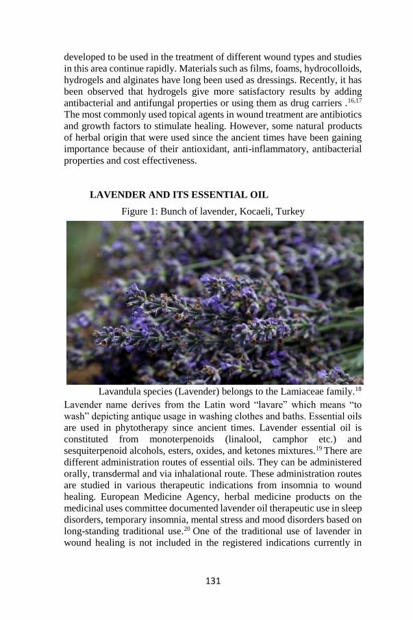
131
developed to be used in the treatment of different wound types and studies
in this area continue rapidly. Materials such as films, foams, hydrocolloids,
hydrogels and alginates have long been used as dressings. Recently, it has
been observed that hydrogels give more satisfactory results by adding
antibacterial and antifungal properties or using them as drug carriers .16,17
The most commonly used topical agents in wound treatment are antibiotics
and growth factors to stimulate healing. However, some natural products
of herbal origin that were used since the ancient times have been gaining
importance because of their antioxidant, anti-inflammatory, antibacterial
properties and cost effectiveness.
LAVENDER AND ITS ESSENTIAL OIL
Figure 1: Bunch of lavender, Kocaeli, Turkey
Lavandula species (Lavender) belongs to the Lamiaceae family.18
Lavender name derives from the Latin word “lavare” which means “to
wash” depicting antique usage in washing clothes and baths. Essential oils
are used in phytotherapy since ancient times. Lavender essential oil is
constituted from monoterpenoids (linalool, camphor etc.) and
sesquiterpenoid alcohols, esters, oxides, and ketones mixtures.19 There are
different administration routes of essential oils. They can be administered
orally, transdermal and via inhalational route. These administration routes
are studied in various therapeutic indications from insomnia to wound
healing. European Medicine Agency, herbal medicine products on the
medicinal uses committee documented lavender oil therapeutic use in sleep
disorders, temporary insomnia, mental stress and mood disorders based on
long-standing traditional use.20 One of the traditional use of lavender in
wound healing is not included in the registered indications currently in

132
EMA. Wound healing and related factors were studied in many in-vitro,
in-vivo and clinical studies.
IN-VITRO STUDIES
Lavender essential oil is studied for antifungal properties and
resulted with antifungal efficacy in Candida, Cryptococcus neoformans,
dermatophyte and Aspergillus strains.21 A different species of lavender is
shown to have anti-leishmanial and antibacterial activities.22 Bouyahya et
al. not only showed these activities they also showed antioxidant activity
with the essential oil. A constituent in lavender oil; Terpinen-4-ol, is found
to inhibit in vitro growth of human melanoma cell line.23 In many in-vitro
cancer studies anti-proliferative effect is shown.24 In vitro scratch assay or
wound healing assay with a prostate cancer cell line, in parallel resulted
reduced healing with lavender essential oil.24 Perillyl alcohol, a
monoterpene constituent in the lavender essential oil is showed to have
antiangiogenic effect via VEGF.25
Abe et al. evaluated the anti-inflammatory effects in neutrophil
adhesion method with various essential oils including lavender oil and
reported that the main component in the lavender EO linalool has
antiinflammatory effect in TNF-α -induced neutrophil adhesion in-vitro.26
Even the concentration is higher than the expected concentration of
transdermal exposed dose of lavender EO, they supported in-vivo
interpretation cannot exclude the potential anti-inflammatory role.
IN-VIVO AND CLINICAL STUDIES
Several clinical studies have been conducted investigating the
effects of lavender oil in wound healing.27 It is well known to have
antioxidant, antibacterial, antifungal and analgesic effects. It is also known to accelerate wound contraction.
Hartman and Coetze investigated the effect of lavender and
chamomile essential oils in their study on 8 patients with chronic wounds.
Complete closure was achieved in 4 of the five patients who were applied
the essential oils, and recovery was not possible in 3 patients in the control
group until the end of the study. Although it is not possible to make a
statistical comparison in this small sample group, it would not be wrong to
say that the essential oils used were effective.28
Kerr compared the effects of various essential oil blends created in
aloe vera gel base on simple wounds and concluded that the most effective
combination was lavender, German chamomile, myrrh and tea tree
essential oils in aloe vera gel. He reported that this preparation reduced
odor, prevented redness and inflammation, and shortened the healing
time.29
133
Altaei evaluated the effect of lavender essential oil on oral mucosa
ulcers in a 4-step study conducted on both animal and human subjects.
Aphthous ulcers treated with lavender essential oil healed faster in both
rabbits and humans. In the safety / toxicity study conducted on mice, the
LD value for the mice was found to be 6.5 gm / kg and no evidence of
dermal irritation was found. In the antibacterial activity study, which is
another pillar of the study, effectiveness has been demonstrated against all
bacterial strains studied.30
The effect of lavender essential oil on wound healing has been
evaluated in many studies in the gynecological patient group. In these
studies, not only the anti-inflammatory effect of lavender oil but also its
analgesic effect were emphasized. Although Vakilian et al. reported that
lavender oil applied to episiotomy incisions reduced erythema but had no
significant effect on pain,31 the following studies by Sheikhan et al. and
Marzouk et al. have shown that it is associated with less pain, as well as
reducing erythema, edema, ecchymosis and discharge.32,33
Koca Kutlu et al. showed that lavender essential oil significantly
increased EGF and FGF expression compared to the control group in their
study comparing the efficacy of several wound treatment methods. EGF is
an agent thought to play a role in wound contraction and epithelization. It
has become a popularized treatment modality recently, although its topical
use is quite expensive. The results of this study suggested that lavender oil
could be a cost-effective alternative to EGF application. The authors also
reported that no edema, discharge and local infection findings were
observed in the lavender oil applied group.34
Ben Djemaa et al. concluded that in animals treated with lavender
oil, wounds were closed faster, covered with a thinner epidermal layer
similar to normal skin, and showed a better epidermal and dermal structure
on histopathological examination in their experimental study conducted on
rats.35
In their 2016 study, Mori et al. showed that topical application of
lavender essential oil reduced the wound area earlier, increased the amount
of TGF- β and increased the amount of both type I and type III collagen on
the excisional wound model in the rat. TGF-β plays a role in wound
contraction by inducing fibroblast proliferation and its transformation into
myofibroblasts. Type III collagen is the first type of collagen produced
during the proliferation phase of wound healing and then matures and is
replaced by Type I collagen. In the same study, the fact that the amount of
type III collagen was close to the control group, but the amount of type I
collagen was higher in the group in which lavender oil was applied on the
7th day measurement, was interpreted as collagen maturation occurred
faster.36

134
SAFETY DATA
Safety information is important for herbal medicinal products. During pregnancy and lactation safety data has not been established for
lavender essential oil. In the absence of the sufficient data, rationally the
use during pregnancy and lactation is not recommended. EMA reminds its
potential on the impairment of the ability to drive and use machines.
Children below 12 years-age are excluded from the eligible data. In a
multicenter clinical study linalool and its hydroperoxide fraction were
found to be common contact allergens in dermatitis patients.37 Moreover
prepubertal gynecomastia is associated with lavender oil topical exposure
in three cases and in vitro cell line studies indicated lavender oil had
estrogenic and antiandrogenic activities.38
CONCLUSION
Lavender oil is a traditional medicinal product that has been used for
many years in wound healing. With recent experimental and clinical
studies, its antioxidant, antimicrobial, anti-inflammatory and analgesic
effects have been revealed and details of its mechanism of action have been
brought to light. In the future, with the help of more detailed and extensive
studies it may be possible to develop standardized treatment protocols and
gather more safety data for wound healing.

135
REFERENCES
1. Guest JF, Ayoub N, McIlwraith T, Uchegbu I, Gerrish A, Weidlich D,
et al. Health economic burden that wounds impose on the National
Health Service in the UK. BMJ Open 2015;5:e009283.
2. Werner S, Grose R. Regulation of wound healing by growth factors and
cytokines. Physiol Rev 2003 Jul;83(3):835-70.
3. Broughton G, Janis JE, Attinger CE. The basic science of wound
healing. Plast Reconstr Surg 2006;117:12S–34S.
4. Huntington JA. Molecular recognition mechanisms of thrombin. J
Thromb Haemost 2005 Aug;3(8):1861-72.
5. Midwood KS, Williams LV, Schwarzbauer JE. Tissue repair and the
dynamics of the extracellular matrix. Int J Biochem Cell Biol. 2004
Jun;36(6):1031-7.
6. Brinkmann V, Reichard U, Goosmann C, Fauler B, Uhlemann Y, Weiss
DS, et al. Neutrophil extracellular traps kill bacteria. Science 2004
Mar 5;303(5663):1532-5.
7. Martin P, Leibovich SJ. Inflammatory cells during wound repair: the
good, the bad and the ugly. Trends Cell Biol 2005;15:599–607.
8. Das A, Sinha M, Datta S, et al. Monocyte and Macrophage Plasticity in
Tissue Repair and Regeneration. Am J Pathol 2015;185:2596– 2606.
9. Martinez FO, Helming L, Gordon S. Alternative activation of
macrophages: an immunologic functional perspective. Annu Rev
Immunol 2009;27:451–483.
10. Shiota N, Nishikori Y, Kakizoe E, et al. Pathophysiological role of skin
mast cells in wound healing after scald injury: study with mast cell-
deficient W/W(V) mice. Int Arch Allergy Immunol 2010;151:80–88.
11. Paquet-Fifield S, Schluter H, Li A, et al. A role for pericytes as
microenvironmental regulators of human skin tissue regeneration. J
Clin Invest 2009;119:2795–2806.
12. Sen CK, Roy S, Gordillo G. Wound healing. In Neligan PC Ed. Plastic
Surgery Principles. Elsevier, Canada. 2018; 165-193.
13. Banerjee J, Chan YC, Sen CK. MicroRNAs in skin and wound healing.
Physiol Genomics 2011;43:543–556.
14. Roy S, Elgharably H, Sinha M, et al. Mixed-species biofilm
compromises wound healing by disrupting epidermal barrier function.
J Pathol 2014;233:331–343.
15. Obagi Z, Damiani G, Grada A, Falanga V. Principles of Wound
Dressings: A Review. Surg Technol Int. 2019 Nov 10;35:50-57.
16. Jayakumar R, Prabaharan M, Sudheesh Kumar PT, Nair SV, Tamura H.
Biomaterials based on chitin and chitosan in wound dressing
application. Biotechnol Adv. 2011 May-Jun;29(3):322-337.
17. Hamedi H, Moradi S, Hudson SM, Tonelli AE. Chitosan based
hydrogels and their applications for drug delivery in wound dressings: A review. Carbohydr Polym. 2018 Nov 1;199:445-460. 18. Salehi B,

136
Mnayer D, Özçelik B, et al. Plants of the genus Lavandula: From farm
to pharmacy. Natural Product Communications, 2018. 13(10): p.
1934578X1801301037.
19. Woronuk G, Demissie Z, Rheault M, Mahmoud Z. Biosynthesis and
therapeutic properties of Lavandula essential oil constituents. Planta
medica, 2011. 77(01): p. 7-15.
20. European Medicines Agency (EMA).
https://www.ema.europa.eu/en/medicines/herbal/lavandulaeaetheroleum#
key-facts-section.
21. Zuzarte M, Gonçalves MJ, Cruz MT, et al. Lavandula luisieri essential
oil as a source of antifungal drugs. Food Chemistry, 2012. 135(3): p.
1505-1510.
22. Bouyahya A, Et-Touys A, Abrini J, et al. Lavandula stoechas essential
oil from Morocco as novel source of antileishmanial, antibacterial and
antioxidant activities. Biocatalysis and Agricultural Biotechnology,
2017. 12: p. 179-184.
23. Mondello F, De Bernardis F, Girolamo A, Cassone A, Salvatore G. In
vivo activity of terpinen-4-ol, the main bioactive component of
Melaleuca alternifolia Cheel (tea tree) oil against azole-susceptible
and-resistant human pathogenic Candida species. BMC infectious
diseases, 2006. 6(1): p. 158.
24. Zhao Y, Chen R, Wang Y, Qing C, Wang W, Yang y. In vitro and in
vivo efficacy studies of Lavender angustifolia essential oil and its
active constituents on the proliferation of human prostate cancer.
Integrative cancer therapies, 2017. 16(2): p. 215-226.
25. Loutrari H, Hatziapostolou M, Skouridou V, et al. Perillyl alcohol is an
angiogenesis inhibitor. Journal of Pharmacology and Experimental Therapeutics, 2004. 311(2): p. 568-575.
26. Abe S, Maruyama N, Hayama K, et al. Suppression of tumor necrosis
factor-alpha-induced neutrophil adherence responses by essential oils.
Mediators of Inflammation, 2003. 12.
27. Samuelson R, Lobl M, Higgins S, Clarey D, Wysong A. The Effects of
Lavender Essential Oil on Wound Healing: A Review of the Current
Evidence. J Altern Complement Med 2020;26(8):680690.
28. Hartman D, Coetzee JC. Two US practitioners’ experience of using
essential oils for wound care. J Wound Care 2002; 11:317–320.
29. Kerr J. The use of essential oils in healing wounds. Int J Aromather
2002;12:202–206.
30. Altaei DT. Topical lavender oil for the treatment of recurrent aphthous
ulceration. Am J Dent 2012;25910:39–43.
31. Vakilian K, Atarha M, Bekhradi R, Chaman R. Healing advantages of
lavender essential oil during episiotomy recovery: A clinical trial.
Complement Ther Clin Pract 2011; 17:50–53.

137
32. Sheikhan F, Jahdi F, Khoei EM, et al. Episiotomy pain relief: Use of
lavender oil essence in primiparous Iranian women. Complement
Ther Clin Pract 2012;18:66–70.
33. Marzouk T, Barakat R, Ragab A, et al. Lavender-thymol as a new
topical aromatherapy preparation for episiotomy: A randomised
clinical trial. J Obstet Gynaecol 2015;35:472–475.
34. Koca Kutlu A, Cecen D, Gurgen SG, et al. A comparison study of
growth factor expression following treatment with transcutaneous
electrical nerve stimulation, saline solution, povidone-iodine, and
lavender oil in wounds healing. Evid Based Complement Alternat
Med 2013;2013:361832.
35. Ben Djemaa FG, Bellassoued K, Zouari S, et al. Antioxidant and wound
healing activity of Lavandula aspic L. ointment. J Tissue Viability
2016;25:193–200.
36. Mori HM, Kawanami H, Kawahata H, Aoki M. Wound healing
potential of lavender oil by acceleration of granu-lation and wound
contraction through induction of TGFBeta in a rat model. BMC
Complement Altern Med 2016;16:144.
37. Matura M, Sköld M, Börje A, et al. Selected oxidized fragrance
terpenes are common contact allergens. Contact dermatitis, 2005.
52(6): p. 320-328.
38. Henley DV, Lipson N, Korach KS, Bloch CA. Prepubertal
gynecomastia linked to lavender and tea tree oils. New England
Journal of Medicine, 2007. 356(5): p. 479-485.

138