Deletion of retinoic acid receptor β (RARβ) impairs pancreatic endocrine differentiation

In Vivo Manganese Exposure Modulates Erk, Akt andDarpp-32 in the Striatum of Developing Rats, andImpairs Their Motor FunctionFabiano M. Cordova1,4, Aderbal S. Aguiar, Jr.2, Tanara V. Peres1, Mark W. Lopes1, Filipe M. Goncalves1,
Aline P. Remor1, Samantha C. Lopes2, Celso Pilati3, Alexandra S. Latini1, Rui D. S. Prediger2,
Keith M. Erikson5, Michael Aschner6, Rodrigo B. Leal1*
1 Departamento de Bioquımica, Centro de Ciencias Biologicas, Universidade Federal de Santa Catarina, Florianopolis, Brazil, 2 Departamento de Farmacologia,
Universidade Federal de Santa Catarina, Florianopolis, Brazil, 3 Centro de Ciencias Agroveterinarias, Universidade do Estado de Santa Catarina, Lages, Brazil, 4 Centro de
Ciencia Animal, Universidade Federal do Tocantins, Araguaına, Brazil, 5 Department of Nutrition, University of North Carolina, Greensboro, North Carolina, United States of
America, 6 Department of Pediatrics, Vanderbilt University Medical Center, Nashville, Tennessee, United States of America
Abstract
Manganese (Mn) is an essential metal for development and metabolism. However, exposures to high Mn levels may betoxic, especially to the central nervous system (CNS). Neurotoxicity is commonly due to occupational or environmentalexposures leading to Mn accumulation in the basal ganglia and a Parkinsonian-like disorder. Younger individuals are moresusceptible to Mn toxicity. Moreover, early exposure may represent a risk factor for the development of neurodegenerativediseases later in life. The present study was undertaken to investigate the developmental neurotoxicity in an in vivo modelof immature rats exposed to Mn (5, 10 and 20 mg/kg; i.p.) from postnatal day 8 (PN8) to PN12. Neurochemical analysis wascarried out on PN14. We focused on striatal alterations in intracellular signaling pathways, oxidative stress and cell death.Moreover, motor alterations as a result of early Mn exposure (PN8-12) were evaluated later in life at 3-, 4- and 5-weeks-of-age. Mn altered in a dose-dependent manner the activity of key cell signaling elements. Specifically, Mn increased thephosphorylation of DARPP-32-Thr-34, ERK1/2 and AKT. Additionally, Mn increased reactive oxygen species (ROS) productionand caspase activity, and altered mitochondrial respiratory chain complexes I and II activities. Mn (10 and 20 mg/kg) alsoimpaired motor coordination in the 3rd, 4th and 5th week of life. TroloxTM, an antioxidant, reversed several of the Mn alteredparameters, including the increased ROS production and ERK1/2 phosphorylation. However, TroloxTM failed to reverse theMn (20 mg/kg)-induced increase in AKT phosphorylation and motor deficits. Additionally, Mn (20 mg/kg) decreased thedistance, speed and grooming frequency in an open field test; TroloxTM blocked only the decrease of grooming frequency.Taken together, these results establish that short-term exposure to Mn during a specific developmental window (PN8-12)induces metabolic and neurochemical alterations in the striatum that may modulate later-life behavioral changes.Furthermore, some of the molecular and behavioral events, which are perturbed by early Mn exposure are not directlyrelated to the production of oxidative stress.
Citation: Cordova FM, Aguiar AS Jr, Peres TV, Lopes MW, Goncalves FM, et al. (2012) In Vivo Manganese Exposure Modulates Erk, Akt and Darpp-32 in theStriatum of Developing Rats, and Impairs Their Motor Function. PLoS ONE 7(3): e33057. doi:10.1371/journal.pone.0033057
Editor: Demetrios Vavvas, Massachusetts Eye & Ear Infirmary - Harvard Medical School, United States of America
Received October 29, 2011; Accepted February 6, 2012; Published March 13, 2012
Copyright: � 2012 Cordova et al. This is an open-access article distributed under the terms of the Creative Commons Attribution License, which permitsunrestricted use, distribution, and reproduction in any medium, provided the original author and source are credited.
Funding: This work was supported by the National Council for Scientific and Technological Development (CNPq) Brazil (#305194/2010-0); Coordination for theTraining and Improvement of Higher Education Personnel (CAPES)/PROCAD (#167/2007); Studies and Projects Financing Agency (FINEP) IBN 01.06.0842-00 andINCT-National Institute of Science and Technology for Excitotoxicity and Neuroprotection; Santa Catarina Research Fundation (FAPESC); National Institute ofEnvironmental Health Sciences (NIEHS) R01 ES10563 (MA). RBL, ASL and RDSP are recipients of CNPq fellowships. The funders had no role in study design, datacollection and analysis, decision to publish, or preparation of the manuscript.
Competing Interests: The authors have declared that no competing interests exist.
* E-mail: [email protected]
Introduction
Manganese (Mn) participates in several biological processes,
with important roles in regulating metabolism [1]. In the central
nervous system (CNS), Mn is an important co-factor for several
enzymes, including superoxide dismutase (SOD) and glutamine
synthetase (GS) [2]. However, excessive exposure to Mn is
neurotoxic, resulting in a neurodegenerative disease affecting
cortical structures and basal ganglia, specifically the globus
pallidus, striatum and substantia nigra pars reticulata [3,4].
Cytotoxicity associated with excessive Mn exposure leads to
neurological dysfunction associated with dystonic movements,
analogous to those commonly noted in idiopathic Parkinson’s
disease (PD) [5]. Notably, much less is known about the effects of
Mn on the developing CNS, in particular potential risks associated
with early Mn exposure and predisposition to later-life onset
neurological injury [6–9]. Newborns retain greater Mn levels than
adults [7] and the developing brain is more susceptible to injury
caused by toxic agents [10–12], reflecting immature and inefficient
homeostasis, low physiological Fe levels and a permeable blood-
brain barrier [5,6,13,14]. The CNS continues to develop
postnatally, and its vulnerability remains high for an extended
period of time, from childhood to adolescence. Although many
neurons are formed at birth, a substantial acceleration in RNA,
PLoS ONE | www.plosone.org 1 March 2012 | Volume 7 | Issue 3 | e33057
DNA and protein synthesis, neuronal migration, glial cells growth
(particularly astroglia, the main site for glutamate and metal
uptake) and axonal myelination persist for several months into the
postnatal period [10,15,16].
Mn exposure is commonly associated with occupational and
industrial processes [4,5,7,17]. High Mn concentrations are found
in the environment due to its abundance in the earth’s crust and
secondary to its use in water treatment, manufacturing of dry
batteries, as well as addition to gasoline (as an antiknock agent;
methylcyclopentadienyl manganese tricarbonyl, MMT) and fun-
gicides [3,6,18,19]. Infantile Mn exposure is also associated with
parenteral nutrition, which is commonly supplemented with
excessive Mn levels [14,20,21].
Mn accumulation in the CNS is regulated by several transport
mechanisms, including divalent metal transporter 1 (DMT-1) and
transferrin/transferrin receptor system (Tf/TfR). However, it has
yet to be ascertained which mechanisms regulate Mn transport
during the developmental period [6]. Notably, both DMT-1 and
TfR are expressed in rat cortex, hippocampus and striatum on the
fifth postnatal day [13,22]. Additionally, in the immature brain
Mn may be transported by other mechanisms, such as ZIP-8
solute carrier, calcium channels, ionotropic glutamate receptors
and the dopamine transporter, as previously shown in the adult
brain [19,23–27].
Mechanisms involved in Mn neurotoxicity are poorly under-
stood [5–7,28]. Oxidative stress is considered to play a major role,
secondary to enhanced levels of redox-active Mn ions [29]. Mn
toxicity is mediated by mitochondrial perturbations, initiating both
apoptotic and necrotic cell death through the formation of reactive
oxygen species (ROS) and oxidative stress [30–34]. Additionally,
Mn induces dopamine autoxidation, increasing the levels of toxic
quinines [35]. Higher sensitivity of the striatum to Mn-induced
oxidative stress [36], especially during development, has been
noted [6]. Oxidative stress in the striatum is associated with
impairment of motor activity [37]. It has also been shown that
Mn-dependent increased ROS formation can interfere with the
removal of glutamate from the synaptic cleft [6,38], resulting in
excitotoxicity [6,39].
In addition, many signaling pathways associated with pro-
grammed cell death are activated after in vitro Mn treatments,
including JNKs, ERK1/2, p38MAPK, PKC and caspases [40–52].
However, the modulation of signaling pathways by Mn has yet to
be shown in a systematic manner in the in vivo developing CNS.
The protein kinases ERK1/2, JNK1/2/3 and p38MAPK are the
foremost enzymes studied in the MAPK family [53–56]. The
ERK1/2 cascade is primarily activated by growth factors,
regulating gene expression, embryogenesis, proliferation, cell
death/survival and neuroplasticity [54,55]. The JNK1/2/3 and
p38MAPK protein kinases, classically recognized as stress-activated
protein kinases (SAPKs), are activated by cytokines and cytotoxic
insults, and are often related to stress and cell death [57,58].
However, JNK and p38MAPK also regulate CNS development and
neuroplasticity [56,59]. Another important intracellular signaling
pathway is PI3K/AKT (PKB) which can be activated by several
growth factors. It plays a central role in cell growth regulation,
proliferation, metabolism and cell survival, as well as neuroplas-
ticity [60,61].
The basal ganglia receive inputs mainly through the striatum
and coordinate vital behaviors, including movement, reward and
motivational processes [62]. Corticolimbic-thalamic glutamatergic
and mesencephalic dopaminergic systems converge on the gamma
aminobutyric acid (GABAergic) medium-sized spiny neurons of
the striatum [63]. These neurons characteristically express a
dopamine- and cAMP-regulated phosphoprotein of 32 kDa
(DARPP-32) [64,65]. DARPP-32 function depends on its relative
state of phosphorylation in two main regulatory sites, Thr-34 and
Thr-75. When DARPP-32 is phosphorylated at Thr-34 mainly by
protein kinase A (PKA) it becomes a potent inhibitor of protein
phosphatase 1 (PP1), which in turn regulates the phosphorylation
state of several classes of proteins, including transcription factors,
ionotropic glutamate receptors and ion channels [66,67]. PKA
also phosphorylates and activates protein phosphatase 2A (PP2A),
which dephosphorylates DARPP-32 at Thr-75 [68,69]. Converse-
ly, when phosphorylated at Thr-75 by Cdk5, DARPP-32 becomes
an inhibitor of PKA activity, thereby relieving inhibition of PP1
[67,70]. Therefore, regulation of the state of DARPP-32
phosphorylation provides a mechanism for integrating information
at striatal medium-sized spiny neurons via a variety of neuro-
transmitters and it may play important regulatory functions in
motor behavior [65,71–74]. Given that the striatum is an
important target of Mn toxicity [5,6] and that Mn may disrupt
dopaminergic and glutamatergic transmission [35,75], we hypoth-
esized that Mn mediates its neurotoxic effects, at least in part, via
disruption of DARPP-32 regulation. Notably, changes on
DARPP-32 phosphorylation by Mn have yet to be described
either in vitro or in vivo.
The biological consequences of developmental Mn exposure
may be particularly harmful, affecting neurogenesis, learning and
memory, with predisposition to late onset neurodegenerative
disorders. Exposure to a toxic stimulus may result in ‘‘imprinting’’,
a process by which early environmental factors may permanently
alter an organism’s gene expression profile [11].
Taken together, the present study was designed to assess
alterations in cell signaling pathways, production of oxidative stress
and later-life behavioral deficits as a consequence of a short-term
postnatal Mn exposure (PN8-12). For the first time, we establish
Mn-induced disruption in regulatory mechanisms of ERK1/2,
AKT and DARPP-32 in the developing rat striatum. Moreover,
we show an increase in oxidative stress and impairment in motor
activity in immature rats exposed to this metal.
Methods
Ethics StatementWistar rats of both genders were obtained from the Uni-
versidade Federal de Santa Catarina breeding colony. The
animals were maintained in an air-conditioned room (23 6 1uC)
on a 12 h light/dark cycle with water and food available ad
libitum. They were treated, manipulated and euthanized accord-
ing to the ‘‘Principles of Laboratory Animal Care’’ (NIH
publication no. 80–23, revised 1996) and approved by the
Committee on the Ethics of Animal Experiments of the Federal
University of Santa Catarina (CEUA/UFSC; www.ceua.ufsc.br;
Permit Number: PP00345). All efforts were made to minimize the
number of animals used and animal suffering.
ReagentsPrimary antibodies anti-ERK1/2, anti-p38MAPK, anti-JNK1/2
and manganese chloride were obtained from Sigma (St. Louis,
MO, USA). Anti-phospho-CREB, anti-CREB, anti-phospho-
ERK1/2, anti-phospho-p38MAPK, anti-phospho-JNK1/2, anti-
AKT, anti-phospho-AKT, anti-DARPP-32, anti-phospho-
DARPP-32-Thr-34 and anti-phospho-DARPP-32-Thr-75 anti-
bodies and LumiGlo detection reagent were purchased from Cell
Signaling (Beverly, MA, USA). Anti-b-actin was purchased from
Santa Cruz Biotechnology (Santa Cruz, CA, USA). Secondary
antibody (anti-rabbit and anti-mouse IgG-horse radish peroxidase
(HRP)-conjugated was purchased from Millipore (Temecula, CA,
Manganese Exposure of Developing Rats
PLoS ONE | www.plosone.org 2 March 2012 | Volume 7 | Issue 3 | e33057

USA). Acrylamide, bis-acrylamide, b-mercaptoethanol, Hy-
bondTM nitrocellulose, Amersham HyperfilmTM ECL, sodium
dodecyl sulfate (SDS) and Tris were obtained from GE Healthcare
Life Sciences (Piscataway, NJ, USA). 6-hydroxy-2,5,7,8-
tetramethylchroman-2-carboxylic acid (TroloxTM) were obtained
from Calbiochem (La Jolla, CA, USA). The N-acetyl-Asp-Glu-
Val-Asp-7-amino-4-methylcoumarin (DEVD-AMC) was obtained
from Biomol (Plymouth Meeting, PA, USA). 29,79-dichlorofluor-
escein diacetate (DCFH2-DA) was purchased from Invitrogen
(Carlsbad, CA, USA). All other reagents were of the highest
analytical grade.
Animals and treatmentsLitters were obtained from random crosses between several
breeding males and females derived from the UFSC breeding
colony. At birth, the number of pups was randomly culled to eight
pups per litter. The treatments began when the pups were eight
days old (PN8). The litters were randomly assigned to each
experiment and the treatments were carried out at the same time
every day (2:00 PM). The animals were individually identified for
each treatment.
For the Mn exposure protocol, four to twelve groups (each
group containing eight pups from a different litter) were used for
the analysis. We performed four treatments (control and Mn 5, 10
and 20 mg/kg) with two animals in each of the groups.
Accordingly, each experimental group was composed of eight
pups treated in duplicate. The pups were treated for 5 consecutive
days (8th to 12th postnatal day; PN8-12) with one daily
intraperitoneal (i.p.) injection of saline (NaCl, 0.9%; control) or
MnCl2 (5, 10 and 20 mg/kg, diluted in saline), as described
previously [76,77]. Rats were euthanized by decapitation on the
14th postnatal day (PN14) [76,77] and the structures of interest
were dissected out for neurochemical analyses. Briefly, the brain
was carefully removed, the cerebral hemispheres were divided and
the hippocampi were isolated (using two fine brushes). This
procedure was followed by displacement of the cortex and
striatum. The striatum was carefully transected and it was
separated from the nearby structures with a sharp spatula. In
addition, to verify the involvement of oxidative stress in Mn-
induced neurotoxicity, pups were treated with the antioxidant
6-hydroxy-2,5,7,8-tetramethylchroman-2-carboxylic acid (Tro-
loxTM) concomitantly with Mn. The same protocol as previously
described was carried out with two pups in each treatment group:
saline (0,9% NaCl; control), TroloxTM 1 mg/kg, MnCl2 20 mg/
kg, MnCl2 20 mg/kg plus TroloxTM 1 mg/kg (administered
10 min before Mn). All treatments were carried out by i.p.
injection. The pups’ body weights were measured daily from
PN8 to PN14 and the weight-gain (g) are reported as mean 6
S.E.M.
Brain metal analysesTissue Mn and Fe concentrations were measured by atomic
absorption spectroscopy (Varian AA240TM, Varian Inc., Palo
Alto, CA, USA) [78]. Striatum, hippocampus and cerebral cortex
were digested for 48–72 h in a sand bath (60uC) in ultrapure nitric
acid (1:10 wt/vol dilution). One hundred ml of digested tissue was
brought to 1 ml of total volume with 2% nitric acid and analyzed
for Mn and Fe. The mixture was then centrifuged and the
supernatant was used for analysis (100 ml aliquot brought up to a
1 ml volume with 2% nitric acid). Bovine liver (10 mg Mn/l) was
digested in ultrapure nitric acid and used as an internal standard.
The data are expressed as mg metal/g tissue and the values are
reported as mean 6 S.E.M.
Western blottingStriatum was dissected, mechanically homogenized in 500 ml of
sample buffer (200 mM Tris, 40 mM EDTA, 4% SDS, pH 6,8)
and immediately boiled for 5 min. Sample dilution solution (1:4
vol/vol; 40% glycerol, 50 mM Tris and minimal bromophenol
blue) and b-mercaptoethanol were added to each sample for a
final concentration of 5%. Protein content was estimated at
750 nm wavelength and the concentration calculated using a
standard curve with bovine serum albumin [79]. The samples
(60 mg of total protein/track) were separated by SDS-PAGE
(miniVE Vertical Electrophoresis SystemTM, GE Healthcare Life
Sciences, Piscataway, NJ, USA) using 10% gels [77]. The proteins
were transferred to nitrocellulose membranes using a semidry
blotting apparatus (TE 70 SemiPhorTM Unit, GE Healthcare Life
Sciences, Piscataway, NJ, USA) (1.2 mA/cm2; 1.5 h) as previously
described [80]. The membranes were blocked (1 h) with 5% skim
milk in TBS (10 mM Tris, 150 mM NaCl, pH 7.5). ERK1/2,
JNK1/2, p38MAPK, AKT, CREB and DARPP-32 total and
phosphorylated forms, were detected using specific antibodies
incubated overnight diluted in TBS-T (10 mM Tris, 150 mM
NaCl, 0,1% Tween-20, pH 7.5) containing 2.5% BSA in the
dilutions 1:1,000 (anti-phospho-CREB, anti-CREB, anti-AKT,
anti-phospho-AKT-Ser-473, anti-DARPP-32, anti-phospho-
DARPP-32-Thr-34, anti-phospho-DARPP-32-Thr-75, anti-phos-
pho-JNK1/2 and anti-phospho-p38MAPK), 1:2,000 (anti-phospho-
ERK1/2 and anti-b-actin), 1:10,000 (anti-JNK1/2 and anti-
p38MAPK) or 1:40,000 (anti-ERK1/2). Next, the membranes were
incubated with anti-rabbit peroxidase-linked secondary antibody
(1:4,000) for 1 h and the reactions developed by chemilumines-
cence (LumiGLOH, Cell Signaling, Beverly, MA, USA). All
blocking and incubation steps were followed by three washes
(5 min) of the membranes with TBS-T. The optical density (O.D.)
of the bands was quantified using Scion ImageTM (Frederick, MD,
USA). The phosphorylation level of each protein was determined
as a ratio of the O.D of the phosphorylated band over the O.D.
of the total band, and the data expressed as percentage of the
control (considered as 100%). The values are presented as mean
6 S.E.M [81].
Histological analysisTo evaluate possible morphological changes in response to Mn
treatment, rat brains were removed, fixed and processed for
histological analysis by hematoxylin and eosin staining [82,83].
Briefly, brains were removed immediately after decapitation and
fixed in 4% paraformaldehyde solution for one week. After
fixation, the tissues were gradually dehydrated in ethanol, cleared
in xylene and embedded in paraffin. Five mm sections stained with
hematoxylin and eosin. All brain areas were analyzed, and the
striatum, hippocampus and cerebral cortex were imaged at 640
magnification.
Mitochondrial respiratory chain complexes activityMitochondrial fractions were prepared from the striatum [84].
The structure was homogenized in 10 volumes of 5 mM
potassium phosphate buffer, pH 7.4, containing 0.3 M sucrose,
5 mM MOPS, 1 mM EGTA and 0.1% bovine serum albumin.
The homogenate was centrifuged at 1,500 g for 10 min at 4uC,
and the pellet was discarded. The supernatant was then
centrifuged for additional 10 min at 4uC at 15,000 g to isolate
the mitochondria present in the pellet, which was suspended in the
same buffer. Disrupted mitochondrial fractions were obtained by
freezing/thawing the samples three times. The NADH dehydro-
genase (complex I), succinate dehydrogenase (complex II) and
cytochrome c oxidase (complex IV) activities were analyzed [85]
Manganese Exposure of Developing Rats
PLoS ONE | www.plosone.org 3 March 2012 | Volume 7 | Issue 3 | e33057
with the plate reader InfiniteTM M200 (TECAN, Mannedorf,
Switzerland). The activities of the respiratory chain complexes are
calculated as nmol min21/mg protein21 or mmol min21/mg
protein21 6 S.E.M.
Quantitation of ROS and F2-IsoPsThe production of reactive species was evaluated using the
DCFH2-DA probe which is oxidized in the presence of reactive
species to the DCF impermeable fluorescent compound, emitting
green fluorescence after excitation at 480 nm [86]. Striata were
rapidly removed and homogenized (1:5 w/v) in a buffer containing
0.1% triton X-100, 0.12 M NaCl, 30 mM sodium phosphate,
pH 7.4. Structures were homogenized using a tissue homogenizer,
at 4uC followed by centrifugation at 10,000 g for 10 min. The
supernatants (0.01 mg protein) were incubated with 40 nM
DCFH-DA for 30 min at 37uC. The DCF fluorescence signal
was measured using a Perkin-Elmer LS55 (Boston, MA, USA)
spectrofluorometer using wavelengths of excitation/emission of
480/520 nm, respectively. An analytical curve was performed
using a standard DCF solution to analyze the results, which were
subsequently normalized as a percentage of the untreated control
(100%).
Because the excessive production of ROS may induce
membrane polyunsaturated fatty acid oxidation, resulting in lipid
peroxidation products, such as F2-isoprostanes (F2-IsoPs), we
evaluated the production of this substance in the striatum of
treated animals. The F2-IsoPs are considered biomarkers of
oxidative stress in both in vitro and in vivo models [34]. Total F2-
IsoPs were determined with a stable isotope dilution method with
detection by gas chromatography/mass spectrometry and selective
ion monitoring as previously described [87,88]. Total F2-IsoPs
were measured in the striatum dissected from the animals exposed
in vivo to different doses of Mn and stored at 280uC until analysis.
Briefly, the striatum were homogenized in Folch solution and
lipids extracted from chloroform layer by evaporation [89] and
then subjected to chemical saponification using 15% KOH to
hydrolyze bound F2-IsoPs. The homogenates were adjusted to a
pH of 3, followed by the addition of 0.1 ng of 15-F2a-IsoP-d4
internal standard. F2-IsoPs were subsequently purified by C18 and
silica Sep-Pak extraction and by thin layer chromatography. They
were then analyzed by pentafluorobenzyl ester, a trimethylsilyl
ether derivative, via gas chromatography, negative ion chemical
ionization-mass spectrometry.
Caspase activityCaspase activity was monitored fluorometrically by the
production of fluorescent AMC from DEVD-AMC fluorogenic
substrate for caspase-3 and related cysteine proteases. Striatum
homogenates were prepared (1:5, w/v) in a buffer containing
10 mM HEPES pH 7.4, 42 mM KCl, 5 mM MgCl2, 1 mM
phenylmethylsulfonylfluoride (PMSF), 0.1 mM EDTA, 0.1 mM
EGTA, 1 mg/ml, pepstatin A, 1 mg/ml leupeptin, 5 mg/ml
aprotinin, 0.5% 3-[(3-cholamidopropyl)-dimethyl-ammonio]-1-
propanesulfonate (CHAPS), and 1 mM dithiothreitol (DTT) at
4uC. The reaction was carried out by mixing this homogenate
(0.1 mg protein) to the reaction buffer containing 25 mM HEPES
pH 7.4, 0.1% CHAPS, 1 mM EDTA, 10% sucrose and 3 mM
DTT and was started by addition of 10 mM caspase-3 fluorogenic
substrate DEVD-AMC. Cleavage of the fluorogenic substrate was
detected spectrofluorometrically (Perkin Elmer LS55, Boston, MA,
USA) after 2 h of incubation at 37uC, using excitation and
emission wavelengths of 380 and 460 nm, respectively [90].
Fluorescence of blanks containing no fluorogenic substrate was
subtracted from the values. Protein content was determined by the
Lowry method [91] and caspase activity is expressed as percent of
control (100%) 6 S.E.M.
Behavioral testsThe animals were kept until PN37, providing a suitable time-
point for the assessment of behavioral effects of early life
Mn exposure. All animals were tested in the rotarod at 22, 29
and 36 days of age (3, 4 and 5 weeks of age), and in an open field
on PN37. Animals were habituated to the experiment room for
1 h prior to the initiation of the behavior tests. Behavioral tests
were carried out during the light phase of the cycle (10:00–
17:00 h).
Rotarod analysesThe Rota-Rod system (Insight Equipamentos Cientıficos,
Ribeirao Preto, Brazil) for locomotor assessment measures the
time an animal maintains balance on a moving cylinder [92]. The
following general conditioning and testing procedures were
employed to select animals for use in the different treatment and
control groups: namely, animals were first conditioned on a
stationary rod for 30 seconds and during this time any animal that
fell was placed back on the rotarod. Next, the animals were
conditioned at a constant speed of 5 rpm for a period of
90 seconds. Animals that failed the first conditioning were allowed
two additional conditioning periods, and those that failed the third
conditioning period were not selected for further testing. This
assured that all the animals in all the treatment groups attained an
analogous a baseline (data not shown).
The same basic conditioning methodology was employed in
testing treatment and control groups. Thirty minutes after the last
conditioning, each animal was placed on the rotarod and its
latency for falling determined. The starting speed was 5 rpm and it
was increased by 0.1 revolutions per second.
Open field analysesTo evaluate Mn-induced motor changes [92], the animals were
tested in the circular open field (50 cm height 660 cm diameter)
made from acrylic (Insight Equipamentos Cientıficos, Ribeirao
Preto, Brazil) and placed in a room with a video camera mounted
on the ceiling. Each experiment lasting 10 min was recorded and
the distance, average speed, number of rearing and grooming were
evaluated with behavioral analysis software ANY-mazeTM (Stoelt-
ing, Wood Dale, IL, USA).
Statistical analysesData are expressed as mean 6 S.E.M and statistical significance
was assessed by one-way analysis of variance (ANOVA) followed
by Duncan’s or Newman-Keuls post-hoc test where appropriate.
Kruskal-Wallis test followed by Dunn’s post-hoc was applied to
rotarod analysis. Analyses were performed with STATISTICATM
5.1 998 Edition (StatSoft, Tulsa, OK, USA). A value of p #0.05
was considered to be significant.
Results
Mn exposure modelTo evaluate if in vivo Mn treatments impaired development, we
analyzed the rats’ weight-gain from PN8-14. As shown in Table 1,
there were no statistically significant differences between the
groups, except for animals exposed to 20 mg/kg, which showed a
significant decrease in weight-gain compared to controls (p,0.05;
Table 1).
To characterize metal accumulation in the CNS, the striatum,
hippocampus and cerebral cortex were analyzed. Mn-treated rats
Manganese Exposure of Developing Rats
PLoS ONE | www.plosone.org 4 March 2012 | Volume 7 | Issue 3 | e33057

accumulated significantly higher Mn concentrations vs. controls
(p,0.001) in the striatum, hippocampus and cerebral cortex
(Fig. 1A). Iron (Fe) levels were also determined in the same brain
regions, since disturbances in transition metals may occur
secondary to Mn exposure [9]. The results showed a significant
increase (p,0.01) in Fe levels only in the striatum of animals
treated with 10 or 20 mg Mn/kg (Fig. 1B), and in the cerebral
cortex of animals treated with 20 mg Mn/kg (Fig. 1B).
To evaluate possible morphological changes in the brain (PN14)
of rats exposed in vivo to Mn (PN8-12), we performed histological
analysis by means of hematoxylin/eosin staining. As shown in
Fig. 2, Mn treatment did not lead to overt morphological changes
and cellular degeneration or death was absent. Additionally, no
morphological changes were observed in the liver, kidneys, spleen
or heart in Mn-treated rats (data not shown).
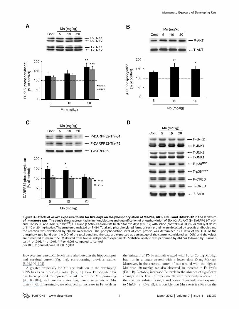
Effects of Mn on cell signalingPhosphorylation and expression of MAPKs (ERK1/2, p38MAPK
and JNK1/2/3), AKT, CREB and DARPP-32 were evaluated on
PN14 (Fig. 3). In the sub-acute model of metal exposure (PN8-12),
Mn significantly increased the phosphorylation of ERK1 (71.17 6
20.34%, p,0.01, Fig. 3A) and ERK2 (58.20 6 16.64%, p,0.001,
Fig. 3A) at the highest dose of 20 mg/kg. Additionally, the
phosphorylation of AKT (Fig. 3B) significantly increased upon
treatment with10 or 20 mg Mn/kg (58.79 6 20.67%, p,0.01 and
46.61 6 17.23%, p,0.05, respectively). DARPP-32-Thr-34
phosphorylation (Fig. 3C) was significantly increased in the lower
dose Mn treated groups (46.13 6 13.50% in 5 mg/kg, p,0.05
and 40.09 6 15.97% in 10 mg/kg, p,0.05). However, brain
DARPP-32-Thr-75 phosphorylation (Fig. 3C), as well as phos-
phorylation and expression of p38MAPK, JNK1/2/3 and CREB
(Ser-133) were unchanged on PN14 (Fig. 3D).
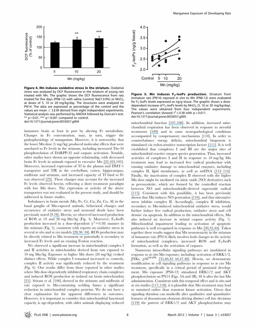
Oxidative stress and mitochondrial activityGiven the involvement of oxidative stress in Mn neurotoxicity,
next we measured ROS generation by DCF fluorescence and F2-
IsoP generation. As shown in Fig. 4, Mn significantly increased
ROS production in the striatum of developing rats treated with 10
or 20 mg/kg (37.21 6 12.37%, p,0.01 and 43.07 6 11.25%,
p,0.001, respectively). Moreover, a dose-dependent increase was
noted in striatal levels of F2-IsoPs in response to Mn treatment
(Pearson’s correlation r2 = 0.38 with p = 0.011) (Fig. 5).
Mitochondrial dysfunction in response to Mn treatment was
analyzed by measurements of the respiratory chain complexes I, II
and IV activities. Increased complex I activity both at 10 and
20 mg Mn/kg (Fig. 6A), as well as complex II at 10 mg Mn/kg
(Fig. 6B) was noted on PN14 (in pups treated with Mn on PN8-
PN12). However, at 20 mg Mn/kg the complex II activity
decreased (Fig. 6B). Complex IV activity was unchanged by Mn
(Fig. 6C).
Effects of Mn on caspase activitySince, an increase in the rate of apoptosis can deregulate tissue
plasticity, changing neurodevelopment and behavior later in life
we decided to evaluate a possible increase in apoptosis in the
striatum in response to Mn treatment, combined with analysis of
neurochemical parameters. The enzymatic activity of caspase-3/7
in the striatum of immature rats exposed to Mn was evaluated by
the DEVD cleavage fluorescence test. At the end of the five-day
exposure period (PN14), increased caspase activity was noted with
all the Mn doses (p,0.01, Fig. 7).
Table 1. Immature rats’ weight gain exposed to Mn in vivo.
Body weightPN8 (g)
Body weightPN14 (g)
Weightgain (g)
Control 12.68 6 0.49 22.51 6 0.79 9.83 6 0.46
5 mg/kg 13.14 6 0.59 23.22 6 0.71 10.09 6 0.41
10 mg/kg 13.09 6 0.54 22.39 6 0.66 9.30 6 0.45
20 mg/kg 12.76 6 0.42 20.96 6 0.67 8.21 6 0.46*
Immature rats were treated with saline (control; NaCl 0.9%) or MnCl2 at doses of5, 10 or 20 mg/kg for five days (PN8-12). Body weights were measuredthroughout the treatment, and the weight gain recorded on PN14. Resultsrepresent mean 6 S.E.M derived from eighteen independent experiments andare expressed in grams (g). Statistical analysis was performed by ANOVAfollowed by Duncan’s test.*p,0.05 compared to control.doi:10.1371/journal.pone.0033057.t001
Figure 1. Effects of short-term Mn exposure on metalaccumulation in the hippocampus, striatum and cerebralcortex of immature rats. The panels show the accumulation of Mn(A) and Fe (B). Rat pups were treated for five days (PN8-12) with saline(control; NaCl 0.9%) or MnCl2 at doses of 5, 10 or 20 mg/kg. The tissueswere analyzed on PN14. Results represent mean 6 S.E.M and areexpressed in mg metal/g tissue derived from four independentexperiments. Statistical analysis was performed by ANOVA followedby Duncan’s test. * p,0.05, ** p,0.01, *** p,0.001 compared tocontrol.doi:10.1371/journal.pone.0033057.g001
Manganese Exposure of Developing Rats
PLoS ONE | www.plosone.org 5 March 2012 | Volume 7 | Issue 3 | e33057

Effects of Mn on motor activityMn induced a significant decrease in motor coordination in the
immature rats as evaluated in the rotarod test. Control and 5 mg
Mn/kg treated rats mastered the learning task, but animals treated
with 10 or 20 mg Mn/kg showed decreased overall latency for
falling off the rotarod vs. controls (p,0.05; Fig. 8).
Effects of Trolox on Mn neurotoxicityThe neurotoxic effects of Mn have been attributed at least in
part to increased ROS production. Accordingly, we co-treated
animals with the antioxidant TroloxTM (1 mg/kg) and Mn
(20 mg/kg). As shown in Fig. 9, TroloxTM was effective in
attenuating the Mn-induced increase in ROS production to levels
indistinguishable from controls. Additionally, TroloxTM reversed
the Mn-induced reduction in weight-gain in rats simultaneously
treated with Mn (20 mg/kg) and TroloxTM (1 mg/kg), leading to
weight-gain analogous to controls (Table 2). Furthermore, the Mn
(20 mg/kg)-induced increase in ERK1/2 phosphorylation was
blocked by TroloxTM (Fig. 10A). However, TroloxTM failed to
block the Mn-induced increase in AKT phosphorylation, and
unexpectedly, TroloxTM alone increased AKT phosphorylation
compared to controls (Fig. 10B). Likewise, TroloxTM treatment did
not mitigate the Mn-induced motor deficits (see rotarod test,
Fig. 11).
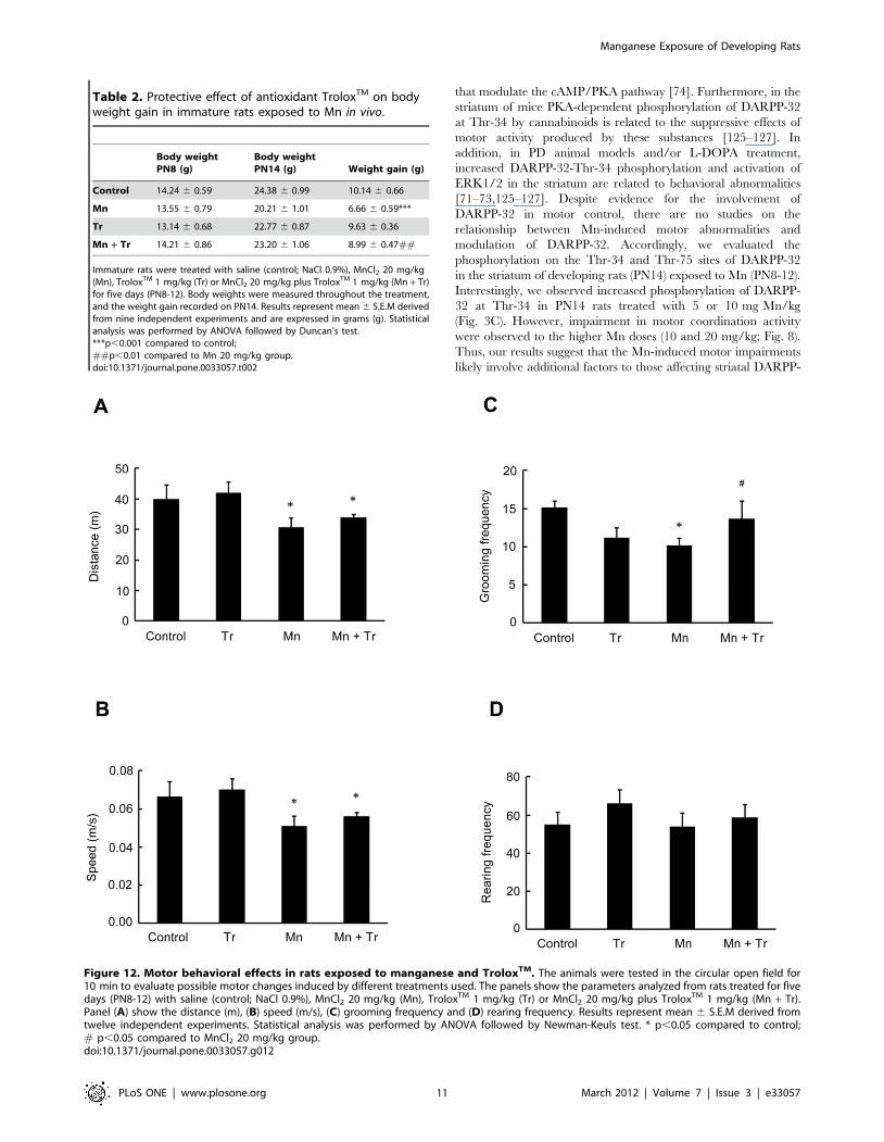
In the open field test, animals treated with 20 mg/kg of Mn
showed a decrease in the distance traveled (p,0.05, Fig. 12A) and
speed (p,0.05; Fig. 12B), as well as reduced grooming frequency
(p,0.05; Fig. 12C) vs. controls. Treatment with TroloxTM
reversed only the Mn effect on grooming frequency (p,0.05
relative to Mn, Fig. 12C). Mn treatment alone did not alter the
rearing frequency (Fig. 12D).
Discussion
Mn is an essential trace element. However, chronic or acute
exposure to exceedingly high Mn levels is common and results in
irreversible CNS damage [93]. Adult Mn-induced toxicity causes a
neurological disorder analogous to idiopathic PD [5]. Histological
observations from experimental animals and humans chronically
poisoned with Mn have shown predominantly degeneration in
dopaminergic nigrostriatal neurons [93]. However, a major issue,
yet to be systematically addressed, relates to the neurotoxic
mechanisms associated with developmental Mn exposure. While
several studies have emphasized that excessive Mn concentrations
in parenteral nutrition may lead to neurological disorders
[6,14,20,21], no in vivo studies have addressed the ability of Mn
to interfere with intracellular signaling pathways in the developing
CNS. To date, mechanisms implied in developmental Mn
neurotoxicity have largely focused on oxidative stress, alterations
in neurotransmitters and receptors, and behavioral abnormalities
[6,8,9,17,36,94–98].
The present study shows for the first time that in vivo
neurodevelopmental sequalae of Mn exposure are associated with
the modulation of intracellular signaling pathways, such as
ERK1/2, AKT and DARPP-32. Moreover, oxidative stress, cell
death and later-life impairment in motor function are also
observed after short-term Mn exposure during a developmental
period (PN8-12).
The experimental model described herein was associated with
significant accumulation of brain Mn (Fig. 1A) and absent
histological alterations in the brain (Fig. 2), as well as in liver,
kidneys, spleen or heart (data not shown). Analyzing the numerical
results of manganese levels achieved in the brain structures of
immature rats, we found that baseline metal levels in the control
are very low at PN14: 0.05, 0.05 and 0.06 mg Mn/g tissue in the
hippocampus, striatum and cerebral cortex, respectively. In
contrast, in animals treated with Mn (20 mg/kg) the levels in the
same brain regions were 0.45, 0.77 and 0.33 mg/g tissue,
respectively. Mn uptake into the brain is high during the neonatal
developmental period that coincides with peak of brain growth.
Our data agree with previous analysis performed in other studies
[99]. Moreover, Mn treatment did not alter the pups’ weight-gain
except for the higher treatment dose (20 mg Mn/kg), where a
slight decrease was noted (Table 1).
The brain structures primarily affected by Mn intoxication
include the striatum, globus pallidus and substantia nigra [5,100].
Figure 2. Histological evaluation of the brain of immature rats exposed to Mn in vivo. The panel shows representative sections from eightindependent experiments of (I) striatum, (II) hippocampus and (III) cerebral cortex from rats treated for five days (PN8-12) with saline (control; NaCl0.9%) or MnCl2 at doses of 5, 10 or 20 mg/kg. The structures analyzed on PN14. The sections were stained with hematoxylin and eosin staining.Magnification 640.doi:10.1371/journal.pone.0033057.g002
Manganese Exposure of Developing Rats
PLoS ONE | www.plosone.org 6 March 2012 | Volume 7 | Issue 3 | e33057
However, increased Mn levels were also noted in the hippocampus
and cerebral cortex (Fig. 1A), corroborating previous studies
[8,94,100–102].
A greater propensity for Mn accumulation in the developing
CNS has been previously noted [5–7,14]. Low Fe body-burden
has been posited to represent a risk factor for Mn poisoning
[98,103,104], with anemic states heightening sensitivity to Mn
toxicity [6]. Interestingly, we observed an increase in Fe levels in
the striatum of PN14 animals treated with 10 or 20 mg Mn/kg,
but not in animals treated with a lower dose (5 mg Mn/kg).
Moreover, in the cerebral cortex of rats treated with the highest
Mn dose (20 mg/kg) we also observed an increase in Fe levels
(Fig. 1B). Notably, increased Fe levels in the absence of significant
changes in the levels of other metals were previously observed in
the striatum, substantia nigra and cortex of juvenile mice exposed
to MnCl2 [9]. Overall, it is possible that Mn exerts it effects on the
Figure 3. Effects of in vivo exposure to Mn for five days on the phosphorylation of MAPKs, AKT, CREB and DARPP-32 in the striatumof immature rats. The panels show representative immunoblotting and quantification of phosphorylation of ERK1/2 (A), AKT (B), DARPP-32-Thr-34and -Thr-75 (C) and JNK1/2, p38MAPK, CREB and b-Actin (D) from rats treated for five days (PN8-12) with saline (control; NaCl 0.9%) or MnCl2 at dosesof 5, 10 or 20 mg/kg/day. The structures analyzed on PN14. Total and phosphorylated forms of each protein were detected by specific antibodies andthe reaction was developed by chemiluminescence. The phosphorylation level of each protein was determined as a ratio of the O.D. of thephosphorylated band over the O.D. of the total band and the data are expressed as percentage of the control (considered as 100%) and the valuesare presented as mean 6 S.E.M derived from twelve independent experiments. Statistical analysis was performed by ANOVA followed by Duncan’stest. * p,0.05, ** p,0.01, *** p,0.001 compared to control.doi:10.1371/journal.pone.0033057.g003
Manganese Exposure of Developing Rats
PLoS ONE | www.plosone.org 7 March 2012 | Volume 7 | Issue 3 | e33057
immature brain at least in part by altering Fe metabolism.
Changes in Fe concentration, may, in turn, trigger the
pathophysiology of manganism. However, it is noteworthy that
the lower Mn dose (5 mg/kg) produced molecular effects that were
unrelated to Fe levels in the striatum, including increased Thr-34
phosphorylation of DARPP-32 and caspase activation. Notably,
other studies have shown an opposite relationship, with decreased
brain Fe levels in animals exposed to excessive Mn [22,103,105].
Moreover, increased expression of Tf in the plasma and DMT-1
transporter and TfR in the cerebellum, cortex, hippocampus,
midbrain and striatum, and increased capacity of Tf bind to Fe
was observed [22]. These changes may account for the increased
Fe levels observed herein, reflecting a short treatment paradigm
with low Mn doses. The expression or activity of the above
transporters was not evaluated in the present study, and should be
addressed in future studies.
Imbalances in brain metals (Mn, Fe, Cr, Cu, Zn, Co, Al) in the
basal ganglia of Mn-exposed animals, behavioral changes and
occurrence of oxidative stress and neuronal death have been
previously noted [9,78]. Herein, we observed increased production
of ROS at 10 and 20 mg Mn/kg (Fig. 4). Moreover, F2-IsoPs
production increased in a dose-dependent (r2 = 0.38) manner in
the striatum (Fig. 5), consistent with reports on oxidative stress in
several in vitro and in vivo models [28,30–34]. ROS production may
be directly related to Mn treatment or potentially is secondary to
increased Fe levels and an ensuing Fenton reaction.
We observed a significant increase in mitochondrial complex I
and II activities in mitochondria derived from rats exposed to
10 mg Mn/kg. Exposure to higher Mn doses (20 mg/kg) evoked
distinct effects. While complex I remained increased vs. controls,
complex II activity was significantly reduced by the treatment
(Fig. 6). Our results differ from those reported in other studies
where Mn dose-dependently inhibited respiratory chain complexes
and induced ROS production in isolated rat brain mitochondria
[31]. Sriram et al. [106] showed in the striatum and midbrain of
rats exposed to Mn-containing welding fumes a significant
reduction in mitochondrial complex proteins. We do not have a
clear explanation for the apparent differences in outcomes.
However, it is important to consider that mitochondrial functional
capacity is age-dependent, with older animals displaying reduced
mitochondrial function [107,108]. In addition, increased mito-
chondrial respiration has been observed in response to stressful
treatments [109] and in some neuropathological conditions
accompanied by compensatory mechanisms [110]. In order to
counterbalance energy deficits, mitochondrial biogenesis is
stimulated via redox-sensitive transcription factors [111]. It is well
established that complexes I and III are the major sites of
mitochondrial reactive oxygen species generation. Thus, increased
activities of complexes I and II in response to 10 mg/kg Mn
treatment may lead to increased free radical production with
ensuing oxidative damage to mitochondrial enzymes, including
complex II, lipid membranes, as well as mtDNA [112–114].
Finally, the inactivation of complex II observed with the higher
Mn dose might be mediated via nitric oxide (NO) derivatives such
as peroxynitrite, which are formed by the controlled reaction
between NO and mitochondrially-derived superoxide radical
[115]. Consistent with this possibility, it has been previously
shown that Mn induces NO generation [116], and that nitrosative
stress inhibits complex II. Accordingly, complex II inhibition,
secondary to Mn-induced mitochondrial oxidative stress, would
further induce free radical production, oxidative stress and cell
demise via apoptosis. In addition to the mitochondrial effects, Mn
also induced an increase in striatal caspase activity (Fig. 7).
Mitochondrial impairment leading to activation of apoptotic
pathways is well recognized in response to Mn [30,32,44]. Taken
together these results suggest that Mn neurotoxicity in the striatum
of immature rats (PN14) likely involves both changes in the activity
of mitochondrial complexes, increased ROS and F2-IsoPs
formation, as well as the activation of caspases.
Numerous intracellular signaling pathways are modulated in
response to in vitro Mn exposure, including: activation of ERK1/2,
JNKs, p38MAPK [35,40,42–44,47,48]. Herein, we demonstrate
modification in cell signaling pathways in response to in vivo Mn
treatment, specifically in a critical period of postnatal develop-
ment. Mn exposure (PN8-12) stimulated ERK1/2 and AKT
phosphorylation on PN14 (Figs. 3A and 3B), 48 h after the last Mn
administration. Consistent with this temporal effect and in vitro and
in vivo studies [117,118], it is plausible that Mn treatment may lead
to sustained rather than transient kinase activation. Given that
signaling duration can markedly alter qualitative and quantitative
features of downstream elements driving distinct cell fate decisions
[119] the pattern of ERK1/2 and AKT phosphorylation may
Figure 4. Mn induces oxidative stress in the striatum. Oxidativestress was analyzed by DCF fluorescence in the striatum of young ratstreated with Mn. The graphic shows the DCF fluorescence from ratstreated for five days (PN8-12) with saline (control; NaCl 0.9%) or MnCl2at doses of 5, 10 or 20 mg/kg/day. The structures were analyzed onPN14. The data are expressed as percentage of the control and thevalues are mean 6 S.E.M derived from eight independent experiments.Statistical analysis was performed by ANOVA followed by Duncan’s test.** p,0.01, *** p,0.001 compared to control.doi:10.1371/journal.pone.0033057.g004
Figure 5. Mn induces F2-IsoPs production. Striatum fromimmature rats (PN14) exposed in vivo to Mn (PN8-12) were evaluatedfor F2-IsoPs levels expressed as ng/g tissue. The graphic shows a dose-dependent increase of F2-IsoPs levels by MnCl2 (5, 10 or 20 mg/kg/day).The values were obtained from four independent experiments.Pearson’s correlation showed r2 = 0.38 with p = 0.011.doi:10.1371/journal.pone.0033057.g005
Manganese Exposure of Developing Rats
PLoS ONE | www.plosone.org 8 March 2012 | Volume 7 | Issue 3 | e33057

trigger long-lasting effects. The ERK and AKT pathways are
involved in essential processes, such as cell proliferation,
differentiation and survival [60,120]. However, ERK1/2 may
also be involved in cell death process [121,122]. In vitro studies in
Mn treated glial cells showed the participation of ERK1/2 and
AKT in the expression of iNOS in microglia [123] and COX-2 in
astrocytes [124]. In addition, it has been demonstrated that Mn
produced apoptotic cell death via the ERK1/2 signaling pathway,
with caspase-3 activation in PC12 and astrocytes [42,44].
Therefore, Mn-induced ERK1/2 and AKT activation may be
associated with changes in neuroplasticity and/or cell viability in
the immature rat striatum, thus disturbing and impairing
physiological neurodevelopment. Moreover, the oxidative stress
response may be involved in the ERK1/2 activation, since the
effect of Mn on ERK1/2 was abrogated by TroloxTM.
Figure 6. Effects of Mn on striatal activity of mitochondrialrespiratory chain complexes in immature rats. (A) Activity ofmitochondrial complex I. (B) Activity of mitochondrial complex II. (C)Activity of mitochondrial complex IV. Rat pups were treated for fivedays (PN8-12) with saline (control; NaCl 0.9%) or MnCl2 at doses of 5, 10or 20 mg/kg. Activities were analyzed on PN14. Results represent mean6 S.E.M and are expressed as nmol min21/mg protein21 or mmolmin21/mg protein21 derived from four independent experiments.Statistical analysis was performed by ANOVA followed by Duncan’s test.* p,0.05 compared to control.doi:10.1371/journal.pone.0033057.g006
Figure 7. Mn treatment stimulates caspase activity in thestriatum of immature rats. Caspase activities were measured byDEVD cleavage. The panel shows the DEVD cleavage test from ratstreated for five days (PN8-12) with saline (control; NaCl 0.9%) or MnCl2at doses of 5, 10 or 20 mg/kg/day. Activities were analyzed on PN14.Results represent mean 6 S.E.M and are expressed as percentage ofcontrol (100%) derived from eight independent experiments. Statisticalanalysis was performed by ANOVA followed by Duncan’s test. * p,0.05,** p,0.01 compared to control.doi:10.1371/journal.pone.0033057.g007
Figure 8. Mn exposure on PN8-12 causes later life onset motordeficits in rats. To evaluate motor coordination, control animals(saline) and rats treated with Mn (PN8-12) were tested on 22, 29 and 36days of age (3, 4 and 5 week-old) on the rotarod task. The graphicshows the overall latency for falling in rats treated with saline (control;NaCl 0.9%) or MnCl2 at doses of 5, 10 or 20 mg/kg/day. Resultsrepresent median 6 interquartile range and are expressed as seconds(s) to latency for falling derived from twelve independent experiments.Statistical analysis was performed by Kruskal-Wallis followed by Dunn’spost-hoc test. * p,0.05 compared to control.doi:10.1371/journal.pone.0033057.g008
Manganese Exposure of Developing Rats
PLoS ONE | www.plosone.org 9 March 2012 | Volume 7 | Issue 3 | e33057
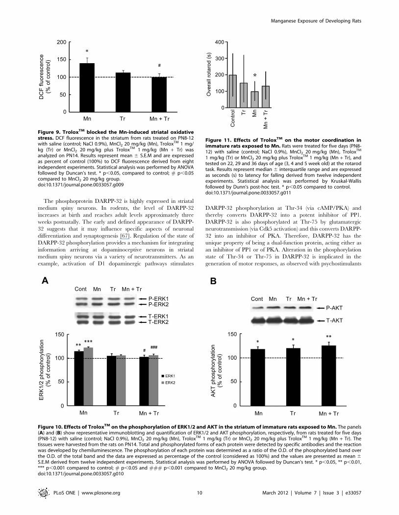
The phosphoprotein DARPP-32 is highly expressed in striatal
medium spiny neurons. In rodents, the level of DARPP-32
increases at birth and reaches adult levels approximately three
weeks postnatally. The early and defined appearance of DARPP-
32 suggests that it may influence specific aspects of neuronal
differentiation and synaptogenesis [67]. Regulation of the state of
DARPP-32 phosphorylation provides a mechanism for integrating
information arriving at dopaminoceptive neurons in striatal
medium spiny neurons via a variety of neurotransmitters. As an
example, activation of D1 dopaminergic pathways stimulates
DARPP-32 phosphorylation at Thr-34 (via cAMP/PKA) and
thereby converts DARPP-32 into a potent inhibitor of PP1.
DARPP-32 is also phosphorylated at Thr-75 by glutamatergic
neurotransmission (via Cdk5 activation) and this converts DARPP-
32 into an inhibitor of PKA. Therefore, DARPP-32 has the
unique property of being a dual-function protein, acting either as
an inhibitor of PP1 or of PKA. Alteration in the phosphorylation
state of Thr-34 or Thr-75 in DARPP-32 is implicated in the
generation of motor responses, as observed with psychostimulants
Figure 9. TroloxTM blocked the Mn-induced striatal oxidativestress. DCF fluorescence in the striatum from rats treated on PN8-12with saline (control; NaCl 0.9%), MnCl2 20 mg/kg (Mn), TroloxTM 1 mg/kg (Tr) or MnCl2 20 mg/kg plus TroloxTM 1 mg/kg (Mn + Tr) wasanalyzed on PN14. Results represent mean 6 S.E.M and are expressedas percent of control (100%) to DCF fluorescence derived from eightindependent experiments. Statistical analysis was performed by ANOVAfollowed by Duncan’s test. * p,0.05, compared to control; # p,0.05compared to MnCl2 20 mg/kg group.doi:10.1371/journal.pone.0033057.g009
Figure 10. Effects of TroloxTM on the phosphorylation of ERK1/2 and AKT in the striatum of immature rats exposed to Mn. The panels(A) and (B) show representative immunoblotting and quantification of ERK1/2 and AKT phosphorylation, respectively, from rats treated for five days(PN8-12) with saline (control; NaCl 0.9%), MnCl2 20 mg/kg (Mn), TroloxTM 1 mg/kg (Tr) or MnCl2 20 mg/kg plus TroloxTM 1 mg/kg (Mn + Tr). Thetissues were harvested from the rats on PN14. Total and phosphorylated forms of each protein were detected by specific antibodies and the reactionwas developed by chemiluminescence. The phosphorylation of each protein was determined as a ratio of the O.D. of the phosphorylated band overthe O.D. of the total band and the data are expressed as percentage of the control (considered as 100%) and the values are presented as mean 6S.E.M derived from twelve independent experiments. Statistical analysis was performed by ANOVA followed by Duncan’s test. * p,0.05, ** p,0.01,*** p,0.001 compared to control; # p,0.05 and ### p,0.001 compared to MnCl2 20 mg/kg group.doi:10.1371/journal.pone.0033057.g010
Figure 11. Effects of TroloxTM on the motor coordination inimmature rats exposed to Mn. Rats were treated for five days (PN8-12) with saline (control; NaCl 0.9%), MnCl2 20 mg/kg (Mn), TroloxTM
1 mg/kg (Tr) or MnCl2 20 mg/kg plus TroloxTM 1 mg/kg (Mn + Tr), andtested on 22, 29 and 36 days of age (3, 4 and 5 week old) at the rotarodtask. Results represent median 6 interquartile range and are expressedas seconds (s) to latency for falling derived from twelve independentexperiments. Statistical analysis was performed by Kruskal-Wallisfollowed by Dunn’s post-hoc test. * p,0.05 compared to control.doi:10.1371/journal.pone.0033057.g011
Manganese Exposure of Developing Rats
PLoS ONE | www.plosone.org 10 March 2012 | Volume 7 | Issue 3 | e33057
that modulate the cAMP/PKA pathway [74]. Furthermore, in the
striatum of mice PKA-dependent phosphorylation of DARPP-32
at Thr-34 by cannabinoids is related to the suppressive effects of
motor activity produced by these substances [125–127]. In
addition, in PD animal models and/or L-DOPA treatment,
increased DARPP-32-Thr-34 phosphorylation and activation of
ERK1/2 in the striatum are related to behavioral abnormalities
[71–73,125–127]. Despite evidence for the involvement of
DARPP-32 in motor control, there are no studies on the
relationship between Mn-induced motor abnormalities and
modulation of DARPP-32. Accordingly, we evaluated the
phosphorylation on the Thr-34 and Thr-75 sites of DARPP-32
in the striatum of developing rats (PN14) exposed to Mn (PN8-12).
Interestingly, we observed increased phosphorylation of DARPP-
32 at Thr-34 in PN14 rats treated with 5 or 10 mg Mn/kg
(Fig. 3C). However, impairment in motor coordination activity
were observed to the higher Mn doses (10 and 20 mg/kg; Fig. 8).
Thus, our results suggest that the Mn-induced motor impairments
likely involve additional factors to those affecting striatal DARPP-
Table 2. Protective effect of antioxidant TroloxTM on bodyweight gain in immature rats exposed to Mn in vivo.
Body weightPN8 (g)
Body weightPN14 (g) Weight gain (g)
Control 14.24 6 0.59 24.38 6 0.99 10.14 6 0.66
Mn 13.55 6 0.79 20.21 6 1.01 6.66 6 0.59***
Tr 13.14 6 0.68 22.77 6 0.87 9.63 6 0.36
Mn + Tr 14.21 6 0.86 23.20 6 1.06 8.99 6 0.47##
Immature rats were treated with saline (control; NaCl 0.9%), MnCl2 20 mg/kg(Mn), TroloxTM 1 mg/kg (Tr) or MnCl2 20 mg/kg plus TroloxTM 1 mg/kg (Mn + Tr)for five days (PN8-12). Body weights were measured throughout the treatment,and the weight gain recorded on PN14. Results represent mean 6 S.E.M derivedfrom nine independent experiments and are expressed in grams (g). Statisticalanalysis was performed by ANOVA followed by Duncan’s test.***p,0.001 compared to control;##p,0.01 compared to Mn 20 mg/kg group.doi:10.1371/journal.pone.0033057.t002
Figure 12. Motor behavioral effects in rats exposed to manganese and TroloxTM. The animals were tested in the circular open field for10 min to evaluate possible motor changes induced by different treatments used. The panels show the parameters analyzed from rats treated for fivedays (PN8-12) with saline (control; NaCl 0.9%), MnCl2 20 mg/kg (Mn), TroloxTM 1 mg/kg (Tr) or MnCl2 20 mg/kg plus TroloxTM 1 mg/kg (Mn + Tr).Panel (A) show the distance (m), (B) speed (m/s), (C) grooming frequency and (D) rearing frequency. Results represent mean 6 S.E.M derived fromtwelve independent experiments. Statistical analysis was performed by ANOVA followed by Newman-Keuls test. * p,0.05 compared to control;# p,0.05 compared to MnCl2 20 mg/kg group.doi:10.1371/journal.pone.0033057.g012
Manganese Exposure of Developing Rats
PLoS ONE | www.plosone.org 11 March 2012 | Volume 7 | Issue 3 | e33057

32 signaling. Regarding this possibility, some studies have shown
that Mn can induce motor impairments affecting other brain
areas, changing the dopamine availability and disturbing GA-
BAergic transmission [3,4,102,128]. We did not address the
mechanism involved in the Mn-induced increase in Thr-34
phosphorylation in DARPP-32. However, a recent study showed
PKA activation by an unknown mechanism in adult rats in
response to developmental (PN1-21) Mn exposure [118]. There-
fore, it is possible that a direct and/or indirect action of Mn on
striatum reinforces the cAMP/PKA/Thr34-DARPP-32 pathway.
Since oxidative stress has been implied as an important factor in
mediating Mn neurotoxicity, we evaluated the efficacy of
antioxidant TroloxTM to mitigate the effects of Mn (20 mg/kg).
The highest dose was chosen since it caused robust neurochemical
and behavioral changes. TroloxTM was effective in blocking the
Mn-dependent increase in ROS production (Fig. 9). Moreover,
TroloxTM reversed the impairment in weight-gain observed in
response to treatment with Mn 20 mg/kg (Table 2). TroloxTM co-
treatment also led to a significant reduction in ERK1/2
phosphorylation (Fig. 10A), suggesting that the Mn-mediated
ERK1/2 activation occurs via ROS production. However, the
Mn-dependent increase in AKT phosphorylation was not altered
by TroloxTM (Fig. 10B). Moreover, unexpectedly, TroloxTM itself
increased AKT phosphorylation. TroloxTM also failed to reduce
the Mn-dependent caspase activation (data not shown). In the
behavioral test paradigms, both motor impairment (Fig. 11) and
the decrease in the distance and speed in open field task (Fig. 12)
inherent to Mn (20 mg/kg) treatment remained unchanged by co-
treatment with TroloxTM. However, TroloxTM reversed the Mn-
induced reduction in grooming frequency (Fig. 12C). Taken
together, we posit that increased oxidative stress and aberrant cell
signaling mediate the diverse responses to Mn treatment; however,
the data also suggest that several of the biochemical and
behavioral alterations inherent to Mn treatment are mediated by
ROS generation-independent mechanisms. Our results are
consistent with previous studies where TroloxTM failed to reverse
Mn-dependent hyperphosphorylation of Ser-40 on Tyrosine
hydroxylase in PC12 cells, despite its ability to block H2O2
production [117].
Mn has a relatively long half-time in the CNS, consistent with a
slow elimination rate [101]. In developing animals exposed to high
Mn levels, Mn concentrations exceeding the homeostatic capacity
may lead to an overload condition, with increased risk for
neurodegenerative diseases such as Parkinson’s disease at later-life
stages [17]. Accordingly, the changes in vital intracellular signaling
pathways, such as DARPP-32 in response to low levels of Mn
treatment during a critical period of postnatal brain development,
may represent important triggers of late onset neurological
manifestations. Additionally, it should be considered that the
DARPP-32 changes observed in response to low doses Mn
treatment that were unaccompanied by immediate behavioral
impairment may imprint changes in proliferation, differentiation
and neural plasticity, resulting in neurological deficits at later life
stages. The present study establishes changes in key intracellular
signaling components which control cell survival, development
and especially the complex process of neuroplasticity. Future
studies should be directed at determining the relationship between
early-life Mn treatment and late-onset neurological disease, and
the potential for therapeutic modalities to attenuate these deficits.
Author Contributions
Conceived and designed the experiments: FMC ASL RDSP MA RBL.
Performed the experiments: FMC ASA TVP MWL FMG APR SCL CP.
Analyzed the data: FMC ASA TVP MWL FMG CP ASL RDSP KME
MA RBL. Contributed reagents/materials/analysis tools: ASL RDSP MA
RBL. Wrote the paper: FMC MA RBL.
References
1. Aschner JL, Aschner M (2005) Nutritional aspects of manganese homeostasis.
Mol Aspects Med 26: 353–362.
2. Aschner M, Guilarte TR, Schneider JS, Zheng W (2007) Manganese: recentadvances in understanding its transport and neurotoxicity. Toxicol Appl
Pharmacol 221: 131–147.
3. Dobson AW, Erikson KM, Aschner M (2004) Manganese neurotoxicity.Ann N Y Acad Sci 1012: 115–128.
4. Perl DP, Olanow CW (2007) The neuropathology of manganese-induced
Parkinsonism. J Neuropathol Exp Neurol 66: 675–682.
5. Roth JA (2009) Are there common biochemical and molecular mechanismscontrolling manganism and parkisonism. Neuromolecular Med 11: 281–296.
6. Erikson KM, Thompson K, Aschner J, Aschner M (2007) Manganese
neurotoxicity: a focus on the neonate. Pharmacol Ther 113: 369–377.
7. Hafeman D, Factor-Litvak P, Cheng Z, van Geen A, Ahsan H (2007)
Association between manganese exposure through drinking water and infant
mortality in Bangladesh. Environ Health Perspect 115: 1107–1112.
8. Moreno JA, Streifel KM, Sullivan KA, Legare ME, Tjalkens RB (2009)
Developmental exposure to manganese increases adult susceptibility to
inflammatory activation of glia and neuronal protein nitration. Toxicol Sci112: 405–415.
9. Moreno JA, Yeomans EC, Streifel KM, Brattin BL, Taylor RJ, et al. (2009)
Age-dependent susceptibility to manganese-induced neurological dysfunction.Toxicol Sci 112: 394–404.
10. Rodier PM (1995) Developing brain as a target of toxicity. Environ Health
Perspect 103 Suppl 6: 73–76.
11. Bondy SC, Campbell A (2005) Developmental neurotoxicology. J Neurosci Res
81: 605–612.
12. Grandjean P, Landrigan PJ (2006) Developmental neurotoxicity of industrial
chemicals. Lancet 368: 2167–2178.
13. Siddappa AJ, Rao RB, Wobken JD, Leibold EA, Connor JR, et al. (2002)
Developmental changes in the expression of iron regulatory proteins
and iron transport proteins in the perinatal rat brain. J Neurosci Res 68:761–775.
14. Hardy G (2009) Manganese in parenteral nutrition: who, when, and why
should we supplement? Gastroenterology 137: S29–35.
15. Gottlieb A, Keydar I, Epstein HT (1977) Rodent brain growth stages: an
analytical review. Biol Neonate 32: 166–176.
16. Rice D, Barone S, Jr. (2000) Critical periods of vulnerability for the developing
nervous system: evidence from humans and animal models. Environ Health
Perspect 108 Suppl 3: 511–533.
17. Lucchini R, Zimmerman N (2009) Lifetime cumulative exposure as a threat for
neurodegeneration: need for prevention strategies on a global scale.
Neurotoxicology 30: 1144–1148.
18. Santamaria AB (2008) Manganese exposure, essentiality & toxicity.
Indian J Med Res 128: 484–500.
19. Burton NC, Guilarte TR (2009) Manganese neurotoxicity: lessons learned
from longitudinal studies in nonhuman primates. Environ Health Perspect 117:
325–332.
20. Suzuki H, Takanashi J, Saeki N, Kohno Y (2003) Temporal parenteral
nutrition in children causing t1 shortening in the anterior pituitary gland and
globus pallidus. Neuropediatrics 34: 200–204.
21. Hardy G, Menendez AM, Manzanares W (2009) Trace element supplemen-
tation in parenteral nutrition: pharmacy, posology, and monitoring guidance.
Nutrition 25: 1073–1084.
22. Garcia SJ, Gellein K, Syversen T, Aschner M (2006) A manganese-enhanced
diet alters brain metals and transporters in the developing rat. Toxicol Sci 92:
516–525.
23. Lucaciu CM, Dragu C, Copaescu L, Morariu VV (1997) Manganese transport
through human erythrocyte membranes. An EPR study. Biochim Biophys Acta
1328: 90–98.
24. Kannurpatti SS, Joshi PG, Joshi NB (2000) Calcium sequestering ability of
mitochondria modulates influx of calcium through glutamate receptor channel.
Neurochem Res 25: 1527–1536.
25. Chen MK, Lee JS, McGlothan JL, Furukawa E, Adams RJ, et al. (2006) Acute
manganese administration alters dopamine transporter levels in the non-
human primate striatum. Neurotoxicology 27: 229–236.
26. He L, Girijashanker K, Dalton TP, Reed J, Li H, et al. (2006) ZIP8, member of
the solute-carrier-39 (SLC39) metal-transporter family: characterization of
transporter properties. Mol Pharmacol 70: 171–180.
Manganese Exposure of Developing Rats
PLoS ONE | www.plosone.org 12 March 2012 | Volume 7 | Issue 3 | e33057

27. Anderson JG, Cooney PT, Erikson KM (2007) Inhibition of DAT function
attenuates manganese accumulation in the globus pallidus. Environ Toxicol
Pharmacol 23: 179–184.
28. Benedetto A, Au C, Aschner M (2009) Manganese-induced dopaminergic
neurodegeneration: insights into mechanisms and genetics shared with
Parkinson’s disease. Chem Rev 109: 4862–4884.
29. Hernandez RB, Farina M, Esposito BP, Souza-Pinto NC, Barbosa F, Jr., et al.
(2011) Mechanisms of manganese-induced neurotoxicity in primary neuronal
cultures: the role of manganese speciation and cell type. Toxicol Sci.
30. Roth JA, Garrick MD (2003) Iron interactions and other biological reactions
mediating the physiological and toxic actions of manganese. Biochem
Pharmacol 66: 1–13.
31. Zhang S, Fu J, Zhou Z (2004) In vitro effect of manganese chloride exposure
on reactive oxygen species generation and respiratory chain complexes
activities of mitochondria isolated from rat brain. Toxicol In Vitro 18: 71–77.
32. Tamm C, Sabri F, Ceccatelli S (2008) Mitochondrial-mediated apoptosis in
neural stem cells exposed to manganese. Toxicol Sci 101: 310–320.
33. Zhang F, Xu Z, Gao J, Xu B, Deng Y (2008) In vitro effect of manganese
chloride exposure on energy metabolism and oxidative damage of mitochon-
dria isolated from rat brain. Environ Toxicol Pharmacol 26: 232–236.
34. Milatovic D, Zaja-Milatovic S, Gupta RC, Yu Y, Aschner M (2009) Oxidative
damage and neurodegeneration in manganese-induced neurotoxicity. Toxicol
Appl Pharmacol 240: 219–225.
35. Prabhakaran K, Ghosh D, Chapman GD, Gunasekar PG (2008) Molecular
mechanism of manganese exposure-induced dopaminergic toxicity. Brain Res
Bull 76: 361–367.
36. Avila DS, Gubert P, Fachinetto R, Wagner C, Aschner M, et al. (2008)
Involvement of striatal lipid peroxidation and inhibition of calcium influx into
brain slices in neurobehavioral alterations in a rat model of short-term oral
exposure to manganese. Neurotoxicology 29: 1062–1068.
37. de Oliveira MR, de Bittencourt Pasquali MA, Silvestrin RB, Mello EST,
Moreira JC (2007) Vitamin A supplementation induces a prooxidative state in
the striatum and impairs locomotory and exploratory activity of adult rats.
Brain Res 1169: 112–119.
38. Erikson KM, Suber RL, Aschner M (2002) Glutamate/aspartate transporter
(GLAST), taurine transporter and metallothionein mRNA levels are
differentially altered in astrocytes exposed to manganese chloride, manganese
phosphate or manganese sulfate. Neurotoxicology 23: 281–288.
39. Xu B, Xu ZF, Deng Y (2010) Manganese exposure alters the expression of N-
methyl-D-aspartate receptor subunit mRNAs and proteins in rat striatum.
J Biochem Mol Toxicol 24: 1–9.
40. Hirata Y, Furuta K, Miyazaki S, Suzuki M, Kiuchi K (2004) Anti-apoptotic
and pro-apoptotic effect of NEPP11 on manganese-induced apoptosis and JNK
pathway activation in PC12 cells. Brain Res 1021: 241–247.
41. Latchoumycandane C, Anantharam V, Kitazawa M, Yang Y, Kanthasamy A,
et al. (2005) Protein kinase Cdelta is a key downstream mediator of manganese-
induced apoptosis in dopaminergic neuronal cells. J Pharmacol Exp Ther 313:
46–55.
42. Ito Y, Oh-Hashi K, Kiuchi K, Hirata Y (2006) p44/42 MAP kinase and c-Jun
N-terminal kinase contribute to the up-regulation of caspase-3 in manganese-
induced apoptosis in PC12 cells. Brain Res 1099: 1–7.
43. Gonzalez LE, Juknat AA, Venosa AJ, Verrengia N, Kotler ML (2008)
Manganese activates the mitochondrial apoptotic pathway in rat astrocytes by
modulating the expression of proteins of the Bcl-2 family. Neurochem Int 53:
408–415.
44. Yin Z, Aschner JL, dos Santos AP, Aschner M (2008) Mitochondrial-
dependent manganese neurotoxicity in rat primary astrocyte cultures. Brain
Res 1203: 1–11.
45. Jang BC (2009) Induction of COX-2 in human airway cells by manganese: role
of PI3K/PKB, p38 MAPK, PKCs, Src, and glutathione depletion. Toxicol In
Vitro 23: 120–126.
46. Cai T, Yao T, Zheng G, Chen Y, Du K, et al. (2010) Manganese induces the
overexpression of alpha-synuclein in PC12 cells via ERK activation. Brain Res
1359: 201–207.
47. Li Y, Sun L, Cai T, Zhang Y, Lv S, et al. (2010) alpha-Synuclein
overexpression during manganese-induced apoptosis in SH-SY5Y neuroblas-
toma cells. Brain Res Bull 81: 428–433.
48. Park EJ, Park K (2010) Induction of oxidative stress and inflammatory
cytokines by manganese chloride in cultured T98G cells, human brain
glioblastoma cell line. Toxicol In Vitro 24: 472–479.
49. Rao KV, Jayakumar AR, Reddy PV, Tong X, Curtis KM, et al. (2010)
Aquaporin-4 in manganese-treated cultured astrocytes. Glia 58: 1490–1499.
50. Shin HJ, Choi MS, Ryoo NH, Nam KY, Park GY, et al. (2010) Manganese-
mediated up-regulation of HIF-1alpha protein in Hep2 human laryngeal
epithelial cells via activation of the family of MAPKs. Toxicol In Vitro 24:
1208–1214.
51. Cai T, Che H, Yao T, Chen Y, Huang C, et al. (2011) Manganese induces tau
hyperphosphorylation through the activation of ERK MAPK pathway in
PC12 cells. Toxicol Sci 119: 169–177.
52. Crittenden PL, Filipov NM (2011) Manganese modulation of MAPK
pathways: effects on upstream mitogen activated protein kinase kinases and
mitogen activated kinase phosphatase-1 in microglial cells. J Appl Toxicol 31:
1–10.
53. Chang L, Karin M (2001) Mammalian MAP kinase signalling cascades. Nature410: 37–40.
54. Chen Z, Gibson TB, Robinson F, Silvestro L, Pearson G, et al. (2001) MAPkinases. Chem Rev 101: 2449–2476.
55. Thomas GM, Huganir RL (2004) MAPK cascade signalling and synapticplasticity. Nat Rev Neurosci 5: 173–183.
56. Waetzig V, Herdegen T (2004) Neurodegenerative and physiological actions of
c-Jun N-terminal kinases in the mammalian brain. Neurosci Lett 361: 64–67.
57. Mielke K, Herdegen T (2000) JNK and p38 stresskinases–degenerative
effectors of signal-transduction-cascades in the nervous system. Prog Neurobiol
61: 45–60.
58. Cowan KJ, Storey KB (2003) Mitogen-activated protein kinases: new signaling
pathways functioning in cellular responses to environmental stress. J Exp Biol206: 1107–1115.
59. Waetzig V, Zhao Y, Herdegen T (2006) The bright side of JNKs-Multitalentedmediators in neuronal sprouting, brain development and nerve fiber
regeneration. Prog Neurobiol 80: 84–97.
60. Brazil DP, Yang ZZ, Hemmings BA (2004) Advances in protein kinase B
signalling: AKTion on multiple fronts. Trends Biochem Sci 29: 233–242.
61. van der Heide LP, Ramakers GM, Smidt MP (2006) Insulin signaling in the
central nervous system: learning to survive. Prog Neurobiol 79: 205–221.
62. Graybiel AM (2005) The basal ganglia: learning new tricks and loving it. Curr
Opin Neurobiol 15: 638–644.
63. Gerfen CR (2004) Basal ganglia. In: Paxinos G, ed. The rat nervous system.Amsterdam: Elsevier Academic. pp 445–508.
64. Qi Z, Miller GW, Voit EO (2010) The internal state of medium spiny neuronsvaries in response to different input signals. BMC Syst Biol 4: 26.
65. Walaas SI, Hemmings HC Jr., Greengard P, Nairn AC (2011) Beyond thedopamine receptor: regulation and roles of serine/threonine protein phospha-
tases. Front Neuroanat 5: 50.
66. Greengard P, Allen PB, Nairn AC (1999) Beyond the dopamine receptor: the
DARPP-32/protein phosphatase-1 cascade. Neuron 23: 435–447.
67. Svenningsson P, Nishi A, Fisone G, Girault JA, Nairn AC, et al. (2004)
DARPP-32: an integrator of neurotransmission. Annu Rev Pharmacol Toxicol44: 269–296.
68. Nishi A, Bibb JA, Snyder GL, Higashi H, Nairn AC, et al. (2000) Amplificationof dopaminergic signaling by a positive feedback loop. Proc Natl Acad Sci U S A
97: 12840–12845.
69. Ahn JH, McAvoy T, Rakhilin SV, Nishi A, Greengard P, et al. (2007) Protein
kinase A activates protein phosphatase 2A by phosphorylation of the B56delta
subunit. Proc Natl Acad Sci U S A 104: 2979–2984.
70. Greengard P (2001) The neurobiology of slow synaptic transmission. Science294: 1024–1030.
71. Santini E, Valjent E, Usiello A, Carta M, Borgkvist A, et al. (2007) Criticalinvolvement of cAMP/DARPP-32 and extracellular signal-regulated protein
kinase signaling in L-DOPA-induced dyskinesia. J Neurosci 27: 6995–7005.
72. Botsakis K, Pavlou O, Poulou PD, Matsokis N, Angelatou F (2010) Blockade of
adenosine A2A receptors downregulates DARPP-32 but increases ERK1/2
activity in striatum of dopamine deficient ‘‘weaver’’ mouse. Neurochem Int 56:245–249.
73. Lebel M, Chagniel L, Bureau G, Cyr M (2010) Striatal inhibition of PKA
prevents levodopa-induced behavioural and molecular changes in the
hemiparkinsonian rat. Neurobiol Dis 38: 59–67.
74. Polissidis A, Chouliara O, Galanopoulos A, Rentesi G, Dosi M, et al. (2010)
Individual differences in the effects of cannabinoids on motor activity,dopaminergic activity and DARPP-32 phosphorylation in distinct regions of
the brain. Int J Neuropsychopharmacol 13: 1175–1191.
75. Sidoryk-Wegrzynowicz M, Lee E, Albrecht J, Aschner M (2009) Manganese
disrupts astrocyte glutamine transporter expression and function. J Neurochem110: 822–830.
76. Rocha JB, Pereira ME, Emanuelli T, Christofari RS, Souza DO (1995) Effectof treatment with mercury chloride and lead acetate during the second stage of
rapid postnatal brain growth on delta-aminolevulinic acid dehydratase (ALA-D) activity in brain, liver, kidney and blood of suckling rats. Toxicology 100:
27–37.
77. Cordova FM, Rodrigues AL, Giacomelli MB, Oliveira CS, Posser T, et al.
(2004) Lead stimulates ERK1/2 and p38MAPK phosphorylation in the
hippocampus of immature rats. Brain Res 998: 65–72.
78. Fitsanakis VA, Zhang N, Anderson JG, Erikson KM, Avison MJ, et al. (2008)
Measuring brain manganese and iron accumulation in rats following 14 weeksof low-dose manganese treatment using atomic absorption spectroscopy and
magnetic resonance imaging. Toxicol Sci 103: 116–124.
79. Peterson GL (1977) A simplification of the protein assay method of Lowry et al.
which is more generally applicable. Anal Biochem 83: 346–356.
80. Bjerrum OJ, Heegaard NHH CRC Handbook of Immunoblotting of Proteins.
81. Posser T, de Aguiar CB, Garcez RC, Rossi FM, Oliveira CS, et al. (2007)
Exposure of C6 glioma cells to Pb(II) increases the phosphorylation of
p38(MAPK) and JNK1/2 but not of ERK1/2. Arch Toxicol 81: 407–414.
82. Grimaldi Filho G (1981) Tecnica histologica. Sao Paulo: Fiocruz.
83. Tolosa EMC, Rodrigues CJ, Behmer OA, Freitas Neto AG (2003) Manual de
tecnicas para histologia normal e patologica. Barueri: Manole.
84. Latini A, Rodriguez M, Borba Rosa R, Scussiato K, Leipnitz G, et al. (2005) 3-
Hydroxyglutaric acid moderately impairs energy metabolism in brain of youngrats. Neuroscience 135: 111–120.
Manganese Exposure of Developing Rats
PLoS ONE | www.plosone.org 13 March 2012 | Volume 7 | Issue 3 | e33057

85. Schuck PF, Tonin A, da Costa Ferreira G, Rosa RB, Latini A, et al. (2007) In
vitro effect of quinolinic acid on energy metabolism in brain of young rats.Neurosci Res 57: 277–288.
86. Sauer H, Wefer K, Vetrugno V, Pocchiari M, Gissel C, et al. (2003) Regulation
of intrinsic prion protein by growth factors and TNF-alpha: the role ofintracellular reactive oxygen species. Free Radic Biol Med 35: 586–594.
87. Morrow JD, Roberts LJ, 2nd (1999) Mass spectrometric quantification of F2-isoprostanes in biological fluids and tissues as measure of oxidant stress.
Methods Enzymol 300: 3–12.
88. Milatovic D, Yin Z, Gupta RC, Sidoryk M, Albrecht J, et al. (2007) Manganeseinduces oxidative impairment in cultured rat astrocytes. Toxicol Sci 98:
198–205.89. Milatovic D, Aschner M (2009) Measurement of isoprostanes as markers of
oxidative stress in neuronal tissue. Curr Protoc Toxicol Chapter 12:Unit12–14.
90. Zuse A, Prinz H, Muller K, Schmidt P, Gunther EG, et al. (2007) 9-
Benzylidene-naphtho[2,3-b]thiophen-4-ones and benzylidene-9(10H)-anthra-cenones as novel tubulin interacting agents with high apoptosis-inducing
activity. Eur J Pharmacol 575: 34–45.91. Lowry OH, Rosebrough NJ, Farr AL, Randall RJ (1951) Protein measurement
with the Folin phenol reagent. J Biol Chem 193: 265–275.
92. Aguiar AS, Jr., Araujo AL, da-Cunha TR, Speck AE, Ignacio ZM, et al. (2009)Physical exercise improves motor and short-term social memory deficits in
reserpinized rats. Brain Res Bull 79: 452–457.93. Lazrishvili IL, Shukakidze AA, Chkhartishvili NN, Bikashvili TZ (2009)
Morphological changes and manganese content in the brains of rat pupssubjected to subchronic poisoning with manganese chloride. Neurosci Behav
Physiol 39: 7–12.
94. Dorman DC, Struve MF, Vitarella D, Byerly FL, Goetz J, et al. (2000)Neurotoxicity of manganese chloride in neonatal and adult CD rats following
subchronic (21-day) high-dose oral exposure. J Appl Toxicol 20: 179–187.95. Weber S, Dorman DC, Lash LH, Erikson K, Vrana KE, et al. (2002) Effects of
manganese (Mn) on the developing rat brain: oxidative-stress related
endpoints. Neurotoxicology 23: 169–175.96. McDougall SA, Reichel CM, Farley CM, Flesher MM, Der-Ghazarian T, et
al. (2008) Postnatal manganese exposure alters dopamine transporter functionin adult rats: Potential impact on nonassociative and associative processes.
Neuroscience 154: 848–860.97. Wasserman GA, Liu X, Factor-Litvak P, Gardner JM, Graziano JH (2008)
Developmental impacts of heavy metals and undernutrition. Basic Clin
Pharmacol Toxicol 102: 212–217.98. Menezes-Filho JA, Bouchard M, Sarcinelli Pde N, Moreira JC (2009)
Manganese exposure and the neuropsychological effect on children andadolescents: a review. Rev Panam Salud Publica 26: 541–548.
99. Takeda A, Ishiwatari S, Okada S (1999) Manganese uptake into rat brain
during development and aging. J Neurosci Res 56: 93–98.100. Normandin L, Carrier G, Gardiner PF, Kennedy G, Hazell AS, et al. (2002)
Assessment of bioaccumulation, neuropathology, and neurobehavior followingsubchronic (90 days) inhalation in Sprague-Dawley rats exposed to manganese
phosphate. Toxicol Appl Pharmacol 183: 135–145.101. Morello M, Canini A, Mattioli P, Sorge RP, Alimonti A, et al. (2008) Sub-
cellular localization of manganese in the basal ganglia of normal and
manganese-treated rats An electron spectroscopy imaging and electronenergy-loss spectroscopy study. Neurotoxicology 29: 60–72.
102. Schneider JS, Decamp E, Clark K, Bouquio C, Syversen T, et al. (2009) Effectsof chronic manganese exposure on working memory in non-human primates.
Brain Res 1258: 86–95.
103. Fitsanakis VA, Zhang N, Garcia S, Aschner M (2010) Manganese (Mn) andiron (Fe): interdependency of transport and regulation. Neurotox Res 18:
124–131.104. Rivera-Mancia S, Rios C, Montes S (2011) Manganese accumulation in the
CNS and associated pathologies. Biometals 24: 811–825.
105. Guilarte TR, Chen MK, McGlothan JL, Verina T, Wong DF, et al. (2006)Nigrostriatal dopamine system dysfunction and subtle motor deficits in
manganese-exposed non-human primates. Exp Neurol 202: 381–390.106. Sriram K, Lin GX, Jefferson AM, Roberts JR, Wirth O, et al. (2010)
Mitochondrial dysfunction and loss of Parkinson’s disease-linked proteins
contribute to neurotoxicity of manganese-containing welding fumes. FASEB J
24: 4989–5002.
107. Kilbride SM, Telford JE, Davey GP (2008) Age-related changes in H2O2production and bioenergetics in rat brain synaptosomes. Biochim Biophys Acta
1777: 783–788.
108. Bagh MB, Thakurta IG, Biswas M, Behera P, Chakrabarti S (2011) Age-related oxidative decline of mitochondrial functions in rat brain is prevented by
long term oral antioxidant supplementation. Biogerontology 12: 119–131.
109. Barros MH, Bandy B, Tahara EB, Kowaltowski AJ (2004) Higher respiratoryactivity decreases mitochondrial reactive oxygen release and increases life span
in Saccharomyces cerevisiae. J Biol Chem 279: 49883–49888.
110. Witte ME, Bo L, Rodenburg RJ, Belien JA, Musters R, et al. (2009) Enhancednumber and activity of mitochondria in multiple sclerosis lesions. J Pathol 219:
193–204.
111. Heddi A, Stepien G, Benke PJ, Wallace DC (1999) Coordinate induction ofenergy gene expression in tissues of mitochondrial disease patients. J Biol Chem
274: 22968–22976.
112. Bandy B, Davison AJ (1990) Mitochondrial mutations may increase oxidativestress: implications for carcinogenesis and aging? Free Radic Biol Med 8:
523–539.
113. Hausladen A, Fridovich I (1994) Superoxide and peroxynitrite inactivateaconitases, but nitric oxide does not. J Biol Chem 269: 29405–29408.
114. Zhang Y, Marcillat O, Giulivi C, Ernster L, Davies KJ (1990) The oxidative
inactivation of mitochondrial electron transport chain components andATPase. J Biol Chem 265: 16330–16336.
115. Radi R, Cassina A, Hodara R (2002) Nitric oxide and peroxynitrite
interactions with mitochondria. Biol Chem 383: 401–409.
116. Aschner M, Erikson KM, Herrero Hernandez E, Tjalkens R (2009)Manganese and its role in Parkinson’s disease: from transport to neuropathol-
ogy. Neuromolecular Med 11: 252–266.
117. Posser T, Franco JL, Bobrovskaya L, Leal RB, Dickson PW, et al. (2009)Manganese induces sustained Ser40 phosphorylation and activation of tyrosine
hydroxylase in PC12 cells. J Neurochem 110: 848–856.
118. McDougall SA, Der-Ghazarian T, Britt CE, Varela FA, Crawford CA (2011)Postnatal manganese exposure alters the expression of D2L and D2S receptor
isoforms: relationship to PKA activity and Akt levels. Synapse 65: 583–591.
119. Murphy LO, Blenis J (2006) MAPK signal specificity: the right place at theright time. Trends Biochem Sci 31: 268–275.
120. Sweatt JD (2001) The neuronal MAP kinase cascade: a biochemical signal
integration system subserving synaptic plasticity and memory. J Neurochem76: 1–10.
121. Chu CT, Levinthal DJ, Kulich SM, Chalovich EM, DeFranco DB (2004)
Oxidative neuronal injury. The dark side of ERK1/2. Eur J Biochem 271:2060–2066.
122. Subramaniam S, Unsicker K (2006) Extracellular signal-regulated kinase as an
inducer of non-apoptotic neuronal death. Neuroscience 138: 1055–1065.
123. Bae JH, Jang BC, Suh SI, Ha E, Baik HH, et al. (2006) Manganese induces
inducible nitric oxide synthase (iNOS) expression via activation of both MAP
kinase and PI3K/Akt pathways in BV2 microglial cells. Neurosci Lett 398:151–154.
124. Liao SL, Ou YC, Chen SY, Chiang AN, Chen CJ (2007) Induction of
cyclooxygenase-2 expression by manganese in cultured astrocytes. NeurochemInt 50: 905–915.
125. Andersson M, Usiello A, Borgkvist A, Pozzi L, Dominguez C, et al. (2005)
Cannabinoid action depends on phosphorylation of dopamine- and cAMP-regulated phosphoprotein of 32 kDa at the protein kinase A site in striatal
projection neurons. J Neurosci 25: 8432–8438.
126. Borgkvist A, Fisone G (2007) Psychoactive drugs and regulation of the cAMP/PKA/DARPP-32 cascade in striatal medium spiny neurons. Neurosci
Biobehav Rev 31: 79–88.
127. Borgkvist A, Marcellino D, Fuxe K, Greengard P, Fisone G (2008) Regulationof DARPP-32 phosphorylation by Delta9-tetrahydrocannabinol. Neurophar-
macology 54: 31–35.
128. Jones DC, Miller GW (2008) The effects of environmental neurotoxicants onthe dopaminergic system: A possible role in drug addiction. Biochem
Pharmacol 76: 569–581.
Manganese Exposure of Developing Rats
PLoS ONE | www.plosone.org 14 March 2012 | Volume 7 | Issue 3 | e33057
Copyright © 2022 FDOKUMEN