Impact of surface functionalization on bacterial cytotoxicity of single-walled carbon nanotubes

Dublin Institute of TechnologyARROW@DIT
Articles Radiation and Environmental Science Centre
1-1-2007
In vitro toxicity evaluation of single walled carbonnanotubes on human A549 lung cellsMaria DavorenDublin Institute of Technology
Eva HerzogDublin Institute of Technology
Alan CaseyDublin Institute of Technology
Benjamin CottineauDublin Institute of Technology
Gordon ChambersDublin Institute of Technology
See next page for additional authors
This Article is brought to you for free and open access by the Radiation andEnvironmental Science Centre at ARROW@DIT. It has been accepted forinclusion in Articles by an authorized administrator of ARROW@DIT. Formore information, please contact [email protected].
Recommended CitationDavboren, Maria et al: In vitro toxicity evaluation of single walled carbon nanotubes on human A549 lung cells. Toxicology in Vitro21 (2007) 438-448.

AuthorsMaria Davoren, Eva Herzog, Alan Casey, Benjamin Cottineau, Gordon Chambers, Hugh J. Byrne, and FionaM. Lyng
This article is available at ARROW@DIT: http://arrow.dit.ie/radart/2

Toxicology in Vitro 21 (2007) 438–448www.elsevier.com/locate/toxinvit
0887-2333/$ - see front matter © 2006 Elsevier Ltd. All rights reserved.doi:10.1016/j.tiv.2006.10.007
In vitro toxicity evaluation of single walled carbon nanotubeson human A549 lung cells
Maria Davoren a,¤, Eva Herzog a, Alan Casey b, Benjamin Cottineau a,b, Gordon Chambers b, Hugh J. Byrne b, Fiona M. Lyng a
a Radiation and Environmental Science Centre, Focas Institute, Dublin Institute of Technology, Kevin Street, Dublin 8, Irelandb Physics of Molecular Materials Group, Focas Institute, Dublin Institute of Technology, Kevin Street, Dublin 8, Ireland
Received 14 June 2006; accepted 15 October 2006Available online 20 October 2006
Abstract
This paper describes the in vitro cytotoxicity assessment of single walled carbon nanotubes (SWCNT) on A549 cells, a human lung cellline. Cellular viability was determined using the alamar blue (AB), neutral red (NR) and MTT assays, which evaluated metabolic, lyso-somal and mitochondrial activity respectively. In addition, the total protein content of the cells was measured using the coomassie bril-liant (CB) blue assay. Supernatants were also assayed for Adenylate Kinase (AK) release and Interleukin 8 (IL-8) which indicated a lossof cell membrane integrity and an inXammation response respectively. To investigate the interactions between serum components in thetest medium and the test materials, exposures were conducted both in serum containing (5%) and serum-free medium. Results from thecytotoxicity tests (AB, CB, MTT) revealed the SWCNT to have very low acute toxicity to the A549 cells as all but one of the reported 24 hEC50 values exceeded the top concentration tested (800 �g/ml). The SWCNT were found to interfere with a number of the dyes used in thecytotoxicity assessment and we are currently conducting a comprehensive spectroscopic study to further investigate these interactions. Ofthe multiple cytotoxicity assays used, the AB assay was found to be the most sensitive and reproducible. Transmission electron micros-copy (TEM) studies conWrmed that there was no intracellular localization of SWCNT in A549 cells following 24 h exposure; however,increased numbers of surfactant storing lamellar bodies were observed in exposed cells.© 2006 Elsevier Ltd. All rights reserved.
Keywords: Single walled carbon nanotubes; Cytotoxicity; TEM; Foetal bovine serum; Lamellar bodies
1. Introduction
Nanotechnology is widely perceived as one of the keytechnologies of the 21st century and accordingly there havebeen huge advances and increased funding in global tech-nological research on nanomaterials. Single wall carbonnanotubes (SWCNT) are considered to have extensivecommercial application potential due to their excellentmechanical, electrical and magnetic properties (Paradiseand Goswami, in press). The broad range of increasingnanotechnology applications for SWCNT will almost cer-
tainly result in the increased potential for both human andenvironmental exposures to this nanomaterial. It is, there-fore, imperative that toxicological research to evaluate thebiocompatibility and possible adverse eVects on both thehealth of humans and the environment is conducted con-comitantly with technological research and development onnanomaterials (Dreher, 2004; Oberdörster et al., 2005; Tho-mas and Sayre, 2005).
Due to their size, SWCNT can easily become airborneand inhaled, hence the evaluation of their pulmonary eVectshas received a considerable amount of interest and a num-ber of in vivo and in vitro studies have been performed todate. Several studies on the eVects of both reWned and rawCNT on the lung tissue of various animal models have beenreported and there appears to be some inconsistency
* Corresponding author. Fax: +353 1 402 7904.E-mail address: [email protected] (M. Davoren).

M. Davoren et al. / Toxicology in Vitro 21 (2007) 438–448 439
between the research Wndings (Huczko et al., 2001; Lamet al., 2004; Shvedova et al., 2005; Warheit et al., 2004).These studies highlighted the inherent diYculty in testingCNT due to their agglomerative nature in aqueous solu-tions; indeed some of the observed mortality was attributedto mechanical blockage of the airways resulting in asphyxi-ation (Warheit et al., 2004). It is now recognised that inorder to elucidate the mechanisms of the pulmonary toxic-ity observed in these preliminary studies further more real-istic in vivo inhalation studies with aerosolised SWCNTneed to be conducted (Muller et al., 2006; Smart et al., 2006;Warheit, 2006).
A number of in vitro studies have also been performedon SWCNT with varying metal content and have evaluateddiVerent mechanistic endpoints. Shvedova et al. (2003)tested iron-rich (30 wt.% iron) SWCNT on human epider-mal keratinocytes (HaCaT) and following 18 h exposurereported oxidative stress and loss of cell viability. They alsoobserved that exposure resulted in ultrastructural and mor-phological changes in these skin cells. Recently, Kaganet al. (2006) demonstrated that iron-rich SWCNT (26 wt.%iron) resulted in a signiWcant loss of intracellular lowmolecular weight thiols (GSH) and accumulation of lipidhydroperoxides in murine macrophages. Fiorito et al.(2006) investigated the eVects of highly puriWed fullerenesand SWCNT on murine and human macrophages andfound these materials did not stimulate the release of theinXammatory marker nitric oxide by murine macrophagecells in culture. In addition, they also demonstrated theuptake of each material by human macrophages to be verylow and that each possessed a very low toxicity againsthuman macrophage cells. Jia et al. (2005) exposed SWCNT(with trace amounts of metal catalysts) to alveolar macro-phages isolated from guinea pigs for 6 h and found that theSWCNT elicited a more toxic response than multi walledCNT (MWCNT), quartz and fullerene. SWCNT have alsobeen tested on human embryo kidney cells (HEK293) andwere found to inhibit the proliferation of these cells byinducing cell apoptosis and decreasing cellular adhesiveability (Cui et al., 2005). As with the in vivo studies dis-cussed earlier, diVerences in SWCNT toxicity and biocom-patibility have also been observed with the various in vitrotests and these discrepancies can most likely be attributedto the varying percentages of catalysts and other impuritiesin the tested SWCNT, in addition to the diVerent dispersionmethods employed to date (Smart et al., 2006).
The objective of this study was to perform a comprehen-sive in vitro cytotoxicity assessment of SWCNT (10 wt.%iron) on A549 cells, a human epithelial-like lung cell line.Quartz was tested in parallel exposures to provide a bench-mark of particle toxicity. As we have recently found thatthere was signiWcant interaction between the SWCNT andfoetal bovine serum (FBS) present in the test medium(Casey et al., in press), particle exposures were conductedboth in serum containing (5%) and serum-free cell culturemedium. Cytotoxicity parameters evaluated in this study,following 24 h exposure to both materials included the met-
abolic, lysosomal, and mitochondrial activities of the cells.In addition total protein content, cell membrane integrityand inXammation responses were also measured. The pres-ent study also employed TEM to characterise the SWCNTpre exposure, to investigate if the SWCNT were interna-lised by these lung cells and to examine for any ultrastruc-tural changes in cell morphology post exposure.
2. Materials and methods
2.1. Test materials
HiPco® derived SWCNT were purchased from CarbonNanotechnologies, Inc. (Houston, TX). This material con-tained 10 wt.% iron. The diameter distribution of theseHiPco® tubes was previously determined to be 0.8–1.2 nmby Raman spectroscopy conducted in our laboratory(Gregan et al., 2003). Quartz powder (CertiWed ReferenceMaterial, BCR No. 66) with a particle distribution of 0.35–3.50 �m was employed as a positive control and obtainedfrom Sigma Aldrich Ltd. (Dublin, Ireland). This standardquartz sample was mined in Frechen, Germany and ismostly silicon dioxide with trace amounts of iron (0.4 mg g¡1),titanium (0.26 mg g¡1), sodium (0.09 mg g¡1) and calcium(0.06 mg g¡1).
2.2. Reagents
3-(4,5-dimethylthiazol-2-yl)-2,5-diphenyltetrazolium bro-mide (MTT), Coomassie Brilliant Blue 250 (CB) and Neu-tral Red (NR) were all purchased from Sigma Aldrich Ltd.(Dublin, Ireland). The ToxiLight™ kit for AdenylateKinase (AK) analysis was purchased from Cambrex BioSci-ence (Wokingham Ltd., UK). Alamar Blue™ (AB) and theHuman Interleukin 8 (IL-8) CytoSet™ were purchased fromBiosource (UK). Cell culture media and supplements andthe trypsinisation solution were purchased from Biosciences(Dublin, Ireland).
2.3. Cell culture
A549 cells (ATCC, CCL-185) a human lung carcinomaepithelial cell line were employed for testing. Cells were cul-tured in Kaighn’s medium (F12K) with 2 mM L-glutaminesupplemented with 10% foetal bovine serum (FBS) and45 IU ml¡1 penicillin and 45 �g ml¡1 streptomycin at 37 °Cin 5% CO2 humidiWed incubator.
2.4. Dispersion of nanomaterials
Stock suspensions of SWCNT and quartz were preparedboth in serum containing (5%) and serum-free (0%)medium. An ultrasonic tip (Ultra sonic processor VCX-750 W) at amplitude of 40% for a total time of 30 s carriedout in 10 s sequential steps was employed to disperse thesuspensions prior to preparation of test concentrations.The suspensions were prepared by dispersing an initial
440 M. Davoren et al. / Toxicology in Vitro 21 (2007) 438–448
concentration of 800 �g/ml of each material by sonication.Each stock concentration was then serially diluted on the96-well plate with each type of medium to prepare test con-centrations. Cells were then exposed to the same concentra-tion range of SWCNT and quartz (1.56, 3.12, 6.25, 12.5, 25,50, 100, 200, 400, 800�g/ml) prepared in both 5%-F12Kand 0%-F12K medium. Transmission electron microscopy(TEM) was performed using a Jeol 100CX TEM, on raw aspurchased HiPco® SWCNT by dispersing 1 mg of SWCNTin 10 ml ethanol with the aid of a sonic tip, the dispersionwas then dropcast onto 200 nm formvar coated coppergrids for examination.
2.4.1. Cytotoxicity assaysFor cytotoxicity assays cells were seeded in 96-well
microplates (Nunc, Denmark) at a density of 1£105 cells/ml in 100 �l F12K medium containing 10% FBS. After 24 hof cell attachment, plates were washed with 100�l/wellphosphate buVered saline (PBS) and the cells were treatedwith increasing concentrations of each nanomaterial pre-pared in either 5% or 0% FBS containing medium for 24 h.Six replicate wells were used for each control and test con-centration per microplate. Cytotoxicity was assessed usingWve assays as outlined below.
2.5. Alamar blue, neutral red, coomassie blue assays
The AB, NR and CB assays were conducted subse-quently on the same set of plates. The AB assay was per-formed Wrst. The bioassay was carried out according tomanufacturer’s instructions. BrieXy, control media or testexposures were removed; the cells were rinsed with PBS and100�l of an AB/NR medium (5% [v/v] solution of AB and1.25% [v/v] of NR dye) prepared in fresh media (withoutFBS or supplements) were added to each well. Following3 h incubation, AB Xuorescence was quantiWed at therespective excitation and emission wavelength of 540 and595 nm. Wells containing medium and AB without cellswere used as blanks. The mean Xuorescent units for the sixreplicate cultures were calculated for each exposure treat-ment and the mean blank value was subtracted from these.Viability and protein determination of the cells followingexposure to each chemical were then subsequently investi-gated using the NR and CB assays according to Liebschand Spielmann (1995) with the modiWcation of CoomassieBrilliant Blue dye being employed in place of Kenacid BlueR dye.
2.6. MTT assay
A second series of plates were set up for the MTT assay.These plates were seeded and exposed identically to the Wrstseries of plates prepared for the AB, NR, and CB assays.Following 24 h of nanomaterial exposure, control mediumor test exposures were removed (medium from each con-trol/treatment was pooled and frozen at ¡80 °C for subse-quent AK and IL-8 analysis), the cells were rinsed with PBS
and 100 �l of fresh medium (without FBS or supplements)was added to each well. Ten microlitres of MTT (5 mg/ml)prepared in PBS was then added to each well and the plateswere incubated for 3 h at 37 °C in a 5% CO2 humidiWedincubator. After this incubation period the medium wasdiscarded, the cells were washed with 100 �l of PBS and100 �l of DMSO was added to each well to extract the dye.The plate was shaken at 240 rpm for 10 min and the absor-bance was measured at 570 nm.
2.7. Measurement of Adenylate Kinase (AK) release
The bioluminescent ToxiLight™ kit (Cambrex BioSci-ence Wokingham Ltd., UK) was employed to measure theconcentration of AK present in the supernatants collectedas described above. This kit quantitatively measures therelease of AK from damaged cells and the assay was per-formed according to manufacturer’s instructions.
2.8. Measurement of Interleukin-8 (IL-8)
Medium was assayed in triplicate for IL-8 using anenzyme-linked immunosorbent assay (ELISA) cytoset kitaccording to manufacturer’s instructions (Biosource Inter-national, Camarillo, CA).
2.9. Light and transmission electron microscopy
Exposures were conducted in 35 mm petri dishes (Nunc,Denmark) that were seeded with 2 ml of cell suspensionprepared in 10%-F12K at a density of 2£ 105 cells/ml. Cellswere allowed to attach for 24 h and were then exposed toselected concentrations of SWCNT (0, 400, 800 �g/ml) andquartz (0, 400, 800 �g/ml) prepared in both 5%-F12K and0%-F12K medium for 24 h. Following exposures, cells werewashed with 0.1 M phosphate buVer and then Wxed in 2.5%glutaraldehyde in 0.1 M phosphate buVer for 1 h, post-Wxedin 1% OsO4 in 0.1 M phosphate buVer for a further hour,dehydrated in ascending grades of ethanol and subse-quently embedded in epoxy resin. Light microscopy sec-tions (1 �m) were cut en face with a glass knife, stained with1% toluidine blue and mounted with DPX. These sectionswere examined using a Nikon Eclipse E600 microscope andimages were recorded using a Spot RT digital camera. Ultrathin sections (80 nm) were cut en face with a diamond knife,stained with uranyl acetate and lead citrate and examinedusing an FEI Tecnai G2 TEM.
2.10. Statistics
Fluorescence as Xuorescent units (FUs), luminescence asrelative light units (RLUs) and absorbance were all quanti-Wed using a microplate reader (TECAN GENios, Grödig,Austria). Experiments were conducted in at least triplicate(three independent experiments). Test treatments for eachassay (AB, NR, MTT, CB) were expressed as percentage ofthe unexposed control§ standard deviation (SD). Control

M. Davoren et al. / Toxicology in Vitro 21 (2007) 438–448 441
values were set at 100%. For the AK assay, cytotoxicity wasexpressed as mean percentage increase relative to the unex-posed control§SD. Control values were set at 0% cytotox-icity. Cytotoxicity data (where appropriate) was Wtted to asigmoidal curve and a four parameter logistic model usedto calculate the 50% EVective Concentration (EC50), whichwas the concentration of nanomaterial which caused a 50%inhibition in comparison to untreated controls. The EC50values are reported §95% ConWdence Intervals (§95% CI).This analysis was performed using XlWt3™ a curve Wttingadd-in for Microsoft® Excel (ID Business Solutions, UK).For cytokine analysis the IL-8 concentration (pg/ml) foreach treatment was calculated from a standard curve con-ducted in parallel with test samples. CoeYcient of variation(CV) for the controls of each test was calculated to ascer-tain reproducibility. Statistical analyses were carried outusing one-way analyses of variance (ANOVA) followed byDunnett’s multiple comparison test.
3. Results
3.1. Initial characterisation of SWCNT
The TEM images obtained for HiPco® nanotubes (aspurchased) tested in this study are shown in Fig. 1. Fig. 1Ashows an area of what is termed high density tubes, bun-dles/aggregates of varying lengths and diameters can beclearly seen which is typical of raw HiPco® SWCNT. InFig 1B, individual SWCNT and remnant catalytic iron par-ticles resultant from the fabrication process are evident.
3.2. Alamar blue assay
SigniWcant cytotoxicity (P 60.05) was recorded at 400and 800 �g/ml SWCNT tested in the presence of serum forthe AB assay, with approximately 33% and 53% inhibitiondemonstrated respectively in comparison to untreated con-trols. The EC50§95% CI value was determined as 744§91 �g/ml SWCNT for serum exposures. In serum free mediasigniWcant cytotoxicity was again recorded at 400 and
800 �g/ml SWCNT where a 42% and 51% inhibition in ABXuorescence was recorded in comparison to unexposedcontrols (Fig. 2A). In the absence of serum, the EC50 wasfound to be >800 �g/ml SWCNT.
Following exposure of the A549 cells to quartz in serumcontaining medium signiWcant cytotoxicity was observed at
Fig. 1. TEM of HiPco® SWCNT (as purchased) (A) 20,000£ magniWcation showing large aggregates (arrows) and high density tubes (B) 200,000£ magni-Wcation showing remnant catalytic iron particles (arrows).
Fig. 2. Cytotoxicity of SWCNT (A) and quartz (B) to A549 cells after 24 hexposure determined by the AB assay. Exposures were conducted inmedia containing 5% serum (�), and serum free media (�). Data areexpressed as percent of control mean § SD of three independent experi-ments. ¤ Denotes a signiWcant diVerence from the control (P 6 0.05). CVfor the controls ranged from 1.7% to 28.7% in serum containing mediaand 2.7–22.9% in serum free media for the SWCNT and 2.1–4.7% in serumcontaining media and in 2.9–3.7% in serum free media for quartz.
*
***
0
20
40
60
80
100
120
Control 3.125 12.5 50 200 800Concentration SWCNTs (µg/ml)
AB
red
ucti
on(%
of
cont
rol)
* * *
*
**
*
0
20
40
60
80
100
120
Control 3.125 12.5 50 200 800Concentration Quartz (µg/ml)
AB
red
ucti
on(%
of
cont
rol)

442 M. Davoren et al. / Toxicology in Vitro 21 (2007) 438–448
200 and 400�g/ml, which resulted in an 18% and 23%inhibitory eVect respectively in comparison to controls. Atthe top concentration tested (800�g/ml), however, only a2% inhibition was recorded in comparison to untreatedcontrols, hence no EC50 value was derived for this exposure.For quartz exposures in the absence of serum signiWcantcytotoxicity was observed at 25, 50, 100, 400, and 800 �g/ml.Approximately 50% and 90% inhibition was recorded incomparison to untreated controls at 400 and 800 �g/mlquartz respectively (Fig. 2B). An EC50 of 397§159 �g/mlfor quartz exposure without serum was determined.
3.3. Coomassie blue assay
SWCNT in serum containing medium produced signiW-cant cytotoxicity at concentrations of 400 (25% inhibition)and 800 (32% inhibition) �g/ml, as elucidated using the CBassay. In the absence of serum, signiWcant cytotoxicity wasalso observed at these concentrations (Fig. 3A). No EC50values were determined for the CB assay for both serumand serum free exposures, as maximum cytotoxicity deter-mined in both instances was less than 50%.
For quartz exposures in serum containing media, signiW-cant cytotoxicity was determined at 200, 400 and 800 �g/ml,with approximately 18%, 30% and 34% inhibition com-pared to untreated controls recorded respectively. There-fore the EC50 value was greater than the top concentrationtested. In the absence of serum, a slight but non-signiWcantincrease in CB absorbance over controls was recorded forconcentrations up to and including 200 �g/ml quartz. Sig-niWcant cytotoxicity was recorded at 400 (22% inhibition)and 800 (52% inhibition) �g/ml quartz (Fig. 3B). An EC50of 753§ 187�g/ml for quartz exposure without serum wasdetermined with this assay.
3.4. MTT assay
For the MTT assay, exposure in serum containing mediaresulted in signiWcant cytotoxicity from 12.5 �g/ml SWCNTupwards, and a maximum of 32% inhibition was recordedat 800 �g/ml SWCNT in comparison to the control. Forexposures in the absence of serum, signiWcant cytotoxicitywas recorded from 3.125 �g/ml SWCNT upwards and800 �g/ml SWCNT resulted in approximately 45% inhibi-tion in comparison to the control (Fig. 4A). No EC50 valueswere, therefore, determined for the MTT assay for bothserum and serum free exposures to SWCNT, as maximumcytotoxicity determined in both instances was less than50%.
No signiWcant cytotoxicity was determined following24 h exposure of the A549 cells to quartz in serum contain-ing media. For the majority of the concentrations tested(except 400 �g/ml) a stimulatory or hormetic response wasrecorded but none of these were found to be statisticallysigniWcant when compared to untreated controls. In theabsence of serum in the test medium, signiWcant cytotoxic-ity was determined at 100, 400 and 800 �g/ml quartz, withapproximately 80% inhibition recorded at the top concen-tration (Fig. 4B). An EC50 of 465§ 130�g/ml for quartzexposure without serum was determined with the MTTassay.
3.5. Light and transmission electron microscopy
3.5.1. Cellular uptake of SWCNTNo evidence of SWCNT internalisation (either in serum
containing or serum free media) was found for this lungepithelium model. Due to the crystalline nature of quartz itwas not possible to section the exposed cell cultures forsubsequent TEM imaging.
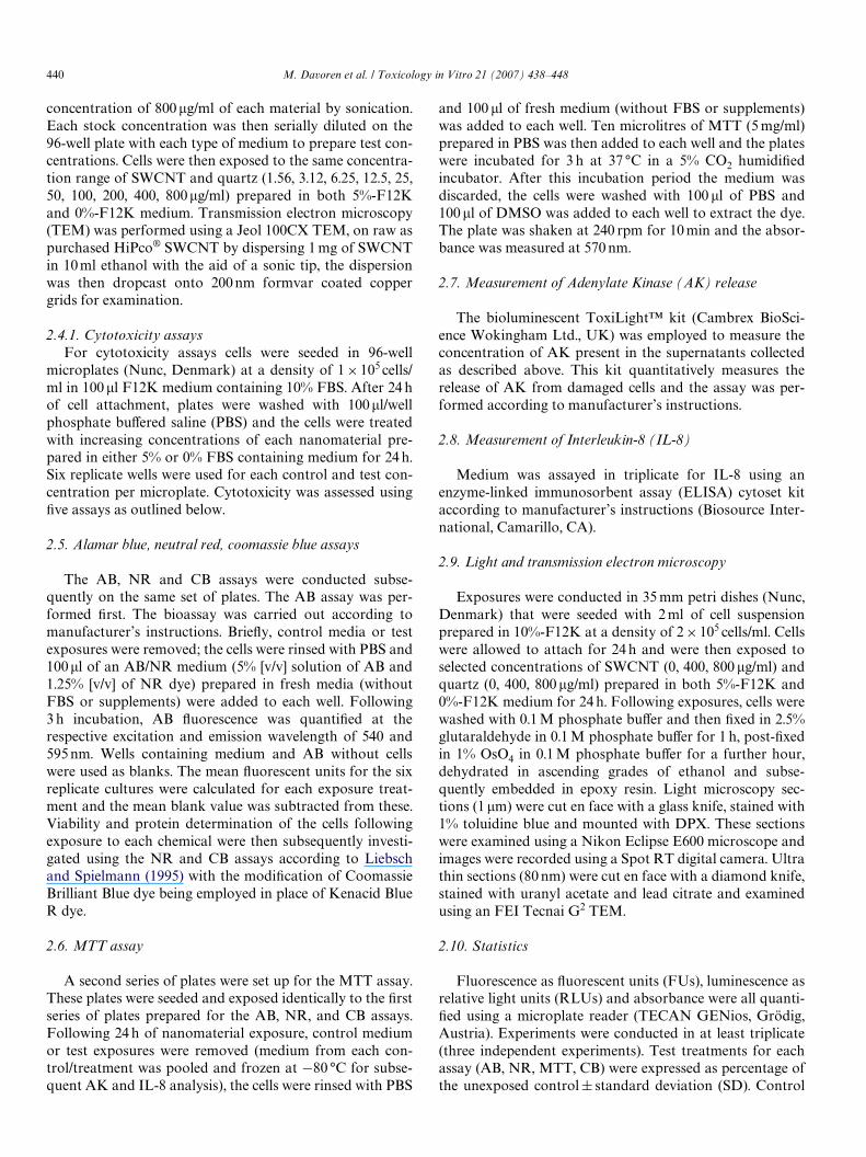
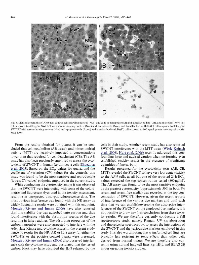
3.5.2. Cellular morphology post exposure to SWCNTLight micrographs of the A549 cells following exposure
to selected concentrations of SWCNT and quartz in serumcontaining medium are presented in Fig. 5. Control A549cells were of typical cuboidal shape, indicative of type IIpulmonary morphology (Fig. 5A). Cells exposed to SWCNTexhibited altered morphology and necrotic and apoptoticcells were observed (Fig. 5B and C). Exposure to the positive
Fig. 3. Cytotoxicity of SWCNT (A) and quartz (B) to A549 cells after 24 hexposure determined by the CB assay. Exposures were conducted in mediacontaining 5% serum (�), and serum free media (�). Data are expressedas percent of control mean § SD of three independent experiments.¤ Denotes a signiWcant diVerence from the control (P 6 0.05). CV for thecontrols ranged from 6.1% to 14.5% in serum containing media and 3.6–25.2% in serum free media for the SWCNT and 4.4–6.3% in serum con-taining media and in 13.2–20.8% in serum free media for quartz.
**
**
0
20
40
60
80
100
120
Control 3.125 12.5 50 200 800
Concentration SWCNTs (µg/ml)
**
*
*
*
0
20
40
60
80
100
120
140
Control 3.125 12.5 50 200 800
Concentration Quartz (µg/ml)
CB
upt
ake
(% o
f co
ntro
l)C
B u
ptak
e (%
of
cont
rol)

M. Davoren et al. / Toxicology in Vitro 21 (2007) 438–448 443
control quartz (800 �g/ml) resulted in extensive cell mortal-ity as evidenced by the cell debris seen in Fig. 5D.
Morphological features such as lamellar body structures,microvilli, tonoWlaments, desmosome junctions, character-istic of alveolar epithelial type II (AE2) cells were observedin TEM images of control cell cultures (Fig. 6). Followingexposure to 400 �g/ml SWCNT, an increase in lamellarbodies was recorded (Fig. 7A–C); multivesicular bodieswere also observed (Fig. 7B) and the presence of extracellu-lar (excreted) lamellar bodies was noted (Fig. 7C). Follow-ing exposure to the highest test concentration of SWCNT,we observed substantially more lamellar bodies when com-pared to untreated controls, although this observation wasnot quantiWed (Fig. 7D–E). A reduction in microvilli andan increase in lipid droplet numbers were also evident com-pared to control cultures. Multivesicular bodies were alsoobserved at the cell surface (Fig. 7F).
One of the main problems encountered in the investiga-tion of SWCNT cytotoxicity is the tendency of these nano-particles to aggregate into large bundles. A phase contrast
micrograph of A549 cells following 24 h exposure to 800 �g/ml SWCNT reveals large aggregates on the cell surface(Fig. 8). These aggregates remained adhered to the cell sur-face even after several washes with PBS.
4. Discussion
This paper describes a comprehensive in vitro cytotoxic-ity assessment of SWCNT on A549 cells, a human lung car-cinoma epithelial cell line. Quartz was tested in parallelexposures to provide a benchmark of particle toxicity.Parameters evaluated in this study following 24 h exposureto both materials, included the metabolic, lysosomal, andmitochondrial activities, in addition to total protein con-tent, cell membrane integrity and inXammation response.The present study also employed TEM to characterise thesamples pre exposure, to investigate if the SWCNT wereinternalised by these lung cells and to examine for anyultrastructural changes in cell morphology post exposure.
The pathogenicity of quartz has been well documentedboth in vitro and in vivo pulmonary studies; hence, it is rou-tinely employed as a positive control to benchmark particletoxicity (Diabeté et al., 2002; Lam et al., 2004; Warheit et al.,2004). In cytotoxicity tests with quartz (AB, NR, CB, MTT),we observed marked diVerences between the toxicity elicitedin the presence and absence of serum. For all four endpoints,the presence of serum had a substantial attenuation on thecytotoxicity of quartz and the EC50 values in all assays werefound to be >800�g/ml quartz. In the absence of serum,however, signiWcant cytotoxicity was demonstrated for allendpoints and the rank order of endpoint sensitivity basedon calculated EC50 values was AB > NR > MTT > CB.Quartz exposure in serum free media was also found toinduce a signiWcant release of AK at both 400 and 800�g/ml,which was indicative of cell membrane damage at these con-centrations (data not shown).
Cell cultures are routinely grown and tested in mediasupplemented with various percentages of serum proteinssuch as FBS. Gülden and Seibert (2005) have recentlyreported that the bioavailability of chemicals in vitro can bereduced due to partitioning into lipids and binding toserum proteins. The ‘protective’ eVect of serum in silica tox-icity has also been well documented (Emerson and Davis,1983; Schimmelpfeng et al., 1992). Antonini and Reasor(1994) demonstrated that coating of silica with a commer-cially available bovine pulmonary surfactant aVordedshort-term protection against its toxicity when tested bothin vitro on rat alveolar macrophages and in vivo followingintratracheal instillation. Antonini et al. (1994) also demon-strated that pretreatment of rats with amiodarone, a drugthat signiWcantly increases the concentration of totalphospholipid in whole lung resulted in reduced toxicity ofsilica dust following subsequent intratracheal instillation.We are currently investigating the eVect of increased expo-sure time (e.g. 48, 72, 96 h) of the cells to both nanomateri-als in order to establish if the protective eVect of the serumdiminishes over time.
Fig. 4. Cytotoxicity of SWCNT (A) and quartz (B) to A549 cells after 24 hexposure determined by the MTT assay. Exposures were conducted inmedia containing 5% serum (�), and serum free media (�). Data areexpressed as percent of control mean § SD of four (SWCNT) or three(quartz) independent experiments. ¤ Denotes a signiWcant diVerence fromthe control (P 6 0.05). CV for the controls ranged from 3.8% to 12.1% inserum containing media and 0.3–23.1% in serum free media for theSWCNT and 5.5–15.3% in serum containing media and in 4.6–9.1% inserum free media for quartz.
* * * * **
**
******
**
0
20
40
60
80
100
120
Control 3.125 12.5 50 200 800Concentration SWCNTs (µg/ml)
MT
T r
educ
tion
(% o
f co
ntro
l)
*
*
*
0
20
40
60
80
100
120
140
Control 3.125 12.5 50 200 800Concentration Quartz (µg/ml)
MT
T r
educ
tion
(% c
ontr
ol)
444 M. Davoren et al. / Toxicology in Vitro 21 (2007) 438–448
From the results obtained for quartz, it can be con-cluded that cell metabolism (AB assay), and mitochondrialactivity (MTT) are negatively impacted at concentrationslower than that required for cell detachment (CB). The ABassay has also been previously employed to assess the cyto-toxicity of SWCNT in human keratinocyte cells (Shvedovaet al., 2003). Based on the EC50 values for quartz and thecoeYcient of variation (CV) values for the controls, thisassay was found to be the most sensitive and reproducible(lowest CV values) endpoint employed in the current study.
While conducting the cytotoxicity assays it was observedthat the SWCNT were interacting with some of the colori-metric and Xuorescent dyes used in the toxicity assessment,resulting in unexpected absorption/Xuorescence data. Themost obvious interference was found with the NR assay aswidely Xuctuating results were obtained with this endpoint.Monteiro-Riviere and Inman (2006) have also reportedthat this viability dye was adsorbed onto carbon and thusfound interference with the absorption spectra of the dyeresulting in false readings. The adsorbing properties of thecarbon nanotubes were also suspected to interfere with theAdenylate Kinase and cytokine assays in the present studyhence no results for the NR, AK or IL-8 assay for either theSWCNTs or the positive control quartz were presented.Monteiro-Riviere and Inman (2006) also observed interfer-ence with the cytokine assay and postulated that the testedcarbon black may have adsorbed the IL-8 released by the
cells in their study. Another recent study has also reportedSWCNT interference with the MTT assay (Wörle-Knirschet al., 2006). Hurt et al. (2006) recently addressed this con-founding issue and advised caution when performing evenestablished toxicity assays in the presence of signiWcantquantities of Wne carbon.
Results presented for the cytotoxicity tests (AB, CB,MTT) revealed the SWCNT to have very low acute toxicityto the A549 cells, as all but one of the reported 24 h EC50values exceeded the top concentration tested (800 �g/ml).The AB assay was found to be the most sensitive endpointas the greatest cytotoxicity (approximately 50% in both 5%serum and serum free media) was recorded at the top con-centration of SWCNT. However, given the recent reportsof interference of the various dye markers and until suchtime that we can establish/overcome the adsorptive inter-ferences of the SWCNT on the employed dye markers, it isnot possible to draw any Wrm conclusions from these toxic-ity results. We are therefore currently conducting a fullspectroscopic study, namely Raman, UV–vis absorptionand Xuorescence spectroscopy, to assess the interactions ofthe SWCNT and the various dye markers employed in thisstudy. It is also worth noting that transformed cell lines aretypically less resistant to toxic eVects than other cellsderived from normal tissues. We are therefore also cur-rently using normal lung cell lines e.g. HFL and BEAS-2Bin our on-going toxicity studies.
Fig. 5. Light micrographs of A549 (A) control cells showing nucleus (Nuc) and cells in metaphase (M) and lamellar bodies (LB), and microvilli (Mv), (B)cells exposed to 400 �g/ml SWCNT with serum showing nucleus (Nuc) and necrotic cells (Nec), and lamellar bodies (LB) (C) cells exposed to 800 �g/mlSWCNT with serum showing nucleus (Nuc) and apoptotic cells (Apop) and lamellar bodies (LB) (D) cells exposed to 800 �g/ml quartz showing cell debris.Mag 400£.

M. Davoren et al. / Toxicology in Vitro 21 (2007) 438–448 445
Fig. 6. TEM of A549 control cells showing (A) nucleus (Nuc), lamellar bodies (LB), lipid droplets (L) and extensive microvilli (Mv), (B) intracellularmicrovilli (Mv), (C) mitochondria (Mt), lamellar bodies (LB), tonoWlaments (TF) and rough endoplasmic reticulum (RER), and (D) desmosome junction(DJ) and free ribosomes (Rb).
Fig. 7. TEM of A549 cells treated with 400 �g/ml SWCNT (A–C) showing (A) nucleus (Nuc), lamellar bodies (LB), and microvilli (Mv), (B) lamellar bod-ies (LB) and multivesicular body (MVB), (C) lamellar bodies (LB) and extracellular lamellar bodies (ELB) and A549 cells treated with 800 �g/ml SWCNT(D–F) showing (D) nucleus (Nuc), lamellar bodies (LB), and reduced microvilli (Mv), and an aggregation of lamellar bodies (ALB), (E) lamellar bodies(LB) and an accumulation of lipid droplets (L), (F) a multivesicular body (MVB) associated with the plasma membrane. A549 cells were exposed toSWCNTs either with serum (A, B and D, E) or without serum (C and F).

446 M. Davoren et al. / Toxicology in Vitro 21 (2007) 438–448
One of the main problems in the toxicity evaluation ofSWCNT is the propensity of the nanotubes to agglomer-ate owing to substantial van der Waals attractions. As ameans to reduce the formation of such bundles, research-ers have employed numerous carrier solvents includingmedia with varying FBS percentages, serum and organicsynthetic surfactants. The use of diVerent dispersal sol-vents will obviously inXuence the concentration ofSWCNT available to the cell and Smart et al. (2006) haverecently proposed that discrepancies in toxicity and bio-compatibility data may be attributed to the diVerent dis-persion methods employed to date. In this study we chosenot to employ synthetic surfactants, but rather to investi-gate the interactions between the FBS routinely usedfor the culturing and testing of the A549 cells and theSWCNT. Test exposures were therefore conducted bothin serum containing (5%) and serum-free medium. Visualobservation of the SWCNT prior to cell exposure revealedthat nanotubes appeared to solubilise (disaggregate)more readily in serum containing medium and indicatedthat the presence of serum had a dispersion eVect on theSWCNT. Monteiro-Riviere et al. (2005) found that diVer-ent synthetic surfactants showed increased dispersion andreduced aggregation of MWCNT, but they also notedthat the presence of surfactant did not alter the cytotoxic-ity of the MWCNT to a human keratinocyte cell line(HEK) as determined by the NR assay. In this study wefound that in general greater toxicity was demonstratedwith the SWCNT in the absence of serum (Figs. 3A and4A). In parallel studies to the in vitro cytotoxicity assess-ment we also employed spectroscopic techniques to assessthe interaction of the SWCNT and the various media
components and we found that there was signiWcant inter-action between the SWCNT and FBS (Casey et al., inpress).
As signiWcant cytotoxicity was demonstrated at 400 and800 �g/ml of SWCNT with the AB, CB and MTT assays,cells were subsequently exposed to these concentrations for24 h, and light and transmission electron microscopy wereemployed to assess for nanomaterial interaction/localisa-tion within the A549 cells and for evidence of dose relatedmorphological changes. Cells were also exposed to the sameconcentrations of quartz without serum and light micros-copy sections conWrmed the extensive cell death demon-strated in the viability assays. We found no evidence in thisstudy that this lung epithelium model internalized theSWCNT. Similarly, whilst Shvedova et al. (2003) reportedultrastructural changes in cell morphology of HaCaT cells,TEM imaging did not reveal intracellular localization ofthese particles. Other studies have shown that internaliza-tion of various nanoparticles can occur. Wörle-Knirschet al. (2006) recently reported that SWCNT could be foundas bundles of thousands inside A549 cells and postulatedthat they were mostly encapsulated in endosomes and infre-quently found inside mitochondria. Monteiro-Riviere et al.(2005) demonstrated MWCNT were primarily locatedwithin the intracytoplasmic vacuoles of human epidermalkeratincotyes (HEKs) and demonstrated diVerent mor-phology between control and those exposed to MWCNT.More recently, the same group (Rouse et al., 2006) hasreported that fullerene-based amino acid nanoparticles canalso localize to intracytoplasmic vacuoles in HEK cells. Theinternalization of ultraWne titanium dioxide by A549 cellshas also been reported (Stearns et al., 2001). In the concur-rent spectroscopic study performed by our group (Caseyet al., in press) Raman and Xuorescence emission analysisindicated that no debundling or reduction in aggregationstate occurred upon the dispersion of SWCNT in the mediaover the concentration range studied. Typically these aggre-gates can be of the order of microns therefore reducing thelikelihood of internalization.
While we did not Wnd any evidence of SWCNT internal-ization in the A549 cells, we did observe ultrastructuralchanges in the cell morphology following SWCNT expo-sure. It is worth noting however, that we could not distin-guish any obvious qualitative morphological diVerencesbetween those exposures conducted with and without serum.The A549 cells have retained the main properties of AE2cells, i.e. pulmonary surfactant secretory functions (Lieberet al., 1976; Fehrenbach, 2001) and within these cellsthe lamellar bodies are recognised as storage vessels fromwhich surfactant is released into the alveolus (Askin andKuhn, 1971). After secretion, alveolar forms of surfactantinclude these lamellar bodies, highly organised structurestermed tubular myelin, and monolayered and multilayered,phospholipid-rich sheets and vesicles (Whitsett andWeaver, 2002). While the principal function of this pulmo-nary surfactant is the regulation of surface tension in thelungs (Nicholas, 1996), it is also postulated that increased
Fig. 8. Phase contrast micrograph (200£) of A549 cells following 24 hexposure to 800 �g/ml SWCNT with 5% serum showing aggregates(arrows) on cell surface. These aggregates were still observed on the cellsurface after several washes with PBS.

M. Davoren et al. / Toxicology in Vitro 21 (2007) 438–448 447
production of this surfactant may play a role in defendingthe lungs against chemical and particulate damage (Hook,1991, 1993).
Following exposure to both 400 and 800 �g/ml SWCNT,an increase in lamellar bodies compared to untreated con-trols was observed within exposed cells. In addition, thepresence of extracellular lamellar bodies, which appeared tohave exocytosed their contents, was recorded at 400 �g/mlSWCNT (Fig. 7C). Multivesicular bodies were also observedfollowing cell exposure to both 400 and 800 �g/ml SWCNT.We surmise that these vesicles were most likely thephospholipid-rich multivesicular bodies as mentionedabove, which are involved in the surfactant synthesis andrecycling. Interestingly, a very similar plasma membraneprojection as that identiWed by us in Fig. 7F was shown byStearns et al. (2001), in an electron micrograph followingexposure of A549 cells to 40 �g/ml ultraWne titanium diox-ide. Stearns et al. (2001) also reported that aggregates ofTiO2, which entered the A549 cells by ingestion, were laterfound in membrane-bound vacuoles and enmeshed withinlamellar bodies. Recently, in an in vivo study, Shvedovaet al. (2005) also observed increased numbers of AE2 cellsexpressing cytoplasmic lamellar bodies in TEM sections ofmouse lung following SWCNT exposure via pharyngealaspiration.
We therefore hypothesize that the increased presence ofthese lamellar bodies and the concurrent surfactant secre-tion is a protective response of the lung cells to reduce thecytotoxic eVects of SWCNT. Hook (1993) postulated thatone possible mechanism that might account for the protec-tive eVects is the quenching of free radicals by the unsatu-rated phospholipids of pulmonary surfactant. This theoryseems quite plausible considering, as we have previously dis-cussed, surface modiWcation of silica with phospholipidscan markedly reduce its toxicity. Previous research has alsoshown that direct mechanical stretching of the AE2 cells cantrigger the release of surfactant (Wirtz and Dobbs, 1990). Ascan be seen in Fig. 8, SWCNT were found to aggregate onthe cell surface and remained attached even after severalwashes with PBS, hence another possible stimulus for thisenhanced secretion of surfactant may be due to the physicalstress of the SWCNT aggregates on the cell surface.
In conclusion, exposure of A549 cells to a wide doserange of SWCNT (1.56–800 �g/ml) for 24 h revealed theSWCNT to have low acute toxicity. In this study we foundthat in general, greater SWCNT toxicity was observed inthe absence of serum, although this trend was not asmarked as that observed for quartz exposures. TheSWCNT were found to interfere with a number of the dyesused in the cytotoxicity assessment and we are currentlyconducting a comprehensive spectroscopic study to furtherinvestigate these interactions. Of the multiple cytotoxicityassays used, the AB assay was found to be the most sensi-tive. TEM studies conWrmed that there was no intracellularlocalization of SWCNT in A549 cells following 24 h expo-sure, however, morphological alterations in exposed cellsobserved in this study revealed that there were increased
numbers of surfactant storing lamellar bodies. We thereforesurmise that the increased presence of these lamellar bodieswas a defensive response of these lung cells to SWCNTexposure and conclude that the protective function of thepulmonary surfactant following nanoparticle exposurewarrants further investigation.
Acknowledgements
We sincerely thank Dr. David Cottell and his staV at theElectron Microscopy laboratory, University College Dub-lin, for expert technical assistance. This project was fundedunder the DIT Team Research Scheme (TERS). Eva Her-zog is funded under the Technological Sector ResearchStrand I, Post Graduate R&D Skills Programme. BenjaminCottineau was supported by the DIT, SFI (Science Foun-dation Ireland) UREKA site FEATURE (Focus on Educa-tion in Analytical Techniques and Research Expertise).
References
Antonini, J.M., Reasor, M.J., 1994. EVect of short-term exogenous pulmo-nary surfactant treatment on acute lung damage associated with theintratracheal instillation of silica. Journal of Toxicology and Environ-mental Health 43, 85–101.
Antonini, J.M., McCloud, C.M., Reasor, M.J., 1994. Acute silica toxicity:attenuation by amiodarone-induced pulmonary phospholipidosis.Environmental Health Perspectives 102, 372–378.
Askin, F.B., Kuhn, C., 1971. The cellular origin of pulmonary surfactant.Laboratory Investigation 25, 260–268.
Casey, A., Davoren, M., Herzog, E., Lyng, F.M., Byrne, H.J., Chambers,G., in press. Probing the interaction of single walled carbon nanotubeswithin cell culture medium as a precursor to toxicity testing. Carbon.
Cui, D., Tian, F., Ozkan, C.S., Wang, M., Gao, H., 2005. EVect of singlewall carbon nanotubes on human HEK293 cells. Toxicology Letters155, 73–85.
Diabeté, S., Mülhopt, S., Paur, H.-R., Wottrich, R., Krug, H.F., 2002. Invitro eVects of incinerator Xy ash on pulmonary macrophages and epi-thelial cells. International Journal of Hygiene and EnvironmentalHealth 204, 323–326.
Dreher, K.L., 2004. Health and environmental impact of nanotechnology:toxicological assessment of manufactured nanoparticles. ToxicologicalSciences 77, 3–5.
Emerson, R., Davis, G.S., 1983. EVect of alveolar lining material-coatedsilica on rat alveolar macrophages. Environmental Health Perspectives51, 81–84.
Fehrenbach, H., 2001. Alveolar epithelial type II cell: defender of the alve-olus revisited. Respiratory Research 2, 33–46.
Fiorito, S., SeraWno, A., Andreola, F., Bernier, P., 2006. EVects of fullerenesand single-wall carbon nanotubes on murine and human macrophages.Carbon 44, 1100–1105.
Gregan, E., Keogh, S.M., Hedderman, T.G., Chambers, G., Byrne, H.J.,2003. Stokes/anti-Stokes Raman spectroscopy of HiPco single walledcarbon nanotubes “Opto-Ireland 2002, Optics and Photonics Technol-ogies and Applications”. In: Blau, W.J., Donegan, J.F., Duke, A.F.,MacCraith, B.D., McLaughlin, J.A., McMillan, N.D., O’Connor, G.M.,O’Mongain, E., Toal, V. (Eds.), SPIE Proceedings 4876, p. 1149.
Gülden, M., Seibert, H., 2005. Impact of bioavailability on the correlationbetween in vitro cytotoxic and in vivo acute Wsh toxic concentrations ofchemicals. Aquatic Toxicology 72, 327–337.
Hook, G.E., 1991. Alveolar proteinosis and phospholipidoses of the lungs.Toxicologic Pathology 19, 482–513.
Hook, G.E.R., 1993. Does pulmonary surfactant aid in defense of thelungs? Environmental Health Perspectives 101, 98–99.

448 M. Davoren et al. / Toxicology in Vitro 21 (2007) 438–448
Huczko, A., Lange, H., Calko, E., Grubek-Jaworska, H., Droszez, P., 2001.Physiological testing of carbon nanotubes: are they asbestos-like? Ful-lerene Science and Technology 9, 251–254.
Hurt, R.H., Monthioux, M., Kane, A., 2006. Toxicology of carbon nanom-aterials: status, trends, and perspectives on the special issue. Carbon 44,1028–1033.
Jia, G., Wang, H., Yan, L., Wang, X., Pei, R., Yan, T., Zhao, Y., Guo, X.,2005. Cytotoxicity of carbon nanomaterials: single-wall nanotube,multi-wall nanotube, and fullerene. Environmental Science and Tech-nology 39, 1378–1383.
Kagan, V.E., Tyurina, Y.Y., Tyurin, V.A., Konduru, N.V., Potapovich,A.I., Osipov, A.N., Kisin, E.R., Schwegler-Berry, D., Mercer, R., Cas-tranova, V., Shvedova, A.A., 2006. Direct and indirect eVects of singlewalled carbon nanotubes on RAW 264.7 macrophages: role of iron.Toxicology Letters, 165-88–165-100.
Lam, C.W., James, J.T., McCluskey, R., Hunter, R.L., 2004. Pulmonarytoxicity of single-wall carbon nanotubes in mice 7 and 90 days afterintratracheal instillation. Toxicological Sciences 77, 126–134.
Lieber, M., Smith, B., Szakal, A., Nelson-Rees, W., Todaro, G., 1976. Acontinuous tumor-cell line from a human lung carcinoma with proper-ties of type II alveolar epithelial cells. International Journal of Cancer17, 62–70.
Liebsch, H.M., Spielmann, H., 1995. Balb/c 3T3 cytotoxicity test. In:O’Hare, S., Atterwill, C.K. (Eds.), Methods in Molecular Biology, InVitro Toxicity Testing Protocols, vol. 43. Humana Press Inc., NJ, pp.177–187.
Monteiro-Riviere, N.A., Inman, A.O., 2006. Challenges for assessing car-bon nanomaterial toxicity to the skin. Carbon 44, 1070–1078.
Monteiro-Riviere, N.A., Nemanich, R.J., Inman, A.O., Wang, Y.Y., Rivi-ere, J.E., 2005. Multi-walled carbon nanotubes interactions with humanepidermal keratinocytes. Toxicology Letters 155, 377–384.
Muller, J., Huaux, F., Lison, D., 2006. Respiratory toxicity of carbon nano-tubes: How worried should we be? Carbon 44, 1048–1056.
Nicholas, T.E., 1996. Pulmonary surfactant: no mere paint on the alveolarwall. Respirology 1, 247–257.
Oberdörster, G., Oberdörster, E., Oberdörster, J., 2005. Nanotoxicology:an emerging discipline evolving from studies of ultraWne particles.Environmental Health Perspectives 113, 823–839.
Paradise, M., Goswami, T., in press. Carbon nanotubes-Production andindustrial applications. Materials and Design.
Rouse, J.G., Yang, J., Barron, A.R., Monteiro-Riviere, N.A., 2006. Fuller-ene-based amino acid nanoparticles interactions with human epider-mal keratinocytes. Toxicology in Vitro 20, 1313–1320.
Schimmelpfeng, J., Drosslemeyer, E., Hofheinz, V., Seidel, A., 1992. InXu-ence of surfactant components and exposure geometry on the eVects ofquartz and asbestos on alveolar macrophages. Environmental HealthPerspectives 97, 225–231.
Shvedova, A.A., Castranova, V., Kisin, E.R., Schwegler-Berry, D., Murray,A.R., Gandelsman, V.Z., Maynard, A., Baron, P., 2003. Exposure tocarbon nanotubes material: assessment of nanotubes cytotoxicity usinghuman keratinocyte cells. Journal of Toxicology and EnvironmentalHealth, Part A 66, 1909–1926.
Shvedova, A.A., Kisin, E.R., Mercer, R., Murray, A.R., Johnson, V.J.,Potapovich, A.I., Tyurina, Y.Y., Gorelik, O., Arepalli, S., Schwegler-Berry, D., Hubbs, A.F., Antonini, J., Evans, D.E., Ku, B.-K., Ramsey,D., Maynard, A., Kagan, V.E., Castranova, V., Baron, P., 2005.Unusual inXammatory and Wbrogenic pulmonary responses to single-walled carbon nanotubes in mice. American Journal of Physiology:Lung Cellular and Molecular Physiology 289, 698–708.
Smart, S.K., Cassady, A.I., Lu, G.Q., Martin, D.J., 2006. The biocompati-bility of carbon nanotubes. Carbon 44, 1034–1047.
Stearns, R.C., Paulauskis, J.D., Godleski, J.J., 2001. Endocytosis of ultra-Wne particles by A549 cells. American Journal of Respiratory Cell andMolecular Biology 24, 108–115.
Thomas, K., Sayre, P., 2005. Research strategies for safety evaluation ofnanomaterials, part I: evaluating the human health implications ofexposure to nanoscale materials. Toxicological Sciences 87, 316–321.
Warheit, D.B., 2006. What is currently known about the health risksrelated to carbon nanotube exposures? Carbon 44, 1064–1069.
Warheit, D.B., Laurence, B.R., Reed, K.L., Roach, D.H., Reynolds, G.A.,Webb, T.R., 2004. Comparative pulmonary toxicity assessment of sin-gle-wall carbon nanotubes in rats. Toxicological Sciences 77, 117–125.
Whitsett, J.A., Weaver, T.E., 2002. Hydrophobic surfactant proteins inlung function and disease. New England Journal of Medicine 347,2141–2148.
Wirtz, H.R.W., Dobbs, L.G., 1990. Calcium mobilization and exocytosis afterone mechanical stretch of lung epithelial cells. Science 250, 1266–1269.
Wörle-Knirsch, J.M., Pulskamp, K., Krug, H.F., 2006. Oops they did itagain! Carbon nanotubes hoax scientists in viability assays. NanoLetters 6, 1261–1268.
Copyright © 2022 FDOKUMEN