Impact of surface functionalization on bacterial cytotoxicity of single-walled carbon nanotubes
9
Impact of Surface Functionalization on Bacterial Cytotoxicity of Single-Walled Carbon Nanotubes Leanne M. Pasquini, † Sara M. Hashmi, † Toby J. Sommer, ‡ Menachem Elimelech, † and Julie B. Zimmerman* ,†,§ † Department of Chemical and Environmental Engineering, Yale University, New Haven, Connecticut 06520-8286, United States ‡ Department of Chemistry, Yale University, New Haven, Connecticut 06520-8107, United States § School of Forestry and Environmental Studies, Yale University, New Haven, Connecticut 06520, United States * S Supporting Information ABSTRACT: The addition of surface functional groups to single-walled carbon nanotubes (SWNTs) is realized as an opportunity to achieve enhanced functionality in the intended application. At the same time, several functionalized SWNTs (fSWNTs), compared to SWNTs, have been shown to exhibit decreased cytotoxicity. Therefore, this unique class of emerging nanomaterials offers the potential enhancement of SWNT applications and potentially simultaneous reduction of their negative human health and environmental impacts depending on the specific functionalization. Here, the percent cell viability loss of Escherichia coli K12 resulting from the interaction with nine fSWNTs, n-propylamine, phenylhydrazine, hydroxyl, phenydicarboxy, phenyl, sulfonic acid, n-butyl, diphenylcyclopropyl, and hydrazine SWNT, is presented. The functional groups range in molecular size, chemical composition, and physicochemical properties. While physiochemical characteristics of the fSWNTs did not correlate, either singularly or in combination, with the observed trend in cell viability, results from combined light scattering techniques (both dynamic and static) elucidate that the percent loss of cell viability can be correlated to fSWNT aggregate size distribution, or dispersity, as well as morphology. Specifically, when the aggregate size polydispersity, quantified as the width of the distribution curve, and the aggregate compactness, quantified by the fractal dimension, are taken together, we find that highly compact and narrowly distributed aggregate size are characteristics of fSWNTs that result in reduced cytotoxicity. The results presented here suggest that surface functionalization has an indirect effect on the bacterial cytotoxicity of SWNTs through the impact on aggregation state, both dispersity and morphology. ■ INTRODUCTION The study of single-walled carbon nanotubes (SWNTs) flourished after Iijima’s synthetic discovery in 1991. 1 SWNTs are sought after for their unique physical, electronic, mechanical, thermal, and antimicrobial properties. 2−8 These properties can be further enhanced for desired applications through functionalization of the SWNT surface. SWNTs and functionalized SWNTs (fSWNTs) have the potential to advance medical treatment, energy generation, and storage, among other sectors. 8−10 However, the properties that make fSWNTs desirable may also contribute to observed adverse human health and environmental impacts. 11−15 An increasing number of studies have reported the potential environmental and human health concerns of nanomaterials. 16−23 Despite significant efforts to reach a definitive understanding of the toxicity mechanism, there still remains a lack of consensus and methods to quantify potential concerns associated with the use of and exposure to SWNTs. A primary reason for the lack of agreement about SWNT toxicity concerns is the inconsistency between reported studies including the testing of SWNTs with varying fundamental properties such as tube length, diameter, solubility, aggregation tendency, and metal catalyst contamination, all of which complicate the exploitation of traditional toxicity assays. 16,17,22 Additionally, the host of different end points and test organisms utilized in toxicity studies compounds the difficulty in interpreting and comparing reported results. With increasing manufacturing demand and incorporation of SWNTs into products likely, 10,24,25 direct and indirect release to the environment and human exposure is of growing concern. As such, it is important to evaluate the potential for SWNTs to adversely impact human health and the environment in a systematic and controlled manner. Perhaps of even greater importance is to understand the relationship between the addition of specific functional groups of interest for perform- ance enhancement in a variety of applications and the potential subsequent impacts on toxicity. Previous studies have shown that specific functional group density can lead to decreases in cytotoxic behavior as compared to the starting nanomateri- Received: February 7, 2012 Revised: April 16, 2012 Accepted: April 19, 2012 Published: April 19, 2012 Article pubs.acs.org/est © 2012 American Chemical Society 6297 dx.doi.org/10.1021/es300514s | Environ. Sci. Technol. 2012, 46, 6297−6305
Transcript of Impact of surface functionalization on bacterial cytotoxicity of single-walled carbon nanotubes

Impact of Surface Functionalization on Bacterial Cytotoxicity ofSingle-Walled Carbon NanotubesLeanne M. Pasquini,† Sara M. Hashmi,† Toby J. Sommer,‡ Menachem Elimelech,†
and Julie B. Zimmerman*,†,§
†Department of Chemical and Environmental Engineering, Yale University, New Haven, Connecticut 06520-8286, United States‡Department of Chemistry, Yale University, New Haven, Connecticut 06520-8107, United States§School of Forestry and Environmental Studies, Yale University, New Haven, Connecticut 06520, United States
*S Supporting Information
ABSTRACT: The addition of surface functional groups to single-walled carbon nanotubes(SWNTs) is realized as an opportunity to achieve enhanced functionality in the intendedapplication. At the same time, several functionalized SWNTs (fSWNTs), compared toSWNTs, have been shown to exhibit decreased cytotoxicity. Therefore, this unique class ofemerging nanomaterials offers the potential enhancement of SWNT applications andpotentially simultaneous reduction of their negative human health and environmental impactsdepending on the specific functionalization. Here, the percent cell viability loss of Escherichiacoli K12 resulting from the interaction with nine fSWNTs, n-propylamine, phenylhydrazine,hydroxyl, phenydicarboxy, phenyl, sulfonic acid, n-butyl, diphenylcyclopropyl, and hydrazineSWNT, is presented. The functional groups range in molecular size, chemical composition,and physicochemical properties. While physiochemical characteristics of the fSWNTs did notcorrelate, either singularly or in combination, with the observed trend in cell viability, resultsfrom combined light scattering techniques (both dynamic and static) elucidate that thepercent loss of cell viability can be correlated to fSWNT aggregate size distribution, ordispersity, as well as morphology. Specifically, when the aggregate size polydispersity, quantified as the width of the distributioncurve, and the aggregate compactness, quantified by the fractal dimension, are taken together, we find that highly compact andnarrowly distributed aggregate size are characteristics of fSWNTs that result in reduced cytotoxicity. The results presented heresuggest that surface functionalization has an indirect effect on the bacterial cytotoxicity of SWNTs through the impact onaggregation state, both dispersity and morphology.
■ INTRODUCTIONThe study of single-walled carbon nanotubes (SWNTs)flourished after Iijima’s synthetic discovery in 1991.1 SWNTsare sought after for their unique physical, electronic,mechanical, thermal, and antimicrobial properties.2−8 Theseproperties can be further enhanced for desired applicationsthrough functionalization of the SWNT surface. SWNTs andfunctionalized SWNTs (fSWNTs) have the potential toadvance medical treatment, energy generation, and storage,among other sectors.8−10
However, the properties that make fSWNTs desirable mayalso contribute to observed adverse human health andenvironmental impacts.11−15 An increasing number of studieshave reported the potential environmental and human healthconcerns of nanomaterials.16−23 Despite significant efforts toreach a definitive understanding of the toxicity mechanism,there still remains a lack of consensus and methods to quantifypotential concerns associated with the use of and exposure toSWNTs. A primary reason for the lack of agreement aboutSWNT toxicity concerns is the inconsistency between reportedstudies including the testing of SWNTs with varyingfundamental properties such as tube length, diameter, solubility,
aggregation tendency, and metal catalyst contamination, all ofwhich complicate the exploitation of traditional toxicityassays.16,17,22 Additionally, the host of different end pointsand test organisms utilized in toxicity studies compounds thedifficulty in interpreting and comparing reported results.With increasing manufacturing demand and incorporation of
SWNTs into products likely,10,24,25 direct and indirect releaseto the environment and human exposure is of growing concern.As such, it is important to evaluate the potential for SWNTs toadversely impact human health and the environment in asystematic and controlled manner. Perhaps of even greaterimportance is to understand the relationship between theaddition of specific functional groups of interest for perform-ance enhancement in a variety of applications and the potentialsubsequent impacts on toxicity. Previous studies have shownthat specific functional group density can lead to decreases incytotoxic behavior as compared to the starting nanomateri-
Received: February 7, 2012Revised: April 16, 2012Accepted: April 19, 2012Published: April 19, 2012
Article
pubs.acs.org/est
© 2012 American Chemical Society 6297 dx.doi.org/10.1021/es300514s | Environ. Sci. Technol. 2012, 46, 6297−6305

al.26,27 This presents potentially useful insights into the designof safer nanomaterials where the addition of surface functionalgroups at certain densities can lead to reduced toxicity whilemaintaining the intended performance of the SWNTs. As such,rather than the impact of surface functional group density, thefocus here is on the addition of functional groups that yield arange of physiochemical properties.The objective of this study is to evaluate how the addition of
a range of functional groups, with the potential for functionalenhancement, impacts SWNT bacterial cytotoxicity and tocorrelate the observed toxicity trends with fSWNT physi-ochemical characteristics and aggregation state. Since physi-ochemical characteristics cannot explain the observed behavioralone, light scattering techniques were utilized to determine iffSWNT aggregate state, both dispersity and morphology, couldbe correlated to the observed cytotoxic behavior.
■ MATERIALS AND METHODSPristine and Functionalized Single-Walled Carbon
Nanotube (SWNT) Preparation. SWNTs synthesized byCVD method were purchased from Nanostructured &Amorphous Materials Inc., Houston TX (90% SWNTs, 95%CNTs, Lot: 1284-091009). To remove residual amorphouscarbon, the nanotubes were placed in an open glass Petri dishand heated at 350 °C for 3 h (Thermolyne 48000 Furnace).Once cooled, the tubes were placed in 12 M HCl (200 mL/gSWNTs), bath sonicated under ambient conditions for onehour to remove residual metal catalyst, and then filteredthrough a 5.0 μm PTFE membrane (Millipore, JMWP04700).The tubes were then repeatedly resuspended in DI water andfiltered until neutral to pH paper. The final solution was filteredand the resulting acid treated SWNTs were dried in an ovenovernight in an open glass Petri dish at 60 °C. These purifiedSWNTs were used as the starting material in the preparation offunctionalized SWNTs.All functional groups are covalently bound to the surface of
the prepared fSWNTs. With the exception of hydrazine anddiphenylcyclopropyl SWNTs, functionalization was carried outaccording to literature methods.26,28−33 The preparation ofhydrazine SWNTs closely follows the method of Yokoi et al.32
The preparation of diphenylcyclopropyl SWNTs was adaptedfrom literature procedures for the carbene derivatization ofother recalcitrant polymers.34,35 Synthesis details that deviatefrom the literature methods are described in the SupportingInformation (SI). When indicated, dry powder starting materialSWNT was physically altered using a ball mill (Heavy-dutyWig-L-Bug grinder/mixer analog) for specified intervals oftime.The fSWNT samples were prepared for the cytotoxicity assay
by probe sonication for 15 min (0.5 mg fSWNT in 20 mLdimethylsulfoxide (DMSO), Sigma-Aldrich, [67−68−5]). Anadditional 10-fold dilution was completed prior to lightscattering experiments.Bacterial Cytotoxicity Assay. Chemical and material
toxicity is measured in a variety of ways and includes a rangeof end points. In this particular study, the relative cytotoxicityof fSWNTs to Escherichia coli K12 (MG 1655) is measured andidentified by the loss of cell wall integrity, referred to here ascell viability. A live-dead fluorescent cytotoxicity assay, similarto that used in previous studies,14,36,37 was utilized to obtainrelative percent cell viability loss of E. coli K12 exposed topristine and fSWNT deposit layers. Assay details are describedand representative fluorescent images can be found in the SI.
SWNT Characterization. Schrodinger’s QikProp was usedto obtain 49 physicochemical properties of the functionalgroups. The 49 descriptors and a detailed description of eachare presented in the SI.Raman spectra for fSWNTs were obtained using a JASCO
NRS-3100 Laser Raman Spectrophotometer with 785 nmincident wavelength. Spectra were collected from five differentlocations on a bulk powder sample, normalized and averaged.The compiled average spectra were then normalized to the G-band. For each sample, the G:D ratios were calculated. Tubediameters, dt (in nm), were estimated from the radial breathingmode (RBM) peak shifts (ωRBM = 100−300 cm−1) using dt =A/(ωRBM − B), where A = 234 cm−1 and B = 10 cm−1.38
The thermal properties of the fSWNTs were characterized bythermogravimetric analysis (TGA) (Setsys 16/18 system) inthe temperature range of 200−1000 °C and ramp rate of 10°C/min under flowing air. TGA was used to determine thesuccess of the acid pretreatment and the relative thermalproperties of the fSWNTs, using the percent mass loss and rateof change of mass curves, respectively. All curves werenormalized per milligram of sample.X-ray photoelectron spectroscopy (XPS) was used to
determine elemental composition and percent surfacefunctionalization. Data were collected using a ThermoScientificESCALAB 250 instrument with a monochronized Al X-raysource (150 eV pass energy for survey scans, 20 eV forcomposition scans, 500 μm spot size) at the University ofOregon CAMCOR facility. The samples were prepared bysonication in ethanol, then drop cast onto a silicon coupon.Both HF treated and nontreated coupons were used. Theadditional HF pretreatment eliminated the need to account forO as SiO2 in the final composition results.The electrophoretic mobility (EPM) (Zeta PALS analyzer,
Brookhaven Instruments) of pristine and fSWNTs wasdetermined in 0.9% NaCl solution at room temperature togain insight into the SWNT charge properties. Samples wereprobe sonicated (Branson Sonifier 450, Duty Cycle 50%,Output control 5) for 15 min then diluted appropriatelydepending on the extent of dispersion. The samples remainedstable during the duration of the measurement (∼45 min) anddid not sediment.Transmission electron microscopy (TEM) images were
obtained to provide information on the relative structuralmorphology and tube diameter estimates of the fSWNTs.Images were collected using a FEI Titan FEG-TEM (80−300Kv, 0.8 Å resolution) at the University of Oregon CAMCORfacility. Samples were sonicated in ethanol, drop cast onto aTEM grid, and allowed to dry prior to imaging. Averagediameters were determined using Image J 1.43u software(National Institutes of Health).
Analysis of SWNT Aggregation State via LightScattering. To determine the structural morphology andaggregation state of fSWNTs, static light scattering (SLS, ALV-GmbH, Germany) was used to obtain the fractal dimension, Df.The scattered light intensity, I, scales as I/I0 ∼ q−Df, where I0 isthe incident intensity and q is the wave vector which dependson the scattering angle.39 Measurements were taken every 1°using eight detectors and a 20 s collection time over the rangeof 0.00516 < q < 0.03397 nm−1, corresponding to scatteringangles 17−153°. Multiple iterations were performed on eachsample. Samples were prepared in the same manner as for thefilter-based toxicity assay followed by a 10× dilution. Thedispersions remained stable against sedimentation throughout
Environmental Science & Technology Article
dx.doi.org/10.1021/es300514s | Environ. Sci. Technol. 2012, 46, 6297−63056298

the duration of the experimental collection (∼30 min). UV−vismeasurements of the sample solution confirmed negligibleabsorption of the sample at λ = 532 nm used for SLSmeasurements.Dynamic light scattering (DLS) data was collected using
both a ZetaPALS analyzer (Brookhaven Instruments) and theALV-GmbH instrument at an angle of 90°. Samples wereprepared in the same manner as for SLS experiments.Measurements on the ZetaPALS instrument were taken every5 s for 1000 cycles, and every 10 s for 500 cycles on the ALVinstrument. The scattered light intensity was monitored toconfirm the absence of sedimentation. UV−vis measurementsof the sample solution confirmed negligible absorption at theZetaPALS and ALV-GmbH incident wavelength (658 and 532nm respectively). The raw correlation functions were exportedand analyzed in MATLAB and Fortran, using the CONTINalgorithm.40,41
■ RESULTS AND DISCUSSION
Molecular Structure of Functionalized SWNTs. Char-acterization of carbon nanotubes offers a unique challenge dueto their inherent size, shape, electronic properties, incon-gruence between manufactured batches, and tendency toagglomerate and interact with other materials.42−44 Theinconsistency of these physiochemical properties amongmanufactured batches makes it even more critical to fullycharacterize the material when evaluating the toxic effects.45
Previous studies have established a precedent for character-ization techniques to determine physiochemical propertiescontributing to MWNTs bacterial cytotoxicity,46 and this studyutilizes a similar approach to the study of pristine and fSWNTs.The range of functional groups used to derivatize the
SWNTs represents varying molecular size and chemicalcomposition (Figure 1). XPS (Table 1) and NMR (SI FiguresS1−S5) confirmed success of surface functionalization. Thepercent functionalization for each sample was calculated using
the normalized elemental composition, as percent of theelement present, and the known structure of the functionalgroups (Table 1). These calculations were limited to functionalgroups containing elements other than carbon and hydrogen, asthey are identifiable from the carbon nanotube itself. Details onthese calculations can be found in the SI. Proton NMRconfirmed the presence of diphenylcyclopropyl, n-propylamine,phenylhydrazine, phenyl, and n-butyl groups, albeit without anyquantitative measure of the degree of functionalization (SIFigures S1−S5). It is clear from these results that the intendedsurface modifications were successful and that the percentsurface functionalization varies greatly depending on thefunctional group.There are several anomalies that arose from the XPS analysis.
Although concentrated hydrochloric acid was used for the acidpretreatment of the starting material to avoid surface oxidation,XPS results indicate the presence of oxygen in the startingmaterial. This oxygen is likely in the form of hydroxyl andcarboxyl groups and results from the nitric-sulfuric acidtreatment performed by the manufacturer (NanoAmor, person-al communication, June 9, 2011). Further, XPS results alsoindicate the presence of residual nitrogen and sulfur in thephenyldicarboxy sample (Figure 1i). Closer inspection of thenitrogen N1s envelope in the XPS spectra confirms thepresence of covalently bound nitrogen (detailed discussion ofpotential byproducts can be found in the SI). The sulfur in thissame sample is likely residual oleum reagent. The presence ofnitrogen in the diphenylcyclopropyl SWNT is discussed in theSI.
Bacterial Cytotoxicity of Functionalized SWNTs. Theresults of cytotoxicity testing indicate that various fSWNTsdifferentially affect cell viability (Figure 2). The percent cellviability loss of the pristine SWNTs (starting material) isconsistent with previously reported studies.26,27 Three of thenine fSWNTs tested in this study resulted in a statisticallysignificant decrease in percent cell viability loss compared withthe starting material. Of the remaining six fSWNTs, oneresulted in approximately equal cell viability loss and fiveresulted in a statistically significant increase in cell viability losscompared to that of the starting material. The percent cellviability loss of the control (no SWNTs) remained consistent at3.3 ± 1.1%. Further details on the statistical analysis are givenin the SI. The compiled results suggest that bacterialcytotoxicity is affected by surface functionalization of theSWNT, either directly or indirectly.
Observed Cytotoxicity of Functionalized SWNTs isnot Correlated to Physiochemical Properties. There areseveral proposed mechanisms of SWNT cytotoxicity, includingproduction of and cell interaction with reactive oxygen species(ROS), direct cell surface oxidation, and physical perturbationof the cell wall.11,14,21,37 It has also been shown that reducedcytotoxicity of carbon nanomaterials is achievable by theaddition of surface functional groups27 and dependent upon thefunctional group density.26 As such, several physiochemicalproperties of pristine and fSWNTs were characterized and theresults systematically correlated with the observed variability incytotoxic response. Regression analysis of the combined resultssuggests that there is no direct correlation between bacterialcytotoxicity and physiochemical, structural, or thermal proper-ties of functionalized SWNTs. Therefore, we are unable toisolate specific chemical properties of the individual functionalgroups that correlate directly with the observed trend in cellviability loss.
Figure 1. Molecular structures illustrating the surface functionalgroups covalently bound to the acid treated SWNTs. (a) n-propylamine, (b) hydrazine, (c) phenylhydrazine, (d) phenyl, (e)diphenylcyclopropane, (f), n-butyl, (g) sulfonic acid, (h) hydroxy, (i)phenyldicarboxy. The functional groups are covalently bound to defectsites on both the tube surface and ends. The starting material containsoxygen in the form of hydroxyl groups and carboxyl groups; bothresulting from the acid treatment performed by the manufacturer.
Environmental Science & Technology Article
dx.doi.org/10.1021/es300514s | Environ. Sci. Technol. 2012, 46, 6297−63056299

Functional Group Physiochemical Properties. QikProp(Version 3.4, Schrodinger, LLC, New York, NY, 2011) wasused to identify 49 physiochemical and molecular properties ofthe nine functional groups. Properties such as molecular weight,molecular dipole, octanol−water partition coefficient, electronaffinity, aqueous solubility and polar surface area arehypothesized to be of particular interest in relation tocytotoxicity.47,48 The QikProp output for these particularproperties are tabulated (Table S1) for each of the ninefunctional groups in the SI. These properties were included in aregression analysis to determine whether specific propertiescorrelate with the trend in cytotoxicity. The results from thisstatistical analysis indicate that individual and combinations ofthese physiochemical properties of the functional groups do notcorrelate with the observed trend in cytotoxic behavior.Structural and Thermal Properties of Functionalized
SWNTs. The presence of surface defect sites as well as tubediameter and length are hypothesized to influence the loss ofcell viability.37,46 Raman spectroscopy, electron microscopy,and thermogravimetric analysis are conventional character-ization techniques to probe these particular structural andthermal properties of fSWNTs.38,49−51 The Raman spectrum(785 nm) of SWNTs consists of three identifiable regions: theradial breathing mode (RBM) between 100 and 300 cm−1, thedefect or D-band at ∼1300 cm−1, and the G-band at ∼1600cm−1.38,50 The RBM peaks of the starting material are observed
at 166, 216, 244, and 278 cm−1 and correspond with tubediameters of 1.41, 1.08, 0.96, and 0.84 nm, respectively. Whilethe shape of these peaks changes slightly in the spectra of thefSWNTs, their location remains the same. The ratio of the G-and D-band intensities is typically used to determine therelative presence of surface defects. The G:D ratios calculatedfrom the normalized spectra of fSWNTs are all lower than thatof the starting material (SI Figure S7), indicating an increaseddisruption of the ordered conjugated tube structure. Yet, thereis no correlation between the extent of disruption and theestimated percent surface functionalization or the observedtrend in cell viability loss.TGA measures the change in mass of a sample with
increasing temperature. The percent mass loss curves of thepurchased tubes before and after acid treatment is shown in SIFigure S8. The temperatures at which significant sample mass islost is represented by peaks in the rate of change of mass curves(SI Table S1). Since they are held to the tube surface by weakerbonds, the evolution of surface impurities and functional groupstakes place at lower temperatures than the main SWNT peak,which occurs ∼600 °C for unfunctionalized SWNTs. Asexpected, the appearance of mass loss peaks at lowertemperatures than that of the starting material is observed forall samples due to the disruption of the ordered tube systemand evolution of surface functional groups. This providesfurther evidence of successful surface functionalization as theaddition of covalent functional groups effectively weakensstructural integrity via the addition of tube defects.52 Yet, wewere unable to correlate the magnitude of change in thermalproperties of the fSWNTs with the calculated percent surfacefunctionalization or the observed cytotoxicity behavior.TEM images were collected for pristine and fSWNTs.
Measurements of tube diameter indicate that average outer walldiameters are between 0.35 and 0.6 nm for bundled tubes and0.4 and 1.1 nm for isolated SWNTs (SI Figure S9). Based onthe images collected, we estimate tube lengths of pristine andall fSWNTs to be greater than 1 μm. Furthermore, tubediameters and lengths are consistent across all fSWNT samplessuggesting that structure is not responsible for the trend in cellviability loss.
Surface Charge of Functionalized SWNTs. When bacterialcells deposit onto the SWNT coated membrane, they contacttube ends, tube surfaces, and the covalently bound functionalgroups, all of which have the potential to carry a charge in anaqueous solution. Due to the sensitivity of bacterial cells tochanges in the charge of their environment, the potentialinfluence of surface charge on cytotoxicity must be considered.
Table 1. Elemental Composition of the Starting Material and Each Functionalized SWNT (fSWNT) Sample, As Determined byXPSa
sample % C % N % O % S % functionalization
starting material 95.9 4.1 ≤4.1n-propylamine 95.2 0.6 3.3 0.6phenylhydrazine 95.7 0.8 3.4 0.4hydroxy 84.8 11.6 13.7phenyldicarboxy 77.8 1.92 19.9 0.3 8.6phenyl 96.6 3.45 N/Asulfonic acid 78.1 18.4 3.45 6.0n-butyl 97 2.99 N/Adiphenylcyclopropane 95.9 0.53 3.4 N/Ahydrazine 95.9 0.4 3.7 0.2
aThese results combined with the known functional group composition, were used to approximate the percent functionalization.
Figure 2. Box plot for the percent loss of viability for bacteriaincubated with the various functionalized SWNT samples. Asterisk (*)indicates samples resulting in a statistically significant difference inpercent loss of cell viability compared with the starting material asdetermined by two-sample t tests (95% CI, α = 0.05).
Environmental Science & Technology Article
dx.doi.org/10.1021/es300514s | Environ. Sci. Technol. 2012, 46, 6297−63056300
The pKa values of the functional groups are estimated based ontabulated values (SI Table S2).53,54 Protonation or deprotona-tion can affect the charge of the particular group andpotentially, the overall surface charge of the tubes. Therefore,the pH of fSWNT dispersions in 0.9% saline is important tounderstanding the charge environment in which the SWNTsand bacteria interact.The cytotoxicity assay is conducted in 0.9% NaCl; therefore,
electrophoretic mobility (EPM) measurements were collectedin this saline solution to determine the surface chargeproperties of all fSWNTs. While the charge of the functionalgroups varies due to differences in pKa values, the EPM of allsamples in 0.9% NaCl is negative (SI Figure S10). Functionalgroups that are predicted to carry a positive charge are notabundant enough to neutralize the inherent negatively chargedSWNT surface, whereas the negatively charged groupscontribute to the overall negative surface charge. Since thereis no significant distinction in the EPM of the fSWNTs underexperimental conditions, surface charge cannot be used toexplain the observed trend in cytotoxicity.Surface Functionalization has an Indirect Effect on
fSWNT Cytotoxicity Through Changes in SWNT Aggre-gate State. One proposed mechanism of SWNT cytotoxicityis the perturbation of the cell wall upon direct contact withnanotubes.14,37,55 Therefore, one might expect that the moresurface area and tube ends available for interaction withbacteria, the more cytotoxic the SWNT sample. Yet, theextreme hydrophobic nature of SWNTs and their naturaltendency to bundle and aggregate pose challenges toquantification of the surface area in contact with bacterialcells. Utilization of various surfactants and organic solvents aswell as the addition of surface functional groups is known toenhance the dispersity of SWNTs.55−57 Due to the range offunctional groups examined in this study, the extent to whichthe fSWNTs disperse is of utmost importance to understandingthe relationship to the observed cytotoxic behavior.Gentle filtration, as utilized in sample preparation for the
cytotoxicity assay, allows the SWNTs to settle onto themembrane and form a coated layer of SWNT aggregates. Thisprocess does not significantly alter the aggregate structure ofthe sample as additional mechanical pressure, hydraulicpressure, or mixing is not applied to system. The BET methodand techniques such as atomic force microscopy (AFM) arecommonly used to characterize the surface area and surfacecharacteristics of solid materials. Yet, it is impossible to quantifythe available contact area of the sample to bacteria that aremicrometers in length and width. Determination of the SWNTsurface area in contact with the cell is further complicated bythe heterogeneous morphology of SWNT aggregates and thebacterial cell surface. Due to the difficulties in quantifying theprecise available contact area of each fSWNT sample to thebacterial cells, the results from the combined light scatteringexperiments, described below, provide a valuable firstapproximation of the comparative aggregate state of thefSWNTs under experimental conditions.Structural Morphology and Aggregation State of
Dispersed fSWNTs. Static light scattering (SLS) was utilizedto determine the fractal dimension (Df) and investigatemorphological differences of the dispersed fSWNTs. SLS canbe used to track colloidal aggregation and the evolution offractal dimension in suspensions.58,59 However, given oursample preparation, the fSWNTs are already in a globularfractal aggregation state at the onset and throughout the SLS
measurements. The dependence of the scattered light intensity,I, on the angle, θ, reveals morphological features of the sample.For spherical aggregates of characteristic size a, such that aq >1, their fractal dimension, Df, can be extracted from
= −I q Df (1)
where I is the scattered light intensity and q is the wave vectordefined as
π
λ=
θ
qn4 sinD 2
(2)
with nD being the solvent index of refraction, λ the wavelengthof incident light, and θ the scattering angle. In general, Df valuesof colloidal aggregates fall between 1 and 3, with more compactmorphological structures associated with higher fractaldimensions.44,60,61
The compiled plot of the scattered intensity, I/Io, as afunction of q is shown in SI Figure S11.39 The solid linesrepresent fits to the data in the range of q, where I(q) (eq 1) islinear on a log−log scale. Fractal dimensions were extractedfrom the slope of the fitted lines (SI Table S3). The average Dfof 2.65 ± 0.16 is indicative of relatively compact carbonnanotube aggregates.44,60
There are three samples having Df values one standarddeviation below the average: hydroxy, phenyl, and sulfonic acidSWNTs. Fractal dimensions between 2 and 3 suggestmorphologies between plate-like and sphere-like structures.The lower Df values of these three samples imply less tightlybound fSWNT aggregate morphology than in the otherfSWNT systems.44,60 While these three fSWNTs exhibitincreased cell viability losses, n-propylamine, phenylhydrazine,and phenyldicarboxy SWNTs result in equivalent cell viabilityloss, yet have average or greater than average Df values. As such,the fractal structure of the samples alone cannot explain thetrend in cell viability loss.
fSWNT Dispersed Aggregate Size Distribution. To comparethe extent of dispersion of the fSWNTs, dynamic lightscattering (DLS) measurements were collected on all of thesamples immediately following the same sample preparationused for SLS analysis. DLS provides a measure of g(Δt), theautocorrelation of the scattered light intensity at a fixed angle,θ. Diffusion of particles in the sample causes fluctuations in thescattered light intensity, and g(Δt) decays exponentially, in thegeneral form:
τΔ = − Δtg( ) exp( t/ ) (3)
where Δt is the time lag and τ is the diffusive time scale in thesystem. The diffusion coefficient, D, is determined from themeasured diffusion time, τ, through
τ = D1/2q2(4)
The particle size can be determined if the particle shape isknown. For instance, the diffusion of spheres is given by theStokes−Einstein equation:
πη=D k T a/6B (5)
where a is the particle radius, kB is the Boltzmann constant, T isthe absolute temperature, and η is the solvent viscosity.In this way, DLS is typically used to determine size of
spherical particles. DLS can also be used to determine therelative size of rod-shaped materials, through an establishedrelationship between the aspect ratio and the diffusion
Environmental Science & Technology Article
dx.doi.org/10.1021/es300514s | Environ. Sci. Technol. 2012, 46, 6297−63056301
coefficient.62,63 While the diffusion coefficient can be obtaineddirectly from eqs 4 and 5 for systems of nearly monodispersedparticles, the heterogeneity of our SWNT dispersions requireduse of CONTIN analysis40,41 to fit g(Δt) to a Laplacetransform:
∫ τ τΔ = −Δt P t tg( ) ( )exp( / )d(6)
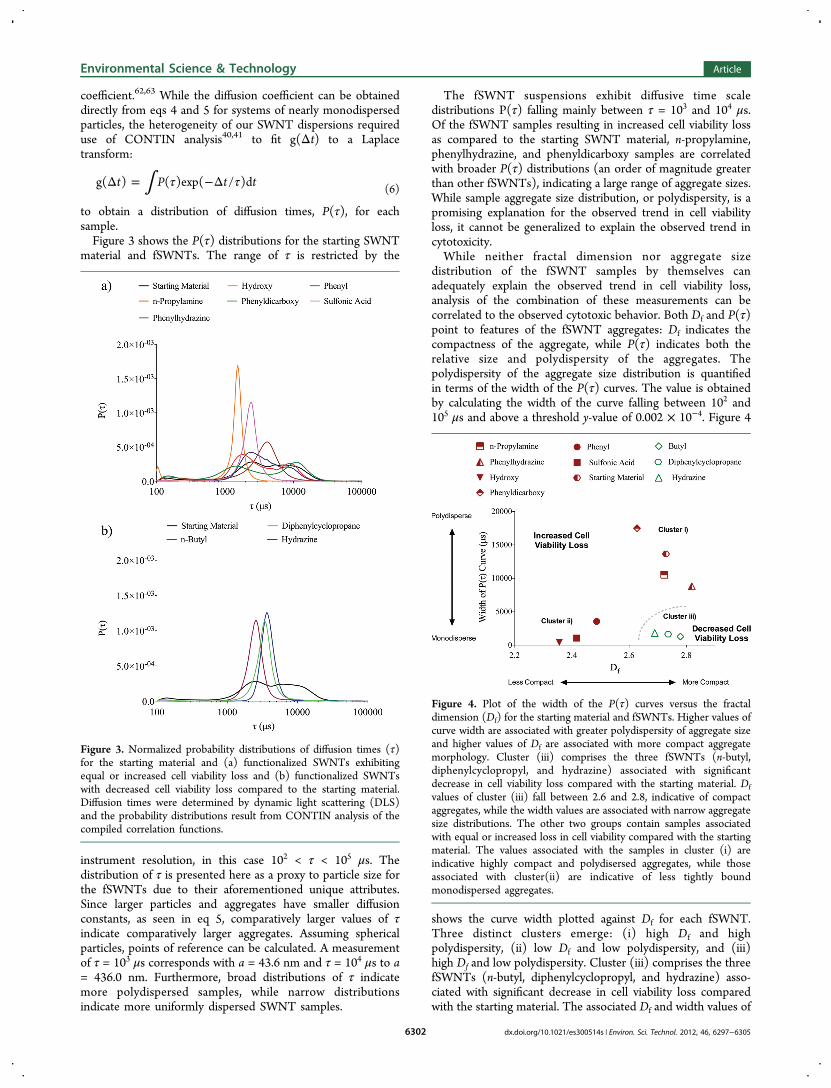
to obtain a distribution of diffusion times, P(τ), for eachsample.Figure 3 shows the P(τ) distributions for the starting SWNT
material and fSWNTs. The range of τ is restricted by the
instrument resolution, in this case 102 < τ < 105 μs. Thedistribution of τ is presented here as a proxy to particle size forthe fSWNTs due to their aforementioned unique attributes.Since larger particles and aggregates have smaller diffusionconstants, as seen in eq 5, comparatively larger values of τindicate comparatively larger aggregates. Assuming sphericalparticles, points of reference can be calculated. A measurementof τ = 103 μs corresponds with a = 43.6 nm and τ = 104 μs to a= 436.0 nm. Furthermore, broad distributions of τ indicatemore polydispersed samples, while narrow distributionsindicate more uniformly dispersed SWNT samples.
The fSWNT suspensions exhibit diffusive time scaledistributions P(τ) falling mainly between τ = 103 and 104 μs.Of the fSWNT samples resulting in increased cell viability lossas compared to the starting SWNT material, n-propylamine,phenylhydrazine, and phenyldicarboxy samples are correlatedwith broader P(τ) distributions (an order of magnitude greaterthan other fSWNTs), indicating a large range of aggregate sizes.While sample aggregate size distribution, or polydispersity, is apromising explanation for the observed trend in cell viabilityloss, it cannot be generalized to explain the observed trend incytotoxicity.While neither fractal dimension nor aggregate size
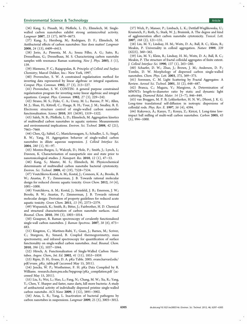
distribution of the fSWNT samples by themselves canadequately explain the observed trend in cell viability loss,analysis of the combination of these measurements can becorrelated to the observed cytotoxic behavior. Both Df and P(τ)point to features of the fSWNT aggregates: Df indicates thecompactness of the aggregate, while P(τ) indicates both therelative size and polydispersity of the aggregates. Thepolydispersity of the aggregate size distribution is quantifiedin terms of the width of the P(τ) curves. The value is obtainedby calculating the width of the curve falling between 102 and105 μs and above a threshold y-value of 0.002 × 10⁻⁴. Figure 4
shows the curve width plotted against Df for each fSWNT.Three distinct clusters emerge: (i) high Df and highpolydispersity, (ii) low Df and low polydispersity, and (iii)high Df and low polydispersity. Cluster (iii) comprises the threefSWNTs (n-butyl, diphenylcyclopropyl, and hydrazine) asso-ciated with significant decrease in cell viability loss comparedwith the starting material. The associated Df and width values of
Figure 3. Normalized probability distributions of diffusion times (τ)for the starting material and (a) functionalized SWNTs exhibitingequal or increased cell viability loss and (b) functionalized SWNTswith decreased cell viability loss compared to the starting material.Diffusion times were determined by dynamic light scattering (DLS)and the probability distributions result from CONTIN analysis of thecompiled correlation functions.
Figure 4. Plot of the width of the P(τ) curves versus the fractaldimension (Df) for the starting material and fSWNTs. Higher values ofcurve width are associated with greater polydispersity of aggregate sizeand higher values of Df are associated with more compact aggregatemorphology. Cluster (iii) comprises the three fSWNTs (n-butyl,diphenylcyclopropyl, and hydrazine) associated with significantdecrease in cell viability loss compared with the starting material. Dfvalues of cluster (iii) fall between 2.6 and 2.8, indicative of compactaggregates, while the width values are associated with narrow aggregatesize distributions. The other two groups contain samples associatedwith equal or increased loss in cell viability compared with the startingmaterial. The values associated with the samples in cluster (i) areindicative highly compact and polydisersed aggregates, while thoseassociated with cluster(ii) are indicative of less tightly boundmonodispersed aggregates.
Environmental Science & Technology Article
dx.doi.org/10.1021/es300514s | Environ. Sci. Technol. 2012, 46, 6297−63056302

this cluster are indicative of compact aggregates with a narrowsize distribution, respectively. The other two groups containsamples associated with equal or increased loss in cell viabilitycompared with the starting material.To further investigate the relationship of fSWNT aggregate
morphology and size distribution to cytotoxicity, an additionalexperiment was conducted in which aggregation state wasaffected by physical means rather than chemical surfacefunctionalization. Since sample preparation by ball milling isknown to affect carbon nanotube size and morphology,64 thepristine SWNT was subjected to mechanical perturbation usingthe ball mill (Heavy-duty Wig-L-Bug grinder/mixer analog),after which SLS (ALV-GmbH), DLS, and cytotoxicity analyseswere performed. The ball milling preparation allowed foralteration of SWNT morphology and dispersity with minimalalteration of surface chemistry.A starting material SWNT sample was collected after 30 min
exposure to the ball mill. This sample resulted in statisticallysignificant increase in cell viability loss (SI Figure S12a).Furthermore, the aggregate size distribution curve shiftedtoward shorter diffusive time, τ, (i.e., smaller aggregate size).Static light scattering measurements were completed as beforeto determine the sample fractal dimensions, which decreasedfrom 2.70 to 2.24 after ball milling. This indicates that smallerless tightly bound aggregates correlate with an increase in cellviability loss. The impact of fractal dimension and dispersity onloss of cell viability is further exemplified by the comparison ofdiphenylcyclopropyl SWNT before and after ball milling. In thesame way, the aggregate size distribution shifts to lower τ, thefractal dimension decreases from 2.73 to 2.32, and the loss ofcell viability significantly increases after ball milling (from 62.2± 5.3 to 92.4 ± 2.7% cell viability loss) (SI Figure S12). TheP(τ) widths and Df associated with the SWNT samples beforeand after treatment in the ball mill are shown in Figure 5. Their
location on the plot supports the hypothesis that bothaggregate morphology and dispersivity impact the delineationbetween increased and decreased SWNT cytotoxicity. Thisdemonstrates that cytotoxicity can be modified via physicalalteration analogous to chemical modification since both can
result in similar impacts on aggregate morphology anddispersity.
Implications. The results presented here suggest that boththe addition of covalent surface functional groups andmechanical perturbation indirectly impact the cytotoxicity ofSWNTs. We conclude that the indirect impact results from theextent of dispersion, which is quantified by both the fractaldimension, Df, and aggregate size distribution. The compiledresults are the first to systematically investigate the relationshipbetween fSWNT physiochemical properties and bacterialcytotoxicity. While the results are limited to nine functionalgroups and E. coli cytotoxicity, they do suggest that there is anindirect relationship between surface functionalization andcytotoxicity through aggregate morphology and dispersity. Thisindicates that these SWNT aggregate characteristics are moreimportant than individual physiochemical properties of thefunctional groups. As such, it appears to be important toconsider aggregation characteristics first and foremost whenpredicting the likelihood for fSWNTs to adversely impactcytotoxicity, an indicator of potential greater impacts for humanhealth and the environment.It is important to understand that further studies are
necessary to fully realize the potential of this knowledge andits application to controlled fSWNT design. Additional toxicitytesting involving other organisms will further elucidate thematerial interactions within a variety of biological systems andshould include measuring additional toxic end points. While theresults presented here do not conclude with a clear set ofdesign rules for reduced hazard, they do support the notion thatthere may be critical minimum properties that could definesuch a set.
■ ASSOCIATED CONTENT
*S Supporting InformationSupporting Information includes further details on novelaspects of the hydrazine and diphenylcyclopropyl SWNTfunctionalization syntheses, the cytotoxicity assay, includingrepresentative fluorescent images, the proposed azo couplingbyproduct (Figure S6), a list of QikProp descriptors, percentfunctionalization calculations, and statistical analysis. Additionalcharacterization data including NMR data for diphenylcyclo-propyl, n-propylamine, phenylhydrazine, phenyl, and butylSWNT (Figure S1−S5), QikProp output data of six descriptorsfor fSWNTs (Table S1), Raman spectra (Figure S7), percentmass loss curves from TGA (Figure S8), tabulated burn offtemperatures from DTG curves (Table S1), representativeTEM images (Figure S9), tabulated pKa values of the functionalgroups and proposed dominant species (Table S2), electro-phoretic mobilities in 0.9% NaCl (Figure S10), I/Io vs q plotsfor determining Df (Figure S11), tabulated Df for all fSWNTs(Table S3) and cytotoxicity and aggregate size distributions ofball milled samples (Figure S12). This material is available freeof charge via the Internet at http://pubs.acs.org.
■ AUTHOR INFORMATION
Corresponding Author*Phone: (203) 432-9703; fax: (203) 432-4837; e-mail: [email protected].
NotesThe authors declare no competing financial interest.
Figure 5. Plot of the width of the P(τ) curves versus the fractaldimension (Df) for the starting material and diphenylcyclopropylSWNTs before and after ball milling. Higher values of curve width areassociated with greater polydispersity of aggregate size and highervalues of Df are associated with more compact aggregate morphology.
Environmental Science & Technology Article
dx.doi.org/10.1021/es300514s | Environ. Sci. Technol. 2012, 46, 6297−63056303

■ ACKNOWLEDGMENTS
We acknowledge the generous support of the National ScienceFoundation under the Research Grant CBET-0854373. LMPrecognizes support from the U.S. Environmental ProtectionAgency (EPA) STAR Fellowship Assistance Agreement No.FP91716701-0 and the NSF Graduate Research FellowshipProgram (GRFP). This publication has not been formallyreviewed by the EPA. The views expressed in this publicationare solely those of the authors, and the EPA does not endorseany products or commercial services mentioned in thispublication. The CAMCOR TEM facility is supported bygrants from the W. M. Keck Foundation, the M. J. MurdockCharitable Trust, the Oregon Nanoscience and Micro-technologies Institute, the Air Force Research Laboratory(under agreement number FA8650-05-1-5041), at the Uni-versity of Oregon. We also thank Dr. Steve Kang, Dr. ChadVecitis, Dr. Codruta Zoican, and Dr. Stephen Golledge for theirassistance with methods and instrument training, and technicalsupport throughout the analysis of the characterization results.
■ REFERENCES(1) Iijima, S. Helical microtubules of graphitic carbon. Nature 1991,354 (7), 56−58.(2) Avouris, P. Molecular electronics with carbon nanotubes. Acc.Chem. Res. 2002, 35 (12), 1026−1034.(3) Odom, T. W.; Huang, J.; Kim, P.; Lieber, C. M. Atomic structureand electronic properties of single-walled carbon nanotubes. Nature1998, 391, 62−64.(4) Ouyang, M.; Huang, J. L.; Lieber, C. M. Fundamental electronicproperties and applications of single-walled carbon nanotubes. Acc.Chem. Res. 2002, 35 (12), 1018−1025.(5) Mauter, M. S.; Elimelech, M. Environmental applications ofcarbon-based nanomaterials. Environ. Sci. Technol. 2008, 42 (16),5843−5859.(6) Aslan, S.; Loebick, C. Z.; Kang, S.; Elimelech, M.; Pfefferle, L. D.;Van Tassel, P. R. Antimicrobial biomaterials based on carbonnanotubes dispersed in poly(lactic-co-glycolic acid). Nanoscale 2010,2 (9), 1789−1794.(7) Popov, V. N. Carbon nanotubes: Properties and application.Mater. Sci. Eng. R 2004, 43 (3), 61−102.(8) Paradise, M.; Goswami, T. Carbon nanotubesProduction andindustrial applications. Mater. Des. 2007, 28 (5), 1477−1489.(9) Kaiser, J. P. R., M.; Buerki-Thurnherr, T.; Wick, P. CarbonnanotubesCurse or blessing. Curr. Med. Chem. 2011, 18, 2115−2128.(10) Lee, S. W.; Yabuuchi, N.; Gallant, B. M.; Chen, S.; Kim, B. S.;Hammond, P. T.; Shao-Horn, Y. High-power lithium batteries fromfunctionalized carbon-nanotube electrodes. Nat. Nano 2010, 5 (7),531−537.(11) Klaine, S. J.; Alvarez, P. J. J.; Batley, G. E.; Fernandes, T. F.;Handy, R. D.; Lyon, D. Y.; Mahendra, S.; McLaughlin, M. J.; Lead, J.R. Nanomaterials in the environment: Behavior, fate, bioavailability,and effects. Environ. Toxicol. Chem. 2008, 27 (9), 1825−1851.(12) Wiesner, M. R.; Lowry, G. V.; Jones, K. L.; Hochella, J. M. F.; DiGiulio, R. T.; Casman, E.; Bernhardt, E. S. Decreasing uncertainties inassessing environmental exposure, risk, and ecological implications ofnanomaterials. Environ. Sci. Technol. 2009, 43 (17), 6458−6462.(13) Yokel, A. R. M., C. Robert Engineered nanomaterials:Exposures, hazards, and risk prevention. J. Occup. Med. Toxicol.2011, 6 (7).(14) Vecitis, C. D.; Zodrow, K. R.; Kang, S.; Elimelech, M.Electronic-structure-dependent bacterial cytotoxicity of single-walledcarbon nanotubes. ACS Nano 2010, 4 (9), 5471−5479.(15) Eckelman, M. J.; Zimmerman, J. B.; Anastas, P. T. Toward greennano. J. Ind. Ecol. 2008, 12 (3), 316−328.
(16) Dhawan, A.; Sharma, V. Toxicity assessment of nanomaterials:Methods and challenges. Anal. Bioanal. Chem. 2010, 398, 589−605.(17) Hussain, M. A.; Kabit, M. A.; Sood, A. K. On the cytotoxicity ofcarbon nanotubes. Curr. Sci. 2009, 96 (5), 664−673.(18) Jia, G.; Wang, H.; Yan, L.; Wang, X.; Pei, R.; Yan, T.; Zhao, Y.;Guo, X. Cytotoxicity of carbon nanomaterials: Single-wall nanotube,multi-wall nanotube, and fullerene. Environ. Sci. Technol. 2005, 39 (5),1378−1383.(19) Tian, F.; Cui, D.; Schwarz, H.; Estrada, G. G.; Kobayashi, H.Cytotoxicity of single-wall carbon nanotubes on human fibroblasts.Toxicol. In Vitro 2006, 20 (7), 1202−1212.(20) Lewinski, N.; Colvin, V.; Drezek, R. Cytotoxicity of nano-particles. Small 2008, 4 (1), 26−49.(21) Pulskamp, K.; Diabate, S.; Krug, H. F. Carbon nanotubes showno sign of acute toxicity but induce intracellular reactive oxygenspecies in dependence on contaminants. Toxicol. Lett. 2007, 168 (1),58−74.(22) Worle-Knirsch, J. M.; Pulskamp, K.; Krug, H. F. Oops they did itagain! Carbon nanotubes hoax scientists in viability assays. Nano Lett.2006, 6 (6), 1261−1268.(23) Manna, S. K.; Sarkar, S.; Barr, J.; Wise, K.; Barrera, E. V.;Jejelowo, O.; Rice-Ficht, A. C.; Ramesh, G. T. Single-walled carbonnanotube induces oxidative stress and activates nuclear transcriptionfactor-kB in human keratinocytes. Nano Lett. 2005, 5 (9), 1676−1684.(24) Sun, Y. P.; Fu, K.; Lin, Y.; Huang, W. Functionalized carbonnanotubes: Properties and applications. Acc. Chem. Res. 2002, 35 (12),1096−1104.(25) Tiraferri, A.; Vecitis, C. D.; Elimelech, M. Covalent binding ofsingle-walled carbon nanotubes to polyamide membranes forantimicrobial surface properties. ACS Appl. Mater. Interfaces 2011, 3(8), 2869−2877.(26) Sayes, C. M.; Liang, F.; Hudson, J. L.; Mendez, J.; Guo, W.;Beach, J. M.; Moore, V. C.; Doyle, C. D.; West, J. L.; Billups, W. E.;Ausman, K. D.; Colvin, V. L. Functionalization density dependence ofsingle-walled carbon nanotubes cytotoxicity in vitro. Toxicol. Lett.2006, 161 (2), 135−142.(27) Sayes, C. M.; Fortner, J. D.; Guo, W.; Lyon, D.; Boyd, A. M.;Ausman, K. D.; Tao, Y. J.; Sitharaman, B.; Wilson, L. J.; Hughes, J. B.;West, J. L.; Colvin, V. L. The differential cytotoxicity of water-solublefullerenes. Nano Lett. 2004, 4 (10), 1881−1887.(28) Pan, H.; Liu, L.; Guo, Z. X.; Dai, L.; Zhang, F.; Zhu, D.; Czerw,R.; Carroll, D. L. Carbon nanotubols from mechanochemical reaction.Nano Lett. 2002, 3 (1), 29−32.(29) Syrgiannis, Z.; Hauke, F.; Rohrl, J.; Hundhausen, M.; Graupner,R.; Elemes, Y.; Hirsch, A. Covalent Sidewall Functionalization ofSWNTs by nucleophilic addition of lithium amides. Eur. J. Org. Chem.2008, 15, 2544−2550.(30) Chen, Y.; Haddon, R. C.; Fang, S.; Rao, A. M.; Eklund, P. C.;Lee, W. H.; Dickey, E. C.; Grulke, E. A.; Pendergrass, J. C.; Chavan,A.; Haley, B. E.; Smalley, R. E. Chemical attachment of organicfunctional groups to single-walled carbon nanotube material. J. Mater.Res. 1998, 13, 2423−2431.(31) Wunderlich, D.; Hauke, F.; Hirsch, A. Preferred functionaliza-tion of metallic and small-diameter single walled carbon nanotubes viareductive alkylation. J. Mater. Chem. 2008, 18 (13), 1493−1497.(32) Yokoi, T.; Iwamatsu, S.; Komai, S.; Hattori, T.; Murata, S.Chemical modification of carbon nanotubes with organic hydrazines.Carbon 2005, 43 (14), 2869−2874.(33) Awenat, K. M.; Davis, P. J.; Moloney, M. G.; Ebenezer, W. Achemical method for the convenient surface functionalisation ofpolymers. Chem. Commun. 2005, 8, 990−992.(34) Wang, H.; Griffiths, J.; Egdell, R. G.; Moloney, M. G.; Foord, J.S. Chemical functionalization of diamond surfaces by reaction withdiaryl carbenes. Langmuir 2008, 24 (3), 862−868.(35) Jonczyk, A.; Wlostowska, J. A Simple Method for generation ofdiazocompounds in an aqueous two-phase system. Synth. Commun.1978, 8 (8), 569−572.
Environmental Science & Technology Article
dx.doi.org/10.1021/es300514s | Environ. Sci. Technol. 2012, 46, 6297−63056304
(36) Kang, S.; Pinault, M.; Pfefferle, L. D.; Elimelech, M. Single-walled carbon nanotubes exhibit strong antimicrobial activity.Langmuir 2007, 23 (17), 8670−8673.(37) Kang, S.; Herzberg, M.; Rodrigues, D. F.; Elimelech, M.Antibacterial effects of carbon nanotubes: Size does matter! Langmuir2008, 24 (13), 6409−6413.(38) Jorio, A.; Pimenta, M. A.; Souza Filho, A. G.; Saito, R.;Dresselhaus, G.; Dresselhaus, M. S. Characterizing carbon nanotubesamples with resonance Raman scattering. New J. Phys. 2003, 5 (1),139.(39) Hiemenz, P. C.; Rajagopalan, R. Principles of Colloid and SurfaceChemistry; Marcel Dekker, Inc.: New York, 1997.(40) Provencher, S. W. A constrained regularization method forinverting data represented by linear algebraic or integral equations.Comput. Phys. Commun. 1982, 27 (3), 213−227.(41) Provencher, S. W. CONTIN: A general purpose constrainedregularization program for inverting noisy linear algebraic and integralequations. Comput. Phys. Commun. 1982, 27 (3), 229−242.(42) Strano, M. S.; Dyke, C. A.; Usrey, M. L.; Barone, P. W.; Allen,M. J.; Shan, H.; Kittrell, C.; Hauge, R. H.; Tour, J. M.; Smalley, R. E.Electronic structure control of single-walled carbon nanotubefunctionalization. Science 2010, 301 (5639), 1519−1522.(43) Saleh, N. B.; Pfefferle, L. D.; Elimelech, M. Aggregation kineticsof multiwalled carbon nanotubes in aquatic systems: Measurementsand environmental implications. Environ. Sci. Technol. 2008, 42 (21),7963−7969.(44) Chen, Q.; Saltiel, C.; Manickavasagam, S.; Schadler, L. S.; Siegel,R. W.; Yang, H. Aggregation behavior of single-walled carbonnanotubes in dilute aqueous suspension. J. Colloid Interface Sci.2004, 280 (1), 91−97.(45) Montes-Burgos, I.; Walczyk, D.; Hole, P.; Smith, J.; Lynch, I.;Dawson, K. Characterisation of nanoparticle size and state prior tonanotoxicological studies. J. Nanopart. Res. 2010, 12 (1), 47−53.(46) Kang, S.; Mauter, M. S.; Elimelech, M. Physicochemicaldeterminants of multiwalled carbon nanotube bacterial cytotoxicity.Environ. Sci. Technol. 2008, 42 (19), 7528−7534.(47) Voutchkova-Kostal, A. M.; Kostal, J.; Connors, K. A.; Brooks, B.W.; Anastas, P. T.; Zimmerman, J. B. Towards rational moleculardesign for reduced chronic aquatic toxicity. Green Chem. 2012, 14 (4),1001−1008.(48) Voutchkova, A. M.; Kostal, J.; Steinfeld, J. B.; Emerson, J. W.;Brooks, B. W.; Anastas, P.; Zimmerman, J. B. Towards rationalmolecular design: Derivation of property guidelines for reduced acuteaquatic toxicity. Green Chem. 2011, 13 (9), 2373−2379.(49) Wepasnick, K.; Smith, B.; Bitter, J.; Fairbrother, H. D. Chemicaland structural characterization of carbon nanotube surfaces. Anal.Bioanal. Chem. 2010, 396 (3), 1003−1014.(50) Graupner, R. Raman spectroscopy of covalently functionalizedsingle-wall carbon nanotubes. J. Raman Spectrosc. 2007, 38 (6), 673−683.(51) Kingston, C.; Martínez-Rubí, Y.; Guan, J.; Barnes, M.; Scriver,C.; Sturgeon, R.; Simard, B. Coupled thermogravimetry, massspectrometry, and infrared spectroscopy for quantification of surfacefunctionality on single-walled carbon nanotubes. Anal. Bioanal. Chem.2010, 396 (3), 1037−1044.(52) Hirsch, A. Functionalization of Single-Walled Carbon Nano-tubes. Angew. Chem., Int. Ed. 2002, 41 (11), 1853−1859.(53) Ripin, D. H.; Evans, D. A. pKa Table. 2005. evans.harvard.edu/pdf/evans_pKa_table.pdf (accessed May 15, 2011).(54) Jencks, W. P.; Westheimer, F. H. pKa Data Compiled by R.Williams. research.chem.psu.edu/brpgroup/pKa_compilation.pdf (ac-cessed May 15, 2011).(55) Liu, S.; Wei, L.; Hao, L.; Fang, N.; Chang, M. W.; Xu, R.; Yang,Y.; Chen, Y. Sharper and faster, nano darts, kill more bacteria: A studyof antibacterial activity of individually dispersed pristine single-walledcarbon nanotube. ACS Nano 2009, 3 (12), 3891−3902.(56) Arias, L. R.; Yang, L. Inactivation of bacterial pathogens bycarbon nanotubes in suspensions. Langmuir 2009, 25 (5), 3003−3012.
(57) Wick, P.; Manser, P.; Limbach, L. K.; Dettlaff-Weglikowska, U.;Krumeich, F.; Roth, S.; Stark, W. J.; Bruinink, A. The degree and kindof agglomeration affect carbon nanotube cytotoxicity. Toxicol. Lett.2007, 168 (2), 121−131.(58) Lin, M. Y.; Lindsay, H. M.; Weitz, D. A.; Ball, R. C.; Klein, R.;Meakin, P. Universality in colloid aggregation. Nature 1989, 339(6223), 360−362.(59) Lin, M. Y.; Klein, R.; Lindsay, H. M.; Weitz, D. A.; Ball, R. C.;Meakin, P. The structure of fractal colloidal aggregates of finite extent.J. Colloid Interface Sci. 1990, 137 (1), 263−280.(60) Schaefer, D. W.; Zhao, J.; Brown, J. M.; Anderson, D. P.;Tomlin, D. W. Morphology of dispersed carbon single-wallednanotubes. Chem. Phys. Lett. 2003, 375, 369−375.(61) Sorensen, C. M. Light Scattering by Fractal Aggregates: AReview. Aerosol Sci. Technol. 2001, 35 (2), 648−687.(62) Branca, C.; Magazu, V.; Mangione, A. Determination ofMWNTs length-to-diameter ratio by static and dynamic lightscattering. Diamond Relat. Mater. 14 (3−7), 846−849.(63) van Bruggen, M. P. B; Lekkerkerker, H. N. W.; Dhont, J. K. G.Long-time translational self-diffusion in isotropic dispersions ofcolloidal rods. Phys. Rev. E. 1997, 56 (4), 4394.(64) Kukovecz, Å.; Kanyo, T.; Konya, Z.; Kiricsi, I. Long-time low-impact ball milling of multi-wall carbon nanotubes. Carbon 2005, 43(5), 994−1000.
Environmental Science & Technology Article
dx.doi.org/10.1021/es300514s | Environ. Sci. Technol. 2012, 46, 6297−63056305