Iron Oxide-Gold Core-Shell Nanoparticles as Multimodal Imaging Contrast Agent
Upload
independentCategory
view
4download
0
NANO REVIEW
Magnetic Iron Oxide Nanoparticles: Synthesis and SurfaceFunctionalization Strategies
Wei Wu Æ Quanguo He Æ Changzhong Jiang
Received: 8 July 2008 / Accepted: 11 September 2008 / Published online: 2 October 2008
� to the authors 2008
Abstract Surface functionalized magnetic iron oxide
nanoparticles (NPs) are a kind of novel functional materi-
als, which have been widely used in the biotechnology and
catalysis. This review focuses on the recent development
and various strategies in preparation, structure, and mag-
netic properties of naked and surface functionalized iron
oxide NPs and their corresponding application briefly. In
order to implement the practical application, the particles
must have combined properties of high magnetic satura-
tion, stability, biocompatibility, and interactive functions at
the surface. Moreover, the surface of iron oxide NPs could
be modified by organic materials or inorganic materials,
such as polymers, biomolecules, silica, metals, etc. The
problems and major challenges, along with the directions
for the synthesis and surface functionalization of iron oxide
NPs, are considered. Finally, some future trends and pro-
spective in these research areas are also discussed.
Keywords Magnetic iron oxide NPs �Surface functionalization � Preparation � Application
Introduction
Nanoparticle are submicron moieties (diameters ranging
from 1 to 100 nm according to the used term, although
there are examples of NPs several hundreds of nanometers
in size) made of inorganic or organic materials, which
have many novel properties compared with the bulk
materials [1]. On this basis, magnetic NPs have many
unique magnetic properties such as superparamagnetic,
high coercivity, low Curie temperature, high magnetic
susceptibility, etc. Magnetic NPs are of great interest for
researchers from a broad range of disciplines, including
magnetic fluids, data storage, catalysis, and bioapplica-
tions [2–6]. Especially, magnetic ferrofluids and data
storage are the applied researches that have led to the
intergration of magnetic NPs in a myriad of commercial
applications. Currently, magnetic NPs are also used in
important bioapplications, including magnetic biosepara-
tion and detection of biological entities (cell, protein,
nucleic acids, enzyme, bacterials, virus, etc.), clinic
diagnosis and therapy (such as MRI (magnetic resonance
image) and MFH (magnetic fluid hyperthermia)), targeted
drug delivery and biological labels. However, it is crucial
to choose the materials for the construction of nano-
structure materials and devises with adjustable physical
and chemical properties. To this end, magnetic iron oxide
NPs became the strong candidates, and the application of
small iron oxide NPs in in vitro diagnostics has been
practiced for nearly half a century [7]. In the last decade,
increased investigations with several types of iron oxides
have been carried out in the field of magnetic NPs (mostly
includes the Fe3O4 magnetite, FeIIFeIII2O4, ferrimagnetic,
superparamagnetic when the size is less than 15 nm),
a-Fe2O3 (hematite, weakly ferromagnetic or antiferro-
magnetic), c-Fe2O3 (maghemite, ferrimagnetic), FeO
W. Wu � C. Jiang (&)
Department of Physics, Wuhan University, Wuhan 430072,
People’s Republic of China
e-mail: [email protected]
W. Wu
e-mail: [email protected]
Q. He
Key Laboratory of Green Packaging and Bio-Nanotechnology
Applications (Hunan Province), Hunan University of
Technology, Zhuzhou 412008, People’s Republic of China
e-mail: [email protected]
123
Nanoscale Res Lett (2008) 3:397–415
DOI 10.1007/s11671-008-9174-9
(wustite, antiferromagnetic), e-Fe2O3 and b-Fe2O3) [8],
among which magnetite and maghemite is the very
promising and popular candidates since its biocompati-
bility have already proven.
However, it is a technological challenge to control size,
shape, stability, and dispersibility of NPs in desired sol-
vents. Magnetic iron oxide NPs have a large surface-to-
volume ratio and therefore possess high surface energies.
Consequently, they tend to aggregate so as to minimize the
surface energies. Moreover, the naked iron oxide NPs have
high chemical activity, and are easily oxidized in air
(especially magnetite), generally resulting in loss of mag-
netism and dispersibility. Therefore, providing proper
surface coating and developing some effective protection
strategies to keep the stability of magnetic iron oxide NPs
is very important. These strategies comprise grafting of or
coating with organic molecules, including small organic
molecules or surfactants, polymers, and biomolecules, or
coating with an inorganic layer, such as silica, metal or
nonmetal elementary substance, metal oxide or metal sul-
fide. Practically, it is worthy that in many cases the
protecting shells not only stabilize the magnetic iron oxide
NPs, but can also be used for further functionalization.
In the following, we focus mainly on recent develop-
ment and various strategies in the preparation, structure and
magnetic properties of various surface functionalized
strategies of magnetic iron oxide NPs and their corre-
sponding applications, as well as the research advances on
functionalizations of magnetic iron oxide NPs worldwide.
Further the problems and major challenges still should be
solved are pointed out, and the directions in these resear-
ches are also discussed.
Synthesis of Iron Oxide NPs
In the last decades, much research has been developed to the
synthesis of iron oxide NPs, and many reports have descri-
bed efficient synthesis approaches to produce the shape-
controlled, stable, biocompatible, and monodispersed iron
oxide NPs. The most common methods including co-pre-
cipitation, thermal decomposition, hydrothermal synthesis,
microemulsion, sonochemical synthesis, and sonochemical
synthetic route can all be directed to the synthesis of high
quality of iron oxide NPs. In addition, these NPs can also be
prepared by the other methods such as electrochemical
synthesis [9, 10], laser pyrolysis techniques [11], microor-
ganism or bacterial synthesis (especially the Magnetotactic
bacteria and iron reducing bacteria) [12, 13], etc. As follows,
we try to present typical and recent examples for the dis-
cussions of each synthetic pathway and the corresponding
formation mechanism.
Co-Precipitation
The most conventional method for obtaining Fe3O4 or
c-Fe2O3 is by co-precipitation. This method consists of
mixing ferric and ferrous ions in a 1:2 molar ratio in highly
basic solutions at room temperature or at elevated temper-
ature. The size and shape of the iron oxide NPs depends on
the type of salt used (such as chlorides, sulfates, nitrates,
perchlorates, etc.), the ferric and ferrous ions ratio, the
reaction temperature, the PH value, ionic strength of the
media, and the other reaction parameters (e.g. stirring rate,
dropping speed of basic solution). Recently we have repor-
ted the co-precipitation synthesis of Fe3O4 NPs and their
corresponding morphology, structure, and magnetic prop-
erties at different reaction temperature was investigated [14].
This method would critically affect the physical and
chemical properties of the nanosized iron oxide particles.
Generally, the saturation magnetization (MS) values found in
nanostructured materials are usually smaller than the
corresponding bulk phase, provided that no change in
ionic configurations occurs [15]. Accordingly, experimental
value for MS in magnetic iron oxide NPs have been reported to
span the 30–80 emu g-1 range, lower than the bulk magnetic
value 100 emu g-1. In addition, Fe3O4 NPs are not very stable
under ambient conditions and are easily oxidised to Fe2O3 or
dissolved in an acidic medium. In order to avoid the possible
oxidation in the air, the synthesis of Fe3O4 NPs must be done
in an anaerobic conditions. Based on this point, Fe3O4 NPs can
also be utilized to prepare the Fe2O3 NPs by oxidation or
anneal treatment under oxygen atmosphere. And oxidation
is not the important influence factor for Fe2O3 NPs due to its
own chemical stability in alkaline or acidic environment.
However, this method generates particles with a wide
particle size distribution, which requires secondary size
selection sometimes. A wide particle size distribution will
result in a wide range of blocking temperatures (TB) due to TB
depends on particle size and, therefore non-ideal magnetic
behavior for many applications. Kang et al. [16] reported a
synthesis of monodispersed, uniform, and narrow size
distributional Fe3O4 NPs (the diameter of NPs was
8.5 ± 1.3 nm) by co-precipitation without surfactants, the
reaction in an aqueous solution with a molar ratio of FeII/
FeIII = 0.5 and a pH = 11–12, and the colloidal suspensions
of the magnetite can be then directly oxidized by aeration to
form colloidal suspensions of c-Fe2O3. In contrast, many
recent publications have described efficient routes to obtain
the monodispersed NPs, surfactants such as dextran or
polyvinyl alcohol (PVA) can be added in the reaction media,
or the particles can be coated in a subsequent step [17, 18].
Surfactants act as protecting agent for controlling particle
size and stabilizing the colloidal dispersions.
Additionally, the disadvantage of these aqueous solution
syntheses is that the high pH value of the reaction mixture
398 Nanoscale Res Lett (2008) 3:397–415
123
has to be adjusted in both the synthesis and purification
steps, and the process toward uniformed and monodi-
spersed NPs has only very limited success. On the other
hand, wastewaters with very basic pH values are also
generated in the experiment, which require subsequent
treatments for protecting the environment.
Thermal Decomposition
An organic solution phase decomposition route has been
widely used in iron oxide NPs synthesis, and decomposi-
tion of Fe(cup)3 (cup = N-nitrosophenylhydroxylamine),
Fe(acac)3 (acac = acetylacetonate), or Fe(CO)5 followed
by oxidation can lead to high-quality monodispersed iron
oxide NPs, which usually requires relatively higher tem-
peratures and a complicated operation.
Sun and Zeng [19] have reported a general decomposition
approach for the synthesis of size-controlled monodispersed
magnetite NPs based on high temperature (265 �C) reaction
of Fe(acac)3 in phenyl ether in the presence of alcohol, oleic
acid, and oleylamine. With the smaller magnetite NPs as
seeds, larger monodispersed magnetite NPs of up to 20 nm in
diameter can be synthesized and dispersed into nonpolar
solvent by seed-mediated growth method. The process does
not require a size-selection procedure and is readily scaled up
for mass production. The as-synthesized Fe3O4 nanoparticle
assemblies can be transformed easily into c-Fe2O3 NPs by
annealing at high temperature (250 �C) and oxygen for 2 h.
Generally, direct decomposition of Fe(Cup)3 single
precursor can lead to monodispersed c-Fe2O3 NPs [20].
The thermal decomposition of Fe(CO)5 produces iron NPs
and the following oxidation by a chemical reagent can also
lead to monodispersed c-Fe2O3 NPs [21]. For instance,
Hyeon et al. [22] reported a synthesis of highly crystalline
and monodispersed iron NPs without size-selection process
by the thermal decomposition of iron pentacarbonyl in the
presence of oleic acid at 100 �C. The resulting iron NPs
were transformed to monodispersed c-Fe2O3 nanocrystal-
lites by controlled oxidation using trimethylamine oxide as
a mild oxidant. Particle size can be varied from 4 to 16 nm
by controlling the experimental parameters.
Although the thermal decomposition method has many
advantages for producing highly monodispersed particles
with a narrow size distribution, it has the big disadvantage
that the resulting NPs are generally only dissolved in
nonpolar solvents.
Microemulsion
Microemulsion is a thermodynamically stable isotropic
dispersion of two immiscible phases (water and oil) under
the surfactant present, the surfactant molecules may form a
monolayer at the interface between the oil and water, with
the hydrophobic tails of the surfactant molecules dissolved
in the oil phase and the hydrophilic head groups in the
aqueous phase. As in the binary systems (water/surfactant
or oil/surfactant), self-assembled structures of different
types can be formed, ranging, for example, from (inverted)
spherical and cylindrical micelles to lamellar phases and
bicontinuous microemulsions, which may coexist with
predominantly oil or aqueous phases [23]. In this sense,
microemulsion and inverse micelles route can be employed
for obtaining the shape- and size-controlled iron oxide NPs.
Particularly, water-in-oil (w/o) microemulsions are
formed by well-defined nanodroplets of the aqueous phase,
dispersed by the assembly of surfactant molecules in a
continuous oil phase. Vidal-Vidal et al. [24] have reported
the synthesis of monodisperse maghemite NPs by the one-
pot microemulsion method. The spherical shaped particles,
capped with a monolayer coating of oleylamine (or oleic
acid), show a narrow size distribution of 3.5 ± 0.6 nm, are
well crystallized and have high saturation magnetization
values (76.3 Am2/kg for uncoated NPs, 35.2 Am2/kg for
oleic acid coated NPs, and 33.2 Am2/kg for oleylamine
coated NPs). Moreover, the results show that oleylamine
act as precipitating and capping agent. However, cyclo-
hexylamine acts only as precipitating agent and does not
avoid particle aggregation. Chin and Yaacob reported the
synthesis of magnetic iron oxide NPs (less than 10 nm) via
w/o microemulsion, furthermore, in comparison to the
particles produced by Massart’s procedure [25], particles
produced by microemulsion technique were smaller in size
and were higher in saturation magnetization [26].
However, despite the presence of surfactants, the
aggregation of the produced NPs usually needs several
washing processes and further stabilization treatments.
Hydrothermal Synthesis
Iron oxide NPs with controlled size and shape are techno-
logically important due to strong correlation between these
parameters and magnetic properties. The microemulsion
and thermal decomposition methods usually lead to com-
plicated process or require relatively high temperatures. As
an alternative, hydrothermal synthesis includes various wet-
chemical technologies of crystallizing substance in a sealed
container from the high temperature aqueous solution
(generally in the range from 130 to 250 �C) at high vapor
pressure (generally in the range from 0.3 to 4 MPa). This
technique has also been used to grow dislocation-free single
crystal particles, and grains formed in this process could
have a better crystallinity than those from other processes,
so hydrothermal synthesis is prone to obtain the highly
crystalline iron oxide NPs.
Several authors have reported the synthesis of iron oxide
NPs by hydrothermal method [27–30]. There are two major
Nanoscale Res Lett (2008) 3:397–415 399
123

methods according to whether or not use the specific sur-
factants. For example, Wang et al. [31] have reported a
one-step hydrothermal process to prepare highly crystalline
Fe3O4 nanopowders without using the surfactants. The
nanoscale Fe3O4 powder (40 nm) obtained at 140 �C for
6 h possessed a saturation magnetization of 85.8 emu�g-1,
a little lower than that of the correspondent bulk Fe3O4 (92
emu�g-1). It is suggested that the well-crystallized Fe3O4
grains formed under appropriate hydrothermal conditions
should be responsible for the increased saturation magne-
tization in nanosized Fe3O4. On the contrary, Zheng et al.
[32] reported a hydrothermal route for preparing Fe3O4
NPs with diameter of ca. 27 nm in the presence of a sur-
factant, sodium bis(2-ethylhexyl)sulfosuccinate (AOT).
The magnetic properties of the NPs exhibited a super-
paramagnetic behavior at room temperature.
Moreover, hydrothermal treatment is one of the suc-
cessful ways of growing crystals for iron oxide NPs.
Recently Daou et al. [33] have reported that the magnetite
particles with an average size of 39 nm and good mono-
dispersity have been synthesized by co-precipitation at
70 �C from ferrous Fe2? and ferric Fe3? ions by a
N(CH3)4OH solution, followed by hydrothermal treatment
at 250 �C. The magnetite NPs before the hydrothermal step
display an average size of 12 nm and are highly oxidized
when they are in contact with air. Additionally, hydro-
thermal synthesis is conductive to prepare the unusual iron
oxide nanostructures such as iron oxide nanocubes [34],
iron oxide hollow spheres [35], etc.
Sonochemical Synthesis
As a competitive alternative, the sonochemical method has
been extensively used to generate novel materials with
unusual properties. The chemical effects of ultrasound arise
from acoustic cavitation, that is, the formation, growth, and
implosive collapse of bubbles in liquid. The implosive
collapse of the bubble generates a localized hotspot
through adiabatic compression or shock wave formation
within the gas phase of the collapsing bubble. The condi-
tions formed in these hotspots have been experimentally
determined, with transient temperatures of 5000 K, pres-
sures of 1800 atm, and cooling rates in excess of 1010 K/s
[36]. These extreme conditions were beneficial to form the
new phase, and have a shear effect for agglomeration,
which is prone to prepare the highly monodispersive NPs.
This method has been applied for the synthesis of various
nanocomposites, and its versatility has been successfully
demonstrated in iron oxide NPs preparation [37]. For
instance, magnetite NPs can be simply synthesized by
sonication of iron(II)acetate in water under an argon
atmosphere. Vijayakumar et al. [38] reported a sonochem-
ical synthetic route for preparing the pure nanometer-size
Fe3O4 powder with particle size of ca. 10 nm. The prepared
Fe3O4 NPs are superparamagnetic and its magnetization at
room temperature is very low (\1.25 emu g-1). Recently
Pinkas et al. [39] developed a sonochemical synthetic
method for preparing the amorphous nanoscopic iron oxide
by sonolysis of Fe(acac)3 under Ar with a small amount
of added water. The organic content and the surface area
of the Fe2O3 NPs can be controlled with an amount of
water in the reaction mixture, and it increases from
48 m2 g-1 for dry solvent up to 260 m2 g-1 when wet Ar is
employed. Such surface modified ultrasmall (1–2 nm) NPs
exhibit an unrecorded low magnetic transition temperature
of about 25 K, below this temperature they behave as spe-
romagnetic, highly magnetically disordered systems with a
high contribution of the surface anisotropy, and this spe-
romagnetic behavior can be observed by Mossbauer
spectra.
The five above mentioned synthetic methods have sev-
eral advantages and disadvantages for preparing iron oxide
NPs, respectively. In terms of size and morphology control
of the iron oxide NPs, thermal decomposition and hydro-
thermal synthetic route seems the optimal method. For
obtaining the water-soluble and biocompatible iron oxide
NPs, co-precipitation often was employed, but this method
presents low control of the particle shape, broad distribu-
tions of sizes and aggregation of particles. As a time-
competitive alternative, sonochemical route can also be
used to synthesis iron oxide NPs with unusually magnetic
properties. In addition, it is noteworthy that some green
chemical synthesis routes and biological synthesis routes
have been reported for environment protection purposes.
For example, recently Bharde et al. [12] reported the bac-
terium Actinobacter sp. is capable of synthesizing
maghemite under aerobic conditions when reacted with the
ferric chloride precursors. Moreover, maghemite NPs show
superparamagnetic characteristics as expected. Therefore,
as the enviromental protection and eco friendliness-
competitive alternative, green chemistry and biological
methods such as bacterially induced synthesis based syn-
thesis of iron oxide NPs are important advances.
In the following sections, the recent development and
various strategies for obtaining surface functionalized iron
oxide NPs by organic and inorganic materials will be
discussed.
Surface Functionalization by Organic Materials
In the preparation and storage of ferrofluids, the stability is
of utmost importance. Organic compounds are often
employed to passivate the surface of the iron oxide NPs
during or after the preparing procedure to avoid agglom-
eration. The main reasons for magnetic iron oxide NPs
400 Nanoscale Res Lett (2008) 3:397–415
123
have hydrophobic surfaces with a large surface area-to-
volume ratio if in the absence of any proper surface coat-
ing, and the hydrophobic interactions between the NPs will
cause them to aggregate and form large clusters, resulting
in increased particle size. Additionally, in order to expand
the biological application scope of the iron oxide NPs,
some biomolecules are also employed to enhance its
biocompatibility.
In recent years, considerable efforts have been devoted
to the design and controlled fabrication of nanostructured
materials displaying specific functional properties.
Organic compounds coated on iron oxide NPs offer a high
potential application in several areas. The structure of
organic compounds functionalized magnetic iron oxide
NPs consists of two major parts: preserved the magnetic
property of magnetic iron oxides and preserved the other
properties of organic molecules. Generally, if iron oxides
were always assumed as the core, its structure can roughly
be divided into three types: core-shell, matrix, and shella-
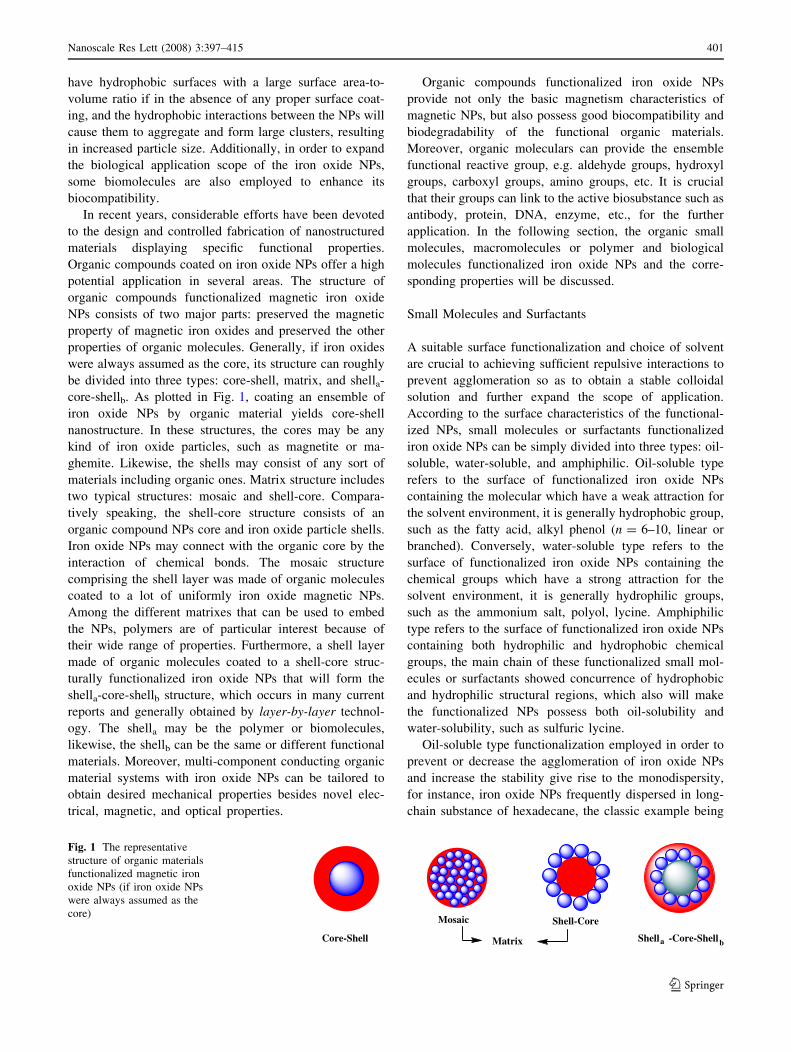
core-shellb. As plotted in Fig. 1, coating an ensemble of
iron oxide NPs by organic material yields core-shell
nanostructure. In these structures, the cores may be any
kind of iron oxide particles, such as magnetite or ma-
ghemite. Likewise, the shells may consist of any sort of
materials including organic ones. Matrix structure includes
two typical structures: mosaic and shell-core. Compara-
tively speaking, the shell-core structure consists of an
organic compound NPs core and iron oxide particle shells.
Iron oxide NPs may connect with the organic core by the
interaction of chemical bonds. The mosaic structure
comprising the shell layer was made of organic molecules
coated to a lot of uniformly iron oxide magnetic NPs.
Among the different matrixes that can be used to embed
the NPs, polymers are of particular interest because of
their wide range of properties. Furthermore, a shell layer
made of organic molecules coated to a shell-core struc-
turally functionalized iron oxide NPs that will form the
shella-core-shellb structure, which occurs in many current
reports and generally obtained by layer-by-layer technol-
ogy. The shella may be the polymer or biomolecules,
likewise, the shellb can be the same or different functional
materials. Moreover, multi-component conducting organic
material systems with iron oxide NPs can be tailored to
obtain desired mechanical properties besides novel elec-
trical, magnetic, and optical properties.
Organic compounds functionalized iron oxide NPs
provide not only the basic magnetism characteristics of
magnetic NPs, but also possess good biocompatibility and
biodegradability of the functional organic materials.
Moreover, organic moleculars can provide the ensemble
functional reactive group, e.g. aldehyde groups, hydroxyl
groups, carboxyl groups, amino groups, etc. It is crucial
that their groups can link to the active biosubstance such as
antibody, protein, DNA, enzyme, etc., for the further
application. In the following section, the organic small
molecules, macromolecules or polymer and biological
molecules functionalized iron oxide NPs and the corre-
sponding properties will be discussed.
Small Molecules and Surfactants
A suitable surface functionalization and choice of solvent
are crucial to achieving sufficient repulsive interactions to
prevent agglomeration so as to obtain a stable colloidal
solution and further expand the scope of application.
According to the surface characteristics of the functional-
ized NPs, small molecules or surfactants functionalized
iron oxide NPs can be simply divided into three types: oil-
soluble, water-soluble, and amphiphilic. Oil-soluble type
refers to the surface of functionalized iron oxide NPs
containing the molecular which have a weak attraction for
the solvent environment, it is generally hydrophobic group,
such as the fatty acid, alkyl phenol (n = 6–10, linear or
branched). Conversely, water-soluble type refers to the
surface of functionalized iron oxide NPs containing the
chemical groups which have a strong attraction for the
solvent environment, it is generally hydrophilic groups,
such as the ammonium salt, polyol, lycine. Amphiphilic
type refers to the surface of functionalized iron oxide NPs
containing both hydrophilic and hydrophobic chemical
groups, the main chain of these functionalized small mol-
ecules or surfactants showed concurrence of hydrophobic
and hydrophilic structural regions, which also will make
the functionalized NPs possess both oil-solubility and
water-solubility, such as sulfuric lycine.
Oil-soluble type functionalization employed in order to
prevent or decrease the agglomeration of iron oxide NPs
and increase the stability give rise to the monodispersity,
for instance, iron oxide NPs frequently dispersed in long-
chain substance of hexadecane, the classic example being
Core-Shell
Mosaic Shell-Core
Shella -Core-ShellbMatrix
Fig. 1 The representative
structure of organic materials
functionalized magnetic iron
oxide NPs (if iron oxide NPs
were always assumed as the
core)
Nanoscale Res Lett (2008) 3:397–415 401
123
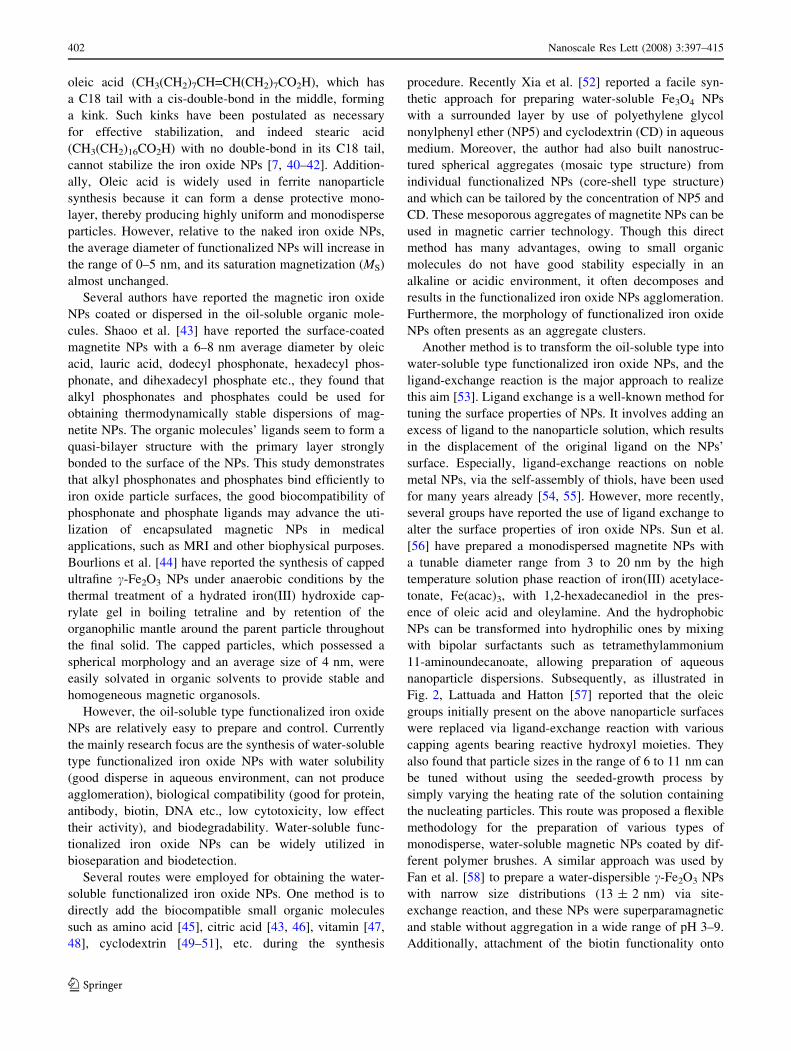
oleic acid (CH3(CH2)7CH=CH(CH2)7CO2H), which has
a C18 tail with a cis-double-bond in the middle, forming
a kink. Such kinks have been postulated as necessary
for effective stabilization, and indeed stearic acid
(CH3(CH2)16CO2H) with no double-bond in its C18 tail,
cannot stabilize the iron oxide NPs [7, 40–42]. Addition-
ally, Oleic acid is widely used in ferrite nanoparticle
synthesis because it can form a dense protective mono-
layer, thereby producing highly uniform and monodisperse
particles. However, relative to the naked iron oxide NPs,
the average diameter of functionalized NPs will increase in
the range of 0–5 nm, and its saturation magnetization (MS)
almost unchanged.
Several authors have reported the magnetic iron oxide
NPs coated or dispersed in the oil-soluble organic mole-
cules. Shaoo et al. [43] have reported the surface-coated
magnetite NPs with a 6–8 nm average diameter by oleic
acid, lauric acid, dodecyl phosphonate, hexadecyl phos-
phonate, and dihexadecyl phosphate etc., they found that
alkyl phosphonates and phosphates could be used for
obtaining thermodynamically stable dispersions of mag-
netite NPs. The organic molecules’ ligands seem to form a
quasi-bilayer structure with the primary layer strongly
bonded to the surface of the NPs. This study demonstrates
that alkyl phosphonates and phosphates bind efficiently to
iron oxide particle surfaces, the good biocompatibility of
phosphonate and phosphate ligands may advance the uti-
lization of encapsulated magnetic NPs in medical
applications, such as MRI and other biophysical purposes.
Bourlions et al. [44] have reported the synthesis of capped
ultrafine c-Fe2O3 NPs under anaerobic conditions by the
thermal treatment of a hydrated iron(III) hydroxide cap-
rylate gel in boiling tetraline and by retention of the
organophilic mantle around the parent particle throughout
the final solid. The capped particles, which possessed a
spherical morphology and an average size of 4 nm, were
easily solvated in organic solvents to provide stable and
homogeneous magnetic organosols.
However, the oil-soluble type functionalized iron oxide
NPs are relatively easy to prepare and control. Currently
the mainly research focus are the synthesis of water-soluble
type functionalized iron oxide NPs with water solubility
(good disperse in aqueous environment, can not produce
agglomeration), biological compatibility (good for protein,
antibody, biotin, DNA etc., low cytotoxicity, low effect
their activity), and biodegradability. Water-soluble func-
tionalized iron oxide NPs can be widely utilized in
bioseparation and biodetection.
Several routes were employed for obtaining the water-
soluble functionalized iron oxide NPs. One method is to
directly add the biocompatible small organic molecules
such as amino acid [45], citric acid [43, 46], vitamin [47,
48], cyclodextrin [49–51], etc. during the synthesis
procedure. Recently Xia et al. [52] reported a facile syn-
thetic approach for preparing water-soluble Fe3O4 NPs
with a surrounded layer by use of polyethylene glycol
nonylphenyl ether (NP5) and cyclodextrin (CD) in aqueous
medium. Moreover, the author had also built nanostruc-
tured spherical aggregates (mosaic type structure) from
individual functionalized NPs (core-shell type structure)
and which can be tailored by the concentration of NP5 and
CD. These mesoporous aggregates of magnetite NPs can be
used in magnetic carrier technology. Though this direct
method has many advantages, owing to small organic
molecules do not have good stability especially in an
alkaline or acidic environment, it often decomposes and
results in the functionalized iron oxide NPs agglomeration.
Furthermore, the morphology of functionalized iron oxide
NPs often presents as an aggregate clusters.
Another method is to transform the oil-soluble type into
water-soluble type functionalized iron oxide NPs, and the
ligand-exchange reaction is the major approach to realize
this aim [53]. Ligand exchange is a well-known method for
tuning the surface properties of NPs. It involves adding an
excess of ligand to the nanoparticle solution, which results
in the displacement of the original ligand on the NPs’
surface. Especially, ligand-exchange reactions on noble
metal NPs, via the self-assembly of thiols, have been used
for many years already [54, 55]. However, more recently,
several groups have reported the use of ligand exchange to
alter the surface properties of iron oxide NPs. Sun et al.
[56] have prepared a monodispersed magnetite NPs with
a tunable diameter range from 3 to 20 nm by the high
temperature solution phase reaction of iron(III) acetylace-
tonate, Fe(acac)3, with 1,2-hexadecanediol in the pres-
ence of oleic acid and oleylamine. And the hydrophobic
NPs can be transformed into hydrophilic ones by mixing
with bipolar surfactants such as tetramethylammonium
11-aminoundecanoate, allowing preparation of aqueous
nanoparticle dispersions. Subsequently, as illustrated in
Fig. 2, Lattuada and Hatton [57] reported that the oleic
groups initially present on the above nanoparticle surfaces
were replaced via ligand-exchange reaction with various
capping agents bearing reactive hydroxyl moieties. They
also found that particle sizes in the range of 6 to 11 nm can
be tuned without using the seeded-growth process by
simply varying the heating rate of the solution containing
the nucleating particles. This route was proposed a flexible
methodology for the preparation of various types of
monodisperse, water-soluble magnetic NPs coated by dif-
ferent polymer brushes. A similar approach was used by
Fan et al. [58] to prepare a water-dispersible c-Fe2O3 NPs
with narrow size distributions (13 ± 2 nm) via site-
exchange reaction, and these NPs were superparamagnetic
and stable without aggregation in a wide range of pH 3–9.
Additionally, attachment of the biotin functionality onto
402 Nanoscale Res Lett (2008) 3:397–415
123

the surfaces of our NPs enabled the magnetic affinity iso-
lation of avidin–FITC from its incubation buffer with 96%
efficiency. However, ligand-exchange reaction often con-
tributes to the complicated operation and the difficulty of
control exchange rate, these problems are urgent to be
solved.
Moreover, the silane agent is often considered as a
candidate for modifying on the surface of iron oxide NPs
directly, for the advantages of the biocompatibility as well
as high density of surface functional endgroups, and
allowing for connecting to other metal, polymer or bio-
molecules [59, 60]. In general, silane-coated iron oxide NPs
still maintains the physical characteristics of naked NPs
with high saturation magnetization, the decrease value of
saturation magnetization often less than 10 emu g-1. The
3-aminopropyltriethyloxysilane (APTES), p-aminophenyl
trimethoxysilane (APTS), and mercaptopropyltriethoxysi-
lane (MPTES) agents were mostly employed for providing
the amino and sulfhydryl group, respectively. The physi-
cochemical mechanism of the silane agent modifying on the
surface of iron oxide NPs according to Arkles is depicted in
Fig. 3 [61]. The hydroxyl groups on the iron oxide NPs
surface reacted with the methoxy groups of the silane
molecules leading to the formation of Si–O bonds and
leaving the terminal functional groups available for
immobilization the other substance.
We have prepared magnetite NPs with average diameter
25 ± 5 nm by modified chemical co-precipitation, and
Fig. 2 Scheme for the magnetic
NPs functionalization procedure
described in this work. Steps 1A
and 1B: ligand-exchange
reactions. Step 2: acylation of
hydroxyl groups to prepare
ATRP surface initiators. Step
3A: surface-initiated ring
opening polymerization of
L-lactide. Steps 3B: surface-
initiated ATRP. Step 4:
deprotection or additional
reaction after polymerization.
Step 5: graftiong of
endfunctionalized PEG chains
onto the nanoparticle surface
using amidation chemistry.
From [57]
OCH3
Si OCH3
OCH3
R-3CH3OH
OH
Si OH
OH
RH2O
Silane
2 Silane
-2 H2OHO Si
R
OH
O Si
R
OH
O Si
R
OH
OH
HOHO OH
Iron Oxide NPs
OO OH
HO
Si
HHO
HH
O
SiSiO
O
HO
R
RR
OH
OO
O
Si
SiSi
HO
OR
RO R
OH
-2 H2O
Fig. 3 Physicochemical
mechanism for modifying the
silane agents on the surface of
iron oxide NPs
Nanoscale Res Lett (2008) 3:397–415 403
123
silane bridged surface tailoring are exploited to obtain
amino-coated and thiolated magnetite NPs via corre-
sponding APTES and MPTES modification, respectively.
The result reveals that the surface functionalized magnetic
particles have a slight dimensional increase in average
diameter while retain almost original saturation magneti-
zation; APTES is prone to maintain the morphology of the
magnetite NPs, but MPTES has an outstanding hypochro-
mic effect though it caused a slight decrease in the
saturation magnetization. The finding is of practical sig-
nificance for magnetic NPs applications associated with
bioconjugation, target carriers, and biosensing [62]. In
addition, silanes were found to render the iron oxide NPs
highly stable and water-dispersible, and it was also found
to form a protective layer against mild acid and alkaline
environments. Moreover, silane ligand-exchange reaction
can make the hydrophobic iron oxide NPs into water-
dispersible [63].
Polymers
Although most studies have focused on the development of
small organic molecules and surfactants coating up to now,
recently polymers functionalized iron oxide NPs are
receiving more and more attention, owing to the advanta-
ges of polymers coating will increase repulsive forces to
balance the magnetic and the van der Waals attractive
forces acting on the NPs. In addition, polymers coating on
the surface of iron oxide NPs offer a high potential in the
application of several fields. Moreover, polymer function-
alized iron oxide NPs have been extensively investigated
due to interest in their unique physical or chemical prop-
erties. To make use of these materials for fundamental or
applied research, access to well-defined NPs samples
whose properties can be ‘‘tuned’’ through chemical
modification is necessary. In a number of cases it has now
been shown that, through careful choice of the passivating
and activating polymers and/or reaction conditions, can
produce NPs with tailored and desired properties.
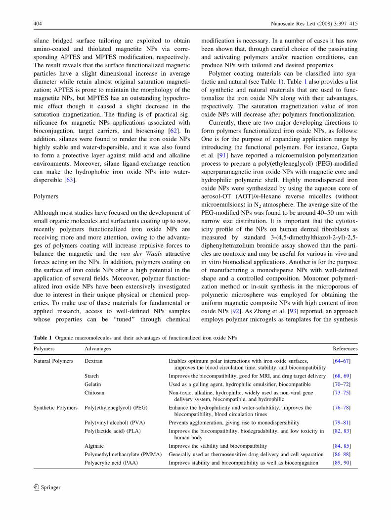
Polymer coating materials can be classified into syn-
thetic and natural (see Table 1). Table 1 also provides a list
of synthetic and natural materials that are used to func-
tionalize the iron oxide NPs along with their advantages,
respectively. The saturation magnetization value of iron
oxide NPs will decrease after polymers functionalization.
Currently, there are two major developing directions to
form polymers functionalized iron oxide NPs, as follows:
One is for the purpose of expanding application range by
introducing the functional polymers. For instance, Gupta
et al. [91] have reported a microemulsion polymerization
process to prepare a poly(ethyleneglycol) (PEG)-modified
superparamagnetic iron oxide NPs with magnetic core and
hydrophilic polymeric shell. Highly monodispersed iron
oxide NPs were synthesized by using the aqueous core of
aerosol-OT (AOT)/n-Hexane reverse micelles (without
microemulsions) in N2 atmosphere. The average size of the
PEG-modified NPs was found to be around 40–50 nm with
narrow size distribution. It is important that the cytotox-
icity profile of the NPs on human dermal fibroblasts as
measured by standard 3-(4,5-dimethylthiazol-2-yl)-2,5-
diphenyltetrazolium bromide assay showed that the parti-
cles are nontoxic and may be useful for various in vivo and
in vitro biomedical applications. Another is for the purpose
of manufacturing a monodisperse NPs with well-defined
shape and a controlled composition. Monomer polymeri-
zation method or in-suit synthesis in the microporous of
polymeric microsphere was employed for obtaining the
uniform magnetic composite NPs with high content of iron
oxide NPs [92]. As Zhang et al. [93] reported, an approach
employs polymer microgels as templates for the synthesis
Table 1 Organic macromolecules and their advantages of functionalized iron oxide NPs
Polymers Advantages References
Natural Polymers Dextran Enables optimum polar interactions with iron oxide surfaces,
improves the blood circulation time, stability, and biocompatibility
[64–67]
Starch Improves the biocompatibility, good for MRI, and drug target delivery [68, 69]
Gelatin Used as a gelling agent, hydrophilic emulsifier, biocompatible [70–72]
Chitosan Non-toxic, alkaline, hydrophilic, widely used as non-viral gene
delivery system, biocompatible, and hydrophilic
[73–75]
Synthetic Polymers Poly(ethyleneglycol) (PEG) Enhance the hydrophilicity and water-solublility, improves the
biocompatibility, blood circulation times
[76–78]
Poly(vinyl alcohol) (PVA) Prevents agglomeration, giving rise to monodispersibility [79–81]
Poly(lactide acid) (PLA) Improves the biocompatibility, biodegradability, and low toxicity in
human body
[82, 83]
Alginate Improves the stability and biocompatibility [84, 85]
Polymethylmethacrylate (PMMA) Generally used as thermosensitive drug delivery and cell separation [86–88]
Polyacrylic acid (PAA) Improves stability and biocompatibility as well as bioconjugation [89, 90]
404 Nanoscale Res Lett (2008) 3:397–415
123
of iron oxide NPs. At first, precursor cations were incor-
porated into the poly(N-isopropyl acrylamide-acrylic acid-
2-hydroxyethyl acrylate) microgel particles crosslinked
with N, N’-methylene bisacrylamide (BIS), then iron oxide
NPs were in-suit synthesized in the microgel by co-pre-
cipitation. They show that NPs with predetermined
dimensions and size-dependent properties can be synthe-
sized by using a very delicate balance between the reaction
conditions, the composition and the structure of microgel
templates, and the concentration of NPs in the microgel.
The synthesis of superparamagnetic Fe3O4 NPs in the
microgel templates was extremely efficient: for NPs con-
centration as high as 0.724 g per gram polymer, the NPs
had an average size of ca. 8.1 nm, low polydispersity, and
showed excellent magnetic properties, while the micro-
spheres carrying these NPs remained stimuli-responsive.
Additionally, atom transfer radical polymerization (ATRP)
method was utilized for obtaining monodisperse NPs with
well-defined core-shell shape. ATRP can offer polymeric
shells with low polydispersity and this method is easy to
control the molecular weight, thereby, the thickness of the
polymeric shell. Recently several polymer-coated iron
oxide NPs through surface-initiated ATRP in polar solvents
were successfully developed [94–97]. ATRP can be used in
preparing polymers with narrow molecular weight distri-
bution, block copolymers, graft polymers, random
copolymers and finally to realize controlling the thickness
of the polymeric shell and the size of polymer functional-
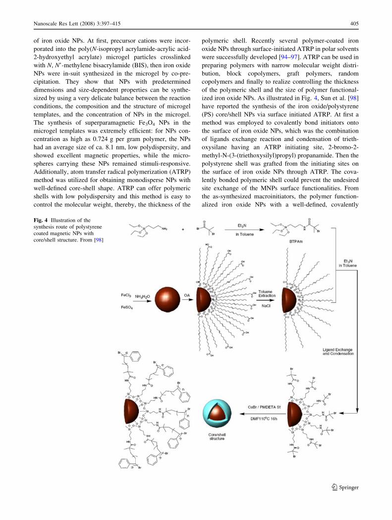
ized iron oxide NPs. As illustrated in Fig. 4, Sun et al. [98]
have reported the synthesis of the iron oxide/polystyrene
(PS) core/shell NPs via surface initiated ATRP. At first a
method was employed to covalently bond initiators onto
the surface of iron oxide NPs, which was the combination
of ligands exchange reaction and condensation of trieth-
oxysilane having an ATRP initiating site, 2-bromo-2-
methyl-N-(3-(triethoxysilyl)propyl) propanamide. Then the
polystyrene shell was grafted from the initiating sites on
the surface of iron oxide NPs through ATRP. The cova-
lently bonded polymeric shell could prevent the undesired
site exchange of the MNPs surface functionalities. From
the as-synthesized macroinitiators, the polymer function-
alized iron oxide NPs with a well-defined, covalently
Fig. 4 Illustration of the
synthesis route of polystyrene
coated magnetic NPs with
core/shell structure. From [98]
Nanoscale Res Lett (2008) 3:397–415 405
123
bonded PS shell have been successfully prepared through
the surface initiated ATRP, which exhibits a characteristic
of a controlled/’’living’’ polymerization.
Moreover, polymer are often employed to make up the
deficiency of small organic molecules and surfactant for the
purpose of obtaining the surface functionalized iron oxide
NPs. Therefore, besides the above research focus, how to
prepare the functionalized iron oxide NPs with ideal
chemical stability and good biocompatibility remains the
great challenges. The right choice of organic materials can
provide a structure suitable for obtaining the well-controlled
functionalized NPs. For example, Frankamp et al. [99, 100]
developed an approach for direct controlling the magnetic
interaction between iron oxide NPs through dendrimer-
mediated self-assembly. The resulting assemblies featured
systematically increasing average interparticle spacing over
a 2.4 nm range with increasing dendrimer generation. This
increase in spacing modulated the collective magnetic
behavior by effective lowering of the dipolar coupling
between the particles. The dependence of blocking tem-
perature on interparticle spacing was found to point toward a
much more dramatic interdependence at close interparticle
spacing, and a weaker correlation at larger spacing. Overall,
the development of other methods for constructing the sur-
face engineering of iron oxide NPs is of great important.
Biological Molecules
Various biological molecules such as protein [101, 102],
polypeptide [103], antibody [104, 105], biotin and avidin
[106], etc., may also be bound to the surface of iron oxide
NPs directly or indirectly by chemically coupling via some
functional endgroups to make the NPs target specific. The
biological molecules functionalized iron oxide NPs will
greatly improve the particles’ biocompatibility. Such mag-
netic NPs can be very useful to assist an effective separation
of proteins, DNA, cells, biochemical products, etc.
With appropriate surface chemistry, biomolecules can
immobilize on iron oxide NPs. Zhang et al. [107] have
reported a microemulsion approach to prepare a human
serum albumin (HAS)-coated Fe3O4 magnetic NPs as a
radioisotope carrier labeled with 188Re and explored the
optimal labeling conditions with 188Re. The procedure for
preparing HAS-coated Fe3O4 magnetic NPs as follows:
With cotton oil as oil phase, a mixture of HSA and mag-
netite solution as water phase and Span-83 as emulsion
agent. The mixture of the above two solutions was ultr-
asonicated for 15 min, then added rapidly dropwise into
cotton oil at 130 �C under stirring condition. The diameter
of HSA-coated magnetic NPs was about 200 nm. The
particles can be labeled with 188Re for the purpose of
in vivo regional target therapy.
In contrast, the major strategy for surface functionali-
zation by biological molecules includes two steps, first
synthesizing the small molecules or polymers functional-
ized NPs, and then coupled to the biomolecules by
chemical bond or physical adsorption. Recently, Lee et al.
[108] developed a route for conjugating the c-Fe2O3 NPs
with single strand oligonucleotides. The water-soluble
magnetic NPs with carboxyl groups on their surfaces were
prepared at first, and then by using 1-ethyl-3-(3-dim-
rthylaminopropyl)carbodiimide hydrochloride (EDC) as a
liker reagent, they successfully modified a protein, strep-
tavidin, on the surface of c-Fe2O3 NPs. Streptavidin
functionalized Fe2O3 can catch a biotin-labeled single
strand oligonucleotides through the strong affinity between
streptavidin and biotin.
Additionally, superparamagnetic iron oxide core of
individual NPs becomes more efficient at dephasing the
spins of surrounding water protons, enhancing spin-spin
relaxation times (T2 relaxation times) so that the NPs act as
magnetic relaxation switches (MRS) [109]. Based on this
phenomenon, Perez and colleagues [110] have developed
biocompatible magnetic nanosensors that act as MRS to
detect molecular interactions in the reversible self-
assembly of disperse magnetic particles into stable nano-
assemblies recently. MRS technology can be used to detect
different types of molecular interactions (DNA-DNA,
protein-protein, protein-small molecule, and enzyme reac-
tions) with high efficiency and sensitivity using magnetic
relaxation measurements MRI. Furthermore, the magnetic
changes are detectable in turbid media and in whole-cell
lysates without protein purification. The developed mag-
netic nanosensors can be used in a variety of biological
applications such as in homogenous assays, as reagents in
miniaturized microfluidic systems, as affinity ligands for
rapid and high-throughput magnetic readouts of arrays, as
probes for magnetic force microscopy, and potentially for
in vivo imaging. For instance, oligonucleotides function-
alized iron oxide NPs aggregate in the presence of target
oligonucleotides (20 pM limit), resulting in a measurable
increase (30 ms) in the T2 relaxation times of the sur-
rounding water. It was further discovered that base pair
insertions in the target strand resulted in only 2–5 ms
increases in the relaxation times, while single base pair
mismatches resulted in 1–21 ms increases in T2, indicating
that these systems could potentially be used to selectively
detect DNA mutations.
Surface Functionalization by Inorganic Materials
Although there have been many significant progresses in
the synthesis of organic materials functionalized iron oxide
NPs, simultaneous control of their shape, stability,
406 Nanoscale Res Lett (2008) 3:397–415
123
biocompatibility, surface structure, and magnetic proper-
ties is still a challenge. As an alternative, inorganic
compound functionalized iron oxide NPs can greatly
enhance the antioxidation properties for naked iron oxide
NPs, and its corresponding scope of application has been
greatly extended. Moreover, inorganic compounds func-
tionalized iron oxide NPs are very promising for
application in catalysis, biolabeling, and bioseparation. The
applied coating inorganic materials include silica, metal,
nonmetal, metal oxides, and sulfides. Composite NPs can
roughly be divided into two major parts: preserved the
magnetic property of iron oxides and preserved the other
properties of inorganic materials.
With controlled structure and interface interactions,
nanocomposites can exhibit novel physical and chemical
properties that will be essential for future technological
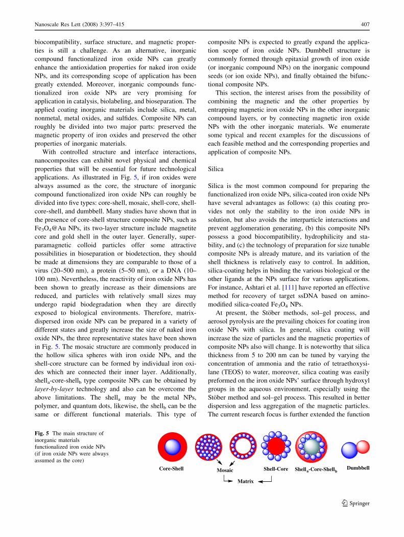
applications. As illustrated in Fig. 5, if iron oxides were
always assumed as the core, the structure of inorganic
compound functionalized iron oxide NPs can roughly be
divided into five types: core-shell, mosaic, shell-core, shell-
core-shell, and dumbbell. Many studies have shown that in
the presence of core-shell structure composite NPs, such as
Fe3O4@Au NPs, its two-layer structure include magnetite
core and gold shell in the outer layer. Generally, super-
paramagnetic colloid particles offer some attractive
possibilities in bioseparation or biodetection, they should
be made at dimensions they are comparable to those of a
virus (20–500 nm), a protein (5–50 nm), or a DNA (10–
100 nm). Nevertheless, the reactivity of iron oxide NPs has
been shown to greatly increase as their dimensions are
reduced, and particles with relatively small sizes may
undergo rapid biodegradation when they are directly
exposed to biological environments. Therefore, matrix-
dispersed iron oxide NPs can be prepared in a variety of
different states and greatly increase the size of naked iron
oxide NPs, the three representative states have been shown
in Fig. 5. The mosaic structure are commonly produced in
the hollow silica spheres with iron oxide NPs, and the
shell-core structure can be formed by individual iron oxi-
des which are connected their inner layer. Additionally,
shella-core-shellb type composite NPs can be obtained by
layer-by-layer technology and also can be overcome the
above limitations. The shella may be the metal NPs,
polymer, and quantum dots, likewise, the shellb can be the
same or different functional materials. This type of
composite NPs is expected to greatly expand the applica-
tion scope of iron oxide NPs. Dumbbell structure is
commonly formed through epitaxial growth of iron oxide
(or inorganic compound NPs) on the inorganic compound
seeds (or ion oxide NPs), and finally obtained the bifunc-
tional composite NPs.
This section, the interest arises from the possibility of
combining the magnetic and the other properties by
entrapping magnetic iron oxide NPs in the other inorganic
compound layers, or by connecting magnetic iron oxide
NPs with the other inorganic materials. We enumerate
some typical and recent examples for the discussions of
each feasible method and the corresponding properties and
application of composite NPs.
Silica
Silica is the most common compound for preparing the
functionalized iron oxide NPs, silica-coated iron oxide NPs
have several advantages as follows: (a) this coating pro-
vides not only the stability to the iron oxide NPs in
solution, but also avoids the interparticle interactions and
prevent agglomeration generating, (b) this composite NPs
possess a good biocompatibility, hydrophilicity and sta-
bility, and (c) the technology of preparation for size tunable
composite NPs is already mature, and its variation of the
shell thickness is relatively easy to control. In addition,
silica-coating helps in binding the various biological or the
other ligands at the NPs surface for various applications.
For instance, Ashtari et al. [111] have reported an effective
method for recovery of target ssDNA based on amino-
modified silica-coated Fe3O4 NPs.
At present, the Stober methods, sol–gel process, and
aerosol pyrolysis are the prevailing choices for coating iron
oxide NPs with silica. In general, silica coating will
increase the size of particles and the magnetic properties of
composite NPs also will change. It is noteworthy that silica
thickness from 5 to 200 nm can be tuned by varying the
concentration of ammonia and the ratio of tetraethoxysi-
lane (TEOS) to water, moreover, silica coating was easily
preformed on the iron oxide NPs’ surface through hydroxyl
groups in the aqueous environment, especially using the
Stober method and sol–gel process. This resulted in better
dispersion and less aggregation of the magnetic particles.
The current research focus is further extended the function
Core-Shell Mosaic Shell-Core Shella-Core-ShellbDumbbell
Matrix
Fig. 5 The main structure of
inorganic materials
functionalized iron oxide NPs
(if iron oxide NPs were always
assumed as the core)
Nanoscale Res Lett (2008) 3:397–415 407
123
of silica functionalized iron oxide NPs, and obtains the
magnetic composites with tunable structure and good
dimension stability.
Therefore, for further extended function of silica func-
tionalized iron oxide NPs, some quantum dots and other
optical materials have been introduced. Ma et al. [112]
reported a synthesis of FexOy@SiO2 core-shell NPs by the
sol–gel and modified Stober methods. Superparamagnetic
iron oxide NPs are first coated with silica to isolate the
magnetic core from the surrounding. Subsequently, the dye
molecules are doped inside a second silica shell based on
which the dye molecules are adopted inside to improve
photostability and allowed for versatile surface function-
alities. The result have shown that the saturation
magnetization (MS) was decreased about 35 emu�g-1 with
a shell thickness of 10–15 nm after silica coating, the
blocking temperature (TB) and the coercivity (Hc) were
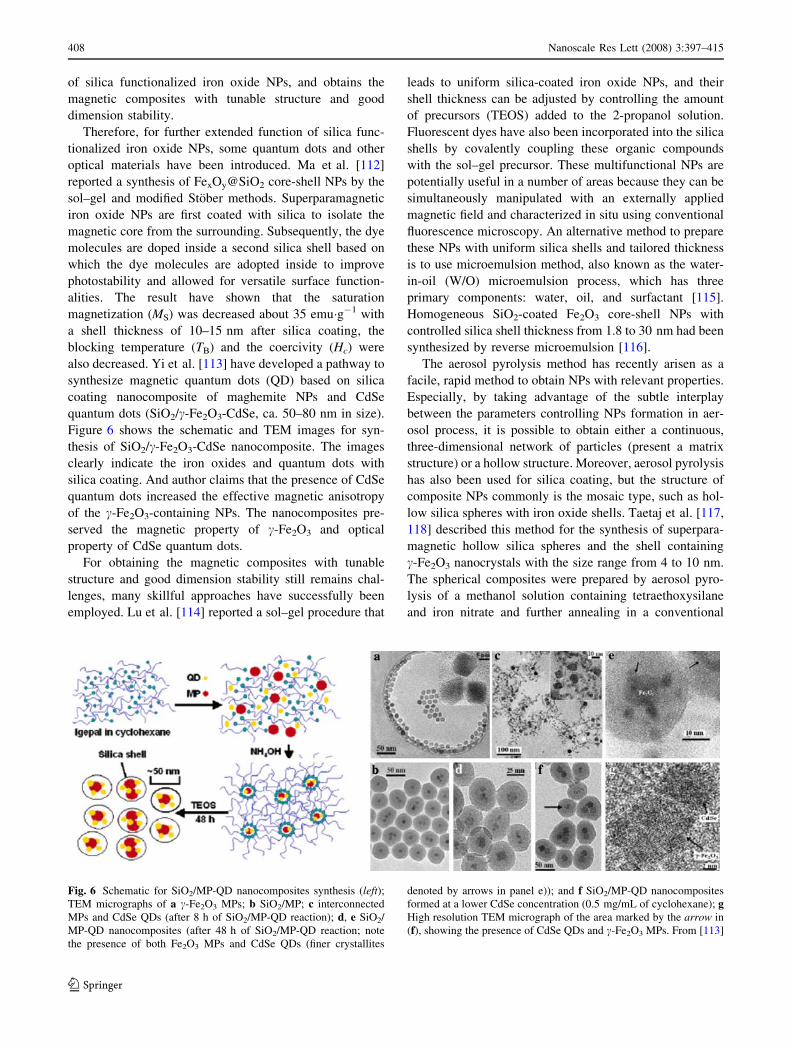
also decreased. Yi et al. [113] have developed a pathway to
synthesize magnetic quantum dots (QD) based on silica
coating nanocomposite of maghemite NPs and CdSe
quantum dots (SiO2/c-Fe2O3-CdSe, ca. 50–80 nm in size).
Figure 6 shows the schematic and TEM images for syn-
thesis of SiO2/c-Fe2O3-CdSe nanocomposite. The images
clearly indicate the iron oxides and quantum dots with
silica coating. And author claims that the presence of CdSe
quantum dots increased the effective magnetic anisotropy
of the c-Fe2O3-containing NPs. The nanocomposites pre-
served the magnetic property of c-Fe2O3 and optical
property of CdSe quantum dots.
For obtaining the magnetic composites with tunable
structure and good dimension stability still remains chal-
lenges, many skillful approaches have successfully been
employed. Lu et al. [114] reported a sol–gel procedure that
leads to uniform silica-coated iron oxide NPs, and their
shell thickness can be adjusted by controlling the amount
of precursors (TEOS) added to the 2-propanol solution.
Fluorescent dyes have also been incorporated into the silica
shells by covalently coupling these organic compounds
with the sol–gel precursor. These multifunctional NPs are
potentially useful in a number of areas because they can be
simultaneously manipulated with an externally applied
magnetic field and characterized in situ using conventional
fluorescence microscopy. An alternative method to prepare
these NPs with uniform silica shells and tailored thickness
is to use microemulsion method, also known as the water-
in-oil (W/O) microemulsion process, which has three
primary components: water, oil, and surfactant [115].
Homogeneous SiO2-coated Fe2O3 core-shell NPs with
controlled silica shell thickness from 1.8 to 30 nm had been
synthesized by reverse microemulsion [116].
The aerosol pyrolysis method has recently arisen as a
facile, rapid method to obtain NPs with relevant properties.
Especially, by taking advantage of the subtle interplay
between the parameters controlling NPs formation in aer-
osol process, it is possible to obtain either a continuous,
three-dimensional network of particles (present a matrix
structure) or a hollow structure. Moreover, aerosol pyrolysis
has also been used for silica coating, but the structure of
composite NPs commonly is the mosaic type, such as hol-
low silica spheres with iron oxide shells. Taetaj et al. [117,
118] described this method for the synthesis of superpara-
magnetic hollow silica spheres and the shell containing
c-Fe2O3 nanocrystals with the size range from 4 to 10 nm.
The spherical composites were prepared by aerosol pyro-
lysis of a methanol solution containing tetraethoxysilane
and iron nitrate and further annealing in a conventional
Fig. 6 Schematic for SiO2/MP-QD nanocomposites synthesis (left);TEM micrographs of a c-Fe2O3 MPs; b SiO2/MP; c interconnected
MPs and CdSe QDs (after 8 h of SiO2/MP-QD reaction); d, e SiO2/
MP-QD nanocomposites (after 48 h of SiO2/MP-QD reaction; note
the presence of both Fe2O3 MPs and CdSe QDs (finer crystallites
denoted by arrows in panel e)); and f SiO2/MP-QD nanocomposites
formed at a lower CdSe concentration (0.5 mg/mL of cyclohexane); gHigh resolution TEM micrograph of the area marked by the arrow in
(f), showing the presence of CdSe QDs and c-Fe2O3 MPs. From [113]
408 Nanoscale Res Lett (2008) 3:397–415
123
furnace. And it was observed that the blocking temperature
(TB) strongly depends on nanocomposite size, the
enhancement of the effective magnetic anisotropy with
respect to the bulk c-Fe2O3 can be considered as coming
fundamentally from surface anisotropy [119].
From the above examples, it can be seen that silica
functionalized iron oxide NPs is a facile process. In this
case, great progress in the field of silica-coated iron oxide
NPs have been made, some research results have also being
transformed into commercial application [120, 121].
Metal or Nonmetal
Another facile route to protect the iron oxide NPs is to
induce a controlled oxidation of a pure single-metal or
nonmetal shell, such as gold, silver, platinum, palladium,
iron, carbon etc. The control of the single-metal or non-
metal layer has a tremendous impact on the scope of iron
oxides’ application, particularly to expand its scope of
biomedical and catalyst application. For instance, gold is
often employed to passivate the surface of magnetite NPs
to avoid oxidation. We need to notice that the gold, silver,
and carbon single-metal functionalization will conduce the
descend of the iron oxide NPs’ saturation magnetization
(Ms) value, but the result of cobalt, platinum, copper,
palladium single-metal functionalization may be opposite,
depending on the mass magnetic susceptibility (v) value of
the used materials. In addition, the diameter of metallic or
nonmetallic functionalized iron oxide NPs is prone to tai-
loring, and the necessary diameter can be obtained by
controlling the reduction and repeat times.
In general, there are two ways for preparing the metal
functionalized iron oxide NPs, one is direct reduction of the
single-metal ions on the surface of iron oxide NPs. Mandal
et al. [122] have reported that the Fe3O4 NPs were sepa-
rated, coated with noble metal gold and silver by directly
reducing the Au? and Ag?, respectively, to achieve stability
of the magnetic NPs for a long time, this route yielded
structures of size from 18 to 30 nm, although the author
claims that the NPs possess a well-defined core-shell
structure, the TEM micrograph of samples cannot directly
reflect. Data also showed that the saturation magnetization
(Ms) of gold-coated is 38 emu g-1 (T = 10 K) and it is
reduced by 57.6% from the bulk Fe3O4 NPs (92 emu g-1,
T = 10 K). Generally, this method often applies to prepare
the core-shell type composite NPs, but it is prone to
maintain the magnetic properties of naked iron oxides.
On the other hand, the most common route was
employed for preparing the metallic functionalized iron
oxide NPs by reduction of the single-metal ion on the sur-
face of the small molecule, polymer or SiO2 functionalized
iron oxide NPs. Several authors have reported the magnetic
iron oxide NPs coated with gold. Gold coatings provide not
only the stability to the NPs in solution, but also helps in
binding the biological molecule which include -SH group at
the NPs surface for various biomedical applications.
Recently we [123] have adopted this approach to prepare
the monodispersed gold-coated Fe3O4 NPs via sonolysis of
a solution mixture of gold ions and amino-modified Fe3O4
NPs with further drop-addition of reducing agent. The
composite NPs with diameter of 30 nm and their saturation
magnetization (Ms) was 63 emu g-1 (T = 300 K), and it is
reduced only by ca. 0.03% from the bulk Fe3O4 NPs
(65 emu g-1, T = 10 K). Therefore, this route was bene-
ficial to maintain the saturation magnetization (Ms) of the
original bare magnetite NPs. Yu et al. [124] also reported a
synthesis of dumbbell-like bifunctional Au-Fe3O4 NPs by
epitaxial growth of iron oxides on the Au seeds. A mixture,
consisting of Au NPs, Fe(CO)5, 1-octadecene solvent, oleic
acid, and oleylamine, was heated and refluxed at 300 �C
followed by room temperature oxidation under air. In this
way, dumbbell-like NPs were obtained with tunable sizes in
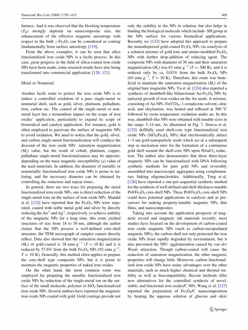
the range 3–14 nm. As illustrated in Fig. 7, Stoeva et al.
[125] skillfully used shell-core type functionalized iron
oxide NPs (SiO2/Fe3O4 NPs) that electrostatically attract
1–3 nm gold-nanoparticle seeds which act in a subsequent
step as nucleation sites for the formation of a continuous
gold shell around the shell-core NPs upon HAuCl4 reduc-
tion. The author also demonstrates that these three-layer
magnetic NPs can be functionalized with DNA following
synthetic methods for pure gold NPs and reversibly
assembled into macroscopic aggregates using complemen-
tary linking oligonucleotides. Additionally, Teng et al.
[126] have reported a one-pot sequential synthetic method
for the synthesis of well-defined and shell-thickness-tunable
Pt@Fe2O3 core-shell NPs. These Pt@Fe2O3 core-shell NPs
could have potential applications in catalysis and as pre-
cursors for making property-tunable magnetic NPs, thin
films, and nanocomposites.
Taking into account the application prospects of mag-
netic record and magnetic ink materials recently, most
studies have focused on the development of carbon-coated
iron oxide magnetic NPs (such as carbon-encapsulated
magnetic NPs), the carbon shell not only protected the iron
oxide NPs from rapidly degraded by environment, but it
also prevented the NPs’ agglomeration caused by van der
Waals attraction. Though carbon-coated will cause the
reduction of saturation magnetization, the other magnetic
properties will change little. Moreover, carbon functional-
ized iron oxide NPs have many advantages over the other
materials, such as much higher chemical and thermal sta-
bility as well as biocompatibility. Recent methods offer
new alternatives for the controlled synthesis of novel,
stable, and functional iron oxides/C NPs. Wang et al. [127]
reported the preparation of Fe3O4/C nanocomposition
by heating the aqueous solution of glucose and oleic
Nanoscale Res Lett (2008) 3:397–415 409
123
acid-stabilized magnetite NPs. Dantas et al. [128] reported
the synthesis of iron oxides/C composites, and it can be
used in the treatment of textile waste water as heteroge-
neous catalysts in the Fenton reaction. Though carbon-
coated iron oxide NPs have many advantageous properties,
the synthesis of monodispersed composite NPs in isolated
from is currency one of challenges in this field.
Furthermore, single-metal functionalized iron oxide NPs
(especially noble metal functionalized iron oxide NPs, and
mostly take the direct method by reducing the single-metal
ions on the surface of iron oxide NPs) also used as cata-
lysts, such as Au/Fe2O3 catalyst for CO oxidation [129,
130], Au/a-Fe2O3 catalyst for water-gas shift reaction [131,
132], Fe3O4/Pd nanoparticle-based catalyst for the cross-
coupling of acrylic acid with iodobenzene [133] and de-
carboxylative coupling reaction in aqueous media [134],
Ag-Fe3O4 catalyst for epoxidation of styrene [135], etc.
These heterogeneous catalysts with a matrix structure
generally as the magnetically recyclable catalysts (MRCs)
are developed in recent years and can be very useful to
assist an effective separation and recovery in a liquid-phase
reaction by a magnet, especially when the catalysts are in
the nanometer-sized range and possess higher activities
than their pure and bulk single-metal or nonmetal coun-
terparts [136, 137]. Therefore, the development of the
facile and rapid methods for the preparation of efficient
MRCs is still a challenge.
Hence, many efforts have been made for getting the
nanometer-sized MRCs with high activity and easy sepa-
ration properties. For example, a Au/FexOy catalyst with
high CO oxidation activities at low temperature prepared by
deposition–precipitation (DP) method was reported by Lin
and Chen [138]. In this work, the size and surface area of
MRCs can be adjusted by the PH value during the DP pro-
cess and the calcination temperature. Moreover, after
90 min time on stream, the conversion of CO can be reached
100%. Additionally, a comparative study results by
Avgouropoulos et al. [139] shows that the Au/a-Fe2O3
catalyst is superior to the Pt/c-Al2O3 and Cuo-CeO2 for
the selective CO oxidation at relatively low reaction
Fig. 7 Synthetic scheme for the preparation of the three-layer NPs
(left); TEM images of colloids after each synthetic step. a, b SiO2
particles covered with silica-primed Fe3O4 NPs (SiO2–Fe3O4). c, dSiO2 particles covered with silica-primed Fe3O4 NPs and heavily
loaded with Au nanoparticle seeds (SiO2–Fe3O4–Au seeds). e Three-
layer magnetic NPs synthesized in a single-step process from particles
presented in (c) and (d). Note the uniformity of the gold shell. The
inset shows the three-layer magnetic NPs drawn to the wall with a
magnet. From [125]
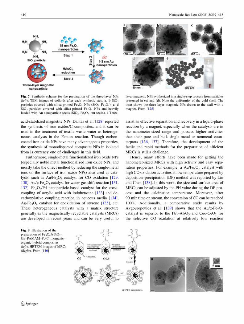
Fig. 8 Illustration of the
preparation of Fe3O4@SiO2–
Gn–PAMAM–Pd(0) inorganic–
organic hybrid composites
(left); HRTEM images of MRCs
(Right). From [140]
410 Nanoscale Res Lett (2008) 3:397–415
123
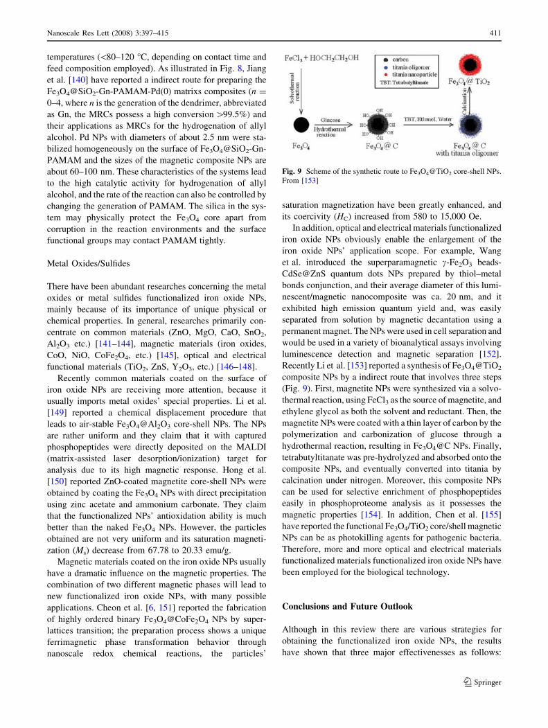
temperatures (\80–120 �C, depending on contact time and
feed composition employed). As illustrated in Fig. 8, Jiang
et al. [140] have reported a indirect route for preparing the
Fe3O4@SiO2-Gn-PAMAM-Pd(0) matrixs composites (n =
0–4, where n is the generation of the dendrimer, abbreviated
as Gn, the MRCs possess a high conversion [99.5%) and
their applications as MRCs for the hydrogenation of allyl
alcohol. Pd NPs with diameters of about 2.5 nm were sta-
bilized homogeneously on the surface of Fe3O4@SiO2-Gn-
PAMAM and the sizes of the magnetic composite NPs are
about 60–100 nm. These characteristics of the systems lead
to the high catalytic activity for hydrogenation of allyl
alcohol, and the rate of the reaction can also be controlled by
changing the generation of PAMAM. The silica in the sys-
tem may physically protect the Fe3O4 core apart from
corruption in the reaction environments and the surface
functional groups may contact PAMAM tightly.
Metal Oxides/Sulfides
There have been abundant researches concerning the metal
oxides or metal sulfides functionalized iron oxide NPs,
mainly because of its importance of unique physical or
chemical properties. In general, researches primarily con-
centrate on common materials (ZnO, MgO, CaO, SnO2,
Al2O3 etc.) [141–144], magnetic materials (iron oxides,
CoO, NiO, CoFe2O4, etc.) [145], optical and electrical
functional materials (TiO2, ZnS, Y2O3, etc.) [146–148].
Recently common materials coated on the surface of
iron oxide NPs are receiving more attention, because it
usually imports metal oxides’ special properties. Li et al.
[149] reported a chemical displacement procedure that
leads to air-stable Fe3O4@Al2O3 core-shell NPs. The NPs
are rather uniform and they claim that it with captured
phosphopeptides were directly deposited on the MALDI
(matrix-assisted laser desorption/ionization) target for
analysis due to its high magnetic response. Hong et al.
[150] reported ZnO-coated magnetite core-shell NPs were
obtained by coating the Fe3O4 NPs with direct precipitation
using zinc acetate and ammonium carbonate. They claim
that the functionalized NPs’ antioxidation ability is much
better than the naked Fe3O4 NPs. However, the particles
obtained are not very uniform and its saturation magneti-
zation (Ms) decrease from 67.78 to 20.33 emu/g.
Magnetic materials coated on the iron oxide NPs usually
have a dramatic influence on the magnetic properties. The
combination of two different magnetic phases will lead to
new functionalized iron oxide NPs, with many possible
applications. Cheon et al. [6, 151] reported the fabrication
of highly ordered binary Fe3O4@CoFe2O4 NPs by super-
lattices transition; the preparation process shows a unique
ferrimagnetic phase transformation behavior through
nanoscale redox chemical reactions, the particles’
saturation magnetization have been greatly enhanced, and
its coercivity (HC) increased from 580 to 15,000 Oe.
In addition, optical and electrical materials functionalized
iron oxide NPs obviously enable the enlargement of the
iron oxide NPs’ application scope. For example, Wang
et al. introduced the superparamagnetic c-Fe2O3 beads-
CdSe@ZnS quantum dots NPs prepared by thiol–metal
bonds conjunction, and their average diameter of this lumi-
nescent/magnetic nanocomposite was ca. 20 nm, and it
exhibited high emission quantum yield and, was easily
separated from solution by magnetic decantation using a
permanent magnet. The NPs were used in cell separation and
would be used in a variety of bioanalytical assays involving
luminescence detection and magnetic separation [152].
Recently Li et al. [153] reported a synthesis of Fe3O4@TiO2
composite NPs by a indirect route that involves three steps
(Fig. 9). First, magnetite NPs were synthesized via a solvo-
thermal reaction, using FeCl3 as the source of magnetite, and
ethylene glycol as both the solvent and reductant. Then, the
magnetite NPs were coated with a thin layer of carbon by the
polymerization and carbonization of glucose through a
hydrothermal reaction, resulting in Fe3O4@C NPs. Finally,
tetrabutyltitanate was pre-hydrolyzed and absorbed onto the
composite NPs, and eventually converted into titania by
calcination under nitrogen. Moreover, this composite NPs
can be used for selective enrichment of phosphopeptides
easily in phosphoproteome analysis as it possesses the
magnetic properties [154]. In addition, Chen et al. [155]
have reported the functional Fe3O4/TiO2 core/shell magnetic
NPs can be as photokilling agents for pathogenic bacteria.
Therefore, more and more optical and electrical materials
functionalized materials functionalized iron oxide NPs have
been employed for the biological technology.
Conclusions and Future Outlook
Although in this review there are various strategies for
obtaining the functionalized iron oxide NPs, the results
have shown that three major effectivenesses as follows:
Fig. 9 Scheme of the synthetic route to Fe3O4@TiO2 core-shell NPs.
From [153]
Nanoscale Res Lett (2008) 3:397–415 411
123
(a) Improves the biocompatibility and chemical stability,
and tailors the dispersibility and water solubility; (b)
Endows the iron oxide new physico-chemical properties,
such as magnet-optical properties, magnetic-electrical
properties, magnetic-thermal properties, etc.; (c) Provides
the iron oxide new functional endgroups for the subsequent
functionalized procedures or the subsequent applications,
such as conjugation with the DNA, antibody, protein, etc.
In the coming years, despite all the recent progresses
made, it is still a challenge to be faced that synthesis of
high-quality functionalized magnetic iron oxide NPs with a
tunable sizes and shapes in a controlled manner. Moreover,
synthesis and surface engineering of iron oxide NPs
involves complex chemical, physical, and physicochemical
multiple interactions, it is the another challenge to under-
stand the synthetic mechanisms detailedly. However, the
magnetic properties and function of naked or surface
functionalized iron oxide NPs depend upon their physical
properties: the size and shape, their microstructure, and the
chemical phase in which they are present [156]. Luckily,
several physicochemical techniques have been developted
to determine these parameters.
Therefore, how to improve the stability and availability
of functionalized iron oxide NPs in extreme environmental
conditions, how to develop an efficient and orderly mag-
netic micro- or nano-assembly structures, and how to
realize large-scale or industrial synthesis, these problems
are urgent to be solved for obtaining a ideal functionalized
iron oxide materials.
For all that, we still believe the surface functionalization
and modification of magnetic iron oxide NPs to introduce
additional functionality will attract more and more attention.
Furthermore, multifunctional magnetic iron oxide compos-
ite nanoparticle systems with designed active sites will
promise for a various applications, such as catalysts, mag-
netic recording, bioseparation, biodetection, etc. The future
work in this area must be focused on the research of the
toxicity and degradability of naked or surface functionalized
iron oxide NPs, and preparing it via green chemistry for
reducing the environmental pollution as much as possible.
Successful development in this area will aid the growth of
the various scientific researches or industrial applications as
well as improving the quality of life in the population.
Acknowledgments W. Wu thanks all the colleagues during past
collaborations at the HUT who have contributed to the work descri-
bed here: Lei Zeng, Jingke Huang, Rong Hu and Professors Jianxin
Tang and Hong Chen. This review is a dedication to all of them.
References
1. L. LaConte, N. Nitin, G. Bao, Mater. Today 8, 32 (2005). doi:
10.1016/S1369-7021(05)00893-X
2. D. Patel, J.Y. Moon, Y. Chang, T.J. Kim, G.H. Lee, Colloid
Surf. A 313–314, 91 (2008). doi:10.1016/j.colsurfa.2007.04.078
3. M. Zhao, L. Josephson, Y. Tang, R. Weissleder, Angew. Chem.
Int. Ed. 42, 1375 (2003). doi:10.1002/anie.200390352
4. S. Mornet, S. Vasseur, F. Grasset, P. Veverka, G. Goglio, A.
Demourgues et al., Prog. Solid State Chem. 34, 237 (2006). doi:
10.1016/j.progsolidstchem.2005.11.010
5. P.D. Stevens, J. Fan, H.M.R. Gardimalla, M. Yen, Y. Gao, Org.
Lett. 7, 2085 (2005). doi:10.1021/ol050218w
6. Y. Jun, J. Choi, J. Cheon. Chem. Commun. (Camb) 1203 (2007).
doi:10.1039/b614735f
7. A.K. Gupta, M. Gupta, Biomaterials 26, 3995 (2005). doi:
10.1016/j.biomaterials.2004.10.012
8. R.M. Cornell, U. Schwertmann, The Iron Oxides: Structures,Properties, Reactions, Occurences and Uses (Wiley-VCH,
Weinheim, 2003)
9. L. Cabrera, S. Gutierrez, N. Menendezb, M.P. Morales, P.
Herrasti, Electrochim. Acta 53, 3436 (2008)
10. C. Pascal, J.L. Pascal, F. Favier, M.L.E. Moubtassim, C. Payen,
Chem. Mater. 11, 141 (1999). doi:10.1021/cm980742f
11. O. Bomatı-Miguel, L. Mazeina, A. Navrotsky, S. Veintemillas-
Verdaguer, Chem. Mater. 20, 591 (2008). doi:10.1021/
cm071178o
12. A.A. Bharde, R.Y. Parikh, M. Baidakova, S. Jouen, B. Han-
noyer, T. Enoki et al., Langmuir 24, 5787 (2008). doi:10.1021/
la704019p
13. Y. Roh, H. Vali, T.J. Phelps, J.W. Moon, J. Nanosci. Nano-
technol. 11, 3517 (2006)
14. W. Wu, Q. He, R. Hu, J. Huang, H. Chen, Rare Metal Mater.
Eng. 34, 238 (2007)
15. V.J. Binh, S.T. Purcell, V. Semet, F. Feschet, Appl. Surf. Sci.
130–132, 803 (1998). doi:10.1016/S0169-4332(98)00158-5
16. Y.S. Kang, S. Risbud, J.F. Rabolt, P. Stroeve, Chem. Mater. 8,
2209 (1996). doi:10.1021/cm960157j
17. A.A. Novakova, V.Y. Lanchinskaya, A.V. Volkov, T.S. Gen-
dler, T.Y. Kiseleva, M.A. Moskvina et al., J. Magn. Magn.
Mater. 258–259, 35 (2003). doi:10.1016/S0304-8853(02)
01062-4
18. J. Lee, T. Isobe, M. Senna, Colloids Surf. A Physicochem. Eng.
Asp. 109, 121 (1996). doi:10.1016/0927-7757(95)03479-X
19. S. Sun, H. Zeng, J. Am. Chem. Soc. 124, 8204 (2002). doi:
10.1021/ja026501x
20. J. Rockenberger, E.C. Scher, P.A. Alivisatos, J. Am. Chem. Soc.
121, 11595 (1999). doi:10.1021/ja993280v
21. K. Woo, J. Hong, S. Choi, H. Lee, J. Ahn, C.S. Kim et al.,
Chem. Mater. 16, 2814 (2004). doi:10.1021/cm049552x
22. T. Hyeon, S.S. Lee, J. Park, Y. Chung, H.B. Na, J. Am. Chem.
Soc. 123, 12798 (2001). doi:10.1021/ja016812s
23. C. Solans, P. Izquierdo, J. Nolla, N. Azemar, M.J. Garcia-
Celma, Curr. Opin. Colloid Interface Sci. 10, 102 (2005). doi:
10.1016/j.cocis.2005.06.004
24. J. Vidal-Vidal, J. Rivas, M.A. Lopez-Quintela, Colloid Surf. A
288, 44 (2006). doi:10.1016/j.colsurfa.2006.04.027
25. F.A. Tourinho, R. Franck, R. Massart, J. Mater. Sci. 25, 3249
(1990). doi:10.1007/BF00587682
26. A.B. Chin, I.I. Yaacob, J. Mater. Process. Technol. 191, 235
(2007). doi:10.1016/j.jmatprotec.2007.03.011
27. X. Hu, J.C. Yu, J. Gong, J. Phys. Chem. C 111, 11180 (2007).
doi:10.1021/jp073073e
28. S. Giria, S. Samantab, S. Majic, S. Gangulic, A. Bhaumikb, J.
Magn. Magn. Mater. 285, 296 (2005). doi:10.1016/j.jmmm.
2004.08.007
29. Z. Jing, S. Wu, Mater. Lett. 58, 3637 (2004). doi:10.1016/
j.matlet.2004.07.010
30. X. Liu, G. Qiu, A. Yan, Z. Wang, X. Li, J. Alloy Compd. 433,
216 (2007). doi:10.1093/comjnl/bxm059
412 Nanoscale Res Lett (2008) 3:397–415
123
31. J. Wang, J. Sun, Q. Sun, Q. Chen, Mater. Res. Bull. 38, 1113
(2003). doi:10.1016/S0025-5408(03)00129-6
32. Y. Zheng, Y. Cheng, F. Bao, Y. Wang, Mater. Res. Bull. 41, 525
(2006). doi:10.1016/j.materresbull.2005.09.015
33. T.J. Daou, G. Pourroy, S. Begin-Colin, J.M. Greneche, C.
Ulhaq-Bouillet, P. Legare et al., Chem. Mater. 18, 4399 (2006).
doi:10.1021/cm060805r
34. S. Wang, Y. Min, S. Yu, J. Phys. Chem. C 111, 3551 (2007).
doi:10.1021/jp068647e
35. M.M. Titirici, M. Antonietti, A. Thomas, Chem. Mater. 18, 3808
(2006). doi:10.1021/cm052768u
36. K.S. Suslick, Science 247, 1439 (1990). doi:10.1126/science.
247.4949.1439
37. J.H. Bang, K.S. Suslick, J. Am. Chem. Soc. 129, 2242 (2007).
doi:10.1021/ja0676657
38. R. Vijayakumar, Y. Koltypin, I. Felner, A. Gedanken, Mater.
Sci. Eng. A 286, 101 (2000). doi:10.1016/S0921-5093(00)
00647-X
39. J. Pinkas, V. Reichlova, R. Zboril, Z. Moravec, P. Bezdicka, J.
Matejkova, Ultrason. Sonochem. 15, 257 (2008). doi:10.1016/
j.ultsonch.2007.03.009
40. P.C. Scholten, Thermodynamics of Magnetic Fluids (Hemi-
sphere, Washington, DC, 1978)
41. R.E. Rosensweig, R. Kaiser, G. Miskolszy, J. Colloid Interface
Sci. 29, 680 (1969). doi:10.1016/0021-9797(69)90220-3
42. R. Tadmor, R.E. Rosensweig, J. Frey, J. Klein, Langmuir 16,
9117 (2000). doi:10.1021/la0009137
43. Y. Sahoo, A. Goodarzi, M.T. Swihart, T.Y. Ohulchanskyy, E.P.
Furlani, P.N. Prasad, J. Phys. Chem. B 109, 3879 (2005). doi:
10.1021/jp045402y
44. A.B. Bourlinos, A. Simopoulos, D. Petridis, Chem. Mater. 14,
899 (2002). doi:10.1021/cm010794w
45. M.H. Sousa, J.C. Rubim, P.G. Sobrinho, F.A. Tourinho, J.
Magn. Magn. Mater. 225, 67 (2001). doi:10.1016/S0304-
8853(00)01229-4
46. P.C. Morais, A.C. Oliveira, A.L. Tronconi, T. Goetze, N. Buske,
IEEE Trans. Magn. 39, 2654 (2003). doi:10.1109/TMAG.2003.
815544
47. S. Mornet, S. Vasseur, F. Grasset, E. Duguet, J. Mater. Chem.
14, 2161 (2004). doi:10.1039/b402025a
48. J. Xie, C.H. Wang, Pharm. Res. 22, 2079 (2005). doi:
10.1007/s11095-005-7782-y
49. S.S. Banerjee, D. Chen, Chem. Mater. 19, 6345 (2007). doi:
10.1021/cm702278u
50. Y. Hou, H. Kondoh, M. Shimojo, E.O. Sako, N. Ozaki, T.
Kogure et al., J. Phys. Chem. B 109, 4845 (2005). doi:10.1021/
jp0476646
51. D. Bonacchi, A. Caneschi, D. Dorignac, A. Falqui, D. Gatteschi,
D. Rovai et al., Chem. Mater. 16, 2016 (2004). doi:10.1021/
cm034948e
52. H. Xia, J. Yi, P. Foo, B. Liu, Chem. Mater. 19, 4087 (2007). doi:
10.1021/cm070918q
53. Z.P. Chen, Y. Zhang, S. Zhang, J.G. Xia, J.W. Liu, K. Xu et al.,
Colloid Surf. A 316, 210 (2008). doi:10.1016/j.colsurfa.
2007.09.017
54. R.S. Ingram, M.J. Hostetler, R.W. Murray, J. Am. Chem. Soc.
119, 9175 (1997). doi:10.1021/ja971734n
55. M.J. Hostetler, S.J. Green, J.J. Stokes, R.W. Murray, J. Am.
Chem. Soc. 118, 4212 (1996). doi:10.1021/ja960198g
56. S. Sun, H. Zeng, D.B. Robinson, S. Raoux, P.M. Rice, S.X.
Wang et al., J. Am. Chem. Soc. 126, 273 (2004). doi:10.1021/
ja0380852
57. M. Lattuada, T.A. Hatton, Langmuir 23, 2158 (2007). doi:
10.1021/la062092x
58. J. Fan, J. Lu, R. Xu, R. Jiang, Y. Gao, J. Colloid Interface Sci.
266, 215 (2003). doi:10.1016/S0021-9797(03)00570-8
59. X. Shen, X. Fang, Y. Zhou, H. Liang, Chem. Lett. 33, 1468
(2004). doi:10.1246/cl.2004.1468
60. M. Ma, Y. Zhang, W. Yu, H. Shen, H. Zhang, N. Gu, Colloid
Surf. A 212, 219 (2003). doi:10.1016/S0927-7757(02)00305-9
61. B. Arales, Chemtechnology 7, 766 (1977)
62. W. Wu, Q. He, H. Chen, in Proceedings of the 1st InternationalConference on Bioinformatics and Biomedical Engineering(Wuhan), vol. 1, 2007, pp. 76
63. R.D. Palma, S. Peeters, M.J. Van Bael, H. Van den Rul, K.
Bonroy, W. Laureyn et al., Chem. Mater. 19, 1821 (2007). doi:
10.1021/cm0628000
64. C.C. Berry, A.S.G. Curtis, J. Phys, D. Appl. Phys. (Berl) 36,
R198 (2003)
65. C.C. Berry, S. Wells, S. Charles, A.S.G. Curtis, Biomaterials 24,
4551 (2003). doi:10.1016/S0142-9612(03)00237-0
66. M. Mikhaylova, D.K. Kim, N. Bobrysheva, M. Osmolowsky, V.
Semenov, T. Tsakalakos et al., Langmuir 20, 2472 (2004). doi:
10.1021/la035648e
67. M.C. Bautista, O. Bomati-Miguel, M.P. Morales, C.J. Serna, S.
Veintemillas-Verdaguer, J. Magn. Magn. Mater. 293, 20 (2005).
doi:10.1016/j.jmmm.2005.01.038
68. D.K. Kim, M. Maria, F.H. Wang, K. Jan, B. Borje, Y. Zhang
et al., Chem. Mater. 15, 4343 (2003). doi:10.1021/cm031104m
69. W. Wang, Z.K. Zhang, J. Disper, Sci. Technol. 28, 557 (2007)
70. D. Olsena, C. Yanga, M. Bodoa, R. Changa, S. Leigha, J. Baeza
et al., Adv. Drug. Deliv. Rev. 55, 1547 (2003). doi:10.1016/
j.addr.2003.08.008
71. B. Gaihre, S. Aryal, M.S. Khil, H.Y. Kim, J. Microencapsul. 25,
21 (2008). doi:10.1080/02652040701737697
72. T. Yonezawa, K. Kamoshita, M. Tanaka, T. Kinoshita, Jpn. J.
Appl. Phys. 47, 1389 (2008). doi:10.1143/JJAP.47.1389
73. G. Li, K. Huang, Y. Jiang, P. Ding, D. Yang, Biochem. Eng. J.
(2008). doi:10.1016/j.bej.2008.01.018
74. G. Li, Y. Jiang, K. Huang, P. Ding, L. Yao, Colloid. Surf. A
(2008). doi: 10.1016/j.colsurfa.2008.01.017
75. E.H. Kim, H.S. Lee, B.K. Kwak, B.K. Kim, J. Magn. Magn.
Mater. 289, 328 (2005). doi:10.1016/j.jmmm.2004.11.093
76. S. Mondini, S. Cenedese, G. Marinoni, G. Molteni, N. Santo,
C.L. Bianchi et al., J. Colloid Interface Sci. 322, 173 (2008). doi:
10.1016/j.jcis.2008.03.008
77. K.G. Paul, T.B. Frigo, J.Y. Groman, E.V. Groman, Bioconjug.
Chem. 15, 394 (2004). doi:10.1021/bc034194u
78. S.M. Moghimi, A.C. Hunter, J.C. Murray, Pharmacol. Rev. 53,
283 (2001)
79. H. Pardoe, W. Chua-anusorn, T.G. Pierre, J. Dobson, J. Magn.
Magn. Mater. 225, 41 (2001). doi:10.1016/S0304-8853(00)
01226-9
80. M. Chastellain, A. Petri, H.J. Hofmann, Colloid Interface Sci.
278, 353 (2004). doi:10.1016/j.jcis.2004.06.025
81. A.J.M. D’Souza, R.L. Schowen, E.M. Topp, J Control Release
94, 91 (2004). doi:10.1016/j.jconrel.2003.09.014
82. F. Chen, Q. Gao, G. Hong, J. Ni, J. Magn. Magn. Mater. 320,
1921 (2008). doi:10.1016/j.jmmm.2008.02.132
83. S.A. Gomez-Lopera, J.L. Arias, V. Gallardo, A.V. Delgado,
Langmuir 22, 2816 (2006). doi:10.1021/la0530079
84. H.L. Ma, Y.F. Xu, X.R. Qi, Y. Maitani, T. Nagai, Int. J. Pharm.
354, 217 (2006). doi:10.1016/j.ijpharm.2007.11.036
85. M.A. Morales, P.V. Finotelli, J.A.H. Coaquira, M.H.M. Rocha-
Leao, C. Diaz-Aguila, E.M. Baggio-Saitovitch et al., Mater. Sci.
Eng. C 28, 253 (2007). doi:10.1016/j.msec.2006.12.016
86. D.M. Zhu, F. Wang, M. Han, H.B. Li, Z. Xu, Chin. J. Inorg.
Chem. 23, 2128 (2007)
87. H. Singh, P.E. Laibinis, T.A. Hatton, Langmuir 21, 11500
(2005). doi:10.1021/la0517843
88. L. An, Z. Li, Y. Wang, B. Yang, Chem. J. Chin. Univ. 27, 1372
(2006)
Nanoscale Res Lett (2008) 3:397–415 413
123
89. G. Shan, J. Xing, M. Luo, H. Liu, J. Chen, Biotechnol. Lett. 25,
1977 (2003). doi:10.1023/B:BILE.0000004388.15751.8c
90. A.S. Arbab, L.A. Bashaw, B.R. Miller, E.K. Jordan, B.K. Lewis,
H. Kalish et al., Radiology 229, 838 (2003). doi:10.1148/
radiol.2293021215
91. A.K. Gupta, S. Wells, IEEE Trans. Nanobioscience 3, 66 (2004).
doi:10.1109/TNB.2003.820277
92. D. Guin, S.V. Manorama, Mater. Lett. 62, 3139 (2008). doi:
10.1016/j.matlet.2008.02.005
93. J. Zhang, S. Xu, E. Kumacheva, J. Am. Chem. Soc. 126, 7908
(2004). doi:10.1021/ja031523k
94. T. Ninjbadgar, S. Yamamoto, T. Fukuda, Solid State Sci. 6, 879
(2004). doi:10.1016/j.solidstatesciences.2003.11.009
95. Q. Fan, K. Neoh, E. Kang, B. Shuter, S. Wang, Biomaterials 28,
5426 (2007). doi:10.1016/j.biomaterials.2007.08.039
96. Y. Zhou, S. Wang, B. Ding, Z. Yang, Chem. Eng. J. 138, 578
(2008). doi:10.1016/j.cej.2007.07.030
97. E. Marutani, S. Yamamoto, T. Ninjbadgar, Y. Tsujii, T. Fukuda,
M. Takano, Polymer (Guildf) 45:2231 (2004). doi:10.1016/
j.polymer.2004.02.005
98. Y. Sun, X. Ding, Z. Zheng, X. Cheng, X. Hu, Y. Peng, Eur.
Polym. J. 43, 762 (2007). doi:10.1016/j.eurpolymj.2006.10.021
99. B.L. Frankamp, A.K. Boal, M.T. Tuominen, V.M. Rotello, J.
Am. Chem. Soc. 127, 9731 (2005). doi:10.1021/ja051351m
100. A.K. Boal, K. Das, M. Gray, V.M. Rotello, Chem. Mater. 14,
2628 (2002). doi:10.1021/cm011689p
101. H. He, H. Liu, K. Zhou, W. Wang, P. Rong, J. Cent. South Univ.
Technol. 13, 6 (2006). doi:10.1007/s11771-006-0097-2
102. M. Mikhaylova, D.K. Kim, C.C. Berry, A. Zagorodni, M.
Toprak, A.S.G. Curtis et al., Chem. Mater. 16, 2344 (2004). doi:
10.1021/cm0348904
103. M. Lewin, N. Carlesso, C. Tung, X. Tang, D. Cory, D.T.
Scadden et al., Nat. Biotechnol. 18, 410 (2000). doi:10.1038/
74464
104. L.X. Tiefenauer, G. Kuhne, R.Y. Andres, Bioconjug. Chem. 4,
347 (1993). doi:10.1021/bc00023a007
105. J.M. Nam, S.I. Stoeva, C.A. Mirkin, J. Am. Chem. Soc. 126,
5932 (2004). doi:10.1021/ja049384?
106. Y. Weizmann, F. Patolsky, O. Lioubashevski, I. Willner, J. Am.
Chem. Soc. 126, 1073 (2004). doi:10.1021/ja038257v
107. C. Zhang, J. Cao, D. Yin, Y. Wang, Y. Feng, J. Tan, Appl. Radiat.
Isot. 61, 1255 (2004). doi:10.1016/j.apradiso.2004.03.114
108. C. Lee, K. Huang, P. Wei, Y. Yao, J. Magn. Magn. Mater. 304,
e412 (2006). doi:10.1016/j.jmmm.2006.01.213
109. L. Josephson, J.M. Perez, R. Weissleder, Angew. Chem. Int. Ed.
40, 3204 (2001). doi:10.1002/1521-3773(20010903)40:17\3204::AID-ANIE3204[3.0.CO;2-H
110. N.L. Rosi, C.A. Mirkin, Chem. Rev. 105, 1547 (2005). doi:
10.1021/cr030067f
111. P. Ashtari, X. He, K. Wang, P. Gong, Talanta 67, 548 (2005).
doi:10.1016/j.talanta.2005.06.043
112. D. Ma, J. Guan, F. Normandin, S. Denommee, G. Enright, T.
Veres et al., Chem. Mater. 18, 1920 (2006). doi:10.1021/
cm052067x
113. D.K. Yi, S.T. Selvan, S.S. Lee, G.C. Papaefthymiou, D. Ku-
ndaliya, J.Y. Ying, J. Am. Chem. Soc. 127, 4990 (2005). doi:
10.1021/ja0428863
114. Y. Lu, Y. Yin, B.T. Mayers, Y. Xia, Nano. Lett. 2, 183 (2002).
doi:10.1021/nl015681q
115. J.E. Smith, L. Wang, W. Tan, Trends Analyt. Chem. 25, 848
(2006). doi:10.1016/j.trac.2006.03.008
116. D.K. Yi, S.S. Lee, G.C. Papaefthymiou, Chem. Mater. 3, 604
(2006)
117. P. Tartaj, T. Gonzalez-Carreno, C.J. Serna, Adv. Mater. 13,
1620 (2001). doi:10.1002/1521-4095(200111)13:21\1620::
AID-ADMA1620[3.0.CO;2-Z
118. P. Tartaj, T. Gonzalez-Carreno, C.J. Serna, Adv. Mater. 16, 529
(2004). doi:10.1002/adma.200305814
119. P. Tartaj, T. Gonzalez-Carreno, C.J. Serna, J. Phys. B 107, 20
(2003)
120. I.J. Brucea, J. Taylor, M. Todd, M.J. Daviesb, E. Borionib, C.
Sangregorioa et al., J. Magn. Magn. Mater. 284, 145 (2004). doi:
10.1016/j.jmmm.2004.06.032
121. V. Salgueirino-Maceira, M.A. Correa-Duarte, M. Spasova, L.M.
Liz-Marzan, M. Farle, Adv. Funct. Mater. 16, 509 (2006). doi:
10.1002/adfm.200500565
122. M. Mandal, S. Kundu, S.K. Ghosh, S. Panigrahi, T.K. Sau, J.
Colloid Interface Sci. 286, 187 (2005). doi:10.1016/j.jcis.
2005.01.013
123. W. Wu, Q. He, H. Chen, J. Tang, L. Nie, Nanotechnology 18,
145609 (2007). doi:10.1088/0957-4484/18/14/145609
124. H. Yu, M. Chen, P.M. Rice, S.X. Wang, R.L. White, S. Sun,
Nano. Lett. 5, 379 (2005). doi:10.1021/nl047955q
125. S.I. Stoeva, F. Huo, J.S. Lee, C.A. Mirkin, J. Am. Chem. Soc.
127, 15362 (2005). doi:10.1021/ja055056d
126. X. Teng, D. Black, N.J. Watkins, Y. Gao, H. Yang, Nano. Lett.
3, 261 (2003). doi:10.1021/nl025918y
127. Z. Wang, H. Guo, Y. Yu, N. He, J. Magn. Magn. Mater. 302,
397 (2006). doi:10.1016/j.jmmm.2005.09.044
128. T.L.P. Dantas, V.P. Mendonca, H.J. Josea, A.E. Rodriguesb,
R.F.P.M. Moreira, Chem. Eng. J. 118, 77 (2006)
129. S. Al-Sayari, A.F. Carley, S.H. Taylor, G.J. Hutchings, Top.
Catal. 44, 123 (2007). doi:10.1007/s11244-007-0285-9
130. G.J. Hutchings, M.S. Hall, A.F. Carley, P. Landon, B.E.
Solsona, C.J. Kiely et al., J. Catal. 242, 71 (2006). doi:10.1016/
j.jcat.2006.06.001
131. J. Li, Y. Zhan, F. Zhang, X. Lin, Q. Zheng, Chinese J. Catal 29,
346 (2008)
132. D. Andreeva, T. Tabakova, V. Idakiev, P. Christov, R. Gio-
vanoli, Appl. Catal. A Gen. 169, 9 (1998). doi:10.1016/
S0926-860X(97)00302-5
133. Z. Wang, B. Shen, Z. Aihua, N. He, Chem. Eng. J. 113, 27
(2005). doi:10.1016/j.cej.2005.08.003
134. K.M. Yao, S.I. Lee, Y.T. Lee, Y.K. Chung, I.S. Lee, Chem. Lett.
37, 116 (2008). doi:10.1246/cl.2008.116
135. D.H. Zhang, G.D. Li, J.X. Li, J.S. Chen, Chem. Commun.
(Camb) 3414 (2008). doi:10.1039/b805737k
136. A. Lu, E.L. Salabas, F. Schuth, Angew. Chem. Int. Ed. 46, 1222
(2007). doi:10.1002/anie.200602866
137. M.D. Kaminski, L. Nunez, J. Magn. Magn. Mater. 194, 31
(1999). doi:10.1016/S0304-8853(98)00579-4
138. H.Y. Lin, Y.W. Chen, Ind. Eng. Chem. Res. 44, 4569 (2005).
doi:10.1021/ie0491488
139. G. Avgouropoulos, T. Ioannides, C. Papadopoulou, J. Batista, S.
Hocevar, H.K. Matralis, Catal Today 75, 157 (2002). doi:
10.1016/S0920-5861(02)00058-5
140. Y.J. Jiang, J.H. Jiang, Q.M. Gao, M.L. Ruan, H.M. Yu, L.J. Qi,
Nanotechnology 19, 075714 (2008). doi:10.1088/0957-4484/
19/7/075714
141. Z. Liu, D. Zhang, S. Han, C. Li, J. Am. Chem. Soc. 127, 6
(2005). doi:10.1021/ja0445239
142. R.H.R. Castro, P. Hidalgo, J.A.H. Coaquira, J. Bettin, D. Zan-
chet, D. Gouvea, Eur. J. Inorg. Chem. 11, 2134 (2005). doi:
10.1002/ejic.200400879
143. S. Decker, K.J. Klabunde, J. Am. Chem. Soc. 118, 12465
(1996). doi:10.1021/ja962371e
144. C.L. Carnes, K.J. Klabunde, Chem. Mater. 14, 1806 (2002). doi:
10.1021/cm011588r
145. M.M. Natile, A. Glisenti, Chem. Mater. 15, 2502 (2003). doi:
10.1021/cm031019e
146. N.O. Nunez, P. Tartaj, M.P. Morales, P. Bonville, C.J. Serna,
Chem. Mater. 16, 3119 (2004). doi:10.1021/cm049860v
414 Nanoscale Res Lett (2008) 3:397–415
123

147. C.T. Chen, Y.C. Chen, Anal. Chem. 77, 5912 (2008). doi:
10.1021/ac050831t
148. C.T. Chen, Y.C. Chen, J. Biomed. Nanotechnol. 4, 73 (2008).
doi:10.1166/jbn.2008.008
149. Y. Li, Y. Liu, J. Tang, H. Lin, N. Yao, X. Shen et al., J.
Chromatogr. A 1172, 57 (2007). doi:10.1016/j.chroma.2007.
09.062
150. R.Y. Hong, S.Z. Zhang, G.Q. Di, H.Z. Li, Y. Zheng, J. Ding
et al., Mater. Res. Bull. 43, 2457 (2008). doi:10.1016/
j.materresbull.2007.07.035
151. J. Cheon, J. Park, J. Choi, Y. Jun, S. Kim, M.G. Kim et al., Proc.
Natl. Acad. Sci. USA 103, 3023 (2006). doi:10.1073/pnas.
0508877103
152. D.S. Wang, J.B. He, N. Rosenzweig, Z. Rosenzweig, Nano. Lett.
4, 409 (2004). doi:10.1021/nl035010n
153. Y. Li, J.S. Wu, D.W. Qi, X.Q. Xu, C.H. Deng, P.Y. Yang et al.,
Chem. Commun. (Camb) 564 (2008). doi:10.1039/b716055k
154. Y. Li, X.Q. Xu, D.W. Qi, C.H. Deng, P.Y. Yang, X.M. Zhang, J.
Proteome. Res 7, 2526 (2008). doi:10.1021/pr700582z
155. W.J. Chen, P.J. Tsai, Y.C. Chen, Small 4, 485 (2008). doi:
10.1002/smll.200701164
156. S. Laurent, D. Forge, M. Port, A. Roch, C. Robic, L.V. Elst
et al., Chem. Rev. 108, 2064 (2008). doi:10.1021/cr068445e
Nanoscale Res Lett (2008) 3:397–415 415
123
Copyright © 2022 FDOKUMEN