Towards a Universal Method for the Stable and Clean Functionalization of Inert Perfluoropolymer...
9
www.afm-journal.de FULL PAPER © 2010 WILEY-VCH Verlag GmbH & Co. KGaA, Weinheim Adv. Funct. Mater. 2010, 20, 3932–3940 3932 www.MaterialsViews.com wileyonlinelibrary.com By Carlo Morasso, Miriam Colombo, Silvia Ronchi, Laura Polito, Serena Mazzucchelli, Diego Monti, Marco Buscaglia, Tommaso Bellini, and Davide Prosperi* contact angle with water, intrinsically low refractive index, absence of protons, and a concentration of 19 F spin-active nuclei that provides a valuable probe for magnetic resonance imaging (MRI). [1] Perfluoroelas- tomer and perfluoropolyether compounds do not spring from Nature, yet they can offer useful building blocks for the design of novel functional biopolymers and clever solutions to physiologically vital issues, which have attracted much attention by several research groups. [2] Recently, by exploiting the low refractive index of one of these materials, we developed a novel biosensor based on the measurement of the intensity of the light scattered by index- matched perfluoroelastomer colloids. [3–6] Light scattering provides a versatile and noninvasive method to study structures and phenomena at the mesoscale level, such as aggregative events, and is thus becoming a crucial characterization tool in colloid sci- ence. Nevertheless, light scattering is not a conventional technique to study molecular association because the binding of isolated ligands and receptors in dilute solutions produces a negligible increment in the scat- tered light intensity, [7] while micrometer- scale particles hosting multiple receptors, for instance entire cells, intrinsically scatter too much light com- pared to the specific contributions due to molecular interactions. This limitation can be overcome by supporting the receptors on nanoscale perfluorinated latex spheres (80 nm in diameter), whose refractive index closely matches that of water (“phantom” nanoparticles, PnPs). When molecular interactions take place at Towards a Universal Method for the Stable and Clean Functionalization of Inert Perfluoropolymer Nanoparticles: Exploiting Photopolymerizable Amphiphilic Diacetylenes DOI: 10.1002/adfm.201001274 Highly fluorinated materials are being widely investigated due to a number of peculiar properties, which are potentially useful for various applications, including use as lubricants, anti-adhesive films, and substitutes for biological fluids for biomedical utilization. However, at present such potential is still poorly exploited. One of the major drawbacks that hampers the rapid development of nanoscale fluoro-hybrid devices is the remarkable inertness of perfluor- opolymeric materials that lack reactive functionalities, as they do not offer any functional groups that can be employed to covalently anchor organic molecules on their surface. In this paper, a convenient method for the stable biofunc- tionalization of strongly unreactive perfluoropolymer nanoparticles (PnPs) is reported. PnPs are easily coated with newly synthesized asymmetric diacetylenic monomer compounds (ADMs), thanks to PnP’s high propensity to interact with hydrophobic moieties. Once monomerically adsorbed onto PnPs, such suitably designed ADMs enable the formation of a robust polymeric shell around the perfluoroelastomer core via a clean UV-promoted localized photopolymerization. Given the peculiar optical characteristics of PnPs, the coating of the particles can be monitored step by step using light scattering, which also allows estima- tion of the fraction of reacted monomers by competitive adsorption with smaller particles. The potential of this method for the biofunctionalization of PnPs is demonstrated with representative proteins and carbohydrates. Among them, the extension to avidin–biotin technology may broaden the scope and applicability of this strategy to potentially a large number of molecules of biomedical interest. [∗] M. Colombo, Dr. S. Mazzucchelli, Dr. D. Prosperi Dipartimento di Biotecnologie e Bioscienze Università degli Studi di Milano-Bicocca Piazza della Scienza 2, 20126 Milan (Italy) and CMENA Università degli Studi di Milano Via G.B. Grassi 74, 20157 Milano (Italy) E-mail: [email protected] Dr. C. Morasso, [+] Dr. S. Ronchi, Dr. L. Polito, Dr. D. Monti, Dr. D. Prosperi Istituto di Scienze e Tecnologie Molecolari CNR, via Fantoli 16/15, 20138 Milano (Italy) Dr. M. Buscaglia, Prof. T. Bellini Dipartimento di Chimica, Biochimica e Biotecnologie per la Medicina Università degli Studi di Milano Via F.lli Cervi 93, 20090 Segrate (Italy) [+] Current address: London Centre for Nanotechnology, University College London, 17-19 Gordon Street, London WCIH OAL (UK) 1. Introduction Highly fluorinated materials are being intensely investigated and used, due to a number of peculiar properties which are potentially useful for biomedical applications, including extreme chemical and biological inertness, biocompatibility, high
-
Upload
independent -
Category
Documents
-
view
1 -
download
0
Transcript of Towards a Universal Method for the Stable and Clean Functionalization of Inert Perfluoropolymer...
www.afm-journal.de
FULL
PAPER
3932
www.MaterialsViews.com
Towards a Universal Method for the Stable and Clean Functionalization of Inert Perfl uoropolymer Nanoparticles: Exploiting Photopolymerizable Amphiphilic Diacetylenes
By Carlo Morasso , Miriam Colombo , Silvia Ronchi , Laura Polito , Serena Mazzucchelli , Diego Monti , Marco Buscaglia , Tommaso Bellini , and Davide Prosperi *
Highly fl uorinated materials are being widely investigated due to a number of peculiar properties, which are potentially useful for various applications, including use as lubricants, anti-adhesive fi lms, and substitutes for biological fl uids for biomedical utilization. However, at present such potential is still poorly exploited. One of the major drawbacks that hampers the rapid development of nanoscale fl uoro-hybrid devices is the remarkable inertness of perfl uor-opolymeric materials that lack reactive functionalities, as they do not offer any functional groups that can be employed to covalently anchor organic molecules on their surface. In this paper, a convenient method for the stable biofunc-tionalization of strongly unreactive perfl uoropolymer nanoparticles (PnPs) is reported. PnPs are easily coated with newly synthesized asymmetric diacetylenic monomer compounds (ADMs), thanks to PnP’s high propensity to interact with hydrophobic moieties. Once monomerically adsorbed onto PnPs, such suitably designed ADMs enable the formation of a robust polymeric shell around the perfl uoroelastomer core via a clean UV-promoted localized photopolymerization. Given the peculiar optical characteristics of PnPs, the coating of the particles can be monitored step by step using light scattering, which also allows estima-tion of the fraction of reacted monomers by competitive adsorption with smaller particles. The potential of this method for the biofunctionalization of PnPs is demonstrated with representative proteins and carbohydrates. Among them, the extension to avidin–biotin technology may broaden the scope and applicability of this strategy to potentially a large number of molecules of biomedical interest.
1. Introduction
Highly fl uorinated materials are being intensely investigated and used, due to a number of peculiar properties which are potentially useful for biomedical applications, including extreme chemical and biological inertness, biocompatibility, high
© 2010 WILEY-VCH Verlag GmbH & Co. KGaA, Weinhewileyonlinelibrary.com
DOI: 10.1002/adfm.201001274
[∗] M. Colombo , Dr. S. Mazzucchelli , Dr. D. Prosperi Dipartimento di Biotecnologie e Bioscienze Università degli Studi di Milano-Bicocca Piazza della Scienza 2, 20126 Milan (Italy)and CMENA Università degli Studi di Milano Via G.B. Grassi 74, 20157 Milano (Italy) E-mail: [email protected]
Dr. C. Morasso ,[
Dr. D. Prosperi Istituto di ScienzCNR, via Fantoli Dr. M. BuscagliaDipartimento diBiochimica e BioUniversità degli Via F.lli Cervi 93
[+] Current addressCollege London,
contact angle with water, intrinsically lowrefractive index, absence of protons, and aconcentration of 19 F spin-active nuclei thatprovides a valuable probe for magneticresonance imaging (MRI). [ 1 ] Perfl uoroelas-tomer and perfl uoropolyether compoundsdo not spring from Nature, yet they canoffer useful building blocks for the designof novel functional biopolymers and cleversolutions to physiologically vital issues,which have attracted much attention byseveral research groups. [ 2 ] Recently, byexploiting the low refractive index of oneof these materials, we developed a novelbiosensor based on the measurement ofthe intensity of the light scattered by index-matched perfl uoroelastomer colloids. [ 3–6 ]
Light scattering provides a versatile andnoninvasive method to study structures andphenomena at the mesoscale level, such asaggregative events, and is thus becoming acrucial characterization tool in colloid sci-ence. Nevertheless, light scattering is not aconventional technique to study molecularassociation because the binding of isolatedligands and receptors in dilute solutionsproduces a negligible increment in the scat-tered light intensity, [ 7 ] while micrometer-scale particles hosting multiple receptors,
cells, intrinsically scatter too much light com-
for instance entire pared to the specifi c contributions due to molecular interactions. This limitation can be overcome by supporting the receptors on nanoscale perfl uorinated latex spheres (80 nm in diameter), whose refractive index closely matches that of water (“phantom” nanoparticles, PnPs). When molecular interactions take place at
im Adv. Funct. Mater. 2010, 20, 3932–3940
+] Dr. S. Ronchi , Dr. L. Polito , Dr. D. Monti ,
e e Tecnologie Molecolari 16/15, 20138 Milano (Italy) , Prof. T. Bellini Chimica, tecnologie per la Medicina Studi di Milano , 20090 Segrate (Italy) : London Centre for Nanotechnology, University 17-19 Gordon Street, London WCIH OAL (UK)
FULL P
APER
www.afm-journal.dewww.MaterialsViews.com
R1 = 3
4a: R2= Ac4b: R2= H
5
n
1: R= OH; n= 52: R= N3 ; n= 3
RO
HN
O
3-53
NO
HN
O
NN
R1
4
NO
O
O
O OO
O
R2O OR2
R2OR2O
OR2
R2O OR2 4
O
S
H H HN
O 4
O
3
3
NHHN
O
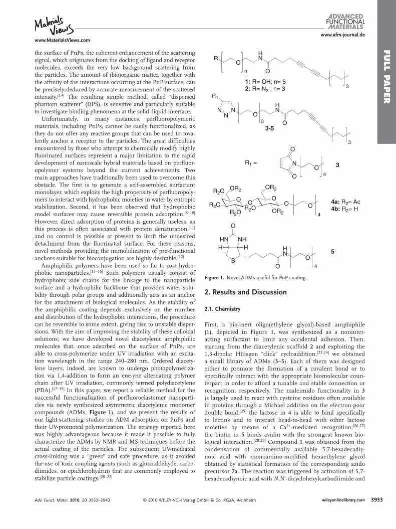
Figure 1 . Novel ADMs useful for PnP coating.
the surface of PnPs, the coherent enhancement of the scattering signal, which originates from the docking of ligand and receptor molecules, exceeds the very low background scattering from the particles. The amount of (bio)organic matter, together with the affi nity of the interactions occurring at the PnP surface, can be precisely deduced by accurate measurement of the scattered intensity. [ 3,4 ] The resulting simple method, called “dispersed phantom scatterer” (DPS), is sensitive and particularly suitable to investigate binding phenomena at the solid–liquid interface.
Unfortunately, in many instances, perfl uoropolymeric materials, including PnPs, cannot be easily functionalized, as they do not offer any reactive groups that can be used to cova-lently anchor a receptor to the particles. The great diffi culties encountered by those who attempt to chemically modify highly fl uorinated surfaces represent a major limitation to the rapid development of nanoscale hybrid materials based on perfl uor-opolymer systems beyond the current achievements. Two main approaches have traditionally been used to overcome this obstacle. The fi rst is to generate a self-assembled surfactant monolayer, which exploits the high propensity of perfl uoropoly-mers to interact with hydrophobic moieties in water by entropic stabilization. Second, it has been observed that hydrophobic model surfaces may cause reversible protein adsorption. [ 8–10 ] However, direct adsorption of proteins is generally useless, as this process is often associated with protein denaturation, [ 11 ] and no control is possible at present to limit the undesired detachment from the fl uorinated surface. For these reasons, novel methods providing the immobilization of pro-functional anchors suitable for bioconjugation are highly desirable. [ 12 ]
Amphiphilic polymers have been used so far to coat hydro-phobic nanoparticles. [ 13–16 ] Such polymers usually consist of hydrophobic side chains for the linkage to the nanoparticle surface and a hydrophilic backbone that provides water solu-bility through polar groups and additionally acts as an anchor for the attachment of biological molecules. As the stability of the amphiphilic coating depends exclusively on the number and distribution of the hydrophobic interactions, the procedure can be reversible to some extent, giving rise to unstable disper-sions. With the aim of improving the stability of these colloidal solutions, we have developed novel diacetylenic amphiphilic molecules that, once adsorbed on the surface of PnPs, are able to cross-polymerize under UV irradiation with an excita-tion wavelength in the range 240–280 nm. Ordered diacety-lene layers, indeed, are known to undergo photopolymeriza-tion via 1,4-addition to form an ene-yne alternating polymer chain after UV irradiation, commonly termed polydiacetylene (PDA). [ 17–19 ] In this paper, we report a reliable method for the successful functionalization of perfl uoroelastomer nanoparti-cles via newly synthesized asymmetric diacetylenic monomer compounds (ADMs, Figure 1 ), and we present the results of our light-scattering studies on ADM adsorption on PnPs and their UV-promoted polymerization. The strategy reported here was highly advantageous because it made it possible to fully characterize the ADMs by NMR and MS techniques before the actual coating of the particles. The subsequent UV-mediated cross-linking was a “green” and safe procedure, as it avoided the use of toxic coupling agents (such as glutaraldehyde, carbo-diimides, or epichlorohydrin) that are commonly employed to stabilize particle coatings. [ 20–22 ]
© 2010 WILEY-VCH Verlag GmAdv. Funct. Mater. 2010, 20, 3932–3940
2. Results and Discussion
2.1. Chemistry
First, a bio-inert oligo(ethylene glycol)-based amphiphile ( 1 ), depicted in Figure 1 , was synthesized as a noninter-acting surfactant to limit any accidental adhesion. Then, starting from the diacetylenic scaffold 2 and exploiting the 1,3-dipolar Hüisgen “click” cycloaddition, [ 23,24 ] we obtained a small library of ADMs ( 3–5 ). Each of them was designed either to promote the formation of a covalent bond or to specifi cally interact with the appropriate biomolecular coun-terpart in order to afford a tunable and stable connection or recognition, respectively. The maleimido functionality in 3 is largely used to react with cysteine residues often available in proteins through a Michael addition on the electron-poor double bond; [ 25 ] the lactose in 4 is able to bind specifi cally to lectins and to interact head-to-head with other lactose moieties by means of a Ca 2 + -mediated recognition; [ 26,27 ] the biotin in 5 binds avidin with the strongest known bio-logical interaction. [ 28,29 ] Compound 1 was obtained from the condensation of commercially available 5,7-hexadecadiy-noic acid with monoamino-modifi ed hexaethylene glycol obtained by statistical formation of the corresponding azido precursor 7a . The reaction was triggered by activation of 5,7-hexadecadiynoic acid with N , N ’-dicyclohexylcarbodiimide and
bH & Co. KGaA, Weinheim 3933wileyonlinelibrary.com

FULL
PAPER
3934
www.afm-journal.dewww.MaterialsViews.com
Scheme 1 . Stepwise synthesis of ADMs 1 and 2 . (TBAB = tetrabutylammonium bro-mide; NHS = N -hydroxysuccinimide; DCC = N , N′ -dicyclohexylcarbodiimide; DIPEA = diisopropylethylamine; DBAD = di- t -butyl-azodicarboxylate; PyBOP = benzotriazol-1-yl-oxytripyrrolidinophosphonium hexafl uorophosphate; Ms = mesityl; Bz = benzyl; DMF = dimethylformamide).
N -hydroxysuccinimide, affording the polymerizable ADM 1 in a moderate yield ( Scheme 1 ).
Similar to the synthesis of 1 , azido diacetylenic scaffold 2 was prepared starting from tetraethylene glycol. Compound 7b was then converted into the amino derivative 8 by a Mitsunobu reaction. Because of the presence of an azido group on the
© 2010 WILEY-VCH Verlag GmbH & Co. KGaA, Weinwileyonlinelibrary.com
Scheme 2 . Preparation of the pro-functional alkyne-bearing units.
Ph3P, DBAD, THF50%
1)Tetrachlorophtalimide,Ph3P, DBAD, THF, 75%
2)NH2NH2, H2O, EtOHBiotin, Py36%
4
NO
O
O
O O
AcO OAc
AcOAcO
AcO
S
NHHNH H
O
HN
O
NH
O O
4
HOO
9
9
TMSOTf, CH2Cl289%
O OO
OTCA
AcO OAc
AcOAcO
OAc
AcO OAc
94
H2NO
molecule, it was necessary to pre-activate triphenylphosphine by use of DBAD to pre-vent the formation of an iminophosphorane, which is an undesired Staudinger adduct of the reaction. The fi nal amine 8 was then obtained via the Ing–Manske procedure, [ 30 ] which involves a reaction with hydrazine in refl uxing ethanol. Compound 2 was then obtained using PyBOP as coupling agent. The synthesis of the propargyl derivatives is depicted in Scheme 2 .
The starting building block 9 [ 31 ] was con-verted into 10 using the Mitsunobu reaction, which was optimized using polymer-bound Ph 3 P to facilitate the removal of Ph 3 PO, an undesired byproduct of the reaction. The peracetylated lactosyl derivative 11 was the result of a glycosylation, following the pop-ular Schmidt procedure. [ 32 ] For the prepara-tion of the modifi ed biotin 13 , it was neces-sary to change the free hydroxyl of 9 into the corresponding primary amine of 12 . The “click” reactions were performed under dif-
ferent conditions, as detailed in the Supporting Information. Unfortunately, when an alcohol was present in the system as co-solvent, some solubility problems occurred. In order to overcome these problems, a biphasic system consisting of a mixture of water and dichloromethane was employed, which resulted in better yields.
heim Adv. Funct. Mater. 2010, 20, 3932–3940
BOP, DIPEA
OO
OAc
OAc 4O
4O
10
11
12
13
FULL P
APER
www.afm-journal.dewww.MaterialsViews.com
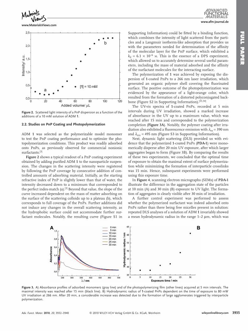
Figure 2 . Scattered light intensity of a PnP dispersion as a function of the additions of a 10 mM solution of ADM 1 .
2.2. Studies on PnP Coating and Photopolymerization
ADM 1 was selected as the polymerizable model monomer to test the PnP coating performance and to optimize the pho-topolymerization conditions. This product was readily adsorbed onto PnPs, as previously observed for commercial nonionic surfactants. [ 3 ]
Figure 2 shows a typical readout of a PnP coating experiment obtained by adding purifi ed ADM 1 to the nanoparticle suspen-sion. The changes in the scattering intensity were registered by following the PnP coverage by consecutive addition of con-trolled amounts of adsorbing material. Initially, as the starting refractive index of PnP is slightly lower than that of water, the intensity decreased down to a minimum that corresponded to the perfect index-match (a). [ 3 ] Beyond that value, the slope of the curve increased dependent on the mass of matter adsorbing on the surface of the scattering colloids up to a plateau (b), which corresponds to full coverage of the PnPs. Further additions did not induce any changes in the overall scattering intensity, as the hydrophobic surface could not accommodate further sur-factant molecules. Notably, the resulting curve (Figure S1 in
© 2010 WILEY-VCH Verlag GmAdv. Funct. Mater. 2010, 20, 3932–3940
Figure 3 . A) Absorbance profi les of adsorbed monomers (gray line) and ofmaximal intensity was reached after 15 min (black line). B) HydrodynamicUV irradiation at 266 nm. After 20 min, a considerable increase was detecpolymerization.
Supporting Information) could be fi tted by a binding function, which combines the intensity of light scattered from the parti-cles and a Langmuir isotherm-like adsorption that provides us with the parameters needed for determination of the affi nity of the molecular layer for the PnP surface, which exhibited a k d = 6.1 × 10 − 6 M . This is the essence of a DPS experiment, which allowed us to accurately determine several useful param-eters, including the mass of material adsorbed and the affi nity of the surfactant molecules for the interacting surface.
The polymerization of 1 was achieved by exposing the dis-persion of 1 -coated PnPs to a 266 nm laser irradiation, which generated an organic polymer shell covering the fl uorinated surface. The positive outcome of the photopolymerization was evidenced by the appearance of a light-orange color, which resulted from the formation of a distorted polyconjugated back-bone (Figure S2 in Supporting Information). [ 33,34 ]
The UV-vis spectra of 1 -coated PnPs, recorded at 5 min intervals during UV irradiation, showed a marked increase of absorbance in the UV up to a maximum value, which was reached after 15 min and corresponded to the polymerization completion ( Figure 3A ). Notably, the polymer coating after irra-diation also exhibited a fl uorescence emission with λ ex = 390 nm and λ em = 495 nm (Figure S3 in Supporting Information).
Next, dynamic light scattering (DLS) provided us with evi-dence that the polymerized 1 -coated PnPs ( PDA-1 ) were mono-merically disperse after 20 min UV exposure, after which larger aggregates began to form (Figure 3 B). By comparing the results of these two experiments, we concluded that the optimal time of exposure to obtain the maximal extent of surface polymeriza-tion while minimizing the formation of interparticle crosslinks was 15 min. Hence, subsequent experiments were performed using this exposure time.
In Figure 4 , scanning electron micrographs (SEMs) of PDA-1 illustrate the difference in the aggregation state of the particles at 10 min (A) and 30 min (B) exposure to UV light. The forma-tion of aggregates is clearly visible after 30 min of irradiation.
A further control experiment was performed to assess whether the polymerized surfactant was indeed adsorbed onto PnPs rather than there being free micelles present in solution: repeated DLS analyses of a solution of ADM 1 invariably showed a mean hydrodynamic radius in the range 1–2 μ m, which was
bH & Co. KGaA, Weinheim 3935wileyonlinelibrary.com
the photopolymerizing fi lm (other lines) acquired at 5 min intervals. The radius of 1 -coated PnPs dependent on the time of exposure to 80 mW ted due to the formation of large agglomerates triggered by interparticle

FULL
PAPER
3936
www.afm-journal.dewww.MaterialsViews.com
Figure 4 . SEM images of PDA-1 after A) 10 min and B) 30 min irradiation. Scale bar represents 300 nm.
remarkably higher than that observed for PDA-1 nanoparticles (Figure S4 in Supporting Information).
PDA-1 nanoparticles were stable in water and did not show nonspecifi c interactions with proteins (e.g., bovine serum albumin; BSA). In fact, after the addition of BSA, no signifi cant increment of scattered light was observed, either with mono-meric or with polymeric forms of the surfactant adsorbed onto PnPs ( Figure 5 ), whereas uncoated particles tend to aggregate in the presence of BSA (data not shown).
Next, we calculated the amount of ADM 1 adsorbed onto the PnPs. Given the amount of particles and the number of ligand addi-tions, each one of them being precisely quantifi ed (6 × 10 − 5 mmol), we determined that about 8.4 × 10 4 molecules were necessary to completely cover each PnP. Since the average surface area of one PnP is 2.01 × 10 4 nm 2 , ca. 4.1 molecules nm − 2 could be accom-modated on the surface of each PnP, which is consistent with a monolayer-like packaging of polymerized 1 , as represented in Scheme 3 . This experiment was repeated several times, invariably obtaining the same number of coating molecules (SD = ± 0.5%), which allowed us to exclude an alternative unordered molecular distribution, such as the formation of multilayers.
© 2010 WILEY-VCH Verlag Gmwileyonlinelibrary.com
Figure 5 . Scattered light intensity of a 0.1% v/v PnP dispersion as a function of the added amount of ADM 1 (full dots) followed by BSA additions (empty dots). BSA was reacted with PnPs coated with 1 after photopolymerization.
The PDA-1 nanoparticles could be centrifuged and redispersed in water several times to remove any possible traces of unbound 1 and the possible presence of micelles. Finally, dried PDA-1 nanoparticles were analyzed by means of attenuated total refl ec-tion (ATR) and Micro-FTIR, and the resulting IR spectra were compared with the FTIR spectrum of ADM 1 (Figure S5 in Sup-porting Information). Besides the expected weak signal of C≡C stretching at 2123 cm − 1 , we observed a marked left-side broad-ening in the strong absorption of the C = O bond at 1650 cm − 1 , which could be explained with the appearance of one supple-mentary small peak in the range 1660–1670 cm − 1 which was attributable to a weak C = C symmetric stretching, evidencing the formation of a new conjugated double bond.
2.3. Determination of Coating Stability
In order to check the actual extent of polymerization of 1 onto PnPs, a desorption test was implemented. With this aim we added smaller perfl uorinated latex nanospheres to the solution as a competitive substrate to induce surfactant sequestration from PnPs. We used the 20 nm radius nanoparticles (PP20) previously characterized. [ 4 ] The size of these nanoparticles, half that of the PnPs, ensured that they could provide a large com-peting surface without contributing signifi cantly to the meas-ured scattering, as discussed below.
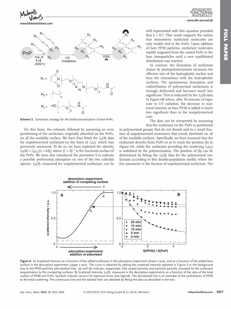
The rationale of the desorption experiment is pictured in Figure 6A , where a typical adsorption curve such as that of Figure 2 is plotted as a function of added surfactant (lower x axis). With I A ( c ), we indicate the scattered intensity vs. sur-factant concentration in the adsorption experiment. In the des-orption experiment, PnPs were fi rst fully coated reaching the point labelled (a) in Figure 6, corresponding to an amount c a of surfactant added to the dispersion and almost entirely adsorbed on the particles. Then, a concentrated dispersion of PP20 was progressively added, thus increasing the total surface made available to the surfactant to adsorb on. As this happened, sur-factant desorbed from the PnPs which resulted in an overall decreased scattered intensity, as indicated in Figure 6 A (top x axis).
Experimental results are reported in Figure 6 B as a function of the ratio X = S (PP20)/ S (PnP) between the total surface area S of PP20 and PnPs available in the suspension as the PP20 are added. With I D ( X ) we indicate the scattered intensity vs. the surface ratio X in the desorption experiment. Plotted I D ( X ) data are already corrected for dilution effects and refer to the intensity values at constant concentration of surfactant. The desorption experiment was performed with unpolymerized sur-factants (downward-pointing triangles) and with coated PnPs after various durations of UV irradiation, the longest being 20 minutes (full dots). In the same fi gure we plotted an esti-mate (dot-dashed line) of the contribution from PP20 to the total scattering. As can be seen, the PP20 contribution to the signal was minor. Indeed, given the size ratio of the particles, the scattering cross-section of each PP20, when fully coated by surfactant, is about 1/16 of that of the PnPs. In the following analysis, we have approximated the contribution as a constant (dot-dashed line in Figure 6 A), which has been added to the scattering of PnPs to obtain the continuous line in Figure 6 A.
bH & Co. KGaA, Weinheim Adv. Funct. Mater. 2010, 20, 3932–3940
FULL P
APER
www.afm-journal.dewww.MaterialsViews.com
R1
R3
R3R2R2
R2
R3
R2
R3
R2
R1
R2
R1
R3
R3R2R2
R2HSAmix of1+3(400/1)
UVirradiation
PnP
R1 =
3
NO
HN
O
NN
4
NO
O
O
3R2 =
5O
HN
O
HOR3 =
Scheme 3 . Schematic strategy for the biofunctionalization of bare PnPs.
On this basis, the estimate followed by assuming an even partitioning of the surfactant, originally adsorbed on the PnPs, on all the available surface. We have thus fi tted the I D ( X ) data for unpolymerized surfactant on the basis of I A ( c ), which was previously measured. To do so, we have exploited the identity I D ( X ) = I A [ c a /(1 + kX )], where (1 + X ) − 1 is the fractional surface of the PnPs. We have also introduced the parameter k to indicate a possible preferential adsorption on one of the two colloidal species. I D ( X ), measured for unpolymerized surfactant, can be
© 2010 WILEY-VCH Verlag GmbH & Co. KGaA, WeinhAdv. Funct. Mater. 2010, 20, 3932–3940
0
0.5
1.0
0adsorption experiment:addition of adsorbant
0
desorption experiment:addition of competing surface
00
0.5
1.0
S
20 min15 min10 min5 min0 min
A a
b
Figure 6 . A) Scattered intensity as a function of the added surfactant in the adsorption experimesurface in the desorption experiment (upper x axis). The curve is obtained by adding the scattedue to the PP20 particles (dot-dashed line). (a) and (b) indicate, respectively, fully coated particsequestration by the competing surfaces. B) Scattered intensity, I D ( X ), measured in the desorptsurface of PP20 and PnPs. Symbols indicate various UV exposure times (see legend). The dot-dto the total scattering. The continuous line and the dashed lines are obtained by fi tting the data
well represented with this equation provided that k ≈ 0.7. This result supports the notion that monomeric surfactant molecules are only weakly tied to the PnPs. Upon addition of bare PP20 particles, surfactant molecules rapidly migrated from the coated PnPs to the bare nanoparticles until a new equilibrated distribution was reached.
In contrast, the formation of surfactant chains by photopolymerization increases the effective size of the hydrophobic anchor and thus the interactions with the hydrophobic surfaces. The spontaneous desorption and redistribution of polymerized surfactants is strongly disfavored and becomes much less signifi cant. This is indicated by the I D ( X ) data in Figure 6 B where, after 10 minutes of expo-sure to UV radiation, the decrease in scat-tered intensity as bare PP20 is added is much less signifi cant than in the nonpolymerized case.
The data can be interpreted by assuming that the surfactant on the PnPs is partitioned
roups that do not desorb and in a small frac-
in polymerized gtion of unpolymerized monomers that evenly distribute on all of the available surfaces. Specifi cally, we have assumed that the surfactant desorbs from PnPs so as to reach the position (b) in Figure 6 A, while the surfactant providing the scattering I A ( c b ) is stabilized by the polymerization. The position of (b) can be determined by fi tting the I D ( X ) data for the polymerized sur-factants according to this double-population model, where the free parameter is the fraction of unpolymerized surfactant. Theeim 3937wileyonlinelibrary.com
2 4
(PP20) / S(PnP)
B
nt (lower x axis), and as a function of the added bare red intensity reported in Figure 2 to the background les and particles partially uncoated by the surfactant
ion experiments as a function of the ratio of the total ashed line is an estimate of the contribution of PP20 as described in the text.

FULL
PAPER
3938
www.afm-journal.dewww.MaterialsViews.com
0 20 40 60 80 100 120 140 160 1800.0
0.5
1.0
1.5
2.0
2.5
3.0
0 20 40 60 80 100 1200.0
0.5
1.0
1.5
2.0
2.5
3.0
3.5
4.0
0 20 40 60 80 100 120 1400.0
0.5
1.0
1.5
2.0
2.5
0 20 40 60 80 100 1200.0
0.5
1.0
1.5
2.0
2.5
Inte
nsity
/ a.
u.
Added volume / µL
Inte
nsity
/ a.
u.
Added volume / µL
DC
BAIn
tens
ity /
a.u.
Added volume / µL
Inte
nsity
/ a.
u.
Added volume / µL
Figure 7 . Scattered intensity measured for polymerized PnPs coated with 1 (C, full dots) and with a 400:1 mixture (10 m M and 25 μ M , respectively) of 1 and 3 (A, full dots), 1 and 5 (B, full dots) and 1 and 4b (D, full dots), and as a function of the amount of added HSA, (A, empty dots), SAv (B and C, empty dots) and galectin-3 (D, empty dots). A) 20 μ M HSA aliquots were added to the solution of PnPs coated with 400:1 1 + 3 up to saturation of available maleimido groups (corresponding to the HSA binding sites) on the PnPs. Residual light at the minimum corresponds to point a in Figure 2 . B) 50 μ M SAv additions were reacted with biotin-containing PnPs, causing interparticle aggregation (no plateau was reached) induced by tetrameric interaction of SAv with biotin. C) 50 μ M SAv aliquots were added to 1 -coated PnPs, no aggregation observed. D) 50 μ M galectin-3 aliquots were added to PnP dispersion containing lactose functionalities which induced particle aggregation. The experiments were carried out in 5 m M phosphate buffer (pH 7.2) using a 0.1% (v/v) PnP dispersion.
resulting curve is shown in Figure 6 B for the more intensely UV-irradiated particles (full dots). The asymptote of the fi tting curve determines the position (b) on the I A (c) curve (dashed construction in Figure 6 B), in turn enabling the quantifi cation of the fraction c b / c a of polymerized surfactant. Specifi cally, we discovered that 10 minutes of UV irradiation were enough to cross-link more then 70% of the adsorbed surfactant mole-cules, while longer exposures did not increase the polymerized fraction.
2.4. Biofunctionalization of PnP Surface
We evaluated the capability of perfl uoroelastomer colloids, in which reactive functional groups were introduced by our method, to serve as a convenient bioconjugation platform for the facile functionalization with complex biomolecules, such as proteins. Scheme 3 illustrates a representative strategy using human serum albumin (HSA). Consecutive additions of a mix-ture of ADMs 1 and 3 in a 400:1 ratio were added to the bare
© 2010 WILEY-VCH Verlag Gmwileyonlinelibrary.com
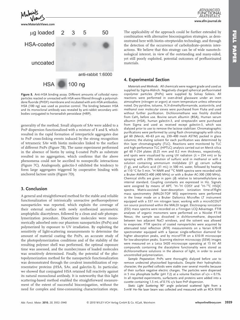
PnPs, thus generating a monolayer of insulated 3 intercalated by clustered groups of 1 . This mixed layer was polymerized on the PnP surface following the UV laser-promoted procedure described above. This generated a large number of maleimido groups, which could be easily linked to HSA by Michael addi-tion at the thiol group of its natively unpaired Cys34 residue. [ 31 ] The average number of anchored proteins was determined by elastic light scattering to be 86 per PnP ( Figure 7A , detailed calculation is reported in Supporting Information). To further validate HSA binding to PnP, we performed a dot-blot bioassay. Increasing amounts of HSA-conjugated and non-conjugated PnPs were fi ltered on a polyvinylidene fl uoride (PVDF) mem-brane and probed with specifi c antibodies that recognize HSA. Positive signals were observed only in HSA-conjugated nano-particles, which demonstrates that anti-HSA antibodies can cross-react with this model protein even when it is anchored to PnP and in a dose-dependent manner ( Figure 8 ).
Analogous procedures were successfully repeated with compounds 4b and 5 , using galectin-3 and streptavidin (SAv), respectively, as the recognizing counterparts, to testify to the
bH & Co. KGaA, Weinheim Adv. Funct. Mater. 2010, 20, 3932–3940
FULL P
APER
www.afm-journal.dewww.MaterialsViews.com
Figure 8 . Anti-HSA binding assay. Different amounts of colloidal nano-particles reacted or unreacted with HSA were fi ltered through a polyvinyli-dene fl uoride (PVDF) membrane and incubated with anti-HSA antibodies. HSA (100 ng) was used as positive control. The binding between HSA and its monoclonal antibody was revealed by anti-rabbit secondary anti-bodies conjugated to horseradish peroxidase (HRP).
generality of the method. Small aliquots of SAv were added to a PnP dispersion functionalized with a mixture of 1 and 5 , which resulted in the rapid formation of interparticle aggregates due to PnP cross-linking events induced by the strong recognition of tetrameric SAv with biotin molecules linked to the surface of different PnPs (Figure 7 B). The same experiment performed in the absence of biotin by using 1 -coated PnPs as substrate resulted in no aggregation, which confi rms that the above phenomena could not be ascribed to nonspecifi c interactions (Figure 7 C). Analogously, galectin-3 exhibited the tendency to form large aggregates triggered by cooperative binding with anchored lactose units (Figure 7 D).
3. Conclusion
A general and straightforward method for the stable and reliable functionalization of intrinsically unreactive perfl uoropolymer nanoparticles was reported, which exploits the coverage of their external surface with newly synthesized asymmetric amphiphilic diacetylenes, followed by a clean and safe photopo-lymerization procedure. Diacetylene molecules were mono-merically adsorbed onto our phantom nanoparticles (PnPs) and polymerized by exposure to UV irradiation. By exploiting the sensitivity of light-scattering measurements to determine the amount of material coating the PnPs, an accurate study of the photopolymerization conditions and of the stability of the resulting polymer shell was performed, the optimal exposure time was assessed, and the number/mass of loaded molecules was sensitively determined. Finally, the potential of the pho-topolymerization method for the nanoparticle functionalization was demonstrated through the covalent immobilization of rep-resentative proteins (HSA, SAv, and galectin-3). In particular, we showed that conjugated HSA retained full reactivity against its natural monoclonal antibody. It is noteworthy that this light scattering-based method enabled the straightforward measure-ment of the extent of successful bioconjugation, without the need for complex and time-consuming characterization steps.
© 2010 WILEY-VCH Verlag GmAdv. Funct. Mater. 2010, 20, 3932–3940
The applicability of the approach could be further extended by combination with alternative bioconjugation strategies, as dem-onstrated here with biotin–streptavidin technology, and through the detection of the occurrence of carbohydrate–protein inter-actions. We believe that this strategy can be of wide nanotech-nological interest, in view of the outstanding and many-sided, yet still poorly exploited, potential outcomes of perfl uorinated materials.
4. Experimental Section Materials and Methods: All chemicals were reagent grade and used as
supplied by Sigma-Aldrich. Negatively charged spherical perfl uorinated copolymer particles (PnPs) were supplied by Solvay Solexis. All reactions were performed in oven-dried glassware under an inert atmosphere (nitrogen or argon) at room temperature unless otherwise noted. Dry pyridine, toluene, N , N -dimethylformamide, acetonitrile, and methanol over molecular sieves were purchased from Fluka and used without further purifi cation. Dichloromethane was freshly distilled from CaH 2 before use. Bovine serum albumin (BSA), Human serum albumin (HSA), human galectin-3, and streptavidin were purchased from Sigma and used as received except galectin-3, which was dialyzed prior to use to remove the lactose stabilizer. Chromatographic purifi cations were performed by using fl ash chromatography with silica gel 60 (Merck, 40–63 μ m eq. 230–400 mesh ASTM) packed in glass columns; the eluting solvent for each purifi cation was determined by thin layer chromatography (TLC). Reactions were monitored by TLC and high-perfomance TLC (HPTLC) analysis carried out on Merck silica gel 60 F-254 plates (0.25 mm and 0.2 mm thickness, respectively), and spots were visualized by using UV radiation ( λ = 254 nm) or by spraying with a 20% solution of sulfuric acid in methanol or with a solution containing ammonium molybdate (21 g) cerium sulfate (1 g), and sulfuric acid (31 mL) in 500 mL water, followed by heating at 110 ° C for 5 min. 1 H NMR and 13 C NMR spectra were recorded with a Bruker AVANCE-400 (400 MHz) or with a Bruker AC-300 (300 MHz). Chemical shifts are given in ppm ( δ ) relative to tetramethylsilane as internal standard. Coupling constants are expressed in Hz. Signals were assigned by means of APT, 1 H– 1 H COSY and 1 H– 13 C HSQC spectra. Matrix-assisted laser-desorption- ionization time-of-fl ight mass spectrometry (MALDI-TOF MS) experiments were performed in the linear mode on a Bruker Daltonics Microfl ex LT instrument equipped with a 337 nm nitrogen laser, working with a microSCOUT ion source positioned within the MALDI target. Electrospray ionization (ESI) mass spectra were recorded on a Finnigan LCQ Advantage. FTIR analyses of organic monomers were performed on a Nicolet FT-IR Nexus; the sample was dissolved in dichloromethane, deposited between two adjacent NaCl windows, and the solvent was allowed to evaporate. FTIR spectra of coated nanoparticles were acquired by attenuated total refl ection (ATR) measurements on a Varian 670-IR spectrometer equipped with a Specac single-refl ection diamond for higher absorption peaks, and by microFTIR on a 610-IR microscope for low-absorption peaks. Scanning electron microscopy (SEM) images were measured on a Leica S420 microscope operating at 15 kV. All compounds containing the diacetylene functionality were stored as dichloromethane solutions in the absence of light, in order to avoid uncontrolled polymerization.
Sample Preparation: PnPs were thoroughly dialyzed before use to remove undesired physisorbed byproducts. Despite their hydrophobic character, the purifi ed colloids were stable over several months because of their surface negative electric charges. The particles were dispersed in 5 m M phosphate buffer (pH 7.2) at a volume fraction of v/v = 0.1%. In all reported experiments, surfactants and proteins were added into a cuvette containing 1.5 mL of 0.1% v/v bare PnP dispersion.
Static Light Scattering: 90 ° angle polarized scattered light from a 5-mW He–Ne laser beam was collected and measured with an RCA 931B
bH & Co. KGaA, Weinheim 3939wileyonlinelibrary.com
FULL
PAPER
3940
www.afm-journal.dewww.MaterialsViews.com
photomultiplier. We used conventional spectrophotometer cuvettes held in a suitably designed cell holder provided with a ministirrer, the necessary tubing holding, and water fl ow to control the temperature, in turn measured by a thermistor. Measurements were performed at 30 ° C. Diacetylenic surfactants and proteins solutions were injected into the cuvette by motorized pumps (Kent Scientifi c Genie syringe pump and Ismatec Reglo piston precision pump). Stirring, temperature, and injections were controlled by a computer, through a suitable interface designed ad hoc to program DPS experiments. Experiments were taken by a programmed sequence of injection, stirring, and data acquisition. Typically, each data point refers to an addition of a few μ L of a surfactant or protein solution to the cuvette initially containing 1.5 mL of bare PnP dispersion. Any injection was typically followed by 10 min of stirring. Sets of 80 independent intensity acquisitions were taken for each condition and analyzed to eliminate possible optical contributions due to dust impurities.
Dynamic Light Scattering: Measurements of the intensity ( I ) autocorrelation functions were periodically taken to ensure that the PnPs were monomerically dispersed at the various stages of the adsorption curves. The data were taken by using a frequency doubled 532-nm, 150-mW NdYag laser and through a single-mode fi ber collection of the scattered light. Cross-correlations were calculated with a BI-9000 digital correlator (Brookhaven Instruments Corp.), after the collected I was divided by a 50/50 fi ber beam splitter.
Absorbance and Photopolymerization: Absorption spectra were measured in the wavelength range 240–600 nm using a fi ber-coupled spectrometer (Ocean Optics Inc., USA). Illumination was provided by fi ber-coupled deuterium and halogen lamps. The UV-vis spectra were recorded at 5 min intervals during UV irradiation. Photopolymerization of samples was promoted by UV laser pulses at 266 nm using a Brilliant laser (Quantel, France) operated with a fourth harmonic generator. In order to achieve a more uniform illumination of the cuvette, the laser beam was expanded and fi ltered by means of a diagram with an aperture diameter of 1 cm. The samples were illuminated with 4 ns pulses with energy of 6–8 mJ each that were generated at a repetition rate of 10 Hz, thus delivering an average power of 60–80 mW with a peak power of more than 1 MW.
Dot Blot Assay: Dot blot was performed by fi ltering proteins and/or nanoparticles onto PVDF membrane, utilizing a Minifold I dot blot apparatus (GE Healthcare, Life Sciences, Little Chalfont, UK), and incubating in blocking solution (5% skim milk in PBS) for 60 min at RT. To evaluate HSA conjugation, membrane was then probed for 60 min at 25 ° C using rabbit anti-HSA antibodies at a 1:2500 dilution in blocking solution. The membrane was rinsed three times in 0.05% Tween in PBS for 10 min each and subsequently incubated for 60 min at RT with a secondary antibody (1:6000 horseradish peroxidase-goat anti-rabbit antibodies in 0.05% Tween in PBS). Immunoreactive spots were revealed using ECL Western blotting reagent (GE Healthcare).
Supporting Information Supporting Information is available from the Wiley Online Library or from the author.
Acknowledgements This work was supported by the “Romeo ed Enrica Invernizzi” Foundation and “Centro di Microscopia Elettronica per le Nanotecnologie applicate alla medicina” (CMENA, University of Milan), the CNR-Regione Lombardia “Mind in Italy” project, a FIRB grant (CHEM-PROFARMA-NET, RBPR05NWWC), and the Cariplo Foundation. We thank R. Allevi (CMENA) for SEM images and Dr. A. Natalello for help in FT-IR characterization. We are grateful to Solvay Solexis for the generous
© 2010 WILEY-VCH Verlag Gwileyonlinelibrary.com
gift of the perfl uorinated colloids and thank M. Bassi for the crucial help in selecting the best polymer composition for the colloid formulation.
Received: June 23, 2010 Published online: September 3, 2010
[ 1 ] M. P. Krafft , J. G. Riess , J. Polym. Sci. Polym. Chem. 2007 , 45 , 1185 . [ 2 ] J. E. Puskas , Y. H. Chen , Biomacromolecules 2004 , 5 , 1141 . [ 3 ] A. Ghetta , D. Prosperi , F. Mantegazza , L. Panza , S. Riva , T. Bellini ,
Proc. Natl. Acad. Sci. U.S.A. 2005 , 102 , 15866 . [ 4 ] D. Prosperi , C. Morasso , F. Mantegazza , M. Buscaglia , L. Hough ,
T. Bellini , Small 2006 , 2 , 1060 . [ 5 ] D. Prosperi , C. Morasso , P. Tortora , D. Monti , T. Bellini , ChemBio-
Chem 2007 , 8 , 1021 . [ 6 ] C. Morasso , T. Bellini , D. Monti , M. Bassi , D. Prosperi , S. Riva ,
ChemBioChem 2009 , 10 , 639 . [ 7 ] M. Mammen , S.-K. Choi , G. M. Whitesides , Angew. Chem. Int. Ed.
1998 , 37 , 2754 . [ 8 ] M. A. N. Coelho , E. P. Vieira , H. Motschmann , H. Mohwald ,
A. F. Thunemann , Langmuir 2003 , 19 , 7544 . [ 9 ] C. E. Giacomelli , W. Norde , Biomacromolecules 2003 , 4 , 1719 . [ 10 ] S. H. Mollmann , J. T. Bukrinsky , S. Frokjaer , U. Elofsson , J. Colloid
Interface Sci. 2005 , 286 , 28 . [ 11 ] J. Vörös , Biophys. J. 2004 , 87 , 553 . [ 12 ] C. M. Santos , A. Kumar , W. Zhang , C. Cai , Chem. Commun. 2009 , 2854 . [ 13 ] C.-A. J. Lin , R. A. Sperling , J. K. Li , T.-Y. Yang , P.-Y. Li , M. Zanella ,
W. H. Chang , W. J. Parak , Small 2008 , 4 , 334 . [ 14 ] S.-W. Kim , S. Kim , J. B. Tracy , A. Jasanoff , M. G. Bawendi , J. Am.
Chem. Soc. 2005 , 127 , 4556 . [ 15 ] M. S. Nikolic , M. Krack , V. Aleksandrovic , A. Kornowski , S. Förster ,
H. Weller , Angew. Chem. Int. Ed 2006 , 45 , 6577 . [ 16 ] R. A. Sperling , T. Pellegrino , J. K. Li , W. H. Chang , W. J. Parak , Adv.
Funct. Mater. 2006 , 16 , 943 . [ 17 ] Y. Lu , Y. Yang , A. Sellinger , M. Lu , J. Huang , H. Fan , R. Haddad ,
G. Lopez , A. R. Burns , D. Y. Sasaki , J. Shelnutt , C. J. Brinker , Nature 2001 , 410 , 913 .
[ 18 ] R. Jelinek , S. Kolusheva , in Creative Chemical Sensor Systems , vol. 277 , Springer-Verlag Berlin , Berlin, Germany 2007 , pp. 155 .
[ 19 ] M. A. Reppy , B. A. Pindzola , Chem. Commun. 2007 , 4317 . [ 20 ] X. Y. Zhang , F. H. Chen , J. Z. Ni , Drug Delivery 2009 , 16 , 280 . [ 21 ] W. Jiang , S. Mardyani , H. Fischer , W. C. W. Chan , Chem. Mater.
2006 , 18 , 872 . [ 22 ] S. I. Kasteren , S. J. Campbell , S. Serres , D. C. Anthony , N. R. Sibson ,
B. G. Davis , Proc. Natl. Acad. Sci. U.S.A. 2009 , 106 , 18 . [ 23 ] H. C. Kolb , M. G. Finn , K. B. Sharpless , Angew. Chem. Int. Ed. 2001 ,
40 , 2004 . [ 24 ] M. Meldal , C. W. Tornoe , Chem. Rev. 2008 , 108 , 2952 . [ 25 ] M. Fleiner , P. Benzinger , T. Fichert , U. Massing , Bioconjugate Chem.
2001 , 12 , 470 . [ 26 ] S. F. Schluter , P. L. Ey , J. Immunol. Methods 1984 , 66 , 89 . [ 27 ] P. Charley , P. Saltman , Science 1963 , 139 , 1205 . [ 28 ] N. M. Green , Adv. Protein Chem. 1975 , 29 , 85 . [ 29 ] N. M. Green , Biochem. J. 1963 , 89 , 609 . [ 30 ] M. N. Khan , J. Org. Chem. 2002 , 60 , 4536 . [ 31 ] L. Polito , D. Monti , E. Caneva , E. Delnevo , G. Russo , D. Prosperi ,
Chem. Commun. 2008 , 621 . [ 32 ] R. Schmidt , R. J. Michel , Angew. Chem. Int. Ed. 1980 , 19 , 731 . [ 33 ] M. Wenzel , G. H. Atkinson , J. Am. Chem. Soc. 1989 , 111 , 6123 . [ 34 ] R. R. Chance , Macromolecules 1980 , 13 , 396 .
mbH & Co. KGaA, Weinheim Adv. Funct. Mater. 2010, 20, 3932–3940