Perception of inert particles by calanoid copepods: behavioral observations and a numerical model
24
Journal of Plankton Research Vol.20 no.ll pp.2129-2152, 1998 Perception of inert particles by calanoid copepods: behavioral observations and a numerical model Marie H.Bundy 1 - 4 , Thomas F.Gross 2 , Henry A.Vanderploeg 1 and J.Rudi Strickler 3 'Great Lakes Environmental Research Laboratory, 2205 Commonwealth Blvd., Ann Arbor, MI48105, 2 Skidaway Institute of Oceanography, 10 Ocean Science Circle, Savannah, GA 31411 and 3 Centerfor Great Lakes Studies, University of Milwaukee-Wisconsin, Milwaukee, WI, USA 4 Present address: Academy of Natural Sciences Estuarine Research Center, 10545 Mackall Road, St Leonard, MD 20685, USA Abstract High-resolution video showed freely swimming Diaptomus sicilis attacking and capturing inert SO um polystyrene beads that were outside the influence of the copepod feeding current. The beads were frequently more than half a body length away and were attacked after the 'bow wake' of the moving copepod displaced the bead away from the copepod. To investigate the hypothesis that deformation of streamlines around the copepod and its first antennae stimulated the attack response, afiniteelement numerical model was constructed. The model described thefluidinteractions between a large object approaching a smaller object in a laminar flow at Reynolds number 5, which is charac- teristic of the fluid regime experienced by foraging copepods. The model revealed that fluid velocity fluctuations and streamline deformations arose in the region between the two objects as separation distance between the objects decreased. The video observations and the model results support the hypotheses that chemoreception is not required for the detection and capture of large phytoplankton cells [Vanderploeg et ai, in Hughes,R.N. (ed.), Behavioral Mechanisms of Food Selection. NATO ASI Series G20, 1990; DeMott and Watson, /. Plankton Res., 13, 1205-1222, 1991], and that swimming behavior plays an integral role in prey detection. Introduction Evidence that copepods can detect the presence and position of remotely located prey has been accumulating since 1980, when high-speed cinematography was first used to observe copepod feeding (Alcaraz et ai, 1980). However, little is known about either the physical mechanisms or the sensory modalities involved in this process. Calanoid copepods feed on suspended particles at rates that may depend on the olfactory and gustatory qualities, and on nutritional, size and/or movement characteristics of the particles, rather than on particle abundance alone (Harvey, 1937; Donaghay and Small, 1979; Butler et al., 1989; Vanderploeg et al, 1990; DeMott and Watson, 1991). Tethered calanoid adults and juveniles can actively capture non-motile phytoplankton cells entrained in the feeding current by using one or more feeding appendages to direct individual particles to the mouth (Koehl and Strickler, 1981; Paffenhofer et al, 1982; Vanderploeg and Paffenhofer, 1985; Paffenhofer and Lewis, 1990). Large (>14 um) beads may be captured in the same way (Vanderploeg etal, 1990). Both active captures and attacks (an attack is characterized by a jump toward the particle, followed by an active capture) may be executed before the particle contacts a feeding appendage, indicating that some signal regarding particle location is detected by the copepod. Traditionally, the feeding current has been considered as the 2129 by guest on July 6, 2011 plankt.oxfordjournals.org Downloaded from
-
Upload
independent -
Category
Documents
-
view
7 -
download
0
Transcript of Perception of inert particles by calanoid copepods: behavioral observations and a numerical model

Journal of Plankton Research Vol.20 no.l l pp.2129-2152, 1998
Perception of inert particles by calanoid copepods: behavioralobservations and a numerical model
Marie H.Bundy1-4, Thomas F.Gross2, Henry A.Vanderploeg1 and J.RudiStrickler3
'Great Lakes Environmental Research Laboratory, 2205 Commonwealth Blvd.,Ann Arbor, MI48105,2Skidaway Institute of Oceanography, 10 Ocean ScienceCircle, Savannah, GA 31411 and3Centerfor Great Lakes Studies, University ofMilwaukee-Wisconsin, Milwaukee, WI, USA4Present address: Academy of Natural Sciences Estuarine Research Center, 10545Mackall Road, St Leonard, MD 20685, USA
Abstract High-resolution video showed freely swimming Diaptomus sicilis attacking and capturinginert SO um polystyrene beads that were outside the influence of the copepod feeding current. Thebeads were frequently more than half a body length away and were attacked after the 'bow wake' ofthe moving copepod displaced the bead away from the copepod. To investigate the hypothesis thatdeformation of streamlines around the copepod and its first antennae stimulated the attack response,a finite element numerical model was constructed. The model described the fluid interactions betweena large object approaching a smaller object in a laminar flow at Reynolds number 5, which is charac-teristic of the fluid regime experienced by foraging copepods. The model revealed that fluid velocityfluctuations and streamline deformations arose in the region between the two objects as separationdistance between the objects decreased. The video observations and the model results support thehypotheses that chemoreception is not required for the detection and capture of large phytoplanktoncells [Vanderploeg et ai, in Hughes,R.N. (ed.), Behavioral Mechanisms of Food Selection. NATO ASISeries G20, 1990; DeMott and Watson, /. Plankton Res., 13, 1205-1222, 1991], and that swimmingbehavior plays an integral role in prey detection.
Introduction
Evidence that copepods can detect the presence and position of remotely locatedprey has been accumulating since 1980, when high-speed cinematography wasfirst used to observe copepod feeding (Alcaraz et ai, 1980). However, little isknown about either the physical mechanisms or the sensory modalities involvedin this process. Calanoid copepods feed on suspended particles at rates that maydepend on the olfactory and gustatory qualities, and on nutritional, size and/ormovement characteristics of the particles, rather than on particle abundancealone (Harvey, 1937; Donaghay and Small, 1979; Butler et al., 1989; Vanderploeget al, 1990; DeMott and Watson, 1991). Tethered calanoid adults and juvenilescan actively capture non-motile phytoplankton cells entrained in the feedingcurrent by using one or more feeding appendages to direct individual particlesto the mouth (Koehl and Strickler, 1981; Paffenhofer et al, 1982; Vanderploegand Paffenhofer, 1985; Paffenhofer and Lewis, 1990). Large (>14 um) beads maybe captured in the same way (Vanderploeg etal, 1990). Both active captures andattacks (an attack is characterized by a jump toward the particle, followed by anactive capture) may be executed before the particle contacts a feedingappendage, indicating that some signal regarding particle location is detected bythe copepod. Traditionally, the feeding current has been considered as the
2129
by guest on July 6, 2011plankt.oxfordjournals.org
Dow
nloaded from

M.H.Bnndy et al.
vehicle that conveys both chemical (Andrews, 1983) and mechanical (L6gier-Visser et al., 1986) signals to the copepod when the copepod is feeding on non-motile prey.
Selection and detection of prey may, however, be two separate processes.DeMott and Watson (1991) used high background levels of chemostimulatorycompounds to mask chemical signals from large algal cells, and found that clear-ance rates and selectivity of Diaptomus birgei on large algae were unchanged.These results, coupled with visual observations of tethered copepods showingactive capture of inert beads in the feeding current (Vanderploeg et al, 1990),suggest that diaptomids primarily use mechanoreception to detect largeparticles. The signal most likely originates from the motion of the bead relativeto the copepod, as the bead is entrained in the feeding current. Visual obser-vations designed to examine the behavioral processes involved in prey discrimin-ation reveal that the process of selecting for or against ingestion of differentparticles often occurs after the particle is captured and handled (Paffenhofer etal., 1982; Cowles etai, 1988; Vanderploeg etal., 1990). The copepod presumablyuses mechanoreceptive and chemoreceptive sensilla on the mouthparts(Friedman, 1980) to distinguish particle quality, and then actively rejectsunwanted particles.
This study presents the first evidence that free-swimming calanoid copepodsattack non-motile inert particles located outside the influence of the feedingcurrent. Freely swimming copepods were observed swimming toward an inertparticle, then jumping forward and capturing it. Immediately before the captureevent, the particle was displaced away from and in front of the approachingcopepod. Here, we used high-resolution video observations to document theattacks and to investigate the hypothesis that the stimulus initiating attackbehavior originates as a distortion of the boundary layer around the movingcopepod. A numerical model was then used to determine whether a stationaryparticle in the path of a moving object can cause a distortion of the boundarylayer around that object at the Reynolds number (Re) characteristic of thecopepod-bead interactions documented by visual observations. The goal of themodel was to describe how a copepod may use mechanoreception to detectstreamline deformations around its first antennae when the boundary layeraround the moving animal interacts with an inert particle in its path. Our resultssuggest that: (i) mechanoreception is involved in the detection and capture oflarge non-motile particles; (ii) perception of dissolved chemical cues is notrequired for the detection and capture of large particles by copepod predators;and (iii) the feeding current is not always involved in the detection and captureof non-motile prey.
Method
Video procedures and behavioral analyses
Diaptomus sicilis females were collected from vertical tows in eastern LakeMichigan using an opening/closing net (202 um mesh size with a 11 solid cod-end)
2130
by guest on July 6, 2011plankt.oxfordjournals.org
Dow
nloaded from

Perception of inert particles by calanoid copepods
at a 100-m-deep station located 15 km west of Muskegon, Michigan. The ascend-ing net was closed at -5 m below the thermocline to reduce mechanical damageto the copepods. The contents of the net cod-end were gently poured into 10 1insulated jugs containing water collected from a similar depth as the net tows.Copepods were returned to an environmental room at the Great Lakes Environ-mental Research Laboratory (GLERL), where they were maintained at 8-10°Cand a 12:12 h light:dark cycle. Using a wide-bore glass tube, -50 D.sicilis femalesand 3-4 males were individually pipetted into 1800 ml of 0.2 um filtered lakewater (FLW), containing -0.3 mm31"1 (360 cells ml"1) Cryptomonas reflexa [12 umequivalent spherical diameter (ESD)]. Copepods were videotaped after at least48 h acclimation to laboratory conditions.
Twenty-four hours prior to videotaping, ~15 female D.sicilis were individuallypipetted into a beaker (the 'acclimation vessel') containing 1800 ml FLW to which-0.3 mm3 I"1 Creflexa and 3-6 ml"1 50 um polystyrene beads (specific gravity1.05 g cm"3; Duke Scientific Corp., Palo Alto, CA) had been added. The beadshad been repeatedly rinsed in deionized water, centrifuged and resuspended indeionized water. On the day of filming, a 3 1 cubic Plexiglas container with aremovable lid ('filming vessel') was filled with 2000 ml of FLW and 800 ml waterfrom the acclimation vessel that was first screened through a 10 um mesh. Addingscreened water from the acclimation vessel seemed to reduce the incidence ofrapid jumping by the copepods, noted when only FLW was used. Enough algaeand beads were then added to create a suspension consisting of 0.2 mm3 I"1
Creflexa and 3-6 ml"1 50 um polystyrene beads.The video system at GLERL, similar to that used by Bundy and Paffenhofer
(1996), consists of a video camera equipped with a near-infrared collimated laserlight source and mounted on a three-dimensional motor drive. It is located in thesame room in which the copepods were maintained and acclimated. The roomwas illuminated by dim, diffuse light from cool white fluorescent bulbs. Thecamera optical set-up utilizes Schlieren optics and resembles that described byStrickler (1985). The database of camera position was synchronized with thevideo record through a time-code generator and a time-stamp overlay on eachvideo field. In this study, a record of camera position was collected at a samplingrate of either 8 or 15 Hz. The temporal resolution of the video images was 60 Hz.The area of the video camera field of view was 5.75 X 7.50 mm, with spatial reso-lution of -10 um in the plane of view and a depth of field of -200 um in the planeof focus. Joysticks and a video monitor in an adjoining room allowed individualcopepods to be followed for hours, if necessary, as they moved about in the 3 1filming vessel. Videotaping of copepod-bead interactions was conducted within10 days of zooplankton collection.
The filming vessel was gently stirred 15 min prior to videotaping to resuspendbeads and phytoplankton. One to three copepods were then added to the filmingvessel and a single copepod was videotaped for 30-60 min. After an individualhad been videotaped, it was removed from the filming vessel and placed in a smallbeaker. The filming vessel was again gently stirred and another copepod wasvideotaped after a 15 min period to allow water motion in the filming vessel todissipate. AH copepods were actively swimming at least 48 h after filming, and
2131
by guest on July 6, 2011plankt.oxfordjournals.org
Dow
nloaded from

M-H.Bundy et al.
each experimental copepod was microscopically examined for broken append-ages and measured.
To determine the position and velocities of the beads and the copepod, thecoordinates of the copepod's left and right first antennae (Al), the eye, the baseand tip of the urosome, along with any particles of interest in the field of view(Figure 1), were digitized in successive video fields using a motion-analysis system(Peak Performance Technologies, Inc.). Two-dimensional data representingpoints in the camera plane of view were used to calculate swimming trajectoriesand velocities. Each set of coordinates representing bead location was subjectedto a low-pass digital filter to remove high frequencies associated with digitizingerror (Weiss et al., 1986). Position data were then mathematically corrected forthe movement of the camera. The horizontal and vertical planes of referencewere the horizontal and vertical planes of the filming vessel, which were normalto the optical axis.
We examined copepod swimming behavior at a fine temporal scale (60 Hz)immediately before an attack to characterize fully the orientation and trajectoryof the copepod body before the attack occurred, and as a means of detectingbehavioral responses to the bead. We digitized the position of the copepod bodyand the bead during an interval beginning after the jump prior to the attack leap(called 'the time interval before capture'). We then determined the orientationof the copepod to the bead, its swimming velocity, and the distance to the beadwhen the attack occurred (Table I). To investigate the role of body orientation inthe remote detection of the bead, we also examined the position and orientation
beadleftAl
right Al
approach angle ^ urosomebase
/urosometip
Fig. L Schematic drawing of a bead and copepod (not to scale). Stars indicate points digitized inbehavioral analyses. The approach angle is defined as the angle composed of the copepod body axisand the trajectory to the bead.
2132
by guest on July 6, 2011plankt.oxfordjournals.org
Dow
nloaded from

Perception of inert particles by calanoid copepods
of putative mechanosensory setae on the Al by using laser scanning confocalmicroscopy (LSCM) to obtain a three-dimensional image [see Bundy andPaffenhofer (1993) for methodology].
We also analyzed swimming behavior for 10 s before the attack by digitizingthe position of the copepod at 67 ms intervals (15 Hz) (Table II). We calculated
Table L Behavior of six copepods during the time interval immediately before attack events. Lettersindicate different attack events for the same copepod. Sampling frequency = 60 Hz
Copepod
1234
5
6
Captureevent
aaaabcabab
Intervalbeforecapture(s)
3.100.601.250.700.070.405.009.001.101.40
Orientation angle (degrees)
After jump
11110510211773
106125107119113
Prior to attack
868989909491
107919395
Distance fromcopepod whenbead first moves(mm)
1.760.971.451.101.861.571.322.101.431.76
Copepod distancefrom head atU Will bvAU ell
time of attack(mm)
1.08a
1.40"0.481.390.780.931.410.481.31
'Copepod used feeding current to capture the bead (see the text for further information).bVentral view of copepod (see the text for further explanation).
Table IL Behavior of six copepods during 10 s intervals before attack events. Letters indicate differentattack events for the same copepod. Swim interval is the interval between jumps. Swim velocity is theaverage velocity during each swim interval. Jump velocity is the maximum velocity for each jump.Sampling frequency = 15 Hz
Copepod
1
2
3
4
5
6
Captureevent
a
a
a
a
b
c
a
b
a
b
Swim interval(s)Mean ± SD
2.10 ±0.9
1.84 ± 1.4
0.98 ± 0.6
1.43 ± 0.7
0.91 ± 0.5(n = 8)1.43 ± 1.0(«=4)3.48 ± 1.6
9.00
1.23 ± 0.7
2.25 ± 0.9
Swim velocity(mrar1)Mean ± SD
1.13 ± 0.5
1.10 ± 0.7
1.87 ± 1.2
1.31 ± 0.9
1.11 ±0.7
1.15 ±0.7
0.86 ± 0.6
1.12 ±0.7
1.22 ± 0.8
0.86 ± 0.7
Jump velocity(mmr1)Mean ± SD
19.66 ± 4.4
13.76 ± 8.2
22.97 ± 16.1
28.8 ± 15.5
22.37 ± 12.9
34.39 ± 7.0
16.05 ± 6.0
19.25 ± 7.0
26.10 ± 5.2
19.16 ± 12.1
2133
by guest on July 6, 2011plankt.oxfordjournals.org
Dow
nloaded from

MJi-Bundy et al.
the swimming interval (i.e. the amount of time spent swimming between eachjump), jump velocity and swimming velocity from copepod displacement.
A 9 s subset of the 10 s behavioral series (digitized at 67 ms intervals) wassubdivided into three 3 s intervals (Table III). The behavior of the copepodduring the 3 s immediately before an attack was compared to that during the 3 sintervals beginning 9 and 6 s before an attack. For each 3 s interval, we calcu-lated the copepod movement trajectory, the mean swimming velocity betweenjumps, and the mean jump velocity. Following the methods of Cain (1989), theswimming trajectory was denned as the direction of movement between eachdigitized position, with 0° indicating no change in direction between two succes-sive positions. The direction of movement (0) is defined as a vector with unitlength, whose coordinates can be averaged to calculate the mean and the angulardeviation of the trajectories. Upward jumps were defined as positive and down-ward jumps were defined as negative. A runs test (e.g. Dixon and Massey, 1951)was then used to determine whether jumps in any interval were non-randomlydirected.
Numerical model
The finite element numerical model used in this study solved for a two-dimen-sional flow field around two circular cylinders: one cylinder was stationary andthe other was embedded in the upstream flow and moving with the streamlinevelocity. This scenario is physically identical to one where a large moving cylin-der approaches a stationary smaller cylinder. The numerical model was based onthe assumption that the wake flow and boundary layer constitute the deformedvelocity streamlines around the moving copepod. The bead can be considered asan incompressible obstacle, embedded within the wake flow, that further deformsthe velocity streamlines. The finite element method allows a grid to resolve theboundaries of the objects in the flow with arbitrary geometry. The goal of thisexercise was to examine the hydrodynamics of the simple two-dimensional caseof circular cylinders normal to the flow, and to characterize fluid velocities andthe geometry of the flow field in the region between the two objects. We suspectedthat deformation of streamlines around the copepod's first antennae and itsmechanoreceptors was the stimulus that initiated attack responses from thecopepod, and that these deformations arose when the boundary layer associatedwith the moving copepod impacted the solid bead. We use the simple case of twocylinders as a test for investigating the potential for flow field distortions to arisebetween two objects moving relative to each other at low Re. The geometry ofthe copepod antennae approaching a prey item is more like a cylinder movingrelative to a sphere. In this case, the magnitude of fluid distortion would mostlikely be lower and have a different spatial distribution than in the case of twocylinders.
The model solved the vorticity equation with non-linear advection and viscousdiffusion terms, and a streamline equation to satisfy the incompressibility require-ment. To solve for the flow field, the low-/?e Navier-Stokes equations werewritten for two-dimensional flow:
2134
by guest on July 6, 2011plankt.oxfordjournals.org
Dow
nloaded from

Tab
le i
n.
Beh
avio
r of
six
cop
epod
s du
ring
3 s
seq
uent
ial
inte
rval
s be
ginn
ing
9 s
befo
re a
ttack
eve
nts.
Let
ters
ind
icat
e di
ffer
ent
atta
ck e
vent
s fo
r th
e sa
me
cope
pod.
Sw
im tr
ajec
tory
is
the
chan
ge in
dir
ectio
n be
twee
n se
quen
tial d
igiti
zed
posi
tions
(see
the
text
). S
wim
vel
ocity
is c
alcu
late
d fro
m d
ispl
acem
ent
at e
ach
sequ
entia
l di
gitiz
ed p
ositi
on. J
ump
velo
city
is th
e m
axim
um v
eloc
ity f
or e
ach
jum
p. S
ampl
ing
freq
uenc
y =
IS H
z
Cop
epod
ev
ent
Swim
traj
ecto
ry (
degr
ees)
Swim
vel
ocity
(m
m s
"1)Ju
mp
velo
city
(m
m s"
9-6
s pr
ior
6-3
s pr
ior
Mea
n ±
SD
Mea
n ±
SD3
s pr
ior
Mea
n ±
SD9-
6 s
prio
r 6-
3 s
prio
rM
ean
± SD
M
ean
± SD
3 s
prio
rM
ean
± SD
9-6
s pr
ior
6-3
s pr
ior
Mea
n ±
SD
Mea
n ±
SD3
s pr
ior
Mea
n ±
SD
1 a
80.8
±17
42
.4 ±
51
35.5
±41
1.
06 ±
0.3
1.24
±0.
6 1.1
±0.
6
2 a
50.4
±45
51
.0 ±
46
43.0
±39
0.
92 ±
0.4
0.
89 ±
0.3
0.96
±0.
4
3 a
21.8
±45
8.
6 ±
57
60.2
±76
1.
99 ±
1.4
1.40
± 1
.0
1.05
± 1
.2
4 a
32.1
±68
17
.2 ±
49
6.3
±38
0.
91 ±
0.9
1.07
±0.
8 1.
51 ±
1.2
b 24
.1 ±
51
176.
5 ±
57
131.
2 ±
53
1.22
±0.
8 1.
18 ±
0.8
0.64
± 0
.4
c 14
1.5
±34
13
2.9
±34
15
4.7
±40
0.
92 ±
0.4
1.
05 ±
0.6
1.
32 ±
1.2
5 a
27.5
±40
25
.8 ±
57
22.9
±44
0.
67 ±
0.4
0.
85 ±
0.6
1.
04 ±
0.5
b 14
.9 ±
74
26.4
±52
36
.7 ±
50
1.21
±0.
8 0.
98 ±
0.9
0.89
±0.
6
6 a
4.6
±45
2.
9 ±
34
2.9
±38
1.
13 ±
0.8
1.18
±0.
8 1.
05 ±
0.5
b 68
.8 ±
63
59.0
±57
53
.3 ±
62
0.53
±0.
4 0.
81 ±
0.6
1.35
±1.
0
9.29
n=l
32.3
± 2
3.8
22.1
3 ±
1.5n
= 2
8.80
± 6
.0n
= 2
15.3
0 ±
8.8
11.5
1 ±
3.4
37.0
3 ±
15.1
18.5
2 ±
4.0
29.6
6 ±
9.8
30.6
9
23.2
0n
= l
14.2
8
39.6
6 ±
9.8
17/7
7 ±
7.4
25.2
3 ±
0.6
7.86
14.7
0w
= l
20.6
5 ±
2.0
n =
214
.97
± 9.
7n
= 2
34.8
4 ±
13.5
16.1
0 ±
3.3
n=
330
.96
± 1.
9n
=2
12.6
0n
= l
24.2
2«
= 1
27.2
0 ±
8.5
n =
331
.90
± 5.
2«=
2
a. o 5' 3
by guest on July 6, 2011plankt.oxfordjournals.orgDownloaded from

M.H.Bundy el al.
dU trdU dU ldP &U
dt dx dy p dx dx2 dy2
dV T dV dV 1 dP &V &V
dt dx By p dx dx2 dy2
These equations were combined using the definition of vorticity (w):
dU dVu> ^ — — —dy dx
to eliminate the pressure variable:
3<D f ,3(i)— = + v 5 + v T T (1)dy dx2 dy2 v '
The incompressibility condition:
implies that the velocity field could be written as a stream function (ij»):
U-Z.V.* (2)
In these equations, U and V are velocity components in the x and y directions,respectively, P is pressure, p is density and v is molecular kinematic viscosity. Bythe definition of vorticity, an equation for the stream function was obtained:
The boundary conditions for ty were specified on the 'far away' borders of arectangular box to give a uniform velocity field. The boundary conditions for wwere obtained from the velocity field and the prescribed velocity of the objectswithin the flow. In this case, the large cylinder was stationary [U,V\ = [0,0] andthe small cylinder had a prescribed velocity [U,V\ = [US,VS] (Roache, 1972). Thegradients of velocity components at the surface of the two cylinders were calcu-lated, and vorticity on the surfaces was obtained. This was the source for vorti-city at the surface of the cylinders that was then advected downstream by thenon-linear terms of equation (1).
A time-stepping or iterative method was used to solve the equations. First,given an initial field of w, ^ was solved from equation (3). Then U and V wereobtained from equation (2). Finally, equation (1) was solved for the updated w
2136
by guest on July 6, 2011plankt.oxfordjournals.org
Dow
nloaded from

jum
p/
Fig
. 2.
Sche
mat
ic d
raw
ing
of t
he s
eque
nce
of e
vent
s le
adin
g to
D.s
icili
s ca
ptur
ing
an in
ert
poly
styr
ene
bead
. Num
bers
coo
rdin
ate
sequ
enti
al e
vent
s. (
Cop
epod
ceph
alot
hora
x le
ngth
= 1
.2 m
m, b
ead
diam
eter
= S
O u
m.)
by guest on July 6, 2011plankt.oxfordjournals.orgDownloaded from

MJLBundy el al.
Fig. 3. Model predictions for a single cylinder oriented perpendicular to the flow, (a) Fluid stream-lines around a single cylinder. Arrows represent fluid velocity vectors [circled vector is scaled to main-stream velocity (U) of 5 mm s~']. (b) Speed isolines around a single cylinder. Isolines are scaled to U= 5 mm s~' and spaced at 0.25 mm s~' intervals.
2138
by guest on July 6, 2011plankt.oxfordjournals.org
Dow
nloaded from

Perception of inert particles by calanotd copepods
given the new U, V and i|». The cycle was repeated until steady state (i.e. the vari-ables did not change through a loop). This was a time-consuming method,because near Re of 5, perturbations and flow separations tend to grow, andconvergence is slow. The field equations (1), (2) and (3) were solved on lineartriangular finite elements with standard Galerkin functions (Burnett, 1987).
For the simulation of the physical interactions between a moving copepod anda non-moving prey item, we used a Re of 5 (Re = I U v 1) , which is in the rangeof Re experienced by adult D.sicilis when swimming and feeding. The length scaleused for the model was the diameter of the large cylinder (/ = D) = 1.5 mm, kine-matic viscosity- (v) = 0.015 cm2 s"1 and main stream fluid velocity (U) = 5 mm s"1.The model was first run for a small to large cylinder diameter ratio of 1:10 (d:D),and then for a length ratio of 1:3, because these values are in the range of typicalcopepod prey:predator body length ratios (Landry and Fagemess, 1988). Theselength scales provide a range for the numerical model that is realistic in terms ofinteractions between moving copepods and prey which have the ability to escape.Also, if flow field effects were not seen at these length scales, then they would notbe expected at smaller length scales.
The model was first run for the large cylinder only (Figure 4). The small cylin-der was then allowed to approach to the position where x = -Ad, y = 2d, and thento the position where x = -2d, y = 2d from the 'leading edge' of the large cylin-der (Figures 5 and 6). In this manner, we simulated the important case of oneobject moving with the flow toward the other stationary object. Stated in the otherrelative coordinate system, we simulated the case of a larger moving object (thecopepod) approaching a smaller stationary object (the prey item) which isdeflected by the 'bow wake' of the former.
Results
Copepod behavior
The typical behavior of the copepod prior to an attack on a bead was character-ized by bouts of slow swimming (1-2 mm s"1) while creating a feeding current,punctuated by short, high-velocity jumps (12-26 mm s"1). Immediately before anattack, the copepod jumped in the direction of the bead, then swam slowly towardthe bead for a few seconds. As the copepod neared, the bead was displaced awayfrom the approaching animal, rather than being entrained toward the copepod inthe feeding current. Beads were displaced at velocities ranging from 0.5 to1.2 mm sr1. After swimming to within approximately a body length, the copepodattacked the bead by jumping toward it and capturing it. A typical sequence ofbehaviors associated with an attack on a bead is shown in Figure 3. In most cases,the copepod attacked the bead by jumping under the bead and capturing it,presumably with an extended feeding appendage. In some cases, the extendedappendage could be seen capturing the bead, and in a few cases the copepodentrained the bead in its feeding current after jumping under the bead. Attackbehavior was initiated within 0.25 s of bead movement. Attack behavior was notobserved when copepods were feeding on single Creflexa cells, but was frequentlyseen when copepods encountered beads. All beads were rejected after capture.
2139
by guest on July 6, 2011plankt.oxfordjournals.org
Dow
nloaded from

MJLBondy et at.
Fig. 4. Model predictions for two cylinders oriented perpendicular to the flow. Horizontal separationdistance is 4d from the leading edge of the cylinders and vertical separation distance is Id. (a) Fluidstreamlines. Arrows represent fluid velocity vectors [circled vector is scaled to mainstream velocity(I/) of 5 mm s"1]. (b) Speed isolines. Isolines are scaled to U = 5 mm s"1 and spaced at 0.25 mm s"1
intervals.
2140
by guest on July 6, 2011plankt.oxfordjournals.org
Dow
nloaded from

Perception of inert partides by calanoid copepods
- 3 -2.5 2.5
Fig. 5. Model predictions for two cylinders oriented perpendicular to the flow. Horizontal separationdistance is Id from the leading edge of the cylinders and vertical separation distance is Id. (a) Fluidstreamlines. Arrows represent fluid velocity vectors [circled vector is scaled to mainstream velocity(IT) of 5 mm s~']. (b) Speed isolines. Isolines are scaled to U = 5 mm s"1 and spaced at 0.25 mm s~'intervals.
2141
by guest on July 6, 2011plankt.oxfordjournals.org
Dow
nloaded from

MJLBundy et al.
-0.5
-1.0
-1.5
-3.0 -2.0 -1.0
Fig. 6. Model predictions for absolute velocity fluctuations of fluid around two cylinders orientedperpendicular to a 5 mm s"1 flow. The shaded boxes correspond to the differences in velocity betweenthe single-cylinder case (Figure 3b) and the case where the small cylinder is located at Ad (i.e. 2 mm)from the leading edge of the large cylinder (Figure 4b). The unshaded boxes correspond to the differ-ences in velocity between the single-cylinder case and the case where the small cylinder is located at2d (i.e. 1 mm) from the leading edge of the large cylinder (Figure 5b).
At a concentration of 4 beads ml-1, the number of observed attacks on beadsranged from 0 to 7 per 30-60 min observation period for each of 11 copepods(mean capture rate = 4.5 beads h"1, SD = 1.5 beads h"1). From these data, we canestimate clearance rates of beads at ~27 ml copepod-1 day1. Video imagesshowed that the beads were not homogeneously distributed throughout thevolume of the filming vessel and, as is expected when food particles are notuniformly distributed, capture rates varied over time.
Many captures occurred when copepods were close to walls, which may influ-ence fluid dynamics between objects moving relative to each other. We thereforeonly selected events for quantitative behavioral analyses that occurred >1 cmfrom filming vessel surfaces, and in which the copepod and the bead were in focusand in view for 10 s prior to the attack. Ten capture events involving six copepodswere selected for detailed analyses.
Table I shows the time interval between the last jump and the capture event;the orientation of the copepod to the bead immediately after the last jump andthe orientation just prior to the attack jump; attack distances; and the distance ofthe bead from the copepod when the bead first began to move away from theapproaching copepod. Distances are conservative estimates because they are
2142
by guest on July 6, 2011plankt.oxfordjournals.org
Dow
nloaded from
Perception of inert particles by calanoid copepods
calculated from two-dimensional data. This is especially true when the copepodwas oriented in a more dorsal or ventral view.
A subset of the observation interval was then partitioned into three 3 s inter-vals (total of continuous 9 s observed prior to an attack) (Table III). We wantedto determine whether specific behaviors changed in the interval just before anattack (i.e. velocities decreased or. increased, or trajectories became smaller-orlarger, or jumps were directed upward or downward). When copepod velocityand trajectory during the 3 s interval immediately before an attack werecompared with behavior during 3 s intervals beginning 9 and 6 s before an attack,no quantitative differences could be detected (paired r-test). The jumps also couldnot be shown to be biased in any direction (runs test; Dixon and Massey, 1951).Consequently, the movements of the copepod prior to the slow swim toward thebead before an attack could not be shown to be directed to the bead, and therewas no other evidence that the copepods detected the presence and location ofthe beads until immediately before an attack.
There were, however, consistent copepod-bead interactions that precededeach attack: (i) in every case, beads were displaced away from the movingcopepod immediately prior to an attack; and (ii) in nine of the 10 attacks, cope-pods initiated the attack only after the approach angle approximated 90° (Figure1 and Table I). Because the bead was always anterior-dorsal to the copepod atthe time of the attack jump, and because it moved away from the copepod beforebeing captured, the detection of these large inert particles did not involve thefeeding current. A feeding current was, however, generated by the copepodduring all bouts of slow swimming (i.e. phytoplankton cells were entrained in flowtoward the mouthparts).
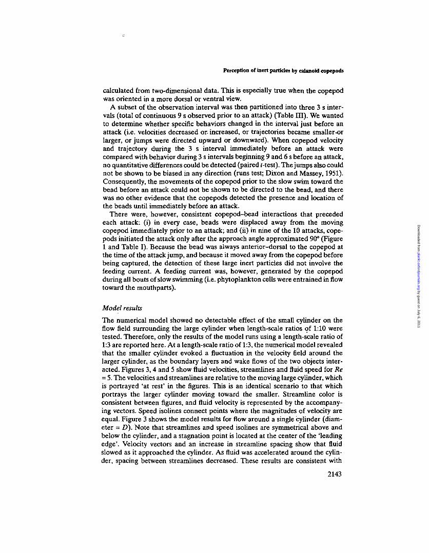
Model results
The numerical model showed no detectable effect of the small cylinder on theflow field surrounding the large cylinder when length-scale ratios of 1:10 weretested. Therefore, only the results of the model runs using a length-scale ratio of1:3 are reported here. At a length-scale ratio of 1:3, the numerical model revealedthat the smaller cylinder evoked a fluctuation in the velocity field around thelarger cylinder, as the boundary layers and wake flows of the two objects inter-acted. Figures 3,4 and 5 show fluid velocities, streamlines and fluid speed for Re= 5. The velocities and streamlines are relative to the moving large cylinder, whichis portrayed 'at rest' in the figures. This is an identical scenario to that whichportrays the larger cylinder moving toward the smaller. Streamline color isconsistent between figures, and fluid velocity is represented by the accompany-ing vectors. Speed isolines connect points where the magnitudes of velocity areequal. Figure 3 shows the model results for flow around a single cylinder (diam-eter = D). Note that streamlines and speed isolines are symmetrical above andbelow the cylinder, and a stagnation point is located at the center of the 'leadingedge'. Velocity vectors and an increase in streamline spacing show that fluidslowed as it approached the cylinder. As fluid was accelerated around the cylin-der, spacing between streamlines decreased. These results are consistent with
2143
by guest on July 6, 2011plankt.oxfordjournals.org
Dow
nloaded from

IVLH.Bundy et at.
predictions and empirical observations of flow geometry around circular cylin-ders at low Re (e.g. Batchelor, 1967; Van Dyke, 1981; Vogel, 1994).
Flow field geometry and fluid speeds were first compared between the singlelarge cylinder case and the case where the leading edge of the small cylinder(diameter = d) was located at x = -Ad, y = Id (i.e. x = -2 mm, y = 1 mm for a5 mm s-1 flow at Re = 5) (Figures 3 and 4). When the smaller cylinder was embed-ded in the flow, as the larger cylinder approached, the influence of the small cylin-der was manifested as a deformation of streamlines and an increase in fluidvelocity in the region between the cylinders, compared to the same region of theflow field when the small cylinder was absent (Figures 3a and 4a). Streamlineswere compressed and shifted vertically (i.e. in the direction of the small cylinder).When the velocities at 0.5 mm intervals directly in front of the cylinder arecompared between cases, e.g. at x = -0.5, y = 0.0 (Figures 3b and 4b), fluid speedincreased from 35% of mainstream velocity for the single-cylinder case to -45%of mainstream velocity when the small cylinder was present. Fluid speed at x =-1.0, y = 0.0 increased from 60% of mainstream velocity for the single large cylin-der case to 70% for the case when the small cylinder was present. The maximumspeed of the fluid moving around the large cylinder increased from a maximumof 125% for the single large cylinder case to 135% of mainstream velocity whenthe small cylinder was present.
When the separation distance between the cylinders was decreased and thesmall cylinder was moved to x = -2d, y = Id (e.g. x = -1 mm, v = 1 mm for a5 mm s"1 flow at Re = 5; Figure 5), flow field geometry and velocity distributionagain were altered. Streamline compression in the region between the two cylin-ders relaxed (Figure 5a), and the fluid speed in the region directly in front of thelarge cylinder (x = -0.5, y = 0.0) decreased from 45% for the larger separationdistance (Figure 4b) to -40% of mainstream velocity for the smaller separationdistance case (Figure 5b). Fluid speed at x = -1.0, y = 0.0 also decreased comparedto the larger separation distance case from 70% to 65% of mainstream velocity.The flow field above the large cylinder also changed as separation distancedecreased. The maximum velocity region decreased to 130% of mainstreamvelocity and extended farther from the cylinder than in the case of the large separ-ation distance (Figures 4b and 5b). Speed isolines also shifted toward theupstream direction.
Figure 6 shows the relative changes in velocity assuming a mainstream flow of5 mm s"1 at Re = 5 for the three cases. The baseline or reference velocity for thechanges is the fluid velocity at the corresponding location for the single-cylindercase (Figure 3b). The largest absolute changes in velocity, relative to the single-cylinder case, are seen within -0.5 mm of the surface of the large cylinder. Figure6 also shows the magnitude of speed fluctuations as the separation distancebetween the large and small cylinders decreases. The largest fluctuations (0.5 and0.75 mm s"1) are seen in a region above the smaller cylinder (at x = -0.5, y = 1.5)and in a region below and upstream from the large cylinder (between x = 0.0, y= -1.0 and x = 0.5, y = -1.0).
The small cylinder is given a prescribed speed in the model that matches thelocal streamline speed. Figures 4b and 5b show speed contours intercepting the
2144
by guest on July 6, 2011plankt.oxfordjournals.org
Dow
nloaded from

Perception of inert particles by calanoid eopepods
surface of the small cylinder, which indicates that the speed of the small cylinderbecame mismatched with the local streamline speed. This was due to the incom-pressible volume of the small cylinder crossing streamlines and distorting velocitycontours associated with the large cylinder. Although the individual contours forthe relative speed of the small cylinder are represented graphically as intercept-ing the cylinder, the average fluid speed surrounding the cylinder may not matchits actual speed. For example, in Figure 5b, the top of the small cylinder pusheswater a little faster than the bottom, and the 4.75 mm s"1 contour curves towardthe cylinder. In other words, the bottom of the small cylinder 'trips up' in thelower velocity fluid. In this case, the prescribed relative speed of the small cylin-der was 4.4873 mm s"1, but the numerically modeled average was 4.3 mm S"1,resulting in a mismatch of -0.2 mm s"1 or - 5 % of the local velocity. The net resultof this disturbance on the velocity field was a downstream perturbation near thelarge cylinder. This downstream perturbation would be seen as temporal changesin velocity as the small cylinder approached in the boundary layer flow.
In summary, we found that as separation distances between the two objectsdecreased, (i) velocity fluctuations arose in the region between and above the twoobjects, and (ii) the spatial distribution of the velocity field changed temporally.The numerical model examined the simple case of two cylinders with diameterratios of 3:1. The model results suggest that in the more complicated case of acomplex shape (the copepod and its first antennae) and a smaller sphere (thepolystyrene bead) functioning at similar Re, flow fields may also be distorted,although the magnitude of velocity fluctuations would probably be smaller, andthe geometry of the flow fields would most likely differ.
Discussion
Perception of distant objects by calanoid eopepods using mechanoreception hasbeen documented by the ability of freely swimming eopepods to detect andcapture moving prey located outside the influence of the feeding current (Gauld,1966; Williamson and Butler, 1986; Jonsson and Tiselius, 1990) and by tetheredeopepods capturing polystyrene beads in the feeding current (Vanderploeg et al,1990). It is not difficult to understand that a motile particle such as a nauplius ora ciliated larva creates a detectable hydrodynamic signal, because these livingparticles move and displace fluid (Yen and Fields, 1992;- Gallager, 1993).However, the sensory mechanisms that calanoid eopepods use to detect inertparticles (i.e. those that do not move independently and have no odor) are notwell understood, because the inert particles must first be displaced by an externalforce before a hydrodynamic disturbance is produced. Calanoids may detectstreamline deformations around large obstacles in their paths (Haury et al., 1980)and, in the present study, were seen actively to avoid the walls of the filmingvessel.
Because the attacked particles in this study are inert, the stimulus detected bythe copepod must be a hydrodynamic signal, rather than a chemical stimulus. Forforaging diaptomids, mechanoreception must therefore play an integral part in thedetection of large, prey-sized, non-moving particles. The concept that eopepods
2145
by guest on July 6, 2011plankt.oxfordjournals.org
Dow
nloaded from

M-H.Bundy et al.
have the ability to detect and capture non-motile particles outside the feedingcurrent is novel, and adds yet another dimension to the repertoire of feedingbehaviors that allow copepods to survive in a highly variable environment.
The biophysics of prey detection is complicated at the Re characteristic of thefluid environment where copepods feed and move about (Re 0.01-100 for a 1 mmcopepod). An acoustic pressure differential (far-field disturbance or sound wave)and fluid displacement (near-field disturbance) are both associated with movingparticles (Kalmijn, 1988). L£gier-Visser et al. (1986) suggested that copepodsdetect a non-acoustic pressure disturbance in the flow field of the feeding currentas particles are entrained toward the first antennae (Al). Although a pressuredifferential is associated with a change in streamline velocity, copepods do nothave the physiological ability to detect a pressure gradient.
The sensory organs used to detect pressure gradients require a 'pressure-to-motion converter' (e.g. a transducer such as the swim bladder of a fish) (vonFrisch, 1938). Crustaceans lack such a transducer and rely instead on the fluiddisplacement component of the signal as the mediator of responses to hydro-dynamic stimuli (Tautz, 1979). While a pressure disturbance accompanies thefluid deformation associated with an obstacle embedded in the flow, the signaldetected by the copepod in this case would be the change in velocity of the fluidaround its sensory structures.
Calanoid first antennae are morphologically and physiologically well suited asdetectors of velocity fluctuations. The Al are long cylindrical structures that havehair-like setae arranged in rows along their axes. The mechanoreceptive setae inthis array contain the terminal segments of mechanosensory neurons (Stricklerand Bal, 1973; Barrientos, 1980; Gresty et al., 1993; Weatherby et al, 1994). Amechanoreceptive seta is stimulated when the fluid velocity around the setachanges and the resultant shear displaces the hair, distorting the membranes ofmechanosensitive dendrites inserted into its base (French, 1988). Transduction ofthe stimulus occurs when the membrane of a sensory neuron is sufficientlydeformed to open ion channels and create a potential across the cell membrane(French, 1988; Corey and Garcia-Afioveros, 1996). These displacements do notneed to be large in magnitude: electrophysiological studies of antennal responsesto a high-frequency dipole source confirm that setae are sensitive to displace-ments as small as 10 nm (Lenz and Yen, 1993).
When a stationary particle has a density different from the surrounding fluid,its momentum will cause it to be accelerated and 'cross' streamlines associatedwith the wake and boundary layer of a larger approaching object, causing a largedisturbance. However, because algal cells and the inert polystyrene beads have adensity very close to water, their effect on the boundary layer should be moresubtle and should be due primarily to the solid body motion of the particle 'span-ning' streamlines. We hypothesize that the disturbance from the bead is detectedby the flexible mechanoreceptive setae on the Al as they are displaced by theflow shear created by the copepod's body wake flow and boundary layer, plus theadditional deformation when an obstacle is present. Simply put, the mechano-receptive setae on the Al are displaced as the velocity gradient around the Al isaltered by the obstacle.
2146
by guest on July 6, 2011plankt.oxfordjournals.org
Dow
nloaded from

Perception of inert particles by calanoid copepods
The extent and location of the fluid deformation predicted by the modeldepend on the orientation of the two objects to each other and their relativemotion and size. If the physical phenomena described in the model hold for swim-ming copepods foraging on large non-motile prey, then the orientation of sensorystructures and the swimming velocity of the copepod should be critical to thepredator's ability to detect its prey by this physical mechanism. The mechanismwould not be available to a 'hovering' copepod.
When we examine three-dimensional reconstruction of confocal images, wefind that the sensory hairs (setae) of Al of D.sicilis are oriented in ananterior-dorsal direction (Figure 7a and b). The setae were tentatively identifiedas mechanoreceptors because of their characteristic morphology and the pres-ence of an articulated socket (Figure 7a), although they may be dually functionaland have some chemoreceptive function as well (Tautz, 1979; Kurbjeweit andBuchholz, 1991; Weatherby et al., 1994). The shorter putative mechanoreceptors(-100 um in length) are located along the anterior-dorsal edge of the Al and thelonger putative mechanoreceptors (-300 um in length) are located on the anterioredge of the Al (Figure 7b). Video images show that the approach angle of thecopepod prior to an attack swings from -120 to 90° as the copepod approachesthe bead. Therefore, as D.sicilis moves toward a bead that is ultimately captured,the sensilla projecting ahead of the animal and lying parallel to and at an angleto its body axis transcribe an arc relative to the bead. This is because the copepodswims with its ventral side up (Figure 7b).
Model results showed that fluctuating flow field effects were pronounced in theregion between the two objects as separation distances decreased (Figure 6). Anorientation angle between 120 and 90° would place the putative mechanorecep-tors in this region of the boundary layer as the copepod neared the bead. Bound-ary layers around many freely swimming calanoids are most pronounced in thisanterior-dorsal region (i.e. the low-velocity region, compared to freestreamvelocity, is 'thicker' here) (Tiselius and Jonsson, 1990; Bundy and Paffenhofer,1996). The geometry of the boundary layer is dependent on swimming velocityand the orientation of the moving copepod (Bundy and Paffenhofer, 1996).
The behavioral advantage of detecting and capturing motile prey withoututilizing the feeding current may lie in the element of surprise. Prey react to vel-ocity gradients (shear and vorticity) in the copepod's feeding current(Williamson, 1987; Jonsson and Tiselius, 1990; Fields and Yen, 1997). Manynauplii and other microzooplankton do not move until disturbed (Williamson andVanderploeg, 1988; Paffenhofer et al., 1996), and some cladocerans use a 'deadman' strategy to avoid capture once they are attacked (Kerfoot et al., 1978).Tiselius and Jonsson (1990) noticed that copepods could approach closer to preythat were located in the anterior-dorsal region of the flow field where feedingcurrent disturbances were lower. Clearance rates of omnivorous calanoids onprey that tend to escape from feeding current flow fields should be enhanced bythis behavior.
Omnivorous copepods would also benefit from this feeding mode when theyare feeding on mixed populations of motile and non-motile prey. The generationof a feeding current while swimming slowly is an efficient tool for feeding on
2147
by guest on July 6, 2011plankt.oxfordjournals.org
Dow
nloaded from

M.H.Bundy et al.
(b) + Yr+Z
+X
Fig. 7. Diaptomus sicilis first antenna (Al). (a) Laser scanning confocal micrograph of the dorsal viewof the right Al of D.sicilis. The long thin setae are putative mechanoreceptive sensilla. Arrows indi-cate the articulated base of sensilla. A 50-um-diameter 'bead' is drawn in for scale, (b) Schematicdrawing showing copepod swimming trajectory and orientation of setae on Al. Lengths of Al andsetae are approximately proportional to copepod body length.
2148
by guest on July 6, 2011plankt.oxfordjournals.org
Dow
nloaded from

Perception of inert particles by calanoid copepods
non-motile prey or on prey with limited escape capabilities. However, motile preywith well-developed escape capabilities may be stimulated to escape by thefeeding current disturbance (Fields and Yen, 1997). An omnivore such as D.siciliscould utilize both types of prey by combining bouts of slow swimming with inter-mittent hops. This behavior was observed in the present study.
Large diatoms, such as Melosira italica, which dominate the water columnduring spring and the deep chlorophyll layer during summer (Fahnenstiel andScavia, 1987), are important components of the diet of D.sicilis (e.g. Vanderploeget al, 1988). It may be that the repeated hopping and turning behavior exhibitedby D.sicilis (Ramcharan and Sprules, 1991; present study) allows this copepodto utilize the foraging mechanism described here to capture large non-motilephytoplankton cells that may otherwise have remained undetected. Note thatthe clearance rates on beads reported here for D.sicilis fall within the range(25-42 ml day1 copepod"1) reported for this species feeding on large diatoms(Vanderploeg et al., 1988). Cyclopoid copepods, such as Oithona spinirostris, donot create feeding currents (Landry and Fagerness, 1988), yet graze efficientlyon large, non-motile cells. Cyclopoids move in hops and jumps, as well as in slowswimming mode (Shuvayev, 1978; Williamson, 1981; Uchima and Murano, 1988;Paffenhofer et al., 1996). The feeding mechanism described here may alsoexplain how cyclopoids are able to detect and capture large non-motile phyto-plankton.
Predator-prey interactions are constrained by behavior and by ambient fluidmotions. To understand fully the mechanisms governing prey detection andcapture, it is important that animals are observed as they move about freely. Wemust also have information on the sensitivity of the sensory structures thatreceive the signals from potential prey. Because the shapes of copepods and theirprey are complex, and because they move in non-linear and unpredictablepatterns, a numerical model cannot precisely replicate predator-prey interactionsin nature. However, the model developed in this study shows us that flow fielddistortion between two objects moving relative to each other can be quantifiedand predicted for a simple test case using length scales and a Reynolds numbersimilar to those in the visual observations. This investigation into the physics ofparticle detection in a quiescent fluid has enhanced our understanding of thephysical, behavioral and physiological processes that may control predator-preyinteractions of calanoid copepods. We see here that mechanoreception alone maybe used to detect remotely located non-motile prey.
Preliminary studies of freely swimming Diaptomus spp., conducted in thislaboratory, also showed that inert beads can be detected and captured in thefeeding current flow. Future studies will evaluate the relative importance ofcapturing particles within and outside the feeding current, and will investigate theinfluence of small-scale ambient water motion on behavior and on the ability ofcalanoids to capture inert particles. These variables must be considered togetherbefore we can better understand the role of feeding behavior in controllingcommunity structure in aquatic systems.
2149
by guest on July 6, 2011plankt.oxfordjournals.org
Dow
nloaded from

M.H.Bundy et at.
Acknowledgements
This study was supported by the Great Lakes Environmental Research Labora-tory (GLERL), and with funding from a postdoctoral award to M.H.B. from theNational Research Council Resident Research Associateship Program. We thankS.Ruberg for his help with the design and implementation of the camera position-ing software, and G.-A.Paffenhdfer for his help and advice on the set-up of thecamera positioning hardware. Two anonymous reviewers provided valuablesuggestions and comments that improved the manuscript. We also thank the staffand ship crew at GLERL's Lake Michigan Field Station for help with zooplank-ton collection. This paper is GLERL contribution no. 1063.
ReferencesAlcaraz,M., Paffenhofer,G.-A. and StricklerJ.R. (1980) Catching the algae: a first account of visual
observations on filter feeding calanoids. In Kerfoot.W.C. (ed.), The Evolution and Ecology ofZooplankton Communities. University Press of New England, Hanover, NH, pp. 241-248.
AndrewsJ.C. (1983) Deformation of the active space in the low Reynolds number feeding current ofcalanoid copepods. Can. J. Fish. Aquat Sci., 40,1293-1302.
Barrientos.Y.C. (1980) infrastructure of sensory units on the first antennae of calanoid copepods.MSc Thesis, University of Ottawa, Ottawa, Canada, pp. 1-81.
Batchelor,G.K. (1967) An Introduction to Fluid Dynamics. Cambridge University Press, London, 615 pp.Bundy,M.H. and Paffenhofer,G.-A. (1993) Innervation of copepod antennules investigated using
laser scanning confocal microscopy. Mar. EcoL Prog. Sen, 102,1-14.Bundy.M.H. and Paffenhofer,G.-A. (1996) Analysis of flow fields associated with freely swimming
calanoid copepods. Mar. EcoL Prog. Ser., 133, 99-113.BurnettJ3.S. (1987) Finite Element Analysis. Addison-Wesleyi Reading, MA, 844 pp.Butler.N.M., Suttle.C.A. and Neill.W.E. (1989) Discrimination by freshwater zooplankton between
single algal cells differing in nutritional status. Oecologia (Berlin), 78, 368-372.Cain,M.L. (1989) The analysis of angular data in ecological field studies. Ecology, 70,1540-1543.CoreyJJ.P. and Garcia-Anoveros,J. (1996) Mechanosensation and the DEG/ENaC ion channels.
Science, 273,323-324.Cowles.TJ., Olson,RJ. and Chisholm.S.W. (1988) Food selection by copepods: discrimination on the
basis of food quality. Mar. Biol., 100,41-49.DeMott.W.R. and Watson,M.D. (1991) Remote detection of algae by copepods: responses to algal
size, odors and motility. J. Plankton Res., 13,1203-1222.Dixon.WJ. and MasseyJU. (1951) Introduction to Statistical Analysis. McGraw-Hill, Inc., New York,
370 pp.Donaghay,P.L. and Small,I.F. (1979) Food selection capabilities of the estuarine copepod Acartia
clausii. Mar. Biol., 52,137-146.Fahnenstiel.G.L. and Scavia J) . (1987) Dynamics of Lake Michigan phytoplankton: recent changes in
surface and deep communities. Can. J. Fish. Aquat. Sci., 44,509-514.FieldsJD.M. and YenJ. (1997) Implications of the feeding current structure of Eucheata rimana, a
carnivorous pelagic copepod, on the spatial orientation of their prey. /. Plankton Res., 19,79-95.French,A.S. (1988) Transduction mechanisms of mechanosensilla. Annu. Rev. Entomoi, 33,39-58.Friedmao>lM. (1980) Comparative morphology and functional significance of copepod receptors
and oral structures. Am. Soc LimnoL Oceanogr. Spec. Symp., 3,185-197.Gallager.S.M. (1993) Hydrodynamic disturbances produced by small zooplankton: case study for the
veliger larva of a bivalve mollusc. /. Plankton Res., IS, 1277-1296.Gauld,D.T. (1966) Swimming and feeding of planktonic copepods. In BamesJH- (ed.). Some Contem-
porary Studies in Marine Science. George Allen and Unwin, London.Gresty,K.A., Boxshall.G.A. and NagasawaJC (1993) Antennulary sensors of the infective copepodid
larva of the salmon louse, Lepeophthierus salmonis (Copepoda: Caligidae). In Boxshall.G.A. andDefaye J) . (eds), Pathogens of Wild and Farmed Fish: Sea Lice. Ellis Horwood, Chichester, pp. 83-98.
Harvey,H.W. (1937) Note on selective feeding by Calanus. J. Mar. Biol Assoc UK, 22,97-100.HauryJL, Kenyon,D.E. and BrooksJ.R. (1980) Experimental evaluation of the avoidance reaction of
Calanus finmarchicus. J. Plankton Res., 2,187-202.
2150
by guest on July 6, 2011plankt.oxfordjournals.org
Dow
nloaded from

Perception of inert particles by calanoid copepods
JonssonJ'.R. and TiseliusJ?. (1990) Feeding behavior, prey detection and capture efficiency of thecopepod Acartia tonsa feeding on planktonic ciliates. Mar. EcoL Prog. Ser., 60,35-44.
Kalmijn,AdJ. (1988) Hydrodynamic and acoustic field detection. In AtemaJ., Fay,R.R., Popper A-N- andTavolga,W.N. (eds), Sensory Biology of Aquatic Organisms. Springer-Verlag, New York, pp. 83-130.
Kerfoot,W.C, Kellogg,D.L. and StricklerJ.R. (1978) Visual observations of live zooplankters:evasion, escape and chemical defenses. In Kerfoot.W.C. (ed.), The Evolution and Ecology ofZooplankton Communities. University Press of New England, Hanover, NH, pp. 10-27.
Koehl,M.A.R. and StricklerJ.R. (1981) Copepod feeding currents: Food capture at low Reynoldsnumber. LimnoL Oceanogr., 26,1062-1073.
KurbjeweitJ7. and Buchholz.C. (1991) Structures and suspected functions of antennular sensilla andpores of three Arctic copepods (Calanus glacialis, Metridia longa, Paraeucheata norvegica). Meeres-forsch., 33,168-182.
Landry,M.R. and Fagemess.V.L. (1988) Behavioral and morphological influences on predatory inter-actions among marine copepods. Bull. Mar. Sci., 43, 509-529.
Le'gier-Visser.M., MitchellJ.G., Okubo,A. and FuhrmanJ.A. (1986) Mechanoreception in calanoidcopepods: a mechanism for prey detection. Mar. Biol., 90, 529-536.
Lenz.P.H. and YenJ. (1993) Distal setal mechanoreceptors of the first antennae of marine copepods.BulL Mar. Sci., S3,170-179.
Paffenhofer,G.-A. and Lewis,K.D. (1990) Perceptive performance and feeding behavior of calanoidcopepods. / Plankton Res., 12,933-946.
Paffenhofer,G.-A., StricklerJ.R. and Alcaraz,M. (1982) Suspension-feeding by herbivorous calanoidcopepods: a cinematographic study. Mar. Biol., 67,193-199.
Paffenhofer,G.-A., StricklerJ.R., LewisJK.D. and Richman.S. (1996) Motion behavior of nauplii andearly copepodid stages of marine planktonic copepods. / . Plankton Res., 9,1699-1715.
Ramcharan.C.W. and Sprules.W.G. (1991) Predator-induced behavioral defense and its ecologicalconsequences for two calanoid copepods. Oecologia (Berlin), 86,276-286.
Roache,PJ. (1972) Computational Fluid Dynamics. Hermosa, Albuquerque, NM, 446 pp.Shuvayev.D. (1978) Movement of some planktonic copepods. Hydrobiol. J. (USSR), 14, 32-36.StricklerJ.R. (1985) Feeding currents in calanoid copepods: Two new hypotheses. In Laverack,M.S.
(ed.), Physiological Adaptations of Marine Animals. Society for Experimental Biology, UK, pp.459-485.
StricklerJ.R. and BaI,A.K. (1973) Setae of the first antennae of the copepod Cyclops scutifer (Sars):their structure and importance. Proc. Natl Acad. Sci. USA, 70,2656-2659.
TautzJ. (1979) Reception of particle oscillation in a medium—an unorthodox sensory capacity. Natur-wissenschaften, 65, 452-461.
Tiselius,P. and JonssonJP. (1990) Foraging behavior of six calanoid copepods: observation and hydro-dynamic analysis. Mar. EcoL Prog. Ser., 66,23-33.
Uchima.A-M. and Murano.M. (1988) Swimming behavior of the marine copepod Oithona davisae:internal control and search for environment. Mar. Biol., 99,47-56.
Van Dykejvl (1981) An Album of Fluid Motion. The Parabolic Press, Stanford, CA, 176 pp.Vanderploeg,H.A. and Paffenh6fer,G.-A. (1985) Modes of algal capture by the freshwater copepod
Diaptomus sicilis and their relation to food-size selection. LimnoL Oceanogr., 30,871-885.VanderpIoeg,H.A., Paffenh6fer,G.-A. and LiebigJ.R. (1988) Diaptomus vs. net phytoplankton:
Effects of algal size and morphology on selectivity of a behaviorally flexible, omnivorous copepod.Bull. Mar. Sci., 43, 561-572.
Vanderploeg,H.A., Paffenh6fer,G.-A. and LiebigJ.R. (1990) Concentration-variable interactionsbetween calanoid copepods and particles of different food quality: Observations and hypotheses.In Hughes J*.N. (ed.), Behavioral Mechanisms of Food Selection. NATO ASI Series G20. Springer-Verlag, Berlin, Heidelberg.
Vogel.S. (1994) Life in Moving Fluids. Princeton University Press, Princeton, NJ.von Frisch,K. (1938) The'sense of hearing in fish. Nature, 141, 8-11.Weatherby,T.M., WongJCK. and LenzJAH. (1994) Fme structure of the distal sensory setae on the
first antennae of Pteuromamma xiphias Giesbrecht (Copepoda). / Crust. Biol., 14,670-685.WeissJD.G., Keller J., GuldenJ. and Maile.W. (1986) Towards a new classification of intracellular
particle movements based on quantitative analyses. Cell MotiL Cytoskel, 6,128-135.Williamson.C.E. (1981) Foraging and looping behavior of a freshwater copepod: Frequency changes
in looping behavior at high and low prey densities. Oecologia (Berlin), SO, 332-336.Williamson.C.E. (1987) Predator-prey interactions between omnivorous diaptomid copepods and
rotifers: The role of prey morphology and behavior. LimnoL Oceanogr., 32,167-177.Williamson.C.E. and Butler,N.M. (1986) Predation on rotifers by the suspension-feeding calanoid
copepod Diaptomus pallidus. LimnoL Oceanogr., 31, 393-402.
2151
by guest on July 6, 2011plankt.oxfordjournals.org
Dow
nloaded from

M.H.Bundy el al.
Williamson.C.E. and Vanderploeg,H. (1988) Predatory suspension-feeding in Diaptomus: Preydefenses and the avoidance of cannibalism. Bull Mar. ScL, 43,561-572.
YenJ. and FieldsJD.M. (1992) Escape responses of Acartia hudsonica (Copepoda) nauplii from theflow field of Temora longicornis (Copepoda). Arch. HydrobioL Beik Ergebn. LimrtoL, 36,123-134.
Received on November 24, 1997; accepted on June 23,1998
by guest on July 6, 2011plankt.oxfordjournals.org
Dow
nloaded from