
In situ XAS with high-energy resolution: The changing structure of platinum during the oxidation of...
7
In situ XAS with high-energy resolution: The changing structure of platinum during the oxidation of carbon monoxide Jagdeep Singh a , Moniek Tromp b , Olga V. Safonova c , Pieter Glatzel d , Jeroen A. van Bokhoven a, * a Institute for Chemical and Bioengineering, ETH Zurich, 8093 Zurich, Switzerland b School of Chemistry, University of Southampton Highfield, Southampton SO17 1BJ, United Kingdom c Swiss Norwegian Beamlines (SNBL), European Synchrotron Radiation Facility (ESRF), BP 220 6 rue Jules Horowitz, 38043 Grenoble, France d European Synchrotron Radiation Facility (ESRF), BP 220 6 rue Jules Horowitz, 38043 Grenoble, France 1. Introduction Knowledge of the structure of the catalytically active sites in solid catalysts is essential to understand the functioning of solid catalysts and chemical processes. It enables the design of new and better catalysts. Despite decades of research, the structure of the adsorption sites of hydrocarbon molecules more complex than ethane, remain essentially unknown [1]. The structure of adsorbates and intermediates are generally determined using vibrational spectroscopy, but this provides only indirect informa- tion about the structure of the catalyst. Adsorption of reactants and intermediates on the surface of transition metals involves the interaction of the atomic or molecular orbitals of the adsorbate with the bands on the metal. The interaction with the metal sp-band causes broadening of the orbital level, while that with the metal d band forms bonding and anti-bonding states. If the anti-bonding state is pushed completely, or partially above the Fermi-level, there is a driving force for the adsorbate to bond to the metal surface. An example is the bonding of hydrogen to platinum surfaces, which creates empty anti- bonding states; in contrast, the anti-bonding state between hydrogen and the (1 1 1) surface of gold is below the Fermi-level, so hydrogen does not bond to bulk gold [2]. The energy of the d band is the essential parameter that determines the bonding: the smaller is the gap between the d band and the Fermi-level, the stronger the metal–adsorbate bonds [3]. X-ray absorption spectroscopy provides the local structure by an EXAFS analysis and density of states by analysis of the near-edge structure. The K edge probes the transition of a 1s electron to np and the L 2,3 edges, those of a 2p to (n 1)d. As a result, the L 3 edge is sensitive to the d density of states and probes the oxidation state of transition metals. The first intense feature, also called the whiteline, in L 3 edge spectra is sensitive to the anti-bonding state after adsorption of reactants or intermediates. The shape and intensity depends on the structure of the adsorption site and adsorption sites of hydrogen atoms [4,5], carbon monoxide [4,6], and ethylene [7] have been observed. Because all atoms contribute to the XAS signal, an averaged signal is determined, which hampers a quantitative evaluation of all types of atoms. Moreover, the excitation of a core electron in an XAS experiment and its finite lifetime causes lifetime broadening, which results in a broad spectra, which further hampers evaluation and interpretation of the spectra. Using methods that subtract known contributions to the spectra aids in solving the first point. High-energy resolution fluorescence detected X-ray absorp- tion spectroscopy (HERFD XAS) is a technique where XAS spectra are recorded with an instrumental broadening that is below the core Catalysis Today 145 (2009) 300–306 ARTICLE INFO Article history: Available online 12 February 2009 Keywords: Platinum HERFD XAS Carbon monoxide oxidation In situ ABSTRACT The catalytically active species during the oxidation of carbon monoxide over a real alumina-supported platinum catalyst under atmospheric pressure are determined by combining in situ high-energy resolution fluorescence detection X-ray absorption spectroscopy (HERFD XAS) at the Pt L 3 edge and kinetic measurements. Catalysts were prepared by incipient wetness impregnation. The oxidation of carbon monoxide occurred in two distinctive regimes, a high-activity regime and a low-activity regime, which have high and low rates of reaction respectively. In the low-activity region, the catalyst is poisoned by carbon monoxide, limiting the dissociative adsorption of oxygen. In the high-activity regime, all CO is desorbed from the surface, increasing the rate of reaction. The low carbon monoxide concentration enables oxygen to react to the surface in this reaction regime generating a more reactive surface. HERFD showed that different phases are active depending on the reaction conditions in nano- sized catalyst particles. ß 2008 Elsevier B.V. All rights reserved. * Corresponding author at: Institute for Chemical and Bioengineering, HCI E 127, Wolfgang Paulistrasse 10, 8093, Zurich, Switzerland. Tel.: +41 446 325542; fax: +41 446 321162. E-mail address: [email protected] (J.A. van Bokhoven). Contents lists available at ScienceDirect Catalysis Today journal homepage: www.elsevier.com/locate/cattod 0920-5861/$ – see front matter ß 2008 Elsevier B.V. All rights reserved. doi:10.1016/j.cattod.2008.11.019
-
Upload
independent -
Category
Documents
-
view
0 -
download
0
Transcript of In situ XAS with high-energy resolution: The changing structure of platinum during the oxidation of...

Catalysis Today 145 (2009) 300–306
In situ XAS with high-energy resolution: The changing structure of platinumduring the oxidation of carbon monoxide
Jagdeep Singh a, Moniek Tromp b, Olga V. Safonova c, Pieter Glatzel d, Jeroen A. van Bokhoven a,*a Institute for Chemical and Bioengineering, ETH Zurich, 8093 Zurich, Switzerlandb School of Chemistry, University of Southampton Highfield, Southampton SO17 1BJ, United Kingdomc Swiss Norwegian Beamlines (SNBL), European Synchrotron Radiation Facility (ESRF), BP 220 6 rue Jules Horowitz, 38043 Grenoble, Franced European Synchrotron Radiation Facility (ESRF), BP 220 6 rue Jules Horowitz, 38043 Grenoble, France
A R T I C L E I N F O
Article history:
Available online 12 February 2009
Keywords:
Platinum
HERFD XAS
Carbon monoxide oxidation
In situ
A B S T R A C T
The catalytically active species during the oxidation of carbon monoxide over a real alumina-supported
platinum catalyst under atmospheric pressure are determined by combining in situ high-energy
resolution fluorescence detection X-ray absorption spectroscopy (HERFD XAS) at the Pt L3 edge and
kinetic measurements. Catalysts were prepared by incipient wetness impregnation. The oxidation of
carbon monoxide occurred in two distinctive regimes, a high-activity regime and a low-activity regime,
which have high and low rates of reaction respectively. In the low-activity region, the catalyst is
poisoned by carbon monoxide, limiting the dissociative adsorption of oxygen. In the high-activity
regime, all CO is desorbed from the surface, increasing the rate of reaction. The low carbon monoxide
concentration enables oxygen to react to the surface in this reaction regime generating a more reactive
surface. HERFD showed that different phases are active depending on the reaction conditions in nano-
sized catalyst particles.
� 2008 Elsevier B.V. All rights reserved.
Contents lists available at ScienceDirect
Catalysis Today
journa l homepage: www.e lsev ier .com/ locate /cat tod
1. Introduction
Knowledge of the structure of the catalytically active sites insolid catalysts is essential to understand the functioning of solidcatalysts and chemical processes. It enables the design of new andbetter catalysts. Despite decades of research, the structure of theadsorption sites of hydrocarbon molecules more complex thanethane, remain essentially unknown [1]. The structure ofadsorbates and intermediates are generally determined usingvibrational spectroscopy, but this provides only indirect informa-tion about the structure of the catalyst.
Adsorption of reactants and intermediates on the surface oftransition metals involves the interaction of the atomic ormolecular orbitals of the adsorbate with the bands on the metal.The interaction with the metal sp-band causes broadening of theorbital level, while that with the metal d band forms bonding andanti-bonding states. If the anti-bonding state is pushed completely,or partially above the Fermi-level, there is a driving force for theadsorbate to bond to the metal surface. An example is the bondingof hydrogen to platinum surfaces, which creates empty anti-
* Corresponding author at: Institute for Chemical and Bioengineering, HCI E 127,
Wolfgang Paulistrasse 10, 8093, Zurich, Switzerland. Tel.: +41 446 325542;
fax: +41 446 321162.
E-mail address: [email protected] (J.A. van Bokhoven).
0920-5861/$ – see front matter � 2008 Elsevier B.V. All rights reserved.
doi:10.1016/j.cattod.2008.11.019
bonding states; in contrast, the anti-bonding state betweenhydrogen and the (1 1 1) surface of gold is below the Fermi-level,so hydrogen does not bond to bulk gold [2]. The energy of the dband is the essential parameter that determines the bonding: thesmaller is the gap between the d band and the Fermi-level, thestronger the metal–adsorbate bonds [3].
X-ray absorption spectroscopy provides the local structure by anEXAFS analysis and density of states by analysis of the near-edgestructure. The K edge probes the transition of a 1s electron to np andthe L2,3 edges, those of a 2p to (n � 1)d. As a result, the L3 edge issensitive to the d density of states and probes the oxidation state oftransition metals. The first intense feature, also called the whiteline,in L3 edge spectra is sensitive to the anti-bonding state afteradsorption of reactants or intermediates. The shape and intensitydepends on the structure of the adsorption site and adsorption sitesof hydrogen atoms [4,5], carbon monoxide [4,6], and ethylene [7]have been observed. Because all atoms contribute to the XAS signal,an averaged signal is determined, which hampers a quantitativeevaluation of all types of atoms. Moreover, the excitation of a coreelectron in an XAS experiment and its finite lifetime causes lifetimebroadening, which results in a broad spectra, which further hampersevaluation and interpretation of the spectra. Using methods thatsubtract known contributions to the spectra aids in solving the firstpoint. High-energy resolution fluorescence detected X-ray absorp-tion spectroscopy (HERFD XAS) is a technique where XAS spectra arerecorded with an instrumental broadening that is below the core
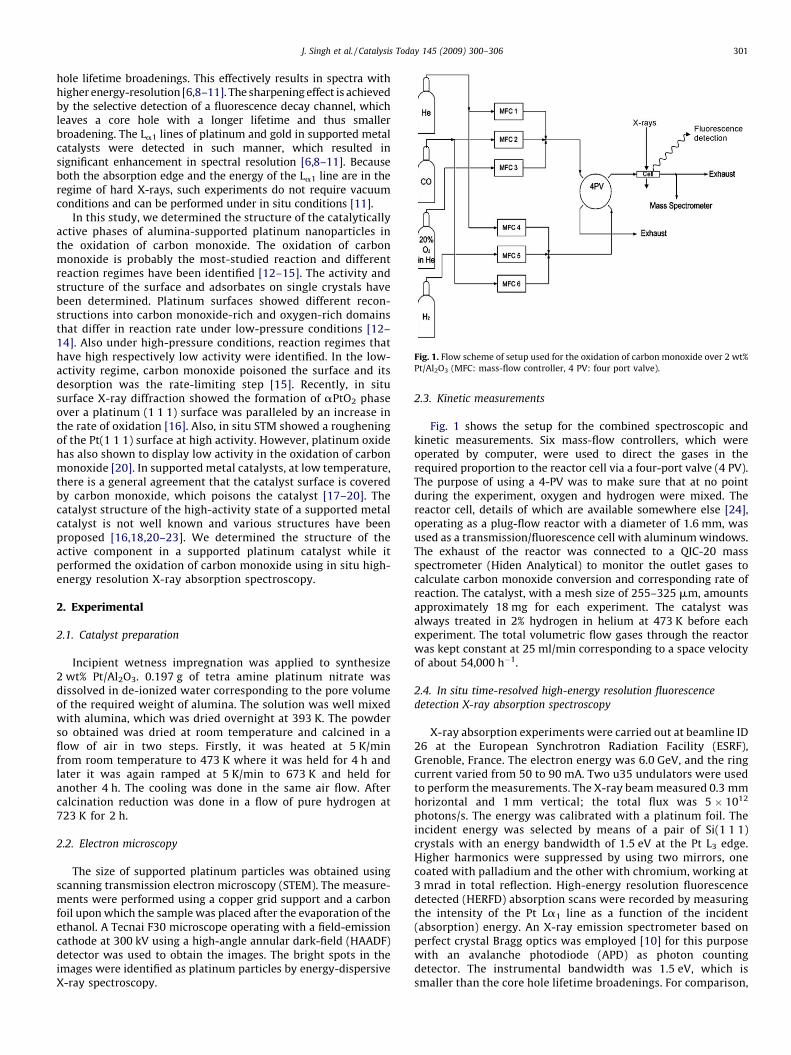
Fig. 1. Flow scheme of setup used for the oxidation of carbon monoxide over 2 wt%
Pt/Al2O3 (MFC: mass-flow controller, 4 PV: four port valve).
J. Singh et al. / Catalysis Today 145 (2009) 300–306 301
hole lifetime broadenings. This effectively results in spectra withhigher energy-resolution [6,8–11]. The sharpening effect is achievedby the selective detection of a fluorescence decay channel, whichleaves a core hole with a longer lifetime and thus smallerbroadening. The La1 lines of platinum and gold in supported metalcatalysts were detected in such manner, which resulted insignificant enhancement in spectral resolution [6,8–11]. Becauseboth the absorption edge and the energy of the La1 line are in theregime of hard X-rays, such experiments do not require vacuumconditions and can be performed under in situ conditions [11].
In this study, we determined the structure of the catalyticallyactive phases of alumina-supported platinum nanoparticles inthe oxidation of carbon monoxide. The oxidation of carbonmonoxide is probably the most-studied reaction and differentreaction regimes have been identified [12–15]. The activity andstructure of the surface and adsorbates on single crystals havebeen determined. Platinum surfaces showed different recon-structions into carbon monoxide-rich and oxygen-rich domainsthat differ in reaction rate under low-pressure conditions [12–14]. Also under high-pressure conditions, reaction regimes thathave high respectively low activity were identified. In the low-activity regime, carbon monoxide poisoned the surface and itsdesorption was the rate-limiting step [15]. Recently, in situsurface X-ray diffraction showed the formation of aPtO2 phaseover a platinum (1 1 1) surface was paralleled by an increase inthe rate of oxidation [16]. Also, in situ STM showed a rougheningof the Pt(1 1 1) surface at high activity. However, platinum oxidehas also shown to display low activity in the oxidation of carbonmonoxide [20]. In supported metal catalysts, at low temperature,there is a general agreement that the catalyst surface is coveredby carbon monoxide, which poisons the catalyst [17–20]. Thecatalyst structure of the high-activity state of a supported metalcatalyst is not well known and various structures have beenproposed [16,18,20–23]. We determined the structure of theactive component in a supported platinum catalyst while itperformed the oxidation of carbon monoxide using in situ high-energy resolution X-ray absorption spectroscopy.
2. Experimental
2.1. Catalyst preparation
Incipient wetness impregnation was applied to synthesize2 wt% Pt/Al2O3. 0.197 g of tetra amine platinum nitrate wasdissolved in de-ionized water corresponding to the pore volumeof the required weight of alumina. The solution was well mixedwith alumina, which was dried overnight at 393 K. The powderso obtained was dried at room temperature and calcined in aflow of air in two steps. Firstly, it was heated at 5 K/minfrom room temperature to 473 K where it was held for 4 h andlater it was again ramped at 5 K/min to 673 K and held foranother 4 h. The cooling was done in the same air flow. Aftercalcination reduction was done in a flow of pure hydrogen at723 K for 2 h.
2.2. Electron microscopy
The size of supported platinum particles was obtained usingscanning transmission electron microscopy (STEM). The measure-ments were performed using a copper grid support and a carbonfoil upon which the sample was placed after the evaporation of theethanol. A Tecnai F30 microscope operating with a field-emissioncathode at 300 kV using a high-angle annular dark-field (HAADF)detector was used to obtain the images. The bright spots in theimages were identified as platinum particles by energy-dispersiveX-ray spectroscopy.
2.3. Kinetic measurements
Fig. 1 shows the setup for the combined spectroscopic andkinetic measurements. Six mass-flow controllers, which wereoperated by computer, were used to direct the gases in therequired proportion to the reactor cell via a four-port valve (4 PV).The purpose of using a 4-PV was to make sure that at no pointduring the experiment, oxygen and hydrogen were mixed. Thereactor cell, details of which are available somewhere else [24],operating as a plug-flow reactor with a diameter of 1.6 mm, wasused as a transmission/fluorescence cell with aluminum windows.The exhaust of the reactor was connected to a QIC-20 massspectrometer (Hiden Analytical) to monitor the outlet gases tocalculate carbon monoxide conversion and corresponding rate ofreaction. The catalyst, with a mesh size of 255–325 mm, amountsapproximately 18 mg for each experiment. The catalyst wasalways treated in 2% hydrogen in helium at 473 K before eachexperiment. The total volumetric flow gases through the reactorwas kept constant at 25 ml/min corresponding to a space velocityof about 54,000 h�1.
2.4. In situ time-resolved high-energy resolution fluorescence
detection X-ray absorption spectroscopy
X-ray absorption experiments were carried out at beamline ID26 at the European Synchrotron Radiation Facility (ESRF),Grenoble, France. The electron energy was 6.0 GeV, and the ringcurrent varied from 50 to 90 mA. Two u35 undulators were usedto perform the measurements. The X-ray beam measured 0.3 mmhorizontal and 1 mm vertical; the total flux was 5 � 1012
photons/s. The energy was calibrated with a platinum foil. Theincident energy was selected by means of a pair of Si(1 1 1)crystals with an energy bandwidth of 1.5 eV at the Pt L3 edge.Higher harmonics were suppressed by using two mirrors, onecoated with palladium and the other with chromium, working at3 mrad in total reflection. High-energy resolution fluorescencedetected (HERFD) absorption scans were recorded by measuringthe intensity of the Pt La1 line as a function of the incident(absorption) energy. An X-ray emission spectrometer based onperfect crystal Bragg optics was employed [10] for this purposewith an avalanche photodiode (APD) as photon countingdetector. The instrumental bandwidth was 1.5 eV, which issmaller than the core hole lifetime broadenings. For comparison,

Fig. 2. Total energy scheme (a) of the electronic transitions (b).
Fig. 3. (a) STEM micrograph of 2 wt% Pt/Al2O3 prepared by incipient wetness
impregnation and (b) particle size distribution of platinum particles.
J. Singh et al. / Catalysis Today 145 (2009) 300–306302
a solid-state detector such as germanium would have an energybandwidth of �400 eV at this energy. The line-sharpening effectarises from the fact that the lifetime broadening in the final stateis smaller than in the intermediate states of the absorptionspectrum (Fig. 2). The lifetime broadening of L3 and M5 levelstabulated in FEFF8.2 code is equal to 5.2 and 2.4 eV respectively.According to the equation given in Ref. [25], the broadening ofHERFD spectra will be equal to 2.2 eV. The HERFD broadening(Lorentzian) is convoluted with the experimental broadening(Gaussian) and the width of platinum whiteline in the HERFDexperiment is calculated to 2.9 eV, which is similar to the valueobserved in the experiment. A Canberra silicon photodiode wasmounted to measure the total fluorescence simultaneously withthe HERFD XAS. The catalyst was heated at 5 K/min from roomtemperature to the temperature of maximum conversion, andcooled back to room temperature, with simultaneous collectionof spectra with a time resolution of 1 min.
3. Results and discussion
The oxidation of carbon monoxide on supported platinumcatalysts proceeds in two regimes, a high-activity and a low-activity one. In this work, the oxidation of carbon monoxide over a2-wt% Pt/Al2O3 catalyst was carried out for oxygen to carbonmonoxide ration of one, two, and five. Fig. 3a shows a STEMmicrograph of the catalyst. The platinum nanoparticles were
identified as bright tiny spots and well dispersed on aluminasupport. Fig. 3b shows the particle size distribution showing amean particle size of about 1 nm. Table 1 summarizes the kineticresults of the oxidation of carbon monoxide in various ratios. Thedata were reproduced from the previous work [31]. At all ratios,the conversion of carbon monoxide increased exponentially withtemperature until at a specific temperature, a large jump in theconversion occurred. We call this jump ‘‘ignition’’ of the reaction.At this point, the rate of reaction increased significantlycompared to the rate at temperature less than the ignitiontemperature. Afterwards, the rate became constant with increasein temperature signifying the complete conversion of carbonmonoxide. This phenomenon of two different reaction regimeshas been observed on single crystals and supported platinumcatalysts [14,15,19,20,26–30] but so far has not been unambigu-ously correlated with changes in the platinum catalyst. Most ofthe data on catalyst structure were derived from vibrationalspectroscopy, which provide information about the adsorbingreactant molecule. The adsorbate is probed and only indirectinformation about the catalyst is obtained. The HERFD XAStechnique, as applied in our experiment, provides complemen-tary information to that obtained from the vibrational techni-ques; i.e. gives the geometric and electronic structure of thecatalytic metal site during reaction. It is also complementary to

Table 1Results of the kinetic measurements for the oxidation of carbon monoxide over a 2 wt% Pt/Al2O3 catalyst.
O2/CO ratio Below ignition Temperature of ignition (K)
Rate of reaction (cm3/(min gcat)) Temperature (K)
1 0.0056 428 472
2 0.0120 428 445
5 0.0195 428 433
Fig. 4. Pt L3 edge XANES of 5 wt% Pt/Al2O3 after reduction in different atmospheres:
helium (black), helium (oxygen) (red), and 1% carbon monoxide in helium (blue)
measured (a) using traditional fluorescence detection and (b) using HERFD XAS
spectroscopy. Reprinted with the permission from J. Phys. Chem. B. (Letter) 110
(2006) 16162. Copyright (2006) American Chemical Society. (For interpretation of
the references to colour in this figure legend, the reader is referred to the web
version of the article.)
J. Singh et al. / Catalysis Today 145 (2009) 300–306 303
surface X-ray diffraction experiments that prove the surfacestructure of single crystals [16].
As the concentration of oxygen increased in the reactantgases, with increasing oxygen to carbon monoxide ratio, thetemperature of ignition decreased (Table 1). Also at anyparticular temperature, the rate of reaction was higher for thehigher oxygen to carbon monoxide ratios. During cooling, asimilar phenomenon occurred. Upon cooling, the conversion ofcarbon monoxide decreased strongly in a very narrow tem-perature interval. We call this extinction of the reaction. Furtherdecreasing the temperature showed a normal exponentialdecrease in conversion. The temperature of extinction, similarto the ignition temperature, was lower under higher oxygenconcentration.
Fig. 4 reproduces the conventional L3 edge XAS and HERFD XASof a supported platinum catalyst after treatment in threeatmospheres: helium, 1% carbon monoxide in helium, and amixture of oxygen and helium [6]. The shape and intensity of thewhiteline varied in each atmosphere, and small shifts in the edgeposition were observed. The changes in the spectra showed muchbetter resolution in the HERFD XAS data. The intensity of thewhiteline in the spectrum of already reduced catalyst underhelium was lower than that of the catalyst under oxygen,reflecting the oxidation of platinum. This increase is visible bothin the conventionally detected fluorescence and in the spectrumof HERFD spectroscopy. In the case of the catalyst with adsorbedcarbon monoxide, the HERFD spectrum revealed a double feature,which was broader on the high-energy side of the whitelinecompared to that in bare platinum. This doublet has been assignedto atop adsorption of carbon monoxide [6]. The spectral featurewas attributed to the anti-bonding state above the Fermi-levelthat forms when the platinum d orbitals and the 2p* orbitals ofcarbon and oxygen overlap. Although the conventionally mea-sured XAS spectra show spectral edges, the high-resolution dataare much more sensitive in detecting any spectral changes. Weused the spectra in Fig. 4 as reference spectra when comparing thechanges in the spectra of the platinum catalysts that weremeasured during the oxidation of carbon monoxide under variousconditions.
Fig. 5a shows the Pt L3 edge HERFD XAS spectra of Pt/Al2O3
below and above the ignition temperature in an atmosphere ofoxygen and carbon monoxide in ratios of one, two, and five. Thesespectra were recorded while heating the sample. The spectrumbelow ignition showed the doublet characteristic of adsorbedcarbon monoxide. Its intensity was low compared to the spectrathat were measured at lower temperature (spectra not shown)[31]. In the low-activity regime, oxygen has a positive effect on thereaction rate and the reaction has a positive order in oxygen[15,32,33]. Adsorbed carbon monoxide on reduced platinum wasobserved (Fig. 5), in general agreement with infra-red data onsupported metal catalysts and single crystals [15,17–20,27,28].This adsorbed carbon monoxide prevents the catalyst surface fromoxidizing even in an overdose of oxygen at sufficiently lowtemperature. Before oxygen can be activated to react to carbonmonoxide, a carbon monoxide molecule has to desorb. The amountof carbon monoxide adsorbed on the metal surface decreases with
temperature; at a certain ignition temperature, the conversion ofcarbon monoxide and the rate of the reaction increases suddenlyand significantly. At a temperature above the ignition temperature,the spectra in all the three ratios showed a strong increase in theintensity of the whiteline while the edge shifted slightly to lowerenergy. These changes are characteristic of oxidized platinum [6].The sudden increase in the whiteline intensity was in accordancewith the increased rate of reaction at this point. This high activity isfound only at elevated temperature [18]. We observed that the

Fig. 5. Pt L3 edge HERFD XANES of 2 wt% Pt/Al2O3 during the oxidation of carbon monoxide (a) above (red) and below (blue) ignition during heating, and (b) above (blue) and
below (red) extinction during cooling for ratios of oxygen to carbon monoxide of (i) one, (ii) two, and (iii) five. (For interpretation of the references to colour in this figure
legend, the reader is referred to the web version of the article.)
J. Singh et al. / Catalysis Today 145 (2009) 300–306304
structure of the supported platinum particles is very different inthis regime. A large fraction of the platinum is oxidized. Fig. 5bshows the Pt L3 edge HERFD XAS spectra of Pt/Al2O3 above andbelow the extinction temperature in atmospheres of oxygen andcarbon monoxide in ratios of one, two, and five measured whilecooling. Below the extinction temperature, the whiteline inten-sity had dropped and the doublet feature appeared in the spectra.This doublet feature got more intense with further cooling down(spectra not shown). This sudden decrease in the intensity of thewhiteline and the re-emergence of the doublet corresponding toadsorbed carbon monoxide was observed in all the three cases.The re-emergence of the doublet feature corresponded to 453,445, and 433 K for oxygen to carbon monoxide ratios of one, two,and five respectively and was accompanied by the extinction ofthe reaction (Table 1). Based on our in situ HERFD XAS data, theoccurrence of sudden changes in the reaction rate can be assignedto two different platinum phases in the two regimes. HERFD XASshows that at low activity (below ignition temperature), platinumis covered with carbon monoxide, which inhibits the reactionfrom proceeding at higher rates and, thus, poisons the surface[15]. Therefore, in this region, the rate-limiting step is desorptionof carbon monoxide, which is necessary for the activation ofoxygen [17,19,21,33]. At high activity, above ignition, theplatinum species are partially oxidized (Fig. 5). The rate ofoxidation increased and the reaction proceeds via a different rate-limiting step. As the surface is no longer poisoned by carbonmonoxide, a higher rate is obtained. Because of the lowconcentration of carbon monoxide, oxygen can react to thesurface, forming the platinum oxide phase. Although PtO2 hasbeen shown to be less active [20], partially oxidized platinum
catalyst showed high activity. This correlates to the high activityobserved for partially oxidized ruthenium surfaces [34]. Fig. 6compares the Pt L3 edge HERFD XAS spectra of Pt/Al2O3 in 5%oxygen in helium oxygen and in an atmosphere of oxygen andcarbon monoxide at a ratio of one, two, and five at a temperatureabove the ignition temperature, thus at high rate of reaction. Incase of oxygen to carbon monoxide ratio of two, the spectrumabove the ignition temperature is compared with the spectrumrecorded under 5% oxygen in helium at 458 K. For the other tworatios, the spectra were compared to spectra recorded at 398 Kunder 5% oxygen in helium. In all cases, spectra above ignition andunder 5% oxygen in helium showed almost the same intensity ofthe whiteline, indicating a high degree of oxidation of theplatinum catalyst above ignition of the reaction. This suggests thatthe extent of oxidation is similar under both sets of conditions.Oxidized nano-sized platinum particles have been shown to bemore reactive than reduced platinum with adsorbed carbonmonoxide on its surface [26]. Our data suggests that the amount ofoxide that is formed above ignition is similar to that under pureoxygen. The concentration of carbon monoxide at the surface isvery low, causing the extent of oxidation to be very similar. Thediffusion of carbon monoxide to the surface may become ratelimiting over the highly active surface of the catalyst. Theseplatinum oxide species are formed at temperatures above theignition temperatures of 472, 445, and 433 K, at oxygen to carbonmonoxide ratios of one, two, and five respectively. These oxidicspecies form only when the oxygen concentration is considerablyhigher than that of carbon monoxide and at temperatures atwhich carbon monoxide has partially desorbed from the surfaceand its concentration decreased because of conversion.

Fig. 6. Pt L3 edge HERFD XANES of 2 wt% Pt/Al2O3 (a) in 5% oxygen in helium at 398 K (red), and during the oxidation of carbon monoxide for a ratio of oxygen to carbon
monoxide of one at 491 K (black), (b) in 5% oxygen in helium at 458 K (red), and during the oxidation of carbon monoxide for a ratio of oxygen to carbon monoxide of two at
472 K (black), and (c) in 5% oxygen in helium at 398 K (red), and during the oxidation of carbon monoxide for a ratio of oxygen to carbon monoxide of five at 454 K (black). (For
interpretation of the references to colour in this figure legend, the reader is referred to the web version of the article.)
J. Singh et al. / Catalysis Today 145 (2009) 300–306 305
4. Conclusion
Platinum nanoparticles, supported on alumina and prepared byincipient wetness impregnation have different activities in theoxidation of carbon monoxide in different gas atmospheres. Thecatalyst shows different structure in a low- and high-activityregime: adsorbed carbon monoxide on platinum in the low-activity regime, that poisons the surface and oxidic platinum in thehigh-activity regime as observed with in situ HERFD XAS. The ratein the low-activity regime is determined by the desorption ofcarbon monoxide from the surface and subsequent dissociativeadsorption of oxygen. In the high-activity regime, the catalyst is nolonger poisoned by carbon monoxide, because of its lowconcentration at the catalyst surface. As a result the catalyst ispartially oxidized. This surface shows a high rate of reaction. Hightemperature and a high oxygen concentration benefits theformation of the more active catalyst. High resolution XAS enablesto determine the structure of supported nanoparticles of loadingsas low as 2 wt%. Because only hard X-rays are involved in thisexperiment, it can be performed under in situ conditions providingstructural information while the catalyst is under real workingconditions.
Acknowledgements
This work was supported by EPSRC (EP/E060404/1) (M. Tromp)and the Swiss National Science Foundation (SNF) (J. Singh and J.A.van Bokhoven).
References
[1] G.A. Somorjai, A.L. Marsh, Philos. Trans. R. Soc. A 363 (2005) 879.[2] B. Hammer, J.K. Nørskov, Nature 376 (1995) 238.[3] B. Hammer, J.K. Nørskov, Surf. Sci. 343 (1995) 21;
B. Hammer, J.K. Nørskov, Adv. Catal. 45 (2000) 71.[4] D.E. Ramaker, D.C. Koningsberger, Phys. Rev. Lett. 89 (2002) 139701.[5] A.L. Ankudinov, J.J. Rehr, J.J. Low, A.R. Bare, Phys. Rev. Lett. 89 (2002) 139702.[6] O.V. Safonova, M. Tromp, J.A. van Bokhoven, F.M.F. de Groot, J. Evans, P. Glatzel, J.
Phys. Chem. B 110 (2006) 16162.[7] E. Bus, D.E. Ramaker, J.A. van Bokhoven, J. Am. Chem. Soc. 129 (26) (2007) 8094.[8] K. Hamalainen, D.P. Siddons, J.B. Hastings, L.E. Berman, Phys. Rev. Lett. 67 (1991)
2850.[9] F.M.F. de Groot, Coord. Chem. Rev. 249 (2005) 31.
[10] P. Glatzel, U. Bergmann, Coord. Chem. Rev. 249 (2005) 65.[11] J.A. van Bokhoven, C. Louis, J.T. Miller, M. Tromp, O.V. Safonova, P. Glatzel, Angew.
Chem. Int. Ed. 45 (2006) 4651.[12] G. Ertl, P.R. Norton, J. Ruestig, Phys. Rev. Lett. 49 (1982) 177.[13] G. Ertl, Adv. Catal. 37 (1990) 213.[14] G. Ertl, Surf. Sci. 287–288 (1993) 1.[15] X. Su, P.S. Cremer, Y.R. Shen, G.A. Somorjai, J. Am. Chem. Soc. 119 (1997) 3994–
4000.[16] M.D. Ackermann, T.M. Pedersen, B.L.M. Hendriksen, O. Robach, S.C. Bobaru, I.
Popa, C. Quiros, H. Kim, B. Hammer, S. Ferrer, J.W.M. Frenken, Phys. Rev. Lett. 95(2005) 255505.
[17] D.M. Haaland, F.L. Williams, J. Catal. 76 (1982) 450.[18] T.H. Lindstrom, T.T. Tsotsis, Surf. Sci. 150 (1985) 487.[19] P.T. Fanson, W.N. Delgass, J. Lauterbach, J. Catal. 204 (2001) 35.[20] P.-A. Carlsson, L. Oesterlund, P. Thormaehlen, A. Palmqvist, E. Fridell, J. Jansson, M.
Skoglundh, J. Catal. 226 (2004) 422.[21] J.A. Anderson, J. Chem. Soc., Faraday Trans. 88 (1992) 1197.[22] R. Burch, P.K. Loader, Appl. Catal. B 5 (1994) 149.[23] S. Yang, A. Maroto-Valiente, M. Benito-Gonzalez, I. Rodriguez-Ramos, A. Guer-
rero-Ruiz, Appl. Catal. B 28 (2000) 223.[24] N. Weiher, E. Bus, B. Gorzolnik, M. Moller, R. Prins, J.A. van Bokhoven, J. Synchro-
ton. Radiat. 12 (2005) 675.

J. Singh et al. / Catalysis Today 145 (2009) 300–306306
[25] F.M.F. de Groot, K.H. Krish, J. Vogel, J. Phys. Rev. B 66 (2002) 195112.[26] P.-A. Carlsson, V.P. Zhdanov, M. Skoglundh, Phys. Chem. Chem. Phys. 8 (2006)
2703.[27] F.J. Gracia, L. Bollmann, E.E. Wolf, J.T. Miller, A.J. Kropf, J. Catal. 220 (2003) 382.[28] F.J. Gracia, S. Guerrero, E.E. Wolf, J.T. Miller, A.J. Kropf, J. Catal. 233 (2005) 372.[29] R.J. Farrauto, R.M. Heck, Catal. Today 51 (1999) 351.
[30] V.P. Zhdanov, J. Chem. Phys. 126 (2007) 074706.[31] J. Singh, E.M. Alayon, M. Tromp, O.V. Safonova, P. Glatzel, M. Nachtegaal, R. Frahm,
J.A. van Bokhoven, Angew. Chem. Int. Ed. 47 (2008) 9260.[32] N.W. Cant, P.C. Hicks, B.S. Lennon, J. Catal. 54 (1978) 372.[33] Y.-E. Li, D. Boecker, R.D. Gonzalez, J. Catal. 110 (1988) 319.[34] K. Reuter, M. Scheffler, Phys. Rev. B 68 (2003) 045407.