Synthesis of platinum and platinum–ruthenium-modified diamond nanoparticles
13
RESEARCH PAPER Synthesis of platinum and platinum–ruthenium-modified diamond nanoparticles Lyda La-Torre-Riveros • Emely Abel-Tatis • Adria ´n E. Me ´ndez-Torres • Donald A. Tryk • Mark Prelas • Carlos R. Cabrera Received: 18 August 2010 / Accepted: 20 December 2010 / Published online: 30 January 2011 Ó Springer Science+Business Media B.V. 2011 Abstract With the aim of developing dimension- ally stable-supported catalysts for direct methanol fuel cell application, Pt and Pt–Ru catalyst nanopar- ticles were deposited onto undoped and boron-doped diamond nanoparticles (BDDNPs) through a chemi- cal reduction route using sodium borohydride as a reducing agent. As-received commercial diamond nanoparticles (DNPs) were purified by refluxing in aqueous nitric acid solution. Prompt gamma neutron activation analysis and transmission electron micros- copy (TEM) techniques were employed to character- ize the as-received and purified DNPs. The purified diamond nanoparticulates, as well as the supported Pt and Pt–Ru catalyst systems, were subjected to various physicochemical characterizations, such as scanning electron microscopy, energy dispersive analysis, TEM, X-ray diffraction, inductively coupled plasma- mass spectrometry, X-ray photoelectron spectros- copy, and infrared spectroscopy. Physicochemical characterization showed that the sizes of Pt and Pt–Ru particles were only a few nanometers (2–5 nm), and they were homogeneously dispersed on the diamond surface (5–10 nm). The chemical reduction method offers a simple route to prepare the well-dispersed Pt and Pt–Ru catalyst nanoparticulates on undoped and BDDNPs for their possible employ- ment as an advanced electrode material in direct methanol fuel cells. Keywords Sodium borohydride Á Chemical reduction route Á Diamond nanoparticles Á Boron-doped diamond nanoparticles Á Pt nanoparticles Á Pt–Ru catalyst Introduction In the recent decades, an increase of research activities in the area of catalyst development for fuel cells has been seen, since the latter are considered to be an environmentally sustainable energy system (Viswanathan and Scibioh 2008). In spite of numer- ous demonstration systems, the commercialization of direct methanol fuel cells has been hampered mainly because of two main reasons: high costs, and durability of materials. Regarding cost reduction, the noble metal loadings must be decreased to levels L. La-Torre-Riveros Á E. Abel-Tatis Á D. A. Tryk Á C. R. Cabrera (&) Center for Advanced Nanoscale Materials, Department of Chemistry, University of Puerto Rico, Rı ´o Piedras, PO Box 70377, San Juan, PR 00936-8377, USA e-mail: [email protected] L. La-Torre-Riveros e-mail: [email protected] A. E. Me ´ndez-Torres Á M. Prelas Nuclear Science and Engineering Institute, University of Missouri, Columbia, MO 65211, USA e-mail: [email protected] M. Prelas e-mail: [email protected] 123 J Nanopart Res (2011) 13:2997–3009 DOI 10.1007/s11051-010-0196-8
-
Upload
independent -
Category
Documents
-
view
0 -
download
0
Transcript of Synthesis of platinum and platinum–ruthenium-modified diamond nanoparticles
RESEARCH PAPER
Synthesis of platinum and platinum–ruthenium-modifieddiamond nanoparticles
Lyda La-Torre-Riveros • Emely Abel-Tatis •
Adrian E. Mendez-Torres • Donald A. Tryk •
Mark Prelas • Carlos R. Cabrera
Received: 18 August 2010 / Accepted: 20 December 2010 / Published online: 30 January 2011
� Springer Science+Business Media B.V. 2011
Abstract With the aim of developing dimension-
ally stable-supported catalysts for direct methanol
fuel cell application, Pt and Pt–Ru catalyst nanopar-
ticles were deposited onto undoped and boron-doped
diamond nanoparticles (BDDNPs) through a chemi-
cal reduction route using sodium borohydride as a
reducing agent. As-received commercial diamond
nanoparticles (DNPs) were purified by refluxing in
aqueous nitric acid solution. Prompt gamma neutron
activation analysis and transmission electron micros-
copy (TEM) techniques were employed to character-
ize the as-received and purified DNPs. The purified
diamond nanoparticulates, as well as the supported Pt
and Pt–Ru catalyst systems, were subjected to various
physicochemical characterizations, such as scanning
electron microscopy, energy dispersive analysis,
TEM, X-ray diffraction, inductively coupled plasma-
mass spectrometry, X-ray photoelectron spectros-
copy, and infrared spectroscopy. Physicochemical
characterization showed that the sizes of Pt and
Pt–Ru particles were only a few nanometers
(2–5 nm), and they were homogeneously dispersed
on the diamond surface (5–10 nm). The chemical
reduction method offers a simple route to prepare the
well-dispersed Pt and Pt–Ru catalyst nanoparticulates
on undoped and BDDNPs for their possible employ-
ment as an advanced electrode material in direct
methanol fuel cells.
Keywords Sodium borohydride � Chemical
reduction route � Diamond nanoparticles �Boron-doped diamond nanoparticles �Pt nanoparticles � Pt–Ru catalyst
Introduction
In the recent decades, an increase of research
activities in the area of catalyst development for fuel
cells has been seen, since the latter are considered to
be an environmentally sustainable energy system
(Viswanathan and Scibioh 2008). In spite of numer-
ous demonstration systems, the commercialization of
direct methanol fuel cells has been hampered mainly
because of two main reasons: high costs, and
durability of materials. Regarding cost reduction,
the noble metal loadings must be decreased to levels
L. La-Torre-Riveros � E. Abel-Tatis �D. A. Tryk � C. R. Cabrera (&)
Center for Advanced Nanoscale Materials, Department
of Chemistry, University of Puerto Rico, Rıo Piedras,
PO Box 70377, San Juan, PR 00936-8377, USA
e-mail: [email protected]
L. La-Torre-Riveros
e-mail: [email protected]
A. E. Mendez-Torres � M. Prelas
Nuclear Science and Engineering Institute,
University of Missouri, Columbia, MO 65211, USA
e-mail: [email protected]
M. Prelas
e-mail: [email protected]
123
J Nanopart Res (2011) 13:2997–3009
DOI 10.1007/s11051-010-0196-8
below \1.0 mg cm-2 from the present 2.0–8.0
mg cm-2, depending on the specific applications.
The main approaches to the reduction of loadings
include increasing platinum utilization, developing
non-noble catalysts, and designing durable and high
performance electrodes. Dispersing catalysts on elec-
trically conducting, high surface area carbon materi-
als, is a significant step forward to achieving finer
dispersion of the metal catalyst and to achieving a
highly electrochemically active surface area (Takasu
et al. 2003). Carbon blacks are the best-known high
surface area substrates for electrocatalysts (Gloaguen
et al. 1997). Different types of carbon materials have
been tested to ascertain their suitability as catalyst
supports for fuel cell applications (Rao et al. 2005;
Scibioh et al. 2008; Antolini 2009). However, a major
problem with the carbon supports is that they lack
chemical stability when subjected to high positive
potentials, because of severe oxidation conditions
(Callstrom et al. 1990; Porcard et al. 1992). Diamond,
because of its high density and strong sp3 bonding, is
inert to oxidative attack. Though undoped diamond is
an electrical insulator, with a wide band gap (5.4 eV)
exhibiting low reactivity, and chemical and electro-
chemical inertness (Mani et al. 2002; Fischer and
Swain 2005; Fischer et al. 2004), it can be made
conductive by doping it with certain elements.
Currently, in most cases, boron is used as dopant of
diamond films, which results in a p-type semicon-
ductor. Another form in which diamond can be used
is that of nanoparticles [diamond nanoparticles
(DNPs)], which can present different electrochemical
characteristics because of its higher surface to
volume ratio and particular features (carbon–oxygen
functionalities, hydrogen terminations etc.) on its
surface, including edges and facets (Hayashi et al.
2004; Danilenko 2004). Oxidative isothermal and
non-isothermal purification in air was found to be an
environmental friendly process in which graphite,
carbon onions, fullerene shells and graphite ribbons
are removed, between 375 and 450�C, from the
carbon soot produced in detonation processes without
loss of the diamond phase (Osswald et al. 2006).
Aqueous acidic pretreatments produce clean and
hydrogenated DNPs (Reinzler et al. 1998; La-Torre-
Riveros et al. 2005). Electrodes have been prepared
electrophoretically with DNPs, and using silicon
wafers as substrates; these electrodes have been used
to study and demonstrate the electrochemical
properties of undoped DNPs in the presence of redox
couples such as ferricyanide/ferrocyanide and hexa-
mine ruthenium(III) (La-Torre-Riveros et al. 2007).
Gold electrodes modified with a drop-coated layer of
undoped DNPs and glassy carbon electrodes modified
with DNP-mineral oil paste have shown that the
electron transfer in solution and at different pH
occurs between the undoped DNPs and the redox
couples (Holt et al. 2008, 2009; Holt 2010). This is
due to the predominant properties of diamond at the
nanoscale such as an insulating sp3 diamond core
with a surface having delocalized p bonds. Synthetic
DNP powders have been modified at their surface
through thermal and electrochemical treatments and
by deposition of metallic palladium on their surface
and used as catalyst support for the oxidation of
carbon monoxide to carbon dioxide (Bogatyreva et al.
2004). After the electrophoretic deposition process of
undoped DNPs on silicon wafers, this diamond layer
was modified by a step-and-sweep potential deposi-
tion method of platinum nanoparticle clusters. In this
experiment, the anodic current density for methanol
oxidation was higher when fewer potential cycles
were applied, showing that undoped DNPs can be
used as an electrode material (La-Torre-Riveros et al.
2010).
Polycrystalline boron-doped diamond possesses
superior morphological stability and corrosion resis-
tance, allowing for its use at elevated temperatures in
oxidizing or reducing media without the loss of
desirable properties compared to conventional sp2
carbon support materials, and is able to withstand
current densities on the order of 1 A cm-2 for days,
in both acidic and alkaline conditions, without
structural degradation (Fischer and Swain 2005;
Chen et al. 1997). Regarding the formation of
diamond-supported catalysts, first, Salazar-Banda
et al. (2006) and other research groups demonstrated
that a few metals such as Pt, Pb, Hg, Pt–RuO2, Pt–
RhO2, Pt–Sn, and Pt–Ru–Sn can be electrochemically
deposited on the surfaces of conductive diamond thin
films and on boron-doped nanoporous honey-comb
diamond films (Salazar-Banda et al. 2006; Suffredini
et al. 2006; Spataru et al. 2008; Sine et al. 2006,
2007; Honda et al. 2001; Bennett et al. 2005).
Deposition techniques have varied from pulsed
galvanostatic deposition (Bennett et al. 2005), cyclic
voltammetry (Honda et al. 2001), potentiostatic
conditions (Wang et al. 2000) to sol–gel methods
2998 J Nanopart Res (2011) 13:2997–3009
123

using several different pre- and post-treatments of the
diamond surface (Montilla et al. 2003). The electro-
chemical behavior of the undoped and boron-doped
diamond film surfaces modified by the metals men-
tioned above were studied by cyclic voltammetry,
measurements of activity for methanol oxidation and
impedance spectroscopy; these materials show prom-
ise for applications in methanol oxidation. The sizes
of the platinum particles on the electrically conduct-
ing microcrystalline and nanocrystalline diamond
thin-film electrodes ranged from 10 to 500 nm, as
observed by transmission electron microscopy (TEM)
and scanning electron microscopy (SEM) micro-
scopic techniques. Electrodeposition was found to
lead to a much more uniform dispersion of metal
nanoparticulates, and narrower (5–15 nm) size dis-
tribution, compared with chemical deposition
(20–24 nm), even though particle clusters formed
(Salazar-Banda et al. 2005). It is well known that the
preparation method of the catalysts influences their
physicochemical properties and catalytic activities.
Over the last decade, numerous preparation methods
of Pt and Pt/Ru/C have been developed, such as the
impregnation (Jeon et al. 2007; Arico et al. 1995),
microemulsion (Zhang and Chan 2003; Liu et al.
2002) and colloidal routes (Watanabe et al. 1987;
Paulus et al. 2000; Arico et al. 2003). However, some
of these methods are suitable for preparing low
loading Pt and Pt/Ru/C electrocatalyst but cannot
provide satisfactory control of the particle size and
distribution (Jeon et al. 2007; Arico et al. 1995). In
traditional methods to improve the activity of cata-
lysts, catalytic supports such as carbon blacks are
treated with HNO3, H2O2, KOH, or NaOH solutions
(Tang et al. 2007).
A widely used method to load platinum, ruthe-
nium, and various catalysts on carbonaceous support
surfaces is chemical reduction. There are many
reagents that are used as reducing agents, such as
NaBH4 (Tang et al. 2007; Joon-Hyun et al. 2008; Xu
et al. 2007; Wang and Chen 2004), KBH4 (Jian et al.
2004), H2O2 (Xiaobo et al. 2007), hydrazine hydrate
(N2H4�H2O) (Nersisyan et al. 2003; Im et al. 2004),
and plasma chemical reduction (Koo et al. 2005).
These methods have produced good results with
amorphous carbon. It should be interesting to exam-
ine the DNPs subjected to some of the methods
mentioned and explore facile and efficient methods to
prepare Pt and Pt–Ru electrocatalysts on stable
support materials. In this study, we report on the
development of dimensionally stable, high surface
area DNPs deposited with Pt and Pt–Ru nanoparticles
of small size, in the range of 2–5 nm, through a
simple chemical reduction route using sodium boro-
hydride as the reducing agent. Before the deposition
of catalyst particulates, the undoped and boron-doped
diamond particulates were treated with a strong acid
to enhance their surface characteristics.
Experimental section
Chemical purification of DNPs
At first, as-received undoped (DNPs) from the Alit
Company, Ukraine, were subjected to chemical
purification by acid reflux treatment. A concentrated
solution of nitric acid was used at high temperature
(130�C) during a prolonged treatment (42 h) (Rein-
zler et al. 1998). Boron-doped diamond nanoparticles
(BDDNPs) were obtained by an electric field-
enhanced diffusion technique (EFED), in which 1 g
of undoped DNPs was mixed with 3 g of boron
powder and subjected to the forced diffusion process.
In this process, a temperature range of 750–950�C, a
potential difference of 150 V, a pressure of
20–40 mmHg, and a laser source were the optimized
experimental conditions employed to obtain doped
DNPs (Suarez et al. 2002). The doped sample was
also cleaned by refluxing with nitric acid diluted in
distilled nanopure water (1:1) to eliminate the boron
excess and graphitic material. The doping level was
0.68 at % of boron, which was determined by X-ray
photoelectron spectroscopy (XPS).
Chemical reduction of platinum and ruthenium
on DNPs
Chemical reduction is a method in which a strong
reducing agent is used to produce metal particles
from its molecular precursor. A DNP surface may be
decorated with platinum and ruthenium nanoparticles
produced through the reaction of an excess (twice the
stoichiometric amount) of sodium borohydride
(Aldrich, 99%) as reducing agent, and H2PtCl6�XH2O
(Aldrich, 99.995%), and RuCl3�XH2O (Alfa Aesar,
99.99%) as the platinum and ruthenium sources. The
reaction process starts with a strong interaction of the
J Nanopart Res (2011) 13:2997–3009 2999
123

DNPs and the platinum salt solution (0.005 M
H2PtCl6: original solution) in an appropriate concen-
tration to produce 20 wt% of metallic platinum or the
desired amount (see Table 1) of platinum and ruthe-
nium (0.005 M RuCl3: original solution), by sonicat-
ing the mixture for a period of 5 h. During this time,
platinum ions are expected to be in close contact with
the DNP surface, providing platinum nucleation sites.
After the sonication process, the reducing agent
(NaBH4: twice the stoichiometric amount necessary
to reduce a 20 wt% of a total of 0.002 g) is directly
added. The last step is carried out under vigorous
stirring until the completion of the reaction. Finally,
the sample is filtered by washing with abundant
nanopure water to eliminate the ions produced in the
reaction. The resulting catalyst powder was dried for
15 min at 115�C in air. Various quantities of diamond
DNPs and different platinum and ruthenium precur-
sor concentrations were used in preparing the
supported catalyst particulates (see Table 1).
Characterization techniques
The undoped DNPs and BDDNPs purified by chem-
ical pretreatment were subjected to structural and
morphological characterizations using TEM,
equipped with a Gatan TG120 microscope, scanning
electron microscopy combined with an energy dis-
persive analyzer (SEM-EDX), a Gemini LEO 1550
instrument, an inductively coupled plasma-mass
spectrometer (ICP-MS) from Agilent Technologies-
7500ce, X-ray diffraction (XRD) using a Rigaku
UltimaIII X-ray diffractometer, with a Cu Ka radi-
ation line, XPS with a PHI 5600 spectrometer,
equipped with a Mg Ka monochromatic X-ray source
(350 W), Fourier transformed-infrared spectrometry
(FT-IR), using a Thermo Nicolet-Continum infrared
spectroscope, and prompt gamma neutron activation
analysis (PGNAA). The experimental conditions
employed for PGNAA were: neutron flux: 6 9 1013
n/cm2 s, exposure time: 11.5 h, element used: U-235
(highly enriched uranium: [20). This analysis was
performed at the University of Missouri, Columbia.
Results and discussion
As-received undoped DNPs, despite having been
subjected to stringent purification steps by the
manufacturer, still contain a low level of amorphous
carbon impurities. Therefore, the commercial DNPs
were subjected to chemical pretreatment in acid
reflux using concentrated HNO3, to remove impuri-
ties and to introduce hydrogen onto the diamond
surface. As expected, the purification process pro-
duced significant differences compared to the com-
mercial sample; i.e., better dispersion, and higher
purity. The acid reflux purification process used to
pretreat the DNPs appears to be effective.
The hydrogen content in the DNP samples as
measured by PGNAA technique was 16,300 ppm for
the as-received and 32,500 ppm for the purified
sample subjected to acid reflux treatment. This
difference in hydrogen content is due to the acid
treatment through which hydrogen atoms become
adsorbed on the surface of the DNPs under the
purification conditions. Interesting results can be seen
in the TEM micrographs (see Fig. 1a, b), in which a
Table 1 Samples under
study: Expected and
experimental amounts
of Pt and Ru
No Sample Wt% Pt Wt% Ru
(Exp) (Exp)
1 Unpurified diamond nanoparticles – –
2 Purified diamond nanoparticles in HNO3 – –
3 Diamond under reducing agent effect – –
4 Diamond nanoparticles (Purified HNO3) 20 (18.9) –
5 Diamond nanoparticles(Purified HNO3) 20 (16.1) 20 (12.6)
6 Diamond nanoparticles (Purified HNO3) 40 (48.2) –
7 Diamond nanoparticles (Purified HNO3) 35 (40.4) 15 (14.8)
8 BDD (high B conc. cleaned in HNO3) – –
9 BDD (high B conc. cleaned in HNO3) 20 (21.4) –
10 BDD (high B conc. cleaned in HNO3) 20 (22.3) 20 (16.3)
3000 J Nanopart Res (2011) 13:2997–3009
123
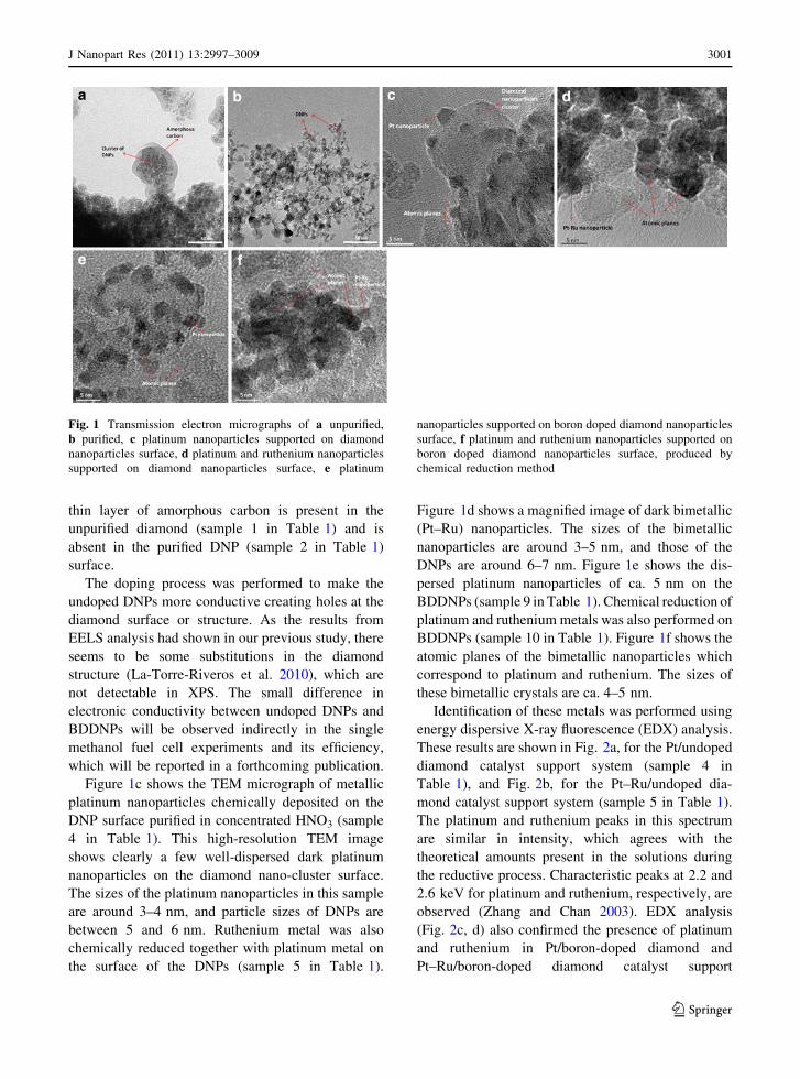
thin layer of amorphous carbon is present in the
unpurified diamond (sample 1 in Table 1) and is
absent in the purified DNP (sample 2 in Table 1)
surface.
The doping process was performed to make the
undoped DNPs more conductive creating holes at the
diamond surface or structure. As the results from
EELS analysis had shown in our previous study, there
seems to be some substitutions in the diamond
structure (La-Torre-Riveros et al. 2010), which are
not detectable in XPS. The small difference in
electronic conductivity between undoped DNPs and
BDDNPs will be observed indirectly in the single
methanol fuel cell experiments and its efficiency,
which will be reported in a forthcoming publication.
Figure 1c shows the TEM micrograph of metallic
platinum nanoparticles chemically deposited on the
DNP surface purified in concentrated HNO3 (sample
4 in Table 1). This high-resolution TEM image
shows clearly a few well-dispersed dark platinum
nanoparticles on the diamond nano-cluster surface.
The sizes of the platinum nanoparticles in this sample
are around 3–4 nm, and particle sizes of DNPs are
between 5 and 6 nm. Ruthenium metal was also
chemically reduced together with platinum metal on
the surface of the DNPs (sample 5 in Table 1).
Figure 1d shows a magnified image of dark bimetallic
(Pt–Ru) nanoparticles. The sizes of the bimetallic
nanoparticles are around 3–5 nm, and those of the
DNPs are around 6–7 nm. Figure 1e shows the dis-
persed platinum nanoparticles of ca. 5 nm on the
BDDNPs (sample 9 in Table 1). Chemical reduction of
platinum and ruthenium metals was also performed on
BDDNPs (sample 10 in Table 1). Figure 1f shows the
atomic planes of the bimetallic nanoparticles which
correspond to platinum and ruthenium. The sizes of
these bimetallic crystals are ca. 4–5 nm.
Identification of these metals was performed using
energy dispersive X-ray fluorescence (EDX) analysis.
These results are shown in Fig. 2a, for the Pt/undoped
diamond catalyst support system (sample 4 in
Table 1), and Fig. 2b, for the Pt–Ru/undoped dia-
mond catalyst support system (sample 5 in Table 1).
The platinum and ruthenium peaks in this spectrum
are similar in intensity, which agrees with the
theoretical amounts present in the solutions during
the reductive process. Characteristic peaks at 2.2 and
2.6 keV for platinum and ruthenium, respectively, are
observed (Zhang and Chan 2003). EDX analysis
(Fig. 2c, d) also confirmed the presence of platinum
and ruthenium in Pt/boron-doped diamond and
Pt–Ru/boron-doped diamond catalyst support
Fig. 1 Transmission electron micrographs of a unpurified,
b purified, c platinum nanoparticles supported on diamond
nanoparticles surface, d platinum and ruthenium nanoparticles
supported on diamond nanoparticles surface, e platinum
nanoparticles supported on boron doped diamond nanoparticles
surface, f platinum and ruthenium nanoparticles supported on
boron doped diamond nanoparticles surface, produced by
chemical reduction method
J Nanopart Res (2011) 13:2997–3009 3001
123
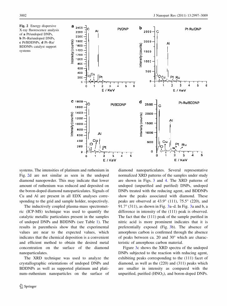
systems. The intensities of platinum and ruthenium in
Fig. 2d are not similar as seen in the undoped
diamond nanopowder. This may indicate that lower
amount of ruthenium was reduced and deposited on
the boron-doped diamond nanoparticulates. Signals of
Cu and Al are present in all EDX analyses corre-
sponding to the grid and sample holder, respectively.
The inductively coupled plasma-mass spectromet-
ric (ICP-MS) technique was used to quantify the
catalytic metallic particulates present in the samples
of undoped DNPs and BDDNPs (see Table 1). The
results in parenthesis show that the experimental
values are near to the expected values, which
indicates that the chemical deposition is a convenient
and efficient method to obtain the desired metal
concentration on the surface of the diamond
nanoparticulates.
The XRD technique was used to analyze the
crystallographic orientations of undoped DNPs and
BDDNPs as well as supported platinum and plati-
num–ruthenium nanoparticles on the surface of
diamond nanoparticulates. Several representative
normalized XRD patterns of the samples under study
are shown in Figs. 3 and 4. The XRD patterns of
undoped (unpurified and purified) DNPs, undoped
DNPs treated with the reducing agent, and BDDNPs
show the peaks associated with diamond. These
peaks are observed at 43.9� (111), 75.5� (220), and
91.7� (311), as shown in Fig. 3a–d. In Fig. 3a and b, a
difference in intensity of the (111) peak is observed.
The fact that the (111) peak of the sample purified in
nitric acid is more prominent indicates that it is
preferentially exposed (Fig. 3b). The absence of
amorphous carbon is confirmed through the absence
of peaks between ca. 20 and 30� which are charac-
teristic of amorphous carbon material.
Figure 3c shows the XRD spectra of the undoped
DNPs subjected to the reaction with reducing agent,
exhibiting peaks corresponding to the (111) facet of
diamond, as well as the (220) and (311) peaks which
are smaller in intensity as compared with the
unpurified, purified (HNO3), and boron-doped DNPs.
Fig. 2 Energy dispersive
X-ray fluorescence analysis
of a Pt/undoped DNPs,
b Pt–Ru/undoped DNPs,
c Pt/BDDNPs, d Pt–Ru/
BDDNPs catalyst support
systems
3002 J Nanopart Res (2011) 13:2997–3009
123
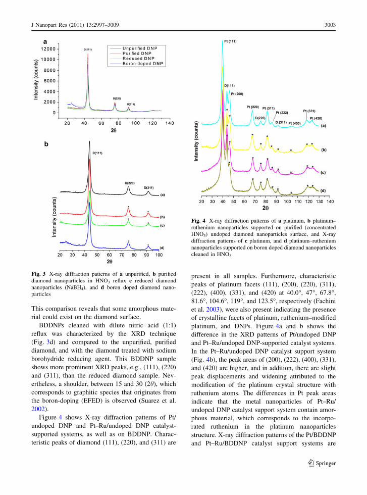
This comparison reveals that some amorphous mate-
rial could exist on the diamond surface.
BDDNPs cleaned with dilute nitric acid (1:1)
reflux was characterized by the XRD technique
(Fig. 3d) and compared to the unpurified, purified
diamond, and with the diamond treated with sodium
borohydride reducing agent. This BDDNP sample
shows more prominent XRD peaks, e.g., (111), (220)
and (311), than the reduced diamond sample. Nev-
ertheless, a shoulder, between 15 and 30 (2h), which
corresponds to graphitic species that originates from
the boron-doping (EFED) is observed (Suarez et al.
2002).
Figure 4 shows X-ray diffraction patterns of Pt/
undoped DNP and Pt–Ru/undoped DNP catalyst-
supported systems, as well as on BDDNP. Charac-
teristic peaks of diamond (111), (220), and (311) are
present in all samples. Furthermore, characteristic
peaks of platinum facets (111), (200), (220), (311),
(222), (400), (331), and (420) at 40.0�, 47�, 67.8�,
81.6�, 104.6�, 119�, and 123.5�, respectively (Fachini
et al. 2003), were also present indicating the presence
of crystalline facets of platinum, ruthenium–modified
platinum, and DNPs. Figure 4a and b shows the
difference in the XRD patterns of Pt/undoped DNP
and Pt–Ru/undoped DNP-supported catalyst systems.
In the Pt–Ru/undoped DNP catalyst support system
(Fig. 4b), the peak areas of (200), (222), (400), (331),
and (420) are higher, and in addition, there are slight
peak displacements and widening attributed to the
modification of the platinum crystal structure with
ruthenium atoms. The differences in Pt peak areas
indicate that the metal nanoparticles of Pt–Ru/
undoped DNP catalyst support system contain amor-
phous material, which corresponds to the incorpo-
rated ruthenium in the platinum nanoparticles
structure. X-ray diffraction patterns of the Pt/BDDNP
and Pt–Ru/BDDNP catalyst support systems are
Fig. 3 X-ray diffraction patterns of a unpurified, b purified
diamond nanoparticles in HNO3 reflux c reduced diamond
nanoparticles (NaBH4), and d boron doped diamond nano-
particles
Fig. 4 X-ray diffraction patterns of a platinum, b platinum–
ruthenium nanoparticles supported on purified (concentrated
HNO3) undoped diamond nanoparticles surface, and X-ray
diffraction patterns of c platinum, and d platinum–ruthenium
nanoparticles supported on boron doped diamond nanoparticles
cleaned in HNO3
J Nanopart Res (2011) 13:2997–3009 3003
123
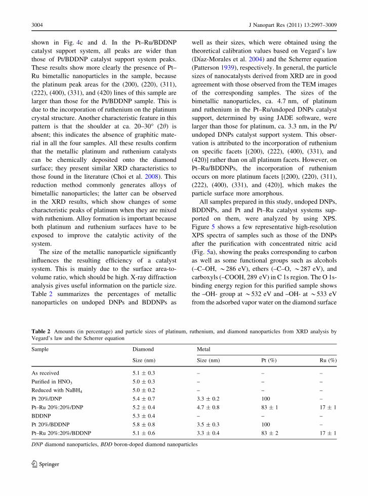
shown in Fig. 4c and d. In the Pt–Ru/BDDNP
catalyst support system, all peaks are wider than
those of Pt/BDDNP catalyst support system peaks.
These results show more clearly the presence of Pt–
Ru bimetallic nanoparticles in the sample, because
the platinum peak areas for the (200), (220), (311),
(222), (400), (331), and (420) lines of this sample are
larger than those for the Pt/BDDNP sample. This is
due to the incorporation of ruthenium on the platinum
crystal structure. Another characteristic feature in this
pattern is that the shoulder at ca. 20–30� (2h) is
absent; this indicates the absence of graphitic mate-
rial in all the four samples. All these results confirm
that the metallic platinum and ruthenium catalysts
can be chemically deposited onto the diamond
surface; they present similar XRD characteristics to
those found in the literature (Choi et al. 2008). This
reduction method commonly generates alloys of
bimetallic nanoparticles; the latter can be observed
in the XRD results, which show changes of some
characteristic peaks of platinum when they are mixed
with ruthenium. Alloy formation is important because
both platinum and ruthenium surfaces have to be
exposed to improve the catalytic activity of the
system.
The size of the metallic nanoparticle significantly
influences the resulting efficiency of a catalyst
system. This is mainly due to the surface area-to-
volume ratio, which should be high. X-ray diffraction
analysis gives useful information on the particle size.
Table 2 summarizes the percentages of metallic
nanoparticles on undoped DNPs and BDDNPs as
well as their sizes, which were obtained using the
theoretical calibration values based on Vegard’s law
(Dıaz-Morales et al. 2004) and the Scherrer equation
(Patterson 1939), respectively. In general, the particle
sizes of nanocatalysts derived from XRD are in good
agreement with those observed from the TEM images
of the corresponding samples. The sizes of the
bimetallic nanoparticles, ca. 4.7 nm, of platinum
and ruthenium in the Pt–Ru/undoped DNPs catalyst
support, determined by using JADE software, were
larger than those for platinum, ca. 3.3 nm, in the Pt/
undoped DNPs catalyst support system. This obser-
vation is attributed to the incorporation of ruthenium
on specific facets [(200), (222), (400), (331), and
(420)] rather than on all platinum facets. However, on
Pt–Ru/BDDNPs, the incorporation of ruthenium
occurs on more platinum facets [(200), (220), (311),
(222), (400), (331), and (420)], which makes the
particle surface more amorphous.
All samples prepared in this study, undoped DNPs,
BDDNPs, and Pt and Pt–Ru catalyst systems sup-
ported on them, were analyzed by using XPS.
Figure 5 shows a few representative high-resolution
XPS spectra of samples such as those of the DNPs
after the purification with concentrated nitric acid
(Fig. 5a), showing the peaks corresponding to carbon
as well as some functional groups such as alcohols
(–C–OH, *286 eV), ethers (–C–O, *287 eV), and
carboxyls (–COOH, 289 eV) in C 1s region. The O 1s-
binding energy region for this purified sample shows
the –OH- group at *532 eV and –OH- at *533 eV
from the adsorbed vapor water on the diamond surface
Table 2 Amounts (in percentage) and particle sizes of platinum, ruthenium, and diamond nanoparticles from XRD analysis by
Vegard’s law and the Scherrer equation
Sample Diamond Metal
Size (nm) Size (nm) Pt (%) Ru (%)
As received 5.1 ± 0.3 – – –
Purified in HNO3 5.0 ± 0.3 – – –
Reduced with NaBH4 5.0 ± 0.2 – – –
Pt 20%/DNP 5.4 ± 0.7 3.3 ± 0.2 100 –
Pt–Ru 20%:20%/DNP 5.2 ± 0.4 4.7 ± 0.8 83 ± 1 17 ± 1
BDDNP 5.3 ± 0.4 – – –
Pt 20%/BDDNP 5.8 ± 0.8 3.5 ± 0.3 100 –
Pt–Ru 20%:20%/BDDNP 5.1 ± 0.6 3.3 ± 0.4 83 ± 2 17 ± 1
DNP diamond nanoparticles, BDD boron-doped diamond nanoparticles
3004 J Nanopart Res (2011) 13:2997–3009
123
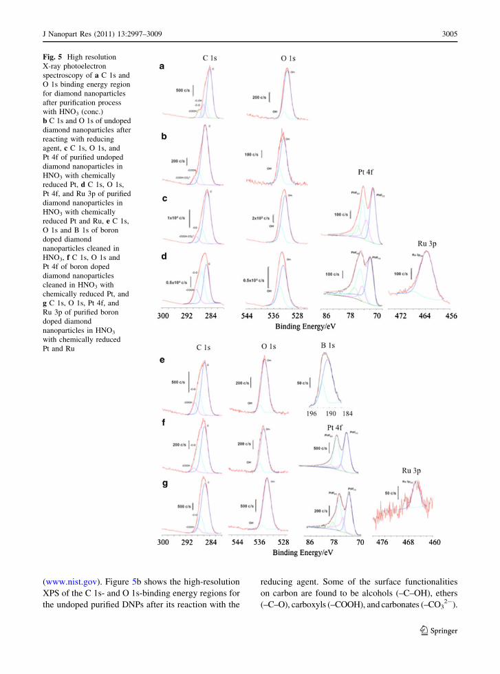
(www.nist.gov). Figure 5b shows the high-resolution
XPS of the C 1s- and O 1s-binding energy regions for
the undoped purified DNPs after its reaction with the
reducing agent. Some of the surface functionalities
on carbon are found to be alcohols (–C–OH), ethers
(–C–O), carboxyls (–COOH), and carbonates (–CO32-).
Fig. 5 High resolution
X-ray photoelectron
spectroscopy of a C 1s and
O 1s binding energy region
for diamond nanoparticles
after purification process
with HNO3 (conc.)
b C 1s and O 1s of undoped
diamond nanoparticles after
reacting with reducing
agent, c C 1s, O 1s, and
Pt 4f of purified undoped
diamond nanoparticles in
HNO3 with chemically
reduced Pt, d C 1s, O 1s,
Pt 4f, and Ru 3p of purified
diamond nanoparticles in
HNO3 with chemically
reduced Pt and Ru, e C 1s,
O 1s and B 1s of boron
doped diamond
nanoparticles cleaned in
HNO3, f C 1s, O 1s and
Pt 4f of boron doped
diamond nanoparticles
cleaned in HNO3 with
chemically reduced Pt, and
g C 1s, O 1s, Pt 4f, and
Ru 3p of purified boron
doped diamond
nanoparticles in HNO3
with chemically reduced
Pt and Ru
J Nanopart Res (2011) 13:2997–3009 3005
123
The oxygen groups including alcohols (–OH) and
hydroxide (OH-) from the adsorbed vapor water are
also present. It is evident that the reducing agent
affects the diamond surface with the absence of alco-
hols and ether groups and the presence of carbonates.
Figure 5c shows the high-resolution XPS spectrum of
C 1s, O 1s, and Pt 4f for platinum chemically reduced
on undoped purified (conc. HNO3) DNPs. This spec-
trum also shows some surface functional groups, such
as ethers (–C–O), carbonates (CO32-), and carboxylic
(–COOH) acid. The oxygen high-resolution binding
energy region shows the presence of alcohols (–OH)
and hydroxides (OH-). On the other hand, the plati-
num high-resolution binding energy spectrum shows
the Pt 4f7/2 (71–72 eV) and Pt 4f5/2 (74–75 eV); which
correspond to metallic platinum and platinum oxide,
respectively (Fachini et al. 2003; www.nist.gov). In
Fig. 5d, the high-resolution XPS spectrum of Pt 4f-
and Ru 3p-binding energy region, for the chemically
reduced on the undoped purified DNPs surface, are
shown, as well as the high-resolution XPS spectra of C
1s and O 1s. Ether (–C–O), and carboxylic (–COOH)
groups are present in the C 1s-binding energy region.
As in other samples, functionalities such as alcohols
(–OH) and hydroxides are also evidenced in the oxy-
gen region. The platinum spectrum in this sample also
shows the characteristic lines of metallic platinum
(Pt 4f7/2, 71–72 eV) and platinum oxides such as PtO
and PtO2 (Pt 4f5/2, 74–75 eV). The presence of
metallic ruthenium at ca. 462.2 eV and the influence of
adsorbed water at ca. 466.5 eV are observed in the
high-resolution XPS of this metal (Ru 3p3/2 line)
(www.nist.gov).
The BDDNPs sample was cleaned in a concen-
trated nitric acid reflux system to eliminate the excess
boron present in the sample. High-resolution XPS
spectra of C 1s, O 1s, and B 1s were also obtained
(see Fig. 5e). The carbon region shows the presence
of ether (–C–O) and carboxylic groups (–COOH).
The oxygen region shows alcohol (–C–OH) and
hydroxide (OH-) groups, and, in the B 1s-binding
energy region, presents peaks for boron and B2O3 at
190.5 and 193 eV, respectively (www.nist.gov). The
amount of boron present in the purified sample was
determined by using the 1s peak areas of boron and
carbon, resulting in a calculated value of 0.68% of
boron in the doped diamond sample. However, the B
1s-binding energy peak is broad; indicating that
boron is not totally incorporated and other oxidized
species of boron are present. These boron oxides
could also be responsible for the platinum deposition
and dispersion on the diamond surface.
Platinum and ruthenium metals were chemically
reduced on the boron-doped diamond surface. High-
energy resolution analysis of these samples (Fig. 5f),
show C 1s, O 1s, and Pt 4f signals. The carbon
spectrum shows the presence of ether (–C–O) and
carboxylic (–COOH) groups. The oxygen spectrum
shows the existence of alcohol (–C–OH) and hydrox-
ide (–OH-) moieties. The platinum high-resolution
spectrum shows the Pt 4f7/2 (71–72 eV) and Pt 4f5/2
(74–75 eV) peaks which also correspond to metallic
platinum and platinum oxide, respectively. Finally,
Fig. 5g shows the C 1s-, O 1s-, Pt 4f-, and Ru 3p-
binding energy regions. Carbon spectrum shows the
presence of ether (–C–O) and carboxylic (–COOH)
groups. The oxygen spectrum shows the existence of
alcohol (–C–OH) and hydroxide (–OH-) moieties.
The platinum spectrum shows the Pt 4f7/2 (71–72 eV)
and Pt 4f5/2 (74–75 eV) peaks which correspond to
metallic platinum and platinum oxide, respectively.
Metallic ruthenium (*462 eV) is evidenced by the
high-resolution XPS spectrum of this metal (Ru 3p3/2
line), and the adsorbed water feature at ca. 466 eV is
also observed. It can be seen that the platinum and
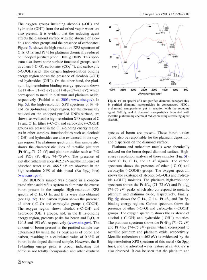
Fig. 6 FT-IR spectra of a not purified diamond nanoparticles,
b purified diamond nanoparticles in concentrated HNO3,
c diamond nanoparticles put in reaction with the reducing
agent NaBH4, and d diamond nanoparticles decorated with
metallic platinum by chemical reduction using a reducing agent
(NaBH4)
3006 J Nanopart Res (2011) 13:2997–3009
123

ruthenium depositions occur on the diamond surface
at the C–OH site; hence, this peak is absent in all the
samples containing platinum and platinum–ruthe-
nium catalysts. The surface organic groups of the
doped diamond are very similar to those of carbon
black. The exchange of ionic groups of surface
organic groups and metal salts may play an important
role in the formation of nanoparticles with a narrow
particle size distribution.
The FT-IR results of the unpurified and purified
DNP samples show that the only difference is the
percentage of reflectance of the m–CO feature, which
is higher in the unpurified sample (Fig. 6a, b). The
functional groups on the undoped diamond surface
are similar to those found in the literature (Kulakova
2004). The FT-IR spectrum shown in Fig. 6a for
unpurified DNPs, shows the m–OH, m–CO2, m–CO,
m–CH2, m–CH3, and m–C–O–C– characteristic IR
bands. Figure 6c for DNPs reacted with the reducing
agent NaBH4 shows, besides m–OH, m–CO, m–CH2,
m–CH3, and m–C–O–C– bands, additional bands of
m–CH at ca. 2800 and 2850 cm-1. The peak of –CH3
at ca. 1250 cm-1 is more intense than those in spectra
a and b of Fig. 6. Finally, spectrum 6d for DNPs
decorated with metallic platinum shows only m–CO,
and low intensity m–C–O–C–, m–CH2, and m–CH3
band features. This fact indicates that the nucleation
of metallic platinum started on the diamond surface
through an interaction between the metal ions and
functional groups such as alcohols (–OH) and some
of the –CH groups, and followed by the deposition.
This analysis should be verified by applying a more
sensitive technique such as far infrared spectroscopy
to determine the interaction between the platinum and
oxygen species.
Conclusions
On the basis of the materials’ characterization results,
it can be concluded that the acid reflux purification
process is efficient and increases the hydrogen
content in the undoped DNPs surface. The TEM
micrographs also corroborate the removal of amor-
phous carbon from the undoped DNPs surface, as
does the absence of amorphous carbon peaks in the
XRD spectra.
The chemical reduction method allowed us to
obtain nanoparticles in amounts near to the desired
atomic percentages in all the samples as was dem-
onstrated by the EDX and ICP results (see Table 1),
which make this method reliable for catalyst prepa-
ration with DNPs as support.
The Pt and Pt–Ru nanoparticle sizes obtained
ranged from 2 to 5 nm, as was shown in the TEM
micrographs and XRD results (see Table 2). These
results are indicative that this fast reaction is conve-
nient to obtain small nanoparticles that are desirable
to increase the surface area of a catalyst system.
The undoped DNPs and BDDNPs surfaces were
successfully used as support systems for metallic
catalyst particulates obtained by chemical reduction.
The platinum and ruthenium compounds mainly
deposit on the –OH sites, as was shown in the XPS
and FTIR spectra. This analysis will be verified by
the use of a more sensitive technique such as far
infrared spectroscopy to determine the interaction
between platinum and oxygen species.
The spectroscopic and surface characterization
showed the viability of obtaining a catalytic system
based on undoped DNPs and BDDNPs supports, and
platinum and ruthenium nanoparticles, which can be
used in applications such as direct methanol fuel cells
and will be reported in a forthcoming publication.
Acknowledgments This research was supported in part by
the NASA-URC Grant No. NNX08BA48A, NSF-EPSCoR, the
Institute for Functional Nanomaterials Grant No. OIA-
0701525, and the NSF NSEC Center for Hierarchical
Manufacturing Grant No. CHM – CMMI – 0531171. We
also acknowledge the support received from the Cornell
Center for Materials Research (CCMR-TEM) at Cornell
University. Editing of the manuscript done by Dr. D. A. Tryk
and Dr. M. A. Scibioh is gratefully acknowledged.
References
Antolini E (2009) Carbon supports for low-temperature fuel
cell catalysts. Appl Catal B 88:1–24
Arico AS, Poltarzewski Z, Kim H, Morana A, Giordano N,
Antonucci V (1995) Investigation of a carbon-supported
quaternary Pt–Ru–Sn–W catalyst for direct methanol fuel
cells. J. Power Sources 55:159–166
Arico AS, Baglio V, Di Blasi A, Modica E, Antonucci PL,
Antonucci V (2003) Analysis of the high-temperature
methanol oxidation behaviour at carbon-supported Pt–Ru
catalysts. J Electroanal Chem 557:167–176
Bennett JA, Show Y, Wang SH, Swain GM (2005) Pulsed
galvanostatic deposition of Pt particles on microcrystal-
line and nanocrystalline diamond thin-film electrodes.
J Electrochem Soc 152:E184–E192
J Nanopart Res (2011) 13:2997–3009 3007
123
Bogatyreva GP, Marinich MA, Ishchenco EV, Gvyazdovskaya
VL, Bazalii GA, Oleinik NA (2004) Application of
modified nanodiamonds as catalysts of heterogeneous and
electrochemical catalyses. Phys Solid State 46:738–741
Callstrom MR, Neenan TX, McCreery RL, Alsmeyer DC
(1990) Doped glassy carbon materials (DGC): low-tem-
perature synthesis, structure, and catalytic behavior. J Am
Chem Soc 112:4954–4956
Chen Q, Granger MC, Lister TE, Swain GM (1997) Morpho-
logical and microstructural stability of boron-doped dia-
mond thin film electrodes in an acidic chloride medium at
high anodic current densities. J Electrochem Soc 144:
3806–3812
Choi SM, Kim JH, Jung JY, Yoon EY, Kim WB (2008) Pt
nanowires prepared via a polymer template method: its
promise toward high Pt-loaded electrocatalysts for meth-
anol oxidation. Electrochem Acta 53:5804–5811
Danilenko VV (2004) On the history of the discovery of nano-
diamond synthesis. Phys Solid State 46:595–599
Dıaz-Morales R, Liu R, Fachini E, Chen G, Segre CU,
Martınez A, Cabrera C, Smotkin ES (2004) XRD and XPS
analysis of as-prepared and conditioned DMFC array
membrane electrode assemblies. J Electrochem Soc
151:A1314–A1318
Fachini ER, Dıaz-Ayala R, Casado-Rivera E, File S, Cabrera
CR (2003) Surface coordination of ruthenium clusters on
platinum nanoparticles for methanol oxidation catalysts.
Langmuir 19:8986–8993
Fischer AE, Swain GM (2005) Preparation and characterization
of boron-doped diamond powder. J Electrochem Soc
152:B369–B37511
Fischer AE, Show Y, Swain GM (2004) Electrochemical per-
formance of diamond thin-film electrodes from different
commercial sources. Anal Chem 76:2553–2560
Gloaguen F, Leger JM, Lamy C (1997) Electrocatalytic oxi-
dation of methanol on platinum nanoparticles electrode-
posited onto porous carbon substrates. J Appl Electrochem
27:1052–1060
Hayashi Y, Mori D, Soga T, Jimbo T (2004) Modification of
the physical properties of chemical vapor-deposited
nanostructure diamond by argon-hydrogen plasma surface
treatment. Phys Solid State 46:733–737
Holt KB (2010) Undoped diamond nanoparticles: origins of
surface redox chemistry. Phys Chem Chem Phys
12:2048–2058
Holt KB, Ziegler Ch, Caruana DJ, Zang J, Millan-Barios EJ,
Hu J, Foord JS (2008) Redox properties of undoped 5 nm
diamond nanoparticles. Phys Chem Chem Phys 10:
303–310
Holt KB, Ziegler Ch, Caruana DJ, Zang J, Millan-Barios EJ
(2009) Electrochemistry of undoped diamond nanoparti-
cles: accessing surface redox states. J Am Chem Soc
131:11272–11273
Honda K, Yoshimura M, Rao TN, Tryk DA, Fujishima A,
Yasui K, Sakamoto Y, Nishio K, Masuda H (2001)
Electrochemical properties of Pt-modified nano-honey-
comb diamond electrodes. J Electroanal Chem 514:35–50
Im DH, Park SY, Hyun SH (2004) Aqueous dispersion stability
of nickel powders prepared by a chemical reduction
method. J Mater Sci 39:3629–3633
Jeon MK, Won JY, Lee KR, Woo SI (2007) Highly active
PtRuFe/C catalyst for methanol electro-oxidation. Elect-
rochem Commun 9:2163–2166
Jian C, Jing-Zhi S, Han-Yin L, Jian H, Mang W (2004) A facile
room-temperature chemical reduction method to
TiO2@CdS core/sheath heterostructure nanowires.
J Mater Chem 14:1203–1206
Joon-Hyun P, Piraman S, Hyun-Jong K, Myung-Keun H, Jae-
Hyuk J, Yong-Rok K, Han-Sung K, Yong-Gun S (2008)
Investigation of metal alloy catalyst for hydrogen release
from sodium borohydride for polymer electrolyte mem-
brane fuel cell application. Int J Hydrogen Energy
33:1845–1852
Koo IG, Lee MS, Shim JH, Ahn JH, Lee WM (2005) Platinum
nanoparticles prepared by a plasma-chemical reduction
method. J Mater Chem 15:4125–4128
Kulakova II (2004) Surface chemistry of nanodiamonds. Phys
Solid State 46:636–643
La-Torre-Riveros L, Tryk DA, Cabrera CR (2005) Chemical
purification and characterization of diamond nanoparticles
for electrophoretically coated electrodes. Rev Adv Mater
Sci 10:256–260
La-Torre-Riveros L, Soto K, Tryk DA, Cabrera CR (2007)
Electrophoretic preparation and characterization of porous
electrodes from diamond nanoparticles. J Phys Conf Ser
61:1022–1026
La-Torre-Riveros L, Soto K, Scibioh MA, Cabrera CR (2010)
Electrophoretically fabricated diamond nanoparticle-
based electrodes. J Electrochem Soc 157:B831–B836
Liu ZL, Lee JY, Ming H, Chen WX, Gan LM (2002) Synthesis
and characterization of PtRu/C catalysts from microemul-
sions and emulsions. J Mater Chem 12:2453–2458
Mani RC, Sharma S, Sunkara MK, Gullapalli J, Baldwin RP,
Rao R, Rao AM, Cowley JM (2002) Synthesis and elec-
trochemical characteristics of a nanocomposite diamond
electrode. Electrochem Solid State Lett 5:E32–E35
Montilla F, Morallon E, Duo I, Comninellis Ch, Vazquez JL
(2003) Platinum particles deposited on synthetic boron-
doped diamond surfaces. Application to methanol oxida-
tion. Electrochim Acta 48:3891–3897
Nersisyan HH, Lee JH, Son HT, Won CW, Maeng DY (2003)
A new and effective chemical reduction method for
preparation of nanosized silver powder and colloid dis-
persion. Mater Res Bull 38:949–956
Osswald S, Yushin G, Mochalin V, Kucheyev SO, Gogotsi Y
(2006) Control of sp2/sp3 carbon ratio and surface
chemistry of nanodiamond powders by selective oxidation
in air. J Am Chem Soc 128:11635–11642
Patterson AL (1939) The Scherrer formula for X-ray particle
size determination. Phys Rev 56:978–982
Paulus U, Endruschat U, Feldmeyer G, Schmidt T, Bonnemann
H, Behm R (2000) New PtRu alloy colloids as precursors
for fuel cell Catalysts. J Catal 195(2):383–393
Porcard NL, Alsmeyer DC, McCreery RL, Neenan TX, Call-
strom MR (1992) Nanoscale platinum(0) clusters in glassy
carbon: synthesis, characterization, and uncommon cata-
lytic activity. J Am Chem Soc 114:769–771
Rao V, Simonov PA, Savinova ER, Plaksin GV, Cherepanova
SV, Kryukova GN, Stimming U (2005) The influence of
carbon support porosity on the activity of PtRu/Si unit
3008 J Nanopart Res (2011) 13:2997–3009
123
anode catalysts for methanol oxidation. J. Power Sources
145:178–187
Reinzler AG, Liu J, Dai H, Nokolaev P, Huffman CB, Rodri-
guez-Macias FJ, Boul PJ, Lu AH, Heymann D, Colvert
DT et al (1998) Large-scale purification of single-wall
carbon nanotubes: process, product, and characterization.
Appl Phys A 67:29–37
Salazar-Banda GR, Suffredini HB, Avaca LA (2005) Improved
stability of PtOx sol-gel-modified diamond electrodes
covered with a Nafion� film. J Braz Chem Soc 16:903–906
Salazar-Banda GR, Suffredini HB, Calegaro ML, Tanimoto
ST, Avaca LA (2006) Sol–gel-modified boron-doped
diamond surfaces for methanol and ethanol electro-oxi-
dation in acid medium. J Power Sources 162:9–20
Scibioh A, Oh I-H, Lim TH, Hong SA, Ha HY (2008) Inves-
tigation of various ionomer-coated carbon supports for
direct methanol fuel cell applications. Appl Catal B
77:373–385
Sine G, Foti G, Comninellis Ch (2006) Boron-doped diamond
(BDD)-supported Pt/Sn nanoparticles synthesized in mi-
croemulsion systems as electrocatalysts of ethanol oxi-
dation. J Electroanal Chem 595:115–124
Sine G, Smida D, Limat M, Foti G, Comninellis Ch (2007)
Microemulsion synthesized Pt/Ru/Sn nanoparticles on
BDD for alcohol electro-oxidation. J Electrochem Soc
154:B170–B174
Spataru N, Zhang X, Spataru T, Tryk D, Fujishima A (2008)
Platinum electrodeposition on conductive diamond pow-
der and its application to methanol oxidation in acidic
media. J Electrochem Soc 155:B264–B269
Suarez A, Prelas MA, Ghosh TK, Tompson RV, Loyalka SK,
Miller WH, Viswanath DS (2002) Diffusion of boron into
polycrystalline diamond films using the electric field
enhanced diffusion (EFED) technique. J Wide Bandgap
Mater 10:15–27
Suffredini HB, Tricoli V, Vatistas N, Avaca LA (2006) Elec-
tro-oxidation of methanol and ethanol using a Pt–RuO2/C
composite prepared by the sol–gel technique and sup-
ported on boron-doped diamond. J Power Sources 158:
124–128
Takasu Y, Kawaguchi T, Sugimoto W, Murakami Y (2003)
Effects of the surface area of carbon support on the
characteristics of highly-dispersed Pt–Ru particles as
catalysts for methanol oxidation. Electrochim Acta 48:
3861–3868
Tang Z, Li Q, Lu G (2007) The effect of plasma pre-treatment
of carbon used as a Pt catalyst support for methanol
electrooxidation. Carbon 45:41–46
Viswanathan B, Scibioh A (2008) Fuel cells: principles and
applications. CRC Press, Boca Raton
Wang L, Chen D (2004) ‘‘One-pot’’ Fabrication of Ag/PMMA
‘‘shell/core’’ nanocomposites by chemical reduction
method. Chem Lett 33:1010–1011
Wang J, Swain GM, Tachibana T, Kobashi K (2000) The
incorporation of Pt nanoparticles into boron-doped dia-
mond thin-films: dimensionally stable catalytic electrodes.
J New Mater Electrochem Syst 3:75–82
Watanabe M, Uchida M, Motoo S (1987) Preparation of highly
dispersed Pt ? Ru alloy clusters and the activity for the
electrooxidation of methanol. J Electroanal Chem 229:
395–406
Xiaobo F, Hao Y, Feng P, Honguan W, Yu Q (2007) Facile
preparation of RuO2/CNT catalyst by a homogenous
oxidation precipitation method and its catalytic perfor-
mance. Appl Catal A 32:190–197
Xu W, Zhou X, Liu Ch, Xing L, Lu T (2007) The real role of
carbon in Pt/C catalysts for oxygen reduction reaction.
Electrochem Commun 9:1002–1006
Zhang X, Chan KY (2003) Water-in-oil microemulsion syn-
thesis of platinum–ruthenium nanoparticles, their charac-
terization and electrocatalytic properties. Chem Mater
15:451–459
J Nanopart Res (2011) 13:2997–3009 3009
123