Ultrastructural Study of Yam Tuber as Related to ... - CiteSeerX

rsif.royalsocietypublishing.org
ResearchCite this article: Sui T, Sandholzer MA, Lunt
AJG, Baimpas N, Smith A, Landini G, Korsunsky
AM. 2014 In situ X-ray scattering evaluation of
heat-induced ultrastructural changes in dental
tissues and synthetic hydroxyapatite. J. R. Soc.
Interface 11: 20130928.
http://dx.doi.org/10.1098/rsif.2013.0928
Received: 10 October 2013
Accepted: 14 March 2014
Subject Areas:bioengineering, biomaterials
Keywords:in situ thermal treatment, dental tissue,
hydroxyapatite, SAXS/WAXS
Author for correspondence:Tan Sui
e-mail: [email protected]
& 2014 The Author(s) Published by the Royal Society. All rights reserved.
In situ X-ray scattering evaluation of heat-induced ultrastructural changes in dentaltissues and synthetic hydroxyapatite
Tan Sui1, Michael A. Sandholzer2, Alexander J. G. Lunt1, Nikolaos Baimpas1,Andrew Smith3, Gabriel Landini2 and Alexander M. Korsunsky1
1Department of Engineering Science, University of Oxford, Parks Road, Oxford OX1 3PJ, UK2School of Dentistry, College of Medical and Dental Sciences, University of Birmingham, Saint Chad’s Queensway,Birmingham B4 6NN, UK3Diamond Light Source Ltd, Harwell Science and Innovation Campus, Didcot OX11 0DE, UK
Human dental tissues consist of inorganic constituents (mainly crystallites of
hydroxyapatite, HAp) and organic matrix. In addition, synthetic HAp powders
are frequently used in medical and chemical applications. Insights into the ultra-
structural alterations of skeletal hard tissues exposed to thermal treatment are
crucial for the estimation of temperature of exposure in forensic and archaeologi-
cal studies. However, at present, only limited data exist on the heat-induced
structural alterations of human dental tissues. In this paper, advanced non-
destructive small- and wide angle X-ray scattering (SAXS/WAXS) synchrotron
techniques were used to investigate the in situ ultrastructural alterations in
thermally treated human dental tissues and synthetic HAp powders. The crystal-
lographic properties were probed by WAXS, whereas HAp grain size
distribution changes were evaluated by SAXS. The results demonstrate the
important role of the organic matrix that binds together the HAp crystallites in
responding to heat exposure. This is highlighted by the difference in the thermal
behaviour between human dental tissues and synthetic HAp powders. The X-ray
analysis results are supported by thermogravimetric analysis. The results con-
cerning the HAp crystalline architecture in natural and synthetic HAp
powders provide a reliable basis for deducing the heating history for dental
tissues in the forensic and archaeological context, and the foundation for further
development and optimization of biomimetic material design.
1. IntroductionHydroxyapatite (HAp) crystallites that are closely integrated in the organic
matrix at the nanoscale are the main inorganic constituents of the hierarchical
mineralized human dental tissues, namely enamel and dentine. Such natural
HAp crystallites are understood to be crucial to the physiology and function
of dental tissues [1]. In parallel, recent developments in synthetic biomineraliza-
tion have demonstrated that synthetic HAp powders can play an important role
for medical applications, especially for the replacement or treatment of dental
tissues [2].
Advanced high-energy treatment techniques are finding increasing use in
modern dentistry. In the early 1970s, Kantola et al. [3] showed that laser treat-
ment of dental tissues can be used to increase the mineral content and
crystallinity of dentine by preferential removal of the inherent water and
protein. With the advent of a variety of new laser systems spanning a range
of energy densities and pulse durations, clinical treatments such as laser-
assisted caries protection were proposed and developed. Local temperature
induced by short pulse laser treatment leads to local heating to temperatures
between several hundred to over 10008C. The alteration of dental tissue remains
highly localized, and improved caries prevention is surmised to be associated
with increased mineralization and HAp crystallite sintering that leads to the
sealing of dentinal tubules, while leaving the remaining tissues intact owing

rsif.royalsocietypublishing.orgJ.R.Soc.Interface
11:20130928
2
to the confined effect of less than 10 mm in depth (at thefluence of 2 J cm22) [4,5].
Any change in the local environment of HAp crystallites,
for example, through laser illumination or direct heating, is
bound to affect the mineral ultrastructure. In the context of
archaeological and forensic investigations, while macroscopic
alterations (e.g. surface colour) can be used to deduce an
approximate temperature range, the investigation of the
micro- and ultrastructural alterations of skeletal hard tissue
to high temperatures has proved to be crucial to obtain a
reliable estimation of the temperature of exposure [6–8].
This, in turn, sheds light on the attendant circumstances, for
example, the presence and nature of combustible agent used.
Dental tissues, being the most highly mineralized part of the
human body, can withstand longer thermal exposure and
survive higher temperatures, thus offering the possibility of
extending the range of applicability of this technique [9].
The above considerations provide strong motivation for a
detailed study into the effect of thermal exposure on the
nanostructure (the arrangement of HAp crystallites) of
human dental tissue as well as the role of the organic
phase. While much research has been carried out into the
heat-induced changes that occur in bone, in connection
with human remains studied in the context of archaeology,
anthropology and palaeontology [10–13], the corresponding
range and depth of study of the thermal response of dental
tissues remains lacking [6,14].
Multiple techniques have been used to study heat-induced
ultrastructural alterations of skeletal hard tissues. Most widely
applied techniques are based on the absorption spectra of
infrared radiation (Fourier transform infrared spectroscopy,
FTIR), or laboratory-based X-ray powder diffraction (XRD)
and laboratory-based small angle X-ray scattering (SAXS)
[6,10,15–17]. However, there has been ongoing debate regard-
ing the general limitations and validity of the results
quantified by FTIR [6,15,16]. Laboratory-based XRD has been
shown to be capable of showing the distinct differences in the
response of HAp crystallites to heating between animal and
human bone [6,16,18]. As a complementary method, labora-
tory-based SAXS allows the identification of local structural
alterations at the nanoscale, and is also capable of a fast through-
put of samples [19]. However, both methods usually involve the
grinding of a sample into powder to obtain volume-averaged
results that do not provide appropriately spatially resolved
information on the local changes of skeletal ultrastructure
within the tissue [19,20]. In order to understand and quan-
tify more precisely the mechanism of such ultrastructural
alterations with temperature, the application of advanced
non-destructive techniques offers an appropriate route. One of
the most advanced non-destructive techniques is synchrotron-
based X-ray diffraction, namely WAXS and SAXS. These
methods have been used to characterize human dental tissues
[21,22], which enabled the characterization of the ultrastructure
of mineralized tissue using intact (not ground) samples, rather
than powdered samples used with laboratory-based techniques.
Recently, the non-destructive synchrotron-based techniques
(WAXS/SAXS) have been applied to the study of previously
thermally treated human dental tissues [9]. However, these
tests were conducted ex situ, where it was possible to consider
only a limited number of samples after exposure to tempera-
tures in the 400–8008C range. In addition, by necessity,
different samples were used for each measurement, and thus
the sample-to-sample ultrastructure variation could not be
excluded. This aspect may weaken the reliability of the
conclusions drawn regarding the mechanism of local ultra-
structural changes. Furthermore, the ex situ mode of sample
characterization is applied only after heating and cooling the
samples to/from different temperatures. Obviously, no infor-
mation is available on the structure evolution during heating
nor cooling. To the best of our knowledge, no results have
been reported on monitoring heat-induced ultrastructural
changes of HAp crystallites using an in situ experimental pro-
tocol spanning the entire relevant range of heating and cooling
nor has the comparison been made between the naturally
occurring HAp crystallites within human dental tissues and
synthetic HAp powders. Thus, in situ thermal measurement
will help to understand the internal architecture of complex
natural materials (highly mineralized human tissues), and its
evolution during thermal exposure.
In this paper, we report the evaluation of the crystallo-
graphic properties (d-spacing and crystal perfection and size)
and the nanostructure (thickness, orientation and degree of
alignment) of HAp crystallites embedded within the dental
tissue as well as synthetic HAp powders subjected to in situthermal treatment. The combination of synchrotron X-ray scat-
tering techniques (WAXS/SAXS) was used. The results were
analysed in order to understand how the internal ultrastruc-
ture within the human dental tissues and the synthetic HAp
powders evolves during the heating and cooling schedule.
2. Material and methods2.1. Sample preparationFreshly extracted intact human third molars (ethical approval
obtained from the National Research Ethics Committee; NHS-
REC reference no. 09.H0405.33/Consortium R&D no. 1465)
were washed and cleaned in distilled water to eliminate residues,
so that the possibility of contamination or other chemical effects
was excluded. The samples were cut into 1 mm-thick cross-
sectional slices in the buccolingual orientation including dentine
and enamel (figure 1a) using a low-speed diamond saw (Isomet
Buehler Ltd, Lake Bluff, IL) and polished using a sequence of grit
papers to minimize the induced residual strain. In total, four
cross sections were prepared and were kept in distilled water
in a commercial fridge at 48C until the experiment was per-
formed. Low temperature storage conditions ensured relatively
low diffusion rates so as to impede or exclude any possibility
of superficial demineralization effect of water.
A mould was filled with 100 mg of commercially available
HAp powder (hydroxyapatite HTP powder, Bio-Rad, Hercules,
CA) and compacted with using an Instron 5544 machine (Instron
Ltd, Bucks, UK) at 1.8 kN force. The resulting disc was 1.2 mm
thick and 8 mm in diameter, with an approximate density of
1150 mg cm23.
The weight-loss over time was additionally evaluated using
thermogravimetry (TGA) at a rate of 128C per minute using
three cube-shaped dental samples (3 � 3 � 3 mm3) extracted
from two molars. The weight fraction of the organic component
was determined with TGA using one cubic dentine sample that
was placed in a rotating 10% formic acid bath for 14 days to elim-
inate the mineral content, thus producing a purely organic sample.
2.2. In situ scattering measurements2.2.1. Thermal treatment set-upIn situ thermal treatment of the samples was performed using a
remotely operated and monitored furnace that allows collection

SAXS-Pilatusdetector
1 cm
human dentalslice sample
synthetic HApsample
(a)
q = 0.1nm–1
20°
900
800
700
600
500
400
300
200
100
00 2000 4000 6000
time (s)
tem
pera
ture
(°C
)
8000 10 000 12 000
dental slicesynthetic HAp
0 10 000 20 000 30 000
time (s)
40 000 70 00060 00050 000 80 000
002
(c)(b)
(e)(d )
x
y
x
y
z
2892 mm
incoming beam
z-axis
11.5°
275.55 mm
WAXS detector
Figure 1. (a) Schematic of the in situ SAXS/WAXS experimental set-up that ensured that the WAXS and SAXS patterns were collected simultaneously. (b) A repre-sentative WAXS pattern showing the Debye – Sherrer ring corresponding to the (002) peak indicated by the red arrow. On the ring, the section along the y-axis in (a)was ‘caked’ (see text) with a 208 range in order to capture the peak shift upon heating and cooling process. (c) A representative SAXS pattern. The dark region inthe middle is the beamstop. The ellipsoidal shape of the pattern indicates a partial alignment of HAp crystallites. The pattern for randomly distributed crystals is around disc with the outline marked with red dashed circle. (d,e) Illustration of the different heating and cooling protocols used for the human dental slice samplesand the synthetic HAp powders sample. (Online version in colour.)
rsif.royalsocietypublishing.orgJ.R.Soc.Interface
11:20130928
3

rsif.royalsocietypublishing.orgJ.R.Soc.Interface
11:20130928
4
of SAXS/WAXS data at temperatures up to 10008C. This furnaceis a modification of the design used at Daresbury laboratory [23].
The samples were fixed in specially designed ceramic sample
holders. A Kapton window in the furnace allowed a high-
energy X-ray transmission set-up to be used, as illustrated in
figure 1a. The heating and cooling protocols were controlled
using a Eurotherm controller. The protocols incorporated
ramps at 128C min21, and constant temperature holds of 200 s
for each temperature level within dental slice samples studied
(enamel and dentine in figure 1d ). The protocol for synthetic
HAp powders is also shown in figure 1e. Slightly different ther-
mal protocols were used for natural and synthetic materials
owing to the limitations of the experimental set-up (furnace heat-
ing rates accessible for different samples). However, preliminary
measurements carried out showed that, within the range of heat-
ing rates considered, the resulting differences were negligible.
The temperature output by the furnace controller and thermo-
couple was of high accuracy (one decimal point, approx. 0.18nominal precision). Overall, the measurements were conducted
at three different points in dentine, one measurement point in
enamel and one point in the synthetic HAp sample. Larger
number of datasets would improve the quality of data and pro-
vide better statistical information on data quality. However,
owing to the limited availability of synchrotron beam time, the
collection of additional datasets was not possible. The statistical
error analysis of dentine has been included in the results, and
is reported below.
2.2.2. Beamline experimental set-upThe experiment was carried out on the I22 experimental beam-
line at Diamond Light Source, Oxford Harwell Campus,
Didcot, UK. A monochromatic X-ray beam (photon energy of
18 keV) was used to illuminate the samples with the beam size
of 65 � 120 mm2 as illustrated schematically in figure 1a. At
each heating or cooling step, WAXS and SAXS patterns were sim-
ultaneously collected at consecutive heating and cooling
increments at enamel and dentine or synthetic HAp crystallite
across the specimens. A lightly compacted disc of NIST standard
silicon powder was used for the WAXS data calibration, and a
disc of silver behenate powder was used for the SAXS data
calibration [24].
WAXS diffraction patterns (figure 1b) were recorded using a
Pilatus 100K detector (Dectris, Baden, Switzerland) placed at a
sample-to-camera distance of 275.55 mm (figure 1a). Further
downstream of the beam a Pilatus 2M detector (Dectris) was
positioned at a distance of 2892 mm to collect the SAXS patterns
(figure 1c). In order to record simultaneously the WAXS and
SAXS patterns at each scanning location, the WAXS detector
was titled and offset at 11.58 to the incident beam.
2.3. X-ray scattering data analysis2.3.1. Wide angle X-ray scattering data analysisWAXS patterns appear graphically as separated rings with differ-
ent widths and intensity levels, which contain characteristic
information related to the crystalline structure. The analysis of
WAXS patterns is performed using Bragg’s law, which establishes
the relationship between the spacing of atomic planes in crystals
and the scattering angle at which these planes produce intense
reflections.
nl ¼ 2dhklspacing sin u, (2:1)
where l is the wavelength, dhklspacing (d-spacing) is the interplanar
distance between planes with Miller indices fhklg, u is one half
of the scattering angle and n is the order of the reflection. Each
peak corresponds to a certain family of lattice planes within a
certain crystallographic phase. The changes of d-spacing could
be used to examine the residual lattice strain and trace the
phase transformation. Therefore, diffraction pattern analysis
can be used to identify and quantify the crystallographic
phases and structure parameters. The typical WAXS pattern is
shown in figure 1b can be used to extract and compare the crys-
tallographic properties (e.g. degree of crystal perfection) between
different samples or the same samples at different stages of its
evolution [20].
In figure 1b, the ring with the strongest intensity represents
the diffraction from the (002) family of lattice planes. Moreover,
the (002) lattice plane reflection is the most distinct ring that is
reliably present in all three materials studied (dentine, enamel,
synthetic HAp). WAXS data can be interpreted in terms of the
shift of the diffraction peak obtained from a cluster of HAp crys-
tallites. The variation of interplanar spacing (d-spacing) with
respect to the temperature between the corresponding lattice
planes was captured using WAXS. The (002) peak represents
the orientation of the scattering vector along the hexagonal c-
axis (parallel to the long direction) within the HAp crystallites.
It also corresponds to the organic fibril orientation owing to
the parallel orientation of fibrils to the long direction in HAp
crystallites. The (002) peak is frequently used in determining
the crystalline length in the bioapatite and thus was chosen as
the principal peak considered in this study [25]. In detail, each
two-dimensional diffraction image was firstly pre-processed
using FIT2D [26]. The (002) peak of interest from each pattern
was ‘caked’ (i.e. binned in the radial–azimuthal coordinates)
within the range of 208 in the y-direction (figure 1b). Sub-
sequently, the (002) peak was fitted with the Gaussian curve to
obtain the peak centre position of d002spacing. Finite dimensions of
the scattering volumes (crystallites) cause diffraction peak broad-
ening that can be used to deduce the particle size. Diffraction
peak width is related to the grain size (e.g. HAp crystallite) by
the Scherrer equation that is widely used for the characterization
of heat-treated mineralized tissues [9,10].
L ¼ klBhkl cos u
, (2:2)
where L is the particle dimension in the direction of the fhklgcrystallographic plane normal, and Bhkl is the full-width at half
maximum (FWHM) of the (hkl) peak. The (002) peak width is
associated with the crystallite length and thus L deduced from
the equation (2.2) of (002) peak could be used to estimate the
average length of HAp crystallites [25]. The factor k is a constant
related to the crystallite shape, whereas l and u have their usual
meaning in Bragg’s law. By monitoring the variation of FWHM
during different heating–cooling procedures, the normalized
length of the HAp crystallites was deduced.
2.3.2. Small angle X-ray scattering data analysisQuantitative interpretation of SAXS patterns provides insights
into the mean thickness and degree of alignment of HAp cry-
stallites. Two-dimensional diffraction images were initially
converted into one-dimensional intensity profiles and processed
using FIT2D software package [26].
(a) Mean thickness. In order to determine the mean crystal
thickness, the scattering intensity I(q,w) was radially integrated
around the entire range of the azimuthal angle w to obtain the
function I(q), where q is the scattering vector. Based on Porod’slaw, which is valid in a two-phase system, the Porod chord
length T is defined as:
T ¼ 4
p
QP
, (2:3)
where P is the Porod constant given by I(q) ¼ Pq24 (Porod’s lawdescribes the decay of intensity for large values of q), and Q rep-
resents the integrated area under the Iq2 � q plot. Here, T is used
as the definition of the mean crystal thickness without any

rsif.royalsocietypublishing.orgJ.R.Soc.Interface
11:20130928
5
assumption of the particle shape. Based on this, the actual thick-ness of the crystals can be calculated by taking further factors
(e.g. volume fraction) into account. The length (period) of the
scattering objects forming phase 1 (l1) and of phase 2 (l2) in a
two-phase system is defined as [27]
l1 ¼T
(1� w1)and l2 ¼
Tw1
, (2:4)
where w1 is the volume fraction of the scattering object. T can be
interpreted as an average measure of the thickness and is close to
the smaller dimension of the two phases. With the knowledge of
w1, T can be used to deduce the thickness of the scattering
objects, with l1 or l2 used as the guideline values for the mean
thickness of crystallites.
(b) Degree of alignment. The degree of alignment (r) of HAp
crystallites (percentage of aligned particles) can also be deter-
mined from the SAXS patterns. It should be noted that for the
enamel, owing to the dense distribution of crystals, the shortest
range electron density changes occur in the gaps between crystal-
line particles, giving rise to the scattering signal [28]. The high
volume fraction of HAp crystallites in enamel implies highly
aligned tight packing, so that the orientation of the gaps between
HAp crystallites corresponds closely to that of the HAp crystal-
lites within the rods [28]. Thus, the information obtained from
the value of r that describes the degree of alignment of the inter-
crystallite gaps can be used to deduce the degree of alignment of
HAp crystallites in enamel.
Figure 1c gives an example SAXS pattern of enamel at room
temperature. In order to quantify the degree of alignment, SAXS
patterns were integrated over all spanned scattering vectors q,
resulting in a function I(w) that depends on the azimuthal
angle w [29,30]. The degree of alignment was determined as the
ratio of the peak area to the overall area under the curve of
I(w), as has been explained and illustrated in previously pub-
lished work [21]. The value of the degree of alignment ranges
from 0 to 1, where r ¼ 0 indicates no predominant orientation
within the plane of the section (normal to the beam) and r ¼ 1
indicates a perfect alignment of all crystallites [29,30].
(c) Scattering object analysis. The scattering contrast in SAXS
arises mainly owing to the fluctuation of electron density. The
intensity of SAXS signal is proportional to the square of the
difference of electron density between the scattering object(s)
as well as the volume fraction of the scattering object(s) [27]
h ¼ (r1 � r2)2w1(1� w1), (2:5)
where h is the mean square fluctuation of electron density, r1 and
r2 are the electron densities of the two scattering phases and w1 is
the volume fraction of the scattering object(s) as defined in
equation (2.4). According to the Babinet principle [27], in terms
of scattering, a system with a relatively low volume fraction of
particles is equivalent to the dense system filled with particles
but having the same volume fraction and arrangement of voids
(i.e. the low density phase). In the former system, the particles
act as scattering objects, whereas in the latter it is the voids.
3. Results3.1. Wide angle X-ray scattering data3.1.1. d-spacing variationThe thermally induced variation of d-spacing of HAp crystal-
lites in human dentine and enamel samples is illustrated in
figure 2a,b. This is observed to be almost reversible during
heating and cooling, although a visible drop at approximately
2008C is seen in the plot for dentine. By contrast, the synthetic
HAp powders display a distinct irreversible change of
d-spacing (figure 2c).
3.1.2 Crystal perfectionThe sharp diffraction peaks of the natural enamel and the
synthetic HAp powders show very small changes during
the heating process (figure 2d,f ). By contrast, the diffraction
peaks of natural HAp crystallites in dentine are broad as
shown in figure 2e. Furthermore, it is obvious that the
peaks start to split and sharpen as the temperature increases,
which indicates that the perfection increases remarkably
during the heating, similar to the behaviour found in
human bone [16,20]. During cooling, the degree of crystal
perfection remained unchanged for all three materials. The
peaks were indexed by reference to standard HAp patterns
(JCPDS 9-432) [31].
3.1.3. Crystalline lengthThe interpretation of the (002) peak width provides infor-
mation on the evolution of the crystallite length. Figure 3a–cdemonstrates the variation of the normalized crystallite
length of HAp crystallites in dentine and enamel and in
synthetic HAp powders, with the value at the initial tempera-
ture serving as the reference (unity). Overall, not much change
is observed in the HAp crystallite length in the enamel,
although some oscillation is seen at medium temperatures
(from approx. 3008C to approx. 6008C). In dentine, the HAp
crystalline length remains almost constant until above
approximately 6008C. Upon further heating, a significant
length increase occurs, followed by the size remaining
almost constant during cooling. In synthetic HAp powders,
however, an apparent change of crystallite length occurs
above approximately 5008C. The thermal history is different
from that in dentine, with the crystallite length continuing to
grow even during the cooling process (figure 3c).
3.2. Small angle X-ray scattering data3.2.1. Scattering intensity and volume fraction variationIn the context of the Babinet principle, initially, the scattering
objects in the dentine and synthetic HAp polycrystalline
samples are the HAp crystallites. By contrast, in the enamel
sample, initially the scattering objects are the gaps between
HAp crystallites filled with organic matter. However,
owing to the gradual denaturing and disappearance of the
organic matrix as well as the accompanying process of crystal
growth, the scattering objects in the dentine and HAp poly-
crystalline samples may also change to gaps. In order to
verify the hypothesis that such change in the nature of the
scattering objects in human dentine and synthetic HAp poly-
crystalline samples indeed takes place, the overall intensity
variations from the SAXS patterns of the human dentine,
enamel and synthetic HAp polycrystalline samples are
plotted in figure 4a–c using open markers. The overall low
intensity for the enamel sample indicates that the volume
fraction of the scattering objects (gaps between crystallites)
remains low. As for dentine and synthetic HAp crystallites,
the observed large increase in the intensity coincides with
the stage at which the HAp crystallites undergo fast
growth, as reflected in the crystalline mean thickness plots
in figure 3e,f.Meanwhile, the volume fraction of the scattering objects,
the HAp crystallites also increases until the transition temp-
erature (approx. 6128C for dentine and approx. 7458C for
synthetic HAp powders). At this point, the increased

0
3.37
3.38
(a)
(b)
(c)
(d )
(e)
( f )
3.39
3.40
3.41
3.42
100 200 300 400temperature (°C)
d 002
(A)
3.45
3.46
3.47
3.48
3.49
3.50
d 002
(A)
3.45
3.46
3.47
3.48
3.49
3.50
d 002
(A)
500 600 700 800 900 10
–500
50100150200250300350400450500550600650
enamelenamel 35100200300400500600700800900200
dentinedentine 35100200300400500600700800900200
synthetic HApsynthetic HAp 2010020030040050060070076077085
11 12
002
210 21
111
2
202
13scattering angle (degree)
inte
nsity
(ar
b.un
its)
–500
50100150200250300350400450500550600650
002
210
102
211
300
112
202
002
210
102 21
1
300112
202
inte
nsity
(ar
b.un
its)
–500
50100150200250300350400450500550600650
inte
nsity
(ar
b.un
its)
14 15 16 17
Figure 2. (a – c) The changes in the d002-spacing of HAp crystallites in enamel, dentine and synthetic HAp powders samples, respectively, during heating andcooling stages (heating, red points; cooling, blue points). Intensity variation upon heating and cooling, caused by (d ) HAp crystallites in enamel; (e) HAp crystallitesin dentine and ( f ) synthetic HAp powders. The red marks in panel ( f ) indicate the occurrence of two new peaks. (Online version in colour.)
rsif.royalsocietypublishing.orgJ.R.Soc.Interface
11:20130928
6
volume fraction of particles (and the reduced volume fraction
of gaps) leads to the gaps between particles becoming the
principal scattering objects, resulting in the dramatic decrease
of intensity.
3.2.2. Crystalline mean thickness with constant volume fractionThe normalized crystallite mean thickness results for samples
were calculated by SAXS interpretation using constant
volume fraction of constituent phases (figure 3d–f ). During
the heating process, a hump is observed in figure 3e for den-
tine at approximately 3008C. Upon further heating, the mean
thickness starts to increase in the range from approx. 5008C to
approx. 7008C. This is followed by a slight decrease up to
approximately 9008C. The mean thickness remains almost
constant during cooling. The variation for the synthetic
HAp powders (figure 3f ) is similar, but appears smoother
compared with that of HAp crystallites in dentine (figure 3e).
Crystal growth in dentine appears to be faster than in the
synthetic HAp powders. In addition, no obvious hump is
observed at low temperature in the graph of the synthetic
HAp powders (figure 3f ). As for the mean thickness variation
of HAp crystallites in the enamel, a drop is visible at low temp-
erature (figure 3d). Beyond this drop, the overall variation is
much smaller compared with the results for dentine and
synthetic HAp crystallites.
3.2.3. Crystalline mean thickness with varied volume fractionBased on the above analysis of the scattering objects, we pos-
tulate the variation of the volume fraction during heating and
cooling as shown in figure 4, starting with the initial volume
fraction obtained by TGA analysis. The volume fraction is
assumed to remain constant during cooling. Note that the

0.94 0
0.2
0.4
0.6
0.8
1.0
1.2
0.96
0.98
1.00
1.02
1.04
1.06
1.08
1.10
0
0
2
4
6
8
10
1
0
2
3
4
5
0.5
1.0
1.5
2.0
2.5
3.0
3.5
0
0.5
1.0
1.5
2.0
2.5
3.0
3.5
0.8
1.0
1.2
1.4
1.6
1.8
2.0
2.2
2.4
4.0enamel
dentine
synthetic HApsynthetic HAp synthetic HAp
dentine dentine
enamel enamelno
rmal
ized
leng
th
0.92
temperature (°C) temperature (°C) temperature (°C)0 100 200 300 400 500 600 700 800 900 0 100 200 300 400 500 600 700 800 900 0 100 200 300 400 500 600 700 800 900
0.96
1.00
1.04
1.08
1.12
1.16
1.20
1.24
norm
aliz
ed s
ize
norm
aliz
ed le
ngth
norm
aliz
ed m
ean
thic
knes
sno
rmal
ized
mea
n th
ickn
ess
1.0
1.1
1.2
1.3
1.4
1.5
norm
aliz
ed m
ean
thic
knes
s
norm
aliz
ed m
ean
thic
knes
sno
rmal
ized
mea
n th
ickn
ess
norm
aliz
ed m
ean
thic
knes
s
normalized length(a) (g)(d )
(b) (h)(e)
(c) (i)( f )
original normalized mean thickness corrected normalized mean thickness
Figure 3. (a – c) The variation of the normalized length of HAp crystallites in human enamel, dentine and synthetic HAp powders (red points for heating and blue pointsfor cooling). (d – f ) The normalized mean thickness (with constant volume fraction) of HAp crystallites in enamel, dentine and synthetic HAp powders. (g – i) The normal-ized mean thickness accounting for the mineral volume fraction variation of HAp crystallites in enamel, dentine and synthetic HAp powders. (Online version in colour.)
rsif.royalsocietypublishing.orgJ.R.Soc.Interface
11:20130928
7
dramatic changes of intensity observed are associated with
the change of the principal scattering objects that occurs
when the volume fraction passes 50%. By taking the
volume fraction variation of HAp crystallites in dentine,
enamel and the synthetic HAp polycrystalline sample into
account, the updated results of the crystalline mean thickness
based on equation (2.4) are shown in figure 3g–i.
3.2.4. Degree of alignmentBecause the synthetic HAp powders were produced from
powdered HAp crystallites, they show an orientation distri-
bution that is close to random at all temperatures. Thus,
only the evolution of the degree of alignment of HAp crystal-
lites in dentine and enamel is presented (figure 5). During
heating, the overall degree of alignment of HAp crystallites
decreases from 0.75 to 0.29 in the enamel, and from 0.17 to
0.05 in dentine. In more detail, in enamel, the degree of align-
ment remains almost constant at very low (approx. 0–1008C)
and very high temperatures (approx. 800–9008C), whereas an
increase occurs above approximately 300–4008C, followed by
the decrease up to approximately 8008C. Contrary to the
result for the enamel, dentine exhibited a longer low tempera-
ture range (approx. 0–3008C) with constant degree of
alignment. Afterwards, the evolution follows a similar
trend to that in the enamel, with an increase from approxi-
mately 300–4008C and a decrease from approximately
400–8008C. However, a more dramatic decrease occurred at
approximately 800–9008C compared with that in the
enamel at the same temperature range. The degree of
alignment remained unchanged during cooling.
3.3. ThermogravimetryFigure 6a shows the weight loss of dentine, enamel and the
organic tooth sample (with most mineral content removed
chemically) with increasing temperature. Two dramatic
drops are visible in the pure organic tissue, at approximately
3008C and approximately 6508C, respectively. Around 7508C,
the organic phase in the tooth almost completely disappears.
As for dentine and enamel, the weight loss is observed to be
less significant than in the pure organic tooth tissue. A dra-
matic drop is still apparent at approximately 3508C, and the
rate of weight loss continues to decrease slowly at high temp-
erature after approximately 7508C. The initial weight fraction
of HAp crystallites in dentine and enamel can be calculated
by monitoring the weight remaining at 7508C in dentine
(70.3%) and enamel (95.8%), because the weight loss up to
this temperature corresponds to that of water and the organic
phase. Using the reported HAp density of 3.16 g cm23 and

00.86 0
200
400
600
800
1000
1200
0.88
0.90
0.92
0.94
0.96
0.98
1.00
volume fraction_heating
enamel
intensity_coolingintensity_heatingvolume fraction_cooling
100 200 300 400temperature (°C)
(a) (b)
volu
me
frac
tion
inte
nsity
(ar
b. u
nits
)
inte
nsity
(ar
b. u
nits
)
500 600 700 800 900 00.2 0
2000
4000
6000
8000
10 000
12 000
0.3
0.4
0.5
0.6
0.7
0.8
0.9
1.0
volume fraction_heating
dentine
intensity_coolingintensity_heatingvolume fraction_cooling
100 200 300 400temperature (°C)
volu
me
frac
tion
500 600 700 800 900
00.2 0
5000
10 000
15 000
20 000
25 000
30 000
0.3
0.4
0.5
0.6
0.7
0.8
0.9
1.0volume fraction_heating
synthetic HAp
intensity_coolingintensity_heatingvolume fraction_cooling
100 200 300 400temperature (°C)
(c)
volu
me
frac
tion
inte
nsity
(ar
b. u
nits
)
500 600 700 800 900
Figure 4. The correlation of the peak intensity variation from SAXS patterns with the mineral volume fraction for (a) human enamel, (b) human dentine and(c) synthetic HAp powders. Open purple markers (heating) and open blue markers (cooling) in panels (a – c) represent the results obtained from constant mineralvolume fraction, whereas the red points (heating) and blue points (cooling) in panels (a – c) correspond to the results obtained with the variation of mineral volumefraction taken into account. (Online version in colour.)
00.2
0.3
0.4
0.5
0.6
0.7
0.8
enamel
100 200 300 400temperature (°C)
(a)
degr
ee o
f al
ignm
ent
500 600 700 800 900 0
0.04
0
0.08
0.12
0.16
0.20 dentine
100 200 300 400temperature (°C)
(b)
500 600 700 800 900
Figure 5. The variation of the degree of alignment of HAp crystallites upon heating/cooling in (a) human enamel and (b) human dentine (red points for heatingand blue points for cooling). (Online version in colour.)
rsif.royalsocietypublishing.orgJ.R.Soc.Interface
11:20130928
8
the methods described in the literature [25], the initial volume
fractions of HAp crystallites were calculated to be 45%
(dentine) and 90% (enamel).
4. DiscussionThe in situ thermal treatment in this study revealed the complete
history of the evolution of the ultrastructure of bioapatite
samples during a continuous heating–cooling schedule.
In a previous ex situ study [9], only pointwise (in terms of temp-
erature) and post-treatment data could be collected. This led
to a fragmented and incomplete picture of the process. In com-
parison, this study provides reliable information on the
sample heating history and ultrastructural response, with the
consistency between samples assured by a degree of cross-
comparison, full coverage of the entire temperature range and
complete observation of the cooling procedure.

0
10
0
20
30
40
50
60
70
80
90
100 enameldentineorganic
organic matrix
organic matrix
organic matrix
organic matrix
organic matrix
organic matrixhydroxyapatite
hydroxyapatite
hydroxyapatite
hydroxyapatite
s s
heating up to 200–250°C
250–600°C
600–900°C
100 200 300 400temperature (°C)
(a) (b)
mas
s (%
)
500 600 700 800 900
Figure 6. (a) TGA plot of weight loss of dental tissues: pure organic tooth with the inorganic content removed (black line), dentine (red line) and enamel (blue line).(b) A schematic of the ultrastructural alteration of organic matrix and HAp crystallites in human dentine upon heating. The process begins with the superior thermal expansionof the organic matrix compared with the HAp crystallites, resulting in compression of HAp during heating (red arrow). The compression relaxes gradually as the organic matrixbegins to denature and burn off, leading to the additional expansion in the HAp crystallites in excess of the pure thermal strain. (Online version in colour.)
rsif.royalsocietypublishing.orgJ.R.Soc.Interface
11:20130928
9
In general, SAXS patterns are more sensitive to the nano-
scale structure, allowing the determination of the mean
thickness of the crystallites by considering the scattering
from two-phase systems [19]. The precise nature of the par-
ticles involved (e.g. amorphous versus crystalline) is not
important in this instance. By contrast, WAXS information
is extracted from crystal lattice diffraction, and its clarity
depends on the degree of perfection of the crystallites.
The principal outcome of quantitative analysis of SAXS
and WAXS patterns was to reveal the temperature-dependent
variation of the nanostructure (thickness, orientation and
degree of alignment) of HAp crystallites as well as the crystal-
lographic properties (d-spacing, crystal perfection and length)
variation during the heating and cooling processes. It is inter-
esting to note how the observed evolution reflects the
differences between dentine and enamel, and also the differ-
ences between the natural HAp crystallites embedded within
organic matrix in human dental tissues, on the one hand, and
synthetic HAp powders, on the other hand. The role played
by the organic matrix during heating is particularly impor-
tant, as it turns out to govern the evolution of size and
strain of HAp crystallites. It is also important to draw the
conclusion that in order to evaluate the exposure temperature
precisely (e.g. in forensic cases), enamel and dentine must be
investigated separately: the analysis of the entire powdered
tooth obscures the sharpness of ultrastructural variations in
dentine and enamel [6,32].
4.1. d-spacing variationIn the case of unconsolidated and unconstrained thermal
expansion and contraction during heating and cooling, the
d-spacing variation curves shown in figure 2a–c would be
fully reversible. The observed difference between the heating–
cooling curves in figure 2 may arise owing to the existence
and relaxation or development of residual microstresses, and
in conjunction with structural and compositional (e.g. phase)
changes. The reduction in the lattice spacing that occurs in
dentine at low temperature (see the curves in figure 2a,b), is
likely to be related to the changes that take place in the organic
matrix in dentine. The changes that occur in this matrix during
heating are illustrated by the schematic diagram in figure 6b.
At low temperatures, the organic matrix expands (swells)
more than the HAp crystallites owing to its superior thermal
expansion coefficient [33]. As a result, the HAp crystallites
experience compression (as marked by the red arrow in
figure 6b) that reaches a maximum when the protein matrix
begins to denature at approximately 2008C. Above that temp-
erature, the protein matrix begins to burn off and gradually
disappears with increasing temperature, which is consistent
with the TGA plot in figure 6a. In parallel with that, the com-
pression in the HAp crystallites becomes relieved, and the
crystallites experience additional expansion over and above
the purely thermal strain.
Furthermore, it appears that the synthetic HAp powders
experience a larger initial residual stress than HAp
powders found in human dental tissue (figure 2a–c). Such
differences may be the consequence of two phenomena.
First, during sample preparation, a significant compressive
stress was applied to the HAp powder to obtain powder
discs with a similar density to the dentine tissue. By contrast,
in the human dental tissue samples, some residual stresses
may have been relaxed during the cutting of slices. Second,
in the synthetic HAp powders, the appearance of a new
phase is observed above 7608C (tricalcium phosphate,
b-TCP). Such decomposition of HAp crystallites to b-TCP
has been reported [34]. The phase change leads to large
and irreversible changes of the lattice parameters, providing
an explanation of the considerable difference in the d-spacing
traces during heating and cooling. It is noted that although
the b-TCP has also been observed in dentine (9008C) and
enamel (8008C) as shown in figure 2d,e, its concentration is
very low, and its effect on d-spacing variation can be ignored
within the bounds of experimental accuracy, so that the
curves appear reversible. The different transformation temp-
eratures indicate an intrinsic difference in the characteristics
between the bioapatites (such as the HAp crystallites in
dentine and enamel) and the pure synthetic hydroxyapatites.
4.2. Crystal perfectionA high degree of crystal perfection is observed in the
enamel and synthetic HAp crystallites compared with den-
tine, as shown in figure 2d–f. The difference arises from the
different micro-morphology of HAp crystallites between
the three materials studied. The improved perfection
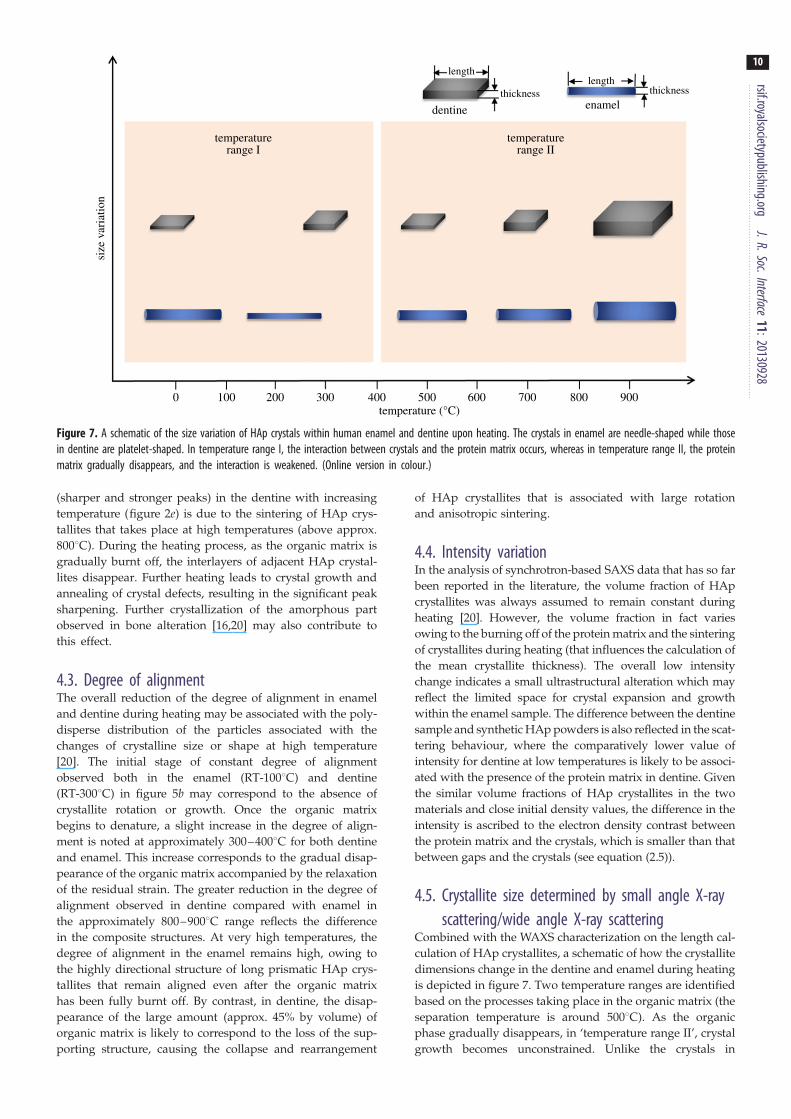
0 100 200 300 400temperature (°C)
temperaturerange I
temperaturerange II
dentine enamel
lengthlength
thickness thicknesssi
ze v
aria
tion
500 600 700 800 900
Figure 7. A schematic of the size variation of HAp crystals within human enamel and dentine upon heating. The crystals in enamel are needle-shaped while thosein dentine are platelet-shaped. In temperature range I, the interaction between crystals and the protein matrix occurs, whereas in temperature range II, the proteinmatrix gradually disappears, and the interaction is weakened. (Online version in colour.)
rsif.royalsocietypublishing.orgJ.R.Soc.Interface
11:20130928
10
(sharper and stronger peaks) in the dentine with increasing
temperature (figure 2e) is due to the sintering of HAp crys-
tallites that takes place at high temperatures (above approx.
8008C). During the heating process, as the organic matrix is
gradually burnt off, the interlayers of adjacent HAp crystal-
lites disappear. Further heating leads to crystal growth and
annealing of crystal defects, resulting in the significant peak
sharpening. Further crystallization of the amorphous part
observed in bone alteration [16,20] may also contribute to
this effect.
4.3. Degree of alignmentThe overall reduction of the degree of alignment in enamel
and dentine during heating may be associated with the poly-
disperse distribution of the particles associated with the
changes of crystalline size or shape at high temperature
[20]. The initial stage of constant degree of alignment
observed both in the enamel (RT-1008C) and dentine
(RT-3008C) in figure 5b may correspond to the absence of
crystallite rotation or growth. Once the organic matrix
begins to denature, a slight increase in the degree of align-
ment is noted at approximately 300–4008C for both dentine
and enamel. This increase corresponds to the gradual disap-
pearance of the organic matrix accompanied by the relaxation
of the residual strain. The greater reduction in the degree of
alignment observed in dentine compared with enamel in
the approximately 800–9008C range reflects the difference
in the composite structures. At very high temperatures, the
degree of alignment in the enamel remains high, owing to
the highly directional structure of long prismatic HAp crys-
tallites that remain aligned even after the organic matrix
has been fully burnt off. By contrast, in dentine, the disap-
pearance of the large amount (approx. 45% by volume) of
organic matrix is likely to correspond to the loss of the sup-
porting structure, causing the collapse and rearrangement
of HAp crystallites that is associated with large rotation
and anisotropic sintering.
4.4. Intensity variationIn the analysis of synchrotron-based SAXS data that has so far
been reported in the literature, the volume fraction of HAp
crystallites was always assumed to remain constant during
heating [20]. However, the volume fraction in fact varies
owing to the burning off of the protein matrix and the sintering
of crystallites during heating (that influences the calculation of
the mean crystallite thickness). The overall low intensity
change indicates a small ultrastructural alteration which may
reflect the limited space for crystal expansion and growth
within the enamel sample. The difference between the dentine
sample and synthetic HAp powders is also reflected in the scat-
tering behaviour, where the comparatively lower value of
intensity for dentine at low temperatures is likely to be associ-
ated with the presence of the protein matrix in dentine. Given
the similar volume fractions of HAp crystallites in the two
materials and close initial density values, the difference in the
intensity is ascribed to the electron density contrast between
the protein matrix and the crystals, which is smaller than that
between gaps and the crystals (see equation (2.5)).
4.5. Crystallite size determined by small angle X-rayscattering/wide angle X-ray scattering
Combined with the WAXS characterization on the length cal-
culation of HAp crystallites, a schematic of how the crystallite
dimensions change in the dentine and enamel during heating
is depicted in figure 7. Two temperature ranges are identified
based on the processes taking place in the organic matrix (the
separation temperature is around 5008C). As the organic
phase gradually disappears, in ‘temperature range II’, crystal
growth becomes unconstrained. Unlike the crystals in

rsif.royalsocietypublishing
11
enamel, it is also observed that the crystallites in dentinegradually become misaligned, as indicated by the drop in
the degree of alignment. Similar phenomena have been
observed in ex situ bone characterization [35].
It is interesting to observe that in the synthetic HAp
powders, continued sintering takes place during high temp-
erature hold and even continues during cooling. This is
probably due to the fact that sintering is driven by the overall
change in the free energy of the system [36].
.orgJ.R.Soc.Interface11:20130928
5. ConclusionThe effects of in situ heat treatment on the hydroxyapatite
(HAp) crystallites (human dental tissues) and synthetic
HAp crystallites were explored using combined synchrotron-
based SAXS/WAXS techniques. From the quantitative analysis
of SAXS and WAXS patterns, multi-scale characterization of the
hierarchical structure of enamel and dentine establishes the
temperature-dependent variation of the nanostructural par-
ameters (thickness, orientation and the degree of alignment)
of HAp crystallites from SAXS and crystallographic properties
(d-spacing, crystal perfection and length) from WAXS during
the heating and cooling processes. The results generally reflect
the difference between dentine and enamel, natural and syn-
thetic HAp crystallites. In the analysis presented, the
emphasis is placed on understanding the role of the organic
matrix during heating. An important observation is made of
the fact that the scattering object may change with the
volume fraction variation of HAp crystallites during heating.
In conclusion, the synchrotron-based combined SAXS/
WAXS analysis has been shown to be a powerful method
for the determination of nanostructure variation induced by
thermal treatment in human enamel and dentine as well as
in synthetic HAp crystallites. The in situ thermal treatment
conducted in this study covered the entire relevant tempera-
ture range and revealed the complete continuous history of
ultrastructure evolution within a single sample during a
continuous heating–cooling schedule. It was clearly demon-
strated that insignificant structural changes occur during
cooling, indicating that relevant conclusions can be drawn
when dental remains samples are analysed as part of archae-
ological or forensic studies. The approach to the study of
ultrastructural alteration in skeletal hard tissues exposed to
in situ thermal treatment developed in this study provides a
better, more reliable basis for deducing the heating history
compared with conventional methods based on the moni-
toring the macro- and microstructural colour [6,7,37].
Furthermore, it allows the interaction between the mineral
crystallites and the organic phase during heating to be cap-
tured. These advantages show the superior utility of in situthermal treatment analysis over previous ex situ tests. Statisti-
cal error analysis of multiple measurement points in dentine
confirmed the reliability of the conclusions drawn. The
results of this work will also be beneficial to the optimization
of the laser fluence used in dental practice, and in the future
design of biomimetic materials.
Acknowledgement. Diamond Light Source is acknowledged for provid-ing the beam time allocation under experiment number SM7981.
Funding statement. A.M.K. acknowledges support of the EPSRC throughgrants EP/I020691 ‘Multi-disciplinary Centre for In situ ProcessingStudies (CIPS)’, EP/G004676 ‘Micromechanical Modelling and Exper-imentation’ and EP/H003215 ‘New Dimensions of EngineeringScience at Large Facilities’.
References
1. Marshall SJ, Balooch M, Habelitz S, Balooch G,Gallagher R, Marshall GW. 2003 The dentin –enamel junction: a natural, multilevel interface.J. Eur. Ceram. Soc. 23, 2897 – 2904. (doi:10.1016/S0955-2219(03)00301-7)
2. Dorozhkin SV. 2010 Nanosized and nanocrystallinecalcium orthophosphates. Acta Biomater. 6,715 – 734. (doi:10.1016/j.actbio.2009.10.031)
3. Kantola S. 1973 Laser-induced effects on toothstructure 0.8. X-ray-diffraction study of dentinexposed to a CO2-laser. Acta Odontol. Scand. 31,381 – 386. (doi:10.3109/00016357309002525)
4. Fried D, Zuerlein MJ, Le CQ, Featherstone JDB. 2002Thermal and chemical modification of dentin by9 – 11-mm CO2 laser pulses of 5 – 100-ms duration.Laser Surg. Med. 31, 275 – 282. (doi:10.1002/Lsm.10100)
5. Zuerlein MJ, Fried D, Featherstone JDB. 1999Modeling the modification depth of carbon dioxidelaser-treated dental enamel. Laser Surg. Med. 25,335 – 347. (doi:10.1002/(Sici)1096-9101(1999)25:4,335::Aid-Lsm8.3.0.Co;2-F)
6. Piga G, Thompson TJU, Malgosa A, Enzo S. 2009 Thepotential of X-ray diffraction in the analysis of burnedremains from forensic contexts. J. Forensic Sci. 54,534 – 539. (doi:10.1111/j.1556-4029.2009.01037.x)
7. Thompson TJU. 2005 Heat-induced dimensionalchanges in bone and their consequences for forensicanthropology. J. Forensic Sci. 50, 1008 – 1015.(doi:10.1520/JFS2004297)
8. Thompson TJU, Islam M, Piduru K, Marcel A. 2011An investigation into the internal and externalvariables acting on crystallinity index usingFourier transform infrared spectroscopy onunaltered and burned bone. Palaeogeogr.Palaeoclimatol. 299, 168 – 174. (doi:10.1016/j.palaeo.2010.10.044)
9. Sui T, Sandholzer MA, Le Bourhis E, Baimpas N, LandiniG, Korsunsky AM. 2014 Structure – mechanical functionrelations at nano-scale in heat-affected human dentaltissue. J. Mech. Behav. Biomed. Mater. 32, 113 – 124.(doi:10.1016/j.jmbbm.2013.12.014)
10. Shipman P, Foster G, Schoeninger M. 1984 Burntbones and teeth: an experimental-study of color,morphology, crystal-structure and shrinkage.J. Archaeol. Sci. 11, 307 – 325. (doi:10.1016/0305-4403(84)90013-X)
11. Enzo S, Bazzoni M, Mazzarello V, Piga G, Bandiera P,Melis P. 2007 A study by thermal treatment andX-ray powder diffraction on burnt fragmented bonesfrom tombs II, IV and IX belonging to the hypogeicnecropolis of ‘Sa Figu’ near Ittiri, Sassari (Sardinia,
Italy). J. Archaeol. Sci. 34, 1731 – 1737. (doi:10.1016/j.jas.2006.12.011)
12. Rogers KD, Daniels P. 2002 An X-ray diffractionstudy of the effects of heat treatment onbone mineral microstructure. Biomaterials23, 2577 – 2585. (doi:10.1016/S0142-9612(01)00395-7)
13. Beckett S, Rogers KD, Clement JG. 2011 Inter-species variation in bone mineral behavior uponheating. J. Forensic Sci. 56, 571 – 579. (doi:10.1111/j.1556-4029.2010.01690.x)
14. Reyes-Gasga J et al. 2008 Structural and thermalbehaviour of human tooth and three synthetichydroxyapatites from 20 to 600 degrees C. J. Phys.D, Appl. Phys. 41, 225407. (doi:10.1088/0022-3727/41/22/225407)
15. Hollund HI, Ariese F, Fernandes R, Jans MME,Kars H. 2013 Testing an alternative high-throughputtool for investigating bone diagenesis: FTIR inattenuated total reflection (ATR) mode. Archaeometry55, 507 – 532. (doi:10.1111/j.1475-4754.2012.00695.x)
16. Rogers K, Beckett S, Kuhn S, Chamberlain A,Clement J. 2010 Contrasting the crystallinityindicators of heated and diagenetically altered bonemineral. Palaeogeogr. Palaeocl. 296, 125 – 129.(doi:10.1016/j.palaeo.2010.06.021)
rsif.royalsocietypublishing.orgJ.R.Soc.Interface
11:20130928
12
17. Squires KE, Thompson TJU, Islam M, Chamberlain A.2011 The application of histomorphometry andFourier transform infrared spectroscopy to theanalysis of early Anglo-Saxon burned bone.J. Archaeol. Sci. 38, 2399 – 2409. (doi:10.1016/j.jas.2011.04.025)18. Piga G, Malgosa A, Thompson TJU, Enzo S. 2008A new calibration of the XRD technique for the studyof archaeological burned human remains. J. Archaeol.Sci. 35, 2171 – 2178. (doi:10.1016/j.jas.2008.02.003)
19. Hiller JC, Wess TJ. 2006 The use of small-angleX-ray scattering to study archaeological andexperimentally altered bone. J. Archaeol. Sci. 33,560 – 572. (doi:10.1016/j.jas.2005.09.012)
20. Hiller JC, Thompson TJ, Evison MP, Chamberlain AT,Wess TJ. 2003 Bone mineral change duringexperimental heating: an X-ray scatteringinvestigation. Biomaterials 24, 5091 – 5097. (doi:10.1016/S0142-9612(03)00427-7)
21. Sui T et al. 2013 Multi-scale modelling anddiffraction-based characterization of elasticbehaviour of human dentine. Acta Biomater.9, 7937 – 7947. (doi:10.1016/j.actbio.2013.04.020)
22. Sui T, Sandholzer MA, Baimpas N, Dolbnya IP, LandiniG, Korsunsky AM. 2013 Hierarchical modelling ofelastic behaviour of human enamel based onsynchrotron diffraction characterization. J. Struct. Biol.184, 136 – 146. (doi:10.1016/j.jsb.2013.09.023)
23. Dent AJ et al. 1995 A new furnace design for use incombined X-ray-absorption and diffraction of
catalysis and ceramics studies: formation fromcarbonate precursors of Cu, Co, Mn spinels for theoxidation of CO and the formation of Plzt, apiezoelectric ceramic. Nucl. Instrum. Methods B 97,20 – 22. (doi:10.1016/0168-583X(94)00369-6)
24. Huang TC, Toraya H, Blanton TN, Wu Y. 1993 X-ray-powder diffraction analysis of silver behenate, apossible low-angle diffraction standard. J. Appl.Cryst. 26, 180 – 184. (doi:10.1107/S0021889892009762)
25. Deymier-Black AC, Almer JD, Stock SR, Haeffner DR,Dunand DC. 2010 Synchrotron X-ray diffractionstudy of load partitioning during elastic deformationof bovine dentin. Acta Biomater. 6, 2172 – 2180.(doi:10.1016/j.actbio.2009.11.017)
26. Hammersley AP. 1997 ‘FIT2D: an introduction andoverview’. ESRF Internal Report 1997.
27. Glatter O, Kratky O. 1982 Small angle X-rayscattering. London, UK: Academic Press.
28. Tanaka T, Yagi N, Ohta T, Matsuo Y, Terada H,Kamasaka K, To-o K, Kometani T, Kuriki T. 2010Evaluation of the distribution and orientation ofremineralized enamel crystallites in subsurfacelesions by X-ray diffraction. Caries Res. 44,253 – 259. (doi:10.1159/000314672)
29. Tesch W, Vandenbos T, Roschgr P, Fratzl-Zelman N,Klaushofer K, Beertsen W, Fratzl P. 2003 Orientation ofmineral crystallites and mineral density during skeletaldevelopment in mice deficient in tissue nonspecificalkaline phosphatase. J. Bone Miner. Res. 18,117 – 125. (doi:10.1359/jbmr.2003.18.1.117)
30. Tesch W, Eidelman N, Roschger P, Goldenberg F,Klaushofer K, Fratzl P. 2001 Graded microstructureand mechanical properties of human crown dentin.Calcified Tissue Int. 69, 147 – 157. (doi:10.1007/s00223-001-2012-z)
31. Ooi CY, Hamdi M, Ramesh S. 2007 Properties ofhydroxyapatite produced by annealing of bovinebone. Ceram. Int. 33, 1171 – 1177. (doi:10.1016/j.ceramint.2006.04.001)
32. Kugler M. 2003 X-ray diffraction analysis in forensicscience: the last resort in many criminal cases. Adv.X-ray Anal. 46, 1 – 16.
33. Lei HJ, Liu B, Fang DN. 2010 The coefficient ofthermal expansion of biomimetic composite. Front.Mater. Sci. China 4, 234 – 238. (doi:10.1007/s11706-010-0088-y)
34. Elliott JC. 1994 Structure and chemistry of theapatites and other calcium orthophosphates.Amsterdam, The Netherlands: Elsevier.
35. Pramanik S, Hanif ASM, Pingguan-Murphy B, AbuOsman NA. 2013 Morphological change of heattreated bovine bone: a comparative study. Materials6, 65 – 75. (doi:10.3390/Ma6010065)
36. Kingery WD, Bowen HK, Uhlmann DR. 1976Introduction to ceramics, 2nd edn. New York, NY:Wiley.
37. Thompson TJU, Islam M, Bonniere M. 2013 A newstatistical approach for determining the crystallinityof heat-altered bone mineral from FTIR spectra.J. Archaeol. Sci. 40, 416 – 422. (doi:10.1016/j.jas.2012.07.008)
Copyright © 2022 FDOKUMEN