In situ localization of type III and type IV collagen-expressing cells in human diabetic nephropathy
8
JOURNAL OF PATHOLOGY, VOL. 174: 131-138 (1994) IN SITU LOCALIZATION OF TYPE I11 AND TYPE DIABETIC NEPHROPATHY IV COLLAGEN-EXPRESSING CELLS IN HUMAN MOHAMMED S. RAZZAQUE, TAKEHIKO KOJI*, TAKASHI TAGUCHI, TAKASHI HARADAt AND PAUL K. NAKANE* Second Department of Pathology, *Third Department of Anatomy, and ?Second Department of Medicine, Nagasaki University School of Medicine, 12-4, Sakamoto machi, Nagasaki 852, Japan Received 1 March 1994 Accepted 6 July 1994 SUMMARY Nodular intercapillary glomerulosclerosis is the most typical lesion of diabetic nephropathy (DN) and is characterized by increased extracellular matrix (ECM) and amorphous masses of mesangial matrix. The local exaggeration of these deposits results in the formation of the typical diabetic nodule. To clarify the composition of the ECM of sclerotic lesions in DN, we investigated the distribution of type I11 and type IV collagens and their mRNAs by immunohistochemistry and in situ hybridization, respectively. In normal renal tissues, there was no intraglomerular immunostaining for type 111 collagen, while strongly positive staining was found in the extraglom- erular interstitium. Positive immunostaining for type 1V collagen was also present in the mesangium, glomerular basement membrane (GBM), Bowman’s capsule, and the vascular pole of the normal glomerulus. In DN, the nodular lesions were negative for type Ill collagen and strongly positive for type IV collagen. On the other hand, in the late stage of global sclerosis, both type Ill and type IV collagens were diffusely present in the sclerotic matrix. To determine the origins of these type I11 and type IV collagens in the sclerotic matrix, in situ hybridization was performed, utilizing thymine-thymine (T-T) dimerized synthetic oligonucleotides complementary to either pro al(II1) chain or pro ul(1V) chain mRNAs as probes. The signals were detected by enzyme immunohistochemistry using an anti-T-T antibody. Intraglomerular cells (glomerular epithelial and mesangial cells) containing type III collagen mRNA were found in DN with sclerotic lesions, but not in normal glomeruli. At this stage of sclerosis, intraglomerular cells (mainly glomerular epithelial cells and infrequently mesangial cells) were positive for type IV collagen mRNA, but there were few positive cells in globally sclerotic glomeruli. This study provides evidence that both type 111 and type IV collagens are synthesized by intraglomerular cells during sclerosis and become significant constituents of the sclerotic matrix in DN. KEY WORDS-Diabetic nephropathy, type 111 collagen, type IV collagen, immunohistochemistry, non-radioactive in situ hybridization. INTRODUCTION The pathogenetic mechanisms responsible for the initiation and progression of diabetic neph- ropathy (DN) are not clearly understood, although about 40 per cent of patients with diabetes mellitus (DM) develop nephropathy.’ The disease is hallmarked by progressive thickening of the glomerular basement membrane (GBM) and a Addressee for correspondence: Takashi Taguchi, Second Department of Pathology, Nagasaki University School of Medicine, 12-4, Sakamoto machi, Nagasaki 852, Japan. 0 1994 by John Wiley & Sons, Ltd. CCC 0022-3417/94/100 131-08 disproportionate increase in mesangial volume. Morphometrically, the matrix increase is about three times that of mesangial cells in human DN.2 Falk et al.3 demonstrated increased immu- nostaining for type IV collagen, laminin, and fibronectin, all of which are normal constituents of the glomerulus. In a separate study, Kim et a1.4 described increased a1 and a2 chains of type IV collagen in the mesangial matrix in the early to middle stages of human DN, while at the end stage, these chains were present in diminished amounts. By competitive polymerase chain reaction, Peten et al. also reported an elevation
-
Upload
independent -
Category
Documents
-
view
0 -
download
0
Transcript of In situ localization of type III and type IV collagen-expressing cells in human diabetic nephropathy
JOURNAL OF PATHOLOGY, VOL. 174: 131-138 (1994)
IN SITU LOCALIZATION OF TYPE I11 AND TYPE
DIABETIC NEPHROPATHY IV COLLAGEN-EXPRESSING CELLS IN HUMAN
MOHAMMED S. RAZZAQUE, TAKEHIKO KOJI*, TAKASHI TAGUCHI, TAKASHI HARADAt AND PAUL K . NAKANE*
Second Department of Pathology, *Third Department of Anatomy, and ?Second Department of Medicine, Nagasaki University School of Medicine, 12-4, Sakamoto machi, Nagasaki 852, Japan
Received 1 March 1994 Accepted 6 July 1994
SUMMARY Nodular intercapillary glomerulosclerosis is the most typical lesion of diabetic nephropathy (DN) and is
characterized by increased extracellular matrix (ECM) and amorphous masses of mesangial matrix. The local exaggeration of these deposits results in the formation of the typical diabetic nodule. To clarify the composition of the ECM of sclerotic lesions in DN, we investigated the distribution of type I11 and type IV collagens and their mRNAs by immunohistochemistry and in situ hybridization, respectively. In normal renal tissues, there was no intraglomerular immunostaining for type 111 collagen, while strongly positive staining was found in the extraglom- erular interstitium. Positive immunostaining for type 1V collagen was also present in the mesangium, glomerular basement membrane (GBM), Bowman’s capsule, and the vascular pole of the normal glomerulus. In DN, the nodular lesions were negative for type Il l collagen and strongly positive for type IV collagen. On the other hand, in the late stage of global sclerosis, both type I l l and type IV collagens were diffusely present in the sclerotic matrix. To determine the origins of these type I11 and type IV collagens in the sclerotic matrix, in situ hybridization was performed, utilizing thymine-thymine (T-T) dimerized synthetic oligonucleotides complementary to either pro al(II1) chain or pro ul(1V) chain mRNAs as probes. The signals were detected by enzyme immunohistochemistry using an anti-T-T antibody. Intraglomerular cells (glomerular epithelial and mesangial cells) containing type III collagen mRNA were found in DN with sclerotic lesions, but not in normal glomeruli. At this stage of sclerosis, intraglomerular cells (mainly glomerular epithelial cells and infrequently mesangial cells) were positive for type IV collagen mRNA, but there were few positive cells in globally sclerotic glomeruli. This study provides evidence that both type 111 and type IV collagens are synthesized by intraglomerular cells during sclerosis and become significant constituents of the sclerotic matrix in DN.
KEY WORDS-Diabetic nephropathy, type 111 collagen, type IV collagen, immunohistochemistry, non-radioactive in situ hybridization.
INTRODUCTION
The pathogenetic mechanisms responsible for the initiation and progression of diabetic neph- ropathy (DN) are not clearly understood, although about 40 per cent of patients with diabetes mellitus (DM) develop nephropathy.’ The disease is hallmarked by progressive thickening of the glomerular basement membrane (GBM) and a
Addressee for correspondence: Takashi Taguchi, Second Department of Pathology, Nagasaki University School of Medicine, 12-4, Sakamoto machi, Nagasaki 852, Japan.
0 1994 by John Wiley & Sons, Ltd. CCC 0022-3417/94/100 13 1-08
disproportionate increase in mesangial volume. Morphometrically, the matrix increase is about three times that of mesangial cells in human DN.2 Falk et al.3 demonstrated increased immu- nostaining for type IV collagen, laminin, and fibronectin, all of which are normal constituents of the glomerulus. In a separate study, Kim et a1.4 described increased a1 and a2 chains of type IV collagen in the mesangial matrix in the early to middle stages of human DN, while at the end stage, these chains were present in diminished amounts. By competitive polymerase chain reaction, Peten et al. also reported an elevation
132 M. S. RAZZAQUE ET AL.
of mRNA for tyqe a2 IV collagen in human glomeru1osclerosis.-
It appears to be, however, that not all compo- nents of the extracellular matrix (ECM) are equally increased. The mRNA for the B1 chain of laminin and the heparan sulphate proteoglycans (HSPs) in the capillary basement membrane were unchanged, even though type IV collagen was increased.6 The immunochemically detectable amounts of type IV collagen increased, although the amount of laminin and HSPs present in the GBM of DM was reduced, by 40 per cent for laminin, compared with that found in normal GBM.' The uneven composition of the GBM in DN is thought to be a result of hyperglycaemia, which greatly accelerates the non-enzymatic glyco- sylation of GBM collagen' and alters the cross- linking with other glycosylated GBM constituents, such as laminin and HSPs. The alteration leads to degradation of both laminin and HSPS.~ In addi- tion to the changes in normal constituents, addi- tional ECM components may appear in diabetic glomerular lesions, focal glomerulosclerosis, and several other types of human glomerulonephritis; Glick et al., using immunological and ultrastruc- tural methods, demonstrated type I collagen in the mesangial sclerotic lesions in human DN and focal glomerulosclerosis. On the other hand, in several other types of glomerular disease, type I11 collagen appeared as an additional component of the ECM in the glomeruli."-14 In a separate study, we have also demonstrated de n o w intraglomerular synthesis of type 111 collagen in human benign nephrosclerosis. l 5
As DN is characterized by progressive expan- sion of the mesangial matrix and a thickened GBM, we set out to determine whether the ECM in sclerotic glomeruli in DM also accumulates some extraglomerular connective tissue elements, such as type 111 collagen. In this study, we have investigated the expression of both intraglomeru- lar collagenous components (type IV collagen) and extraglomerular collagenous components (type 111 collagen) in the process of sclerosis in DM, using immunohistochemistry and non-radioactive in situ hybridization.
MATERIALS AND METHODS Kidney tissues
Renal biopsies from ten patients with the typical histological changes of DN as described in the
WHO classification of glomerular diseases16 and ten samples of renal tissue without any noticeable abnormality were used in this study. Portions of the tissues were processed for routine light and electron microscopic examination. For light microscopy, the tissues were fixed in 10 per cent formalin and embedded in paraffin; for electron microscopy, the tissues were fixed in 2.5 per cent glutaraldehyde, osmicated, and embedded in Epon.
Immunoiiistociietnistry
Paraffin sections (4 pm) were deparaffinized and endogenous peroxidase activity was denatured with 0.3 per cent hydrogen peroxide in methanol. After mild treatment with trypsin, the sections were reacted with non-immune rabbit serum to block non-specific binding of monoclonal antibod- ies. As immunohistochemical controls, the mono- clonal antibodies were replaced by normal mouse serum diluted with phosphate-buffered saline (PBS) in the same proportion as that used for the experiment. Monoclonal antibodies against type I11 collagen and type IV collagen were purchased from Fuji Chemical and Dako, and were diluted to 1:25 (for type I11 collagen) and 1:75 (for type IV collagen) with PBS (pH 7.2). The sections were reacted with the primary antibodies and processed further using ABC kits (Vector Laboratories, Burlingame, CA). The staining intensity of col- lagen type 111 and collagen type IV was graded semi-quantitatively according to the following scale: O=no staining; + =focal staining; + + =weak staining; + + + =strong staining.
In situ hybridization
Oligo-DNA probes used-A 45-base sequence complementary to the mRNA of human pro al(II1) chain (40814126)" and a 45-base sequence complementary to the mRNA of human pro al(1V) chain ( 1008-1053)'s were chosen. A computer-assisted search (GenBank) of the above antisense sequences, as well as the sense sequences, revealed no significant homology with any known sequences other than those of pro al(II1) and pro al(IV) chains. These anti- sense and sense sequences were used for synthetic oligo-DNA probes but for haptenization of the oligo-DNAs with T-T dimers, two and three TTA repeats of the 5'- and 3'-ends of the sequences
COLLAGEN EXPRESSION IN DIABETIC NEPHROPATHY 133
were added, respectively,'9720 at the time of synthe- sis. The oligo-DNAs were synthesized by an auto- matic Applied Biosystem DNA synthesizer (model 391 PCR-MATE, Foster City, CA) and purified on an OPC column and a Nap 10 column (Phar- macia). T-T dimer was introduced into the oligo- DNAs by UV irradiation with a dose of 10 000 J/m2 and the introduction was verified immu- nochemically using mouse monoclonal anti-T-T IgG (Kyowa Medics. Japan); the hybridizabilities of the T-T dimerized oligo-DNA probes were confirmed b dot blot hybridization as described previously. 1220
In situ hybridization procedure-In situ hybrid- ization was carried out as previously described. 19,20 All the in situ hybridization experiments were done in duplicate slides. Briefly, deparaffinized sections (4pm) were treated successively with 0.3 per cent H,O,, 0 . 2 ~ HCl, 0.2 per cent Triton X-100 and proteinase K. The sections were post-fixed with 4 per cent paraformaldehyde (PFA), immersed in 2 mdml glycine, and kept in 40 per cent deionized formamide in 4 x SSC until used.
Hybridization was performed overnight at 37°C with 2pdrnl T-T dimerized antisense oli o
with 10 per cent dextran sulphate and 40 per cent deionized formamide. Then the slides were washed in 50 per cent formamide in 2 x SSC, 2 x SSC, and PBS. Immunohistochemistry was performed on the sections as previously described."920 The sites of peroxidase were visu- alized using a solution containing 3,3'- diaminobenzidine.4 HCl (DAB), H20,, CoCl,, and NiS04(NH4)2S04;2' one of the duplicate slides was counterstained by periodic-acid Schiff (PAS). Adjacent sections were reacted either with the hybridization solution containing T-T dimer- ized sense oligo-DNA probes or the solution without probes as the negative controls. Sections consecutive to these sections were also hybridized with T-T dimerized oligo-DNA complementary to 28s rRNA, to demonstrate the degree of RNA retention and the accessibility of the prqbes to cytoplasmic RNA. To establish the specficity of the hybridization, some consecutive sections were pretreated with RNase or hybridized with a solution containing a 50-fold excess of non- haptenized antisense oligo-DNA probes, in addi- tion to the T-T dimerized antisense oligo-DNA probes.
DNA probes in the hybridization solution' 8. 20 -
RESULTS Irnrnunohistochernical localization of type III collagen and type I V collagen in normal kidney
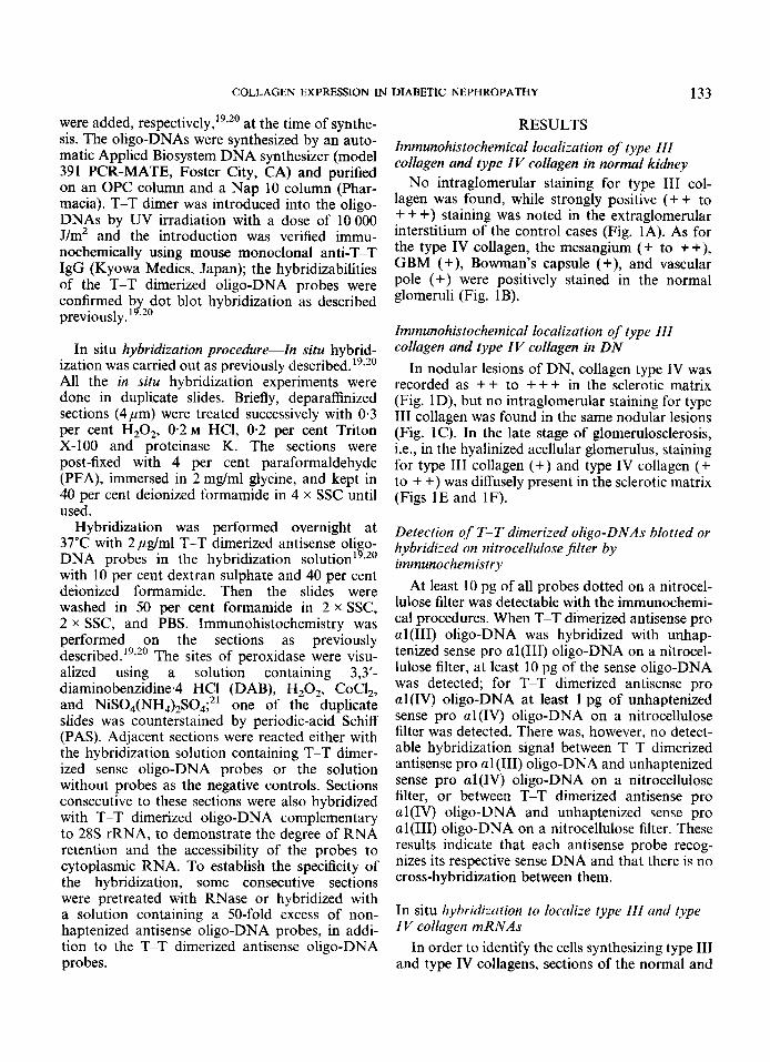
No intraglomerular staining for type I11 col- lagen was found, while strongly positive (+ + to + + +) staining was noted in the extraglomerular interstitium of the control cases (Fig. IA). As for the type IV collagen, the mesangium (+ to ++), GBM (+), Bowman's capsule (+), and vascular pole (+) were positively stained in the normal glomeruli (Fig. 1B).
Irnmunohistochernical localization of type III collagen and type I V collagen in DN
In nodular lesions of DN, collagen type IV was recorded as + + to +++ in the sclerotic matrix (Fig. lD), but no intraglomerular staining for type 111 collagen was found in the same nodular lesions (Fig. 1C). In the late stage of glomerulosclerosis, i.e., in the hyalinized acellular glomerulus, staining for type 111 collagen (+) and type IV collagen (+ to + +) was diffusely present in the sclerotic matrix (Figs 1E and 1F).
Detection of T-T dimerized oligo-DNAs blotted or hybridized on nitrocellulose Jjlter by immunochernistry
At least 10 pg of all probes dotted on a nitrocel- lulose filter was detectable with the immunochemi- cal procedures. When T-T dimerized antisense pro a1 (111) oligo-DNA was hybridized with unhap- tenized sense pro al(II1) oligo-DNA on a nitrocel- lulose filter, at least 10 pg of the sense oligo-DNA was detected; for T-T dimerized antisense pro al(IV) oligo-DNA at least I pg of unhaptenized sense pro al(1V) oligo-DNA on a nitrocellulose filter was detected. There was, however, no detect- able hybridization signal between T-T dimerized antisense pro a1 (111) oligo-DNA and unhaptenized sense pro al(Iv) oligo-DNA on a nitrocellulose filter, or between T-T dimerized antisense pro al(1V) oligo-DNA and unhaptenized sense pro al(II1) oligo-DNA on a nitrocellulose filter. These results indicate that each antisense probe recog- nizes its respective sense DNA and that there is no cross-hybridization between them.
In situ hybridization to localize type III and type IV collagen mRNAs
In order to identify the cells synthesizing type I11 and type IV collagens, sections of the normal and
134 M. S. RAZZAQUE ET AL.
COLLAGEN EXPRESSION IN DIABETIC NEPHROPATHY 135
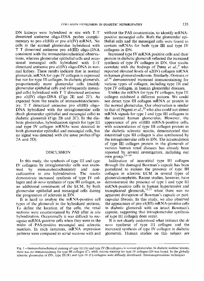
DN kidneys were hybridized in situ with T-T dimerized antisense oligo-DNA probes comple- mentary to pro al(II1) or pro al(1V) mRNA. No cells in the normal glomerulus hybridized with T-T dimerized antisense pro al(II1) oligo-DNA consistent with the immunohis tochemical observa- tions, whereas glomerular epithelial cells and occa- sional mesangial cells hybridized with T-T dimerized antisense pro a1 (IV) oligo-DNA in nor- mal kidney. These results indicate that in normal glomeruli, mRNA for type IV collagen is expressed but not for type 111 collagen. In diabetic glomeruli, proportionally more glomerular cells (mainly glomerular epithelial cells and infrequently mesan- gial cells) hybridized with T-T dimerized antisense pro al(1V) oligo-DNA (Figs 2E and 2F). As expected from the results of immunohistochemis- try, T-T dimerized antisense pro al(II1) oligo- DNA hybridized with the intraglomerular cells (both glomerular epithelial and mesangial cells) in diabetic glomeruli (Figs 2B and 2C). In the dia- betic glomerulus, hybridization signals for type 111 and type IV collagen niRNAs were detected in both glomerular epithelial and mesangial cells, but no signal was detected with the sense probes (Figs 2A and 2D).
DISCUSSION
In this study, the synthesis of type I11 and type IV collagens by intraglomerular cells was exam- ined by immunohistochemistry and non- radioactive in situ hybridization. The results demonstrate increased synthesis of type IV col- lagen and de novo synthesis of type I11 collagen, as an additional constituent of the ECM, by both glomerular epithelial and mesangial cells during the progression of sclerosis in DN.
It is hard to analyse the mRNA-positive cell types of the glomeruli in the hybridized sections. To define the location of the cells, the renal sections were counterstained by PAS after in situ hybridization. Occasionally it was difficult to rec- ognize mRNA-positive cells when they were in the midst of PAS-positive mesangial and sclerotic matrices. In such instances, mRNA expression patterns were compared in serial sections with and
without the PAS counterstain, to identify mRNA- positive mesangial cells. Both the glomerular epi- thelial cells and the mesangial cells were found to contain mRNAs for both type I11 and type IV collagens in DN.
Increased type IV mRNA positive cells and their protein in diabetic glomeruli reflected the increased synthesis of type IV collagen in DN. Our results correlate with the findings of Peten et d.,’ who reported elevated levels of a2(IV) collagen mRNA in human glomerulosclerosis. Similarly, Oomura rt ~ l . ‘ ~ demonstrated increased immunostaining for various types of collagen, including type 111 and type IV collagen, in human glomerular diseases.
Unlike the mRNA for type IV collagen, type 111 collagen exhibited a different pattern. We could not detect type 111 collagen mRNA or protein in the normal glomerulus. Our observation is similar to that of Prigent et n1.,22 who also could not detect mRNA signals for type I and type 111 collagens in the normal human glomerulus. However, the appearance of pro cnl(II1) mRNA-positive cells. with accumulation of type 111 collagen protein in the diabetic sclerotic matrix, demonstrated that interstitial type 111 collagen is also synthesized by the intraglomerular cells in DN. The accumulation of type 111 collagen protein in the glomeruli of various human renal diseases has already been reported by several investigators, including our own group.”-14
Infiltration of interstitial type 111 collagen through the damaged Bowman’s capsule has been postulated to explain the presence of type 111 collagen in sclerotic ECM in several types of glomerulonephritis. Recent studies, however, have demonstrated the presence of type I and type 111 mRNA-positive cells in human hypertensive and transplanted glomeruli,’s3” when there was no apparent disruption of Bowman’s capsule or peri- capsular fibrosis. In this study, we also observed the appearance of pro aI(II1) mRNA-positive cells in diabetic glomeruli with an intact Bowman’s capsule, suggesting that intraglomerular synthesis of type 111 collagen does occur.
It is not clearly understood what initiates the rlt. now synthesis of type 111 collagen and the increased synthesis of type IV collagen in diabetic glomeruli. Human studies on this subject are
Fig. 1 -11mmt1noIiistocheni1cal staining of type 111 (A) and type IV (B) collagens in normal glomerulus. In diabetic nodular lesions, there was no immunostaining for type Ill collagen (C) , while intense staining for type IV collagen (D) was found. I n the globally sclerotic glomerulus of DN, type 111 (E) and type IV (F) collagens were diffusely distributed. (Immunoperoxidase technique)
136 M. S. RAZZAQUE ET AL.
Fig. 2-In situ hybridization using type 111 collagen sense (A), type 111 collagen antisense (B, C), type IV collagen sense (D), and type IV collagen antisense (E, F) oligo-DNA probes, showing positive epithelial cells (small arrow-head) in the glomerulus of DN. High power view of the glomerulus (C and F) reveals mRNA positive mesangial cells, within the PAS-positive mesangial matrix (large arrow-head). (PAS counterstain)
sparse. From experimental studies, however, there are indications that some glomerular cells in altered environments can produce both types of collagen. Rat mesangial cells, when placed in an
in vitro environment, produce interstitial collagens, including type I11 collagen.23 Rat glomerular epi- thelial cells also produce increased matrix compo- nents, such as type IV and fibronectin, following
COLLAGEN EXPRESSION IN DIABETIC NEPHROPATHY 137
exposure to TGF-P1 .24 An elevated glucose envi- ronment is also known to influence the composi- tion of the ECM.25 Hyperglycaemia elevates the levels of glomerular mRNAs, including that encodin type IV collageq6 TGF-o,26 and TIMP 2 mRNA!7 in experimental diabetic kidneys. Poss- ibly through the elevation of mRNAs, mesangial cells grown in a high glucose medium accumulate more fibronectin, laminin, and type IV collagen than cells grown in a low glucose medium.28 All these observations, together with the current results, suggest that the hyperglycaemic environ- ment of DM induced the synthesis of type I11 collagen as well as increasing the synthesis of type IV collagen and subsequently contributed to the sclerosis in DN.
To localize the specific mRNA sequence at the individual cell level, we found that non-radioactive in situ hybridization utilizing T-T dimerized syn- thetic oligo-DNA as a probe on renal biopsy material was a reliable and sensitive technique. As histological examination of the renal biopsy is already the most common diagnostic method, the use of in situ hybridization on renal biopsies should be able to provide valuable information at the molecular level in a variety of pathological conditions.
In conclusion, the cells in diabetic glomeruli synthesize both type I11 and type IV collagens. Overproduction of these collagens contributes significantly to the sclerotic process in DN.
ACKNOWLEDGEMENTS
We thank Drs A. Yoshii and Y. Shibata for their excellent technical assistance during this study. We also gratefully acknowledge the kind help of Mr T. Matsumoto, Mrs Y. Yamashita, and Mrs S. Nakanose.
REFERENCES
Anderson AR, Christiansen JS, Anderson JK, Kreinen S, Deckert K. Diabetic nephropathy in type I (insulin dependent) diabetes: an epidemiological study. Diabetologia 1985; 2 5 496501, Steffens MW, Bilous RW, Sutherland DER, Mauer SM. Cell and matrix components of the glomerular mesangium in type I diabetes. Diabetes 1992; 41: 679-684. Fdlk RJ, Scheinman JL, Mauer SM, Michael AF. Polyanti- genic expansion of basement membrane constituents in diabetic nephropathy. Diabetes 1983; 3 2 3439. Kim Y, Kleppel MM, Butkoski RJ, Ma& SM, Wieslander J, Michael AF. Differential expression of basement membrane col- lagen chains in diabetic nephropathy. Am J Pathol 1991; 138: 4 I 3420.
5. Peten EP, Striker LJ, Carome MA, Elliot SJ, Yang CW, Striker GE. The contribution of increased collagen synthesis to human glom- erulosclerosis: a quantitative analysis of Q 2 IV collagen mRNA expression by competitive polymerase chain reaction. J Exp Med 1992; 176: 1571-1576.
6. Ledbetter S, Copeland EJ, Noonam D, Vogeli G, Hassell JR. Altered steady-state mRNA levels of basement membrane proteins in diabetic mouse kidneys and thromboxane synthase inhibition. Diabetes 1990; 3 9 196-203.
7. Shimomura H, Spiro RG. Studies on macromolecular compo- nents of human glomerular basement membrane and alteration in diabetes. Diabetes 1987; 35: 1130-1142.
8. Cohen MP, Urdanivia E, Surma M, Wu W. Increased glycosyla- tion of glomerular basement membrane collagen in diabetes. Biochem Biophys Res Commun 1980; 9 5 765-769.
9. Charonis AS, Reger LA, Dege JE, ef al. Laminin alteration after in vitro nonenzymatic glycosylation. Diabetes 1990; 39 807-814.
10. Glick AD, Jacobson HR, Haralson MA. Mesangial deposition of type I collagen in human glomerulosclerosis. Hum Pathol 1992; 23
11. Yoshioka K, Takemura T, Tohda M, et al. Glomerular localization of type 111 collagen in human kidney disease. Kidney Znt 1989; 3 5 1203-1211.
12. Funabiki K, Horikoshi S, Tomino Y, Nagai Y, Koide H. Immuno- histochemical analysis of extracellular components in the glomeru- lar sclerosis of patients with glomerulonephritis. CIin NephroI 1990; 34: 239-246.
13. Razzaque S, Taguchi T. Immunohistochemical study of glomerular sclerosis in benign nephrosclerosis. Acta Histochem Cytochem 1992; 2 5 746 (Abstract).
14. Oomura A, Nakamura T, Arakawa M, Ooshima A, Isemura M. Alteration of extracellular matrix components in human glomerular diseases. Virchows Arch A [Pathol Anat] 1989; 415: 151-159.
15. Razzaque MS, Koji T, Kawano H, Harada T, Nakane PK, Taguchi T. Glomerular expression of type I11 and type IV collagens in benign nephrosclerosis: immunohistochemical and in situ hybridiz- ation study. Pathol Res Pract 1994; 190: 493499.
16. Churg J, Sobin LH. Renal Disease: Classification and Atlas of Glomerular Diseases. 1st edn. Tokyo: Igaku-Shoin, 1982: 226-239.
17. Ala-Kokkao L, Kontusaari S, Baldwin TC, Kuivaniemi H, Prockop DJ. Structure of cDNA clones coding for the entire prepro al(II1) chain of human type I11 procollagen. Difference protein structure from type I procollagen and conservation of codon preferences. Biochem J 1989; 260: 509-516.
18. Braze1 D, Oberbaumer I, Dieringer H, et al. Completion of the amino acid sequence of al chain of human basement mem- brane collagen (type 1V) reveals 21 non-triplet interruptions located within the collagenous domain. Eur J Biochem 1987; 168:
19. Koji T, Nakane PK. Localization in situ of specific mRNA using thymine-thymine dimerized DNA probes: sensitive and reliable non-radioactive in situ hybridization. Acra Pathol Jpn 1990; 40: 793-807.
20. Koji T, Iznmi S, Tanno M, Moriuchi T, Nakane PK. Localization in situ of c-myc mRNA and c-myc protein in adult mouse testis. Histochem J 1988; 20: 551-557.
21. Adams JC. Heavy metal intensification of DAB-based HRP reaction product. J Histochem Cytochem 1981; 2 9 775.
22. Prigent CS, Belair MF, Mandet C, Nochy D, Bariety J, Bruneval P. Expression of type I and 111 collagen in human glomeruli from transplanted kidneys and glomerulonephritides. Pathol Res Pruc 1993; 189 804 (Abstract).
23. Abrass CK, Peterson CV, Raugi GJ. Phenotypic expression of collagen types in mesangial matrix of diabetic and non-diabetic rats. Diabetes 1988; 37: 1695-1702.
24. Nakamura T, Miller D, Rnoslahti E, Border WA. Production of extracellular matrix by glomerular epithelial cells is regu- lated by transforming growth factor PI. Kidney Int 1992; 41: 1213-1221.
1373-1379.
529-536.
138 M. S . RAZZAQUE ET AL.
25. Silhiger S, Crowley S, Shan Z, Brownlee M, Satrino J, Scholondorff 27. Guijarro C, ODonnel MP, Kasiske BL, Kim Y, Kedne WF. D. Nonenzymatic glycation of mesangial matrix and prolonged Glomerular mRNA expression for type IV collagen (COL) and exposure of mesangial matrix to elevated glucose reduces tissue inhibitor of metalloproteinase 2 (TIMP 2) in obese Zucker collagen synthesis and proteoglycan charge. Kidney Int 1993; 43: rats (ZR). J Am Sor Nephrol 1993; 4: 651 (Abstract). 853-864. 28. Ayo SH, Radnik RA, Glass WF 11, et a/. Increased extracellular
26. Nakamura T, Fnkui M, Ehihard I, et a/. mRNA expression of matrix synthesis and mRNA in mesangial cells grown in high growth factors in glomeruli from diabetic rats. Diabetes 1993; 42: glucose medium. Am J Physiol 1991; 26U F185-FI91. 450456.