Improving the Optical Contrast of Backscattering Signal in Reflectance-Based Imaging with Gold...
8
Improving the Optical Contrast of Backscattering Signal in Reflectance-Based Imaging with Gold Nanoshells James C. Y. Kah a , Tzu-Hao Chow b , Malini C. Olivo* c , Beng-Koon Ng b , Razul S. Gulam b , Colin J. R. Sheppard a a Division of Bioengineering, National University of Singapore, Singapore; b School of Electrical and Electronics Engineering, Nanyang Technological University, Singapore; c Division of Medical Sciences, National Cancer Centre Singapore, Singapore ABSTRACT The application of gold nanoparticles as a contrast agent in optical bioimaging is well appreciated, but limited to a narrow excitation range due to its rather invariable optical resonance typically at 520 nm. Compared to gold nanoparticles, the optical response of gold nanoshells can be tuned to match the higher excitation wavelength of many promising clinical reflectance-based imaging modalities such as the optical coherence tomography (OCT). In this study, we demonstrate the tunability of gold nanoshells to improve the optical contrast of backscattering signal under confocal reflectance microscopy and OCT. The gold nanoshells were synthesized and conjugated to antibodies for in vitro demonstration of their selective optical contrast in cancer cells over normal cells under the confocal reflectance microscopy. The OCT signals from these gold nanoshells were compared to that from bare silica cores and intrinsic tissue scattering using 1% Intralipid. We have shown that gold nanoshells are able to elicit an optical contrast to discriminate between cancerous and normal cells under the confocal reflectance microscopy based on differences in molecular markers expression. Compared to bare silica core, the presence of the gold shell is able to effect a higher backscattered OCT signal with an apparent contrast over 1% Intralipid. This contrast can be made to be dependent on the molecular marker expression with antibody specificity. Keywords: gold nanoshells, contrast agent, confocal reflectance, OCT 1. INTRODUCTION Reflectance-based imaging is a class of imaging technique that examines tissue based on reflection or backscattering of excitation light. Amongst the different modalities in this class such as the confocal reflectance microscopy and optical coherence tomography (OCT), OCT is an emerging imaging modality that is gaining widespread attention in recent years. 1 OCT functions in similar manner to ultrasound where it detects tissue structural abnormalities by picking up changes in its backscattering due to mismatches in refractive indices within tissue. Its applicability has been investigated on several different tissue types and has produced images of stromal morphology with quality comparable to histology. 2,3 However, the contrast between neoplastic and normal tissues is often too low to be of any clinical value 4 and the coherence gating process underlying the formation of OCT images renders its detection of incoherent fluorescence processes virtually impossible. This makes OCT insensitive towards imaging molecular changes labeled with a fluorescent probe. The use of OCT for in situ imaging of early molecular changes associated with carcinogenesis before the occurrence of phenotypic changes would thus require alternative optical labelers that are compatible with such reflectance-based imaging modality. Several types of metallic nanostructures have recently been developed and investigated to address the limitations of such imaging techniques based on their optical properties. Among these, gold nanostructures such as gold nanoparticles 5 , gold nanorods 6 and gold nanoshells 7 have been the most widely investigated. These gold nanostructures exhibit surface plasmon resonance (SPR) which allow them to resonantly scatter or absorb light when excited at their SPR frequency. * Correspondence: email [email protected] ; phone (65)64368317; fax (65)63720161 Biophotonics 2007: Optics in Life Science, edited by Jürgen Popp, Gert von Bally, Proc. of SPIE-OSA Biomedical Optics, SPIE Vol. 6633, 66330L, © 2007 SPIE-OSA · 1605-7422/07/$18 SPIE-OSA Vol. 6633 66330L-1
-
Upload
independent -
Category
Documents
-
view
1 -
download
0
Transcript of Improving the Optical Contrast of Backscattering Signal in Reflectance-Based Imaging with Gold...
Improving the Optical Contrast of Backscattering Signal in Reflectance-Based Imaging with Gold Nanoshells
James C. Y. Kaha, Tzu-Hao Chowb, Malini C. Olivo*c, Beng-Koon Ngb, Razul S. Gulamb,
Colin J. R. Shepparda
aDivision of Bioengineering, National University of Singapore, Singapore; bSchool of Electrical and Electronics Engineering, Nanyang Technological University, Singapore;
cDivision of Medical Sciences, National Cancer Centre Singapore, Singapore
ABSTRACT
The application of gold nanoparticles as a contrast agent in optical bioimaging is well appreciated, but limited to a narrow excitation range due to its rather invariable optical resonance typically at 520 nm. Compared to gold nanoparticles, the optical response of gold nanoshells can be tuned to match the higher excitation wavelength of many promising clinical reflectance-based imaging modalities such as the optical coherence tomography (OCT). In this study, we demonstrate the tunability of gold nanoshells to improve the optical contrast of backscattering signal under confocal reflectance microscopy and OCT. The gold nanoshells were synthesized and conjugated to antibodies for in vitro demonstration of their selective optical contrast in cancer cells over normal cells under the confocal reflectance microscopy. The OCT signals from these gold nanoshells were compared to that from bare silica cores and intrinsic tissue scattering using 1% Intralipid. We have shown that gold nanoshells are able to elicit an optical contrast to discriminate between cancerous and normal cells under the confocal reflectance microscopy based on differences in molecular markers expression. Compared to bare silica core, the presence of the gold shell is able to effect a higher backscattered OCT signal with an apparent contrast over 1% Intralipid. This contrast can be made to be dependent on the molecular marker expression with antibody specificity.
Keywords: gold nanoshells, contrast agent, confocal reflectance, OCT
1. INTRODUCTION Reflectance-based imaging is a class of imaging technique that examines tissue based on reflection or backscattering of excitation light. Amongst the different modalities in this class such as the confocal reflectance microscopy and optical coherence tomography (OCT), OCT is an emerging imaging modality that is gaining widespread attention in recent years.1 OCT functions in similar manner to ultrasound where it detects tissue structural abnormalities by picking up changes in its backscattering due to mismatches in refractive indices within tissue. Its applicability has been investigated on several different tissue types and has produced images of stromal morphology with quality comparable to histology.2,3
However, the contrast between neoplastic and normal tissues is often too low to be of any clinical value4 and the coherence gating process underlying the formation of OCT images renders its detection of incoherent fluorescence processes virtually impossible. This makes OCT insensitive towards imaging molecular changes labeled with a fluorescent probe. The use of OCT for in situ imaging of early molecular changes associated with carcinogenesis before the occurrence of phenotypic changes would thus require alternative optical labelers that are compatible with such reflectance-based imaging modality.
Several types of metallic nanostructures have recently been developed and investigated to address the limitations of such imaging techniques based on their optical properties. Among these, gold nanostructures such as gold nanoparticles5, gold nanorods6 and gold nanoshells7 have been the most widely investigated. These gold nanostructures exhibit surface plasmon resonance (SPR) which allow them to resonantly scatter or absorb light when excited at their SPR frequency.
* Correspondence: email [email protected]; phone (65)64368317; fax (65)63720161
Biophotonics 2007: Optics in Life Science, edited by Jürgen Popp, Gert von Bally, Proc. of SPIE-OSA Biomedical Optics, SPIE Vol. 6633, 66330L, © 2007 SPIE-OSA · 1605-7422/07/$18
SPIE-OSA Vol. 6633 66330L-1
The application of metallic nanoparticles such as gold nanoparticles as optical labelers in reflectance-based imaging is well appreciated, but limited to a narrow optical excitation range that are appropriate for use with only certain optical systems having matching optical wavelength. This is due to the rather invariable optical plasmon resonance of gold nanoparticles, typically at 520 nm. Although its plasmon resonance is known to vary with size, the plasmon resonance of gold nanoparticles hardly changes by more than 60 nm before their sizes become impractical for biological applications.8 Hence, for those promising optical imaging systems such as OCT that operate in the near infrared (NIR) wavelengths, the optical response afforded by these gold nanoparticles are not matched, resulting in sub-optimal optical performance.
Gold nanoshells are an emerging class of optically tunable metallodielectric core-shell nanostructure consisting of a dielectric core, usually made of silica, surrounded by a thin gold layer.9,10 Unlike gold nanoparticles, the optical response of gold nanoshells can be tuned from visible wavelength range right up to the mid-infrared wavelength by changing the relative dimension of the shell thickness to the core radius to match the excitation wavelength of the light source of interest in different applications.11 Hence, they are versatile for use with potential clinical imaging modalities that operate in the NIR such as the OCT. Besides their optical tunability, gold nanoshells are also able to resonantly scattering light when excited at their surface plasmon resonance to create strong reflective signals, as well as absorb and convert light into thermal energy for thermal destruction.12-14 Apart from their optical properties, these nanostructures also present inert biological activities which make them appropriate for biological applications.15,16 They can also be readily surface functionalized with targeting moieties such as antibodies or peptides to probe for specific cellular biomarkers with high specificity and affinity.9 When coupled with appropriate biomarkers, these gold nanostructures bioconjugates may provide useful optical signal for molecular specific information to assist clinicians in diagnosis of early pre-cancers.
In this study, we develop gold nanoshells and exploit their optical response to improve the optical contrast in backscattering signal for discriminating cancer cells from normal cells based on expression of certain clinically relevant molecular markers in epithelial type cells in vitro. The epidermal growth factor receptor (EGFR) is one such clinically relevant cell surface receptor biomarker used in this study that is overexpressed in vast majority of epithelial cancer but not in normal cells.17 We also demonstrate their ability to map the molecular expression of EGFR in these cells and show the increase in OCT signal from tissue phantoms in the presence of these gold nanoshells.
2. MATERIALS AND METHODS
2.1 Synthesis and characterization of gold nanoshells
Gold nanoshells with a 302 nm diameter silica core surrounded with a gold shell of 28 nm thickness were synthesized in a four step process. The first step involved the synthesis of the dielectric silica core using the commonly used Stöber procedure, which involved the hydrolysis and condensation of tetraethylorthosilicate (TEOS) (Merck) in a sol-gel process.18 The monodispersed silica core was then surface functionalized with a terminal amine group by grafting them with 12 mM aminopropyltriethoxysilane (APTES) (Sigma-Aldrich) under constant heating with vigorous stirring at 80 °C for 1 h to make the surface amenable for deposition of small gold particles. This was then followed by seeding the amine functionalized silica with small gold hydroxide nanoparticles of about 4 nm using the deposition-precipitation (DP) process commonly used in the gold catalytic field to form the seed solution.19,20 The last step involved the reduction of gold hydroxide solution formed from hydrolysis of hydrogen tetrachloroaurate (HAuCl4) (Sigma-Aldrich) at pH 10.1 using potassium carbonate (Sigma-Aldrich) and after an overnight aging. The gold hydroxide solution was reduced by 6.6 mM sodium borohydride (Sigma-Aldrich) under vigorous stirring to form additional gold on the small seed particles until they grew in size and coalesced together to form a complete layer of gold shell. In this reduction step, 10 mM sodium citrate dihydrate (Sigma-Aldrich) was also added to slow the reaction and stabilize the gold nanoshells by acting as a capping agent. The particle size and process of shell growth on the silica seed was imaged under the TEM (Jeol JEM-2100F) operating at 200 kV. The extinction spectra of gold nanoshells at different stages of shell growth were also measured using a UV-Vis spectrophotometer (Shimadzu UV-2401 PC) in the wavelength range from 400 nm to 900 nm. To compare these experimentally measured spectra with theoretical prediction of the spectra based on Mie theory,21 a program for the calculation of optical spectra of core-shell structures based on Mie theory was written in MATLAB. The gold nanoshells were prepared to a final concentration of 5 x 10⁹
particles per ml as determined using absorption spectroscopy and were stored at 4°C when not in use.
SPIE-OSA Vol. 6633 66330L-2
2.2 Conjugation of gold nanoshells with antibodies
The gold nanoshells were conjugated to monoclonal anti-EGFR (Epidermal Growth Factor Receptor) (Santa Cruz Biotechnology, Inc) as our selected cancer biomarker for imaging using 11-mercaptoundecanoic (11-MUA) acid (Sigma-Aldrich) as the linker. The 11-MUA linker dissolved in ethanol was attached to the gold nanoshells by incubating the gold nanoshells (5 x 10⁹ particles per ml) in 5 mM 11-MUA at room temperature for 1 h. The 11-MUA tethered gold nanoshells were then washed in water for at least five times to remove the excess linkers before attaching the anti-EGFR to the other end of the linker via carbodiimide chemistry. Prior to the addition of anti-EGFR, the carboxylic group on the 11-MUA was converted, with the aid of 1-ethyl-3-(3-dimethylaminopropyl)-carbo (EDAC) (Sigma-Aldrich) as an activator, to a succinimide group with greater activity to form amide bond with amine. The 11-MUA tethered gold nanoshells were incubated with 15 mM of N-hydroxysuccinimide (NHS) (Sigma-Aldrich) and 75 mM EDAC for 10 min at room temperature before 1 µg of anti-EGFR was added and the entire mixture incubated at 37 °C for 3 h. The anti-EGFR conjugated gold nanoshells were then washed for at least 5 times in water before redispersing them in 1X PBS (pH 7.4) and stored in 4 °C until further use. The successful conjugation of the anti-EGFR on gold nanoshells was determined using X-ray photon spectroscopy by examining the changes in the S 2p and N 1s spectra (data not shown).
2.3 Cell culture
Both the poorly-differentiated nasopharyngeal epithelium carcinoma CNE2 cell line and the normal human bronchial epithelium (NHBE) cell line were used in this study. The CNE2 cells were grown in RPMI 1640 supplemented with 10% fetal bovine serum, 2 mM glutamine, sodium pyruvate, non-essential amino acids and 100 unit/ml penicillin/streptomycin while the normal cells were grown in fibroblast growth medium (Cambrex Corp.). Both cell lines were grown on coverslip placed in petri dish and maintained at 37oC in humidified 5% CO2 and 95% air atmosphere. To verify the differential expression of EGFR between the normal and cancer cells, fluorescence activated cell sorting (FACS) analysis was performed on both cell lines with a primary mouse anti-EGFR (Santa Cruz Biotechnology, Inc). The FACS analysis shows a high expression of EGFR as a suitable biomarker on the CNE2 cells compared to the NHBE cells (data not shown).
2.4 In vitro cellular imaging
The anti-EGFR conjugated gold nanoshells were incubated with CNE2 cells and NHBE cells as normal control for in vitro demonstration of their selective optical contrast in cancer cells over normal cells under the confocal reflectance microscopy. Prior to imaging, both cell lines were washed in 1X PBS and fixed in 4% paraformaldehyde, before incubating them with 100 µl of the anti-EGFR conjugated gold nanoshells i.e. 5 x 10⁸ particles for 1 h at 37 °C. Following the incubation, the cells on the coverslip were then rinsed thrice with 1X PBS to wash away the excess and unbounded gold nanoshells. The coverslip with cells were then placed on a microscope slide and sealed with nail varnish prior to imaging. The cells were imaged using a confocal microscopy (Carl Zeiss LSM510 Meta) under differential interference contrast (DIC) and confocal reflectance mode with an Olympus 63x oil immersion objective. The confocal reflectance microscopy was performed under 633 nm illumination with a helium-neon laser.
2.5 OCT system setup
A single-mode fiber based Fourier-domain OCT system centered at 800 nm was used for the phantom studies to assess the optical contrast of suspended gold nanoshells. The schematic for the OCT system setup is shown in Fig. 1. The light source used was a Ti:Sapphire (Femtolaser Inc, Integral OCT) laser operating at a centre wavelength of 800 nm with a bandwidth of 120 nm. The power output at the sample was approximately 7 mW. The measured in-focus axial resolution was 4 µm. A low 0.3 NA, 10x microscope objective was used to provide lateral resolution of 9 µm. Instead of having a reference arm in a typical Fourier-domain OCT setup, a cover slip was placed at the sample arm to provide self-referencing. Back scattered light from the samples was collected via the microscope objective and directed to a spectrometer (Ocean Optics HR4000) that was used as a detector to detect the interference fringes that provides the scattering profile of the samples. The spectrum was then output to a computer for digital processing using a routine written in Labview 8.0.
SPIE-OSA Vol. 6633 66330L-3
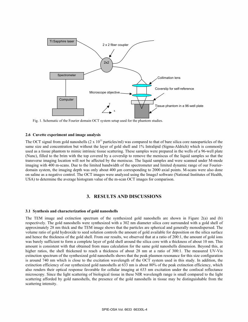
Fig. 1. Schematic of the Fourier domain OCT system setup used for the phantom studies.
2.6 Cuvette experiment and image analysis
The OCT signal from gold nanoshells (2 x 10¹¹ particles/ml) was compared to that of bare silica core nanoparticles of the same size and concentration but without the layer of gold shell and 1% Intralipid (Sigma-Aldrich) which is commonly used as a tissue phantom to mimic intrinsic tissue scattering. These samples were prepared in the wells of a 96-well plate (Nunc), filled to the brim with the top covered by a coverslip to remove the meniscus of the liquid samples so that the transverse imaging location will not be affected by the meniscus. The liquid samples and were scanned under M-mode imaging with 400 m-scans. Due to the limited bandwidth of the spectrometer and limited dynamic range of our Fourier-domain system, the imaging depth was only about 400 µm corresponding to 2000 axial points. M-scans were also done on saline as a negative control. The OCT images were analyzed using the ImageJ software (National Institutes of Health, USA) to determine the average histogram value of the m-scan OCT images for comparison.
3. RESULTS AND DISCUSSIONS
3.1 Synthesis and characterization of gold nanoshells
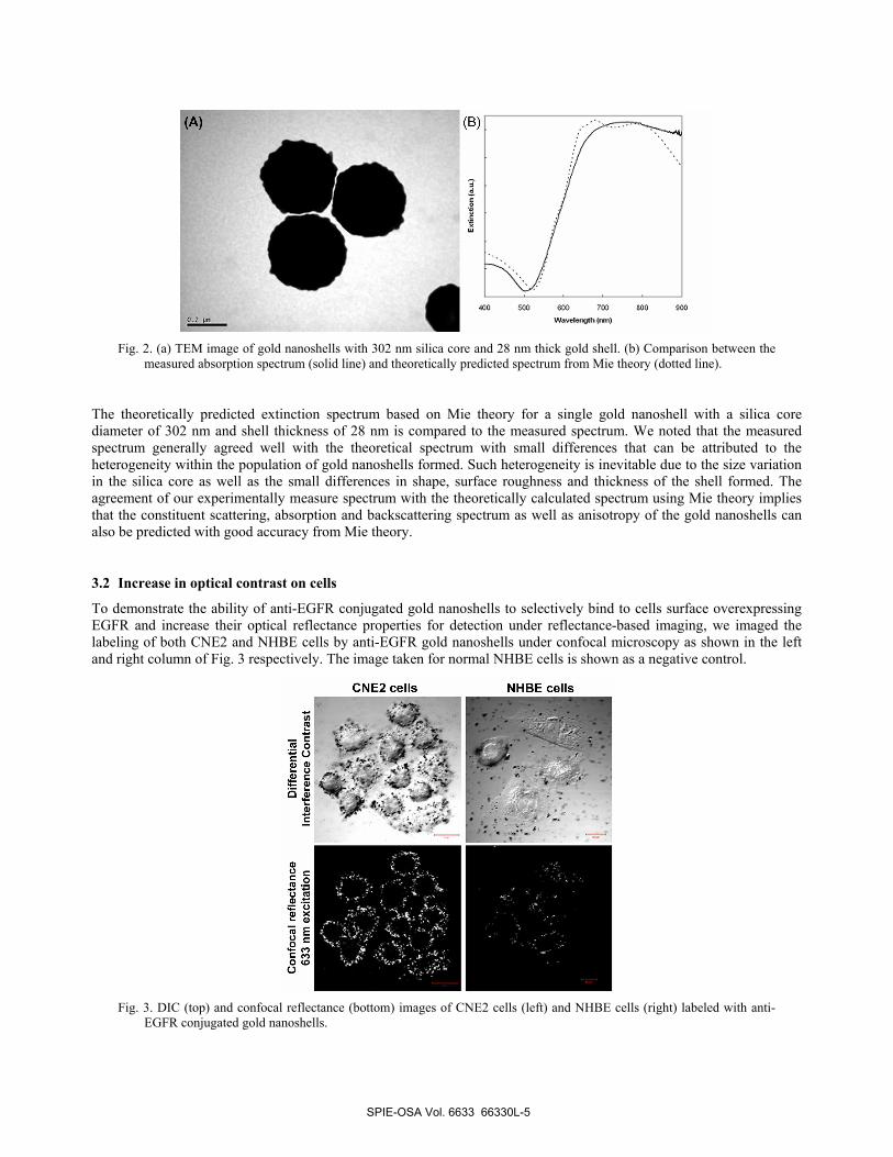
The TEM image and extinction spectrum of the synthesized gold nanoshells are shown in Figure 2(a) and (b) respectively. The gold nanoshells were synthesized with a 302 nm diameter silica core surrounded with a gold shell of approximately 28 nm thick and the TEM image shows that the particles are spherical and generally monodispersed. The volume ratio of gold hydroxide to seed solution controls the amount of gold available for deposition on the silica surface and hence the thickness of the gold shell. From our results, we observed that at a ratio of 200:1, the amount of gold ions was barely sufficient to form a complete layer of gold shell around the silica core with a thickness of about 10 nm. This amount is consistent with that obtained from mass calculation for the same gold nanoshells dimension. Beyond this, at higher ratios, the shell thickened to reach a thickness of about 28 nm at a ratio of 300:1. The measured UV-Vis extinction spectrum of the synthesized gold nanoshells shows that the peak plasmon resonance for this size configuration is around 740 nm which is close to the excitation wavelength of the OCT system used in this study. In addition, the extinction efficiency of our synthesized gold nanoshells at 633 nm is about 80% of the peak extinction efficiency, which also renders their optical response favorable for cellular imaging at 633 nm excitation under the confocal reflectance microscopy. Since the light scattering of biological tissue in these NIR wavelength range is small compared to the light scattering afforded by gold nanoshells, the presence of the gold nanoshells in tissue may be distinguishable from the scattering intensity.
Tissue phantom in a 96-well plate
Collimation lens
Ti:Sapphire laser
Spectrometer
Computer
Microscope objective
2 x 2 fiber coupler
x
y
Coverslip for self-reference
2x2
SPIE-OSA Vol. 6633 66330L-4
400 500 000 700 200 900
Waveleng*h4ml)
Diff
eren
tial
Inte
rfer
ence
Con
tras
t
•.j
*
Fig. 2. (a) TEM image of gold nanoshells with 302 nm silica core and 28 nm thick gold shell. (b) Comparison between the
measured absorption spectrum (solid line) and theoretically predicted spectrum from Mie theory (dotted line).
The theoretically predicted extinction spectrum based on Mie theory for a single gold nanoshell with a silica core diameter of 302 nm and shell thickness of 28 nm is compared to the measured spectrum. We noted that the measured spectrum generally agreed well with the theoretical spectrum with small differences that can be attributed to the heterogeneity within the population of gold nanoshells formed. Such heterogeneity is inevitable due to the size variation in the silica core as well as the small differences in shape, surface roughness and thickness of the shell formed. The agreement of our experimentally measure spectrum with the theoretically calculated spectrum using Mie theory implies that the constituent scattering, absorption and backscattering spectrum as well as anisotropy of the gold nanoshells can also be predicted with good accuracy from Mie theory.
3.2 Increase in optical contrast on cells
To demonstrate the ability of anti-EGFR conjugated gold nanoshells to selectively bind to cells surface overexpressing EGFR and increase their optical reflectance properties for detection under reflectance-based imaging, we imaged the labeling of both CNE2 and NHBE cells by anti-EGFR gold nanoshells under confocal microscopy as shown in the left and right column of Fig. 3 respectively. The image taken for normal NHBE cells is shown as a negative control.
Fig. 3. DIC (top) and confocal reflectance (bottom) images of CNE2 cells (left) and NHBE cells (right) labeled with anti-
EGFR conjugated gold nanoshells.
SPIE-OSA Vol. 6633 66330L-5
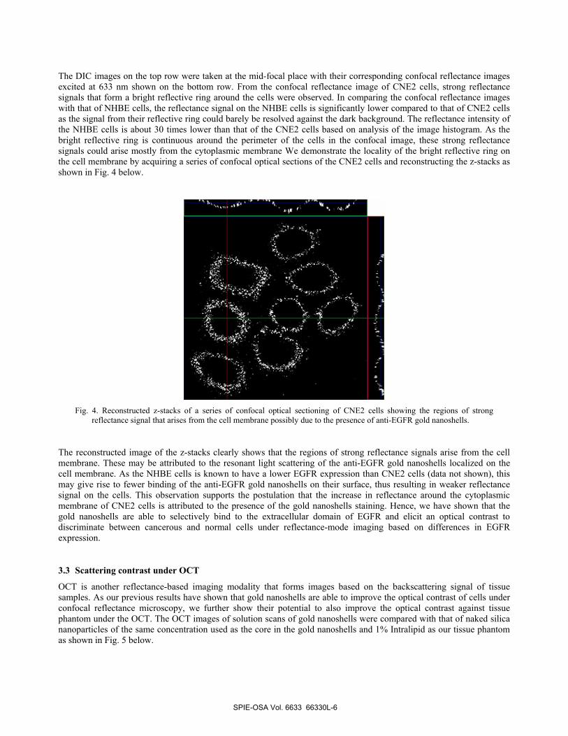
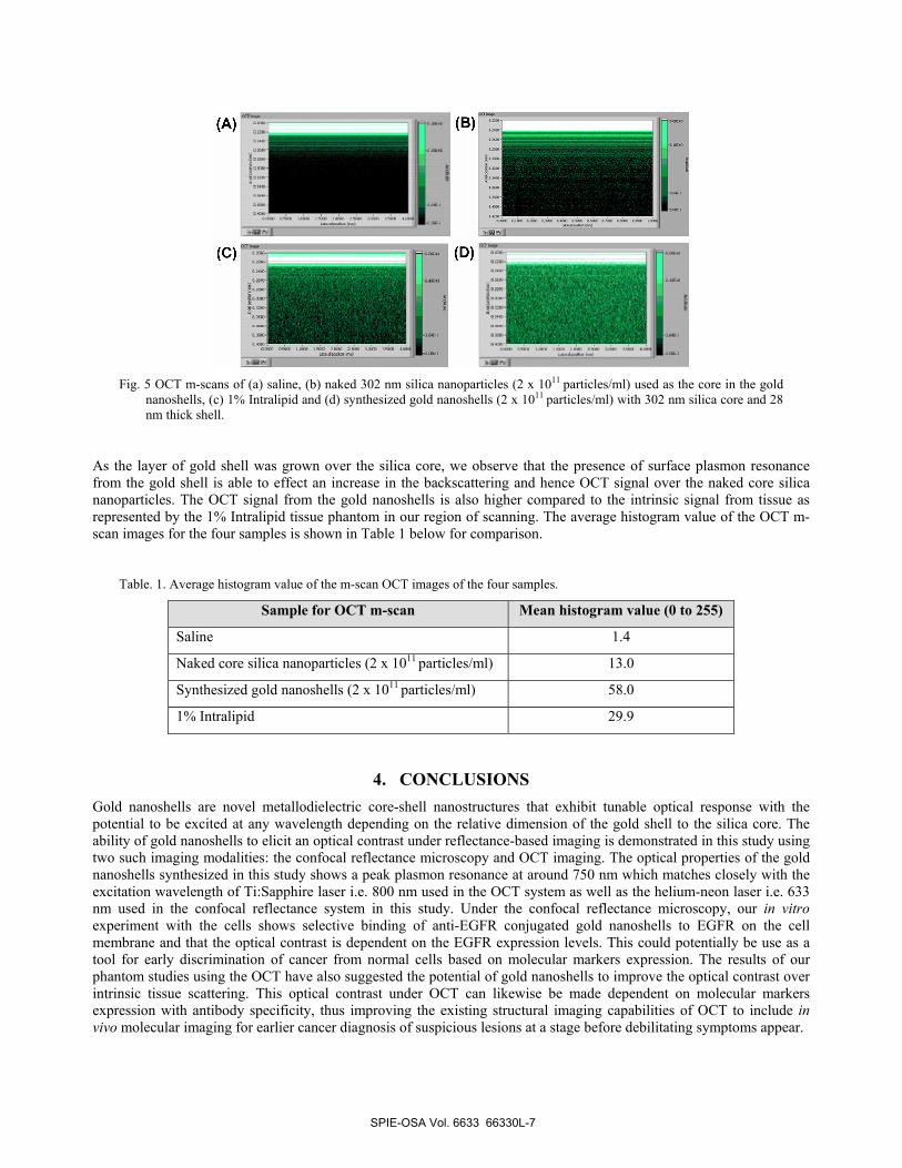
The DIC images on the top row were taken at the mid-focal place with their corresponding confocal reflectance images excited at 633 nm shown on the bottom row. From the confocal reflectance image of CNE2 cells, strong reflectance signals that form a bright reflective ring around the cells were observed. In comparing the confocal reflectance images with that of NHBE cells, the reflectance signal on the NHBE cells is significantly lower compared to that of CNE2 cells as the signal from their reflective ring could barely be resolved against the dark background. The reflectance intensity of the NHBE cells is about 30 times lower than that of the CNE2 cells based on analysis of the image histogram. As the bright reflective ring is continuous around the perimeter of the cells in the confocal image, these strong reflectance signals could arise mostly from the cytoplasmic membrane We demonstrate the locality of the bright reflective ring on the cell membrane by acquiring a series of confocal optical sections of the CNE2 cells and reconstructing the z-stacks as shown in Fig. 4 below.
Fig. 4. Reconstructed z-stacks of a series of confocal optical sectioning of CNE2 cells showing the regions of strong
reflectance signal that arises from the cell membrane possibly due to the presence of anti-EGFR gold nanoshells.
The reconstructed image of the z-stacks clearly shows that the regions of strong reflectance signals arise from the cell membrane. These may be attributed to the resonant light scattering of the anti-EGFR gold nanoshells localized on the cell membrane. As the NHBE cells is known to have a lower EGFR expression than CNE2 cells (data not shown), this may give rise to fewer binding of the anti-EGFR gold nanoshells on their surface, thus resulting in weaker reflectance signal on the cells. This observation supports the postulation that the increase in reflectance around the cytoplasmic membrane of CNE2 cells is attributed to the presence of the gold nanoshells staining. Hence, we have shown that the gold nanoshells are able to selectively bind to the extracellular domain of EGFR and elicit an optical contrast to discriminate between cancerous and normal cells under reflectance-mode imaging based on differences in EGFR expression.
3.3 Scattering contrast under OCT
OCT is another reflectance-based imaging modality that forms images based on the backscattering signal of tissue samples. As our previous results have shown that gold nanoshells are able to improve the optical contrast of cells under confocal reflectance microscopy, we further show their potential to also improve the optical contrast against tissue phantom under the OCT. The OCT images of solution scans of gold nanoshells were compared with that of naked silica nanoparticles of the same concentration used as the core in the gold nanoshells and 1% Intralipid as our tissue phantom as shown in Fig. 5 below.
SPIE-OSA Vol. 6633 66330L-6
—_(B)
El
(C) (D) °-
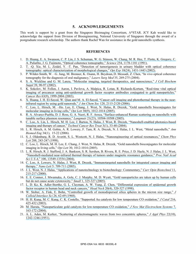
Fig. 5 OCT m-scans of (a) saline, (b) naked 302 nm silica nanoparticles (2 x 10¹¹ particles/ml) used as the core in the gold
nanoshells, (c) 1% Intralipid and (d) synthesized gold nanoshells (2 x 10¹¹ particles/ml) with 302 nm silica core and 28 nm thick shell.
As the layer of gold shell was grown over the silica core, we observe that the presence of surface plasmon resonance from the gold shell is able to effect an increase in the backscattering and hence OCT signal over the naked core silica nanoparticles. The OCT signal from the gold nanoshells is also higher compared to the intrinsic signal from tissue as represented by the 1% Intralipid tissue phantom in our region of scanning. The average histogram value of the OCT m-scan images for the four samples is shown in Table 1 below for comparison.
Table. 1. Average histogram value of the m-scan OCT images of the four samples.
Sample for OCT m-scan Mean histogram value (0 to 255)
Saline 1.4
Naked core silica nanoparticles (2 x 10¹¹ particles/ml) 13.0
Synthesized gold nanoshells (2 x 10¹¹ particles/ml) 58.0
1% Intralipid 29.9
4. CONCLUSIONS Gold nanoshells are novel metallodielectric core-shell nanostructures that exhibit tunable optical response with the potential to be excited at any wavelength depending on the relative dimension of the gold shell to the silica core. The ability of gold nanoshells to elicit an optical contrast under reflectance-based imaging is demonstrated in this study using two such imaging modalities: the confocal reflectance microscopy and OCT imaging. The optical properties of the gold nanoshells synthesized in this study shows a peak plasmon resonance at around 750 nm which matches closely with the excitation wavelength of Ti:Sapphire laser i.e. 800 nm used in the OCT system as well as the helium-neon laser i.e. 633 nm used in the confocal reflectance system in this study. Under the confocal reflectance microscopy, our in vitro experiment with the cells shows selective binding of anti-EGFR conjugated gold nanoshells to EGFR on the cell membrane and that the optical contrast is dependent on the EGFR expression levels. This could potentially be use as a tool for early discrimination of cancer from normal cells based on molecular markers expression. The results of our phantom studies using the OCT have also suggested the potential of gold nanoshells to improve the optical contrast over intrinsic tissue scattering. This optical contrast under OCT can likewise be made dependent on molecular markers expression with antibody specificity, thus improving the existing structural imaging capabilities of OCT to include in vivo molecular imaging for earlier cancer diagnosis of suspicious lesions at a stage before debilitating symptoms appear.
SPIE-OSA Vol. 6633 66330L-7
5. ACKNOWLEDGEMENTS This work is support by a grant from the Singapore Bioimaging Consortium, A*STAR. JCY Kah would like to acknowledge the support from Division of Bioengineering, National University of Singapore through the award of a postgraduate research scholarship. The authors thank Rachel Wan for assistance in the gold nanoshells synthesis.
REFERENCES
1. D. Huang, E. A. Swanson, C. P. Lin, J. S. Schuman, W. G. Stinson, W. Chang, M. R. Hee, T. Flotte, K. Gregory, C. A. Puliafito, J. G. Fujimoto, “Optical coherence tomography,” Science 254, 1178-1181 (1991).
2. T. -Q. Xie, M. L. Zeidel, Y. -T. Pan, “Detection of tumorigenesis in urinary bladder with optical coherence tomography: optical characterization of morphological changes,” Opt Exp 10(24), 1431-1443 (2002).
3. P. Wilder-Smith, W. –G. Jung, M. Brenner, K. Osann, H. Beydoun, D. Messadi, Z. Chen, “In vivo optical coherence tomography for the diagnosis of oral malignancy,” Lasers Surg Med 35, 269-275 (2004).
4. S. A. Wickline and G. M. Lanza, “Molecular imaging, targeted therapeutics, and nanoscience,” J Cell Biochem Suppl 39, 90-97 (2002).
5. K. Sokolov, M. Follen, J. Aaron, I. Pavlova, A. Malpica, R. Lotan, R. Richards-Kortum, “Real-time vital optical imaging of precancer using anti-epidermal growth factor receptor antibodies conjugated to gold nanoparticles,” Cancer Res 63(9), 1999-2004 (2003).
6. X. Huang, I. H. El-Sayed, W. Qian and M. A. El-Sayed, “Cancer cell imaging and photothermal therapy in the near-infrared region by using gold nanorods,” J Am Chem Soc 128, 2115-2120 (2006).
7. C. Loo, L. Hirsch, M. –Ho. Lee, E. Chang, J. West, N. Halas, R. Drezek, “Gold nanoshells bioconjugates for molecular imaging in living cells,” Opt Lett 30(9), 1012-1014 (2005).
8. R. A. Alvarez-Puebla, D. J. Ross, G. A. Nazri, R. F. Aroca, “Surface-enhanced Raman scattering on nanoshells with tunable surface plasmon resonance,” Langmuir 21(23), 10504-10508 (2005).
9. C. Loo, A. Lin, L. Hirsch, M. H. Lee, J. Barton, N. Halas, J. West, R. Drezek, “Nanoshell-enabled photonics-based imaging and therapy of cancer,” Technol Cancer Res Treat 3(1), 33-40 (2004).
10. L. R. Hirsch, A. M. Gobin, A. R. Lowery, F. Tam, R. A. Drezek, N. J. Halas, J. L. West, “Metal nanoshells,” Ann Biomed Eng 34(1), 15-22 (2006).
11. S. J. Oldenburg, R. D. Averitt, S. L. Westcott, N. J. Halas, “Nanoengineering of optical resonances,” Chem Phys Lett 288, 243-247 (1998).
12. C. Loo, L. Hirsch, M. H. Lee, E. Chang, J. West, N. Halas, R. Drezek, “Gold nanoshells bioconjugates for molecular imaging in living cells,” Opt Lett 30, 1012-1014 (2005).
13. L. R. Hirsch, R. J. Stafford, J. A. Bankson, S. R. Sershen, B. Rivera, R. E. Price, J. D. Hazle, N. J. Halas, J. L. West, “Nanoshell-mediated near-infrared thermal therapy of tumors under magnetic resonance guidance,” Proc Natl Acad Sci U S A,” 100, 13549-13554 (2004).
14. C. Loo, A. Lowery, N. Halas, J. West, R. Drezek, “Immunotargeted nanoshells for integrated cancer imaging and therapy,” Nano Lett 5: 709-711 (2005).
15. J. L. West, N. J. Halas, “Applications of nanotechnology to biotechnology: Commentary,” Curr Opin Biotechnol 11, 215-217 (2000).
16. E. E. Connor, J. Mwamuka, A. Gole, C. J. Murphy, M. D. Wyatt, “Gold nanoparticles are taken up by human cells but do not cause acute cytotoxicity,” Small 1, 325-327 (2005).
17. L. D. Ke, K. Adler-Storthz, G. L. Clayman, A. W. Yung, Z. Chen, “Differential expression of epidermal growth factor receptor in human head and neck cancers,” Head Neck 20(4), 320-327 (1998).
18. W. Stober, A. Fink, E. Bohn, “Controlled growth of monodispersed silica spheres in the micron size range,” J Colloid Interface Sci 26, 62-69 (1968).
19. H. H. Kung, M. C. Kung, C. K. Costello, “Supported Au catalysts for low temperature CO oxidation,” J Catal 216, 425-432 (2003).
20. M. Haruta, “Nanoparticulate gold catalysts for low-temperature CO oxidation,” J New Mat Electrochem Systems 7, 163-172 (2004).
21. A. L. Aden, M. Kerker, “Scattering of electromagnetic waves from two concentric spheres,” J Appl Phys 22(10), 1242-1246 (1951).
SPIE-OSA Vol. 6633 66330L-8