Two-step formation of 1H NMR visible mobile lipids during apoptosis of paclitaxel-treated K562 cells
Upload
independentCategory
view
1download
0
ORIGINAL ARTICLE
Debora G. Rodrigues Æ Durvanei A. Maria
Denise C. Fernandes Æ Claudete J. Valduga
Ricardo D. Couto Æ Olga C. M. Ibanez
Raul C. Maranhao
Improvement of paclitaxel therapeutic index by derivatizationand association to a cholesterol-rich microemulsion: in vitroand in vivo studies
Received: 12 July 2004 / Accepted: 27 September 2004 / Published online: 22 February 2005� Springer-Verlag 2005
Abstract A cholesterol-rich microemulsion or nanopar-ticle termed LDE concentrates in cancer tissues afterinjection into the bloodstream. Here the cytotoxicity,pharmacokinetics, toxicity to animals and therapeuticaction of a paclitaxel lipophilic derivative associated toLDE is compared with those of the commercial paclit-axel. Results show that LDE-paclitaxel oleate is stable.The cytostatic activity of the drug in the complex isdiminished compared with the commercial paclitaxeldue to the cytotoxicity of the vehicle Cremophor ELused in the commercial formulation. Competitionexperiments in neoplastic cultured cells show that pac-litaxel oleate and LDE are internalized together by theLDL receptor pathway. LDE-paclitaxel oleate arreststhe G2/M phase of cell cycle, similarly to commercialpaclitaxel. Tolerability to mice is remarkable, such thatthe lethal dose (LD50) was ninefold greater than that ofthe commercial formulation (LD50 = 326 lM and37 lM, respectively). LDE concentrates paclitaxel oleatein the tumor roughly fourfold relative to the normaladjacent tissues. At equimolar doses, the association ofpaclitaxel oleate with LDE results in remarkable chan-ges in the drug pharmacokinetic parameters whencompared to commercial paclitaxel (t1/2=218 min and
184 min, AUC=1,334 lg h/ml and 707 lg h/ml andCL=0.125 ml/min and 0.236 ml/min, respectively). Fi-nally, the therapeutic efficacy of the complex is pro-nouncedly greater than that of the commercialpaclitaxel, as indicated by the reduction in tumorgrowth, increase in survival rates and % cure of treatedmice. In conclusion, LDE-paclitaxel oleate is a stablecomplex and compared with paclitaxel toxicity is con-siderably reduced and activity is enhanced, which maylead to improved therapeutic index in clinical use.
Keywords Nanoparticles Æ Paclitaxel Æ Emulsions ÆCholesterol Æ Low-density lipoprotein receptors ÆCancer treatment Æ Drug targeting
Introduction
After being injected into the blood stream, a cholesterol-rich microemulsion or nanoparticle termed LDE is takenup by the low-density lipoprotein (LDL) receptors in theplasma membrane and is internalized into the cells. Wepreviously showed that LDE can concentrate in leukemiacells or in solid tumors such as ovarian and breast carci-nomas [1, 9, 16] that overexpress those receptors. There-fore, LDE may serve as a vehicle to chemotherapeuticagents directed against neoplastic tissues. Recently, wereported that the antineoplastic agent paclitaxel can beassociated at high rates with LDE without loss of cyto-toxicity [21]. However, we subsequently observed that theLDE-paclitaxel complex tends to dissociate in thebloodstream after injection into mice (unpublished data).
The most remarkable clinical antitumor activity ofpaclitaxel has been in advanced ovarian and breastcancers, resulting in the worldwide regulatory approvalof the agent in both cancers. The drug is commerciallyavailable as a 1/1 mixture with a derivative castor oilnamed Cremophor EL and ethanol which is rarely
D. G. Rodrigues Æ D. C. Fernandes Æ C. J. ValdugaR. D. Couto Æ R. C. Maranhao (&)Lipid Metabolism Laboratory, Heart Institute (InCor), Universityof Sao Paulo Medical School Hospital, Av. Dr. Eneas de CarvalhoAguiar, 44, 1 subsolo, 05403-000 Sao Paulo, BrazilE-mail: [email protected].: +55-11-30695108Fax: +55-11-30695574
D. G. Rodrigues Æ R. D. Couto Æ R. C. MaranhaoFaculty of Pharmaceutical Sciences, University of Sao Paulo,Sao Paulo, Brazil
D. A. Maria Æ O. C. M. IbanezLaboratory of Immunogenetics of the Butantan Institute,Sao Paulo, Brazil
Cancer Chemother Pharmacol (2005) 55: 565–576DOI 10.1007/s00280-004-0930-y
responsible for severe life-threatening hypersensitivityreactions in patients [27]. Because LDE is atoxic and hasa further drug targeting ability demonstrated in eitherovarian or breast carcinomas, it should be an attractivecandidate vehicle to improve the paclitaxel therapeuticindex.
An usual strategy to render chemotherapeutic agentsmore stable when associated to LDL or artificial lipidemulsions has been to attach lipophilic groups to theirmolecules. In this respect, Lundberg et al. [14] have re-cently shown that a lipophilic derivative of paclitaxel in alipid o/w emulsion covered with a hydrophilic polymerimproved the pharmacokinetic parameters of paclitaxel.On the other hand, we showed that the stability of theLDE-etoposide complex is also improved when thisantineoplastic agent is attached with an oleoyl group [24].
In this study, the modification strategy was attemptedto enhance the stability of the LDE-paclitaxel oleatecomplex by attaching an oleoyl group to the drug.Compared with commercial paclitaxel, the resultingpreparation showed higher therapeutic index that wasachieved by diminution of the toxicity and increase ofthe anticancer action.
Materials and methods
Materials
Crystalline paclitaxel was purchased from Calbiochem(CA) dimethlysulfoxide (DMSO), 3-(4,5 dim-ethylthiazol-2-yl)-2,5-diphenyl tetrazolium bromide(MTT), derivative of castor oil (Cremophor EL), trio-lein, cholesteryl oleate, cholesterol, oleic acid, ethylacetate, chloroform, triethylamine, dichloromethane,and phosphatidylcholine were purchased from Sigma (StLouis, MO), methanol and acetonitrile from Merck(Darmstadt, Germany). [3H]-paclitaxel with the tritiumat the m-position and p-position of the aromatic ring, atthe 10¢-position and 2¢-position of the taxane ring and3¢-position of the side chain was purchased from Mor-avek (Brea, CA). The clinically approved formulation ofpaclitaxel, Taxol was obtained from Bristol-MyersSquibb Co (1 ml contains 6 mg of paclitaxel, 527 mg ofCremophor EL, 49.7% v/v dehydrated alcohol, USP).The human small lung cell carcinoma NCI H292 cell linewas purchased from Adolfo Lutz Institute (Sao Paulo,Brazil). The murine B16F10 melanoma cell line wasobtained from the American Type Culture Collection.C57BL/6J mice were purchased from the Central Ani-mal Care Unit at Instituto Butantan. Females aged 8–12weeks were used in all experiments.
Paclitaxel oleate synthesis and characterization
Paclitaxel was modified following the method de-scribed by Lundberg [14]. Briefly, oleoyl chloride,
prepared by use of oxalyl chloride and oleic acid, wasquickly added to a solution of paclitaxel and triethyl-amine in dry acetonitrile. The mixture was stirred atroom temperature for 45 min and the product wasthen extracted and purified. When necessary, traceamounts of [3H]-paclitaxel were added to the initialsolution. The purity and the structure of the productwere characterized by [1H] and [13C] NMR using aBruker DPX-300 instrument and by liquid chroma-tography coupled to tandem mass spectrometry withelectrospray ionization (LC-ESI/MS/MS) using a Shi-madzu Model LC-10AD high-pressure liquid chro-matograph (HPLC) equipped with a UV detector at227 nm, 15 cm · 4.6 mm Luna C8 [2] (Phenomenex,Torrance, CA), and a 4 cm · 10 mm I.D. pre-columnwith shim-pack CLC-ODS (M) C18 (Shimadzu,Columbia, ML) coupled to a mass spectrometer triplequadrupole Quattro II (Micromass, Manchester, UK).The LC–MS–MS experiments were performed whilescanning from m/z 500 to 1,300 at a scan rate of 2 s/scan. The samples were analyzed in duplicate byHPLC using an isocratic solvent system constituted ofacetonitrile–methanol (90:10) at a flow-rate of 1.0 ml/min.
Preparation of LDE
In brief, LDE was prepared from a lipid mixture com-posed of 40 mg cholesteryl oleate, 20 mg egg phos-phaditylcholine, 1 mg triolein and 0.5 mg cholesterol.Emulsification of lipids by prolonged ultrasonic irradi-ation in aqueous media and the procedure of two-stepultracentrifugation of the crude emulsion with densityadjustment by addition of KBr to obtain LDE micro-emulsion was carried out by the method described pre-viously [7] modified by Maranhao [15]. LDE wasdialyzed against saline solution and passed through0.22 lm filter for the experiments. When necessary, traceamounts of [14C]-cholesteryl ester were added to theinitial solution.
Association of paclitaxel oleate to LDE
Paclitaxel oleate was incorporated into LDE by solubi-lization of paclitaxel oleate in ethanol and by adding itinto the emulsion. The solution was then sonicated for30 min at 70�C using a Branson Sonifier 450 (Danbury,CT), equipped with a 1 cm flat titanium probe. LDE-paclitaxel oleate was centrifuged at 3,000 rpm for15 min to separate the unbound paclitaxel oleate. LDE-paclitaxel oleate was then passed through 0.22 lm porepolycarbonate filter and kept at 4�C until it was used.When necessary [3H]-paclitaxel oleate were added to theinitial solution. The yield of each batch was assayedbefore the use. LDE-paclitaxel oleate was always pre-pared at the same day of the experiments.
566
Stability of the drug-LDE complex in vitro
Stability of LDE-paclitaxel oleate was tested by mem-brane dialysis (12,000 MW cut-off) against humanplasma and Tris–HCl solution pH 7.4. One milliliter oflabeled [14C]-cholesteryl oleate-LDE:[3H]-paclitaxel ole-ate was dialyzed against 20 ml of plasma and Tris–HClsolution. Samples of 5 ll were collected from the dialysisbag at 0.03–168 h interval and placed separately intovials with 7 ml scintillation solution ‘‘Hisafe’’ (PerkinElmer, Loughborough, England). The radioactivity wasmeasured by liquid scintillation spectrometry with aPackard 1600 TR model Liquid Scintillation Analyzer.
Incubation of LDE-paclitaxel oleate with human plasma
[14C]-cholesteryl oleate-LDE:[3H]-paclitaxel oleate wasincubated with human plasma for 1 h at 37�C. Follow-ing incubation, plasma was separated in its lipoproteinand lipoprotein-deficient plasma (LPDP) fractions bydensity gradient ultracentrifugation and the percentageof the LDE and of the paclitaxel oleate label recoveredin each fraction was determined by radioactivitycounting as previously described.
Cell growth inhibition
The NCI H292 cells were maintained in RPMI 1640medium supplemented with antibiotics and 10% (v/v)fetal calf serum (FCS) at 37�C in a humidified incubatorwith 5% (v/v) CO2. Prior the experiments, the cells wereharvested from the culture and distributed into 96-wellculture plates at 105 cells/well. After 24 h incubation,serial dilutions of a Cremophor/EL stock solution ofpaclitaxel oleate and paclitaxel and LDE-paclitaxeloleate were added to the wells in triplicate. The finalconcentrations of paclitaxel or paclitaxel oleate (0.003–3 lM) and LDE-paclitaxel oleate (0.0001–10 lM) wereused. The cells were left in the incubator for further72 h, at the end of this period the medium was removedand the number of living tumor cells were determined bythe colorimetric MTT assay. The 50% inhibitory con-centration (IC50) was determined as the drug concen-tration required to inhibit 50% of the cell growth.
LDE-paclitaxel oleate uptake by neoplastic cells
The cellular uptake of paclitaxel oleate was determinedby incubation of NCI H292 cells with increasingamounts of [14C]-cholesteryl oleate-LDE-[3H]-paclitaxeloleate. 106 viable cells were platted on 35 mm Petridishes. On the next day, the medium was replaced byother containing 10% lipoprotein-deficient serum(LPDS). On the third day, increasing amounts of [14C]-cholesteryl oleate-LDE-[3H]-paclitaxel oleate (0.005–400 lg/ml) were added to the plates in duplicate and
incubated for 4 h at 37�C. The cells were then washedthree times with cold PBS plus BSA and twice with PBSat 37�C, harvested and centrifuged at 14,000 rpm for15 min and 200 ll NaOH 0.1 M was added to the pelletto disrupt the cell pellet under vortex mixing. Thesamples were placed separately into vials with scintilla-tion solution and the radioactivity was measured by li-quid scintillation spectrometry.
Competition between LDE-paclitaxel oleateand native LDL
NCI H292 viable cells (106) were incubated during 24 hin RPMI 1640 containing antibiotics, supplemented with10% LPDS. After this period, 200 lg/ml of [14C]-cho-lesteryl oleate-LDE-[3H]-paclitaxel oleate and increasingamounts of human LDL (50–400 lg/ml) were added tothe plates in duplicate and incubated for 4 h at 37�C.The cells were then washed three times with cold PBSplus BSA and twice with PBS at 37�C, harvested andcentrifuged at 14,000 rpm for 15 min; 200 ll of NaOH0.1 M were added to the pellet to disrupt the cell pelletunder vortex mixing before radioactivity measurement.
LDE-paclitaxel oleate toxicity to mice
Toxicity experiments were performed using groups offive C57BL/6J mice weighing roughly 20 g. LDE-paclit-axel oleate or commercial paclitaxel were administeredi.p. in a single or multiple doses ranging from 90 lM/kgto 450 lM/kg and 12 lM/kg to 120 lM/kg, respectively.The same volume of LDE alone and Cremophor EL/ethanol were also administered as a control. Survival andweight changes were observed daily over a 60-day period.Lethal doses (LD) were determined by simple interpo-lation. Rapid death occurring after bolus administrationwas rejected, only that manifested more slowly, as a re-sult of drug effects on GI and/or bone marrow wasmeasured. The maximum tolerated dose (MTD) wasdefined as the allowance of a median body weight loss of15% of the control and causes neither death due toxiceffects nor remarkable changes in the general signs within1 week after administration. All the animal experimentsreported in this study were approved by the AnimalEthics Committee of the University of Sao Paulo.
Inoculation of the B16 melanoma cells in mice
Murine B16F10 (H2b) variant of the B16 melanoma cellline originating from C57BL/6J mice were used in theexperiments. Cells were cultured in RPMI-1640 mediumsupplemented with 10% FCS, 2 mM-L-glutamine,1 mM sodium pyruvate and antibiotics. Cell suspensionswere detached from plates with trypsin and 0.2% ver-sene. After trypsin inactivation with 10% FCS, viablecells were counted by trypan blue dye exclusion. For
567
tumor transfer 5·104 cells suspended in 100 ll of PBSwere injected subcutaneously into the flank regions ofmice. Ten days after inoculation the tumors becamemacroscopically apparent.
Immunohistochemistry for LDL receptors in the B16melanoma cells
The presence of LDL receptors in the melanoma celllineage that was inoculated in mice was documented bythe immunoperoxidase technique. In summary, silane-coated glass microscope smears used in the preparationof melanoma cell film were immediately fixed in 95%ethanol for 10 min and equilibrated in PBS prior toimmunohistochemistry. The procedure involves theapplication to cell smears of the following antibody se-quence: mouse IgG2ak (LP02) antihuman LDL receptorat 2.5 lg/ml for 18 h at 4�C; DAKO LSABR System(Carpinteria, USA), followed by biotinylated link anti-body and streptavidin-HRP, for 30 min each. After eachstep the smears were washed in PBS (pH 7.6) and, fi-nally, the peroxidase activity was revealed using diam-inobenzidine (40 mg/100 ml) and H2O2 (6 ll/ml)[(substrate-chromogen solution)] by 5–10 min. Smearswere counterstained with haematoxylin solution. A po-sitive tissue control for the LDL receptor was providedby performing the immunoperoxidase reaction in humanliver sections. Negative control smears were performedin the same cell line by incubating an adjacent sectionwith an irrelevant murine IgG monoclonal antibody.
Flow cytometry DNA analysis
B16 melanoma cells were incubated with 0.05 lM LDE-paclitaxel oleate, commercial paclitaxel and controls for24 h. Following the treatment, the cells were harvested,washed with PBS and resuspended in 375 ll trypsin0.03 g/l, 10 mM Tris (pH 8.0). After 15 min incubationat room temperature, the neutralization solution (tripsininhibitor 0.5 g/l, RNase A 0.1 g/l and spermine 1.2 g/l)was added and incubation continued for 15 min. Pel-leted cells were resuspended in 0.3 ml PBS and fixed byaddition of ice-cold ethanol (70%). Prior to analysis,cells were incubated with 18 lg/ml propidium iodidesolution and incubated in the dark for 30 min. Flowcytometry analysis was performed on a FACScan flowcytometry system (Scalibur-Becton Dickinson, San Jose,CA). The DNA content in the cell cycle phases (G0/G1,S and G2/M) was analyzed by the Cell-Quest softwareand by the Mod-fit software cell.
Biodistribution of LDE and LDE-paclitaxel oleatein B16 melanoma-bearing mice
One hundred microliter of LDE (1.2 kBq, 0.3 mg totallipids) labeled with [14C]-cholesteryl oleate or LDE-
paclitaxel oleate (1.2 kBq, 0.3 mg total lipids and0.06 mg drug) labeled with [3H]-paclitaxel oleate and[14C]-cholesteryl oleate were injected i.p. as a single bo-lus in groups of five C57BL/6J mice. The animals werekept in individual cages for 18 h, when they were sac-rificed and tissues samples of 200–250 mg of skin, tu-mor, liver, brain, spleen, lung, testicle, kidney, heart andintestine (animals treated with LDE alone) and skin,liver and tumor (animals treated with LDE-paclitaxeloleate) were collected and kept in cold saline solutionprior to lipid extraction with chloroform/methanol (2:1v/v). The extracted lipids and drug were concentratedand measured by liquid scintillation spectrometry.
Plasma kinetics of double-labeled LDE-paclitaxeloleate and commercial paclitaxel in mice
To verify whether the derivatization and association ofpaclitaxel to LDE would alter the pharmacokinetics ofthe commercially available drug, the pharmacokineticprofile of LDE-paclitaxel oleate and of commercialpaclitaxel were determined in control mice. LDE-pac-litaxel oleate labeled with [3H]-paclitaxel oleate and[14C]-cholesteryl oleate (100 ll total volume, 1.2 kBq,0.3 mg total lipids and 0.06 mg drug) and commercial[3H]-paclitaxel (0.08 mg) were injected as a single bolusinto the retro-orbital venous plexus of five mice fordetermination of the plasma decaying curves. Bloodsamples were collected at pre-established intervals dur-ing 24 h. Plasma was separated by a 15 min centrifu-gation (3,000g) and the radioactivity was counted in ascintillation solution. The fractional clearance rate(FCR) of the [14C]-cholesteryl oleate-LDE, [3H]-paclit-axel oleate and [3H]-paclitaxel was calculated accordingto the method described by Matthews [18], where a1, a2,b1 and b2 were estimated from biexponential curvesobtained from the remaining radioactivity found inplasma after injection, fitted by least squares procedure,as y ¼ a1 e
�b1t� �
þ a2 e�b2t
� �where y represents the
radioactivity plasma decay.The pharmacokinetic parameters of paclitaxel oleate
associated with LDE and commercial paclitaxel werecalculated using a multicompartmental model by meansof a computer software (PK Solutions, Ashland, OH).The log plasma concentration versus time curves werefitted by biexponential equations and the half-life (t1/2)calculated by dividing 0.693 by the rate constant for eachphase. The total AUC was calculated using the lineartrapezoid method with extrapolation to infinity. Totalplasma clearance was calculated by dividing the dose bythe AUC. The volume of distribution at steady state wasestimated graphically from trapezoidal total area mea-surements.
Antitumor activity
Mice were inoculated with B16 melanoma cells as de-scribed above. Treatments were started on day 11,
568
when the tumor implanted in the animals reached a60–100 mm3 volume. The mice were randomly allo-cated to groups of eight animals. On days 11, 14, 19 ascounted from the initial inoculation day, each groupwas injected i.p. with one of the following prepara-tions: LDE-paclitaxel oleate at two dose levels,17.5 mg/kg and 70.3 mg/kg (equimolar doses with15 mg/kg and 60 mg/kg of paclitaxel, respectively),commercial paclitaxel at 15 mg/kg; 0.9% saline solu-tion or LDE alone. These last two were controlgroups.
The tumor sizes were measured three times a weekusing a calliper-like instrument during the experiment.The size measurement was converted to tumor weight bythe equation: tumor weight = (length2 · width)/2. Theexperiment was ended on day 34, but the survival of theanimals in each experimental group was monitored over180 days. The antitumor activity was assessed accordingto the guidelines established by the National CancerInstitute [19].
Statistical analysis
The differences in the cell survival and cell uptake curveswere evaluated by the unpaired Student’s t test. TheANOVA variance test was used to compare the resultsobtained from the flow cytometry DNA analysis and tocompare the AUC obtained from the growth tumorinhibition curve. The Mann–Whitney test was used forFCR data analysis. All the values were expressed asmeans ± SEM. In all analysis, p<0.05 was consideredstatistically significant. The survival time plotting (Ka-plan–Meyer test) and survival comparison betweengroups were carried out using the Graph Pad Prismstatistical software.
Results
Paclitaxel oleate synthesis, characterizationand association to LDE
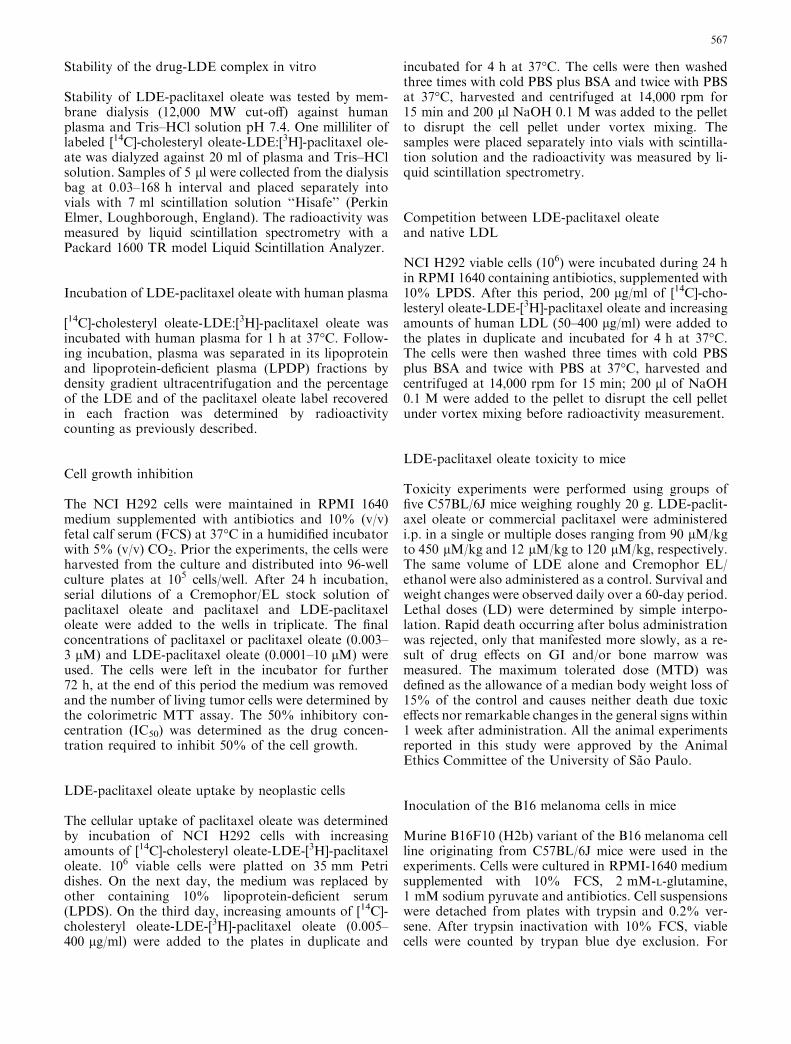
Figure 1 shows the full scan mass spectra of the paclit-axel oleate product ions [M+H]+. The peaks of the corefragments appear at m/z 551 and at m/z 854 whereas thewhole molecule peak appears at m/z 1,118. As estimatedby HPLC, the yield of the esterification reaction wasroughly 90%. The amount of paclitaxel oleate associ-ated to LDE by co-sonication was 85%. It follows thateach ml of the emulsion (30 mg of emulsion total lipids)solubilizes 4.8–5.4 mg paclitaxel oleate.
Stability of the LDE-paclitaxel oleate complex in vitro
Figure 2 shows the results of the experiment wherein theLDE-paclitaxel oleate was dialyzed for 168 h againsteither plasma or Tris buffer. It is clear that during thefirst 24 h, only negligible dissociation of the complexoccurred regardless plasma or Tris buffer was used in theexperiment. In the ensuing 24–168 h, however, there wassome dissociation against plasma, which was less intensefor the Tris solution.
Incubation of LDE-paclitaxel oleate withhuman plasma
After incubation with plasma, 97.8% of the LDEradioactive label and 98.4% of the paclitaxel oleate labelwere found at the lipoprotein-containing plasma frac-tion whereas 2.2% and 1.6% of the two labels, respec-tively, were found at the lipoprotein-deficient fraction.Therefore, only residual amounts of drug leaks out of
Fig. 1 Representative full scanchromatogram (m/z 500–1,400)of paclitaxel oleate analyzed byLC-ESI/MS/MS (liquidchromatography coupled totandem mass spectrometry withelectrospray ionization) at ascan rate of 2 s/scan. Thesamples were analysed induplicate by HPLC using anisocratic solvent systemconstituted of acetonitrile–methanol (90:10) at a flow-rateof 1.0 ml/min
569

the microemulsion and is found in the fraction thatcontains mainly albumin, globulins and a-glycoprotein.
Cell growth inhibition
Figure 3a shows the dose–response curves of the cyto-static activity of paclitaxel oleate and of paclitaxel bothusing Cremophor as solubilizing agent. It is clear thatthe chemical modification of the drug does not affect thecytostatic activity of the drug (p=0.523). The cytostaticindex of paclitaxel oleate (IC50 = 0.11 lM) and paclit-axel (IC50 = 0.11 lM) calculated from the curves aresimilar.
Figure 3b shows the cytostatic activity curves ofLDE-paclitaxel oleate, paclitaxel oleate solubilized inCremophor and, finally of LDE-paclitaxel oleate withaddition of Cremophor. This last experiment was per-formed to discriminate the additive cytotoxicity ofCremophor. LDE-paclitaxel oleate has a clear-cut lowercytotoxicity than paclitaxel oleate solubilized in Crem-ophor (IC50 = 1.00 lM and 0.09 lM, respectively,p=0.015). However, when Cremophor is added to LDE-paclitaxel oleate incubates, the cytotoxicity increases(IC50 = 0.15 lM, p=0.756), so that the IC50 ap-proaches to that of paclitaxel oleate solubilized inCremophor. Therefore, the greater cytotoxicity of thecommercial formulation and of the paclitaxel oleatesolubilized in Cremophor can be ascribed to the pres-ence of Cremophor.
LDE-paclitaxel uptake by neoplastic cells
Figure 4a shows that when NCI H292 cells were incu-bated with increasing amounts of LDE-paclitaxel oleatelabeled with [3H]-paclitaxel oleate and [14C]-cholesteryl
oleate, there was a proportionally increasing uptake ofthe two labels and the two uptake curves are similar(p=0.2717). This indicates that both paclitaxel andcholesteryl oleate components of the complex aresimultaneously internalized into the cells.
Figure 4b shows that the addition of increasingamounts of native human LDL to the incubates of LDE-paclitaxel oleate and the malignant cells leads to a pro-gressive diminution of cell uptake of both [3H]-paclitaxeloleate and [14C]-cholesteryl oleate contained in LDE.This indicates that when LDE uptake is diminished bythe competition of native LDL the cell uptake of thedrug associated to the microemulsion is also propor-tionally diminished.
LDE-paclitaxel oleate toxicity to mice
Table 1 shows the results of the toxicity experimentswith LDE-paclitaxel oleate and commercial paclitaxel
Fig. 3 Effects on NCI-H292 cells survival of serial dilutions of aCremophor/EL stock solution of a paclitaxel oleate (filled circle)and paclitaxel (open circle). b Effects on survival of LDE-paclitaxeloleate with (open circle) or without (filled circle) the addition ofCremophor EL/ethanol and commercial paclitaxel (filled square).Cells were incubated for 72 h at 37�C. Results are presented asmeans ± SEM (bars) of three experiments performed in triplicate
Fig. 2 Dialysis of 1.0 ml of LDE-paclitaxel oleate doubly labeledwith [14C] cholesteryl ester-LDE and [3H]-paclitaxel oleate against20 ml of human plasma (open circle) and Tris–HCl buffer (filledcircle) at 0.03–168 h interval. The radioactivity were measured byliquid scintillation. Results are presented means ± SEM (bars) ofthree experiments
570
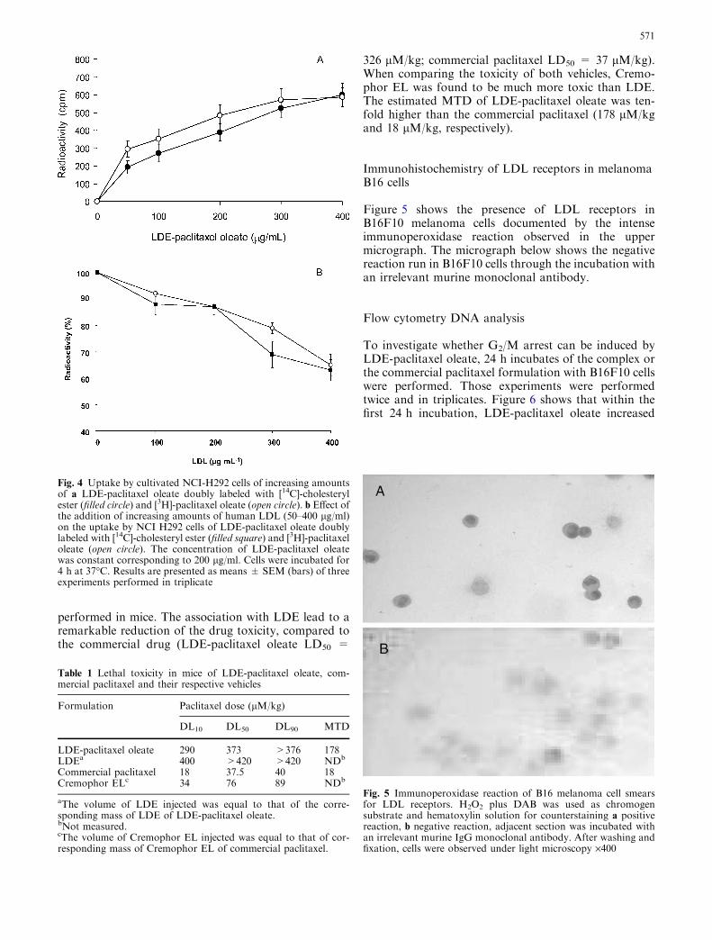
performed in mice. The association with LDE lead to aremarkable reduction of the drug toxicity, compared tothe commercial drug (LDE-paclitaxel oleate LD50 =
326 lM/kg; commercial paclitaxel LD50 = 37 lM/kg).When comparing the toxicity of both vehicles, Cremo-phor EL was found to be much more toxic than LDE.The estimated MTD of LDE-paclitaxel oleate was ten-fold higher than the commercial paclitaxel (178 lM/kgand 18 lM/kg, respectively).
Immunohistochemistry of LDL receptors in melanomaB16 cells
Figure 5 shows the presence of LDL receptors inB16F10 melanoma cells documented by the intenseimmunoperoxidase reaction observed in the uppermicrograph. The micrograph below shows the negativereaction run in B16F10 cells through the incubation withan irrelevant murine monoclonal antibody.
Flow cytometry DNA analysis
To investigate whether G2/M arrest can be induced byLDE-paclitaxel oleate, 24 h incubates of the complex orthe commercial paclitaxel formulation with B16F10 cellswere performed. Those experiments were performedtwice and in triplicates. Figure 6 shows that within thefirst 24 h incubation, LDE-paclitaxel oleate increased
Fig. 4 Uptake by cultivated NCI-H292 cells of increasing amountsof a LDE-paclitaxel oleate doubly labeled with [14C]-cholesterylester (filled circle) and [3H]-paclitaxel oleate (open circle). b Effect ofthe addition of increasing amounts of human LDL (50–400 lg/ml)on the uptake by NCI H292 cells of LDE-paclitaxel oleate doublylabeled with [14C]-cholesteryl ester (filled square) and [3H]-paclitaxeloleate (open circle). The concentration of LDE-paclitaxel oleatewas constant corresponding to 200 lg/ml. Cells were incubated for4 h at 37�C. Results are presented as means ± SEM (bars) of threeexperiments performed in triplicate
Table 1 Lethal toxicity in mice of LDE-paclitaxel oleate, com-mercial paclitaxel and their respective vehicles
Formulation Paclitaxel dose (lM/kg)
DL10 DL50 DL90 MTD
LDE-paclitaxel oleate 290 373 >376 178LDEa 400 >420 >420 NDb
Commercial paclitaxel 18 37.5 40 18Cremophor ELc 34 76 89 NDb
aThe volume of LDE injected was equal to that of the corre-sponding mass of LDE of LDE-paclitaxel oleate.bNot measured.cThe volume of Cremophor EL injected was equal to that of cor-responding mass of Cremophor EL of commercial paclitaxel.
Fig. 5 Immunoperoxidase reaction of B16 melanoma cell smearsfor LDL receptors. H2O2 plus DAB was used as chromogensubstrate and hematoxylin solution for counterstaining a positivereaction, b negative reaction, adjacent section was incubated withan irrelevant murine IgG monoclonal antibody. After washing andfixation, cells were observed under light microscopy ·400
571
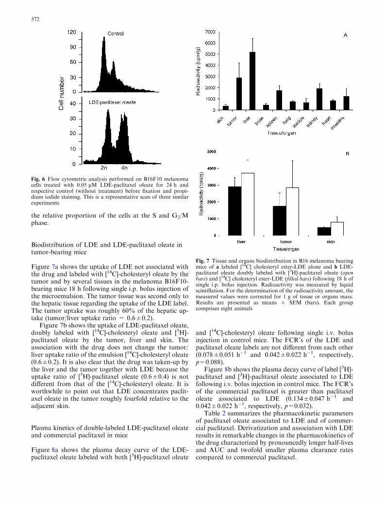
the relative proportion of the cells at the S and G2/Mphase.
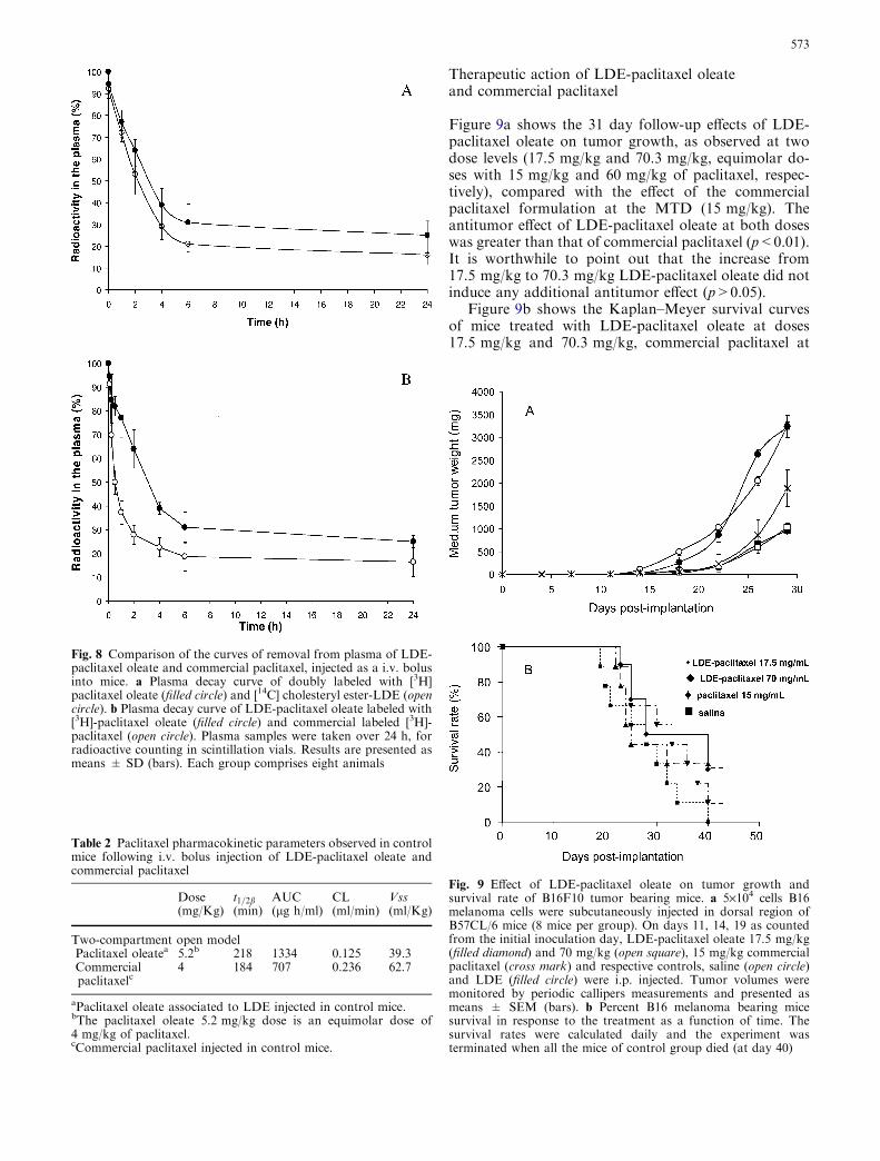
Biodistribution of LDE and LDE-paclitaxel oleate intumor-bearing mice
Figure 7a shows the uptake of LDE not associated withthe drug and labeled with [14C]-cholesteryl oleate by thetumor and by several tissues in the melanoma B16F10-bearing mice 18 h following single i.p. bolus injection ofthe microemulsion. The tumor tissue was second only tothe hepatic tissue regarding the uptake of the LDE label.The tumor uptake was roughly 60% of the hepatic up-take (tumor/liver uptake ratio = 0.6±0.2).
Figure 7b shows the uptake of LDE-paclitaxel oleate,doubly labeled with [14C]-cholesteryl oleate and [3H]-paclitaxel oleate by the tumor, liver and skin. Theassociation with the drug does not change the tumor/liver uptake ratio of the emulsion [14C]-cholesteryl oleate(0.6±0.2). It is also clear that the drug was taken-up bythe liver and the tumor together with LDE because theuptake ratio of [3H]-paclitaxel oleate (0.6±0.4) is notdifferent from that of the [14C]-cholesteryl oleate. It isworthwhile to point out that LDE concentrates paclit-axel oleate in the tumor roughly fourfold relative to theadjacent skin.
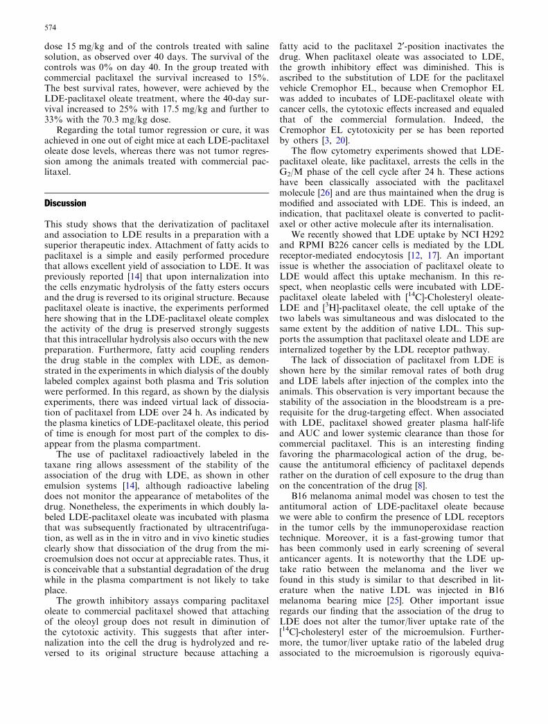
Plasma kinetics of double-labeled LDE-paclitaxel oleateand commercial paclitaxel in mice
Figure 8a shows the plasma decay curve of the LDE-paclitaxel oleate labeled with both [3H]-paclitaxel oleate
and [14C]-cholesteryl oleate following single i.v. bolusinjection in control mice. The FCR’s of the LDE andpaclitaxel oleate labels are not different from each other(0.078±0.051 h�1 and 0.042±0.022 h�1, respectively,p=0.088).
Figure 8b shows the plasma decay curve of label [3H]-paclitaxel and [3H]-paclitaxel oleate associated to LDEfollowing i.v. bolus injection in control mice. The FCR’sof the commercial paclitaxel is greater than paclitaxeloleate associated to LDE (0.134±0.047 h�1 and0.042±0.022 h�1, respectively, p=0.032).
Table 2 summarizes the pharmacokinetic parametersof paclitaxel oleate associated to LDE and of commer-cial paclitaxel. Derivatization and association with LDEresults in remarkable changes in the pharmacokinetics ofthe drug characterized by pronouncedly longer half-livesand AUC and twofold smaller plasma clearance ratescompared to commercial paclitaxel.
Fig. 7 Tissue and organs biodistribution in B16 melanoma bearingmice of a labeled [14C] cholesteryl ester-LDE alone and b LDE-paclitaxel oleate doubly labeled with [3H]-paclitaxel oleate (openbars) and [14C] cholesteryl ester-LDE (filled bars) following 18 h ofsingle i.p. bolus injection. Radioactivity was measured by liquidscintillation. For the determination of the radioactivity amount, themeasured values were corrected for 1 g of tissue or organs mass.Results are presented as means ± SEM (bars). Each groupcomprises eight animals
Fig. 6 Flow cytometric analysis performed on B16F10 melanomacells treated with 0.05 lM LDE-paclitaxel oleate for 24 h andrespective control (without treatment) before fixation and propi-dium iodide staining. This is a representative scan of three similarexperiments
572
Therapeutic action of LDE-paclitaxel oleateand commercial paclitaxel
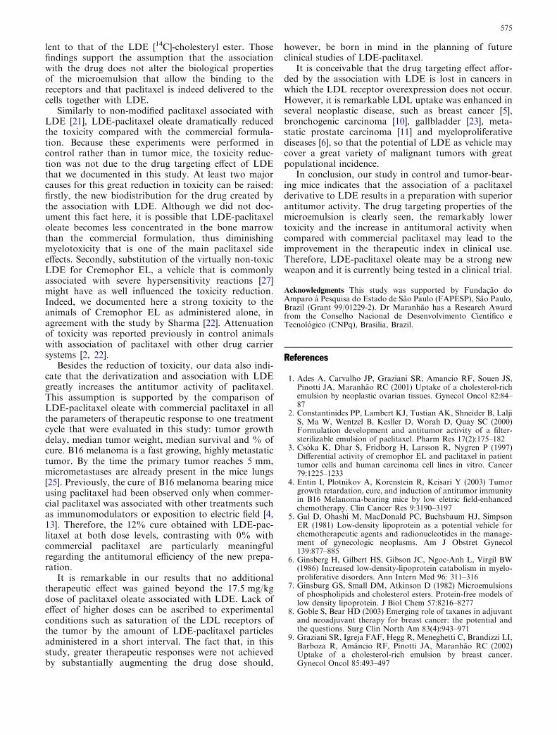
Figure 9a shows the 31 day follow-up effects of LDE-paclitaxel oleate on tumor growth, as observed at twodose levels (17.5 mg/kg and 70.3 mg/kg, equimolar do-ses with 15 mg/kg and 60 mg/kg of paclitaxel, respec-tively), compared with the effect of the commercialpaclitaxel formulation at the MTD (15 mg/kg). Theantitumor effect of LDE-paclitaxel oleate at both doseswas greater than that of commercial paclitaxel (p<0.01).It is worthwhile to point out that the increase from17.5 mg/kg to 70.3 mg/kg LDE-paclitaxel oleate did notinduce any additional antitumor effect (p>0.05).
Figure 9b shows the Kaplan–Meyer survival curvesof mice treated with LDE-paclitaxel oleate at doses17.5 mg/kg and 70.3 mg/kg, commercial paclitaxel at
Fig. 8 Comparison of the curves of removal from plasma of LDE-paclitaxel oleate and commercial paclitaxel, injected as a i.v. bolusinto mice. a Plasma decay curve of doubly labeled with [3H]paclitaxel oleate (filled circle) and [14C] cholesteryl ester-LDE (opencircle). b Plasma decay curve of LDE-paclitaxel oleate labeled with[3H]-paclitaxel oleate (filled circle) and commercial labeled [3H]-paclitaxel (open circle). Plasma samples were taken over 24 h, forradioactive counting in scintillation vials. Results are presented asmeans ± SD (bars). Each group comprises eight animals
Table 2 Paclitaxel pharmacokinetic parameters observed in controlmice following i.v. bolus injection of LDE-paclitaxel oleate andcommercial paclitaxel
Dose(mg/Kg)
t1=2b(min)
AUC(lg h/ml)
CL(ml/min)
Vss(ml/Kg)
Two-compartment open modelPaclitaxel oleatea 5.2b 218 1334 0.125 39.3Commercialpaclitaxelc
4 184 707 0.236 62.7
aPaclitaxel oleate associated to LDE injected in control mice.bThe paclitaxel oleate 5.2 mg/kg dose is an equimolar dose of4 mg/kg of paclitaxel.cCommercial paclitaxel injected in control mice.
Fig. 9 Effect of LDE-paclitaxel oleate on tumor growth andsurvival rate of B16F10 tumor bearing mice. a 5·104 cells B16melanoma cells were subcutaneously injected in dorsal region ofB57CL/6 mice (8 mice per group). On days 11, 14, 19 as countedfrom the initial inoculation day, LDE-paclitaxel oleate 17.5 mg/kg(filled diamond) and 70 mg/kg (open square), 15 mg/kg commercialpaclitaxel (cross mark) and respective controls, saline (open circle)and LDE (filled circle) were i.p. injected. Tumor volumes weremonitored by periodic callipers measurements and presented asmeans ± SEM (bars). b Percent B16 melanoma bearing micesurvival in response to the treatment as a function of time. Thesurvival rates were calculated daily and the experiment wasterminated when all the mice of control group died (at day 40)
573
dose 15 mg/kg and of the controls treated with salinesolution, as observed over 40 days. The survival of thecontrols was 0% on day 40. In the group treated withcommercial paclitaxel the survival increased to 15%.The best survival rates, however, were achieved by theLDE-paclitaxel oleate treatment, where the 40-day sur-vival increased to 25% with 17.5 mg/kg and further to33% with the 70.3 mg/kg dose.
Regarding the total tumor regression or cure, it wasachieved in one out of eight mice at each LDE-paclitaxeloleate dose levels, whereas there was not tumor regres-sion among the animals treated with commercial pac-litaxel.
Discussion
This study shows that the derivatization of paclitaxeland association to LDE results in a preparation with asuperior therapeutic index. Attachment of fatty acids topaclitaxel is a simple and easily performed procedurethat allows excellent yield of association to LDE. It waspreviously reported [14] that upon internalization intothe cells enzymatic hydrolysis of the fatty esters occursand the drug is reversed to its original structure. Becausepaclitaxel oleate is inactive, the experiments performedhere showing that in the LDE-paclitaxel oleate complexthe activity of the drug is preserved strongly suggeststhat this intracellular hydrolysis also occurs with the newpreparation. Furthermore, fatty acid coupling rendersthe drug stable in the complex with LDE, as demon-strated in the experiments in which dialysis of the doublylabeled complex against both plasma and Tris solutionwere performed. In this regard, as shown by the dialysisexperiments, there was indeed virtual lack of dissocia-tion of paclitaxel from LDE over 24 h. As indicated bythe plasma kinetics of LDE-paclitaxel oleate, this periodof time is enough for most part of the complex to dis-appear from the plasma compartment.
The use of paclitaxel radioactively labeled in thetaxane ring allows assessment of the stability of theassociation of the drug with LDE, as shown in otheremulsion systems [14], although radioactive labelingdoes not monitor the appearance of metabolites of thedrug. Nonetheless, the experiments in which doubly la-beled LDE-paclitaxel oleate was incubated with plasmathat was subsequently fractionated by ultracentrifuga-tion, as well as in the in vitro and in vivo kinetic studiesclearly show that dissociation of the drug from the mi-croemulsion does not occur at appreciable rates. Thus, itis conceivable that a substantial degradation of the drugwhile in the plasma compartment is not likely to takeplace.
The growth inhibitory assays comparing paclitaxeloleate to commercial paclitaxel showed that attachingof the oleoyl group does not result in diminution ofthe cytotoxic activity. This suggests that after inter-nalization into the cell the drug is hydrolyzed and re-versed to its original structure because attaching a
fatty acid to the paclitaxel 2¢-position inactivates thedrug. When paclitaxel oleate was associated to LDE,the growth inhibitory effect was diminished. This isascribed to the substitution of LDE for the paclitaxelvehicle Cremophor EL, because when Cremophor ELwas added to incubates of LDE-paclitaxel oleate withcancer cells, the cytotoxic effects increased and equaledthat of the commercial formulation. Indeed, theCremophor EL cytotoxicity per se has been reportedby others [3, 20].
The flow cytometry experiments showed that LDE-paclitaxel oleate, like paclitaxel, arrests the cells in theG2/M phase of the cell cycle after 24 h. These actionshave been classically associated with the paclitaxelmolecule [26] and are thus maintained when the drug ismodified and associated with LDE. This is indeed, anindication, that paclitaxel oleate is converted to paclit-axel or other active molecule after its internalisation.
We recently showed that LDE uptake by NCI H292and RPMI B226 cancer cells is mediated by the LDLreceptor-mediated endocytosis [12, 17]. An importantissue is whether the association of paclitaxel oleate toLDE would affect this uptake mechanism. In this re-spect, when neoplastic cells were incubated with LDE-paclitaxel oleate labeled with [14C]-Cholesteryl oleate-LDE and [3H]-paclitaxel oleate, the cell uptake of thetwo labels was simultaneous and was dislocated to thesame extent by the addition of native LDL. This sup-ports the assumption that paclitaxel oleate and LDE areinternalized together by the LDL receptor pathway.
The lack of dissociation of paclitaxel from LDE isshown here by the similar removal rates of both drugand LDE labels after injection of the complex into theanimals. This observation is very important because thestability of the association in the bloodstream is a pre-requisite for the drug-targeting effect. When associatedwith LDE, paclitaxel showed greater plasma half-lifeand AUC and lower systemic clearance than those forcommercial paclitaxel. This is an interesting findingfavoring the pharmacological action of the drug, be-cause the antitumoral efficiency of paclitaxel dependsrather on the duration of cell exposure to the drug thanon the concentration of the drug [8].
B16 melanoma animal model was chosen to test theantitumoral action of LDE-paclitaxel oleate becausewe were able to confirm the presence of LDL receptorsin the tumor cells by the immunoperoxidase reactiontechnique. Moreover, it is a fast-growing tumor thathas been commonly used in early screening of severalanticancer agents. It is noteworthy that the LDE up-take ratio between the melanoma and the liver wefound in this study is similar to that described in lit-erature when the native LDL was injected in B16melanoma bearing mice [25]. Other important issueregards our finding that the association of the drug toLDE does not alter the tumor/liver uptake rate of the[14C]-cholesteryl ester of the microemulsion. Further-more, the tumor/liver uptake ratio of the labeled drugassociated to the microemulsion is rigorously equiva-
574
lent to that of the LDE [14C]-cholesteryl ester. Thosefindings support the assumption that the associationwith the drug does not alter the biological propertiesof the microemulsion that allow the binding to thereceptors and that paclitaxel is indeed delivered to thecells together with LDE.
Similarly to non-modified paclitaxel associated withLDE [21], LDE-paclitaxel oleate dramatically reducedthe toxicity compared with the commercial formula-tion. Because these experiments were performed incontrol rather than in tumor mice, the toxicity reduc-tion was not due to the drug targeting effect of LDEthat we documented in this study. At least two majorcauses for this great reduction in toxicity can be raised:firstly, the new biodistribution for the drug created bythe association with LDE. Although we did not doc-ument this fact here, it is possible that LDE-paclitaxeloleate becomes less concentrated in the bone marrowthan the commercial formulation, thus diminishingmyelotoxicity that is one of the main paclitaxel sideeffects. Secondly, substitution of the virtually non-toxicLDE for Cremophor EL, a vehicle that is commonlyassociated with severe hypersensitivity reactions [27]might have as well influenced the toxicity reduction.Indeed, we documented here a strong toxicity to theanimals of Cremophor EL as administered alone, inagreement with the study by Sharma [22]. Attenuationof toxicity was reported previously in control animalswith association of paclitaxel with other drug carriersystems [2, 22].
Besides the reduction of toxicity, our data also indi-cate that the derivatization and association with LDEgreatly increases the antitumor activity of paclitaxel.This assumption is supported by the comparison ofLDE-paclitaxel oleate with commercial paclitaxel in allthe parameters of therapeutic response to one treatmentcycle that were evaluated in this study: tumor growthdelay, median tumor weight, median survival and % ofcure. B16 melanoma is a fast growing, highly metastatictumor. By the time the primary tumor reaches 5 mm,micrometastases are already present in the mice lungs[25]. Previously, the cure of B16 melanoma bearing miceusing paclitaxel had been observed only when commer-cial paclitaxel was associated with other treatments suchas immunomodulators or exposition to electric field [4,13]. Therefore, the 12% cure obtained with LDE-pac-litaxel at both dose levels, contrasting with 0% withcommercial paclitaxel are particularly meaningfulregarding the antitumoral efficiency of the new prepa-ration.
It is remarkable in our results that no additionaltherapeutic effect was gained beyond the 17.5 mg/kgdose of paclitaxel oleate associated with LDE. Lack ofeffect of higher doses can be ascribed to experimentalconditions such as saturation of the LDL receptors ofthe tumor by the amount of LDE-paclitaxel particlesadministered in a short interval. The fact that, in thisstudy, greater therapeutic responses were not achievedby substantially augmenting the drug dose should,
however, be born in mind in the planning of futureclinical studies of LDE-paclitaxel.
It is conceivable that the drug targeting effect affor-ded by the association with LDE is lost in cancers inwhich the LDL receptor overexpression does not occur.However, it is remarkable LDL uptake was enhanced inseveral neoplastic disease, such as breast cancer [5],bronchogenic carcinoma [10], gallbladder [23], meta-static prostate carcinoma [11] and myeloproliferativediseases [6], so that the potential of LDE as vehicle maycover a great variety of malignant tumors with greatpopulational incidence.
In conclusion, our study in control and tumor-bear-ing mice indicates that the association of a paclitaxelderivative to LDE results in a preparation with superiorantitumor activity. The drug targeting properties of themicroemulsion is clearly seen, the remarkably lowertoxicity and the increase in antitumoral activity whencompared with commercial paclitaxel may lead to theimprovement in the therapeutic index in clinical use.Therefore, LDE-paclitaxel oleate may be a strong newweapon and it is currently being tested in a clinical trial.
Acknowledgments This study was supported by Fundacao doAmparo a Pesquisa do Estado de Sao Paulo (FAPESP), Sao Paulo,Brazil (Grant 99/01229-2). Dr Maranhao has a Research Awardfrom the Conselho Nacional de Desenvolvimento Cientıfico eTecnologico (CNPq), Brasilia, Brazil.
References
1. Ades A, Carvalho JP, Graziani SR, Amancio RF, Souen JS,Pinotti JA, Maranhao RC (2001) Uptake of a cholesterol-richemulsion by neoplastic ovarian tissues. Gynecol Oncol 82:84–87
2. Constantinides PP, Lambert KJ, Tustian AK, Shneider B, LaljiS, Ma W, Wentzel B, Kesller D, Worah D, Quay SC (2000)Formulation development and antitumor activity of a filter-sterilizable emulsion of paclitaxel. Pharm Res 17(2):175–182
3. Csoka K, Dhar S, Fridborg H, Larsson R, Nygren P (1997)Differential activity of cremophor EL and paclitaxel in patienttumor cells and human carcinoma cell lines in vitro. Cancer79:1225–1233
4. Entin I, Plotnikov A, Korenstein R, Keisari Y (2003) Tumorgrowth retardation, cure, and induction of antitumor immunityin B16 Melanoma-bearing mice by low eletric field-enhancedchemotherapy. Clin Cancer Res 9:3190–3197
5. Gal D, Ohashi M, MacDonald PC, Buchsbaum HJ, SimpsonER (1981) Low-density lipoprotein as a potential vehicle forchemotherapeutic agents and radionucleotides in the manage-ment of gynecologic neoplasms. Am J Obstret Gynecol139:877–885
6. Ginsberg H, Gilbert HS, Gibson JC, Ngoc-Anh L, Virgil BW(1986) Increased low-density-lipoprotein catabolism in myelo-proliferative disorders. Ann Intern Med 96: 311–316
7. Ginsburg GS, Small DM, Atkinson D (1982) Microemulsionsof phospholipids and cholesterol esters. Protein-free models oflow density lipoprotein. J Biol Chem 57:8216–8277
8. Goble S, Bear HD (2003) Emerging role of taxanes in adjuvantand neoadjuvant therapy for breast cancer: the potential andthe questions. Surg Clin North Am 83(4):943–971
9. Graziani SR, Igreja FAF, Hegg R, Meneghetti C, Brandizzi LI,Barboza R, Amancio RF, Pinotti JA, Maranhao RC (2002)Uptake of a cholesterol-rich emulsion by breast cancer.Gynecol Oncol 85:493–497
575
10. Gueddari N, Favre G, Hachem H, Marek E, Le Gaillard F,Soula G (1993) Evidence for up-regulated low density lipo-protein receptor in human lung adenocarcinoma cell line A549.Biochemie 75:811–819
11. Henriksson P, Ericsson S, Stege R, Ericsson M, Rudling M,Berglund L, Angelin B (1989) Hypocholesterolaemia and in-creased elimination of low-density lipoproteins in metastaticcancer of the prostate. Lancet 2:1178–1180
12. Hungria VTM, Latrilha MC, Rodrigues DG, Bydlowski SP,Chiattone CS, Maranhao RC (2004) Metabolism of a choles-terol-rich microemulsion (LDE) in patients with multiplemyeloma and a preliminary clinical study of LDE as a drugvehicle for the treatment of the disease. Cancer Chem Phar-macol 53:51–60
13. Kalechman YK, Longo DL, Catane R, Shasi A, Albeck M,Serdini B (2000) Synergistic anti-tumoral effect of paclitaxel(Taxol)+AS101 in a murine model of B16 melanoma: associ-ation with ras-dependent signal transduction pathways. Int JCancer 86:281–288
14. Lundberg BB, Risovic V, Ramaswamy M, Wasan KM (2003)A lipophilic paclitaxel derivative incorporated in a lipid emul-sion for parenteral administration. J Control Release 86:93–100
15. Maranhao RC, Cesar TB, Pedroso MTB, Hirata MH, Mesq-uita CH (1993) Metabolic behavior in rats of a nonproteinmicroemulsion resembling LDL. Lipids 28:691–696
16. Maranhao RC, Garicochea B, Silva EL, Dorlhiac-Llacer P,Cadena SMS, Coelho IJC, Meneghetti JC, Pileggi FJC, Cha-mone DAF (1994) Plasma kinetics and biodistribution of alipid emulsion resembling low-density lipoprotein in patientswith acute leukemia. Cancer Res 54:4660–4666
17. Maranhao RC, Graziani SR, Yamaguchi N, Melo RF, LatrilhaMC, Rodrigues DG, Couto RD, Schreier S, Buzaid AC (2002)Association of carmustine with a lipid emulsion: in vitro, invivo and preliminary studies in cancer patients. Cancer ChemPharmacol 49(6):487–498
18. Matthews CME (1957) The theory of tracer experiments with1331 I-labeled plasma proteins. Phys Med Biol 2:36–44
19. Plowman J, Dykes DJ, Hollingshead M, Simpson H, Alley MC(1997) Human tumor xenograft models in NCI development.In: Teicher BA (ed) Anticancer drug development: preclinicalscreening, clinical trials and approval. Humana, Totowa, pp101–125
20. Reinecke P, Corvin J, Gabbert HE, Gerharz CD (1996) Anti-proliferative effects of paclitaxel (taxol) on human renal clearcell carcinomas in vitro. Eur J Cancer 33:1122–1129
21. Rodrigues DG, Covolan CC, Coradi ST, Barboza R, Ma-ranhao RC (2002) Use of a cholesterol-rich emulsion that bindsto low-density lipoprotein receptors as a vehicle for paclitaxel. JPharm Phamacol 56:765–772
22. Sharma A, Mayhew E, Bolcsak L, Cavanaugh C, Harmon P,Janoff A, Bernacki RJ (1997) Activity of paclitaxel liposomeformulations against human ovarian tumor xenografts. Int JCancer 71:103–107
23. Ueyama Y, Matsuzawa U, Yamashita S, Funahashi T, SakaiN, Nakamura T, Kubo M, Tarui S (1990) Hypocholesterolemicfactor from gallbladder cancer cells. Lancet 336:707–709
24. Valduga CJ, Fernandes DC, Lo Prete AC, Azevedo CHM,Rodrigues DG, Maranhao RC (2003) Use of a cholesterol-rich microemulsion that binds to low-density lipoproteinreceptors as vehicle for etoposide. J Pharm Pharmacol55:1615–1622
25. Verluis AJ, Rensen PC, Rump ET, Van Berkel TJ, BijsterboschMK (1998) Low density lipoprotein receptor-mediated deliveryof a lipophilic daunorubicin derivative to B16 tumours in miceusing apolipoprotein E-enriched liposomes. Br J Cancer78(12):1607–1614
26. Wang T, Wang H, Soong Y (2000) Paclitaxel-induced celldeath. Cancer 88(11):2619–2628
27. Weiss RB, Donehower RC, Wiernik PH, Ohnuma T, GrallaRJ, Trump DL, Baker JL, VanEcho DA, VonHoff DD, Ley-land J (1990) Hypersensitivity reactions from taxol. J ClinOncol 8:1263–1268
576
Copyright © 2022 FDOKUMEN