Pulse radiolytic investigation of perfluorinated surfactants in aqueous solutions
Upload
independentCategory
view
2download
0
Journal of Colloid and Interface Science 319 (2008) 322–329www.elsevier.com/locate/jcis
Synthesis of nanocrystalline calcium phosphate in microemulsion—effect ofnature of surfactants
Sujata Singh a,∗, Pallavi Bhardwaj a, V. Singh a, S. Aggarwal a, U.K. Mandal b
a University School of Basic and Applied Sciences, GGS Indraprastha University, Kashmere Gate, Delhi-110006, Indiab University School of Chemical Technology, GGS Indraprastha University, Kashmere Gate, Delhi-110006, India
Received 2 July 2007; accepted 21 September 2007
Available online 26 September 2007
Abstract
Nanosized calcium phosphate (CP) powders have been synthesized by an inverse microemulsion system using kerosene as the oil phase,a cationic surfactant Aliquat 336, a non-ionic surfactant Tween 20 and their mixture and aqueous solutions of calcium nitrate tetrahydrate andbiammonium hydrogen phosphate as the water phase. It has been found that the nature of surfactants played an important role to regulate thesize and morphologies of the calcium phosphate nanoparticles. The cationic surfactant Aliquat 336 has been found to regulate the nucleationand crystal growth. The synthesized powders have been comprehensively characterized using transmission electron microscopy (TEM), scanningelectron microscopy (SEM), powder X-ray diffraction (XRD) and Fourier transform infrared spectroscopy (FTIR). Our results show that thebrushite (DCPD) is the major phase comprising the calcium phosphate nanoparticles. In mixed surfactants mediated system a morphologicalcontrolled highly crystalline particles have been synthesized. Further, the role of Aliquat 336 has been established and a plausible syntheticmechanism has been proposed.© 2007 Elsevier Inc. All rights reserved.
Keywords: Microemulsion; Calcium phosphate; Nanocrystalline; Surfactant
1. Introduction
Calcium phosphates are of high relevance in material sci-ence, biology and medicine as they constitute the major inor-ganic phase of human hard tissue, i.e., bone and teeth [1–3].Except for enamel, calcium phosphates are always nanocrys-talline in structure [4]. As the natural bone is made of nanoscalefeatures, it is believed that deliberate tailoring of the crystal-lite size, morphology, stoichiometry and composition of cal-cium phosphate nanoparticles could lead to improved propertiessuch as bioactivity, biocompatibility, surface area, chemical andphysical stability and mechanical properties. Also, the similar-ity of the crystallographic structure of natural bone, enamel anddentin to that of synthetic HAP along with the chemical analy-sis showing the presence of calcium and phosphate as principle
* Corresponding author.E-mail address: [email protected] (S. Singh).
0021-9797/$ – see front matter © 2007 Elsevier Inc. All rights reserved.doi:10.1016/j.jcis.2007.09.059
constituents in these minerals, led to the belief that the majorinorganic phase of bone and teeth are essentially calcium HAPrepresented by the chemical formula Ca10(PO4)6(OH)2 with acalcium-to-phosphorus ratio of 1.67 [5–7]. Brushite, i.e., cal-cium hydrogen phosphate dihydrate (CaHPO4·2H2O) is formedin guano and phosphorite deposits, soils and human calculi. Itacts as a precursor phase to the formation of bones and teeth aswell as for HAP [8,9]. In industries it is used as an intermediatein phosphate fertilizer production and also as a food additive inpharmaceutics as well as a component of tooth paste [9].
Synthetic HAP has excellent biocompatibility together witha reasonably good degree of bioactivity [10,11]. Also disso-lution range and resorption properties close to those of nat-ural bone make it an excellent alternative to autogeneous bone,xenografts or allografts since they have the ability to bond withthe bone, create an excellent bond with natural tissue and stim-ulate new bone growth [5]. For most of the applications HAPhas remained the favorite choice. But due to some shortcom-ings of HAP efforts are on exploring other calcium phosphates
S. Singh et al. / Journal of Colloid and Interface Science 319 (2008) 322–329 323
which may act as substitute for HAPs. For example, brushitecoatings are cheap and simple to apply on conducting material[12,13].
In recent years, the preparation, characterization and ap-plications of the nanosized calcium phosphate, have receivedincreasing attention from many researchers around the world[7,14,15]. Calcium phosphate nanoparticles can be fabricatedby employing a variety of chemical based processing routessuch as wet chemical route, sol–gel technique, solid-state reac-tion at elevated temperature, biosynthesis route, chemical pre-cipitation, hydrothermal reaction, microwave heating, emulsionprocessing route and recently microemulsions [8,16–20].
Among various approaches developed for synthesizingcalcium phosphate nanoparticles, the microemulsion basedmethod is one of the most flexible and convenient method,as it is very effective in producing particles having a size inthe range of nanometers together with a minimized degreeof particle agglomeration and controlled morphology of thenanoparticles. The water-in-oil (W/O) microemulsion solutionsare transparent, isotropic liquid media with nanosized waterdroplets that are dispersed in a continuous oil phase and stabi-lized by surfactant molecules at the water/oil interface. Thesesurfactants stabilized cavities provide a cage like effect thatcan control nucleation and growth. As a result, the particlesobtained in such a manner are generally very fine and monodis-persed [21,22].
The formation of nanocrystalline calcium phosphate in mi-croemulsion systems has been described as a fairly compli-cated process and is known to be dependent on several para-meters such as calcium and phosphate ion concentration, ionicstrength, pH, temperature and nature and concentration of sur-factants [23–25]. Previous works mainly emphasized upon con-trolling the stoichiometry of reactants, whereas with the de-velopment of nanotechnologies, considerable efforts are nowfocused on controlling the morphology and size because studieshave shown that many chemical capabilities of calcium phos-phate mainly depend on their morphology and size [26]. Alsovery few work has been reported on calcium phosphate synthe-sis by microemulsion methods using ionic and mixed surfac-tants [27,28].
This motivated us to synthesize calcium phosphate nanopar-ticles through reverse microemulsion processing route usingTween 20 as non-ionic, Aliquat 336 as cationic surfactants andtheir mixture. The effect of nature of the surfactants and pre-cursor concentration on the morphology and size of calciumphosphate nanoparticles has been studied through TEM, SEM,XRD, and FTIR.
2. Experimental
2.1. Chemicals
The chemical precursors used for the synthesis were (i)Ca(NO3)2·4H2O, (ii) (NH4)2HPO4, (iii) isopentanol (Analyti-cal reagents; SRL, India); (iv) Tween 20 (polyoxyethylene 20sorbitan monolaurate) (SRL, India) and Aliquat 336 ([CH3–
N(CH2)7CH3]3Cl) (Sigma-Aldrich, USA). All the chemicalswere used as supplied; (v) Kerosene with specific gravity of0.79 (purchased from local market and used after double distil-lation).
2.2. Synthesis of calcium phosphate nanoparticle powders
Calcium phosphate powders were synthesized by reactingCa(NO3)2·4H2O and (NH4)2HPO4 in five reaction systemsmentioned as in Table S1 (supporting information). Stock so-lutions of calcium nitrate tetrahydrate (0.2, 0.15, and 0.10 M)and biammonium hydrogen phosphate (0.12, 0.09, and 0.06 M)were prepared from analytical grade chemicals using doubledistilled water. The concentration of reactants and the nature ofthe two surfactants had been varied in series of five experimentsand the products obtained were analysed. The experimental de-tails are described in the following paragraphs.
2.2.1. Synthesis of calcium phosphate nanoparticles in singlesurfactant systems
Two systems were setup with single surfactant, one withTween 20 (ME1) and other with Aliquat 336 (ME2). As atypical synthesis, in ME1 system, two quaternary reverse mi-croemulsions were prepared. Aqueous solution of 0.20 M cal-cium nitrate (7.32 wt%), Tween 20 (19.512 wt%), Kerosene(53.656 wt%), and isopentanol (19.512 wt%) were mixed to-gether by stirring for 6 h using mechanical stirrer at a speed of8000 rpm resulting in stable transparent precursor solution, i.e.,microemulsion A-E1. The reaction was carried out at 30–33 ◦C.Reverse microemulsion B-E1 was prepared under similar con-ditions with aqueous solution of 0.12 M (NH4)2HPO4 as thewater phase. In system ME2, two ternary reverse microemul-sions A-E2 and B-E2 containing Aliquat 336 (25.807 wt%),Kerosene (70.968 wt%), and aqueous solution of 0.20 M cal-cium nitrate (3.226 wt%) and aqueous solution of 0.12 M(NH4)2HPO4 as the water phase respectively were preparedby following the same procedure as mentioned above. ThepH measured for the reverse microemulsions A and B ofboth the systems were in the ranges 4–5 and 7–8, respec-tively.
Now, the reverse microemulsion B was added slowly to re-verse microemulsion A by constantly stirring on magnetic stir-rer for 5 min. The reaction mixture was then aged for 4 daysat room temperature (30–33 ◦C). On mixing the reverse mi-croemulsions A and B the pH measured for the systems ME1and ME2 was found to be in between 4 and 5. After 4 days thewhite precipitates were collected by centrifugation, washed re-peatedly with methanol and then dried in an air oven at 80 ◦Cfor 4–5 h.
2.2.2. Synthesis of calcium phosphate nanoparticles in mixedsurfactant system
Three systems ME3, ME4, ME5 were setup using bothTween 20 and Aliquat 336. Two reverse microemulsions, onequaternary A-E3, A-E4, A-E5 containing Tween 20 (19.512wt%), Kerosene (53.659 wt%), Isopentanol (19.512 wt%), wa-ter (7.32 wt%) and the other pentanary B-E3, B-E4, B-E5 con-
324 S. Singh et al. / Journal of Colloid and Interface Science 319 (2008) 322–329
taining Tween 20 (17.0732 wt%), Aliquat 336 (2.439 wt%),Kerosene (53.659 wt%), Isopentanol (19.512 wt%), water(7.32 wt%) were prepared for the three systems. Water phasein quaternary and pentanary reverse microemulsions was theaqueous solution of Ca(NO3)2·4H2O (0.2, 0.15, and 0.10 M)and aqueous solution of (NH4)2HPO4 (0.12, 0.09, and 0.06 M),respectively. The pH for the reverse microemulsions A and Bfor all three systems were found to be in between 4–5 and 6–7,respectively. The pH for the reaction mixtures was found to bein between 4–5. The subsequent procedure was the same as inthe single surfactant system. Product obtained from ME4 sys-tem was calcined at 800 ◦C for 4 h.
3. Characterization
3.1. FTIR spectra
Fourier transform infrared (FTIR) spectroscopic measure-ments were taken on Shimadzu Japan FTIR-8700 spectropho-tometer. Samples of isolated particles of prepared nanoparticleswere mixed with KBr, homogenized and converted into pelletsunder a pressure of 8 ton and the spectra (% transmittance withwave number) were taken thereafter. The prepared nanoparti-cles were then characterized by the FTIR spectra.
3.2. XRD measurements
The X-ray powder diffraction measurements were taken in aPhilips PW 3040/60 X’Pert PRO (PANalytical) diffractometer(Netherlands) using nickel filtered CuKα radiation at 1.54 Å.The resultant intensity data was processed using in-built PC-APD diffraction software to monitor the peak position andits corresponding intensity data correctly. The samples wereplaced on a glass slide and the measurements were taken con-tinuously from 10◦ to 50◦ angles at 0.02◦ interval.
3.3. TEM measurements
TEM measurements of the samples were taken on a Mor-gagni 268 D TEM (The Netherlands) with a 70 kV acceleratingvoltage. The dispersions of CP nanopowders in water wereplaced on carbon-coated 400 mesh copper grids, allowed todry at room temperature before taking measurements. The ob-tained micrographs were then examined for particle shape andsize.
3.4. SEM measurements
The morphologies of the calcium phosphate nanoparticleswere also observed by SEM. Coating were carried out underreduced pressure in an inert argon gas atmosphere (Agar SputterCoater P 7340). After sputter coating the tissues were examinedunder Scanning Electron Microscope (Leo 435 VP) operated at15–25 kV.
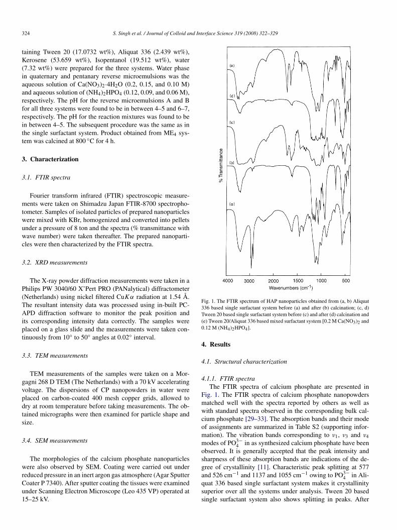
Fig. 1. The FTIR spectrum of HAP nanoparticles obtained from (a, b) Aliquat336 based single surfactant system before (a) and after (b) calcination; (c, d)Tween 20 based single surfactant system before (c) and after (d) calcination and(e) Tween 20/Aliquat 336 based mixed surfactant system [0.2 M Ca(NO3)2 and0.12 M (NH4)2HPO4].
4. Results
4.1. Structural characterization
4.1.1. FTIR spectraThe FTIR spectra of calcium phosphate are presented in
Fig. 1. The FTIR spectra of calcium phosphate nanopowdersmatched well with the spectra reported by others as well aswith standard spectra observed in the corresponding bulk cal-cium phosphate [29–33]. The absorption bands and their modeof assignments are summarized in Table S2 (supporting infor-mation). The vibration bands corresponding to ν1, ν3 and ν4modes of PO3−
4 in as synthesized calcium phosphate have beenobserved. It is generally accepted that the peak intensity andsharpness of these absorption bands are indications of the de-gree of crystallinity [11]. Characteristic peak splitting at 577and 526 cm−1 and 1137 and 1055 cm−1 owing to PO3−
4 in Ali-quat 336 based single surfactant system makes it crystallinitysuperior over all the systems under analysis. Tween 20 basedsingle surfactant system also shows splitting in peaks. After
S. Singh et al. / Journal of Colloid and Interface Science 319 (2008) 322–329 325
calcination the bands corresponding to PO3−4 due to ν1, ν3 and
ν4 become sharper. A sharp band due to ν2 modes of PO3−4 at
450 cm−1 appears after calcination in all the samples.The vibrational band around 874 cm−1 (ME1, ME2, and
ME3) and 1200 cm−1 (ME1 and ME2) have been assignedas signature peaks for brushite indicating the presence ofHPO2−
4 group in uncalcined samples [31–33]. A small peakat 1070 cm−1 in all samples has been attributed to the ν3mode of PO3−
4 group in apatite. Also IR bands observed around1137.85 cm−1 (ME1), 1127.95 cm−1 (ME2) and 1134.10 cm−1
(ME3) may be related to peak at 1127 cm−1 observed in HAproduced under conditions of variable pH and assigned to theν′
6 and ν′′6 degenerate stretch of HPO2−
4 ions in brushite. Thisindicates the formation of poorly crystalline HAP at the earlystages of precipitation which has either been converted to crys-talline HA or gets dissolved [29,30].
The IR band due to bending (δ) mode of OH− groups hasbeen observed at 1650 cm−1 (ME1), 1621 cm−1 (ME2), and1651 cm−1 (ME3) in the spectra of unheated samples. Uponcalcination the intensity of these bands decreases indicating thatthe peak is due to adsorbed water. As shown in Fig. 1 a broadband between 3400–3500 cm−1 has been observed due to thestretching (νs) mode of H-bonded OH− or water [14] conform-ing the formation of HAP nanopowders. After calcination thebroad band disappears to show the sharp band (Fig. 1, b and d),assigned to the structural OH− group.
The IR bands attributed to ν2 and ν3 modes of CO2−3 sub-
stitution in HAP, probably formed by dissolution of CO2 gasfrom the atmosphere or due to the presence of CaCO3 in the re-actants. Literature pertaining to carbonate apatite classifies thesubstitution as A-type (CO2−
3 for OH−) and B-type (CO2−3 for
PO3−4 ). A very sharp and intense band appears at ∼725 cm−1
due to P2O4−7 showing the presence of calcium pyrophosphate
(Ca2P2O7). The presence of calcium pyrophosphate in a cal-cined sample indicates the presence of HPO2−
4 in the samplebefore calcination because calcium pyrophosphate is formed bythe loss of one H2O molecule from two HPO2−
4 group frombrushite (DCPD) under high temperature conditions. The IRbands around 2900, 1740, 1350, and 1250 cm−1 attributed tothe surfactant still attached over the nanoparticles surfaces, dis-appears upon calcinations (Fig. 1, b and d).
FTIR spectrum shows that the nanoparticles obtained arepredominantly brushite. It also indicates the formation ofpoorly crystalline HAP at the early stages of precipitation.
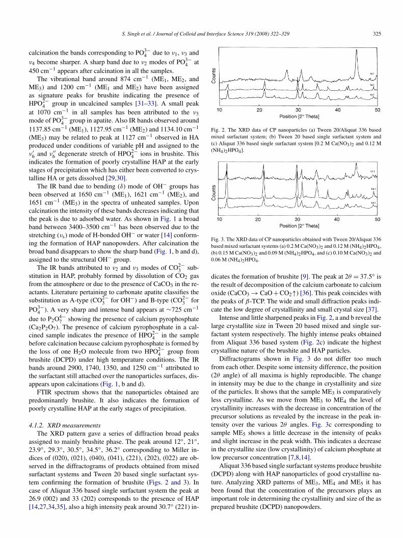
4.1.2. XRD measurementsThe XRD pattern gave a series of diffraction broad peaks
assigned to mainly brushite phase. The peak around 12◦, 21◦,23.9◦, 29.3◦, 30.5◦, 34.5◦, 36.2◦ corresponding to Miller in-dices of (020), (021), (040), (041), (221), (202), (022) are ob-served in the diffractograms of products obtained from mixedsurfactant systems and Tween 20 based single surfactant sys-tem confirming the formation of brushite (Figs. 2 and 3). Incase of Aliquat 336 based single surfactant system the peak at26.9 (002) and 33 (202) corresponds to the presence of HAP[14,27,34,35], also a high intensity peak around 30.7◦ (221) in-
Fig. 2. The XRD data of CP nanoparticles (a) Tween 20/Aliquat 336 basedmixed surfactant system; (b) Tween 20 based single surfactant system and(c) Aliquat 336 based single surfactant system [0.2 M Ca(NO3)2 and 0.12 M(NH4)2HPO4].
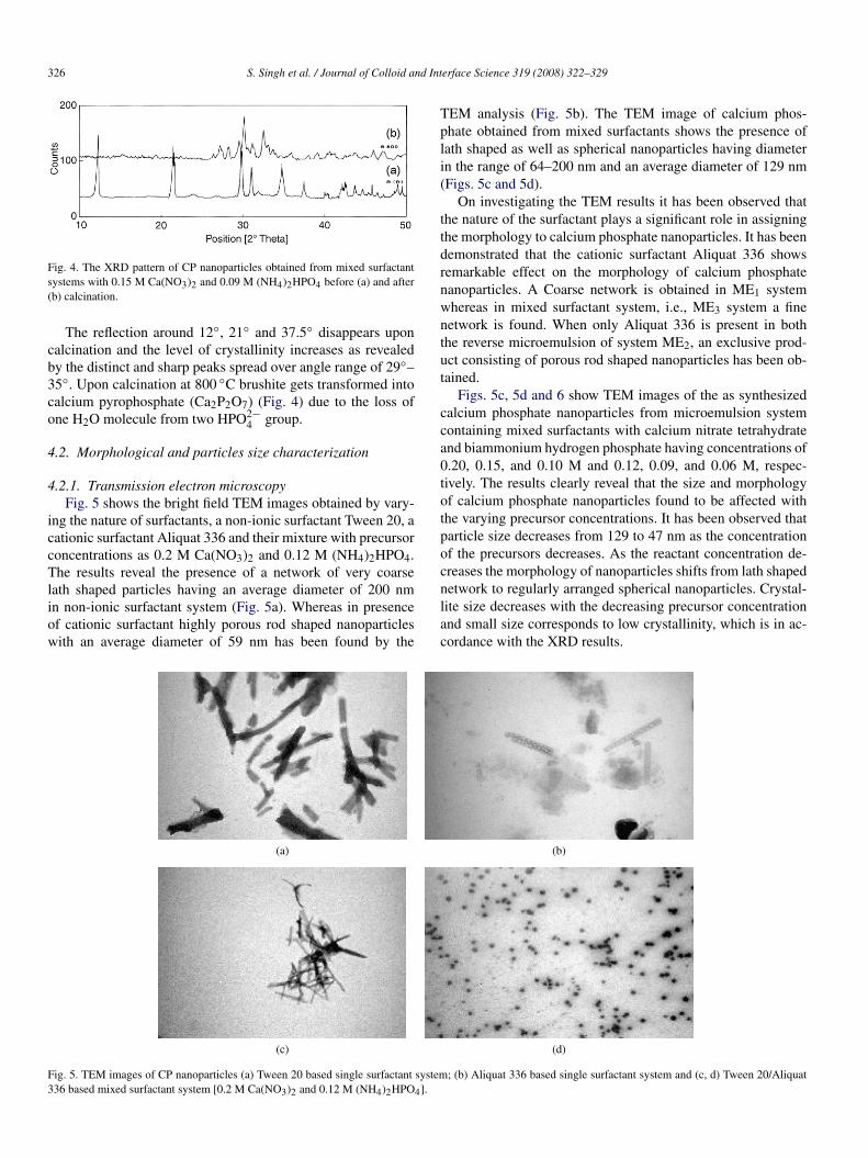
Fig. 3. The XRD data of CP nanoparticles obtained with Tween 20/Aliquat 336based mixed surfactant systems (a) 0.2 M Ca(NO3)2 and 0.12 M (NH4)2HPO4,(b) 0.15 M Ca(NO3)2 and 0.09 M (NH4)2HPO4, and (c) 0.10 M Ca(NO3)2 and0.06 M (NH4)2HPO4.
dicates the formation of brushite [9]. The peak at 2θ = 37.5◦ isthe result of decomposition of the calcium carbonate to calciumoxide (CaCO3 → CaO + CO2↑) [36]. This peak coincides withthe peaks of β-TCP. The wide and small diffraction peaks indi-cate the low degree of crystallinity and small crystal size [37].
Intense and little sharpened peaks in Fig. 2, a and b reveal thelarge crystallite size in Tween 20 based mixed and single sur-factant system respectively. The highly intense peaks obtainedfrom Aliquat 336 based system (Fig. 2c) indicate the highestcrystalline nature of the brushite and HAP particles.
Diffractograms shown in Fig. 3 do not differ too muchfrom each other. Despite some intensity difference, the position(2θ angle) of all maxima is highly reproducible. The changein intensity may be due to the change in crystallinity and sizeof the particles. It shows that the sample ME3 is comparativelyless crystalline. As we move from ME3 to ME4 the level ofcrystallinity increases with the decrease in concentration of theprecursor solutions as revealed by the increase in the peak in-tensity over the various 2θ angles. Fig. 3c corresponding tosample ME5 shows a little decrease in the intensity of peaksand slight increase in the peak width. This indicates a decreasein the crystallite size (low crystallinity) of calcium phosphate atlow precursor concentration [7,8,14].
Aliquat 336 based single surfactant systems produce brushite(DCPD) along with HAP nanoparticles of good crystalline na-ture. Analyzing XRD patterns of ME3, ME4 and ME5 it hasbeen found that the concentration of the precursors plays animportant role in determining the crystallinity and size of the asprepared brushite (DCPD) nanopowders.
326 S. Singh et al. / Journal of Colloid and Interface Science 319 (2008) 322–329
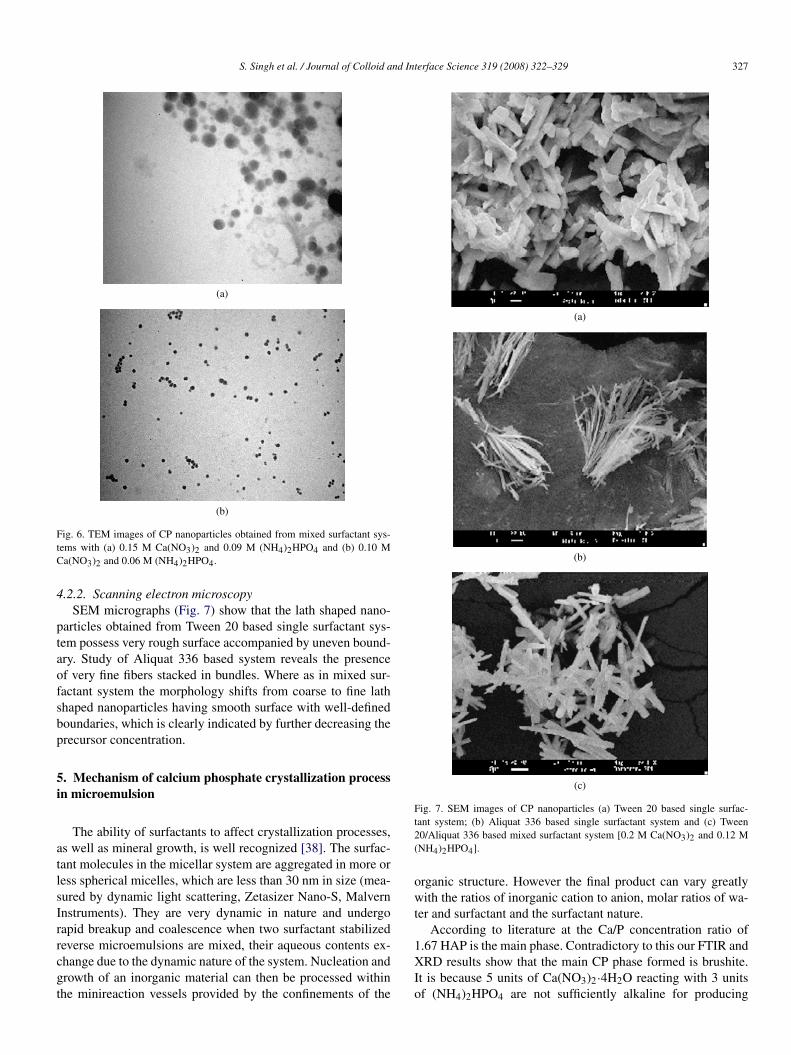
Fig. 4. The XRD pattern of CP nanoparticles obtained from mixed surfactantsystems with 0.15 M Ca(NO3)2 and 0.09 M (NH4)2HPO4 before (a) and after(b) calcination.
The reflection around 12◦, 21◦ and 37.5◦ disappears uponcalcination and the level of crystallinity increases as revealedby the distinct and sharp peaks spread over angle range of 29◦–35◦. Upon calcination at 800 ◦C brushite gets transformed intocalcium pyrophosphate (Ca2P2O7) (Fig. 4) due to the loss ofone H2O molecule from two HPO2−
4 group.
4.2. Morphological and particles size characterization
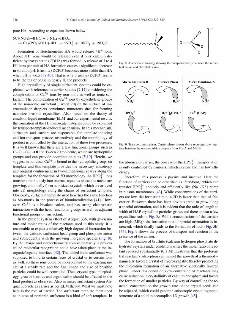
4.2.1. Transmission electron microscopyFig. 5 shows the bright field TEM images obtained by vary-
ing the nature of surfactants, a non-ionic surfactant Tween 20, acationic surfactant Aliquat 336 and their mixture with precursorconcentrations as 0.2 M Ca(NO3)2 and 0.12 M (NH4)2HPO4.The results reveal the presence of a network of very coarselath shaped particles having an average diameter of 200 nmin non-ionic surfactant system (Fig. 5a). Whereas in presenceof cationic surfactant highly porous rod shaped nanoparticleswith an average diameter of 59 nm has been found by the
TEM analysis (Fig. 5b). The TEM image of calcium phos-phate obtained from mixed surfactants shows the presence oflath shaped as well as spherical nanoparticles having diameterin the range of 64–200 nm and an average diameter of 129 nm(Figs. 5c and 5d).
On investigating the TEM results it has been observed thatthe nature of the surfactant plays a significant role in assigningthe morphology to calcium phosphate nanoparticles. It has beendemonstrated that the cationic surfactant Aliquat 336 showsremarkable effect on the morphology of calcium phosphatenanoparticles. A Coarse network is obtained in ME1 systemwhereas in mixed surfactant system, i.e., ME3 system a finenetwork is found. When only Aliquat 336 is present in boththe reverse microemulsion of system ME2, an exclusive prod-uct consisting of porous rod shaped nanoparticles has been ob-tained.
Figs. 5c, 5d and 6 show TEM images of the as synthesizedcalcium phosphate nanoparticles from microemulsion systemcontaining mixed surfactants with calcium nitrate tetrahydrateand biammonium hydrogen phosphate having concentrations of0.20, 0.15, and 0.10 M and 0.12, 0.09, and 0.06 M, respec-tively. The results clearly reveal that the size and morphologyof calcium phosphate nanoparticles found to be affected withthe varying precursor concentrations. It has been observed thatparticle size decreases from 129 to 47 nm as the concentrationof the precursors decreases. As the reactant concentration de-creases the morphology of nanoparticles shifts from lath shapednetwork to regularly arranged spherical nanoparticles. Crystal-lite size decreases with the decreasing precursor concentrationand small size corresponds to low crystallinity, which is in ac-cordance with the XRD results.
(a) (b)
(c) (d)
Fig. 5. TEM images of CP nanoparticles (a) Tween 20 based single surfactant system; (b) Aliquat 336 based single surfactant system and (c, d) Tween 20/Aliquat336 based mixed surfactant system [0.2 M Ca(NO3)2 and 0.12 M (NH4)2HPO4].
S. Singh et al. / Journal of Colloid and Interface Science 319 (2008) 322–329 327
(a)
(b)
Fig. 6. TEM images of CP nanoparticles obtained from mixed surfactant sys-tems with (a) 0.15 M Ca(NO3)2 and 0.09 M (NH4)2HPO4 and (b) 0.10 MCa(NO3)2 and 0.06 M (NH4)2HPO4.
4.2.2. Scanning electron microscopySEM micrographs (Fig. 7) show that the lath shaped nano-
particles obtained from Tween 20 based single surfactant sys-tem possess very rough surface accompanied by uneven bound-ary. Study of Aliquat 336 based system reveals the presenceof very fine fibers stacked in bundles. Where as in mixed sur-factant system the morphology shifts from coarse to fine lathshaped nanoparticles having smooth surface with well-definedboundaries, which is clearly indicated by further decreasing theprecursor concentration.
5. Mechanism of calcium phosphate crystallization processin microemulsion
The ability of surfactants to affect crystallization processes,as well as mineral growth, is well recognized [38]. The surfac-tant molecules in the micellar system are aggregated in more orless spherical micelles, which are less than 30 nm in size (mea-sured by dynamic light scattering, Zetasizer Nano-S, MalvernInstruments). They are very dynamic in nature and undergorapid breakup and coalescence when two surfactant stabilizedreverse microemulsions are mixed, their aqueous contents ex-change due to the dynamic nature of the system. Nucleation andgrowth of an inorganic material can then be processed withinthe minireaction vessels provided by the confinements of the
(a)
(b)
(c)
Fig. 7. SEM images of CP nanoparticles (a) Tween 20 based single surfac-tant system; (b) Aliquat 336 based single surfactant system and (c) Tween20/Aliquat 336 based mixed surfactant system [0.2 M Ca(NO3)2 and 0.12 M(NH4)2HPO4].
organic structure. However the final product can vary greatlywith the ratios of inorganic cation to anion, molar ratios of wa-ter and surfactant and the surfactant nature.
According to literature at the Ca/P concentration ratio of1.67 HAP is the main phase. Contradictory to this our FTIR andXRD results show that the main CP phase formed is brushite.It is because 5 units of Ca(NO3)2·4H2O reacting with 3 unitsof (NH4)2HPO4 are not sufficiently alkaline for producing
328 S. Singh et al. / Journal of Colloid and Interface Science 319 (2008) 322–329
pure HA. According to equation shown below:
5Ca(NO3)2·4H2O + 3(NH4)2HPO4→ Ca5(PO4)3OH + 4H+ + 6NH+
4 + 10NO−3 + 19H2O.
Formation of stoichiometric HA would release 4H+ ions.About 3H+ ions would be released even if only calcium de-ficient hydroxyapatite (CDHA) was formed. A release of 3 to 4H+ ions per unit of HA formation causes a significant decreasein solution pH. Brushite (DCPD) becomes more stable than HAwhen pH is <6.5 [39,40]. That is why brushite (DCPD) seemsto be the major phase in nearly all the products.
High crystallinity of single surfactant systems could be ex-plained with reference to earlier studies [7,14] considering thecomplexation of Ca2+ ions by non-ionic as well as ionic sur-factant. The complexation of Ca2+ ions by oxyethylene groupsof the non-ionic surfactant (Tween 20) on the surface of mi-croemulsion droplets constitutes numerous sites for formingnanosize brushite crystallites. Also, based on the theory ofemulsion liquid membrane (ELM) and our experimental results,the formation of the 1D mesoscale materials could be explainedby transport-template-induced mechanism. In this mechanism,surfactant and carriers are responsible for template-inducingand ion-transport process respectively and the morphology ofproduct is controlled by the interaction of these two processes.It is well known that there are a few functional groups such asC=O, –O–, –OH on Tween 20 molecule, which are hydrophilicgroups and can provide coordination sites [5,19]. Herein, wesuggest in our case, Ca2+ is bound to the hydrophilic groups ontemplate and this template provides the necessary anisotropyand original confinement in two-dimensional spaces along thetemplate for the formation of 2D morphology. As HPO2−
4 ionstransfer continuously into internal-aqueous phase, the nuclei aregrowing, and finally form nanosized crystals, which are arrayedinto 2D morphology along the chains of surfactant template.Obviously, surfactant template used here has the same functionas bio-matrix in the process of biomineralization [41]. How-ever, Ca2+ is a bivalent cation, and has strong electrostaticinteraction with the head functional groups as well as adjacentfunctional groups on surfactant.
In the present system effect of Aliquat 336, with given na-ture and molar ratios of the reactants used in this study, it isreasonable to expect a relatively high degree of interaction be-tween the cationic surfactant head group and phosphate anionand subsequently with the growing inorganic species (Fig. 8).By the charge and stereochemistry complementarily, a processcalled molecular recognition could have taken place at the in-organic/organic interface [42]. The added ionic surfactant wassupposed to bind to certain faces of crystal or to certain ionsas well, so these ions could be incorporated to the existing nu-clei at a steady rate and the final shape and size of brushiteparticles could be well controlled. Thus, crystal type, morphol-ogy, growth kinetics and organization should be affected in thefinal product as observed. Also in mixed surfactant system Ali-quat 336 acts as carrier as per ELM theory. What we must notehere is the role of carrier. The surfactant template mentionedas in case of nonionic surfactant is a kind of soft template. In
Fig. 8. A schematic drawing showing the complementarity between the surfac-tant cation and phosphate anion.
Fig. 9. Transport mechanism. Carrier phase shown above represents the inter-face between the microemulsion droplets from ME-A and ME-B.
the absence of carrier, the process of the HPO2−4 transportation
is only controlled by osmosis, which is slow and has low effi-ciency.
Therefore, this process is passive and inactive. Here thefunction of carriers can be described as ‘ferryboat,’ which cantransfer HPO2−
4 choicely and efficiently like (Na+/K+) pumpin plasma membranes [43]. While concentrations of the carri-ers are low, the formation rate in 2D is faster than that of freecarrier. However, there has been obvious trend to grow alonga special orientation, and it is evident that the ratio of length towidth of HAP crystalline particles grows and there appear a fewcrystalline rods in Fig. 5c. While concentrations of the carriersare high (ME2), the formation rate of special orientation is in-creased, which finally leads to the formation of rods (Fig. 5b)[44]. Fig. 9 shows the process of transport and reaction in thepresence of the carrier.
The formation of brushite (calcium hydrogen phosphate di-hydrate) crystals under conditions where the molar ratio of reac-tant reduced substantially (0.1 M) illustrates that the preferen-tial reactant’s adsorption can inhibit the growth of a thermody-namically favored crystal of hydroxyapatite thereby promotingthe nucleation formation of an alternative kinetically favoredphase. Under this condition slow conversion of reactants maycause reduction in crystallinity of calcium phosphate and favorsthe formation of smaller particles. By way of controlling the re-actant concentration the growth rate of the crystal seeds canbe adjusted, which could generate anisotropic crystallographicstructure of a solid to accomplish 1D growth [45].
S. Singh et al. / Journal of Colloid and Interface Science 319 (2008) 322–329 329
6. Conclusion
We have demonstrated the synthesis of calcium phosphatenanoparticles by reverse microemulsion using Aliquat 336 andTween 20 as the surfactants, where reactants concentration hasbeen found to have an influence on the size as well as on mor-phology. It has been established that the cationic surfactantAliquat 336 can be used to control the morphology and crys-tallinity of calcium phosphate nanoparticles. The crystallinityof the particles increases with the increase in the concentrationof Aliquat 336. A variety of morphologies have been obtainedby varying the amount of Aliquat 336. It has also been shownthat in solution the formation of calcium phosphate is primarilycontrolled by the pH rather than the Ca/P ratio.
It is evident that the nature and concentration of the sur-factant and the concentration of the precursors has a stronginfluence on the growth of this biomaterial. In our experiment,the materials used were inexpensive and environment friendly.So, this technique could be a novel approach to synthesize CPnanoparticles.
Acknowledgments
We acknowledge technical support from Mr. Sandeep Aryafor assistance in TEM measurements and Mr. Rajesh Patnaikfor assistance in SEM measurements (Sophisticated Analyti-cal Instrumentation Facility, Department of Anatomy, AIIMS,Delhi). We also thank Mr. Amarjeet Singh for his help in X-RayDiffraction measurements (Textile Department, IIT, Delhi). Wealso thank to Prof. Anup Beniwal for the editing of our paper.
Supporting information
The online version of this article contains additional support-ing information.
Please visit doi: 10.1016/j.jcis.2007.09.059.
References
[1] H. Aoki, Science and Medical Application of Hydroxyapatite, JAAS,Tokyo, Japan, 1991.
[2] L.L. Hench, J. Am. Ceram. Soc. 74 (1991) 1487.[3] S. Weiner, H.D. Wagner, Annu. Rev. Mater. Sci. 28 (1998) 271.[4] T. Welzel, W. Meyer-Zaika, M. Epple, Chem. Commun. (2004) 1204.[5] S.V. Dorozhkin, M. Epple, Angew. Chem. Int. Ed. 41 (2002) 3130.[6] R.Z. Le Geros, J.P. Le Geros, Dense Hydroxyapatite, An Introduction to
Bioceramics, World Scientific, Singapore, 1993.
[7] Y. Wu, S. Bose, Langmuir 21 (2005) 3232.[8] G.K. Lim, J. Wang, S.C. Ng, L.M. Gan, Langmuir 15 (1999) 7472.[9] C.I. Sainz-Diaz, A. Villacampa, F. Otalora, Am. Mineral. 89 (2004) 307.
[10] T. Harai, M. Hodono, I. Komasava, Langmuir 16 (2000) 955.[11] D. Tadic, M. Epple, Biomaterials 25 (2004) 987.[12] K.A. Thomas, J.F. Kay, S.D. Cook, M. Jarcho, J. Biomed. Mater. Res. 21
(1987) 1395.[13] S.D. Cook, K.A. Thomas, J.E. Dalton, J.F. Kay, Sem. Arthoplasty 2 (4)
(1991) 268.[14] R. Kumar, K.H. Prakash, P. Cheang, K.A. Khor, Langmuir 20 (2004) 5196.[15] C.E. Fowler, M. Li, S. Mann, H.C. Margolis, J. Mater. Chem. 15 (2005)
3317.[16] Y. Xu, D. Wang, L. Yang, H. Tang, Mater. Charact. 47 (2001) 83.[17] K.L. Yadav, P.W. Brown, J. Biomed. Mater. Res. A 65A (2003) 158.[18] Y. Wang, S. Zhang, K. Wei, N. Zhao, J. Chen, X. Wang, Mater. Lett. (2006)
1484.[19] G.K. Lim, J. Wang, S.C. Ng, C.H. Chew, L.M. Gan, Biomaterials 18
(1997) 1433.[20] S.M. Arifuzzaman, S. Rohani, J. Cryst. Growth 267 (3–4) (2004) 624.[21] J.H. Fendler, Chem. Rev. 87 (1987) 877.[22] B.K. Paul, S.P. Moulik, J. Dispersion Sci. Technol. 18 (1997) 301.[23] A.L. Boskey, A.S. Posner, J. Phys. Chem. 77 (1973) 2313.[24] J.P. Baronne, G.H. Nancollas, J. Colloid Interface Sci. 62 (1977) 421.[25] L. Montastrue, C. Azzaro-Pantel, B. Biscans, M. Cabassud, S. Bomenech,
Chem. Eng. J. 94 (2003) 41.[26] M. Cao, Y. Wang, C. Guo, Y. Qi, C. Hu, Langmuir 20 (2004) 4784.[27] M. Uota, H. Arakawa, N. Kitamura, T. Yoshimura, J. Tanaka, T. Kijima,
Langmuir 21 (2005) 4724.[28] X.F. Xiao, R.F. Liu, Mater. Lett. 60 (2006) 2627.[29] S.J. Gadaleta, E.P. Paschalis, F. Betts, R. Mendelsohn, A.L. Boskey, Cal-
cify. Tissue Int. 58 (1996) 9.[30] N. Pleshko, A. Boskey, R. Mendelsohn, Biophys. J. 60 (1991) 786.[31] R.T. Bailey, C. Holt, in: D.W.L. Huskins (Ed.), Calcified Tissue, CRC
Press, Boca Raton, FL, 1989, pp. 93–120.[32] E.E. Berry, C.B. Baddiel, Spectrochim. Acta 23A (1967) 2089.[33] R.J. Le-Geros, in: H.M. Myers (Ed.), Monographs in Oral Science, vol. 15,
Karger, Basel, 1991.[34] R. Gonzalez-McQuire, J.Y. Chane-Ching, E. Vignaud, A. Lebugle,
S. Mann, J. Mater. Chem. 14 (2004) 2277.[35] G. Guo, Y. Sun, Z. Wang, H. Guo, Ceram. Int. 31 (2005) 869.[36] K.D. Rogers, P. Daniels, Biomaterials 23 (2002) 2577.[37] T. Isobe, S. Nakamura, R. Nemoto, M. Senna, J. Phys. Chem. B (2002)
106.[38] H. Colfen, S. Mann, Angew. Chem. Int. Ed. 42 (2003) 2350.[39] R. Boistelle, I. Lopez-Velero, J. Cryst. Growth 102 (1990) 609.[40] F. Abbona, F. Christiansson, M. Franchini-Angela, H. Lundager-Madsen,
J. Cryst. Growth 131 (3–4) (1993) 331.[41] S. Mann, J. Mater. Chem. 5 (1995) 935.[42] L. Yan, Y.D. Li, Z.X. Deng, Int. J. Inorg. Mater. 3 (2001) 633.[43] D. Voet, J.G. Voet, Biochemistry, second ed., Wiley, New York, 1995,
p. 524.[44] J.T. Hu, T.W. Odom, C.H. Lieber, Acc. Chem. Res. 32 (1999) 435.[45] Y. Xia, P. Yang, Y. Sun, Y. Wu, B. Mayers, B. Gates, Y. Yin, F.K. Kim, H.
Yan, Adv. Mater. 15 (2003) 353.
Copyright © 2022 FDOKUMEN