filler effect of mineral powders on ultra high performance ...
Upload
independentCategory
view
0download
0
Advanced Powder Technology 22 (2011) 722–727
Contents lists available at ScienceDirect
Advanced Powder Technology
journal homepage: www.elsevier .com/locate /apt
Original Research Paper
Effect of aerosol carriers on ultrasonically prepared nanocrystalline ZnO powders
L.A. Patil a,⇑, A.R. Bari a, M.D. Shinde a, Vinita Deo a, M.P. Kaushik b
a Nanomaterials Research Lab., Department of Physics, Pratap College, Amalner 425 401, Indiab Defence Research and Development Establishment, Gwalior 474 002, India
a r t i c l e i n f o
Article history:Received 20 February 2010Received in revised form 21 July 2010Accepted 14 October 2010Available online 29 October 2010
Keywords:Nanocrystalline ZnO powderAirSpherical shapeOxygenHexagonal shapeUltrasonic spraying
0921-8831/$ - see front matter � 2011 Published bydoi:10.1016/j.apt.2010.10.011
⇑ Corresponding author. Tel.: +91 9422767373.E-mail address: [email protected]
a b s t r a c t
Nanocrystalline ZnO powders were synthesized using ultrasonic spraying and decomposition technique.The compressed air and pure oxygen were used as aerosol carriers. Effect of aerosol carriers on ultrason-ically atomized nanocrystalline ZnO powders was studied. The powders were characterized using X-raydiffraction, transmission electron microscopy (TEM), electron diffraction, absorption spectroscopy andphotoluminescence. The influence of air and oxygen on crystallite morphology was studied using TEM.It was confirmed from TEM analysis that the crystallites were nearly spherical in powder prepared inthe presence of compressed air. In the presence of pure oxygen, the crystallites could acquire regular hex-agonal shape. Complete hexagonal shapes may be due to sufficient (100%) supply of pure oxygen duringthe process of formation of ZnO nanocrystallite. The powder with regular shaped crystallites could behighly pure and also stoichiometric in nature. Pure oxygen could therefore be more advantageous as car-rier than air for production of nanocrystalline ZnO powder. The effect of aerosol carriers (compressed airand pure oxygen) on crystallite morphology of nanocrystalline ZnO powders is reported in this article.
� 2011 Published by Elsevier B.V. on behalf of The Society of Powder Technology Japan. All rightsreserved.
1. Introduction A wide number of methods have been used to prepare ZnO
Now a days, synthesis of nanocrystalline metal oxide with spe-cific morphology and size has attracted significant attention due totheir possible use in different fields [1,2]. ZnO is well known as animportant II–VI semiconductor with a direct wide band gap of3.37 eV and has a large excitonic binding energy (60 meV) [3]. Thismaterial has a stable wurtzite structure with lattice parameters ofa = 3.249 Å and c = 5.205 Å. The atomic arrangement in ZnO struc-ture can be constructed as a system of alternating planes of Zn2+
and O2� ions with tetrahedral coordination.Nanocrystalline ZnO also possesses excellent chemical and
thermal stability. Because of its unique properties, ZnO is em-ployed to make devices, applicable in different fields of scienceand daily life. Nanocrystalline ZnO is widely used for makinggas-sensors [4–6], light emitting diodes [7], acoustic wave filters[8], UV photo detectors, field effect transistors, intermolecularp–n junction diodes, schottky diodes, photo-diodes [9], opticalmodulator wave guides [10] and photonic crystals [11]. This is anexceptionally important material having applications in pigments,rubber additives, varistors and optical devices and thus interestingfor both research and practical studies. ZnO is also known to haveanti-bacterial properties and along with its arsenic scavengingability finds use in water treatment and purification.
Elsevier B.V. on behalf of The Soci
(L.A. Patil).
powders, including, homogeneous precipitation in aqueous solu-tion of Zn2+ cations, hydrothermal synthesis [12], microwave syn-thesis [13], solution combustion [14], pulsed laser deposition [15],emulsion precipitation [16], ultrasonic atomization [17], ultrasonicspray nebulizer [18–22], spray pyrolysis [23], freeze-drying [24]and sol–gel processes [25]. Compared with these reported meth-ods, ultrasonic spray pyrolysis (USP) is convenient and simple,which is a versatile technique widely used for the synthesis of avariety of inorganic and organic materials [26,27].
In the present communication, we have developed ultrasonicspray pyrolysis technique to prepare nanocrystalline ZnO powderwith crystallite sizes in the range of 12–15 nm. Effect of aerosol car-riers (compressed air or pure oxygen) on crystallite morphology wasstudied. Structural properties (and crystallites sizes), morphologicalstudies along with crystallite size distribution, optical properties,photoluminescence properties were studied, respectively, fromX-ray diffraction technique (CuKa radiation with k = 1.5418 Å),TEM (Tecnai 20 G2, FEI make), UV–vis spectroscopy (Simadzu2450 UV–vis) and fluorescence spectrophotometer (Perkin–ElmerLS-55).
2. Experimental
2.1. Preparation of nanocrystalline ZnO powder
ZnO nanostructured powder was synthesized using ultrasonicspraying and decomposition technique. The set up consists of three
ety of Powder Technology Japan. All rights reserved.

L.A. Patil et al. / Advanced Powder Technology 22 (2011) 722–727 723
major components: (1) ultrasonic atomizer (2.1–2.3 MHz –Gapusol 9001 RBI Meylan, France) with three transducers forconversion of precursor solution into aerosol, (2) quartz reactor(with diameter of 40 mm and length of 120 mm) placed in twozone pyrolysis furnace, and (3) novel trapping system to collectthe nanocrystalline powder.
Aqueous solution (0.5 M) of zinc nitrate (Zn(NO3)26H2O, Merckextra pure) was prepared in double distilled water. The stock solu-tion was stored in cylindrical vessel connected to ultrasonic gener-ator through tube. When solution was allowed to pass to thegenerator, it was converted into aerosol with the rate of 1.08 SLPM.The aerosol was pushed forward through quartz reactor usingcompressed air or pure oxygen. Quartz reactor was placed in dou-ble zone tubular furnace. The temperature of drying zone (1stzone) was kept at 573 K and pyrolysis zone (2nd zone) was at1073 K. The carrier airflow rate was optimized as 20 kg/cm2 to pre-vent compositional segregation. An aerosol would pass through thechain of processes of: heating, solvent evaporation, pyrolysis,reaction with air/oxygen and finally form the fine particles. Theresidence time of the mist particles is about 5–7 s. The nanocrys-talline powder was then collected using novel trapping system.The nanocrystalline ZnO powders obtained in presence of com-pressed air and oxygen as aerosol carriers were referred to as P1and P2, respectively.
2.2. Novel trapping system
It was really very difficult to separate out the smaller nanopar-ticles from larger ones. The novel trapping system [28] was usedfor size wise sorting of nanoparticles. The novel trapping systemconsisted of series of interconnected four glass traps with eachtrap filled with water up to 3/4th of its height. Dimensions ofeach trap were identical. The exit end of quartz reactor was con-nected to first trap of trapping system as depicted in Fig. 1. Hea-vier particles went deep into the water column along with aerosolcarrier (air/oxygen). Heavier particles could settle down into thewater and resided there itself but the lighter would bubbled outof water with carrier and reach to next trap. Of these particles,heavier could remain and lighter would go to next trap. In thisway, smaller particles with narrow particle size distribution couldreach last trap. In this way trapping system was employed to sortout the particles size wise.
Fig. 1. System for the preparation of ZnO nanopow
2.3. Estimation of droplet diameter
The mean diameter of droplets from aerosol was estimated asfollows. The surface tension (cs) of the solution was measured byQuinke’s method which was found to be 71.50 dyne/cm. The den-sity (qs) was determined by specific gravity bottle which was esti-mated to be 1.10 g/cm3. Mean droplet diameter (Ddrop) of aerosolwas determined using relation,
Ddrop ¼ 0:34ð8pcs=qsf 2Þ1=3 ð1Þ
value of mean droplets diameter was calculated to be 2.41 lm.
3. Results and discussion
3.1. Structural analysis
Fig. 2a and b shows the X-ray diffractograms of the nanocrystal-line ZnO powders synthesized, respectively from compressed airand pure oxygen as carriers. The diffraction peaks from variousplanes and d-values were matching well with standard JCPDS data[29].
These diffraction profiles illustrate crystalline nature and hex-agonal wurtzite structure of ZnO nanopowders. Average crystallitesize of nanocrystallites associated with powders P1 and P2 was cal-culated using Scherer formula.
3.1.1. Lattice parametersThe lattice parameters of ZnO were calculated using Eq. (2):
1=d2ð1 0 1Þ ¼ 4=3ð1=a2Þ þ ð1=c2Þ ð2Þ
where ‘d’ is the interplaner distance, ‘a’ and ‘c’ are the latticeparameters (being hexagonal structure c/a =
p3/8). Average
crystallite size and lattice parameters ‘a’ and ‘c’ are presented inTable 1.
3.1.2. Number of unit cellsTo calculate number of unit cells, let us consider a crystallite ‘p’
of diameter ‘r’. The crystallite p would consist of n smaller identicalunit cells of edges a and c.
ders by ultrasonic spray pyrolysis technique.

Fig. 2. X-ray diffractrogram of nanocrystalline ZnO powders: (a) P1 and (b) P2.
Table 1Crystallite size, size distribution, average crystallite size and mean deviation, lattice parameters and number of unit cells.
Sample Counts Size variation(nm)
Standard deviation Average size(nm)
Lattice parameters (Å) No. of unit cells (n)
min max (XRD) (TEM) a c (XRD) (TEM)
P1 (air) 73 9 17 4 12 12 3.34 5.46 2,47,010 2,81,425P2 (oxygen) 47 7 18 5.5 15 15 3.34 5.46 4,50,672 5,08,397
724 L.A. Patil et al. / Advanced Powder Technology 22 (2011) 722–727
4=3pðr=VÞ3 ¼ nV ð3Þwhere V = 3
p3/2 a2c (for hexagonal structure), i.e.
n ¼ 4=3pðr=2VÞ3 ð4Þ
The calculated values of number of unit cells are presented inTable 1.
3.2. Electron diffraction
Fig. 3a and b shows the electron diffraction patterns of P1 andP2 powders, respectively.
It is clear from figures that the ZnO particles are crystalline innature. The electron diffraction patterns shows spotty but contin-uous ring patterns without any additional diffraction spots andrings of secondary phases revealing their crystalline structure.Eleven fringe patterns corresponding to planes: (1 0 0), (0 0 2),(1 0 1), (1 0 2), (1 1 0), (1 0 3), (2 0 0), (1 1 2), (2 0 1), (0 0 4) and(2 0 2) are consistent with the peaks observed in XRD patterns.XRD and TEM studies confirmed pure wurtzite hexagonal structureof ZnO as evidenced from Figs. 2 and 3, respectively.
3.3. Microstructure and crystallite size distribution using TEM
Fig. 4a and b shows the TEM images along with histograms show-ing crystallite size distribution for the nanocrystalline ZnO powderssynthesized in presence of compressed air and pure oxygen,respectively.
It is clear from Fig. 4a that the crystallites of powder P1 arenearly spherical or ellipsoidal in shape. The powder P2 consistsof regular hexagonal shaped crystallites as indicated in Fig. 4b.The change in shape from spherical/ellipsoidal to hexagonal, inpresence of pure oxygen, could be attributed to supply of sufficient(100%) oxygen (one of the constituent elements of ZnO) to growand acquire hexagonal shape. It is well known that regular shapedcrystals of some typical compound are found to be purer as com-pared to its noncrystalline powder [30]. Such powder could alsobe stoichiometric in nature. Possibility of contamination in powdercan also be avoided. Therefore, synthesis of powder in presence ofpure oxygen could be more advantageous than in presence of airwhich consists of only about 20% of oxygen.
The crystallite size distribution in powder P1 was relatively nar-row as compared to the size distribution in powder P2 (Table 1).The crystallites associated with powder P2 are relatively largerwith broad size distribution. It may be because of sufficient supplyof constituent ions (of zinc and oxygen) required to acquire regularshaped crystallites.
It is clear from Table 1 that the crystallite sizes calculated fromXRD and observed from TEM are nearly matching. There is an in-crease in crystallite size due to change of carrier from compressedair to pure oxygen. The calculated lattice parameter values areslightly greater than reported [31] lattice parameters of ZnO. Thenumber of unit cells in the crystallite associated with the powdersynthesized in the presence of pure oxygen is larger as comparedto powder synthesized in the presence compressed air.
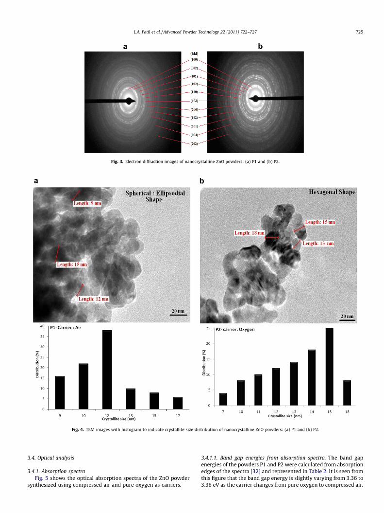
Fig. 3. Electron diffraction images of nanocrystalline ZnO powders: (a) P1 and (b) P2.
Fig. 4. TEM images with histogram to indicate crystallite size distribution of nanocrystalline ZnO powders: (a) P1 and (b) P2.
L.A. Patil et al. / Advanced Powder Technology 22 (2011) 722–727 725
3.4. Optical analysis
3.4.1. Absorption spectraFig. 5 shows the optical absorption spectra of the ZnO powder
synthesized using compressed air and pure oxygen as carriers.
3.4.1.1. Band gap energies from absorption spectra. The band gapenergies of the powders P1 and P2 were calculated from absorptionedges of the spectra [32] and represented in Table 2. It is seen fromthis figure that the band gap energy is slightly varying from 3.36 to3.38 eV as the carrier changes from pure oxygen to compressed air.
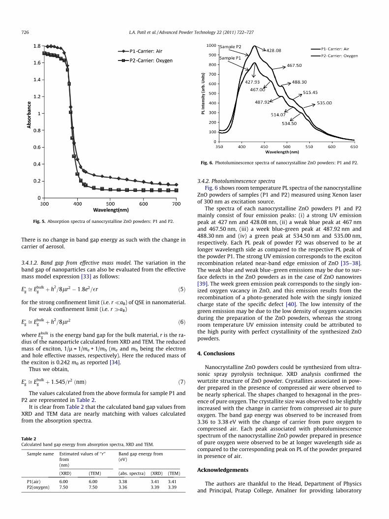
Fig. 5. Absorption spectra of nanocrystalline ZnO powders: P1 and P2.
Fig. 6. Photoluminescence spectra of nanocrystalline ZnO powders: P1 and P2.
726 L.A. Patil et al. / Advanced Powder Technology 22 (2011) 722–727
There is no change in band gap energy as such with the change incarrier of aerosol.
3.4.1.2. Band gap from effective mass model. The variation in theband gap of nanoparticles can also be evaluated from the effectivemass model expression [33] as follows:
E�g ffi Ebulkg þ h2
=8lr2 � 1:8e2=�r ð5Þ
for the strong confinement limit (i.e. r�aB) of QSE in nanomaterial.For weak confinement limit (i.e. r �aB)
E�g ffi Ebulkg þ h2
=8lr2 ð6Þ
where Ebulkg is the energy band gap for the bulk material, r is the ra-
dius of the nanoparticle calculated from XRD and TEM. The reducedmass of exciton, 1/l = 1/me + 1/mh (me and mh being the electronand hole effective masses, respectively). Here the reduced mass ofthe exciton is 0.242 m0 as reported [34].
Thus we obtain,
E�g ffi Ebulkg þ 1:545=r2 ðnmÞ ð7Þ
The values calculated from the above formula for sample P1 andP2 are represented in Table 2.
It is clear from Table 2 that the calculated band gap values fromXRD and TEM data are nearly matching with values calculatedfrom the absorption spectra.
Table 2Calculated band gap energy from absorption spectra, XRD and TEM.
Sample name Estimated values of ‘‘r’’from(nm)
Band gap energy from(eV)
(XRD) (TEM) (abs. spectra) (XRD) (TEM)
P1(air) 6.00 6.00 3.38 3.41 3.41P2(oxygen) 7.50 7.50 3.36 3.39 3.39
3.4.2. Photoluminescence spectraFig. 6 shows room temperature PL spectra of the nanocrystalline
ZnO powders of samples (P1 and P2) measured using Xenon laserof 300 nm as excitation source.
The spectra of each nanocrystalline ZnO powders P1 and P2mainly consist of four emission peaks: (i) a strong UV emissionpeak at 427 nm and 428.08 nm, (ii) a weak blue peak at 467 nmand 467.50 nm, (iii) a week blue-green peak at 487.92 nm and488.30 nm and (iv) a green peak at 534.50 nm and 535.00 nm,respectively. Each PL peak of powder P2 was observed to be atlonger wavelength side as compared to the respective PL peak ofthe powder P1. The strong UV emission corresponds to the excitonrecombination related near-band edge emission of ZnO [35–38].The weak blue and weak blue–green emissions may be due to sur-face defects in the ZnO powders as in the case of ZnO nanowires[39]. The week green emission peak corresponds to the singly ion-ized oxygen vacancy in ZnO, and this emission results from therecombination of a photo-generated hole with the singly ionizedcharge state of the specific defect [40]. The low intensity of thegreen emission may be due to the low density of oxygen vacanciesduring the preparation of the ZnO powders, whereas the strongroom temperature UV emission intensity could be attributed tothe high purity with perfect crystallinity of the synthesized ZnOpowders.
4. Conclusions
Nanocrystalline ZnO powders could be synthesized from ultra-sonic spray pyrolysis technique. XRD analysis confirmed thewurtzite structure of ZnO powder. Crystallites associated in pow-der prepared in the presence of compressed air were observed tobe nearly spherical. The shapes changed to hexagonal in the pres-ence of pure oxygen. The crystallite size was observed to be slightlyincreased with the change in carrier from compressed air to pureoxygen. The band gap energy was observed to be increased from3.36 to 3.38 eV with the change of carrier from pure oxygen tocompressed air. Each peak associated with photoluminescencespectrum of the nanocrystalline ZnO powder prepared in presenceof pure oxygen were observed to be at longer wavelength side ascompared to the corresponding peak on PL of the powder preparedin presence of air.
Acknowledgements
The authors are thankful to the Head, Department of Physicsand Principal, Pratap College, Amalner for providing laboratory

L.A. Patil et al. / Advanced Powder Technology 22 (2011) 722–727 727
facilities for this work. The financial support for this work from theDepartment of Information Technology and Defence ResearchDevelopment Organisation (DRDO), Government of India, NewDelhi are gratefully acknowledged. The authors are also gratefulto Dr. N.P. Lalla and Mr. Satish Potdar IUC, Indore, for providingTEM of the samples.
References
[1] X. Duan, Y. Huang, R. Agrawal, C.M. Lieber, Single-nanowire electrically drivenlasers, Nature 421 (2003) 241–245.
[2] M.S. Fuhrer, J. Nygard, L. Shih, M. Forero, Y.G. Yoon, M.S.C. Mazzoni, H.J. Choi,Crossed nanotube junctions, Science 288 (2000) 494–497.
[3] Z. Fan, J.G. Lu, Zinc oxide nanostructures: synthesis and properties, J. Nanosci.Nanotech. 5 (2005) 1561–1573.
[4] L.A. Patil, A.R. Bari, M.D. Shinde, Vinita Deo, Ultrasonically preparednanocrystalline ZnO thin films for highly sensitive LPG sensing, Sens.Actuators B: Chem. 149 (2010) 79–86.
[5] L.A. Patil, A.R. Bari, M.D. Shinde, Vinita Deo, Ultrasonically synthesizednanocrystalline ZnO powder based thick film sensor for ammonia sensing,Sensor Rev. 30 (2010) 290–296.
[6] D.R. Patil, L.A. Patil, Room temperature chlorine gas sensing using surfacemodified ZnO thick film resistors, Sens. Actuators B 123 (2007) 546–553.
[7] N. Saito, H. Haneda, T. Sekiguchi, N. Ohashi, I. Sakaguchi, K. Koumoto, Low-temperature fabrication of light-emitting zinc oxide micropatterns using self-assembled monolayers, Adv. Mater. 14 (2002) 418–421.
[8] N.W. Emanetoglu, C. Gorla, Y. Lu, Epitaxial ZnO piezoelectric thin films for sawfilters, Mat. Sci. Semicond. Process 2 (1999) 247.
[9] J.Y. Lee, Y.S. Choi, J.H. Kim, M.O. Park, S. Im, Optimizing n-ZnO/p-Siheterojunctions for photodiode applications, Thin Solid Films 403 (2002)553–557.
[10] M.H. Koch, P.Y. Timbrell, R.N. Lamb, The influence of film crystallinity on thecoupling efficiency of ZnO optical modulator waveguides, Semicond. Sci.Technol. 10 (1995) 1523–1527.
[11] Y.D. Bagnall, T. Yao, ZnO as a novel photonic material for the UV region, Mater.Sci. Eng. B 75 (2000) 190–198.
[12] B. Baruwati, D.K. Kumar, S.V. Manorama, Hydrothermal synthesis of highlycrystalline ZnO nanoparticles: a competitive sensor for LPG and EtOH, Sens.Actuators B 119 (2006) 676–682.
[13] N. Tabet, R. Al Ghashani, S. Achour, Ultra fast synthesis of zinc oxidenanostructures by microwaves, Superlattices Microstruct. 45 (2009) 598–603.
[14] B.M. Cheng, L. Yu, C. Duan, H. Wang, P.A. Tanner, Vacuum ultraviolet andvisible spectra of ZnO:Eu3+ prepared by combustion synthesis, J. Phys.:Condens. Matter 20 (2008) 345231–345234.
[15] Y.W. Sun, Y.Y. Tsui, Production of porous nanostructured zinc oxide thin filmsby pulsed laser deposition, Opt. Mater. 29 (2007) 1111–1114.
[16] C.H. Lu, C.H. Yeh, Emulsion precipitation of submicron zinc oxide powder,Mater. Lett. 33 (1997) 129–132.
[17] P. Singh, A. Kumar, A. Kaushal, D. Kaur, A. Pandey, R.N. Goyal, In situ hightemperature XRD studies of ZnO nanopowder prepared via cost effectiveultrasonic mist chemicalvapour deposition, Bull. Mater. Sci. 31 (2008) 573–578.
[18] T.Q. Liu, O. Sakurai, N. Mizutani, M. Kato, Preparation of spherical fine ZnOparticles by the spray pyrolysis method using ultrasonic atomizationtechniques, J. Mat. Sci. 21 (1986) 3698–3702.
[19] O. Miloševic, B. Jordovic, D. Uskokovic, Preparation of fine spherical ZnOpowders by an ultrasonic spray pyrolysis method, Mater. Lett. 19 (1994) 165–170.
[20] C. Panatarani, I.W. Lenggoro, K. Okuyama, Synthesis of single crystalline ZnOnanoparticles by salt-assisted spray pyrolysis, J. Nanoparticle Res. 5 (2003) 47–53.
[21] L. Mädler, Liquid-fed aerosol reactors for one-step synthesis of nano-structured particles, KONA 22 (2004) 107–120.
[22] Kikuo Okuyama, I. Wuled Lenggoro, Preparation of nanoparticles via sprayroute, Chem. Eng. Sci. 58 (2003) 537–547.
[23] S. Leea, T. Sona, J. Yuna, H. Kwona, G.L. Messingb, B. Jun, Preparation of BaTiO3
nanoparticles by combustion spray pyrolysis, Mater. Lett. 58 (2004) 2932–2936.
[24] Z.W. Marinkovi, O. Milosevi, M.V. Nikoli, M.G. Kakazoy, M.V. Karpec, T.V.Tomila, M.M. Risti, Evolution of the microstructure of disperse ZnO powdersobtained by the freeze-drying method, Mater. Sci. Eng. A 375–377 (2004) 620–624.
[25] A.R. Bari, M.D. Shinde, V. Deo, L.A. Patil, Effect of solvents on the particlemorphology of nanostructured ZnO, Ind. J. Pure Appl. Phys. 47 (2009) 24–27.
[26] A.S. Gandhi, V. Jayaram, A.H. Chokshi, Low temperature densificationbehaviour of metastable phases in ZrO2–Al2O3 powders produced by spraypyrolysis, Mater. Sci. Eng. A 304 (2001) 785–789.
[27] S.C. Zhang, G.L. Messing, W. Huebner, YBa2Cu3O7�x superconductor powdersynthesis by spray pyrolysis of organic acid solutions, J. Aerosol. Sci. 22 (1991)585.
[28] L.A. Patil, M.D. Shinde, A.R. Bari, V.V. Deo, Novel trapping system for size wisesorting of SnO2 nanoparticles synthesized from pyrolysis of ultrasonicallyatomized spray for gas sensing, Sens. Actuators B 143 (2009) 316–324.
[29] JCPDS Data Card No. 5-664.[30] G.K. Solanki, D.B. Patel, S. Unadkat, N.N. Gosai, R.R. Patel, Synthesis of
GeTe0.1Se0.9 single crystal and its structural and optical characterization, J.Ovonic Res. 6 (2010) 173–180.
[31] H. Morkoç, Ümit Özgur, Zinc Oxide: Fundamentals Materials and DeviceTechnology, WILEY-VCH Verlag GmbH & Co. KGaA, Weinheim, 2009, p. 12,ISBN: 978-3-527-40813-9.
[32] R.H. Bari, L.A. Patil, P.S. Sonawane, M.D. Mahanubhav, V.R. Patil, P.K. Khanna,Studies on chemically deposited CuInSe2 thin films, Mater. Lett. 61 (2007)2058–5061.
[33] B.D. Cullity, Elements of X-ray Diffractions, 1978, p. 102.[34] P. Singh, A.K. Chawla, D. Kaur, R. Chandra, Effect of oxygen partial pressure on
the structural and optical properties of sputter deposited ZnO nanocrystallinethin films, Mater. Lett. 61 (2007) 2050–2053.
[35] K. Vanheusden, W.L. Warren, C.H. Sesger, D.R. Tallant, J.A. Voigt, B.E. Gnage,Mechanisms behind green photoluminescence in ZnO phosphor powders, J.Appl. Phys. 79 (1996) 7983–7987.
[36] V. Stikant, D.R. Clarke, On the optical band gap of zinc oxide, J. Appl. Phys. 83(1998) 5447–5450.
[37] S.C. Lyu, Y. Zhang, H. Ruh, H. Lee, H. Shim, E. Suh, C.J. Lee, Low temperaturegrowth and photoluminescence of well-aligned zinc oxide nanowires, Chem.Phys. Lett. 363 (2002) 134–138.
[38] L. Bergman, X.B. Chen, J.L. Morrison, J. Huso, A.P. Purdy, Photoluminescencedynamics in ensembles of wide-band-gap nanocrystallites and powders, J.Appl. Phys. 96 (2004) 675–679.
[39] J. Wang, L. Gao, Hydrothermal synthesis and photoluminescence properties ofZnO nanowires, Solid State Commun. 132 (2004) 269–271.
[40] B.D. Yao, Y.F. Chan, N. Wang, Formation of ZnO nanostructures by a simple wayof thermal evaporation, Appl. Phys. Lett. 81 (2002) 757–759.
Copyright © 2022 FDOKUMEN